Embed Size (px)
Citation preview
Accepted Manuscript
Design, synthesis and biological evaluation of quinoline-indole derivatives as anti-tubulin agents targeting the colchicine binding site
Wenlong Li, Wen Shuai, Honghao Sun, Feijie Xu, Yi Bi, Jinyi Xu, Cong Ma, HequanYao, Zheying Zhu, Shengtao Xu
PII: S0223-5234(18)31029-8
DOI: https://doi.org/10.1016/j.ejmech.2018.11.070
Reference: EJMECH 10928
To appear in: European Journal of Medicinal Chemistry
Received Date: 11 October 2018
Revised Date: 20 November 2018
Accepted Date: 28 November 2018
Please cite this article as: W. Li, W. Shuai, H. Sun, F. Xu, Y. Bi, J. Xu, C. Ma, H. Yao, Z. Zhu, S. Xu,Design, synthesis and biological evaluation of quinoline-indole derivatives as anti-tubulin agentstargeting the colchicine binding site, European Journal of Medicinal Chemistry (2018), doi: https://doi.org/10.1016/j.ejmech.2018.11.070.
This is a PDF file of an unedited manuscript that has been accepted for publication. As a service toour customers we are providing this early version of the manuscript. The manuscript will undergocopyediting, typesetting, and review of the resulting proof before it is published in its final form. Pleasenote that during the production process errors may be discovered which could affect the content, and alllegal disclaimers that apply to the journal pertain.
MANUSCRIP
T
ACCEPTED
ACCEPTED MANUSCRIPT
Graphic Abstract
MANUSCRIP
T
ACCEPTED
ACCEPTED MANUSCRIPT
Design, Synthesis and Biological Evaluation of Quinoline-Indole
Derivatives as Anti-tubulin Agents Targeting the Colchicine
Binding Site
Wenlong Lia, Wen Shuaia, Honghao Suna, Feijie Xua, Yi Bib, Jinyi Xua,*, Cong Mac,
Hequan Yaoa, Zheying Zhud, Shengtao Xua,*
aState Key Laboratory of Natural Medicines and Department of Medicinal Chemistry, China
Pharmaceutical University, 24 Tong Jia Xiang, Nanjing 210009, P. R. China
bSchool of Pharmacy, Key Laboratory of Molecular Pharmacology and Drug Evaluation, Yantai
University, Yantai, 264005. P. R. China
cState Key Laboratory of Chemical Biology and Drug Discovery, and Department of Applied
Biology and Chemical Technology, The Hong Kong Polytechnic University, Kowloon, Hong Kon
g
dDivision of Molecular Therapeutics & Formulation, School of Pharmacy, The University of
Nottingham, University Park Campus, Nottingham NG7 2RD, U. K.
*Corresponding Author:
E-mail addresses: [email protected] (J. Xu); [email protected] (S. Xu).
Abstract
A series of novel isocombretastatin A-4 (isoCA-4) analogs were designed and
synthesized by replacing 3,4,5-trimethoylphenyl and isovanillin of isoCA-4 with
quinoline and indole moieties, respectively. The structure activity relationships (SARs)
of these synthesized quinoline-indole derivatives have been intensively investigated.
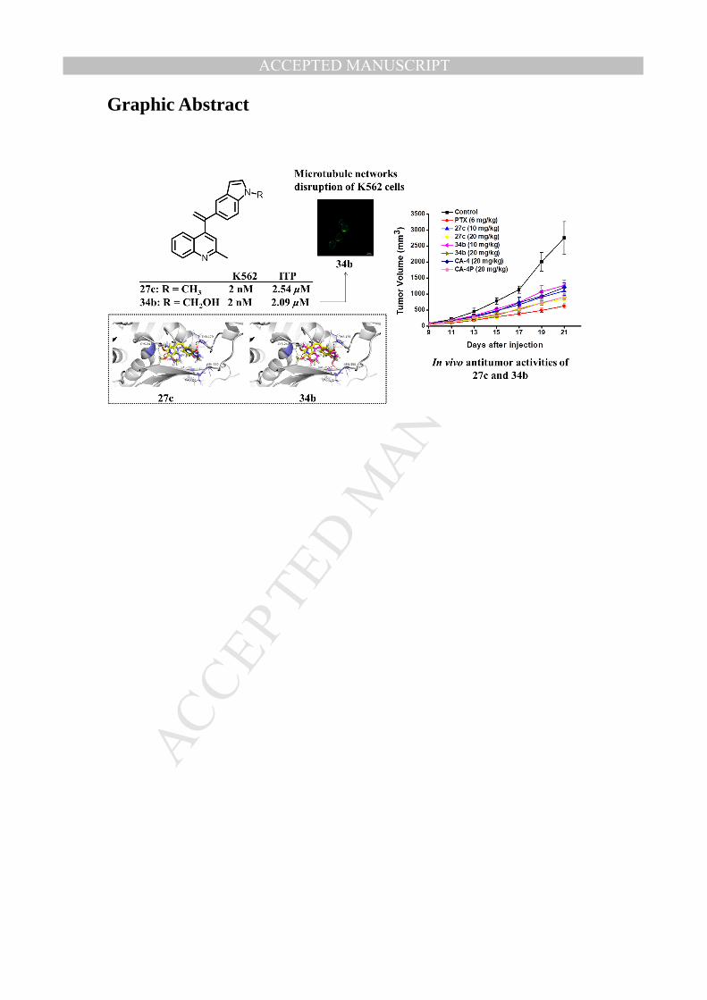
Two compounds 27c and 34b exhibited the most potent activities against five cancer
cell lines with IC50 values ranging from 2 to 11 nM, which were comparable to those
of Combretastatin A-4 (CA-4, 1). Further mechanism investigations revealed that 34b
effectively inhibited the microtubule polymerization by binding to the colchicine site
of tubulin. Further cellular mechanism studies elucidated that 34b disrupted cell
microtubule networks, arrested the cell cycle at G2/M phase, induced apoptosis and
depolarized mitochondria of K562 cells. Moreover, 34b displayed potent anti-vascular
activity in both wound healing and tube formation assays. Importantly, 27c and 34b
significantly inhibited tumor growth in H22 xenograft models without apparent
toxicity, suggesting that 27c and 34b deserve further research as potent antitumor
MANUSCRIP
T
ACCEPTED
ACCEPTED MANUSCRIPT
agents for cancer therapy.
Key words: quinoline; indole; microtubule; tubulin inhibitor; colchicine binding
site; antitumor
1. Introduction
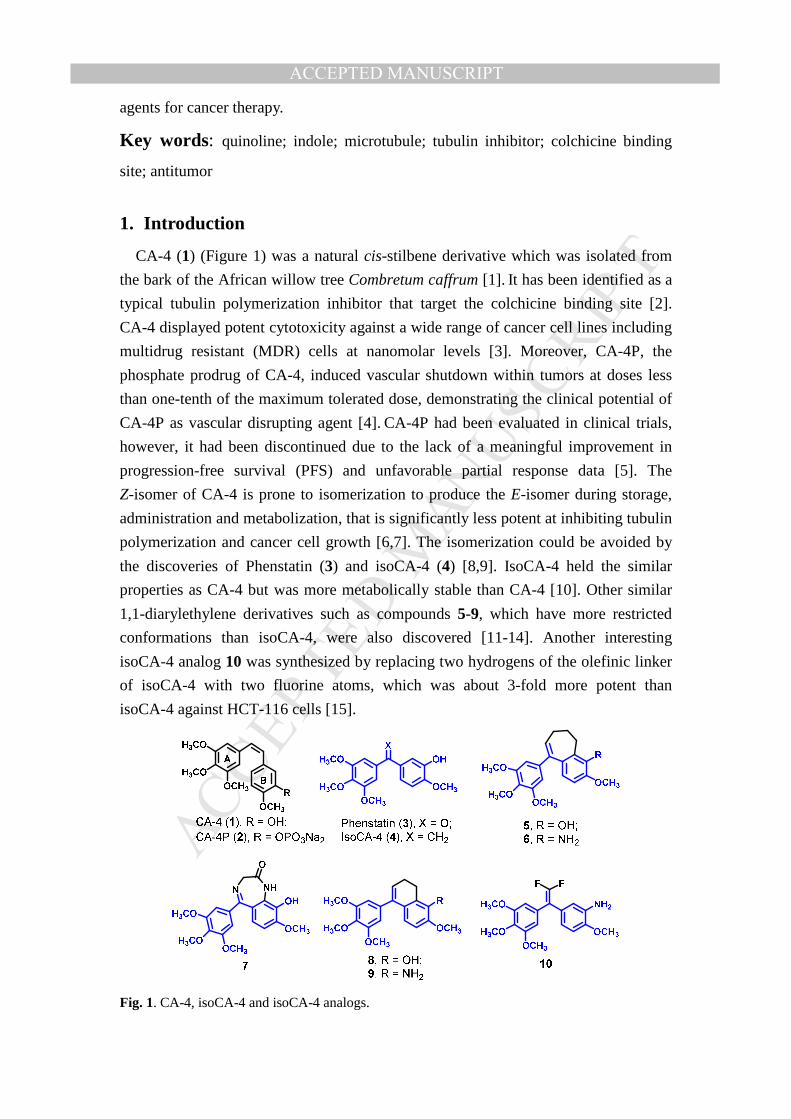
CA-4 (1) (Figure 1) was a natural cis-stilbene derivative which was isolated from
the bark of the African willow tree Combretum caffrum [1]. It has been identified as a
typical tubulin polymerization inhibitor that target the colchicine binding site [2].
CA-4 displayed potent cytotoxicity against a wide range of cancer cell lines including
multidrug resistant (MDR) cells at nanomolar levels [3]. Moreover, CA-4P, the
phosphate prodrug of CA-4, induced vascular shutdown within tumors at doses less
than one-tenth of the maximum tolerated dose, demonstrating the clinical potential of
CA-4P as vascular disrupting agent [4]. CA-4P had been evaluated in clinical trials,
however, it had been discontinued due to the lack of a meaningful improvement in
progression-free survival (PFS) and unfavorable partial response data [5]. The
Z-isomer of CA-4 is prone to isomerization to produce the E-isomer during storage,
administration and metabolization, that is significantly less potent at inhibiting tubulin
polymerization and cancer cell growth [6,7]. The isomerization could be avoided by
the discoveries of Phenstatin (3) and isoCA-4 (4) [8,9]. IsoCA-4 held the similar
properties as CA-4 but was more metabolically stable than CA-4 [10]. Other similar
1,1-diarylethylene derivatives such as compounds 5-9, which have more restricted
conformations than isoCA-4, were also discovered [11-14]. Another interesting
isoCA-4 analog 10 was synthesized by replacing two hydrogens of the olefinic linker
of isoCA-4 with two fluorine atoms, which was about 3-fold more potent than
isoCA-4 against HCT-116 cells [15].
Fig. 1. CA-4, isoCA-4 and isoCA-4 analogs.
MANUSCRIP
T
ACCEPTED
ACCEPTED MANUSCRIPT
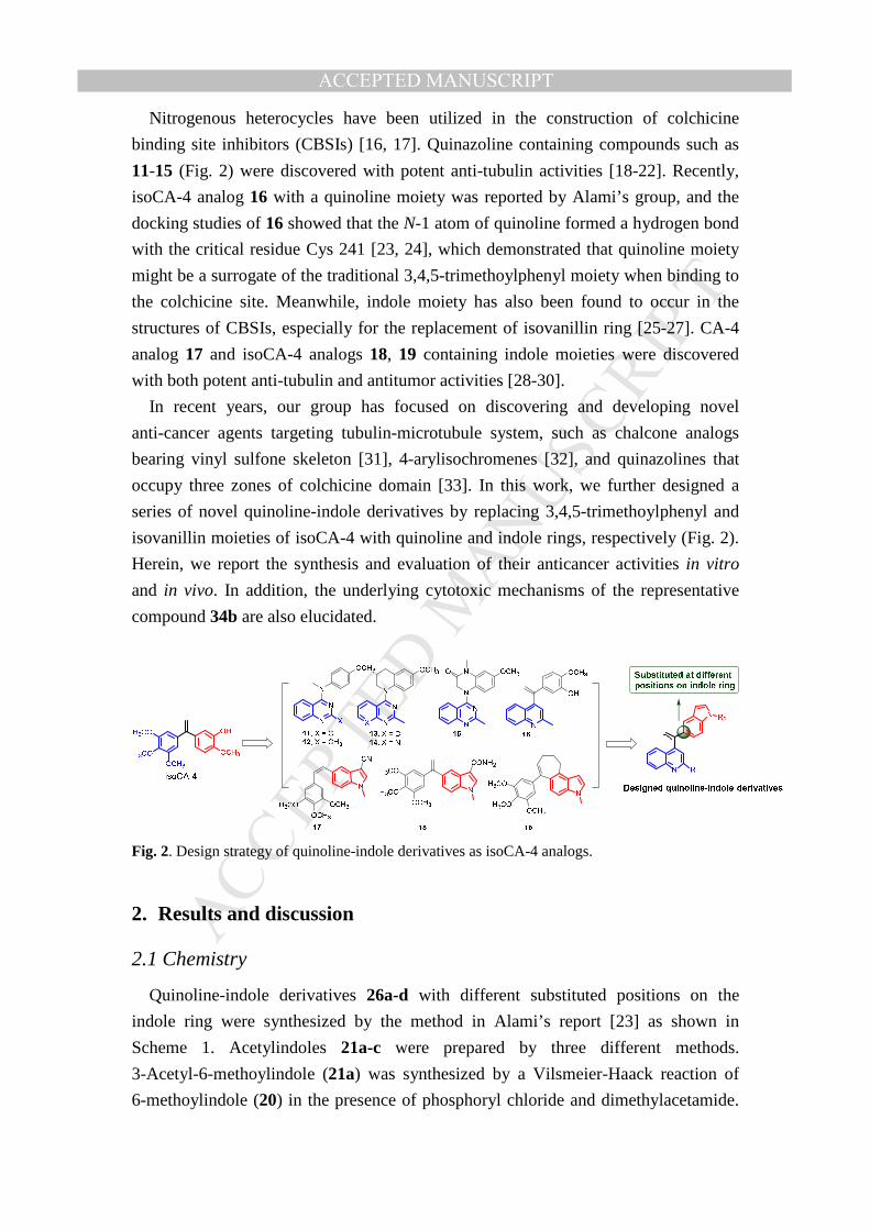
Nitrogenous heterocycles have been utilized in the construction of colchicine
binding site inhibitors (CBSIs) [16, 17]. Quinazoline containing compounds such as
11-15 (Fig. 2) were discovered with potent anti-tubulin activities [18-22]. Recently,
isoCA-4 analog 16 with a quinoline moiety was reported by Alami’s group, and the
docking studies of 16 showed that the N-1 atom of quinoline formed a hydrogen bond
with the critical residue Cys 241 [23, 24], which demonstrated that quinoline moiety
might be a surrogate of the traditional 3,4,5-trimethoylphenyl moiety when binding to
the colchicine site. Meanwhile, indole moiety has also been found to occur in the
structures of CBSIs, especially for the replacement of isovanillin ring [25-27]. CA-4
analog 17 and isoCA-4 analogs 18, 19 containing indole moieties were discovered
with both potent anti-tubulin and antitumor activities [28-30].
In recent years, our group has focused on discovering and developing novel
anti-cancer agents targeting tubulin-microtubule system, such as chalcone analogs
bearing vinyl sulfone skeleton [31], 4-arylisochromenes [32], and quinazolines that
occupy three zones of colchicine domain [33]. In this work, we further designed a
series of novel quinoline-indole derivatives by replacing 3,4,5-trimethoylphenyl and
isovanillin moieties of isoCA-4 with quinoline and indole rings, respectively (Fig. 2).
Herein, we report the synthesis and evaluation of their anticancer activities in vitro
and in vivo. In addition, the underlying cytotoxic mechanisms of the representative
compound 34b are also elucidated.
Fig. 2. Design strategy of quinoline-indole derivatives as isoCA-4 analogs.
2. Results and discussion
2.1 Chemistry
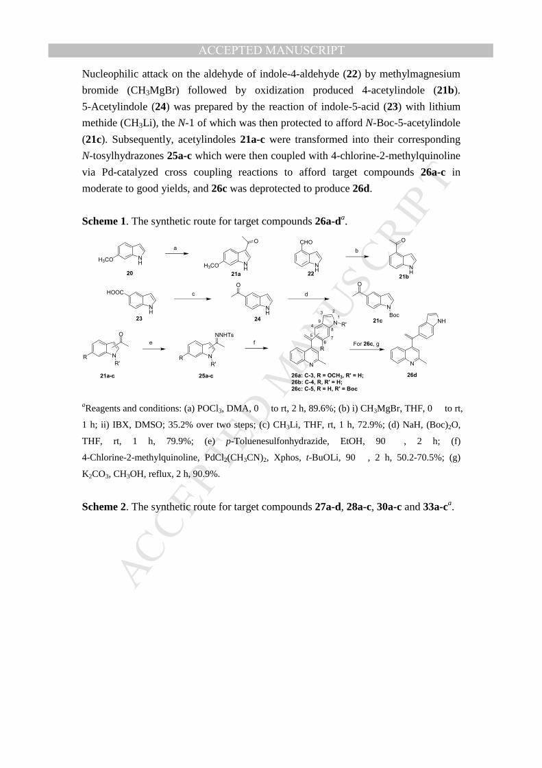
Quinoline-indole derivatives 26a-d with different substituted positions on the
indole ring were synthesized by the method in Alami’s report [23] as shown in
Scheme 1. Acetylindoles 21a-c were prepared by three different methods.
3-Acetyl-6-methoylindole (21a) was synthesized by a Vilsmeier-Haack reaction of
6-methoylindole (20) in the presence of phosphoryl chloride and dimethylacetamide.
MANUSCRIP
T
ACCEPTED
ACCEPTED MANUSCRIPT
Nucleophilic attack on the aldehyde of indole-4-aldehyde (22) by methylmagnesium
bromide (CH3MgBr) followed by oxidization produced 4-acetylindole (21b).
5-Acetylindole (24) was prepared by the reaction of indole-5-acid (23) with lithium
methide (CH3Li), the N-1 of which was then protected to afford N-Boc-5-acetylindole
(21c). Subsequently, acetylindoles 21a-c were transformed into their corresponding
N-tosylhydrazones 25a-c which were then coupled with 4-chlorine-2-methylquinoline
via Pd-catalyzed cross coupling reactions to afford target compounds 26a-c in
moderate to good yields, and 26c was deprotected to produce 26d.
Scheme 1. The synthetic route for target compounds 26a-da.
NH
CHO
NH
HOOC
NH
H3CO
NH
O
NH
H3CO
O
NH
O
N
O
Boc
N
O
RR'
N
NNHTs
RR'
a b
c d
N
NH
e f
26a: C-3, R = OCH3, R' = H;
26b: C-4, R, R' = H;
26c: C-5, R = H, R' = Boc
21a 21b
21c
21a-c 25a-c 26d
20 22
23 24
N
5
67
8
94
N1
23
R'
RFor 26c, g
aReagents and conditions: (a) POCl3, DMA, 0 � to rt, 2 h, 89.6%; (b) i) CH3MgBr, THF, 0 � to rt,
1 h; ii) IBX, DMSO; 35.2% over two steps; (c) CH3Li, THF, rt, 1 h, 72.9%; (d) NaH, (Boc)2O,
THF, rt, 1 h, 79.9%; (e) p-Toluenesulfonhydrazide, EtOH, 90 �, 2 h; (f)
4-Chlorine-2-methylquinoline, PdCl2(CH3CN)2, Xphos, t-BuOLi, 90 �, 2 h, 50.2-70.5%; (g)
K2CO3, CH3OH, reflux, 2 h, 90.9%.
Scheme 2. The synthetic route for target compounds 27a-d, 28a-c, 30a-c and 33a-ca.
MANUSCRIP
T
ACCEPTED
ACCEPTED MANUSCRIPT
N
Cl
N
N
OCH3R
N
N
OCH3H2N
c d
N
COOHe
N
COClc
N
O N
OCH3
R
d
N
O N
OCH3
NH2
29 30a, R = H;
30b, R = NO230c
31 32 33a, R = H;
33b, R = NO2
33c
27a: C-3, R = OCH3, R' = CH3;
27b: C-4, R = H, R' = CH3;27c: C-5, R = H, R' = CH3;
27d: C-5, R = H, R' = C2H5
28b: C-4;28c: C-5
26a: C-3, R = OCH3;
26b: C-4, R = H;
26d: C-5, R = H
a
b
28a
b
N
NH
OCH3
N
4
67
8
9 NH 1
23
R10
N
4
67
8
9 N1
23
R10
R'
N
N
5 5
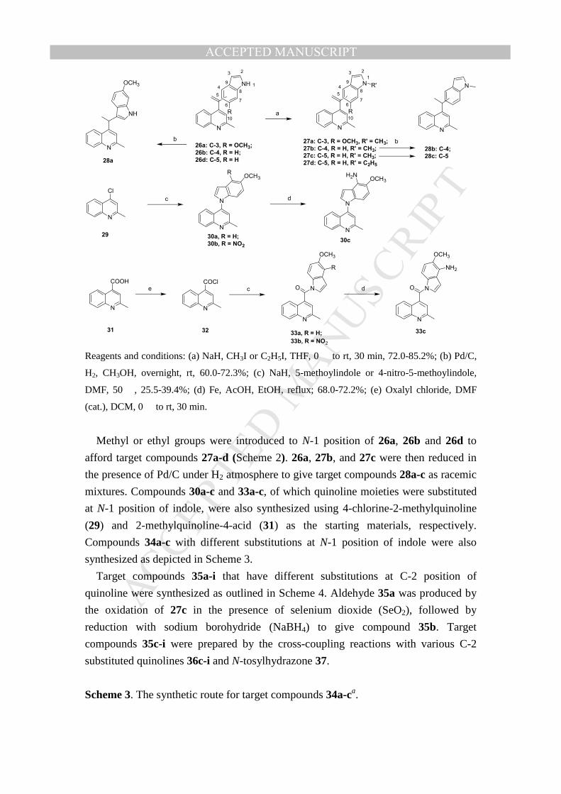
Reagents and conditions: (a) NaH, CH3I or C2H5I, THF, 0 � to rt, 30 min, 72.0-85.2%; (b) Pd/C,
H2, CH3OH, overnight, rt, 60.0-72.3%; (c) NaH, 5-methoylindole or 4-nitro-5-methoylindole,
DMF, 50 �, 25.5-39.4%; (d) Fe, AcOH, EtOH, reflux; 68.0-72.2%; (e) Oxalyl chloride, DMF
(cat.), DCM, 0 � to rt, 30 min.
Methyl or ethyl groups were introduced to N-1 position of 26a, 26b and 26d to
afford target compounds 27a-d (Scheme 2). 26a, 27b, and 27c were then reduced in
the presence of Pd/C under H2 atmosphere to give target compounds 28a-c as racemic
mixtures. Compounds 30a-c and 33a-c, of which quinoline moieties were substituted
at N-1 position of indole, were also synthesized using 4-chlorine-2-methylquinoline
(29) and 2-methylquinoline-4-acid (31) as the starting materials, respectively.
Compounds 34a-c with different substitutions at N-1 position of indole were also
synthesized as depicted in Scheme 3.
Target compounds 35a-i that have different substitutions at C-2 position of
quinoline were synthesized as outlined in Scheme 4. Aldehyde 35a was produced by
the oxidation of 27c in the presence of selenium dioxide (SeO2), followed by
reduction with sodium borohydride (NaBH4) to give compound 35b. Target
compounds 35c-i were prepared by the cross-coupling reactions with various C-2
substituted quinolines 36c-i and N-tosylhydrazone 37.
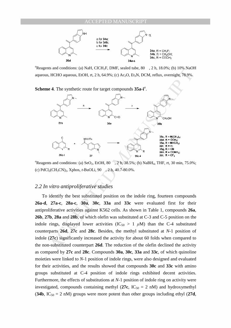
Scheme 3. The synthetic route for target compounds 34a-ca.
MANUSCRIP
T
ACCEPTED
ACCEPTED MANUSCRIPT
aReagents and conditions: (a) NaH, ClCH2F, DMF, sealed tube, 80 �, 2 h, 18.0%; (b) 10% NaOH
aqueous, HCHO aqueous, EtOH, rt, 2 h, 64.9%; (c) Ac2O, Et3N, DCM, reflux, overnight, 78.9%.
Scheme 4. The synthetic route for target compounds 35a-ia.
aReagents and conditions: (a) SeO2, EtOH, 80 �, 2 h, 38.5%; (b) NaBH4, THF, rt, 30 min, 75.0%;
(c) PdCl2(CH3CN)2, Xphos, t-BuOLi, 90 �, 2 h, 40.7-80.0%.
2.2 In vitro antiproliferative studies
To identify the best substituted position on the indole ring, fourteen compounds
26a-d, 27a-c, 28a-c, 30a, 30c, 33a and 33c were evaluated first for their
antiproliferative activities against K562 cells. As shown in Table 1, compounds 26a,
26b, 27b, 28a and 28b, of which olefin was substituted at C-3 and C-5 position on the
indole rings, displayed lower activities (IC50 > 1 µM) than the C-4 substituted
counterparts 26d, 27c and 28c. Besides, the methyl substituted at N-1 position of
indole (27c) significantly increased the activity for about 60 folds when compared to
the non-substituted counterpart 26d. The reduction of the olefin declined the activity
as compared by 27c and 28c. Compounds 30a, 30c, 33a and 33c, of which quinoline
moieties were linked to N-1 position of indole rings, were also designed and evaluated
for their activities, and the results showed that compounds 30c and 33c with amino
groups substituted at C-4 position of indole rings exhibited decent activities.
Furthermore, the effects of substitutions at N-1 position of indole ring on activity were
investigated, compounds containing methyl (27c, IC50 = 2 nM) and hydroxymethyl
(34b, IC50 = 2 nM) groups were more potent than other groups including ethyl (27d,
MANUSCRIP
T
ACCEPTED
ACCEPTED MANUSCRIPT
IC50 = 31 nM), fluoride methyl (34a, IC50 = 153 nM), or acetyl (34c, IC50 = 179 nM).
Moreover, the effects of substitutions at C-2 position of quinoline moiety on activity
were studied, compounds 35a (CHO), 35b (CH2OH), 35c (dimethylamino), 35d
(OCH3), 35e (NHCH3), 35f (H), 35g (CN), 35h (CONH2) and 35i (CF3) all exhibited
less potent activities than 27c (CH3).
Five cancer cell lines including human hepatocellular carcinoma (HepG2),
epidermoid carcinoma of the nasopharynx (KB), human colon cancer cells (HCT-8)
human breast cancer cells (MDA-MB-231), mouse liver cancer cells (H22), and
human normal hepatocytes LO2 cells were chosen to further evaluate the
antiproliferative activities of representative compounds. The cytotoxic data in Table 2
showed that all these selected compounds displayed potent activities against these five
cancer cell lines in nanomolar ranges, and they also exhibited good selectivities
against LO2 cells. The K562 cell was the most sensitive cell line among six cancer
cell lines tested. Compounds 27c and 34b that showed the most potent activities
against K562 cells also displayed very potent activities against these five cancer cell
lines with IC50 values ranging from 5 to 11 nM, which were comparable to those of
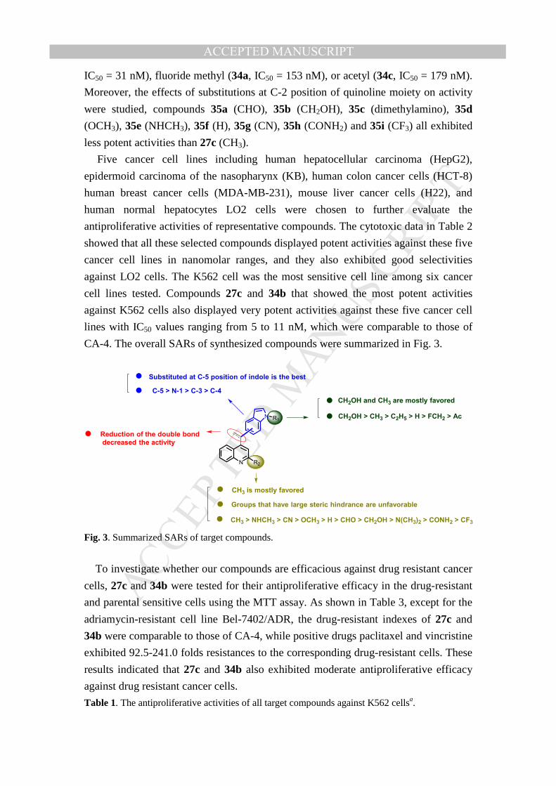
CA-4. The overall SARs of synthesized compounds were summarized in Fig. 3.
N R2
N R1
C-5 > N-1 > C-3 > C-4
CH2OH > CH3 > C2H5 > H > FCH2 > Ac
CH3 > NHCH3 > CN > OCH3 > H > CHO > CH2OH > N(CH3)2 > CONH2 > CF3
Substituted at C-5 position of indole is the best
Reduction of the double bond
decreased the activity
CH2OH and CH3 are mostly favored
CH3 is mostly favored
Groups that have large steric hindrance are unfavorable
Fig. 3. Summarized SARs of target compounds.
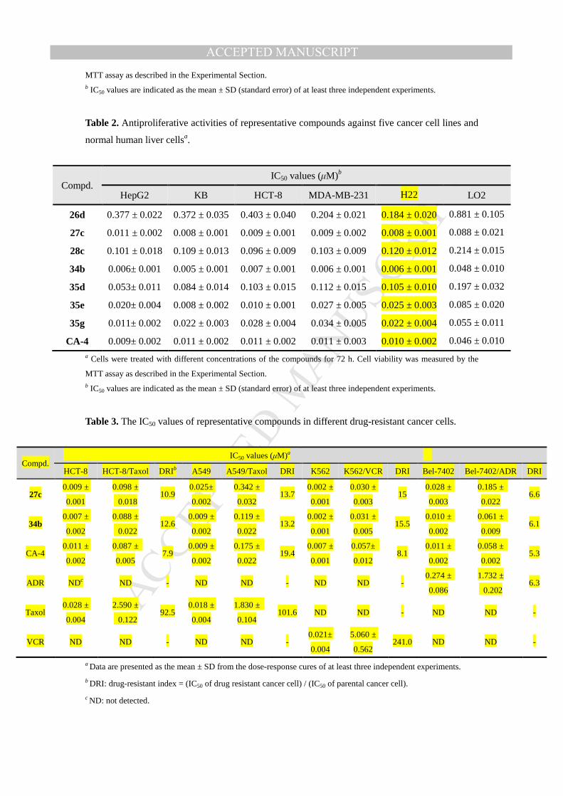
To investigate whether our compounds are efficacious against drug resistant cancer
cells, 27c and 34b were tested for their antiproliferative efficacy in the drug-resistant
and parental sensitive cells using the MTT assay. As shown in Table 3, except for the
adriamycin-resistant cell line Bel-7402/ADR, the drug-resistant indexes of 27c and
34b were comparable to those of CA-4, while positive drugs paclitaxel and vincristine
exhibited 92.5-241.0 folds resistances to the corresponding drug-resistant cells. These
results indicated that 27c and 34b also exhibited moderate antiproliferative efficacy
against drug resistant cancer cells.
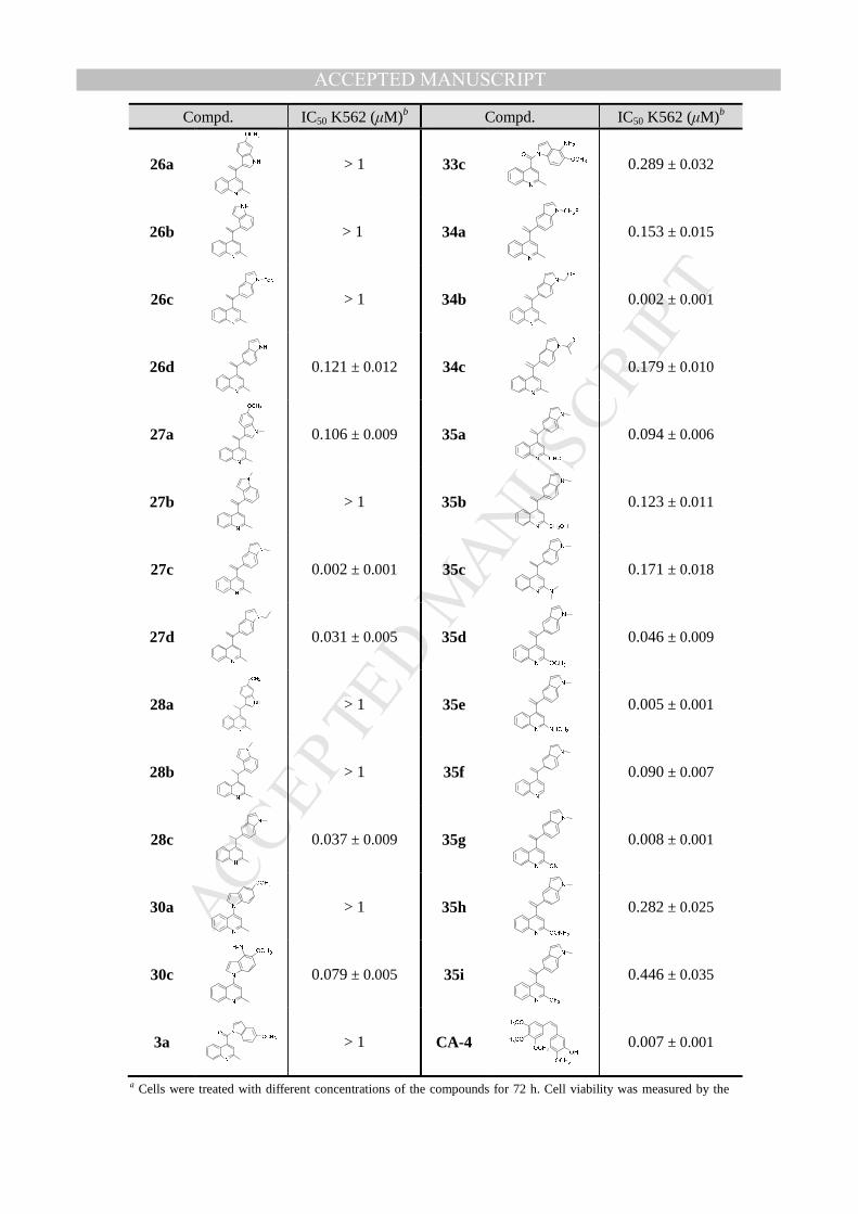
Table 1. The antiproliferative activities of all target compounds against K562 cellsa.
MANUSCRIP
T
ACCEPTED
ACCEPTED MANUSCRIPT
Compd. IC50 K562 (µM)b Compd. IC50 K562 (µM)b
26a
> 1 33c
0.289 ± 0.032
26b
> 1 34a
0.153 ± 0.015
26c
> 1 34b
0.002 ± 0.001
26d
0.121 ± 0.012 34c
0.179 ± 0.010
27a
0.106 ± 0.009 35a
0.094 ± 0.006
27b
> 1 35b
0.123 ± 0.011
27c
0.002 ± 0.001 35c
0.171 ± 0.018
27d
0.031 ± 0.005 35d
0.046 ± 0.009
28a
> 1 35e
0.005 ± 0.001
28b
> 1 35f
0.090 ± 0.007
28c
0.037 ± 0.009 35g
0.008 ± 0.001
30a
> 1 35h
0.282 ± 0.025
30c
0.079 ± 0.005 35i
0.446 ± 0.035
3a
> 1 CA-4
0.007 ± 0.001
a Cells were treated with different concentrations of the compounds for 72 h. Cell viability was measured by the
MANUSCRIP
T
ACCEPTED
ACCEPTED MANUSCRIPT
MTT assay as described in the Experimental Section. b IC50 values are indicated as the mean ± SD (standard error) of at least three independent experiments.
Table 2. Antiproliferative activities of representative compounds against five cancer cell lines and
normal human liver cellsa.
Compd. IC50 values (µM)b
HepG2 KB HCT-8 MDA-MB-231 H22 LO2
26d 0.377 ± 0.022 0.372 ± 0.035 0.403 ± 0.040 0.204 ± 0.021 0.184 ± 0.020 0.881 ± 0.105
27c 0.011 ± 0.002 0.008 ± 0.001 0.009 ± 0.001 0.009 ± 0.002 0.008 ± 0.001 0.088 ± 0.021
28c 0.101 ± 0.018 0.109 ± 0.013 0.096 ± 0.009 0.103 ± 0.009 0.120 ± 0.012 0.214 ± 0.015
34b 0.006± 0.001 0.005 ± 0.001 0.007 ± 0.001 0.006 ± 0.001 0.006 ± 0.001 0.048 ± 0.010
35d 0.053± 0.011 0.084 ± 0.014 0.103 ± 0.015 0.112 ± 0.015 0.105 ± 0.010 0.197 ± 0.032
35e 0.020± 0.004 0.008 ± 0.002 0.010 ± 0.001 0.027 ± 0.005 0.025 ± 0.003 0.085 ± 0.020
35g 0.011± 0.002 0.022 ± 0.003 0.028 ± 0.004 0.034 ± 0.005 0.022 ± 0.004 0.055 ± 0.011
CA-4 0.009± 0.002 0.011 ± 0.002 0.011 ± 0.002 0.011 ± 0.003 0.010 ± 0.002 0.046 ± 0.010
a Cells were treated with different concentrations of the compounds for 72 h. Cell viability was measured by the
MTT assay as described in the Experimental Section. b IC50 values are indicated as the mean ± SD (standard error) of at least three independent experiments.
Table 3. The IC50 values of representative compounds in different drug-resistant cancer cells.
Compd. IC50 values (µM)a
HCT-8 HCT-8/Taxol DRIb A549 A549/Taxol DRI K562 K562/VCR DRI Bel-7402 Bel-7402/ADR DRI
27c 0.009 ±
0.001
0.098 ±
0.018 10.9
0.025±
0.002
0.342 ±
0.032 13.7
0.002 ±
0.001
0.030 ±
0.003 15
0.028 ±
0.003
0.185 ±
0.022 6.6
34b 0.007 ±
0.002
0.088 ±
0.022 12.6
0.009 ±
0.002
0.119 ±
0.022 13.2
0.002 ±
0.001
0.031 ±
0.005 15.5
0.010 ±
0.002
0.061 ±
0.009 6.1
CA-4 0.011 ±
0.002
0.087 ±
0.005 7.9
0.009 ±
0.002
0.175 ±
0.022 19.4
0.007 ±
0.001
0.057±
0.012 8.1
0.011 ±
0.002
0.058 ±
0.002 5.3
ADR NDc ND - ND ND - ND ND - 0.274 ±
0.086
1.732 ±
0.202 6.3
Taxol 0.028 ±
0.004
2.590 ±
0.122 92.5
0.018 ±
0.004
1.830 ±
0.104 101.6 ND ND - ND ND -
VCR ND ND - ND ND - 0.021±
0.004
5.060 ±
0.562 241.0 ND ND -
a Data are presented as the mean ± SD from the dose-response cures of at least three independent experiments.
b DRI: drug-resistant index = (IC50 of drug resistant cancer cell) / (IC50 of parental cancer cell).
c ND: not detected.
MANUSCRIP
T
ACCEPTED
ACCEPTED MANUSCRIPT
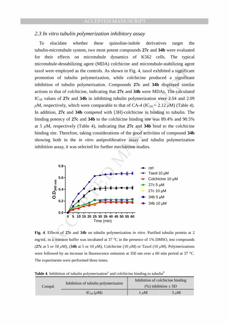
2.3 In vitro tubulin polymerization inhibitory assay
To elucidate whether these quinoline-indole derivatives target the
tubulin-microtubule system, two most potent compounds 27c and 34b were evaluated
for their effects on microtubule dynamics of K562 cells. The typical
microtubule-destabilizing agent (MDA) colchicine and microtubule-stabilizing agent
taxol were employed as the controls. As shown in Fig. 4, taxol exhibited a significant
promotion of tubulin polymerization, while colchicine produced a significant
inhibition of tubulin polymerization. Compounds 27c and 34b displayed similar
actions to that of colchicine, indicating that 27c and 34b were MDAs. The calculated
IC50 values of 27c and 34b in inhibiting tubulin polymerization were 2.54 and 2.09
µM, respectively, which were comparable to that of CA-4 (IC50 = 2.12 µM) (Table 4).
In addition, 27c and 34b competed with [3H]-colchicine in binding to tubulin. The
binding potency of 27c and 34b to the colchicine binding site was 89.4% and 90.5%
at 5 µM, respectively (Table 4), indicating that 27c and 34b bind to the colchicine
binding site. Therefore, taking considerations of the good activities of compound 34b
showing both in the in vitro antiproliferative assay and tubulin polymerization
inhibition assay, it was selected for further mechanism studies.
O.D
340
nm
0 5 10 15 20 25 30 35 40 45 50 55 600.0
0.2
0.4
0.6
0.8 ctrlTaxol 10 µM
Colchicine 10 µM
27c 5 µM
27c 10 µM
Time (min)
34b 5 µM
34b 10 µM
Fig. 4. Effects of 27c and 34b on tubulin polymerization in vitro. Purified tubulin protein at 2
mg/mL in a reaction buffer was incubated at 37 °C in the presence of 1% DMSO, test compounds
(27c at 5 or 10 µM), (34b at 5 or 10 µM), Colchicine (10 µM) or Taxol (10 µM). Polymerizations
were followed by an increase in fluorescence emission at 350 nm over a 60 min period at 37 °C.
The experiments were performed three times.
Table 4. Inhibition of tubulin polymerizationa and colchicine binding to tubulinb
Compd. Inhibition of tubulin polymerizaion
Inhibition of colchicine binding
(%) inhibition ± SD
IC50 (µM) 1 µM 5 µM
MANUSCRIP
T
ACCEPTED
ACCEPTED MANUSCRIPT
27c 2.54 ± 0.13 78.4 ± 1.9 89.4 ± 2.2
34b 2.09 ± 0.20 79.4 ± 2.2 90.5 ± 2.5
CA-4 2.12 ± 0.10 80.2 ± 2.1 91.5 ± 4.0 a The tubulin assembly assay measured the extent of assembly of 2 mg/mL tubulin after 60 min at
37 °C. Data are presented as mean from three independent experiments. b Tubulin, 1 µM; [3H]-colchicine, 5 µM; and inhibitors, 1 or 5 µM.
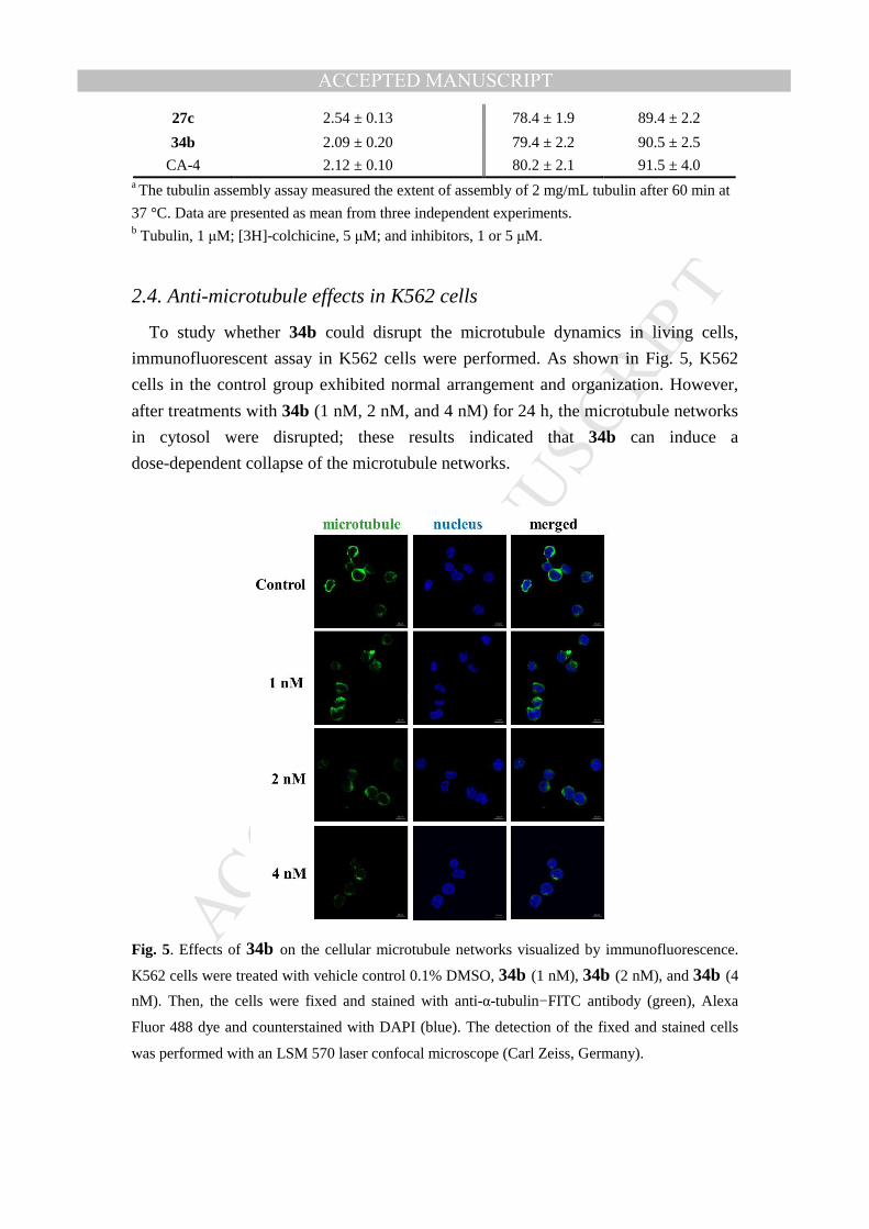
2.4. Anti-microtubule effects in K562 cells
To study whether 34b could disrupt the microtubule dynamics in living cells,
immunofluorescent assay in K562 cells were performed. As shown in Fig. 5, K562
cells in the control group exhibited normal arrangement and organization. However,
after treatments with 34b (1 nM, 2 nM, and 4 nM) for 24 h, the microtubule networks
in cytosol were disrupted; these results indicated that 34b can induce a
dose-dependent collapse of the microtubule networks.
Fig. 5. Effects of 34b on the cellular microtubule networks visualized by immunofluorescence.
K562 cells were treated with vehicle control 0.1% DMSO, 34b (1 nM), 34b (2 nM), and 34b (4
nM). Then, the cells were fixed and stained with anti-α-tubulin−FITC antibody (green), Alexa
Fluor 488 dye and counterstained with DAPI (blue). The detection of the fixed and stained cells
was performed with an LSM 570 laser confocal microscope (Carl Zeiss, Germany).
MANUSCRIP
T
ACCEPTED
ACCEPTED MANUSCRIPT
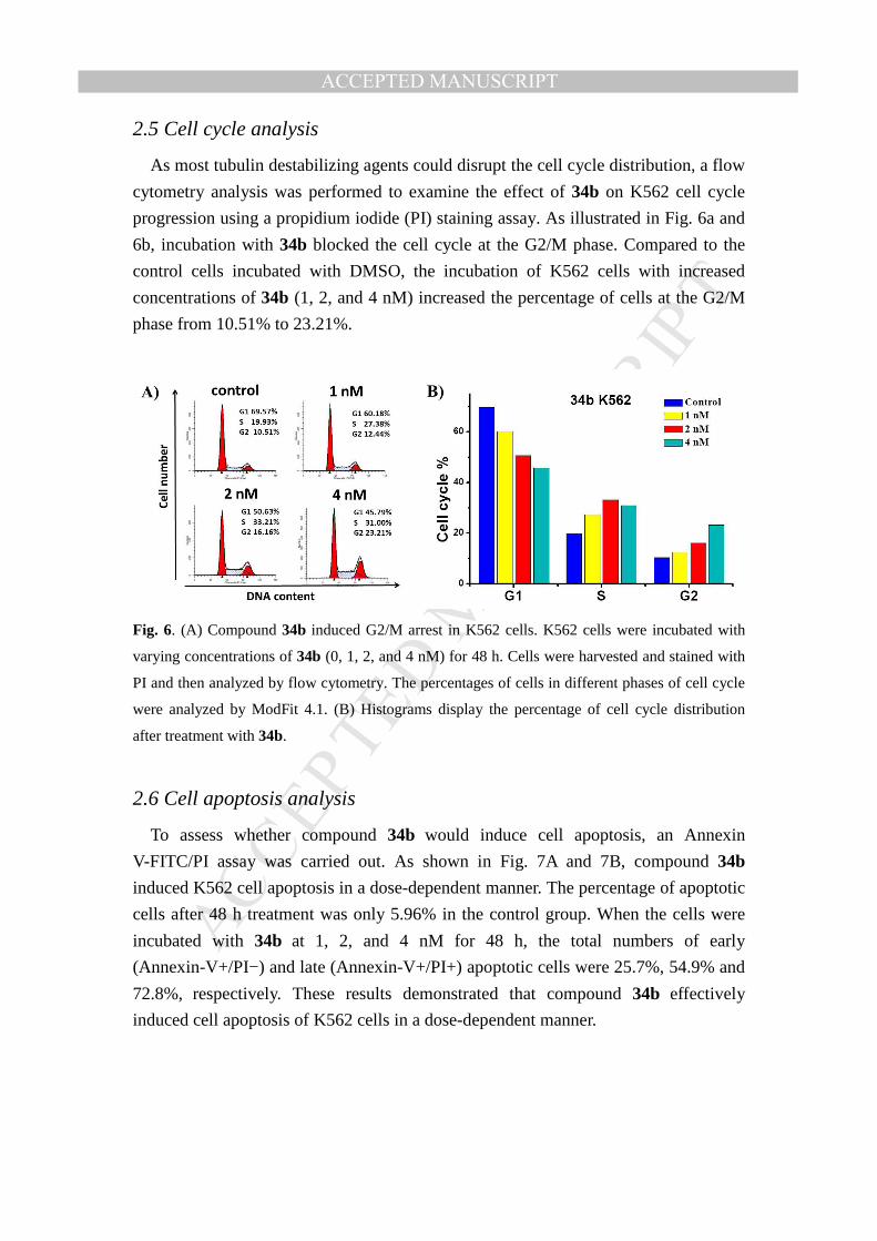
2.5 Cell cycle analysis
As most tubulin destabilizing agents could disrupt the cell cycle distribution, a flow
cytometry analysis was performed to examine the effect of 34b on K562 cell cycle
progression using a propidium iodide (PI) staining assay. As illustrated in Fig. 6a and
6b, incubation with 34b blocked the cell cycle at the G2/M phase. Compared to the
control cells incubated with DMSO, the incubation of K562 cells with increased
concentrations of 34b (1, 2, and 4 nM) increased the percentage of cells at the G2/M
phase from 10.51% to 23.21%.
Fig. 6. (A) Compound 34b induced G2/M arrest in K562 cells. K562 cells were incubated with
varying concentrations of 34b (0, 1, 2, and 4 nM) for 48 h. Cells were harvested and stained with
PI and then analyzed by flow cytometry. The percentages of cells in different phases of cell cycle
were analyzed by ModFit 4.1. (B) Histograms display the percentage of cell cycle distribution
after treatment with 34b.
2.6 Cell apoptosis analysis
To assess whether compound 34b would induce cell apoptosis, an Annexin
V-FITC/PI assay was carried out. As shown in Fig. 7A and 7B, compound 34b
induced K562 cell apoptosis in a dose-dependent manner. The percentage of apoptotic
cells after 48 h treatment was only 5.96% in the control group. When the cells were
incubated with 34b at 1, 2, and 4 nM for 48 h, the total numbers of early
(Annexin-V+/PI−) and late (Annexin-V+/PI+) apoptotic cells were 25.7%, 54.9% and
72.8%, respectively. These results demonstrated that compound 34b effectively
induced cell apoptosis of K562 cells in a dose-dependent manner.
MANUSCRIP
T
ACCEPTED
ACCEPTED MANUSCRIPT
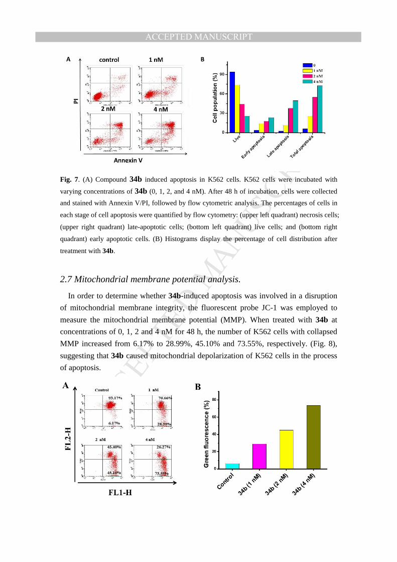
Fig. 7. (A) Compound 34b induced apoptosis in K562 cells. K562 cells were incubated with
varying concentrations of 34b (0, 1, 2, and 4 nM). After 48 h of incubation, cells were collected
and stained with Annexin V/PI, followed by flow cytometric analysis. The percentages of cells in
each stage of cell apoptosis were quantified by flow cytometry: (upper left quadrant) necrosis cells;
(upper right quadrant) late-apoptotic cells; (bottom left quadrant) live cells; and (bottom right
quadrant) early apoptotic cells. (B) Histograms display the percentage of cell distribution after
treatment with 34b.
2.7 Mitochondrial membrane potential analysis.
In order to determine whether 34b-induced apoptosis was involved in a disruption
of mitochondrial membrane integrity, the fluorescent probe JC-1 was employed to
measure the mitochondrial membrane potential (MMP). When treated with 34b at
concentrations of 0, 1, 2 and 4 nM for 48 h, the number of K562 cells with collapsed
MMP increased from 6.17% to 28.99%, 45.10% and 73.55%, respectively. (Fig. 8),
suggesting that 34b caused mitochondrial depolarization of K562 cells in the process
of apoptosis.
MANUSCRIP
T
ACCEPTED
ACCEPTED MANUSCRIPT
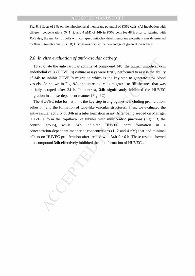
Fig. 8. Effects of 34b on the mitochondrial membrane potential of K562 cells. (A) Incubation with
different concentrations (0, 1, 2, and 4 nM) of 34b in K562 cells for 48 h prior to staining with
JC-1 dye, the number of cells with collapsed mitochondrial membrane potentials was determined
by flow cytometry analysis. (B) Histograms display the percentage of green fluorescence.
2.8 In vitro evaluation of anti-vascular activity
To evaluate the anti-vascular activity of compound 34b, the human umbilical vein
endothelial cells (HUVECs) culture assays were firstly performed to assess the ability
of 34b to inhibit HUVECs migration which is the key step to generate new blood
vessels. As shown in Fig. 9A, the untreated cells migrated to fill the area that was
initially scraped after 24 h. In contrast, 34b significantly inhibited the HUVEC
migration in a dose-dependent manner (Fig. 9C).
The HUVEC tube formation is the key step in angiogenesis, including proliferation,
adhesion, and the formation of tube-like vascular structures. Then, we evaluated the
anti-vascular activity of 34b in a tube formation assay. After being seeded on Matrigel,
HUVECs form the capillary-like tubules with multicentric junctions (Fig. 9B, the
control group), while 34b inhibited HUVEC cord formation in a
concentration-dependent manner at concentrations (1, 2 and 4 nM) that had minimal
effects on HUVEC proliferation after treated with 34b for 6 h. These results showed
that compound 34b effectively inhibited the tube formation of HUVECs.
MANUSCRIP
T
ACCEPTED
ACCEPTED MANUSCRIPT
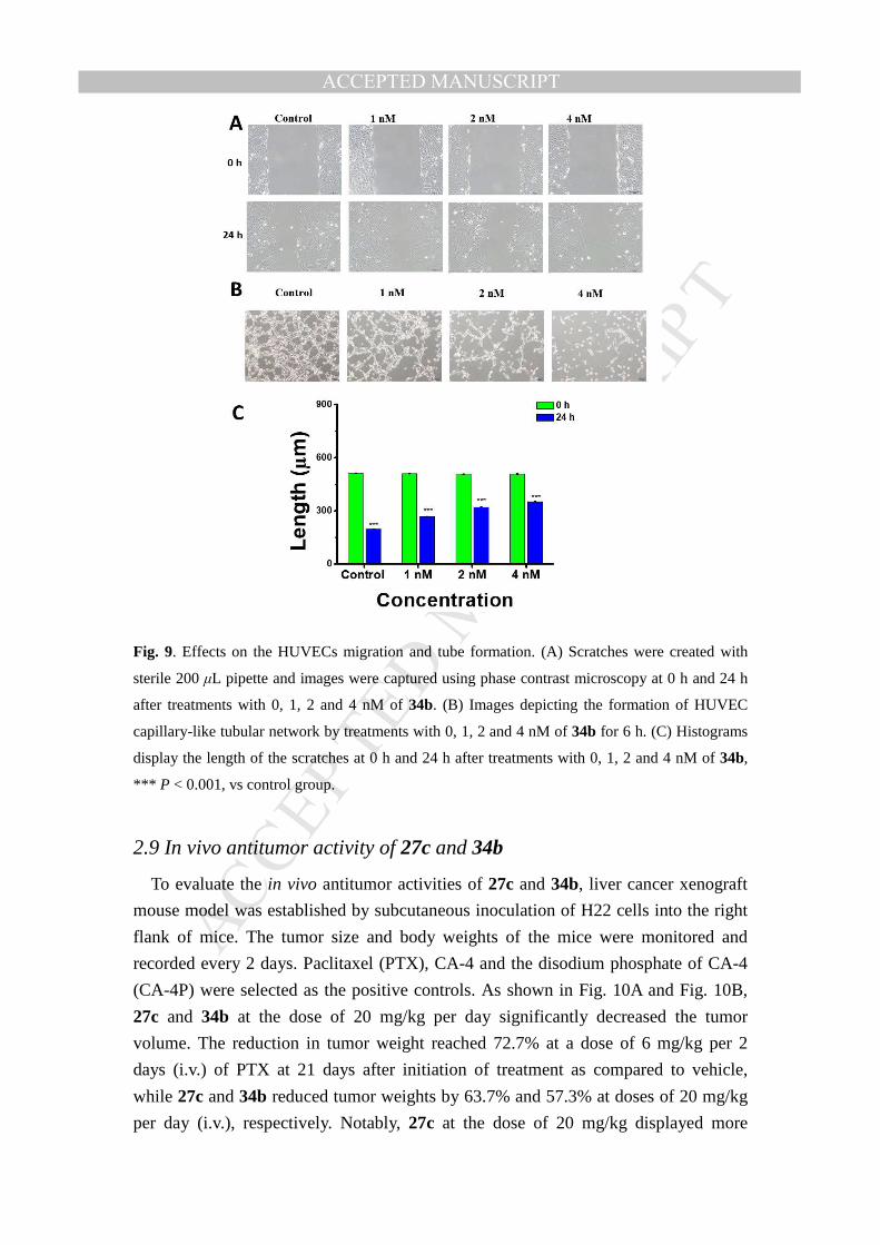
Fig. 9. Effects on the HUVECs migration and tube formation. (A) Scratches were created with
sterile 200 µL pipette and images were captured using phase contrast microscopy at 0 h and 24 h
after treatments with 0, 1, 2 and 4 nM of 34b. (B) Images depicting the formation of HUVEC
capillary-like tubular network by treatments with 0, 1, 2 and 4 nM of 34b for 6 h. (C) Histograms
display the length of the scratches at 0 h and 24 h after treatments with 0, 1, 2 and 4 nM of 34b,
*** P < 0.001, vs control group.
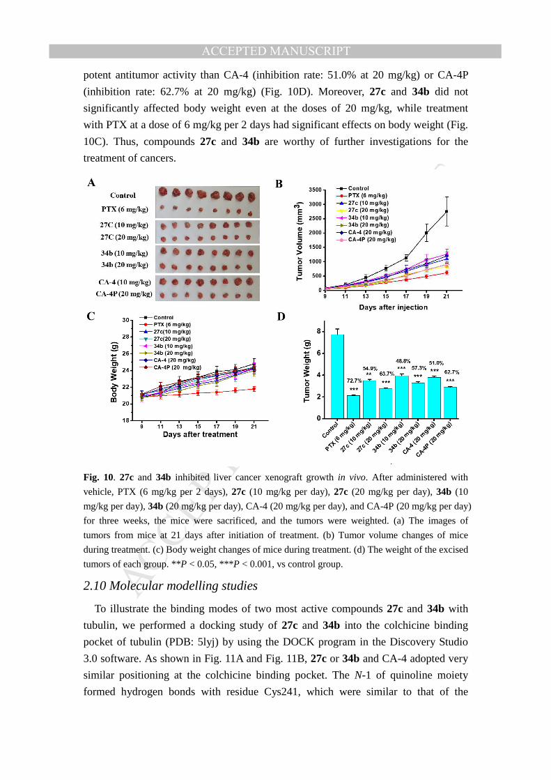
2.9 In vivo antitumor activity of 27c and 34b
To evaluate the in vivo antitumor activities of 27c and 34b, liver cancer xenograft
mouse model was established by subcutaneous inoculation of H22 cells into the right
flank of mice. The tumor size and body weights of the mice were monitored and
recorded every 2 days. Paclitaxel (PTX), CA-4 and the disodium phosphate of CA-4
(CA-4P) were selected as the positive controls. As shown in Fig. 10A and Fig. 10B,
27c and 34b at the dose of 20 mg/kg per day significantly decreased the tumor
volume. The reduction in tumor weight reached 72.7% at a dose of 6 mg/kg per 2
days (i.v.) of PTX at 21 days after initiation of treatment as compared to vehicle,
while 27c and 34b reduced tumor weights by 63.7% and 57.3% at doses of 20 mg/kg
per day (i.v.), respectively. Notably, 27c at the dose of 20 mg/kg displayed more
MANUSCRIP
T
ACCEPTED
ACCEPTED MANUSCRIPT
potent antitumor activity than CA-4 (inhibition rate: 51.0% at 20 mg/kg) or CA-4P
(inhibition rate: 62.7% at 20 mg/kg) (Fig. 10D). Moreover, 27c and 34b did not
significantly affected body weight even at the doses of 20 mg/kg, while treatment
with PTX at a dose of 6 mg/kg per 2 days had significant effects on body weight (Fig.
10C). Thus, compounds 27c and 34b are worthy of further investigations for the
treatment of cancers.
Fig. 10. 27c and 34b inhibited liver cancer xenograft growth in vivo. After administered with
vehicle, PTX (6 mg/kg per 2 days), 27c (10 mg/kg per day), 27c (20 mg/kg per day), 34b (10
mg/kg per day), 34b (20 mg/kg per day), CA-4 (20 mg/kg per day), and CA-4P (20 mg/kg per day)
for three weeks, the mice were sacrificed, and the tumors were weighted. (a) The images of
tumors from mice at 21 days after initiation of treatment. (b) Tumor volume changes of mice
during treatment. (c) Body weight changes of mice during treatment. (d) The weight of the excised
tumors of each group. **P < 0.05, ***P < 0.001, vs control group.
2.10 Molecular modelling studies
To illustrate the binding modes of two most active compounds 27c and 34b with
tubulin, we performed a docking study of 27c and 34b into the colchicine binding
pocket of tubulin (PDB: 5lyj) by using the DOCK program in the Discovery Studio
3.0 software. As shown in Fig. 11A and Fig. 11B, 27c or 34b and CA-4 adopted very
similar positioning at the colchicine binding pocket. The N-1 of quinoline moiety
formed hydrogen bonds with residue Cys241, which were similar to that of the
MANUSCRIP
T
ACCEPTED
ACCEPTED MANUSCRIPT
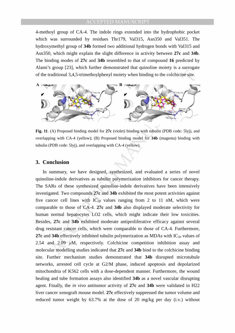
4-methoyl group of CA-4. The indole rings extended into the hydrophobic pocket
which was surrounded by residues Thr179, Val315, Asn350 and Val351. The
hydroxymethyl group of 34b formed two additional hydrogen bonds with Val315 and
Asn350, which might explain the slight difference in activity between 27c and 34b.
The binding modes of 27c and 34b resembled to that of compound 16 predicted by
Alami’s group [23], which further demonstrated that quinoline moiety is a surrogate
of the traditional 3,4,5-trimethoylphenyl moiety when binding to the colchicine site.
Fig. 11. (A) Proposed binding model for 27c (violet) binding with tubulin (PDB code: 5lyj), and
overlapping with CA-4 (yellow); (B) Proposed binding model for 34b (magenta) binding with
tubulin (PDB code: 5lyj), and overlapping with CA-4 (yellow).
3. Conclusion
In summary, we have designed, synthesized, and evaluated a series of novel
quinoline-indole derivatives as tubulin polymerization inhibitors for cancer therapy.
The SARs of these synthesized quinoline-indole derivatives have been intensively
investigated. Two compounds 27c and 34b exhibited the most potent activities against
five cancer cell lines with IC50 values ranging from 2 to 11 nM, which were
comparable to those of CA-4. 27c and 34b also displayed moderate selectivity for
human normal hepatocytes LO2 cells, which might indicate their low toxicities.
Besides, 27c and 34b exhibited moderate antiproliferative efficacy against several
drug resistant cancer cells, which were comparable to those of CA-4. Furthermore,
27c and 34b effectively inhibited tubulin polymerization as MDAs with IC50 values of
2.54 and 2.09 µM, respectively. Colchicine competition inhibition assay and
molecular modelling studies indicated that 27c and 34b bind to the colchicine binding
site. Further mechanism studies demonstrated that 34b disrupted microtubule
networks, arrested cell cycle at G2/M phase, induced apoptosis and depolarized
mitochondria of K562 cells with a dose-dependent manner. Furthermore, the wound
healing and tube formation assays also identified 34b as a novel vascular disrupting
agent. Finally, the in vivo antitumor activity of 27c and 34b were validated in H22
liver cancer xenograft mouse model. 27c effectively suppressed the tumor volume and
reduced tumor weight by 63.7% at the dose of 20 mg/kg per day (i.v.) without
MANUSCRIP
T
ACCEPTED
ACCEPTED MANUSCRIPT
apparent toxicity, which was more potent than CA-4 and CA-4P. Collectively, these
results highlighted the 27c and 34d as novel anti-tubulin agents with clinical potential
for the treatment of cancers, which deserve to be further investigated.
4. Experimental
4.1. Chemistry
4.1.1. General
Most chemicals and solvents were purchased from commercial sources. Further
purification and drying by standard methods were employed when necessary. 1H
NMR and 13C NMR spectra were recorded on Bruker-300 spectrometers in the
indicated solvents (TMS as internal standard). Data are reported as follows: chemical
shift in ppm (d), multiplicity (s =singlet, d =doublet, t =triplet, q =quartet, brs = broad
singlet, m = multiple), coupling constant (Hz), and integration. High Resolution Mass
measurement was performed on Agilent QTOF 6520 mass spectrometer with electron
spray ionization (ESI) as the ion source. Flash column chromatography was carried
out using commercially available silica gel (200-300 mesh) under pressure.
4.1.2 Synthesis of intermediate 21a.
To a solution of 6-methoylindole (300 mg, 2.04 mmol) in 5 mL DMA, POCl3 (1.9
mL, 20.4 mmol) was added at 0 �. After stirring for 2 h at room temperature, the
mixture was basified with 10% NaOH aqueous. The precipitates were collected by
filtration, washed with water and dried to afford intermediate 21a (346 mg, 89.6%) as
yellow solid, the crude product was used without further purification. The spectral
data of 21a was in accordance with literature report [34].
4.1.3 Synthesis of intermediate 21b.
To a solution of indole-4-aldehyde (500 mg, 3.45 mmol) in 20 mL anhydrous THF,
a solution of CH3MgBr in diethyl ether (3 M, 2.87 mL, 8.6 mmol) was added
dropwise at 0 � under N2 atmosphere. After stirring for 1 h, the reaction was
quenched by NH4Cl aqueous, and extracted with CH2Cl2 (3 × 50 mL). The combined
organic layers were then washed with brine, dried over anhydrous Na2SO4, and
concentrated in vacuo to provide 240 mg 1-(1H-indol-4-yl)ethan-1-ol as colorless oil,
which was dissolved into 10 mL DMSO, and IBX(500 mg, 1.79 mmol)was added in
one potion. After stirring for 1 h, the mixture was diluted with 50 mL EtOAc, then
washed with water (20 mL × 3), saturated brine, dried over anhydrous Na2SO4, and
concentrated in vacuo to afford crude product, which was purified by column
chromatography with petroleum/ethyl acetate (2:1) to give intermediate 21b (193 mg,
35.2% over two steps). The spectral data of 21b was in accordance with literature
MANUSCRIP
T
ACCEPTED
ACCEPTED MANUSCRIPT
report [35].
4.1.4 Synthesis of intermediate 21c.
To a solution of indole-5-acid (2.5 g, 15.5 mmol) in 30 mL anhydrous THF, a
solution of CH3Li (1.6 M, 30 mL, 51.2 mmol) in diethyl ether was added dropwise at
0 � under N2 atmosphere. The mixture was stirred at room temperature for 4 h, the
reaction was quenched by NH4Cl aqueous, and extracted with CH2Cl2 (3 × 50 mL).
The combined organic layers were then washed with brine, dried over anhydrous
Na2SO4, and concentrated in vacuo to provide 1.8 g 5-acetylindole (24) as white solid.
To a solution of 24 (500 mg, 3.14 mmol) in 20 mL anhydrous THF, NaH (60%, 188
mg, 4.71 mmol) was added and the mixture was stirred for 15 min. Then, (Boc)2O
(822 mg, 4.71 mmol) was added dropwise. After stirring for 1 h, the mixture was
diluted with 50 mL EtOAc, then washed with water (20 mL × 3), saturated brine,
dried over anhydrous Na2SO4, and concentrated in vacuo to afford the crude product,
which was purified by column chromatography with petroleum/ethyl acetate (5:1) to
give intermediate 21a (650 mg, 79.9%). The spectral data of 21c was in accordance
with literature report [36].
4.1.5 The general procedure for the preparations of compounds 26a-d.
To the solutions of various acetylindoles (0.82 mmol) in 10 mL EtOH,
p-toluenesulfonhydrazide (183 mg, 0.98 mmol) was added. After stirring for 2 h at
refluxing temperature, the mixtures were cooled to room temperature, and the
precipitates were collected by filtration, washed with cold EtOH and dried to afford
corresponding N-tosylhydrazones 25a-c as yellow solids in moderate to excellent
yields. Then, to the solutions of 25a-c (0.98 mmol) in 2 mL dioxane in sealed tube,
4-chlorine-2-methylquinoline (40 mg, 0.23 mmol), Xphos (19 mg, 0.04 mmol),
Pd(CH3CN)2Cl2 (6 mg, 0.02 mmol), t-BuOLi (40 mg, 0.51 mmol) were added. After
stirring for 2 h at 90 �, the mixtures were filtered and the filtrates were concentrated
to afford the crude products, which were purified by column chromatography with
petroleum/ethyl acetate (5:1) to give compounds 26a-c in good yields.
4.1.5.1 4-(1-(6-methoxy-1H-indol-3-yl)vinyl)-2-methylquinoline (26a)
Grey solid, yield 50.2%; 1H NMR (300 MHz, CDCl3) δ 8.41 (s, 1H), 8.02 (d, J =
8.5 Hz, 1H), 7.89 (d, J = 8.4 Hz, 1H), 7.76 (d, J = 8.6 Hz, 1H), 7.65 - 7.55 (m, 1H),
7.30 (s, 1H), 7.13 - 7.03 (m, 1H), 6.99 - 6.87 (m, 1H), 6.86 - 6.84 (m, 1H), 6.53 (d, J
= 2.5 Hz, 1H), 6.05 (d, J = 1.4 Hz, 1H), 5.30 (d, J = 1.5 Hz, 1H), 3.84 (s, 3H), 2.75 (s,
3H); 13C NMR (75 MHz, CDCl3) δ 158.26, 156.13, 152.03, 149.51, 147.50, 139.73,
137.29, 128.80, 127.94, 125.78, 124.99, 123.72, 121.50, 120.65, 118.94, 116.80,
112.69, 109.90, 94.45, 55.13, 24.73; HR-MS (ESI) m/z: calcd for C21H19N2O [M+H]+
315.1492, found 315.1498.
MANUSCRIP
T
ACCEPTED
ACCEPTED MANUSCRIPT
4.1.5.2 4-(1-(1H-indol-4-yl)vinyl)-2-methylquinoline (26b)
Grey solid, yield 62.5%; 1H NMR (300 MHz, CDCl3) δ 8.54 (s, 1H), 8.04 (d, J =
8.4 Hz, 1H), 7.86 (dd, J = 8.3, 1.4 Hz, 1H), 7.59 (ddd, J = 8.3, 6.8, 1.4 Hz, 1H), 7.34
(d, J = 8.0 Hz, 1H), 7.30 (s, 1H), 7.25 (s, 1H), 7.17 (t, J = 2.8 Hz, 1H), 7.08 (s, 1H),
6.92 – 6.86 (m, 1H), 6.45 (s, 1H), 6.12 (d, J = 1.6 Hz, 1H), 5.66 (d, J = 1.6 Hz, 1H),
2.75 (s, 3H); 13C NMR (75 MHz, CDCl3) δ 158.19, 149.38, 145.70, 129.39, 128.73,
128.12, 127.32, 125.47, 125.10, 124.26, 122.17, 121.87, 121.17, 119.37, 119.07,
110.79, 101.56, 100.63, 100.11, 55.10; HR-MS (ESI) m/z: calcd for C20H17N2
[M+H] + 285.1386, found 285.1388.
4.1.5.3 tert-butyl 5-(1-(2-methylquinolin-4-yl)vinyl)-1H-indole-1-carboxylate (26c)
Grey solid, yield 70.5%; 1H NMR (300 MHz, CDCl3) δ 8.01 (s, 1H), 7.98 (d, J =
2.7 Hz, 1H), 7.66 (d, J = 8.4 Hz, 1H), 7.61 - 7.54 (m, 1H), 7.51 (d, J = 3.8 Hz, 1H),
7.35 - 7.31 (m, 1H), 7.30 - 7.26 (m, 1H), 7.26 - 7.22 (m, 1H), 7.21 (d, J = 4.5 Hz, 1H),
6.40 (d, J = 3.7 Hz, 1H), 5.95 (s, 1H), 5.33 (s, 1H), 2.72 (s, 3H), 1.59 (s, 9H); 13C
NMR (75 MHz, CDCl3) δ 158.29, 149.09, 148.45, 147.74, 146.04, 134.23, 130.24,
128.73, 128.36, 126.09, 125.61, 125.07, 124.95, 122.40, 122.05, 118.88, 115.68,
114.66, 106.97, 83.38, 27.67, 24.90; HR-MS (ESI) m/z: calcd for C25H25N2O2
[M+H] + 385.1911, found 385.1916.
4.1.5.4 4-(1-(1H-indol-5-yl)vinyl)-2-methylquinoline (26d)
To a solution of 26c (75 mg, 0.20 mmol) in 10 mL CH3OH, K2CO3 (33 mg, 0.24
mmol) was added. After stirring for 2 h at refluxing temperature, the mixture was
extracted with CH2Cl2 (3 × 25 mL). The combined organic layers were then washed
with brine, dried over anhydrous Na2SO4, and concentrated in vacuo to provide the
crude product, which was purified by column chromatography with petroleum/ethyl
acetate (5:1) to give 50 mg 26d as grey solid, yield 90.9%; 1H NMR (300 MHz,
CDCl3) δ 8.31 (s, 1H), 8.04 (d, J = 8.4 Hz, 1H), 7.79 (dd, J = 8.3, 1.4 Hz, 1H), 7.62
(ddd, J = 8.4, 6.8, 1.5 Hz, 1H), 7.51 - 7.44 (m, 1H), 7.34 - 7.31 (m, 1H), 7.31 - 7.29
(m, 1H), 7.29 (s, 1H), 7.23 (dd, J = 8.5, 1.7 Hz, 1H), 7.19 (t, J = 2.8 Hz, 1H), 6.47 –
6.45 (m, 1H), 5.98 (d, J = 1.2 Hz, 1H), 5.33 (d, J = 1.2 Hz, 1H), 2.78 (s, 3H); 13C
NMR (75 MHz, CDCl3) δ 158.30, 149.24, 147.65, 146.57, 135.22, 131.48, 128.77,
128.08, 127.47, 125.87, 125.24, 125.07, 124.60, 122.10, 120.31, 118.86, 114.47,
110.66, 102.43, 24.80; HR-MS (ESI) m/z: calcd for C20H17N2 [M+H]+ 285.1386,
found 285.1391.
4.1.6 The general procedures for the preparations of compounds 27a-d.
To the solutions of 26a, 26b or 26d (0.16 mmol) in 10 mL anhydrous THF, NaH
(60%, 10 mg, 0.24 mmol) was added and the mixtures were stirred for 15 min. CH3I
or C2H5I (0.20 mmol) was added, after stirring for 30 min at ambient temperature, the
mixtures were extracted with CH2Cl2 (3 × 25 mL). The combined organic layers were
then washed with brine (25 mL), dried over anhydrous Na2SO4, and concentrated in
MANUSCRIP
T
ACCEPTED
ACCEPTED MANUSCRIPT
vacuo. The residues were purified by column chromatography with petroleum/ethyl
acetate (5:1) an eluent to afford products 27a-d as grey solids in good to excellent
yields.
4.1.6.1 4-(1-(6-methoxy-1-methyl-1H-indol-3-yl)vinyl)-2-methylquinoline (27a)
Grey solid, yield 85.2%; 1H NMR (300 MHz, CDCl3) δ 8.05 (d, J = 8.5 Hz, 1H),
7.91 (d, J = 7.9 Hz, 1H), 7.78 (d, J = 8.7 Hz, 1H), 7.68 - 7.59 (m, 1H), 7.34 (d, J = 8.3
Hz, 1H), 7.30 (s, 1H), 6.88 (dd, J = 8.7, 2.3 Hz, 1H), 6.76 (d, J = 2.3 Hz, 1H), 6.40 (s,
1H), 6.02 (d, J = 1.3 Hz, 1H), 5.24 (d, J = 1.3 Hz, 1H), 3.90 (s, 3H), 3.58 (s, 3H), 2.78
(s, 3H); 13C NMR (75 MHz, CDCl3) δ 158.23, 156.07, 149.53, 147.54, 141.70, 139.60,
138.05, 128.79, 128.30, 128.06, 125.78, 125.00, 121.45, 120.88, 119.40, 115.38,
112.04, 109.47, 92.68, 55.21, 32.31, 24.82; HR-MS (ESI) m/z: calcd for
C22H21N2O[M+H]+ 329.1648, found 329.1655.
4.1.6.2 2-methyl-4-(1-(1-methyl-1H-indol-4-yl)vinyl) quinoline (27b)
Yield 73.5%, grey solid; 1H NMR (300 MHz, CDCl3) δ 8.02 (d, J = 8.5 Hz, 1H),
7.84 (d, J = 8.8 Hz, 1H), 7.63 - 7.57 (m, 1H), 7.30 (s, 1H), 7.27 (s, 1H), 7.26 (s, 1H),
7.12 (t, J = 7.8 Hz, 1H), 7.02 (d, J = 3.1 Hz, 1H), 6.88 (d, J = 7.6 Hz, 1H), 6.38 (d, J =
3.2 Hz, 1H), 6.11 (d, J = 1.7 Hz, 1H), 5.65 (d, J = 1.7 Hz, 1H), 3.80 (s, 3H), 2.75 (s,
3H); 13C NMR (75 MHz, CDCl3) δ 158.17, 149.20, 147.81, 145.71, 136.69, 132.90,
128.69, 128.65, 128.53, 128.33, 125.62, 125.41, 125.05, 124.95, 121.82, 120.95,
119.13, 108.76, 100.22, 32.52, 24.89; HR-MS (ESI) m/z: calcd for C22H19N2[M+H] +
299.1543, found 299.1544.
4.1.6.3 2-methyl-4-(1-(1-methyl-1H-indol-5-yl)vinyl) quinoline (27c)
Yield 83.4%, grey solid; 1H NMR (300 MHz, CDCl3) δ 8.05 (d, J = 8.4 Hz, 1H),
7.84 - 7.75 (m, 1H), 7.65 - 7.57 (m, 1H), 7.46 (s, 1H), 7.29 (s, 2H), 7.27 - 7.25 (m,
1H), 7.25 (s, 1H), 7.02 (d, J = 3.1 Hz, 1H), 6.38 (d, J = 3.1 Hz, 1H), 5.98 (s, 1H), 5.32
(s, 1H), 3.77 (s, 3H), 2.78 (s, 3H); 13C NMR (75 MHz, CDCl3) δ 158.25, 149.06,
147.72, 146.63, 136.00, 131.11, 129.06, 128.65, 128.26, 127.94, 125.79, 125.18,
124.97, 122.03, 119.94, 119.11, 114.35, 108.73, 101.02, 32.42, 24.91; HR-MS (ESI)
m/z: calcd for C21H19N2[M+H] + 299.1543, found 299.1543.
4.1.6.4 2-methyl-4-(1-(1-ethyl-1H-indol-5-yl)vinyl) quinoline (27d)
Yield 72.0%, white solid; 1H NMR (300 MHz, CDCl3) δ 7.94 (dd, J = 8.5, 1.2 Hz,
1H), 7.69 (d, J = 7.7 Hz, 1H), 7.47 (ddd, J = 8.4, 6.8, 1.5 Hz, 1H), 7.34 (s, 1H), 7.16
(s, 1H), 7.14 (s, 1H), 7.11 (s, 1H), 7.11 - 7.10 (m, 1H), 6.92 (d, J = 3.1 Hz, 1H), 6.25
(d, J = 3.2 Hz, 1H), 5.85 (d, J = 1.3 Hz, 1H), 5.18 (d, J = 1.2 Hz, 1H), 3.94 (t, J = 7.3
Hz, 2H), 2.65 (s, 3H), 1.27 (t, J = 7.3 Hz, 3H); 13C NMR (75 MHz, CDCl3) δ 158.25,
149.11, 147.71, 146.59, 135.04, 131.04, 128.67, 128.25, 128.11, 127.28, 125.83,
125.22, 125.00, 122.03, 119.79, 119.21, 114.28, 108.80, 101.15, 40.58, 24.91, 14.99;
HR-MS (ESI) m/z: calcd for C22H21N2[M+H] + 313.1699, found 313.1702.
MANUSCRIP
T
ACCEPTED
ACCEPTED MANUSCRIPT
4.1.7 The general procedures for the preparations of compounds 28a-c.
To solutions of 26a, 27b or 27c (0.16 mmol) in 10 mL anhydrous CH3OH, Pd-C (5
mg) was added and the mixtures were stirred overnight under H2 atmosphere. Then,
the mixtures were filtered and the filtrates were concentrated. The residues were
purified by column chromatography with petroleum/ethyl acetate (5:1) to give
compounds 28a-c in good yields.
4.1.7.1 4-(1-(6-methoxy-1H-indol-3-yl)ethyl)-2-methylquinoline (28a)
Yield 72.3%, white solid; 1H NMR (300 MHz, CDCl3) δ 8.23 (s, 1H), 8.10 (d, J =
8.6 Hz, 1H), 8.00 (d, J = 8.4 Hz, 1H), 7.59 (t, J = 7.7 Hz, 1H), 7.41 (t, J = 7.8 Hz, 1H),
7.08 (d, J = 8.8 Hz, 1H), 7.04 (s, 1H), 6.80 (s, 1H), 6.77 (s, 1H), 6.60 (d, J = 8.9 Hz,
1H), 5.04 (d, J = 7.2 Hz, 1H), 3.73 (s, 3H), 2.56 (s, 3H), 1.72 (d, J = 7.0 Hz, 3H); 13C
NMR (75 MHz, CDCl3) δ 158.44, 156.05, 151.86, 147.67, 147.52, 136.92, 128.84,
128.42, 125.05, 124.84, 122.74, 120.50, 120.23, 119.41, 119.31, 108.83, 94.19, 55.11,
31.27, 24.96, 20.84; HR-MS (ESI) m/z: calcd for C21H21N2O[M+H]+ 317.1648, found
317.1655.
4.1.7.2 2-methyl-4-(1-(1-methyl-1H-indol-4-yl) ethyl)quinolone (28b)
Yield 60%, white solid; 1H NMR (300 MHz, CDCl3) δ 8.06 - 8.03 (m, 1H), 8.02 -
7.99 (m, 1H), 7.60 (ddd, J = 8.4, 6.9, 1.4 Hz, 1H), 7.37 (ddd, J = 8.3, 6.8, 1.3 Hz, 1H),
7.22 (d, J = 9.6 Hz, 2H), 7.14 (t, J = 7.7 Hz, 1H), 7.02 (d, J = 3.1 Hz, 1H), 6.85 (d, J =
7.1 Hz, 1H), 6.42 (dd, J = 3.2, 0.9 Hz, 1H), 5.29 (q, J = 7.2 Hz, 1H), 3.79 (s, 3H),
2.69 (s, 3H), 1.82 (d, J = 7.1 Hz, 3H); 13C NMR (75 MHz, CDCl3) δ 158.26, 151.36,
147.69, 136.55, 136.23, 128.85, 128.65, 128.29, 128.15, 126.79, 125.07, 122.99,
121.28, 119.60, 116.72, 107.37, 98.55, 36.90, 32.49, 25.14, 20.50; HR-MS (ESI) m/z:
calcd for C21H21N2[M+H] + 301.1699, found 301.1702.
4.1.7.3 2-methyl-5-(1-(1-methyl-1H-indol-4-yl) ethyl)quinolone (28c)
Yield 70.3%, white solid; 1H NMR (300 MHz, CDCl3) δ 8.17 - 8.08 (m, 1H), 8.06
(d, J = 7.3 Hz, 1H), 7.62 (ddd, J = 8.4, 6.8, 1.4 Hz, 1H), 7.52 (s, 1H), 7.44 - 7.36 (m,
1H), 7.28 (s, 1H), 7.24 (d, J = 8.4 Hz, 1H), 7.10 (dd, J = 8.5, 1.7 Hz, 1H), 7.02 (d, J =
3.1 Hz, 1H), 6.44 (d, J = 3.1 Hz, 1H), 5.02 (q, J = 7.1 Hz, 1H), 3.73 (s, 3H), 2.77 (s,
3H), 1.83 (d, J = 7.1 Hz, 3H); 13C NMR (75 MHz, CDCl3) δ 158.23, 151.84, 147.80,
135.51, 135.06, 128.82, 128.70, 128.24, 128.17, 125.13, 124.94, 123.40, 121.30,
119.59, 118.82, 108.88, 100.32, 39.68, 32.31, 25.13, 22.01; HR-MS (ESI) m/z: calcd
for C21H21N2[M+H] + 301.1699, found 301.1705.
4.1.8 The general procedures for the preparations of compounds 30a, 30c, 33a and
33c.
To solutions of 5-methoylindole or 4-nitro-5-methoylindole (0.68 mmol) in 10 mL
anhydrous DMF, NaH (60%, 41 mg, 1.02 mmol) was added under N2 atmosphere and
the mixtures were stirred for 15 min. Then, 4-chloride-2-methylquinoline or newly
prepared 2-methylquinoline-4-formyl chloride (0.68 mmol) was added and the
MANUSCRIP
T
ACCEPTED
ACCEPTED MANUSCRIPT
mixtures were stirred overnight at 50 �. Then, the mixtures were diluted with 25 mL
EtOAc, then washed with water (20 mL × 3 ), saturated brine, dried over anhydrous
Na2SO4, and concentrated in vacuo, the residues were purified by column
chromatography with petroleum/ethyl acetate (5:1) to give 30a, 30b, 33a and 33b;
Then, compounds 30b or 33b (0.24 mmol) was dissolved into 5 mL mixture solvent
of EtOH and AcOH (1:1), and Fe powder (134 mg, 2.4 mmol) was added in one
potion. The reactions were stirred for 2 h at 65 �, then the solvents were removed in
vavo and the residues were neutralized by saturated NaHCO3 aqueous. The mixtures
were filtrated, and the filtrates were extracted with EtOAc (3 × 20 mL). The combined
organic layers were then washed with saturated brine, dried over anhydrous Na2SO4,
and concentrated in vacuo to afford the crude products, which were purified by
column chromatography with petroleum/ethyl acetate (2:1) to give 30c or 33c.
4.1.8.1 4-(5-methoxy-1H-indol-1-yl)-2-methylquinoline (30a)
Yield 25.5%, grey solid; 1H NMR (300 MHz, CDCl3) δ 9.87 (s, 1H), 8.20 (d, J =
3.1 Hz, 1H), 8.17 (d, J = 2.8 Hz, 1H), 8.12 (d, J = 2.3 Hz, 1H), 7.80 (d, J = 7.9 Hz,
1H), 7.74 (d, J = 8.1 Hz, 1H), 7.55 (t, J = 7.6 Hz, 1H), 7.48 (s, 1H), 7.40 (s, 1H), 7.12
(dd, J = 8.7, 2.4 Hz, 1H), 3.94 (s, 3H), 2.87 (s, 3H); 13C NMR (75 MHz, CDCl3) δ
185.53, 158.68, 152.70, 148.30, 135.30, 130.83, 129.63, 127.72, 127.10, 124.13,
123.49, 122.84, 122.23, 120.29, 119.01, 115.23, 100.63, 55.81, 29.69; HR-MS (ESI)
m/z: calcd for C19H17N2O[M+H]+ 289.1335, found 289.1342.
4.1.8.2 5-methoxy-1-(2-methylquinolin-4-yl)-1H-indol-4-amine (30c)
Yield 20.4% over two steps, yellow oil; 1H NMR (300 MHz, CDCl3) δ 8.08 - 8.02
(m, 1H), 7.69 - 7.62 (m, 2H), 7.37 - 7.31 (m, 1H), 7.26 (s, 1H), 7.17 (d, J = 3.4 Hz,
1H), 6.75 (d, J = 8.8 Hz, 1H), 6.60 (d, J = 3.3 Hz, 1H), 6.48 (d, J = 8.8 Hz, 1H), 4.01
(s, 2H), 3.79 (s, 3H), 2.70 (s, 3H); 13C NMR (75 MHz, CDCl3) δ 159.50, 149.58,
144.72, 140.32, 133.61, 130.17, 129.06, 128.46, 128.30, 126.22, 123.66, 122.99,
119.05, 118.89, 110.27, 100.87, 100.39, 57.61, 25.39; HR-MS (ESI) m/z: calcd for
C19H18N3O[M+H]+ 304.1444, found 304.1448.
4.1.8.3 (5-methoxy-1H-indol-1-yl)(2-methylquinolin-4-yl)methanone (33a)
Yield 39.4%, grey solid; 1H NMR (300 MHz, CDCl3) δ 8.48 (s, 1H), 8.16 (d, J =
8.2 Hz, 1H), 7.83 - 7.73 (m, 2H), 7.55 - 7.50 (m, 1H), 7.46 (s, 1H), 7.13 - 7.04 (m,
2H), 6.87 (s, 1H), 6.53 (d, J = 3.7 Hz, 1H), 3.92 (s, 3H), 2.85 (s, 3H);
13C NMR (75
MHz, CDCl3) δ 165.44, 158.07, 156.75, 147.65, 140.24, 131.55, 129.90, 129.72,
128.76, 126.84, 126.67, 124.12, 122.10, 119.69, 116.96, 113.19, 109.52, 103.60,
55.21, 24.84; HR-MS (ESI) m/z: calcd for C20H17N2O2[M+H] + 317.1285, found
317.1288.
4.1.8.4 (4-amino-5-methoxy-1H-indol-1-yl)(2-methylquinolin-4-yl)methanone (33c)
Yield 23.5% over two steps, yellow oil; 1H NMR (300 MHz, CDCl3) δ 8.17 - 8.07
(m, 1H), 7.94 (s, 1H), 7.76 (d, J = 6.9 Hz, 1H), 7.73 (s, 1H), 7.50 (d, J = 8.2 Hz, 1H),
MANUSCRIP
T
ACCEPTED
ACCEPTED MANUSCRIPT
7.44 (d, J = 9.6 Hz, 1H), 6.98 (d, J = 8.8 Hz, 1H), 6.75 (s, 1H), 6.49 (s, 1H), 4.12 (s,
2H), 3.95 (s, 3H), 2.83 (s, 3H); 13C NMR (75 MHz, CDCl3) δ 166.01, 158.62, 148.15,
143.52, 140.99, 131.08, 130.37, 129.25, 128.41, 127.12, 125.70, 124.69, 122.59,
120.14, 119.56, 110.03, 106.57, 106.40, 56.66, 25.42; HR-MS (ESI) m/z: calcd for
C20H18N3O2[M+H] + 332.1394, found 332.1393.
4.1.9 The synthesis of 4-(1-(1-(fluoromethyl)-1H-indol-5-yl)vinyl)-2-methylquinoline
(34a). ClCH2F gas was inlet to a solution of 5 mL DMF in sealed tube under ice-bath
condition for 5 min, then NaH (60%, 28 mg, 0.70 mmol) and 26d (100 mg, 0.36
mmol) were added. The mixture was stirred at 80 ℃ for 2 h, which was then diluted
with 25 mL EtOAc, washed with water (20 mL × 3 ), saturated brine, dried over
anhydrous Na2SO4, and concentrated in vacuo, and the residue was purified by flash
column chromatography with petroleum/ethyl acetate (5:1) to give the product 34a as
white solid, which was unstable in the silicagel column leading to low yield 18.0%; 1H NMR (300 MHz, CDCl3) δ 8.05 (d, J = 8.4 Hz, 1H), 7.75 (dd, J = 8.4, 1.4 Hz, 1H),
7.62 (ddd, J = 8.5, 6.9, 1.4 Hz, 1H), 7.45 (d, J = 1.6 Hz, 1H), 7.40 (d, J = 8.6 Hz, 1H),
7.36 - 7.29 (m, 2H), 7.28 (s, 1H), 7.16 (d, J = 3.4 Hz, 1H), 6.52 - 6.46 (m, 1H), 6.17 (s,
1H), 5.99 (d, J = 2.6 Hz, 2H), 5.37 (d, J = 1.1 Hz, 1H), 2.78 (s, 3H); 13C NMR (75
MHz, CDCl3) δ 158.30, 148.67, 147.69, 146.18, 129.10, 128.77, 128.28, 128.20,
128.15, 125.67, 125.10, 122.08, 121.30, 119.36, 115.30, 108.95, 104.77, 104.74,
84.66, 82.03, 24.90; HR-MS (ESI) m/z: calcd for C21H18FN2[M+H] + 317.1449, found
329.1452.
4.1.10 The synthesis of (5-(1-(2-methylquinolin-4-yl)vinyl)-1H-indol-1-yl)methanol
(34b). To a solution of 26d (70 mg, 0.25 mmol) in 2 mL EtOH, 1 mL 10% NaOH
aqueous and 1 mL formaldehyde aqueous were added. The mixture was stirred for 2 h,
and the precipitates were collected by filtration, washed with water and dried to afford
34b as pink solid (50 mg, yield 64.9%); 1H NMR (300 MHz, DMSO-d6) δ 7.95 (d, J =
8.3 Hz, 1H), 7.70 - 7.57 (m, 2H), 7.52 (d, J = 8.4 Hz, 1H), 7.37 (s, 2H), 7.32 (s, 1H),
7.25 (s, 1H), 6.39 (d, J = 23.4 Hz, 2H), 6.01 (s, 1H), 5.49 (d, J = 6.8 Hz, 2H), 5.31 (s,
1H), 3.35 (s, 1H), 2.70 (s, 3H); 13C NMR (75 MHz, DMSO-d6) δ 158.66, 148.54,
147.62, 146.63, 135.22, 131.26, 129.26, 129.07, 128.62, 128.57, 125.71, 125.47,
124.84, 122.22, 119.77, 118.66, 114.90, 110.57, 101.70, 68.63, 24.79; HR-MS (ESI)
m/z: calcd for C21H19N2O[M+H]+ 315.1492, found 315.1497.
4.1.11 The synthesis of 1-(5-(1-(2-methylquinolin-4-yl)vinyl)-1H-indol-1-yl)ethan-1-
-one (34c). To a solution of 26d (50 mg, 0.18 mmol) in 5 mL DCM, glacial acetic acid
(22 µL, 0.21 mmol), Et3N (36 µL, 0.54 mmol) and catalytic DMAP were added. The
mixture was refluxed overnight, which were then diluted with 25 mL EtOAc, washed
with water (20 mL × 3 ), saturated brine, dried over anhydrous Na2SO4, and
MANUSCRIP
T
ACCEPTED
ACCEPTED MANUSCRIPT
concentrated in vacuo. The residue was purified by flash column chromatography
with petroleum/ethyl acetate (5:1) to give the product 34c as white solid (45 mg,
78.9%); 1H NMR (300 MHz, CDCl3) δ 8.37 (d, J = 8.7 Hz, 1H), 8.05 (d, J = 8.1 Hz,
1H), 7.70 (d, J = 8.4 Hz, 1H), 7.62 (ddd, J = 8.4, 6.9, 1.5 Hz, 1H), 7.42 (dd, J = 8.7,
1.9 Hz, 1H), 7.37 (d, J = 3.7 Hz, 1H), 7.34 (d, J = 1.7 Hz, 1H), 7.30 (d, J = 7.1 Hz,
1H), 7.28 (s, 1H), 6.51 (d, J = 4.0 Hz, 1H), 6.03 (d, J = 1.1 Hz, 1H), 5.41 (d, J = 1.1
Hz, 1H), 2.78 (s, 3H), 2.61 (s, 3H); 13C NMR (75 MHz, CDCl3) δ 168.00, 158.30,
148.30, 147.72, 145.85, 139.98, 135.16, 134.75, 130.11, 128.78, 128.35, 125.56,
125.37, 125.11, 124.91, 123.17, 122.06, 118.83, 116.08, 108.82, 24.88, 23.39; HR-MS
(ESI) m/z: calcd for C22H19N2O[M+H]+ 327.1492, found 327.1496.
4.1.12 The synthesis of 4-(1-(1-methyl-1H-indol-5-yl)vinyl)quinoline-2-carbaldehyde
(35a). To a solution of 27c (100 mg, 0.34 mmol) in 5 mL dioxane, SeO2 (45 mg, 0.4
mmol) was added. The mixture was refluxed for 2 h, which were then diluted with 25
mL EtOAc, washed with water (20 mL × 3 ), saturated brine, dried over anhydrous
Na2SO4, and concentrated in vacuo. Then, the residue was purified by flash column
chromatography with petroleum/ethyl acetate (20:1) to give the product 35a as white
solid (40 mg, 38.5%); 1H NMR (300 MHz, CDCl3) δ 10.29 (s, 1H), 8.27 (d, J = 8.2
Hz, 1H), 8.03 (s, 1H), 7.93 - 7.85 (m, 1H), 7.75 (ddd, J = 8.4, 6.9, 1.4 Hz, 1H), 7.52 -
7.43 (m, 1H), 7.41 (t, J = 1.2 Hz, 1H), 7.25 (d, J = 5.6 Hz, 1H), 7.23 (d, J = 1.6 Hz,
1H), 7.03 (d, J = 3.1 Hz, 1H), 6.37 (d, J = 3.1 Hz, 1H), 6.03 (d, J = 1.0 Hz, 1H), 5.37
(d, J = 1.1 Hz, 1H), 3.78 (s, 3H); 13C NMR (75 MHz, CDCl3) δ 193.51, 151.88,
150.64, 147.82, 146.23, 136.08, 130.77, 130.08, 129.64, 129.19, 128.78, 128.44,
127.99, 126.25, 119.89, 119.13, 117.46, 115.35, 108.88, 101.04, 32.43; HR-MS (ESI)
m/z: calcd for C21H16N2NaO[M+Na]+ 335.1155, found 335.1155.
4.1.13 The synthesis of (4-(1-(1-methyl-1H-indol-5-yl)vinyl)quinolin-2-yl)methanol
(35b). To a solution of 35a (20 mg, 0.06 mmol) in 10 mL THF, NaBH4 (4.8 mg, 0.12
mmol) was added. The mixture was stirred for 30 min, which were then quenched
with NH4Cl aqueous, diluted with 25 mL EtOAc, washed with water (20 mL × 3 ),
saturated brine, dried over anhydrous Na2SO4, and concentrated in vacuo. The residue
was purified by flash column chromatography with petroleum/ethyl acetate (2:1) to
give the product 35b as colorless oil (15 mg, 75%); 1H NMR (300 MHz, CDCl3) δ
8.10 (d, J = 8.9 Hz, 1H), 7.82 (dd, J = 8.4, 1.4 Hz, 1H), 7.65 (ddd, J = 8.4, 6.9, 1.4 Hz,
1H), 7.43 (s, 1H), 7.34 (ddd, J = 8.3, 6.9, 1.3 Hz, 1H), 7.27 (s, 1H), 7.26 – 7.23 (m,
2H), 7.02 (d, J = 3.1 Hz, 1H), 6.38 (d, J = 3.1 Hz, 1H), 5.99 (d, J = 1.2 Hz, 1H), 5.33
(d, J = 1.2 Hz, 1H), 4.95 (s, 2H), 3.77 (s, 3H); 13C NMR (75 MHz, CDCl3) δ 158.14,
150.01, 146.62, 145.55, 136.70, 132.66, 129.01, 128.79, 128.30, 126.08, 125.72,
MANUSCRIP
T
ACCEPTED
ACCEPTED MANUSCRIPT
125.65, 125.60, 120.96, 119.46, 119.14, 118.17, 108.88, 100.14, 63.62, 32.54;
HR-MS (ESI) m/z: calcd for C21H19N2O[M+H]+ 315.1492, found 335.1494.
4.1.14 The general procedures for the preparations of compounds 35c-g and 35i.
Various C-2 substituted 4-chlorinequinolines were synthesized according to the
literature report [27]. To the solution of N-tosylhydrazone 37 (150 mg, 0.44 mmol) in
2 mL dioxane in sealed tube, various 4-chlorinequinolines (0.44 mmol), Xphos (19
mg, 0.04 mmol), Pd(CH3CN)2Cl2 (6 mg, 0.02 mmol), t-BuOLi (77 mg, 0.97 mmol)
were added. After stirring for 2 h at 90 ℃, the mixtures were filtered and the filtrates
were concentrated to afford the crude products, which were purified by column
chromatography with petroleum/ethyl acetate (5:1) to give compounds 35c-g and 35i
in good yields.
4.1.14.1 N, N-dimethyl-4-(1-(1-methyl-1H-indol-5-yl)vinyl)quinolin-2-amine (35c)
Yield 80%, yellow solid; 1H NMR (300 MHz, CDCl3) δ 7.72 (d, J = 8.2 Hz, 1H),
7.54 (dd, J = 8.1, 1.5 Hz, 1H), 7.51 (dd, J = 1.7, 0.7 Hz, 1H), 7.44 (ddd, J = 8.4, 6.9,
1.5 Hz, 1H), 7.31 (dd, J = 8.7, 1.8 Hz, 1H), 7.21 (s, 1H), 7.01 (d, J = 3.1 Hz, 1H),
7.00 - 6.93 (m, 1H), 6.90 (s, 1H), 6.37 (dd, J = 3.2, 0.9 Hz, 1H), 5.93 (d, J = 1.4 Hz,
1H), 5.31 (d, J = 1.4 Hz, 1H), 3.76 (s, 3H), 3.25 (s, 6H); 13C NMR (75 MHz, CDCl3)
δ 157.10, 149.94, 148.05, 147.49, 136.03, 131.02, 128.99, 128.71, 127.99, 126.01,
125.79, 121.43, 121.03, 119.91, 119.07, 113.49, 109.07, 108.71, 101.06, 37.63, 32.41;
HR-MS (ESI) m/z: calcd for C22H22N3[M+H] + 328.1808, found 328.1814.
4.1.14.2 2-methoxy-4-(1-(1-methyl-1H-indol-5-yl)vinyl)quinoline (35d)
Yield 66.7%, White solid; 1H NMR (300 MHz, CDCl3) δ 7.90 – 7.83 (m, 1H), 7.65
(dd, J = 8.2, 1.4 Hz, 1H), 7.57 - 7.50 (m, 1H), 7.51 - 7.48 (m, 1H), 7.27 (dd, J = 8.7,
1.7 Hz, 1H), 7.21 (d, J = 0.9 Hz, 1H), 7.19 - 7.11 (m, 1H), 6.98 (d, J = 3.1 Hz, 1H),
6.94 (s, 1H), 6.36 (dd, J = 3.1, 0.8 Hz, 1H), 5.93 (d, J = 1.3 Hz, 1H), 5.31 (d, J = 1.3
Hz, 1H), 4.11 (s, 3H), 3.71 (s, 3H); 13C NMR (75 MHz, CDCl3) δ 161.86, 151.67,
146.59, 146.48, 136.05, 130.81, 129.03, 128.74, 127.99, 126.91, 126.00, 124.05,
123.31, 119.90, 119.03, 114.21, 112.78, 108.75, 101.06, 52.89, 32.40; HR-MS (ESI)
m/z: calcd for C21H19N2O[M+H]+ 315.1492, found315.1493.
4.1.14.3 N-methyl-4-(1-(1-methyl-1H-indol-5-yl)vinyl)quinolin-2-amine (35e)
Yield 45.1%, White solid; 1H NMR (300 MHz, CDCl3) δ 7.72 (dd, J = 8.4, 1.2 Hz,
1H), 7.56 (dd, J = 8.2, 1.5 Hz, 1H), 7.51 (d, J = 1.7 Hz, 1H), 7.49 - 7.42 (m, 1H), 7.30
(dd, J = 8.6, 1.7 Hz, 1H), 7.25 (s, 1H), 7.22 (d, J = 8.7 Hz, 1H), 7.06 - 6.94 (m, 2H),
6.64 (s, 1H), 6.38 (d, J = 3.0 Hz, 1H), 5.92 (d, J = 1.4 Hz, 1H), 5.30 (d, J = 1.3 Hz,
1H), 3.75 (s, 3H), 3.11 (d, J = 4.9 Hz, 3H); 13C NMR (75 MHz, CDCl3) δ 156.96,
150.09, 147.81, 146.90, 136.01, 130.87, 128.96, 128.79, 127.94, 125.88, 125.66,
122.31, 121.36, 119.88, 119.01, 113.64, 110.91, 108.66, 101.04, 32.40, 28.26;
HR-MS (ESI) m/z: calcd for C21H20N3[M+H] + 314.1652, found 314.1655.
4.1.14.4 4-(1-(1-methyl-1H-indol-5-yl)vinyl)quinolone (35f)
MANUSCRIP
T
ACCEPTED
ACCEPTED MANUSCRIPT
Yield 64.7%, White solid; 1H NMR (300 MHz, CDCl3) δ 8.85 (d, J = 4.4 Hz, 1H),
8.05 (dd, J = 8.6, 1.3 Hz, 1H), 7.75 (dd, J = 8.5, 1.4 Hz, 1H), 7.55 (ddd, J = 8.4, 6.8,
1.5 Hz, 1H), 7.35 (d, J = 1.1 Hz, 1H), 7.29 (d, J = 4.4 Hz, 1H), 7.27 - 7.20 (m, 1H),
7.17 (d, J = 10.1 Hz, 1H), 7.14 (s, 1H), 6.91 (d, J = 3.1 Hz, 1H), 6.28 (d, J = 3.1 Hz,
1H), 5.90 (d, J = 1.2 Hz, 1H), 5.24 (d, J = 1.2 Hz, 1H), 3.65 (s, 3H); 13C NMR (75
MHz, CDCl3) δ 149.70, 149.14, 147.97, 146.52, 136.04, 131.04, 129.12, 129.05,
128.72, 127.98, 126.91, 126.06, 125.88, 121.27, 119.92, 119.12, 114.61, 108.80,
101.05, 32.41; HR-MS (ESI) m/z: calcd for C21H17N2[M+H] + 285.1386, found
285.1391.
4.1.14.5 4-(1-(1-methyl-1H-indol-5-yl)vinyl)quinoline-2-carbonitrile (35g)
Yield 40.7%, White solid; 1H NMR (300 MHz, CDCl3) δ 8.19 (d, J = 8.7 Hz, 1H),
7.89 (d, J = 7.8 Hz, 1H), 7.79 - 7.73 (m, 1H), 7.69 (s, 1H), 7.52 - 7.45 (m, 1H), 7.39
(d, J = 1.0 Hz, 1H), 7.26 (s, 1H), 7.20 (dd, J = 8.6, 1.7 Hz, 1H), 7.05 (d, J = 3.1 Hz,
1H), 6.39 (dd, J = 3.2, 0.9 Hz, 1H), 6.05 (d, J = 0.8 Hz, 1H), 5.36 (d, J = 0.9 Hz, 1H),
3.79 (s, 3H); 13C NMR (75 MHz, CDCl3) δ 151.04, 148.10, 145.27, 136.15, 133.03,
130.36, 129.68, 129.40, 128.99, 128.65, 128.02, 127.55, 126.05, 123.33, 119.79,
119.18, 117.21, 115.88, 109.04, 101.09, 32.47; HR-MS (ESI) m/z: calcd for
C21H16N3[M+H] + 310.1339, found 310.1340.
4.1.14.6 4-(1-(1-methyl-1H-indol-5-yl)vinyl)-2-(trifluoromethyl)quinolone (35i)
Yield 57.0%, White solid; 1H NMR (300 MHz, CDCl3) δ 8.26 (d, J = 8.5 Hz, 1H),
7.92 (d, J = 8.5 Hz, 1H), 7.79 - 7.75 (m, 1H), 7.74 (d, J = 4.1 Hz, 1H), 7.47 (ddd, J =
8.0, 6.6, 1.2 Hz, 1H), 7.44 - 7.40 (m, 1H), 7.25 (d, J = 2.5 Hz, 1H), 7.05 (d, J = 3.1 Hz,
1H), 6.41 (d, J = 3.1 Hz, 1H), 6.06 (s, 1H), 5.39 (s, 1H), 5.30 (s, 1H), 3.79 (s, 3H); 13C
NMR (75 MHz, CDCl3) δ 151.58, 147.48, 147.09, 147.02, 145.99, 136.12, 130.58,
129.95, 129.76, 129.28, 128.01, 127.81, 127.59, 126.01, 125.49, 123.02, 119.84,
119.16, 116.83, 116.80, 115.50, 108.95, 101.09, 32.43; HR-MS (ESI) m/z: calcd for
C21H16N3O2[M+H] + 353.1260, found 353.1266.
4.1.15 The synthesis of 4-(1-(1-methyl-1H-indol-5-yl)vinyl)quinoline-2-carboxamide
(35h). To a solution of N-tosylhydrazone 37 (118 mg, 0.34 mmol) in 2 mL dioxane in
sealed tube, 4-chlorine-2-cyanquinoline (80 mg, 0.34 mmol), Xphos (17 mg, 0.03
mmol), Pd(CH3CN)2Cl2 (9 mg, 0.03 mmol), t-BuOLi (87 mg, 1.09 mmol) were added.
After stirring for 2 h at 90 ℃, the mixture was filtered and the filtrate was
concentrated, the residue was purified by column chromatography with
petroleum/ethyl acetate (5:1) to give 34 mg product 35h as white solid, yield 30.0%; 1H NMR (300 MHz, CDCl3) δ 8.24 (s, 1H), 8.11 (d, J = 5.0 Hz, 1H), 8.04 (d, J = 8.5
Hz, 1H), 7.78 (d, J = 9.0 Hz, 1H), 7.64 - 7.57 (m, 1H), 7.34 (t, J = 1.2 Hz, 1H), 7.34 -
7.27 (m, 1H), 7.16 (d, J = 8.2 Hz, 2H), 6.93 (d, J = 3.1 Hz, 1H), 6.28 (d, J = 3.1 Hz,
1H), 5.98 (s, 1H), 5.96 - 5.89 (m, 1H), 5.29 (s, 1H), 3.67 (s, 3H); 13C NMR (75 MHz,
MANUSCRIP
T
ACCEPTED
ACCEPTED MANUSCRIPT
CDCl3) δ 166.70, 150.70, 148.56, 146.54, 136.04, 130.99, 129.49, 129.27, 129.08,
128.01, 127.34, 126.10, 119.98, 119.13, 118.93, 115.27, 108.80, 107.66, 101.02,
98.47, 32.42; HR-MS (ESI) m/z: calcd for C21H17N3NaO[M+Na]+ 350.1264, found
350.1267.
4.2 Pharmacology
4.2.1 In vitro antiproliferative assay
Cells were purchased from Nanjing KeyGen Biotech Co. Ltd. (Nanjing, China).
The cytotoxicity of the test compounds was determined using the MTT assay. Briefly,
the cell lines were incubated at 37 °C in a humidified 5% CO2 incubator for 24 h in
96-microwell plates. After medium removal, 100 mL of culture medium with 0.1%
DMSO containing the test compounds at different concentrations was added to each
well and incubated at 37 °C for another 72 h. The MTT (5 mg/mL in PBS) was added
and incubated for another 4 h, the optical density was detected with a microplate
reader at 490 nm. The IC50 values were calculated according to the dose-dependent
curves. All the experiments were repeated in at least three independent experiments.
4.2.2 In vitro tubulin polymerization inhibitory assay
An amount of 2 mg/mL tubulin (Cytoskeleton) was resuspended in PEM buffer
containing 80 mM piperazine-N,N’-bis(2-ethanesulfonic acid) sequisodium salt
PIPES (pH 6.9), 0.5 mM EGTA, 2 mM MgCl2, and 15% glycerol. Then the mixture
was preincubated with tested compounds or vehicle DMSO on ice. PEG containing
GTP was added to the final concentration of 3 mg/mL before detecting the tubulin
polymerization reaction. After 30 min, the absorbance was detected by a
spectrophotometer at 340 nm at 37 °C every 2 min for 60 min. The area under the
curve was used to determine the concentration that inhibited tubulin polymerization
by 50% (IC50), which was calculated with GraphPad Prism Software version 5.02.
4.2.3 Competitive Inhibition Assays.
The competitive binding activity of tested compounds was evaluated using a
radiolabeled [3H] colchicine competition scintillation proximity (SPA) assay. In brief,
0.08 µM [3H] colchicine was mixed with 27c (1 µM, 5 µM), 34b (1 µM, 5 µM) or
CA-4 (1 µM, 5 µM) and biotinylated porcine tubulin (0.5 µg) in a buffer of 100 µL
containing 80 mM PIPES (pH 6.8), 1 mM EGTA, 10% glycerol, 1 mM MgCl2, and 1
mM GTP for 2 h at 37 °C. Then streptavidin-labeled SPA beads (80 µg) were added
to each mixture. The radioactive counts were measured directly with a scintillation
counter.
4.2.4 Immunofluorescence Staining
MANUSCRIP
T
ACCEPTED
ACCEPTED MANUSCRIPT
K562 cells were seeded into 6-well plates and then treated with vehicle control
0.1% DMSO, 34b (1, 2, 4 nM). The cells were fixed with 4% paraformaldehyde and
then penetrated with PBS for three times. After blocking for 20 min by adding 50-100
µL goat serum albumin at room temperature, cells were incubated with a monoclonal
antibody (anti-α-tubulin) at 37 °C for 2 h. Then the cells were washed three times by
PBS following staining by fluorescence antibody and labeling of nuclei by
4,6-diamidino-2-phenylindole (DAPI). Cells were finally visualized using an LSM
570 laser confocal microscope (Carl Zeiss, Germany).
4.2.5 Cell cycle analysis
K562 cells were seeded into 6-well plates and incubated at 37 °C in a humidified
5% CO2 incubator for 24 h, and then treated with or without 34b at indicated
concentrations for another 48 h. The collected cells were fixed by adding 70% ethanol
at 4 °C for 12 h. Subsequently, the cells were resuspended in PBS containing 100 mL
RNase A and 400 mL of propidium iodide for 30 min. The DNA content of the cells
was measured using a FACS Calibur flow cytometer (Bectone Dickinson, San Jose,
CA, USA).
4.2.6 Cell apoptosis analysis
After treatment with or without 34b at indicated concentrations for 48 h, the cells
were washed twice in PBS, centrifuged and resuspended in 500 mL AnnexinV
binding buffer. The cells were then harvested, washed and stained with 5 mL Annexin
V-APC and 5 mL 7-AAD in the darkness for 15 min. Apoptosis was analyzed using a
FACS Calibur flow cytometer (Bectone Dickinson, San Jose, CA, USA).
4.2.7 Wound healing assay
K562 cells were grown in 6-well plates for 24 h. Scratches were made in
confluent monolayers using 200 µL pipette tip. Then, wounds were washed twice with
PBS to remove non-adherent cell debris. The media containing different
concentrations (1, 2, 4 nM) of the compound 34b were added to the petridishes. Cells
which migrated across the wound area were photographed using phase contrast
microscopy at 0 h and 24 h. The migration distance of cells migrated in to the wound
area was measured manually.
4.2.8 Tube formation assay
EC Matrigel matrix was thawed at 4 °C overnight, and HUVECs suspended in
DMEM were seeded in 96-well culture plates at a cell density of 50,000 cells/well
after polymerization of the Matrigel at 37 °C for 30 min. They were then treated with
20 µL different concentrations of compound 34b or vehicle for 6 h at 37 °C. Then, the
MANUSCRIP
T
ACCEPTED
ACCEPTED MANUSCRIPT
morphological changes of the cells and tubes formed were observed and
photographed under inverted microscope (OLYMPUS, Japan).
4.2.9 In vivo antitumor evaluation
Five-week-old male Institute of Cancer Research (ICR) mice were purchased from
Shanghai SLAC Laboratory Animals Co. Ltd. A total of 1 × 10⁶ H22 cells were
subcutaneously inoculated into the right flank of ICR mice according to protocols of
tumor transplant research, to initiate tumor growth. After incubation for one day, mice
were weighted and at random divided into eight groups of eight animals. The groups
treated with 27c and 34b were administered 10, 20 mg/kg in a vehicle of 10%
DMF/2% Tween 80/88% saline, respectively. The positive control group was treated
with PTX (6 mg/kg) every 2 days by intravenous injection. CA-4 and CA-4P were
administered 20 mg/kg in a vehicle of 10% DMF/2% Tween 80/88% saline and saline
solution, respectively. The negative control group received a vehicle of 10% DMF/2%
Tween 80/88% saline through intravenous injection. Treatments of 27c, 34b, CA-4
and CA-4P were done at a frequency of intravenous injection one dose per day for a
total 21 consecutive days while the positive group was treated with PTX one dose per
two days. The mice were sacrificed after the treatments and the tumors were excised
and weighed. The inhibition rate was calculated as follows: Tumor inhibitory ratio (%)
= (1-average tumor weight of treated group/average tumor weight of control group) ×
100%.
4.3 Molecular modeling
In our study, the X-ray structure of the CA-4-α,β-tubulin complex was
downloaded from the Protein Data Bank (PDB code 5lyi). The protein was prepared
by removal of the stathmin-like domain, subunits C and D, water molecules and
colchicine using Discovery Studio modules. The docking procedure was performed by
employing DOCK program in Discovery Studio 3.0 software, and the structural image
was obtained using PyMOL software.
Acknowledgments The authors acknowledge the National Natural Science Foundation of China (No.
81373280; 81673306, 81703348), The Open Project of State Key Laboratory of
Natural Medicines, China Pharmaceutical University (No. SKLNMKF 201710) for
financial support, "Double First-Class" University project CPU2018GY04, China
Pharmaceutical University.
REFERENCES
MANUSCRIP
T
ACCEPTED
ACCEPTED MANUSCRIPT
1. G. R. Pettit, S. B. Singh, E. Hamel, C. M. Lin, D. S. Alberts, D. Garcia-Kendal, Isolation and
structure of the strong cell growth and tubulin inhibitor combretastatin A-4. Experiential. 45
(1989) 209-211.
2. C. M. Lin, S. B. Singh, P. S. Chu, R. O. Dempcy, J. M. Schmidt, G. R. Pettit, E. Hamel,
Interactions of tubulin with potent natural and synthetic analogs of the antimitotic agent
combretastatin: a structure-activity study. Mol. Pharmacol. 34 (1988) 200-208.
3. A. T. Mc Gown, B. W. Fox, Differential cytotoxicity of combretastatins A1 and A4 in two
daunorubicin-resistant P388 cell lines. Cancer Chemother. Pharmacol. 26 (1990) 79-81.
4. G. G. Dark, S. A. Hill, V. E. Prise, G. M. Tozer, G. R. Pettit, D. J. Chaplin, Combretastatin
A-4, an agent that displays potent and selective toxicity toward tumor vasculature. Cancer
Res. 57 (1997) 1829-1834.
5. http://investor.mateon.com/releasedetail.cfm?ReleaseID=1041745.
6. S. Aprile, E. Del Grosso, G. C. Tron, G. Grosa, Identification of the human
UDP-glucuronosyltransferases involved in the glucuronidation of combretastatin A-4. Drug
Metab. Dispos. 35 (2007) 2252-2261.
7. S. Aprile, E. Del Grosso, G. C. Tron, G. Grosa, In vitro metabolism study of combretastatin
A-4 in rat and human liver microsomes. Drug Metab. Dispos. 35 (2007) 2252-2261.
8. G. R. Pettit, B. Toki, D. L. Herald, P. Verdier-Pinard, M. R. Boyd, E. Hamel, R. K. Pettit,
Antineoplastic agents. 379. synthesis of phenstatin phosphate. J. Med. Chem. 41 (1998)
1688-1695.
9. S. Messaoudi, B. Tréguier, A. Hamze, O. Provot, J. F. Peyrat, J. R. Rodrigo De Losada, J. M.
Liu, J. Bignon, J. Wdzieczak-Bakala, S. Thoret, J. Dubois, J. D. Brion, M. Alami,
Isocombretastatins a versus combretastatins A: the forgotten isoCA-4 isomer as a highly
promising cytotoxic and antitubulin agent. J. Med. Chem. 52 (2009) 4538-4542.
10. M. A. Soussi, S. Aprile, S. Messaoudi, O. Provot, E. Del Grosso, J. Bignon, J. Dubois, J. D.
Brion, G. Grosa, M. Alami, The metabolic fate of isoCombretastatin A-4 in human liver
microsomes: identification, synthesis and biological evaluation of metabolites.
ChemMedChem 6 (2011) 1781-1788.
11. M. Sriram, J. J. Hall, N. C. Grohmann, T. E. Strecker, T. Wootton, A. Franken, M. L.
Trawick, K. G. Pinney, Design, synthesis and biological evaluation of dihydronaphthalene
and benzosuberene analogs of the combretastatins as inhibitors of tubulin polymerization in
cancer chemotherapy. Bioorg. Med. Chem. 16 (2008) 8161-8171.
12. C. A. Herdman, T. E. Strecker, R. P. Tanpure, Z. Chen, A. Winters, J. Gerberich, L. Liu, E.
Hamel, R. P. Mason, D. J. Chaplin, M. L. Trawick, K. G. Pinney, Synthesis and biological
evaluation of benzocyclooctene-based and indene-based anticancer agents that function as
inhibitors of tubulin polymerization. Med. Chem. Comm. 7 (2012) 2418-2427.
13. E. Rasolofonjatovo, O. Provot, A. Hamze, J. Rodrigo, J. Bignon, J. Wdzieczak-Bakala, D.
Destravines, J. Dubois, J. Brion, M. Alami, Conformationnally restricted naphthalene
derivatives type isocombretastatin A-4 and isoerianin analogues: synthesis, cytotoxicity and

MANUSCRIP
T
ACCEPTED
ACCEPTED MANUSCRIPT
antitubulin activity. Eur. J. Med. Chem. 52 (2012) 22-32.
14. U. Galli, C. Travelli, S. Aprile, E. Arrigoni, S. Torretta, G. Grosa, A. Massarotti, G. Sorba, P.
L. Canonico, A. A. Genazzani, G. C. Tron, Design, synthesis, and biological evaluation of
combretabenzodiazepines: a novel class of anti-tubulin agents. J. Med. Chem. 58 (2015)
1345-1357.
15. T. Naret, J. Bignon, G. Bernadat, M. Benchekroun, H. Levaique, C. Lenoir, J. Dubois, A.
Pruvost, F. Saller, D. Borgel, B. Manoury, V. Leblais, R. Darrigrand, S. Apcher, J. Brion, E.
Schmitt, F. R. Leroux, M. Alami, A. Hamze, A fluorine scan of a tubulin polymerization
inhibitor isocombretastatin A-4: Design, synthesis, molecular modelling, and biological
evaluation. Eur. J. Med. Chem. 143 (2018) 473-490.
16. R. Álvarez, L. Aramburu, P. Puebla, E. González, M. Vicente, M. Medarde, R. Peláez,
Pyridine based antitumour compounds acting at the colchicine site. Curr. Med. Chem. 23
(2016) 1100-1130.
17. W. Li, H. Sun, S. Sun, Z. Zhu, J. Xu, Tubulin inhibitors targeting the colchicine binding site:
a perspective of privileged structures. Future Med. Chem. 9 (2017) 1765-1794.
18. S. Kasibhatla, V. Baichwal, S. X. Cai, B. Roth, I. Skvortsova, S. Skvortsov, P. Lukas, N. M.
English, N. Sirisoma, J. Drewe, A. Pervin, B. Tseng, R. O. Carlson, C. M. Pleiman,
MPC-6827: a small-molecule inhibitor of microtubule formation that is not a substrate for
multidrug resistance pumps. Cancer Res. 67 (2007) 5865-5871.
19. N. Sirisoma, A. Pervin, H. Zhang, S. Jiang, J. A. Willardsen, M. B. Anderson, G. Mather, C.
M. Pleiman, S. Kasibhatla, B. Tseng, J. Drewe, S. Cai, Discovery of
N-(4-methoxyphenyl)-N,2-dimethylquinazolin-4-amine, a potent apoptosis inducer and
efficacious anticancer agent with high blood brain barrier penetration. J. Med. Chem. 52
(2009) 2341-2351.
20. X. F. Wang, S. B. Wang, E. Ohkoshi, L. T. Wang, E. Hamel, K. Qian, S. L. Morris-Natschke,
K. H. Lee, L. Xie, N-Aryl-6-methoxy-1,2,3,4-tetrahydroquinolines: A novel class of
antitumor agents targeting the colchicine site on tubulin. Eur. J. Med. Chem. 67 (2013)
196-207.
21. X. Wang, F. Guan, E. Ohkoshi, W. Guo, L. Wang, D. Zhu, S. Wang, L. Wang, E. Hamel, D.
Yang, L. Li, K. Qian, S. L. Morris-Natschke, S. Yuan, K. Lee, L. Xie, Optimization of
4-(N-cycloamino)phenylquinazolines as a novel class of tubulin-polymerization inhibitors
targeting the colchicine site. J. Med. Chem. 57 (2014) 1390-1402.
22. S. Banerjee, K. E. Arnst, Y. Wang, G. Kumar, S. Deng, L. Yang, G. Li, J. Yang, S. W. White,
W. Li, D. D. Miller, Heterocyclic-Fused pyrimidines as novel tubulin polymerization
inhibitors targeting the colchicine binding site: structural basis and antitumor efficacy. J. Med.
Chem. 61(2018) 1704-1718.
23. I. Khelifi, T. Naret, D. Renko, A. Hamze, G. Bernadat, J. Bignon, C. Lenoir, J. Dubois, J.
Brion, O. Provot, M. Alami, Design, synthesis and anticancer properties of
IsoCombretaQuinolines as potent tubulin assembly inhibitors. Eur. J. Med. Chem. 127 (2017)
MANUSCRIP
T
ACCEPTED
ACCEPTED MANUSCRIPT
1025-1034.
24. M. Soussi, O. Provot, G. Berbadat, J. Bignon, D. Desravines, J. Dubois, J. Brion, S.
Messaoudi, M. Alami. IsoCombretaQuinazolines: potent cytotoxic agents with antitubulin
activity. ChemMedChem 10 (2015) 1392-1402.
25. A. Brancale, R. Silvestr, Indole, a core nucleus for potent inhibitors of tubulin polymerization.
Med. Res. Rev. 27 (2007) 209-238,
26. S. A. Patil, R. Patil, D. D. Miller, Indole molecules as inhibitors of tubulin polymerization:
potential new anticancer agents. Future Med. Chem. 4 (2012) 2085-2115.
27. R. Patil, S. A. Patil, K. D. Beaman, S. A. Patil, Indole molecules as inhibitors of tubulin
polymerization: potential new anticancer agents, an update (2013-2015). Future Med. Chem.
8 (2016) 1291-1316.
28. R. Álvarez, C. Gajate, P. Puebla, F. Mollinedo, M. Medarde, R. Peláez, Substitution at the
indole 3 position yields highly potent indolecombretastatins against human tumor cells. Eur.
J. Med. Chem. 158 (2018) 167-183.
29. R. Álvarez, P. Puebla, J. F. Díaz, A. C. Bento, R. G. Navas, J. Vicente, F. Mollinedo, J. M.
Andreu, M. Medarde, R. Peláez, Endowing indole-based tubulin inhibitors with an anchor for
derivatization: highly potent 3�substituted indolephenstatins and indoleisocombretastatins. J.
Med. Chem. 56 (2013) 2813-2827.
30. J. Yan, J. Hu, B. An, L. Huang, X. Li, Design, synthesis, and biological evaluation of
cyclic-indole derivatives as anti-tumor agents via the inhibition of tubulin polymerization.
Eur. J. Med. Chem. 125 (2017) 663-675.
31. W. Li, Y. Yin, H. Yao, W. Shuai, H. Sun, S. Xu, J. Liu, H. Yao, Z. Zhu, J. Xu, Discovery of
novel vinyl sulfone derivatives as anti-tumor agents with microtubule polymerization
inhibitory and vascular disrupting activities. Eur. J. Med. Chem. 157 (2018) 1068-1080.
32. W. Li, W. Shuai, F. Xu, H. Sun, S. Xu, H. Yao, J. Liu, H. Yao, Z. Zhu, J. Xu, Discovery of
novel 4-arylisochromenes as anticancer agents with inhibitory activity of tubulin
polymerization. ACS Med. Chem. Lett. 9 (2018) 974-979.
33. W. Li, Y. Yin, W. Shuai, F. Xu, H. Yao; J. Liu, K. Cheng, J. Xu, Z. Zhu, S. Xu. Discovery of
novel quinazolines as potential anti-tubulin agents occupying three zones of colchicine
domain. Bioorg. Chem. 83 (2019) 380-390.
34. M. R. Buemi, L. D. Luca, S. Ferro, R. Gitto. Targeting GluN2B-containing
N-methyl-D-aspartate receptors: design, synthesis, and binding affinity evaluation of novel
3-substituted indoles. Arch. Pharm. Chem. Life Sci. 347 (2014) 533-539.
35. T. He, X. Tao, X. Wu, L. Cai, V. W. P, Acetylation of N-heteroaryl bromides via
PdCl2/(o-tolyl)3P catalyzed Heck reactions. Synthesis, 6 (2008) 887-890.
36. B. M. Trost, A. McClory, Rhodium-catalyzed cycloisomerization: formation of indoles,
benzofurans, and enol lactones. Angew. Chem. Int. Ed. 46 (2007) 2074-2077.
MANUSCRIP
T
ACCEPTED
ACCEPTED MANUSCRIPT
Highlights:
� A series of novel quinoline-indole derivatives have been designed and synthesized.
� 34b showed the most potent in vitro antiproliferative activity.
� 27c and 34b inhibited tubulin polymerization by binding to the colchicine site.
� 34b displayed potent in vitro anti-vascular activity.
� 27c and 34b inhibited tumor growth in H22 liver cancer xenograft mouse model.