Embed Size (px)
Citation preview

Endokrinologie/Diabetologie-Kolloquium - Infektiologie
Carol Strahm - Leitender Arzt Infektiologie/ Spitalhygiene //18.12.2019
Diabetisches Fusssyndrom Infektiologische Sicht

Endokrinologie/Diabetologie-Kolloquium - Infektiologie 2
Diabetisches Fusssyndrom mit Ulkus
DFUDiabetisches Fusssyndrom mit Infektion
DFIDiabetisches Fussyndrom mit Osteomyelitis
DFO

Endokrinologie/Diabetologie-Kolloquium - Infektiologie 3
Diabetes nimmt zu - weltweit
www.diabetesatlas.org

Endokrinologie/Diabetologie-Kolloquium - Infektiologie 4
DFI - warum relevant?■ Häufige Konsultationen, häufigster Grund für Hospitalisation
■ Hoher Pflegeaufwand (tägliche Wundkontrollen, …)
■ Antibiotische Therapien
■ Chirurgische Eingriffe
■ Hohe Kosten für Gesundheitswesen
■ Und ……
IWGDF Guidelines 2019 // Lipsky 2016 PMID 26386266

Endokrinologie/Diabetologie-Kolloquium - Infektiologie 5
(1)Lavery 2006 PMID 16732010 // (2)Ndosi 2018 PMID 29083500
1666 Patienten mit Diabetes1
9.1% DFI
80% DFI Weichteile 20% DFO (Osteomyelitis)
2 Jahre

Endokrinologie/Diabetologie-Kolloquium - Infektiologie 5
(1)Lavery 2006 PMID 16732010 // (2)Ndosi 2018 PMID 29083500
1666 Patienten mit Diabetes1
9.1% DFI
80% DFI Weichteile 20% DFO (Osteomyelitis)
2 Jahre
15% verstorben299 Pat mit DFI2 46% Ulcus
geheilt
10% erneutes Ulcus
17% Amputation
6% Revask.OP1 Jahre
Häufige und schwere Infektion mit hoher Morbidität und Mortalität!

Endokrinologie/Diabetologie-Kolloquium - Infektiologie 6
Fallbeispiel70 jähriger Patient, Diabetisches Fusssyndrom
Seit 2 Tagen zunehmende Rötung, Fieber und Schüttelfrost
Klinisch Überwärmung, Schwellung, keine Schmerzen

Endokrinologie/Diabetologie-Kolloquium - Infektiologie 6
Fallbeispiel70 jähriger Patient, Diabetisches Fusssyndrom
Seit 2 Tagen zunehmende Rötung, Fieber und Schüttelfrost
Klinisch Überwärmung, Schwellung, keine Schmerzen
Infektion?
Falls ja: Schweregrad der Infektion?
Soll der Patient hospitalisiert werden?
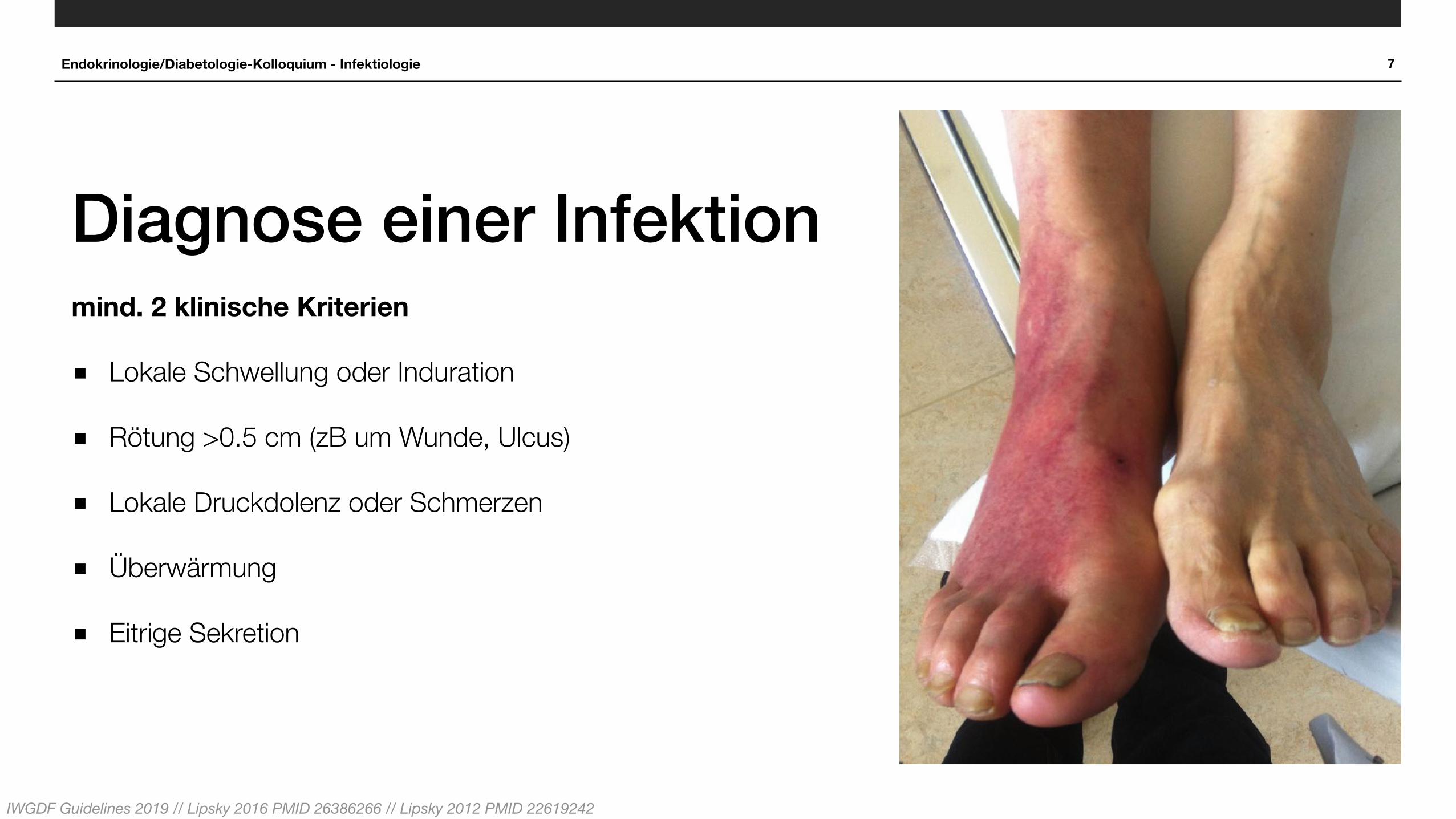
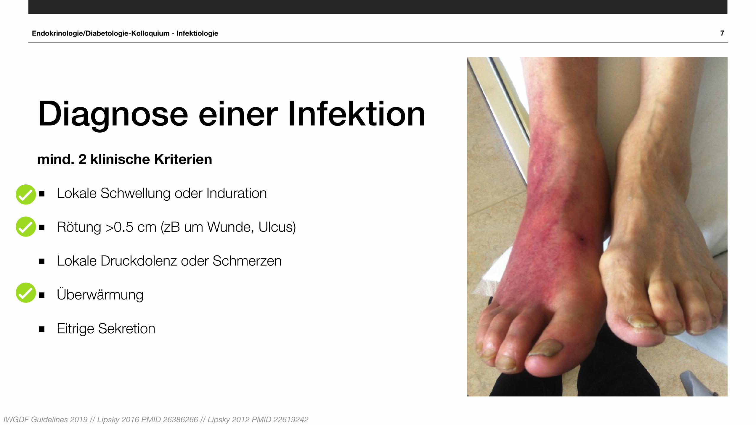
Endokrinologie/Diabetologie-Kolloquium - Infektiologie 7
Diagnose einer Infektionmind. 2 klinische Kriterien
■ Lokale Schwellung oder Induration
■ Rötung >0.5 cm (zB um Wunde, Ulcus)
■ Lokale Druckdolenz oder Schmerzen
■ Überwärmung
■ Eitrige Sekretion
Keine anderen Grund für Entzündungsreaktion der Haut: Trauma, Gicht, akuter Charcot, Fraktur, Thrombose / venöse Stase
IWGDF Guidelines 2019 // Lipsky 2016 PMID 26386266 // Lipsky 2012 PMID 22619242

8
IWGDF Klassifikation■ Keine Infektion (Grad 1)
■ Milde Infektion (Grad 2)
■ Rötung < 2 cm um Ulcus und nur Haut/ subkutane Strukturen
■ „Moderate“ Infektion (Grad 3)
■ Rötung > 2cm u/o tiefere Strukturen (Sehnen, Muskel, Gelenke, Knochen)
■ Schwere Infektion (Grad 4)
■ Alle DFI mit systemischer Manifestation (SIRS)
IWGDF Guidelines 2019 // Lipsky 2004 PMID 15472838 Ischämie (+Polyneuropathie) erschweren Diagnose und Behandlung einer DFI
IWGDF Guideline on the diagnosis and treatment of foot infection in persons with diabetes
Part of the 2019 IWGDF Guidelineson the Prevention and Managementof Diabetic Foot Disease
IWGDFGuidelines
2019

8
IWGDF Klassifikation■ Keine Infektion (Grad 1)
■ Milde Infektion (Grad 2)
■ Rötung < 2 cm um Ulcus und nur Haut/ subkutane Strukturen
■ „Moderate“ Infektion (Grad 3)
■ Rötung > 2cm u/o tiefere Strukturen (Sehnen, Muskel, Gelenke, Knochen)
■ Schwere Infektion (Grad 4)
■ Alle DFI mit systemischer Manifestation (SIRS)
IWGDF Guidelines 2019 // Lipsky 2004 PMID 15472838 Ischämie (+Polyneuropathie) erschweren Diagnose und Behandlung einer DFI
IWGDF Guideline on the diagnosis and treatment of foot infection in persons with diabetes
Part of the 2019 IWGDF Guidelineson the Prevention and Managementof Diabetic Foot Disease
IWGDFGuidelines
Ausdehnung, Tiefe, Allgemeinsymptome
2019

Fieber
8
IWGDF Klassifikation■ Keine Infektion (Grad 1)
■ Milde Infektion (Grad 2)
■ Rötung < 2 cm um Ulcus und nur Haut/ subkutane Strukturen
■ „Moderate“ Infektion (Grad 3)
■ Rötung > 2cm u/o tiefere Strukturen (Sehnen, Muskel, Gelenke, Knochen)
■ Schwere Infektion (Grad 4)
■ Alle DFI mit systemischer Manifestation (SIRS)
IWGDF Guidelines 2019 // Lipsky 2004 PMID 15472838 Ischämie (+Polyneuropathie) erschweren Diagnose und Behandlung einer DFI
IWGDF Guideline on the diagnosis and treatment of foot infection in persons with diabetes
Part of the 2019 IWGDF Guidelineson the Prevention and Managementof Diabetic Foot Disease
IWGDFGuidelines
Ausdehnung, Tiefe, Allgemeinsymptome
2019

Endokrinologie/Diabetologie-Kolloquium - Infektiologie
Klassifikation mehrfach validiert
Hospitalisation
Amputation
Major Amputation
Tod
Hospitalisation
Hospitalisationsdauer
Re-Hospitalisationen
564 • CID 2007:44 (15 February) • BRIEF REPORT
Figure 1. Hospitalization and amputation based on the Infectious Dis-eases Society of America and the International Working Group on theDiabetic Foot foot infection severity classification.
were similar to those in most diabetic populations. During amean of 27.2 months of follow-up, 247 (14.8%) of the 1666patients developed a lower extremity wound, and 151 (9.1%)developed a foot infection. Using the IDSA-IWGDF system, weclassified 71 (47.0%) of the infections as mild, 52 (34%) asmoderate, and 27 (17.9%) as severe. One patient with a mildinfection (0.6%) did not have a recognizable portal of entryand was not included in the analysis.
With an increasing IDSA-IWGDF classification system in-fection severity (i.e., going from uninfected to severe infection),there was a statistically significant trend toward an increasedrisk for amputation (x2
trend, 108.0; ), an increased an-P ! .001atomic level of amputation (x2
trend, 113.3; ) (table 2),P ! .001and an increased need for lower-extremity–related hospitali-zations (x2
trend, 118.6; ). These data are shown graphi-P ! .001cally in figure 1. Furthermore, as shown in table 2, with in-creasing IDSA-IWGDF infection severity there was a significanttrend toward increasing risk for experiencing other diabeticfoot–related complications, such as neuropathy, vascular dis-ease, and history of amputation. Additionally, there was a trendtoward a deeper wound, a higher prevalence of osteomyelitis,and more frequent occurrence of multiple infections duringthe evaluation period with increasing infection severity.
Discussion. Clinicians and researchers have used classifi-cation schemes for the foot-related complications of diabetesfor 30 years [12, 13]. The usefulness of these systems is attestedto by the fact that over a dozen have been devised since theoriginal Meggitt-Wagner grading system [8]. Previously pub-lished diabetic foot classification systems either did not spe-cifically define infection or, if they did, only noted its presenceor absence. Determining the severity of a foot infection in apatient with diabetes may help the clinician with several im-portant tasks. These include deciding whether the patientshould be hospitalized, whether to use parenteral or oral an-
tibiotic therapy, and how urgently surgery or other treatmentsneed to be performed. The IDSA and IWGDF developed in-fection severity classifications that were designed to be simpleto apply and easy to remember, but they were based on expertconsensus, not study data. We believe the results of this studyare the first to validate these new guidelines. We found that anincreasing severity of infection is associated with more-frequentlower-extremity comorbidities, such as peripheral neuropathyand arterial vascular disease, and, not surprisingly, with deeperinfection-related disease (especially bone and joint disease). Ofmost interest is that increasing IDSA-IWGDF severity is asso-ciated with a significantly increased need for hospitalization andmore-frequent (and higher level) lower extremity amputation.
Considering that these patients were screened for foot dis-orders at enrollment in the study, were educated about properfoot care, and had ready access to a foot clinic, we observed ahigher incidence of foot infection than expected. As would beexpected from an ambulatory population, most of the infec-tions were mild or moderate, and were treated with oral an-tibiotic therapy on an outpatient basis. Surprisingly, 27 (18%)of all infections were severe, and 50 patients required a lower-extremity amputation of some type. Of note is that the mildand moderate infections were also associated with a high riskfor hospitalization and, too often, a poor outcome.
The most important finding of this study is that it supportsthe clinical value of the IDSA-IWGDF diabetic foot infectionclassification in predicting clinical outcomes. It suggests thatpersons with mildly infected or noninfected wounds are highlyunlikely to require hospitalization, develop osteomyelitis, orundergo amputation. This likely would have a significant im-pact on the use of resources in this population. We believe thatthe simplicity of determining the components of this system,coupled with the strong suggestion of its clinical utility, maymake it a useful instrument in helping clinicians determinewhich of their patients are at the highest risk for adverse out-comes from a diabetic foot infection. Perhaps more aggressivemedical, surgical, and adjunctive measures could be directedat these patients, with the hope that this would improve theirfoot salvaging outcome. This system should also be useful forclinical research studies, to allow for comparisons among pa-tients enrolled in various investigations.
Acknowledgments
Potential conflicts of interest. All authors: no conflicts.
References
1. Pecoraro RE, Reiber GE, Burgess EM. Pathways to diabetic limb am-putation: basis for prevention. Diabetes Care 1990; 13:513–21.
2. Singh N, Armstrong DG, Lipsky BA. Preventing foot ulcers in patientswith diabetes. JAMA 2005; 293:217–28.
3. Centers for Disease Control and Prevention. History of foot ulcer amongpersons with diabetes—United States, 2000–2002. MMWR Morb MortalWkly Rep 2003; 52:1098–102.
(1) Lavery 2007 PMID 17243061 // Chuan 2015 PMID 25875097// Seth 2019 PMID 30820414 // Wukich 2013 PMID 23520292+24062324
Prospektive Studie1: 1666 Patienten mit DFU, USA
Grad 1 Grad 2 Grad 3 Grad 4
IWGDF Guideline on the diagnosis and treatment of foot infection in persons with diabetes
Part of the 2019 IWGDF Guidelineson the Prevention and Managementof Diabetic Foot Disease
IWGDFGuidelines
2019
‚limb-th
reaten
ing‘
‚life-t
hreaten
ing‘

Endokrinologie/Diabetologie-Kolloquium - Infektiologie 10
Einstufung Beschreibung Ausmass Management
Grad 1 keine Infektion
keine Symptome oder Zeichen einer Infektion
Grad 2 milde Infektion
• Schwellung/Induration• Erythem• Schmerz/Druckdolenz• Überwärmung
Weichteilinfektion
>0.5 cm to ≤2 cm um Ulcus
Ambulant in Praxis
Grad 3 moderate Infektion(lokale Infektion)
Abscess OsteomyelitisKleingelenks Arthritis
Infektion der tieferen Strukturen, aber ohne systemische Infekt-zeichen>2 cm um Ulcus
Ambulant oder stationär, elektive Zuweisung
Grad 4schwere Infektion Grad 2/3 mit systemischen
InfektzeichenNotfallmässige
Zuweisung
Lipsky 2016 PMID 26386266

Endokrinologie/Diabetologie-Kolloquium - Infektiologie 11
Zeichen einer schwereren/ komplizierteren Infektion (komplizierte Grad 3 und Grad 4 Infekte)
Wunde Tiefere Penetration der Infektion (Faszien, Sehnen, Muskel, Gelenke, Knochen)
Cellulitis >2cm vom Ulkus, rasche Progredienz (inkl Lymphangitis)
Lokale Symptome Schwere lokale Entzündung/ Induration, Bullae, Krepitus, Verfärbung, Nekrose/ Gangrän, Blutungen/ Petechien; Schmerzen
Allgemein Rascher Beginn/Progression; SIRS; Leukozytose, hohes CRP, Nierenversagen; Hyperglykämie; Fremdkörper; Penetrationsverletzung; Abszesse; PAVK
—> Hospitalisation?
Grad 4 und komplizierte Grad 3 Infektionen; hämodynamische und metabolische Entgleisung; IV Antibiotika; Ischämie; stationäre diagnostische Tests; Chirurgie; Compliance;
komplexe Wundbehandlungen; engmaschige Beobachtung notwendig
Lipsky 2016 PMID 26386266 // IWGDF Guidelines 2019

Endokrinologie/Diabetologie-Kolloquium - Infektiologie
Neue Klassifikation IWGDF 2019
Osteomyelitis (DFO)

13
IWGDF Klassifikation Osteomyelitis (O)■ Grad 1
■ Grad 2
■ Grad 3 (O)
■ Jede Osteomyelitis
■ Grad 4 (O)
■Osteomyelitis mit SIRS
IWGDF Guideline on the diagnosis and treatment of foot infection in persons with diabetes
Part of the 2019 IWGDF Guidelineson the Prevention and Managementof Diabetic Foot Disease
IWGDFGuidelines
IWGDF Guidelines 2019 // Lipsky 2016 PMID 26386266 // Lipsky 2012 PMID 22619242
O10.1: The Infected Diabetic Foot: Re-evaluating the Infectious Diseases Society of America Diabetic Foot Infection Classification Lavery et al; 8th international symposium of diabetic foot infection 2019 Den HaagLavery 2019 PMID 31179491
Grad 1 no infection Grad 2 mild STI Grad 3 moderate/ severe STI Grad 4 moderate/ severe DFO
?

Endokrinologie/Diabetologie-Kolloquium - Infektiologie 14
Osteomyelitis mindestens Grad 3 (O)
IWGDF Guidelines 2019 // Lipsky 2016 PMID 26386266 // Lipsky 2012 PMID 22619242

Endokrinologie/Diabetologie-Kolloquium - Infektiologie 14
Osteomyelitis mindestens Grad 3 (O)
IWGDF Guidelines 2019 // Lipsky 2016 PMID 26386266 // Lipsky 2012 PMID 22619242
‚sausage toe‘

Endokrinologie/Diabetologie-Kolloquium - Infektiologie 15
Wann an Osteomyelitis denken?Patient
Wunde/ Ulcus
Klinische Einschätzung
Polyneuropathie, PAVK/ Ischämie, Schmerzen
Rezidiv-Ulcus Multiple Ulcera
Grösse >2cm2 Verzögerte Heilung (>4 Wochen) trotz Therapie
Tiefe >3mm, freiliegender Knochen, probe-to-bone
‚sausage toe‘
Alle Wunden sorgfältig untersuchen (Palpation, Debridement und Sondierung (initial und follow-up)) !!

Endokrinologie/Diabetologie-Kolloquium - Infektiologie
Welche mikrobiologische Diagnostik sinnvoll?

Endokrinologie/Diabetologie-Kolloquium - Infektiologie 17
Einstufung Beschreibung Ausmass Diagnostik
Grad 1 keine Infektion
keine Symptome oder Zeichen einer Infektion Keine Diagnostik
Grad 2 milde Infektion
• Schwellung/Induration• Erythem• Schmerz/Druckdolenz• Überwärmung
Weichteilinfektion
>0.5 cm to ≤2 cm um Ulcus
Keine Diagnostik, sofern nicht kürzlich AB
Keine Abstriche! Allenfalls Biopsie nach Curettage
Grad 3 moderate Infektion(lokale Infektion)
Abscess OsteomyelitisKleingelenks Arthritis
Infektion der tieferen Strukturen, aber ohne systemische Infektzeichen>2 cm um Ulcus
meist chirurgische Biopsien
Grad 4schwere Infektion Grad 2/3 mit systemischen
InfektzeichenBlutkulturen
chirurgische Biopsien
Lipsky 2016 PMID 26386266

Endokrinologie/Diabetologie-Kolloquium - Infektiologie 18
Diagnostik DFO■ Probe-to-bone Test (Sensitivität 87% und Spezifität 83%)1
■ BSR >70 mm/h
■ Konventionelle Röntgenbild (erste Wochen negativ)
■ MRT (Sens. 90% und Spez. 80%)2 nur falls Diagnose unklar
■ Planung Eingriff: Abszesse, Ausdehnung der Osteomyelitis
■ Definitive Diagnose: Knochen-Biopsie (Histo und Bakt) —> entscheidend für Therapie
(1)Lam 2016 PMID 27369321 // (2)Dinh 2008 PMID 18611152 // Lipsky 2016 PMID 26386266 // Lipsky 2012 PMID 22619242

Endokrinologie/Diabetologie-Kolloquium - Infektiologie 19
2 Monate(mind 2-3 Wochen)
• Periostreaktion
• Kortikalisunterbruch, Erosion
• Knochenzerstörung, Osteolyse
• Verlust Trabekel, Demineralisierung
• Sequester
• Knochenneubildung

Endokrinologie/Diabetologie-Kolloquium - Infektiologie 20
Charcot-Fuss: wichtige Differentialdiagnose (Neuro-Osteo-Arthropathie)■ seltenes, schweres Krankheitsbild (Voraussetzung
diabetische Polyneuropathie), 0.8-8% der Diabetiker
■ akut: lokale, teilweise schmerzhafte Entzündungsreaktion, systemische Entzündungszeichen normal (BSR!)
■ chronisch: schmerzlose (nicht-infektiöse) Zerstörung lasttragender Knochen und Gelenke
■ schwierig von Osteomyelitis zu unterscheiden (aber seltener, meist PNP >> PAVK, Mittelfuss betroffen (OM Grosszehe und Vorfuss), in der Regel kein Ulkus (Anamnese!), Rötung nimmt ab bei Elevation
Ertugrul 2012 PMID 24205433 // Lipsky 2015 PMID 26386266
Blood leukocyte counts, serum C-reactive protein
(CRP) and procalcitonin concentrations are also usually
normal in CN while high in DFO, but these laboratory
parameters are relatively non-specific (44). In one study of
severe diabetic foot infections, both the leukocyte count
and the CRP level were higher in those with exclusively
soft-tissue infection than in those with concomitant
osteomyelitis (45). In another study (46) leukocyte count,
CRP, procalcitonin, and ESR levels were each signific-
antly higher in patients with foot infections, including
osteomyelitis, than patients without foot infection. Be-
cause DFO is usually a chronic condition, acute infection
markers, such as the leukocyte count and procalcitonin,
are generally within normal ranges (47, 48). Combined
with clinical findings (as discussed above) a highly elevated
ESR is likely to suggest osteomyelitis than cellulitis (39).High CRP levels and elevated leukocyte counts are not
common in CN (49). In one study, levels of bone-specific
alkaline phosphatase (a marker of bone formation) and
urinary deoxypyridinoline (a marker of bone resorption)
were found to be increased in acute CN compared to
diabetic persons without CN, reflecting ongoing bone
turnover and remodelling (50). Two other studies found
an increase in the bone resorption marker pyridinoline
cross-linked carboxy-terminal telopeptide domain of
type 1 collagen in acute CN (51, 52). Preliminary reports
suggest, however, that conventional markers of bone
turnover are of no value in differentiating osteomyelitis
from Charcot arthropathy (53).
Bone biopsyThe criterion standard for diagnosing DFO is demon-
strating positive findings on a bone specimen for both
culture and histopathology. Bone culture alone has been
reported to have a sensitivity of 92% and a specificity
of 60%. The major advantage of bone culture is that
it is the only method to define the causative pathogen(s),
thereby allowing determination of their antibiotic sen-
sitivities and accurate targeting of therapy (54, 55). Bone
samples can be obtained during an open operative pro-
cedure or by percutaneous biopsy (34). Biopsy should
be done through clinically uninfected skin after skin
antisepsis, can be undertaken at the bedside or in the
radiology suite with imaging guidance, and often does
not require anaesthesia (because of sensory neuropathy).
Studies have shown that cultures of overlying soft tissue
or sinus tracts are not sufficiently accurate in predicting
bone pathogens (56, 57). Clinicians should note that
bone specimens may yield false-positive results because
of contamination by wound-colonising flora, or false-
negative results because of sampling errors, prior antibi-
otic therapy or a failure to isolate fastidious organisms (5).Because of the potential problems noted with bone
culture, many favour histopathology as the gold standard
in diagnosing osteomyelitis (5, 11, 14, 16, 17, 22, 48).
Characteristic histological findings include aggregates
of inflammatory cells (neutrophils, lymphocytes, histio-
cytes, and plasma cells), erosion of trabecular bone and
marrow changes, including loss of normal fat, fibrosis,
and reactive bone formation (58). Although bone biopsy
is safe, it does require some time, skill, and expense; it is
therefore most recommended in specific circumstances, as
outlined in Table 2 (11). Biopsying bone is not generally
recommended in suspected CN, but reports of histological
examination of surgical specimens reveal that osteoclasts
significantly outnumber osteoblasts in reactive bone (50).
Fig. 1. (A) Plain radiographs with the left foot showing typical bony changes in Charcot neuro-osteoarthropathy (bone destruction,joint fragmentation, and dislocation). (B) Photograph of the same patient’s left foot with great toe and midfoot deformities, includingcollapsed arch.
Differentiating osteomyelitis from neuro-osteoarthropathy
Citation: Diabetic Foot & Ankle 2013, 4: 21855 - http://dx.doi.org/10.3402/dfa.v4i0.21855 3(page number not for citation purpose)
DD DFI
DD DFO

in the operative !eld, no matter how carefully rules of asepticsurgery are followed. The relevance of the !ndings of cul-tures of residual bone after toe or forefoot amputation hasnever been addressed in detail, to our knowledge. The aim ofthe present study was to prospectively compare cultures andhistological analyses of biopsy specimens obtained intra-operatively from the bone remaining after toe or forefootamputation.
Materials and MethodsPatients
During a period of 2 years, we prospectively enrolled con-secutive patients requiring a toe or forefoot amputation
because of gangrene and/or infection at the Department ofVascular Surgery at the University Hospital Basel, Switzerland.Patients requiring emergency amputation because of severesoft-tissue infection or trauma were excluded. The decision toperform the amputation as well as the timing were at the dis-cretion of the treating surgeon. Radiographic examinationswere not required for inclusion into the study since the indi-cation for amputation was based on clinical grounds in allcases. The local ethical committee approved the study protocol,and all patients gave written informed consent. All patientsunderwent assessment for peripheral arterial disease with anklebrachial index measurements as well as segmental volumeplethysmography. If necessary and possible, peripheral arterialdisease was treated with endovascular techniques or bypasssurgery before or after amputation. Age; sex; presence of dia-betes mellitus; stage of peripheral arterial disease; previousrevascularization procedures; C-reactive protein (CRP) level;presence of necrosis and/or cellulitis; PEDIS (perfusion, extent,depth, infection, and sensation) classi!cation; number of am-putated toes; amputation level; wound closure; and antibiotictreatment before, during, and after amputation were docu-mented. Patient charts from regular outpatient visits or fromrehospitalizations were analyzed retrospectively to assess out-comes. Treatment was considered to have failed if another,more proximal amputation had to be performed.
Surgical Technique of AmputationSurgery was performed by 1 of 3 senior consultants. The skinof the entire foot was disinfected using an alcohol-based iodinesolution (Braunoderm; B Braun) for 3 minutes, and steriledrapes were applied. The surgeon determined the extent ofamputation on the basis of the macroscopic aspect, takinginto account the extent of ischemia and infection. If necrosishad not reached the interdigital fold, exarticulation of themetatarsophalangeal joint was performed followed byremoval of the cartilage from the metatarsal head. If necrosisor infection was more extensive, transmetatarsal amputationof individual digits or the entire forefoot was performed usingan oscillating saw.
Bone BiopsyThe protocol for retrieving biopsy specimens from the residual,presumably uninfected bone (“proximal biopsy”) is described in
Figure 1. Toe amputation was performed with use of all possibleprecautions to avoid contamination of the residual bone, includingwrapping the toe dedicated for amputation in sterile gauze andremoving it before retrieving biopsy specimens from the residualbone. The technique of proximal bone biopsy differed according towhether the patient underwent exarticulation of the metatarso-phalangeal joint or transmetatarsal amputation. In the case ofexarticulation, 2 bone cylinders (1 for culture and 1 for histologicalanalysis) were retrieved from the metatarsal head using an 8GJamshidi biopsy needle (BD). In the case of transmetatarsalamputation, a 3 to 5-mm slice of corticocancellous metatarsalbone was removed using an oscillating saw. The slice was then cutin half, with 1 half used for culture and the other used for histo-logical analysis. All biopsy specimens were transferred directly intosterile closable transport tubes and sent in parallel for microbio-logical culture and histological analysis immediately after surgery.
Histological Analysis of Bone Biopsy SpecimensHistological analysis was performed using EDTA (ethyl-enediaminetetraacetic acid) decalci!cation (in combinationwith ultrasound at 30!C) of formalin-!xed probes followed byparaf!n embedding and staining with hematoxylin and eosin.The time until decalci!cation was noted. The size of eachbiopsy specimen was measured.
Our histological criteria for diagnosing osteomyelitiswere adapted from the approaches described by Mirra et al.9
and Spangehl et al.10, in their work on periprosthetic jointinfections, and in general pathology textbooks11 and were stan-dardized to enhance reproducibility. Our criteria were based onthe hypothesis that osteomyelitis should be absent from the bone
Fig. 1
Technique for retrieving bone biopsy specimens from residual bone(“proximal biopsy”).
1449
THE JOURNAL OF BONE & JOINT SURGERY d J B J S .ORG
VOLUME 100-A d NUMBER 17 d SEPTEMBER 5, 2018CULTURE OF BONE BIOPSY SPECIMENS OVEREST IMATES
OSTEOMYEL IT I S AFTER TOE OR FOREFOOT AMPUTAT ION
Endokrinologie/Diabetologie-Kolloquium - Infektiologie 21
Intraoperative Diagnostik DFOZusätzlich zu tiefen Biopsien und Knochenbiopsien:
Histologie und Bakteriologie aus Resektionsstumpf
CAVE: oft Kontamination! —> Histologie wichtig
Mijuskovic 2018 PMID 30180052

Endokrinologie/Diabetologie-Kolloquium - Infektiologie
Chirurgie Verbesserung der Durchblutung Diabeteseinstellung Schuhversorgung …
Antimikrobielle Therapie

Endokrinologie/Diabetologie-Kolloquium - Infektiologie 23
Infektion Erwartete Erreger Antibiotika Gabe
Mild (2) Staphylococcus aureus Streptokokken CoAmoxi, Cefuroxime, Clindamycin per os
Mittelschwer (3) zusätzlichEnterobakterien
CoAmoxi, Clindamycin(+Chinolon)(Kultur?)
per os oder initial IV
Schwer (4) ev zusätzlich Anaerobier und Pseudomonas
CoAmoxi, Pip/Tazo, Cefepime, Carbapenem (Kultur!) IV
vorbehandelt Pseudomonas? MRSA?ESBL?
Pip/Tazo, Cefepime, Carbapenem(Kultur!) IV
Osteomyelitis DFO 3/4 O
Staphylokokken, Streptokokken, Enterobakterien
nach Knochenkulturen Ev empirisch Clindamycin (+/-
Chinolon)
per os, (initial IV)
Uckay (2014) PMID 23911085
Empirische Therapie

Endokrinologie/Diabetologie-Kolloquium - Infektiologie
der letzten Jahre …
Therapie // Neuere Trends

Endokrinologie/Diabetologie-Kolloquium - Infektiologie 25
TherapiedauerWeichteilinfektion Totale Therapiedauer
Therapie bei lokaler Infektion (ohne Beteiligung tiefer Strukturen u/o Osteomyelitis)
1-2 Wochen
ausgedehnte Infektionen, langsames Ansprechen, schwere PAVK
3(-4) Wochen
Osteomyelitis Totale Therapiedauer
Therapie bei lokaler Infektion (inkl tiefer Strukturen und/oder Osteomyelitis)
Abhängig von Chirurgie und Resektionsrand
Bei offensichtlicher chirurgisch kompletter Exzision des infizierten Gewebes (inklusive Knochen)
einige Tage postop, ev stopp postop
Bei Restinfektion in den Weichteilen Therapie Weichteilinfekt (insgesamt 7 Tage)
Bei Restinfekton im Knochen 4-6 WochenOhne chirurgische Intervention 6 (bis max. 12) Wochen
IWGDF Guidelines 2019 // Lipsky 2016 PMID 26386266 // Lipsky 2012 PMID 22619242
Endokrinologie/Diabetologie-Kolloquium - Infektiologie 26
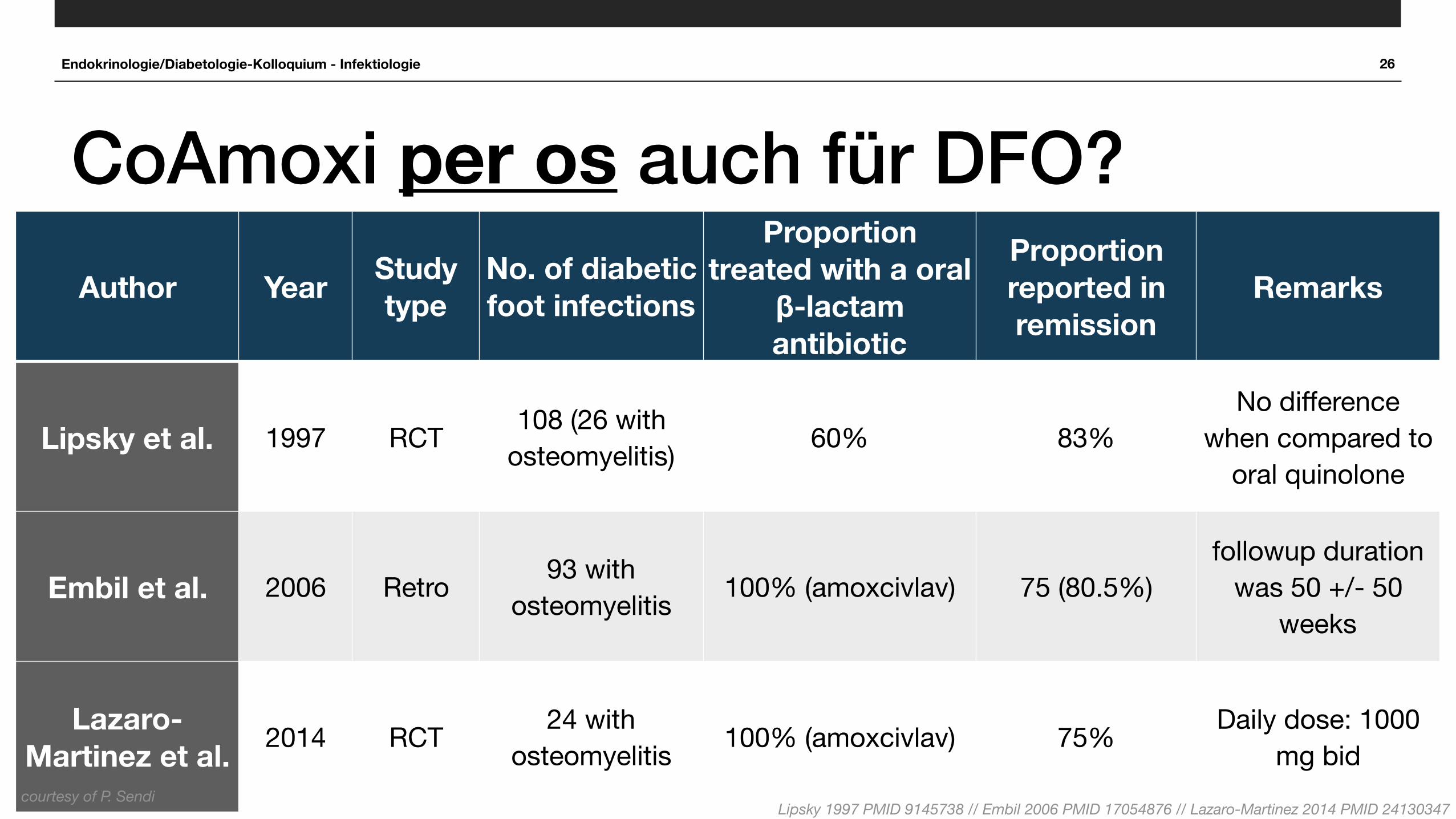
CoAmoxi per os auch für DFO?
■ Inkl Augmentin per osAuthor Year Study type
No. of diabetic foot infections
Proportion treated with a oral
β-lactam antibiotic
Proportion reported in remission
Remarks
Lipsky et al. 1997 RCT 108 (26 with osteomyelitis) 60% 83%
No difference when compared to
oral quinolone
Embil et al. 2006 Retro 93 with osteomyelitis 100% (amoxcivlav) 75 (80.5%)
followup duration was 50 +/- 50
weeks
Lazaro-Martinez et al. 2014 RCT 24 with
osteomyelitis 100% (amoxcivlav) 75% Daily dose: 1000 mg bid
courtesy of P. SendiLipsky 1997 PMID 9145738 // Embil 2006 PMID 17054876 // Lazaro-Martinez 2014 PMID 24130347

Endokrinologie/Diabetologie-Kolloquium - Infektiologie 26
CoAmoxi per os auch für DFO?
■ Inkl Augmentin per osAuthor Year Study type
No. of diabetic foot infections
Proportion treated with a oral
β-lactam antibiotic
Proportion reported in remission
Remarks
Lipsky et al. 1997 RCT 108 (26 with osteomyelitis) 60% 83%
No difference when compared to
oral quinolone
Embil et al. 2006 Retro 93 with osteomyelitis 100% (amoxcivlav) 75 (80.5%)
followup duration was 50 +/- 50
weeks
Lazaro-Martinez et al. 2014 RCT 24 with
osteomyelitis 100% (amoxcivlav) 75% Daily dose: 1000 mg bid
courtesy of P. SendiLipsky 1997 PMID 9145738 // Embil 2006 PMID 17054876 // Lazaro-Martinez 2014 PMID 24130347
CoAmoxi 3x1g per os?

Endokrinologie/Diabetologie-Kolloquium - Infektiologie 27
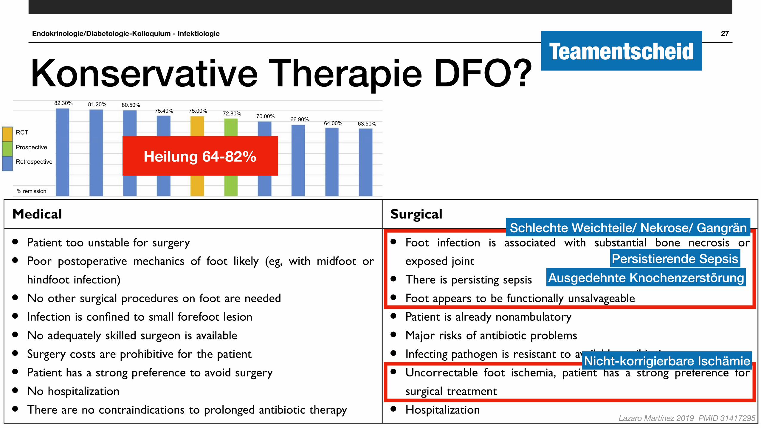
Konservative Therapie DFO?
Lazaro Martínez 2019 PMID 31417295
had been eradicated, but without any imaging tests performedfor con!rmation. Studies that performed microbiological boneculture to establish antibiotic regimens usingantibiograms presented DFO-remission rates of 64%,28
72.8%,30 and 81.2%.26 The difference between remissionrates could be associated with the methods used in obtainingbone samples, ie, by means of percutaneous biopsy in the !rststudy and then bone debridement of the ulcer in the other twostudies. In general, they reported successful treatment withoutsurgical treatment with remission in approximately two-thirdsof cases.
Additionally, some studies have investigated otherparameters associated with good DFO outcomes, whichinclude a decrease in in"ammatory biomarkers, such aserythrocyte-sedimentation rate (ESR) and CRP,31–33 boneremineralization on plain radiography, and completehealing of any overlying soft-tissue wounds.9 However,comparison of these studies is dif!cult, due to the varia-bility in protocols in antibiotic prescription and the lackof in"ammatory markers or radiological evidence forcon!rmation.
How to choose antibiotics and route ofadministrationFor many years, antibiotic therapy for DFO was adminis-tered intravenously for prolonged periods.34
However, in the last few years, two reviews of theliterature did not !nd any statistically signi!cant differ-ence between oral and parenteral administration of anti-biotics for the treatment of OM if bacteria were sensitiveto the antibiotic administered.35,36
On the other hand, interesting pharmacokinetic data haveshown that that antibiotics that reach the highest bone:serumconcentration ratios (ie, "uoroquinolones, sulfonamides,cyclins, macrolides, rifampin, fusidic acid, and oxazolidi-nones) are also those with the highest bioavailability duringoral administration of these agents.36
In a review conducted in 2017,37 Senneville et al saidthat it is logical that preference be given to antibiotics thatexhibit high diffusion into the bone (ie, a bone:blood ratio>0.3) and have good oral bioavailability (ie, >90%), due tothe prolonged duration of treatment that is usually recom-mended in these settings and the chronic nature of boneinfection that is encountered in patients with DFO.
The selection of an antibiotic agent to treat DFOshould begin with the selection of agents that cover thepresumed pathogens tested. Bone culture provides themost accurate microbiological information, and surgicalor percutaneous bone biopsy is the optimal method ofobtaining a sample of uncontaminated bone.37,38
Combinations of two agents with high oral availabilityand bone diffusion have been shown to treat DFO.Rifampicin, "uoroquinolone (o"oxacin, cipro"oxacin,levo"oxacin, or moxi"oxacin), and !-lactam–"uoroquino-lone combinations seem appropriate for the treatment ofStaphylococcus-induced and Gram-negative DFO.28,39,40
However, this may be limited, due to the risk of occur-rence of adverse events, with antibiotics being hepatotoxicand nephrotoxic in patients who are likely to have comorbid-ities and who receive multiple treatments. Therefore, we musttake into account daily doses and potential adverse events ofantibiotics with satisfactory oral bioavailability and bonediffusion for the treatment of patients with DFO.36,41
Game a
nd Je
ffcoa
te,25
2008 (U
K)
Lese
ns et
al,26 2
011 (F
rance
)
Embil et
al,18
2006 (C
anad
a)
Valabh
ji et a
l,29
2009 (U
K)
Láza
ro Mart
ínez e
t al,22
2014 (S
pain)
Jorda
no-M
ontan
ez et
al,30 2
014 (S
pain)
Pittet e
t al,27
1999 (S
witzerla
nd)
Achary
a et a
l,24
2013 (U
K)
Senne
ville e
t al,28
2008 (F
rance
)
Zeun e
t al,23
2016 (U
K)
RCT
Prospective
Retrospective
% remission
82.30% 81.20% 80.50%75.40% 75.00% 72.80% 70.00% 66.90%
64.00% 63.50%
Figure 2 Distribution of rates of remission of diabetic foot osteomyelitis with antibiotic treatment.
Lázaro Martínez et al Dovepress
submit your manuscript | www.dovepress.com
DovePressDiabetes, Metabolic Syndrome and Obesity: Targets and Therapy 2019:12950
professionals have different views on the choice of antibio-tics, route and duration of administration, and place ofsurgery.66
Adjuvant therapiesTo date, there are insuf!cient data to demonstrate theef!cacy of different adjuvant therapeutic practices, suchas granulocyte growth factors, hyperbaric oxygen therapy,and local antibiotic-delivery systems, in the treatment ofDFO.67–72
The serious problem of resistance to positive pathogensand the lack of new antimicrobial agents are the majorchallenges in the management of these patients. Asa solution to this problem, many have chosen local anti-biotic-delivery systems.73–76
In theory, the main advantages of local antibiotic-delivery systems are higher levels of antibiotic concentra-tion in the affected area, pharmacokinetic advantages,ability to overcome the possibility of resistant pathogens,and in cases of biodegradable material, avoidance of addi-tional surgical procedures. However, experience in DFOhas been limited to case reports and case series, and thereare no data that can be used to compare this therapy withstandard medical therapy. Therefore, we cannot currentlymake any speci!c recommendations on indications orapplication times for this treatment.77
The most recent review on local antibiotic-deliverysystems78 concluded that they represent a promising phar-maceutical option in the treatment of DFIs. Well-designedrandomized clinical trials are required to establish theiref!cacy and de!ne the framework for their usage.Currently, the role of local antibiotic-delivery systems intreating DFIs is limited and outside routine practice.
DiscussionBoth medical and surgical options have been shown to beeffective in the treatment of OM.21,22,28,37,60,66 However,there are also some criteria where there is consensus onwhich would be the best initial treatment depending on thecharacteristics of the patient. In such a way, when OM isassociated with soft-tissue infection or ischemia,9 bothpresentation and clinical characteristics are different, andthus also management. Therefore, the !rst conclusioncould be that there is not a single treatment for DFObecause it is not a single disease, and its association withsoft-tissue infection, ischemia, location, and patient char-acteristics determines the outcome, regardless of the treat-ment options.
After analysis of the literature, we can say that there isconsensus on when surgical or medical treatment would bethe !rst option in treating DFO.9,16,22,37
However, when we are treating DFO medically, it isnecessary to ensure a good antibiotic choice with goodbioavailability and proper duration of therapy dependingon the characteristics of the patient and the infection,37
assessing dosage,36 since in the absence of a bone-resection therapies, antibiotic treatment should be at least6 weeks,4,42 which is what the literature indicates, and inthat case duration and posology will be driven by thecharacteristics and comorbidities of the patient. In patientswith kidney disease, the dosage of antibiotics must beadjusted from bactericidal to bacteriostatic affecting theeffectiveness of the antibiotic.
Another barrier to themedical treatment ofOM is the exacttime as to when antibiotic therapy should be discontinued. The6-week reference margin collected in the literature is based ona single study.42 It does not seem strong enough to supporta universal recommendation. There may be patients in whom
Table 1 Criteria for selecting primarily antibiotic or surgical approaches for diabetic foot osteomyelitis
Medical Surgical
! Patient too unstable for surgery! Poor postoperative mechanics of foot likely (eg, with midfoot or
hindfoot infection)! No other surgical procedures on foot are needed! Infection is con!ned to small forefoot lesion! No adequately skilled surgeon is available! Surgery costs are prohibitive for the patient! Patient has a strong preference to avoid surgery! No hospitalization! There are no contraindications to prolonged antibiotic therapy
! Foot infection is associated with substantial bone necrosis orexposed joint
! There is persisting sepsis! Foot appears to be functionally unsalvageable! Patient is already nonambulatory! Major risks of antibiotic problems! Infecting pathogen is resistant to available antibiotics! Uncorrectable foot ischemia, patient has a strong preference for
surgical treatment! Hospitalization
Note: Data from Lipsky et al9 and Senneville and Robineau.37
Lázaro Martínez et al Dovepress
submit your manuscript | www.dovepress.com
DovePressDiabetes, Metabolic Syndrome and Obesity: Targets and Therapy 2019:12954
had been eradicated, but without any imaging tests performedfor con!rmation. Studies that performed microbiological boneculture to establish antibiotic regimens usingantibiograms presented DFO-remission rates of 64%,28
72.8%,30 and 81.2%.26 The difference between remissionrates could be associated with the methods used in obtainingbone samples, ie, by means of percutaneous biopsy in the !rststudy and then bone debridement of the ulcer in the other twostudies. In general, they reported successful treatment withoutsurgical treatment with remission in approximately two-thirdsof cases.
Additionally, some studies have investigated otherparameters associated with good DFO outcomes, whichinclude a decrease in in"ammatory biomarkers, such aserythrocyte-sedimentation rate (ESR) and CRP,31–33 boneremineralization on plain radiography, and completehealing of any overlying soft-tissue wounds.9 However,comparison of these studies is dif!cult, due to the varia-bility in protocols in antibiotic prescription and the lackof in"ammatory markers or radiological evidence forcon!rmation.
How to choose antibiotics and route ofadministrationFor many years, antibiotic therapy for DFO was adminis-tered intravenously for prolonged periods.34
However, in the last few years, two reviews of theliterature did not !nd any statistically signi!cant differ-ence between oral and parenteral administration of anti-biotics for the treatment of OM if bacteria were sensitiveto the antibiotic administered.35,36
On the other hand, interesting pharmacokinetic data haveshown that that antibiotics that reach the highest bone:serumconcentration ratios (ie, "uoroquinolones, sulfonamides,cyclins, macrolides, rifampin, fusidic acid, and oxazolidi-nones) are also those with the highest bioavailability duringoral administration of these agents.36
In a review conducted in 2017,37 Senneville et al saidthat it is logical that preference be given to antibiotics thatexhibit high diffusion into the bone (ie, a bone:blood ratio>0.3) and have good oral bioavailability (ie, >90%), due tothe prolonged duration of treatment that is usually recom-mended in these settings and the chronic nature of boneinfection that is encountered in patients with DFO.
The selection of an antibiotic agent to treat DFOshould begin with the selection of agents that cover thepresumed pathogens tested. Bone culture provides themost accurate microbiological information, and surgicalor percutaneous bone biopsy is the optimal method ofobtaining a sample of uncontaminated bone.37,38
Combinations of two agents with high oral availabilityand bone diffusion have been shown to treat DFO.Rifampicin, "uoroquinolone (o"oxacin, cipro"oxacin,levo"oxacin, or moxi"oxacin), and !-lactam–"uoroquino-lone combinations seem appropriate for the treatment ofStaphylococcus-induced and Gram-negative DFO.28,39,40
However, this may be limited, due to the risk of occur-rence of adverse events, with antibiotics being hepatotoxicand nephrotoxic in patients who are likely to have comorbid-ities and who receive multiple treatments. Therefore, we musttake into account daily doses and potential adverse events ofantibiotics with satisfactory oral bioavailability and bonediffusion for the treatment of patients with DFO.36,41
Game a
nd Je
ffcoa
te,25
2008 (U
K)
Lese
ns et
al,26 2
011 (F
rance
)
Embil et
al,18
2006 (C
anad
a)
Valabh
ji et a
l,29
2009 (U
K)
Láza
ro Mart
ínez e
t al,22
2014 (S
pain)
Jorda
no-M
ontan
ez et
al,30 2
014 (S
pain)
Pittet e
t al,27
1999 (S
witzerla
nd)
Achary
a et a
l,24
2013 (U
K)
Senne
ville e
t al,28
2008 (F
rance
)
Zeun e
t al,23
2016 (U
K)
RCT
Prospective
Retrospective
% remission
82.30% 81.20% 80.50%75.40% 75.00% 72.80% 70.00% 66.90%
64.00% 63.50%
Figure 2 Distribution of rates of remission of diabetic foot osteomyelitis with antibiotic treatment.
Lázaro Martínez et al Dovepress
submit your manuscript | www.dovepress.com
DovePressDiabetes, Metabolic Syndrome and Obesity: Targets and Therapy 2019:12950
Schlechte Weichteile/ Nekrose/ Gangrän
Persistierende Sepsis
Nicht-korrigierbare Ischämie
Ausgedehnte Knochenzerstörung
Heilung 64-82%
Endokrinologie/Diabetologie-Kolloquium - Infektiologie 27
Konservative Therapie DFO?
Lazaro Martínez 2019 PMID 31417295
had been eradicated, but without any imaging tests performedfor con!rmation. Studies that performed microbiological boneculture to establish antibiotic regimens usingantibiograms presented DFO-remission rates of 64%,28
72.8%,30 and 81.2%.26 The difference between remissionrates could be associated with the methods used in obtainingbone samples, ie, by means of percutaneous biopsy in the !rststudy and then bone debridement of the ulcer in the other twostudies. In general, they reported successful treatment withoutsurgical treatment with remission in approximately two-thirdsof cases.
Additionally, some studies have investigated otherparameters associated with good DFO outcomes, whichinclude a decrease in in"ammatory biomarkers, such aserythrocyte-sedimentation rate (ESR) and CRP,31–33 boneremineralization on plain radiography, and completehealing of any overlying soft-tissue wounds.9 However,comparison of these studies is dif!cult, due to the varia-bility in protocols in antibiotic prescription and the lackof in"ammatory markers or radiological evidence forcon!rmation.
How to choose antibiotics and route ofadministrationFor many years, antibiotic therapy for DFO was adminis-tered intravenously for prolonged periods.34
However, in the last few years, two reviews of theliterature did not !nd any statistically signi!cant differ-ence between oral and parenteral administration of anti-biotics for the treatment of OM if bacteria were sensitiveto the antibiotic administered.35,36
On the other hand, interesting pharmacokinetic data haveshown that that antibiotics that reach the highest bone:serumconcentration ratios (ie, "uoroquinolones, sulfonamides,cyclins, macrolides, rifampin, fusidic acid, and oxazolidi-nones) are also those with the highest bioavailability duringoral administration of these agents.36
In a review conducted in 2017,37 Senneville et al saidthat it is logical that preference be given to antibiotics thatexhibit high diffusion into the bone (ie, a bone:blood ratio>0.3) and have good oral bioavailability (ie, >90%), due tothe prolonged duration of treatment that is usually recom-mended in these settings and the chronic nature of boneinfection that is encountered in patients with DFO.
The selection of an antibiotic agent to treat DFOshould begin with the selection of agents that cover thepresumed pathogens tested. Bone culture provides themost accurate microbiological information, and surgicalor percutaneous bone biopsy is the optimal method ofobtaining a sample of uncontaminated bone.37,38
Combinations of two agents with high oral availabilityand bone diffusion have been shown to treat DFO.Rifampicin, "uoroquinolone (o"oxacin, cipro"oxacin,levo"oxacin, or moxi"oxacin), and !-lactam–"uoroquino-lone combinations seem appropriate for the treatment ofStaphylococcus-induced and Gram-negative DFO.28,39,40
However, this may be limited, due to the risk of occur-rence of adverse events, with antibiotics being hepatotoxicand nephrotoxic in patients who are likely to have comorbid-ities and who receive multiple treatments. Therefore, we musttake into account daily doses and potential adverse events ofantibiotics with satisfactory oral bioavailability and bonediffusion for the treatment of patients with DFO.36,41
Game a
nd Je
ffcoa
te,25
2008 (U
K)
Lese
ns et
al,26 2
011 (F
rance
)
Embil et
al,18
2006 (C
anad
a)
Valabh
ji et a
l,29
2009 (U
K)
Láza
ro Mart
ínez e
t al,22
2014 (S
pain)
Jorda
no-M
ontan
ez et
al,30 2
014 (S
pain)
Pittet e
t al,27
1999 (S
witzerla
nd)
Achary
a et a
l,24
2013 (U
K)
Senne
ville e
t al,28
2008 (F
rance
)
Zeun e
t al,23
2016 (U
K)
RCT
Prospective
Retrospective
% remission
82.30% 81.20% 80.50%75.40% 75.00% 72.80% 70.00% 66.90%
64.00% 63.50%
Figure 2 Distribution of rates of remission of diabetic foot osteomyelitis with antibiotic treatment.
Lázaro Martínez et al Dovepress
submit your manuscript | www.dovepress.com
DovePressDiabetes, Metabolic Syndrome and Obesity: Targets and Therapy 2019:12950
professionals have different views on the choice of antibio-tics, route and duration of administration, and place ofsurgery.66
Adjuvant therapiesTo date, there are insuf!cient data to demonstrate theef!cacy of different adjuvant therapeutic practices, suchas granulocyte growth factors, hyperbaric oxygen therapy,and local antibiotic-delivery systems, in the treatment ofDFO.67–72
The serious problem of resistance to positive pathogensand the lack of new antimicrobial agents are the majorchallenges in the management of these patients. Asa solution to this problem, many have chosen local anti-biotic-delivery systems.73–76
In theory, the main advantages of local antibiotic-delivery systems are higher levels of antibiotic concentra-tion in the affected area, pharmacokinetic advantages,ability to overcome the possibility of resistant pathogens,and in cases of biodegradable material, avoidance of addi-tional surgical procedures. However, experience in DFOhas been limited to case reports and case series, and thereare no data that can be used to compare this therapy withstandard medical therapy. Therefore, we cannot currentlymake any speci!c recommendations on indications orapplication times for this treatment.77
The most recent review on local antibiotic-deliverysystems78 concluded that they represent a promising phar-maceutical option in the treatment of DFIs. Well-designedrandomized clinical trials are required to establish theiref!cacy and de!ne the framework for their usage.Currently, the role of local antibiotic-delivery systems intreating DFIs is limited and outside routine practice.
DiscussionBoth medical and surgical options have been shown to beeffective in the treatment of OM.21,22,28,37,60,66 However,there are also some criteria where there is consensus onwhich would be the best initial treatment depending on thecharacteristics of the patient. In such a way, when OM isassociated with soft-tissue infection or ischemia,9 bothpresentation and clinical characteristics are different, andthus also management. Therefore, the !rst conclusioncould be that there is not a single treatment for DFObecause it is not a single disease, and its association withsoft-tissue infection, ischemia, location, and patient char-acteristics determines the outcome, regardless of the treat-ment options.
After analysis of the literature, we can say that there isconsensus on when surgical or medical treatment would bethe !rst option in treating DFO.9,16,22,37
However, when we are treating DFO medically, it isnecessary to ensure a good antibiotic choice with goodbioavailability and proper duration of therapy dependingon the characteristics of the patient and the infection,37
assessing dosage,36 since in the absence of a bone-resection therapies, antibiotic treatment should be at least6 weeks,4,42 which is what the literature indicates, and inthat case duration and posology will be driven by thecharacteristics and comorbidities of the patient. In patientswith kidney disease, the dosage of antibiotics must beadjusted from bactericidal to bacteriostatic affecting theeffectiveness of the antibiotic.
Another barrier to themedical treatment ofOM is the exacttime as to when antibiotic therapy should be discontinued. The6-week reference margin collected in the literature is based ona single study.42 It does not seem strong enough to supporta universal recommendation. There may be patients in whom
Table 1 Criteria for selecting primarily antibiotic or surgical approaches for diabetic foot osteomyelitis
Medical Surgical
! Patient too unstable for surgery! Poor postoperative mechanics of foot likely (eg, with midfoot or
hindfoot infection)! No other surgical procedures on foot are needed! Infection is con!ned to small forefoot lesion! No adequately skilled surgeon is available! Surgery costs are prohibitive for the patient! Patient has a strong preference to avoid surgery! No hospitalization! There are no contraindications to prolonged antibiotic therapy
! Foot infection is associated with substantial bone necrosis orexposed joint
! There is persisting sepsis! Foot appears to be functionally unsalvageable! Patient is already nonambulatory! Major risks of antibiotic problems! Infecting pathogen is resistant to available antibiotics! Uncorrectable foot ischemia, patient has a strong preference for
surgical treatment! Hospitalization
Note: Data from Lipsky et al9 and Senneville and Robineau.37
Lázaro Martínez et al Dovepress
submit your manuscript | www.dovepress.com
DovePressDiabetes, Metabolic Syndrome and Obesity: Targets and Therapy 2019:12954
had been eradicated, but without any imaging tests performedfor con!rmation. Studies that performed microbiological boneculture to establish antibiotic regimens usingantibiograms presented DFO-remission rates of 64%,28
72.8%,30 and 81.2%.26 The difference between remissionrates could be associated with the methods used in obtainingbone samples, ie, by means of percutaneous biopsy in the !rststudy and then bone debridement of the ulcer in the other twostudies. In general, they reported successful treatment withoutsurgical treatment with remission in approximately two-thirdsof cases.
Additionally, some studies have investigated otherparameters associated with good DFO outcomes, whichinclude a decrease in in"ammatory biomarkers, such aserythrocyte-sedimentation rate (ESR) and CRP,31–33 boneremineralization on plain radiography, and completehealing of any overlying soft-tissue wounds.9 However,comparison of these studies is dif!cult, due to the varia-bility in protocols in antibiotic prescription and the lackof in"ammatory markers or radiological evidence forcon!rmation.
How to choose antibiotics and route ofadministrationFor many years, antibiotic therapy for DFO was adminis-tered intravenously for prolonged periods.34
However, in the last few years, two reviews of theliterature did not !nd any statistically signi!cant differ-ence between oral and parenteral administration of anti-biotics for the treatment of OM if bacteria were sensitiveto the antibiotic administered.35,36
On the other hand, interesting pharmacokinetic data haveshown that that antibiotics that reach the highest bone:serumconcentration ratios (ie, "uoroquinolones, sulfonamides,cyclins, macrolides, rifampin, fusidic acid, and oxazolidi-nones) are also those with the highest bioavailability duringoral administration of these agents.36
In a review conducted in 2017,37 Senneville et al saidthat it is logical that preference be given to antibiotics thatexhibit high diffusion into the bone (ie, a bone:blood ratio>0.3) and have good oral bioavailability (ie, >90%), due tothe prolonged duration of treatment that is usually recom-mended in these settings and the chronic nature of boneinfection that is encountered in patients with DFO.
The selection of an antibiotic agent to treat DFOshould begin with the selection of agents that cover thepresumed pathogens tested. Bone culture provides themost accurate microbiological information, and surgicalor percutaneous bone biopsy is the optimal method ofobtaining a sample of uncontaminated bone.37,38
Combinations of two agents with high oral availabilityand bone diffusion have been shown to treat DFO.Rifampicin, "uoroquinolone (o"oxacin, cipro"oxacin,levo"oxacin, or moxi"oxacin), and !-lactam–"uoroquino-lone combinations seem appropriate for the treatment ofStaphylococcus-induced and Gram-negative DFO.28,39,40
However, this may be limited, due to the risk of occur-rence of adverse events, with antibiotics being hepatotoxicand nephrotoxic in patients who are likely to have comorbid-ities and who receive multiple treatments. Therefore, we musttake into account daily doses and potential adverse events ofantibiotics with satisfactory oral bioavailability and bonediffusion for the treatment of patients with DFO.36,41
Game a
nd Je
ffcoa
te,25
2008 (U
K)
Lese
ns et
al,26 2
011 (F
rance
)
Embil et
al,18
2006 (C
anad
a)
Valabh
ji et a
l,29
2009 (U
K)
Láza
ro Mart
ínez e
t al,22
2014 (S
pain)
Jorda
no-M
ontan
ez et
al,30 2
014 (S
pain)
Pittet e
t al,27
1999 (S
witzerla
nd)
Achary
a et a
l,24
2013 (U
K)
Senne
ville e
t al,28
2008 (F
rance
)
Zeun e
t al,23
2016 (U
K)
RCT
Prospective
Retrospective
% remission
82.30% 81.20% 80.50%75.40% 75.00% 72.80% 70.00% 66.90%
64.00% 63.50%
Figure 2 Distribution of rates of remission of diabetic foot osteomyelitis with antibiotic treatment.
Lázaro Martínez et al Dovepress
submit your manuscript | www.dovepress.com
DovePressDiabetes, Metabolic Syndrome and Obesity: Targets and Therapy 2019:12950
Schlechte Weichteile/ Nekrose/ Gangrän
Persistierende Sepsis
Nicht-korrigierbare Ischämie
Ausgedehnte Knochenzerstörung
Teamentscheid
Heilung 64-82%

Endokrinologie/Diabetologie-Kolloquium - Infektiologie 28
Take to work■ Klinische Einteilung (IWGDF/ IDSA): Grad 1 - 4
■Grad 1 keine Infektion, keine Antibiotika, keine Diagnostik
■Grad 2 ambulante und empirische Behandlung, 7 Tage
■Grad 3/4 Morbidität/ Mortalität: ev Hospitalisation / interdisziplinäres Team für Management
■ Osteomyelitis: schwierige Diagnose & Therapie, häufig Komplikationen/ Relapse
■ Trends: kürzere Therapiedauer, konservative Therapie der Osteomyelitis

Endokrinologie/Diabetologie-Kolloquium - Infektiologie 29
Fragen? Danke für Aufmerksamkeit!