Embed Size (px)
Citation preview
Diplomarbeit
Acute Intoxications in Intensive Care Medicine:
A Retrospective Data Analysis of an Internal Medicine
Intensive Care Unit
eingereicht von
Ines Rauch
zur Erlangung des akademischen Grades
Doktorin der gesamten Heilkunde
(Dr. med. univ.)
an der
Medizinischen Universität Graz
ausgeführt an der
Universitätsklinik für Innere Medizin
Allgemeinen internistischen Intensivstation
unter der Anleitung von
Priv.-Doz. OA Dr.med.univ. Dr.scient.med. Gerald Hackl
und
Univ. FA Dr.med.univ. Alexander Christian Reisinger
Graz, 15.09.2021
2
Eidesstattliche Erklärung
Ich erkläre ehrenwörtlich, dass ich die vorliegende Arbeit selbstständig und ohne
fremde Hilfe verfasst habe, andere als die angegebenen Quellen nicht verwendet
habe und die den benutzten Quellen wörtlich oder inhaltlich entnommenen Stellen
als solche kenntlich gemacht habe.
Graz, am 15.09.2021 Ines Rauch eh.
3
Acknowledgements
I would like to extend my gratitude to my diploma thesis supervisors Priv.-Doz. OA
Dr.med.univ. Dr.scient.med. Gerald Hackl and Univ. FA Dr.med.univ. Alexander
Christian Reisinger. Without your constant support and extensive knowledge of
intoxications and intensive care medicine, this thesis would not have been
possible. Your insightful feedback and expansive statistical knowledge enabled me
to write a diploma thesis I can be proud of.
Furthermore, I would like to thank my family and friends for their unwavering
support, love and patience.
4
Table of Contents
Acknowledgements ................................................................................................. 3
Table of Contents ................................................................................................... 4
List of Tables ........................................................................................................... 6
List of Figures .......................................................................................................... 7
Glossary and Abbreviations..................................................................................... 8
Zusammenfassung ................................................................................................ 10
Abstract ................................................................................................................. 12
1 Introduction ......................................................................................................... 14
1.1 Epidemiology ................................................................................................ 14
1.2 Toxidromes .................................................................................................. 15
1.2.1 Sedative Hypnotic Toxidrome ............................................................ 16
1.2.2 Opioid Toxidrome............................................................................... 17
1.2.3 Sympathomimetic Toxidrome ............................................................ 19
1.2.4 Anticholinergic Toxidrome .................................................................. 20
1.2.5 Cholinergic Toxidrome ....................................................................... 21
1.2.1 Serotonergic Toxidrome ..................................................................... 21
1.3 ICU Treatment of Patients with acute Intoxications ...................................... 22
1.3.1 Primary Toxin Elimination .................................................................. 22
1.3.1.1 Activated Charcoal ............................................................... 23
1.3.1.2 Gastric Lavage ..................................................................... 24
1.3.1.3 Whole Bowel Irrigation ......................................................... 24
1.3.1.4 Ipecac Syrup ........................................................................ 25
1.3.1.5 Endoscopic Medication Retrieval ........................................ 25
1.3.2 Secondary Toxin Elimination ............................................................. 25
1.3.2.1 Extracorporeal Toxin Elimination .......................................... 25
1.3.2.2 HBO Therapy ....................................................................... 27
1.3.2.3 Lipid Rescue Therapy .......................................................... 28
5
1.3.2.4 High Dose Insulin Euglycemic Therapy ................................ 28
1.3.2.5 Urine Alkalization .................................................................. 28
1.3.2.6 Forced Diuresis .................................................................... 29
1.4 Antidotes and Antagonists............................................................................ 29
1.4.1 Naloxone ............................................................................................ 29
1.4.2 Flumazenil ......................................................................................... 30
1.4.3 Physostigmine ................................................................................... 31
1.4.4 N-Acetylcysteine ................................................................................ 31
1.4.5 Atropine ............................................................................................. 32
1.4.6 Methylene and Toluidine Blue ............................................................ 33
1.4.7 Other Antidotes .................................................................................. 34
2 Material and Methods ......................................................................................... 34
2.1 Study Design ................................................................................................ 34
2.2 Data Collection ............................................................................................. 34
2.3 Statistical Data Analysis ............................................................................... 36
3 Results ............................................................................................................... 37
3.1 Distribution of Patients per Intoxication Group ............................................. 38
3.2 Distribution of Intoxication Patients by Sex .................................................. 40
3.3 Distribution of Intoxication Patients by Age Group ....................................... 41
3.4 Reason for Intoxication ................................................................................ 43
3.5 Intoxication Treatment ................................................................................. 44
3.6 ICU Length of Stay ....................................................................................... 46
3.7 ICU Mortality ................................................................................................ 46
4 Discussion .......................................................................................................... 47
4.1 Comparison with Global Literature ............................................................... 51
4.2 Limitations of this Study ............................................................................... 53
5 Conclusion .......................................................................................................... 54
6 Bibliography ........................................................................................................ 55
6
List of Tables
Table 1: Categorization of intoxications according to substance groups ............... 36
Table 2: Distribution of patients per intoxication group ......................................... 39
Table 3: Substances categorized in the group other ............................................ 40
Table 4: Number and percentage of intoxication patients per sex
and intoxication group ........................................................................................... 40
Table 5: Number and percentage of intoxication patients per age group .............. 41
Table 6: Reason for Intoxication ............................................................................ 43
Table 7: Number of patients that received detoxification or antidotes .................. 44
Table 8: Number of patients intubated per intoxication group and sex ................. 45
Table 9: Specification of ICU mortality cases ...................................................... 47
7
List of Figures
Figure 1: Data collection procedure ...................................................................... 37
Figure 2: Number of patients per intoxication group .............................................. 38
Figure 3: Number of intoxication patients per age group ....................................... 42
Figure 4: Boxplot of age distribution by sex ........................................................... 42
Figure 5: Reason for intoxication ........................................................................... 43
8
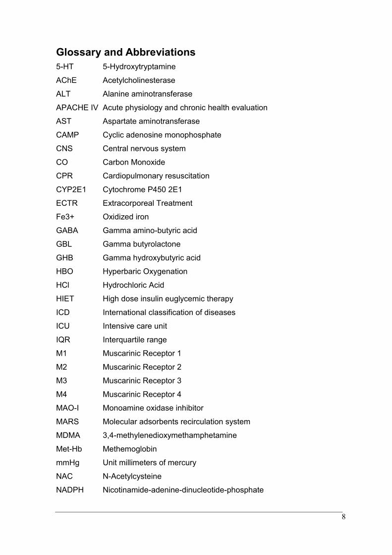
Glossary and Abbreviations
5-HT 5-Hydroxytryptamine
AChE Acetylcholinesterase
ALT Alanine aminotransferase
APACHE IV Acute physiology and chronic health evaluation
AST Aspartate aminotransferase
CAMP Cyclic adenosine monophosphate
CNS Central nervous system
CO Carbon Monoxide
CPR Cardiopulmonary resuscitation
CYP2E1 Cytochrome P450 2E1
ECTR Extracorporeal Treatment
Fe3+ Oxidized iron
GABA Gamma amino-butyric acid
GBL Gamma butyrolactone
GHB Gamma hydroxybutyric acid
HBO Hyperbaric Oxygenation
HCl Hydrochloric Acid
HIET High dose insulin euglycemic therapy
ICD International classification of diseases
ICU Intensive care unit
IQR Interquartile range
M1 Muscarinic Receptor 1
M2 Muscarinic Receptor 2
M3 Muscarinic Receptor 3
M4 Muscarinic Receptor 4
MAO-I Monoamine oxidase inhibitor
MARS Molecular adsorbents recirculation system
MDMA 3,4-methylenedioxymethamphetamine
Met-Hb Methemoglobin
mmHg Unit millimeters of mercury
NAC N-Acetylcysteine
NADPH Nicotinamide-adenine-dinucleotide-phosphate
9
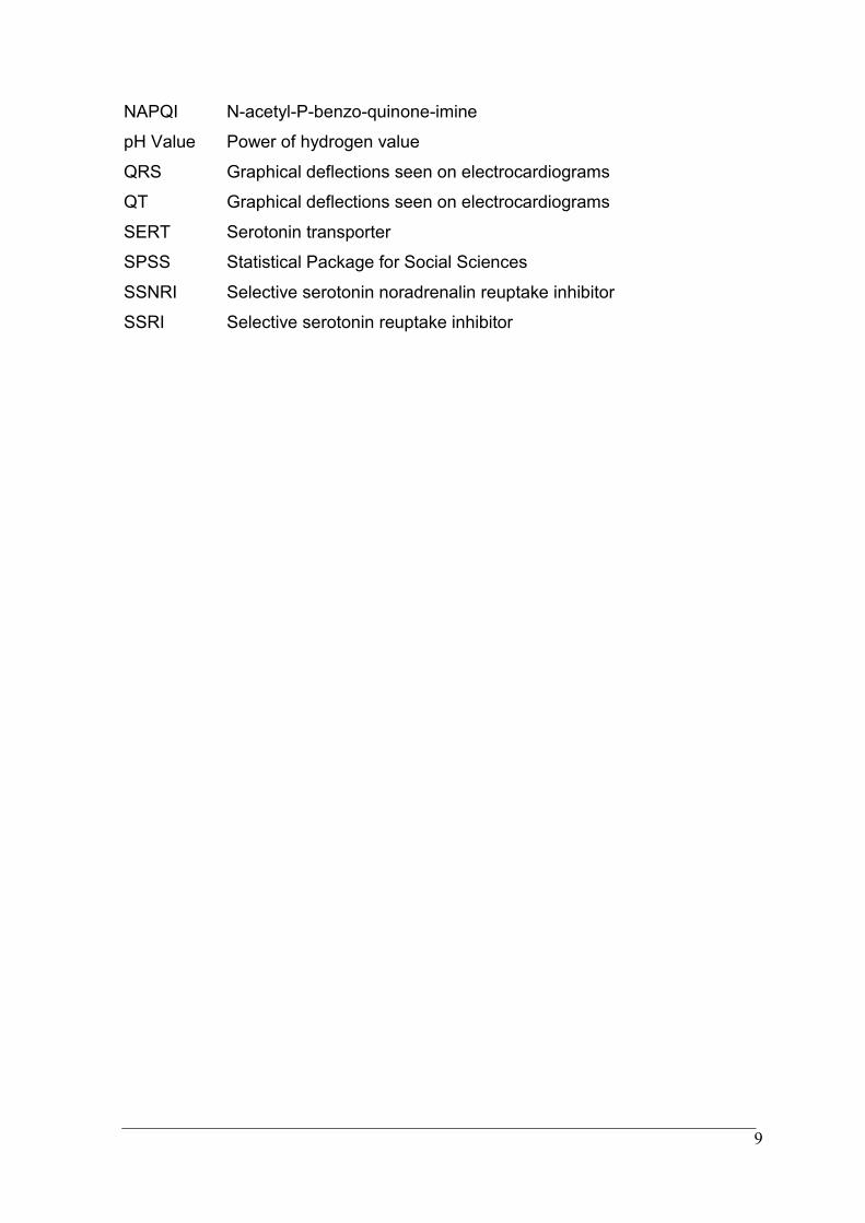
NAPQI N-acetyl-P-benzo-quinone-imine
pH Value Power of hydrogen value
QRS Graphical deflections seen on electrocardiograms
QT Graphical deflections seen on electrocardiograms
SERT Serotonin transporter
SPSS Statistical Package for Social Sciences
SSNRI Selective serotonin noradrenalin reuptake inhibitor
SSRI Selective serotonin reuptake inhibitor
10
Zusammenfassung
Hintergrund:
Schwere Intoxikationen nach der Ingestion, Inhalation oder intravenösen
Applikation von Stoffen erfordern häufig eine intensivmedizinische Behandlung. Im
Rahmen von Intoxikationen können unterschiedliche invasive Behandlungsschritte
wie die Echtzeitüberwachung, Intubation, Beatmung und eine frühzeitige
Giftelimination erforderlich sein, um körperliche Funktionen zu unterstützen bzw.
zu ersetzen.
Ziel: Ziel dieser Diplomarbeit ist es Intoxikationen, die einen Intensivaufenthalt an
der internistischen Intensivstation des Universitätsklinikum Graz erforderlich
machen zu kategorisieren und die Verteilung der durchgeführten therapeutischen
Schritte darzustellen.
Material und Methoden: Es wurden in dieser retrospektiven Datenanalyse
toxikologische IntensivpatientInnen (n=227) von Jänner 2013 bis Dezember 2018
inkludiert. Die Patientenakten, sowie die intensivmedizinische Dokumentation im
Computersystem wurden als Grundlage der Datenauswertung herangezogen. Es
erfolgte die Einteilung der PatientInnen in neun unterschiedliche Gruppen je nach
auslösenden Agens der Intoxikation (0= Unbekannt, 1= Äthanol, 2= Analgetika, 3=
Antidepressiva, Antiepileptika und Antipsychotika, 4= Straßendrogen (inklusive
Opiate, Kokain und Amphetamine), 5= Sedativa, 6= Kohlenmonoxid, Arsen und
Zyanide, 7= andere nicht klassifizierbare Stoffe, 8= Mischintoxikationen von zwei
oder mehr Stoffgruppen). Ebenfalls wurden die Intoxikationsfälle in Hinsicht auf
das Geschlecht, der Altersverteilung, Grund für die Intoxikation, der primären und
sekundären Giftelimination, Antidotgabe, Notwendigkeit für Intubation, Dauer der
Intubation sowie Dauer des Intensivaufenthaltes und der Mortalität untersucht.
Ergebnis: Die häufigste Intoxikation beider Geschlechter, die eine
intensivmedizinische Behandlung erforderlich machte, stellte die Gruppe der
Mischintoxikationen dar. Bei den Frauen folgte die Gruppe der Antidepressiva,
Antiepileptika und Antipychotika (17.5%), darauffolgend die Gruppe der anderen
11
nicht klassifizierbaren Intoxikationen (9.7%). Anschließend folgten Analgetika und
Sedativa mit je 7.8% aller Intoxikationen. Bei den männlichen Intensivpatienten
war die zweithäufigste Ursache für eine Intoxikation der Abusus von
Straßendrogen (15.3%). gefolgt von Äthanol (12.1%), und Antidepressiva,
Antiepileptika und Antipsychotika (9.7%). 52% aller Intoxikationen waren in
suizidaler Absicht, ein Viertel aufgrund von rekreationellen Gründen, und 13.2%
der Intoxikationen waren akzidentiell. Ein Viertel aller PatientInnen erhielten
Aktivkohle als primäre Giftelimination und bei 6.6% wurden sekundäre
Gifteliminationsmaßnahmen durchgeführt. Frauen erhielten signifikant öfter
primäre Gifteliminationsmaßnahmen (p=0.008). Vier Prozent aller toxikologischen
PatientInnen verstarben während des ICU-Aufenthalts.
Zusammenfassung: In dieser retrospektiven Datenanalyse konnten die
häufigsten Intoxikationen auf der internistischen Intensivstation, als auch die
notwendigen therapeutischen Maßnahmen erstmalig aufgeschlüsselt und mit der
internationale Datenlage verglichen werden.
12
Abstract
Background: Intoxications following ingestion, inhalation or intravenous
application of substances can require treatment in an intensive care unit (ICU).
Reduced states of consciousness or impaired organ function, invasive
therapeutical measures such as intubation, mechanical ventilation, monitoring and
toxin elimination may be necessary.
Objective: The aim of this diploma thesis is to categorize intoxications that
required a stay at the medical intensive care unit at the University Hospital Graz
and to analyze the distribution of necessary therapeutical measures.
Material and Methods: In this retrospective data analysis, ICU patients (n=227)
from January 2013 until December 2018 were included. Patient files and electronic
documentation were utilized for data collection. Patients were separated into nine
groups according to the causational substance (0= unknown, 1= ethanol, 2=
analgesics, 3= antidepressants, antiepileptics and antipsychotics, 4= street drugs
including opiates, cocaine and amphetamines, 5= sedatives, 6= carbon monoxide,
arsenic and cyanide, 7= other not categorized substances and 8= combined drug
intoxications of two or more substance groups). Intoxications were analyzed
according to the distribution of sex, age, reason for intoxication, toxin elimination,
antidotes administered, intubation, as well as length of the ICU stay and ICU
mortality.
Results: Combined drug intoxications are the most common intoxication requiring
intensive care treatment. In females, this was followed by the group of
antidepressants, antiepileptics and antipsychotics (17.5%), and the group “other”
(9.7%). Analgesics, sedatives and ethanol were almost equally common at 5-8%.
In males, following the combined drug intoxications, the most common singular
substance intoxication were street drugs (15.3%), followed by ethanol (12.1%),
and the group of antidepressants, antiepileptics and antipsychotics (9.7%).
Approximately half of all intoxications were intended suicide. One quarter of
13
patients received activated charcoal as primary detoxification and 6.6% of patients
required secondary detoxification methods. In addition, females received primary
detoxification methods significantly more often than males (p=0.008). ICU mortality
regarding intoxication patients was low at 4%.
Conclusion: In this retrospective data analysis, the distribution of the most
common intoxications leading to a stay at the medical intensive care unit and the
necessary therapeutical steps, as well as ICU mortality were determined and
compared to international data.
14
1 Introduction
1.1. Epidemiology
Acute intoxications following the inhalation, ingestion or intravenous administration
of substances are a frequent cause for admission to medical intensive care units
(ICU) worldwide. Considering the varying capacities of comprehensive patient
monitoring and resources, 3.7-40% of patients with acute intoxications in the
emergency department are admitted to the ICU (1, 2). As a result, it has become
increasingly important to focus on and categorize the most common substances
and the necessary therapeutical measures. This allows for early targeted
diagnostics, treatment protocols to be established and to enable strategic planning
of intensive care bed capacities.
Patients requiring intensive care measures or observance can present to an ICU in
varying severity. Multiple studies have shown that the mortality rate of these
patients is surprisingly low at 1.9- 2.1% (1, 3). In addition, according to a study
done by Heyerdal et al., 58% of patients had an ICU stay of one day, two days
were necessary for 31% of patients, and only 11% stayed three or more days (4).
Taking the international data into account, it was of increasing importance to
assess and compare patient data of the medical intensive care unit in Graz to
international standards.
This diploma thesis offers a structured overview of common intoxications,
pathophysiological mechanisms, and therapeutic options, as well as an insight into
the substances present in patients with acute intoxications that are admitted to an
intensive care unit. Furthermore, utilizing retrospective data analysis, the median
length of stay, necessary therapeutical steps, and mortality rate at the intensive
care unit of the Department of Internal Medicine, University Hospital Graz were
evaluated.
15
1.2. Toxidromes
The classification of patient symptoms following acute intoxications in groups
according to toxidromes, or toxic syndromes, can aid in establishing a correct and
rapid diagnosis, thus enabling early specialized treatment including detoxification
and the application of antidotes. The initial diagnostic criteria in patients to
evaluate the presenting toxidrome are a complete physical examination,
monitorization of the patient including pulse oximetry, invasive or noninvasive
blood pressure monitoring, a 12-lead electrocardiogram, laboratory blood tests,
blood gas analysis, end tidal carbon dioxide monitoring and urine samples (5, 6).
Consequently, this grouping of typical symptoms can aid in the identification of
intoxications and lead to early initiation of adequate treatment. There are six main
toxidromes commonly described in literature, namely, sedative hypnotic, opioid,
sympathomimetic, anticholinergic, cholinergic, and neuroleptic agent toxidromes
(7, 8). In recent years, a seventh syndrome, namely the serotonergic toxidrome
has been increasingly mentioned in literature (9).
Toxidromes enable a rapid conclusion to be drawn when a singular substance is
ingested. Nevertheless, there are some limitations to this method of classification
(6). An intoxication with more than one substance or substance groups can
produce a complex symptom combination and interfere in the correct diagnostic
processes. In addition, a plethora of new medications and designer drugs do not
typically conform to the diagnostic criteria or can result in similar symptoms to
other substances with different pathophysiology, and therefore requiring different
treatment. Furthermore, it is important to note that a singular focus on toxidromes
can limit differential diagnostic thinking and could result in underlying non
toxicological diseases being overlooked.
16
1.2.1. Sedative/Hypnotic Toxidrome
The sedative hypnotic toxidrome is commonly caused by substances such as
benzodiazepines, ethanol, barbiturates, gamma hydroxybutyrate (GHB) and
gamma butyrolactone (GBL) (9, 10). Typical symptoms of the sedative hypnotic
toxidrome are depression of the central nervous system, hyporeflexia, intact
respiratory drive or minimal bradypnea, regular pupil size and possible
hypotension (9).
These symptoms can be explained when considering the pharmacology of
benzodiazepine and barbiturate interaction. Gamma-aminobutyric acid (GABA)
functions as an inhibitory neurotransmitter in the central nervous system (CNS).
Benzodiazepines serve as GABA-A receptor modulators and bind to the GABA-A
receptors, which contain ligand-gated chloride ion channels (11). This bond
facilitates a conformational change of the receptor, resulting in an increased
frequency and duration of chloride transport into the cell (11). Consequently, when
GABA is present, cells are hyperpolarized and exert inhibitory effects on the
central nervous system (12).
Similarly, barbiturates also increase the inhibitory effect of GABA via GABA-A
receptor modulation by prolonging the chloride channel influx into central nervous
cells. Barbiturates applied in high doses, in comparison to benzodiazepines, can
activate chloride channels directly (13). Consequently, barbiturates can activate
the inhibitory pathway in absence of GABA, and also increase the chloride channel
influx more significantly than benzodiazepines (13). This difference in the
mechanism of action explains the increased symptom severity of a high dose
barbiturate intoxication in comparison to a high dose benzodiazepine intoxication
(13).
17
GHB is a recreational drug colloquially known as the “date rape drug” or is also
commonly referred to as liquid ecstasy. A structurally similar pro-drug known as
GBL can also be utilized as a recreational drug, as it is rapidly converted into
GHB. GHB functions as a depressant of the central nervous system by interacting
with the GABA-B receptor, in comparison to other common sedative agents, which
act on the GABA-A Receptor (14).
Furthermore, unique pharmacological aspects of GBL/GHB intoxications must be
considered. The maximum concentration of GHB in the bloodstream, occurs
rapidly, at around 20-40 minutes after ingestion (14). Similarly, the half-life of GHB
is short at approximately 30-50 minutes after ingestion (14). Consequently, the
range of detection in the bloodstream is limited to approximately 4-5 hours after
application and 8-10 hours in urine samples (14).
Ethanol is the most widely utilized sedative/hypnotic agent, due to its wide
availability and social context. Therefore, severe misuse and abuse of alcohol has
become a common reason for admission to the intensive care unit (15). Ethanol’s
pathophysiological effects on the central nervous system are still object of
considerable research. Studies have, however, come to the conclusion that
ethanol and its metabolites function via a plethora of pathways, one of which,
being on subunits of the GABA-A receptor (16). Ethanol binds to GABA-A
receptors, consequently causing a chloride influx and hyperpolarizing cells of the
central nervous system (16). This GABA mediated effect can result in significant
central nervous depression when consumed in high doses (17).
1.2.2. Opioid Toxidrome
Common synthetic and naturally occurring opioids such as hydromorphone,
oxycodone, dihydrocodeine, fentanyl, morphine and heroin can result in profound
analgesia and sedation, but also cardiorespiratory depression. Typical symptoms
of an acute opioid toxidrome are severe respiratory depression with bradypnea
resulting in hypoxia, as well as miosis, analgesia, coma, hypotension, bradycardia,
18
nausea and reduced gastrointestinal motility (18). In addition, pulmonary edema
can occur in opioid intoxication (18). Rhabdomyolysis and critical limb ischemia
caused by prolonged muscle compression, hypoxic brain and renal damage are
common delayed effects of opioid intoxication due to significant hypoxic organ
injury (9). The severe depressant effect of opioids on vital bodily functions,
especially in high doses, result in a grave increase in mortality and morbidity in
comparison to other substance groups (9).
There are three common opioid receptors types, which are located primarily in the
neural cells of the central nervous system: μ-opioid receptor, the δ-opioid receptor
and the κ-opioid receptor (19). The μ-opioid receptor plays a central role in pain
inhibition, resulting in the profound analgesic effect of opioids, but also in the
symptoms of acute opioid intoxications. Following the application of an opioid, G-
Protein coupled receptor pathways are activated, causing a decrease in adenylyl
cyclase function. This results in lower intracellular cyclic adenosine
monophosphate (cAMP) levels, consequently hyperpolarizing cells and causing
reduced neurotransmitter release (19). Additionally, the effects of opioids are not
limited to the central nervous system, as opioid receptors are located throughout
the body and commonly exert their effects on the gastrointestinal tract resulting in
reduced motility (19).
The severe effect of opioids on the respiratory and circulatory system,
consequently cause the necessity of an aggressive intensive care treatment plan
to adequately treat organ dysfunction caused by prolonged hypoxia and ischemia.
Due to the oftentimes delayed medical care of intoxicated patients and prolonged
phases of coma; rhabdomyolysis, compartment syndrome, and acute kidney injury
are common (9). Similarly, prolonged phases of hypoventilation can result in
irreversible hypoxic brain injury (9).
19
1.2.3. Sympathomimetic Toxidrome
The sympathomimetic toxidrome is typically characterized by increased activation
of the sympathetic nervous system. Common causational substances of the
sympathomimetic toxidrome include cocaine, amphetamines and
methamphetamines, ketamine and a plethora of new designer stimulants. Patients
suffering from a sympathomimetic toxidrome can present with tachycardia,
bronchodilation, tachypnea, mydriatic pupils, reduced bowel and urinary function,
severe neurological agitation, paranoia, psychotic symptoms and seizures (9).
Typical cardiac complications of severe sympathomimetic intoxications are
palpitations, arrhythmias, hypertension, and ischemia (9).
Cocaine inhibits the reuptake of catecholamines in the synaptic cleft, causing an
activation of the sympathetic nervous system due to alpha adrenergic activation
and an increased sensitivity to noradrenaline (20). This results in the typical
symptoms of tachycardia and high blood pressure, resulting in an increase in
myocardial oxygen demand (20).
Furthermore, cocaine can cause vasospasm of the coronary arteries due to
increased endothelin-1 secretion and thrombosis by modulating platelet function
(21). This mismatch of decreased oxygen supply and increased oxygen demand
can in turn cause myocardial ischemia in otherwise healthy patients (20). In
addition, cocaine also acts on myocardial cells as a class I antiarrhythmic agent
and local anesthetic, causing significant arrythmia and electrocardiographic
abnormalities including QRS and QT interval prolongation (20).
Amphetamines (e.g. speed), methamphetamines (crystal meth) and MDMA (3,4-
methylenedioxymethamphetamine, Ecstasy) are stimulants that can also cause a
sympathomimetic toxidrome (22). Amphetamines exert their primary effect in the
central nervous system at the noradrenaline transporter, by causing an increased
secretion of noradrenalin into the extracellular space (22). In addition,
amphetamines modulate the reuptake of several other presynaptic monoamine
20
transporters, such as the serotonin (SERT) and dopamine transporters, which
results in increased concentrations of neurotransmitters in the synaptic cleft (22).
Similar to other sympathomimetic drugs, amphetamines can lead to euphoria,
increased activity, anxiety, paranoia, hallucinations, tachycardia, high blood
pressure, hyperthermia, and a reduction of appetite (22). Considering the
symptoms of the sympathomimetic toxidrome the most common uses for these
substances becomes evident, namely, doping, weight loss and as a party drug.
1.2.4. Anticholinergic Toxidrome
A variety of commonly prescribed medications function via anticholinergic
pathways. These include, but are not limited to: antipsychotics, antihistamines,
muscle relaxants, atropine and Parkinson medications. These substances function
as competitive antagonists of acetylcholine at peripheral and central muscarinic
receptors, resulting in an inhibition of typical cholinergic activity such as cognitive
functioning (23). Inhibition of the M1 receptor in the central nervous system results
in the typically observed altered mental status and agitated delirium in
anticholinergic intoxications (23). Other symptoms of the anticholinergic toxidrome
are mediated mainly by the M2, M3 and M4 receptors (23).
Symptoms of an intoxication with anticholinergic substances include altered
mental status such as delirium or hallucinations, mydriasis, hyperthermia,
vasodilatation, anhidrosis and tachycardia (9). Further symptoms can include but
are not limited to urinary retention, decreased bowel motility, tremor, seizures, and
coma. The typical mnemonic “hot as a hare” (hyperthermia), “red as a beet”
(flushed skin), “blind as a bat” (mydriasis), “dry as a bone” (dry skin and mucosa)
and “mad as a hatter” (delirium) can be utilized to characterize the anticholinergic
toxidrome (24). Moreover, it is vital to consider that symptom onset and the
severity of symptoms can be prolonged, due to anticholinergic effects resulting in
reduced gastrointestinal motility, possibly delaying the absorption of the
causational substance (9).
21
1.2.5. Cholinergic Toxidrome
The cholinergic toxidrome is rare in Austria, but can be caused by pesticides such
as organophosphates or carbamates, an overdose of cholinesterase inhibitors or
the use of sarin as a chemical weapon (9). The method of action is primarily an
inhibition of the enzyme cholinesterase, causing reduced hydrolysis of
acetylcholine and consequent accumulation of acetylcholine in the synaptic cleft
(25). As a result, muscarinic and nicotinic acetylcholine receptors are activated
and cholinergic transmission significantly increases (26).
A complex combination of muscarinic and nicotinic activation occur in the
cholinergic toxidrome. Muscarinic pathways cause symptoms such as sweating,
salivation, bronchospasm, bronchorrhea, diarrhea, vomiting, miosis, bradycardia
and hypotension (27). Symptoms typical for nicotinic activation include
tachycardia, muscle weakness, paralysis, and coma (27).
1.2.6. Serotonergic Toxidrome
Serotonin syndrome is caused by an overdose consisting of selective serotonin
reuptake inhibitors (SSRI’s) or selective serotonin noradrenalin reuptake inhibitors
(SSNRI’s) commonly utilized in the treatment of depression. The combination of
serotonergic substances with monoamine oxidase inhibitors (MAO-I) or tramadol
have also been reported to generate serotonergic symptoms (28). The
serotonergic toxidrome following medication intake is a relatively new
phenomenon, as SSRI antidepressant prescription has shown an 18% increase in
cases in the U.S. from 2002 to 2016 (28).
SSRI’s have a broad method of action by altering the typical serotonin pathways in
the central and peripheral nervous system. 5-Hydroxytryptamine (5-HT), also
known as serotonin, is a neurotransmitter with central receptor functions regulating
affective behavior, appetite, thermoregulation, motor tone and gastrointestinal
regulation (28). SSRI’s function by inhibiting serotonin reuptake from the synaptic
cleft and further modulate postsynaptic serotonin receptors (28).
22
As a result serotonergic activation is increased, mainly via the 5-HT1a and 5-HT2a
receptors (28). Serotonin noradrenaline reuptake inhibitors also exert their effect
through the interaction with 5-HT receptors, yet also act by reducing the reuptake
of noradrenaline in the synaptic cleft, with the intended effect of increasing
activation of the sympathetic nervous system (29). In intoxications with serotonin
noradrenaline reuptake inhibitors, symptoms such as an increased activation of
the sympathetic nervous system can also be expected (29).
Typical symptoms of increased serotonergic activity include neuromuscular
activation and muscle rigidity, hyperreflexia, agitation, delirium, hyperthermia,
seizures and metabolic acidosis (9). Differentiation of serotonergic from an
anticholinergic syndrome is oftentimes difficult and requires careful observation of
patient symptoms and past medical history. Gastrointestinal function can aid in this
difficult distinction, as diarrhea can typically be observed in the serotonergic
toxidrome, whereas gastrointestinal motility is reduced in anticholinergic
toxidromes (9). Similarly, diaphoresis is observed in serotonergic toxidromes, and
is typically reduced in the anticholinergic toxidrome (9).
1.3. ICU Treatment of Patients with Acute Intoxications
1.3.1. Primary Toxin Elimination
Primary toxin elimination involves the removal of a toxic agent from the body,
before it is absorbed, therefore aiming to prevent systemic effects on the patient.
Historically, multiple methods of toxin removal such as the implementation of
ipecac syrup, gastric lavage and whole bowel irrigation have been utilized. As
these have become increasingly obsolete, activated charcoal has taken up a key
role in a variety of acute poisonings (30, 31).
23
1.3.1.1 Activated Charcoal
Activated charcoal can be specifically utilized in the initial one to two hours
following the ingestion of a toxic substance amount (32). Nevertheless, delayed or
repeated clinical implementation for specific intoxications have been described as
effective in literature (33). Especially patients with intoxications of medications with
delayed release coatings, or substances that take part in the enterohepatic
circulation pathway, may benefit from activated charcoal being administered in the
first six hours or repetitive application (33). Furthermore, ingestion of potential
lethal doses of substances without specific antidotes may be an indication for
activated charcoal outside the initial 1-2 hour period.
Activated charcoal can bind significant amounts of toxins and drugs in the
gastrointestinal tract, due to its large surface area and chemical composition (33).
Once administered orally or through a nasogastric tube, it binds to the toxic
substance, prevents its adsorption in the small intestine and the toxin is then
excreted with the stool (33).
It is of utmost importance for the therapeutical efficacy following the application of
charcoal, that the causational substance was taken orally, has chemical properties
that allow it to be adsorbed by charcoal or has a significant enterohepatic
circulation (33). Some substances such as alcohols, inorganic salts, metals such
as lithium, acids and bases and solvents cannot bind to activated charcoal and its
use is therefore not indicated (33).
Furthermore, a prerequisite for the intake of activated charcoal is that the patient
must be conscious, cooperative and able to swallow effectively, e.g. unaltered
protective reflexes. Alternatively airway protection of comatose patients with
intubation should be considered, in which case activated charcoal can be applied
utilizing a nasogastric tube (33).
24
Oftentimes a significant limitation of activated charcoal is the danger of application
in patients with an altered mental status, that do not fulfill intubation criteria. In
these cases, the risk of charcoal lung aspiration, and also the risks of intubation
must be weighed against the benefit of reducing potentially life threatening
substance absorption.
The recommended dosage of activated charcoal is adapted to patient body weight
and should be 0.5 -1 gram per kilogram body weight, however, in adult patients it
is common practice to apply a single dose of 50 grams in a water suspension, if
they do not qualify for repeated doses (33).
1.3.1.2 Gastric Lavage
Gastric lavage is a method of primary detoxification, in which fluid is introduced via
an orogastric tube and the stomach contents are consequently aspirated, with the
aim of removing the toxic substance. There is little data suggesting that gastric
lavage has any patient benefit and the risk of pulmonary aspiration of gastric
contents without intubation is high (30). Its application has therefore become
increasingly rare and should only be executed in special circumstances after
expert consultation (30).
1.3.1.3 Whole Bowel Irrigation
Whole bowel irrigation is an increasingly rare procedure where a polyethylene
glycol electrolyte solution is enterally applied, causing liquid bowel movements and
consequently rapid removal of ingested substances from the gastrointestinal tract
(34). In cases of the ingestion of delayed-release coated medications, especially
two hours or more after ingestion or in cases of body packing, whole bowel
irrigation can be considered (34). Nevertheless, there is no empirical data to
indicate that whole bowel irrigation improves patient outcome and is therefore not
established in routine practice (35).
25
1.3.1.4 Ipecac Syrup
Ipecac syrup can be utilized to induce vomiting, with the goal of removing the
intoxicating substance. Ipecac syrup interacts with the stomach lining, while also
inducing emesis via interactions in the central nervous system (36). As a result, it
facilitates toxic substance removal through emesis. According to literature, ipecac
syrup should not be utilized in medical practice, as it reduces the effectiveness of
other toxin elimination or reversal treatments (31).
1.3.1.5 Endoscopic Medication Retrieval
Following the ingestion of a large amount of pills, some patients on a case to case
basis may benefit from an endoscopic medication retrieval. Pharmacobezoars are
a complex mass of medication, their coating and binding substances, which
conglomerate in the stomach resulting in delayed or hindered gastric passage.
Consequently, the medications cannot be excreted with the stool, and its
intoxication window can be prolonged (37). There are only singular case reports
regarding the evidence of endoscopic medication retrieval, but it has been
reported in intoxications following the ingestion of slow release clomipramine (38),
extended release potassium chloride (39), lithium (40), and extended release
quetiapine (41). In intoxications with few other treatment options and hindered
gastric passage, endoscopic retrieval of the medication masses was utilized and
deemed feasible.
1.3.2. Secondary Toxin Elimination
1.3.2.1 Extracorporeal Toxin Elimination
Extracorporeal toxin elimination or extracorporeal treatment (ECTR) is a method of
toxin removal in severe intoxication cases, by method of hemodialysis,
hemofiltration, hemoperfusion, and molecular adsorbent recirculating systems
(MARS). These ECTR techniques are common in intensive care units and require
large vascular access, close patient monitoring and specialist teams.
26
Hemodialysis is a method of renal replacement therapy, which allows intensive
care specialists to precisely influence toxin elimination, electrolyte and pH values.
In cases of severe intoxication, either caused by, or resulting in impaired kidney
function, or if the clinical evaluation concurs that there are limited treatment
options, then dialysis can be indicated. Hemodialysis functions by an exchange of
substances by diffusion along a concentration gradient of a semi-permeable
membrane (42). As a result, toxic substances, but also water and electrolytes can
be removed from the body, bypassing the kidney and allowing for adequate patient
fluid balance (42). Criteria for successful treatment of intoxications utilizing
hemodialysis include water-solubility, low molecular weight and low protein-binding
tendencies (42). Depending on the intended substance, membranes or flow rates
can vary to enable optimal toxin removal (42).
Hemoperfusion is a method of blood filtration to remove substances of larger
molecular size or higher protein binding tendencies, as these cannot be
adequately removed by hemodialysis (43). Blood passes through an external filter
consisting of sorbent material, resulting in specific toxins and waste products
binding to the filter material, whereas blood cells can pass through the membrane
(43). It can be utilized in intoxications including theophylline, phenytoin,
phenobarbital, carbamazepine, and paraquat (43). A considerable limitation of
hemoperfusion is that electrolyte or acid-base dysregulation and fluid balancing
cannot be achieved with hemoperfusion (43).
Other methods that can be utilized in select cases are hemofiltration or a
molecular adsorbent recirculating system (MARS). Hemofiltration utilizes
convection by applying a transmembrane pressure to continuously remove plasma
by ultrafiltration (42). In addition, MARS can be indicated in intoxications with
substances that have albumin-binding tendencies such as paracetamol or
mushroom poisonings (44).
27
1.3.2.2 Hyperbaric Oxygenation Therapy
Hyperbaric oxygenation (HBO) therapy is the treatment of exposing a patient to
100% oxygen in a hyperbaric chamber with a surrounding pressure of ≥ 2 bar (45).
Physiologically at normal atmospheric pressure, the body’s oxygenation occurs by
the binding of oxygen to hemoglobin and its consequent delivery to organs and
tissues (45).
When higher external pressure (alveolar oxygen partial pressure) is applied,
oxygen can be increasingly dissolved in the bloodstream according to Henry’s law
(45). At high alveolar oxygen partial pressures (2000mmHg), enough oxygen can
theoretically be dissolved in the bloodstream to enable oxygenation of bodily
tissues completely without hemoglobin (45).
Carbon monoxide (CO) has an around 300 times higher affinity to hemoglobin
than oxygen. In patients with carbon monoxide poisoning, oxygen is displaced
from its erythrocyte binding site, and CO furthermore functions as an inhibitor of
cytochrome-oxidase in the respiratory chain (45). Consequently causing severe
hypoxia resulting in symptoms such as dizziness, headaches, blurred vision,
emesis, convulsions, reduced mental status, coma and death (46).
The treatment of CO intoxications includes high flow oxygen therapy and rapid
hyperbaric oxygenation therapy. HBO results in a competitive displacement of CO
by oxygen and a reduction of the half-life of carbon monoxide bound hemoglobin
from 4 to 6 hours in ambient air conditions, to under thirty minutes, resulting in
rapid disassociation and elimination of CO (46).
28
1.3.2.3 Lipid Rescue Therapy
Lipid rescue therapy consists of the intravenous application of a lipid emulsion,
commonly intralipid 20%, in local anesthetics systemic toxicity or substances with
lipophilic traits (47). Research has also indicated a use for lipid emulsion therapy
in severe intoxications with lipophilic antiarrhythmic medications such as beta
blockers and calcium channel blockers, as well as intoxications with tricyclic
antidepressants (47). In severe intoxications resulting in extreme cardiorespiratory
compromise following the application or ingestion of lipophilic substances, lipid
emulsion therapy should be considered (47).
1.3.2.4 High Dose Insulin Euglycemic Therapy
High dose insulin euglycemic therapy (HIET) can be utilized in severe calcium
channel and beta blocker intoxications (48). Intoxications with antiarrhythmic and
antihypertensive medications can result in cardiotoxic effects presenting with
symptoms such as bradycardia, hypotension, and cardiogenic shock (48).
Calcium channel blockers act on cardiac L-type voltage-gated calcium channels,
as well as vascular calcium channels (48). Following symptomatic treatment of
calcium channel blocker intoxications such as volume repletion and the
implementation of catecholamines, high dose insulin and glucose therapy can be
utilized (49). More research needs to be done to fully understand the
pathophysiological mechanism leading to blood pressure stability following HIET.
Its suspected method of action is by increasing contractility of smooth muscle,
following adequate glucose adsorption and availability resulting in increased
cardiac function and consequent cardiopulmonary stabilization (49).
1.3.2.5 Urine alkalization
Urine alkalization is a method of secondary detoxification with the aim of
increasing the elimination of substances such as methotrexate, phenobarbital and
salicylates, without necessarily increasing general diuresis rates (50).
29
The intravenous application of alkaline substances such as sodium bicarbonate,
aims in producing urine with a pH of 7.5 or higher, resulting in ionization of weak
acids and increased elimination of the toxic substances with the urine (51). In
severe salicylate poisoning, where dialysis is not indicated, urine alkalization
should be utilized as the first line of therapy (50).
1.3.2.6 Forced diuresis
Forced diuresis is generally considered to be an intended increase in diuresis by
utilizing isotone intravenous drips and diuretics. Due to an increase in diuresis,
renal clearance of the intoxicating substance is anticipated. There are few case
reports and no standardized procedure for this method of secondary elimination
and more studies would be necessary, to determine its use in acute toxicological
patients.
1.4 Antidotes and Antagonists
1.4.1 Naloxone
Naloxone is utilized to reverse the cardiopulmonary and central nervous
depression caused by opioids. Its method of function is a competitive opioid
antagonism blocking the binding site of opioids (52). Intravenous application
results in a rapid onset of action in under one to two minutes. The suggested
dosage varies in literature and common practice has shown that naloxone should
be titrated according to the patients level of consciousness and symptoms (52).
Typical doses in adults to are 0,1 -0,4 milligrams intravenously at a slow rate with
repeated application until the desired effect is achieved (52). If no change in
vigilance and breathing rate is observed following high doses of naloxone, then an
alternative diagnosis must be considered (52). In addition, pulmonary aspiration of
gastric contents should be considered and ruled out before application of
naloxone, as endotracheal intubation and anesthesia may be indicated instead.
30
The duration of action of naloxone is around 20-90 minutes and the patient must
be observed carefully, to prevent a repeated opioid toxidrome as the naloxone
wears off (52). Continuous application of naloxone via an infusion pump may be
necessary in high dose intoxications. Furthermore, intranasal and intramuscular
naloxone application has become increasingly utilized in recent years, in response
to the United States opioid crisis (53).
1.4.2 Flumazenil
Flumazenil is an antidote utilized to reverse the effects of a benzodiazepine
overdose. Flumazenil is also a competitive receptor antagonist, as it blocks the
GABA-A binding site of benzodiazepines, resulting in a decrease of chloride influx
into the cell leading to a reversal of the sedative hypnotic effect (52).
The suggested method of application is intravenously with an initial dosage of 0.1
milligrams per minute, repeated until the desired clinical effect is observed.
Similarly to naloxone, the intravenous onset of action is rapid at around 1-2
minutes and a possible pulmonary aspiration of gastric contents should be
considered before application (52). The elimination half-life time of flumazenil is
around 60 minutes and therefore patients must be monitored closely to prevent a
repeated sedative/hypnotic toxidrome, due to residual benzodiazepines present in
the patients tissue and blood stream (52).
Furthermore, it has become evident that combined drug intoxications of
benzodiazepines and tricyclic antidepressants should not be reversed with
flumazenil, as benzodiazepines protect patients from adverse effects such as
convulsions in tricyclic intoxication (54).
31
1.4.3 Physostigmine
Physostigmine is utilized as an antidote in intoxications presenting as
anticholinergic toxidromes. It functions as an indirect parasympathomimetic by
inhibiting acetylcholinesterase (AChE), an enzyme that breaks down acetylcholine
in the synaptic cleft (23). Consequently, this results in an accumulation of available
acetylcholine to competitively bind to muscarinic receptors. The anticholinergic
substance is therefore increasingly displaced from the receptors and a decrease in
anticholinergic symptoms can be observed (23). Furthermore, physostigmine can
pass the blood brain barrier and therefore can exert its therapeutical effect in
central nervous anticholinergic, as well as peripheral symptoms of the intoxication
(23).
The suggested i.v. dose is a slow application of 0.04 mg/kg at a maximum
application rate of 1mg per minute and a maximum of 2 mg per application. The
total dosage of applied physostigmine should be titrated following the patient’s
clinical response. Repeated application following insufficient patient response can
be considered after 5-20 minutes. In addition, it is vital to consider that the
elimination half-life of physostigmine is extremely short at 22 minutes after
intravenous bolus application (23). Consequently, continuous application with an
infusion pump may be necessary.
1.4.4 N- Acetylcysteine
N-Acetylcysteine (NAC) is utilized as the standard method of treatment in
intoxications of acetaminophen, commonly known as Paracetamol. Paracetamol
has become a commonplace over the counter analgesic and antipyretic. When
consumed in large amounts, the regular metabolization process is oversaturated
and the co-substrate glutathione, which aids in metabolization of acetaminophen
to non-toxic end products, is depleted (55). Consequently, excess acetaminophen
is mainly metabolized by the CYP2E1 enzyme in the liver, and with no further
glutathione available, N-acetyl-P-benzo-quinone-imine (NAPQI), a substance with
remarkable liver toxicity is produced, resulting in the destruction of liver tissue and
ultimately liver failure (55).
32
NAC functions by providing precursors of glutathione synthesis, resulting in an
increased availability of glutathione for the metabolization of acetaminophen to
non-hepatotoxic substances (56). NAC application following acetaminophen
intoxication has shown a significant reduction of liver damage parameters
aspartate aminotransferase (AST) and alanine transaminase (ALT), as well as a
reduction in mortality (56).
The initial intravenous dose consists of 150 milligrams per kilogram, applied in 15
minutes, followed by 50 milligrams per kilogram for the next four hours, and 100
milligrams per kilogram over the following 16 hours (57). Recent publications have
shown that an application timespan of 12 hours consisting of 100 milligrams per
kilogram the first two hours and then 200 milligrams per kilogram for 10 hours
reduces adverse emetic events significantly (57).
1.4.5 Atropine
Atropine is an anticholinergic drug utilized to counteract poisonings with
substances that function via cholinergic pathways such as organophosphates or
chemical warfare gases. In addition, it could also be utilized as symptomatic
treatment of non-hypoxia induced bradycardia following poisoning with a plethora
of substances.
Organophosphates are utilized in agriculture as pesticides and herbicides, and
due to their easy accessibility in some regions can be used for self-harm.
Intoxications following contact or ingestion of organophosphates results in an
inhibition of the enzyme acetylcholinesterase, resulting in a reduced reuptake of
acetylcholine and an accumulation in the synaptic cleft (26). Consequently
symptoms described in the previous chapter pertaining to the cholinergic
toxidrome occur.
33
Atropine acts as a competitive muscarine receptor antagonist, results in a
displacement of acetylcholine from muscarinic receptors, leading to a reduction in
cholinergic action (26). Varying recommended starting dose can be found in
literature. Commonly 1-3 milligrams of intravenous atropine is recommended in
these intoxications, with titration according to the vital signs and until improvement
of the patients symptoms can be observed, oftentimes making repeated and high
doses necessary (26).
1.4.6 Methylene and Toluidine Blue
Methylene and toluidine blue are substances that can function as partners in redox
reactions and are utilized in intoxications to reverse methemoglobinemia.
Hemoglobin is an integral part of the red blood cells and binds oxygen facilitating
its transport around the body, allowing for organs and peripheral tissues to be
oxygenated. Methemoglobin (Met-Hb) is produced when hemoglobin’s Fe2+ is
oxidized to an Fe3+ state (58).
Oxidized iron cannot be utilized to transport oxygen and consequently patients
with a significant methemoglobinemia suffer from severe hypoxemic symptoms
(58). Substances resulting in methemoglobinemia include “Poppers”, a colloquial
term for a variety of nitrites, packaged in recreational vials meant to be inhaled.
When applied in large amounts or consumed orally and/or intravenously, and
consequently absorbed into the bloodstream, nitrites oxidize the iron in
hemoglobin resulting in reduced oxygen transport throughout the body (59).
Normal Met-Hb laboratory values are around 0-2%, up to 15% Met-Hb saturation
is generally tolerated well and symptoms can be unspecific (60). Levels over 15-
20% Met-Hb result in dyspnea, tachycardia, severe central and peripheral
cyanosis, and failure to respond to oxygen supplementation (59). Over 50% Met-
Hb saturation results in severe dyspnea, metabolic acidosis, arrythmias, coma and
Met-Hb levels exceeding 70% lead to severe hypoxic symptoms and even death
(60).
34
To reverse the oxidization of hemoglobin toluidine or methylene blue can be used.
These substances cause a reduction of Fe3+ to Fe2+, via the nicotinamide-
adenine-dinucleotide-phosphate (NADPH) Met-Hb reductase pathway, therefore
enabling oxygen transport and resulting in a stabilization of the patient (59).
Recommended dosage of toluidine blue following poppers intoxication are 3
milligrams per kilogram body weight (59).
1.4.7 Other Antidotes
A wide variety of substances can be utilized as antidotes in rare intoxications and
would exceed the scope of this diploma thesis. These antidotes include but are not
limited to andexanet alfa, digitalis antibodies, hydroxocobalamin, ethanol,
fomepizole, and glucose.
2 Material and Methods
2.1 Study Design
The data presented in this diploma thesis consist of a monocentric retrospective
data analysis including patients from the medical ICU of the Department of Internal
Medicine at the Medical University of Graz, Austria. It includes six full years of
documented toxicological ICU patient data from the first of January 2013 to the
end of December 2018. Following an application to the local institutional review
board (ethics committee of the Medical University of Graz), the retrospective data
collection was approved (votum number:31-205 ex 18/19).
2.2 Data Collection
The data was obtained from the electronic medical records and charts of all
patients admitted to the ICU of the Department of Internal Medicine from the
01.01.2013 to the 31.12.2018. An automated digital search using German
keywords in admission files and notes was used to identify potential toxicological
patients.
35
The screening keywords utilized were: Intoxikation, Intoxikationen, Vergiftung,
Vergiftungen, Suizid, Selbstmord, Toxikologie, Toxscreen, Screening, Drogen,
Rausch, Alkohol and Gift. Furthermore, the ICD-10 Codes T36 until T65, T96,
F10.0 until F19.0, 901, 902, 922, 923, 929, 931, 941, 999 und U99.9 were utilized
to further screen possible intoxication cases. The consequent detected results in
the patients’ medical records were then manually screened for appropriateness
regarding inclusion and exclusion criteria.
Inclusion criteria consisted of 18-100 year old patients admitted to the medical
intensive care unit in Graz from the 01.01.2013 to the 31.12.2018. An intoxication
must be determined causational for the ICU stay. Exclusion criteria consisted of
patients under 18 years of age, or patients where the ICU stay could not clearly be
verified to be caused by an intoxication. In addition, the collection of the data was
done on a case by case basis, meaning that patients with multiple ICU stays, due
to multiple independent intoxication events were counted separately. Data was
utilized in a pseudonymized format, to conform to data protection laws.
The information collected consisted of general demographic data such as age,
sex, length of the ICU stay, if the patient was intubated during the ICU stay or was
intubated before arrival by preclinical emergency doctors, the timespan the patient
was intubated, and if the patient survived. To identify the causational substance or
substances patient files, medical records, protocols of emergency personnel, or
information from relatives were considered. Primary or secondary toxin elimination
or antagonist use (independent if the application was retrospectively valid) was
also documented. Primary detoxification included activated charcoal, gastric
lavage, whole bowel irrigation, ipecac syrup, and endoscopic medication retrieval.
For secondary detoxification extracorporeal toxin removal, repetitive application of
activated charcoal, forced diuresis (defined as >5L of intravenous fluid and
application of loop diuretics), urine alkalization, HBO, and HIET were considered.
Furthermore, the reason for the intoxication was categorized as suicidal,
accidental, recreational, iatrogenic, or if not documented, noted as unclear.
36
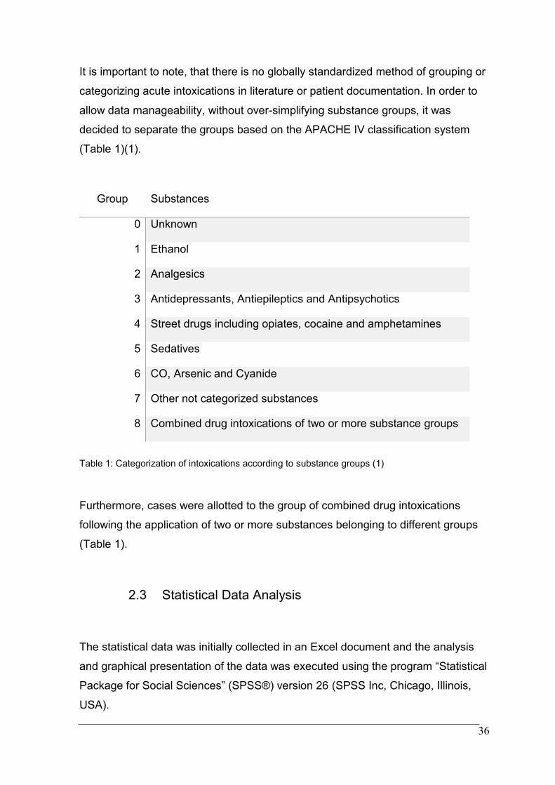
It is important to note, that there is no globally standardized method of grouping or
categorizing acute intoxications in literature or patient documentation. In order to
allow data manageability, without over-simplifying substance groups, it was
decided to separate the groups based on the APACHE IV classification system
(Table 1)(1).
Group Substances
0 Unknown
1 Ethanol
2 Analgesics
3 Antidepressants, Antiepileptics and Antipsychotics
4 Street drugs including opiates, cocaine and amphetamines
5 Sedatives
6 CO, Arsenic and Cyanide
7 Other not categorized substances
8 Combined drug intoxications of two or more substance groups
Table 1: Categorization of intoxications according to substance groups (1)
Furthermore, cases were allotted to the group of combined drug intoxications
following the application of two or more substances belonging to different groups
(Table 1).
2.3 Statistical Data Analysis
The statistical data was initially collected in an Excel document and the analysis
and graphical presentation of the data was executed using the program “Statistical
Package for Social Sciences” (SPSS®) version 26 (SPSS Inc, Chicago, Illinois,
USA).
37
A plethora of statistical methods were utilized to enable a concise answer to the
research question. Discrete variables were evaluated utilizing frequencies and
percentages, whereas continuous variables were evaluated utilizing median and
25th – 75th percentile. To compare statistical differences of the data, the Mann-
Whitney-U test and Chi-square test were utilized.
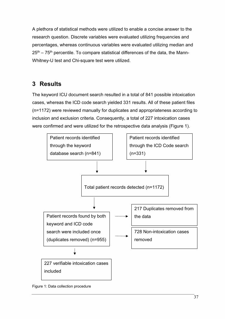
3 Results
The keyword ICU document search resulted in a total of 841 possible intoxication
cases, whereas the ICD code search yielded 331 results. All of these patient files
(n=1172) were reviewed manually for duplicates and appropriateness according to
inclusion and exclusion criteria. Consequently, a total of 227 intoxication cases
were confirmed and were utilized for the retrospective data analysis (Figure 1).
Figure 1: Data collection procedure
Patient records identified
through the keyword
database search (n=841)
Patient records identified
through the ICD Code search
(n=331)
Patient records found by both
keyword and ICD code
search were included once
(duplicates removed) (n=955)
217 Duplicates removed from
the data
Total patient records detected (n=1172)
227 verifiable intoxication cases
included
728 Non-intoxication cases
removed
38
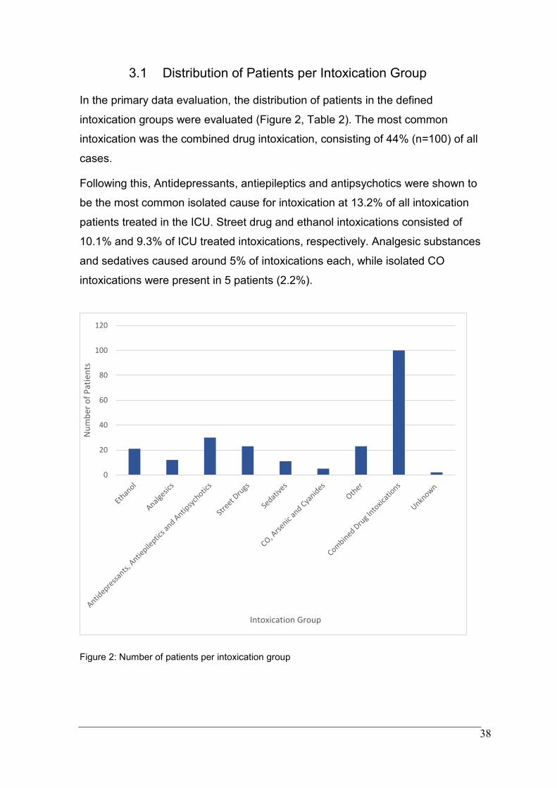
3.1 Distribution of Patients per Intoxication Group
In the primary data evaluation, the distribution of patients in the defined
intoxication groups were evaluated (Figure 2, Table 2). The most common
intoxication was the combined drug intoxication, consisting of 44% (n=100) of all
cases.
Following this, Antidepressants, antiepileptics and antipsychotics were shown to
be the most common isolated cause for intoxication at 13.2% of all intoxication
patients treated in the ICU. Street drug and ethanol intoxications consisted of
10.1% and 9.3% of ICU treated intoxications, respectively. Analgesic substances
and sedatives caused around 5% of intoxications each, while isolated CO
intoxications were present in 5 patients (2.2%).
Figure 2: Number of patients per intoxication group
0
20
40
60
80
100
120
Nu
mb
er o
f P
atie
nts
Intoxication Group
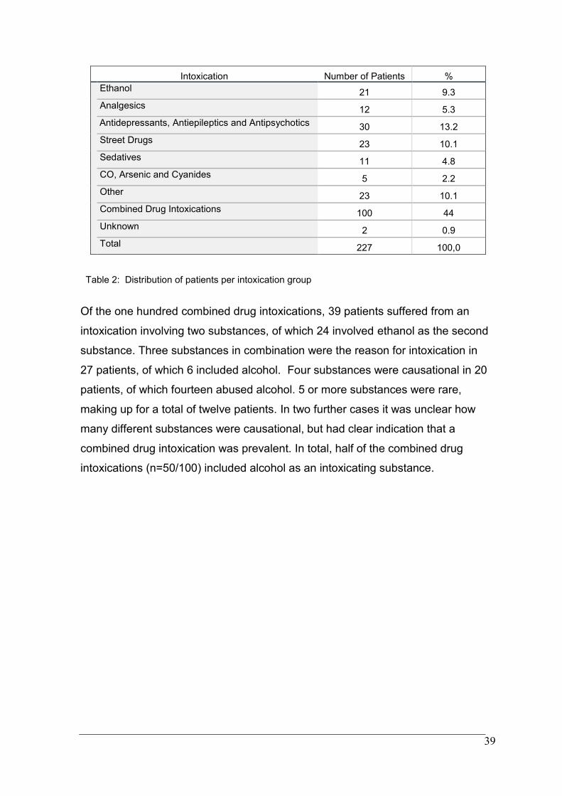
39
Intoxication Number of Patients %
Ethanol 21 9.3
Analgesics 12 5.3
Antidepressants, Antiepileptics and Antipsychotics 30 13.2
Street Drugs 23 10.1
Sedatives 11 4.8
CO, Arsenic and Cyanides 5 2.2
Other 23 10.1
Combined Drug Intoxications 100 44
Unknown 2 0.9
Total 227 100,0
Table 2: Distribution of patients per intoxication group
Of the one hundred combined drug intoxications, 39 patients suffered from an
intoxication involving two substances, of which 24 involved ethanol as the second
substance. Three substances in combination were the reason for intoxication in
27 patients, of which 6 included alcohol. Four substances were causational in 20
patients, of which fourteen abused alcohol. 5 or more substances were rare,
making up for a total of twelve patients. In two further cases it was unclear how
many different substances were causational, but had clear indication that a
combined drug intoxication was prevalent. In total, half of the combined drug
intoxications (n=50/100) included alcohol as an intoxicating substance.
40
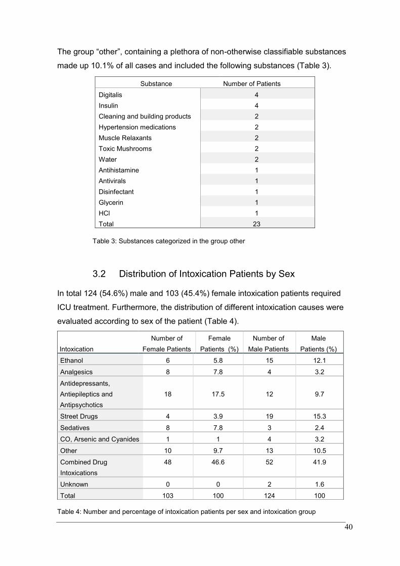
The group “other”, containing a plethora of non-otherwise classifiable substances
made up 10.1% of all cases and included the following substances (Table 3).
Table 3: Substances categorized in the group other
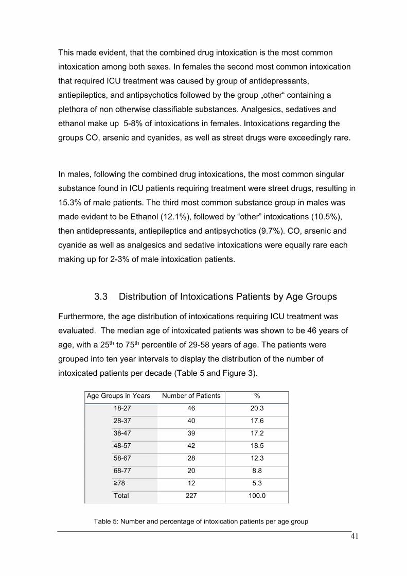
3.2 Distribution of Intoxication Patients by Sex
In total 124 (54.6%) male and 103 (45.4%) female intoxication patients required
ICU treatment. Furthermore, the distribution of different intoxication causes were
evaluated according to sex of the patient (Table 4).
Table 4: Number and percentage of intoxication patients per sex and intoxication group
Substance Number of Patients
Digitalis 4
Insulin 4
Cleaning and building products 2
Hypertension medications 2
Muscle Relaxants 2
Toxic Mushrooms 2
Water 2
Antihistamine 1
Antivirals 1
Disinfectant 1
Glycerin 1
HCl 1
Total 23
Intoxication
Number of
Female Patients
Female
Patients (%)
Number of
Male Patients
Male
Patients (%)
Ethanol 6 5.8 15 12.1
Analgesics 8 7.8 4 3.2
Antidepressants,
Antiepileptics and
Antipsychotics
18
17.5
12
9.7
Street Drugs 4 3.9 19 15.3
Sedatives 8 7.8 3 2.4
CO, Arsenic and Cyanides 1 1 4 3.2
Other 10 9.7 13 10.5
Combined Drug
Intoxications
48 46.6 52 41.9
Unknown 0 0 2 1.6
Total 103 100 124 100
41
This made evident, that the combined drug intoxication is the most common
intoxication among both sexes. In females the second most common intoxication
that required ICU treatment was caused by group of antidepressants,
antiepileptics, and antipsychotics followed by the group „other“ containing a
plethora of non otherwise classifiable substances. Analgesics, sedatives and
ethanol make up 5-8% of intoxications in females. Intoxications regarding the
groups CO, arsenic and cyanides, as well as street drugs were exceedingly rare.
In males, following the combined drug intoxications, the most common singular
substance found in ICU patients requiring treatment were street drugs, resulting in
15.3% of male patients. The third most common substance group in males was
made evident to be Ethanol (12.1%), followed by “other” intoxications (10.5%),
then antidepressants, antiepileptics and antipsychotics (9.7%). CO, arsenic and
cyanide as well as analgesics and sedative intoxications were equally rare each
making up for 2-3% of male intoxication patients.
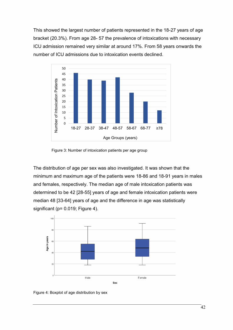
3.3 Distribution of Intoxications Patients by Age Groups
Furthermore, the age distribution of intoxications requiring ICU treatment was
evaluated. The median age of intoxicated patients was shown to be 46 years of
age, with a 25th to 75th percentile of 29-58 years of age. The patients were
grouped into ten year intervals to display the distribution of the number of
intoxicated patients per decade (Table 5 and Figure 3).
Table 5: Number and percentage of intoxication patients per age group
Age Groups in Years Number of Patients %
18-27 46 20.3
28-37 40 17.6
38-47 39 17.2
48-57 42 18.5
58-67 28 12.3
68-77 20 8.8
≥78 12 5.3
Total 227 100.0
42
This showed the largest number of patients represented in the 18-27 years of age
bracket (20.3%). From age 28- 57 the prevalence of intoxications with necessary
ICU admission remained very similar at around 17%. From 58 years onwards the
number of ICU admissions due to intoxication events declined.
Figure 3: Number of intoxication patients per age group
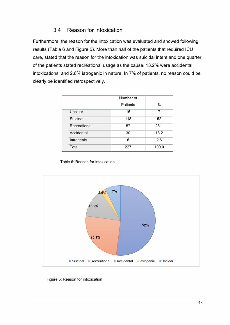
The distribution of age per sex was also investigated. It was shown that the
minimum and maximum age of the patients were 18-86 and 18-91 years in males
and females, respectively. The median age of male intoxication patients was
determined to be 42 [28-55] years of age and female intoxication patients were
median 48 [33-64] years of age and the difference in age was statistically
significant (p= 0.019; Figure 4).
Figure 4: Boxplot of age distribution by sex
0
5
10
15
20
25
30
35
40
45
50
18-27 28-37 38-47 48-57 58-67 68-77 ≥78
Num
ber
of
Into
xic
ation P
atients
Age Groups (years)
43
3.4 Reason for Intoxication
Furthermore, the reason for the intoxication was evaluated and showed following
results (Table 6 and Figure 5). More than half of the patients that required ICU
care, stated that the reason for the intoxication was suicidal intent and one quarter
of the patients stated recreational usage as the cause. 13.2% were accidental
intoxications, and 2.6% iatrogenic in nature. In 7% of patients, no reason could be
clearly be identified retrospectively.
Number of
Patients %
Unclear 16 7
Suicidal 118 52
Recreational 57 25.1
Accidental 30 13.2
Iatrogenic 6 2.6
Total 227 100.0
Table 6: Reason for intoxication
Figure 5: Reason for intoxication
52%
25.1%
13.2%
2.6% 7%
Suicidal Recreational Accidental Iatrogenic Unclear
44
3.5 Intoxication Treatment (Primary and Secondary
Detoxification, Antidotes, and Intubation)
Furthermore, key points pertaining to the treatment of intoxications were
evaluated, namely if primary or secondary detoxification methods were necessary,
if antidotes were given and whether intubation and mechanical ventilation were
performed. Some of these interventions were applied preclinically by emergency
medicine personnel or in the emergency room and repeated or performed in the
ICU.
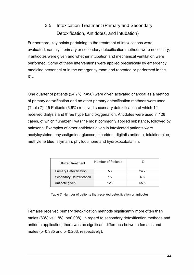
One quarter of patients (24.7%, n=56) were given activated charcoal as a method
of primary detoxification and no other primary detoxification methods were used
(Table 7). 15 Patients (6.6%) received secondary detoxification of which 12
received dialysis and three hyperbaric oxygenation. Antidotes were used in 126
cases, of which flumazenil was the most commonly applied substance, followed by
naloxone. Examples of other antidotes given in intoxicated patients were
acetylcysteine, physostigmine, glucose, biperiden, digitalis antidote, toluidine blue,
methylene blue, silymarin, phylloquinone and hydroxocobalamin.
Utilized treatment
Number of Patients
%
Primary Detoxification 56 24.7
Secondary Detoxification 15 6.6
Antidote given 126 55.5
Table 7: Number of patients that received detoxification or antidotes
Females received primary detoxification methods significantly more often than
males (33% vs. 18%; p=0.008). In regard to secondary detoxification methods and
antidote application, there was no significant difference between females and
males (p=0.385 and p=0.263, respectively).
45
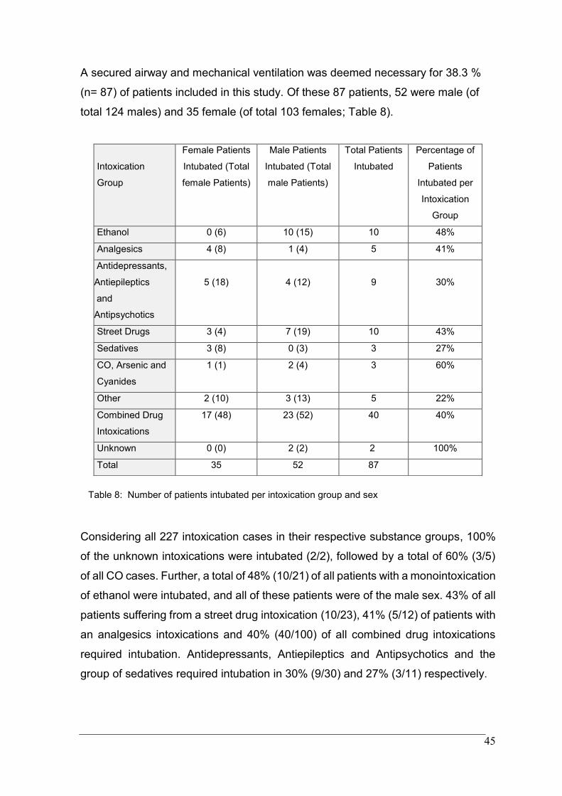
A secured airway and mechanical ventilation was deemed necessary for 38.3 %
(n= 87) of patients included in this study. Of these 87 patients, 52 were male (of
total 124 males) and 35 female (of total 103 females; Table 8).
Table 8: Number of patients intubated per intoxication group and sex
Considering all 227 intoxication cases in their respective substance groups, 100%
of the unknown intoxications were intubated (2/2), followed by a total of 60% (3/5)
of all CO cases. Further, a total of 48% (10/21) of all patients with a monointoxication
of ethanol were intubated, and all of these patients were of the male sex. 43% of all
patients suffering from a street drug intoxication (10/23), 41% (5/12) of patients with
an analgesics intoxications and 40% (40/100) of all combined drug intoxications
required intubation. Antidepressants, Antiepileptics and Antipsychotics and the
group of sedatives required intubation in 30% (9/30) and 27% (3/11) respectively.
Intoxication
Group
Female Patients
Intubated (Total
female Patients)
Male Patients
Intubated (Total
male Patients)
Total Patients
Intubated
Percentage of
Patients
Intubated per
Intoxication
Group
Ethanol 0 (6) 10 (15) 10 48%
Analgesics 4 (8) 1 (4) 5 41%
Antidepressants,
Antiepileptics
and
Antipsychotics
5 (18)
4 (12)
9
30%
Street Drugs 3 (4) 7 (19) 10 43%
Sedatives 3 (8) 0 (3) 3 27%
CO, Arsenic and
Cyanides
1 (1) 2 (4) 3 60%
Other 2 (10) 3 (13) 5 22%
Combined Drug
Intoxications
17 (48) 23 (52) 40 40%
Unknown 0 (0) 2 (2) 2 100%
Total 35 52 87
46
When taking into account the comparison of necessary invasive airway
management pertaining to the sexes, it was determined that there was no significant
difference in the rate of intubation between male and female patients (p=0.220). In
addition, the length of intubation was evaluated. Males were intubated a median of
21 hours [11.9-47.1], and females 16.5 hours [7.3-49.5]. There was no significant
difference in intubation time for male and female patients (p=0.418).
3.6 ICU Length of Stay
Furthermore, the duration of ICU stay of intoxication patients was investigated.
The median stay of patients in the ICU was shown to be 32 hours [18.7- 63] with a
range spanning from 1.7 hours of minimum stay to 840.4 hours of maximum stay
in the ICU (equating to 35 days).
The median length of stay for male patients in the ICU was 32 hours [19.4-65.2].
Similarly, the median length of stay for females was 32 hours [16.4-63.0]. The
length of ICU stay was not statistically significantly different between male and
female patients (p=0.443).
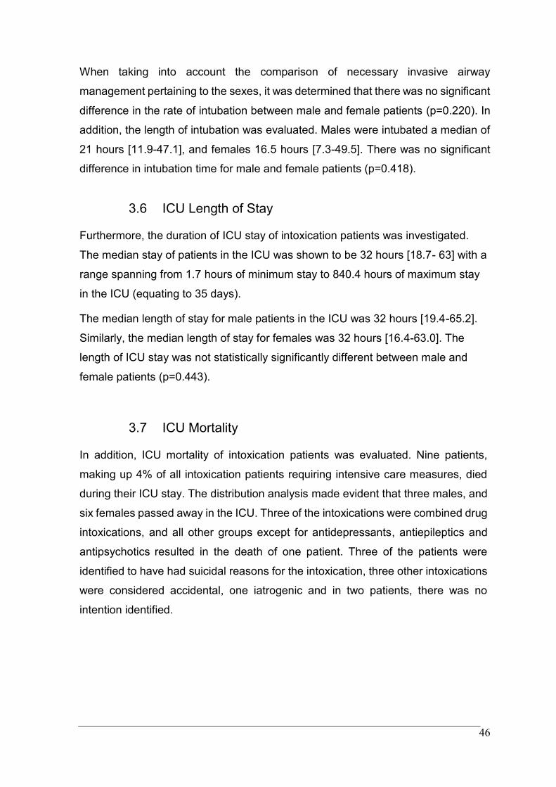
3.7 ICU Mortality
In addition, ICU mortality of intoxication patients was evaluated. Nine patients,
making up 4% of all intoxication patients requiring intensive care measures, died
during their ICU stay. The distribution analysis made evident that three males, and
six females passed away in the ICU. Three of the intoxications were combined drug
intoxications, and all other groups except for antidepressants, antiepileptics and
antipsychotics resulted in the death of one patient. Three of the patients were
identified to have had suicidal reasons for the intoxication, three other intoxications
were considered accidental, one iatrogenic and in two patients, there was no
intention identified.
47
Of these 9 patients, 5 were resuscitated preclinically and following return of
spontaneous circulation transported to the medical ICU for further treatment.
A further two patients were resuscitated during the ICU stay and consequently
passed away following termination of cardio-pulmonal resuscitation (CPR).
Furthermore, there was no significant difference in ICU mortality between the sexes
(p=0.306). Insight into the patients that died in the ICU due to acute intoxications is
given in table 9.
Table 9: Specification of ICU mortality cases
4 Discussion
Drug and substance abuse, as well as accidental and iatrogenic intoxications
requiring real-time constant observation and invasive intensive care treatment
have become an integral part of medical intensive care units worldwide. These
patients, in the acute phase, oftentimes require airway, breathing and circulatory
support, constant monitoring of vital signs, as well as detoxification procedures
such as dialysis, which require a high level of care and invasive vascular access
requiring specially trained personnel. As a result, it has become imperative to
critically evaluate the results of this diploma thesis regarding intoxication patients
admitted to the intensive care unit in Graz, and establish a comparison to
international data.
Patient Sex Age Intoxication Group Substance Specification CPR
1 Male 80 Ethanol Ethanol Preclinical
2 Male 51 Other Insulin Pens No
3 Male 25 Combined Drug
Intoxication
Methadone and Benzodiazepines Preclinical
4 Female 46 Analgesics Common cold medication containing
45 g of Paracetamol over 3 months
ICU
5 Female 23 Street Drugs Urine screening: opioids positive Preclinical
6 Female 72 Benzodiazepines Lorazepam and Diazepam No
7 Female 49 CO Gas inhalation during house fire Preclinical
8 Female 35 Combined drug
intoxication
Quetiapine, prothipendyl and ethanol ICU
9 Female 47 Combined drug
intoxication
Ethanol and CO inhalation Preclinical
48
We investigated a six year time period and identified 227 patients with
intoxications requiring ICU care. Interestingly, for these intensive care procedures
including observation and detoxification techniques, patients only required short
stays within the ICU. Furthermore, a majority of patients that required invasive
mechanical ventilation were intubated for a limited amount of time. A possible
reason for the short ICU stays and intubation times could be the half-life of a
plethora of medications and drugs being limited to hours. Therefore, patients may
be rapidly transferred to step-down units.
The separation of toxicological intensive care patients into nine intoxication
groups, was done to establish the first ever collected data displaying the
distribution of different intoxication cases in recent years in Graz. Surprisingly, the
group of combined drug intoxications were shown to be the most common group,
making up almost half of all cases. In an attempt to make the prevalence of
substances more clear, the combined drug intoxication group was closely
evaluated in reference to the number of substances applied, as well as the role of
ethanol in this group. It can be concluded that, as previously suspected, alcohol
plays a large role in combined drug intoxications with half including alcohol.
Furthermore, the group other enables only limited information as a plethora of
varying substances are included and can therefore not easily be utilized in direct
comparisons between groups.
Regarding intoxication prevalence by patient age, the largest number of patients
were represented in the youngest age bracket of patients 18-27 years of age
(20.3%). From age 28- 57 the prevalence of intoxications with necessary ICU
admission remains very similar with around 17%. To determine possible causes
for the distribution of intoxications at a younger age and to develop mental health
and addiction prevention measures, further research and advocacy is necessary.
Furthermore, it was made evident that female intoxication patients requiring ICU
treatment were statistically significantly older than male patients.
49
In addition, when investigating the distribution of patients in intoxication groups by
sex, an interesting difference in substances causing an ICU stay found. Men were
more likely to require treatment following alcohol or street drug use, whereas
female patients displayed an increased number of antidepressant, antiepileptics
and antipsychotic intoxications, as well as sedative intoxications.
Furthermore, in the evaluation of the diploma thesis data, it became evident that a
surprisingly low percentage (24.7%) of patients received activated charcoal as a
primary detoxification measure. A possible reason for this, is that oftentimes
activated charcoal is considered useful in the first hour following ingestion and
adequate protective reflexes such as swallowing and the absence of severe
vomiting are necessary for its application. Consequently, the possibility of
activated charcoal administration is limited in patients with reduced awareness and
alertness. Application therefore requires a concise benefit to risk evaluation.
Possible improvements to patient treatment, could be to further enable and
encourage adequate early preclinical administration of activated charcoal. Further
research pertaining to the benefits of activated charcoal in intoxications with
significant enterohepatic circulation or delayed gastric passage is necessary, as
these groups may benefit significantly from this method of detoxification. A german
publication by Schwacke et al. determined, similar to our data, that 30% of
intoxication patients in an ICU received activated charcoal as a primary
detoxification technique (62).
Interestingly, in this diploma thesis it was determined that significantly more
females received activated charcoal as a method of primary detoxification in
comparison to male patients. A multitude of possible reasons must be considered
to explain this phenomenon. It is possible that female patients administer more
toxicological substances perorally that can be treated with activated charcoal,
however, it is also possible that female patients seek medical assistance earlier
and the administration of activated charcoal perorally is therefore possible in a
larger number of cases.
50
Furthermore, a large number of intoxication patients requiring intensive care
measures regarding ethanol and street drugs (including intravenous drug abuse)
were male, which cannot be treated with activated charcoal.
ICU mortality was surprisingly low with only 4% of intoxication patients passing
away. When considering ICU mortality, it is vital to mention, that around the clock
availability of emergency preclinical physicians in Austria, it is likely that not all
intoxication patients requiring CPR and consequent possible intensive care
procedures are transported to the ICU in Graz. If during the complex preclinical
decision-making process, it is determined by the emergency doctor, that the
patients chances of benefiting from further invasive procedures and CPR are
minutely small, then a decision can be made to withdraw care at the patients place
of residence.
As a result, the data may vary from other paramedic systems, where different CPR
termination protocols are in place. Furthermore, gravely ill intoxications patients,
that pass away in their home and are determined to have overt clinical signs of
irreversible death, are also not included in this retrospective analysis. Therefore,
ICU mortality rates cannot be utilized to infer information about general mortality in
intoxication cases.
The University Hospital Graz is a central hospital offering a plethora of advanced
and specialized fields as well as offering the only HBO center for Styria and parts
of Carinthia. Consequently, it is the only possible destination for patients suffering
severe CO and smoke intoxications, therefore resulting in the admission and
transfer of severely ill patients from other intensive care units. In addition, the
University Hospital Graz also has a specialized center for transplant surgery. Both
of these factors must be considered when evaluating the ICU mortality of this
specific ICU. Furthermore, we found that 7 out of 9 patients who passed away
within the ICU had received cardiopulmonary resuscitation due to the severity of
the intoxication, which ultimately results in an extremely poor prognosis.
51
4.1 Comparison with Global Literature
To be able to draw adequate conclusions pertaining to intoxication patients at the
medical intensive care unit in the University Hospital Graz, it is of utmost
importance to compare the collected data to international literature. The
prevalence of common substances utilized in acute intoxications can vary broadly
depending on the regions socioeconomic status, availability of drugs and local
medication prescription practice and laws, as well as mental health and addiction
measures that are put in place. Furthermore, there is no standardized method of
intoxication substance categorization, making direct comparisons exceedingly
difficult.
The distribution of male to female patients was shown to be similar in a German
study conducted in Heidelberg by Schwacke et. al. The data collected in this
diploma thesis made evident that male (54.6%) and female intoxication patients
(45.4%) are admitted to the ICU in almost the same ratio (62). Furthermore,
regarding age distribution, the median age of intoxication in the ICU in Graz was
determined to be 46 years of age across both sexes; 48 years in female patients
and 42 years in male patients. Schwacke et. al. separated the group into adverse
drug reactions which showed a median of 71 years of age, and intentional
intoxications resulting in a median age of 39.
Moreover, Aydin et. al. evaluated the frequency that primary and secondary
detoxification as well as antidotes were administered in a medical ICU in Turkey.
Thirteen percent of the total 83 intoxication patients received primary detoxification
with activated charcoal, 11% dialysis as secondary elimination and 58% of
patients an antidote (63). Similarly, the data of this diploma thesis indicate that one
quarter of all intoxication patients received activated charcoal and more than half
(55%) received any form of antidote.
52
We did not assess for the appropriateness of the antidote application, and only
investigated whether any antidote was given or not. This may result in a higher
total number of antidotes given, however, the retrospective evaluation of only
correct antidotes given would bias the results. The decision for application of
antidotes must be performed at the bedside according to clinical assessment of
the patients symptoms, often with little additional information available. This
intrinsically leads to retrospectively falsely applied medications after the fact, when
the intended effect of the antidote does not take place and when later on more test
result may be available.
In a study done by Heyerdal et. al. in Oslo, the ICU length of stay was evaluated. It
indicates a median ICU stay of one day, with 58% of patients discharged from the
ICU after the first 24 hours and only 11% of patients stayed three or more days
(4). Similarly, the ICU length of stay determined in this diploma thesis was also
short with a median of 32 hours for both sexes.
Heyerdal et. al. also investigated the cause for intoxication. In the study it was
found that approximately 35% listed the reason for intoxication to be definite or
possible suicide (4). Furthermore, 24% of patients stated that the reason for
intoxication was an appeal for help, another 36% of intoxications were determined
to be due to recreational drug use and 4% were reported to be accidental (4). The
data in this diploma thesis shows a similar trend, with more than half of all
intoxication patients referencing suicidal intentions, one quarter of all patients
citing recreational drug use as the reason, and 13% of all patients presenting with
accidental intoxications.
In regards to ICU mortality, Heyerdal et.al. reported a mortality of 1.1% of all
intoxication patients. ICU Mortality at the medical ICU in Graz was determined to
be higher, resulting in the death of 4% of all intoxication patients. Considerations
must be made that the medical ICU in Graz is the central hospital for Styria and
therefore commonly receives the most critically ill patients, oftentimes following
protracted CPR and requiring maximal intensive care treatment.
53
4.2 Limitations of this Study
When considering the patients included in the study, it is initially important to
mention, that this data does not reflect all intoxication cases prevalent in Graz.
Due to the inclusion of only those patients requiring intensive care measures, a
large majority of the presenting cases in the emergency room were not evaluated
and included, and therefore cannot be utilized to generalize and extrapolate data
pertaining to the general number of intoxications requiring hospital care. In
addition, due to multiple close observation possibilities and monitoring floors in the
University Hospital Graz outside of the intensive care unit, it is possible that not all
patients fulfilling the intensive care criteria of other hospitals, were admitted to the
intensive care unit in Graz. The possibility for close monitoring and the level of
care that a ward can provide, varies greatly internationally and must be taken into
consideration. Nevertheless, to investigate all intoxicated patients from all
emergency departments, intermediate- and intensive care units would have been
beyond the scope of this thesis.
Furthermore, it is important to acknowledge, that the information pertaining to the
cause of the intoxication and reason for intoxication, were collected from multiple
sources. Documentation from the preclinical emergency doctors, information from
the emergency medical services, the patient themselves, relatives and in some
cases reports from police were used. These preclinical resources are oftentimes
vital for the diagnosis, treatment and further management of these patients. The
data evaluated in this retrospective analysis was then collected from the intensive
care physicians documentation.
This makes evident, that multiple level communication is necessary to diagnose
and treat toxicological patients. A substantial part of the collected data pertaining
to the substances causing the intoxication were based on the subjective reports of
patients and emergency personnel. In some cases, there were no recognizable
substances or reason that could be determined. However, all available resources
were utilized to classify patients accurately.
54
The ideal method of inclusion of toxicological patients, would be to only utilize
substrate or urine analysis, to confirm the intoxication substance. This, however, is
not possible in a plethora of intoxications, depending on the time interval until
treatment and diagnostics can be performed, the method of application and the
laboratory possibilities of detection. Consequently, a clinical diagnosis based on all
available information is usually necessary.
In addition, we defined forced diuresis as the application of at least 5l of
intravenous fluids in combination with the use of loop diuretics, as there is no clear
consensus or definition in literature. This global heterogenous terminology,
however, makes it difficult to differentiate or compare the implementation of forced
diuresis as a secondary detoxification method with other reports or publications.
We investigated the charts of intoxicated patients to identify if any received forced
diuresis per our definition; but found no respective cases. Interestingly, we noted
that physicians also used the term “forced diuresis” in their documentation
heterogeneously
5 Conclusion
In conclusion, it can be said that this diploma thesis was successful in identifying
the most common intoxications leading to an admission to the intensive care unit
at the University Hospital Graz over a time span of six years. Furthermore,
possible causes, distribution of patient traits such as age and sex, treatment and
ICU length of stay and mortality were evaluated and can provide valuable
information aiding in the decision-making process of treating physicians.
55
6 Bibliography
1. Brandenburg R, Brinkman S, Keizer NF de, Meulenbelt J, Lange DW de. In-
hospital mortality and long-term survival of patients with acute intoxication
admitted to the ICU. Crit Care Med 2014; 42(6):1471–9.
2. Burillo-Putze G, Munne P, Dueñas A, Pinillos MA, Naveiro JM, Cobo J et al.
National multicentre study of acute intoxication in emergency departments of
Spain. Eur J Emerg Med 2003; 10(2):101–4.
3. Lindqvist E, Edman G, Hollenberg J, Nordberg P, Ösby U, Forsberg S. Intensive
care admissions due to poisoning. Acta Anaesthesiol Scand 2017; 61(10):1296–
304.
4. Heyerdahl F, Bjornas MA, Hovda KE, Skog K, Opdahl A, Wium C et al. Acute
poisonings treated in hospitals in Oslo: a one-year prospective study (II): clinical
outcome. Clin Toxicol (Phila) 2008; 46(1):42–9.
5. Mégarbane B, Donetti L, Blanc T, Chéron G, Jacobs F. ICU management of
severe poisoning with medications or Illicit substances. Réanimation 2006;
15(5):343–53.
6. Reisinger A, Rabensteiner J, Hackl G. Diagnosis of acute intoxications in
critically ill patients: focus on biomarkers - part 1: epidemiology, methodology and
general overview. Biomarkers 2020; 25(1):9–19.
7. Abu Omar MNI, Foxworth J. Using Toxidromes to Assess Poisoned Patients.
Hospital Medicine Clinics 2014; 3(1):e128-e138.
8. Hackl G. Akute Intoxikationen: Marker für Screening, Diagnose und
Therapiesteuerung. Med Klin Intensivmed Notfmed 2019; 114(4):302–12.
9. Rasimas JJ, Sinclair CM. Assessment and Management of Toxidromes in the
Critical Care Unit. Crit Care Clin 2017; 33(3):521–41.
10. Liang HK. Clinical evaluation of the poisoned patient and toxic syndromes.
Clinical Chemistry 1996; 42(8):1350–5.
11. Simone CG, Bobrin BD. StatPearls: Anxiolytics and Sedative-Hypnotics
Toxicity. Treasure Island (FL); 2020.
56
12. Griffin CE, Kaye AM, Bueno FR, Kaye AD. Benzodiazepine Pharmacology and
Central Nervous System–Mediated Effects. Ochsner J 2013; 13(2):214–23.
13. Löscher W, Rogawski MA. How theories evolved concerning the mechanism of
action of barbiturates. Epilepsia 2012; 53 Suppl 8:12–25.
14. Busardò FP, Jones AW. GHB pharmacology and toxicology: acute intoxication,
concentrations in blood and urine in forensic cases and treatment of the
withdrawal syndrome. Curr Neuropharmacol 2015; 13(1):47–70.
15. LaHood AJ, Kok SJ. StatPearls: Ethanol Toxicity. Treasure Island (FL); 2021.
16. Lobo IA, Harris RA. GABA(A) receptors and alcohol. Pharmacol Biochem
Behav 2008; 90(1):90–4.
17. Wallner M, Olsen RW. Physiology and pharmacology of alcohol: the
imidazobenzodiazepine alcohol antagonist site on subtypes of GABAA receptors
as an opportunity for drug development? Br J Pharmacol 2008; 154(2):288–98.
18. Dolinak D. Opioid Toxicity. Acad Forensic Pathol 2017; 7(1):19–35.
19. Pathan H, Williams J. Basic opioid pharmacology: an update. Br J Pain 2012;
6(1):11–6.
20. Schwartz BG, Rezkalla S, Kloner RA. Cardiovascular effects of cocaine.
Circulation 2010; 122(24):2558–69.
21. Wilbert-Lampen U, Seliger C, Zilker T, Arendt RM. Cocaine increases the
endothelial release of immunoreactive endothelin and its concentrations in human
plasma and urine: reversal by coincubation with sigma-receptor antagonists.
Circulation 1998; 98(5):385–90.
22. Steinkellner T, Freissmuth M, Sitte HH, Montgomery T. The ugly side of
amphetamines: short- and long-term toxicity of 3,4-
methylenedioxymethamphetamine (MDMA, 'Ecstasy'), methamphetamine and D-
amphetamine. Biol Chem 2011; 392(1-2):103–15.
23. Dawson AH, Buckley NA. Pharmacological management of anticholinergic
delirium - theory, evidence and practice. Br J Clin Pharmacol 2016; 81(3):516–24.
57
24. Ramjan KA, Williams AJ, Isbister GK, Elliott EJ. 'Red as a beet and blind as a
bat' Anticholinergic delirium in adolescents: lessons for the paediatrician. J
Paediatr Child Health 2007; 43(11):779–80.
25. Reisinger A, Rabensteiner J, Hackl G. Diagnosis of acute intoxications in
critically ill patients: focus on biomarkers - part 2: markers for specific intoxications.
Biomarkers 2020; 25(2):112–25.
26. Eddleston M, Buckley NA, Eyer P, Dawson AH. Management of acute
organophosphorus pesticide poisoning. The Lancet 2008; 371(9612):597–607.
27. Henretig FM, Kirk MA, McKay CA. Hazardous Chemical Emergencies and
Poisonings. N Engl J Med 2019; 380(17):1638–55.
28. Scotton WJ, Hill LJ, Williams AC, Barnes NM. Serotonin Syndrome:
Pathophysiology, Clinical Features, Management, and Potential Future Directions.
Int J Tryptophan Res 2019; 12:1178646919873925.
29. Lambert O, Bourin M. SNRIs: mechanism of action and clinical features.
Expert Rev Neurother 2002; 2(6):849–58.
30. Benson BE, Hoppu K, Troutman WG, Bedry R, Erdman A, Höjer J et al.
Position paper update: gastric lavage for gastrointestinal decontamination. Clin
Toxicol (Phila) 2013; 51(3):140–6.
31. Höjer J, Troutman WG, Hoppu K, Erdman A, Benson BE, Mégarbane B et al.
Position paper update: ipecac syrup for gastrointestinal decontamination. Clin
Toxicol (Phila) 2013; 51(3):134–9.
32. Chyka PA, Seger D, Krenzelok EP, Vale JA. Position paper: Single-dose
activated charcoal. Clin Toxicol (Phila) 2005; 43(2):61–87.
33. Zellner T, Prasa D, Färber E, Hoffmann-Walbeck P, Genser D, Eyer F. The
Use of Activated Charcoal to Treat Intoxications. Dtsch Arztebl Int 2019;
116(18):311–7.
34. Tenenbein M, Lheureux P. Position paper: whole bowel irrigation. J Toxicol
Clin Toxicol 2004; 42(6):843–54.
58
35. Thanacoody R, Caravati EM, Troutman B, Höjer J, Benson B, Hoppu K et al.
Position paper update: whole bowel irrigation for gastrointestinal decontamination
of overdose patients. Clin Toxicol (Phila) 2015; 53(1):5–12.
36. Benzoni T, Gibson J. StatPearls: Ipecac. Treasure Island (FL); 2020.
37. Simpson S-E. Pharmacobezoars described and demystified. Clin Toxicol
(Phila) 2011; 49(2):72–89.
38. Höjer J, Personne M. Endoscopic removal of slow release clomipramine
bezoars in two cases of acute poisoning. Clin Toxicol (Phila) 2008; 46(4):317–9.
39. Briggs AL, Deal LL. Endoscopic removal of pharmacobezoar in case of
intentional potassium overdose. J Emerg Med 2014; 46(3):351–4.
40. Thornley-Brown D, Galla JH, Williams PD, Kant KS, Rashkin M. Lithium
toxicity associated with a trichobezoar. Ann Intern Med 1992; 116(9):739–40.
41. Rauber-Lüthy C, Hofer KE, Bodmer M, Kullak-Ublick GA, Kupferschmidt H,
Ceschi A. Gastric pharmacobezoars in quetiapine extended-release overdose: a
case series. Clin Toxicol (Phila) 2013; 51(10):937–40.
42. Jha VK, Padmaprakash KV. Extracorporeal Treatment in the Management of
Acute Poisoning: What an Intensivist Should Know? Indian J Crit Care Med 2018;
22(12):862–9.
43. Mahesh C, Nagalakshmi N, Sivaparvathi K, Nagaraj RD, Praveen N,
Amarendra N et al. Hemoperfusion in the management of acute poisoning. J NTR
Univ Health Sci 2020; 9(4):250.
44. Wittebole X, Hantson P. Use of the molecular adsorbent recirculating system
(MARS™) for the management of acute poisoning with or without liver failure. Clin
Toxicol (Phila) 2011; 49(9):782–93.
45. Wolfgang Oczenski. Atmen-Atemhilfen: Atemphysiologie und
Beatmungstechnik. 10. Stuttgart: Georg Thieme Verlag; 2017.
46. Gill AL, Bell CNA. Hyperbaric oxygen: its uses, mechanisms of action and
outcomes. QJM 2004; 97(7):385–95.
47. Weinberg GL. Lipid Emulsion Infusion: Resuscitation for Local Anesthetic and
other Drug Overdose. Anesthesiology 2012; 117(1):180–7.
59
48. Engebretsen KM, Kaczmarek KM, Morgan J, Holger JS. High-dose insulin
therapy in beta-blocker and calcium channel-blocker poisoning. Clin Toxicol (Phila)
2011; 49(4):277–83.
49. Burkes R, Wendorf G. A multifaceted approach to calcium channel blocker
overdose: a case report and literature review. Clin Case Rep 2015; 3(7):566–9.
50. Proudfoot AT, Krenzelok EP, Vale JA. Position Paper on urine alkalinization. J
Toxicol Clin Toxicol 2004; 42(1):1–26.
51. Proudfoot AT, Krenzelok EP, Brent J, Vale JA. Does urine alkalinization
increase salicylate elimination? If so, why? Toxicol Rev 2003; 22(3):129–36.
52. Chacko B, Peter JV. Antidotes in Poisoning. Indian J Crit Care Med 2019;
23(Suppl 4):S241-S249.
53. Kerensky T, Walley AY. Opioid overdose prevention and naloxone rescue kits:
what we know and what we don't know. Addict Sci Clin Pract 2017; 12(1):4.
54. Lheureux P, Vranckx M, Leduc D, Askenasi R. Risks of flumazenil in mixed
benzodiazepine-tricyclic antidepressant overdose: report of a preliminary study in
the dog. J Toxicol Clin Exp 1992; 12(1):43–53.
55. UB G, Tadvi NA. Paracetamol Toxicity: A Review. J Cont Med A Dent 2014;
2(3):12–5.
56. Heard KJ. Acetylcysteine for acetaminophen poisoning. N Engl J Med 2008;
359(3):285–92.
57. Bateman DN, Dear JW, Thanacoody HKR, Thomas SHL, Eddleston M,
Sandilands EA et al. Reduction of adverse effects from intravenous acetylcysteine
treatment for paracetamol poisoning: a randomised controlled trial. The Lancet
2014; 383(9918):697–704.
58. Rehman HU. Methemoglobinemia. West J Med 2001; 175(3):193–6.
59. Reisinger A, Vogt S, Essl A, Rauch I, Bangerl F, Eller P et al. Lessons of the
month 3: Intravenous poppers abuse: case report, management and possible
complications. Clin Med (Lond) 2020; 20(2):221–3.
60
60. Romanelli F, Smith KM, Thornton AC, Pomeroy C. Poppers: epidemiology and
clinical management of inhaled nitrite abuse. Pharmacotherapy 2004; 24(1):69–
78.
61. Brandenburg R, Brinkman S, Keizer NF de, Kesecioglu J, Meulenbelt J, Lange
DW de. The need for ICU admission in intoxicated patients: a prediction model.
Clin Toxicol (Phila) 2017; 55(1):4–11.
62. Schwake L, Wollenschläger I, Stremmel W, Encke J. Adverse drug reactions
and deliberate self-poisoning as cause of admission to the intensive care unit: a 1-
year prospective observational cohort study. Intensive Care Med 2009; 35(2):266–
74.
63. Aydin K, Cetinkaya A. Retrospective Review of Cases of Intoxication in
Medical Intensive Care Unit. J Crit Intensive Care 2020.