Embed Size (px)
Citation preview
Aus dem Adolf-Butenandt-Institut für Lehrstuhl Stoffwechselbiochemie der Ludwig-Maximilians Universität München
Vorstand: Prof. Dr. rer. nat. Dr. h.c. Christian Haass
Functional characterization of Shadoo, a PrP-like protein with neuroprotective activity
Dissertation
Zum Erwerb des Doktorgrades der Naturwissenschaften (Dr. rer. nat.)
an der Medizinischen Fakultät
der Ludwig-Maximilians-Universität München
vorgelegt von
Vignesh Sakthivelu aus
Coimbatore, Indien 2011
Gedruckt mit Genehmigung der Medizinischen Fakultät
der Ludwig-Maximilians-Universität München
Betreuer : Prof. Dr. rer. nat. Jörg Tatzelt
Zweitgutachter : Priv. Doz. Dr. rer. nat. Monika Bradl
Dekan : Prof. Dr. med. Dr.h.c. Maximilian Reiser, FACR,FRCR
Mündliche Prüfung am : 30.05.2012
Acknowledgement
This doctoral thesis is a significant part of the research study that I did as a
PhD student in the group of Prof. Dr. Jörg Tatzelt. Right from the beginning until
the completion of the study, I had the good opportunity to work with many people
whose contribution in different ways to the study have helped in making my PhD
thesis very special. I would like to sincerely thank all these people who have made
a noteworthy impact on my life.
Firstly, I would like to convey my immense gratefulness to my advisor Prof.
Dr. Jörg Tatzelt, for giving me an interesting and important research topic, for his
critical supervision, guidance and also for all his personal care and affection during
my entire stay.
I am deeply indebted to PD. Dr. Konstanze F. Winklhofer for her valuable
support and major discussions and suggestions during the entire PhD timeline.
Many heartfelt wishes are also in the offing to Dr. Ralf Seidel from the Max
Planck Institute of Molecular Physiology, Dortmund, Germany for his enormous
help in preparing the rabbit polyclonal Sho antibody.
I earnestly thank Prof. Dr. Christian Haass for agreeing to be a member of
my PhD thesis committee and also for his helpful discussions during my research.
Frankly, much of my work has been possible due to the company and help
of former and present members of the prion and Parkin group: Geli, Margit, Anita,
Mareike, Lena and Julia. Special thanks to Veronika for her technical assistance in
the lab; Sabine and Annette for their outstanding lab management. Heartfelt thanks
to Uli, Natalie, Daniela, Viktoria and Sina for not just being good lab mates but
also for being good friends and giving me the push and energy in all my high and
low times; Anna, Alexandra, Kathrin, Elisa, Carolin, Maria Patra, Maria Funke
and the people in Haass department are also fondly thanked.

My special thanks to Krishnaveni, Senthil Murugan and Katyayni for their
love, special care and a critical help with my thesis.
My friends Santmoor, Sabari, Anu, Vidhya Shankar, Guru, Jeeva,
Nagendran, Karthikeyan, Vigneshwaran, Ramesh, Vaideeswaran, Maha, Sudhakar,
Bharati, Vaibhaoa, Shanaya, Kavin, Avinash, Paresh, Arun, Dilip, Sathish and
Raghu deserve each and every word of goodness for their kind hospitality and
never making me feel alone during my entire stay in Munich.
Words fail me, but the reason why I have reached this stage in life is
because of the constant belief, unconditional love and wonderful prayers of my
beloved parents Sakthivelu, Santhi and my brother Rajkumar that have helped me
in all walks of my life.
Last but not the least, I am highly thankful to the DAAD (German
Academic Exchange Service, Germany) for awarding me a fellowship that helped
me to successfully complete my PhD in Germany.
Contents
Introduction 1
Prion diseases 1
Transmissible spongiform encephalopathies in animals 1
Scrapie 1
Bovine spongiform encephalopathy 3
Chronic wasting disease 4
Transmissible spongiform encephalopathies in humans 5
Clinical signs and neuropathology of human prion diseases 9
Prion protein 11
Nature of the infectious agent 11
Conformational transition from PrPC to PrPSc 13
Prion protein gene 15
Biogenesis and structure of PrP 16
Function of prion protein 18
Studies from PrP knockout mice 18
Stress-protective functions of PrPC 19
PrP protection against PrP∆HD 20
Role of PrP in copper binding and oxidative stress 20
Neurotoxic signaling through PrPC 21
Putative co-receptors for PrP 22
Additional PrP interacting proteins 24
Shadoo 26
Sprn gene and its polymorphisms 26
Expression of Sprn gene 28
Structure of Sho 29
Biological function of Sho 29
Aim of the study 31
Results 33
Generation of antibodies against human Sho 33
Biochemical characterization of mammalian Sho 34
Cloning of human Sho and mutants thereof 34
Wild type Sho and Sho mutants are complex glycosylated 36
Sho and the mutants are targeted to the outer leaflet of the plasma membrane via a GPI anchor 38
Sho attenuates glutamate induced excitotoxic stress 40
Sho protect cells against PrP∆HD-induced toxicity 42
The hydrophobic domain mediates homodimerization of Sho 43
Sho and PrP homodimers are formed within the cell 47
No evidence for the trans dimers at the cell surface 49
Interaction between Sho and PrP 50
The N-terminal domain of Sho can restore stress-protective activity of PrPΔN
52
N-Sho/PrP-C is complex glycosylated and GPI-anchored 53
Sho-PrP has a stress-protective activity 53
A possible role of Sho in PrPSc-induced toxicity 55
PrP∆HD toxic signaling is blocked by NMDA receptor antagonist 58
Discussion 60
Biogenesis of human Sho 60
Sho is complex glycosylated and attached to the plasma membrane 60
Sho forms homodimers 61
No formation of PrP/Sho mixed dimer 65
The stress-protective activity of Sho 65
Deleting HD in Sho does not lead to neurotoxic species 67
The N-terminal domain of Sho can functionally replace that of PrP 68
Sho does not protect cells from PrPSc-induced apoptosis 69
Summary 71
Zusammenfassung 73
Methods 75
Molecular biology methods 75
Cloning and site directed mutation by polymerase chain reaction 75
Agarose gel electrophoresis 76
Isolation and purification of DNA fragments from agarose gel 76
Enzymatic modification of DNA fragments 76
Alkaline phosphatase treatment 77
Ligation of cDNA fragments into vector DNA 77
Preparation of competent bacteria 77
Transformation of competent bacteria 78
Plasmid DNA preparation from bacterial culture 78
Sequencing 78
Cell biology methods 78
Cell culture 78
Cultivation of cells 78
Passaging 79
Plating the cells 79
Transfection 79
Harvesting the cells 80
Total cell lysates 80
SDS-PAGE 80
Western blot analysis 80
Ponceau S staining 81
Immunodetection of proteins 81
Glycosylation analysis 81
Treatment with tunicamycin 81
Digestion with Endo H or PNGase F 82
Treatment with brefeldin A 82
Indirect immunofluorescence microscopy 82
Co-immunoprecipitation 83
Co-cultivation assay 83
Apoptosis assay 83
Statistical analysis 84
Materials 85
Biological materials 85
Bacterial strain 85
Vectors 85
Cell lines 85
Antibodies 85
Primer list 85
Chemicals and reagents 86
Medium 88
Kits 88
Equipments 88
Solutions and buffers 89
References 91
Abbreviations 115
Curriculum vitae 117
Introduction
1
Introduction
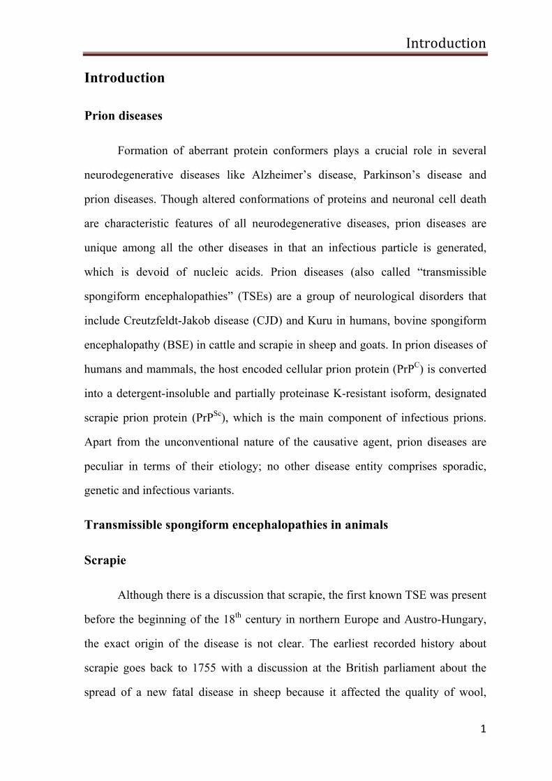
Prion diseases
Formation of aberrant protein conformers plays a crucial role in several
neurodegenerative diseases like Alzheimer’s disease, Parkinson’s disease and
prion diseases. Though altered conformations of proteins and neuronal cell death
are characteristic features of all neurodegenerative diseases, prion diseases are
unique among all the other diseases in that an infectious particle is generated,
which is devoid of nucleic acids. Prion diseases (also called “transmissible
spongiform encephalopathies” (TSEs) are a group of neurological disorders that
include Creutzfeldt-Jakob disease (CJD) and Kuru in humans, bovine spongiform
encephalopathy (BSE) in cattle and scrapie in sheep and goats. In prion diseases of
humans and mammals, the host encoded cellular prion protein (PrPC) is converted
into a detergent-insoluble and partially proteinase K-resistant isoform, designated
scrapie prion protein (PrPSc), which is the main component of infectious prions.
Apart from the unconventional nature of the causative agent, prion diseases are
peculiar in terms of their etiology; no other disease entity comprises sporadic,
genetic and infectious variants.
Transmissible spongiform encephalopathies in animals
Scrapie
Although there is a discussion that scrapie, the first known TSE was present
before the beginning of the 18th century in northern Europe and Austro-Hungary,
the exact origin of the disease is not clear. The earliest recorded history about
scrapie goes back to 1755 with a discussion at the British parliament about the
spread of a new fatal disease in sheep because it affected the quality of wool,
Introduction
2
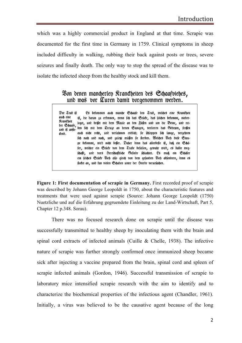
which was a highly commercial product in England at that time. Scrapie was
documented for the first time in Germany in 1759. Clinical symptoms in sheep
included difficulty in walking, rubbing their back against posts or trees, severe
seizures and finally death. The only way to stop the spread of the disease was to
isolate the infected sheep from the healthy stock and kill them.
Figure 1: First documentation of scrapie in Germany. First recorded proof of scrapie was described by Johann George Leopoldt in 1750, about the characteristic features and treatments that were used against scrapie (Source: Johann George Leopoldt (1750) Nuetzliche und auf die Erfahrung gegruendete Einleitung zu der Land-Wirtschaft, Part 5, Chapter 12 p.348. Sorau).
There was no focused research done on scrapie until the disease was
successfully transmitted to healthy sheep by inoculating them with the brain and
spinal cord extracts of infected animals (Cuille & Chelle, 1938). The infective
nature of scrapie was further strongly confirmed once immunized sheep became
sick after injecting a vaccine prepared from the brain, spinal cord and spleen of
scrapie infected animals (Gordon, 1946). Successful transmission of scrapie to
laboratory mice intensified scrapie research with the aim to identify and to
characterize the biochemical properties of the infectious agent (Chandler, 1961).
Initially, a virus was believed to be the causative agent because of the long
Introduction
3
incubation period. But scrapie agent’s resistance to heat, ultraviolet light and
formaldehyde, which are known to destroy viral particles, suggested that a virus
was obviously not the infectious particle. Healthy sheep injected with brain
homogenate from scrapie infected sheep developed clinical symptoms after five
months, whereas goats injected with the sheep scrapie brain homogenate
developed the clinical symptoms after 23 months. The incubation time was
shortened to eight months when the brain homogenate from scrapie infected goat
was injected in to a healthy goat. These were the first evidences describing a
phenomenon denoted species barrier. Even though the scrapie transmission to the
laboratory animals was experimentally confirmed but so far there is no proof
available for the sheep or goat scrapie transmission into humans.
Bovine spongiform encephalopathy
BSE affects cattle and is commonly referred to as “mad cow disease”. The
first case of BSE was confirmed in the year 1986 in Great Britain. After this, the
number of new cases increased significantly within the next few years in all parts
of Great Britain, with a maximum of 36,680 cases identified in 1992. The clinical
symptoms observed in BSE affected animals are difficulty in standing, lack of
muscle coordination with trouble to walk and loss of weight. Affected cattle die
within a few weeks or a few months after the onset of clinical symptoms. The
source of BSE is still unknown, though it is believed that contaminated meat-and-
bone meal (MBM) prepared from scrapie infected sheep might be responsible for
the spread of the disease. New BSE cases in Great Britain are declining after the
subsequent ban of MBM by the British government.
In the beginning of the 20th century the infectivity and disease transmission
to heterologous animals was clearly demonstrated along with prolonged incubation
Introduction
4
period. In 1996, a new type of prion disease in humans called variant Creutzfeldt-
Jakob disease (vCJD) was reported in United Kingdom (UK). The current idea is
that vCJD is due to a transmission of BSE to humans, possibly through the
consumption of BSE contaminated food stuffs. So far, 170 cases of vCJD have
been recorded in Great Britain. In addition, a few vCJD cases have been reported
outside the UK, but none so far in Germany.
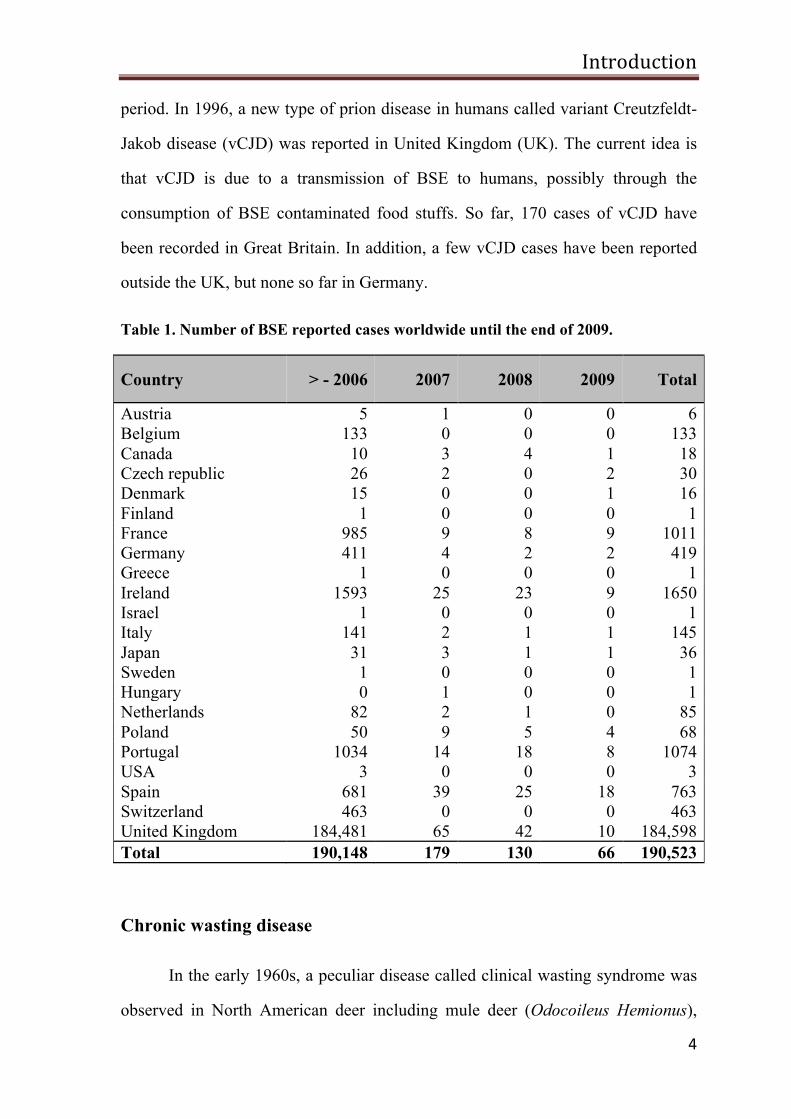
Table 1. Number of BSE reported cases worldwide until the end of 2009.
Country > - 2006 2007 2008 2009 Total
Austria 5 1 0 0 6 Belgium 133 0 0 0 133 Canada 10 3 4 1 18 Czech republic 26 2 0 2 30 Denmark 15 0 0 1 16 Finland 1 0 0 0 1 France 985 9 8 9 1011 Germany 411 4 2 2 419 Greece 1 0 0 0 1 Ireland 1593 25 23 9 1650 Israel 1 0 0 0 1 Italy 141 2 1 1 145 Japan 31 3 1 1 36 Sweden 1 0 0 0 1 Hungary 0 1 0 0 1 Netherlands 82 2 1 0 85 Poland 50 9 5 4 68 Portugal 1034 14 18 8 1074 USA 3 0 0 0 3 Spain 681 39 25 18 763 Switzerland 463 0 0 0 463 United Kingdom 184,481 65 42 10 184,598 Total 190,148 179 130 66 190,523
Chronic wasting disease
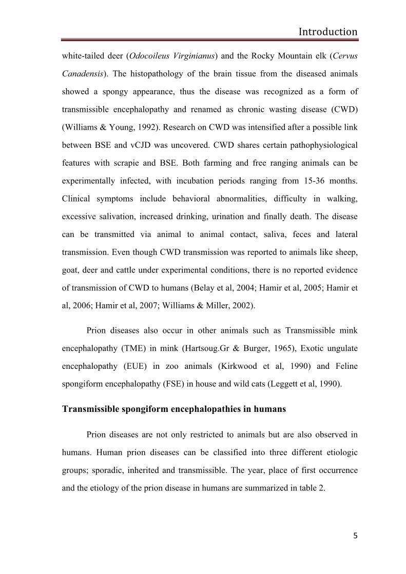
In the early 1960s, a peculiar disease called clinical wasting syndrome was
observed in North American deer including mule deer (Odocoileus Hemionus),
Introduction
5
white-tailed deer (Odocoileus Virginianus) and the Rocky Mountain elk (Cervus
Canadensis). The histopathology of the brain tissue from the diseased animals
showed a spongy appearance, thus the disease was recognized as a form of
transmissible encephalopathy and renamed as chronic wasting disease (CWD)
(Williams & Young, 1992). Research on CWD was intensified after a possible link
between BSE and vCJD was uncovered. CWD shares certain pathophysiological
features with scrapie and BSE. Both farming and free ranging animals can be
experimentally infected, with incubation periods ranging from 15-36 months.
Clinical symptoms include behavioral abnormalities, difficulty in walking,
excessive salivation, increased drinking, urination and finally death. The disease
can be transmitted via animal to animal contact, saliva, feces and lateral
transmission. Even though CWD transmission was reported to animals like sheep,
goat, deer and cattle under experimental conditions, there is no reported evidence
of transmission of CWD to humans (Belay et al, 2004; Hamir et al, 2005; Hamir et
al, 2006; Hamir et al, 2007; Williams & Miller, 2002).
Prion diseases also occur in other animals such as Transmissible mink
encephalopathy (TME) in mink (Hartsoug.Gr & Burger, 1965), Exotic ungulate
encephalopathy (EUE) in zoo animals (Kirkwood et al, 1990) and Feline
spongiform encephalopathy (FSE) in house and wild cats (Leggett et al, 1990).
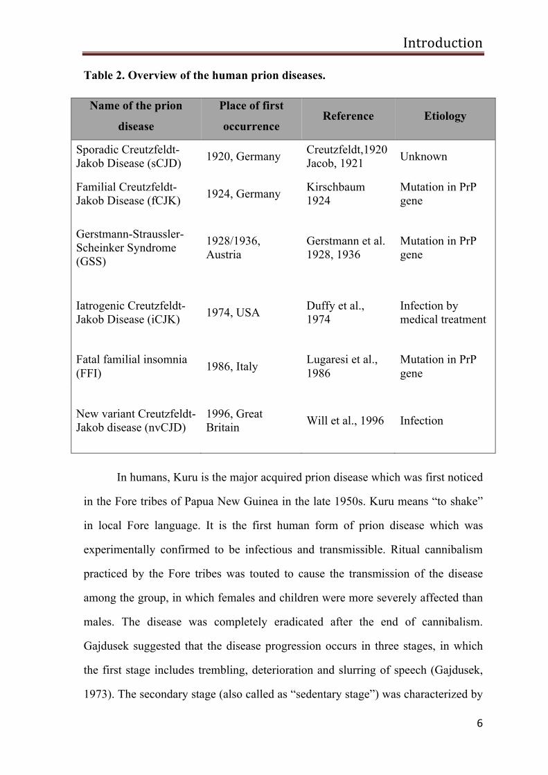
Transmissible spongiform encephalopathies in humans
Prion diseases are not only restricted to animals but are also observed in
humans. Human prion diseases can be classified into three different etiologic
groups; sporadic, inherited and transmissible. The year, place of first occurrence
and the etiology of the prion disease in humans are summarized in table 2.
Introduction
6
Table 2. Overview of the human prion diseases.
Name of the prion
disease
Place of first
occurrence Reference Etiology
Sporadic Creutzfeldt- Jakob Disease (sCJD) 1920, Germany Creutzfeldt,1920
Jacob, 1921 Unknown
Familial Creutzfeldt-Jakob Disease (fCJK) 1924, Germany Kirschbaum
1924 Mutation in PrP gene
Gerstmann-Straussler-Scheinker Syndrome (GSS)
1928/1936, Austria
Gerstmann et al. 1928, 1936
Mutation in PrP gene
Iatrogenic Creutzfeldt-Jakob Disease (iCJK) 1974, USA Duffy et al.,
1974 Infection by medical treatment
Fatal familial insomnia (FFI) 1986, Italy Lugaresi et al.,
1986 Mutation in PrP gene
New variant Creutzfeldt-Jakob disease (nvCJD)
1996, Great Britain Will et al., 1996 Infection
In humans, Kuru is the major acquired prion disease which was first noticed
in the Fore tribes of Papua New Guinea in the late 1950s. Kuru means “to shake”
in local Fore language. It is the first human form of prion disease which was
experimentally confirmed to be infectious and transmissible. Ritual cannibalism
practiced by the Fore tribes was touted to cause the transmission of the disease
among the group, in which females and children were more severely affected than
males. The disease was completely eradicated after the end of cannibalism.
Gajdusek suggested that the disease progression occurs in three stages, in which
the first stage includes trembling, deterioration and slurring of speech (Gajdusek,
1973). The secondary stage (also called as “sedentary stage”) was characterized by
Introduction
7
severe ataxia, shock like muscle jerks and depression while the terminal stage was
linked to the inability of the patient to sit without an external support, defective
muscle coordination and difficulty in eating (Gajdusek, 1973).
Analyzing the postmortem brain of kuru patients, Igor Klatzo noted that
Kuru is very similar to that of another human prion disease called Creutzfeldt-
Jakob disease (CJD) (Klatzo et al, 1959). Kuru was successfully transmitted to
chimpanzees by intracerebral injection of brain homogenate from Kuru infected
individuals. Later, the nature of infectivity of CJD was confirmed in similar
experiments (Gajdusek et al, 1966; Gibbs et al, 1968).
Sporadic human prion diseases occur spontaneously with no prior family
history and mutations in the prion protein gene (PRNP). CJD was first described
by two German neuropathologists Hans Gerhard Creutzfeldt and Alfons Jakob
independently (Creutzfeldt, 1920; Creutzfeldt, 1921; Jakob, 1921). CJD is
classified into four major types, sporadic CJD (sCJD), iatrogenic CJD (iCJD),
variant CJD (vCJD) and familial CJD (fCJD). The most common form of CJD is
sCJD, and accounts for approximately 85% of all CJD cases (Johnson, 2005;
Prince et al, 2006). So far, the mechanisms which trigger sCJD have not been
identified. However, sporadic somatic cell mutations in PRNP gene, spontaneous
refolding of prion protein or unidentified infection have been proposed to be the
cause of sCJD but none of these have been experimentally proven (Aguzzi et al,
2008).
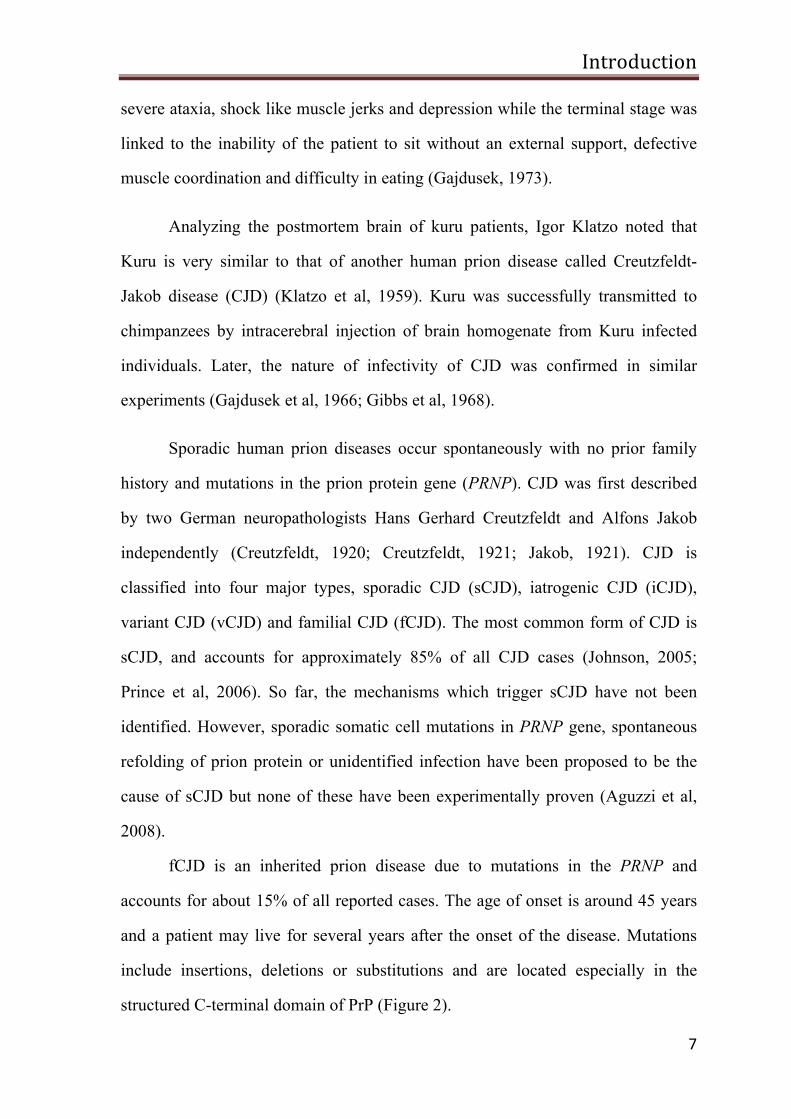
fCJD is an inherited prion disease due to mutations in the PRNP and
accounts for about 15% of all reported cases. The age of onset is around 45 years
and a patient may live for several years after the onset of the disease. Mutations
include insertions, deletions or substitutions and are located especially in the
structured C-terminal domain of PrP (Figure 2).
Introduction
8
Figure 2: The human prion protein gene (PRNP) with all definite and suspected pathogenic mutations currently identified. The entire prion protein consists of residues 23-230, with 1-22 a signal peptide for targeting to the endoplasmic reticulum and 231-253 a signal peptide for GPI-anchor attachment. M/V and E/K219 are common polymorphisms that can influence the onset and phenotype of the disease (Adopted from Mead, 2006).
Gerstmann–Sträussler–Scheinker syndrome (GSS) is a rare genetic
autosomal dominant prion disease first described by Josef Gerstmann, an Austrian
neuropathologist, along with Ernst Sträussler and Ilya Scheinker (Gerstmann et al,
1935). Mice infected with intracerebral injection of GSS brain homogenate
developed the disease symptoms comparable to the clinical symptoms of GSS,
providing experimental evidence for the infectious nature of inherited prion
diseases (Tateishi & Kitamoto, 1995).
Fatal familial insomnia (FFI) also belongs to the group of inherited prion
diseases with a mutation in the PRNP. So far, there are 40 families found to
possess the mutated gene worldwide. A disease with similar symptoms was
described in patients with no PrP mutations called sporadic fatal insomnia (SFI).
The third group of the human prion disease is the infectious form that
includes iCJD and the new variant Creutzfeldt-Jakob disease (nvCJD). Infectious
forms account for less than 1% of all cases. Contaminated human growth
hormones isolated from CJD infected individuals and contamination of surgical
Introduction
9
instruments from CJD infected tissues as a result of medical procedure accounted
for the iatrogenic forms (Bernoulli et al, 1977; Davanipour et al, 1984; Duffy et al,
1974; Kondo & Kuroiwa, 1982). vCJD develops probably due to the intake of BSE
contaminated food products and in contrast to the classical form has a very long
incubation period (Aguzzi & Weissmann, 1996; Bruce et al, 1997; Collinge et al,
1996; Hill et al, 1997). So far, 280 vCJD cases have been confirmed worldwide
since the first cases reported in 1996. The number of cases may rise in the future
considering the unusually prolonged incubation periods of this disease. There is
evidence that vCJD in contrast to sCJD, can be transmitted through blood products
(Aguzzi & Glatzel, 2004; Llewelyn et al, 2004; Peden et al, 2004; Wroe et al,
2006). This indicates that prion contaminated blood products can significantly
increase the risk of prion disease in humans. As a consequence people who lived in
the UK between 1980 and 1996 were not allowed to donate blood in countries
outside the UK.
Clinical signs and neuropathology of human prion diseases
The main characteristic features of all human prion diseases are prolonged
incubation periods with complex etiology and differences in their disease duration,
onset of clinical manifestation and neuropathology. Onset of the symptoms in
sCJD is approximately around the age of 60 years. In fCJD and GSS, the
symptoms are observed at an average age of 45-50 years, whereas in vCJD clinical
signs are detected in relatively younger patients with an average age of 29 years.
Kuru occurs at a wide range of ages between 4-60 years which is possibly
associated with the concentration and the exposure time of the infectious particle.
Prion diseases not only differ in their incubation periods but also in the duration
between the onset of clinical signs and death. The average duration between the
onset of the clinical symptoms and death in sCJD is only 2-3 months while in
Introduction
10
vCJD the average is 14 months and that of Kuru is 12 months. GSS shows an
exceptionally long duration of the disease with an average of 5 years (Collinge,
2001; Johnson & Gibbs, 1998).
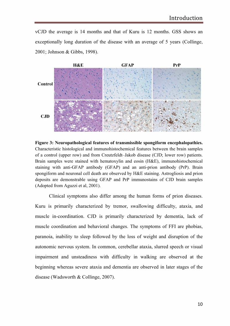
Figure 3: Neuropathological features of transmissible spongiform encephalopathies. Characteristic histological and immunohistochemical features between the brain samples of a control (upper row) and from Creutzfeldt–Jakob disease (CJD; lower row) patients. Brain samples were stained with hematoxylin and eosin (H&E), immunohistochemical staining with anti-GFAP antibody (GFAP) and an anti-prion antibody (PrP). Brain spongiform and neuronal cell death are observed by H&E staining. Astrogliosis and prion deposits are demonstrable using GFAP and PrP immunostains of CJD brain samples (Adopted from Aguzzi et al, 2001).
Clinical symptoms also differ among the human forms of prion diseases.
Kuru is primarily characterized by tremor, swallowing difficulty, ataxia, and
muscle in-coordination. CJD is primarily characterized by dementia, lack of
muscle coordination and behavioral changes. The symptoms of FFI are phobias,
paranoia, inability to sleep followed by the loss of weight and disruption of the
autonomic nervous system. In common, cerebellar ataxia, slurred speech or visual
impairment and unsteadiness with difficulty in walking are observed at the
beginning whereas severe ataxia and dementia are observed in later stages of the
disease (Wadsworth & Collinge, 2007).
Introduction
11
In prion disease, neuropathological changes which include brain
vacuolation, astrogliosis and accumulation of PrP amyloid deposits of various
structure and size are observed (Figure 3). Kuru amyloid plaques were named after
they were found in the brain of Kuru patients and are homogeneous deposits of
protein aggregates (Klatzo et al, 1959). In addition, multicentric plaques were
observed in postmortem brain of GSS patients and characteristic spiked-ball
plaques were observed in vCJD patients (Brown, 1992); (Will et al, 1996).
Prion protein
Nature of the infectious agent
Although scrapie is known for more than 250 years and other prion diseases
are known for several decades now, the cause of the infectious agent responsible
for TSEs is still a mystery. Experiments to understand the biophysical and
chemical properties are limited by difficult, time consuming and expensive
bioassays. All the assays were performed in sheep and goats until successive
transmission of scrapie to laboratory mice was discovered (Chandler, 1961).
Scrapie was suggested to be caused by a slow virus, since healthy animals
developed clinical signs after the intracerebral inoculation of brain homogenate
contaminated with scrapie agent. However, the scrapie agent was found to be
highly resistant towards formalin treatment that efficiently inactivates viral
particles (Gordon, 1946). The observation that animals developed the disease after
the inoculation of formalin treated scrapie brain suspension, laid a strong base for
the speculation of a slow non-viral infection (Sigurdsson, 1954). The scrapie agent
was also resistant to ultraviolet radiation, which causes damage to nucleic acids,
suggesting that the infectious material was largely composed of protein rather than
DNA/RNA (Alper et al, 1967; Griffith, 1967). In control experiments, however,
Introduction
12
scrapie infectivity was diminished upon treatment with proteinase K, diethyl
pyrocarbonate, SDS, guanidinium thiocyanate, phenol and urea, suggesting that
scrapie agent is composed of proteins required for infectivity (Prusiner et al, 1981).
The term “Prion” (meaning proteinaceous infectious particles) was coined by
Stanley B Prusiner in order to differentiate scrapie infectious particles from viruses
or viroids (Prusiner, 1982). In 1982, Prusiner and colleagues reported a protein that
co-purified with scrapie infectivity (Bolton, 1982).The protease resistant protein
obtained from proteinase-K-treated Syrian hamster (SHa) brain suspension had a
molecular weight of 27-30kD and was designated as scrapie prion protein 27-30
(PrP27-30), and was glycosylated (Bolton et al, 1985; Prusiner et al, 1983).
The subsequent determination of the amino acid sequence at the N-terminus
of PrP27-30 allowed molecular cloning of the prion protein (PrP) gene.
Interestingly, PrP is expressed by the host and no significant alteration in PrP
mRNA level was found between healthy and infected animals (Oesch et al, 1985).
The disease associated protease resistant form of PrP was designated as scrapie
prion protein (PrPSc) while the normal protease sensitive cellular prion protein was
designated as PrPC. PrPSc is found in all forms of prion diseases and is absent in
other neurodegenerative disease such as Alzheimer’s disease, Parkinson’s disease
and amyotrophic lateral sclerosis (Bockman et al, 1985; Bockman et al, 1987;
Brown et al, 1986; Manuelidis, 1985; Manuelidis et al, 1985). The expression level
of glial fibrillary acidic protein (GFAP) is elevated in prion infected mice in
parallel with PrPSc accumulation. However, mice devoid of GFAP did not show
any alteration in disease progression (Gomi et al, 1995; Manuelidis et al, 1987;
Tatzelt et al, 1996).
Introduction
13
Many attempts to disprove the prion hypothesis and to demonstrate that
viral particles are the pathogen have failed; however, the definite molecular
composition of the infectious agent is still unknown. Host encoded 25-mer
polynucleotides were co-purified with the infectious particles, which were later
identified as non-essential components of the infectious units (Safar et al, 2005).
Purified disease associated infectious molecules are not only composed of PrPSc
but also contain significant amounts of lipids and carbohydrates (Appel et al, 1999;
Dumpitak et al, 2005; Klein et al, 1998). Recent findings described that infectious
prion particles can be generated from bacterially expressed recombinant prion
protein (rPrP) which also cause prion diseases in mice (Kim et al, 2010; Legname
et al, 2004; Wang et al, 2010). This result strongly supports the protein only
hypothesis.
Conformational transition from PrPC to PrPSc
Although PrPC and PrPSc have the same amino acid (aa) sequence (primary
structure) and posttranslational modification, PrPSc differs from PrPC by its
biochemical and biophysical properties such as solubility and secondary structure.
This difference in secondary structure indicates that PrPSc must be an altered
conformer of PrPC. The conformational transition of PrPC to PrPSc is believed to
take place at the cell surface or in endosomes (Borchelt et al, 1992; Caughey &
Raymond, 1991). The exact mechanism for the conformational change is not
known but several theories have been proposed. The heterodimer model assumes
that PrPSc interacts with PrPC, thereby catalyzing its conversion to PrPSc (Figure 4)
(Cohen et al, 1994).
Introduction
14
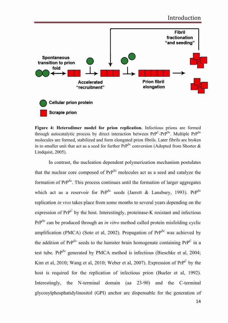
Figure 4: Heterodimer model for prion replication. Infectious prions are formed through autocatalytic process by direct interaction between PrPC-PrPSc. Multiple PrPSc molecules are formed, stabilized and form elongated prion fibrils. Later fibrils are broken in to smaller unit that act as a seed for further PrPSc conversion (Adopted from Shorter & Lindquist, 2005).
In contrast, the nucleation dependent polymerization mechanism postulates
that the nuclear core composed of PrPSc molecules act as a seed and catalyze the
formation of PrPSc. This process continues until the formation of larger aggregates
which act as a reservoir for PrPSc seeds (Jarrett & Lansbury, 1993). PrPSc
replication in vivo takes place from some months to several years depending on the
expression of PrPC by the host. Interestingly, proteinase-K resistant and infectious
PrPSc can be produced through an in vitro method called protein misfolding cyclic
amplification (PMCA) (Soto et al, 2002). Propagation of PrPSc was achieved by
the addition of PrPSc seeds to the hamster brain homogenate containing PrPC in a
test tube. PrPSc generated by PMCA method is infectious (Bieschke et al, 2004;
Kim et al, 2010; Wang et al, 2010; Weber et al, 2007). Expression of PrPC by the
host is required for the replication of infectious prion (Bueler et al, 1992).
Interestingly, the N-terminal domain (aa 23-90) and the C-terminal
glycosylphosphatidylinositol (GPI) anchor are dispensable for the generation of
Introduction
15
infectious prions (Chesebro et al, 2005b; Fischer et al, 1996). Nucleic acids and
lipids have been shown to be involved in conversion and propagation of prions
from bacterially expressed recombinant PrP (Wang et al, 2010). But a different
study revealed that infectious prions can be generated from recombinant PrP
without any cofactors by PMCA technique (Kim et al, 2010).
Prion protein gene
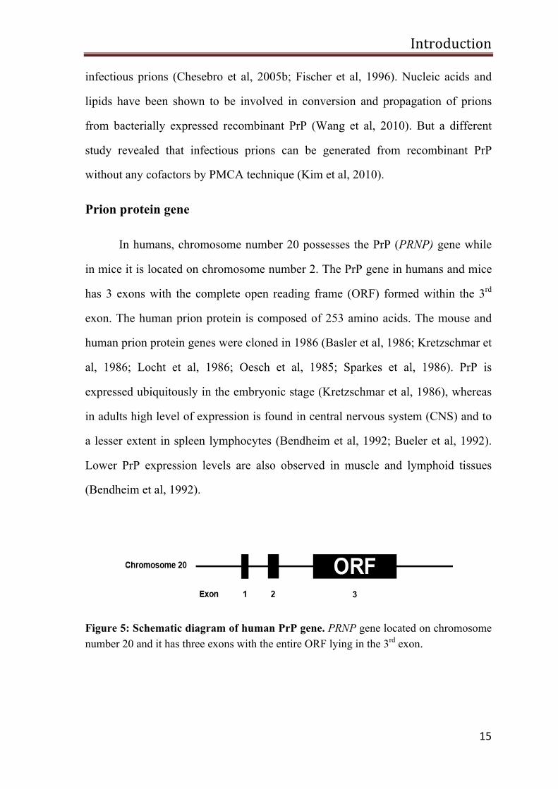
In humans, chromosome number 20 possesses the PrP (PRNP) gene while
in mice it is located on chromosome number 2. The PrP gene in humans and mice
has 3 exons with the complete open reading frame (ORF) formed within the 3rd
exon. The human prion protein is composed of 253 amino acids. The mouse and
human prion protein genes were cloned in 1986 (Basler et al, 1986; Kretzschmar et
al, 1986; Locht et al, 1986; Oesch et al, 1985; Sparkes et al, 1986). PrP is
expressed ubiquitously in the embryonic stage (Kretzschmar et al, 1986), whereas
in adults high level of expression is found in central nervous system (CNS) and to
a lesser extent in spleen lymphocytes (Bendheim et al, 1992; Bueler et al, 1992).
Lower PrP expression levels are also observed in muscle and lymphoid tissues
(Bendheim et al, 1992).
Figure 5: Schematic diagram of human PrP gene. PRNP gene located on chromosome number 20 and it has three exons with the entire ORF lying in the 3rd exon.
Introduction
16
Biogenesis and structure of PrP
PrP biogenesis starts with the translocation of the nascent PrP amino acid
chain into the lumen of the endoplasmic reticulum (ER). In the ER lumen, a series
of posttranslational modifications takes place such as cleaving of the N-terminal
signal sequence (aa 1-23), addition of glycans (aa 181 and 197), addition of GPI
anchor to the C-terminal end after cleaving the GPI signaling sequence (aa 231-
253) and formation of disulfide bond between aa 170 and 214. During trafficking
through the secretory pathway the core glycans are processed into complex
structure. Finally, mature PrP is transported to the outer leaflet of the plasma
membrane. At the cell surface, PrP is present in three forms; unglycosylated,
monoglycosylated and diglycosylated (Prusiner, 1989; Weissmann, 1994).
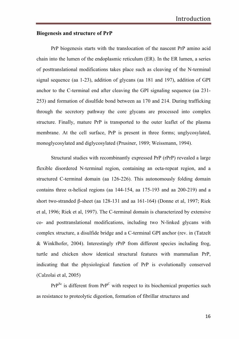
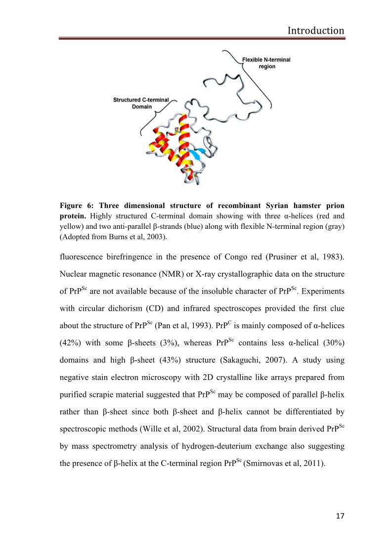
Structural studies with recombinantly expressed PrP (rPrP) revealed a large
flexible disordered N-terminal region, containing an octa-repeat region, and a
structured C-terminal domain (aa 126-226). This autonomously folding domain
contains three α-helical regions (aa 144-154, aa 175-193 and aa 200-219) and a
short two-stranded β-sheet (aa 128-131 and aa 161-164) (Donne et al, 1997; Riek
et al, 1996; Riek et al, 1997). The C-terminal domain is characterized by extensive
co- and posttranslational modifications, including two N-linked glycans with
complex structure, a disulfide bridge and a C-terminal GPI anchor (rev. in (Tatzelt
& Winklhofer, 2004). Interestingly rPrP from different species including frog,
turtle and chicken show identical structural features with mammalian PrP,
indicating that the physiological function of PrP is evolutionally conserved
(Calzolai et al, 2005)
PrPSc is different from PrPC with respect to its biochemical properties such
as resistance to proteolytic digestion, formation of fibrillar structures and
Introduction
17
Figure 6: Three dimensional structure of recombinant Syrian hamster prion protein. Highly structured C-terminal domain showing with three α-helices (red and yellow) and two anti-parallel β-strands (blue) along with flexible N-terminal region (gray) (Adopted from Burns et al, 2003).
fluorescence birefringence in the presence of Congo red (Prusiner et al, 1983).
Nuclear magnetic resonance (NMR) or X-ray crystallographic data on the structure
of PrPSc are not available because of the insoluble character of PrPSc. Experiments
with circular dichorism (CD) and infrared spectroscopes provided the first clue
about the structure of PrPSc (Pan et al, 1993). PrPC is mainly composed of α-helices
(42%) with some β-sheets (3%), whereas PrPSc contains less α-helical (30%)
domains and high β-sheet (43%) structure (Sakaguchi, 2007). A study using
negative stain electron microscopy with 2D crystalline like arrays prepared from
purified scrapie material suggested that PrPSc may be composed of parallel β-helix
rather than β-sheet since both β-sheet and β-helix cannot be differentiated by
spectroscopic methods (Wille et al, 2002). Structural data from brain derived PrPSc
by mass spectrometry analysis of hydrogen-deuterium exchange also suggesting
the presence of β-helix at the C-terminal region PrPSc (Smirnovas et al, 2011).
Introduction
18
Function of prion protein
As mentioned above, PrPC is expressed in all tetrapods and birds and the
structural properties are highly conserved. Therefore, it is plausible to assume that
the physiological function of PrPC is also conserved. During the last three decades,
a number of studies have been carried out to uncover the functional properties of
PrPC. However, the function of PrPC is still enigmatic. Approaches to unmask the
cellular function of PrPC by several groups are discussed in detail below.
Studies from PrP knockout mice
Mice with a targeted disruption in the PrP gene (Prnp) do not show any
distinct phenotype (Bueler et al, 1992). However, PrPC expression is indispensable
for the replication of scrapie prions; mice devoid of PrPC are resistant to prion
diseases and do not propagate infectious prions (Bueler et al, 1993; Bueler et al,
1992; Rambold et al, 2008a). Subsequent studies on PrP knockout mice exhibited
slight phenotypic alterations, such as changes in circadian cycle rhythms (Tobler et
al, 1996), olfaction (Le Pichon et al, 2009), abnormalities in neuronal excitability
(Collinge et al, 1994), altered neurite outgrowth (Santuccione et al, 2005) and
deficiency in proliferation of hematopoietic stem cells and neural precursor cells
(Steele et al, 2006; Zhang et al, 2006). Upon using a stroke model, it became more
clear that PrP knockout mice are highly sensitive to ischemic insult, hypoxia and
seizures (McLennan et al, 2004; Mitteregger et al, 2007; Shyu et al, 2005; Spudich
et al, 2005; Weise et al, 2006). Based on the above results, many functional roles
have been attributed to PrPC such as changes in synaptic transmission and neuronal
excitability, protection against oxidative stress and a role in cell proliferation,
differentiation and adhesion (rev. in (Linden et al, 2008; Nicolas et al, 2009).
Introduction
19
Since PrP null mice do not show any significant phenotype, PrPC does not
seem to be an essential protein, at least under optimal conditions in a laboratory.
However, alternatively compensatory mechanisms might have been activated in
PrPo/o mice that overcame the loss of PrPC. To identify if prion knockout mice
develop any compensating mechanism during embryogenesis to balance the lack
of PrPC, Mallucci et al, generated a PrP conditional knockout mice, in which PrP
expression is turned off during postnatal stage. However, the conditional knockout
mice did not have any phenotype either (Mallucci et al, 2002). Thus, although
PrP0/0 mice have been available for more than 2 decades the definite physiological
function of PrPC is still enigmatic.
Stress-protective functions of PrPC
The first line of evidence indicating a stress-protective function of PrPC
arose from experiments with hippocampal neurons isolated from the PrP knockout
mice (Amitsuka et al, 1999). Later, it was found that PrP knockout mice are
sensitive to ischemic brain damage, kainate induced seizure and to oxidative stress
(Rangel et al, 2007). PrPC knockout mice subjected to an ischemic brain injury
show larger infarct volume with an increased activity of caspase-3 and expression
of PrPC rescues the brain injury from the ischemic insults and improves the
neurological performance (Mitteregger et al, 2007; Spudich et al, 2005). In
addition, up regulation of PrPC mRNA and high immune reactivity of PrPC is
observed during an ischemic condition in humans and rodents (McLennan et al,
2004). In a cell culture model, recently established in our group, expression of
cellular PrPC protects human neuroblastoma (SH-SY5Y) cells from stress-induced
apoptosis (Rambold et al, 2008a). Based on these findings, it appears that PrPC
may be involved in stress-protective and cell survival signaling pathways
(Resenberger et al, 2011).
Introduction
20
PrP protection against PrP∆HD
In an experiment to analyze PrPC regions responsible for the conversion of
PrPSc, it emerged that the deletion of the internal hydrophobic domain (HD) (aa
112-128) resulted in the generation of a neurotoxic mutant PrP, denoted as PrP
∆HD or PrP∆CR (Baumann et al, 2007; Li et al, 2007a; Shmerling et al, 1998).
Interestingly removal of 20 amino acids within the HD is enough to cause
formation of neurotoxic molecule (Li et al, 2007a). Transgenic mice expressing
PrP∆HD develop severe ataxia and neurodegeneration in the cerebellum and die
100 days after birth. Surprisingly, this neurodegenerative phenotype was
completely abolished by the co-expression of a single copy of PrPC (Baumann et
al, 2007; Li et al, 2007a; Shmerling et al, 1998). PrP∆HD-induced apoptotic cell
death and protective function of wtPrP upon co-expression have been
demonstrated also in cultured cells (Rambold et al, 2008b). In a different cell
culture model expression of PrP∆105-125 was shown to induce cation-permeable
channels or membrane pore dependent current which was inhibited by over
expression of PrP or by the addition of glycosaminoglycan (Solomon et al, 2010).
Biochemical properties of PrP∆HD mutants indicate that the neurotoxic function
resulted from the alteration in the normal function of PrP (Ballif et al, 2007;
Christensen & Harris, 2009).
Role of PrP in copper binding and oxidative stress
Several in vitro and in vivo studies indicate that the histidine residues
located within the octa-repeat region of the N-terminus of PrPC are associated with
Cu2+ binding activity (Brown et al, 1997b; Stockel et al, 1998; Viles et al, 1999).
Reduced levels of Cu2+ were observed in the subcellular and synaptosomal
Introduction
21
fractions prepared from brain of PrP knockout mice (Brown et al, 1997a).
However, alterations in brain Cu2+ level in PrP0/0 mice were challenged by
Waggoner et al., (Waggoner et al, 2000). Further studies showed that binding of
Cu2+ to PrP induces the formation of a misfolded PrP conformer distinct from
PrPSc and that it stimulates endocytic trafficking of PrP (Pauly & Harris, 1998;
Perera & Hooper, 2001; Quaglio et al, 2001). Since PrPC is largely localized in the
presynaptic membrane, so PrPC might have an influence on synaptic Cu2+
homeostasis. In conclusion, the findings summarized above could suggest that
PrPC might be involved in modulating the Cu2+ dependent intracellular signaling
cascade directly or indirectly in the presynaptic cleft.
For many years, oxidative stress has been linked to neuronal cell death in
neurodegenerative diseases. An increase in oxidative stress biomarkers was
described in PrP0/0 mice, indicating that PrPC might be involved in the suppression
of oxidative stress (Wong et al, 2001). Decreased super oxide dismutase-1 (SOD-
1) activity was identified in neuronal cells from PrP null mice (Brown et al,
1997b). SOD-1 requires cofactors such as Cu2+ and Zn2+ for its cellular function
and hence the impaired Cu2+ levels in PrP null neurons could be responsible for the
decreased SOD-1 activity that sensitize the cells to increased oxidative stress. PrP
itself possess a SOD-like enzymatic activity that is abolished in mutants lacking
the octa-repeat region, which is involved in Cu2+ binding (Brown et al, 1999).
Neurotoxic signaling through PrPC
Prion propagation and neurotoxicity are the two central events in prion
disease and the expression of PrPC is essential for both. Brandner and colleagues
were the first to show an important role of PrPC as a mediator of PrPSc-induced
neurotoxicity in prion disease. They grafted PrPC over expressing neural tissue into
Introduction
22
the brain of PrP0/0 mice. After the intracerebral inoculation with scrapie prions the
grafted PrPC expressing brain tissue propagated PrPSc and developed clinical
characteristic features of prion disease, but the neighboring tissue devoid of PrPC
stayed healthy although PrPSc spread from graft to the host brain (Brandner et al,
1996). The role of PrPC as a mediator of PrPSc-induced neurotoxicity was further
supported by other transgenic mouse models, scrapie-infected mice expressing
non-neuronal PrPC did not develop clinical symptoms, although they accumulate
PrPSc in addition with astrogliosis (Mallucci et al, 2003). Similarly, transgenic
mice expressing anchorless PrP and infected with PrPSc do not develop clinical
disease though they propagate infectious prions (Chesebro et al, 2005b).
Cell culture experiments from our group support the idea that the
expression of PrPC is required to transmit neurotoxic signals linked to PrPSc.
Furthermore, the intrinsically disordered N-terminal domain and GPI anchor are
required for this activity (Rambold et al, 2008b; Resenberger et al, 2011). The in
vivo and in vitro studies described above support the scenario that PrPSc mediates
its toxic effects through an interaction with PrPC. This PrPC/PrPSc complex could
possibly modulate PrPC dependant signaling pathways (Resenberger et al, 2011).
Putative co-receptors for PrP
PrPC attachment via GPI moiety to the detergent resistant microdomains
(DRMs) of plasma membrane would suggests that PrPC might be involved in
signal transduction, since DMRs are widely recognized as membrane signaling
platforms (Allen et al, 2007; Haigh et al, 2009). Antibody mediated PrP cross-
linking activates Fyn tyrosine kinase and as a consequence phosphorylation of
extracellular signal-regulated kinase 1/2 (ERK1/2) in a caveolin-1 dependent
manner (Mouillet-Richard et al, 2000; Schneider et al, 2003; Toni et al, 2006).
Introduction
23
Establishment of synaptic like structure in cultured primary hippocampal neurons
was observed after the addition of rPrP, however, this effect was blocked by
protein kinase C and SRC kinase inhibitors (Kanaani et al, 2005). Binding of Cu2+
to PrPC activates phosphatidylinositol 3-kinase (PI3K), thereby triggering the
neuroprotective signals (Vassallo et al, 2005). A recent study shows that increased
levels of phosphorylated mitogen-activated protein kinases (MAPKs) are involved
in neuro protection against PrPSc induced toxicity (Uppington & Brown, 2008).
In order to transmit intracellular signals, PrPC would require a co-receptor
since it does not have any direct contact to the cytosol. Several biological
molecules are proposed to interact with PrP and are discussed in detail below. So
far, 37/67 kDa laminin receptor (Gauczynski et al, 2001; Rieger et al, 1997), an
unknown 66 kDa membrane protein (Martins et al, 1997) and the stress-inducible
transmembrane protein 1 (STI1) are proposed as interacting partners of PrPC
(Zanata et al, 2002). It has been shown that amino acid residues 230-245 from
STI1 interact with the hydrophobic region (aa113-128) of PrP through which PrP
transduces the neuroprotective signals (Zanata et al, 2002). A recent study reported
that the recruitment of PrPC-STI1complex at the cell surface induces the
neuroprotection and neuritogenesis, with increased protein synthesis via PI3K-
mTOR signaling and this neuroprotective translational stimulation is abolished in
scrapie infected cells (Roffe et al, 2010).
Using yeast two-hybrid technology, Rieger and colleagues demonstrated
that a 37 kDa laminin receptor precursor (LRP) interacts with PrPC, thereby acting
as a cellular receptor or co-receptor for PrPC (Rieger et al, 1997). The 37 kDa
LRP/67 kDa LR and PrPC are co-localized at the cell surface of neuronal and non-
neuronal cells (Gauczynski et al, 2001). PrPC has two different sites for LRP/LR
binding; the direct binding domain at C-terminal region (aa 144-179) and the
Introduction
24
indirect binding domain at N-terminal region (aa 53-93). Similar to that, LRP/LR
aa regions 161-179 involved in direct and indirect interaction with PrPC via amino
acids 180-285.
Additional PrP interacting proteins
Proteins associated with intracellular vesicles or caveolae-like domains such
as synapsin, growth factor receptor-bound protein 2 (Grb-2), prion interactor 1
(Pint1), p75, caveolin and casein kinase 2 (CK2) were also described to form
complexes with PrPC (Della-Bianca et al, 2001; Meggio et al, 2000; Mouillet-
Richard et al, 2000; Spielhaupter & Schatzl, 2001). Interestingly, PrPC associated
with caveolin or CK2 induces intracellular signaling through Fyn kinase or
phosphotransferase activity of CK2α respectively (Meggio et al, 2000; Mouillet-
Richard et al, 2000). Abnormally folded PrP Q217R, a mutant associated with
GSS, was shown to bind to Bip (heat shock 70 kDa protein 5(HSP70). As a
consequence PrP Q217R was retained in the endoplasmic reticulum. Further, Bip-
PrPQ217R enhanced the proteasomal degradation of abnormally folded mutant
PrP, thus preventing the formation of protein aggregates, suggesting that Bip might
play a significant quality control role in PrP biogenesis (Jin et al, 2000). GFAP and
Bcl2 were also found to interact with PrP (Kurschner & Morgan, 1995; Oesch et
al, 1990). Cytosolic prion protein co-aggregates with Bcl2 and thereby triggers
apoptotic cell death. This toxicity is abolished by the co-expression of cytosolic
heat shock proteins (Rambold et al, 2006).
The function of glutamate receptors was also proposed to be modulated by
PrPC, but evidences are inconsistent. Electrophysiological findings suggest that
PrPC binds to the NR2D subunit of the N-Methyl-D-aspartic acid receptor
(NMDAR) complex and there by suppresses the NMDAR complex activity
Introduction
25
(Khosravani et al, 2008). Recent study shows PrPC-laminin γ1 complexes along
with group I metabotropic glutamate receptors (mGluR1/5) and initiates signaling
cascade for neurite outgrowth (Beraldo et al, 2011). More biochemical evidences
are needed to establish the functional link between NMDAR and PrPC. Apart from
these, PrPC is proposed to be a cellular receptor for amyloid-β (Aβ) oligomers.
Binding of Aβ oligomers with PrPC initiates synaptic dysfunction and altered long-
term potentiation (LTP) in hippocampal neurons (Lauren et al, 2009). This was
challenged by Kessels et al., in a study, where Aβ oligomers induced synaptic
dysfunction, loss of dendritic spines and altered LTP were irrespective of PrPC
expression (Kessels et al, 2010). At the same time, independent studies by
different groups supported the hypothesis that PrPC might be a receptor for Aβ
oligomer (Barry et al, 2011; Caetano et al, 2011; Chung et al, 2010; Freir et al,
2011; Resenberger et al, 2011). Recently, our group identified that PrPC not only
can mediate toxic signals induced by Aβ oligomers, but also can transmit deadly
signals by different β-sheet rich oligomeric conformers (Resenberger et al, 2011).
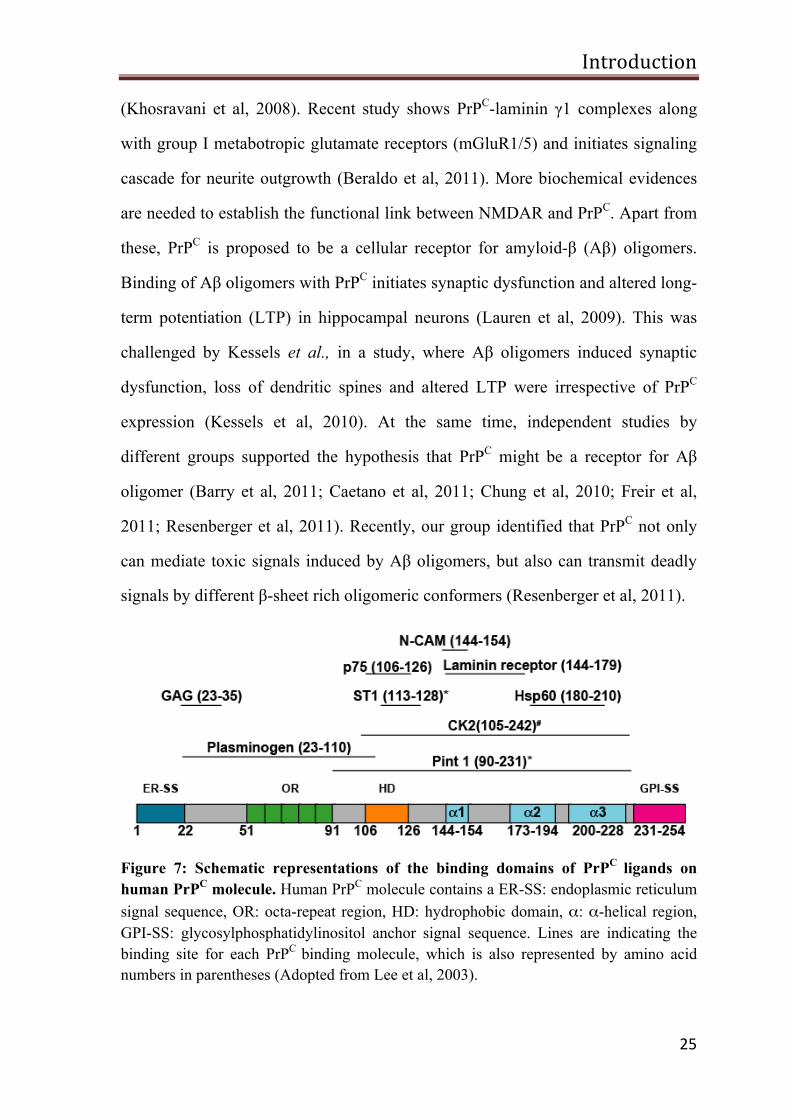
Figure 7: Schematic representations of the binding domains of PrPC ligands on human PrPC molecule. Human PrPC molecule contains a ER-SS: endoplasmic reticulum signal sequence, OR: octa-repeat region, HD: hydrophobic domain, α: α-helical region, GPI-SS: glycosylphosphatidylinositol anchor signal sequence. Lines are indicating the binding site for each PrPC binding molecule, which is also represented by amino acid numbers in parentheses (Adopted from Lee et al, 2003).
Introduction
26
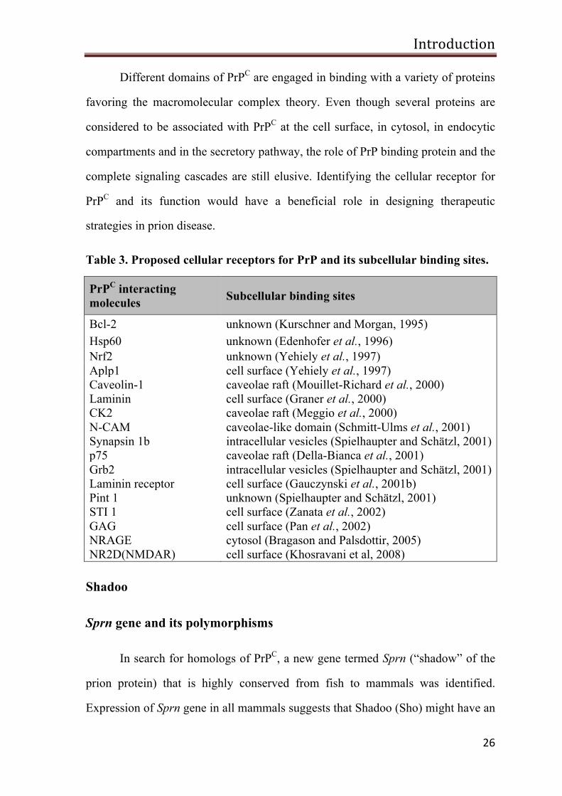
Different domains of PrPC are engaged in binding with a variety of proteins
favoring the macromolecular complex theory. Even though several proteins are
considered to be associated with PrPC at the cell surface, in cytosol, in endocytic
compartments and in the secretory pathway, the role of PrP binding protein and the
complete signaling cascades are still elusive. Identifying the cellular receptor for
PrPC and its function would have a beneficial role in designing therapeutic
strategies in prion disease.
Table 3. Proposed cellular receptors for PrP and its subcellular binding sites.
PrPC interacting molecules Subcellular binding sites
Bcl-2 unknown (Kurschner and Morgan, 1995) Hsp60 unknown (Edenhofer et al., 1996) Nrf2 unknown (Yehiely et al., 1997) Aplp1 cell surface (Yehiely et al., 1997) Caveolin-1 caveolae raft (Mouillet-Richard et al., 2000) Laminin cell surface (Graner et al., 2000) CK2 caveolae raft (Meggio et al., 2000) N-CAM caveolae-like domain (Schmitt-Ulms et al., 2001) Synapsin 1b intracellular vesicles (Spielhaupter and Schätzl, 2001) p75 caveolae raft (Della-Bianca et al., 2001) Grb2 intracellular vesicles (Spielhaupter and Schätzl, 2001) Laminin receptor cell surface (Gauczynski et al., 2001b) Pint 1 unknown (Spielhaupter and Schätzl, 2001) STI 1 cell surface (Zanata et al., 2002) GAG cell surface (Pan et al., 2002) NRAGE cytosol (Bragason and Palsdottir, 2005) NR2D(NMDAR) cell surface (Khosravani et al, 2008)
Shadoo
Sprn gene and its polymorphisms
In search for homologs of PrPC, a new gene termed Sprn (“shadow” of the
prion protein) that is highly conserved from fish to mammals was identified.
Expression of Sprn gene in all mammals suggests that Shadoo (Sho) might have an
Introduction
27
important physiological function. A SPRN pseudogene was described in humans
and primates and may have arisen due to the segmental duplication (Harrison et al,
2010; Premzl et al, 2004). A human SPRN pseudogene has an overlap with the
non-coding exon of SYCE1 gene that is involved in meiosis in mammals (Harrison
et al, 2010). Chromosomal rearrangements in fish have produced multiple paralogs
of Sprn gene and at least 2 Sprn gene copies are present in fish genome (Harrison
et al, 2010; Premzl et al, 2004; Premzl et al, 2003; Strumbo et al, 2001; Strumbo et
al, 2006).
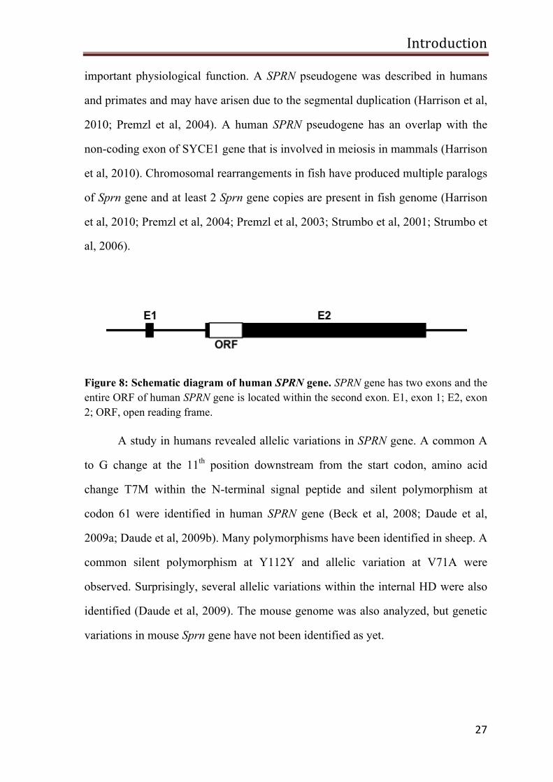
Figure 8: Schematic diagram of human SPRN gene. SPRN gene has two exons and the entire ORF of human SPRN gene is located within the second exon. E1, exon 1; E2, exon 2; ORF, open reading frame.
A study in humans revealed allelic variations in SPRN gene. A common A
to G change at the 11th position downstream from the start codon, amino acid
change T7M within the N-terminal signal peptide and silent polymorphism at
codon 61 were identified in human SPRN gene (Beck et al, 2008; Daude et al,
2009a; Daude et al, 2009b). Many polymorphisms have been identified in sheep. A
common silent polymorphism at Y112Y and allelic variation at V71A were
observed. Surprisingly, several allelic variations within the internal HD were also
identified (Daude et al, 2009). The mouse genome was also analyzed, but genetic
variations in mouse Sprn gene have not been identified as yet.
Introduction
28
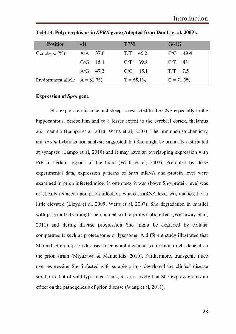
Table 4. Polymorphisms in SPRN gene (Adopted from Daude et al, 2009).
Position -11 T7M G61G
Genotype (%) A/A 37.6 T/T 45.2 C/C 49.4
G/G 15.1 C/T 39.8 C/T 43
A/G 47.3 C/C 15.1 T/T 7.5
Predominant allele A = 61.7% T = 65.1% C = 71.0%
Expression of Sprn gene
Sho expression in mice and sheep is restricted to the CNS especially to the
hippocampus, cerebellum and to a lesser extent to the cerebral cortex, thalamus
and medulla (Lampo et al, 2010; Watts et al, 2007). The immunohistochemistry
and in situ hybridization analysis suggested that Sho might be primarily distributed
at synapses (Lampo et al, 2010) and it may have an overlapping expression with
PrP in certain regions of the brain (Watts et al, 2007). Prompted by these
experimental data, expression patterns of Sprn mRNA and protein level were
examined in prion infected mice. In one study it was shown Sho protein level was
drastically reduced upon prion infection, whereas mRNA level was unaltered or a
little elevated (Lloyd et al, 2009; Watts et al, 2007). Sho degradation in parallel
with prion infection might be coupled with a proteostatic effect (Westaway et al,
2011) and during disease progression Sho might be degraded by cellular
compartments such as proteaosome or lysosome. A different study illustrated that
Sho reduction in prion diseased mice is not a general feature and might depend on
the prion strain (Miyazawa & Manuelidis, 2010). Furthermore, transgenic mice
over expressing Sho infected with scrapie prions developed the clinical disease
similar to that of wild type mice. Thus, it is not likely that Sho expression has an
effect on the pathogenesis of prion disease (Wang et al, 2011).
Introduction
29
Structure of Sho
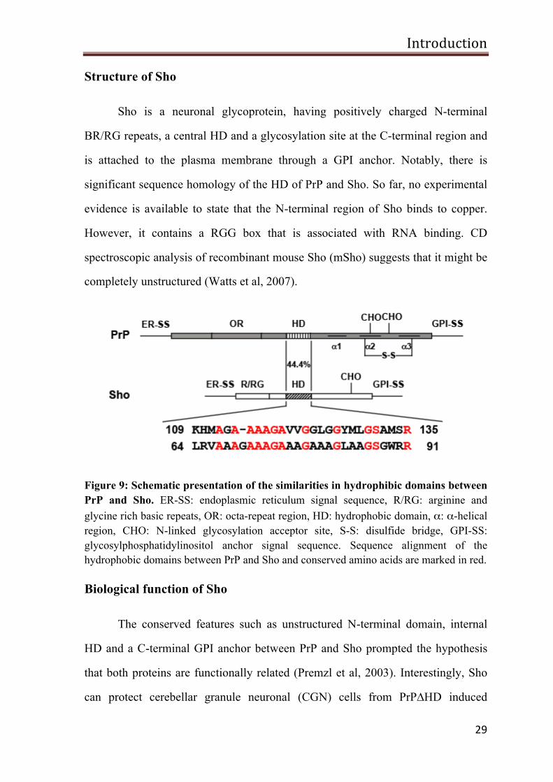
Sho is a neuronal glycoprotein, having positively charged N-terminal
BR/RG repeats, a central HD and a glycosylation site at the C-terminal region and
is attached to the plasma membrane through a GPI anchor. Notably, there is
significant sequence homology of the HD of PrP and Sho. So far, no experimental
evidence is available to state that the N-terminal region of Sho binds to copper.
However, it contains a RGG box that is associated with RNA binding. CD
spectroscopic analysis of recombinant mouse Sho (mSho) suggests that it might be
completely unstructured (Watts et al, 2007).
Figure 9: Schematic presentation of the similarities in hydrophibic domains between PrP and Sho. ER-SS: endoplasmic reticulum signal sequence, R/RG: arginine and glycine rich basic repeats, OR: octa-repeat region, HD: hydrophobic domain, α: α-helical region, CHO: N-linked glycosylation acceptor site, S-S: disulfide bridge, GPI-SS: glycosylphosphatidylinositol anchor signal sequence. Sequence alignment of the hydrophobic domains between PrP and Sho and conserved amino acids are marked in red.
Biological function of Sho
The conserved features such as unstructured N-terminal domain, internal
HD and a C-terminal GPI anchor between PrP and Sho prompted the hypothesis
that both proteins are functionally related (Premzl et al, 2003). Interestingly, Sho
can protect cerebellar granule neuronal (CGN) cells from PrP∆HD induced
Introduction
30
neurotoxicity (Watts et al, 2007). Blocking the expression of Sho in a PrP null
background leads to a lethal phenotype in mouse embryos suggesting that Sho
might be required for early embryogenesis (Young et al, 2009). SPRN null allele
and single nucleotide polymorphisms (SNPs) were identified in CJD patients in the
UK, supporting a possible role of SPRN genetic variants in prion diseases (Beck et
al, 2008). The conserved RGG boxes in Sho might be associated with binding of
RNA molecules (Corley & Gready, 2008). RGG box containing proteins are
shown to be involved in RNA processing and in some proteins RGG boxes
mediate the interaction with its binding partners (Lukasiewicz et al, 2007). Similar
to PrP, localization of GPI-anchored Sho in lipid rafts might indicate a role in
neural cell signaling.
Aim of the study
31
Aim of the study
As described above, PrPC is ubiquitously expressed and structural
similarities in PrP between different species suggest that its function might be
evolutionarily conserved (Calzolai et al, 2005; Wopfner et al, 1999). Despite
numerous studies, the physiological function of PrP is largely unknown. However,
different studies in transgenic animals and cultured cells are now supporting the
idea that PrPC can protect neuronal cells against stress-induced cell death (rev in
(Westergard et al, 2007). From one class of PrP mutants (PrP∆HD) it emerged that
PrPC can acquire a neurotoxic potential by deleting the internal HD (Baumann et
al, 2007; Li et al, 2007b; Shmerling et al, 1998). Interestingly, expression of PrPC
can completely prevent the neurotoxic activity of PrPΔHD suggesting that PrPC
and PrPΔHD can induce neurotrophic or neurotoxic signaling via similar signaling
pathway (Li et al, 2007b; Rambold et al, 2008b).
A genomic analysis indicated the presence of a PrP-related gene (SPRN) that
encodes Sho (Premzl et al, 2003). Sho is expressed in the CNS. The sequence
homology between Sho and PrP is found within the internal HD, however, certain
features such as, a N-terminal repeat region and a C-terminal GPI anchor are also
conserved and provoked the hypothesis that Sho and PrP are functionally related
(Premzl et al, 2003). Moreover similarly to PrP, Sho can rescue neurons from
PrP∆HD-induced neurotoxicity (Watts et al, 2007). From the above studies, it is
reasonable to assume that PrP and Sho might transmit their neuroprotective signals
by activating similar intracellular signaling cascade.
Aim of the study
32
Hence, the aim of the present study was:
• To analyze the biogenesis of human Sho in SH-SY5Y cells, in
particular; ER import, glycosylation patterns, maturation, dimerization
and cellular localization.
• To provide insight into the stress-protective activity of Sho. In
particular, we aimed to identify domains of Sho that are required for its
stress-protective activity. To explore the stress-protective activity of Sho
two different stress paradigms are employed in this study, which
includes exposition of SH-SY5Y cells to the excitotoxin glutamate and
the expression of neurotoxic PrP mutant PrP∆HD.
• To test for the possibility of a conserved function of the N-termini of
Sho and PrP.
• To elucidate the role of Sho in PrPSc-induced apoptosis.
Results
33
Results
Sho is highly conserved from fish to mammals and it was predicted to be
glycosylated and anchored onto the plasma membrane via a GPI moiety (Premzl et
al, 2003). Although Sho has no overall sequence homology with mammalian PrP,
some characteristic features are conserved, such as the internal HD, N-linked
glycosylation and a GPI anchor at the C-terminal. Similar to mammalian PrP,
zebra fish Sho (zeSho) and mSho were found to be complex glycosylated and
targeted to the plasma membrane via the GPI anchor (Miesbauer et al, 2006; Watts
et al, 2007). As an initial step, this study aimed to analyze wild type human Sho
and the impact of different domains on maturation, trafficking and stress-
protective activity.
Generation of antibodies against human Sho
Sho antibodies are not commercially available. Production of antibodies
against Sho will be useful to detect endogenous Sho level in cells and tissues and
for further functional characterization of Sho in prion diseases. For the generation
of the antibodies, the human Sho gene was cloned into the pET-19b vector using
the restricting enzymes NdeI and XbaI. Further, Sho gene was transformed and
expressed in E.coli-BL-21 strain. Expression levels of recombinant Sho (rSho) in
E.coli were high, but the protein was exclusively in the insoluble fraction. For
immunization, inclusion bodies were purified and solubilized in guanidine
hydrochloride (GndHCl). With this solution two rabbits were immunized
(Eurogentec, Belgium). After 90 days, serum samples were collected from the
immunized rabbits.
Results
34
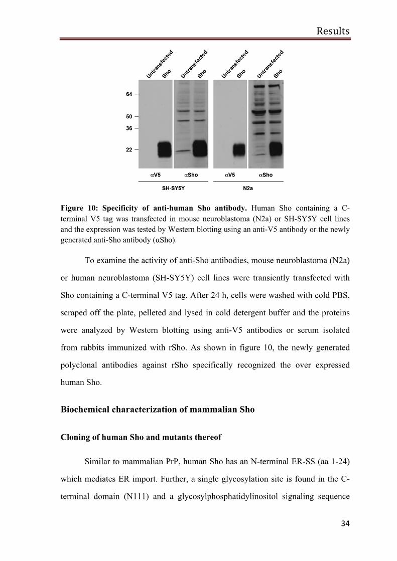
Figure 10: Specificity of anti-human Sho antibody. Human Sho containing a C-terminal V5 tag was transfected in mouse neuroblastoma (N2a) or SH-SY5Y cell lines and the expression was tested by Western blotting using an anti-V5 antibody or the newly generated anti-Sho antibody (αSho).
To examine the activity of anti-Sho antibodies, mouse neuroblastoma (N2a)
or human neuroblastoma (SH-SY5Y) cell lines were transiently transfected with
Sho containing a C-terminal V5 tag. After 24 h, cells were washed with cold PBS,
scraped off the plate, pelleted and lysed in cold detergent buffer and the proteins
were analyzed by Western blotting using anti-V5 antibodies or serum isolated
from rabbits immunized with rSho. As shown in figure 10, the newly generated
polyclonal antibodies against rSho specifically recognized the over expressed
human Sho.
Biochemical characterization of mammalian Sho
Cloning of human Sho and mutants thereof
Similar to mammalian PrP, human Sho has an N-terminal ER-SS (aa 1-24)
which mediates ER import. Further, a single glycosylation site is found in the C-
terminal domain (N111) and a glycosylphosphatidylinositol signaling sequence
Results
35
(GPI-SS) at the C-terminus (aa 127-151) (Premzl et al, 2003). zeSho biogenesis
was previously analyzed in our group. It starts with the translocation of nascent
Sho polypeptide into the ER lumen, where it undergoes a series of
posttranslational modifications such as glycosylation and GPI anchor attachment at
the C-terminal (Miesbauer et al, 2006). Thereafter, the protein is complex
glycosylated and transported to the outer surface of the plasma membrane
(Miesbauer et al, 2006).
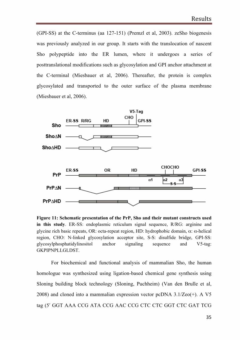
Figure 11: Schematic presentation of the PrP, Sho and their mutant constructs used in this study. ER-SS: endoplasmic reticulum signal sequence, R/RG: arginine and glycine rich basic repeats, OR: octa-repeat region, HD: hydrophobic domain, α: α-helical region, CHO: N-linked glycosylation acceptor site, S-S: disulfide bridge, GPI-SS: glycosylphosphatidylinositol anchor signaling sequence and V5-tag: GKPIPNPLLGLDST.
For biochemical and functional analysis of mammalian Sho, the human
homologue was synthesized using ligation-based chemical gene synthesis using
Sloning building block technology (Sloning, Puchheim) (Van den Brulle et al,
2008) and cloned into a mammalian expression vector pcDNA 3.1/Zeo(+). A V5
tag (5ʹ′ GGT AAA CCG ATA CCG AAC CCG CTC CTC GGT CTC GAT TCG
Results
36
ACG 3ʹ′) or HA tag (5ʹ′ TAC CCA TAC GAT GTT CCA GAT TAC GCT 3ʹ′) was
introduced between amino acids 124 and 125. Sho was used as a template to
generate the subsequent deletions and mutants by standard polymerase chain
reaction (PCR) method: Sho∆N (aa 30-56 deleted) and Sho∆HD (aa 68-89
deleted). The design of the Sho mutants was based on PrP mutants that have been
characterized previously.
Wild type Sho and Sho mutants are complex glycosylated
Protein glycosylation maintains the folding, physiological structural and
cellular localization, thereby enhancing the protein-protein interaction, solubility
and increases the resistance against proteolysis (Shental-Bechor & Levy, 2008;
Winklhofer et al, 2003a; Zhou et al, 2005). PrP mutant devoid of unstructured N-
terminal domain (PrP∆N) shows altered neuroprotective activity but still could
endorse propagation of infectious prions (Fischer et al, 1996; Mitteregger et al,
2007; Rambold et al, 2008b). Removal of intrinsic HD (aa 113-133 deletion) from
PrP showed a gain of neurotoxic function which can be repressed by the
expression of a single copy of PrPC (Baumann et al, 2007; Li et al, 2007a;
Rambold et al, 2008b; Shmerling et al, 1998). Importantly, these PrP mutants are
complex glycosylated and are targeted to the outer leaflet of the plasma membrane
through their GPI anchor (Winklhofer et al, 2003b).
As previously mentioned, earlier experiments showed that zeSho expressed
in mammalian cells is complex glycosylated and anchored via a GPI moiety to the
plasma membrane (Miesbauer et al, 2006). To examine the co and post-
translational modifications of human Sho and the respective mutants indicated in
Results
37
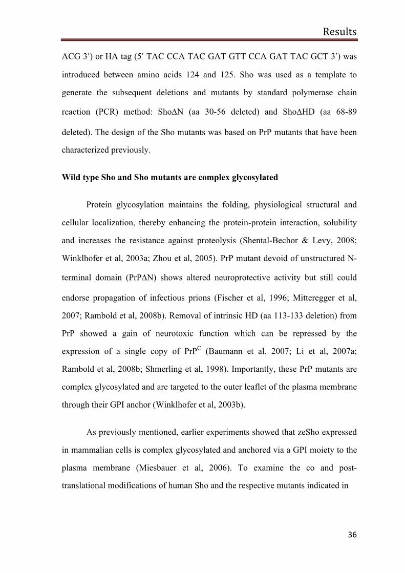
Figure 12: Deletion of the N-terminal or the hydrophobic domain does not interfere with biogenesis of Sho. Sho and its mutants are complex glycosylated. SH-SY5Y cells were transiently transfected with the constructs indicated in figure 10. Total cell lysates were treated with Endo H (A) or PNGase F (B) (+) or left untreated (-) and PrP or Sho proteins were detected by Western blotting.
figure 11 the constructs were expressed in SH-SY5Y cells. To monitor N-linked
glycosylation, cell lysates were treated with Endoglycosidase H (Endo H), an
enzyme that cleaves only high mannose structure or Peptide: N-glycosidase F
(PNGase F), which can remove all N-linked glycans. An increase in the
electrophoretic mobility of the proteins after PNGase F digestion (Figure 12B)
indicated that all constructs are modified with N-linked glycans. Endo H treatment
did not show any difference in the electrophoretic mobility of the proteins (Figure
12A) indicating that all the constructs are modified with N-linked glycans of
complex structure.
Results
38
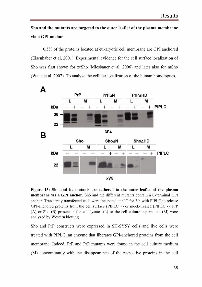
Sho and the mutants are targeted to the outer leaflet of the plasma membrane
via a GPI anchor
0.5% of the proteins located at eukaryotic cell membrane are GPI anchored
(Eisenhaber et al, 2001). Experimental evidence for the cell surface localization of
Sho was first shown for zeSho (Miesbauer et al, 2006) and later also for mSho
(Watts et al, 2007). To analyze the cellular localization of the human homologues,
Figure 13: Sho and its mutants are tethered to the outer leaflet of the plasma membrane via a GPI anchor. Sho and the different mutants contain a C-terminal GPI anchor. Transiently transfected cells were incubated at 4°C for 3 h with PIPLC to release GPI-anchored proteins from the cell surface (PIPLC +) or mock-treated (PIPLC -). PrP (A) or Sho (B) present in the cell lysates (L) or the cell culture supernatant (M) were analyzed by Western blotting.
Sho and PrP constructs were expressed in SH-SY5Y cells and live cells were
treated with PIPLC, an enzyme that liberates GPI-anchored proteins from the cell
membrane. Indeed, PrP and PrP mutants were found in the cell culture medium
(M) concomitantly with the disappearance of the respective proteins in the cell
Results
39
lysates (L) (Figure 13A). Similarly, levels of Sho and the different mutants were
diminished in the cell lysates upon treatment with PIPLC. In parallel, Sho and
Sho∆N levels were increased in the cell culture medium (M) of PIPLC treated
cells; however, significant amounts of Sho∆HD were not detected in the
supernatant of PIPLC treated cells, using neither anti-V5 antibody nor newly
generated rabbit polyclonal anti-Sho antisera (αSho). However, the levels of
Sho∆HD were significantly decreased in PIPLC treated cells lysates (L) (Figure
13B). So far, we have not been able to identify the molecular mechanism
responsible for this peculiar phenomenon.
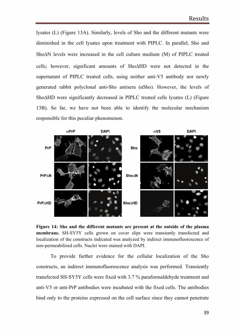
Figure 14: Sho and the different mutants are present at the outside of the plasma membrane. SH-SY5Y cells grown on cover slips were transiently transfected and localization of the constructs indicated was analyzed by indirect immunofluorescence of non-permeabilized cells. Nuclei were stained with DAPI.
To provide further evidence for the cellular localization of the Sho
constructs, an indirect immunofluorescence analysis was performed. Transiently
transfected SH-SY5Y cells were fixed with 3.7 % paraformaldehyde treatment and
anti-V5 or anti-PrP antibodies were incubated with the fixed cells. The antibodies
bind only to the proteins expressed on the cell surface since they cannot penetrate
Results
40
the cell membrane. All constructs including ShoΔHD were detected on the outer
surface of the plasma membrane (Figure 14).
Collectively, these findings revealed that full length Sho, as well as its
mutants, lacking N-terminal or internal HD are complex glycosylated and attached
to the outer leaflet of the cell membrane through a GPI anchor.
Sho attenuates glutamate induced excitotoxic stress
Excessive stimulation of neuronal cells by neurotransmitters such as
glutamate can damage the neuronal cells through a pathological process called
excitotoxicity. As previously mentioned, altered LTP and increased neuronal
excitability have been observed in PrP knockout mice (Collinge et al, 1994; Curtis
et al, 2003; Maglio et al, 2004; Mallucci et al, 2002). A recent study suggests that
PrP knockout mice exhibit enhanced NMDAR dependent neuronal excitability
(Khosravani et al, 2008). These results would indicate that PrPC might be involved
in attenuating the neuronal excitability by regulating the glutamate receptor’s
activity. This is the rationale behind the use of glutamate as a physiological stress
agent to analyze the role of PrP and Sho in stress-induced toxicity.
SH-SY5Y cells transiently transfected with the constructs indicated in
figure 11, were grown on cover slips. The cells were treated with 500 µM
glutamate for 3 h, followed by paraformaldehyde fixation. Apoptotic cells were
identified by indirect immunofluorescence assay using an antibody against
activated caspase-3. In this context, it is important to note that SH-SY5Y cells are
characterized by low levels of endogenous PrPC (Figure 15, right panel, pcDNA,
3F4). Consistent with previous results, PrPC was able to protect cells against
excitotoxic cell death whereas the deletion of the intrinsically disordered
Results
41
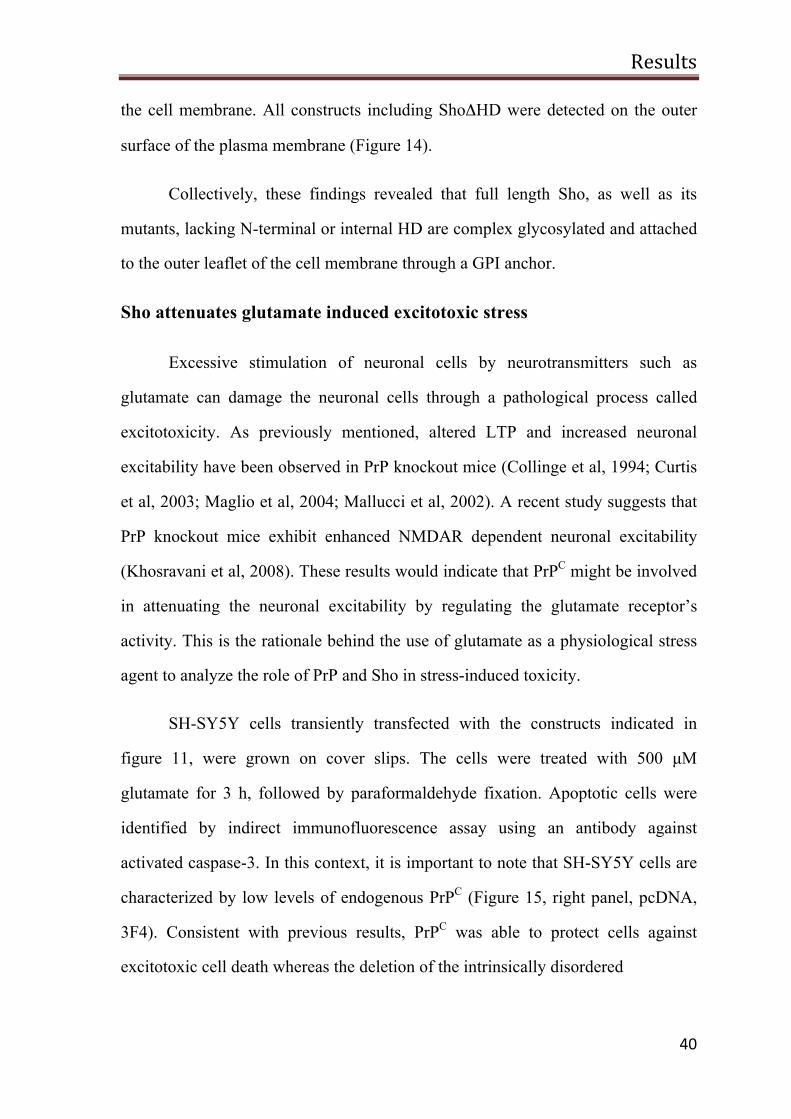
Figure 15: Sho protects against glutamate stress-induced apoptosis. SH-SY5Y cells expressing the constructs indicated were stressed with glutamate (500 µM) for 3 h at 37°C, fixed, permeabilized and activation of caspase-3 was analyzed by indirect immunofluorescence. To detect cells undergoing apoptosis, the number of activated caspase-3-positive cells out of at least 1100 transfected cells was determined in at least three independent experiments. Percentage of apoptotic cells among transfected cells is shown. Expression levels were analyzed by immunoblotting (right panel). *P<0.05, **P<0.005, ***<0.0005.
N-terminal domain (PrP∆N) leads to loss of protective activity (Figure 15)
(Rambold et al, 2008b).
Similarly, expression of Sho had a stress-protective ability, which was
abolished by the deletion of the N-terminal domain (Figure 15). These results
indicate that the deletion of the N-terminal domain from Sho and PrP has similar
outcome, i.e, the loss of a stress-protective activity. However, deletion of the
hydrophobic region (HD) had different consequences. Earlier experiments from
cell culture and transgenic mice expressing PrP∆HD showed that PrP∆HD
obtained a toxic activity (Baumann et al, 2007; Li et al, 2007a; Rambold et al,
2008b; Shmerling et al, 1998). As illustrated in figure 15, PrP∆HD expression was
toxic to SH-SY5Y cells and also it does not interfere with glutamate induced
Results
42
excitotoxicity. Surprisingly, Sho∆HD expression did not induce apoptotic cell
death in SH-SY5Y cells, but it was also devoid of a stress-protective activity to
interfere with glutamate-induced cell death.
Sho protect cells against PrP∆HD-induced toxicity
Transgenic mice expressing PrP∆105-125 in a PrP null background exhibit
neurodegenerative phenotype such as cerebellar atrophy, tremor, granule neuronal
loss and astrogliosis. This phenotype is eliminated upon expression of full length
PrP (Li et al, 2007a). Moreover, mSho has been shown to protect neurons from
PrP∆HD induced neurotoxicity as well (Watts et al, 2007). Therefore, we have
decided to use the expression of PrP∆HD as a second model for neurotoxic insult
in order to identify Sho domains required for its activity to protect neurons against
PrP∆HD induced toxicity. Sho and its mutants were transiently co-transfected with
PrP∆HD in SH-SY5Y cells. As a control, mock transfection of pcDNA or
expression of GFP-GPI constructs was used. After 24 h, transfected cells were
fixed with 3.7% paraformaldehyde solution and stained with anti-active caspase-3
antibody in order to identify apoptotic cells. PrP mutants corresponding to Sho
constructs were analyzed in parallel.
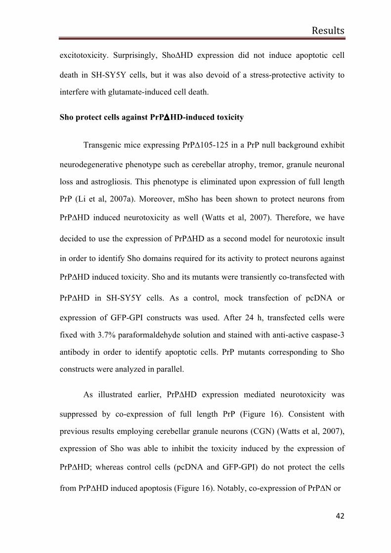
As illustrated earlier, PrP∆HD expression mediated neurotoxicity was
suppressed by co-expression of full length PrP (Figure 16). Consistent with
previous results employing cerebellar granule neurons (CGN) (Watts et al, 2007),
expression of Sho was able to inhibit the toxicity induced by the expression of
PrP∆HD; whereas control cells (pcDNA and GFP-GPI) do not protect the cells
from PrP∆HD induced apoptosis (Figure 16). Notably, co-expression of PrP∆N or
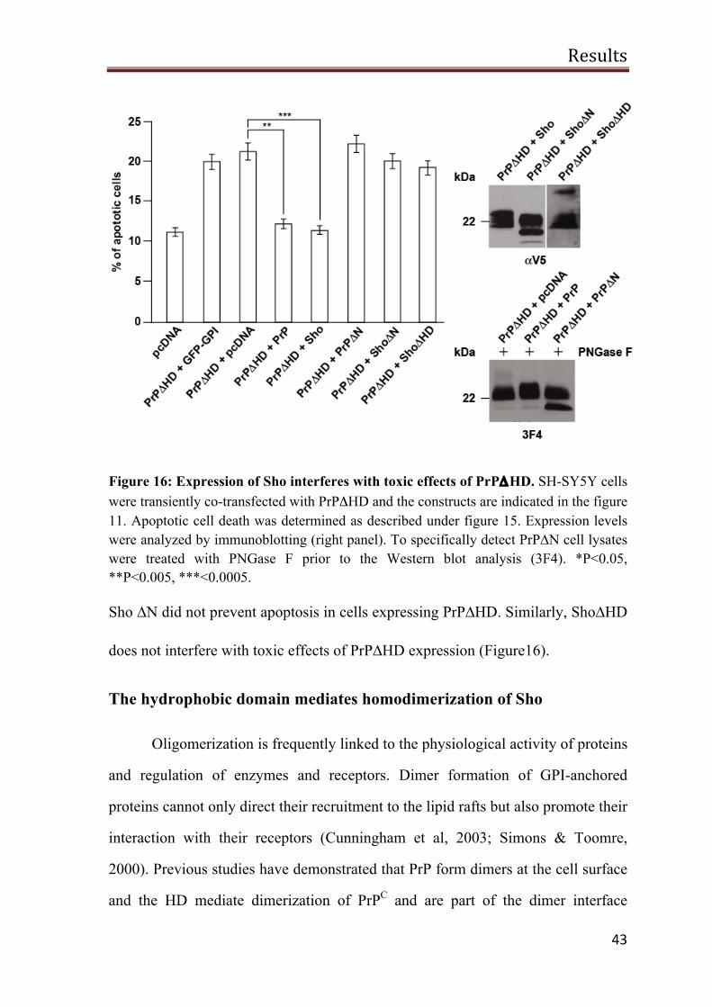
Results
43
Figure 16: Expression of Sho interferes with toxic effects of PrPΔHD. SH-SY5Y cells were transiently co-transfected with PrPΔHD and the constructs are indicated in the figure 11. Apoptotic cell death was determined as described under figure 15. Expression levels were analyzed by immunoblotting (right panel). To specifically detect PrP∆N cell lysates were treated with PNGase F prior to the Western blot analysis (3F4). *P<0.05, **P<0.005, ***<0.0005.
Sho ∆N did not prevent apoptosis in cells expressing PrP∆HD. Similarly, Sho∆HD
does not interfere with toxic effects of PrP∆HD expression (Figure16).
The hydrophobic domain mediates homodimerization of Sho
Oligomerization is frequently linked to the physiological activity of proteins
and regulation of enzymes and receptors. Dimer formation of GPI-anchored
proteins cannot only direct their recruitment to the lipid rafts but also promote their
interaction with their receptors (Cunningham et al, 2003; Simons & Toomre,
2000). Previous studies have demonstrated that PrP form dimers at the cell surface
and the HD mediate dimerization of PrPC and are part of the dimer interface
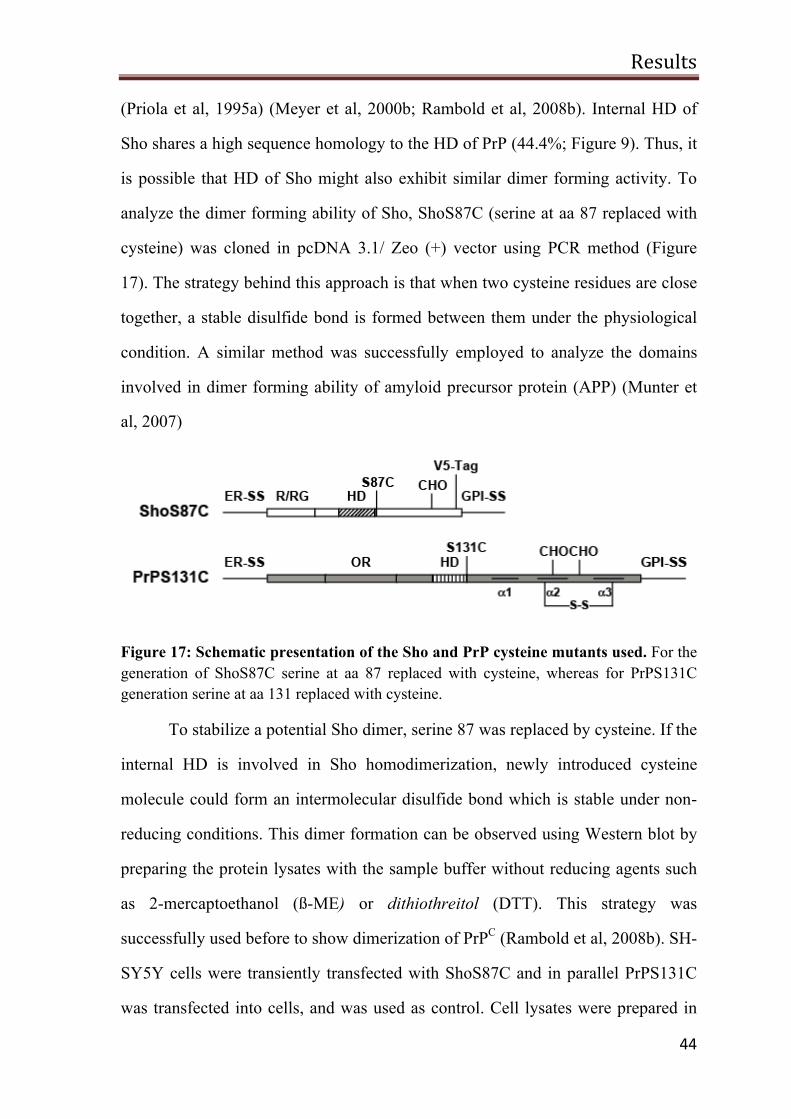
Results
44
(Priola et al, 1995a) (Meyer et al, 2000b; Rambold et al, 2008b). Internal HD of
Sho shares a high sequence homology to the HD of PrP (44.4%; Figure 9). Thus, it
is possible that HD of Sho might also exhibit similar dimer forming activity. To
analyze the dimer forming ability of Sho, ShoS87C (serine at aa 87 replaced with
cysteine) was cloned in pcDNA 3.1/ Zeo (+) vector using PCR method (Figure
17). The strategy behind this approach is that when two cysteine residues are close
together, a stable disulfide bond is formed between them under the physiological
condition. A similar method was successfully employed to analyze the domains
involved in dimer forming ability of amyloid precursor protein (APP) (Munter et
al, 2007)
Figure 17: Schematic presentation of the Sho and PrP cysteine mutants used. For the generation of ShoS87C serine at aa 87 replaced with cysteine, whereas for PrPS131C generation serine at aa 131 replaced with cysteine.
To stabilize a potential Sho dimer, serine 87 was replaced by cysteine. If the
internal HD is involved in Sho homodimerization, newly introduced cysteine
molecule could form an intermolecular disulfide bond which is stable under non-
reducing conditions. This dimer formation can be observed using Western blot by
preparing the protein lysates with the sample buffer without reducing agents such
as 2-mercaptoethanol (ß-ME) or dithiothreitol (DTT). This strategy was
successfully used before to show dimerization of PrPC (Rambold et al, 2008b). SH-
SY5Y cells were transiently transfected with ShoS87C and in parallel PrPS131C
was transfected into cells, and was used as control. Cell lysates were prepared in
Results
45
the presence of Laemmli sample buffer with or without ß-ME or DTT and proteins
were analyzed by Western blotting. Corroborating the earlier results, PrP dimers
were found in the cell lysates prepared from PrPS131C under non-reducing
Figure 18: The hydrophobic domain is part of the dimer interface. SH-SY5Y cells were transiently transfected with PrPS131C or ShoS87C and cell lysates were analyzed under reducing (+ βME) or non-reducing (- βME) conditions. Proteins were detected by immunoblotting.
conditions (Figure 18). Similarly, higher molecular weight species appeared with
the molecular mass similar to Sho dimer indicating the formation of Sho
homodimer (Figure 18). In the presence of reducing agents the migration pattern of
ShoS87C was identical to that of wild type Sho, indicating that the introduced
cysteine residues induced the formation of intermolecular disulfide bond.
Furthermore, the possible involvement of the N-terminal domain on homo
dimer formation of Sho was studied. To this end amino acid residues 30-56 were
deleted from the ShoS87C (ShoΔN,S87C) mutant (Figure 19A). SH-SY5Y cells
were transiently transfected with ShoΔN,S87C and characterized by Western
blotting. As shown in figure 19B, ShoΔN,S87C formed dimers similarly to
ShoS87C.
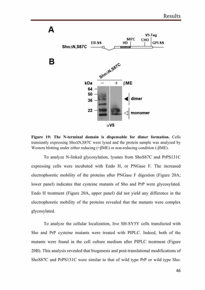
Results
46
Figure 19: The N-terminal domain is dispensable for dimer formation. Cells transiently expressing Sho∆N,S87C were lysed and the protein sample was analyzed by Western blotting under either reducing (+βME) or non-reducing condition (-βME).
To analyze N-linked glycosylation, lysates from ShoS87C and PrPS131C
expressing cells were incubated with Endo H, or PNGase F. The increased
electrophoretic mobility of the proteins after PNGase F digestion (Figure 20A;
lower panel) indicates that cysteine mutants of Sho and PrP were glycosylated.
Endo H treatment (Figure 20A, upper panel) did not yield any difference in the
electrophoretic mobility of the proteins revealed that the mutants were complex
glycosylated.
To analyze the cellular localization, live SH-SY5Y cells transfected with
Sho and PrP cysteine mutants were treated with PIPLC. Indeed, both of the
mutants were found in the cell culture medium after PIPLC treatment (Figure
20B). This analysis revealed that biogenesis and post-translational modifications of
ShoS87C and PrPS131C were similar to that of wild type PrP or wild type Sho:
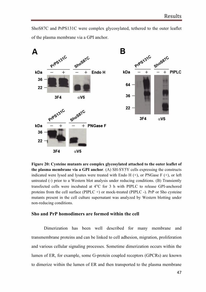
Results
47
ShoS87C and PrPS131C were complex glycosylated, tethered to the outer leaflet
of the plasma membrane via a GPI anchor.
Figure 20: Cysteine mutants are complex glycosylated attached to the outer leaflet of the plasma membrane via a GPI anchor. (A) SH-SY5Y cells expressing the constructs indicated were lysed and lysates were treated with Endo H (+), or PNGase F (+), or left untreated (-) prior to a Western blot analysis under reducing conditions. (B) Transiently transfected cells were incubated at 4°C for 3 h with PIPLC to release GPI-anchored proteins from the cell surface (PIPLC +) or mock-treated (PIPLC -). PrP or Sho cysteine mutants present in the cell culture supernatant was analyzed by Western blotting under non-reducing conditions.
Sho and PrP homodimers are formed within the cell
Dimerization has been well described for many membrane and
transmembrane proteins and can be linked to cell adhesion, migration, proliferation
and various cellular signaling processes. Sometime dimerization occurs within the
lumen of ER, for example, some G-protein coupled receptors (GPCRs) are known
to dimerize within the lumen of ER and then transported to the plasma membrane
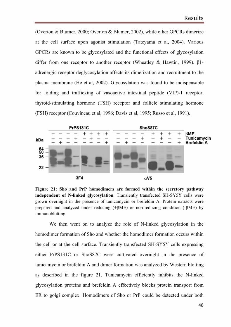
Results
48
(Overton & Blumer, 2000; Overton & Blumer, 2002), while other GPCRs dimerize
at the cell surface upon agonist stimulation (Tateyama et al, 2004). Various
GPCRs are known to be glycosylated and the functional effects of glycosylation
differ from one receptor to another receptor (Wheatley & Hawtin, 1999). β1-
adrenergic receptor deglycosylation affects its dimerization and recruitment to the
plasma membrane (He et al, 2002). Glycosylation was found to be indispensable
for folding and trafficking of vasoactive intestinal peptide (VIP)-1 receptor,
thyroid-stimulating hormone (TSH) receptor and follicle stimulating hormone
(FSH) receptor (Couvineau et al, 1996; Davis et al, 1995; Russo et al, 1991).
Figure 21: Sho and PrP homodimers are formed within the secretory pathway independent of N-linked glycosylation. Transiently transfected SH-SY5Y cells were grown overnight in the presence of tunicamycin or brefeldin A. Protein extracts were prepared and analyzed under reducing (+βME) or non-reducing condition (-βME) by immunoblotting.
We then went on to analyze the role of N-linked glycosylation in the
homodimer formation of Sho and whether the homodimer formation occurs within
the cell or at the cell surface. Transiently transfected SH-SY5Y cells expressing
either PrPS131C or ShoS87C were cultivated overnight in the presence of
tunicamycin or brefeldin A and dimer formation was analyzed by Western blotting
as described in the figure 21. Tunicamycin efficiently inhibits the N-linked
glycosylation proteins and brefeldin A effectively blocks protein transport from
ER to golgi complex. Homodimers of Sho or PrP could be detected under both
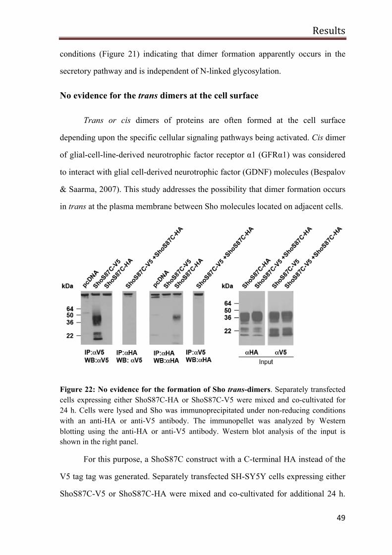
Results
49
conditions (Figure 21) indicating that dimer formation apparently occurs in the
secretory pathway and is independent of N-linked glycosylation.
No evidence for the trans dimers at the cell surface
Trans or cis dimers of proteins are often formed at the cell surface
depending upon the specific cellular signaling pathways being activated. Cis dimer
of glial-cell-line-derived neurotrophic factor receptor α1 (GFRα1) was considered
to interact with glial cell-derived neurotrophic factor (GDNF) molecules (Bespalov
& Saarma, 2007). This study addresses the possibility that dimer formation occurs
in trans at the plasma membrane between Sho molecules located on adjacent cells.
Figure 22: No evidence for the formation of Sho trans-dimers. Separately transfected cells expressing either ShoS87C-HA or ShoS87C-V5 were mixed and co-cultivated for 24 h. Cells were lysed and Sho was immunoprecipitated under non-reducing conditions with an anti-HA or anti-V5 antibody. The immunopellet was analyzed by Western blotting using the anti-HA or anti-V5 antibody. Western blot analysis of the input is shown in the right panel.
For this purpose, a ShoS87C construct with a C-terminal HA instead of the
V5 tag tag was generated. Separately transfected SH-SY5Y cells expressing either
ShoS87C-V5 or ShoS87C-HA were mixed and co-cultivated for additional 24 h.
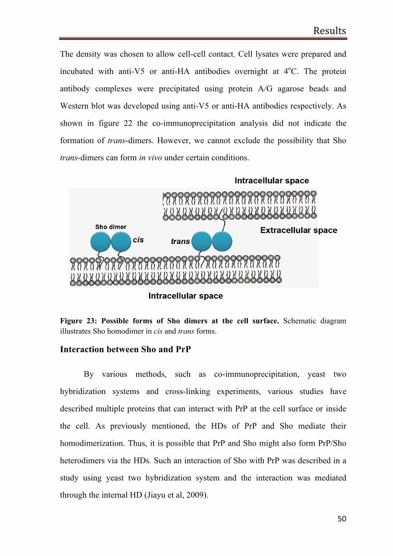
Results
50
The density was chosen to allow cell-cell contact. Cell lysates were prepared and
incubated with anti-V5 or anti-HA antibodies overnight at 4oC. The protein
antibody complexes were precipitated using protein A/G agarose beads and
Western blot was developed using anti-V5 or anti-HA antibodies respectively. As
shown in figure 22 the co-immunoprecipitation analysis did not indicate the
formation of trans-dimers. However, we cannot exclude the possibility that Sho
trans-dimers can form in vivo under certain conditions.
Figure 23: Possible forms of Sho dimers at the cell surface. Schematic diagram illustrates Sho homodimer in cis and trans forms.
Interaction between Sho and PrP
By various methods, such as co-immunoprecipitation, yeast two
hybridization systems and cross-linking experiments, various studies have
described multiple proteins that can interact with PrP at the cell surface or inside
the cell. As previously mentioned, the HDs of PrP and Sho mediate their
homodimerization. Thus, it is possible that PrP and Sho might also form PrP/Sho
heterodimers via the HDs. Such an interaction of Sho with PrP was described in a
study using yeast two hybridization system and the interaction was mediated
through the internal HD (Jiayu et al, 2009).
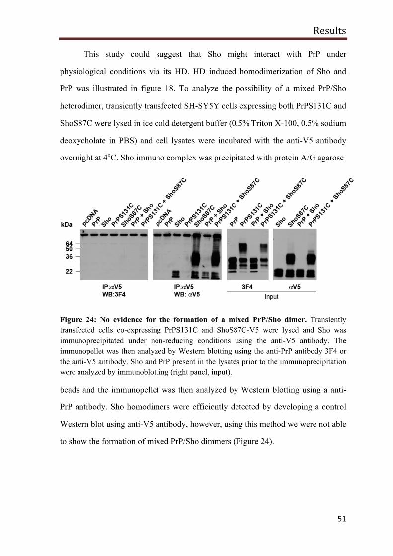
Results
51
This study could suggest that Sho might interact with PrP under
physiological conditions via its HD. HD induced homodimerization of Sho and
PrP was illustrated in figure 18. To analyze the possibility of a mixed PrP/Sho
heterodimer, transiently transfected SH-SY5Y cells expressing both PrPS131C and
ShoS87C were lysed in ice cold detergent buffer (0.5% Triton X-100, 0.5% sodium
deoxycholate in PBS) and cell lysates were incubated with the anti-V5 antibody
overnight at 4oC. Sho immuno complex was precipitated with protein A/G agarose
Figure 24: No evidence for the formation of a mixed PrP/Sho dimer. Transiently transfected cells co-expressing PrPS131C and ShoS87C-V5 were lysed and Sho was immunoprecipitated under non-reducing conditions using the anti-V5 antibody. The immunopellet was then analyzed by Western blotting using the anti-PrP antibody 3F4 or the anti-V5 antibody. Sho and PrP present in the lysates prior to the immunoprecipitation were analyzed by immunoblotting (right panel, input).
beads and the immunopellet was then analyzed by Western blotting using a anti-
PrP antibody. Sho homodimers were efficiently detected by developing a control
Western blot using anti-V5 antibody, however, using this method we were not able
to show the formation of mixed PrP/Sho dimmers (Figure 24).
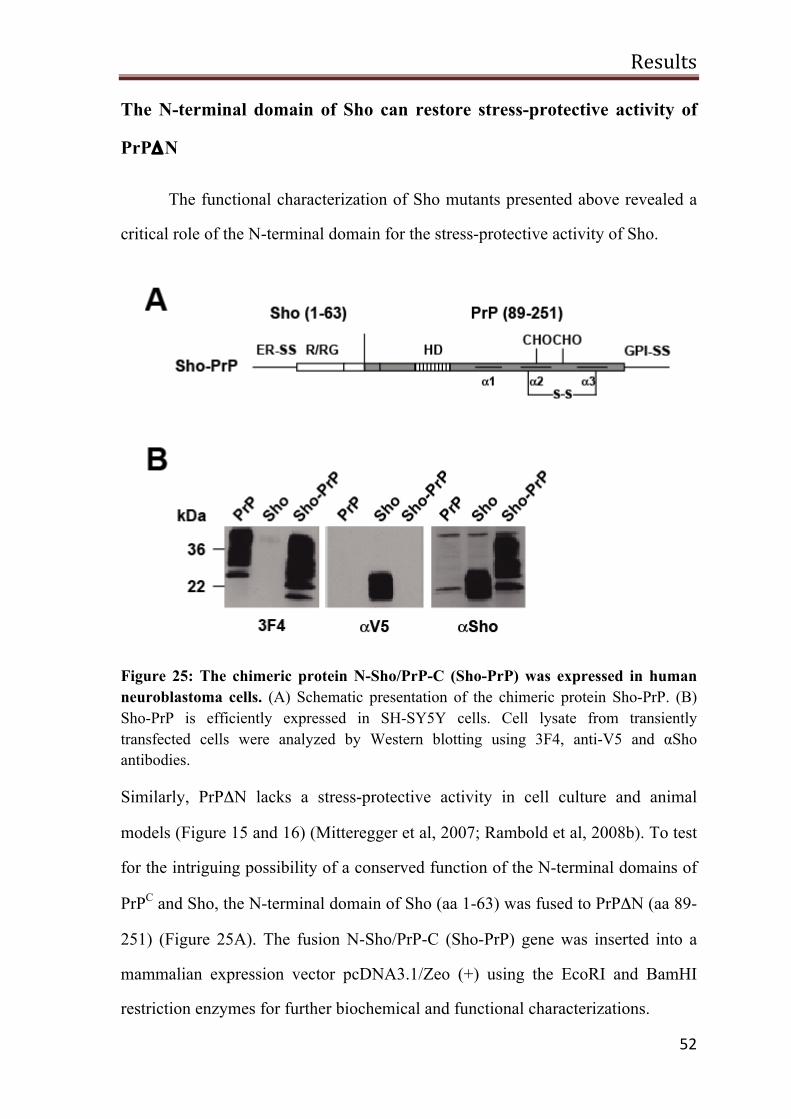
Results
52
The N-terminal domain of Sho can restore stress-protective activity of
PrPΔN
The functional characterization of Sho mutants presented above revealed a
critical role of the N-terminal domain for the stress-protective activity of Sho.
Figure 25: The chimeric protein N-Sho/PrP-C (Sho-PrP) was expressed in human neuroblastoma cells. (A) Schematic presentation of the chimeric protein Sho-PrP. (B) Sho-PrP is efficiently expressed in SH-SY5Y cells. Cell lysate from transiently transfected cells were analyzed by Western blotting using 3F4, anti-V5 and αSho antibodies.
Similarly, PrPΔN lacks a stress-protective activity in cell culture and animal
models (Figure 15 and 16) (Mitteregger et al, 2007; Rambold et al, 2008b). To test
for the intriguing possibility of a conserved function of the N-terminal domains of
PrPC and Sho, the N-terminal domain of Sho (aa 1-63) was fused to PrPΔN (aa 89-
251) (Figure 25A). The fusion N-Sho/PrP-C (Sho-PrP) gene was inserted into a
mammalian expression vector pcDNA3.1/Zeo (+) using the EcoRI and BamHI
restriction enzymes for further biochemical and functional characterizations.
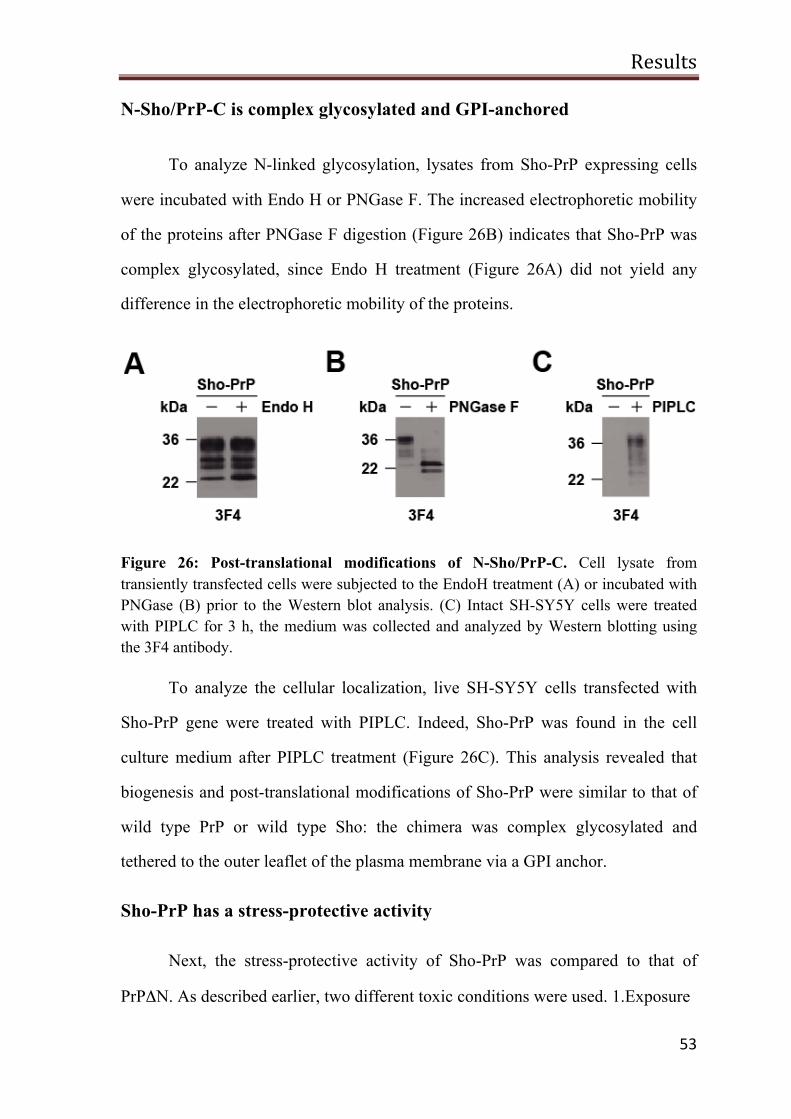
Results
53
N-Sho/PrP-C is complex glycosylated and GPI-anchored
To analyze N-linked glycosylation, lysates from Sho-PrP expressing cells
were incubated with Endo H or PNGase F. The increased electrophoretic mobility
of the proteins after PNGase F digestion (Figure 26B) indicates that Sho-PrP was
complex glycosylated, since Endo H treatment (Figure 26A) did not yield any
difference in the electrophoretic mobility of the proteins.
Figure 26: Post-translational modifications of N-Sho/PrP-C. Cell lysate from transiently transfected cells were subjected to the EndoH treatment (A) or incubated with PNGase (B) prior to the Western blot analysis. (C) Intact SH-SY5Y cells were treated with PIPLC for 3 h, the medium was collected and analyzed by Western blotting using the 3F4 antibody.
To analyze the cellular localization, live SH-SY5Y cells transfected with
Sho-PrP gene were treated with PIPLC. Indeed, Sho-PrP was found in the cell
culture medium after PIPLC treatment (Figure 26C). This analysis revealed that
biogenesis and post-translational modifications of Sho-PrP were similar to that of
wild type PrP or wild type Sho: the chimera was complex glycosylated and
tethered to the outer leaflet of the plasma membrane via a GPI anchor.
Sho-PrP has a stress-protective activity
Next, the stress-protective activity of Sho-PrP was compared to that of
PrPΔN. As described earlier, two different toxic conditions were used. 1.Exposure
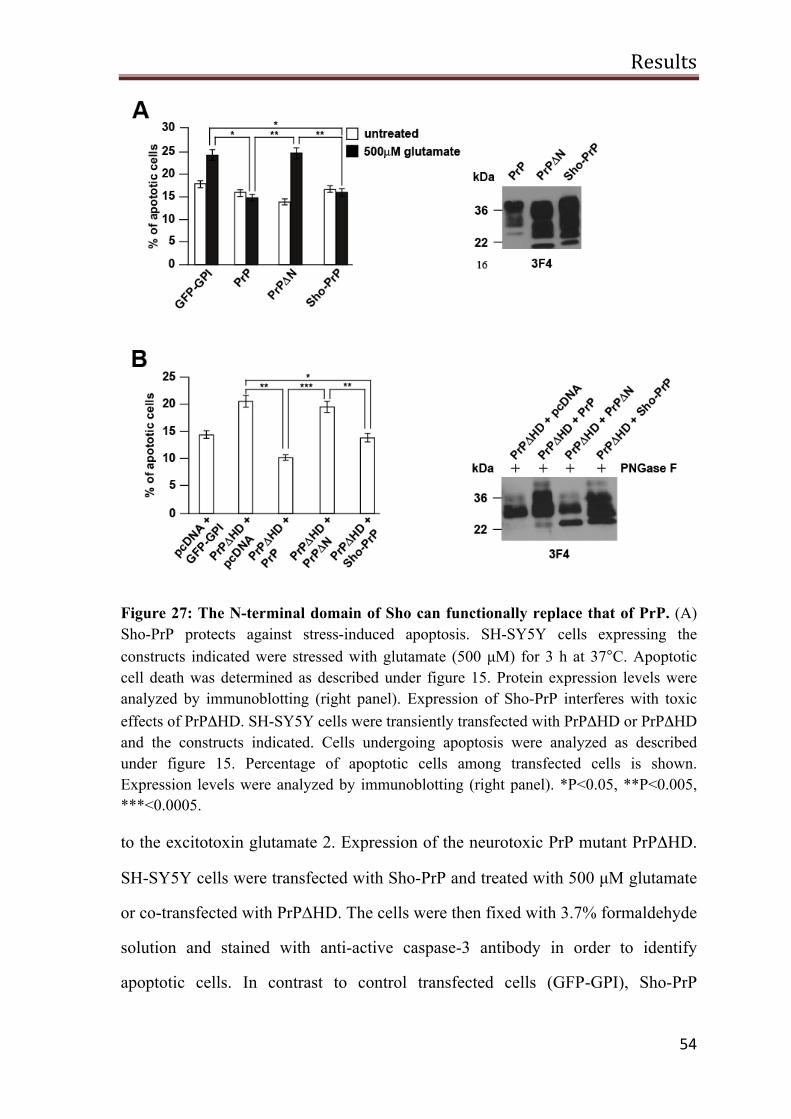
Results
54
Figure 27: The N-terminal domain of Sho can functionally replace that of PrP. (A) Sho-PrP protects against stress-induced apoptosis. SH-SY5Y cells expressing the constructs indicated were stressed with glutamate (500 µM) for 3 h at 37°C. Apoptotic cell death was determined as described under figure 15. Protein expression levels were analyzed by immunoblotting (right panel). Expression of Sho-PrP interferes with toxic effects of PrPΔHD. SH-SY5Y cells were transiently transfected with PrPΔHD or PrPΔHD and the constructs indicated. Cells undergoing apoptosis were analyzed as described under figure 15. Percentage of apoptotic cells among transfected cells is shown. Expression levels were analyzed by immunoblotting (right panel). *P<0.05, **P<0.005, ***<0.0005.
to the excitotoxin glutamate 2. Expression of the neurotoxic PrP mutant PrPΔHD.
SH-SY5Y cells were transfected with Sho-PrP and treated with 500 µM glutamate
or co-transfected with PrP∆HD. The cells were then fixed with 3.7% formaldehyde
solution and stained with anti-active caspase-3 antibody in order to identify
apoptotic cells. In contrast to control transfected cells (GFP-GPI), Sho-PrP
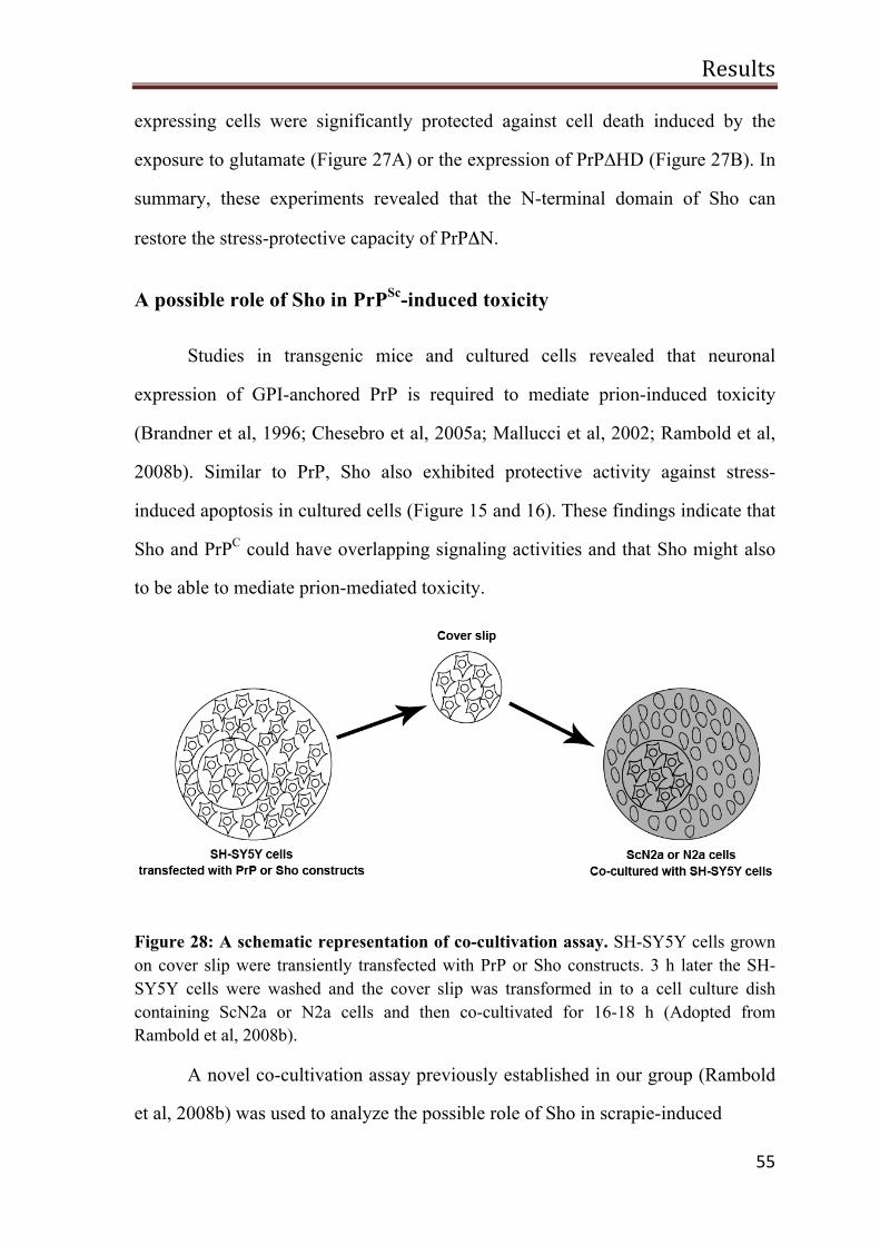
Results
55
expressing cells were significantly protected against cell death induced by the
exposure to glutamate (Figure 27A) or the expression of PrP∆HD (Figure 27B). In
summary, these experiments revealed that the N-terminal domain of Sho can
restore the stress-protective capacity of PrPΔN.
A possible role of Sho in PrPSc-induced toxicity
Studies in transgenic mice and cultured cells revealed that neuronal
expression of GPI-anchored PrP is required to mediate prion-induced toxicity
(Brandner et al, 1996; Chesebro et al, 2005a; Mallucci et al, 2002; Rambold et al,
2008b). Similar to PrP, Sho also exhibited protective activity against stress-
induced apoptosis in cultured cells (Figure 15 and 16). These findings indicate that
Sho and PrPC could have overlapping signaling activities and that Sho might also
to be able to mediate prion-mediated toxicity.
Figure 28: A schematic representation of co-cultivation assay. SH-SY5Y cells grown on cover slip were transiently transfected with PrP or Sho constructs. 3 h later the SH-SY5Y cells were washed and the cover slip was transformed in to a cell culture dish containing ScN2a or N2a cells and then co-cultivated for 16-18 h (Adopted from Rambold et al, 2008b).
A novel co-cultivation assay previously established in our group (Rambold
et al, 2008b) was used to analyze the possible role of Sho in scrapie-induced
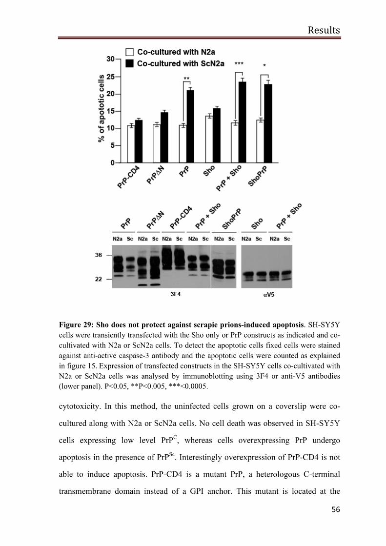
Results
56
Figure 29: Sho does not protect against scrapie prions-induced apoptosis. SH-SY5Y cells were transiently transfected with the Sho only or PrP constructs as indicated and co-cultivated with N2a or ScN2a cells. To detect the apoptotic cells fixed cells were stained against anti-active caspase-3 antibody and the apoptotic cells were counted as explained in figure 15. Expression of transfected constructs in the SH-SY5Y cells co-cultivated with N2a or ScN2a cells was analysed by immunoblotting using 3F4 or anti-V5 antibodies (lower panel). P<0.05, **P<0.005, ***<0.0005.
cytotoxicity. In this method, the uninfected cells grown on a coverslip were co-
cultured along with N2a or ScN2a cells. No cell death was observed in SH-SY5Y
cells expressing low level PrPC, whereas cells overexpressing PrP undergo
apoptosis in the presence of PrPSc. Interestingly overexpression of PrP-CD4 is not
able to induce apoptosis. PrP-CD4 is a mutant PrP, a heterologous C-terminal
transmembrane domain instead of a GPI anchor. This mutant is located at the
Results
57
plasma membrane but not in lipid rafts and has no stress-protective activity
(Rambold et al, 2008b; Winklhofer et al, 2003c). Since ScN2a cells were reported
to release PrPSc molecules consistently in the cell culture medium via exosomes
(Fevrier et al, 2004; Vella et al, 2007), this approach could be a valid tool to
identify the role of Sho in prion infection.
SH-SY5Y cells grown on cover slips were transiently transfected with PrP
and Sho constructs. 4 h later, the transfected cells on cover slips were extensively
washed with DMEM without FCS and placed in to cell culture dishes either with
N2a or ScN2a. After 18 h the SH-SY5Y cells were fixed and stained with anti-
active caspase-3 antibody and the apoptotic cells were analyzed as mentioned in
figure 15. Corroborating previous finding (Rambold et al, 2008b), SH-SY5Y cells
expressing PrP-CD4 did not undergo apoptosis when co-cultivated with N2a or
ScN2a cells (Figure 29; PrP-CD4). However, significant increase in apoptotic cell
death was observed, when SH-SY5Y cells expressing GPI-anchored PrPC were co-
cultivated with ScN2a (Figure 29; PrP). Expression of wild type Sho did not
decrease viability of SH-SY5Y cells co-cultured with ScN2a cells (Figure 29;
Sho).
Further-on, two more questions need to be addressed more elaborately.
Firstly, can Sho-PrP also transmit a toxic signal similar to PrPC? Expression of
PrP∆N does not sensitize SH-SY5Y cells to PrPSc-induced cell death (Figure 29;
PrP∆N). Similarly PrP∆N does not protect the cells against stress-induced cell
death. But the fusion protein Sho-PrP restores stress-protective signaling. When
Sho-PrP expressing SH-SY5Y cells were co-cultivated with ScN2a cells, indeed,
Sho-PrP expression sensitized the SH-SY5Y cells to PrPSc-induced apoptosis
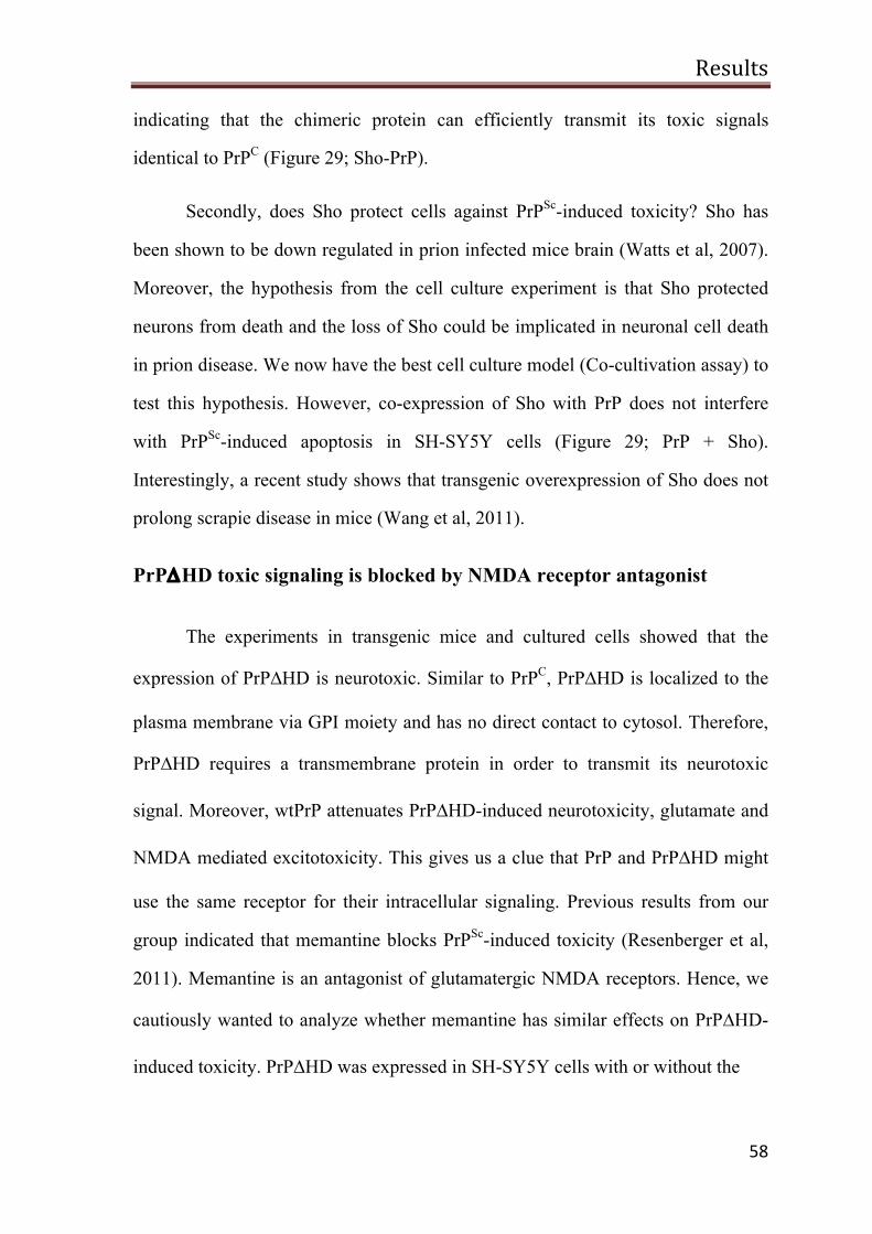
Results
58
indicating that the chimeric protein can efficiently transmit its toxic signals
identical to PrPC (Figure 29; Sho-PrP).
Secondly, does Sho protect cells against PrPSc-induced toxicity? Sho has
been shown to be down regulated in prion infected mice brain (Watts et al, 2007).
Moreover, the hypothesis from the cell culture experiment is that Sho protected
neurons from death and the loss of Sho could be implicated in neuronal cell death
in prion disease. We now have the best cell culture model (Co-cultivation assay) to
test this hypothesis. However, co-expression of Sho with PrP does not interfere
with PrPSc-induced apoptosis in SH-SY5Y cells (Figure 29; PrP + Sho).
Interestingly, a recent study shows that transgenic overexpression of Sho does not
prolong scrapie disease in mice (Wang et al, 2011).
PrP∆HD toxic signaling is blocked by NMDA receptor antagonist
The experiments in transgenic mice and cultured cells showed that the
expression of PrP∆HD is neurotoxic. Similar to PrPC, PrP∆HD is localized to the
plasma membrane via GPI moiety and has no direct contact to cytosol. Therefore,
PrP∆HD requires a transmembrane protein in order to transmit its neurotoxic
signal. Moreover, wtPrP attenuates PrP∆HD-induced neurotoxicity, glutamate and
NMDA mediated excitotoxicity. This gives us a clue that PrP and PrP∆HD might
use the same receptor for their intracellular signaling. Previous results from our
group indicated that memantine blocks PrPSc-induced toxicity (Resenberger et al,
2011). Memantine is an antagonist of glutamatergic NMDA receptors. Hence, we
cautiously wanted to analyze whether memantine has similar effects on PrP∆HD-
induced toxicity. PrP∆HD was expressed in SH-SY5Y cells with or without the
Results
59
Figure 30: Memantine rescues SH-SY5Y cells from PrP∆HD induced apoptosis. SH-SY5Y cells were transiently transfected with PrP∆HD and cultured overnight in the presence of memantine. Apoptotic cells were identified by indirect immunofluorescence using the anti-active caspase-3 antibody as explained in figure 15. Protein expression was analyzed by immunoblotting using the 3F4 antibody (right panel). * P<0.05, **P<0.005, ***<0.0005.
presence of memantine. Indeed, upon the treatment with memantine the apoptotic
cell death in PrP∆HD expressing cells was significantly reduced (Figure 30).
Discussion
60
Discussion
The prominent role of PrP in prion diseases is well characterized and so far
no other molecule has been found to be connected to these diseases. Less than a
decade ago, in a search for PrP-related proteins using comparative genomics,
Premzl et al., identified a new PrP-related gene called Shadoo (Sho; Shadow of
prion protein) (Premzl et al, 2003). Evolutionary origin of PrP and Sho are not
known. However, a study suggests that the genes of PrP might have co-evolved
from a common ancestral gene (Premzl et al, 2004). Sho is expressed mainly in
CNS and is highly conserved from fish to man (Premzl et al, 2003). Although there
is no overall sequence homology, PrP and Sho have a conserved internal HD and
both are anchored on the plasma membrane via a GPI moiety. This study provides
new insights into biogenesis of human Sho and its physiological activity,
specifically its ability to protect cells against stress-induced apoptosis.
Biogenesis of human Sho
Sho is complex glycosylated and attached to the plasma membrane.
Previously our group analyzed the biogenesis of zeSho in mammalian cells
and provided the first experimental evidence that Sho is complex glycosylated and
targeted to the outer leaflet of the plasma membrane (Miesbauer et al, 2006), a
finding corroborated for mSho later (Watts et al, 2007). In this study, the
biogenesis of human Sho was analyzed in detail. PNGase F and Endo H, enzymes
that remove the N-linked glycans from proteins were employed to study the
glycosylation pattern of Sho. Corroborating previous findings for zeSho and mSho,
human Sho expressed in SH-SY5Y cells is sensitive to PNGase F but resistant to
Endo H, indicating that Sho is complex glycosylated. In addition, a possible role of
N-terminal and HD in Sho biogenesis was also addressed. Similar to wtSho,
Discussion
61
mutants devoid of N-terminal and HD treated with PNGase F also show difference
in their migration pattern in comparison with untreated protein samples indicating
that these mutants are also modified with complex glycans.
As described above, zeSho and mSho are attached to the plasma membrane
via GPI moiety. So the next question addressed was whether the human
homologue is also anchored to the plasma membrane via a GPI moiety. To test
this, Sho expressing SH-SY5Y cells were treated with PIPLC. As expected, Sho
was found in the cell culture medium after the PIPLC treatment. Sho mutant
devoid of the N-terminal domain was also released in to the cell culture medium
after the PIPLC treatment, whereas, Sho∆HD was undetectable using either anti-
V5 antibody or αSho. But reduced level of Sho∆HD was observed in the cell
lysates treated with PIPLC. So the expression of Sho∆HD was further analyzed by
non-permeable fluorescence microscopy technique. Indeed, similar to wtSho,
expression of Sho∆HD at cell surface was confirmed by fluorescence microscopy.
Collectively, these results confirm that Sho and its deletion mutants are modified
with complex glycans, targeted to the plasma membrane through a GPI moiety.
Moreover, deletion of the N-terminal or the HD does not have any detectable
impact on Sho biogenesis.
Sho forms homodimers
Many GPI anchored proteins have been shown to form dimers. Dimer
formation is not only linked to the physiological function but also to cellular
trafficking and targeting to lipid rafts (Mayor & Riezman, 2004; Paladino et al,
2004; Simons & Toomre, 2000). For example, dimerization was demonstrated for
CD59, a GPI anchored complement regulatory protein (Hatanaka et al, 1998),
Discussion
62
GFRα, urokinase-type plasminogen activator receptor (uPAR/CD87) and CD55
(Airaksinen & Saarma, 2002; Hatanaka et al, 1998). Formation of PrP homodimers
have been reported previously and that the internal HD is required for dimerization
and is a part of the dimer interface (Meyer et al, 2000a; Priola et al, 1995b;
Rambold et al, 2008b). PrP homodimerization was experimentally shown by
introducing a cysteine residue within the HD of PrP (PrPS131C) (Rambold et al,
2008b). A similar approach was previously used to analyze the dimerization of
human epidermal growth factor receptor 2 (Erb-2/Her2) and the amyloid precursor
protein (APP) (Cao et al, 1992; Munter et al, 2007). Since the HD of Sho is highly
homologues to the HD of PrP, it is reasonable to assume that Sho might also be
able to form homodimers via its HD.
To address this possibility, a modification was introduced into Sho: the
serine residue at 87th position was replaced by a cysteine residue (ShoS87C). If
Sho forms a dimer under physiological conditions, the cysteine residues come
closer and form a disulfide bond. Under non-reducing conditions the dimers can be
detected by a shift in protein migration on SDS/PAGE. Western blot analysis from
cultured mammalian cells expressing ShoS87C showed an additional slow
migrating band in comparison to wtSho under non-reducing conditions. The
slower migrating band disappears in the presence of reducing agents such as β-ME
or DDT indicating that Sho forms homodimers and the homodimer is stabilized by
an intermolecular disulfide bond via the introduced cysteine residues. PNGase F
and PIPLC treatment of ShoS87C revealed that the biogenesis is not altered by the
cysteine modification i.e. similar to wtSho, ShoS87C is also complex glycosylated
and attached to the plasma membrane through a GPI moiety.
The next question we addressed was whether the N-terminal domain of Sho
had an impact on homodimerization. To test this, Sho∆N,S87C (aa 30-56 deleted
Discussion
63
in ShoS87C) was expressed in cultured mammalian cells and the protein samples
was analyzed by Western blotting under non-reducing conditions. Indeed, similar
to ShoS87C, Sho∆N,S87C also forms dimers. These results indicate that the N-
terminal domain is not required for Sho homodimerization.
Many membrane proteins form dimers within the cell and are transported to
the outer membrane. For example presence of immature GPCR dimers within the
ER lumen suggests that dimerization might be an integral part of GPCR maturation
and an initial step in the production of functional GPCRs (Milligan, 2004).
Sometimes substrate binding can also induce dimerization of the target proteins at
the cell surface. For example brain derived neurotrophic factor (BDNF) binding to
TrkB tyrosine kinase receptor induces receptor dimerization (Blum & Konnerth,
2005; Scharfman & McNamara, 2010).
After we obtained experimental evidence for Sho and PrP
homodimerization, we further wanted to know if Sho and PrP homodimerization
take place within the cell or at the plasma membrane. To test this, Sho and PrP
were expressed in mammalian cells in the presence of brefeldin A, a lactone
antibiotic that blocks the protein transport from ER to golgi. Western blot analysis
revealed that the Sho and PrP homodimers could be detected after the treatment
with brefeldin A. This result indicated that Sho and PrP homodimers are formed
within the secretory pathway. Further, the effect of glycosylation on
homodimerization was investigated. Mammalian cells expressing ShoS87C and
PrPS131C were treated with tunicamycin and homodimer formation was analyzed
by Western blotting. Indeed, Sho and PrP homodimers can be detected under this
condition. Thus, glycosylation is not required for Sho or PrP homodimer
formation.
Discussion
64
Taken together, Sho and PrP homodimer formation occurs within the
secretory pathway and does not require N-linked glycosylation. Since dimerization
is a general basic characteristic feature of several membrane receptors, it could be
possible that homodimerization of Sho and PrP might be linked with cellular
processes such as maturation, cellular trafficking, binding to their receptors,
regulation of intracellular signals and endocytosis.
Signal transduction can be initiated between two different cells through
protein-protein interaction at the outer surface of the plasma membrane. For
example, a homodimer of N-Cadherin, a cell adhesion molecule interacts with
another N-Cadherin homodimer of a neighboring cell to form functional cis-trans
homotetramer (Kim et al, 2005). Cis homodimers of junctional adhesion molecule
1 (JAM 1) can bind to other cis homodimer of JAM 1 of the adjacent cell and
regulate the paracellular permeability and leukocyte transmigration (Kostrewa et
al, 2001). Thus, the question arose whether Sho can form trans homodimers at the
cell surface. To address this experimentally, two different tags (V5 and HA tags)
were introduced into ShoS87C. Cells expressing Sho constructs with two different
tags were mixed together and grown until the establishment of cell to cell contacts.
Further, the cells were lysed and the possible formation of a trans homodimer was
analyzed by co-immunoprecipitation analysis. Using co-immunoprecipitation
analysis, Sho cis homodimer could be readily detected but not trans homodimers
or cis-trans homotetramers. However, we cannot rule out the possibility that under
certain circumstances Sho trans dimers might be formed in vivo in response to a
specific stimulus.
Discussion
65
No formation of PrP/Sho mixed dimer
As described earlier, Sho and PrP contain a highly homologues HD. Since,
both Sho and PrP can form homodimers via their HD, it is tempting to speculate
that Sho and PrP may form heterodimers and HD might be a part of the dimer
interface. Using yeast two-hybrid system, it was demonstrated that Sho can
interact with PrP, which is mediated by the HD (Jiayu et al, 2009). Further,
collision induced dissociation (CID) spectral studies suggested that PrP can be co-
purified with Sho or vice versa (Watts et al, 2009). To analyze a possible
interaction, PrP along with Sho or PrPS131C with ShoS87C were co-transfected,
protein samples were co-immunoprecipitated and analyzed by Western blotting. If
Sho and PrP formed a mixed dimer under physiological conditions, introduced
cysteine molecules might form a disulfide bridge and different migration patterns
for Sho/PrP heterodimer might be seen on Western blot. However, using this
approach we were not able to show a PrP/Sho interaction.
The stress-protective activity of Sho
Since Sho and PrP share certain structural features, it seems plausible that
the two proteins have also similar physiological functions. Indeed in 2007, Joel
Watts and colleagues described a PrP-like neuroprotective activity of Sho. Similar
to wtPrP, Sho can protect CGN cells against PrP∆HD-induced toxicity (Watts et
al, 2007). In our study we investigated whether this protective activity of Sho is
limited to PrP∆HD-induced apoptosis or can also protect cells against other
physiological stress agents. To test this, glutamate was employed as a model for
excitotoxic stress. Glutamate is considered to be the main mediator of
excitotoxicity in the CNS by changing the LTP. Moreover several studies
suggested that PrP can ameliorate altered LTP and excitotoxicity induced neuronal
Discussion
66
cell death (Collinge et al, 2004; Khosravani et al, 2008; Rambold et al, 2008b;
Whittington et al, 1995).
Mammalian cells expressing PrP or Sho were stressed with acute
concentration of glutamate and apoptotic cell death was then measured by staining
cells for active caspase-3. Indeed, similar to PrP, Sho can protect SH-SY5Y cells
from glutamate stress-induced apoptosis. Further, to map the domains involved in
this stress-protective activity of Sho, the deletion mutant clones coding for Sho
protein without the N-terminal or HD were prepared. Stress-protective activity of
Sho mutants was analyzed by apoptotic cell death as readout. Similar to PrP∆N
mutant, N-terminally truncated version of Sho lost its stress-protective activity and
lack of HD also makes Sho functionally inefficient. These results indicate that the
stress-protective activity of Sho and PrP depend on similar domains i.e, N-terminal
domain and internal HD. The impaired stress-protective activity of Sho mutants is
not due to improper cellular trafficking, since both the mutants are complex
glycosylated and attached to the plasma membrane via a GPI moiety.
With the above results, it is possible to assume that the N-terminal region
and the HD are required for the stress-protective function of Sho and PrP. But it is
still puzzling as to how the N-terminal and HD are involved in stress-protective
signaling? It has been suggested that the intrinsically disordered domains of
protein are implicated in protein-protein interaction (Tompa et al, 2009). The N-
terminal domain of PrP is intrinsically disordered and by CD spectroscopic
analysis of rSho it has been suggested that whole protein might be intrinsically
disordered (Watts et al, 2007). Therefore, it could be reasonable to assume that the
N-terminal domain of Sho and PrP may interact with their unidentified co-
receptors involving intracellular signaling cascade.
Discussion
67
Deleting HD in Sho does not lead to neurotoxic species
Experiments in transgenic mice revealed the unexpected finding that by
deleting the intrinsic HD, PrP can gain a neurotoxic potential (Baumann et al,
2007; Li et al, 2007a; Shmerling et al, 1998). Interestingly, the neurotoxic potential
of PrP∆HD is independent of replication of infectious prion (rev.in (Winklhofer et
al, 2008). Co-expression of a single copy of wtPrP can completely abolish the
neurotoxic phenotype although the cellular mechanism involved in PrP∆HD
mediated toxicity is unknown (Shmerling et al, 1998). Similar to wtPrP, PrP∆HD
is also complex glycosylated and targeted to the plasma membrane through a GPI
anchor (Winklhofer et al, 2003c). An interesting finding is that similar to PrPC, co-
transfection of Sho can counteract PrP∆HD induced toxicity in CGN cells
indicating that Sho has PrP-like activity in terms of neutralizing the neurotoxicity
induced by PrP∆HD (Watts et al, 2007).
As previously mentioned, sequence similarity between Sho and PrP lies
within the HDs. So the next question addressed was, does the removal of the HD
from Sho generate neurotoxic species similar to that of PrP∆HD? To test this,
Sho∆HD expressing mammalian cells were fixed and stained for active caspase-3
and the apoptosis level was measured. Using a cell culture assay developed in our
lab, we could reproduce PrP∆HD-induced toxicity. Mammalian cells expressing
PrP∆HD undergo apoptosis and this phenotype is rescued upon co-expression of
wtPrP or wtSho. Interestingly, expression of Sho∆HD does not induce apoptosis in
cultured mammalian cells indicating that removal of the intrinsic HD of Sho does
not result in toxic species at least under the experimental conditions tested. At the
same time, mammalian cells undergo apoptosis when Sho∆HD and PrP∆HD are

Discussion
68
co-expressed indicating that Sho∆HD does not interfere with PrP∆HD-induced
apoptosis. Although Sho and PrP contain identical HD, acquiring a neurotoxic
conformer might be exclusive for PrP.
The N-terminal domain of Sho can functionally replace that of PrP
Sho and PrP can protect cells against different toxic insults and their
mutants devoid of N-terminal domain (Sho∆N and PrP∆N) are impaired in their
stress-protective activity. The next question we addressed was to whether the
fusion of the N-terminal domain of Sho with PrP∆N can restore its stress-
protective activity. To analyze this, cultured mammalian cells expressing the
chimeric protein Sho-PrP (N-terminus (aa 1-63) of Sho fused to PrP∆N (aa 89-
251) were stressed with glutamate or co-expressed along with PrP∆HD and
apoptotic cell death was measured using activated caspase-3 staining. Indeed,
similar to wtPrP, Sho-PrP protects cultured cells against glutamate or PrP∆HD-
induced apoptosis.
There is no experimental evidence that either copper or any other metal co-
factors bind to the N-terminal domain of Sho. There is no sequence homology
between the N-termini of Sho and PrP and the only similarity seems to be that both
domains are intrinsically disordered. Based on the hypothesis, that the intrinsically
disordered domains can be involved in protein-protein interaction, it is reasonable
to assume that N-terminal domain of Sho and PrP bind with same co-receptor to
mediate stress-protective signaling.
Discussion
69
Sho does not protect cells from PrPSc-induced apoptosis
Formation of PrPSc in the CNS of infected individuals is the crucial event in
prion diseases. Apart from PrP, there is no solid evidence of any other protein to
play a curtail role in prion disease. After the discovery of Sho, it has been
hypothesized that it may have an important role in prion disease. Interestingly, it
was reported that Sho is downregulated in prion infected mice. However, Sho
mRNA level seems to be unchanged (Lloyd et al, 2009; Watts et al, 2007).
Identifying the SPRN null allele in two patients affected with vCJD also supports
the involvement of Sho in prion diseases (Beck et al, 2008).
To address a possible effect of Sho on PrPSc-induced toxicity, a novel
method called co-cultivation assay, which is developed in our laboratory, was
used. PrP and Sho were expressed in cultured mammalian cells and these cells
were then co-cultivated with ScN2a cells, which are constantly secreting PrPSc in
the medium. Corroborating previous findings, PrPC expressing cells underwent
apoptosis in the presence of PrPSc, whereas in contrast, Sho expressing cells were
not affected by the presence of PrPSc. Further-more, we checked whether co-
expression of Sho with PrP can block PrPSc-induced apoptosis. To test this, Sho
was co-expressed with PrP and these cells were grown in the presence of PrPSc.
Co-expression of Sho with PrP failed to protect cells against PrPC dependent PrPSc-
induced apoptosis. A recent study in transgenic mice shows that Sho
overexpression does not alter scrapie pathogenesis (Wang et al, 2011). Moreover,
down regulation of Sho in mice infected with different prion strains has been
studied by Kohtaro Miyazawa and Laura Manuelidis and Sho reduction seems to
be prion strain specific. For example Sho level seems to be unaffected in mice
infected with Asian CJD strain whereas Kuru infected mice show a significant
reduction in Sho level (Miyazawa & Manuelidis, 2010).
Discussion
70
In summary, our co-cultivation assay results suggest that Sho can neither
protect cells from PrPSc-induced apoptosis nor can it mediate PrPSc-induced
toxicity. Together with recently published studies, it appears that Sho cannot
modulate prion pathogenesis.
Summary
71
Summary
Shadoo (Sho) is the only known protein that has some similarities with the
cellular prion protein (PrPC). Both proteins are evolutionarily highly conserved
glycoproteins that are mainly expressed in the brain. Both the proteins share
common characteristic features such as an unstructured N-terminal domain, a
homologous internal hydrophobic domain (HD) and a C-terminal
glycosylphosphatidylinositol (GPI) anchor. Preliminary data also showed that PrP
and Sho have apparently a similar stress-protective activity. In the present study,
the biogenesis and physiological function of the human homologue Sho was
studied. Compared with PrP show here some similarities and differences.
Similarities
• Both the proteins are complex-glycosylated and selectively transported by a
GPI anchor to the outer plasma membrane. Both dimerize within the
secretory pathway. The dimerization is mediated here by the hydrophobic
domain.
• Both have a stress-protective effect, for which the N-terminal domain is
required.
• PrP and Sho probably activate similar cellular pathways that protect cells
from stress-induced apoptosis.
Differences
• Unlike PrP∆HD, deletion of the hydrophobic domain of Sho leads to the
formation of no toxic conformers.
• Sho does not mediate toxicity induced by PrPSc.
Summary
72
Taken together, this study reinforces the view that PrP and Sho could
mediate their neuroprotective potential by common cellular co-receptors. The
formation and propagation of toxic conformers is specific for PrP. Although Sho is
unlikely to contribute to prion pathogenesis, studying the physiological function of
Sho could be a useful tool to clarify the physiological function of PrP. The
discovery of the physiological role of PrP is important to understand the
pathological role of PrP in prion diseases. This knowledge can help to develop new
therapeutic strategies against prion diseases.
Zusammenfassung
73
Zusammenfassung
Shadoo (Sho) ist das bisher einzig bekannte Protein, das gewisse
Ähnlichkeiten mit dem zellulären Prion-Protein (PrPC) aufweist. Beide Proteine
sind evolutionär hochkonservierte Glycoproteine, die hauptsächlich im Gehirn
exprimiert werden. Chakterische Merkmale beider Proteine sind eine
unstrukturierte N-terminale Domäne, eine homologe interne hydrophobe Domäne
und ein C-terminaler Glycosylphosphatidylinositol (GPI)-Anker. Erste Daten
zeigten darüber hinaus, dass Sho und PrP offenbar eine ähnliche stress-protektive
Aktivität besitzen. Im Rahmen der vorliegenden Studie wurde die Biogenese und
physiologische Funktion des menschlichen Sho-Homologs untersucht. Verglichen
mit PrP zeigen sich hier einige Ähnlichkeiten und Unterschiede.
Ähnlichkeiten
• Beide Proteine werden complex-glykosyliert und gezielt durch einen GPI-
Anker an die äußere Plasmamembran transportiert. Beide dimerisieren
innerhalb des sekretorischen Signalweges. Die Dimerisierung wird dabei
durch die hydrophobe Domäne vermittelt.
• Beide besitzen eine stress-protektive Wirkung, wofür die N-terminale
Domäne benötigt wird.
• PrP und Sho aktivieren vermutlich ähnliche zelluläre Signalwege, um
Zellen vor stress-induzierter Apoptose zu schützen.
Unterschiede
• Im Gegensatz zu PrP∆HD führt eine Deletion der hydrophoben Domäne
von Sho nicht zur Bildung von toxischen Konformeren.
• Sho vermittelt keine PrPSc-induzierte Toxizität.
Zusammenfassung
74
Zusammengenommen festigt die vorliegende Studie die Ansicht, dass PrP
und Sho ihr neuroprotektives Potenzial durch gemeinsame zelluläre Co-
Rezeptoren vermittlen könnten. Die Bildung und Propagierung toxischer
Konformere ist jedoch spezifisch für PrP. Obwhol Sho somit vermutlich nicht zur
Prion-Pathogenese beiträgt, könnte die Studie der physiologischen Funktion von
Sho ein nützliches Werkzeug sein, um die physiologische Funktion von PrP zu
klären. Die Aufdeckung der physiologischen Rolle von PrP ist bedeutend, um die
pathologische Rolle von PrP in Prion-Erkrankungern zu verstehen. Dieses Wissen
kann dazu beitragen neue therapeutische Strategien gegen Prion-Erkrankungen zu
entwicklen.
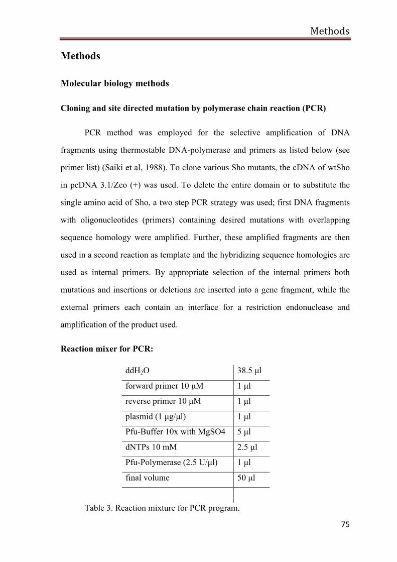
Methods
75
Methods
Molecular biology methods
Cloning and site directed mutation by polymerase chain reaction (PCR)
PCR method was employed for the selective amplification of DNA
fragments using thermostable DNA-polymerase and primers as listed below (see
primer list) (Saiki et al, 1988). To clone various Sho mutants, the cDNA of wtSho
in pcDNA 3.1/Zeo (+) was used. To delete the entire domain or to substitute the
single amino acid of Sho, a two step PCR strategy was used; first DNA fragments
with oligonucleotides (primers) containing desired mutations with overlapping
sequence homology were amplified. Further, these amplified fragments are then
used in a second reaction as template and the hybridizing sequence homologies are
used as internal primers. By appropriate selection of the internal primers both
mutations and insertions or deletions are inserted into a gene fragment, while the
external primers each contain an interface for a restriction endonuclease and
amplification of the product used.
Reaction mixer for PCR:
ddH2O 38.5 µl
forward primer 10 µM 1 µl
reverse primer 10 µM 1 µl
plasmid (1 µg/µl) 1 µl
Pfu-Buffer 10x with MgSO4 5 µl
dNTPs 10 mM 2.5 µl
Pfu-Polymerase (2.5 U/µl) 1 µl
final volume 50 µl
Table 3. Reaction mixture for PCR program.
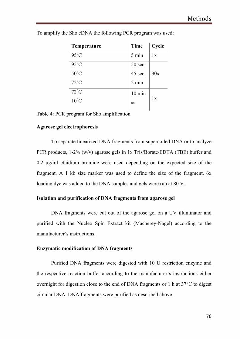
Methods
76
To amplify the Sho cDNA the following PCR program was used:
Temperature Time Cycle
95oC 5 min 1x
95oC
50oC
72oC
50 sec
45 sec
2 min
30x
72oC
10oC 10 min
∞ 1x
Table 4: PCR program for Sho amplification
Agarose gel electrophoresis
To separate linearized DNA fragments from supercoiled DNA or to analyze
PCR products, 1-2% (w/v) agarose gels in 1x Tris/Borate/EDTA (TBE) buffer and
0.2 µg/ml ethidium bromide were used depending on the expected size of the
fragment. A 1 kb size marker was used to define the size of the fragment. 6x
loading dye was added to the DNA samples and gels were run at 80 V.
Isolation and purification of DNA fragments from agarose gel
DNA fragments were cut out of the agarose gel on a UV illuminator and
purified with the Nucleo Spin Extract kit (Macherey-Nagel) according to the
manufacturer’s instructions.
Enzymatic modification of DNA fragments
Purified DNA fragments were digested with 10 U restriction enzyme and
the respective reaction buffer according to the manufacturer’s instructions either
overnight for digestion close to the end of DNA fragments or 1 h at 37°C to digest
circular DNA. DNA fragments were purified as described above.
Methods
77
Alkaline phosphatase treatment
To avoid self-ligation, the linearized vectors were dephosphorylated with
shrimp alkaline phosphatase (SAP) before ligation. SAP and SAP reaction buffer
was added to the digested vector according to the manufacturer’s instructions. The
mixture was incubated at 37°C for 10 min and heat inactivated at 65°C for 10 min.
Ligation of cDNA fragments into vector DNA
To ligate the digested and purified DNA fragment into a respectively
linearized plasmid, 100-200 ng of the plasmid was mixed with 1-2 µg DNA
fragment, T4 ligase buffer and T4 ligase in a final volume of 20 µl. The mixture
was incubated for 3 h at room temperature and heat inactivated for 10 min at 65°C.
7 µl were used for transformation of competent bacteria of the E. coli strain DH5α
Preparation of competent bacteria
A DNA molecule cannot usually pass through the bacterial cell membrane
since it is a highly hydrophilic molecule. So, the bacterial cells might be competent
to take up the plasmid DNA into the cells. This is done by making pores and
destabilizing the cell wall using high concentration of divalent cations. Single
colony of freshly grown E.Coli DH5α strain was dissolved in 2 ml of Luria broth
(LB) medium and shaken for 16 h at 37oC. Then the mixture was poured into 250
ml LB medium and the cells were cultivated for 2-3 h until an OD 590 value of
0.4-0.6 was reached. Further, the culture was centrifuged at 3750 rpm for 5 min at
4oC and the bacterial pellet was resuspended in 100 ml of ice cold TFB1 buffer.
The suspension was incubated on ice for 5 min and then centrifuged at 3750 rpm
for 5 min at 4oC. The pellet was then resuspended in 10 ml of ice cold TFB2 buffer
and incubated for 30-60 min on ice. Further, it was aliquoted up to 100 ml and
competent cells were stored in liquid nitrogen or in -80oC until the use.
Methods
78
Transformation of competent bacteria
The transformation is used for receiving and amplifying the plasmid by
E.Coli (Sambrook, 1989). 100 ml of competent bacterial cells were gently thawed
on ice and mixed with ligated or 1 mg of plasmid DNA. After 30 min of
incubation on ice, the suspension was incubated for 90 seconds at 42oC (heat
shock) and then kept on ice for 5 min. After adding 400 ml of LB medium without
antibiotics the culture was shaken for 60-90 min at 37oC and plated in different
concentrations of antibiotic containing agar plates. Then, the plates were incubated
at 37oC for 16-20 h.
Plasmid DNA preparation from bacterial culture
For the preparation of plasmid DNA Qiagen-Mini/Maxi-Kit was used and
followed according to the manufacturer’s instructions.
Sequencing
The DNA sequencing was performed based on the Sanger’s chain
termination method (Sanger et al, 1977) by GATC company in Konstanz.,
Germany.
Cell biology methods
Cell culture
Cultivation of cells
Human neuroblastoma cells (SH-SY5Y) cells were cultured in Dulbecco's
modified Eagle’s medium (DMEM). The complete medium contained 10% heat
inactivated fetal calf serum (FCS), 1% antibiotics solution (final concentration was
1U/ml of penicillin G, 1mg/ml of streptomycin) and 2 mM glutamine. The cell line
Methods
79
was cultured as an adherent single monolayer in cell culture flasks at 37oC with
5% CO2.
Passaging
The passaging of the cell line was done on an average every 3-4 days. After
aspiration of the cell culture medium the cells were rinsed with phosphate buffered
saline (PBS -/-) and then incubated with trypsin (0.5 g/L) for several minutes.
Further, the cells were scrutinized carefully, resuspended in prewarmed complete
medium and divided with the desired seeding ratio into new cell culture flasks.
Plating the cells
For plating the cells, the existing quantity of cells was determined using a
Neubauer cell counting chamber. SH-SY5Y cells were plated at a density of 5 x10⁵
cells in 3.5 cm culture dishes. For immunofluorescence analysis, cells were thinly
plated in order to detect individual cells more efficiently. In this case, 4x 10⁵ SH-
SY5Y cells were platted on sterile cover slips in a 3.5 cm cell culture dish.
Transfection
The cells were plated 24 h before transfection and then the cells were
washed with medium without FCS. For transient transfection, the plasmid DNA
was mixed with Lipofectamine and Plus (Invitrogen) in OptiMEM according to the
manufacturer’s instructions. After 3 h the transfection mixture was replaced with
complete medium and the cells were incubated for 24 h at 37oC with 5% CO2
before proceeding to the experiments as indicated.
Methods
80
Harvesting the cells
Cells were harvested 24 h after the transfection. These cells were washed
twice with PBS -/- and then scraped off with a cell scraper in PBS -/-. Then, cells
were centrifuged for 3 min at 3000 rpm and the cells pellet was placed on ice
before processing for the experiments as indicated.
Total cell lysate
The cell pellets were resuspended on ice cold in detergent buffer (0.1%
Triton X-100 or 0.5% Triton X-100/sodium desoxycholate (DOC) in PBS-/- with
protease inhibitors and incubated on ice for 10-20 min with harsh vortexing in
between. The resulting total cell lysate was mixed with Laemmli sample buffer,
boiled for 10 min at 95oC and analyzed by the SDS-polyacrylamide gel
electrophoresis (SDS-PAGE).
SDS-PAGE
The proteins were separated using one-dimensional, discontinues SDS-
PAGE (Laemmli, 1970). The concentration of stacking gel was 4% and for the
separating gel depending on the size of the protein, an 8-14% polyacrylamide
concentration was used. Electrophoresis was done at 150-250V in a Hoefer SE600
chamber.
Western blot analysis
For subsequent immunodetection, previously separated proteins by SDS-
PAGE were transferred onto a nitrocellulose membrane (Towbin et al, 1979). The
protein transfer was performed in transfer buffer at a constant current of 1000 mA
for 2 h at 4 ° C.

Methods
81
Ponceau S staining
After blotting the membrane was incubated for 5 min in Ponceau S solution
and rinsed in distilled H2O to check for complete transfer of proteins from the gel
to the nitrocellulose membrane. Before the immune reaction the membrane was
decolorized by PBST.
Immunodetection of proteins
The immunodetection of proteins was performed using the Enhanced
Chemiluminescence (ECL) system according to the manufacturer’s instructions.
First the nitrocellulose membrane was blocked for non-specific binding with 5%
skimmed milk solution for 1 h at room temperature (RT) and followed by
incubation with primary antibody or antiserum for 16 h at 4oC. The membrane was
washed 3 times with PBST for 10 min and incubated with horseradish peroxidase
(HRP) conjugated secondary anti-mouse or anti-rabbit antibody in PBST for 45-60
min at room temperature. Subsequently, the membrane was again washed 3 times
with PBST and incubated with HPR-substrate, and then the blots were exposed to
the X-ray film to visualize the signals.
Glycosylation analysis
For the detection of glycosylation of proteins various methods that were
employed have been described below.
Treatment with tunicamycin
To analyze the protein core glycosylation, tunicamycin which blocks the
synthesis of all N-linked glycoproteins (N-glycans) was used. Transiently
transfected cells were incubated with 0.5 mg/ml of tunicamycin at 37oC with 5%
CO2 and the cell lysates were analyzed by Western blotting.
Methods
82
Digestion with Endo H or PNGase F
To identify whether the N-linked glycosylation of proteins was present in
the form of a high mannose structure or complex glycosylated, an enzymatic
digestion of cell lysates with Endo H or PNGase F was performed. Endo H cleaves
the glycoforms of high mannose structure (Maley et al, 1989; Robbins et al, 1984)
whereas PNGase F digests the complex glycans (Plummer et al, 1984; Tarentino et
al, 1985; Tarentino & Plummer, 1987). The cell lysates were mixed with
denaturing buffer, boiled at 95oC for 10 min and incubated on ice for 5 min. After
the addition of reaction buffer and the enzyme, the sample was incubated for 1-3 h
at 37oC and mixed with Laemmli sample buffer. Further-on, the sample was
resolved on the SDS-PAGE gel and analyzed by Western blotting.
Treatment with brefedin A
Transiently transfected SH-SY5Y cells were grown in the presence of
1µg/ml of brefeldin A, which blocks the protein transport from ER to golgi
complex and triggers the retrograde protein transport Golgi complex to ER. After
24 h of transfection the cell lysates were prepared and analyzed by Western
blotting.
Indirect immunofluorescence microscopy
Transiently transfected SH-SY5Y cells were grown on glass cover slips and
fixed 24 h post transfection with 3.7% PFA for 20 min. Fixed cells were incubated
with primary antibody for 45 min at 37°C in PBS containing 1% BSA. After
extensive washing with cold PBS, incubation with the Cy3 conjugated secondary
antibody followed at 37°C for 30 min. Cells were mounted onto glass slides and
examined by fluorescence microscopy.
Methods
83
Co-immunoprecipitation
To analyze formation of a mixed PrP/Sho dimer SH-SY5Y cells were co-
transfected with PrPS131C and ShoS87C. At 24 h post-transfection the cells were
harvested and lysed in ice-cold detergent lysis buffer (0.5% Triton X-100, 0.5%
sodium deoxycholate in PBS) supplemented with protease inhibitors. Precleared
lysates were incubated with αV5 antibody overnight at 4°C. The immunocomplex
was precipitated with protein A sepharose beads and analyzed by Western blotting.
To analyze formation of Sho trans-dimers, separate dishes of SH-SY5Y cells were
transfected with either ShoS87C-V5 or ShoS87C-HA. 3 h post-transfection cells
were extensively washed, trypsinized, mixed together and seeded in one cell
culture dish. 24 h later, the cells were harvested and analyzed as described above.
Co-cultivation assay
SH-SY5Y cells grown on coverslips were transiently transfected with PrP
and/or Sho constructs using lipofectamine plus reagent. At 3 h after the
transfection, the cover slips were transferred into cell culture dishes containing
ScN2a or N2a cells. 16 h later, the apoptotic cell death in transiently transfected
SH-SY5Y cells was analyzed.
Apoptosis assay
As described earlier (Rambold et al, 2006), SH-SY5Y cells were grown on
cover slips. 24 h after transfection, the cells were incubated with glutamate (500
µM) for 3 h. The cells were then fixed and activated caspase-3 detected by indirect
immunofluorescence using an anti-active caspase-3 antibody. To detect cells
undergoing apoptosis, the number of activated caspase-3 positive cells out of at
least 1100 transfected cells was determined using a Zeiss Axioscope 2 plus
Methods
84
microscope (Carl Zeiss, Göttingen, Germany). Quantifications were based on
triplicates of at least three independent experiments.
Statistical analysis
Data were expressed as means ± SE. All the experiments were performed in
triplicates and repeated at least three times. Statistical analysis was carried out
using student’s t-test. P-values are as follows: * P<0.05, **P<0.005, ***<0.0005.
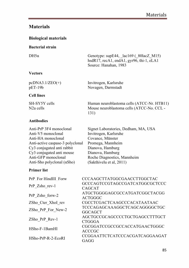
Materials
85
Materials
Biological materials
Bacterial strain
DH5α Genotype: supE44, _lac169 (_80lacZ_M15) hsdR17, recA1, endA1, gyr96, thi-1, eLA1 Source: Hanahan, 1983
Vectors pcDNA3.1/ZEO(+) pET-19b
Invitrogen, Karlsruhe Novagen, Darmstadt
Cell lines
SH-SY5Y cells Human neuroblastoma cells (ATCC-Nr. HTB11) N2a cells Mouse neuroblastoma cells (ATCC-No. CCL -
131)
Antibodies
Anti-PrP 3F4 monoclonal Anti-V5 monoclonal Anti-HA monoclonal Anti-active caspase-3 polyclonal Cy3 conjugated anti rabbit Cy3 conjugated anti mouse Anti-GFP monoclonal Anti-Sho polyclonal (αSho)
Signet Laboratories, Dedham, MA, USA Invitrogen, Karlsruhe Covance, Münster Promega, Mannheim Dianova, Hamburg Dianova, Hamburg Roche Diagnostics, Mannheim (Sakthivelu et al, 2011)
Primer list
PrP_For HindIII_Forw CCCAAGCTTATGGCGAACCTTGGCTAC
PrP_Zsho_rev-1 GCCCAGTCCGTAGCCGATCATGGCGCTCCCCAGCAT
PrP_Zsho_forw-2 ATGCTGGGGAGCGCCATGATCGGCTACGGACTGGGC
ZSho_Cter_XhoI_rev CGCCTCGACTCAAGCCCACATAATAAC
ZSho_PrP_For_New-2 TCCCAGAGCAAAGGCTCAGCAGGGGCTGCGGCAGCT
ZSho_PrP_Rev-1 AGCTGCCGCAGCCCCTGCTGAGCCTTTGCTCTGGGA
HSho-F-1BamHI CGCGGATCCGCCGCCACCATGAACTGGGCACCCGC
HSho-PrP-R-2-EcoRI CCGGAATTCTCATCCCACGATCAGGAAGATGAGG
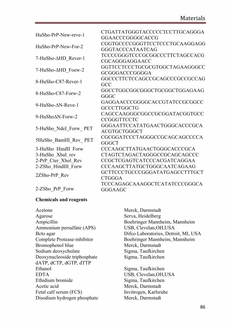
Materials
86
HuSho-PrP-New-reve-1 CTGATTATGGGTACCCCCTCCTTGCAGGGAGGAACCCGGGGCACCG
HuSho-PrP-New-For-2 CGGTGCCCCGGGTTCCTCCCTGCAAGGAGGGGGTACCCATAATCAG
7-HuSho-∆HD_Rever-1 TCCCCGGGTCCCGCGGCCCTTCTAGCCACGCGCAGGGAGGAACC
7-HuSho-∆HD_Foew-2 GGTTCCTCCCTGCGCGTGGCTAGAAGGGCCGCGGGACCCGGGGA
8-HuSho-C87-Rever-1 GGCCCTTCTCCAGCCGCAGCCCGCCGCCAGGCC
8-HuSho-C87-Forw-2 GGCCTGGCGGCGGGCTGCGGCTGGAGAAGGGGC
9-HuSho-∆N-Reve-1 GAGGAACCCGGGGCACCGTATCCGCGGCCGCCCTTGGCTG
9-HuSho∆N-Forw-2 CAGCCAAGGGCGGCCGCGGATACGGTGCCCCGGGTTCCTC
5-HuSho_NdeI_Forw_ PET GGGAATTCCATATGAACTGGGCACCCGCAACGTGCTGGGCT
5HuSho_BamHI_Rev_ PET CGCGGATCCCTAGGGCCGCAGCAGCCCCAGGGCT
3-HuSho_HindII_Forw CCCAAGCTTATGAACTGGGCACCCGCA 3-HuSho_XbaI_rev CTAGTCTAGACTAGGGCCGCAGCAGCCC 2-PrP_Cter_Xhol_Rev CCGCTCGAGTCATCCCACGATCAGGAA 2-ZSho_HindIII_Forw CCCAAGCTTATGCTGGGCAATCAGAAG
2ZSho-PrP_Rev GCTTCCCTGCCCGGGATATGAGCCTTTGCTCTGGGA
2-ZSho_PrP_Forw TCCCAGAGCAAAGGCTCATATCCCGGGCAGGGAAGC
Chemicals and reagents
Acetone Merck, Darmstadt Agarose Serva, Heidelberg Ampicillin Boehringer Mannheim, Mannheim Ammonium persulfate (APS) USB, Clevelan,OH,USA Bcto agar Difco Laboratories, Detroit, MI, USA Complete Protease-inhibitor Boehringer Mannheim, Mannheim Bromophenol blue Merck, Darmstadt Sodium deoxycholate Sigma, Taufkirchen Deoxynucleoside triphosphate dATP, dCTP, dGTP, dTTP
Sigma, Taufkirchen
Ethanol Sigma, Taufkirchen EDTA USB, Clevelan,OH,USA Ethidium bromide Sigma, Taufkirchen Acetic acid Merck, Darmstadt Fetal calf serum (FCS) Invitrogen, Karlsruhe Disodium hydrogen phosphate Merck, Darmstadt
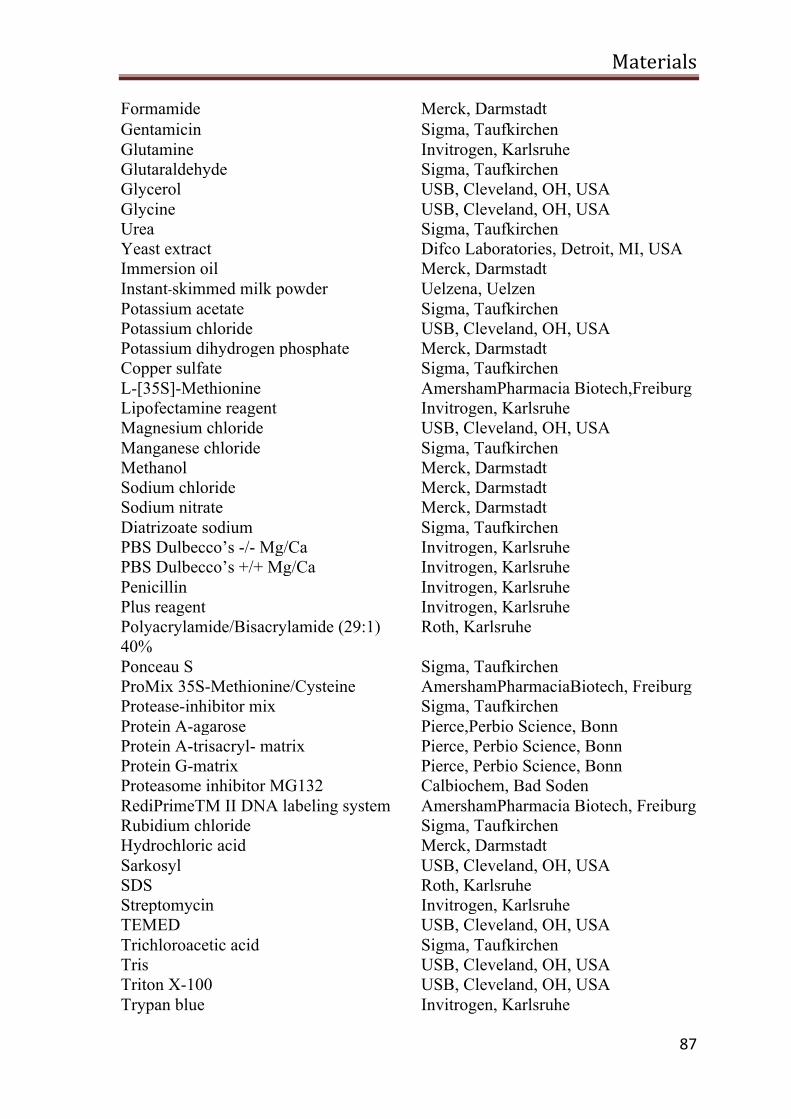
Materials
87
Formamide Merck, Darmstadt Gentamicin Sigma, Taufkirchen Glutamine Invitrogen, Karlsruhe Glutaraldehyde Sigma, Taufkirchen Glycerol USB, Cleveland, OH, USA Glycine USB, Cleveland, OH, USA Urea Sigma, Taufkirchen Yeast extract Difco Laboratories, Detroit, MI, USA Immersion oil Merck, Darmstadt Instant-‐skimmed milk powder Uelzena, Uelzen Potassium acetate Sigma, Taufkirchen Potassium chloride USB, Cleveland, OH, USA Potassium dihydrogen phosphate Merck, Darmstadt Copper sulfate Sigma, Taufkirchen L-[35S]-Methionine AmershamPharmacia Biotech,Freiburg Lipofectamine reagent Invitrogen, Karlsruhe Magnesium chloride USB, Cleveland, OH, USA Manganese chloride Sigma, Taufkirchen Methanol Merck, Darmstadt Sodium chloride Merck, Darmstadt Sodium nitrate Merck, Darmstadt Diatrizoate sodium Sigma, Taufkirchen PBS Dulbecco’s -/- Mg/Ca Invitrogen, Karlsruhe PBS Dulbecco’s +/+ Mg/Ca Invitrogen, Karlsruhe Penicillin Invitrogen, Karlsruhe Plus reagent Invitrogen, Karlsruhe Polyacrylamide/Bisacrylamide (29:1) 40%
Roth, Karlsruhe
Ponceau S Sigma, Taufkirchen ProMix 35S-Methionine/Cysteine AmershamPharmaciaBiotech, Freiburg Protease-inhibitor mix Sigma, Taufkirchen Protein A-agarose Pierce,Perbio Science, Bonn Protein A-trisacryl- matrix Pierce, Perbio Science, Bonn Protein G-matrix Pierce, Perbio Science, Bonn Proteasome inhibitor MG132 Calbiochem, Bad Soden RediPrimeTM II DNA labeling system AmershamPharmacia Biotech, Freiburg Rubidium chloride Sigma, Taufkirchen Hydrochloric acid Merck, Darmstadt Sarkosyl USB, Cleveland, OH, USA SDS Roth, Karlsruhe Streptomycin Invitrogen, Karlsruhe TEMED USB, Cleveland, OH, USA Trichloroacetic acid Sigma, Taufkirchen Tris USB, Cleveland, OH, USA Triton X-100 USB, Cleveland, OH, USA Trypan blue Invitrogen, Karlsruhe

Materials
88
Tunicamycin Sigma, Taufkirchen Tween-20 USB, Cleveland, OH, USA β-Mercaptoethanol Merck, Darmstadt
Medium
Dulbecco's Modified Eagle's medium (DMEM),
Invitrogen, Karlsruhe
Minimal Essential Medium (MEM), Invitrogen, Karlsruhe Minimal Essential Medium, without L-Methionine
Invitrogen, Karlsruhe
OPTIMEM Invitrogen, Karlsruhe LB-Medium 1% NaCl
1% Bacto tryptone 0.5% Yeast extract 100 mg / ml Ampicillin and 30 mg / ml Kanamycin ( added after autoclaving)
LB-Agar LB medium + 1.5% Bacto agar 100 µg/ ml Ampicillin and 30 mg / ml Kanamycin (added after autoclaving)
Kits
ECL RPN 2106 Amersham Pharmacia Biotech, Freiburg Immobilon Western chemiluminescent HRP substrate
Millipore, Schwalb Bach
Protein assay kit Bio-Rad, München QIAprep spin plasmid extraction kit Mini / Maxi
Qiagen, Hilden
QIAquick gel extraction kit Qiagen, Hilden TNT T7 quick coupled transcription / translation system
Promega, Mannheim
Equipments
Agarose gel electrophoresis Central workshop, MPI, Martinsried Mettler Toledo AG285 Analytical Balance
Mettler-Toledo GmbH, Giessen
Incubators Heraeus, Hanau X-Omat Kodak film developer, Stuttgart X-Omat Film developer Kodak, Stuttgart Gel documentation system MWG Biotech, Ebersberg Gel dryer SGD300 Savant, Holbrook, NY, USA GS-6R refrigerated centrifuge with rotor GH3.8
Beckmann, Unterschleissheim
J2-21M refrigerated centrifuge with rotor JA-14
Beckmann, Unterschleissheim
Microscope Axiovert 25, 200M Carl Zeiss, Göttingen
Materials
89
Axioscope2 plus microscope (Axiovision software)
Carl Zeiss, Göttingen
pH meter Fisher Scientific, Nidderau Pipettes (P10, P20, P100, P200) Abimed Gilson, Langenfeld Pipette (P1000) Eppendorf, Hamburg Polyacrylamide gel electrophoresis Amersham Biosciences, Freiburg PCR machine T3 thermocycler Biometra GmbH, Göttingen Thermomixer Eppendorf, Hamburg Table centrifuge centrifuge 5415C Eppendorf, Hamburg
Solutions and buffers
APS solution 10% APS in PBS Blocking milk for Western blot 5% Skimmed milk powder in 1x PBST Blocking buffer for IF 1% BSA in PBS Coomassie destaining solution 40% Methanol
7% Acetic acid DNA sample buffer (6x) 0.25% Bromophenol blue
30% Glycerol Laemmli sample buffer (2x)
120 mM Tris pH 6.8 2% SDS 20% Glycerol 0.5% Bromophenol blue 2% β-Mercaptoethanol
Lysis buffer
0.5% Triton X-100, 0.5% DOC + Protease inhibitor in PBS
PCR mix
1200 µl of H2O 200 µl of 10x Pfu / Taq buffer each 2 µl of dNTPs
PBS (cell culture) Gibco, BRL Life Technologies, Karlsruhe
PBS (10x)
80 g of NaCl 2 g of KCl 14.4 g of Na2HPO4 x 2 H2O 2.4 g of KH2PO4 for 1000 ml of H2O
PBS-T (1x) 1% Tween-20 in 1x PBS
Pfu polymerase buffer (10x) Promega, Mannheim
Ponceau S staning solution
0.2 g of Ponceau S 5 ml of acetic acid 100 ml of H2O
Stacking gel buffer for SDS-PAGE
0.5 M Tris, pH 6.8 0.4% SDS
Materials
90
pH 6.8
Separating gel buffer for SDS-PAGE
1.5 M Tris, pH 8.8 0.4% SDS pH 8.8
Shrimp alkaline phosphatase buffer (10x)
Roche Diagnostics, Mannheim
T4 DNA ligase buffer (10x) MBI Fermentas, St. Leon-Rot
TAE buffer (50x)
2 M Tris base 57.1 ml of Glacial acetic acid 50 mM Na2EDTA x 2H2O, pH 8.0 add1000 ml of H2O
TE buffer
10 mM Tris-HCl, pH 7.5 1 mM EDTA, pH 8.0
Western blot transfer buffer
20 mM Tris-Base 150 mM Glycine 0.01% SDS 20% Methanol
Tunicamycin 10 mg/ml in H2O Brefeldin A 5mg/ml in Ethanol
References
91
References
Aguzzi A, Baumann F, Bremer J (2008) The prion's elusive reason for being. Annual Review of Neuroscience 31: 439-477
Aguzzi A, Glatzel M (2004) vCJD tissue distribution and transmission by
transfusion--a worst-case scenario coming true? Lancet 363: 411-412
Aguzzi A, Glatzel M, Montrasio F, Prinz M, Heppner FL (2001) Interventional
strategies against prion diseases. Nat Rev Neurosci 2: 745-749
Aguzzi A, Weissmann C (1996) Spongiform encephalopathies: a suspicious
signature. Nature 383: 666-667
Airaksinen MS, Saarma M (2002) The GDNF family: signalling, biological
functions and therapeutic value. Nat Rev Neurosci 3: 383-394
Allen JA, Halverson-Tamboli RA, Rasenick MM (2007) Lipid raft microdomains
and neurotransmitter signalling. Nat Rev Neurosci 8: 128-140
Alper T, Cramp WA, Haig DA, Clarke MC (1967) Does the agent of scrapie
replicate without nucleic acid? Nature 214: 764-766
Amitsuka H, Kuwahara K, Yoshida T, Tenya K, Sakakibara T, Mihalik M,
Menovsky AA (1999) Anomalous transport properties of dilute uranium alloys R1-xUxRu2Si2 (R = Th, Y; x <= 0.07). Physica B 259-61: 412-414
Appel TR, Dumpitak C, Matthiesen U, Riesner D (1999) Prion rods contain an
inert polysaccharide scaffold. Biol Chem 380: 1295-1306
Ballif BC, Hornor SA, Jenkins E, Madan-Khetarpal S, Surti U, Jackson KE,
Asamoah A, Brock PL, Gowans GC, Conway RL, Graham JM, Jr., Medne L, Zackai EH, Shaikh TH, Geoghegan J, Selzer RR, Eis PS, Bejjani BA, Shaffer LG (2007) Discovery of a previously unrecognized microdeletion syndrome of 16p11.2-p12.2. Nat Genet 39: 1071-1073
Barry AE, Klyubin I, Mc Donald JM, Mably AJ, Farrell MA, Scott M, Walsh DM,
Rowan MJ (2011) Alzheimer's Disease Brain-Derived Amyloid-{beta}-Mediated Inhibition of LTP In Vivo Is Prevented by Immunotargeting
References
92
Cellular Prion Protein. The Journal of neuroscience : the official journal of the Society for Neuroscience 31: 7259-7263
Basler K, Oesch B, Scott M, Westaway D, Walchli M, Groth DF, McKinley MP,
Prusiner SB, Weissmann C (1986) Scrapie and cellular PrP isoforms are encoded by the same chromosomal gene. Cell 46: 417-428
Baumann F, Tolnay M, Brabeck C, Pahnke J, Kloz U, Niemann HH, Heikenwalder
M, Rulicke T, Burkle A, Aguzzi A (2007) Lethal recessive myelin toxicity of prion protein lacking its central domain. Embo Journal 26: 538-547
Beck JA, Campbell TA, Adamson G, Poulter M, Uphill JB, Molou E, Collinge J,
Mead S (2008) Association of a null allele of SPRN with variant Creutzfeldt-Jakob disease. J Med Genet 45: 813-817
Belay ED, Maddox RA, Williams ES, Miller MW, Gambetti P, Schonberger LB
(2004) Chronic wasting disease and potential transmission to humans. Emerg Infect Dis 10: 977-984
Bendheim PE, Brown HR, Rudelli RD, Scala LJ, Goller NL, Wen GY, Kascsak
RJ, Cashman NR, Bolton DC (1992) Nearly ubiquitous tissue distribution of the scrapie agent precursor protein. Neurology 42: 149-156
Beraldo FH, Arantes CP, Santos TG, Machado CF, Roffe M, Hajj GN, Lee KS,
Magalhaes AC, Caetano FA, Mancini GL, Lopes MH, Americo TA, Magdesian MH, Ferguson SS, Linden R, Prado MA, Martins VR (2011) Metabotropic glutamate receptors transduce signals for neurite outgrowth after binding of the prion protein to laminin gamma1 chain. FASEB J 25: 265-279
Bernoulli C, Siegfried J, Baumgartner G, Regli F, Rabinowicz T, Gajdusek DC,
Gibbs CJ, Jr. (1977) Danger of accidental person-to-person transmission of Creutzfeldt-Jakob disease by surgery. Lancet 1: 478-479
Bespalov MM, Saarma M (2007) GDNF family receptor complexes are emerging
drug targets. Trends Pharmacol Sci 28: 68-74
Bieschke J, Weber P, Sarafoff N, Beekes M, Giese A, Kretzschmar H (2004)
Autocatalytic self-propagation of misfolded prion protein. Proc Natl Acad Sci U S A 101: 12207-12211
References
93
Blum R, Konnerth A (2005) Neurotrophin-mediated rapid signaling in the central
nervous system: Mechanisms and functions. Physiology 20: 70-78
Bockman JM, Kingsbury DT, McKinley MP, Bendheim PE, Prusiner SB (1985)
Creutzfeldt-Jakob disease prion proteins in human brains. N Engl J Med 312: 73-78
Bockman JM, Prusiner SB, Tateishi J, Kingsbury DT (1987) Immunoblotting of
Creutzfeldt-Jakob disease prion proteins: host species-specific epitopes. Ann Neurol 21: 589-595
Bolton B (1982) Issues in validity research on the 16 PF. Psychol Rep 50: 1077-
1078
Bolton DC, Meyer RK, Prusiner SB (1985) Scrapie PrP 27-30 is a
sialoglycoprotein. J Virol 53: 596-606
Borchelt DR, Taraboulos A, Prusiner SB (1992) Evidence for Synthesis of Scrapie
Prion Proteins in the Endocytic Pathway. Journal of Biological Chemistry 267: 16188-16199
Brandner S, Isenmann S, Raeber A, Fischer M, Sailer A, Kobayashi Y, Marino S,
Weissmann C, Aguzzi A (1996) Normal host prion protein necessary for scrapie-induced neurotoxicity. Nature 379: 339-343
Brown DR, Qin K, Herms JW, Madlung A, Manson J, Strome R, Fraser PE, Kruck
T, von Bohlen A, Schulz-Schaeffer W, Giese A, Westaway D, Kretzschmar H (1997a) The cellular prion protein binds copper in vivo. Nature 390: 684-687
Brown DR, Schulz-Schaeffer WJ, Schmidt B, Kretzschmar HA (1997b) Prion
protein-deficient cells show altered response to oxidative stress due to decreased SOD-1 activity. Exp Neurol 146: 104-112
Brown DR, Wong BS, Hafiz F, Clive C, Haswell SJ, Jones IM (1999) Normal
prion protein has an activity like that of superoxide dismutase. Biochem J 344 Pt 1: 1-5
References
94
Brown P (1992) The phenotypic expression of different mutations in transmissible human spongiform encephalopathy. Rev Neurol (Paris) 148: 317-327
Brown P, Coker-Vann M, Pomeroy K, Franko M, Asher DM, Gibbs CJ, Jr.,
Gajdusek DC (1986) Diagnosis of Creutzfeldt-Jakob disease by Western blot identification of marker protein in human brain tissue. N Engl J Med 314: 547-551
Bruce ME, Will RG, Ironside JW, McConnell I, Drummond D, Suttie A,
McCardle L, Chree A, Hope J, Birkett C, Cousens S, Fraser H, Bostock CJ (1997) Transmissions to mice indicate that 'new variant' CJD is caused by the BSE agent. Nature 389: 498-501
Bueler H, Aguzzi A, Sailer A, Greiner RA, Autenried P, Aguet M, Weissmann C
(1993) Mice devoid of PrP are resistant to scrapie. Cell 73: 1339-1347
Bueler H, Fischer M, Lang Y, Bluethmann H, Lipp HP, DeArmond SJ, Prusiner
SB, Aguet M, Weissmann C (1992) Normal development and behaviour of mice lacking the neuronal cell-surface PrP protein. Nature 356: 577-582
Burns CS, Aronoff-Spencer E, Legname G, Prusiner SB, Antholine WE, Gerfen
GJ, Peisach J, Millhauser GL (2003) Copper coordination in the full-length, recombinant prion protein. Biochemistry 42: 6794-6803
Caetano FA, Beraldo FH, Hajj GN, Guimaraes AL, Jurgensen S, Wasilewska-
Sampaio AP, Hirata PH, Souza I, Machado CF, Wong DY, De Felice FG, Ferreira ST, Prado VF, Rylett RJ, Martins VR, Prado MA (2011) Amyloid-beta oligomers increase the localization of prion protein at the cell surface. Journal of neurochemistry 117: 538-553
Calzolai L, Lysek DA, Perez DR, Guntert P, Wuthrich K (2005) Prion protein
NMR structures of chickens, turtles, and frogs. Proc Natl Acad Sci U S A 102: 651-655
Cao H, Bangalore L, Dompe C, Bormann BJ, Stern DF (1992) An extra cysteine
proximal to the transmembrane domain induces differential cross-linking of p185neu and p185neu. The Journal of biological chemistry 267: 20489-20492
References
95
Caughey B, Raymond GJ (1991) The Scrapie-Associated Form of Prp Is Made from a Cell-Surface Precursor That Is Both Protease-Sensitive and Phospholipase-Sensitive. Journal of Biological Chemistry 266: 18217-18223
Chandler RL (1961) Encephalopathy in mice produced by inoculation with scrapie
brain material. Lancet 1: 1378-1379
Chesebro B, Race R, Kercher L (2005a) Scrapie pathogenesis in brain and retina:
effects of prion protein expression in neurons and astrocytes. J Neurovirol 11: 476-480
Chesebro B, Trifilo M, Race R, Meade-White K, Teng C, LaCasse R, Raymond L,
Favara C, Baron G, Priola S, Caughey B, Masliah E, Oldstone M (2005b) Anchorless prion protein results in infectious amyloid disease without clinical scrapie. Science 308: 1435-1439
Christensen HM, Harris DA (2009) A deleted prion protein that is neurotoxic in
vivo is localized normally in cultured cells. J Neurochem 108: 44-56
Chung E, Ji Y, Sun Y, Kascsak RJ, Kascsak RB, Mehta PD, Strittmatter SM,
Wisniewski T (2010) Anti-PrPC monoclonal antibody infusion as a novel treatment for cognitive deficits in an Alzheimer's disease model mouse. BMC Neurosci 11: 130
Cohen FE, Pan KM, Huang Z, Baldwin M, Fletterick RJ, Prusiner SB (1994)
Structural Clues to Prion Replication. Science 264: 530-531
Collinge J (2001) Prion diseases of humans and animals: their causes and
molecular basis. Annu Rev Neurosci 24: 519-550
Collinge J, Asante EA, Li YG, Gowland I, Jefferys JGR (2004) Pathogenic human
prion protein rescues PrP null phenotype in transgenic mice. Neuroscience Letters 360: 33-36
Collinge J, Sidle KC, Meads J, Ironside J, Hill AF (1996) Molecular analysis of
prion strain variation and the aetiology of 'new variant' CJD. Nature 383: 685-690
References
96
Collinge J, Whittington MA, Sidle KC, Smith CJ, Palmer MS, Clarke AR, Jefferys JG (1994) Prion protein is necessary for normal synaptic function. Nature 370: 295-297
Corley SM, Gready JE (2008) Identification of the RGG box motif in Shadoo:
RNA-binding and signaling roles? Bioinform Biol Insights 2: 383-400
Couvineau A, Fabre C, Gaudin P, Maoret JJ, Laburthe M (1996) Mutagenesis of
N-glycosylation sites in the human vasoactive intestinal peptide 1 receptor. Evidence that asparagine 58 or 69 is crucial for correct delivery of the receptor to plasma membrane. Biochemistry 35: 1745-1752
Creutzfeldt HG (1920) A peculiar localised disease of the central nervous system
(Preliminary announcement ). Z Gesamte Neurol Psy 57: 1-18
Creutzfeldt HG (1921) The new findings of the brain-anatomical (histo-
pathological) research for mental illnesses. Deut Med Wochenschr 47: 1589-1590
Cuille J, Chelle PL (1938) Is the trembling of sheep detemined by a filtrable virus?
Cr Hebd Acad Sci 206: 1687-1688
Cunningham O, Andolfo A, Santovito ML, Iuzzolino L, Blasi F, Sidenius N
(2003) Dimerization controls the lipid raft partitioning of uPAR/CD87 and regulates its biological functions. EMBO J 22: 5994-6003
Curtis J, Errington M, Bliss T, Voss K, MacLeod N (2003) Age-dependent loss of
PTP and LTP in the hippocampus of PrP-null mice. Neurobiol Dis 13: 55-62
Daude N, Wohlgemuth S, Rogaeva E, Farid AH, Heaton M, Westaway D (2009a)
Frequent missense and insertion/deletion polymorphisms in the ovine Shadoo gene parallel species-specific variation in PrP. PLoS One 4: e6538
Davanipour Z, Goodman L, Alter M, Sobel E, Asher D, Gajdusek DC (1984)
Possible modes of transmission of Creutzfeldt-Jakob disease. N Engl J Med 311: 1582-1583
References
97
Davis D, Liu X, Segaloff DL (1995) Identification of the sites of N-linked glycosylation on the follicle-stimulating hormone (FSH) receptor and assessment of their role in FSH receptor function. Mol Endocrinol 9: 159-170
Della-Bianca V, Rossi F, Armato U, Dal-Pra I, Costantini C, Perini G, Politi V,
Della Valle G (2001) Neurotrophin p75 receptor is involved in neuronal damage by prion peptide-(106-126). J Biol Chem 276: 38929-38933
Donne DG, Viles JH, Groth D, Mehlhorn I, James TL, Cohen FE, Prusiner SB,
Wright PE, Dyson HJ (1997) Structure of the recombinant full-length hamster prion protein PrP(29-231): the N terminus is highly flexible. Proc Natl Acad Sci U S A 94: 13452-13457
Duffy P, Wolf J, Collins G, DeVoe AG, Streeten B, Cowen D (1974) Letter:
Possible person-to-person transmission of Creutzfeldt-Jakob disease. N Engl J Med 290: 692-693
Dumpitak C, Beekes M, Weinmann N, Metzger S, Winklhofer KF, Tatzelt J,
Riesner D (2005) The polysaccharide scaffold of PrP 27-30 is a common compound of natural prions and consists of alpha-linked polyglucose. Biol Chem 386: 1149-1155
Eisenhaber B, Bork P, Eisenhaber F (2001) Post-translational GPI lipid anchor
modification of proteins in kingdoms of life: analysis of protein sequence data from complete genomes. Protein Eng 14: 17-25
Fevrier B, Vilette D, Archer F, Loew D, Faigle W, Vidal M, Laude H, Raposo G
(2004) Cells release prions in association with exosomes. Proc Natl Acad Sci U S A 101: 9683-9688
Fischer M, Rulicke T, Raeber A, Sailer A, Moser M, Oesch B, Brandner S, Aguzzi
A, Weissmann C (1996) Prion protein (PrP) with amino-proximal deletions restoring susceptibility of PrP knockout mice to scrapie. EMBO J 15: 1255-1264
Freir DB, Nicoll AJ, Klyubin I, Panico S, Mc Donald JM, Risse E, Asante EA,
Farrow MA, Sessions RB, Saibil HR, Clarke AR, Rowan MJ, Walsh DM, Collinge J (2011) Interaction between prion protein and toxic amyloid beta assemblies can be therapeutically targeted at multiple sites. Nat Commun 2: 336
References
98
Gajdusek DC (1973) Kuru and Creutzfeldt-Jakob disease. Experimental models of
noninflammatory degenerative slow virus disease of the central nervous system. Ann Clin Res 5: 254-261
Gajdusek DC, Gibbs CJ, Alpers M (1966) Experimental transmission of a Kuru-
like syndrome to chimpanzees. Nature 209: 794-796
Gauczynski S, Peyrin JM, Haik S, Leucht C, Hundt C, Rieger R, Krasemann S,
Deslys JP, Dormont D, Lasmezas CI, Weiss S (2001) The 37-kDa/67-kDa laminin receptor acts as the cell-surface receptor for the cellular prion protein. EMBO J 20: 5863-5875
Gerstmann J, Straussler E, Scheinker I (1935) On a peculiar hereditary-congenital
disease of the central nervous system - Along with an article on the question of premature ageing. Z Gesamte Neurol Psy 154: 736-762
Gibbs CJ, Jr., Gajdusek DC, Asher DM, Alpers MP, Beck E, Daniel PM,
Matthews WB (1968) Creutzfeldt-Jakob disease (spongiform encephalopathy): transmission to the chimpanzee. Science 161: 388-389
Gomi H, Yokoyama T, Fujimoto K, Ikeda T, Katoh A, Itoh T, Itohara S (1995)
Mice devoid of the glial fibrillary acidic protein develop normally and are susceptible to scrapie prions. Neuron 14: 29-41
Gordon WS (1946) Advances in veterinary research. Vet Rec 58: 516-525
Griffith JS (1967) Self-replication and scrapie. Nature 215: 1043-1044
Haigh CL, Lewis VA, Vella LJ, Masters CL, Hill AF, Lawson VA, Collins SJ
(2009) PrPC-related signal transduction is influenced by copper, membrane integrity and the alpha cleavage site. Cell Res 19: 1062-1078
Hamir AN, Kunkle RA, Cutlip RC, Miller JM, O'Rourke KI, Williams ES, Miller
MW, Stack MJ, Chaplin MJ, Richt JA (2005) Experimental transmission of chronic wasting disease agent from mule deer to cattle by the intracerebral route. J Vet Diagn Invest 17: 276-281
References
99
Hamir AN, Kunkle RA, Miller JM, Greenlee JJ, Richt JA (2006) Experimental second passage of chronic wasting disease (CWD(mule deer)) agent to cattle. J Comp Pathol 134: 63-69
Hamir AN, Miller JM, Kunkle RA, Hall SM, Richt JA (2007) Susceptibility of
cattle to first-passage intracerebral inoculation with chronic wasting disease agent from white-tailed deer. Vet Pathol 44: 487-493
Harrison PM, Khachane A, Kumar M (2010) Genomic assessment of the evolution
of the prion protein gene family in vertebrates. Genomics 95: 268-277
Hartsoug.Gr, Burger D (1965) Encephalopathy of Mink .I. Epizootiologic and
Clinical Observations. Journal of Infectious Diseases 115: 387-&
Hatanaka M, Seya T, Miyagawa S, Matsumoto M, Hara T, Tanaka K, Shimizu A
(1998) Cellular distribution of a GPI-anchored complement regulatory protein CD59: homodimerization on the surface of HeLa and CD59-transfected CHO cells. J Biochem 123: 579-586
He J, Xu J, Castleberry AM, Lau AG, Hall RA (2002) Glycosylation of beta(1)-
adrenergic receptors regulates receptor surface expression and dimerization. Biochem Biophys Res Commun 297: 565-572
Hill AF, Desbruslais M, Joiner S, Sidle KC, Gowland I, Collinge J, Doey LJ,
Lantos P (1997) The same prion strain causes vCJD and BSE. Nature 389: 448-450, 526
Jakob A (1921) Unusual diseases of the central nervous system with striking
anatomic results (Spastic pseudosclerosis - Encephalomyelopathy with disseminated focal degeneration). Z Gesamte Neurol Psy 64: 147-228
Jarrett JT, Lansbury PT, Jr. (1993) Seeding "one-dimensional crystallization" of
amyloid: a pathogenic mechanism in Alzheimer's disease and scrapie? Cell 73: 1055-1058
Jiayu W, Zhu H, Ming X, Xiong W, Songbo W, Bocui S, Wensen L, Jiping L,
Keying M, Zhongyi L, Hongwei G (2009) Mapping the interaction site of prion protein and Sho. Mol Biol Rep
References
100
Jin T, Gu Y, Zanusso G, Sy M, Kumar A, Cohen M, Gambetti P, Singh N (2000) The chaperone protein BiP binds to a mutant prion protein and mediates its degradation by the proteasome. J Biol Chem 275: 38699-38704
Johnson RT (2005) Prion diseases. Lancet Neurol 4: 635-642
Johnson RT, Gibbs CJ, Jr. (1998) Creutzfeldt-Jakob disease and related
transmissible spongiform encephalopathies. N Engl J Med 339: 1994-2004
Kanaani J, Prusiner SB, Diacovo J, Baekkeskov S, Legname G (2005)
Recombinant prion protein induces rapid polarization and development of synapses in embryonic rat hippocampal neurons in vitro. J Neurochem 95: 1373-1386
Kessels HW, Nguyen LN, Nabavi S, Malinow R (2010) The prion protein as a
receptor for amyloid-beta. Nature 466: E3-4; discussion E4-5
Khosravani H, Zhang Y, Tsutsui S, Hameed S, Altier C, Hamid J, Chen L,
Villemaire M, Ali Z, Jirik FR, Zamponi GW (2008) Prion protein attenuates excitotoxicity by inhibiting NMDA receptors. J Cell Biol 181: 551-565
Kim JI, Cali I, Surewicz K, Kong Q, Raymond GJ, Atarashi R, Race B, Qing L,
Gambetti P, Caughey B, Surewicz WK (2010) Mammalian prions generated from bacterially expressed prion protein in the absence of any mammalian cofactors. J Biol Chem 285: 14083-14087
Kim YJ, Johnson KR, Wheelock MJ (2005) N-cadherin-mediated cell motility
requires cis dimers. Cell Commun Adhes 12: 23-39
Kirkwood JK, Wells GA, Wilesmith JW, Cunningham AA, Jackson SI (1990)
Spongiform encephalopathy in an arabian oryx (Oryx leucoryx) and a greater kudu (Tragelaphus strepsiceros). Vet Rec 127: 418-420
Klatzo I, Gajdusek DC, Zigas V (1959) Pathology of Kuru. Lab Invest 8: 799-847
Klein TR, Kirsch D, Kaufmann R, Riesner D (1998) Prion rods contain small
amounts of two host sphingolipids as revealed by thin-layer chromatography and mass spectrometry. Biol Chem 379: 655-666
References
101
Kondo K, Kuroiwa Y (1982) A case control study of Creutzfeldt-Jakob disease: association with physical injuries. Ann Neurol 11: 377-381
Kostrewa D, Brockhaus M, D'Arcy A, Dale GE, Nelboeck P, Schmid G, Mueller
F, Bazzoni G, Dejana E, Bartfai T, Winkler FK, Hennig M (2001) X-ray structure of junctional adhesion molecule: structural basis for homophilic adhesion via a novel dimerization motif. EMBO J 20: 4391-4398
Kretzschmar HA, Stowring LE, Westaway D, Stubblebine WH, Prusiner SB,
Dearmond SJ (1986) Molecular cloning of a human prion protein cDNA. DNA 5: 315-324
Kurschner C, Morgan JI (1995) The cellular prion protein (PrP) selectively binds
to Bcl-2 in the yeast two-hybrid system. Brain Res Mol Brain Res 30: 165-168
Lampo E, Van den Broeck W, Willemarck N, Van Poucke M, Casteleyn CR, De
Spiegelaere W, Van Zeveren A, Peelman LJ (2010) Distribution of the Shadoo protein in the ovine brain assessed by immunohistochemistry. Res Vet Sci
Lauren J, Gimbel DA, Nygaard HB, Gilbert JW, Strittmatter SM (2009) Cellular
prion protein mediates impairment of synaptic plasticity by amyloid-beta oligomers. Nature 457: 1128-1132
Le Pichon CE, Valley MT, Polymenidou M, Chesler AT, Sagdullaev BT, Aguzzi
A, Firestein S (2009) Olfactory behavior and physiology are disrupted in prion protein knockout mice. Nat Neurosci 12: 60-69
Lee KS, Linden R, Prado MA, Brentani RR, Martins VR (2003) Towards cellular
receptors for prions. Rev Med Virol 13: 399-408
Leggett MM, Dukes J, Pirie HM (1990) A spongiform encephalopathy in a cat. Vet
Rec 127: 586-588
Legname G, Baskakov IV, Nguyen HO, Riesner D, Cohen FE, DeArmond SJ,
Prusiner SB (2004) Synthetic mammalian prions. Science 305: 673-676
References
102
Li A, Christensen HM, Stewart LR, Roth KA, Chiesa R, Harris DA (2007a) Neonatal lethality in transgenic mice expressing prion protein with a deletion of residues 105-125. EMBO J 26: 548-558
Li AM, Barmada SJ, Roth KA, Harris DA (2007b) N-terminally deleted forms of
the prion protein activate both bax-dependent and bax-independent neurotoxic pathways. Journal of Neuroscience 27: 852-859
Linden R, Martins VR, Prado MA, Cammarota M, Izquierdo I, Brentani RR
(2008) Physiology of the prion protein. Physiol Rev 88: 673-728
Llewelyn CA, Hewitt PE, Knight RS, Amar K, Cousens S, Mackenzie J, Will RG
(2004) Possible transmission of variant Creutzfeldt-Jakob disease by blood transfusion. Lancet 363: 417-421
Lloyd SE, Grizenkova J, Pota H, Collinge J (2009) Shadoo (Sprn) and prion
disease incubation time in mice. Mamm Genome 20: 367-374
Locht C, Chesebro B, Race R, Keith JM (1986) Molecular cloning and complete
sequence of prion protein cDNA from mouse brain infected with the scrapie agent. Proc Natl Acad Sci U S A 83: 6372-6376
Lukasiewicz R, Nolen B, Adams JA, Ghosh G (2007) The RGG domain of Npl3p
recruits Sky1p through docking interactions. Journal of Molecular Biology 367: 249-261
Maglio LE, Perez MF, Martins VR, Brentani RR, Ramirez OA (2004)
Hippocampal synaptic plasticity in mice devoid of cellular prion protein. Brain Res Mol Brain Res 131: 58-64
Maley F, Trimble RB, Tarentino AL, Plummer TH, Jr. (1989) Characterization of
glycoproteins and their associated oligosaccharides through the use of endoglycosidases. Anal Biochem 180: 195-204
Mallucci G, Dickinson A, Linehan J, Klohn PC, Brandner S, Collinge J (2003)
Depleting neuronal PrP in prion infection prevents disease and reverses spongiosis. Science 302: 871-874
References
103
Mallucci GR, Ratte S, Asante EA, Linehan J, Gowland I, Jefferys JG, Collinge J (2002) Post-natal knockout of prion protein alters hippocampal CA1 properties, but does not result in neurodegeneration. EMBO J 21: 202-210
Manuelidis L (1985) Creutzfeldt-Jakob Disease Prion Proteins in Human Brains.
New Engl J Med 312: 1643-1644
Manuelidis L, Tesin DM, Sklaviadis T, Manuelidis EE (1987) Astrocyte gene
expression in Creutzfeldt-Jakob disease. Proc Natl Acad Sci U S A 84: 5937-5941
Manuelidis L, Valley S, Manuelidis EE (1985) Specific proteins associated with
Creutzfeldt-Jakob disease and scrapie share antigenic and carbohydrate determinants. Proc Natl Acad Sci U S A 82: 4263-4267
Martins VR, Graner E, Garcia-Abreu J, de Souza SJ, Mercadante AF, Veiga SS,
Zanata SM, Neto VM, Brentani RR (1997) Complementary hydropathy identifies a cellular prion protein receptor. Nat Med 3: 1376-1382
Mayor S, Riezman H (2004) Sorting GPI-anchored proteins. Nat Rev Mol Cell Biol
5: 110-120
McLennan NF, Brennan PM, McNeill A, Davies I, Fotheringham A, Rennison
KA, Ritchie D, Brannan F, Head MW, Ironside JW, Williams A, Bell JE (2004) Prion protein accumulation and neuroprotection in hypoxic brain damage. American Journal of Pathology 165: 227-235
Mead S (2006) Prion disease genetics. Eur J Hum Genet 14: 273-281
Meggio F, Negro A, Sarno S, Ruzzene M, Bertoli A, Sorgato MC, Pinna LA
(2000) Bovine prion protein as a modulator of protein kinase CK2. Biochem J 352 Pt 1: 191-196
Meyer RK, Lustig A, Oesch B, Fatzer R, Zurbriggen A, Vandevelde M (2000a) A
monomer-dimer equilibrium of a cellular prion protein (PrPC) not observed with recombinant PrP. The Journal of biological chemistry 275: 38081-38087
References
104
Meyer RK, Lustig A, Oesch B, Fatzer R, Zurbriggen A, Vandevelde M (2000b) A monomer-dimer equilibrium of a cellular prion protein (PrPC) not observed with recombinant PrP. J Biol Chem 275: 38081-38087
Miesbauer M, Bamme T, Riemer C, Oidtmann B, Winklhofer KF, Baier M, Tatzelt
J (2006) Prion protein-related proteins from zebrafish are complex glycosylated and contain a glycosylphosphatidylinositol anchor. Biochem Biophys Res Commun 341: 218-224
Milligan G (2004) G protein-coupled receptor dimerization: function and ligand
pharmacology. Molecular pharmacology 66: 1-7
Mitteregger G, Vosko M, Krebs B, Xiang W, Kohlmannsperger V, Nolting S,
Hamann GF, Kretzschmar HA (2007) The role of the octarepeat region in neuroprotective function of the cellular prion protein. Brain Pathol 17: 174-183
Miyazawa K, Manuelidis L (2010) Agent-specific Shadoo responses in
transmissible encephalopathies. J Neuroimmune Pharmacol 5: 155-163
Mouillet-Richard S, Ermonval M, Chebassier C, Laplanche JL, Lehmann S,
Launay JM, Kellermann O (2000) Signal transduction through prion protein. Science 289: 1925-1928
Munter LM, Voigt P, Harmeier A, Kaden D, Gottschalk KE, Weise C, Pipkorn R,
Schaefer M, Langosch D, Multhaup G (2007) GxxxG motifs within the amyloid precursor protein transmembrane sequence are critical for the etiology of Abeta42. The EMBO journal 26: 1702-1712
Nicolas O, Gavin R, del Rio JA (2009) New insights into cellular prion protein
(PrPc) functions: the "ying and yang" of a relevant protein. Brain Res Rev 61: 170-184
Oesch B, Teplow DB, Stahl N, Serban D, Hood LE, Prusiner SB (1990)
Identification of cellular proteins binding to the scrapie prion protein. Biochemistry 29: 5848-5855
Oesch B, Westaway D, Walchli M, McKinley MP, Kent SB, Aebersold R, Barry
RA, Tempst P, Teplow DB, Hood LE, et al. (1985) A cellular gene encodes scrapie PrP 27-30 protein. Cell 40: 735-746
References
105
Overton MC, Blumer KJ (2000) G-protein-coupled receptors function as oligomers
in vivo. Curr Biol 10: 341-344
Overton MC, Blumer KJ (2002) The extracellular N-terminal domain and
transmembrane domains 1 and 2 mediate oligomerization of a yeast G protein-coupled receptor. J Biol Chem 277: 41463-41472
Paladino S, Sarnataro D, Pillich R, Tivodar S, Nitsch L, Zurzolo C (2004) Protein
oligomerization modulates raft partitioning and apical sorting of GPI-anchored proteins. J Cell Biol 167: 699-709
Pan KM, Baldwin M, Nguyen J, Gasset M, Serban A, Groth D, Mehlhorn I, Huang
Z, Fletterick RJ, Cohen FE, et al. (1993) Conversion of alpha-helices into beta-sheets features in the formation of the scrapie prion proteins. Proc Natl Acad Sci U S A 90: 10962-10966
Pauly PC, Harris DA (1998) Copper stimulates endocytosis of the prion protein. J
Biol Chem 273: 33107-33110
Peden AH, Head MW, Ritchie DL, Bell JE, Ironside JW (2004) Preclinical vCJD
after blood transfusion in a PRNP codon 129 heterozygous patient. Lancet 364: 527-529
Perera WS, Hooper NM (2001) Ablation of the metal ion-induced endocytosis of
the prion protein by disease-associated mutation of the octarepeat region. Curr Biol 11: 519-523
Plummer TH, Jr., Elder JH, Alexander S, Phelan AW, Tarentino AL (1984)
Demonstration of peptide:N-glycosidase F activity in endo-beta-N-acetylglucosaminidase F preparations. J Biol Chem 259: 10700-10704
Premzl M, Gready JE, Jermiin LS, Simonic T, Marshall Graves JA (2004)
Evolution of vertebrate genes related to prion and Shadoo proteins--clues from comparative genomic analysis. Mol Biol Evol 21: 2210-2231
Premzl M, Sangiorgio L, Strumbo B, Marshall Graves JA, Simonic T, Gready JE
(2003) Shadoo, a new protein highly conserved from fish to mammals and with similarity to prion protein. Gene 314: 89-102
References
106
Prince LA, Mann D, Reilly T (2006) Creutzfeldt-Jakob disease: an emergency
department presentation of a rare disease. J Emerg Med 31: 41-44
Priola SA, Caughey B, Wehrly K, Chesebro B (1995a) A 60-kDa prion protein
(PrP) with properties of both the normal and scrapie-associated forms of PrP. J Biol Chem 270: 3299-3305
Prusiner SB (1982) Novel proteinaceous infectious particles cause scrapie. Science
216: 136-144
Prusiner SB (1989) Scrapie prions. Annu Rev Microbiol 43: 345-374
Prusiner SB, McKinley MP, Bowman KA, Bolton DC, Bendheim PE, Groth DF,
Glenner GG (1983) Scrapie prions aggregate to form amyloid-like birefringent rods. Cell 35: 349-358
Prusiner SB, McKinley MP, Groth DF, Bowman KA, Mock NI, Cochran SP,
Masiarz FR (1981) Scrapie agent contains a hydrophobic protein. Proc Natl Acad Sci U S A 78: 6675-6679
Quaglio E, Chiesa R, Harris DA (2001) Copper converts the cellular prion protein
into a protease-resistant species that is distinct from the scrapie isoform. J Biol Chem 276: 11432-11438
Rambold AS, Miesbauer M, Olschewski D, Seidel R, Riemer C, Smale L, Brumm
L, Levy M, Gazit E, Oesterhelt D, Baier M, Becker CF, Engelhard M, Winklhofer KF, Tatzelt J (2008a) Green tea extracts interfere with the stress-protective activity of PrP and the formation of PrP. J Neurochem 107: 218-229
Rambold AS, Miesbauer M, Rapaport D, Bartke T, Baier M, Winklhofer KF,
Tatzelt J (2006) Association of Bcl-2 with misfolded prion protein is linked to the toxic potential of cytosolic PrP. Mol Biol Cell 17: 3356-3368
Rambold AS, Muller V, Ron U, Ben-Tal N, Winklhofer KF, Tatzelt J (2008b)
Stress-protective signalling of prion protein is corrupted by scrapie prions. EMBO J 27: 1974-1984
References
107
Rangel A, Burgaya F, Gavin R, Soriano E, Aguzzi A, Del Rio JA (2007) Enhanced susceptibility of Prnp-deficient mice to kainate-induced seizures, neuronal apoptosis, and death: Role of AMPA/kainate receptors. J Neurosci Res 85: 2741-2755
Resenberger UK, Harmeier A, Woerner AC, Goodman JL, Muller V, Krishnan R,
Vabulas RM, Kretzschmar HA, Lindquist S, Hartl FU, Multhaup G, Winklhofer KF, Tatzelt J (2011) The cellular prion protein mediates neurotoxic signalling of beta-sheet-rich conformers independent of prion replication. EMBO J
Rieger R, Edenhofer F, Lasmezas CI, Weiss S (1997) The human 37-kDa laminin
receptor precursor interacts with the prion protein in eukaryotic cells. Nat Med 3: 1383-1388
Riek R, Hornemann S, Wider G, Billeter M, Glockshuber R, Wuthrich K (1996)
NMR structure of the mouse prion protein domain PrP(121-321). Nature 382: 180-182
Riek R, Hornemann S, Wider G, Glockshuber R, Wuthrich K (1997) NMR
characterization of the full-length recombinant murine prion protein, mPrP(23-231). FEBS Lett 413: 282-288
Robbins PW, Trimble RB, Wirth DF, Hering C, Maley F, Maley GF, Das R,
Gibson BW, Royal N, Biemann K (1984) Primary structure of the Streptomyces enzyme endo-beta-N-acetylglucosaminidase H. J Biol Chem 259: 7577-7583
Roffe M, Beraldo FH, Bester R, Nunziante M, Bach C, Mancini G, Gilch S,
Vorberg I, Castilho BA, Martins VR, Hajj GN (2010) Prion protein interaction with stress-inducible protein 1 enhances neuronal protein synthesis via mTOR. Proc Natl Acad Sci U S A 107: 13147-13152
Russo D, Chazenbalk GD, Nagayama Y, Wadsworth HL, Rapoport B (1991) Site-
directed mutagenesis of the human thyrotropin receptor: role of asparagine-linked oligosaccharides in the expression of a functional receptor. Mol Endocrinol 5: 29-33
Safar JG, Kellings K, Serban A, Groth D, Cleaver JE, Prusiner SB, Riesner D
(2005) Search for a prion-specific nucleic acid. Journal of Virology 79: 10796-10806
References
108
Saiki RK, Gelfand DH, Stoffel S, Scharf SJ, Higuchi R, Horn GT, Mullis KB,
Erlich HA (1988) Primer-Directed Enzymatic Amplification of DNA with a Thermostable DNA-Polymerase. Science 239: 487-491
Sakaguchi S (2007) Molecular biology of prion protein and its first homologous
protein. J Med Invest 54: 211-223
Sakthivelu V, Seidel RP, Winklhofer KF, Tatzelt J (2011) Conserved stress-
protective activity between prion protein and shadoo. J Biol Chem 286: 8901-8908
Sanger F, Nicklen S, Coulson AR (1977) DNA Sequencing with Chain-
Terminating Inhibitors. P Natl Acad Sci USA 74: 5463-5467
Santuccione A, Sytnyk V, Leshchyns'ka I, Schachner M (2005) Prion protein
recruits its neuronal receptor NCAM to lipid rafts to activate p59fyn and to enhance neurite outgrowth. J Cell Biol 169: 341-354
Scharfman HE, McNamara JO (2010) Temporal lobe epilepsy and the BDNF
receptor, TrkB. Epilepsia 51: 46-46
Schneider B, Mutel V, Pietri M, Ermonval M, Mouillet-Richard S, Kellermann O
(2003) NADPH oxidase and extracellular regulated kinases 1/2 are targets of prion protein signaling in neuronal and nonneuronal cells. Proc Natl Acad Sci U S A 100: 13326-13331
Shental-Bechor D, Levy Y (2008) Effect of glycosylation on protein folding: a
close look at thermodynamic stabilization. Proc Natl Acad Sci U S A 105: 8256-8261
Shmerling D, Hegyi I, Fischer M, Blattler T, Brandner S, Gotz J, Rulicke T,
Flechsig E, Cozzio A, von Mering C, Hangartner C, Aguzzi A, Weissmann C (1998) Expression of amino-terminally truncated PrP in the mouse leading to ataxia and specific cerebellar lesions. Cell 93: 203-214
Shorter J, Lindquist S (2005) Prions as adaptive conduits of memory and
inheritance. Nat Rev Genet 6: 435-450
References
109
Shyu WC, Lin SZ, Chiang MF, Ding DC, Li KW, Chen SF, Yang HI, Li H (2005) Overexpression of PrPC by adenovirus-mediated gene targeting reduces ischemic injury in a stroke rat model. J Neurosci 25: 8967-8977
Simons K, Toomre D (2000) Lipid rafts and signal transduction. Nat Rev Mol Cell
Biol 1: 31-39
Smirnovas V, Baron GS, Offerdahl DK, Raymond GJ, Caughey B, Surewicz WK
(2011) Structural organization of brain-derived mammalian prions examined by hydrogen-deuterium exchange. Nat Struct Mol Biol 18: 504-506
Solomon IH, Huettner JE, Harris DA (2010) Neurotoxic Mutants of the Prion
Protein Induce Spontaneous Ionic Currents in Cultured Cells. Journal of Biological Chemistry 285: 26719-26726
Soto C, Saborio GP, Anderes L (2002) Cyclic amplification of protein misfolding:
application to prion-related disorders and beyond. Trends Neurosci 25: 390-394
Sparkes RS, Simon M, Cohn VH, Fournier RE, Lem J, Klisak I, Heinzmann C,
Blatt C, Lucero M, Mohandas T, et al. (1986) Assignment of the human and mouse prion protein genes to homologous chromosomes. Proc Natl Acad Sci U S A 83: 7358-7362
Spielhaupter C, Schatzl HM (2001) PrPC directly interacts with proteins involved
in signaling pathways. J Biol Chem 276: 44604-44612
Spudich A, Frigg R, Kilic E, Kilic U, Oesch B, Raeber A, Bassetti CL, Hermann
DM (2005) Aggravation of ischemic brain injury by prion protein deficiency: Role of ERK-1/-2 and STAT-1. Neurobiology of Disease 20: 442-449
Steele AD, Emsley JG, Ozdinler PH, Lindquist S, Macklis JD (2006) Prion protein
(PrPc) positively regulates neural precursor proliferation during developmental and adult mammalian neurogenesis. Proc Natl Acad Sci U S A 103: 3416-3421
Stockel J, Safar J, Wallace AC, Cohen FE, Prusiner SB (1998) Prion protein
selectively binds copper(II) ions. Biochemistry 37: 7185-7193
References
110
Strumbo B, Ronchi S, Bolis LC, Simonic T (2001) Molecular cloning of the cDNA coding for Xenopus laevis prion protein. FEBS Lett 508: 170-174
Strumbo B, Sangiorgio L, Ronchi S, Gready JE, Simonic T (2006) Cloning and
analysis of transcripts and genes encoding fish-specific proteins related to PrP. Fish Physiol Biochem 32: 339-353
Tarentino AL, Gomez CM, Plummer TH, Jr. (1985) Deglycosylation of
asparagine-linked glycans by peptide:N-glycosidase F. Biochemistry 24: 4665-4671
Tarentino AL, Plummer TH, Jr. (1987) Peptide-N4-(N-acetyl-beta-glucosaminyl)
asparagine amidase and endo-beta-N-acetylglucosaminidase from Flavobacterium meningosepticum. Methods Enzymol 138: 770-778
Tateishi J, Kitamoto T (1995) Inherited prion diseases and transmission to rodents.
Brain Pathol 5: 53-59
Tateyama M, Abe H, Nakata H, Saito O, Kubo Y (2004) Ligand-induced
rearrangement of the dimeric metabotropic glutamate receptor 1alpha. Nat Struct Mol Biol 11: 637-642
Tatzelt J, Maeda N, Pekny M, Yang SL, Betsholtz C, Eliasson C, Cayetano J,
Camerino AP, DeArmond SJ, Prusiner SB (1996) Scrapie in mice deficient in apolipoprotein E or glial fibrillary acidic protein. Neurology 47: 449-453
Tatzelt J, Winklhofer KF (2004) Folding and misfolding of the prion protein in the
secretory pathway. Amyloid 11: 162-172
Tobler I, Gaus SE, Deboer T, Achermann P, Fischer M, Rulicke T, Moser M,
Oesch B, McBride PA, Manson JC (1996) Altered circadian activity rhythms and sleep in mice devoid of prion protein. Nature 380: 639-642
Tompa P, Fuxreiter M, Oldfield CJ, Simon I, Dunker AK, Uversky VN (2009)
Close encounters of the third kind: disordered domains and the interactions of proteins. Bioessays 31: 328-335
References
111
Toni M, Spisni E, Griffoni C, Santi S, Riccio M, Lenaz P, Tomasi V (2006) Cellular prion protein and caveolin-1 interaction in a neuronal cell line precedes Fyn/Erk 1/2 signal transduction. J Biomed Biotechnol 2006: 69469
Towbin H, Staehelin T, Gordon J (1979) Electrophoretic transfer of proteins from
polyacrylamide gels to nitrocellulose sheets: procedure and some applications. Proc Natl Acad Sci U S A 76: 4350-4354
Uppington KM, Brown DR (2008) Resistance of cell lines to prion toxicity aided
by phospho-ERK expression. J Neurochem 105: 842-852
Van den Brulle J, Fischer M, Langmann T, Horn G, Waldmann T, Arnold S,
Fuhrmann M, Schatz O, O'Connell T, O'Connell D, Auckenthaler A, Schwer H (2008) A novel solid phase technology for high-throughput gene synthesis. Biotechniques 45: 340-343
Vassallo N, Herms J, Behrens C, Krebs B, Saeki K, Onodera T, Windl O,
Kretzschmar HA (2005) Activation of phosphatidylinositol 3-kinase by cellular prion protein and its role in cell survival. Biochem Biophys Res Commun 332: 75-82
Vella LJ, Sharples RA, Lawson VA, Masters CL, Cappai R, Hill AF (2007)
Packaging of prions into exosomes is associated with a novel pathway of PrP processing. J Pathol 211: 582-590
Viles JH, Cohen FE, Prusiner SB, Goodin DB, Wright PE, Dyson HJ (1999)
Copper binding to the prion protein: structural implications of four identical cooperative binding sites. Proc Natl Acad Sci U S A 96: 2042-2047
Wadsworth JD, Collinge J (2007) Update on human prion disease. Biochim
Biophys Acta 1772: 598-609
Waggoner DJ, Drisaldi B, Bartnikas TB, Casareno RL, Prohaska JR, Gitlin JD,
Harris DA (2000) Brain copper content and cuproenzyme activity do not vary with prion protein expression level. J Biol Chem 275: 7455-7458
Wang F, Wang X, Yuan CG, Ma J (2010) Generating a prion with bacterially
expressed recombinant prion protein. Science 327: 1132-1135
References
112
Wang H, Wan J, Wang W, Wang D, Li S, Liao P, Hao Z, Wu S, Xu J, Li N, Ouyang H, Gao H (2011) Overexpression of Shadoo protein in transgenic mice does not impact the pathogenesis of scrapie. Neurosci Lett 496: 1-4
Watts JC, Drisaldi B, Ng V, Yang J, Strome B, Horne P, Sy MS, Yoong L, Young
R, Mastrangelo P, Bergeron C, Fraser PE, Carlson GA, Mount HT, Schmitt-Ulms G, Westaway D (2007) The CNS glycoprotein Shadoo has PrP(C)-like protective properties and displays reduced levels in prion infections. EMBO J 26: 4038-4050
Watts JC, Huo H, Bai Y, Ehsani S, Jeon AH, Shi T, Daude N, Lau A, Young R,
Xu L, Carlson GA, Williams D, Westaway D, Schmitt-Ulms G (2009) Interactome analyses identify ties of PrP and its mammalian paralogs to oligomannosidic N-glycans and endoplasmic reticulum-derived chaperones. PLoS Pathog 5: e1000608
Weber P, Giese A, Piening N, Mitteregger G, Thomzig A, Beekes M, Kretzschmar
HA (2007) Generation of genuine prion infectivity by serial PMCA. Vet Microbiol 123: 346-357
Weise J, Sandau R, Schwarting S, Crome O, Wrede A, Schulz-Schaeffer W, Zerr I,
Bahr M (2006) Deletion of cellular prion protein results in reduced Akt activation, enhanced postischemic caspase-3 activation, and exacerbation of ischemic brain injury. Stroke 37: 1296-1300
Weissmann C (1994) Molecular biology of prion diseases. Trends Cell Biol 4: 10-
14
Westaway D, Daude N, Wohlgemuth S, Harrison P (2011) The PrP-Like Proteins
Shadoo and Doppel. Top Curr Chem
Westergard L, Christensen HM, Harris DA (2007) The cellular prion protein
(PrP(C)): its physiological function and role in disease. Biochim Biophys Acta 1772: 629-644
Wheatley M, Hawtin SR (1999) Glycosylation of G-protein-coupled receptors for
hormones central to normal reproductive functioning: its occurrence and role. Hum Reprod Update 5: 356-364
References
113
Whittington MA, Sidle KCL, Gowland I, Meads J, Hill AF, Palmer MS, Jefferys JGR, Collinge J (1995) Rescue of Neurophysiological Phenotype Seen in Prp Null Mice by Transgene Encoding Human Prion Protein, (Vol 9, Pg 197, 1995). Nature Genetics 9: 451-451
Will RG, Ironside JW, Zeidler M, Cousens SN, Estibeiro K, Alperovitch A, Poser
S, Pocchiari M, Hofman A, Smith PG (1996) A new variant of Creutzfeldt-Jakob disease in the UK. Lancet 347: 921-925
Wille H, Michelitsch MD, Guenebaut V, Supattapone S, Serban A, Cohen FE,
Agard DA, Prusiner SB (2002) Structural studies of the scrapie prion protein by electron crystallography. Proc Natl Acad Sci U S A 99: 3563-3568
Williams ES, Miller MW (2002) Chronic wasting disease in deer and elk in North
America. Rev Sci Tech 21: 305-316
Williams ES, Young S (1992) Spongiform encephalopathies in Cervidae. Rev Sci
Tech 11: 551-567
Winklhofer KF, Heller U, Reintjes A, Tatzelt J (2003a) Inhibition of complex
glycosylation increases the formation of PrPsc. Traffic 4: 313-322
Winklhofer KF, Heske J, Heller U, Reintjes A, Muranyi W, Moarefi I, Tatzelt J
(2003b) Determinants of the in vivo folding of the prion protein - A bipartite function of helix 1 in folding and aggregation. Journal of Biological Chemistry 278: 14961-14970
Winklhofer KF, Heske J, Heller U, Reintjes A, Muranyi W, Moarefi I, Tatzelt J
(2003c) Determinants of the in vivo folding of the prion protein. A bipartite function of helix 1 in folding and aggregation. J Biol Chem 278: 14961-14970
Winklhofer KF, Tatzelt J, Haass C (2008) The two faces of protein misfolding:
gain- and loss-of-function in neurodegenerative diseases. EMBO J 27: 336-349
Wong BS, Liu T, Li R, Pan T, Petersen RB, Smith MA, Gambetti P, Perry G,
Manson JC, Brown DR, Sy MS (2001) Increased levels of oxidative stress markers detected in the brains of mice devoid of prion protein. J Neurochem 76: 565-572
References
114
Wopfner F, Weidenhofer G, Schneider R, von Brunn A, Gilch S, Schwarz TF,
Werner T, Schatzl HM (1999) Analysis of 27 mammalian and 9 avian PrPs reveals high conservation of flexible regions of the prion protein. Journal of molecular biology 289: 1163-1178
Wroe SJ, Pal S, Siddique D, Hyare H, Macfarlane R, Joiner S, Linehan JM,
Brandner S, Wadsworth JD, Hewitt P, Collinge J (2006) Clinical presentation and pre-mortem diagnosis of variant Creutzfeldt-Jakob disease associated with blood transfusion: a case report. Lancet 368: 2061-2067
Young R, Passet B, Vilotte M, Cribiu EP, Beringue V, Le Provost F, Laude H,
Vilotte JL (2009) The prion or the related Shadoo protein is required for early mouse embryogenesis. FEBS Lett 583: 3296-3300
Zanata SM, Lopes MH, Mercadante AF, Hajj GN, Chiarini LB, Nomizo R, Freitas
AR, Cabral AL, Lee KS, Juliano MA, de Oliveira E, Jachieri SG, Burlingame A, Huang L, Linden R, Brentani RR, Martins VR (2002) Stress-inducible protein 1 is a cell surface ligand for cellular prion that triggers neuroprotection. EMBO J 21: 3307-3316
Zhang CC, Steele AD, Lindquist S, Lodish HF (2006) Prion protein is expressed
on long-term repopulating hematopoietic stem cells and is important for their self-renewal. Proc Natl Acad Sci U S A 103: 2184-2189
Zhou F, Xu W, Hong M, Pan Z, Sinko PJ, Ma J, You G (2005) The role of N-
linked glycosylation in protein folding, membrane targeting, and substrate binding of human organic anion transporter hOAT4. Mol Pharmacol 67: 868-876
Abbreviations
115
Abbreviations
APS Ammonium persulfate aa Amino acids ATP Adenosine triphosphate BAX Bcl-associated X protein Bcl-2 B-cell lymphoma 2 BiP Immunoglobulin heavy chain binding protein bp Base pairs BSA Bovine serum albumin BSE Bovine spongiform encephalopathy oC Celsius CHO N-linked glycosylation CJD Creutzfeldt-Jakob disease CNS Central nervous system CWD Chronic wasting disease Da Dalton DAPI 4',6-diamidino-2 phenylindoldihydrochlorid dATP Deoxyadenosine triphosphate dCTP Deoxycytosine triphosphate dGTP Deoxyguanosine triphosphate dNTP Deoxynucleotide triphosphates DMEM Dulbecco's Modified Eagle's Medium DMSO Dimethyl sulfoxide DNA Deoxyribonucleic acid DOC Deoxycholate DRM Detergent-resistant membranes DTT Dithiothreitol ECL Enhances chemiluminescence E.coli Escherichia coli EDTA Ethylenediaminetetraacetic acid Endo H Endoglycosidase H (Endo-β-N-acetylglucosaminidase H) ER Endoplasmic reticulum FCS Fetal calf serum FFI Fatal familial insomnia FSE Feline spongiform encephalopathy g Standard gravity GPI Glycosyl-inositol-Phophatidyl GSS Gerstmann-Sträussler-Scheinker syndrome h Hour HD Hydrophobic domain HRP, Horseradish peroxidase Hsp Heat shock protein IF Immunofluorescence IP Immunoprecipitation kb Kilo base pairs
Abbreviations
116
kDa Kilodaltons S-S Disulfide bond M Molar MEM Minimum essential medium ml Milliliter mm Millimolar Min Minutes Mg Milligram MG132 N-(benzyloxycarbonyl) leucinylleucinylleucinal NMDAR N-Methyl-D-Aspartate receptor NMR Nuclear magnetic resonance spectroscopy OR Octa repeat ORF Open reading frame PAGE Polyacrylamide gel electrophoresis PBS Phosphate buffered saline PCR Polymerase chain reaction PIPLC Phosphatidylinositol-specific phospholipase C PK Proteinase K PNGase F Peptide: N-glycosidase F PrP Prion protein PrPC Cellular prion protein PrPSc Scrapie prion protein pH Negative logarithm of the H3O+- ion concentration Ponceau S 3-hydroxy-4-[2-sulfo-4-(4-sulfonatophenylazo)-phenylazo] -
2.7 naphthalene disulfonic acid SAP Shrimp alkaline phosphatase SDS Sodium dodecyl sulfate Sho Shadoo SOD Superoxide dismutase TEMED N, N, N ', N'-tetramethylethylenediamine TCA Trichloroacetic acid TE Tris-EDTA Tris Tris(hydroxymethyl)aminomethane Triton X-100 t-octylphenoxypolyethoxyethanol TSE Transmissible spongiform encephalopathies Tween 20 Polyoxyethylen-Sorbitan-Monolaurate U Enzyme activity, reaction of 1 mmol substance / min V Volt wt Wild type
117
Curriculum vitae
Personal Details
Name Vignesh Sakthivelu
Date of Birth 07th July 1981 in Coimbatore, India
Father’s Name Mr. Sakthivelu Lingae Gowda
Mother’s Name Mrs. Santhi Sakthivelu
Address Gmunderstr 7, 81379 München
Marital Status Single
Nationality Indian
Education
Oct 2007 - present Laboratory for Neurodegenerative Disease Research, Neurobiochemistry, Ludwig-Maximilians-University Munich, Germany PhD project: “Functional characterization of Shadoo, a PrP-like protein with neuroprotective activity”. Supervisor: Prof. Dr. Jörg Tatzelt, Ph.D.
Sep 2003 – Feb 2007 Bioorganic and Neurochemistry Lab, Central Leather Research Institute, Chennai, Tamil Nadu, India. Research Scholar. Project title: “The neuroprotective efficacy of alpha-crystallin against acute inflammation in mice”. Supervisor: Dr.R.Jayakumar, Ph.D.
Jun 2001- Apr 2003 M.Sc., (Master of Science) in Biotechnology, Periyar University, Salem, Tamil Nadu, India.
Jun 1998- Apr 2001 B.Sc., (Bachelor of Science) in Biochemistry, Bharathiyar University, Coimbatore, Tamil Nadu, India.
Jun 1997- Apr 1998 HSC (Higher Secondary Course), MSSD Higher Secondary School, Coimbatore, Tamil Nadu, India.
Jun 1995- Apr 1996 SSLC (Secondary School Leaving Certificate), SFV Govt. High School, Coimbatore, Tamil Nadu, India
118
Publications
Sakthivelu V, Seidel RP, Winklhofer KF, Tatzelt J (2011) Conserved stress-protective activity between prion protein and shadoo. J Biol Chem 286: 8901-8908. Gunasingh MJ, Philip JE, Ashok BS, Kirubagaran R, Jebaraj WC, Davis GD, Vignesh S, Dhandayuthapani S, Jayakumar R (2008) Melatonin prevents amyloid protofibrillar induced oxidative imbalance and biogenic amine catabolism. Life Sci 83: 96-102. Jesudason EP, Masilamoni JG, Ashok BS, Baben B, Arul V, Jesudoss KS, Jebaraj WC, Dhandayuthapani S, Vignesh S, Jayakumar R (2008) Inhibitory effects of short-term administration of DL-alpha-lipoic acid on oxidative vulnerability induced by Abeta amyloid fibrils (25-35) in mice. Mol Cell Biochem 311: 145-156. Masilamoni JG, Jesudason EP, Dhandayuthapani S, Ashok BS, Vignesh S, Jebaraj WC, Paul SF, Jayakumar R (2008) The neuroprotective role of melatonin against amyloid beta peptide injected mice. Free Radic Res 42: 661-673. Masilamoni JG, Vignesh S, Kirubagaran R, Jesudason EP, Jayakumar R (2005) The neuroprotective efficacy of alpha-crystallin against acute inflammation in mice. Brain Res Bull 67: 235-241. Conferences and workshops attended
Graduate Retreat Neurodegenerative Disease Research Fraueninsel, Chiemsee, Germany, Oct. 28-30 2007. Talk: Characterization of the physiological function of mammalian prion protein Ringberg Symposium: Molecular Mechanisms of Prion diseases and Parkinson’s disease Ringberg Castle, Rottach-Egern, Germany, March 5-8 2008 Prion 2009- Transmissible Spongiform Encephalopathies Porto Carras Grand Resort, Chalkidiki, Greece, 23-25 September 2009 Workshop: Young Researchers Event: Training in scientific communication. Organized by Neuroprion in NIKITI - ELIA BEACH, Greece, 19-22 September 2009
Stipends
PhD fellowship awarded by DAAD (German Academic Exchange Service, Germany) from Jun-2007- Mar 2011.
Travel grant awarded to attend the Neuroprion Young Researchers Training Event in Greece from 19-22 Sep 2009.


![Product sheet extended - stromerzeuger-de.de · Abgasemmissions optmiert für 97/68 50Hz (COM) Stage IIIA [50Hz] Spezifischer Kraftstoffverbrauch bei @ 75% PRP € g/kWh 238 [50Hz]](https://img.pdfslide.org/doc/110x75/5d67a9dd88c9939d108b6b61/product-sheet-extended-stromerzeuger-dede-abgasemmissions-optmiert-fuer-9768.jpg)