Embed Size (px)
Citation preview
Wissenschaftszentrum Weihenstephan für Ernährung, Landnutzung und Umwelt
Lehrstuhl für Botanik
Identification and characterization of a novel component
involved in water deficit stress and abscisic acid signaling in Arabidopsis thaliana
Jin Huang
Vollständiger Abdruck der von der Fakultät Wissenschaftszentrum Weihenstephan für Ernährung, Landnutzung und Umwelt der Technischen Universität München zur Erlangung des akademischen Grades eines
Doktors der Naturwissenschaften
genehmigten Dissertation.
Vorsitzender: Univ.-Prof. Dr. Ralph Hückelhoven
Prüfer der Dissertation:
1. Univ.-Prof. Dr. Erwin Grill 2. Univ.-Prof. Dr. Kay Schneitz
Die Dissertation wurde am 28.10.2014 bei der Technischen Universität München eingereicht und durch die Fakultät Wissenschaftszentrum Weihenstephan für Ernährung, Landnutzung und Umwelt am 27.11.2014 angenommen.

1
Contents
SUMMARY .................................................................................................................................................... 13
ZUSAMMENFASSUNG ................................................................................................................................... 15
1. INTRODUCTION .................................................................................................................................... 17
1.1 PHYSIOLOGICAL RESPONSES TO WATER DEFICIT .............................................................................................. 17
1.1.1 Drought escape ............................................................................................................................... 18
1.1.2 Low water potential/dehydration avoidance ................................................................................. 19
1.1.3 Dehydration tolerance .................................................................................................................... 20
1.1.4 Integrated stressresponses ............................................................................................................. 21
1.2 WATER DEFICIT PERCEPTION AND TRANSDUCTION........................................................................................... 23
1.2.1 Two-component systems ................................................................................................................ 25
1.2.2 Mechanosensor .............................................................................................................................. 26
1.2.3 Cell wall integrity (CWI) sensor ....................................................................................................... 28
1.2.4 Upstream signal transduction ........................................................................................................ 29
1.3 ABA-DEPENDENT SIGNALING PATHWAY ........................................................................................................ 31
1.3.1 ABA metabolism ............................................................................................................................. 31
1.3.2 ABA transport ................................................................................................................................. 35
1.3.3 ABA perception and core ABA signaling ......................................................................................... 36
1.3.4 Signal transduction and the targets of SnRK2s ............................................................................... 40
1.3.5 Ca2+-dependent ABA signaling ........................................................................................................ 44
1.4 ABA-INDEPENDENT WATER DEFICIT SIGNALING .............................................................................................. 45
1.5 THE AIM OF THIS WORK ............................................................................................................................. 47
2. MATERIALS AND METHODS .................................................................................................................. 49
2.1 MATERIALS ............................................................................................................................................. 49
2.1.1 Plant materials ................................................................................................................................ 49
2.1.2 Reagents ......................................................................................................................................... 49
2.1.3 Microorganisms .............................................................................................................................. 50
2.1.4 Oligonucleotides and plasmids ....................................................................................................... 50
2.1.5 Medium and sterilization ................................................................................................................ 50
2.2 METHODS .............................................................................................................................................. 55
2.2.1 Plant growth condition ................................................................................................................... 55
2.2.2 Seed sterilization and seedling growth condition ........................................................................... 55
2.2.3 Quantification of ABA-dependent luciferase activity ...................................................................... 55
2.2.4 Map-based cloning ......................................................................................................................... 56
2.2.5 Next generation sequencing (NGS) ................................................................................................. 58
2.2.6 Methods for plant analysis ............................................................................................................. 58
2.2.7 Protoplasts expression in Arabidopsis thaliana .............................................................................. 61
2.2.8 Floral dip of Arabidopsis thaliana ................................................................................................... 64
2.2.9 Standard molecular biology methods ............................................................................................. 65
2.2.10 High-throughput yeast two-hybrid screen ................................................................................. 68
2
3. RESULTS ............................................................................................................................................... 75
3.1 ISOLATION OF A NOVEL ABA-HYPERSENSITIVE MUTANT AHR11 ........................................................................ 75
3.2 MAP-BASED CLONING AND NEXT GENERATION SEQUENCING ............................................................................ 78
3.2.1 Generation of mapping population ............................................................................................... 78
3.2.2 First-pass mapping ......................................................................................................................... 80
3.2.3 Fine-scale mapping ........................................................................................................................ 82
3.2.4 Pre-selection of candidate genes ................................................................................................... 84
3.2.5 Next generation sequencing .......................................................................................................... 85
3.3 IDENTIFICATION OF AHR11 GENE .............................................................................................................. 87
3.3.1 Ectopic expression in Arabidopsis thaliana protoplasts ................................................................. 87
3.3.2 Ectopic expression in plants ........................................................................................................... 90
3.3.3 T-DNA knockout mutants of candidate gene ................................................................................. 94
3.4 PHYSIOLOGICAL ANALYSES OF AHR11 .......................................................................................................... 97
3.4.1 Transpiration and leaf temperature under well-watered condition .............................................. 97
3.4.2 Water loss of detached shoots ....................................................................................................... 99
3.4.3 Stomatal closure and ABA............................................................................................................ 100
3.4.4 Seed dormancy............................................................................................................................. 104
3.4.5 Seed Germination ........................................................................................................................ 105
3.4.6 Root growth ................................................................................................................................. 106
3.4.7 Developmental phenotypes of ahr11 ........................................................................................... 108
3.5 PHYSIOLOGICAL ANALYSES OF AHR11 MUTANT IN ABA2-1 AND ABI1-1 GENOME BACKGROUND ............................ 111
3.5.1 Crosses of ahr11 to aba2-1 and abi1-1 mutants .......................................................................... 111
3.5.2 Physiological analyses of double mutants ahr11/aba2-1 and ahr11/abi1-1 ............................... 111
3.6 FUNCTIONAL ANALYSIS OF AHR11 ........................................................................................................... 113
3.6.1 Subcellular localization of wild type AHR11 and of truncated protein Δahr11 ............................ 113
3.6.2 ABA biosynthesis and distribution under water deficit ................................................................ 113
3.7 YEAST TWO-HYBRID SCREEN FOR PROTEINS INTERACTING WITH AHR11 ........................................................... 119
3.7.1 Generation of expression plasmids and identification of autoactivators .................................... 119
3.7.2 Yeast-two-hybrid screen and verification of putative interactions .............................................. 120
3.7.3 Analysis of potential interacting candidates ................................................................................ 122
4. DISCUSSIONS ...................................................................................................................................... 125
4.1 IDENTIFICATION OF AHR11 USING A FORWARD GENETIC APPROACH ............................................................... 125
4.2 FUNCTION OF AHR11 IN PLANTS ............................................................................................................. 127
4.2.1 AHR11 protein .............................................................................................................................. 127
4.2.2 AHR11 negatively regulates ABA-induced gene expression ......................................................... 132
4.2.3 Physiological function of AHR11 under water deficit ................................................................... 133
4.3 AHR11 AS A COMPONENT OF A REGULATORY NETWORK ............................................................................... 140
4.3.1 Network of genes co-regulated with AHR11 ................................................................................ 140
4.3.2 Yeast two-hybrid interactome screen for interacting partners of AHR11 .................................... 143
4.4 MULTIPLE FUNCTIONS OF AHR11 IN DEVELOPMENT AND NON-ABA HORMONE SIGNALING PATHWAYS ................. 153
5. APPENDIX ........................................................................................................................................... 155
5.1 OLIGO NUCLEOTIDES USED IN THIS WORK ................................................................................................... 155
3
5.1.1 Primers for mapping ..................................................................................................................... 155
5.1.2 Primers for Cloning and genotyping ............................................................................................. 157
5.1.3 Primers for Real Time-PCR ............................................................................................................ 160
5.2 STRAINS USED IN THIS WORK .................................................................................................................... 161
5.3 GENOTYPING OF MUTANTS AND TRANSFORMANTS OF ARABIDOPSIS THALIANA ................................................... 164
5.3.1 EMS-induced mutant ahr11 .......................................................................................................... 164
5.3.2 ABA-reporter line pATHB6::LUC .................................................................................................... 165
5.3.3 ABA-deficient mutant aba2-1 ....................................................................................................... 166
5.3.4 ABA-insensitive mutant abi1-1 ..................................................................................................... 167
5.4 AHR11-INTEREACTING CANDIDATES IN YEAST TWO-HYBRID SCREEN................................................................. 168
6. REFERENCES ....................................................................................................................................... 169
LEBENSLAUF ................................................................................................................................................ 205
ACKNOWLEDGMENTS ................................................................................................................................. 207

5
List of Figures and Tables
Figures
Figure 1-1: Conceptual diagram of the responses of plants to water deficit stress
Figure 1-2: A simplified model of the water deficit sensing and signaling pathway
Figure 1-3: Hydraulic sensors and sensor candidates in different organisms
Figure 1-4: Overview of the ABA-dependent water deficit stress signaling pathway
Figure 3-1: Activation of the LUC reporter in wild type and ahr11 seedlings harboring the pATHB6::LUC ABA-reporter construct by water deficit stress and exogenous ABA
Figure 3-2: ABA-induced activation of the LUC-reporter in wild type pATHB6::LUC and mutant ahr11 seedlings grown in liquid culture
Figure 3-3: Hypersensitivity of ahr11 to water deficit stress is a recessive trait
Figure 3-4: Phenotyping of Ler x ahr11 F3 progenies
Figure 3-5A: Schematic representation of first-pass mapping marker set
Figure 3-5B: Bulked segregant analysis of ahr11 mutant
Figure 3-6: Recombination events of 17 individuals in Ler x ahr11 F2 mapping population
Figure 3-7: Map-based cloning of the AHR11 gene in ahr11
Figure 3-8: SNPs of ahr11 detected by NGS in interest region on chromosome V
Figure 3-9: Schematic representation of AHR11 and of changes in ahr11 mutant alleles
Figure 3-10: The ABA-induced luciferase activity measured in ahr11 protoplasts specifically results from the ABA-reporter construct pRD29B::LUC
Figure 3-11: The ahr11 mutant allele confers ABA-hypersensitivity to Arabidopsis protoplasts
Figure 3-12: ABA-dependent luciferase activity in response to water deficit stress and ABA of transgenic ahr11 mutant and wild type pAtHB6::LUC
Figure 3-13: Morphological phenotype of transgenic ahr11 mutant and wild type pAtHB6::LUC
Figure 3-14: Selection and characterization of AHR11 homozygous T-DNA knockout mutants
Figure 3-15: The ABA-induced and water-deficit-induced activation of the ABA-reporter in AHR11 T-DNA knockout mutants harboring the pATHB6::LUC ABA-reporter construct
Figure 3-16: Protoplasts of AHR11 T-DNA knockout mutants show ABA-hypersensitivity which is complemented by the wild type allele
Figure 3-17: Enhanced transpiration rate of ahr11 under well-watered conditions
Figure 3-18: Comparison of leaf temperature of ahr11 and pAtHB6::LUC under well-watered condition
Figure 3-19: Enhanced water loss of detached ahr11 shoots
Figure 3-20: The stomatal response of ahr11 to root-inflicted water deficit and ABA
Figure 3-21: Hypersensitive and rapid response to exogenous ABA of detached rosette leaf stomatal closure of ahr11
Figure 3-22: The seeds of ahr11 show an extended dormancy
6
Figure 3-23: Water deficit stress and exogenous ABA inhibit germination of ahr11 and pAtHB6::LUC seeds to a similar extent
Figure 3-24: Contrasting sensitivity of ahr11 root growth to water deficit stress and to exogenous ABA
Figure 3-25: Development of ahr11 compared to pAtHB6::LUC
Figure 3-26: Chlorophyll content of ahr11 and pATHB6::LUC
Figure 3-27: Enhanced senescence of ahr11 compared to pAtHB6::LUC
Figure 3-28: Response of germination of ABA-deficient aba2-1, ABA-insensitive abi1-1, as well as double mutants ahr11/aba2-1 and ahr11/abi1-1 to water deficit stress and ABA
Figure 3-29: Subcellular localization of eGFP::AHR11 and eGFP::Δahr11 fusion protein in protoplasts
Figure 3-30: The ABA biosynthesis in ahr11 under water deficit is similar to wild type pATHB6::LUC
Figure 3-31: ABA content in fresh seeds of ahr11 and pATHB6::LUC
Figure 3-32: Distribution of water-deficit-induced active ABA in cotyledon of ahr11 and pATHB6::LUC
Figure 3-33: Distribution of exogenous ABA in cotyledon of ahr11 and pATHB6::LUC
Figure 3-34: The expression of ABA transporters in ahr11 under water deficit stress
Figure 3-35: Schematic representation of fragmented AHR11 ORFs used as baits in the yeast two-hybrid screen and identification of autoactivators
Figure 3-36: Interaction between fragmented AHR11 and candidate proteins in Y2H
Figure 3-37: Grouping of AHR11-interacting candidates
Figure 4-1: Similarity analysis of AHR11 protein in the kingdom
Figure 4-2: Schematic evolutionary tree representing AHR11 radiation
Figure 4-3: Alignment of amino acid sequences of AHR11 orthologs from different species
Figure 4-4: Conserved domains of the AHR11 protein
Figure 4-5: Schematic view of hypothetical function of AHR11 in ABA intercellular transmission
Figure 4-6: Expression pattern of AHR11 during Arabidopsis thaliana Col-0 plant development
Figure 4-7: Gene expression pattern during seed development
Figure 4-8: Gene expression pattern during imbibition
Figure 4-9: Hypothetical function of AHR11 in germination under water deficit stress
Figure 4-10: ABI1 as part of a network of putatively co-regulated genes
Figure 4-11: AHR11 as part of a network of putatively co-regulated genes
Figure 4-12: Protein-protein interactions of AHR11
Figure 4-13: functions of MYC2 and JAMs in JA signaling and osmotic response
Figure 4-14: Model of Auxin-regulated transcription
Figure 4-15: Model of JA-regulated transcription
Figure 4-16: Hypothetical model of AHR11-regulated transcription
Figure 5-1: Genotyping of EMS-induced mutant ahr11
Figure 5-2: Genotyping of ABA-reporter line pATHB6::LUC harboring the pATHB6::LUC ABA-reporter construct

7
Figure 5-3: Genotyping of ABA-deficient mutant aba2-1
Figure 5-4: Genotyping of ABA-deficient mutant abi1-1
Tables
Table 3-1: Genotype of 28 recombinants
Table3-2: Description of detected SNPs of ahr11
Table 3-3: The transgenic plants isolation and identification
Table 3-4: Early flowering time of ahr11 compared to wild type pATHB6::LUC
Table 3-5: List of candidate proteins interacting with AHR11 in yeast

9
Abbreviations
ABA Abscisic Acid
3AT 3-amino-1,2,4-triazole
AAO Abscisic aldehyde oxidase
ABA-GE ABA glucose ester
ABA-GTase ABA-glucosyltransferase
ABAR/ CHLH/ GUN5 ABA-binding protein/H subunit of Mg-chelatase/Genomes uncoupled 5
ABC transporter ATP-binding cassette transporter
ABI1/2 ABA INSENSITIVE 1/2; A type PP2C
ABI3 ABA INSENSITIVE 3; B3 domain TF
ABI4 ABA INSENSITIVE 4; APETALA 2 (AP2) domain TF
ABI5 ABA INSENSITIVE 5; bZIP TF
ABRE ABA-responsive element
AD Activation Domain
AFP ABI5-binding protein
AHG1/3 Hypersensitive germination 1/3
AHR11 ABA-Hypersensitive Response 11
AIB/JAM1 ABA-inducible bHLH transcription factor/Jasmonate-associated MYC2-like 1
AIL1 ABA-inducible LEA
AIT1/NRT1.2 ABA-IMPORTING TRANSPORTER 1/NITRATE TRANSPORTER 1.2
AKT1 Arabidopsis Potassium Transporter 1
Amp Ampicillin
ARE Auxin-responsive element
AREBs/ABFs ABRE-binding proteins/factors
ARF Auxin response TF
AtCSP3 Arabidopsis COLD SHOCK DOMAIN (CSD) PROTEIN 3
ATHB6 Arabidopsis HOMEOBOX PROTEIN 6; HD-ZIP TF
Bet v 1 Major birch pollen allergen of Betula verrucosa 1
BG β-glucosidase
bHLH TF basic helix-loop-helix transcriptional factor
BiFC Bimolecular fluorescence complementation
BL Bright light
bZIP TF Basic leucine zipper transcription factor
CAM and CaMK Calmoduline and calmodulin-dependent protein kinase
CaMV Cauliflower Mosaic Virus
CAPs Cleaved amplified polymorphic sequence
CBL and CIPK Calcineurin B-like protein and CBL interacting protein kinase
CCaMK Calmodulin dependent protein kinases
CDPK/CPK and CRK Ca2+-dependent protein kinase and CDPK-related kinase
CDS Coding sequence
CE Coupling element
CHX Cycloheximide
CK Cytokinin
CKI1/2 CYTOKININ-INDEPENDENT 1/2
COI1 CORONATINE INSENSITIVE 1
CPL3 C-terminal domain phosphatase-like 3
CRD domain cysteine-rich domain at N-terminus of Wsc sensors
CRE1 CYTOKININ RESPONSE 1
CTAB Hexadecyl trimethyl-ammonium bromide
CTLH C-terminal to lissencephaly homology domain of TPL
CWI Cell wall integrity
DB DNA binding Domain
10
dCAP Derived cleaved amplified polymorphic sequence
DCP mRNA decapping protein
DLC Dynein light chain protein
DNA_pol_viral_N DNA-polymerase N-terminal domain of virus
DPA Dihydrophaseic acid
DRE/CRT Dehydration-responsive cis-element/C-Repeat
DREB/CBF DRE-binding protein/C-repeat-binding factor
DSDS Days of seed dry storage
DST Drought and salt tolerance C2H2-type TF
DTT Dithiothreitol
EAR-motif Ethylene response factor-associated amphiphilic repression motif
ECM Extracellular matrix
EDTA Ethylenediaminetetraacetic acid
EIN4 ETHYLENE INSENSITITIVE 4
EMS Ethylmethanesulfonate
ER Endoplasmic reticulum
ERD1 EARLY RESPONSIVE TO DEHYDRATION 1
ERS1/2 ETHYLENE SENSOR 1/2
EtBr Ethidium bromide
ETR1/2 ETHYLENE RESPONSE 1/2
GA Gibberellic acid
GC-MS Gas chromatography mass spectrometry
GCR2 G-protein coupled receptor 2
GTG1/2 G-protein coupled receptor (GPCR)-type G protein 1/2
HAB1/2 Homology to ABI1 1/2; A type PP2C
HAI1/2/3 Highly ABA-induced PP2C 1/2/3; A type PP2C
HBV Hepatitis B virus
HDA19 HISTONE DEACETYLASE 19
HK Histidine kinase
HOG pathway High osmolarity glycerol pathway
HPt Histidine-containing phosphotransfer protein
Hsf Heat shock TF
ICa Hyperpolarization-activated Ca2+-permeable cation channel
InDels Insertions/deletions DNA polymorphisms
JA Jasmonic acid
JAM Jasmonate-associated MYC2-like protein; bHLH TF
JAZ JASMONATE-ZIM DOMAIN protein
JID JAZ interaction domain
Kan Kanamycin
KAT1/2 POTASSIUM CHANNEL IN ARABIDOPSIS THALIANA 1/2
KEG KEEP ON GOING
LEA Late embryogenesis abundant protein
LecRLK L-type lectin receptor-like kinase
LUC Photinus pyralis luciferase
MAPK Mitogen-activated protein kinase
MAPKK MAPK kinase
MAPKKK MAPK kinase kinase
MCA1/2 mid1-complementing activity 1/2
MID1 Mating pheromone-induced death 1
MoCoSu Molybdenum cofactor sulfurase
MR Mutual rank value
MS ion channel Mechanosensitive ion channel
MscS and MscL mechanosensitive channel of small and large conductance
11
MSL MscS-Like protein
MUG 4-Methylumbelliferyl-b-D-Glucuronid
NADPH oxidase Nicotinamide adenine dinucleotide phosphate-oxidase
NCBI-CDD National Center for Biotechnology Information-Conserved domain database
NCED 9-cis-epoxycarotenoid dioxygenase
NGS Next generation sequencing
NINJA Novel interactor of JAZ
NLS Nuclear localization signal
OST1 OPTEN STOMATA 1
PA Phaseic acid
PERK Proline-rich extension-like receptor kinase
PP2C Type 2C protein phosphatase
PPFD Photosynthetic photon flux density
qRT-PCR Quantitative Real-Time PCR
QUAC1 QUICK-ACTIVATING ANION CHANNEL 1
RAB18 RESPONSE TO ABA 18
RCARs/PYR1/PYLs Regulatory Components of ABA Receptors/ Pyrabactin Resistance Protein 1/PYR-Like
proteins
RD22 RESPONSIVE TO DESSICATION 22
RD29A RESPONSIVE TO DESSICATION 29A
RD29B/LTI65 RESPONSIVE TO DESSICATION 29B/LOW-TEMPERATURE-INDUCED 65
RH Relative humidity
RKP Related to KPC1 protein
RLK Receptor-like kinase
RLU Relative luminescence unit
RNAP II RNA polymerase II
ROS Reactive oxygen species
RR Response regulator
S/R-type anion channel Voltage-independent slow/rapid-type anion channel
SA Salicylic acid
SDR1 Short chain dehydrogenase/reductase-like enzyme
SIAH SINA (SEVEN IN ABSENTIA) mammalian homolog
SIMK Stress-inducible MAP kinase
SIMKK SIMK kinase
SINAT-like SINA plant homolog like protein
SINAT SINA (SEVEN IN ABSENTIA) plant homolog
SIPK Salicylic acid-induced protein kinase
SIPKK SIPK kinase
SL Strigolactone
SLAC1/SLAH3 SLOW ANION CHANNEL-ASSOCIATED 1/SLAC1 HOMOLOGUE 3
SNPs Single nucleotide polymorphisms
SnRK2 Subfamily 2 of sucrose nonfermenting 1 (SNF1)-related kinase
SOS pathway Salt overly sensitive pathway
SSLPs Simple sequence length polymorphisms
TIR1 TRANSPORT INHIBITOR RESPONSE 1
TMD Transmembrane domain
TPL/TPRs TOPLESS/TOPLESS-related proteins
VDE Violaxanthin de-epoxidase
VWC Volumetric water content
WAK Wall-associated kinase
WRKY WRKYGQK domain TF
ZEP Zeaxanthin epoxidase
ψw Water potential

13
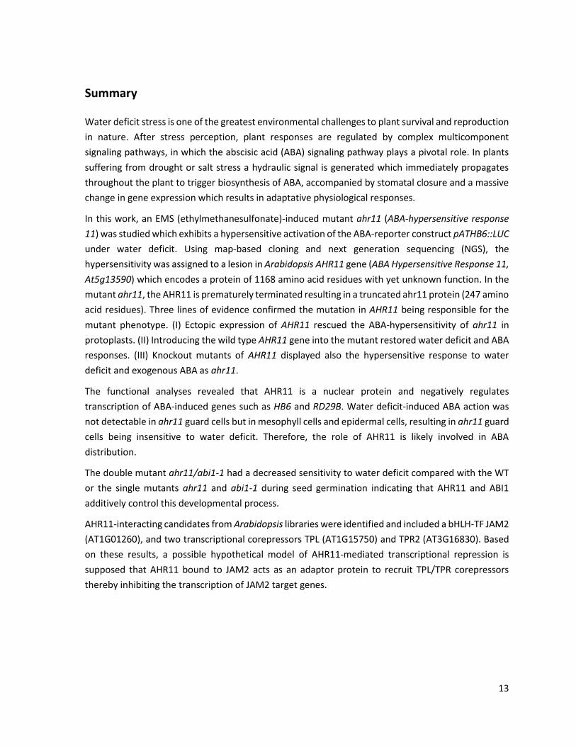
Summary
Water deficit stress is one of the greatest environmental challenges to plant survival and reproduction
in nature. After stress perception, plant responses are regulated by complex multicomponent
signaling pathways, in which the abscisic acid (ABA) signaling pathway plays a pivotal role. In plants
suffering from drought or salt stress a hydraulic signal is generated which immediately propagates
throughout the plant to trigger biosynthesis of ABA, accompanied by stomatal closure and a massive
change in gene expression which results in adaptative physiological responses.
In this work, an EMS (ethylmethanesulfonate)-induced mutant ahr11 (ABA-hypersensitive response
11) was studied which exhibits a hypersensitive activation of the ABA-reporter construct pATHB6::LUC
under water deficit. Using map-based cloning and next generation sequencing (NGS), the
hypersensitivity was assigned to a lesion in Arabidopsis AHR11 gene (ABA Hypersensitive Response 11,
At5g13590) which encodes a protein of 1168 amino acid residues with yet unknown function. In the
mutant ahr11, the AHR11 is prematurely terminated resulting in a truncated ahr11 protein (247 amino
acid residues). Three lines of evidence confirmed the mutation in AHR11 being responsible for the
mutant phenotype. (I) Ectopic expression of AHR11 rescued the ABA-hypersensitivity of ahr11 in
protoplasts. (II) Introducing the wild type AHR11 gene into the mutant restored water deficit and ABA
responses. (III) Knockout mutants of AHR11 displayed also the hypersensitive response to water
deficit and exogenous ABA as ahr11.
The functional analyses revealed that AHR11 is a nuclear protein and negatively regulates
transcription of ABA-induced genes such as HB6 and RD29B. Water deficit-induced ABA action was
not detectable in ahr11 guard cells but in mesophyll cells and epidermal cells, resulting in ahr11 guard
cells being insensitive to water deficit. Therefore, the role of AHR11 is likely involved in ABA
distribution.
The double mutant ahr11/abi1-1 had a decreased sensitivity to water deficit compared with the WT
or the single mutants ahr11 and abi1-1 during seed germination indicating that AHR11 and ABI1
additively control this developmental process.
AHR11-interacting candidates from Arabidopsis libraries were identified and included a bHLH-TF JAM2
(AT1G01260), and two transcriptional corepressors TPL (AT1G15750) and TPR2 (AT3G16830). Based
on these results, a possible hypothetical model of AHR11-mediated transcriptional repression is
supposed that AHR11 bound to JAM2 acts as an adaptor protein to recruit TPL/TPR corepressors
thereby inhibiting the transcription of JAM2 target genes.

15
Zusammenfassung
Für das Ü berleben und die Fortpflanzung stellt in der freien Natur Trockenstress als Umweltfaktor
eine der größten Herausforderungen für Pflanzen dar. Nach der Wahrnehmung der Stresseinwirkung
wird die Reaktion der Pflanze durch komplexe Signalwege aus zahlreichen Komponenten reguliert
wobei der Abscisinsäure (ABA)-Signalweg eine Schlüsselrolle übernimmt. Bei Belastung durch
Trockenstress oder Salzstress wird in Pflanzen ein hydraulisches Signal generiert, das sich sehr schnell
in der gesamten Pflanze ausbreitet und die Biosynthese von ABA induziert. Durch den gestiegenen
ABA-Spiegel kommt es zum Spaltenschluß sowie zu einer starken Veränderung des
Genexpressionsmusters was letztendlich zu physiologischen Anpassungsreaktionen führt.
In dieser Arbeit wurde die durch Ethylmethansulfonat (EMS)-Behandlung generierte Mutante ahr11
(ABA-hypersensitive response 11) untersucht, die bei Wasserstress eine hypersensitive Aktivierung
des ABA-Reporterkonstrukts pAtHB6::LUC zeigt. Mit Hilfe des kartierungsgestützten Klonierens sowie
des Next Generation Sequencing (NGS) gelang es, die hypersensitive Reaktion der Mutante auf eine
Läsion im Arabidopsis-Gen AHR11 (ABA Hypersensitive Response 11, At5g13590) zurückzuführen.
Diese Gen codiert für ein Protein aus 1168 Aminosäuren mit noch nicht bekannter Funktion. In der
Mutante ahr11 liegt AHR11 als trunkiertes Protein ahr11 aus 247 Aminosäuren vor. Drei Befunde
sprechen dafür, dass die Mutation in AHR11 für den beobachteten Phänotyp verantwortlich ist: (I)
Eine Ü berexpression von AHR11 hebt die ABA-Hypersensitivität von ahr11 in Protoplasten auf. (II) Die
Transfektion der Mutante mit dem Wildtyp-Gen AHR11 führt zur Wiederherstellung der normalen
Reaktion auf Wasserdefizit und auf ABA. (III) Knockout-Mutanten von AHR11 zeigen wie ahr11
ebenfalls eine hypersensitive Reaktion auf Wasserdefizit und ABA.
Die funktionelle Analyse ergab dass es sich bei AHR11 um ein kernlokalisiertes Protein handelt, das
die Transkription ABA-induzierter Gene wie HB6 und RD29B negativ reguliert. Wasserdefizit-
induzierte ABA-Aktivität ließ sich in Schließzellen von ahr11 nicht nachweisen, wohl aber in
Mesophyll- und Epidermiszellen, was erklärt warum die Schließzellen von ahr11 gegenüber
Wasserdefizit insensitiv reagieren. AHR11 spielt damit vermutlich eine Rolle bei der ABA-Verteilung.
Eine ahr11/abi1-1 Doppelmutante zeigte hinsichtlich der Keimung eine verringerte Sensitivität
gegenüber Wasserdefizit verglichen mit den Einzelmutanten ahr11 und abi1-1, was dafür spricht, dass
AHR11 und ABI1 diesen Entwicklungsprozess auf additive Weise beeinflussen.
Mögliche AHR11-Interaktoren wurden aus genomischen Arabidopsis-DNA-Banken isoliert. Darunter
befinden sich ein bHLH-Transkriptionsfaktor, JAM2 (AT1G01260) sowie zwei Transkriptionelle Co-
Repressoren, TPL (AT1G15750) und TPR2 (AT3G16830). Aufgrund dieses Ergebnisses wird ein
hypothetisches Modell für die AHR11-vermittelte transkriptionelle Repression vorgeschlagen, in dem
an JAM2 gebundenes AHR11 als Adaptorprotein für die TPL/TPR-Co-Repressoren fungiert und auf
diese Weise die Transkription der JAM2-Zielgene inhibiert.

Introduction
17
1. Introduction
Plants, with their sessile lifestyle in natural habitats, constantly suffer from various abiotic
environmental stresses, such as drought, high salinity, and low temperature. Among these adverse
environmental conditions, drought is considered a major abiotic stress with water deficiency
impairing plant growth and development in > 50% of the Earth’s surface area. It has long been
recognized, e.g. in ancient Egypt thousands of years ago, that high crop yield can be obtained by
appropriate water supply. To date, however, water shortage has become a critical bottleneck in
improvement of worldwide crop productivity. In the future, the situation is expected to be even worse
because of the global climate change and over exploitation of underground water (Gleeson et al.,
2012).
Plant water deficit tolerance or susceptibility is a complex phenomenon, because stress can occur at
different growth stages with varying sensitivety, and moreover, multiple stresses may affect plants
simultaneously. Drought, high salinity and low temperature challenge the water status of plants, and
plants have gradually evolved sophisticated acclimation mechanisms to sense, transduce and respond
to the restricted water availability (Mahajan and Tuteja, 2005). In recent decades, physiological bases
for plant responses to water deficit have become the subject of intense research (Medrano et al.,
2002, Ober and Sharp, 2003, Flexas et al., 2004, Rosado et al., 2006, Christmann et al., 2007). Stomatal
conductance, leaf water potential and turgor pressure are the common parameters to assess the
water deficit response (Medrano et al., 2002, Ache et al., 2010, Tardieu, 2012). At the molecular level,
important drought-related signaling pathways have been unraveled and numerous drought-
responsive genes have been discovered (Bray, 2002, Verslues and Bray, 2006, Chen et al., 2009,
Hayano-Kanashiro et al., 2009, Lim et al., 2009, Chen et al., 2010a, Fujii and Zhu, 2012). However,
some key aspects such as water deficit signal perception and early signal transduction are still little
understood.
1.1 Physiological responses to water deficit
Plant water deficit may result from a number of different abiotic stresses such as drought, salinity and
cold (Mahajan and Tuteja, 2005). The availability of soil water to the plant primarily depends on the
quantity of water stored in the soil and to the soil water potential (ψw). ψw is the chemical potential
of water divided by the partial molar volume (Boyer and Kramer, 1995, Kramer and Boyer, 1995). In
essence, the ψw gradient among soil, plant and atmosphere determines the direction of free water
movement. During daytime, the low value of ψw in the atmosphere pulls up water from the soil
through root and stem into the leaf where it diffuses into the air via the stomata. However, this
pathway requites (I) that root ψw is more negative than soil ψw and (II) that a ψw gradient is maintained
within the plant with most negative ψw occurring in the leaves. A decrease of soil ψw during drought
periods may lower or even reverse the plant-soil ψw gradient thereby impeding water uptake or even
causing a loss of water. To describe the plant’s adaptive strategy to water deficit, the terminologies
Introduction
18
such as drought excape, low ψw /dehydration avoidance and dehydration tolerance were employed
(Levitt, 1972, Verslues and Juenger, 2011).
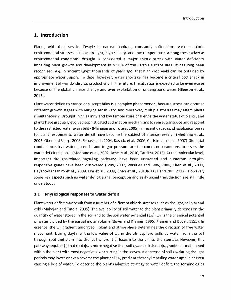
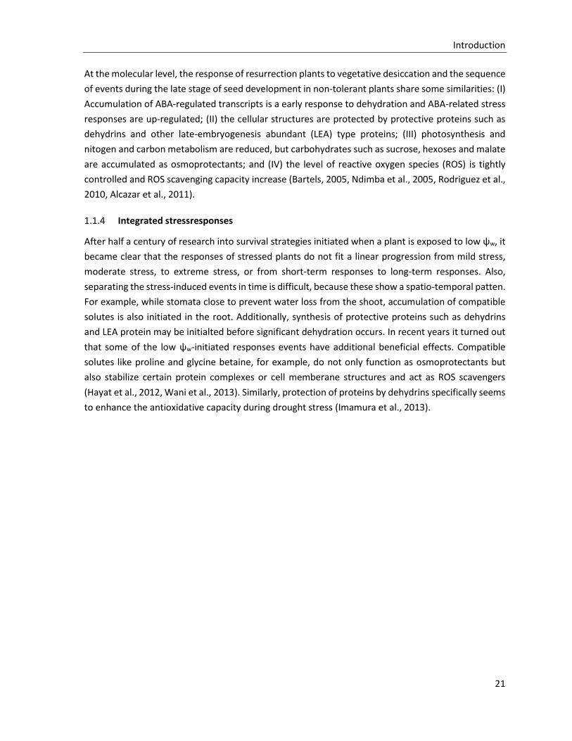
Figure 1-1: Conceptual diagram of the responses of plants to water deficit stress
Water deficit adaptation strategies of plant are termed drought escape, low ψw avoidance and dehydration tolerance (Verslues and Juenger, 2011). Some desert plants adopt drought escape by rapid completion of the growth cycle when the environmentis suitable, while Arabidopsis may to some degree escape stressful conditions by early flowering. Low ψw avoidance strategies may be followed by dehydration tolerance strategies in certain plant species. However, Arabidopsis is not tolerant to severe dehydration. Modified from Verslues and Juenger (2011).
1.1.1 Drought escape
To cope with seasonal water limitation, certain plant species adjust their development to complete
their life cycle in a timely manner. This is a strategy adopted e.g. by some desert plants with very rapid
growth after rain. When water is not limiting, further development may slow down resulting in high
seed yield, whereas under the influence of stress, plants including Arabidopsis often prematurely
transits to reproductive development to generate stress-tolerant seeds before severe stress leads to
the death of the mother plant. A general variation of flowering time has been found among natural
accessions of Arabidopsis thaliana (McKay et al., 2003). In drought stress experiments, the ecotypes
Columbia and Landsberg commonly exhibited early flowering. A screen of different flowering time
mutants under conditions triggering drought-escape (DE) revealed GIGSNTEA (GI), FLOWERING LOCUS
T (FT) and TWIN SISTER OF FT (TSF) as central components of the DE response and showed that the
phytohormone ABA and long days are required for DE (Riboni et al., 2013). From an agricultural
viewpoint, drought escape saves water but also reduces cumulative photosynthesis during the life
cycle, which leads to a trade-off between stress escape and potential yields (Tardieu, 2012, Tardieu,
2013).
Introduction
19
1.1.2 Low water potential/dehydration avoidance
Under mild water deficit, the principal responses of plants help to avoid low ψw and maintain tissue
water balance (water/osmotic homeostasis) close to the unstressed level by reducing water loss
and/or increasing water uptake. Water loss mainly takes place through transpiration via stomatal
pores in aerial tissues and is accompanied by CO2 uptake. Hence, plants quickly close the stomata to
minimize transpiration as a short-term response to mild water deficit. At the whole plant level, gain
of above-ground biomass decreases in response to water deficit due to changes of carbon usage
(Hummel et al., 2010). An attenuated shoot development is an efficacious way to limit leaf area and
thereby transpirational water loss. With water shortage, Arabidopsis leaf rosette growth has been
shown to decrease more than photosynthesis, leading to an increase in carbon availability to promote
root growth and osmotic adjustment (Hummel et al., 2010). Correspondingly, the sensitivity to low
soil ψw is distinct between the root and the shoot (Hsiao and Xu, 2000, Mahajan and Tuteja, 2005).
Compared to the stress-limited shoot development, the root system is generally more resistant to
stress. Mild water deficit even promotes axial root elongation and lateral root development, which
allows to explore the soil for yet unexploited water sources (Sharp et al., 2004, Yamaguchi et al., 2009,
Yamaguchi and Sharp, 2010). Still, the trade-off associated with a reduced transpiration rate is a
decrease of photosynthesis, which means an overall diminished carbon assimilation and shoot growth.
When water homeostasis cannot be maintained by the low ψw avoidance response mentioned above,
a decrease tissue ψw, particularly in the root, is the next adaptative step. Of the different components
of ψw, which is the osmotic potential ψs, the pressure potential ψp and the gravitational potential ψg
(Christmann et al., 2013), only ψs may be altered by the plant (few exceptions exist where also ψp is
modified) which, however, indirectly also affects ψp. The ψs is substantially lowered by an increase in
solute concentration. While an increase of anorganic ions and organic acid ions is observed in the
vacuole, ψs in the cytoplasm is adjusted with the help of a number of small molecules named
osmoprotectants or compatible solutes, which do not interfere with cellular function and help
organisms to survive extreme osmotic stress (Lang, 2007). Osmotic adjustment of the root may be
achieved by uptake of inorganic ions such as K+, Cl- and Na+ from the soil with K+ uptake mediated by
K+ channels playing a key role (Sze et al., 2004, Osakabe et al., 2013, Wang et al., 2013). Adjustment
was shown to be fast with hyperosmotically-stressed root cells of Arabidopsis recovering their turgor
within 40 to 50 min (Shabala and Lew, 2002). Enhanced ion uptake is accompanied by synthesis of
osmoprotectants such as the amino acids proline, non-reducing sugars (Suc and trehalose), polyols
(mannitol and sorbitol) and quaternary amines (such as glycine betaine) under water deficit stress
(Nakayama et al., 2000, Rontein et al., 2002, Iordachescu and Imai, 2008, Hayat et al., 2012, Wani et
al., 2013). The most striking property of these solutes is the lack of a negative influence on cellular
metabolism and function.
The pressure potential ψp of living cells is the cell turgor pressure which is thought to be at or above
zero while in non-living water-conducting cells with thick, lignified walls, ψp usually is negative and is
then called tension. Under water deficit stress, cell wall extensbility is reduced as part of the water
deficit response (Moore et al., 2008). Studies of drought effects on the cell wells in roots of maize (Zea
Introduction
20
mays) revealed that cell wall hardening occurs in the basal part of the root elongation zone, and is
accompanied by alterations in metabolism and accumulation of cell wall proteins, such as expansins,
xyloglucan endotransglycosylases, glucanases, dehydrins and other proteins of carbohydrate and
amino acid metabolism (Wu and Cosgrove, 2000, Fan and Neumann, 2004, Sharp et al., 2004, Fan et
al., 2006, Poroyko et al., 2007, Zhu et al., 2007a, Spollen et al., 2008). And similar proteomic analyses
of root cell wall were also performed in soybean and common bean (Yamaguchi et al., 2009, Yang et
al., 2013).
If soil ψw becomes still more negative, additional mechanisms become important, such as leaf area
adjustment by shedding of leaves to reduce transpiration or a premature termination of the plant’s
life cycle. Abscission of leaves is preceded by leaf senescence, a programmed aging process during
which important nutrients are redistributed from the senescing leafs to actively growing tissues or
storage organs. Senescence is regulated by plant hormones with ethylene functioning as the major
senescence-promoting hormone and cytokinin as the major senescence-inhibiting hormone and may
be prematurely induced by abiotic stresses through an effect on hormone homeostasis (Buchanan-
Wollaston et al., 2005). Although the promotion of senescence by different signals may involve
distinct signal transduction pathways, similar senescence-associated genes (SAGs) are induced by
different stresses. Therefore the initiated senescence processes may share common execution events
(Guo and Gan, 2012). The premature termination of the life cycle may be understood as a premature
initiation of whole-plant senescence where redistribution of nutrients into reproductive structures
ensures the production of the next generation.
1.1.3 Dehydration tolerance
In order to colonize the land, the early land plants with their very simple architecture must have been
desiccation-tolerant in both vegetative and reproductive stages to survive the dehydrating
atmospheres of land habitats (Oliver et al., 2005). Vegetative desiccation tolerance is common in less
complex plants such as bryophytes (Proctor and Pence, 2002), but only about 300 species with
vegetative desiccation tolerance are found in vascular plants (Porembski and Barthlott, 2000). These
vascular plants, also named resurrection plants (Gaff, 1971), can recover from an extreme
dehydration and resume normal growth when water is available(Phillips et al., 2008). Craterostigma
plantagineum has been employed as a model system to unveil the mechanisms of desiccation
tolerance because desiccation tolerance can be studied both in undifferentiated callus cultures and
in differentiated plants (Bartels, 2005, Rodriguez et al., 2010, Alcazar et al., 2011). Plants which are
not sesiccation-tolerant in the vegetative stage often show dehydration tolerance during reproductive
stages. Seed dehydration tolerance is associated with seed dormancy and is of great importance for
survival. Until the environmental conditions are suitable to establish a new plant generation, the seed
enters a dormant stage to survive even extended periods of unfavorable conditions (Koornneef et al.,
2002, Finch-Savage and Leubner-Metzger, 2006, Bentsink and Koornneef, 2008, Holdsworth et al.,
2008). Seed dormancy is controlled by genetic factors as well by environmental factors such as light,
temperature and water availability and also by the time elapsed since initiation of dormancy.
Introduction
21
At the molecular level, the response of resurrection plants to vegetative desiccation and the sequence
of events during the late stage of seed development in non-tolerant plants share some similarities: (I)
Accumulation of ABA-regulated transcripts is a early response to dehydration and ABA-related stress
responses are up-regulated; (II) the cellular structures are protected by protective proteins such as
dehydrins and other late-embryogenesis abundant (LEA) type proteins; (III) photosynthesis and
nitogen and carbon metabolism are reduced, but carbohydrates such as sucrose, hexoses and malate
are accumulated as osmoprotectants; and (IV) the level of reactive oxygen species (ROS) is tightly
controlled and ROS scavenging capacity increase (Bartels, 2005, Ndimba et al., 2005, Rodriguez et al.,
2010, Alcazar et al., 2011).
1.1.4 Integrated stressresponses
After half a century of research into survival strategies initiated when a plant is exposed to low ψw, it
became clear that the responses of stressed plants do not fit a linear progression from mild stress,
moderate stress, to extreme stress, or from short-term responses to long-term responses. Also,
separating the stress-induced events in time is difficult, because these show a spatio-temporal patten.
For example, while stomata close to prevent water loss from the shoot, accumulation of compatible
solutes is also initiated in the root. Additionally, synthesis of protective proteins such as dehydrins
and LEA protein may be initialted before significant dehydration occurs. In recent years it turned out
that some of the low ψw-initiated responses events have additional beneficial effects. Compatible
solutes like proline and glycine betaine, for example, do not only function as osmoprotectants but
also stabilize certain protein complexes or cell memberane structures and act as ROS scavengers
(Hayat et al., 2012, Wani et al., 2013). Similarly, protection of proteins by dehydrins specifically seems
to enhance the antioxidative capacity during drought stress (Imamura et al., 2013).
Introduction
22
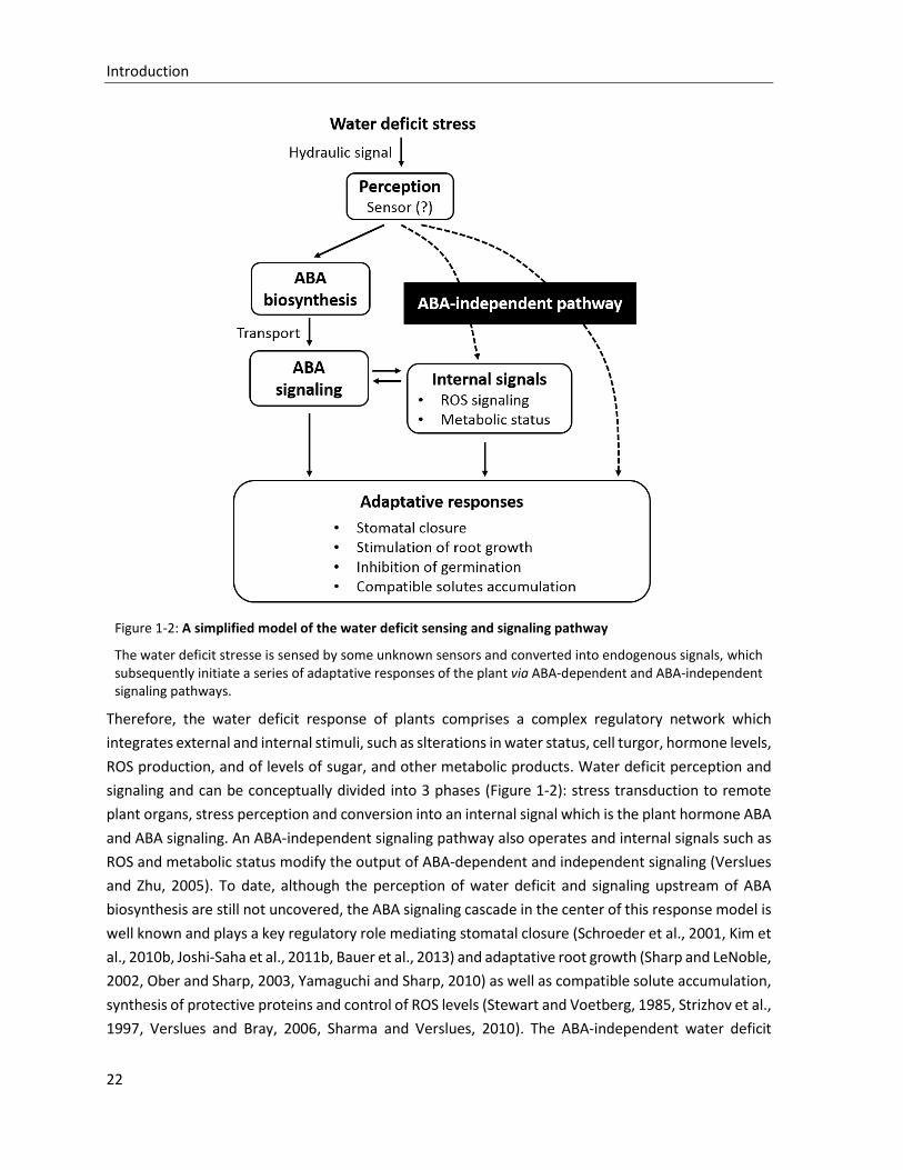
Figure 1-2: A simplified model of the water deficit sensing and signaling pathway
The water deficit stresse is sensed by some unknown sensors and converted into endogenous signals, which subsequently initiate a series of adaptative responses of the plant via ABA-dependent and ABA-independent signaling pathways.
Therefore, the water deficit response of plants comprises a complex regulatory network which
integrates external and internal stimuli, such as slterations in water status, cell turgor, hormone levels,
ROS production, and of levels of sugar, and other metabolic products. Water deficit perception and
signaling and can be conceptually divided into 3 phases (Figure 1-2): stress transduction to remote
plant organs, stress perception and conversion into an internal signal which is the plant hormone ABA
and ABA signaling. An ABA-independent signaling pathway also operates and internal signals such as
ROS and metabolic status modify the output of ABA-dependent and independent signaling (Verslues
and Zhu, 2005). To date, although the perception of water deficit and signaling upstream of ABA
biosynthesis are still not uncovered, the ABA signaling cascade in the center of this response model is
well known and plays a key regulatory role mediating stomatal closure (Schroeder et al., 2001, Kim et
al., 2010b, Joshi-Saha et al., 2011b, Bauer et al., 2013) and adaptative root growth (Sharp and LeNoble,
2002, Ober and Sharp, 2003, Yamaguchi and Sharp, 2010) as well as compatible solute accumulation,
synthesis of protective proteins and control of ROS levels (Stewart and Voetberg, 1985, Strizhov et al.,
1997, Verslues and Bray, 2006, Sharma and Verslues, 2010). The ABA-independent water deficit
Introduction
23
signaling pathway also stimulates stress adaptation of plants, but its precise role and the structure of
the respective signaling network are still much less clear.
1.2 Water deficit perception and transduction
The water deficit response of plants starts with sensing of external stimuli, which is low soil ψw. The
external signals are both converted into endogenous signals and transmitted to plant parts distal from
the site of perception. Low soil ψw induces a drop in ψw of root cells which causes a loss of turgor in
these cells. After a lag phase, responses are observed in remote plant parts like the shoot. It was
suggested that a decrease in soil ψw was converted into an internal chemical signal in the root with
ABA being the likely candidate. The plant hormone is supposed to be then transmitted to the shoot
via the xylem (Wilkinson and Davies, 2002). This hypothesis was recently challenged by studies which
used reciprocal grafting of wild type and ABA-deficient mutants of Arabidopsis and tomato. According
to these studies, root exposure to water deficit induces ABA biosynthesis in the shoot and shoot-
biosynthesized ABA is necessary and sufficient to mediate drought-induced responses (Holbrook et
al., 2002, Christmann et al., 2007). It was further demonstrated that water deficit stress results in
generation of a hydraulic signal which is rapidly transmitted to the shoot. Attenuation of the hydraulic
signal by feeding water to leaves abolished ABA-dependent responses like stomatal closure
(Christmann et al., 2007). Water feeding to leaves would not attenuate chemical signals or electrical
signals which have also been suggested as long-distance signals relayed from roots (Grams et al., 2007,
Gil et al., 2008). Accordingly a hydraulic signal plays a crucial role in the water deficit response
(Christmann et al., 2007, Christmann et al., 2013). In essence, a drought-induced hydraulic signal is a
drop in ψw, which in living cells where the signal is decoded comprises a decrease of the cell turgor
and of the osmotic potential. To date the hydraulic sensors in plant as well as the early steps in the
signaling cascade initiated by the hydraulic signal are still unknown. In theory any cellular component
that monitors turgor-dependent parameters such as cell wall tension or changes in osmotic potential
is a promising candidate for a hydraulic sensor. Also, candidate sensors could be homologs of
osmosensors or mechanosensors of bacteria, fungi, and metazoans. The term hydraulic sensor will be
used here to cover sensors for which the mode of action is unknown or has not been proven and that
putatively moitor changes in turgor, solute concentration or cell wall-plasma membrane associated
tension.
Introduction
24
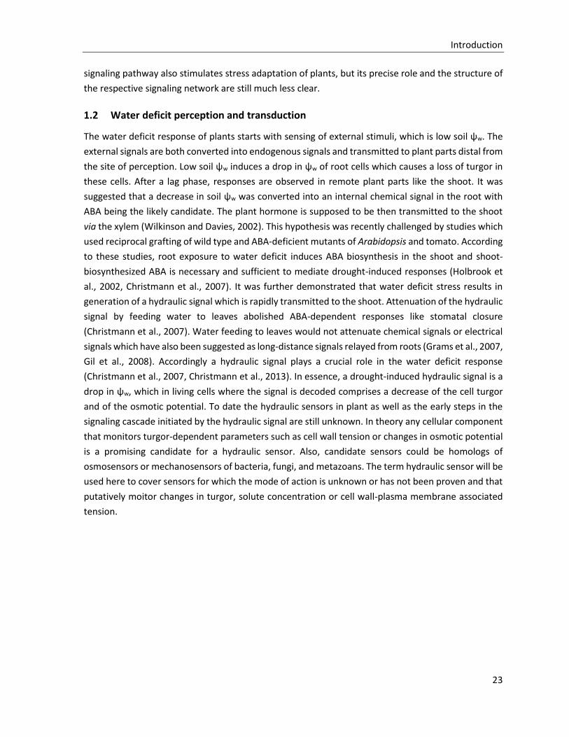
Figure 1-3: Hydraulic sensors and sensor candidates in different organisms
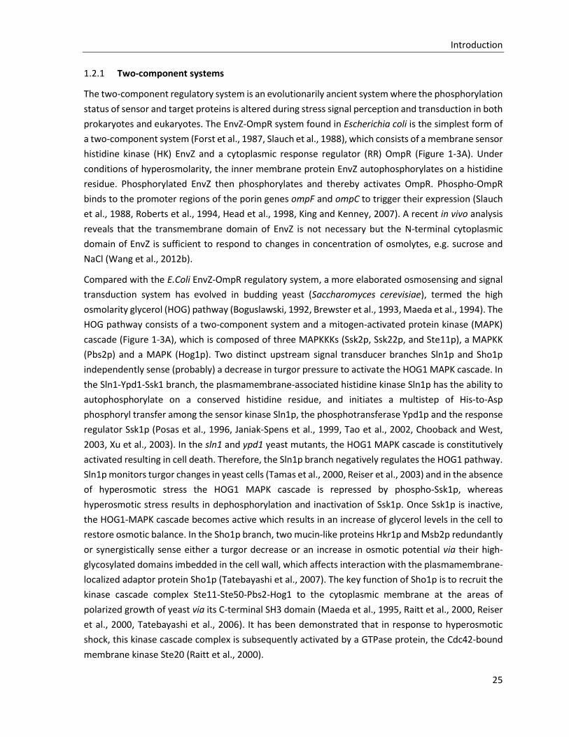
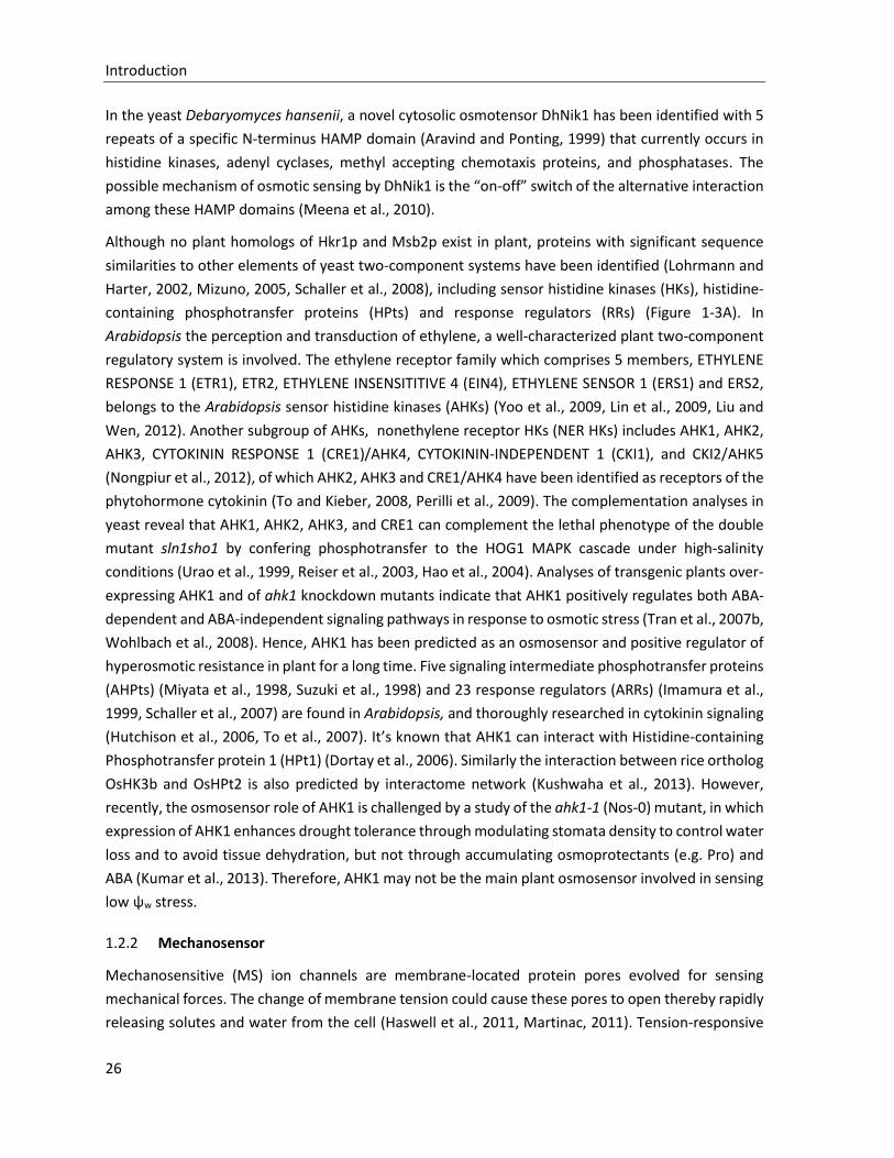
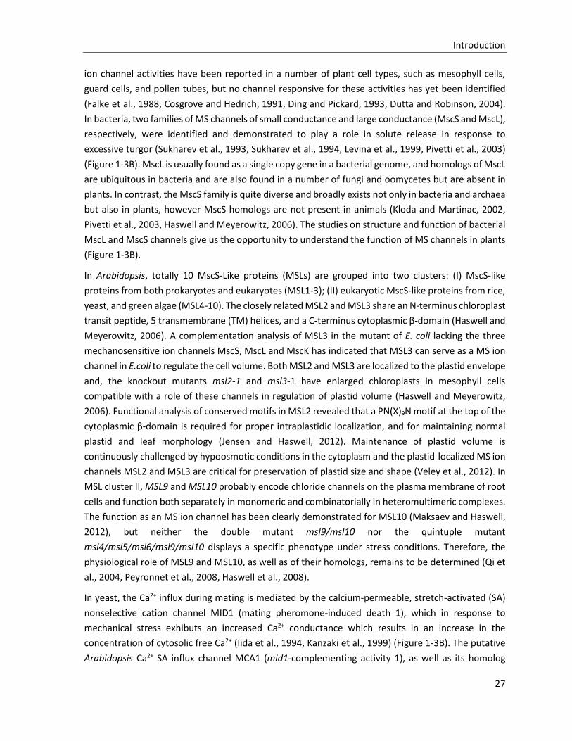
Osmosensors, turgor sensors and sensors of cell wall integrity identified in E. coli and in yeast are shown together with the respective plant candidates. (A) Two-component stress response systems. (B) Mechanosensitive (MS) ion channels. (C) Cell wall integrity (CWI) pathways. See text for more details.
Introduction
25
1.2.1 Two-component systems
The two-component regulatory system is an evolutionarily ancient system where the phosphorylation
status of sensor and target proteins is altered during stress signal perception and transduction in both
prokaryotes and eukaryotes. The EnvZ-OmpR system found in Escherichia coli is the simplest form of
a two-component system (Forst et al., 1987, Slauch et al., 1988), which consists of a membrane sensor
histidine kinase (HK) EnvZ and a cytoplasmic response regulator (RR) OmpR (Figure 1-3A). Under
conditions of hyperosmolarity, the inner membrane protein EnvZ autophosphorylates on a histidine
residue. Phosphorylated EnvZ then phosphorylates and thereby activates OmpR. Phospho-OmpR
binds to the promoter regions of the porin genes ompF and ompC to trigger their expression (Slauch
et al., 1988, Roberts et al., 1994, Head et al., 1998, King and Kenney, 2007). A recent in vivo analysis
reveals that the transmembrane domain of EnvZ is not necessary but the N-terminal cytoplasmic
domain of EnvZ is sufficient to respond to changes in concentration of osmolytes, e.g. sucrose and
NaCl (Wang et al., 2012b).
Compared with the E.Coli EnvZ-OmpR regulatory system, a more elaborated osmosensing and signal
transduction system has evolved in budding yeast (Saccharomyces cerevisiae), termed the high
osmolarity glycerol (HOG) pathway (Boguslawski, 1992, Brewster et al., 1993, Maeda et al., 1994). The
HOG pathway consists of a two-component system and a mitogen-activated protein kinase (MAPK)
cascade (Figure 1-3A), which is composed of three MAPKKKs (Ssk2p, Ssk22p, and Ste11p), a MAPKK
(Pbs2p) and a MAPK (Hog1p). Two distinct upstream signal transducer branches Sln1p and Sho1p
independently sense (probably) a decrease in turgor pressure to activate the HOG1 MAPK cascade. In
the Sln1-Ypd1-Ssk1 branch, the plasmamembrane-associated histidine kinase Sln1p has the ability to
autophosphorylate on a conserved histidine residue, and initiates a multistep of His-to-Asp
phosphoryl transfer among the sensor kinase Sln1p, the phosphotransferase Ypd1p and the response
regulator Ssk1p (Posas et al., 1996, Janiak-Spens et al., 1999, Tao et al., 2002, Chooback and West,
2003, Xu et al., 2003). In the sln1 and ypd1 yeast mutants, the HOG1 MAPK cascade is constitutively
activated resulting in cell death. Therefore, the Sln1p branch negatively regulates the HOG1 pathway.
Sln1p monitors turgor changes in yeast cells (Tamas et al., 2000, Reiser et al., 2003) and in the absence
of hyperosmotic stress the HOG1 MAPK cascade is repressed by phospho-Ssk1p, whereas
hyperosmotic stress results in dephosphorylation and inactivation of Ssk1p. Once Ssk1p is inactive,
the HOG1-MAPK cascade becomes active which results in an increase of glycerol levels in the cell to
restore osmotic balance. In the Sho1p branch, two mucin-like proteins Hkr1p and Msb2p redundantly
or synergistically sense either a turgor decrease or an increase in osmotic potential via their high-
glycosylated domains imbedded in the cell wall, which affects interaction with the plasmamembrane-
localized adaptor protein Sho1p (Tatebayashi et al., 2007). The key function of Sho1p is to recruit the
kinase cascade complex Ste11-Ste50-Pbs2-Hog1 to the cytoplasmic membrane at the areas of
polarized growth of yeast via its C-terminal SH3 domain (Maeda et al., 1995, Raitt et al., 2000, Reiser
et al., 2000, Tatebayashi et al., 2006). It has been demonstrated that in response to hyperosmotic
shock, this kinase cascade complex is subsequently activated by a GTPase protein, the Cdc42-bound
membrane kinase Ste20 (Raitt et al., 2000).
Introduction
26
In the yeast Debaryomyces hansenii, a novel cytosolic osmotensor DhNik1 has been identified with 5
repeats of a specific N-terminus HAMP domain (Aravind and Ponting, 1999) that currently occurs in
histidine kinases, adenyl cyclases, methyl accepting chemotaxis proteins, and phosphatases. The
possible mechanism of osmotic sensing by DhNik1 is the “on-off” switch of the alternative interaction
among these HAMP domains (Meena et al., 2010).
Although no plant homologs of Hkr1p and Msb2p exist in plant, proteins with significant sequence
similarities to other elements of yeast two-component systems have been identified (Lohrmann and
Harter, 2002, Mizuno, 2005, Schaller et al., 2008), including sensor histidine kinases (HKs), histidine-
containing phosphotransfer proteins (HPts) and response regulators (RRs) (Figure 1-3A). In
Arabidopsis the perception and transduction of ethylene, a well-characterized plant two-component
regulatory system is involved. The ethylene receptor family which comprises 5 members, ETHYLENE
RESPONSE 1 (ETR1), ETR2, ETHYLENE INSENSITITIVE 4 (EIN4), ETHYLENE SENSOR 1 (ERS1) and ERS2,
belongs to the Arabidopsis sensor histidine kinases (AHKs) (Yoo et al., 2009, Lin et al., 2009, Liu and
Wen, 2012). Another subgroup of AHKs, nonethylene receptor HKs (NER HKs) includes AHK1, AHK2,
AHK3, CYTOKININ RESPONSE 1 (CRE1)/AHK4, CYTOKININ-INDEPENDENT 1 (CKI1), and CKI2/AHK5
(Nongpiur et al., 2012), of which AHK2, AHK3 and CRE1/AHK4 have been identified as receptors of the
phytohormone cytokinin (To and Kieber, 2008, Perilli et al., 2009). The complementation analyses in
yeast reveal that AHK1, AHK2, AHK3, and CRE1 can complement the lethal phenotype of the double
mutant sln1sho1 by confering phosphotransfer to the HOG1 MAPK cascade under high-salinity
conditions (Urao et al., 1999, Reiser et al., 2003, Hao et al., 2004). Analyses of transgenic plants over-
expressing AHK1 and of ahk1 knockdown mutants indicate that AHK1 positively regulates both ABA-
dependent and ABA-independent signaling pathways in response to osmotic stress (Tran et al., 2007b,
Wohlbach et al., 2008). Hence, AHK1 has been predicted as an osmosensor and positive regulator of
hyperosmotic resistance in plant for a long time. Five signaling intermediate phosphotransfer proteins
(AHPts) (Miyata et al., 1998, Suzuki et al., 1998) and 23 response regulators (ARRs) (Imamura et al.,
1999, Schaller et al., 2007) are found in Arabidopsis, and thoroughly researched in cytokinin signaling
(Hutchison et al., 2006, To et al., 2007). It’s known that AHK1 can interact with Histidine-containing
Phosphotransfer protein 1 (HPt1) (Dortay et al., 2006). Similarly the interaction between rice ortholog
OsHK3b and OsHPt2 is also predicted by interactome network (Kushwaha et al., 2013). However,
recently, the osmosensor role of AHK1 is challenged by a study of the ahk1-1 (Nos-0) mutant, in which
expression of AHK1 enhances drought tolerance through modulating stomata density to control water
loss and to avoid tissue dehydration, but not through accumulating osmoprotectants (e.g. Pro) and
ABA (Kumar et al., 2013). Therefore, AHK1 may not be the main plant osmosensor involved in sensing
low ψw stress.
1.2.2 Mechanosensor
Mechanosensitive (MS) ion channels are membrane-located protein pores evolved for sensing
mechanical forces. The change of membrane tension could cause these pores to open thereby rapidly
releasing solutes and water from the cell (Haswell et al., 2011, Martinac, 2011). Tension-responsive
Introduction
27
ion channel activities have been reported in a number of plant cell types, such as mesophyll cells,
guard cells, and pollen tubes, but no channel responsive for these activities has yet been identified
(Falke et al., 1988, Cosgrove and Hedrich, 1991, Ding and Pickard, 1993, Dutta and Robinson, 2004).
In bacteria, two families of MS channels of small conductance and large conductance (MscS and MscL),
respectively, were identified and demonstrated to play a role in solute release in response to
excessive turgor (Sukharev et al., 1993, Sukharev et al., 1994, Levina et al., 1999, Pivetti et al., 2003)
(Figure 1-3B). MscL is usually found as a single copy gene in a bacterial genome, and homologs of MscL
are ubiquitous in bacteria and are also found in a number of fungi and oomycetes but are absent in
plants. In contrast, the MscS family is quite diverse and broadly exists not only in bacteria and archaea
but also in plants, however MscS homologs are not present in animals (Kloda and Martinac, 2002,
Pivetti et al., 2003, Haswell and Meyerowitz, 2006). The studies on structure and function of bacterial
MscL and MscS channels give us the opportunity to understand the function of MS channels in plants
(Figure 1-3B).
In Arabidopsis, totally 10 MscS-Like proteins (MSLs) are grouped into two clusters: (I) MscS-like
proteins from both prokaryotes and eukaryotes (MSL1-3); (II) eukaryotic MscS-like proteins from rice,
yeast, and green algae (MSL4-10). The closely related MSL2 and MSL3 share an N-terminus chloroplast
transit peptide, 5 transmembrane (TM) helices, and a C-terminus cytoplasmic β-domain (Haswell and
Meyerowitz, 2006). A complementation analysis of MSL3 in the mutant of E. coli lacking the three
mechanosensitive ion channels MscS, MscL and MscK has indicated that MSL3 can serve as a MS ion
channel in E.coli to regulate the cell volume. Both MSL2 and MSL3 are localized to the plastid envelope
and, the knockout mutants msl2-1 and msl3-1 have enlarged chloroplasts in mesophyll cells
compatible with a role of these channels in regulation of plastid volume (Haswell and Meyerowitz,
2006). Functional analysis of conserved motifs in MSL2 revealed that a PN(X)9N motif at the top of the
cytoplasmic β-domain is required for proper intraplastidic localization, and for maintaining normal
plastid and leaf morphology (Jensen and Haswell, 2012). Maintenance of plastid volume is
continuously challenged by hypoosmotic conditions in the cytoplasm and the plastid-localized MS ion
channels MSL2 and MSL3 are critical for preservation of plastid size and shape (Veley et al., 2012). In
MSL cluster II, MSL9 and MSL10 probably encode chloride channels on the plasma membrane of root
cells and function both separately in monomeric and combinatorially in heteromultimeric complexes.
The function as an MS ion channel has been clearly demonstrated for MSL10 (Maksaev and Haswell,
2012), but neither the double mutant msl9/msl10 nor the quintuple mutant
msl4/msl5/msl6/msl9/msl10 displays a specific phenotype under stress conditions. Therefore, the
physiological role of MSL9 and MSL10, as well as of their homologs, remains to be determined (Qi et
al., 2004, Peyronnet et al., 2008, Haswell et al., 2008).
In yeast, the Ca2+ influx during mating is mediated by the calcium-permeable, stretch-activated (SA)
nonselective cation channel MID1 (mating pheromone-induced death 1), which in response to
mechanical stress exhibuts an increased Ca2+ conductance which results in an increase in the
concentration of cytosolic free Ca2+ (Iida et al., 1994, Kanzaki et al., 1999) (Figure 1-3B). The putative
Arabidopsis Ca2+ SA influx channel MCA1 (mid1-complementing activity 1), as well as its homolog
Introduction
28
MCA2, have been identified by a functional complementation screening of the yeast mutant mid1
(Nakagawa et al., 2007). The expression of MCA1 and MCA2 complements the lethal phenotype of
the mid1 mutant, and mediates the stretch activated Ca2+ uptake in yeast (Nakagawa et al., 2007).
Also, MCA1-dependent MS cation currents were observed in Xenopus laevis oocytes (Furuichi et al.,
2012). MCA1 is present in the plasma membrane of root cells, and in yeast, too, MCA1 localizes to the
yeast plasma membrane as an integral membrane protein. MCA2 shows an overlapping but also
distinct spatial expression pattern compared to MCA1. Both single mutants mca1-null and mca2-null
are defective in Ca2+ uptake from the roots, but only the primary roots of mca1-null seedling are
impaired in penetrating a harder agar medium from a softer (Yamanaka et al., 2010). Therefore, MCA1
and MCA2 are putative MS ion channels which differentially mediate Ca2+ uptake in Arabidopsis
(Figure 1-3B).
1.2.3 Cell wall integrity (CWI) sensor
Cell wall remodeling during growth and upon extracellular stress has been extensively studied in yeast.
The signal transduction pathway uncovered by such studies has been designated cell wall integrity
(CWI) pathway, which essentially is composed of the cell wall-associated cell surface sensors, the
GDP/GTP exchange factors (GEFs), and the small GTPases (Rhos) (Levin, 2005, Levin, 2011). The yeast
genome encodes five CWI sensors belonging to two subgroups (Figure 1-3C): the Wsc-type sensors
(Wsc1, Wsc2 and Wsc3) and the Mid-type sensors (Mid2 and Mtl1). All of them share a single
transmembrane domain (TMD), a short cytoplasmic tail (CT) and a large extracellular region (Rodicio
and Heinisch, 2010). In the extracellular region the highly O-mannosylated serine- and threonine-rich
(STR) sequences form a nanospring-like structure that is capable of resisting high mechanical force
and of responding to cell surface stress (Dupres et al., 2009). In addition, the specific cysteine-rich
domain (CRD) at the N-terminus of Wsc sensors transiently interacts with glucan chains in the cell wall.
Although Mid-type sensors lack the CRD domain, an N-glycosylated asparagine near the N-terminus
may serve a similar function as CRD domain (Hutzler et al., 2008). Consequently, strain either on the
cell wall or on the plasma membrane should generate a mechanical force on the extracellular
nanospring-like structure. The strain-induced conformational changes of CWI sensors could then
signal to GEFs, which directly activate the Rho GTPases, and then trigger the sole yeast protein kinase
C (PKC1)-activated MAPK cascade (Garcia et al., 2006).
Similar to the STR sequences in Wsc1, the ankyrin repeat motifs have also been postuated to form
nanospring structures which, when coupled to an ion channel are thought to transmit mechanical
forces to the channel thereby altering channel open state probability (Howard and Bechstedt, 2004,
Lee et al., 2006b). Accordingly, the plant proteins containing ankyrin repeats (Becerra et al., 2004)
including kinases and potassium channels might be involved in hydraulic sensing.
Proteins putatively involved in sensing of plant cell wall integrity include the L-type lectin receptor-
like kinases (LecRLKs) and the proline-rich extension-like receptor kinases (PERKs) (Figure 1-3C).
LecRLKs were identified in a screen for tripeptidic integrin-recognition motif Arg-Gly-Asp (RGD)-
binding proteins (Gouget et al., 2006). Integrins are central components of metazoan plasma

Introduction
29
membrane-localized protein complexes thought to function in mechanosensing of shear stress (Shyy
and Chien, 2002) and the LecRLKs seem to play a structural and signaling role at the plant cell surfaces
through protein-protein interactions with plant RGD-containing proteins (Gouget et al., 2006). Further
members of the receptor-like kinase family (RLKs) whose extracellular domains mediate carbohydrate
ligand binding in the cell wall have recently been linked cell wall integrity sensing during vegetative
and reproductive development (Boisson-Dernier et al., 2011).
The Arabidopsis genome encodes 15 PERKs, of which PERK4 has been identified as a plasma
membrane-associated protein which perturbs Ca2+ homeostasis during the early stage of ABA
signaling to inhibit primary root cell elongation (Bai et al., 2009a, Bai et al., 2009b). Additionally, the
receptor-like wall-associated kinases (WAKs) bind pectins in the cell wall, and are necessary both for
cell expansion during development and for a response to pathogens and wounding. The type and
concentration of pectins in the wall could lead to a WAK-dependent activation of different cytosolic
MAPK signaling pathways (Kohorn and Kohorn, 2012).
In the nematode Caenorhabditis elegans, an eukaryotic MS sodium channel complex (MEC-4/MEC10)
has been identified in a screen for mutants defective in the response to gentle touch (Chalfie and Au,
1989). The channel proteins have specialized extracellular matrix (ECM) and unique cytoskeletal
elements to form the adhesions between the cell wall and the plasma membrane (Arnadottir and
Chalfie, 2010). Similarly, the animal cell surface receptor integrins also have a dual-tethered adhesion
structure, of which the extracellular domains of integrins bind to cell wall ligands, while the flexible
internal tails interact with the actin cytoskeleton, to connect the interior of the cell to the extracellular
environment and to bi-directionally transmit signals across the cell membrane (Wegener and
Campbell, 2008, Shyy and Chien, 2002, Legate et al., 2009). Although the analysis of the Arabidopsis
genome indicates that homologs of MEC-4/MEC-10 channels and integrins do not exist, plant genome
encodes some integrin-like proteins that are structurally and functionally similar to animal integrins
(Schindler et al., 1989). For instance, the Zea mays integrin-like proteins mediate the interaction
between cell wall and plasma membrane as well as cell responses to osmotic stress (Lu et al., 2007).
An Arabidopsis plasma membrane protein AT14A serves as a transmembrane linker between the cell
wall and the cytoskeleton just as integrin complexes do in animals, and plays important roles in
controlling polarity and morphogenesis (Lu et al., 2012). In addition, Arabidopsis integrin-like protein
NON-RACE-SPECIFIC DISEASE RESISTANCE 1 (NDR1) has been demonstrated to play an essential role
in plant disease resistance (Lu et al., 2013, Knepper et al., 2011). Taken together the plant integrin-
like proteins have important roles of maintaining the integrity of the cell wall-plasma membrane
connection and are involved in the cell wall strain perception and signal transduction during growth
development and environmental stress.
1.2.4 Upstream signal transduction
Whatever the primary sensing mechanism, it must be linked to specific down-stream molecules that
further transmit the signal. It has long been known that cytoplasmic free Ca2+ increases in Arabidopsis
seedlings within seconds of osmotic stress (Knight et al., 1997) and under mechanical stimulation
Introduction
30
(Monshausen et al., 2009). Some presumed hydraulic signal sensors such as MCA1 and MCA2 are
calcium channel proteins which might directly mediate Ca2+ uptake (Yamanaka et al., 2010), while the
sensor candidate PERK4 indirectly regulates Ca2+ homeostasis (Bai et al., 2009a). Hence, cytoplasmic
Ca2+ transients have been considered to play a role in the early response to water deficit. However,
Ca2+ transients are also involved in early ABA signaling (see section 1.3.5). Therefore, it is hard to
discern of Ca2+ action upstream or downstream of ABA, or both.
The MAPK kinase pathway, an eukaryotic conserved signal transduction module which is activated as
part of the HOG pathway in yeast. In Arabidopsis, 80 MAPKK kinases (MAPKKKs), 10 MAPK kinase
(MAPKKs/MKKs) and 20 MAP kinases (MAPKs/MPKs) have been characterized (Ichimura et al., 2002).
The MAPKKKs are probably the scaffolding proteins to recruit the MAPK cascade to the response sites.
The diversity of MAPKKKs ensures that different external stimuli are precisely converted to initiate
similar MAPK cascades, which finally activate the same downstream MAPK. Several hyperosmotic
stress-activated plant MAPK cascades have been identified in many species, such as the cscades
activating salt stress-inducible MAP kinase (SIMK) alfalfa (Munnik et al., 1999, Kiegerl et al., 2000),
salicylic acid-induced protein kinase (SIPK) in tobacco (Droillard et al., 2000, Liu et al., 2000,
Mikolajczyk et al., 2000), as well as MPK3, MPK4, MPK6 and MPK7 in Arabidopsis (Ichimura et al.,
2000, Droillard et al., 2002, Colcombet and Hirt, 2008, Opdenakker et al., 2012). However, unlike in
yeast, osmotic stress-dependent MAPK cascade activation in plants seems to be no early response
directly triggered by osmosensing. Rather, these MAP kinase cascades appear to be activated H2O2-
dependently during ABA signaling (see section 1.3.4.3).
In spite of the limited knowledge about hydraulic signal perception and early signal transduction, the
accumulation of endogenous ABA, which is followed by ABA signal transduction, is undoubtedly an
output of upstream hydraulic signaling (see section 1.3). Concomitantly water deficit is also
transduced via an ABA-independent pathway (see section 1.4).
Introduction
31
1.3 ABA-dependent signaling pathway
The plant hormone abscisic acid (ABA) was discovered as abscisin II and dormin in 1960s (Liu and
Carnsdagger, 1961, Ohkuma et al., 1963). ABA modulates many aspects of plant growth and
development including embryo maturation, seed dormancy, germination, cell division, cell elongation,
and floral induction (Finkelstein et al., 2002, Cutler et al., 2010, Takezawa et al., 2011, Seung et al.,
2011). Correspondingly, the ABA-deficient mutant aba1-1 displays a stunted growth phenotype
(Sharp, 2002), a phenotype which can be rescued by exogenous ABA (Finkelstein et al., 2002). In
addition, ABA is extensively studied as a ‘stress hormone’, because of its dramatical increase when
plants are challenged by biotic and abiotic stresses (Schroeder et al., 2001, Zhu, 2002, Himmelbach et
al., 2003, Christmann et al., 2006, Qin et al., 2011, Huang et al., 2011, Cao et al., 2011, Finkelstein,
2013). Under water deficit, ABA is recruited to regulate many aspects of adaptative responses, such
as stomatal closure to reduce transpiration (Schroeder et al., 2001, Kim et al., 2010b, Joshi-Saha et al.,
2011b, Bauer et al., 2013), maintenance of root growth to allow extraction of water from additional
sources (Sharp and LeNoble, 2002, Ober and Sharp, 2003, Yamaguchi and Sharp, 2010), accumulation
of compatible solutes and synthesis of protective proteins (Stewart and Voetberg, 1985, Strizhov et
al., 1997, Verslues and Bray, 2006, Sharma and Verslues, 2010).
1.3.1 ABA metabolism
ABA is a sesquiterpenoid with a single chiral centre at C-1’. The naturally occurring compound is
exclusively the S-(+)-enantiomer ABA (2-cis, 4-trans ABA) of which the 2-cis double bond is isomerized
by light (UV light being most effective) resulting in an equilibrium of cis-ABA and trans-ABA. While
trans-ABA is biologically inactive, R-(-)-ABA is weakly active in some processes, and has the ability to
trigger biosynthesis of natural active S-(+)-ABA (Cutler et al., 2010). The level of locally active ABA in
the plant is regulated by the balance of ABA biosynthesis and ABA catabolism, and further by
compartmentation and transport.
Introduction
32
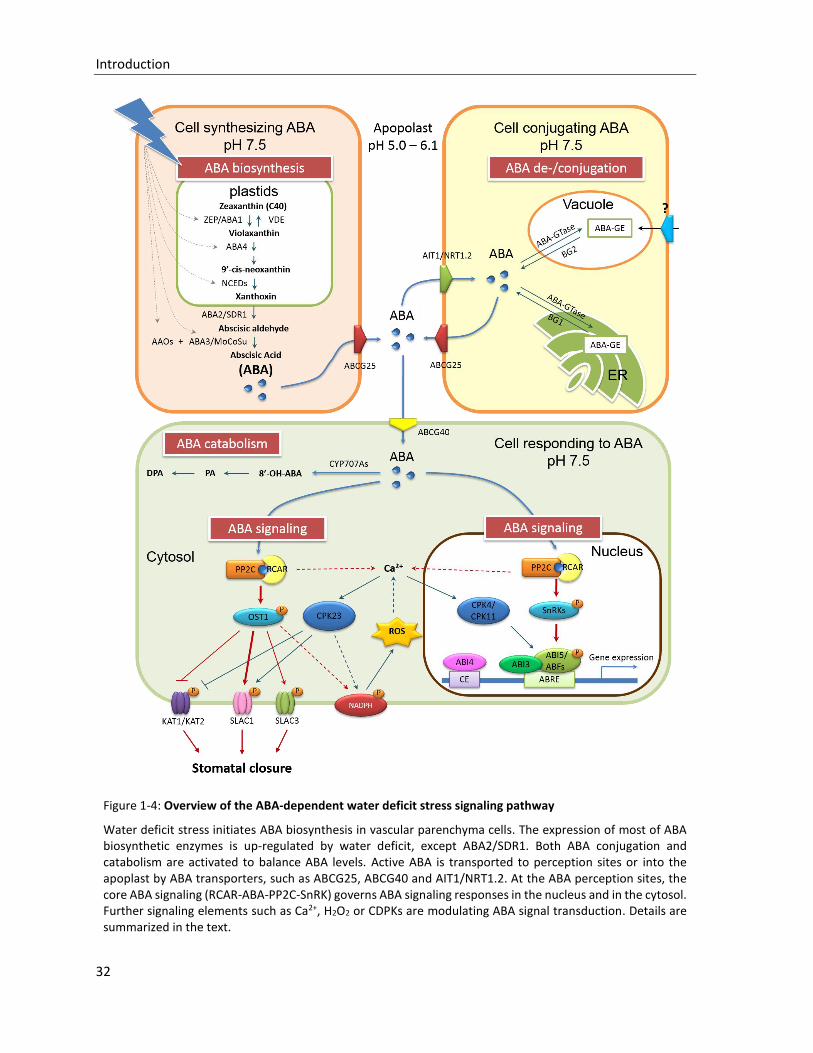
Figure 1-4: Overview of the ABA-dependent water deficit stress signaling pathway
Water deficit stress initiates ABA biosynthesis in vascular parenchyma cells. The expression of most of ABA biosynthetic enzymes is up-regulated by water deficit, except ABA2/SDR1. Both ABA conjugation and catabolism are activated to balance ABA levels. Active ABA is transported to perception sites or into the apoplast by ABA transporters, such as ABCG25, ABCG40 and AIT1/NRT1.2. At the ABA perception sites, the core ABA signaling (RCAR-ABA-PP2C-SnRK) governs ABA signaling responses in the nucleus and in the cytosol. Further signaling elements such as Ca2+, H2O2 or CDPKs are modulating ABA signal transduction. Details are summarized in the text.
Introduction
33
1.3.1.1 ABA biosynthesis
ABA biosynthesis pathway has been preciously revealed from a number of plant species and been
detailedly reviewed in many publications (Xiong and Zhu, 2003, Schwartz et al., 2003, Nambara and
Marion-Poll, 2005, Christmann et al., 2006, Wasilewska et al., 2008, Finkelstein, 2013). In higher plants
the specific ABA biosynthesis starts from the oxidative cleavage of a C40 epoxycarotenoid precursor
zeaxanthin to violaxanthin, which is catalyzed by a zeaxanthin epoxidase (ZEP) in plastids, firstly
isolated in tobacco (Marin et al., 1996). The Arabidopsis ZEP is encoded by ABA1 gene which was
identified from the ABA-deficient mutant aba1 (Koornneef et al., 1982). Meanwhile, a violaxanthin
de-epoxidase (VDE) functions as an antagonism of ZEP to produce more photo-protective zeaxanthin
in response to the intensive light (Bugos et al., 1998). Both mutations of ZEP and VDE can result in
decreased zeaxanthin accumulation (Niyogi et al., 1998). A plasid membrane-localiyed protein ABA4
catalyzes the transforming from violaxantin to primary ABA precursor neoxanthinis (North et al.,
2007). The rate-limiting step of ABA biosynthesis is catalyzed by 9-cis-epoxycarotenoid dioxygenase
(NCED) in plastids. NCED cleaves the xanthophylls (neoxanthin and/or violaxanthin) to C15 compound
xanthoxin/xanthoxal which is subsequently released into the cytosol (Schwartz et al., 1997). Then,
xanthoxin is transformed into ABA by two oxidative steps. The short chain dehydrogenase/reductase-
like (SDR1) enzyme encoded by ABA2 firstly catalyzes the intermediate reaction to produce ABA
aldehyde (Cheng et al., 2002). The final step of ABA converting is accomplished by abscisic aldehyde
oxidases (AAOs) (Seo et al., 2000) and a molybdenum cofactor sulfurase (MoCoSu) encoded by ABA3
(Xiong et al., 2001).
Because of these extremely defined ABA biosynthetic enzymes, we have the opportunity to
investigate how the ABA biosynthesis is regulated during growth development and under
environmental stresses (Xiong and Zhu, 2003). The biosynthetic enzymes ZEP/ABA1, ABA4,
SDR1/ABA2 and MoCoSu/ABA3 are single copy genes, excepting that the AAOs and the NCEDs are
encoded by small gene family, respectively. The ZEP/ABA1 is abundantly expressed in all tissues, and
is induced by drought stresses (Xiong et al., 2002). Although the impaired ABA4 significantly reduces
the ABA level in plant, the aba4 mutant can also produce ABA via an alternative pathway using
violaxanthin as a substrate (North et al., 2007). The AAO inactive phenotype of ABA-deficient mutant
aba3/los5 with impaired MoCoSu indicates that the ubiquitous MoCoSu/ABA3 enzyme is prerequisite
for AAOs activity (Xiong et al., 2001). AAO protein family contains four members (AAO1-to-4), in which
AAO3 is the main abscisic aldehyde oxidase in seeds and leaves, even though high expression levels
of AAO1 and AAO4 in the seed (Seo et al., 2004). And Arabidopsis genome encodes 9 NCEDs, of which
NCED2, 3, 5, 6 and 9 are identified as plastid-localized functional NCEDs, but differ in binding activity
of the thykaloid membrane (Tan et al., 2003). The roles of ABA during seed maturation and
germination have been clearly studied (Finkelstein et al., 2002, Xiong and Zhu, 2003, Kanno et al.,
2010, Finkelstein, 2013). Although the whole seed is considered to be involved in ABA biosynthesis,
the functional NCEDs still display distinct expression patterns. For example, NCED2 and NCED3 are
found in the surrounding maternal tissue of seed (Tan et al., 2003); NCED6 acts specifically in the
endosperm (Lefebvre et al., 2006); and NCED9 locates in both endosperm and embryos (Lefebvre et
Introduction
34
al., 2006). At the mid-stage of seed development, ABA is primarily synthesized in maternal tissues by
NCED2 and NCED3, and subsequently transported into embryo to promote the synthesis of storage
proteins in embryo. Then the second peak of ABA accumulation occurs during the desiccation phase
to activate the producing of protective protein LEAs and initiate the seed dormancy. Here, the ABA is
derived from the de novo synthesis catalyzed by NCED9 in embryo. A recent report indicates that upon
imbibition the endosperm in dormant seed coat can synthesize ABA which is released to the no-
dormant embryo to maintain the seed dormancy, while a DELLA protein RGL2 is required to promote
this ABA synthesis (Lee et al., 2010).
Concerning the water deficit-induced ABA de novo synthesis, three important ABA biosynthetic
enzymes, NCED3, ABA2 and AAO3, are co-localized in the vascular parenchyma cells (Cheng et al.,
2002, Koiwai et al., 2004, Endo et al., 2008). The expression of NCED3 is up-regulated by drought stress
(Tan et al., 2003) via a stress-inducible NAC TF ATAF1 that directly binds the NCED3 promoter (Jensen
et al., 2008, Jensen et al., 2013). Therefore, Stress-induced ABA is predominately synthesized in the
vascular tissue and subsequently transported into surrounding responsive cells. Recently, the
research of stomatal response to dry air in ABA-deficient mutant aba3-1 revealed that the guard cell
may autonomously synthesize ABA to promote stomatal closure in response to low relative humidity
(Bauer et al., 2013). For its high expression level, the ZEP/ABA1 seems not the rate-limiting enzyme of
ABA biosynthesis, but is still up-regulated by drought stress. In Arabidopsis, tobacco, and tomato the
ZEP/ABA1 is drought-induced in roots but not in leaves (Audran et al., 1998, Thompson et al., 2000),
whereas the cowpea ZEP transcripts are not drought responsive at all (Iuchi et al., 2000). Only the
SDR1/ABA2 expression appears not to be regulated by stress, but up-regulated by sugar (Cheng et al.,
2002). In addition, ZEP/ABA1, AAO3 and MoCoSu in Arabidopsis are all up-regulated by ABA,
meanwhile the ABA-induced up-regulation of NCED3 is also found in the Landsberg background (Xiong
et al., 2002). Therefore, the ABA biosynthesis is positively regulated by water deficit and ABA.
1.3.1.2 ABA catabolism and conjugation
Antagonizing to excessive ABA accumulation, the ABA level in cellular is balanced by catabolism and
conjugation (Cutler and Krochko, 1999). The major ABA catabolic pathway is triggered by ABA 8’-
hydroxylation and catalyzed by a cytochrome P450 CYP707A family (CYP707A1-4), whose transcripts
are increased in response to water deficit stresses including salt, hyperosmotic and dehydration, as
well as to exogenously appied ABA (Kushiro et al., 2004, Saito et al., 2004). The cyp707a mutant
accumulates higher ABA content than wild type as well as the lines overexpressing ABA biosynthetic
enzymes. The spatial analysis indicates the different expression patterns of CYP707As: the CYP707A3
plays a prominent role in ABA catabolism in the vegetative tissues of Arabidopsis, because it is the
most abundantly protein induced by dehydration, especially highly expressed in leaves (Umezawa et
al., 2006); the CYP707A1 is expressed in the embryo during the mid-maturation stage and decreased
during the late-maturation stage, whereas the CYP707A2 in both embryo and endosperm plays a role
during the late-maturation stage till seed germination (Okamoto et al., 2006). In subsequent step, the
8’-OH-ABA is isomerized to the phaseic acid (PA), which then converts to dihydrophaseic acid (DPA).
Introduction
35
Actually, ABA can also be hydroxylated at the 7’or 9’ positions. At least 9’-hydroxylation is a side
pathway of CYP707As catalyzed ABA catabolism (Okamoto et al., 2011).
Compared with ABA catabolism, ABA conjugation is a reversible process to reduce active ABA level in
cellular. ABA glucose ester (ABA-GE) is the predominant conjugate form and sequestered in apopolast
tissues and vacuoles of mesophyll cells as an inactive storage ABA (Bray and Zeevaart, 1985, Cutler
and Krochko, 1999). The glycosylation of ABA is catalyzed by the ABA-glucosyltransferase (ABA-GTase).
Although the water stress- and ABA-inducible UDP-GTase identified from adzuki bean displays an ABA
dependent GTase activity in vitro (Xu et al., 2002), over-expressing of the Arabidopsis GTase UGT71B6
only mildly influences the free ABA content and the physiological phenotype, whereas the ABA-GE is
massively accumulated in the over-expression lines (Priest et al., 2006). The possible reason is the
functional redundancy of GTases in plant. The inactive storage ABA can be hydrolyzed by β-
glucosidases (BGs). In Arabidopsis, the BG1 localizes in the endoplasmic reticulum (ER) (Lee et al.,
2006c), and the BG2 (Xu et al., 2012) localized in the vacuole.and quickly resorted by stresses. Under
osmotic stress, Both BGs rapidly polymerize from lower to higher molecular weight forms to liberate
active ABA. Therefore, ABA can be quickly restored from the storage ABA-GE by water deficit.
1.3.2 ABA transport
Besides the balancing of ABA biosynthesis and catabolism, cellular ABA content is also regulated by
ABA intercellular translocation. The ABA flux between organs is explained as an ‘ionic trap model’
(Boursiac et al., 2013). The weak acidic ABA (pKa 4.7) is in an equilibrium between the anionic charged
form (ABA-) and the protonated uncharged form (ABA-H). The uncharged ABA-H in apoplast (pH 5.0
to 6.1) can freely diffuse through the lipid bilayer of plasma membrane into cytosol, where the ABA-
H is converted to the undiffusible charged form ABA- for higher cytoplasmic pH (7.5). Therefore, the
cytosol becomes a ‘trap’ for a constant ABA influx from xylem sap to surrounding cells. A big question
is how the undiffusible ABA- is exported from synthetic parenchyma cells to xylem sap over the ionic
trap. Under environmental stresses, the xylem sap pH value rises up to 6.7, reducing the motive force
for ABA-H uptake (Dietz et al., 2000).
The active transport of ABA has been investigated since 1970s, but the molecular bases of such
transport are just isolated lately (Kuromori and Shinozaki, 2010, Kuromori et al., 2010, Kang et al.,
2010, Kanno et al., 2012, Boursiac et al., 2013). ABCG25 and ABCG40, which were respectively
identified from mutants atabcg25 and atabcg40, belong to the G family of ATP-binding cassette (ABC)
transporter (Kang et al., 2010, Kuromori et al., 2010). The ABC transporter family contains 130
members in Arabidopsis, but only few of them are functionally analyzed (Kang et al., 2012). The
ABCG25 locates on the membrane of vascular parenchyma cells and plays a role during the ATP-
dependent ABA export (Kuromori et al., 2010). Compared to ABCG25, the membrane-localized
ABCG40 is broadly expressed in many tissues, including leaves of young plantlets, primary roots and
lateral roots. It is remarkable that the expression level of ABCG40 in guard cells is significantly higher
than in mesophyll cells. And ABCG40 shows an ATP-dependent ABA import activity in yeast and
tobacco BY2 cells (Kang et al., 2010). Hence, a simple model of ABA translocation is constructed: The
Introduction
36
ABA-exporter ABCG25 works at ABA biosynthesizing cells for the ABA transport from inside to the
bundle sheath cell apoplastic area over the ionic trap; Then ABA is diffused passively with the flow of
water directed to the sites of transpiration; Subsequently, ABA is imported to guard cells by the ABA-
importer ABCG40 to trigger the stomatal closure, particularly under stresses (Kuromori and Shinozaki,
2010). Since the discovery of ABCG25 and ABCG40, the ABCG group members of ABC transporter
family are strong candidates for ABA transporters. Several ABCG proteins have also been reported to
be involved in drought and salt tolerance, e.g. ABCG36 (Kim et al., 2010a) and ABCG22 (Kuromori et
al., 2011), but the substrates transported by them have not been determined.
Recently, this model is improved by the third ABA transporter ABA-IMPORTING TRANSPORTER 1 (AIT1)
which is found in yeast two-hybrid system using the ABA receptor complex (ABI1/PYR1 complex, see
section 1.3.3.1) as a sensor (Kanno et al., 2012). AIT1 is also named NRT1.2 (NITRATE TRANSPORTER
1.2) identified as a low-affinity nitrate (NO3-) transporter in root epidermis and root hair (Huang et al.,
1999). High activity of AIT1/NRT1.2 promoter is detected in imbibed seeds as well as in vascular tissues
of inflorescence stems, leaves and roots. In yeast and insect cells the expression of AIT1/NRT1.2
enhanced ABA uptake (Kanno et al., 2012). Thus, the primary role of AIT1/NRT1.2 seems to be the
ABA import from the vessels to epidermis and guard cells for the regulation of stomatal aperture in
inflorescence stems (Kanno et al., 2012).
The conjugated storage ABA (ABA-GE) is also considered as a possible shuttle molecule of ABA signals,
and is hypothesized to be transported into mesophyll vacuoles by ABC transporters (Sauter et al.,
2002), two of which the vacuole-localized ABCC1 and ABCC2 exhibit an ABA-GE importing activity in
vitro (Burla et al., 2013).
1.3.3 ABA perception and core ABA signaling
ABA signaling is initiated from the specific perception by receptor at the action site. The previous
studies implied the presence of multiple types of ABA receptors on the cell surface as well as in the
cytosol (Allan et al., 1994, Jeannette et al., 1999). Since 2009, a cytosol/nucleus Regulatory
Components of ABA Receptors (RCAR1-14)/Pyrabactin Resistance Protein 1 (PYR 1)/PYR-Like (PYL1-
13) have been independently identified and confirmed as cytosolic ABA receptors by several research
groups (Ma et al., 2009, Park et al., 2009). In pace with this illustrious discovery, the essential core
components of ABA signaling pathway from hormone perception to signal transduction have been
identified: cytosolic ABA receptors (RCARs/PYR1/PYLs), clade A of type 2C serine/threonine protein
phosphatases (PP2Cs), and subfamily 2 of SNF1-related kinases (SnRK2s). Subsequently a number of
downstream targets, e.g. the ABA-activated basic leucine zipper transcription factors (bZIP TFs:
ABFs/AREBs) and the guard cell specific SLOW ANION CHANNEL-ASSOCIATED 1 (SLAC1), are
phosphorylated by SnRK2 kinases to organize the adaptative responses (Raghavendra et al., 2010,
Joshi-Saha et al., 2011b, Qin et al., 2011). Transient expression of these components have successfully
reconstituted the ABA signaling pathway in Arabidopsis protoplasts (Fujii et al., 2009).
Introduction
37
1.3.3.1 ABA coreceptor complex: RCARs/PYR1/PYLs-PP2Cs
Since the first plant PP2C, ABA INSENSITIVE 1 (ABI1) was identified from an ABA-insensitive mutant
abi1-1 (Meyer et al., 1994, Leung et al., 1994), PP2Cs have been generally described as negative
regulators of ABA signaling (Schweighofer et al., 2004). Accompanied with the identification of ABA
receptor family RCARs/PYR1/PYLs, the role of PP2Cs during ABA perception has been investigated.
Further researches revealed that PP2Cs play as coreceptor together with RCARs/PYR1/PYLs even
though they do not make direct contacting with ABA (Szostkiewicz et al., 2009, Park et al., 2009, Ma
et al., 2009, Nishimura et al., 2010).
The well known ABA-related ABI1 and its homolog ABI2 (Rodriguez et al., 1998, Leung et al., 1997)
belong to the clade A of PP2Cs, whose phosphatase activity depends on the presence of Mg2+ and
Mn2+ (Leube et al., 1998). Arabidopsis genome encodes 80 PP2Cs which are grouped into twelve
subfamilies (from A to K) (Xue et al., 2008, Fuchs et al., 2012). In the ABA-insensitive mutants abi1-1
and abi2-1, the amino acid residue converting in ABI1 (Gly180Asp) and ABI2 (Gly168Asp) leads to a
hypermorphic mutation of ABI1 and ABI2, respectively, which cause a preferential accumulation of
the mutated PP2Cs in the nucleus abolishing the interaction between ABI1/ABI2 and RCARs (Robert
et al., 2006, Moes et al., 2008, Santiago et al., 2009b). As a result, both abi1-1 and abi2-1 mutants are
dominantly insensitive to ABA. Conversely, loss-of-function alleles of ABI1 and ABI2 (abi1-2, abi1-3,
and abi2-1R) are hypersensitive to ABA and drought stress (Merlot et al., 2001, Saez et al., 2006).
Therefore, ABI1 and ABI2 negatively control a broad region of ABA signaling during plant development
and stresses. In addition to prototypical ABI1 and ABI2, other members of clade A PP2Cs, e.g.
PP2CA/ABA Hypersensitive germination 3 (AHG3) (Yoshida et al., 2006b, Kuhn et al., 2006), Homology
to ABI1 1 (HAB1) (Saez et al., 2004), Homology to ABI1 2 (HAB2), Highly ABA-induced PP2C 1-3 (HAIs)
(Bhaskara et al., 2012), and ABA Hypersensitive germination 1 (AHG1) (Nishimura et al., 2007), have
been also characterized as negative regulators of ABA signaling. The extreme ABA hypersensitive
phenotypes of multiple clade A PP2C knockouts revealed that these clade A PP2Cs redundantly
regulate ABA response, and the fine-tuning of ABA sensitivity can be obtained through combined
inactivation of these PP2Cs (Saez et al., 2006, Rubio et al., 2009).
The cytosolic ABA receptors RCAR1 (identical to PYL9) and RCAR3 (identical to PYL8) were initially
identified as ABI1- and ABI2-interacting proteins in a yeast two-hybrid screen, and specifically bound
to ABA (Ma et al., 2009, Szostkiewicz et al., 2009). RCARs/PYR1/PYLs family containing 14 members in
Arabidopsis belongs to the Bet v 1 superfamily, of which the most distinctive feature is a large solvent
accessible hydrophobic cavity (binding pocket) as a ligand binding site involved in binding and
metabolism of large hydrophobic compounds such as lipids, hormones and antibiotics (Radauer et al.,
2008). In vitro RCAR1 is required for ABA to completely block the phosphatase activity of ABI2. The
binding assay of ABA and RCAR1-PP2C revealed that a 1:1 ratio heteromeric protein complex RCAR1-
PP2C has single ABA binding site. The RCAR1 over-expressing lines displayed an ABA hypersensitivity
with regard to the physiological ABA responses including the inhibition of seed germination and root
elongation, as well as the stomatal closure (Ma et al., 2009). Compared to RCAR1, RCAR3 displayed
greater ABA sensitivity to PP2C regulation, but less stringent stereo-selectivity for the (+)-ABA
Introduction
38
(Szostkiewicz et al., 2009). In parallel, the ABA receptor roles of RCARs/PYR1/PYLs were independently
discovered by several other groups. PYR1 (identical to RCAR11) was identified by chemical genetic
screen of the mutant pyr1 which is insensitive to the synthetic ABA agonist pyrabactin (Park et al.,
2009). ABA-bound PYR1 inhibits the activity of PP2Cs via their physical interaction. For functional
redundancy, the single mutant pyr1 displays a normal ABA sensitivity, whereas the triple mutant
pyr1/pyl1/pyl4 and the quadruple mutant pyr1/pyl1/pyl2/pyl4 are strongly insensitive to ABA (Park et
al., 2009). PYL5, PYL6 and PYL8 interact with HAB1 in a yeast two-hybrid screen (Santiago et al., 2009b).
The over-expressing analysis and ABA-binding assay of PYL5 proved that ABA-bound PYL5 inhibits the
activity of clade A PP2Cs to activate ABA signaling (Santiago et al., 2009b). Furthermore, the mass-
spectrometric (MS) analyses of ABI1-interacting proteins in Arabidopsis also confirmed the interaction
between RCARs and PP2Cs which is quickly stimulated by ABA (Nishimura et al., 2010). These results
indicate that Arabidopsis RCARs/PYR1/PYLs have the ability to bind ABA and PP2Cs, and inhibit
phosphatase activity of PP2Cs in a combinatorial manner.
According to crystallographic structure analyses of ABA binding and protein phosphatase inhibition,
the RCARs/PYR1/PYLs are separated into three distinct subclasses (Hao et al., 2011, Dupeux et al.,
2011, Zhang et al., 2012), including cis-dimeric receptors (PYR1, PYL1 and PYL2), trans-dimeric
receptor (PYL3) and monomeric receptors (PYL4-6, PYL8-10). In the absence of ABA, PYR1 and PYL1-2
proteins exist as a cis-homodimer which means that the ABA binding pocket of each subunit is towards
the same direction. Surrounding the ABA binding pocket, two highly conserved surface loops CL2
serve as a ‘gate’ and a ‘latch’ of the open ligand-binding pocket (Melcher et al., 2009). The CL2 loops
are also thought to be involved in the dimer interface of PYL2 (Yin et al., 2009). Once ABA has bound,
a conformational change in the CL2 loops leads to the closure of the gate and latch, as well as the
dimer dissociation, subsequently creating a surface to interact with and competitively inhibit the
active site of PP2C. A conserved tryptophan in PP2C serves as a ‘lock’ to stable the 1:1 heteroternary
receptor-ABA-PP2C complex by inserting directly between the gate and latch (Yin et al., 2009,
Miyazono et al., 2009, Nishimura et al., 2009, Santiago et al., 2009a, Melcher et al., 2010).
PYL3/RCAR13 is the most distinctive ABA receptor proteins and exists in limited species. The ABA-
bound PYL3/RCAR13 prefers to trans-homodimeric form, which can easily disassociates to the
monomer compared to cis-dimers (Zhang et al., 2012). It seems that PYL3 exists in an equilibrium of
monomer, cis-dimer and trans-dimer. In contrast, the crystal structure of monomeric PYL10 indicated
that the CL2 loops of PYL10 are constitutively closed, leading to a high affinity of PYL10 to PP2Cs.
Therefore, PLY10 ABA-independently inhibits the PP2C activity. Of cause, the presence of ABA
increases the affinity of the receptor complex (Hao et al., 2011, Sun et al., 2012). RCAR7/PYL13 was
considered as an inactive ABA receptor (Fujii et al., 2009). However, the crystal structure of PLY13-
PP2CA indicated that RCAR7/PYL13 selectively inhibits PP2CA independent of ABA (Li et al., 2013). The
RCAR7 over-expressing and knockout lines revealed a bona fide ABA receptor function for
RCAR7/PYL13 (Fuchs et al., 2014). Therefore, all members of RCARs/PYR1/PYLs family may combine
ABA into the ligand-binding pocket. But the ABA affinity of heteromeric receptor complexes is
significantly higher. The Kd value of ABA binding to the heteromeric receptor complexe is around
Introduction
39
nanomole level which is relevance to the physiological ABA level (Ma et al., 2009, Szostkiewicz et al.,
2009). Therefore, the ABA receptor and the PP2C constitute an ABA coreceptor complex during ABA
perception.
As mentioned in section 1.3.1, ABA has several enantiomers, of which S-(+)-ABA is the natural active
ABA, and conversely trans-ABA is biologically inactive. The affinities of ABA receptors to ABA
enantiomers have been investigated. Crystal structures of apo-PYL5, PYL3-(-)-ABA and PYL9-(+)-ABA
showed that PYL5 strongly binds (-)-ABA, that PYL9 is a stringently exclusive (+)-ABA receptor, and
that PYL3 may accept both ABA enantiomers. Differing (+)-ABA binding, the steric hindrance and
hydrophobic interaction are the two key factors for (-)-ABA (Zhang et al., 2013). Taken together 14
RCARs/PYR1/PYLs and 9 clade A PP2Cs combinational recognize the ABA enantiomers.
Although the ABA coreceptor role of the heteromeric receptor complex RCARs/PYR1/PYLs-PP2Cs has
been proved via structural, biochemical and site-directed mutagenesis studies, the existence of other
probable ABA receptors still cannot be excluded. The reported ABA receptor candidates include the
plastidic-localized ABA-binding protein (ABAR)/H subunit of Mg-chelatase (CHLH)/Genomes
uncoupled 5 (GUN5) (Shen et al., 2006, Wu et al., 2009, Mochizuki et al., 2001) and the plasma
membrane-localized G-protein coupled receptor 2 (GCR2) (Liu et al., 2007) and G-protein coupled
receptor (GPCR)-type G protein 1 and 2 (GTG1/GTG2) (Pandey et al., 2009). However, the
contradictory results of the ABA binding activities pointed out that it is still too early to conclude the
natural role of these ABA receptor candidates in ABA signaling (Gao et al., 2007, Johnston et al., 2007,
Risk et al., 2009, Tsuzuki et al., 2011). Thereby more robust method of determining ABA binding (e.g.
isothermal titration calorimetry) is suggested in future research to prove the ABA binding abilities
(Christmann and Grill, 2009, Risk et al., 2009).
Given the central role of RCARs/PYR1/PYLs-PP2Cs receptor complex in ABA signaling, the investigation
of their target transducers becomes necessary to understand the ABA signal transduction. In addition
to the proper activation of the positive regulators SnRK2 kinases, various PP2Cs members physically
interact with a number of targets, including the homeodomain transcription factor HB6 which
negatively regulates ABA signaling (Himmelbach et al., 2002), a plastid-associated lipid-binding
protein fibrillin involved in the ABA-mediated photoprotection (Yang et al., 2006), the calcium binding
proteins and protein kinases (see section 1.3.5) and the ion channels (Lee et al., 2009, Brandt et al.,
2012). Therefore, the PP2Cs may play as a crucial hub that connects the ABA perception with other
signaling pathways involved in abiotic stress.
1.3.3.2 Activation of SnRK2s by ABA coreceptor complex
SnRK2 kinases belong to a plant-specific serine/threonine kinase family sucrose nonfermenting 1
(SNF1)-related kinases (SnRKs) and have been identified to involved in general osmotic stress and
plant development in many species such as tobacco (Mikolajczyk et al., 2000), fava bean (Li et al.,
2000), maize (Huai et al., 2008), wheat (Mao et al., 2009), soybean (Monks et al., 2001), rice
(Kobayashi et al., 2004) and Arabidopsis (Boudsocq et al., 2004). Arabidopsis genome encodes 10
SnRK2s (SnRK2.1-10) with three subgroups (Kulik et al., 2011). All SnRK2s share a N-terminal Ser/Thr
Introduction
40
kinase domain and a C-terminal regulatory domain (Domain I) involved in ABA-independent osmotic
stress response (Kobayashi et al., 2004, Belin et al., 2006). Specifically, the ABA-activated kinases of
SnRK2 group 3, including SnRK2.2, SnRK2.3 and SnRK2.6, contain a C-terminal ABA-specific box
(Domain II) (Yoshida et al., 2006a).
SnRK2.6, also named SnRK2E, was firstly discovered as OPTEN STOMATA 1 (OST1) and highly
expressed in guard cells. The mutant ost1 displays a wilty phenotype mainly due to its impaired
stomatal closure, but a normal ABA responsiveness in seeds (Mustilli et al., 2002, Yoshida et al., 2002).
Two homologs SnRK2.2 and SnRK2.3 are widely expressed in all tissues. The distinct expression
pattern of SnRK2s is consistent with the critical role of SnRK2.6 in stomatal response, and the broad
regulations of SnRK2.2 and SnRK2.3 in response to ABA during seed germination, dormancy and
seedling growth (Fujii et al., 2007). Triple mutant snrk2.2/3/6, also named snrk2d/e/I, exhibits poor
growth and few products, as well as extremely insensitive to ABA (Fujii and Zhu, 2009, Fujita et al.,
2009, Nakashima et al., 2009a). Therefore SnRK2.6, SnRK2.2 and SnRK2.3 function redundantly as
positive regulators in ABA signaling during plant development and stress tolerance.
As the important positive regulators of ABA signaling, SnRK2s of subgroup 3 are directly regulated by
PP2Cs (Umezawa et al., 2009). Earlier researches have revealed some vital aspects of the regulation
of SnRK2 kinases activity. Firstly, the conserved serine residue (Ser175) in the activation loop plays a
critical role for the kinase activity (Belin et al., 2006, Burza et al., 2006), and is dephosphorylated by
PP2C in the absence of ABA (Umezawa et al., 2009, Vlad et al., 2009). Secondly, the ABA-specific box
Domain II constantly interacts with PP2C, as is not affected by ABA (Yoshida et al., 2006a, Umezawa
et al., 2009). The mimic crystal structures of the SnRK2.6-HAB1 complex and the receptor-PP2C
complex clearly revealed the regulation mechanism of SnRK2s kinase activity by receptor-ABA-PP2C
complex (Soon et al., 2011, Ng et al., 2011, Yunta et al., 2011). In the absence of ABA, the activation
loop of SnRK2 mimics the ABA receptor gate to approach the catalytic domain of PP2C, and the
dephosphorylation of Ser175 by PP2C leads to the SnRK2 converting to basal activity form, then the
conserved tryptophan of PP2C inserts into the kinase active cleft to completely block the kinase
activity (Soon et al., 2011). Reversely, in the presence of ABA, the activation of SnRK2s is suggested as
a two-step intramolecular kinase activation. The ABA-bound receptor competitively binds to PP2C
resulting in the liberation and conversion of SnRK2 from PP2C-inhibited state to a partially active state,
which is then fully activated by the phosphorylation of its activation loop (Ng et al., 2011).
1.3.4 Signal transduction and the targets of SnRK2s
Given the central role of SnRK2s, especially mention about ABA-activated SnRK2s, in ABA signaling,
their downstream targets transmit the internal signals towards distinct directions, such as the ABA-
activated transcription in nucleus, the ion channel-mediated stomata responses, and the
accumulation of reactive oxygen species (ROS) production (Joshi-Saha et al., 2011a).
Introduction
41
1.3.4.1 ABA-activated transcription in nucleus
The ABA-regulated gene expression is mainly managed at transcription level. The ABA-responsive
element (ABRE)-binding proteins/factors (AREB1-3/ABF1-4) are well-characterized substrates of
SnRK2s in nucleus (Fujita et al., 2012). The AREBs/ABFs belong to basic leucine zipper transcription
factors (bZIP TFs) family (Choi et al., 2000, Uno et al., 2000), of which AREB1/ABF2, AREB2/ABF4, and
ABF3 are strongly induced and activated by osmotic stress and ABA, and have largely overlapping
functions in vegetative tissues (Kang et al., 2002, Kim et al., 2004, Fujita et al., 2005, Kobayashi et al.,
2005, Yoshida et al., 2010). Another typical representative of ABA-activated bZIP TFs ABA-INSENSITIVE
5 (ABI5) especially regulates ABA-dependent seed dormancy and germination, as well as post-
germination development (Finkelstein and Lynch, 2000, Lopez-Molina et al., 2001). The bZIP TFs share
a conserved leucine zipper dimerization motif and a basic DNA binding region containing two α-helices
(Schumacher et al., 2000, Lindemose et al., 2013). The SnRK2 kinases directly multiply phosphorylate
the Ser/Thr residues of the R-X-X-S/T sites in N-terminal conserved region of bZIP TFs to fully activate
the TFs (Kobayashi et al., 2005, Furihata et al., 2006, Johnson et al., 2002). The multi-phosphorylated
homo- or hetero-dimeric AREBs/ABFs/ABI5 (Yoshida et al., 2010, Lindemose et al., 2013) bind to the
ABA-responsive cis-element (ABRE: ACGTGT/GC) in the promoter region of ABA-induced genes to
activate the transcription. ABRE element was firstly identified in the promoter region of wheat Em
gene (Guiltinan et al., 1990) and of rice Rab16 gene (Mundy et al., 1990). Actually, single ABRE element
is not sufficient to initiate the ABA-activated transcription, and must cooperate with other ABREs or
coupling elements (CEs) to accomplish this mission (Shen and Ho, 1995, Skriver et al., 1991). Most of
the known CE elements have the similarity with ABRE and contain an A/GCGT motif (Hobo et al., 1999).
In addition to bZIP TFs, some other transcription factors are also involved in ABA- or stress-induced
transcription. For instance, both the B3 domain TF ABA INSENSITIVE 3 (ABI3) (Giraudat et al., 1992,
McCarty et al., 1991) and the APETALA 2 (AP2) domain TF ABA INSENSITIVE 4 (ABI4) (Finkelstein et al.,
1998), synergistically cooperating with ABI5, are well-characterized positive regulators of seed-
specific and/or ABA-inducible gene expression. ABI3 and ABI4 regulate the expression of ABI5 in seed
development and maturing (Lopez-Molina et al., 2002, Bossi et al., 2009, Reeves et al., 2011). Some
R2R3-MYB and bHLH-MYC TFs are implicated in ABA-mediated abiotic stress responses (Abe et al.,
1997, Li et al., 2007, Dubos et al., 2010). MYB2 was induced by ABA and dehydration, and the over-
expressing of MYC2 (bHLH TF) and/or MYB2 (R2R3-MYB TF) enhanced the ABA sensitivity and the
drought tolerance (Abe et al., 1997, Abe et al., 2003). The HD-ZIP TF ATHB6 (Himmelbach et al., 2002),
as well as its homologs ATHB7 and ATHB12 (Olsson et al., 2004), negatively regulates ABA-induced
gene expression via the direct interaction with PP2C (Elhiti and Stasolla, 2009, Valdes et al., 2012).
Furthermore, WRKYGQK domain (WRKY) TFs contrarily modulate ABA-dependent stress response.
WRKY57 (Jiang et al., 2012), WRKY25 and WRKY33 (Jiang and Deyholos, 2009, Li et al., 2011) improve
drought tolerance via ABA-dependent pathway, while WRKY18, WRKY40 and WRKY60 negatively
regulate seed germination and post-germination development by inhibiting the expression of many
ABA-responsive genes, e.g. ABF4, ABI4, ABI5, DREB1A, MYB2, and RAB18 (Shang et al., 2010, Chen et
al., 2010b).
Introduction
42
Although it is hard to precisely construct the sophisticated regulation network, the comparisons of
trancriptomes for Arabidopsis exposed to ABA and various abiotic stresses have reinforced the view
that about 1-10% genes of the genome are affected, and split fairly evenly between induced and
repressed genes (Zeller et al., 2009, Nakashima et al., 2009a, Nakashima et al., 2009b, Yoshida et al.,
2010, Choudhury and Lahiri, 2010, Okamoto et al., 2010). Although the diversity of experiment
conditions leads to differential gene regulation, about 50 ABA-induced genes are common to nearly
all conditions (Wang et al., 2011). According to the biological functions, the core set ABA-induced
proteins are roughly sorted into two types: (I) the protective proteins involved in stress tolerance,
such as late embryogenesis abundant (LEA) proteins, ROS scavenge enzymes and compatible solute
metabolism enzymes, and (II) the signaling regulators, including transcription factors, protein kinases
and phosphatases.
LEA proteins were firstly found in cotton seeds, accumulating in late embryogenesis (Dure et al., 1981).
Arabidopsis has 51 LEA proteins (Hundertmark and Hincha, 2008). The well-characterized LEA proteins,
such as RD29B/LTI65 involved in desiccation and cold responses, dehydrin protein RAB18, stress- and
cold-induced protein KIN2/COR6.6 and ABA-inducible LEA 1 (AIL1), are remarkably accumulated under
ABA treatment (Fujita et al., 2005). The hydrophilic and unfolded structure of LEAs implies the
probable role for counteracting crystallization of cellular components or the irreversibly damaging
effects of increasing ionic strength (Hundertmark and Hincha, 2008). Besides the protective proteins,
the negative regulators PP2Cs are also induced by ABA. Five members of group-Ab PP2C subfamily,
including AHG1, AHG3, HAI1, HAI2, and HAI3, were significantly less expressed in the triple mutant
areb1/areb2/abf3 under water stress (Nakashima et al., 2009a). Furthermore, the ABA-upregulated
TFs, e.g. MYBs (Ding et al., 2009), bHLHs (Abe et al., 2003, Li et al., 2007) and heat shock TFs (Hsfs)
(Kotak et al., 2007), have been indicated to involve in drought stress response. In contrast, the ABA-
repressed proteins are enriched for proteins associated with plant growth and development (Wang
et al., 2011).
1.3.4.2 ABA signaling to ion channel
Guard cell is highly developed single cell that integrates both environmental and internal signals
(Hetherington and Woodward, 2003, Sirichandra et al., 2009b). The ABA-induced stomatal closure
and the ABA-inhibited stomatal opening are short-term responses to the mild water deficit, and are
accomplished by the guard cell specific kinase-phosphatase pair OST1/SnRK2.6-PP2C, which directly
regulate the activities of ion channels in plasma membrane by phosphorylation/dephosphorylation
(Pandey et al., 2007, Joshi-Saha et al., 2011b, Lee et al., 2012).
To initiate stomatal closure, ABA activates the guard cell voltage-independent slow (S)-type anion
channel and voltage-dependent rapid (R)-type anion channel to trigger a transient membrane
depolarization. Subsequently, the Cl- and malate2- are released from guard cells to decrease the
cytosolic osmotic potential, resulting in stomatal closure (Roelfsema et al., 2004). The guard cell S-
type anion channel SLOW ANION CHANNEL-ASSOCIATED 1 (SLAC1) is activated by OST1/SnRK2.6 (Negi
et al., 2008, Vahisalu et al., 2008). The recessive mutant slac1 shows constitutively high stomatal
Introduction
43
conductance and strong ABA-insensitivity, while the R-type anion currents and Ca2+-permeable
channel currents are not disrupted (Vahisalu et al., 2008, Geiger et al., 2011). In vitro, OST1/SnRK2.6
phosphorylates the N-terminus of SLAC1 to activate the anion channel currents, meanwhile, co-
expressing of PP2Cs such as ABI1 and PP2CA successfully inhibits this activation (Lee et al., 2009,
Geiger et al., 2009). The PP2Cs not only inhibit the OST1/SnRK2.6 kinase activity via physical
interaction in the absence of ABA, but also directly dephosphorylate SLAC1 to inhibit the S-type anion
currents (Lee et al., 2009, Brandt et al., 2012). It is already known that the R-type anion channel
QUAC1 is also activated by ABA through the phosphorylation of OST1/SnRK2.6 (Imes et al., 2013). In
addition to the activation by Ca2+-independent kinases, the anion channels are also regulated by Ca2+-
dependent kinases (see section 1.3.5).
In guard cells the inward-rectifying potassium channel KAT1 and its homolog KAT2 form the
heteromultimeric channels and mediate K+ influx resulting in stomatal opening (Kwak et al., 2001,
Pilot et al., 2001, Lebaudy et al., 2009). In vitro, OST1/SnRK2.6 phosphorylates two positions of the
cytosolic C-terminus of KAT1 (Thr306 and Thr308), and further point mutation assay implied that the
phosphorylation at Thr306 is responsible for the functional inhibition of KAT1 (Sato et al., 2009).
Therefore the ion channel activity of KAT1 is inhibited by OST1/SnRK2.6 to keep the ABA-dependent
stomatal closure.
1.3.4.3 ABA-induced ROS production
Reactive oxygen species (ROS) play a dual role in the response of plants to abiotic stresses, functioning
as toxic by-products of stress metabolism and as signaling molecule in regulating diverse function in
plants (Choudhury et al., 2013). The downstream signaling of ROS is likely to occur via calcium and
protein phosphorylation (Kar, 2011). In guard cells the plasma membrane-bound NADPH oxidases
encoded by AtrbohD and AtrbohF genes directly interact with OST1/SnRK2.6 and are phosphorylated
at Ser13 and Ser174 amino acid residues (Mustilli et al., 2002, Kwak et al., 2003, Sirichandra et al., 2009a).
The phosphorylated active NADPH oxidases transfer electrons from cytoplasmic NADPH to O2 to
generate O2- and, subsequently, H2O2 (Torres and Dangl, 2005), which activates the hyperpolarization-
activated Ca2+-permeable cation channel (ICa) to increase the cytosolic free calcium ([Ca2+]cyt), then
leading to the stomatal closure (Pei et al., 2000). The NADPH oxidase RBOH C in the epidermal cells of
Arabidopsis root elongation zone mediates the tip-localized Ca2+ influx and cytosolic Ca2+ elevation
during root hair growth (Foreman et al., 2003). The ROS-induced activation of calcium channel is a
broad conserved signaling cassette in plant biology (Mori and Schroeder, 2004), whereas the
intracellular Ca2+ elevation could also lead to the ROS producing in apoplast by directly stimulating
NADPH oxidase (Takeda et al., 2008, Monshausen et al., 2009). Therefore, the Ca2+ elevation and ROS
signaling constitute a positive feedback loop to generate an oxidative burst. In addition, the ABA-
induced ROS production may inhibit the activities of ABI1 and ABI2 to turn up the ABA signaling
pathway (Meinhard and Grill, 2001, Meinhard et al., 2002).
The ROS production such as H2O2 has been shown to activate several MAPK cascades (Desikan et al.,
2000, Grant et al., 2000, Colcombet and Hirt, 2008). Many MAPK cascade components have been
Introduction
44
illustrated to locate at downstream of ROS signaling, including the MAPKKKs, e.g. ANP1, MEKK1 and
MKKK20 in Arabidopsis (Kim et al., 2011a), NPK1 in tobacco (Kovtun et al., 2000), DSM1 in rice (Ning
et al., 2010) and OMTK1 in alfalfa (Nakagami et al., 2004), the MAPKKs, e.g. MKK1 (Teige et al., 2004),
MKK4 (Ren et al., 2002) and MKK5 (Kovtun et al., 2000), and the MAPKs, e.g. MPK3/MPK6, MPK4, and
MPK9/MPK12 (Jammes et al., 2011). H2O2 especially activates the Arabidopsis Raf-type MAPKKK ANP1,
initiating the MPK3/MPK6 cascades (Kovtun et al., 2000). MEKK1-MKK1-MPK4 cascade is also
responsible to H2O2 (Teige et al., 2004, Droillard et al., 2004, Nakagami et al., 2006). The ROS-activated
MAPKs phosphorylate a variety of substrates, including transcription factors, transcription regulators,
splicing factors and other protein kinases, to respond to biotic and abiotic stresses (Ichimura et al.,
2002, Mishra et al., 2006). The ROS-induced genes seem to be involved in cellular repair/protection
mechanisms or in the H2O2 stress response signal transduction pathway (Desikan et al., 2000, Desikan
et al., 2001, Choudhury et al., 2013).
ROS production are common second massagers working downstream of many hormones such as
salicylic acid (SA), ABA, jasmonic acid (JA) and ethylene during the protective responses in plants under
abiotic stresses. The involvement of ROS in crosstalk among these signaling pathways has not been
fully understood (Fujita et al., 2006).
1.3.5 Ca2+-dependent ABA signaling
In contrast to the Ca2+-independent SnRK2s activity, the Ca2+-dependent signaling connects to the core
ABA signaling via a number of Ca2+ binding protein and protein kinase such as calcineurin B-like
proteins (CBLs) and CBL interacting protein kinases (CIPKs), Ca2+-dependent protein kinases
(CDPKs/CPKs) and CDPK-related kinases (CRKs), calmodulines (CAMs) and calmodulin-dependent
protein kinases (CaMKs), and Ca2+- and calmodulin dependent protein kinases (CCaMKs) (Luan, 2009,
Das and Pandey, 2010, Wurzinger et al., 2011, Asano et al., 2012).
CIPKs belonging to the SnRK3 subfamily contain a SNF1-like kinase domain and two regulatory
domains FISL and PPI, which are binding sites to CBLs and PP2Cs respectively (Ohta et al., 2003,
Sanchez-Barrena et al., 2013). Therefore, the CBL-CIPK complex has been discussed as one of the
important targets of PP2Cs involved in the plant response to abiotic stresses and ABA (Batistic and
Kudla, 2004, Kolukisaoglu et al., 2004, Luan, 2009). The SCaBP5 (CBL1)-interacting kinase PKS3/CIPK15
physically interacts to ABI2 participating a calcium responsive negative regulatory loop to control ABA
sensitivity (Guo et al., 2002). And the Ca2+-binding CBL1/9-CIPK26 complex phosphorylates the N-
terminus of NADPH oxidase AtROBHF to induce the ROS signaling (Drerup et al., 2013, Lyzenga et al.,
2013). The best known CBL-CIPK-PP2C network is the salt overly sensitive (SOS) pathway regulating
sensitivity to salinity in Arabidopsis (Zhu, 2002, Ji et al., 2013). In the presence of Ca2+, the Ca2+-bound
SOS3 activates the SOS2 kinase leading to the Na+/H+ antiporter SOS1 open, whereas the released
ABI2 physically binds to the PPI domain of SOS3 to prevent the interaction between SOS2 and SOS3
(Ohta et al., 2003, Quintero et al., 2011, Sanchez-Barrena et al., 2013). The regulation of Na+/H+
antiporter SOS1 by SOS3/CBL4-SOS2/CIPK24-ABI2 network illustrates that the molecular on-off
switches control the phosphorylation/dephosphorzlation of plant ion transporters. Similarly,
Introduction
45
Arabidopsis K+ transporter 1 (AKT1) is also regulated by the CBL1/9-CIPK23-PP2C pathway to involve
in K+ uptake during stomatal opening (Li et al., 2006, Xu et al., 2006a, Cheong et al., 2007). Some
members of CBLs can interact with PP2CA in vitro (Lan et al., 2011). Thus, it seems like that the
activation of AKT1 by CBL1/9-CIPK23 is inhibited by PP2C, whereas certain CBLs can re-switch on the
ion channels by binding to PP2Cs.
CDPKs/CPKs are also broadly announced as regulators of ABA-dependent signaling pathway (Harmon
et al., 2000, Ludwig et al., 2004, Asano et al., 2012). The CPK32 (Choi et al., 2005), CPK4 and CPK11
(Zhu et al., 2007b), bind to and phosphorylate the bZIP TFs AREBs/ABFs at Ser residue of C2 conserved
domain in vitro. The disruption of CDPKs reduces, but over-expression of the genes enhances, ABA
sensitivity and the expression of ABA-regulated genes. In guard cell, CPK23 and CPK21, as well as CPK3
and CPK6, promote the ABA- and Ca2+-induced stomatal closure by activating the SLAC1, SLAH3, and
Ica channels (Mori et al., 2006, Geiger et al., 2010, Geiger et al., 2011, Brandt et al., 2012). The BiFC-
based interaction analyses displayed that ABI1 physically binds to CPK23 and CPK21 to block the
activation of SLAC1 and SLAH3 by CDPKs (Geiger et al., 2011). These results indicate that CDPKs are
probably additional regulation targets of PP2Cs, and further activated by the cytosolic Ca2+ elevation.
However, not all of CDPKs are positive regulator of ABA signaling. For evidence, CPK12 negatively
regulates ABA signaling in seed germination and post-germination growth (Zhao et al., 2011a). CPK12
phosphorylates not only ABFs, e.g. ABF1 and ABF4, but also ABI2 to activate both positive and negative
regulators of ABA-signaling (Zhao et al., 2011a). Therefore, it seems like that different members of
CDPK family may constitute a regulatory loop by antagonistic functions, in which CPK12 plays as a
balancer of ABA signal transduction (Zhao et al., 2011b).
In fact, although Ca2+ is a simple divalent cation, the diversity of cytosolic Ca2+ signaling arises from a
wide range of Ca2+ concentration elevations. The redundancy of Ca2+-binding proteins and the
different compositions of canonic and non canonic calcium binding sites seem to control the overall
calcium affinity of the Ca2+ signals decoding. However, the mechanisms of specificity and fine-tuning
of the Ca2+-dependent signaling must be investigated in future.
1.4 ABA-independent water deficit signaling
Although the crucial role of ABA-dependent signaling in water deficit response has been thoroughly
comprehended, much less is understood on the more direct, ABA-independent osmosensing pathway.
Studying ABA-independent signaling in general is hampered by the fact that, actually, none of the
ABA-deficient mutants, e.g. aba1 and aba2, and the ABA-insensitive mutants, e.g. abi1-1 and abi2-1
is completely deficient or insensitive to ABA. Even in the decuple mutant snrk2.1/2/3/4/5/6/7/8/9/10,
in which all members of SnRK2s are disrupted, the osmotic stress-induced ABA increase is greatly
reduced but not abolished (Fujii et al., 2011). Transcriptomic and proteomic analyses have exhibited
the stress-responsive gene network involved in general physiological responses to water deficit stress
(Yamaguchi-Shinozaki and Shinozaki, 2006, Shinozaki and Yamaguchi-Shinozaki, 2007, Abebe et al.,
2009, Aprile et al., 2009, Jogaiah et al., 2012, Le et al., 2012). While most drought-induced genes are
also ABA-induced, some drought stress responsive genes do not change expression in response to
Introduction
46
exogenously applied ABA such as the ERD1 gene encoding a Clp protease regulatory subunit D (ClpD)
(Nakashima et al., 1997, Simpson et al., 2003). The differential regulation of certain genes by ABA or
hyperosmotic stress such as the RD29A gene (Yamaguchi-Shinozaki and Shinozaki, 1994, Yamaguchi-
Shinozaki and Shinozaki, 2005) has fostered detailed promoter analysis of such genes. The promoter
sequence analysis of RD29A gene then uncovered two major cis-elements (Yamaguchi-Shinozaki and
Shinozaki, 1993): ABA-responsive ABRE and dehydration-responsive cis-element (DRE)/C-Repeat (CRT)
(Yamaguchi-Shinozaki and Shinozaki, 1994, Yamaguchi-Shinozaki and Shinozaki, 2005). The DRE cis-
element (TACCGACAT) is recognized by drought-induced DRE-binding protein DREB2A and its single
homolog (Liu et al., 1998, Sakuma et al., 2006), as well as low temperature-induced DREB1A/C-repeat-
binding factor 3 (CBF3) and its two homologs (Liu et al., 1998, Akhtar et al., 2012). DREB2A regulates
the expression of water deficit-induced genes particularly, some of which, are also targeted by
DREB1A (Sakuma et al., 2006).
Indeed, these evidences demonstrate the presence of an ABA-independent regulatory pathway which,
together with the ABA-dependent pathway, synergistically governs the water deficit responses. Going
through the entire signal transduction process from perception to adaptable responses, it seems likely
that the ABA-independent events exist on every step of the water deficit signal transduction.
Firstly, the heteromeric ABA coreceptor complexes RCARs/PP2Cs have approximately 80
combinations, which vary in their affinities to ABA thus allowing for the adjustment of ABA signaling.
In contrast to the ABA-dependent construction of receptor-ABA-PP2C complexes, two monomeric
ABA receptors PYL10 and PYL13, constitutively bind to PP2C in the absence of ABA, and
antagonistically inhibit the phosphatase activity independent of ABA (Hao et al., 2011, Sun et al., 2012,
Li et al., 2013). On the other hand, the HAI PP2Cs, particularly HAI1, interact with monomeric RCARs
but not dimeric RCARs, and this interaction is unaffected or only moderately stimulated by ABA
(Bhaskara et al., 2012). The hai mutants display increased low ψw-induced proline and osmoregulatory
solute accumulation, as well as enhanced expression of dehydration-protective genes but reduced
expression of defense genes (Bhaskara et al., 2012). These results imply that under low ψw conditions
HAI PP2Cs are not inhibited in a RCAR-ABA-PP2C complex, and thus remain active to dephosphorylate
undiscovered substrate proteins as regulators of the low ψw response (Bhaskara et al., 2012).
Secondly, almost SnRK2 kinase family proteins (except SnRK2.9) are activated by hyperosmotic stress,
but only five of them (subgroup 2 and 3) are weakly or strongly activated by ABA. The ABA-
independent SnRK2s (subgroup 1) do not interact with PP2C in yeast and contain a C-terminal Domain
I involved in the ABA-independent osmotic response (Boudsocq et al., 2004). However, the role of
ABA-independent SnRK2s is still unclear.
Given the Ca2+-dependent signals, the CBL1 represents a integrator of multiple stress signals, and
positively regulates salt and drought responses but negatively regulates cold response, while the
expression of ABA-regulated genes is not affected (Cheong et al., 2003). In contrast, CBL9 that displays
similar expression pattern to CBL1 is induced by ABA, and commonly regulates ABA biosynthesis
(Pandey et al., 2004). Both CBL1 and CBL9 can interact with different CIPK kinases, such as CIPK1,
Introduction
47
CIPK23 and CIPK24 (D'Angelo et al., 2006). The alternation of complex formations between CBL1-CIPK
and CBL9-CIPK is a convergence point for ABA-dependent and ABA-independent gene expression and
ion channel regulation.
MAPK cascades probably play a role in the early water deficit response with MPK6 being directly
activated by drought, also in an ABA-deficient mutant aba2-4, and with exogenous ABA failing to
suppress the rehydration-dependent inactivation of MPK6 (Tsugama et al., 2012). Thus the ROS-
induced MPK6 activation appares to be ABA-independent. mRNA decapping protein DCP1, which
forms a mRNA decapping complex with DCP2 and DCP5 to regulate postembryonic development (Xu
et al., 2006b), has been identified as direct phosphorylation target of the dehydration stress activated
MPK6 (Xu and Chua, 2012). Therefore, the MPK6-DCP1 pathway is suggested as a rapid ABA-
independent response to water deficit stress in Arabidopsis.
1.5 The aim of this work
Water deficit is a central abiotic stress factor for wild plant species and agricultural crops. Limited
access to water triggers ABA accumulation in plants and the plant hormone controls a number of
essential stress-adaptative responses including stomatal closure, adjustment of vegetative growth,
accumulation of osmolytes, and modification of gene expression. While the core ABA signaling
pathway has been elucidated, the whole water deficit signaling network is still poorly understood.
The major interest of this work is to better understand the molecular mechanisms of water deficit
signaling in Arabidopsis. Map-based cloning was recruited to identify a novel Arabidopsis mutant
ahr11 with an alternated water deficit response phenotype. To elucidate the function of the novel
component AHR11, the protein was ectopically expressed in protoplasts and in planta. In addition, a
screen for proteins interacting with AHR11 was performed using a yeast two-hybrid screen.

Materials and methods
49
2. Materials and methods
2.1 Materials
2.1.1 Plant materials
Wild-type Arabidopsis thaliana
Two accessions of Arabidopsis thaliana Columbia (Col-0) and Landsberg erecta (Ler) were employed
in this work as wild-type reference or source for protoplasts, DNA and RNA isolation. Landsberg erecta
(Ler) was used as the paternal outcross line for generating mapping population. All accessions were
received from the Arabidopsis Biological Resource Center (ABRC; https://abrc.osu.edu), Ohio, USA.
Transgenic Arabidopsis thaliana
Transgenic ABA-repoter line pATHB6::LUC (Arabidopsis thaliana Col-0 background) harboring the
Photinus pyralis luciferase (LUC) reporter under control of the ABA-activated promoter of
homeodomain protein ATHB6 (At2g22430) was generated previously to monitor active ABA pools in
vivo (Himmelbach et al., 2002, Christmann et al., 2005). In this work, the pATHB6::LUC ABA-reporter
line was applied for generation of the EMS-mutagenized seed population by J. Berger and Dr. A.
Christmann and as a reference for physiological analysis if not otherwise indicated.
Arabidopsis thaliana mutants
T-DNA knockout mutants of AHR11 SAIL_88_D02.v1 (Col-0 background; N804 for short) and
GK_476H03 (Col-0 background; Δ476 for short) served in this work for novel protein characterization.
Meanwhile, the pATHB6::LUC ABA-reporter construct was introduced into these T-DNA mutants by
crossing to the pATHB6::LUC reporter line.
ABA-deficient mutant aba2-1 (Leon-Kloosterziel et al., 1996) and ABA-insensitive mutant abi1-1
(Koornneef et al., 1984) were used for generating double mutants ahr11/aba2-1 and ahr11/abi1-1,
respectively.
All mutants were ordered from ABRC (https://abrc.osu.edu).
2.1.2 Reagents
The chemicals used in this work were purchased in p.a. quality from Fluka (Sigma-Aldrich GmbH,
Munich), Roth (Carl Roth GmbH & Co. KG, Karlsruhe), Serva (Serva Electrophoresis GmbH, Heidelberg)
and Qigen (Qigen GmbH, Hilden). The DNA ladders (1 kb, 100 bp and λHindIII DNA ladders) were
applied by Thermo scientific (Thermo scientific GmbH, Munich). The basic molecular biology enzymes
(Restriction enzymes, T4-ligase/-kinase, alkaline phosphatase, GoTaq-polymerase, Phusion-HiFi-
polymerase, Klenow-fragment polymerase, M-MuLV reverse transcriptase, and so on) were obtained
from Thermo scientific (Thermo scientific GmbH, Munich) and/or New England Biolabs (NEB GmbH,
Frankfurt a. M). The enzymes Macerozym R-10 and cellulose R-10 were acquired from Yakult (Yakult
Materials and methods
50
Honshia Co. Ltd., Japan). The detergent Silvet L-77 was purchased from Lehle Seeds (Round Rock,
Texas, USA).
2.1.3 Microorganisms
Bacterial strains
For routine cloning, Escherichia coli (E. coli) strains DH5α (strain #2970; no resistant; Stratagene GmbH,
Heidelberg) and/or XL1Blue (strain #3340; Tetracyclin-resistant; Stratagene GmbH, Heidelberg) were
employed to propagate plasmids.
The Agrobacterium tumefacien strain pGV3101 harboring plasmid pMP90/pSOUP (strain #625)
(Hellens et al., 2000) was used for plant transformation by inflorescence dip (Harrison et al., 2006,
Zhang et al., 2006).
Yeast strains
Yeast strains Y8800 (MATa) and Y8930 (MATα) in yeast two-hybrid screen were kindly donated by Dr.
Pascal Braun from Department of Plant Systems Biology of Technique University Munich (TU
München). These two strains were derived from pJ69-4 (James et al., 1996), which harbor the
following genotype: leu2-3,112 trp1-901 his3-200 ura3-52 gal4Δgal80ΔGAL2-ADE2 LYS2:: GAL1-
HIS3 MET2::GAL7-lacZ cyh2R. The Y8800 and Y8930 strains were transformed with pDEST AD-Y and
pDEST DB-X constructs, respectively (Dreze et al., 2010).
2.1.4 Oligonucleotides and plasmids
All the oligonucleotides created and used in this work were listed in the appendix 5.1, stored at -20°
C in the Institute’s primer collection. The primers were synthesized by Eurofins MWG Operon
(Ebersberg, Germany). All the constructs generated and used in this work were listed in the appendix
5.2. The clones harboring the respective plasmids were stored at -80°C in the institute’s strain
collection labeled with the numbers indicated in the list.
2.1.5 Medium and sterilization
2.1.5.1 Medium for plants
MS medium (Murashige and Skoog, 1962) (per 1000 ml)
10 x MS-Macrosalts solution 100 ml
400 x MS-Microsalts solution 2.5 ml
MES 1 g
Sucrose 10 g
Agar 10g (for solid medium only)
pH 5.8 (KOH)

Materials and methods
51
Autoclave to sterilize
MS-0.5x sucrose medium (MS0.5xsuc)
MS0.5xsuc medium was prepared as the same way as MS medium mentioned above, but 5 g/l sucrose.
10 x MS-Macrosalts solution [g/l]
NH4NO3 16.5
KNO3 19.0
CaCl2·2H2O 4.4
KH2PO4 1.7
MgSO4·7H2O 3.7
400 x MS-Microsalts solution [g/l]
CoCl2·6H2O 0.01
CuSO4·5H2O 0.01
Na2-EDTA 14.60
H3BO4 1.20
KI 0.30
MnSO4·H2O 4.00
Na2MoO4·2H2O 0.10
ZnSO4·7H2O 0.80
FeSO4·7H2O 11.12
pH 4.5
Store at -20°C
400 x B5-Vitamin solution [g/l]
Nicotinic acid 0.4
Pyridoxine 0.4
Thiamine 4.0
Myo-Inositol 40.0
Materials and methods
52
Medium for bacteria
LB medium [g/l]
Bacto tryptone 10
Yeast extract 5
NaCl 10
Agar 10 (for solid medium only)
pH7.0 (NaOH)
Autoclave to sterilize
For preparation of selective medium, antibiotics were added in the appropriate concentration after
cooling LB medium below 50°C.
E. coli were grown in LB medium with appropriate antibiotics at 37°C for 12-16 h, with shaking at 200
rpm. Agrobacterium were grown in LB medium with appropriate antibiotics at 30°C for 2 days, with
shaking at 200 rpm.
2.1.5.2 Medium for Yeast
All yeast medium recipes were described by Matija Dreze (2010).
YEPD liquid medium [g/l] (nonselective rich yeast medium)
Yeast extract 10
Bacto-peptone 20
Autoclave to sterilize
Before use add 50 ml 40% (w/v) autoclaved glucose and 15 ml 65 mM adenine solution.
YEPD agar plates
40 g agar and 950 ml mQ water were shaken well. 2 x YEPD medium and agar were mixed after
autoclaving. Before pouring, 50 ml 40% (w/v) glucose and 15 ml 65 mM adenine solution were added
into the medium. 15 cm diameter YEPD agar plates were dried and stored in room temperature.
Materials and methods
53
2 x Sc medium (synthetic complete medium) (per 1000ml)
10 x Yeast-macrosalts solution 200 ml
1000 x Yeast-microsalts solution 2 ml
Amino acid powder 2.6 g
pH 5.9 (NaOH)
Autoclave to sterile
Add 8 ml each amino acid stock solution as needed (15 ml adenine solution).
Sc agar plates
40 g agar and 950 ml mQ water were shaken well. 2 x Sc medium and agar were mixed after
autoclaving. Before pouring, 50 ml 40% (w/v) glucose and amino acid solution were added into the
medium. 15 cm diameter Sc agar plates were dried and stored in room temperature.
Amino acid powder mix and stock solution
Mix 6 g of each of the following amino acids: alanine, arginine, aspartic acid, asparagine, cysteine,
glutamic acid, glutamine, glycine, isoleucine, lysine, methionine, phenylalanine, proline, serine,
threonine, tyrosine, valine and pyrimidine uracil.
The different amino acid stock solutions are prepared at the following concentrations:
Amino acids Stock solution [mM] Stock condition
Histidine-HCl 100 room temperature; light protected
Leucine 100 room temperature
Adenine sulfate 65 room temperature
Tryptophan 40 4°C; light protected
The concentrated amino acid stock solutions are used at 8 ml/l of medium, except for adenine which
is used at 15 ml/l of medium.
10 x Yeast-macrosalts solution [g/l]
KH2PO4 10
MgSO4·7H2O 10.24
NaCl 1
MgCl2 1
(NH4)2SO4 50
After autoclaving, the stock solution was stored at room temperature.
Materials and methods
54
1000 x Yeast-microsalts solution [g/l]
Biotin 0.002
Pantothenic Calcium 0.4
Folic Acid 0.02
Myo-Inositol 2.0
Nicotinic Acid 0.4
p-Aminobezoic Acid 0.02
Pyridoxine Hydrochlorid 0.4
Riboflavin 0.2
Thiamine HCl 0.4
H3BO3 0.5
CuSO4·5H2O 0.04
KI 0.1
FeCl3·6H2O 0.2
MnSO4·H2O 0.4
Na2MoO4·2H2O 0.2
ZnSO4 400
The stock solution was allocated into 50 ml Facon-tubes and stored at -20°C.
2.1.5.3 Antibiotics and hormones
Antibiotic Final concentration
[mg/l]
Stock solution
[mg/ml]
Ampicillin 50 100 (in mQ H2O)
Kanamycin 50 50 (in mQ H2O)
Rifampicin 50 50 (in DMSO; light protection)
Gentamicin 25 25 (in mQ H2O)
Hygromycin 50 50 (in mQ H2O)
Chloromycetin 25 10 (in ethanol; light protection)
Stock solution of each antibiotic was dissolved in corresponding buffer, filtrated through 0.22 µM filter
and stored at -20°C.
Hormone Final concentration
[µM]
Stock solution
[mM]
ABA 0.01-30 5 (in 10 mM MES-KOH, pH7.0)
Stock solution of each hormone was dissolved in corresponding buffer, filtrated through 0.22 µM filter
and stored at -20°C.
Materials and methods
55
2.2 Methods
2.2.1 Plant growth condition
Arabidopsis thaliana plants were grown in a controlled phyto-chamber (Conviron, Canada) under the
following long-day conditions: 16 h light (photosynthetic photon flux density [PPFD]: 200 µmol m-2 s-
1, temperature [T]: 22°C, relative humidity [RH]: 50%) and 8 h dark (T: 18°C, RH: 50%). For certain
experiments mentioned in context, Arabidopsis thaliana plants were grown in short-day conditions:
8 h light (PPFD: 200 µmol m-2 s-1, T: 22°C, RH: 50%) and 16 h dark (T: 18°C, RH: 50%).
2.2.2 Seed sterilization and seedling growth condition
Arabidopsis thaliana seeds were immerged within 1 ml of 80% ethanol containing 0.1% (v/v) Triton X-
100 and incubated for 20 min under constant shaking at 600 rpm. After removing the supernatant,
the seeds were washed with 1 ml of 3% sodium hypochlorite for 3 min. Then seeds were eventually
cleaned by thorough washing with sterile mQ H2O for 5 times in sterile bench. Finally, seeds were
sown in 9 cm Petri dishes containing MS0.5xsuc solid medium. Then stratification was performed at 4°C
for 2 days in the dark before germination. The seedlings were grown vertically in a culture room under
continuous illumination (PPFD: 60 µmol m-2s-1, T: 22°C) till suited development stage for further
experiments.
2.2.3 Quantification of ABA-dependent luciferase activity
The ABA-reporter line pATHB6::LUC has been generated to quantitatively detect the active ABA pool
in vivo (Christmann et al., 2005). The ABA-induced luciferase oxidizes luciferin to oxyluciferin and this
process releases photons which can be sensitively detected by an intensified CCD camera (ORCAII ERG;
Hamamatsu photonics, Hamamatsu City, Japan) adapted with a Schneider Xenon 0.95/25 Objective
(Schneider, Kreuznach, Germany). Shortly, the seedlings harboring the ABA-reporter construct
pATHB6::LUC were sprayed with luciferin solution*. After 10 min incubation in the dark, the light
emission of luciferase activity was detected by the CCD camera in a dark box. The exposure time was
10 min and imaging was done with 4 x 4 pixel binning. The intensity of light emission was translated
into a false-color spectrum. Quantitative determination of luciferase activity was performed with
software Simple PCI 6.6.0.0 (Compix, Cranberry Township, PA). The gray levels of pixels within the
measuring area corrected for background activity are referred to as CCD-RLU (Christmann et al., 2005).
Detection of luminescence on a cellular level was performed with an inverted microscope (Axiovert
200; Carl Zeiss, Germany) equipped with Fluar objectives and the CCD camera mounted to the
microscope base port. Seedlings were placed in a drop of Luciferin solution in a small chamber that
was sealed at the bottom with a coverslip. Seedlings were covered with a small pice nylon-net, and
the whole chamber was loosely covered with a coverslip to reduce evaporation. Pixel binning was 4 x
4, and exposure times varied between 10 and 30 min as indicated.

Materials and methods
56
* luciferin solution
luciferin (PJK, Kleinblittersdorf, Germany) 1 mM MES-KOH (pH7.0) 10 mM Tween-80 0.01% (v/v)
Luciferin solution was filtrated through 0.22 µM filter, and allocated into 12 ml Facon-tubes and stored
at -20°C in the dark.
2.2.4 Map-based cloning
The strategy of map-based cloning was designed and modified based on several previous researches
(Lukowitz et al., 2000, Jander et al., 2002, Jander, 2006, Hou et al., 2010). The most commonly used
Arabidopsis thaliana Columbia x Landsberg erecta (Col-0 x Ler) mapping combination system served
in this work.
2.2.4.1 Generation of F2 mapping population
The EMS-mutant (Col-0 background, with pATHB6::LUC ABA-reporter construct) was out-crossed to
another diverged accession Ler to generate a mapping population. Ler plant was used as the egg
donator and mutant plant was used as the sperm donator to be sure successful crossing pATHB6::LUC
ABA-reporter construct from pollens of mutant. The ABA-dependent light emission from seedlings
exposed to water deficit stress (-0.6 MPa, mannitol) served as the parameter to select homozygous
mutant phenotypic F2 individuals. Only the individuals whose F3 progenies were homogenously
hypersensitive to water deficit stress were included into the mapping population.
2.2.4.2 First-pass mapping
The bulked segregant analysis was employed in first-pass mapping (Michelmore et al., 1991). The
genomic DNA samples of homozygous individuals were isolated from leaf tissue (see section 2.2.9.1)
for mapping. DNA pool was constructed by mixing DNA samples isometrically. A mapping marker set
with 24 simple sequence length polymorphisms (SSLPs) markers, spaced evenly every 20 centiMorgan
(cM) on the whole genome of Arabidopsis thaliana, was kindly provided by Dr. Farhah Assaad (TU
München). All markers are PCR based, co-dominant (fully informative) and relatively abundant. And
the sequence information was listed in Appendix 5.1.1. The basic polymerase chain reaction (PCR)
recipe and program were described as follows. The PCR products were separated by 2% agarose gel
electrophoresis. The genotyping was identified by comparing with several references, including Col-0
DNA, Ler DNA and an isometric DNA mixture of Col-0 and Ler. The genotyping of mixture reference
was heterozygous for all molecular markers.
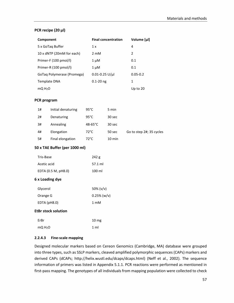
Materials and methods
57
PCR recipe (20 µl)
PCR program
1# Initial denaturing 95°C 5 min
2# Denaturing 95°C 30 sec
3# Annealing 48-65°C 30 sec
4# Elongation 72°C 50 sec Go to step 2#; 35 cycles
5# Final elongation 72°C 10 min
50 x TAE Buffer (per 1000 ml)
Tris-Base 242 g
Acetic acid 57.1 ml
EDTA (0.5 M, pH8.0) 100 ml
6 x Loading dye
Glycerol 50% (v/v)
Orange G 0.25% (w/v)
EDTA (pH8.0) 1 mM
EtBr stock solution
ErBr 10 mg
mQ H2O 1 ml
2.2.4.3 Fine-scale mapping
Designed molecular markers based on Cereon Genomics (Cambridge, MA) database were grouped
into three types, such as SSLP markers, cleaved amplified polymorphic sequences (CAPs) markers and
derived CAPs (dCAPs; http://helix.wustl.edu/dcaps/dcaps.html) (Neff et al., 2002). The sequence
information of primers was listed in Appendix 5.1.1. PCR reactions were performed as mentioned in
first-pass mapping. The genotypes of all individuals from mapping population were collected to check
Component Final concentration Volume [µl]
5 x GoTaq Buffer 1 x 4
10 x dNTP (20mM for each) 2 mM 2
Primer-F (100 pmol/l) 1 µM 0.1
Primer-R (100 pmol/l) 1 µM 0.1
GoTaq Polymerase (Promega) 0.01-0.25 U/µl 0.05-0.2
Template DNA 0.1-20 ng 1
mQ H2O Up to 20
Materials and methods
58
the recombination events in the mapping population. The genomic region of interest was flanked by
two markers with lowest recombinant rate.
2.2.5 Next generation sequencing (NGS)
Genomic DNA samples for next generation sequencing (NGS) were isolated from root tissue of a liquid
culture of seedlings (see section 2.2.9.2) to avoid chloroplast DBA contamination. NGS of the mutant
was carried out on the ABI SOLiD 3G+ sequencing platform by Dr. Tim-Matthias Strom from Helmholtz
Center Munich Human Genetic Institute. Data analysis was carried out using CLC Genomics
Workbench version 3.7 (CLC bio; Aarhus, Denmark) and LUMA (Febit biomed). The crude sequence
data obtained by NGS was aligned to the public reference Arabidopsis thaliana Col-0 genome (TAIR10).
2.2.6 Methods for plant analysis
2.2.6.1 Transpiration measurement
The long-term transpiration of intact plant was measured using a portable gas exchange fluorescence
system (GFS-3000, Heinz Walz, Germany). The plant growth and measurement were performed in
long-day conditions. The entire aerial portion of 3- to 4-week-old plant (before flowering) was sealed
off into a gas exchange cuvette (ring-chamber, custom designed, Heinz Walz, Germany) equipped with
GFS-3000. The conditions in the ring-chamber were kept constant with growing chamber: impeller: 9;
flow: 750 µmol s-1; H2O: 12000 ppm; CO2: 380 ppm; PPFD: 190 µmol m-2s-1; T: 28.5°C (light) and 22°C
(night).
2.2.6.2 Thermal imaging
Plants for thermal imaging were grown in water-controlled pots, in which the volumetric water
content (VWC)* was steadied to 80% by adding additional water every day. The thermal pictures were
captured using a thermographic system (VarioCAM high resolution head, InfraTec, Germany) in long-
day growth chamber. The leaf surface temperature was quantified via software (IRBIS v2.2, InfraTec,
Germany).
* VWC = water weigh [g]/dry soil weigh [g] x soil bulk density (0.167) x 100%
2.2.6.3 Water loss of detached shoots
The entire aerial portion was excised from the roots of 3- to 4-week-old plants, and the decline of the
fresh weight was measured at ambient conditions over time. The water loss* of the detached shoot
was calculated as follows.
*Water loss [%] = (Fresh weigh0-Fresh weighn)/Fresh weigh0 x 100%
in which n = time after excision from the roots.
Materials and methods
59
2.2.6.4 Stomatal response to root-inflicted water deficit and ABA
The root-inflicted stress analysis was described in previous research (Christmann et al., 2007). 5-day-
old seedlings grown on MS0.5xSuc medium were water deficit stressedby exposing roots to MS0.5xSuc solid
medium supplemented with mannitol to imitate low water potential for 24 h. For ABA responses, the
indicated amount of ABA was not only added into MS0.5xSuc medium, but also sprayed evenly on the
surface of seedlings. Then the stomata of the abaxial cotyledon surface were directly observed under
a research microscope (HBO 50 Axioskop; Carl Zeiss, Germany). 5 biological replicates, which
contained more than 100 stomata in total, were employed in each condition.
2.2.6.5 Stomatal response of rosette leaf to exogenous ABA
The ABA-dependent stomatal closure of rosette leaf was performed as described previously (Pei et al.,
1997). The rosette leaves with same development stage clipped from 3-week-old Arabidopsis thaliana
were floated on 3 ml of incubation solution*, and exposed to white light (150 µmol m−2 s−1) filtered
through a water jacket for 2 h, for stomatal opening. Then the indicated ABA was added to the
incubation solution, and the leaves were kept under the same conditions for another 2 h before
measuring the stomatal aperture. Subsequently, the abaxial epidermis was mounted onto a
microscope slide with transparent adhesive tape. Stomata images were observed under a research
microscope (HBO 50 Axioskop; Carl Zeiss, Germany). About 30 stomata with 16-22 µm edge height
were measuredin each condition. The ambient temperature was maintained at 25 ± 1°C.
The experiment condition of Ca2+ dependency of ABA-induced leaf stomatal closure was as same as
the method mentioned above. The free Ca2+ concentration in the incubation solution was buffered by
EGTA.
* Incubation solution
KCl 10 mM CaCl2 0.2 mM EGTA 0.1 mM MES-KOH (pH 6.15) 10 mM
2.2.6.6 Computed analysis of stomatal aperture
All stomata detections were performed with a digital camera (Olympus UC30; Olympus, Japan)
connected to a research microscope (HBO 50 Axioskop; Carl Zeiss, Germany). And the width and
length measurements of stomatal pores were accomplished using image analysis software (Image J
1.42q; Wayne Rasband, National Institutes of Health, USA). The ratio of width/length (W/L) of
stomatal aperture was analyzed as a functional parameter to demonstrate stomatal response to
certain treatments. Statistical tests (one-way ANOVA) were performed using software SPSS 16.0.0
(IBM SPSS Statistics). P < 0.05 was defined as statistically significant difference.
Materials and methods
60
2.2.6.7 Seed dormancy and germination
All seed batches for dormancy analysis were harvested at the same time from plants grown side by
side. Seeds were regarded as being germinated when the radical ruptured the surrounding structures
(Koornneef et al., 2002, Bentsink and Koornneef, 2008). The percentage of germinated seeds at
particular timing was taken as a parameter of dormancy depth and the response to stress or hormones.
Dormancy analyses in mQ water under light were performed as described previously (Alonso-Blanco
et al., 2003). Sterilized seeds (50-100) were sown on water soaked filter paper in 9 cm Petri dish. After
7 days incubation in culture room (T: 22°C, PPFD: 60 µmol m-2s-1, constantly) the number of
germinated seeds was scored under a dissecting microscope (Stemi SVII; Carl Zeiss, Germany). Seed
germination rate at a series of the days of seed dry storage (DSDS) from the harvest date was detected.
Two replicates were employed for each seed batch.
Seeds of Arabidopsis thaliana for germination assay were harvested and stored in room temperature
more than 6 months for after-ripen. Since the inhibition of high sucrose level on germination, the
MS0.5xsuc medium with half amount of sucrose was employed as basic medium in germination assay.
50-100 of sterilized seeds were evenly sown in 9 cm Petri dish with MS0.5xsuc medium, which was
supplemented with ABA or mannitol according to the germination condition examined. And seeds
were stratified at 4°C in the dark for 2 days. The rate of germinated seeds was calculated after 2 days
growth in culture room (T: 22°C, PPFD: 60 µmol m-2s-1, constantly). Three independent experiments
were performed using different seed batches.
2.2.6.8 Root growth
The 4-day-old seedlings of Arabidopsis thaliana grown on MS0.5xsuc medium were genteelly transferred
onto and entirely exposed to MS0.5xsuc medium supplemented with various concentrations mannitol
to adjust medium water potential or with ABA, and then grown vertically in culture room (22°C, light
constant) for 3 days. The position of root tip was marked every 24 h under a dissecting microscope
(Stemi SVII; Carl Zeiss, Germany). Then the photographs were taken with a digital camera (Canon G10,
Canon, Japan; camera control software: Canon Utilities Remote Capture DC ver. 3.1.0.5), and the root
length was measured using an image analysis software (Image J 1.42q; Wayne Rasband, National
Institutes of Health, USA). Statistical tests (one-way ANOVA) were performed using software SPSS
16.0.0 (IBM SPSS Statistics). P < 0.05 was defined as statistically significant difference.
2.2.6.9 ABA quantification
The fresh shoot tissue of 5-day-old Arabidopsis thaliana seedlings was harvested and frozen in liquid
nitrogen for ABA quantification after exposed to root-inflicted water deficit stress (MS0.5xSuc with
mannitol). Two biological replicates were prepared for each condition. The gas chromatography mass
spectrometry (GC-MS) analysis of ABA quantification was carried out by Prof. Dr. Jutta Ludwig-Müller
from the Institut für Botanik of Technische Universität Dresden. The details of GC-MS analysis was
described in previous reports (Martin-Rodriguez et al., 2011). ABA quantification was repeated three
times for each biological replicate (equivalent to minimum 100 mg FW of shoot tissue).
Materials and methods
61
2.2.6.10 Pigments analysis
Plant tissue samples were ground in liquid nitrogen, and dissolved in 1 ml cold methanol (100%), then
incubated on ice for 5 min. The absorbance of the extract, which was clarified by centrifuging for 4
min at maximal speed, was measured with a spectrophotometer (Ultrospec 2000 UV/Visible,
Pharmacia Biotech) at certain wave length. Because the spectrophotometer response was linear with
pigment concentration up to an absorbance value of one, the solutions were diluted further when the
absorbance exceeded one. Following formulae were used to calculate different pigments (Sims and
Gamon, 2002, Lichtenthaler, 1987):
Chlorophyll a [µg/ml extract] = 16.72 x A665 - 9.16 x A652;
Chlorophyll b [µg/ml extract] = 34.09 x A665 - 15.28 x A652;
Chlorophyll a + b [µg/ml extract] = 1.44 x A665 + 24.93 x A652;
Carotinoid [µg/ml extract] = (1000 x A470 - 1.63 x Chl a - 104.96 x Chl b)/221;
where Aχ was the absorbance of the extract solution in a 1 cm path length cuvette at wave length χ.
The relative content of chlorophyll (a+b) to total carotinoid was employed here as a parameter of
pigments in leaves.
2.2.7 Protoplasts expression in Arabidopsis thaliana
The protoplasts isolation and transfection protocol was modified by M. Meinhard and T. Hoffmann
according to previous publication (Abel and Theologis, 1994).
2.2.7.1 Isolation of protoplasts from plant leaves
The 3- to 4-week-old Arabidopsis thaliana plants (before flowering) are suitable for protoplasts
isolation. 0.5-1 g of leaves were incubated within 10 ml enzyme solution* on a vertical shaker
(RotoShake; 30 rpm) at 23°C for 4 h. Digested protoplast suspension was filtrated through a 150 µm
mesh nylon net. After centrifugation at 60 g for 2 min, the supernatant was carefully removed and
discarded. And the pellet protoplasts were resuspended in 10 ml WIMK solution** by rolling the tube
in the hands. The wash step was repeated once with 5 ml WIMK solution. After another centrifugation
step the pellet was resuspended in an appropriate volume of MaMg solution*** with a concentration
of 0.5-1.0 x 10⁶ protoplasts/ml. The viability of the protoplasts was estimated by staining with FDA (1
mg/ml in Aceton). Before the subsequent transfection, the protoplasts suspension was cooled to 4°C
for 30 min.
Materials and methods
62
* Enzyme solution
** WIMK solution
Mannitol 500 mM
MES-Tris (pH 5.8-6.0) 5 mM
*** Mannitol-Magnesium (MaMg) solution
All solutions were allocated into 50 ml Facon-tubes and stored at -20°C.
2.2.7.2 Protoplasts transfection
Quality of plasmid DNA is the major issue for successful transfection. All plasmid DNA used in
protoplasts transfection were purified according to the manufactures of Plasmid DNA midi
preparation (JETSTAR plasmid purification kit, GENOMED).
For transfection, a mixture of the plasmids to be introduced into the protoplasts was provided in 2 ml
reaction tubes as follows:
Subsequently, 100 µl (0.5-1.0 x 10⁶) protoplasts suspension was transferred to the reaction tubes
containing the DNA premix. After additional 120 µl of PEG solution*, the suspension was mixed
immediately by gently inverting tubes 5 times and incubated in room temperature for 3-5 min. Then,
the suspension was washed by 750 µl and 600 µl WIMK solution, orderly. After centrifugation (2500
rpm, 2 min), pellet cells were resuspended into 100 µl WIMK solution with or without the inductive
substances such as ABA. The transfections were incubated on a horizontal shaker (Rotamax) at 50 rpm
for 16 h.
Cellulase 1% (w/v)
Macerozyme 0.25% (w/v)
Mannitol 400 mM
CaCl2 8 mM
BSA (Fluka, #05488) 0.6% (w/v)
MES-KOH (pH 5.6) 5 mM
Mannitol 400 mM
MgCl2 15 mM
MES-KOH (pH 5.8) 5 mM
pRD29B::LUC ABA-reporter plasmid (#3041) 4 µl (4 µg DNA)
p35S::GUS GUS-reporter plasmid (#883) 2 µl (2 µg DNA)
effector plasmid/empty plasmid (#1337) 4 µl (4 µg DNA)
Mannitol (800 mM) 10 µl
Materials and methods
63
* PEG solution
PEG-4000 40% (w/v)
CaCl2 300 mM
MES-Tris (pH5.8) 5 mM
PEG solution was allocated into 50 ml Facon-tubes and stored at -20°C.
2.2.7.3 Activity assay of reporter
GUS activity
The 35S promoter driven GUS reporter was used to assess transformation efficiency and to normalize
the luciferase activity of single samples. The GUS activity was measured as fluorescence signal derived
from and correlating to the β-glucoronidase activity. 50 µl aliquot of the protoplast suspension was
transferred to black 96-well-microtiterplate (Greiner bio one). Then 100 µl of 1.5 x MUG-solution*
were added into each well. The GUS activity was measured at 37°C for 12 min in the microplate reader
(Synergy 2, BioTek). GUS activity was calculated and given as relative fluorescence units (RFU)/sec.
Luciferase (LUC) activity
The luciferase activity was detected using luminometer (Flash’n’Glow, BERTHOLD). 50 µl aliquot of
the protoplast suspension was transferred into luminometer round bottom tubes (SARSTEDT). The
sample tubes were automatically processed and 100 µl of LAR substrate** were added automatically
to start the luminescence reaction. Total luciferase activity was given as relative luminescence units
(RLU) and allowed calculation of RLU/sec.The relative activity of LUC to GUS reflected the activation
of signaling.
* 1.5 x MUG-solution
Na2HPO4/NaH2PO4 (pH 7.0) 50 mM
Na2EDTA 10 mM
Triton X-100 0.1% (v/v)
Dithiothreitol (DTT) 1 mM
4-Methylumbelliferyl-b-D-Glucuronid (MUG) 0.2 mM
Materials and methods
64
** LAR substrate
Tricine/NaOH (pH 7.8) 20 mM
MgSO4 2.7 mM
Na2EDTA 0.5 mM
DTT 30 mM
ATP 0.5 mM
(MgCO3)4Mg(OH)2 1 mM
Coenzyme A 250 µM
Luciferin 500 µM
Triton X-100 1% (v/v)
CCLR
Tris-phosphate (pH 7.8) 25 mM
DTT 2 mM
1,2-diaminocyclohexane-N,N,N',N'-tetra acetic acid 2 mM
Glycerin 10% (v/v)
2.2.8 Floral dip of Arabidopsis thaliana
For stable transformation of Arabidopsis thaliana an agrobacterium-mediated floral dip
transformation was performed according to previous reports (Zhang et al., 2006, Harrison et al., 2006).
Recipient plants were grown under long-day conditions for 4 weeks until inflorescences were present
but not yet opened. Agrobacterium strain pGV 3101 pMP90/pSOUP (#625) carrying desired plasmid
(pGreenII_AscI_Hygro derivation) was cultivated in LB medium with Kanamycin (50 mg/l), Rifampicin
(25 mg/l) and Gentamicin (25 mg/l) at 30°C for 2 days. The culture was amplified in 200 ml fresh LB
medium without antibiotics at 30°C for another 2 days. Agrobacterium cells were pelleted and
resuspended in infiltration medium*. The inflorescences were submerged into the agrobacterium
suspension for 3 min assuring complete wetting of the stems and inflorescences. The infiltrated plants
were irrigated with 1 x MS-macrosalts for nutrition. To improve transformation efficiency, the
procedure was usually repeated once, 3 days after the first dipping.
Transgenic seeds of T1 generation were screened according to the hygromycin (50 mg/l) resistance
marker and presence of the transformed fragment was verified by PCR analysis of genomic DNA
isolated from hygromycin resistant plants. The positive transformants were planted into soil for next
generation. The sterilized T2 transgenic seeds were placed on MS medium containing hygromycin (50
mg/l). The hygromycin-resistant T2 lines with a segregation rate of approximately 1:3 were selected
as transgenic plants with one copy insertion for further homozygous stable transformation screening.
About 100 seeds of each T3 line were sown on hygromycin-MS medium (50 mg/l) to select for
homozygous T3 populations with 100% hygromycin resistance.
Materials and methods
65
* Infiltration medium (per 1000 ml)
10 x Macrosalts solution 50 ml MES 0.5 g Sucrose 100 g BAP (1 mg/ml in EtOH) 10 µl VAC-IN-STUFF Silvet L-77 100 µl pH 5.8 (KOH)
2.2.9 Standard molecular biology methods
The standard molecular biology methods used in this work were as described by the protocols
provided by the manufacturers or according to the standard protocols (Sambrook and Russell, 2001).
Modifications and adaptations of single protocols and experiments are separately mentioned.
plasmid DNA mini-preparation (NucleoSpin® Plasmid, MACHEREY-NAGEL)
Plasmid DNA midi-preparation (JETSTAR® plasmid purification kit, GENOMED)
Polymerase chain reaction (GoTaq®DNA polymerase, Promega)
High-fidelity PCR (Phusion High-Fidelity DNA polymerase, Thermo Scientific)
Plant tissue PCR (KAPA3G plant PCR Kit, KAPABIOSYSTEMS)
DNA restriction (NEB and Thermo Scientific)
DNA fragment dephosphorylation (alkaline phosphatase, NEB)
DNA ligation (T4 DNA ligase, Thermo Scientific)
Agarose gel electrophoresis
DNA gel extraction (PeqLab Gel Extraction kit, PEQLAB)
Preparation of competent E.coli cells
Preparation of competent Agrobacterium cells
‘Heat-shock’ plasmid transformation to chemical competent E. coli cells
Plasmid electroporation to competent Agrobacterium cells
2.2.9.1 Genomic DNA isolation from leaves tissue
100 mg Arabidopsis thaliana leaves were collected and homogenized in 300 µl 2x CTAB buffer*. After
vigorously mixing and 30 min incubation at 65°C, 300 µl of chloroform was added. Then two phases
were separated by spinning down at 10,000 g speed for 5 min. The upper aqueous phase was
transformed into a new reaction tube. Following precipitation from the upper aqueous phase by
additional 300 µl isopropanol, the DNA was washed by 80% ethanol and dried at 37°C. Finally, DNA
was dissolved in 0.1 x TE buffer**, with 10 ng/µl RNase.
Materials and methods
66
* 2x CTAB buffer
Hexadecyl trimethyl-ammonium bromide (CTAB) 2% (w/v)
Tris-HCl (pH 8.0) 200 mM
EDTA (pH 8.0) 50 mM
NaCl 2M
** TE buffer
Tris-HCl (pH 8.0) 10 mM
EDTA (pH 8.0) 1 mM
2.2.9.2 Genomic DNA isolation from root tissue
Genomic DNA was isolated from root tissue of a liquid culture of seedlings to avoid chloroplast DNA
contamination. Sterilized Arabidopsis seeds were sown into a 100 ml flask with 50 ml modified MS
liquid medium* and grown in dark at 22°C on a shaker (100 rpm) till enough root tissue gotten. During
this period, fresh medium was substituted weekly. The isolation of high quality genomic DNA from
root tissue was performed following the handbook of DNeasy Plant Maxi Kit (QIAGEN).
* Modified-MS liquid medium (per 1000 ml)
10 x MS-Macrosalts solution 100 ml
400 x MS-Microsalts solution 2.5 ml
400 x B5-vitamin solution 2.5 ml
MES 1 g
Sucrose 15g
pH 5.8 (KOH)
Autoclave to sterilize
2.2.9.3 RNA isolation
Total RNA was isolated from rosette leaves or 5-day-old seedlings of Arabidopsis thaliana following
the handbook of RNeasy Mini Kit (QIAGEN). Stress treatments were supplied before isolation. To
determine the RNA concentration and purity, UV spectroscopy was used. The absorbance of RNA was
measured at 260 nm and 280 nm by a NanoDrop-Photometer (NanoPhotometer 7211, IMPLEN). The
A260/A280 ratio of 1.8-2.1 was an indication of highly purified RNA.
2.2.9.4 RT-PCR
RT-PCR process followed the handbook of RevertAid First Strand cDNA Synthesis Kit (#K1621; Thermo
Scientific).
Materials and methods
67
Removal of genomic DNA from RNA preparation
DNase I (RNase-free; #EN0521; Thermo Scientific) was employed for genomic DNA removal from RNA.
The followings were mixed and incubated at 37°C for 30 min:
RNA 1 µg
10x Reaction Buffer with MgCl2 1 µl
DNase I, RNase-free 1 µl
Water, nuclease-free Up to 10 µl
Additional 1 µl 50 mM EDTA and 10 min incubation at 65°C was performed to deactivate the DNase I.
Synthesis of the first strand cDNA
The product from above step was used as a template to synthesize the first strand cDNA. 1 µl oligo
(dT)18 primer was added and incubated at 65°C for 5 min. The following components were
supplemented in the indicated order:
5 x Reaction Buffer 4 µl
RiboLock RNase Inhibitor 1 µl
10 mM dNTP mix 1 µl
RevertAid M-MuLV Reverse Transcriptase 1 µl
The reaction was incubated at 37°C for 1 h and stopped by heating at 70°C for 10 min.
Semi-quantitative RT-PCR
Conventional PCR was done by using 0.5 µl (about 25 ng) cDNA as template. To semi-quantify the
transcriptional level of target gene, endogenous standards such as housekeeping genes, β-Actin and
Tubulin, were used as internal references. The PCR products were separated by gel electrophoresis
and quantified by software Image J. The relative expression level was achieved by comparing the
amount of products amplified by the target gene and the endogenous standard.
2.2.9.5 Quantitative real-time PCR
Quantitative real-time PCR (qRT-PCR) reactions were performed using the iQTMSYBR® Green Supermix
(#170-8880, Bio-Rad). A master mix for all reactions was prepared (on the ice or at room temperature)
by adding all requited components, except the cDNA template, as follows:
qRT-PCR recipe (10 µl)
iQTMSYBR® Green Supermix (2 X) 5 µl
Primer-F (10 pmol/l) 0.1 µl
Primer-R (10 pmol/l) 0.1 µl
cDNA template 0.5 µl
H2O Up to 10 µl
Materials and methods
68
Assay master mix was dispensed equal aliquots into each qPCR tubes. And the cDNA samples were
added to the tubes as reaction template. The quantitative real-time PCR was accomplished using
CFX96 Touch Real-time PCR Detection system (#185-5196, Bio-Rad).
qRT-PCR program
Initial denaturing 95°C 3 min
Denaturing 95°C 15 sec
Annealing and elongation 55°C 30 sec Go to step 2; 40 cycles
Melt curve analysis 55-95°C 0.5°C increment 2-5 sec/step
The data analysis was performed according to the CFX Manager software (184-5001, Bio-Rad). In this
work, the expression level of housekeeping gene UBC9 (At4g27960) was used as a reference
(Czechowski et al., 2005).
For target genes, a common method for validating a real-time PCR assay involves constructing a
primer standard curve, which helps to determine the primer efficiency (E*), linear dynamic range, and
reproducibility of the assay. 8 serial 2-fold dilutions of cDNAs were served as template to perform a
qRT-PCR reaction described as above. The standard curve was set as a plot of Ct value to log dilution
value. The sequence information of primer pairs was listed in Appendix 5.1.3.
*E=10-(1/slope)
where 90% < E < 105%; and R2 > 0.98
2.2.10 High-throughput yeast two-hybrid screen
High-throughput yeast two-hybrid screen was described previously by Dreze (2010).
2.2.10.1 Assembly of DB-X and AD-Y expression plasmids
The generation of expression plasmids was accomplished using gateway cloning system (Karimi et al.,
2007, Walhout et al., 2000). Firstly, distinct ORFs of interest were inserted into the dual selection entry
vector pENTRTM 1A (KanR and CmS; Catalog no. A10462; INVITROGEN) by the restrictionsites SalI and
EcoRI. Additionally, a STOP code was added at the C-terminus of ORFs. Plasmid DNAs from positive
clones were analyzed by restriction analysis, as well as by sequencing (primer: pENTR-seq-r #1919 in
Appendix 5.1.2).
Secondly, ROFs of interest were recombined into the destination vectors pDEST-DB (AmpR) and
pDEST-AD (AmpR), respectively, via a Gateway LR recombination reaction between the entry clones
and destination vectors. For large (> 10 kb) entry clones or destination vectors, linearizing the vectors
by restriction digestion (SmaI) may increase the recombination efficiency by up to 2-fold. The Gateway
LR recombination reaction was performed according to the manual of Gateway LR clonase II Enzyme
mix (Catalog no. 11790-020; INVITROGEN). Briefly, the following components were added to a 1.5 ml
reaction tube at room temperature and mix.
Materials and methods
69
Entry clone (50-150 ng) 1-7 µl
Destination vector (150 ng/ µl) 1 µl
LR ClonaseTM II enzyme mix 2µl
TE buffer (pH 8.0) Up to 10µl
After 1 h incubation at 25°C, additional 1 µl of the Proteinase K solution was added into samples,
which were subsequently incubated at 37°C for 10 min to terminate the reaction.
Then the LR reaction products were transformed into E.coli DH5α by ‘Heat-shock’ transformation. The
positive clones with ampicillin resistance were analyzed by PCR analysis and sequencing. The vector
specific primers pDEST-AD (#1916), pDEST-DB (#1917) and pDEST-term (#1918) were employed for
analyses.
2.2.10.2 Yeast transformation
Yeast transformation is performed following the ‘High efficiency LiAc transformation’ published
previously (Gietz and Woods, 2002). pDEST-DB-X and pDEST-AD-Y expression plasmid constructs were
individually transformed into competent yeast strains Y8930 (MATα) and Y8800 (MATa), respectively.
Single yeast colonies were used to inoculate a 5 ml pre-culture in Sc medium supplemented with the
corresponding amino acids and grown over night at 30°C. An aliquot of 5 ml yeast pre-culture was
added into 50 ml YEPD full medium and incubated at 30°C on a shaker (200 rpm) till reaching an
optimal OD600 of 0.6-0.8 (3 x 10⁷ cells).The yeast cells were harvested via centrifugation for 5 min at
1,000 x g. After washing the cells in 25 ml sterile mQ water, the pellet was resuspended in 1 ml of 1 x
LiAc (100 mM) and transferred into a new reaction tube. Cells were subsequently pelleted by short
spinning and dissolved in 400 µl 1 x LiAc. For one transformation 50 µl of competent yeast cell
suspension were used. In reaction tube cells were pelleted by short spinning, and the supernatant
was removed carefully. The followings were supplemented into competentcells, orderly:
PEG4000 (50% w/v) 240 µl
10 x LiAc (1 M) 36 µl
Carrier DNA* (10 µg/µl) 10µl
DNA (100-500 ng/µl) 1 µl
sterile mQ water 73 µl
Transformation mixtures were resuspended and incubated at 42°C for 40 min. After heat-shocking
the cells were harvested by centrifugation for 15 sec shot spinning and resuspended in 100 µl mQ
water for subsequent plating on Sc selective medium. The transformed yeast on selective solid
medium was incubated at 30°C for 2 days.
Materials and methods
70
* Carrier DNA
Single stranded DNA derived from salmon testes was dissolved 10 mg/ml in 1 x TE buffer (pH 7.5),
stirred for 3 h and ultrasonic-pulsed twice at max power for 30 sec. Then 1/10 volume of NaAc (3 M)
was added to this solution. Subsequently, DNA was precipitated by mixing with 2.5 volume of EtOH
(100%). The precipitated DNA is pelleted by centrifugation for 10 min at max speed and frozen for
later use. The aliquots of carrier DNA to be used for transformation were again boiled at 95°C for 5
min and then cooled on ice prior to use.
2.2.10.3 Yeast colony PCR
A quick and easy yeast colony PCR was modified according to the ‘Yeast Colony PCR v 2.0 protocol’
from Blackburn lab in university of California San Francisco. For cell lysis, a small yeast colony was
resuspended in 10 µl of 0.02 M NaOH in PCR tube. The suspension was incubated on a PCR machine
at 99°C for 10 min, and then kept on ice or freeze for longer. Phusion High-fidelity DNA polymerase
(Thermo Scientific) was applied for the PCR reaction, and a master mix for PCR reactions was prepared
by adding all requited components, except the cell lysis template, as follows:PCR recipe (20 µl)
Component Final concentration Volume [µl]
5 x Phusion HiFi polymerase buffer 1 x 4
10 x dNTP (20 mM for each) 2 mM 2
AD or DB primer (100 pmol/l) 1 µM 1
Term primer (100 pmol/l) 1 µM 1
Phusion HiFi DNA polymerase 0.25 U/µl 0.2
PCR Enhancer (100 x) 0.5-1 x 0.1-0.2
Cell lysis suspension template 1
mQ H2O Up to 20
PCR program
1# Initial denaturing 98°C 30 sec
2# Denaturing 98°C 10 sec
3# Annealing 58°C 30 sec
4# Elongation 72°C 3 min Go to step 2; 35 cycles
5# Final elongation 72°C 10 min
The PCR enhancer supplied in KAPA plant PCR Kit (KAPABIOSYSTEMS) was employed here to substitute
the Q-solution in original protocol. The primer concentration was about ten-times more than standard
PCR protocols. The PCR products were separated by 1% agarose gel electrophoresis, and DNA
fragments of interest were extracted from agarose gel according to the manual of the PeqLab Gel
Extraction Kit (PEQLAB), and subsequently sequenced by GATC Biotech.
Materials and methods
71
2.2.10.4 Controls of yeast-two-hybrid
For reliable validation of protein interacting, 6 of real biophysical interactors served as interacting
controls in yeast two-hybrid system (Table 2-1). All interactions that pass these controls were
considered high-quality yeast-two-hybrid interactions.
Table 2-1: Yeast two-hybrid controls
Controls Plasmid pairs Protein Interaction strenth
1 pDEST-AD
pDEST-DB
No insert
No insert None, background
2 pDEST-AD-E2F1
pDEST-DB-pRB
Human E2F1 aa342-437
Human pRB aa 302-928 Weak (control for CHX control plates)
3 pDEST-AD-Jun
pDEST-DB-Fos
Mouse Jun aa 250-325
Rat Fos aa 132-211 Moderately strong
4 pDEST-AD
pDEST-DB-Gal4
No insert
Yeast Gal4 aa 1-881 Very strong
5 pDEST-AD-dE2F1
pDEST-DB-dDP
Drosophila E2F aa 225-433
Drosophila DP aa 1-337 Strong
6 pDEST-AD-CYH2-dE2F1
pDEST-DB-dDP
Drosophila E2F aa 225-433
Drosophila DP aa 1-377 Strong (control for CHX plates)
Identities and description of expected phenotypes for the six controls used in every yeast two-hybrid experiment (Dreze et al., 2010).
2.2.10.5 Identification of autoactivators
Autoactivation of yeast-two-hybrid-inducible reporter genes is a common artifact of the yeast two-
hybrid system. The identification of autoactivators was achieved in diploid yeast strains obtained by
mating yeast strains Y8930 (DB-X) with the yeast strain Y8800 transformed with the AD encoding
plasmid containing no insert (pDEST-AD-emp).
The DB-X yeast strains and AD-emp yeast strain were incubated in fresh liquid mediums Sc-Leu and Sc-Trp, respectively, at 30°C for 72 h on a shaker.
5 µl of DB-X liquid cultures were spotted on a YEPE plate and allowed to dry for 30-60 min.
5 µl of AD-emp liquid cultures were spotted on top of the DB-X spots and allowed to dry for 30-60 min.
The mating plates were incubated at 30°C for 14-18 h.
The mating plates were replicated onto Sc-Leu-Trp plates to select for diploid cells, and incubated at 30°C for 14-18 h.
Subsequently, the diploid cells on Sc-Leu-Trp plates were replicated onto Sc-Leu-Trp-His + 1 mM 3AT (3-amino-1,2,4-triazole) plates, where nonautoactivating yeast cells were not able to grow, and incubated at 30°C for 14-18 h.
Materials and methods
72
Replica-clean of the Sc-Leu-Trp-His + 1 mM 3AT plates were performed by pushing evenly the plate on a piece of sterilized velvet to remove excess yeast. Plates were incubated at 30°C for another 72 h.
All yeast strains on Sc-Leu-Trp-His + 1 mM 3AT plates showing a stronger growth phenotype than the
‘no interaction’ control (control 1) were considered autoactivators. To reliably identify autoactivators,
two independent tests were performed, and the most stringent scores were accepted to unsure high
quality of the starting material for subsequent interactome mapping. The chemical 3AT is a
competitive inhibitor of the HIS3 gene product, therefore, when dealing with DB-X auto activators,
higher 3AT concentrations could be used to circumvent autoactivator-dependent activity of GAL1-
HIS3. All autoactivators of DB-X strains were excluded from the collection of DB-X.
Albeit much less frequent, autoactivation of AD-Y could also occur. The AD-Y autoactivator
identification by use of mating between AD-Y yeast strains and DB-emp yeast strain was easily
adapted.
2.2.10.6 Yeast two-hybrid primary screening
5 µl of glycerol stocks of the DB-X yeast strains and AD pools tested were inoculated in round bottom 96-well plates containing 160 µl selective medium in every well (Sc-Leu for DB-X, Sc-Trp for Ad pool) at 30°C for another 72 h for 72 h on a shaker.
For each combination (AD-pool plate x DB-X plate), 5 µl/well of the respective AD-Y pool liquid culture were spotted onto a mating plate (YEPD) using a liquid handling robot, and allowed to dry for 30-60 min.
Then 5 µl of each DB-X were spotted on top of the AD pool spots, and allowed to dry for 30-60 min.
Six controls of Y2H were spotted on every plate.
After an incubation at 30°C for 14-18 h, mated yeast cells were replicated from mating plates onto screening plates (Sc-Leu-trp-His + 1 mM 3AT), meanwhile replicated onto CHX control plates (Sc-Leu-His + 1 mM3AT + 1 mg/l CHX), and incubated at 30°C for 14-18 h.
All screening plates were cleaned by pressing plates onto a piece of velvet stretched over a replica-plating block and incubated at 30°C for 5 days.
The colonies that grew better than background (control 1) and did not grow on CHX plates were considered primary positive colonies.
Three colonies from each positive interacting spot were picked into a 96-well plate with round bottom (LOT: E13030FL; Greiner bio-one) containing 160 µl Sc-Leu-Trp medium per well, and incubating the culture plate at 30°C for 72 h on a shaker for subsequent phenotyping experiments.
2.2.10.7 Secondary phenotyping
5 µl of primary positive diploid yeast strains and Y2H controls were spotted onto Sc-Leu-Trp plates
using a 96-well liquid handling robot and incubated at 30°C for 48 h. Positives were replicated onto
fur phenotyping plates:
Materials and methods
73
Plate cleaning was immediately performed after replica-plating to minimize background growth, and
then the phenotyping plates were incubate at 30°C for 72 h.
During phenotyping, both reporter genes His and Ade were inspected, and any yeast spot showing
growth on CHX plates should not be considered for further processing. All positives were patched and
incubated on fresh Sc-Leu-Trp plates at 30°C for 48 h. the inserts of the AD-Y in positive colonies were
amplified by yeast colony PCR for subsequent ORF identification by sequencing. Once sequencing data
have been received and the candidate protein pairs have been identified, a list of unique candidate
interacting pairs could be complied.
2.2.10.8 Independent verification of candidate Y2H pairs
According to the list of candidate interacting pairs, the individual ORFs of candidates were subcloned
into expression plasmid vector (pDEST-AD) and subsequently transformed into yeast strain Y8800 by
Angela Alkofer from Department of Plant Systems Biology of TU München.
5 µl of glycerol stock of the DB-X yeast strain and AD-Y yeast strain of interest were inoculated in round bottom 96-well plates containing 160 µl/well selective medium (Sc-Leu for DB-X, Sc-Trp for AD pool) at 30°C for 72 h on a shaker.
5 µl/well of liquid cultures AD-Y and DB-X were mated on YEPD plate as described above and incubated at 30°C for 14-18 h.
Then mated cells were replicated onto diploid selection plates Sc-Leu-Trp, and incubated 30°C for 14-18 h.
Positives were replicated onto four phenotyping plates, and cleaned immediately.
After 3 days incubation, the growth phenotype was scored in the same way as for secondary phenotyping.
The verification was performed at least three times independently.
Sc-Leu-Trp-His + 1 mM 3AT (His auxotrophy)
Sc-Leu-His + 1 mM 3AT + 1 mg/l CHX (CHX control plate for His)
Sc-Leu-Trp-Ade (Ade auxotrophy)
Sc-Leu-Ade + 1 mg/l CHX (CHX control plate for Ade)

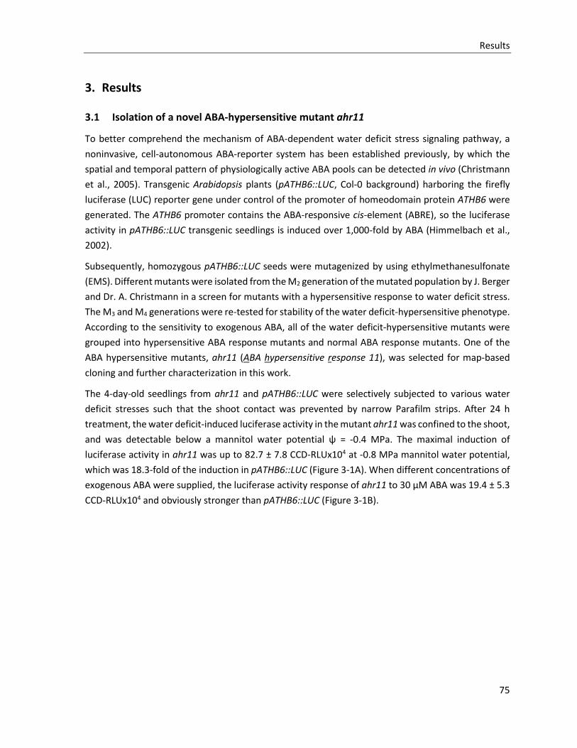
Results
75
3. Results
3.1 Isolation of a novel ABA-hypersensitive mutant ahr11
To better comprehend the mechanism of ABA-dependent water deficit stress signaling pathway, a
noninvasive, cell-autonomous ABA-reporter system has been established previously, by which the
spatial and temporal pattern of physiologically active ABA pools can be detected in vivo (Christmann
et al., 2005). Transgenic Arabidopsis plants (pATHB6::LUC, Col-0 background) harboring the firefly
luciferase (LUC) reporter gene under control of the promoter of homeodomain protein ATHB6 were
generated. The ATHB6 promoter contains the ABA-responsive cis-element (ABRE), so the luciferase
activity in pATHB6::LUC transgenic seedlings is induced over 1,000-fold by ABA (Himmelbach et al.,
2002).
Subsequently, homozygous pATHB6::LUC seeds were mutagenized by using ethylmethanesulfonate
(EMS). Different mutants were isolated from the M2 generation of the mutated population by J. Berger
and Dr. A. Christmann in a screen for mutants with a hypersensitive response to water deficit stress.
The M3 and M4 generations were re-tested for stability of the water deficit-hypersensitive phenotype.
According to the sensitivity to exogenous ABA, all of the water deficit-hypersensitive mutants were
grouped into hypersensitive ABA response mutants and normal ABA response mutants. One of the
ABA hypersensitive mutants, ahr11 (ABA hypersensitive response 11), was selected for map-based
cloning and further characterization in this work.
The 4-day-old seedlings from ahr11 and pATHB6::LUC were selectively subjected to various water
deficit stresses such that the shoot contact was prevented by narrow Parafilm strips. After 24 h
treatment, the water deficit-induced luciferase activity in the mutant ahr11 was confined to the shoot,
and was detectable below a mannitol water potential ψ = -0.4 MPa. The maximal induction of
luciferase activity in ahr11 was up to 82.7 ± 7.8 CCD-RLUx104 at -0.8 MPa mannitol water potential,
which was 18.3-fold of the induction in pATHB6::LUC (Figure 3-1A). When different concentrations of
exogenous ABA were supplied, the luciferase activity response of ahr11 to 30 µM ABA was 19.4 ± 5.3
CCD-RLUx104 and obviously stronger than pATHB6::LUC (Figure 3-1B).
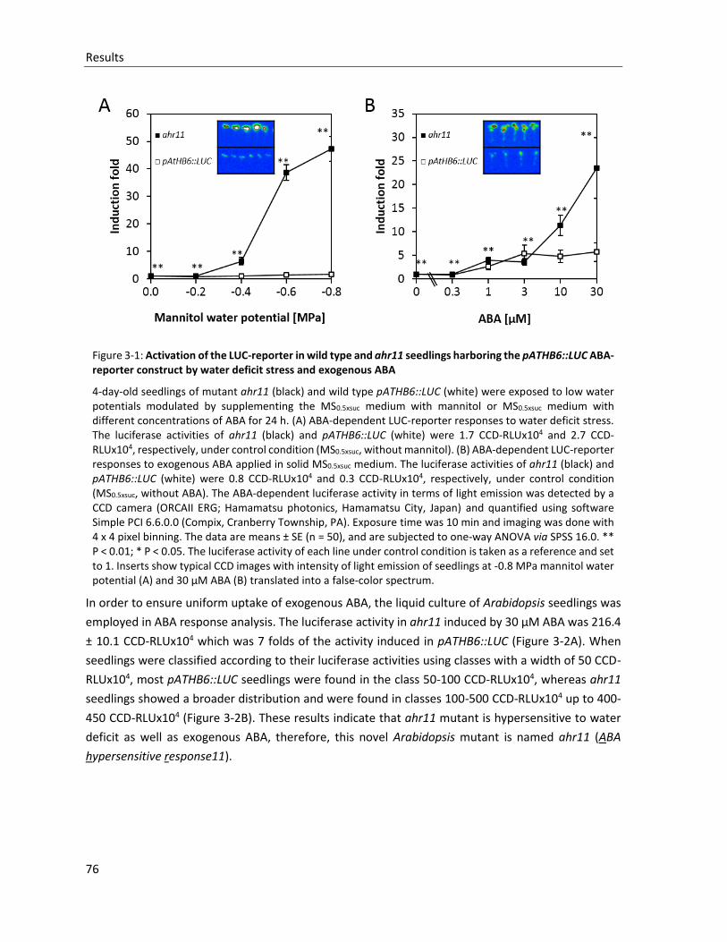
Results
76
Figure 3-1: Activation of the LUC-reporter in wild type and ahr11 seedlings harboring the pATHB6::LUC ABA-reporter construct by water deficit stress and exogenous ABA
4-day-old seedlings of mutant ahr11 (black) and wild type pATHB6::LUC (white) were exposed to low water potentials modulated by supplementing the MS0.5xsuc medium with mannitol or MS0.5xsuc medium with different concentrations of ABA for 24 h. (A) ABA-dependent LUC-reporter responses to water deficit stress. The luciferase activities of ahr11 (black) and pATHB6::LUC (white) were 1.7 CCD-RLUx104 and 2.7 CCD-RLUx104, respectively, under control condition (MS0.5xsuc, without mannitol). (B) ABA-dependent LUC-reporter responses to exogenous ABA applied in solid MS0.5xsuc medium. The luciferase activities of ahr11 (black) and pATHB6::LUC (white) were 0.8 CCD-RLUx104 and 0.3 CCD-RLUx104, respectively, under control condition (MS0.5xsuc, without ABA). The ABA-dependent luciferase activity in terms of light emission was detected by a CCD camera (ORCAII ERG; Hamamatsu photonics, Hamamatsu City, Japan) and quantified using software Simple PCI 6.6.0.0 (Compix, Cranberry Township, PA). Exposure time was 10 min and imaging was done with 4 x 4 pixel binning. The data are means ± SE (n = 50), and are subjected to one-way ANOVA via SPSS 16.0. ** P < 0.01; * P < 0.05. The luciferase activity of each line under control condition is taken as a reference and set to 1. Inserts show typical CCD images with intensity of light emission of seedlings at -0.8 MPa mannitol water potential (A) and 30 µM ABA (B) translated into a false-color spectrum.
In order to ensure uniform uptake of exogenous ABA, the liquid culture of Arabidopsis seedlings was
employed in ABA response analysis. The luciferase activity in ahr11 induced by 30 µM ABA was 216.4
± 10.1 CCD-RLUx104 which was 7 folds of the activity induced in pATHB6::LUC (Figure 3-2A). When
seedlings were classified according to their luciferase activities using classes with a width of 50 CCD-
RLUx104, most pATHB6::LUC seedlings were found in the class 50-100 CCD-RLUx104, whereas ahr11
seedlings showed a broader distribution and were found in classes 100-500 CCD-RLUx104 up to 400-
450 CCD-RLUx104 (Figure 3-2B). These results indicate that ahr11 mutant is hypersensitive to water
deficit as well as exogenous ABA, therefore, this novel Arabidopsis mutant is named ahr11 (ABA
hypersensitive response11).
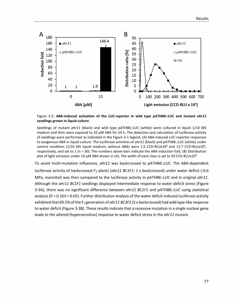
Results
77
Figure 3-2: ABA-induced activation of the LUC-reporter in wild type pATHB6::LUC and mutant ahr11 seedlings grown in liquid culture
Seedlings of mutant ahr11 (black) and wild type pATHB6::LUC (white) were cultured in liquid 1/10 MS medium and then were exposed to 10 µM ABA for 24 h. The detection and calculation of luciferase activity of seedlings were performed as indicated in the Figure 3-1 legend. (A) ABA-induced LUC-reporter responses to exogenous ABA in liquid culture. The luciferase activities of ahr11 (black) and pATHB6::LUC (white) under control condition (1/10 MS liquid medium, without ABA) were 1.5 CCD-RLUx104 and 12.7 CCD-RLUx104, respectively, and set to 1 (n = 50). The numbers above bars indicate the ABA induction fold. (B) Distribution plot of light emission under 10 µM ABA shown in (A). The width of each class is set to 50 CCD-RLUx104.
To avoid multi-mutation influences, ahr11 was backcrossed to pATHB6::LUC. The ABA-dependent
luciferase activity of backcrossed F1 plants (ahr11 BC1F1; 1 x backcrossed) under water deficit (-0.6
MPa, mannitol) was then compared to the luciferase activity in pATHB6::LUC and in original ahr11.
Although the ahr11 BC1F1 seedlings displayed intermediate response to water deficit stress (Figure
3-3A), there was no significant difference between ahr11 BC1F1 and pATHB6::LUC using statistical
analysis (P = 0.163 > 0.05). Further distribution analysis of the water deficit-induced luciferase activity
exhibited that 69.5% of the F2 generation of ahr11 BC3F2 (3 x backcrossed) had wild type-like response
to water deficit (Figure 3-3B). These results indicate that a recessive mutation in a single nuclear gene
leads to the altered (hypersensitive) response to water deficit stress in the ahr11 mutant.
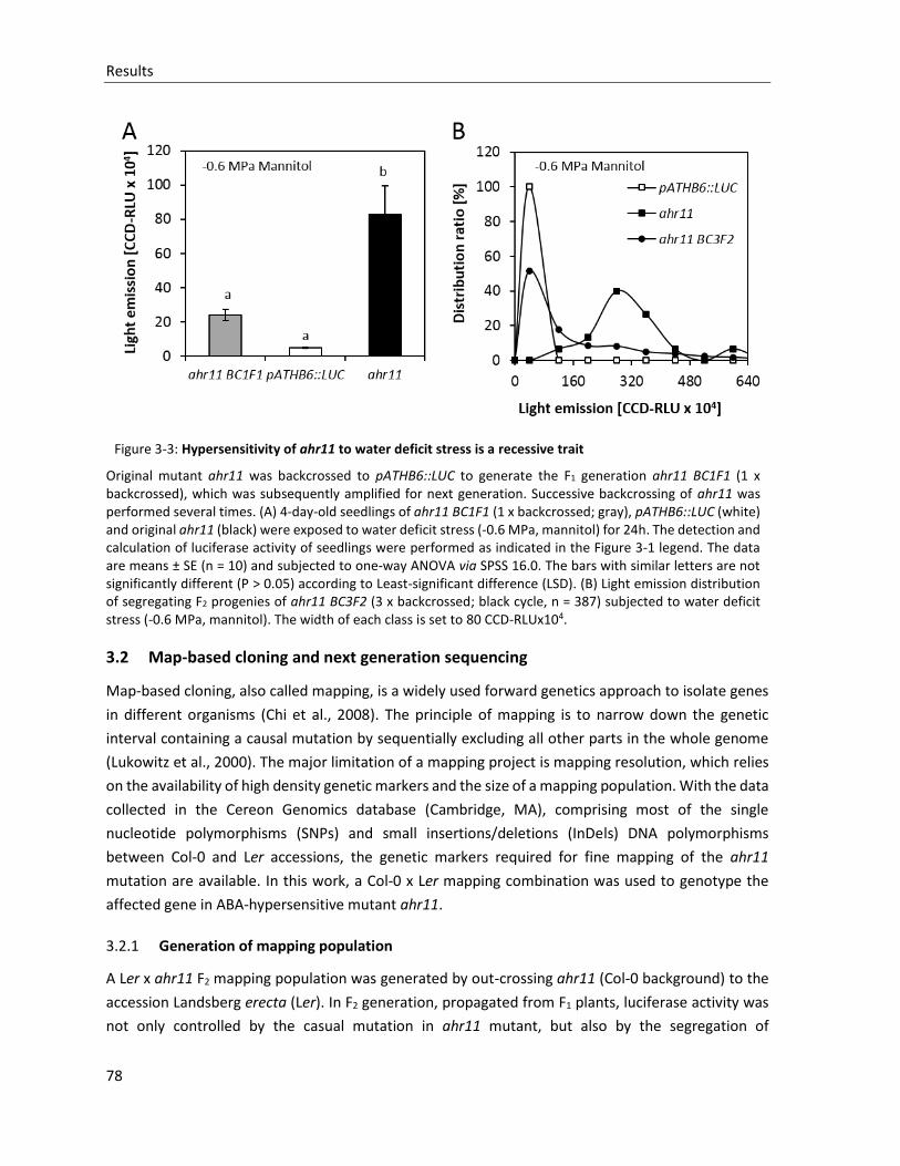
Results
78
Figure 3-3: Hypersensitivity of ahr11 to water deficit stress is a recessive trait
Original mutant ahr11 was backcrossed to pATHB6::LUC to generate the F1 generation ahr11 BC1F1 (1 x backcrossed), which was subsequently amplified for next generation. Successive backcrossing of ahr11 was performed several times. (A) 4-day-old seedlings of ahr11 BC1F1 (1 x backcrossed; gray), pATHB6::LUC (white) and original ahr11 (black) were exposed to water deficit stress (-0.6 MPa, mannitol) for 24h. The detection and calculation of luciferase activity of seedlings were performed as indicated in the Figure 3-1 legend. The data are means ± SE (n = 10) and subjected to one-way ANOVA via SPSS 16.0. The bars with similar letters are not significantly different (P > 0.05) according to Least-significant difference (LSD). (B) Light emission distribution of segregating F2 progenies of ahr11 BC3F2 (3 x backcrossed; black cycle, n = 387) subjected to water deficit stress (-0.6 MPa, mannitol). The width of each class is set to 80 CCD-RLUx104.
3.2 Map-based cloning and next generation sequencing
Map-based cloning, also called mapping, is a widely used forward genetics approach to isolate genes
in different organisms (Chi et al., 2008). The principle of mapping is to narrow down the genetic
interval containing a causal mutation by sequentially excluding all other parts in the whole genome
(Lukowitz et al., 2000). The major limitation of a mapping project is mapping resolution, which relies
on the availability of high density genetic markers and the size of a mapping population. With the data
collected in the Cereon Genomics database (Cambridge, MA), comprising most of the single
nucleotide polymorphisms (SNPs) and small insertions/deletions (InDels) DNA polymorphisms
between Col-0 and Ler accessions, the genetic markers required for fine mapping of the ahr11
mutation are available. In this work, a Col-0 x Ler mapping combination was used to genotype the
affected gene in ABA-hypersensitive mutant ahr11.
3.2.1 Generation of mapping population
A Ler x ahr11 F2 mapping population was generated by out-crossing ahr11 (Col-0 background) to the
accession Landsberg erecta (Ler). In F2 generation, propagated from F1 plants, luciferase activity was
not only controlled by the casual mutation in ahr11 mutant, but also by the segregation of
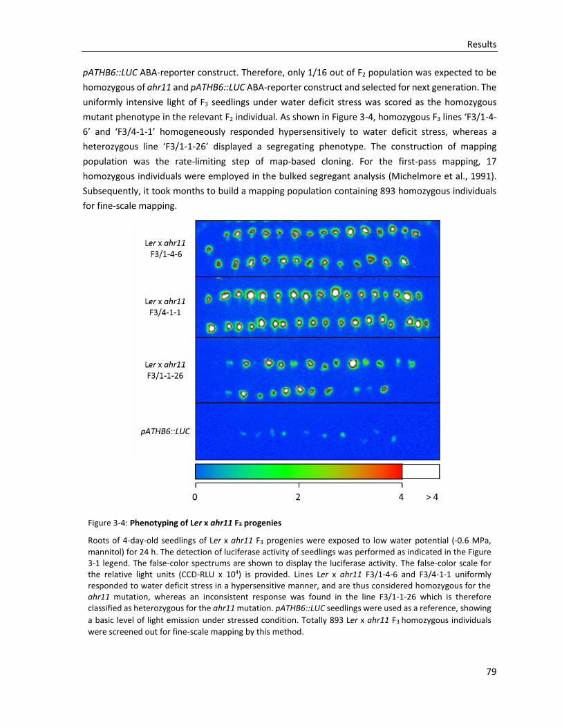
Results
79
pATHB6::LUC ABA-reporter construct. Therefore, only 1/16 out of F2 population was expected to be
homozygous of ahr11 and pATHB6::LUC ABA-reporter construct and selected for next generation. The
uniformly intensive light of F3 seedlings under water deficit stress was scored as the homozygous
mutant phenotype in the relevant F2 individual. As shown in Figure 3-4, homozygous F3 lines ‘F3/1-4-
6’ and ‘F3/4-1-1’ homogeneously responded hypersensitively to water deficit stress, whereas a
heterozygous line ‘F3/1-1-26’ displayed a segregating phenotype. The construction of mapping
population was the rate-limiting step of map-based cloning. For the first-pass mapping, 17
homozygous individuals were employed in the bulked segregant analysis (Michelmore et al., 1991).
Subsequently, it took months to build a mapping population containing 893 homozygous individuals
for fine-scale mapping.
Figure 3-4: Phenotyping of Ler x ahr11 F3 progenies
Roots of 4-day-old seedlings of Ler x ahr11 F3 progenies were exposed to low water potential (-0.6 MPa,
mannitol) for 24 h. The detection of luciferase activity of seedlings was performed as indicated in the Figure 3-1 legend. The false-color spectrums are shown to display the luciferase activity. The false-color scale for the relative light units (CCD-RLU x 104) is provided. Lines Ler x ahr11 F3/1-4-6 and F3/4-1-1 uniformly responded to water deficit stress in a hypersensitive manner, and are thus considered homozygous for the ahr11 mutation, whereas an inconsistent response was found in the line F3/1-1-26 which is therefore classified as heterozygous for the ahr11 mutation. pATHB6::LUC seedlings were used as a reference, showing
a basic level of light emission under stressed condition. Totally 893 Ler x ahr11 F3 homozygous individuals
were screened out for fine-scale mapping by this method.
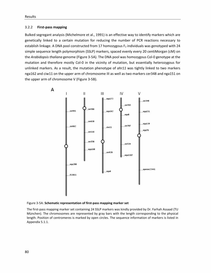
Results
80
3.2.2 First-pass mapping
Bulked segregant analysis (Michelmore et al., 1991) is an effective way to identify markers which are
genetically linked to a certain mutation for reducing the number of PCR reactions necessary to
establish linkage. A DNA pool constructed from 17 homozygous F2 individuals was genotyped with 24
simple sequence length polymorphism (SSLP) markers, spaced evenly every 20 centiMorgan (cM) on
the Arabidopsis thaliana genome (Figure 3-5A). The DNA pool was homozygous Col-0 genotype at the
mutation and therefore mostly Col-0 in the vicinity of mutation, but essentially heterozygous for
unlinked markers. As a result, the mutation phenotype of ahr11 was tightly linked to two markers
nga162 and ciw11 on the upper arm of chromosome III as well as two markers cer348 and nga151 on
the upper arm of chromosome V (Figure 3-5B).
Figure 3-5A: Schematic representation of first-pass mapping marker set
The first-pass mapping marker set containing 24 SSLP markers was kindly provided by Dr. Farhah Assaad (TU München). The chromosomes are represented by gray bars with the length corresponding to the physical length. Position of centromeres is marked by open circles. The sequence information of markers is listed in Appendix 5.1.1.
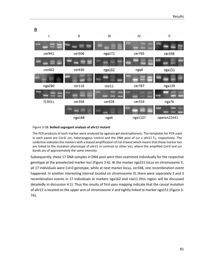
Results
81
Figure 3-5B: Bulked segregant analysis of ahr11 mutant
The PCR products of each marker were analyzed by agarose gel electrophoresis. The templates for PCR used
in each panel are Col-0, Ler, heterozygous control and the DNA pool of Ler x ahr11 F2, respectively. The
underline indicates the markers with a biased amplification of Col-0 band which means that these marker loci are linked to the mutation phenotype of ahr11 in contrast to other loci, where the amplified Col-0 and Ler bands are of approximately the same intensity.
Subsequently, these 17 DNA samples in DNA pool were then examined individually for the respective
genotype at the preselected marker loci (Figure 3-6). At the marker nga151 locus on chromosome V,
all 17 individuals were Col-0 genotype, while at next marker locus, cer348, one recombination event
happened. In another interesting interval located on chromosome III, there were separately 2 and 3
recombination events in 17 individuals at markers nga162 and ciw11 (this region will be discussed
detailedly in discussion 4.1). Thus the results of first-pass mapping indicate that the causal mutation
of ahr11 is located on the upper arm of chromosome V and tightly linked to marker nga151 (Figure 3-
7A).
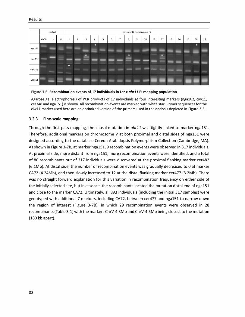
Results
82
Figure 3-6: Recombination events of 17 individuals in Ler x ahr11 F2 mapping population
Agarose gel electrophoresis of PCR products of 17 individuals at four interesting markers (nga162, ciw11, cer348 and nga151) is shown. All recombination events are marked with white star. Primer sequences for the ciw11 marker used here are an optimized version of the primers used in the analysis depicted in Figure 3-5.
3.2.3 Fine-scale mapping
Through the first-pass mapping, the causal mutation in ahr11 was tightly linked to marker nga151.
Therefore, additional markers on chromosome V at both proximal and distal sides of nga151 were
designed according to the database Cereon Arabidopsis Polymorphism Collection (Cambridge, MA).
As shown in Figure 3-7B, at marker nga151, 9 recombination events were observed in 317 individuals.
At proximal side, more distant from nga151, more recombination events were identified, and a total
of 80 recombinants out of 317 individuals were discovered at the proximal flanking marker cer482
(6.1Mb). At distal side, the number of recombination events was gradually decreased to 0 at marker
CA72 (4.24Mb), and then slowly increased to 12 at the distal flanking marker cer477 (3.2Mb). There
was no straight forward explanation for this variation in recombination frequency on either side of
the initially selected site, but in essence, the recombinants located the mutation distal end of nga151
and close to the marker CA72. Ultimately, all 893 individuals (including the initial 317 samples) were
genotyped with additional 7 markers, including CA72, between cer477 and nga151 to narrow down
the region of interest (Figure 3-7B), in which 29 recombination events were observed in 28
recombinants (Table 3-1) with the markers ChrV-4.3Mb and ChrV-4.5Mb being closest to the mutation
(180 kb apart).
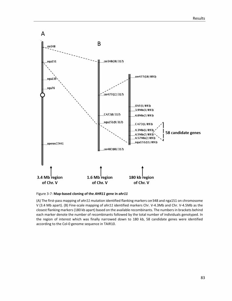
Results
83
Figure 3-7: Map-based cloning of the AHR11 gene in ahr11
(A) The first-pass mapping of ahr11 mutation identified flanking markers cer348 and nga151 on chromosome V (3.4 Mb apart). (B) Fine-scale mapping of ahr11 identified markers Chr. V-4.3Mb and Chr. V-4.5Mb as the closest flanking markers (180 kb apart) based on the available recombinants. The numbers in brackets behind each marker denote the number of recombinants followed by the total number of individuals genotyped. In the region of interest which was finally narrowed down to 180 kb, 58 candidate genes were identified according to the Col-0 genome sequence in TAIR10.
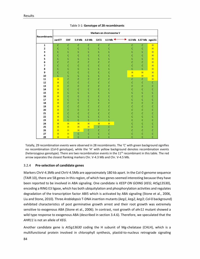
Results
84
Table 3-1: Genotype of 28 recombinants
Totally, 29 recombination events were observed in 28 recombinants. The ‘C’ with green background signifies no recombination (Col-0 genotype), while the ‘H’ with yellow background denotes recombination events (heterozygous genotype). There are two recombination events in the 11th recombinant in this table. The red arrow separates the closest flanking markers Chr. V-4.3 Mb and Chr. V-4.5 Mb.
3.2.4 Pre-selection of candidate genes
Markers ChrV-4.3Mb and ChrV-4.5Mb are approximately 180 kb apart. In the Col-0 genome sequence
(TAIR 10), there are 58 genes in this region, of which two genes seemed interesting because they have
been reported to be involved in ABA signaling. One candidate is KEEP ON GOING (KEG; At5g13530),
encoding a RING E3 ligase, which has both ubiquitylation and phosphorylation activities and regulates
degradation of the transcription factor ABI5 which is activated by ABA signaling (Stone et al., 2006,
Liu and Stone, 2010). Three Arabidopsis T-DNA insertion mutants (keg1, keg2, keg3; Col-0 background)
exhibited characteristics of post germinative growth arrest and their root growth was extremely
sensitive to exogenous ABA (Stone et al., 2006). In contrast, root growth of ahr11 mutant showed a
wild type response to exogenous ABA (described in section 3.4.6). Therefore, we speculated that the
AHR11 is not an allele of KEG.
Another candidate gene is At5g13630 coding the H subunit of Mg-chelatase (CHLH), which is a
multifunctional protein involved in chlorophyll synthesis, plastid-to-nucleus retrograde signaling
Results
85
(Mochizuki et al., 2001) and which has been claimed to be a plastidic-localized ABA-binding protein
(ABAR) (Shen et al., 2006, Wu et al., 2009). The ABA-affinity chromatography technique was used to
demonstrate that CHLH specifically binds ABA via the C-terminus (Shen et al., 2006, Wu et al., 2009,
Shang et al., 2010, Du et al., 2012). However, in another ABA binding assay, recombinant CHLH did
not bind ABA but specifically affected the ABA-signaling pathway in guard cells (Tsuzuki et al., 2011,
Tsuzuki et al., 2013). Meanwhile, the barley CHLH homolog could not function as an ABA receptor
(Muller and Hansson, 2009). These contradictory results point out that the natural role of CHLH in
ABA signaling is still too early to be concluded. Still, the gene was considered a candidate for the
mutation in ahr11, and in this project a 6658 bp genomic sequence of CHLH, including coding
sequence and promoter region, was amplified from ahr11 by PCR, and then sequenced by single read
sequencing. No mutation was detected in ABAR/CHLH gene in ahr11. Therefore, the mutation of
ahr11 is not in ABAR/CHLH.
Although two ABA signaling-related genes, KEG and ABAR/CHLH, were excluded, the number of
candidates was still too large to identify the AHR11 in reasonable time using a classical approach and
therefore, the application of a next generation sequencing (NGS) technique was envisaged.
3.2.5 Next generation sequencing
Considering the automated Sanger sequencing method as a ‘first-generation’ technology, newer
whole genome-scale sequencing methods are referred to as next-generation sequencing (NGS).
Generation of an enormous volume of detailed genetic information is the major advance offered by
NGS (Metzker, 2009). The NGS technology was thus employed to sequence the whole genome of the
ahr11 mutant to obtain information on the mutations located in the mapped region. This information
was expected to allow rapid identification of the causal mutation in ahr11. The crude sequences
obtained by NGS were aligned to the public reference genome from Arabidopsis thaliana Col-0
(TAIR10). In the ahr11 mutant, 172,351,045 reads were obtained covering 99.34% of the reference
Col-0 genome at least 20-fold. The causal mutation of ahr11 was refined in the 180 kb apart from 4.3
Mb to 4.5 Mb on chromosome V, but for reasons of safety, SNPs in a larger region from 4.0 Mb to 4.5
Mb were examined.
As shown in Figure 3-8 and Table3-2, 8 exclusively homozygous SNPs were identified in interest region,
all of which were C to T transitions: one SNP was in an intergene region between At5g13170 and
At5g13180; one SNP was in a promoter region of At5g13917; 2 SNPs were located near splicing sites
of At5g12960 and At5g13130; and another 4 SNPs were found in 4 gene coding sequences leading to
2 synonymous mutations including At5g12940 and At5g13260, as well as to one amino acid residue
exchange in At5g13930 and one premature termination of At5g13590. In the fine-scale mapping
process, a definite homozygous individual (the 24th recombinant in Table 3-1) had a recombination
event at marker 4.3 Mb. So that 3 SNPs (the 6th, 7th and 8th SNPs in Table 3-2) were extracted as final
candidates. For further identification, a new marker 4.48Mb was designed based on the 7th SNP at
position 4.48 Mb on Chromosome V. Then 3 recombinants (the 9th, 10th and 24rd in Table 3-1) were
analyzed for this marker, and one recombination event was detected in the 9th recombinant, thereby
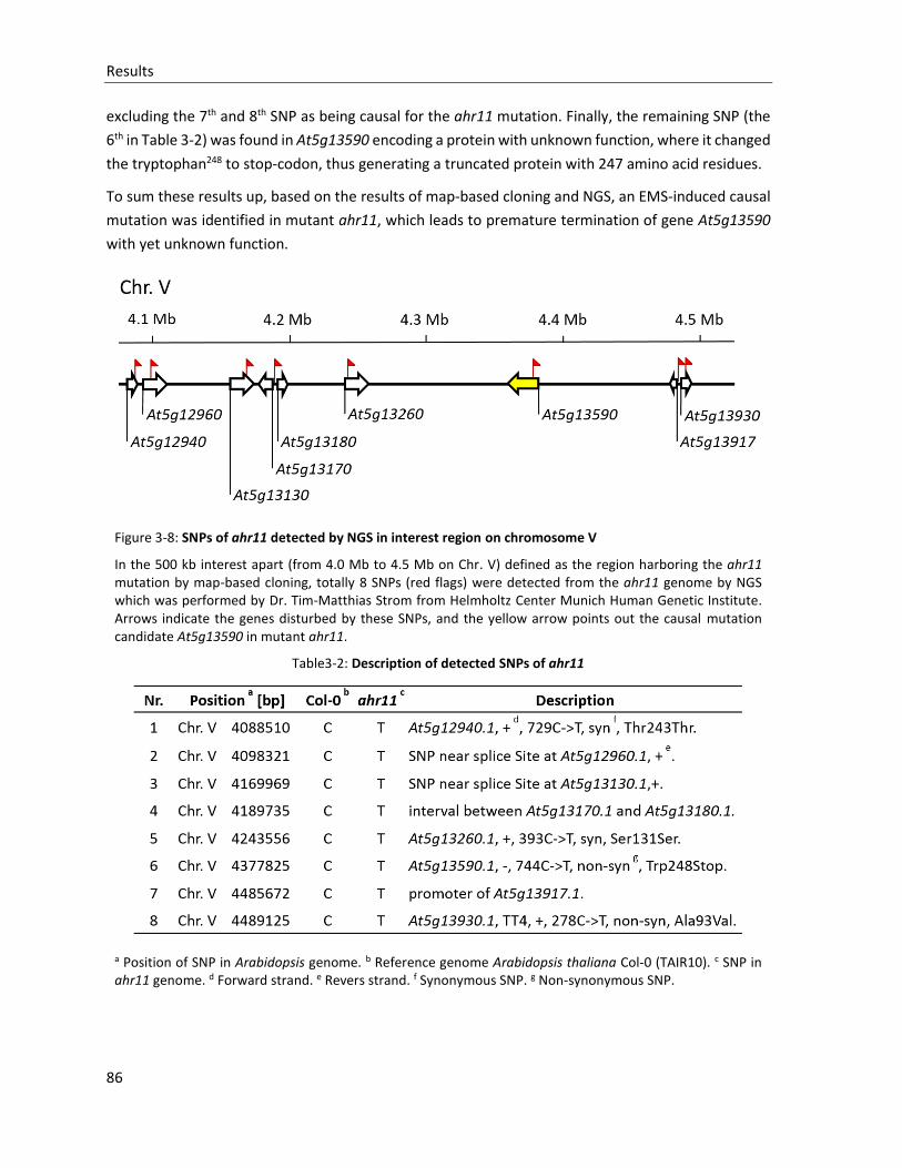
Results
86
excluding the 7th and 8th SNP as being causal for the ahr11 mutation. Finally, the remaining SNP (the
6th in Table 3-2) was found in At5g13590 encoding a protein with unknown function, where it changed
the tryptophan248 to stop-codon, thus generating a truncated protein with 247 amino acid residues.
To sum these results up, based on the results of map-based cloning and NGS, an EMS-induced causal
mutation was identified in mutant ahr11, which leads to premature termination of gene At5g13590
with yet unknown function.
Figure 3-8: SNPs of ahr11 detected by NGS in interest region on chromosome V
In the 500 kb interest apart (from 4.0 Mb to 4.5 Mb on Chr. V) defined as the region harboring the ahr11 mutation by map-based cloning, totally 8 SNPs (red flags) were detected from the ahr11 genome by NGS which was performed by Dr. Tim-Matthias Strom from Helmholtz Center Munich Human Genetic Institute. Arrows indicate the genes disturbed by these SNPs, and the yellow arrow points out the causal mutation candidate At5g13590 in mutant ahr11.
Table3-2: Description of detected SNPs of ahr11
a Position of SNP in Arabidopsis genome. b Reference genome Arabidopsis thaliana Col-0 (TAIR10). c SNP in ahr11 genome. d Forward strand. e Revers strand. f Synonymous SNP. g Non-synonymous SNP.
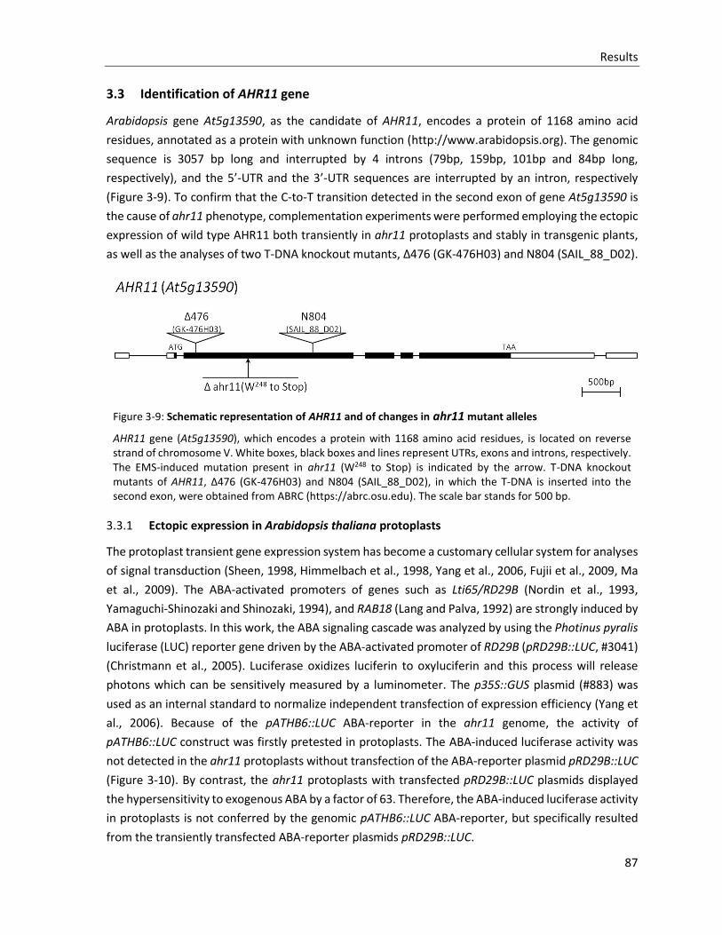
Results
87
3.3 Identification of AHR11 gene
Arabidopsis gene At5g13590, as the candidate of AHR11, encodes a protein of 1168 amino acid
residues, annotated as a protein with unknown function (http://www.arabidopsis.org). The genomic
sequence is 3057 bp long and interrupted by 4 introns (79bp, 159bp, 101bp and 84bp long,
respectively), and the 5’-UTR and the 3’-UTR sequences are interrupted by an intron, respectively
(Figure 3-9). To confirm that the C-to-T transition detected in the second exon of gene At5g13590 is
the cause of ahr11 phenotype, complementation experiments were performed employing the ectopic
expression of wild type AHR11 both transiently in ahr11 protoplasts and stably in transgenic plants,
as well as the analyses of two T-DNA knockout mutants, Δ476 (GK-476H03) and N804 (SAIL_88_D02).
Figure 3-9: Schematic representation of AHR11 and of changes in ahr11 mutant alleles
AHR11 gene (At5g13590), which encodes a protein with 1168 amino acid residues, is located on reverse strand of chromosome V. White boxes, black boxes and lines represent UTRs, exons and introns, respectively. The EMS-induced mutation present in ahr11 (W248 to Stop) is indicated by the arrow. T-DNA knockout mutants of AHR11, Δ476 (GK-476H03) and N804 (SAIL_88_D02), in which the T-DNA is inserted into the second exon, were obtained from ABRC (https://abrc.osu.edu). The scale bar stands for 500 bp.
3.3.1 Ectopic expression in Arabidopsis thaliana protoplasts
The protoplast transient gene expression system has become a customary cellular system for analyses
of signal transduction (Sheen, 1998, Himmelbach et al., 1998, Yang et al., 2006, Fujii et al., 2009, Ma
et al., 2009). The ABA-activated promoters of genes such as Lti65/RD29B (Nordin et al., 1993,
Yamaguchi-Shinozaki and Shinozaki, 1994), and RAB18 (Lang and Palva, 1992) are strongly induced by
ABA in protoplasts. In this work, the ABA signaling cascade was analyzed by using the Photinus pyralis
luciferase (LUC) reporter gene driven by the ABA-activated promoter of RD29B (pRD29B::LUC, #3041)
(Christmann et al., 2005). Luciferase oxidizes luciferin to oxyluciferin and this process will release
photons which can be sensitively measured by a luminometer. The p35S::GUS plasmid (#883) was
used as an internal standard to normalize independent transfection of expression efficiency (Yang et
al., 2006). Because of the pATHB6::LUC ABA-reporter in the ahr11 genome, the activity of
pATHB6::LUC construct was firstly pretested in protoplasts. The ABA-induced luciferase activity was
not detected in the ahr11 protoplasts without transfection of the ABA-reporter plasmid pRD29B::LUC
(Figure 3-10). By contrast, the ahr11 protoplasts with transfected pRD29B::LUC plasmids displayed
the hypersensitivity to exogenous ABA by a factor of 63. Therefore, the ABA-induced luciferase activity
in protoplasts is not conferred by the genomic pATHB6::LUC ABA-reporter, but specifically resulted
from the transiently transfected ABA-reporter plasmids pRD29B::LUC.
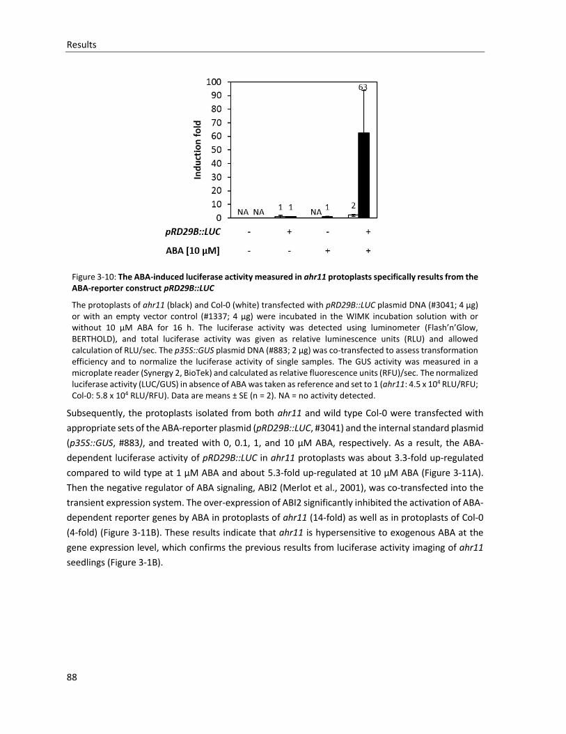
Results
88
Figure 3-10: The ABA-induced luciferase activity measured in ahr11 protoplasts specifically results from the ABA-reporter construct pRD29B::LUC
The protoplasts of ahr11 (black) and Col-0 (white) transfected with pRD29B::LUC plasmid DNA (#3041; 4 µg) or with an empty vector control (#1337; 4 µg) were incubated in the WIMK incubation solution with or without 10 μM ABA for 16 h. The luciferase activity was detected using luminometer (Flash’n’Glow, BERTHOLD), and total luciferase activity was given as relative luminescence units (RLU) and allowed calculation of RLU/sec. The p35S::GUS plasmid DNA (#883; 2 μg) was co-transfected to assess transformation efficiency and to normalize the luciferase activity of single samples. The GUS activity was measured in a microplate reader (Synergy 2, BioTek) and calculated as relative fluorescence units (RFU)/sec. The normalized luciferase activity (LUC/GUS) in absence of ABA was taken as reference and set to 1 (ahr11: 4.5 x 10⁴ RLU/RFU; Col-0: 5.8 x 10⁴ RLU/RFU). Data are means ± SE (n = 2). NA = no activity detected.
Subsequently, the protoplasts isolated from both ahr11 and wild type Col-0 were transfected with
appropriate sets of the ABA-reporter plasmid (pRD29B::LUC, #3041) and the internal standard plasmid
(p35S::GUS, #883), and treated with 0, 0.1, 1, and 10 µM ABA, respectively. As a result, the ABA-
dependent luciferase activity of pRD29B::LUC in ahr11 protoplasts was about 3.3-fold up-regulated
compared to wild type at 1 µM ABA and about 5.3-fold up-regulated at 10 µM ABA (Figure 3-11A).
Then the negative regulator of ABA signaling, ABI2 (Merlot et al., 2001), was co-transfected into the
transient expression system. The over-expression of ABI2 significantly inhibited the activation of ABA-
dependent reporter genes by ABA in protoplasts of ahr11 (14-fold) as well as in protoplasts of Col-0
(4-fold) (Figure 3-11B). These results indicate that ahr11 is hypersensitive to exogenous ABA at the
gene expression level, which confirms the previous results from luciferase activity imaging of ahr11
seedlings (Figure 3-1B).
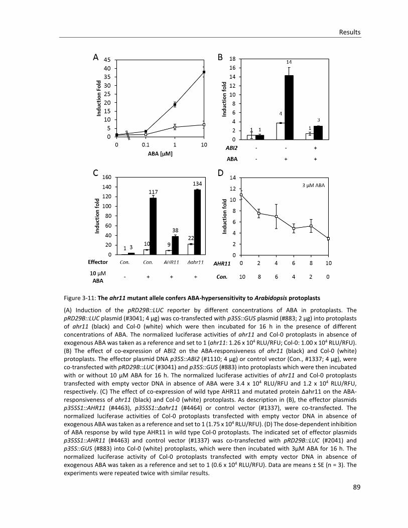
Results
89
Figure 3-11: The ahr11 mutant allele confers ABA-hypersensitivity to Arabidopsis protoplasts
(A) Induction of the pRD29B::LUC reporter by different concentrations of ABA in protoplasts. The pRD29B::LUC plasmid (#3041; 4 µg) was co-transfected with p35S::GUS plasmid (#883; 2 μg) into protoplasts of ahr11 (black) and Col-0 (white) which were then incubated for 16 h in the presence of different concentrations of ABA. The normalized luciferase activities of ahr11 and Col-0 protoplasts in absence of exogenous ABA was taken as a reference and set to 1 (ahr11: 1.26 x 10⁴ RLU/RFU; Col-0: 1.00 x 10⁴ RLU/RFU). (B) The effect of co-expression of ABI2 on the ABA-responsiveness of ahr11 (black) and Col-0 (white) protoplasts. The effector plasmid DNA p35S::ABI2 (#1110; 4 µg) or control vector (Con., #1337; 4 µg), were co-transfected with pRD29B::LUC (#3041) and p35S::GUS (#883) into protoplasts which were then incubated with or without 10 μM ABA for 16 h. The normalized luciferase activities of ahr11 and Col-0 protoplasts transfected with empty vector DNA in absence of ABA were 3.4 x 10⁴ RLU/RFU and 1.2 x 10⁴ RLU/RFU, respectively. (C) The effect of co-expression of wild type AHR11 and mutated protein Δahr11 on the ABA-responsiveness of ahr11 (black) and Col-0 (white) protoplasts. As description in (B), the effector plasmids p35SS1::AHR11 (#4463), p35SS1::Δahr11 (#4464) or control vector (#1337), were co-transfected. The normalized luciferase activities of Col-0 protoplasts transfected with empty vector DNA in absence of exogenous ABA was taken as a reference and set to 1 (1.75 x 10⁴ RLU/RFU). (D) The dose-dependent inhibition of ABA response by wild type AHR11 in wild type Col-0 protoplasts. The indicated set of effector plasmids p35SS1::AHR11 (#4463) and control vector (#1337) was co-transfected with pRD29B::LUC (#2041) and p35S::GUS (#883) into Col-0 (white) protoplasts, which were then incubated with 3μM ABA for 16 h. The normalized luciferase activity of Col-0 protoplasts transfected with empty vector DNA in absence of exogenous ABA was taken as a reference and set to 1 (0.6 x 10⁴ RLU/RFU). Data are means ± SE (n = 3). The experiments were repeated twice with similar results.
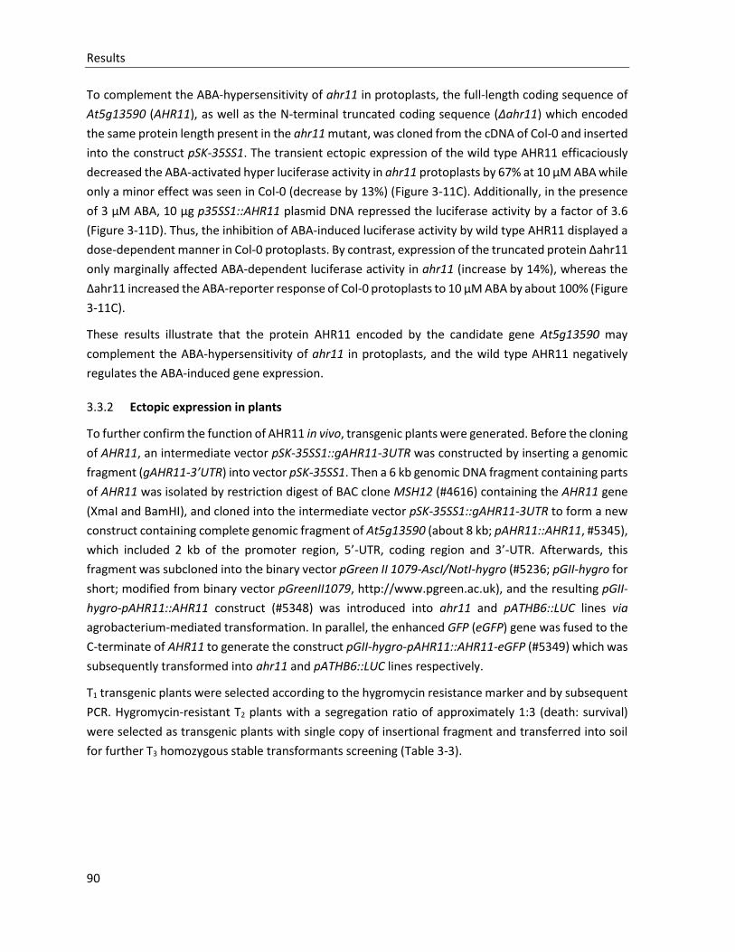
Results
90
To complement the ABA-hypersensitivity of ahr11 in protoplasts, the full-length coding sequence of
At5g13590 (AHR11), as well as the N-terminal truncated coding sequence (Δahr11) which encoded
the same protein length present in the ahr11 mutant, was cloned from the cDNA of Col-0 and inserted
into the construct pSK-35SS1. The transient ectopic expression of the wild type AHR11 efficaciously
decreased the ABA-activated hyper luciferase activity in ahr11 protoplasts by 67% at 10 µM ABA while
only a minor effect was seen in Col-0 (decrease by 13%) (Figure 3-11C). Additionally, in the presence
of 3 µM ABA, 10 µg p35SS1::AHR11 plasmid DNA repressed the luciferase activity by a factor of 3.6
(Figure 3-11D). Thus, the inhibition of ABA-induced luciferase activity by wild type AHR11 displayed a
dose-dependent manner in Col-0 protoplasts. By contrast, expression of the truncated protein Δahr11
only marginally affected ABA-dependent luciferase activity in ahr11 (increase by 14%), whereas the
Δahr11 increased the ABA-reporter response of Col-0 protoplasts to 10 µM ABA by about 100% (Figure
3-11C).
These results illustrate that the protein AHR11 encoded by the candidate gene At5g13590 may
complement the ABA-hypersensitivity of ahr11 in protoplasts, and the wild type AHR11 negatively
regulates the ABA-induced gene expression.
3.3.2 Ectopic expression in plants
To further confirm the function of AHR11 in vivo, transgenic plants were generated. Before the cloning
of AHR11, an intermediate vector pSK-35SS1::gAHR11-3UTR was constructed by inserting a genomic
fragment (gAHR11-3’UTR) into vector pSK-35SS1. Then a 6 kb genomic DNA fragment containing parts
of AHR11 was isolated by restriction digest of BAC clone MSH12 (#4616) containing the AHR11 gene
(XmaI and BamHI), and cloned into the intermediate vector pSK-35SS1::gAHR11-3UTR to form a new
construct containing complete genomic fragment of At5g13590 (about 8 kb; pAHR11::AHR11, #5345),
which included 2 kb of the promoter region, 5’-UTR, coding region and 3’-UTR. Afterwards, this
fragment was subcloned into the binary vector pGreen II 1079-AscI/NotI-hygro (#5236; pGII-hygro for
short; modified from binary vector pGreenII1079, http://www.pgreen.ac.uk), and the resulting pGII-
hygro-pAHR11::AHR11 construct (#5348) was introduced into ahr11 and pATHB6::LUC lines via
agrobacterium-mediated transformation. In parallel, the enhanced GFP (eGFP) gene was fused to the
C-terminate of AHR11 to generate the construct pGII-hygro-pAHR11::AHR11-eGFP (#5349) which was
subsequently transformed into ahr11 and pATHB6::LUC lines respectively.
T1 transgenic plants were selected according to the hygromycin resistance marker and by subsequent
PCR. Hygromycin-resistant T2 plants with a segregation ratio of approximately 1:3 (death: survival)
were selected as transgenic plants with single copy of insertional fragment and transferred into soil
for further T3 homozygous stable transformants screening (Table 3-3).
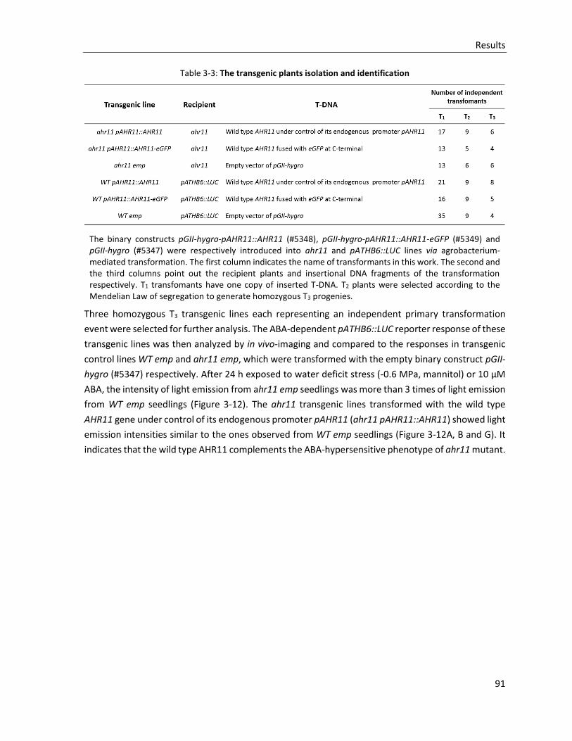
Results
91
Table 3-3: The transgenic plants isolation and identification
The binary constructs pGII-hygro-pAHR11::AHR11 (#5348), pGII-hygro-pAHR11::AHR11-eGFP (#5349) and pGII-hygro (#5347) were respectively introduced into ahr11 and pATHB6::LUC lines via agrobacterium-mediated transformation. The first column indicates the name of transformants in this work. The second and the third columns point out the recipient plants and insertional DNA fragments of the transformation respectively. T1 transfomants have one copy of inserted T-DNA. T2 plants were selected according to the Mendelian Law of segregation to generate homozygous T3 progenies.
Three homozygous T3 transgenic lines each representing an independent primary transformation
event were selected for further analysis. The ABA-dependent pATHB6::LUC reporter response of these
transgenic lines was then analyzed by in vivo-imaging and compared to the responses in transgenic
control lines WT emp and ahr11 emp, which were transformed with the empty binary construct pGII-
hygro (#5347) respectively. After 24 h exposed to water deficit stress (-0.6 MPa, mannitol) or 10 µM
ABA, the intensity of light emission from ahr11 emp seedlings was more than 3 times of light emission
from WT emp seedlings (Figure 3-12). The ahr11 transgenic lines transformed with the wild type
AHR11 gene under control of its endogenous promoter pAHR11 (ahr11 pAHR11::AHR11) showed light
emission intensities similar to the ones observed from WT emp seedlings (Figure 3-12A, B and G). It
indicates that the wild type AHR11 complements the ABA-hypersensitive phenotype of ahr11 mutant.
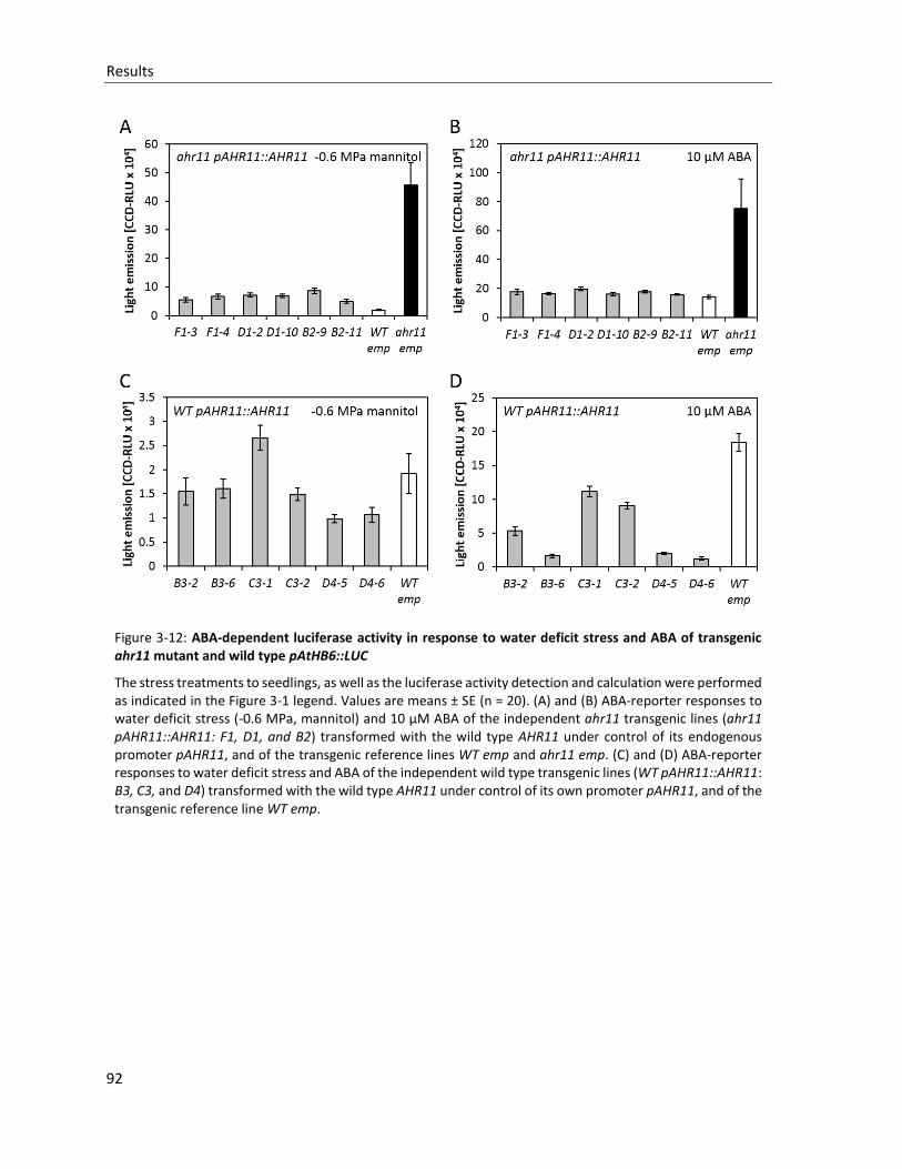
Results
92
Figure 3-12: ABA-dependent luciferase activity in response to water deficit stress and ABA of transgenic ahr11 mutant and wild type pAtHB6::LUC
The stress treatments to seedlings, as well as the luciferase activity detection and calculation were performed as indicated in the Figure 3-1 legend. Values are means ± SE (n = 20). (A) and (B) ABA-reporter responses to water deficit stress (-0.6 MPa, mannitol) and 10 µM ABA of the independent ahr11 transgenic lines (ahr11 pAHR11::AHR11: F1, D1, and B2) transformed with the wild type AHR11 under control of its endogenous promoter pAHR11, and of the transgenic reference lines WT emp and ahr11 emp. (C) and (D) ABA-reporter responses to water deficit stress and ABA of the independent wild type transgenic lines (WT pAHR11::AHR11: B3, C3, and D4) transformed with the wild type AHR11 under control of its own promoter pAHR11, and of the transgenic reference line WT emp.
Results
93
Figure 3-12: ABA-dependent luciferase activity in response to water deficit stress and ABA of transgenic ahr11 mutant and wild type pAtHB6::LUC (continued)
(E) and (F) ABA-reporter responses to water deficit stress and ABA of the independent ahr11 transgenic lines (ahr11 pAHR11::AHR11-eGFP: A5 and B5 ), of the wild type transgenic lines (WT pAHR11::AHR11-eGFP: 4A8 and 4G8) transformed with the wild type AHR11 fused to eGFP under control of its endogenous promoter pAHR11, and of the reference lines WT emp andahr11 emp. (G) False-color images of typical response from A-F. The color scale for the relative light units (CCD-RLUx104) is provided.
In contrast, expressing a fusion protein between AHR11 and eGFP under control of its endogenous
promoter (pAHR11::AHR11-eGFP) in ahr11 did not complement the ahr11 phenotype (Figure 3-12E,
F and G). Moreover, expressing pAHR11::AHR11 in pAtHB6::LUC (WT pAHR11::AHR11) reduced the
ABA-dependent responses of the wild type reporter lines compatible with a function of AHR11 as a
negative regulator in ABA signaling (Figure 3-12C, D and G). With the eGFP fused C-terminally to
AHR11, this effect was lost in WT pAHR11::AHR11-eGFP transgenic plants (Figure 3-12D, E and G).
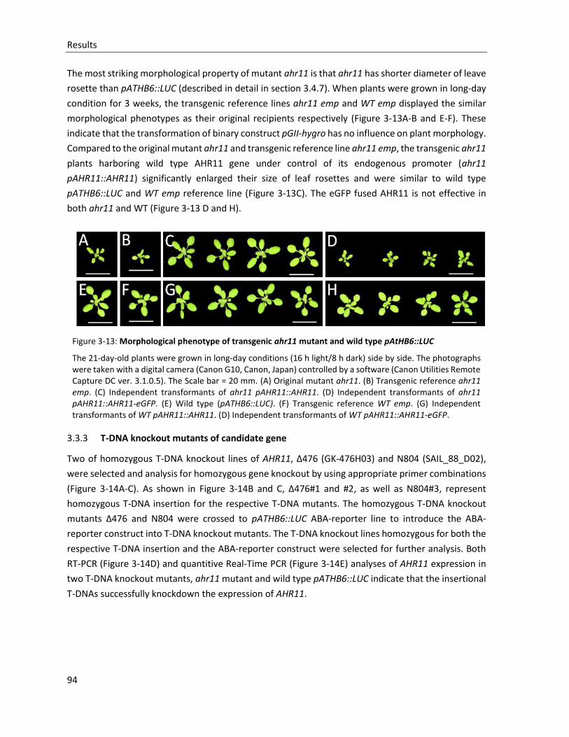
Results
94
The most striking morphological property of mutant ahr11 is that ahr11 has shorter diameter of leave
rosette than pATHB6::LUC (described in detail in section 3.4.7). When plants were grown in long-day
condition for 3 weeks, the transgenic reference lines ahr11 emp and WT emp displayed the similar
morphological phenotypes as their original recipients respectively (Figure 3-13A-B and E-F). These
indicate that the transformation of binary construct pGII-hygro has no influence on plant morphology.
Compared to the original mutant ahr11 and transgenic reference line ahr11 emp, the transgenic ahr11
plants harboring wild type AHR11 gene under control of its endogenous promoter (ahr11
pAHR11::AHR11) significantly enlarged their size of leaf rosettes and were similar to wild type
pATHB6::LUC and WT emp reference line (Figure 3-13C). The eGFP fused AHR11 is not effective in
both ahr11 and WT (Figure 3-13 D and H).
Figure 3-13: Morphological phenotype of transgenic ahr11 mutant and wild type pAtHB6::LUC
The 21-day-old plants were grown in long-day conditions (16 h light/8 h dark) side by side. The photographs were taken with a digital camera (Canon G10, Canon, Japan) controlled by a software (Canon Utilities Remote Capture DC ver. 3.1.0.5). The Scale bar = 20 mm. (A) Original mutant ahr11. (B) Transgenic reference ahr11 emp. (C) Independent transformants of ahr11 pAHR11::AHR11. (D) Independent transformants of ahr11 pAHR11::AHR11-eGFP. (E) Wild type (pATHB6::LUC). (F) Transgenic reference WT emp. (G) Independent transformants of WT pAHR11::AHR11. (D) Independent transformants of WT pAHR11::AHR11-eGFP.
3.3.3 T-DNA knockout mutants of candidate gene
Two of homozygous T-DNA knockout lines of AHR11, Δ476 (GK-476H03) and N804 (SAIL_88_D02),
were selected and analysis for homozygous gene knockout by using appropriate primer combinations
(Figure 3-14A-C). As shown in Figure 3-14B and C, Δ476#1 and #2, as well as N804#3, represent
homozygous T-DNA insertion for the respective T-DNA mutants. The homozygous T-DNA knockout
mutants Δ476 and N804 were crossed to pATHB6::LUC ABA-reporter line to introduce the ABA-
reporter construct into T-DNA knockout mutants. The T-DNA knockout lines homozygous for both the
respective T-DNA insertion and the ABA-reporter construct were selected for further analysis. Both
RT-PCR (Figure 3-14D) and quantitive Real-Time PCR (Figure 3-14E) analyses of AHR11 expression in
two T-DNA knockout mutants, ahr11 mutant and wild type pATHB6::LUC indicate that the insertional
T-DNAs successfully knockdown the expression of AHR11.
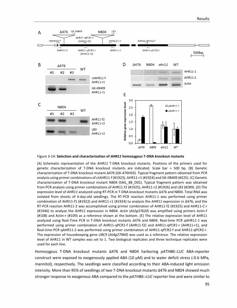
Results
95
Figure 3-14: Selection and characterization of AHR11 homozygous T-DNA knockout mutants
(A) Schematic representation of the AHR11 T-DNA knockout mutants. Positions of the primers used for genetic characterization of T-DNA knockout mutants are indicated. Scale bar = 500 bp. (B) Genetic characterization of T-DNA knockout mutant Δ476 (GK-476H03). Typical fragment pattern obtained from PCR analysis using primer combinations of ctAHR11-f (#1923), AHR11-r1 (#1924) and GK-08409 (#225). (C) Genetic characterization of T-DNA knockout mutant N804 (SAIL_88_D02). Typical fragment pattern was obtained from PCR analysis using primer combinations of AHR11-f2 (#1925), AHR11-r2 (#1926) and LB3 (#289). (D) The expression level of AHR11 analyzed using RT-PCR in T-DNA knockout mutants Δ476 and N804. Total RNA was isolated from shoots of 4-day-old seedlings. The RT-PCR reaction AHR11-1 was performed using primer combination of AHR11-f1 (#1922) and AHR11-r1 (#1924) to analysis the AHR11 expression in Δ476, and the RT-PCR reaction AHR11-2 was accomplished using primer combination of AHR11-f2 (#1925) and AHR11-C-r (#1946) to analyze the AHR11 expression in N804. Actin (At2g37620) was amplified using primers Actin-f (#108) and Actin-r (#109) as a reference shown at the bottom. (E) The relative expression level of AHR11 analyzed using Real-Time PCR in T-DNA knockout mutants Δ476 and N804. Real-time PCR qAHR11-1 was performed using primer combination of AHR11-qPCR1-f (AHR11-f2) and AHR11-qPCR1-r (AHR11-r1), and Real-time PCR qAHR11-2 was performed using primer combination of AHR11-qPCR2-f and AHR11-qPCR2-r. The expression of housekeeping gene UBC9 (At4g27960) was used as a reference. The relative expression level of AHR11 in WT samples was set to 1. Two biological replicates and three technique replicates were used for each line.
Homozygous T-DNA knockout mutants Δ476 and N804 harboring pATHB6::LUC ABA-reporter
construct were exposed to exogenously applied ABA (10 µM) and to water deficit stress (-0.6 MPa,
mannitol), respectively. The seedlings were classified according to their ABA-induced light emission
intensity. More than 95% of seedlings of two T-DNA knockout mutants Δ476 and N804 showed much
stronger response to exogenous ABA compared to the pATHB6::LUC reporter line and were similar to
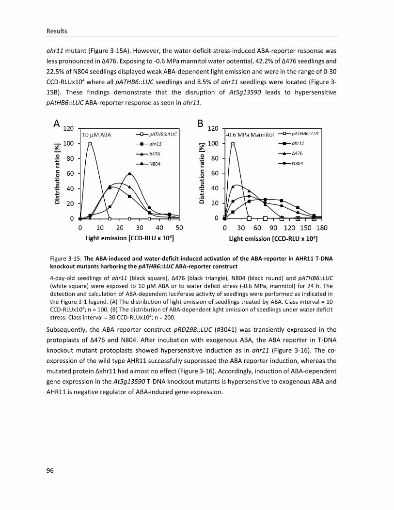
Results
96
ahr11 mutant (Figure 3-15A). However, the water-deficit-stress-induced ABA-reporter response was
less pronounced in Δ476. Exposing to -0.6 MPa mannitol water potential, 42.2% of Δ476 seedlings and
22.5% of N804 seedlings displayed weak ABA-dependent light emission and were in the range of 0-30
CCD-RLUx104 where all pATHB6::LUC seedlings and 8.5% of ahr11 seedlings were located (Figure 3-
15B). These findings demonstrate that the disruption of At5g13590 leads to hypersensitive
pAtHB6::LUC ABA-reporter response as seen in ahr11.
Figure 3-15: The ABA-induced and water-deficit-induced activation of the ABA-reporter in AHR11 T-DNA knockout mutants harboring the pATHB6::LUC ABA-reporter construct
4-day-old seedlings of ahr11 (black square), Δ476 (black triangle), N804 (black round) and pATHB6::LUC (white square) were exposed to 10 µM ABA or to water deficit stress (-0.6 MPa, mannitol) for 24 h. The detection and calculation of ABA-dependent luciferase activity of seedlings were performed as indicated in the Figure 3-1 legend. (A) The distribution of light emission of seedlings treated by ABA. Class interval = 10 CCD-RLUx104; n = 100. (B) The distribution of ABA-dependent light emission of seedlings under water deficit stress. Class interval = 30 CCD-RLUx104; n = 200.
Subsequently, the ABA reporter construct pRD29B::LUC (#3041) was transiently expressed in the
protoplasts of Δ476 and N804. After incubation with exogenous ABA, the ABA reporter in T-DNA
knockout mutant protoplasts showed hypersensitive induction as in ahr11 (Figure 3-16). The co-
expression of the wild type AHR11 successfully suppressed the ABA reporter induction, whereas the
mutated protein Δahr11 had almost no effect (Figure 3-16). Accordingly, induction of ABA-dependent
gene expression in the At5g13590 T-DNA knockout mutants is hypersensitive to exogenous ABA and
AHR11 is negative regulator of ABA-induced gene expression.
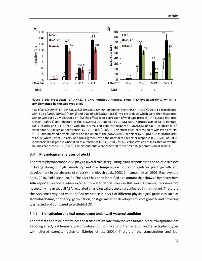
Results
97
Figure 3-16: Protoplasts of AHR11 T-DNA knockout mutants show ABA-hypersensitivity which is complemented by the wild type allele
4 µg of p35SS1::AHR11 (#4463), p35SS1::Δahr11 (#4464) or control vector (Con., #1337), were co-transfected with 4 µg of pRD29B::LUC (#3041) and 2 µg of p35S::GUS (#883) into protoplasts which were then incubated with or without 10 μM ABA for 16 h. (A) The effect of co-expression of wild type protein (AHR11) and mutated protein (Δahr11) on induction of the pRD29B::LUC reporter by 10 µM ABA in protoplasts of Col-0 (white), ahr11 (black) and Δ476 (red) with the normalized reporter response (LUC/GUS) of Col-0 in absence of exogenous ABA taken as a reference (1.75 x 10⁴ RLU/RFU). (B) The effect of co-expression of wild type protein AHR11 and mutated protein Δahr11 on induction of the pRD29B::LUC reporter by 10 µM ABA in protoplasts of Col-0 (white), ahr11 (black), and N804 (green) with the normalized reporter response (LUC/GUS) of Col-0 in absence of exogenous ABA taken as a reference (1.3 x 10⁴ RLU/RFU). Values which are indicated above the columns are means ± SE (n = 3). The experiments were repeated three times to generate similar results.
3.4 Physiological analyses of ahr11
The stress phytohormone ABA plays a pivotal role in regulating plant responses to the abiotic stresses
including drought, high osmolarity and low temperature but also regulates plant growth and
development in the absence of stress (Himmelbach et al., 2003, Christmann et al., 2006, Raghavendra
et al., 2010, Finkelstein, 2013). The ahr11 has been identified as a mutant that shows a hypersensitive
ABA reporter response when exposed to water deficit stress in this work. However, this does not
necessarily mean that all ABA-regulated physiological processes are affected in this mutant. Therefore,
the ABA sensitivity and water deficit resistance in ahr11 of different physiological processes such as
stomatal closure, dormancy, germination, post-germination development, root growth, and flowering
was tested and compared to pAtHB6::LUC.
3.4.1 Transpiration and leaf temperature under well-watered condition
The stomata aperture determines the transpiration rate from the leaf surface. Since transpiration has
a cooling effect, leaf temperature provides a robust indicator of transpiration and reflects phenotypes
with altered stomatal behavior (Merlot et al., 2002). Therefore, the transpiration and leaf
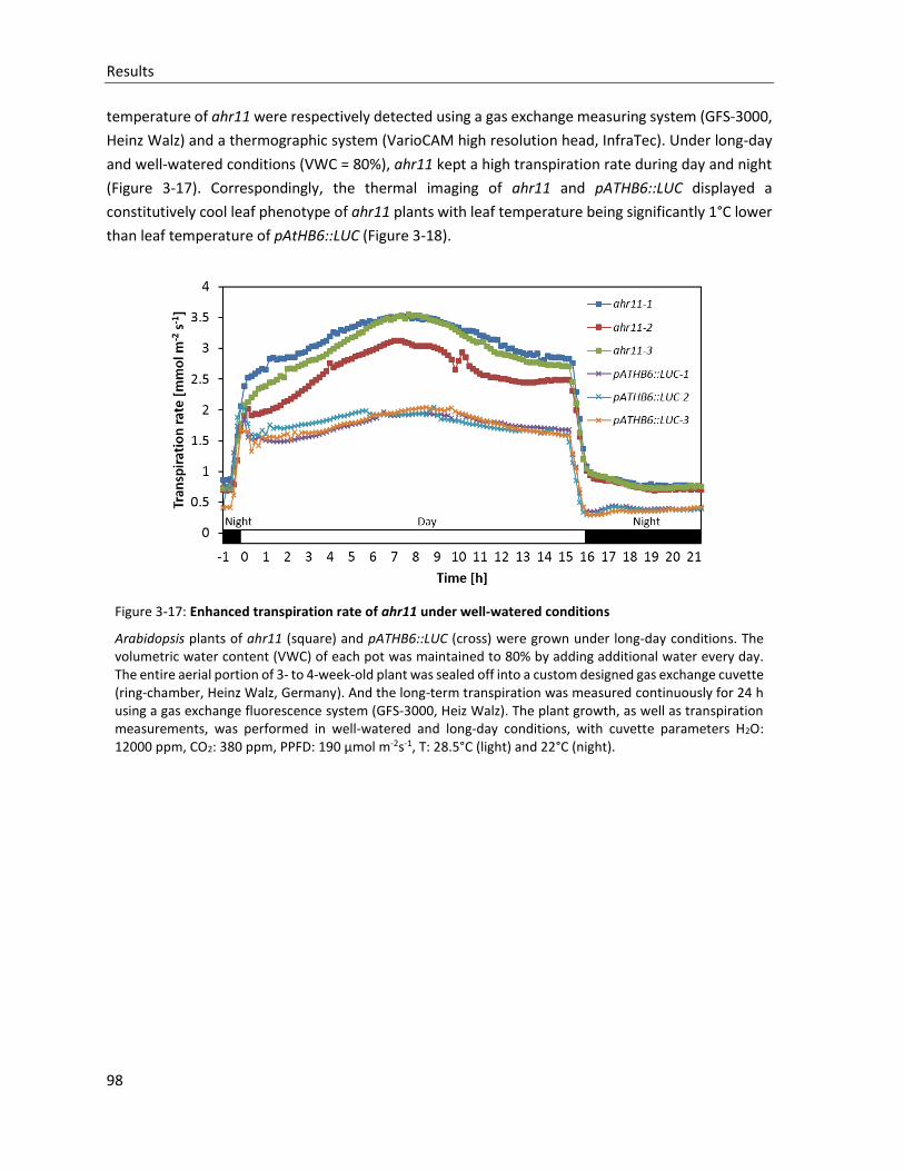
Results
98
temperature of ahr11 were respectively detected using a gas exchange measuring system (GFS-3000,
Heinz Walz) and a thermographic system (VarioCAM high resolution head, InfraTec). Under long-day
and well-watered conditions (VWC = 80%), ahr11 kept a high transpiration rate during day and night
(Figure 3-17). Correspondingly, the thermal imaging of ahr11 and pATHB6::LUC displayed a
constitutively cool leaf phenotype of ahr11 plants with leaf temperature being significantly 1°C lower
than leaf temperature of pAtHB6::LUC (Figure 3-18).
Figure 3-17: Enhanced transpiration rate of ahr11 under well-watered conditions
Arabidopsis plants of ahr11 (square) and pATHB6::LUC (cross) were grown under long-day conditions. The volumetric water content (VWC) of each pot was maintained to 80% by adding additional water every day. The entire aerial portion of 3- to 4-week-old plant was sealed off into a custom designed gas exchange cuvette (ring-chamber, Heinz Walz, Germany). And the long-term transpiration was measured continuously for 24 h using a gas exchange fluorescence system (GFS-3000, Heiz Walz). The plant growth, as well as transpiration measurements, was performed in well-watered and long-day conditions, with cuvette parameters H2O: 12000 ppm, CO2: 380 ppm, PPFD: 190 µmol m-2s-1, T: 28.5°C (light) and 22°C (night).
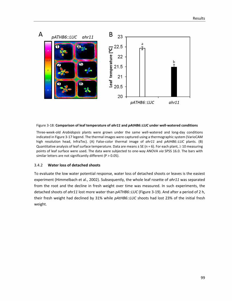
Results
99
Figure 3-18: Comparison of leaf temperature of ahr11 and pAtHB6::LUC under well-watered conditions
Three-week-old Arabidopsis plants were grown under the same well-watered and long-day conditions indicated in Figure 3-17 legend. The thermal images were captured using a thermographic system (VarioCAM high resolution head, InfraTec). (A) False-color thermal image of ahr11 and pAtHB6::LUC plants. (B)
Quantitative analysis of leaf surface temperature. Data are means ± SE (n = 6). For each plant, 10 measuring points of leaf surface were used. The data were subjected to one-way ANOVA via SPSS 16.0. The bars with similar letters are not significantly different (P > 0.05).
3.4.2 Water loss of detached shoots
To evaluate the low water potential response, water loss of detached shoots or leaves is the easiest
experiment (Himmelbach et al., 2002). Subsequently, the whole leaf rosette of ahr11 was separated
from the root and the decline in fresh weight over time was measured. In such experiments, the
detached shoots of ahr11 lost more water than pATHB6::LUC (Figure 3-19). And after a period of 2 h,
their fresh weight had declined by 31% while pAtHB6::LUC shoots had lost 23% of the initial fresh
weight.
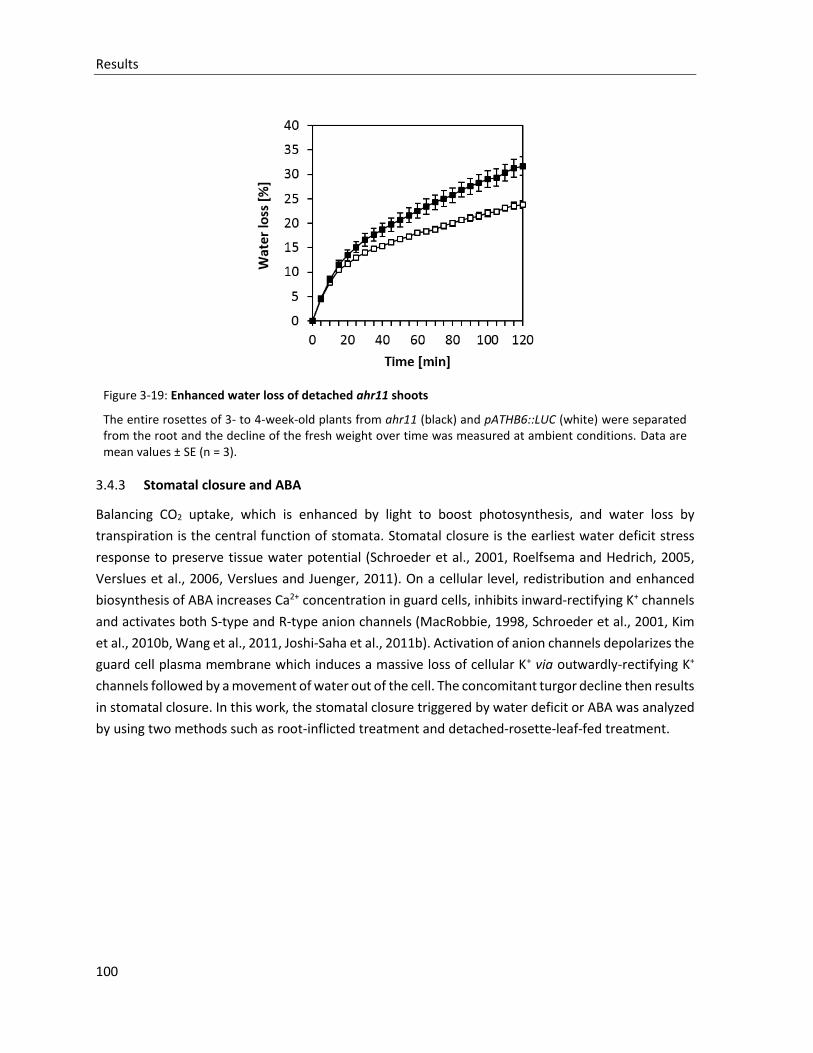
Results
100
Figure 3-19: Enhanced water loss of detached ahr11 shoots
The entire rosettes of 3- to 4-week-old plants from ahr11 (black) and pATHB6::LUC (white) were separated from the root and the decline of the fresh weight over time was measured at ambient conditions. Data are mean values ± SE (n = 3).
3.4.3 Stomatal closure and ABA
Balancing CO2 uptake, which is enhanced by light to boost photosynthesis, and water loss by
transpiration is the central function of stomata. Stomatal closure is the earliest water deficit stress
response to preserve tissue water potential (Schroeder et al., 2001, Roelfsema and Hedrich, 2005,
Verslues et al., 2006, Verslues and Juenger, 2011). On a cellular level, redistribution and enhanced
biosynthesis of ABA increases Ca2+ concentration in guard cells, inhibits inward-rectifying K+ channels
and activates both S-type and R-type anion channels (MacRobbie, 1998, Schroeder et al., 2001, Kim
et al., 2010b, Wang et al., 2011, Joshi-Saha et al., 2011b). Activation of anion channels depolarizes the
guard cell plasma membrane which induces a massive loss of cellular K+ via outwardly-rectifying K+
channels followed by a movement of water out of the cell. The concomitant turgor decline then results
in stomatal closure. In this work, the stomatal closure triggered by water deficit or ABA was analyzed
by using two methods such as root-inflicted treatment and detached-rosette-leaf-fed treatment.
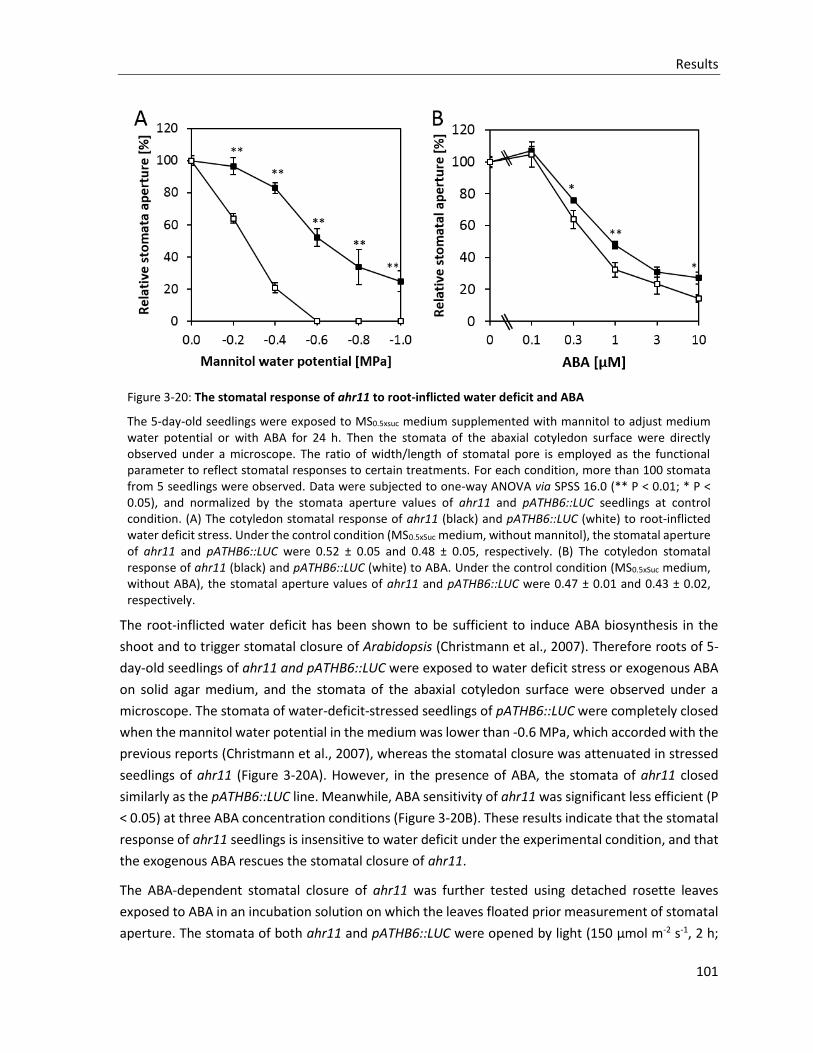
Results
101
Figure 3-20: The stomatal response of ahr11 to root-inflicted water deficit and ABA
The 5-day-old seedlings were exposed to MS0.5xsuc medium supplemented with mannitol to adjust medium water potential or with ABA for 24 h. Then the stomata of the abaxial cotyledon surface were directly observed under a microscope. The ratio of width/length of stomatal pore is employed as the functional parameter to reflect stomatal responses to certain treatments. For each condition, more than 100 stomata from 5 seedlings were observed. Data were subjected to one-way ANOVA via SPSS 16.0 (** P < 0.01; * P < 0.05), and normalized by the stomata aperture values of ahr11 and pATHB6::LUC seedlings at control condition. (A) The cotyledon stomatal response of ahr11 (black) and pATHB6::LUC (white) to root-inflicted water deficit stress. Under the control condition (MS0.5xSuc medium, without mannitol), the stomatal aperture of ahr11 and pATHB6::LUC were 0.52 ± 0.05 and 0.48 ± 0.05, respectively. (B) The cotyledon stomatal response of ahr11 (black) and pATHB6::LUC (white) to ABA. Under the control condition (MS0.5xSuc medium, without ABA), the stomatal aperture values of ahr11 and pATHB6::LUC were 0.47 ± 0.01 and 0.43 ± 0.02, respectively.
The root-inflicted water deficit has been shown to be sufficient to induce ABA biosynthesis in the
shoot and to trigger stomatal closure of Arabidopsis (Christmann et al., 2007). Therefore roots of 5-
day-old seedlings of ahr11 and pATHB6::LUC were exposed to water deficit stress or exogenous ABA
on solid agar medium, and the stomata of the abaxial cotyledon surface were observed under a
microscope. The stomata of water-deficit-stressed seedlings of pATHB6::LUC were completely closed
when the mannitol water potential in the medium was lower than -0.6 MPa, which accorded with the
previous reports (Christmann et al., 2007), whereas the stomatal closure was attenuated in stressed
seedlings of ahr11 (Figure 3-20A). However, in the presence of ABA, the stomata of ahr11 closed
similarly as the pATHB6::LUC line. Meanwhile, ABA sensitivity of ahr11 was significant less efficient (P
< 0.05) at three ABA concentration conditions (Figure 3-20B). These results indicate that the stomatal
response of ahr11 seedlings is insensitive to water deficit under the experimental condition, and that
the exogenous ABA rescues the stomatal closure of ahr11.
The ABA-dependent stomatal closure of ahr11 was further tested using detached rosette leaves
exposed to ABA in an incubation solution on which the leaves floated prior measurement of stomatal
aperture. The stomata of both ahr11 and pATHB6::LUC were opened by light (150 µmol m-2 s-1, 2 h;
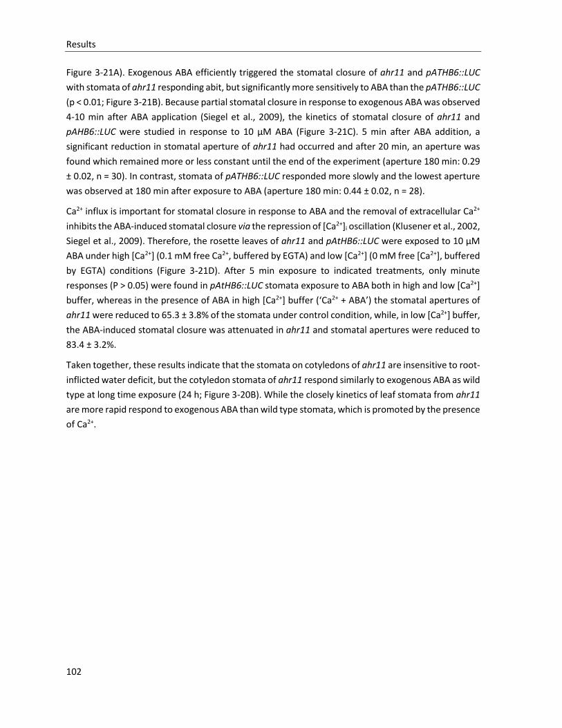
Results
102
Figure 3-21A). Exogenous ABA efficiently triggered the stomatal closure of ahr11 and pATHB6::LUC
with stomata of ahr11 responding abit, but significantly more sensitively to ABA than the pATHB6::LUC
(p < 0.01; Figure 3-21B). Because partial stomatal closure in response to exogenous ABA was observed
4-10 min after ABA application (Siegel et al., 2009), the kinetics of stomatal closure of ahr11 and
pAHB6::LUC were studied in response to 10 µM ABA (Figure 3-21C). 5 min after ABA addition, a
significant reduction in stomatal aperture of ahr11 had occurred and after 20 min, an aperture was
found which remained more or less constant until the end of the experiment (aperture 180 min: 0.29
± 0.02, n = 30). In contrast, stomata of pATHB6::LUC responded more slowly and the lowest aperture
was observed at 180 min after exposure to ABA (aperture 180 min: 0.44 ± 0.02, n = 28).
Ca2+ influx is important for stomatal closure in response to ABA and the removal of extracellular Ca2+
inhibits the ABA-induced stomatal closure via the repression of [Ca2+]i oscillation (Klusener et al., 2002,
Siegel et al., 2009). Therefore, the rosette leaves of ahr11 and pAtHB6::LUC were exposed to 10 µM
ABA under high [Ca2+] (0.1 mM free Ca2+, buffered by EGTA) and low [Ca2+] (0 mM free [Ca2+], buffered
by EGTA) conditions (Figure 3-21D). After 5 min exposure to indicated treatments, only minute
responses (P > 0.05) were found in pAtHB6::LUC stomata exposure to ABA both in high and low [Ca2+]
buffer, whereas in the presence of ABA in high [Ca2+] buffer (‘Ca2+ + ABA’) the stomatal apertures of
ahr11 were reduced to 65.3 ± 3.8% of the stomata under control condition, while, in low [Ca2+] buffer,
the ABA-induced stomatal closure was attenuated in ahr11 and stomatal apertures were reduced to
83.4 ± 3.2%.
Taken together, these results indicate that the stomata on cotyledons of ahr11 are insensitive to root-
inflicted water deficit, but the cotyledon stomata of ahr11 respond similarly to exogenous ABA as wild
type at long time exposure (24 h; Figure 3-20B). While the closely kinetics of leaf stomata from ahr11
are more rapid respond to exogenous ABA than wild type stomata, which is promoted by the presence
of Ca2+.
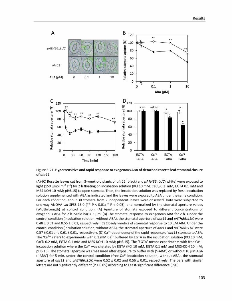
Results
103
Figure 3-21: Hypersensitive and rapid response to exogenous ABA of detached rosette leaf stomatal closure of ahr11
(A)-(C) Rosette leaves cut from 3-week-old plants of ahr11 (black) and pATHB6::LUC (white) were exposed to light (150 µmol m−2 s−1) for 2 h floating on incubation solution (KCl 10 mM, CaCl2 0.2 mM, EGTA 0.1 mM and MES-KOH 10 mM; pH6.15) to open stomata. Then, the incubation solution was replaced by fresh incubation solution supplemented with ABA as indicated and the leaves were exposed to ABA under the same condition. For each condition, about 30 stomata from 2 independent leaves were observed. Data were subjected to one-way ANOVA via SPSS 16.0 (** P < 0.01; * P < 0.05), and normalized by the stomatal aperture values (Width/Length) at control condition. (A) Aperture of stomata exposed to different concentrations of exogenous ABA for 2 h. Scale bar = 5 µm. (B) The stomatal response to exogenous ABA for 2 h. Under the control condition (incubation solution, without ABA), the stomatal aperture of ahr11 and pATHB6::LUC were 0.48 ± 0.01 and 0.55 ± 0.02, respectively. (C) Closely kinetics of stomatal response to 10 µM ABA. Under the control condition (incubation solution, without ABA), the stomatal aperture of ahr11 and pATHB6::LUC were 0.57 ± 0.01 and 0.61 ± 0.01, respectively. (D) Ca2+-dependency of the rapid response of ahr11 stomata to ABA. The ‘Ca2+’ refers to experiments with 0.1 mM Ca2+ buffered by EGTA in the incubation solution (KCl 10 mM, CaCl2 0.2 mM, EGTA 0.1 mM and MES-KOH 10 mM; pH6.15). The ‘EGTA’ means experiments with free Ca2+-incubation solution where the Ca2+ was chelated by EGTA (KCl 10 mM, EGTA 0.1 mM and MES-KOH 10 mM; pH6.15). The stomatal aperture was measured after exposure to buffer with (‘+ABA’) or without 10 µM ABA (‘-ABA’) for 5 min. under the control condition (free Ca2+-incubation solution, without ABA), the stomatal aperture of ahr11 and pATHB6::LUC were 0.52 ± 0.02 and 0.56 ± 0.01, respectively. The bars with similar letters are not significantly different (P > 0.05) according to Least-significant difference (LSD).
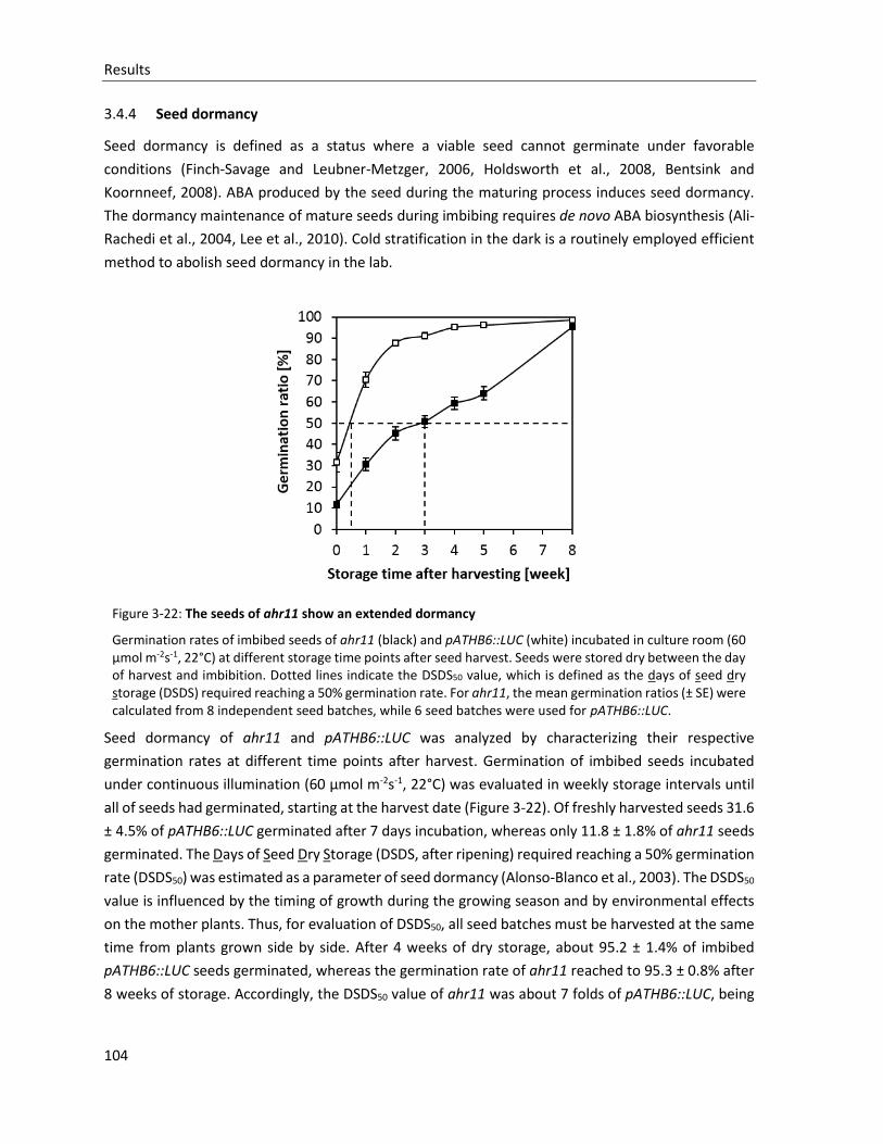
Results
104
3.4.4 Seed dormancy
Seed dormancy is defined as a status where a viable seed cannot germinate under favorable
conditions (Finch-Savage and Leubner-Metzger, 2006, Holdsworth et al., 2008, Bentsink and
Koornneef, 2008). ABA produced by the seed during the maturing process induces seed dormancy.
The dormancy maintenance of mature seeds during imbibing requires de novo ABA biosynthesis (Ali-
Rachedi et al., 2004, Lee et al., 2010). Cold stratification in the dark is a routinely employed efficient
method to abolish seed dormancy in the lab.
Figure 3-22: The seeds of ahr11 show an extended dormancy
Germination rates of imbibed seeds of ahr11 (black) and pATHB6::LUC (white) incubated in culture room (60 µmol m-2s-1, 22°C) at different storage time points after seed harvest. Seeds were stored dry between the day of harvest and imbibition. Dotted lines indicate the DSDS50 value, which is defined as the days of seed dry storage (DSDS) required reaching a 50% germination rate. For ahr11, the mean germination ratios (± SE) were calculated from 8 independent seed batches, while 6 seed batches were used for pATHB6::LUC.
Seed dormancy of ahr11 and pATHB6::LUC was analyzed by characterizing their respective
germination rates at different time points after harvest. Germination of imbibed seeds incubated
under continuous illumination (60 µmol m-2s-1, 22°C) was evaluated in weekly storage intervals until
all of seeds had germinated, starting at the harvest date (Figure 3-22). Of freshly harvested seeds 31.6
± 4.5% of pATHB6::LUC germinated after 7 days incubation, whereas only 11.8 ± 1.8% of ahr11 seeds
germinated. The Days of Seed Dry Storage (DSDS, after ripening) required reaching a 50% germination
rate (DSDS50) was estimated as a parameter of seed dormancy (Alonso-Blanco et al., 2003). The DSDS50
value is influenced by the timing of growth during the growing season and by environmental effects
on the mother plants. Thus, for evaluation of DSDS50, all seed batches must be harvested at the same
time from plants grown side by side. After 4 weeks of dry storage, about 95.2 ± 1.4% of imbibed
pATHB6::LUC seeds germinated, whereas the germination rate of ahr11 reached to 95.3 ± 0.8% after
8 weeks of storage. Accordingly, the DSDS50 value of ahr11 was about 7 folds of pATHB6::LUC, being
Results
105
approximately 21 days and 3 days respectively, in this experiment. Therefore, ahr11 seeds are much
more dormant than pATHB6::LUC seeds.
3.4.5 Seed Germination
Germination is defined as the visible emergence of the radicle through surrounding structures (Finch-
Savage and Leubner-Metzger, 2006). In Arabidopsis, this is a two-stage process with testa rupture
followed by endosperm rupture. Germination is controlled by environmental factors such as light,
temperature and water status, as well as by the balance between GA and ABA, in which GA promotes
seed germination by enhancing degradation of DELLA proteins to favour testa rupture, whereas ABA
has an antagonistic effect and inhibits germination via the transcriptional regulators ABI3 and ABI5
which repress endosperm rupture.
Figure 3-23: Water deficit stress and exogenous ABA inhibit germination of ahr11 and pAtHB6::LUC seeds to a similar extent
Seeds were sown on MS0.5xsuc medium supplemented with mannitol to modulate low water potential as indicated or with ABA, and were stratified at 4°C in the dark for 2 days. The number of germinated seeds was counted on the 2nd day after transfer to culture room (22°C, light: 60 µmol m-2s-1, constantly). Seeds were scored as germinated when the radicle had visibly emerged through the ruptured testa. Data are means ± SE (n = 2). Three independent experiments are performed to generate similar results. (A) Inhibition of germination of ahr11 (black) and pATHB6::LUC (white) seeds by water deficit stress in the medium. The germination rate of ahr11 and pATHB6::LUC under control condition (80.8 ± 0.5% and 99.5 ± 0.5%, respectively) was taken as a reference and set to 100%. (B) Inhibition of germination of ahr11 and pATHB6::LUC seeds by exogenous ABA. The germination rate of ahr11 and pATHB6::LUC under control condition (75.4 ± 1.1% and 99.3 ± 0.7%, respectively) was taken as a reference and set to 100%.
Reduced water availability and exogenous ABA were tested for their effects on germination of
imbibed ahr11 seeds. Under control condition of water deficit stress (MS0.5xSuc without mannitol), 80.8
± 0.5% of ahr11 seeds germinated after 2 days transferred to culture room (22°C, light: 60 µmol m-2s-
1, constantly), and 99.5 ± 0.5% of pATHB6::LUC seeds germinated (Figure 3-23A), while under control
condition of ABA response (MS0.5xSuc without ABA), the germination rates of ahr11 and pATHB6::LUC
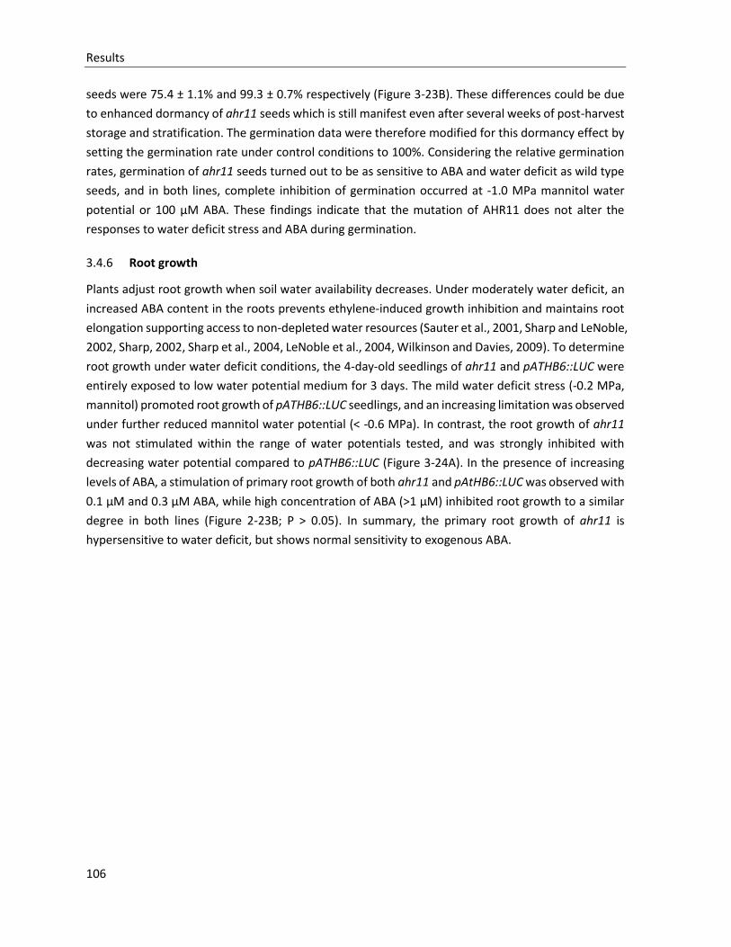
Results
106
seeds were 75.4 ± 1.1% and 99.3 ± 0.7% respectively (Figure 3-23B). These differences could be due
to enhanced dormancy of ahr11 seeds which is still manifest even after several weeks of post-harvest
storage and stratification. The germination data were therefore modified for this dormancy effect by
setting the germination rate under control conditions to 100%. Considering the relative germination
rates, germination of ahr11 seeds turned out to be as sensitive to ABA and water deficit as wild type
seeds, and in both lines, complete inhibition of germination occurred at -1.0 MPa mannitol water
potential or 100 µM ABA. These findings indicate that the mutation of AHR11 does not alter the
responses to water deficit stress and ABA during germination.
3.4.6 Root growth
Plants adjust root growth when soil water availability decreases. Under moderately water deficit, an
increased ABA content in the roots prevents ethylene-induced growth inhibition and maintains root
elongation supporting access to non-depleted water resources (Sauter et al., 2001, Sharp and LeNoble,
2002, Sharp, 2002, Sharp et al., 2004, LeNoble et al., 2004, Wilkinson and Davies, 2009). To determine
root growth under water deficit conditions, the 4-day-old seedlings of ahr11 and pATHB6::LUC were
entirely exposed to low water potential medium for 3 days. The mild water deficit stress (-0.2 MPa,
mannitol) promoted root growth of pATHB6::LUC seedlings, and an increasing limitation was observed
under further reduced mannitol water potential (< -0.6 MPa). In contrast, the root growth of ahr11
was not stimulated within the range of water potentials tested, and was strongly inhibited with
decreasing water potential compared to pATHB6::LUC (Figure 3-24A). In the presence of increasing
levels of ABA, a stimulation of primary root growth of both ahr11 and pAtHB6::LUC was observed with
0.1 µM and 0.3 µM ABA, while high concentration of ABA (>1 µM) inhibited root growth to a similar
degree in both lines (Figure 2-23B; P > 0.05). In summary, the primary root growth of ahr11 is
hypersensitive to water deficit, but shows normal sensitivity to exogenous ABA.
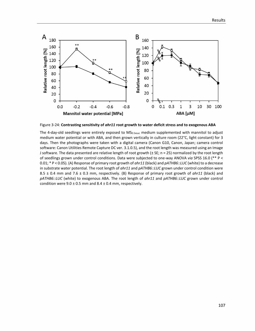
Results
107
Figure 3-24: Contrasting sensitivity of ahr11 root growth to water deficit stress and to exogenous ABA
The 4-day-old seedlings were entirely exposed to MS0.5xsuc medium supplemented with mannitol to adjust medium water potential or with ABA, and then grown vertically in culture room (22°C, light constant) for 3 days. Then the photographs were taken with a digital camera (Canon G10, Canon, Japan; camera control software: Canon Utilities Remote Capture DC ver. 3.1.0.5), and the root length was measured using an Image J software. The data presented are relative length of root growth (± SE; n = 25) normalized by the root length of seedlings grown under control conditions. Data were subjected to one-way ANOVA via SPSS 16.0 (** P < 0.01; * P < 0.05). (A) Response of primary root growth of ahr11 (black) and pATHB6::LUC (white) to a decrease in substrate water potential. The root length of ahr11 and pATHB6::LUC grown under control condition were 8.5 ± 0.4 mm and 7.6 ± 0.3 mm, respectively. (B) Response of primary root growth of ahr11 (black) and pATHB6::LUC (white) to exogenous ABA. The root length of ahr11 and pATHB6::LUC grown under control condition were 9.0 ± 0.5 mm and 8.4 ± 0.4 mm, respectively.
Results
108
3.4.7 Developmental phenotypes of ahr11
The premature translation stop of AHR11 in ahr11 leads to a series of phenotypic alternations, such
as flowering time, number of rosette leaves, inflorescence morphology, chlorophyll content and leaf
senescence.
Figure 3-25: Development of ahr11 compared to pAtHB6::LUC
(A) and (B) The rosette leaves of 28-day-old ahr11 mutant and pATHB6::LUC. (C) and (D) The 19-day-old plant of ahr11 mutant and pATHB6::LUC. (E) and (F) The inflorescence stem of ahr11 mutant and pATHB6::LUC. (G) and (H) The 34-day-old plant of ahr11 mutant and pATHB6::LUC. All plants were grown under long-day conditions. The photographs were taken with a digital camera (Canon G10, Canon, Japan; camera control software: Canon Utilities Remote Capture DC ver. 3.1.0.5). Scale bar = 20mm.
The morphological phenotype of ahr11 and wild type pATHB6::LUC plants at different development
stages are documented in Figure 3-25. At the same developmental stage, ahr11 has less rosette leaves
(Figure 3-25A and B), as well as smaller plant size (Figure 3-25C and D) compared with pATHB6::LUC.
During reproductive development, siliques of ahr11 were upright and almost attached to the
inflorescence stem (Figure 3-25E), and inflorescences had a bushy appearance due to the weak apical
dominance (Figure 3-25G). When plants were grown under long-day conditions, ahr11 was found to
flower earlier than wild type pATHB6::LUC. Although in different trials slight variations were seen in
the leaf number and plant size, the ahr11 constantly flowered about 2 days earlier than the
pATHB6::LUC (Table 3-4).
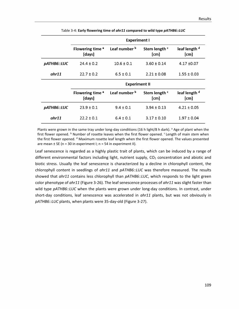
Results
109
Table 3-4: Early flowering time of ahr11 compared to wild type pATHB6::LUC
Plants were grown in the same tray under long-day conditions (16 h light/8 h dark). a Age of plant when the first flower opened. b Number of rosette leaves when the first flower opened. c Length of main stem when the first flower opened. d Maximum rosette leaf length when the first flower opened. The values presented are mean ± SE (n = 30 in experiment I; n = 54 in experiment II).
Leaf senescence is regarded as a highly plastic trait of plants, which can be induced by a range of
different environmental factors including light, nutrient supply, CO2 concentration and abiotic and
biotic stress. Usually the leaf senescence is characterized by a decline in chlorophyll content, the
chlorophyll content in seedlings of ahr11 and pATHB6::LUC was therefore measured. The results
showed that ahr11 contains less chlorophyll than pATHB6::LUC, which responds to the light green
color phenotype of ahr11 (Figure 3-26). The leaf senescence processes of ahr11 was slight faster than
wild type pATHB6::LUC when the plants were grown under long-day conditions. In contrast, under
short-day conditions, leaf senescence was accelerated in ahr11 plants, but was not obviously in
pATHB6::LUC plants, when plants were 35-day-old (Figure 3-27).
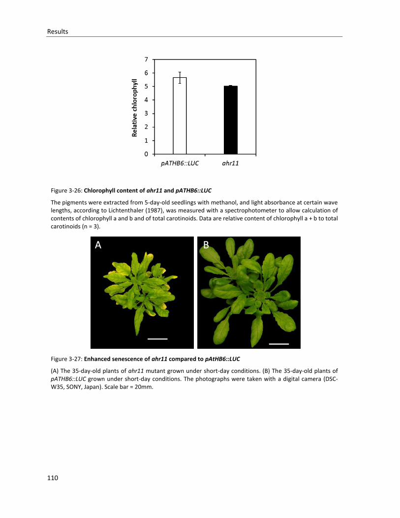
Results
110
Figure 3-26: Chlorophyll content of ahr11 and pATHB6::LUC
The pigments were extracted from 5-day-old seedlings with methanol, and light absorbance at certain wave lengths, according to Lichtenthaler (1987), was measured with a spectrophotometer to allow calculation of contents of chlorophyll a and b and of total carotinoids. Data are relative content of chlorophyll a + b to total carotinoids (n = 3).
Figure 3-27: Enhanced senescence of ahr11 compared to pAtHB6::LUC
(A) The 35-day-old plants of ahr11 mutant grown under short-day conditions. (B) The 35-day-old plants of pATHB6::LUC grown under short-day conditions. The photographs were taken with a digital camera (DSC-W35, SONY, Japan). Scale bar = 20mm.
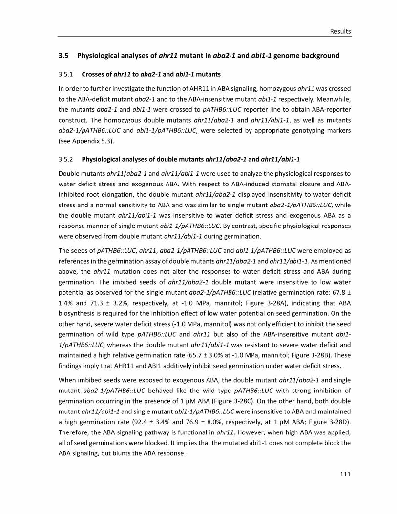
Results
111
3.5 Physiological analyses of ahr11 mutant in aba2-1 and abi1-1 genome background
3.5.1 Crosses of ahr11 to aba2-1 and abi1-1 mutants
In order to further investigate the function of AHR11 in ABA signaling, homozygous ahr11 was crossed
to the ABA-deficit mutant aba2-1 and to the ABA-insensitive mutant abi1-1 respectively. Meanwhile,
the mutants aba2-1 and abi1-1 were crossed to pATHB6::LUC reporter line to obtain ABA-reporter
construct. The homozygous double mutants ahr11/aba2-1 and ahr11/abi1-1, as well as mutants
aba2-1/pATHB6::LUC and abi1-1/pATHB6::LUC, were selected by appropriate genotyping markers
(see Appendix 5.3).
3.5.2 Physiological analyses of double mutants ahr11/aba2-1 and ahr11/abi1-1
Double mutants ahr11/aba2-1 and ahr11/abi1-1 were used to analyze the physiological responses to
water deficit stress and exogenous ABA. With respect to ABA-induced stomatal closure and ABA-
inhibited root elongation, the double mutant ahr11/aba2-1 displayed insensitivity to water deficit
stress and a normal sensitivity to ABA and was similar to single mutant aba2-1/pATHB6::LUC, while
the double mutant ahr11/abi1-1 was insensitive to water deficit stress and exogenous ABA as a
response manner of single mutant abi1-1/pATHB6::LUC. By contrast, specific physiological responses
were observed from double mutant ahr11/abi1-1 during germination.
The seeds of pATHB6::LUC, ahr11, aba2-1/pATHB6::LUC and abi1-1/pATHB6::LUC were employed as
references in the germination assay of double mutants ahr11/aba2-1 and ahr11/abi1-1. As mentioned
above, the ahr11 mutation does not alter the responses to water deficit stress and ABA during
germination. The imbibed seeds of ahr11/aba2-1 double mutant were insensitive to low water
potential as observed for the single mutant aba2-1/pATHB6::LUC (relative germination rate: 67.8 ±
1.4% and 71.3 ± 3.2%, respectively, at -1.0 MPa, mannitol; Figure 3-28A), indicating that ABA
biosynthesis is required for the inhibition effect of low water potential on seed germination. On the
other hand, severe water deficit stress (-1.0 MPa, mannitol) was not only efficient to inhibit the seed
germination of wild type pATHB6::LUC and ahr11 but also of the ABA-insensitive mutant abi1-
1/pATHB6::LUC, whereas the double mutant ahr11/abi1-1 was resistant to severe water deficit and
maintained a high relative germination rate (65.7 ± 3.0% at -1.0 MPa, mannitol; Figure 3-28B). These
findings imply that AHR11 and ABI1 additively inhibit seed germination under water deficit stress.
When imbibed seeds were exposed to exogenous ABA, the double mutant ahr11/aba2-1 and single
mutant aba2-1/pATHB6::LUC behaved like the wild type pATHB6::LUC with strong inhibition of
germination occurring in the presence of 1 µM ABA (Figure 3-28C). On the other hand, both double
mutant ahr11/abi1-1 and single mutant abi1-1/pATHB6::LUC were insensitive to ABA and maintained
a high germination rate (92.4 ± 3.4% and 76.9 ± 8.0%, respectively, at 1 µM ABA; Figure 3-28D).
Therefore, the ABA signaling pathway is functional in ahr11. However, when high ABA was applied,
all of seed germinations were blocked. It implies that the mutated abi1-1 does not complete block the
ABA signaling, but blunts the ABA response.

Results
112
Figure 3-28: Response of germination of ABA-deficient aba2-1, ABA-insensitive abi1-1, as well as double mutants ahr11/aba2-1 and ahr11/abi1-1 to water deficit stress and ABA
(A) Inhibition of germination of ahr11 (black square), pATHB6::LUC (white square), aba2-1/pATHB6::LUC (white triangle), and ahr11/aba2-1 (black triangle) by water deficit stress. Germination rates under the control condition (MS0.5xSuc, without mannitol): ahr11: 91.0 ± 5.1%, pATHB6::LUC: 93.1 ± 4.2%, aba2-1/pATHB6::LUC: 76.1 ± 9.4%, and ahr11/aba2-1: 97.2 ± 1.1%. (B) Inhibition of germination of ahr11 (black square), pATHB6::LUC (white square), abi1-1/pATHB6::LUC (white round), and ahr11/abi1-1 (black round) by water deficit stress. Germination rates under the control condition: ahr11: 91.3 ± 0.3%, pATHB6::LUC: 95.2 ± 3.4%, abi1-1/pATHB6::LUC: 81.8 ± 1.3%, and ahr11/abi1-1: 93.7 ± 2.2%. (C) Inhibition of germination of ahr11, pATHB6::LUC, aba2-1/pATHB6::LUC, and ahr11/aba2-1 by exogenous ABA. Germination rates under the control condition (MS0.5xSuc without ABA): ahr11: 89.6 ± 0.5%, pATHB6::LUC: 97.2 ± 0.7%, aba2-1/pATHB6::LUC: 98.5 ± 1.5%, ahr11/aba2-1: 91.9 ± 3.9%. (D) Inhibition of germination of ahr11, pATHB6::LUC, abi1-1/pATHB6::LUC, and ahr11/abi1-1 by exogenous ABA. Germination rates under the control condition: ahr11: 86.4 ± 0.0%, pATHB6::LUC: 94.8 ± 3.9%, abi1-1/pATHB6::LUC: 87.5 ± 0.7%, and ahr11/abi1-1: 89.9 ± 3.6%. The experiments and calculations were performed as indicated in the Figure 3-23 legend.
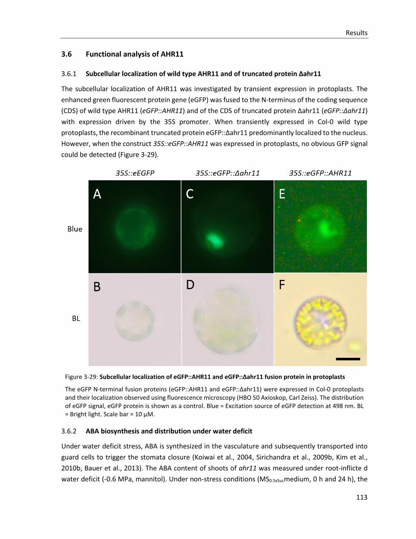
Results
113
3.6 Functional analysis of AHR11
3.6.1 Subcellular localization of wild type AHR11 and of truncated protein Δahr11
The subcellular localization of AHR11 was investigated by transient expression in protoplasts. The
enhanced green fluorescent protein gene (eGFP) was fused to the N-terminus of the coding sequence
(CDS) of wild type AHR11 (eGFP::AHR11) and of the CDS of truncated protein Δahr11 (eGFP::Δahr11)
with expression driven by the 35S promoter. When transiently expressed in Col-0 wild type
protoplasts, the recombinant truncated protein eGFP::Δahr11 predominantly localized to the nucleus.
However, when the construct 35S::eGFP::AHR11 was expressed in protoplasts, no obvious GFP signal
could be detected (Figure 3-29).
Figure 3-29: Subcellular localization of eGFP::AHR11 and eGFP::Δahr11 fusion protein in protoplasts
The eGFP N-terminal fusion proteins (eGFP::AHR11 and eGFP::Δahr11) were expressed in Col-0 protoplasts and their localization observed using fluorescence microscopy (HBO 50 Axioskop, Carl Zeiss). The distribution of eGFP signal, eGFP protein is shown as a control. Blue = Excitation source of eGFP detection at 498 nm. BL = Bright light. Scale bar = 10 µM.
3.6.2 ABA biosynthesis and distribution under water deficit
Under water deficit stress, ABA is synthesized in the vasculature and subsequently transported into
guard cells to trigger the stomata closure (Koiwai et al., 2004, Sirichandra et al., 2009b, Kim et al.,
2010b, Bauer et al., 2013). The ABA content of shoots of ahr11 was measured under root-inflicte d
water deficit (-0.6 MPa, mannitol). Under non-stress conditions (MS0.5xSucmedium, 0 h and 24 h), the
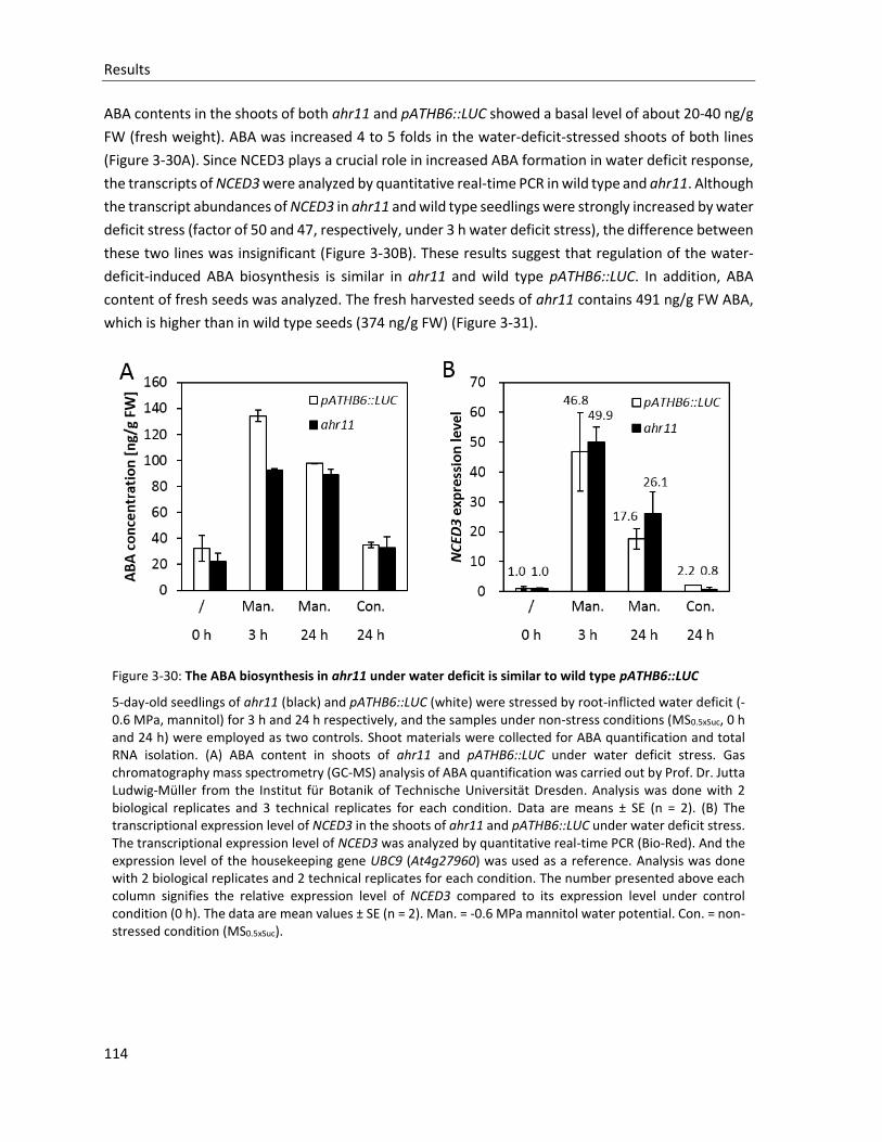
Results
114
ABA contents in the shoots of both ahr11 and pATHB6::LUC showed a basal level of about 20-40 ng/g
FW (fresh weight). ABA was increased 4 to 5 folds in the water-deficit-stressed shoots of both lines
(Figure 3-30A). Since NCED3 plays a crucial role in increased ABA formation in water deficit response,
the transcripts of NCED3 were analyzed by quantitative real-time PCR in wild type and ahr11. Although
the transcript abundances of NCED3 in ahr11 and wild type seedlings were strongly increased by water
deficit stress (factor of 50 and 47, respectively, under 3 h water deficit stress), the difference between
these two lines was insignificant (Figure 3-30B). These results suggest that regulation of the water-
deficit-induced ABA biosynthesis is similar in ahr11 and wild type pATHB6::LUC. In addition, ABA
content of fresh seeds was analyzed. The fresh harvested seeds of ahr11 contains 491 ng/g FW ABA,
which is higher than in wild type seeds (374 ng/g FW) (Figure 3-31).
Figure 3-30: The ABA biosynthesis in ahr11 under water deficit is similar to wild type pATHB6::LUC
5-day-old seedlings of ahr11 (black) and pATHB6::LUC (white) were stressed by root-inflicted water deficit (-0.6 MPa, mannitol) for 3 h and 24 h respectively, and the samples under non-stress conditions (MS0.5xSuc, 0 h and 24 h) were employed as two controls. Shoot materials were collected for ABA quantification and total RNA isolation. (A) ABA content in shoots of ahr11 and pATHB6::LUC under water deficit stress. Gas chromatography mass spectrometry (GC-MS) analysis of ABA quantification was carried out by Prof. Dr. Jutta Ludwig-Müller from the Institut für Botanik of Technische Universität Dresden. Analysis was done with 2 biological replicates and 3 technical replicates for each condition. Data are means ± SE (n = 2). (B) The transcriptional expression level of NCED3 in the shoots of ahr11 and pATHB6::LUC under water deficit stress. The transcriptional expression level of NCED3 was analyzed by quantitative real-time PCR (Bio-Red). And the expression level of the housekeeping gene UBC9 (At4g27960) was used as a reference. Analysis was done with 2 biological replicates and 2 technical replicates for each condition. The number presented above each column signifies the relative expression level of NCED3 compared to its expression level under control condition (0 h). The data are mean values ± SE (n = 2). Man. = -0.6 MPa mannitol water potential. Con. = non-stressed condition (MS0.5xSuc).
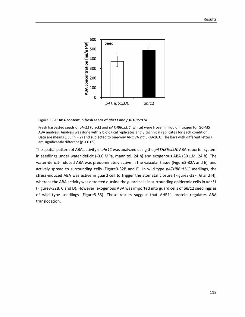
Results
115
Figure 3-31: ABA content in fresh seeds of ahr11 and pATHB6::LUC
Fresh harvested seeds of ahr11 (black) and pATHB6::LUC (white) were frozen in liquid nitrogen for GC-MS ABA analysis. Analysis was done with 2 biological replicates and 3 technical replicates for each condition. Data are means ± SE (n = 2) and subjected to one-way ANOVA via SPAA16.0. The bars with different letters are significantly different (p < 0.05).
The spatial pattern of ABA activity in ahr11 was analyzed using the pATHB6::LUC ABA-reporter system
in seedlings under water deficit (-0.6 MPa, mannitol; 24 h) and exogenous ABA (30 µM, 24 h). The
water-deficit-induced ABA was predominately active in the vascular tissue (Figure3-32A and E), and
actively spread to surrounding cells (Figure3-32B and F). In wild type pATHB6::LUC seedlings, the
stress-induced ABA was active in guard cell to trigger the stomatal closure (Figure3-32F, G and H),
whereas the ABA activity was detected outside the guard cells in surrounding epidermic cells in ahr11
(Figure3-32B, C and D). However, exogenous ABA was imported into guard cells of ahr11 seedlings as
of wild type seedlings (Figure3-33). These results suggest that AHR11 protein regulates ABA
translocation.
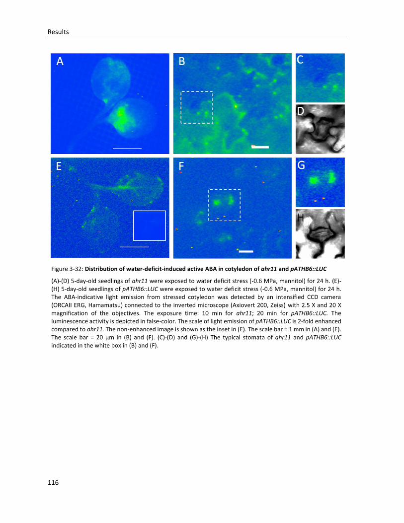
Results
116
Figure 3-32: Distribution of water-deficit-induced active ABA in cotyledon of ahr11 and pATHB6::LUC
(A)-(D) 5-day-old seedlings of ahr11 were exposed to water deficit stress (-0.6 MPa, mannitol) for 24 h. (E)-(H) 5-day-old seedlings of pATHB6::LUC were exposed to water deficit stress (-0.6 MPa, mannitol) for 24 h. The ABA-indicative light emission from stressed cotyledon was detected by an intensified CCD camera (ORCAII ERG, Hamamatsu) connected to the inverted microscope (Axiovert 200, Zeiss) with 2.5 X and 20 X magnification of the objectives. The exposure time: 10 min for ahr11; 20 min for pATHB6::LUC. The luminescence activity is depicted in false-color. The scale of light emission of pATHB6::LUC is 2-fold enhanced compared to ahr11. The non-enhanced image is shown as the inset in (E). The scale bar = 1 mm in (A) and (E). The scale bar = 20 µm in (B) and (F). (C)-(D) and (G)-(H) The typical stomata of ahr11 and pATHB6::LUC indicated in the white box in (B) and (F).
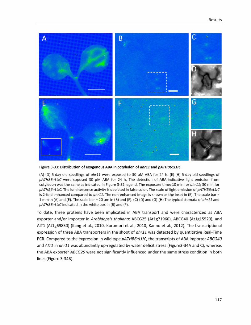
Results
117
Figure 3-33: Distribution of exogenous ABA in cotyledon of ahr11 and pATHB6::LUC
(A)-(D) 5-day-old seedlings of ahr11 were exposed to 30 µM ABA for 24 h. (E)-(H) 5-day-old seedlings of pATHB6::LUC were exposed 30 µM ABA for 24 h. The detection of ABA-indicative light emission from cotyledon was the same as indicated in Figure 3-32 legend. The exposure time: 10 min for ahr11; 30 min for pATHB6::LUC. The luminescence activity is depicted in false color. The scale of light emission of pATHB6::LUC is 2-fold enhanced compared to ahr11. The non-enhanced image is shown as the inset in (E). The scale bar = 1 mm in (A) and (E). The scale bar = 20 µm in (B) and (F). (C)-(D) and (G)-(H) The typical stomata of ahr11 and pATHB6::LUC indicated in the white box in (B) and (F).
To date, three proteins have been implicated in ABA transport and were characterized as ABA
exporter and/or importer in Arabidopsis thaliana: ABCG25 (At1g71960), ABCG40 (At1g15520), and
AIT1 (At1g69850) (Kang et al., 2010, Kuromori et al., 2010, Kanno et al., 2012). The transcriptional
expression of three ABA transporters in the shoot of ahr11 was detected by quantitative Real-Time
PCR. Compared to the expression in wild type pATHB6::LUC, the transcripts of ABA importer ABCG40
and AIT1 in ahr11 was abundantly up-regulated by water deficit stress (Figure3-34A and C), whereas
the ABA exporter ABCG25 were not significantly influenced under the same stress condition in both
lines (Figure 3-34B).
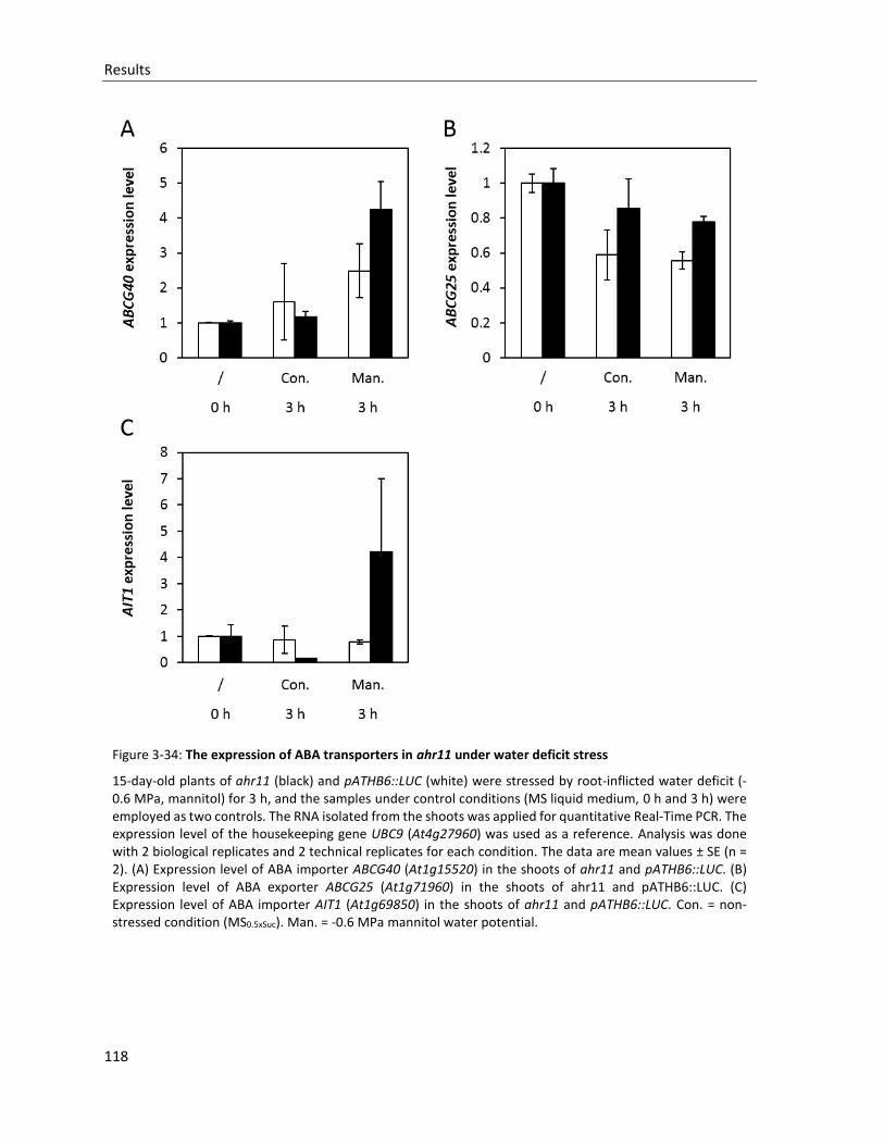
Results
118
Figure 3-34: The expression of ABA transporters in ahr11 under water deficit stress
15-day-old plants of ahr11 (black) and pATHB6::LUC (white) were stressed by root-inflicted water deficit (-0.6 MPa, mannitol) for 3 h, and the samples under control conditions (MS liquid medium, 0 h and 3 h) were employed as two controls. The RNA isolated from the shoots was applied for quantitative Real-Time PCR. The expression level of the housekeeping gene UBC9 (At4g27960) was used as a reference. Analysis was done with 2 biological replicates and 2 technical replicates for each condition. The data are mean values ± SE (n = 2). (A) Expression level of ABA importer ABCG40 (At1g15520) in the shoots of ahr11 and pATHB6::LUC. (B) Expression level of ABA exporter ABCG25 (At1g71960) in the shoots of ahr11 and pATHB6::LUC. (C) Expression level of ABA importer AIT1 (At1g69850) in the shoots of ahr11 and pATHB6::LUC. Con. = non-stressed condition (MS0.5xSuc). Man. = -0.6 MPa mannitol water potential.
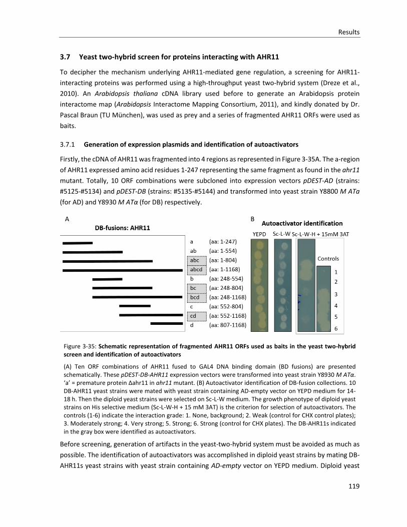
Results
119
3.7 Yeast two-hybrid screen for proteins interacting with AHR11
To decipher the mechanism underlying AHR11-mediated gene regulation, a screening for AHR11-
interacting proteins was performed using a high-throughput yeast two-hybrid system (Dreze et al.,
2010). An Arabidopsis thaliana cDNA library used before to generate an Arabidopsis protein
interactome map (Arabidopsis Interactome Mapping Consortium, 2011), and kindly donated by Dr.
Pascal Braun (TU München), was used as prey and a series of fragmented AHR11 ORFs were used as
baits.
3.7.1 Generation of expression plasmids and identification of autoactivators
Firstly, the cDNA of AHR11 was fragmented into 4 regions as represented in Figure 3-35A. The a-region
of AHR11 expressed amino acid residues 1-247 representing the same fragment as found in the ahr11
mutant. Totally, 10 ORF combinations were subcloned into expression vectors pDEST-AD (strains:
#5125-#5134) and pDEST-DB (strains: #5135-#5144) and transformed into yeast strain Y8800 M ATa
(for AD) and Y8930 M ATα (for DB) respectively.
Figure 3-35: Schematic representation of fragmented AHR11 ORFs used as baits in the yeast two-hybrid screen and identification of autoactivators
(A) Ten ORF combinations of AHR11 fused to GAL4 DNA binding domain (BD fusions) are presented schematically. These pDEST-DB-AHR11 expression vectors were transformed into yeast strain Y8930 M ATα. ‘a’ = premature protein Δahr11 in ahr11 mutant. (B) Autoactivator identification of DB-fusion collections. 10 DB-AHR11 yeast strains were mated with yeast strain containing AD-empty vector on YEPD medium for 14-18 h. Then the diploid yeast strains were selected on Sc-L-W medium. The growth phenotype of diploid yeast strains on His selective medium (Sc-L-W-H + 15 mM 3AT) is the criterion for selection of autoactivators. The controls (1-6) indicate the interaction grade: 1. None, background; 2. Weak (control for CHX control plates); 3. Moderately strong; 4. Very strong; 5. Strong; 6. Strong (control for CHX plates). The DB-AHR11s indicated in the gray box were identified as autoactivators.
Before screening, generation of artifacts in the yeast-two-hybrid system must be avoided as much as
possible. The identification of autoactivators was accomplished in diploid yeast strains by mating DB-
AHR11s yeast strains with yeast strain containing AD-empty vector on YEPD medium. Diploid yeast
Results
120
strains were replicated on Sc-L-W selection medium and Sc-L-W-H + 15 mM 3AT His-phenotype
medium. The diploid yeasts with BD-AHR11-abc and BD-AHR11-abcd (Figure 3-35A) showed a strong
growth phenotype on His-phenotype medium compared with the ‘no interaction’ Y2H control (control
1; Figure 3-35B). Therefore, these two strains were definitely autoactivators and were removed from
the collection of DB-AHR11s. In addition, other three diploid yeasts harboring DB-AHR11-bc, -bcd and
-cd displayed weak autoactivation phenotype, but were still included into DB-AHR11s collection. As a
result, totally 8 different DB-AHR11s were employed for further screening for interacting proteins.
3.7.2 Yeast-two-hybrid screen and verification of putative interactions
Eight DB-AHR11s yeast strains harboring DB-AHR11-a, -ab, -b, -bc, -bcd, -c, -cd, and -d vectors,
respectively, were used as bait to mate with AD pools consisting the AD-cDNA library expressing yeast
clones. The positive clones were selected for His and Ade auxotrophy on selection medium. Through
the screening of 59 AD pools (containing about 11000 cDNAs), 75 interacting candidates were selected
by growth phenotyping and sequence analysis, which were listed in Appendix 5.4. For verification of
interactions, all of these 75 yeast strains harboring AD-fusions of interacting candidates were mated
with 8 DB-AHR11s yeast strains respectively. The growth phenotype of diploid yeast strains on
selection medium was used to verify the candidate interaction pairs in yeast. Slight growth of diploids
on the CHX control medium (second and forth columns in Figure 3-36) was considered to be the result
of autoactivation. Therefore, and as anticipated from the initial results (Figure 3-35B), all interacting
pairs of DB-AHR11-bc, bcd and cd were considered as autoactivators and excluded from further
growth phenotype scoring. With growth evaluation on both the selective -His and -Ade medium,
finally 7 independent AHR11-interacting candidates were identified in the yeast-two-hybrid screening
(Figure 3-36) and subsequently grouped into three groups according to the interacting region of
AHR11 and interacting intensity (Figure 3-37).
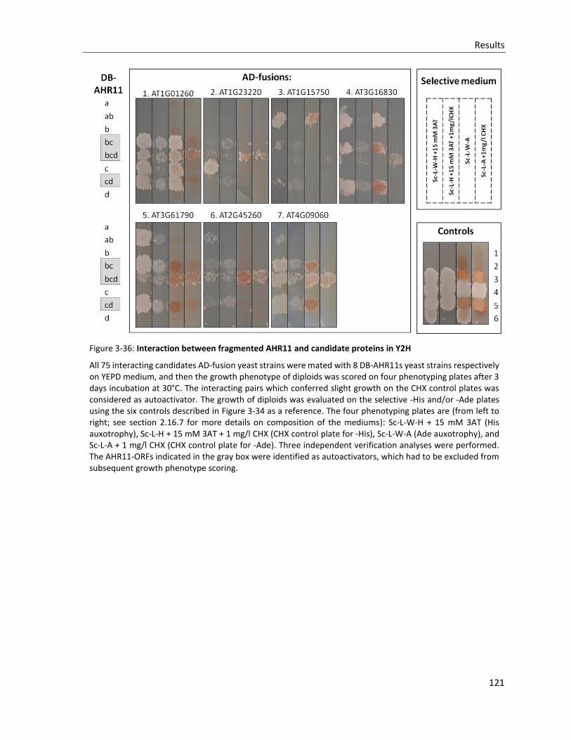
Results
121
Figure 3-36: Interaction between fragmented AHR11 and candidate proteins in Y2H
All 75 interacting candidates AD-fusion yeast strains were mated with 8 DB-AHR11s yeast strains respectively on YEPD medium, and then the growth phenotype of diploids was scored on four phenotyping plates after 3 days incubation at 30°C. The interacting pairs which conferred slight growth on the CHX control plates was considered as autoactivator. The growth of diploids was evaluated on the selective -His and/or -Ade plates using the six controls described in Figure 3-34 as a reference. The four phenotyping plates are (from left to right; see section 2.16.7 for more details on composition of the mediums): Sc-L-W-H + 15 mM 3AT (His auxotrophy), Sc-L-H + 15 mM 3AT + 1 mg/l CHX (CHX control plate for -His), Sc-L-W-A (Ade auxotrophy), and Sc-L-A + 1 mg/l CHX (CHX control plate for -Ade). Three independent verification analyses were performed. The AHR11-ORFs indicated in the gray box were identified as autoactivators, which had to be excluded from subsequent growth phenotype scoring.
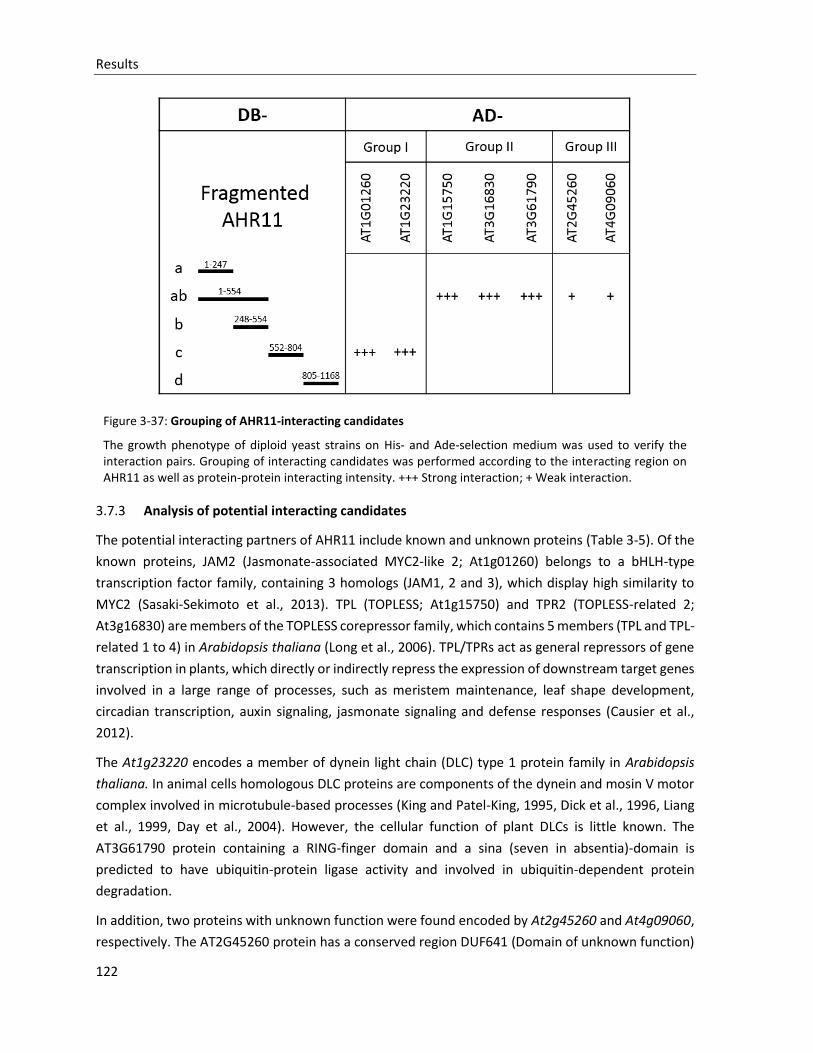
Results
122
Figure 3-37: Grouping of AHR11-interacting candidates
The growth phenotype of diploid yeast strains on His- and Ade-selection medium was used to verify the interaction pairs. Grouping of interacting candidates was performed according to the interacting region on AHR11 as well as protein-protein interacting intensity. +++ Strong interaction; + Weak interaction.
3.7.3 Analysis of potential interacting candidates
The potential interacting partners of AHR11 include known and unknown proteins (Table 3-5). Of the
known proteins, JAM2 (Jasmonate-associated MYC2-like 2; At1g01260) belongs to a bHLH-type
transcription factor family, containing 3 homologs (JAM1, 2 and 3), which display high similarity to
MYC2 (Sasaki-Sekimoto et al., 2013). TPL (TOPLESS; At1g15750) and TPR2 (TOPLESS-related 2;
At3g16830) are members of the TOPLESS corepressor family, which contains 5 members (TPL and TPL-
related 1 to 4) in Arabidopsis thaliana (Long et al., 2006). TPL/TPRs act as general repressors of gene
transcription in plants, which directly or indirectly repress the expression of downstream target genes
involved in a large range of processes, such as meristem maintenance, leaf shape development,
circadian transcription, auxin signaling, jasmonate signaling and defense responses (Causier et al.,
2012).
The At1g23220 encodes a member of dynein light chain (DLC) type 1 protein family in Arabidopsis
thaliana. In animal cells homologous DLC proteins are components of the dynein and mosin V motor
complex involved in microtubule-based processes (King and Patel-King, 1995, Dick et al., 1996, Liang
et al., 1999, Day et al., 2004). However, the cellular function of plant DLCs is little known. The
AT3G61790 protein containing a RING-finger domain and a sina (seven in absentia)-domain is
predicted to have ubiquitin-protein ligase activity and involved in ubiquitin-dependent protein
degradation.
In addition, two proteins with unknown function were found encoded by At2g45260 and At4g09060,
respectively. The AT2G45260 protein has a conserved region DUF641 (Domain of unknown function)
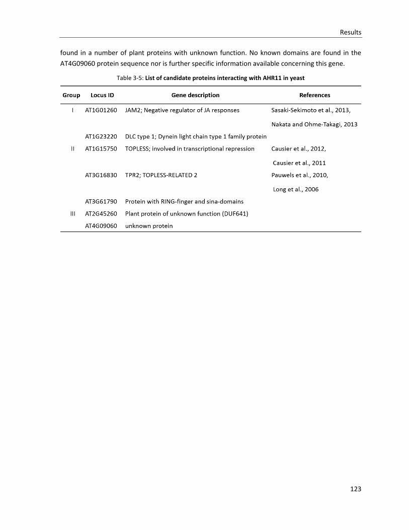
Results
123
found in a number of plant proteins with unknown function. No known domains are found in the
AT4G09060 protein sequence nor is further specific information available concerning this gene.
Table 3-5: List of candidate proteins interacting with AHR11 in yeast

Discussions
125
4. Discussions
The molecular network of water deficit response is a highly orchestrated, complex signaling network
recruiting different signaling pathways (Chinnusamy et al., 2004, Ahuja et al., 2010, Golldack et al.,
2014), in which the central role of ABA signaling has been well established (Cutler et al., 2010, Fujii
and Zhu, 2012). Transcriptomic studies have identified types of transcription factors (TFs) regulating
drought-responsive gene transcription, such as bZIP, AP2/ERF, NAC, bHLH, ZF, MYB and WRKYs (Tran
et al., 2007a, Nakashima et al., 2009b). In contrast of ABA-induced bZIP-TF ABFs/AREBs, the drought-
induced AP2-TF DREB2A/CBF3 ABA-independently regulates the expression of many drought-
responsive genes by recognizing a DRE cis-element presenting in promoter region of these genes (Liu
et al., 1998, Sakuma et al., 2006). However, the expression of DREB2A is ABA dependent for the ABRE
cis-elements in its promoter (Kim et al., 2011b). Therefore, the ABA-independent water deficit
response signaling pathway is still much less clear, compared with the prosperous research on the
ABA-dependent pathway. Subsequently, the identity of new components responsible for the water
deficit response is one of the most important issues in plant stress biology. In this work, a novel
signaling element, the protein AHR11 was identified by a forward genetic approach and characterized
as a negative regulator of ABA-dependent water deficit signaling pathway. Isolation of interacting
partners of AHR11 in a yeast two-hybrid screen allowed proposing a possible molecular mechanism
by which AHR11 represses target genes through recruiting of members of the TPL/TPRs corepressor
family.
4.1 Identification of AHR11 using a forward genetic approach
Phenotype-based forward genetic approach is the most frequently used method to link phenotypes
to genomic elements (Alonso-Blanco et al., 2005). The robust detection of a reliable phenotype is the
decisive factor for successful forward genetic identification (Lukowitz et al., 2000, Jander et al., 2002,
Peters et al., 2003). In our laboratory, a transgenic Arabidopsis thaliana ABA-reporter line
pATHB6::LUC was generated and characterized as being well suited to monitor the accumulation and
distribution of physiologically active ABA in vivo (Christmann et al., 2005, Christmann et al., 2007).
When supplied with the luciferase substrate luciferin, seedlings of the ABA-reporter line pATHB6::LUC
emit light in response to water deficit stress or to exogenous application of ABA. From an EMS-
mutagenized population of this pATHB6::LUC ABA-reporter line the mutant ahr11 was isolated based
on a hypersensitive response to water deficit stress and exogenous ABA (Figure 3-1 and Figure 3-2).
Using map-based cloning and next generation sequencing (NGS), hypersensitivity was assigned to a
lesion in a gene AHR11 (At5g13590) located on the chromosome V with unknown function.
The biggest advantage of map-based cloning is that this process may be performed without prior
assumptions on the gene identity, and that any genomic element affecting the phenotype of interest
could be found (Jander et al., 2002). In this work, the intensified ABA-dependent bioluminescence of
the mutant ahr11 under water deficit stress involves at least two loci including the causal mutation as
Discussions
126
well as the transformed chimeric gene construct pATHB6::LUC. In the first-pass mapping, the bulked
segregant analysis of ahr11 indicated two interesting regions flanked by two molecular markers,
respectively (Figure 3-5B). One region on chromosome V was proven to harbor the mutation of AHR11
in this work while the second region located on chromosome III was simultaneously highlighted in a
parallel map-based cloning approach targeting a different mutation (Cao et al., unpublished). Thus it
was likely that the ABA-reporter construct pATHB6::LUC is inserted in the region on chromosome III
flanked by markers nga162 and ciw11, which was subsequently confirmed by NGS analysis. Sequence
information of the region flanked by nga162 and ciw11 illustrated that the ABA-reporter construct
pATHB6::LUC is inserted into the promoter region of MYB15 gene (At3g23250), which is a member of
the R2R3 factor gene family, and encodes a transcriptional factor involved in ABA and osmotic stress
responses (Agarwal et al., 2006, Ding et al., 2009). Since the ABA-reporter line behaves like wild type
Arabidopsis thaliana Col-0 in all ABA-dependent physiological processes routinely assayed in our
laboratory, there is no indication to date that the insertion of the ABA-reporter construct has any
effect on ABA-signaling.
For the fine-scale mapping, it has been suggested that using a mapping population of 3,000 to 4,000
plants results in mapping of the gene of interest to a genomic region of less than 4 kb size with a high
probability (Jander et al., 2002), while the application of NGS efficiently and precisely releases high-
resolution genotype information which allows reducing the mapping population size and shortening
the mapping schedule (Schneeberger et al., 2009, Austin et al., 2011). In this work, a mapping
population comprising 893 homozygous individuals allowed to narrow down the region of interest to
180 kb on chromosome V. NGS revealed a limited number of mutations in this region with only one
exerting a non-synonymous change in a coding region (Figure 3-8 and Table 3-2). Therefore the
combination of map-based cloning and NGS greatly facilitated the process to identifying the gene
At5g13590 as AHR11.
Following identification of AHR11, verification of the lesion in AHR11 being responsible for the ahr11
phenotype was performed using three approaches. (I) Ectopic expression of wild type AHR11 which
rescued the ABA-hypersensitive responses of ahr11 in protoplasts (Figure 3-11C). (II) Generation of
transgenic ahr11 pAHR11::AHR11 mutant plants in which wild type ABA-sensitivity and wild type
water-deficit-sensitivity were restored (Figure 3-12A and B). (III) Knockout mutants of AHR11, Δ476
and N804, were ABA-hypersensitive and displayed the same response pattern to water deficit and
ABA as ahr11 (Figure 3-15). The ectopic expression of AHR11 could restore ABA sensitivity in
protoplasts isolated from knockout mutants (Figure 3-16). Taken together the results clearly proof
At5g13590 as being AHR11. Although the transgenic complementation mutant ahr11 pAHR11::AHR11
resembled wild type morphology with bigger rosettes and more explanate leaves than found in ahr11
(Figure 3-13C), the reversion of morphology was still not complete, suggesting a dosage-dependent
effect of the AHR11 protein. A parallel attempt to complement the mutant phenotype by stable
expression of an AHR11-eGFP fusion protein in ahr11, transformed with the pAHR11::AHR11-eGFP
construct, failed to reduce the hypersensitivity to water deficit as well as to ABA seen in ahr11 (Figure
3-13D). The possible reason is that the eGFP protein interferes with the function of AHR11 when fused
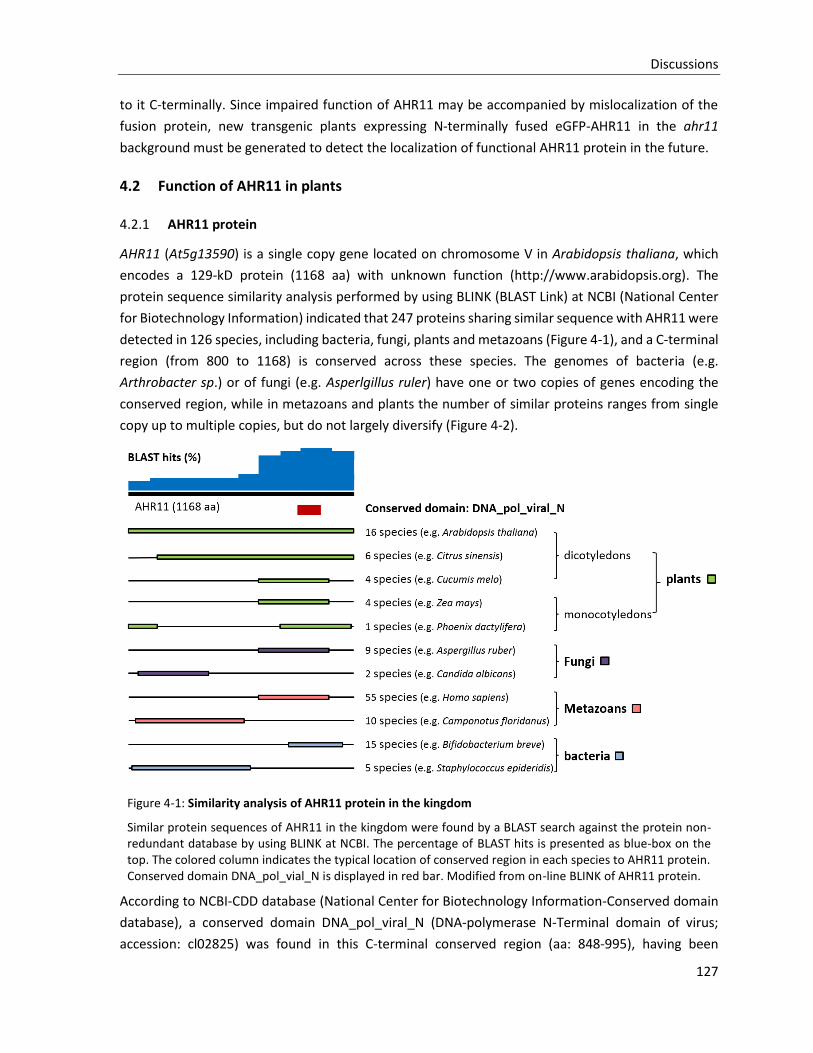
Discussions
127
to it C-terminally. Since impaired function of AHR11 may be accompanied by mislocalization of the
fusion protein, new transgenic plants expressing N-terminally fused eGFP-AHR11 in the ahr11
background must be generated to detect the localization of functional AHR11 protein in the future.
4.2 Function of AHR11 in plants
4.2.1 AHR11 protein
AHR11 (At5g13590) is a single copy gene located on chromosome V in Arabidopsis thaliana, which
encodes a 129-kD protein (1168 aa) with unknown function (http://www.arabidopsis.org). The
protein sequence similarity analysis performed by using BLINK (BLAST Link) at NCBI (National Center
for Biotechnology Information) indicated that 247 proteins sharing similar sequence with AHR11 were
detected in 126 species, including bacteria, fungi, plants and metazoans (Figure 4-1), and a C-terminal
region (from 800 to 1168) is conserved across these species. The genomes of bacteria (e.g.
Arthrobacter sp.) or of fungi (e.g. Asperlgillus ruler) have one or two copies of genes encoding the
conserved region, while in metazoans and plants the number of similar proteins ranges from single
copy up to multiple copies, but do not largely diversify (Figure 4-2).
Figure 4-1: Similarity analysis of AHR11 protein in the kingdom
Similar protein sequences of AHR11 in the kingdom were found by a BLAST search against the protein non-redundant database by using BLINK at NCBI. The percentage of BLAST hits is presented as blue-box on the top. The colored column indicates the typical location of conserved region in each species to AHR11 protein. Conserved domain DNA_pol_vial_N is displayed in red bar. Modified from on-line BLINK of AHR11 protein.
According to NCBI-CDD database (National Center for Biotechnology Information-Conserved domain
database), a conserved domain DNA_pol_viral_N (DNA-polymerase N-Terminal domain of virus;
accession: cl02825) was found in this C-terminal conserved region (aa: 848-995), having been
Discussions
128
identified in superfamily member of pfam00242, which contains a number of DNA polymerase from
viruses such as Hepatitis B virus (HBV) (Grethe et al., 2000, Huy et al., 2004), woodchuck hepatitis
virus 7 (Cohen et al., 1988) and wood chuck monkey hepatitis B virus (Lanford et al., 2003). It is already
known that the DNA polymerase N-terminal domain of P protein in duck HBV (DHBV) is composed of
a hepadnavirus-specific terminal protein domain (TP), which was identified as a (-)-DNA linker, and a
highly variable spacer domain. Together with the C-terminal polymerase/reverse transcriptase (RT)
domain, the P protein primes reverse transcription during virus replication (Bartenschlager and
Schaller, 1988, Beck and Nassal, 2007). Compared to C-terminal region, the N-terminal region of
AHR11 is less conserved. Only several proteins of bacteria (e.g. Staphylococcus epideridis), of fungi
(e.g. Candida albicans) and of metazoans (e.g. Camponotus floridanus) display similarity to the N-
terminal region of AHR11 (Figure 4-1), which are predicted as histone demethylase, otogelin-like
proteins, or mucin-like proteins. The full length protein of AHR11 is highly conserved in dicotyledons,
but not in monocotyledons. Therefore, it seems like that the C-terminal region of AHR11 might have
a function of DNA binding, and has been preserved in the course of evolution, and that a new function
domain is combined to the N-terminus of conserved domain in dicots radiation.
To research the biological function of N-terminal region of AHR11, the peptide alignment among
orthologs of AHR11 and function domain predictions were performed. As shown in Figure 4-3, a highly
conserved region (235NRE(S/T)NWDLNTTMDV(A)WE249) is present in AHR11 orthologs from 7
dicotyledons. This conserved region might be preserved during evolution for some undiscovered
functions. The W248-to-STOP mutation in ahr11 mutant occurs at the C-terminal side of this region.
According to the common sequence LxLxL (Ohta et al., 2001, Kagale and Rozwadowski, 2010), where
‘x’ is represented by any amino acid residue, two ethylene response factor-associated amphiphilic
repression (EAR) motifs are found in the N-terminal region of AHR11 (208LNLSL212 and 314LSLGL318,
respectively, Figure 4-4). EAR-motif is the most analyzed form of transcriptional repression motif,
generally found in transcriptional regulators known to function as negative regulators (Kagale and
Rozwadowski, 2010). Therefore, AHR11 is predicted as a novel EAR-repressor to regulate gene
transcription.
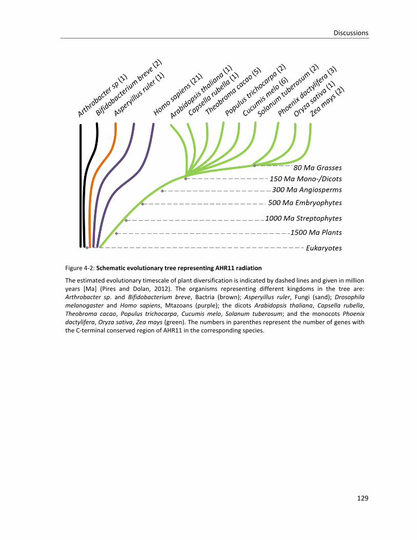
Discussions
129
Figure 4-2: Schematic evolutionary tree representing AHR11 radiation
The estimated evolutionary timescale of plant diversification is indicated by dashed lines and given in million years [Ma] (Pires and Dolan, 2012). The organisms representing different kingdoms in the tree are: Arthrobacter sp. and Bifidobacterium breve, Bactria (brown); Asperyillus ruler, Fungi (sand); Drosophila melanogaster and Homo sapiens, Mtazoans (purple); the dicots Arabidopsis thaliana, Capsella rubella, Theobroma cacao, Populus trichocarpa, Cucumis melo, Solanum tuberosum; and the monocots Phoenix dactylifera, Oryza sativa, Zea mays (green). The numbers in parenthes represent the number of genes with the C-terminal conserved region of AHR11 in the corresponding species.
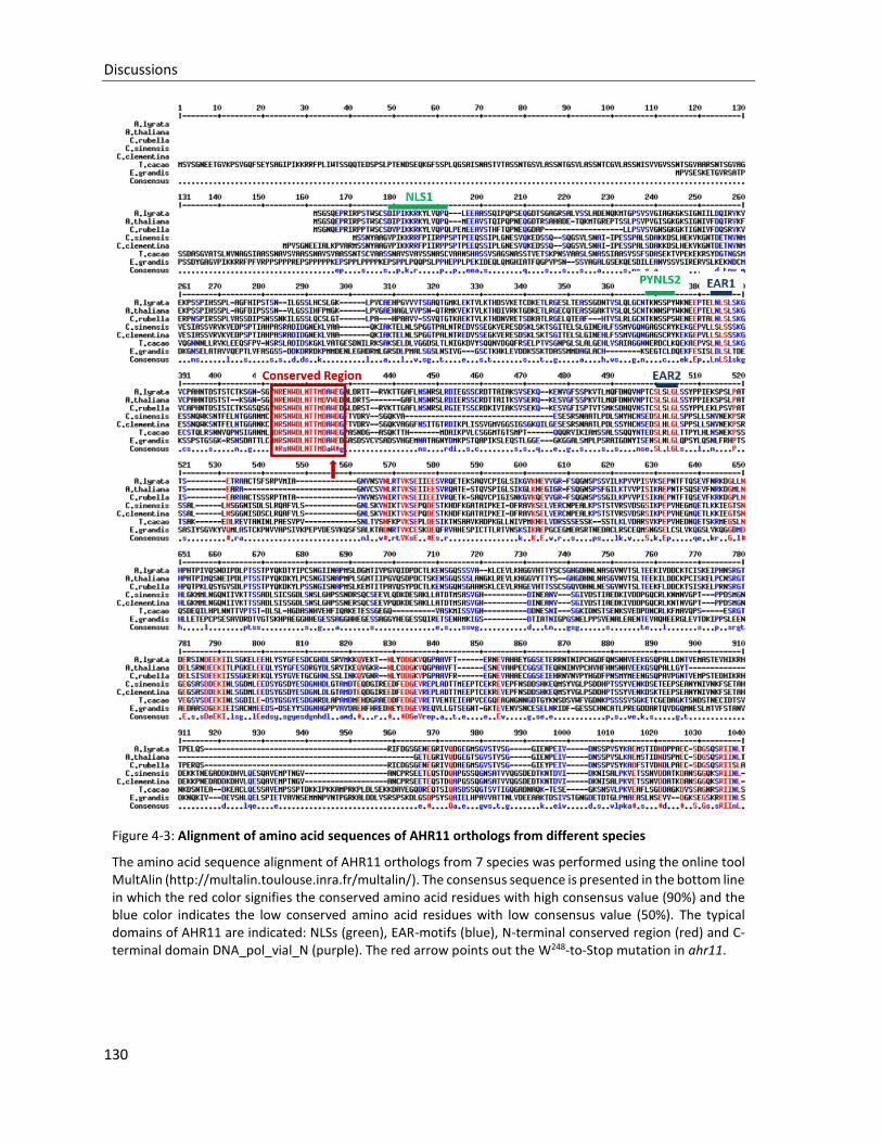
Discussions
130
Figure 4-3: Alignment of amino acid sequences of AHR11 orthologs from different species
The amino acid sequence alignment of AHR11 orthologs from 7 species was performed using the online tool MultAlin (http://multalin.toulouse.inra.fr/multalin/). The consensus sequence is presented in the bottom line in which the red color signifies the conserved amino acid residues with high consensus value (90%) and the blue color indicates the low conserved amino acid residues with low consensus value (50%). The typical domains of AHR11 are indicated: NLSs (green), EAR-motifs (blue), N-terminal conserved region (red) and C-terminal domain DNA_pol_vial_N (purple). The red arrow points out the W248-to-Stop mutation in ahr11.
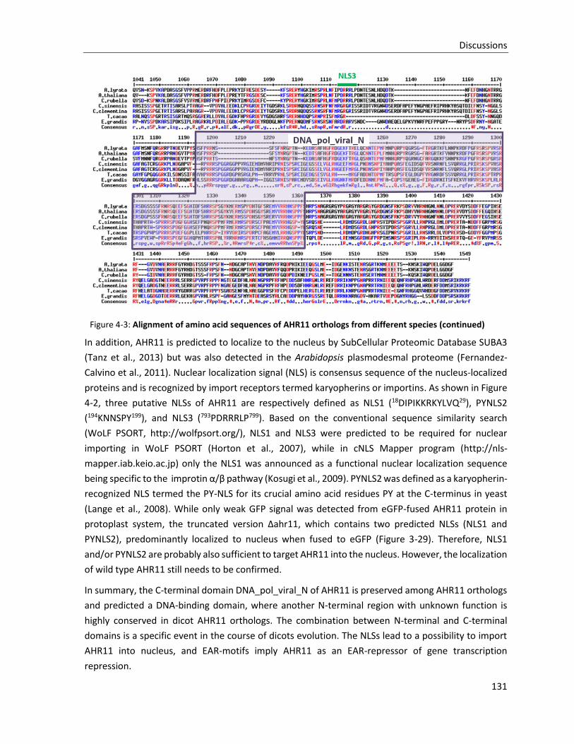
Discussions
131
Figure 4-3: Alignment of amino acid sequences of AHR11 orthologs from different species (continued)
In addition, AHR11 is predicted to localize to the nucleus by SubCellular Proteomic Database SUBA3
(Tanz et al., 2013) but was also detected in the Arabidopsis plasmodesmal proteome (Fernandez-
Calvino et al., 2011). Nuclear localization signal (NLS) is consensus sequence of the nucleus-localized
proteins and is recognized by import receptors termed karyopherins or importins. As shown in Figure
4-2, three putative NLSs of AHR11 are respectively defined as NLS1 (18DIPIKKRKYLVQ29), PYNLS2
(194KNNSPY199), and NLS3 (793PDRRRLP799). Based on the conventional sequence similarity search
(WoLF PSORT, http://wolfpsort.org/), NLS1 and NLS3 were predicted to be required for nuclear
importing in WoLF PSORT (Horton et al., 2007), while in cNLS Mapper program (http://nls-
mapper.iab.keio.ac.jp) only the NLS1 was announced as a functional nuclear localization sequence
being specific to the improtin α/β pathway (Kosugi et al., 2009). PYNLS2 was defined as a karyopherin-
recognized NLS termed the PY-NLS for its crucial amino acid residues PY at the C-terminus in yeast
(Lange et al., 2008). While only weak GFP signal was detected from eGFP-fused AHR11 protein in
protoplast system, the truncated version Δahr11, which contains two predicted NLSs (NLS1 and
PYNLS2), predominantly localized to nucleus when fused to eGFP (Figure 3-29). Therefore, NLS1
and/or PYNLS2 are probably also sufficient to target AHR11 into the nucleus. However, the localization
of wild type AHR11 still needs to be confirmed.
In summary, the C-terminal domain DNA_pol_viral_N of AHR11 is preserved among AHR11 orthologs
and predicted a DNA-binding domain, where another N-terminal region with unknown function is
highly conserved in dicot AHR11 orthologs. The combination between N-terminal and C-terminal
domains is a specific event in the course of dicots evolution. The NLSs lead to a possibility to import
AHR11 into nucleus, and EAR-motifs imply AHR11 as an EAR-repressor of gene transcription
repression.
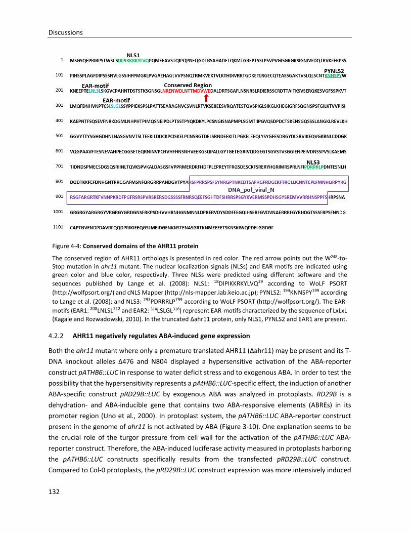
Discussions
132
Figure 4-4: Conserved domains of the AHR11 protein
The conserved region of AHR11 orthologs is presented in red color. The red arrow points out the W248-to-Stop mutation in ahr11 mutant. The nuclear localization signals (NLSs) and EAR-motifs are indicated using green color and blue color, respectively. Three NLSs were predicted using different software and the
sequences published by Lange et al. (2008): NLS1: 18DIPIKKRKYLVQ29 according to WoLF PSORT
(http://wolfpsort.org/) and cNLS Mapper (http://nls-mapper.iab.keio.ac.jp); PYNLS2: 194KNNSPY199 according
to Lange et al. (2008); and NLS3: 793PDRRRLP799 according to WoLF PSORT (http://wolfpsort.org/). The EAR-
motifs (EAR1: 208LNLSL212 and EAR2: 314LSLGL318) represent EAR-motifs characterized by the sequence of LxLxL
(Kagale and Rozwadowski, 2010). In the truncated Δahr11 protein, only NLS1, PYNLS2 and EAR1 are present.
4.2.2 AHR11 negatively regulates ABA-induced gene expression
Both the ahr11 mutant where only a premature translated AHR11 (Δahr11) may be present and its T-
DNA knockout alleles Δ476 and N804 displayed a hypersensitive activation of the ABA-reporter
construct pATHB6::LUC in response to water deficit stress and to exogenous ABA. In order to test the
possibility that the hypersensitivity represents a pAtHB6::LUC-specific effect, the induction of another
ABA-specific construct pRD29B::LUC by exogenous ABA was analyzed in protoplasts. RD29B is a
dehydration- and ABA-inducible gene that contains two ABA-responsive elements (ABREs) in its
promoter region (Uno et al., 2000). In protoplast system, the pATHB6::LUC ABA-reporter construct
present in the genome of ahr11 is not activated by ABA (Figure 3-10). One explanation seems to be
the crucial role of the turgor pressure from cell wall for the activation of the pATHB6::LUC ABA-
reporter construct. Therefore, the ABA-induced luciferase activity measured in protoplasts harboring
the pATHB6::LUC constructs specifically results from the transfected pRD29B::LUC construct.
Compared to Col-0 protoplasts, the pRD29B::LUC construct expression was more intensively induced
Discussions
133
by exogenous ABA in protoplasts of ahr11 (Figure 3-11A) and of T-DNA knockout lines (Figure 3-16),
while transient ectopic expression of wild type AHR11 in Col-0 protoplasts inhibited luciferase
expression in the presence ABA by a factor of 3.6 (Figure 3-11D). Transient expression of ABI2, which
encodes a negative regulator of ABA signaling, was sufficient to repress the activation of pRD29B::LUC
by ABA in ahr11 protoplasts (Figure 3-11B). These results indicate that AHR11 functions as a negative
regulator of ABA signaling pathway and analysis of pATHB6::LUC lines transformed with a
pAHR11::AHR11 constructs supports this view (Figure 3-12D).
4.2.3 Physiological function of AHR11 under water deficit
Althrough AHR11 negatively regulates ABA-induced gene expression, ahr11 mutant did not always
respond hypersensitively to ABA in the ABA-regulated physiological processes. The cotyledon stomata
of ahr11 were defective to close in their response to water deficit (Figure 3-20A) and the wild type
response could be restored by applying ABA exogenously (Figure 3-20B). Further analyses of
transpiration under well-watered long-day conditions and water loss from detached shoots, also
indicate that the regulation of stomatal aperture is impaired in ahr11. Ahr11 seeds display the
unaltered, wild type-like sensitivity to ABA and to restricted water availability in germination (Figure
3-23), while the primary root elongation of ahr11 seedlings was strongly inhibited by water deficit
stress (Figure 3-24A). These results indicate that physiological responses to water deficit rather than
responses to ABA are impaired in ahr11 suggesting that the AHR11 protein might function to affect
both water deficit signaling upstream of the ABA perception and ABA-dependent gene expression.
4.2.3.1 AHR11 affects ABA redistribution under water deficit stress
ABA biosynthetic enzymes including ABA2/SDR1 (Cheng et al., 2002), NCED3 (Barrero et al., 2006,
Endo et al., 2008, Hao et al., 2009), and AAO3 (Koiwai et al., 2004) are confined to vascular tissues, as
well as guard cells (Bauer et al., 2013). Water deficit-induced ABA is biosynthesized in plant
vasculature tissues, and subsequently redistributed to cells outside the vasculature including probably
guard cells (Boursiac et al., 2013). Therefore, the local level of active ABA in plants depends not only
on the rate of stress-induced biosynthetic ABA formation, but also on ABA translocation. The analysis
of ABA content of the stressed ahr11 shoots (Figure 3-30A) revealed that the shoot ABA biosynthesis
is induced by water deficit in the ahr11 mutant to a degree also observed in wild type shoot, and
NCED3 transcript abundance exhibited a corresponding increase (Figure 3-30B). The hypersensitive
ABA-reporter response in ahr11 can thus not be explained by an increase in bulk leaf ABA levels while
an affected ABA translocation from the synthesis sites to the perception sites cannot be ruled out.
Figure 4-5 demonstrates the translocation of ABA facilitated by ABA transporters: ABA-exporter
ABCG25, as well as ABA-importers ABCG40 and AIT1/NRT1.2 (Kuromori and Shinozaki, 2010, Kuromori
et al., 2010, Kang et al., 2010, Kanno et al., 2012, Boursiac et al., 2013).
ABCG25 was identified as an ABA-exporter at ABA biosynthesis site, belonging to the WBC (white-
brown complex) group of ABCG transporter subfamily (Kuromori and Shinozaki, 2010, Kang et al.,
2010). The ABCG25 over-expressing plants have a high leaf temperature phenotype compatible with
Discussions
134
increased delivery of ABA to sites of perception (Kang et al., 2010). Under water deficit stress, ABA is
synthesized in vascular parenchzma cells where the ABA-exporter ABCG25 then manages transport of
ABA into the bundle sheath cell apoplast area outside the stele which is continuous with the mesophyll
and epidermal cell apoplast (Kuromori et al., 2010, Shatil-Cohen et al., 2011), and where ABA is
distributed passively with the flow of water directed to the sites of transpiration. Subsequently, ABA
influx into guard cells is facilitated by ABA-importer ABCG40 to trigger stomatal closure (Kuromori and
Shinozaki, 2010). ABCG40 belongs to the PDR (Pleiotropic drug resistance) group of ABCG transporter
subfamily, and is detectable in many tissues but preferentially expressed in guard cells and targeted
to the plasma membrane there (Kang et al., 2010). atabcg40 knockout mutants are insensitive to ABA
with respect to stomatal closure. And the expression of ABA-inducible genes is delayed in the
atabcg40 mutant indicating that ABCG40 is important for rapid ABA-mediated stress responses (Kang
et al., 2010). In addition, AIT1/NRT1.2 was identified recently and shown to be an ABA-importer
located in the vascular tissues (Kanno et al., 2012). The ait1 mutant showed a decreased ABA response
in dormancy, and decreased stomatal closure. Thus the possible function of AIT1/NRT1.2 is to retrieve
excess ABA from the apoplast into bundle sheath cells to maintain the ABA pool size at the site of
biosynthesis and also to facilitate ABA influx into guard cells (Kanno et al., 2012).
In ahr11 mutant, the shoot expression of ABA-importers (both ABCG40 and AIT1/NRT1.2) were
strongly increased by water deficit stress, compared to the expression extent in wild type, while the
expression of ABA-exporter was down-regulated by water deficit stress but undistinguishable
between ahr11 and wild type (Figure 3-34). ABA in vivo-imaging revealed that ABA action was not
detectable in cotyledon guard cells under water deficit, while mesophyll cells and epidermal cells
responded strongly (Figure 3-32). This finding could explain the observed insensitivity of ahr11
stomata to water deficit and the hypersensitive response of ABA-reporter. Because of the high
expression of ABA-transporters in ahr11 under water deficit stress, it is now suggested that the AHR11
represses transcription of ABA-transporters to prevent ABA uptake thereby ensuring that sufficient
ABA arrives at the guard cells. Although the same amount of ABA is synthesized in shoot of ahr11 and
wild type, ABA is predominantly taken up and not released by mesophyll and epidermal cells in ahr11
causing enhanced activation of the ABA-reporter. A concomitant ABA deficit in guard cells impairs the
response of stomata to water deficit. However, when ABA was supplied exogenously, enough ABA is
available to the apoplast surrounding stomata guard cells to allow ahr11 guard cells to take up
sufficient hormone to initiate stomatal closure. The faster ABA-induced stomatal closure in ahr11
(Figure 3-21C) suggests the enhanced hormone uptake or increased hormone response in ahr11. This
might be interpreted such that AHR11 exerts an inhibitory effect on expression of ABA-transporters
including guard cells. Accordingly, ahr11 guard cells might have an enhanced capacity to take up ABA
which, however, does not compensate the effect of enhanced ABA retrieval from the apoplast on its
way from the vasculature to the guard cells.
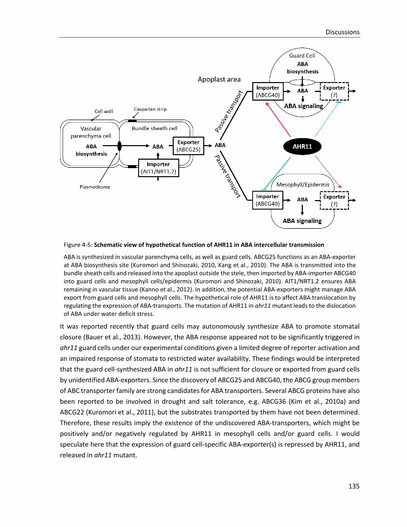
Discussions
135
Figure 4-5: Schematic view of hypothetical function of AHR11 in ABA intercellular transmission
ABA is synthesized in vascular parenchyma cells, as well as guard cells. ABCG25 functions as an ABA-exporter at ABA biosynthesis site (Kuromori and Shinozaki, 2010, Kang et al., 2010). The ABA is transmitted into the bundle sheath cells and released into the apoplast outside the stele, then imported by ABA-importer ABCG40 into guard cells and mesophyll cells/epidermis (Kuromori and Shinozaki, 2010). AIT1/NRT1.2 ensures ABA remaining in vascular tissue (Kanno et al., 2012). In addition, the potential ABA-exporters might manage ABA export from guard cells and mesophyll cells. The hypothetical role of AHR11 is to affect ABA translocation by regulating the expression of ABA-transports. The mutation of AHR11 in ahr11 mutant leads to the dislocation of ABA under water deficit stress.
It was reported recently that guard cells may autonomously synthesize ABA to promote stomatal
closure (Bauer et al., 2013). However, the ABA response appeared not to be significantly triggered in
ahr11 guard cells under our experimental conditions given a limited degree of reporter activation and
an impaired response of stomata to restricted water availability. These findings would be interpreted
that the guard cell-synthesized ABA in ahr11 is not sufficient for closure or exported from guard cells
by unidentified ABA-exporters. Since the discovery of ABCG25 and ABCG40, the ABCG group members
of ABC transporter family are strong candidates for ABA transporters. Several ABCG proteins have also
been reported to be involved in drought and salt tolerance, e.g. ABCG36 (Kim et al., 2010a) and
ABCG22 (Kuromori et al., 2011), but the substrates transported by them have not been determined.
Therefore, these results imply the existence of the undiscovered ABA-transporters, which might be
positively and/or negatively regulated by AHR11 in mesophyll cells and/or guard cells. I would
speculate here that the expression of guard cell-specific ABA-exporter(s) is repressed by AHR11, and
released in ahr11 mutant.
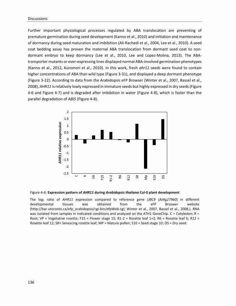
Discussions
136
Further important physiological processes regulated by ABA translocation are preventing of
premature germination during seed development (Kanno et al., 2010) and initiation and maintenance
of dormancy during seed maturation and imbibition (Ali-Rachedi et al., 2004, Lee et al., 2010). A seed
coat bedding assay has proven the maternal ABA translocation from dormant seed coat to non-
dormant embryo to keep dormancy (Lee et al., 2010, Lee and Lopez-Molina, 2013). The ABA-
transporter mutants or over-expressing lines displayed normal ABA-involved germination phenotypes
(Kanno et al., 2012, Kuromori et al., 2010). In this work, fresh ahr11 seeds were found to contain
higher concentrations of ABA than wild type (Figure 3-31), and displayed a deep dormant phenotype
(Figure 3-22). According to data from the Arabidopsis eFP Browser (Winter et al., 2007, Bassel et al.,
2008), AHR11 is relatively lowly expressed in immature seeds but highly expressed in dry seeds (Figure
4-6 and Figure 4-7) and is degraded after imbibition in water (Figure 4-8), which is faster than the
parallel degradation of ABI5 (Figure 4-8).
Figure 4-6: Expression pattern of AHR11 during Arabidopsis thaliana Col-0 plant development
The log2 ratio of AHR11 expression compared to reference gene UBC9 (At4g27960) in different developmental tissues was obtained from the eFP Broswer website (http://bar.utoronto.ca/efp_arabidopsis/cgi-bin/efpWeb.cgi; Winter et al., 2007, Bassel et al., 2008,). RNA was isolated from samples in indicated conditions and analyzed on the ATH1 GeneChip. C = Cotyledon; R = Root; VP = Vegetative rosette; F15 = Flower stage 15; R1-2 = Rosette leaf 1+2; R6 = Rosette leaf 6; R12 = Rosette leaf 12; SR= Senescing rosette leaf; MP = Mature pollen; S10 = Seed stage 10; DS = Dry seed.
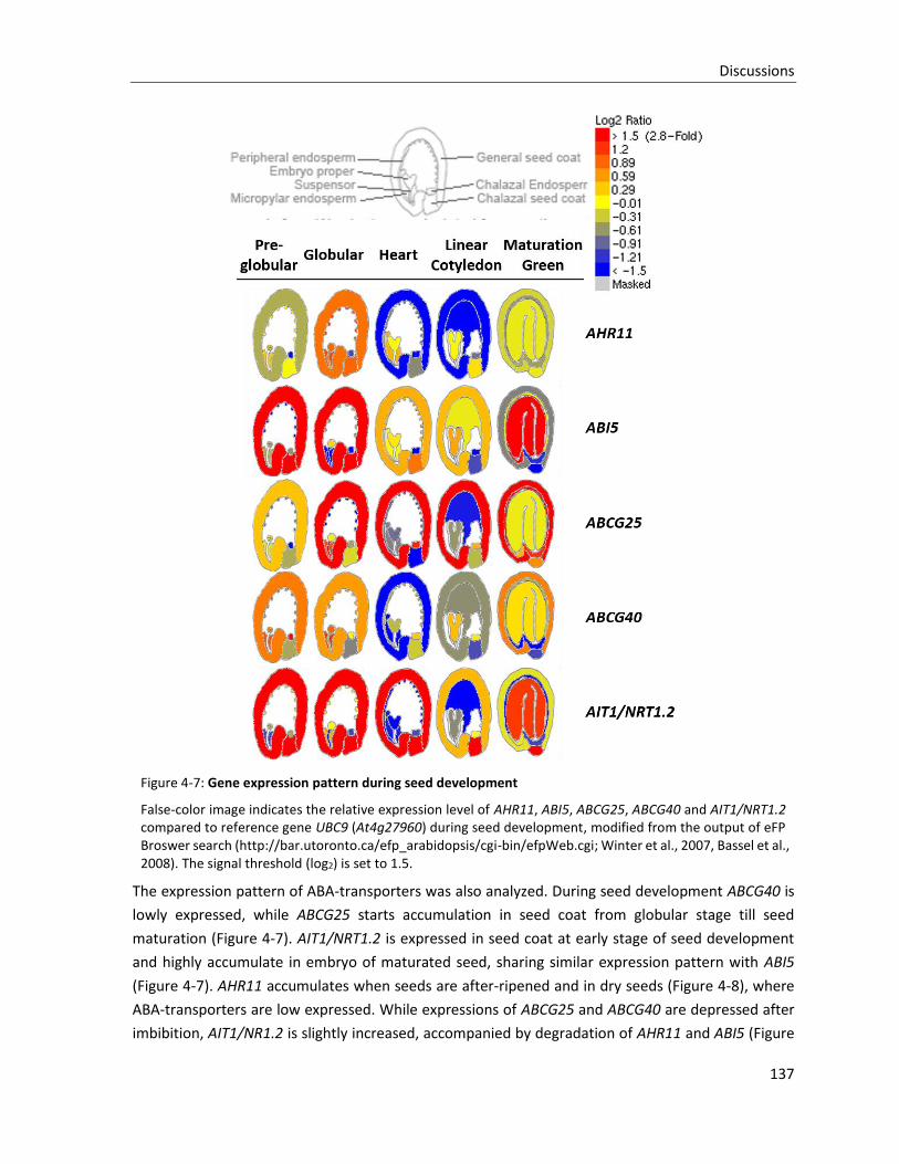
Discussions
137
Figure 4-7: Gene expression pattern during seed development
False-color image indicates the relative expression level of AHR11, ABI5, ABCG25, ABCG40 and AIT1/NRT1.2 compared to reference gene UBC9 (At4g27960) during seed development, modified from the output of eFP Broswer search (http://bar.utoronto.ca/efp_arabidopsis/cgi-bin/efpWeb.cgi; Winter et al., 2007, Bassel et al., 2008). The signal threshold (log2) is set to 1.5.
The expression pattern of ABA-transporters was also analyzed. During seed development ABCG40 is
lowly expressed, while ABCG25 starts accumulation in seed coat from globular stage till seed
maturation (Figure 4-7). AIT1/NRT1.2 is expressed in seed coat at early stage of seed development
and highly accumulate in embryo of maturated seed, sharing similar expression pattern with ABI5
(Figure 4-7). AHR11 accumulates when seeds are after-ripened and in dry seeds (Figure 4-8), where
ABA-transporters are low expressed. While expressions of ABCG25 and ABCG40 are depressed after
imbibition, AIT1/NR1.2 is slightly increased, accompanied by degradation of AHR11 and ABI5 (Figure
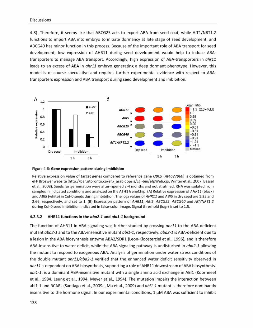
Discussions
138
4-8). Therefore, it seems like that ABCG25 acts to export ABA from seed coat, while AIT1/NRT1.2
functions to import ABA into embryo to initiate dormancy at late stage of seed development, and
ABCG40 has minor function in this process. Because of the important role of ABA transport for seed
development, low expression of AHR11 during seed development would help to induce ABA-
transporters to manage ABA transport. Accordingly, high expression of ABA-transporters in ahr11
leads to an excess of ABA in ahr11 embryo generating a deep dormant phenotype. However, this
model is of course speculative and requires further experimental evidence with respect to ABA-
transporters expression and ABA transport during seed development and imbibition.
Figure 4-8: Gene expression pattern during imbibition
Relative expression value of target genes compared to reference gene UBC9 (At4g27960) is obtained from eFP Broswer website (http://bar.utoronto.ca/efp_arabidopsis/cgi-bin/efpWeb.cgi; Winter et al., 2007, Bassel et al., 2008). Seeds for germination were after-ripened 2-4 months and not stratified. RNA was isolated from samples in indicated conditions and analyzed on the ATH1 GeneChip. (A) Relative expression of AHR11 (black) and ABI5 (white) in Col-0 seeds during imbibition. The log2 values of AHR11 and ABI5 in dry seed are 1.35 and 2.66, respectively, and set to 1. (B) Expression pattern of AHR11, ABI5, ABCG25, ABCG40 and AIT/NRT1.2 during Col-0 seed imbibition indicated in false-color image. Signal threshold (log2) is set to 1.5.
4.2.3.2 AHR11 functions in the aba2-1 and abi1-1 background
The function of AHR11 in ABA signaling was further studied by crossing ahr11 to the ABA-deficient
mutant aba2-1 and to the ABA-insensitive mutant abi1-1, respectively. aba2-1 is ABA-deficient due to
a lesion in the ABA biosynthesis enzyme ABA2/SDR1 (Leon-Kloosterziel et al., 1996), and is therefore
ABA-insensitive to water deficit, while the ABA signaling pathway is undisturbed in aba2-1 allowing
the mutant to respond to exogenous ABA. Analysis of germination under water stress conditions of
the double mutant ahr11/aba2-1 verified that the enhanced water deficit sensitivity observed in
ahr11 is dependent on ABA biosynthesis, supporting a role of AHR11 downstream of ABA biosynthesis.
abi1-1, is a dominant ABA-insensitive mutant with a single amino acid exchange in ABI1 (Koornneef
et al., 1984, Leung et al., 1994, Meyer et al., 1994). The mutation impairs the interaction between
abi1-1 and RCARs (Santiago et al., 2009a, Ma et al., 2009) and abi1-1 mutant is therefore dominantly
insensitive to the hormone signal. In our experimental conditions, 1 µM ABA was sufficient to inhibit

Discussions
139
germination of wild type pATHB6::LUC and ahr11 single mutant, not of abi1-1/pATHB6::LUC mutant,
but 10 µM ABA is successfully block the seed germination of abi1-1/pATHB6::LUC (Figure 3-28D). It
implies that abi1-1 is not fully blocking the ABA response, but shifting the ABA sensitivity to a high
concentration. Therefore, the water deficit-inhibited germination on abi1-1/pATHB6::LUC (Figure 3-
28B) might be also managed by the ABA-dependent pathway. However, the double mutant
ahr11/abi1-1 displayed a high germination rate under severe water deficit stress (65.7% under -1.0
MPa, Figure3-28B), providing strong evidence that AHR11 and ABI1 additively inhibit seed
germination under water deficit stress. However, the molecular mechanism of AHR11 in this pathway
is still unclear.
Figure 4-9: Hypothetical function of AHR11 in germination under water deficit stress
ABA biosynthesis is initiated by water deficit stress, and ABA subsequently actively transported intercellularly, activating the core ABA signaling pathway to inhibit seed germination. ABA-deficient mutant aba2-1 blocks ABA biosynthesis and the ABA-dependent germination inhibition. ABA-insensitive mutant abi1-1 inhibits ABA response signaling, but does not fully block this pathway. AHR11 acts as a negative regulator to repress active ABA transport, resulting in germination inhibition under water deficit stress. ahr11 mutant enhances ABA transport, but additively contributes to the abi1-1 effect in promoting germination under water deficit stress. The connection between AHR11 and ABA signaling is still unclear, and direct germination inhibition by AHR11 cannot be excluded.
Discussions
140
4.3 AHR11 as a component of a regulatory network
The output of water deficit signal transduction is dependent on the respective contribution of positive
and negative regulators which are part of a protein-protein interaction network. In order to
understand how AHR11 is integrated into this network, a yeast two-hybrid interactome screen was
performed for AHR11-interacting partners.
4.3.1 Network of genes co-regulated with AHR11
Online tool ATTED-II ver.7.1 (http://atted.jp) is a database that provides co-regulated gene
relationships based primarily on co-expression microarray data (Obayashi et al., 2009). Using
information on cis-elements in promoters of co-expressed genes, co-regulation of genes is predicted.
As a result of the ATTED-II database search, a network of genes with a high probability of being co-
regulated with AHR11 (Figure 4-11), as well as ABI1 (Figure 4-12) for comparison, was obtained.
The clade A PP2C, ABI1, has been characterized as coreceptor of RCAR ABA receptors, negatively
regulating ABA signaling pathway (Fuchs et al., 2012). ABI1 locates in the center of the co-regulated
gene network (figure 4-10), being surrounded by some related genes, in which five members of clade
A PP2Cs are highlighted by red points, including ABI2, SAG113, PP2CA, HAI2 and HAB2, and other
important proteins are also involved, e.g. transcription factors including NAC TF RD26 (MR 4.6) and
homeodomain protein HB7 (MR 5.7). Therefore, the co-regulated gene network efficiently indicates
related-genes with similar function or with syntropic regulation. In the co-regulated gene network of
AHR11 (Figure 4-11), four genes with the highest co-regulation probability are directly connected to
AHR11, including At1g21580, RKP (Related to KPC1 protein; At2g22010), CPL3 (C-terminal domain
phosphatase-like 3; At2g33540), and CPK23 (Calcium dependent protein kinase 23; At4g04740).
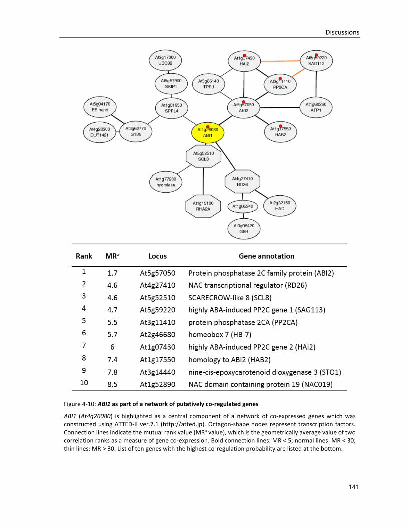
Discussions
141
Figure 4-10: ABI1 as part of a network of putatively co-regulated genes
ABI1 (At4g26080) is highlighted as a central component of a network of co-expressed genes which was constructed using ATTED-II ver.7.1 (http://atted.jp). Octagon-shape nodes represent transcription factors. Connection lines indicate the mutual rank value (MRa value), which is the geometrically average value of two correlation ranks as a measure of gene co-expression. Bold connection lines: MR < 5; normal lines: MR < 30; thin lines: MR > 30. List of ten genes with the highest co-regulation probability are listed at the bottom.
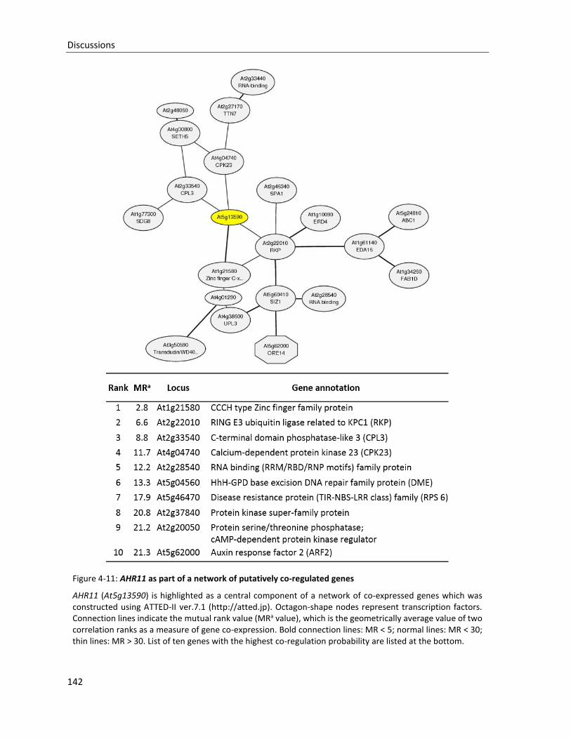
Discussions
142
Figure 4-11: AHR11 as part of a network of putatively co-regulated genes
AHR11 (At5g13590) is highlighted as a central component of a network of co-expressed genes which was constructed using ATTED-II ver.7.1 (http://atted.jp). Octagon-shape nodes represent transcription factors. Connection lines indicate the mutual rank value (MRa value), which is the geometrically average value of two correlation ranks as a measure of gene co-expression. Bold connection lines: MR < 5; normal lines: MR < 30; thin lines: MR > 30. List of ten genes with the highest co-regulation probability are listed at the bottom.
Discussions
143
The gene with the highest probability of being co-regulated with AHR11, At1g21580, encodes a
member of the CCCH-type Zinc finger protein family. Proteins of this family contain 1 to 6 tandem
zinc-binding motifs characterized by three cysteines followed by one histidine and are proposed to be
RNA-binding proteins with regulatory functions in mRNA processing (Wang et al., 2008a), thereby
playing important regulatory roles in diverse biological processes in plants (Li et al., 2001, Cheng et
al., 2003, Sunkar and Zhu, 2004, Borsani et al., 2005, Kong et al., 2006, Lee et al., 2006a, Sun et al.,
2007, Kim et al., 2008, Wang et al., 2008b, Guo et al., 2009, Deng et al., 2011). The CCCH-type zinc
finger protein SOMNUS, for instance, is involved in light-dependent modulation of expression of ABA
and GA metabolic genes thereby playing a role in regulation of seed germination (Kim et al., 2008),
and two related zinc finger proteins AtSZF1 and AtSZF2 negatively regulate salt stress responses (Sun
et al., 2007).
The RING-type E3 ubiquitin ligase RKP is a homolog of the human cell cycle regulator KPC1. In
Arabidopsis, RKP is induced by the C4 protein from Beet severe curly top virus (BSCTV) during virus
infection and regulates the host cell cycle by degrading the cyclin-dependent kinase inhibitor
KRP1/ICK1 (Ren et al., 2008, Lai et al., 2009).
CPL3 encodes a RNA polymerase II (RNAP II) CTD phosphatase-like protein which plays a role in
modulating RNAP II phosphorylation status during osmotic stress and thereby negatively regulates
ABA responses (Koiwa et al., 2002, Bang et al., 2006). The core component of RNAP II not only
catalyzes mRNA synthesis, but also regulates RNA processing including capping, splicing, and
polyadenylation (Hirose and Manley, 2000). The activity of RNAP II is dependent on the
phosphorylation status of the CTD domain, which is regulated by both CTD kinases and phosphatases
(Palancade and Bensaude, 2003). Therefore, At1g21580 and CPL3 might point to a function of AHR11
in regulation of mRNA synthesis, processing and degradation.
The calcium-dependent protein kinase CPK23 has been demonstrated to be involved in responses to
drought and salt stresses (Ma and Wu, 2007). The cpk23 mutant exhibits decreased stomatal aperture
and a slowed-down water loss together with enhanced tolerance to drought and salt stresses (Ma and
Wu, 2007). In guard cells, CPK23 action is relieved from ABI1 inhibition in the presence of ABA by
inactivation of ABA in the RCAR-ABA-ABI1 receptor complex and subsequently results in
phosphorylation and activation of SLAC1 at resting cytosolic Ca2+ concentrations, whereas its homolog
CPK21 regulates SLAC1 activity in a calcium-dependent way (Geiger et al., 2010). Given the impaired
stomatal regulation observed in ahr11, co-regulation of CPK23 and AHR11 might reflect a functional
connection of the two proteins in water deficit stress signaling.
4.3.2 Yeast two-hybrid interactome screen for interacting partners of AHR11
Since the yeast two-hybrid (Y2H) system was established (Fields and Song, 1989), it became one of
the most efficient and popular tools for protein-protein interaction assays and recently, an improved
high-throughput binary interactome mapping pipeline based on the Y2H system was used to generate
a protein-protein interaction map for the interactome network of the plant Arabidopsis thaliana
containing about 6200 highly reliable interaction between about 2700 proteins (Arabidopsis
Discussions
144
Interactome Mapping Consortium, 2011). Recently, AHR11 was published as an interacting candidate
of AtCSP3 (COLD SHOCK DOMAIN PROTEIN 3; At2g17870) in a classic Y2H screen (Sasaki et al., 2013).
AtCSP3, sharing the cold shock domain (CSD) with bacterial cold shock proteins (CSDs), acts as a RNA
chaperone to regulate freezing tolerance in Arabidopsis thaliana (Kim et al., 2009). However, the
interaction between AtCSP3 and AHR11 could not be confirmed in planta (Sasaki et al., 2013).
To decipher the mechanism underlying AHR11-mediated gene regulation, interactome analysis of
AHR11 using a high-throughput Y2H sceen (Dreze et al., 2010) was performed. Because the full-length
DNA binding domain (DB)-AHR11 fusion protein strongly activated the transcription of Y2H reporter
genes irrespective of the presence of any activation domain (AD)-fusion protein (Figure 3-35), the
AHR11 protein was fragmented in order to screen for interacting candidates. However, the
fragmentation of AHR11 still could not prevent entirely autoactivation. We found that the
combinations of AHR11 fragments containing the c-region (aa: 552-804) always exhibited
autoactivation, except the c-region alone. It implies that the c-region might be important, but not
sufficient, for transcription activation. Subsequently, AHR11 fragments were shown to interact with
seven proteins, which were then classified into three groups according to their interaction region of
AHR11, as well as the interaction intensity (Figure 3-37 and Figure 4-12). Group I includes JAM2
(Jasmonate-associated MYC2-like 2; AT1G01260) and dynein light chain (DLC) type 1 protein
(AT1G23220), which display a strong interaction with the c-region (aa: 552-804) of AHR11 protein.
Group II includes TPL (TOPLESS; AT1G15750), TPR2 (TOPLESS-related 2; AT3G16830), and SINAT-like
protein (AT3G61790), which strongly bind the ab-region (aa: 1-554) of AHR11. Group III encompasses
AT2G45260 and AT4G09060, two proteins with unknown function showing weak interaction with the
ab-region (aa: 1-554). The interaction between AHR11 and AtCSP3 could not be detected in our screen,
because AtCSP3 is not present in the library.
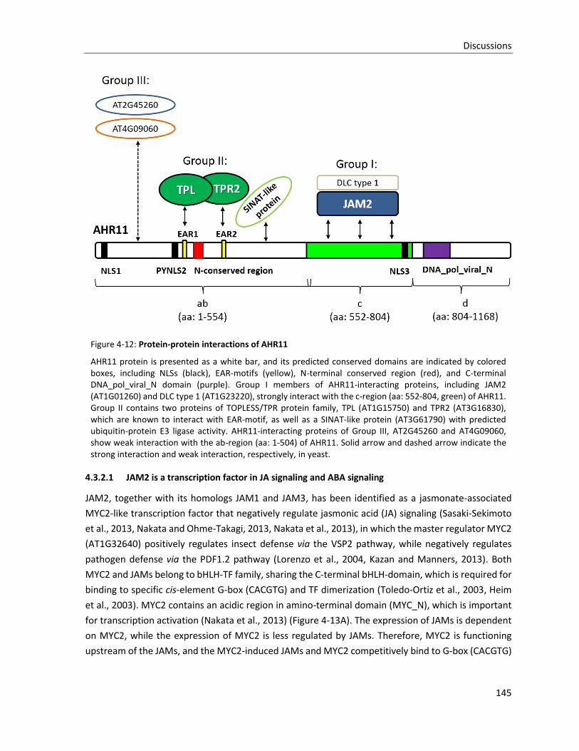
Discussions
145
Figure 4-12: Protein-protein interactions of AHR11
AHR11 protein is presented as a white bar, and its predicted conserved domains are indicated by colored boxes, including NLSs (black), EAR-motifs (yellow), N-terminal conserved region (red), and C-terminal DNA_pol_viral_N domain (purple). Group I members of AHR11-interacting proteins, including JAM2 (AT1G01260) and DLC type 1 (AT1G23220), strongly interact with the c-region (aa: 552-804, green) of AHR11. Group II contains two proteins of TOPLESS/TPR protein family, TPL (AT1G15750) and TPR2 (AT3G16830), which are known to interact with EAR-motif, as well as a SINAT-like protein (AT3G61790) with predicted ubiquitin-protein E3 ligase activity. AHR11-interacting proteins of Group III, AT2G45260 and AT4G09060, show weak interaction with the ab-region (aa: 1-504) of AHR11. Solid arrow and dashed arrow indicate the strong interaction and weak interaction, respectively, in yeast.
4.3.2.1 JAM2 is a transcription factor in JA signaling and ABA signaling
JAM2, together with its homologs JAM1 and JAM3, has been identified as a jasmonate-associated
MYC2-like transcription factor that negatively regulate jasmonic acid (JA) signaling (Sasaki-Sekimoto
et al., 2013, Nakata and Ohme-Takagi, 2013, Nakata et al., 2013), in which the master regulator MYC2
(AT1G32640) positively regulates insect defense via the VSP2 pathway, while negatively regulates
pathogen defense via the PDF1.2 pathway (Lorenzo et al., 2004, Kazan and Manners, 2013). Both
MYC2 and JAMs belong to bHLH-TF family, sharing the C-terminal bHLH-domain, which is required for
binding to specific cis-element G-box (CACGTG) and TF dimerization (Toledo-Ortiz et al., 2003, Heim
et al., 2003). MYC2 contains an acidic region in amino-terminal domain (MYC_N), which is important
for transcription activation (Nakata et al., 2013) (Figure 4-13A). The expression of JAMs is dependent
on MYC2, while the expression of MYC2 is less regulated by JAMs. Therefore, MYC2 is functioning
upstream of the JAMs, and the MYC2-induced JAMs and MYC2 competitively bind to G-box (CACGTG)
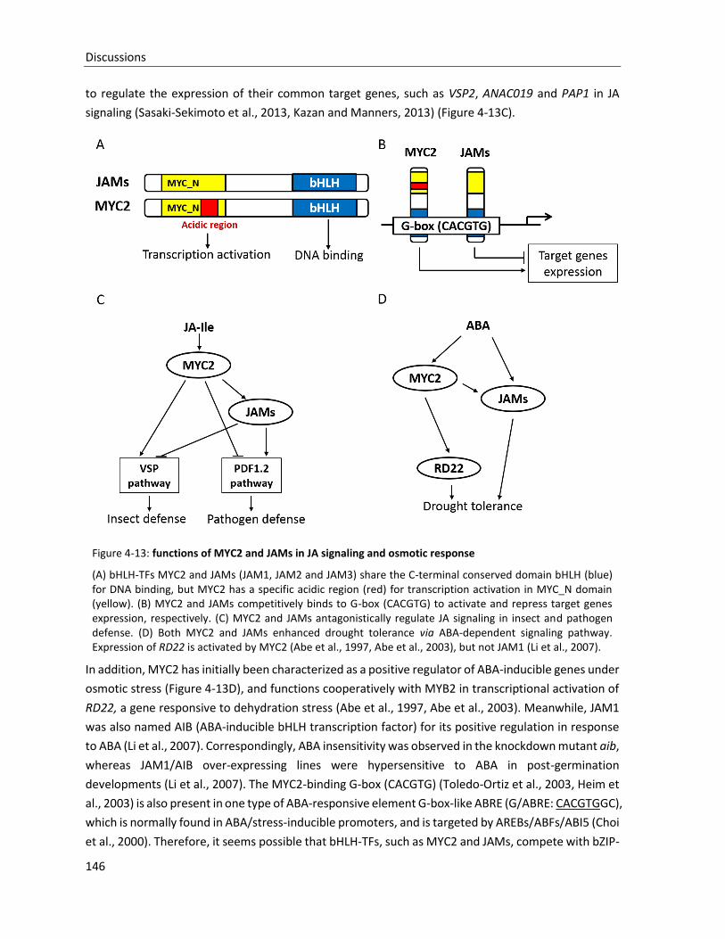
Discussions
146
to regulate the expression of their common target genes, such as VSP2, ANAC019 and PAP1 in JA
signaling (Sasaki-Sekimoto et al., 2013, Kazan and Manners, 2013) (Figure 4-13C).
Figure 4-13: functions of MYC2 and JAMs in JA signaling and osmotic response
(A) bHLH-TFs MYC2 and JAMs (JAM1, JAM2 and JAM3) share the C-terminal conserved domain bHLH (blue) for DNA binding, but MYC2 has a specific acidic region (red) for transcription activation in MYC_N domain (yellow). (B) MYC2 and JAMs competitively binds to G-box (CACGTG) to activate and repress target genes expression, respectively. (C) MYC2 and JAMs antagonistically regulate JA signaling in insect and pathogen defense. (D) Both MYC2 and JAMs enhanced drought tolerance via ABA-dependent signaling pathway. Expression of RD22 is activated by MYC2 (Abe et al., 1997, Abe et al., 2003), but not JAM1 (Li et al., 2007).
In addition, MYC2 has initially been characterized as a positive regulator of ABA-inducible genes under
osmotic stress (Figure 4-13D), and functions cooperatively with MYB2 in transcriptional activation of
RD22, a gene responsive to dehydration stress (Abe et al., 1997, Abe et al., 2003). Meanwhile, JAM1
was also named AIB (ABA-inducible bHLH transcription factor) for its positive regulation in response
to ABA (Li et al., 2007). Correspondingly, ABA insensitivity was observed in the knockdown mutant aib,
whereas JAM1/AIB over-expressing lines were hypersensitive to ABA in post-germination
developments (Li et al., 2007). The MYC2-binding G-box (CACGTG) (Toledo-Ortiz et al., 2003, Heim et
al., 2003) is also present in one type of ABA-responsive element G-box-like ABRE (G/ABRE: CACGTGGC),
which is normally found in ABA/stress-inducible promoters, and is targeted by AREBs/ABFs/ABI5 (Choi
et al., 2000). Therefore, it seems possible that bHLH-TFs, such as MYC2 and JAMs, compete with bZIP-
Discussions
147
TFs, such as AREBs/ABFs/ABI5, for the G/ABRE binding sites to regulate target genes expression. As
discussed in JA signaling, MYC2 is a transcriptional activator, but JAMs are a repressor, which might
be caused by the presence/absence of the N-terminal acidic region. In ABA signaling, the regulation
mechanism seems more indirect. Although the promoter region of RD22 does not contain any ABREs,
the dehydration-induced RD22 expression is also activated by MYC2 via its G-box (Iwasaki et al., 1995),
but not by JAM1 (Li et al., 2007). The target specificity of MYC2 and JAMs in ABA signaling is still
unclear. In this work, the interaction between JAM2 and c-region (aa: 542-804) of AHR11 was found
in yeast. Combination with the negative regulatory role of AHR11, we could speculate that AHR11
repress JAM2-targeted gene expression. However, it is still a matter of speculation whether AHR11
might have a general function as a regulator of activities of bHLH-TFs. It is necessary to analyze the
molecular function of the interaction between AHR11 and JAM2, as well as other JAMs and MYC2 in
future.
4.3.2.2 TPL/TPRs corepressor complex regulate general transcription repression
In this work, AHR11 has been identified as a novel EAR-motif protein, containing two predicted EAR-
motifs: EAR1 in the a-region (aa: 1-247) and EAR2 in the b-region (aa: 248-544) (Figure 4-4). In addition,
a conserved region (aa: 235-249) with unknown function is present between these two EAR-motifs. It
is already known that EAR-motif proteins repress gene transcription via recruiting TOPLESS and
TOPLESS-related transcriptional corepressors to target genes (Kagale et al., 2010). In yeast, the ab-
region (aa: 1-544) of AHR11 strongly interacted with TPL (TOPLESS; AT1G15750) and TPR2 (TOPLESS-
related 2; AT3G16830), while the a-region (aa: 1-247) or b-region (aa: 248-544) containing a single
EAR-motif was impaired binding either TPL or TPR2. These results could imply that a single EAR-motif
is not sufficient for AHR11 to recruit TPL/TPRs, which can be proved by inactivation of a EAR-motif in
the ab-region. The EAR-motif surrounding sequence such as N-conserved region might also contribute
to TPL/TPRs recruition.
TPL and TPR2 belong to the TOPLESS co-repressor family (TPL/TPRs), and were initially identified as
transcriptional corepressors that negatively regulate apical embryonic fate in Arabidopsis (Long et al.,
2006). The C-terminal to lissencephaly homology (CTLH) domain of TPL directly binds to the EAR-motif
located in the domain I of IAA12/BDL (Emes and Ponting, 2001, Szemenyei et al., 2008). The TPL-
interactome analysis revealed that TPL/TPR corepressors predominantly interact directly with specific
transcription factors (Causier et al., 2011). Many of which were previously implicated in
transcriptional repression as EAR-repressors involved in diverse processes, including growth and
development, hormone responses, and stress responses (Kieffer et al., 2006, Long et al., 2006, Takase
et al., 2007, Szemenyei et al., 2008, Pauwels et al., 2010, Causier et al., 2011, Wang et al., 2012a,
Krogan et al., 2012, Tao et al., 2013). Therefore, TPL/TPRs manage a common transcriptional
repression mechanism, which has been unraveled in detail in several hormone responses pathways
including auxin signaling (Szemenyei et al., 2008) and JA signaling (Pauwels et al., 2010). In auxin
signaling (Figure 4-14), auxin response TFs (ARFs) recognize auxin-responsive cis-element (ARE:
TGTCTC) to activate target gene expression (Ulmasov et al., 1997). The EAR-repressors of AUX/IAA
Discussions
148
family couple the TPL/TPRs transcriptional corepressor complex to ARFs, subsequently TPL/TPRs
repress auxin-induced transcription probably via the HISTONE DEACETYLASE 19 (HDA19) (Szemenyei
et al., 2008). Similar transcriptional repression has also been found in JA signaling (Figure 4-15).
JASMONATE-ZIM DOMAIN (JAZ) proteins, which play a central repressive role in JA signaling, binds to
the JAZ interaction domain (JID) at the N-terminal region of MYC2. If the JAZs contain the
aforementioned EAR-motif, also bind to TPL/TPRs (Shyu et al., 2012), thereby recruiting the TPL/TPRs
co-repressors, putatively together with HDA19, to repress MYC2 target genes expression (Pauwels
and Goossens, 2011, Niu et al., 2011, Fernandez-Calvo et al., 2011, Shyu et al., 2012, Kazan and
Manners, 2013). The Non-EAR-motif JAZ proteins may cooperate with the EAR-motif adaptor protein
NINJA (Novel interactor of JAZ) to recruit TPL/TPRs (Pauwels et al., 2010). When auxin or JA (JA-Ile)
are presenct, the receptor complex TIR1-auxin-AUX/IAA or COI1-JA-Ile-JAZ initiates the ubiquitylation
and degradation of the EAR-repressors AUX/IAA or JAZ by the proteasome pathway to release
transcriptions from repression by TPL/TPRs.
Interestingly, the ABI5-binding proteins (AFPs) share three conserved domains with the adaptor
protein NINJA (Lopez-Molina et al., 2003, Garcia et al., 2008) and also carry the EAR-motif to interact
with TPL/TPRs (Pauwels et al., 2010). Therefore, AFPs likely function as EAR-repressor adaptor
proteins to inhibit ABI5 target genes transcription in ABA signaling by recruiting the transcriptional
repressors TPL/TPRs.
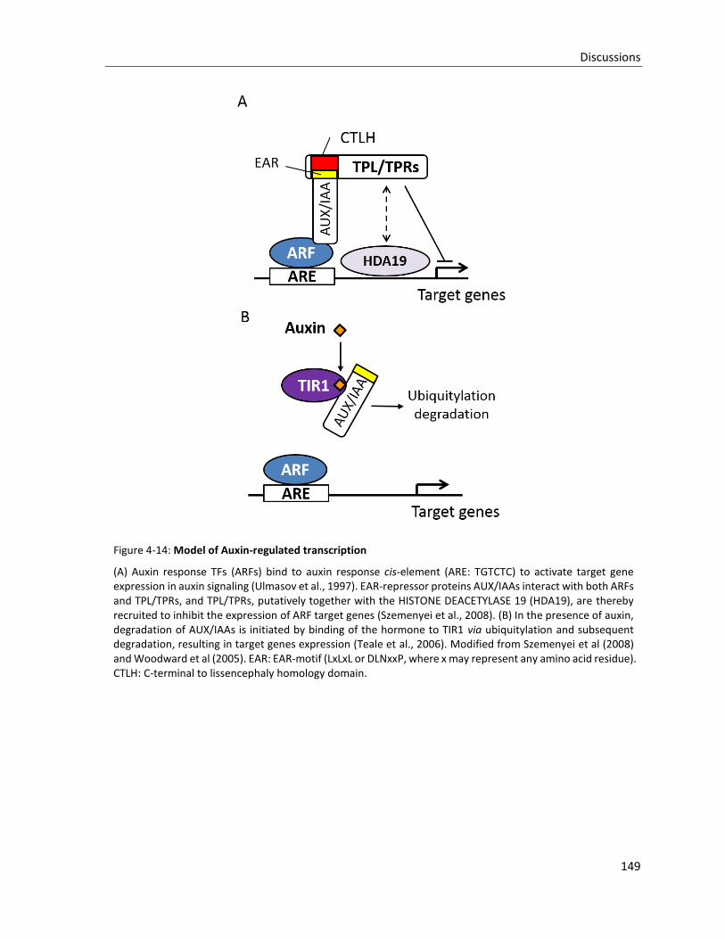
Discussions
149
Figure 4-14: Model of Auxin-regulated transcription
(A) Auxin response TFs (ARFs) bind to auxin response cis-element (ARE: TGTCTC) to activate target gene expression in auxin signaling (Ulmasov et al., 1997). EAR-repressor proteins AUX/IAAs interact with both ARFs and TPL/TPRs, and TPL/TPRs, putatively together with the HISTONE DEACETYLASE 19 (HDA19), are thereby recruited to inhibit the expression of ARF target genes (Szemenyei et al., 2008). (B) In the presence of auxin, degradation of AUX/IAAs is initiated by binding of the hormone to TIR1 via ubiquitylation and subsequent degradation, resulting in target genes expression (Teale et al., 2006). Modified from Szemenyei et al (2008) and Woodward et al (2005). EAR: EAR-motif (LxLxL or DLNxxP, where x may represent any amino acid residue). CTLH: C-terminal to lissencephaly homology domain.
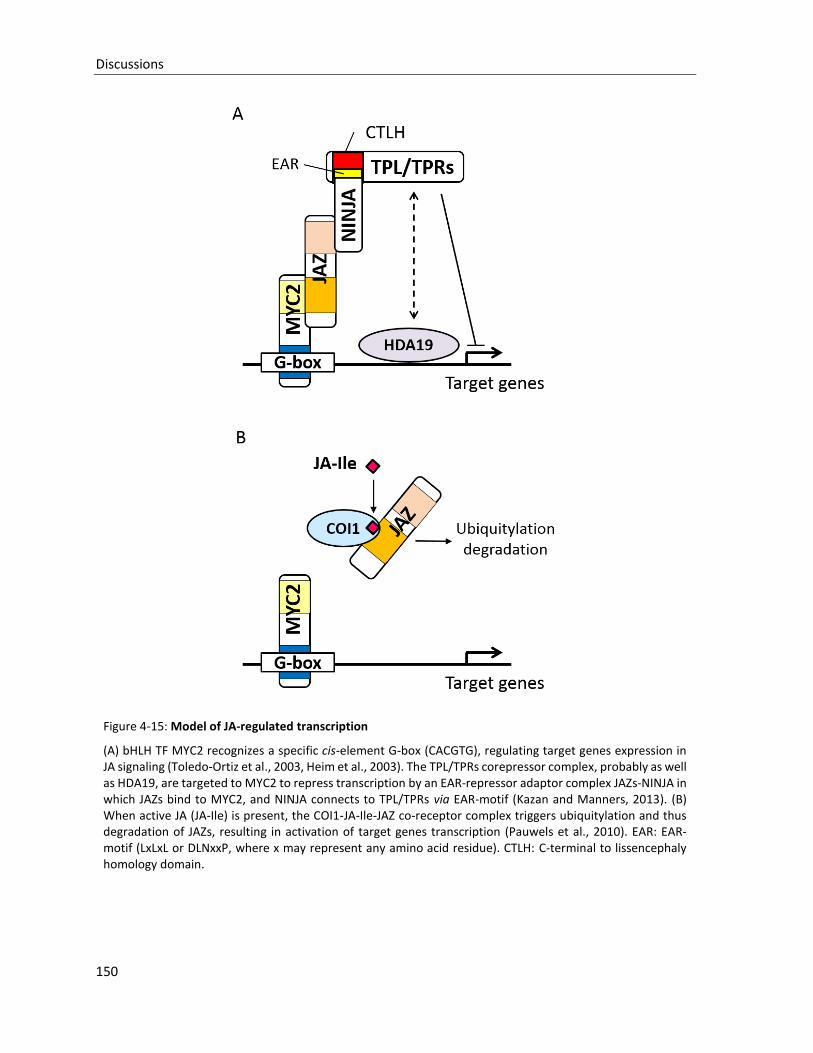
Discussions
150
Figure 4-15: Model of JA-regulated transcription
(A) bHLH TF MYC2 recognizes a specific cis-element G-box (CACGTG), regulating target genes expression in JA signaling (Toledo-Ortiz et al., 2003, Heim et al., 2003). The TPL/TPRs corepressor complex, probably as well as HDA19, are targeted to MYC2 to repress transcription by an EAR-repressor adaptor complex JAZs-NINJA in which JAZs bind to MYC2, and NINJA connects to TPL/TPRs via EAR-motif (Kazan and Manners, 2013). (B) When active JA (JA-Ile) is present, the COI1-JA-Ile-JAZ co-receptor complex triggers ubiquitylation and thus degradation of JAZs, resulting in activation of target genes transcription (Pauwels et al., 2010). EAR: EAR-motif (LxLxL or DLNxxP, where x may represent any amino acid residue). CTLH: C-terminal to lissencephaly homology domain.
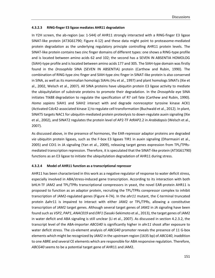
Discussions
151
4.3.2.3 RING-finger E3 ligase mediates AHR11 degradation
In Y2H screen, the ab-region (aa: 1-544) of AHR11 strongly interacted with a RING-finger E3 ligase
SINAT-like protein (AT3G61790; Figure 4-12) and these data might point to proteasome-mediated
protein degradation as the underlying regulatory principle controlling AHR11 protein levels. The
SINAT-like protein contains two zinc finger domains of different types: one shows a RING-type profile
and is located between amino acids 62 and 102; the second has a SEVEN IN ABSENTIA HOMOLOG
(SIAH)-type profile and is located between amino acids 177 and 305. The SIAH-type domain was firstly
found in the Drosophila SINA (SEVEN IN ABSENTIA) protein (Carthew and Rubin, 1990). The
combination of RING-type zinc finger and SIAH-type zinc finger in SINAT-like protein is also conserved
in SINA, as well as its mammalian homologs SIAHs (Hu et al., 1997) and plant homologs SINATs (Xie et
al., 2002, Welsch et al., 2007). All SINA proteins have ubiquitin protein E3 ligase activity to mediate
the ubiquitylation of substrate proteins to promote their degradation. In the Drosophila eye SINA
initiates Ttk88 degradation to regulate the specification of R7 cell fate (Carthew and Rubin, 1990).
Homo sapiens SIAH1 and SIAH2 interact with and degrade nonreceptor tyrosine kinase ACK1
(Activated Cdc42-associated kinase 1) to regulate cell transformation (Buchwald et al., 2012). In plant,
SINAT5 targets NAC1 for ubiquitin-mediated protein proteolysis to down-regulate auxin signaling (Xie
et al., 2002), and SINAT2 regulates the protein level of AP2-TF AtRAP2.2 in Arabidopsis (Welsch et al.,
2007).
As discussed above, in the presence of hormones, the EAR-repressor adaptor proteins are degraded
via ubiquitin protein ligases, such as the F-box E3 ligases TIR1 in auxin signaling (Dharmasiri et al.,
2005) and COI1 in JA signaling (Yan et al., 2009), releasing target genes expression from TPL/TPRs-
mediated transcription repression. Therefore, it is speculated that the SINAT-like protein (AT3G61790)
functions as an E3 ligase to initiate the ubiquitylation degradation of AHR11 during stress.
4.3.2.4 Model of AHR11 function as a transcriptional repressor
AHR11 has been characterized in this work as a negative regulator of response to water deficit stress,
especially involved in ABA/stress-induced gene transcription. According to its interaction with both
bHLH-TF JAM2 and TPL/TPRs transcriptional corepressors in yeast, the novel EAR-protein AHR11 is
proposed to function as an adaptor protein, recruiting the TPL/TPRs corepressor complex to inhibit
transcription of JAM2-regulated genes (Figure 4-7A). In the ahr11 mutant, the C-terminal truncated
protein Δahr11 is impaired to interact with either JAM2 or TPL/TPRs, allowing a constitutive
transcription of JAM2 target genes. Although several target genes of JAM2 in JA signaling have been
found such as VSP2, PAP1, ANAC019 and ERF1 (Sasaki-Sekimoto et al., 2013), the target genes of JAM2
in water deficit and ABA signaling is still unclear (Li et al., 2007). As discussed in section 4.2.3.2, the
transcript level of the ABA-importer ABCG40 is significantly higher in ahr11 shoot after exposure to
water deficit stress. The cis-element analysis of ABCG40 promoter reveals the presence of 11 G-box
elements which might be recognized by JAM2 in the upstream region (1635 bp) of ABCG40, inaddition
to one ABRE and several CE elements which are responsible for ABA responsive regulation. Therefore,
ABCG40 seems to be a potential target gene of AHR11 and JAM2.
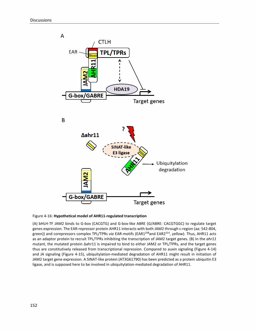
Discussions
152
Figure 4-16: Hypothetical model of AHR11-regulated transcription
(A) bHLH-TF JAM2 binds to G-box (CACGTG) and G-box-like ABRE (G/ABRE: CACGTGGC) to regulate target genes expression. The EAR-repressor protein AHR11 interacts with both JAM2 through c-region (aa: 542-804, green)) and corepressors complex TPL/TPRs via EAR-motifs (EAR1208and EAR2314, yellow). Thus, AHR11 acts as an adaptor protein to recruit TPL/TPRs inhibiting the transcription of JAM2 target genes. (B) In the ahr11 mutant, the mutated protein Δahr11 is impaired to bind to either JAM2 or TPL/TPRs, and the target genes thus are constitutively released from transcriptional repression. Compared to auxin signaling (Figure 4-14) and JA signaling (Figure 4-15), ubiquitylation-mediated degradation of AHR11 might result in initiation of JAM2 target gene expression. A SINAT-like protein (AT3G61790) has been predicted as a protein ubiquitin E3 ligase, and is supposed here to be involved in ubiquitylation-mediated degradation of AHR11.
Discussions
153
Similar to auxin signaling (Figure 4-14) and JA signaling (Figure 4-15), the transcriptional repression
adaptor protein AHR11 is presumably susceptible to ubiquitin-mediated degradation as inferred from
the observation that it interacts with the RING-finger E3 ligase SINAT-like protein (AT3G61790).
Additionally, the RING-type E3 ligase RKP co-regulated with AHR11 might reflect a role of AHR11 level
regulation. In the ahr11 mutant, the mutated protein Δahr11 is impaired to bind to either JAM2 or
TPL/TPRs, and the JAM2-regulated genes thus are constitutively released from transcriptional
repression. The subsequent genome-wide transcription analysis of water deficit-stressed ahr11 would
uncover the target genes of JAM2, as well as of AHR11, in water deficit response.
Although the high-throughput Y2H screen used in this work is a reliable tool which recently has been
employed successfully in several studies (Yu et al., 2008, Braun, 2012, Braun et al., 2013), protein-
protein interaction between AHR11 and its interacting partners still need to be confirmed in planta,
e.g. by using bimolecular fluorescence complementation (BiFC) in protoplasts (Bhat et al., 2006).
4.4 Multiple functions of AHR11 in development and non-ABA hormone signaling
pathways
Although the mutant ahr11 shows an altered response in several ABA-regulated physiological
processes, ahr11 also displays specifically phenotypic alternations such as early flowering (Table 3-4),
less rosette leaves (Figure 3-25), and reduced apical dominance (Figure 3-25). Thus, AHR11
presumably has multiple regulatory functions in plant development and non-ABA hormone signaling
pathways. Due to the dual function of the AHR11-interacting protein JAM2 in ABA and JA signaling,
AHR11 is supposed to also be involved in JA signaling by suppressing the expression of JAM2 target
genes. Both JAMs and MYC2 are positive regulators of ABA signaling (Abe et al., 1997, Abe et al., 2003,
Li et al., 2007), but antagonistically regulate JA signaling (Sasaki-Sekimoto et al., 2013, Kazan and
Manners, 2013). How AHR11 is involved as a regulatory element in both signaling pathways, perhaps
representing a node of crosstalk of ABA and JA signaling needs further investigation.
The transition from vegetative growth to flowering is an important developmental process in plants.
This switch is regulated by several pathways including the photoperiod response pathway, the
autonomous pathway and GA signaling (Mouradov et al., 2002). In the floral transition process, ABA
seems to play a minor role, acting as a floral repressor while GA plays a dominant role in promoting
the transition to reproductive development (Domagalska et al., 2010). Since ahr11 shows an early
flowering phenotype under long day conditions and develops less rosette leaves compared to wild
type (pATHB6::LUC) plants (Table 3-4), AHR11 might be also involved in the cross talk between ABA
and GA signaling during transition to flowering.
During the reproductive developmental stage, ahr11 plants exhibit reduced apical dominance with
respect to inflorescence branching (Figure 3-25). It is already known that the shoot architecture is
largely regulated by three classes of hormone: auxin, cytokinin (CK) and strigolactone (SL) (Ferguson
and Beveridge, 2009). Typically, the young leaves synthesize auxin at the tip of the main shoot to
inhibit branching (Booker et al., 2003). The hormones CK and SL both are up-regulated by auxin, and
Discussions
154
move into the axillary buds (Dun et al., 2009) to antagonistically promote or inhibit lateral branching
(Davies et al., 1966, Gomez-Roldan et al., 2008, Umehara et al., 2008). Probably SL also influences CK
levels. Many SL mutants display an abnormal CK level in xylem sap (Foo et al., 2007). SL promotes
shoot branching by triggering rapid depletion of auxin efflux protein PIN1 from the plasma membrane
(Balla et al., 2012, Shinohara et al., 2013, Koltai, 2014, Young et al., 2014). In addition, the lateral bud
outgrowth is also inhibited by ABA via an auxin-independent manner (Cline and Oh, 2006). These
imply that AHR11 not only regulates stress responses but also modulates aspects of plant growth
development by affecting the corss talk between ABA and other hormonal and developmental cues.
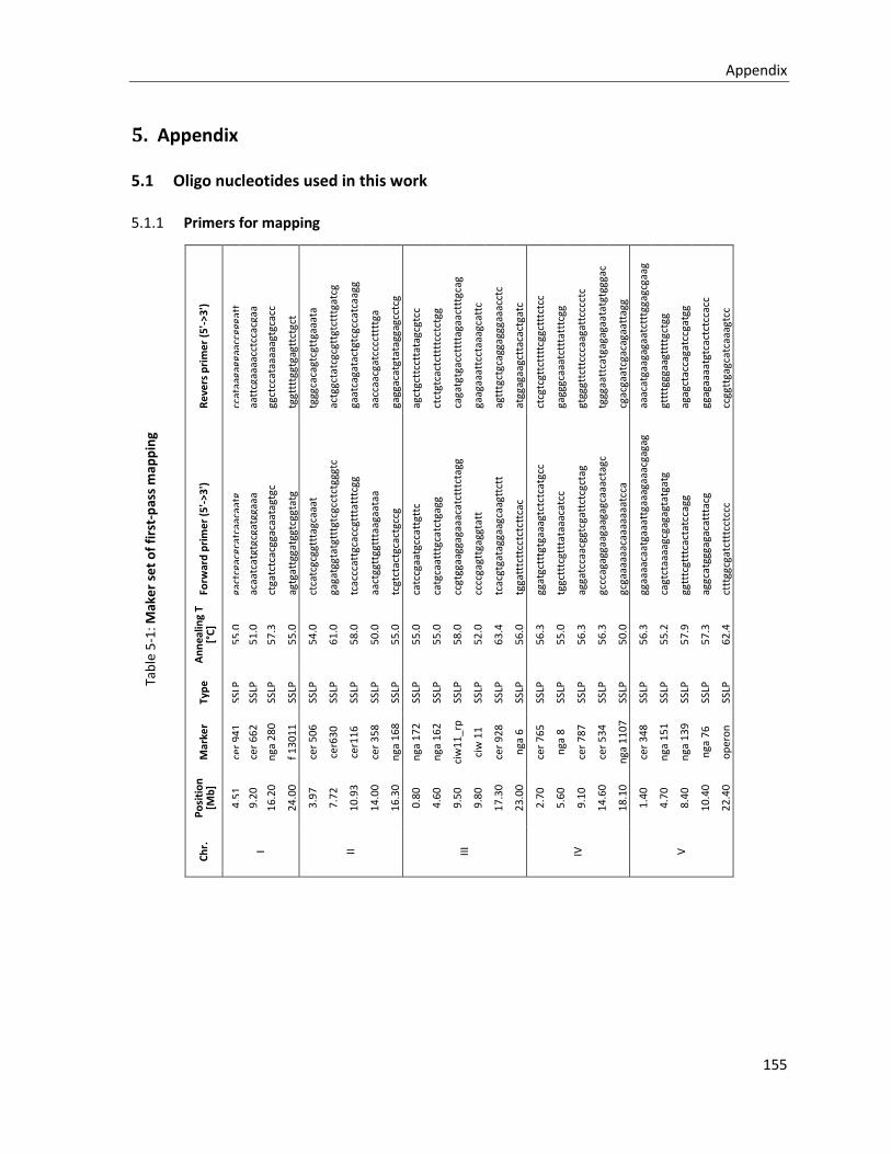
Appendix
155
5. Appendix
5.1 Oligo nucleotides used in this work
5.1.1 Primers for mapping
Re
vers
pri
me
r (5
'->3
')
ccat
aaga
ggaa
ccgg
gatt
aatt
cgaa
aacc
tcca
cgaa
ggct
ccat
aaaa
agtg
cacc
tggt
tttg
gtga
gttc
tgct
tggg
caca
gtcg
ttga
aata
actg
gcta
tcgc
gttg
tctt
tgat
cg
gaat
caga
tact
gtcg
ccat
caag
g
aacc
aacg
atcc
cctt
ttga
gagg
acat
gtat
agga
gcct
cg
agct
gctt
cctt
atag
cgtc
c
ctct
gtca
ctct
tttc
ctct
gg
caga
tgtg
acct
ttta
gaac
tttg
cag
gaag
aaat
tcct
aaag
catt
c
agtt
tgct
gcag
gagg
gaaa
cctc
atgg
agaa
gctt
acac
tgat
c
ctcg
tcgt
tctt
ttcg
gctt
tctc
c
gagg
gcaa
atct
ttat
ttcg
g
gtgg
gttc
ttcc
caag
attc
ccct
c
tggg
aatt
catg
agag
aata
tgtg
ggac
cgac
gaat
cgac
agaa
ttag
g
aaac
atga
agag
aatc
tttg
gagc
gaag
gttt
tggg
aagt
tttg
ctgg
agag
ctac
caga
tccg
atgg
ggag
aaaa
tgtc
actc
tcca
cc
ccgg
ttga
gcat
caaa
gtcc
Forw
ard
pri
me
r (5
'->3
')
gact
cgac
gcat
caac
aatg
acaa
tcat
gtgc
gatg
gaaa
ctga
tctc
acgg
acaa
tagt
gc
agtg
attg
gatg
gtcg
gtat
g
ctca
tcgc
ggtt
tagc
aaat
gaga
tggt
atgt
ttgt
cgcc
tctg
ggtc
tcac
ccat
tgca
ccgt
ttat
ttcg
g
aact
ggtt
ggtt
taag
aata
a
tcgt
ctac
tgca
ctgc
cg
catc
cgaa
tgcc
attg
ttc
catg
caat
ttgc
atct
gagg
ccgt
ggaa
ggag
aaac
atct
ttct
agg
cccc
gagt
tgag
gtat
t
tcac
gtga
tagg
aagc
aagt
tctt
tgga
tttc
ttcc
tctc
ttca
c
ggat
gctt
tgtg
aaag
tctc
tcat
gcc
tggc
tttc
gttt
ataa
acat
cc
agga
tcca
acgg
tcga
ttct
cgct
ag
gccc
agag
gaag
aaga
gcaa
acta
gc
gcga
aaaa
acaa
aaaa
atcc
a
ggaa
aaca
atga
aatt
gaaa
gaaa
cgag
ag
cagt
ctaa
aagc
gaga
gtat
gatg
ggtt
tcgt
ttca
ctat
ccag
g
aggc
atgg
gaga
catt
tacg
cttt
ggcg
atct
ttcc
tccc
An
nea
ling
T [°
C]
55
.0
51
.0
57
.3
55
.0
54
.0
61
.0
58
.0
50
.0
55
.0
55
.0
55
.0
58
.0
52
.0
63
.4
56
.0
56
.3
55
.0
56
.3
56
.3
50
.0
56
.3
55
.2
57
.9
57
.3
62
.4
Typ
e
SSLP
SSLP
SSLP
SSLP
SSLP
SSLP
SSLP
SSLP
SSLP
SSLP
SSLP
SSLP
SSLP
SSLP
SSLP
SSLP
SSLP
SSLP
SSLP
SSLP
SSLP
SSLP
SSLP
SSLP
SSLP
Mar
ker
cer
94
1
cer
66
2
nga
28
0
f 1
30
11
cer
50
6
cer6
30
cer1
16
cer
35
8
nga
16
8
nga
17
2
nga
16
2
ciw
11
_rp
ciw
11
cer
92
8
nga
6
cer
76
5
nga
8
cer
78
7
cer
53
4
nga
11
07
cer
34
8
nga
15
1
nga
13
9
nga
76
op
ero
n
22
44
1
Po
siti
on
[M
b]
4.5
1
9.2
0
16
.20
24
.00
3.9
7
7.7
2
10
.93
14
.00
16
.30
0.8
0
4.6
0
9.5
0
9.8
0
17
.30
23
.00
2.7
0
5.6
0
9.1
0
14
.60
18
.10
1.4
0
4.7
0
8.4
0
10
.40
22
.40
Ch
r.
I II
III
IV
V
Tab
le 5
-1: M
ake
r se
t o
f fi
rst-
pas
s m
app
ing
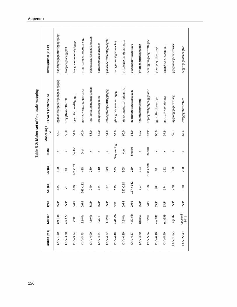
Appendix
156
Re
vers
pri
me
r (5
'->3
')
aaac
atga
agag
aatc
tttg
gagc
gaag
tcat
gacc
gaac
cggg
ttct
tcac
gcaa
ataa
aata
tgtt
ggga
gttg
atcc
aaga
atat
gatg
caag
gc
ctag
tgtt
tttt
acgc
agga
catg
tttc
c
aatc
ccag
taac
caaa
caca
ca
gaaa
caac
tctt
cata
ttga
aagc
tc
catc
ggat
aacg
tgta
taga
ctag
gttc
ctcg
tcct
gaat
gtga
tagt
cc
gcat
atgc
gctt
cttc
tgtt
cac
gtta
tgga
gttt
ctag
ggca
cg
ccaa
tggt
aagc
cagt
ttct
tagc
tc
gtaa
cgca
gctt
catc
cagc
agag
ctac
caga
tccg
atgg
ggag
aaaa
tgtc
actc
tcca
cc
ccgg
ttga
gcat
caaa
gtcc
Forw
ard
pri
me
r (5
'->3
')
ggaa
aaca
atga
aatt
gaaa
gaaa
cgag
ag
tccg
gttc
aacc
ttat
tctt
tgcc
cctt
ctta
aatt
tgtg
gt
gacg
atgt
aagt
tggt
ggaa
ccac
a
tgta
tacc
agtg
catg
gttt
gcat
ggg
ccca
gtct
aacc
acga
ccac
cata
agat
attg
tcat
tttg
gttg
ag
gtaa
cgtc
ctcg
aaca
actg
gag
atga
ccta
ggtg
ttca
tttg
tagg
ttc
gaat
tcca
tgtt
gtat
tagg
acag
g
tgcc
ccat
tttg
ttct
tctc
tcga
cgct
tctg
atgc
aggg
aaat
c
ggtt
cctt
cctt
caaa
ttga
ggtt
tcgt
ttca
ctat
ccag
g
aggc
atgg
gaga
catt
tacg
cttt
ggcg
atct
ttcc
tccc
An
nea
ling
T
[°C
]
56
.3
58
.0
54
.0
60
.0
58
.0
57
.0
54
.0
53
.0
60
.0
58
.0
55
.2
60
°C
60
.0
57
.9
57
.3
62
.4
No
te
/ /
Eco
RV
Dra
I
/ / /
Seq
uen
cin
g
Nd
eI
Fnu
4H
I
/
Bam
HI
/ / / /
Ler
[bp
]
10
0
40
46
1+1
39
42
5
26
9
11
0
34
9
58
5
50
5
26
9
12
3
18
0 +
18
8
40
13
2
30
0
26
0
Co
l [b
p]
18
5
71
60
0
24
3+1
82
24
9
12
4
37
7
58
5
28
7+2
18
12
7 +
14
2
15
7
36
8
80
17
4
22
0
37
0
Typ
e
SSLP
SSLP
CA
PS
CA
PS
SSLP
SSLP
SSLP
SNP
CA
PS
CA
PS
SSLP
CA
PS
SSLP
SSLP
SSLP
SSLP
Mar
ker
cer
34
8
cer
47
7
OX
F
3.9
Mb
4.0
Mb
CA
72
4.3
Mb
4.4
8M
b
4.5
Mb
4.5
7M
b
nga
15
1
5.3
Mb
cer
48
2
nga
13
9
nga
76
op
ero
n2
24
41
Po
siti
on
[M
b]
Ch
rV-1
.40
Ch
rV-3
.20
Ch
rV-3
.84
Ch
rV-3
.93
Ch
rV-4
.00
Ch
rV-4
.24
Ch
rV-4
.32
Ch
rV-4
.48
Ch
rV-4
.50
Ch
rV-4
.57
Ch
rV-4
.70
Ch
rV-5
.34
Ch
rV-6
.10
Ch
rV-8
.40
Ch
rV-1
3.6
8
Ch
rV-2
2.4
0
Tab
le 5
-2: M
ake
r se
t o
f fi
ne
-sca
le m
app
ing
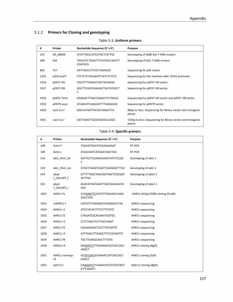
Appendix
157
5.1.2 Primers for Cloning and genotyping
Table 5-3: Uniform primers
# Primer Nucleotide-Sequence (5’->3’) Purpose
225 GK_08409 ATATTGACCATCATACTCATTGC Genotyping of GABI-Kat T-DNA mutant
289 LB3 TAGCATCTGAATTTCATAACCAATCTCGATACA
Genotyping of SAIL T-DNA mutant
856 T3-f AATTAACCCTCACTAAAGGG Sequencing for pSK vector.
1915 p35S-end-f CTCTCTCTACAAATCTATCTCTCTC Sequencing for the insertion after 35SS1 promoter.
1916 pDEST-AD CGCGTTTGGAATCACTACAGGG Sequencing for pDEST-AD vector.
1917 pDEST-DB GGCTTCAGTGGAGACTGATATGCCTC
Sequencing for pDEST-DB vector.
1918 pDEST-Term GGAGACTTGACCAAACCTCTGGCG Sequencing for pDEST-AD vector and pDEST-DB vector.
1919 pENTR-seq-r GTAACATCAGAGATTTTGAGACAC Sequencing for pENTR vector.
1920 LacZ-ins-f GACCATGATTACGCCAAGCTCG 86bp to AscI, Sequencing for Binary vector and transgenic plants.
1921 LacZ-ins-r GATTAAGTTGGGTAACGCCAGG 192bp to AscI, Sequencing for Binary vector and transgenic plants.
Table 5-4: Specific primers
# Primer Nucleotide-Sequence (5’->3’) Purpose
108 Actin-f TGGGATGACATGGAGAAGAT RT-PCR
109 Actin-r ATACCAATCATAGATGGCTGG RT-PCR
216 abi1_XhoI_for AATTCCTCGAGGGAAGTATCTCCGGC
Genotyping of abi1-1
219 abi1_XhoI_rev GTACTCGAGTCAGTTCAAGGGTTTGC Genotyping of abi1-1
554 aba2-1_DeCAPS_f
GTTTTTGGCTAGCGATGACTCGCGGTACTTAA
Genotyping of aba2-1
555 aba2-1_DeCAPS_r
AGACATGATAAATTGGCGGACAATAAAC
Genotyping of aba2-1
1922 AHR11-f1 CCGGAATTCATGTCTGGAAGCCAAGAGCCTAG
AHR11 (At5g13590) cloning (EcoRI)
1923 ctAHR11-f ctATGTCTGGAAGCCAAGAGCCTAG AHR11 sequencing
1924 AHR11-r1 ATCCTACACTTTCCTTCTGTC AHR11 sequencing
1925 AHR11-f2 CTAGATCGCACAAGTGGTGC AHR11 sequencing
1926 AHR11-r2 CCTCTAGCTCCTTACCAGGT AHR11 sequencing
1927 AHR11-f3 CGAAAGAACTCCCTTGTAATTC AHR11 sequencing
1928 AHR11-r3 ATTTAACCTTGGGCTTCTCATAATTC AHR11 sequencing
1929 AHR11-f4 TGCTTCAGGCAGCTTTGTG AHR11 sequencing
1930 AHR11-r4 GAAGATCTTTAAAAACCATCACCACCGAGCT
AHR11 cloning (BglII)
1931 AHR11-nonstop-r4
GCGTCGACGCAAAACCATCACCACCGAGCT
AHR11 cloning (SalI)
1932 Δahr11-r GAAGATCTTCAAACATCCATGGTAGTATTCAAATC
Δahr11 cloning (BglII)
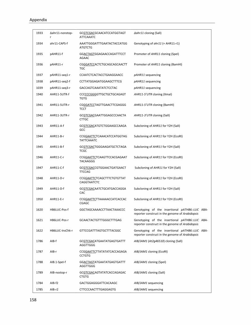
Appendix
158
1933 Δahr11-nonstop-r
GCGTCGACGCAACATCCATGGTAGTATTCAAATC
Δahr11 cloning (SalI)
1934 ahr11-CAPS-f AAATTGGGATTTGAATACTACCATGGATGTCTG
Genotyping of ahr11 (+ AHR11-r1)
1935 pAHR11-f GGACTAGTGGAGAACCAGATTTCCTAGAAC
Promoter of AHR11 cloning (SpeI)
1936 pAHR11-r CGGGATCCACTCTGCAGCAGCAACTTTGC
Promoter of AHR11 cloning (BamHI)
1937 pAHR11-seq1-r CCAATCTCACTACCTGAAGGAACC pAHR11 sequencing
1938 pAHR11-seq2-f CCTTATGGAGATGGAAGCTTTCG pAHR11 sequencing
1039 pAHR11-seq3-r GACCAGTCAAATATCTCCTAC pAHR11 sequencing
1940 AHR11-5UTR-f CCCCCCGGGGTTGCTGCTGCAGAGTTGTG
AHR11-5’UTR cloning (XmaI)
1941 AHR11-5UTR-r CGGGATCCTAGTTGAACTTCGAGGGTCCT
AHR11-5’UTR cloning (BamHI)
1942 AHR11-3UTR-r GCGTCGACGAATTGGAGCCCAACTACTTGC
AHR11-3’UTR cloning (SalI)
1943 AHR11-A-f GCGTCGACATGTCTGGAAGCCAAGAGCC
Subcloning of AHR11 for Y2H (SalI)
1944 AHR11-B-r CCGGAATTCTCAAACATCCATGGTAGTATTCAAATC
Subcloning of AHR11 for Y2H (EcoRI)
1945 AHR11-B-f GCGTCGACTGGGAAGATGCTCTAGATCGC
Subcloning of AHR11 for Y2H (SalI)
1946 AHR11-C-r CCGGAATTCTCAAGTTCCACGAGAATTACAAGGG
Subcloning of AHR11 for Y2H (EcoRI)
1947 AHR11-C-f GCGTCGACCGTGGAACTGATGAACTTTCCAG
Subcloning of AHR11 for Y2H (SalI)
1948 AHR11-D-r CCGGAATTCTCAGCTTTCTGTGTTATCAGGTAATCTC
Subcloning of AHR11 for Y2H (EcoRI)
1949 AHR11-D-f GCGTCGACAATCTGCATGACCAGGACAC
Subcloning of AHR11 for Y2H (SalI)
1950 AHR11-E-r CCGGAATTCTTAAAAACCATCACCACCGAGC
Subcloning of AHR11 for Y2H (EcoRI)
1620 HB6LUC-Pos-f GGCTAGCAAAACCTTAACTAAACCC Genotyping of the insertional pATHB6::LUC ABA-reporter construct in the genome of Arabidopsis
1621 HB6LUC-Pos-r GCAACTACTGTTTGGGCTTTGAG Genotyping of the insertional pATHB6::LUC ABA-reporter construct in the genome of Arabidopsis
1622 HB6LUC-InsChk-r GTTCCGATTTAGTGCTTTACGGC Genotyping of the insertional pATHB6::LUC ABA-reporter construct in the genome of Arabidopsis
1786 AIB-f GCGTCGACATGAATATGAGTGATTTAGGTTGGG
AIB/JAM1 (At2g46510) cloning (SalI)
1787 AIB-r CCGGAATTCTTATATATCACCAGAGACCTGTG
AIB/JAM1 cloning (EcoRI)
1788 AIB.1-SpeI-f GGACTAGTATGAATATGAGTGATTTAGGTTGGG
AIB/JAM1 cloning (SpeI)
1789 AIB-nostop-r GCGTCGACAGTATATCACCAGAGACCTGTG
AIB/JAM1 cloning (SalI)
1784 AIB-f2 GACTGGAGGGATTCACAAGC AIB/JAM1 sequencing
1785 AIB-r2 CTTCCCAACTTTGAGGAGTG AIB/JAM1 sequencing
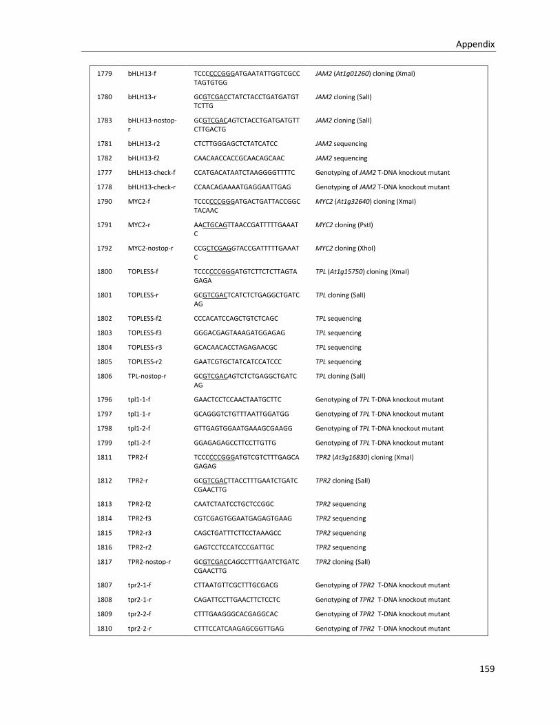
Appendix
159
1779 bHLH13-f TCCCCCCGGGATGAATATTGGTCGCCTAGTGTGG
JAM2 (At1g01260) cloning (XmaI)
1780 bHLH13-r GCGTCGACCTATCTACCTGATGATGTTCTTG
JAM2 cloning (SalI)
1783 bHLH13-nostop-r
GCGTCGACAGTCTACCTGATGATGTTCTTGACTG
JAM2 cloning (SalI)
1781 bHLH13-r2 CTCTTGGGAGCTCTATCATCC JAM2 sequencing
1782 bHLH13-f2 CAACAACCACCGCAACAGCAAC JAM2 sequencing
1777 bHLH13-check-f CCATGACATAATCTAAGGGGTTTTC Genotyping of JAM2 T-DNA knockout mutant
1778 bHLH13-check-r CCAACAGAAAATGAGGAATTGAG Genotyping of JAM2 T-DNA knockout mutant
1790 MYC2-f TCCCCCCGGGATGACTGATTACCGGCTACAAC
MYC2 (At1g32640) cloning (XmaI)
1791 MYC2-r AACTGCAGTTAACCGATTTTTGAAATC
MYC2 cloning (PstI)
1792 MYC2-nostop-r CCGCTCGAGGTACCGATTTTTGAAATC
MYC2 cloning (XhoI)
1800 TOPLESS-f TCCCCCCGGGATGTCTTCTCTTAGTAGAGA
TPL (At1g15750) cloning (XmaI)
1801 TOPLESS-r GCGTCGACTCATCTCTGAGGCTGATCAG
TPL cloning (SalI)
1802 TOPLESS-f2 CCCACATCCAGCTGTCTCAGC TPL sequencing
1803 TOPLESS-f3 GGGACGAGTAAAGATGGAGAG TPL sequencing
1804 TOPLESS-r3 GCACAACACCTAGAGAACGC TPL sequencing
1805 TOPLESS-r2 GAATCGTGCTATCATCCATCCC TPL sequencing
1806 TPL-nostop-r GCGTCGACAGTCTCTGAGGCTGATCAG
TPL cloning (SalI)
1796 tpl1-1-f GAACTCCTCCAACTAATGCTTC Genotyping of TPL T-DNA knockout mutant
1797 tpl1-1-r GCAGGGTCTGTTTAATTGGATGG Genotyping of TPL T-DNA knockout mutant
1798 tpl1-2-f GTTGAGTGGAATGAAAGCGAAGG Genotyping of TPL T-DNA knockout mutant
1799 tpl1-2-f GGAGAGAGCCTTCCTTGTTG Genotyping of TPL T-DNA knockout mutant
1811 TPR2-f TCCCCCCGGGATGTCGTCTTTGAGCAGAGAG
TPR2 (At3g16830) cloning (XmaI)
1812 TPR2-r GCGTCGACTTACCTTTGAATCTGATCCGAACTTG
TPR2 cloning (SalI)
1813 TPR2-f2 CAATCTAATCCTGCTCCGGC TPR2 sequencing
1814 TPR2-f3 CGTCGAGTGGAATGAGAGTGAAG TPR2 sequencing
1815 TPR2-r3 CAGCTGATTTCTTCCTAAAGCC TPR2 sequencing
1816 TPR2-r2 GAGTCCTCCATCCCGATTGC TPR2 sequencing
1817 TPR2-nostop-r GCGTCGACCAGCCTTTGAATCTGATCCGAACTTG
TPR2 cloning (SalI)
1807 tpr2-1-f CTTAATGTTCGCTTTGCGACG Genotyping of TPR2 T-DNA knockout mutant
1808 tpr2-1-r CAGATTCCTTGAACTTCTCCTC Genotyping of TPR2 T-DNA knockout mutant
1809 tpr2-2-f CTTTGAAGGGCACGAGGCAC Genotyping of TPR2 T-DNA knockout mutant
1810 tpr2-2-r CTTTCCATCAAGAGCGGTTGAG Genotyping of TPR2 T-DNA knockout mutant
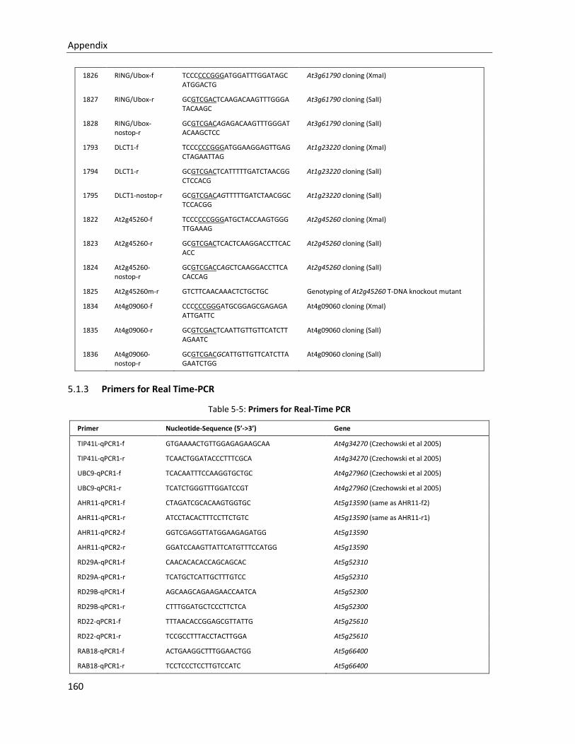
Appendix
160
1826 RING/Ubox-f TCCCCCCGGGATGGATTTGGATAGCATGGACTG
At3g61790 cloning (XmaI)
1827 RING/Ubox-r GCGTCGACTCAAGACAAGTTTGGGATACAAGC
At3g61790 cloning (SalI)
1828 RING/Ubox-nostop-r
GCGTCGACAGAGACAAGTTTGGGATACAAGCTCC
At3g61790 cloning (SalI)
1793 DLCT1-f TCCCCCCGGGATGGAAGGAGTTGAGCTAGAATTAG
At1g23220 cloning (XmaI)
1794 DLCT1-r GCGTCGACTCATTTTTGATCTAACGGCTCCACG
At1g23220 cloning (SalI)
1795 DLCT1-nostop-r GCGTCGACAGTTTTTGATCTAACGGCTCCACGG
At1g23220 cloning (SalI)
1822 At2g45260-f TCCCCCCGGGATGCTACCAAGTGGGTTGAAAG
At2g45260 cloning (XmaI)
1823 At2g45260-r GCGTCGACTCACTCAAGGACCTTCACACC
At2g45260 cloning (SalI)
1824 At2g45260-nostop-r
GCGTCGACCAGCTCAAGGACCTTCACACCAG
At2g45260 cloning (SalI)
1825 At2g45260m-r GTCTTCAACAAACTCTGCTGC Genotyping of At2g45260 T-DNA knockout mutant
1834 At4g09060-f CCCCCCGGGATGCGGAGCGAGAGAATTGATTC
At4g09060 cloning (XmaI)
1835 At4g09060-r GCGTCGACTCAATTGTTGTTCATCTTAGAATC
At4g09060 cloning (SalI)
1836 At4g09060-nostop-r
GCGTCGACGCATTGTTGTTCATCTTAGAATCTGG
At4g09060 cloning (SalI)
5.1.3 Primers for Real Time-PCR
Table 5-5: Primers for Real-Time PCR
Primer Nucleotide-Sequence (5’->3’) Gene
TIP41L-qPCR1-f GTGAAAACTGTTGGAGAGAAGCAA At4g34270 (Czechowski et al 2005)
TIP41L-qPCR1-r TCAACTGGATACCCTTTCGCA At4g34270 (Czechowski et al 2005)
UBC9-qPCR1-f TCACAATTTCCAAGGTGCTGC At4g27960 (Czechowski et al 2005)
UBC9-qPCR1-r TCATCTGGGTTTGGATCCGT At4g27960 (Czechowski et al 2005)
AHR11-qPCR1-f CTAGATCGCACAAGTGGTGC At5g13590 (same as AHR11-f2)
AHR11-qPCR1-r ATCCTACACTTTCCTTCTGTC At5g13590 (same as AHR11-r1)
AHR11-qPCR2-f GGTCGAGGTTATGGAAGAGATGG At5g13590
AHR11-qPCR2-r GGATCCAAGTTATTCATGTTTCCATGG At5g13590
RD29A-qPCR1-f CAACACACACCAGCAGCAC At5g52310
RD29A-qPCR1-r TCATGCTCATTGCTTTGTCC At5g52310
RD29B-qPCR1-f AGCAAGCAGAAGAACCAATCA At5g52300
RD29B-qPCR1-r CTTTGGATGCTCCCTTCTCA At5g52300
RD22-qPCR1-f TTTAACACCGGAGCGTTATTG At5g25610
RD22-qPCR1-r TCCGCCTTTACCTACTTGGA At5g25610
RAB18-qPCR1-f ACTGAAGGCTTTGGAACTGG At5g66400
RAB18-qPCR1-r TCCTCCCTCCTTGTCCATC At5g66400
Appendix
161
ERD1-qPCR1-f GACTGCCATTGCTGAAGGAC At5g51070
ERD1-qPCR1-r CTCCAGCTCTCCCCTTTCTT At5g51070
HB6-qPCR1-f TTCAGTGGGTGGTCTCATCTC At2g22430
HB6-qPCR1-r CACCGTATCTCCTCGGACTC At2g22430
NCED3-qPCR1-f ATGGCTTCTTTCACGGCAACGGC At3g14440
NCED3-qPCR1-r GCTACAATAACTCAAGTCGGAGC At3g14440
GFP-qPCR1-f CGTAAACGGCCACAAGTTCAG eGFP
GFP-qPCR1-r GTGGTGCAGATGAACTTCAGG eGFP
G40-qPCR1-f CACCAAGCGAGTAATAGTATGAG At1g15520
G40-qPCR1-r GCTTCTTCATCGTCTTCTTCTC At1g15520
G40-qPCR2-f CTGCTAGAGAGGCTCATCAAAG At1g15520
G40-qPCR2-r GTTTCCAGAGGAGTTTCTCATGCTC At1g15520
G25-qPCR1-f GTCCAGATTCATCTCCTCGTC At1g71960
G25-qPCR1-r GAACTTGAGGGTGATTGGGAAAC At1g71960
G25-qPCR2-f CTACAATCGTGACCGTGACAATG At1g71960
G25-qPCR2-r CTTTGTTGACGTAGTAACCACCG At1g71960
AIT1-qPCR1-f TCGTGCGATAGTCCCACATG At1g69850 (Kanno, Y. et al. 2012)
AIT1-qPCR1-r GGTACAACCCCACGAATAGCA At1g69850 (Kanno, Y. et al. 2012)
5.2 Strains used in this work
# Organism Strain Vector/Plasmid Resistance
425 EC DH5α pSK AscI Amp
625 AT GV3101 pMP90/pSOUP Rif/Gent/Tet
883 EC DH5α pSK 35SΩGUS Amp
1110 EC DH5α pBI221 35S::ABI2 Amp
1238 EC DH5α pSPYCE-35S/pUC-SPYCE Amp
1240 EC DH5α pSPYNE-35S/pUC-SPYNE Amp
1246 EC DH5α PEZS-CL (EGFP) Amp
1337 EC DH5α pSK delta (HincII - SmaI) Amp
2970 EC DH5α empty strain for competent cells
3041 EC DH5α pSK_pRD29B::LUC new Amp
3331 Y AH109 empty strain for competent cells
3333 EC DH5α empty strain for competent cells
3340 EC XL1 blue empty strain for competent cells Tet
4065 EC DH5α pSK 35S empty MCS Ter Amp
4344 EC DH5α pSK-35s-CHC1 Amp
4463 EC XL1 blue pSK 35S RRSC26* Amp
4464 EC XL1 blue pSK 35S rrsc26 Amp
4465 EC DH5α pEZS EGFP rrsc26 Amp
4466 EC DH5α pART27 AscI 35S rrsc26 Kan
4467 AT GV3101 pART27 AscI 35S rrsc26 kan/Rif
4554 EC DH5α pGY-35S::rrsc26-EGFP Amp
4555 EC DH5α PGAD424-rrsc26 Amp
4556 EC DH5α pBridge-rrsc26 Amp
4557 EC DH5α pART27 AscI-35S::EGFP-rrsc26 Kan
4558 AT GV3101 pART27 AscI-35S::EGFP-rrsc26 kan/Rif
Appendix
162
4581 EC DH5α pEZS-35S-EGFP::RRSC26 Amp
4582 EC DH5α PGY-35S::RRSC26-EGFP Amp
4583 EC DH5α pGAD424-RRSC26 Amp
4584 EC DH5α pBridge-RRSC26 Amp
4585 EC DH5α pBI AscI Bar-RRSC26 Kan
4586 AT GV3101 pBI AscI Bar-RRSC26 kan/Rif
4615 EC DH10B T6I14(BAC) Cam
4616 EC NS3529 MISH12(BAC) Kan (25µg/ml)
4619 EC DH5α pEZS-NLS1-EGFP Amp
4620 EC DH5α pEZS-NLS2-EGFP Amp
4621 EC DH5α pEZS-PYNLS-EGFP Amp
4622 EC DH5α pNIGEL18 Amp
4623 Y AH109 pBridge-RRSC26 -Trp
4624 Y AH109 pBridge-rrsc26 -Trp
4625 Y AH109 pGAD424-RRSC26 -Leu
4626 Y AH109 PGAD424-rrsc26 -Leu
5115 EC DH5α pENTR1A_AHR11_a Kan
5116 EC DH5α pENTR1A_AHR11_ab Kan
5117 EC DH5α pENTR1A_AHR11_abc Kan
5118 EC DH5α pENTR1A_AHR11_abcd Kan
5119 EC DH5α pENTR1A_AHR11_b Kan
5120 EC DH5α pENTR1A_AHR11_bc Kan
5121 EC DH5α pENTR1A_AHR11_bcd Kan
5122 EC DH5α pENTR1A_AHR11_c Kan
5123 EC DH5α pENTR1A_AHR11_cd Kan
5124 EC DH5α pENTR1A_AHR11_d Kan
5125 EC XL1blue pDESTAD_AHR11_a Amp
5126 EC XL1blue pDESTAD_AHR11_ab Amp
5127 EC XL1blue pDESTAD_AHR11_abc Amp
5128 EC XL1blue pDESTAD_AHR11_abcd Amp
5129 EC XL1blue pDESTAD_AHR11_b Amp
5130 EC XL1blue pDESTAD_AHR11_bc Amp
5131 EC XL1blue pDESTAD_AHR11_bcd Amp
5132 EC XL1blue pDESTAD_AHR11_c Amp
5133 EC XL1blue pDESTAD_AHR11_cd Amp
5134 EC XL1blue pDESTAD_AHR11_d Amp
5135 EC XL1blue pDESTDB_AHR11_a Amp
5136 EC XL1blue pDESTDB_AHR11_ab Amp
5137 EC XL1blue pDESTDB_AHR11_abc Amp
5138 EC XL1blue pDESTDB_AHR11_abcd Amp
5139 EC XL1blue pDESTDB_AHR11_b Amp
5140 EC XL1blue pDESTDB_AHR11_bc Amp
5141 EC XL1blue pDESTDB_AHR11_bcd Amp
5142 EC XL1blue pDESTDB_AHR11_c Amp
5143 EC XL1blue pDESTDB_AHR11_cd Amp
5144 EC XL1blue pDESTDB_AHR11_d Amp
5236 EC XL1Blue pGreenII0179 AscI Hygromycin Kan
5345 EC DH5α psk-AscI-endgAHR11 Amp
5346 EC DH5α psk-AscI-endgAHR11-EGFP Amp
5347 EC DH5α pGreenII1079-AscI-Hygro-emp Kan
5348 EC DH5α pGreenII1079-AscI-Hygro-endgAHR11 Kan
5349 EC DH5α pGreenII1079-AscI-Hygro-endgAHR11-EGFP Kan
5350 AT GV 3101 pMP90/pSOUP pGreenII1079-AscI-Hygro-emp kan/Rif/Gent
Appendix
163
5351 AT GV 3101 pMP90/pSOUP pGreenII1079-AscI-Hygro-endgAHR11 kan/Rif/Gent
5352 AT GV 3101 pMP90/pSOUP pGreenII1079-AscI-Hygro-endgAHR11-EGFP kan/Rif/Gent
5353 EC DH5α psk-AscI-35S-AHR11-p90-GFP Amp
5354 EC DH5α psk-AscI-35S-AHR11-p600-GFP Amp
5355 EC DH5α psk-AscI-35S-gAHR11-3UTR Amp
5356 EC DH5α psk-AscI-35S-gAHR11 Amp
5357 EC DH5α psk-AscI-pAHR11 Amp
5358 EC DH5α psk-AscI-pAHR11-5UTR Amp
5359 EC DH5α psk-AscI-pAHR11-LUC Amp
5360 EC DH5α psk-AscI-pAHR11-5UTR-LUC Amp
5621 EC DH5α pENTR-AIB kan
5622 EC DH5α pENTR-AIB.2 kan
5624 EC pDESTAD Amp
5625 EC pDESTDB Amp
5627 EC DH5α pGreenII1079-AscI-HygroI-35s-AHR11-CDS kan
5628 EC DH5α pGreenII1079-AscI-Hygro-35s-gAHR11-
3UTR kan
5629 EC DH5α pGreenII1079-AscI-Hygro-35s-delta-ahr11 kan
5630 AT GV 3101 pMP90/pSOUP pGreenII1079-AscI-Hygro-35s-AHR11-CDS kan/Rif/Gent
5631 AT GV 3101 pMP90/pSOUP pGreenII1079-AscI-Hygro-35s-gAHR11-
3UTR kan/Rif/Gent
5632 AT GV 3101 pMP90/pSOUP pGreenII1079-AscI-Hygro-35s-delta-ahr11 kan/Rif/Gent
5854 EC DH5α pSK-AscI-35S-MYC2 Amp
5855 EC DH5α pSK-AscI-35S-bHLH13 Amp
5856 EC DH5α pSK-AscI-35S-DLCT1 Amp
5890 EC DH5α psk-AscI-35S-RING Amp
5891 EC DH5α psk-AscI-35S-AIB.1 Amp
5892 EC DH5α psk-AscI-35S-AIB.2 Amp
*RRSC26 is the old lab-working name of AHR11 (At5g13590).
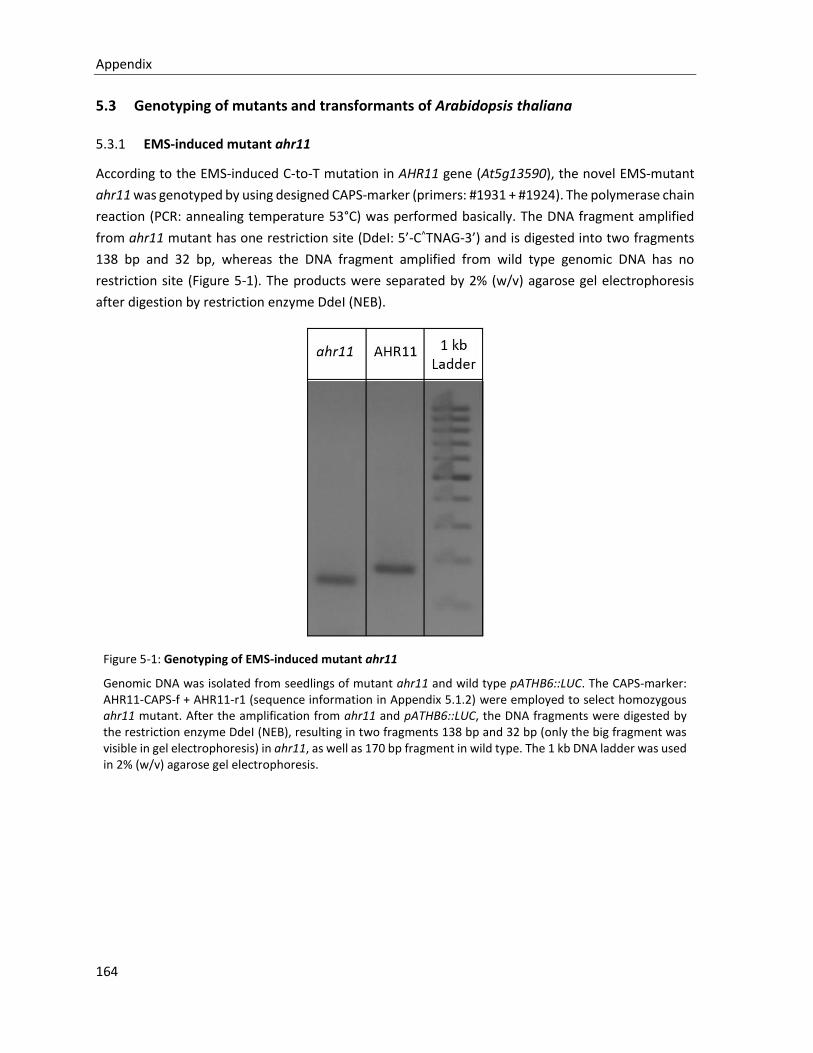
Appendix
164
5.3 Genotyping of mutants and transformants of Arabidopsis thaliana
5.3.1 EMS-induced mutant ahr11
According to the EMS-induced C-to-T mutation in AHR11 gene (At5g13590), the novel EMS-mutant
ahr11 was genotyped by using designed CAPS-marker (primers: #1931 + #1924). The polymerase chain
reaction (PCR: annealing temperature 53°C) was performed basically. The DNA fragment amplified
from ahr11 mutant has one restriction site (DdeI: 5’-C^TNAG-3’) and is digested into two fragments
138 bp and 32 bp, whereas the DNA fragment amplified from wild type genomic DNA has no
restriction site (Figure 5-1). The products were separated by 2% (w/v) agarose gel electrophoresis
after digestion by restriction enzyme DdeI (NEB).
Figure 5-1: Genotyping of EMS-induced mutant ahr11
Genomic DNA was isolated from seedlings of mutant ahr11 and wild type pATHB6::LUC. The CAPS-marker: AHR11-CAPS-f + AHR11-r1 (sequence information in Appendix 5.1.2) were employed to select homozygous ahr11 mutant. After the amplification from ahr11 and pATHB6::LUC, the DNA fragments were digested by the restriction enzyme DdeI (NEB), resulting in two fragments 138 bp and 32 bp (only the big fragment was visible in gel electrophoresis) in ahr11, as well as 170 bp fragment in wild type. The 1 kb DNA ladder was used in 2% (w/v) agarose gel electrophoresis.
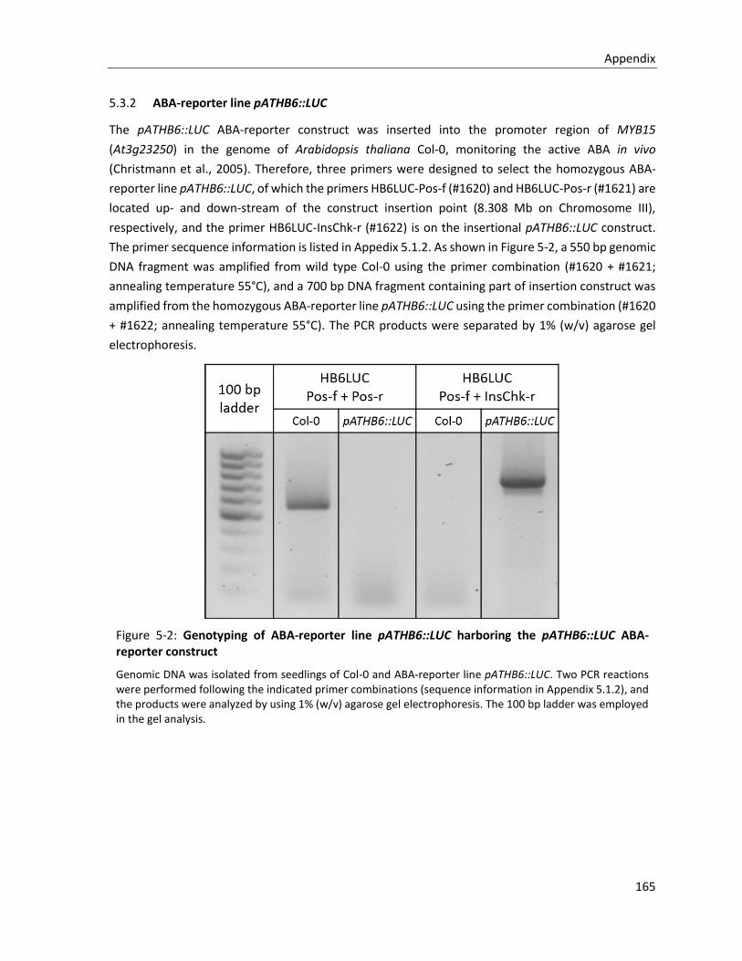
Appendix
165
5.3.2 ABA-reporter line pATHB6::LUC
The pATHB6::LUC ABA-reporter construct was inserted into the promoter region of MYB15
(At3g23250) in the genome of Arabidopsis thaliana Col-0, monitoring the active ABA in vivo
(Christmann et al., 2005). Therefore, three primers were designed to select the homozygous ABA-
reporter line pATHB6::LUC, of which the primers HB6LUC-Pos-f (#1620) and HB6LUC-Pos-r (#1621) are
located up- and down-stream of the construct insertion point (8.308 Mb on Chromosome III),
respectively, and the primer HB6LUC-InsChk-r (#1622) is on the insertional pATHB6::LUC construct.
The primer secquence information is listed in Appedix 5.1.2. As shown in Figure 5-2, a 550 bp genomic
DNA fragment was amplified from wild type Col-0 using the primer combination (#1620 + #1621;
annealing temperature 55°C), and a 700 bp DNA fragment containing part of insertion construct was
amplified from the homozygous ABA-reporter line pATHB6::LUC using the primer combination (#1620
+ #1622; annealing temperature 55°C). The PCR products were separated by 1% (w/v) agarose gel
electrophoresis.
Figure 5-2: Genotyping of ABA-reporter line pATHB6::LUC harboring the pATHB6::LUC ABA-reporter construct
Genomic DNA was isolated from seedlings of Col-0 and ABA-reporter line pATHB6::LUC. Two PCR reactions were performed following the indicated primer combinations (sequence information in Appendix 5.1.2), and the products were analyzed by using 1% (w/v) agarose gel electrophoresis. The 100 bp ladder was employed in the gel analysis.
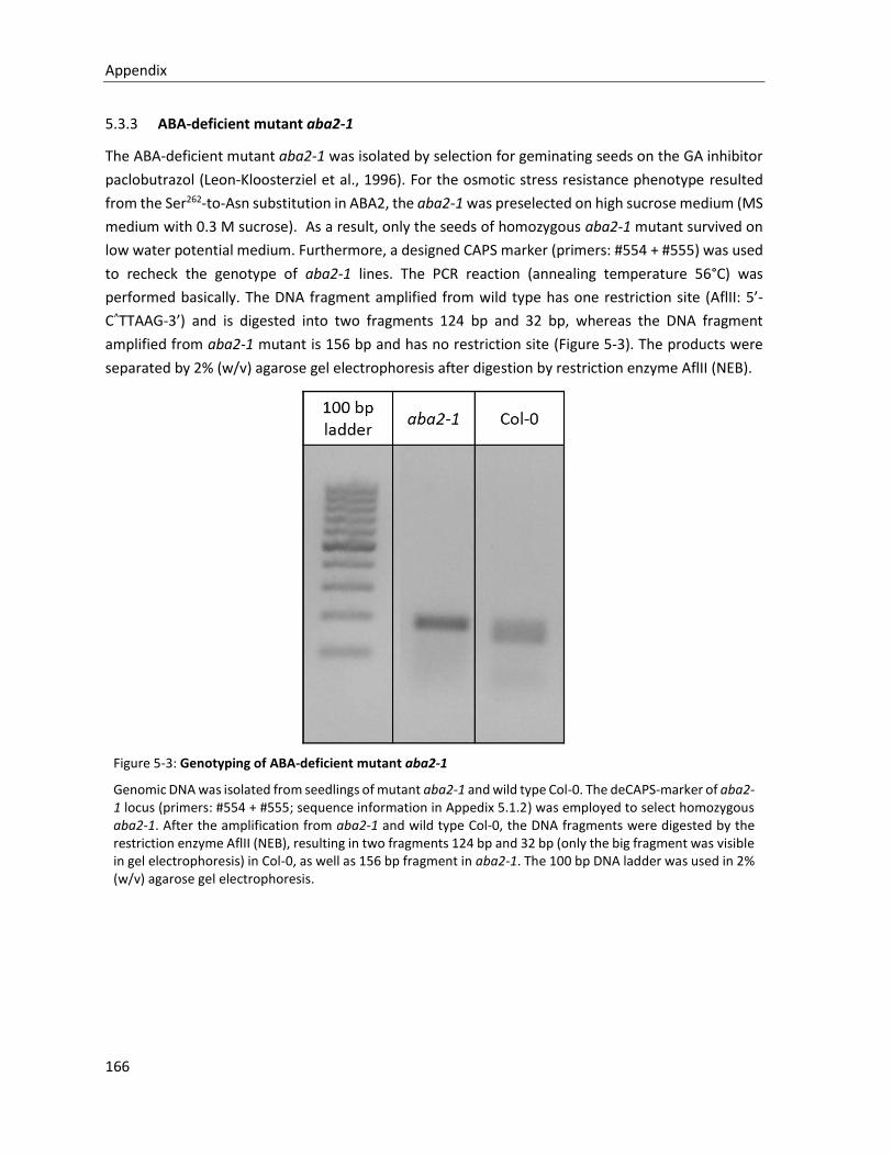
Appendix
166
5.3.3 ABA-deficient mutant aba2-1
The ABA-deficient mutant aba2-1 was isolated by selection for geminating seeds on the GA inhibitor
paclobutrazol (Leon-Kloosterziel et al., 1996). For the osmotic stress resistance phenotype resulted
from the Ser262-to-Asn substitution in ABA2, the aba2-1 was preselected on high sucrose medium (MS
medium with 0.3 M sucrose). As a result, only the seeds of homozygous aba2-1 mutant survived on
low water potential medium. Furthermore, a designed CAPS marker (primers: #554 + #555) was used
to recheck the genotype of aba2-1 lines. The PCR reaction (annealing temperature 56°C) was
performed basically. The DNA fragment amplified from wild type has one restriction site (AflII: 5’-
C^TTAAG-3’) and is digested into two fragments 124 bp and 32 bp, whereas the DNA fragment
amplified from aba2-1 mutant is 156 bp and has no restriction site (Figure 5-3). The products were
separated by 2% (w/v) agarose gel electrophoresis after digestion by restriction enzyme AflII (NEB).
Figure 5-3: Genotyping of ABA-deficient mutant aba2-1
Genomic DNA was isolated from seedlings of mutant aba2-1 and wild type Col-0. The deCAPS-marker of aba2-1 locus (primers: #554 + #555; sequence information in Appedix 5.1.2) was employed to select homozygous aba2-1. After the amplification from aba2-1 and wild type Col-0, the DNA fragments were digested by the restriction enzyme AflII (NEB), resulting in two fragments 124 bp and 32 bp (only the big fragment was visible in gel electrophoresis) in Col-0, as well as 156 bp fragment in aba2-1. The 100 bp DNA ladder was used in 2% (w/v) agarose gel electrophoresis.
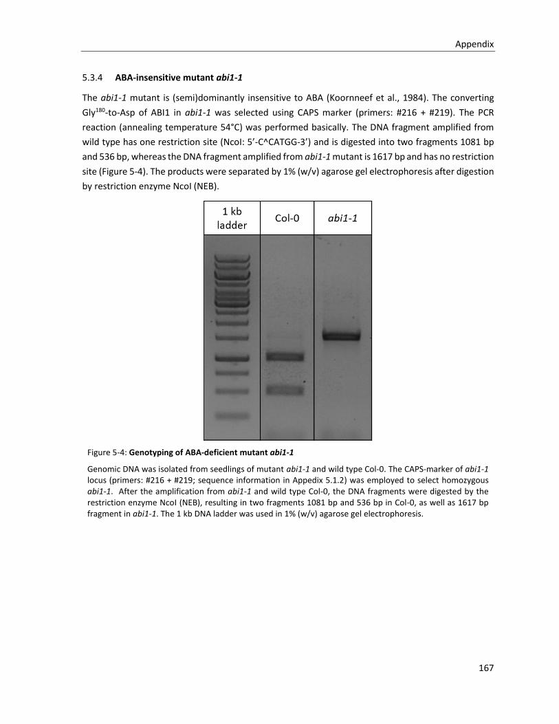
Appendix
167
5.3.4 ABA-insensitive mutant abi1-1
The abi1-1 mutant is (semi)dominantly insensitive to ABA (Koornneef et al., 1984). The converting
Gly180-to-Asp of ABI1 in abi1-1 was selected using CAPS marker (primers: #216 + #219). The PCR
reaction (annealing temperature 54°C) was performed basically. The DNA fragment amplified from
wild type has one restriction site (NcoI: 5’-C^CATGG-3’) and is digested into two fragments 1081 bp
and 536 bp, whereas the DNA fragment amplified from abi1-1 mutant is 1617 bp and has no restriction
site (Figure 5-4). The products were separated by 1% (w/v) agarose gel electrophoresis after digestion
by restriction enzyme NcoI (NEB).
Figure 5-4: Genotyping of ABA-deficient mutant abi1-1
Genomic DNA was isolated from seedlings of mutant abi1-1 and wild type Col-0. The CAPS-marker of abi1-1 locus (primers: #216 + #219; sequence information in Appedix 5.1.2) was employed to select homozygous abi1-1. After the amplification from abi1-1 and wild type Col-0, the DNA fragments were digested by the restriction enzyme NcoI (NEB), resulting in two fragments 1081 bp and 536 bp in Col-0, as well as 1617 bp fragment in abi1-1. The 1 kb DNA ladder was used in 1% (w/v) agarose gel electrophoresis.
Appendix
168
5.4 AHR11-intereacting candidates in yeast two-hybrid screen
AT1G01260 AT2G07696 AT2G37090 AT3G49310 AT4G25660 AT5G22210
AT1G06460 AT2G17570 AT2G37240 AT3G53020 AT4G27650 AT5G24360
AT1G08710 AT2G18670 AT2G45260 AT3G55700 AT4G27880 AT5G38410
AT1G13540 AT2G21080 AT2G45680 AT3G58680 AT4G29780 AT5G38420
AT1G15750 AT2G21820 AT2G47920 AT3G61790 AT4G31240 AT5G38430
AT1G22920 AT2G23290 AT3G01090 AT3G61930 AT4G31340 AT5G42180
AT1G23220 AT2G23420 AT3G06035 AT4G01920 AT4G33320 AT5G42480
AT1G29920 AT2G26100 AT3G06640 AT4G03415 AT4G34080 AT5G47890
AT1G58470 AT2G28660 AT3G09880 AT4G09060 AT4G36250 AT5G57900
AT1G71230 AT2G30930 AT3G13672 AT4G16146 AT5G01720 AT5G58960
AT1G72150 AT2G31060 AT3G16830 AT4G20170 AT5G09770
AT1G76850 AT2G32600 AT3G25800 AT4G20870 AT5G18460
AT2G05230 AT2G33860 AT3G26030 AT4G22710 AT5G19690
References
169
6. References
Abe, H., Urao, T., Ito, T., Seki, M., Shinozaki, K. & Yamaguchi-Shinozaki, K. (2003). Arabidopsis AtMYC2 (bHLH) and AtMYB2 (MYB) function as transcriptional activators in abscisic acid signaling. Plant Cell, Vol. 15, No. 1, pp. 63-78.
Abe, H., Yamaguchi-Shinozaki, K., Urao, T., Iwasaki, T., Hosokawa, D. & Shinozaki, K. (1997). Role of arabidopsis MYC and MYB homologs in drought- and abscisic acid-regulated gene expression. Plant Cell, Vol. 9, No. 10, pp. 1859-68.
Abebe, T., Melmaiee, K., Berg, V. & Wise, R.P. (2009). Drought response in the spikes of barley: gene expression in the lemma, palea, awn, and seed. Funct Integr Genomics, Vol. 10, No. 2, pp. 191-205.
Abel, S. & Theologis, A. (1994). Transient transformation of Arabidopsis leaf protoplasts: a versatile experimental system to study gene expression. Plant J, Vol. 5, No. 3, pp. 421-7.
Ache, P., Bauer, H., Kollist, H., Al-Rasheid, K.A., Lautner, S., Hartung, W. & Hedrich, R. (2010). Stomatal action directly feeds back on leaf turgor: new insights into the regulation of the plant water status from non-invasive pressure probe measurements. Plant J, Vol. 62, No. 6, pp. 1072-82.
Agarwal, M., Hao, Y., Kapoor, A., Dong, C.H., Fujii, H., Zheng, X. & Zhu, J.K. (2006). A R2R3 type MYB transcription factor is involved in the cold regulation of CBF genes and in acquired freezing tolerance. J Biol Chem, Vol. 281, No. 49, pp. 37636-45.
Ahuja, I., de Vos, R.C., Bones, A.M. & Hall, R.D. (2010). Plant molecular stress responses face climate change. Trends Plant Sci, Vol. 15, No. 12, pp. 664-74.
Akhtar, M., Jaiswal, A., Taj, G., Jaiswal, J.P., Qureshi, M.I. & Singh, N.K. (2012). DREB1/CBF transcription factors: their structure, function and role in abiotic stress tolerance in plants. J Genet, Vol. 91, No. 3, pp. 385-95.
Alcazar, R., Bitrian, M., Bartels, D., Koncz, C., Altabella, T. & Tiburcio, A.F. (2011). Polyamine metabolic canalization in response to drought stress in Arabidopsis and the resurrection plant Craterostigma plantagineum. Plant Signal Behav, Vol. 6, No. 2, pp. 243-50.
Ali-Rachedi, S., Bouinot, D., Wagner, M.H., Bonnet, M., Sotta, B., Grappin, P. & Jullien, M. (2004). Changes in endogenous abscisic acid levels during dormancy release and maintenance of mature seeds: studies with the Cape Verde Islands ecotype, the dormant model of Arabidopsis thaliana. Planta, Vol. 219, No. 3, pp. 479-88.
Allan, A.C., Fricker, M.D., Ward, J.L., Beale, M.H. & Trewavas, A.J. (1994). Two Transduction Pathways Mediate Rapid Effects of Abscisic Acid in Commelina Guard Cells. Plant Cell, Vol. 6, No. 9, pp. 1319-1328.
Alonso-Blanco, C., Bentsink, L., Hanhart, C.J., Blankestijn-de Vries, H. & Koornneef, M. (2003). Analysis of natural allelic variation at seed dormancy loci of Arabidopsis thaliana. Genetics, Vol. 164, No. 2, pp. 711-29.
Alonso-Blanco, C., Mendez-Vigo, B. & Koornneef, M. (2005). From phenotypic to molecular polymorphisms involved in naturally occurring variation of plant development. Int J Dev Biol, Vol. 49, No. 5-6, pp. 717-32.
Aprile, A., Mastrangelo, A.M., De Leonardis, A.M., Galiba, G., Roncaglia, E., Ferrari, F., De Bellis, L., Turchi, L., Giuliano, G. & Cattivelli, L. (2009). Transcriptional profiling in response to terminal drought stress reveals differential responses along the wheat genome. BMC Genomics, Vol. 10, pp. 279.
Aravind, L. & Ponting, C.P. (1999). The cytoplasmic helical linker domain of receptor histidine kinase and methyl-accepting proteins is common to many prokaryotic signalling proteins. FEMS Microbiol Lett, Vol. 176, No. 1, pp. 111-6.
References
170
Arnadottir, J. & Chalfie, M. (2010). Eukaryotic mechanosensitive channels. Annu Rev Biophys, Vol. 39, pp. 111-37.
Asano, T., Hayashi, N., Kikuchi, S. & Ohsugi, R. (2012). CDPK-mediated abiotic stress signaling. Plant Signal Behav, Vol. 7, No. 7, pp. 817-21.
Audran, C., Borel, C., Frey, A., Sotta, B., Meyer, C., Simonneau, T. & Marion-Poll, A. (1998). Expression studies of the zeaxanthin epoxidase gene in nicotiana plumbaginifolia. Plant Physiol, Vol. 118, No. 3, pp. 1021-8.
Austin, R.S., Vidaurre, D., Stamatiou, G., Breit, R., Provart, N.J., Bonetta, D., Zhang, J., Fung, P., Gong, Y., Wang, P.W., McCourt, P. & Guttman, D.S. (2011). Next-generation mapping of Arabidopsis genes. Plant J, Vol. 67, No. 4, pp. 715-25.
Bai, L., Zhang, G., Zhou, Y., Zhang, Z., Wang, W., Du, Y., Wu, Z. & Song, C.P. (2009a). Plasma membrane-associated proline-rich extensin-like receptor kinase 4, a novel regulator of Ca signalling, is required for abscisic acid responses in Arabidopsis thaliana. Plant J, Vol. 60, No. 2, pp. 314-27.
Bai, L., Zhou, Y. & Song, C.P. (2009b). Arabidopsis proline-rich extensin-like receptor kinase 4 modulates the early event toward abscisic acid response in root tip growth. Plant Signal Behav, Vol. 4, No. 11, pp. 1075-7.
Balla, J., Kalousek, P., Reinohl, V., Friml, J. & Prochazka, S. (2012). Competitive canalization of PIN-dependent auxin flow from axillary buds controls pea bud outgrowth. Plant J, Vol. 65, No. 4, pp. 571-7.
Bang, W., Kim, S., Ueda, A., Vikram, M., Yun, D., Bressan, R.A., Hasegawa, P.M., Bahk, J. & Koiwa, H. (2006). Arabidopsis carboxyl-terminal domain phosphatase-like isoforms share common catalytic and interaction domains but have distinct in planta functions. Plant Physiol, Vol. 142, No. 2, pp. 586-94.
Barrero, J.M., Rodriguez, P.L., Quesada, V., Piqueras, P., Ponce, M.R. & Micol, J.L. (2006). Both abscisic acid (ABA)-dependent and ABA-independent pathways govern the induction of NCED3, AAO3 and ABA1 in response to salt stress. Plant Cell Environ, Vol. 29, No. 10, pp. 2000-8.
Bartels, D. (2005). Desiccation Tolerance Studied in the Resurrection Plant Craterostigma plantagineum. Integr Comp Biol, Vol. 45, No. 5, pp. 696-701.
Bartenschlager, R. & Schaller, H. (1988). The amino-terminal domain of the hepadnaviral P-gene encodes the terminal protein (genome-linked protein) believed to prime reverse transcription. EMBO J, Vol. 7, No. 13, pp. 4185-92.
Bassel, G.W., Fung, P., Chow, T.F., Foong, J.A., Provart, N.J. & Cutler, S.R. (2008). Elucidating the germination transcriptional program using small molecules. Plant Physiol, Vol. 147, No. 1, pp. 143-55.
Batistic, O. & Kudla, J. (2004). Integration and channeling of calcium signaling through the CBL calcium sensor/CIPK protein kinase network. Planta, Vol. 219, No. 6, pp. 915-24.
Bauer, H., Ache, P., Lautner, S., Fromm, J., Hartung, W., Al-Rasheid, K.A., Sonnewald, S., Sonnewald, U., Kneitz, S., Lachmann, N., Mendel, R.R., Bittner, F., Hetherington, A.M. & Hedrich, R. (2013). The stomatal response to reduced relative humidity requires guard cell-autonomous ABA synthesis. Curr Biol, Vol. 23, No. 1, pp. 53-7.
Becerra, C., Jahrmann, T., Puigdomenech, P. & Vicient, C.M. (2004). Ankyrin repeat-containing proteins in Arabidopsis: characterization of a novel and abundant group of genes coding ankyrin-transmembrane proteins. Gene, Vol. 340, No. 1, pp. 111-21.
Beck, J. & Nassal, M. (2007). Hepatitis B virus replication. World J Gastroenterol, Vol. 13, No. 1, pp. 48-64.
Belin, C., de Franco, P.O., Bourbousse, C., Chaignepain, S., Schmitter, J.M., Vavasseur, A., Giraudat, J., Barbier-Brygoo, H. & Thomine, S. (2006). Identification of features regulating OST1 kinase activity and OST1 function in guard cells. Plant Physiol, Vol. 141, No. 4, pp. 1316-27.
Bentsink, L. & Koornneef, M. (2008). Seed dormancy and germination. Arabidopsis Book, Vol. 6, pp. e0119.
References
171
Bhaskara, G.B., Nguyen, T.T. & Verslues, P.E. (2012). Unique drought resistance functions of the highly ABA-induced clade A protein phosphatase 2Cs. Plant Physiol, Vol. 160, No. 1, pp. 379-95.
Bhat, R.A., Lahaye, T. & Panstruga, R. (2006). The visible touch: in planta visualization of protein-protein interactions by fluorophore-based methods. Plant Methods, Vol. 2, pp. 12.
Boguslawski, G. (1992). PBS2, a yeast gene encoding a putative protein kinase, interacts with the RAS2 pathway and affects osmotic sensitivity of Saccharomyces cerevisiae. J Gen Microbiol, Vol. 138, No. 11, pp. 2425-32.
Boisson-Dernier, A., Kessler, S.A. & Grossniklaus, U. (2011). The walls have ears: the role of plant CrRLK1Ls in sensing and transducing extracellular signals. J Exp Bot, Vol. 62, No. 5, pp. 1581-91.
Booker, J., Chatfield, S. & Leyser, O. (2003). Auxin acts in xylem-associated or medullary cells to mediate apical dominance. Plant Cell, Vol. 15, No. 2, pp. 495-507.
Borsani, O., Zhu, J., Verslues, P.E., Sunkar, R. & Zhu, J.K. (2005). Endogenous siRNAs derived from a pair of natural cis-antisense transcripts regulate salt tolerance in Arabidopsis. Cell, Vol. 123, No. 7, pp. 1279-91.
Bossi, F., Cordoba, E., Dupre, P., Mendoza, M.S., Roman, C.S. & Leon, P. (2009). The Arabidopsis ABA-INSENSITIVE (ABI) 4 factor acts as a central transcription activator of the expression of its own gene, and for the induction of ABI5 and SBE2.2 genes during sugar signaling. Plant J, Vol. 59, No. 3, pp. 359-74.
Boudsocq, M., Barbier-Brygoo, H. & Lauriere, C. (2004). Identification of nine sucrose nonfermenting 1-related protein kinases 2 activated by hyperosmotic and saline stresses in Arabidopsis thaliana. J Biol Chem, Vol. 279, No. 40, pp. 41758-66.
Boursiac, Y., Leran, S., Corratge-Faillie, C., Gojon, A., Krouk, G. & Lacombe, B. (2013). ABA transport and transporters. Trends Plant Sci, Vol. 18, No. 6, pp. 325-33.
Boyer, J.S. & Kramer, P.J. (1995). Measuring the water status of plants and soils, San Diego, Calif.: Academic Press, p. x, 178 p.
Brandt, B., Brodsky, D.E., Xue, S., Negi, J., Iba, K., Kangasjarvi, J., Ghassemian, M., Stephan, A.B., Hu, H. & Schroeder, J.I. (2012). Reconstitution of abscisic acid activation of SLAC1 anion channel by CPK6 and OST1 kinases and branched ABI1 PP2C phosphatase action. Proc Natl Acad Sci U S A, Vol. 109, No. 26, pp. 10593-8.
Braun, P. (2012). Interactome mapping for analysis of complex phenotypes: insights from benchmarking binary interaction assays. Proteomics, Vol. 12, No. 10, pp. 1499-518.
Braun, P., Aubourg, S., Van Leene, J., De Jaeger, G. & Lurin, C. (2013). Plant protein interactomes. Annu Rev Plant Biol, Vol. 64, pp. 161-87.
Bray, E.A. (2002). Classification of genes differentially expressed during water-deficit stress in Arabidopsis thaliana: an analysis using microarray and differential expression data. Ann Bot, Vol. 89 Spec No, pp. 803-11.
Bray, E.A. & Zeevaart, J.A. (1985). The Compartmentation of Abscisic Acid and beta-d-Glucopyranosyl Abscisate in Mesophyll Cells. Plant Physiol, Vol. 79, No. 3, pp. 719-22.
Brewster, J.L., de Valoir, T., Dwyer, N.D., Winter, E. & Gustin, M.C. (1993). An osmosensing signal transduction pathway in yeast. Science, Vol. 259, No. 5102, pp. 1760-3.
Buchanan-Wollaston, V., Page, T., Harrison, E., Breeze, E., Lim, P.O., Nam, H.G., Lin, J.F., Wu, S.H., Swidzinski, J., Ishizaki, K. & Leaver, C.J. (2005). Comparative transcriptome analysis reveals significant differences in gene expression and signalling pathways between developmental and dark/starvation-induced senescence in Arabidopsis. Plant J, Vol. 42, No. 4, pp. 567-85.
References
172
Buchwald, M., Pietschmann, K., Brand, P., Gunther, A., Mahajan, N.P., Heinzel, T. & Kramer, O.H. (2012). SIAH ubiquitin ligases target the nonreceptor tyrosine kinase ACK1 for ubiquitinylation and proteasomal degradation. Oncogene, Vol. 32, No. 41, pp. 4913-20.
Bugos, R.C., Hieber, A.D. & Yamamoto, H.Y. (1998). Xanthophyll cycle enzymes are members of the lipocalin family, the first identified from plants. J Biol Chem, Vol. 273, No. 25, pp. 15321-4.
Burla, B., Pfrunder, S., Nagy, R., Francisco, R.M., Lee, Y. & Martinoia, E. (2013). Vacuolar transport of abscisic acid glucosyl ester is mediated by ATP-binding cassette and proton-antiport mechanisms in Arabidopsis. Plant Physiol, Vol. 163, No. 3, pp. 1446-58.
Burza, A.M., Pekala, I., Sikora, J., Siedlecki, P., Malagocki, P., Bucholc, M., Koper, L., Zielenkiewicz, P., Dadlez, M. & Dobrowolska, G. (2006). Nicotiana tabacum osmotic stress-activated kinase is regulated by phosphorylation on Ser-154 and Ser-158 in the kinase activation loop. J Biol Chem, Vol. 281, No. 45, pp. 34299-311.
Cao, F.Y., Yoshioka, K. & Desveaux, D. (2011). The roles of ABA in plant-pathogen interactions. J Plant Res, Vol. 124, No. 4, pp. 489-99.
Carthew, R.W. & Rubin, G.M. (1990). seven in absentia, a gene required for specification of R7 cell fate in the Drosophila eye. Cell, Vol. 63, No. 3, pp. 561-77.
Causier, B., Ashworth, M., Guo, W. & Davies, B. (2011). The TOPLESS interactome: a framework for gene repression in Arabidopsis. Plant Physiol, Vol. 158, No. 1, pp. 423-38.
Causier, B., Lloyd, J., Stevens, L. & Davies, B. (2012). TOPLESS co-repressor interactions and their evolutionary conservation in plants. Plant Signal Behav, Vol. 7, No. 3, pp. 325-8.
Chalfie, M. & Au, M. (1989). Genetic control of differentiation of the Caenorhabditis elegans touch receptor neurons. Science, Vol. 243, No. 4894 Pt 1, pp. 1027-33.
Chen, C.C., Liang, C.S., Kao, A.L. & Yang, C.C. (2009). HHP1 is involved in osmotic stress sensitivity in Arabidopsis. J Exp Bot, Vol. 60, No. 6, pp. 1589-604.
Chen, C.C., Liang, C.S., Kao, A.L. & Yang, C.C. (2010a). HHP1, a novel signalling component in the cross-talk between the cold and osmotic signalling pathways in Arabidopsis. J Exp Bot, Vol. 61, No. 12, pp. 3305-20.
Chen, H., Lai, Z., Shi, J., Xiao, Y., Chen, Z. & Xu, X. (2010b). Roles of arabidopsis WRKY18, WRKY40 and WRKY60 transcription factors in plant responses to abscisic acid and abiotic stress. BMC Plant Biol, Vol. 10, pp. 281.
Cheng, W.H., Endo, A., Zhou, L., Penney, J., Chen, H.C., Arroyo, A., Leon, P., Nambara, E., Asami, T., Seo, M., Koshiba, T. & Sheen, J. (2002). A unique short-chain dehydrogenase/reductase in Arabidopsis glucose signaling and abscisic acid biosynthesis and functions. Plant Cell, Vol. 14, No. 11, pp. 2723-43.
Cheng, Y., Kato, N., Wang, W., Li, J. & Chen, X. (2003). Two RNA binding proteins, HEN4 and HUA1, act in the processing of AGAMOUS pre-mRNA in Arabidopsis thaliana. Dev Cell, Vol. 4, No. 1, pp. 53-66.
Cheong, Y.H., Kim, K.N., Pandey, G.K., Gupta, R., Grant, J.J. & Luan, S. (2003). CBL1, a calcium sensor that differentially regulates salt, drought, and cold responses in Arabidopsis. Plant Cell, Vol. 15, No. 8, pp. 1833-45.
Cheong, Y.H., Pandey, G.K., Grant, J.J., Batistic, O., Li, L., Kim, B.G., Lee, S.C., Kudla, J. & Luan, S. (2007). Two calcineurin B-like calcium sensors, interacting with protein kinase CIPK23, regulate leaf transpiration and root potassium uptake in Arabidopsis. Plant J, Vol. 52, No. 2, pp. 223-39.
Chi, X.F., Lou, X.Y. & Shu, Q.Y. (2008). Progressive fine mapping in experimental populations: an improved strategy toward positional cloning. J Theor Biol, Vol. 253, No. 4, pp. 817-23.
References
173
Chinnusamy, V., Schumaker, K. & Zhu, J.K. (2004). Molecular genetic perspectives on cross-talk and specificity in abiotic stress signalling in plants. J Exp Bot, Vol. 55, No. 395, pp. 225-36.
Choi, H., Hong, J., Ha, J., Kang, J. & Kim, S.Y. (2000). ABFs, a family of ABA-responsive element binding factors. J Biol Chem, Vol. 275, No. 3, pp. 1723-30.
Choi, H.I., Park, H.J., Park, J.H., Kim, S., Im, M.Y., Seo, H.H., Kim, Y.W., Hwang, I. & Kim, S.Y. (2005). Arabidopsis calcium-dependent protein kinase AtCPK32 interacts with ABF4, a transcriptional regulator of abscisic acid-responsive gene expression, and modulates its activity. Plant Physiol, Vol. 139, No. 4, pp. 1750-61.
Chooback, L. & West, A.H. (2003). Co-crystallization of the yeast phosphorelay protein YPD1 with the SLN1 response-regulator domain and preliminary X-ray diffraction analysis. Acta Crystallogr D Biol Crystallogr, Vol. 59, No. Pt 5, pp. 927-9.
Choudhury, A. & Lahiri, A. (2010). Comparative analysis of abscisic acid-regulated transcriptomes in Arabidopsis. Plant Biol (Stuttg), Vol. 13, No. 1, pp. 28-35.
Choudhury, S., Panda, P., Sahoo, L. & Panda, S.K. (2013). Reactive oxygen species signaling in plants under abiotic stress. Plant Signal Behav, Vol. 8, No. 4, pp. e23681.
Christmann, A. & Grill, E. (2009). Are GTGs ABA's biggest fans? Cell, Vol. 136, No. 1, pp. 21-3.
Christmann, A., Grill, E. & Huang, J. (2013). Hydraulic signals in long-distance signaling. Curr Opin Plant Biol, Vol. 16, No. 3, pp. 293-300.
Christmann, A., Hoffmann, T., Teplova, I., Grill, E. & Muller, A. (2005). Generation of active pools of abscisic acid revealed by in vivo imaging of water-stressed Arabidopsis. Plant Physiol, Vol. 137, No. 1, pp. 209-19.
Christmann, A., Moes, D., Himmelbach, A., Yang, Y., Tang, Y. & Grill, E. (2006). Integration of abscisic acid signalling into plant responses. Plant Biol (Stuttg), Vol. 8, No. 3, pp. 314-25.
Christmann, A., Weiler, E.W., Steudle, E. & Grill, E. (2007). A hydraulic signal in root-to-shoot signalling of water shortage. Plant J, Vol. 52, No. 1, pp. 167-74.
Cline, M.G. & Oh, C. (2006). A reappraisal of the role of abscisic acid and its interaction with auxin in apical dominance. Ann Bot, Vol. 98, No. 4, pp. 891-7.
Cohen, J.I., Miller, R.H., Rosenblum, B., Denniston, K., Gerin, J.L. & Purcell, R.H. (1988). Sequence comparison of woodchuck hepatitis virus replicative forms shows conservation of the genome. Virology, Vol. 162, No. 1, pp. 12-20.
Colcombet, J. & Hirt, H. (2008). Arabidopsis MAPKs: a complex signalling network involved in multiple biological processes. Biochem J, Vol. 413, No. 2, pp. 217-26.
Cosgrove, D.J. & Hedrich, R. (1991). Stretch-activated chloride, potassium, and calcium channels coexisting in plasma membranes of guard cells of Vicia faba L. Planta, Vol. 186, No. 1, pp. 143-53.
Cutler, A.J. & Krochko, J.E. (1999). Formation and breakdown of ABA. Trends Plant Sci, Vol. 4, No. 12, pp. 472-478.
Cutler, S.R., Rodriguez, P.L., Finkelstein, R.R. & Abrams, S.R. (2010). Abscisic acid: emergence of a core signaling network. Annu Rev Plant Biol, Vol. 61, pp. 651-79.
Czechowski, T., Stitt, M., Altmann, T., Udvardi, M.K. & Scheible, W.R. (2005). Genome-wide identification and testing of superior reference genes for transcript normalization in Arabidopsis. Plant Physiol, Vol. 139, No. 1, pp. 5-17.
D'Angelo, C., Weinl, S., Batistic, O., Pandey, G.K., Cheong, Y.H., Schultke, S., Albrecht, V., Ehlert, B., Schulz, B., Harter, K., Luan, S., Bock, R. & Kudla, J. (2006). Alternative complex formation of the Ca-regulated protein kinase CIPK1 controls abscisic acid-dependent and independent stress responses in Arabidopsis. Plant J, Vol. 48, No. 6, pp. 857-72.
References
174
Das, R. & Pandey, G.K. (2010). Expressional analysis and role of calcium regulated kinases in abiotic stress signaling. Curr Genomics, Vol. 11, No. 1, pp. 2-13.
Davies, C.R., Seth, A.K. & Wareing, P.F. (1966). Auxin and kinetin interaction in apical dominance. Science, Vol. 151, No. 3709, pp. 468-9.
Day, C.L., Puthalakath, H., Skea, G., Strasser, A., Barsukov, I., Lian, L.Y., Huang, D.C. & Hinds, M.G. (2004). Localization of dynein light chains 1 and 2 and their pro-apoptotic ligands. Biochem J, Vol. 377, No. Pt 3, pp. 597-605.
Deng, H., Liu, H., Li, X., Xiao, J. & Wang, S. (2011). A CCCH-type zinc finger nucleic acid-binding protein quantitatively confers resistance against rice bacterial blight disease. Plant Physiol, Vol. 158, No. 2, pp. 876-89.
Desikan, R., Neill, S.J. & Hancock, J.T. (2000). Hydrogen peroxide-induced gene expression in Arabidopsis thaliana. Free Radic Biol Med, Vol. 28, No. 5, pp. 773-8.
Desikan, R., S, A.H.-M., Hancock, J.T. & Neill, S.J. (2001). Regulation of the Arabidopsis transcriptome by oxidative stress. Plant Physiol, Vol. 127, No. 1, pp. 159-72.
Dharmasiri, N., Dharmasiri, S. & Estelle, M. (2005). The F-box protein TIR1 is an auxin receptor. Nature, Vol. 435, No. 7041, pp. 441-5.
Dick, T., Ray, K., Salz, H.K. & Chia, W. (1996). Cytoplasmic dynein (ddlc1) mutations cause morphogenetic defects and apoptotic cell death in Drosophila melanogaster. Mol Cell Biol, Vol. 16, No. 5, pp. 1966-77.
Dietz, K.J., Sauter, A., Wichert, K., Messdaghi, D. & Hartung, W. (2000). Extracellular beta-glucosidase activity in barley involved in the hydrolysis of ABA glucose conjugate in leaves. J Exp Bot, Vol. 51, No. 346, pp. 937-44.
Ding, J.P. & Pickard, B.G. (1993). Modulation of mechanosensitive calcium-selective cation channels by temperature. Plant J, Vol. 3, No. 5, pp. 713-20.
Ding, Z., Li, S., An, X., Liu, X., Qin, H. & Wang, D. (2009). Transgenic expression of MYB15 confers enhanced sensitivity to abscisic acid and improved drought tolerance in Arabidopsis thaliana. J Genet Genomics, Vol. 36, No. 1, pp. 17-29.
Domagalska, M.A., Sarnowska, E., Nagy, F. & Davis, S.J. (2010). Genetic analyses of interactions among gibberellin, abscisic acid, and brassinosteroids in the control of flowering time in Arabidopsis thaliana. PLoS One, Vol. 5, No. 11, pp. e14012.
Dortay, H., Mehnert, N., Burkle, L., Schmulling, T. & Heyl, A. (2006). Analysis of protein interactions within the cytokinin-signaling pathway of Arabidopsis thaliana. FEBS J, Vol. 273, No. 20, pp. 4631-44.
Drerup, M.M., Schlucking, K., Hashimoto, K., Manishankar, P., Steinhorst, L., Kuchitsu, K. & Kudla, J. (2013). The Calcineurin B-like calcium sensors CBL1 and CBL9 together with their interacting protein kinase CIPK26 regulate the Arabidopsis NADPH oxidase RBOHF. Mol Plant, Vol. 6, No. 2, pp. 559-69.
Dreze, M., Monachello, D., Lurin, C., Cusick, M.E., Hill, D.E., Vidal, M. & Braun, P. (2010). High-quality binary interactome mapping. Methods Enzymol, Vol. 470, pp. 281-315.
Droillard, M., Boudsocq, M., Barbier-Brygoo, H. & Lauriere, C. (2002). Different protein kinase families are activated by osmotic stresses in Arabidopsis thaliana cell suspensions. Involvement of the MAP kinases AtMPK3 and AtMPK6. FEBS Lett, Vol. 527, No. 1-3, pp. 43-50.
Droillard, M.J., Boudsocq, M., Barbier-Brygoo, H. & Lauriere, C. (2004). Involvement of MPK4 in osmotic stress response pathways in cell suspensions and plantlets of Arabidopsis thaliana: activation by hypoosmolarity and negative role in hyperosmolarity tolerance. FEBS Lett, Vol. 574, No. 1-3, pp. 42-8.
References
175
Droillard, M.J., Thibivilliers, S., Cazale, A.C., Barbier-Brygoo, H. & Lauriere, C. (2000). Protein kinases induced by osmotic stresses and elicitor molecules in tobacco cell suspensions: two crossroad MAP kinases and one osmoregulation-specific protein kinase. FEBS Lett, Vol. 474, No. 2-3, pp. 217-22.
Du, S.Y., Zhang, X.F., Lu, Z., Xin, Q., Wu, Z., Jiang, T., Lu, Y., Wang, X.F. & Zhang, D.P. (2012). Roles of the different components of magnesium chelatase in abscisic acid signal transduction. Plant Mol Biol, Vol. 80, No. 4-5, pp. 519-37.
Dubos, C., Stracke, R., Grotewold, E., Weisshaar, B., Martin, C. & Lepiniec, L. (2010). MYB transcription factors in Arabidopsis. Trends Plant Sci, Vol. 15, No. 10, pp. 573-81.
Dun, E.A., Brewer, P.B. & Beveridge, C.A. (2009). Strigolactones: discovery of the elusive shoot branching hormone. Trends Plant Sci, Vol. 14, No. 7, pp. 364-72.
Dupeux, F., Santiago, J., Betz, K., Twycross, J., Park, S.Y., Rodriguez, L., Gonzalez-Guzman, M., Jensen, M.R., Krasnogor, N., Blackledge, M., Holdsworth, M., Cutler, S.R., Rodriguez, P.L. & Marquez, J.A. (2011). A thermodynamic switch modulates abscisic acid receptor sensitivity. EMBO J, Vol. 30, No. 20, pp. 4171-84.
Dupres, V., Alsteens, D., Wilk, S., Hansen, B., Heinisch, J.J. & Dufrene, Y.F. (2009). The yeast Wsc1 cell surface sensor behaves like a nanospring in vivo. Nat Chem Biol, Vol. 5, No. 11, pp. 857-62.
Dure, L., 3rd, Greenway, S.C. & Galau, G.A. (1981). Developmental biochemistry of cottonseed embryogenesis and germination: changing messenger ribonucleic acid populations as shown by in vitro and in vivo protein synthesis. Biochemistry, Vol. 20, No. 14, pp. 4162-8.
Dutta, R. & Robinson, K.R. (2004). Identification and characterization of stretch-activated ion channels in pollen protoplasts. Plant Physiol, Vol. 135, No. 3, pp. 1398-406.
Elhiti, M. & Stasolla, C. (2009). Structure and function of homodomain-leucine zipper (HD-Zip) proteins. Plant Signal Behav, Vol. 4, No. 2, pp. 86-8.
Emes, R.D. & Ponting, C.P. (2001). A new sequence motif linking lissencephaly, Treacher Collins and oral-facial-digital type 1 syndromes, microtubule dynamics and cell migration. Hum Mol Genet, Vol. 10, No. 24, pp. 2813-20.
Endo, A., Sawada, Y., Takahashi, H., Okamoto, M., Ikegami, K., Koiwai, H., Seo, M., Toyomasu, T., Mitsuhashi, W., Shinozaki, K., Nakazono, M., Kamiya, Y., Koshiba, T. & Nambara, E. (2008). Drought induction of Arabidopsis 9-cis-epoxycarotenoid dioxygenase occurs in vascular parenchyma cells. Plant Physiol, Vol. 147, No. 4, pp. 1984-93.
Falke, L.C., Edwards, K.L., Pickard, B.G. & Misler, S. (1988). A stretch-activated anion channel in tobacco protoplasts. FEBS Lett, Vol. 237, No. 1-2, pp. 141-4.
Fan, L., Linker, R., Gepstein, S., Tanimoto, E., Yamamoto, R. & Neumann, P.M. (2006). Progressive inhibition by water deficit of cell wall extensibility and growth along the elongation zone of maize roots is related to increased lignin metabolism and progressive stelar accumulation of wall phenolics. Plant Physiol, Vol. 140, No. 2, pp. 603-12.
Fan, L. & Neumann, P.M. (2004). The spatially variable inhibition by water deficit of maize root growth correlates with altered profiles of proton flux and cell wall pH. Plant Physiol, Vol. 135, No. 4, pp. 2291-300.
Ferguson, B.J. & Beveridge, C.A. (2009). Roles for auxin, cytokinin, and strigolactone in regulating shoot branching. Plant Physiol, Vol. 149, No. 4, pp. 1929-44.
Fernandez-Calvino, L., Faulkner, C., Walshaw, J., Saalbach, G., Bayer, E., Benitez-Alfonso, Y. & Maule, A. (2011). Arabidopsis plasmodesmal proteome. PLoS One, Vol. 6, No. 4, pp. e18880.
Fernandez-Calvo, P., Chini, A., Fernandez-Barbero, G., Chico, J.M., Gimenez-Ibanez, S., Geerinck, J., Eeckhout, D., Schweizer, F., Godoy, M., Franco-Zorrilla, J.M., Pauwels, L., Witters, E., Puga, M.I., Paz-Ares, J., Goossens, A., Reymond, P., De Jaeger, G. & Solano, R. (2011). The Arabidopsis bHLH transcription
References
176
factors MYC3 and MYC4 are targets of JAZ repressors and act additively with MYC2 in the activation of jasmonate responses. Plant Cell, Vol. 23, No. 2, pp. 701-15.
Fields, S. & Song, O. (1989). A novel genetic system to detect protein-protein interactions. Nature, Vol. 340, No. 6230, pp. 245-6.
Finch-Savage, W.E. & Leubner-Metzger, G. (2006). Seed dormancy and the control of germination. New Phytol, Vol. 171, No. 3, pp. 501-23.
Finkelstein, R. (2013). Abscisic Acid synthesis and response. Arabidopsis Book, Vol. 11, pp. e0166.
Finkelstein, R.R., Gampala, S.S. & Rock, C.D. (2002). Abscisic acid signaling in seeds and seedlings. Plant Cell, Vol. 14 Suppl, pp. S15-45.
Finkelstein, R.R. & Lynch, T.J. (2000). The Arabidopsis abscisic acid response gene ABI5 encodes a basic leucine zipper transcription factor. Plant Cell, Vol. 12, No. 4, pp. 599-609.
Finkelstein, R.R., Wang, M.L., Lynch, T.J., Rao, S. & Goodman, H.M. (1998). The Arabidopsis abscisic acid response locus ABI4 encodes an APETALA 2 domain protein. Plant Cell, Vol. 10, No. 6, pp. 1043-54.
Flexas, J., Bota, J., Loreto, F., Cornic, G. & Sharkey, T.D. (2004). Diffusive and metabolic limitations to photosynthesis under drought and salinity in C(3) plants. Plant Biol (Stuttg), Vol. 6, No. 3, pp. 269-79.
Foo, E., Morris, S.E., Parmenter, K., Young, N., Wang, H., Jones, A., Rameau, C., Turnbull, C.G. & Beveridge, C.A. (2007). Feedback regulation of xylem cytokinin content is conserved in pea and Arabidopsis. Plant Physiol, Vol. 143, No. 3, pp. 1418-28.
Foreman, J., Demidchik, V., Bothwell, J.H., Mylona, P., Miedema, H., Torres, M.A., Linstead, P., Costa, S., Brownlee, C., Jones, J.D., Davies, J.M. & Dolan, L. (2003). Reactive oxygen species produced by NADPH oxidase regulate plant cell growth. Nature, Vol. 422, No. 6930, pp. 442-6.
Forst, S., Comeau, D., Norioka, S. & Inouye, M. (1987). Localization and membrane topology of EnvZ, a protein involved in osmoregulation of OmpF and OmpC in Escherichia coli. J Biol Chem, Vol. 262, No. 34, pp. 16433-8.
Fuchs, S., Grill, E., Meskiene, I. & Schweighofer, A. (2012). Type 2C protein phosphatases in plants. FEBS J, Vol. 280, No. 2, pp. 681-93.
Fuchs, S., Tischer, S.V., Wunschel, C., Christmann, A. & Grill, E. (2014). Abscisic acid sensor RCAR7/PYL13, specific regulator of protein phosphatase coreceptors. Proc Natl Acad Sci U S A.
Fujii, H., Chinnusamy, V., Rodrigues, A., Rubio, S., Antoni, R., Park, S.Y., Cutler, S.R., Sheen, J., Rodriguez, P.L. & Zhu, J.K. (2009). In vitro reconstitution of an abscisic acid signalling pathway. Nature, Vol. 462, No. 7273, pp. 660-4.
Fujii, H., Verslues, P.E. & Zhu, J.K. (2007). Identification of two protein kinases required for abscisic acid regulation of seed germination, root growth, and gene expression in Arabidopsis. Plant Cell, Vol. 19, No. 2, pp. 485-94.
Fujii, H., Verslues, P.E. & Zhu, J.K. (2011). Arabidopsis decuple mutant reveals the importance of SnRK2 kinases in osmotic stress responses in vivo. Proc Natl Acad Sci U S A, Vol. 108, No. 4, pp. 1717-22.
Fujii, H. & Zhu, J.K. (2009). Arabidopsis mutant deficient in 3 abscisic acid-activated protein kinases reveals critical roles in growth, reproduction, and stress. Proc Natl Acad Sci U S A, Vol. 106, No. 20, pp. 8380-5.
Fujii, H. & Zhu, J.K. (2012). Osmotic stress signaling via protein kinases. Cell Mol Life Sci, Vol. 69, No. 19, pp. 3165-73.
Fujita, M., Fujita, Y., Noutoshi, Y., Takahashi, F., Narusaka, Y., Yamaguchi-Shinozaki, K. & Shinozaki, K. (2006). Crosstalk between abiotic and biotic stress responses: a current view from the points of convergence in the stress signaling networks. Curr Opin Plant Biol, Vol. 9, No. 4, pp. 436-42.
References
177
Fujita, Y., Fujita, M., Satoh, R., Maruyama, K., Parvez, M.M., Seki, M., Hiratsu, K., Ohme-Takagi, M., Shinozaki, K. & Yamaguchi-Shinozaki, K. (2005). AREB1 is a transcription activator of novel ABRE-dependent ABA signaling that enhances drought stress tolerance in Arabidopsis. Plant Cell, Vol. 17, No. 12, pp. 3470-88.
Fujita, Y., Nakashima, K., Yoshida, T., Katagiri, T., Kidokoro, S., Kanamori, N., Umezawa, T., Fujita, M., Maruyama, K., Ishiyama, K., Kobayashi, M., Nakasone, S., Yamada, K., Ito, T., Shinozaki, K. & Yamaguchi-Shinozaki, K. (2009). Three SnRK2 protein kinases are the main positive regulators of abscisic acid signaling in response to water stress in Arabidopsis. Plant Cell Physiol, Vol. 50, No. 12, pp. 2123-32.
Fujita, Y., Yoshida, T. & Yamaguchi-Shinozaki, K. (2012). Pivotal role of the AREB/ABF-SnRK2 pathway in ABRE-mediated transcription in response to osmotic stress in plants. Physiol Plant, Vol. 147, No. 1, pp. 15-27.
Furihata, T., Maruyama, K., Fujita, Y., Umezawa, T., Yoshida, R., Shinozaki, K. & Yamaguchi-Shinozaki, K. (2006). Abscisic acid-dependent multisite phosphorylation regulates the activity of a transcription activator AREB1. Proc Natl Acad Sci U S A, Vol. 103, No. 6, pp. 1988-93.
Furuichi, T., Iida, H., Sokabe, M. & Tatsumi, H. (2012). Expression of Arabidopsis MCA1 enhanced mechanosensitive channel activity in the Xenopus laevis oocyte plasma membrane. Plant Signal Behav, Vol. 7, No. 8, pp. 1022-6.
Gaff, D.F. (1971). Desiccation-tolerant flowering plants in southern Africa. Science, Vol. 174, No. 4013, pp. 1033-4.
Gao, Y., Zeng, Q., Guo, J., Cheng, J., Ellis, B.E. & Chen, J.G. (2007). Genetic characterization reveals no role for the reported ABA receptor, GCR2, in ABA control of seed germination and early seedling development in Arabidopsis. Plant J, Vol. 52, No. 6, pp. 1001-13.
Garcia, M.E., Lynch, T., Peeters, J., Snowden, C. & Finkelstein, R. (2008). A small plant-specific protein family of ABI five binding proteins (AFPs) regulates stress response in germinating Arabidopsis seeds and seedlings. Plant Mol Biol, Vol. 67, No. 6, pp. 643-58.
Garcia, P., Tajadura, V., Garcia, I. & Sanchez, Y. (2006). Role of Rho GTPases and Rho-GEFs in the regulation of cell shape and integrity in fission yeast. Yeast, Vol. 23, No. 13, pp. 1031-43.
Geiger, D., Maierhofer, T., Al-Rasheid, K.A., Scherzer, S., Mumm, P., Liese, A., Ache, P., Wellmann, C., Marten, I., Grill, E., Romeis, T. & Hedrich, R. (2011). Stomatal closure by fast abscisic acid signaling is mediated by the guard cell anion channel SLAH3 and the receptor RCAR1. Sci Signal, Vol. 4, No. 173, pp. ra32.
Geiger, D., Scherzer, S., Mumm, P., Marten, I., Ache, P., Matschi, S., Liese, A., Wellmann, C., Al-Rasheid, K.A., Grill, E., Romeis, T. & Hedrich, R. (2010). Guard cell anion channel SLAC1 is regulated by CDPK protein kinases with distinct Ca2+ affinities. Proc Natl Acad Sci U S A, Vol. 107, No. 17, pp. 8023-8.
Geiger, D., Scherzer, S., Mumm, P., Stange, A., Marten, I., Bauer, H., Ache, P., Matschi, S., Liese, A., Al-Rasheid, K.A., Romeis, T. & Hedrich, R. (2009). Activity of guard cell anion channel SLAC1 is controlled by drought-stress signaling kinase-phosphatase pair. Proc Natl Acad Sci U S A, Vol. 106, No. 50, pp. 21425-30.
Gietz, R.D. & Woods, R.A. (2002). Transformation of yeast by lithium acetate/single-stranded carrier DNA/polyethylene glycol method. Methods Enzymol, Vol. 350, pp. 87-96.
Gil, P.M., Gurovich, L., Schaffer, B., Alcayaga, J., Rey, S. & Iturriaga, R. (2008). Root to leaf electrical signaling in avocado in response to light and soil water content. J Plant Physiol, Vol. 165, No. 10, pp. 1070-8.
Giraudat, J., Hauge, B.M., Valon, C., Smalle, J., Parcy, F. & Goodman, H.M. (1992). Isolation of the Arabidopsis ABI3 gene by positional cloning. Plant Cell, Vol. 4, No. 10, pp. 1251-61.
Gleeson, T., Wada, Y., Bierkens, M.F. & van Beek, L.P. (2012). Water balance of global aquifers revealed by groundwater footprint. Nature, Vol. 488, No. 7410, pp. 197-200.
References
178
Golldack, D., Li, C., Mohan, H. & Probst, N. (2014). Tolerance to drought and salt stress in plants: Unraveling the signaling networks. Front Plant Sci, Vol. 5, pp. 151.
Gomez-Roldan, V., Fermas, S., Brewer, P.B., Puech-Pages, V., Dun, E.A., Pillot, J.P., Letisse, F., Matusova, R., Danoun, S., Portais, J.C., Bouwmeester, H., Becard, G., Beveridge, C.A., Rameau, C. & Rochange, S.F. (2008). Strigolactone inhibition of shoot branching. Nature, Vol. 455, No. 7210, pp. 189-94.
Gouget, A., Senchou, V., Govers, F., Sanson, A., Barre, A., Rouge, P., Pont-Lezica, R. & Canut, H. (2006). Lectin receptor kinases participate in protein-protein interactions to mediate plasma membrane-cell wall adhesions in Arabidopsis. Plant Physiol, Vol. 140, No. 1, pp. 81-90.
Grams, T.E., Koziolek, C., Lautner, S., Matyssek, R. & Fromm, J. (2007). Distinct roles of electric and hydraulic signals on the reaction of leaf gas exchange upon re-irrigation in Zea mays L. Plant Cell Environ, Vol. 30, No. 1, pp. 79-84.
Grant, J.J., Yun, B.W. & Loake, G.J. (2000). Oxidative burst and cognate redox signalling reported by luciferase imaging: identification of a signal network that functions independently of ethylene, SA and Me-JA but is dependent on MAPKK activity. Plant J, Vol. 24, No. 5, pp. 569-82.
Grethe, S., Heckel, J.O., Rietschel, W. & Hufert, F.T. (2000). Molecular epidemiology of hepatitis B virus variants in nonhuman primates. J Virol, Vol. 74, No. 11, pp. 5377-81.
Guiltinan, M.J., Marcotte, W.R., Jr. & Quatrano, R.S. (1990). A plant leucine zipper protein that recognizes an abscisic acid response element. Science, Vol. 250, No. 4978, pp. 267-71.
Guo, Y. & Gan, S.S. (2012). Convergence and divergence in gene expression profiles induced by leaf senescence and 27 senescence-promoting hormonal, pathological and environmental stress treatments. Plant Cell Environ, Vol. 35, No. 3, pp. 644-55.
Guo, Y., Xiong, L., Song, C.P., Gong, D., Halfter, U. & Zhu, J.K. (2002). A calcium sensor and its interacting protein kinase are global regulators of abscisic acid signaling in Arabidopsis. Dev Cell, Vol. 3, No. 2, pp. 233-44.
Guo, Y.H., Yu, Y.P., Wang, D., Wu, C.A., Yang, G.D., Huang, J.G. & Zheng, C.C. (2009). GhZFP1, a novel CCCH-type zinc finger protein from cotton, enhances salt stress tolerance and fungal disease resistance in transgenic tobacco by interacting with GZIRD21A and GZIPR5. New Phytol, Vol. 183, No. 1, pp. 62-75.
Hao, G.P., Wu, Z.Y., Chen, M.S., Cao, M.Q., Pelletier, G., Huang, C.L. & Yang, Q. (2004). [ATHK1 gene regulates signal transduction of osmotic stress in Arabidopsis thaliana]. Zhi Wu Sheng Li Yu Fen Zi Sheng Wu Xue Xue Bao, Vol. 30, No. 5, pp. 553-60.
Hao, G.P., Zhang, X.H., Wang, Y.Q., Wu, Z.Y. & Huang, C.L. (2009). Nucleotide variation in the NCED3 region of Arabidopsis thaliana and its association study with abscisic acid content under drought stress. J Integr Plant Biol, Vol. 51, No. 2, pp. 175-83.
Hao, Q., Yin, P., Li, W., Wang, L., Yan, C., Lin, Z., Wu, J.Z., Wang, J., Yan, S.F. & Yan, N. (2011). The molecular basis of ABA-independent inhibition of PP2Cs by a subclass of PYL proteins. Mol Cell, Vol. 42, No. 5, pp. 662-72.
Harmon, A.C., Gribskov, M. & Harper, J.F. (2000). CDPKs - a kinase for every Ca2+ signal? Trends Plant Sci, Vol. 5, No. 4, pp. 154-9.
Harrison, S.J., Mott, E.K., Parsley, K., Aspinall, S., Gray, J.C. & Cottage, A. (2006). A rapid and robust method of identifying transformed Arabidopsis thaliana seedlings following floral dip transformation. Plant Methods, Vol. 2, pp. 19.
Haswell, E.S. & Meyerowitz, E.M. (2006). MscS-like proteins control plastid size and shape in Arabidopsis thaliana. Curr Biol, Vol. 16, No. 1, pp. 1-11.
Haswell, E.S., Peyronnet, R., Barbier-Brygoo, H., Meyerowitz, E.M. & Frachisse, J.M. (2008). Two MscS homologs provide mechanosensitive channel activities in the Arabidopsis root. Curr Biol, Vol. 18, No. 10, pp. 730-4.
References
179
Haswell, E.S., Phillips, R. & Rees, D.C. (2011). Mechanosensitive channels: what can they do and how do they do it? Structure, Vol. 19, No. 10, pp. 1356-69.
Hayano-Kanashiro, C., Calderon-Vazquez, C., Ibarra-Laclette, E., Herrera-Estrella, L. & Simpson, J. (2009). Analysis of gene expression and physiological responses in three Mexican maize landraces under drought stress and recovery irrigation. PLoS One, Vol. 4, No. 10, pp. e7531.
Hayat, S., Hayat, Q., Alyemeni, M.N., Wani, A.S., Pichtel, J. & Ahmad, A. (2012). Role of proline under changing environments: a review. Plant Signal Behav, Vol. 7, No. 11, pp. 1456-66.
Head, C.G., Tardy, A. & Kenney, L.J. (1998). Relative binding affinities of OmpR and OmpR-phosphate at the ompF and ompC regulatory sites. J Mol Biol, Vol. 281, No. 5, pp. 857-70.
Heim, M.A., Jakoby, M., Werber, M., Martin, C., Weisshaar, B. & Bailey, P.C. (2003). The basic helix-loop-helix transcription factor family in plants: a genome-wide study of protein structure and functional diversity. Mol Biol Evol, Vol. 20, No. 5, pp. 735-47.
Hellens, R.P., Edwards, E.A., Leyland, N.R., Bean, S. & Mullineaux, P.M. (2000). pGreen: a versatile and flexible binary Ti vector for Agrobacterium-mediated plant transformation. Plant Mol Biol, Vol. 42, No. 6, pp. 819-32.
Hetherington, A.M. & Woodward, F.I. (2003). The role of stomata in sensing and driving environmental change. Nature, Vol. 424, No. 6951, pp. 901-8.
Himmelbach, A., Hoffmann, T., Leube, M., Hohener, B. & Grill, E. (2002). Homeodomain protein ATHB6 is a target of the protein phosphatase ABI1 and regulates hormone responses in Arabidopsis. EMBO J, Vol. 21, No. 12, pp. 3029-38.
Himmelbach, A., Iten, M. & Grill, E. (1998). Signalling of abscisic acid to regulate plant growth. Philos Trans R Soc Lond B Biol Sci, Vol. 353, No. 1374, pp. 1439-44.
Himmelbach, A., Yang, Y. & Grill, E. (2003). Relay and control of abscisic acid signaling. Curr Opin Plant Biol, Vol. 6, No. 5, pp. 470-9.
Hirose, Y. & Manley, J.L. (2000). RNA polymerase II and the integration of nuclear events. Genes Dev, Vol. 14, No. 12, pp. 1415-29.
Hobo, T., Asada, M., Kowyama, Y. & Hattori, T. (1999). ACGT-containing abscisic acid response element (ABRE) and coupling element 3 (CE3) are functionally equivalent. Plant J, Vol. 19, No. 6, pp. 679-89.
Holbrook, N.M., Shashidhar, V.R., James, R.A. & Munns, R. (2002). Stomatal control in tomato with ABA-deficient roots: response of grafted plants to soil drying. J Exp Bot, Vol. 53, No. 373, pp. 1503-14.
Holdsworth, M.J., Bentsink, L. & Soppe, W.J. (2008). Molecular networks regulating Arabidopsis seed maturation, after-ripening, dormancy and germination. New Phytol, Vol. 179, No. 1, pp. 33-54.
Horton, P., Park, K.J., Obayashi, T., Fujita, N., Harada, H., Adams-Collier, C.J. & Nakai, K. (2007). WoLF PSORT: protein localization predictor. Nucleic Acids Res, Vol. 35, No. Web Server issue, pp. W585-7.
Hou, X., Li, L., Peng, Z., Wei, B., Tang, S., Ding, M., Liu, J., Zhang, F., Zhao, Y., Gu, H. & Qu, L.J. (2010). A platform of high-density INDEL/CAPS markers for map-based cloning in Arabidopsis. Plant J, Vol. 63, No. 5, pp. 880-8.
Howard, J. & Bechstedt, S. (2004). Hypothesis: a helix of ankyrin repeats of the NOMPC-TRP ion channel is the gating spring of mechanoreceptors. Curr Biol, Vol. 14, No. 6, pp. R224-6.
Hsiao, T.C. & Xu, L.K. (2000). Sensitivity of growth of roots versus leaves to water stress: biophysical analysis and relation to water transport. J Exp Bot, Vol. 51, No. 350, pp. 1595-616.
Hu, G., Chung, Y.L., Glover, T., Valentine, V., Look, A.T. & Fearon, E.R. (1997). Characterization of human homologs of the Drosophila seven in absentia (sina) gene. Genomics, Vol. 46, No. 1, pp. 103-11.
References
180
Huai, J., Wang, M., He, J., Zheng, J., Dong, Z., Lv, H., Zhao, J. & Wang, G. (2008). Cloning and characterization of the SnRK2 gene family from Zea mays. Plant Cell Rep, Vol. 27, No. 12, pp. 1861-8.
Huang, G.T., Ma, S.L., Bai, L.P., Zhang, L., Ma, H., Jia, P., Liu, J., Zhong, M. & Guo, Z.F. (2011). Signal transduction during cold, salt, and drought stresses in plants. Mol Biol Rep, Vol. 39, No. 2, pp. 969-87.
Huang, N.C., Liu, K.H., Lo, H.J. & Tsay, Y.F. (1999). Cloning and functional characterization of an Arabidopsis nitrate transporter gene that encodes a constitutive component of low-affinity uptake. Plant Cell, Vol. 11, No. 8, pp. 1381-92.
Hummel, I., Pantin, F., Sulpice, R., Piques, M., Rolland, G., Dauzat, M., Christophe, A., Pervent, M., Bouteille, M., Stitt, M., Gibon, Y. & Muller, B. (2010). Arabidopsis plants acclimate to water deficit at low cost through changes of carbon usage: an integrated perspective using growth, metabolite, enzyme, and gene expression analysis. Plant Physiol, Vol. 154, No. 1, pp. 357-72.
Hundertmark, M. & Hincha, D.K. (2008). LEA (late embryogenesis abundant) proteins and their encoding genes in Arabidopsis thaliana. BMC Genomics, Vol. 9, pp. 118.
Hutchison, C.E., Li, J., Argueso, C., Gonzalez, M., Lee, E., Lewis, M.W., Maxwell, B.B., Perdue, T.D., Schaller, G.E., Alonso, J.M., Ecker, J.R. & Kieber, J.J. (2006). The Arabidopsis histidine phosphotransfer proteins are redundant positive regulators of cytokinin signaling. Plant Cell, Vol. 18, No. 11, pp. 3073-87.
Hutzler, F., Gerstl, R., Lommel, M. & Strahl, S. (2008). Protein N-glycosylation determines functionality of the Saccharomyces cerevisiae cell wall integrity sensor Mid2p. Mol Microbiol, Vol. 68, No. 6, pp. 1438-49.
Huy, T.T., Ushijima, H., Quang, V.X., Win, K.M., Luengrojanakul, P., Kikuchi, K., Sata, T. & Abe, K. (2004). Genotype C of hepatitis B virus can be classified into at least two subgroups. J Gen Virol, Vol. 85, No. Pt 2, pp. 283-92.
Ichimura, K., Mizoguchi, T., Yoshida, R., Yuasa, T. & Shinozaki, K. (2000). Various abiotic stresses rapidly activate Arabidopsis MAP kinases ATMPK4 and ATMPK6. Plant J, Vol. 24, No. 5, pp. 655-65.
Ichimura, K., Shinozaki, K., Tena, G., Sheen, J., Henry, Y., Champion, A., Kreis, M., Zhang, S., Hirt, H., Wilson, C., Heberle-Bors, E., Ellis, B., Morris, P., Innes, R., Ecker, J., Scheel, D., Klessig, D., Machida, Y., Mundy, J., Ohashi, Y. & Walker, J. (2002). Mitogen-activated protein kinase cascades in plants: a new nomenclature. Trends Plant Sci, Vol. 7, No. 7, pp. 301-8.
Iida, H., Nakamura, H., Ono, T., Okumura, M.S. & Anraku, Y. (1994). MID1, a novel Saccharomyces cerevisiae gene encoding a plasma membrane protein, is required for Ca2+ influx and mating. Mol Cell Biol, Vol. 14, No. 12, pp. 8259-71.
Imamura, A., Hanaki, N., Nakamura, A., Suzuki, T., Taniguchi, M., Kiba, T., Ueguchi, C., Sugiyama, T. & Mizuno, T. (1999). Compilation and characterization of Arabidopsis thaliana response regulators implicated in His-Asp phosphorelay signal transduction. Plant Cell Physiol, Vol. 40, No. 7, pp. 733-42.
Imamura, T., Higuchi, A. & Takahashi, H. (2013). Dehydrins are highly expressed in overwintering buds and enhance drought and freezing tolerance in Gentiana triflora. Plant Sci, Vol. 213, pp. 55-66.
Imes, D., Mumm, P., Bohm, J., Al-Rasheid, K.A., Marten, I., Geiger, D. & Hedrich, R. (2013). Open STomata Kinase OST1 controls R-type anion channel QUAC1 in Arabidopsis guard cells. Plant J.
Iordachescu, M. & Imai, R. (2008). Trehalose biosynthesis in response to abiotic stresses. J Integr Plant Biol, Vol. 50, No. 10, pp. 1223-9.
Iuchi, S., Kobayashi, M., Yamaguchi-Shinozaki, K. & Shinozaki, K. (2000). A stress-inducible gene for 9-cis-epoxycarotenoid dioxygenase involved in abscisic acid biosynthesis under water stress in drought-tolerant cowpea. Plant Physiol, Vol. 123, No. 2, pp. 553-62.
Iwasaki, T., Yamaguchi-Shinozaki, K. & Shinozaki, K. (1995). Identification of a cis-regulatory region of a gene in Arabidopsis thaliana whose induction by dehydration is mediated by abscisic acid and requires protein synthesis. Mol Gen Genet, Vol. 247, No. 4, pp. 391-8.
References
181
James, P., Halladay, J. & Craig, E.A. (1996). Genomic libraries and a host strain designed for highly efficient two-hybrid selection in yeast. Genetics, Vol. 144, No. 4, pp. 1425-36.
Jammes, F., Yang, X., Xiao, S. & Kwak, J.M. (2011). Two Arabidopsis guard cell-preferential MAPK genes, MPK9 and MPK12, function in biotic stress response. Plant Signal Behav, Vol. 6, No. 11.
Jander, G. (2006). Gene identification and cloning by molecular marker mapping. Methods Mol Biol, Vol. 323, pp. 115-26.
Jander, G., Norris, S.R., Rounsley, S.D., Bush, D.F., Levin, I.M. & Last, R.L. (2002). Arabidopsis map-based cloning in the post-genome era. Plant Physiol, Vol. 129, No. 2, pp. 440-50.
Janiak-Spens, F., Sparling, J.M., Gurfinkel, M. & West, A.H. (1999). Differential stabilities of phosphorylated response regulator domains reflect functional roles of the yeast osmoregulatory SLN1 and SSK1 proteins. J Bacteriol, Vol. 181, No. 2, pp. 411-7.
Jeannette, E., Rona, J.P., Bardat, F., Cornel, D., Sotta, B. & Miginiac, E. (1999). Induction of RAB18 gene expression and activation of K+ outward rectifying channels depend on an extracellular perception of ABA in Arabidopsis thaliana suspension cells. Plant J, Vol. 18, No. 1, pp. 13-22.
Jensen, G.S. & Haswell, E.S. (2012). Functional analysis of conserved motifs in the mechanosensitive channel homolog MscS-Like2 from Arabidopsis thaliana. PLoS One, Vol. 7, No. 6, pp. e40336.
Jensen, M.K., Hagedorn, P.H., de Torres-Zabala, M., Grant, M.R., Rung, J.H., Collinge, D.B. & Lyngkjaer, M.F. (2008). Transcriptional regulation by an NAC (NAM-ATAF1,2-CUC2) transcription factor attenuates ABA signalling for efficient basal defence towards Blumeria graminis f. sp. hordei in Arabidopsis. Plant J, Vol. 56, No. 6, pp. 867-80.
Jensen, M.K., Lindemose, S., de Masi, F., Reimer, J.J., Nielsen, M., Perera, V., Workman, C.T., Turck, F., Grant, M.R., Mundy, J., Petersen, M. & Skriver, K. (2013). ATAF1 transcription factor directly regulates abscisic acid biosynthetic gene NCED3 in Arabidopsis thaliana. FEBS Open Bio, Vol. 3, pp. 321-7.
Ji, H., Pardo, J.M., Batelli, G., Van Oosten, M.J., Bressan, R.A. & Li, X. (2013). The Salt Overly Sensitive (SOS) pathway: established and emerging roles. Mol Plant, Vol. 6, No. 2, pp. 275-86.
Jiang, Y. & Deyholos, M.K. (2009). Functional characterization of Arabidopsis NaCl-inducible WRKY25 and WRKY33 transcription factors in abiotic stresses. Plant Mol Biol, Vol. 69, No. 1-2, pp. 91-105.
Jiang, Y., Liang, G. & Yu, D. (2012). Activated expression of WRKY57 confers drought tolerance in Arabidopsis. Mol Plant, Vol. 5, No. 6, pp. 1375-88.
Jogaiah, S., Govind, S.R. & Tran, L.S. (2012). Systems biology-based approaches toward understanding drought tolerance in food crops. Crit Rev Biotechnol, Vol. 33, No. 1, pp. 23-39.
Johnson, R.R., Wagner, R.L., Verhey, S.D. & Walker-Simmons, M.K. (2002). The abscisic acid-responsive kinase PKABA1 interacts with a seed-specific abscisic acid response element-binding factor, TaABF, and phosphorylates TaABF peptide sequences. Plant Physiol, Vol. 130, No. 2, pp. 837-46.
Johnston, C.A., Temple, B.R., Chen, J.G., Gao, Y., Moriyama, E.N., Jones, A.M., Siderovski, D.P. & Willard, F.S. (2007). Comment on "A G protein coupled receptor is a plasma membrane receptor for the plant hormone abscisic acid". Science, Vol. 318, No. 5852, pp. 914; author reply 914.
Joshi-Saha, A., Valon, C. & Leung, J. (2011a). Abscisic acid signal off the STARting block. Mol Plant, Vol. 4, No. 4, pp. 562-80.
Joshi-Saha, A., Valon, C. & Leung, J. (2011b). A brand new START: abscisic acid perception and transduction in the guard cell. Sci Signal, Vol. 4, No. 201, pp. re4.
Kagale, S., Links, M.G. & Rozwadowski, K. (2010). Genome-wide analysis of ethylene-responsive element binding factor-associated amphiphilic repression motif-containing transcriptional regulators in Arabidopsis. Plant Physiol, Vol. 152, No. 3, pp. 1109-34.
References
182
Kagale, S. & Rozwadowski, K. (2010). EAR motif-mediated transcriptional repression in plants: an underlying mechanism for epigenetic regulation of gene expression. Epigenetics, Vol. 6, No. 2, pp. 141-6.
Kang, J., Hwang, J.U., Lee, M., Kim, Y.Y., Assmann, S.M., Martinoia, E. & Lee, Y. (2010). PDR-type ABC transporter mediates cellular uptake of the phytohormone abscisic acid. Proc Natl Acad Sci U S A, Vol. 107, No. 5, pp. 2355-60.
Kang, J., Park, J., Choi, H., Burla, B., Kretzschmar, T., Lee, Y. & Martinoia, E. (2012). Plant ABC Transporters. Arabidopsis Book, Vol. 9, pp. e0153.
Kang, J.Y., Choi, H.I., Im, M.Y. & Kim, S.Y. (2002). Arabidopsis basic leucine zipper proteins that mediate stress-responsive abscisic acid signaling. Plant Cell, Vol. 14, No. 2, pp. 343-57.
Kanno, Y., Hanada, A., Chiba, Y., Ichikawa, T., Nakazawa, M., Matsui, M., Koshiba, T., Kamiya, Y. & Seo, M. (2012). Identification of an abscisic acid transporter by functional screening using the receptor complex as a sensor. Proc Natl Acad Sci U S A, Vol. 109, No. 24, pp. 9653-8.
Kanno, Y., Jikumaru, Y., Hanada, A., Nambara, E., Abrams, S.R., Kamiya, Y. & Seo, M. (2010). Comprehensive hormone profiling in developing Arabidopsis seeds: examination of the site of ABA biosynthesis, ABA transport and hormone interactions. Plant Cell Physiol, Vol. 51, No. 12, pp. 1988-2001.
Kanzaki, M., Nagasawa, M., Kojima, I., Sato, C., Naruse, K., Sokabe, M. & Iida, H. (1999). Molecular identification of a eukaryotic, stretch-activated nonselective cation channel. Science, Vol. 285, No. 5429, pp. 882-6.
Kar, R.K. (2011). Plant responses to water stress: Role of reactive oxygen species. Plant Signal Behav, Vol. 6, No. 11.
Karimi, M., Depicker, A. & Hilson, P. (2007). Recombinational cloning with plant gateway vectors. Plant Physiol, Vol. 145, No. 4, pp. 1144-54.
Kazan, K. & Manners, J.M. (2013). MYC2: The Master in Action. Mol Plant, Vol. 6, No. 3, pp. 686-703.
Kieffer, M., Stern, Y., Cook, H., Clerici, E., Maulbetsch, C., Laux, T. & Davies, B. (2006). Analysis of the transcription factor WUSCHEL and its functional homologue in Antirrhinum reveals a potential mechanism for their roles in meristem maintenance. Plant Cell, Vol. 18, No. 3, pp. 560-73.
Kiegerl, S., Cardinale, F., Siligan, C., Gross, A., Baudouin, E., Liwosz, A., Eklof, S., Till, S., Bogre, L., Hirt, H. & Meskiene, I. (2000). SIMKK, a mitogen-activated protein kinase (MAPK) kinase, is a specific activator of the salt stress-induced MAPK, SIMK. Plant Cell, Vol. 12, No. 11, pp. 2247-58.
Kim, D.H., Yamaguchi, S., Lim, S., Oh, E., Park, J., Hanada, A., Kamiya, Y. & Choi, G. (2008). SOMNUS, a CCCH-type zinc finger protein in Arabidopsis, negatively regulates light-dependent seed germination downstream of PIL5. Plant Cell, Vol. 20, No. 5, pp. 1260-77.
Kim, D.Y., Jin, J.Y., Alejandro, S., Martinoia, E. & Lee, Y. (2010a). Overexpression of AtABCG36 improves drought and salt stress resistance in Arabidopsis. Physiol Plant, Vol. 139, No. 2, pp. 170-80.
Kim, J.M., Woo, D.H., Kim, S.H., Lee, S.Y., Park, H.Y., Seok, H.Y., Chung, W.S. & Moon, Y.H. (2011a). Arabidopsis MKKK20 is involved in osmotic stress response via regulation of MPK6 activity. Plant Cell Rep, Vol. 31, No. 1, pp. 217-24.
Kim, J.S., Mizoi, J., Yoshida, T., Fujita, Y., Nakajima, J., Ohori, T., Todaka, D., Nakashima, K., Hirayama, T., Shinozaki, K. & Yamaguchi-Shinozaki, K. (2011b). An ABRE promoter sequence is involved in osmotic stress-responsive expression of the DREB2A gene, which encodes a transcription factor regulating drought-inducible genes in Arabidopsis. Plant Cell Physiol, Vol. 52, No. 12, pp. 2136-46.
Kim, M.H., Sasaki, K. & Imai, R. (2009). Cold shock domain protein 3 regulates freezing tolerance in Arabidopsis thaliana. J Biol Chem, Vol. 284, No. 35, pp. 23454-60.
References
183
Kim, S., Kang, J.Y., Cho, D.I., Park, J.H. & Kim, S.Y. (2004). ABF2, an ABRE-binding bZIP factor, is an essential component of glucose signaling and its overexpression affects multiple stress tolerance. Plant J, Vol. 40, No. 1, pp. 75-87.
Kim, T.H., Bohmer, M., Hu, H., Nishimura, N. & Schroeder, J.I. (2010b). Guard cell signal transduction network: advances in understanding abscisic acid, CO2, and Ca2+ signaling. Annu Rev Plant Biol, Vol. 61, pp. 561-91.
King, S.M. & Patel-King, R.S. (1995). The M(r) = 8,000 and 11,000 outer arm dynein light chains from Chlamydomonas flagella have cytoplasmic homologues. J Biol Chem, Vol. 270, No. 19, pp. 11445-52.
King, S.T. & Kenney, L.J. (2007). Application of fluorescence resonance energy transfer to examine EnvZ/OmpR interactions. Methods Enzymol, Vol. 422, pp. 352-60.
Kloda, A. & Martinac, B. (2002). Mechanosensitive channels of bacteria and archaea share a common ancestral origin. Eur Biophys J, Vol. 31, No. 1, pp. 14-25.
Klusener, B., Young, J.J., Murata, Y., Allen, G.J., Mori, I.C., Hugouvieux, V. & Schroeder, J.I. (2002). Convergence of calcium signaling pathways of pathogenic elicitors and abscisic acid in Arabidopsis guard cells. Plant Physiol, Vol. 130, No. 4, pp. 2152-63.
Knepper, C., Savory, E.A. & Day, B. (2011). Arabidopsis NDR1 is an integrin-like protein with a role in fluid loss and plasma membrane-cell wall adhesion. Plant Physiol, Vol. 156, No. 1, pp. 286-300.
Knight, H., Trewavas, A.J. & Knight, M.R. (1997). Calcium signalling in Arabidopsis thaliana responding to drought and salinity. Plant J, Vol. 12, No. 5, pp. 1067-78.
Kobayashi, Y., Murata, M., Minami, H., Yamamoto, S., Kagaya, Y., Hobo, T., Yamamoto, A. & Hattori, T. (2005). Abscisic acid-activated SNRK2 protein kinases function in the gene-regulation pathway of ABA signal transduction by phosphorylating ABA response element-binding factors. Plant J, Vol. 44, No. 6, pp. 939-49.
Kobayashi, Y., Yamamoto, S., Minami, H., Kagaya, Y. & Hattori, T. (2004). Differential activation of the rice sucrose nonfermenting1-related protein kinase2 family by hyperosmotic stress and abscisic acid. Plant Cell, Vol. 16, No. 5, pp. 1163-77.
Kogan, F. (1997). Global drought watch from space. Bull Am Meteorol Soc, Vol. 78, pp. 621–636.
Kohorn, B.D. & Kohorn, S.L. (2012). The cell wall-associated kinases, WAKs, as pectin receptors. Front Plant Sci, Vol. 3, pp. 88.
Koiwa, H., Barb, A.W., Xiong, L., Li, F., McCully, M.G., Lee, B.H., Sokolchik, I., Zhu, J., Gong, Z., Reddy, M., Sharkhuu, A., Manabe, Y., Yokoi, S., Zhu, J.K., Bressan, R.A. & Hasegawa, P.M. (2002). C-terminal domain phosphatase-like family members (AtCPLs) differentially regulate Arabidopsis thaliana abiotic stress signaling, growth, and development. Proc Natl Acad Sci U S A, Vol. 99, No. 16, pp. 10893-8.
Koiwai, H., Nakaminami, K., Seo, M., Mitsuhashi, W., Toyomasu, T. & Koshiba, T. (2004). Tissue-specific localization of an abscisic acid biosynthetic enzyme, AAO3, in Arabidopsis. Plant Physiol, Vol. 134, No. 4, pp. 1697-707.
Koltai, H. (2014). Receptors, repressors, PINs: a playground for strigolactone signaling. Trends Plant Sci.
Kolukisaoglu, U., Weinl, S., Blazevic, D., Batistic, O. & Kudla, J. (2004). Calcium sensors and their interacting protein kinases: genomics of the Arabidopsis and rice CBL-CIPK signaling networks. Plant Physiol, Vol. 134, No. 1, pp. 43-58.
Kong, Z., Li, M., Yang, W., Xu, W. & Xue, Y. (2006). A novel nuclear-localized CCCH-type zinc finger protein, OsDOS, is involved in delaying leaf senescence in rice. Plant Physiol, Vol. 141, No. 4, pp. 1376-88.
Koornneef, M., Bentsink, L. & Hilhorst, H. (2002). Seed dormancy and germination. Curr Opin Plant Biol, Vol. 5, No. 1, pp. 33-6.
References
184
Koornneef, M., Jorna, M.L., Brinkhorst-van der Swan, D.L. & Karssen, C.M. (1982). The isolation of abscisic acid (ABA) deficient mutants by selection of induced revertants in non-germinating gibberellin sensitive lines of Arabidopsis thaliana (L.) heynh. Theor Appl Genet, Vol. 61, No. 4, pp. 385-93.
Koornneef, M., Reuling, G. & Karssen, C.M. (1984). The isolation and characterisation of abscisic acid-insensitive mutants of Arabidopsis thaliana. Physiol Plant Vol. 61, pp. 377-383.
Kosugi, S., Hasebe, M., Matsumura, N., Takashima, H., Miyamoto-Sato, E., Tomita, M. & Yanagawa, H. (2009). Six classes of nuclear localization signals specific to different binding grooves of importin alpha. J Biol Chem, Vol. 284, No. 1, pp. 478-85.
Kotak, S., Vierling, E., Baumlein, H. & von Koskull-Doring, P. (2007). A novel transcriptional cascade regulating expression of heat stress proteins during seed development of Arabidopsis. Plant Cell, Vol. 19, No. 1, pp. 182-95.
Kovtun, Y., Chiu, W.L., Tena, G. & Sheen, J. (2000). Functional analysis of oxidative stress-activated mitogen-activated protein kinase cascade in plants. Proc Natl Acad Sci U S A, Vol. 97, No. 6, pp. 2940-5.
Kramer, P.J. & Boyer, J.S. (1995). Water relations of plants and soils, San Diego: Academic Press, p. xv, 495 p.
Krogan, N.T., Hogan, K. & Long, J.A. (2012). APETALA2 negatively regulates multiple floral organ identity genes in Arabidopsis by recruiting the co-repressor TOPLESS and the histone deacetylase HDA19. Development, Vol. 139, No. 22, pp. 4180-90.
Kuhn, J.M., Boisson-Dernier, A., Dizon, M.B., Maktabi, M.H. & Schroeder, J.I. (2006). The protein phosphatase AtPP2CA negatively regulates abscisic acid signal transduction in Arabidopsis, and effects of abh1 on AtPP2CA mRNA. Plant Physiol, Vol. 140, No. 1, pp. 127-39.
Kulik, A., Wawer, I., Krzywinska, E., Bucholc, M. & Dobrowolska, G. (2011). SnRK2 protein kinases--key regulators of plant response to abiotic stresses. OMICS, Vol. 15, No. 12, pp. 859-72.
Kumar, M.N., Jane, W.N. & Verslues, P.E. (2013). Role of the putative osmosensor Arabidopsis histidine kinase1 in dehydration avoidance and low-water-potential response. Plant Physiol, Vol. 161, No. 2, pp. 942-53.
Kuromori, T., Miyaji, T., Yabuuchi, H., Shimizu, H., Sugimoto, E., Kamiya, A., Moriyama, Y. & Shinozaki, K. (2010). ABC transporter AtABCG25 is involved in abscisic acid transport and responses. Proc Natl Acad Sci U S A, Vol. 107, No. 5, pp. 2361-6.
Kuromori, T. & Shinozaki, K. (2010). ABA transport factors found in Arabidopsis ABC transporters. Plant Signal Behav, Vol. 5, No. 9, pp. 1124-6.
Kuromori, T., Sugimoto, E. & Shinozaki, K. (2011). Arabidopsis mutants of AtABCG22, an ABC transporter gene, increase water transpiration and drought susceptibility. Plant J, Vol. 67, No. 5, pp. 885-94.
Kushiro, T., Okamoto, M., Nakabayashi, K., Yamagishi, K., Kitamura, S., Asami, T., Hirai, N., Koshiba, T., Kamiya, Y. & Nambara, E. (2004). The Arabidopsis cytochrome P450 CYP707A encodes ABA 8'-hydroxylases: key enzymes in ABA catabolism. EMBO J, Vol. 23, No. 7, pp. 1647-56.
Kushwaha, H.R., Singla-Pareek, S.L. & Pareek, A. (2013). Putative osmosensor - OsHK3b - a histidine kinase protein from rice shows high structural conservation with its ortholog AtHK1 from Arabidopsis. J Biomol Struct Dyn.
Kwak, J.M., Mori, I.C., Pei, Z.M., Leonhardt, N., Torres, M.A., Dangl, J.L., Bloom, R.E., Bodde, S., Jones, J.D. & Schroeder, J.I. (2003). NADPH oxidase AtrbohD and AtrbohF genes function in ROS-dependent ABA signaling in Arabidopsis. EMBO J, Vol. 22, No. 11, pp. 2623-33.
Kwak, J.M., Murata, Y., Baizabal-Aguirre, V.M., Merrill, J., Wang, M., Kemper, A., Hawke, S.D., Tallman, G. & Schroeder, J.I. (2001). Dominant negative guard cell K+ channel mutants reduce inward-rectifying K+ currents and light-induced stomatal opening in arabidopsis. Plant Physiol, Vol. 127, No. 2, pp. 473-85.
References
185
Lai, J., Chen, H., Teng, K., Zhao, Q., Zhang, Z., Li, Y., Liang, L., Xia, R., Wu, Y., Guo, H. & Xie, Q. (2009). RKP, a RING finger E3 ligase induced by BSCTV C4 protein, affects geminivirus infection by regulation of the plant cell cycle. Plant J, Vol. 57, No. 5, pp. 905-17.
Lan, W.Z., Lee, S.C., Che, Y.F., Jiang, Y.Q. & Luan, S. (2011). Mechanistic analysis of AKT1 regulation by the CBL-CIPK-PP2CA interactions. Mol Plant, Vol. 4, No. 3, pp. 527-36.
Lanford, R.E., Chavez, D., Barrera, A. & Brasky, K.M. (2003). An infectious clone of woolly monkey hepatitis B virus. J Virol, Vol. 77, No. 14, pp. 7814-9.
Lang, F. (2007). Mechanisms and significance of cell volume regulation. J Am Coll Nutr, Vol. 26, No. 5 Suppl, pp. 613S-623S.
Lang, V. & Palva, E.T. (1992). The expression of a rab-related gene, rab18, is induced by abscisic acid during the cold acclimation process of Arabidopsis thaliana (L.) Heynh. Plant Mol Biol, Vol. 20, No. 5, pp. 951-62.
Lange, A., Mills, R.E., Devine, S.E. & Corbett, A.H. (2008). A PY-NLS nuclear targeting signal is required for nuclear localization and function of the Saccharomyces cerevisiae mRNA-binding protein Hrp1. J Biol Chem, Vol. 283, No. 19, pp. 12926-34.
Le, D.T., Nishiyama, R., Watanabe, Y., Tanaka, M., Seki, M., Ham le, H., Yamaguchi-Shinozaki, K., Shinozaki, K. & Tran, L.S. (2012). Differential gene expression in soybean leaf tissues at late developmental stages under drought stress revealed by genome-wide transcriptome analysis. PLoS One, Vol. 7, No. 11, pp. e49522.
Lebaudy, A., Pascaud, F., Very, A.A., Alcon, C., Dreyer, I., Thibaud, J.B. & Lacombe, B. (2009). Preferential KAT1-KAT2 heteromerization determines inward K+ current properties in Arabidopsis guard cells. J Biol Chem, Vol. 285, No. 9, pp. 6265-74.
Lee, B.H., Kapoor, A., Zhu, J. & Zhu, J.K. (2006a). STABILIZED1, a stress-upregulated nuclear protein, is required for pre-mRNA splicing, mRNA turnover, and stress tolerance in Arabidopsis. Plant Cell, Vol. 18, No. 7, pp. 1736-49.
Lee, G., Abdi, K., Jiang, Y., Michaely, P., Bennett, V. & Marszalek, P.E. (2006b). Nanospring behaviour of ankyrin repeats. Nature, Vol. 440, No. 7081, pp. 246-9.
Lee, K.H., Piao, H.L., Kim, H.Y., Choi, S.M., Jiang, F., Hartung, W., Hwang, I., Kwak, J.M. & Lee, I.J. (2006c). Activation of glucosidase via stress-induced polymerization rapidly increases active pools of abscisic acid. Cell, Vol. 126, No. 6, pp. 1109-20.
Lee, K.P. & Lopez-Molina, L. (2013). A seed coat bedding assay to genetically explore in vitro how the endosperm controls seed germination in Arabidopsis thaliana. J Vis Exp, No. 81, pp. e50732.
Lee, K.P., Piskurewicz, U., Tureckova, V., Strnad, M. & Lopez-Molina, L. (2010). A seed coat bedding assay shows that RGL2-dependent release of abscisic acid by the endosperm controls embryo growth in Arabidopsis dormant seeds. Proc Natl Acad Sci U S A, Vol. 107, No. 44, pp. 19108-13.
Lee, S.C., Lan, W., Buchanan, B.B. & Luan, S. (2009). A protein kinase-phosphatase pair interacts with an ion channel to regulate ABA signaling in plant guard cells. Proc Natl Acad Sci U S A, Vol. 106, No. 50, pp. 21419-24.
Lee, S.C., Lim, C.W., Lan, W., He, K. & Luan, S. (2012). ABA signaling in guard cells entails a dynamic protein-protein interaction relay from the PYL-RCAR family receptors to ion channels. Mol Plant, Vol. 6, No. 2, pp. 528-38.
Lefebvre, V., North, H., Frey, A., Sotta, B., Seo, M., Okamoto, M., Nambara, E. & Marion-Poll, A. (2006). Functional analysis of Arabidopsis NCED6 and NCED9 genes indicates that ABA synthesized in the endosperm is involved in the induction of seed dormancy. Plant J, Vol. 45, No. 3, pp. 309-19.
Legate, K.R., Wickstrom, S.A. & Fassler, R. (2009). Genetic and cell biological analysis of integrin outside-in signaling. Genes Dev, Vol. 23, No. 4, pp. 397-418.
References
186
LeNoble, M.E., Spollen, W.G. & Sharp, R.E. (2004). Maintenance of shoot growth by endogenous ABA: genetic assessment of the involvement of ethylene suppression. J Exp Bot, Vol. 55, No. 395, pp. 237-45.
Leon-Kloosterziel, K.M., Gil, M.A., Ruijs, G.J., Jacobsen, S.E., Olszewski, N.E., Schwartz, S.H., Zeevaart, J.A. & Koornneef, M. (1996). Isolation and characterization of abscisic acid-deficient Arabidopsis mutants at two new loci. Plant J, Vol. 10, No. 4, pp. 655-61.
Leube, M.P., Grill, E. & Amrhein, N. (1998). ABI1 of Arabidopsis is a protein serine/threonine phosphatase highly regulated by the proton and magnesium ion concentration. FEBS Lett, Vol. 424, No. 1-2, pp. 100-4.
Leung, J., Bouvier-Durand, M., Morris, P.C., Guerrier, D., Chefdor, F. & Giraudat, J. (1994). Arabidopsis ABA response gene ABI1: features of a calcium-modulated protein phosphatase. Science, Vol. 264, No. 5164, pp. 1448-52.
Leung, J., Merlot, S. & Giraudat, J. (1997). The Arabidopsis ABSCISIC ACID-INSENSITIVE2 (ABI2) and ABI1 genes encode homologous protein phosphatases 2C involved in abscisic acid signal transduction. Plant Cell, Vol. 9, No. 5, pp. 759-71.
Levin, D.E. (2005). Cell wall integrity signaling in Saccharomyces cerevisiae. Microbiol Mol Biol Rev, Vol. 69, No. 2, pp. 262-91.
Levin, D.E. (2011). Regulation of cell wall biogenesis in Saccharomyces cerevisiae: the cell wall integrity signaling pathway. Genetics, Vol. 189, No. 4, pp. 1145-75.
Levina, N., Totemeyer, S., Stokes, N.R., Louis, P., Jones, M.A. & Booth, I.R. (1999). Protection of Escherichia coli cells against extreme turgor by activation of MscS and MscL mechanosensitive channels: identification of genes required for MscS activity. EMBO J, Vol. 18, No. 7, pp. 1730-7.
Levitt, J. (1972). Responses of plants to environmental stresses, New York,: Academic Press, p. xii, 697 p.
Li, H., Sun, J., Xu, Y., Jiang, H., Wu, X. & Li, C. (2007). The bHLH-type transcription factor AtAIB positively regulates ABA response in Arabidopsis. Plant Mol Biol, Vol. 65, No. 5, pp. 655-65.
Li, J., Jia, D. & Chen, X. (2001). HUA1, a regulator of stamen and carpel identities in Arabidopsis, codes for a nuclear RNA binding protein. Plant Cell, Vol. 13, No. 10, pp. 2269-81.
Li, J., Wang, X.Q., Watson, M.B. & Assmann, S.M. (2000). Regulation of abscisic acid-induced stomatal closure and anion channels by guard cell AAPK kinase. Science, Vol. 287, No. 5451, pp. 300-3.
Li, L., Kim, B.G., Cheong, Y.H., Pandey, G.K. & Luan, S. (2006). A Ca(2)+ signaling pathway regulates a K(+) channel for low-K response in Arabidopsis. Proc Natl Acad Sci U S A, Vol. 103, No. 33, pp. 12625-30.
Li, S., Fu, Q., Chen, L., Huang, W. & Yu, D. (2011). Arabidopsis thaliana WRKY25, WRKY26, and WRKY33 coordinate induction of plant thermotolerance. Planta, Vol. 233, No. 6, pp. 1237-52.
Li, W., Wang, L., Sheng, X., Yan, C., Zhou, R., Hang, J., Yin, P. & Yan, N. (2013). Molecular basis for the selective and ABA-independent inhibition of PP2CA by PYL13. Cell Res, Vol. 23, No. 12, pp. 1369-79.
Liang, J., Jaffrey, S.R., Guo, W., Snyder, S.H. & Clardy, J. (1999). Structure of the PIN/LC8 dimer with a bound peptide. Nat Struct Biol, Vol. 6, No. 8, pp. 735-40.
Lichtenthaler, H.K. (1987). Chlorophylls and carotenoids: pigments of photosynthetic biomembranes. Methods Enzymol, Vol. 148, pp. 350-382.
Lim, G.H., Zhang, X., Chung, M.S., Lee, D.J., Woo, Y.M., Cheong, H.S. & Kim, C.S. (2009). A putative novel transcription factor, AtSKIP, is involved in abscisic acid signalling and confers salt and osmotic tolerance in Arabidopsis. New Phytol, Vol. 185, No. 1, pp. 103-13.
Lin, Z., Zhong, S. & Grierson, D. (2009). Recent advances in ethylene research. J Exp Bot, Vol. 60, No. 12, pp. 3311-36.
References
187
Lindemose, S., O'Shea, C., Jensen, M.K. & Skriver, K. (2013). Structure, function and networks of transcription factors involved in abiotic stress responses. Int J Mol Sci, Vol. 14, No. 3, pp. 5842-78.
Liu, H. & Stone, S.L. (2010). Abscisic acid increases Arabidopsis ABI5 transcription factor levels by promoting KEG E3 ligase self-ubiquitination and proteasomal degradation. Plant Cell, Vol. 22, No. 8, pp. 2630-41.
Liu, Q., Kasuga, M., Sakuma, Y., Abe, H., Miura, S., Yamaguchi-Shinozaki, K. & Shinozaki, K. (1998). Two transcription factors, DREB1 and DREB2, with an EREBP/AP2 DNA binding domain separate two cellular signal transduction pathways in drought- and low-temperature-responsive gene expression, respectively, in Arabidopsis. Plant Cell, Vol. 10, No. 8, pp. 1391-406.
Liu, Q. & Wen, C.K. (2012). Cooperative ethylene receptor signaling. Plant Signal Behav, Vol. 7, No. 8, pp. 1009-13.
Liu, W.C. & Carnsdagger, H.R. (1961). Isolation of Abscisin, an Abscission Accelerating Substance. Science, Vol. 134, No. 3476, pp. 384-5.
Liu, X., Yue, Y., Li, B., Nie, Y., Li, W., Wu, W.H. & Ma, L. (2007). A G protein-coupled receptor is a plasma membrane receptor for the plant hormone abscisic acid. Science, Vol. 315, No. 5819, pp. 1712-6.
Liu, Y., Zhang, S. & Klessig, D.F. (2000). Molecular cloning and characterization of a tobacco MAP kinase kinase that interacts with SIPK. Mol Plant Microbe Interact, Vol. 13, No. 1, pp. 118-24.
Lohrmann, J. & Harter, K. (2002). Plant two-component signaling systems and the role of response regulators. Plant Physiol, Vol. 128, No. 2, pp. 363-9.
Long, J.A., Ohno, C., Smith, Z.R. & Meyerowitz, E.M. (2006). TOPLESS regulates apical embryonic fate in Arabidopsis. Science, Vol. 312, No. 5779, pp. 1520-3.
Lopez-Molina, L., Mongrand, S. & Chua, N.H. (2001). A postgermination developmental arrest checkpoint is mediated by abscisic acid and requires the ABI5 transcription factor in Arabidopsis. Proc Natl Acad Sci U S A, Vol. 98, No. 8, pp. 4782-7.
Lopez-Molina, L., Mongrand, S., Kinoshita, N. & Chua, N.H. (2003). AFP is a novel negative regulator of ABA signaling that promotes ABI5 protein degradation. Genes Dev, Vol. 17, No. 3, pp. 410-8.
Lopez-Molina, L., Mongrand, S., McLachlin, D.T., Chait, B.T. & Chua, N.H. (2002). ABI5 acts downstream of ABI3 to execute an ABA-dependent growth arrest during germination. Plant J, Vol. 32, No. 3, pp. 317-28.
Lorenzo, O., Chico, J.M., Sanchez-Serrano, J.J. & Solano, R. (2004). JASMONATE-INSENSITIVE1 encodes a MYC transcription factor essential to discriminate between different jasmonate-regulated defense responses in Arabidopsis. Plant Cell, Vol. 16, No. 7, pp. 1938-50.
Lu, B., Chen, F., Gong, Z.H., Xie, H., Zhang, J.H. & Liang, J.S. (2007). Intracellular localization of integrin-like protein and its roles in osmotic stress-induced abscisic acid biosynthesis in Zea mays. Protoplasma, Vol. 232, No. 1-2, pp. 35-43.
Lu, B., Wang, J., Zhang, Y., Wang, H., Liang, J. & Zhang, J. (2012). AT14A mediates the cell wall-plasma membrane-cytoskeleton continuum in Arabidopsis thaliana cells. J Exp Bot, Vol. 63, No. 11, pp. 4061-9.
Lu, H., Zhang, C., Albrecht, U., Shimizu, R., Wang, G. & Bowman, K.D. (2013). Overexpression of a citrus NDR1 ortholog increases disease resistance in Arabidopsis. Front Plant Sci, Vol. 4, pp. 157.
Luan, S. (2009). The CBL-CIPK network in plant calcium signaling. Trends Plant Sci, Vol. 14, No. 1, pp. 37-42.
Ludwig, A.A., Romeis, T. & Jones, J.D. (2004). CDPK-mediated signalling pathways: specificity and cross-talk. J Exp Bot, Vol. 55, No. 395, pp. 181-8.
Lukowitz, W., Gillmor, C.S. & Scheible, W.R. (2000). Positional cloning in Arabidopsis. Why it feels good to have a genome initiative working for you. Plant Physiol, Vol. 123, No. 3, pp. 795-805.
References
188
Lyzenga, W.J., Liu, H., Schofield, A., Muise-Hennessey, A. & Stone, S.L. (2013). Arabidopsis CIPK26 interacts with KEG, components of the ABA signalling network and is degraded by the ubiquitin-proteasome system. J Exp Bot, Vol. 64, No. 10, pp. 2779-91.
Ma, S.Y. & Wu, W.H. (2007). AtCPK23 functions in Arabidopsis responses to drought and salt stresses. Plant Mol Biol, Vol. 65, No. 4, pp. 511-8.
Ma, Y., Szostkiewicz, I., Korte, A., Moes, D., Yang, Y., Christmann, A. & Grill, E. (2009). Regulators of PP2C phosphatase activity function as abscisic acid sensors. Science, Vol. 324, No. 5930, pp. 1064-8.
MacRobbie, E.A. (1998). Signal transduction and ion channels in guard cells. Philos Trans R Soc Lond B Biol Sci, Vol. 353, No. 1374, pp. 1475-88.
Maeda, T., Takekawa, M. & Saito, H. (1995). Activation of yeast PBS2 MAPKK by MAPKKKs or by binding of an SH3-containing osmosensor. Science, Vol. 269, No. 5223, pp. 554-8.
Maeda, T., Wurgler-Murphy, S.M. & Saito, H. (1994). A two-component system that regulates an osmosensing MAP kinase cascade in yeast. Nature, Vol. 369, No. 6477, pp. 242-5.
Mahajan, S. & Tuteja, N. (2005). Cold, salinity and drought stresses: an overview. Arch Biochem Biophys, Vol. 444, No. 2, pp. 139-58.
Maksaev, G. & Haswell, E.S. (2012). MscS-Like10 is a stretch-activated ion channel from Arabidopsis thaliana with a preference for anions. Proc Natl Acad Sci U S A, Vol. 109, No. 46, pp. 19015-20.
Mao, X., Zhang, H., Tian, S., Chang, X. & Jing, R. (2009). TaSnRK2.4, an SNF1-type serine/threonine protein kinase of wheat (Triticum aestivum L.), confers enhanced multistress tolerance in Arabidopsis. J Exp Bot, Vol. 61, No. 3, pp. 683-96.
Marin, E., Nussaume, L., Quesada, A., Gonneau, M., Sotta, B., Hugueney, P., Frey, A. & Marion-Poll, A. (1996). Molecular identification of zeaxanthin epoxidase of Nicotiana plumbaginifolia, a gene involved in abscisic acid biosynthesis and corresponding to the ABA locus of Arabidopsis thaliana. EMBO J, Vol. 15, No. 10, pp. 2331-42.
Martin-Rodriguez, J.A., Leon-Morcillo, R., Vierheilig, H., Ocampo, J.A., Ludwig-Muller, J. & Garcia-Garrido, J.M. (2011). Ethylene-dependent/ethylene-independent ABA regulation of tomato plants colonized by arbuscular mycorrhiza fungi. New Phytol, Vol. 190, No. 1, pp. 193-205.
Martinac, B. (2011). Bacterial mechanosensitive channels as a paradigm for mechanosensory transduction. Cell Physiol Biochem, Vol. 28, No. 6, pp. 1051-60.
McCarty, D.R., Hattori, T., Carson, C.B., Vasil, V., Lazar, M. & Vasil, I.K. (1991). The Viviparous-1 developmental gene of maize encodes a novel transcriptional activator. Cell, Vol. 66, No. 5, pp. 895-905.
McKay, J.K., Richards, J.H. & Mitchell-Olds, T. (2003). Genetics of drought adaptation in Arabidopsis thaliana: I. Pleiotropy contributes to genetic correlations among ecological traits. Mol Ecol, Vol. 12, No. 5, pp. 1137-51.
Medrano, H., Escalona, J.M., Bota, J., Gulias, J. & Flexas, J. (2002). Regulation of photosynthesis of C3 plants in response to progressive drought: stomatal conductance as a reference parameter. Ann Bot, Vol. 89 Spec No, pp. 895-905.
Meena, N., Kaur, H. & Mondal, A.K. (2010). Interactions among HAMP domain repeats act as an osmosensing molecular switch in group III hybrid histidine kinases from fungi. J Biol Chem, Vol. 285, No. 16, pp. 12121-32.
Meinhard, M. & Grill, E. (2001). Hydrogen peroxide is a regulator of ABI1, a protein phosphatase 2C from Arabidopsis. FEBS Lett, Vol. 508, No. 3, pp. 443-6.
Meinhard, M., Rodriguez, P.L. & Grill, E. (2002). The sensitivity of ABI2 to hydrogen peroxide links the abscisic acid-response regulator to redox signalling. Planta, Vol. 214, No. 5, pp. 775-82.
References
189
Melcher, K., Ng, L.M., Zhou, X.E., Soon, F.F., Xu, Y., Suino-Powell, K.M., Park, S.Y., Weiner, J.J., Fujii, H., Chinnusamy, V., Kovach, A., Li, J., Wang, Y., Peterson, F.C., Jensen, D.R., Yong, E.L., Volkman, B.F., Cutler, S.R., Zhu, J.K. & Xu, H.E. (2009). A gate-latch-lock mechanism for hormone signalling by abscisic acid receptors. Nature, Vol. 462, No. 7273, pp. 602-8.
Melcher, K., Xu, Y., Ng, L.M., Zhou, X.E., Soon, F.F., Chinnusamy, V., Suino-Powell, K.M., Kovach, A., Tham, F.S., Cutler, S.R., Li, J., Yong, E.L., Zhu, J.K. & Xu, H.E. (2010). Identification and mechanism of ABA receptor antagonism. Nat Struct Mol Biol, Vol. 17, No. 9, pp. 1102-8.
Merlot, S., Gosti, F., Guerrier, D., Vavasseur, A. & Giraudat, J. (2001). The ABI1 and ABI2 protein phosphatases 2C act in a negative feedback regulatory loop of the abscisic acid signalling pathway. Plant J, Vol. 25, No. 3, pp. 295-303.
Merlot, S., Mustilli, A.C., Genty, B., North, H., Lefebvre, V., Sotta, B., Vavasseur, A. & Giraudat, J. (2002). Use of infrared thermal imaging to isolate Arabidopsis mutants defective in stomatal regulation. Plant J, Vol. 30, No. 5, pp. 601-9.
Metzker, M.L. (2009). Sequencing technologies - the next generation. Nat Rev Genet, Vol. 11, No. 1, pp. 31-46.
Meyer, K., Leube, M.P. & Grill, E. (1994). A protein phosphatase 2C involved in ABA signal transduction in Arabidopsis thaliana. Science, Vol. 264, No. 5164, pp. 1452-5.
Michelmore, R.W., Paran, I. & Kesseli, R.V. (1991). Identification of markers linked to disease-resistance genes by bulked segregant analysis: a rapid method to detect markers in specific genomic regions by using segregating populations. Proc Natl Acad Sci U S A, Vol. 88, No. 21, pp. 9828-32.
Mikolajczyk, M., Awotunde, O.S., Muszynska, G., Klessig, D.F. & Dobrowolska, G. (2000). Osmotic stress induces rapid activation of a salicylic acid-induced protein kinase and a homolog of protein kinase ASK1 in tobacco cells. Plant Cell, Vol. 12, No. 1, pp. 165-78.
Mishra, N.S., Tuteja, R. & Tuteja, N. (2006). Signaling through MAP kinase networks in plants. Arch Biochem Biophys, Vol. 452, No. 1, pp. 55-68.
Miyata, S., Urao, T., Yamaguchi-Shinozaki, K. & Shinozaki, K. (1998). Characterization of genes for two-component phosphorelay mediators with a single HPt domain in Arabidopsis thaliana. FEBS Lett, Vol. 437, No. 1-2, pp. 11-4.
Miyazono, K., Miyakawa, T., Sawano, Y., Kubota, K., Kang, H.J., Asano, A., Miyauchi, Y., Takahashi, M., Zhi, Y., Fujita, Y., Yoshida, T., Kodaira, K.S., Yamaguchi-Shinozaki, K. & Tanokura, M. (2009). Structural basis of abscisic acid signalling. Nature, Vol. 462, No. 7273, pp. 609-14.
Mizuno, T. (2005). Two-component phosphorelay signal transduction systems in plants: from hormone responses to circadian rhythms. Biosci Biotechnol Biochem, Vol. 69, No. 12, pp. 2263-76.
Mochizuki, N., Brusslan, J.A., Larkin, R., Nagatani, A. & Chory, J. (2001). Arabidopsis genomes uncoupled 5 (GUN5) mutant reveals the involvement of Mg-chelatase H subunit in plastid-to-nucleus signal transduction. Proc Natl Acad Sci U S A, Vol. 98, No. 4, pp. 2053-8.
Moes, D., Himmelbach, A., Korte, A., Haberer, G. & Grill, E. (2008). Nuclear localization of the mutant protein phosphatase abi1 is required for insensitivity towards ABA responses in Arabidopsis. Plant J, Vol. 54, No. 5, pp. 806-19.
Monks, D.E., Aghoram, K., Courtney, P.D., DeWald, D.B. & Dewey, R.E. (2001). Hyperosmotic stress induces the rapid phosphorylation of a soybean phosphatidylinositol transfer protein homolog through activation of the protein kinases SPK1 and SPK2. Plant Cell, Vol. 13, No. 5, pp. 1205-19.
Monshausen, G.B., Bibikova, T.N., Weisenseel, M.H. & Gilroy, S. (2009). Ca2+ regulates reactive oxygen species production and pH during mechanosensing in Arabidopsis roots. Plant Cell, Vol. 21, No. 8, pp. 2341-56.
Moore, J.P., Vicre-Gibouin, M., Farrant, J.M. & Driouich, A. (2008). Adaptations of higher plant cell walls to water loss: drought vs desiccation. Physiol Plant, Vol. 134, No. 2, pp. 237-45.
References
190
Mori, I.C., Murata, Y., Yang, Y., Munemasa, S., Wang, Y.F., Andreoli, S., Tiriac, H., Alonso, J.M., Harper, J.F., Ecker, J.R., Kwak, J.M. & Schroeder, J.I. (2006). CDPKs CPK6 and CPK3 function in ABA regulation of guard cell S-type anion- and Ca(2+)-permeable channels and stomatal closure. PLoS Biol, Vol. 4, No. 10, pp. e327.
Mori, I.C. & Schroeder, J.I. (2004). Reactive oxygen species activation of plant Ca2+ channels. A signaling mechanism in polar growth, hormone transduction, stress signaling, and hypothetically mechanotransduction. Plant Physiol, Vol. 135, No. 2, pp. 702-8.
Mouradov, A., Cremer, F. & Coupland, G. (2002). Control of flowering time: interacting pathways as a basis for diversity. Plant Cell, Vol. 14 Suppl, pp. S111-30.
Muller, A.H. & Hansson, M. (2009). The barley magnesium chelatase 150-kd subunit is not an abscisic acid receptor. Plant Physiol, Vol. 150, No. 1, pp. 157-66.
Mundy, J., Yamaguchi-Shinozaki, K. & Chua, N.H. (1990). Nuclear proteins bind conserved elements in the abscisic acid-responsive promoter of a rice rab gene. Proc Natl Acad Sci U S A, Vol. 87, No. 4, pp. 1406-10.
Munnik, T., Ligterink, W., Meskiene, I.I., Calderini, O., Beyerly, J., Musgrave, A. & Hirt, H. (1999). Distinct osmo-sensing protein kinase pathways are involved in signalling moderate and severe hyper-osmotic stress. Plant J, Vol. 20, No. 4, pp. 381-8.
Mustilli, A.C., Merlot, S., Vavasseur, A., Fenzi, F. & Giraudat, J. (2002). Arabidopsis OST1 protein kinase mediates the regulation of stomatal aperture by abscisic acid and acts upstream of reactive oxygen species production. Plant Cell, Vol. 14, No. 12, pp. 3089-99.
Nakagami, H., Kiegerl, S. & Hirt, H. (2004). OMTK1, a novel MAPKKK, channels oxidative stress signaling through direct MAPK interaction. J Biol Chem, Vol. 279, No. 26, pp. 26959-66.
Nakagami, H., Soukupova, H., Schikora, A., Zarsky, V. & Hirt, H. (2006). A Mitogen-activated protein kinase kinase kinase mediates reactive oxygen species homeostasis in Arabidopsis. J Biol Chem, Vol. 281, No. 50, pp. 38697-704.
Nakagawa, Y., Katagiri, T., Shinozaki, K., Qi, Z., Tatsumi, H., Furuichi, T., Kishigami, A., Sokabe, M., Kojima, I., Sato, S., Kato, T., Tabata, S., Iida, K., Terashima, A., Nakano, M., Ikeda, M., Yamanaka, T. & Iida, H. (2007). Arabidopsis plasma membrane protein crucial for Ca2+ influx and touch sensing in roots. Proc Natl Acad Sci U S A, Vol. 104, No. 9, pp. 3639-44.
Nakashima, K., Fujita, Y., Kanamori, N., Katagiri, T., Umezawa, T., Kidokoro, S., Maruyama, K., Yoshida, T., Ishiyama, K., Kobayashi, M., Shinozaki, K. & Yamaguchi-Shinozaki, K. (2009a). Three Arabidopsis SnRK2 protein kinases, SRK2D/SnRK2.2, SRK2E/SnRK2.6/OST1 and SRK2I/SnRK2.3, involved in ABA signaling are essential for the control of seed development and dormancy. Plant Cell Physiol, Vol. 50, No. 7, pp. 1345-63.
Nakashima, K., Ito, Y. & Yamaguchi-Shinozaki, K. (2009b). Transcriptional regulatory networks in response to abiotic stresses in Arabidopsis and grasses. Plant Physiol, Vol. 149, No. 1, pp. 88-95.
Nakashima, K., Kiyosue, T., Yamaguchi-Shinozaki, K. & Shinozaki, K. (1997). A nuclear gene, erd1, encoding a chloroplast-targeted Clp protease regulatory subunit homolog is not only induced by water stress but also developmentally up-regulated during senescence in Arabidopsis thaliana. Plant J, Vol. 12, No. 4, pp. 851-61.
Nakata, M., Mitsuda, N., Herde, M., Koo, A.J., Moreno, J.E., Suzuki, K., Howe, G.A. & Ohme-Takagi, M. (2013). A bHLH-Type Transcription Factor, ABA-INDUCIBLE BHLH-TYPE TRANSCRIPTION FACTOR/JA-ASSOCIATED MYC2-LIKE1, Acts as a Repressor to Negatively Regulate Jasmonate Signaling in Arabidopsis. Plant Cell.
Nakata, M. & Ohme-Takagi, M. (2013). Two bHLH-type transcription factors, JA-ASSOCIATED MYC2-LIKE2 and JAM3, are transcriptional repressors and affect male fertility. Plant Signal Behav, Vol. 8, No. 12.
References
191
Nakayama, H., Yoshida, K., Ono, H., Murooka, Y. & Shinmyo, A. (2000). Ectoine, the compatible solute of Halomonas elongata, confers hyperosmotic tolerance in cultured tobacco cells. Plant Physiol, Vol. 122, No. 4, pp. 1239-47.
Nambara, E. & Marion-Poll, A. (2005). Abscisic acid biosynthesis and catabolism. Annu Rev Plant Biol, Vol. 56, pp. 165-85.
Ndimba, B.K., Chivasa, S., Simon, W.J. & Slabas, A.R. (2005). Identification of Arabidopsis salt and osmotic stress responsive proteins using two-dimensional difference gel electrophoresis and mass spectrometry. Proteomics, Vol. 5, No. 16, pp. 4185-96.
Neff, M.M., Turk, E. & Kalishman, M. (2002). Web-based primer design for single nucleotide polymorphism analysis. Trends Genet, Vol. 18, No. 12, pp. 613-5.
Negi, J., Matsuda, O., Nagasawa, T., Oba, Y., Takahashi, H., Kawai-Yamada, M., Uchimiya, H., Hashimoto, M. & Iba, K. (2008). CO2 regulator SLAC1 and its homologues are essential for anion homeostasis in plant cells. Nature, Vol. 452, No. 7186, pp. 483-6.
Ng, L.M., Soon, F.F., Zhou, X.E., West, G.M., Kovach, A., Suino-Powell, K.M., Chalmers, M.J., Li, J., Yong, E.L., Zhu, J.K., Griffin, P.R., Melcher, K. & Xu, H.E. (2011). Structural basis for basal activity and autoactivation of abscisic acid (ABA) signaling SnRK2 kinases. Proc Natl Acad Sci U S A, Vol. 108, No. 52, pp. 21259-64.
Ning, J., Li, X., Hicks, L.M. & Xiong, L. (2010). A Raf-like MAPKKK gene DSM1 mediates drought resistance through reactive oxygen species scavenging in rice. Plant Physiol, Vol. 152, No. 2, pp. 876-90.
Nishimura, N., Hitomi, K., Arvai, A.S., Rambo, R.P., Hitomi, C., Cutler, S.R., Schroeder, J.I. & Getzoff, E.D. (2009). Structural mechanism of abscisic acid binding and signaling by dimeric PYR1. Science, Vol. 326, No. 5958, pp. 1373-9.
Nishimura, N., Sarkeshik, A., Nito, K., Park, S.Y., Wang, A., Carvalho, P.C., Lee, S., Caddell, D.F., Cutler, S.R., Chory, J., Yates, J.R. & Schroeder, J.I. (2010). PYR/PYL/RCAR family members are major in-vivo ABI1 protein phosphatase 2C-interacting proteins in Arabidopsis. Plant J, Vol. 61, No. 2, pp. 290-9.
Nishimura, N., Yoshida, T., Kitahata, N., Asami, T., Shinozaki, K. & Hirayama, T. (2007). ABA-Hypersensitive Germination1 encodes a protein phosphatase 2C, an essential component of abscisic acid signaling in Arabidopsis seed. Plant J, Vol. 50, No. 6, pp. 935-49.
Niu, Y., Figueroa, P. & Browse, J. (2011). Characterization of JAZ-interacting bHLH transcription factors that regulate jasmonate responses in Arabidopsis. J Exp Bot, Vol. 62, No. 6, pp. 2143-54.
Niyogi, K.K., Grossman, A.R. & Bjorkman, O. (1998). Arabidopsis mutants define a central role for the xanthophyll cycle in the regulation of photosynthetic energy conversion. Plant Cell, Vol. 10, No. 7, pp. 1121-34.
Nongpiur, R., Soni, P., Karan, R., Singla-Pareek, S.L. & Pareek, A. (2012). Histidine kinases in plants: cross talk between hormone and stress responses. Plant Signal Behav, Vol. 7, No. 10, pp. 1230-7.
Nordin, K., Vahala, T. & Palva, E.T. (1993). Differential expression of two related, low-temperature-induced genes in Arabidopsis thaliana (L.) Heynh. Plant Mol Biol, Vol. 21, No. 4, pp. 641-53.
North, H.M., De Almeida, A., Boutin, J.P., Frey, A., To, A., Botran, L., Sotta, B. & Marion-Poll, A. (2007). The Arabidopsis ABA-deficient mutant aba4 demonstrates that the major route for stress-induced ABA accumulation is via neoxanthin isomers. Plant J, Vol. 50, No. 5, pp. 810-24.
Obayashi, T., Hayashi, S., Saeki, M., Ohta, H. & Kinoshita, K. (2009). ATTED-II provides coexpressed gene networks for Arabidopsis. Nucleic Acids Res, Vol. 37, No. Database issue, pp. D987-91.
Ober, E.S. & Sharp, R.E. (2003). Electrophysiological responses of maize roots to low water potentials: relationship to growth and ABA accumulation. J Exp Bot, Vol. 54, No. 383, pp. 813-24.
Ohkuma, K., Lyon, J.L., Addicott, F.T. & Smith, O.E. (1963). Abscisin II, an Abscission-Accelerating Substance from Young Cotton Fruit. Science, Vol. 142, No. 3599, pp. 1592-3.
References
192
Ohta, M., Guo, Y., Halfter, U. & Zhu, J.K. (2003). A novel domain in the protein kinase SOS2 mediates interaction with the protein phosphatase 2C ABI2. Proc Natl Acad Sci U S A, Vol. 100, No. 20, pp. 11771-6.
Ohta, M., Matsui, K., Hiratsu, K., Shinshi, H. & Ohme-Takagi, M. (2001). Repression domains of class II ERF transcriptional repressors share an essential motif for active repression. Plant Cell, Vol. 13, No. 8, pp. 1959-68.
Okamoto, M., Kushiro, T., Jikumaru, Y., Abrams, S.R., Kamiya, Y., Seki, M. & Nambara, E. (2011). ABA 9'-hydroxylation is catalyzed by CYP707A in Arabidopsis. Phytochemistry, Vol. 72, No. 8, pp. 717-22.
Okamoto, M., Kuwahara, A., Seo, M., Kushiro, T., Asami, T., Hirai, N., Kamiya, Y., Koshiba, T. & Nambara, E. (2006). CYP707A1 and CYP707A2, which encode abscisic acid 8'-hydroxylases, are indispensable for proper control of seed dormancy and germination in Arabidopsis. Plant Physiol, Vol. 141, No. 1, pp. 97-107.
Okamoto, M., Tatematsu, K., Matsui, A., Morosawa, T., Ishida, J., Tanaka, M., Endo, T.A., Mochizuki, Y., Toyoda, T., Kamiya, Y., Shinozaki, K., Nambara, E. & Seki, M. (2010). Genome-wide analysis of endogenous abscisic acid-mediated transcription in dry and imbibed seeds of Arabidopsis using tiling arrays. Plant J, Vol. 62, No. 1, pp. 39-51.
Oliver, M.J., Velten, J. & Mishler, B.D. (2005). Desiccation tolerance in bryophytes: a reflection of the primitive strategy for plant survival in dehydrating habitats? Integr Comp Biol, Vol. 45, No. 5, pp. 788-99.
Olsson, A.S., Engstrom, P. & Soderman, E. (2004). The homeobox genes ATHB12 and ATHB7 encode potential regulators of growth in response to water deficit in Arabidopsis. Plant Mol Biol, Vol. 55, No. 5, pp. 663-77.
Opdenakker, K., Remans, T., Vangronsveld, J. & Cuypers, A. (2012). Mitogen-Activated Protein (MAP) Kinases in Plant Metal Stress: Regulation and Responses in Comparison to Other Biotic and Abiotic Stresses. Int J Mol Sci, Vol. 13, No. 6, pp. 7828-53.
Osakabe, Y., Arinaga, N., Umezawa, T., Katsura, S., Nagamachi, K., Tanaka, H., Ohiraki, H., Yamada, K., Seo, S.U., Abo, M., Yoshimura, E., Shinozaki, K. & Yamaguchi-Shinozaki, K. (2013). Osmotic stress responses and plant growth controlled by potassium transporters in Arabidopsis. Plant Cell, Vol. 25, No. 2, pp. 609-24.
Palancade, B. & Bensaude, O. (2003). Investigating RNA polymerase II carboxyl-terminal domain (CTD) phosphorylation. Eur J Biochem, Vol. 270, No. 19, pp. 3859-70.
Pandey, G.K., Cheong, Y.H., Kim, K.N., Grant, J.J., Li, L., Hung, W., D'Angelo, C., Weinl, S., Kudla, J. & Luan, S. (2004). The calcium sensor calcineurin B-like 9 modulates abscisic acid sensitivity and biosynthesis in Arabidopsis. Plant Cell, Vol. 16, No. 7, pp. 1912-24.
Pandey, S., Nelson, D.C. & Assmann, S.M. (2009). Two novel GPCR-type G proteins are abscisic acid receptors in Arabidopsis. Cell, Vol. 136, No. 1, pp. 136-48.
Pandey, S., Zhang, W. & Assmann, S.M. (2007). Roles of ion channels and transporters in guard cell signal transduction. FEBS Lett, Vol. 581, No. 12, pp. 2325-36.
Park, S.Y., Fung, P., Nishimura, N., Jensen, D.R., Fujii, H., Zhao, Y., Lumba, S., Santiago, J., Rodrigues, A., Chow, T.F., Alfred, S.E., Bonetta, D., Finkelstein, R., Provart, N.J., Desveaux, D., Rodriguez, P.L., McCourt, P., Zhu, J.K., Schroeder, J.I., Volkman, B.F. & Cutler, S.R. (2009). Abscisic acid inhibits type 2C protein phosphatases via the PYR/PYL family of START proteins. Science, Vol. 324, No. 5930, pp. 1068-71.
Pauwels, L., Barbero, G.F., Geerinck, J., Tilleman, S., Grunewald, W., Perez, A.C., Chico, J.M., Bossche, R.V., Sewell, J., Gil, E., Garcia-Casado, G., Witters, E., Inze, D., Long, J.A., De Jaeger, G., Solano, R. & Goossens, A. (2010). NINJA connects the co-repressor TOPLESS to jasmonate signalling. Nature, Vol. 464, No. 7289, pp. 788-91.
Pauwels, L. & Goossens, A. (2011). The JAZ proteins: a crucial interface in the jasmonate signaling cascade. Plant Cell, Vol. 23, No. 9, pp. 3089-100.
References
193
Pei, Z.M., Kuchitsu, K., Ward, J.M., Schwarz, M. & Schroeder, J.I. (1997). Differential abscisic acid regulation of guard cell slow anion channels in Arabidopsis wild-type and abi1 and abi2 mutants. Plant Cell, Vol. 9, No. 3, pp. 409-23.
Pei, Z.M., Murata, Y., Benning, G., Thomine, S., Klusener, B., Allen, G.J., Grill, E. & Schroeder, J.I. (2000). Calcium channels activated by hydrogen peroxide mediate abscisic acid signalling in guard cells. Nature, Vol. 406, No. 6797, pp. 731-4.
Perilli, S., Moubayidin, L. & Sabatini, S. (2009). The molecular basis of cytokinin function. Curr Opin Plant Biol, Vol. 13, No. 1, pp. 21-6.
Peters, J.L., Cnudde, F. & Gerats, T. (2003). Forward genetics and map-based cloning approaches. Trends Plant Sci, Vol. 8, No. 10, pp. 484-91.
Peyronnet, R., Haswell, E.S., Barbier-Brygoo, H. & Frachisse, J.M. (2008). AtMSL9 and AtMSL10: Sensors of plasma membrane tension in Arabidopsis roots. Plant Signal Behav, Vol. 3, No. 9, pp. 726-9.
Phillips, J.R., Fischer, E., Baron, M., van den Dries, N., Facchinelli, F., Kutzer, M., Rahmanzadeh, R., Remus, D. & Bartels, D. (2008). Lindernia brevidens: a novel desiccation-tolerant vascular plant, endemic to ancient tropical rainforests. Plant J, Vol. 54, No. 5, pp. 938-48.
Pilot, G., Lacombe, B., Gaymard, F., Cherel, I., Boucherez, J., Thibaud, J.B. & Sentenac, H. (2001). Guard cell inward K+ channel activity in arabidopsis involves expression of the twin channel subunits KAT1 and KAT2. J Biol Chem, Vol. 276, No. 5, pp. 3215-21.
Pires, N.D. & Dolan, L. (2012). Morphological evolution in land plants: new designs with old genes. Philos Trans R Soc Lond B Biol Sci, Vol. 367, No. 1588, pp. 508-18.
Pivetti, C.D., Yen, M.R., Miller, S., Busch, W., Tseng, Y.H., Booth, I.R. & Saier, M.H., Jr. (2003). Two families of mechanosensitive channel proteins. Microbiol Mol Biol Rev, Vol. 67, No. 1, pp. 66-85, table of contents.
Poroyko, V., Spollen, W.G., Hejlek, L.G., Hernandez, A.G., LeNoble, M.E., Davis, G., Nguyen, H.T., Springer, G.K., Sharp, R.E. & Bohnert, H.J. (2007). Comparing regional transcript profiles from maize primary roots under well-watered and low water potential conditions. J Exp Bot, Vol. 58, No. 2, pp. 279-89.
Posas, F., Wurgler-Murphy, S.M., Maeda, T., Witten, E.A., Thai, T.C. & Saito, H. (1996). Yeast HOG1 MAP kinase cascade is regulated by a multistep phosphorelay mechanism in the SLN1-YPD1-SSK1 "two-component" osmosensor. Cell, Vol. 86, No. 6, pp. 865-75.
Priest, D.M., Ambrose, S.J., Vaistij, F.E., Elias, L., Higgins, G.S., Ross, A.R., Abrams, S.R. & Bowles, D.J. (2006). Use of the glucosyltransferase UGT71B6 to disturb abscisic acid homeostasis in Arabidopsis thaliana. Plant J, Vol. 46, No. 3, pp. 492-502.
Qi, Z., Kishigami, A., Nakagawa, Y., Iida, H. & Sokabe, M. (2004). A mechanosensitive anion channel in Arabidopsis thaliana mesophyll cells. Plant Cell Physiol, Vol. 45, No. 11, pp. 1704-8.
Qin, F., Shinozaki, K. & Yamaguchi-Shinozaki, K. (2011). Achievements and challenges in understanding plant abiotic stress responses and tolerance. Plant Cell Physiol, Vol. 52, No. 9, pp. 1569-82.
Quintero, F.J., Martinez-Atienza, J., Villalta, I., Jiang, X., Kim, W.Y., Ali, Z., Fujii, H., Mendoza, I., Yun, D.J., Zhu, J.K. & Pardo, J.M. (2011). Activation of the plasma membrane Na/H antiporter Salt-Overly-Sensitive 1 (SOS1) by phosphorylation of an auto-inhibitory C-terminal domain. Proc Natl Acad Sci U S A, Vol. 108, No. 6, pp. 2611-6.
Radauer, C., Lackner, P. & Breiteneder, H. (2008). The Bet v 1 fold: an ancient, versatile scaffold for binding of large, hydrophobic ligands. BMC Evol Biol, Vol. 8, pp. 286.
Raghavendra, A.S., Gonugunta, V.K., Christmann, A. & Grill, E. (2010). ABA perception and signalling. Trends Plant Sci, Vol. 15, No. 7, pp. 395-401.
References
194
Raitt, D.C., Posas, F. & Saito, H. (2000). Yeast Cdc42 GTPase and Ste20 PAK-like kinase regulate Sho1-dependent activation of the Hog1 MAPK pathway. EMBO J, Vol. 19, No. 17, pp. 4623-31.
Reeves, W.M., Lynch, T.J., Mobin, R. & Finkelstein, R.R. (2011). Direct targets of the transcription factors ABA-Insensitive(ABI)4 and ABI5 reveal synergistic action by ABI4 and several bZIP ABA response factors. Plant Mol Biol, Vol. 75, No. 4-5, pp. 347-63.
Reiser, V., Raitt, D.C. & Saito, H. (2003). Yeast osmosensor Sln1 and plant cytokinin receptor Cre1 respond to changes in turgor pressure. J Cell Biol, Vol. 161, No. 6, pp. 1035-40.
Reiser, V., Salah, S.M. & Ammerer, G. (2000). Polarized localization of yeast Pbs2 depends on osmostress, the membrane protein Sho1 and Cdc42. Nat Cell Biol, Vol. 2, No. 9, pp. 620-7.
Ren, D., Yang, H. & Zhang, S. (2002). Cell death mediated by MAPK is associated with hydrogen peroxide production in Arabidopsis. J Biol Chem, Vol. 277, No. 1, pp. 559-65.
Ren, H., Santner, A., del Pozo, J.C., Murray, J.A. & Estelle, M. (2008). Degradation of the cyclin-dependent kinase inhibitor KRP1 is regulated by two different ubiquitin E3 ligases. Plant J, Vol. 53, No. 5, pp. 705-16.
Riboni, M., Galbiati, M., Tonelli, C. & Conti, L. (2013). GIGANTEA enables drought escape response via abscisic acid-dependent activation of the florigens and SUPPRESSOR OF OVEREXPRESSION OF CONSTANS. Plant Physiol, Vol. 162, No. 3, pp. 1706-19.
Risk, J.M., Day, C.L. & Macknight, R.C. (2009). Reevaluation of abscisic acid-binding assays shows that G-Protein-Coupled Receptor2 does not bind abscisic Acid. Plant Physiol, Vol. 150, No. 1, pp. 6-11.
Robert, N., Merlot, S., N'Guyen, V., Boisson-Dernier, A. & Schroeder, J.I. (2006). A hypermorphic mutation in the protein phosphatase 2C HAB1 strongly affects ABA signaling in Arabidopsis. FEBS Lett, Vol. 580, No. 19, pp. 4691-6.
Roberts, D.L., Bennett, D.W. & Forst, S.A. (1994). Identification of the site of phosphorylation on the osmosensor, EnvZ, of Escherichia coli. J Biol Chem, Vol. 269, No. 12, pp. 8728-33.
Rodicio, R. & Heinisch, J.J. (2010). Together we are strong--cell wall integrity sensors in yeasts. Yeast, Vol. 27, No. 8, pp. 531-40.
Rodriguez, M.C., Edsgard, D., Hussain, S.S., Alquezar, D., Rasmussen, M., Gilbert, T., Nielsen, B.H., Bartels, D. & Mundy, J. (2010). Transcriptomes of the desiccation-tolerant resurrection plant Craterostigma plantagineum. Plant J, Vol. 63, No. 2, pp. 212-28.
Rodriguez, P.L., Benning, G. & Grill, E. (1998). ABI2, a second protein phosphatase 2C involved in abscisic acid signal transduction in Arabidopsis. FEBS Lett, Vol. 421, No. 3, pp. 185-90.
Roelfsema, M.R. & Hedrich, R. (2005). In the light of stomatal opening: new insights into 'the Watergate'. New Phytol, Vol. 167, No. 3, pp. 665-91.
Roelfsema, M.R., Levchenko, V. & Hedrich, R. (2004). ABA depolarizes guard cells in intact plants, through a transient activation of R- and S-type anion channels. Plant J, Vol. 37, No. 4, pp. 578-88.
Rontein, D., Basset, G. & Hanson, A.D. (2002). Metabolic engineering of osmoprotectant accumulation in plants. Metab Eng, Vol. 4, No. 1, pp. 49-56.
Rosado, A., Amaya, I., Valpuesta, V., Cuartero, J., Botella, M.A. & Borsani, O. (2006). ABA- and ethylene-mediated responses in osmotically stressed tomato are regulated by the TSS2 and TOS1 loci. J Exp Bot, Vol. 57, No. 12, pp. 3327-35.
Rubio, S., Rodrigues, A., Saez, A., Dizon, M.B., Galle, A., Kim, T.H., Santiago, J., Flexas, J., Schroeder, J.I. & Rodriguez, P.L. (2009). Triple loss of function of protein phosphatases type 2C leads to partial constitutive response to endogenous abscisic acid. Plant Physiol, Vol. 150, No. 3, pp. 1345-55.
References
195
Saez, A., Apostolova, N., Gonzalez-Guzman, M., Gonzalez-Garcia, M.P., Nicolas, C., Lorenzo, O. & Rodriguez, P.L. (2004). Gain-of-function and loss-of-function phenotypes of the protein phosphatase 2C HAB1 reveal its role as a negative regulator of abscisic acid signalling. Plant J, Vol. 37, No. 3, pp. 354-69.
Saez, A., Robert, N., Maktabi, M.H., Schroeder, J.I., Serrano, R. & Rodriguez, P.L. (2006). Enhancement of abscisic acid sensitivity and reduction of water consumption in Arabidopsis by combined inactivation of the protein phosphatases type 2C ABI1 and HAB1. Plant Physiol, Vol. 141, No. 4, pp. 1389-99.
Saito, S., Hirai, N., Matsumoto, C., Ohigashi, H., Ohta, D., Sakata, K. & Mizutani, M. (2004). Arabidopsis CYP707As encode (+)-abscisic acid 8'-hydroxylase, a key enzyme in the oxidative catabolism of abscisic acid. Plant Physiol, Vol. 134, No. 4, pp. 1439-49.
Sakuma, Y., Maruyama, K., Osakabe, Y., Qin, F., Seki, M., Shinozaki, K. & Yamaguchi-Shinozaki, K. (2006). Functional analysis of an Arabidopsis transcription factor, DREB2A, involved in drought-responsive gene expression. Plant Cell, Vol. 18, No. 5, pp. 1292-309.
Sanchez-Barrena, M.J., Martinez-Ripoll, M. & Albert, A. (2013). Structural Biology of a Major Signaling Network that Regulates Plant Abiotic Stress: The CBL-CIPK Mediated Pathway. Int J Mol Sci, Vol. 14, No. 3, pp. 5734-49.
Santiago, J., Dupeux, F., Round, A., Antoni, R., Park, S.Y., Jamin, M., Cutler, S.R., Rodriguez, P.L. & Marquez, J.A. (2009a). The abscisic acid receptor PYR1 in complex with abscisic acid. Nature, Vol. 462, No. 7273, pp. 665-8.
Santiago, J., Rodrigues, A., Saez, A., Rubio, S., Antoni, R., Dupeux, F., Park, S.Y., Marquez, J.A., Cutler, S.R. & Rodriguez, P.L. (2009b). Modulation of drought resistance by the abscisic acid receptor PYL5 through inhibition of clade A PP2Cs. Plant J, Vol. 60, No. 4, pp. 575-88.
Sasaki-Sekimoto, Y., Jikumaru, Y., Obayashi, T., Saito, H., Masuda, S., Kamiya, Y., Ohta, H. & Shirasu, K. (2013). Basic helix-loop-helix transcription factors JASMONATE-ASSOCIATED MYC2-LIKE1 (JAM1), JAM2, and JAM3 are negative regulators of jasmonate responses in Arabidopsis. Plant Physiol, Vol. 163, No. 1, pp. 291-304.
Sasaki, K., Kim, M.H. & Imai, R. (2013). Arabidopsis COLD SHOCK DOMAIN PROTEIN 2 is a negative regulator of cold acclimation. New Phytol, Vol. 198, No. 1, pp. 95-102.
Sato, A., Sato, Y., Fukao, Y., Fujiwara, M., Umezawa, T., Shinozaki, K., Hibi, T., Taniguchi, M., Miyake, H., Goto, D.B. & Uozumi, N. (2009). Threonine at position 306 of the KAT1 potassium channel is essential for channel activity and is a target site for ABA-activated SnRK2/OST1/SnRK2.6 protein kinase. Biochem J, Vol. 424, No. 3, pp. 439-48.
Sauter, A., Davies, W.J. & Hartung, W. (2001). The long-distance abscisic acid signal in the droughted plant: the fate of the hormone on its way from root to shoot. J Exp Bot, Vol. 52, No. 363, pp. 1991-7.
Sauter, A., Dietz, K.J. & Hartung, W. (2002). A possible stress physiological role of abscisic acid conjugates in root-to-shoot signalling. Plant Cell Environ, Vol. 25, No. 2, pp. 223-228.
Schaller, G.E., Doi, K., Hwang, I., Kieber, J.J., Khurana, J.P., Kurata, N., Mizuno, T., Pareek, A., Shiu, S.H., Wu, P. & Yip, W.K. (2007). Nomenclature for two-component signaling elements of rice. Plant Physiol, Vol. 143, No. 2, pp. 555-7.
Schaller, G.E., Kieber, J.J. & Shiu, S.H. (2008). Two-component signaling elements and histidyl-aspartyl phosphorelays. Arabidopsis Book, Vol. 6, pp. e0112.
Schindler, M., Meiners, S. & Cheresh, D.A. (1989). RGD-dependent linkage between plant cell wall and plasma membrane: consequences for growth. J Cell Biol, Vol. 108, No. 5, pp. 1955-65.
Schneeberger, K., Ossowski, S., Lanz, C., Juul, T., Petersen, A.H., Nielsen, K.L., Jorgensen, J.E., Weigel, D. & Andersen, S.U. (2009). SHOREmap: simultaneous mapping and mutation identification by deep sequencing. Nat Methods, Vol. 6, No. 8, pp. 550-1.
References
196
Schroeder, J.I., Kwak, J.M. & Allen, G.J. (2001). Guard cell abscisic acid signalling and engineering drought hardiness in plants. Nature, Vol. 410, No. 6826, pp. 327-30.
Schumacher, M.A., Goodman, R.H. & Brennan, R.G. (2000). The structure of a CREB bZIP.somatostatin CRE complex reveals the basis for selective dimerization and divalent cation-enhanced DNA binding. J Biol Chem, Vol. 275, No. 45, pp. 35242-7.
Schwartz, S.H., Leon-Kloosterziel, K.M., Koornneef, M. & Zeevaart, J.A. (1997). Biochemical characterization of the aba2 and aba3 mutants in Arabidopsis thaliana. Plant Physiol, Vol. 114, No. 1, pp. 161-6.
Schwartz, S.H., Qin, X. & Zeevaart, J.A. (2003). Elucidation of the indirect pathway of abscisic acid biosynthesis by mutants, genes, and enzymes. Plant Physiol, Vol. 131, No. 4, pp. 1591-601.
Schweighofer, A., Hirt, H. & Meskiene, I. (2004). Plant PP2C phosphatases: emerging functions in stress signaling. Trends Plant Sci, Vol. 9, No. 5, pp. 236-43.
Seo, M., Aoki, H., Koiwai, H., Kamiya, Y., Nambara, E. & Koshiba, T. (2004). Comparative studies on the Arabidopsis aldehyde oxidase (AAO) gene family revealed a major role of AAO3 in ABA biosynthesis in seeds. Plant Cell Physiol, Vol. 45, No. 11, pp. 1694-703.
Seo, M., Peeters, A.J., Koiwai, H., Oritani, T., Marion-Poll, A., Zeevaart, J.A., Koornneef, M., Kamiya, Y. & Koshiba, T. (2000). The Arabidopsis aldehyde oxidase 3 (AAO3) gene product catalyzes the final step in abscisic acid biosynthesis in leaves. Proc Natl Acad Sci U S A, Vol. 97, No. 23, pp. 12908-13.
Seung, D., Risopatron, J.P., Jones, B.J. & Marc, J. (2011). Circadian clock-dependent gating in ABA signalling networks. Protoplasma.
Shabala, S.N. & Lew, R.R. (2002). Turgor regulation in osmotically stressed Arabidopsis epidermal root cells. Direct support for the role of inorganic ion uptake as revealed by concurrent flux and cell turgor measurements. Plant Physiol, Vol. 129, No. 1, pp. 290-9.
Shang, Y., Yan, L., Liu, Z.Q., Cao, Z., Mei, C., Xin, Q., Wu, F.Q., Wang, X.F., Du, S.Y., Jiang, T., Zhang, X.F., Zhao, R., Sun, H.L., Liu, R., Yu, Y.T. & Zhang, D.P. (2010). The Mg-chelatase H subunit of Arabidopsis antagonizes a group of WRKY transcription repressors to relieve ABA-responsive genes of inhibition. Plant Cell, Vol. 22, No. 6, pp. 1909-35.
Sharma, S. & Verslues, P.E. (2010). Mechanisms independent of abscisic acid (ABA) or proline feedback have a predominant role in transcriptional regulation of proline metabolism during low water potential and stress recovery. Plant Cell Environ, Vol. 33, No. 11, pp. 1838-51.
Sharp, R.E. (2002). Interaction with ethylene: changing views on the role of abscisic acid in root and shoot growth responses to water stress. Plant Cell Environ, Vol. 25, No. 2, pp. 211-222.
Sharp, R.E. & LeNoble, M.E. (2002). ABA, ethylene and the control of shoot and root growth under water stress. J Exp Bot, Vol. 53, No. 366, pp. 33-7.
Sharp, R.E., Poroyko, V., Hejlek, L.G., Spollen, W.G., Springer, G.K., Bohnert, H.J. & Nguyen, H.T. (2004). Root growth maintenance during water deficits: physiology to functional genomics. J Exp Bot, Vol. 55, No. 407, pp. 2343-51.
Shatil-Cohen, A., Attia, Z. & Moshelion, M. (2011). Bundle-sheath cell regulation of xylem-mesophyll water transport via aquaporins under drought stress: a target of xylem-borne ABA? Plant J, Vol. 67, No. 1, pp. 72-80.
Sheen, J. (1998). Mutational analysis of protein phosphatase 2C involved in abscisic acid signal transduction in higher plants. Proc Natl Acad Sci U S A, Vol. 95, No. 3, pp. 975-80.
Shen, Q. & Ho, T.H. (1995). Functional dissection of an abscisic acid (ABA)-inducible gene reveals two independent ABA-responsive complexes each containing a G-box and a novel cis-acting element. Plant Cell, Vol. 7, No. 3, pp. 295-307.
References
197
Shen, Y.Y., Wang, X.F., Wu, F.Q., Du, S.Y., Cao, Z., Shang, Y., Wang, X.L., Peng, C.C., Yu, X.C., Zhu, S.Y., Fan, R.C., Xu, Y.H. & Zhang, D.P. (2006). The Mg-chelatase H subunit is an abscisic acid receptor. Nature, Vol. 443, No. 7113, pp. 823-6.
Shinohara, N., Taylor, C. & Leyser, O. (2013). Strigolactone can promote or inhibit shoot branching by triggering rapid depletion of the auxin efflux protein PIN1 from the plasma membrane. PLoS Biol, Vol. 11, No. 1, pp. e1001474.
Shinozaki, K. & Yamaguchi-Shinozaki, K. (2007). Gene networks involved in drought stress response and tolerance. J Exp Bot, Vol. 58, No. 2, pp. 221-7.
Shyu, C., Figueroa, P., Depew, C.L., Cooke, T.F., Sheard, L.B., Moreno, J.E., Katsir, L., Zheng, N., Browse, J. & Howe, G.A. (2012). JAZ8 lacks a canonical degron and has an EAR motif that mediates transcriptional repression of jasmonate responses in Arabidopsis. Plant Cell, Vol. 24, No. 2, pp. 536-50.
Shyy, J.Y. & Chien, S. (2002). Role of integrins in endothelial mechanosensing of shear stress. Circ Res, Vol. 91, No. 9, pp. 769-75.
Siegel, R.S., Xue, S., Murata, Y., Yang, Y., Nishimura, N., Wang, A. & Schroeder, J.I. (2009). Calcium elevation-dependent and attenuated resting calcium-dependent abscisic acid induction of stomatal closure and abscisic acid-induced enhancement of calcium sensitivities of S-type anion and inward-rectifying K channels in Arabidopsis guard cells. Plant J, Vol. 59, No. 2, pp. 207-20.
Simpson, S.D., Nakashima, K., Narusaka, Y., Seki, M., Shinozaki, K. & Yamaguchi-Shinozaki, K. (2003). Two different novel cis-acting elements of erd1, a clpA homologous Arabidopsis gene function in induction by dehydration stress and dark-induced senescence. Plant J, Vol. 33, No. 2, pp. 259-70.
Sims, D.A. & Gamon, J.A. (2002). Relationships between leaf pigment content and spectral reflectance across a wide range of species, leaf structures and developmental stages. Remote sensing of environment, Vol. 81, pp. 337-354.
Sirichandra, C., Gu, D., Hu, H.C., Davanture, M., Lee, S., Djaoui, M., Valot, B., Zivy, M., Leung, J., Merlot, S. & Kwak, J.M. (2009a). Phosphorylation of the Arabidopsis AtrbohF NADPH oxidase by OST1 protein kinase. FEBS Lett, Vol. 583, No. 18, pp. 2982-6.
Sirichandra, C., Wasilewska, A., Vlad, F., Valon, C. & Leung, J. (2009b). The guard cell as a single-cell model towards understanding drought tolerance and abscisic acid action. J Exp Bot, Vol. 60, No. 5, pp. 1439-63.
Skriver, K., Olsen, F.L., Rogers, J.C. & Mundy, J. (1991). cis-acting DNA elements responsive to gibberellin and its antagonist abscisic acid. Proc Natl Acad Sci U S A, Vol. 88, No. 16, pp. 7266-70.
Slauch, J.M., Garrett, S., Jackson, D.E. & Silhavy, T.J. (1988). EnvZ functions through OmpR to control porin gene expression in Escherichia coli K-12. J Bacteriol, Vol. 170, No. 1, pp. 439-41.
Soon, F.F., Ng, L.M., Zhou, X.E., West, G.M., Kovach, A., Tan, M.H., Suino-Powell, K.M., He, Y., Xu, Y., Chalmers, M.J., Brunzelle, J.S., Zhang, H., Yang, H., Jiang, H., Li, J., Yong, E.L., Cutler, S., Zhu, J.K., Griffin, P.R., Melcher, K. & Xu, H.E. (2011). Molecular mimicry regulates ABA signaling by SnRK2 kinases and PP2C phosphatases. Science, Vol. 335, No. 6064, pp. 85-8.
Spollen, W.G., Tao, W., Valliyodan, B., Chen, K., Hejlek, L.G., Kim, J.J., Lenoble, M.E., Zhu, J., Bohnert, H.J., Henderson, D., Schachtman, D.P., Davis, G.E., Springer, G.K., Sharp, R.E. & Nguyen, H.T. (2008). Spatial distribution of transcript changes in the maize primary root elongation zone at low water potential. BMC Plant Biol, Vol. 8, pp. 32.
Stewart, C.R. & Voetberg, G. (1985). Relationship between Stress-Induced ABA and Proline Accumulations and ABA-Induced Proline Accumulation in Excised Barley Leaves. Plant Physiol, Vol. 79, No. 1, pp. 24-7.
References
198
Stone, S.L., Williams, L.A., Farmer, L.M., Vierstra, R.D. & Callis, J. (2006). KEEP ON GOING, a RING E3 ligase essential for Arabidopsis growth and development, is involved in abscisic acid signaling. Plant Cell, Vol. 18, No. 12, pp. 3415-28.
Strizhov, N., Abraham, E., Okresz, L., Blickling, S., Zilberstein, A., Schell, J., Koncz, C. & Szabados, L. (1997). Differential expression of two P5CS genes controlling proline accumulation during salt-stress requires ABA and is regulated by ABA1, ABI1 and AXR2 in Arabidopsis. Plant J, Vol. 12, No. 3, pp. 557-69.
Sukharev, S.I., Blount, P., Martinac, B., Blattner, F.R. & Kung, C. (1994). A large-conductance mechanosensitive channel in E. coli encoded by mscL alone. Nature, Vol. 368, No. 6468, pp. 265-8.
Sukharev, S.I., Martinac, B., Arshavsky, V.Y. & Kung, C. (1993). Two types of mechanosensitive channels in the Escherichia coli cell envelope: solubilization and functional reconstitution. Biophys J, Vol. 65, No. 1, pp. 177-83.
Sun, D., Wang, H., Wu, M., Zang, J., Wu, F. & Tian, C. (2012). Crystal structures of the Arabidopsis thaliana abscisic acid receptor PYL10 and its complex with abscisic acid. Biochem Biophys Res Commun, Vol. 418, No. 1, pp. 122-7.
Sun, J., Jiang, H., Xu, Y., Li, H., Wu, X., Xie, Q. & Li, C. (2007). The CCCH-type zinc finger proteins AtSZF1 and AtSZF2 regulate salt stress responses in Arabidopsis. Plant Cell Physiol, Vol. 48, No. 8, pp. 1148-58.
Sunkar, R. & Zhu, J.K. (2004). Novel and stress-regulated microRNAs and other small RNAs from Arabidopsis. Plant Cell, Vol. 16, No. 8, pp. 2001-19.
Suzuki, T., Imamura, A., Ueguchi, C. & Mizuno, T. (1998). Histidine-containing phosphotransfer (HPt) signal transducers implicated in His-to-Asp phosphorelay in Arabidopsis. Plant Cell Physiol, Vol. 39, No. 12, pp. 1258-68.
Sze, H., Padmanaban, S., Cellier, F., Honys, D., Cheng, N.H., Bock, K.W., Conejero, G., Li, X., Twell, D., Ward, J.M. & Hirschi, K.D. (2004). Expression patterns of a novel AtCHX gene family highlight potential roles in osmotic adjustment and K+ homeostasis in pollen development. Plant Physiol, Vol. 136, No. 1, pp. 2532-47.
Szemenyei, H., Hannon, M. & Long, J.A. (2008). TOPLESS mediates auxin-dependent transcriptional repression during Arabidopsis embryogenesis. Science, Vol. 319, No. 5868, pp. 1384-6.
Szostkiewicz, I., Richter, K., Kepka, M., Demmel, S., Ma, Y., Korte, A., Assaad, F.F., Christmann, A. & Grill, E. (2009). Closely related receptor complexes differ in their ABA selectivity and sensitivity. Plant J, Vol. 61, No. 1, pp. 25-35.
Takase, T., Yasuhara, M., Geekiyanage, S., Ogura, Y. & Kiyosue, T. (2007). Overexpression of the chimeric gene of the floral regulator CONSTANS and the EAR motif repressor causes late flowering in Arabidopsis. Plant Cell Rep, Vol. 26, No. 6, pp. 815-21.
Takeda, S., Gapper, C., Kaya, H., Bell, E., Kuchitsu, K. & Dolan, L. (2008). Local positive feedback regulation determines cell shape in root hair cells. Science, Vol. 319, No. 5867, pp. 1241-4.
Takezawa, D., Komatsu, K. & Sakata, Y. (2011). ABA in bryophytes: how a universal growth regulator in life became a plant hormone? J Plant Res, Vol. 124, No. 4, pp. 437-53.
Tamas, M.J., Rep, M., Thevelein, J.M. & Hohmann, S. (2000). Stimulation of the yeast high osmolarity glycerol (HOG) pathway: evidence for a signal generated by a change in turgor rather than by water stress. FEBS Lett, Vol. 472, No. 1, pp. 159-65.
Tan, B.C., Joseph, L.M., Deng, W.T., Liu, L., Li, Q.B., Cline, K. & McCarty, D.R. (2003). Molecular characterization of the Arabidopsis 9-cis epoxycarotenoid dioxygenase gene family. Plant J, Vol. 35, No. 1, pp. 44-56.
Tanz, S.K., Castleden, I., Hooper, C.M., Vacher, M., Small, I. & Millar, H.A. (2013). SUBA3: a database for integrating experimentation and prediction to define the SUBcellular location of proteins in Arabidopsis. Nucleic Acids Res, Vol. 41, No. Database issue, pp. D1185-91.
References
199
Tao, Q., Guo, D., Wei, B., Zhang, F., Pang, C., Jiang, H., Zhang, J., Wei, T., Gu, H., Qu, L.J. & Qin, G. (2013). The TIE1 transcriptional repressor links TCP transcription factors with TOPLESS/TOPLESS-RELATED corepressors and modulates leaf development in Arabidopsis. Plant Cell, Vol. 25, No. 2, pp. 421-37.
Tao, W., Malone, C.L., Ault, A.D., Deschenes, R.J. & Fassler, J.S. (2002). A cytoplasmic coiled-coil domain is required for histidine kinase activity of the yeast osmosensor, SLN1. Mol Microbiol, Vol. 43, No. 2, pp. 459-73.
Tardieu, F. (2012). Any trait or trait-related allele can confer drought tolerance: just design the right drought scenario. J Exp Bot, Vol. 63, No. 1, pp. 25-31.
Tardieu, F. (2013). Plant response to environmental conditions: assessing potential production, water demand, and negative effects of water deficit. Front Physiol, Vol. 4, pp. 17.
Tatebayashi, K., Tanaka, K., Yang, H.Y., Yamamoto, K., Matsushita, Y., Tomida, T., Imai, M. & Saito, H. (2007). Transmembrane mucins Hkr1 and Msb2 are putative osmosensors in the SHO1 branch of yeast HOG pathway. EMBO J, Vol. 26, No. 15, pp. 3521-33.
Tatebayashi, K., Yamamoto, K., Tanaka, K., Tomida, T., Maruoka, T., Kasukawa, E. & Saito, H. (2006). Adaptor functions of Cdc42, Ste50, and Sho1 in the yeast osmoregulatory HOG MAPK pathway. EMBO J, Vol. 25, No. 13, pp. 3033-44.
Teale, W.D., Paponov, I.A. & Palme, K. (2006). Auxin in action: signalling, transport and the control of plant growth and development. Nat Rev Mol Cell Biol, Vol. 7, No. 11, pp. 847-59.
Teige, M., Scheikl, E., Eulgem, T., Doczi, R., Ichimura, K., Shinozaki, K., Dangl, J.L. & Hirt, H. (2004). The MKK2 pathway mediates cold and salt stress signaling in Arabidopsis. Mol Cell, Vol. 15, No. 1, pp. 141-52.
Thompson, A.J., Jackson, A.C., Parker, R.A., Morpeth, D.R., Burbidge, A. & Taylor, I.B. (2000). Abscisic acid biosynthesis in tomato: regulation of zeaxanthin epoxidase and 9-cis-epoxycarotenoid dioxygenase mRNAs by light/dark cycles, water stress and abscisic acid. Plant Mol Biol, Vol. 42, No. 6, pp. 833-45.
To, J.P., Deruere, J., Maxwell, B.B., Morris, V.F., Hutchison, C.E., Ferreira, F.J., Schaller, G.E. & Kieber, J.J. (2007). Cytokinin regulates type-A Arabidopsis Response Regulator activity and protein stability via two-component phosphorelay. Plant Cell, Vol. 19, No. 12, pp. 3901-14.
To, J.P. & Kieber, J.J. (2008). Cytokinin signaling: two-components and more. Trends Plant Sci, Vol. 13, No. 2, pp. 85-92.
Toledo-Ortiz, G., Huq, E. & Quail, P.H. (2003). The Arabidopsis basic/helix-loop-helix transcription factor family. Plant Cell, Vol. 15, No. 8, pp. 1749-70.
Torres, M.A. & Dangl, J.L. (2005). Functions of the respiratory burst oxidase in biotic interactions, abiotic stress and development. Curr Opin Plant Biol, Vol. 8, No. 4, pp. 397-403.
Tran, L.S., Nakashima, K., Shinozaki, K. & Yamaguchi-Shinozaki, K. (2007a). Plant gene networks in osmotic stress response: from genes to regulatory networks. Methods Enzymol, Vol. 428, pp. 109-28.
Tran, L.S., Urao, T., Qin, F., Maruyama, K., Kakimoto, T., Shinozaki, K. & Yamaguchi-Shinozaki, K. (2007b). Functional analysis of AHK1/ATHK1 and cytokinin receptor histidine kinases in response to abscisic acid, drought, and salt stress in Arabidopsis. Proc Natl Acad Sci U S A, Vol. 104, No. 51, pp. 20623-8.
Tsugama, D., Liu, S. & Takano, T. (2012). Drought-induced activation and rehydration-induced inactivation of MPK6 in Arabidopsis. Biochem Biophys Res Commun, Vol. 426, No. 4, pp. 626-9.
Tsuzuki, T., Takahashi, K., Inoue, S., Okigaki, Y., Tomiyama, M., Hossain, M.A., Shimazaki, K., Murata, Y. & Kinoshita, T. (2011). Mg-chelatase H subunit affects ABA signaling in stomatal guard cells, but is not an ABA receptor in Arabidopsis thaliana. J Plant Res, Vol. 124, No. 4, pp. 527-38.
References
200
Tsuzuki, T., Takahashi, K., Tomiyama, M., Inoue, S. & Kinoshita, T. (2013). Overexpression of the Mg-chelatase H subunit in guard cells confers drought tolerance via promotion of stomatal closure in Arabidopsis thaliana. Front Plant Sci, Vol. 4, pp. 440.
Ulmasov, T., Hagen, G. & Guilfoyle, T.J. (1997). ARF1, a transcription factor that binds to auxin response elements. Science, Vol. 276, No. 5320, pp. 1865-8.
Umehara, M., Hanada, A., Yoshida, S., Akiyama, K., Arite, T., Takeda-Kamiya, N., Magome, H., Kamiya, Y., Shirasu, K., Yoneyama, K., Kyozuka, J. & Yamaguchi, S. (2008). Inhibition of shoot branching by new terpenoid plant hormones. Nature, Vol. 455, No. 7210, pp. 195-200.
Umezawa, T., Okamoto, M., Kushiro, T., Nambara, E., Oono, Y., Seki, M., Kobayashi, M., Koshiba, T., Kamiya, Y. & Shinozaki, K. (2006). CYP707A3, a major ABA 8'-hydroxylase involved in dehydration and rehydration response in Arabidopsis thaliana. Plant J, Vol. 46, No. 2, pp. 171-82.
Umezawa, T., Sugiyama, N., Mizoguchi, M., Hayashi, S., Myouga, F., Yamaguchi-Shinozaki, K., Ishihama, Y., Hirayama, T. & Shinozaki, K. (2009). Type 2C protein phosphatases directly regulate abscisic acid-activated protein kinases in Arabidopsis. Proc Natl Acad Sci U S A, Vol. 106, No. 41, pp. 17588-93.
Uno, Y., Furihata, T., Abe, H., Yoshida, R., Shinozaki, K. & Yamaguchi-Shinozaki, K. (2000). Arabidopsis basic leucine zipper transcription factors involved in an abscisic acid-dependent signal transduction pathway under drought and high-salinity conditions. Proc Natl Acad Sci U S A, Vol. 97, No. 21, pp. 11632-7.
Urao, T., Yakubov, B., Satoh, R., Yamaguchi-Shinozaki, K., Seki, M., Hirayama, T. & Shinozaki, K. (1999). A transmembrane hybrid-type histidine kinase in Arabidopsis functions as an osmosensor. Plant Cell, Vol. 11, No. 9, pp. 1743-54.
Vahisalu, T., Kollist, H., Wang, Y.F., Nishimura, N., Chan, W.Y., Valerio, G., Lamminmaki, A., Brosche, M., Moldau, H., Desikan, R., Schroeder, J.I. & Kangasjarvi, J. (2008). SLAC1 is required for plant guard cell S-type anion channel function in stomatal signalling. Nature, Vol. 452, No. 7186, pp. 487-91.
Valdes, A.E., Overnas, E., Johansson, H., Rada-Iglesias, A. & Engstrom, P. (2012). The homeodomain-leucine zipper (HD-Zip) class I transcription factors ATHB7 and ATHB12 modulate abscisic acid signalling by regulating protein phosphatase 2C and abscisic acid receptor gene activities. Plant Mol Biol, Vol. 80, No. 4-5, pp. 405-18.
Veley, K.M., Marshburn, S., Clure, C.E. & Haswell, E.S. (2012). Mechanosensitive channels protect plastids from hypoosmotic stress during normal plant growth. Curr Biol, Vol. 22, No. 5, pp. 408-13.
Verslues, P.E., Agarwal, M., Katiyar-Agarwal, S., Zhu, J. & Zhu, J.K. (2006). Methods and concepts in quantifying resistance to drought, salt and freezing, abiotic stresses that affect plant water status. Plant J, Vol. 45, No. 4, pp. 523-39.
Verslues, P.E. & Bray, E.A. (2006). Role of abscisic acid (ABA) and Arabidopsis thaliana ABA-insensitive loci in low water potential-induced ABA and proline accumulation. J Exp Bot, Vol. 57, No. 1, pp. 201-12.
Verslues, P.E. & Juenger, T.E. (2011). Drought, metabolites, and Arabidopsis natural variation: a promising combination for understanding adaptation to water-limited environments. Curr Opin Plant Biol, Vol. 14, No. 3, pp. 240-5.
Verslues, P.E. & Zhu, J.K. (2005). Before and beyond ABA: upstream sensing and internal signals that determine ABA accumulation and response under abiotic stress. Biochem Soc Trans, Vol. 33, No. Pt 2, pp. 375-9.
Vlad, F., Rubio, S., Rodrigues, A., Sirichandra, C., Belin, C., Robert, N., Leung, J., Rodriguez, P.L., Lauriere, C. & Merlot, S. (2009). Protein phosphatases 2C regulate the activation of the Snf1-related kinase OST1 by abscisic acid in Arabidopsis. Plant Cell, Vol. 21, No. 10, pp. 3170-84.
Walhout, A.J., Temple, G.F., Brasch, M.A., Hartley, J.L., Lorson, M.A., van den Heuvel, S. & Vidal, M. (2000). GATEWAY recombinational cloning: application to the cloning of large numbers of open reading frames or ORFeomes. Methods Enzymol, Vol. 328, pp. 575-92.
References
201
Wang, D., Guo, Y., Wu, C., Yang, G., Li, Y. & Zheng, C. (2008a). Genome-wide analysis of CCCH zinc finger family in Arabidopsis and rice. BMC Genomics, Vol. 9, pp. 44.
Wang, L., Kim, J. & Somers, D.E. (2012a). Transcriptional corepressor TOPLESS complexes with pseudoresponse regulator proteins and histone deacetylases to regulate circadian transcription. Proc Natl Acad Sci U S A, Vol. 110, No. 2, pp. 761-6.
Wang, L., Xu, Y., Zhang, C., Ma, Q., Joo, S.H., Kim, S.K., Xu, Z. & Chong, K. (2008b). OsLIC, a Novel CCCH-Type Zinc Finger Protein with Transcription Activation, Mediates Rice Architecture via Brassinosteroids Signaling. PLoS One, Vol. 3, No. 10, pp. e3521.
Wang, L.C., Morgan, L.K., Godakumbura, P., Kenney, L.J. & Anand, G.S. (2012b). The inner membrane histidine kinase EnvZ senses osmolality via helix-coil transitions in the cytoplasm. EMBO J, Vol. 31, No. 11, pp. 2648-59.
Wang, M., Zheng, Q., Shen, Q. & Guo, S. (2013). The critical role of potassium in plant stress response. Int J Mol Sci, Vol. 14, No. 4, pp. 7370-90.
Wang, R.S., Pandey, S., Li, S., Gookin, T.E., Zhao, Z., Albert, R. & Assmann, S.M. (2011). Common and unique elements of the ABA-regulated transcriptome of Arabidopsis guard cells. BMC Genomics, Vol. 12, pp. 216.
Wani, S.H., Singh, N.B., Haribhushan, A. & Mir, J.I. (2013). Compatible solute engineering in plants for abiotic stress tolerance - role of glycine betaine. Curr Genomics, Vol. 14, No. 3, pp. 157-65.
Wasilewska, A., Vlad, F., Sirichandra, C., Redko, Y., Jammes, F., Valon, C., Frei dit Frey, N. & Leung, J. (2008). An update on abscisic acid signaling in plants and more. Mol Plant, Vol. 1, No. 2, pp. 198-217.
Wegener, K.L. & Campbell, I.D. (2008). Transmembrane and cytoplasmic domains in integrin activation and protein-protein interactions (review). Mol Membr Biol, Vol. 25, No. 5, pp. 376-87.
Welsch, R., Maass, D., Voegel, T., Dellapenna, D. & Beyer, P. (2007). Transcription factor RAP2.2 and its interacting partner SINAT2: stable elements in the carotenogenesis of Arabidopsis leaves. Plant Physiol, Vol. 145, No. 3, pp. 1073-85.
Wilkinson, S. & Davies, W.J. (2002). ABA-based chemical signalling: the co-ordination of responses to stress in plants. Plant Cell Environ, Vol. 25, No. 2, pp. 195-210.
Wilkinson, S. & Davies, W.J. (2009). Drought, ozone, ABA and ethylene: new insights from cell to plant to community. Plant Cell Environ, Vol. 33, No. 4, pp. 510-25.
Winter, D., Vinegar, B., Nahal, H., Ammar, R., Wilson, G.V. & Provart, N.J. (2007). An "Electronic Fluorescent Pictograph" browser for exploring and analyzing large-scale biological data sets. PLoS One, Vol. 2, No. 8, pp. e718.
Wohlbach, D.J., Quirino, B.F. & Sussman, M.R. (2008). Analysis of the Arabidopsis histidine kinase ATHK1 reveals a connection between vegetative osmotic stress sensing and seed maturation. Plant Cell, Vol. 20, No. 4, pp. 1101-17.
Wu, F.Q., Xin, Q., Cao, Z., Liu, Z.Q., Du, S.Y., Mei, C., Zhao, C.X., Wang, X.F., Shang, Y., Jiang, T., Zhang, X.F., Yan, L., Zhao, R., Cui, Z.N., Liu, R., Sun, H.L., Yang, X.L., Su, Z. & Zhang, D.P. (2009). The magnesium-chelatase H subunit binds abscisic acid and functions in abscisic acid signaling: new evidence in Arabidopsis. Plant Physiol, Vol. 150, No. 4, pp. 1940-54.
Wu, Y. & Cosgrove, D.J. (2000). Adaptation of roots to low water potentials by changes in cell wall extensibility and cell wall proteins. J Exp Bot, Vol. 51, No. 350, pp. 1543-53.
Wurzinger, B., Mair, A., Pfister, B. & Teige, M. (2011). Cross-talk of calcium-dependent protein kinase and MAP kinase signaling. Plant Signal Behav, Vol. 6, No. 1, pp. 8-12.
References
202
Xie, Q., Guo, H.S., Dallman, G., Fang, S., Weissman, A.M. & Chua, N.H. (2002). SINAT5 promotes ubiquitin-related degradation of NAC1 to attenuate auxin signals. Nature, Vol. 419, No. 6903, pp. 167-70.
Xiong, L., Ishitani, M., Lee, H. & Zhu, J.K. (2001). The Arabidopsis LOS5/ABA3 locus encodes a molybdenum cofactor sulfurase and modulates cold stress- and osmotic stress-responsive gene expression. Plant Cell, Vol. 13, No. 9, pp. 2063-83.
Xiong, L., Lee, H., Ishitani, M. & Zhu, J.K. (2002). Regulation of osmotic stress-responsive gene expression by the LOS6/ABA1 locus in Arabidopsis. J Biol Chem, Vol. 277, No. 10, pp. 8588-96.
Xiong, L. & Zhu, J.K. (2003). Regulation of abscisic acid biosynthesis. Plant Physiol, Vol. 133, No. 1, pp. 29-36.
Xu, J. & Chua, N.H. (2012). Dehydration stress activates Arabidopsis MPK6 to signal DCP1 phosphorylation. EMBO J, Vol. 31, No. 8, pp. 1975-84.
Xu, J., Li, H.D., Chen, L.Q., Wang, Y., Liu, L.L., He, L. & Wu, W.H. (2006a). A protein kinase, interacting with two calcineurin B-like proteins, regulates K+ transporter AKT1 in Arabidopsis. Cell, Vol. 125, No. 7, pp. 1347-60.
Xu, J., Yang, J.Y., Niu, Q.W. & Chua, N.H. (2006b). Arabidopsis DCP2, DCP1, and VARICOSE form a decapping complex required for postembryonic development. Plant Cell, Vol. 18, No. 12, pp. 3386-98.
Xu, Q., Porter, S.W. & West, A.H. (2003). The yeast YPD1/SLN1 complex: insights into molecular recognition in two-component signaling systems. Structure, Vol. 11, No. 12, pp. 1569-81.
Xu, Z.J., Nakajima, M., Suzuki, Y. & Yamaguchi, I. (2002). Cloning and characterization of the abscisic acid-specific glucosyltransferase gene from adzuki bean seedlings. Plant Physiol, Vol. 129, No. 3, pp. 1285-95.
Xu, Z.Y., Lee, K.H., Dong, T., Jeong, J.C., Jin, J.B., Kanno, Y., Kim, D.H., Kim, S.Y., Seo, M., Bressan, R.A., Yun, D.J. & Hwang, I. (2012). A vacuolar beta-glucosidase homolog that possesses glucose-conjugated abscisic acid hydrolyzing activity plays an important role in osmotic stress responses in Arabidopsis. Plant Cell, Vol. 24, No. 5, pp. 2184-99.
Xue, T., Wang, D., Zhang, S., Ehlting, J., Ni, F., Jakab, S., Zheng, C. & Zhong, Y. (2008). Genome-wide and expression analysis of protein phosphatase 2C in rice and Arabidopsis. BMC Genomics, Vol. 9, pp. 550.
Yamaguchi-Shinozaki, K. & Shinozaki, K. (1993). Characterization of the expression of a desiccation-responsive rd29 gene of Arabidopsis thaliana and analysis of its promoter in transgenic plants. Mol Gen Genet, Vol. 236, No. 2-3, pp. 331-40.
Yamaguchi-Shinozaki, K. & Shinozaki, K. (1994). A novel cis-acting element in an Arabidopsis gene is involved in responsiveness to drought, low-temperature, or high-salt stress. Plant Cell, Vol. 6, No. 2, pp. 251-64.
Yamaguchi-Shinozaki, K. & Shinozaki, K. (2005). Organization of cis-acting regulatory elements in osmotic- and cold-stress-responsive promoters. Trends Plant Sci, Vol. 10, No. 2, pp. 88-94.
Yamaguchi-Shinozaki, K. & Shinozaki, K. (2006). Transcriptional regulatory networks in cellular responses and tolerance to dehydration and cold stresses. Annu Rev Plant Biol, Vol. 57, pp. 781-803.
Yamaguchi, M. & Sharp, R.E. (2010). Complexity and coordination of root growth at low water potentials: recent advances from transcriptomic and proteomic analyses. Plant Cell Environ, Vol. 33, No. 4, pp. 590-603.
Yamaguchi, M., Valliyodan, B., Zhang, J., Lenoble, M.E., Yu, O., Rogers, E.E., Nguyen, H.T. & Sharp, R.E. (2009). Regulation of growth response to water stress in the soybean primary root. I. Proteomic analysis reveals region-specific regulation of phenylpropanoid metabolism and control of free iron in the elongation zone. Plant Cell Environ, Vol. 33, No. 2, pp. 223-43.
Yamanaka, T., Nakagawa, Y., Mori, K., Nakano, M., Imamura, T., Kataoka, H., Terashima, A., Iida, K., Kojima, I., Katagiri, T., Shinozaki, K. & Iida, H. (2010). MCA1 and MCA2 that mediate Ca2+ uptake have distinct and overlapping roles in Arabidopsis. Plant Physiol, Vol. 152, No. 3, pp. 1284-96.
References
203
Yan, J., Zhang, C., Gu, M., Bai, Z., Zhang, W., Qi, T., Cheng, Z., Peng, W., Luo, H., Nan, F., Wang, Z. & Xie, D. (2009). The Arabidopsis CORONATINE INSENSITIVE1 protein is a jasmonate receptor. Plant Cell, Vol. 21, No. 8, pp. 2220-36.
Yang, Y., Sulpice, R., Himmelbach, A., Meinhard, M., Christmann, A. & Grill, E. (2006). Fibrillin expression is regulated by abscisic acid response regulators and is involved in abscisic acid-mediated photoprotection. Proc Natl Acad Sci U S A, Vol. 103, No. 15, pp. 6061-6.
Yang, Z.B., Eticha, D., Fuhrs, H., Heintz, D., Ayoub, D., Van Dorsselaer, A., Schlingmann, B., Rao, I.M., Braun, H.P. & Horst, W.J. (2013). Proteomic and phosphoproteomic analysis of polyethylene glycol-induced osmotic stress in root tips of common bean (Phaseolus vulgaris L.). J Exp Bot, Vol. 64, No. 18, pp. 5569-86.
Yin, P., Fan, H., Hao, Q., Yuan, X., Wu, D., Pang, Y., Yan, C., Li, W., Wang, J. & Yan, N. (2009). Structural insights into the mechanism of abscisic acid signaling by PYL proteins. Nat Struct Mol Biol, Vol. 16, No. 12, pp. 1230-6.
Yoo, S.D., Cho, Y. & Sheen, J. (2009). Emerging connections in the ethylene signaling network. Trends Plant Sci, Vol. 14, No. 5, pp. 270-9.
Yoshida, R., Hobo, T., Ichimura, K., Mizoguchi, T., Takahashi, F., Aronso, J., Ecker, J.R. & Shinozaki, K. (2002). ABA-activated SnRK2 protein kinase is required for dehydration stress signaling in Arabidopsis. Plant Cell Physiol, Vol. 43, No. 12, pp. 1473-83.
Yoshida, R., Umezawa, T., Mizoguchi, T., Takahashi, S., Takahashi, F. & Shinozaki, K. (2006a). The regulatory domain of SRK2E/OST1/SnRK2.6 interacts with ABI1 and integrates abscisic acid (ABA) and osmotic stress signals controlling stomatal closure in Arabidopsis. J Biol Chem, Vol. 281, No. 8, pp. 5310-8.
Yoshida, T., Fujita, Y., Sayama, H., Kidokoro, S., Maruyama, K., Mizoi, J., Shinozaki, K. & Yamaguchi-Shinozaki, K. (2010). AREB1, AREB2, and ABF3 are master transcription factors that cooperatively regulate ABRE-dependent ABA signaling involved in drought stress tolerance and require ABA for full activation. Plant J, Vol. 61, No. 4, pp. 672-85.
Yoshida, T., Nishimura, N., Kitahata, N., Kuromori, T., Ito, T., Asami, T., Shinozaki, K. & Hirayama, T. (2006b). ABA-hypersensitive germination3 encodes a protein phosphatase 2C (AtPP2CA) that strongly regulates abscisic acid signaling during germination among Arabidopsis protein phosphatase 2Cs. Plant Physiol, Vol. 140, No. 1, pp. 115-26.
Young, N.F., Ferguson, B.J., Antoniadi, I., Bennett, M.H., Beveridge, C.A. & Turnbull, C.G. (2014). Conditional Auxin Response and Differential Cytokinin Profiles in Shoot Branching Mutants. Plant Physiol, Vol. 165, No. 4, pp. 1723-1736.
Yu, H., Braun, P., Yildirim, M.A., Lemmens, I., Venkatesan, K., Sahalie, J., Hirozane-Kishikawa, T., Gebreab, F., Li, N., Simonis, N., Hao, T., Rual, J.F., Dricot, A., Vazquez, A., Murray, R.R., Simon, C., Tardivo, L., Tam, S., Svrzikapa, N., Fan, C., de Smet, A.S., Motyl, A., Hudson, M.E., Park, J., Xin, X., Cusick, M.E., Moore, T., Boone, C., Snyder, M., Roth, F.P., Barabasi, A.L., Tavernier, J., Hill, D.E. & Vidal, M. (2008). High-quality binary protein interaction map of the yeast interactome network. Science, Vol. 322, No. 5898, pp. 104-10.
Yunta, C., Martinez-Ripoll, M. & Albert, A. (2011). SnRK2.6/OST1 from Arabidopsis thaliana: cloning, expression, purification, crystallization and preliminary X-ray analysis of K50N and D160A mutants. Acta Crystallogr Sect F Struct Biol Cryst Commun, Vol. 67, No. Pt 3, pp. 364-8.
Zeller, G., Henz, S.R., Widmer, C.K., Sachsenberg, T., Ratsch, G., Weigel, D. & Laubinger, S. (2009). Stress-induced changes in the Arabidopsis thaliana transcriptome analyzed using whole-genome tiling arrays. Plant J, Vol. 58, No. 6, pp. 1068-82.
Zhang, X., Henriques, R., Lin, S.S., Niu, Q.W. & Chua, N.H. (2006). Agrobacterium-mediated transformation of Arabidopsis thaliana using the floral dip method. Nat Protoc, Vol. 1, No. 2, pp. 641-6.
References
204
Zhang, X., Jiang, L., Wang, G., Yu, L., Zhang, Q., Xin, Q., Wu, W., Gong, Z. & Chen, Z. (2013). Structural insights into the abscisic acid stereospecificity by the ABA receptors PYR/PYL/RCAR. PLoS One, Vol. 8, No. 7, pp. e67477.
Zhang, X., Zhang, Q., Xin, Q., Yu, L., Wang, Z., Wu, W., Jiang, L., Wang, G., Tian, W., Deng, Z., Wang, Y., Liu, Z., Long, J., Gong, Z. & Chen, Z. (2012). Complex structures of the abscisic acid receptor PYL3/RCAR13 reveal a unique regulatory mechanism. Structure, Vol. 20, No. 5, pp. 780-90.
Zhao, R., Sun, H.L., Mei, C., Wang, X.J., Yan, L., Liu, R., Zhang, X.F., Wang, X.F. & Zhang, D.P. (2011a). The Arabidopsis Ca(2+) -dependent protein kinase CPK12 negatively regulates abscisic acid signaling in seed germination and post-germination growth. New Phytol, Vol. 192, No. 1, pp. 61-73.
Zhao, R., Wang, X.F. & Zhang, D.P. (2011b). CPK12: A Ca ( 2+) -dependent protein kinase balancer in abscisic acid signaling. Plant Signal Behav, Vol. 6, No. 11.
Zhu, J., Alvarez, S., Marsh, E.L., Lenoble, M.E., Cho, I.J., Sivaguru, M., Chen, S., Nguyen, H.T., Wu, Y., Schachtman, D.P. & Sharp, R.E. (2007a). Cell wall proteome in the maize primary root elongation zone. II. Region-specific changes in water soluble and lightly ionically bound proteins under water deficit. Plant Physiol, Vol. 145, No. 4, pp. 1533-48.
Zhu, J.K. (2002). Salt and drought stress signal transduction in plants. Annu Rev Plant Biol, Vol. 53, pp. 247-73.
Zhu, S.Y., Yu, X.C., Wang, X.J., Zhao, R., Li, Y., Fan, R.C., Shang, Y., Du, S.Y., Wang, X.F., Wu, F.Q., Xu, Y.H., Zhang, X.Y. & Zhang, D.P. (2007b). Two calcium-dependent protein kinases, CPK4 and CPK11, regulate abscisic acid signal transduction in Arabidopsis. Plant Cell, Vol. 19, No. 10, pp. 3019-36.
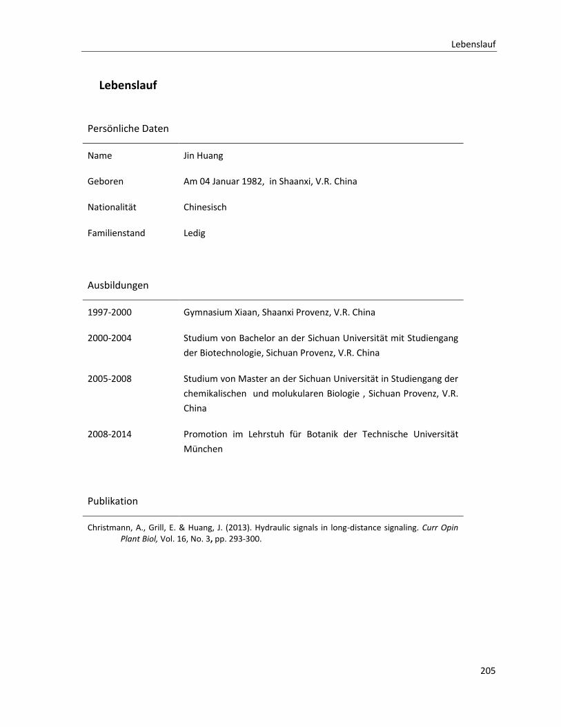
Lebenslauf
205
Lebenslauf
Persönliche Daten
Name Jin Huang
Geboren Am 04 Januar 1982, in Shaanxi, V.R. China
Nationalität Chinesisch
Familienstand Ledig
Ausbildungen
1997-2000 Gymnasium Xiaan, Shaanxi Provenz, V.R. China
2000-2004 Studium von Bachelor an der Sichuan Universität mit Studiengang
der Biotechnologie, Sichuan Provenz, V.R. China
2005-2008 Studium von Master an der Sichuan Universität in Studiengang der
chemikalischen und molukularen Biologie , Sichuan Provenz, V.R.
China
2008-2014 Promotion im Lehrstuh für Botanik der Technische Universität
München
Publikation
Christmann, A., Grill, E. & Huang, J. (2013). Hydraulic signals in long-distance signaling. Curr Opin Plant Biol, Vol. 16, No. 3, pp. 293-300.

Acknowledgments
207
Acknowledgments
This work was performed in the laboratory of of Prof. Dr. Erwin Grill at the Insitute of Botany of Techinische Universität München. I would like to thank with a deep sense of gratitude for Prof. Dr. Erwin Grill accepting me in his laboratory, for the excellent working conditions provided, and the consistent support, teaching and interesting scientific discussions during my Ph. D. time.
I extend my gratitude to Prof. Dr. Kay Schnneitz and Prof. Dr. Ralph Hückelhoven for participation in my committee.
I would like to thank Dr. Alexander Christmann for his help, patience, scientific and valuable discussions and suggestions from the beginning to the end druing my Ph. D. time in this laboratory. I gratefully acknowledge the scientific advices and supports of Dr. Farhah Assaad and Dr. Pascal Braun. Thanks also to Prof. Dr. Christine Gietl for sincere care and valuable discussion.
I specially thank Ting Cao for cooperation and scientific discussion. I am deeply appreciative to many of my colleagues who helped me at the beginning during my Ph. D. time: Dr. Arthur Korte, Dr. Simone Demmel, Dr. Christian Wellmann, Dr. Izabela Szostkiewicz, Dr. Michal Kepka and Dr. Stefan Fuchs. Thanks to C. Eltschig, C. Heidersberger, C. Kornbauer, L. Berngehrer, J. Berger, C. Klaus, L. Held, M. Shimmit, J. Reischenbeck, Z. Yang, G. Czap, K. Hill, N. Kowalski, N. Pleißner, F. Mayer, M. Ruschhaupt, S. Tischer, C. Wunschel, M. Papacek, K. Gläser, K. Rybak, A. Steiner and T. Höwing for their understanding and indispensable cooperation.
I would like to thank Beate Seelinger for her help in daily life and bureaucracy.
I am very grateful to Dr. Tim-Matthias Strom from Helmholtz Center Munich Human Genetic Institute, for providing an opportunity and assistance for next generation sequencing (NGS) and data analysis. I also thank to Prof. Dr. Jutta Ludwig-Müller from the Institut für Botanik of Technische Universität Dresden for GC-MS analysis of ABA quantification.
Finally, I want to express my sincere gratitude to my parents for their help, support, patience, and encouragement during all my life.
This work is supported by Deutsche Forschungsgemeinschaft. And my living in Germany is funded by the State Scholarship fund from the China Scholarship Council of the P.R. China.