Embed Size (px)
Citation preview
1
Identification of a novel putative interaction partner of the nucleoporin ALADIN
Ramona Jühlen a*
, Dana Landgraf a, Angela Huebner
a, Katrin Koehler
a
a Klinik und Poliklinik für Kinder- und Jugendmedizin, Medizinische Fakultät Carl Gustav Carus,
Technische Universität Dresden, Germany
* Corresponding author:
E-mail: [email protected]
Keywords
ALADIN; Cell division; Nuclear pore complex; PGRMC2; Triple A syndrome
.CC-BY-NC-ND 4.0 International licensenot peer-reviewed) is the author/funder. It is made available under aThe copyright holder for this preprint (which was. http://dx.doi.org/10.1101/064766doi: bioRxiv preprint first posted online Jul. 19, 2016;
2
Summary statement
In our study we report the interaction of the microsomal integral membrane protein progesterone
receptor membrane compartment 2 with the nucleoporin ALADIN.
Abstract
It has been shown that the nucleoporin ALADIN employs a significant role in the redox homeostasis
of the cell but the function in steroidogenesis contributing to adrenal atrophy in triple A syndrome
remains largely unknown. In an attempt to identify new interaction partners of ALADIN, co-
immunoprecipitation followed by proteome analysis was conducted in different expression models
using the human adrenocortical tumour cell line NCI-H295R. Our results suggest an interaction of
ALADIN with the microsomal protein PGRMC2. PGRMC2 is shown to be activity regulator of CYP
P450 enzymes and therefore, to be a possible target for adrenal dysregulation in triple A syndrome.
We show that there is a sexual dimorphism regarding the expression of Pgrmc2 in adrenals and
gonads of WT and Aaas KO mice. Female Aaas KO mice are sterile due to delayed oocyte maturation
and meiotic spindle assembly. A participation in meiotic spindle assembly confirms the recently
investigated involvement of ALADIN in mitosis and emphasises an interaction with PGRMC2 which
is a regulator of cell cycle. By identification of a novel interaction partner of ALADIN we provide
novel aspects for future research of the function of ALADIN during cell cycle and for new insights
into the pathogenesis of triple A syndrome.
.CC-BY-NC-ND 4.0 International licensenot peer-reviewed) is the author/funder. It is made available under aThe copyright holder for this preprint (which was. http://dx.doi.org/10.1101/064766doi: bioRxiv preprint first posted online Jul. 19, 2016;
3
Introduction
The triple A syndrome (MIM#231550) is an autosomal recessive disease manifesting with the triad of
ACTH-resistant adrenal insufficiency, achalasia of the stomach cardia and alacrima in combination
with progressive neurological impairment (Allgrove et al., 1978). The syndrome is caused by
mutations in the AAAS (achalasia-adrenal insufficiency-alacrima syndrome) gene encoding the protein
ALADIN (alacrima-achalasia-adrenal insufficiency neurologic disorder) (Handschug et al., 2001;
Tullio-Pelet et al., 2000). ALADIN is ubiquitously expressed, but shows enhanced levels in
neuroendocrine and gastrointestinal structures; tissues which are most affected in triple A patients
(Handschug et al., 2001).
ALADIN is a scaffold nucleoporin (NUP) anchored within the nuclear pore complex (NPC)
by the transmembrane NUP NDC1 (nuclear division cycle 1 homologue (S. cerevisiae)) (Kind et al.,
2009; Yamazumi et al., 2009). It belongs to the group of barely exchangeable NUPs and therefore
seems to be involved in building the structural scaffold backbone of the complex at the nuclear
membrane (Rabut et al., 2004). Over the last years it has been shown that NUPs have fundamental
functions in cell biology, especially beyond nucleo-cytoplasmic transport (Fahrenkrog, 2014; Nofrini
et al., 2016).
Our group has reported that ALADIN is involved in the oxidative stress response of
fibroblasts and adrenocortical cells but the role of ALADIN in adrenal steroidogenesis contributing to
the adrenal phenotype in triple A patients is largely unknown (Jühlen et al., 2015; Kind et al., 2010;
Koehler et al., 2013; Storr et al., 2009). Recently, we showed that a depletion of ALADIN in
adrenocortical carcinoma cells leads to an alteration in glucocorticoid and androgenic steroidogenesis
and a diminished redox homeostasis (Jühlen et al., 2015). Our results described in this article propose
an interaction of ALADIN with the microsomal integral membrane protein progesterone receptor
membrane compartment 2 (PGRMC2). PGRMC2 belongs to the group of membrane-associated
progesterone receptors (MAPRs). These receptors are restricted to the ER and are thought to act on
mitosis while localising to the somatic spindle apparatus and to regulate the activity of some CYP
P450 enzymes (e.g. CYP21A2) (Keator et al., 2012; Peluso et al., 2014; Wendler and Wehling, 2013).
By the attempt to identify new interaction partners of ALADIN we aimed to clarify the cellular
functions of ALADIN at the NPC and to explain the mechanisms which contribute to the adrenal
insufficiency in triple A syndrome. Our observations give the basis for further research on the
association between ALADIN and PGRMC2 and about the function of ALADIN during cell cycle and
steroidogenesis beyond nucleo-cytoplasmic transport.
.CC-BY-NC-ND 4.0 International licensenot peer-reviewed) is the author/funder. It is made available under aThe copyright holder for this preprint (which was. http://dx.doi.org/10.1101/064766doi: bioRxiv preprint first posted online Jul. 19, 2016;
4
Results
PGRMC2 precipitates with ALADIN in an exogenous and endogenous ALADIN adrenal cell
expression model
Co-IP was conducted in NCI-H295R cells either expressing endogenous ALADIN or additionally
exogenous GFP-ALADIN.
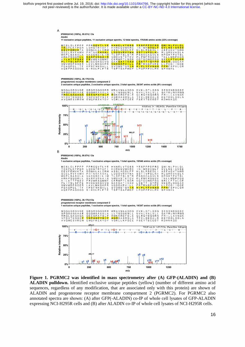
We performed mass spectrometry analyses of bound fractions of GFP(-ALADIN) and ALADIN co-IP.
In the exogenous GFP-ALADIN expression model sufficient ALADIN peptides could be identified
(Fig. 1A), the analysis of endogenous ALADIN co-IP resulted in less detected peptides (Fig. 1B).
Despite distinct methodological optimisation procedures using different protocols and antibodies,
endogenous ALADIN co-IP had a low yield and measurement of bound fractions using mass
spectrometry was more difficult to process. All proteins identified in mass spectrometry in GFP-
ALADIN co-IP and ALADIN co-IP but which were not found in the specific control pulldown assays
are presented in the supplementary data (Tables S1 and S2).
PGRMC2 was simultaneously identified by mass spectrometry analysis in co-IP of GFP-
ALADIN using GFP-Trap_A agarose beads and in co-IP of ALADIN using anti-ALADIN coupled to
Protein G UltraLink resin sepharose beads. Exclusive unique peptides of ALADIN and PGRMC2
detected in mass spectrometry after GFP-ALADIN and ALADIN pulldown are shown in Fig. 1A and
1B, respectively.
We additionally confirmed the identification of PGRMC2 in GFP-ALADIN and ALADIN co-
IP by Western Blot. Successful GFP-ALADIN pulldown in lysates of NCI-H295R cells stably
expressing GFP-ALADIN (86 kDa) is presented in Fig. 2A. In accordance with our mass
spectrometry results PGRMC2 (24 kDa) could also be detected after GFP-ALADIN pulldown (Fig.
2A, arrow). The negative control remained empty.
Successful endogenous ALADIN pulldown is shown in Fig. 2B. ALADIN (59 kDa) was
found in the bound fraction of the ALADIN co-IP but the negative control using normal mouse IgG
was shown to be empty. In accordance to our result after GFP-ALADIN pulldown, PGRMC2
precipitated in endogenous ALADIN pulldown with no unspecific interaction in the negative control
(Fig. 2B, arrow).
ALADIN precipitates with PGRMC2 in an exogenous and endogenous PGRMC2 adrenal cell
expression model
In order to further evaluate a possible interaction between the nucleoporin ALADIN and microsomal
PGRMC2 we conducted reciprocal co-IP assays.
Reciprocal pulldowns were done in NCI-H295R cells transiently expressing exogenous
PGRMC2-GFP and endogenous PGRMC2.
.CC-BY-NC-ND 4.0 International licensenot peer-reviewed) is the author/funder. It is made available under aThe copyright holder for this preprint (which was. http://dx.doi.org/10.1101/064766doi: bioRxiv preprint first posted online Jul. 19, 2016;
5
Efficient pulldown of (PGRMC2-)GFP in lysates of NCI-H295R cells transiently expressing
PGRMC2-GFP (51 kDa) is presented in Fig. 2C. Confirming our previous results identifying a
possible interaction between ALADIN and PGRMC2, ALADIN could be detected after PGRMC2-
GFP pulldown (Fig. 2C, arrow). The negative control was empty.
PGRMC2 pulldown is shown in Fig. 2D. PGRMC2 was successfully detected in the bound
fraction of the PGRMC2 co-IP and the negative control using rabbit IgG remained empty. According
to our results after PGRMC2-GFP pulldown, ALADIN slightly but visibly precipitated after
PGRMC2 pulldown with no unspecific interaction in the negative control (Fig. 2D, arrow).
Localisation of ALADIN and PGRMC2 in adrenal cells using different expression models
Further evidence of possible co-localisation of ALADIN and PGRMC2 is given after
immunofluorescent staining. Cells expressing GFP-ALADIN and PGRMC2-GFP were used to verify
unspecific staining of anti-ALADIN and anti-PGRMC2 in NCI-H295R cells. Staining was done using
anti-ALADIN, anti-PGRMC2 and anti-NPC proteins (mAb414).
Immunostaining with mAb414 in all adrenal cell expression models gave a thin circle around
the nucleus indicating punctuate localisations of NPCs (Fig. 3).
Immunofluorescent staining of the nucleoporin ALADIN appeared at the nuclear envelope at
the proximity of NPCs in all adrenal cell expression models. In the exogenous GFP-ALADIN cell
model the fusion protein was correctly targeted to the nuclear envelope and did not accumulate to a
greater extent in the cytoplasm. We showed that ALADIN almost completely co-localises with anti-
NPC proteins (mAb414) immunostaining at the nuclear envelope; substantially verifying the
localisation of ALADIN at the nuclear pore (Fig. 3).
In immunofluorescent staining the microsomal protein PGRMC2 localised to the central ER
but also revealed a patchy and punctuate staining pattern around the nucleus to the perinuclear space
between nuclear envelope and ER in all adrenal cell expression models (Fig. 3). The same PGRMC2
immunostaining pattern was observed in human cervical carcinoma (HeLa) (Fig. S1A) and human
fibroblasts (Fig. S1B). In the exogenous adrenal cell model the PGRMC2-GFP fusion protein was still
correctly targeted to the central ER and perinuclear space. In the staining with the anti-PGRMC2 in
NCI-H295R cells we could also observe nuclear staining in some adrenal cells (Fig. 3, arrow).
Nuclear localisation of PGRMC2 was absent in the PGRMC2-GFP adrenal cell expression model. Co-
localisation between mAb414 and PGRMC2 or ALADIN and PGRMC2 was not complete but showed
positivity in the perinuclear space and in the nuclear membrane in all cell expression models (Fig. 3).
The expression of PGRMC2 is not affected after AAAS knock-down in human adrenal cells
To test if PGRMC2 expression is affected when ALADIN is down-regulated we used the inducible
.CC-BY-NC-ND 4.0 International licensenot peer-reviewed) is the author/funder. It is made available under aThe copyright holder for this preprint (which was. http://dx.doi.org/10.1101/064766doi: bioRxiv preprint first posted online Jul. 19, 2016;
6
NCI-H295R1-TR cells with AAAS knock-down shRNA or scrambled shRNA as negative control
(Jühlen et al., 2015).
We could not find an alteration on PGRMC2 mRNA level after induction of ALADIN
depletion by doxycycline in NCI-H295R1-TR cells in at least ten triplicate experiments (Fig. S2).
Pgrmc2 exhibits a sexual dimorphism in adrenals and gonads of WT and Aaas KO mice
In order to examine the expression of Pgrmc2 in WT and Aaas KO mice we looked at the adrenals
and gonads using Taq Man analysis and Western Blot (Fig. 4A).
Indeed, we found that Pgrmc2 exploited a sexual dimorphism in female and male WT and
Aaas KO mice: the expression in testes was significantly higher independent of genotype compared to
female ovaries, in female KO adrenals the expression was significantly higher compared to male KO
adrenals (Fig. 4A).
Interestingly, in female adrenals the depletion of ALADIN leads to a significant increase in
Pgrmc2 expression whereas in female ovaries a decrease compared to WT ovaries was observed (Fig.
4A). The expression of Pgrmc2 was not altered in male Aaas KO adrenals or testes compared to male
WT organs in at least four triplicate experiments (Fig. 4A).
To examine our findings in female WT and Aaas KO mice on PGRMC2 RNA level, we
conducted Western Blot of several female murine WT and Aaas KO tissues, i.e. adrenals, brain,
ovaries and spleen (Fig. 4B). We could confirm our results on PGRMC2 RNA level and could show
an increase in PGRMC2 protein in female adrenals of Aaas KO mice compared to female adrenals of
WT mice (Fig. 4B). Furthermore, in ovaries of Aaas KO mice PGRMC2 protein was diminished
compared to ovaries of WT mice.
.CC-BY-NC-ND 4.0 International licensenot peer-reviewed) is the author/funder. It is made available under aThe copyright holder for this preprint (which was. http://dx.doi.org/10.1101/064766doi: bioRxiv preprint first posted online Jul. 19, 2016;
7
Discussion
The exact role of the nucleoporin ALADIN at the NPC and its involvement in steroidogenesis leading
to the characteristic adrenal atrophy in triple A syndrome remains largely unknown. We and others
have provided proofs of the involvement of ALADIN in the oxidative stress response of the cell
(Jühlen et al., 2015; Kind et al., 2010; Koehler et al., 2013; Prasad et al., 2013; Storr et al., 2009).
Recently, we could show that a depletion of ALADIN in adrenocortical carcinoma cells leads to an
alteration in glucocorticoid and androgenic steroidogenesis (Jühlen et al., 2015).
Despite the reported interaction between ALADIN and ferritin heavy chain 1 no other
interaction partner which would lead to the identification of a plausible function and signal
transduction of ALADIN in the cell is known so far (Storr et al., 2009).
In an attempt to identify new interaction partners of ALADIN co-IP analyses showed that
PGRMC2 precipitated with ALADIN. To verify the identified association between ALADIN and
PGRMC2 reciprocal IP was conducted. Our results showed a co-precipitation of ALADIN with
PGRMC2. By different co-IP approaches using exogenous and endogenous expression systems in
human adrenal cells we can show for the first time that the nucleoporin ALADIN associates in a
complex with the microsomal protein PGRMC2.
PGRMC2 belongs to the MAPR family. MAPRs are restricted to the ER and are thought to
regulate the activity of CYP P450 enzymes. The first identified MAPR, PGRMC1, gained wide-
spread attention (Falkenstein et al., 1996). PGRMC1 is a cytochrome-related protein with several
implications in cancer (Clark et al., 2016; Falkenstein et al., 1996; Kabe et al., 2016)
In this work, PGRMC2 was found to interact with the nucleoporin ALADIN. PGRMC2 is
barely investigated compared to its homologue PGRMC1. It is known that PGRMC2 alters activity of
CYP3A4 as possible electron donor, and binds CYP21A2, most likely through its cytochrome b5-
similar heme-binding domain (Albrecht et al., 2012; Wendler and Wehling, 2013).
Visualising PGRMC2 in the cell using immunofluorescence and confocal microscopy
PGRMC2 appeared at the central ER and interestingly, at the nuclear envelope and the perinuclear
ER. We detected that PGRMC2 co-localises with ALADIN and with different FG-repeat NUPs
(stained with anti-NPC proteins (mAb414)) to the nuclear envelope and the perinuclear ER. Taken
together, our results in immunofluorescence microscopy using different ALADIN and PGRMC2
adrenal cell expression systems provide a basis for future research of how ALADIN and PGRMC2
possibly associate in a complex close to the nuclear envelope and what the molecular function of this
association would be.
Furthermore, we present that ALADIN depletion did not result in diminished PGRMC2
expression on mRNA level in vitro in adrenocortical carcinoma cells. However, we found that Pgrmc2
has a sexual dimorphic role in adrenals and gonads of WT and Aaas KO mice and of note, ALADIN
.CC-BY-NC-ND 4.0 International licensenot peer-reviewed) is the author/funder. It is made available under aThe copyright holder for this preprint (which was. http://dx.doi.org/10.1101/064766doi: bioRxiv preprint first posted online Jul. 19, 2016;
8
depletion leads to an alteration in PGRMC2 RNA and protein level in adrenals and ovaries of female
Aaas KO mice.
Our group reported that female mice homozygous deficient for Aaas are infertile (Huebner et
al., 2006). Carvalhal et al. recently presented that ALADIN is involved in mitotic and meiotic spindle
assembly, chromosome segregation and production of fertile mouse oocytes (Carvalhal et al., 2015;
Carvalhal et al., 2016). Interestingly, both PGRMC1 and 2 were shown to be involved in regulation of
ovarian follicle development and therefore imply a neuroendocrine function (Wendler and Wehling,
2013). Deficiency of either MAPRs decreases the anti-apoptotic and anti-mitotic action of
progesterone, although PGRMC2 seems to be important for anti-mitotic actions of the steroid.
Depletion of either PGRMC1 or PGRMC2 leads to increased entry into cell cycle. Both proteins
localise to the mitotic spindle and seem to exploit a distinct role during metaphase of mitosis, thereby
suppressing entry into cell cycle. This effect is thought to be synergistic and does not seem to be
additive (Griffin et al., 2014; Peluso et al., 2014; Sueldo et al., 2015).
Most recently, ALADIN and PGRMC2 have been identified to interact with the human
centrosome-cilium interface (Gupta et al., 2015; Hanson et al., 2014; Yan et al., 2014). The
centrosome is a fundamental organelle which participates in cell cycle progression and mitotic spindle
assembly. Conclusively, ALADIN and PGRMC2 both seem to have an important role at the mitotic
and meiotic spindle and to be involved in the sterility of female Aaas KO mice.
In summary, our straightforward work about the identification of the novel interactor of
ALADIN, PGRMC2, provide new insights into the molecular function of the nucleoporin ALADIN in
the pathogenesis of triple A syndrome. In the future it needs to be investigated how and why ALADIN
associates with the microsomal protein at the perinuclear ER. In addition, their possible simultaneous
role at the spindle apparatus shall await more research and reveal additional functions of ALADIN
during cell division and meiosis.
.CC-BY-NC-ND 4.0 International licensenot peer-reviewed) is the author/funder. It is made available under aThe copyright holder for this preprint (which was. http://dx.doi.org/10.1101/064766doi: bioRxiv preprint first posted online Jul. 19, 2016;
9
Materials and Methods
Cell culture
NCI-H295R cells stably expressing GFP-ALADIN fusion protein or GFP were generated as described
previously using the gamma-retroviral transfer vectors pcz-CFG5.1-GFP-AAAS and pcz-CFG5.1-GFP
(Kind et al., 2009).
NCI-H295R cells transiently expressing PGRMC2-GFP fusion protein were generated as
follows. Cells were transfected with pCMV6-AC-PGRMC2-GFP vector (RG204682) (OriGene
Technologies, Rockville MD, USA) using X-tremeGENE HP DNA transfection reagent (Roche
Diagnostics, Mannheim, Germany) following the manufacturer’s protocols. Cells were harvested or
fixed after 48 hours.
In all exogenous expression models clones were selected by moderate expression of the
desired fusion protein and true cellular localisation in order to exclude the possibility of false positive
protein interactions.
Cells were cultured in DMEM/F12 medium (Lonza, Cologne, Germany) supplemented with 1
mM L-glutamine (Lonza, Cologne, Germany), 5% Nu-serum (BD Biosciences, Heidelberg,
Germany), 1% insulin-tranferrin-selenium) (Gibco, Life Technologies, Darmstadt, Germany) and 1%
antibiotic-antimycotic solution (PAA, GE Healthcare GmbH, Little Chalfont, United Kingdom).
NCI-H295R1-TR cells with AAAS knock-down or scrambled shRNA were generated, selected
and cultured as described previously (Jühlen et al., 2015).
Animals
All procedures were approved by the Regional Board for Veterinarian Affairs (AZ 24-9168.21-1-
2002-1) in accordance with the institutional guidelines for the care and use of laboratory animals.
C57BL/6J mice were obtained from Janvier Labs (Le Genest-Saint-Isle, France). Aaas KO mice were
generated as described previously (Huebner et al., 2006).
RNA extraction, cDNA synthesis and quantitative real-time PCR using TaqMan
Total RNA from cultured cells (n=10) and from frozen murine organs (at least four animals per
genotype and sex) was isolated using the NucleoSpin RNA (Macherey-Nagel, Düren, Germany)
according to the protocol from the manufacturer. Purity of the RNA was assessed using Nanodrop
Spectrophotometer (ND-1000) (NanoDrop Technologies, Wilmington DE, USA). The amount of 500
ng of total RNA was reverse transcribed using the GoScript Reverse Transcription System (Promega,
Mannheim, Germany) following the protocols from the manufacturer. Primers for the amplification of
the target sequence were designed using Primer Express 3.0 (Applied Biosystems) and compared to
the human or murine genome database for unique binding using BLAST search (National Center for
.CC-BY-NC-ND 4.0 International licensenot peer-reviewed) is the author/funder. It is made available under aThe copyright holder for this preprint (which was. http://dx.doi.org/10.1101/064766doi: bioRxiv preprint first posted online Jul. 19, 2016;
10
Biotechnology Information, U.S. National Library of Medicine, 2013). The primer sequences are
listed in the supplementary data of this article (Table S3).
The qPCR amplifications were performed in triplicates using the GoTaq Probe qPCR Master
Mix (Promega) according to the manufacturer’s reaction parameter on an ABI 7300 Fast Real-Time
PCR System (Applied Biosystems, Life Technologies, Darmstadt, Germany). In all results
repeatability was assessed by standard deviation of triplicate Cts and reproducibility was verified by
normalizing all real-time RT-PCR experiments by the Ct of each positive control per run.
Immunoblots
After SDS-PAGE separation onto 4-12% PAGE (150 V for 1.5 hours) and electroblotting (30 V for
1.5 hours) (Invitrogen, Life Technologies, Darmstadt, Germany) onto Amersham hybond-ECL
nitrocellulose membrane (0.45 µm) (GE Healthcare GmbH, Little Chalfont, United Kingdom) non-
specific binding of proteins to the membrane was blocked by incubation in PBS containing 3% BSA
(Sigma-Aldrich, Munich, Germany) at room-temperature.
The membrane was then probed with primary antibodies either anti-ALADIN (B-11: sc-
374073) (Santa Cruz Biotechnology, Inc., Heidelberg, Germany) (1:100 in 3% PBS/BSA) or anti-
PGRMC2 (HPA041172) (Sigma-Aldrich, Munich, Germany) (1:200 in 5% PBS/milk powder) over-
night at 4°C. Secondary antibodies goat anti-mouse IgG conjugated to horseradish peroxidase (1:2000
in 3% PBS/BSA) (Invitrogen, Life Technologies, Darmstadt, Germany) or goat anti-rabbit IgG
conjugated to horseradish peroxidase (1:3000 in 5% PBS/milk powder) (Cell Signalling Technology
Europe B.V., Leiden, Netherlands) were incubated one hour at room-temperature.
Co-immunoprecipitation
For GFP co-IP lysates from NCI-H295R expressing GFP-ALADIN or PGRMC2-GFP were used.
Lysates from cells expressing GFP were used as negative control. Cell lysates (500 µg protein) were
added to the pre-equilibrated GFP-Trap_A agarose beads (ChromoTek GmbH, Planegg-Martinsried,
Germany), gently resuspended by flipping the tube and bound over-night at constant mixing at 4°C.
After washing steps the beads were gently re-suspended in 60 µl NUPAGE 2X LDS sample buffer
and in order to dissociate the captured immunocomplexes from the beads, boiled at 95 °C for 10
minutes and Western Blot analysis was conducted with 20 µl of the eluate. The left 40 µl of the eluate
using the lysates of NCI-H295R expressing GFP-ALADIN or GFP was further processed for
proteomic profiling using mass spectrometry. These experiments following mass spectrometry
analysis were repeated three times.
For co-IP of ALADIN or PGRMC2 lysates from NCI-H295R cells and Protein G UltraLink
resin sepharose beads (Pierce, Thermo Scientific, Fischer Scientific, Schwerte, Germany) were used.
.CC-BY-NC-ND 4.0 International licensenot peer-reviewed) is the author/funder. It is made available under aThe copyright holder for this preprint (which was. http://dx.doi.org/10.1101/064766doi: bioRxiv preprint first posted online Jul. 19, 2016;
11
Beads were gently resuspended in anti-ALADIN (2 µg/ml) or anti-PGRMC2 (HPA041172) (2 µg/ml)
and as negative controls normal mouse or rabbit IgG (Invitrogen, Life Technologies, Darmstadt,
Germany) (2 µg/ml). All antibodies were bound to the beads over-night at 4°C in a rotation chamber.
After washing cell lysates (500 µg protein) were added to the beads, gently resuspended by flipping
the tube and bound over-night as described before. After washing the beads were gently resuspended
in 60 µl sample buffer containing dilution buffer, NUPAGE 1X LDS Sample Buffer and 1X Reducing
Agent. The captured immunocomplexes were dissociated and the eluates were collected and
processed by Western Blot as described previously. These experiments were repeated three times. The
left 40 µl of the eluate after ALADIN co-IP and negative control was further processed for proteomic
profiling using mass spectrometry. Mass spectrometry analysis was conducted once.
Proteomic profiling using tandem mass spectrometry
Entire gel lanes were cut into 40 slabs, each of which was in-gel digested with trypsin (Shevchenko et
al., 2006). Gel analyses were performed at the Mass Spectrometry Facility at the Max Planck Institute
for Molecular Cell Biology and Genetics Dresden on a nano high-performance liquid chromatograph
Ultimate interfaced on-line to a LTQ Orbitrap Velos hybrid tandem mass spectrometer as described
previously (Vasilj et al., 2012).
Database search was performed against IPI human database (downloaded in July 2010) and
NCBI protein collection without species restriction (updated in June 2014) using MASCOT software
v.2.2. Scaffold software v.4.3.2 was used to validate MS/MS-based protein identifications. Protein
probabilities were assigned by the Protein Prophet algorithm (Nesvizhskii et al., 2003).
Immunofluorescence microscopy
Cells grown onto glass cover slips were fixed for 5 minutes with 4% PFA (SAV LP, Flinsbach,
Germany) in PBS, permeabilised for 5 minutes with 0.5% Triton-X-100 in PBS and fixed again for 5
minutes. Blocking was performed for 30 minutes with 2% BSA/0.1% Triton-X-100 in PBS at room-
temperature.
All antibodies used for immunofluorescence were diluted in blocking solution. Primary
antibodies anti-ALADIN (1:25), or anti-PGRMC2 (HPA041172) (1:50) or anti-PGRMC2 (F-3: sc-
374624) (Santa Cruz Biotechnology, Inc.) (1:25) and anti-NPC proteins (mAb414) (Covance, Berkley
CA, USA) (1:800) were incubated at 4°C over-night in a humidified chamber. Secondary antibodies
goat anti-mouse IgG Cy3 (1:800) (Amersham Biosciences, Freiburg, Germany), Alexa Fluor 488 and
555 goat anti-rabbit IgG (1:500) (Molecular Probes, Life Technologies) were incubated one hour at
room-temperature in the dark.
Fluorescence was visualised using the confocal laser microscope TCS SP2 (Leica
.CC-BY-NC-ND 4.0 International licensenot peer-reviewed) is the author/funder. It is made available under aThe copyright holder for this preprint (which was. http://dx.doi.org/10.1101/064766doi: bioRxiv preprint first posted online Jul. 19, 2016;
12
Microsystems, Mannheim, Germany). The experiments were repeated at least three times.
Statistics of TaqMan analyses
Statistical analyses were made using the open-source software R version 3.3.0 and R Studio version
0.99.902 (R Core Team, 2015). Unpaired Wilcoxon-Mann-Whitney U-test was performed. During
evaluation of the results a confidence interval alpha of 95% and P values lower than 0.05 were
considered as statistically significant. Results are shown as box plots which give a fast and efficient
overview about median, first and third quartile (25th and 75th percentile, respectively), interquartile
range (IQR), minimal and maximal values and outliers.
.CC-BY-NC-ND 4.0 International licensenot peer-reviewed) is the author/funder. It is made available under aThe copyright holder for this preprint (which was. http://dx.doi.org/10.1101/064766doi: bioRxiv preprint first posted online Jul. 19, 2016;
13
Acknowledgements
We thank Waldemar Kanczkowski for providing the NCI-H295R cells. Barbara Kind generously
generated pseudo retroviruses containing pcz-CFG5.1-GFP-ALADIN and pcz-CFG5.1-GFP. We
thank the Mass Spectrometry Facility at the Max Planck Institute for Molecular Cell Biology and
Genetics Dresden for MS-based peptide analyses.
Competing interests
The authors declare no competing interests.
Author contributions
RJ, AH and KK conceived and designed the experiments. RJ performed all experiments. DL helped
with immunofluorescence staining and KK assisted with confocal microscopy. RJ analysed the data
and wrote the paper. AH and KK helped improving the manuscript. All authors read the final version
of the manuscript and gave their permission for publication.
Funding
This work was supported by a Deutsche Forschungsgemeinschaft grant HU 895/5-2 (Clinical
Research Unit 252) to AH. The funders had no role in study design, data collection and analysis,
decision to publish, or preparation of the manuscript.
References
Albrecht, C., Huck, V., Wehling, M. and Wendler, A. (2012). In vitro inhibition of SKOV-3 cell
migration as a distinctive feature of progesterone receptor membrane component type 2
versus type 1. Steroids 77, 1543–1550.
Allgrove, J., Clayden, G. S., Grant, D. B. and Macaulay, J. C. (1978). Familial glucocorticoid
deficiency with achalasia of the cardia and deficient tear production. Lancet 1, 1284–1286.
Carvalhal, S., Ribeiro, S. A., Arocena, M., Kasciukovic, T., Temme, A., Koehler, K., Huebner, A.
and Griffis, E. R. (2015). The nucleoporin ALADIN regulates Aurora A localization to
ensure robust mitotic spindle formation. Mol. Biol. Cell 26, 3424–3438.
Carvalhal, S., Stevense, M., Koehler, K., Naumann, R., Huebner, A., Jessberger, R. and Griffis,
E. (2016). ALADIN is Required for the Production of Fertile Mouse Oocytes. bioRxiv.
Clark, N. C., Friel, A. M., Pru, C. A., Zhang, L., Shioda, T., Rueda, B. R., Peluso, J. J. and Pru,
J. K. (2016). Progesterone receptor membrane component 1 promotes survival of human
breast cancer cells and the growth of xenograft tumors. Cancer Biol. Ther. 17, 262–271.
Fahrenkrog, B. (2014). Nucleoporin Gene Fusions and Hematopoietic Malignancies. New J. Sci.
2014.
.CC-BY-NC-ND 4.0 International licensenot peer-reviewed) is the author/funder. It is made available under aThe copyright holder for this preprint (which was. http://dx.doi.org/10.1101/064766doi: bioRxiv preprint first posted online Jul. 19, 2016;
14
Falkenstein, E., Meyer, C., Eisen, C., Scriba, P. C. and Wehling, M. (1996). Full-length cDNA
sequence of a progesterone membrane-binding protein from porcine vascular smooth muscle
cells. Biochem. Biophys. Res. Commun. 229, 86–89.
Griffin, D., Liu, X., Pru, C., Pru, J. K. and Peluso, J. J. (2014). Expression of progesterone
receptor membrane component-2 within the immature rat ovary and its role in regulating
mitosis and apoptosis of spontaneously immortalized granulosa cells. Biol. Reprod. 91, 36.
Gupta, G. D., Coyaud, É., Gonçalves, J., Mojarad, B. A., Liu, Y., Wu, Q., Gheiratmand, L.,
Comartin, D., Tkach, J. M., Cheung, S. W. T., et al. (2015). A Dynamic Protein Interaction
Landscape of the Human Centrosome-Cilium Interface. Cell 163, 1484–1499.
Handschug, K., Sperling, S., Yoon, S. J., Hennig, S., Clark, A. J. and Huebner, A. (2001). Triple A
syndrome is caused by mutations in AAAS, a new WD-repeat protein gene. Hum. Mol. Genet.
10, 283–290.
Hanson, D., Stevens, A., Murray, P. G., Black, G. C. M. and Clayton, P. E. (2014). Identifying
biological pathways that underlie primordial short stature using network analysis. J. Mol.
Endocrinol. 52, 333–344.
Huebner, A., Mann, P., Rohde, E., Kaindl, A. M., Witt, M., Verkade, P., Jakubiczka, S.,
Menschikowski, M., Stoltenburg-Didinger, G. and Koehler, K. (2006). Mice lacking the
nuclear pore complex protein ALADIN show female infertility but fail to develop a
phenotype resembling human triple A syndrome. Mol. Cell. Biol. 26, 1879–1887.
Jühlen, R., Idkowiak, J., Taylor, A. E., Kind, B., Arlt, W., Huebner, A. and Koehler, K. (2015).
Role of ALADIN in Human Adrenocortical Cells for Oxidative Stress Response and
Steroidogenesis. PloS One 10, e0124582.
Kabe, Y., Nakane, T., Koike, I., Yamamoto, T., Sugiura, Y., Harada, E., Sugase, K., Shimamura,
T., Ohmura, M., Muraoka, K., et al. (2016). Haem-dependent dimerization of
PGRMC1/Sigma-2 receptor facilitates cancer proliferation and chemoresistance. Nat.
Commun. 7, 11030.
Keator, C. S., Mah, K. and Slayden, O. D. (2012). Alterations in progesterone receptor membrane
component 2 (PGRMC2) in the endometrium of macaques afflicted with advanced
endometriosis. Mol. Hum. Reprod. 18, 308–319.
Kind, B., Koehler, K., Lorenz, M. and Huebner, A. (2009). The nuclear pore complex protein
ALADIN is anchored via NDC1 but not via POM121 and GP210 in the nuclear envelope.
Biochem. Biophys. Res. Commun. 390, 205–210.
Kind, B., Koehler, K., Krumbholz, M., Landgraf, D. and Huebner, A. (2010). Intracellular ROS
level is increased in fibroblasts of triple A syndrome patients. J. Mol. Med. Berl. Ger. 88,
1233–1242.
Koehler, K., End, K., Kind, B., Landgraf, D., Mitzscherling, P. and Huebner, A. (2013). Changes
in differential gene expression in fibroblast cells from patients with triple A syndrome under
oxidative stress. Horm. Metab. Res. Horm. Stoffwechselforschung Horm. Metab. 45, 102–108.
National Center for Biotechnology Information, U.S. National Library of Medicine (2013). Basic
Local Alignment Search Tool. Bethesda MD, USA.
.CC-BY-NC-ND 4.0 International licensenot peer-reviewed) is the author/funder. It is made available under aThe copyright holder for this preprint (which was. http://dx.doi.org/10.1101/064766doi: bioRxiv preprint first posted online Jul. 19, 2016;
15
Nesvizhskii, A. I., Keller, A., Kolker, E. and Aebersold, R. (2003). A statistical model for
identifying proteins by tandem mass spectrometry. Anal. Chem. 75, 4646–4658.
Nofrini, V., Di Giacomo, D. and Mecucci, C. (2016). Nucleoporin genes in human diseases. Eur. J.
Hum. Genet.
Peluso, J. J., Griffin, D., Liu, X. and Horne, M. (2014). Progesterone receptor membrane
component-1 (PGRMC1) and PGRMC-2 interact to suppress entry into the cell cycle in
spontaneously immortalized rat granulosa cells. Biol. Reprod. 91, 104.
Prasad, R., Metherell, L. A., Clark, A. J. and Storr, H. L. (2013). Deficiency of ALADIN impairs
redox homeostasis in human adrenal cells and inhibits steroidogenesis. Endocrinology 154,
3209–3218.
Rabut, G., Doye, V. and Ellenberg, J. (2004). Mapping the dynamic organization of the nuclear pore
complex inside single living cells. Nat. Cell Biol. 6, 1114–1121.
R Core Team (2015). R: A language and environment for statistical computing. Vienna, Austria: R
Foundation for Statistical Computing.
Shevchenko, A., Tomas, H., Havlis, J., Olsen, J. V. and Mann, M. (2006). In-gel digestion for mass
spectrometric characterization of proteins and proteomes. Nat. Protoc. 1, 2856–2860.
Storr, H. L., Kind, B., Parfitt, D. A., Chapple, J. P., Lorenz, M., Koehler, K., Huebner, A. and
Clark, A. J. (2009). Deficiency of ferritin heavy-chain nuclear import in triple a syndrome
implies nuclear oxidative damage as the primary disease mechanism. Mol. Endocrinol.
Baltim. Md 23, 2086–2094.
Sueldo, C., Liu, X. and Peluso, J. J. (2015). Progestin and AdipoQ Receptor 7, Progesterone
Membrane Receptor Component 1 (PGRMC1), and PGRMC2 and Their Role in Regulating
Progesterone’s Ability to Suppress Human Granulosa/Luteal Cells from Entering into the Cell
Cycle. Biol. Reprod. 93, 63.
Tullio-Pelet, A., Salomon, R., Hadj-Rabia, S., Mugnier, C., de Laet, M. H., Chaouachi, B.,
Bakiri, F., Brottier, P., Cattolico, L., Penet, C., et al. (2000). Mutant WD-repeat protein in
triple-A syndrome. Nat. Genet. 26, 332–335.
Vasilj, A., Gentzel, M., Ueberham, E., Gebhardt, R. and Shevchenko, A. (2012). Tissue
proteomics by one-dimensional gel electrophoresis combined with label-free protein
quantification. J. Proteome Res. 11, 3680–3689.
Wendler, A. and Wehling, M. (2013). PGRMC2, a yet uncharacterized protein with potential as
tumor suppressor, migration inhibitor, and regulator of cytochrome P450 enzyme activity.
Steroids 78, 555–558.
Yamazumi, Y., Kamiya, A., Nishida, A., Nishihara, A., Iemura, S., Natsume, T. and Akiyama, T. (2009). The transmembrane nucleoporin NDC1 is required for targeting of ALADIN to
nuclear pore complexes. Biochem. Biophys. Res. Commun. 389, 100–104.
Yan, J., Yan, F., Li, Z., Sinnott, B., Cappell, K. M., Yu, Y., Mo, J., Duncan, J. A., Chen, X.,
Cormier-Daire, V., et al. (2014). The 3M Complex Maintains Microtubule and Genome
Integrity. Mol. Cell 54, 791–804.
.CC-BY-NC-ND 4.0 International licensenot peer-reviewed) is the author/funder. It is made available under aThe copyright holder for this preprint (which was. http://dx.doi.org/10.1101/064766doi: bioRxiv preprint first posted online Jul. 19, 2016;
16
Figure 1. PGRMC2 was identified in mass spectrometry after (A) GFP-(ALADIN) and (B)
ALADIN pulldown. Identified exclusive unique peptides (yellow) (number of different amino acid
sequences, regardless of any modification, that are associated only with this protein) are shown of
ALADIN and progesterone receptor membrane compartment 2 (PGRMC2). For PGRMC2 also
annotated spectra are shown: (A) after GFP(-ALADIN) co-IP of whole cell lysates of GFP-ALADIN
expressing NCI-H295R cells and (B) after ALADIN co-IP of whole cell lysates of NCI-H295R cells.
.CC-BY-NC-ND 4.0 International licensenot peer-reviewed) is the author/funder. It is made available under aThe copyright holder for this preprint (which was. http://dx.doi.org/10.1101/064766doi: bioRxiv preprint first posted online Jul. 19, 2016;
17
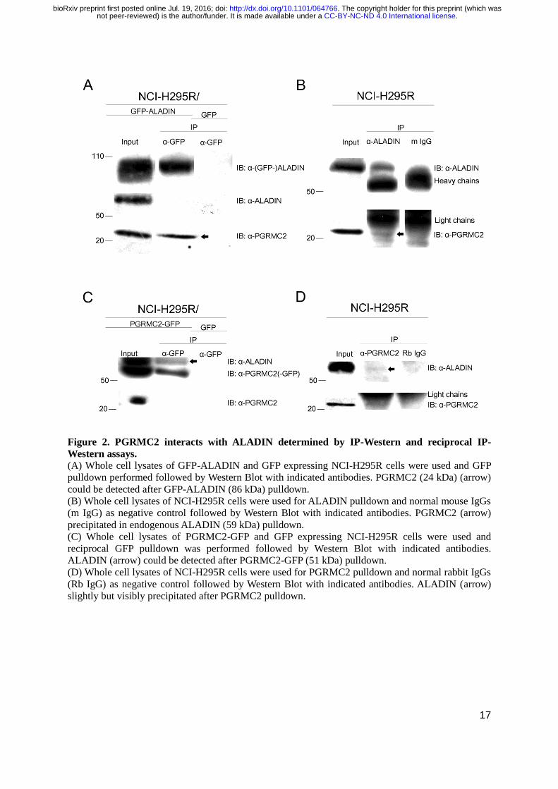
Figure 2. PGRMC2 interacts with ALADIN determined by IP-Western and reciprocal IP-
Western assays. (A) Whole cell lysates of GFP-ALADIN and GFP expressing NCI-H295R cells were used and GFP
pulldown performed followed by Western Blot with indicated antibodies. PGRMC2 (24 kDa) (arrow)
could be detected after GFP-ALADIN (86 kDa) pulldown.
(B) Whole cell lysates of NCI-H295R cells were used for ALADIN pulldown and normal mouse IgGs
(m IgG) as negative control followed by Western Blot with indicated antibodies. PGRMC2 (arrow)
precipitated in endogenous ALADIN (59 kDa) pulldown.
(C) Whole cell lysates of PGRMC2-GFP and GFP expressing NCI-H295R cells were used and
reciprocal GFP pulldown was performed followed by Western Blot with indicated antibodies.
ALADIN (arrow) could be detected after PGRMC2-GFP (51 kDa) pulldown.
(D) Whole cell lysates of NCI-H295R cells were used for PGRMC2 pulldown and normal rabbit IgGs
(Rb IgG) as negative control followed by Western Blot with indicated antibodies. ALADIN (arrow)
slightly but visibly precipitated after PGRMC2 pulldown.
.CC-BY-NC-ND 4.0 International licensenot peer-reviewed) is the author/funder. It is made available under aThe copyright holder for this preprint (which was. http://dx.doi.org/10.1101/064766doi: bioRxiv preprint first posted online Jul. 19, 2016;
18
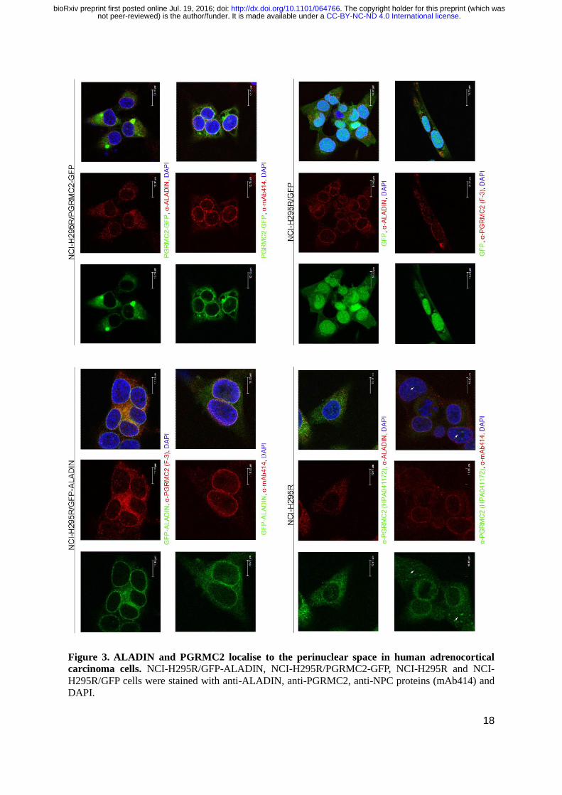
Figure 3. ALADIN and PGRMC2 localise to the perinuclear space in human adrenocortical
carcinoma cells. NCI-H295R/GFP-ALADIN, NCI-H295R/PGRMC2-GFP, NCI-H295R and NCI-
H295R/GFP cells were stained with anti-ALADIN, anti-PGRMC2, anti-NPC proteins (mAb414) and
DAPI.
.CC-BY-NC-ND 4.0 International licensenot peer-reviewed) is the author/funder. It is made available under aThe copyright holder for this preprint (which was. http://dx.doi.org/10.1101/064766doi: bioRxiv preprint first posted online Jul. 19, 2016;
19
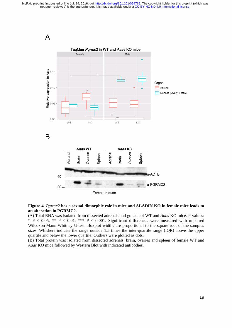
Figure 4. Pgrmc2 has a sexual dimorphic role in mice and ALADIN KO in female mice leads to
an alteration in PGRMC2. (A) Total RNA was isolated from dissected adrenals and gonads of WT and Aaas KO mice. P-values:
* P < 0.05, ** P < 0.01, *** P < 0.001. Significant differences were measured with unpaired
Wilcoxon-Mann-Whitney U-test. Boxplot widths are proportional to the square root of the samples
sizes. Whiskers indicate the range outside 1.5 times the inter-quartile range (IQR) above the upper
quartile and below the lower quartile. Outliers were plotted as dots.
(B) Total protein was isolated from dissected adrenals, brain, ovaries and spleen of female WT and
Aaas KO mice followed by Western Blot with indicated antibodies.
.CC-BY-NC-ND 4.0 International licensenot peer-reviewed) is the author/funder. It is made available under aThe copyright holder for this preprint (which was. http://dx.doi.org/10.1101/064766doi: bioRxiv preprint first posted online Jul. 19, 2016;