Embed Size (px)
Citation preview
Identification of novel mechanisms regulating the NAD+-dependent deacetylase SIRT1
Inauguraldissertation
zur Erlangung der Würde eines Doktors der Philosophie
vorgelegt der Philosophisch-Naturwissenschaftlichen Fakultät
der Universität Basel
von
Dimitrios Anastasiou
aus Athen (Griechenland)
Zürich, 2006
Genehmigt von der Philosophisch-Naturwissenschaftlichen Fakultät auf Antrag von
Prof. Dr. Michael Hall, Prof. Dr. Wilhelm Krek und Prof. Dr. Matthias Peter.
Basel, den 02 Mai 2006
Prof. Dr. Hans-Jakob Wirz (Dekan)

I declare that I wrote this thesis "Identification of novel mechanisms regulating the NAD+-
dependent deacetylase SIRT1" with the help indicated and only handed it in to the Faculty of
Science of the University of Basel and to no other faculty and no other university.
Zurich, 13 April 2006 Dimitrios Anastasiou

Στουs γονειs µου Κωστα & Ελενη

"∆ηµιουργωνταs µια ποιηση πανω απο καθε καταστροφη"
Μανωληs Αναγνωστακηs
Παρενθεσειs, Επιγνωση
Abstract
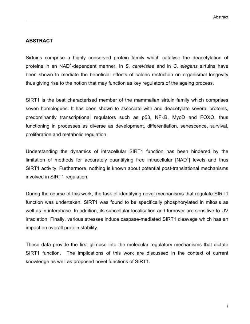
ABSTRACT
Sirtuins comprise a highly conserved protein family which catalyse the deacetylation of
proteins in an NAD+-dependent manner. In S. cerevisiae and in C. elegans sirtuins have
been shown to mediate the beneficial effects of caloric restriction on organismal longevity
thus giving rise to the notion that may function as key regulators of the ageing process.
SIRT1 is the best characterised member of the mammalian sirtuin family which comprises
seven homologues. It has been shown to associate with and deacetylate several proteins,
predominantly transcriptional regulators such as p53, NFκB, MyoD and FOXO, thus
functioning in processes as diverse as development, differentiation, senescence, survival,
proliferation and metabolic regulation.
Understanding the dynamics of intracellular SIRT1 function has been hindered by the
limitation of methods for accurately quantifying free intracellular [NAD+] levels and thus
SIRT1 activity. Furthermore, nothing is known about potential post-translational mechanisms
involved in SIRT1 regulation.
During the course of this work, the task of identifying novel mechanisms that regulate SIRT1
function was undertaken. SIRT1 was found to be specifically phosphorylated in mitosis as
well as in interphase. In addition, its subcellular localisation and turnover are sensitive to UV
irradiation. Finally, various stresses induce caspase-mediated SIRT1 cleavage which has an
impact on overall protein stability.
These data provide the first glimpse into the molecular regulatory mechanisms that dictate
SIRT1 function. The implications of this work are discussed in the context of current
knowledge as well as proposed novel functions of SIRT1.
i

Acknowledgements
Acknowledgements
To start with, I would like to thank Prof. Wilhelm Krek for providing me the opportunity to
collaborate with him for my first long-term investment into my scientific career, for the
stimulating environment that has nurtured my inquiring mind and his insightful approaches to
aspects of scientific discovery.
I thank my colleagues for the good times and company throughout these years, especially
those who challenged me and thus made me better.
I would like to express my appreciation to the other two members of my thesis committee
Prof. Peter and Prof. Hall for their commitment to their role, the acceptance of which they
have honored me with.
Finally, I would like to exercise my right to provide my partner Pia and my parents Kosta and
Eleni with my thanks in person. Anything else would only diminish the paramount role that
they have played and continue to play in my life as well as the completion of the work
presented here.
ii
Table of contents
TABLE OF CONTENTS CHAPTER 1 - ADAPTIVE CELLULAR RESPONSES TO ENVIRONMENTAL STIMULI 1.1 SIGNALLING PATHWAYS REGULATING ADAPTIVE RESPONSES TO NUTRIENT AVAILABILITY................................. 1
1.1.1 Archetypal signaling strategies in bacteria and lower eucaryotes.................................................... 1 . 1.1.2 Major homeostatic pathways in higher eucaryotes........................................................................... 5
1.1.2.1 The insulin/IGF signaling system................................................................................................ 5 1.1.2.1.1 The PI3K-PKB signaling pathway........................................................................................ 6 1.1.2.1.2 Endocrine functions of the IGF system and the regulation of longevity.............................. 10
1.1.2.2 The TOR signaling pathway...................................................................................................... 12 1.1.2.2.1 Signaling pathways regulating TOR activity....................................................................... 12 1.1.2.2.2 Functions of the TOR pathway............................................................................................ 14
1.1.2.3 Molecular pathways sensing oxygen......................................................................................... 16 1.2 REGULATION OF CHROMATIN STRUCTURE AND GENE EXPRESSION............................................................... 18
1.2.1 Regulation of chromatin structure.................................................................................................... 18 1.2.1.1 Histone variants......................................................................................................................... 19 1.2.1.2 ATP-dependent nucleosome remodeling.................................................................................. 19 1.2.1.3 Covalent histone modifications and the histone code concept.................................................. 20
1.2.1.3.1 Histone acetylation.............................................................................................................. 21 1.2.1.3.2 Histone methylation............................................................................................................. 24 1.2.1.3.3 Histone phosphorylation...................................................................................................... 25 1.2.1.3.4 Histone ubiquitination.......................................................................................................... 26 1.2.1.3.5 Histone ADP-ribosylation.................................................................................................... 26 1.2.1.3.6 Epigenetics and the 'histone code' hypothesis................................................................... 27
1.2.2 Transcriptional regulation................................................................................................................. 28 1.2.2.1 Basal transcription..................................................................................................................... 28 1.2.2.2 Transcription factors and their regulation.................................................................................. 30
1.2.2.2.1 Mechanisms of transcription factor action........................................................................... 30
1.2.2.2.2 Post-translational regulation of transcription factors........................................................... 31 1.2.2.2.3 Ligand-mediated modulation of transcription factor activity................................................ 32 1.2.2.2.4 Transcriptional regulatory networks.................................................................................... 33
1.2.2.3 Additional mechanisms of gene regulation................................................................................ 34 1.3 Conclusion.............................................................................................................................................. 35 CHAPTER 2 - THE SIRTUIN FAMILY OF PROTEIN DEACETYLASES 2. INTRODUCTION TO THE SIRTUIN FAMILY......................................................................................... 36 2.1 Discovery of sirtuins and determination of their enzymatic activity......................................................... 36 2.2 Structural and enzymatic properties of sirtuins....................................................................................... 38
2.2.1 Structural insights into the regulation of Hst2p................................................................................. 45 2.2.2 Fate of the sirtuin deacetylation products......................................................................................... 46
2.2.2.1 Nicotinamide and NAD+ biosynthesis pathways........................................................................ 46 2.2.2.2 The function and fate of 2',3'-O-ADP-ribose.............................................................................. 49
2.2.3 Specificity of sirtuins......................................................................................................................... 50 2.2.4 Small molecule modulators of sirtuins.............................................................................................. 51
2.3 FUNCTIONAL STUDIES OF SIRTUINS................................................................................................. 54 2.3.1 Sirtuin functions in prokaryotic organisms........................................................................................ 54
2.3.1.1 Bacterial sirtuins........................................................................................................................ 54 2.3.1.2 Archaeal sirtuins........................................................................................................................ 55
iii
Table of contents
2.3.2 Sirtuin functions in S. cerevisiae...................................................................................................... 55 2.3.2.1 Regulation of chromatin silencing in S. cerevisiae by sirtuins.................................................. 55 .
2.3.2.1.1 Silencing at mating type loci............................................................................................... 55 2.3.2.1.2 Chromatin silencing at telomeres....................................................................................... 58 2.3.2.1.3 Chromatin silencing at the rDNA locus............................................................................... 59
2.3.2.2 Regulation of meiotic checkpoint function and recombination................................................... 61 2.3.2.3 Regulation of DNA replication.................................................................................................... 62 2.3.2.4 Sir2 and the regulation of life-span in S. cerevisiae................................................................... 63
2.3.2.4.1 Molecular mechanisms that determine life-span in S. cerevisiae........................................ 63 2.3.2.4.2 Regulation of life-span by caloric restriction........................................................................ 65 2.3.2.4.3 Proposed mechanisms of Sir2p-mediated life-span extension by caloric restriction
in S. cerevisiae................................................................................................................... 66 2.3.2.5 Homologues of Sir2 (Hst) proteins............................................................................................. 67
2.3.3 Caenorhabditis elegans sirtuins....................................................................................................... 72 2.3.4 Drosophila melanogaster sirtuins..................................................................................................... 76 2.3.5 The mammalian sirtuin family........................................................................................................... 78
2.3.5.1 SIRT1......................................................................................................................................... 78 2.3.5.1.1 Expression and genetic ablation of SIRT1 in the mouse.................................................... 78 . 2.3.5.1.2 Regulation of chromatin structure by SIRT1....................................................................... 82 2.3.5.1.3 Regulation of transcription by SIRT1................................................................................... 85 2.3.5.1.4 Regulation of survival by SIRT1.......................................................................................... 90 2.3.5.1.5 SIRT1 regulation of transcription factors involved in muscle differentiation...................... 105 2.3.5.1.6 Genetic and biochemical interactions of SIRT1 with proteins regulating metabolism....... 107 2.3.5.1.7 Neuroprotection and cardioprotection by SIRT1............................................................... 112 2.3.5.1.8 SIRT1 and caloric restriction in rodents............................................................................ 113
2.3.5.2 SIRT2....................................................................................................................................... 114 2.3.5.3 SIRT3....................................................................................................................................... 116 2.3.5.4 SIRT6....................................................................................................................................... 118
2.4 Aim of the project.................................................................................................................................. 121 CHAPTER 3 - MATERIALS AND METHODS 3.1 BUFFERS............................................................................................................................................. 123 3.2 METHODS............................................................................................................................................ 129
3.2.1 BIOINFORMATICS RESOURCES................................................................................................. 129 3.2.2 MOLECULAR BIOLOGICAL TECHNIQUES.................................................................................. 129 3.2.3 CELL CULTURE METHODS......................................................................................................... 131
3.2.3.1 Mammalian cell culture............................................................................................................ 131 3.2.3.2 Insect cell culture..................................................................................................................... 136 3.2.3.3 Bacterial culture - Production of recombinant proteins in E. coli............................................. 137
3.2.4 BIOCHEMICAL TECHNIQUES...................................................................................................... 137 3.2.5 IMMUNOLOGICAL TECHNIQUES................................................................................................ 142
CHAPTER 4 - RESULTS: FUNCTION AND REGULATION OF SIRT1
4.1 REGULATION OF SIRT1 BY PHOSPHORYLATION.......................................................................... 148
4.1.1 Production of α-SIRT1 polyclonal and monoclonal antibodies....................................................... 148 4.1.2 SIRT1 is a phosphoprotein............................................................................................................. 154 4.1.3 Phosphorylation of SIRT1 in interphase......................................................................................... 156
4.1.3.1 Identification of phosphorylation sites of SIRT1 expressed in Sf9 cells.................................. 156 . 4.1.3.2 Identification of the mobility-shift inducing phosphorylation sites............................................ 160 4.1.3.3 In vitro phosphorylation of SIRT1 by Casein Kinase 2............................................................ 164
4.1.3.3.1 SIRT1 in the UV response..................................................................................................... 168
iv
Table of contents
4.1.4 Regulation of SIRT1 by phosphorylation in mitosis........................................................................ 179
4.2 REGULATION OF SIRT1 BY CASPASE-MEDIATED CLEAVAGE..................................................... 200 . 4.3 OTHER FEATURES OF THE SIRT1 PRIMARY SEQUENCE ............................................................ 210 CHAPTER 5 - DISCUSSION AND FUTURE PERSPECTIVES 5.1 Common pathways underlying homeostatic cellular processes are disregulated in diverse diseases. 211
5.1.1 Functional interconnection between the PKB, TOR and oxygen signaling pathways in health and disease..................................................................................................................... 212
5.1.1.1 Representative mechanisms employed by hypoxia to inhibit growth and proliferation........... 213 5.1.1.2 Reciprocal relation between metabolic enzymes and proteins regulating proliferation........... 213
5.2 The sirtuin family of protein deacetylases............................................................................................ 217 5.2.1 Sirtuins and the regulation of organismal life-span........................................................................ 219 5.2.2 Mammalian sirtuin function in diseases associated with ageing.................................................... 221
5.2.2.1 Molecular pathways involved in ageing are modulated by SIRT1........................................... 221 5.2.2.2 Molecular basis of SIRT1 function in ageing phenotypes........................................................ 222
5.2.2.2.1 Neuroprotective and cardioprotective roles of SIRT1........................................................ 223 5.2.2.2.2 SIRT1 and muscle mass maintenance.............................................................................. 224 5.2.2.2.3 SIRT1 functions in metabolic regulation............................................................................ 226 5.2.2.2.4 Reproduction..................................................................................................................... 228 5.2.2.2.5 SIRT1 and cancer............................................................................................................. 229
5.3 Reported roles of other sirtuins in the determination of life-span......................................................... 233 5.4 Role of NAD in transcriptional regulation and disease......................................................................... 234 5.5 SIRT1 as a drug target......................................................................................................................... 236 5.6 Emerging nuclear roles of metabolic enzymes and metabolic intermediates....................................... 238 5.7 Functional interaction between sirtuins and HDACs............................................................................. 240 5.8 Conclusion............................................................................................................................................ 241 5.9 THESIS RESULTS: DISCUSSION AND FUTURE PERSPECTIVES
5.9.1 Regulation of SIRT1 by phosphorylation....................................................................................... 243 . 5.9.1.1 SIRT1 is a nuclear phosphoprotein......................................................................................... 243 5.9.1.2 Identification of SIRT1 phosphorylation sites in interphase..................................................... 244 5.9.1.3 SIRT1 in cellular responses to UV irradiation.......................................................................... 249 5.9.1.4 Phosphorylation of SIRT1 in mitosis........................................................................................ 254
5.9.1.4.1 Potential functions of SIRT1 in mitosis.................................................................................. 262 5.9.2 Regulation of SIRT1 by caspase-mediated cleavage.................................................................... 267
5.9.2.1 Functional significance of caspase-mediated SIRT1 cleavage............................................... 268 5.9.3 Prediction of signalling pathways which SIRT1 may participate in................................................ 272 5.9.4 Conclusion..................................................................................................................................... 274
6. REFERENCES..................................................................................................................................... 276
v
Acronyms and abbreviations
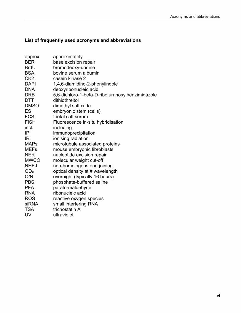
List of frequently used acronyms and abbreviations approx. approximately BER base excision repair BrdU bromodeoxy-uridine BSA bovine serum albumin CK2 casein kinase 2 DAPI 1,4,6-diamidino-2-phenylindole DNA deoxyribonucleic acid DRB 5,6-dichloro-1-beta-D-ribofuranosylbenzimidazole DTT dithiothreitol DMSO dimethyl sulfoxide ES embryonic stem (cells) FCS foetal calf serum FISH Fluorescence in-situ hybridisation incl. including IP immunoprecipitation IR ionising radiation MAPs microtubule associated proteins MEFs mouse embryonic fibroblasts NER nucleotide excision repair MWCO molecular weight cut-off NHEJ non-homologous end joining OD# optical density at # wavelength O/N overnight (typically 16 hours) PBS phosphate-buffered saline PFA paraformaldehyde RNA ribonucleic acid ROS reactive oxygen species siRNA small interfering RNA TSA trichostatin A UV ultraviolet
vi
Chapter 1 - Adaptive cellular responses to environmental stimuli
CHAPTER 1 ADAPTIVE CELLULAR RESPONSES TO
ENVIRONMENTAL STIMULI 1.1 SIGNALLING PATHWAYS REGULATING ADAPTIVE RESPONSES TO NUTRIENT AVAILABILITY
1.1.1 Archetypal signaling strategies in bacteria and lower eucaryotes
Evolutionary considerations suggest that the ensemble of living organisms that
constitute an environment's population stems from their ability to perpetuate under this
environment's particular conditions. By definition, such populations are fit to thrive. Yet, living
environments are dynamic rather than static and in combination with genetic variability
contribute to the evolution of the species. Thus, a paramount feature of living organisms
throughout the phyla is their ability to adapt to such environmental changes in order to
increase their survival potential.
Unicellular organisms have evolved specific biochemical systems of variable
complexity that allow them to respond to environmental changes such as fluctuating levels of
nutrients.
Bacteria preferentially utilise glucose as their primary carbon source even in the
presence of other sugars in their growth environment. Only following depletion of glucose can
1
Chapter 1 - Adaptive cellular responses to environmental stimuli
other sugars such as lactose also be used for energy production, a phenomenon called
diauxic growth. Jacob and Monod proposed the concept of the lac operon to explain this
phenomenon which was subsequently confirmed and elaborated extensively (Lewis, 2005). In
this model, the lac repressor can bind a cis acting element in the promoter region of genes
encoding proteins that allow lactose production suppressing their expression under
conditions of glucose abundance. When lactose is the primary carbon source, it binds to the
repressor inducing a conformational change which reduces its affinity for the operator,
leading to its dissociation from the promoter and allowing the expression of genes involved in
the uptake and metabolism of lactose.
More advanced signaling cascades in bacteria adopt a simple two-component
modular configuration comprising a sensor and an effector module. This is exemplified by the
two-component signal transduction (or phosphorelay) system which is widely employed by
bacteria and to a lesser extend by fungi and plants (Perraud et al., 1999). In bacteria, two-
component systems regulate basic cellular processes such as chemotaxis, osmoregulation,
temperature sensing, metabolism and
membrane transport (West and Stock, 2001).
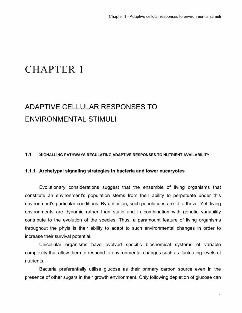
The basic architecture of such
systems is depicted in FIGURE 1-1. The
sensor (HK) is a transmembrane protein
(TM1 and TM2 are the membrane-
spanning regions) which can dimerise
through a dimerisation domain. The
intracellular region of the protein contains
a histidine kinase activity characterised by
four conserved motifs (N, G1, F, G2). The
effector component of the system (RR)
contains a conserved regulatory domain
and an effector domain. Environmental
stimuli induce the histidine kinase activity
of HK leading to its autophosphorylation
(depicted with P in FIGURE 1-1). Following
FIGURE 1-1. Schematic represenation of a basictwo-component phosphotransfer system. A typicaltwo-component phosphotransfer system consists of adimeric transmembrane sensor HK and a cytoplasmicRR. A monomer of a representative HK is shown withtransmembrane segments indicated by TM1 and TM2.Conserved sequence motifs N, G1, F and G2, arelocated in the ATP-binding domain. HKs catalyze ATP-dependent autophosphorylation of a specificconserved His residue (H). The activities of HKs aremodulated by environmental signals. The phosphorylgroup (P) is then transferred to a specific Asp residue(D) located within the conserved regulatory domain ofan RR. Phosphorylation of the RR typically activatesan associated (or downstream) effector domain, whichultimately elicits a specific cellular response.
Adapted from West and Stock, 2001
2
Chapter 1 - Adaptive cellular responses to environmental stimuli
that, RR catalyses the transfer of the phosphate group to one of its own aspartic acid
residues within the regulatory domain leading to the activation of the effector domain. More
elaborate systems based on these principles including consecutive histidine/aspartic acid
phosphorelay systems are also found (Perraud et al., 1999).
Unicellular eucaryotic organisms such as the yeast S. cerevisiae exhibit increased
complexity in the signaling cascades mediating adaptive responses, which reflect not only
their architectural differences to procaryotes (e.g. in the case of regulated nucleocytoplasmic
transport) but also their increased computational capacity in decision-making processes. A
well-studied system, largely conserved also in higher eucaryotes is the mitogen-activated
protein kinase (MAPK) signalling cascade in yeast.
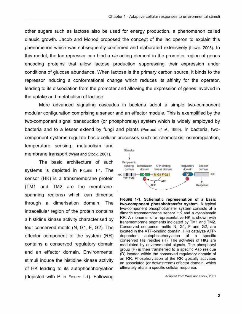
The MAPK kinase signaling pathwa
behaviour and responses to osmotic stress
and nutrient availability (FIGURE 1-2). Upon
induction, a kinase cascade involving
sequential phosphorylation/activation steps
is initiated (FIGURE 1-3A). Surprisingly,
multiple stimuli use a largely shared set of
molecules to elicit diverse and specific
responses raising the issue how specificity
is attained in such systems.
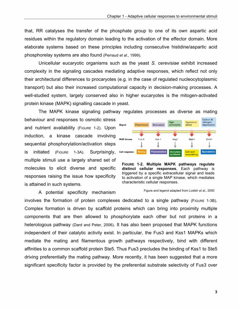
A potential specificit
y regulates processes as diverse as mating
y mechanism
involve
FIGURE 1-2. Multiple MAPK pathways regulatedistinct cellular responses. Each pathway istriggered by a specific extracellular signal and leadsto activation of a single MAP kinase, which mediatescharacteristic cellular responses.
Figure and legend adapted from Lodish et al., 2000
s the formation of protein complexes dedicated to a single pathway (FIGURE 1-3B).
Complex formation is driven by scaffold proteins which can bring into proximity multiple
components that are then allowed to phosphorylate each other but not proteins in a
heterologous pathway (Dard and Peter, 2006). It has also been proposed that MAPK functions
independent of their catalytic activity exist. In particular, the Fus3 and Kss1 MAPKs which
mediate the mating and filamentous growth pathways respectively, bind with different
affinities to a common scaffold protein Ste5. Thus Fus3 precludes the binding of Kss1 to Ste5
driving preferentially the mating pathway. More recently, it has been suggested that a more
significant specificity factor is provided by the preferential substrate selectivity of Fus3 over
3
Chapter 1 - Adaptive cellular responses to environmental stimuli
A B
FIGURE 1-3. Mechanisms of specificity in yeast MAPK signaling. (A) MAPK cascade that transmitssignals in the mating pathway in S. cerevisiae. The receptors for yeast a or a mating factors are bothcoupled to the same trimeric G protein. Ligand binding leads to activation of the G protein and dissociationof Gα · GTP from the G βγ complex. In the yeast mating pathway, however, the physiological responses areinduced by the dissociated Gβγ, which activates a protein kinase cascade. The final component, Fus3, isfunctionally equivalent to MAP kinase (MAPK) in higher eukaryotes. It phosphorylates transcription factors(e.g., Ste12) that control expression of proteins involved in mating-specific cellular responses. (B)Formation of pathway-specific complexes prevents "cross-talk" between pathways that contain a commoncomponent, such as Ste11 in these two pathways. These large complexes are assembled on themolecular scaffolds Ste5 and Pbs2. Unlike Ste5, which has no catalytic function, Pbs2 has MEK activity(analogous to Ste7 in the mating pathway). Once phosphorylated by Ste11, activated Pbs2phosphorylates Hog1.
Figure and legend adapted from Lodish et al., 2000
Kss1 towards Far1 rather than physical occlusion of Kss1 from the signaling scaffold
(Breitkreutz and Tyers, 2002).
Thus, even in single-cell eucaryotes, elaborate networks are in action to sense and
respond to environmental changes. Although the principles of adaptive responses delineated
for unicellular organisms are broadly conserved in more advanced forms of life, multicellular
organisms exhibit increased complexity in the form of functionally specialised organs and
organ systems.
Reflecting this complexity, an additional level of co-ordination is required to sustain
survival in response to environmental as well as intraorganismal changes. For this to be
achieved elaborate endocrine systems are in action. Such a signaling system with central
roles in animal physiology is mediated by the hormone insulin and the related insulin-like
growth factors (IGFs), IGF1 and IGF2. Both at the intracellular as well as organismal level,
4
Chapter 1 - Adaptive cellular responses to environmental stimuli
the sophistication of this system exemplifies the underlying basis of advanced biological
systems design.
1.1.2 Major homeostatic pathways in higher eucaryotes 1.1.2.1 The insulin/IGF signaling system
The insulin/IGF sytem is involved in fundamental biological processes such as growth,
proliferation, survival and metabolic regulation (White, 2003; Pollak et al., 2004). For example, in
response to feeding, insulin, which is produced in the pancreas, dictates the uptake and
catabolism of glucose by peripheral tissues. IGF1 and IGF2 are produced primarily by the
liver and have mitogenic capacity. IGF1 but not IGF2 production is dictated by pituitary gland-
derived growth hormone underlying its function in regulating animal size (Kenyon, 2001). The
insulin/IGF system has also an evolutionarily conserved function in determining organismal
longevity which is tighly linked to its responsiveness to nutritional inputs (Kenyon, 2001).
The effects of insulin/IGF are mediated by binding to three receptors, the insulin
receptor (IR), IGF1 receptor (IGF1R) and IGF2 receptor (IGF2R). A fourth family member
exists named insulin receptor-related receptor (IRR) for which an endogenous ligand has not
been identified (Kitamura et al., 2003).
IR, IGF1R and IRR harbour ligand-activated tyrosine kinase activity in their
intracellular domains which initiates downstream signaling cascades. Although several
protein substrates of the insulin/IGF receptor tyrosine kinase activity have been identified,
genetic ablation studies in mice suggest that the majority of insulin responses are mediated
by insulin receptor substrates 1 or 2 (IRS1 or IRS2 respectively) (White, 2003). Thus, IRS1 is
responsible for body growth control and peripheral insulin action, while IRS2 controls brain
growth, body weight, glucose homeostasis and female fertility (White, 2003).
IRSs are scaffold proteins which upon their phosphorylation allow the docking of
multiple kinases or other scaffold proteins that contain phospho-aminoacid binding domains.
Upon binding to IRS proteins through its SH2 domains, the lipid kinase activity of
phosphoinositide-3 kinase (PI3K) is induced and results in increased membrane
phosphatidylinositol-3,4,5-triphosphate [PtdIns(3,4,5)P3] levels (Vanhaesebroeck and Alessi,
5
Chapter 1 - Adaptive cellular responses to environmental stimuli
2000). These lipids are preferentially recognised by the pleckstrin homology (PH) domains of
protein kinase B (PKB, a.k.a. Akt) and phosphoinositide-dependent kinase (PDK). Following
membrane recruitment, PDK phosphorylates PKB at S308 contributing to its activation.
A critical regulator of this pathway is the tumour suppressor protein PTEN
(phosphatase and tensin homologue deleted on chromosome 10). PTEN is a lipid
phosphatase which attenuates PKB activation by catalysing the reverse reaction to that of
PI3K.
1.1.2.1.1 The PI3K-PKB signaling pathway
Regulation of cell survival
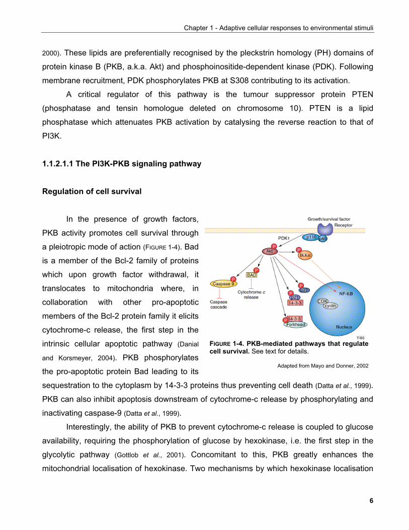
In the presence of growth factors,
PKB activity promotes cell survival through
a pleiotropic mode of action (FIGURE 1-4). Bad
is a member of the Bcl-2 family of proteins
which upon growth factor withdrawal, it
translocates to mitochondria where, in
collaboration with other pro-apoptotic
members of the Bcl-2 protein family it elicits
cytochrome-c release, the first step in the
intrinsic cellular apoptotic pathway (Danial
and Korsmeyer, 2004). PKB phosphorylates
the pro-apoptotic protein Bad leading to its
sequestration to the cytoplasm by 14-3-3 proteins thus preventing cell death (Datta et al., 1999).
PKB can also inhibit apoptosis downstream of cytochrome-c release by phosphorylating and
inactivating caspase-9 (Datta et al., 1999).
FIGURE 1-4. PKB-mediated pathways that regulatecell survival. See text for details.
Adapted from Mayo and Donner, 2002
Interestingly, the ability of PKB to prevent cytochrome-c release is coupled to glucose
availability, requiring the phosphorylation of glucose by hexokinase, i.e. the first step in the
glycolytic pathway (Gottlob et al., 2001). Concomitant to this, PKB greatly enhances the
mitochondrial localisation of hexokinase. Two mechanisms by which hexokinase localisation
6
Chapter 1 - Adaptive cellular responses to environmental stimuli
to mitochondria prevents apoptosis have been proposed (Majewski et al., 2004). Firstly,
hexokinase precludes the recruitment of the pro-apoptotic protein Bax to mitochondria
preventing cytochrome-c release. Secondly, it participates in the maintainance of
mitochondrial integrity by regulating the voltage-dependent anion channel (VDAC) in the
outer mitochondrial membrane. VDAC is involved in the exchange of metabolites such as
adenine nucleotides and respiratory substrates across the outer mitochondrial membrane
contributing to mitochondrial homeostasis. Upon glucose withdrawal, decreased hexokinase
at mitochondria results in VDAC closure leading to mitochondrial outer membrane swelling
and eventual rupture.
PKB also contributes to the activation of the anti-apoptotic pathway driven by the
transcription factor NFκB. PKB phosphorylates and activates the inhibitor of κB kinase β
(IKKβ). IKKβ in turn targets the inhibitor of κB (IκB) proteins for degradation allowing the
activation of NFκB. NFκB target genes include the cellular inhibitor of apoptosis (cIAP)
proteins that bind to and inactivate caspases (Datta et al., 1999).
Forkhead or winged-helix transcription factors are also regulated by PKB activity. In
the presence of growth factors, PKB phosphorylates FOXOs (forkhead box subclass O) and
FOXA2 factors leading to their sequestration in the cytoplasm by 14-3-3 proteins (Plas and
Thompson, 2005). Upon growth factor limitation, FOXOs can translocate to the nucleus where
they bind cognate DNA sequences in target gene promoters modulating their expression. A
FOXO target gene is Fas ligand (FasL) which upon binding to its cognate receptor induces
apoptotic cell death in neuronal cells (Datta et al., 1999). PKB-mediated phosphorylation
prevents the pro-apoptotic function of FOXO through FasL expression.
Regulation of cell cycle
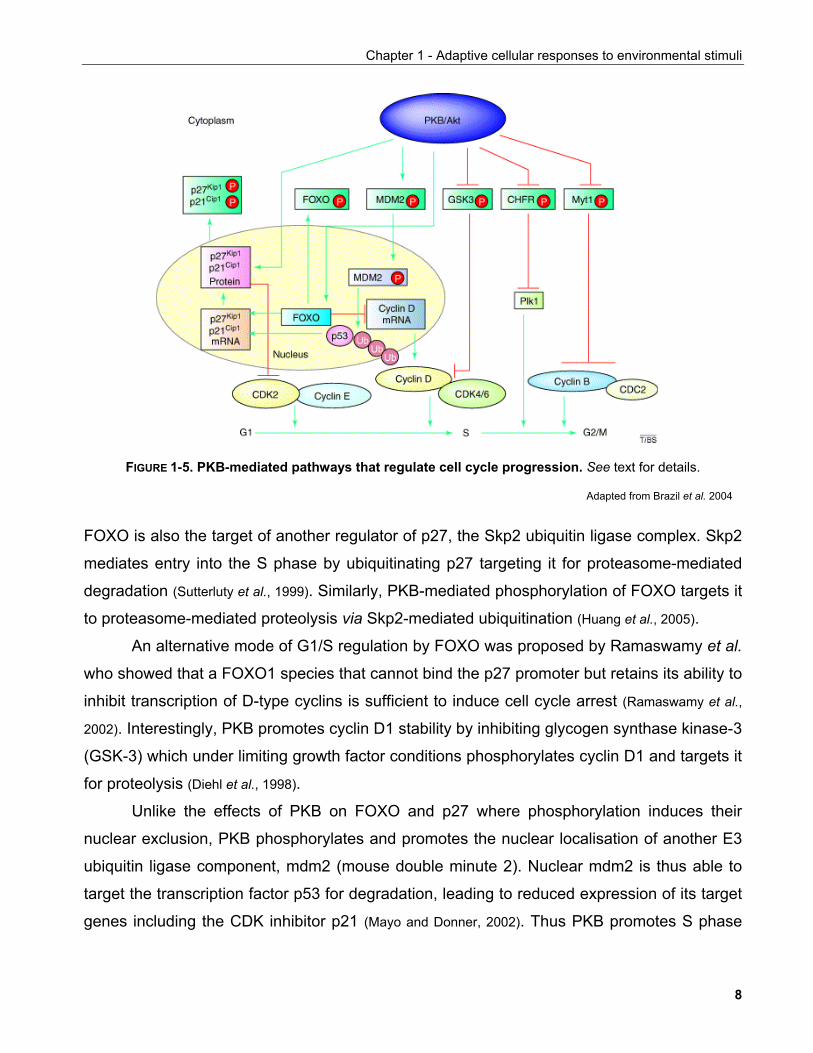
Another emerging function of PKB is in the regulation of the cell cycle (FIGURE 1-5). PKB
attentuates the activity of the cyclin-dependent kinase (CDK) inhibitor p27 by at least two
mechanisms. By directly phosphorylating p27, PKB induces its retention to the cytoplasm by
14-3-3 proteins preventing p27 from inhibiting nuclear CDK complexes (Shin et al., 2002).
Secondly, p27 is a transriptional target of FOXO transcription factors thus, in the presence of
growth factors, nuclear exclusion of FOXO leads to reduced p27 transcription. Interestingly,
7
Chapter 1 - Adaptive cellular responses to environmental stimuli
FIGURE 1-5. PKB-mediated pathways that regulate cell cycle progression. See text for details.
Adapted from Brazil et al. 2004
FOXO is also the target of another regulator of p27, the Skp2 ubiquitin ligase complex. Skp2
mediates entry into the S phase by ubiquitinating p27 targeting it for proteasome-mediated
degradation (Sutterluty et al., 1999). Similarly, PKB-mediated phosphorylation of FOXO targets it
to proteasome-mediated proteolysis via Skp2-mediated ubiquitination (Huang et al., 2005).
An alternative mode of G1/S regulation by FOXO was proposed by Ramaswamy et al.
who showed that a FOXO1 species that cannot bind the p27 promoter but retains its ability to
inhibit transcription of D-type cyclins is sufficient to induce cell cycle arrest (Ramaswamy et al.,
2002). Interestingly, PKB promotes cyclin D1 stability by inhibiting glycogen synthase kinase-3
(GSK-3) which under limiting growth factor conditions phosphorylates cyclin D1 and targets it
for proteolysis (Diehl et al., 1998).
Unlike the effects of PKB on FOXO and p27 where phosphorylation induces their
nuclear exclusion, PKB phosphorylates and promotes the nuclear localisation of another E3
ubiquitin ligase component, mdm2 (mouse double minute 2). Nuclear mdm2 is thus able to
target the transcription factor p53 for degradation, leading to reduced expression of its target
genes including the CDK inhibitor p21 (Mayo and Donner, 2002). Thus PKB promotes S phase
8
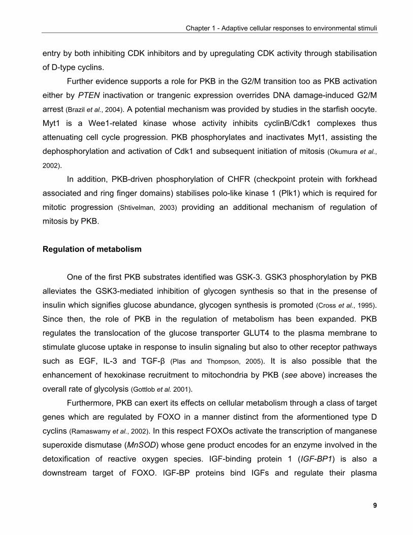
Chapter 1 - Adaptive cellular responses to environmental stimuli
entry by both inhibiting CDK inhibitors and by upregulating CDK activity through stabilisation
of D-type cyclins.
Further evidence supports a role for PKB in the G2/M transition too as PKB activation
either by PTEN inactivation or trangenic expression overrides DNA damage-induced G2/M
arrest (Brazil et al., 2004). A potential mechanism was provided by studies in the starfish oocyte.
Myt1 is a Wee1-related kinase whose activity inhibits cyclinB/Cdk1 complexes thus
attenuating cell cycle progression. PKB phosphorylates and inactivates Myt1, assisting the
dephosphorylation and activation of Cdk1 and subsequent initiation of mitosis (Okumura et al.,
2002).
In addition, PKB-driven phosphorylation of CHFR (checkpoint protein with forkhead
associated and ring finger domains) stabilises polo-like kinase 1 (Plk1) which is required for
mitotic progression (Shtivelman, 2003) providing an additional mechanism of regulation of
mitosis by PKB.
Regulation of metabolism
One of the first PKB substrates identified was GSK-3. GSK3 phosphorylation by PKB
alleviates the GSK3-mediated inhibition of glycogen synthesis so that in the presense of
insulin which signifies glucose abundance, glycogen synthesis is promoted (Cross et al., 1995).
Since then, the role of PKB in the regulation of metabolism has been expanded. PKB
regulates the translocation of the glucose transporter GLUT4 to the plasma membrane to
stimulate glucose uptake in response to insulin signaling but also to other receptor pathways
such as EGF, IL-3 and TGF-β (Plas and Thompson, 2005). It is also possible that the
enhancement of hexokinase recruitment to mitochondria by PKB (see above) increases the
overall rate of glycolysis (Gottlob et al. 2001).
Furthermore, PKB can exert its effects on cellular metabolism through a class of target
genes which are regulated by FOXO in a manner distinct from the aformentioned type D
cyclins (Ramaswamy et al., 2002). In this respect FOXOs activate the transcription of manganese
superoxide dismutase (MnSOD) whose gene product encodes for an enzyme involved in the
detoxification of reactive oxygen species. IGF-binding protein 1 (IGF-BP1) is also a
downstream target of FOXO. IGF-BP proteins bind IGFs and regulate their plasma
9
Chapter 1 - Adaptive cellular responses to environmental stimuli
availability since they attenuate their ability to activate their cognate receptors (Pollak et al.,
2004). Furthermore, FOXOs co-regulate the expression of metabolic genes in combination
with nuclear receptors. FOXO in conjunction with peroxisome proliferator-activated receptor-γ
(PPAR-γ) represses transciprtional activity of IGF-BP1 and phosphoenolpyruvate
carboxykinase (PEPCK) gene promoters. Conversely, in combination with PGC-1α (PPAR-γ
co-activator 1α), FOXO induces the transcription of PEPCK and glucose-6-phosphatase (G-
6-Pase) upon fasting in the liver thus contributing to the gluconeogenesis programme
(Puigserver et al., 2003). FOXO transcriptional responses are in addition fine-tuned by
acetylation, more of which will be discussed in Chapter 2.
1.1.2.1.2 Endocrine functions of the IGF system and the regulation of longevity
Endocrine IGF signaling is central to organismal growth. IGF factors are produced in
the liver a process controlled by growth hormone (GH). GH is produced by the pituitary gland
in response to signals from the hypothalamus, mainly somatostatin and growth-hormone-
releasing hormone (GHRH). The ability of GH to regulate IGF production though, is greatly
influenced by dietary input. Under low food intake conditions, IGF production is suppressed
(Thissen et al., 1994).
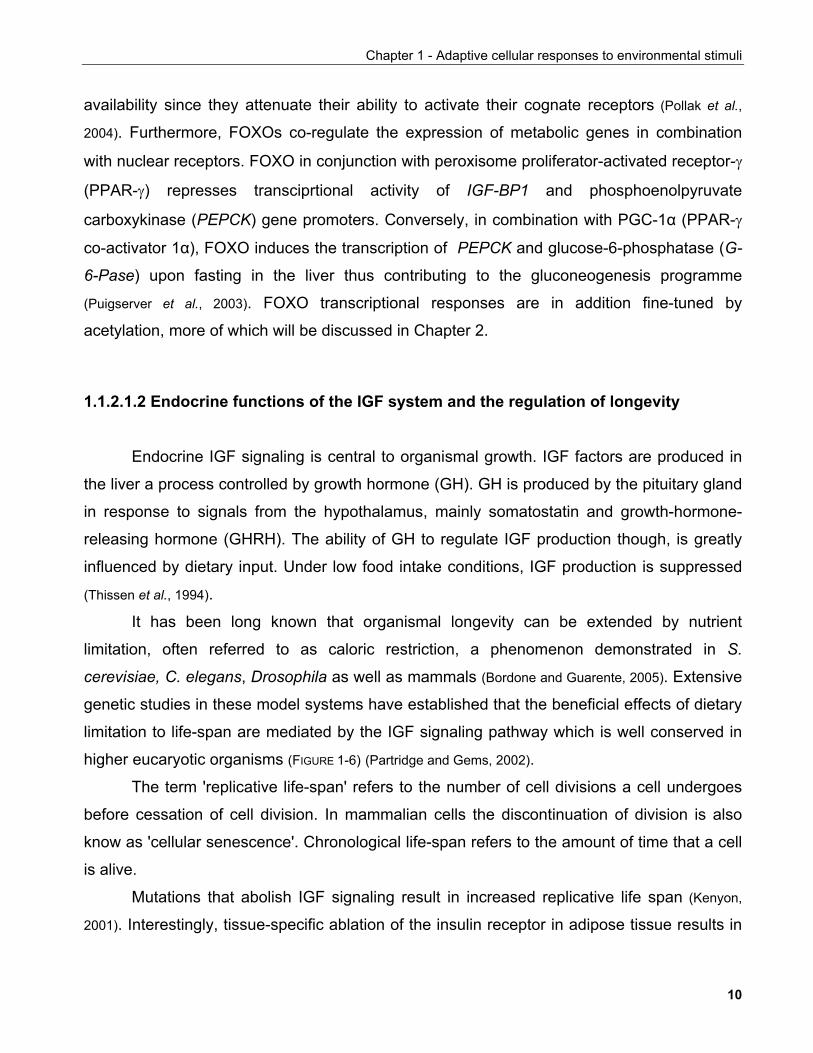
It has been long known that organismal longevity can be extended by nutrient
limitation, often referred to as caloric restriction, a phenomenon demonstrated in S.
cerevisiae, C. elegans, Drosophila as well as mammals (Bordone and Guarente, 2005). Extensive
genetic studies in these model systems have established that the beneficial effects of dietary
limitation to life-span are mediated by the IGF signaling pathway which is well conserved in
higher eucaryotic organisms (FIGURE 1-6) (Partridge and Gems, 2002).
The term 'replicative life-span' refers to the number of cell divisions a cell undergoes
before cessation of cell division. In mammalian cells the discontinuation of division is also
know as 'cellular senescence'. Chronological life-span refers to the amount of time that a cell
is alive.
Mutations that abolish IGF signaling result in increased replicative life span (Kenyon,
2001). Interestingly, tissue-specific ablation of the insulin receptor in adipose tissue results in
10
Chapter 1 - Adaptive cellular responses to environmental stimuli
FIGURE 1-6. Neuroendocrine regulation of ageing. (a) Insulin/IGF signalling in a two-step hormone signallingsystem in Caenorhabditis elegans. In this model, food modulates the production of an insulin-like peptidehormone (INS) by chemosensory neurons. This acts on DAF-2, which is also expressed in the nervoussystem, to cause the production of a second hormone signal, which modulates development and ageingthroughout the organism. Elements of this model are speculative, and the following remain to be determined:whether environmental stimuli regulate INS production and what these stimuli are; the role of DAF-16 inregulating secondary hormone production; and whether this hormone regulates longevity. (b) Two hypothesesfor the role of insulin/IGF signalling in ageing in Drosophila. In both models, Drosophila insulin-like peptides(DILPs) are produced by the brain in response to environmental or internal nutritional stimuli. How theproduction of DILPs is regulated is unknown. In one version of this model, DILPs act directly on the ovaries,stimulating the production of the steroid hormone ecdysone; in the other, DILPs stimulate the production of theisoprenoid hormone juvenile hormone by the CORPORA ALLATA. (c) Insulin/IGF signalling in mice. Thismodel proposes that Igf1, rather than insulin, acts as a modulator of ageing in mammals; this role of Igf1 inageing remains to be shown directly. DAF, dauer larva formation abnormal; Igf1, insulin-like growth factor 1;Inr, insulin-like receptor.
Figure and legend adapted from Partridge and Gems, 2002
a ~18% increase in life-span in mice suggesting that specific metabolic effects are
responsible for this effect (Bluher et al., 2003).
The influence of IGF on life-span is tightly coupled to its ability to down-regulate
forkhead transcription factors (Kenyon, 2005). In C. elegans, life-span extension due to
mutations in the IGF pathway depend on the presence of DAF-16. In agreement to this,
dFOXO overexpression in Drosophila results in lifespan extension. This function of FOXO
factors is tighly coupled to a concomitant resistance to stress (Kenyon, 2001; Kenyon, 2005)
which is also thought to operate in other mutant animals with extended life-span (Miggliacio et
al., 1999).
11
Chapter 1 - Adaptive cellular responses to environmental stimuli
An explanation of this may lie with the genes regulated by forkhead factors. FOXO
drives the expression of MnSOD, an enzyme involved in superoxide detoxification. MnSOD
overexpression in Drosophila suffices to confer life-span extension (Kenyon, 2001). This
evidence provides support of the "free radical theory" of ageing which states that the rate of
ageing is related to the deleterious effects of free raicals upon the cell (Balaban et al., 2005).
Although relatively little is known about the downstream effectors of free radicals it has
been proposed that they cause an accumulation of mutations in the DNA leading to
progressively aberrant cellular functions leading to cellular death and the decline of organ
performance (Lombard et al., 2005). Alternatively, there is evidence that signaling pathways that
regulate cellular survival are regulated by reactive oxygen species (ROS). Jun N-terminal
kinase (Jnk) is activated by phosphorylation which is counteracted by the action of
phosphatases. The enzymatic activity of Jnk phosphatases is regulated by ROS in that high
ROS levels oxidise a key residue in the phosphatase catalytic site leading to their inactivation
(Kamata et al., 2005). Thus Jnk kinases are allowed to elicit the cellular apoptotic programme
which in turn may contribute to tissue decline (Balaban et al., 2005).
Despite the lack of a classical IGF signaling pathway in yeast, homologues thereof
have been also implicated in the regulation of cellular life-span. Mutations in Sch9 a gene
encoding for a homologue of PKB confer increased replicative life-span in S. cerevisiae.
Interestingly, two recent studies identified yeast TOR1 as a negative effector of both
replicative and chronological life-span in response to nutrient satiety consistent the interplay
of this pathway with the IGF system in higher organisms (Kaeberlein et al., 2005; Powers et al.,
2006). Thus, the conserved functions of IGF signaling in response to dietary factors appear to
underlie the determination of cellular as well as organismal life-span.
1.1.2.2 The TOR signaling pathway
1.1.2.2.1 Signaling pathways regulating TOR activity
Under conditions of growth factor availability, cell proliferation is favoured. However, to
ensure sustainable growth, cell division has to be co-ordinated with concomitant increases in
12
Chapter 1 - Adaptive cellular responses to environmental stimuli
cell mass, primarily protein synthesis. A key signaling module in the regulation of cell growth
is mediated by the target of rapamycin (TOR) kinase.
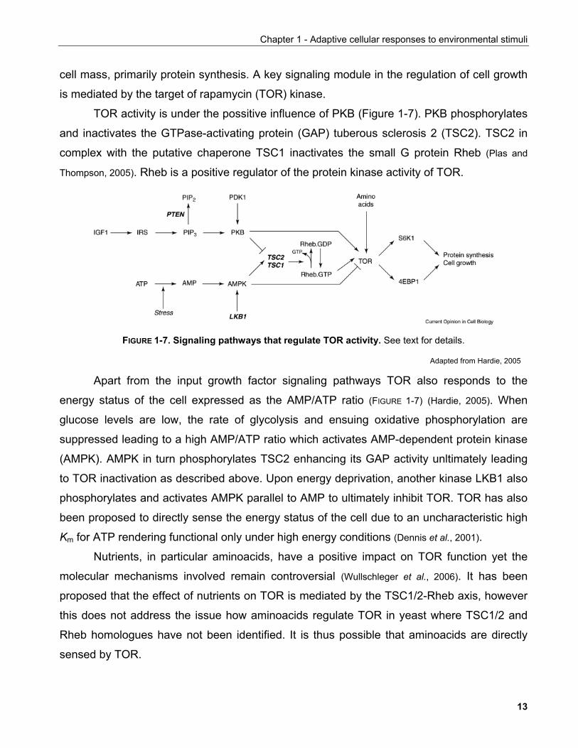
TOR activity is under the possitive influence of PKB (Figure 1-7). PKB phosphorylates
and inactivates the GTPase-activating protein (GAP) tuberous sclerosis 2 (TSC2). TSC2 in
complex with the putative chaperone TSC1 inactivates the small G protein Rheb (Plas and
Thompson, 2005). Rheb is a positive regulator of the protein kinase activity of TOR.
FIGURE 1-7. Signaling pathways that regulate TOR activity. See text for details.
Adapted from Hardie, 2005
Apart from the input growth factor signaling pathways TOR also responds to the
energy status of the cell expressed as the AMP/ATP ratio (FIGURE 1-7) (Hardie, 2005). When
glucose levels are low, the rate of glycolysis and ensuing oxidative phosphorylation are
suppressed leading to a high AMP/ATP ratio which activates AMP-dependent protein kinase
(AMPK). AMPK in turn phosphorylates TSC2 enhancing its GAP activity unltimately leading
to TOR inactivation as described above. Upon energy deprivation, another kinase LKB1 also
phosphorylates and activates AMPK parallel to AMP to ultimately inhibit TOR. TOR has also
been proposed to directly sense the energy status of the cell due to an uncharacteristic high
Km for ATP rendering functional only under high energy conditions (Dennis et al., 2001).
Nutrients, in particular aminoacids, have a positive impact on TOR function yet the
molecular mechanisms involved remain controversial (Wullschleger et al., 2006). It has been
proposed that the effect of nutrients on TOR is mediated by the TSC1/2-Rheb axis, however
this does not address the issue how aminoacids regulate TOR in yeast where TSC1/2 and
Rheb homologues have not been identified. It is thus possible that aminoacids are directly
sensed by TOR.
13
Chapter 1 - Adaptive cellular responses to environmental stimuli
1.1.2.2.2 Functions of the TOR pathway
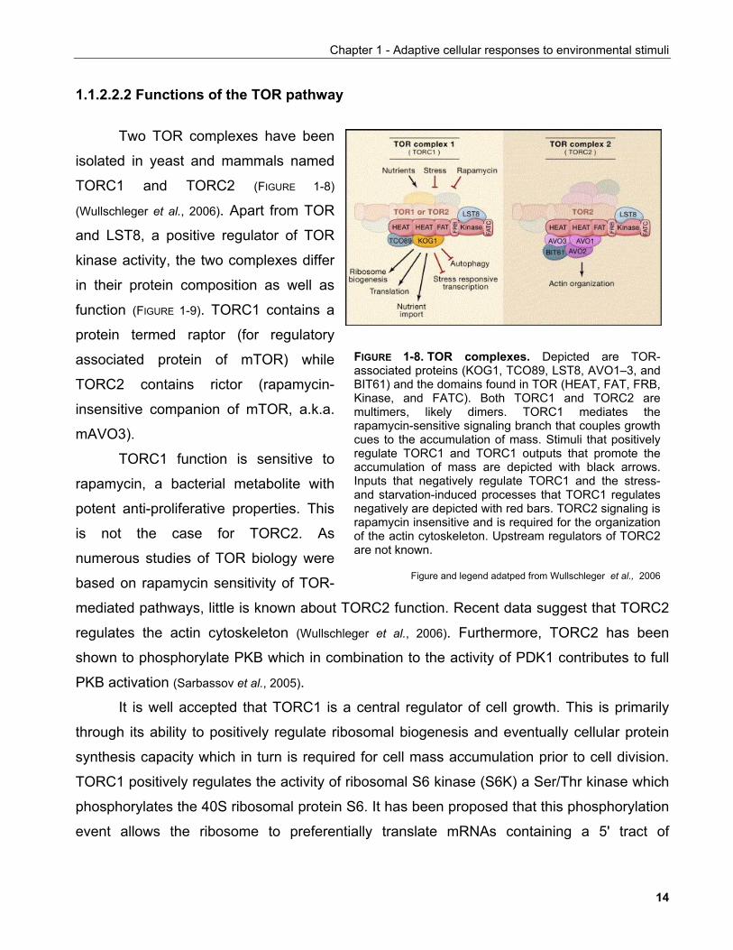
Two TOR complexes have been
isolated in yeast and mammals named
TORC1 and TORC2 (FIGURE 1-8)
(Wullschleger et al., 2006). Apart from TOR
and LST8, a positive regulator of TOR
kinase activity, the two complexes differ
in their protein composition as well as
function (FIGURE 1-9). TORC1 contains a
protein termed raptor (for regulatory
associated protein of mTOR) while
TORC2 contains rictor (rapamycin-
insensitive companion of mTOR, a.k.a.
mAVO3).
TORC1 function is sensitive to
rapamycin, a bacterial metabolite with
potent anti-proliferative properties. This
is not the case for TORC2. As
numerous studies of TOR biology were
based on rapamycin sensitivity of TOR-
mediated pathways, little is known about TORC2 function. Recent data suggest that TORC2
regulates the actin cytoskeleton (Wullschleger et al., 2006). Furthermore, TORC2 has been
shown to phosphorylate PKB which in combination to the activity of PDK1 contributes to full
PKB activation (Sarbassov et al., 2005).
FIGURE 1-8. TOR complexes. Depicted are TOR-associated proteins (KOG1, TCO89, LST8, AVO1–3, andBIT61) and the domains found in TOR (HEAT, FAT, FRB,Kinase, and FATC). Both TORC1 and TORC2 aremultimers, likely dimers. TORC1 mediates therapamycin-sensitive signaling branch that couples growthcues to the accumulation of mass. Stimuli that positivelyregulate TORC1 and TORC1 outputs that promote theaccumulation of mass are depicted with black arrows.Inputs that negatively regulate TORC1 and the stress-and starvation-induced processes that TORC1 regulatesnegatively are depicted with red bars. TORC2 signaling israpamycin insensitive and is required for the organizationof the actin cytoskeleton. Upstream regulators of TORC2are not known.
Figure and legend adatped from Wullschleger et al., 2006
It is well accepted that TORC1 is a central regulator of cell growth. This is primarily
through its ability to positively regulate ribosomal biogenesis and eventually cellular protein
synthesis capacity which in turn is required for cell mass accumulation prior to cell division.
TORC1 positively regulates the activity of ribosomal S6 kinase (S6K) a Ser/Thr kinase which
phosphorylates the 40S ribosomal protein S6. It has been proposed that this phosphorylation
event allows the ribosome to preferentially translate mRNAs containing a 5' tract of
14
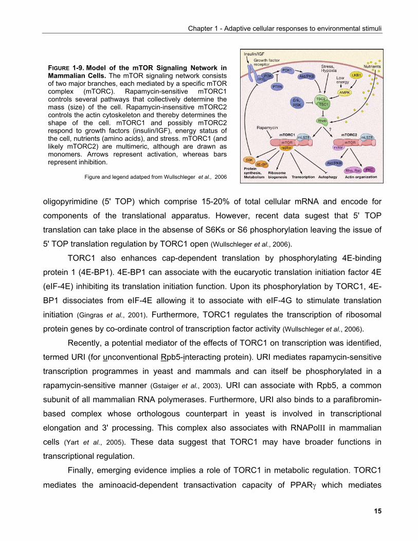
Chapter 1 - Adaptive cellular responses to environmental stimuli
FIGURE 1-9. Model of the mTOR Signaling Network inMammalian Cells. The mTOR signaling network consistsof two major branches, each mediated by a specific mTORcomplex (mTORC). Rapamycin-sensitive mTORC1controls several pathways that collectively determine themass (size) of the cell. Rapamycin-insensitive mTORC2controls the actin cytoskeleton and thereby determines theshape of the cell. mTORC1 and possibly mTORC2respond to growth factors (insulin/IGF), energy status ofthe cell, nutrients (amino acids), and stress. mTORC1 (andlikely mTORC2) are multimeric, although are drawn asmonomers. Arrows represent activation, whereas barsrepresent inhibition.
Figure and legend adatped from Wullschleger et al., 2006
oligopyrimidine (5' TOP) which comprise 15-20% of total cellular mRNA and encode for
components of the translational apparatus. However, recent data sugest that 5' TOP
translation can take place in the absense of S6Ks or S6 phosphorylation leaving the issue of
5' TOP translation regulation by TORC1 open (Wullschleger et al., 2006).
TORC1 also enhances cap-dependent translation by phosphorylating 4E-binding
protein 1 (4E-BP1). 4E-BP1 can associate with the eucaryotic translation initiation factor 4E
(eIF-4E) inhibiting its translation initiation function. Upon its phosphorylation by TORC1, 4E-
BP1 dissociates from eIF-4E allowing it to associate with eIF-4G to stimulate translation
initiation (Gingras et al., 2001). Furthermore, TORC1 regulates the transcription of ribosomal
protein genes by co-ordinate control of transcription factor activity (Wullschleger et al., 2006).
Recently, a potential mediator of the effects of TORC1 on transcription was identified,
termed URI (for unconventional Rpb5-interacting protein). URI mediates rapamycin-sensitive
transcription programmes in yeast and mammals and can itself be phosphorylated in a
rapamycin-sensitive manner (Gstaiger et al., 2003). URI can associate with Rpb5, a common
subunit of all mammalian RNA polymerases. Furthermore, URI also binds to a parafibromin-
based complex whose orthologous counterpart in yeast is involved in transcriptional
elongation and 3' processing. This complex also associates with RNAPolII in mammalian
cells (Yart et al., 2005). These data suggest that TORC1 may have broader functions in
transcriptional regulation.
Finally, emerging evidence implies a role of TORC1 in metabolic regulation. TORC1
mediates the aminoacid-dependent transactivation capacity of PPARγ which mediates
15
Chapter 1 - Adaptive cellular responses to environmental stimuli
adipogenesis. In addition, genetic ablation of S6K1 confers resistance to diet and age-
induced obesity in mice (Um et al., 2004). The underlying mechanism was attributed to the
inhibitory phosphorylation of IRS1 by S6K under conditions of nutrient abundance which
negatively regulates insulin signaling.
1.1.2.3 Molecular pathways sensing oxygen Another important attribute of cells concerns their capacity to sense oxygen, an
important factor for cellular functions such as oxidative phosphorylation. This is exemplified
by solid tumours whose development is inhibited by agents that block their ability to elicit
angiogenesis which otherwise provides the necessary supply of nutrients and oxygen to
support survival (Reymond and Segrè, 2006; Carmeliet and Jain, 2000).
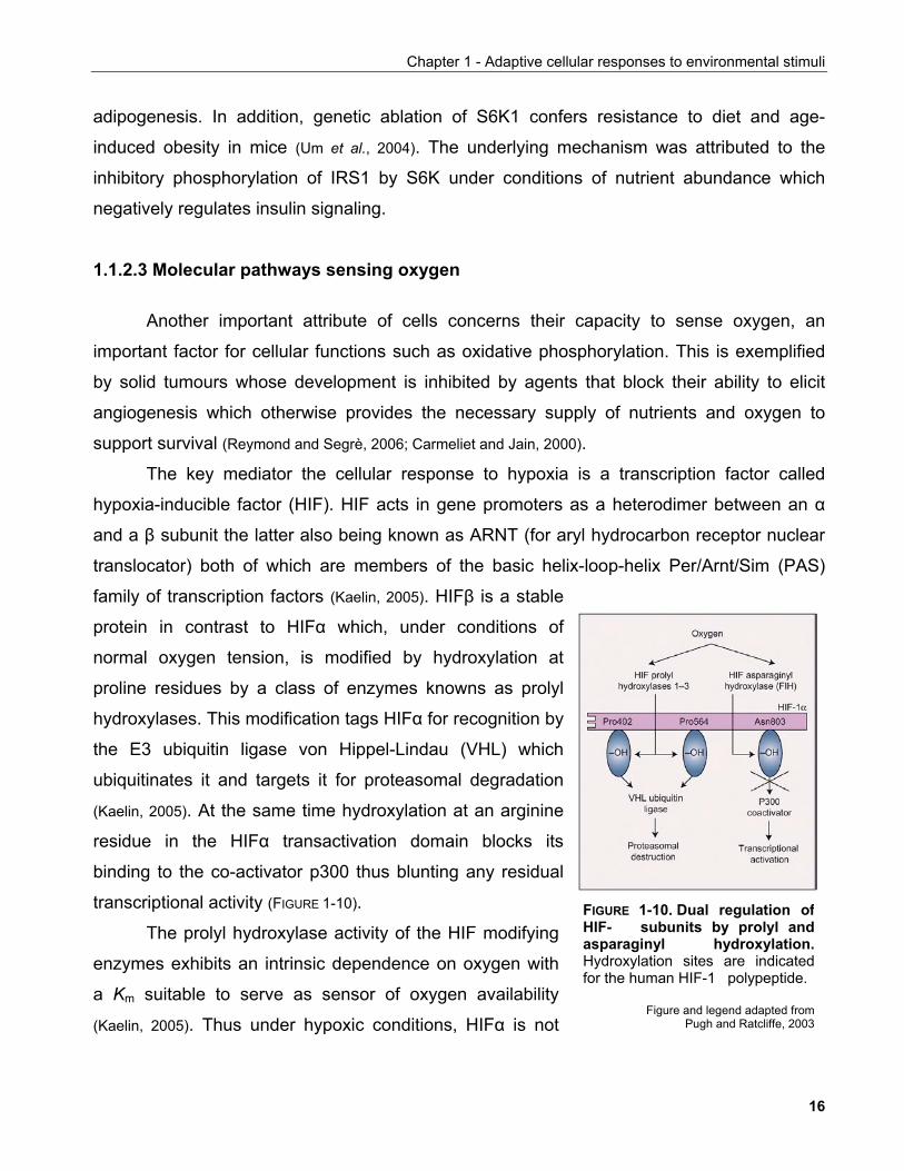
The key mediator the cellular response to hypoxia is a transcription factor called
hypoxia-inducible factor (HIF). HIF acts in gene promoters as a heterodimer between an α
and a β subunit the latter also being known as ARNT (for aryl hydrocarbon receptor nuclear
translocator) both of which are members of the basic helix-loop-helix Per/Arnt/Sim (PAS)
family of transcription factors (Kaelin, 2005). HIFβ is a stable
protein in contrast to HIFα which, under conditions of
normal oxygen tension, is modified by hydroxylation at
proline residues by a class of enzymes knowns as prolyl
hydroxylases. This modification tags HIFα for recognition by
the E3 ubiquitin ligase von Hippel-Lindau (VHL) which
ubiquitinates it and targets it for proteasomal degradation
(Kaelin, 2005). At the same time hydroxylation at an arginine
residue in the HIFα transactivation domain blocks its
binding to the co-activator p300 thus blunting any residual
transcriptional activity (FIGURE 1-10). FIGURE 1-10. Dual regulation ofHIF- subunits by prolyl andasparaginyl hydroxylation.Hydroxylation sites are indicatedfor the human HIF-1 polypeptide.
Figure and legend adapted fromPugh and Ratcliffe, 2003
The prolyl hydroxylase activity of the HIF modifying
enzymes exhibits an intrinsic dependence on oxygen with
a Km suitable to serve as sensor of oxygen availability
(Kaelin, 2005). Thus under hypoxic conditions, HIFα is not
16
Chapter 1 - Adaptive cellular responses to environmental stimuli
hydroxylated, cannot be recognised by VHL and thus is stabilised. Once in the nucleus, HIF
drives the expression of target genes involved in physiological as well as metabolic
responses that allow cells to adapt to low oxygen conditions (see also discussion in Chapter 5).
Towards this end, HIF target genes include angiogenesis-promoting factors such as VEGF
(vascular endothelial growth factor) as well as other growth factors (e.g. TGF-β) and genes
involved in glycolysis (Semenza, 2002).
Interestingly, recent reports provide evidence both in Drosophila and mammalian cells
that hypoxia inhibits the TOR pathway via the TSC1/2 complex (Liu et al., 2006; Brugarolas et al.,
2004; Reiling et al., 2004). There is however some controversy whether this phenomenon is
dependent on HIF. Brugarolas et al. reported that hypoxia inhibits TOR through the HIF
target gene RTP801/REDD1 (for regulated in development and DNA damage responses)
and that this effect is independent of AMPK activity (Brugarolas et al., 2004). In contrast, Liu et al.
showed that ARNT-deficient fibroblasts retain their ability to inhibit TOR under hypoxic
conditions leading them to propose that hypoxia-induced changes in cellular energy status
activates the AMPK pathway which also contributes to TOR inactivation (Liu et al., 2006).
Irrespective of the exact molecular details, these data provide a first glimpse into the
mechanisms involved in the growth inhibitory effects of hypoxia. Furthermore, they
demonstrate the existense of intimate connections between basic homeostatic pathways that
ensure the co-ordinate control of cellular activities in response to environmental factors.
The molecular circuitries described above provide a picture of the complexity
underlying the first level of adaptive cellular responses to environmental stimuli. Many of the
effects elicited by these pathways require the expression of new genes as demonstrated for
HIF and forkhead factors. The regulation of gene expression is a second level at which
multiple regulatory inputs convert to implement transcriptional programmes that support
cellular functions. Thus the following section will review the mechanisms involved in the
regulation of gene expression.
17
Chapter 1 - Adaptive cellular responses to environmental stimuli
1.2 REGULATION OF CHROMATIN STRUCTURE AND GENE EXPRESSION
1.2.1 Regulation of chromatin structure
Eucaryotic genomes comprise thousands of genes encoded in the DNA in a way that
allows gene expression suited to support specific cellular needs to be achieved with
remarkable accuracy.
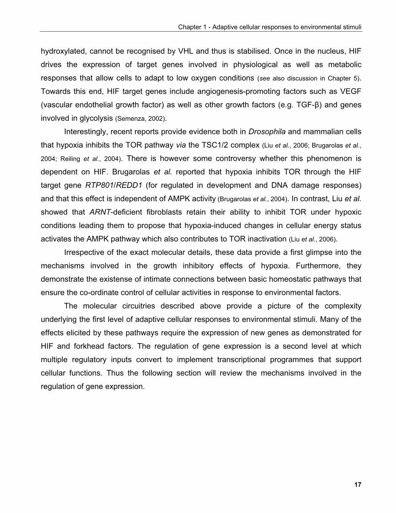
A critical factor in this feat is the packaging of the DNA into ordered structures called
nucleosomes which are arranged in a "bead-on-a-string" configuration comprising the
chromatin fiber. Nucleosomes consist of a strectch of DNA wrapped around a proteinaceous
core of core histones (H2A, H2B, H3, and H4) arranged as an octamer and stabilised by
linker histones (H1, H1°, and H5). Nucleosomes are spatially positioned to form a 30-nm
chromatin fiber which in turn can further
compact to increasingly thicker structures to
form chromosomes which is the configuration
of DNA during cell division (FIGURE 1-11)
(Falsenfeld and Groudine, 2003).
In interphase cells, distinct regions of
chromatin can be observed cytologically
known as euchromatin and heterochromatin.
Heterochromatin is thought to be tightly
condensed and thus inaccessible to DNA
binding factors unlike euchromatin which
adopts a more relaxed conformation (Grewal and
Moazed, 2003).
Chromatin structure can be altered by
replacement of the core histones with
specialized histone variants, ATP-dependent
nucleosome remodeling enzymes, or by
covalent modification of histones within the
nucleosome.
FIGURE 1-11. Hierarchical organisation of nuclearDNA structure in eucaryotes. See text for details
Adapted from Falsenfeld and Groudine, 2003
18
Chapter 1 - Adaptive cellular responses to environmental stimuli
1.2.1.1 Histone variants
Variants for all except histone H4 have been identified and are thought to have
occurred through gene duplication (Gilbert et al., 2005). Histone variants are located in distinct
chromatin regions where they are proposed to participate in the formation of specialised
chromatin structures. CENP-A is an H3 variant found in centromeric chromatin which is
characterised by increased compaction. H2A.Z may be able to influence chromatin structure
since H2A.Z-containing nucleosomal arrays are less condensed and thus may facilitate
transcription. Converesely, another H2 variant, macroH2A has been associated with X-
chromosome inactivation and may interfere with gene transscription (Gilbert et al., 2005).
1.2.1.2 ATP-dependent nucleosome remodeling
Nucleosomes perform a dual function as structural components of chromatin and as
regulators of gene expression. Nucleosome position is precisely determined so that key
transcription factor binding sites are exposed while maintaining proper DNA packaging.
During processes that require active nucleosome repositioning, such as replication and
transcription, nucleosomes can be mobilised on the chromatin fiber or the histone-DNA
contacts within individual nucleosomes can be discrupted by ATP-dependent chromatin
remodeling complexes which use the energy derived from ATP hydrolysis to perform their
task (Smith and Peterson, 2005).
A central function in ATP-dependent chromatin remodeling complexes is performed by
a helicase-like protein of the SWI/SNF (switch genes/sucrose non-fermentors) family. This
class of helicases has also been subdivided into three subfamilies based on primary
sequence homology as well as the individual charateristics of the corresponding remodeling
complexes: the SWI2/SNF2, Mi-2/CHD and ISWI families (Smith and Peterson, 2005).
The SWI2/SNF2 complexes have been implicated in the regulation of gene
transcription in yeast but also mammalian organisms where they participate in differentiation,
early development and cytokine-mediated gene expression. the catalytic components contain
bromodomains which mediate interaction with acetylated histone tails. Mutations of complex
components have also been associated with tumour progression, in particular lung and
19
Chapter 1 - Adaptive cellular responses to environmental stimuli
gastric cancers. Aside from their role in transcriptional regulation, SWI2/SNF2 complexes
have also been implicated in global chromatin structure control during mitosis when
chromosomes undergo major structural changes.
The ISWI (imitation SWI)-based complexes contain ATPases which are characterised
by a different histone-binding domain than SWI/SNF complexes, namely the SANT domain.
Although they also participate in transcriptional regulation, they have also been implicated in
global nucleosome assembly and positioning. This is likely to be coupled to replication as
ISWI components co-localise with replication foci in mammalian cells. Furthermore, ISWI
complexes are thought to be involved in transciptional repression as well as the formation of
silenced regions on chromatin (Smith and Peterson, 2005).
The ATPases of the third class of remodeling complexes, the Mi-2 family, contain yet
another histone binding domain, the chromodomain. Many of Mi-2 complexes are thought to
participate in transcriptional repression by virtue of their association with histone
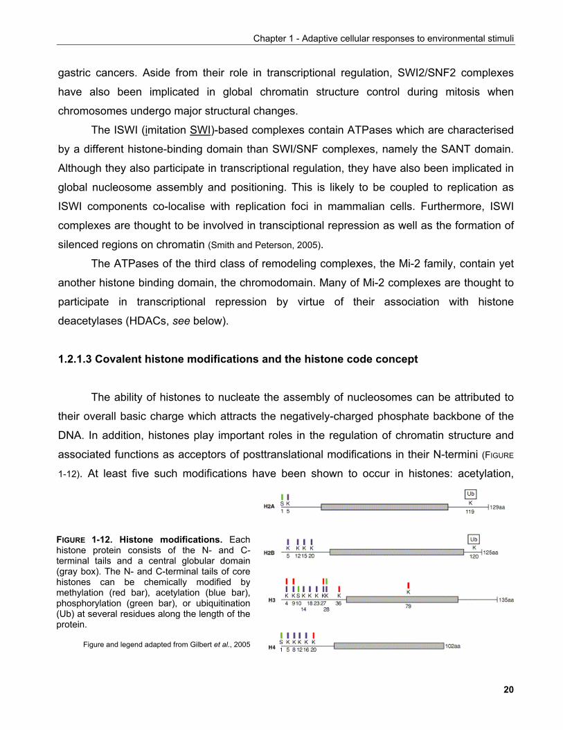
deacetylases (HDACs, see below). 1.2.1.3 Covalent histone modifications and the histone code concept The ability of histones to nucleate the assembly of nucleosomes can be attributed to
their overall basic charge which attracts the negatively-charged phosphate backbone of the
DNA. In addition, histones play important roles in the regulation of chromatin structure and
associated functions as acceptors of posttranslational modifications in their N-termini (FIGURE
1-12). At least five such modifications have been shown to occur in histones: acetylation,
FIGURE 1-12. Histone modifications. Eachhistone protein consists of the N- and C-terminal tails and a central globular domain(gray box). The N- and C-terminal tails of corehistones can be chemically modified bymethylation (red bar), acetylation (blue bar),phosphorylation (green bar), or ubiquitination(Ub) at several residues along the length of theprotein.
Figure and legend adapted from Gilbert et al., 2005
20
Chapter 1 - Adaptive cellular responses to environmental stimuli
methylation, phosphorylation, ADP-ribosylation and ubiquitination. At least the first three of
these are thought to exert their function primarily by disrupting histone/DNA contacts in the
nucleosome thus altering chromatin structure directly, while there is evidence that modified
histone tails serve as platforms for other DNA regulatory complexes via the recruitment of
proteins (Jenuwein and Allis, 2001).
1.2.1.3.1 Histone acetylation
Early experiments provided evidence that histone acetylation correlates with areas of
high DNase sensitivity and transcriptional activity suggesting that these chromatin regions
exhibit a lower degree of compaction (Roth et al., 2001). Indeed, the acetyl group serves as a
moiety partially neutralising the basic charge of histones thus weakening interactions with the
surrounding DNA although biophysical evidence has not provided any evidence for gross
structural changes in chromatin fiber structure (Gilbert et al. 2005).
At the global level, euchromatin which is associated with transcriptionally competent
regions of the genome, contains high levels of acetylated histones whereas heterochromatin
is characterised by histone hypoacetylation (Grewal and Moazed, 2003). Heterochromatin is
concentrated around functional chromosomal regions such as centromeres and telomeres
and participates in genomic stability by maintaining the structure of these regions intact.
Histone acetylation can occur at specific lysine residues which are highly conserved
throughout the species. It is regulated by enzymes called acetyltransferases which transfer
acetyl groups from acetyl-CoA to histones and is removed by deacetylases. Either of these
classes of enymes are recruited to specific sites of the genome by sequence-specific
transcription factors to regulate gene expression. Acetylated lysines are recognised by
dedicated protein interaction domains called bromodomains.
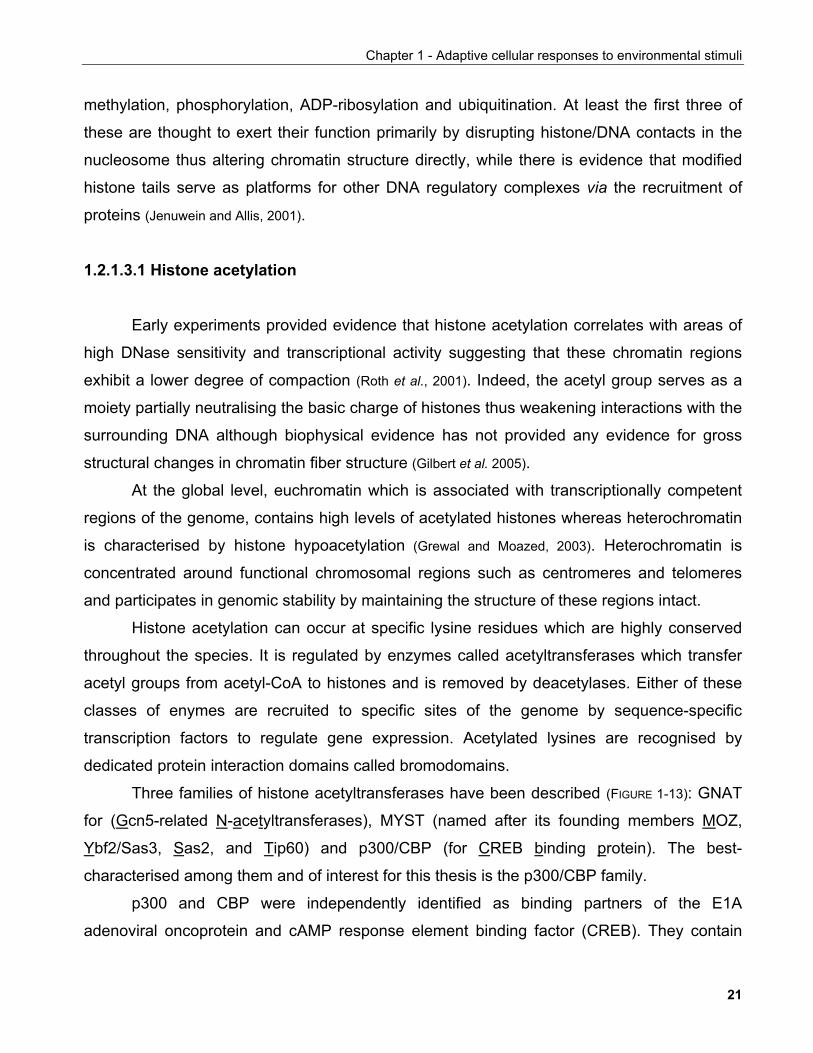
Three families of histone acetyltransferases have been described (FIGURE 1-13): GNAT
for (Gcn5-related N-acetyltransferases), MYST (named after its founding members MOZ,
Ybf2/Sas3, Sas2, and Tip60) and p300/CBP (for CREB binding protein). The best-
characterised among them and of interest for this thesis is the p300/CBP family.
p300 and CBP were independently identified as binding partners of the E1A
adenoviral oncoprotein and cAMP response element binding factor (CREB). They contain
21
Chapter 1 - Adaptive cellular responses to environmental stimuli
FIGURE 1-13. Overview of histone acetyltransferases (HATs).
Adapted from Vaquero et al., 2003
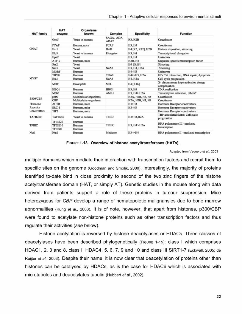
multiple domains which mediate their interaction with transcription factors and recruit them to
specific sites on the genome (Goodman and Smolik, 2000). Interestingly, the majority of proteins
identified to-date bind in close proximity to second of the two zinc fingers of the histone
acetyltransferase domain (HAT, or simply AT). Genetic studies in the mouse along with data
derived from patients support a role of these proteins in tumour suppression. Mice
heterozygous for CBP develop a range of hematopoietic malignansies due to bone marrow
abnormalities (Kung et al., 2000). It is of note, however, that apart from histones, p300/CBP
were found to acetylate non-histone proteins such as other transcription factors and thus
regulate their activities (see below).
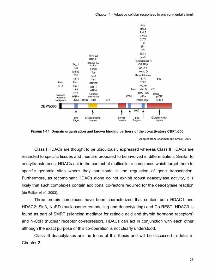
Histone acetylation is reversed by histone deacetylases or HDACs. Three classes of
deacetylases have been described phylogenetically (FIGURE 1-15): class I which comprises
HDAC1, 2, 3 and 8, class II HDAC4, 5, 6, 7, 9 and 10 and class III SIRT1-7 (Eckwall, 2005; de
Ruijter et al., 2003). Despite their name, it is now clear that deacetylation of proteins other than
histones can be catalysed by HDACs, as is the case for HDAC6 which is associated with
microtubules and deacetylates tubulin (Hubbert et al., 2002).
22
Chapter 1 - Adaptive cellular responses to environmental stimuli
FIGURE 1-14. Domain organisation and known binding partners of the co-activators CBP/p300.
Adapted from Goodman and Smolik, 2000
Class I HDACs are thought to be ubiquitously expressed whereas Class II HDACs are
restricted to specific tissues and thus are proposed to be involved in differentiation. Similar to
acetyltransferases, HDACs act in the context of multicellular complexes which target them to
specific genomic sites where they participate in the regulation of gene transcription.
Furthermore, as recombinant HDACs alone do not exhibit robust deacetylase activity, it is
likely that such complexes contain additional co-factors required for the deacetylase reaction
(de Ruijter et al., 2003).
Three protein complexes have been characterized that contain both HDAC1 and
HDAC2: Sin3, NuRD (nucleosome remodelling and deacetylating) and Co-REST. HDAC3 is
found as part of SMRT (silencing mediator for retinoic acid and thyroid hormone receptors)
and N-CoR (nuclear receptor co-repressor). HDACs can act in conjunction with each other
although the exact purpose of this co-operation is not clearly understood.
Class III deacetylases are the focus of this thesis and will be discussed in detail in
Chapter 2.
23
Chapter 1 - Adaptive cellular responses to environmental stimuli
FIGURE1-15. Overview of protein deacetylase families.
Adapted from Vaquero et al., 2003
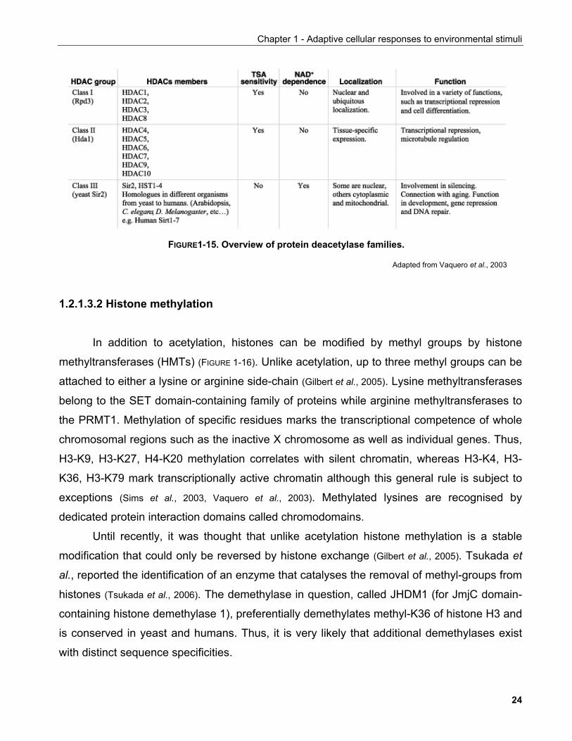
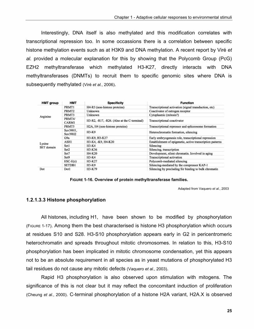
1.2.1.3.2 Histone methylation In addition to acetylation, histones can be modified by methyl groups by histone
methyltransferases (HMTs) (FIGURE 1-16). Unlike acetylation, up to three methyl groups can be
attached to either a lysine or arginine side-chain (Gilbert et al., 2005). Lysine methyltransferases
belong to the SET domain-containing family of proteins while arginine methyltransferases to
the PRMT1. Methylation of specific residues marks the transcriptional competence of whole
chromosomal regions such as the inactive X chromosome as well as individual genes. Thus,
H3-K9, H3-K27, H4-K20 methylation correlates with silent chromatin, whereas H3-K4, H3-
K36, H3-K79 mark transcriptionally active chromatin although this general rule is subject to
exceptions (Sims et al., 2003, Vaquero et al., 2003). Methylated lysines are recognised by
dedicated protein interaction domains called chromodomains.
Until recently, it was thought that unlike acetylation histone methylation is a stable
modification that could only be reversed by histone exchange (Gilbert et al., 2005). Tsukada et
al., reported the identification of an enzyme that catalyses the removal of methyl-groups from
histones (Tsukada et al., 2006). The demethylase in question, called JHDM1 (for JmjC domain-
containing histone demethylase 1), preferentially demethylates methyl-K36 of histone H3 and
is conserved in yeast and humans. Thus, it is very likely that additional demethylases exist
with distinct sequence specificities.
24
Chapter 1 - Adaptive cellular responses to environmental stimuli
Interestingly, DNA itself is also methylated and this modification correlates with
transcriptional repression too. In some occassions there is a correlation between specific
histone methylation events such as at H3K9 and DNA methylation. A recent report by Viré et
al. provided a molecular explanation for this by showing that the Polycomb Group (PcG)
EZH2 methyltransferase which methylated H3-K27, directly interacts with DNA
methyltransferases (DNMTs) to recruit them to specific genomic sites where DNA is
subsequently methylated (Viré et al., 2006).
FIGURE 1-16. Overview of protein methyltransferase families.
Adapted from Vaquero et al., 2003
1.2.1.3.3 Histone phosphorylation All histones, including H1, have been shown to be modified by phosphorylation
(FIGURE 1-17). Among them the best characterised is histone H3 phosphorylation which occurs
at residues S10 and S28. H3-S10 phosphorylation appears early in G2 in pericentromeric
heterochromatin and spreads throughout mitotic chromosomes. In relation to this, H3-S10
phosphorylation has been implicated in mitotic chromosome condensation, yet this appears
not to be an absolute requirement in all species as in yeast mutations of phosphorylated H3
tail residues do not cause any mitotic defects (Vaquero et al., 2003).
Rapid H3 phosphorylation is also observed upon stimulation with mitogens. The
significance of this is not clear but it may reflect the concomitant induction of proliferation
(Cheung et al., 2000). C-terminal phosphorylation of a histone H2A variant, H2A.X is observed
25
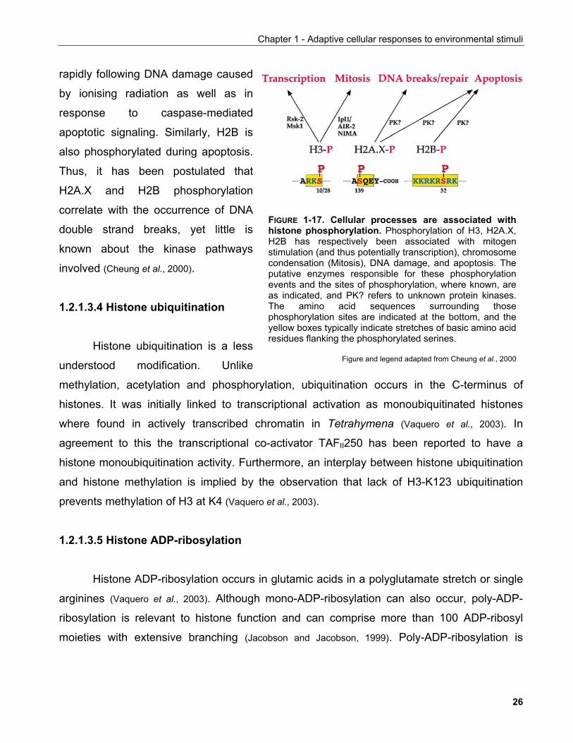
Chapter 1 - Adaptive cellular responses to environmental stimuli
rapidly following DNA damage caused
by ionising radiation as well as in
response to caspase-mediated
apoptotic signaling. Similarly, H2B is
also phosphorylated during apoptosis.
Thus, it has been postulated that
H2A.X and H2B phosphorylation
correlate with the occurrence of DNA
double strand breaks, yet little is
known about the kinase pathways
involved (Cheung et al., 2000).
1.2.1.3.4 Histone ubiquitination
Histone ubiquitination is a less
understood modification. Unlike
methylation, acetylation and phosphorylation, ubiquitination occurs in the C-terminus of
histones. It was initially linked to transcriptional activation as monoubiquitinated histones
where found in actively transcribed chromatin in Tetrahymena (Vaquero et al., 2003). In
agreement to this the transcriptional co-activator TAFII250 has been reported to have a
histone monoubiquitination activity. Furthermore, an interplay between histone ubiquitination
and histone methylation is implied by the observation that lack of H3-K123 ubiquitination
prevents methylation of H3 at K4 (Vaquero et al., 2003).
FIGURE 1-17. Cellular processes are associated withhistone phosphorylation. Phosphorylation of H3, H2A.X,H2B has respectively been associated with mitogenstimulation (and thus potentially transcription), chromosomecondensation (Mitosis), DNA damage, and apoptosis. Theputative enzymes responsible for these phosphorylationevents and the sites of phosphorylation, where known, areas indicated, and PK? refers to unknown protein kinases.The amino acid sequences surrounding thosephosphorylation sites are indicated at the bottom, and theyellow boxes typically indicate stretches of basic amino acidresidues flanking the phosphorylated serines.
Figure and legend adapted from Cheung et al., 2000
1.2.1.3.5 Histone ADP-ribosylation
Histone ADP-ribosylation occurs in glutamic acids in a polyglutamate stretch or single
arginines (Vaquero et al., 2003). Although mono-ADP-ribosylation can also occur, poly-ADP-
ribosylation is relevant to histone function and can comprise more than 100 ADP-ribosyl
moieties with extensive branching (Jacobson and Jacobson, 1999). Poly-ADP-ribosylation is
26
Chapter 1 - Adaptive cellular responses to environmental stimuli
highly dynamic being catalysed by poly-ADP-ribosyltransferases (PARPs) and removed by
poly-ADP-ribose glycohydrolases (PARGs).
PARP1 is required to recruit the DNA repair machinery in response to DNA damage it
is though possible that PARP1 has a stimulatory effect on gene transcription by modifying not
only histones but also transcription factors. Conversely, PARP1 has also been shown to
inhibit RNAPolII transcriptional elongation (Vaquero et al., 2003 and references therein). PARP
activity requires NAD+ and one of its enzymatic products is nicotinamide both of which are
features of the enzymatic reaction catalysed by sirtuin deacetylases (see Chapter 2). The
possible significance of this will be discussed in Chapter 5. 1.2.1.3.6 Epigenetics and the 'histone code' hypothesis
As is evident from the above, histone modifications serve as recognition sites for
specific protein domains to recruit additional chromatin-modifying complexes that regulate
gene expression. Thus, the modification status of histone tails in addition to sequence
elements encoded on the DNA can dictate chromatin structure and thus transcriptional
activity. The recognition of this led credence to the notion of a "histone code" which
postulates that covalent chromatin modifications are carriers of information that contribute to
gene expression (Jenuwein and Allis, 2001).
The histone code along with DNA methylation are so-called epigenetic events as they
are not encoded in the genome, yet they correlate with specific phenotypes (Pennisi, 2001).
Although epigenetic regulation underlies basic biological phenomena such as inheritence
and differentiation (Pennisi, 2001), the field of epigenetics has only recently started to provide
detailed molecular mechanisms primarily fuelled by the discovery of the enzymes involved in
histone and DNA modification, the identification of specific protein interaction domains
recognising such modifications and the elucidation of the interlinks between them.
Given this knownledge, it is of great interest to identify links between specific
epigenetic phenomena and associated phenotypes as it becomes apparent that information
other than DNA nucleotide sequence is contained within chromatin.
27
Chapter 1 - Adaptive cellular responses to environmental stimuli
1.2.2 Transcriptional regulation
Extracellular stimuli can evoke specific transcriptional responses by eliciting changes
in chromatin structure. Multiple mechanisms of specificity that ensure appropriate pathway
activation are in place, however a major role in this process is played by the sequence-
specific binding of transcription factors to DNA elements that can affect the activity of the
basal transcriptional machinery. These factors and their assembly into functional protein
complexes can be controled either by specific small molecule ligands such as lipids,
cholesterol and xenobiotics, or by posttranslational modifications often smilar to the ones
occuring in histones. This section will provide an overview of the current knowledge
underlying transcriptional regulation necessary for the understanding of the concepts
discussed in later chapters of this thesis.
1.2.2.1 Basal transcription
mRNA synthesis from a gene-encoding DNA sequence is catalysed by RNA
Polymerases (RNAPols). Three distinct RNAPols exist in eucaryotic cells which were first
distinguished from their variable sensitivity to amanitin, a mushroom poison. They are
designated RNAPolI, RNAPolII and RNAPolIII.
RNAPolI catalyses the transcription of precursor rRNA (pre-rRNA), which is processed
into 28S, 5.8S, and 18S rRNAs. RNAPolIII transcribes the genes encoding tRNAs, 5S rRNA
as well as other small, stable RNAs including U6 RNA involved in RNA splicing and the 7S
RNA of the signal-recognition particle involved in the transport of proteins into the
endoplasmic reticulum (Lodish et al., 2000). RNAPolII transcribes all protein-encoding genes as
well as four small nulcear RNAs involved in RNA splicing.
DNA sequences located at the beginning (5') of the gene coding region recruit
RNAPols to initiate gene transcription (FIGURE 1-18). Such sequences constitute the core
promoter element which is able to sustain transcriptional initiation by RNAPols.
Initiation of RNAPolII-catalysed transcription starts by the recognition of the TATA box
element by the TATA-binding protein (TBP) of TFIID. TBP binding causes a dramatic
conformational change on the DNA which is thought to contribute to the downstream
28

Chapter 1 - Adaptive cellular responses to environmental stimuli
transcriptional activation events
(Orphanides et al., 1996). RNAPolII
transcription initiation can also occur in
the absence of TATA elements where
other sequences such as the initiator (Inr)
or the downstream core promoter element
(DPE) take up its role (Butler and Kadonaga,
2002).
TFIID nucleates the ordered
recruitment of a multisubunit complex
which comprises activites that lead to
promoter 'melting', i.e. nucleosome
disruption and opening of the DNA double
helix, rendering it competent as a
transcription template. Following that, transcriptional elongation from TATA-driven promoters
requires the phosphorylation of the C-terminal domain (CTD) of RNAPolII which is catalysed
by CDK7.
FIGURE 1-18. Core promoter elements. Some corepromoter motifs that can participate in transcription byRNA polymerase II are depicted. Each of these elementsis found in only a subset of core promoters. Any specificcore promoter may contain some, all, or none of thesemotifs. The BRE is an upstream extension of a subset ofTATA boxes. The DPE requires an Inr, and is locatedprecisely at +28 to +32 relative to the A+1 nucleotide inthe Inr. The DPE consensus was determined withDrosophila transcription factors and core promoters. TheInr consensus sequence is shown for both Drosophila(Dm) and humans (Hs).
Figure and legends adapted from Butler and Kadonaga, 2002
rDNA genes contain distinct promoter elements that recruit RNAPolI. These elements
are recognised by upstreat binding factor (UBF) which nucleates the formation of the
RNAPolI holoenzyme (Grummt, 2003).
Three types of RNAPolIII promoters have been described based on the sequence
elements encompassed by each (Schramm and Hernandez, 2002). In contrast to RNAPolI and
RNAPolII core promoters which are located upstream or at the very 5' of the mRNA-
encoding region, RNAPolIII promoters encoding for tRNA and 5S rRNA are intragenic while
these encoding for snRNAs contain a TATA box and a proximal sequence element (PSE).
In addition to promoter elements which act in-cis DNA sequences which act in-trans
play important roles in transcriptional regulation. These elements are called enhancers and in
contrast to promoters whose spatial positioning with respect to the transcribed region is
important for their function, enhancers can promote gene transcription in a position- and
orientation-independent manner.
29
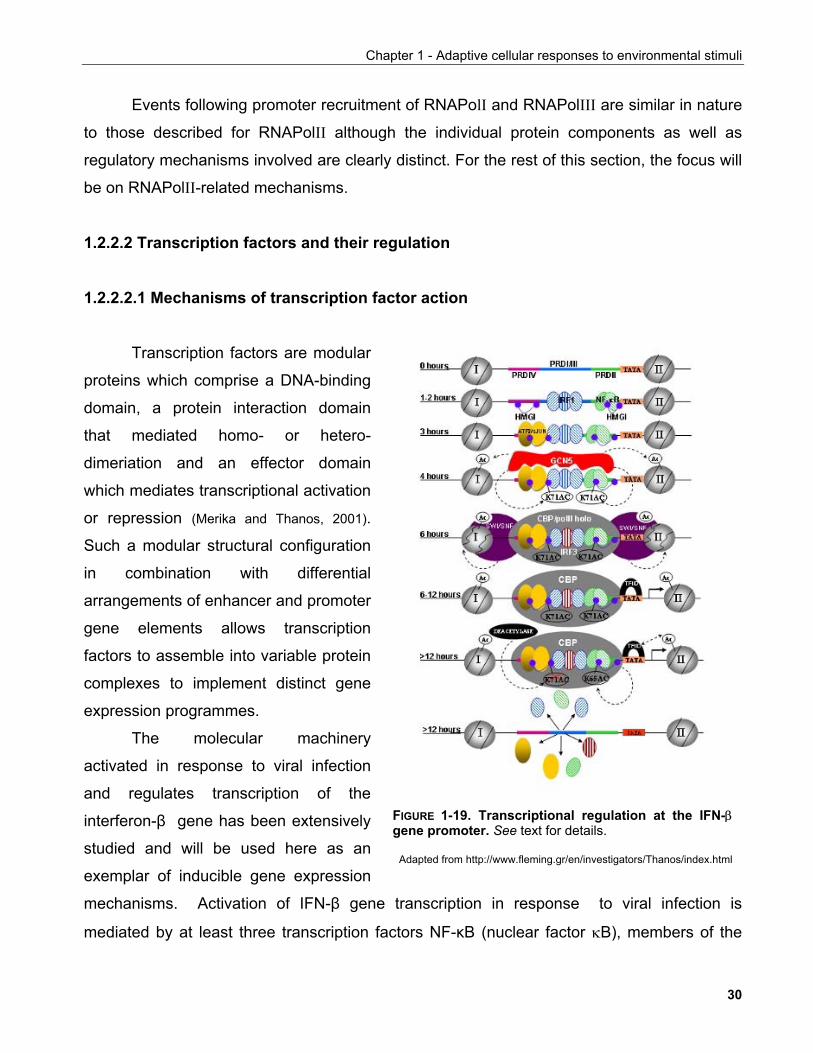
Chapter 1 - Adaptive cellular responses to environmental stimuli
Events following promoter recruitment of RNAPolI and RNAPolIII are similar in nature
to those described for RNAPolII although the individual protein components as well as
regulatory mechanisms involved are clearly distinct. For the rest of this section, the focus will
be on RNAPolII-related mechanisms.
1.2.2.2 Transcription factors and their regulation 1.2.2.2.1 Mechanisms of transcription factor action
Transcription factors are modular
proteins which comprise a DNA-binding
domain, a protein interaction domain
that mediated homo- or hetero-
dimeriation and an effector domain
which mediates transcriptional activation
or repression (Merika and Thanos, 2001).
Such a modular structural configuration
in combination with differential
arrangements of enhancer and promoter
gene elements allows transcription
factors to assemble into variable protein
complexes to implement distinct gene
expression programmes.
The molecular machinery
activated in response to viral infection
and regulates transcription of the
interferon-β gene has been extensively
studied and will be used here as an
exemplar of inducible gene expression
mechanisms. Activation of IFN-β gene transcription in response to viral infection is
mediated by at least three transcription factors NF-κB (nuclear factor κB), members of the
FIGURE 1-19. Transcriptional regulation at the IFN-βgene promoter. See text for details.
Adapted from http://www.fleming.gr/en/investigators/Thanos/index.html
30
Chapter 1 - Adaptive cellular responses to environmental stimuli
IRF (interferon regulatory factor) family, and the ATF-2 (activating transcription factor-2)/c-
Jun heterodimer (Merika and Thanos, 2001).
These factors (activators) act in concert upon viral entry to form a complex known as
the 'enhanceosome' to specifically activate IFN-β gene transcription (FIGURE 1-19). Assembly of
these factors on the IFN-β gene promoter is dictated by enhancer elements which are found
in a specific spatial positioning pattern. In a different context any of these factors can mediate
other processes such as response to cytokines, UV irradiation or the proliferative response to
mitogens. At the same time none of them alone can elicit the response to viral infection
(Merika and Thanos, 2001). Enhanceosome assembly is greatly facilitated by a high mobility
group protein HMG I(Y) which binds to four sites in the enhancer sequence and induces a
conformational change on the DNA that lowers the free energy of activator binding (Merika and
Thanos, 2001). At the same time HMG I(Y) contributes to the stability of the enhanceosome.
Upon its assembly, the enhanceosome, recruits the co-activator GCN5 which
acetylates a nucleosome masking the promoter TATA element. At the same time GCN5 also
acetylates HMG I(Y) at K71 contributing to enhanceosome stability. TATA box exposure
allows the assembly of the RNAPolII pre-initiation complex along with the acetyltransferase
CBP which in turn recruits the SWI/SNF remodelling complex and acetylates promoter
histones, a prerequisite for SWI/SNF-mediated nucleosomal remodelling (Agalioti et al., 2000).
This cascade culminates with the assembly of the RNAPolII holoenzyme and transcriptional
initiation.
1.2.2.2.2 Post-translational regulation of transcription factors
Apart from histone modifications, transcription factor modifications are also well-
established events in the regulation of transcription. These include acetylation,
phosphorylation, ubiquitination, sumoylation and proteolytic cleavage to mention but some
and have variable effects in the ability of transcription factors to modulate gene expression.
Many of the modifying enzymes described for histones also target transcription factors. Such
modifications provide a way of modulating transcription factor activity by upstream signaling
cascades as described in section 1.2. The wealth of studies on the regulation of the tumour
suppressor p53 provide a characteristic example of this.
31
Chapter 1 - Adaptive cellular responses to environmental stimuli
p53 is a transcription factor that binds to specific DNA elements as a tetramer. It is a
highly unstable protein due to its constant ubiquitination by the E3 ligase Mdm2 (a.k.a.
Hdm2) which targets it for proteasomal degradation. Mdm2 also binds an N-terminal
transactivation domain thus inhibiting downstream transcriptional induction. Upon exposure
of cells to genotoxic stresses, p53 ubiquitination is suppressed allowing its accumulation in
the nucleus where it activates or represses more than 150 target genes to control cell cycle
and survival (Bode and Dong, 2004).
Multiple pathways are involved in p53 stabilisation. Acetylation targets among other
sites the lysine residues ubiquitinated by Mdm2 contributing to p53 stabilisation.
Furthermore, p53 acetylation enhances its ability to bind to DNA and consequently its
transcription activation capacity (Luo et al., 2004). Phosphorylation by multiple kinases can also
affect p53 targeting by Mdm2, increase its transcriptional activity and modulate its subcellular
localisation by affecting NLS recognition (Bode and Dong, 2004). Furthermore, phosphorylation
of p53 renders it a substrate for the peptidyl-prolyl isomerase Pin1 which induces a
conformational change that enhances its transactivation capacity (Zheng et al., 2002; Zacchi et al.,
2002). Recently, methylation of p53 by a SET domain methyltransferase was also reported to
modulate protein stability (Chuikov et al., 2004).
Acetylation can also have more subtle effects on transcription factor activity. Upon
stimulation the NFκB transcription factor is released from the inhibitory protein IκB to
translocate to the nucleus and activate transcription (Chen and Greene, 2004). Similarly to p53
acetylation has been shown to enhance binding to DNA. At the same time though,
acetylation reduces its affinity for IκB albeit by targeting different lysine residues. Conversely,
HDAC3-mediated deacetylation contributes to the attenuation of the NFκB response by
enhancing NFκB interaction with IκB, a complex that is actively exported to the cytoplasm
(Chen et al., 2001).
1.2.2.2.3 Ligand-mediated modulation of transcription factor activity
Similarly to procaryotic cells, some eucaryotic transcription factors harbour domains
that can be directly bound to by specific ligands. Several small molecules including
aminoacids (proline, arginine), intermediate metabolites (benzoic scid, orotic acid) and
32
Chapter 1 - Adaptive cellular responses to environmental stimuli
sugars are known to regulate transcriptional switches with the primary goal to modulate
metabolic output depending on the cellular needs as relayed by these molecules (Sellick and
Reece, 2005).
The family of nuclear receptors comprise transcription factors most of which are
regulated by ligand binding (Weatherman et al., 1999). Nuclear receptor ligands include among
others hormones such as estrogen and growth hormone, metabolites such as bile acids,
lipids and cholesterol and xenobiotics such as drugs. The natural ligands for many nuclear
receptors are unknown thus such receptors are described as 'orphan'.
Ligand effects are mediated through a C-terminal ligand binding domain (LBD) and an
N-terminal DNA binding domain (DBD) linked by a flexible linker region which is proposed to
allow not only conformational changes but also contribute to the recruitment of co-activators
and co-repressors.
Co-activator and co-repressor recruitment sites are overlapping in the LBD. Thus it is
postulated that ligand binding induces a conformational change that allows the exchange of
co-repressors for co-activator complexes to induce gene expression (Nagy and Schwabe, 2004).
Recently, two F-box/WD-40 containing proteins, transducin β-like (TBL1) and the related
TBLR1, were identified as critical components for this exchange mechanism. Upon ligand
binding TBL1 and TBLR1 recruit the 19S proteasome to induce the clearance of co-repressor
complexes allowing the binding of co-activators to the liganded receptors (Perissi et al., 2004).
1.2.2.2.4 Transcriptional regulatory networks
The complexity of the mechanisms in place to determine gene transcription by multiple
regulatory events allows an enormous degree of flexibility in the implementation of specific
gene expression programmes suited to environmental cues. During development for
example, multipotent stem cells derived from a single zygote form the starting cell pool which
will generate hundreds of different cell types to form tissues, organs and together an
organism.
Recent technical advances are starting to provide an integraded view of how
transcription factor networks drive the spatio-temporaly precise execution of complex
biological processes (Blais and Dynlacht, 2005). During early development, the transcription
33
Chapter 1 - Adaptive cellular responses to environmental stimuli
factors OCT4, SOX2 and NANOG are important for the maintainance of the pluripotent
character of stem cells in that loss-of-function results in aberrant differentiation. Genome-
wide analysis of target promoter occupancy revealed an elaborate gene regulatory network
dictated by OCT4, SOX2 and NANOG which among other features drives the expression of
homeobox transcription factors important for development (Boyer et al., 2005). This work implied
the existence of autoregulatory and feedforward loops which enable the maintainance of the
stem cell's pluripotent character while enabling it to repond to developmental cues in order to
give rise to appropriate cell types (Boyer et al., 2005).
1.2.2.3 Additional mechanisms of gene regulation
Finally, in addition to the above mechanisms regulating gene expression other factors
may also contribute to it.
Chromosomes are arranged into so-called chromosome territories in the interphase
nucleus in that they occupy distinct nuclear regions. For example the rDNA-containing
acrocentric chromosomes 13, 14, 15, 21 and 22 associate preferentially with nucleoli. Human
chromosomes 18 and 19, although of comparable size they differ in gene density with
chromosome 18 being gene-poor (~4.3 genes per Mbp) and chromosome 19 gene-rich (22.7
genes per Mbp). Chromosome 18 is located in the nuclear periphery whereas chromosome
19 is positioned more internally (Gilbert et al., 2005). Furthermore, chromosome 19 spreads in
an area larger than that of chromosome 18 suggesting that the chromatin in the former
adopts a less condensed conformation than that in the latter.
It is thought that interchromatin domains (ICDs) which are chromatin-free regions,
contain a high density of transcriptional apparatus components to which genes located in
euchromatin have access (Gilbert et al., 2005). Moreover, foci of transcription have been
observed in nuclei (Francastel et al., 2000). However, little is known about the way nuclear
architecture can affect gene expression in reponse to specific stimuli or whether it simply
provides an organisational framework for proper execution of the cell's transcriptional
activities.
Finally, post-transcriptional regulatory mechanisms exist to determine gene
expression. A mechanism of post-transcriptional regulation previously identified in plants and
34
Chapter 1 - Adaptive cellular responses to environmental stimuli
C. elegans has been recently shown to apply in mammalian cells. It is known as RNA
interference (RNAi) and it acts by the formation of precisely processed small RNA molecules
that guide molecular complexes such as the RISC (RNA-induced silencing complex) or RITS
(RNA-induced initiation of transcriptional gene silencing) to recognise complementary
sequences on target mRNAs (Tomari and Zamore, 2005). This drives the degradation of the
target mRNA and thus reduction of the respective gene product synthesis. There is evidence
that engogenous small RNA molecules known as microRNAs (miRNAs) are involved in
development and disease thus further expanding the cellular toolbox or transcription
regulatory mechanisms (Wienholds and Plasterk, 2005).
1.3 Conclusion
This chapter provided an overview of the signaling mechanisms involved in the
transduction of environmental cues to the nucleus and described mechanisms available
therein that allow the implementation of transcriptional programmes determined by the nature
of the upstream signals. The importance of elucidating such basic cellular processes is
emphasised by the increasing appreciation that deviation from these finely designed
pathways underlies the cause of many disease states such as cancer and diabetes.
Prompted by our interest in undertanding how nutrient availability and cellular energy
status modulate gene trasncription, we initiated the investigation of the sirtuin family of
deacetylases. This family comprises proteins with reported histone deacetylase activity
which, unlike HDACs, depends on NAD+. Thus, sirtuins are prime candidates to function as
nodes that couple cellular redox status and transcriptional regulation.
35
Chapter 2 - The sirtuin family of protein deacetylases
CHAPTER 2
THE SIRTUIN FAMILY OF PROTEIN DEACETYLASES
2. INTRODUCTION TO THE SIRTUIN FAMILY 2.1 Discovery of sirtuins and determination of their enzymatic activity
The sirtuin family of enzymes comprises protein members from across all three phyla.
They are characterised by a highly conserved core domain which carries a catalytic activity
and in is flanked by N- and C-terminal regions of variable length which are particularly
prevalent in the more recent additions of the evolutionary time-scale.
Early studies implicated the yeast sirtuin Sir2 as part of the genetic network that
regulates the position-dependent expression of genes which determine mating type in S.
cerevisiae, a phenomenon also known as gene silencing (Ivy et al., 1986; Kimmerly and Rine, 1987;
Rine and Herskowitz, 1987). An indication as to the actual activity of Sir2 was given by the finding
that silenced chromatin correlated with concomitant hypoeacetylation of histones and that
Sir2 overexpression resulted in significant histone deacetylation in vivo (Braunstein et al., 1993).
This implied that Sir2p harbors a deacetylase activity. However, initial attempts to
demonstrate such enzymatic action in vitro were unsuccesful.
In search of a catalytic activity it was noted that the CobB gene in Salmonella
typhimurium, a bacterial sirtuin member, could compensate for loss-of-function mutations in
another gene, CobT (Tsang and Escalante-Semerena, 1998). CobT encodes for a 5,6-
36
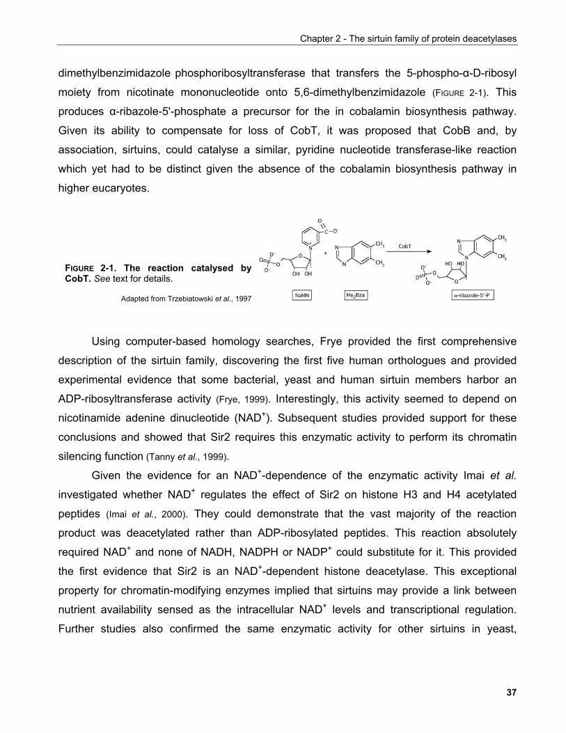
Chapter 2 - The sirtuin family of protein deacetylases
dimethylbenzimidazole phosphoribosyltransferase that transfers the 5-phospho-α-D-ribosyl
moiety from nicotinate mononucleotide onto 5,6-dimethylbenzimidazole (FIGURE 2-1). This
produces α-ribazole-5'-phosphate a precursor for the in cobalamin biosynthesis pathway.
Given its ability to compensate for loss of CobT, it was proposed that CobB and, by
association, sirtuins, could catalyse a similar, pyridine nucleotide transferase-like reaction
which yet had to be distinct given the absence of the cobalamin biosynthesis pathway in
higher eucaryotes.
FIGURE 2-1. The reaction catalysed byCobT. See text for details.
Adapted from Trzebiatowski et al., 1997
Using computer-based homology searches, Frye provided the first comprehensive
description of the sirtuin family, discovering the first five human orthologues and provided
experimental evidence that some bacterial, yeast and human sirtuin members harbor an
ADP-ribosyltransferase activity (Frye, 1999). Interestingly, this activity seemed to depend on
nicotinamide adenine dinucleotide (NAD+). Subsequent studies provided support for these
conclusions and showed that Sir2 requires this enzymatic activity to perform its chromatin
silencing function (Tanny et al., 1999).
Given the evidence for an NAD+-dependence of the enzymatic activity Imai et al.
investigated whether NAD+ regulates the effect of Sir2 on histone H3 and H4 acetylated
peptides (Imai et al., 2000). They could demonstrate that the vast majority of the reaction
product was deacetylated rather than ADP-ribosylated peptides. This reaction absolutely
required NAD+ and none of NADH, NADPH or NADP+ could substitute for it. This provided
the first evidence that Sir2 is an NAD+-dependent histone deacetylase. This exceptional
property for chromatin-modifying enzymes implied that sirtuins may provide a link between
nutrient availability sensed as the intracellular NAD+ levels and transcriptional regulation.
Further studies also confirmed the same enzymatic activity for other sirtuins in yeast,
37
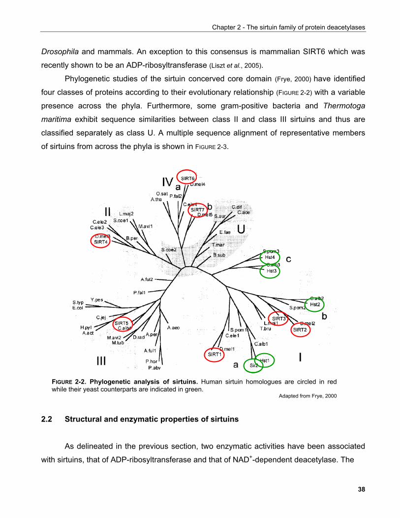
Chapter 2 - The sirtuin family of protein deacetylases
Drosophila and mammals. An exception to this consensus is mammalian SIRT6 which was
recently shown to be an ADP-ribosyltransferase (Liszt et al., 2005).
Phylogenetic studies of the sirtuin concerved core domain (Frye, 2000) have identified
four classes of proteins according to their evolutionary relationship (FIGURE 2-2) with a variable
presence across the phyla. Furthermore, some gram-positive bacteria and Thermotoga
maritima exhibit sequence similarities between class II and class III sirtuins and thus are
classified separately as class U. A multiple sequence alignment of representative members
of sirtuins from across the phyla is shown in FIGURE 2-3.
FIGURE 2-2. Phylogenetic analysis of sirtuins. Human sirtuin homologues are circled in redwhile their yeast counterparts are indicated in green.
Adapted from Frye, 2000
2.2 Structural and enzymatic properties of sirtuins
As delineated in the previous section, two enzymatic activities have been associated
with sirtuins, that of ADP-ribosyltransferase and that of NAD+-dependent deacetylase. The
38
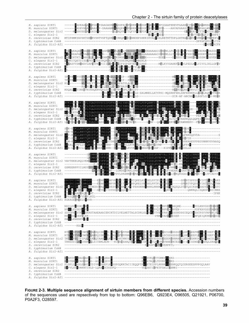
Chapter 2 - The sirtuin family of protein deacetylases H. sapiens SIRT1 ------MADEAALALQPGGSPSAAGADREAASSPAGEPLRKRPRRDGPGLERSPGEPGGAAPEREVPAAARGCPGAAAAALWREAEAEAAAM. musculus SIRT1 ------MADEVALALQAAGSP-SAAAAMEAASQPADEPLRKRPRRDGPGLGRSPGEPS----AAVAPAAAGCEAASAAAPAALWREAAGAAD. melanogaster Sir2 -----MMENYEEIRLGHIRSK-------DLGNQVPDTTQFYPPTKFDFGAEILASTST------------EAEAEATATTTEPATSELAGKC. elegans Sir2-1 -------------------------------------------------MSRDSGNDS--------------EVAVTHGEVQEITEENPEIS. cerevisiae SIR2 MTIPHMKYAVSKTSENKVSNTVSPTQDKDAIRKQPDDIINNDEPSHKKIKVAQPDSLRETNTTDPLGHTKAALGEVASMELKPTNDMDPLAS. typhimurium CobB -----------------------------------------------------------------------------SPPAFNPDSALTINA. fulgidus Sir2-Af1 ---------------------------------------------------------------------------------SPNPDARCFN H. sapiens SIRT1 AGGEQEAQATAAAGEGDNGPGLQGPSREPPLADNLYDEDDDDEGEEEEEAAAAAIGYRDNLLFGDEIITNGFHSCESDEEDRASHASSSDWM. musculus SIRT1 ASAEREAPATAVAGDGDNGSGLR---REPRAADDFDDDEGEEEDEAAAAAAAAAIGYRDNLLLTDGLLTNGFHSCESDDDDRTSHASSSDWD. melanogaster Sir2 ANGEIKTKTLAAREEQEIGANLEHKTKNPTKSMGEDEDDEEEEEEDDE-------EEEE----DDEEGITGTSNEDEDSSSNCSSSVEPDWC. elegans Sir2-1 GSMHITQETDISDAPETNTDSSRQRTESTTSVSSESWQNNDEMMSNLR-------------------------------------------S. cerevisiae SIR2 VSAASVVSMSNDVLKPETPKGPIIISKNPSNGIFYGPSFTKRESLNAR-------MFLKYYGAHKFLDTYLPEDLNSLYIYYLIKLLGFEVS. typhimurium CobB AD------------------------DEPENDENTDEACETYLASEEC-------------------------------------------A. fulgidus Sir2-Af1 AD------------------------DEPENDENTDEACETYLASEEC------------------------------------------- H. sapiens SIRT1 TPRPRIGPYTFVQQHLMIGTDPRTILKDLLPETIP--PPELDD--------------------MTLWQIVINILSEPPKRKKRKDINTIEDM. musculus SIRT1 TPRPRIGPYTFVQQHLMIGTDPRTILKDLLPETIP--PPELDD--------------------MTLWQIVINILSEPPKRKKRKDINTIEDD. melanogaster Sir2 KLR-------WLQREFYTGRVPRQVIASIMPHFATGLAGDTDD--------------------SVLWDYLAHLLNEPKRRNKLASVNTFDDC. elegans Sir2-1 ----------RAQRLLDDGATPLQIIQQIFPDFNASRIATMSE--------------------NAHFAILSDLLERAPVRQKLTNYNSLADS. cerevisiae SIR2 KDQALIGTINSIVHINSQERVQDLGSAISVTNVEDPLAKKQTV--------------------RLIKDLQRAINKVLCTRLRLSNFFTIDHS. typhimurium CobB ----------------------REGLATRYPRTEINSIRHMLG-SALMNELLATYPHI-MQSRRFHRLSRFRKNKRLLRERLRQRIFFRDRA. fulgidus Sir2-Af1 ----------------------REGLATRYPRTEINSIRHMLG--------------------SIR-AF-ARCHAEGLBSFLGIDS-MDEK H. sapiens SIRT1 AVKLLQECKKIIVLTGAGVSVSCGIPDFRSRDGIYARLAVDFPDLPDPQAMFDIEYFRKDPRPFFKFAKEIYP----GQFQPSLCHKFIALM. musculus SIRT1 AVKLLQECKKIIVLTGAGVSVSCGIPDFRSRDGIYARLAVDFPDLPDPQAMFDIEYFRKDPRPFFKFAKEIYP----GQFQPSLCHKFIALD. melanogaster Sir2 VISLVKKSQKIIVLTGAGVSVSCGIPDFRSTNGIYARLAHDFPDLPDPQAMFDINYFKRDPRPFYKFAREIYPGE--FQFQPSPCHRFIKMC. elegans Sir2-1 AVELFKTKKHILVLTGAGVSVSCGIPDFRSKDGIYARLRSEFPDLPDPTAMFDIRYFRENPAPFYNFAREIFP----GQFVPSVSHRFIKES. cerevisiae SIR2 FIQKLHTARKILVLTGAGVSTSLGIPDFRSSEGFYSKIKHLG--LDDPQDVFNYNIFMHDPSVFYNIANMVLP----PEKIYSPLHSFIKMS. typhimurium CobB VVPEMMENPRVLVLTGAGISAESGIRTFRAADGLWEEHRVEDVATP---EGF----AR-NPGLVQTFYNARRQQLQQPEIQPNAAHLALAKA. fulgidus Sir2-Af1 LLKTIAESKYLVALTGAGVSAESGIPTFRGKDGLWNRYRPEE------LANP--QAFAKDPEKVWKWYAWRMEKV--FNAQPNKAHQAFAE H. sapiens SIRT1 SDK-EGKLLRNYTQNIDTLEQVAGIQR--IIQCHGSFATASCLICKYKVDCEAVRGDIFNQVVPRCPRCPADEP-----------------M. musculus SIRT1 SDK-EGKLLRNYTQNIDTLEQVAGIQR--ILQCHGSFATASCLICKYKVDCEAVRGDIFNQVVPRCPRCPADEP-----------------D. melanogaster Sir2 LET-KGKLLRNYTQNIDTLERVAGIQR--VIECHGSFSTASCTKCRFKCNADALRADIFAQRIPVCPQCQPNKEQSVDA-----------SC. elegans Sir2-1 LET-SGRLLRNYTQNIDTLEHQTGIKR--VVECHGSFSKCTCTRCGQKYDGNEIREEVLAMRVAHCKRCEGVIK-----------------S. cerevisiae SIR2 LQM-KGKLLRNYTQNIDNLESYAGISTDKLVQCHGSFATATCVTCHWNLPGERIFNKIRNLELPLCPYCYKKRREYFPEGYNNKVGVAASQS. typhimurium CobB LEEALGDRFLLVTQNIDNLHERAGNRN--IIHMHGELLKVRCSQSGQILEWNGDVMP-----EDKCHCCQFPAP-----------------A. fulgidus Sir2-Af1 LER-LGVLKCLITQNVDDLHERAGSRN--VIHLHGSLRVVRCTSCNNSFEVESAP---KIPPLPKCDKCGSLLR----------------- H. sapiens SIRT1 -------------LAIMKPEIVFFGENLPEQFHRAMKYDKDEVDLLIVIGSSLKVRPVALIPSSIP-HEVPQILINREP-LPHLHFDVELLM. musculus SIRT1 -------------LAIMKPEIVFFGENLPEQFHRAMKYDKDEVDLLIVIGSSLKVRPVALIPSSIP-HEVPQILINREP-LPHLHFDVELLD. melanogaster Sir2 VAVTEEELRQLVENGIMKPDIVFFGEGLPDEYHTVMATDKDVCDLLIVIGSSLKVRPVAHIPSSIP-ATVPQILINREQ-LHHLKFDVELLC. elegans Sir2-1 -----------------P-NIVFFGEDLGREFHQHVTEDKHKVDLIVVIGSSLKVRPVALIPHCVD-KNVPQILINRES-LPHYNADIELLS. cerevisiae SIR2 GSMSERPPYILNSYGVLKPDITFFGEALPNKFHKSIREDILECDLLICIGTSLKVAPVSEIVNMVP-SHVPQVLINRDP-VKHAEFDLSLLS. typhimurium CobB ----------------LRPHVVWFGEMP--LGMDEIYMALSMADIFIAIGTSGHVYPAAGFVHEAKLHGAHTVELNLEPSQVGSEFEEKHYA. fulgidus Sir2-Af1 ------------------PGVVWFGEMLPPDVLDRAMREVERADVIIVAGTSAVVQPAASLPLIVKQRGGAIIEINPDE-TPLTPIADYSL H. sapiens SIRT1 GDCDVIINELCHRLGG---EYAKLCCNPVKLSEITEKPPRTQKELAYLSELPPTPLHVSEDSS-----SPERTSPPDSSVIVTLLDQAAKSM. musculus SIRT1 GDCDVIINELCHRLGG---EYAKLCCNPVKLSEITEKPPRPQKELVHLSELPPTPLHISEDSS-----SPERTVPQDSSVIATLVDQATNND. melanogaster Sir2 GDSDVIINQICHRLSDNDDCWRQLCCDESVLTESKELMP-PEHSNHHLHHHLHHHRHCSSESERQSQLDTDTQSIKSNSSADYILGSAGTCC. elegans Sir2-1 GNCDDIIRDICFSLGG---SFTELITSYDSIMEQQGKTK-------------------SQKPS---------QNKRQLISQEDFLNICMKES. cerevisiae SIR2 GYCDDIAAMVAQKCGWT----------------------------------------------------------------------IPHKS. typhimurium CobB GPASQVVPEFVDKFLKGL-------------------------------------------------------------------------A. fulgidus Sir2-Af1 RGKAGEVMDELVRHVR--------------------------------------------------------------------------- H. sapiens SIRT1 NDDLDVSESKGCME----------------------------------EKPQEVQTSRNVESIAEQME-------NPDLKNVGSSTGEKNEM. musculus SIRT1 NVNDLEVSESSCVE----------------------------------EKPQEVQTSRNVENINVEN---------PDFKAVGSSTADKNED. melanogaster Sir2 SDSGFESSTFSCGKRSTAAEAAAIERIKTDILVELNETTALSCDRLGLEGPQTTVESYRHLSIDSSKDSGIEQCDNEATPSYVRPSNLVQEC. elegans Sir2-1 KRNDDSSDEPTLKK---------------------------------------PRMSVADDSMDSEK--------NNFQEIQKHKSEDDDDS. cerevisiae SIR2 KWNDLKNKNFKCQE-----------------------------------------------------------------------------S. typhimurium CobB -------------------------------------------------------------------------------------------A. fulgidus Sir2-Af1 -------KALS-------------------------------------------------------------------------------- H. sapiens SIRT1 RTSVAGTVRKCWPNRVAKEQISRRLD-GNQYLFLPPNRYIFHGAEVYSDSEDDVLSSSSCGSNSDSGTCQSPSLEEPMEDESEIEEFYNGLM. musculus SIRT1 RTSVAETVRKCWPNRLAKEQISKRLE-GNQYLFVPPNRYIFHGAEVYSDSEDDVLSSSSCGSNSDSGTCQSPSLEEPLEDESEIEEFYNGLD. melanogaster Sir2 TKTVAPSLTPIPQQRGKRQTAAERLQPGTFYSHTNNYSYVFPGAQVFWDNDYSDDDDEEEERSHNRHSDLFGNVGHNYKDDDEDACDLNAVC. elegans Sir2-1 TRNSDDILKKIKHPRLLSITEMLHDN---KCVAISAHQTVFPGAECSFDLETLKLVRDVHHETHCESSCG-SSCSSNADSEANQLSRAQSLS. cerevisiae SIR2 -----------------------------------------------KDKGVYVVTSDEHPKTL---------------------------S. typhimurium CobB -------------------------------------------------------------------------------------------A. fulgidus Sir2-Af1 ------------------------------------------------------------------------------------------- H. sapiens SIRT1 EDEPDVPERAGGAGFG--TDGDDQEAIN-----------------EAISVKQEVTDMNYPSNKS--------------------- M. musculus SIRT1 EDDTERPECAGGSGFG--ADGGDQEVVN-----------------EAIATRQELTDVNYPSDKS--------------------- D. melanogaster Sir2 PLSPLLPHSLEAHIFTDIVNGSNEPLPNSSPGQKRTACIIEQQPTPAIETEIPPLKKRRPSEENKQQTQIERSEESPPPGQLAAV C. elegans Sir2-1 DDFVLSDEDRKNTIHLD-LQRADSCDGDFQ---------------YELSETIDPETFSHLCEEMRI------------------- S. cerevisiae SIR2 ------------------------------------------------------------------------------------- S. typhimurium CobB ------------------------------------------------------------------------------------- A. fulgidus Sir2-Af1 -------------------------------------------------------------------------------------
FIGURE 2-3. Multiple sequence alignment of sirtuin members from different species. Accession numbersof the sequences used are repsectively from top to bottom: Q96EB6, Q923E4, O96505, Q21921, P06700,P0A2F3, O28597. 39
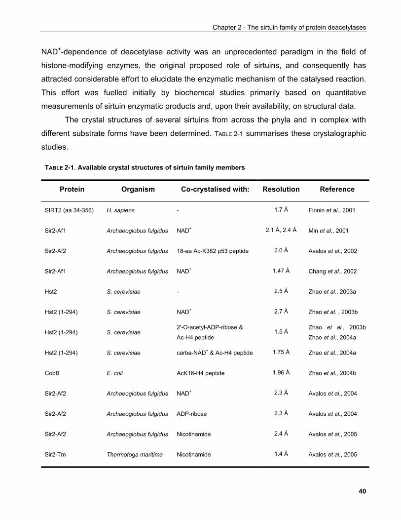
Chapter 2 - The sirtuin family of protein deacetylases
NAD+-dependence of deacetylase activity was an unprecedented paradigm in the field of
histone-modifying enzymes, the original proposed role of sirtuins, and consequently has
attracted considerable effort to elucidate the enzymatic mechanism of the catalysed reaction.
This effort was fuelled initially by biochemcal studies primarily based on quantitative
measurements of sirtuin enzymatic products and, upon their availability, on structural data.
The crystal structures of several sirtuins from across the phyla and in complex with
different substrate forms have been determined. TABLE 2-1 summarises these crystalographic
studies.
TABLE 2-1. Available crystal structures of sirtuin family members
Protein Organism Co-crystalised with: Resolution Reference
SIRT2 (aa 34-356) H. sapiens - 1.7 Å Finnin et al., 2001
Sir2-Af1 Archaeoglobus fulgidus NAD+ 2.1 Å, 2.4 Å Min et al., 2001
Sir2-Af2 Archaeoglobus fulgidus 18-aa Ac-K382 p53 peptide 2.0 Å Avalos et al., 2002
Sir2-Af1 Archaeoglobus fulgidus NAD+ 1.47 Å Chang et al., 2002
Hst2 S. cerevisiae - 2.5 Å Zhao et al., 2003a
Hst2 (1-294) S. cerevisiae NAD+ 2.7 Å Zhao et al. , 2003b
Hst2 (1-294) S. cerevisiae 2'-O-acetyl-ADP-ribose &
Ac-H4 peptide 1.5 Å
Zhao et al., 2003b
Zhao et al., 2004a
Hst2 (1-294) S. cerevisiae carba-NAD+ & Ac-H4 peptide 1.75 Å Zhao et al., 2004a
CobB E. coli AcK16-H4 peptide 1.96 Å Zhao et al., 2004b
Sir2-Af2 Archaeoglobus fulgidus NAD+ 2.3 Å Avalos et al., 2004
Sir2-Af2 Archaeoglobus fulgidus ADP-ribose 2.3 Å Avalos et al., 2004
Sir2-Af2 Archaeoglobus fulgidus Nicotinamide 2.4 Å Avalos et al., 2005
Sir2-Tm Thermotoga maritima Nicotinamide 1.4 Å Avalos et al., 2005
40
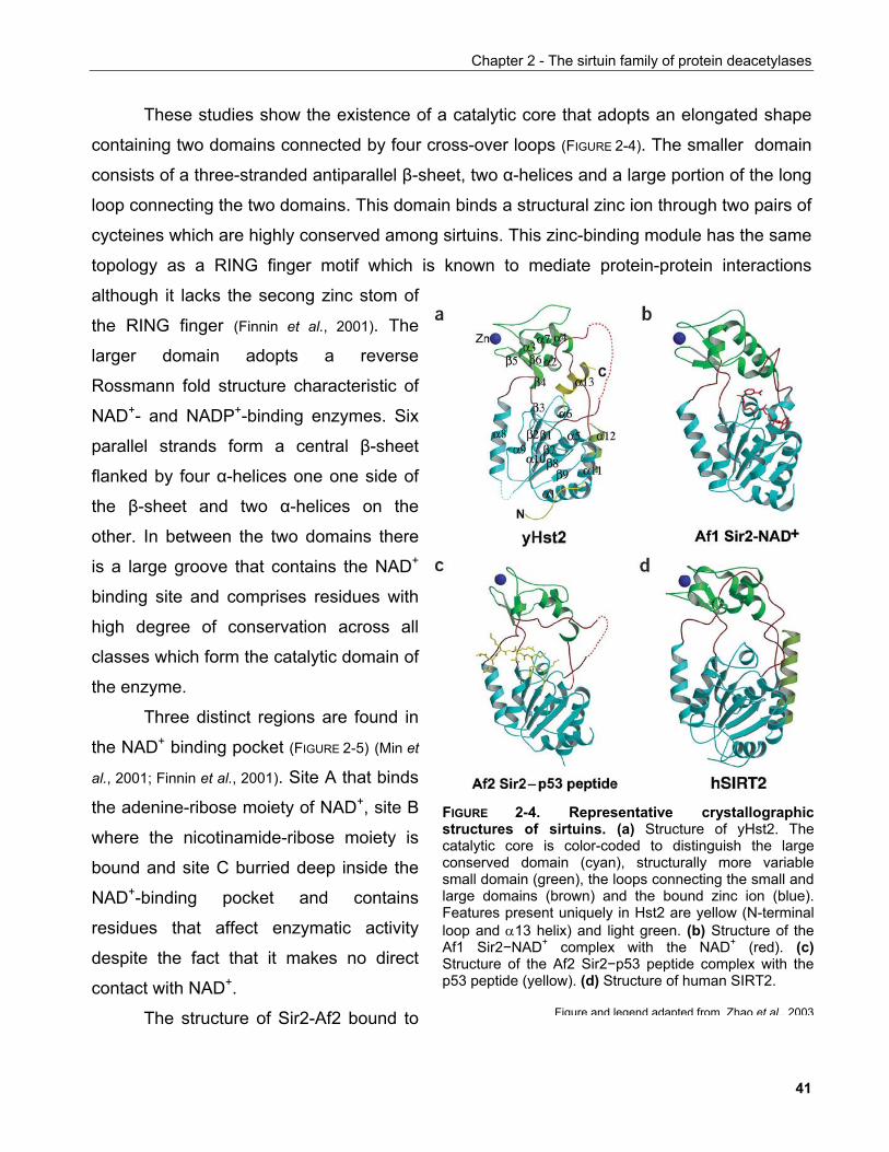
Chapter 2 - The sirtuin family of protein deacetylases
These studies show the existence of a catalytic core that adopts an elongated shape
containing two domains connected by four cross-over loops (FIGURE 2-4). The smaller domain
consists of a three-stranded antiparallel β-sheet, two α-helices and a large portion of the long
loop connecting the two domains. This domain binds a structural zinc ion through two pairs of
cycteines which are highly conserved among sirtuins. This zinc-binding module has the same
topology as a RING finger motif which is known to mediate protein-protein interactions
although it lacks the secong zinc stom of
the RING finger (Finnin et al., 2001). The
larger domain adopts a reverse
Rossmann fold structure characteristic of
NAD+- and NADP+-binding enzymes. Six
parallel strands form a central β-sheet
flanked by four α-helices one one side of
the β-sheet and two α-helices on the
other. In between the two domains there
is a large groove that contains the NAD+
binding site and comprises residues with
high degree of conservation across all
classes which form the catalytic domain of
the enzyme.
Three distinct regions are found in
the NAD+ binding pocket (FIGURE 2-5) (Min et
al., 2001; Finnin et al., 2001). Site A that binds
the adenine-ribose moiety of NAD+, site B
where the nicotinamide-ribose moiety is
bound and site C burried deep inside the
NAD+-binding pocket and contains
residues that affect enzymatic activity
despite the fact that it makes no direct
contact with NAD+.
FIGURE 2-4. Representative crystallographicstructures of sirtuins. (a) Structure of yHst2. Thecatalytic core is color-coded to distinguish the largeconserved domain (cyan), structurally more variablesmall domain (green), the loops connecting the small andlarge domains (brown) and the bound zinc ion (blue).Features present uniquely in Hst2 are yellow (N-terminalloop and α13 helix) and light green. (b) Structure of theAf1 Sir2−NAD+ complex with the NAD+ (red). (c)Structure of the Af2 Sir2−p53 peptide complex with thep53 peptide (yellow). (d) Structure of human SIRT2.
Figure and legend adapted from Zhao et al., 2003The structure of Sir2-Af2 bound to
41
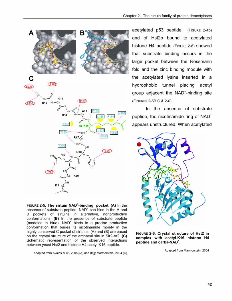
Chapter 2 - The sirtuin family of protein deacetylases
acetylated p53 peptide (FIGURE 2-4b)
and of Hst2p bound to acetylated
histone H4 peptide (FIGURE 2-6) showed
that substrate binding occurs in the
large pocket between the Rossmann
fold and the zinc binding module with
the acetylated lysine inserted in a
hydrophobic tunnel placing acetyl
group adjacent the NAD+-binding site
(FIGURES 2-5B,C & 2-6).
A B
C
In the absence of substrate
peptide, the nicotinamide ring of NAD+
appears unstructured. When acetylated
FIGURE 2-5. The sirtuin NAD+-binding pocket. (A) In theabsence of substrate peptide, NAD+ can bind in the A andB pockets of sirtuins in alternative, nonproductiveconformations. (B) In the presence of substrate peptide(modeled in blue), NAD+ binds in a precise productiveconformation that buries its nicotinamide moiety in thehighly conserved C pocket of sirtuins. (A) and (B) are basedon the crystal structure of the archaeal sirtuin Sir2-Af2. (C)Schematic representation of the observed interactionsbetween yeast Hst2 and histone H4 acetyl-K16 peptide.
Adapted from Avalos et al., 2005 [(A) and (B)]; Marmorstein, 2004 (C)
FIGURE 2-6. Crystal structure of Hst2 incomplex with acetyl-K16 histone H4peptide and carba-NAD+.
Adapted from Marmorstein, 2004
42
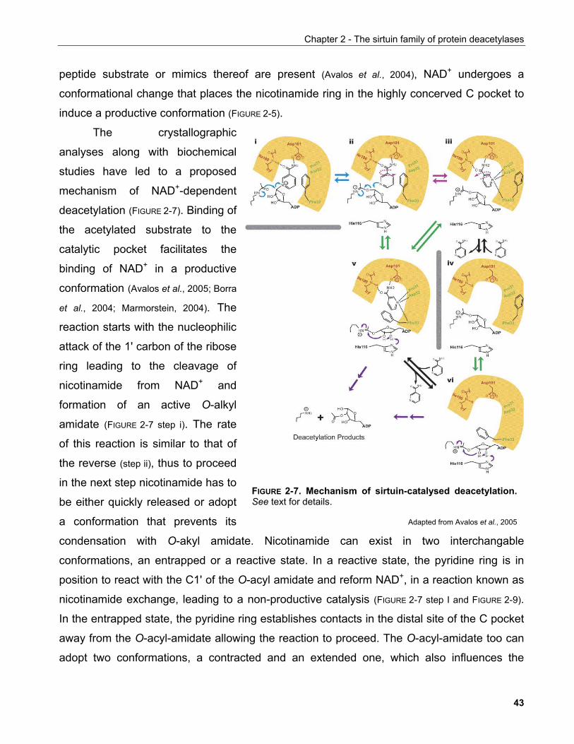
Chapter 2 - The sirtuin family of protein deacetylases
peptide substrate or mimics thereof are present (Avalos et al., 2004), NAD+ undergoes a
conformational change that places the nicotinamide ring in the highly concerved C pocket to
induce a productive conformation (FIGURE 2-5).
The crystallographic
analyses along with biochemical
studies have led to a proposed
mechanism of NAD+-dependent
deacetylation (FIGURE 2-7). Binding of
the acetylated substrate to the
catalytic pocket facilitates the
binding of NAD+ in a productive
conformation (Avalos et al., 2005; Borra
et al., 2004; Marmorstein, 2004). The
reaction starts with the nucleophilic
attack of the 1' carbon of the ribose
ring leading to the cleavage of
nicotinamide from NAD+ and
formation of an active O-alkyl
amidate (FIGURE 2-7 step i). The rate
of this reaction is similar to that of
the reverse (step ii), thus to proceed
in the next step nicotinamide has to
be either quickly released or adopt
a conformation that prevents its
condensation with O-akyl amidate. Nicotinamide can exist in two interchangable
conformations, an entrapped or a reactive state. In a reactive state, the pyridine ring is in
position to react with the C1' of the O-acyl amidate and reform NAD+, in a reaction known as
nicotinamide exchange, leading to a non-productive catalysis (FIGURE 2-7 step I and FIGURE 2-9).
In the entrapped state, the pyridine ring establishes contacts in the distal site of the C pocket
away from the O-acyl-amidate allowing the reaction to proceed. The O-acyl-amidate too can
adopt two conformations, a contracted and an extended one, which also influences the
FIGURE 2-7. Mechanism of sirtuin-catalysed deacetylation. See text for details.
Adapted from Avalos et al., 2005
43
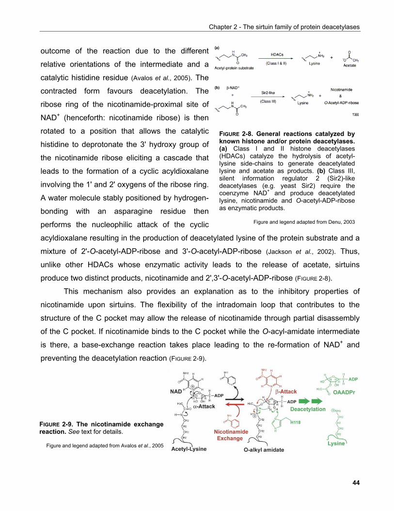
Chapter 2 - The sirtuin family of protein deacetylases
outcome of the reaction due to the different
relative orientations of the intermediate and a
catalytic histidine residue (Avalos et al., 2005). The
contracted form favours deacetylation. The
ribose ring of the nicotinamide-proximal site of
NAD+ (henceforth: nicotinamide ribose) is then
rotated to a position that allows the catalytic
histidine to deprotonate the 3' hydroxy group of
the nicotinamide ribose eliciting a cascade that
leads to the formation of a cyclic acyldioxalane
involving the 1' and 2' oxygens of the ribose ring.
A water molecule stably positioned by hydrogen-
bonding with an asparagine residue then
performs the nucleophilic attack of the cyclic
acyldioxalane resulting in the production of deacetylated lysine of the protein substrate and a
mixture of 2'-O-acetyl-ADP-ribose and 3'-O-acetyl-ADP-ribose (Jackson et al., 2002). Thus,
unlike other HDACs whose enzymatic activity leads to the release of acetate, sirtuins
produce two distinct products, nicotinamide and 2',3'-O-acetyl-ADP-ribose (FIGURE 2-8).
This mechanism
FIGURE 2-8. General reactions catalyzed byknown histone and/or protein deacetylases.(a) Class I and II histone deacetylases(HDACs) catalyze the hydrolysis of acetyl-lysine side-chains to generate deacetylatedlysine and acetate as products. (b) Class III,silent information regulator 2 (Sir2)-likedeacetylases (e.g. yeast Sir2) require thecoenzyme NAD+ and produce deacetylatedlysine, nicotinamide and O-acetyl-ADP-riboseas enzymatic products.
Figure and legend adapted from Denu, 2003
also provides an explanation as to the inhibitory properties of
nicotin
amide upon sirtuins. The flexibility of the intradomain loop that contributes to the
structure of the C pocket may allow the release of nicotinamide through partial disassembly
of the C pocket. If nicotinamide binds to the C pocket while the O-acyl-amidate intermediate
is there, a base-exchange reaction takes place leading to the re-formation of NAD+ and
preventing the deacetylation reaction (FIGURE 2-9).
FIGURE 2-9. The nicotinamide exchangereaction. See text for details.
alos et al., 2005
Figure and legend adapted from Av
44
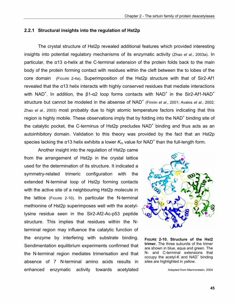
Chapter 2 - The sirtuin family of protein deacetylases
2.2.1 Structural insights into the regulation of Hst2p
he crystal structure of Hst2p revealed additional features which provided interesting
insight
from t
T
s into potential regulatory mechanisms of its enzymatic activity (Zhao et al., 2003a). In
particular, the α13 α-helix at the C-terminal extension of the protein folds back to the main
body of the protein forming contact with residues within the cleft between the to lobes of the
core domain (FIGURE 2-4a). Superimposition of the Hst2p structure with that of Sir2-Af1
revealed that the α13 helix interacts with highly conserved residues that mediate interactions
with NAD+. In addition, the β1-α2 loop forms contacts with NAD+ in the Sir2-Af1-NAD+
structure but cannot be modeled in the absense of NAD+ (Finnin et al., 2001; Avalos et al., 2002;
Zhao et al., 2003) most probably due to high atomic temperature factors indicating that this
region is highly mobile. These observations imply that by folding into the NAD+ binding site of
the catalytic pocket, the C-terminus of Hst2p precludes NAD+ binding and thus acts as an
autoinhibitory domain. Validation to this theory was provided by the fact that an Hst2p
species lacking the α13 helix exhibits a lower Km value for NAD+ than the full-length form.
Another insight into the regulation of Hst2p came
he arrangement of Hst2p in the crystal lattice
used for the determination of its structure. It indicated a
symmetry-related trimeric configuration with the
extended N-terminal loop of Hst2p forming contacts
with the active site of a neighbouring Hst2p molecule in
the lattice (FIGURE 2-10). In particular the N-terminal
methionine of Hst2p superimposes well with the acetyl-
lysine residue seen in the Sir2-Af2-Ac-p53 peptide
structure. This implies that residues within the N-
terminal region may influence the catalytic function of
the enzyme by interfering with substrate binding.
Sendimentation equilibrium experiments confirmed that
the N-terminal region mediates trimerisation and that
absence of 7 N-terminal amino acids results in
enhanced enzymatic activity towards acetylated
FIGURE 2-10. Structure of the Hst2trimer. The three subunits of the trimerare shown in blue, aqua and green. TheN- and C-terminal extensions thatoccupy the acetyl-K and NAD+ bindingsites are highlighted in yellow.
Adapted from Marmorstein, 2004
45
Chapter 2 - The sirtuin family of protein deacetylases
substrates.
The proposed autoinhibitory function of the C-terminus of Hst2p is very reminiscent of
the Src kinase regulatory mode where C-terminal extension when phosphorylated on a
specific tyrosine residue can bind the SH2 domain of the enzyme and impose an
autoinhibitory closed conformation (Harrison, 2003). Thus, as Hst2p shares N- and C-terminal
extensions with other members of sirtuins, including mammalian SIRT1, it is possible that the
observed structural features reflect true regulatory mechanisms.
2.2.2 Fate of the sirtuin deacetylation products
Apart from the deacetylated lysine, nicotinamide and 2',3'-O-ADP-ribose are produced
as a result of sirtuin-mediated deacetylation (FIGURE 2-8). The function of these molecules has
not been firmly established yet, however some insights have been provided by recent work in
the field.
2.2.2.1 Nicotinamide and NAD+ biosynthesis pathways
Biochemical studies along with the structural data presented above provided good
evidence that the involvement of two naturally occuring metabolites in the catalysis, NAD+
and nicotinamide, may provide a potential mechanism of regulation of sirtuin activity. For this,
we shall review the basic pathways of NAD+ biosynthesis in the cell which will be of
importance to understand the underlying principles and significance of the work addressing
the function of sirtuins discussed below.
In both procaryotic and eucaryotic cells there are two pathways that participate in
NAD+ production, the de novo pathway and the salvage pathway (FIGURE 2-11). The
converging molecule of the two pathways is nicotinic acid mononucleotide (NaMN).
In the de novo pathway NaMN derives from the degradation of tryptophan in a six-step
reaction where the last step is catalysed by quinolinate phosphoribosyl transferase
(BNA6/QPT1). In animals this reaction occurs mainly in the liver and kidney. Inadequate
dietary supplementation of tryptophan leads to a condition known as pellagra characterised
by diarrhea, dermatitis and dementia.
46
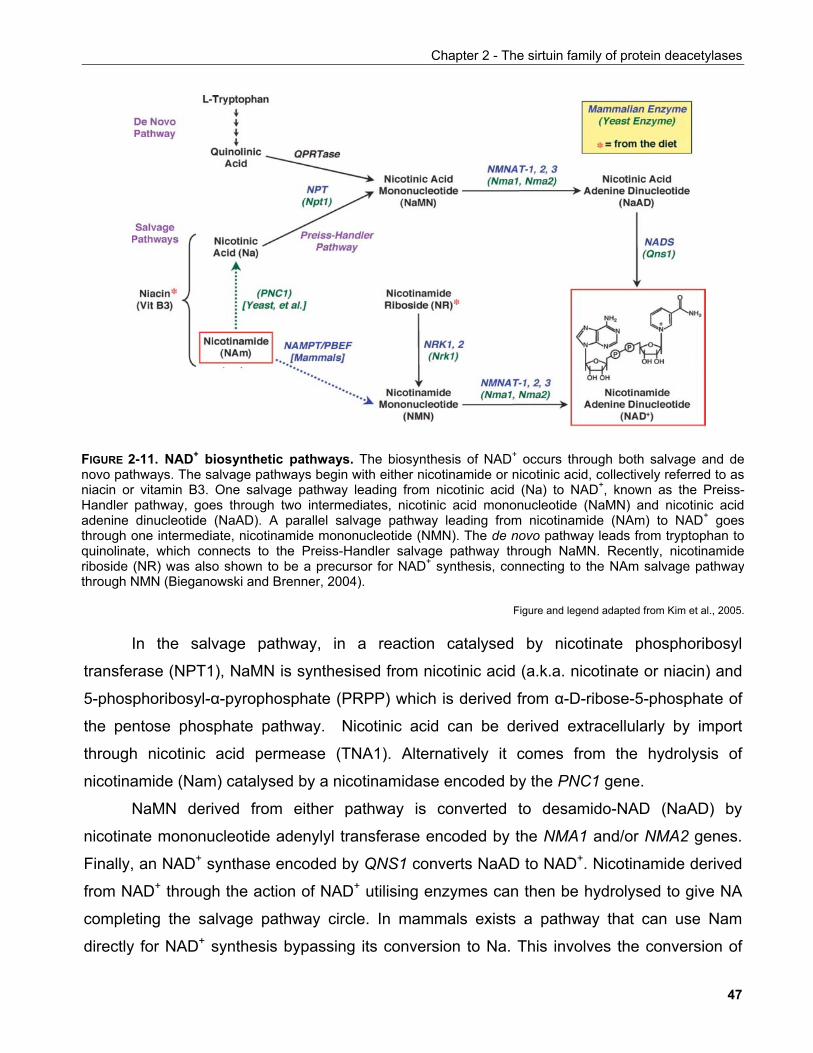
Chapter 2 - The sirtuin family of protein deacetylases
FIGURE 2-11. NAD+ biosynthetic pathways. The biosynthesis of NAD+ occurs through both salvage and denovo pathways. The salvage pathways begin with either nicotinamide or nicotinic acid, collectively referred to asniacin or vitamin B3. One salvage pathway leading from nicotinic acid (Na) to NAD+, known as the Preiss-Handler pathway, goes through two intermediates, nicotinic acid mononucleotide (NaMN) and nicotinic acidadenine dinucleotide (NaAD). A parallel salvage pathway leading from nicotinamide (NAm) to NAD+ goesthrough one intermediate, nicotinamide mononucleotide (NMN). The de novo pathway leads from tryptophan toquinolinate, which connects to the Preiss-Handler salvage pathway through NaMN. Recently, nicotinamideriboside (NR) was also shown to be a precursor for NAD+ synthesis, connecting to the NAm salvage pathwaythrough NMN (Bieganowski and Brenner, 2004).
Figure and legend adapted from Kim et al., 2005.
In the salvage pathway, in a reaction catalysed by nicotinate phosphoribosyl
transferase (NPT1), NaMN is synthesised from nicotinic acid (a.k.a. nicotinate or niacin) and
5-phosphoribosyl-α-pyrophosphate (PRPP) which is derived from α-D-ribose-5-phosphate of
the pentose phosphate pathway. Nicotinic acid can be derived extracellularly by import
through nicotinic acid permease (TNA1). Alternatively it comes from the hydrolysis of
nicotinamide (Nam) catalysed by a nicotinamidase encoded by the PNC1 gene.
NaMN derived from either pathway is converted to desamido-NAD (NaAD) by
nicotinate mononucleotide adenylyl transferase encoded by the NMA1 and/or NMA2 genes.
Finally, an NAD+ synthase encoded by QNS1 converts NaAD to NAD+. Nicotinamide derived
from NAD+ through the action of NAD+ utilising enzymes can then be hydrolysed to give NA
completing the salvage pathway circle. In mammals exists a pathway that can use Nam
directly for NAD+ synthesis bypassing its conversion to Na. This involves the conversion of
47
Chapter 2 - The sirtuin family of protein deacetylases
Nam into nicotinamide mononucleotide (NamMN) which is then converted to NAD+ by the
action of the same enzyme that catalyses NAD+ formation from NaMN (Voet and Voet,
Biochemistry, Wiley 1995, p825).
NAD+ is a key molecule in many metabolic, primarily catabolic, processes such as
glycolysis and the tricarboxylic acid (TCA) cycle, where it participates as a proton acceptor
leading to its reduction to NADH. It is also important in DNA metabolism since enzymes like
DNA ligase and poly(ADP) ribose polymerase (PARP) depend on NAD for their activity.
The ratio of NAD+ to NADH is important for the redox state of the cell and has been
shown to flactuate in response to metabolic changes. Accordingly, it can regulate the activity
of enzymes such as glyceraldehyde 3-phosphate dehydrogenase in glycolysis and pyruvate
dehydrogenase which converts pyruvate to acetyl-CoA that is used in the TCA cycle. Thus,
homeostatic mechanisms exist that ensure the constant re-oxidation of NADH to NAD+ e.g.
through the respiratory chain. Nevertheless, limitations in the accurate experimental
determination of the intracellular [NAD+]/[NADH] ratio have fueled an extensive ongoing
debate as to the exact quantitation of this ratio and how it fluctuates (Lin and Guarente, 2003).
The relevance of the interaction between NAD+ metabolic pathways and the function
of sirtuins is further exemplified by the recent evidence in the work of Pan et al. which
revealed a network of synthetic lethal interactions between NPT1 and components of the
replication machinery (Pan et al., 2006). In the same study, Hst3 shared the majority of these
interactions although it did not exhibit synthetic lethality with Npt1 itself suggesting that they
participate in a novel NAD+-dependent pathway regulating DNA replication in yeast.
Revollo et al. found that in mammals the rate-limiting enzyme in NAD+ biosynthesis is
nicotinamide phosphoribosyltransferase (NamPT) (FIGURE 2-11) rather than
nicotinamide/nicotinic acid mononucleotide adenylyl transferase (Nmnat) (Revollo et al., 2004).
Accordingly, overexpression of NamPT increased cellular NAD+ levels and this correlated
with transcriptional activation of mouse SIRT1 on a heterologous reporter. Furthermore, the
transcription profiles induced by overexpression of either NamPT or SIRT1 in MEFs
significantly overlapped, indicating that regulation of intracellular NAD+ by the mammalian
salvage pathway also had a potential impact on SIRT1.
48
Chapter 2 - The sirtuin family of protein deacetylases
Araki et al. observed that increased NAD+ levels due to overxpression of Namnt1
provided neuroprotection depending on SIRT1 (Araki et al., 2004). This work will be discussed
in more detail in section 2.3.5.1.7.
Some other aspects of interest in relation to this issue will be presented in the
discussion of his thesis (Chapter 5).
2.2.2.2 The function and fate of 2',3'-O-ADP-ribose Borra et al. hypothesised that being a distinctive protein deacetylation reaction
product, 2',3'-O-acetyl-ADP-ribose may serve either as a second messenger in a manner
reminiscent of cAMP, or as a substrate for other proteins. They tested their hypothesis by
microinjecting purified O-acetyl-ADP-ribose into starfish (A. miniata) immature oocytes a
system previously used to probe the biological function of proteins compounds. At low mM
concentrations, O-acetyl-ADP-ribose was able to block oocyte maturation (Borra et al., 2002). In
starfish immature oocyte cell-free extracts O-acetyl-ADP-ribose is rapidly metabolised
suggesting the existence of enzymes that mediate its turnover. Thus it is possible that either
O-acetyl-ADP-ribose or a metabolite thereof is responsible for the observed effects. The
oocyte maturation blockade is recapitulated by injection of recombinant yeast sirtuin Hst2p in
a manner dependent on its enzymatic activity. Microinjection of either Hst2p or O-acetyl-
ADP-ribose into one cell of a two-cell stage embryo also caused a delay in cell cycle
progression of the injected cell.
In a subsequent study, Rafty et al. identified a class of hydrolases termed Nudix that
could robustly catalyse the cleavage of O-acetyl-ADP-ribose into AMP and acetylated ribose-
5'-phosphate (Rafty et al., 2002). However, in cellular extracts O-acetyl-ADP-ribose was subject
to modifications inconsistent with Nudix activities an associated with novel unidentified
enzymes. Rafty et al. predicted the existence of cytoplasmic esterases that hydrolysed O-
acetyl-ADP-ribose to acetate and ADP-ribose and nuclear O-acetyl-ADP-ribose-dependent
acetyltransferases.
In a study with exciting implications, Kustatscher et al. discovered that the macro
domain of histone macroH2A1.1 (mH2A1.1) can bind O-acetyl-ADP-ribose through a distinct
binding pocket with µM affinity (Kustatscher et al., 2005). mH2A1 is a histone variant found
49
Chapter 2 - The sirtuin family of protein deacetylases
associated with heterochromatic foci in senescent and quiescent cells. Interestingly, the gene
encoding for mH2A1 can give rise to two alternatively spliced variants with distinct
spatiotemporal distribution during development. Among these isoforms, only one is able to
bind O-acetyl-ADP-ribose suggestive of a potential regulatory role for the alternative splicing
of this histone and adding another link between NAD+ and DNA metabolism.
These experiments suggested that the enzymatic products of sirtuins and in particular
O-acetyl-ADP-ribose may have novel unexplored functions aside the deacetylation of
proteins conferred by the enzymes themselves.
2.2.3 Specificity of sirtuins
As for all catalysed reactions, an issue of interest is whether the enzymes involved
exhibit substrate specificity and if yes how this is attained. Ultimately, provided enough
information as to the associated biological functions, enzymatic specificity may provide
important clues for the effective therapeutic targetting of the enzyme in question.
Early studies of sirtuin deacetylation activity showed that the yeast Sir2p may target
histones for deacetylation based on the fact that overexpression of Sir2p in yeast leads to
histone hypoacetylation. Imai et al. provided evidence that Sir2 has preference for residues
Ac-K9 and Ac-K14 in histone H3 as well as residue Ac-K16 in histone H4 in vitro in
agreement to previous data indicating a requirement for these residues for silencing (Imai et
al., 2000). Some evidence to support this is also provided by recent studies in mammalian
cells that show SIRT1 may deacetylate both histones H3 and H4 with preference to Ac-K9
and Ac-K16 respectively as well as histone H1 Ac-K26 in vivo (Vaquero et al., 2004). However,
the high degree of phylogenic conservation of sirtuins and the fact that bacteria do not have
canonical histones raised the possibility that additional sirtuin deacetylation targets exist, a
hypothesis proved to be correct in both prokaryotic and eukaryotic organisms. Despite the
emergence of new substrates a consensus deacetylation target sequence failed to emerge.
In order to investigate the issue of substrate specificity for the human sitruin SIRT1,
Blander et al. performed an unbiased study using oriented peptide libraries (Blander et al.,
2005). This approach has been succesfully used before for the identification of protein domain
consensus binding sites as well as kinase target sequences. This screen involved the
50
Chapter 2 - The sirtuin family of protein deacetylases
incubation of a degenerate peptide library containing acetylated lysines with recombinant
SIRT1 and the selective purification of deacetylated peptides. This is achieved by reacting
the ε-amino group of deacetylated lysine with a photocleavable linker which is tagged with
biotin. After removal of the linker, the purified peptides bearing sites specifically targeted by
SIRT1 were analysed by Edman degradation. This allowed the determination of the
frequency at which specific aminoacids occur in the regions flanking the deacetylated lysine.
This approach clearly showed that SIRT1 shows no sequence specificity in vitro (Blander et al.,
2005). In a subsequent study, Khan et al. using yeast Hst2p showed that this sirtuin shows
preference for acetylates lysines situated within unstrustured protein regions (Khan et al., 2005).
These approaches imply that sirtuins, unless promiscuous, attain specificity in vivo by
mechanisms other than recognition of substrate primary sequence. Crystal structures of
sirtuins in complex with peptide substrates only are available and show only weak contacts
between sirtuin residues and the region surrounding the target lysine. It is not for sure that
this applies in the context of the entire protein substrate but if true, it would support the notion
that sirtuins may deacetylate their targets specifically by virtue of their localisation, binding
partners that act like scaffold proteins or both.
2.2.4 Small molecule modulators of sirtuins
Metabolites of the NAD+ biosynthetic pathway such as nicotinamide, NADH and NAD+
itself have been useful tools in preliminary pharmacological studies of sirtuins due to their
availability and low cost. However, their prominent role in basic cellular metabolic processes
as well as our limited knowledge of mammalian sirtuin biology prompted the search of both
activators and inhibitors that could provide sirtuin-specific targeting.
A study by Howitz et al. identified a class of compounds called polyphenols as sirtuin
activating compounds (STACs). Among them, resveratrol, a molecule found in red wine,
exhibited the most potent effects on sirtuins by lowering the Km of the enzyme for acetylated
substrate (Howitz et al., 2003). Given the previously documented effects of resveratrol on
neuroprotection, cardioprotection and chemoprotection (Pervaiz, 2003), the study of Howitz et
al. raised the exhiting possibility that the molecular target of resveratrol is a sirtuin. In a
follow-up study, Wood et al. could show that resveratrol and other STACs could extend life-
51
Chapter 2 - The sirtuin family of protein deacetylases
span in Drosophila as well as C. elegans in a manner dependent on Sir2 and non-additive to
caloric restriction (Wood et al., 2004).
However, subsequent studies by Kaeberlein et al. and Borra et al. challenged these
observations (Kaeberlein et al. 2005, Borra et al. 2005). Kaeberlein et al. could find no evidence that
resveratrol increased life-span in three different yeast strains, while both studies provided
evidence that the observed effect of resveratrol depended on the particular fluorophore
attached to the peptide used to assay sirtuin activity. Thus, resveratrol seems to be a
substrate-specific activator of sirtuins and whether it truly regulates sirtuins in vivo remains to
be further investigated especially following recent evidence that enhanced NAD+ biosynthesis
and SIRT1 activity promote neuroprotection in mice (Araki et al., 2004).
Forward chemical genetic approaches have also yielded novel compounds that inhibit
sirtuin activity. Forward chemical genetic screens involve assaying of libraries of small
organic molecules for their ability to inhibit or enhance a particular phenotype. Grozinger et
al. assayed a library of 1,600 compounds for their ability to allow expression of a reporter
gene from the telomeres that would lead to cell death (Grozinger et al., 2001). Their work yielded
three inhibitors, A3, M15 and sirtinol, the latter being the most potent exhibith IC50 values of
68 and 38 µM for yeast Sir2p and hSIRT2 respectively.
Bedalov et al. used an ameliorated version of this screen in that inhibition of telomeric
silencing rescued cell growth, having the advantage that they could eliminate false hits due to
cytotoxic compounds (Bedalov et al., 2001). Their study identified 11 out of 6,000 compounds
screened to inhibit telomeric silencing. Among these, only 1, splitomicin was subsequently
shown in a secondary screen to be capable of inhibiting silencing also at rDNA and mating
type loci. Splitomicin exhibits an IC50 of 60 µM for yeast Sir2p in vitro. Mutagenesis analysis
revealed that splitomicin may target the acetylated lysine binding pocket of the enzyme. In a
later study, Hirao et al. identified dehydrosplitomicin and compound 26, two splitomicin
analogues as specific inhibitors of the yeast sirtuins Hst1 and Sir2 respectively (Hirao et al.,
2003).
Recently, Solomon et al. identified a novel compound in a high-throughput screen
using bacterially expressed SIRT1 (Solomon et al., 2006). The compound, named EX-527
inhibits SIRT1 potently with an IC50 of 38 nM and is highly specific since its IC50 values for
52
Chapter 2 - The sirtuin family of protein deacetylases
SIRT2 and SIRT3 are in the µM range. This compound was succesfully used for intracellular
inhibition of SIRT1 as described in section 2.3.5.1.4.
It is likely that such compounds will be of great use for the study of sirtuin biology,
especially when activators/inhibitors targeting other sirtuin homologues are developed (see
discussion in Chapter 5). The emergence of powerful novel technologies such as RNA
interference and gene targeting by homologous recombination allow efficient genetic
manipulation of protein expression in mammalian cells. However, RNAi is limited by the
protein turnover rates so that a gradual rather than accute protein depletion takes place.
Furthermore, the presence of several homologues in a protein family, at times allows the
functional compensation of the depleted protein. Small molecule compounds that can
accutely interfere with enzymatic activity can offer important tools to overcome such
difficulties provided that their specificity is reasonably establised. However, the translation of
such compounds into therapeutics will only depend on our understanding of a protein's
biological function.
53
Chapter 2 - The sirtuin family of protein deacetylases
2.3 FUNCTIONAL STUDIES OF SIRTUINS 2.3.1 Sirtuin functions in prokaryotic organisms 2.3.1.1 Bacterial sirtuins
The ability of Salmonella typhimurium cobB to compensate cobT mutations in the
cobalamin biosynthesis pathway provided the first hints into the biochemical activity of
sirtuins (Tsang and Escalante-Semerena, 1998). Interestingly, strains of Salmonella enterica that
lack the cobB gene are unable to utilise propioniate and low acetate concentration as carbon
sources because in such strains the acetyl-CoA synthase (ACS) enzymes required to convert
these substances into acyl-CoA derivatives are inactive (Starai et al., 2002). ACS from cobB- but
not cobB+ strains was highly acetylated implying that its enzymatic activity is regulated by
acetylation (Starai et al., 2002).
ACS mediates acetyl-CoA production in a two-step mechanism. First, acetate is
adenylated to yield acetyl-AMP and subsequently ACS catalyses the formation of a thioester
bond to produce acetyl-CoA (Starai et al., 2004). Acetylation of ACS at K609 by the
acetyltransferase Pat (protein acetyltransferase) specifically inhibits the ATP-dependent
adenylation of acetate but leaves the thioester bond forming activity unaffected (Starai et al.,
2004; Starai et al., 2002). Incubation of inactive ACS with NAD+ and cobB resulted in a dramatic
increase of ACS activity suggesting that the deacetylase activity harboured by cobB acts as
an activator of ACS (Starai et al., 2002).
Interestingly, ACS K609 is located within a sequence which is highly conserved
among AMP-forming enzymes including propionyl-CoA synthase and luciferase (Starai et al.,
2002). Despite lack of experimental evidence, it is tempting to speculate for a broader role of
acetylation in regulating similar reactions and thus an expanded role of sirtuins in these
processes.
54
Chapter 2 - The sirtuin family of protein deacetylases
2.3.1.2 Archaeal sirtuins
Similar to bacteria, some but not all Archaea do not have histone-like proteins to
organise their genome. Instead, this role is performed by small 7-10 kDa proteins (Bernander,
2003). One such protein known as Sso10b and later renamed Alba (acetylation lowers binding
affinity) which binds to DNA as a dimer, was identified as a interaction partner of the sirtuin
orthologue from Sulfolobus solfataricus ssSir2.
Alba was shown to be acetylated at K16 by an archaeal orthologue of Pat
acetyltransferase (Marsh et al., 2005) which results in lower affinity for DNA (Bell et al., 2002). This
was further supported by the crystal structure of Alba and molecular modeling that revealed a
role of K16 in the binding of the protein to DNA (Wardleworth et al. 2002; Zhao et al., 2003). ssSir2-
mediated deacetylation induces the ability of Alba to mediate silencing in an in vitro
reconstituted transcription system, an attribute that correlates with the ability of Alba to bind
DNA (Bell et al., 2002; Marsh et al., 2005).
These data indicated that archaeal sirtuins may regulate gene transcription by
modulating the ability of proteins to bind DNA. Furthermore, as the chromosomal DNA of
Sulfolobus is differently organised in exponentially growing versus stationary phase cells, the
regulation of Alba acetylation by ssSir2 may have a broader role in genome organisation
(Bernander, 2003).
2.3.2 Sirtuin functions in S. cerevisiae 2.3.2.1 Regulation of chromatin silencing in S. cerevisiae by sirtuins 2.3.2.1.1 Silencing at mating type loci
Two types of haploid cells are known in the yeast Saccharomyces cerevisiae, denoted
a and α. These cell types are determined by the selective expression of the mating type
locus (MAT) alleles MATa and MATα which encode transcriptional regulators that dictate the
expression of gene sets specific for each type.
Certain yeast strains known as homothallic are able to interconvert between the two
cell types. These strains harbor an active HO gene encoding a site-specific endonuclease
55
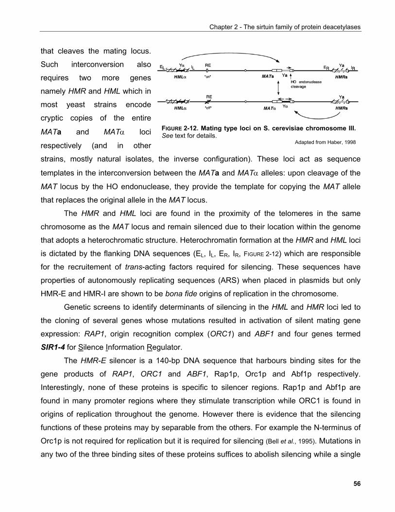
Chapter 2 - The sirtuin family of protein deacetylases
that cleaves the mating locus.
Such interconversion also
requires two more genes
namely HMR and HML which in
most yeast strains encode
cryptic copies of the entire
MATa and MATα loci
respectively (and in other
strains, mostly natural isolates, the inverse configuration). These loci act as sequence
templates in the interconversion between the MATa and MATα alleles: upon cleavage of the
MAT locus by the HO endonuclease, they provide the template for copying the MAT allele
that replaces the original allele in the MAT locus.
FIGURE 2-12. Mating type loci on S. cerevisiae chromosome III.See text for details.
Adapted from Haber, 1998
The HMR and HML loci are found in the proximity of the telomeres in the same
chromosome as the MAT locus and remain silenced due to their location within the genome
that adopts a heterochromatic structure. Heterochromatin formation at the HMR and HML loci
is dictated by the flanking DNA sequences (EL, IL, ER, IR, FIGURE 2-12) which are responsible
for the recruitement of trans-acting factors required for silencing. These sequences have
properties of autonomously replicating sequences (ARS) when placed in plasmids but only
HMR-E and HMR-I are shown to be bona fide origins of replication in the chromosome.
Genetic screens to identify determinants of silencing in the HML and HMR loci led to
the cloning of several genes whose mutations resulted in activation of silent mating gene
expression: RAP1, origin recognition complex (ORC1) and ABF1 and four genes termed
SIR1-4 for Silence Information Regulator.
The HMR-E silencer is a 140-bp DNA sequence that harbours binding sites for the
gene products of RAP1, ORC1 and ABF1, Rap1p, Orc1p and Abf1p respectively.
Interestingly, none of these proteins is specific to silencer regions. Rap1p and Abf1p are
found in many promoter regions where they stimulate transcription while ORC1 is found in
origins of replication throughout the genome. However there is evidence that the silencing
functions of these proteins may by separable from the others. For example the N-terminus of
Orc1p is not required for replication but it is required for silencing (Bell et al., 1995). Mutations in
any two of the three binding sites of these proteins suffices to abolish silencing while a single
56
Chapter 2 - The sirtuin family of protein deacetylases
copy of any of these binding sites is insufficient to establish silenced chromatin. Conversely,
the Sir proteins are devoid of direct DNA binding ability. Thus it is thought that the combined
binding of Rap1p, Orc1p and Abf1p to the silencer creates a platform for the recruitment of
Sir proteins by means of protein-protein interactions.
Isolation of native Sir complexes from S. cerevisiae showed that while Sir2p and Sir4p
form a soluble complex, Sir3p fails to co-purify in stoichiometric amounts. Furthermore, Sir3p
and Sir4p bind preferentially to the hypoacetylated tails of histone H3 and histone H4 in vitro
and Sir mutations that disrupt these interactions abolish silencing (Hecht et al.,1995). Mutational
analyses also showed that the regions of histones H3 and H4 required for Sir3p and Sir4p
binding are also required for silencing. Importantly, genomic regions occupied by Sir
complexes overlap with the location of hypoacetylated histones suggesting a tight connection
between these two phenomena.
The initiating step in the establishment of Sir-dependent chromatin silencing is the
binding of Sir1p to the silencer. Sir1p binds directly to Orc1p and is confined to the silencer
region without spreading adjacently. Tethering of Sir1p to silencers by a heterologous DNA
binding domain can support the establishment of silenced chromatin in the absence of ORC
or Rap1p. Both Sir3p and Sir4p can bind to Rap1 and the Sir3p-Rap1p interaction can also
occur in vitro in the absense of other yeast proteins (Moretti et al., 1994). Sir4p is recruited to the
silencer through its interaction with Sir1p/Rap1p independently of Sir2p and Sir3p. Thus
Sir2p is most likely recruited to the silencer in complex with Sir4p. Sir3p is engaged in the
complex through its interactions with Sir4p and Rap1p.
Once located to the silencer the Sir complex spreads towards the gene to be silenced.
Sir2p and Sir3p are essential for this. The histone deacetylase activity of Sir2p which is
dispensable for the recruitment of the complex to the silencer is required for this function.
Upon its recruitment to the silencer Sir2p is brought in the proximity of acetylated histones H3
and H4 in the nearby nucleosome which it deacetylates creating high affinity sites for more
Sir3p and Sir4p. In turn, this allows the sequential binding of more Sir2 complexes allowing
their spreading along with the consequent deacetylation of histones. Thus chromatin loaded
with Sir complexes adopts a compact structure which renders it silent.
57

Chapter 2 - The sirtuin family of protein deacetylases
2.3.2.1.2 Chromatin silencing at telomeres
Sir complexes also participate in another site of silencing in yeast at telomeres. This
phenomenon, reminiscent of position-effect variegation is known as telomere position effect
(TPE) and refers to the silencing of genes in the proximity of telomeres. Telomeric silencing
shares many of the features of silencing at mating-type loci yet it differs in that it is
independent of Sir1p and that recruitment of the Sir complex in yeast telomeric repeats is
mediated by both Rap1p and a protein known as Hdf1 or yKu70 (Guarente, 1999).
Luo et al. found that Sir4p can bind to Rap1p independent of other Sir proteins or the
Ku complex in the telomeric region proximal to the chromosome end (Luo et al., 2002). The
reverse is true as the distance from chromosome ends increases suggesting that as in the
case of mating type loci, the Rap1p-Sir4p complex participates in the seeding of the silenced
chromatin at the telomeres. A schematic model of the proposed mechanism for the
establishment of silencing at yeast telomeres is shown in Figure 2-13.
FIGURE 2-13. Schematic model of Sir-mediatedsilencing mechanism at yeast telomeres.Multiple copies of Rap1 bind to a simple repeatedsequence at each telomere region, which lacksnucleosomes (top). This nucleates the assembly ofa multiprotein complex (bottom) through protein-protein interactions between Rap1, Sir2, Sir3, Sir4,and the hypoacetylated amino-terminal tails ofhistones H3 and H4 of nearby nucleosomes.Asterisks represent hyperacetylated histone amino-terminal tails. The heterochromatin structureencompasses ~4 kb of DNA neighboring the Rap1-binding sites, irrespective of its sequence. Theactual structure of the higher-order heterochromatinis not yet understood.
Adapted from Grunstein, 1997
Hediger et al. used live imaging of yeast telomeres to unveil another function of the Sir
proteins at telomeres. They found that Ku and Sir proteins share a redudant role in telomere
anchoring to the nuclear periphery (Hediger et al., 2002). Telomere anchoring is thought to be
important for the establishment of epigenetic states. Both Ku and Sir support telomere
anchoring in the absence of eachother, but while Ku's contribution spans the entire cell cycle,
that of Sirs is limited to the S-phase: In the absence of Ku, telomeres are less efficiently
58
Chapter 2 - The sirtuin family of protein deacetylases
tethered to the nuclear periphery during G1. This work provided evidence for a mechanism
that explains the juxtaposition of silenced chromatin in distinct sub-nuclear compartments.
The Ku complex is phylogenetically conserved from bacteria to humans (Daley et al.,
2005). It comprises two subunits, termed Ku70 and Ku80. Its best known functions are in non-
homologous end joining (NHEJ), V(D)J recombination and DNA damage repair where it
participates in the recognition of double-strand DNA breaks and serves to recruit the
appropriate protein complexes such as DNA ligase that fulfill the corresponding functions
(Boulton and Jackson, 1998).
The work of Tsukamoto et al. identified Sir4 as a binding partner of yKu70 in a yeast
two-hybrid screen. They could show that in accordance to the previously established roles of
the Sir proteins that Sir2p, Sir3p and Sir4p but not Sir1 participate in the illegitimate
recombination, end-joining and double-strand break repair pathways (Tsukamoto et al., 1997).
Mutations in the corresponding genes confer higher sensitivity to genotoxic stresses such as
ionising irradiation. Interestingly, upon DNA damage, both Sir and Ku proteins re-localise
from subtelomeric foci to sites of DNA damage albeit with different kinetics providing support
for the notion that subtelomeric chromatin serves as a reservoir for proteins that are
relocated to specific nuclear sites upon physiological stimuli (Martin et al., 1999).
With regards to the role of the Sir complex in DNA damage repair, it has been
suggested that its recruitment at sites of DNA damage induces a heterochromatin-like
structure which may prevent the transcriptional and replication machineries from accessing
the site until it has been repaired. Alternatively, the localised compact state of chromatin may
facilitate the re-joining of the damaged DNA (Critchlow and Jackson, 1998).
2.3.2.1.3 Chromatin silencing at the rDNA locus Another site where silencing holds important roles in the yeast genome is the loci that
encode ribosomal RNAs (rDNA). The rDNA locus has a repetitive nature in that it comprises
a 9.1 kb sequence that is tandemly repeated 100-200 times. Each rDNA repeat encodes the
35S RNA which is transcribed by RNAPolI and serves as the precursor for the 25S, 18S and
5.8S rRNAs and the RNAPolIII-transcribed 5S rRNA. These genes are separated by non-
transcribed spacer regions. Sir2p was shown to be more highly associated with these spacer
59
Chapter 2 - The sirtuin family of protein deacetylases
regions than with the 35S coding region and this gave rise to the as yet unproven notion that
a silencer sequence similar to the one found in mating type loci is also present therein.
Several RNA PolII-transcribed genes when introduced within the rDNA repeats are silenced
(Bryk et al., 1997; Fritze et al., 1997; Smith and Boeke, 1997).
Unlike mating type and telomeric loci, Sir3 and Sir4 are not required for silensing at
the rDNA loci. Rather an entirely separate Sir2p-containing complex called RENT (for
regulator of nucleolar silencing and telophase) is required. The RENT complex was identifed
by two groups using distinct starting points for their investigation. Seeking to understand the
mechanism of rDNA silensing, Straight et al. compared gel patterns of proteins which interact
with GST-Sir2 vs. GST-Sir4 C-terminal fragment fusions to identify Sir4p components that
are specific to Sir2p. They found a protein with an apparent mass of 175 kDa which they
termed Net1 by virtue of its previously given names (NUS1-nucleolar silencing protein,
ESC5-establishes silent chromatin and TUB2-telophase arrest bypassed) (Straight et al., 1999).
Shou et al. also identified Net1p in a genetic screen for proteins that allow a yeast
strain bearing a deletion of the cdc15 gene and arrests in telophase to survive (Shou et al.,
1999). Cdc15 encodes a protein kinase which is part of a signalling pathway known as the
mitotic exit network (MEN). The MEN is required for proper exit from mitosis primarily through
its ability to inactivate mitotic cyclin-dependent kinases. Its function is to induce and maintain
the release of the phosphatase Cdc14p from the nucleolus to the cytoplasm at anaphase and
beyond during which Cdc14p dephosphorylates and thus inactivates mitotic kinases allowing
cells to enter the next G1. To understand the mechanism underlying the genetic interaction
between Cdc14p and Net1p, Shou et al. used immunoaffinity purification to identify binding
partners of Net1. Among the proteins identified were Cdc14p and Sir2p (Shou et al., 1999).
Visintin et al. also indepedently identified Net1p as a Cdc14p partner in a yeast two-hybrid
screen (Visintin et al., 1999).
Net1 was shown to be required for normal growth and rDNA silencing. In agreement to
this, chromatin immunoprecipitation experiments showed that Net1 is associated with rDNA
loci independent of Sir2p. Conversely, Sir2p binding to rDNA requires the presence of Net1.
Thus Net1 seems to be the protein that recruits Sir2p to rDNA consistent with the absence of
Sir4 from these loci. At the same time Cdc14p also binds to the rDNA loci in a Net1-
dependent manner (Straight et al., 1999). The binding of Sir2p, Net1p and Cdc14p to rDNA
60
Chapter 2 - The sirtuin family of protein deacetylases
coincides with their localisation to nucleoli throughout the cell cycle until the
anaphase/telophase transition where both Sir2p and Cdc14p are released to the cytoplasm.
The release of Cdc14p from the nucleoli coincides with its activation because when bound to
Net1p its phosphatase activity is inhibited.
In strict terms, the observations above do not prove the existence of a ternary complex
comprising Sir2p, Net1p and Cdc14p. Indeed, Shou et al. even demonstrated that loss of
rDNA silencing does not bypass lethality caused by a mutation in tem1 whose product is an
activator of the MEN. This implies two separable functions for Net1p/Cdc14p and
Net1p/Sir2p complexes. Sequential co-immunoprecipitation experiments, mutational analysis
on Net1p in an attempt to dissect these two functions in rDNA silencing and mitotic exit as
well as genetic analysis of the interaction between MEN and Sir2 would be required to firmly
establish the interconnectivities between these pathways.
2.3.2.2 Regulation of meiotic checkpoint function and recombination
Aside its function in rDNA silencing as part of the RENT complex, the localisation of
Sir2p in the nucleolus also functions in securing faithful meiotic progression (San-Segundo and
Roeder, 1999). Proper synapsis of homologous chromosomes during meiosis is guarenteed by
checkpoint systems. Mutations in meiotic checkpoint genes such as zip1 cause cells to arrest
in the pachytene phase of meiosis I (Sym et al., 1993). Pch2p is a protein identified in a genetic
screen for mutants that can bypass the pachytene checkpoint elicited by zip1 mutation. zip1-
pch2 double mutants sporulate and undergo mitotic recombination but the haploid progeny
show poor viability due to chromosome missegregation.
The proper function of Pch2p requires its localisation to the nucleolus, an event which
is dependent on Sir2p. In the absence of Sir2p, Pch2p is localised to the nucleoplasm and
these cells exhibit high rates of recombination in the rDNA locus similar to cells defective for
Pch2p alone. This may arise due to the increased presence of the meiotic recombination
protein Hop1p in the nucleolus.
The exact role of Sir2p in meiotic checkpoint function is not understood but two pieces
of experimental evidence suggest that it may contribute through its role in the establishment
of the structural characteristics of silenced chromatin (San-Segundo and Roeder, 1999). Firstly, in
61
Chapter 2 - The sirtuin family of protein deacetylases
a strain carrying deletion of the rDNA array with the essential rDNA genes provided as an
extrachromosomal plasmid, Pch2p still localises to nucleoli-like structures but is defective in
its meitotic checkpoint function. In this strain, mutation in sir3 induces the relocalisation of
Pch2p and Sir2p to the telomeres and partially restores checkpoint function. A second
experiment showed that in the presence of intact rDNA, overexpression of Sir4p induces the
relocalisation of Pch2p to telomeres but only partially abolishes meiotic checkpoint function
and this is depedent on sir2. This evidence sustains the notion that the telomeric locus
provides some element similar to the rDNA arrays that supports the meiotic checkpoint
function of Pch2p.
Interestingly, mutations in another pachytene checkpoint protein Dot1 (a.k.a. Pch1)
disrupt Pch2 and Sir2 localisation from the nucleoli (San-Segundo and Roeder, 2000). Dot1 is a
distinct H3 histone methyltransferase which lacks the charactersitic SET domain of other
lysine methyltransferases and can trimethylate H3-K79 (Vaquero et al., 2003).
This implies a possible interconnection between histone methylation and Sir-mediated
silencing which has not been experimentally addressed further yet.
2.3.2.3 Regulation of DNA replication
Given the association of Sir2p with ORC, it is not surprising that recently a novel role
for Sir2p in the control of replication has emerged (Pappas et al., 2004). Replication of DNA
takes place during the S-phase of the cell cycle. However, assembly of pre-replication
complexes (pre-RC) at origins of replication occurs as early as at the anaphase of the
previous cell cycle. Cdc6p orchestrates pre-RC assembly by initiating recruitment of its
components such as the MCM (minichromosome maintainance) helicase to ORC at the
origins of replication. The temperature-sensitive cdc6-4 mutant is lethal due to its inability to
assemble pre-RCs.
A genetic screen for rescue of this phenotype identified mutations in sir2 and to a
lesser degree sir3 and sir4 as potential candidates. Loss of sir2 was shown to supress the
lethality of other replication initiation mutants, notably orc-5 and mcm2, but not that of genes
acting at steps subsequent to pre-RC assembly such as the kinase and DNA polymerase α-
encoding genes cdc7 and cdc17 respectively. Subsequently, chromatin immunoprecipitation
62
Chapter 2 - The sirtuin family of protein deacetylases
experiments showed that Sir2p deletion restored the loading of MCM2 helicase to origins
albeit to a different extend depending on the ARS tested.
The exact mechanism by which Sir2p inhibits replication is unclear. Interestingly,
deletion of two other deacetylases, Hda1p and Rpd3p does not phenocopy the sir2 deletion
suggesting that the observed effects on pre-RC assembly are specific to Sir2p. It has been
demonstrated that targeted increase of acetylation near ARS induces earlier activation of the
affected originsshowing increased recruitment of Cdc45p, a temporal marker for the initiation
of DNA synthesis (Vogelauer et al., 2002). Thus sir2 may inhibit replication by deacetylating
histones at ARS, deacetylate a replication complex component or even regulate the
transcription of a gene required for replication.
2.3.2.4 Sir2 and the regulation of life-span in S. cerevisiae 2.3.2.4.1 Molecular mechanisms that determine life-span in S. cerevisiae
Yeast cells multiply by the budding of a daughter cell from the mother cell. Cell
division is asymmetrical in that daughter cells are smaller and consist mainly of newly
synthesised molecules including cell wall components and proteins. Ageing in yeast is
associated with disinct phenotypes, namely increase in size, sterility and cell surface
deterioration (Sinclair et al., 1998). The "ageing" of the mother cell can be followed
microscopically by removing the daughter cell away each generation. Also, because budding
leaves a scar on the surface of the mother cell, counting of the bud scars can be used to
define a yeasst cell's age. Thus, it was shown that each yeast cell divides by and large a
fixed number of times, approximately 20 (Sinclair et al., 1998) indicating that there exists a
genetic component underlying life-span determination.
An important work that shed light in the process of ageing in yeast was provided by
Kennedy et al. (Kennedy et al., 1994) who found that yeast daughter cell derived from old
mothers exhibited limited life-span. This suggested that a factor may be present in the
mother cells that accumulates after each cell division and can be transmitted in a stochastic
manner to the daughter cells at increased propensities as the cell ages. Interestingly,
subsequent generations of these daughter cells did not inherit the decreased life-span
63
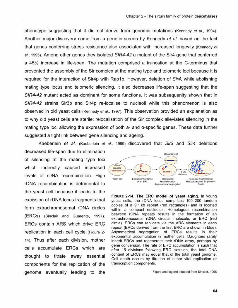
Chapter 2 - The sirtuin family of protein deacetylases
phenotype suggesting that it did not derive from genomic mutations (Kennedy et al., 1994).
Another major discovery came from a genetic screen by Kennedy et al. based on the fact
that genes conferring stress resistance also associated with increased longevity (Kennedy et
al., 1995). Among other genes they isolated SIR4-42 a mutant of the Sir4 gene that conferred
a 45% increase in life-span. The mutation comprised a truncation at the C-terminus that
prevented the assembly of the Sir complex at the mating type and telomeric loci because it is
required for the interaction of Sir4p with Rap1p. However, deletion of Sir4, while abolishing
mating type locus and telomeric silencing, it also decreases life-span suggesting that the
SIR4-42 mutant acted as dominant for some functions. It was subsequenlty shown that in
SIR4-42 strains Sir3p and Sir4p re-localise to nucleoli while this phenomenon is also
observed in old yeast cells (Kennedy et al., 1997). This observation provided an explanation as
to why old yeast cells are sterile: relocalisation of the Sir complex alleviates silencing in the
mating type loci allowing the expression of both a- and α-specific genes. These data further
suggested a tight link between gene silencing and ageing.
Kaeberlein et al. (Kaeberlein et al., 1999) discovered that Sir3 and Sir4 deletions
decreased life-span due to elimination
of silencing at the mating type loci
which indirectly caused increased
levels of rDNA recombination. High
rDNA recombination is detrimental to
the yeast cell because it leads to the
excission of rDNA locus fragments that
form extrachromosomal rDNA circles
(ERCs) (Sinclair and Guarente, 1997).
ERCs contain ARS which drive ERC
replication in each cell cycle (Figure 2-
14). Thus after each division, mother
cells accumulate ERCs which are
thought to titrate away essential
components for the replication of the
genome eventually leading to the
FIGURE 2-14. The ERC model of yeast aging. In youngyeast cells, the rDNA locus comprises 100–200 tandemcopies of a 9.1 kb repeat (red rectangles) and is locatedwithin a compact nucleolus. Homologous recombinationbetween rDNA repeats results in the formation of anextrachromosomal rDNA circular molecule, or ERC (redcircle). ERCs can replicate via the ARS elements in eachrepeat (ERCs derived from the first ERC are shown in blue).Asymmetrical segregation of ERCs results in theirexponential accumulation in mother cells. Daughters rarelyinherit ERCs and regenerate their rDNA array, perhaps bygene conversion. The rate of ERC accumulation is such thatafter 15 divisions following ERC excision, the total DNAcontent of ERCs may equal that of the total yeast genome.Cell death occurs by titration of either vital replication ortranscription components.
Figure and legend adapted from Sinclair, 1998
64
Chapter 2 - The sirtuin family of protein deacetylases
inability of the cell to divide further. The localisation of Sir2p to the nucleolus at sites of rDNA
loci, suppresses the recombination that gives rise to ERCs, allowing yeast cells to divide
more times. Indeed, a second copy of Sir2 suffices to confer an increase of up to 30% in the
replicative life-span of yeast (Kaeberlein et al., 1999).
Interestingly, Aguilaniu et al. provided a novel mechanism by which Sir2p may
increase life span. Their starting point was the fact that protein carbonylation, an irreversible
effect of oxidative damage, accumulates with replicative age (Aguilaniu et al., 2003).
Furthermore, carbonylated proteins are preferentially segregated in the mother cells during
cytokinesis. However, mother cells bearing a Sir2 deletion failed to retain carbonylated
proteins suggesting that Sir2p protects progeny cells from oxidative damage by dictating the
retention of oxidatively damaged cellular components at the mother cell.
2.3.2.4.2 Regulation of life-span by caloric restriction
Given that limitation of food uptake in mammals and rodents had been shown to result
in increased longevity, Lin et al. tested whether caloric restriction in yeast could also extend
its replicative life-span. Limiting the glucose content in the growth medium from 2.0% to 0.5%
resulted in an increase of average life-span from 21.2 to 26.2 generations (Lin et al., 2000).
Glucose activates the cAMP/PKA pathway and mutations in its components (e.g. the
GDP/GTP exchange factor Cdc25) or in enzymes that are involved in glucose metabolism
such as hexokinase also exhibit increased life-spans (FIGURE 2-15). Thus caloric restriction also
seems to positively influence replicative life-span in yeast.
Interestingly, caloric restriction does not increase the life-span of yeast cells carrying
mutations in the Sir2 gene. Similarly, mutation of the Npt1 gene which is involved in the
salvage pathway of NAD+ biosynthesis also abolishes the beneficial effects of caloric
restriction. In agreement to the known function of Sir2p in limiting rDNA recombination, cells
carrying mutation in the cdc25 gene to mimic caloric restriction show decreased levels of
recombination of a reporter plasmid concomitant to reduced ERC levels. Subsequent studies
also demostrated that calorically restricted yeast exhibits enhanced silencing (Lin et al., 2002).
65
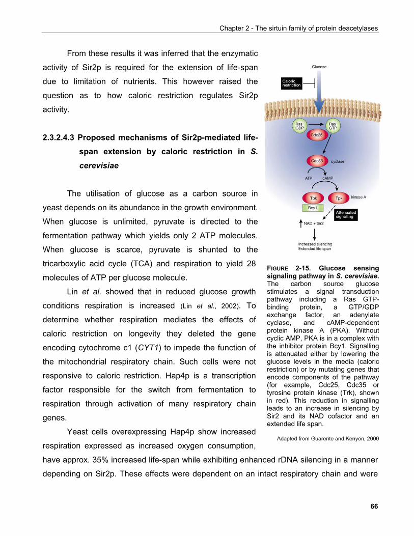
Chapter 2 - The sirtuin family of protein deacetylases
From these results it was inferred that the enzymatic
activity of Sir2p is required for the extension of life-span
due to limitation of nutrients. This however raised the
question as to how caloric restriction regulates Sir2p
activity.
2.3.2.4.3 Proposed mechanisms of Sir2p-mediated life-span extension by caloric restriction in S. cerevisiae
The utilisation of glucose as a carbon source in
yeast depends on its abundance in the growth environment.
When glucose is unlimited, pyruvate is directed to the
fermentation pathway which yields only 2 ATP molecules.
When glucose is scarce, pyruvate is shunted to the
tricarboxylic acid cycle (TCA) and respiration to yield 28
molecules of ATP per glucose molecule.
Lin et al. showed that in reduced glucose growth
ast cells overexpressing Hap4p show increased
respira
conditions respiration is increased (Lin et al., 2002). To
determine whether respiration mediates the effects of
caloric restriction on longevity they deleted the gene
encoding cytochrome c1 (CYT1) to impede the function of
the mitochondrial respiratory chain. Such cells were not
responsive to caloric restriction. Hap4p is a transcription
factor responsible for the switch from fermentation to
respiration through activation of many respiratory chain
genes.
Ye
tion expressed as increased oxygen consumption,
have approx. 35% increased life-span while exhibiting enhanced rDNA silencing in a manner
depending on Sir2p. These effects were dependent on an intact respiratory chain and were
FIGURE 2-15. Glucose sensingsignaling pathway in S. cerevisiae.The carbon source glucosestimulates a signal transductionpathway including a Ras GTP-binding protein, a GTP/GDPexchange factor, an adenylatecyclase, and cAMP-dependentprotein kinase A (PKA). Withoutcyclic AMP, PKA is in a complex withthe inhibitor protein Bcy1. Signallingis attenuated either by lowering theglucose levels in the media (caloricrestriction) or by mutating genes thatencode components of the pathway(for example, Cdc25, Cdc35 ortyrosine protein kinase (Trk), shownin red). This reduction in signallingleads to an increase in silencing bySir2 and its NAD cofactor and anextended life span.
Adapted from Guarente and Kenyon, 2000
66
Chapter 2 - The sirtuin family of protein deacetylases
not additive to those elicited by low glucose growth conditions. Furthermore, it is possible that
Hap4p overxpression resulted in increased resistance to ROS, This was however disproven
by the fact that these cells exhibited similar sensitivities to oxidising agents and mRNA levels
of antioxidant enzymes. Based on these observations, Lin et al. proposed that the effects of
caloric restriction on Sir2 may be either due to the reduced carbon flow through glycolysis
because of the elevated ATP levels and/or due to increased NAD+/NADH ratio because of
increased rates of NADH oxidation. Nevertheless both proposals remained experimentally
unproven.
Anderson et al. addressed this issue by creating an NAD+-reporter strain. They
exploited the fact that NadR protein from Salmonella typhimurium binds to a DNA sequence
called the NAD box in a manner that depends on NAD+ concentration. Their strain expresses
a fusion of NadR fragment and the transcriptional activation domain of GAL4 so that the
fusion activates transcription of a gene required for survival under the control of UAS in an
NAD+-dependent way (Anderson et al., 2003a). Thus fluctuations of intracellular NAD+ levels
directly correlate with viability. Using this system they found that upon caloric restriction their
reporter line grew slower because of low NAD+ because the phenotype could be rescued
with acetaldehyde which induces NADH oxidation and presumably increases the NAD+ pool.
Furthermore, using highly-sensitive 13C NMR spectroscopy they could show that the
intracellular pool of NAD+ is not greatly altered by caloric restriction. In vitro deacetylation
assays also showed that neither Sir2p nor SIRT1 activity are altered by NADH when
concentrations within the physiological range of NAD+/NADH were used. Finally, depletion of
NADH by treatment with acetaldehyde did not alter silencing of rDNA reporters suggesting
that fluctuation of NADH levels are unlikely to affect Sir2 activity.
In a subsequent study, Anderson et al. investigated whether the natural sirtuin inhibitor
nicotinamide can alter cellular Sir2p activity. They could show that increased levels of Npt1,
an enzyme in the NAD+ salvage pathway, suffices to induce life-span extension associated
with increased silencing and rDNA stability (Anderson et al., 2003b). At the same time steady-
state NAD+ levels were unaltered indicating that the observed effects were not due to
increased NAD+. Because the above were true for most of the enzymes involved in the NAD+
salvage pathway, they concluded that increased flux through this pathway suffices to elicit
life-span extension. In a follow-up study they investigated whether nicotinamide affects Sir2p
67
Chapter 2 - The sirtuin family of protein deacetylases
activity. Nicotinamide is not only an intermediate in the NAD+ salvage pathway but also one
of the products of the sirtuin-catalysed deacetylation reaction. Nicotinamide was found to be
a non-competitive inhibitor of both Sir2p and SIRT1 and decreased the life-span of wild type
yeast cells concomitant to inducing recombination of rDNA arrays and inhibiting silencing.
To address whether nicotinamide is important in determining life-span increase upon
caloric restriction, Anderson et al. showed that caloric restriction could not extend the life-
span of yeast lacking Pnc1 (Anderson et al., 2003b). Interestingly, caloric restriction as well as
other stress conditions that are known to increase life-span such as heat shock, osmotic
stress and amino-acid starvation are all potent inducers of Pnc1p protein levels. Increased
Pnc1p activity could increase Sir2p activity by stimulating NAD+ synthesis. This is unlikely to
be the case since supplementation of nicotinic acid, the product of the reaction catalysed by
Pnc1p did not increase gene silencing at rDNA. Furthermore, deletion of Npt1p, a protein that
feeds nicotinic acid to the NAD+ salvage pathway downstream of Pnc1p, did not affect rDNA
silencing when intracellular NAD+ levels were restored suggesting that Pnc1p can increase
Sir2p activity in the absence of an intact NAD+ salvage pathway. Finally, Anderson et al.
could show that reduction of nicotinamide levels by driving its excretion had a positive impact
on rDNA silencing as well as life-span.
Lin et al. then argued that the effects of caloric restriction are actually exerted through
modulation of the levels of NADH (Lin et al., 2004). They could show that caloric restriction
decreases NADH levels while it leaves NAD+ levels unaffected. This was dependent on an
intact mitochondrial respiratory chain and could also be observed in cells with genetically
increased respiration via the overexpression of the transcription factor Hap4p. Furthermore,
NADH levels decreased upon caloric restriction indepedently of the presence of Sir2p. In
vitro analysis of the effects of NADH on Sir2p as well as human SIRT1 showed that NADH
can act as a competitive inhibitor of both enzymes' catalytic activities.
In order to examine whether this is applicable in vivo, Lin et al. overexpressed two
NADH dehydrogenases to induce a decrease of cellular NADH (Lin et al., 2004). These cells
exhibited increased life-span in the presence of 2% glucose similar to wildtype cells under
caloric restriction (0.5% glucose) and this life-span extension was not further enhanced by
lowering glucose levels. Finally, they overexpressed Nnt1p to reduce the levels of
nicotinamide and found that caloric restriction would still increase life-span in a mutant Pnc1
68
Chapter 2 - The sirtuin family of protein deacetylases
background suggesting. The authors interpreted this as an indication that in mutant Pnc1
strains, elevated nicotinamide levels mask the beneficial effects of decreased NADH.
However it is important to note that this conclusion is the result of alternative interpretation of
the experiments by Anderson et al. rather than hardcore experimental evidence to support
Lin et al.'s claims. Furthermore, despite their life-span measurements, Lin et al. provided no
evidence as to the actual downstream effects such as ERC accumulation or silencing
function of Sir2p.
Recent genetic studies by Kaeberlein et al. have put both these models under
scrutiny. To begin with, Kaeberlein et al. could show that caloric restriction is able to actually
increase life-span in the absence of Sir2p (Kaeberlein et al., 2004). Subsequently, they used
respiratory-deficient yeast strains with a broader range of glucose concentrations than used
before to show that even in the original strain background used by Lin et al. Sir2p is
dispensable for the caloric restriction-induced extension of longevity (Kaeberlein et al., 2005a).
Caloric restriction did not alter Sir2p activity based on an experiment assaying telomeric
silencing efficiency. In addition they could show that in the absence of Sir2p, nicotinamide
was able to reduce replicative life-span while this was also true for the effects of caloric
restriction. Surprisingly, they found that although in strains lacking Sir2p nicotinamide did not
reduce life-span consistent with the notion that it targets Sir2p, when these strains where
subjected to caloric restriction nicotinamide only partially inhibited life-span extension.
These data implicated that there exist at least one Sir2p-independent pathway
mediating longevity in response to caloric restriction and that nicotinamide inhibits both
pathways. Indeed, using a large-scale screen for genes that mediate longevity in yeast,
Kaeberlein et al. subsequently identified genes whose deletion increased life-span. Among
them were components of the yeast TOR1 pathway and SCH9 kinase the latter being an
orthologue of mammalian PKB (Akt) kinase. Consistent with their previous conclusions TOR1
and SCH9 deletions extended yeast life-span in the absense of Sir2 (Kaeberlein et al., 2005b).
The works presented above exemplify one of the many controversies present in the
field of Sir2 biology. Clearly, some differences in the conclusion drawn from each study may
be accounted for due to differences in yeast strains and experimental conditions used. While
Sir2p overexpression can extend replicative life-span, whether it also plays a role in life-span
extension upon caloric restriction remains at best uncertain. Current evidence supports a
69
Chapter 2 - The sirtuin family of protein deacetylases
model where Sir2 acts in parallel with another independent pathway which is driven by
caloric restriction, yet the molecular mechanisms of this remain to be identified.
2.3.2.5 Homologues of Sir2 (Hst) proteins
Apart from Sir2p only limited information exists about the other four yeast sirtuins,
Hst1-4 (for homologues of Sir two 1-4) (FIGURE 2-16). Brachmann et al. showed that Hst1p can
rescue the silencing defect of Sir2 mutants while an Hst3 and Hst4 double mutant strain
exhibits telomeric silencing defects. Furthermore, they could demonstrate that Hst3 and Hst4
contribute to genomic stability. Interestingly, in an advanced genetic screen for synthetic
lethality, Pan et al. discovered a broad genetic interaction between Hst3 and replication
machinery components. Hst3 and Hst4 also interact genetically with eachother albeit the
latter does not share the synthetic lethal interactions of the former with replication
components (Pan et al., 2006).
Bedalov et al. provided evidence supporting a role for Hst1p as a cellular NAD+
sensor. They could show that whereas the NAD+ salvage pathway is constitutively active, low
intracellular NAD+ concentrations potently induce the de novo pathway and that Hst1p is a
transcriptional repressor of these genes (Bedalov et al., 2003). Consistent with the notion that
Hst1p senses NAD+ levels, they could show that Hst1p has higher Km for NAD+ than other
yeast sirtuins.
Halme et al. identified Hst1p and Hst2p as the deacetylases responsible for the
epigenetic silencing of FLO10 (Halme et al., 2004). FLO genes express cell-wall glycoproteins
that regulate cell adhesion. Only one family member is expressed while the others are
silenced due to their proximity near telomeres. Expression of the silenced genes allows for
variation of cell-surface properties which is functionally important for processes such as
pseudohyphal growth in response to nutrient limitation. Thus Hst1p and Hst2p can influence
the cell-surface properties of a yeast cell through epigenetic regulation of FLO gene
epxression. In the light of the results presented by Bedalov et al. it would be of interest to
investigate whether a drop in intracellular NAD+ levels can function as an indicator of the
metabolic status of the cell and thus influence FLO gene expression contributing to the
phenotypic changes accompanying nutritional availability.
70
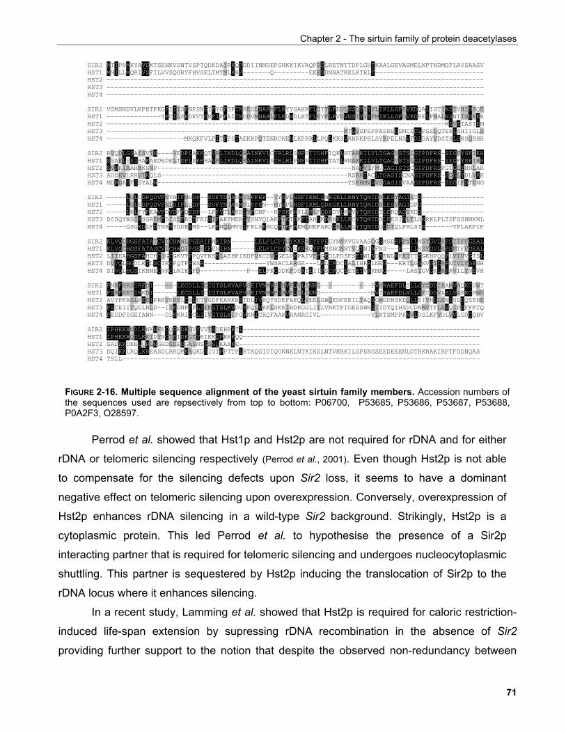
Chapter 2 - The sirtuin family of protein deacetylases
SIR2 MTIPHMKYAVSKTSENKVSNTVSPTQDKDAIRKQPDDIINNDEPSHKKIKVAQPDSLRETNTTDPLGHTKAALGEVASMELKPTNDMDPLAVSAASV HST1 MNILLMQRIVSFILVVSQGRYFHVGELTMTMLKRP-------Q---------EEESDNNATKKLKTRLT---------------------------- HST2 ------------------------------------------------------------------------------------------------- HST3 ------------------------------------------------------------------------------------------------- HST4 ------------------------------------------------------------------------------------------------- SIR2 VSMSNDVLKPETPKGPIIISKNPSNGIFYGPSFTKRESLNARMFLKYYGAHKFLDTYLPEDLNSLYIYYLIKLLGFEVKDQALIGTINSIVHINSQE HST1 --------------YPCILGKDKVTGKFIFPAITKDDVMNARLFLKDNDLKTFLEYFLPVEVNSIYIYFMIKLLGFDVKDKELFMALNSNITSNKER HST2 ---------------------------------------------------------------------------------------MSVSTASTEM HST3 -------------------------------------------------------------MTSVSPSPPASRSGSMCSDLPSSLQTEKLAHIIGLD HST4 --------------------MKQKFVLPITPPSTAEKKPQTENRCNENLKPRRLLPQLKKSVRNRKPRLSYRPELNSVFDLDAYVDSTHLSKSQRHH SIR2 RVQDLGSAISVTN----VEDPLAKKQTVRLIKDLQRAINKVLCTRLRLSNFFTIDHFIQKLHTARKILVLTGAGVSTSLGIPDFRS-SEGFYSKIKH HST1 SSAELSSIHAKAEDEDELTDPLEKKHAVKLIKDLQKAINKVLSTRLRLPNFNTIDHFTATLRNAKKILVLTGAGVSTSLGIPDFRS-SEGFYSKIRH HST2 SVRKIAAHMKSNP--------------------------------------------------NAKVIFMVGAGISTSCGIPDFRSPGTGLYHNLAR HST3 ADDEVLRRVTKQLS------------------------------------------------RSRRIACLTGAGISCNAGIPDFRS-SDGLYDLVKK HST4 MDRDAGFISYALN-------------------------------------------------YSKRMVVVSGAGISVAAGIPDFRS-SEGIFSTVNG SIR2 -----LGLDDPQDVFNYNIFMHDP--SVFYNIANMVLPPEK--IYSPLHSFIKMLQMKGKLLRNYTQNIDNLESYAGISTD---------------- HST1 -----LGLEDPQDVFNLDIFLQDP--SVFYNIAHMVLPPEN--MYSPLHSFIKMLQDKGKLLRNYTQNIDNLESYAGIDPD---------------- HST2 -----LKLPYPEAVFDVDFFQSDP--LPFYTLAKELYPGNF--RPSKFHYLLKLFQDKDVLKRVYTQNIDTLERQAGVKDD---------------- HST3 DCSQYWSIKSGREMFDISLFRDDFKISIFAKFMERLYSNVQLAKPTKTHKFIAHLKDRNKLLRCYTQNIDGLEESIGLTLSNRKLPLTSFSSHWKNL HST4 -----GSGKDLFDYNRVYGDESMS--LKFNQLMVSLFRLSKNCQPTKFHEMLNEFARDGRLLRLYTQNIDGLDTQLPHLSTN-------VPLAKPIP SIR2 KLVQCHGSFATATCVTCHWNLPGERIFNKIRN------LELPLCPYCYKKRREYFPEGYNNKVGVAASQGSMSERPPYILNSYGVLKPDITFFGEAL HST1 KLVQCHGSFATASCVTCHWQIPGEKIFENIRN------LELPLCPYCYQKRKQYFPMSNGNNTVQTNINFNS---P--ILKSYGVLKPDMTFFGEAL HST2 LIIEAHGSFAHCHCIGCGKVYPPQVFKSKLAEHPIKDFVKCDVCGELVKPAIVFFGEDLPDSFSETWLNDSEWLREKITTSGKHPQQPLVIVVGTSL HST3 DVVQLHGDLKTLSCTKCFQTFPWSR----------------YWSRCLRRGE---LPLCPDCEALINKRLNEG---KRTLGSNVGILRPNIVLYGENH HST4 STVQLHGSIKHMECNKCLNIKPFD------------P--ELFKCDDKFDSRTEIIPSCPQCEEYETVRKMAG-----LRSTGVGKLRPRVILYNEVH SIR2 PNKFHKSIREDI------LECDLLICIGTSLKVAPVSEIVNMVPSHVPQVLINRD-------------PVKHAEFDLSLLGYCDDIAAMVAQKCGWT HST1 PSRFHKTIRKDI------LECDLLICIGTSLKVAPVSEIVNMVPSHVPQILINRD-------------MVTHAEFDLNLLGFCDDVASLVAKKCHWD HST2 AVYPFASLPEEIPRKVKRVLCNLETVGDFKANKRPTDLIVHQYSDEFAEQLVEELGWQEDFEKILTAQGGMGDNSKEQLLEIVHDLENLSLDQSEHE HST3 PSCEIITQGLNLD--IIKGNPDFLIIMGTSLKVDGVKQLVKKLSKKIHDRGGLIILVNKTPIGESSWHGIIDYQIHSDCDNWVTFLESQIPDFFKTQ HST4 PEGDFIGEIANN---DLKKRIDCLIIVGTSLKIPGVKNICRQFAAKVHANRGIVL-------------YLNTSMPPKNVLDSLKFVDLVVLGDCQHV SIR2 IPHKKWNDLKNKNFKCQEKDKGVYVVTSDEHPKTL------------------------------------------------------------- HST1 IPHKKWQDLKKIDYNCTEIDKGTYKIKKQPRKKQQ------------------------------------------------------------- HST2 SADKKDKKLQRLNGHDSDEDGASNSSSSQKAAKE-------------------------------------------------------------- HST3 DQIKKLRQLKREASDLRKQMKAQKDSIGTPPTTPLRTAQGIDIQGNNELNTKIKSLNTVKRKILSPENSSEEDEEENLDTRKRAKIRPTFGDNQAS HST4 TSLL--------------------------------------------------------------------------------------------
FIGURE 2-16. Multiple sequence alignment of the yeast sirtuin family members. Accession numbers ofthe sequences used are repsectively from top to bottom: P06700, P53685, P53686, P53687, P53688,P0A2F3, O28597.
Perrod et al. showed that Hst1p and Hst2p are not required for rDNA and for either
rDNA or telomeric silencing respectively (Perrod et al., 2001). Even though Hst2p is not able
to compensate for the silencing defects upon Sir2 loss, it seems to have a dominant
negative effect on telomeric silencing upon overexpression. Conversely, overexpression of
Hst2p enhances rDNA silencing in a wild-type Sir2 background. Strikingly, Hst2p is a
cytoplasmic protein. This led Perrod et al. to hypothesise the presence of a Sir2p
interacting partner that is required for telomeric silencing and undergoes nucleocytoplasmic
shuttling. This partner is sequestered by Hst2p inducing the translocation of Sir2p to the
rDNA locus where it enhances silencing.
In a recent study, Lamming et al. showed that Hst2p is required for caloric restriction-
induced life-span extension by supressing rDNA recombination in the absence of Sir2
providing further support to the notion that despite the observed non-redundancy between
71
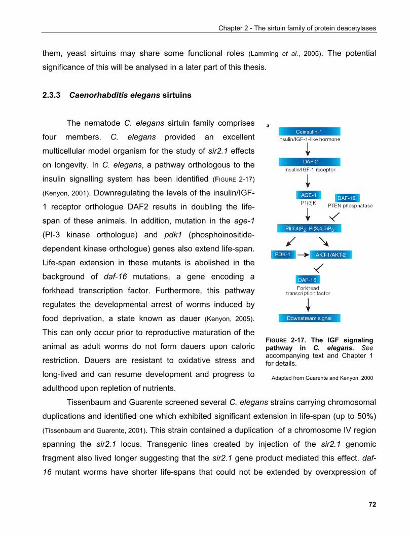
Chapter 2 - The sirtuin family of protein deacetylases
them, yeast sirtuins may share some functional roles (Lamming et al., 2005). The potential
significance of this will be analysed in a later part of this thesis.
2.3.3 Caenorhabditis elegans sirtuins
The nematode C. elegans sirtuin family comprises
four members. C. elegans provided an excellent
multicellular model organism for the study of sir2.1 effects
on longevity. In C. elegans, a pathway orthologous to the
insulin signalling system has been identified (FIGURE 2-17)
(Kenyon, 2001). Downregulating the levels of the insulin/IGF-
1 receptor orthologue DAF2 results in doubling the life-
span of these animals. In addition, mutation in the age-1
(PI-3 kinase orthologue) and pdk1 (phosphoinositide-
dependent kinase orthologue) genes also extend life-span.
Life-span extension in these mutants is abolished in the
background of daf-16 mutations, a gene encoding a
forkhead transcription factor. Furthermore, this pathway
regulates the developmental arrest of worms induced by
food deprivation, a state known as dauer (Kenyon, 2005).
This can only occur prior to reproductive maturation of the
animal as adult worms do not form dauers upon caloric
restriction. Dauers are resistant to oxidative stress and
long-lived and can resume development and progress to
adulthood upon repletion of nutrients.
FIGURE 2-17. The IGF signalingpathway in C. elegans. Seeaccompanying text and Chapter 1for details.
Adapted from Guarente and Kenyon, 2000
Tissenbaum and Guarente screened several C. elegans strains carrying chromosomal
duplications and identified one which exhibited significant extension in life-span (up to 50%)
(Tissenbaum and Guarente, 2001). This strain contained a duplication of a chromosome IV region
spanning the sir2.1 locus. Transgenic lines created by injection of the sir2.1 genomic
fragment also lived longer suggesting that the sir2.1 gene product mediated this effect. daf-
16 mutant worms have shorter life-spans that could not be extended by overxpression of
72
Chapter 2 - The sirtuin family of protein deacetylases
sir2.1. Furthermore, sir2.1 overexpression did not extent further the life-span of worms
carrying mutations in the daf-2 gene. These experiments firmly established SIR2.1 as a
mediator of life-span within the C. elegans insulin signalling system.
Mutations in the insulin signalling pathway synergise with mutations in the TGF-β
pathway to affect life-span. daf-1 and daf-4 encode two types of TGF-β receptors and
mutations in either of these genes cause a temperature-sensitive constitutive dauer
phenotype. sir2.1 overexpression alone did not induce dauer formation but in the background
of either daf-1 and daf-4 mutations it increased markedly the proportion of animals that
entered the dauer stage. These experiments further supported the notion that by modulating
the C. elegans insulin pathway, SIR2.1 regulates longevity and that as in the unicellular S.
cerevisiae, it holds an evolutionarily conserved function.
A later study by Wang and Tissenbaum further investigated the role of sir2.1 in the
worm by employing a loss-of-function approach. They used a strain with a deletion in the
sir2.1 locus that eliminates half of the sirtuin core domain and is likely to result in a null allele
(Wang and Tissenbaum, 2006). Worms mutated for sir2.1 showed a slight decrease in mean life-
span which was further exuberated when these worms were exposed to stresses such as
heat-shock, H2O2 and UV irradiation. Next, they tested for genetic interactions with daf-2
mutants to establish the exclusivity as well as the topology of the pathway with respect to life-
span. Interestingly, worms mutated in both daf-2 and sir2.1 had the same life-span as worms
mutated in daf-2 only suggesting that sir2.1 is upstream of the insulin receptor or alternatively
in a parallel pathway.
unc-13, a gene positioned genetically upstream of daf-2, is a regulator of
neurotransmiter release and when mutated it results in a moderate life-span extension in a
daf-16 dependent manner. Mutations in sir2.1 could partially suppress the long-lived
phenotype of unc13 mutant worms. When in this double-mutant background a mutation of
daf-16 was added, unc-13 life-extension could be completely suppressed suggesting that
sir2.1 and daf-16 have distinct functions in life-span determination in C. elegans.
Interestingly, one of the many phenotypes of the unc-13 mutants a defect in food ingestion
implying that life-span extenstion in these mutants is a by-product of caloric restriction.
Mutations in the eat-2 gene are thought to mimick caloric restriction because of
pharyngeal defects leading to slower and irregular pumping. Accordingly, eat-2 mutant
73
Chapter 2 - The sirtuin family of protein deacetylases
worms live longer. Worms carrying mutations in both eat-2 and sir2.1 have wild-type life-
spans suggesting that the effects of caloric restriction on longevity are dependent on the
sir2.1 gene (Wang and Tissenbaum, 2006). These data provided experimental evidence for the
involvement of sir2.1 in regulating caloric restriction-driven extension of worm life-span and
that these effects are only partially overlapping with those of daf-16.
Underlying the validity of these findings, Viswanathan et al. exploited the compound
resveratrol, previously shown to enhance the activity of SIRT1, to probe the signalling
pathways mediating longevity in C. elegans. Resveratrol was found to increase life-span in a
manner dependent on sir2.1 but independent of daf-16 (Viswanathan et al., 2005). Furthermore,
resveratrol treatment in combination with sir2.1 overexpression had additive effects on life-
span, which the authors interpreted as an indication that resveratrol increases life-span via a
sir2.1-independent pathway. This result could also be interpreted as super-activation of
already overexpressed sir2.1 by resveratrol according to the proposed role of the compound
as an activator of sirtuins, a notion supported by data in the same work which show that the
effects of resveratrol on longevity are abolished in a sir2.1 mutant strain. On the other hand,
unlike resveratrol, sir2.1 overexpression increases life-span in a manner fully dependent on
daf-16.
Wanting to explore the molecular mechanisms involved in resveratrol-induced
increase in life-span, Viswanathan et al., analysed gene expression profiles of worms treated
with this compound. A class of genes that was prominently induced by resveratrol treatment
encodes PQN [prion-like asparagine(Q)/glutamine(N)-rich] and its subgroup ABU (activated
in blocked unfolded protein response) which, as the name implies, act in response to ER
stresses when the canonical UPR (unfolded protein response) pathway malfunctions. ER
responses dictate cell survival upon different stress conditions so resveratrol may affect life-
span by activating this pathway to promote survival. RNAi-mediated downregulation of
PQN/ABU proteins reduced or even completely abolished (in the case of ABU-11) the
resveratrol-induced increase in longevity. Consistent with the notion that pqn gene activation
correlates with increased life-span, transgenic animals overxpressing ABU-11 showed life-
span extension up to 28% in a manner dependent on ABU-11 expression levels.
Resveratrol was expected to activate SIR2.1 which could in turn induce transcription
of the pgn/abu genes. Surprisingly, sir2.1 mutant worms showed higher levels of abu-11
74
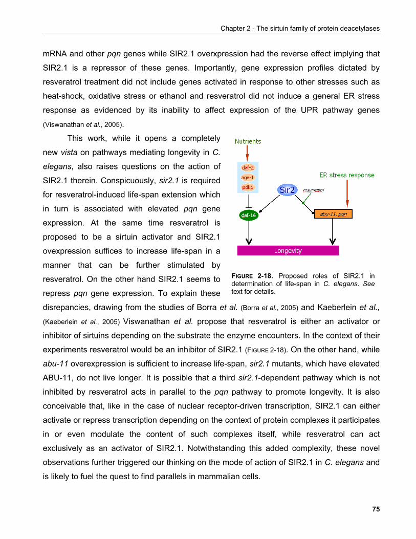
Chapter 2 - The sirtuin family of protein deacetylases
mRNA and other pqn genes while SIR2.1 overxpression had the reverse effect implying that
SIR2.1 is a repressor of these genes. Importantly, gene expression profiles dictated by
resveratrol treatment did not include genes activated in response to other stresses such as
heat-shock, oxidative stress or ethanol and resveratrol did not induce a general ER stress
response as evidenced by its inability to affect expression of the UPR pathway genes
(Viswanathan et al., 2005).
FIGURE 2-18. Proposed roles of SIR2.1 indetermination of life-span in C. elegans. Seetext for details.
This work, while it opens a completely
new vista on pathways mediating longevity in C.
elegans, also raises questions on the action of
SIR2.1 therein. Conspicuously, sir2.1 is required
for resveratrol-induced life-span extension which
in turn is associated with elevated pqn gene
expression. At the same time resveratrol is
proposed to be a sirtuin activator and SIR2.1
ovexpression suffices to increase life-span in a
manner that can be further stimulated by
resveratrol. On the other hand SIR2.1 seems to
repress pqn gene expression. To explain these
disrepancies, drawing from the studies of Borra et al. (Borra et al., 2005) and Kaeberlein et al.,
(Kaeberlein et al., 2005) Viswanathan et al. propose that resveratrol is either an activator or
inhibitor of sirtuins depending on the substrate the enzyme encounters. In the context of their
experiments resveratrol would be an inhibitor of SIR2.1 (FIGURE 2-18). On the other hand, while
abu-11 overexpression is sufficient to increase life-span, sir2.1 mutants, which have elevated
ABU-11, do not live longer. It is possible that a third sir2.1-dependent pathway which is not
inhibited by resveratrol acts in parallel to the pqn pathway to promote longevity. It is also
conceivable that, like in the case of nuclear receptor-driven transcription, SIR2.1 can either
activate or repress transcription depending on the context of protein complexes it participates
in or even modulate the content of such complexes itself, while resveratrol can act
exclusively as an activator of SIR2.1. Notwithstanding this added complexity, these novel
observations further triggered our thinking on the mode of action of SIR2.1 in C. elegans and
is likely to fuel the quest to find parallels in mammalian cells.
75
Chapter 2 - The sirtuin family of protein deacetylases
2.3.4 Drosophila melanogaster sirtuins
Five orthologues of the yeast Sir2 have been identified in Drosophila (CG5216,
CG5085, G3187, CG6284, and CG11305, FlyBase), among which dSir2 (CG5216) shares
the highest homology with and was actually shown to exhibit NAD+-dependent deacetylase
activity. dSir2 localises primarily in the nucleus in cultured Drosophila cells and the animal
but is found to be exclusively cytoplasmic in the syncytial blastoderm and both cytoplasmic
and nuclear during late embryogenesis. The dSir2 mRNA is present throughout
emryogenesis, is markedly downregulated in larvae and returns to higher levels in the pupal
and adult stages. In particular, the presence of dSir2 transcripts within the first hours of
emryogenesis imply maternal effect (Rosenberg and Parkhurst, 2002).
Indeed, embrya derived from mothers with reduced dSir2 contribution exhibit
segmentation defects due to aberant pair rule gene expression as hinted by the derepression
of the secondary pair rule gene fushi tarazu (ftz). This phenocopies reduced funtion of a
primary pair rule gene, hairy, which is a member of the basic helix-loop-helix (bHLH) family of
repressors Idiscussed in more detail below) and behaves genetically as a repressor of ftz.
Consistent with this hypothesis, dSir2 and hairy interact genetically to determine embryonic
segmentation as well as physically in a biochemical binding assay via a basic aminoacid
motif conserved in bHLH proteins (Rosenberg and Parkhurst, 2002). In fact, another bHLH
protein, Deadpan, also interacted with dSir2. Deadpan is involved in sex determination by
participating in the expression of a gender-specific transcipt sex lethal (sxl). In agreement to
this, lower dSir2 resulted in male-specific lethality due to aberrant dosage compensation
providing evidence for the physiological significance of the observed biochemical interaction
with Deadpan (Rosenberg and Parkhurst, 2002).
More recently, a report by Astrom et al. has challenged the above conclusions based
on the fact that the dSir2 mutant strain used for these experiments could be fully
complemented by a strain they generated which lacked most of the dSir2 ORF (Astrom et al.,
2003). Using this strain, Astrom et al. also demonstrated that viability of Sxl null male flies is
not sensitive to dSir2 levels suggesting that the effects observed by Roseneberg and
Parkhurst reflected extraneous mutations in the strain used in their studies.
76
Chapter 2 - The sirtuin family of protein deacetylases
Another well characterised occurrence of gene silencing in Drosophila is mediated by
the polycomb group proteins (PcG), best known for their involvement in the spatial regulation
of homeobox gene expression during development. PcG comprises several chromatin-
modifying proteins that are recruited to the DNA by means of specialised regulatory elements
called polycomb response elements (PREs).
dSir2 deletion was found to enhance the PcG mutant phenotype "extra sex combs" via
a PRE-dependent mechanism (Furuyama et al., 2004). Consistent with a role in polycomb
silencing, dSir2 interacts directly and co-localises with the PcG protein E(Z) while it co-
fractionates with other members of a previously known E(Z) complex from larval extracts.
One of these proteins, Rpd3 is also a deacetylase found to affect the expression of a largely
distinct set of genes. Disruption of dSir2 does not affect the localisation of PcG components
to chromatin suggesting that the role of dSir2 in this context is not exerted through regulation
of PcG protein recruitment. Thus the functional significance of dSir2 in this distinct E(Z)
complex remains to be elucidated. Further insighst into the potential role of sirtuins in
polycomb-mediated repression were provided by the identification of a mammalian complex
comprising the mammalian enhancer of zeste orthologue Ezh2 and SIRT1 and will be
discussed in section 2.3.5.1.3.
dSir2 associates with both heterochromatin and euchromatin. Several groups reported
that dSir2 mutations affect heterochromatin silencing a phenomenon known in Drosophila as
position effect variegation (PEV). Assaying for PEV is attained in reporter fly strains by
placing the white gene in the context of DNA derived from genomic regions known to be
subject to silencing. When mutated, a gene thought to mediate PEV would alleviate silencing
of the reporter gene and the eyes of these animals would show red patches corresponding to
areas with defective silencing. Using such an assay, Rosenberg and Parkhurst suggested
that dSir2 participates in heterochromatic but not telomeric silening (Rosenberg and Parkhurst,
2002). Newman et al. speculated that loss of dSir2 function results in impairment of
heterochromatin formation or maintainance, surprisingly, though, they have omitted to
substantiate this by studying the banding pattern of salivary gland chromosomes (Newman et
al., 2002). In the same study, dSir2 was found to share similar distribution pattern with dCBP,
an acetyltransferase which was found to be associated with SIRT1 is mammalian cells (see
later) and was also implicated in heterochromatin formation.
77
Chapter 2 - The sirtuin family of protein deacetylases
dSir2 loss-of-function mutations reduce Drosophila life-span when animals with
matched genetic backgrounds are compared (Rogina and Helfand, 2004). Furthermore, when
dSir2 was overexpressed under the regulation of a strong promoter, an extension of up to
57% was observed. dSir2 was shown to be highly expressed in neuronal tissues and
neuronal-specific dSir2 overexpression was sufficient to induce an increase in life-span.
Significantly, calorie restriction could not further increase longevity in dSir2-overexpressing
animals suggesting that the effects of caloric restriction on longevity in Drosophila are
mediated by dSir2 (Rogina and Helfand, 2004).
2.3.5 The mammalian sirtuin family 2.3.5.1 SIRT1 2.3.5.1.1 Expression and genetic ablation of SIRT1 in the mouse
Since the description of the mammalian sirtuin family, SIRT1 (also referred to as
mSir2α or Sir2α in the mouse) has attracted most of research efforts to elucidate its function
as it has been considered the functional orthologue of scSir2. This was based on the fact that
it exhibits the highest homology compared to the other mammalian sirtuins, it is localised in
the nucleus and was shown to complement the silencing defects of sir2-defective strains.
Furthermore, SIRT1 shares the characteristic N- and C-terminal extensions flanking the
conserved sirtuin core domain laso present in scSir2. A multiple sequence alignment of
SIRT1 with the other six human famiy members is shown in FIGURE 2-18.
SIRT1 mRNA is ubiquitously expressed in all mouse tissues tested (Sakamoto et al.,
2003; McBurney et al., 2003a). Quantitative expression analysis indicated that SIRT1 is highly
expressed in the mouse embryo early during development but progressively declines to
lower amounts which remain constant into adulthood. The highest levels of SIRT1 mRNA
were found in the lung and testes (Sakamoto et al., 2003). Immunohistochemical studies also
showed ubiquitous expression of SIRT1 with predominant presence of the protein in the
heart, brain, spinal cord and dorsal root ganglia at E10.5-13.5 (Sakamoto et al., 2003). McBurney
et al. also showed that SIRT1 protein is highly expressed in mouse emryonic stem (ES) cells
78
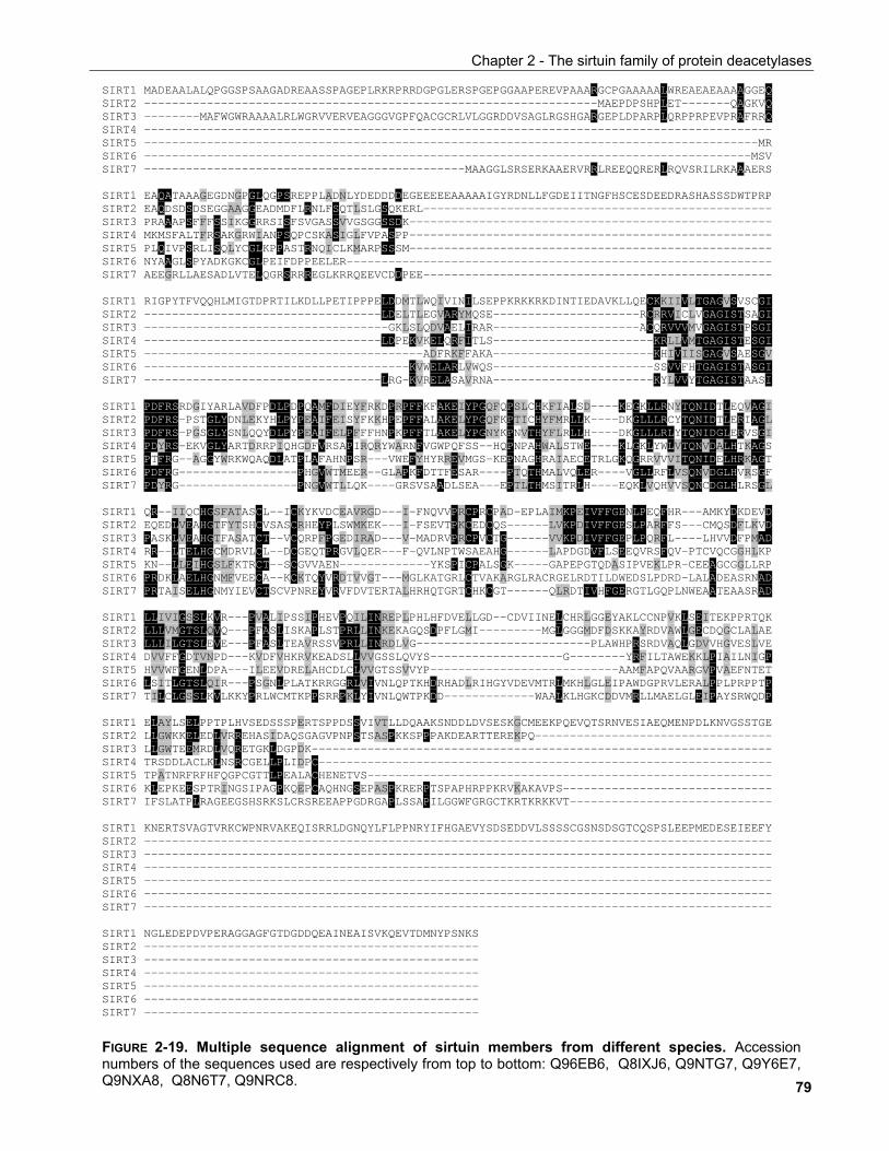
Chapter 2 - The sirtuin family of protein deacetylases
79
FIGURE 2-19. Multiple sequence alignment of sirtuin members from different species. Accessionnumbers of the sequences used are respectively from top to bottom: Q96EB6, Q8IXJ6, Q9NTG7, Q9Y6E7,Q9NXA8, Q8N6T7, Q9NRC8.
SIRT1 MADEAALALQPGGSPSAAGADREAASSPAGEPLRKRPRRDGPGLERSPGEPGGAAPEREVPAAARGCPGAAAAALWREAEAEAAAAGGEQ SIRT2 -----------------------------------------------------------------MAEPDPSHPLET-------QAGKVQ SIRT3 --------MAFWGWRAAAALRLWGRVVERVEAGGGVGPFQACGCRLVLGGRDDVSAGLRGSHGARGEPLDPARPLQRPPRPEVPRAFRRQ SIRT4 ------------------------------------------------------------------------------------------ SIRT5 ----------------------------------------------------------------------------------------MR SIRT6 ---------------------------------------------------------------------------------------MSV SIRT7 ----------------------------------------------MAAGGLSRSERKAAERVRRLREEQQRERLRQVSRILRKAAAERS SIRT1 EAQATAAAGEGDNGPGLQGPSREPPLADNLYDEDDDDEGEEEEEAAAAAIGYRDNLLFGDEIITNGFHSCESDEEDRASHASSSDWTPRP SIRT2 EAQDSDSDSEGGAAGGEADMDFLRNLFSQTLSLGSQKERL-------------------------------------------------- SIRT3 PRAAAPSFFFSSIKGGRRSISFSVGASSVVGSGGSSDK---------------------------------------------------- SIRT4 MKMSFALTFRSAKGRWIANPSQPCSKASIGLFVPASPP---------------------------------------------------- SIRT5 PLQIVPSRLISQLYCGLKPPASTRNQICLKMARPSSSM---------------------------------------------------- SIRT6 NYAAGLSPYADKGKCGLPEIFDPPEELER------------------------------------------------------------- SIRT7 AEEGRLLAESADLVTELQGRSRRREGLKRRQEEVCDDPEE-------------------------------------------------- SIRT1 RIGPYTFVQQHLMIGTDPRTILKDLLPETIPPPELDDMTLWQIVINILSEPPKRKKRKDINTIEDAVKLLQECKKIIVLTGAGVSVSCGI SIRT2 ----------------------------------LDELTLEGVARYMQSE---------------------RCRRVICLVGAGISTSAGI SIRT3 -----------------------------------GKLSLQDVAELIRAR---------------------ACQRVVVMVGAGISTPSGI SIRT4 ----------------------------------LDPEKVKELQRFITLS-----------------------KRLLVMTGAGISTESGI SIRT5 ----------------------------------------ADFRKFFAKA-----------------------KHIVIISGAGVSAESGV SIRT6 --------------------------------------KVWELARLVWQS-----------------------SSVVFHTGAGISTASGI SIRT7 ----------------------------------LRG-KVRELASAVRNA-----------------------KYLVVYTGAGISTAASI SIRT1 PDFRSRDGIYARLAVDFPDLPDPQAMFDIEYFRKDPRPFFKFAKEIYPGQFQPSLCHKFIALSD----KEGKLLRNYTQNIDTLEQVAGI SIRT2 PDFRS-PSTGLYDNLEKYHLPYPEAIFEISYFKKHPEPFFALAKELYPGQFKPTICHYFMRLLK----DKGLLLRCYTQNIDTLERIAGL SIRT3 PDFRS-PGSGLYSNLQQYDLPYPEAIFELPFFFHNPKPFFTLAKELYPGNYKPNVTHYFLRLLH----DKGLLLRLYTQNIDGLERVSGI SIRT4 PDYRS-EKVGLYARTDRRPIQHGDFVRSAPIRQRYWARNFVGWPQFSS--HQPNPAHWALSTWE----KLGKLYWLVTQNVDALHTKAGS SIRT5 PTFRG--AGGYWRKWQAQDLATPLAFAHNPSR---VWEFYHYRREVMGS-KEPNAGHRAIAECETRLGKQGRRVVVITQNIDELHRKAGT SIRT6 PDFRG-----------------PHGVWTMEER--GLAPKFDTTFESAR----PTQTHMALVQLER----VGLLRFLVSQNVDGLHVRSGF SIRT7 PDYRG-----------------PNGVWTLLQK----GRSVSAADLSEA---EPTLTHMSITRLH----EQKLVQHVVSQNCDGLHLRSGL SIRT1 QR--IIQCHGSFATASCL--ICKYKVDCEAVRGD---I-FNQVVPRCPRCPAD-EPLAIMKPEIVFFGENLPEQFHR---AMKYDKDEVD SIRT2 EQEDLVEAHGTFYTSHCVSASCRHEYPLSWMKEK---I-FSEVTPKCEDCQS------LVKPDIVFFGESLPARFFS---CMQSDFLKVD SIRT3 PASKLVEAHGTFASATCT--VCQRPFPGEDIRAD---V-MADRVPRCPVCTG------VVKPDIVFFGEPLPQRFL----LHVVDFPMAD SIRT4 RR--LTELHGCMDRVLCL--DCGEQTPRGVLQER---F-QVLNPTWSAEAHG------LAPDGDVFLSEEQVRSFQV-PTCVQCGGHLKP SIRT5 KN--LLEIHGSLFKTRCT--SCGVVAEN-------------YKSPICPALSGK-----GAPEPGTQDASIPVEKLPR-CEEAGCGGLLRP SIRT6 PRDKLAELHGNMFVEECA--KCKTQYVRDTVVGT---MGLKATGRLCTVAKARGLRACRGELRDTILDWEDSLPDRD-LALADEASRNAD SIRT7 PRTAISELHGNMYIEVCTSCVPNREYVRVFDVTERTALHRHQTGRTCHKCGT------QLRDTIVHFGERGTLGQPLNWEAATEAASRAD SIRT1 LLIVIGSSLKVR---PVALIPSSIPHEVPQILINREPLPHLHFDVELLGD--CDVIINELCHRLGGEYAKLCCNPVKLSEITEKPPRTQK SIRT2 LLLVMGTSLQVQ---PFASLISKAPLSTPRLLINKEKAGQSDPFLGMI---------MGLGGGMDFDSKKAYRDVAWLGECDQGCLALAE SIRT3 LLLILGTSLEVE---PFASLTEAVRSSVPRLLINRDLVG-------------------------PLAWHPRSRDVAQLGDVVHGVESLVE SIRT4 DVVFFGDTVNPD---KVDFVHKRVKEADSLLVVGSSLQVYS-------------------G--------YRFILTAWEKKLPIAILNIGP SIRT5 HVVWFGENLDPA---ILEEVDRELAHCDLCLVVGTSSVVYP---------------------------AAMFAPQVAARGVPVAEFNTET SIRT6 LSITLGTSLQIR---PSGNLPLATKRRGGRLVIVNLQPTKHDRHADLRIHGYVDEVMTRLMKHLGLEIPAWDGPRVLERALPPLPRPPTP SIRT7 TILCLGSSLKVLKKYPRLWCMTKPPSRRPKLYIVNLQWTPKDD-------------WAALKLHGKCDDVMRLLMAELGLEIPAYSRWQDP SIRT1 ELAYLSELPPTPLHVSEDSSSPERTSPPDSSVIVTLLDQAAKSNDDLDVSESKGCMEEKPQEVQTSRNVESIAEQMENPDLKNVGSSTGE SIRT2 LLGWKKELEDLVRREHASIDAQSGAGVPNPSTSASPKKSPPPAKDEARTTEREKPQ---------------------------------- SIRT3 LLGWTEEMRDLVQRETGKLDGPDK------------------------------------------------------------------ SIRT4 TRSDDLACLKLNSRCGELLPLIDPC----------------------------------------------------------------- SIRT5 TPATNRFRFHFQGPCGTTLPEALACHENETVS---------------------------------------------------------- SIRT6 KLEPKEESPTRINGSIPAGPKQEPCAQHNGSEPASPKRERPTSPAPHRPPKRVKAKAVPS------------------------------ SIRT7 IFSLATPLRAGEEGSHSRKSLCRSREEAPPGDRGAPLSSAPILGGWFGRGCTKRTKRKKVT----------------------------- SIRT1 KNERTSVAGTVRKCWPNRVAKEQISRRLDGNQYLFLPPNRYIFHGAEVYSDSEDDVLSSSSCGSNSDSGTCQSPSLEEPMEDESEIEEFY SIRT2 ------------------------------------------------------------------------------------------ SIRT3 ------------------------------------------------------------------------------------------ SIRT4 ------------------------------------------------------------------------------------------ SIRT5 ------------------------------------------------------------------------------------------ SIRT6 ------------------------------------------------------------------------------------------ SIRT7 ------------------------------------------------------------------------------------------ SIRT1 NGLEDEPDVPERAGGAGFGTDGDDQEAINEAISVKQEVTDMNYPSNKS SIRT2 ------------------------------------------------ SIRT3 ------------------------------------------------ SIRT4 ------------------------------------------------ SIRT5 ------------------------------------------------ SIRT6 ------------------------------------------------ SIRT7 ------------------------------------------------
Chapter 2 - The sirtuin family of protein deacetylases
and during spermatogenesis, particularly in the nuclei of spermatogonia, spermatocytes and
round spermatids (McBurney et al., 2003a; McBurney et al., 2003b).
Consistent with SIRT1 expression in embryonic tissues, mice with genetic ablation of
the both SIRT1 alleles die soon after birth, although in an outbred background animals can
survive to sterile adults which are smaller than their littermates (McBurney et al., 2003a). The
sterility was attributed to defects in spermatogenesis with increased apoptosis in the
seminiferous tubules and associated failure of spermatozoa to mature properly in agreement
with the high expression of SIRT1 during spermatogenesis. Furthermore, SIRT1-/- mice had
eye abnormalities which were attributed to an eyelid opening failure seen consistently in
these animals (McBurney et al., 2003a).
SIRT1-/- mice exhibit no global defects in silencing of either endogenous genes or
reporter β-galactosidase transgene suggesting that if SIRT1 functions in silencing similarly to
its yeast orthologue it does so in a gene-specific rather than genome-wide manner (McBurney
et al., 2003a). Furthermore, ES cells derived from SIRT1-/- mice are able to differentiate in vitro
and exhibit no global changes in histone acetylation patterns (McBurney et al., 2003b).
The SIRT1-/- mice used in the studies of McBurney et al. were generated by targeted
deletion of exons 5 and 6 of the murine SIRT1 gene which encode for a large part of the
catalytic subunit (McBurney et al., 2003a). In an independent study, Cheng et al. used targeted
homologous recombination to delete the entire region encoding for SIRT1. They also created
a mouse line with a conditional deletion of exon4 in the SIRT1 gene which encodes 51
aminoacids of the SIRT1 conserved domain (Cheng et al., 2003).
These animals exhibited cardiac abnormalities, including ventricular and atrial septal
defects and elongated atrioventricular valves, all of which were absent in SIRT1-/- mice
surviving into adulthood. In addition, the retinas of post-natal SIRT1-/- mice showed severe
defects with many of the characteristic retinal layers being thinner and disorganised. This
correlated with abnormal closure of the optic fissure found in SIRT1-/- embrya providing
evidence that the retinal abnormalities are attributable to a developmental defect rather than
a secondary effect of eyelid closure abnormalities as proposed by McBurney et al. (McBurney
et al., 2003a).
Several studies mainly in tissue culture systems have implicated SIRT1 in various
cellular processes by means of its interaction with other proteins (TABLE 2-2). There is a
80
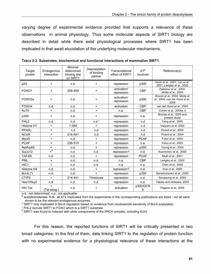
Chapter 2 - The sirtuin family of protein deacetylases
varying degree of experimental evidence provided that supports a relevance of these
observations in animal physiology. Thus some molecular aspects of SIRT1 biology are
described in detail while there exist physiological processes where SIRT1 has been
implicated in that await elucidation of the underlying molecular mechanisms.
TABLE 2-2. Substrates, biochemical and functional interactions of mammalian SIRT1.
Target protein
Endogenous interaction
Minimal determined binding site on SIRT1
Deacetylation of binding
partner
Transcriptional effect of SIRT1
AT§ involved
Reference(s)
p53 + n.d. + repression p300 Vaziri et al., 2001; Luo et al., 2001; Langley et al., 2002
FOXO1 + 208-409 + activation/ repression CBP Daitokou et al., 2004;
Motta et al., 2004
FOXO3a + n.d. + activation/ repression p300
Brunet et al., 2004; Motta et al., 2004; van der Horst et al.,
2004 FOXO4 n.d. n.d. + activation CBP van der Horst et al., 2004 Ku70 + n.d. + n.a. CBP Cohen et al., 2004a, b
p300 + n.d. + repression n.a. Bouras et al., 2005 and present study
FHL2 n.d. n.d. n.d.∂ repression n.d Yang et al., 2005 Histone H1 + 1-268 + repression n.d Vaquero et al. 2004 PPARγ + n.d n.d repression n.d Picard et al., 2004 NCoR + 214-541 n.d repression n.d Picard et al., 2004 MyoD + n.d. + repression PCAF Fulco et al., 2003 PCAF + 236-510 + repression n.a. Fulco et al., 2003 RelA/p65 + n.d. + repression p300 Yeung et al., 2004 Su(z)12 +¥ n.d. n.d. repression? n.d. Kuzmichev et al., 2005 TAFI68 n.d. n.d. + repression PCAF Muth et al., 2001 PML + n.d. n.d. n.a. CBP Langley et al., 2002 HIC1 + n.d. n.d. n.a. n.a. Chen et al., 2005 Histone H4 n.d. n.d. + repression? n.d. Imai et al., 2000 Bcl-6 (*) n.d. n.d. + repression p300 Bereshchenko et al., 2002 CTIP2 + 214-441 ?/histones repression n.d. Senawong et al., 2003 Hes1/Hey2 n.d. n.d. n.d. repression n.d. Takata and Ishikawa, 2003
HIV Tat + (Tat exog.) n.d. + activation p300/GCN
5 Pagans et al., 2005
n.d.: not determined; n.a.: not applicable § acetyltransferase. N.B.: all ATs implicated from the experiments in the corresponding publications are listed ; not all were
shown to be the relevant endogenous enzymes. * SIRT1 only implicated in Bcl-6 regulation based on evidence from nicotinamide sensitivity of Bcl-6 acetylation ∂ FHL2 recruits SIRT1 to FOXO which is a SIRT1 substrate ¥ SIRT1 was found to inderact with other components of the PRC4 complex, including Ezh2
For this reason, the reported functions of SIRT1 will be critically presented in two
broad categories. In the first of them, data linking SIRT1 to the regulation of protein function
with no experimental evidence for a physiological relevance of these interactions at the
81
Chapter 2 - The sirtuin family of protein deacetylases
organismal level will be discussed. In the second category work implicating SIRT1 in
physiological processes irrespective of the degree of understanding of the molecular
mechanisms involved will be presented. An integrative review of this work is provided in
Chapter 5.
2.3.5.1.2 Regulation of chromatin structure by SIRT1
Histone H1
The fact that Saccharomyces cerevisiae Sir2 was shown to affect histone acetylation
prompted Vaquero et al. to investigate whether this function was conserved in mammalian
cells. In an in vitro deacetylase assay using recombinant SIRT1 they observed that all four
core histones could be deacetylated albeit with different kinetics and an apparent specificity
for histones H3 and H4 (Vaquero et al., 2004). Using antibodies against specific acetylated
residues of these histones they could demonstrate specificity for SIRT1 against H4-K16 in
vitro, while in vivo downregulation of SIRT1 by siRNA resulted in increasing levels of H4-K16
acetylation and decrease in H4-K20 methylation, a mark for repressed chromatin.
To further investigate the function of SIRT1 on chromatin remodelling, Vaquero et al.
isolated chromatographically SIRT1-associated proteins from cells stably expressing Flag-
tagged SIRT1 and found a specific interaction with histone H1b, a histone H1 isoform
implicated in heterochromatin formation. The authors next asked whether H1 is regulated by
acetylation and identified H1-K26 as the residue targetted for deacetylation. To investigate
the functional significance of these observations, SIRT1 was fused to the DNA binding
domain of GAL4 and used in transcriptional reporter assays. GAL4-SIRT1 could induce a
94% decrease in reporter activity while a catalytically inactive mutant only 36% indicating that
part of the SIRT1 repressive activity was independent of its enzymatic activity and likely to
depend on its ineraction with histone H1 since an N-terminal deletion of SIRT1 was also
partially defective in repressing reporter activity. This region corresponded to the H1
interacting site.
To further analyse the effect of SIRT1 on chromatin modification, the authors
established a 293-based cell line with a stably-integrated luciferase reporter under the control
82
Chapter 2 - The sirtuin family of protein deacetylases
of GAL4-binding sites. The same line carried a stable integration of a tetracycline-regulated
GAL4-SIRT1 contruct which upon induction was detected in the luciferase promoter and
repressed it. This coincided with histone H1 recruitment and H4-K16 deacetylation
exclusively in the promoter. Upon examination of other histone modifications by ChIP
analysis, tetracycline induction also resulted in increased H4-monoMeK20 and H3-triMeK9
but these modifications also spread in the coding region of the reporter. H3-diMeK79, a
modification marking the boundary between active and inactive chromatin was conversely
downregulated.
In unicellular eucaryotes including S. cerevisiae, histone H1 depletion does not affect
survival but it shortens lifespan. Histone H1 has also been shown to hold important roles in
germline proliferation and differentiation in multicellular eucaryotes such as C. elegans where
it participates in chromatin silencing. In mice, severe developmental defects are apparent
only when several histone H1 variants are simultaneously deleted while experiments indicate
that although required for maintainance of higher order chromatin, H1 is not likely to be
needed for its formation. It is also postulated that H1-mediated changes in higher chromatin
structure may affect the binding of other chromatin regulating proteins with ultimate effects on
gene expression.
The work of Vaquero et al. provided evidence that in conjunction with histone H1,
SIRT1 activity can influence the establishment of heterochromatin either through direct
deacetylation of H1 and thus chromatin compaction or through the deacetylation of other
histones possibly in combination with enzymatic activities recruited to chromatin through
histone H1. It would be predicted that in such a role, SIRT1 could influence global chromatin
structure. The consequences of this and the interlink to lifespan under the light of global
heterochromatinisation observed in ageing remain to be elucidated.
SIRT1 in polycomb repressing complexes
As discussed in section 1.2.1.3.2, histone lysine modifications are important elements of
the epigenetic machinery regulating chromatin structure and gene expression. Lysine
methylation is catalysed by lysine-specific histone methyltransferases (HKMT) which are part
of multisubunit complexes. The human enhancer of zeste orhologue 2 (Ezh2) is a SET
83
Chapter 2 - The sirtuin family of protein deacetylases
domain methyltransferase that forms different complexes known as polycomb repressive
complexes (or PRCs) mainly defined by the isoform of Eed, the human Extra Sex Combs
orthologue, that they contain. Each Eed isoform confers specificity as to the lysine residue
targeted by the corresponding PRC complex.
PRC4 was purified as a novel polycomb repressive complex on account of its
exclusive content of Eed2 and its specificity towards histone H1b K26 (Kuzmichev et al., 2005).
Equipped with the knowledge that H1b-K26 is deacetylated by SIRT1, Kuzmichev et al.
determined that SIRT1 is a component of PRC4 most probably through its association with
Su(z)12 (suppressor of zeste 12). Interestingly, a catalytically inactive SIRT1 mutant could
associate with Eed but not with Ezh2.
Because PRC component abundance changes during differentiation and in cancer,
the authors examined the levels of PRC4 components in these conditions. Upon induction to
differentiate, mouse ES cells showed progressively decreasing levels of Ezh2, Eed2 and
mSIRT1 while breast and colon cancer tissues showed similar levels of expression of PRC4
components to transformed cell lines which were significantly higher compared to normal
tissues. To investigate the significance of this observation, Kuzmichev et al. used a mouse
model of prostate cancer that carries monoallelic deletions of both the prostate-specific
homeobox gene Nkx3.1 and the tumour suppressor lipid phosphatase Pten. As tumours
emerged in these animals, the protein levels of SIRT1 and Ezh2 as well as the mRNA levels
of Ezh2, Su(z)12 and Eed were elevated. Concomitant to this, PRC target genes were
accordingly affected.
Thus SIRT1 participates in a complex with chromatin modifying activity that may
contribute to some aspects of the transformed phenotype (Kuzmichev et al., 2005). Although not
demonstrated, the targetting of H1b-K26 by the deacetylase activity of SIRT1 may allow its
methylation by PRC4 in a similar fashion as previously described for HDAC1 in H3-K9
methylation. Recently, a report by Viré et al. showed that Ezh2 can dictate DNA CpG
methylation by the direct recruitment of DNA methyltransferase activity providing an
integrative model for two major epigenetic mechanisms (Viré et al., 2006). It would be of interest
to know whether SIRT1 activity can modulate Ezh2-associated DNA methylation.
84
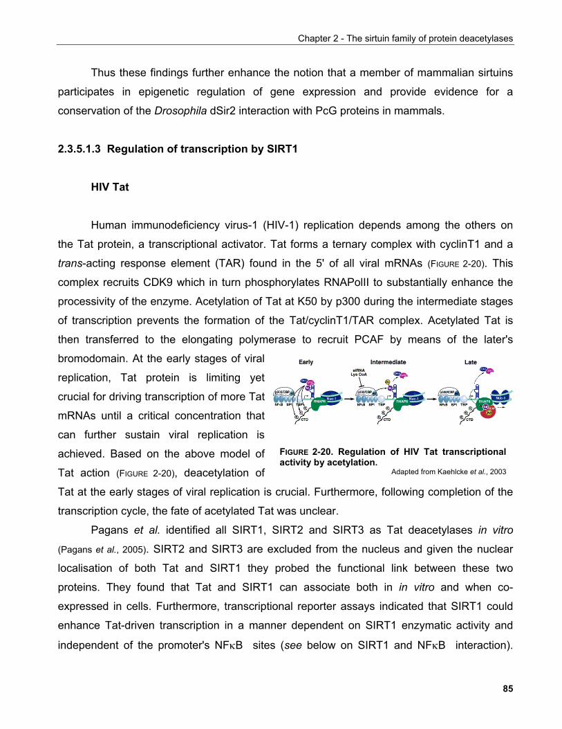
Chapter 2 - The sirtuin family of protein deacetylases
Thus these findings further enhance the notion that a member of mammalian sirtuins
participates in epigenetic regulation of gene expression and provide evidence for a
conservation of the Drosophila dSir2 interaction with PcG proteins in mammals.
2.3.5.1.3 Regulation of transcription by SIRT1
HIV Tat
FIGURE 2-20. Regulation of HIV Tat transcriptionalactivity by acetylation.
Adapted from Kaehlcke et al., 2003
Human immunodeficiency virus-1 (HIV-1) replication depends among the others on
the Tat protein, a transcriptional activator. Tat forms a ternary complex with cyclinT1 and a
trans-acting response element (TAR) found in the 5' of all viral mRNAs (FIGURE 2-20). This
complex recruits CDK9 which in turn phosphorylates RNAPolII to substantially enhance the
processivity of the enzyme. Acetylation of Tat at K50 by p300 during the intermediate stages
of transcription prevents the formation of the Tat/cyclinT1/TAR complex. Acetylated Tat is
then transferred to the elongating polymerase to recruit PCAF by means of the later's
bromodomain. At the early stages of viral
replication, Tat protein is limiting yet
crucial for driving transcription of more Tat
mRNAs until a critical concentration that
can further sustain viral replication is
achieved. Based on the above model of
Tat action (FIGURE 2-20), deacetylation of
Tat at the early stages of viral replication is crucial. Furthermore, following completion of the
transcription cycle, the fate of acetylated Tat was unclear.
Pagans et al. identified all SIRT1, SIRT2 and SIRT3 as Tat deacetylases in vitro
(Pagans et al., 2005). SIRT2 and SIRT3 are excluded from the nucleus and given the nuclear
localisation of both Tat and SIRT1 they probed the functional link between these two
proteins. They found that Tat and SIRT1 can associate both in in vitro and when co-
expressed in cells. Furthermore, transcriptional reporter assays indicated that SIRT1 could
enhance Tat-driven transcription in a manner dependent on SIRT1 enzymatic activity and
independent of the promoter's NFκB sites (see below on SIRT1 and NFκB interaction).
85
Chapter 2 - The sirtuin family of protein deacetylases
Conversely, siRNA targeting SIRT1 reduced Tat transcriptional activity. Tat-driven
transcription dramatically decreased i Sirt1-/- MEFs, and reconstitution of SIRT1 can reverse
this effect. Additionally, small molecule inhibition of SIRT1 by nicotinamide or the splitomicin
derivative HR73 recapitulated the effects of SIRT1 siRNA on Tat-driven transcription. To test
whether SIRT1 inhibition could be used as a target for therapeutic intervention against HIV
infection, Pagans et al. treated HIV-infected Jurkat cells with HR73 and found that HIV gene
transcription was approx. 5-fold decreased. This ultimate experiment provided proof of
principle that the identified role of cellular SIRT1 in the replication of HIV can be a site of
pharmacological targeting in the future.
CTIP1 and CTIP2
Chicken ovalbumin upstream promoter transcription factor (COUP-TF)-interacting
proteins 1 and 2 (CTIP1and CTIP2), are transcription factors that interact with and emhance
COUP-TF-mediated transcriptonal repression. It has also been proposed that they may
mediate transcriptional repression independently of COUP-TF since they can bind directly to
a defined DNA sequence and they are expressed in hematopoietic cells of lymphoid origin
which do not express COUP-TF. Loss-of-function analysis in mice as well as data showing
genetic aberations the CTIP1 and CTIP2 loci, have provided significant correlations between
CTIP1and CTIP2 dysregulation and hematopoietic malignancies.
Both CTIP1- and CTIP2-driven repression are independent of class I/II HDACs based
on their sensitivity to TSA. Senawong et al. could show that nicotinamide could alleviate
CTIP2-induced repression in reporter assays as well as demostrate by ChIP that the effects
of nicotinamide correlated with increased H3/H4 acetylation in an exogenous chromatinised
template (Senawong et al., 2003). Co-expression of SIRT1 recapitulates these findings in a
manner dependent on the latter's enzymatic activity. suggesting that SIRT1 participates in
CTIP2-driven repression. This effect stems from the ability of SIRT1 to interact via its sirtuin
core domain with CTIP2 both in vitro and in vivo. A follow-up study demonstrated that the
closely related protein CTIP1 behaves in a very similar manner as CTIP2 in the assays
employed (Senawong et al., 2005).
86
Chapter 2 - The sirtuin family of protein deacetylases
Apart from the discovery of SIRT1 as the deacetylase that potentially mediates the
TSA-insensitive repressive activities of CTIP1/2, the physiological importance of these
findings remains elucive. It would be of interest to investigate whether SIRT1 can be
exploited for pharmaceutical intervention within the context of leukemias that carry aberrant
CTIP1/2 chromosomal loci.
TAFI68
Apart from PolII-mediated gene transcription, chromatin remodelling has also been
implicated in the regulation of PolI-transcribed genes which predominantly encode for
ribosomal DNA. Transcription termination factor-I (TTF-I) is required for the recruitment of
chromatin remodelling complexes to PolI promoters to render chromatin accessible to the
transcriptional machinery. Targeted promoter binding of TTF-I is achieved by a promoter-
proximal terminator element named T0 160 bp upstream the transcription start site.
Muth et al. showed that TTF-I is able to bind the acetyltransferase PCAF, thus they
investigated whether components of the transcription initiation machinery are acetylation
substrates. They found that PCAF specifically acetylates the TAFI68 (TATA-box binding
protein-associated factor I of 68 kDa) subunit of the promoter selectivity factor TIF-IB/SL-1
both in vivo and in vitro. Similar to other acetylated proteins, TAFI68 can also bind to PCAF
(Muth et al., 2001). Importantly, TAFI68 was also shown to be acetylated in vivo and that this
acetylation can occur when it is bound to the TIF-IB/SL-1 holocomplex.
Furthermore, electrophoretic mobility-shift assays (EMSAs) demonstrated that
acetylation of TAFI68 increased its binding to rDNA sequences consistent with previous
observations for many other transcription factors. In an in vitro reconstituted transcription
system, acetylated TAFI68 is able to drive transcription more powerfully compared to the
unacetylated form suggesting that the increased binding of TAFI68 to DNA via acetylation
may regulate transcription of rDNA genes. Interestingly, treatment of cells with TSA did not
affect rDNA transcription assayed by Norhtern blot of pre-rRNA transcripts suggesting that
class I/II HDACs are not the physiological deacetylases regulating rDNA transcription.
Conversely, SIRT1 can deacetylate TAFI68 in vitro while incubation of the previously
employed in vitro transcription system with SIRT1 reduced rDNA transcription suggesting
87
Chapter 2 - The sirtuin family of protein deacetylases
that SIRT1 may be the relevant deacetylase. Notably, the DNA template used in this system
is devoid of histones suggesting that SIRT1 exerts this regulatory role independent of
chromatin.
rDNA silencing was one of the first described functions for yeast Sir2. This work
provided the first evidence that a mammalian sirtuin can modulate rDNA transcription.
Ribosomal DNA synthesis is essential for ribosomal biogenesis which correlates with a cell's
translational capacity. Protein translation is required for proliferation as demonstrated by cell
cycle arrest by means of small molecule inhibitors of translation (e.g. mimosine) and the
cytostatic effects of ribosome-targeting antibiotics. Furthermore, it has been suggested that
there exists a checkpoint that only allows cell division to occur under conditions that optimal
growth has been achieved (Thomas, 2000). Consequently, the role of SIRT1 in rDNA
transcription as documented in the work of Muth et al. is likely to reveal a deeper role for this
protein in cellular homeostasis.
Regulation of Hes1/Hey2 bHLH repressors
The bHLH family of transcription factors comprise both activators and repressors
which differ in the presence of distinct regulatory motifs. These motifs are thought to mediate
the interaction with co-repressors which in turn mediate the recruitment of chromatin
modifying factors such as deacetylases to implement transcriptional repression. The
mammalian counterpart of Hairy is Hes1 and is found to recruit co-repressors through a C-
terminal WRPW motif. Another member of the Hairy family of repressors, the Drosophila Hey
and its mammalian orthologue Hey2 show a distinct mechanism of repressor recruitment
through the bHLH domain itself implying diversity in the mode of action of this family of
transcriptional regulators.
Driven by the discovery of Rosenberg and Parkhurst in Drosophila that dSIRT1
interacts with the bHLH factor Hairy, Takata et al. examined the potential interaction between
the mammalian counterparts of these proteins (Takata et al., 2003). Although no endogenous
interactions were observed between these proteins, co-transfection experiments
demonstrated that both protein could immunoprecipitate each other. In vitro binding assays
mapped the SIRT1 binding site to the bHLH domain of both human HES1 and HEY2 protein.
88
Chapter 2 - The sirtuin family of protein deacetylases
Reporter assays employing GAL4 fusions of Hes1 and Hey2 contransfected with either wt or
catalytically inactive SIRT1 showed that SIRT1 augments the repression driven by either
factor and that its enzymatic activity is required for this effect. Taken together these data
suggested that SIRT1 may be a novel regulator of human bHLH factor transcriptional
repression activity. It would be of interest to examine whether acetylation of bHLH factors is
also involved in their regulation as for the majority of transcription factors interacting with
SIRT1.
Te interaction of SIRT1 with bHLH repressors is important with respect to recent
evidence suggesting that Hes1 regulates directly the transcription of the cell cycle inhibitor
p27. Hes1-/- mice exhibit developmental defects attributed to reduced proliferation which
correlated with increased p27 mRNA levels. Indeed, Hes1 can bind elements in the p27
promoter and repress its activity (Murata et al., 2005). Interestingly, SIRT1-/- mice exhibit cardiac
as well as retinal development defects similar to Hey2- and Hes1-deficient mice respectively
(Cheng et al., 2003). It would be of interest to investigate whether SIRT1 is required for the
repressive effect on p27 expression and if any of the observed phenotypes of the SIRT1-/-
mice can be attributed to the proposed function of SIRT1 on Hes1/Hey2 factors.
Bcl-6
BCL6 is a protooncogene that encodes a nuclear protein of the BTB/POZ (bric-à-brac,
tramtrac, broad complex/Pox virus zinc fingers) family of proteins. It is required for germline
centre formation and it has been implicated in the pathogenesis of B-cell lymphomas. Bcl-6
acts as a transcriptional repressor by recruiting HDAC-containing co-repressor complexes
including SMRT and Sin3A. This function depends on the zinc finger DNA binding domain,
the N-terminal POZ domain and an additional repression domain located in the middle of the
molecule. Bereshchenko et al. showed that Bcl-6 repressional activity is regulated by
acetylation (Bereshchenko et al., 2003).
In particular, Bcl-6 interacts and gets acetylated by the acetyltransferase p300. This
acetylation markedly decreases its transcriptional repressor activity on a luciferase reporter
and induces the dissociation of Bcl-6 from HDAC2. Accordingly, mutation of the Bcl-6
acetylation sites abolishes p300-mediated regulation and diminishes the transformation
89
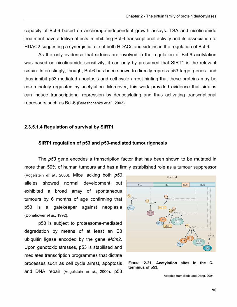
Chapter 2 - The sirtuin family of protein deacetylases
capacity of Bcl-6 based on anchorage-independent growth assays. TSA and nicotinamide
treatment have additive effects in inhibiting Bcl-6 transcriptional activity and its association to
HDAC2 suggesting a synergistic role of both HDACs and sirtuins in the regulation of Bcl-6.
As the only evidence that sirtuins are involved in the regulation of Bcl-6 acetylation
was based on nicotinamide sensitivity, it can only by presumed that SIRT1 is the relevant
sirtuin. Interestingly, though, Bcl-6 has been shown to directly repress p53 target genes and
thus inhibit p53-mediated apoptosis and cell cycle arrest hinting that these proteins may be
co-ordinately regulated by acetylation. Moreover, this work provided evidence that sirtuins
can induce transcriptional repression by deacetylating and thus activating transcriptional
repressors such as Bcl-6 (Bereshchenko et al., 2003).
2.3.5.1.4 Regulation of survival by SIRT1
SIRT1 regulation of p53 and p53-mediated tumourigenesis
The p53 gene encodes a transcription factor that has been shown to be mutated in
more than 50% of human tumours and has a firmly established role as a tumour suppressor
(Vogelstein et al., 2000). Mice lacking both p53
alleles showed normal development but
exhibited a broad array of spontaneous
tumours by 6 months of age confirming that
p53 is a gatekeeper against neoplasia
(Donehower et al., 1992).
p53 is subject to proteasome-mediated
degradation by means of at least an E3
ubiquitin ligase encoded by the gene Mdm2.
Upon genotoxic stresses, p53 is stabilised and
mediates transcription programmes that dictate
processes such as cell cycle arrest, apoptosis
and DNA repair (Vogelstein et al., 2000). p53
FIGURE 2-21. Acetylation sites in the C-terminus of p53.
Adapted from Bode and Dong, 2004
90
Chapter 2 - The sirtuin family of protein deacetylases
stabilisation is not sufficient, though for full p53 activity. Concomitant to the occurrence of
protein stabilisation, an array of post-translational modifications is required for p53 to fully
exert its roles which include phosphorylation, sumoylation, ubiquitination, neddylation,
acetylation and methylation. These modifications span the entire region of the protein and
affect protein stability, transcriptional activity, hetero- and homo-dimerisation and localisation.
Acetylation of p53 has been shown to increase protein stability and transactivation
capacity (Figure 2-21). It is believed that acetylation prevents ubiquitination, but also
neddylation or sumoylation of lysine residues, a proposal fitting to the observation that
increased deacetylase activities found in many cancers correlate with destabilisation of p53.
In parallel, acetylation of the C-terminus of p53 increases its DNA binding affinity both in vivo
and in vitro. Thus, modification of p53 by acetylation modulates its activity at multiple levels
and emerges as an important regulatory mechanism in tumour progression.
Acetylation of p53 upon DNA damage can be enhanced by treatment of cells with
HDAC inhibitors but TSA-independent deacetylation can also occur (Luo et al., 2001). Two
reports by Luo et al. and Vaziri et al. first demonstrated that the TSA-insensitive p53
deacetylase activity could be attributed to SIRT1 (Luo et al., 2001; Vaziri et al., 2001). SIRT1
interacts in vivo and in vitro with p53 and shows an apparent specificity for acetylated K382.
Overxpression of SIRT1 results in decrease of both basal as well as p300 overexpression-
induced and ionising radiation-or etoposide-induced K382 acetylation. Transfection of a
catalytically inactive mutant of SIRT1 had a dominant-negative effect in that, while deficient in
p53 deacetylation in vitro, it could induce p53 hyperacetylation in response to ionising
radiation. Furthermore, nicotinamide, a proposed inhibitor of sirtuins, was sufficient to abolish
most of p300-induced p53 acetylation. Importantly, SIRT1 inhibited p53-driven transcription
in both in vivo targets (p21) and in reporter assays. Matching these, SIRT1 overexpression
inhibited apoptosis in response to ionising radiation, etoposide treatment or indeed p53
overexpression alone but not Fas-induced apoptosis pointing to a specific effect on the p53
apoptotic pathway.
In an extension of these studies, Langley et al. provided evidence that p53
deacetylation by SIRT1 occurs in the context of promyelocytic leukemia (PML) nuclear
bodies (Langley et al., 2002). PML bodies are distinct foci in the cell nucleus with high
concentrations of the PML protein and are known to be sites of regulation of transcription,
91
Chapter 2 - The sirtuin family of protein deacetylases
apoptosis, tumour suppression and the anti-viral response. They are dynamic structures that
respond to a variety of cellular stresses and comprise cellular depots for several factors
including the small ubiquitin-like modifier SUMO-1, the acetyltransferase CBP and the tumour
suppressors pRb and p53. Upon overexpression of the PML isoform PML-IV, p53 is recruited
to PML bodies, a translocation which correlates with acetylation of p53 in K382, its
subsequent activation and induction of premature cellular senescence.
SIRT1 was found to be associated with PML-IV and localise to PML bodies in both
primary and transformed cells that overxpressed PML-IV (Langley et al., 2002). This localisation
appeared to bear functional significance because PML-IV-induced cellular senesence was
attenuated by co-expression of SIRT1, which correlated with decreased p53 acetylation at
K382. This report supported the notion that SIRT1 may regulate cellular life-span through a
pathway involving p53.
Fibroblasts derived from mice deficient for mSIRT1 show increased levels of p53
acetylation in response to DNA damaging agents. However, it was also found that by using
acetylation-specific antibodies higher acetylation of residues other than K379, the mouse
equivalent of K382, could also be observed suggesting an expanded specificity for SIRT1.
Furthermore, despite hyperacetylation of p53, adriamycin fails to induce higher levels of p21,
a p53 target, in Sirt1-/- MEFs a finding contradictory to the ones of Luo et al. and Vaziri et al..
Consistent with these, Sirt1-/- MEFs were equally sensitive to death induced by adriamycin
and UV. In contrast, thymocytes of Sirt1-/- animals were hypersensitive to ionising radiation
(IR)-induced cell death and this correlated with p53 hyperacetylation. Of note,
p53-/- thymocytes were resistant to IR-induced death. This implied that the hypersensitivity of
Sirt1-/- thymocytes was due to hypeactivation of the p53 pathway. However, the levels of p53
target genes were not assayed in this context leaving open the issue of whether p53
hyperacetylation in Sirt1-/- animals/cells actually affects a downstream transcription program.
SIRT1 is highly expressed in mouse ES cells (McBurney et al., 2003b). Interestingly, ES
cells fail to undergo G1 arrest upon treatment with ionising radiation despite the presence of
p53 protein raising the possibility that p53 function is impaired due to presumed
concomitantly high SIRT1 activity. This, however is not the case as SIRT1-/- ES cells show
cell cycle profiles which are intistinguishable from those of SIRT1+/+ ES cells (McBurney et al.,
2003b). Although the discrepancies between studies can be attributed to the different nature
92
Chapter 2 - The sirtuin family of protein deacetylases
of the cell systems employed, they also illustrate that the regulation of p53 by SIRT1 in the
physiological context of an organism remains poorly understood.
This issue has been addressed in a recent study by Kamel et al. who generated mice
defective in both Sirt1 and p53 genes (Kamel et al., 2006). These animals had an overall
phenotype indistinguishable from Sirt1-/- mice, including early post-natal lethality, eyelid
closure defects and reduced size. Furthermore, although they could confirm the interaction of
the two proteins and the effects on p53 acetylation, the authors could obtain no evidence of
changes in p53-mediated gene transcription nor differential sensitivity of
thymocytes/splenocytes to ionising radiation.
These results are in agreement with the study of Solomon et al. who identified
compound EX-527 in a high-throughput screen for SIRT1 catalytic inhibitors and used it to
acutely downregulate SIRT1 activity (Solomon et al., 2006). EX-527 enhances p53 K382
acetylation upon different genotoxic stimuli (etoposide, adriamycin, hydroxyurea, H2O2) in a
variety of human cell lines [NCI-H460 (large-cell lung carcinoma), U2-OS (osteosarcoma),
MCF7 (breast carcinoma), HMEC (human mammary epithelial cells)]. This is not
accompanied, however, by a concomitant change in p21 expression, nor cell viability or
growth rate. Conversely, although TSA acted additively to EX-527 to induce p53
hyperacetylation, it also decreased stress-induced viability under the same experimental
conditions.
Overall these studies suggested that SIRT1-mediated deacetylation as a potential
novel mechanism of p53 regulation. This is an exciting observation because it would imply
that in tumours that are treated with DNA-damaging agents, SIRT1 inhibition may potentiate
apoptosis by allowing hyperactivation of the p53 apoptotic response and thus providing the
basis for ameliorated cancer therapies. However, given the absense of effects on p53-
mediated transcription and the accumulating evidence that SIRT1 inhibition may not affect
viability in response to genotoxic stress, it would be of great importance to determine the
functional relevance of SIRT1-catalysed p53 deacetylation.
93
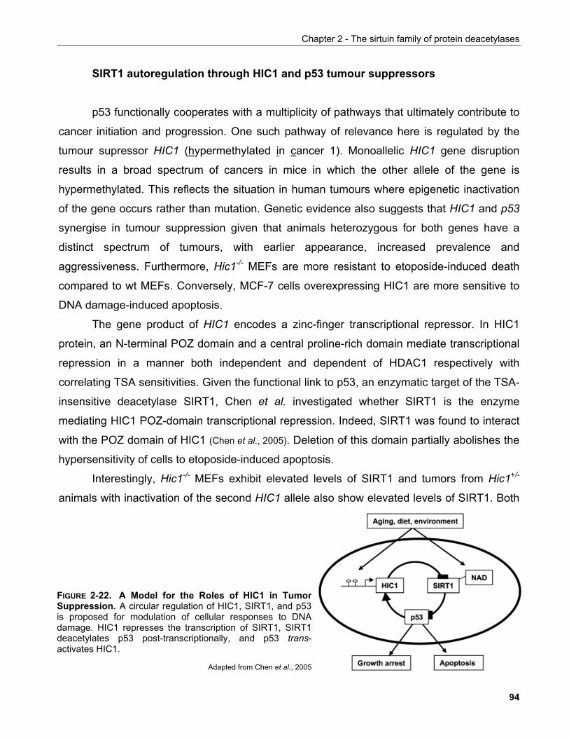
Chapter 2 - The sirtuin family of protein deacetylases
SIRT1 autoregulation through HIC1 and p53 tumour suppressors
p53 functionally cooperates with a multiplicity of pathways that ultimately contribute to
cancer initiation and progression. One such pathway of relevance here is regulated by the
tumour supressor HIC1 (hypermethylated in cancer 1). Monoallelic HIC1 gene disruption
results in a broad spectrum of cancers in mice in which the other allele of the gene is
hypermethylated. This reflects the situation in human tumours where epigenetic inactivation
of the gene occurs rather than mutation. Genetic evidence also suggests that HIC1 and p53
synergise in tumour suppression given that animals heterozygous for both genes have a
distinct spectrum of tumours, with earlier appearance, increased prevalence and
aggressiveness. Furthermore, Hic1-/- MEFs are more resistant to etoposide-induced death
compared to wt MEFs. Conversely, MCF-7 cells overexpressing HIC1 are more sensitive to
DNA damage-induced apoptosis.
The gene product of HIC1 encodes a zinc-finger transcriptional repressor. In HIC1
protein, an N-terminal POZ domain and a central proline-rich domain mediate transcriptional
repression in a manner both independent and dependent of HDAC1 respectively with
correlating TSA sensitivities. Given the functional link to p53, an enzymatic target of the TSA-
insensitive deacetylase SIRT1, Chen et al. investigated whether SIRT1 is the enzyme
mediating HIC1 POZ-domain transcriptional repression. Indeed, SIRT1 was found to interact
with the POZ domain of HIC1 (Chen et al., 2005). Deletion of this domain partially abolishes the
hypersensitivity of cells to etoposide-induced apoptosis.
Interestingly, Hic1-/- MEFs exhibit elevated levels of SIRT1 and tumors from Hic1+/-
animals with inactivation of the second HIC1 allele also show elevated levels of SIRT1. Both
FIGURE 2-22. A Model for the Roles of HIC1 in TumorSuppression. A circular regulation of HIC1, SIRT1, and p53is proposed for modulation of cellular responses to DNAdamage. HIC1 represses the transcription of SIRT1, SIRT1deacetylates p53 post-transcriptionally, and p53 trans-activates HIC1.
Adapted from Chen et al., 2005
94
Chapter 2 - The sirtuin family of protein deacetylases
reporter and ChIP assays confirmed that HIC1 but not a ∆POZ species binds directly to the
SIRT1 promoter via two 5'TGCC(A/C)3' sites in the same orientation. This implied that SIRT1
may regulate its own expression by complexing with HIC1 which proved to be the case since
consecutive ChIP experiments confirmed the presence of both proteins on the Sirt1
promoter. Importantly, transfection of dominant negative SIRT1 in Hic1-/- MEFs resulted in
reduced resistance to apoptosis presumably by blunting the elevated levels of SIRT1
encountered in these cells. Furthermore, despite its accumulation in etoposide-treated cells,
p53 acetylation was only evident in HIC1-overexpressing cells and, in reverse, siRNA-
mediated downregulation of HIC1 resulted in SIRT1 accumulation and p53 hypoacetylation.
These data provided evidence for SIRT1 as a key effector of tumourigenesis in
response to HIC1 inactivation and pointed to a molecular link between HIC1 and p53 tumour
supressor pathways (FIGURE 2-22).
Regulation of forkhead transcription factors
The insulin signalling pathway has been described as a phylogenetically conserved
signalling module in the determination of life-span. In Drosophila and C. elegans, the
forkhead transcription factor daf-16, which is negatively regulated by the insulin pathway, is
important for this effect on longevity (Giannakou et al., 2004).
In mammals the functional orthologues of daf-16 are believed to be the forkhead or
winged helix family of transcription factors (TFs) which comprises four members, FOXO1 (or
FKHR), FOXO3 (or FKHRL1), FOXO4 (or AFX) and FOXO6. Phosphorylation of these
factors by PKB leads to their exclusion from the nucleus under conditions of growth factor
availability. The identification of SIRT1 as a transcription factor deacetylase provided an entry
point for the experimental validation of the hypothesis that mammalian sirtuins and forkhead
factors functionally interact.
The acetyltransferase CBP enhances acetylation of both FOXO1 and FOXO4 when
co-transfected in cultured cells while both p300 and PCAF have the same effect on FOXO3.
In all cases complexes between FOXO TFs and the corresponding acetyltransferases were
observed. Furthermore, FOXO TF acetylation increases upon some genotoxic stresses.
Brunet et al. found that H2O2 and to a certain extend heat shock but not UV induce FOXO3
95
Chapter 2 - The sirtuin family of protein deacetylases
hyperacetylation in 293T cells (Brunet et al., 2004). Increased FOXO3 acetylation correlated
with enhanced binding to the acetyltransferase PCAF. In HeLa cells, Motta et al. also
observed UV-induced FOXO3 hyperacetylation (Motta et al., 2004). Consistent with these van
der Horst et al. found that FOXO4 is hyperacetylated in response to H2O2 in HEK293T cells
and this event also correlated with increased binding to acetyltransferase CBP. A more
recent study by Kitamura et al. validated the acetylation-dependent binding of FOXO1 too to
CBP in the pancreatic β-cell line βTC3.
FOXO3 acetylation was not altered by treatment of either TSA or nicotinamide alone
but the combination of the two drugs resulted in a marked hyperacetylation of the protein
suggesting that it is subject to regulation by both class I/II and class III deacetylases (Brunet et
al., 2004). Co-transfection experiments showed that FOXO1, FOXO3 and FOXO4 all interact
with SIRT1. This interaction was observed mainly in response to H2O2 (Brunet et al., 2004; van
der Horst et al., 2004) or after serum starvation (Daitoku et al., 2004), conditions that were shown to
result in nuclear accumulation of FOXO but in the case of FOXO3, Motta et al. could also
observe the interaction in unstimulated cells in co-transfection experiments. Moreover, Yang
et al. showed that the LIM-only protein FHL2 (four-and-a-half LIM 2) tethers SIRT1 to FOXO1
to induce its deacetylation (Yang et al., 2005).
Mutation of the FOXO sites phosphorylated by PKB to alanines results in the
constitutive translocation of FOXOs to the nucleus. Such a mutant species cannot interact
with SIRT1 in the absense of stress stimuli suggesting that this interaction probably depends
in additional modifications confered upon the protein(s) in response to such stimuli (Brunet et
al., 2004). Brunet et al. identified several stress-induced phosphorylation and acetylation sites
in transfected FOXO3 which might contribute to this interaction.
These observations raised the question whether acetylation of play a role in the
regulation of FOXO activity. FOXO1 and FOXO3 reporter activities were enhanced by
contransfection of CBP and p300 respectively. This effect could arise either due to
acetylation of histones or direct activation of FOXOs. Daitoku et al. identified putative
acetylation sites on FOXO1 by similarity to known histone H2B acetylation sites and mutated
them to arginine to create non-acetylatable species (Daitoku et al., 2004). Surprisingly, some of
these mutants either alone or in combination resulted in increased FOXO1 transcriptional
activity. Comparison of the relative activation induced by either wild-type or non-acetylatable
96
Chapter 2 - The sirtuin family of protein deacetylases
mutants showed that although CBP could induce transcription driven by both species, it
would do so more profoundly in the non-acetylatable mutants. This led to the conclusion that
in this context CBP co-activated FOXO1-driven transcription by presumably deacetylating
histones but would subsequently attenuate FOXO1 activity through its acetylation. Consistent
with these conclusions, van der Horst et al. discovered that CBP inhibited the transcriptional
activity of FOXO4. At present, it is not clear whether this applies to FOXO3 too, yet it
demonstrates the potential pitfalls and complexity in interpreting such results.
Conversely, in order to determine whether SIRT1-driven deacetylation of FOXOs
could alter their activity a variety of assays were employed. SIRT1 was shown to inhibit
reporter acivity driven by wild-type and a constitutively nuclear FOXO3 as well as in cells
overexpressing the lipid phosphatase PTEN and thus are expected to have low PKB activity
and allow translocation of exogenous FOXO3 to the nucleus. This effect depended on SIRT1
deacetylase activity and was sensitive to nicotinamide only when SIRT1 was co-expressed
(Motta et al., 2004). SIRT1 could also inhibit reporter activity driven by constitutively nuclear
FOXO1 and FOXO4 (Motta et al., 2004). Although conceptually agreeable these results are
surprising under the light of the observation that simple translocation of FOXO3 to the
nucleus does not suffice to sustain an interaction with SIRT1 raising the question as to the
mode of action of SIRT1 under these experimental conditions. On the other hand, Daitoku et
al. could show that SIRT1 deacetylase activity enhanced CBP-driven FOXO1 co-activation in
an additive manner.
In the light of these contradictory results it is interesting to know how SIRT1 affects
FOXO target genes. In agreement with their reporter assay results, upon treatment of
HEK293 cells with nicotinamide Daitoku et al. observed a decrease in the protein levels of
the cell cylce inhibitor p27 and maganese superoxide dismutase (MnSOD), an enzyme
involved in the detoxification of reactive oxygen species both FOXO1 target genes. Similarly,
overexpression of SIRT1 but not a catalytically inactive mutant significantly enhanced the
levels of these proteins in response to growth factor withdrawal. They also demonstrated the
presense of SIRT1 along with CBP and FOXO1 in the promoters of p27 and MnSOD genes.
In agreement with the above conclusions is the study of van der Horst et al., who found that
FOXO4-driven p27 and MnSOD expression is suppressed when SIRT1 are downregulated
by RNAi. Interestingly, using the same cell line (HEK293) Motta et al. observed the opposite
97
Chapter 2 - The sirtuin family of protein deacetylases
effect on p27 protein expression when they transfected SIRT1 along with FOXO3. They also
found enhanced activity of a reporter gene driven by a FOXO consensus DNA binding site in
Sirt1-/- ES cells both in the presence and absense of exogenous FOXO3, supporting their
findings of SIRT1 as a repressor of FOXO transcriptional activity.
Brunet et al. used a Rat-1 cell line derivative that was engineered to express a fusion
of FOXO3 to the ligand binding domain of the oestrogen receptor (Brunet et al., 2004). Upon
addition of 4-hydroxytamoxifen (4-OHT), the fusion protein dimerises and translocates to the
nucleus and is competent in driving transcription of FOXO target genes such as p27 and
GADD45. In these settings, the combination of TSA and nicotinamide suppresses p27 and
GADD45 induction but does not affect another FOXO target gene BIM which is known to
promote cell death. Accordingly, Brunet et al. note that SIRT1 also repressed FOXO-
dependent transcription of a reporter under the control of the death cytokine Fas ligand gene
promoter. They could also extend their findings in Sirt1-/- MEFs where expression of GADD45
was impaired in response to treatment with the PI-3 kinase inhibitor LY294002, which allows
FOXO translocation to the nucleus. Conversely, BIM expression was unaltered in Sirt1-/-
MEFs. Motta et al. went further to investigate FOXO target gene expression in the liver and
kidney of Sirt1-/- mice where they found enhanced expression of PEPCK and IGFBP1.
Accordingly, p300, FOXO1, FOXO3 and SIRT1 itself are bound to the IGFBP1 gene
promoter in wildtype MEFs, as well as in Sirt1-/- MEFs despite the absense of SIRT1 protein
from this site which suggests that SIRT1 does not prevent transcription factor recruitment
(Motta et al., 2004).
To investigate the functional consequences of FOXO coactivation by SIRT1 of p27,
Brunet et al. used their FOXO3-ER Rat1 fibroblasts and could show that, in agreement with
their observations on p27 expression, SIRT1 enhanced the G1 cell cycle arrest driven by
nuclear translocation of FOXO3. In agreement to this, exogenous FOXO3 expression in
Sirt1-/- MEFs was impaired in eliciting a G1 arrest compared to wild-type MEFs. Similar
effects of SIRT1 on cell cycle progression were reported by van der Horst et al. in the DLD-1
human colon carcinoma cell line derivative DL-23 that inducibly expresses constitutively
active FOXO3 as well as in transient transfections of constitutively active FOXO3 in A14 cells
which are NIH3T3 mouse fibroblast derivatives that overexpress the human insulin receptor.
98
Chapter 2 - The sirtuin family of protein deacetylases
van der Horst et al. could also demonstrate a functional link for the observed SIRT1
effects on MnSOD expression (van der Horst et al., 2004). They used a probe whose
fluorescence intensity is sensitive to its oxidation status which in turn reflects cellular
oxidative stress. Although FOXO3 alone could protect cells from H2O2-evoked oxidative
stress, nicotinamide suppressed this effect. Brunet et al. provided further evidence
supporting the cytoprotective role of SIRT1 against oxidative stress by showing that Sirt1-/-
MEFs were more sensitive to H2O2 treatment. They could also show that FOXO-induced cell
death is attenuated by SIRT1. Cerebellar granule neurons contransfected with FOXO3 and
SIRT1 showed less apoptosis compared to cells expressing only FOXO3. Also, in their
FOXO3-ER Rat1 fibroblasts the DNA damaging agent etoposide was less potent in inducing
cell death in the presence of exogenous SIRT1.
Overall these data suggest a gene-specific role for SIRT1 in the regulation of FOXO-
driven transcription. SIRT1 supports the function of FOXO in G1 cell cycle arrest by co-
activating transcription of p27 and supports its anti-oxidative capacity presumably through
MnSOD and probalby other genes. On the other hand SIRT1 opposes the pro-apoptotic role
of FOXOs. In the context of a cell under cytotoxic stress, SIRT1 could thus modulate FOXO-
driven transcriptional responses in a way that promotes cell cycle arrest but opposes death
allowing time for the cell to repair damage and eventually promote survival.
The inconsistent effects of SIRT1 on different FOXO target genes are not conpletely
surprising. As discussed in the introduction, Ramaswamy et al. have identified distinct
classes of FOXO target genes with accordingly different influence on cell cycle arrest and
apoptosis (Ramaswamy et al., 2002). Thus the observation of Motta et al. that SIRT1 can
negatively regulate FOXO-driven expression of IGFBP1 and PEPCK, as opposed to what
was shown by other groups for p27 and GADD45 may reflect the fact that these genes
exhibit different promoter requirements for their FOXO-mediated activation. Interestingly, the
study of Ramaswamy et al. also showed that downregulation of p27 is not necessary for
FOXO-induced cell cycle arrest which rather driven by FOXO-mediated repression of
cyclinD1 (Ramaswamy et al., 2002). It would be of interest to investigate whether SIRT1
mediates cell cycle arrest exclusively through p27 or is also shared by the cyclinD1 branch of
FOXO-driven transcription.
99
Chapter 2 - The sirtuin family of protein deacetylases
Recently, a molecular link between FOXO1 and PML, both of which are binding
partners of SIRT1, was discovered and provided a physiological setting where these
interactions might hold a major role. In particular, they investigated the role of FOXO1 in the
protection of pancreatic β-cells from glucose-induced toxicity. Chronic exposure to high
glucose such as in diabetic patients, is thought to overload β-cells' glycolytic capacity
ultimately resulting in the production of excess reactive oxygen species (ROS). By nature, β-
cells have reduced capacity to deal with these agents which in turn have been proposed to
impair expression of the Insulin2 (Ins2) gene trascription factor Pdx1 leading to increased
apoptosis.
Kitamura et al. found that induction of ROS leads to relocation of FOXO1 to nuclear
PML bodies, an event shown to depend on its acetylation (Kitamura et al., 2005). Concomitant to
this relocation, FOXO1 activates the transcription of two Ins2 transcription factors, MafA and
NeuroD to protect β-cells from death due to hyperglycaemia. In contrast to its requirement for
localisation to PML bodies, the transcriptional activity of FOXO1 requires its deacetylation.
Hypoacetylated FOXO1 is highly active in transcription but has a high ubiquitin-mediated
turnover. FOXO1 ubiquitination is increased in cells overexpressing SIRT1. In this respect, it
is implied, although not experimentally proven, that SIRT1-driven deacetylation promotes the
transcriptional activity of FOXO1 while inducing its increased turnover. Thus, SIRT1 may
have a cytoprotective role for pancreatic β-cells in vivo by co-activating FOXO1-driven
expression of MafA and NeuroD.
Transcriptional regulation of SIRT1 by p53 and FOXO
All the evidence described above supports a role for SIRT1 as a regulator of FOXO
function. Another study by Nemoto et al. showed that FOXO may be itself a regulator of
SIRT1 expression (Nemoto et al., 2004). They could show that the promoter of the Sirt1 gene
mediates the induction of a reporter gene transcription in response to glucose and growth
factor starvation in PC12 cells. Under these conditions, FOXO was found in the nucleus and
showed increased transcriptional activity. siRNA-mediated decrease of FOXO3 by siRNA
could suppress the activity of the SIRT1 promoter under starvation suggesting that FOXO3
transcriptionally regulates SIRT1. Interestingly, SIRT1 mRNA levels increased upon
100
Chapter 2 - The sirtuin family of protein deacetylases
overnight fasting in the muscle and liver but not in the heart of mice. This is in contrast to the
findings of Rodgers et al. who found increased SIRT1 protein levels in mouse liver upon
fasting without concomitant increase of mRNA levels (see below) (Rodgers et al. 2005).
Nemoto et al. found that the region between nucleotides -202 and -91 from SIRT1
transcription start site is important for starvation-inducible induction of the gene but could
identify no consensus forkhead binding sites within it. However, they determined two p53
consensus sites that when mutated alone or together they could abolish FOXO3-stimulated
SIRT1 promoter activity. This suggested that FOXO3 and p53 synergise in the regulation of
SIRT1 gene transcription. FOXO3 and p53 were shown to physically interact both in vitro and
when co-transfected in vivo and this interaction increased upon growth factor withdrawal. p53
inhibited the transcription of a reporter gene driven by the SIRT1 promoter p53 binding
element and this was aleviated by co-expression of FOXO3. In parallel, FOXO3 could also
inhibit p53-driven reporter activity driven by an alternative p53 binding element suggesting
that the physical interaction of FOXO3 with p53 in promoters had antagonistic effects on the
latter's transcriptional activity. In accordance to these results, basal SIRT1 mRNA levels were
elevated in the adipose tissue of p53-/- mice but the starvation-induced induction of its
transcription was abolished.
Taken together, these data suggested that in response to nutrient limitation, SIRT1 is
upregulated via a transcriptional mechanism involving the alleviation of p53-mediated
repression of its promoter by FOXO3.
Overall, there is compelling evidence that SIRT1 interacts physically and functionally
with several forkhead transcripton factors in a reciprocal manner. Furthermore, the recent
findings that p53 can also participate in the determination of organismal longevity and the
interconnection between PML, which by itself regulates senescence, forkhead transcription
factors and SIRT1 implicates a complex regulatory mechanism that may govern cellular and
organismal life-span.
NFκB
The transcription factor NFκB is known to hold important roles in acute inflammatory
responses and cell survival by mediating the expression of genes with of a broad spectrum of
101
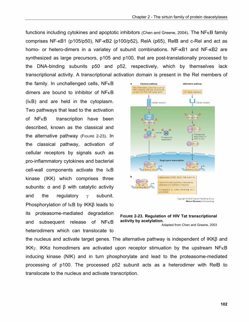
Chapter 2 - The sirtuin family of protein deacetylases
FIGURE 2-23. Regulation of HIV Tat transcriptionalactivity by acetylation.
Adapted from Chen and Greene, 2003
functions including cytokines and apoptotic inhibitors (Chen and Greene, 2004). The NFκB family
comprises NF-κB1 (p105/p50), NF-κB2 (p100/p52), RelA (p65), RelB and c-Rel and act as
homo- or hetero-dimers in a variatey of subunit combinations. NF-κB1 and NF-κB2 are
synthesized as large precursors, p105 and p100, that are post-translationally processed to
the DNA-binding subunits p50 and p52, respectively, which by themselves lack
transcriptional activity. A transcriptional ac
the family. In unchallenged cells, NFκB
dimers are bound to inhibitor of NFκB
(IκB) and are held in the cytoplasm.
Two pathways that lead to the activation
of NFκB transcription have been
described, known as the classical and
the alternative pathway (FIGURE 2-23). In
the classical pathway, activation of
cellular receptors by signals such as
pro-inflammatory cytokines and bacterial
cell-wall components activate the IκB
kinase (IKK) which comprises three
subunits: α and β with catalytic activity
and the regulatory γ subunit.
Phosphorylation of IκB by IKKβ leads to
its proteasome-mediated degradation
and subsequent release of NFκB
heterodimers which can translocate to
the nucleus and activate target genes. The alternative pathway is independent of IKKβ and
IKKγ. IKKα homodimers are activated upon receptor stimuation by the upstream NFκB
inducing kinase (NIK) and in turn phosphorylate and lead to the proteasome-mediated
processing of p100. The processed p52 subunit acts as a heterodimer with RelB to
translocate to the nucleus and activate transcription.
tivation domain is present in the Rel members of
102
Chapter 2 - The sirtuin family of protein deacetylases
As in every multistep cellular process, the NFκB pathway offers multiple levels for
regulatory inputs but the transcription factor itself is subject to a variety of modulatory events.
Phosphorylation of RelA/p65 by PKA, MAP kinases and PKCζ allows the phosphorylation-
dependent binding of co-activators including CREB binding protein (CBP) and p300 both of
which exhibit histone acetyltransferase (HAT) activity. Acetylation of RelA/p65 at specific
lysines affects both DNA binding and transcriptional activity. Recent reports also suggest that
RelA/p65 acetylation reduces its affinity for IκB and deacetylation by class I HDACs
promotes its nuclear export in a IκB-dependent manner.
The observation that resveratrol, a sirtuin activator, is also a potent NFκB
transcriptional inhibitor, led Yeung et al. to investigate whether SIRT1 is required for this
inhibitory effect. They confirmed that pharmacological activation of sirtuin activity with
resveratrol suppressed TNFα-induced NFκB activation of reporter activity while the sirtuin
inhibitors splitomycin and nicotinamide had the reverse effect (Yeung et al., 2004). Interestingly,
the latter compounds also increased basal reporter activity in the absence of TNFα, a fact not
discussed by the authors but that could signify a role for sirtuin-mediated inhibition of basal
transcription as observed in the work of Vaquero et al. discussed earlier. SIRT1 co-
transfection inhibited TNFα-induced NFκB reporter activity suggesting that the observed
effects of the pharmacological study were mediated by this sirtuin.
The authors went on to determine that it was the transcriptional activity of the
RelA/p65 subunit that was sensitive to SIRT1 dose pointing at a potential regulatory site for
SIRT1 on NFκB dimers. Indeed, co-transfection as well as endigenous co-
immunoprecipitation experiments showed that SIRT1 and p65 interact. Importantly,
resveratrol treatment did not alter IκB degradation kinetics, nuclear accumulation kinetics of
neither p65 nor p50, nor did it affect their DNA binding capacity. These suggested that SIRT1
may modulate this factor at the level of its transcriptional activity.
It could subsequently be shown that SIRT1 acts through the transactivation domain of
p65 by deacetylating it at lysine 310. Furthermore, a p65 transactivation domain mutant for
the acetylateable residue (K310) was no longer subject to resveratrol- and SIRT1-mediated
repression confirming that NFκB activity is modulated by SIRT1 through deacetylation of the
transactivation domain. Importantly, these events are significant for cellular homeostasis
103
Chapter 2 - The sirtuin family of protein deacetylases
because NFκB inhibition by resveratrol sensitised cells to TNFα-induced apoptosis in a
SIRT1-dependent manner. As expected, this was due to suppression of TNFα-induced
expression of NFκB anti-apoptotic target genes, such as cIAP and Bcl-XL. Interestingly,
resveratrol seems to potentiate SIRT1 occupancy of target promoters which correlated well
with the observed repression of target genes.
This study brought forward an aspect of SIRT1 biology which conflicted previous
reports supporting its role as an antiapoptotic factor, e.g. in the context of p53 and FOXO.
This is not entirely surprising since the signalling cascades involved in the activation of these
pathways are seemingly divergent. Thus while DNA damage would engage the p53
response, inflammatory stimuli such as TNFα would signal through the NFκB pathway.
However, it is also increasingly appreciated that under specific contexts, there is extensive
cross-talk between these pathways which establishes an elaborate mechanism determining
cell survival (Janes et al., 2006; Janes et al., 2005).
Ku70
Ku70 is a protein involved in the DNA repair pathway and has been shown to be a
substrate for the DNA-dependent protein kinase DNA-PK. As a heterodimer with Ku80 it
participates in non-homologous end-joining (NHEJ), V(D)J recombination and telomere
maintenance. Consequently, Ku70 null knockout mice exhibit higher sensitivity to ionising
radiation, are immunocompromised and show high rates of apoptosis in the developing
nervous system. The latter phenomenon is attributed to the ability of Ku70 to associate with
the pro-apoptotic factor Bax in the cytosol in a complex devoid of Ku80 and sequester Bax
from mitochondria. In response to apoptotic stimuli Bax translocates to the mitochondrial
outer membrane, oligomerises and renders it permeable eliciting the release of cytochrome c
and other death-promoting factors.
The interaction between Ku70 and Bax is regulated by the acetylation status of Ku70.
When Ku70 is acetylated by the acetyltransferases CBP and PCAF it dissociates from Bax
allowing it to translocate to mitochondria (Cohen et al., 2004a). Several lysines were shown to
be acetylated in vivo but K539 and K542 appeared to be critical for this regulation given that
104
Chapter 2 - The sirtuin family of protein deacetylases
cells expressing Ku70 with either of these lysines substituted with glutamines to mimic
acetylation, were more sensitive to apoptosis.
Sirtuins were implicated in the regulation of Bax-mediated apoptosis on account of the
fact that Ku70 acetylation is sensitive to both TSA and nicotinamide, a sirtuin inhibitor. In this
way, deacetylase-mediated sequestration of Bax by Ku70 would promote survival. In a later
report, Cohen et al. showed that SIRT1 is a Ku70 deacetylase which led them to propose
that caloric restriction promotes survival at the cellular level and increased life-span at the
organismal level by supressing Bax-mediated apoptosis in a manner dependend on SIRT1
(Cohen et al., 2004b).
2.3.5.1.5 SIRT1 regulation of transcription factors involved in muscle differentiation
Repression of MyoD by SIRT1
Intracellular redox state is finely regulated in muscle cells in particular during
differentiation when a drop in the [NAD+]/[NADH] ratio is observed. This prompted Fulco et al.
to investigate the role of SIRT1 in muscle differentiation due to the NAD+ dependence of the
enzyme (Fulco et al., 2003). MyoD is a basic helix-loop-helix (bHLH) transcription factor which
collaborates with the MEF2 (myocyte enhancer factor 2) transcription factor to induce gene
expression programmes that dictate muscle differentiation.
Inhibitor studies indicated that mSIRT1 supressed transcription driven by MyoD and
MEF2 and attenuated muscle differentiation by global downregulation of muscle-specific
genes. mSIRT1 was found in a ternary complex with PCAF and MyoD and was able to
deacetylate both factors in vitro and in vivo. Since MyoD acetylation is important for its full
transcriptional activity, it is possible that deacetylation of MyoD by SIRT1 mediates the
effects of the later on muscle differentiation (Fulco et al., 2003). Conversely, the authors
demonstrated that mSIRT1 activity regulates histone H3 K9 and K14 acetylation in muscle-
specific gene promoters implying that SIRT1 may regulate promoter accessibility directly
through chromatin modifications. Importantly, by modulating the intracellular [NAD+]/[NADH]
ratio coupled to RNA interference-mediates mSIRT1 downregulation the authors
105
Chapter 2 - The sirtuin family of protein deacetylases
demostrated the effects of intracellular redox state on muscle differentiation are mediated
through mSIRT1.
Functional interaction of SIRT1 with HDAC4 to regulate MEF2 repression
The class II deacetylase HDAC4 has been shown to interact with MEF2 via an N-
terminal MITR (MEF2-interacting transcriptional repression) homology domain and repress
muscle-specific gene expression. This function has been assumed to be performed via
promoter specific HDAC4-mediated histone deacetylation. Nevertheless, deacetylase-
independent transcriptional repression activity of HDAC4 has also been reported.
SUMO (small ubiquitin-like modifier) is a small protein that can be covalently linked to
lysine residues of other proteins and has been implicated in transcriptional repression.
Similar to ubiquitination, protein sumoylation requires an E1 activating enzyme, an E2 SUMO
conjugating enzyme (Ubc9) and a SUMO E3 ligase several of which have been identified.
In a yeast two-hybrid screen, Ubc9 was found to bind to an N-terminal coiled-coil
region of HDAC4 previously shown to mediate interaction with MEF2 (Zhao et al., 2005).
HDAC4 was shown to be sumoylated on K559, however mutations in this residue failed to
reveal an effect on the ability of HDAC4 to repress MEF2 transcriptional activity. Zhao et al.
subsequently showed that MEF2 is itself sumoylated at K424 and HDAC4 could enhance
MEF2 sumoylation both when co-transfected in cultured cells or in in vitro sumoylation
assays. This suggested that HDAC4 recruits the SUMO conjugating complex to modify
MEF2.
Interestingly, MEF2 K424 is also acetylated directly by the MEF2 co-activator CBP yet
this acetylation is not sensitive neither to HDAC4 overexpression nor to TSA treatment.
MEF2 K424 is, however, sensitive to the sirtuin inhibitor nicotinamide and SIRT1 was shown
to interact with HDAC4 and reverse CBP-mediated MEF2 acetylation in co-transfection
experiments.
As MEF2 acetylation increases during myocyte differentiation Zhao et al. predicted
that K424 sumoylation would have an inhibitory effect on MEF2 function. Indeed, fusion of
SUMO to MEF2 impedes its ability to interact with MyoD in activation of reporter gene
106
Chapter 2 - The sirtuin family of protein deacetylases
transcription while a sumoylation-deficient mutant of MEF2 shows higher activity in the same
assay.
These data led the authors to propose a model where HDAC4 and SIRT1 synergise to
induce MEF2 respression. SIRT1 deacetylates MEF2 at K424 to allow its sumoylation by the
SUMO-conjugating activity recruited by HDAC4. This is a first indication that SIRT1 acts in
concert with another HDAC to repress gene expression in a mechanism other than histone
deacetylation.
2.3.5.1.6 Genetic and biochemical interactions of SIRT1 with proteins regulating metabolism
The implication of sirtuins in regulation of life-span upon caloric restriction in lower
eucaryotes led to the hypothesis that they may mediate metabolic changes which in turn
promote longevity. In this respect genetic ablation of the insulin receptor in mice leads to
18% increase in mean life-span while as discussed C. elegans sir2.1 interacts genetically
with the insulin signalling pathway. Recent studies have shed light into potential mechanisms
that employ SIRT1 to regulate cellular metabolic pathways.
SIRT1 and the regulation of fat metabolism
Picard et al. used the mouse cell line 3T3-L1 which can be induced to differentiate into
cells with adipocytic characteristics including expression of adipose-specific differentiation
markers and triglyceride accumulation in cytoplasmic lipid droplets (Picard et al., 2004). Upon
differentiation, 3T3-L1 cells with downregulated levels of mSIRT1 by means of a vector-
driven short hairpin RNA showed increased accumulation of intracellular triglycerides (TG)
while the converse was observed in cells overepxpressing SIRT1. This behaviour was
paralleled by the levels of adipogenic factors such as PPARγ, C/EBPδ, C/EBPα and aP2
respectively. This implied a role of SIRT1 in adipogenesis.
To investigate whether SIRT1 affects fat metabolism in differentiated adipocytes, the
authors used the sirtuin activator resveratrol to stimulate the activity of mSIRT1 and also
107
Chapter 2 - The sirtuin family of protein deacetylases
observed suppression of TG accumulation while the levels of free fatty acids (FFA) in the
medium were elevated. Picard et al. could recapitulate these findings in primary rat
adipocytes treated with resveratrol. To investigate the molecular mechanism underlying this
behaviour, chromatin immunopreciptiation (ChIP) was employed. mSIRT1 was found bound
to the aP2 and PPARγ promoters implying that it may exert its adipogenesis-suppressing role
by inhibiting the action of adipogenesis-promoting factors. Indeed, mSIRT1 can directly bind
to both PPARγ and its transcriptional repressor NCoR in the same promoters where PPARγ
is found. Importantly, NCoR depletion by shRNA abrogated the suppressive action of SIRT1
on fat accumulation. The authors extended their findings in the mouse and showed that
mSIRT1 is bound to PPARγ and aP2 promoters in mice that had been fasted, a condition
characterised by high FFA serum content. Consistent with this, Sirt1+/- animals exhibited
lower FFA serum levels upon fasting than Sirt1+/+ animals and adipocytes from Sirt1+/-
animals also had suppressed FFA mobilisation upon adrenergic stimulation. In all, these
findings suggested that SIRT1 modulates circulating FFA levels by suppressing the function
of adipogenic transcription factors.
Although the data in this study are convincing and well-controllled with respect to the
experimental setups employed, there are two issues worth of further investigation. Firstly, the
authors showed that upon induction of 3T3-L1 cells to differentiate, endogenous mSIRT1
levels increase. This behaviour is reminiscent of that of the adipogenesis-promoting factor
PPARγ and contradicts the proposed role of SIRT1 as an inhibitor of adipogenesis. Secondly,
the proposed mechanism of SIRT1 regulation of fat metabolism would suggest that
suppression of adipogenesis, which is characterised by accumulation of TG, is sufficient to
induce the release of FFA. To the best of my knowledge little evidence exists to support this.
It is possible that absence of SIRT1 supresses FFA production upon fasting by a machanism
related to TG turnover such as lipolysis an aspect worth investigating, in particular through
assaying the activity of TG mobilising enzymes such as hormone-sensitive lipase (HSL) and
the recently discovered adipose triglyceride lipase (ATGL)(Zehner et al., 2005; Zimmermann et al.,
2004).
108
Chapter 2 - The sirtuin family of protein deacetylases
SIRT1 and the regulation of glucose homeostasis
Blood glucose levels are tightly regulated through elaborate molecular mechanisms
involving several tissues. This guarantees that tissues such as the brain and red blood cells
where glucose is the primary fuel used for energy production are in constant supply.
SIRT1 regulation of fasting hepatic gluconeogenesis. Another important regulator
of energy homeostasis is peroxisome proiferator-activated receptor-γ coactivator 1α (PGC-
1α). PGC-1α is required for a diverse set of metabolic processes such as mitochondrial
oxidative metabolism in brown adipose tissue, fiber-type switching in skeletal muscle and
gluconeogenesis and stimulation of fatty acid oxidation in the liver upon fasting. Fasting liver
metabolism is dictated by the actions of hormones as well as nutrients the mode of action of
the latter being largely elucive.
As some aspects of fasting (controlled by PGC-1α) recapitulate some of the effects of
caloric restriction (proposed to be regulated by sirtuins), Rodgers et al. examined the
potential role of SIRT1 in glucose homeostasis. They found that although mSIRT1 mRNA
levels do not change in the livers of fasted animals, protein levels increase (Rodgers et al. 2005).
They also observed that fasting livers have elevated NAD+ levels. These observations
indicated that mSIRT1 has a role in fasting liver responses.
The authors went on to show that the observed elevation of mSIRT1 protein is due to
increased translation and that it can be positively regulated by the levels of pyruvate, a
metabolite derived from blood alanine by the fasting liver and used as a substrate for
gluconeogenesis. Conversely, glucose causes a decrease in mSIRT1 protein levels. PGC-1α
activates the gluconeogenic programme through its interaction with HNF4α. SIRT1 is found
in a complex with both proteins and regulates the acetylation status of PGC-1α. Acetylated
PGC-1α has decreased capacity to co-activate HNF4α in response to pyruvate and
downregulation of mSIRT1 by siRNA abolishes the ability of PGC-1α to induce
gluconeogenic genes such as PEPCK and Glucose-6-phosphatase. Interestingly, this SIRT1
dependence is not extended to the mitochondrial genes regulated by PGC-1α. PGC-1α and
SIRT1 also synergise to suppress glycolysis and in combination with the co-ordinated
enhancement of gluconeogenesis they result in elevation of glucose production in
109
Chapter 2 - The sirtuin family of protein deacetylases
hepatocytes. Taken together these data provided solid evidence for a role of SIRT1 in
regulating glucose production in the liver through the transcriptional co-activator PGC-1α
(Rodgers et al. 2005).
SIRT1 regulation of insulin secretion. A major role in acute glucose regulation at
the organismal level is played by the pancreas. The exocrine pancreas which comprises the
bulk of the organ is largely involved in the excretion of digestive enzymes. Embedded within
this exocrine tissue are clusters of cells known as the islets of Langerhans which are involved
in the endocrine functions of the organ. Three major cell types are found in the islets of
Langerhans and are the sites of secretion of the major hormones produced therein. The α-
cells, which secrete glucagon, the β-cells which secrete insulin and the δ cells which secrete
somatostatin. Defects in pancreatic function, expecially in β-cells as encountered in disease
such as diabetes have detrimental effects on animal physiology.
Pancreatic β-cells respond to elevated levels of blood glucose to secrete insulin which
acts in peripheral tissues such as the muscle to stimulate glucose uptake and adipose tissue
to increase energy storage in the form of triglycerides. The basic components of glucose-
stimulated insulin secretion in β-cells have been described. Glucose catabolism results in
increased ATP/ADP ratios that lead to the closing of ATP-dependent K+ channels. This
causes membrane depolarisation which in turn activates voltage-gated Ca2+ and induces
calcium influx that then allows intracellular vesicles containing insulin to fuse with the plasma
membrane and secrete their contents. Proteins that modify this pathway have been
described, notably uncoupling protein 2 (UCP2) which uncouples proton entry to
mitochondria from ATP production. The action of UCP2 results in lower ATP/ADP levels and
decreased insulin secretion. Indeed Ucp2-/- knockout animals show increased islet ATP and
increased glucose-stimulated insulin secretion. UCP2 levels were elevated in ob/ob mice, a
genetic model of obesity-induced diabetes, and this elevation correlated with increased
insulin lelvels and hypoglycemia.
Moynihan et al. (Moynihan et al., 2005) and Bordone et al. (Bordone et al., 2005)
independently described a role for mSIRT1 in β-cell function. Both groups' work was
triggered by the observation that mSIRT1 is expressed in the pancreatic islets. Moyihan et
110
Chapter 2 - The sirtuin family of protein deacetylases
al., clearly showed that mSIRT1 shows higher expression in α-cells where it localises to the
cytoplasm while a weak β-cell specific staining was also observed.
To investigate the physiological significance of this observation, Moyihan et al. created
a transgenic mouse that expressed mSIRT1 cDNA under the control of the human insulin
promoter (Moynihan et al., 2005). They termed these animals BESTO for β-cell-specific Sirt1-
overexpressing. Conversely, Bordone et al. made use of the Sirt1-/- animals generated by
McBurney et al. as a loss-of-function model (Bordone et al., 2005).
BESTO mice exhibited normal insulin and glucose levels in both fed and fasted state
but when challenged with high glucose they showed better glucose tolerance attributed to
elevated insulin levels. Sirt1-/- animals had constitutively lower levels of insulin and
completely impaired insulin secretion upon glucose injection. Surprisingly, glucose levels in
Sirt1-/- mice were lower compared to wild-type controls while these mice also consumed more
food, suggesting that they have improved insulin sensitivity. In their respective models, both
groups confirmed that islet morphology and gross mass did not differ from control
preparations. Islets isolated from Sirt1-/- animals, however were defective in glucose-induced
insulin secretion while pancreata of BESTO mice showed elevated insulin levels when
perfused with glucose.
In an attempt to understand the impaired insulin secretion phenotype of Sirt1-/- islets,
Bordone et al. used INS-1 cells which are rat pancreatic cell lines where they downregulated
SIRT1 by means of shRNA to exclude differential expression of proteins involved in glucose
uptake and insulin secretion. However, they observed suppressed levels of ATP/ADP in the
knockdown cells which implied that respiration in these cells is more uncoupled compared to
control cells. Upon examination the levels of UCP2 protein were found to be higher in both
INS-1 knockdown cells and the pancreata of Sirt1-/- mice. Furthermore, ChIP experiments
showed that mSIRT1 binds directly the Ucp2 promoter to suppress expression of the gene.
Using gene expression profiling of derivatives of MIN6, a mouse β-cell line, where mSIRT1
levels were modulated, Moynihan et al. reached largely the same conclusions. Interestingly,
Bordone et al. measured the NAD+/NADH in the pancreata of starved animals and found in to
be decreased. This would lead to a decrease in SIRT1 activity. In agreement to this, UCP2
levels were high in starved wild-type animals.
111
Chapter 2 - The sirtuin family of protein deacetylases
β-cell disfunction is associated with diseases such as diabetes and positively
correlates with old age. mSIRT1 expression in the pancreas was clearly demonstrated
although the focus of the above work was on β-cells despite the significant presence of
mSIRT1 in α-cells too. However, the above data are significant because they provide the first
evidence of a physiological system where modulation of SIRT1 activity can be considered as
a drug target and paved the way for further investigation of its role in metabolic regulation
which the major effect of caloric restriction is thought to be exerted upon.
2.3.5.1.7 Neuroprotection and cardioprotection by SIRT1
Degeneration of neuronal axons is a feature of many neuropathies such as
Parkinson's and Alzheimer's disease. In Wallerian degeneration slow (wlds) mice, a
spontaneous dominant mutation results in the overexpression of Wlds, a chimaeric nuclear
protein comprising the N-terminal 70 aminoacids of Ufd2a (ubiquitin fusion degradation
protein 2a) which participates in ubiquitin chain assembly and the entire sequence of Nmnat1
which participates in nuclear NAD+ biosynthesis (see section 2.2.2.1). These mice exhibit
delayed axonal degeneration.
Araki et al. determined that the enzymatic activity of Nmnat1 is required for the
neuroprotective effects of Wlds in response to various damaging agents such as ischaemia
and actin poisons as well as mechanical ablation of axons suggesting a role of nuclear NAD+
in this process which they called NAD+-dependent axonal protection (NDAP) (Araki et al., 2004).
In agreement to this, exogenously provided NAD+ also had a protective effect in neuronal
cultures. Araki et al. hypothesised that de novo gene expression is required for NDAP as
these were evident only after >8 hours of pretreatment with NAD+.
Using sirtinol and resveratrol, an inhibitor and activator of sirtuins respectively as well
as lentivirus-expressed shRNA against SIRT1, Araki et al. could demonstrate that NDAP
required SIRT1 consistent with the dependence of its activity on NAD+. Interestingly, they
could observe no change in cellular NAD+ concentrations suggesting that because of the
nuclear localisation of Wlds it is only the nuclear pool of NAD+ that is relevant in NDAP (Araki
et al. 2004).
112
Chapter 2 - The sirtuin family of protein deacetylases
Similarly, pharmacological evidence suggests a role for sirtuins in cardioprotection and
cardiac hypertrophy as SIRT1 overexpression protected cardiomyocytes from serum
deprivation-induced death and casued an increased cell size (Alcendor et al., 2004).
Conversely, sirtuin inhibition induced cell death in isolated neonatal rat cardiomyocytes.
These effects are likely mediated by SIRT1 since in a dog model of heart failure, SIRT1 was
specifically found to be upregulated (Crow, 2004).
2.3.5.1.8 SIRT1 and caloric restriction in rodents
As sirtuins in lower organisms mediate in part the effects of caloric restriction on
longevity, it is of great interest to examine whether this constitutes a conserved function of
mammalian sirtuins. At least two pieces of evidence indicate that this is the case.
Caloric restriction in mice can be implemented by reducing food availability to 60% of
normal food intake. Under these conditions, complex physiological and behavioural changes
are observed such as a drop in blood plasma triglyceride, glucose and IGF-1 levels and
increase in animal movement. In response to caloric restriction, mice carrying a genetic
ablation of SIRT1 alleles, exhibit reduced or absent increases of physical activity (Chen et al.,
2005). This is in spite of changes in plasma markers of caloric restriction comparable to
wildtype animals and enhanced performance in movement capacity assays such as
accelerating treadmil or rotarod. This suggests that SIRT1 mediates at least some
behavioural effects of caloric restriction, possibly through its action in movement centers of
the central nervous system where SIRT1 has been shown to be strognly expressed (Sakamoto
et al., 2003).
In a separate study, Nisolo et al. observed a marked increase of mitochondrial
biogenesis and respiration in mice under a caloric restriction regimen (Nisolo et al., 2005). This
correlated with increased expression of key mitochonrial biogenesis regulators such as PGC-
1α, nuclear respiratory factor-1 (NRF-1) and mitochondrial transcription factor A (Tfam) in
WAT. This response was markedly reduced in mice null for endothelial nitric oxide synthase
gene (eNOS-/-) while eNOS mRNA levels were induced upon caloric restriction in wild-type
mice uggesting that it mediates the effects of caloric restriction on mitochondrial biogenesis.
113
Chapter 2 - The sirtuin family of protein deacetylases
Interestingly, SIRT1 mRNA and protein level changes paralleled those of
mitochondrial biogenesis genes and were also shown to depend on the presence of eNOS in
WAT as well as other tissues. The same could be recapitulated in cultured white adipocytes
where treatment with NO donors or a cGMP analogue (mimicking a second messenger
downstream of eNOS) induced a significant increase in SIRT1 protein levels. Thus SIRT1
expression is upregulated in WAT upon caloric restriction by a mechanism involving eNOS-
mediated signaling.
In this study, the expression of both PGC-1α and SIRT1 in induced upon caloric
restriction suggesting a potential functional interaction of the two proteins in WAT similar to
the one seen in the liver (Rodgers et al., 2005). However, in the latter case Rodgers et al.
showed that SIRT1 synergises with PGC-1α for the expression of genes involved in hepatic
gluconeogenesis but not mitochondrial biogenesis (Rodgers et al., 2005). It is possible that the
interaction of SIRT1 with PGC-1α in WAT does occur and is distinct from that in the liver
owing to the differing function of the two tissues upon fasting.
2.3.5.2 SIRT2
SIRT2 localises to the cytoplasm and in particular is found associated with
microtubules. Concomitant to this property, SIRT2 is the only sirtuin that deacetylates tubulin
while it shares this function with a member of class II deacetylases, HDAC6 (Hubbert et al.,
2002). SIRT2 and HDAC6 interact in vivo and siRNA-mediated knock-down of either enzyme
alone is sufficient to induce tubulin hyperacetylation (North et al., 2003). Crucially, the authors
did not investigate the effect of both SIRT2 and HDAC6 protein knock-down which would
shed light on the interdependency of their activities on tubulin. Interestingly though, SIRT2
and HDAC6 differ in that among the two only SIRT2 can deacetylate microtubules purified
from cell lysates while both enzymes can deacetylate in vitro-assembled microtubules. Also,
as predicted, the tubulin deacetylase activity of SIRT2 is NAD+-dependent. These
observations imply the presence of an HDAC6-inhibitory microtubule-associated protein(s)
(MAPs) and point to differential regulation of these tubulin deacetylases.
Microtubule dynamics play central role in cell motility and in settings where cell
movement is thought to participate in disease, such as cancer metastasis. HDAC6
114
Chapter 2 - The sirtuin family of protein deacetylases
overexpressing cells showed increased motility in transwell migration assays, a property
thought to be conferred by decreased levels of stable microtubules due to higher
deacetylation rates (Hubbert et al., 2002). However, although high acetylation levels of tubulin
correlate with stabilised microtubules, it has been an issue of debate whether microtubule
acetylation is a cause or a consequence of microtubule stability. In another report, Palazzo et
al. showed that HDAC6 inhibition by TSA, while inducing tubulin hyperacetylation, fails to
induce tubulin detyrosynation (Palazzo et al., 2003). Tubulin detyrosynation by itself does not
induce microtubule stabilisation, rather stable microtubules accumulate this modification and
thus it is considered a bona fide marker of microtubule stability. Based on this, it was
proposed that the effects of microtubule acetylation changes on cell motility are not due to
influences on microtubule stability but on the activity of other factors such as MAPs and
motor proteins which in turn might influence such events as endocytic vesicle recycling and
recruitment of intermediate filaments to microtubules.
Dryden et al. reported that SIRT2 abundance increases during mitosis and that SIRT2
is subject to a λ-phosphatase-sensitive modification occuring in cells arrested with
nocodazole (Dryden et al., 2003). Stable overexpression of SIRT2 but not of a catalytically
inactive mutant delayed exit from a nocodazole-induced mitotic arrest. In S. cereviciae exit
from mitosis depends on a signalling network known as the mitotic exit network (MEN). A key
molecule in this system is the phosphatase Cdc14p, which dephosphorylates the mitosis-
specific kinase Cdc28/Clb.
Two orthologues of the yeast Cdc14 have been described in humans, Cdc14A and
Cdc14B (Mailand et al., 2002). Transfection of Cdc14B in cells stably expressing SIRT2 led to
decrease in SIRT2 levels while a catalytically inactive mutant of the phosphatase failed to
have the same effect (Dryden et al., 2003). The loss of SIRT2 was shown to be reversible by
treatment with proteasome inhibitors suggesting that phosphorylation may regulate SIRT2
stability during cell cycle progression. Corroborating to this was the fact that co-transfection
of SIRT2 with ubiquitin allowed the detection of ubiquitinated SIRT2. It would be of great
interest to investigate whether the proposed role of SIRT2 in mitosis is exerted through its
microtubule deacetylation activity.
Some light to this aspect of SIRT2 function was shed by the study of Bae et al. who
analysed the proteomes of human gliomas and showed that SIRT2 is markedly
115
Chapter 2 - The sirtuin family of protein deacetylases
downregulated in these cancers (Bae et al., 2004). Interestingly, the SIRT2 locus is located in
chromosomal region 19q13.2 which is frequently deleted in gliomas. Gliomas are also
characterised by aneuploidy suggesting that defects in the spindle checkpoint may underlie
tumour development and may reflect the proposed function of SIRT2 in mitosis.
In an attempt to identify SIRT2 interacting partners by a yeast two-hybrid assay,
Hiratsuka et al. showed that SIRT2 interacts both in vitro and in vivo with the homeobox
transcription factor HOXA10 (Hiratsuka et al., 2003). Interestinlgy, HOXA10 was shown to
regulate the transcription of the cell cycle inhibitor p21 in the context of differentiation. This is
reminiscent of the SIRT1 interacting partners Hes1/Hey2 that regulate another cell cycle
inhibitor, p27. It remains unclear whether these interactions are of physiological significance
while little is known about the mechanism of action of SIRT2 in the experimental settings
employed in these studies.
2.3.5.3 SIRT3
SIRT3 localises to mitochondria by means of a predicted amphipathic α-helix
comprising the first 25 aminoacids of the protein (Schwer et al., 2002). Immunoblotting analysis
of cell extracts revealed two immunoreacetive bands, one at the predicted size of ~44 kDa
and one at ~28kDa. The 28 kDa species was shown to be a soluble mitochondrial matrix
protein derived proteolytically from the inner mitochondrial membrane-associated 44 kDa
species predominantly through the action of mitochondrial matrix processing peptidase
(MPP). Interestingly, the full-length protein lacks detectable enzymatic activity while the
processed form exhibits NAD+-dependend deacetylase activity.
Aside their role in energy production, mitochondria are also major players in the
dissipation of energy in the form of heat, a process known as adaptive thermogenesis, which
is important for maintaining energy balance in response to environmental stimuli such as diet
and temperature. In rodents, brown adipose tissue (BAT) is the primary site of adaptive
thermogenesis a fact linked to their high content of mitochondria. The production of heat by
mitochondria is attained primarily through the action of uncoupling protein 1 (UCP1) which
uncouples fuel oxidation from ATP production by inducing proton leakage across the
mitochondrial inner membrane.
116
Chapter 2 - The sirtuin family of protein deacetylases
Shi et al. observed that mSIRT3 mRNAs are enriched in mouse BAT but not in white
adipose tissue (WAT) (Shi et al., 2005). In addition, mSIRT3 transcripts were induced in
response to cold exposure and brown adipocytes overexpressing SIRT3 by means of
retroviral transduction induced the expression of UCP1 along with other mitochondrial genes
(ATP synthase, cytochrome c oxidase subunits II and IV) and PGC-1α, a coactivator shown
to have major metabolic regulatory functions including mitochondrial biogenesis. Importantly,
the authors demonstrated that these effects required both the deacetylase and ADP-
ribosyltransferase activities of mSIRT3. mSIRT3-induced UCP1 induction was shown to
depend on PGC-1α and correlated with phosphorylation of CREB, a known PGC-1α
activator. Consistent to these, mSIRT3 stimulated both mitochondrial electron transport and
uncoupling and reduced mitochondrial membrane potential and ROS production.
Upon examination of mSIRT3 transcript levels in mouse models of obesity, mSIRT3
was shown to be reduced in BAT, in agreement with its defective thermogenesis capacity in
obesity. Interestingly, given the proposed role of PGC-1α in diabetes (Mootha et al., 2003),
Yechoor et al. observed a downregulation of SIRT3 mRNA levels in the skeletal muscle of a
streptozotocin-induced mouse model of diabetes a phenomenon further exuberated by
muscle-specific deletion of the insulin receptor (MIRKO mice) (Yechoor et al., 2004). Moreover,
as ROS production has been linked to life-span regulation, it remains to be investigated
whether the proposed roles of SIRT3 mediate organismal longevity.
Indeed there is evidence that in humans specific SIRT3 polymorphisms are found in
long-lived individuals. The SIRT3 genomic locus maps to the chromosomal region 11p15.5
where four other genes potentially associated with longevity are also located (HRAS1,
Insulin-like Growth Factor 2, Proinsulin, and Tyrosine Hydroxylase). A genetic-demographic
study identified a silent nucleotide transversion that correlated with increased life-span in
males but not in females of the examined cohort. Linkage-disequilibrium analysis revealed a
potential role of this SIRT3 polymorphism or a tightly linked gene in longevity independent of
the other longevity-associated genes in the locus (Rose et al., 2003).
Further analysis identified a allele-length polymorphism in intron 5 of the gene that
was highly linked to this silent variant. A nucleotide variability within this polymorphism was
found to convert a GATA3 site to a deltaEF1 site lacking enhancer activity in reporter assays.
Intriguingly, this allele was completely absent in old male subjects while allele length was
117
Chapter 2 - The sirtuin family of protein deacetylases
also shown to correlate with specific age groups (Bellizzi et al., 2005). How different haplotypes
dictate downstream molecular events and how these are linked to longevity remains to be
elucidated.
2.3.5.4 SIRT6
FISH studies located the SIRT6 locus in chromosomal region 19p13.3. In the mouse,
it is expressed in both embryonic and adult tissues, with higher mRNA levels detected in the
brain, heart and liver. lacZ reporter gene replacement of the endogenous mSIRT6 locus
confirmed ubiquitous expression of the protein in the mouse. Interestingly, in the muscle
mSIRT6 shows the highest protein expression levels despite low mRNA abundance relative
to other tissues tested (Liszt et al., 2005).
SIRT6 is reported to be a nuclear protein, excluded from nucleoli (Michishita et al., 2005;
Liszt et al., 2005) while showing a moderate concentration in regions of high Hoechst staining,
implying that it is associated with heterochromatin. To this effect, it has been shown by
biochemical fractionation of chromatin- and nuclear matrix-bound proteins that, contrary to
SIRT1, SIRT6 associates with chromatin.
SIRT6 shows no in vitro deacetylase activity against an acetylated histone H4 (K16) or
acetylated p53 (K382) peptide. Consistent with this, p53 K382 acetylation was markedly
suppressed in normal human prostate epithelial cells transduced with a SIRT1 but not with
SIRT6 recombinant retrovirus. This implied either that the specificity of SIRT6 for the tested
substrates differed from that of SIRT1 or that SIRT6 has no deacetylase activity.
Biochemical studies probing the enzymatic activity of mouse SIRT6 (mSIRT6) showed
that bacterially expressed mSIRT6 is able to transfer robustly 32P from 32P-NAD+ but not to
core histones as substrate suggesting that it has auto-ADP-ribosyltransferase activity (Liszt et
al., 2005). Moreover, this was shown to be an intramolecular reaction since co-incubation with
differently tagged mSIRT6 failed to label an mSIRT6 species mutated in the catalytic site
previously demonstrated to be important for the ADP-ribosyltranferase activity. Although no
substrates of this SIRT6 activity have been reported to-date, it is thought that auto-ADP
ribosylation may serve as an autoregulatory mechanism for the enzyme based on previous
reports from other proteins.
118
Chapter 2 - The sirtuin family of protein deacetylases
In an attempt to understand the physiological functions of SIRT6, Mostoslavsky et al.
created a knock-out mouse lacking SIRT6 by means of homologous recombination
(Mostoslavsky et al., 2006). ES and MEF cells derived from these animals showed reduced BrdU
incorporation and S-phase cells implying a proliferation defect. These cells also axhibited a
higher sensitivity to ionising radiation treatment but not ultraviolet radiation indicating an
intact nucleotide excision repair (NER) pathway. Karyotype analysis revealed a multiplicity of
chromosomal abnormalities pointing to a defect in genomic stability maintainance pathways.
To address this issue, Mostoslavsky et al. analysed the ability of SIRT6-/- cells to arrest in
response to IR and found it to be intact. In addition, mSIRT6 deficiency did not compromise
double-strand break (DSB) repair pathways as judged by an extrachromosomal plasmid-
based non-homologous end joining (NHEJ) assay, chromosomal DSB repair rates and
formation/clearance rates of γ-H2AX foci, an early marker of DSBs.
Conversely, both SIRT6-/- MEFs and ES cells were found to be more sensitive to
treatment with the alkylating agent MMS and the ROS inducer H2O2, which create DNA
lesions repaired by the BER pathway. Importantly these phenotypes were shown to be
reversible by re-expression of recombinant SIRT6 but not a catalytically inactive mutant.
Furthermore, the MMS, IR and H2O2 sensitivity was rescued by overexpression of the 5'-
deoxyribose phosphate (dRP) lyase domain of Polβ, a protein exclusively operating in the
BER pathway, confirming that SIRT6 mediates genomic stability by modulation of BER repair
mechanisms. These effects are not thought to directly affect BER factors, since their
expression is not altered in SIRT6-/- cells, nor does mSIRT6 colocalise with these
components upon MMS and H2O2 treatment. Interestingly, however, extracts of SIRT6-/- cells
could perform as wildtype cells in in vitro BER assays.
SIRT6-/- mice were born at the expected Mendelian frequency from an intercross
between SIRT6 heterozygous animals and remained apparently normal and viable albeit
weighing less than their wild-type litermates (Mostoslavsky et al., 2006). However, SIRT6-/- mice
invariably died at around 3 weeks of age following the onset of several degenerative
processes such as subcutaneous fat loss, lordokyphosis due to osteopenia, colitis,
lymphopenia, phenotypes associated with ageing. The lymphopenia could be attributed to
increased lymphocyte apoptosis which correlated with decreased CD4+/CD8+ double positive
thymocytes and decrease in splenic lymphocytes and bone marrow B cell progenitors. The
119
Chapter 2 - The sirtuin family of protein deacetylases
latter phenotypes were shown to be due to systemic effects on lymphocytes, since both
donor bone marrow SIRT6-/- cells and wt competitor cells contributed equally to lymphocyte
repopulation when co-injected into lethally irradiated acceptor mice. Along these lines,
SIRT6-/- mice exhibited markedly reduced IGF-1 serum levels. Concomitantly, plasma
glucose of these animals steadily decreased after P12 reaching undetectable levels before
death. IGF-1 is thought to contribute to lymphocyte resistance to apoptosis and reduction of
lymphocyte population associated with ageing correlates with low IGF-1 serum levels.
Although little is known about the molecular mechanisms or, indeed, the actual SIRT6
enzymatic activity that mediates the observed effects, genomic stability and consequently
ageing, this work comprises the most convincing evidence to-date that in higher eucaryotic
organisms a member of the sirtuin family mediates processes involved in determining life-
span.
120
Chapter 2 - The sirtuin family of protein deacetylases
2.4 Aim of the project
In relation to our interests in understanding cellular responses to nutrient availability
we selected to study sirtuins as potential candidates linking the metabolic status of the cell
expressed by the levels of NAD+ and transcription through their deacetylase activity.
The identification of the NAD+-dependent enzymatic activity of sirtuins fuelled a lot of
speculation as to whether intracellular NAD+/NADH levels could act as regulators of their
activity. This is a particuarly attractive hypothesis since yeast sirtuins are known mediators of
the effects of caloric restriction on longevity. However, experimental verification of this
hypothesis is hintered by the lack of direct methods of assaying sirtuin activity in native
conditions since cell disruption automatically dictates the provision of exogenous NAD+ for
the enzymatic reaction. Consequently, sirtuin activity can only be indirectly predicted by
correlation with the (measurable) intracellular [NAD+]/[NADH] or assessment of the
acetylation status of known substrates.
Post-translational modification (PTM) is a ubiquitous mode of protein regulation by
means of affecting biomolecular interactions or enzymatic activity. Thus, PTMs have been
broadly employed as a means to investigate the behaviour of proteins and their influence
upon the biological systems they participate in. To-date, no PTMs have been identified for
sirtuins.
The aim of this project was to identify PTMs that influence sirtuin activity, focusing on
SIRT1. We employed an broad range of approaches towards this goal and to understand the
functional significance of the newly-discovered modifications.
121

Chapter 3 - Materials and methods
CHAPTER 3 MATERIALS AND METHODS
122
Chapter 3 - Materials and methods
3.1 BUFFERS (in alphabetical order) Apoptotic extract buffer A 20 mM HEPES-KOH pH 7.5 10 mM KCl 1.5 mM MgCl2 1.0 mM EDTA 1.0 mM EGTA Used with freshly added DTT (1mM) protease inhibitors as described in the text. ATP for luciferase assays 100 mM dissolved in H2O adjusted to pH 7.8 with NaOH 2xBBS (Borate-buffered saline) 50 mM BES [N-N-Bis(2-hydroxyethyl)-2-aminoethanesulfonic acid] pH 6.96 280 mM NaCl 1.5 mM Na2HPO4 β-galactosidase assay buffer 60 mM Na phosphate 10 mM KCl 1 mM MgCl2 50 mM β-mercaptoethanol (added fresh prior to use) 0.7 mg/ml ONPG (o-nitrophenyl-β-D-galactopyranoside) 60 mM Na phosphate was prepared by mixing (for 1 litre): 93.2 ml 1M Na2HPO4 6.8 ml 1M NaH2PO4 Gel Filtration buffer 50 mM Hepes pH 7.5 150 mM NaCl 10 mM MgCl2 0.1% NaN3
123
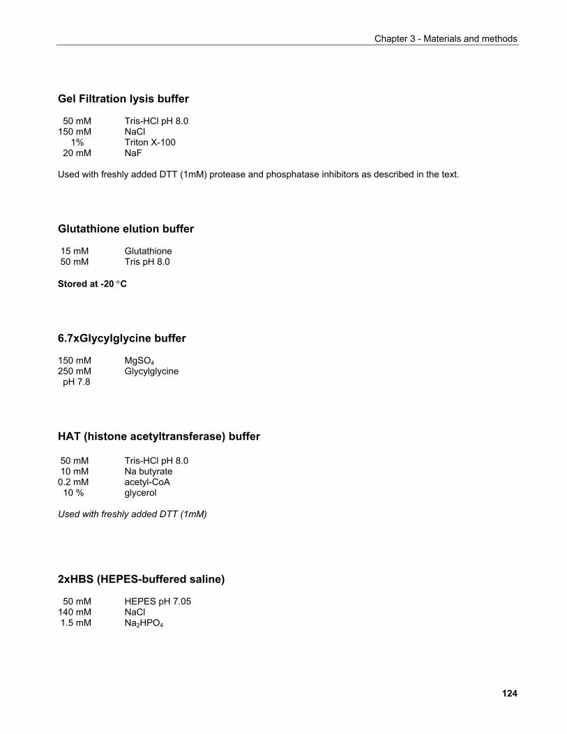
Chapter 3 - Materials and methods
Gel Filtration lysis buffer 50 mM Tris-HCl pH 8.0 150 mM NaCl 1% Triton X-100 20 mM NaF Used with freshly added DTT (1mM) protease and phosphatase inhibitors as described in the text. Glutathione elution buffer 15 mM Glutathione 50 mM Tris pH 8.0 Stored at -20 °C 6.7xGlycylglycine buffer 150 mM MgSO4 250 mM Glycylglycine pH 7.8 HAT (histone acetyltransferase) buffer 50 mM Tris-HCl pH 8.0 10 mM Na butyrate 0.2 mM acetyl-CoA 10 % glycerol Used with freshly added DTT (1mM) 2xHBS (HEPES-buffered saline) 50 mM HEPES pH 7.05 140 mM NaCl 1.5 mM Na2HPO4
124
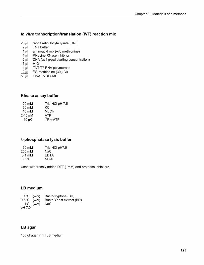
Chapter 3 - Materials and methods
In vitro transcription/translation (IVT) reaction mix 25 µl rabbit reticulocyte lysate (RRL) 2 µl TNT buffer 1 µl aminoacid mix (w/o methionine) 1 µl RNasine RNase inhibitor 2 µl DNA (at 1 µg/µl starting concentration) 16 µl H2O 1 µl TNT T7 RNA polymerase 2 µl 35S-methionine (30 µCi) 50 µl FINAL VOLUME Kinase assay buffer 20 mM Tris-HCl pH 7.5 50 mM KCl 10 mM MgCl2 2-10 µM ATP 10 µCi 32P-γ-ATP λ-phosphatase lysis buffer 50 mM Tris-HCl pH7.5 250 mM NaCl 0.1 mM EDTA 0.5 % NP-40 Used with freshly added DTT (1mM) and protease inhibitors LB medium 1 % (w/v) Bacto-tryptone (BD) 0.5 % (w/v) Bacto-Yeast extract (BD) 1% (w/v) NaCl pH 7.0 LB agar 15g of agar in 1 l LB medium
125
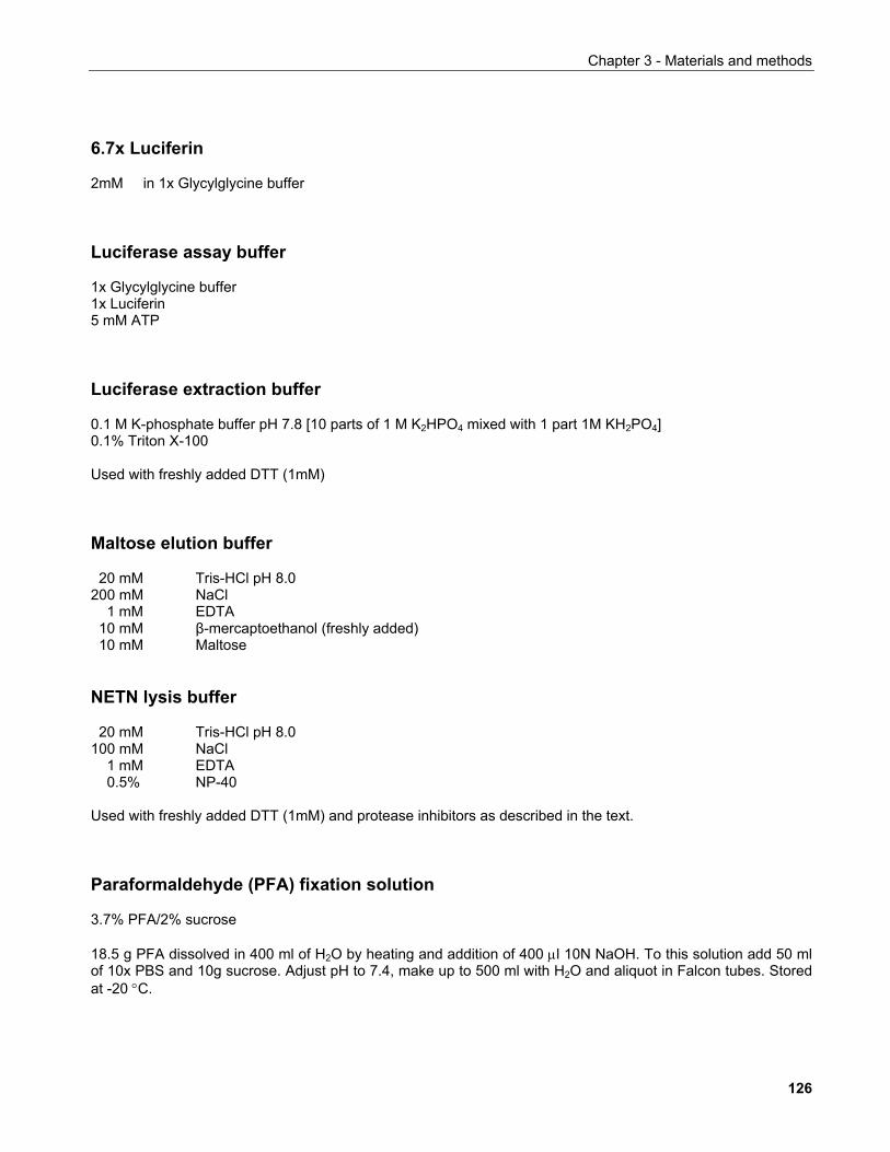
Chapter 3 - Materials and methods
6.7x Luciferin 2mM in 1x Glycylglycine buffer Luciferase assay buffer 1x Glycylglycine buffer 1x Luciferin 5 mM ATP Luciferase extraction buffer 0.1 M K-phosphate buffer pH 7.8 [10 parts of 1 M K2HPO4 mixed with 1 part 1M KH2PO4] 0.1% Triton X-100 Used with freshly added DTT (1mM) Maltose elution buffer 20 mM Tris-HCl pH 8.0 200 mM NaCl 1 mM EDTA 10 mM β-mercaptoethanol (freshly added) 10 mM Maltose NETN lysis buffer 20 mM Tris-HCl pH 8.0 100 mM NaCl 1 mM EDTA 0.5% NP-40 Used with freshly added DTT (1mM) and protease inhibitors as described in the text. Paraformaldehyde (PFA) fixation solution 3.7% PFA/2% sucrose 18.5 g PFA dissolved in 400 ml of H2O by heating and addition of 400 µl 10N NaOH. To this solution add 50 ml of 10x PBS and 10g sucrose. Adjust pH to 7.4, make up to 500 ml with H2O and aliquot in Falcon tubes. Stored at -20 °C.
126
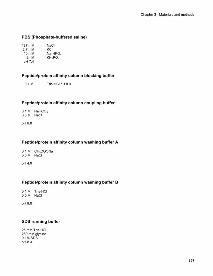
Chapter 3 - Materials and methods
PBS (Phosphate-buffered saline) 137 mM NaCl 2.7 mM KCl 10 mM Na2HPO4 2mM KH2PO4 pH 7.4 Peptide/protein affinity column blocking buffer 0.1 M Tris-HCl pH 8.0 Peptide/protein affinity column coupling buffer 0.1 M NaHCO3 0.5 M NaCl pH 8.0 Peptide/protein affinity column washing buffer A 0.1 M CH3COONa 0.5 M NaCl pH 4.0 Peptide/protein affinity column washing buffer B 0.1 M Tris-HCl 0.5 M NaCl pH 8.0 SDS running buffer 25 mM Tris-HCl 250 mM glycine 0.1% SDS pH 8.3
127
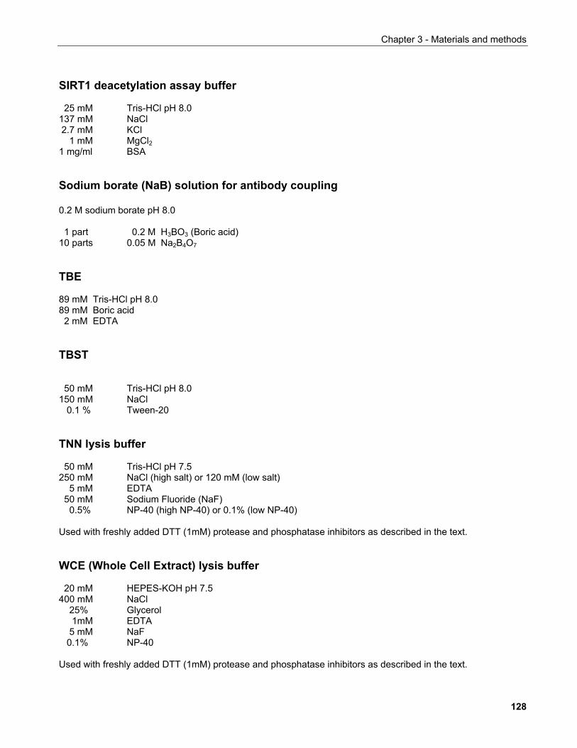
Chapter 3 - Materials and methods
SIRT1 deacetylation assay buffer 25 mM Tris-HCl pH 8.0 137 mM NaCl 2.7 mM KCl 1 mM MgCl2 1 mg/ml BSA Sodium borate (NaB) solution for antibody coupling 0.2 M sodium borate pH 8.0 1 part 0.2 M H3BO3 (Boric acid) 10 parts 0.05 M Na2B4O7 TBE 89 mM Tris-HCl pH 8.0 89 mM Boric acid 2 mM EDTA TBST 50 mM Tris-HCl pH 8.0 150 mM NaCl 0.1 % Tween-20 TNN lysis buffer 50 mM Tris-HCl pH 7.5 250 mM NaCl (high salt) or 120 mM (low salt) 5 mM EDTA 50 mM Sodium Fluoride (NaF) 0.5% NP-40 (high NP-40) or 0.1% (low NP-40) Used with freshly added DTT (1mM) protease and phosphatase inhibitors as described in the text. WCE (Whole Cell Extract) lysis buffer 20 mM HEPES-KOH pH 7.5 400 mM NaCl 25% Glycerol 1mM EDTA 5 mM NaF 0.1% NP-40 Used with freshly added DTT (1mM) protease and phosphatase inhibitors as described in the text.
128
Chapter 3 - Materials and methods
3.2 METHODS 3.2.1 BIOINFORMATICS RESOURCES Multiple sequence alignments: ClustalW 1.8 (Baylor College of Medicine)
http://searchlauncher.bcm.tmc.edu/multi-align/multi-align.html
Aligned sequence editing: BoxShade (Pasteur Institute)
http://bioweb.pasteur.fr/seqanal/interfaces/boxshade-simple.html
PEST motif identification: PESTFind (University of Vienna)
https://emb1.bcc.univie.ac.at/toolbox/pestfind/pestfind-analysis-webtool.htm
Phosphorylation site searches: Scansite Motif Scanner (MIT)
http://scansite.mit.edu/motifscanner/motifscan1.phtml?database=_SWS_
3.2.2 MOLECULAR BIOLOGICAL TECHNIQUES Construction of plasmids expressing SIRT1 and deletion/point mutants thereof
Two vectors containing the cDNA encoding for human SIRT1 and that of a catalytically
inactive mutant (Vaziri et al., 2001) were kindly provided by Prof. Robert Weinberg (Whitehead
Institute, MIT, USA). These vectors were named pYESir2 and pYESir2HY respectively and
were based on a pBabePuro retroviral expression vector backbone. The SIRT1 cDNA was
excised with BamHI/SnaBI and subcloned into pcDNA3 and pcDNA3-HA vectors in the
BamHI/EcoRV sites.
All SIRT1 point mutants were created by the two-step PCR method, using either the Pwo
polymerase kit (Roche), or GC-rich PCR system (Roche) for amplification of regions
corresponding to the N-terminus of the protein, according to manufacturer's protocols.
Mutations proximal to the N-terminus of SIRT1 were introduced to BamHI/PacI-digested
129
Chapter 3 - Materials and methods
pcDNA3-HA-SIRT1 plasmid while mutations proximal to the C-terminus were introduced to
PacI/XbaI-digested pcDNA3-HA-SIRT1 plasmid. The XbaI site is part of the 3' UTR of SIRT1
mRNA which was present in the original pYESir2 vector. Truncation mutants were cloned
following the same strategy. All mutations were confirmed by sequencing of the relevant
region. The mutated cDNAs as well as other SIRT1 fragments were subsequently subcloned
to various vectors as follows:
pcDNA3-based vectors: Into the BamHI/XbaI sites
pBabePuro vector: Into the BamHI/SnaBI sites (following 3' Klenow fill-in)
pAcGST vector: Into the BamHI/SmaI sites
pGEX-2TK vector: Into the BamH/EcoRI sites
pMAL-c2 vector: Into the BamHI/XbaI sites
Other plasmids used in this study were obtained as follows:
GST-Pin1: Gianni Del Sal, LNCIB, Trieste, Italy (Zacchi et al., 2002)
GST-p300(HAT): Richard Eckner, New Jersey Medical School, New Jersey, USA
Flag-β-TrCP: Giulio Draetta, European Inst. of Oncology, Milan (Busino et al., 2003)
Origin of SIRT1 knock-out mice Mice of the 129/SvJ strain containing a targeted deletion of the SIRT1 locus were provided
by Prof. F. Alt, Harvard University, Boston USA (Cheng et al., 2003).
Genotyping of SIRT1 knock-out mice and MEFs
Genotyping was performed by standard Taq polymerase PCR methods using the following
primers recommended by the provider of the mice:
130
Chapter 3 - Materials and methods
SIRT1SKO-F: 5'-CTTGCACTTCAAGGGACCAA-3'
SIRT1SKO-R1: 5'-GTATACCCACCACATCTGAG-3'
SIRT1SKO-R2: 5'-CTACCACTCCTGGCTACCAA-3'
All three primers were uses simultaneously giving a band of ~ 800 bp for the knockout allele
and a band of ~500 bp for the wildtype allele.
Specific PCR conditions were: 33 cycles of 94 °C, 40"; 55 °C 50"; and 72 °C 1.2'.
3.2.3 CELL CULTURE METHODS 3.2.3.1 Mammalian cell culture NB: When the term "standard medium" is used in the context of cell culture applications, the
following formulation should be assumed:
500 ml Dulbecco's MEM (Invitrogen, #10938-025) supplemented with 50 ml FCS
(final: 10%) 5ml of 200mM L-Glutamine (final: 2mM) and 5ml 10,000 U/ml
penicillin/10,000µg/ml streptomycin mix (final: 100U and 100µg respectively).
MEF medium: standard medium + 200 µM β-mercaptoethanol
Sf9 insect cell medium: Grace's insect medium (Invitrogen, #11605-045) + 10 % FCS
Other media formulations are indicated were appropriate
Transfection of mammalian cells with small interfering RNAs (siRNAs) At least 24 hours prior to transfection, cells were seeded in antibiotic-free standard medium
so that they were approx. 50% confluent on the day of transfection.
For a 10-cm tissue culture plate, 18 µl of 20 µM siRNA (40 nM final concentration unless
othewise indicated) were added to 892 µl OPTIMEM serum-free medium in an Eppendorf
131

Chapter 3 - Materials and methods
tube and mixed by pipetting. In a separate tube, 18 µl of Oligofectamine transfection reagent
were added to 72µl OPTIMEM and gently mixed by flicking the tube 1-2 times. The lipid
preparation was then allowed to sit at room temperature for 10' and it was then added to the
diluted siRNA mix. In the meantime, cells were washed at least 1x with standard medium
without antibiotics and without serum and 5 ml of the wash medium were added to the cells
prior to return to the incubator. Following a 20-25' incubation at room temperature, the
transfection mix was added to the cells in a dropwise fashion. 4-6 hours after transfection,
standard medium containing 30% FCS was added to the plate to a final FCS content of 10%
(typically 3 ml of this medium to the 6 ml of medium in plates). Cells were harvested at the
indicated times and typically 48-72 hours after transfection.
RNAi target sequences control non-silencing: 5'-AATTCTCCGAACGTGTCACGT-3'
Cdh1 (Donzelli et al., 2002): 5'-AATGAGAAGTCTCCCAGTCAG-3'
Cdc20 (Donzelli et al., 2002): 5'-AAACCTGGCGGTGACCGCTAT-3'
MPS1 (Stucke et al., 2002): 5'-AACCCAGAGGACTGGTTGAGT-3'
Bub1 (Johnson et al., 2004): 5'-AAATACCACAATGACCCAAGA-3'
BubR1 (Johnson et al., 2004): 5'-AACGGGCATTTGAATATGAAA-3'
SIRT1-1: 5'-AATTATCACTAATGGTTTTCA-3'
SIRT1-2 (Lin and Elledge, 2003): 5'-AACTGGAGCTGGGGTGTCTGT-3'
SIRT1-3*: 5'-CAAGCGATGTTTGATATTGAA-3'
(*designed using the HiPerfmormance Design Algorithm by Qiagen)
Calcium phosphate transfection of cultured cells For calcium phosphate transfection, cells were seeded 24h prior to transfection. 1-2 hr prior
to transfection, the medium was exchanged with 9ml of fresh standard medium.
132
Chapter 3 - Materials and methods
Typically 1-5 µg of plasmid DNA were combined with H2O to a final volume of 450 µl. 50 µl of
2.5M CaCl were added to the DNA mix followed by vortexing for 10'' then 500 µl of 2xBBS
buffer were added followed by 30'' vortexing. The tranfection mix was left at room
temperature for 20' then added to the cells in a drop-wise fashion while gently rocking the
plates. 16 hours post-transfection the cells were washed 3x with DMEM and supplemented
with 9 ml of standard medium. Cells were harvested for further work approx. 40-46 hr, post-
transfection.
Luciferase and β-galactosidase reporter assays 1.0x10⁵-1.5x10⁵ cells were seeded in 6-well plates at least 24 hours prior to transfection.
Transfected cells were lysed in 250 µl luciferase extraction buffer, harvested with a Teflon
cell scraper and transferred to an Eppendorf tube. Cellular debris was pelleted by
centifugation for 10-15' at 14,000 rpm (20,800xg) at 4 °C. Cleared supernatants were
transferred to white 96-well plates. 100 µl of cell lysate were used for luciferase activity assay
in a MicroLumat Plus LB96V (Berthold Technologies, Bad-Woldbad, Germany) luminometer.
Two injections, each 50 µl luciferase assay buffer were added per sample with a 1.6" delay
and 15" integration time.
In parallel, 20 µl of each lysate were transferred to wells in transparent 96-well plates and
170 µl β-galactosidase assay buffer were added to each sample. Reactions were incubated
in the dark at 37 °C until a yellow coloration started becoming visible and quantified by
spectophotometry at 420 nm prior to apparent reaction saturation.
All tranfections for luciferase assays were performed in triplicate, each luciferase reading was
normalised against the corresponding β-galactosidase value, data were averaged and
standard deviation was calculated using Microsoft Excel.
133
Chapter 3 - Materials and methods
In vivo 35S-labelling of proteins Cells at approx. 60-80% confluency were used for in vivo 35S-labelling of newly synthesised
proteins. Cells were washed twice with methionine-free medium (with L-glutamine and
penicillin/streptomycin as in standard medium) without FCS and supplemented with 4 ml of
the wash medium containing 10 % dialysed FCS. Cells were incubated for 15'-30' at 37 °C,
the medium was subsequently replaced with 3ml of methionine-free medium (incl.
penicillin/streptomycin, L-glutamine and 10 % dialysed FCS) supplemented with 100 µCi/ml
radioactive labelling mix (PROMIX, Amersham Biochiences). The plates were covered with
charcoal filters and incubated at 37 °C, 5% CO2 for 3 hours after which cells were harvested
for further manipulations.
Isolation of mouse embryonic fibroblasts (MEFs) SIRT1+/+, SIRT1+/- and SIRT1-/- MEFs were derived from E12.5 embryos from an intercross of
SIRT1+/- animals. Observation of a vaginal plug after overnight mating was considered as
E0.5. Pregnant mice were sacrificed by cervical dislocation, embryos were isolated and
placed in ice-cold PBS containing 2% FCS. The head and internal organs of the embryos
were removed and the head was used for genotyping. The remaining tissue was finely cut
with a scalpel and incubated for up to 15' in trypsin solution at 37 °C, 5% CO2. Trypsinised
tissue was further dissociated by pipetting through a 1ml pipette tip, transferred to a 10 cm
tissue culture plate and incubated at 37 °C, 5% CO2. After reaching confluency, each 10-cm
dish of MEFs was expanded into two 15-cm dishes. At confluency, MEFs were either
counted and frozen in 9:1 MEF medium/DMSO or expanded for further experiments.
134
Chapter 3 - Materials and methods
Retroviral infection of mammalian cells Retroviruses were created by transfecting the appropriate recombinant retroviral transfer
vector pBabePuro, that carries the puromycin selectable marker into the BOSC23 or Phoenix
"packaging" cell lines.
In detail, one day prior to trasfection, the packaging cells were seeded so that at the day of
transfection they were 40-60% confluent. 1-2 hr prior to transfection, the medium was
exchanged with 9ml of fresh medium.
For the transfection, 20 µg of recombinant retroviral transfer vector containing the cDNA of
interest were diluted with H2O to a final volume of 440 µl. After addition of 62 µl of 2M CaCl2
the mixture was vortexed for 10'' followed by addition of 500 µl of 2xHBS. The transfection
mix was vortexed for 30'' and let to stand at room temperature for 20'. The transfection mix
(1ml in 9ml of medium per 10 cm dish) was added dropwise to the packaging cell line and
cells were incubated for approx. 16 hours (O/N) at 37°C, 5% CO2. Cells were subsequently
washed 3x with DMEM and supplemented with 9ml of standard medium. On the same day,
the cells to be infected were seeded so that they were approx. 50-60% confluent on the day
of infection. Approx. 40-46 hr, post-transfection, the medium of the packaging cells was
harvested with a sterile syringe and filtered through a 0.45 µm syringe filter into a 15ml
Falcon tube. This viral supernatant was supplemented with polybrene from a stock of 8
mg/ml to a final concentration of 4 µg/ml and added to the cells to be infected after removal
of the old medium. Infection was repeated twice and 24 hr after the last infection, selection of
the infected cells was initiated by replacing the medium with standard medium containing 2
µg/ml puromycin. Based on inspection of puromycin resistant colonies, typically >200-300
colonies comprised each pool (uncloned cell mass).
135
Chapter 3 - Materials and methods
3.2.3.2 Insect cell culture Production of recombinant baculoviruses
2x10⁶ Sf9 cells were seeded in 6-cm dishes and were left to attach for approx. 20' at room
temperature in a total medium volume of 4 ml. In the meantime, 0.5µg of BaculoGold® DNA
(in 5 µl) were combined with 2 µg of recombinant baculovirus transfer vector, mixed gently by
pipetting and let stand for 5'. Following that, 1ml of Transfection Buffer B was added to the
DNA mix. The medium of Sf9 was then aspirated and cells were overlayed with 1ml
Transfection Buffer A. The 1ml of Transfection Buffer B/DNA was then added dropwise to the
cells, and the plates were incubated for 4 h at 27 °C. The transfection mix was then
aspirated, cells were washed 2x with 4 ml and 4 ml of medium was applied to each plate.
The 6-cm dishes were then placed in 15-cm dishes along with Whattman 3M paper pre-wet
with H2O to minimise culture mediun evaporation and incubated in ambient atmosphere, at
27 °C for 5 days. Supernatants were then harvested, cell debris was removed by
centrifugation (1000xg, 2' at RT), the supernatant was filtered through a 0.45 µm syringe filter
and used to infect 2x10⁶ Sf9 cells in 6-cm dishes in 3 ml of Grace's insect medium/10% FCS.
At least 72 h post-infection, the resulting viral supernatant (1st amplification) was harvested
as above and 1 ml of it was used to infect 2x10⁷ Sf9 cells in T75, screw-cap tissue culture
flasks with a filter lid in a medium volume of 30-40 ml. At least 72 h post-infection, the viral
supernatant (2nd amplification) was harvested as above and stored in the dark at 4 °C. This
was used as the main viral stock and further viral amplifications were initiated from this.
Production of recombinant proteins in Sf9 insect cells Logarithmically growing Sf9 cells were dislocated from the dish, counted and re-plated at
5x10⁶ cells per 10 cm plate. Cells were left to attach to the plate and the medium was
replaced with 5 ml fresh medium (Grace's insect medium/10% FCS). 0.2-1.0 ml of
recombinant baculovirus stock was used for infection. Cells were incubated at 27 °C and
harvested 48 hours after infection.
136
Chapter 3 - Materials and methods
3.2.3.3 Bacterial culture - Production of recombinant proteins in E. coli The appropriate plasmid encoding the protein of interest was transformed into E. coli by the
heat-shock method. Transformants were grown O/N at 37 °C. A single colony was then used
to inoculate 10 ml LB medium (+antibiotic) and the culture was grown shaking (230 rpm) O/N.
The following day, the culture was diluted 1:10 with fresh pre-warmed LB medium
(+antibiotic) and incubated for 1.5 h as above. Protein expression was induced with 0.1mM
IPTG for 3-4 hours and cells were harvested by centrifugation prior to further processing.
3.2.4 BIOCHEMICAL TECHNIQUES
Purification of GST-fusion proteins produced in Sf9 insect cells
Cells were lysed in 1ml TNN lysis buffer per cell pellet derived from a 10-cm culture plate
(initial seeding density: 5x10⁶ cells infected for ~48 hours) for 30' on ice. Cell debris was
removed by centrifugation (20', 12000xg, 4 °C). Glutathione bead slurry was added to the
cleared supernatant and the mixture was incubated for 1 h in a rotating wheel at 4 °C. The
beads were subsequently pelleted by brief centrifugation up to maximum speed at 4 °C and
similarly washed 4x with lysis buffer. The beads were subsequently washed 1x with 50 mM
Tris-HCl pH 8.0 and the purified proteins were either used for assays as described in the
corresponding sections or eluted 2x with glutathione elution buffer for 10-20' at room
temperature. The eluted material was supplemented with glycerol to a final concentration of
20% and stored at -80 °C. All steps were performed at 4 °C.
137
Chapter 3 - Materials and methods
Purification of GST-fusion proteins produced in E. coli All steps were performed exactly as above with the exception that bacterial cells were lysed
in NETN buffer and sonicated (3x10" at maximum amplitude) on ice.
Purification of MAL-fusion proteins produced in E. coli
All steps were performed as described for bacterially-expressed GST-fusion proteins with the
exception that amylose resin (NEB, E8021-S) was used for the purification of the
recombinant proteins and following washing with the lysis buffer the bead-bound material
was washed 1x with maltose elution buffer (without maltose) and eluted 2x with maltose
elution buffer for 10-20' at room temperature.
Gel filtration chromatography
A frozen pellet of 5x10⁹ HeLa cells was lysed in 10 ml gel filtration lysis buffer, dounce
homogenised (15 strokes) with a type B tight-fitting pestle and left for 30' on ice. The lysate
was pre-cleared by centrifugation for 30', at 3000xg, 4 °C and filtered through a 0.45 µm low-
protein binding syringe filter prior to gel filtration. Gel filtration was performed using a
Superose 6, XK26/70 preparative column at a flow rate of 0.5 ml/min in gel filtration buffer. 50
fractions of 4 ml each were collected in total and 50 µl of each fraction were used for SDS-
PAGE analysis.
In vitro kinase assays Recombinant GST or GST-SIRT fragment fusion proteins were expressed in E. coli and
purified using glutathione beads as described. Recombinant CK2 was purchased from Cell
Signalling. 500 units of CK2 were diluted in kinase assay buffer (10 µM ATP) containing ~4
138
Chapter 3 - Materials and methods
µg recombinant protein substrate to a final volume of 30 µl. Reactions were incubated for 30'
at 30°C, stopped by addition of Laemmli buffer and boiling followed by analysis on an SDS-
PAGE gel. Gels were dried and exposed for autoradiography.
GST-CyclinB1/His-Cdk2 were co-expressed in Sf9 cells and purified by glutathione affinity
chromatography. Assay conditions were as for CK2.
BubR1 was immunoprecipitated from cell extracts. Assay conditions were as for CK2.
In vitro SIRT1 deacetylase activity assay using a fluorogenic peptide
For in vitro determination of SIRT1 activity, SIRT1 preparation (immunoprecipitated or
recombinant expressed in Sf9 cells as a GST-fusion) was incubated with 100 µM Fluor de
Lys-SIRT1 acetylated peptide substrate (corresponding to residues 379-382 of p53) and 0.5
mM NAD+ in SIRT1 deacetylation assay buffer according to manufacturer's protocol
(BIOMOL HDAC fluorescent activity assay/drug discovery kit - AK-500). Final reaction
volume was 50 µl. Reactions were incubated for 30' at 37 °C and stopped by addition of 1x
volume of Fluor de Lys Developer II incubated for an additional 45' at 37 °C in the dark and
fluorescence was measured (excitation: 360 nm, emission: 460 nm, cutoff: 455 nm) with a
SPECTRAmax GEMINI 96-well plate fluorometer (Molecular Devices).
In vitro SIRT1 deacetylase activity assay using autoacetylated GST-p300(HAT)
Prior to use in the deacetylation assay, GST-p300(HAT) was expressed in E. coli, purified
using glutathione beads, washed 3x in NETN buffer, washed 2x in HAT buffer (w/o acetyl-
CoA) and the beads were resuspended in HAT buffer with acetyl-CoA. The reaction was left
to proceed for 30' at 30 °C. The final reaction volume was 50 µl. This autoacetylation reaction
can also be omitted with equally good results as GST-p300(HAT) shows significant
autoacetylation when expressed in bacteria and can thus be readily used as a substrate for
139
Chapter 3 - Materials and methods
the deacetylation reaction. Recombinant GST-SIRT1 or GST-SIRT1(H363Y) expressed in
Sf9 cells was purified using glutathione beads and eluted as described. Following
autoacetylation, the GST-p300(HAT)-bound beads were washed 3x in TNN buffer, 2x in
SIRT1 deacetylation assay buffer, the beads were equally divided to Eppendorf tubes for the
various reactions and resuspended in SIRT1 deacetylation assay buffer supplemented with
0.5 mM NAD+ and, where indicated, 0.5 mM nicotinamide or 10 mM Na butyrate. 5 µl of
SIRT1 enzyme preparation corresponding to ~2 µg of purified protein were used per reaction.
The final reaction volume was 50 µl. Reactions were incubated for 1h at 37 °C, stopped with
addition of Laemmli buffer, boiled and analysed by SDS-PAGE.
In vitro transcription/translation In vitro transcription/translation was performed with the Promega TNT® coupled reticulocyte
lysate system using T7 RNA polymerase. Typically, 2 µg of DNA midiprep were incubated
with IVT reaction mix for 90' and used directly or stored at -20 °C.
In vitro binding assays The appropriate fusion protein (bait) was purified as detailed above, the beads were washed
3x with 1 ml lysis buffer without additives, and resuspended in 1 ml lysis buffer with additives.
1-5 µl of IVT were added to the bead suspension and the tubes were incubated in a head-to-
head rotor for 1-3 hr. The beads were subsequently washed 4x with 1 ml lysis buffer without
additives and bound material was released by addition of Laemmli buffer and boiling for 5'
prior to loading on a gel.
140
Chapter 3 - Materials and methods
Preparation of cell-free apoptotic extracts from 293 cells Extracts were prepared as described in (Liu et al., 1996). In detail, 293 cells were grown to 90%
confluency, washed 1x with ice-cold PBS and scrapped with a teflon cell scrapper in a Falcon
tube and resuspended in 5x volumes of ice-cold apoptotic extract buffer A. The lysates were
allowed to stand on ice for 15' then homogenised with a tight-fitting type B pestle in a Dounce
homogeniser followed by centrifugation at 1000xg for 10' at 4 °C. The resulting supernatant
was then centrifuged at 105xg (23,700 rpm) for 1h at 4 °C in a Beckman SW40 rotor. The
supernatant (S-100 fraction) was aliquoted, snap-frozen in liquid N2 and stored at -80 °C until
further use. Total protein concentration in these extracts was at 5 µg/µl.
In vitro caspase cleavage assay
For in vitro cleavage assays, S-100 extracts were thawed on ice, dATP was added to a final
concentration of 1 mM where applicable and the extract was incubated at 30°C for 1h. 10 µl
of extract corresponding to 5 µg total protein were added to 1-2 µl of in vitro translated
material and incubated for the time indicated in the text. Reactions were stopped by the
addition of SDS sample buffer and analysed by SDS-PAGE.
Tryptic digestion of GST-SIRT1 for phosphorylation analysis GST-SIRT1 was expressed in Sf9 cells and purified as described, but not eluted from the
beads. Following washes with lysis buffer, the sample was divided into two equal parts and
either mock-treated, or treated with λ-PPase as described. The bead-bound material was
then washed with lysis buffer to remove λ-PPase followed by 2x washes with 10 mM Tris pH
8.1. The beads were subsequently resuspended in 50 µl of 10 mM Tris pH 8.1 and
supplemented with CaCl2 to a final concentration of 2mM and sequencing-grade trypsin
(Promega) at a ratio of 1:20 (w/w) trypsin:GST-SIRT1 (this corresponded to 1 µg trypsin).
The samples were incubated at 37°C O/N, the supernatants were harvested and stored at
-80°C until analysed by MALDI-MS.
141
Chapter 3 - Materials and methods
3.2.5 IMMUNOLOGICAL TECHNIQUES Paraformaldehyde fixation/Triton X-100 permeabilisation
Cells were seeded at least 24 and usually 48 hours prior to treatment/fixation on 18x18 mm
#1 coverlips placed in the wells of a 6-well plate. The medium was aspirated and 2 ml PFA
fixation solution freshly thawed and pre-warmed to 37 °C was added to each well. The plate
was incubated at 37 °C for 10'. Cells were then washed 2x with PBS and permeabilised with
0.02% Triton X-100 in PBS for 10' at RT. Cells were washed 2x with PBS and proccessed for
immunostaining as described below.
Methanol fixation/Acetone permeabilisation
Cells were seeded on coverslips as above, the medium was aspirated and cells were directly
fixed with methanol (-20 °C) then permeabilised in an acetone bath (-20 °C). Coverslips were
subsequently washed 1x in a PBS bath at RT and processed for immunostaining as
described below.
Cells were seeded at least 24 and usually 48 hours prior to treatment/fixation on 18x18 mm
#1 coverlips placed in the wells of a 6-well plate.
Immunostaining of cultured cells
Fixed/permeabilised cells were incubated with primary antibody mix (1-5 µg/ml of antibody in
PBS/1% BSA/1% goat serum) for 1-2h at RT or O/N at 4 °C. Cells were washed 3x with PBS
at RT and subsequently incubated with secondary antibodies coupled to the appropriate
fluorescent dye (diluted in PBS/1% BSA/1% goat serum) for 1h at RT. In parallel DNA was
counterstained with 1 µg/ml DAPI (1,4,6-diamidino-2-phenylindole). Cells were washed 3x
and inverted over Vectashield medium on top of a microscope glass. Coverslips were sealed
using nail varnish and either observed directly , or were stored in the dark at 4 °C until
observation.
142
Chapter 3 - Materials and methods
Preparation of peptide affinity columns for antibody purification
For antibodies raised against a peptide, the same antigenic peptide was used for affinity
purification. In detail: Activated CH sepharose 4B (Amersham #17-0490-01) was
resuspended in 20 ml of 1 mM HCl and washed for 15' through a sintered glass filter with
300-400 ml of the same solution. Washed sepharose was transferred to a 15 ml Falcon tube
and washed 1x with peptide coupling buffer. Peptide was dissolved in coupling buffer in a
volume equivalent to 2x volumes of swollen sepharose and was added to the sepharose
bead slurry [20mg peptide per gram of sepharose (dry weight; 1 g sepharose gives ~3ml
slurry following swelling)]. The suspension was incubated on a tilting shaker for 2 h at RT.
The beads were subsequently washed 2x with peptide coupling buffer and unreacted
moieties on the beads were blocked with blocking buffer for 1 h at RT. The beads were then
washed for 3 consecutive cycles of alternating wash buffer A and B. Finally, the beads were
washed with PBS and stored in PBS+0.05% NaN3 at 4 °C.
Preparation of protein affinity columns for antibody purification
Preparation of protein affinity columns was essentially as described for peptide columns with
the following modifications:
CNBr-activated sepharose 4B (Amersham #17-0430-01) was used; 1 mg of protein was
coupled per gram of sepharose; 1 g sepharose gives ~3.5 ml slurry following swelling.
The relevant antigenic region was expressed as a fusion protein in bacteria and purified as
described. Prior to coupling, the purified protein was dialysed against coupling buffer O/N at
4°C.
143
Chapter 3 - Materials and methods
Affinity purification of rabbit polyclonal antibodies α-SIRT1 Ctp was purified through a peptide affinity column using the same antigenic peptide.
α-SIRT1 GST-S1 antibody was purified through a MAL-SIRT1(575-676) column. Affinity
purification was performed identically for both antibodies.
Affinity columns were prepared as described. The storage buffer was allowed to flow through
and the column was washed with 3 volumes of binding buffer (PBS+0.2% Tween-20).
Typically, 3 ml of rabbit serum were diluted to 10 ml total volume with binding buffer
(PBS+0.2% Tween-20) and applied to the column. Bound material was washed until the
OD280 was equal to ODPBS+0.2% Tween-20 (usually approx. 3 column volumes). Elution was
accomplished using 8-10 ml of 0.2M Glycine pH 2.2. Fractions (7-9) of 750 µl were collected
directly into Eppendorf tubes containing 250 µl 1M Tris pH 8.0. The OD280 of all fractions was
measured using (PBS+0.2% Tween-20)/Tris pH 8.0 (3:1) for the first 2 fractions and 0.2M
Glycine pH 2.2/Tris pH 8.0 (3:1) for the following fractions. Fractions with the highest OD280
were poolled, loaded on a pre-wet dialysis bag (MWCO 6,000-8,000, Spectra/Por) and
dialysed O/N in PBS+30% glycerol. The dialysate was collected, OD280 was determined
using the dialysis buffer as blank and purified antibody concentration was determined
according to the formula:
[antibody µg/µl]= OD280 x ε
where ε= 0.699 and is the IgG extinction coefficient.
The purified antibody solution was supplemented with BSA at a final concentration of 1
µg/ml, aliquoted and stored at -80 °C until further use. Antibody working solutions were
typically stored at 4 °C where they were stable for at least 3 months.
144
Chapter 3 - Materials and methods
Immunoprecipitation
Cells were rinsed with ice-cold PBS and either lysed directly in 1ml TNN lysis buffer per
10cm dish, and hervested with a teflon cell scraper in a 1.5ml Eppendorf tube. Cells were left
on ice for 30' and debris and unlysed material was pelleted by centifugation for 20-30' at
14,000 rpm (20,800xg) at 4 °C. The cleared supernatant was transferred to a new Eppendorf
tube containing 35 µl of a 50% Protein-A sepharose bead slurry, or a 1:1 mixture of Protein-
A:ProteinG beads where appicable and pre-cleared for 1 h on a head-to-head rotator at 4°C.
The beads were subsequently pelleted for 10' at 14,000 rpm (20,800xg) at 4 °C, the
supernatant was transferred to a new Eppendorf tube and supplemented with the appropriate
antibody. Immunocomplexes were precipitated with the addition of 35 µl of beads as used for
the preclearing step, for 1 h on a head-to-head rotator at 4°C. Beads were washed 4x in 1 ml
lysis buffer without supplements by successive pelleting of the beads and following the final
wash, residual buffer was aspirated using a hypodermic needle, the beads were
resuspended in 2x Laemmli buffer, boiled and analysed by SDS-PAGE.
Alternatively, cells were rinsed with ice-cold PBS and collected with a teflon cell scraper in 1
ml PBS in an Eppendorf tube, centrifuged for 3' at 3,000 rpm (1000xg) at 4 °C, the
supernatant was aspirated, cell pellets were snap-frozen in liquid nitrogen and stored at -
80°C until use.
Antibody coupling to sepharose beads
Antibody diluted in PBS was bound to protein-A sepharose CL-4B beads (Amersham, #17-
0780-01) in a rotating wheel for 1 h at 4 °C. Approx. 2 mg antibody were bound per ml of wet
beads. The beads were washed 2x with 10 volumes of 0.2 M NaB (pH 9.0) and resuspended
in 10 volumes of the same solution. Solid dimethylpimelimidate (DMP) was added to the
suspension to a final concentration of 20 mM while ensuring that the pH of the suspension
was ≥8.3. The coupling reaction was allowed to proceed for 30 min in a tilting shaker and
145
Chapter 3 - Materials and methods
was stopped by washing the beads 1x with 0.2 M ethanolamine followed by a 2 h incubation
in the same solution with gentle mixing at RT. The beads were finally washed 2x with PBS
and stored in PBS+0.05% NaN3 at 4 °C. Coupling efficiency was assessed by analysing
equivalent volumes of bead suspension before and after coupling by SDS-PAGE.
Preparative large-scale immunoprecipitation for mass spectrometry
A frozen pellet of 5x10⁹ HeLa cells was lysed in 160 ml TNN buffer, dounce homogenised (10
strokes) with a type B tight-fitting pestle and left for 30' on ice. The lysate was cleared by
centrifugation for 30', at 18000xg, 4 °C using an SS34 rotor, followed by centrifugation for
30', 100000xg at 4°C using an SW37 rotor. Rabbit IgG covalently coupled to protein-A
sepharose was used to pre-clear the lysate for 1h at 4°C, then the beads were removed by
centrifugation (5', 3000xg) and the bound material (unspecific, control) was eluted by addition
of 300 µl 0.2M glycine pH2.2. the eluate was neutralised with 100 µl of 1M K2HPO4. The
supernatant was incubated for 3h with α-SIRT1 Ctp antibody covalently coupled to
sepharose beads. The beads were subsequently washed 4x with lysis buffer and bound
material was eluted as above. Both eluates were concentrated using Centricon columns
(Millipore) with a M.W.C.O. (molecular weight cut-off) of 10,000. Laemmli buffer was added
to the samples to a 1x final dilution and samples were analysed by SDS-PAGE in a 6-15%
gradient gel. Gels were subsequently stained using colloidal blue solution and relevant bands
were excised with a surgical scalpel. Following in-gel trypsinisation, proteins were identified
as described (Gstaiger et al., 2003).
146
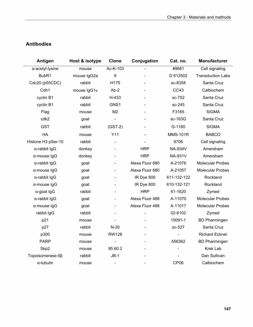
Chapter 3 - Materials and methods
Antibodies
Antigen Host & isotype Clone Conjugation Cat. no. Manufacturer α-acetyl-lysine mouse Ac-K-103 - #9681 Cell signaling
BubR1 mouse IgG2a 9 - D 612502 Transduction Labs
Cdc20 (p55CDC) rabbit H175 - sc-8358 Santa Cruz
Cdh1 mouse IgG1κ Ab-2 - CC43 Calbiochem
cyclin B1 rabbit H-433 - sc-752 Santa Cruz
cyclin B1 rabbit GNS1 - sc-245 Santa Cruz
Flag mouse M2 - F3165 SIGMA
cdk2 goat - - sc-163G Santa Cruz
GST rabbit (GST-2) - G-1160 SIGMA
HA mouse Y11 - MMS-101R BABCO
Histone H3 pSer-10 rabbit - - 9706 Cell signaling
α-rabbit IgG donkey - HRP NA-934V Amersham
α-mouse IgG donkey - HRP NA-931V Amersham
α-rabbit IgG goat - Alexa Fluor 680 A-21076 Molecular Probes
α-mouse IgG goat - Alexa Fluor 680 A-21057 Molecular Probes
α-rabbit IgG goat - IR Dye 800 611-132-122 Rockland
α-mouse IgG goat - IR Dye 800 610-132-121 Rockland
α-goat IgG rabbit - HRP 61-1620 Zymed
α-rabbit IgG goat - Alexa Fluor 488 A-11070 Molecular Probes
α-mouse IgG goat - Alexa Fluor 488 A-11017 Molecular Probes
rabbit IgG rabbit - - 02-6102 Zymed
p21 mouse - - 15091-1 BD Pharmingen
p27 rabbit N-20 - sc-527 Santa Cruz
p300 mouse RW128 - - Richard Eckner
PARP mouse - - 556362 BD Pharmingen
Skp2 mouse 95.60.2 - - Krek Lab
Topoisomerase-IIβ rabbit JB-1 - - Dan Sullivan
α-tubulin mouse - - CP06 Calbiochem
147
Chapter 4 - Results: Function and regulation of SIRT1
148
CHAPTER 4
RESULTS: FUNCTION AND REGULATION OF SIRT1 4.1 REGULATION OF SIRT1 BY PHOSPHORYLATION 4.1.1 Production of α-SIRT1 polyclonal and monoclonal antibodies
Since at the time this project was initiated no commercial antibodies against
mammalian sirtuins were available, in order to perform functional analyses of SIRT1, we
developed two polyclonal antibodies raised in rabbits. One was raised against a peptide
corresponding to the last 21 aminoacids of the C-terminus of SIRT1. The other was raised
against an N-terminal fusion of GST to aminoacids 575-676 of SIRT1 expressed in E. coli.
These antibodies will henceforth be referred to as Ctp and GST-S1 respectively. The
latter fragment was chosen based on sufficient expression levels of the corresponding
recombinant protein after a small-scale screen of GST-fusions of SIRT1 fragments
spanning the N- and C-terminal regions of the protein. Given the enhanced mobility of
protein domains known to harbour catalytic acivity due e.g. to induced-fit effects upon
substrate binding, the sirtuin core domain was deemed potentially unsuitable for raising
an antibody against it. This is because in experimental conditions where the protein's
tertiary structure is conserved such as immunofluorescence microscopy studies or
immunoprecipitation, such antibodies may exhibit weak affinities for the antigen.
Two rabbits were immunised with each of the antigens and the sera were tested for
immunoreactivity to HA-tagged SIRT1 overexpressed in 293 cells. Based on their ability to
efficiently recognise the antigen, the serum from one rabbit typically 3-5 months after
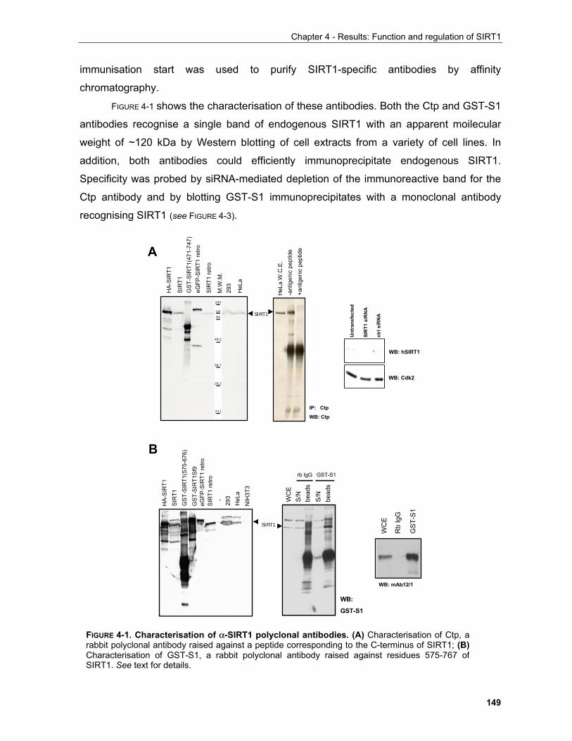
Chapter 4 - Results: Function and regulation of SIRT1
149
immunisation start was used to purify SIRT1-specific antibodies by affinity
chromatography.
FIGURE 4-1 shows the characterisation of these antibodies. Both the Ctp and GST-S1
antibodies recognise a single band of endogenous SIRT1 with an apparent moilecular
weight of ~120 kDa by Western blotting of cell extracts from a variety of cell lines. In
addition, both antibodies could efficiently immunoprecipitate endogenous SIRT1.
Specificity was probed by siRNA-mediated depletion of the immunoreactive band for the
Ctp antibody and by blotting GST-S1 immunoprecipitates with a monoclonal antibody
recognising SIRT1 (see FIGURE 4-3).
SIRT1
A
B
HA
-SIR
T1
SIR
T1
GS
T-S
IRT1
(575
-676
)
eGFP
-SIR
T1 re
troS
IRT1
retro
293
HeL
a
-
NIH
3T3
GS
T-S
IRT1
Sf9
203
11593
48.2
34.7
28.2
21.1
HA
-SIR
T1
SIR
T1G
ST-
SIR
T1(4
71-7
47)
eGFP
-SIR
T1 re
tro
SIR
T1 re
tro
293
HeL
a
M.W
.M.
SIRT1
HeL
aW
.C.E
.-a
ntig
enic
pep
tide
IP: Ctp
WB: Ctp
+ant
igen
ic p
eptid
eS
/Nbe
ads
WC
E
rb IgG
S/N
bead
s
GST-S1
WB:
GST-S1
WB: mAb12/1
Rb
IgG
WC
E
GS
T-S
1
WB: hSIRT1
WB: Cdk2
Unt
rans
fect
ed
SIR
T1 s
iRN
A
ctrl
siR
NA
FIGURE 4-1. Characterisation of α-SIRT1 polyclonal antibodies. (A) Characterisation of Ctp, arabbit polyclonal antibody raised against a peptide corresponding to the C-terminus of SIRT1; (B)Characterisation of GST-S1, a rabbit polyclonal antibody raised against residues 575-767 ofSIRT1. See text for details.
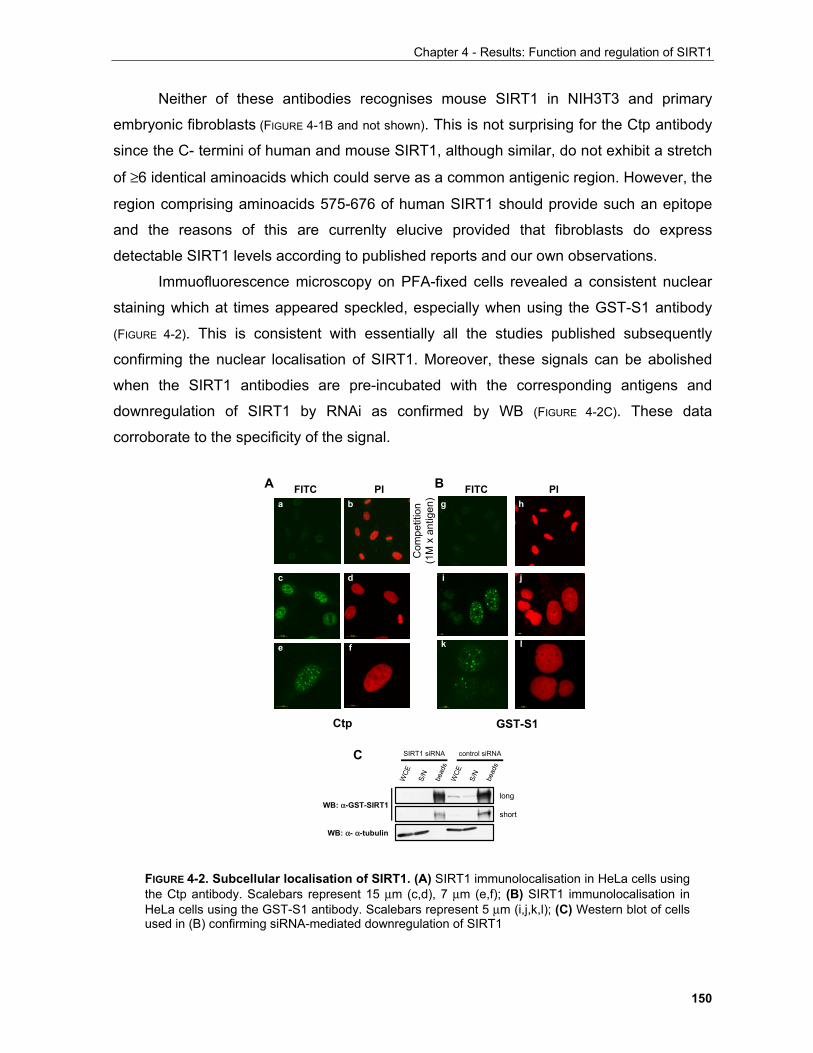
Chapter 4 - Results: Function and regulation of SIRT1
150
Neither of these antibodies recognises mouse SIRT1 in NIH3T3 and primary
embryonic fibroblasts (FIGURE 4-1B and not shown). This is not surprising for the Ctp antibody
since the C- termini of human and mouse SIRT1, although similar, do not exhibit a stretch
of ≥6 identical aminoacids which could serve as a common antigenic region. However, the
region comprising aminoacids 575-676 of human SIRT1 should provide such an epitope
and the reasons of this are currenlty elucive provided that fibroblasts do express
detectable SIRT1 levels according to published reports and our own observations.
Immuofluorescence microscopy on PFA-fixed cells revealed a consistent nuclear
staining which at times appeared speckled, especially when using the GST-S1 antibody
(FIGURE 4-2). This is consistent with essentially all the studies published subsequently
confirming the nuclear localisation of SIRT1. Moreover, these signals can be abolished
when the SIRT1 antibodies are pre-incubated with the corresponding antigens and
downregulation of SIRT1 by RNAi as confirmed by WB (FIGURE 4-2C). These data
corroborate to the specificity of the signal.
FITC PI FITC PI
Com
petit
ion
(1M
x a
ntig
en)
WB: α- α-tubulin
WB: α-GST-SIRT1
S/N
bead
s
WCE
SIRT1 siRNA control siRNA
S/N
bead
s
WCE
short
long
A B
C
GST-S1Ctp
a b
c d
e f
g h
i j
k l
FIGURE 4-2. Subcellular localisation of SIRT1. (A) SIRT1 immunolocalisation in HeLa cells usingthe Ctp antibody. Scalebars represent 15 µm (c,d), 7 µm (e,f); (B) SIRT1 immunolocalisation inHeLa cells using the GST-S1 antibody. Scalebars represent 5 µm (i,j,k,l); (C) Western blot of cellsused in (B) confirming siRNA-mediated downregulation of SIRT1
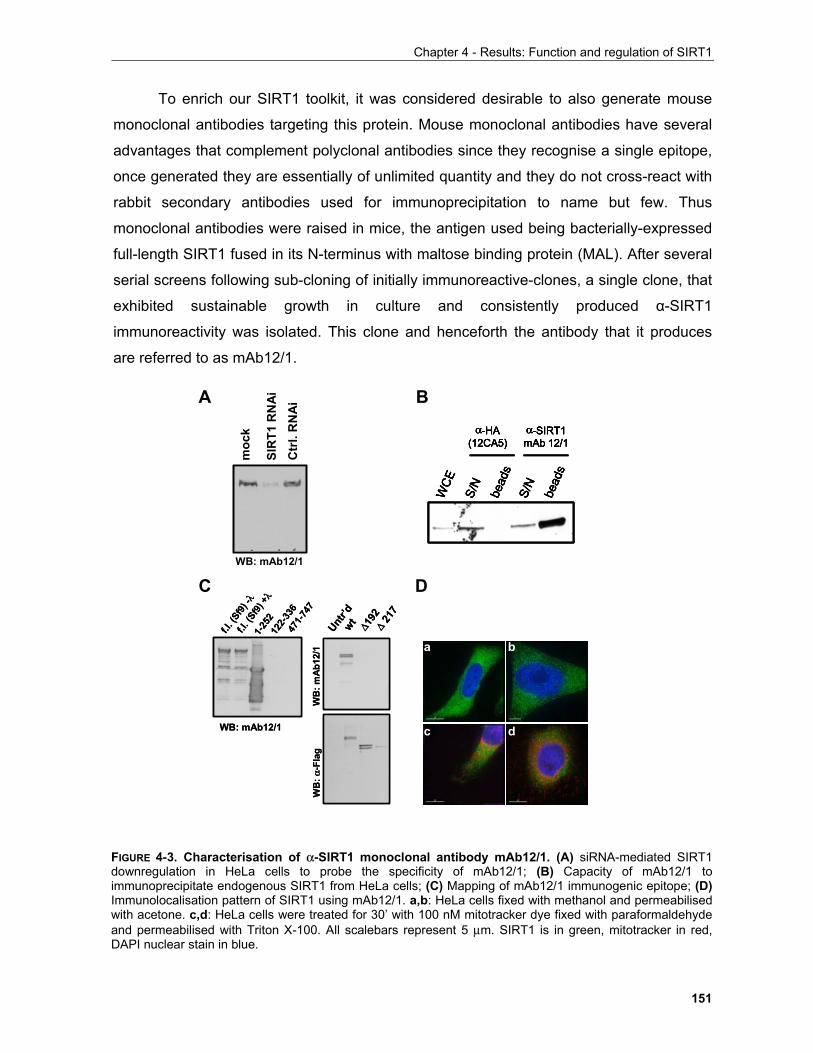
Chapter 4 - Results: Function and regulation of SIRT1
151
To enrich our SIRT1 toolkit, it was considered desirable to also generate mouse
monoclonal antibodies targeting this protein. Mouse monoclonal antibodies have several
advantages that complement polyclonal antibodies since they recognise a single epitope,
once generated they are essentially of unlimited quantity and they do not cross-react with
rabbit secondary antibodies used for immunoprecipitation to name but few. Thus
monoclonal antibodies were raised in mice, the antigen used being bacterially-expressed
full-length SIRT1 fused in its N-terminus with maltose binding protein (MAL). After several
serial screens following sub-cloning of initially immunoreactive-clones, a single clone, that
exhibited sustainable growth in culture and consistently produced α-SIRT1
immunoreactivity was isolated. This clone and henceforth the antibody that it produces
are referred to as mAb12/1.
S/N
bead
sS/
Nbe
ads
WCE
α-HA (12CA5)
α-SIRT1 mAb 12/1
S/N
bead
sS/
Nbe
ads
WCE
α-HA (12CA5)
α-SIRT1 mAb 12/1
S/N
bead
sS/
Nbe
ads
WCE
α-HA (12CA5)
α-SIRT1 mAb 12/1
S/N
bead
sS/
Nbe
ads
WCE
α-HA (12CA5)
α-SIRT1 mAb 12/1
WB: mAb12/1
moc
k
SIR
T1 R
NA
iC
trl.
RN
AiA B
C D
1-252
122-
336
471-
747
f.l. (
Sf9)
-λf.l.
(Sf9
) +λ
WB: mAb12/1
1-252
122-
336
471-
747
f.l. (
Sf9)
-λf.l.
(Sf9
) +λ
WB: mAb12/1
∆192
∆21
7
wt
Untr’d
WB
: mA
b12/
1W
B: α
-Fla
g
∆192
∆21
7
wt
Untr’d
WB
: mA
b12/
1W
B: α
-Fla
g
a b
c d
FIGURE 4-3. Characterisation of α-SIRT1 monoclonal antibody mAb12/1. (A) siRNA-mediated SIRT1downregulation in HeLa cells to probe the specificity of mAb12/1; (B) Capacity of mAb12/1 toimmunoprecipitate endogenous SIRT1 from HeLa cells; (C) Mapping of mAb12/1 immunogenic epitope; (D)Immunolocalisation pattern of SIRT1 using mAb12/1. a,b: HeLa cells fixed with methanol and permeabilisedwith acetone. c,d: HeLa cells were treated for 30’ with 100 nM mitotracker dye fixed with paraformaldehydeand permeabilised with Triton X-100. All scalebars represent 5 µm. SIRT1 is in green, mitotracker in red,DAPI nuclear stain in blue.
Chapter 4 - Results: Function and regulation of SIRT1
152
mAb12/1 can recognise a single band in WB (FIGURE 4-3) and immunoreactivity is
reduced in cells treated with siRNA targeting SIRT1. Furthermore, mAb12/1 can efficiently
immunorpecipitate endogenous SIRT1 from HeLa cell lysates using Protein-A sepharose.
The epitope recognised by mAb12/1 was determined to be within the first 121 aminoacids
of the protein (FIGURE 4-3C) and λ-phosphatase (henceforth: λ-PPase) treatment did not
affect its recognition of GST-SIRT1 expressed in Sf9 cells. Furthermore, mAb12/1 was
also tested for its ability to recognise SIRT1 by indirect immunofluorescence (FIGURE 4-3D).
Unlike Ctp and GST-S1, mAb12/1 gave a cytoplasmic staining pattern when using
different fixation/permeabilisation methods. This cytoplasmic staining does not correspond
to mitochondria as judged by co-staining with the mitochondrial marker MitoTracker.
Although in contrast to both what the polyclonal antibodies of this study as well as
numerous published results from other groups suggest, mAb12/1 may selectively
recognise a small cytoplasmic subfraction of SIRT1 which is otherwise masked when a
strong nuclear SIRT1 staining is present. mAb12/1 was subsequently shown to
specifically recognise an unphosphorylated form of SIRT1, however whether this is
unlikely to be connected to the observed immunolocalisation because mutants of the
phosphoaminoacids in question are also nuclear (see Section 1.2.2, this chapter).
Given the fact that all three antibodies recognise different epitopes while they all
immunoprecipitate endogenous SIRT1, their ability to recognise different SIRT1
complexes was tested. For this purpose, HeLa cells were incubated with 35S-methionine
to label newly synthesised proteins and subjected to immunoprecipitation with GST-S1
and mAb12/1. As shown in FIGURE 4-4A, apart from a band at ~120kDa presumably
corresponding to SIRT1, the pattern of co-immunoprecipitated bands differed between the
two antibodies. As SIRT1 requires NAD+ for its enzymatic activity, NADH was proposed to
be a competitive inhibitor thereof (Lin et al., 2004) and because of the fact that small
molecules involved in catalysis often impose a conformational change to the
corresponding enzymes, the possibility that either NAD+ or NADH can change the pattern
of SIRT1 binding partners was tested. HeLa cells were labelled as above but all
subsequent manipulations thereof including lysis, immunoprecipitation with mAb12/1 and
washes were performed in the presence of the indicated compound. No major differences
in the pattern of co-precipitated bands between the different conditions tested were
observed suggesting that NAD+ and NADH may not exert their effects on SIRT1 activity
by modulation of its binding to other proteins.
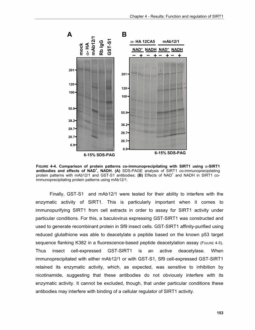
Chapter 4 - Results: Function and regulation of SIRT1
153
Finally, GST-S1 and mAb12/1 were tested for their ability to interfere with the
enzymatic activity of SIRT1. This is particularly important when it comes to
immunopurifying SIRT1 from cell extracts in order to assay for SIRT1 activity under
particular conditions. For this, a baculovirus expressing GST-SIRT1 was constructed and
used to generate recombinant protein in Sf9 insect cells. GST-SIRT1 affinity-purified using
reduced glutathione was able to deacetylate a peptide based on the known p53 target
sequence flanking K382 in a fluorescence-based peptide deacetylation assay (FIGURE 4-5).
Thus insect cell-expressed GST-SIRT1 is an active deacetylase. When
immunoprecipitated with either mAb12/1 or with GST-S1, Sf9 cell-expressed GST-SIRT1
retained its enzymatic activity, which, as expected, was sensitive to inhibition by
nicotinamide, suggesting that these antibodies do not obviously interfere with its
enzymatic activity. It cannot be excluded, though, that under particular conditions these
antibodies may interfere with binding of a cellular regulator of SIRT1 activity.
mAb12/1α- HA 12CA5
201
120
100
55.9
38.2
29.7
20.7
6.9
NAD+ NAD+ NADH NADH– + – +– +– +
201
120
100
55.9
38.2
29.7
20.7
6.9
α-H
Am
ock
mA
b12/
1R
b Ig
GG
ST-S
1
6-15% SDS-PAG6-15% SDS-PAG
A B
FIGURE 4-4. Comparison of protein patterns co-immunoprecipitating with SIRT1 using α-SIRT1antibodies and effects of NAD+, NADH. (A) SDS-PAGE analysis of SIRT1 co-immunoprecipitatingprotein patterns with mAb12/1 and GST-S1 antibodies; (B) Effects of NAD+ and NADH in SIRT1 co-immunoprecipitating protein patterns using mAb12/1.
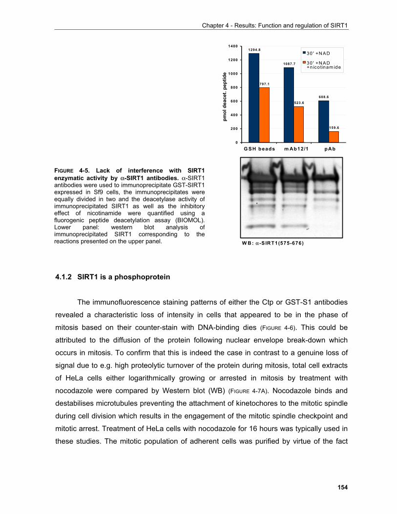
Chapter 4 - Results: Function and regulation of SIRT1
154
4.1.2 SIRT1 is a phosphoprotein
The immunofluorescence staining patterns of either the Ctp or GST-S1 antibodies
revealed a characteristic loss of intensity in cells that appeared to be in the phase of
mitosis based on their counter-stain with DNA-binding dies (FIGURE 4-6). This could be
attributed to the diffusion of the protein following nuclear envelope break-down which
occurs in mitosis. To confirm that this is indeed the case in contrast to a genuine loss of
signal due to e.g. high proteolytic turnover of the protein during mitosis, total cell extracts
of HeLa cells either logarithmically growing or arrested in mitosis by treatment with
nocodazole were compared by Western blot (WB) (FIGURE 4-7A). Nocodazole binds and
destabilises microtubules preventing the attachment of kinetochores to the mitotic spindle
during cell division which results in the engagement of the mitotic spindle checkpoint and
mitotic arrest. Treatment of HeLa cells with nocodazole for 16 hours was typically used in
these studies. The mitotic population of adherent cells was purified by virtue of the fact
1294.8
1087.7
608.6
797.1
523.6
159.6
0
200
400
600
800
1000
1200
1400
G SH beads m Ab12/1 pAb
pmol
dea
cet.
pept
ide
30 ' +N AD
30 ' +N AD+nicotinam ide
W B : α -S IRT1(575-676)
FIGURE 4-5. Lack of interference with SIRT1enzymatic activity by α-SIRT1 antibodies. α-SIRT1antibodies were used to immunoprecipitate GST-SIRT1expressed in Sf9 cells, the immunoprecipitates wereequally divided in two and the deacetylase activity ofimmunoprecipitated SIRT1 as well as the inhibitoryeffect of nicotinamide were quantified using afluorogenic peptide deacetylation assay (BIOMOL).Lower panel: western blot analysis ofimmunoprecipitated SIRT1 corresponding to thereactions presented on the upper panel.
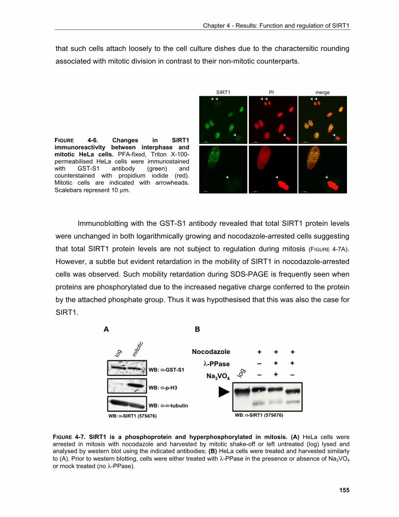
Chapter 4 - Results: Function and regulation of SIRT1
155
that such cells attach loosely to the cell culture dishes due to the charactersitic rounding
associated with mitotic division in contrast to their non-mitotic counterparts.
Immunoblotting with the GST-S1 antibody revealed that total SIRT1 protein levels
were unchanged in both logarithmically growing and nocodazole-arrested cells suggesting
that total SIRT1 protein levels are not subject to regulation during mitosis (FIGURE 4-7A).
However, a subtle but evident retardation in the mobility of SIRT1 in nocodazole-arrested
cells was observed. Such mobility retardation during SDS-PAGE is frequently seen when
proteins are phosphorylated due to the increased negative charge conferred to the protein
by the attached phosphate group. Thus it was hypothesised that this was also the case for
SIRT1.
SIRT1 PI merge
FIGURE 4-6. Changes in SIRT1immunoreactivity between interphase andmitotic HeLa cells. PFA-fixed, Triton X-100-permeabilised HeLa cells were immunostainedwith GST-S1 antibody (green) andcounterstained with propidium iodide (red).Mitotic cells are indicated with arrowheads.Scalebars represent 10 µm.
log
WB: α-SIRT1 (575-676)
– + +– + –
+ + +Nocodazole
λ-PPase
Na3VO4
A B
log mito
tic
WB: α-GST-S1
WB: α-p-H3
WB: α-α-tubulin
WB: α-SIRT1 (575-676)
FIGURE 4-7. SIRT1 is a phosphoprotein and hyperphosphorylated in mitosis. (A) HeLa cells werearrested in mitosis with nocodazole and harvested by mitotic shake-off or left untreated (log) lysed andanalysed by western blot using the indicated antibodies; (B) HeLa cells were treated and harvested similarlyto (A). Prior to western blotting, cells were either treated with λ-PPase in the presence or absence of Na3VO4or mock treated (no λ-PPase).

Chapter 4 - Results: Function and regulation of SIRT1
156
To test this hypothesis, cell extracts from nocodazole-arrested cells were treated
with λ-PPase and subjected to immunoblotting (FIGURE 4-7B). λ-PPase is a dual specificity
phosphatase encoded by the bacteriophage λ genome. As a control, a similar reaction
was set in the presence of Na3VO4 a potent inhibitor of λ-PPase (Zhuo et al., 1993). The
SIRT1 immunoractive band in the λ-PPase-treated nocodazole-arrested cells migrated
with increased mobility suggesting that indeed the observed band-shift was due to
phosphorylation. However, it was also obvious that λ-PPase caused an even higher
mobility than that of untreated cells. A more careful observation of the untreated sample
SIRT1 band reveals a "comet-tail" effect suggestive of the presence of more than one
SIRT1 species. Given the fact that in a typical exponentially growing population of HeLa
cells 3-5% of cells are in mitosis, it is very unlikely that the observed electrophoretic
retardation in interphase extracts is due to this small population. Thus, it was concluded
that SIRT1 is phosphorylated in interphase and hyperphosphorylated in nocodazole-
arrested mitotic cells.
Based on these results, a two-fold goal for the project was defined, namely to
identify the phosphorylation site(s) of SIRT1 that cause the reduced electrophoretic
mobility in (i) interphase cells and (ii) in mitotic cells and explore their effects on SIRT1
activity.
4.1.3 Phosphorylation of SIRT1 in interphase
To identify the interphase kinase and its target residues of SIRT1, a
multidisciplnary approach was employed harnessing the power of bioinformatics,
biochemical evidence and litterature search. These approaches as well as the functional
characterisation of the identified modifications will be delineated below.
4.1.3.1 Identification of phosphorylation sites of SIRT1 expressed in Sf9 cells Recombinant human proteins expressed in insect cells often exhibit patterns of
post-translational modifications similar to the ones found in human cells. Interestingly, λ-
PPase treatment of GST-SIRT1 expressed in Sf9 cells increases the electrophoretic
mobility of the protein (FIGURE 4-8A) similarly to the situation in human cells suggesting that
it is phosphorylated. Thus, it was hypothesised that if phosphorylation does play a role in
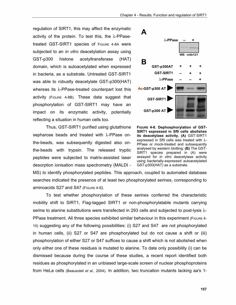
Chapter 4 - Results: Function and regulation of SIRT1
157
FIGURE 4-8. Dephosphorylation of GST-SIRT1 expressed in Sf9 cells abolishesits deacetylase activity. (A) GST-SIRT1expressed in Sf9 cells was treated with λ-PPase or mock-treated and subsequentlyanalysed by western blotting; (B) The GST-SIRT1 species prepared in (A) wereassayed for in vitro deacetylase activityusing bacterially-expressed autoacetylatedGST-p300(HAT) as a substrate.
regulation of SIRT1, this may affect the enzymatic
activity of the protein. To test this, the λ-PPase-
treated GST-SIRT1 species of FIGURE 4-8A were
subjected to an in vitro deacetylation assay using
GST-p300 histone acetyltransferase (HAT)
domain, which is autoacetylated when expressed
in bacteria, as a substrate. Untreated GST-SIRT1
was able to robustly deacetylate GST-p300(HAT)
whereas its λ-PPase-treated counterpart lost this
activity (FIGURE 4-8B). These data suggest that
phosphorylation of GST-SIRT1 may have an
impact on its enzymatic activity, potentially
reflecting a situation in human cells too.
Thus, GST-SIRT1 purified using glutathione
sepharose beads and treated with λ-PPase on-
the-beads, was subsequently digested also on-
the-beads with trypsin. The released tryptic
peptides were subjected to matrix-assisted laser
desorption ionisation mass spectrometry (MALDI -
MS) to identify phosphorylated peptides. This approach, coupled to automated database
searches indicated the presence of at least two phosphorylated serines, corresponding to
aminoacids S27 and S47 (FIGURE 4-9).
To test whether phosphorylation of these serines conferred the characteristic
mobility shift to SIRT1, Flag-tagged SIRT1 or non-phosphorylatable mutants carrying
serine to alanine substitutions were transfected in 293 cells and subjected to post-lysis λ-
PPase treatment. All three species exhibited similar behaviour in this experiment (FIGURE 4-
10) suggesting any of the following possibilities: (i) S27 and S47 are not phosphorylated
in human cells, (ii) S27 or S47 are phosphorylated but do not cause a shift or (iii)
phosphorylation of either S27 or S47 suffices to cause a shift which is not abolished when
only either one of these residues is mutated to alanine. To date only possibility (i) can be
dismissed because during the course of these studies, a recent report identified both
residues as phosphorylated in an unbiased large-scale screen of nuclear phosphoproteins
from HeLa cells (Beausoleil et al., 2004). In addition, two truncation mutants lacking aa's 1-
GST-SIRT1
GST-p300 AT
Ac-GST-p300 AT
GST-p300AT
GST-SIRT1
λ-PPase– + +– – +
+ + +
Aλ-PPase – +
WB: mAb12/1
B
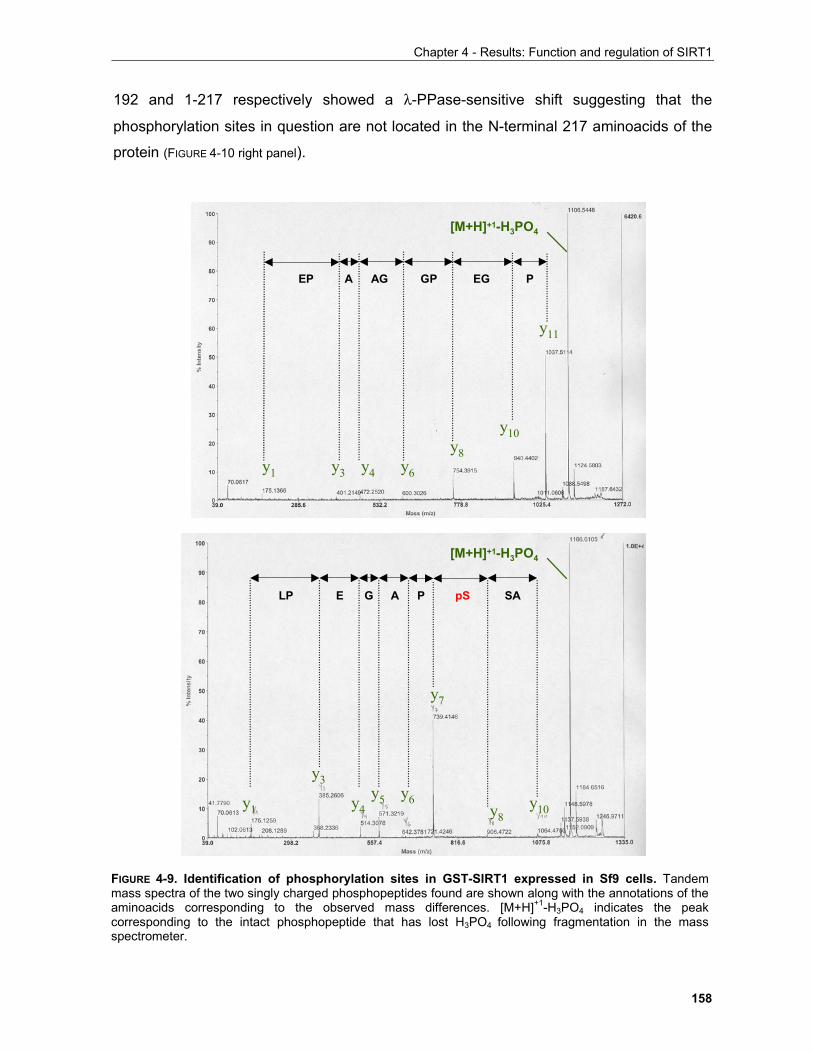
Chapter 4 - Results: Function and regulation of SIRT1
158
192 and 1-217 respectively showed a λ-PPase-sensitive shift suggesting that the
phosphorylation sites in question are not located in the N-terminal 217 aminoacids of the
protein (FIGURE 4-10 right panel).
FIGURE 4-9. Identification of phosphorylation sites in GST-SIRT1 expressed in Sf9 cells. Tandemmass spectra of the two singly charged phosphopeptides found are shown along with the annotations of theaminoacids corresponding to the observed mass differences. [M+H]+1-H3PO4 indicates the peakcorresponding to the intact phosphopeptide that has lost H3PO4 following fragmentation in the massspectrometer.
y1
y3y6
y7
y8y10
[M+H]+1-H3PO4
[M+H]+1-H3PO4
SApS
y4y5
PAGELP
y11
y10y8
y6y4y3y1
PEGGPAGAEP
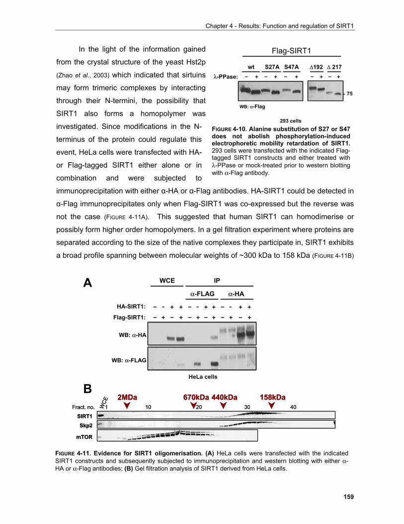
Chapter 4 - Results: Function and regulation of SIRT1
159
FIGURE 4-10. Alanine substitution of S27 or S47does not abolish phosphorylation-inducedelectrophoretic mobility retardation of SIRT1.293 cells were transfected with the indicated Flag-tagged SIRT1 constructs and either treated withλ-PPase or mock-treated prior to western blottingwith α-Flag antibody.
∆192 ∆ 217
75
– + – + – +λ-PPase: – + – +
wt S27A S47A
Flag-SIRT1
WB: α-Flag
293 cells
In the light of the information gained
from the crystal structure of the yeast Hst2p
(Zhao et al., 2003) which indicated that sirtuins
may form trimeric complexes by interacting
through their N-termini, the possibility that
SIRT1 also forms a homopolymer was
investigated. Since modifications in the N-
terminus of the protein could regulate this
event, HeLa cells were transfected with HA-
or Flag-tagged SIRT1 either alone or in
combination and were subjected to
immunoprecipitation with either α-HA or α-Flag antibodies. HA-SIRT1 could be detected in
α-Flag immunoprecipitates only when Flag-SIRT1 was co-expressed but the reverse was
not the case (FIGURE 4-11A). This suggested that human SIRT1 can homodimerise or
possibly form higher order homopolymers. In a gel filtration experiment where proteins are
separated according to the size of the native complexes they participate in, SIRT1 exhibits
a broad profile spanning between molecular weights of ~300 kDa to 158 kDa (FIGURE 4-11B)
WB: α-HA
WB: α-FLAG
IP
α-HAα-FLAG
WCE
HA-SIRT1:
Flag-SIRT1: – + – + – + – + – + – +
– - + + – - + + – - + +
HeLa cells
Fract. no.
SIRT1Skp2
mTOR
1WCE
10 20 30 40
2MDa 670kDa 440kDa 158kDaFract. no.
SIRT1Skp2
mTOR
1WCE
10 20 30 40
2MDa 670kDa 440kDa 158kDa
A
B
FIGURE 4-11. Evidence for SIRT1 oligomerisation. (A) HeLa cells were transfected with the indicatedSIRT1 constructs and subsequently subjected to immunoprecipitation and western blotting with either α-HA or α-Flag antibodies; (B) Gel filtration analysis of SIRT1 derived from HeLa cells.
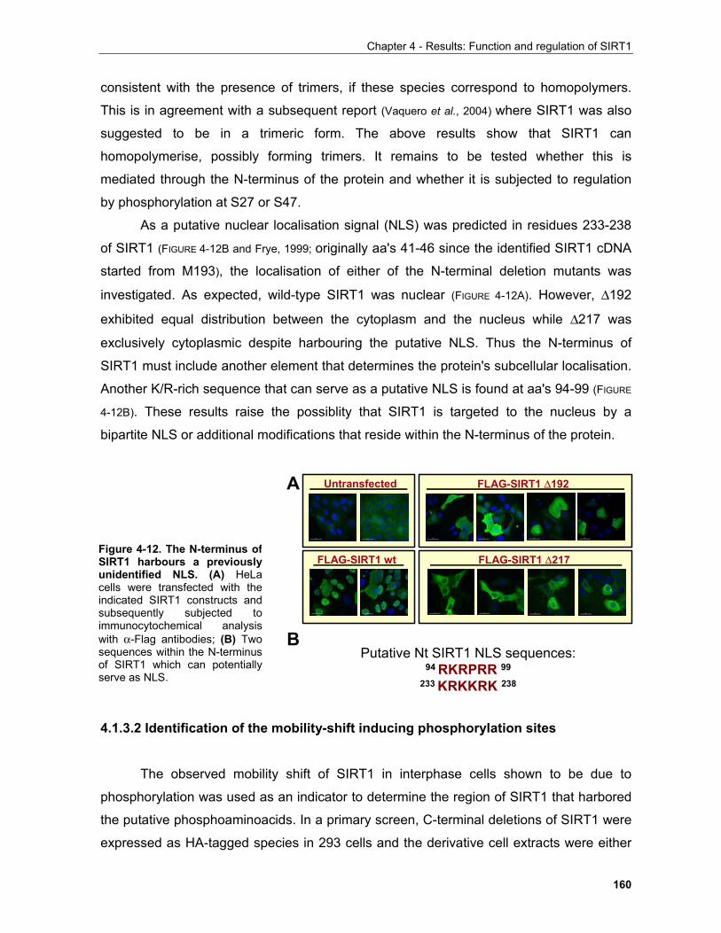
Chapter 4 - Results: Function and regulation of SIRT1
160
consistent with the presence of trimers, if these species correspond to homopolymers.
This is in agreement with a subsequent report (Vaquero et al., 2004) where SIRT1 was also
suggested to be in a trimeric form. The above results show that SIRT1 can
homopolymerise, possibly forming trimers. It remains to be tested whether this is
mediated through the N-terminus of the protein and whether it is subjected to regulation
by phosphorylation at S27 or S47.
As a putative nuclear localisation signal (NLS) was predicted in residues 233-238
of SIRT1 (FIGURE 4-12B and Frye, 1999; originally aa's 41-46 since the identified SIRT1 cDNA
started from M193), the localisation of either of the N-terminal deletion mutants was
investigated. As expected, wild-type SIRT1 was nuclear (FIGURE 4-12A). However, ∆192
exhibited equal distribution between the cytoplasm and the nucleus while ∆217 was
exclusively cytoplasmic despite harbouring the putative NLS. Thus the N-terminus of
SIRT1 must include another element that determines the protein's subcellular localisation.
Another K/R-rich sequence that can serve as a putative NLS is found at aa's 94-99 (FIGURE
4-12B). These results raise the possiblity that SIRT1 is targeted to the nucleus by a
bipartite NLS or additional modifications that reside within the N-terminus of the protein.
4.1.3.2 Identification of the mobility-shift inducing phosphorylation sites
The observed mobility shift of SIRT1 in interphase cells shown to be due to
phosphorylation was used as an indicator to determine the region of SIRT1 that harbored
the putative phosphoaminoacids. In a primary screen, C-terminal deletions of SIRT1 were
expressed as HA-tagged species in 293 cells and the derivative cell extracts were either
Untransfected
FLAG-SIRT1 wt
FLAG-SIRT1 ∆192
FLAG-SIRT1 ∆217
Putative Nt SIRT1 NLS sequences:94 RKRPRR 99
233 KRKKRK 238
A
B
Figure 4-12. The N-terminus ofSIRT1 harbours a previouslyunidentified NLS. (A) HeLacells were transfected with theindicated SIRT1 constructs andsubsequently subjected toimmunocytochemical analysiswith α-Flag antibodies; (B) Twosequences within the N-terminusof SIRT1 which can potentiallyserve as NLS.
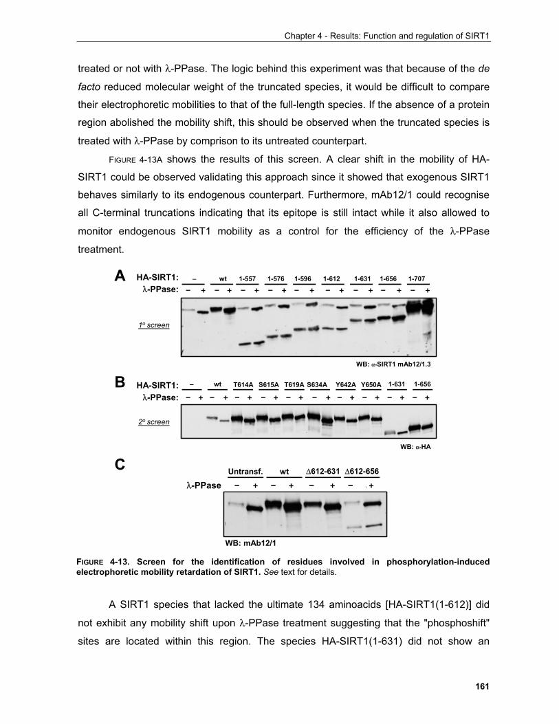
Chapter 4 - Results: Function and regulation of SIRT1
161
treated or not with λ-PPase. The logic behind this experiment was that because of the de
facto reduced molecular weight of the truncated species, it would be difficult to compare
their electrophoretic mobilities to that of the full-length species. If the absence of a protein
region abolished the mobility shift, this should be observed when the truncated species is
treated with λ-PPase by comprison to its untreated counterpart.
FIGURE 4-13A shows the results of this screen. A clear shift in the mobility of HA-
SIRT1 could be observed validating this approach since it showed that exogenous SIRT1
behaves similarly to its endogenous counterpart. Furthermore, mAb12/1 could recognise
all C-terminal truncations indicating that its epitope is still intact while it also allowed to
monitor endogenous SIRT1 mobility as a control for the efficiency of the λ-PPase
treatment.
A SIRT1 species that lacked the ultimate 134 aminoacids [HA-SIRT1(1-612)] did
not exhibit any mobility shift upon λ-PPase treatment suggesting that the "phosphoshift"
sites are located within this region. The species HA-SIRT1(1-631) did not show an
– + – + – + – + – + – + – + – + – +
– + – + – + – + – + – + – + – + – + – +
1o screen
2o screen
λ-PPase:
λ-PPase:
WB: α-SIRT1 mAb12/1.3
WB: α-HA
– wt T614A S615A T619A S634A Y642A Y650A 1-631 1-656
1-631 1-6561-557– wt 1-576 1-596 1-612 1-707HA-SIRT1:
HA-SIRT1:
A
B
C Untransf. wt ∆612-631 ∆612-656
– + – + – + – \ +λ-PPase
WB: mAb12/1
FIGURE 4-13. Screen for the identification of residues involved in phosphorylation-inducedelectrophoretic mobility retardation of SIRT1. See text for details.
Chapter 4 - Results: Function and regulation of SIRT1
162
obvious shift, yet the intensity of the SIRT1 band in the λ-PPase treated cells was higher,
a phenomenon which, based on experience acquired during this project, may arise when
multiple protein species are concentrated to a single band. Indeed this proved to be the
case when this experiment was repeated (see FIGURE 4-13B). Thus based on this experiment
it was concluded that phosphorylation of SIRT1 between residues 613 and 656 causes
the observed mobility shift of SIRT1 in interphase.
To confirm this, all phosphorylatable aminoacids within this region were mutated to
alanines and subjected to the same type of assay (FIGURE 4-13B). None of these individual
mutations sufficed to abolish λ-PPase sensitivity suggesting that multiple phosphorylation
sites within this region may contribute to the observed shift. Given the fact that any
combination of 2 or more of the 6 aminoacids could be phosphorylated, it was considered
appropriate to address this issue by creating a species which lacked aminoacids 612-631
and 612-656 henceforth referred to as ∆612-631 and ∆612-656 respectively. As ∆612-631
migrated very similarly to wt SIRT1 it is difficult to conclude whether this mutant still shows
a shift. However, it is clear that the ∆612-656 deletion renders SIRT1 insensitive to λ-
PPase treatment providing further evidence that the 612-656 region of SIRT1 is
phosphorylated in interphase.
An alternative interpretation of these results is that aa 612-656 do not harbor the
phosphorylation sites in question but rather they serve as the docking site for the
responsible kinase. Despite the transient nature of enzyme-substrate interactions, some
kinases have been shown to interact with their substrates stably enough to allow the co-
purification of the complex by affinity chromatography. Such an example is cyclin A/Cdk2
with E2F-1 (Krek et al., 1994) and Jnk with c-Jun (Hibi et al., 1993). To investigate the
possibility that SIRT1 co-purifies with a kinase, SIRT1 was immunopurified (IP'd) with
each of the Ctp, GST-S1 and mAb12/1 antibodies, the buffer of the immunoprecipitates
(IPs) was exhanged for a standard kinase buffer supplemmented with 32P-ATP. In the
presence of a co-purifying kinase, SIRT1 should be labelled with 32P as visualised by
autoradiography.
As seen in Figure 4-14A, despite the presence of IP'd SIRT1 with all antibodies, no
signal was present in the mAb12/1 IP whereas a very weak signal may be seen in the
immunoprecipitates of the polyclonal antibodies (arrowheads). This suggested either the
complete absence of a co-purifying kinase or the unsuitability of the reaction conditions for
the succesful outcome of this experiment. It is also possible that the weak signal in the
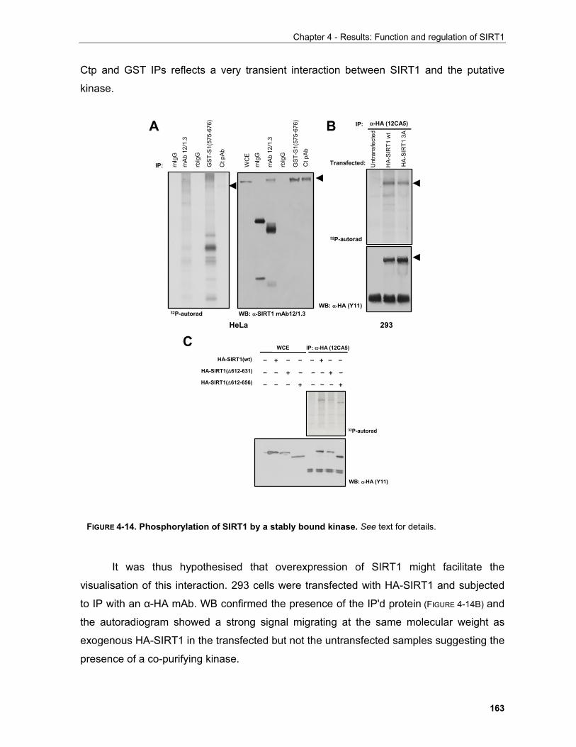
Chapter 4 - Results: Function and regulation of SIRT1
163
Ctp and GST IPs reflects a very transient interaction between SIRT1 and the putative
kinase.
It was thus hypothesised that overexpression of SIRT1 might facilitate the
visualisation of this interaction. 293 cells were transfected with HA-SIRT1 and subjected
to IP with an α-HA mAb. WB confirmed the presence of the IP'd protein (FIGURE 4-14B) and
the autoradiogram showed a strong signal migrating at the same molecular weight as
exogenous HA-SIRT1 in the transfected but not the untransfected samples suggesting the
presence of a co-purifying kinase.
C
A B
mA
b 12
/1.3
GST
-S1(
575-
676)
Ct p
Ab
rbIg
G
mIg
G
mA
b 12
/1.3
GST
-S1(
575-
676)
Ct p
Ab
rbIg
G
WC
E
32P-autorad WB: α-SIRT1 mAb12/1.3
IP:
IP: α-HA (12CA5)
WB: α-HA (Y11)
32P-autorad
HA-
SIR
T1 w
t
mIg
G
HA-
SIR
T1 3
A
Unt
rans
fect
ed
Transfected:
HeLa 293
WCE IP: α-HA (12CA5)
HA-SIRT1(wt)
HA-SIRT1(∆612-631)
– + – – – + – –
– – + – – – + –HA-SIRT1(∆612-656) – – – + – – – +
WB: α-HA (Y11)
32P-autorad
FIGURE 4-14. Phosphorylation of SIRT1 by a stably bound kinase. See text for details.
Chapter 4 - Results: Function and regulation of SIRT1
164
To test whether this kinase interacted with SIRT1 through the phosphoshift site, the
same experiment was repeated with wild-type HA-SIRT1 and the ∆612-631, ∆612-656
mutants. As shown in FIGURE 4-14C, neither deletion abolishes the 32P-labelling of the
corresponding IP'd species.
These experiments suggest that there is a kinase that can bind stably to SIRT1
causing its in vitro phosphorylation in a region outside 612-656 which was shown to
harbour residues responsible for the protein's electrophoretic mobility shift. This does not
exclude the possibility that the kinase causing the shift is bound to this region in which
case the interaction might be too weak to be detected with this assay especially in the
presence of another kinase demonstrated to be present in these experiments. Thus it is
necessary to probe further the identity of this kinase and identify the sites affected by its
action.
4.1.3.3 In vitro phosphorylation of SIRT1 by Casein Kinase 2
Kinases exhibit profound selectivity not only for the residue that they target for
phosphorylation but also for the aminoacids that flank it. The availability of information on
phosphorylation consensus sites targeted by specific kinases allowed the creation of
searchable databases that provide the researcher with the ability to investigate whether
their protein of interest harbours any or specified phosphorylation sites. Clearly, though,
this approach is limited to previously reported data and cannot indicate whether a
proposed phosphorylation site occurs in vivo and has physiological relevance.
In search for potential phosphorylation sites on SIRT1 the program Scansite was
used (http://scansite.mit.edu/motifscanner/motifscan1.phtml?database=_SWS_). This
program allows the input protein sequence to be investigated for harbouring
phosphorylation sites within any of 62 motifs comprising kinase consensus target sites as
well as known binding motifs for several protein interaction-mediating domains such as
SH2 and SH3 domains.
This approach indicated that the C-terminus of SIRT harbours a putative
phosphorylation site for casein kinase 2 (CK2) at residue S693. The CK2 consensus site
consists of a phosphorylatable serine or threonine residue followed by acidic aminoacids
i.e. D or E or phosphorylated S and T, especially at the +3 position (Litchfield, 2003). Manual
observation of the sequence revealed two more putative CK2 sites at residues S659 and
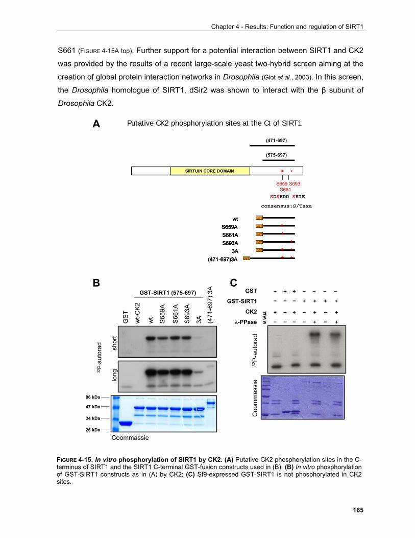
Chapter 4 - Results: Function and regulation of SIRT1
165
S661 (FIGURE 4-15A top). Further support for a potential interaction between SIRT1 and CK2
was provided by the results of a recent large-scale yeast two-hybrid screen aiming at the
creation of global protein interaction networks in Drosophila (Giot et al., 2003). In this screen,
the Drosophila homologue of SIRT1, dSir2 was shown to interact with the β subunit of
Drosophila CK2.
GS
T
wt
S65
9A
S66
1A
S69
3A
3A (471
-697
) 3A
wt-C
K2
32P-
auto
rad
GST-SIRT1 (575-697)
86 kDa
47 kDa
34 kDa
26 kDa
long
shor
t
Coommassie
SIRTUIN CORE DOMAIN * *S659
S661S693
*
(575-697)
(471-697)
Putative CK2 phosphorylation sites at the Ct of SIRT1
GST
GST
GST
GST
**
*** *** *
wtS659AS661AS693A
3A
GST
(471-697)3A GST
GST
GST
GST
GST
**
*** *** *
wtS659AS661AS693A
3A
GST
(471-697)3A GST
GST
GST
GST
GST
**
*** *** *
wtS659AS661AS693A
3A
GST
(471-697)3A GST
GST
GST
GST
GST
**
*** *** *
wtS659AS661AS693A
3A
GST
(471-697)3A GST
A
B
SDSEDD SEIE
consensus:S/Taxa
GST
GST-SIRT1
CK2
λ-PPase – – – – + – +
+ – + – + – +
– – – + + + +
– + + – – – –
M.W
.M.
Coo
mm
assi
e32
P-au
tora
d
C
FIGURE 4-15. In vitro phosphorylation of SIRT1 by CK2. (A) Putative CK2 phosphorylation sites in the C-terminus of SIRT1 and the SIRT1 C-terminal GST-fusion constructs used in (B); (B) In vitro phosphorylationof GST-SIRT1 constructs as in (A) by CK2; (C) Sf9-expressed GST-SIRT1 is not phosphorylated in CK2sites.
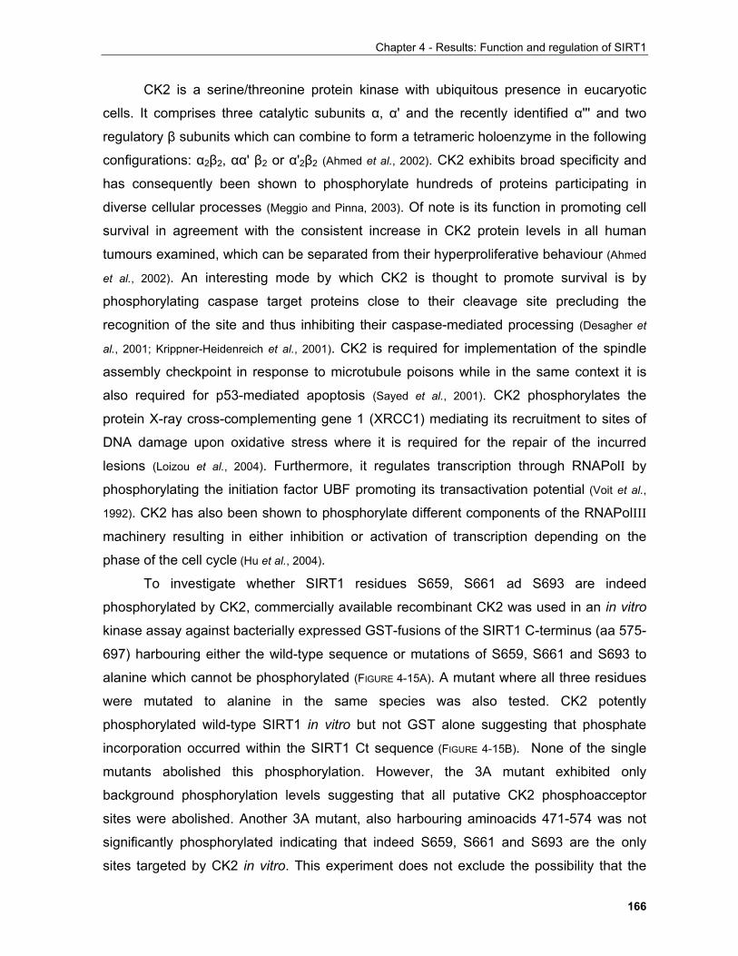
Chapter 4 - Results: Function and regulation of SIRT1
166
CK2 is a serine/threonine protein kinase with ubiquitous presence in eucaryotic
cells. It comprises three catalytic subunits α, α' and the recently identified α''' and two
regulatory β subunits which can combine to form a tetrameric holoenzyme in the following
configurations: α2β2, αα' β2 or α'2β2 (Ahmed et al., 2002). CK2 exhibits broad specificity and
has consequently been shown to phosphorylate hundreds of proteins participating in
diverse cellular processes (Meggio and Pinna, 2003). Of note is its function in promoting cell
survival in agreement with the consistent increase in CK2 protein levels in all human
tumours examined, which can be separated from their hyperproliferative behaviour (Ahmed
et al., 2002). An interesting mode by which CK2 is thought to promote survival is by
phosphorylating caspase target proteins close to their cleavage site precluding the
recognition of the site and thus inhibiting their caspase-mediated processing (Desagher et
al., 2001; Krippner-Heidenreich et al., 2001). CK2 is required for implementation of the spindle
assembly checkpoint in response to microtubule poisons while in the same context it is
also required for p53-mediated apoptosis (Sayed et al., 2001). CK2 phosphorylates the
protein X-ray cross-complementing gene 1 (XRCC1) mediating its recruitment to sites of
DNA damage upon oxidative stress where it is required for the repair of the incurred
lesions (Loizou et al., 2004). Furthermore, it regulates transcription through RNAPolI by
phosphorylating the initiation factor UBF promoting its transactivation potential (Voit et al.,
1992). CK2 has also been shown to phosphorylate different components of the RNAPolIII
machinery resulting in either inhibition or activation of transcription depending on the
phase of the cell cycle (Hu et al., 2004).
To investigate whether SIRT1 residues S659, S661 ad S693 are indeed
phosphorylated by CK2, commercially available recombinant CK2 was used in an in vitro
kinase assay against bacterially expressed GST-fusions of the SIRT1 C-terminus (aa 575-
697) harbouring either the wild-type sequence or mutations of S659, S661 and S693 to
alanine which cannot be phosphorylated (FIGURE 4-15A). A mutant where all three residues
were mutated to alanine in the same species was also tested. CK2 potently
phosphorylated wild-type SIRT1 in vitro but not GST alone suggesting that phosphate
incorporation occurred within the SIRT1 Ct sequence (FIGURE 4-15B). None of the single
mutants abolished this phosphorylation. However, the 3A mutant exhibited only
background phosphorylation levels suggesting that all putative CK2 phosphoacceptor
sites were abolished. Another 3A mutant, also harbouring aminoacids 471-574 was not
significantly phosphorylated indicating that indeed S659, S661 and S693 are the only
sites targeted by CK2 in vitro. This experiment does not exclude the possibility that the
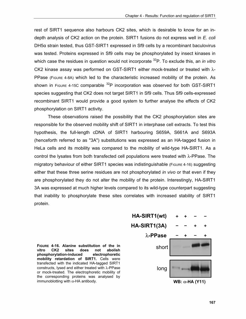
Chapter 4 - Results: Function and regulation of SIRT1
167
rest of SIRT1 sequence also harbours CK2 sites, which is desirable to know for an in-
depth analysis of CK2 action on the protein. SIRT1 fusions do not express well in E. coli
DH5α strain tested, thus GST-SIRT1 expressed in Sf9 cells by a recombinant baculovirus
was tested. Proteins expressed in Sf9 cells may be phosphorylated by insect kinases in
which case the residues in question would not incorporate 32P. To exclude this, an in vitro
CK2 kinase assay was performed on GST-SIRT1 either mock-treated or treated with λ-
PPase (FIGURE 4-8A) which led to the characteristic increased mobility of the protein. As
shown in FIGURE 4-15C comparable 32P incorporation was observed for both GST-SIRT1
species suggesting that CK2 does not target SIRT1 in Sf9 cells. Thus Sf9 cells-expressed
recombinant SIRT1 would provide a good system to further analyse the effects of CK2
phosphorylation on SIRT1 activity.
These observations raised the possibility that the CK2 phosphorylation sites are
responsible for the observed mobility shift of SIRT1 in interphase cell extracts. To test this
hypothesis, the full-length cDNA of SIRT1 harbouring S659A, S661A and S693A
(henceforth referred to as "3A") substitutions was expressed as an HA-tagged fusion in
HeLa cells and its mobility was compared to the mobility of wild-type HA-SIRT1. As a
control the lysates from both transfected cell populations were treated with λ-PPase. The
migratory behaviour of either SIRT1 species was indistinguishable (FIGURE 4-16) suggesting
either that these three serine residues are not phosphorylated in vivo or that even if they
are phosphorylated they do not alter the mobility of the protein. Interestingly, HA-SIRT1
3A was expressed at much higher levels compared to its wild-type counterpart suggesting
that inability to phosphorylate these sites correlates with increased stability of SIRT1
protein.
short
long
– + – +
HA-SIRT1(wt)
HA-SIRT1(3A)
λ-PPase
– – + +
+ + – –
WB: α-HA (Y11)
FIGURE 4-16. Alanine substitution of the invitro CK2 sites does not abolishphosphorylation-induced electrophoreticmobility retardation of SIRT1. Cells weretransfected with the indicated HA-tagged SIRT1constructs, lysed and either treated with λ-PPaseor mock-treated. The electrophoretic mobility ofthe corresponding proteins was analysed byimmunoblotting with α-HA antibody.
Chapter 4 - Results: Function and regulation of SIRT1
168
4.1.3.3.1 SIRT1 in the UV response
The experimental approaches delineated above were based on the widely
accepted assumption that CK2 is a constitutively active kinase (Litchfield, 2003). However,
several recent reports have proposed novel modes of CK2 action where its activity is
inducible by extracellular stimuli. In particular a new emerging role of CK2 in response to
UV irradiation is of note.
CK2 is part of a complex with the chromatin transcriptional elongation factor FACT
(facilitates chromatin transcription) which comprises hSpt16 and SSRP1 (structure-
specific recognition protein 1). In the context of this complex whose assembly is induced
upon UV irradiation, CK2 phosphorylates p53 at S392 resulting in enhanced binding of
p53 to DNA which in turn correlates with its ability to induce transcription (Keller et al., 2001).
In another report, Kato and co-workers (Kato
et al., 2003) showed that CK2 mediates the
phosphorylation-driven degradation of IκB upon
ultraviolet (UV) irradiation in a pathway distinct from
that of IκB kinase (IKK). In response to inflammatory
cytokines and ionising radiation IKK phosphorylates
the N-terminus of IκB and promotes its degradation
via a β-TrCP-based SCF E3 ubiquitin ligase
complex (Hayden and Ghosh, 2004). In contrast, upon
UV irradiation CK2 phosphorylates six residues in
the C-terminus of IκB in a manner dependent on the
kinase p38 and leads to the degradation of IκB and
subsequent NFκB activation. Furthermore, the CK2 sites reside within a region known as
the PEST motif (FIGURE 4-17). The PEST motif is characterised by enrichment in P, E, S and
T residues but lacks positively-charged aminoacids. It is found in several proteins
including the myc, fos and jun oncogenes and p53 tumour suppressor which it targets for
degradation by the ubiquitin-proteasome pathway (Rechsteiner and Rogers, 1996).
To investigate whether SIRT1 also carries a similar motif and is therefore
potentially regulated by PEST-mediated proteolysis, a web-based algorithm that scores
for potential PEST motifs in protein sequences was used1. A region between aa's 653-711
1 (https://emb1.bcc.univie.ac.at/toolbox/pestfind/pestfind-analysis-webtool.htm)
NF-κB
Nuclear translocation/activation
CK2
p38
IKK
IκBα
IL-1TNF LPS IR UV
PEST** ******
IκBα degradation
p53
DNA repair
FIGURE 4-17. The CK2phosphorylation sites of IκB arelocated within a PEST sequence.See text for details.
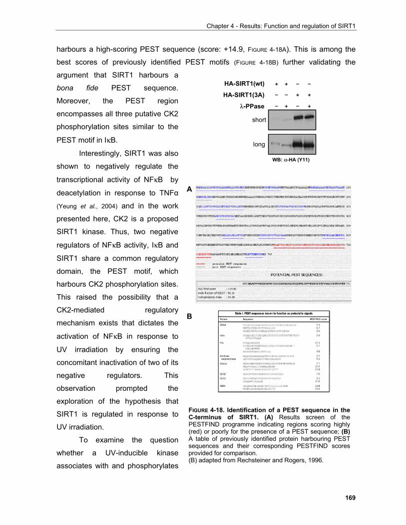
Chapter 4 - Results: Function and regulation of SIRT1
169
harbours a high-scoring PEST sequence (score: +14.9, FIGURE 4-18A). This is among the
best scores of previously identified PEST motifs (FIGURE 4-18B) further validating the
argument that SIRT1 harbours a
bona fide PEST sequence.
Moreover, the PEST region
encompasses all three putative CK2
phosphorylation sites similar to the
PEST motif in IκB.
Interestingly, SIRT1 was also
shown to negatively regulate the
transcriptional activity of NFκB by
deacetylation in response to TNFα
(Yeung et al., 2004) and in the work
presented here, CK2 is a proposed
SIRT1 kinase. Thus, two negative
regulators of NFκB activity, IκB and
SIRT1 share a common regulatory
domain, the PEST motif, which
harbours CK2 phosphorylation sites.
This raised the possibility that a
CK2-mediated regulatory
mechanism exists that dictates the
activation of NFκB in response to
UV irradiation by ensuring the
concomitant inactivation of two of its
negative regulators. This
observation prompted the
exploration of the hypothesis that
SIRT1 is regulated in response to
UV irradiation.
To examine the question
whether a UV-inducible kinase
associates with and phosphorylates
short
long
– + – +
HA-SIRT1(wt)
HA-SIRT1(3A)
λ-PPase
– – + +
+ + – –
WB: α-HA (Y11)
A
B
FIGURE 4-18. Identification of a PEST sequence in theC-terminus of SIRT1. (A) Results screen of thePESTFIND programme indicating regions scoring highly(red) or poorly for the presence of a PEST sequence; (B)A table of previously identified protein harbouring PESTsequences and their corresponding PESTFIND scoresprovided for comparison. (B) adapted from Rechsteiner and Rogers, 1996.
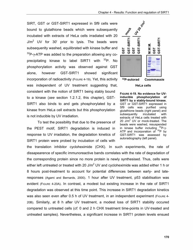
Chapter 4 - Results: Function and regulation of SIRT1
170
SIRT, GST or GST-SIRT1 expressed in Sf9 cells were
bound to glutathione beads which were subsequently
incubated with extracts of HeLa cells irradiated with 20
J/m2 UV for 30' prior to lysis. The beads were
subsequenlty washed, equilibrated with kinase buffer and 32P-γ-ATP was added to the preparation allowing any co-
precipitating kinase to label SIRT1 with 32P. No
phosphorylation activity was observed against GST
alone, however GST-SIRT1 showed significant
incorporation of radioactivity (FIGURE 4-19). Yet, this activity
was independent of UV treatment suggesting that,
consistent with the notion of SIRT1 being stably bound
to a kinase (see section 1.2.1.2, this chapter), GST-
SIRT1 also binds to and gets phosphorylated by a
kinase from HeLa cell extracts but this phosphorylation
is not inducible by UV irradiation.
To test the possibility that due to the presence of
the PEST motif, SIRT1 degradation is induced in
response to UV irradiation, the degradation kinetics of
SIRT1 protein were probed by incubation of cells with
the translation inhibitor cycloheximide (CHX). In such experiments, the rate of
dissapearence of specific immunoreactive bands correlates with the rate of degradation of
the corresponding protein since no more protein is newly synthesised. Thus, cells were
either left untreated or treated with 20 J/m2 UV and cycloheximide was added either 1 h or
8 hours post-treatment to account for potential differences between early- and late-
responses (Agami and Bernards, 2000). 1 hour after UV treatment, p53 stabilisation was
evident (FIGURE 4-20A). In contrast, a modest but existing increase in the rate of SIRT1
degradation was observed at this time point. This increase in SIRT1 degradation kinetics
was also seen even after 0.5 h of UV treatment, in an independent experiment (FIGURE 4-
20B). Similarly, at 8 h after UV treatment, a modest loss of SIRT1 stability occured
compared to untreated cells (cf. 0 and 2 h CHX treatment time-points in UV-treated and
untreated samples). Nevertheless, a significant increase in SIRT1 protein levels ensued
UV 20
J/m
2
Coommassie32P-autorad
HeLa cells
GST
GST
-SIR
T1G
STG
ST-S
IRT1
GST
GST
-SIR
T1
M.W
.M.
moc
k
FIGURE 4-19. No evidence for UV-inducible phosphorylation ofSIRT1 by a stably-bound kinase.GST or GST-SIRT1 expressed inSf9 cells was purified usingglutathione beads (right panel) andsubsequently incubated withextracts of HeLa cells treated with20 J/m2 UV or mock-treated. Thebeads were washed, resuspendedin kinase buffer including 32P-γ-ATP and incorporation of 32P byGST-SIRT1 was assessed byautoradiography (left panel).
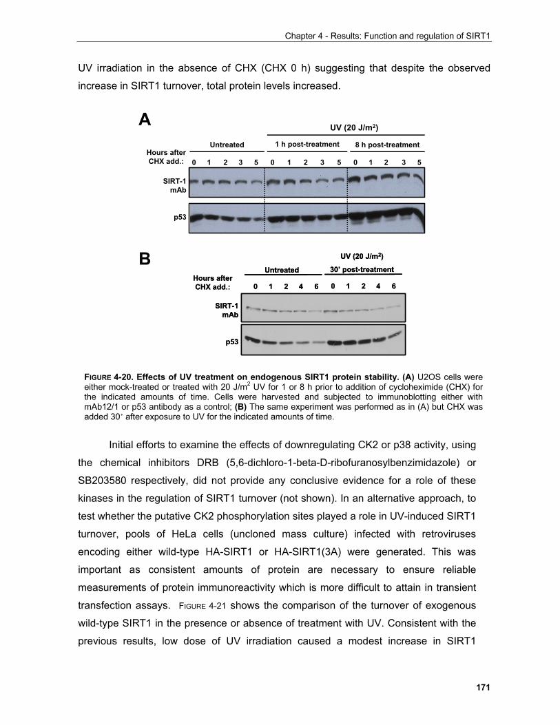
Chapter 4 - Results: Function and regulation of SIRT1
171
UV irradiation in the absence of CHX (CHX 0 h) suggesting that despite the observed
increase in SIRT1 turnover, total protein levels increased.
Initial efforts to examine the effects of downregulating CK2 or p38 activity, using
the chemical inhibitors DRB (5,6-dichloro-1-beta-D-ribofuranosylbenzimidazole) or
SB203580 respectively, did not provide any conclusive evidence for a role of these
kinases in the regulation of SIRT1 turnover (not shown). In an alternative approach, to
test whether the putative CK2 phosphorylation sites played a role in UV-induced SIRT1
turnover, pools of HeLa cells (uncloned mass culture) infected with retroviruses
encoding either wild-type HA-SIRT1 or HA-SIRT1(3A) were generated. This was
important as consistent amounts of protein are necessary to ensure reliable
measurements of protein immunoreactivity which is more difficult to attain in transient
transfection assays. FIGURE 4-21 shows the comparison of the turnover of exogenous
wild-type SIRT1 in the presence or absence of treatment with UV. Consistent with the
previous results, low dose of UV irradiation caused a modest increase in SIRT1
Hours after CHX add.: 52 310
UV (20 J/m2)
1 h post-treatment 8 h post-treatmentUntreated
52 310 52 310
SIRT-1mAb
p53
Hours after CHX add.: 62 410
Untreated
UV (20 J/m2)
30’ post-treatment
62 410
SIRT-1mAb
p53
Hours after CHX add.: 62 410
Untreated
UV (20 J/m2)
30’ post-treatment
62 410
SIRT-1mAb
p53
A
B
FIGURE 4-20. Effects of UV treatment on endogenous SIRT1 protein stability. (A) U2OS cells wereeither mock-treated or treated with 20 J/m2 UV for 1 or 8 h prior to addition of cycloheximide (CHX) forthe indicated amounts of time. Cells were harvested and subjected to immunoblotting either withmAb12/1 or p53 antibody as a control; (B) The same experiment was performed as in (A) but CHX wasadded 30’ after exposure to UV for the indicated amounts of time.
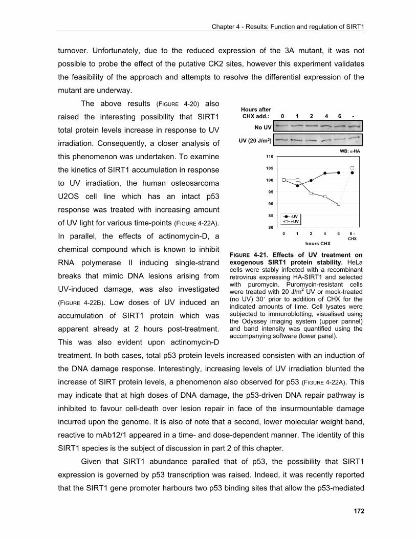
Chapter 4 - Results: Function and regulation of SIRT1
172
turnover. Unfortunately, due to the reduced expression of the 3A mutant, it was not
possible to probe the effect of the putative CK2 sites, however this experiment validates
the feasibility of the approach and attempts to resolve the differential expression of the
mutant are underway.
The above results (FIGURE 4-20) also
raised the interesting possibility that SIRT1
total protein levels increase in response to UV
irradiation. Consequently, a closer analysis of
this phenomenon was undertaken. To examine
the kinetics of SIRT1 accumulation in response
to UV irradiation, the human osteosarcoma
U2OS cell line which has an intact p53
response was treated with increasing amount
of UV light for various time-points (FIGURE 4-22A).
In parallel, the effects of actinomycin-D, a
chemical compound which is known to inhibit
RNA polymerase II inducing single-strand
breaks that mimic DNA lesions arising from
UV-induced damage, was also investigated
(FIGURE 4-22B). Low doses of UV induced an
accumulation of SIRT1 protein which was
apparent already at 2 hours post-treatment.
This was also evident upon actinomycin-D
treatment. In both cases, total p53 protein levels increased consisten with an induction of
the DNA damage response. Interestingly, increasing levels of UV irradiation blunted the
increase of SIRT protein levels, a phenomenon also observed for p53 (FIGURE 4-22A). This
may indicate that at high doses of DNA damage, the p53-driven DNA repair pathway is
inhibited to favour cell-death over lesion repair in face of the insurmountable damage
incurred upon the genome. It is also of note that a second, lower molecular weight band,
reactive to mAb12/1 appeared in a time- and dose-dependent manner. The identity of this
SIRT1 species is the subject of discussion in part 2 of this chapter.
Given that SIRT1 abundance paralled that of p53, the possibility that SIRT1
expression is governed by p53 transcription was raised. Indeed, it was recently reported
that the SIRT1 gene promoter harbours two p53 binding sites that allow the p53-mediated
80
85
90
95
100
105
110
0 1 2 4 6 6 -CHX
hours CHX
-UV+UV
Hours after CHX add.: 42 610 -
UV (20 J/m2)
No UV
WB: α-HA
FIGURE 4-21. Effects of UV treatment onexogenous SIRT1 protein stability. HeLacells were stably infected with a recombinantretrovirus expressing HA-SIRT1 and selectedwith puromycin. Puromycin-resistant cellswere treated with 20 J/m2 UV or mock-treated(no UV) 30’ prior to addition of CHX for theindicated amounts of time. Cell lysates weresubjected to immunoblotting, visualised usingthe Odyssey imaging system (upper pannel)and band intensity was quantified using theaccompanying software (lower panel).
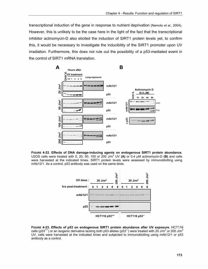
Chapter 4 - Results: Function and regulation of SIRT1
173
transcriptional induction of the gene in response to nutrient deprivation (Nemoto et al., 2004).
However, this is unlikely to be the case here in the light of the fact that the transcriptional
inhibitor actinomycin-D also elicited the induction of SIRT1 protein levels yet, to confirm
this, it would be necessary to investigate the inducibility of the SIRT1 promoter upon UV
irradiation. Furthermore, this does not rule out the possibility of a p53-mediated event in
the control of SIRT1 mRNA translation.
Actinomycin D (0.4 µM)
1h 2h 4h 8hUnt
r’d.
short
longmA
b12/
1α-
p53
81
20 J
/m2
0.5 2 4Unt
r’d. Hours after
UV treatment
50 J
/m2
100
J/m
220
0 J/
m2
mAb12/1
p53
mAb12/1
p53
mAb12/1
p53
mAb12/1
p53
Long exposure:
A B
FIGURE 4-22. Effects of DNA damage-inducing agents on endogenous SIRT1 protein abundance.U2OS cells were treated with 0, 20, 50, 100 or 200 J/m2 UV (A) or 0.4 µM actinomycin-D (B) and cellswere harvested at the indicated times. SIRT1 protein levels were assessed by immunoblotting usingmAb12/1. As a control, p53 antibody was used on the same blots.
HCT116 p53+/+ HCT116 p53-/-
UV dose :
hrs post-treatment :
20 J/m2
mAb12/1
p53
0 2 81 4 8 0 2 81 4 8
20 J/m2
200
J/m
2
200
J/m
2
FIGURE 4-23. Effects of p53 on endogenous SIRT1 protein abundance after UV exposure. HCT116cells (p53+/+) or an isogenic derivative lacking both p53 alleles (p53-/-) were treated with 20 J/m2 or 200 J/m2
UV, cells were harvested at the indicated times and subjected to immunoblotting using mAb12/1 or p53antibody as a control.
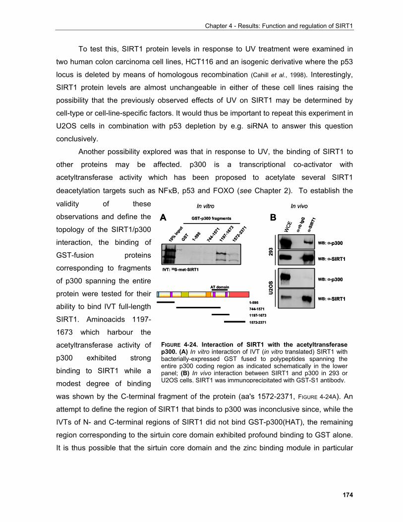
Chapter 4 - Results: Function and regulation of SIRT1
174
To test this, SIRT1 protein levels in response to UV treatment were examined in
two human colon carcinoma cell lines, HCT116 and an isogenic derivative where the p53
locus is deleted by means of homologous recombination (Cahill et al., 1998). Interestingly,
SIRT1 protein levels are almost unchangeable in either of these cell lines raising the
possibility that the previously observed effects of UV on SIRT1 may be determined by
cell-type or cell-line-specific factors. It would thus be important to repeat this experiment in
U2OS cells in combination with p53 depletion by e.g. siRNA to answer this question
conclusively.
Another possibility explored was that in response to UV, the binding of SIRT1 to
other proteins may be affected. p300 is a transcriptional co-activator with
acetyltransferase activity which has been proposed to acetylate several SIRT1
deacetylation targets such as NFκB, p53 and FOXO (see Chapter 2). To establish the
validity of these
observations and define the
topology of the SIRT1/p300
interaction, the binding of
GST-fusion proteins
corresponding to fragments
of p300 spanning the entire
protein were tested for their
ability to bind IVT full-length
SIRT1. Aminoacids 1197-
1673 which harbour the
acetyltransferase activity of
p300 exhibited strong
binding to SIRT1 while a
modest degree of binding
was shown by the C-terminal fragment of the protein (aa's 1572-2371, FIGURE 4-24A). An
attempt to define the region of SIRT1 that binds to p300 was inconclusive since, while the
IVTs of N- and C-terminal regions of SIRT1 did not bind GST-p300(HAT), the remaining
region corresponding to the sirtuin core domain exhibited profound binding to GST alone.
It is thus possible that the sirtuin core domain and the zinc binding module in particular
A B
1-595
IVT: 35S-met-SIRT1
744-
1571
1197
-167
315
72-23
71
10% in
put
GST
GST-p300 fragments
AT domain
In vitro
WB: α-p300
WB: α-SIRT1
293
U2O
S
α-rb
IgG
α-SI
RT1
WCE
WB: α-p300
WB: α-SIRT1
In vivo
1-595
744-1571
1197-1673
1572-2371
A B
1-595
IVT: 35S-met-SIRT1
744-
1571
1197
-167
315
72-23
71
10% in
put
GST
GST-p300 fragments
AT domain
In vitro
WB: α-p300
WB: α-SIRT1
293
U2O
S
α-rb
IgG
α-SI
RT1
WCE
WB: α-p300
WB: α-SIRT1
In vivo
1-595
744-1571
1197-1673
1572-2371
FIGURE 4-24. Interaction of SIRT1 with the acetyltransferasep300. (A) In vitro interaction of IVT (in vitro translated) SIRT1 withbacterially-expressed GST fused to polypeptides spanning theentire p300 coding region as indicated schematically in the lowerpanel; (B) In vivo interaction between SIRT1 and p300 in 293 orU2OS cells. SIRT1 was immunoprecipitated with GST-S1 antibody.
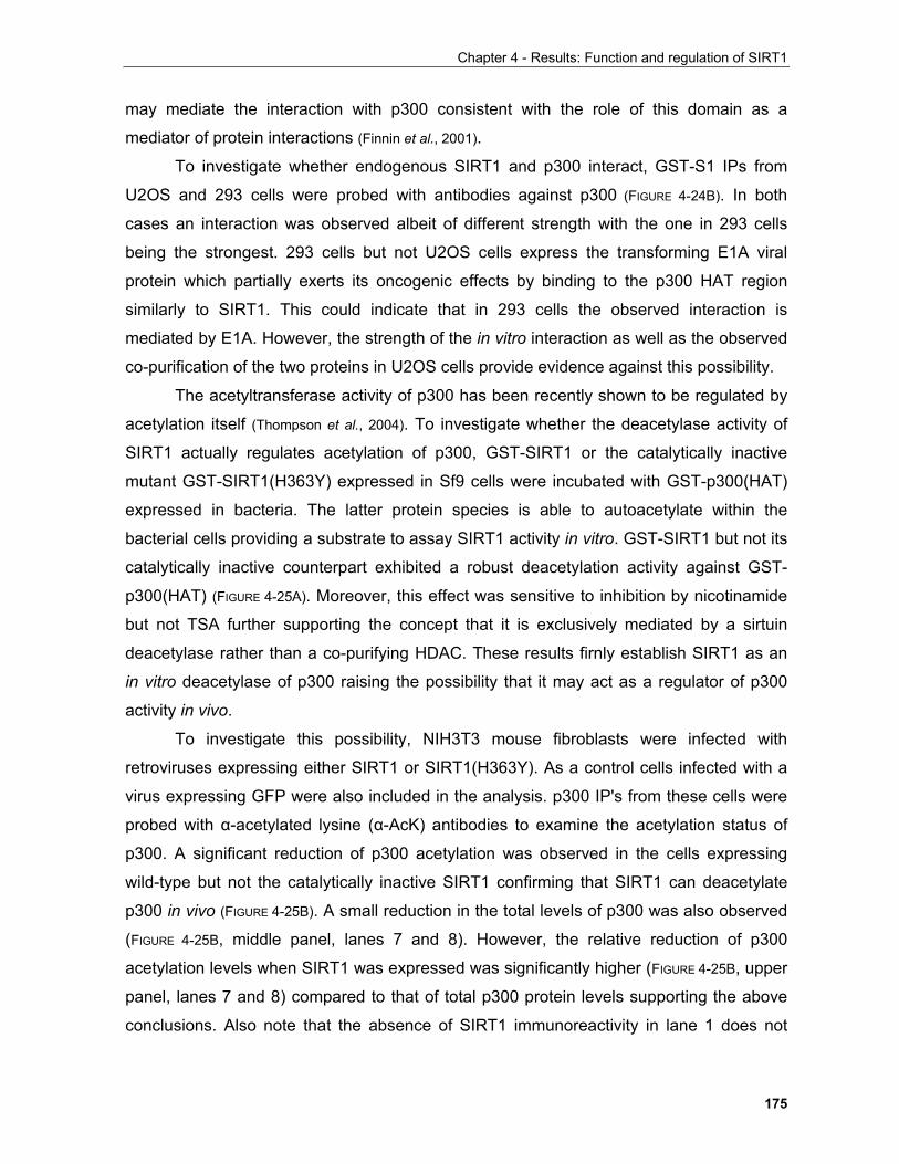
Chapter 4 - Results: Function and regulation of SIRT1
175
may mediate the interaction with p300 consistent with the role of this domain as a
mediator of protein interactions (Finnin et al., 2001).
To investigate whether endogenous SIRT1 and p300 interact, GST-S1 IPs from
U2OS and 293 cells were probed with antibodies against p300 (FIGURE 4-24B). In both
cases an interaction was observed albeit of different strength with the one in 293 cells
being the strongest. 293 cells but not U2OS cells express the transforming E1A viral
protein which partially exerts its oncogenic effects by binding to the p300 HAT region
similarly to SIRT1. This could indicate that in 293 cells the observed interaction is
mediated by E1A. However, the strength of the in vitro interaction as well as the observed
co-purification of the two proteins in U2OS cells provide evidence against this possibility.
The acetyltransferase activity of p300 has been recently shown to be regulated by
acetylation itself (Thompson et al., 2004). To investigate whether the deacetylase activity of
SIRT1 actually regulates acetylation of p300, GST-SIRT1 or the catalytically inactive
mutant GST-SIRT1(H363Y) expressed in Sf9 cells were incubated with GST-p300(HAT)
expressed in bacteria. The latter protein species is able to autoacetylate within the
bacterial cells providing a substrate to assay SIRT1 activity in vitro. GST-SIRT1 but not its
catalytically inactive counterpart exhibited a robust deacetylation activity against GST-
p300(HAT) (FIGURE 4-25A). Moreover, this effect was sensitive to inhibition by nicotinamide
but not TSA further supporting the concept that it is exclusively mediated by a sirtuin
deacetylase rather than a co-purifying HDAC. These results firnly establish SIRT1 as an
in vitro deacetylase of p300 raising the possibility that it may act as a regulator of p300
activity in vivo.
To investigate this possibility, NIH3T3 mouse fibroblasts were infected with
retroviruses expressing either SIRT1 or SIRT1(H363Y). As a control cells infected with a
virus expressing GFP were also included in the analysis. p300 IP's from these cells were
probed with α-acetylated lysine (α-AcK) antibodies to examine the acetylation status of
p300. A significant reduction of p300 acetylation was observed in the cells expressing
wild-type but not the catalytically inactive SIRT1 confirming that SIRT1 can deacetylate
p300 in vivo (FIGURE 4-25B). A small reduction in the total levels of p300 was also observed
(FIGURE 4-25B, middle panel, lanes 7 and 8). However, the relative reduction of p300
acetylation levels when SIRT1 was expressed was significantly higher (FIGURE 4-25B, upper
panel, lanes 7 and 8) compared to that of total p300 protein levels supporting the above
conclusions. Also note that the absence of SIRT1 immunoreactivity in lane 1 does not
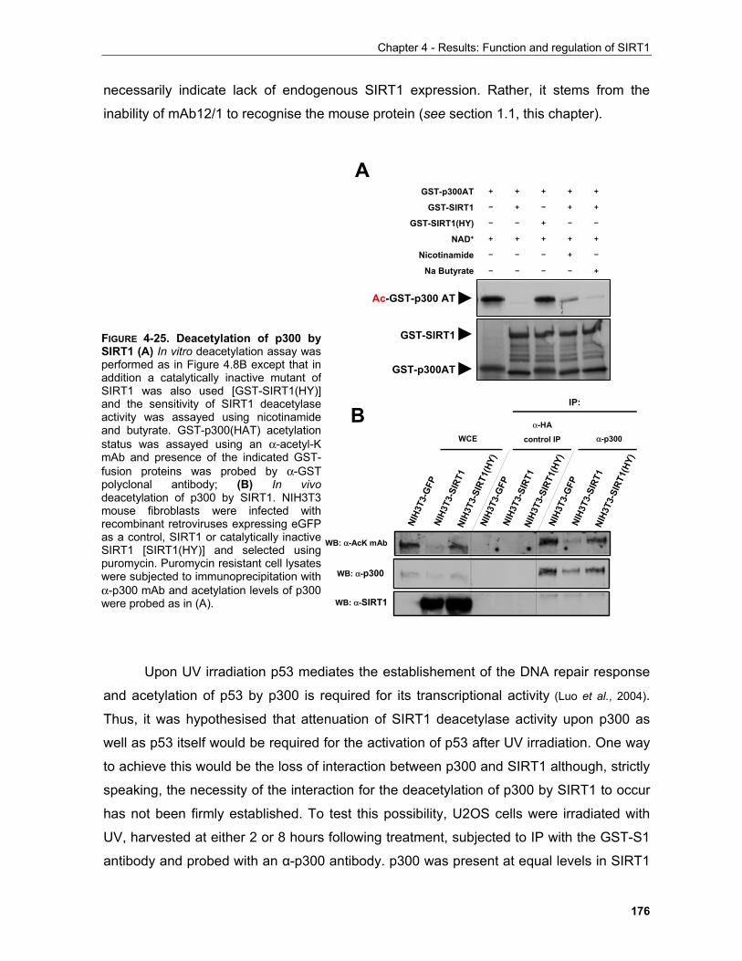
Chapter 4 - Results: Function and regulation of SIRT1
176
necessarily indicate lack of endogenous SIRT1 expression. Rather, it stems from the
inability of mAb12/1 to recognise the mouse protein (see section 1.1, this chapter).
Upon UV irradiation p53 mediates the establishement of the DNA repair response
and acetylation of p53 by p300 is required for its transcriptional activity (Luo et al., 2004).
Thus, it was hypothesised that attenuation of SIRT1 deacetylase activity upon p300 as
well as p53 itself would be required for the activation of p53 after UV irradiation. One way
to achieve this would be the loss of interaction between p300 and SIRT1 although, strictly
speaking, the necessity of the interaction for the deacetylation of p300 by SIRT1 to occur
has not been firmly established. To test this possibility, U2OS cells were irradiated with
UV, harvested at either 2 or 8 hours following treatment, subjected to IP with the GST-S1
antibody and probed with an α-p300 antibody. p300 was present at equal levels in SIRT1
IP:
WB: α-AcK mAb
WB: α-p300
WB: α-SIRT1
NIH3
T3-G
FPNI
H3T3
-SIR
T1
WCEα-HA
control IP α-p300
NIH3
T3-S
IRT1
(HY)
NIH3
T3-G
FPNI
H3T3
-SIR
T1NI
H3T3
-SIR
T1(H
Y)NI
H3T3
-GFP
NIH3
T3-S
IRT1
NIH3
T3-S
IRT1
(HY)
A
Ac-GST-p300 AT
GST-SIRT1
GST-p300AT
GST-p300AT
GST-SIRT1
GST-SIRT1(HY)
NAD+
Nicotinamide
Na Butyrate – – – – +
– – – + –
– + – + +
+ + + + +
– – + – –
+ + + + +
B
FIGURE 4-25. Deacetylation of p300 bySIRT1 (A) In vitro deacetylation assay wasperformed as in Figure 4.8B except that inaddition a catalytically inactive mutant ofSIRT1 was also used [GST-SIRT1(HY)]and the sensitivity of SIRT1 deacetylaseactivity was assayed using nicotinamideand butyrate. GST-p300(HAT) acetylationstatus was assayed using an α-acetyl-K mAb and presence of the indicated GST-fusion proteins was probed by α-GST polyclonal antibody; (B) In vivodeacetylation of p300 by SIRT1. NIH3T3mouse fibroblasts were infected withrecombinant retroviruses expressing eGFPas a control, SIRT1 or catalytically inactiveSIRT1 [SIRT1(HY)] and selected usingpuromycin. Puromycin resistant cell lysateswere subjected to immunoprecipitation withα-p300 mAb and acetylation levels of p300were probed as in (A).
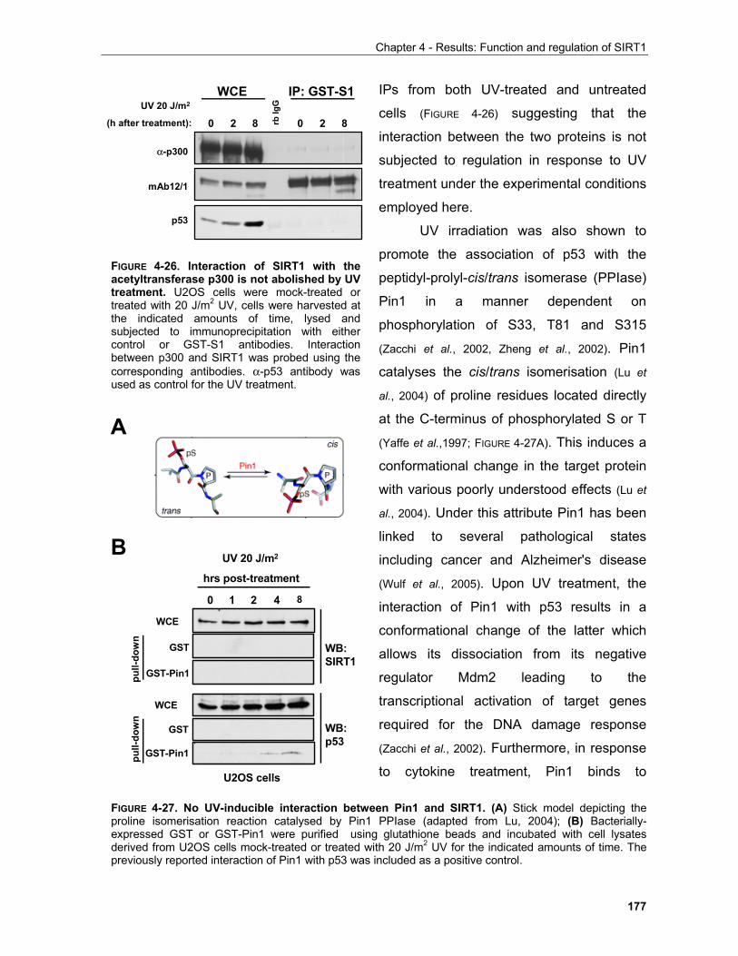
Chapter 4 - Results: Function and regulation of SIRT1
177
IPs from both UV-treated and untreated
cells (FIGURE 4-26) suggesting that the
interaction between the two proteins is not
subjected to regulation in response to UV
treatment under the experimental conditions
employed here.
UV irradiation was also shown to
promote the association of p53 with the
peptidyl-prolyl-cis/trans isomerase (PPIase)
Pin1 in a manner dependent on
phosphorylation of S33, T81 and S315
(Zacchi et al., 2002, Zheng et al., 2002). Pin1
catalyses the cis/trans isomerisation (Lu et
al., 2004) of proline residues located directly
at the C-terminus of phosphorylated S or T
(Yaffe et al.,1997; FIGURE 4-27A). This induces a
conformational change in the target protein
with various poorly understood effects (Lu et
al., 2004). Under this attribute Pin1 has been
linked to several pathological states
including cancer and Alzheimer's disease
(Wulf et al., 2005). Upon UV treatment, the
interaction of Pin1 with p53 results in a
conformational change of the latter which
allows its dissociation from its negative
regulator Mdm2 leading to the
transcriptional activation of target genes
required for the DNA damage response
(Zacchi et al., 2002). Furthermore, in response
to cytokine treatment, Pin1 binds to
UV 20 J/m2
(h after treatment):
mAb12/1
WCE IP: GST-S1
α-p300
rbIg
G
0 2 8 0 2 8
p53
FIGURE 4-26. Interaction of SIRT1 with theacetyltransferase p300 is not abolished by UVtreatment. U2OS cells were mock-treated ortreated with 20 J/m2 UV, cells were harvested atthe indicated amounts of time, lysed andsubjected to immunoprecipitation with eithercontrol or GST-S1 antibodies. Interactionbetween p300 and SIRT1 was probed using thecorresponding antibodies. α-p53 antibody wasused as control for the UV treatment.
UV 20 J/m2
hrs post-treatment
WB: SIRT1
WB:p53
GST-Pin1
GST
WCE
0 21 4 8
GST-Pin1
GST
WCE
U2OS cells
pull-
dow
npu
ll-do
wn
B
A
FIGURE 4-27. No UV-inducible interaction between Pin1 and SIRT1. (A) Stick model depicting theproline isomerisation reaction catalysed by Pin1 PPIase (adapted from Lu, 2004); (B) Bacterially-expressed GST or GST-Pin1 were purified using glutathione beads and incubated with cell lysatesderived from U2OS cells mock-treated or treated with 20 J/m2 UV for the indicated amounts of time. Thepreviously reported interaction of Pin1 with p53 was included as a positive control.
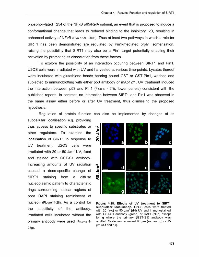
Chapter 4 - Results: Function and regulation of SIRT1
178
phosphorylated T254 of the NFκB p65/RelA subunit, an event that is proposed to induce a
conformational change that leads to reduced binding to the inhibitory IκB, resulting in
enhanced activity of NFκB (Ryo et al., 2003). Thus at least two pathways in which a role for
SIRT1 has been demonstrated are regulated by Pin1-mediated prolyl isomerisation,
raising the possibility that SIRT1 may also be a Pin1 target potentially enabling their
activation by promoting its dissociation from these factors.
To explore the possibility of an interaction occuring between SIRT1 and Pin1,
U2OS cells were irradiated with UV and harvested at various time-points. Lysates thereof
were incubated with glutathione beads bearing bound GST or GST-Pin1, washed and
subjected to immunoblotting with either p53 antibody or mAb12/1. UV treatment induced
the interaction between p53 and Pin1 (FIGURE 4-27B, lower panels) consistent with the
published reports. In contrast, no interaction between SIRT1 and Pin1 was observed in
the same assay either before or after UV treatment, thus dismissing the proposed
hypothesis.
Regulation of protein function can also be implemented by changes of its
subcellular localisation e.g. providing
thus access to specific substrates or
other regulators. To examine the
localisation of SIRT1 in response to
UV treatment, U2OS cells were
irradiated with 20 or 50 J/m2 UV, fixed
and stained with GST-S1 antibody.
Increasing amounts of UV radiation
caused a dose-specific change of
SIRT1 staining from a diffuse
nucleoplasmic pattern to characteristic
rings surrounding nuclear regions of
poor DAPI staining reminiscent of
nucleoli (Figure 4-28). As a control for
the specificity of the antibody,
irradiated cells incubated without the
primary antibody were used (FIGURE 4-
28g).
50 J
/m2
20 J
/m2
a b c
d e f
g h i
50 J
/m2
20 J
/m2
a b c
d e f
g h i
FIGURE 4-28. Effects of UV treatment to SIRT1subnuclear localisation. U2OS cells were treatedwith 20 (a-c) or 50 J/m2 (d-i) UV and immunostainedwith GST-S1 antibody (green) or DAPI (blue) exceptfor g where the primary (GST-S1) antibody wasomitted. Scalebars represent 90 µm (a-c and g) or 15µm (d-f and h,i).

Chapter 4 - Results: Function and regulation of SIRT1
179
In addition to the well-established function of nucleoli in the assembly of ribosomes,
a role in cellular stress responses is also emerging (Olson, 2004). In particular, nucleoli
appear to function as nuclear depots of proteins in a manner depending on their structural
integrity. Upon stress stimulation, disruption of nucleolar structure allows the release of
such factors partly contributing to the ensuing cellular responses to these stimuli. A more
detailed account of this function of nucleoli will be given in Chapter 5. Under the light of
these, it would be important to conclusively establish that the distinct localisation of SIRT1
is indeed associated with nucleoli and subsequently investigate the functional significance
of this phenomenon.
4.1.4 Regulation of SIRT1 by phosphorylation in mitosis According to its electrophoretic mobility during SDS-PAGE, SIRT1 was shown to
be a phosphoprotein which is subjected to hyper-phosphorylation in cells arrested in
mitosis by treatment with the microtubule-destabilising agent nocodazole (FIGURE 4-7). This
observation raised the possibility that SIRT1 is phosphorylated in a cell cycle-dependent
manner. Mitotic phosphorylation of SIRT1 may be particularly relevant especially given
the proposed roles of situins in life-span
determination under the light of increasing
evidence for a link between components of the
mitotic regulatory apparatus and the aging process (e.g. Ly et al., 2000; Baker et al., 2005 and discussion in
Chapter 5).
Cell proliferation requires a series of events
that are collectively known as the cell cycle. Four
distinct phases comprise a cell cycle (FIGURE 4-29A):
the S (synthesis) phase where the genome is
replicated, the M (mitosis) phase where all
components of the mother cell divide to give rise
to the daughter cells and two "gap" phases G1
preceding the S phase and G2 preceding the M
phase. All phases between two successive
mitoses are known as the interphase. Quiescent
B
A
Figure 4-29. Schematic representationof the eucaryotic cell cycle (A) andassociated changes in cyclin proteinlevels (B). (A) adapted from Purves et al. and (B)from Sherr, 1996.
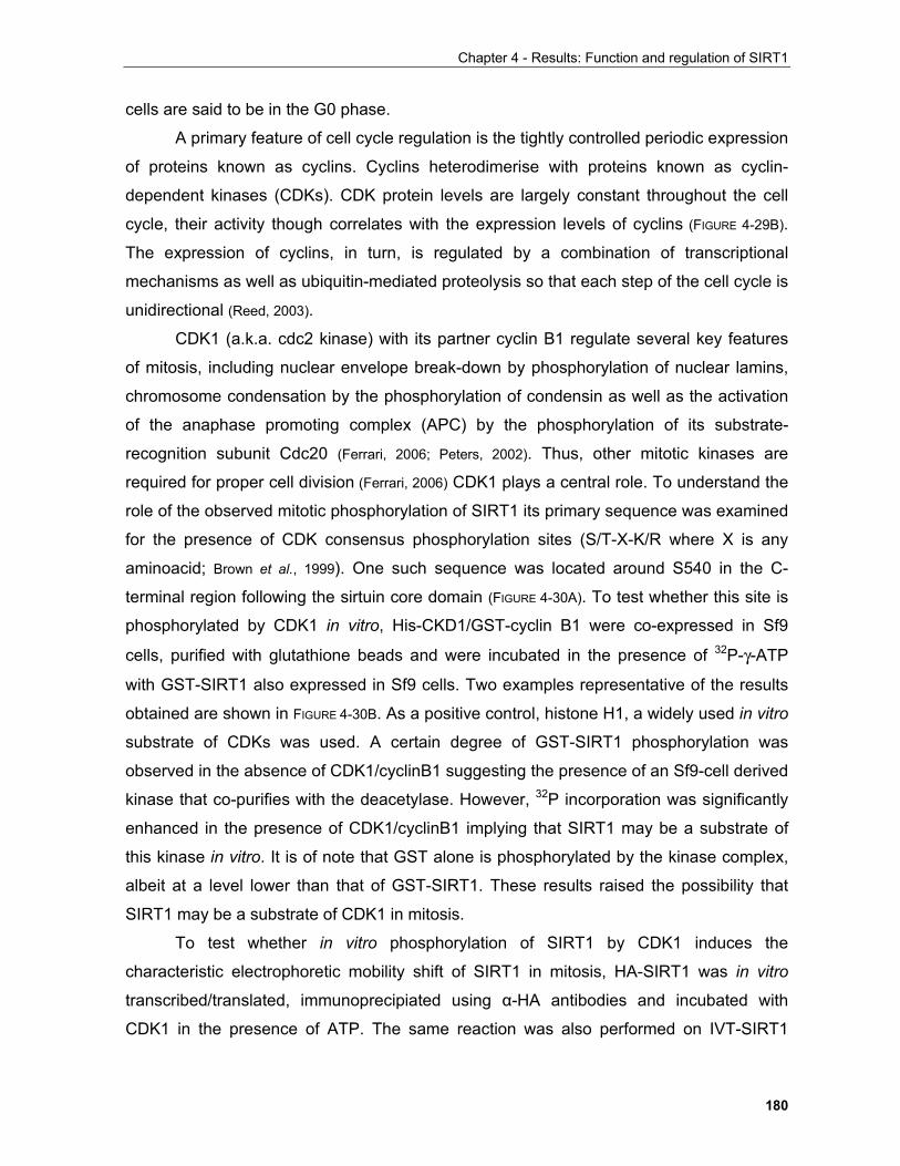
Chapter 4 - Results: Function and regulation of SIRT1
180
cells are said to be in the G0 phase.
A primary feature of cell cycle regulation is the tightly controlled periodic expression
of proteins known as cyclins. Cyclins heterodimerise with proteins known as cyclin-
dependent kinases (CDKs). CDK protein levels are largely constant throughout the cell
cycle, their activity though correlates with the expression levels of cyclins (FIGURE 4-29B).
The expression of cyclins, in turn, is regulated by a combination of transcriptional
mechanisms as well as ubiquitin-mediated proteolysis so that each step of the cell cycle is
unidirectional (Reed, 2003).
CDK1 (a.k.a. cdc2 kinase) with its partner cyclin B1 regulate several key features
of mitosis, including nuclear envelope break-down by phosphorylation of nuclear lamins,
chromosome condensation by the phosphorylation of condensin as well as the activation
of the anaphase promoting complex (APC) by the phosphorylation of its substrate-
recognition subunit Cdc20 (Ferrari, 2006; Peters, 2002). Thus, other mitotic kinases are
required for proper cell division (Ferrari, 2006) CDK1 plays a central role. To understand the
role of the observed mitotic phosphorylation of SIRT1 its primary sequence was examined
for the presence of CDK consensus phosphorylation sites (S/T-X-K/R where X is any
aminoacid; Brown et al., 1999). One such sequence was located around S540 in the C-
terminal region following the sirtuin core domain (FIGURE 4-30A). To test whether this site is
phosphorylated by CDK1 in vitro, His-CKD1/GST-cyclin B1 were co-expressed in Sf9
cells, purified with glutathione beads and were incubated in the presence of 32P-γ-ATP
with GST-SIRT1 also expressed in Sf9 cells. Two examples representative of the results
obtained are shown in FIGURE 4-30B. As a positive control, histone H1, a widely used in vitro
substrate of CDKs was used. A certain degree of GST-SIRT1 phosphorylation was
observed in the absence of CDK1/cyclinB1 suggesting the presence of an Sf9-cell derived
kinase that co-purifies with the deacetylase. However, 32P incorporation was significantly
enhanced in the presence of CDK1/cyclinB1 implying that SIRT1 may be a substrate of
this kinase in vitro. It is of note that GST alone is phosphorylated by the kinase complex,
albeit at a level lower than that of GST-SIRT1. These results raised the possibility that
SIRT1 may be a substrate of CDK1 in mitosis.
To test whether in vitro phosphorylation of SIRT1 by CDK1 induces the
characteristic electrophoretic mobility shift of SIRT1 in mitosis, HA-SIRT1 was in vitro
transcribed/translated, immunoprecipiated using α-HA antibodies and incubated with
CDK1 in the presence of ATP. The same reaction was also performed on IVT-SIRT1
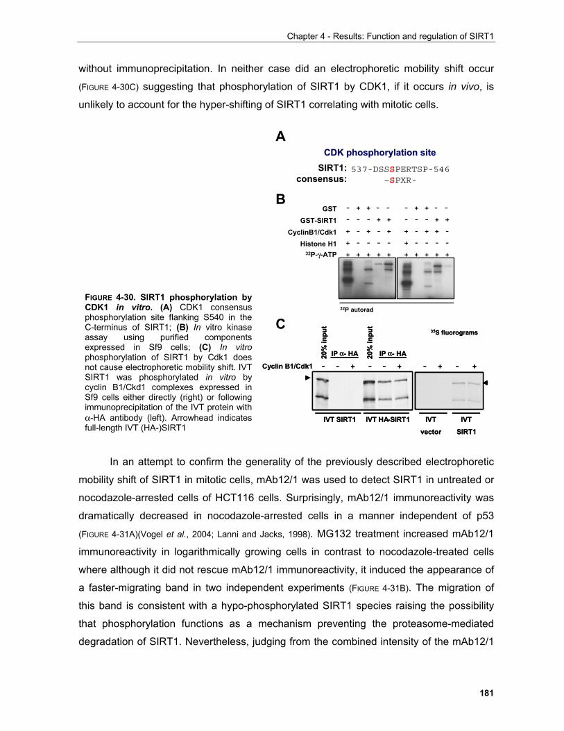
Chapter 4 - Results: Function and regulation of SIRT1
181
CyclinB1/Cdk1
GSTGST-SIRT1
Histone H1 + - - - - + - - - -
+ - + - + + - + + -
32P-γ-ATP + + + + + + + + + +
- - - + + - - - + +
- + + - - - + + - -
32P autorad
B
C
A
537-DSSSPERTSP-546 –SPXR-
CDK phosphorylation siteCDK phosphorylation siteSIRT1:
consensus:
Cyclin B1/Cdk1
IVT SIRT1 IVT HA-SIRT1 IVT
vector
IVT
SIRT1
- - + - - + - + - +
IP α- HA IP α- HA20%
inpu
t
20%
inpu
t
35S fluorograms
Cyclin B1/Cdk1
IVT SIRT1 IVT HA-SIRT1 IVT
vector
IVT
SIRT1
- - + - - + - + - +
IP α- HA IP α- HA20%
inpu
t
20%
inpu
t
35S fluorograms
FIGURE 4-30. SIRT1 phosphorylation byCDK1 in vitro. (A) CDK1 consensusphosphorylation site flanking S540 in theC-terminus of SIRT1; (B) In vitro kinaseassay using purified componentsexpressed in Sf9 cells; (C) In vitrophosphorylation of SIRT1 by Cdk1 doesnot cause electrophoretic mobility shift. IVTSIRT1 was phosphorylated in vitro bycyclin B1/Ckd1 complexes expressed inSf9 cells either directly (right) or followingimmunoprecipitation of the IVT protein withα-HA antibody (left). Arrowhead indicatesfull-length IVT (HA-)SIRT1
without immunoprecipitation. In neither case did an electrophoretic mobility shift occur
(FIGURE 4-30C) suggesting that phosphorylation of SIRT1 by CDK1, if it occurs in vivo, is
unlikely to account for the hyper-shifting of SIRT1 correlating with mitotic cells.
In an attempt to confirm the generality of the previously described electrophoretic
mobility shift of SIRT1 in mitotic cells, mAb12/1 was used to detect SIRT1 in untreated or
nocodazole-arrested cells of HCT116 cells. Surprisingly, mAb12/1 immunoreactivity was
dramatically decreased in nocodazole-arrested cells in a manner independent of p53
(FIGURE 4-31A)(Vogel et al., 2004; Lanni and Jacks, 1998). MG132 treatment increased mAb12/1
immunoreactivity in logarithmically growing cells in contrast to nocodazole-treated cells
where although it did not rescue mAb12/1 immunoreactivity, it induced the appearance of
a faster-migrating band in two independent experiments (FIGURE 4-31B). The migration of
this band is consistent with a hypo-phosphorylated SIRT1 species raising the possibility
that phosphorylation functions as a mechanism preventing the proteasome-mediated
degradation of SIRT1. Nevertheless, judging from the combined intensity of the mAb12/1
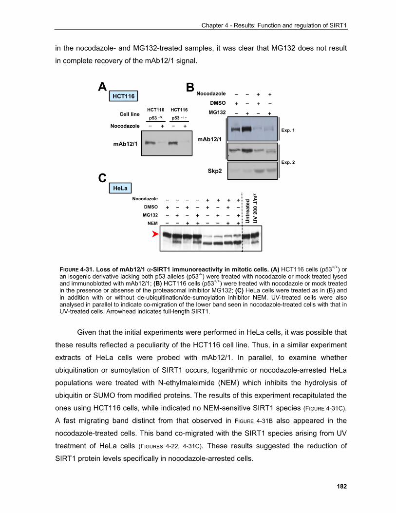
Chapter 4 - Results: Function and regulation of SIRT1
182
in the nocodazole- and MG132-treated samples, it was clear that MG132 does not result
in complete recovery of the mAb12/1 signal.
Given that the initial experiments were performed in HeLa cells, it was possible that
these results reflected a peculiarity of the HCT116 cell line. Thus, in a similar experiment
extracts of HeLa cells were probed with mAb12/1. In parallel, to examine whether
ubiquitination or sumoylation of SIRT1 occurs, logarithmic or nocodazole-arrested HeLa
populations were treated with N-ethylmaleimide (NEM) which inhibits the hydrolysis of
ubiquitin or SUMO from modified proteins. The results of this experiment recapitulated the
ones using HCT116 cells, while indicated no NEM-sensitive SIRT1 species (FIGURE 4-31C).
A fast migrating band distinct from that observed in FIGURE 4-31B also appeared in the
nocodazole-treated cells. This band co-migrated with the SIRT1 species arising from UV
treatment of HeLa cells (FIGURES 4-22, 4-31C). These results suggested the reduction of
SIRT1 protein levels specifically in nocodazole-arrested cells.
A
C
mAb12/1
Skp2
Exp. 1
Exp. 2
Nocodazole
DMSO
MG132
– – + +
+ – + –
– + – +
mAb12/1
Nocodazole – + – +
Cell lineHCT116
p53 +/+
HCT116
p53 – / –
Nocodazole
DMSO
MG132
NEM – – + + – – + + Unt
reat
edU
V 20
0 J/
m2
– – – – + + + ++ – + – + – + –– + – + – + – +
HCT116
HeLa
B
FIGURE 4-31. Loss of mAb12/1 α-SIRT1 immunoreactivity in mitotic cells. (A) HCT116 cells (p53+/+) oran isogenic derivative lacking both p53 alleles (p53-/-) were treated with nocodazole or mock treated lysedand immunoblotted with mAb12/1; (B) HCT116 cells (p53+/+) were treated with nocodazole or mock treatedin the presence or absense of the proteasomal inhibitor MG132; (C) HeLa cells were treated as in (B) andin addition with or without de-ubiquitination/de-sumoylation inhibitor NEM. UV-treated cells were alsoanalysed in parallel to indicate co-migration of the lower band seen in nocodazole-treated cells with that inUV-treated cells. Arrowhead indicates full-length SIRT1.
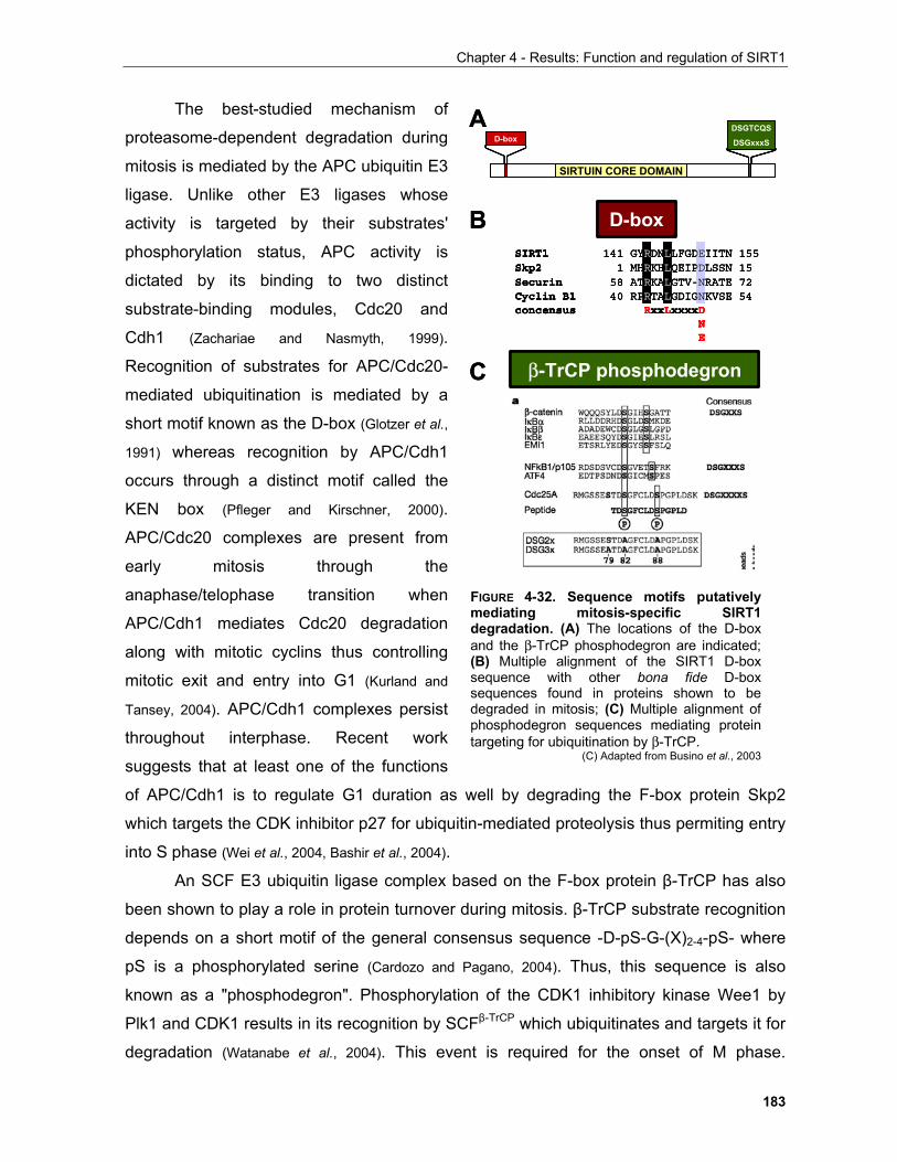
Chapter 4 - Results: Function and regulation of SIRT1
183
The best-studied mechanism of
proteasome-dependent degradation during
mitosis is mediated by the APC ubiquitin E3
ligase. Unlike other E3 ligases whose
activity is targeted by their substrates'
phosphorylation status, APC activity is
dictated by its binding to two distinct
substrate-binding modules, Cdc20 and
Cdh1 (Zachariae and Nasmyth, 1999).
Recognition of substrates for APC/Cdc20-
mediated ubiquitination is mediated by a
short motif known as the D-box (Glotzer et al.,
1991) whereas recognition by APC/Cdh1
occurs through a distinct motif called the
KEN box (Pfleger and Kirschner, 2000).
APC/Cdc20 complexes are present from
early mitosis through the
anaphase/telophase transition when
APC/Cdh1 mediates Cdc20 degradation
along with mitotic cyclins thus controlling
mitotic exit and entry into G1 (Kurland and
Tansey, 2004). APC/Cdh1 complexes persist
throughout interphase. Recent work
suggests that at least one of the functions
of APC/Cdh1 is to regulate G1 duration as well by degrading the F-box protein Skp2
which targets the CDK inhibitor p27 for ubiquitin-mediated proteolysis thus permiting entry
into S phase (Wei et al., 2004, Bashir et al., 2004).
An SCF E3 ubiquitin ligase complex based on the F-box protein β-TrCP has also
been shown to play a role in protein turnover during mitosis. β-TrCP substrate recognition
depends on a short motif of the general consensus sequence -D-pS-G-(X)2-4-pS- where
pS is a phosphorylated serine (Cardozo and Pagano, 2004). Thus, this sequence is also
known as a "phosphodegron". Phosphorylation of the CDK1 inhibitory kinase Wee1 by
Plk1 and CDK1 results in its recognition by SCFβ-TrCP which ubiquitinates and targets it for
degradation (Watanabe et al., 2004). This event is required for the onset of M phase.
SIRT1 141 GYRDNLLFGDEIITN 155Skp2 1 MHRKHLQEIPDLSSN 15Securin 58 ATRKALGTV-NRATE 72Cyclin B1 40 RPRTALGDIGNKVSE 54concensus RxxLxxxxD
NE
SIRT1 141 GYRDNLLFGDEIITN 155Skp2 1 MHRKHLQEIPDLSSN 15Securin 58 ATRKALGTV-NRATE 72Cyclin B1 40 RPRTALGDIGNKVSE 54concensus RxxLxxxxD
NE
D-box
β-TrCP phosphodegron
SIRTUIN CORE DOMAIN
D-boxDSGTCQS
DSGxxxS
A
B
C
SIRT1 141 GYRDNLLFGDEIITN 155Skp2 1 MHRKHLQEIPDLSSN 15Securin 58 ATRKALGTV-NRATE 72Cyclin B1 40 RPRTALGDIGNKVSE 54concensus RxxLxxxxD
NE
SIRT1 141 GYRDNLLFGDEIITN 155Skp2 1 MHRKHLQEIPDLSSN 15Securin 58 ATRKALGTV-NRATE 72Cyclin B1 40 RPRTALGDIGNKVSE 54concensus RxxLxxxxD
NE
D-box
β-TrCP phosphodegron
SIRTUIN CORE DOMAIN
D-boxDSGTCQS
DSGxxxS
A
B
C
FIGURE 4-32. Sequence motifs putativelymediating mitosis-specific SIRT1degradation. (A) The locations of the D-boxand the β-TrCP phosphodegron are indicated;(B) Multiple alignment of the SIRT1 D-boxsequence with other bona fide D-boxsequences found in proteins shown to bedegraded in mitosis; (C) Multiple alignment ofphosphodegron sequences mediating proteintargeting for ubiquitination by β-TrCP.
(C) Adapted from Busino et al., 2003
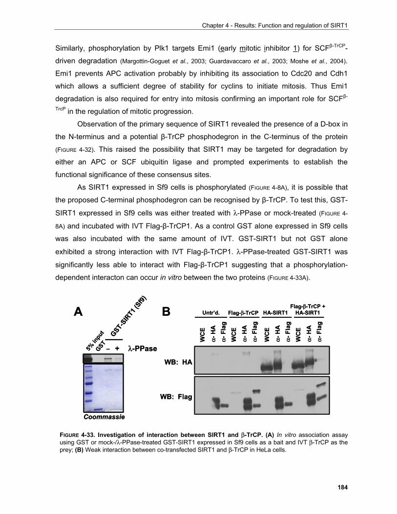
Chapter 4 - Results: Function and regulation of SIRT1
184
Similarly, phosphorylation by Plk1 targets Emi1 (early mitotic inhibitor 1) for SCFβ-TrCP-
driven degradation (Margottin-Goguet et al., 2003; Guardavaccaro et al., 2003; Moshe et al., 2004).
Emi1 prevents APC activation probably by inhibiting its association to Cdc20 and Cdh1
which allows a sufficient degree of stability for cyclins to initiate mitosis. Thus Emi1
degradation is also required for entry into mitosis confirming an important role for SCFβ-
TrcP in the regulation of mitotic progression.
Observation of the primary sequence of SIRT1 revealed the presence of a D-box in
the N-terminus and a potential β-TrCP phosphodegron in the C-terminus of the protein
(FIGURE 4-32). This raised the possibility that SIRT1 may be targeted for degradation by
either an APC or SCF ubiquitin ligase and prompted experiments to establish the
functional significance of these consensus sites.
As SIRT1 expressed in Sf9 cells is phosphorylated (FIGURE 4-8A), it is possible that
the proposed C-terminal phosphodegron can be recognised by β-TrCP. To test this, GST-
SIRT1 expressed in Sf9 cells was either treated with λ-PPase or mock-treated (FIGURE 4-
8A) and incubated with IVT Flag-β-TrCP1. As a control GST alone expressed in Sf9 cells
was also incubated with the same amount of IVT. GST-SIRT1 but not GST alone
exhibited a strong interaction with IVT Flag-β-TrCP1. λ-PPase-treated GST-SIRT1 was
significantly less able to interact with Flag-β-TrCP1 suggesting that a phosphorylation-
dependent interacton can occur in vitro between the two proteins (FIGURE 4-33A).
GSTGST-
SIRT1
(Sf9)
5% in
put
– + λ-PPase
Coommassie
A B
α-H
A
WC
E
α-Fl
ag
α-H
AW
CE
α-Fl
ag
α-H
AW
CE
α-Fl
ag
α-H
A
WC
E
α-Fl
agUntr’d. Flag-β-TrCP HA-SIRT1
Flag-β-TrCP + HA-SIRT1
WB: HA
WB: Flag
GSTGST-
SIRT1
(Sf9)
5% in
put
– + λ-PPase
Coommassie
A B
α-H
A
WC
E
α-Fl
ag
α-H
AW
CE
α-Fl
ag
α-H
AW
CE
α-Fl
ag
α-H
A
WC
E
α-Fl
agUntr’d. Flag-β-TrCP HA-SIRT1
Flag-β-TrCP + HA-SIRT1
WB: HA
WB: Flag
FIGURE 4-33. Investigation of interaction between SIRT1 and β-TrCP. (A) In vitro association assayusing GST or mock-/λ-PPase-treated GST-SIRT1 expressed in Sf9 cells as a bait and IVT β-TrCP as theprey; (B) Weak interaction between co-transfected SIRT1 and β-TrCP in HeLa cells.
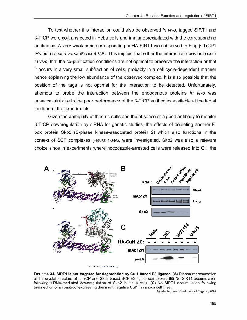
Chapter 4 - Results: Function and regulation of SIRT1
185
To test whether this interaction could also be observed in vivo, tagged SIRT1 and
β-TrCP were co-transfected in HeLa cells and immunoprecipitated with the corresponding
antibodies. A very weak band corresponding to HA-SIRT1 was observed in Flag-β-TrCP1
IPs but not vice versa (FIGURE 4-33B). This implied that either the interaction does not occur
in vivo, that the co-purification conditions are not optimal to preserve the interaction or that
it occurs in a very small subfraction of cells, probably in a cell cycle-dependent manner
hence explaining the low abundance of the observed complex. It is also possible that the
position of the tags is not optimal for the interaction to be detected. Unfortunately,
attempts to probe the interaction between the endogenous proteins in vivo was
unsuccessful due to the poor performance of the β-TrCP antibodies available at the lab at
the time of the experiments.
Given the ambiguity of these results and the absence or a good antibody to monitor
β-TrCP downregulation by siRNA for genetic studies, the effects of depleting another F-
box protein Skp2 (S-phase kinase-associated protein 2) which also functions in the
context of SCF complexes (FIGURE 4-34A), were investigated. Skp2 was also a relevant
choice since in experiments where nocodazole-arrested cells were released into G1, the
Untra
nsfec
ted
Skp2 4
0 nM
mock
Contro
l siR
NASkp
2 20 n
M
RNAi:
Skp2
mAb12/1Short
Long
Hela U2OS
HCT116
293
– + – + – + – +HA-Cul1 ∆C:
mAb12/1
α-HA
A
C
B
Untra
nsfec
ted
Skp2 4
0 nM
mock
Contro
l siR
NASkp
2 20 n
M
RNAi:
Skp2
mAb12/1Short
Long
Hela U2OS
HCT116
293
– + – + – + – +HA-Cul1 ∆C:
mAb12/1
α-HA
A
C
B
FIGURE 4-34. SIRT1 is not targeted for degradation by Cul1-based E3 ligases. (A) Ribbon representationof the crystal structure of β-TrCP and Skp2-based SCF E3 ligase complexes; (B) No SIRT1 accumulationfollowing siRNA-mediated downregulation of Skp2 in HeLa cells; (C) No SIRT1 accumulation followingtransfection of a construct expressing dominant negative Cul1 in various cell lines.
(A) adapted from Cardozo and Pagano, 2004
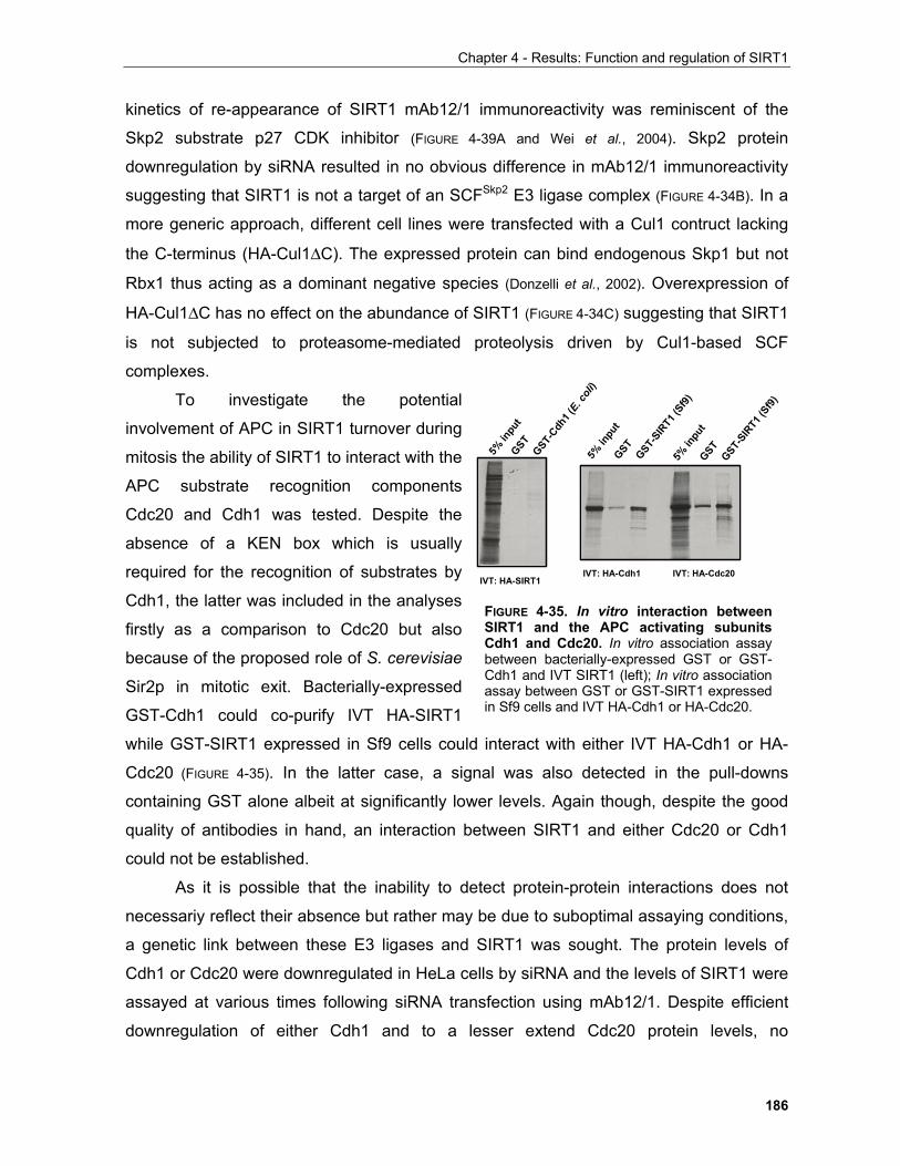
Chapter 4 - Results: Function and regulation of SIRT1
186
kinetics of re-appearance of SIRT1 mAb12/1 immunoreactivity was reminiscent of the
Skp2 substrate p27 CDK inhibitor (FIGURE 4-39A and Wei et al., 2004). Skp2 protein
downregulation by siRNA resulted in no obvious difference in mAb12/1 immunoreactivity
suggesting that SIRT1 is not a target of an SCFSkp2 E3 ligase complex (FIGURE 4-34B). In a
more generic approach, different cell lines were transfected with a Cul1 contruct lacking
the C-terminus (HA-Cul1∆C). The expressed protein can bind endogenous Skp1 but not
Rbx1 thus acting as a dominant negative species (Donzelli et al., 2002). Overexpression of
HA-Cul1∆C has no effect on the abundance of SIRT1 (FIGURE 4-34C) suggesting that SIRT1
is not subjected to proteasome-mediated proteolysis driven by Cul1-based SCF
complexes.
To investigate the potential
involvement of APC in SIRT1 turnover during
mitosis the ability of SIRT1 to interact with the
APC substrate recognition components
Cdc20 and Cdh1 was tested. Despite the
absence of a KEN box which is usually
required for the recognition of substrates by
Cdh1, the latter was included in the analyses
firstly as a comparison to Cdc20 but also
because of the proposed role of S. cerevisiae
Sir2p in mitotic exit. Bacterially-expressed
GST-Cdh1 could co-purify IVT HA-SIRT1
while GST-SIRT1 expressed in Sf9 cells could interact with either IVT HA-Cdh1 or HA-
Cdc20 (FIGURE 4-35). In the latter case, a signal was also detected in the pull-downs
containing GST alone albeit at significantly lower levels. Again though, despite the good
quality of antibodies in hand, an interaction between SIRT1 and either Cdc20 or Cdh1
could not be established.
As it is possible that the inability to detect protein-protein interactions does not
necessariy reflect their absence but rather may be due to suboptimal assaying conditions,
a genetic link between these E3 ligases and SIRT1 was sought. The protein levels of
Cdh1 or Cdc20 were downregulated in HeLa cells by siRNA and the levels of SIRT1 were
assayed at various times following siRNA transfection using mAb12/1. Despite efficient
downregulation of either Cdh1 and to a lesser extend Cdc20 protein levels, no
IVT: HA-SIRT1
GSTGST-
Cdh1 (
E. coli
)
5% in
put
GSTGST-
SIRT1
(Sf9)
5% in
put
GSTGST-
SIRT1
(Sf9)
5% in
put
IVT: HA-Cdh1 IVT: HA-Cdc20
FIGURE 4-35. In vitro interaction betweenSIRT1 and the APC activating subunitsCdh1 and Cdc20. In vitro association assaybetween bacterially-expressed GST or GST-Cdh1 and IVT SIRT1 (left); In vitro associationassay between GST or GST-SIRT1 expressedin Sf9 cells and IVT HA-Cdh1 or HA-Cdc20.
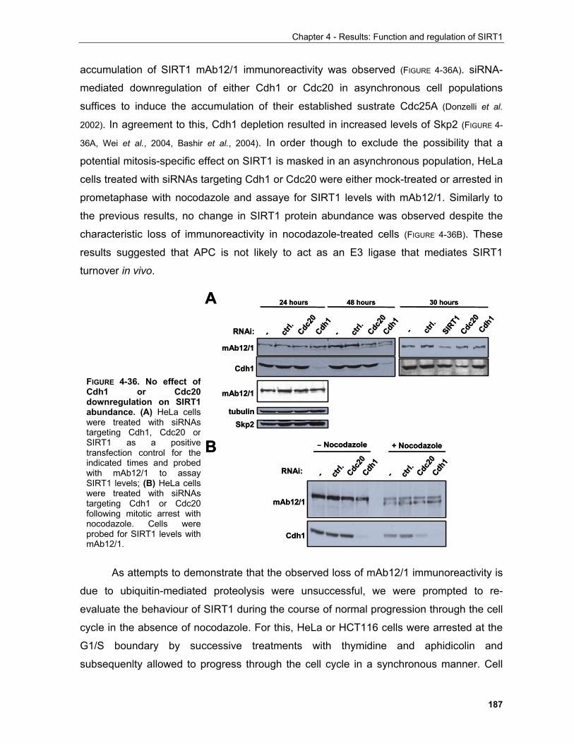
Chapter 4 - Results: Function and regulation of SIRT1
187
accumulation of SIRT1 mAb12/1 immunoreactivity was observed (FIGURE 4-36A). siRNA-
mediated downregulation of either Cdh1 or Cdc20 in asynchronous cell populations
suffices to induce the accumulation of their established sustrate Cdc25A (Donzelli et al.
2002). In agreement to this, Cdh1 depletion resulted in increased levels of Skp2 (FIGURE 4-
36A, Wei et al., 2004, Bashir et al., 2004). In order though to exclude the possibility that a
potential mitosis-specific effect on SIRT1 is masked in an asynchronous population, HeLa
cells treated with siRNAs targeting Cdh1 or Cdc20 were either mock-treated or arrested in
prometaphase with nocodazole and assaye for SIRT1 levels with mAb12/1. Similarly to
the previous results, no change in SIRT1 protein abundance was observed despite the
characteristic loss of immunoreactivity in nocodazole-treated cells (FIGURE 4-36B). These
results suggested that APC is not likely to act as an E3 ligase that mediates SIRT1
turnover in vivo.
As attempts to demonstrate that the observed loss of mAb12/1 immunoreactivity is
due to ubiquitin-mediated proteolysis were unsuccessful, we were prompted to re-
evaluate the behaviour of SIRT1 during the course of normal progression through the cell
cycle in the absence of nocodazole. For this, HeLa or HCT116 cells were arrested at the
G1/S boundary by successive treatments with thymidine and aphidicolin and
subsequenlty allowed to progress through the cell cycle in a synchronous manner. Cell
– Nocodazole + Nocodazole
- ctrl.
Cdh1
Cdc20
- ctrl.
Cdh1
Cdc20
Cdh1
mAb12/1
RNAi:
RNAi:
tubulinSkp2
24 hours 48 hours
mAb12/1
Cdh1
- ctrl.
Cdh1
Cdc20
- ctrl.
Cdh1
Cdc20
- ctrl.
Cdh1
Cdc20
SIRT1
30 hours
mAb12/1
A
B – Nocodazole + Nocodazole
- ctrl.
Cdh1
Cdc20
- ctrl.
Cdh1
Cdc20
Cdh1
mAb12/1
RNAi:
RNAi:
tubulinSkp2
24 hours 48 hours
mAb12/1
Cdh1
- ctrl.
Cdh1
Cdc20
- ctrl.
Cdh1
Cdc20
- ctrl.
Cdh1
Cdc20
SIRT1
30 hours
mAb12/1
A
B
FIGURE 4-36. No effect ofCdh1 or Cdc20downregulation on SIRT1abundance. (A) HeLa cellswere treated with siRNAstargeting Cdh1, Cdc20 orSIRT1 as a positivetransfection control for theindicated times and probedwith mAb12/1 to assaySIRT1 levels; (B) HeLa cellswere treated with siRNAstargeting Cdh1 or Cdc20following mitotic arrest withnocodazole. Cells wereprobed for SIRT1 levels withmAb12/1.
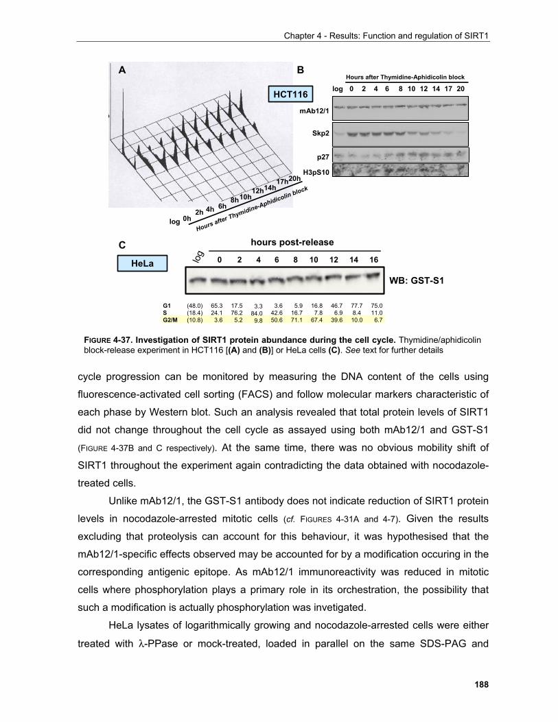
Chapter 4 - Results: Function and regulation of SIRT1
188
cycle progression can be monitored by measuring the DNA content of the cells using
fluorescence-activated cell sorting (FACS) and follow molecular markers characteristic of
each phase by Western blot. Such an analysis revealed that total protein levels of SIRT1
did not change throughout the cell cycle as assayed using both mAb12/1 and GST-S1
(FIGURE 4-37B and C respectively). At the same time, there was no obvious mobility shift of
SIRT1 throughout the experiment again contradicting the data obtained with nocodazole-
treated cells.
Unlike mAb12/1, the GST-S1 antibody does not indicate reduction of SIRT1 protein
levels in nocodazole-arrested mitotic cells (cf. FIGURES 4-31A and 4-7). Given the results
excluding that proteolysis can account for this behaviour, it was hypothesised that the
mAb12/1-specific effects observed may be accounted for by a modification occuring in the
corresponding antigenic epitope. As mAb12/1 immunoreactivity was reduced in mitotic
cells where phosphorylation plays a primary role in its orchestration, the possibility that
such a modification is actually phosphorylation was invetigated.
HeLa lysates of logarithmically growing and nocodazole-arrested cells were either
treated with λ-PPase or mock-treated, loaded in parallel on the same SDS-PAG and
2h 4h 6h8h10h
12h14h17h20h
0hlog
2 4 6 8 10 12 14 170log 20Hours after Thymidine-Aphidicolin block
mAb12/1
Skp2
p27
H3pS10
Hours after Thymidine-Aphidicolin block
A B
C
HeLa
HCT116
log 0 2 4 6 8 10 12 14 16
hours post-release
WB: GST-S1
G1SG2/M
65.324.13.6
17.576.2
5.2
3.384.0
9.8
3.642.650.6
5.916.771.1
16.87.8
67.4
46.76.9
39.6
77.78.410.0
75.011.0
6.7
(48.0)(18.4)(10.8)
FIGURE 4-37. Investigation of SIRT1 protein abundance during the cell cycle. Thymidine/aphidicolinblock-release experiment in HCT116 [(A) and (B)] or HeLa cells (C). See text for further details
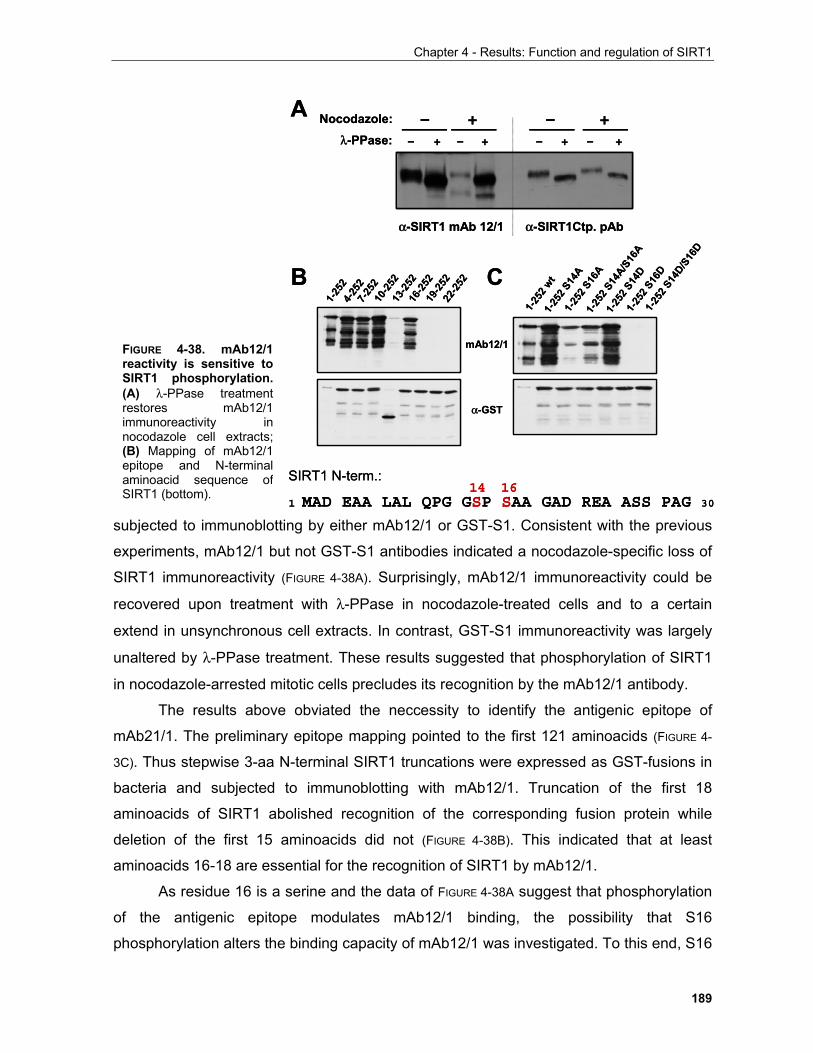
Chapter 4 - Results: Function and regulation of SIRT1
189
subjected to immunoblotting by either mAb12/1 or GST-S1. Consistent with the previous
experiments, mAb12/1 but not GST-S1 antibodies indicated a nocodazole-specific loss of
SIRT1 immunoreactivity (FIGURE 4-38A). Surprisingly, mAb12/1 immunoreactivity could be
recovered upon treatment with λ-PPase in nocodazole-treated cells and to a certain
extend in unsynchronous cell extracts. In contrast, GST-S1 immunoreactivity was largely
unaltered by λ-PPase treatment. These results suggested that phosphorylation of SIRT1
in nocodazole-arrested mitotic cells precludes its recognition by the mAb12/1 antibody.
The results above obviated the neccessity to identify the antigenic epitope of
mAb21/1. The preliminary epitope mapping pointed to the first 121 aminoacids (FIGURE 4-
3C). Thus stepwise 3-aa N-terminal SIRT1 truncations were expressed as GST-fusions in
bacteria and subjected to immunoblotting with mAb12/1. Truncation of the first 18
aminoacids of SIRT1 abolished recognition of the corresponding fusion protein while
deletion of the first 15 aminoacids did not (FIGURE 4-38B). This indicated that at least
aminoacids 16-18 are essential for the recognition of SIRT1 by mAb12/1.
As residue 16 is a serine and the data of FIGURE 4-38A suggest that phosphorylation
of the antigenic epitope modulates mAb12/1 binding, the possibility that S16
phosphorylation alters the binding capacity of mAb12/1 was investigated. To this end, S16
α-SIRT1Ctp. pAbα-SIRT1 mAb 12/1
– + – + – + – +λ-PPase:Nocodazole: – + – +
1-25
24-
252
1-25
2 wt
7-25
210
-252
13-2
5216
-252
19-2
5222
-252
1-25
2 S14
A1-
252 S
16A
1-25
2 S14
A/S16
A
1-25
2 S14
D1-
252 S
16D
1-25
2 S14
D/S16
D
mAb12/1
α-GST
1 MAD EAA LAL QPG GSP SAA GAD REA ASS PAG 30
SIRT1 N-term.:14 16
A
B C
FIGURE 4-38. mAb12/1reactivity is sensitive toSIRT1 phosphorylation.(A) λ-PPase treatmentrestores mAb12/1immunoreactivity innocodazole cell extracts;(B) Mapping of mAb12/1epitope and N-terminalaminoacid sequence ofSIRT1 (bottom).
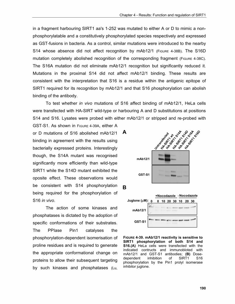
Chapter 4 - Results: Function and regulation of SIRT1
190
in a fragment harbouring SIRT1 aa's 1-252 was mutated to either A or D to mimic a non-
phosphorylatable and a constitutively phosphorylated species respectively and expressed
as GST-fusions in bacteria. As a control, similar mutations were introduced to the nearby
S14 whose absence did not affect recognition by mAb12/1 (FIGURE 4-38B). The S16D
mutation completely abolished recognition of the corresponding fragment (FIGURE 4-38C).
The S16A mutation did not eliminate mAb12/1 recognition but significantly reduced it.
Mutations in the proximal S14 did not affect mAb12/1 binding. These results are
consistent with the interpretation that S16 is a residue within the antigenic epitope of
SIRT1 required for its recognition by mAb12/1 and that S16 phosphorylation can abolish
binding of the antibody.
To test whether in vivo mutations of S16 affect binding of mAb12/1, HeLa cells
were transfected with HA-SIRT wild-type or harbouring A and D substitutions at positions
S14 and S16. Lysates were probed with either mAb12/1 or stripped and re-probed with
GST-S1. As shown in FIGURE 4-39A, either A
or D mutations of S16 abolished mAb12/1
binding in agreement with the results using
bacterially expressed proteins. Interestingly
though, the S14A mutant was recognised
significantly more efficiently than wild-type
SIRT1 while the S14D mutant exhibited the
oposite effect. These observations would
be consistent with S14 phosphorylation
being required for the phosphorylation of
S16 in vivo.
The action of some kinases and
phosphatases is dictated by the adoption of
specific conformations of their substrates.
The PPIase Pin1 catalyses the
phosphorylation-dependent isomerisation of
proline residues and is required to generate
the appropriate conformational change on
proteins to allow their subsequent targeting
by such kinases and phosphatases (Lu,
FIGURE 4-39. mAb12/1 reactivity is sensitive toSIRT1 phosphorylation of both S14 andS16.(A) HeLa cells were transfected with theindicated contructs and immunobloted withmAb12/1 and GST-S1 antibodies; (B) Dose-dependent inhibition of SIRT1 S16phosphorylation by the Pin1 prolyl isomeraseinhibitor juglone.
mAb12/1
GST-S1
+Nocodazole -NocodazoleJuglone (µM): 0 0 2010 30 2010 30
HA-SIR
T1 w
t
HA-SIR
T1 S
14A
HA-SIR
T1 S
16A
HA-SIR
T1 S
14D
HA-SIR
T1 S
16D
Untra
nsfe
cted
mAb12/1
GST-S1
A
B
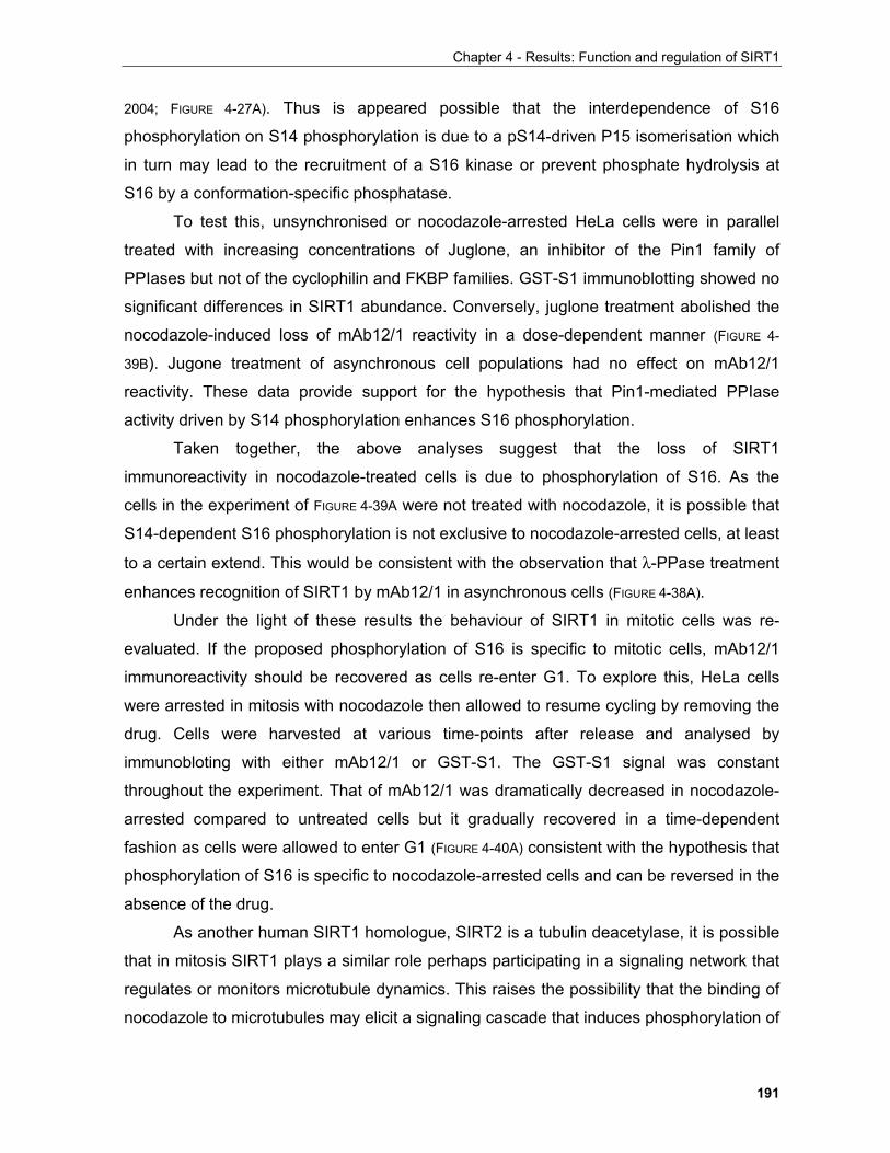
Chapter 4 - Results: Function and regulation of SIRT1
191
2004; FIGURE 4-27A). Thus is appeared possible that the interdependence of S16
phosphorylation on S14 phosphorylation is due to a pS14-driven P15 isomerisation which
in turn may lead to the recruitment of a S16 kinase or prevent phosphate hydrolysis at
S16 by a conformation-specific phosphatase.
To test this, unsynchronised or nocodazole-arrested HeLa cells were in parallel
treated with increasing concentrations of Juglone, an inhibitor of the Pin1 family of
PPIases but not of the cyclophilin and FKBP families. GST-S1 immunoblotting showed no
significant differences in SIRT1 abundance. Conversely, juglone treatment abolished the
nocodazole-induced loss of mAb12/1 reactivity in a dose-dependent manner (FIGURE 4-
39B). Jugone treatment of asynchronous cell populations had no effect on mAb12/1
reactivity. These data provide support for the hypothesis that Pin1-mediated PPIase
activity driven by S14 phosphorylation enhances S16 phosphorylation.
Taken together, the above analyses suggest that the loss of SIRT1
immunoreactivity in nocodazole-treated cells is due to phosphorylation of S16. As the
cells in the experiment of FIGURE 4-39A were not treated with nocodazole, it is possible that
S14-dependent S16 phosphorylation is not exclusive to nocodazole-arrested cells, at least
to a certain extend. This would be consistent with the observation that λ-PPase treatment
enhances recognition of SIRT1 by mAb12/1 in asynchronous cells (FIGURE 4-38A).
Under the light of these results the behaviour of SIRT1 in mitotic cells was re-
evaluated. If the proposed phosphorylation of S16 is specific to mitotic cells, mAb12/1
immunoreactivity should be recovered as cells re-enter G1. To explore this, HeLa cells
were arrested in mitosis with nocodazole then allowed to resume cycling by removing the
drug. Cells were harvested at various time-points after release and analysed by
immunobloting with either mAb12/1 or GST-S1. The GST-S1 signal was constant
throughout the experiment. That of mAb12/1 was dramatically decreased in nocodazole-
arrested compared to untreated cells but it gradually recovered in a time-dependent
fashion as cells were allowed to enter G1 (FIGURE 4-40A) consistent with the hypothesis that
phosphorylation of S16 is specific to nocodazole-arrested cells and can be reversed in the
absence of the drug.
As another human SIRT1 homologue, SIRT2 is a tubulin deacetylase, it is possible
that in mitosis SIRT1 plays a similar role perhaps participating in a signaling network that
regulates or monitors microtubule dynamics. This raises the possibility that the binding of
nocodazole to microtubules may elicit a signaling cascade that induces phosphorylation of
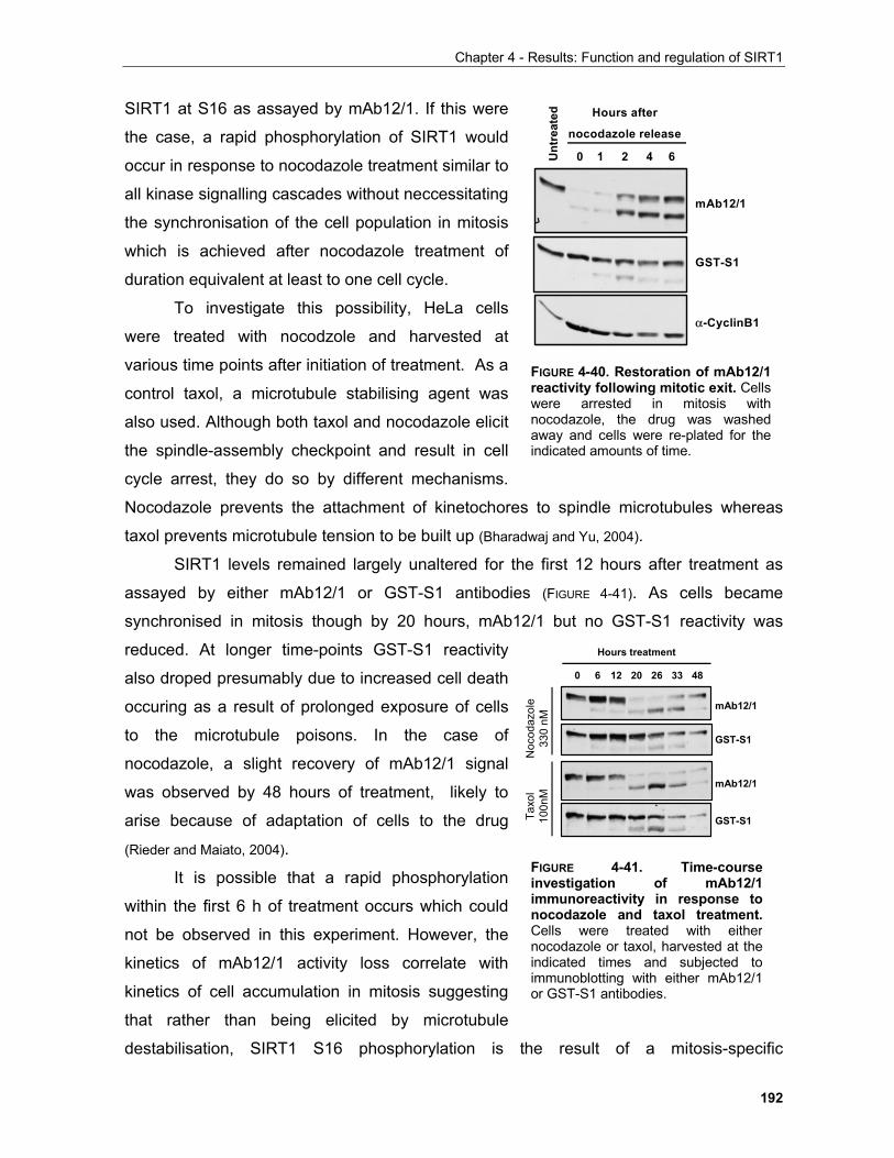
Chapter 4 - Results: Function and regulation of SIRT1
192
SIRT1 at S16 as assayed by mAb12/1. If this were
the case, a rapid phosphorylation of SIRT1 would
occur in response to nocodazole treatment similar to
all kinase signalling cascades without neccessitating
the synchronisation of the cell population in mitosis
which is achieved after nocodazole treatment of
duration equivalent at least to one cell cycle.
To investigate this possibility, HeLa cells
were treated with nocodzole and harvested at
various time points after initiation of treatment. As a
control taxol, a microtubule stabilising agent was
also used. Although both taxol and nocodazole elicit
the spindle-assembly checkpoint and result in cell
cycle arrest, they do so by different mechanisms.
Nocodazole prevents the attachment of kinetochores to spindle microtubules whereas
taxol prevents microtubule tension to be built up (Bharadwaj and Yu, 2004).
SIRT1 levels remained largely unaltered for the first 12 hours after treatment as
assayed by either mAb12/1 or GST-S1 antibodies (FIGURE 4-41). As cells became
synchronised in mitosis though by 20 hours, mAb12/1 but no GST-S1 reactivity was
reduced. At longer time-points GST-S1 reactivity
also droped presumably due to increased cell death
occuring as a result of prolonged exposure of cells
to the microtubule poisons. In the case of
nocodazole, a slight recovery of mAb12/1 signal
was observed by 48 hours of treatment, likely to
arise because of adaptation of cells to the drug
(Rieder and Maiato, 2004).
It is possible that a rapid phosphorylation
within the first 6 h of treatment occurs which could
not be observed in this experiment. However, the
kinetics of mAb12/1 activity loss correlate with
kinetics of cell accumulation in mitosis suggesting
that rather than being elicited by microtubule
destabilisation, SIRT1 S16 phosphorylation is the result of a mitosis-specific
FIGURE 4-40. Restoration of mAb12/1reactivity following mitotic exit. Cellswere arrested in mitosis withnocodazole, the drug was washedaway and cells were re-plated for theindicated amounts of time.
mAb12/1
GST-S1
α-CyclinB1
62 41
Hours after
nocodazole release
0Unt
reat
ed
Taxo
l10
0nM
Noc
odaz
ole
330
nM
2612 20 336
Hours treatment
0 48
mAb12/1
GST-S1
mAb12/1
GST-S1
FIGURE 4-41. Time-course investigation of mAb12/1 immunoreactivity in response to nocodazole and taxol treatment. Cells were treated with either nocodazole or taxol, harvested at the indicated times and subjected to immunoblotting with either mAb12/1 or GST-S1 antibodies.
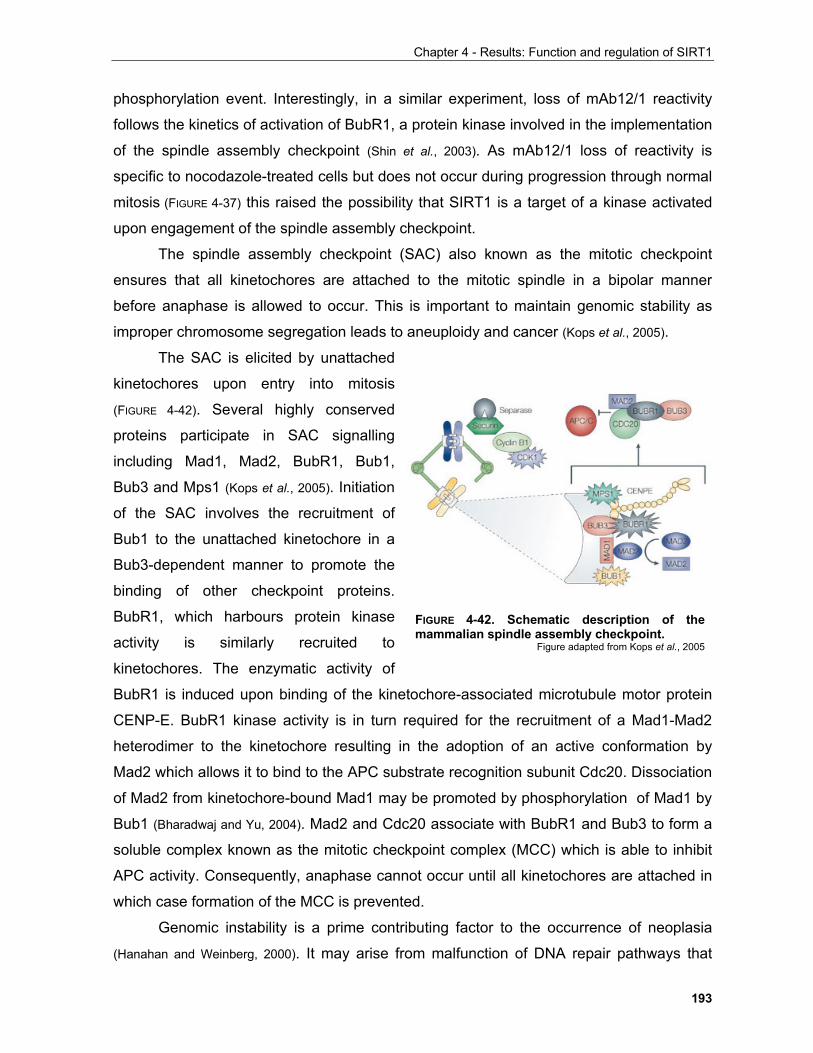
Chapter 4 - Results: Function and regulation of SIRT1
193
phosphorylation event. Interestingly, in a similar experiment, loss of mAb12/1 reactivity
follows the kinetics of activation of BubR1, a protein kinase involved in the implementation
of the spindle assembly checkpoint (Shin et al., 2003). As mAb12/1 loss of reactivity is
specific to nocodazole-treated cells but does not occur during progression through normal
mitosis (FIGURE 4-37) this raised the possibility that SIRT1 is a target of a kinase activated
upon engagement of the spindle assembly checkpoint.
The spindle assembly checkpoint (SAC) also known as the mitotic checkpoint
ensures that all kinetochores are attached to the mitotic spindle in a bipolar manner
before anaphase is allowed to occur. This is important to maintain genomic stability as
improper chromosome segregation leads to aneuploidy and cancer (Kops et al., 2005).
The SAC is elicited by unattached
kinetochores upon entry into mitosis
(FIGURE 4-42). Several highly conserved
proteins participate in SAC signalling
including Mad1, Mad2, BubR1, Bub1,
Bub3 and Mps1 (Kops et al., 2005). Initiation
of the SAC involves the recruitment of
Bub1 to the unattached kinetochore in a
Bub3-dependent manner to promote the
binding of other checkpoint proteins.
BubR1, which harbours protein kinase
activity is similarly recruited to
kinetochores. The enzymatic activity of
BubR1 is induced upon binding of the kinetochore-associated microtubule motor protein
CENP-E. BubR1 kinase activity is in turn required for the recruitment of a Mad1-Mad2
heterodimer to the kinetochore resulting in the adoption of an active conformation by
Mad2 which allows it to bind to the APC substrate recognition subunit Cdc20. Dissociation
of Mad2 from kinetochore-bound Mad1 may be promoted by phosphorylation of Mad1 by
Bub1 (Bharadwaj and Yu, 2004). Mad2 and Cdc20 associate with BubR1 and Bub3 to form a
soluble complex known as the mitotic checkpoint complex (MCC) which is able to inhibit
APC activity. Consequently, anaphase cannot occur until all kinetochores are attached in
which case formation of the MCC is prevented.
Genomic instability is a prime contributing factor to the occurrence of neoplasia
(Hanahan and Weinberg, 2000). It may arise from malfunction of DNA repair pathways that
FIGURE 4-42. Schematic description of the mammalian spindle assembly checkpoint.
Figure adapted from Kops et al., 2005
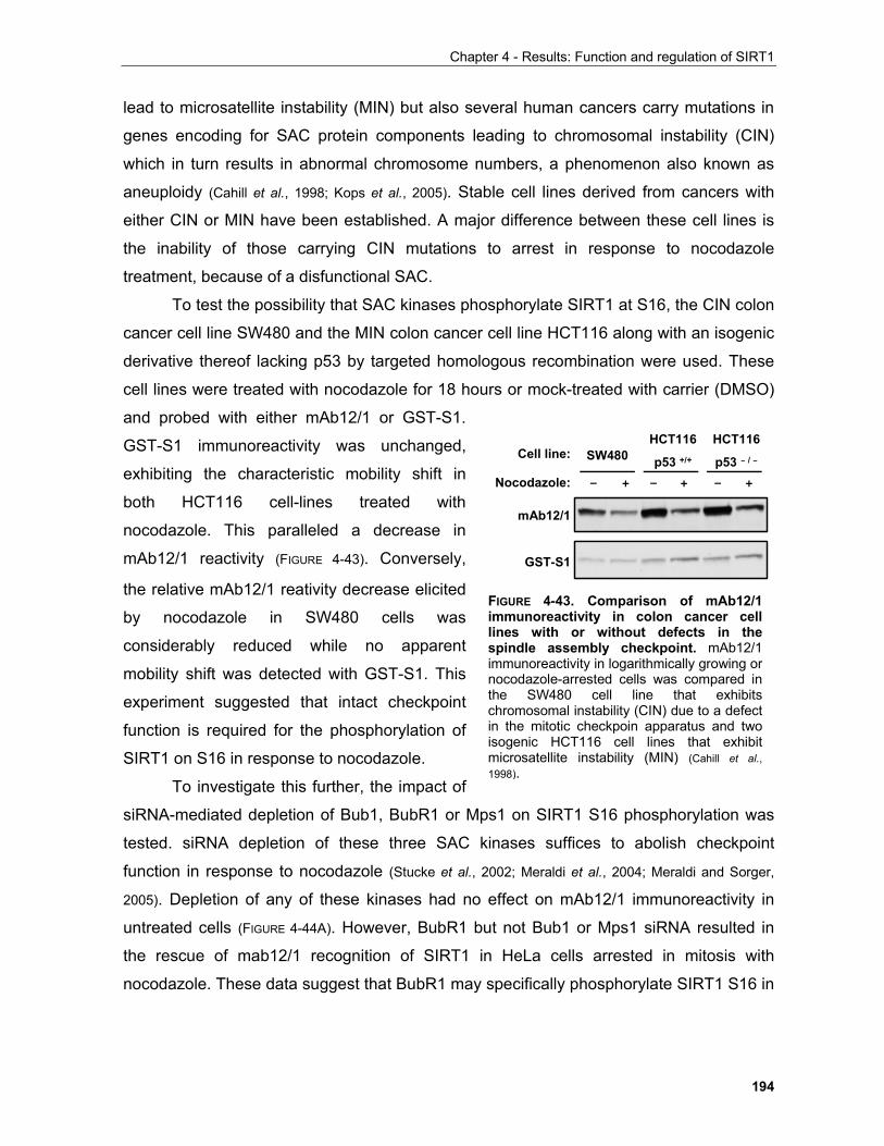
Chapter 4 - Results: Function and regulation of SIRT1
194
lead to microsatellite instability (MIN) but also several human cancers carry mutations in
genes encoding for SAC protein components leading to chromosomal instability (CIN)
which in turn results in abnormal chromosome numbers, a phenomenon also known as
aneuploidy (Cahill et al., 1998; Kops et al., 2005). Stable cell lines derived from cancers with
either CIN or MIN have been established. A major difference between these cell lines is
the inability of those carrying CIN mutations to arrest in response to nocodazole
treatment, because of a disfunctional SAC.
To test the possibility that SAC kinases phosphorylate SIRT1 at S16, the CIN colon
cancer cell line SW480 and the MIN colon cancer cell line HCT116 along with an isogenic
derivative thereof lacking p53 by targeted homologous recombination were used. These
cell lines were treated with nocodazole for 18 hours or mock-treated with carrier (DMSO)
and probed with either mAb12/1 or GST-S1.
GST-S1 immunoreactivity was unchanged,
exhibiting the characteristic mobility shift in
both HCT116 cell-lines treated with
nocodazole. This paralleled a decrease in
mAb12/1 reactivity (FIGURE 4-43). Conversely,
the relative mAb12/1 reativity decrease elicited
by nocodazole in SW480 cells was
considerably reduced while no apparent
mobility shift was detected with GST-S1. This
experiment suggested that intact checkpoint
function is required for the phosphorylation of
SIRT1 on S16 in response to nocodazole.
To investigate this further, the impact of
siRNA-mediated depletion of Bub1, BubR1 or Mps1 on SIRT1 S16 phosphorylation was
tested. siRNA depletion of these three SAC kinases suffices to abolish checkpoint
function in response to nocodazole (Stucke et al., 2002; Meraldi et al., 2004; Meraldi and Sorger,
2005). Depletion of any of these kinases had no effect on mAb12/1 immunoreactivity in
untreated cells (FIGURE 4-44A). However, BubR1 but not Bub1 or Mps1 siRNA resulted in
the rescue of mab12/1 recognition of SIRT1 in HeLa cells arrested in mitosis with
nocodazole. These data suggest that BubR1 may specifically phosphorylate SIRT1 S16 in
Nocodazole: – + – + – +
Cell line:HCT116
p53 +/+
HCT116
p53 – / –SW480
mAb12/1
GST-S1
FIGURE 4-43. Comparison of mAb12/1immunoreactivity in colon cancer celllines with or without defects in thespindle assembly checkpoint. mAb12/1immunoreactivity in logarithmically growing ornocodazole-arrested cells was compared inthe SW480 cell line that exhibitschromosomal instability (CIN) due to a defectin the mitotic checkpoin apparatus and twoisogenic HCT116 cell lines that exhibitmicrosatellite instability (MIN) (Cahill et al.,1998).
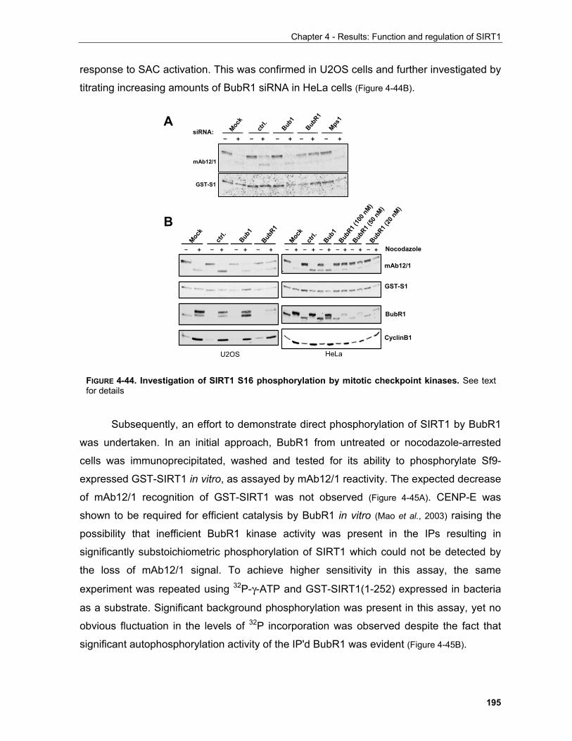
Chapter 4 - Results: Function and regulation of SIRT1
195
response to SAC activation. This was confirmed in U2OS cells and further investigated by
titrating increasing amounts of BubR1 siRNA in HeLa cells (Figure 4-44B).
Subsequently, an effort to demonstrate direct phosphorylation of SIRT1 by BubR1
was undertaken. In an initial approach, BubR1 from untreated or nocodazole-arrested
cells was immunoprecipitated, washed and tested for its ability to phosphorylate Sf9-
expressed GST-SIRT1 in vitro, as assayed by mAb12/1 reactivity. The expected decrease
of mAb12/1 recognition of GST-SIRT1 was not observed (Figure 4-45A). CENP-E was
shown to be required for efficient catalysis by BubR1 in vitro (Mao et al., 2003) raising the
possibility that inefficient BubR1 kinase activity was present in the IPs resulting in
significantly substoichiometric phosphorylation of SIRT1 which could not be detected by
the loss of mAb12/1 signal. To achieve higher sensitivity in this assay, the same
experiment was repeated using 32P-γ-ATP and GST-SIRT1(1-252) expressed in bacteria
as a substrate. Significant background phosphorylation was present in this assay, yet no
obvious fluctuation in the levels of 32P incorporation was observed despite the fact that
significant autophosphorylation activity of the IP'd BubR1 was evident (Figure 4-45B).
mAb12/1
GST-S1
BubR1
Mock
ctrl.
Bub1
– + – + – + – + – +
Mps1
siRNA:
– + – + – + – + – + – +
BubR1
Mock
ctrl.
Bub1
– + – + – + – +
Mock
ctrl.
Bub1
BubR1 (
100 n
M)
BubR1 (
50 nM
)Bub
R1 (20
nM)
mAb12/1
GST-S1
BubR1
CyclinB1
Nocodazole
U2OS HeLa
A
B
FIGURE 4-44. Investigation of SIRT1 S16 phosphorylation by mitotic checkpoint kinases. See textfor details
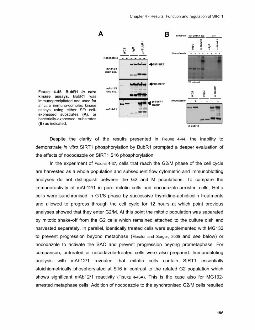
Chapter 4 - Results: Function and regulation of SIRT1
196
Despite the clarity of the results presented in FIGURE 4-44, the inability to
demonstrate in vitro SIRT1 phosphorylation by BubR1 prompted a deeper evaluation of
the effects of nocodazole on SIRT1 S16 phosphorylation.
In the experiment of FIGURE 4-37, cells that reach the G2/M phase of the cell cycle
are harvested as a whole population and subsequent flow cytometric and immunoblotting
analyses do not distinguish between the G2 and M populations. To compare the
immunoractivity of mAb12/1 in pure mitotic cells and nocodazole-arrested cells, HeLa
cells were sunchronised in G1/S phase by successive thymidine-aphidicolin treatments
and allowed to progress through the cell cycle for 12 hours at which point previous
analyses showed that they enter G2/M. At this point the mitotic population was separated
by mitotic shake-off from the G2 cells which remained attached to the culture dish and
harvested separately. In parallel, identically treated cells were supplemented with MG132
to prevent progression beyond metaphase (Meraldi and Sorger, 2005 and see below) or
nocodazole to activate the SAC and prevent progression beyong prometaphase. For
comparison, untreated or nocodazole-treated cells were also prepared. Immunobloting
analysis with mAb12/1 revealed that mitotic cells contain SIRT1 essentially
stoichiometrically phosphorylated at S16 in contrast to the related G2 population which
shows significant mAb12/1 reactivity (FIGURE 4-46A). This is the case also for MG132-
arrested metaphase cells. Addition of nocodazole to the synchronised G2/M cells resulted
mIg
G
α-B
ubR
1
WC
E
Nocodazole – + – + – +
mAb12/1short exp.
α-BubR1
mAb12/1long exp.
GST-SIRT1
GST-SIRT1
p-BubR1BubR1
mIg
G
α-B
ubR
1
WC
E
Nocodazole – + – + – +
α-BubR1
32P autorad
mIg
G
α-B
ubR
1
Nocodazole – + – +
mIg
G
α-B
ubR
1
– + – +
Substrate: GST-SIRT1 (1-252) GSTA B
FIGURE 4-45. BubR1 in vitrokinase assays. BubR1 wasimmunoprecipitated and used forin vitro immuno-complex kinaseassays using either Sf9 cell-expressed substrates (A), orbacterially-expressed substrates(B) as indicated.
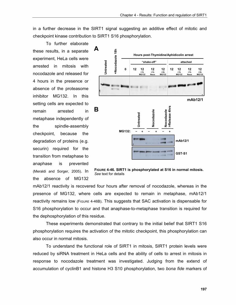
Chapter 4 - Results: Function and regulation of SIRT1
197
in a further decrease in the SIRT1 signal suggesting an additive effect of mitotic and
checkpoint kinase contribution to SIRT1 S16 phosphorylation.
To further elaborate
these results, in a separate
experiment, HeLa cells were
arrested in mitosis with
nocodazole and released for
4 hours in the presence or
absence of the proteasome
inhibitor MG132. In this
setting cells are expected to
remain arrested in
metaphase independently of
the spindle-assembly
checkpoint, because the
degradation of proteins (e.g.
securin) required for the
transition from metaphase to
anaphase is prevented
(Meraldi and Sorger, 2005). In
the absence of MG132
mAb12/1 reactivity is recovered four hours after removal of nocodazole, whereas in the
presence of MG132, where cells are expected to remain in metaphase, mAb12/1
reactivity remains low (FIGURE 4-46B). This suggests that SAC activation is dispensable for
S16 phosphorylation to occur and that anaphase-to-metaphase transition is required for
the dephosphorylation of this residue.
These experiments demonstrated that contrary to the initial belief that SIRT1 S16
phosphorylation requires the activation of the mitotic checkpoint, this phosphorylation can
also occur in normal mitosis.
To understand the functional role of SIRT1 in mitosis, SIRT1 protein levels were
reduced by siRNA treatment in HeLa cells and the ability of cells to arrest in mitosis in
response to nocodazole treatment was investigated. Judging from the extend of
accumulation of cyclinB1 and histone H3 S10 phosphorylation, two bona fide markers of
Unt
reat
ed
+Noc
odaz
ole
18h
Hours post-Thymidine/Aphidicolin arrest
“shake-off” attached
0 12 12 +1h
MG132
12 +1h
Noco
12 +3h
MG132
12 12 +1h
MG132
12 +1h
Noco
12 +3h
MG132
mAb12/1
MG132: – + – + – +
Noc
odaz
ole
Noc
odaz
ole
>Rel
ease
4hr
s
mAb12/1
GST-S1
Unt
reat
ed
B
A
FIGURE 4-46. SIRT1 is phosphorylated at S16 in normal mitosis.See text for details
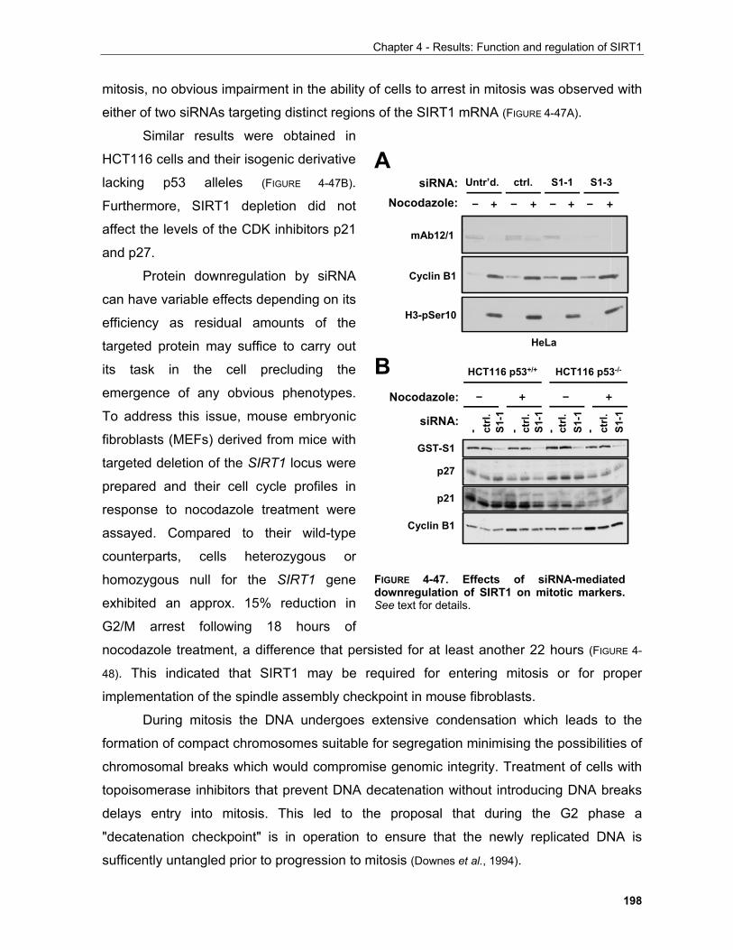
Chapter 4 - Results: Function and regulation of SIRT1
198
mitosis, no obvious impairment in the ability of cells to arrest in mitosis was observed with
either of two siRNAs targeting distinct regions of the SIRT1 mRNA (FIGURE 4-47A).
Similar results were obtained in
HCT116 cells and their isogenic derivative
lacking p53 alleles (FIGURE 4-47B).
Furthermore, SIRT1 depletion did not
affect the levels of the CDK inhibitors p21
and p27.
Protein downregulation by siRNA
can have variable effects depending on its
efficiency as residual amounts of the
targeted protein may suffice to carry out
its task in the cell precluding the
emergence of any obvious phenotypes.
To address this issue, mouse embryonic
fibroblasts (MEFs) derived from mice with
targeted deletion of the SIRT1 locus were
prepared and their cell cycle profiles in
response to nocodazole treatment were
assayed. Compared to their wild-type
counterparts, cells heterozygous or
homozygous null for the SIRT1 gene
exhibited an approx. 15% reduction in
G2/M arrest following 18 hours of
nocodazole treatment, a difference that persisted for at least another 22 hours (FIGURE 4-
48). This indicated that SIRT1 may be required for entering mitosis or for proper
implementation of the spindle assembly checkpoint in mouse fibroblasts.
During mitosis the DNA undergoes extensive condensation which leads to the
formation of compact chromosomes suitable for segregation minimising the possibilities of
chromosomal breaks which would compromise genomic integrity. Treatment of cells with
topoisomerase inhibitors that prevent DNA decatenation without introducing DNA breaks
delays entry into mitosis. This led to the proposal that during the G2 phase a
"decatenation checkpoint" is in operation to ensure that the newly replicated DNA is
sufficently untangled prior to progression to mitosis (Downes et al., 1994).
mAb12/1
Cyclin B1
H3-pSer10
Nocodazole: – + – + – + – +
Untr’d. ctrl.siRNA: S1-1 S1-3
HeLa
p27
p21
Cyclin B1
GST-S1
HCT116 p53+/+ HCT116 p53-/-
Nocodazole: – + – +
siRNA:
- ctrl.
S1-1
ctrl.
S1-1
- ctrl.
S1-1
- ctrl.
S1-1
-
A
B
FIGURE 4-47. Effects of siRNA-mediated downregulation of SIRT1 on mitotic markers. See text for details.
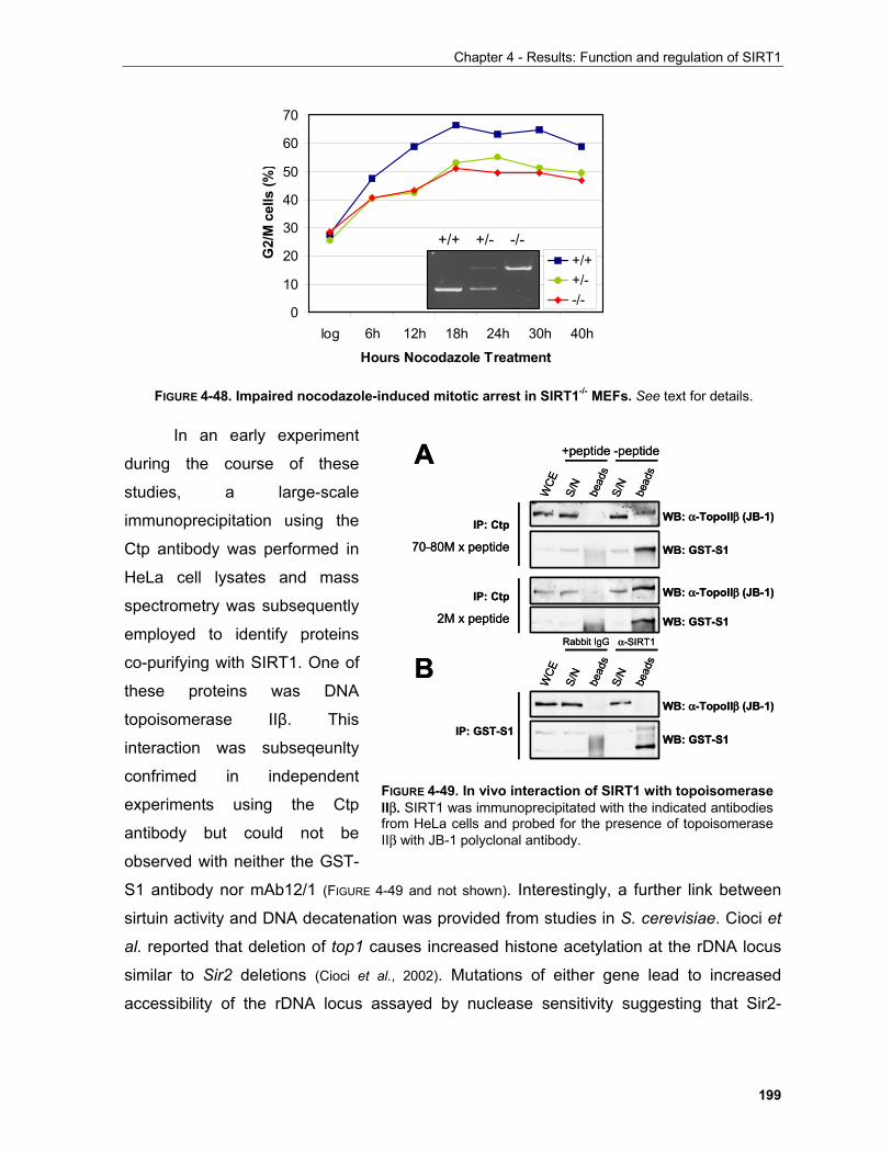
Chapter 4 - Results: Function and regulation of SIRT1
199
In an early experiment
during the course of these
studies, a large-scale
immunoprecipitation using the
Ctp antibody was performed in
HeLa cell lysates and mass
spectrometry was subsequently
employed to identify proteins
co-purifying with SIRT1. One of
these proteins was DNA
topoisomerase IIβ. This
interaction was subseqeunlty
confrimed in independent
experiments using the Ctp
antibody but could not be
observed with neither the GST-
S1 antibody nor mAb12/1 (FIGURE 4-49 and not shown). Interestingly, a further link between
sirtuin activity and DNA decatenation was provided from studies in S. cerevisiae. Cioci et
al. reported that deletion of top1 causes increased histone acetylation at the rDNA locus
similar to Sir2 deletions (Cioci et al., 2002). Mutations of either gene lead to increased
accessibility of the rDNA locus assayed by nuclease sensitivity suggesting that Sir2-
0
10
20
30
40
50
60
70
log 6h 12h 18h 24h 30h 40h
Hours Nocodazole Treatment
G2/
M c
ells
(%)
+/++/--/-
+/+ -/-+/-
FIGURE 4-48. Impaired nocodazole-induced mitotic arrest in SIRT1-/- MEFs. See text for details.
FIGURE 4-49. In vivo interaction of SIRT1 with topoisomerase IIβ. SIRT1 was immunoprecipitated with the indicated antibodiesfrom HeLa cells and probed for the presence of topoisomeraseIIβ with JB-1 polyclonal antibody.
WB: GST-S1S/
N
bead
s
WCE
+peptide
S/N
bead
s
-peptide
WB: α-TopoIIβ (JB-1)IP: Ctp
70-80M x peptide
IP: GST-S1
WB: α-TopoIIβ (JB-1)
WB: GST-S1
IP: Ctp
2M x peptide
WB: α-TopoIIβ (JB-1)
WB: GST-S1
S/N
bead
s
WCE
Rabbit IgG
S/N
bead
sα-SIRT1
A
B
WB: GST-S1S/
N
bead
s
WCE
+peptide
S/N
bead
s
-peptide
WB: α-TopoIIβ (JB-1)IP: Ctp
70-80M x peptide
IP: GST-S1
WB: α-TopoIIβ (JB-1)
WB: GST-S1
IP: Ctp
2M x peptide
WB: α-TopoIIβ (JB-1)
WB: GST-S1
S/N
bead
s
WCE
Rabbit IgG
S/N
bead
sα-SIRT1
A
B
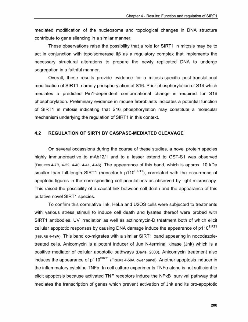
Chapter 4 - Results: Function and regulation of SIRT1
200
mediated modification of the nucleosome and topological changes in DNA structure
contribute to gene silencing in a similar manner.
These observations raise the possibility that a role for SIRT1 in mitosis may be to
act in conjunction with topoisomerase IIβ as a regulatory complex that implements the
necessary structural alterations to prepare the newly replicated DNA to undergo
segregation in a faithful manner.
Overall, these results provide evidence for a mitosis-specific post-translational
modification of SIRT1, namely phosphorylation of S16. Prior phosphorylation of S14 which
mediates a predicted Pin1-dependent conformational change is required for S16
phosphorylation. Preliminary evidence in mouse firbroblasts indicates a potential function
of SIRT1 in mitosis indicating that S16 phosphorylation may constitute a molecular
mechanism underlying the regulation of SIRT1 in this context.
4.2 REGULATION OF SIRT1 BY CASPASE-MEDIATED CLEAVAGE
On several occassions during the course of these studies, a novel protein species
highly immunoreactive to mAb12/1 and to a lesser extend to GST-S1 was observed
(FIGURES 4-7B, 4-22, 4-40, 4-41, 4-46). The appearance of this band, which is approx. 10 kDa
smaller than full-length SIRT1 (henceforth p110SIRT1), correlated with the occurrence of
apoptotic figures in the corresponding cell populations as observed by light microscopy.
This raised the possibility of a causal link between cell death and the appearance of this
putative novel SIRT1 species.
To confirm this correlative link, HeLa and U2OS cells were subjected to treatments
with various stress stimuli to induce cell death and lysates thereof were probed with
SIRT1 antibodies. UV irradiation as well as actinomycin-D treatment both of which elicit
cellular apoptotic responses by causing DNA damage induce the appearance of p110SIRT1
(FIGURE 4-49A). This band co-migrates with a similar SIRT1 band appearing in nocodazole-
treated cells. Anicomycin is a potent inducer of Jun N-terminal kinase (Jnk) which is a
positive mediator of cellular apoptotic pathways (Davis, 2000). Anicomycin treatment also
induces the appearance of p110SIRT1 (FIGURE 4-50A lower panel). Another apoptosis inducer in
the inflammatory cytokine TNFα. In cell culture experiments TNFα alone is not sufficient to
elicit apoptosis because activated TNF receptors induce the NFκB survival pathway that
mediates the transcription of genes which prevent activation of Jnk and its pro-apoptotic

Chapter 4 - Results: Function and regulation of SIRT1
201
function (Kamata et al., 2005; De Smaele et al.,
2001; Tang et al., 2001). Inhibition of NFκB target
gene expression by actinomycin-D to block
transcription or cycloheximide (CHX) to block
translation enables TNFα to induce
apoptosis. CHX alone can also induce
apoptosis. As shown in FIGURE 4-50B CHX
alone and to a greater extend the
combination of TNFα and CHX, but not TNFα
alone lead to the induction of p110SIRT1. Thus
multiple stress stimuli that induce cell death
elicit the appearance of a novel SIRT1
species, p110SIRT1.
This observation prompted the
investigation of the molecular mechanism
that results in the appearance of p110SIRT1.
DNA database sequence searches indicated
that p110SIRT1 is unlilkely to arise by
expression of a SIRT1 gene alternative transcript or alternative splicing. This was further
supported by the fact that neither actinomycin-D nor CHX prevented formation of this
band (FIGURE 4-50). Furthermore, alternative translation initiation was also excluded since
two potential methionine residues that could account for this give rise to SIRT1 species
which are smaller than p110SIRT1 (see FIGURE 4-10). Apart from its role in protein
degradation, the proteolytic activity of the proteasome is also implicated in regulated
protein processing (e.g. Orian et al., 2000; Hoiby et al., 2004). Proteolytic processing by the
proteasome is also unlikely since MG132 treatment of cells does not prevent the
occurrence of p110SIRT1 (FIGURE 4-46B).
A specific class of cysteine proteases known as caspases are essential mediators
of the apoptotic programme (Danial and Korsmeyer, 2004). In unstimulated cells, caspases are
present as inactive zymogens (Riedl and Shi, 2004). When the apoptotic pathway is
activated, limited proteolytic events result in the cleavage of the zymogens to yield active
caspases which in turn catalyse the cleavage of effector proteins (Danial and Korsmeyer,
2004).
A
B
20.5 1
TNFα
10ng/ml
4
CHX
80
– – – – – + + – + +
– + + + + + – + + –
Hours
mAb12/1
118 kDa
8h A
ct-D
Unt
reat
ed
Noc
odaz
ole
5 10 20 30 60 120 180Unt
r’d. Anisomycin (10µg/ml)
treatment (min.)
118 kDa
100 kDa
Tx’d
SIR
T1
Unt
reat
ed
UV
200
J/ m
2
FIGURE 4-50. Various stress stimuli induceSIRT1 processing. Cells in (A) were U2OSand in (B) HeLa. See text for details.
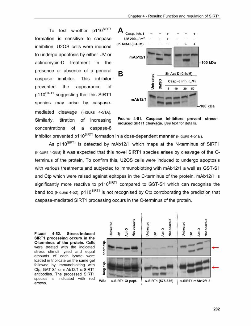
Chapter 4 - Results: Function and regulation of SIRT1
202
To test whether p110SIRT1
formation is sensitive to caspase
inhibition, U2OS cells were induced
to undergo apoptosis by either UV or
actinomycin-D treatment in the
presence or absence of a general
caspase inhibitor. This inhibitor
prevented the appearance of
p110SIRT1 suggesting that this SIRT1
species may arise by caspase-
mediated cleavage (FIGURE 4-51A).
Similarly, titration of increasing
concentrations of a caspase-8
inhibitor prevented p110SIRT1 formation in a dose-dependent manner (FIGURE 4-51B).
As p110SIRT1 is detected by mAb12/1 which maps at the N-terminus of SIRT1
(FIGURE 4-38B) it was expected that this novel SIRT1 species arises by cleavage of the C-
terminus of the protein. To confirm this, U2OS cells were induced to undergo apoptosis
with various treatments and subjected to immunoblotting with mAb12/1 a well as GST-S1
and Ctp which were raised against epitopes in the C-terminus of the protein. mAb12/1 is
significantly more reactive to p110SIRT1 compared to GST-S1 which can recognise the
band too (FIGURE 4-52). p110SIRT1 is not recognised by Ctp corroborating the prediction that
caspase-mediated SIRT1 processing occurs in the C-terminus of the protein.
FIGURE 4-51. Caspase inhibitors prevent stress-induced SIRT1 cleavage. See text for details.
A
B
8h Act-D (0.4uM)
Casp. inh.-IUV 200 J/ m2
– – + – – +
mAb12/1
– + + – – –– – – – + +
Casp.-8 inh. (µM)
5 10 20 50
8h Act-D (0.4uM)
DM
SO
Unt
reat
ed
mAb12/1
100 kDa
100 kDa
α-SIRT1 (575-676)
Act
-D
Unt
reat
ed
Noc
odaz
ole
UV
Act
-D
Unt
reat
ed
Noc
odaz
ole
UV
Act
-D
Unt
reat
ed
Noc
odaz
ole
UV
WB: α-SIRT1 Ct pept. α-SIRT1 mAb12/1.3
shor
t exp
.lo
ng e
xp.
FIGURE 4-52. Stress-inducedSIRT1 processing occurs in theC-terminus of the protein. Cellswere treated with the indicatedstress stimuli lysed and equalamounts of each lysate wereloaded in triplicate on the same gelfollowed by immunoblotting withCtp, GAT-S1 or mAb12/1 α-SIRT1antibodies. The processed SIRT1species is indicated with redarrows.
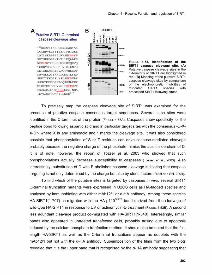
Chapter 4 - Results: Function and regulation of SIRT1
203
To precisely map the caspase cleavage site of SIRT1 was examined for the
presence of putative caspase consensus target sequences. Several such sites were
identified in the C-terminus of the protein (FIGURE 4-53A). Caspases show specificity for the
peptide bond following aspartic acid and in particular target sites with the consensus -D-X-
X-D*- where X is any aminoacid and * marks the cleavage site. It was also considered
possible that phosphorylation of S or T residues can drive caspase-mediated cleavage
probably because the negative charge of the phosphate mimics the acidic side-chain of D.
It is of note, however, the report of Tozser et al. 2003 who showed that such
phosphorylations actually decrease susceptibility to caspases (Tozser et al., 2003). Also
interestingly, substitution of D with E abolishes caspase cleavage indicating that caspase
targeting is not only determined by the charge but also by steric factors (Riedl and Shi, 2004).
To find which of the putative sites is targeted by caspases in vivo, several SIRT1
C-terminal truncation mutants were expressed in U2OS cells as HA-tagged species and
analysed by immunobloting with either mAb12/1 or α-HA antibody. Among these species
HA-SIRT1(1-707) co-migrated with the HA-p110SIRT1 band derived from the cleavage of
wild-type HA-SIRT1 in response to UV or actinomycin-D treatment (FIGURE 4-53B). A second
less adundant cleavage product co-migrated with HA-SIRT1(1-540). Interestingly, similar
bands also appeared in untreated transfected cells, probably arising due to apoptosis
induced by the calcium phosphate tranfection method. It should also be noted that the full-
length HA-SIRT1 as well as the C-terminal truncations appear as doublets with the
mAb12/1 but not with the α-HA antibody. Supeimposition of the films from the two blots
revealed that it is the upper band that is recognised by the α-HA antibody suggesting that
FIGURE 4-53. Identification of theSIRT1 caspase cleavage site. (A)Putative caspase cleavage sites in theC-terminus of SIRT1 are highlighted inred; (B) Mapping of the putative SIRT1caspase cleavage sites by comparisonof the electrophoretic mobilities oftruncated SIRT1 species withprocessed SIRT1 following stress.
481DCDVIINELCHRLGGEYAKLCCNPVKLSEITEKPPRTQKELAYLSELPPTPLHVSEDSSSPERTSPPDSSVIVTLLDQAAKSNDDLDVSESKGCMEEKPQEVQTSRNVESIAEQMENPDLKNVGSSTGEKNERTSVAGTVRKCWPNRVAKEQISRRLDGNQYLFLPPNRYIFHGAEVYSDSEDDVLSSSSCGSNSDSGTCQSPSLEEPMEDESEIEEFYNGLEDEPDVPERAGGAGFGTDGDDQEAINEAISVKQEVTDMNYPSNKS747
Putative SIRT1 CPutative SIRT1 C--terminal terminal caspase cleavage sitescaspase cleavage sites
A B
mA
b12/
1α-
HA
Unt
rans
’dw
tw
t +U
Vw
t +A
ct-D
(1-7
07)
(1-6
64)
(1-5
67)
(1-5
40)
HA-SIRT1
100 kDa
75 kDa
100 kDa
75 kDa
100 kDa
75 kDa
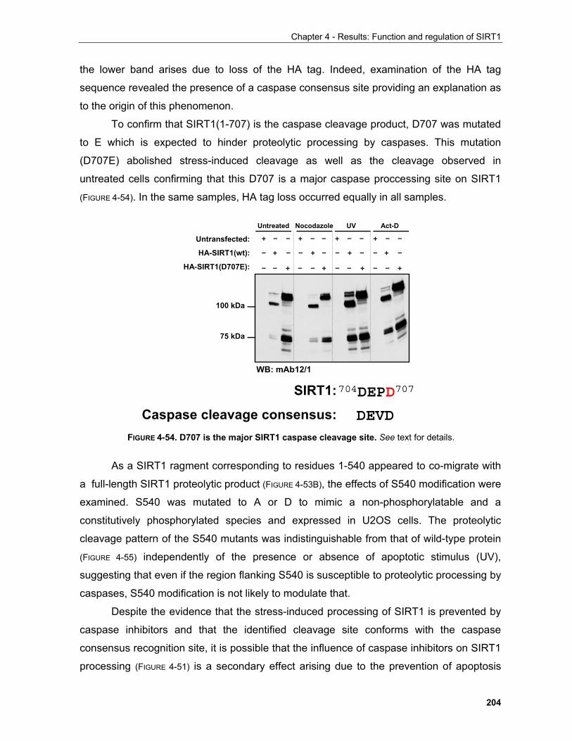
Chapter 4 - Results: Function and regulation of SIRT1
204
the lower band arises due to loss of the HA tag. Indeed, examination of the HA tag
sequence revealed the presence of a caspase consensus site providing an explanation as
to the origin of this phenomenon.
To confirm that SIRT1(1-707) is the caspase cleavage product, D707 was mutated
to E which is expected to hinder proteolytic processing by caspases. This mutation
(D707E) abolished stress-induced cleavage as well as the cleavage observed in
untreated cells confirming that this D707 is a major caspase proccessing site on SIRT1
(FIGURE 4-54). In the same samples, HA tag loss occurred equally in all samples.
As a SIRT1 ragment corresponding to residues 1-540 appeared to co-migrate with
a full-length SIRT1 proteolytic product (FIGURE 4-53B), the effects of S540 modification were
examined. S540 was mutated to A or D to mimic a non-phosphorylatable and a
constitutively phosphorylated species and expressed in U2OS cells. The proteolytic
cleavage pattern of the S540 mutants was indistinguishable from that of wild-type protein
(FIGURE 4-55) independently of the presence or absence of apoptotic stimulus (UV),
suggesting that even if the region flanking S540 is susceptible to proteolytic processing by
caspases, S540 modification is not likely to modulate that.
Despite the evidence that the stress-induced processing of SIRT1 is prevented by
caspase inhibitors and that the identified cleavage site conforms with the caspase
consensus recognition site, it is possible that the influence of caspase inhibitors on SIRT1
processing (FIGURE 4-51) is a secondary effect arising due to the prevention of apoptosis
704DEPD707SIRT1:
Caspase cleavage consensus: DEVD
Untransfected:
HA-SIRT1(wt):
HA-SIRT1(D707E): – – + – – + – – + – – +
– + – – + – – + – – + –
+ – – + – – + – – + – –
Act-DUntreated Nocodazole UV
WB: mAb12/1
100 kDa
75 kDa
FIGURE 4-54. D707 is the major SIRT1 caspase cleavage site. See text for details.
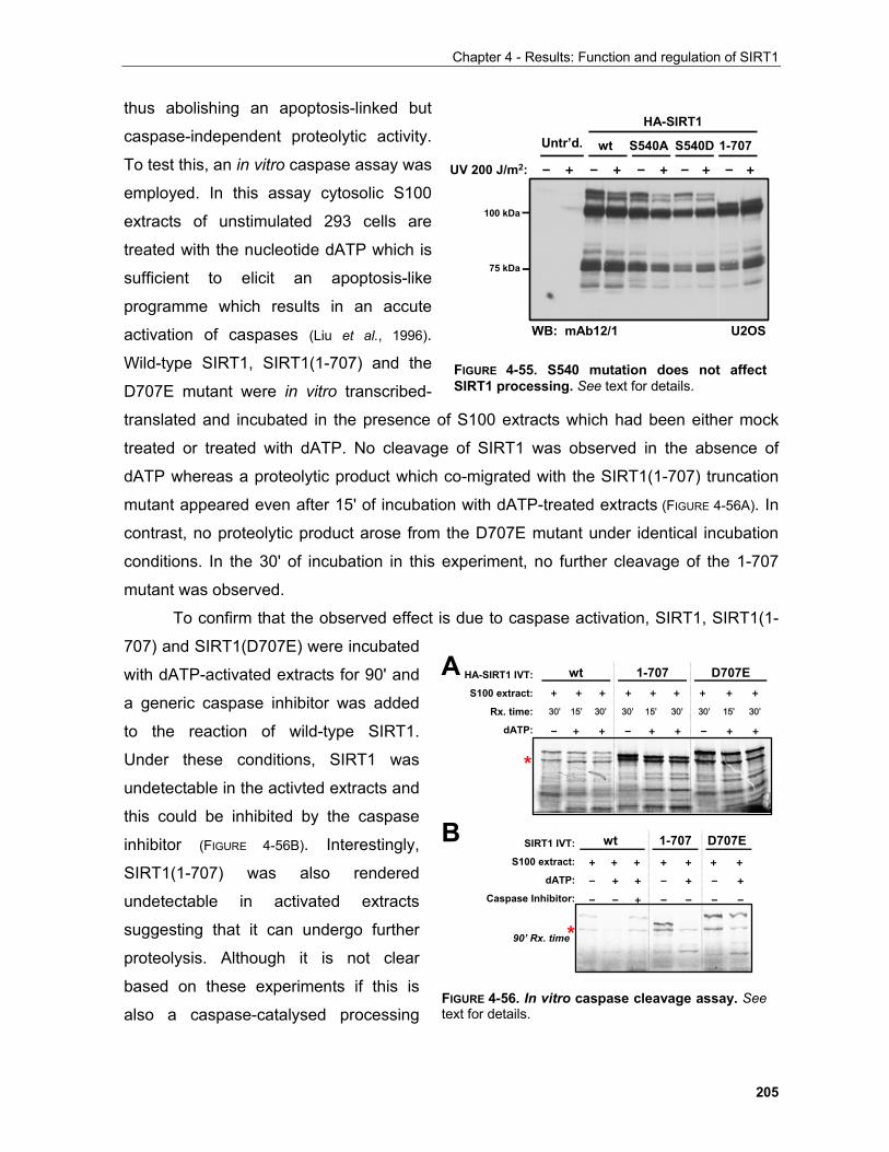
Chapter 4 - Results: Function and regulation of SIRT1
205
thus abolishing an apoptosis-linked but
caspase-independent proteolytic activity.
To test this, an in vitro caspase assay was
employed. In this assay cytosolic S100
extracts of unstimulated 293 cells are
treated with the nucleotide dATP which is
sufficient to elicit an apoptosis-like
programme which results in an accute
activation of caspases (Liu et al., 1996).
Wild-type SIRT1, SIRT1(1-707) and the
D707E mutant were in vitro transcribed-
translated and incubated in the presence of S100 extracts which had been either mock
treated or treated with dATP. No cleavage of SIRT1 was observed in the absence of
dATP whereas a proteolytic product which co-migrated with the SIRT1(1-707) truncation
mutant appeared even after 15' of incubation with dATP-treated extracts (FIGURE 4-56A). In
contrast, no proteolytic product arose from the D707E mutant under identical incubation
conditions. In the 30' of incubation in this experiment, no further cleavage of the 1-707
mutant was observed.
To confirm that the observed effect is due to caspase activation, SIRT1, SIRT1(1-
707) and SIRT1(D707E) were incubated
with dATP-activated extracts for 90' and
a generic caspase inhibitor was added
to the reaction of wild-type SIRT1.
Under these conditions, SIRT1 was
undetectable in the activted extracts and
this could be inhibited by the caspase
inhibitor (FIGURE 4-56B). Interestingly,
SIRT1(1-707) was also rendered
undetectable in activated extracts
suggesting that it can undergo further
proteolysis. Although it is not clear
based on these experiments if this is
also a caspase-catalysed processing
Untr’d. HA-SIRT1
wt S540A S540D 1-707
– + – + – + – + – +UV 200 J/m2:
WB: mAb12/1 U2OS
100 kDa
75 kDa
FIGURE 4-55. S540 mutation does not affectSIRT1 processing. See text for details.
HA-SIRT1 IVT:
S100 extract:
Rx. time:
dATP: – + + – + + – + +
30’ 15’ 30’ 30’ 15’ 30’ 30’ 15’ 30’
+ + + + + + + + +
wt 1-707 D707E
SIRT1 IVT:
S100 extract:
dATP:
Caspase Inhibitor:
wt 1-707 D707E+ + + + + + +
– + + – + – +
– – + – – – –
90’ Rx. time
A
B
*
*
FIGURE 4-56. In vitro caspase cleavage assay. Seetext for details.
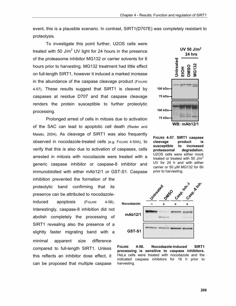
Chapter 4 - Results: Function and regulation of SIRT1
206
event, this is a plausible scenario. In contrast, SIRT1(D707E) was completely resistant to
proteolysis.
To investigate this point further, U2OS cells were
treated with 50 J/m2 UV light for 24 hours in the presence
of the proteasome inhibitor MG132 or carrier solvents for 6
hours prior to harvesting. MG132 treatment had little effect
on full-length SIRT1, however it induced a marked increase
in the abundance of the caspase cleavage product (FIGURE
4-57). These results suggest that SIRT1 is cleaved by
caspases at residue D707 and that caspase cleavage
renders the protein susceptible to further proteolytic
processing. Prolonged arrest of cells in mitosis due to activation
of the SAC can lead to apoptotic cell death (Rieder and
Maiato, 2004). As cleavage of SIRT1 was also frequently
observed in nocodazole-treated cells (e.g. FIGURE 4-50A), to
verify that this is also due to activation of caspases, cells
arrested in mitosis with nocodazole were treated with a
generic caspase inhibitor or caspase-8 inhibitor and
immunoblotted with either mAb12/1 or GST-S1. Caspase
inhibition prevented the formation of the
proteolytic band confirming that its
presence can be attributed to nocodazole-
induced apoptosis (FIGURE 4-58).
Interestingly, caspase-8 inhibition did not
abolish completely the processing of
SIRT1 revealing also the presence of a
slightly faster migrating band with a
minimal apparent size difference
compared to full-length SIRT1. Unless
this reflects an inhibitor dose effect, it
can be proposed that multiple caspase
FIGURE 4-57. SIRT1 caspasecleavage product issusceptible to increasedproteasomal degradation.U2OS cells were either mocktreated or treated with 50 J/m2
UV for 24 h and with eithercarrier or 50 µM MG132 for 6hprior to harvesting.
Unt
reat
edEt
OH
DM
SOM
G13
2
WB: mAb12/1
UV 50 J/m2
24 hrs
100 kDa
75 kDa
100 kDa
75 kDa
mAb12/1
GST-S1
Nocodazole: – + + +
Casp.
Inh.-I
Casp.-
8 Inh
.
DMSO
Untre
ated
FIGURE 4-58. Nocodazole-induced SIRT1 processing is sensitive to caspase inhibitors.HeLa cells were treated with nocodazole and the indicated caspase inhibitors for 16 h prior to harvesting.
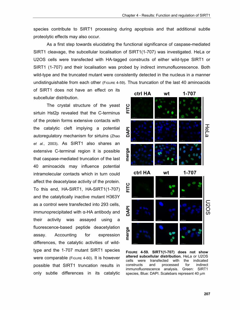
Chapter 4 - Results: Function and regulation of SIRT1
207
species contribute to SIRT1 processing during apoptosis and that additional subtle
proteolytic effects may also occur.
As a first step towards elucidating the functional significance of caspase-mediated
SIRT1 cleavage, the subcellular localisation of SIRT1(1-707) was investigated. HeLa or
U2OS cells were transfected with HA-tagged constructs of either wild-type SIRT1 or
SIRT1 (1-707) and their localisation was probed by indirect immunofluorescence. Both
wild-type and the truncated mutant were consistently detected in the nucleus in a manner
undistinguishable from each other (FIGURE 4-59). Thus truncation of the last 40 aminoacids
of SIRT1 does not have an effect on its
subcellular distribution.
The crystal structure of the yeast
sirtuin Hst2p revealed that the C-terminus
of the protein forms extensive contacts with
the catalytic cleft implying a potential
autoregulatory mechanism for sirtuins (Zhao
et al., 2003). As SIRT1 also shares an
extensive C-terminal region it is possible
that caspase-mediated truncation of the last
40 aminoacids may influence potential
intramolecular contacts which in turn could
affect the deacetylase activity of the protein.
To this end, HA-SIRT1, HA-SIRT1(1-707)
and the catalytically inactive mutant H363Y
as a control were transfected into 293 cells,
immunoprecipitated with α-HA antibody and
their activity was assayed using a
fluorescence-based peptide deacetylation
assay. Accounting for expression
differences, the catalytic activities of wild-
type and the 1-707 mutant SIRT1 species
were comparable (FIGURE 4-60). It is however
possible that SIRT1 truncation results in
only subtle differences in its catalytic
ctrl HA wt 1-707
HeLa
ctrl HA wt 1-707
U2O
S
FITC
D
API
mer
geFI
TC
DA
PIm
erge
FIGURE 4-59. SIRT1(1-707) does not show altered subcellular distribution. HeLa or U2OS cells were transfected with the indicated constructs and processed for indirect immunofluorescence analysis. Green: SIRT1 species, Blue: DAPI. Scalebars represent 40 µm
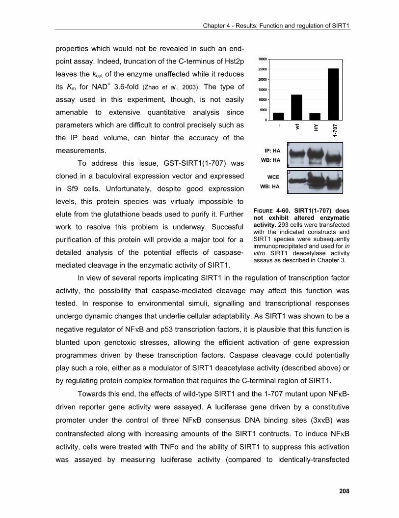
Chapter 4 - Results: Function and regulation of SIRT1
208
properties which would not be revealed in such an end-
point assay. Indeed, truncation of the C-terminus of Hst2p
leaves the kcat of the enzyme unaffected while it reduces
its Km for NAD+ 3.6-fold (Zhao et al., 2003). The type of
assay used in this experiment, though, is not easily
amenable to extensive quantitative analysis since
parameters which are difficult to control precisely such as
the IP bead volume, can hinter the accuracy of the
measurements.
To address this issue, GST-SIRT1(1-707) was
cloned in a baculoviral expression vector and expressed
in Sf9 cells. Unfortunately, despite good expression
levels, this protein species was virtualy impossible to
elute from the glutathione beads used to purify it. Further
work to resolve this problem is underway. Succesful
purification of this protein will provide a major tool for a
detailed analysis of the potential effects of caspase-
mediated cleavage in the enzymatic activity of SIRT1.
In view of several reports implicating SIRT1 in the regulation of transcription factor
activity, the possibility that caspase-mediated cleavage may affect this function was
tested. In response to environmental simuli, signalling and transcriptional responses
undergo dynamic changes that underlie cellular adaptability. As SIRT1 was shown to be a
negative regulator of NFκB and p53 transcription factors, it is plausible that this function is
blunted upon genotoxic stresses, allowing the efficient activation of gene expression
programmes driven by these transcription factors. Caspase cleavage could potentially
play such a role, either as a modulator of SIRT1 deacetylase activity (described above) or
by regulating protein complex formation that requires the C-terminal region of SIRT1.
Towards this end, the effects of wild-type SIRT1 and the 1-707 mutant upon NFκB-
driven reporter gene activity were assayed. A luciferase gene driven by a constitutive
promoter under the control of three NFκB consensus DNA binding sites (3xκB) was
contransfected along with increasing amounts of the SIRT1 contructs. To induce NFκB
activity, cells were treated with TNFα and the ability of SIRT1 to suppress this activation
was assayed by measuring luciferase activity (compared to identically-transfected
0
5000
10000
15000
20000
25000
30000
wt
HY
1-70
7–
WCE
WB: HA
IP: HA
WB: HA
FIGURE 4-60. SIRT1(1-707) doesnot exhibit altered enzymaticactivity. 293 cells were transfectedwith the indicated constructs andSIRT1 species were subsequentlyimmunoprecipitated and used for invitro SIRT1 deacetylase activityassays as described in Chapter 3.
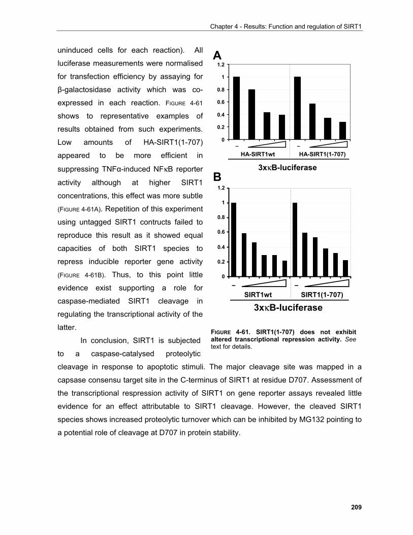
Chapter 4 - Results: Function and regulation of SIRT1
209
uninduced cells for each reaction). All
luciferase measurements were normalised
for transfection efficiency by assaying for
β-galactosidase activity which was co-
expressed in each reaction. FIGURE 4-61
shows to representative examples of
results obtained from such experiments.
Low amounts of HA-SIRT1(1-707)
appeared to be more efficient in
suppressing TNFα-induced NFκB reporter
activity although at higher SIRT1
concentrations, this effect was more subtle
(FIGURE 4-61A). Repetition of this experiment
using untagged SIRT1 contructs failed to
reproduce this result as it showed equal
capacities of both SIRT1 species to
repress inducible reporter gene activity
(FIGURE 4-61B). Thus, to this point little
evidence exist supporting a role for
caspase-mediated SIRT1 cleavage in
regulating the transcriptional activity of the
latter.
In conclusion, SIRT1 is subjected
to a caspase-catalysed proteolytic
cleavage in response to apoptotic stimuli. The major cleavage site was mapped in a
capsase consensu target site in the C-terminus of SIRT1 at residue D707. Assessment of
the transcriptional respression activity of SIRT1 on gene reporter assays revealed little
evidence for an effect attributable to SIRT1 cleavage. However, the cleaved SIRT1
species shows increased proteolytic turnover which can be inhibited by MG132 pointing to
a potential role of cleavage at D707 in protein stability.
0
0.2
0.4
0.6
0.8
1
1.2
– –HA-SIRT1wt HA-SIRT1(1-707)
3xκB-luciferase
0
0.2
0.4
0.6
0.8
1
1.2
– –SIRT1wt SIRT1(1-707)
3xκB-luciferase
A
B
FIGURE 4-61. SIRT1(1-707) does not exhibitaltered transcriptional repression activity. Seetext for details.
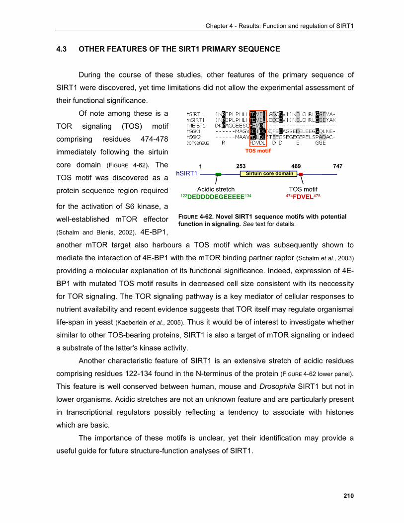
Chapter 4 - Results: Function and regulation of SIRT1
210
4.3 OTHER FEATURES OF THE SIRT1 PRIMARY SEQUENCE
During the course of these studies, other features of the primary sequence of
SIRT1 were discovered, yet time limitations did not allow the experimental assessment of
their functional significance.
Of note among these is a
TOR signaling (TOS) motif
comprising residues 474-478
immediately following the sirtuin
core domain (FIGURE 4-62). The
TOS motif was discovered as a
protein sequence region required
for the activation of S6 kinase, a
well-established mTOR effector
(Schalm and Blenis, 2002). 4E-BP1,
another mTOR target also harbours a TOS motif which was subsequently shown to
mediate the interaction of 4E-BP1 with the mTOR binding partner raptor (Schalm et al., 2003)
providing a molecular explanation of its functional significance. Indeed, expression of 4E-
BP1 with mutated TOS motif results in decreased cell size consistent with its neccessity
for TOR signaling. The TOR signaling pathway is a key mediator of cellular responses to
nutrient availability and recent evidence suggests that TOR itself may regulate organismal
life-span in yeast (Kaeberlein et al., 2005). Thus it would be of interest to investigate whether
similar to other TOS-bearing proteins, SIRT1 is also a target of mTOR signaling or indeed
a substrate of the latter's kinase activity.
Another characteristic feature of SIRT1 is an extensive stretch of acidic residues
comprising residues 122-134 found in the N-terminus of the protein (FIGURE 4-62 lower panel).
This feature is well conserved between human, mouse and Drosophila SIRT1 but not in
lower organisms. Acidic stretches are not an unknown feature and are particularly present
in transcriptional regulators possibly reflecting a tendency to associate with histones
which are basic.
The importance of these motifs is unclear, yet their identification may provide a
useful guide for future structure-function analyses of SIRT1.
TOS motifTOS motif
hSIRT11 253 469 747
Sirtuin core domain
TOS motif474FDVEL478
Acidic stretch122DEDDDDEGEEEEE134
FIGURE 4-62. Novel SIRT1 sequence motifs with potentialfunction in signaling. See text for details.
Chapter 5 - Discussion and future perspectives
CHAPTER 5
DISCUSSION AND FUTURE PERSPECTIVES
Elucidating signaling circuitries that underlie cellular and organismal physiology has
allowed a significant understanding of the homeostatic mechanisms employed by living
organisms to adapt to environmental stimuli. Such mechanisms have been a major driving
force in the evolution process and continue to be important in the way pathogenic organisms
as well as aberrantly functioning cells withstand ever more elaborate pharmacological
interventions treating disease. Thus, in-depth comprehension of adaptive signaling systems
and their interlinks allows to track down the molecular causes of particular disease states
leading to the development of novel knowledge-based therapeutic strategies.
5.1 Common pathways underlying homeostatic cellular processes are disregulated in diverse diseases
It has been increasingly evident that molecular players of pathways involved in basic
homeostatic processes go frequently awry in diseases apparently as diverse as cancer and
the metabolic syndrome. In particular several components of the insulin, TOR and oxygen
signaling pathways have been directly implicated in such diseases (Wullschleger et al., 2006; Plas
and Thompson, 2005; Barry and Krek, 2004). Aberrant function of these circuitries underlies
211
Chapter 5 - Discussion and future perspectives
dramatic changes in the proliferative, growth and metabolic cellular capacity, which are
closely related to the disease phenotype(s).
The fact that these signaling pathways co-ordinate such basic cellular functions
provides a first level of explanation as to why their molecular components comprise the
common denominator of different diseases. However, the mechanistic details of this
phenomenon remain largely elusive. Recent evidence has shed more light into the intriguing
cross-talk at play which co-ordinates diverse cellular activities and underlies the complex
disease phenotypes associated with mutations of these central homeostatic pathways.
5.1.1 Functional interconnection between the PKB, TOR and oxygen signaling pathways in health and disease
The insulin/IGF pathway involves a highly conserved set of signaling molecules that
coordinate cellular growth, proliferation and metabolism (Plas and Thompson, 2005; Brazil et al.,
2004). A central orchestrator of these effects is the kinase PKB. PKB phosphorylates a
diverse array of proteins to promote cell cycle progression, survival and sustain the metabolic
capacity at the levels required for proliferation (Plas and Thompson, 2005).
An independent pathway that yet is under the positive influence of growth factor
signaling through the insulin receptor is mediated by the TOR kinase. TOR is also activated
by aminoacids through a largely unknown mechanism and is a central regulator of cellular
protein biosynthetic capacity by dictating ribosomal biogenesis (Wullschleger et al., 2006). At the
same time, cellular energy status, modulated e.g. by glucose, also determines the activity of
TOR via the AMPK kinase cascade (Hardie, 2005).
The VHL-HIF signaling axis allows cells to adapt their metabolic programme to the
oxygen tension experienced at any given time and to elicit appropriate adaptive changes in
their environment such as the formation of new blood vessels in response to hypoxic
conditions (Barry and Krek, 2004; Kaelin, 2002).
212
Chapter 5 - Discussion and future perspectives
5.1.1.1 Representative mechanisms employed by hypoxia to inhibit growth and proliferation
At least two mechanisms have been shown to implement suppression of cellular
growth in response to hypoxia. The first requires a HIF target gene known as REDD1, which
is also a p53 target. REDD1 signals via the TSC1/TSC2 tumour suppressor complex to
inhibit TOR activation (Brugarolas et al., 2004). The second mechanism implicates that reduction
in cellular energy due to low oxygen tension results in the activation of the AMPK pathway
which in turn inhibits components of the translational apparatus (e.g.eIF2α and eEF2) as well
as TOR to suppress translation (Liu et al., 2006).
Similarly, energy availability expressed as the ratio of AMP/ATP depends on another
tumour suppressor, p53 to elicit cell cycle arrest under conditions of nutrient limitation. This
requires the phosphorylation and subsequent activation of p53 by the AMPK kinase (Jones et
al., 2005). Interestingly, p53 is also stabilised by HIF, however no direct experimental evidence
exists to support that p53 is required for the cell cycle arrest in response to hypoxia (Dang and
Semenza, 1999). An alternative mechanism possibly at play postulates that HIF represses the
activity of the myc oncoprotein which itself is a negative regulator of p21 and p27
transcription (Gartel and Shchors, 2003).
5.1.1.2 Reciprocal relation between metabolic enzymes and proteins regulating
proliferation
Cancer cells exhibit profoundly altered metabolic characteristics, predominantly
increased glucose uptake and enhanced anaerobic glycolysis even at normoxic conditions
(Dang and Semenza, 1999). This phenomenon is known as the Warburg effect. Several
oncogenes as well as tumour suppressors have been shown to mediate the gene expression
changes associated with such a phenotype.
The transcriptional activity of HIF accounts for a large part of such changes as it
induces the transcription of essentially all key enzymes in the glycolytic pathway (Dang and
Semenza, 1999). In addition, the tumour suppressor p53 and the oncogene myc also regulate
respectively the expression of hexokinase, the rate-limiting step in glycolysis and lactate
213
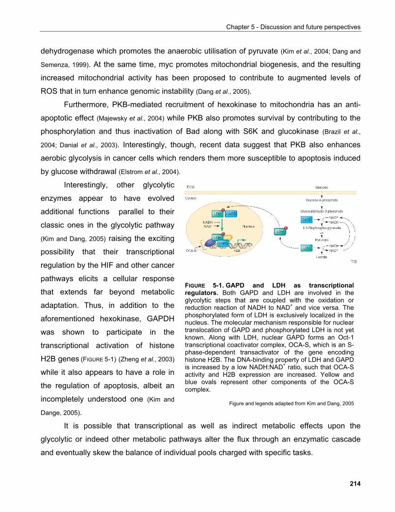
Chapter 5 - Discussion and future perspectives
dehydrogenase which promotes the anaerobic utilisation of pyruvate (Kim et al., 2004; Dang and
Semenza, 1999). At the same time, myc promotes mitochondrial biogenesis, and the resulting
increased mitochondrial activity has been proposed to contribute to augmented levels of
ROS that in turn enhance genomic instability (Dang et al., 2005).
Furthermore, PKB-mediated recruitment of hexokinase to mitochondria has an anti-
apoptotic effect (Majewsky et al., 2004) while PKB also promotes survival by contributing to the
phosphorylation and thus inactivation of Bad along with S6K and glucokinase (Brazil et al.,
2004; Danial et al., 2003). Interestingly, though, recent data suggest that PKB also enhances
aerobic glycolysis in cancer cells which renders them more susceptible to apoptosis induced
by glucose withdrawal (Elstrom et al., 2004).
Interestingly, other glycolytic
enzymes appear to have evolved
additional functions parallel to their
classic ones in the glycolytic pathway
(Kim and Dang, 2005) raising the exciting
possibility that their transcriptional
regulation by the HIF and other cancer
pathways elicits a cellular response
that extends far beyond metabolic
adaptation. Thus, in addition to the
aforementioned hexokinase, GAPDH
was shown to participate in the
transcriptional activation of histone
H2B genes (FIGURE 5-1) (Zheng et al., 2003)
while it also appears to have a role in
the regulation of apoptosis, albeit an
incompletely understood one (Kim and
Dange, 2005).
FIGURE 5-1. GAPD and LDH as transcriptionalregulators. Both GAPD and LDH are involved in theglycolytic steps that are coupled with the oxidation orreduction reaction of NADH to NAD+ and vice versa. Thephosphorylated form of LDH is exclusively localized in thenucleus. The molecular mechanism responsible for nucleartranslocation of GAPD and phosphorylated LDH is not yetknown. Along with LDH, nuclear GAPD forms an Oct-1transcriptional coactivator complex, OCA-S, which is an S-phase-dependent transactivator of the gene encodinghistone H2B. The DNA-binding property of LDH and GAPDis increased by a low NADH:NAD+ ratio, such that OCA-Sactivity and H2B expression are increased. Yellow andblue ovals represent other components of the OCA-Scomplex.
Figure and legends adapted from Kim and Dang, 2005
It is possible that transcriptional as well as indirect metabolic effects upon the
glycolytic or indeed other metabolic pathways alter the flux through an enzymatic cascade
and eventually skew the balance of individual pools charged with specific tasks.
214
Chapter 5 - Discussion and future perspectives
Further to the apparent dependence of cancer cells to glycolysis, mitochondrial
function also plays an important role in cancer biology. To begin with, mitochondrial genes
participating in the tricarboxylic acid (TCA) cycle such as succinate dehydrogenase and
fumarate hydratase were shown to be mutated in different cancers including renal cell
carcinoma which is frequently associated with mutations on VHL, the negative regulator of
HIF (Barry and Krek, 2004; Gottlieb and Tomlinson, 2005 and references therein). In particular,
accumulation of succinate due to succinate dehydrogenase malfunction leads to inhibition of
HIF prolyl hydroxylases and thus stabilisation of HIF under normoxic conditions, a
phenomenon called pseudohypoxia, providing a possible rationale behind the tumourigenic
effects arising from mutations in metabolic enzymes (see Chapter 1).
Interestingly, members of the E2F protein family, which are transcription factors with
cardinal roles in the regulation of cell cycle progression, have been shown to regulate
mitochondrial gene expression. In particular, E2F4 specifically co-operates with the pocket
protein p130 to mediate the gene expression programme in response to growth arrest stimuli
such as contact inhibition, growth factor withdrawal and forced expression of the p16INK4A
CDK inhibitor (Cam et al., 2004). A remarkable enrichment of nuclear respiratory factor-1 (NRF-
1) binding sites is exhibited within the set of E2F4 promoters especially of genes required for
mitochondrial biogenesis. This work provided a hint of a molecular mechanism linking
proliferation and the metabolic status of the cell.
Mitochondrial dysfunction is also linked to the formation of reactive oxygen species
(ROS) which promote genomic instability possibly by enhancing DNA mutation rates (Balban et
al., 2005). As ROS is a by-product of basic mitochondrial functions, the existence of cellular
mechanisms that ensure their prompt elimination is of great importance. MnSOD is one of
the enzymes that catalyse ROS removal and is under the transcriptional control of FOXO in
response to oxidative stress stimuli. This is of particular interest as the hyperactivation of
PKB in tumours would lead not only to increased mitochondrial metabolism but also to
enhanced FOXO suppression and thus impaired MnSOD activity. Consequently, mutations
that result in PKB hyperactivation such as in the PTEN gene constitute a double-edged
sword against tumour suppression.
Conversely, aspects of cellular proliferation can also have an impact on organismal
metabolism. Abblation of the p27 gene locus in mice leads to increased pancreatic islet mass
215
Chapter 5 - Discussion and future perspectives
due to enhanced proliferation of β-cells. Consequently, these mice have an improved
response to diabetes-associated hyperglycaemia (Uchida et al., 2005). Furthermore, glucose
intolerant diabetic patients have remarkably reduced mRNA levels of the HIF
heterodimerisation partner ARNT in pancreatic islets (Gunton et al., 2005). This is recapitulated
in a mouse model bearing β-cell specific deletion of ARNT as well as tissue culture
experiments where ARNT loss results in impaired glucose-stimulated insulin release.
In a macroscopic view of these observations, a stricking correlation between diet and
the incidence of disease exists. Although in the case of metabolic diseases the causative role
of diet quality is more straight-forward, although incompletely understood, the link to cancer
is less clear. Excluding the influence of dietary substances that have a tumourigenic effect on
their own, there is increasing evidence that the metabolic state of an organism may influence
tumour formation propensity independently of genetic background.
In particular, obesity, which frequently results in insulin resistance and consequently to
major imbalances in metabolic homeostasis, has been directly linked to some types of cancer
occuring in the colon, endometrium, kidney and oesophagus (Calle and Thun, 2004). Some
preliminary glimpses of the molecular mechanisms underlying this phenomenon have been
recently produced.
Inflammation seems to be a common denominator in both cancer and obesity-
associated disorders. For example, in a mouse model of colitis, which is characterised by a
persistent inflammatory response, genetic ablation of macrophage-mediated inflammation
results in attenuation of tumour incidence and size (Greten et al., 2004). Similarly, ablation of
macrophage-mediated inflammation results in enhanced insulin sensitivity in obese mice
(Arkan et al., 2005; Wellen and Hotamisligil, 2005).
Furthermore, in the hyperglycaemic diabetic mouse, S6K1, which is under the positive
influence of TOR kinase, phosphorylates IRS in an inhibitory site resulting in reduced insulin
receptor signaling and thus insulin resistance. Interestingly, inactivating mutations of the
genes encoding for the TSC tumour suppressor complex lead to hyperactivation of the TOR
signaling pathway and tumour development (Astrinidis and Henske, 2005).
Conspicuously, the incidence of both cancer and diabetes increase with age (Hasty et
al., 2003; Moller et al., 2003), and besides its roles in these diseases the IGF/insulin signaling
pathway is a well established regulator of the ageing process in a manner remarkably similar
216
Chapter 5 - Discussion and future perspectives
between different species. Genetic ablation of IGF signaling pathway components results in
extension of lifespan in Drosophila, C. elegans and in mice (Partridge and Gems, 2002). This
extended longevity phenotype depends on the activity of the forkhead family of transcription
factors (Guarente and Kenyon, 2000). For example, genetic studies have revealed that the activity
of the forkhead target gene MnSOD leads to increased resistance to oxidative stress-elicited
damage and promotes organismal life-span (Kenyon, 2005). Intriguingly, ROS-induced
oxidative damage is also a proposed causal factor for cancer, further underlining the
commonalities of phenotypes associated with ageing and associated diseases.
In addition to dietary quality and its influence on disease occurrence (McCullough and
Giovannucci, 2004), dietary restriction has a major impact on ageing, an effect closely
associated with its ability to suppress the IGF signaling pathway (Guarente and Picard, 2005). In
lower organisms, the beneficial effects of caloric restriction on organismal life-span have
been partially attributed to the sirtuin family of protein deacetylases (Guarente and Picard, 2005).
Thus, a great deal of experimental effort has been invested into investigating the role of
sirtuins in this process.
5.2 The sirtuin family of protein deacetylases
Further to its well-established role in the regulation of gene expression through
alternation of transcription factor activity and chromatin structure, acetylation is being
increasingly appreciated as a modification with a broad spectrum of effects (Kouzarides, 2000).
Proteins are acetylated by acetyltransferases by transfer of the acetyl-group from acetyl-CoA
to either the ε-amino group of lysine residues or the α-amino group of protein amino-terminal
residues. While N-terminal acetylation occurs co-translationaly in about 85% or eucaryotic
proteins, lysine acetylation occurs post-translationaly (Polevoda-Sherman 2002). Similar to
phosphorylation the reversible nature of acetylation is a paramount feature allowing rapid
adaptation to a dynamic environment. Thus deacetylases are likely to play key roles in
processes regulated by acetylation other than chromatin structure modification. Sirtuins are a recently identified protein family of deacetylases, often referred to as
class III deacetylases, which are distinct from classical HDACs due to the dependence of
their activity on NAD+ and their insensitivity to TSA (Denu, 2003). They are highly conserved
217
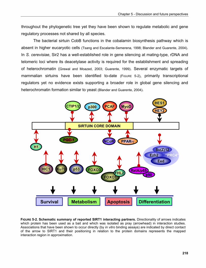
Chapter 5 - Discussion and future perspectives
throughout the phylogenetic tree yet they have been shown to regulate metabolic and gene
regulatory processes not shared by all species.
The bacterial sirtuin CobB functions in the cobalamin biosynthesis pathway which is
absent in higher eucaryotic cells (Tsang and Escalante-Semerena, 1998; Blander and Guarente, 2004).
In S. cerevisiae, Sir2 has a well-established role in gene silencing at mating-type, rDNA and
telomeric loci where its deacetylase activity is required for the establishment and spreading
of heterochromatin (Grewal and Moazed, 2003; Guarente, 1999). Several enzymatic targets of
mammalian sirtuins have been identified to-date (FIGURE 5-2), primarily transcriptional
regulators yet no evidence exists supporting a broader role in global gene silencing and
heterochromatin formation similar to yeast (Blander and Guarente, 2004).
PCAF
FOXO3
MyoDHES1
HEY2p300
p50RelA/p65
NCoR
p53
ApoptosisSurvival
Ku70
Differentiation
H1
SIRTUIN CORE DOMAIN
PGC-1 PPAR-γ
Ezh2Eed
HIC1
CTIP1/2
FOXO1FHL2
Su(Z)12
Metabolism
“PRC4”
FIGURE 5-2. Schematic summary of reported SIRT1 interacting partners. Directionality of arrows indicateswhich protein has been used as a bait and which was isolated as pray (arrowhead) in interaction studies.Associations that have been shown to occur directly (by in vitro binding assays) are indicated by direct contactof the arrow to SIRT1 and their positioning in relation to the protein domains represents the mappedinteraction region in approximation.
218
Chapter 5 - Discussion and future perspectives
5.2.1 Sirtuins and the regulation of organismal life-span
Sir2 has been proposed to be a factor determining longevity in S. cerevisiae as well as
C. elegans. Furthermore, Sir2 mediates life-span extension in response to nutrient limitation.
This is based on experimental evidence demonstrating that the presence of Sir2 is required
for life-span extension in calorically restricted yeast and worm while overexpression of the
protein suffices to increase life-span in a manner not-additive to caloric restriction itself
(Guarente 2005; Tissenbaum and Guarente, 2001; Lin et al., 2000).
Various theories have been put forward to explain the role of sirtuins in longevity.
Initially it was proposed that the suppression of recombination at the rDNA locus by Sir2p
attenuates the formation of ERCs (extrachromosomal rDNA circles) which normally
accumulate in mother cells and correlate with yeast ageing but also are suppressed in
calorically restricted cells (Lin et al., 2000; Sinclair, 1998; Sinclair and Guarente. 1997). Interestingly,
increased recombination rates are observed in ageing yeast cells suggesting a link between
genomic stability and life-span which extends beyond the rDNA locus (McMurray and Gottschling,
2003). In addition, preferential segragation of oxidatively damaged proteins in mother cells
has also been implicated in yeast ageing and shown to depend on Sir2p (Aguilaniu et al. 2003).
Increased respiration and changes in cellular NADH or nicotinamide levels have all
been proposed to influence Sir2 activity and contribute to its involvement in caloric restriction
(Lin et al., 2004; Anderson et al., 2003; Lin et al., 2002; Anderson et al., 2002). Recent evidence, however
has challenged the exclusive role of Sir2 contribution in longevity showing that TOR1 and
SCH9 deletions can increase yeast life-span in the absence of Sir2 (Kaeberlein et al., 2005;
Kaeberlein et al., 2005). Moreover, caloric restriction fails to increase life-span in yeast strains
carrying TOR1 and SCH9 deletions indicating a major contribution of these proteins in this
process (Kaeberlein et al., 2005). This clearly demonstrated that, at least in yeast, there are Sir2-
independent pathways that mediate the effects of nutrient limitation on longevity. These
pathways involve the kinases Tor1, a well-established sensor of nutrient availability in yeast
and mammals (Jacinto and Hall, 2003) and the yeast orthologue of mammalian PKB kinase
SCH9.
Interestingly, TOR regulates ribosomal biogenesis through the PKA pathway. TOR
modulates PKA subcellular localisation to maintain the donwstream kinase YAK1 inactive.
219
Chapter 5 - Discussion and future perspectives
TOR inhibition leads to activation of YAK1 which in turn phosphorylates the transcriptional
co-repressor CRF1. CRF1 then transclocates to the nucleus and competes with the co-
activator IFH1 for binding to the ribosomal gene transcription factor FHL1 thus repressing
ribosomal gene transcription (Martin et al., 2004). In addition TOR mediates ribosomal
biogenesis by regulation of rDNA transcription through the transcription factors UBF and
TIF1A (Mayer et al., 2004).
PKA is also involved in the glucose sensing pathway and PKA mutations mimic caloric
restriction to extend life-span in a manner dependent on Sir2p (Lin et al., 2002). Furthermore,
SIRT1-mediated deacetylation of the RNAPolI transcription factor subunit TAFI68 represses
ribosomal biogenesis. Thus, TOR and Sir2, both of which are proposed to function as
nutrient sensors, regulate life-span and ribosomal biogenesis in opposite ways and are
genetically linked to PKA which also responds to nutrient availability. It would hence be of
interest to investigate whether TOR and Sir2 genetically interact to regulate ribosomal
biogenesis and whether the later process is an active determinant of life-span or simply
correlates to proliferation rates associated with ageing.
In C. elegans mutations in the IGF signaling pathway regulate life-span in a manner
dependent on the forkhead transcription factor daf-16 (Tissenbaum and Guarente, 2002). A
genetic interaction between Sir-2.1 and the IGF-signaling pathway has been reported. In
particular, life-span extension induced by increased Sir-2.1 expression dependents on the
forkhead transcription factor daf-16 which is under the negative regulation of the IGF
signaling pathway (Wang and Tissenbaum, 2006; Tissenbaum and Guarente, 2001). This provided
evidence that sirtuins may mediate organismal longevity in multicellular organisms albeit by a
different, or as-yet unidentified common, mechanism than in yeast. Consequently, the quest
to identify a similar function in human sirtuins was pursued.
220
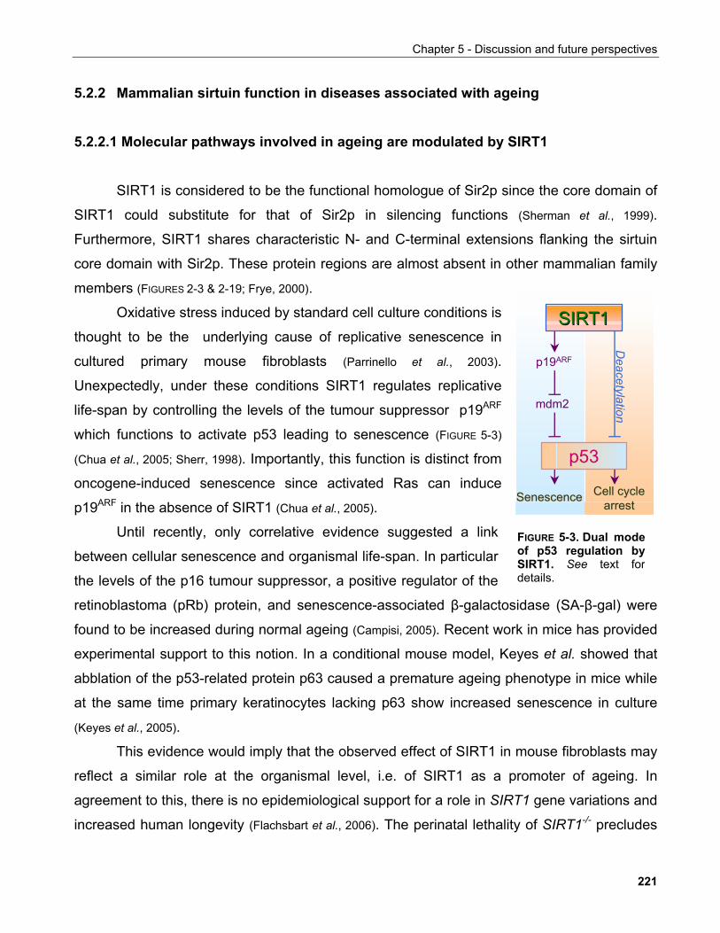
Chapter 5 - Discussion and future perspectives
5.2.2 Mammalian sirtuin function in diseases associated with ageing
5.2.2.1 Molecular pathways involved in ageing are modulated by SIRT1
SIRT1 is considered to be the functional homologue of Sir2p since the core domain of
SIRT1 could substitute for that of Sir2p in silencing functions (Sherman et al., 1999).
Furthermore, SIRT1 shares characteristic N- and C-terminal extensions flanking the sirtuin
core domain with Sir2p. These protein regions are almost absent in other mammalian family
members (FIGURES 2-3 & 2-19; Frye, 2000).
p19ARF
mdm2
p53
SenescenceSenescence Cell cycle Cell cycle arrestarrest
SIRT1SIRT1
Deacetylation
Oxidative stress induced by standard cell culture conditions is
thought to be the underlying cause of replicative senescence in
cultured primary mouse fibroblasts (Parrinello et al., 2003).
Unexpectedly, under these conditions SIRT1 regulates replicative
life-span by controlling the levels of the tumour suppressor p19ARF
which functions to activate p53 leading to senescence (FIGURE 5-3)
(Chua et al., 2005; Sherr, 1998). Importantly, this function is distinct from
oncogene-induced senescence since activated Ras can induce
p19ARF in the absence of SIRT1 (Chua et al., 2005).
Until recently, only correlative evidence suggested a link
between cellular senescence and organismal life-span. In particular
the levels of the p16 tumour suppressor, a positive regulator of the
retinoblastoma (pRb) protein, and senescence-associated β-galactosidase (SA-β-gal) were
found to be increased during normal ageing (Campisi, 2005). Recent work in mice has provided
experimental support to this notion. In a conditional mouse model, Keyes et al. showed that
abblation of the p53-related protein p63 caused a premature ageing phenotype in mice while
at the same time primary keratinocytes lacking p63 show increased senescence in culture
(Keyes et al., 2005).
FIGURE 5-3. Dual modeof p53 regulation bySIRT1. See text fordetails.
This evidence would imply that the observed effect of SIRT1 in mouse fibroblasts may
reflect a similar role at the organismal level, i.e. of SIRT1 as a promoter of ageing. In
agreement to this, there is no epidemiological support for a role in SIRT1 gene variations and
increased human longevity (Flachsbart et al., 2006). The perinatal lethality of SIRT1-/- precludes
221
Chapter 5 - Discussion and future perspectives
their use as a model to probe the contribution of SIRT1 on ageing. Interestingly, adipose-
specific ablation of IGF receptor results in a ~18% increase in life-span in mice suggesting
that organismal life-span can be influenced by the function of certain pathways in specific
tissues (Bluher et al., 2003). Thus, conditional deletion of SIRT1 would provide an important tool
for resolving its roles in mammalian biology and in particular ageing.
5.2.2.2 Molecular basis of SIRT1 function in ageing phenotypes
In our view of SIRT1 as a regulator of mammalian life-span, there are two ideas to be
taken into consideration. Firstly, the evolutionary theories of ageing would suggest that there
need not be a single gene or a common mechanism of life-span determination throughout the
species (Kipling et al., 2004; Kirkwood, 1996). Secondly, the multiplicity of mutations giving rise to
progeroid (premature ageing) syndromes and the wide spectrum of associated phenotypes
would support the notion that multiple factors contribute to the ageing process which
ultimately determines life-span (Kipling et al., 2004).
In elderly people as well as progeroid syndrome patients and mouse models of aging
common phenotypic manifestations include reduced body weight, reduced fecudity, higher
cancer incidence, lordokyphosis (curvature of the spine), decreased bone density, decreased
organ mass, cellular senescence, alopecia (balding), increased skin ulcerations and atrophy,
decreased dermal and adipose thickness, and sebaceous gland hyperplasia (Lombard et al.,
2005; Keyes et al., 2005). Werner syndrome (WS), a human autosomal genetic disorder provides
a characteristic example of the above. Werner syndrome patients suffer from abnormal
stature, atherosclerosis, graying of the hair, type II diabetes, cataracts and osteoporosis, all
of which are diseases more prevalent with ageing (Kipling et al., 2004).
Based on the above, in our analysis of SIRT1 as a modulator of mammalian life-span
it would be more meaningful to consider its potential functional role in alleviating age-related
phenotypes rather than view it as an intrinsic genetic determinant of longevity. This is also a
relevant approach when it comes to exploiting sirtuins as drug targets to treat ageing-related
symptoms with the aim to ameliorate the life of the elderly.
222
Chapter 5 - Discussion and future perspectives
5.2.2.2.1 Neuroprotective and cardioprotective roles of SIRT1
Immunohistochemical studies have identifed the heart and central nervous system as
sites of high murine SIRT1 expression during emryogenesis and in adult animals (Sakamoto et
al., 2004). Furthermore, mice with genetic ablation of the SIRT1 locus exhibit multiple
developmental defects some of which are consistent with this localisation (Cheng et al., 2003).
Experimental evidence suggests that SIRT1 has a protective role against neuronal and
cardiac damage.
In the Wallerian degeneration slow (wlds) mice increased nuclear NAD+ underlies the
protection exhibited in the neurons of these mice against neurodegenerative agents.
Importantly, the neuroprotective effects of NAD+ require SIRT1. Moreover, sirtinol and
resveratrol, two compounds that inhibit and activate SIRT1 respectively affect this process in
a manner consistent with the proposed involvement of SIRT1 (Araki et al. 2004). Similar effects
of sirtinol and resveratrol were also observed in an independent in vitro model of cerebral
ischaemia (Raval et al., 2005).
These observations also extend to the heart. In isolated neonatal rat cardiomyocytes
sirtuin inhibition by either nicotinamide or sirtinol induced cell death in a p53-dependent
manner. SIRT1 overexpression also caused an increase in cardiomyocyte size and protected
cells from apoptosis following serum starvation (Alcendor et al., 2004). Importantly, SIRT1 levels
were dramatically increased in a dog model of heart failure possibly a result of a failed
attempt to prevent cell death (Crow, 2004).
Risk of neurodegenerative and cardiovascular pathological conditions such as
Alzheimer's and ischaemic heart disease respectively increase dramatically with age and
together account for the vast majority of death rates (Hadley et al., 2005; Kirkwood, 1996). A
causative role for the devastating outcomes of ischaemic conditions is also connected to how
promptly they are treated so that to minimise tissue damage. This, in combination with life-
style factors such as diet, which is proposed to affect sirtuin function, render sirtuins an
important potential target for preventive treatments in the context of these diseases.
223
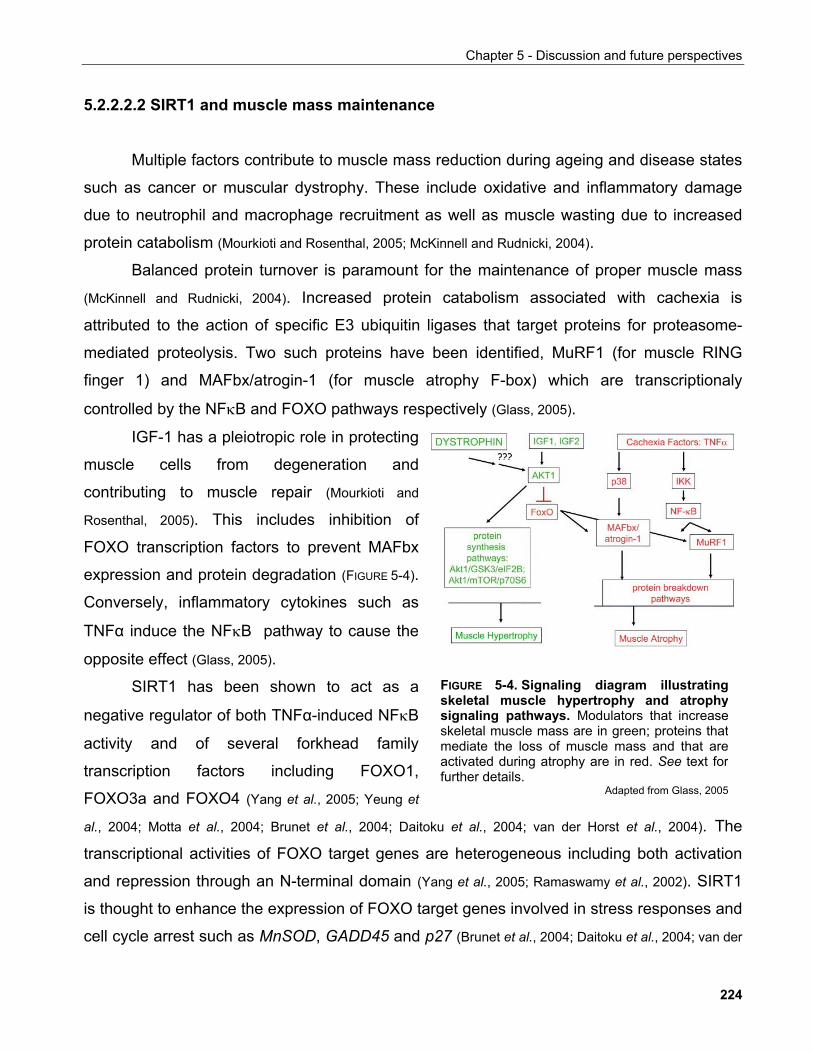
Chapter 5 - Discussion and future perspectives
5.2.2.2.2 SIRT1 and muscle mass maintenance
Multiple factors contribute to muscle mass reduction during ageing and disease states
such as cancer or muscular dystrophy. These include oxidative and inflammatory damage
due to neutrophil and macrophage recruitment as well as muscle wasting due to increased
protein catabolism (Mourkioti and Rosenthal, 2005; McKinnell and Rudnicki, 2004).
Balanced protein turnover is paramount for the maintenance of proper muscle mass
(McKinnell and Rudnicki, 2004). Increased protein catabolism associated with cachexia is
attributed to the action of specific E3 ubiquitin ligases that target proteins for proteasome-
mediated proteolysis. Two such proteins have been identified, MuRF1 (for muscle RING
finger 1) and MAFbx/atrogin-1 (for muscle atrophy F-box) which are transcriptionaly
controlled by the NFκB and FOXO pathways respectively (Glass, 2005).
IGF-1 has a pleiotropic role in protecting
muscle cells from degeneration and
contributing to muscle repair (Mourkioti and
Rosenthal, 2005). This includes inhibition of
FOXO transcription factors to prevent MAFbx
expression and protein degradation (FIGURE 5-4).
Conversely, inflammatory cytokines such as
TNFα induce the NFκB pathway to cause the
opposite effect (Glass, 2005).
SIRT1 has been shown to act as a
negative regulator of both TNFα-induced NFκB
activity and of several forkhead family
transcription factors including FOXO1,
FOXO3a and FOXO4 (Yang et al., 2005; Yeung et
al., 2004; Motta et al., 2004; Brunet et al., 2004; Daitoku et al., 2004; van der Horst et al., 2004). The
transcriptional activities of FOXO target genes are heterogeneous including both activation
and repression through an N-terminal domain (Yang et al., 2005; Ramaswamy et al., 2002). SIRT1
is thought to enhance the expression of FOXO target genes involved in stress responses and
cell cycle arrest such as MnSOD, GADD45 and p27 (Brunet et al., 2004; Daitoku et al., 2004; van der
FIGURE 5-4. Signaling diagram illustratingskeletal muscle hypertrophy and atrophysignaling pathways. Modulators that increaseskeletal muscle mass are in green; proteins thatmediate the loss of muscle mass and that areactivated during atrophy are in red. See text forfurther details.
Adapted from Glass, 2005
224
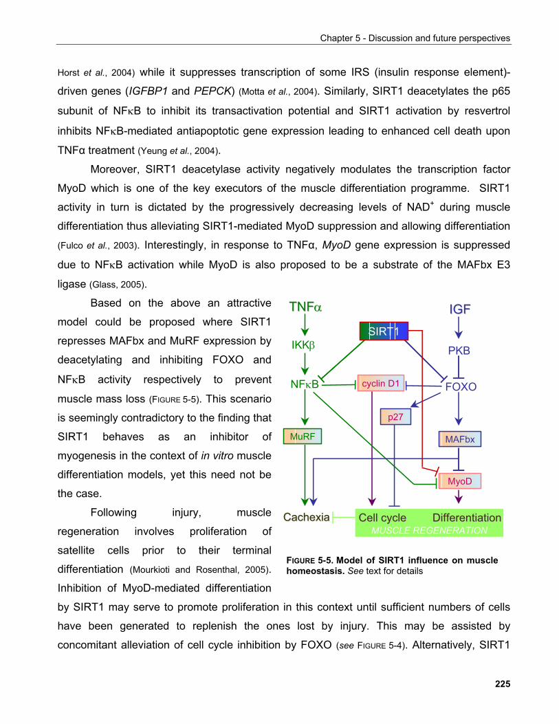
Chapter 5 - Discussion and future perspectives
Horst et al., 2004) while it suppresses transcription of some IRS (insulin response element)-
driven genes (IGFBP1 and PEPCK) (Motta et al., 2004). Similarly, SIRT1 deacetylates the p65
subunit of NFκB to inhibit its transactivation potential and SIRT1 activation by resvertrol
inhibits NFκB-mediated antiapoptotic gene expression leading to enhanced cell death upon
TNFα treatment (Yeung et al., 2004).
Moreover, SIRT1 deacetylase activity negatively modulates the transcription factor
MyoD which is one of the key executors of the muscle differentiation programme. SIRT1
activity in turn is dictated by the progressively decreasing levels of NAD+ during muscle
differentiation thus alleviating SIRT1-mediated MyoD suppression and allowing differentiation
(Fulco et al., 2003). Interestingly, in response to TNFα, MyoD gene expression is suppressed
due to NFκB activation while MyoD is also proposed to be a substrate of the MAFbx E3
ligase (Glass, 2005).
Based on the above an attractive
model could be proposed where SIRT1
represses MAFbx and MuRF expression by
deacetylating and inhibiting FOXO and
NFκB activity respectively to prevent
muscle mass loss (FIGURE 5-5). This scenario
is seemingly contradictory to the finding that
SIRT1 behaves as an inhibitor of
myogenesis in the context of in vitro muscle
differentiation models, yet this need not be
the case.
Following injury, muscle
regeneration involves proliferation of
satellite cells prior to their terminal
differentiation (Mourkioti and Rosenthal, 2005).
Inhibition of MyoD-mediated differentiation
by SIRT1 may serve to promote proliferation in this context until sufficient numbers of cells
have been generated to replenish the ones lost by injury. This may be assisted by
concomitant alleviation of cell cycle inhibition by FOXO (see FIGURE 5-4). Alternatively, SIRT1
IKKβ
NFκB cyclin D1
Cell cycleCell cycle DifferentiationDifferentiation
TNFTNFαα IGFIGF
PKB
FOXO
MuRF MAFbx
MyoD
SIRT1
CachexiaCachexia
p27
MUSCLE REGENERATION
FIGURE 5-5. Model of SIRT1 influence on musclehomeostasis. See text for details
225
Chapter 5 - Discussion and future perspectives
may promote MyoD mRNA and protein stability through inhibition of FOXO and NFκB as
delineated above. Furthermore, NFκB inhibition by SIRT1 may also limit inflammatory
cytokine production in macrophages thus mitigating the effects of inflammation-induced
damage in the muscle.
Evidently, these interconnections are derived from SIRT1 interactions observed in a
variety of heterologous experimental settings and is not clear whether it has a physiological
function in the animal. However, they would provide a reasonable starting model for further
investigation of exploiting SIRT1 as a therapeutic target for ageing and cancer-associated
muscle atrophy.
5.2.2.2.3 SIRT1 functions in metabolic regulation
SIRT1 in homeostatic organ functions regulating glucose metabolism
Ageing is associated with several pathological conditions affecting metabolic functions
of the body. Prevalent among them is type 2 diabetes which shows almost an exponential
incidence rate increase after the age of 20-30 years (Moller et al., 2003). Type 2 diabetes is
associated with decreased insulin secretion or the development of insulin resistance which in
turn is a major risk factor for cardiovascular disease and a series of other medical conditions
collecively known as the metabolic syndrome (de Luka and Olefsky, 2006).
In response to elevated glucose levels e.g. following a meal, insulin is secreted by the
β-cells found in the pancreatic islets of Langerhans to promote glucose uptake and
catabolism in peripheral tissues. β-cell function deteriorates with ageing (Moller et al., 2003).
SIRT1 is expressed in the pancreatic β-cells as well as the glucagon-producing α-cells
(Moynihan et al., 2005; Bordone et al., 2005). In β-cells SIRT1 enhances insulin secretion by
suppressing the expression of uncoupling protein 2 (UCP2). UCP's uncouple metabolic fuel
oxidation from ATP production thus leading to a decrease of ATP which is required for the
secretion of insulin (Lowell and Spiegelman, 2000). Consistent with a physiological role of SIRT1
in insulin secretion, mice overexpressing SIRT1 in pancreatic β-cells exhibited an enhanced
response to glucose challenge attributed to higher blood insulin, while conversely, SIRT1-/-
animals had lower levels of circulating insulin (Moynihan et al., 2005; Bordone et al., 2005).
226
Chapter 5 - Discussion and future perspectives
Furthermore, SIRT1 appears to have a protective role against glucose-induced
cytotoxicity in pancreatic β-cells, an underlying cause of β-cell degeneration seen in diabetic
patients whose plasma glucose levels are high. The cytotoxic effects of increased glucose
conentrations are attributed to elevated mitochondrial oxidation rates which lead to increased
ROS production. Under these conditions, SIRT1 was shown to be required in sustaining
FOXO1-mediated transcription of MafA and NeuroD, two transcription factors required for the
expression of the Insulin 2 gene to prevent apoptosis (Kitamura et al., 2005).
Another site of SIRT1 function in glucose metabolism is the liver. SIRT1 interacts with
the co-activator PGC-1α to induce gluconeogenesis in response to fasting (Rodgers et al., 2005).
This is rather the opposite role compared to the one it plays in β-cells where SIRT1
essentially functions to promote glucose utilisation. However, the strong expression of the
protein in the α-cells may also be similar to its hepatic function, although nothing is known
about that yet and it is possible that the different functions can solely be attributed to the
proteins SIRT1 partners with to perform the respective functions.
The above evidence suggests a multiple impact of SIRT1 function on body glucose
homeostasis through its role in insulin secretion and contribution to survival in the context of
pancreatic β-cells and gluconeogenesis in the liver. Interestingly, SIRT3, a mitochondrial
sirtuin, was shown to be downregulated in the skeletal muscle of a mouse model of diabetes
(Yechoor et al., 2004). Whether this underlies a concerted function of sirtuins in metabolic
regulation (see section 5.7) or, indeed, the function of SIRT3 in mitochondira, remain to be
elucidated.
Thus the activity of SIRT1 and possibly other sirtuins may also be a relevant
therapeutic target for diabetes, where aberrant glucose homeostasis and β-cell disfunction
are key manifestations of the disease.
SIRT1 function in lipid metabolism
Another tissue of interest in metabolic regulation is the adipose tissue. The adipose
tissue is the major site of triglyceride storage, an important energy source when glucose
availability is limited. During fasting and starvation, adipose triglyceride (TG) stores are
mobilised to give rise to free fatty acids (FFA) which can be utilised by other tissues for
227
Chapter 5 - Discussion and future perspectives
energy production (Rosen et al., 2002). Furthermore, the metabolic activities of adipose tissue
may have an impact on organismal longevity since adipose-specific ablation of IGF receptor
results in a ~18% increase in life-span in mice.
SIRT1 was shown to suppress adipocyte differentiation by inhibiting the adipogenic
factor PPARγ through the transcriptional co-repressor NCoR (Picard et al., 2004). Furthermore,
it was suggested that SIRT1 is required for TG mobilisation as SIRT1+/- animals exhibited low
levels of blood FFAs following fasting or β-adrenergic stimulation.
Interestingly, the bacterial sirtuin orthologue CobB deacetylates and activates the
enzyme acetyl-CoA synthase (ACS) which catalyses the synthesis of acetyl-CoA, a key
molecule in mitochondrial oxidation and lipid synthesis (Starai et al., 2004; Starai et al., 2002). Thus
it is possible that in higher organisms too SIRT1 or another sirtuins may affect lipid
metabolism by mediating intracellular acetyl-CoA levels.
Dyslipidemia is a common feature of the metabolic syndrome-associated disorders
including cardiovascular disease and atherosclerosis. Thus it would be of interest to
investigate whether modulation of SIRT1 activity can be considered as a possible target for
treating these conditions or symptoms thereof.
5.2.2.2.4 Reproduction
There is an inverse correlation between age and fecundity in that as animals age their
ability to produce viable offspring decreases (Partridge and Gems, 2002). Experimental evidence
in model organisms suggests that a hormonal cue from the reproductive system regulates
life-span in both the nematode and the mouse.
In C. elegans, ablation of germline precursors leads to an approx. 60% life-span
extension. Interestingly this is associated with an apparent loss/reduction of IGF signaling in
the intestine and adipose tissue since in these animals daf-16 translocates to the nucleus
(Kenyon, 2005). In the mouse, transplantation of ovaries derived from young animals to old
animals results in 40-60% increase in life-span. As the transplantation process kills the germ
cells, these animals have only a somatic gonad, a situation that parallels that in worms
described above (Kenyon, 2005).
228
Chapter 5 - Discussion and future perspectives
Despite this apparent evolutionary conservation, there are examples of animals that
do not comply with this relationship between reproductive capacity and life-span. This is the
case for the honey bee (Apis mellifera). Honey bee populations have a long-lived queen cast
with a reproductive role and a short-lived cast of sterile workers. Both queens and the long-
lived worker bees that remain in the hive to tend the larvae express higher levels of the
vitellogenin protein compared to the animals that leave the collony to collect honey (Seehuus et
al., 2006). Vitellogenin is controlled by the IGF signaling pathway in C. elegans and, consistent
with that, reduction of vitellogenin protein levels results in increased life-span in the worm. In
contrast, in the honey bee, despite its beneficial role in reproduction by equiping the yolk with
nutrients, the protein confers resistance to oxidative stress thus extending life-span (Seehuus
et al., 2006).
SIRT1 is highly expressed in the developing spermatocytes and deletion of the SIRT1
gene leads to severe sperm abnormalities and sterility (McBurney et al., 2003). In this context
SIRT1 would apper to be important to the reproductive capacity of the animal thus defying its
role in longevity based on the above. Interestingly, reduction of IGF signaling starting at the
time of hatching in C. elegans extends life-span and delays reproduction whereas IGF
signaling reduction in the adult increases life-span to the same extend without affecting
reproduction (Kenyon, 2005). Thus it is possible that SIRT1 holds a role in the adult
reproductive system that extends beyond embryonic development, possibly by controlling
IGF signaling through its documented roles in regulating forkhead factor activity.
5.2.2.2.5 SIRT1 and cancer
Cancer is a term coined to describe a vastly heterogeneous set of diseases that are
characterised by aberrant proliferation at the expense to physiological body functions, which,
when untreated, leads to death. Thus, although malignansies occur in younger individuals
too, sporadic cancer incidence shows a striking correlation with age (DePinho, 2000).
Several genetic and epigenetic processes have been linked to age-induced cancer
and mouse model studies have provided extensive evidence that genes involved in the
maintenance of genomic stability and cancer are also intimately linked to the development of
ageing phenotypes (Lombard et al., 2005; DePinho, 2000).
229
Chapter 5 - Discussion and future perspectives
Perhaps one of the most striking recent molecular links between cancer and ageing
was the discovery of an allele of the p53 tumour suppressor which correlates with high p53
activity and confers resistence to tumourigenesis but induces the onset of premature ageing
due to increased IGF signaling (Levine et al., 2006; Campisi, 2004; Maier et al., 2004; Tyner et al., 2002).
This and other evidence provided support to the notion that ageing is a by-product of an
organism's mechanisms for tumour suppression (Ferbeyre and Lowe, 2002).
Furthermore, recent work has identified interlinks between nutrient signaling and
pathways involved in cancer development such as the activation of p53 by the AMPK kinase
to co-ordinate cell cycle progression with the energy status of the cell (Jones et al., 2005). Both
the AMPK and IGF signaling pathways are upstream regulators of the mTOR pathway which
plays a paramount role in regulating cell growth and mutations in components of these
pathways are frequently found in cancers (Wullshleger et al., 2006 and section 5.1).
Whether SIRT1 has a causative role in tumour development is not known as no
studies reporting SIRT1 mutations in cancer have emerged to-date. However, several lines of
evidence indicate that SIRT1 may regulate various signaling pathways known to be involved
in cancer.
SIRT1 and epigenetic changes occuring in cancer
SIRT1 has been shown to have multiple effects on histone modifications. siRNA-
mediated downregulation of SIRT1 leads to H4-K16 hyperacetylation, and reduction in H3-
Tri-MeK9 and H4-MeK20 in mammalian cells while in vitro SIRT1 preferentially deacetylates
H4-K16 (Vaquero et al., 2004; Imai et al., 2000). Interestingly, Fraga et al. reported a consistent
loss of H4-K16 and H4-Tri-MeK20 in various tumours and tumour-derived cell lines
suggesting that these modifications consistute epigenetic hallmarks of cancer (Fraga et al.,
2005).
SIRT1 was also identified as a component of the polycomb repressive complex 4
(PRC4) which harbours the SET domain histone methyltransferase Ezh2 (Kuzmichev et al.,
2005). Four distinct PRC complexes have been identified to-date that differ in their
composition and sustrate specificity. In a mouse model of prostate cancer, the protein levels
of all PRC4 components tested, including SIRT1, were upregulated. Concomitantly,
230
Chapter 5 - Discussion and future perspectives
expression of PRC4 target genes was accordingly modified in these tissues. Although there
is no evidence confirming a causal role for PRC4 in cancer initiation, it is possible that PRC4-
mediated histone modifications contribute to cancer-spicific epigenetic changes.
In addition, in the context of the PRC4 complex, SIRT1 deacetylates histone H1-K26
and promotes heterochromatin formation through spreading hypometylated histone H3-K79
(Kuzmichev et al., 2005; Vaquero et al., 2004). Histone H1 is a linker histone which has primarily a
structural role in maintaining chromatin structure and through this regulates genomic stability
and ageing (Harvey and Downs, 2004). Furthermore, linker histone H1.2 has also been
implicated as a signal transmitter of apoptosis from the nucleus to the cytoplasm (Konishi et al.,
2003). Elucidation of the functional significance of SIRT1 in histone modifications is likely to
provide further insights into how it may contribute to cancer phenotypes.
A focused effort to develop HDAC inhibitors has been long underway following the
ralisation that many cancers and in particular leucemias, exhibit aberrant acetylation (Marks et
al. 2001). In this respect, SIRT1 joins its kins as a potential target for cancer therapy, although
clearly, more has to be known about its exact function in the disease to validate this
prospect.
Non-histone targets of SIRT1 in tumour development
Acetylation of transcription factors correlates, in general, with increased transcriptional
activity mainly due to enhanced binding to DNA (Kouzarides, 2000). SIRT1 deacetylates several
transcription factors involved in the regulation of cell cycle progression and apoptosis
consistent with a role in the fundamental processes underlying cancer.
SIRT1 deacetylates the tumour suppressor p53 to inhibit its transcriptional acivity
resulting in reduced apoptosis in response to various genotoxic stimuli (Luo et al., 2001; Vaziri et
al., 2001). Furthermore, SIRT1 localises to PML bodies to attenutate p53-dependent cellular
senescence induced by PML-IV overexpression (Langley et al., 2002).
SIRT1 associates with the tumour suppressor HIC1 which has been shown to act
synergistically with p53 in tumourigenesis. Both SIRT1 and HIC1 can bind the SIRT1
promoter to repress gene transcription and thus allow p53 acetylation and activation (Chen et
al., 2005). HIC1-/- cells exhibit elevated levels of SIRT1, hypoacetylated p53 and enhanced
231
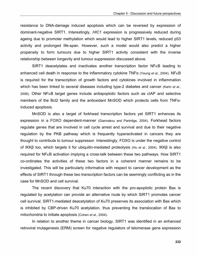
Chapter 5 - Discussion and future perspectives
resistance to DNA-damage induced apoptosis which can be reversed by expression of
dominant-negative SIRT1. Interestingly, HIC1 expression is progressively reduced during
ageing due to promoter methylation which would lead to higher SIRT1 levels, reduced p53
activity and prolonged life-span. However, such a model would also predict a higher
propensity to form tumours due to higher SIRT1 activity consistent with the inverse
relationship between longevity and tumour suppression discussed above.
SIRT1 deacetylates and inactivates another transcription factor NFκB leading to
enhanced cell death in response to the inflammatory cytokine TNFα (Yeung et al., 2004). NFκB
is required for the transcription of growth factors and cytokines involved in inflammation
which has been linked to several diseases including type-2 diabetes and cancer (Karin et al.,
2006). Other NFκB target genes include antiapoptotic factors such as cIAP and selective
members of the Bcl2 family and the antioxidant MnSOD which protects cells from TNFα-
induced apoptosis.
MnSOD is also a target of forkhead transcription factors yet SIRT1 enhances its
expression in a FOXO dependent-manner (Giannakou and Partridge, 2004). Forkhead factors
regulate genes that are involved in cell cycle arrest and survival and due to their negative
regulation by the PKB pathway which is frequently hyperactivated in cancers they are
thought to contribute to tumour suppressor. Interestingly, FOXO is under the negative control
of IKKβ too, which targets it for ubiquitin-mediated proteolysis (Hu et al., 2004). IKKβ is also
required for NFκB activation implying a cross-talk between these two pathways. How SIRT1
co-ordinates the activities of these two factors in a coherent manner remains to be
investigated. This will be particularly informative with respect to cancer development as the
effects of SIRT1 through these two transcription factors can be seemingly conflicting as in the
case for MnSOD and cell survival.
The recent discovery that Ku70 interaction with the pro-apoptotic protein Bax is
regulated by acetylation can provide an alternative route by which SIRT1 promotes cancer
cell survival. SIRT1-mediated deacetylation of Ku70 preserves its association with Bax which
is inhibited by CBP-driven Ku70 acetylation, thus preventing the translocation of Bax to
mitochondria to initiate apoptosis (Cohen et al., 2004).
In relation to another theme in cancer biology, SIRT1 was identified in an enhanced
retroviral mutagenesis (ERM) screen for negative regulators of telomerase gene expression
232
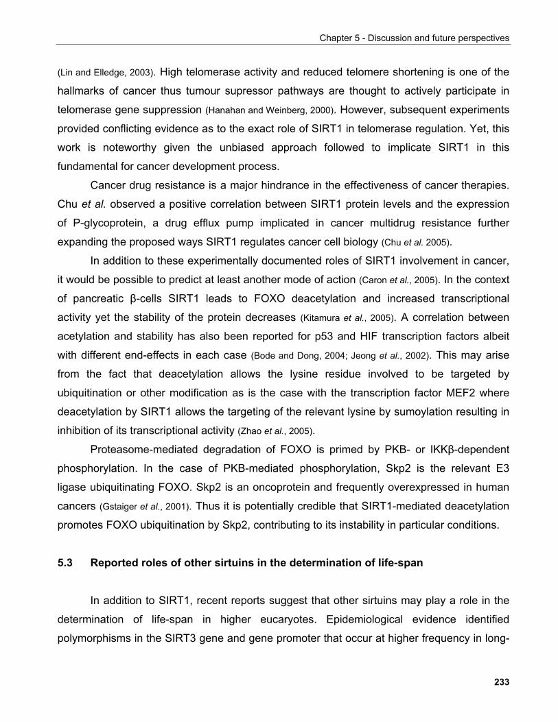
Chapter 5 - Discussion and future perspectives
(Lin and Elledge, 2003). High telomerase activity and reduced telomere shortening is one of the
hallmarks of cancer thus tumour supressor pathways are thought to actively participate in
telomerase gene suppression (Hanahan and Weinberg, 2000). However, subsequent experiments
provided conflicting evidence as to the exact role of SIRT1 in telomerase regulation. Yet, this
work is noteworthy given the unbiased approach followed to implicate SIRT1 in this
fundamental for cancer development process.
Cancer drug resistance is a major hindrance in the effectiveness of cancer therapies.
Chu et al. observed a positive correlation between SIRT1 protein levels and the expression
of P-glycoprotein, a drug efflux pump implicated in cancer multidrug resistance further
expanding the proposed ways SIRT1 regulates cancer cell biology (Chu et al. 2005).
In addition to these experimentally documented roles of SIRT1 involvement in cancer,
it would be possible to predict at least another mode of action (Caron et al., 2005). In the context
of pancreatic β-cells SIRT1 leads to FOXO deacetylation and increased transcriptional
activity yet the stability of the protein decreases (Kitamura et al., 2005). A correlation between
acetylation and stability has also been reported for p53 and HIF transcription factors albeit
with different end-effects in each case (Bode and Dong, 2004; Jeong et al., 2002). This may arise
from the fact that deacetylation allows the lysine residue involved to be targeted by
ubiquitination or other modification as is the case with the transcription factor MEF2 where
deacetylation by SIRT1 allows the targeting of the relevant lysine by sumoylation resulting in
inhibition of its transcriptional activity (Zhao et al., 2005).
Proteasome-mediated degradation of FOXO is primed by PKB- or IKKβ-dependent
phosphorylation. In the case of PKB-mediated phosphorylation, Skp2 is the relevant E3
ligase ubiquitinating FOXO. Skp2 is an oncoprotein and frequently overexpressed in human
cancers (Gstaiger et al., 2001). Thus it is potentially credible that SIRT1-mediated deacetylation
promotes FOXO ubiquitination by Skp2, contributing to its instability in particular conditions.
5.3 Reported roles of other sirtuins in the determination of life-span
In addition to SIRT1, recent reports suggest that other sirtuins may play a role in the
determination of life-span in higher eucaryotes. Epidemiological evidence identified
polymorphisms in the SIRT3 gene and gene promoter that occur at higher frequency in long-
233
Chapter 5 - Discussion and future perspectives
lived individuals (Bellizzi et al., 2005; Rose et al., 2003). Given the prominent role of mitochondria in
ageing processes (Balaban et al., 2005), a possible role of SIRT3 in this context would not be
surprising.
Mouse SIRT6 which was shown to harbour an ADP-ribosyltransferase activity (Liszt et
al., 2005), is required for DNA repair by the base-excision repair (BER) pathway thus
contributing to genomic stability. Deletion of both SIRT6 alleles in the mouse results in viable
animals which however die soon after birth following the onset of multiple degenerative
processes reminiscent of ageing symptoms (Mostoslavsky et al., 2006). Consequently, by
supporting genomic stability, SIRT6 may contribute to organismal survival and fitness.
Thus, based on current evidence it appears that multiple sirtuins may contribute to
various aspects of organismal physiology to regulate fitness. Consistent with this is the
proposal put forward here, namely that, in relation to sirtuin function, ageing should be
viewed as a set of phenotypes various aspects of which individual sirtuins may contribute to
or indeed alleviate.
5.4 Role of NAD in transcriptional regulation and disease
Changes of NAD+ intracellular dynamics in disease
Under certain conditions, SIRT1 activity may be regulated by controlling SIRT1 protein
levels as is the case in the liver following fasting (Rodgers et al., 2005; Kuzmichev et al., 2005) or in
prostate cancer. However, a characteristic feature of sirtuins is the dependence of their
enzymatic activity on NAD+ and their proposed regulation by NADH and/or nicotinamide.
Consequently, conditions where cellular NAD+ levels change may lead to modulation of
sirtuin activity.
Changes in intracellular NAD+/NADH concentrations have been implicated in various
conditions such as diabetes-associated retinopathy (Wahlberg et al., 2002; Salceda et al., 1998),
cardiovascular disease (Liu et al., 2005), fatty liver disease (You and Crabb, 2004) and
neurodegenerative diseases such as Alzheimer's (Jackson et al., 1994). Furthermore,
NAD+/NADH has been shown to decrease during muscle differentiation (Fulco et al., 2003).
234
Chapter 5 - Discussion and future perspectives
A key target of such changes has
been thought to be the nuclear enzyme
poly-ADP ribose polymerase (PARP). The
enzymatic activity of PARP shows stricking
similarities with that of sirtuins (FIGURE 5-6).
They both modify histones in an NAD+-
dependent manner. Unlike redox enzymes
that use NAD+ as a co-factor, PARPs and
sirtuins cleave the glycosidic bond between
ADP-ribose and nicotinamide thus
consuming NAD+ irreversibly (Kim et al.,
2005). Yet, PARP-1 is considered to be the
major consumer of NAD+ leading to
dramatic changes in cellular NAD+ pools upon its activation following DNA damage (Zhang,
2003). Indeed, in streptozocin-induced type-I diabetes mouse model, genetic ablation of the
PARP1 gene results in reduced β-cell death and protects mice against diabetes (Burkart et al.,
1999).
FIGURE 5-6. Dynamics and enzymology of proteinADP-ribosylation. Synthesis and degradation of PARon an acceptor protein. PARP-1 catalyzes thepolymerization of ADP-ribose units from donor NAD+
molecules on target proteins, resulting in theattachment of PAR. PARG catalyzes the hydrolysis ofPAR producing free mono and oligo(ADP-ribose). ADP-ribosyl protein lyase cleaves the final remaining ADP-ribose monomer from the target protein, releasing ADP-3''-deoxypentose-2''-ulose (ADP-DP)
Figure and legend adapted from Kim et al., 2005.
Interestingly, increasing the levels of NPT1, an enzyme involved in the NAD+ salvage
pathway in yeast, leads to enhanced rDNA and telomeric silencing without changing steady-
FIGURE 5-7. Interplay between sirtuin and PARPsignaling pathways. The depletion of NAD+ and theincrease of nicotinamide by PARP-1 may suppressSir2 deacetylation activity. PARP-1 and Sir2 sharethe NAD+ salvage pathway. The rapid and cyclicturnover of poly(ADP-ribose) by PARG to ADP-riboseresults in a drop of NAD+ and the rise ofnicotinamide, both of which are able to directly down-regulate Sir2 activity. The possible linkage of PARP-1and Sir2 pathways may provide a network thatcouples energy production, oxidative stress, DNAdamage, chromosome remodeling and genesilencing. ARPP: ADP-ribose pyrophosphatase;NAD+: β-nicotinamide adenine dinucleotide; NPRT:nicotinate phosphoribosyl transferase; PARG:poly(ADP-ribose) glycohydrolase; PARP-1:poly(ADP-ribose) polymerase-1; Ppi: pyrophosphate;PRPP: 5-phosphoribosyl-1-pyrophosphate; RPPK:ribose phosphate pyrophosphokinase.
Figure and legend adapted from Zhang, 2003.
235
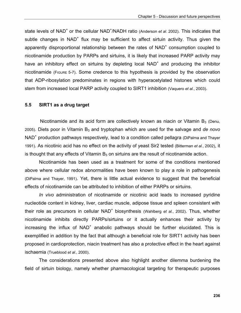
Chapter 5 - Discussion and future perspectives
state levels of NAD+ or the cellular NAD+/NADH ratio (Anderson et al. 2002). This indicates that
subtle changes in NAD+ flux may be sufficient to affect sirtuin activity. Thus given the
apparently disproportional relationship between the rates of NAD+ consumption coupled to
nicotinamide production by PARPs and sirtuins, it is likely that increased PARP activity may
have an inhibitory effect on sirtuins by depleting local NAD+ and producing the inhibitor
nicotinamide (FIGURE 5-7). Some credence to this hypothesis is provided by the observation
that ADP-ribosylation predominates in regions with hyperacetylated histones which could
stem from increased local PARP activity coupled to SIRT1 inhibition (Vaquero et al., 2003).
5.5 SIRT1 as a drug target
Nicotinamide and its acid form are collectively known as niacin or Vitamin B3 (Denu,
2005). Diets poor in Vitamin B3 and tryptophan which are used for the salvage and de novo
NAD+ production pathways respectively, lead to a condition called pellagra (DiPalma and Thayer
1991). As nicotinic acid has no effect on the activity of yeast Sir2 tested (Bitterman et al., 2002), it
is thought that any effects of Vitamin B3 on sirtuins are the result of nicotinamide action.
Nicotinamide has been used as a treatment for some of the conditions mentioned
above where cellular redox abnormalities have been known to play a role in pathogenesis
(DiPalma and Thayer, 1991). Yet, there is little actual evidence to suggest that the beneficial
effects of nicotinamide can be attributed to inhibition of either PARPs or sirtuins.
In vivo administration of nicotinamide or nicotinic acid leads to increased pyridine
nucleotide content in kidney, liver, cardiac muscle, adipose tissue and spleen consistent with
their role as precursors in cellular NAD+ biosynthesis (Wahlberg et al., 2002). Thus, whether
nicotinamide inhibits directly PARPs/sirtuins or it actually enhances their activity by
increasing the influx of NAD+ anabolic pathways should be further elucidated. This is
exemplified in addition by the fact that although a beneficial role for SIRT1 activity has been
proposed in cardioprotection, niacin treatment has also a protective effect in the heart against
ischaemia (Trueblood et al., 2000).
The considerations presented above also highlight another dilemma burdening the
field of sirtuin biology, namely whether pharmacological targeting for therapeutic purposes
236
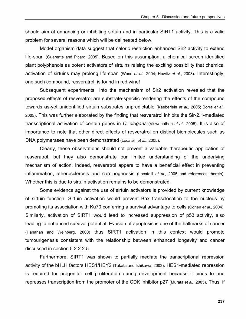
Chapter 5 - Discussion and future perspectives
should aim at enhancing or inhibiting sirtuin and in particular SIRT1 activity. This is a valid
problem for several reasons which will be delineated below.
Model organism data suggest that caloric restriction enhanced Sir2 activity to extend
life-span (Guarente and Picard, 2005). Based on this assumption, a chemical screen identified
plant polyphenols as potent activators of sirtuins raising the exciting possibility that chemical
activation of sirtuins may prolong life-span (Wood et al., 2004; Howitz et al., 2003). Interestingly,
one such compound, resveratrol, is found in red wine!
Subsequent experiments into the mechanism of Sir2 activation revealed that the
proposed effects of resveratrol are substrate-specific rendering the effects of the compound
towards as-yet unidentified sirtuin substrates unpredictable (Kaeberlein et al., 2005; Borra et al.,
2005). This was further elaborated by the finding that resveratrol inhibits the Sir-2.1-mediated
transcriptional activation of certain genes in C. elegans (Viswanathan et al., 2005). It is also of
importance to note that other direct effects of resveratrol on distinct biomolecules such as
DNA polymerases have been demonstrated (Locatelli et al., 2005).
Clearly, these observations should not prevent a valuable therapeutic application of
resveratrol, but they also demonstrate our limited understanding of the underlying
mechanism of action. Indeed, resveratrol appers to have a beneficial effect in preventing
inflammation, atherosclerosis and carcinogenesis (Locatelli et al., 2005 and references therein).
Whether this is due to sirtuin activation remains to be demonstrated.
Some evidence against the use of sirtuin activators is provided by current knowledge
of sirtuin function. Sirtuin activation would prevent Bax transclocation to the nucleus by
promoting its association with Ku70 conferring a survival advantage to cells (Cohen et al., 2004).
Similarly, activation of SIRT1 would lead to increased suppression of p53 activity, also
leading to enhanced survival potential. Evasion of apoptosis is one of the hallmarks of cancer
(Hanahan and Weinberg, 2000) thus SIRT1 activation in this context would promote
tumourigenesis consistent with the relationship between enhanced longevity and cancer
discussed in section 5.2.2.2.5.
Furthermore, SIRT1 was shown to partially mediate the transcriptional repression
activity of the bHLH factors HES1/HEY2 (Takata and Ishikawa, 2003). HES1-mediated repression
is required for progenitor cell proliferation during development because it binds to and
represses transcription from the promoter of the CDK inhibitor p27 (Murata et al., 2005). Thus, if
237
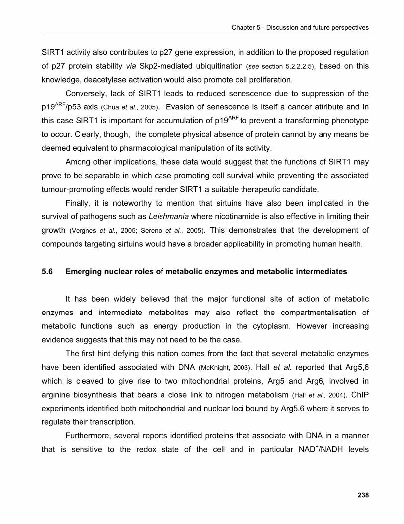
Chapter 5 - Discussion and future perspectives
SIRT1 activity also contributes to p27 gene expression, in addition to the proposed regulation
of p27 protein stability via Skp2-mediated ubiquitination (see section 5.2.2.2.5), based on this
knowledge, deacetylase activation would also promote cell proliferation.
Conversely, lack of SIRT1 leads to reduced senescence due to suppression of the
p19ARF/p53 axis (Chua et al., 2005). Evasion of senescence is itself a cancer attribute and in
this case SIRT1 is important for accumulation of p19ARF to prevent a transforming phenotype
to occur. Clearly, though, the complete physical absence of protein cannot by any means be
deemed equivalent to pharmacological manipulation of its activity.
Among other implications, these data would suggest that the functions of SIRT1 may
prove to be separable in which case promoting cell survival while preventing the associated
tumour-promoting effects would render SIRT1 a suitable therapeutic candidate.
Finally, it is noteworthy to mention that sirtuins have also been implicated in the
survival of pathogens such as Leishmania where nicotinamide is also effective in limiting their
growth (Vergnes et al., 2005; Sereno et al., 2005). This demonstrates that the development of
compounds targeting sirtuins would have a broader applicability in promoting human health.
5.6 Emerging nuclear roles of metabolic enzymes and metabolic intermediates
It has been widely believed that the major functional site of action of metabolic
enzymes and intermediate metabolites may also reflect the compartmentalisation of
metabolic functions such as energy production in the cytoplasm. However increasing
evidence suggests that this may not need to be the case.
The first hint defying this notion comes from the fact that several metabolic enzymes
have been identified associated with DNA (McKnight, 2003). Hall et al. reported that Arg5,6
which is cleaved to give rise to two mitochondrial proteins, Arg5 and Arg6, involved in
arginine biosynthesis that bears a close link to nitrogen metabolism (Hall et al., 2004). ChIP
experiments identified both mitochondrial and nuclear loci bound by Arg5,6 where it serves to
regulate their transcription.
Furthermore, several reports identified proteins that associate with DNA in a manner
that is sensitive to the redox state of the cell and in particular NAD+/NADH levels
238
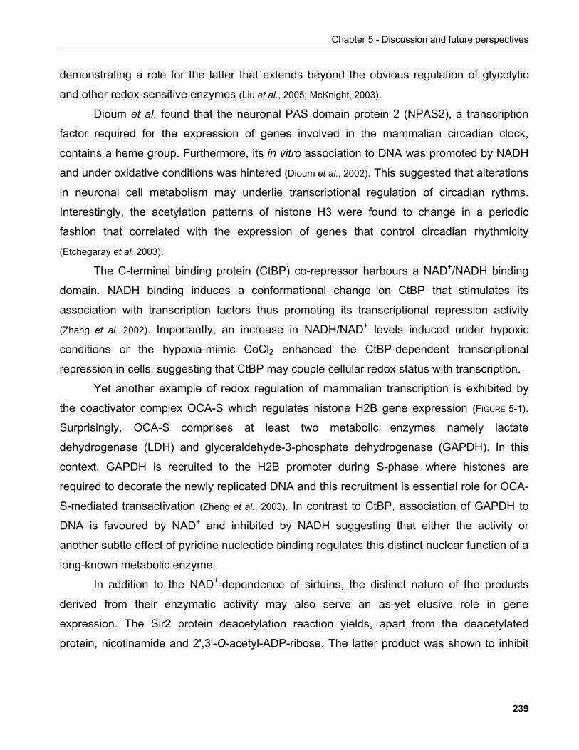
Chapter 5 - Discussion and future perspectives
demonstrating a role for the latter that extends beyond the obvious regulation of glycolytic
and other redox-sensitive enzymes (Liu et al., 2005; McKnight, 2003).
Dioum et al. found that the neuronal PAS domain protein 2 (NPAS2), a transcription
factor required for the expression of genes involved in the mammalian circadian clock,
contains a heme group. Furthermore, its in vitro association to DNA was promoted by NADH
and under oxidative conditions was hintered (Dioum et al., 2002). This suggested that alterations
in neuronal cell metabolism may underlie transcriptional regulation of circadian rythms.
Interestingly, the acetylation patterns of histone H3 were found to change in a periodic
fashion that correlated with the expression of genes that control circadian rhythmicity
(Etchegaray et al. 2003).
The C-terminal binding protein (CtBP) co-repressor harbours a NAD+/NADH binding
domain. NADH binding induces a conformational change on CtBP that stimulates its
association with transcription factors thus promoting its transcriptional repression activity
(Zhang et al. 2002). Importantly, an increase in NADH/NAD+ levels induced under hypoxic
conditions or the hypoxia-mimic CoCl2 enhanced the CtBP-dependent transcriptional
repression in cells, suggesting that CtBP may couple cellular redox status with transcription.
Yet another example of redox regulation of mammalian transcription is exhibited by
the coactivator complex OCA-S which regulates histone H2B gene expression (FIGURE 5-1).
Surprisingly, OCA-S comprises at least two metabolic enzymes namely lactate
dehydrogenase (LDH) and glyceraldehyde-3-phosphate dehydrogenase (GAPDH). In this
context, GAPDH is recruited to the H2B promoter during S-phase where histones are
required to decorate the newly replicated DNA and this recruitment is essential role for OCA-
S-mediated transactivation (Zheng et al., 2003). In contrast to CtBP, association of GAPDH to
DNA is favoured by NAD+ and inhibited by NADH suggesting that either the activity or
another subtle effect of pyridine nucleotide binding regulates this distinct nuclear function of a
long-known metabolic enzyme.
In addition to the NAD+-dependence of sirtuins, the distinct nature of the products
derived from their enzymatic activity may also serve an as-yet elusive role in gene
expression. The Sir2 protein deacetylation reaction yields, apart from the deacetylated
protein, nicotinamide and 2',3'-O-acetyl-ADP-ribose. The latter product was shown to inhibit
239
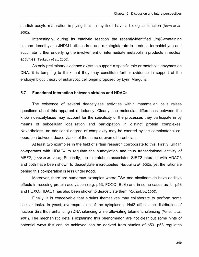
Chapter 5 - Discussion and future perspectives
starfish oocyte maturation implying that it may itself have a biological function (Borra et al.,
2002).
Interestingly, during its catalytic reaction the recently-identified JmjC-containing
histone demethylase JHDM1 utilises iron and α-ketoglutarate to produce formaldehyde and
succinate further underlying the involvement of intermediate metabolism products in nuclear
activities (Tsukada et al., 2006).
As only preliminary evidence exists to support a specific role or metabolic enzymes on
DNA, it is tempting to think that they may constitute further evidence in support of the
endosymbiotic theory of eukaryotic cell origin proposed by Lynn Margulis.
5.7 Functional interaction between sirtuins and HDACs
The existence of several deacetylase activities within mammalian cells raises
questions about this apparent redudancy. Clearly, the molecular differences between the
known deacetylases may account for the specificity of the processes they participate in by
means of subcellular localisation and participation in distinct protein complexes.
Nevertheless, an additional degree of complexity may be exerted by the combinatorial co-
operation between deacetylases of the same or even different class.
At least two examples in the field of sirtuin research corroborate to this. Firstly, SIRT1
co-operates with HDAC4 to regulate the sumoylation and thus transcriptional activity of
MEF2, (Zhao et al., 2005). Secondly, the microtubule-associated SIRT2 interacts with HDAC6
and both have been shown to deacetylate microtubules (Hubbert et al., 2002), yet the rationale
behind this co-operation is less understood.
Moreover, there are numerous examples where TSA and nicotinamide have additive
effects in rescuing protein acetylation (e.g. p53, FOXO, Bcl6) and in some cases as for p53
and FOXO, HDAC1 has also been shown to deacetylate them (Kouzarides, 2000).
Finally, it is conceivable that sirtuins themselves may collaborate to perform some
cellular tasks. In yeast, overexpression of the cytoplasmic Hst2 affects the distribution of
nuclear Sir2 thus enhancing rDNA silencing while alleviating telomeric silencing (Perrod et al.,
2001). The mechanistic details explaining this phenomenon are not clear but some hints of
potential ways this can be achieved can be derived from studies of p53. p53 regulates
240
Chapter 5 - Discussion and future perspectives
apoptosis through a nuclear pool that transcribes po-apoptotic genes and a cytoplasmic pool
that, when activated, enhances mitochondrial permeability. In unstressed conditions
cytolasmic p53 is bound to Bcl-XL preventing it from activating apoptosis. Following genotoxic
stress, nuclear p53 drives the expression of PUMA (for p53-upregulated modifier of
apoptosis) which induces the release of cytoplasmic p53 from Bcl-XL to induce BAX and thus
mitochondrial permeability and apoptosis (Chipuk et al., 2005), providing a striking example of
functional co-operation between distinct pools of the same enzyme.
5.8 Conclusion
A surge of recent data have implicated mammalian sirtuins and in particular SIRT1 in
the regulation of transcription factor activity. These effects are primarily exerted through the
deacetylase enzymatic function of SIRT1 which has been widely suspected but only once
experimentally demonstrated (Fulco et al., 2003) to be potentially linked to cellular NAD+/NADH
changes. Thus further work would need to address how the NAD+-dependance of SIRT1 is
actually relevant to the transcriptional regulatory activity of the protein. For this, it is important
to attain a deeper understanding of the metabolic pathways regulating NAD+ biosynthesis as
well as the role of other NAD+ utilising enzymes such as PARPs (Zhang et al., 2003). This is
also relevant for elucidating the cellular mode of action of known small molecule regulators of
SIRT1 such as nitotinamide which is at the same time an important intermediate in the NAD+
salvage pathway (Lin et al., 2003).
In addition, the existing preliminary evidence for a role of the sirtuin enzymatic product
2',3'-O-acetyl-ADP-ribose as a secondary messenger (Borra et al., 2002) supported by the
identification of 2',3'-O-acetyl-ADP-ribose-metabolising enzymes (Rafty et al., 2002) paves the
way for further penetrating into the roles of these metabolites in cellular physiology.
Clearly, despite the plethora of SIRT1 deacetylase targets already identified (FIGURE 5-
2), the current approaches towards identifying novel SIRT1 substrates have been in part
limited to predictions from sirtuin function in lower organisms. Yet, very little evidence exists
to support a functional conservation between yeast Sir2 and mammalian SIRT1 excluding
probably the documented actions on histone H1 deacetylation (Vaquero et al., 2004) and its
participation in a transcriptional repression complex (Kuzmichev et al., 2005). For example, none
241
Chapter 5 - Discussion and future perspectives
of the Sir2 protein complexes involved in silencing in S. cerevisiae have been identified in
mammalian cells although the protein complex composition of many evolutionarily conserved
proteins such as TOR and URI has been demonstrated to be also preserved throughout
evolution (Wullschleger et al., 2006; Gstaiger et al., 2003).
What is more, should sirtuin activity be subjected to targeted pharmacological
manipulation, it would be important to establish the interconnections between family
members not excluding a detailed characterisation of structural similarities of these proteins
as well as to other deacetylases. Clearly, current evidence from gene ablation studies in the
mouse suggests that non-redundant functions of individual sirtuin members exist as distinct
not compensated for phenotypes are evident (Mostoslavsky et al., 2006; Cheng et al., 2003).
Finally, the limitations in directly measuring intracellular NAD+/NADH levels and
demonstrating the direct link to sirtuin activity remains a great obstacle in studying sirtuin
biology in their cellular in vivo context. Protein post-translational modifications (PTMs) and in
particular phosphorylation constitute a universally applicable regulatory mechanism.
Monitoring steady-state PTM levels e.g. by immunohistochemical techniques is indicative of
specific signaling pathway activities in the context of tissues and organs, which in turn allows
the correlation of signalling fluxes with particular disease states.
No known SIRT1 PTMs have been reported yet. In order to address this need, the
work presented in this thesis has focused on the identification of molecular events and
pathways with potentially regulatory roles for SIRT1 activity.
242
Chapter 5 - Discussion and future perspectives
5.9 THESIS RESULTS: DISCUSSION AND FUTURE PERSPECTIVES 5.9.1 Regulation of SIRT1 by phosphorylation 5.9.1.1 SIRT1 is a nuclear phosphoprotein
To study the function and cellular localisation of SIRT1 one mouse monoclonal and
two rabbit polyclonal antibodies raised against different antigenic regions of the protein were
developed. All three antibodies were specific for SIRT1 and were succesfully used in different
experimental applications including immunoprecipitation and immunolocalisation (FIGURES 4-1
TO 4-4). Importantly, these antibodies sustain SIRT1 enzymatic activity making them useful for
purifying SIRT1 and assaying deacetylase activity in vitro (FIGURE 4-5).
A preliminary characterisation of the protein's subcellular localisation using either of
the polyclonal antibodies in indirect immunofluorescence microscopy revealed a nuclear,
occassionaly speckled, staining pattern (FIGURE 4-2). In contrast mAb12/1 gave a prominent
cytoplasmic staining following different fixation/permeabilisation conditions (FIGURE 4.3). SIRT1
has been consistently reported as a nuclear protein (e.g. Vaquero et al., 2004; Langley et al., 2002)
while mAb12/1 recognises a single band when used in immunoblotting attesting to its
specificity (FIGURE 4-3). Thus, despite the absence of biochemical evidence in support of this, it
could be postulated that a fraction of SIRT1 does localise to the cytoplasm but is only
revealed with mAb12/1 due to a peculiarity of the antibody allowing it to recognise the
cytoplasmic SIRT1 pool. This cytoplasmic SIRT1 pool would be predicted to be much smaller
relative to its nuclear counterpart given the inability of the polyclonal antibodies to recognise
the latter (FIGURE 4-2). Alternatively, despite its apparent specificity in immunoblotting
applications mAb12/1 specificity may be skewed following cell fixation giving rise to an
artifactual cytoplasmic signal.
An extensive attempt to compare the focal staining of SIRT1 with other known
speckled structures in the nucleus such as lamin-containing replication foci (Kennedy et al.,
2000), BRCA1-containing nuclear foci (Scully et al., 1996) and PML bodies (Seeler and Dejean,
1999) have indicated a partial co-localisation with PML bodies consistent with a previous
study (Langley et al., 2002), but not with lamin or BRCA1 (data not presented here).
243
Chapter 5 - Discussion and future perspectives
Interestingly, the immunofluorescence signal corresponding to SIRT1 was markedly
reduced in mitotic cells (FIGURE 4.6). To test and exclude the possibility that this did not simply
result from signal diffusion due to nuclear envelope break-down the protein levels of
endogenous SIRT1 were compared in unsynchronous and mitosis-arrested cells by
immunoblotting. This approach revealed that SIRT1 migration in denaturing gels is sensitive
to λ-PPase attesting to the fact that SIRT1 is a phosphoprotein (FIGURE 4-7). Furthermore, in
mitotic cells, SIRT1 in nocodazole-arrested cells exhibited a more prominent electrophoretic
mobility retardation relative to that in logarithmically growing cells suggesting the existence of
mitosis-specific phosphorylation event(s) in addition to the interphase one(s) (FIGURE 4-7B).
To this end a multidisciplinary approach was followed to determine the interphase-
specific as well as the mitotic phosphorylation events which SIRT1 is subjected to.
5.9.1.2 Identification of SIRT1 phosphorylation sites in interphase
Initial experiments revealed that the electrophoretic mobility of Sf9 (insect) cell-
expressed recombinant GST-SIRT1 is sensitive to λ-PPase treatment indicating that it is also
a phosphoprotein, similarly to endogenous human SIRT1 (FIGURE 4-8A). Interestingly, the in
vitro deacetylase activity of insect cell-expressed GST-SIRT1 was significantly reduced
following λ-PPase treatment (FIGURE 4-8B). As phosphorylation events are often conserved
between insect and mammalian cells, this observation indicated that phosphorylation can
regulate the enzymatic activity of SIRT1. Supporting this notion, other deacetylases have
also been shown to be regulated at multiple levels by phosphorylation (e.g. Zhang et al. 2005;
Pflum et al., 2001). Thus an in-depth investigation into the identification and functional
characterisation of the relevant phosphorylation sites was undertaken.
The phosphorylation sites of insect cell-expressed SIRT1 were determined by tryptic
digestion of purified recombinant protein followed by mass spectrometric analysis (FIGURE 4-9).
This approach revealed the existence of at least two serines modified by phosphorylation at
positions 27 and 47 in the N-terminus of the protein. Individual mutations of either residues to
non-phosphorylatable alanine indicated that neither of these were the phosphoshift sites and
truncation of the N-terminus of the protein suggested that the phoshoshift site does not
reside within the first 217 aminoacids (FIGURE 4-10). Nevertheless further credence to the fact
244
Chapter 5 - Discussion and future perspectives
that these these phosphorylation events are conserved was provided by a study published
subsequently and identified the same SIRT1 phosphorylation sites employing a large-scale
study of HeLa cell nuclear phosphoproteins (Beausoleil et al., 2004). Importantly, no other SIRT1
phosphorylation sites were reported in the same study.
The yeast sirtuin orthologue Hst2p has been reported to form homotrimers through N-
terminal domain contacts. Furthermore, this homotrimerisation event is important for efficient
enzymatic catalysis (Zhao et al., 2003). Given the presence of the identified phosphorylation
sites in the N-terminus of the protein, the possibility that S27 and/or S47 phoshorylation
regulates human SIRT1 homopolymerisation was tested. Co-expression of differentially
tagged SIRT1 followed by reciprocal immunoprecipitation confirmed the ability of SIRT for
homotypic association (FIGURE 4-11A). Moreover, gel filtration analysis of endogenous SIRT1 in
HeLa cells revealed a size distribution pattern consistent with the protein being in a
monomeric to trimeric configuration (FIGURE 4-11B). Preliminary experiments to investigate the
influence of S27 and S47 phosphorylation on this association gave ambiguous results but
further work into this issue is underway.
An interesting observation stemming out of this line of investigation revealed that apart
from the previously described putative nuclear localisation sequence (NLS) in residues 233-
238, a second NLS must be present, possibly within residues 94-99, as a SIRT1 species
lacking the N-terminal 217 aminoacids exhibits an exclusively cytoplasmic localisation (FIGURE
4-12).
In an alternative approach, the λ-PPase-sensitive electrophoretic mobility shift of
endogenous SIRT1 was used as an indirect means of identifying the relevant interphase
phosphorylation sites henceforth referred to as 'phosphoshift sites'. Sequential screens of
truncated SIRT1 species revealed that the phosphoshift site(s) is (are) located within region
612-656 at the C-terminal extension of the protein (FIGURE 4-13A). A SIRT1 species with a
deletion of aa's 612-656 showed an electrophoretic mobility which was not sensitive to λ-
PPase confirming that the region in question carries the phosphoshift sites (FIGURE 4-13C).
However, single mutations of all phosphorylatable aminoacids harboured within this region
failed to abolish the λ-PPase-sensitivity of SIRT1 mobility (FIGURE 4-13B) suggesting that more
than one sites are responsible for the observed shift.
245
Chapter 5 - Discussion and future perspectives
Alternatively, none of these sites are phosphorylated to cause a mobility shift, rather
the region in question is responsible for the recruitment of the relevant kinase which upon
deletion of aa's 612-656 would be abolished resulting in the observed behaviour of the
protein in FIGURE 4-13C.
To investigate this posibility, either endogenous or transfected SIRT1 was
immunoprecipitated and incubated with 32P-γ-ATP to allow its labeling by a putative stably-
bound kinase. Although little activity was detected in immunoprecipitates of endogenous
protein (FIGURE 4-14A), HA-tagged SIRT1 was strongly labelled with 32P indicating the presence
of a co-purifying kinase (FIGURE 4-14B). To test whether the putative co-purifying kinase
associates through aa's 612-656 which cause the phosphoshift, the same assay as above
was performed in full-length SIRT1 or a species lacking aa's 612-656. Deletion of aa's 612-
656 did not abolish 32P incorporation (FIGURE 4-14C) suggesting that the identity of the stably-
bound kinase differs from that of the kinase causing the phosphoshift.
Thus, the above experimental approach identified a region in the C-temrinus of human
SIRT1 that is required for the phosphorylation of the protein during interphase and a separate
phosphorylation event mediated by a stably bound kinase. Further work into this issue will
aim at identifying the site(s) of SIRT1 phosphorylation by the co-purifying kinase and probing
the nature of the kinases involved. Towards this end, initial attempts to identify SIRT1 co-
precipitating proteins by mass spectrometry has been hintered by insufficently pure
immunoprecipitates which under the light of the gel filtration data is likely to stem from a very
low abundance of such SIRT1-containing complexes (FIGURE 4-11B).
The emergence of data demonstrating that specificity of substrate recognition by
kinases is determined by the primary sequence surrounding the phosphorylated residue, led
to the development of searchable web-based databases which allow the user to probe for the
presence of kinase target sites in a protein of interest. Following this approach one putative
site for CK2 kinase was identified at residue S693. Further observation of the C-terminal
sequence of the protein revealed at least two other sites (S659 and S661) that complied with
the criteria of a CK2 site (FIGURE 4-15A).
In vitro kinase assay using recombinant CK2 cofirmed that all three SIRT1 sites are
phosphorylated by CK2. Mutation of all three sites did not affect phosphorylation by the co-
precipitating kinase (FIGURE 4-14B) nor did it abolish the λ-PPase-sensitive shift of interphase
246
Chapter 5 - Discussion and future perspectives
cells (FIGURE 4-16). This indicated that, if CK2 phosphorylates SIRT1 in vivo, this constitutes
yet a third phosphorylation event.
Given the strength of SIRT1 phosphorylation by CK2 in vitro it is important to
determine whether this also occurs in vivo. Several lines of evidence support the value of
pursuing this observations further. To begin with, a large-scale two-hybrid analysis of the
Drosophila proteome revealed a direct association of dSir2 with the the regulatory β subunit
of CK2 (Giot et al. 2003). Similarly, the entire CK2 complex is a third order interactor of the
yeast Sir2 in another large-scale screen (Ho et al., 2002). In human cells CK2 phosphorylates
and activates another nuclear deacetylase, HDAC1. Furthermore, this phosphorylation
enhances the formation of HDAC1-containing transcriptional complexes (Pflum et al. 2001).
For a long time CK2 was thought to be a constitutively active enzyme and its substrate
repertoire numbers hundreds of proteins (Meggio and Pinna, 2003; Ahmed et al., 2002). Yet, recent
evidence has demonstrated additional roles for this kinase where it acts in an inducible
manner (Loizou et al., 2004). In this respect it is also important to note that the specificity of CK2
towards different substrates depends on the context of the protein complex which CK2
participates in. Thus purified CK2 phosphorylates different substrates than when bound to
the DNA damage-inducible FACT complex (Keller et al. 2001). Also, CK2 phosphorylates
components of the RNAPolIII holoenzyme with either inhibitory or activating effects on its
transcriptional activity depending on the cell cycle phase (Hu et al., 2004). This may partially
explain the apparent in vitro promisquity of CK2 towards other proteins but also underlines
the fact that despite its apparently mudane cellular tasks, it may perform as-yet unidentified
exciting novel functions, in particular through SIRT1 phosphorylation under specific condition
and molecular context.
The crystal structure of the Hst2p yeast sirtuin revealed that the C-terminal extension
of the protein forms contacts with the NAD+ binding pocket in the enzymatic active site (Zhao
et al., 2003). Intramolecular contact formation is a mechanism commonly employed by
enzymes to regulate their activity. The example of Src kinase readily demonstrates this
(Harrison, 2003). Phosphorylation of the C-terminal tail of Src leads to recognition and binding
of the phosphorylated tyrosine residue by the SH2 domain of th e kinase, inducing a
compact inactive conformation of the enzyme (FIGURE 5-8).
247
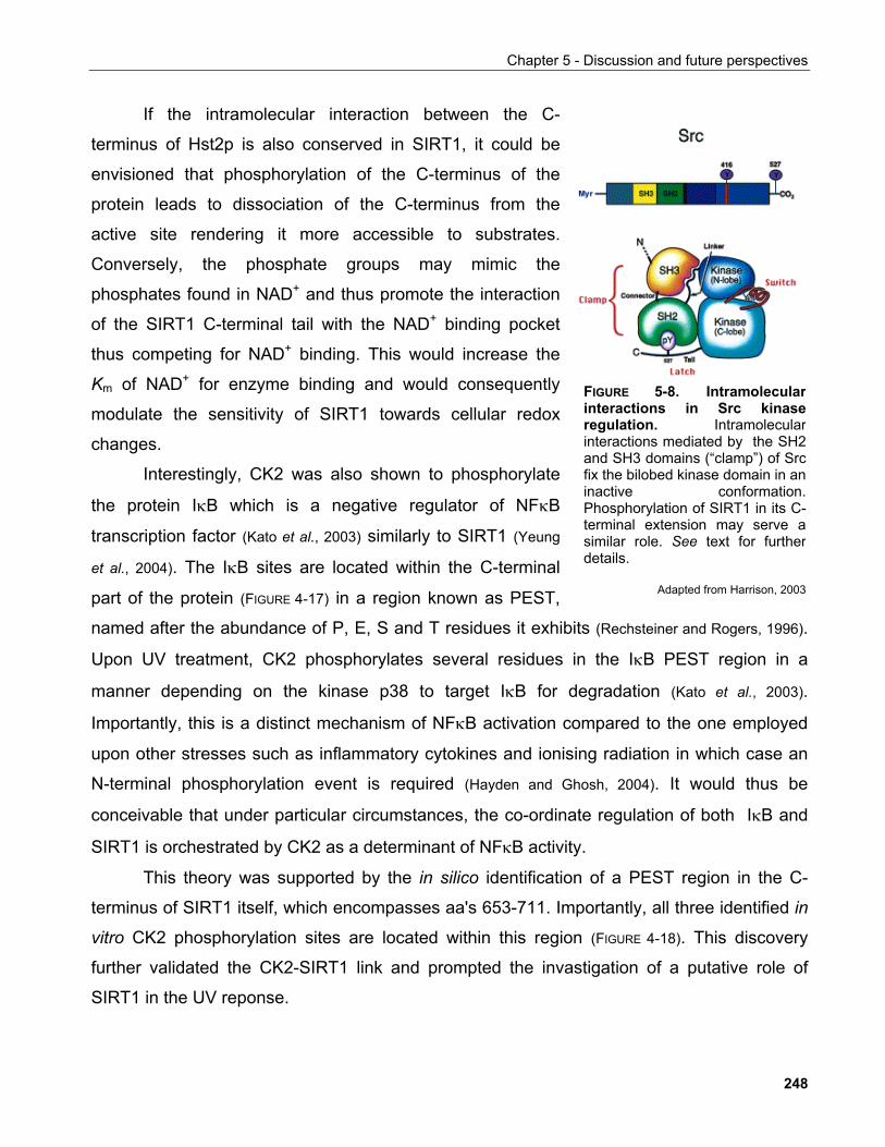
Chapter 5 - Discussion and future perspectives
If the intramolecular interaction between the C-
terminus of Hst2p is also conserved in SIRT1, it could be
envisioned that phosphorylation of the C-terminus of the
protein leads to dissociation of the C-terminus from the
active site rendering it more accessible to substrates.
Conversely, the phosphate groups may mimic the
phosphates found in NAD+ and thus promote the interaction
of the SIRT1 C-terminal tail with the NAD+ binding pocket
thus competing for NAD+ binding. This would increase the
Km of NAD+ for enzyme binding and would consequently
modulate the sensitivity of SIRT1 towards cellular redox
changes.
Interestingly, CK2 was also shown to phosphorylate
the protein IκB which is a negative regulator of NFκB
transcription factor (Kato et al., 2003) similarly to SIRT1 (Yeung
et al., 2004). The IκB sites are located within the C-terminal
part of the protein (FIGURE 4-17) in a region known as PEST,
named after the abundance of P, E, S and T residues it exhibits (Rechsteiner and Rogers, 1996).
Upon UV treatment, CK2 phosphorylates several residues in the IκB PEST region in a
manner depending on the kinase p38 to target IκB for degradation (Kato et al., 2003).
Importantly, this is a distinct mechanism of NFκB activation compared to the one employed
upon other stresses such as inflammatory cytokines and ionising radiation in which case an
N-terminal phosphorylation event is required (Hayden and Ghosh, 2004). It would thus be
conceivable that under particular circumstances, the co-ordinate regulation of both IκB and
SIRT1 is orchestrated by CK2 as a determinant of NFκB activity.
FIGURE 5-8. Intramolecularinteractions in Src kinaseregulation. Intramolecularinteractions mediated by the SH2and SH3 domains (“clamp”) of Srcfix the bilobed kinase domain in aninactive conformation.Phosphorylation of SIRT1 in its C-terminal extension may serve asimilar role. See text for furtherdetails.
Adapted from Harrison, 2003
This theory was supported by the in silico identification of a PEST region in the C-
terminus of SIRT1 itself, which encompasses aa's 653-711. Importantly, all three identified in
vitro CK2 phosphorylation sites are located within this region (FIGURE 4-18). This discovery
further validated the CK2-SIRT1 link and prompted the invastigation of a putative role of
SIRT1 in the UV reponse.
248
Chapter 5 - Discussion and future perspectives
5.9.1.3 SIRT1 in cellular responses to UV irradiation
Epidemiological as well as experimental studies corroborate to the fact that UV light
contributes profoundly in the aetiology of skin cancer (Bode and Dong, 2003). Several kinase
signaling cascades have been shown to be activated in response to UV irradiation
contributing to a proliferation-like response, possibly reflecting the perceived need to
compensate for the damaged tissue (Bode and Dong, 2003).
Jun N-terminal kinase (Jnk) was identified as a UV-inducible kinase by its ability to
associate with c-jun (Derijard et al., 1994) similarly to the identification of CK2 as the UV-
inducible IκB kinase (Kato et al., 2003). Based on this paradigm and equipped with the
knowledge that SIRT1 associated stably with a kinase, the possibility that this kinase is also
UV-inducible was investigated. However, although recombinant GST-SIRT1 could co-
precipitate kinase activity, this did not appear to be UV-inducible (FIGURE 4-19).
Altered SIRT1 protein turnover in response to UV irradiation
Cycloheximide chase studies indicated that SIRT1 is more unstable following UV
irradiation compared to mock treatment in agreement with the model presented above which
draws parallels with the regulation of IκB by CK2 (FIGURES 4-20, 4-21). In contrast to this result,
further analyses revealed that SIRT1 protein abundance increases in a time- and dose-
dependent manner (FIGURE 4-22). Importantly, this increase is independent of the presence of
p53 protein which mediates the expression of several genes following UV irradiation (Latonen
and Laiho, 2005).
These results, although at first sight conflicting need not be considered as such.
Cellular responses to DNA damage evoke cell cycle arrest allowing time for DNA repair to
take place and prevent genomic instability. In the case of irrepairable damage apoptosis is
elicited. Cellular responses following DNA damage also involve de novo gene transcription
which for intrinsic reasons requires significantly longer time than the immediate arrest
required to prevent engamement into an abberant and possibly detrimental process.
This has been well-documented in the response to ionising radiation. Long-term cell
cycle arrest is achieved by transcriptional upregulation of the CDK inhibitor p21 which
249
Chapter 5 - Discussion and future perspectives
depends on the tumour suppressor p53. Immediately after damage though, cell cycle arrest
is achieved by proteolytic destruction of cyclin-D which allows the release of p21 from CDK4
and its association to CDK2 leading to cell cycle arrest (Agami and Bernards, 2000). This work
clearly demonstrated that the mechanisms employed to implement initiation and
maintenance of cellular responses to DNA damage are distinct.
In response to UV irradiation, several transcription factors are activated including p53,
NFκB and AP-1 (Bode and Dong, 2003). Yet, SIRT1 appears to inhibit the transcriptional activity
of at least p53 and NFκB (Yeung et al., 2004; Vaziri et al., 2001; Luo et al., 2001). It is possible that
an initially high proteolytic turnover of SIRT1 alleviates such repressive effects on
transcription to allow for an early boost of gene expression required for the damage
response. At later times, SIRT1 net protein levels increase, probably despite continuously
high proteolytic turnover reflecting a dynamic system adaptable to intracellular cues
monitoring the extend of repair. This is consistent with the results in FIGURE 4.22 that indicate
no accumulation of SIRT1 protein at high doses of UV irradiation where the extend of
damage is presumably insurmountable for the cell.
Importantly although the kinetics of SIRT1 accumulation closely resemble those of p53
there is no indication that SIRT1 expression is a target of p53 in this context based on FIGURE
4.23. However this possiblity should be tested repeatedly to confirm the validity of the latter
experiment especially under the light of recent evidence that the SIRT1 gene promoter
harbours p53 binding sites which drive SIRT1 expression in response to starvation (Nemoto et
al., 2004).
Furthermore, although the osteosarcoma cell line used here has been extensively
employed as a system to study cellular UV responses, it is conceivable that it does not reflect
a physiologically relevant system to study such pathways as bone cells are not subjected to
direct UV exposure. Thus a more suitable cell line to investigate these processes would be
one derived from skin such as HaCaT keratinocytes. This is prarticularly important as the
presence of additional transcription factors may determine the exact outcome of SIRT1
action.
Finally, following a different line of investigation, it was discovered that mAb12/1 used
for all the above experiments shows phosphorylation-sensitive recognition of the
corresponding SIRT1 epitope (see later this section). Thus a re-evalutation of these results is
250
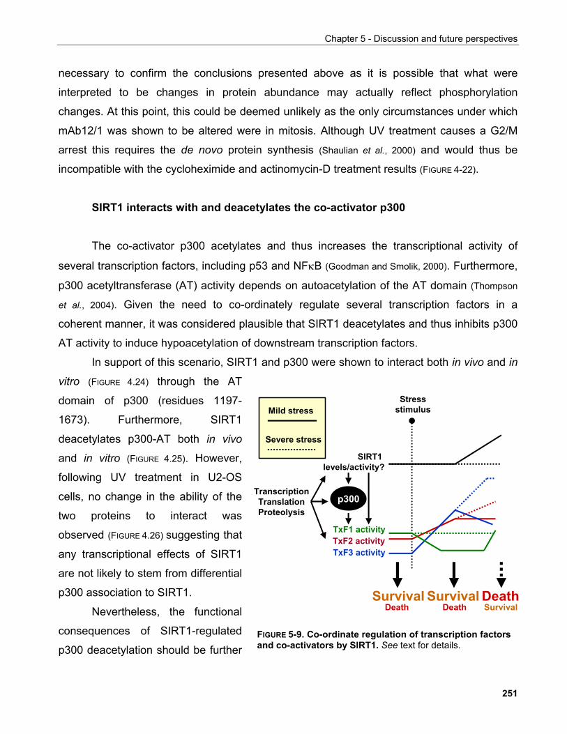
Chapter 5 - Discussion and future perspectives
necessary to confirm the conclusions presented above as it is possible that what were
interpreted to be changes in protein abundance may actually reflect phosphorylation
changes. At this point, this could be deemed unlikely as the only circumstances under which
mAb12/1 was shown to be altered were in mitosis. Although UV treatment causes a G2/M
arrest this requires the de novo protein synthesis (Shaulian et al., 2000) and would thus be
incompatible with the cycloheximide and actinomycin-D treatment results (FIGURE 4-22).
SIRT1 interacts with and deacetylates the co-activator p300
The co-activator p300 acetylates and thus increases the transcriptional activity of
several transcription factors, including p53 and NFκB (Goodman and Smolik, 2000). Furthermore,
p300 acetyltransferase (AT) activity depends on autoacetylation of the AT domain (Thompson
et al., 2004). Given the need to co-ordinately regulate several transcription factors in a
coherent manner, it was considered plausible that SIRT1 deacetylates and thus inhibits p300
AT activity to induce hypoacetylation of downstream transcription factors.
In support of this scenario, SIRT1 and p300 were shown to interact both in vivo and in
vitro (FIGURE 4.24) through the AT
domain of p300 (residues 1197-
1673). Furthermore, SIRT1
deacetylates p300-AT both in vivo
and in vitro (FIGURE 4.25). However,
following UV treatment in U2-OS
cells, no change in the ability of the
two proteins to interact was
observed (FIGURE 4.26) suggesting that
any transcriptional effects of SIRT1
are not likely to stem from differential
p300 association to SIRT1.
SIRT1 levels/activity?
Stressstimulus
TxF1 activity
TranscriptionTranslationProteolysis
SurvivalSurvival
TxF2 activityTxF3 activity
p300
Mild stress
Severe stress
SurvivalDeath
DeathDeathNevertheless, the functional
consequences of SIRT1-regulated
p300 deacetylation should be further FIGURE 5-9. Co-ordinate regulation of transcription factorsand co-activators by SIRT1. See text for details.
251
Chapter 5 - Discussion and future perspectives
investigated. Indeed, systematic analyses of transcriptional responses to stress stimuli have
identified extensive cross-talking between transcription factors that underlie the
corresponding gene expression changes (Janes et al., 2005; Janes et al., 2006). This is
conceptually reasonable as given the plethora of environmental stimuli a cell faces at any
given time, the pleiotropic effects elicited by different transcription factors need to be co-
ordinated so that a coherent and appropriate response is elicited. Alternatively, it is the very
fine balance between the activities of these transcription factors that determines the final
outcome of the response. In any case, despite our understanding of individual regulatory
evetns, the underlying molecular mechanisms responsible for the co-ordination thereof
remain largely unknown. Thus, it is conceivable that SIRT1 may play such a co-ordinating
role through its dual function in deacetylating both transcription factors directly and an
upstream acetyltransferase, in parallel to additional regulatory mechanisms (FIGURE 5-9).
UV-inducible changes in SIRT1 subnuclear localisation
In addition to changes in gene expression, UV irradiation results in significant
structural rearrangements within the cell, possibly reflecting the need to accommodate the
ensuing changes in gene transcription and DNA replication or other regulatory mechanisms
(discussed below). Thus the localisation of SIRT1 in response to UV irradiation in U2OS cells
was investigated by indirect immunofluorescence microscopy.
Strikingly, following UV irradiation SIRT1 redistributed from showing a diffuse nuclear
localisation to form characteristic ring-like structures within the nucleus surrounding regions
staining poor for DAPI and thus being predicted to be nucleoli (FIGURE 4.28). Clearly further
evaluation of this conclusion should be pursued in the form of co-localisation studies with
nucleolar markers as well as biochemical confirmation of this (Andersen et al., 2002).
The nucleoli are structures within the nucleus where rDNA is transcribed by RNAPolI
and ribosomes are assembled, i.e. they are the sites of ribosomal biogenesis (Grummt, 2003).
Further to this well-established role, though, other nucleolar functions have been postulated
as several proteins with no apparent role in ribosomal biogenesis have been found therein
(Olson, 2004).
252
Chapter 5 - Discussion and future perspectives
In particular, nucleolar integrity is important for regulation of p53 tumour suppressor
protein stability by sequestering the p53 E3 ubiquitin ligase mdm2/hdm2. Consistent with
this, the majority of stress stimuli that lead to p53 stabilisation tested to-date are associated
with disruption of nucleolar structure (Olson, 2004; Rubbi and Milner, 2003). Direct disruption of
nucleolar structures by microinjection of antibodies against nucleolar structural proteins or
localised UV irradiation of nucleoli established that nucleolar integrity acts as a cellular
sensor for stress to induce p53 stability (Rubbi and Milner, 2003). Three apparently distinct
mechanisms that link nucleolar structure and p53 stabilisation have been proposed.
The first one involves the p19ARF tumour suppressor which binds to mdm2 and
sequesters it to the nucleolus thus preventing p53 degradation (Weber et al., 1999).
Interestingly, p19ARF progressively accumulates to the nucleoli of serially passaged primary
mouse fibroblasts correlating with the onset of senescence which is known to require p53
(Weber et al., 1999). Furthermore, absence of SIRT1 prevents p19ARF accumulation and
subsequent p53 stabilisation allowing MEFs to overcome senescence (Chua et al., 2005).
The second mechanism proposes exactly the same role for the PML protein which can
recruit mdm2 to the nucleolus in a manner independently of p19ARF (Bernardi et al., 2004).
Interestingly, PML was also shown to associate with SIRT1 and regulate the stability of
another SIRT1 target, FOXO1 in pancreatic β-cells in response to hyperglycaemia-induced
ROS (Kitamura et al., 2005; Langley et al., 2002).
The third mechanism involves the ribosomal assembly and transport protein
nucleophosmin (NPM, a.k.a. B23, numatrin or NO38). NPM is detected in the nucleoli of
unstressed cells although it undergoes constant nucleocytoplasmic transport. In response to
UV, though, NPM shows a diffuse nucleoplasmic distribution. This correlates with disruption
of hmd2/p53 interaction and consequently increased p53 stability (Kurki et al., 2004). In this
context, NPM has also been shown to direclty interact with p53 and act as a repressor of its
transcriptional activity thus setting a threshold for UV-induced p53 activation (Maiguel et al.,
2004).
Based on these data, a general model of how nucleolar structure regulates p53
stability has been proposed. According to this, following stress, disruption of the nucleolus
results in the release of nucleolar proteins such as p19ARF and NPM which bind to mdm2 and
prevent p53 degradation (Olson, 2004).
253
Chapter 5 - Discussion and future perspectives
Additional evidence corroborating to a role of SIRT1 in nucleolar function is provided
by the well-studied biology of Sir2 in yeast. In S. cerevisiae Sir2 is part of a multisubunit
complex known as RENT. The RENT complex is required for silencing at the rDNA loci and
mitotic exit (Straight et al., 1999; Shou et el., 1999). Net1, an integral part of the RENT complex, is
required for nucleolar integrity. Interestingly, Sir2 is associated throughout the cell cycle with
nucleoli until the anaphase/telophase transition when it disperses away from the nucleoli,
which undergo structural changes (Straight et al., 1999; Shou et el., 1999; Gotta et al., 1997).
Consistent with these, nucleolar fragmentation correlates with ageing in yeast cells (Sinclair et
al., 1997) while ageing-associated changes in nuclear structure in general are also observed in
mammalian cells (Lans and Hoeijmakers, 2006).
Finally, in response to various cellular stresses, the transcription of rDNA genes and
therefore ribosomal biogenesis is downregulated resulting in a general reduction in protein
biosynthetic capacity (Grummt, 2003). This is mediated, at least in part, by the Jnk2 kinase
which phosphorylates the RNAPolI basal transcription factor TIF-IA to inhibit RNAPolI
holoenzyme formation (Mayer et al., 2005). Thus post-translational modifications regulate pre-
initiation complex formation to control rRNA synthesis in response to stress. Similarly,
deacetylation of TAFI68, a subunit of TIF-IB, another RNAPolI basal transcription factor,
decreases its ability to bind to rDNA promoters and rRNA synthesis (Muth et al., 2001).
Under the light of this, it can be proposed that the recruitment of SIRT1 to nucleoli
following UV irradiation may contribute to the suppression of RNAPolI-mediated transcription
by preventing the assembly of the RNAPolI transcriptional machinery in the rDNA promoter.
It would thus be interesting to investigate the effects of SIRT1 on the efficiency of rDNA
transcription in response to UV or other stresses.
5.9.1.4 Phosphorylation of SIRT1 in mitosis
The eucaryotic cell cycle (FIGURE 4-29A) comprises a sequence of spatiotemporaly
ordered events that aim at the duplication of the genome and its subsequent segragation to
daugther cells in a faithful manner that maintains genomic integrity. During mitosis major
structural changes occur in the cell such as nuclear envelope break-down and chromosomal
254
Chapter 5 - Discussion and future perspectives
condensation. The reversibility of these changes is crucial to the outcome of the commitment
of cells to division.
Several aspects of chromosome condensation have been described in relation to the
gross structural changes that chromatin undergoes during mitosis (Swedlow and Hirano, 2003).
The condensed nature of mitotic chromatin is reminiscent of heterochromatin, the
transcriptionally inactive part of the genome which correlates with at least one distinct state of
histone modification, that of hypoacetylation (Grewal and Moazed, 2003). The role of histone
modifications in mitotic chromatin condensation is less clear. Phosphorylation of histone H3
at S10 is considered to be a hallmark or mitotic chromatin, yet its exact function is not well
understood (Swedlow and Hirano, 2003).
Global acetylation in mitosis is thought to serve as an epigenetic memory mark for
transcriptionally active genes (Jeppesen, 1997) yet both HATs and HDACs dissociate from
mitotic chromatin being unable to modify histones in the context of mitotic chromosomes
(Kruhlak et al., 2001). Whether, though global deacetylation is a prerequisite for mitotic
chromosome condensation remains unclear.
SIRT1 was found to be hyperphosphorylated in nocodazole-arrested mitotic cells
(FIGURE 4-7) suggestive of a cell cycle-specific modification and potentially also function of the
protein. Further corroborating to this, a single CDK consensus site was identified in the C-
terminus of SIRT1 (S540) and recombinant cyclinB/Cdk1 complexes can phosphorylate
insect cell-expressed GST-SIRT1 in vitro (FIGURE 4-30). This phosphorylation does not cause
the characteristic electrophoretic mobility shift of SIRT1 seen in mitosis (FIGURE 4-30C) implying
that Cdk1 is not the relevant kinase, that this site is not at all phosphorylated in vivo, or if it is,
it does not change SIRT1 mobility or, indeed, that there are other unidentified
phosphorylation sites and kinases modifying SIRT1 in mitosis.
Nocodazole is a microtubule destabilising drug by virtue of its ability to bind to
microtubules and inhibit their polymerisation. This disrupts the mitotic spindle, prevents
kinetochore attachment to it and thus elicits the mitotic spindle checkpoint (Rieder and Maiato,
2004). The ultimate goal of this process is to prevent the degradation of specific substrates
such as cyclins and securin by the APC ubiquitin ligase a process which is normally
necessary for the metaphase to anaphase transition causing the observed cell cycle arrest in
what bears the characterisitics of a prometaphase (Peters, 2002).
255
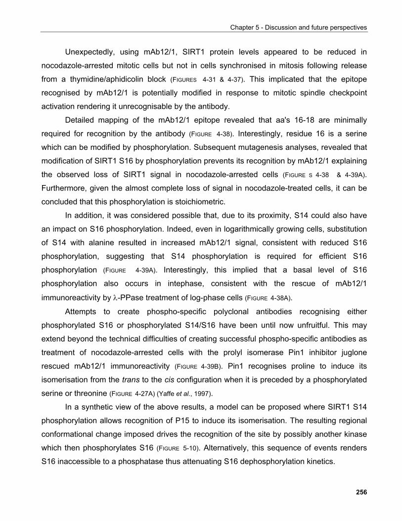
Chapter 5 - Discussion and future perspectives
Unexpectedly, using mAb12/1, SIRT1 protein levels appeared to be reduced in
nocodazole-arrested mitotic cells but not in cells synchronised in mitosis following release
from a thymidine/aphidicolin block (FIGURES 4-31 & 4-37). This implicated that the epitope
recognised by mAb12/1 is potentially modified in response to mitotic spindle checkpoint
activation rendering it unrecognisable by the antibody.
Detailed mapping of the mAb12/1 epitope revealed that aa's 16-18 are minimally
required for recognition by the antibody (FIGURE 4-38). Interestingly, residue 16 is a serine
which can be modified by phosphorylation. Subsequent mutagenesis analyses, revealed that
modification of SIRT1 S16 by phosphorylation prevents its recognition by mAb12/1 explaining
the observed loss of SIRT1 signal in nocodazole-arrested cells (FIGURE S 4-38 & 4-39A).
Furthermore, given the almost complete loss of signal in nocodazole-treated cells, it can be
concluded that this phosphorylation is stoichiometric.
In addition, it was considered possible that, due to its proximity, S14 could also have
an impact on S16 phosphorylation. Indeed, even in logarithmically growing cells, substitution
of S14 with alanine resulted in increased mAb12/1 signal, consistent with reduced S16
phosphorylation, suggesting that S14 phosphorylation is required for efficient S16
phosphorylation (FIGURE 4-39A). Interestingly, this implied that a basal level of S16
phosphorylation also occurs in intephase, consistent with the rescue of mAb12/1
immunoreactivity by λ-PPase treatment of log-phase cells (FIGURE 4-38A).
Attempts to create phospho-specific polyclonal antibodies recognising either
phosphorylated S16 or phosphorylated S14/S16 have been until now unfruitful. This may
extend beyond the technical difficulties of creating successful phospho-specific antibodies as
treatment of nocodazole-arrested cells with the prolyl isomerase Pin1 inhibitor juglone
rescued mAb12/1 immunoreactivity (FIGURE 4-39B). Pin1 recognises proline to induce its
isomerisation from the trans to the cis configuration when it is preceded by a phosphorylated
serine or threonine (FIGURE 4-27A) (Yaffe et al., 1997).
In a synthetic view of the above results, a model can be proposed where SIRT1 S14
phosphorylation allows recognition of P15 to induce its isomerisation. The resulting regional
conformational change imposed drives the recognition of the site by possibly another kinase
which then phosphorylates S16 (FIGURE 5-10). Alternatively, this sequence of events renders
S16 inaccessible to a phosphatase thus attenuating S16 dephosphorylation kinetics.
256
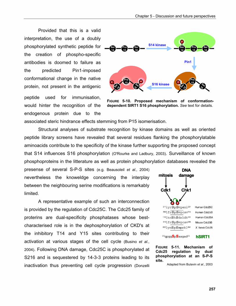
Chapter 5 - Discussion and future perspectives
Provided that this is a valid
interpretation, the use of a doubly
phosphorylated synthetic peptide for
the creation of phospho-specific
antibodies is doomed to failure as
the predicted Pin1-imposed
conformational change in the native
protein, not present in the antigenic
peptide used for immunisation,
would hinter the recognition of the
endogenous protein due to the
associated steric hindrance effects stemming from P15 isomerisation.
Ser14
Ser16
Pro15
P
Ser14
Ser16
Pro15
Pro15 Ser16Ser
14
P
Pro15 Ser16Ser
14
P
P
Pin1
S14 kinase
S16 kinase
Ser14
Ser16
Pro15
P
Ser14
Ser16
Pro15
Pro15 Ser16Ser
14
PSer
14
P
Pro15 Ser16Ser
14
PSer
14
P
PP
Pin1
S14 kinase
S16 kinase
FIGURE 5-10. Proposed mechanism of conformation-dependent SIRT1 S16 phosphorylation. See text for details.
Structural analyses of substrate recognition by kinase domains as well as oriented
peptide library screens have revealed that several residues flanking the phosphorylatable
aminoacids contribute to the specificity of the kinase further supporting the proposed concept
that S14 influences S16 phosphorylation (O'Rourke and Ladbury, 2003). Surveillance of known
phosphoproteins in the litterature as well as protein phosphory
presense of several S-P-S sites (e.g. Beausoleil et al., 2004)
nevertheless the knoweldge concerning the interplay
between the neighbouring serine modifications is remarkably
limited.
lation databases revealed the
mitosis
P P
DNA damage
Cdk1 Chk1
10qpgg d21 hSIRT1
mitosis
P P
DNA damage
Cdk1 Chk1
10qpgg d21 hSIRT1
A representative example of such an interconnection
is provided by the regulation of Cdc25C. The Cdc25 family of
proterins are dual-specificity phosphatases whose best-
characterised role is in the dephosphorylation of CKD's at
the inhibitory T14 and Y15 sites contributing to their
activation at various stages of the cell cycle (Busino et al.,
2004). Following DNA damage, Cdc25C is phosphorylated at
S216 and is sequestered by 14-3-3 proteins leading to its
inactivation thus preventing cell cycle progression (Donzelli
SpSaagaSpSaaga
FIGURE 5-11. Mechanism ofCdc25 regulation by dualphosphorylation at an S-P-Ssite.
Adapted from Bulavin et al., 2003
257
Chapter 5 - Discussion and future perspectives
and Draetta, 2003). However, during mitosis, the preceding S214 is phosphorylated
preventing S216 phosphorylation and thus maintaining Cdc25C capable to activate CDKs
and allow cell cycle progression (FIGURE 5-11) (Bulavin et al., 2003). Interestingly, S214 and S216
flank a proline residue. Although in this particular example, neighbouring phosphorylation has
an inhibitory rather than a promoting effect contrasting what is proposed for the SIRT1 S-P-S
site, it strongly supports the merit of further investigating the interplay between S14 and S16.
Investigation of SIRT1 S16 phosphorylation by mitotic checkpoint kinases
SIRT1 was found to be phosphorylated in response to activation of the mitotic spindle
checkpoint providing a hint for the possible kinases involved. To this end, three previously
characterised checkpoint kinases, Bub1, BubR1 and Mps1 (Kops et al., 2005) were depleted by
siRNA, cells treated with nocodazole and the resulting effects on S16 phosphorylation were
monitored with mAb12/1. This approach initially indicated that depletion of BubR1 but not
Bub1 or Mps1 rescues mAb12/1 immunoreactivity in nocodazole-arrested cells suggesting
that BubR1 is the relevant S16 kinase. However, various lines of evidence indicate that this
was an erroneous conclusion.
Firstly, BubR1 fails to phosphorylate recombinant SIRT1 in vitro (FIGURE 4-45) despite
exhibiting a robust autophosphorylation activity which suggests that it is an active kinase.
Secondly, BubR1 depletion itself suffices to prevent mitotic checkpoint signaling resulting in
failure of cells to arrest in prometaphase in response to nocodazole treatment (Meraldi et al.,
2004). Thus, the apparent rescue of mAb12/1 immunoreactivity is likely to stem from the fact
that the cells are actually not fully arrested in mitosis and thus not phosphorylated on S16
rather than from the inactivation of BubR1 per se.
Although the ability of cells to arrest in mitosis in response to nocodazole treatment
should also be compromised by Bub1 and Mps1 depletion leading to a mAb12/1
immunoractivity rescue similar to BubR1 depletion, this is not the case here. This is likely to
be due to the inefficient downregulation of the respective protein levels by the siRNAs
employed in this study as this is known to allow sufficient residual protein at the kinetochores
to maintain a functional checkpoint (P. Meraldi, ETH, personal communication). Thus, when the
258
Chapter 5 - Discussion and future perspectives
above observations are considered in their totality, it is likely that the approach followed for
the identification of the relevant S16 kinase is problematic.
Further investigation led to the observation that when the mitotic cells were separated
from the G2 cells in a synchronous population following release from a thymidine/aphidicolin
block, SIRT1 also showed stoichiometric phosphorylation of S16 (FIGURE 4-46). This firmly
established that S16 phosphorylation is not specifically elicited in response to spindle
checkpoint activation, but rather it occurs during normal mitosis. Further support for this was
provided by the fact that cells arrested in metaphase by MG132 treatment in the absence of
spindle checkpoint stimulation also exhibited S16 phosphorylation (FIGURE 4-46B).
A potentially more appropriate approach to this problem would involve the use of cell-
free mitotic extracts accompanied by immunodepletion of candidate kinases and subsequent
use of these preparations for in vitro kinase assays using SIRT1 as a substrate (Nakagawa et
al., 1989). This would overcome the defects in mitotic arrest/entry which are tightly associated
with siRNA-mediated depletion of mitotic and checkpoint kinases as demonstrated above.
The modification of SIRT1 in mitosis implies a role of the protein in cell cycle. To probe
this possibility, SIRT1 was depleted by siRNA and the effects on mitotic markers following
arrest with nocodazole were analysed. Cyclin B1 accumulation and phosphorylation of S10
on histone H3 showed an indistinguishable pattern in either the presence or absence of
SIRT1 (FIGURE 4.47A) suggesting that SIRT1 is not likely to be an integral component of spindle
assembly checkpoint. It is also possible, however, that similar to incomplete depletion of
Bub1 and Mps1, residual SIRT1 may mask such an effect.
This is supported by the finding that SIRT1-/- or SIRT1+/- MEFs are partially impaired in
their ability to arrest in G2/M following nocodazole treatment (FIGURE 4-48). Similarly, though
this experiment may also hide some pitfalls as the observed effect can reflect reduced
proliferation rates of the SIRT1-/- or SIRT1+/- MEFs.
Furthermore, mouse cells are particularly resistant to nocodazole treatment as the
drug does not seem to build up in such high intracellular concentrations as in human cells.
This has been partially attributed to increased drug efflux via the P-glycoprotein mutlidrug
resistance protein (Rieder and Maiato, 2004). Recently, SIRT1 was shown to possitively regulate
the expression of P-glycoprotein multidrug resistance gene mdr1 in cancer cells (Chu et al.,
2005). If this also applies to mouse cells, one would predict that reducing or completely
259
Chapter 5 - Discussion and future perspectives
eliminating expression of SIRT1 in MEFs would result in impaired nocodazole efflux, leading
to higher intracellular amounts of the drug and thus increased sensitivity of these cells to
nocodazole-induced mitotic arrest. This would contradict the observed effects and would
consequently dismiss this interpretation.
Despite the evidence presented above, it is noteworthy that the initial line of
investigation to explain the loss of SIRT1 immunoreactivity in mitosis was based on the
assumption that it was a consequence of proteolytic degradation of SIRT1. This was strongly
supported by the identification of specific sequences that have been previously shown to
target proteins for proteolysis during the M-phase, namely the D-box which serves as a
recognition motif for APC/Cdc20 (Glotzer et al., 1991) and the β-TrCP phosphodegron which is
recognised by the SCFβ-TrCP E3 ligase (FIGURE 4-32) (Watanabe et al., 2004; Moshe et al., 2004;
Margottin-Goguet et al., 2003; Busino et al., 2003; Jin et al., 2003).
This prediction was further supported by clear experimental evidence that
demonstrated a direct in vitro interaction of SIRT1 with both substrate recognition subunits of
the APC, Cdh1 and Cdc20 in vitro as well as a phosphorylation-dependent interaction of
SIRT1 with β-TrCP (FIGURES 4-33 & 4-35). However, subsequent experiments were unable to
demonstrate a functional consequence of the presence of these E3 ligase recognition motifs
(FIGURES 4-34 & 4-36).
In retrospect, the inability of β-TrCP to target SIRT1 for degradation is not surprising.
The crystal structure of β-TrCP in complex with its substrate β-catenin coupled to
biochemical evidence demonstrated that the spacing between the β-TrCP phosphodegron
and the lysine residue targeted for ubiquitination by the E3 ligase complex is crucial and
varies between 9-11 aminoacids in known β-TrCP substrates (Wu et al., 2003). No lysine
residues lie in the proximity of the SIRT1 phosphodegron providing an explanation for the
inability of β-TrCP to target the protein for degradation. The question, though, what, if any, is
the function of this sequence motif remains unanswered.
With respect to the D-box it could be argued that if SIRT1 were an APC substrate,
mitotic arrest with nocodazole would result in SIRT1 accumulation rather than degradation
due to APC inactivation. Although this is the case for most APC substrates, it is of interest to
note that the degradation of cyclin A by the APC is not inhibited by the spindle assembly
checkpoint as with other APC substrates (Geley et al., 2001).
260
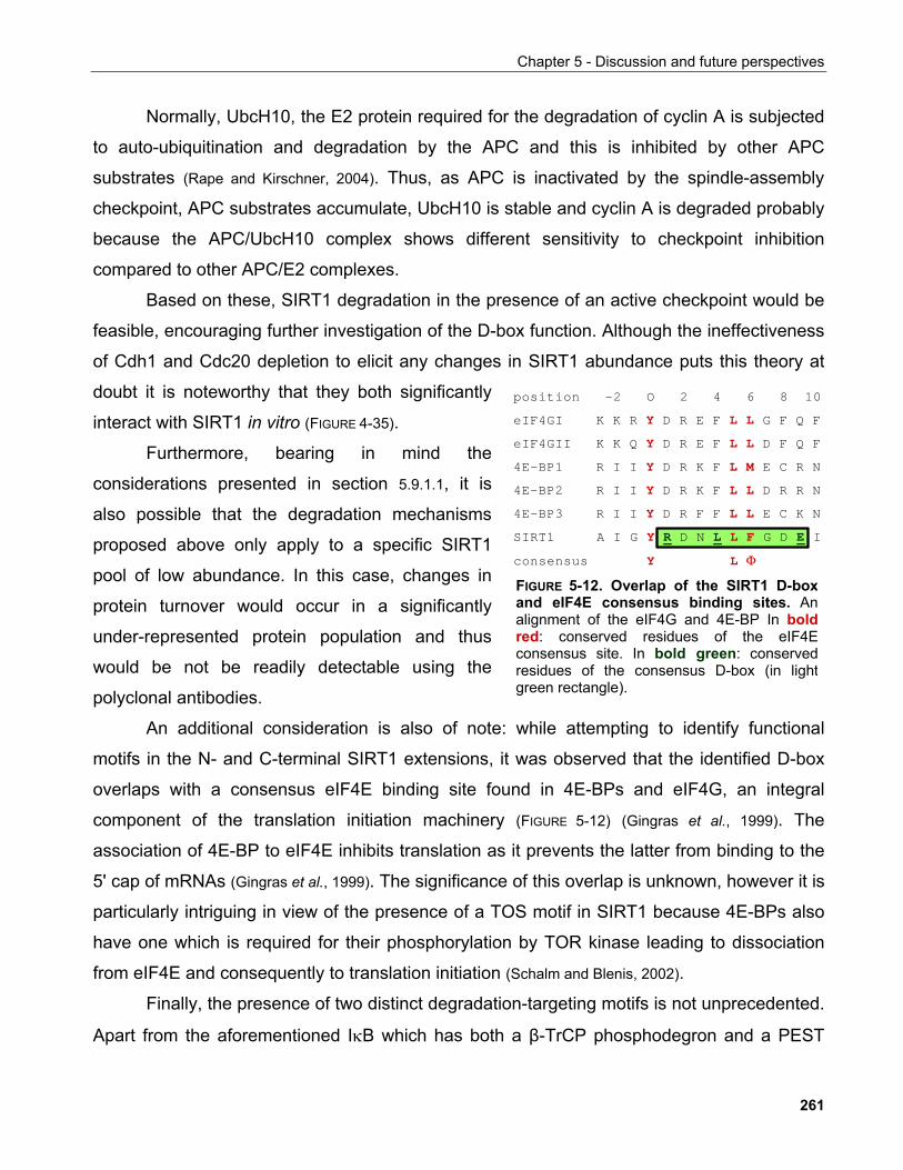
Chapter 5 - Discussion and future perspectives
Normally, UbcH10, the E2 protein required for the degradation of cyclin A is subjected
to auto-ubiquitination and degradation by the APC and this is inhibited by other APC
substrates (Rape and Kirschner, 2004). Thus, as APC is inactivated by the spindle-assembly
checkpoint, APC substrates accumulate, UbcH10 is stable and cyclin A is degraded probably
because the APC/UbcH10 complex shows different sensitivity to checkpoint inhibition
compared to other APC/E2 complexes.
Based on these, SIRT1 degradation in the presence of an active checkpoint would be
feasible, encouraging further investigation of the D-box function. Although the ineffectiveness
of Cdh1 and Cdc20 depletion to elicit any changes in SIRT1 abundance puts this theory at
doubt it is noteworthy that they both significantly
interact with SIRT1 in vitro (FIGURE 4-35).
Furthermore, bearing in mind the
considerations presented in section 5.9.1.1, it is
also possible that the degradation mechanisms
proposed above only apply to a specific SIRT1
pool of low abundance. In this case, changes in
protein turnover would occur in a significantly
under-represented protein population and thus
would be not be readily detectable using the
polyclonal antibodies.
eIF4GII
position -2 O 2 4 6 8 10
eIF4GI K K R D R E F G F Q F
K K Q D R E F D F Q F
4E-BP1 R I I D R K F E C R N
4E-BP2 R I I D R K F D R R N
4E-BP3 R I I D R F F E C K N
SIRT1 A I G R D N L G D E I
consensus
Y L LY L LY L MY L LY L LY L FY L Φ
FIGURE 5-12. Overlap of the SIRT1 D-boxand eIF4E consensus binding sites. Analignment of the eIF4G and 4E-BP In boldred: conserved residues of the eIF4Econsensus site. In bold green: conservedresidues of the consensus D-box (in lightgreen rectangle).
An additional consideration is also of note: while attempting to identify functional
motifs in the N- and C-terminal SIRT1 extensions, it was observed that the identified D-box
overlaps with a consensus eIF4E binding site found in 4E-BPs and eIF4G, an integral
component of the translation initiation machinery (FIGURE 5-12) (Gingras et al., 1999). The
association of 4E-BP to eIF4E inhibits translation as it prevents the latter from binding to the
5' cap of mRNAs (Gingras et al., 1999). The significance of this overlap is unknown, however it is
particularly intriguing in view of the presence of a TOS motif in SIRT1 because 4E-BPs also
have one which is required for their phosphorylation by TOR kinase leading to dissociation
from eIF4E and consequently to translation initiation (Schalm and Blenis, 2002).
Finally, the presence of two distinct degradation-targeting motifs is not unprecedented.
Apart from the aforementioned IκB which has both a β-TrCP phosphodegron and a PEST
261
Chapter 5 - Discussion and future perspectives
region (Kato et al., 2003), Cdc25A also harbours a KEN box targeting the protein to APCCdh1 as
well as a β-TrCP motif (Busino et al., 2004). Moreover, treatment of logarithmically growing cells
with the proteasome inhibitor MG132 results in SIRT1 protein accumulation demonstrating
that the protein is subjected to constant proteolytic turnover (FIGURE 4-31B). Given the fact that
SIRT1 protein accumulates in response to at least one stimulus, namely nutrient starvation
(Rodgers et al., 2005) the mechanism of reversing this effect is of importance and based on the
observations presented here is likely to be via proteasome-mediated degradation.
5.9.1.4.1 Potential functions of SIRT1 in mitosis Evidence from the literature indicates various aspects of mitosis where functions
perfomed by SIRT1 could be envisioned.
The potential role of SIRT1 as a histone deacetylase in mitotic chromosome
condensation has been discussed in Chapter 4. Its recent demonstration as a histone H1
deacetylase specifically, adds further to this notion (Vaquero et al., 2004). H1 is a linker histone
which has been shown to be required for proper chromosome structure in Xenopus egg
extracts. Depletion of histone H1 leads to incompletely condensed chromosomes that extend
out of the metaphase plate (Maresca et al., 2005). It is not clear whether the acetylation status of
histone H1 is important for this function, yet this is a line of investigation that should be
further explored. Interestingly, histone H1 interacts with the N-terminus of SIRT1 which also
harbours the mitotic phosphorylation site S16 described in this work. Thus, it is possible that
S16 phosphorylation controls this interaction to regulate histone H1 function in mitotic
chromosome structure apart from its proposed role in transcriptional regulation (Vaquero et al.,
2004).
Additional potential roles of SIRT1 in mitosis are discussed below.
Acetyl-CoA metabolism and mitosis
To begin with, at least two distinct acetyltransferases have been implicated in mitotic
progression, namely Eco1 and p300. The Eco1 acetyltransferase was firstly described in
262
Chapter 5 - Discussion and future perspectives
yeast as a protein required for the establishment of chromatid cohesion during S phase.
Bioinformatic analysis identified an acetyl-CoA binding motif within the Eco1 sequence and
subsequent experiments demonstrated an in vitro acetyltransferase activity which targets
several components of the cohesin complex (Ivanov et al., 2002). The importance of this activity
has been subsequently questioned as yeast expressing acetyltransferase-defective Eco1 is
viable (Brands and Skibbens, 2005). Although the exact mode of action of Eco1 remains unclear,
orthologues have been identified in higher organisms and were also shown to be required for
cohesion (Hou and Zou, 2005; Williams et al., 2003).
Recently, a further link between silencing and chromatin cohesion involving the Sir
protein was established. Using a system that allowed the visualisation of cohesion outside
the context of the chromosome, Chang et al. could show that in S. cerevisiae the Sir proteins
are required for cohesion of the HMR loci, but not for other chromosomal regions, by directly
recruiting the cohesin complex (Chang et al., 2005). In addition, in S. pombe, Swi6 is required
for the recruitment of the cohesin complex to centromeric heterochromatin but does not
appear to participate in chromosome arm cohesion (Huang and Moazed, 2006). A Swi6
orthologue has been identified in both Drosophila and humans and is known as
heterochromatin protein 1 (HP1). As the topology of chromatid cohesion remains debatable
(Huang and Moazed, 2006), it is tempting to speculate that in a conserved similar function of
SIRT1 is also present in mammalian cells.
Furthermore, the dynamics of acetyl-CoA and related metabolic activities appear to
have a broader impact on mitosis. In S. pombe, mutations in acetyl-CoA carboxylase (ACC)
and fatty acid synthase result in defective mitosis (Saitoh et al., 1996). The situin orthologue
CobB has been implicated in acetyl-CoA biosynthesis in bacteria by activating the acetyl-CoA
synthase (Starai et al., 2002). Also SIRT1 regulates lipid metabolism in rodents where the
proposed mechanism of action though is thought to be through transcriptional regulation
(Picard et al., 2004). Given the apparent involvement of SIRT1 in mitosis presented here, it
would be really exciting to investigate how acetyl-CoA metabolism and lipid biosynthetic
processes impact cell division and what the potential role of sirtuins in these processes might
be.
The acetyltransferases CBP/p300 have also been shown to be required for proper
mitotic progression by associating with the APC components Apc5 and Apc7 via an E1A-like
263
Chapter 5 - Discussion and future perspectives
region present in the latter two (Turnell et al., 2005). Both Apc5 and Apc7 potentiate CBP/p300
transcriptional activation while CBP is required for the ubiquitin ligase function of APC and
also for proper mitotic exit. It is important to note, though, that this latter function is attributed
to the E4 ligase activity of CBP. Thus, in addition to acting as a deacetylase for the putative
substrates of the proteins described above, SIRT1 may also modulate the mitotic role of
CBP/p300 by deacetylation as shown in this thesis.
SIRT1 and mitotic exit
Further evidence for potential roles of SIRT1 in mitosis lies within our current
knowledge of sirtuin biology in lower organisms. In S. cerevisiae Sir2p is part of the RENT
complex as described previously, which also includes the Cdc14 phosphatase that
inactivates mitotic CDKs to promote mitotic exit (Shou et al., 1999; Straight et al., 1999). Two
mammalian orthologues of Cdc14 have been identified named Cdc14A and Cdc14B and
abberant expression of either protein leads to mitotic defects althought their exact
mechanism of action reamisn unclear (Mailand et al., 2002).
Interestingly, SIRT1 S16 dephosphorylation appears to require the anaphase-to-
metaphase transition implying that the dynamics of SIRT1 phosphorylation during mitosis
may be linked to mitotic exit (FIGURE 4.46B). It would thus be of interest to probe the
evolutionary conservation and potential functional significance of the SIRT1/Cdc14
association in particular in the context of mitotic exit. This is also of broader interest as
despite the accumulating knowledge about mitosis-associated events, relatively little is
known about mitotic exit in mammalian cells.
Survival during mitosis
Progress through mitosis is associated with dramatic cytoarchitectural changes which
have a broad impact on the organisation of the cell and thus require sustaining survival
signaling in order to prevent cell death. Recent evidence suggests that upon treatment of
cells with microtubule poisons, the NFκB pathway is activated and this is required for survival
(Mistry et al., 2004). As SIRT1 has been shown to deacetylate and thus inactivate the p65
264
Chapter 5 - Discussion and future perspectives
subunit of NFκB, it is likely that is also has a similar role in this context. Furthermore, this
might also have therapeutic implications as microtubule poisons such as taxol are used for
cancer treatment. If the above prediction is correct, concomitant inactivation of survival
pathways by SIRT1 activation in addition to microtubule poisons would not only attenuate the
high proliferation rate of cancer cells but would also enhance their propensity to undergo
apoptosis.
It is noteworthy that activation of mitotic proteins such as CDKs is also linked to cell
death. The CDK inactivating proteins Wee1 kinase and the Cdc27 subunit of the APC are
cleaved in a caspase-dependent manner following apoptotic stimulation leading to CDK
activation (Zhou et al., 1998). Conversely, induced activation of CDK1 in post-mitotic neurons
promotes neuronal apoptosis (Stegmuller and Bonni, 2005). In either cases, the exact purpose
and ensuing consequences of CDK activation remains unclear but these examples underline
yet another aspect of the close interplay between cell division and apoptosis. Again, the
preceding knowledge of SIRT1 involvement in cell survival regulation coupled to the
proposed involvement in mitosis strongly indicate another path of investigation along these
lines.
Priming S phase events in mitosis
One of the success strategies of the cell cycle programme is based on mechanisms
that couple each phase to the other thus allowing unidirectional progression. Thus events
that are associated with a particular cell cycle phase have their foundations in a preceding
phase.
One such example is the formation of pre-replication complexes (pre-RCs) during late
mitosis and early G1. Starting already at anaphase, pre-RCs assemble at replication origins
to establish replication competence, although by themselves are not sufficient for replication
(Prasanth et al., 2004). In S. cerevisiae Sir2p deletion suppresses the phenotype of a pre-RC
assembly mutant (Cdc6p - required for loading of the pre-RC component MCM helicase to
origins of replication) indicating that Sir2p is a negative regulator of replication (Pappas et al.,
2004).
265
Chapter 5 - Discussion and future perspectives
It would be of interest to investigate whether SIRT1 has a similar role in mammalian
cells. In this case, SIRT1 would need to be inactivated to allow efficient pre-RC formation.
Following mitotic exit, re-activation of SIRT1 would function in preventing pre-RC formation
and thus aberrant genome re-replication until the next cell cycle. An experimental issue to be
taken into consideration is that based on such a model, depletion of SIRT1, e.g. by siRNA,
would have minimal impact on mitosis itself. If the identified SIRT1 S16 phosphorylation
plays a role in this process a more appropriate approach would require the investigation of
how SIRT1 S16 mutants affect S phase entry/progression kinetics.
Furthermore, S phase onset is regulated by the levels of the CDK inhibitor p27.
Degradation of p27 by the SCFSkp2 ubiquitin ligase underlies S-phase entry (Sutterluty et al.,
1999) while Skp2 levels are also under the negative control of APCCdh1 to regulate G1
duration by allowing p27 accumulation and thus prevent premature S phase entry (Bashir et al.,
2004; Wei et al., 2004). As previously discussed (section 5.2.2.5), SIRT1 may contribute to
transcriptional repression of the p27 gene in collaboration with the Hes1/Hey2 transcriptional
repressors or alternatively, promote Skp2-mediated p27 degradation by deacetylating and
thus exposing lysine residues in p27 for ubiquitination.
Mitosis and ageing
Recent data suggest a close link between molecular components regulating mitosis as
well as the spindle assembly checkpoint and the ageing process (Baker et al., 2005).
Comparison of mRNA transcript levels between fibroblasts derived from young and old
invividuals as well as people who suffer from a progeroid syndrome revealed that a striking
proporion of genes that displayed differential expression are involved in mitotic progression.
These genes included cyclins A, B and F, Polo-like kinase and the APC component Cdc20
which were all downregulated in the fibroblasts derived from old/prematurely aged individuals
(Ly et al., 2000).
Mouse models further support these findings. Mice genetically engineered to express
a BubR1 hypomorphic allele (leading to reduced but not completely abolished BubR1
266
Chapter 5 - Discussion and future perspectives
expression) suffer from premature ageing which correlates with increased expression of
senescence markers (reviewed in Fernandez-Capetillo and Nussenzweig, 2004).
In conclusion, the role of SIRT1 in mitosis proposed by this work is unclear and only
inferred by the observation that SIRT1 is stoichiometrically phosphorylated at least at S16 in
this phase of the cell cycle. The ability of SIRT1 to modulate mammalian ageing has been
poorly investigated and largely unproven. Understanding the significance of SIRT1
modification in mitosis would provide further insights into a novel function for SIRT1 and a
potential link to the regulation of organismal life-span.
5.9.2 Regulation of SIRT1 by caspase-mediated cleavage
During the course of these studies and on several occasions a fast-migrating protein
band strongly immunoreactive to mAb12/1 and to a lesser extend to GST-S1 was observed
(e.g. FIGURES 4-7, 4-22, 4-31). The appearence of this band, initially termed p110SIRT1 by virtue of
its apparent molecular weight, invariably correlated with the presence of apoptotic figures
observed by light microscopy.
Systematic investigation of various apoptosis-inducing stimuli further supported this
observation (FIGURE 4-50). Furthermore, the use of caspase inhibitors as well as in vitro
caspase cleavage assays established that p110SIRT1 derives from full-length SIRT1 by
caspase-mediated proteolytic processing (FIGURES 4-51 & 4-56).
Comparison of SIRT1 antibody recognition patterns in extracts of cells stimulated to
undergo apoptosis indicated that caspase cleavage occured in the C-terminal site of SIRT1
(FIGURE 4-52). Systematic mapping and mutagenesis of consensus caspase cleavage sites on
SIRT1 identified D707 as the relevant processing site giving rise to p110SIRT1 which was
henceforth referred to as SIRT1(1-707) (FIGURES 4-53, 4-54, 4-56).
Based on these results, the GST-S1 epitope is preserved in SIRT1(1-707) which
would thus be predicted to be fully recognised by this antibody. However, while GST-S1 only
weakly recognises SIRT1(1-707) in immunoblotting, mAb12/1 exhibits significantly higher
relative immunoreactivity towards this species (FIGURE 4-52). Under the light of the
phosphorylation-sensitive recognition of SIRT1 by mAb12/1, these results also bear
267

Chapter 5 - Discussion and future perspectives
additional significance: they imply that SIRT1(1-707) is dephosphorylated, enhancing its
recognition by the monoclonal antibody. As our understanding of SIRT1 S16 phosphorylation
remains limited, it is difficult to appreciate the significance of this observation, nevertheless, it
should be taken into consideration in subsequent studies.
It is also worth mentioning that there exist several examples in the literature where
phosphorylation of residues in the proximity of caspase cleavage sites was shown to impede
processing by caspases. This was shown to be the case for the class II deacetylase HDAC4
(Paroni et al., 2004, Liu et al., 2004), the transcription factor Max (Krippner-Heidenreich et al., 2001), the
tumour suppressor lipid phosphatase PTEN (Torres et al., 2003) and the apoptosis factor Bid
(Desagher et al., 2001). Intriguingly, in all these cases, the relevant kinase is CK2. However,
none of the mutant CK2 sites confered differential cleavage sensitivity to SIRT1 (not shown)
suggesting that this mechanism may not apply for this protein under the experimental
conditions employed for these experiments.
5.9.2.1 Functional significance of caspase-mediated SIRT1 cleavage
Importantly, SIRT1(1-707) does not exhibit altered in vitro enzymatic activity nor does
it localise differently than its full-length counterpart (FIGURES 4-59 & 4-60). These results raise
the obvious question what is the functional significance of this processing event.
Initial approaches towards addressing this issue focused on the potential impact of
SIRT1 C-terminal cleavage on its transcriptional activities. For this purpose, a heterologous
luciferase reporter assay was employed where 3 consensus NFκB binding sites were located
upstream of a consistutive promoter to render luciferase expression sensitive to NFκB
activity. This is a well-suited system for such studies as it provides a straightforward means
of not only monitoring transcriptional effects of SIRT1 but also allows to assay various SIRT1
mutants and assess their functional significance.
The choice of NFkB reporter was based on previous work where SIRT1 was shown to
negatively regulate NFκB transcriptional activity in response to TNFα stimulation (Yeung et al.,
2004), but also on the fact that most of the stimuli that were shown to induce SIRT1 cleavage
are also known to be potent NFκB activators (FIGURE 4-50). Of note is also the example of
hematopoietic progenitor kinase 1 (HPK1) which is an activator of NFκB , yet under apoptotic
268
Chapter 5 - Discussion and future perspectives
conditions it undergoes caspase-mediated cleavage which converts it into an inhibitor of
NFκB (Arnold et al., 2001). Thus it was postulated that under such conditions, modulation of
SIRT1 activity may in turn impact the activation status and/or magnitude of NFκB.
Following extensive optimisation of assay conditions, SIRT1 was found to consistently
suppress NFκB reporter activity in response to TNFα stimulation even at very low amounts of
transfected SIRT1 which ranged in the few ng of DNA level or up to 1/500 SIRT1-to-reporter
plasmid ratio. However, no significant difference in the ability of SIRT1(1-707) to suppress
TNFα-induced NFκB activity was observed (FIGURE 4-61). These data implied that caspase-
mediated processing of SIRT1 is unlikely to impact its transcriptional repressor activity under
the assay conditions used.
It is worthwhile, however, to critically evaluate the validity of this conclusion with
respect to the limitations that such an experimental system comprises. To begin with it is
possible that the reporter construct employed here does not bear the necessary features
required for a functional consequence of SIRT1 cleavage to be revealed. This would be
particularly the case if an additional protein interaction mediated by the cleaved C-terminal
region is required for such an effect. It is of interest to note that in the original work reporting
the SIRT1-NFκB interaction, treatment of cells with the SIRT1 activator resveratrol resulted in
suppression of NFκB gene espression attributed to the prolonged occupancy of NFκB target
gene promoters by SIRT1 (Yeung et al., 2004). The molecular mechanism of this is intriguing
but remains elusive. It is possible that this can be accounted by a conformational change
elicited by resveratrol. Similarly, any of the modifications of SIRT1 discovered in this work,
including the caspase-mediated cleavage may have a similar modus operanti.
Furthermore, Starai et al. reported that CobB, a bacterial sirtuin deacetylates acetyl-
CoA synthase leading to its activation (Starai et al., 2002). As the authors point out, the catalytic
lysine targeted for deacetylation constitutes a functionally conserved residue in AMP-forming
enzymes which include luciferase. Thus it is conceivable that the expression of SIRT1 in the
NFκB reporter assays results in a steady-state modification of luciferase that alters its activity
leading to erroneous conclusions.
It is also clear that additional levels of NFκB activity modulation exist such as the
deacetylation-dependent nucleocytoplasmic transport of the p65 subunit (Chen et al., 2001).
Thus, it is possible that SIRT1(1-707) does have a different function than that of full-length
269
Chapter 5 - Discussion and future perspectives
SIRT1 however the steady-state conditions under which the reporter assays are performed
are unsuitable for detecting the potentially subtle differences incurred.
Examining the nuclear translocation kinetics of NFκB as well as NFκB target gene
expression following TNFα stimulation is likely to be more informative in this context. The
evaluation of MEFs lacking SIRT1 for such an experiment is underway. Provided that the
presence of SIRT1 proves to be a determinant of NFκB activation amplitude, re-introduction
of SIRT1 and mutants thereof by retroviral transduction will be employed to assess the
differential effects of SIRT1 species.
A significant hint as to the potential significance of SIRT1 cleavage was also provided
by two other pieces of data. Firstly, inhibition of proteasome activity by MG132 under
conditions that induced apoptosis, resulted in rescue of SIRT1(1-707) immunoreactivity
(FIGURE 4-57). What is more, in vitro cleavage of SIRT1 by activated cell extracts indicated that
under these conditions, SIRT1(1-707) was sensitive to further proteolytic processing, while
mutation of the caspase cleavage site not only prevented SIRT1(1-707) formation, but it also
blocked SIRT1 degradation overall (FIGURE 4.56).
Taken together, these data suggest that a potential role of SIRT1 cleavage by
caspases following activation of the apoptotic programme is to render the protein more
susceptible to further proteolysis. The impact of a SIRT1 non-cleavable mutant on the
sensitivity of cells to apoptotic stimuli is currently under examination.
Interestingly, the C-terminal region removed following caspase cleavage harbours a
consensus sumoylation site (733VKQE736)(Seelre and Dejean, 2003). Modification of proteins by
SUMO has been linked, among others, to transcriptional regulation and interestingly, SUMO
is found concentrated at PML bodies, a site where also SIRT1 was shown to localise under
specific conditions (Seelre and Dejean, 2003; Langley et al., 2002). It is also of particular interest that
p300, has a transcriptional repression domain called CRD1 (for cell cycle regulatory domain
1) which harbours two sumoylation sites K1020 and K1024. SUMO modification at these
lysines is essential for its transcriptional repressor function (Bouras et al., 2005). Interestingly,
SIRT1 represses p300-mediated transactivation in a manner that depends on the p300
CRD1 domain because SIRT1 deacetylates K1020 and K1024 allowing them to be modified
by SUMO ligases and promoting the transcriptional repressor function of p300 (Bouras et al.,
2005).
270
Chapter 5 - Discussion and future perspectives
Increased caspase activity during ageing
Several of the ageing phenotypes can be attributed to loss of tissue mass due to
increased apoptosis. This has been well documented for different post-mitotic tissues
including the heart, muscle and the central nervous system where apoptosis is one of the
underlyng disease mechanisms in Alzheimer's and Parkinson's disease, two age-related
neurological disorders (Zhang et al., 2003).
Overall, there is a striking correlation between caspase activity and ageing-associated
apoptosis. Studies in humans as well as mice have shown increased activity as well as
mRNA levels for various caspases in cells of the liver, spleen, lung and the immune system
(Zhang et al., 2003). Strikingly, mouse models that exhibit premature ageing also have
increased caspase activity.
A recent example of this are mice with a mutation in the nucleus-encoded catalytic
subunit of mitochondrial DNA (mtDNA) polymerase PolgA that abolishes its proofreading
activity and thus renders it more prone to errors during mtDNA replication. These mice have
high mtDNA mutation rates and, at the organismal level, they suffer from classic symptoms of
premature ageing such as weight loss, hair loss, skeletal abnormalities, osteoporosis,
anemia and reduced fertility (Kujoth et al., 2005; Trifunovic et al., 2004). Such mice also exhibit
increased occurrence of apoptotic markers, including cleaved and thus activated caspase-3
especially in tissues with high cell turnover such as the thymus, intestine and testis (Kujoth et
al., 2005).
Intriguingly, increased apoptosis in kidneys of aged rats can be suppressed by caloric
restriction (Lee et al., 2004). This was attributed to the decrease of oxidative damage elicited by
diet and, in support of this, markers of lipid peroxidation resulting from oxidative damage,
were higher in animals fed ad libitum compared to those subjected to caloric restriction (Lee et
al., 2004).
Finally, progressive apoptosis of oocyte population acquired during embryonic
development in the ovaries leads to reduced reproductive capacity and ultimately what is
known as the menopause in females (Tilly, 2000). A recent report has drawn a link between
cellular metabolic status and oocyte death. NADPH production by the pentose phosphate
271

Chapter 5 - Discussion and future perspectives
pathway promotes the activity of calcium/calmodulin-dependent protein kinase II (CaMKII)
which in turn phosphorylates and inhibits caspase-2, a key executor of the apoptotic
programme in oocytes (Nutt et al., 2005). This provides evidence that the nutrient status of the
environment experienced by the reproductive system determines to a significant extend the
reproductive capacity of an animal.
In conclusion, the work described above as well as the demonstration that SIRT1 is a
substrate for caspases, constitute compelling evidence in favour of investigating the link
between ageing-associated apoptosis and SIRT1 processing especially in the light of the
proposed role of SIRT1 in ageing. Moreover, undertanding the functional significance of
SIRT1 processing is likely to provide an entirely novel mechanism of SIRT1 regulation and
thus shed light into unknown aspects of the protein's role in physiological conditions where a
role for apoptosis has been documented.
5.9.3 Prediction of signalling pathways which SIRT1 may participate in
In addition to the experimental evidence provided in this work with regards to novel
potential SIRT1 regulatory mechanisms, current knowledge derived from the literature as well
as the in silico identification of putative regulatory motifs in the primary sequence of SIRT1
cast some light into which signaling pathways are likely to be involved in modulating SIRT1
function (FIGURE 5-13).
One of the phenotypes of SIRT1-/- mice is persistent eyelid closure accompanied by
developmental abnormalities of the eye (Cheng et al., 2003; McBurney et al., 2003). The EGF
signaling pathway is a well-established mediator of eyelid development in the mouse and
genetic inactivation of its components such as Jnk1 and Jnk2 or c-jun invariably leads to
eyelid closure defects (Weston et al., 2004; Zenz et al., 2003; Li, 2003). This is due to the inability of
two fronts of epithelial cells to fuse during embryonic development attributed to reduced EGF
receptor signaling. Furthermore, EGF signaling is also important for the major structural
changes leading to opening of the eyelid post-natally (Zieske, 2004).
The persistence of eyelid closure in SIRT1-/- mice post-natally indicates that SIRT1
plays an important role in the eyelid opening process and this is likely to be linked to its ability
to participate in signaling downstream the EGF receptor, possibly by modulating the
272
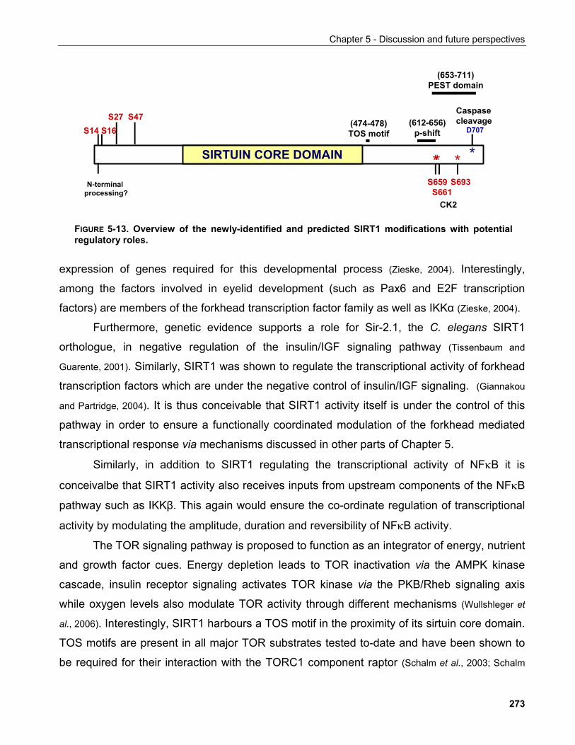
Chapter 5 - Discussion and future perspectives
SIRTUIN CORE DOMAIN
__CK2
*D707
(612-656)p-shift
Caspase cleavage
(653-711) PEST domain
N-terminal processing?
(474-478)TOS motif
* *S659 S661
S693
*
S14 S16S27 S47
FIGURE 5-13. Overview of the newly-identified and predicted SIRT1 modifications with potentialregulatory roles.
expression of genes required for this developmental process (Zieske, 2004). Interestingly,
among the factors involved in eyelid development (such as Pax6 and E2F transcription
factors) are members of the forkhead transcription factor family as well as IKKα (Zieske, 2004).
Furthermore, genetic evidence supports a role for Sir-2.1, the C. elegans SIRT1
orthologue, in negative regulation of the insulin/IGF signaling pathway (Tissenbaum and
Guarente, 2001). Similarly, SIRT1 was shown to regulate the transcriptional activity of forkhead
transcription factors which are under the negative control of insulin/IGF signaling. (Giannakou
and Partridge, 2004). It is thus conceivable that SIRT1 activity itself is under the control of this
pathway in order to ensure a functionally coordinated modulation of the forkhead mediated
transcriptional response via mechanisms discussed in other parts of Chapter 5.
Similarly, in addition to SIRT1 regulating the transcriptional activity of NFκB it is
conceivalbe that SIRT1 activity also receives inputs from upstream components of the NFκB
pathway such as IKKβ. This again would ensure the co-ordinate regulation of transcriptional
activity by modulating the amplitude, duration and reversibility of NFκB activity.
The TOR signaling pathway is proposed to function as an integrator of energy, nutrient
and growth factor cues. Energy depletion leads to TOR inactivation via the AMPK kinase
cascade, insulin receptor signaling activates TOR kinase via the PKB/Rheb signaling axis
while oxygen levels also modulate TOR activity through different mechanisms (Wullshleger et
al., 2006). Interestingly, SIRT1 harbours a TOS motif in the proximity of its sirtuin core domain.
TOS motifs are present in all major TOR substrates tested to-date and have been shown to
be required for their interaction with the TORC1 component raptor (Schalm et al., 2003; Schalm
273
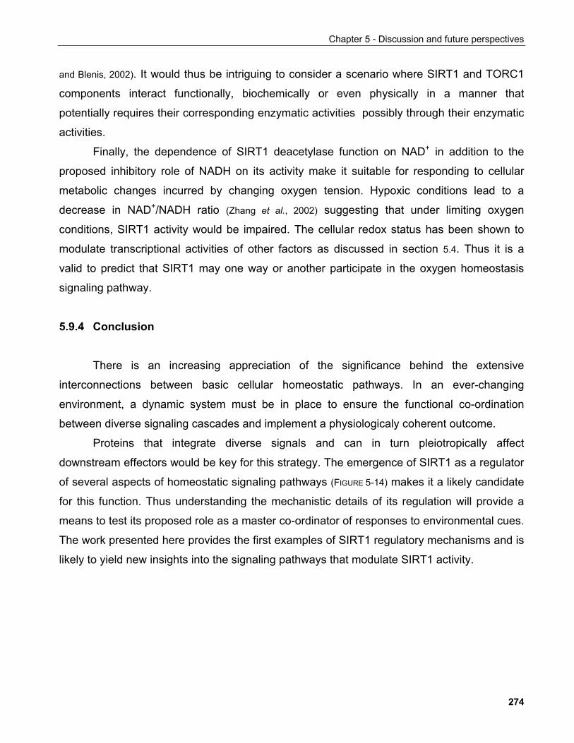
Chapter 5 - Discussion and future perspectives
and Blenis, 2002). It would thus be intriguing to consider a scenario where SIRT1 and TORC1
components interact functionally, biochemically or even physically in a manner that
potentially requires their corresponding enzymatic activities possibly through their enzymatic
activities.
Finally, the dependence of SIRT1 deacetylase function on NAD+ in addition to the
proposed inhibitory role of NADH on its activity make it suitable for responding to cellular
metabolic changes incurred by changing oxygen tension. Hypoxic conditions lead to a
decrease in NAD+/NADH ratio (Zhang et al., 2002) suggesting that under limiting oxygen
conditions, SIRT1 activity would be impaired. The cellular redox status has been shown to
modulate transcriptional activities of other factors as discussed in section 5.4. Thus it is a
valid to predict that SIRT1 may one way or another participate in the oxygen homeostasis
signaling pathway.
5.9.4 Conclusion
There is an increasing appreciation of the significance behind the extensive
interconnections between basic cellular homeostatic pathways. In an ever-changing
environment, a dynamic system must be in place to ensure the functional co-ordination
between diverse signaling cascades and implement a physiologicaly coherent outcome.
Proteins that integrate diverse signals and can in turn pleiotropically affect
downstream effectors would be key for this strategy. The emergence of SIRT1 as a regulator
of several aspects of homeostatic signaling pathways (FIGURE 5-14) makes it a likely candidate
for this function. Thus understanding the mechanistic details of its regulation will provide a
means to test its proposed role as a master co-ordinator of responses to environmental cues.
The work presented here provides the first examples of SIRT1 regulatory mechanisms and is
likely to yield new insights into the signaling pathways that modulate SIRT1 activity.
274
Chapter 5 - Discussion and future perspectives
ATPAMPATPAMP
TORC1Raptor
mLST8
mTOR
TORC1Raptor
mLST8
mTOR
LKB1
TSC1TSC2TSC1TSC2
Rheb
TORC2Rictor
mLST8
mTOR
TORC2Rictor
mLST8
mTOR
PI3K
4E-BP1
S6
PTEN
?
S6K
PDK1
Skp2
p27
PKB
REDD1
VHL
Proliferation Survival
p53
SIRT1SIRT1
Metabolism
Growth factorsHormones
NAD+
NADH
?
MAPK signaling
Growth Differentiation
AMPKHIF
?
p21
ROS
FOXO
IRS
O2
myc
FIGURE 5-14. Functional interaction of SIRT1 with major homeostatic cellular pathways. A putative rolein the co-ordination of such signaling network may underlie the pleiotropic character of the SIRT1 substratesset. In addition, interconnections between the FOXO, p53 and NFκB signaling cascades have been reportedbut are not depicted here. See text for further details.
275
References
6. REFERENCES Acharyya, S., M.E. Butchbach, Z. Sahenk, H. Wang, M. Saji, M. Carathers, M.D. Ringel, R.J. Skipworth, K.C. Fearon, M.A.
Hollingsworth, P. Muscarella, A.H. Burghes, J.A. Rafael-Fortney and D.C. Guttridge (2005), Dystrophin glycoprotein complex dysfunction: a regulatory link between muscular dystrophy and cancer cachexia, Cancer Cell, 8:421-432
Afshar, G. and J.P. Murnane (1999), Characterization of a human gene with sequence homology to Saccharomyces cerevisiae SIR2, Gene, 234:161-168
Agalioti, T., S. Lomvardas, B. Parekh, J. Yie, T. Maniatis and D. Thanos (2000), Ordered recruitment of chromatin modifying and general transcription factors to the IFN-beta promoter, Cell, 103:667-678
Agami, R. and R. Bernards (2000), Distinct initiation and maintenance mechanisms cooperate to induce G1 cell cycle arrest in response to DNA damage, Cell, 102:55-66
Agarwal, S., S. Sharma, V. Agrawal and N. Roy (2005), Caloric restriction augments ROS defense in S. cerevisiae, by a Sir2p independent mechanism, Free Radic Res, 39:55-62
Aguilaniu, H., L. Gustafsson, M. Rigoulet and T. Nystrom (2003), Asymmetric inheritance of oxidatively damaged proteins during cytokinesis, Science, 299:1751-1753
Ahmed, K., D.A. Gerber and C. Cochet (2002), Joining the cell survival squad: an emerging role for protein kinase CK2, Trends Cell Biol, 12:226-230
Alcendor, R.R., L.A. Kirshenbaum, S. Imai, S.F. Vatner and J. Sadoshima (2004), Silent information regulator 2alpha, a longevity factor and class III histone deacetylase, is an essential endogenous apoptosis inhibitor in cardiac myocytes, Circ Res, 95:971-980
Alfred, J. (2000), Counting the calories to immortality, Nat Rev Genet, 1:88 Al-Regaiey, K.A., M.M. Masternak, M. Bonkowski, L. Sun and A. Bartke (2005), Long-lived growth hormone receptor knockout mice:
interaction of reduced insulin-like growth factor i/insulin signaling and caloric restriction, Endocrinology, 146:851-860 Andersen, J.S., C.E. Lyon, A.H. Fox, A.K. Leung, Y.W. Lam, H. Steen, M. Mann and A.I. Lamond (2002), Directed proteomic analysis of
the human nucleolus, Curr Biol, 12:1-11 Anderson, R.M., K.J. Bitterman, J.G. Wood, O. Medvedik, H. Cohen, S.S. Lin, J.K. Manchester, J.I. Gordon and D.A. Sinclair (2002),
Manipulation of a nuclear NAD+ salvage pathway delays aging without altering steady-state NAD+ levels, J Biol Chem, 277:18881-18890
Anderson, R.M., M. Latorre-Esteves, A.R. Neves, S. Lavu, O. Medvedik, C. Taylor, K.T. Howitz, H. Santos and D.A. Sinclair (2003a), Yeast life-span extension by calorie restriction is independent of NAD fluctuation, Science, 302:2124-2126
Anderson, R.M., K.J. Bitterman, J.G. Wood, O. Medvedik and D.A. Sinclair (2003b), Nicotinamide and PNC1 govern lifespan extension by calorie restriction in Saccharomyces cerevisiae, Nature, 423:181-185
Andrulis, E.D., A.M. Neiman, D.C. Zappulla and R. Sternglanz (1998), Perinuclear localization of chromatin facilitates transcriptional silencing, Nature, 394n:592-595
Anekonda, T.S. and P.H. Reddy (2006), Neuronal protection by sirtuins in Alzheimer's disease, J Neurochem, 96:305-313 Ansari, A. and M.R. Gartenberg (1997), The yeast silent information regulator Sir4p anchors and partitions plasmids, Mol Cell Biol,
17:7061-7068 Antebi, A. (2004), Tipping the balance toward longevity, Dev Cell, 6:315-316 Aparicio, O.M., B.L. Billington and D.E. Gottschling (1991), Modifiers of position effect are shared between telomeric and silent mating-
type loci in S. cerevisiae, Cell, 66:1279-1287 Araki, T., Y. Sasaki and J. Milbrandt (2004), Increased nuclear NAD biosynthesis and SIRT1 activation prevent axonal degeneration,
Science, 305:1010-1013 Arkan, M.C., A.L. Hevener, F.R. Greten, S. Maeda, Z.W. Li, J.M. Long, A. Wynshaw-Boris, G. Poli, J. Olefsky and M. Karin (2005),
IKK-beta links inflammation to obesity-induced insulin resistance, Nat Med, 11:191-198 Armstrong, C.M., M. Kaeberlein, S.I. Imai and L. Guarente (2002), Mutations in Saccharomyces cerevisiae gene SIR2 can have
differential effects on in vivo silencing phenotypes and in vitro histone deacetylation activity, Mol Biol Cell, 13:1427-1438 Arnold, R., J. Liou, H.C. Drexler, A. Weiss and F. Kiefer (2001), Caspase-mediated cleavage of hematopoietic progenitor kinase 1
(HPK1) converts an activator of NFkappaB into an inhibitor of NFkappaB, J Biol Chem, 276:14675-14684 Ashrafi, K., D. Sinclair, J.I. Gordon and L. Guarente (1999), Passage through stationary phase advances replicative aging in
Saccharomyces cerevisiae, Proc Natl Acad Sci U S A, 96:9100-9105 Astrinidis, A. and E.P. Henske (2005), Tuberous sclerosis complex: linking growth and energy signaling pathways with human disease,
Oncogene, 24:7475-7481 Astrom, S.U., T.W. Cline and J. Rine (2003), The Drosophila melanogaster sir2+ gene is nonessential and has only minor effects on
position-effect variegation, Genetics, 163:931-937 Astrom, S.U., A. Kegel, J.O. Sjostrand and J. Rine (2000), Kluyveromyces lactis Sir2p regulates cation sensitivity and maintains a
specialized chromatin structure at the cryptic alpha-locus, Genetics, 156:81-91 Astrom, S.U. and J. Rine (1998), Theme and variation among silencing proteins in Saccharomyces cerevisiae and Kluyveromyces lactis,
Genetics, 148:1021-1029 Avalos, J.L., K.M. Bever and C. Wolberger (2005), Mechanism of sirtuin inhibition by nicotinamide: altering the NAD(+) cosubstrate
specificity of a Sir2 enzyme, Mol Cell, 17:855-868 Avalos, J.L., J.D. Boeke and C. Wolberger (2004), Structural basis for the mechanism and regulation of Sir2 enzymes, Mol Cell, 13:639-
648 Avalos, J.L., I. Celic, S. Muhammad, M.S. Cosgrove, J.D. Boeke and C. Wolberger (2002), Structure of a Sir2 enzyme bound to an
acetylated p53 peptide, Mol Cell, 10:523-535 Ayala, G., D. Wang, G. Wulf, A. Frolov, R. Li, J. Sowadski, T.M. Wheeler, K.P. Lu and L. Bao (2003), The prolyl isomerase Pin1 is a
novel prognostic marker in human prostate cancer, Cancer Res, 63:6244-6251
276
References
Bae, N.S., M.J. Swanson, A. Vassilev and B.H. Howard (2004), Human histone deacetylase SIRT2 interacts with the homeobox transcription factor HOXA10, J Biochem (Tokyo), 135:695-700
Baker, D.J., J. Chen and J.M. van Deursen (2005), The mitotic checkpoint in cancer and aging: what have mice taught us?, Current Opinion in Cell Biology, 17:583-589
Bakker, B.M., K.M. Overkamp, A.J.A. van Maris, P. Kotter, M.A.H. Luttik, J.P. van Dijken and J.T. Pronk (2001), Stoichiometry and compartmentation of NADH metabolism in Saccharomyces cerevisiae, FEMS Microbiology Reviews, 25:15-37
Balaban, R.S., S. Nemoto and T. Finkel (2005), Mitochondria, oxidants, and aging, Cell, 120:483-495 Barlow, A.L., C.M. van Drunen, C.A. Johnson, S. Tweedie, A. Bird and B.M. Turner (2001), dSIR2 and dHDAC6: two novel, inhibitor-
resistant deacetylases in Drosophila melanogaster, Exp Cell Res, 265:90-103 Barry, R.E. and W. Krek (2004), The von Hippel-Lindau tumour suppressor: a multi-faceted inhibitor of tumourigenesis, Trends Mol Med,
10:466-472 Bashir, T., N.V. Dorrello, V. Amador, D. Guardavaccaro and M. Pagano (2004), Control of the SCF(Skp2-Cks1) ubiquitin ligase by the
APC/C(Cdh1) ubiquitin ligase, Nature, 428:190-193 Beausoleil, S.A., M. Jedrychowski, D. Schwartz, J.E. Elias, J. Villen, J. Li, M.A. Cohn, L.C. Cantley and S.P. Gygi (2004), Large-scale
characterization of HeLa cell nuclear phosphoproteins, Proc Natl Acad Sci U S A, 101:12130-12135 Beckman, M. (2004), Low-cal connections, Sci Aging Knowledge Environ, 2004:nf60 Bedalov, A., T. Gatbonton, W.P. Irvine, D.E. Gottschling and J.A. Simon (2001), Identification of a small molecule inhibitor of Sir2p,
Proc Natl Acad Sci U S A, 98:15113-15118 Bedalov, A., M. Hirao, J. Posakony, M. Nelson and J.A. Simon (2003), NAD+-Dependent Deacetylase Hst1p Controls Biosynthesis and
Cellular NAD+ Levels in Saccharomyces cerevisiae, Mol. Cell. Biol., 23:7044-7054 Bedalov, A. and J.A. Simon (2003), Sir2 flexes its muscle, Dev Cell, 5:188-189 Bedalov, A. and J.A. Simon (2004), NEUROSCIENCE: NAD to the Rescue 10.1126/science.1102497, Science, 305:954-955 Bedalov, A. and J.A. Simon (2004), Neuroscience. NAD to the rescue, Science, 305:954-955 Bell, S.D., C.H. Botting, B.N. Wardleworth, S.P. Jackson and M.F. White (2002), The interaction of Alba, a conserved archaeal
chromatin protein, with Sir2 and its regulation by acetylation, Science, 296:148-151 Bell, S.P., J. Mitchell, J. Leber, R. Kobayashi and B. Stillman (1995), The multidomain structure of Orc1p reveals similarity to regulators
of DNA replication and transcriptional silencing, Cell, 83:563-568 Bellizzi, D., G. Rose, P. Cavalcante, G. Covello, S. Dato, F. De Rango, V. Greco, M. Maggiolini, E. Feraco, V. Mari, C. Franceschi, G.
Passarino and G. De Benedictis (2005), A novel VNTR enhancer within the SIRT3 gene, a human homologue of SIR2, is associated with survival at oldest ages, Genomics, 85:258-263
Benguria, A., P. Hernandez, D.B. Krimer and J.B. Schvartzman (2003), Sir2p suppresses recombination of replication forks stalled at the replication fork barrier of ribosomal DNA in Saccharomyces cerevisiae, Nucleic Acids Res, 31:893-898
Bennett, C.B., J.R. Snipe, J.W. Westmoreland and M.A. Resnick (2001), SIR functions are required for the toleration of an unrepaired double-strand break in a dispensable yeast chromosome, Mol Cell Biol, 21:5359-5373
Bereshchenko, O.R., W. Gu and R. Dalla-Favera (2002), Acetylation inactivates the transcriptional repressor BCL6, Nat Genet, 32:606-613
Bernander, R. (2003), The archaeal cell cycle: current issues, Mol Microbiol, 48:599-604 Bernardi, R., P.P. Scaglioni, S. Bergmann, H.F. Horn, K.H. Vousden and P.P. Pandolfi (2004), PML regulates p53 stability by
sequestering Mdm2 to the nucleolus, Nat Cell Biol, 6:665-672 Bernstein, B.E., J.K. Tong and S.L. Schreiber (2000), Genomewide studies of histone deacetylase function in yeast, Proc Natl Acad Sci U
S A, 97:13708-13713 Bharadwaj, R. and H. Yu (2004), The spindle checkpoint, aneuploidy, and cancer, Oncogene, 23:2016-2027 Bi, X., Q. Yu, J.J. Sandmeier and S. Elizondo (2004), Regulation of transcriptional silencing in yeast by growth temperature, J Mol Biol,
344:893-905 Bieganowski, P. and C. Brenner (2004), Discoveries of nicotinamide riboside as a nutrient and conserved NRK genes establish a Preiss-
Handler independent route to NAD+ in fungi and humans, Cell, 117:495-502 Bitterman, K.J., R.M. Anderson, H.Y. Cohen, M. Latorre-Esteves and D.A. Sinclair (2002), Inhibition of silencing and accelerated aging
by nicotinamide, a putative negative regulator of yeast sir2 and human SIRT1, J Biol Chem, 277:45099-45107 Bitterman, K.J., O. Medvedik and D.A. Sinclair (2003), Longevity regulation in Saccharomyces cerevisiae: linking metabolism, genome
stability, and heterochromatin, Microbiol Mol Biol Rev, 67:376-399, table of contents Blais, A. and B.D. Dynlacht (2005), Constructing transcriptional regulatory networks, Genes Dev, 19:1499-1511 Blander, G. and L. Guarente (2004), The Sir2 family of protein deacetylases, Annu Rev Biochem, 73:417-435 Blander, G., J. Olejnik, E. Krzymanska-Olejnik, T. McDonagh, M. Haigis, M.B. Yaffe and L. Guarente (2005), SIRT1 shows no
substrate specificity in vitro, J Biol Chem, 280:9780-9785 Bluher, M., B.B. Kahn and C.R. Kahn (2003), Extended longevity in mice lacking the insulin receptor in adipose tissue, Science, 299:572-
574 Bode, A.M. and Z. Dong (2003), Mitogen-activated protein kinase activation in UV-induced signal transduction, Sci STKE, 2003:RE2 Bode, A.M. and Z. Dong (2004), Post-translational modification of p53 in tumorigenesis, Nat Rev Cancer, 4:793-805 Bonizzi, G. and M. Karin (2004), The two NF-[kappa]B activation pathways and their role in innate and adaptive immunity, Trends in
Immunology, 25:280-288 Bordone, L. and L. Guarente (2005), Calorie restriction, SIRT1 and metabolism: understanding longevity, Nat Rev Mol Cell Biol, 6:298-305 Bordone, L., M.C. Motta, F. Picard, A. Robinson, U.S. Jhala, J. Apfeld, T. McDonagh, M. Lemieux, M. McBurney, A. Szilvasi, E.J.
Easlon, S.J. Lin and L. Guarente (2005), Sirt1 Regulates Insulin Secretion by Repressing UCP2 in Pancreatic beta Cells, PLoS Biol, 4:e31
Borra, M.T. and J.M. Denu (2004), Quantitative assays for characterization of the Sir2 family of NAD(+)-dependent deacetylases, Methods Enzymol, 376:171-187
Borra, M.T., M.R. Langer, J.T. Slama and J.M. Denu (2004), Substrate specificity and kinetic mechanism of the Sir2 family of NAD+-dependent histone/protein deacetylases, Biochemistry, 43:9877-9887
277
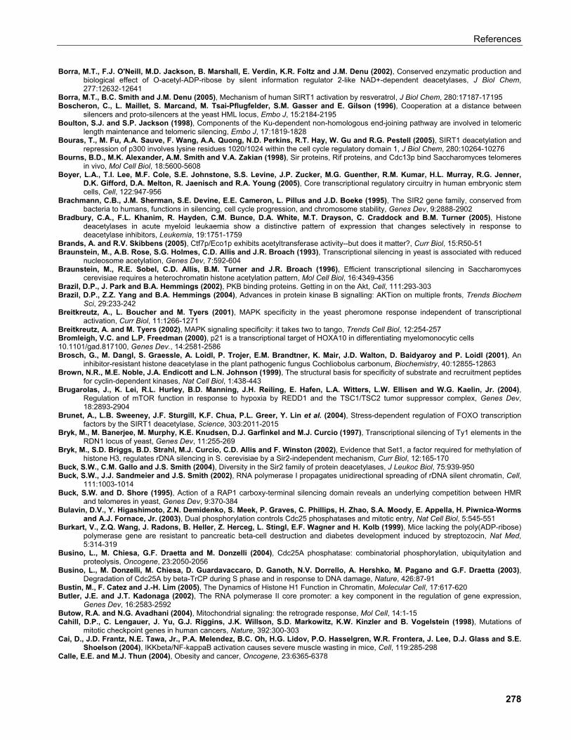
References
Borra, M.T., F.J. O'Neill, M.D. Jackson, B. Marshall, E. Verdin, K.R. Foltz and J.M. Denu (2002), Conserved enzymatic production and biological effect of O-acetyl-ADP-ribose by silent information regulator 2-like NAD+-dependent deacetylases, J Biol Chem, 277:12632-12641
Borra, M.T., B.C. Smith and J.M. Denu (2005), Mechanism of human SIRT1 activation by resveratrol, J Biol Chem, 280:17187-17195 Boscheron, C., L. Maillet, S. Marcand, M. Tsai-Pflugfelder, S.M. Gasser and E. Gilson (1996), Cooperation at a distance between
silencers and proto-silencers at the yeast HML locus, Embo J, 15:2184-2195 Boulton, S.J. and S.P. Jackson (1998), Components of the Ku-dependent non-homologous end-joining pathway are involved in telomeric
length maintenance and telomeric silencing, Embo J, 17:1819-1828 Bouras, T., M. Fu, A.A. Sauve, F. Wang, A.A. Quong, N.D. Perkins, R.T. Hay, W. Gu and R.G. Pestell (2005), SIRT1 deacetylation and
repression of p300 involves lysine residues 1020/1024 within the cell cycle regulatory domain 1, J Biol Chem, 280:10264-10276 Bourns, B.D., M.K. Alexander, A.M. Smith and V.A. Zakian (1998), Sir proteins, Rif proteins, and Cdc13p bind Saccharomyces telomeres
in vivo, Mol Cell Biol, 18:5600-5608 Boyer, L.A., T.I. Lee, M.F. Cole, S.E. Johnstone, S.S. Levine, J.P. Zucker, M.G. Guenther, R.M. Kumar, H.L. Murray, R.G. Jenner,
D.K. Gifford, D.A. Melton, R. Jaenisch and R.A. Young (2005), Core transcriptional regulatory circuitry in human embryonic stem cells, Cell, 122:947-956
Brachmann, C.B., J.M. Sherman, S.E. Devine, E.E. Cameron, L. Pillus and J.D. Boeke (1995), The SIR2 gene family, conserved from bacteria to humans, functions in silencing, cell cycle progression, and chromosome stability, Genes Dev, 9:2888-2902
Bradbury, C.A., F.L. Khanim, R. Hayden, C.M. Bunce, D.A. White, M.T. Drayson, C. Craddock and B.M. Turner (2005), Histone deacetylases in acute myeloid leukaemia show a distinctive pattern of expression that changes selectively in response to deacetylase inhibitors, Leukemia, 19:1751-1759
Brands, A. and R.V. Skibbens (2005), Ctf7p/Eco1p exhibits acetyltransferase activity--but does it matter?, Curr Biol, 15:R50-51 Braunstein, M., A.B. Rose, S.G. Holmes, C.D. Allis and J.R. Broach (1993), Transcriptional silencing in yeast is associated with reduced
nucleosome acetylation, Genes Dev, 7:592-604 Braunstein, M., R.E. Sobel, C.D. Allis, B.M. Turner and J.R. Broach (1996), Efficient transcriptional silencing in Saccharomyces
cerevisiae requires a heterochromatin histone acetylation pattern, Mol Cell Biol, 16:4349-4356 Brazil, D.P., J. Park and B.A. Hemmings (2002), PKB binding proteins. Getting in on the Akt, Cell, 111:293-303 Brazil, D.P., Z.Z. Yang and B.A. Hemmings (2004), Advances in protein kinase B signalling: AKTion on multiple fronts, Trends Biochem
Sci, 29:233-242 Breitkreutz, A., L. Boucher and M. Tyers (2001), MAPK specificity in the yeast pheromone response independent of transcriptional
activation, Curr Biol, 11:1266-1271 Breitkreutz, A. and M. Tyers (2002), MAPK signaling specificity: it takes two to tango, Trends Cell Biol, 12:254-257 Bromleigh, V.C. and L.P. Freedman (2000), p21 is a transcriptional target of HOXA10 in differentiating myelomonocytic cells 10.1101/gad.817100, Genes Dev., 14:2581-2586 Brosch, G., M. Dangl, S. Graessle, A. Loidl, P. Trojer, E.M. Brandtner, K. Mair, J.D. Walton, D. Baidyaroy and P. Loidl (2001), An
inhibitor-resistant histone deacetylase in the plant pathogenic fungus Cochliobolus carbonum, Biochemistry, 40:12855-12863 Brown, N.R., M.E. Noble, J.A. Endicott and L.N. Johnson (1999), The structural basis for specificity of substrate and recruitment peptides
for cyclin-dependent kinases, Nat Cell Biol, 1:438-443 Brugarolas, J., K. Lei, R.L. Hurley, B.D. Manning, J.H. Reiling, E. Hafen, L.A. Witters, L.W. Ellisen and W.G. Kaelin, Jr. (2004),
Regulation of mTOR function in response to hypoxia by REDD1 and the TSC1/TSC2 tumor suppressor complex, Genes Dev, 18:2893-2904
Brunet, A., L.B. Sweeney, J.F. Sturgill, K.F. Chua, P.L. Greer, Y. Lin et al. (2004), Stress-dependent regulation of FOXO transcription factors by the SIRT1 deacetylase, Science, 303:2011-2015
Bryk, M., M. Banerjee, M. Murphy, K.E. Knudsen, D.J. Garfinkel and M.J. Curcio (1997), Transcriptional silencing of Ty1 elements in the RDN1 locus of yeast, Genes Dev, 11:255-269
Bryk, M., S.D. Briggs, B.D. Strahl, M.J. Curcio, C.D. Allis and F. Winston (2002), Evidence that Set1, a factor required for methylation of histone H3, regulates rDNA silencing in S. cerevisiae by a Sir2-independent mechanism, Curr Biol, 12:165-170
Buck, S.W., C.M. Gallo and J.S. Smith (2004), Diversity in the Sir2 family of protein deacetylases, J Leukoc Biol, 75:939-950 Buck, S.W., J.J. Sandmeier and J.S. Smith (2002), RNA polymerase I propagates unidirectional spreading of rDNA silent chromatin, Cell,
111:1003-1014 Buck, S.W. and D. Shore (1995), Action of a RAP1 carboxy-terminal silencing domain reveals an underlying competition between HMR
and telomeres in yeast, Genes Dev, 9:370-384 Bulavin, D.V., Y. Higashimoto, Z.N. Demidenko, S. Meek, P. Graves, C. Phillips, H. Zhao, S.A. Moody, E. Appella, H. Piwnica-Worms
and A.J. Fornace, Jr. (2003), Dual phosphorylation controls Cdc25 phosphatases and mitotic entry, Nat Cell Biol, 5:545-551 Burkart, V., Z.Q. Wang, J. Radons, B. Heller, Z. Herceg, L. Stingl, E.F. Wagner and H. Kolb (1999), Mice lacking the poly(ADP-ribose)
polymerase gene are resistant to pancreatic beta-cell destruction and diabetes development induced by streptozocin, Nat Med, 5:314-319
Busino, L., M. Chiesa, G.F. Draetta and M. Donzelli (2004), Cdc25A phosphatase: combinatorial phosphorylation, ubiquitylation and proteolysis, Oncogene, 23:2050-2056
Busino, L., M. Donzelli, M. Chiesa, D. Guardavaccaro, D. Ganoth, N.V. Dorrello, A. Hershko, M. Pagano and G.F. Draetta (2003), Degradation of Cdc25A by beta-TrCP during S phase and in response to DNA damage, Nature, 426:87-91
Bustin, M., F. Catez and J.-H. Lim (2005), The Dynamics of Histone H1 Function in Chromatin, Molecular Cell, 17:617-620 Butler, J.E. and J.T. Kadonaga (2002), The RNA polymerase II core promoter: a key component in the regulation of gene expression,
Genes Dev, 16:2583-2592 Butow, R.A. and N.G. Avadhani (2004), Mitochondrial signaling: the retrograde response, Mol Cell, 14:1-15 Cahill, D.P., C. Lengauer, J. Yu, G.J. Riggins, J.K. Willson, S.D. Markowitz, K.W. Kinzler and B. Vogelstein (1998), Mutations of
mitotic checkpoint genes in human cancers, Nature, 392:300-303 Cai, D., J.D. Frantz, N.E. Tawa, Jr., P.A. Melendez, B.C. Oh, H.G. Lidov, P.O. Hasselgren, W.R. Frontera, J. Lee, D.J. Glass and S.E.
Shoelson (2004), IKKbeta/NF-kappaB activation causes severe muscle wasting in mice, Cell, 119:285-298 Calle, E.E. and M.J. Thun (2004), Obesity and cancer, Oncogene, 23:6365-6378
278
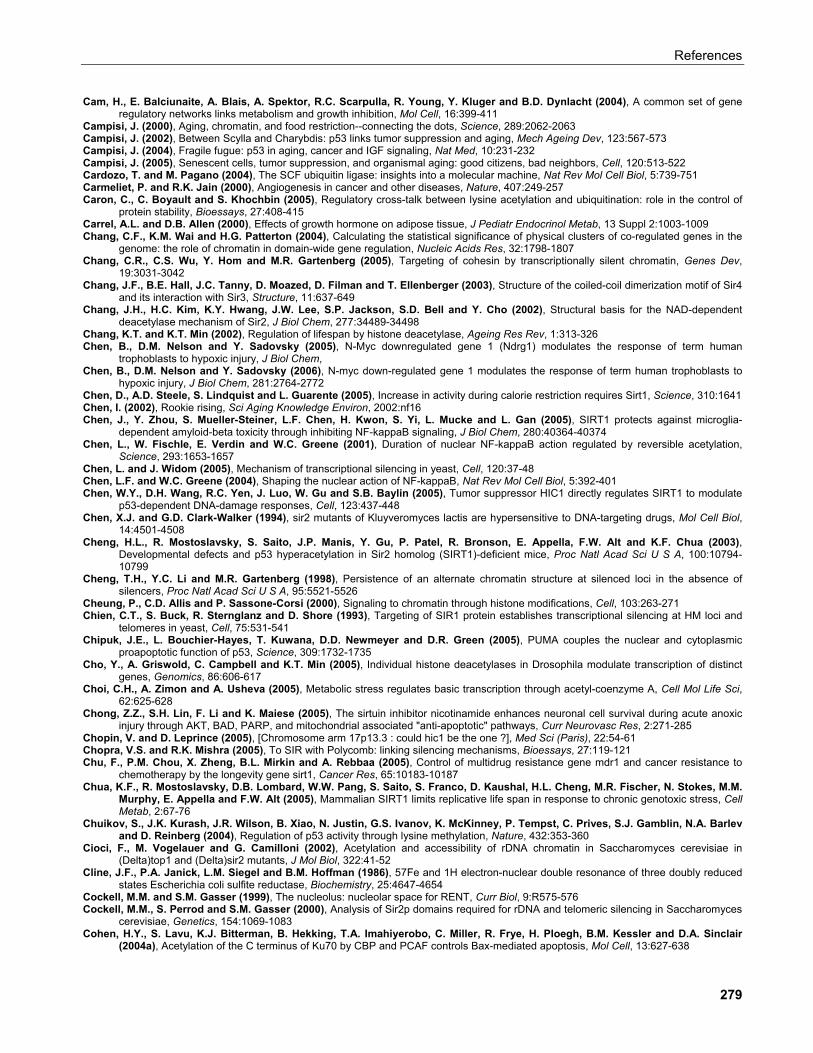
References
Cam, H., E. Balciunaite, A. Blais, A. Spektor, R.C. Scarpulla, R. Young, Y. Kluger and B.D. Dynlacht (2004), A common set of gene regulatory networks links metabolism and growth inhibition, Mol Cell, 16:399-411
Campisi, J. (2000), Aging, chromatin, and food restriction--connecting the dots, Science, 289:2062-2063 Campisi, J. (2002), Between Scylla and Charybdis: p53 links tumor suppression and aging, Mech Ageing Dev, 123:567-573 Campisi, J. (2004), Fragile fugue: p53 in aging, cancer and IGF signaling, Nat Med, 10:231-232 Campisi, J. (2005), Senescent cells, tumor suppression, and organismal aging: good citizens, bad neighbors, Cell, 120:513-522 Cardozo, T. and M. Pagano (2004), The SCF ubiquitin ligase: insights into a molecular machine, Nat Rev Mol Cell Biol, 5:739-751 Carmeliet, P. and R.K. Jain (2000), Angiogenesis in cancer and other diseases, Nature, 407:249-257 Caron, C., C. Boyault and S. Khochbin (2005), Regulatory cross-talk between lysine acetylation and ubiquitination: role in the control of
protein stability, Bioessays, 27:408-415 Carrel, A.L. and D.B. Allen (2000), Effects of growth hormone on adipose tissue, J Pediatr Endocrinol Metab, 13 Suppl 2:1003-1009 Chang, C.F., K.M. Wai and H.G. Patterton (2004), Calculating the statistical significance of physical clusters of co-regulated genes in the
genome: the role of chromatin in domain-wide gene regulation, Nucleic Acids Res, 32:1798-1807 Chang, C.R., C.S. Wu, Y. Hom and M.R. Gartenberg (2005), Targeting of cohesin by transcriptionally silent chromatin, Genes Dev,
19:3031-3042 Chang, J.F., B.E. Hall, J.C. Tanny, D. Moazed, D. Filman and T. Ellenberger (2003), Structure of the coiled-coil dimerization motif of Sir4
and its interaction with Sir3, Structure, 11:637-649 Chang, J.H., H.C. Kim, K.Y. Hwang, J.W. Lee, S.P. Jackson, S.D. Bell and Y. Cho (2002), Structural basis for the NAD-dependent
deacetylase mechanism of Sir2, J Biol Chem, 277:34489-34498 Chang, K.T. and K.T. Min (2002), Regulation of lifespan by histone deacetylase, Ageing Res Rev, 1:313-326 Chen, B., D.M. Nelson and Y. Sadovsky (2005), N-Myc downregulated gene 1 (Ndrg1) modulates the response of term human
trophoblasts to hypoxic injury, J Biol Chem, Chen, B., D.M. Nelson and Y. Sadovsky (2006), N-myc down-regulated gene 1 modulates the response of term human trophoblasts to
hypoxic injury, J Biol Chem, 281:2764-2772 Chen, D., A.D. Steele, S. Lindquist and L. Guarente (2005), Increase in activity during calorie restriction requires Sirt1, Science, 310:1641 Chen, I. (2002), Rookie rising, Sci Aging Knowledge Environ, 2002:nf16 Chen, J., Y. Zhou, S. Mueller-Steiner, L.F. Chen, H. Kwon, S. Yi, L. Mucke and L. Gan (2005), SIRT1 protects against microglia-
dependent amyloid-beta toxicity through inhibiting NF-kappaB signaling, J Biol Chem, 280:40364-40374 Chen, L., W. Fischle, E. Verdin and W.C. Greene (2001), Duration of nuclear NF-kappaB action regulated by reversible acetylation,
Science, 293:1653-1657 Chen, L. and J. Widom (2005), Mechanism of transcriptional silencing in yeast, Cell, 120:37-48 Chen, L.F. and W.C. Greene (2004), Shaping the nuclear action of NF-kappaB, Nat Rev Mol Cell Biol, 5:392-401 Chen, W.Y., D.H. Wang, R.C. Yen, J. Luo, W. Gu and S.B. Baylin (2005), Tumor suppressor HIC1 directly regulates SIRT1 to modulate
p53-dependent DNA-damage responses, Cell, 123:437-448 Chen, X.J. and G.D. Clark-Walker (1994), sir2 mutants of Kluyveromyces lactis are hypersensitive to DNA-targeting drugs, Mol Cell Biol,
14:4501-4508 Cheng, H.L., R. Mostoslavsky, S. Saito, J.P. Manis, Y. Gu, P. Patel, R. Bronson, E. Appella, F.W. Alt and K.F. Chua (2003),
Developmental defects and p53 hyperacetylation in Sir2 homolog (SIRT1)-deficient mice, Proc Natl Acad Sci U S A, 100:10794-10799
Cheng, T.H., Y.C. Li and M.R. Gartenberg (1998), Persistence of an alternate chromatin structure at silenced loci in the absence of silencers, Proc Natl Acad Sci U S A, 95:5521-5526
Cheung, P., C.D. Allis and P. Sassone-Corsi (2000), Signaling to chromatin through histone modifications, Cell, 103:263-271 Chien, C.T., S. Buck, R. Sternglanz and D. Shore (1993), Targeting of SIR1 protein establishes transcriptional silencing at HM loci and
telomeres in yeast, Cell, 75:531-541 Chipuk, J.E., L. Bouchier-Hayes, T. Kuwana, D.D. Newmeyer and D.R. Green (2005), PUMA couples the nuclear and cytoplasmic
proapoptotic function of p53, Science, 309:1732-1735 Cho, Y., A. Griswold, C. Campbell and K.T. Min (2005), Individual histone deacetylases in Drosophila modulate transcription of distinct
genes, Genomics, 86:606-617 Choi, C.H., A. Zimon and A. Usheva (2005), Metabolic stress regulates basic transcription through acetyl-coenzyme A, Cell Mol Life Sci,
62:625-628 Chong, Z.Z., S.H. Lin, F. Li and K. Maiese (2005), The sirtuin inhibitor nicotinamide enhances neuronal cell survival during acute anoxic
injury through AKT, BAD, PARP, and mitochondrial associated "anti-apoptotic" pathways, Curr Neurovasc Res, 2:271-285 Chopin, V. and D. Leprince (2005), [Chromosome arm 17p13.3 : could hic1 be the one ?], Med Sci (Paris), 22:54-61 Chopra, V.S. and R.K. Mishra (2005), To SIR with Polycomb: linking silencing mechanisms, Bioessays, 27:119-121 Chu, F., P.M. Chou, X. Zheng, B.L. Mirkin and A. Rebbaa (2005), Control of multidrug resistance gene mdr1 and cancer resistance to
chemotherapy by the longevity gene sirt1, Cancer Res, 65:10183-10187 Chua, K.F., R. Mostoslavsky, D.B. Lombard, W.W. Pang, S. Saito, S. Franco, D. Kaushal, H.L. Cheng, M.R. Fischer, N. Stokes, M.M.
Murphy, E. Appella and F.W. Alt (2005), Mammalian SIRT1 limits replicative life span in response to chronic genotoxic stress, Cell Metab, 2:67-76
Chuikov, S., J.K. Kurash, J.R. Wilson, B. Xiao, N. Justin, G.S. Ivanov, K. McKinney, P. Tempst, C. Prives, S.J. Gamblin, N.A. Barlev and D. Reinberg (2004), Regulation of p53 activity through lysine methylation, Nature, 432:353-360
Cioci, F., M. Vogelauer and G. Camilloni (2002), Acetylation and accessibility of rDNA chromatin in Saccharomyces cerevisiae in (Delta)top1 and (Delta)sir2 mutants, J Mol Biol, 322:41-52
Cline, J.F., P.A. Janick, L.M. Siegel and B.M. Hoffman (1986), 57Fe and 1H electron-nuclear double resonance of three doubly reduced states Escherichia coli sulfite reductase, Biochemistry, 25:4647-4654
Cockell, M.M. and S.M. Gasser (1999), The nucleolus: nucleolar space for RENT, Curr Biol, 9:R575-576 Cockell, M.M., S. Perrod and S.M. Gasser (2000), Analysis of Sir2p domains required for rDNA and telomeric silencing in Saccharomyces
cerevisiae, Genetics, 154:1069-1083 Cohen, H.Y., S. Lavu, K.J. Bitterman, B. Hekking, T.A. Imahiyerobo, C. Miller, R. Frye, H. Ploegh, B.M. Kessler and D.A. Sinclair
(2004a), Acetylation of the C terminus of Ku70 by CBP and PCAF controls Bax-mediated apoptosis, Mol Cell, 13:627-638
279
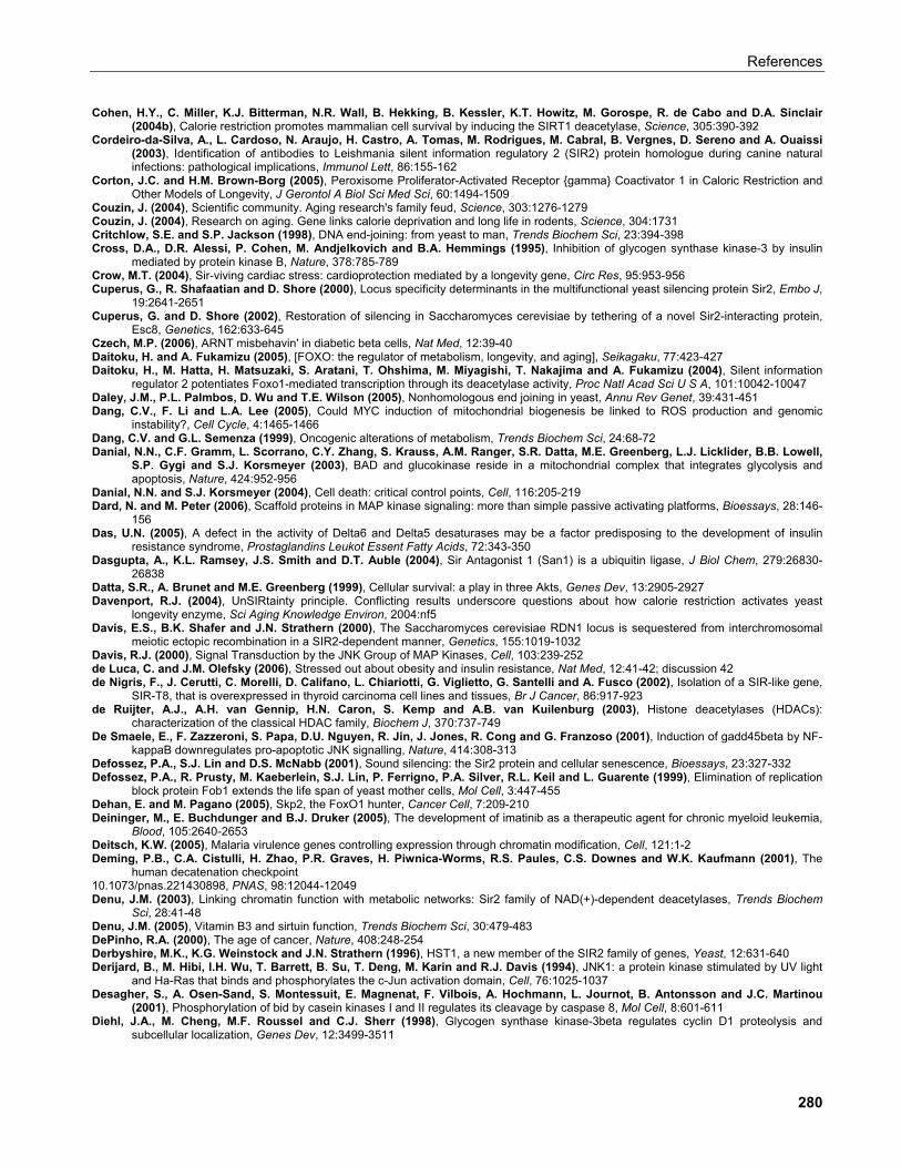
References
Cohen, H.Y., C. Miller, K.J. Bitterman, N.R. Wall, B. Hekking, B. Kessler, K.T. Howitz, M. Gorospe, R. de Cabo and D.A. Sinclair (2004b), Calorie restriction promotes mammalian cell survival by inducing the SIRT1 deacetylase, Science, 305:390-392
Cordeiro-da-Silva, A., L. Cardoso, N. Araujo, H. Castro, A. Tomas, M. Rodrigues, M. Cabral, B. Vergnes, D. Sereno and A. Ouaissi (2003), Identification of antibodies to Leishmania silent information regulatory 2 (SIR2) protein homologue during canine natural infections: pathological implications, Immunol Lett, 86:155-162
Corton, J.C. and H.M. Brown-Borg (2005), Peroxisome Proliferator-Activated Receptor {gamma} Coactivator 1 in Caloric Restriction and Other Models of Longevity, J Gerontol A Biol Sci Med Sci, 60:1494-1509
Couzin, J. (2004), Scientific community. Aging research's family feud, Science, 303:1276-1279 Couzin, J. (2004), Research on aging. Gene links calorie deprivation and long life in rodents, Science, 304:1731 Critchlow, S.E. and S.P. Jackson (1998), DNA end-joining: from yeast to man, Trends Biochem Sci, 23:394-398 Cross, D.A., D.R. Alessi, P. Cohen, M. Andjelkovich and B.A. Hemmings (1995), Inhibition of glycogen synthase kinase-3 by insulin
mediated by protein kinase B, Nature, 378:785-789 Crow, M.T. (2004), Sir-viving cardiac stress: cardioprotection mediated by a longevity gene, Circ Res, 95:953-956 Cuperus, G., R. Shafaatian and D. Shore (2000), Locus specificity determinants in the multifunctional yeast silencing protein Sir2, Embo J,
19:2641-2651 Cuperus, G. and D. Shore (2002), Restoration of silencing in Saccharomyces cerevisiae by tethering of a novel Sir2-interacting protein,
Esc8, Genetics, 162:633-645 Czech, M.P. (2006), ARNT misbehavin' in diabetic beta cells, Nat Med, 12:39-40 Daitoku, H. and A. Fukamizu (2005), [FOXO: the regulator of metabolism, longevity, and aging], Seikagaku, 77:423-427 Daitoku, H., M. Hatta, H. Matsuzaki, S. Aratani, T. Ohshima, M. Miyagishi, T. Nakajima and A. Fukamizu (2004), Silent information
regulator 2 potentiates Foxo1-mediated transcription through its deacetylase activity, Proc Natl Acad Sci U S A, 101:10042-10047 Daley, J.M., P.L. Palmbos, D. Wu and T.E. Wilson (2005), Nonhomologous end joining in yeast, Annu Rev Genet, 39:431-451 Dang, C.V., F. Li and L.A. Lee (2005), Could MYC induction of mitochondrial biogenesis be linked to ROS production and genomic
instability?, Cell Cycle, 4:1465-1466 Dang, C.V. and G.L. Semenza (1999), Oncogenic alterations of metabolism, Trends Biochem Sci, 24:68-72 Danial, N.N., C.F. Gramm, L. Scorrano, C.Y. Zhang, S. Krauss, A.M. Ranger, S.R. Datta, M.E. Greenberg, L.J. Licklider, B.B. Lowell,
S.P. Gygi and S.J. Korsmeyer (2003), BAD and glucokinase reside in a mitochondrial complex that integrates glycolysis and apoptosis, Nature, 424:952-956
Danial, N.N. and S.J. Korsmeyer (2004), Cell death: critical control points, Cell, 116:205-219 Dard, N. and M. Peter (2006), Scaffold proteins in MAP kinase signaling: more than simple passive activating platforms, Bioessays, 28:146-
156 Das, U.N. (2005), A defect in the activity of Delta6 and Delta5 desaturases may be a factor predisposing to the development of insulin
resistance syndrome, Prostaglandins Leukot Essent Fatty Acids, 72:343-350 Dasgupta, A., K.L. Ramsey, J.S. Smith and D.T. Auble (2004), Sir Antagonist 1 (San1) is a ubiquitin ligase, J Biol Chem, 279:26830-
26838 Datta, S.R., A. Brunet and M.E. Greenberg (1999), Cellular survival: a play in three Akts, Genes Dev, 13:2905-2927 Davenport, R.J. (2004), UnSIRtainty principle. Conflicting results underscore questions about how calorie restriction activates yeast
longevity enzyme, Sci Aging Knowledge Environ, 2004:nf5 Davis, E.S., B.K. Shafer and J.N. Strathern (2000), The Saccharomyces cerevisiae RDN1 locus is sequestered from interchromosomal
meiotic ectopic recombination in a SIR2-dependent manner, Genetics, 155:1019-1032 Davis, R.J. (2000), Signal Transduction by the JNK Group of MAP Kinases, Cell, 103:239-252 de Luca, C. and J.M. Olefsky (2006), Stressed out about obesity and insulin resistance, Nat Med, 12:41-42; discussion 42 de Nigris, F., J. Cerutti, C. Morelli, D. Califano, L. Chiariotti, G. Viglietto, G. Santelli and A. Fusco (2002), Isolation of a SIR-like gene,
SIR-T8, that is overexpressed in thyroid carcinoma cell lines and tissues, Br J Cancer, 86:917-923 de Ruijter, A.J., A.H. van Gennip, H.N. Caron, S. Kemp and A.B. van Kuilenburg (2003), Histone deacetylases (HDACs):
characterization of the classical HDAC family, Biochem J, 370:737-749 De Smaele, E., F. Zazzeroni, S. Papa, D.U. Nguyen, R. Jin, J. Jones, R. Cong and G. Franzoso (2001), Induction of gadd45beta by NF-
kappaB downregulates pro-apoptotic JNK signalling, Nature, 414:308-313 Defossez, P.A., S.J. Lin and D.S. McNabb (2001), Sound silencing: the Sir2 protein and cellular senescence, Bioessays, 23:327-332 Defossez, P.A., R. Prusty, M. Kaeberlein, S.J. Lin, P. Ferrigno, P.A. Silver, R.L. Keil and L. Guarente (1999), Elimination of replication
block protein Fob1 extends the life span of yeast mother cells, Mol Cell, 3:447-455 Dehan, E. and M. Pagano (2005), Skp2, the FoxO1 hunter, Cancer Cell, 7:209-210 Deininger, M., E. Buchdunger and B.J. Druker (2005), The development of imatinib as a therapeutic agent for chronic myeloid leukemia,
Blood, 105:2640-2653 Deitsch, K.W. (2005), Malaria virulence genes controlling expression through chromatin modification, Cell, 121:1-2 Deming, P.B., C.A. Cistulli, H. Zhao, P.R. Graves, H. Piwnica-Worms, R.S. Paules, C.S. Downes and W.K. Kaufmann (2001), The
human decatenation checkpoint 10.1073/pnas.221430898, PNAS, 98:12044-12049 Denu, J.M. (2003), Linking chromatin function with metabolic networks: Sir2 family of NAD(+)-dependent deacetylases, Trends Biochem
Sci, 28:41-48 Denu, J.M. (2005), Vitamin B3 and sirtuin function, Trends Biochem Sci, 30:479-483 DePinho, R.A. (2000), The age of cancer, Nature, 408:248-254 Derbyshire, M.K., K.G. Weinstock and J.N. Strathern (1996), HST1, a new member of the SIR2 family of genes, Yeast, 12:631-640 Derijard, B., M. Hibi, I.H. Wu, T. Barrett, B. Su, T. Deng, M. Karin and R.J. Davis (1994), JNK1: a protein kinase stimulated by UV light
and Ha-Ras that binds and phosphorylates the c-Jun activation domain, Cell, 76:1025-1037 Desagher, S., A. Osen-Sand, S. Montessuit, E. Magnenat, F. Vilbois, A. Hochmann, L. Journot, B. Antonsson and J.C. Martinou
(2001), Phosphorylation of bid by casein kinases I and II regulates its cleavage by caspase 8, Mol Cell, 8:601-611 Diehl, J.A., M. Cheng, M.F. Roussel and C.J. Sherr (1998), Glycogen synthase kinase-3beta regulates cyclin D1 proteolysis and
subcellular localization, Genes Dev, 12:3499-3511
280
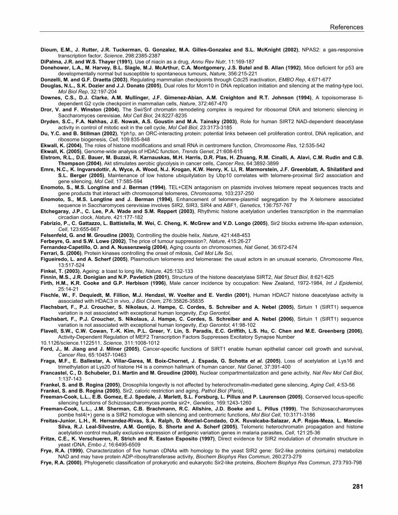
References
Dioum, E.M., J. Rutter, J.R. Tuckerman, G. Gonzalez, M.A. Gilles-Gonzalez and S.L. McKnight (2002), NPAS2: a gas-responsive transcription factor, Science, 298:2385-2387
DiPalma, J.R. and W.S. Thayer (1991), Use of niacin as a drug, Annu Rev Nutr, 11:169-187 Donehower, L.A., M. Harvey, B.L. Slagle, M.J. McArthur, C.A. Montgomery, J.S. Butel and B. Allan (1992), Mice deficient for p53 are
developmentally normal but susceptible to spontaneous tumours, Nature, 356:215-221 Donzelli, M. and G.F. Draetta (2003), Regulating mammalian checkpoints through Cdc25 inactivation, EMBO Rep, 4:671-677 Douglas, N.L., S.K. Dozier and J.J. Donato (2005), Dual roles for Mcm10 in DNA replication initiation and silencing at the mating-type loci,
Mol Biol Rep, 32:197-204 Downes, C.S., D.J. Clarke, A.M. Mullinger, J.F. Gimenez-Abian, A.M. Creighton and R.T. Johnson (1994), A topoisomerase II-
dependent G2 cycle checkpoint in mammalian cells, Nature, 372:467-470 Dror, V. and F. Winston (2004), The Swi/Snf chromatin remodeling complex is required for ribosomal DNA and telomeric silencing in
Saccharomyces cerevisiae, Mol Cell Biol, 24:8227-8235 Dryden, S.C., F.A. Nahhas, J.E. Nowak, A.S. Goustin and M.A. Tainsky (2003), Role for human SIRT2 NAD-dependent deacetylase
activity in control of mitotic exit in the cell cycle, Mol Cell Biol, 23:3173-3185 Du, Y.C. and B. Stillman (2002), Yph1p, an ORC-interacting protein: potential links between cell proliferation control, DNA replication, and
ribosome biogenesis, Cell, 109:835-848 Ekwall, K. (2004), The roles of histone modifications and small RNA in centromere function, Chromosome Res, 12:535-542 Ekwall, K. (2005), Genome-wide analysis of HDAC function, Trends Genet, 21:608-615 Elstrom, R.L., D.E. Bauer, M. Buzzai, R. Karnauskas, M.H. Harris, D.R. Plas, H. Zhuang, R.M. Cinalli, A. Alavi, C.M. Rudin and C.B.
Thompson (2004), Akt stimulates aerobic glycolysis in cancer cells, Cancer Res, 64:3892-3899 Emre, N.C., K. Ingvarsdottir, A. Wyce, A. Wood, N.J. Krogan, K.W. Henry, K. Li, R. Marmorstein, J.F. Greenblatt, A. Shilatifard and
S.L. Berger (2005), Maintenance of low histone ubiquitylation by Ubp10 correlates with telomere-proximal Sir2 association and gene silencing, Mol Cell, 17:585-594
Enomoto, S., M.S. Longtine and J. Berman (1994), TEL+CEN antagonism on plasmids involves telomere repeat sequences tracts and gene products that interact with chromosomal telomeres, Chromosoma, 103:237-250
Enomoto, S., M.S. Longtine and J. Berman (1994), Enhancement of telomere-plasmid segregation by the X-telomere associated sequence in Saccharomyces cerevisiae involves SIR2, SIR3, SIR4 and ABF1, Genetics, 136:757-767
Etchegaray, J.P., C. Lee, P.A. Wade and S.M. Reppert (2003), Rhythmic histone acetylation underlies transcription in the mammalian circadian clock, Nature, 421:177-182
Fabrizio, P., C. Gattazzo, L. Battistella, M. Wei, C. Cheng, K. McGrew and V.D. Longo (2005), Sir2 blocks extreme life-span extension, Cell, 123:655-667
Felsenfeld, G. and M. Groudine (2003), Controlling the double helix, Nature, 421:448-453 Ferbeyre, G. and S.W. Lowe (2002), The price of tumour suppression?, Nature, 415:26-27 Fernandez-Capetillo, O. and A. Nussenzweig (2004), Aging counts on chromosomes, Nat Genet, 36:672-674 Ferrari, S. (2006), Protein kinases controlling the onset of mitosis, Cell Mol Life Sci, Figueiredo, L. and A. Scherf (2005), Plasmodium telomeres and telomerase: the usual actors in an unusual scenario, Chromosome Res,
13:517-524 Finkel, T. (2003), Ageing: a toast to long life, Nature, 425:132-133 Finnin, M.S., J.R. Donigian and N.P. Pavletich (2001), Structure of the histone deacetylase SIRT2, Nat Struct Biol, 8:621-625 Firth, H.M., K.R. Cooke and G.P. Herbison (1996), Male cancer incidence by occupation: New Zealand, 1972-1984, Int J Epidemiol,
25:14-21 Fischle, W., F. Dequiedt, M. Fillion, M.J. Hendzel, W. Voelter and E. Verdin (2001), Human HDAC7 histone deacetylase activity is
associated with HDAC3 in vivo, J Biol Chem, 276:35826-35835 Flachsbart, F., P.J. Croucher, S. Nikolaus, J. Hampe, C. Cordes, S. Schreiber and A. Nebel (2005), Sirtuin 1 (SIRT1) sequence
variation is not associated with exceptional human longevity, Exp Gerontol, Flachsbart, F., P.J. Croucher, S. Nikolaus, J. Hampe, C. Cordes, S. Schreiber and A. Nebel (2006), Sirtuin 1 (SIRT1) sequence
variation is not associated with exceptional human longevity, Exp Gerontol, 41:98-102 Flavell, S.W., C.W. Cowan, T.-K. Kim, P.L. Greer, Y. Lin, S. Paradis, E.C. Griffith, L.S. Hu, C. Chen and M.E. Greenberg (2006),
Activity-Dependent Regulation of MEF2 Transcription Factors Suppresses Excitatory Synapse Number 10.1126/science.1122511, Science, 311:1008-1012 Ford, J., M. Jiang and J. Milner (2005), Cancer-specific functions of SIRT1 enable human epithelial cancer cell growth and survival,
Cancer Res, 65:10457-10463 Fraga, M.F., E. Ballestar, A. Villar-Garea, M. Boix-Chornet, J. Espada, G. Schotta et al. (2005), Loss of acetylation at Lys16 and
trimethylation at Lys20 of histone H4 is a common hallmark of human cancer, Nat Genet, 37:391-400 Francastel, C., D. Schubeler, D.I. Martin and M. Groudine (2000), Nuclear compartmentalization and gene activity, Nat Rev Mol Cell Biol,
1:137-143 Frankel, S. and B. Rogina (2005), Drosophila longevity is not affected by heterochromatin-mediated gene silencing, Aging Cell, 4:53-56 Frankel, S. and B. Rogina (2005), Sir2, caloric restriction and aging, Pathol Biol (Paris), Freeman-Cook, L.L., E.B. Gomez, E.J. Spedale, J. Marlett, S.L. Forsburg, L. Pillus and P. Laurenson (2005), Conserved locus-specific
silencing functions of Schizosaccharomyces pombe sir2+, Genetics, 169:1243-1260 Freeman-Cook, L.L., J.M. Sherman, C.B. Brachmann, R.C. Allshire, J.D. Boeke and L. Pillus (1999), The Schizosaccharomyces
pombe hst4(+) gene is a SIR2 homologue with silencing and centromeric functions, Mol Biol Cell, 10:3171-3186 Freitas-Junior, L.H., R. Hernandez-Rivas, S.A. Ralph, D. Montiel-Condado, O.K. Ruvalcaba-Salazar, A.P. Rojas-Meza, L. Mancio-
Silva, R.J. Leal-Silvestre, A.M. Gontijo, S. Shorte and A. Scherf (2005), Telomeric heterochromatin propagation and histone acetylation control mutually exclusive expression of antigenic variation genes in malaria parasites, Cell, 121:25-36
Fritze, C.E., K. Verschueren, R. Strich and R. Easton Esposito (1997), Direct evidence for SIR2 modulation of chromatin structure in yeast rDNA, Embo J, 16:6495-6509
Frye, R.A. (1999), Characterization of five human cDNAs with homology to the yeast SIR2 gene: Sir2-like proteins (sirtuins) metabolize NAD and may have protein ADP-ribosyltransferase activity, Biochem Biophys Res Commun, 260:273-279
Frye, R.A. (2000), Phylogenetic classification of prokaryotic and eukaryotic Sir2-like proteins, Biochem Biophys Res Commun, 273:793-798
281
References
Fuerst, P.G. and D.F. Voytas (2003), CEN plasmid segregation is destabilized by tethered determinants of Ty 5 integration specificity: a role for double-strand breaks in CEN antagonism, Chromosoma, 112:58-65
Fulco, M., R.L. Schiltz, S. Iezzi, M.T. King, P. Zhao, Y. Kashiwaya, E. Hoffman, R.L. Veech and V. Sartorelli (2003), Sir2 regulates skeletal muscle differentiation as a potential sensor of the redox state, Mol Cell, 12:51-62
Furuyama, T., R. Banerjee, T.R. Breen and P.J. Harte (2004), SIR2 is required for polycomb silencing and is associated with an E(Z) histone methyltransferase complex, Curr Biol, 14:1812-1821
Gallo, C.M., D.L. Smith, Jr. and J.S. Smith (2004), Nicotinamide clearance by Pnc1 directly regulates Sir2-mediated silencing and longevity, Mol Cell Biol, 24:1301-1312
Gan, L., Y. Han, S. Bastianetto, Y. Dumont, T.G. Unterman and R. Quirion (2005), FoxO-dependent and -independent mechanisms mediate SirT1 effects on IGFBP-1 gene expression, Biochem Biophys Res Commun, 337:1092-1096
Gangadharan, S., S. Ghidelli and R.T. Kamakaka (2004), Purification of Sir2 proteins from yeast, Methods Enzymol, 377:234-254 Gangloff, Y.G., M. Mueller, S.G. Dann, P. Svoboda, M. Sticker, J.F. Spetz, S.H. Um, E.J. Brown, S. Cereghini, G. Thomas and S.C.
Kozma (2004), Disruption of the mouse mTOR gene leads to early postimplantation lethality and prohibits embryonic stem cell development, Mol Cell Biol, 24:9508-9516
Garcia, S.N. and L. Pillus (1999), Net results of nucleolar dynamics, Cell, 97:825-828 Garcia, S.N. and L. Pillus (2002), A unique class of conditional sir2 mutants displays distinct silencing defects in Saccharomyces
cerevisiae, Genetics, 162:721-736 Garcia-Salcedo, J.A., P. Gijon, D.P. Nolan, P. Tebabi and E. Pays (2003), A chromosomal SIR2 homologue with both histone NAD-
dependent ADP-ribosyltransferase and deacetylase activities is involved in DNA repair in Trypanosoma brucei, Embo J, 22:5851-5862
Garske, A.L. and J.M. Denu (2006), SIRT1 Top 40 Hits: Use of One-Bead, One-Compound Acetyl-Peptide Libraries and Quantum Dots to Probe Deacetylase Specificity, Biochemistry, 45:94-101
Gartel, A.L. and K. Shchors (2003), Mechanisms of c-myc-mediated transcriptional repression of growth arrest genes, Exp Cell Res, 283:17-21
Gartenberg, M.R. (2000), The Sir proteins of Saccharomyces cerevisiae: mediators of transcriptional silencing and much more, Curr Opin Microbiol, 3:132-137
Gasser, S.M. and M.M. Cockell (2001), The molecular biology of the SIR proteins, Gene, 279:1-16 Gasser, S.M., M. Gotta, H. Renauld, T. Laroche and M. Cockell (1998), Nuclear organization and silencing: trafficking of Sir proteins,
Novartis Found Symp, 214:114-126; discussion 126-132 Geley, S., E. Kramer, C. Gieffers, J. Gannon, J.M. Peters and T. Hunt (2001), Anaphase-promoting complex/cyclosome-dependent
proteolysis of human cyclin A starts at the beginning of mitosis and is not subject to the spindle assembly checkpoint, J Cell Biol, 153:137-148
Gems, D. (2001), Ageing. Yeast longevity gene goes public, Nature, 410:154-155 Gerber, S.A., J. Rush, O. Stemman, M.W. Kirschner and S.P. Gygi (2003), Absolute quantification of proteins and phosphoproteins from
cell lysates by tandem MS, Proc Natl Acad Sci U S A, 100:6940-6945 Gershon, H. and D. Gershon (2000), The budding yeast, Saccharomyces cerevisiae, as a model for aging research: a critical review, Mech
Ageing Dev, 120:1-22 Ghidelli, S., D. Donze, N. Dhillon and R.T. Kamakaka (2001), Sir2p exists in two nucleosome-binding complexes with distinct deacetylase
activities, Embo J, 20:4522-4535 Giannakou, M.E. and L. Partridge (2004), The interaction between FOXO and SIRT1: tipping the balance towards survival, Trends Cell
Biol, 14:408-412 Gilbert, N., S. Gilchrist and W.A. Bickmore (2005), Chromatin organization in the mammalian nucleus, Int Rev Cytol, 242:283-336 Gillespie, C.S., C.J. Proctor, R.J. Boys, D.P. Shanley, D.J. Wilkinson and T.B. Kirkwood (2004), A mathematical model of ageing in
yeast, J Theor Biol, 229:189-196 Gingras, A.C., B. Raught and N. Sonenberg (1999), eIF4 initiation factors: effectors of mRNA recruitment to ribosomes and regulators of
translation, Annu Rev Biochem, 68:913-963 Gingras, A.C., B. Raught and N. Sonenberg (2001), Regulation of translation initiation by FRAP/mTOR, Genes Dev, 15:807-826 Giot, L., J.S. Bader, C. Brouwer, A. Chaudhuri, B. Kuang, Y. Li et al. (2003), A Protein Interaction Map of Drosophila melanogaster 10.1126/science.1090289, Science, 302:1727-1736 Girdwood, D., D. Bumpass, O.A. Vaughan, A. Thain, L.A. Anderson, A.W. Snowden, E. Garcia-Wilson, N.D. Perkins and R.T. Hay
(2003), P300 transcriptional repression is mediated by SUMO modification, Mol Cell, 11:1043-1054 Glass, D.J. (2005), A signaling role for dystrophin: inhibiting skeletal muscle atrophy pathways, Cancer Cell, 8:351-352 Glass, D.J. (2005), Skeletal muscle hypertrophy and atrophy signaling pathways, The International Journal of Biochemistry & Cell Biology,
37:1974-1984 Glotzer, M., A.W. Murray and M.W. Kirschner (1991), Cyclin is degraded by the ubiquitin pathway, Nature, 349:132-138 Gong, W., K. Suzuki, M. Russell and K. Riabowol (2005), Function of the ING family of PHD proteins in cancer, Int J Biochem Cell Biol,
37:1054-1065 Goodman, R.H. and S. Smolik (2000), CBP/p300 in cell growth, transformation, and development, Genes Dev, 14:1553-1577 Gotta, M., S. Strahl-Bolsinger, H. Renauld, T. Laroche, B.K. Kennedy, M. Grunstein and S.M. Gasser (1997), Localization of Sir2p: the
nucleolus as a compartment for silent information regulators, Embo J, 16:3243-3255 Gottlieb, E. and I.P. Tomlinson (2005), Mitochondrial tumour suppressors: a genetic and biochemical update, Nat Rev Cancer, 5:857-866 Gottlieb, S. and R.E. Esposito (1989), A new role for a yeast transcriptional silencer gene, SIR2, in regulation of recombination in
ribosomal DNA, Cell, 56:771-776 Gottlob, K., N. Majewski, S. Kennedy, E. Kandel, R.B. Robey and N. Hay (2001), Inhibition of early apoptotic events by Akt/PKB is
dependent on the first committed step of glycolysis and mitochondrial hexokinase, Genes Dev, 15:1406-1418 Gottschling, D.E. (2000), Gene silencing: two faces of SIR2, Curr Biol, 10:R708-711 Graham, I.R. and A. Chambers (1996), Rap1p is a negative regulator of the RAP1 gene, Curr Genet, 30:93-100 Gravel, S. and S.P. Jackson (2003), Increased genome instability in aging yeast, Cell, 115:1-2 Gray, S.G. and T.J. Ekstrom (2001), The human histone deacetylase family, Exp Cell Res, 262:75-83
282
References
Greten, F.R., L. Eckmann, T.F. Greten, J.M. Park, Z.W. Li, L.J. Egan, M.F. Kagnoff and M. Karin (2004), IKKbeta links inflammation and tumorigenesis in a mouse model of colitis-associated cancer, Cell, 118:285-296
Grewal, S.I. and D. Moazed (2003), Heterochromatin and epigenetic control of gene expression, Science, 301:798-802 Grossman, S.R., M.E. Deato, C. Brignone, H.M. Chan, A.L. Kung, H. Tagami, Y. Nakatani and D.M. Livingston (2003),
Polyubiquitination of p53 by a ubiquitin ligase activity of p300, Science, 300:342-344 Grounds, M.D. (2002), Reasons for the degeneration of ageing skeletal muscle: a central role for IGF-1 signalling, Biogerontology, 3:19-24 Grozinger, C.M., E.D. Chao, H.E. Blackwell, D. Moazed and S.L. Schreiber (2001), Identification of a class of small molecule inhibitors of
the sirtuin family of NAD-dependent deacetylases by phenotypic screening, J Biol Chem, 276:38837-38843 Grozinger, C.M. and S.L. Schreiber (2002), Deacetylase enzymes: biological functions and the use of small-molecule inhibitors, Chem
Biol, 9:3-16 Grubisha, O., B.C. Smith and J.M. Denu (2005), Small molecule regulation of Sir2 protein deacetylases, Febs J, 272:4607-4616 Grummt, I. (2003), Life on a planet of its own: regulation of RNA polymerase I transcription in the nucleolus, Genes Dev, 17:1691-1702 Grunstein, M. (1997), Molecular model for telomeric heterochromatin in yeast, Curr Opin Cell Biol, 9:383-387 Grunweller, A. and A.E. Ehrenhofer-Murray (2002), A novel yeast silencer. the 2mu origin of Saccharomyces cerevisiae has HST3-,
MIG1- and SIR-dependent silencing activity, Genetics, 162:59-71 Gstaiger, M., R. Jordan, M. Lim, C. Catzavelos, J. Mestan, J. Slingerland and W. Krek (2001), Skp2 is oncogenic and overexpressed in
human cancers, Proc Natl Acad Sci U S A, 98:5043-5048 Gstaiger, M., B. Luke, D. Hess, E.J. Oakeley, C. Wirbelauer, M. Blondel, M. Vigneron, M. Peter and W. Krek (2003), Control of nutrient-
sensitive transcription programs by the unconventional prefoldin URI, Science, 302:1208-1212 Guarante, L.P. (2005), Regulation of Aging by SIR2, Ann N Y Acad Sci, 1055:222 Guardavaccaro, D., Y. Kudo, J. Boulaire, M. Barchi, L. Busino, M. Donzelli, F. Margottin-Goguet, P.K. Jackson, L. Yamasaki and M.
Pagano (2003), Control of meiotic and mitotic progression by the F box protein beta-Trcp1 in vivo, Dev Cell, 4:799-812 Guarente, L. (1999), Diverse and dynamic functions of the Sir silencing complex, Nat Genet, 23:281-285 Guarente, L. (2000), Sir2 links chromatin silencing, metabolism, and aging, Genes Dev, 14:1021-1026 Guarente, L. (2001), SIR2 and aging--the exception that proves the rule, Trends Genet, 17:391-392 Guarente, L. (2005), Calorie restriction and SIR2 genes--towards a mechanism, Mech Ageing Dev, 126:923-928 Guarente, L. and C. Kenyon (2000), Genetic pathways that regulate ageing in model organisms, Nature, 408:255-262 Guarente, L. and F. Picard (2005), Calorie restriction--the SIR2 connection, Cell, 120:473-482 Gunton, J.E., R.N. Kulkarni, S. Yim, T. Okada, W.J. Hawthorne, Y.H. Tseng, R.S. Roberson, C. Ricordi, P.J. O'Connell, F.J. Gonzalez
and C.R. Kahn (2005), Loss of ARNT/HIF1beta mediates altered gene expression and pancreatic-islet dysfunction in human type 2 diabetes, Cell, 122:337-349
Guttridge, D.C., M.W. Mayo, L.V. Madrid, C.Y. Wang and A.S. Baldwin, Jr. (2000), NF-kappaB-induced loss of MyoD messenger RNA: possible role in muscle decay and cachexia, Science, 289:2363-2366
Haber, J.E. (1999), Sir-Ku-itous routes to make ends meet, Cell, 97:829-832 Haber, J.E. (2003), Aging: the sins of the parents, Curr Biol, 13:R843-845 Hadley, E.C., E.G. Lakatta, M. Morrison-Bogorad, H.R. Warner and R.J. Hodes (2005), The future of aging therapies, Cell, 120:557-567 Hall, D.A., H. Zhu, X. Zhu, T. Royce, M. Gerstein and M. Snyder (2004), Regulation of gene expression by a metabolic enzyme, Science,
306:482-484 Hall, S.S. (2003), Longevity research. In vino vitalis? Compounds activate life-extending genes, Science, 301:1165 Halme, A., S. Bumgarner, C. Styles and G.R. Fink (2004), Genetic and epigenetic regulation of the FLO gene family generates cell-
surface variation in yeast, Cell, 116:405-415 Hamilton, B., Y. Dong, M. Shindo, W. Liu, I. Odell, G. Ruvkun and S.S. Lee (2005), A systematic RNAi screen for longevity genes in C.
elegans, Genes Dev, 19:1544-1555 Hanahan, D. and R.A. Weinberg (2000), The hallmarks of cancer, Cell, 100:57-70 Hardie, D.G. (2005), New roles for the LKB1-->AMPK pathway, Curr Opin Cell Biol, 17:167-173 Harris, N., V. Costa, M. MacLean, M. Mollapour, P. Moradas-Ferreira and P.W. Piper (2003), Mnsod overexpression extends the yeast
chronological (G(0)) life span but acts independently of Sir2p histone deacetylase to shorten the replicative life span of dividing cells, Free Radic Biol Med, 34:1599-1606
Harrison, S.C. (2003), Variation on an Src-like Theme, Cell, 112:737-740 Harvey, A.C. and J.A. Downs (2004), What functions do linker histones provide?, Mol Microbiol, 53:771-775 Hasty, P. (2001), The impact energy metabolism and genome maintenance have on longevity and senescence: lessons from yeast to
mammals, Mech Ageing Dev, 122:1651-1662 Hasty, P., J. Campisi, J. Hoeijmakers, H. van Steeg and J. Vijg (2003), Aging and genome maintenance: lessons from the mouse?,
Science, 299:1355-1359 Hayden, M.S. and S. Ghosh (2004), Signaling to NF-kappaB, Genes Dev, 18:2195-2224 He, S., D. Bauman, J.S. Davis, A. Loyola, K. Nishioka, J.L. Gronlund, D. Reinberg, F. Meng, N. Kelleher and D.G. McCafferty (2003),
Facile synthesis of site-specifically acetylated and methylated histone proteins: reagents for evaluation of the histone code hypothesis, Proc Natl Acad Sci U S A, 100:12033-12038
Hecht, A., T. Laroche, S. Strahl-Bolsinger, S.M. Gasser and M. Grunstein (1995), Histone H3 and H4 N-termini interact with SIR3 and SIR4 proteins: a molecular model for the formation of heterochromatin in yeast, Cell, 80:583-592
Hediger, F., F.R. Neumann, G. Van Houwe, K. Dubrana and S.M. Gasser (2002), Live imaging of telomeres: yKu and Sir proteins define redundant telomere-anchoring pathways in yeast, Curr Biol, 12:2076-2089
Hegde, V. and H. Klein (2000), Requirement for the SRS2 DNA helicase gene in non-homologous end joining in yeast, Nucleic Acids Res, 28:2779-2783
Heilbronn, L.K., A.E. Civitarese, I. Bogacka, S.R. Smith, M. Hulver and E. Ravussin (2005), Glucose tolerance and skeletal muscle gene expression in response to alternate day fasting, Obes Res, 13:574-581
Hekimi, S. and L. Guarente (2003), Genetics and the specificity of the aging process, Science, 299:1351-1354 Heltweg, B., F. Dequiedt, B.L. Marshall, C. Brauch, M. Yoshida, N. Nishino, E. Verdin and M. Jung (2004), Subtype selective
substrates for histone deacetylases, J Med Chem, 47:5235-5243
283
References
Heo, S.J., K. Tatebayashi, I. Ohsugi, A. Shimamoto, Y. Furuichi and H. Ikeda (1999), Bloom's syndrome gene suppresses premature ageing caused by Sgs1 deficiency in yeast, Genes Cells, 4:619-625
Hibi, M., A. Lin, T. Smeal, A. Minden and M. Karin (1993), Identification of an oncoprotein- and UV-responsive protein kinase that binds and potentiates the c-Jun activation domain, Genes Dev, 7:2135-2148
Hirao, M., J. Posakony, M. Nelson, H. Hruby, M. Jung, J.A. Simon and A. Bedalov (2003), Identification of selective inhibitors of NAD+-dependent deacetylases using phenotypic screens in yeast, J Biol Chem, 278:52773-52782
Hiratsuka, M., T. Inoue, T. Toda, N. Kimura, Y. Shirayoshi, H. Kamitani, T. Watanabe, E. Ohama, C.G. Tahimic, A. Kurimasa and M. Oshimura (2003), Proteomics-based identification of differentially expressed genes in human gliomas: down-regulation of SIRT2 gene, Biochem Biophys Res Commun, 309:558-566
Hisahara, S., S. Chiba, H. Matsumoto and Y. Horio (2005), Transcriptional regulation of neuronal genes and its effect on neural functions: NAD-dependent histone deacetylase SIRT1 (Sir2alpha), J Pharmacol Sci, 98:200-204
Ho, Y., A. Gruhler, A. Heilbut, G.D. Bader, L. Moore, S.L. Adams et al. (2002), Systematic identification of protein complexes in Saccharomyces cerevisiae by mass spectrometry, Nature, 415:180-183
Hoiby, T., D.J. Mitsiou, H. Zhou, H. Erdjument-Bromage, P. Tempst and H.G. Stunnenberg (2004), Cleavage and proteasome-mediated degradation of the basal transcription factor TFIIA, Embo J, 23:3083-3091
Holmes, S.G., A.B. Rose, K. Steuerle, E. Saez, S. Sayegh, Y.M. Lee and J.R. Broach (1997), Hyperactivation of the silencing proteins, Sir2p and Sir3p, causes chromosome loss, Genetics, 145:605-614
Hoopes, L.L., M. Budd, W. Choe, T. Weitao and J.L. Campbell (2002), Mutations in DNA replication genes reduce yeast life span, Mol Cell Biol, 22:4136-4146
Hoppe, G.J., J.C. Tanny, A.D. Rudner, S.A. Gerber, S. Danaie, S.P. Gygi and D. Moazed (2002), Steps in assembly of silent chromatin in yeast: Sir3-independent binding of a Sir2/Sir4 complex to silencers and role for Sir2-dependent deacetylation, Mol Cell Biol, 22:4167-4180
Horio, Y., S. Hisahara and J. Sakamoto (2003), [Functional analysis of SIR2], Nippon Yakurigaku Zasshi, 122 Suppl:30P-32P Hou, F. and H. Zou (2005), Two human orthologues of Eco1/Ctf7 acetyltransferases are both required for proper sister-chromatid cohesion,
Mol Biol Cell, 16:3908-3918 Howitz, K.T., K.J. Bitterman, H.Y. Cohen, D.W. Lamming, S. Lavu, J.G. Wood, R.E. Zipkin, P. Chung, A. Kisielewski, L.L. Zhang, B.
Scherer and D.A. Sinclair (2003), Small molecule activators of sirtuins extend Saccharomyces cerevisiae lifespan, Nature, 425:191-196
Hu, M.C., D.F. Lee, W. Xia, L.S. Golfman, F. Ou-Yang, J.Y. Yang, Y. Zou, S. Bao, N. Hanada, H. Saso, R. Kobayashi and M.C. Hung (2004), IkappaB kinase promotes tumorigenesis through inhibition of forkhead FOXO3a, Cell, 117:225-237
Hu, P., K. Samudre, S. Wu, Y. Sun and N. Hernandez (2004), CK2 phosphorylation of Bdp1 executes cell cycle-specific RNA polymerase III transcription repression, Mol Cell, 16:81-92
Hu, P., S. Wu and N. Hernandez (2003), A minimal RNA polymerase III transcription system from human cells reveals positive and negative regulatory roles for CK2, Mol Cell, 12:699-709
Huang, H., K.M. Regan, F. Wang, D. Wang, D.I. Smith, J.M. van Deursen and D.J. Tindall (2005), Skp2 inhibits FOXO1 in tumor suppression through ubiquitin-mediated degradation, Proc Natl Acad Sci U S A, 102:1649-1654
Huang, J. and D. Moazed (2003), Association of the RENT complex with nontranscribed and coding regions of rDNA and a regional requirement for the replication fork block protein Fob1 in rDNA silencing, Genes Dev, 17:2162-2176
Huang, J. and D. Moazed (2006), Sister chromatid cohesion in silent chromatin: each sister to her own ring 10.1101/gad.1398106, Genes Dev., 20:132-137 Hubbert, C., A. Guardiola, R. Shao, Y. Kawaguchi, A. Ito, A. Nixon, M. Yoshida, X.F. Wang and T.P. Yao (2002), HDAC6 is a
microtubule-associated deacetylase, Nature, 417:455-458 Imai, S. (2001), [The molecular mechanism of aging and longevity and the function of Sir2 proteins], Nippon Ronen Igakkai Zasshi, 38:735-
739 Imai, S., C.M. Armstrong, M. Kaeberlein and L. Guarente (2000), Transcriptional silencing and longevity protein Sir2 is an NAD-
dependent histone deacetylase, Nature, 403:795-800 Imai, S., F.B. Johnson, R.A. Marciniak, M. McVey, P.U. Park and L. Guarente (2000), Sir2: an NAD-dependent histone deacetylase that
connects chromatin silencing, metabolism, and aging, Cold Spring Harb Symp Quant Biol, 65:297-302 Ivanov, D., A. Schleiffer, F. Eisenhaber, K. Mechtler, C.H. Haering and K. Nasmyth (2002), Eco1 is a novel acetyltransferase that can
acetylate proteins involved in cohesion, Curr Biol, 12:323-328 Ivessa, A.S. and V.A. Zakian (2002), To fire or not to fire: origin activation in Saccharomyces cerevisiae ribosomal DNA, Genes Dev,
16:2459-2464 Ivy, J.M., J.B. Hicks and A.J. Klar (1985), Map positions of yeast genes SIR1, SIR3 and SIR4, Genetics, 111:735-744 Ivy, J.M., A.J. Klar and J.B. Hicks (1986), Cloning and characterization of four SIR genes of Saccharomyces cerevisiae, Mol Cell Biol,
6:688-702 Iyer, L.M., K.S. Makarova, E.V. Koonin and L. Aravind (2004), Comparative genomics of the FtsK-HerA superfamily of pumping
ATPases: implications for the origins of chromosome segregation, cell division and viral capsid packaging, Nucleic Acids Res, 32:5260-5279
Jacinto, E. and M.N. Hall (2003), Tor signalling in bugs, brain and brawn, Nat Rev Mol Cell Biol, 4:117-126 Jackson, G.R., K. Werrbach-Perez, Z. Pan, D. Sampath and J.R. Perez-Polo (1994), Neurotrophin regulation of energy homeostasis in
the central nervous system, Dev Neurosci, 16:285-290 Jackson, M.D. and J.M. Denu (2002), Structural identification of 2'- and 3'-O-acetyl-ADP-ribose as novel metabolites derived from the Sir2
family of beta -NAD+-dependent histone/protein deacetylases, J Biol Chem, 277:18535-18544 Jackson, M.D., M.T. Schmidt, N.J. Oppenheimer and J.M. Denu (2003), Mechanism of nicotinamide inhibition and transglycosidation by
Sir2 histone/protein deacetylases, J Biol Chem, 278:50985-50998 Jackson, S.P. (1997), Genomic stability. Silencing and DNA repair connect, Nature, 388:829-830 Jacobson, M.K. and E.L. Jacobson (1999), Discovering new ADP-ribose polymer cycles: protecting the genome and more, Trends
Biochem Sci, 24:415-417 Janes, K.A., J.G. Albeck, S. Gaudet, P.K. Sorger, D.A. Lauffenburger and M.B. Yaffe (2005), A systems model of signaling identifies a
molecular basis set for cytokine-induced apoptosis, Science, 310:1646-1653
284
References
Janes, K.A., J.G. Albeck, L.X. Peng, P.K. Sorger, D.A. Lauffenburger and M.B. Yaffe (2003), A High-throughput Quantitative Multiplex Kinase Assay for Monitoring Information Flow in Signaling Networks: Application to Sepsis-Apoptosis, Mol Cell Proteomics, 2:463-473
Janes, K.A., S. Gaudet, J.G. Albeck, U.B. Nielsen, D.A. Lauffenburger and P.K. Sorger (2006), The Response of Human Epithelial Cells to TNF Involves an Inducible Autocrine Cascade, Cell, 124:1225-1239
Janes, K.A., J.R. Kelly, S. Gaudet, J.G. Albeck, P.K. Sorger and D.A. Lauffenburger (2004), Cue-signal-response analysis of TNF-induced apoptosis by partial least squares regression of dynamic multivariate data, J Comput Biol, 11:544-561
Jarolim, S., J. Millen, G. Heeren, P. Laun, D.S. Goldfarb and M. Breitenbach (2004), A novel assay for replicative lifespan in Saccharomyces cerevisiae, FEMS Yeast Res, 5:169-177
Jedrusik, M.A. and E. Schulze (2001), A single histone H1 isoform (H1.1) is essential for chromatin silencing and germline development in Caenorhabditis elegans, Development, 128:1069-1080
Jedrusik, M.A. and E. Schulze (2003), Telomeric position effect variegation in Saccharomyces cerevisiae by Caenorhabditis elegans linker histones suggests a mechanistic connection between germ line and telomeric silencing, Mol Cell Biol, 23:3681-3691
Jeggo, P.A. (1998), DNA breakage and repair, Adv Genet, 38:185-218 Jenuwein, T. and C.D. Allis (2001), Translating the histone code, Science, 293:1074-1080 Jeong, J.W., M.K. Bae, M.Y. Ahn, S.H. Kim, T.K. Sohn, M.H. Bae, M.A. Yoo, E.J. Song, K.J. Lee and K.W. Kim (2002), Regulation and
destabilization of HIF-1alpha by ARD1-mediated acetylation, Cell, 111:709-720 Jeppesen, P. (1997), Histone acetylation: a possible mechanism for the inheritance of cell memory at mitosis, Bioessays, 19:67-74 Jiang, J.C., J. Wawryn, H.M. Shantha Kumara and S.M. Jazwinski (2002), Distinct roles of processes modulated by histone
deacetylases Rpd3p, Hda1p, and Sir2p in life extension by caloric restriction in yeast, Exp Gerontol, 37:1023-1030 Jin, J., T. Shirogane, L. Xu, G. Nalepa, J. Qin, S.J. Elledge and J.W. Harper (2003), SCFbeta-TRCP links Chk1 signaling to degradation
of the Cdc25A protein phosphatase, Genes Dev, 17:3062-3074 Johnson, F.B., D.A. Sinclair and L. Guarente (1999), Molecular biology of aging, Cell, 96:291-302 Johnson, L.M., P.S. Kayne, E.S. Kahn and M. Grunstein (1990), Genetic evidence for an interaction between SIR3 and histone H4 in the
repression of the silent mating loci in Saccharomyces cerevisiae, Proc Natl Acad Sci U S A, 87:6286-6290 Johnson, V.L., M.I. Scott, S.V. Holt, D. Hussein and S.S. Taylor (2004), Bub1 is required for kinetochore localization of BubR1, Cenp-E,
Cenp-F and Mad2, and chromosome congression, J Cell Sci, 117:1577-1589 Johzuka, K. and T. Horiuchi (2002), Replication fork block protein, Fob1, acts as an rDNA region specific recombinator in S. cerevisiae,
Genes Cells, 7:99-113 Jones, R.G., D.R. Plas, S. Kubek, M. Buzzai, J. Mu, Y. Xu, M.J. Birnbaum and C.B. Thompson (2005), AMP-activated protein kinase
induces a p53-dependent metabolic checkpoint, Mol Cell, 18:283-293 Jorgensen, P., I. Rupes, J.R. Sharom, L. Schneper, J.R. Broach and M. Tyers (2004), A dynamic transcriptional network communicates
growth potential to ribosome synthesis and critical cell size, Genes Dev, 18:2491-2505 Kaeberlein, M. (2004), Aging-related research in the "-omics" age, Sci Aging Knowledge Environ, 2004:pe39 Kaeberlein, M., A.A. Andalis, G.R. Fink and L. Guarente (2002), High osmolarity extends life span in Saccharomyces cerevisiae by a
mechanism related to calorie restriction, Mol Cell Biol, 22:8056-8066 Kaeberlein, M., A.A. Andalis, G.B. Liszt, G.R. Fink and L. Guarente (2004), Saccharomyces cerevisiae SSD1-V confers longevity by a
Sir2p-independent mechanism, Genetics, 166:1661-1672 Kaeberlein, M. and L. Guarente (2002), Saccharomyces cerevisiae MPT5 and SSD1 function in parallel pathways to promote cell wall
integrity, Genetics, 160:83-95 Kaeberlein, M., D. Hu, E.O. Kerr, M. Tsuchiya, E.A. Westman, N. Dang, S. Fields and B.K. Kennedy (2005a), Increased Life Span due
to Calorie Restriction in Respiratory-Deficient Yeast, PLoS Genet, 1:e69 Kaeberlein, M., B. Jegalian and M. McVey (2002), AGEID: a database of aging genes and interventions, Mech Ageing Dev, 123:1115-
1119 Kaeberlein, M. and B.K. Kennedy (2005), Large-scale identification in yeast of conserved ageing genes, Mech Ageing Dev, 126:17-21 Kaeberlein, M., K.T. Kirkland, S. Fields and B.K. Kennedy (2004), Sir2-independent life span extension by calorie restriction in yeast,
PLoS Biol, 2:E296 Kaeberlein, M., K.T. Kirkland, S. Fields and B.K. Kennedy (2005), Genes determining yeast replicative life span in a long-lived genetic
background, Mech Ageing Dev, 126:491-504 Kaeberlein, M., R.W. Powers, 3rd, K.K. Steffen, E.A. Westman, D. Hu, N. Dang, E.O. Kerr, K.T. Kirkland, S. Fields and B.K. Kennedy
(2005b), Regulation of yeast replicative life span by TOR and Sch9 in response to nutrients, Science, 310:1193-1196 Kaeberlein, M., T. McDonagh, B. Heltweg, J. Hixon, E.A. Westman, S.D. Caldwell, A. Napper, R. Curtis, P.S. DiStefano, S. Fields, A.
Bedalov and B.K. Kennedy (2005c), Substrate-specific activation of sirtuins by resveratrol, J Biol Chem, 280:17038-17045 Kaeberlein, M., M. McVey and L. Guarente (1999), The SIR2/3/4 complex and SIR2 alone promote longevity in Saccharomyces cerevisiae
by two different mechanisms, Genes Dev, 13:2570-2580 Kaeberlein, M., M. McVey and L. Guarente (2001), Using yeast to discover the fountain of youth, Sci Aging Knowledge Environ, 2001:pe1 Kaehlcke, K., A. Dorr, C. Hetzer-Egger, V. Kiermer, P. Henklein, M. Schnoelzer, E. Loret, P.A. Cole, E. Verdin and M. Ott (2003),
Acetylation of Tat Defines a CyclinT1-Independent Step in HIV Transactivation, Molecular Cell, 12:167-176 Kaelin, W.G., Jr. (2002), How oxygen makes its presence felt, Genes Dev, 16:1441-1445 Kahana, A. and D.E. Gottschling (1999), DOT4 links silencing and cell growth in Saccharomyces cerevisiae, Mol Cell Biol, 19:6608-6620 Kamakaka, R.T. and J. Rine (1998), Sir- and silencer-independent disruption of silencing in Saccharomyces by Sas10p, Genetics,
149:903-914 Kamata, H., S.-i. Honda, S. Maeda, L. Chang, H. Hirata and M. Karin (2005), Reactive Oxygen Species Promote TNF[alpha]-Induced
Death and Sustained JNK Activation by Inhibiting MAP Kinase Phosphatases, Cell, 120:649-661 Kamel, C., M. Abrol, K. Jardine, X. He and M.W. McBurney (2006), SirT1 fails to affect p53-mediated biological functions, Aging Cell,
5:81-88 Kapahi, P., B.M. Zid, T. Harper, D. Koslover, V. Sapin and S. Benzer (2004), Regulation of lifespan in Drosophila by modulation of genes
in the TOR signaling pathway, Curr Biol, 14:885-890 Kasulke, D., S. Seitz and A.E. Ehrenhofer-Murray (2002), A role for the Saccharomyces cerevisiae RENT complex protein Net1 in HMR
silencing, Genetics, 161:1411-1423
285
References
Katan-Khaykovich, Y. and K. Struhl (2005), Heterochromatin formation involves changes in histone modifications over multiple cell generations, Embo J, 24:2138-2149
Kato, T., Jr., M. Delhase, A. Hoffmann and M. Karin (2003), CK2 Is a C-Terminal IkappaB Kinase Responsible for NF-kappaB Activation during the UV Response, Mol Cell, 12:829-839
Keller, D.M., X. Zeng, Y. Wang, Q.H. Zhang, M. Kapoor, H. Shu, R. Goodman, G. Lozano, Y. Zhao and H. Lu (2001), A DNA damage-induced p53 serine 392 kinase complex contains CK2, hSpt16, and SSRP1, Mol Cell, 7:283-292
Kendrew, S.G. (2000), Extending life--a lifestyle choice, Trends Biochem Sci, 25:644 Kennedy, B.K., N.R. Austriaco, Jr. and L. Guarente (1994), Daughter cells of Saccharomyces cerevisiae from old mothers display a
reduced life span, J Cell Biol, 127:1985-1993 Kennedy, B.K., N.R. Austriaco, Jr., J. Zhang and L. Guarente (1995), Mutation in the silencing gene SIR4 can delay aging in S.
cerevisiae, Cell, 80:485-496 Kennedy, B.K., D.A. Barbie, M. Classon, N. Dyson and E. Harlow (2000), Nuclear organization of DNA replication in primary mammalian
cells, Genes Dev, 14:2855-2868 Kennedy, B.K., M. Gotta, D.A. Sinclair, K. Mills, D.S. McNabb, M. Murthy, S.M. Pak, T. Laroche, S.M. Gasser and L. Guarente (1997),
Redistribution of silencing proteins from telomeres to the nucleolus is associated with extension of life span in S. cerevisiae, Cell, 89:381-391
Kennedy, B.K. and L. Guarente (1996), Genetic analysis of aging in Saccharomyces cerevisiae, Trends Genet, 12:355-359 Kennedy, B.K., E.D. Smith and M. Kaeberlein (2005), The enigmatic role of Sir2 in aging, Cell, 123:548-550 Kenyon, C. (2001), A conserved regulatory system for aging, Cell, 105:165-168 Kenyon, C. (2005), The plasticity of aging: insights from long-lived mutants, Cell, 120:449-460 Keogh, M.C., S.K. Kurdistani, S.A. Morris, S.H. Ahn, V. Podolny, S.R. Collins et al. (2005), Cotranscriptional set2 methylation of histone
H3 lysine 36 recruits a repressive Rpd3 complex, Cell, 123:593-605 Keyes, W.M., Y. Wu, H. Vogel, X. Guo, S.W. Lowe and A.A. Mills (2005), p63 deficiency activates a program of cellular senescence and
leads to accelerated aging 10.1101/gad.342305, Genes Dev., 19:1986-1999 Khan, A.N. and P.N. Lewis (2005), Unstructured conformations are a substrate requirement for the Sir2 family of NAD-dependent protein
deacetylases, J Biol Chem, 280:36073-36078 Kim, J.W., K.I. Zeller, Y. Wang, A.G. Jegga, B.J. Aronow, K.A. O'Donnell and C.V. Dang (2004), Evaluation of myc E-box phylogenetic
footprints in glycolytic genes by chromatin immunoprecipitation assays, Mol Cell Biol, 24:5923-5936 Kim, J.-w. and C.V. Dang (2005), Multifaceted roles of glycolytic enzymes, Trends in Biochemical Sciences, 30:142-150 Kim, M.Y., T. Zhang and W.L. Kraus (2005), Poly(ADP-ribosyl)ation by PARP-1: 'PAR-laying' NAD+ into a nuclear signal, Genes Dev,
19:1951-1967 Kim, S., A. Benguria, C.Y. Lai and S.M. Jazwinski (1999), Modulation of life-span by histone deacetylase genes in Saccharomyces
cerevisiae, Mol Biol Cell, 10:3125-3136 Kimmerly, W.J. and J. Rine (1987), Replication and segregation of plasmids containing cis-acting regulatory sites of silent mating-type
genes in Saccharomyces cerevisiae are controlled by the SIR genes, Mol Cell Biol, 7:4225-4237 Kimura, A., T. Umehara and M. Horikoshi (2002), Chromosomal gradient of histone acetylation established by Sas2p and Sir2p functions
as a shield against gene silencing, Nat Genet, 32:370-377 Kipling, D., T. Davis, E.L. Ostler and R.G. Faragher (2004), What can progeroid syndromes tell us about human aging?, Science,
305:1426-1431 Kirkwood, T.B. (1996), Human senescence, Bioessays, 18:1009-1016 Kirkwood, T.B. (2002), p53 and ageing: too much of a good thing?, Bioessays, 24:577-579 Kitamura, T., C.R. Kahn and D. Accili (2003), INSULIN RECEPTOR KNOCKOUT MICE, Annual Review of Physiology, 65:313-332 Kitamura, Y.I., T. Kitamura, J.P. Kruse, J.C. Raum, R. Stein, W. Gu and D. Accili (2005), FoxO1 protects against pancreatic beta cell
failure through NeuroD and MafA induction, Cell Metab, 2:153-163 Klieger, Y., O. Yizhar, D. Zenvirth, N. Shtepel-Milman, M. Snoek and G. Simchen (2005), Involvement of Sir2/4 in silencing of DNA
breakage and recombination on mouse YACs during yeast meiosis, Mol Biol Cell, 16:1449-1455 Kloting, N. and M. Bluher (2005), Extended longevity and insulin signaling in adipose tissue, Exp Gerontol, 40:878-883 Kobayashi, T. and A.R. Ganley (2005), Recombination regulation by transcription-induced cohesin dissociation in rDNA repeats, Science,
309:1581-1584 Kobayashi, T., T. Horiuchi, P. Tongaonkar, L. Vu and M. Nomura (2004), SIR2 regulates recombination between different rDNA repeats,
but not recombination within individual rRNA genes in yeast, Cell, 117:441-453 Kobayashi, Y., Y. Furukawa-Hibi, C. Chen, Y. Horio, K. Isobe, K. Ikeda and N. Motoyama (2005), SIRT1 is critical regulator of FOXO-
mediated transcription in response to oxidative stress, Int J Mol Med, 16:237-243 Koering, C.E., A. Pollice, M.P. Zibella, S. Bauwens, A. Puisieux, M. Brunori, C. Brun, L. Martins, L. Sabatier, J.F. Pulitzer and E.
Gilson (2002), Human telomeric position effect is determined by chromosomal context and telomeric chromatin integrity, EMBO Rep, 3:1055-1061
Konishi, A., S. Shimizu, J. Hirota, T. Takao, Y. Fan, Y. Matsuoka, L. Zhang, Y. Yoneda, Y. Fujii, A.I. Skoultchi and Y. Tsujimoto (2003), Involvement of histone H1.2 in apoptosis induced by DNA double-strand breaks, Cell, 114:673-688
Kops, G.J., B.A. Weaver and D.W. Cleveland (2005), On the road to cancer: aneuploidy and the mitotic checkpoint, Nat Rev Cancer, 5:773-785
Koshiji, M., Y. Kageyama, E.A. Pete, I. Horikawa, J.C. Barrett and L.E. Huang (2004), HIF-1alpha induces cell cycle arrest by functionally counteracting Myc, Embo J, 23:1949-1956
Kouzarides, T. (2000), Acetylation: a regulatory modification to rival phosphorylation?, Embo J, 19:1176-1179 Krippner-Heidenreich, A., R.V. Talanian, R. Sekul, R. Kraft, H. Thole, H. Ottleben and B. Luscher (2001), Targeting of the transcription
factor Max during apoptosis: phosphorylation-regulated cleavage by caspase-5 at an unusual glutamic acid residue in position P1, Biochem J, 358:705-715
Kristjuhan, A., B.O. Wittschieben, J. Walker, D. Roberts, B.R. Cairns and J.Q. Svejstrup (2003), Spreading of Sir3 protein in cells with severe histone H3 hypoacetylation, Proc Natl Acad Sci U S A, 100:7551-7556
286
References
Kruhlak, M.J., M.J. Hendzel, W. Fischle, N.R. Bertos, S. Hameed, X.J. Yang, E. Verdin and D.P. Bazett-Jones (2001), Regulation of global acetylation in mitosis through loss of histone acetyltransferases and deacetylases from chromatin, J Biol Chem, 276:38307-38319
Kruszewski, M. and I. Szumiel (2005), Sirtuins (histone deacetylases III) in the cellular response to DNA damage--facts and hypotheses, DNA Repair (Amst), 4:1306-1313
Kuimov, A.N. (2004), Polypeptide components of telomere nucleoprotein complex, Biochemistry (Mosc), 69:117-129 Kung, A.L., V.I. Rebel, R.T. Bronson, L.E. Ch'ng, C.A. Sieff, D.M. Livingston and T.P. Yao (2000), Gene dose-dependent control of
hematopoiesis and hematologic tumor suppression by CBP, Genes Dev, 14:272-277 Kurki, S., K. Peltonen, L. Latonen, T.M. Kiviharju, P.M. Ojala, D. Meek and M. Laiho (2004), Nucleolar protein NPM interacts with HDM2
and protects tumor suppressor protein p53 from HDM2-mediated degradation, Cancer Cell, 5:465-475 Kurland, J.F. and W.P. Tansey (2004), Crashing waves of destruction: the cell cycle and APC(Cdh1) regulation of SCF(Skp2), Cancer
Cell, 5:305-306 Kustatscher, G., M. Hothorn, C. Pugieux, K. Scheffzek and A.G. Ladurner (2005), Splicing regulates NAD metabolite binding to histone
macroH2A, Nat Struct Mol Biol, 12:624-625 Kuzmichev, A., R. Margueron, A. Vaquero, T.S. Preissner, M. Scher, A. Kirmizis, X. Ouyang, N. Brockdorff, C. Abate-Shen, P.
Farnham and D. Reinberg (2005), Composition and histone substrates of polycomb repressive group complexes change during cellular differentiation, Proc Natl Acad Sci U S A, 102:1859-1864
Kyrylenko, S., O. Kyrylenko, T. Suuronen and A. Salminen (2003), Differential regulation of the Sir2 histone deacetylase gene family by inhibitors of class I and II histone deacetylases, Cell Mol Life Sci, 60:1990-1997
Lachner, M. and T. Jenuwein (2002), The many faces of histone lysine methylation, Current Opinion in Cell Biology, 14:286-298 Ladurner, A.G., C. Inouye, R. Jain and R. Tjian (2003), Bromodomains mediate an acetyl-histone encoded antisilencing function at
heterochromatin boundaries, Mol Cell, 11:365-376 Lamming, D.W., M. Latorre-Esteves, O. Medvedik, S.N. Wong, F.A. Tsang, C. Wang, S.J. Lin and D.A. Sinclair (2005), HST2 mediates
SIR2-independent life-span extension by calorie restriction, Science, 309:1861-1864 Lamming, D.W., J.G. Wood and D.A. Sinclair (2004), Small molecules that regulate lifespan: evidence for xenohormesis, Mol Microbiol,
53:1003-1009 Landhuis, E. (2004), Counterattack, Sci Aging Knowledge Environ, 2004:nf57 Landry, J., J.T. Slama and R. Sternglanz (2000), Role of NAD(+) in the deacetylase activity of the SIR2-like proteins, Biochem Biophys
Res Commun, 278:685-690 Landry, J., A. Sutton, S.T. Tafrov, R.C. Heller, J. Stebbins, L. Pillus and R. Sternglanz (2000), The silencing protein SIR2 and its
homologs are NAD-dependent protein deacetylases, Proc Natl Acad Sci U S A, 97:5807-5811 Langley, E., M. Pearson, M. Faretta, U.M. Bauer, R.A. Frye, S. Minucci, P.G. Pelicci and T. Kouzarides (2002), Human SIR2
deacetylates p53 and antagonizes PML/p53-induced cellular senescence, Embo J, 21:2383-2396 Lanni, J.S. and T. Jacks (1998), Characterization of the p53-dependent postmitotic checkpoint following spindle disruption, Mol Cell Biol,
18:1055-1064 Lans, H. and J.H. Hoeijmakers (2006), Cell biology: ageing nucleus gets out of shape, Nature, 440:32-34 Latonen, L. and M. Laiho (2005), Cellular UV damage responses--functions of tumor suppressor p53, Biochim Biophys Acta, 1755:71-89 Lee, C., J.P. Etchegaray, F.R. Cagampang, A.S. Loudon and S.M. Reppert (2001), Posttranslational mechanisms regulate the
mammalian circadian clock, Cell, 107:855-867 Lee, J.H., K.J. Jung, J.W. Kim, H.J. Kim, B.P. Yu and H.Y. Chung (2004), Suppression of apoptosis by calorie restriction in aged kidney,
Exp Gerontol, 39:1361-1368 Lee, S.E., F. Paques, J. Sylvan and J.E. Haber (1999), Role of yeast SIR genes and mating type in directing DNA double-strand breaks to
homologous and non-homologous repair paths, Curr Biol, 9:767-770 Lee, S.S. and G. Ruvkun (2002), Longevity: don't hold your breath, Nature, 418:287-288 Leibiger, I.B. and P.O. Berggren (2005), A SIRTain role in pancreatic beta cell function, Cell Metab, 2:80-82 Leibiger, I.B. and P.O. Berggren (2006), Sirt1: a metabolic master switch that modulates lifespan, Nat Med, 12:34-36 Lemieux, M.E., X. Yang, K. Jardine, X. He, K.X. Jacobsen, W.A. Staines, M.E. Harper and M.W. McBurney (2005), The Sirt1
deacetylase modulates the insulin-like growth factor signaling pathway in mammals, Mech Ageing Dev, 126:1097-1105 Leslie, M. (2005), Shortchanged by Sir2, Sci Aging Knowledge Environ, 2005:nf87 Levine, A.J., Z. Feng, T.W. Mak, H. You and S. Jin (2006), Coordination and communication between the p53 and IGF-1-AKT-TOR signal
transduction pathways, Genes Dev, 20:267-275 Lewis, L.K. and M.A. Resnick (2000), Tying up loose ends: nonhomologous end-joining in Saccharomyces cerevisiae, Mutat Res, 451:71-
89 Lewis, M. (2005), The lac repressor, C R Biol, 328:521-548 Li, F., Y. Wang, K.I. Zeller, J.J. Potter, D.R. Wonsey, K.A. O'Donnell, J.W. Kim, J.T. Yustein, L.A. Lee and C.V. Dang (2005), Myc
stimulates nuclearly encoded mitochondrial genes and mitochondrial biogenesis, Mol Cell Biol, 25:6225-6234 Li, G., C. Gustafson-Brown, S.K. Hanks, K. Nason, J.M. Arbeit, K. Pogliano, R.M. Wisdom and R.S. Johnson (2003), c-Jun is
essential for organization of the epidermal leading edge, Dev Cell, 4:865-877 Li, X.X., J. Lu, H. Dian and B.Q. Huang (2003), [The histone deacetylase activity of SIR2 and chromatin silencing], Yi Chuan, 25:484-488 Li, Y.C., T.H. Cheng and M.R. Gartenberg (2001), Establishment of transcriptional silencing in the absence of DNA replication, Science,
291:650-653 Lieb, J.D., X. Liu, D. Botstein and P.O. Brown (2001), Promoter-specific binding of Rap1 revealed by genome-wide maps of protein-DNA
association, Nat Genet, 28:327-334 Lin, S.J., P.A. Defossez and L. Guarente (2000), Requirement of NAD and SIR2 for life-span extension by calorie restriction in
Saccharomyces cerevisiae, Science, 289:2126-2128 Lin, S.J., E. Ford, M. Haigis, G. Liszt and L. Guarente (2004), Calorie restriction extends yeast life span by lowering the level of NADH,
Genes Dev, 18:12-16 Lin, S.J. and L. Guarente (2003), Nicotinamide adenine dinucleotide, a metabolic regulator of transcription, longevity and disease, Curr
Opin Cell Biol, 15:241-246
287
References
Lin, S.J., M. Kaeberlein, A.A. Andalis, L.A. Sturtz, P.A. Defossez, V.C. Culotta, G.R. Fink and L. Guarente (2002), Calorie restriction extends Saccharomyces cerevisiae lifespan by increasing respiration, Nature, 418:344-348
Lin, S.Y. and S.J. Elledge (2003), Multiple tumor suppressor pathways negatively regulate telomerase, Cell, 113:881-889 Lindsley, J.E. and J. Rutter (2004), Nutrient sensing and metabolic decisions, Comp Biochem Physiol B Biochem Mol Biol, 139:543-559 Liou, G.G., J.C. Tanny, R.G. Kruger, T. Walz and D. Moazed (2005), Assembly of the SIR complex and its regulation by O-acetyl-ADP-
ribose, a product of NAD-dependent histone deacetylation, Cell, 121:515-527 Liszt, G., E. Ford, M. Kurtev and L. Guarente (2005), Mouse Sir2 homolog SIRT6 is a nuclear ADP-ribosyltransferase, J Biol Chem,
280:21313-21320 Litchfield, D.W. (2003), Protein kinase CK2: structure, regulation and role in cellular decisions of life and death, Biochem J, 369:1-15 Liu, F., M. Dowling, X.J. Yang and G.D. Kao (2004), Caspase-mediated specific cleavage of human histone deacetylase 4, J Biol Chem,
279:34537-34546 Liu, H., R. Colavitti, Rovira, II and T. Finkel (2005), Redox-dependent transcriptional regulation, Circ Res, 97:967-974 Liu, L., T.P. Cash, R.G. Jones, B. Keith, C.B. Thompson and M.C. Simon (2006), Hypoxia-induced energy stress regulates mRNA
translation and cell growth, Mol Cell, 21:521-531 Liu, T., S. Kuljaca, A. Tee and G.M. Marshall Histone deacetylase inhibitors: Multifunctional anticancer agents, Cancer Treatment
Reviews, In Press, Corrected Proof: Liu, X., C.N. Kim, J. Yang, R. Jemmerson and X. Wang (1996), Induction of apoptotic program in cell-free extracts: requirement for dATP
and cytochrome c, Cell, 86:147-157 Livi, G.P., J.B. Hicks and A.J. Klar (1990), The sum1-1 mutation affects silent mating-type gene transcription in Saccharomyces
cerevisiae, Mol Cell Biol, 10:409-412 Locatelli, G.A., M. Savio, L. Forti, I. Shevelev, K. Ramadan, L.A. Stivala, V. Vannini, U. Hubscher, S. Spadari and G. Maga (2005),
Inhibition of mammalian DNA polymerases by resveratrol: mechanism and structural determinants, Biochem J, 389:259-268 Lombard, D.B., K.F. Chua, R. Mostoslavsky, S. Franco, M. Gostissa and F.W. Alt (2005), DNA repair, genome stability, and aging, Cell,
120:497-512 Lomvardas, S. and D. Thanos (2002), Opening chromatin, Mol Cell, 9:209-211 Longtine, M.S., S. Enomoto, S.L. Finstad and J. Berman (1993), Telomere-mediated plasmid segregation in Saccharomyces cerevisiae
involves gene products required for transcriptional repression at silencers and telomeres, Genetics, 133:171-182 Lorberg, A. and M.N. Hall (2004), TOR: the first 10 years, Curr Top Microbiol Immunol, 279:1-18 Lowell, B.B. and B.M. Spiegelman (2000), Towards a molecular understanding of adaptive thermogenesis, Nature, 404:652-660 Lu, K.P. (2004), Pinning down cell signaling, cancer and Alzheimer's disease, Trends Biochem Sci, 29:200-209 Lu, P.J., G. Wulf, X.Z. Zhou, P. Davies and K.P. Lu (1999), The prolyl isomerase Pin1 restores the function of Alzheimer-associated
phosphorylated tau protein, Nature, 399:784-788 Luo, J., M. Li, Y. Tang, M. Laszkowska, R.G. Roeder and W. Gu (2004), Acetylation of p53 augments its site-specific DNA binding both in
vitro and in vivo, Proc Natl Acad Sci U S A, 101:2259-2264 Luo, J., A.Y. Nikolaev, S. Imai, D. Chen, F. Su, A. Shiloh, L. Guarente and W. Gu (2001), Negative control of p53 by Sir2alpha promotes
cell survival under stress, Cell, 107:137-148 Luo, K., M.A. Vega-Palas and M. Grunstein (2002), Rap1-Sir4 binding independent of other Sir, yKu, or histone interactions initiates the
assembly of telomeric heterochromatin in yeast, Genes Dev, 16:1528-1539 Ly, D.H., D.J. Lockhart, R.A. Lerner and P.G. Schultz (2000), Mitotic Misregulation and Human Aging 10.1126/science.287.5462.2486, Science, 287:2486-2492 MacDonald, M., N. Neufeldt, B. Park, M. Berger and N. Ruderman (1976), Alanine metabolism and gluconeogenesis in the rat, Am J
Physiol, 231:619-626 Machida, S. and F.W. Booth (2004), Increased nuclear proteins in muscle satellite cells in aged animals as compared to young growing
animals, Exp Gerontol, 39:1521-1525 Machin, F., K. Paschos, A. Jarmuz, J. Torres-Rosell, C. Pade and L. Aragon (2004), Condensin regulates rDNA silencing by modulating
nucleolar Sir2p, Curr Biol, 14:125-130 Madhani, H.D. and G.R. Fink (1998), The riddle of MAP kinase signaling specificity, Trends Genet, 14:151-155 Maeda, S., L.C. Hsu, H. Liu, L.A. Bankston, M. Iimura, M.F. Kagnoff, L. Eckmann and M. Karin (2005), Nod2 mutation in Crohn's
disease potentiates NF-kappaB activity and IL-1beta processing, Science, 307:734-738 Magni, G., A. Amici, M. Emanuelli, G. Orsomando, N. Raffaelli and S. Ruggieri (2004), Structure and function of nicotinamide
mononucleotide adenylyltransferase, Curr Med Chem, 11:873-885 Mahlknecht, U., A.D. Ho and S. Voelter-Mahlknecht (2006), Chromosomal organization and fluorescence in situ hybridization of the
human Sirtuin 6 gene, Int J Oncol, 28:447-456 Mai, A., S. Massa, S. Lavu, R. Pezzi, S. Simeoni, R. Ragno, F.R. Mariotti, F. Chiani, G. Camilloni and D.A. Sinclair (2005), Design,
synthesis, and biological evaluation of sirtinol analogues as class III histone/protein deacetylase (Sirtuin) inhibitors, J Med Chem, 48:7789-7795
Maier, B., W. Gluba, B. Bernier, T. Turner, K. Mohammad, T. Guise, A. Sutherland, M. Thorner and H. Scrable (2004), Modulation of mammalian life span by the short isoform of p53, Genes Dev, 18:306-319
Maiguel, D.A., L. Jones, D. Chakravarty, C. Yang and F. Carrier (2004), Nucleophosmin sets a threshold for p53 response to UV radiation, Mol Cell Biol, 24:3703-3711
Mailand, N., C. Lukas, B.K. Kaiser, P.K. Jackson, J. Bartek and J. Lukas (2002), Deregulated human Cdc14A phosphatase disrupts centrosome separation and chromosome segregation, Nat Cell Biol, 4:317-322
Majewski, N., V. Nogueira, P. Bhaskar, P.E. Coy, J.E. Skeen, K. Gottlob, N.S. Chandel, C.B. Thompson, R.B. Robey and N. Hay (2004), Hexokinase-mitochondria interaction mediated by Akt is required to inhibit apoptosis in the presence or absence of Bax and Bak, Mol Cell, 16:819-830
Mantovani, F., S. Piazza, M. Gostissa, S. Strano, P. Zacchi, R. Mantovani, G. Blandino and G. Del Sal (2004), Pin1 links the activities of c-Abl and p300 in regulating p73 function, Mol Cell, 14:625-636
Mao, Y., A. Abrieu and D.W. Cleveland (2003), Activating and silencing the mitotic checkpoint through CENP-E-dependent activation/inactivation of BubR1, Cell, 114:87-98
288
References
Marchfelder, U., K. Rateitschak and A.E. Ehrenhofer-Murray (2003), SIR-dependent repression of non-telomeric genes in Saccharomyces cerevisiae?, Yeast, 20:797-801
Marcotte, P.A., P.R. Richardson, J. Guo, L.W. Barrett, N. Xu, A. Gunasekera and K.B. Glaser (2004), Fluorescence assay of SIRT protein deacetylases using an acetylated peptide substrate and a secondary trypsin reaction, Anal Biochem, 332:90-99
Maresca, T.J., B.S. Freedman and R. Heald (2005), Histone H1 is essential for mitotic chromosome architecture and segregation in Xenopus laevis egg extracts, J Cell Biol, 169:859-869
Margolskee, J.P. (1988), The sporulation capable (sca) mutation of Saccharomyces cerevisiae is an allele of the SIR2 gene, Mol Gen Genet, 211:430-434
Margottin-Goguet, F., J.Y. Hsu, A. Loktev, H.M. Hsieh, J.D. Reimann and P.K. Jackson (2003), Prophase destruction of Emi1 by the SCF(betaTrCP/Slimb) ubiquitin ligase activates the anaphase promoting complex to allow progression beyond prometaphase, Dev Cell, 4:813-826
Marmorstein, R. (2004), Structure and chemistry of the Sir2 family of NAD+-dependent histone/protein deactylases, Biochem Soc Trans, 32:904-909
Marmorstein, R. (2004), Structural and chemical basis of histone acetylation, Novartis Found Symp, 259:78-98; discussion 98-101, 163-109
Marsh, V.L., S.Y. Peak-Chew and S.D. Bell (2005), Sir2 and the acetyltransferase, Pat, regulate the archaeal chromatin protein, Alba, J Biol Chem, 280:21122-21128
Martin, A.M., D.J. Pouchnik, J.L. Walker and J.J. Wyrick (2004), Redundant roles for histone H3 N-terminal lysine residues in subtelomeric gene repression in Saccharomyces cerevisiae, Genetics, 167:1123-1132
Martin, C. and Y. Zhang (2005), The diverse functions of histone lysine methylation, Nat Rev Mol Cell Biol, 6:838-849 Martin, D.E. and M.N. Hall (2005), The expanding TOR signaling network, Curr Opin Cell Biol, 17:158-166 Martin, D.E., A. Soulard and M.N. Hall (2004), TOR regulates ribosomal protein gene expression via PKA and the Forkhead transcription
factor FHL1, Cell, 119:969-979 Martin, S.G., T. Laroche, N. Suka, M. Grunstein and S.M. Gasser (1999), Relocalization of telomeric Ku and SIR proteins in response to
DNA strand breaks in yeast, Cell, 97:621-633 Matecic, M., S. Stuart and S.G. Holmes (2002), SIR2-induced inviability is suppressed by histone H4 overexpression, Genetics, 162:973-
976 Matsushita, N., Y. Takami, M. Kimura, S. Tachiiri, M. Ishiai, T. Nakayama and M. Takata (2005), Role of NAD-dependent deacetylases
SIRT1 and SIRT2 in radiation and cisplatin-induced cell death in vertebrate cells, Genes Cells, 10:321-332 Matsutani, N., H. Yokozaki, E. Tahara, H. Tahara, H. Kuniyasu, Y. Kitadai, K. Haruma, K. Chayama and W. Yasui (2001), Expression
of MRE11 complex (MRE11, RAD50, NBS1) and hRap1 and its relation with telomere regulation, telomerase activity in human gastric carcinomas, Pathobiology, 69:219-224
Mayer, C., H. Bierhoff and I. Grummt (2005), The nucleolus as a stress sensor: JNK2 inactivates the transcription factor TIF-IA and down-regulates rRNA synthesis, Genes Dev, 19:933-941
Mayer, C., J. Zhao, X. Yuan and I. Grummt (2004), mTOR-dependent activation of the transcription factor TIF-IA links rRNA synthesis to nutrient availability, Genes Dev, 18:423-434
Mayo, L.D. and D.B. Donner (2002), The PTEN, Mdm2, p53 tumor suppressor-oncoprotein network, Trends Biochem Sci, 27:462-467 McAinsh, A.D., S. Scott-Drew, J.A. Murray and S.P. Jackson (1999), DNA damage triggers disruption of telomeric silencing and Mec1p-
dependent relocation of Sir3p, Curr Biol, 9:963-966 McBurney, M.W., X. Yang, K. Jardine, M. Bieman, J. Th'ng and M. Lemieux (2003), The absence of SIR2alpha protein has no effect on
global gene silencing in mouse embryonic stem cells, Mol Cancer Res, 1:402-409 McBurney, M.W., X. Yang, K. Jardine, M. Hixon, K. Boekelheide, J.R. Webb, P.M. Lansdorp and M. Lemieux (2003), The mammalian
SIR2alpha protein has a role in embryogenesis and gametogenesis, Mol Cell Biol, 23:38-54 McCord, R., M. Pierce, J. Xie, S. Wonkatal, C. Mickel and A.K. Vershon (2003), Rfm1, a novel tethering factor required to recruit the
Hst1 histone deacetylase for repression of middle sporulation genes, Mol Cell Biol, 23:2009-2016 McCullough, M.L. and E.L. Giovannucci (2004), Diet and cancer prevention, Oncogene, 23:6349-6364 McKinnell, I.W. and M.A. Rudnicki (2004), Molecular mechanisms of muscle atrophy, Cell, 119:907-910 McKnight, S. (2003), Gene switching by metabolic enzymes--how did you get on the invitation list?, Cell, 114:150-152 McMurray, M.A. and D.E. Gottschling (2003), An age-induced switch to a hyper-recombinational state, Science, 301:1908-1911 McVey, M., M. Kaeberlein, H.A. Tissenbaum and L. Guarente (2001), The short life span of Saccharomyces cerevisiae sgs1 and srs2
mutants is a composite of normal aging processes and mitotic arrest due to defective recombination, Genetics, 157:1531-1542 Meggio, F. and L.A. Pinna (2003), One-thousand-and-one substrates of protein kinase CK2?, Faseb J, 17:349-368 Meneghini, M.D., M. Wu and H.D. Madhani (2003), Conserved histone variant H2A.Z protects euchromatin from the ectopic spread of
silent heterochromatin, Cell, 112:725-736 Meraldi, P., V.M. Draviam and P.K. Sorger (2004), Timing and checkpoints in the regulation of mitotic progression, Dev Cell, 7:45-60 Meraldi, P. and P.K. Sorger (2005), A dual role for Bub1 in the spindle checkpoint and chromosome congression, Embo J, 24:1621-1633 Mercier, G., N. Berthault, N. Touleimat, F. Kepes, G. Fourel, E. Gilson and M. Dutreix (2005), A haploid-specific transcriptional
response to irradiation in Saccharomyces cerevisiae, Nucleic Acids Res, 33:6635-6643 Merika, M. and D. Thanos (2001), Enhanceosomes, Current Opinion in Genetics & Development, 11:205-208 Merker, R.J. and H.L. Klein (2002), hpr1Delta affects ribosomal DNA recombination and cell life span in Saccharomyces cerevisiae, Mol
Cell Biol, 22:421-429 Michel, A.H., B. Kornmann, K. Dubrana and D. Shore (2005), Spontaneous rDNA copy number variation modulates Sir2 levels and
epigenetic gene silencing, Genes Dev, 19:1199-1210 Michishita, E., J.Y. Park, J.M. Burneskis, J.C. Barrett and I. Horikawa (2005), Evolutionarily conserved and nonconserved cellular
localizations and functions of human SIRT proteins, Mol Biol Cell, 16:4623-4635 Migliaccio, E., M. Giorgio, S. Mele, G. Pelicci, P. Reboldi, P.P. Pandolfi, L. Lanfrancone and P.G. Pelicci (1999), The p66shc adaptor
protein controls oxidative stress response and life span in mammals, Nature, 402:309-313 Mills, K.D., D.A. Sinclair and L. Guarente (1999), MEC1-dependent redistribution of the Sir3 silencing protein from telomeres to DNA
double-strand breaks, Cell, 97:609-620 Min, J., J. Landry, R. Sternglanz and R.M. Xu (2001), Crystal structure of a SIR2 homolog-NAD complex, Cell, 105:269-279
289
References
Mirabella, A. and M.R. Gartenberg (1997), Yeast telomeric sequences function as chromosomal anchorage points in vivo, Embo J, 16:523-533
Mistry, P., K. Deacon, S. Mistry, J. Blank and R. Patel (2004), NF-kappaB promotes survival during mitotic cell cycle arrest, J Biol Chem, 279:1482-1490
Moazed, D. (2001), Enzymatic activities of Sir2 and chromatin silencing, Curr Opin Cell Biol, 13:232-238 Moazed, D. (2001), Common themes in mechanisms of gene silencing, Mol Cell, 8:489-498 Moazed, D. and D. Johnson (1996), A deubiquitinating enzyme interacts with SIR4 and regulates silencing in S. cerevisiae, Cell, 86:667-
677 Moazed, D., A. Kistler, A. Axelrod, J. Rine and A.D. Johnson (1997), Silent information regulator protein complexes in Saccharomyces
cerevisiae: a SIR2/SIR4 complex and evidence for a regulatory domain in SIR4 that inhibits its interaction with SIR3, Proc Natl Acad Sci U S A, 94:2186-2191
Moazed, D., A.D. Rudner, J. Huang, G.J. Hoppe and J.C. Tanny (2004), A model for step-wise assembly of heterochromatin in yeast, Novartis Found Symp, 259:48-56; discussion 56-62, 163-169
Moller, N., L. Gormsen, J. Fuglsang and J. Gjedsted (2003), Effects of ageing on insulin secretion and action, Horm Res, 60:102-104 Moretti, P., K. Freeman, L. Coodly and D. Shore (1994), Evidence that a complex of SIR proteins interacts with the silencer and telomere-
binding protein RAP1, Genes Dev, 8:2257-2269 Moretti, P. and D. Shore (2001), Multiple interactions in Sir protein recruitment by Rap1p at silencers and telomeres in yeast, Mol Cell Biol,
21:8082-8094 Mork, B.V. and T.D. Tilley (2004), Synthons for coordinatively unsaturated complexes of tungsten, and their use for the synthesis of high
oxidation-state silylene complexes, J Am Chem Soc, 126:4375-4385 Morris, B.J. (2005), A forkhead in the road to longevity: the molecular basis of lifespan becomes clearer, J Hypertens, 23:1285-1309 Moshe, Y., J. Boulaire, M. Pagano and A. Hershko (2004), Role of Polo-like kinase in the degradation of early mitotic inhibitor 1, a
regulator of the anaphase promoting complex/cyclosome, Proc Natl Acad Sci U S A, 101:7937-7942 Mostoslavsky, R., K.F. Chua, D.B. Lombard, W.W. Pang, M.R. Fischer, L. Gellon et al. (2006), Genomic instability and aging-like
phenotype in the absence of mammalian SIRT6, Cell, 124:315-329 Motta, M.C., N. Divecha, M. Lemieux, C. Kamel, D. Chen, W. Gu, Y. Bultsma, M. McBurney and L. Guarente (2004), Mammalian
SIRT1 represses forkhead transcription factors, Cell, 116:551-563 Mourkioti, F. and N. Rosenthal (2005), IGF-1, inflammation and stem cells: interactions during muscle regeneration, Trends Immunol,
26:535-542 Moynihan, K.A., A.A. Grimm, M.M. Plueger, E. Bernal-Mizrachi, E. Ford, C. Cras-Meneur, M.A. Permutt and S. Imai (2005), Increased
dosage of mammalian Sir2 in pancreatic beta cells enhances glucose-stimulated insulin secretion in mice, Cell Metab, 2:105-117 Munshi, N., T. Agalioti, S. Lomvardas, M. Merika, G. Chen and D. Thanos (2001), Coordination of a transcriptional switch by HMGI(Y)
acetylation, Science, 293:1133-1136 Murata, K., M. Hattori, N. Hirai, Y. Shinozuka, H. Hirata, R. Kageyama, T. Sakai and N. Minato (2005), Hes1 directly controls cell
proliferation through the transcriptional repression of p27Kip1, Mol Cell Biol, 25:4262-4271 Muth, V., S. Nadaud, I. Grummt and R. Voit (2001), Acetylation of TAF(I)68, a subunit of TIF-IB/SL1, activates RNA polymerase I
transcription, Embo J, 20:1353-1362 Nagy, L. and J.W. Schwabe (2004), Mechanism of the nuclear receptor molecular switch, Trends Biochem Sci, 29:317-324 Nakagawa, J., G.T. Kitten and E.A. Nigg (1989), A somatic cell-derived system for studying both early and late mitotic events in vitro, J
Cell Sci, 94 ( Pt 3):449-462 Napper, A.D., J. Hixon, T. McDonagh, K. Keavey, J.F. Pons, J. Barker et al. (2005), Discovery of indoles as potent and selective
inhibitors of the deacetylase SIRT1, J Med Chem, 48:8045-8054 Nemoto, S., M.M. Fergusson and T. Finkel (2004), Nutrient availability regulates SIRT1 through a forkhead-dependent pathway, Science,
306:2105-2108 Nemoto, S., M.M. Fergusson and T. Finkel (2005), SIRT1 functionally interacts with the metabolic regulator and transcriptional coactivator
PGC-1{alpha}, J Biol Chem, 280:16456-16460 Nemoto, S. and T. Finkel (2004), Ageing and the mystery at Arles, Nature, 429:149-152 Neufeld, T.P. (2004), Genetic analysis of TOR signaling in Drosophila, Curr Top Microbiol Immunol, 279:139-152 Neumeister, P., C. Albanese, B. Balent, J. Greally and R.G. Pestell (2002), Senescence and epigenetic dysregulation in cancer, Int J
Biochem Cell Biol, 34:1475-1490 Newman, B.L., J.R. Lundblad, Y. Chen and S.M. Smolik (2002), A Drosophila homologue of Sir2 modifies position-effect variegation but
does not affect life span, Genetics, 162:1675-1685 Ng, H.H., S. Dole and K. Struhl (2003), The Rtf1 component of the Paf1 transcriptional elongation complex is required for ubiquitination of
histone H2B, J Biol Chem, 278:33625-33628 Ng, H.H., Q. Feng, H. Wang, H. Erdjument-Bromage, P. Tempst, Y. Zhang and K. Struhl (2002), Lysine methylation within the globular
domain of histone H3 by Dot1 is important for telomeric silencing and Sir protein association, Genes Dev, 16:1518-1527 Nikonov, G.I., S.F. Vyboishchikov, L.G. Kuzmina and J.A. Howard (2002), Serendipitous syntheses and structures of
[Cp2Nb(H)((SiMe2)2(mu-NR))], Chem Commun (Camb), 568-569 Nisoli, E., C. Tonello, A. Cardile, V. Cozzi, R. Bracale, L. Tedesco, S. Falcone, A. Valerio, O. Cantoni, E. Clementi, S. Moncada and
M.O. Carruba (2005), Calorie restriction promotes mitochondrial biogenesis by inducing the expression of eNOS, Science, 310:314-317
Nisoli, E., C. Tonello, A. Cardile, V. Cozzi, R. Bracale, L. Tedesco, S. Falcone, A. Valerio, O. Cantoni, E. Clementi, S. Moncada and M.O. Carruba (2005), Calorie Restriction Promotes Mitochondrial Biogenesis by Inducing the Expression of eNOS
10.1126/science.1117728, Science, 310:314-317 Nojima, H., C. Tokunaga, S. Eguchi, N. Oshiro, S. Hidayat, K. Yoshino, K. Hara, N. Tanaka, J. Avruch and K. Yonezawa (2003), The
mammalian target of rapamycin (mTOR) partner, raptor, binds the mTOR substrates p70 S6 kinase and 4E-BP1 through their TOR signaling (TOS) motif, J Biol Chem, 278:15461-15464
North, B.J., B.L. Marshall, M.T. Borra, J.M. Denu and E. Verdin (2003), The human Sir2 ortholog, SIRT2, is an NAD+-dependent tubulin deacetylase, Mol Cell, 11:437-444
290
References
North, B.J., B. Schwer, N. Ahuja, B. Marshall and E. Verdin (2005), Preparation of enzymatically active recombinant class III protein deacetylases, Methods, 36:338-345
North, B.J. and E. Verdin (2004), Sirtuins: Sir2-related NAD-dependent protein deacetylases, Genome Biol, 5:224 Nutt, L.K., S.S. Margolis, M. Jensen, C.E. Herman, W.G. Dunphy, J.C. Rathmell and S. Kornbluth (2005), Metabolic regulation of
oocyte cell death through the CaMKII-mediated phosphorylation of caspase-2, Cell, 123:89-103 Oakes, M., I. Siddiqi, L. Vu, J. Aris and M. Nomura (1999), Transcription factor UAF, expansion and contraction of ribosomal DNA (rDNA)
repeats, and RNA polymerase switch in transcription of yeast rDNA, Mol Cell Biol, 19:8559-8569 Ohkuni, K., Y. Kikuchi, K. Hara, T. Taneda, N. Hayashi and A. Kikuchi (2006), Suppressor analysis of the mpt5/htr1/uth4/puf5 deletion in
Saccharomyces cerevisiae, Mol Genet Genomics, 275:81-88 Okada, A., K. Kushima, Y. Aoki, M. Bialer and M. Fujiwara (2005), Identification of early-responsive genes correlated to valproic acid-
induced neural tube defects in mice, Birth Defects Res A Clin Mol Teratol, 73:229-238 Okumura, E., T. Fukuhara, H. Yoshida, S. Hanada Si, R. Kozutsumi, M. Mori, K. Tachibana and T. Kishimoto (2002), Akt inhibits Myt1
in the signalling pathway that leads to meiotic G2/M-phase transition, Nat Cell Biol, 4:111-116 Olaharski, A.J., J. Rine, B.L. Marshall, J. Babiarz, L. Zhang, E. Verdin and M.T. Smith (2005), The Flavoring Agent Dihydrocoumarin
Reverses Epigenetic Silencing and Inhibits Sirtuin Deacetylases, PLoS Genet, 1:e77 Olson, M.O. (2004), Sensing cellular stress: another new function for the nucleolus?, Sci STKE, 2004:pe10 Olson, M.O.J. (2004), Sensing Cellular Stress: Another New Function for the Nucleolus? 10.1126/stke.2242004pe10, Sci. STKE, 2004:pe10- Onyango, P., I. Celic, J.M. McCaffery, J.D. Boeke and A.P. Feinberg (2002), SIRT3, a human SIR2 homologue, is an NAD-dependent
deacetylase localized to mitochondria, Proc Natl Acad Sci U S A, 99:13653-13658 Orian, A., H. Gonen, B. Bercovich, I. Fajerman, E. Eytan, A. Israel, F. Mercurio, K. Iwai, A.L. Schwartz and A. Ciechanover (2000),
SCF(beta)(-TrCP) ubiquitin ligase-mediated processing of NF-kappaB p105 requires phosphorylation of its C-terminus by IkappaB kinase, Embo J, 19:2580-2591
Orlando, V. (2003), Polycomb, Epigenomes, and Control of Cell Identity, Cell, 112:599-606 O'Rourke, L. and J.E. Ladbury (2003), Specificity is complex and time consuming: mutual exclusivity in tyrosine kinase-mediated
signaling, Acc Chem Res, 36:410-416 Orphanides, G., T. Lagrange and D. Reinberg (1996), The general transcription factors of RNA polymerase II, Genes Dev, 10:2657-2683 Osada, S., M. Kurita, J. Nishikawa and T. Nishihara (2005), Chromatin assembly factor Asf1p-dependent occupancy of the SAS histone
acetyltransferase complex at the silent mating-type locus HMLalpha, Nucleic Acids Res, 33:2742-2750 Ota, H., E. Tokunaga, K. Chang, M. Hikasa, K. Iijima, M. Eto, K. Kozaki, M. Akishita, Y. Ouchi and M. Kaneki (2005), Sirt1 inhibitor,
Sirtinol, induces senescence-like growth arrest with attenuated Ras-MAPK signaling in human cancer cells, Oncogene, Ota, H., E. Tokunaga, K. Chang, M. Hikasa, K. Iijima, M. Eto, K. Kozaki, M. Akishita, Y. Ouchi and M. Kaneki (2006), Sirt1 inhibitor,
Sirtinol, induces senescence-like growth arrest with attenuated Ras-MAPK signaling in human cancer cells, Oncogene, 25:176-185 Ouaissi, A. (2003), Apoptosis-like death in trypanosomatids: search for putative pathways and genes involved, Kinetoplastid Biol Dis, 2:5 Pagans, S., A. Pedal, B.J. North, K. Kaehlcke, B.L. Marshall, A. Dorr, C. Hetzer-Egger, P. Henklein, R. Frye, M.W. McBurney, H.
Hruby, M. Jung, E. Verdin and M. Ott (2005), SIRT1 regulates HIV transcription via Tat deacetylation, PLoS Biol, 3:e41 Palacios DeBeer, M.A. and C.A. Fox (1999), A role for a replicator dominance mechanism in silencing, Embo J, 18:3808-3819 Palazzo, A., B. Ackerman and G.G. Gundersen (2003), Cell biology: Tubulin acetylation and cell motility, Nature, 421:230 Palecek, S.P., A.S. Parikh and S.J. Kron (2000), Genetic analysis reveals that FLO11 upregulation and cell polarization independently
regulate invasive growth in Saccharomyces cerevisiae, Genetics, 156:1005-1023 Pan, X., P. Ye, D.S. Yuan, X. Wang, J.S. Bader and J.D. Boeke (2006), A DNA integrity network in the yeast Saccharomyces cerevisiae,
Cell, 124:1069-1081 Pandey, R., A. Muller, C.A. Napoli, D.A. Selinger, C.S. Pikaard, E.J. Richards, J. Bender, D.W. Mount and R.A. Jorgensen (2002),
Analysis of histone acetyltransferase and histone deacetylase families of Arabidopsis thaliana suggests functional diversification of chromatin modification among multicellular eukaryotes, Nucleic Acids Res, 30:5036-5055
Pappas, D.L., Jr., R. Frisch and M. Weinreich (2004), The NAD(+)-dependent Sir2p histone deacetylase is a negative regulator of chromosomal DNA replication, Genes Dev, 18:769-781
Parker, J.A., M. Arango, S. Abderrahmane, E. Lambert, C. Tourette, H. Catoire and C. Neri (2005), Resveratrol rescues mutant polyglutamine cytotoxicity in nematode and mammalian neurons, Nat Genet, 37:349-350
Paroni, G., M. Mizzau, C. Henderson, G. Del Sal, C. Schneider and C. Brancolini (2004), Caspase-dependent regulation of histone deacetylase 4 nuclear-cytoplasmic shuttling promotes apoptosis, Mol Biol Cell, 15:2804-2818
Parrinello, S., E. Samper, A. Krtolica, J. Goldstein, S. Melov and J. Campisi (2003), Oxygen sensitivity severely limits the replicative lifespan of murine fibroblasts, Nat Cell Biol, 5:741-747
Parsons, X.H., S.N. Garcia, L. Pillus and J.T. Kadonaga (2003), Histone deacetylation by Sir2 generates a transcriptionally repressed nucleoprotein complex, Proc Natl Acad Sci U S A, 100:1609-1614
Partridge, L. and D. Gems (2002), The evolution of longevity, Curr Biol, 12:R544-546 Partridge, L. and D. Gems (2002), Mechanisms of ageing: public or private?, Nat Rev Genet, 3:165-175 Partridge, L., M.D. Piper and W. Mair (2005), Dietary restriction in Drosophila, Mech Ageing Dev, 126:938-950 Pasero, P., A. Bensimon and E. Schwob (2002), Single-molecule analysis reveals clustering and epigenetic regulation of replication
origins at the yeast rDNA locus, Genes Dev, 16:2479-2484 Pennisi, E. (2001), Behind the scenes of gene expression, Science, 293:1064-1067 Perez-Martin, J., J.A. Uria and A.D. Johnson (1999), Phenotypic switching in Candida albicans is controlled by a SIR2 gene, Embo J,
18:2580-2592 Perissi, V., A. Aggarwal, C.K. Glass, D.W. Rose and M.G. Rosenfeld (2004), A corepressor/coactivator exchange complex required for
transcriptional activation by nuclear receptors and other regulated transcription factors, Cell, 116:511-526 Perraud, A.L., V. Weiss and R. Gross (1999), Signalling pathways in two-component phosphorelay systems, Trends Microbiol, 7:115-120 Perrod, S., M.M. Cockell, T. Laroche, H. Renauld, A.L. Ducrest, C. Bonnard and S.M. Gasser (2001), A cytosolic NAD-dependent
deacetylase, Hst2p, can modulate nucleolar and telomeric silencing in yeast, Embo J, 20:197-209 Perrod, S. and S.M. Gasser (2003), Long-range silencing and position effects at telomeres and centromeres: parallels and differences, Cell
Mol Life Sci, 60:2303-2318
291
References
Pervaiz, S. (2003), Resveratrol: from grapevines to mammalian biology, Faseb J, 17:1975-1985 Peters, J.M. (2002), The anaphase-promoting complex: proteolysis in mitosis and beyond, Mol Cell, 9:931-943 Pfleger, C.M. and M.W. Kirschner (2000), The KEN box: an APC recognition signal distinct from the D box targeted by Cdh1, Genes Dev,
14:655-665 Pflum, M.K., J.K. Tong, W.S. Lane and S.L. Schreiber (2001), Histone deacetylase 1 phosphorylation promotes enzymatic activity and
complex formation, J Biol Chem, 276:47733-47741 Picard, F. and L. Guarente (2005), Molecular links between aging and adipose tissue, Int J Obes (Lond), 29 Suppl 1:S36-39 Picard, F., M. Kurtev, N. Chung, A. Topark-Ngarm, T. Senawong, R. Machado De Oliveira, M. Leid, M.W. McBurney and L. Guarente
(2004), Sirt1 promotes fat mobilization in white adipocytes by repressing PPAR-gamma, Nature, 429:771-776 Pillus, L. and J. Rine (1989), Epigenetic inheritance of transcriptional states in S. cerevisiae, Cell, 59:637-647 Plas, D.R. and C.B. Thompson (2005), Akt-dependent transformation: there is more to growth than just surviving, Oncogene, 24:7435-
7442 Pollak, M.N., E.S. Schernhammer and S.E. Hankinson (2004), Insulin-like growth factors and neoplasia, Nat Rev Cancer, 4:505-518 Posakony, J., M. Hirao and A. Bedalov (2004), Identification and characterization of Sir2 inhibitors through phenotypic assays in yeast,
Comb Chem High Throughput Screen, 7:661-668 Posakony, J., M. Hirao, S. Stevens, J.A. Simon and A. Bedalov (2004), Inhibitors of Sir2: evaluation of splitomicin analogues, J Med
Chem, 47:2635-2644 Powers, R.W., 3rd, M. Kaeberlein, S.D. Caldwell, B.K. Kennedy and S. Fields (2006), Extension of chronological life span in yeast by
decreased TOR pathway signaling, Genes Dev, 20:174-184 Powers, T. (2004), Ribosome biogenesis: giant steps for a giant problem, Cell, 119:901-902 Prasanth, S.G., J. Mendez, K.V. Prasanth and B. Stillman (2004), Dynamics of pre-replication complex proteins during the cell division
cycle, Philos Trans R Soc Lond B Biol Sci, 359:7-16 Pryde, F.E. and E.J. Louis (1999), Limitations of silencing at native yeast telomeres, Embo J, 18:2538-2550 Pugh, C.W. and P.J. Ratcliffe (2003), Regulation of angiogenesis by hypoxia: role of the HIF system, Nat Med, 9:677-684 Puigserver, P., J. Rhee, J. Donovan, C.J. Walkey, J.C. Yoon, F. Oriente, Y. Kitamura, J. Altomonte, H. Dong, D. Accili and B.M.
Spiegelman (2003), Insulin-regulated hepatic gluconeogenesis through FOXO1-PGC-1alpha interaction, Nature, 423:550-555 Radford, S.J., M.L. Boyle, C.J. Sheely, J. Graham, D.P. Haeusser, L. Zimmerman and J.B. Keeney (2004), Increase in Ty1 cDNA
recombination in yeast sir4 mutant strains at high temperature, Genetics, 168:89-101 Rafty, L.A., M.T. Schmidt, A.L. Perraud, A.M. Scharenberg and J.M. Denu (2002), Analysis of O-acetyl-ADP-ribose as a target for Nudix
ADP-ribose hydrolases, J Biol Chem, 277:47114-47122 Ramaswamy, S., N. Nakamura, I. Sansal, L. Bergeron and W.R. Sellers (2002), A novel mechanism of gene regulation and tumor
suppression by the transcription factor FKHR, Cancer Cell, 2:81-91 Rape, M. and M.W. Kirschner (2004), Autonomous regulation of the anaphase-promoting complex couples mitosis to S-phase entry,
Nature, 432:588-595 Rape, M., S.K. Reddy and M.W. Kirschner (2006), The processivity of multiubiquitination by the APC determines the order of substrate
degradation, Cell, 124:89-103 Raval, A.P., K.R. Dave and M.A. Perez-Pinzon (2005), Resveratrol mimics ischemic preconditioning in the brain, J Cereb Blood Flow
Metab, Ray, A. and K.W. Runge (1998), The C terminus of the major yeast telomere binding protein Rap1p enhances telomere formation, Mol Cell
Biol, 18:1284-1295 Rechsteiner, M. and S.W. Rogers (1996), PEST sequences and regulation by proteolysis, Trends Biochem Sci, 21:267-271 Reed, S.I. (2003), Ratchets and clocks: the cell cycle, ubiquitylation and protein turnover, Nat Rev Mol Cell Biol, 4:855-864 Reid, R.J. and R. Rothstein (2004), Stay close to your sister, Mol Cell, 14:418-420 Reiling, J.H. and E. Hafen (2004), The hypoxia-induced paralogs Scylla and Charybdis inhibit growth by down-regulating S6K activity
upstream of TSC in Drosophila, Genes Dev, 18:2879-2892 Ren, R. (2005), Mechanisms of BCR-ABL in the pathogenesis of chronic myelogenous leukaemia, Nat Rev Cancer, 5:172-183 Revollo, J.R., A.A. Grimm and S. Imai (2004), The NAD biosynthesis pathway mediated by nicotinamide phosphoribosyltransferase
regulates Sir2 activity in mammalian cells, J Biol Chem, 279:50754-50763 Richter, J.D. and N. Sonenberg (2005), Regulation of cap-dependent translation by eIF4E inhibitory proteins, Nature, 433:477-480 Rieder, C.L. and H. Maiato (2004), Stuck in division or passing through: what happens when cells cannot satisfy the spindle assembly
checkpoint, Dev Cell, 7:637-651 Riedl, S.J. and Y. Shi (2004), Molecular mechanisms of caspase regulation during apoptosis, Nat Rev Mol Cell Biol, 5:897-907 Rine, J. (2005), Cell biology. Twists in the tale of the aging yeast, Science, 310:1124-1125 Rine, J. and I. Herskowitz (1987), Four genes responsible for a position effect on expression from HML and HMR in Saccharomyces
cerevisiae, Genetics, 116:9-22 Robyr, D., Y. Suka, I. Xenarios, S.K. Kurdistani, A. Wang, N. Suka and M. Grunstein (2002), Microarray deacetylation maps determine
genome-wide functions for yeast histone deacetylases, Cell, 109:437-446 Rodgers, J.T., C. Lerin, W. Haas, S.P. Gygi, B.M. Spiegelman and P. Puigserver (2005), Nutrient control of glucose homeostasis
through a complex of PGC-1alpha and SIRT1, Nature, 434:113-118 Rogina, B. and S.L. Helfand (2004), Sir2 mediates longevity in the fly through a pathway related to calorie restriction, Proc Natl Acad Sci U
S A, 101:15998-16003 Rogina, B., S.L. Helfand and S. Frankel (2002), Longevity regulation by Drosophila Rpd3 deacetylase and caloric restriction, Science,
298:1745 Romero, F., A.M. Gil-Bernabe, C. Saez, M.A. Japon, J.A. Pintor-Toro and M. Tortolero (2004), Securin is a target of the UV response
pathway in mammalian cells, Mol Cell Biol, 24:2720-2733 Rose, G., S. Dato, K. Altomare, D. Bellizzi, S. Garasto, V. Greco et al. (2003), Variability of the SIRT3 gene, human silent information
regulator Sir2 homologue, and survivorship in the elderly, Exp Gerontol, 38:1065-1070 Rosen, E.D., C.J. Walkey, P. Puigserver and B.M. Spiegelman (2000), Transcriptional regulation of adipogenesis, Genes Dev, 14:1293-
1307
292
References
Rosenberg, M.I. and S.M. Parkhurst (2002), Drosophila Sir2 is required for heterochromatic silencing and by euchromatic Hairy/E(Spl) bHLH repressors in segmentation and sex determination, Cell, 109:447-458
Roth, S.Y., J.M. Denu and C.D. Allis (2001), Histone acetyltransferases, Annu Rev Biochem, 70:81-120 Roy, N. and K.W. Runge (1999), The ZDS1 and ZDS2 proteins require the Sir3p component of yeast silent chromatin to enhance the
stability of short linear centromeric plasmids, Chromosoma, 108:146-161 Roy, N. and K.W. Runge (2000), Two paralogs involved in transcriptional silencing that antagonistically control yeast life span, Curr Biol,
10:111-114 Rudner, A.D., B.E. Hall, T. Ellenberger and D. Moazed (2005), A nonhistone protein-protein interaction required for assembly of the SIR
complex and silent chromatin, Mol Cell Biol, 25:4514-4528 Rusche, L.N. and J. Rine (2001), Conversion of a gene-specific repressor to a regional silencer, Genes Dev, 15:955-967 Rutter, J., M. Reick, L.C. Wu and S.L. McKnight (2001), Regulation of Clock and NPAS2 DNA Binding by the Redox State of NAD
Cofactors 10.1126/science.1060698, Science, 293:510-514 Ryo, A., Y.C. Liou, K.P. Lu and G. Wulf (2003), Prolyl isomerase Pin1: a catalyst for oncogenesis and a potential therapeutic target in
cancer, J Cell Sci, 116:773-783 Ryo, A., Y.C. Liou, G. Wulf, M. Nakamura, S.W. Lee and K.P. Lu (2002), PIN1 is an E2F target gene essential for Neu/Ras-induced
transformation of mammary epithelial cells, Mol Cell Biol, 22:5281-5295 Ryo, A., M. Nakamura, G. Wulf, Y.C. Liou and K.P. Lu (2001), Pin1 regulates turnover and subcellular localization of beta-catenin by
inhibiting its interaction with APC, Nat Cell Biol, 3:793-801 Ryo, A., F. Suizu, Y. Yoshida, K. Perrem, Y.C. Liou, G. Wulf, R. Rottapel, S. Yamaoka and K.P. Lu (2003), Regulation of NF-kappaB
signaling by Pin1-dependent prolyl isomerization and ubiquitin-mediated proteolysis of p65/RelA, Mol Cell, 12:1413-1426 Saitoh, S., K. Takahashi, K. Nabeshima, Y. Yamashita, Y. Nakaseko, A. Hirata and M. Yanagida (1996), Aberrant mitosis in fission
yeast mutants defective in fatty acid synthetase and acetyl CoA carboxylase, J Cell Biol, 134:949-961 Sakamoto, J., T. Miura, K. Shimamoto and Y. Horio (2004), Predominant expression of Sir2alpha, an NAD-dependent histone
deacetylase, in the embryonic mouse heart and brain, FEBS Lett, 556:281-286 Salceda, R., C. Vilchis, V. Coffe and R. Hernandez-Munoz (1998), Changes in the redox state in the retina and brain during the onset of
diabetes in rats, Neurochem Res, 23:893-897 Sandmeier, J.J., I. Celic, J.D. Boeke and J.S. Smith (2002), Telomeric and rDNA silencing in Saccharomyces cerevisiae are dependent
on a nuclear NAD(+) salvage pathway, Genetics, 160:877-889 Sandri, M., C. Sandri, A. Gilbert, C. Skurk, E. Calabria, A. Picard, K. Walsh, S. Schiaffino, S.H. Lecker and A.L. Goldberg (2004),
Foxo transcription factors induce the atrophy-related ubiquitin ligase atrogin-1 and cause skeletal muscle atrophy, Cell, 117:399-412 San-Segundo, P.A. and G.S. Roeder (1999), Pch2 links chromatin silencing to meiotic checkpoint control, Cell, 97:313-324 San-Segundo, P.A. and G.S. Roeder (2000), Role for the silencing protein Dot1 in meiotic checkpoint control, Mol Biol Cell, 11:3601-3615 Santoso, B. and J.T. Kadonaga (2006), Reconstitution of chromatin transcription with purified components reveals a chromatin-specific
repressive activity of p300, Nat Struct Mol Biol, Sarbassov, D.D., D.A. Guertin, S.M. Ali and D.M. Sabatini (2005), Phosphorylation and regulation of Akt/PKB by the rictor-mTOR
complex, Science, 307:1098-1101 Sarbassov dos, D., S.M. Ali and D.M. Sabatini (2005), Growing roles for the mTOR pathway, Curr Opin Cell Biol, 17:596-603 Saren, A.M., P. Laamanen, J.B. Lejarcegui and L. Paulin (1997), The sequence of a 36.7 kb segment on the left arm of chromosome IV
from Saccharomyces cerevisiae reveals 20 non-overlapping open reading frames (ORFs) including SIT4, FAD1, NAM1, RNA11, SIR2, NAT1, PRP9, ACT2 and MPS1 and 11 new ORFs, Yeast, 13:65-71
Sauve, A.A., I. Celic, J. Avalos, H. Deng, J.D. Boeke and V.L. Schramm (2001), Chemistry of gene silencing: the mechanism of NAD+-dependent deacetylation reactions, Biochemistry, 40:15456-15463
Sauve, A.A., R.D. Moir, V.L. Schramm and I.M. Willis (2005), Chemical activation of Sir2-dependent silencing by relief of nicotinamide inhibition, Mol Cell, 17:595-601
Sauve, A.A. and V.L. Schramm (2003), Sir2 regulation by nicotinamide results from switching between base exchange and deacetylation chemistry, Biochemistry, 42:9249-9256
Sauve, A.A. and V.L. Schramm (2004), SIR2: the biochemical mechanism of NAD(+)-dependent protein deacetylation and ADP-ribosyl enzyme intermediates, Curr Med Chem, 11:807-826
Schalm, S.S. and J. Blenis (2002), Identification of a conserved motif required for mTOR signaling, Curr Biol, 12:632-639 Schalm, S.S., D.C. Fingar, D.M. Sabatini and J. Blenis (2003), TOS motif-mediated raptor binding regulates 4E-BP1 multisite
phosphorylation and function, Curr Biol, 13:797-806 Schild, D. (1995), Suppression of a new allele of the yeast RAD52 gene by overexpression of RAD51, mutations in srs2 and ccr4, or
mating-type heterozygosity, Genetics, 140:115-127 Schmelzle, T. and M.N. Hall (2000), TOR, a central controller of cell growth, Cell, 103:253-262 Schmidt, M.T., B.C. Smith, M.D. Jackson and J.M. Denu (2004), Coenzyme specificity of Sir2 protein deacetylases: implications for
physiological regulation, J Biol Chem, 279:40122-40129 Schramm, L. and N. Hernandez (2002), Recruitment of RNA polymerase III to its target promoters, Genes Dev, 16:2593-2620 Schwer, B., B.J. North, R.A. Frye, M. Ott and E. Verdin (2002), The human silent information regulator (Sir)2 homologue hSIRT3 is a
mitochondrial nicotinamide adenine dinucleotide-dependent deacetylase, J Cell Biol, 158:647-657 Scolnick, D.M. and T.D. Halazonetis (2000), Chfr defines a mitotic stress checkpoint that delays entry into metaphase, Nature, 406:430-
435 Scully, R., S. Ganesan, M. Brown, J.A. De Caprio, S.A. Cannistra, J. Feunteun, S. Schnitt and D.M. Livingston (1996), Location of
BRCA1 in human breast and ovarian cancer cells, Science, 272:123-126 Seehuus, S.C., K. Norberg, U. Gimsa, T. Krekling and G.V. Amdam (2006), Reproductive protein protects functionally sterile honey bee
workers from oxidative stress, Proc Natl Acad Sci U S A, 103:962-967 Seehuus, S.-C., K. Norberg, U. Gimsa, T. Krekling and G.V. Amdam (2006), Reproductive protein protects functionally sterile honey bee
workers from oxidative stress 10.1073/pnas.0502681103, PNAS, 103:962-967 Seeler, J.S. and A. Dejean (1999), The PML nuclear bodies: actors or extras?, Curr Opin Genet Dev, 9:362-367
293
References
Seeler, J.S. and A. Dejean (2003), Nuclear and unclear functions of SUMO, Nat Rev Mol Cell Biol, 4:690-699 Sekinger, E.A. and D.S. Gross (2001), Silenced chromatin is permissive to activator binding and PIC recruitment, Cell, 105:403-414 Sellick, C.A. and R.J. Reece (2005), Eukaryotic transcription factors as direct nutrient sensors, Trends Biochem Sci, 30:405-412 Semenza, G.L. (2002), HIF-1 and tumor progression: pathophysiology and therapeutics, Trends Mol Med, 8:S62-67 Senawong, T., V.J. Peterson, D. Avram, D.M. Shepherd, R.A. Frye, S. Minucci and M. Leid (2003), Involvement of the histone
deacetylase SIRT1 in chicken ovalbumin upstream promoter transcription factor (COUP-TF)-interacting protein 2-mediated transcriptional repression, J Biol Chem, 278:43041-43050
Senawong, T., V.J. Peterson and M. Leid (2005), BCL11A-dependent recruitment of SIRT1 to a promoter template in mammalian cells results in histone deacetylation and transcriptional repression, Arch Biochem Biophys, 434:316-325
Sereno, D., A.M. Alegre, R. Silvestre, B. Vergnes and A. Ouaissi (2005), In vitro antileishmanial activity of nicotinamide, Antimicrob Agents Chemother, 49:808-812
Sereno, D., L. Vanhille, B. Vergnes, A. Monte-Allegre and A. Ouaissi (2005), Experimental study of the function of the excreted/secreted Leishmania LmSIR2 protein by heterologous expression in eukaryotic cell line, Kinetoplastid Biol Dis, 4:1
Shankaranarayana, G.D., M.R. Motamedi, D. Moazed and S.I. Grewal (2003), Sir2 regulates histone H3 lysine 9 methylation and heterochromatin assembly in fission yeast, Curr Biol, 13:1240-1246
Shanley, D.P. and T.B. Kirkwood (2001), Evolution of the human menopause, Bioessays, 23:282-287 Sharp, J.A., D.C. Krawitz, K.A. Gardner, C.A. Fox and P.D. Kaufman (2003), The budding yeast silencing protein Sir1 is a functional
component of centromeric chromatin, Genes Dev, 17:2356-2361 Shaulian, E., M. Schreiber, F. Piu, M. Beeche, E.F. Wagner and M. Karin (2000), The mammalian UV response: c-Jun induction is
required for exit from p53-imposed growth arrest, Cell, 103:897-907 Sherman, J.M., E.M. Stone, L.L. Freeman-Cook, C.B. Brachmann, J.D. Boeke and L. Pillus (1999), The conserved core of a human
SIR2 homologue functions in yeast silencing, Mol Biol Cell, 10:3045-3059 Sherr, C.J. (1996), Cancer cell cycles, Science, 274:1672-1677 Sherr, C.J. (1998), Tumor surveillance via the ARF-p53 pathway, Genes Dev, 12:2984-2991 Shi, T., F. Wang, E. Stieren and Q. Tong (2005), SIRT3, a mitochondrial sirtuin deacetylase, regulates mitochondrial function and
thermogenesis in brown adipocytes, J Biol Chem, 280:13560-13567 Shin, H.J., K.H. Baek, A.H. Jeon, M.T. Park, S.J. Lee, C.M. Kang, H.S. Lee, S.H. Yoo, D.H. Chung, Y.C. Sung, F. McKeon and C.W.
Lee (2003), Dual roles of human BubR1, a mitotic checkpoint kinase, in the monitoring of chromosomal instability, Cancer Cell, 4:483-497
Shin, I., F.M. Yakes, F. Rojo, N.Y. Shin, A.V. Bakin, J. Baselga and C.L. Arteaga (2002), PKB/Akt mediates cell-cycle progression by phosphorylation of p27(Kip1) at threonine 157 and modulation of its cellular localization, Nat Med, 8:1145-1152
Shore, D. (2000), The Sir2 protein family: A novel deacetylase for gene silencing and more, Proc Natl Acad Sci U S A, 97:14030-14032 Shore, D. (2001), Telomeric chromatin: replicating and wrapping up chromosome ends, Curr Opin Genet Dev, 11:189-198 Shore, D., M. Squire and K.A. Nasmyth (1984), Characterization of two genes required for the position-effect control of yeast mating-type
genes, Embo J, 3:2817-2823 Shou, W., K.M. Sakamoto, J. Keener, K.W. Morimoto, E.E. Traverso, R. Azzam, G.J. Hoppe, R.M. Feldman, J. DeModena, D.
Moazed, H. Charbonneau, M. Nomura and R.J. Deshaies (2001), Net1 stimulates RNA polymerase I transcription and regulates nucleolar structure independently of controlling mitotic exit, Mol Cell, 8:45-55
Shou, W., J.H. Seol, A. Shevchenko, C. Baskerville, D. Moazed, Z.W. Chen, J. Jang, H. Charbonneau and R.J. Deshaies (1999), Exit from mitosis is triggered by Tem1-dependent release of the protein phosphatase Cdc14 from nucleolar RENT complex, Cell, 97:233-244
Shtivelman, E. (2003), Promotion of mitosis by activated protein kinase B after DNA damage involves polo-like kinase 1 and checkpoint protein CHFR, Mol Cancer Res, 1:959-969
Sims, R.J., 3rd, K. Nishioka and D. Reinberg (2003), Histone lysine methylation: a signature for chromatin function, Trends Genet, 19:629-639
Sinclair, D., K. Mills and L. Guarente (1998), Aging in Saccharomyces cerevisiae, Annu Rev Microbiol, 52:533-560 Sinclair, D.A. (2002), Paradigms and pitfalls of yeast longevity research, Mech Ageing Dev, 123:857-867 Sinclair, D.A. and L. Guarente (1997), Extrachromosomal rDNA circles--a cause of aging in yeast, Cell, 91:1033-1042 Sinclair, D.A., K. Mills and L. Guarente (1997), Accelerated aging and nucleolar fragmentation in yeast sgs1 mutants, Science, 277:1313-
1316 Sinclair, D.A., K. Mills and L. Guarente (1998), Molecular mechanisms of yeast aging, Trends Biochem Sci, 23:131-134 Smeal, T., J. Claus, B. Kennedy, F. Cole and L. Guarente (1996), Loss of transcriptional silencing causes sterility in old mother cells of S.
cerevisiae, Cell, 84:633-642 Smith, B.C. and J.M. Denu (2006), Sir2 protein deacetylases: evidence for chemical intermediates and functions of a conserved histidine,
Biochemistry, 45:272-282 Smith, C.D., D.L. Smith, J.L. DeRisi and E.H. Blackburn (2003), Telomeric protein distributions and remodeling through the cell cycle in
Saccharomyces cerevisiae, Mol Biol Cell, 14:556-570 Smith, C.L. and C.L. Peterson (2005), ATP-dependent chromatin remodeling, Curr Top Dev Biol, 65:115-148 Smith, J. (2002), Human Sir2 and the 'silencing' of p53 activity, Trends Cell Biol, 12:404-406 Smith, J.S., J. Avalos, I. Celic, S. Muhammad, C. Wolberger and J.D. Boeke (2002), SIR2 family of NAD(+)-dependent protein
deacetylases, Methods Enzymol, 353:282-300 Smith, J.S. and J.D. Boeke (1997), An unusual form of transcriptional silencing in yeast ribosomal DNA, Genes Dev, 11:241-254 Smith, J.S., C.B. Brachmann, I. Celic, M.A. Kenna, S. Muhammad, V.J. Starai, J.L. Avalos, J.C. Escalante-Semerena, C. Grubmeyer,
C. Wolberger and J.D. Boeke (2000), A phylogenetically conserved NAD+-dependent protein deacetylase activity in the Sir2 protein family, Proc Natl Acad Sci U S A, 97:6658-6663
Smith, J.S., C.B. Brachmann, L. Pillus and J.D. Boeke (1998), Distribution of a limited Sir2 protein pool regulates the strength of yeast rDNA silencing and is modulated by Sir4p, Genetics, 149:1205-1219
Smith, J.S., E. Caputo and J.D. Boeke (1999), A genetic screen for ribosomal DNA silencing defects identifies multiple DNA replication and chromatin-modulating factors, Mol Cell Biol, 19:3184-3197
294
References
Solomon, J.M., R. Pasupuleti, L. Xu, T. McDonagh, R. Curtis, P.S. Distefano and L.J. Huber (2006), Inhibition of SIRT1 Catalytic Activity Increases p53 Acetylation but Does Not Alter Cell Survival following DNA Damage, Mol Cell Biol, 26:28-38
Starai, V.J., I. Celic, R.N. Cole, J.D. Boeke and J.C. Escalante-Semerena (2002), Sir2-dependent activation of acetyl-CoA synthetase by deacetylation of active lysine, Science, 298:2390-2392
Starai, V.J. and J.C. Escalante-Semerena (2004), Identification of the protein acetyltransferase (Pat) enzyme that acetylates acetyl-CoA synthetase in Salmonella enterica, J Mol Biol, 340:1005-1012
Starai, V.J., J.G. Gardner and J.C. Escalante-Semerena (2005), Residue Leu-641 of Acetyl-CoA synthetase is critical for the acetylation of residue Lys-609 by the Protein acetyltransferase enzyme of Salmonella enterica, J Biol Chem, 280:26200-26205
Starai, V.J., H. Takahashi, J.D. Boeke and J.C. Escalante-Semerena (2003), Short-chain fatty acid activation by acyl-coenzyme A synthetases requires SIR2 protein function in Salmonella enterica and Saccharomyces cerevisiae, Genetics, 163:545-555
Starai, V.J., H. Takahashi, J.D. Boeke and J.C. Escalante-Semerena (2004), A link between transcription and intermediary metabolism: a role for Sir2 in the control of acetyl-coenzyme A synthetase, Curr Opin Microbiol, 7:115-119
Stegmuller, J. and A. Bonni (2005), Moving past proliferation: new roles for Cdh1-APC in postmitotic neurons, Trends Neurosci, 28:596-601
Stitt, T.N., D. Drujan, B.A. Clarke, F. Panaro, Y. Timofeyva, W.O. Kline, M. Gonzalez, G.D. Yancopoulos and D.J. Glass (2004), The IGF-1/PI3K/Akt pathway prevents expression of muscle atrophy-induced ubiquitin ligases by inhibiting FOXO transcription factors, Mol Cell, 14:395-403
Strahl-Bolsinger, S., A. Hecht, K. Luo and M. Grunstein (1997), SIR2 and SIR4 interactions differ in core and extended telomeric heterochromatin in yeast, Genes Dev, 11:83-93
Straight, A.F., W. Shou, G.J. Dowd, C.W. Turck, R.J. Deshaies, A.D. Johnson and D. Moazed (1999), Net1, a Sir2-associated nucleolar protein required for rDNA silencing and nucleolar integrity, Cell, 97:245-256
Strauss, E. (2000), Cell biology. New clue to age control in yeast, Science, 287:1181-1182 Strauss, E. (2001), Longevity. Growing old together, Science, 292:41-43 Strauss, E. (2003), Hungering for simplicity, Sci Aging Knowledge Environ, 2003:NF10 Strauss, E. (2003), Longevity research. Single signal unites treatments that prolong life, Science, 300:881-883 Stucke, V.M., H.H. Sillje, L. Arnaud and E.A. Nigg (2002), Human Mps1 kinase is required for the spindle assembly checkpoint but not for
centrosome duplication, Embo J, 21:1723-1732 Su, Y., A.B. Barton and D.B. Kaback (2000), Decreased meiotic reciprocal recombination in subtelomeric regions in Saccharomyces
cerevisiae, Chromosoma, 109:467-475 Suka, N., K. Luo and M. Grunstein (2002), Sir2p and Sas2p opposingly regulate acetylation of yeast histone H4 lysine16 and spreading of
heterochromatin, Nat Genet, 32:378-383 Sun, Z.W. and M. Hampsey (1999), A general requirement for the Sin3-Rpd3 histone deacetylase complex in regulating silencing in
Saccharomyces cerevisiae, Genetics, 152:921-932 Sutterluty, H., E. Chatelain, A. Marti, C. Wirbelauer, M. Senften, U. Muller and W. Krek (1999), p45SKP2 promotes p27Kip1
degradation and induces S phase in quiescent cells, Nat Cell Biol, 1:207-214 Sutton, A., R.C. Heller, J. Landry, J.S. Choy, A. Sirko and R. Sternglanz (2001), A novel form of transcriptional silencing by Sum1-1
requires Hst1 and the origin recognition complex, Mol Cell Biol, 21:3514-3522 Swedlow, J.R. and T. Hirano (2003), The making of the mitotic chromosome: modern insights into classical questions, Mol Cell, 11:557-
569 Sym, M., J.A. Engebrecht and G.S. Roeder (1993), ZIP1 is a synaptonemal complex protein required for meiotic chromosome synapsis,
Cell, 72:365-378 Taddei, A., F. Hediger, F.R. Neumann, C. Bauer and S.M. Gasser (2004), Separation of silencing from perinuclear anchoring functions in
yeast Ku80, Sir4 and Esc1 proteins, Embo J, 23:1301-1312 Takata, T. and F. Ishikawa (2003), Human Sir2-related protein SIRT1 associates with the bHLH repressors HES1 and HEY2 and is
involved in HES1- and HEY2-mediated transcriptional repression, Biochem Biophys Res Commun, 301:250-257 Tamburini, B.A. and J.K. Tyler (2005), Localized histone acetylation and deacetylation triggered by the homologous recombination
pathway of double-strand DNA repair, Mol Cell Biol, 25:4903-4913 Tang, B.L. (2005), Alzheimer's disease: channeling APP to non-amyloidogenic processing, Biochem Biophys Res Commun, 331:375-378 Tang, G., Y. Minemoto, B. Dibling, N.H. Purcell, Z. Li, M. Karin and A. Lin (2001), Inhibition of JNK activation through NF-kappaB target
genes, Nature, 414:313-317 Taniguchi, C.M., B. Emanuelli and C.R. Kahn (2006), Critical nodes in signalling pathways: insights into insulin action, Nat Rev Mol Cell
Biol, 7:85-96 Tanner, K.G., J. Landry, R. Sternglanz and J.M. Denu (2000), Silent information regulator 2 family of NAD- dependent histone/protein
deacetylases generates a unique product, 1-O-acetyl-ADP-ribose, Proc Natl Acad Sci U S A, 97:14178-14182 Tanny, J.C., G.J. Dowd, J. Huang, H. Hilz and D. Moazed (1999), An enzymatic activity in the yeast Sir2 protein that is essential for gene
silencing, Cell, 99:735-745 Tanny, J.C., D.S. Kirkpatrick, S.A. Gerber, S.P. Gygi and D. Moazed (2004), Budding yeast silencing complexes and regulation of Sir2
activity by protein-protein interactions, Mol Cell Biol, 24:6931-6946 Tanny, J.C. and D. Moazed (2001), Coupling of histone deacetylation to NAD breakdown by the yeast silencing protein Sir2: Evidence for
acetyl transfer from substrate to an NAD breakdown product, Proc Natl Acad Sci U S A, 98:415-420 Tanny, J.C. and D. Moazed (2002), Recognition of acetylated proteins: lessons from an ancient family of enzymes, Structure, 10:1290-
1292 Tatar, M. (2005), SIR2 calls upon the ER, Cell Metab, 2:281-282 Taylor, S.S., M.I. Scott and A.J. Holland (2004), The spindle checkpoint: a quality control mechanism which ensures accurate
chromosome segregation, Chromosome Res, 12:599-616 Thiagalingam, S., K.H. Cheng, H.J. Lee, N. Mineva, A. Thiagalingam and J.F. Ponte (2003), Histone deacetylases: unique players in
shaping the epigenetic histone code, Ann N Y Acad Sci, 983:84-100 Thissen, J.P., J.M. Ketelslegers and L.E. Underwood (1994), Nutritional regulation of the insulin-like growth factors, Endocr Rev, 15:80-
101 Thomas, G. (2000), An encore for ribosome biogenesis in the control of cell proliferation, Nat Cell Biol, 2:E71-72
295
References
Thompson, P.R., D. Wang, L. Wang, M. Fulco, N. Pediconi, D. Zhang, W. An, Q. Ge, R.G. Roeder, J. Wong, M. Levrero, V. Sartorelli, R.J. Cotter and P.A. Cole (2004), Regulation of the p300 HAT domain via a novel activation loop, Nat Struct Mol Biol, 11:308-315
Thoreen, C.C. and D.M. Sabatini (2005), AMPK and p53 help cells through lean times, Cell Metab, 1:287-288 Thrower, D.A. and K. Bloom (2001), Dicentric chromosome stretching during anaphase reveals roles of Sir2/Ku in chromatin compaction
in budding yeast, Mol Biol Cell, 12:2800-2812 Tilly, J.L. (2001), Commuting the death sentence: how oocytes strive to survive, Nat Rev Mol Cell Biol, 2:838-848 Tissenbaum, H.A. and L. Guarente (2001), Increased dosage of a sir-2 gene extends lifespan in Caenorhabditis elegans, Nature,
410:227-230 Tissenbaum, H.A. and L. Guarente (2002), Model organisms as a guide to mammalian aging, Dev Cell, 2:9-19 Tomari, Y. and P.D. Zamore (2005), Perspective: machines for RNAi, Genes Dev, 19:517-529 Torres, J., J. Rodriguez, M.P. Myers, M. Valiente, J.D. Graves, N.K. Tonks and R. Pulido (2003), Phosphorylation-regulated cleavage of
the tumor suppressor PTEN by caspase-3: implications for the control of protein stability and PTEN-protein interactions, J Biol Chem, 278:30652-30660
Torres, J.Z., J.B. Bessler and V.A. Zakian (2004), Local chromatin structure at the ribosomal DNA causes replication fork pausing and genome instability in the absence of the S. cerevisiae DNA helicase Rrm3p, Genes Dev, 18:498-503
Toussaint, O., J. Remacle, B.F. Clark, E.S. Gonos, C. Franceschi and T.B. Kirkwood (2000), Biology of ageing, Bioessays, 22:954-956 Tozser, J., P. Bagossi, G. Zahuczky, S.I. Specht, E. Majerova and T.D. Copeland (2003), Effect of caspase cleavage-site
phosphorylation on proteolysis, Biochem J, 372:137-143 Travers, H., H.T. Spotswood, P.A. Moss and B.M. Turner (2002), Human CD34+ hematopoietic progenitor cells hyperacetylate core
histones in response to sodium butyrate, but not trichostatin A, Exp Cell Res, 280:149-158 Triolo, T. and R. Sternglanz (1996), Role of interactions between the origin recognition complex and SIR1 in transcriptional silencing,
Nature, 381:251-253 Trueblood, N. and R. Ramasamy (1998), Aldose reductase inhibition improves altered glucose metabolism of isolated diabetic rat hearts,
Am J Physiol, 275:H75-83 Trueblood, N.A., R. Ramasamy, L.F. Wang and S. Schaefer (2000), Niacin protects the isolated heart from ischemia-reperfusion injury,
Am J Physiol Heart Circ Physiol, 279:H764-771 Trzebiatowski, J.R. and J.C. Escalante-Semerena (1997), Purification and Characterization of CobT, the Nicotinate-mononucleotide:5,6-
Dimethylbenzimidazole Phosphoribosyltransferase Enzyme from Salmonella typhimurium LT2 10.1074/jbc.272.28.17662, J. Biol. Chem., 272:17662-17667 Tsang, A.W. and J.C. Escalante-Semerena (1998), CobB, a new member of the SIR2 family of eucaryotic regulatory proteins, is required
to compensate for the lack of nicotinate mononucleotide:5,6-dimethylbenzimidazole phosphoribosyltransferase activity in cobT mutants during cobalamin biosynthesis in Salmonella typhimurium LT2, J Biol Chem, 273:31788-31794
Tsukada, Y.-i., J. Fang, H. Erdjument-Bromage, M.E. Warren, C.H. Borchers, P. Tempst and Y. Zhang (2006), Histone demethylation by a family of JmjC domain-containing proteins, Nature, 439:811-816
Tsukamoto, Y., J. Kato and H. Ikeda (1997), Silencing factors participate in DNA repair and recombination in Saccharomyces cerevisiae, Nature, 388:900-903
Tulin, A., N.M. Naumova, A.K. Menon and A.C. Spradling (2005), Drosophila poly(ADP-ribose) glycohydrolase (Parg) mediates chromatin structure and Sir2-dependent silencing, Genetics,
Turnell, A.S., G.S. Stewart, R.J. Grand, S.M. Rookes, A. Martin, H. Yamano, S.J. Elledge and P.H. Gallimore (2005), The APC/C and CBP/p300 cooperate to regulate transcription and cell-cycle progression, Nature, 438:690-695
Turner, R. (2003), Molecular physiology: Tuned for longer life, Nature, 423:125 Uchida, T., T. Nakamura, N. Hashimoto, T. Matsuda, K. Kotani, H. Sakaue, Y. Kido, Y. Hayashi, K.I. Nakayama, M.F. White and M.
Kasuga (2005), Deletion of Cdkn1b ameliorates hyperglycemia by maintaining compensatory hyperinsulinemia in diabetic mice, Nat Med, 11:175-182
Um, S.H., F. Frigerio, M. Watanabe, F. Picard, M. Joaquin, M. Sticker, S. Fumagalli, P.R. Allegrini, S.C. Kozma, J. Auwerx and G. Thomas (2004), Absence of S6K1 protects against age- and diet-induced obesity while enhancing insulin sensitivity, Nature, 431:200-205
van der Horst, A., L.G. Tertoolen, L.M. de Vries-Smits, R.A. Frye, R.H. Medema and B.M. Burgering (2004), FOXO4 is acetylated upon peroxide stress and deacetylated by the longevity protein hSir2(SIRT1), J Biol Chem, 279:28873-28879
Vanhaesebroeck, B., S.J. Leevers, K. Ahmadi, J. Timms, R. Katso, P.C. Driscoll, R. Woscholski, P.J. Parker and M.D. Waterfield (2001), Synthesis and function of 3-phosphorylated inositol lipids, Annu Rev Biochem, 70:535-602
Vaquero, A., A. Loyola and D. Reinberg (2003), The constantly changing face of chromatin, Sci Aging Knowledge Environ, 2003:RE4 Vaquero, A., M. Scher, D. Lee, H. Erdjument-Bromage, P. Tempst and D. Reinberg (2004), Human SirT1 interacts with histone H1 and
promotes formation of facultative heterochromatin, Mol Cell, 16:93-105 Vaziri, H., S.K. Dessain, E. Ng Eaton, S.I. Imai, R.A. Frye, T.K. Pandita, L. Guarente and R.A. Weinberg (2001), hSIR2(SIRT1)
functions as an NAD-dependent p53 deacetylase, Cell, 107:149-159 Vergnes, B., D. Sereno, N. Madjidian-Sereno, J.L. Lemesre and A. Ouaissi (2002), Cytoplasmic SIR2 homologue overexpression
promotes survival of Leishmania parasites by preventing programmed cell death, Gene, 296:139-150 Vergnes, B., D. Sereno, J. Tavares, A. Cordeiro-da-Silva, L. Vanhille, N. Madjidian-Sereno, D. Depoix, A. Monte-Alegre and A.
Ouaissi (2005), Targeted disruption of cytosolic SIR2 deacetylase discloses its essential role in Leishmania survival and proliferation, Gene, 363:85-96
Vergnes, B., L. Vanhille, A. Ouaissi and D. Sereno (2005), Stage-specific antileishmanial activity of an inhibitor of SIR2 histone deacetylase, Acta Trop, 94:107-115
Vire, E., C. Brenner, R. Deplus, L. Blanchon, M. Fraga, C. Didelot et al. (2006), The Polycomb group protein EZH2 directly controls DNA methylation, Nature, 439:871-874
Visintin, R., E.S. Hwang and A. Amon (1999), Cfi1 prevents premature exit from mitosis by anchoring Cdc14 phosphatase in the nucleolus, Nature, 398:818-823
Viswanathan, M., S.K. Kim, A. Berdichevsky and L. Guarente (2005), A role for SIR-2.1 regulation of ER stress response genes in determining C. elegans life span, Dev Cell, 9:605-615
296
References
Voelter-Mahlknecht, S., A.D. Ho and U. Mahlknecht (2005), Chromosomal organization and localization of the novel class IV human histone deacetylase 11 gene, Int J Mol Med, 16:589-598
Voelter-Mahlknecht, S., A.D. Ho and U. Mahlknecht (2005), FISH-mapping and genomic organization of the NAD-dependent histone deacetylase gene, Sirtuin 2 (Sirt2), Int J Oncol, 27:1187-1196
Voelter-Mahlknecht, S. and U. Mahlknecht (2003), Cloning and structural characterization of the human histone deacetylase 6 gene, Int J Mol Med, 12:87-93
Voelter-Mahlknecht, S. and U. Mahlknecht (2006), Cloning, chromosomal characterization and mapping of the NAD-dependent histone deacetylases gene sirtuin 1, Int J Mol Med, 17:59-67
Vogel, C., A. Kienitz, I. Hofmann, R. Muller and H. Bastians (2004), Crosstalk of the mitotic spindle assembly checkpoint with p53 to prevent polyploidy, Oncogene, 23:6845-6853
Vogelauer, M., L. Rubbi, I. Lucas, B.J. Brewer and M. Grunstein (2002), Histone acetylation regulates the time of replication origin firing, Mol Cell, 10:1223-1233
Vogelstein, B., D. Lane and A.J. Levine (2000), Surfing the p53 network, Nature, 408:307-310 Vogt, C., A. Losche, S. Kleinsteuber and S. Muller (2005), Population profiles of a stable, commensalistic bacterial culture grown with
toluene under sulphate-reducing conditions, Cytometry A, 66:91-102 Voit, R., A. Schnapp, A. Kuhn, H. Rosenbauer, P. Hirschmann, H.G. Stunnenberg and I. Grummt (1992), The nucleolar transcription
factor mUBF is phosphorylated by casein kinase II in the C-terminal hyperacidic tail which is essential for transactivation, Embo J, 11:2211-2218
Wahlberg, G., U. Adamson and J. Svensson (2000), Pyridine nucleotides in glucose metabolism and diabetes: a review, Diabetes Metab Res Rev, 16:33-42
Wang, C., M.W. Wang, S. Tashiro, S. Onodera and T. Ikejima (2005), Roles of SIRT1 and phosphoinositide 3-OH kinase/protein kinase C pathways in evodiamine-induced human melanoma A375-S2 cell death, J Pharmacol Sci, 97:494-500
Wang, J., Q. Zhai, Y. Chen, E. Lin, W. Gu, M.W. McBurney and Z. He (2005), A local mechanism mediates NAD-dependent protection of axon degeneration, J Cell Biol, 170:349-355
Wang, J., Q. Zhai, Y. Chen, E. Lin, W. Gu, M.W. McBurney and Z. He (2005), A local mechanism mediates NAD-dependent protection of axon degeneration
10.1083/jcb.200504028, J. Cell Biol., 170:349-355 Wang, Y. and H.A. Tissenbaum (2006), Overlapping and distinct functions for a Caenorhabditis elegans SIR2 and DAF-16/FOXO, Mech
Ageing Dev, 127:48-56 Wardleworth, B.N., R.J. Russell, S.D. Bell, G.L. Taylor and M.F. White (2002), Structure of Alba: an archaeal chromatin protein
modulated by acetylation, Embo J, 21:4654-4662 Warner, H.R. (2002), Recent progress in understanding the relationships among aging, replicative senescence, cell turnover and cancer, In
Vivo, 16:393-396 Watanabe, N., H. Arai, Y. Nishihara, M. Taniguchi, T. Hunter and H. Osada (2004), M-phase kinases induce phospho-dependent
ubiquitination of somatic Wee1 by SCFbeta-TrCP, Proc Natl Acad Sci U S A, 101:4419-4424 Weatherman, R.V., R.J. Fletterick and T.S. Scanlan (1999), Nuclear-receptor ligands and ligand-binding domains, Annu Rev Biochem,
68:559-581 Weber, J.D., L.J. Taylor, M.F. Roussel, C.J. Sherr and D. Bar-Sagi (1999), Nucleolar Arf sequesters Mdm2 and activates p53, Nat Cell
Biol, 1:20-26 Wei, W., N.G. Ayad, Y. Wan, G.J. Zhang, M.W. Kirschner and W.G. Kaelin, Jr. (2004), Degradation of the SCF component Skp2 in cell-
cycle phase G1 by the anaphase-promoting complex, Nature, 428:194-198 Weitao, T., M. Budd and J.L. Campbell (2003), Evidence that yeast SGS1, DNA2, SRS2, and FOB1 interact to maintain rDNA stability,
Mutat Res, 532:157-172 Wellen, K.E. and G.S. Hotamisligil (2005), Inflammation, stress, and diabetes, J Clin Invest, 115:1111-1119 West, A.H. and A.M. Stock (2001), Histidine kinases and response regulator proteins in two-component signaling systems, Trends in
Biochemical Sciences, 26:369-376 Weston, C.R., A. Wong, J.P. Hall, M.E. Goad, R.A. Flavell and R.J. Davis (2004), The c-Jun NH2-terminal kinase is essential for
epidermal growth factor expression during epidermal morphogenesis, Proc Natl Acad Sci U S A, 101:14114-14119 White, M.F. (2003), Insulin signaling in health and disease, Science, 302:1710-1711 Wienholds, E. and R.H. Plasterk (2005), MicroRNA function in animal development, FEBS Lett, 579:5911-5922 Williams, B.C., C.M. Garrett-Engele, Z. Li, E.V. Williams, E.D. Rosenman and M.L. Goldberg (2003), Two putative acetyltransferases,
san and deco, are required for establishing sister chromatid cohesion in Drosophila, Curr Biol, 13:2025-2036 Wiren, M., R.A. Silverstein, I. Sinha, J. Walfridsson, H.M. Lee, P. Laurenson, L. Pillus, D. Robyr, M. Grunstein and K. Ekwall (2005),
Genomewide analysis of nucleosome density histone acetylation and HDAC function in fission yeast, Embo J, 24:2906-2918 Wood, J.G., B. Rogina, S. Lavu, K. Howitz, S.L. Helfand, M. Tatar and D. Sinclair (2004), Sirtuin activators mimic caloric restriction and
delay ageing in metazoans, Nature, 430:686-689 Wu, G., G. Xu, B.A. Schulman, P.D. Jeffrey, J.W. Harper and N.P. Pavletich (2003), Structure of a beta-TrCP1-Skp1-beta-catenin
complex: destruction motif binding and lysine specificity of the SCF(beta-TrCP1) ubiquitin ligase, Mol Cell, 11:1445-1456 Wu, W.S., Z.X. Xu, W.N. Hittelman, P. Salomoni, P.P. Pandolfi and K.S. Chang (2003), Promyelocytic leukemia protein sensitizes tumor
necrosis factor alpha-induced apoptosis by inhibiting the NF-kappaB survival pathway, J Biol Chem, 278:12294-12304 Wulf, G., G. Finn, F. Suizu and K.P. Lu (2005), Phosphorylation-specific prolyl isomerization: is there an underlying theme?, Nat Cell Biol,
7:435-441 Wulf, G., P. Garg, Y.C. Liou, D. Iglehart and K.P. Lu (2004), Modeling breast cancer in vivo and ex vivo reveals an essential role of Pin1
in tumorigenesis, Embo J, 23:3397-3407 Wulf, G., A. Ryo, Y.C. Liou and K.P. Lu (2003), The prolyl isomerase Pin1 in breast development and cancer, Breast Cancer Res, 5:76-82 Wulf, G.M., Y.C. Liou, A. Ryo, S.W. Lee and K.P. Lu (2002), Role of Pin1 in the regulation of p53 stability and p21 transactivation, and cell
cycle checkpoints in response to DNA damage, J Biol Chem, 277:47976-47979 Wulf, G.M., A. Ryo, G.G. Wulf, S.W. Lee, T. Niu, V. Petkova and K.P. Lu (2001), Pin1 is overexpressed in breast cancer and cooperates
with Ras signaling in increasing the transcriptional activity of c-Jun towards cyclin D1, Embo J, 20:3459-3472 Wullschleger, S., R. Loewith and M.N. Hall (2006), TOR signaling in growth and metabolism, Cell, 124:471-484
297
References
Wyrick, J.J., F.C. Holstege, E.G. Jennings, H.C. Causton, D. Shore, M. Grunstein, E.S. Lander and R.A. Young (1999), Chromosomal landscape of nucleosome-dependent gene expression and silencing in yeast, Nature, 402:418-421
Xia, Y. and W.W. Kao (2004), The signaling pathways in tissue morphogenesis: a lesson from mice with eye-open at birth phenotype, Biochem Pharmacol, 68:997-1001
Xie, H.B. and K.G. Golic (2004), Gene deletions by ends-in targeting in Drosophila melanogaster, Genetics, 168:1477-1489 Xu, R.M. (2003), A pivotal role of the coiled coil of Sir4, Structure, 11:608-609 Xuan, Z. and M.Q. Zhang (2005), From worm to human: bioinformatics approaches to identify FOXO target genes, Mech Ageing Dev,
126:209-215 Yaffe, M.B., M. Schutkowski, M. Shen, X.Z. Zhou, P.T. Stukenberg, J.U. Rahfeld, J. Xu, J. Kuang, M.W. Kirschner, G. Fischer, L.C.
Cantley and K.P. Lu (1997), Sequence-specific and phosphorylation-dependent proline isomerization: a potential mitotic regulatory mechanism, Science, 278:1957-1960
Yahiaoui, B., A. Taibi and A. Ouaissi (1996), A Leishmania major protein with extensive homology to silent information regulator 2 of Saccharomyces cerevisiae, Gene, 169:115-118
Yamaza, H., T. Chiba, Y. Higami and I. Shimokawa (2002), Lifespan extension by caloric restriction: an aspect of energy metabolism, Microsc Res Tech, 59:325-330
Yang, Y., H. Hou, E.M. Haller, S.V. Nicosia and W. Bai (2005), Suppression of FOXO1 activity by FHL2 through SIRT1-mediated deacetylation, Embo J, 24:1021-1032
Yang, Y.H., Y.H. Chen, C.Y. Zhang, M.A. Nimmakayalu, D.C. Ward and S. Weissman (2000), Cloning and characterization of two mouse genes with homology to the yeast Sir2 gene, Genomics, 69:355-369
Yart, A., M. Gstaiger, C. Wirbelauer, M. Pecnik, D. Anastasiou, D. Hess and W. Krek (2005), The HRPT2 tumor suppressor gene product parafibromin associates with human PAF1 and RNA polymerase II, Mol Cell Biol, 25:5052-5060
Yechoor, V.K., M.E. Patti, K. Ueki, P.G. Laustsen, R. Saccone, R. Rauniyar and C.R. Kahn (2004), Distinct pathways of insulin-regulated versus diabetes-regulated gene expression: an in vivo analysis in MIRKO mice, Proc Natl Acad Sci U S A, 101:16525-16530
Yeung, F., J.E. Hoberg, C.S. Ramsey, M.D. Keller, D.R. Jones, R.A. Frye and M.W. Mayo (2004), Modulation of NF-kappaB-dependent transcription and cell survival by the SIRT1 deacetylase, Embo J, 23:2369-2380
You, M. and D.W. Crabb (2004), Recent advances in alcoholic liver disease II. Minireview: molecular mechanisms of alcoholic fatty liver, Am J Physiol Gastrointest Liver Physiol, 287:G1-6
Yu, Q., S. Elizondo and X. Bi (2005), Structural Analyses of Sum1-1p-dependent Transcriptionally Silent Chromatin in Saccharomyces cerevisiae, J Mol Biol,
Zacchi, P., M. Gostissa, T. Uchida, C. Salvagno, F. Avolio, S. Volinia, Z. Ronai, G. Blandino, C. Schneider and G. Del Sal (2002), The prolyl isomerase Pin1 reveals a mechanism to control p53 functions after genotoxic insults, Nature, 419:853-857
Zachariae, W. and K. Nasmyth (1999), Whose end is destruction: cell division and the anaphase-promoting complex, Genes Dev, 13:2039-2058
Zechner, R., J.G. Strauss, G. Haemmerle, A. Lass and R. Zimmermann (2005), Lipolysis: pathway under construction, Curr Opin Lipidol, 16:333-340
Zemzoumi, K., D. Sereno, C. Francois, E. Guilvard, J.L. Lemesre and A. Ouaissi (1998), Leishmania major: cell type dependent distribution of a 43 kDa antigen related to silent information regulatory-2 protein family, Biol Cell, 90:239-245
Zenz, R., H. Scheuch, P. Martin, C. Frank, R. Eferl, L. Kenner, M. Sibilia and E.F. Wagner (2003), c-Jun regulates eyelid closure and skin tumor development through EGFR signaling, Dev Cell, 4:879-889
Zhang, C.-Y., G. Baffy, P. Perret, S. Krauss, O. Peroni, D. Grujic, T. Hagen, A.J. Vidal-Puig, O. Boss and Y.-B. Kim (2001), Uncoupling Protein-2 Negatively Regulates Insulin Secretion and Is a Major Link between Obesity, [beta] Cell Dysfunction, and Type 2 Diabetes, Cell, 105:745-755
Zhang, J. (2003), Are poly(ADP-ribosyl)ation by PARP-1 and deacetylation by Sir2 linked?, Bioessays, 25:808-814 Zhang, J.H., Y. Zhang and B. Herman (2003), Caspases, apoptosis and aging, Ageing Res Rev, 2:357-366 Zhang, L., W. Wang, Y. Hayashi, J.V. Jester, D.E. Birk, M. Gao, C.Y. Liu, W.W. Kao, M. Karin and Y. Xia (2003), A role for MEK kinase
1 in TGF-beta/activin-induced epithelium movement and embryonic eyelid closure, Embo J, 22:4443-4454 Zhang, Q., D.W. Piston and R.H. Goodman (2002), Regulation of corepressor function by nuclear NADH, Science, 295:1895-1897 Zhang, X., O.V. Kurnasov, S. Karthikeyan, N.V. Grishin, A.L. Osterman and H. Zhang (2003), Structural characterization of a human
cytosolic NMN/NaMN adenylyltransferase and implication in human NAD biosynthesis, J Biol Chem, 278:13503-13511 Zhang, X., Y. Ozawa, H. Lee, Y.D. Wen, T.H. Tan, B.E. Wadzinski and E. Seto (2005), Histone deacetylase 3 (HDAC3) activity is
regulated by interaction with protein serine/threonine phosphatase 4, Genes Dev, 19:827-839 Zhang, Z., M.K. Hayashi, O. Merkel, B. Stillman and R.M. Xu (2002), Structure and function of the BAH-containing domain of Orc1p in
epigenetic silencing, Embo J, 21:4600-4611 Zhao, K., X. Chai, A. Clements and R. Marmorstein (2003a), Structure and autoregulation of the yeast Hst2 homolog of Sir2, Nat Struct
Biol, 10:864-871 Zhao, K., X. Chai and R. Marmorstein (2003), Structure of a Sir2 substrate, Alba, reveals a mechanism for deacetylation-induced
enhancement of DNA binding, J Biol Chem, 278:26071-26077 Zhao, K., X. Chai and R. Marmorstein (2003b), Structure of the yeast Hst2 protein deacetylase in ternary complex with 2'-O-acetyl ADP
ribose and histone peptide, Structure, 11:1403-1411 Zhao, K., R. Harshaw, X. Chai and R. Marmorstein (2004a), Structural basis for nicotinamide cleavage and ADP-ribose transfer by
NAD(+)-dependent Sir2 histone/protein deacetylases, Proc Natl Acad Sci U S A, 101:8563-8568 Zhao, K., X. Chai and R. Marmorstein (2004b), Structure and substrate binding properties of cobB, a Sir2 homolog protein deacetylase
from Escherichia coli, J Mol Biol, 337:731-741 Zhao, X., T. Sternsdorf, T.A. Bolger, R.M. Evans and T.P. Yao (2005), Regulation of MEF2 by histone deacetylase 4- and SIRT1
deacetylase-mediated lysine modifications, Mol Cell Biol, 25:8456-8464 Zheng, H., H. You, X.Z. Zhou, S.A. Murray, T. Uchida, G. Wulf, L. Gu, X. Tang, K.P. Lu and Z.X. Xiao (2002), The prolyl isomerase Pin1
is a regulator of p53 in genotoxic response, Nature, 419:849-853 Zheng, L., R.G. Roeder and Y. Luo (2003), S phase activation of the histone H2B promoter by OCA-S, a coactivator complex that contains
GAPDH as a key component, Cell, 114:255-266
298

References
Zhou, B.-B., H. Li, J. Yuan and M.W. Kirschner (1998), Caspase-dependent activation of cyclin-dependent kinases during Fas-induced apoptosis in Jurkat cells
10.1073/pnas.95.12.6785, PNAS, 95:6785-6790 Zhou, X.Z., P.J. Lu, G. Wulf and K.P. Lu (1999), Phosphorylation-dependent prolyl isomerization: a novel signaling regulatory mechanism,
Cell Mol Life Sci, 56:788-806 Zhuo, S., J. Clemens, D. Hakes, D. Barford and J. Dixon (1993), Expression, purification, crystallization, and biochemical characterization
of a recombinant protein phosphatase, J. Biol. Chem., 268:17754-17761 Zieske, J.D. (2004), Corneal development associated with eyelid opening, Int J Dev Biol, 48:903-911 Zimmermann, R., J.G. Strauss, G. Haemmerle, G. Schoiswohl, R. Birner-Gruenberger, M. Riederer, A. Lass, G. Neuberger, F.
Eisenhaber, A. Hermetter and R. Zechner (2004), Fat Mobilization in Adipose Tissue Is Promoted by Adipose Triglyceride Lipase, Science, 306:1383-1386
299