Embed Size (px)
Citation preview
Signal Transduction Mechanisms and Wiring
Specificity of Bce-type Antimicrobial Peptide
Sensing and Detoxification Modules in Firmicutes
Dissertation
Chong Fang
2015
Signal Transduction Mechanisms and Wiring
Specificity of Bce-type Antimicrobial Peptide
Sensing and Detoxification Modules in Firmicutes
Dissertation
der Fakultät für Biologie
der Ludwig-Maximilians-Universität München
vorgelegt von
Chong Fang
München 2015

Erstgutachter: Prof. Dr. Thorsten Mascher
Zweitgutachter: PD Dr. Ralf Heermann
Tag der Einreichung: 29.07.2015
Tag der mündlichen Prüfung: 22.09.2015
Eidesstattliche Versicherung und Erklärung
Hiermit versichere ich an Eides statt, dass die vorliegende Dissertation von mir selbständig und ohne
unerlaubte Hilfe angefertigt wurde. Zudem wurden keine anderen als die angegebenen Quellen
verwendet.
Außerdem versichere ich, dass die Dissertation keiner anderen Prüfungskommission vorgelegt wurde
und ich mich nicht anderweitig einer Doktorprüfung ohne Erfolg unterzogen habe.
München, 29.07.2015
Chong Fang
Statutory declaration and statement
I declare that I have authored this thesis independently, that I have not used other than the declared
sources/resources. As well I declare that I have not submitted a dissertation without success and not
passed the oral exam. The present dissertation (neither the entire dissertation nor parts) has not been
presented to another examination board.
München, 29.07.2015
Chong Fang
Table of contents
Table of contents
Abbreviations .......................................................................................................................................... I
List of publications ................................................................................................................................ II
Contributions to publications presented in this thesis ..................................................................... III
Summary .............................................................................................................................................. IV
Zusammenfassung ............................................................................................................................... VI
Chapter I ― Introduction ..................................................................................................................... 1
1.1. The bacterial cell envelope ................................................................................................................ 2
1.2. Antimicrobial peptides ...................................................................................................................... 5
1.3. Mechanisms of antimicrobial peptide resistance in Gram-positive bacteria ..................................... 6
1.4. Regulatory network orchestrating antimicrobial peptide resistance in Bacillus subtilis ................... 7
1.5. Regulatory network orchestrating antimicrobial peptide resistance in Enterococcus faecalis ....... 10
1.6. Two-component signal transduction systems .................................................................................. 11
1.7. Signaling specificity of Bce-type two-component systems in Bacillus subtilis .............................. 13
1.8. Aims of this thesis ........................................................................................................................... 16
Chapter II ............................................................................................................................................. 17
Bacillus subtilis as a Platform for Molecular Characterisation of Regulatory Mechanisms of
Enterococcus faecalis Resistance against Cell Wall Antibiotics
Chapter III ............................................................................................................................................ 27
Identification and Characterization of a Bacitracin Resistance Network in Enterococcus faecalis
Chapter IV ............................................................................................................................................ 37
Insulation and Wiring Specificity Determinants of BceR-like Response Regulators and their Target
Promoters in Bacillus subtilis
Chapter V .............................................................................................................................................. 67
Specificity Determinant and Rewiring Signal Transduction of BceRS-like Two-Component Systems in
Bacillus subtilis
Chapter VI ― Concluding Discussion................................................................................................ 87
6.1. Bacillus subtilis as a heterologous host: advantages and considerations ........................................ 88
6.2. The Bce-type modules are arranged differently within B. subtilis and E. faecalis ......................... 89
6.3. Specificity determination of Bce-like TCSs in Bacillus subtilis ..................................................... 92
6.4. Open questions and further research ............................................................................................... 98
References of Chapter I and Chapter VI ......................................................................................... 100
Acknowledgments............................................................................................................................... 107
Curriculum Vitae ............................................................................................................................... 108
Abbreviations
I
Abbreviations
ABC ATP-binding cassette
AMP antimicrobial peptide
bp base pair(s)
CM cytoplasmic membrane
EMSA electrophoretic mobility shift assay
HK histidine kinase
IM-HK intramembrane-sensing histidine kinase
IPTG isopropyl-β-D-thiogalactopyranoside
MLS macrolide-lincosamide-streptogramin B
OD optical density
OM outer membrane
PCR polymerase chain reaction
PG peptidoglycan
RR response regulator
SDS-PAGE sodium dodecyl sulphate-polyacrylamide gel electrophoresis
SPR surface plasmon resonance
TCS two-component system
X-Gal 5-bromo-4-chloro-3-indoyl-β-D-galactopyranoside
List of publications
II
List of publications
Publications and manuscripts presented in this thesis:
Chapter II
Fang C., Stiegeler E, Cook G.M., Mascher T., Gebhard S. (2014) Bacillus subtilis as a Platform for
Molecular Characterisation of Regulatory Mechanisms of Enterococcus faecalis Resistance against
Cell Wall Antibiotics. PLoS ONE 9(3): e93169.
Chapter III
Gebhard S., Fang C., Shaaly A., Leslie D.J., Weimar M.R., Kalamorz F., Carne A. and Cook G.M.
(2014) Identification and Characterisation of a Bacitracin Resistance Network in Enterococcus
faecalis. Antimicrob. Agents Chemother. Vol. 58 no. 3 1425-1433.
Chapter IV
Fang C., Staroń A., Grafe M., Heermann R., Jung K., Gebhard S. and Mascher T. Insulation and
Wiring Specificity Determinants of BceR-like Response Regulators and their Target Promoters in
Bacillus subtilis. Manuscript.
Publication not presented in this thesis:
Dintner S., Heermann R., Fang C., Jung K. and Gebhard S. (2014) A Sensory Complex Consisting of
an ATP-Binding-Cassette Transporter and a Two-Component Regulatory System Controls Bacitracin
Resistance in Bacillus subtilis. J. Biol. Chem. doi: 10.1074/jbc.M114.596221.
Contributions to publications
III
Contributions to publications presented in this thesis
Chapter II
Susanne Gebhard, Thorsten Mascher and Gregory M. Cook conceived and designed the experiments.
Chong Fang and Emanuel Stiegeler performed the experiments. Chong Fang, Emanuel Stiegeler and
Susanne Gebhard analyzed the data. Susanne Gebhard and Chong Fang wrote the manuscript.
Chapter III
David J. Leslie, Marion R. Weimar, and Falk Kalamorz characterized the resistance network in E.
faecalis; Chong Fang performed all work with B. subtilis; Aishath Shaaly and Alan Carne performed
the proteomic analysis; Susanne Gebhard and Gregory M. Cook designed the study and coordinated
experimental work; Susanne Gebhard wrote the manuscript.
Chapter IV
Thorsten Mascher and Susanne Gebhard conceived and designed the experiments. Chong Fang, Anna
Staroń and Martin Grafe performed the in vivo experiments. Chong Fang performed the in vitro BceR
purification and EMSAs. Ralf Heermann conducted and analyzed the SPR experiments. Ralf
Heermann and Kirsten Jung gave valuable input for the manuscript. Thorsten Mascher, Ralf
Heermann and Chong Fang analyzed the data and wrote the manuscript.
Chong Fang Prof. Dr. Thorsten Mascher
Summary
IV
Summary
The environment of many bacteria often contains antimicrobial peptides (AMPs) that are produced by
competing microorganisms or the host immune defense systems. Most AMPs target the bacterial cell
envelope. Among all the mechanisms exploited by bacteria to survive AMP challenge, the most
efficient and significant way is the use of ABC transporters, which remove AMPs from their sites of
action. A special type of BceAB-like ABC transporters is widely distributed in Firmicutes bacteria.
The transporters are unique in their dual role as both mediators of resistance and sensors for the
underling signal transduction. The ABC transporter binds and thereby senses the AMP, and then
passes the signal onto the cognate histidine kinase, which harbors only a short extracellular loop and is
by itself not capable of AMP sensing. Signaling from the histidine kinase to the cognate response
regulator by phosphotransfer then strongly induces the transcription of the ABC transporter operon,
thereby mediating AMP resistance. Since the ABC transporter is usually located in direct genomic
neighborhood to its two-component system, both together form Bce-like AMP detoxification modules,
which are widely conserved in Firmicutes bacteria.
In the first part of my thesis, I focused on studying AMP resistance signaling in Enterococcus faecalis.
The knowledge of AMP resistance-related systems is limited by the challenge of genetic manipulation
of E. faecalis. Therefore, we exploited Bacillus subtilis as a host for heterologous studies. Two
previously studied E. faecalis AMP resistance systems were introduced and proved well functional in
B. subtilis. We confirmed that B. subtilis is a suitable heterologous host for studying the E. faecalis
cell wall-targeting antibiotic resistance module, with considerations being paid to the genomic
background and the expression level. Previous studies identified two BceAB-like ABC transporters
and one BceRS-like two-component system in the genome of E. faecalis, but these ABC-transporters
are not located near the two-component system operon. Neither the function of nor the relationship
between them is known. By using the established B. subtilis platform, we functionally characterized a
bacitracin sensing and detoxification network comprised of these two ABC transporters and the one
two-component system, and gained a deeper understanding of the Bce-type antibiotic resistance
module of E. faecalis.
In the second part of my thesis, I then analyzed the determinants of wiring signaling specificity for
Bce-like two-component systems of B. subtilis. The genome of B. subtilis encodes three paralogous
Bce-like systems, which share significant sequence and structural similarity and are therefore
predicted to have considerable cross-talk. However, previous studies demonstrated that these three
systems are insulated quite well with only minor cross-regulation between the BceS histidine kinase
and the PsdR response regulator. We first aimed at understanding the molecular mechanisms evolved
by B. subtilis to maintain the intrasystem signaling fidelity and intersystem insulation with regards to
RR-promoter. By performing in vivo chimeric promoter activity assays and in vitro response regulator
Summary
V
binding assays, we demonstrated that B. subtilis developed a hierarchical cooperative binding model,
involving two binding sites and a linker region on the promoter, to maintain the regulatory specificity
of Bce-like response regulator to their target promoters. Next we aimed at understanding the
phosphotransfer specificity between Bce-like histidine kinases and their cognate response regulators.
Towards that aim, we performed in vivo chimeric response regulator assays with either the cognate or
the non-cognate histidine kinases. We were able to identify a novel specificity determinant ― the α1-
β2-α2 region — within the response regulator receiver domain that is necessary to determine the
specific signaling with the cognate histidine kinase.
In summary, this thesis established B. subtilis as a platform for heterologous studying AMP responsive
signaling systems of E. faecalis, which then provided a deeper understanding of the bacitracin sensing
and resistance network in this organism. Moreover, it provides new insight into specificity
determining mechanisms of two Bce-like systems of B. subtilis.
Zusammenfassung
VI
Zusammenfassung
Der Lebensraum vieler Bakterien enthält antimikrobielle Peptide (AMPs), welche von Konkurrenten
oder dem Immunsystem des Wirtes produziert weden. Viele AMPs haben die bakterielle Zellhülle als
Hauptangriffspunkt. Unter allen bakteriellen AMP-Resistenzmechanismen, stellt der effizienteste und
bedeutendste Mechanismus das Verwenden von ABC-Transporter dar, welche AMPs von ihren
Wirkorten entfernen. Ein spezieller Typ von BceAB-ähnlichen ABC-Transportern ist weitverbreitet in
firmicuten Bakterien. Diese Transporter sind einzigartig in ihrer Doppelrolle als sowohl Vermittler
von Resistenz, als auch Sensoren für die zugrundeliegende Signaltransduktion. Die ABC-Transporter
binden und erkennen somit die AMPs. Danach geben sie das Signal an die zugehörigen
Histidinkinasen weiter, welche nur über eine kleine extrazelluäre Domäne verfügen und selbst zur
AMP-Erkennung nicht in der Lage sind. Die Signalweiterleitung von der Histidinkinase zum
zugehörigen Antwortregulator mittels der Phosphatgruppenübertragung induziert dann die
Transkription des ABC-Transporter-Operons stark und vermittelt so die AMP-Resistenz. Da die ABC-
Transporter häufig genomisch in nächster Nähe zu ihren Zweikomponentensystemen liegen, bilden
sie zusammen ein Bce-artiges Entgiftungsmodul gegen Peptidantibiotika, welches weitgehend
konserviert in firmicuten Bakterien vorliegt.
Im ersten Teil meiner Arbeit lag das Hauptaugenmerk auf der Erforschung des Signalwegs der AMP-
Resistenz in Enterococcus faecalis. Das Wissen hierüber ist in E. faecalis aufgrund der in dieser
Bakterienart schwierigen Genmanipulation gering. Deshalb wollten wir Bacillus subtilis als Wirt für
heterologe Studien etablieren. Zwei vormals untersuchte Resistenzsysteme von E. faecalis gegen
Peptidantibiotika wurden in B. subtilis eingebracht und funktionierten dort einwandfrei. Wir konnten
somit bestätigen, dass B. subtilis ein geeigneter heterologer Wirt zur Untersuchung von
Resistenzmodulen aus E. faecalis ist. Frühere Studien identifizierten zwei BceAB-ähnliche ABC-
Transporter und ein BceRS-artiges Zweikomponentensystem im Genom von E. faecalis. Die ABC-
Transporter befanden sich aber genomisch nicht in der Nähe des Zweikomponentensystems und weder
über ihre Funktion noch eine mögliche Interaktion zwischen ihnen war Näheres bekannt. Durch das
Verwenden der etablierten B. subtilis-Plattform konnten wir die Funktionsweise eines
Bacitracinerkennungs- und Entgiftungsnetzwerkes beschreiben, welches aus den oben erwähnten zwei
ABC-Transportern und dem Zweikomponentensystem bestand. Somit konnten wir ein besseres
Verständnis des Bce-ähnlichen Rsistenzmoduls gegen Antibiotika in E. faecalis erlangen.
Im zweiten Teil meiner Arbeit analysierte ich die Determinanten, welche für die Signalspezifizität von
Bce-ähnlichen Zweikomponentensystemen in B. subtilis verantwortlich sind. Das Genom von B.
subtilis codiert drei paraloge Bce-ähnliche Systeme, welche eine signifikante Ähnlichkeit in der
Sequenz und Struktur besitzen, weshalb ihnen ein hohes Maß an Crosstalk vorhergesagt wurde.
Frühere Studien konnten hingegen zeigen, dass diese drei Systeme ziemlich gut voneinander isoliert
Zusammenfassung
VII
sind und es nur zu einer geringen Kreuzregulation zwischen der BceS Histidinkinase und dem PsdR
Antwortregulator kommt. Unser erstes Ziel war es, die molekularen Mechanismen zu verstehen,
welche die systeminterne Signalspezifizität und die Isolierung zwischen den Systemen
aufrechterhalten. Mittels in vivo Aktivitätsanalysen chimärer Promotoren und in vitro Bindungsstudien
von Antwortregulatoren konnten wir zeigen, dass B. subtilis hierfür ein fein abgestimmtes
hierarchisches und kooperatives Bindungsmodell entwickelte. Dieses beinhaltet zwei Bindestellen und
eine Linker-Region auf dem Promotor und sorgt dafür, dass die Regulationsspezifität von Bce-
ähnlichen Antwortregulatoren zu ihren Zielpromotoren erhalten bleibt. Als nächstes untersuchte ich
die Spezifität der Phosphatgruppenübertragung zwischen der Bce-ähnlichen Histidinkinase und ihrem
Antwortregulator. Um dieses Ziel zu erreichen, führten wir in vivo Bindungsstudien chimärer
Antwortregulatoren mit entweder der zugehörigen oder nicht zugehörigen Histidinkinase durch. Wir
konnten damit eine neue Spezifizitätsdeterminante ― die α1-β2-α2-Region ― auf der
Empfängerdomäne des Antwortregulators identifizieren, welche für die Signalspezifizität mit der
zugehörigen Histidinkinase verantwortlich ist.
Zusammenfassend etablierte diese Arbeit B. subtilis als eine Plattform für heterologe Studien von
Signaltransduktionssystemen aus E. faecalis, welche auf Antibiotika reagieren. Diese heterologe
Plattform ermöglichte uns ein tieferes Verständnis des Netzwerks, welches Bacitracin erkennt und die
Resistenz ermöglicht. Zusätzlich erlangten wir neue Erkenntnisse über spezifitätsbestimmende
Mechanismen zweier Bce-artiger Systeme in B. subtilis.

1
Chapter I
Introduction
Chapter I - Introduction
2
1. Introduction
Survival in the competitive bacterial habitat demands both production of and defense against
numerous antimicrobial peptides (AMPs). The bacterial cell envelope is the first and principal line to
confront and protect the cell from antibiotics. It is therefore the target of a wide array of antibiotics. To
cope with myriad AMPs and improve the chances of survival in harsh living environments, bacteria,
like Bacillus subtilis and Enterococcus faecalis, have evolved a variety of direct and indirect
resistance mechanisms.
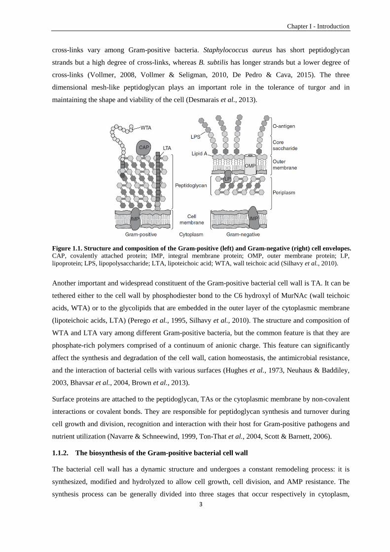
1.1. The bacterial cell envelope — the first defense system
The cell envelope is an essential and complex structure of the bacterial cell with sophisticated layers.
It is crucial for maintaining cell integrity, cell shape, surface properties, solute permeability, and self-
defense. It keeps the bacterial cell as a separate individual while also enabling bacterial
communication (Braun et al., 2014). The Gram-negative bacterial cell envelope has three layers
including the outer membrane, the peptidoglycan cell wall and the cytoplasmic membrane (Fig. 1.1).
The outer membrane plays an important role in separating the cell from toxic molecules and
stabilizing the cytoplasmic membrane. Compared to the Gram-negative bacteria, the Gram-positive
cell envelope has only two functional layers: the peptidoglycan cell wall and the cytoplasmic
membrane (Fig. 1.1). For Gram-positive bacteria, lacking the protective outer membrane necessitates a
peptidoglycan cell wall thicker and more complex than Gram-negative bacteria to tolerate the harsh
environmental challenges and support the cell membrane (Silhavy et al., 2010).
1.1.1. The composition of the Gram-positive bacterial cell wall
The cell wall of Gram-positive bacteria varies among different species, but can be described in general
as a three dimensional net-like structure comprised of many peptidoglycan layers, teichoic acids (TAs)
and surface proteins (Silhavy et al., 2010) (Fig. 1.1).
The peptidoglycan of Gram-positive bacteria is around 30-100 nm thick with up to 40 layers
consisting of glycan chains cross-linked by cell wall peptides, while the Gram-negative bacterial
peptidoglycan has only one to a few layers (Bertsche et al., 2014). Every glycan strand is made up of
repeating N-acetylglucosamine-(β1-4)-N-acetylmuramic acid (GlcNAc-MurNAc) disaccharide units.
The penta-peptide moiety with a common sequence L-Ala-D-Glu-DAA (dibasic amino acid)-D-Ala-D-
Ala is linked to the lactic acid of N-acetylmuramic acid via an amide bond with the first amino acid (L-
alanine). DAA is the dibasic amino acid that differs between bacteria. Most Gram-negative species, as
well as some Gram-positives such as Bacilli and Mycobacteria, use mDAP (meso-diaminopimelate),
while most Gram-positives use L-Lys (Scheffers & Pinho, 2005, Bertsche et al., 2014, Wheeler et al.,
2014). The glycan strands and the peptide stems together form the peptidoglycan chains, which are
connected by cross-bridges (Vollmer et al., 2008). The length of the peptidoglycan chains and the
Chapter I - Introduction
3
cross-links vary among Gram-positive bacteria. Staphylococcus aureus has short peptidoglycan
strands but a high degree of cross-links, whereas B. subtilis has longer strands but a lower degree of
cross-links (Vollmer, 2008, Vollmer & Seligman, 2010, De Pedro & Cava, 2015). The three
dimensional mesh-like peptidoglycan plays an important role in the tolerance of turgor and in
maintaining the shape and viability of the cell (Desmarais et al., 2013).
Figure 1.1. Structure and composition of the Gram-positive (left) and Gram-negative (right) cell envelopes. CAP, covalently attached protein; IMP, integral membrane protein; OMP, outer membrane protein; LP,
lipoprotein; LPS, lipopolysaccharide; LTA, lipoteichoic acid; WTA, wall teichoic acid (Silhavy et al., 2010).
Another important and widespread constituent of the Gram-positive bacterial cell wall is TA. It can be
tethered either to the cell wall by phosphodiester bond to the C6 hydroxyl of MurNAc (wall teichoic
acids, WTA) or to the glycolipids that are embedded in the outer layer of the cytoplasmic membrane
(lipoteichoic acids, LTA) (Perego et al., 1995, Silhavy et al., 2010). The structure and composition of
WTA and LTA vary among different Gram-positive bacteria, but the common feature is that they are
phosphate-rich polymers comprised of a continuum of anionic charge. This feature can significantly
affect the synthesis and degradation of the cell wall, cation homeostasis, the antimicrobial resistance,
and the interaction of bacterial cells with various surfaces (Hughes et al., 1973, Neuhaus & Baddiley,
2003, Bhavsar et al., 2004, Brown et al., 2013).
Surface proteins are attached to the peptidoglycan, TAs or the cytoplasmic membrane by non-covalent
interactions or covalent bonds. They are responsible for peptidoglycan synthesis and turnover during
cell growth and division, recognition and interaction with their host for Gram-positive pathogens and
nutrient utilization (Navarre & Schneewind, 1999, Ton-That et al., 2004, Scott & Barnett, 2006).
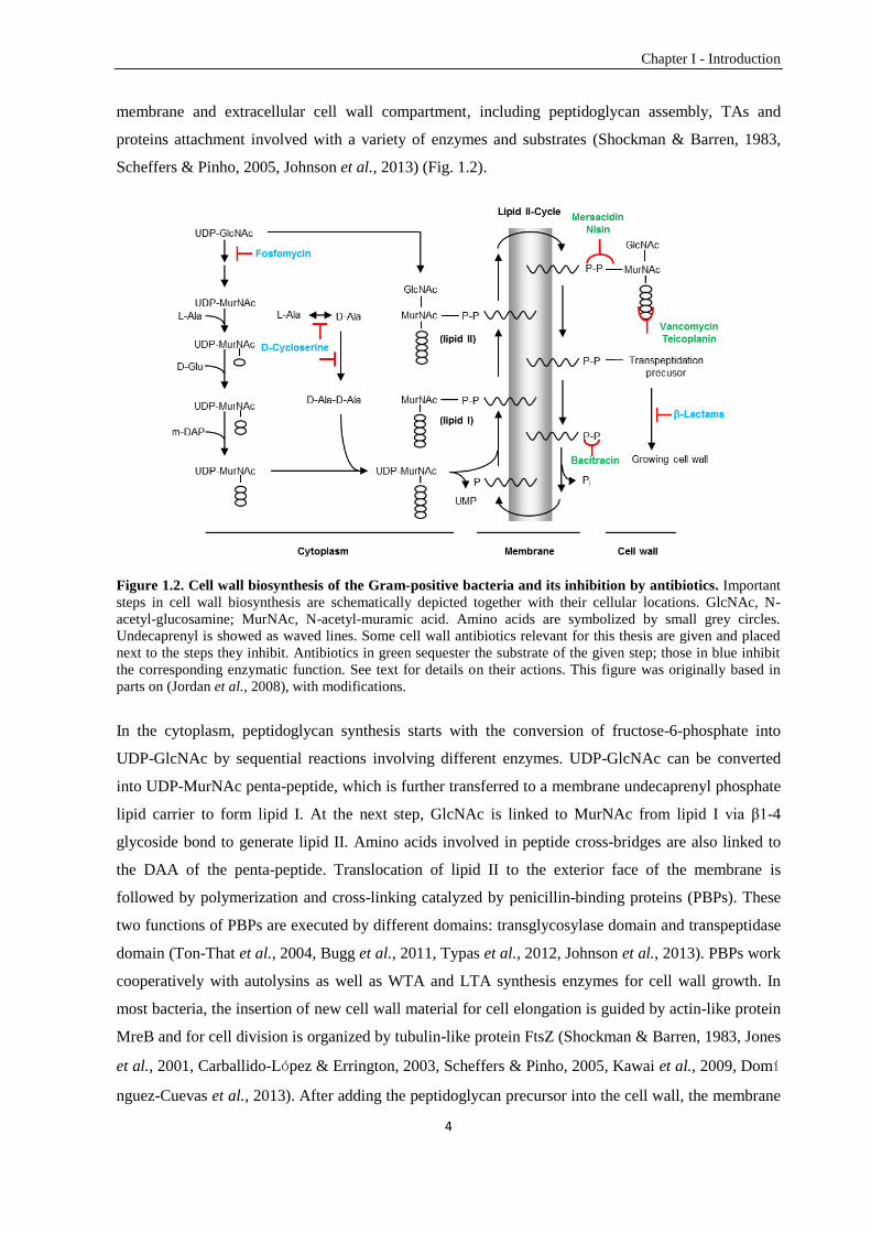
1.1.2. The biosynthesis of the Gram-positive bacterial cell wall
The bacterial cell wall has a dynamic structure and undergoes a constant remodeling process: it is
synthesized, modified and hydrolyzed to allow cell growth, cell division, and AMP resistance. The
synthesis process can be generally divided into three stages that occur respectively in cytoplasm,
Chapter I - Introduction
4
membrane and extracellular cell wall compartment, including peptidoglycan assembly, TAs and
proteins attachment involved with a variety of enzymes and substrates (Shockman & Barren, 1983,
Scheffers & Pinho, 2005, Johnson et al., 2013) (Fig. 1.2).
Figure 1.2. Cell wall biosynthesis of the Gram-positive bacteria and its inhibition by antibiotics. Important
steps in cell wall biosynthesis are schematically depicted together with their cellular locations. GlcNAc, N-
acetyl-glucosamine; MurNAc, N-acetyl-muramic acid. Amino acids are symbolized by small grey circles.
Undecaprenyl is showed as waved lines. Some cell wall antibiotics relevant for this thesis are given and placed
next to the steps they inhibit. Antibiotics in green sequester the substrate of the given step; those in blue inhibit
the corresponding enzymatic function. See text for details on their actions. This figure was originally based in
parts on (Jordan et al., 2008), with modifications.
In the cytoplasm, peptidoglycan synthesis starts with the conversion of fructose-6-phosphate into
UDP-GlcNAc by sequential reactions involving different enzymes. UDP-GlcNAc can be converted
into UDP-MurNAc penta-peptide, which is further transferred to a membrane undecaprenyl phosphate
lipid carrier to form lipid I. At the next step, GlcNAc is linked to MurNAc from lipid I via β1-4
glycoside bond to generate lipid II. Amino acids involved in peptide cross-bridges are also linked to
the DAA of the penta-peptide. Translocation of lipid II to the exterior face of the membrane is
followed by polymerization and cross-linking catalyzed by penicillin-binding proteins (PBPs). These
two functions of PBPs are executed by different domains: transglycosylase domain and transpeptidase
domain (Ton-That et al., 2004, Bugg et al., 2011, Typas et al., 2012, Johnson et al., 2013). PBPs work
cooperatively with autolysins as well as WTA and LTA synthesis enzymes for cell wall growth. In
most bacteria, the insertion of new cell wall material for cell elongation is guided by actin-like protein
MreB and for cell division is organized by tubulin-like protein FtsZ (Shockman & Barren, 1983, Jones
et al., 2001, Carballido-López & Errington, 2003, Scheffers & Pinho, 2005, Kawai et al., 2009, Domí
nguez-Cuevas et al., 2013). After adding the peptidoglycan precursor into the cell wall, the membrane
Chapter I - Introduction
5
lipid carrier remains in the pyrophosphate form and will further be dephosphorylated and flipped back
to the cytoplasmic side of the membrane for recycling. The steps of the cell wall biosynthesis linked to
the cytoplasmic membrane via undecaprenyl are referred to as the “Lipid II cycle”. The cell wall
synthesis process, especially the lipid II cycle, is the target of numerous AMPs.
1.2. Antimicrobial peptides — the inhibitors of bacterial cell wall synthesis
AMPs are secondary metabolites produced for self-defense by a variety of organisms like bacteria,
fungi, plants, insects, and animals. They are small (usually 6 to 100 amino acids) and usually
positively charged amphipathic molecules with different lengths, sequences, secondary structures, and
antimicrobial spectrum (Berdy, 2005, Nakatsuji & Gallo, 2012, Bahar & Ren, 2013). They can be
separated into four groups based on the secondary structure they mainly harbor: β-strands, α-helices,
loop structures, and extended structures (Davies & Webb, 1998, Lee et al., 2015). AMPs can be
synthesized either nonribosomally or ribosomally. Nonribosomally synthesized AMPs like bacitracin,
gramicidin, and glycopeptides are drastically modified and mainly produced by bacteria. They are
synthesized according to the multiple-carrier thiotemplate mechanism by a series of very large and
multifunctional peptide synthetases in an ordered fashion. Ribosomally synthesized AMPs can be
produced by a wide range of organisms as major defense molecules against microorganisms (Stein et
al., 1996, Hancock & Chapple, 1999, Papagianni, 2003). Lantibiotics, a large family of AMPs, are
ribosomally synthesized and post-translationally modified with unusual amino acids such as
lanthionine and methyllanthionine (McAuliffe et al., 2001, Chatterjee et al., 2005). One of the most
famous members is Nisin, a type A lantibiotic containing five lanthionine rings and three dehydrated
amino acids produced by Lactococcus lactis during stationary growth phase (Hsu et al., 2004).
The modes of action AMPs exert against bacteria include inhibition of the cell wall synthesis,
membrane dysfunction by channels/pores formation, and repression of intracellular functions like
DNA, RNA or proteins synthesis (Yeaman & Yount, 2003). Cell wall targeting AMPs implement their
functions either by disrupting the activity of enzymes involved in cell wall synthesis or isolating
substrates/precursors of corresponding enzymes (Jordan et al., 2008).
Examples of AMPs acting on bacterial cell wall are wide-ranging (Fig. 1.2). Fosfomycin and D-
cycloserine can target and hinder the cytoplasmic steps of the bacterial cell wall synthesis (Nikolaidis
et al., 2014). Most lantibiotics can target the lipid II and impede the cell wall synthesis (Breukink & de
Kruijff, 2006). Nisin has antimicrobial function against a wide range of Gram-positive bacteria and the
outgrowth of spores of Bacilli and Clostridia (Héchard & Sahl, 2002, de Arauz et al., 2009). It can
bind to lipid II and use it as an anchor molecule to further insert itself into the lipid bilayers. Thus it
presents a dual mode of antimicrobial activity causing inhibition of the cell wall biosynthesis and pore
formation on the membrane, which ultimately result in cell lysis (Nagao et al., 2006). Mersacidin, a
type B lantibiotic with a more globular structure, can complex lipid II and prevent the cell wall
Chapter I - Introduction
6
synthesis (Stein, 2005, Willey & van Der Donk, 2007). Lipid II is also the target of glycopeptides like
teicoplanin and vancomycin. They can inhibit polymerization and cross-linking by binding to the D-
Ala-D-Ala dipeptide terminus of the lipid II and block the cell wall synthesis, which eventually leads
to cell death (Marshall et al., 1998, Silver, 2003). Bacitracin, a branched cyclic nonribosomally
synthesized dodecylpeptide AMP mainly produced by Bacillus licheniformis and some strains of B.
subtilis, binds tightly to the undecaprenyl pyrophosphate and prevents its dephosphorylation and
recycling (Bernlohr & Novelli, 1963, Katz & Fisher, 1987, Azevedo et al., 1993, Konz et al., 1997).
1.3. Mechanisms of antimicrobial peptide resistance in Gram-positive bacteria
To survive in a competitive environment, bacteria have developed different strategies either via
spontaneous mutations or acquisition of additional genes to acquire AMP resistance. Some bacteria
can form biofilm to confer resistance (Otto, 2006). Resistance can also be achieved by synthesizing
proteases to degrade the AMPs (Sun et al., 2009). Resistance against cell wall acting AMPs can also
be mediated by reducing the access of the drugs to the cell envelope by changing the cell’s surface
charge — possible in both Gram-positive and Gram-negative bacteria. In Gram-positive bacteria, the
negative charge of the cell surface can be reduced by incorporating D-Ala to the highly negatively
charged TAs. This is accomplished by gene products of the dlt operon. Bacteria are more sensitive to
cationic AMPs if this operon is inactivated (Neuhaus & Baddiley, 2003, McBride & Sonenshein, 2011,
Reichmann et al., 2013). The reduced negative charge of TAs was postulated to diminish the
electrostatic attraction between the AMPs and the cell envelope (Peschel & Sahl, 2006). However, an
alternative model was proposed: the D-alanylation of TAs modifies the electrostatic interaction
between TAs themselves thereby making the cell envelope more compact and less permeable for
AMPs to reach their cell wall targets (Saar-Dover et al., 2012, Revilla-Guarinos et al., 2014).
Specific resistance against AMPs includes modifying their cell wall targets. In enterococci, resistance
against vancomycin is conferred by altering the binding target D-Ala-D-Ala on the C-terminal of lipid
II into D-Ala-D-Lac or D-Ala-D-Ser (Bugg et al., 1991). This switch leads to a reduced number of
hydrogen bonds from five to four between AMPs and their target — lipid II, which decreases the
binding affinity by 1000-fold (Bugg et al., 1991, Kahne et al., 2005). Two types of vancomycin
resistance were found in E. faecalis and will be described in detail in Section 1.5 (Walsh et al., 1996).
The most efficient mechanism against AMPs is mediated by the ATP-binding cassette (ABC)
transporters. These transporters usually contain one or two permease domains with variable number of
transmembrane (TM) helices, and each permease domain is associated with an ATPase (Gebhard,
2012). ATP hydrolysis provides energy for resistance against AMPs. Three different types of ABC
transporters, the LanFEG-type, the BceAB-type, and the BcrAB-type, have been found widespread in
Firmicutes bacteria for AMP resistance (Gebhard, 2012). The BcrAB-type and the LanFEG-type
transporters are mainly responsible for sensing and resistance against self-produced AMPs and most of
Chapter I - Introduction
7
them have a very narrow substrate range. Some of them were found to be associated with AMP
biosynthesis genes. For example, the ABC transporter BcrAB together with the undecaprenyl
pyrophosphate phosphatase BcrC are encoded in the bacitracin biosynthesis locus and confer self-
resistance in B. licheniformis (Podlesek et al., 1995); the NisFEG system in L. lactis is responsible for
mediating resistance to the self-produced nisin (Stein et al., 2003).
The BceAB-type transporters are hardly ever associated with AMP biosynthetic genes. The range of
resistance is quite broad including lantibiotics, cyclic AMPs like bacitracin, glycopeptides, and
peptides from the innate immune systems of higher organisms like defensins and cathelicidins
(summarized in (Gebhard & Mascher, 2011)). The permeases of these transporters have 10 TM helices
and a large extracellular loop between helices 7 and 8. TM helices 2 to 4 and TM helices 8 to 10 form
two FtsX-domains (Dintner et al., 2011, Dintner et al., 2014). Furthermore, this kind of transporters
are not only responsible for AMP resistance but also indispensable for AMP perception (Rietkötter et
al., 2008, Staroń et al., 2011). However, the molecular mechanisms of substrate detection, signaling
and resistance are not fully understood (more details see Section 1.7).
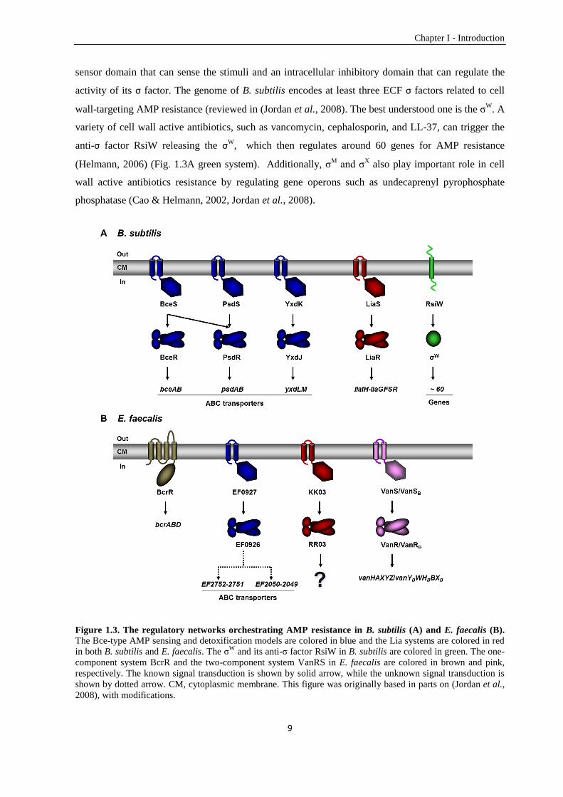
1.4. Regulatory network orchestrating antimicrobial peptide resistance in Bacillus
subtilis
B. subtilis is the best-characterized member of the Gram-positive bacteria and can be isolated from
diverse environments, e.g., soil, water source, and plant root surfaces. It is a rod-shaped bacterium that
can form highly resistant dormant endospores in response to nutrient limitation (Earl et al., 2008, van
Dijl & Hecker, 2013). The genome of B. subtilis contains around 4.2×10⁶ bp with 4,100 protein-
coding genes, and about 4-5% of the genome is devoted to antibiotics production (Kunst et al., 1997).
Antibiotics such as subtilosin, surfactin, bacilysin, lantibiotics including subtilin, ericin and mersacidin
have been reported to be synthesized ribosomally or nonribosomally by a wide array of B. subtilis
strains to inhibit competitors in the same environment (Stein, 2005). In addition to the ability of AMP
production, AMP resistance is also crucial and orchestrated by a complex regulatory network, which is
shown in Figure 1.3A. One of the specific and most efficient defense mechanisms is the Bce-type
ABC transporter that are mainly found in Firmicutes bacteria (Dintner et al., 2011). As mentioned
above, the Bce-type ABC transporter is responsible for both AMP perception and resistance. The
expression of the ABC transporter operon is regulated by a Bce-type two-component system (TCS),
which is comprised of a membrane-anchored histidine kinase (HK) and a cytoplasmic response
regulator (RR) (Joseph et al., 2002) (details of TCSs will be introduced in Section 1.6). The sensor
domain of the Bce-type HK harbors a short extracellular loop (<10 amino acids for most) between the
two transmembrane helices and is not able to detect the AMP (Mascher, 2006, Mascher, 2014). The
ABC transporter and TCS are genetically and functionally linked, and together they form the Bce-type
AMP sensing and detoxification module (Dintner et al., 2011). The signal transduction circuit starts
Chapter I - Introduction
8
when the ABC transporter detects the AMP and passes the signal to the TCS to activate the HK. The
phosphotransfer from the HK to the RR will in turn trigger the upregulation of the ABC transporter
operon for AMP resistance. The TCS operon is under the control of a constitutive promoter, while the
ABC transporter operon is expressed under the control of an AMP inducible, RR-dependent promoter
(Ohki et al., 2003, Staroń et al., 2011). The genome of B. subtilis encodes three such systems to cope
with the challenges from different kinds of AMPs (Joseph et al., 2002) (Fig. 1.3A blue systems). The
BceRS-BceAB system can sense and confer resistance against bacitracin, actagardine and mersacidin.
It has also been reported to respond to a fungal defensing plectasin (Staroń et al., 2011). The PsdRS-
PsdAB system shares the same inducer actagardine with the Bce system but cannot confer resistance
against it. Other antibiotics that can be detected and detoxified by the Psd system are nisin,
enduracidin, gallidermin and subtilin (Staroń et al., 2011). The only known inducer for the YxdJK-
YxdLM-YxeA system is a human neutrophil peptide, LL-37 (Pietiäinen et al., 2005). This system is
assumed to be involved in resistance against an unknown group of antibiotics. The gene locus harbors
an extra yxeA gene encoding a long peptide that is conserved in many Gram-positive bacteria. It might
be an immune protein participating in the proposed AMP resistance by interacting with and
neutralizing the antibiotic (Joseph et al., 2004).
B. subtilis also developed other response systems to counteract cell envelope damage caused by AMPs.
The LiaRS TCS, which is widespread in most Firmicutes bacteria, is a damage-sensing signal
transduction system (Wolf et al., 2012) (Fig. 1.3A red system). It can strongly respond to a wide range
of cell wall antibiotics, such as bacitracin, nisin, ramoplanin, and vancomycin (Mascher et al., 2004,
Pietiäinen et al., 2005, Hachmann et al., 2009). In the presence of a stimulus, the phosphorylated LiaR
can strongly induce the expression of the liaIH-liaGFSR operons. While in the absence of stimulus,
the transcription of the liaIH operon is switched off and the liaGFSR operon is under the control of a
weak constitutive promoter, PliaG (Jordan et al., 2006). The LiaRS TCS has a strong inhibitor, LiaF,
and deletion of liaF led to a constitutive active system in the absence of cell envelope stress (Jordan et
al., 2006). However, the functions of most gene products of the lia operon are not clearly known. The
LiaG is a putative membrane anchored hypothetical protein with unknown function. The LiaH is a
member of phage shock protein family, and it is homologous to the Escherichia coli phage shock
protein PspA, which suggests that the Lia system harbors a PspA-like response to maintain the
membrane integrity (Model et al., 1997, Darwin, 2005, Wolf et al., 2010). The LiaH is anchored to the
membrane by the small membrane protein LiaI (Domínguez-Escobar et al., 2014).
Another important signal transduction system that can regulate AMP resistance involves the
extracytoplasmic function (ECF) σ factors. They are small proteins containing only two of the four
conserved regions of the primary σ factor. Additionally, they are usually co-transcribed with
corresponding anti-σ factors (Heimann, 2002). The anti-σ factor often harbors an extracytoplasmic
Chapter I - Introduction
9
sensor domain that can sense the stimuli and an intracellular inhibitory domain that can regulate the
activity of its σ factor. The genome of B. subtilis encodes at least three ECF σ factors related to cell
wall-targeting AMP resistance (reviewed in (Jordan et al., 2008). The best understood one is the σW
. A
variety of cell wall active antibiotics, such as vancomycin, cephalosporin, and LL-37, can trigger the
anti-σ factor RsiW releasing the σW, which then regulates around 60 genes for AMP resistance
(Helmann, 2006) (Fig. 1.3A green system). Additionally, σM
and σX also play important role in cell
wall active antibiotics resistance by regulating gene operons such as undecaprenyl pyrophosphate
phosphatase (Cao & Helmann, 2002, Jordan et al., 2008).
Figure 1.3. The regulatory networks orchestrating AMP resistance in B. subtilis (A) and E. faecalis (B).
The Bce-type AMP sensing and detoxification models are colored in blue and the Lia systems are colored in red
in both B. subtilis and E. faecalis. The σW
and its anti-σ factor RsiW in B. subtilis are colored in green. The one-
component system BcrR and the two-component system VanRS in E. faecalis are colored in brown and pink,
respectively. The known signal transduction is shown by solid arrow, while the unknown signal transduction is
shown by dotted arrow. CM, cytoplasmic membrane. This figure was originally based in parts on (Jordan et al.,
2008), with modifications.
Chapter I - Introduction
10
1.5. Regulatory network orchestrating antimicrobial peptide resistance in
Enterococcus faecalis
E. faecalis, another low-GC Gram-positive bacterium, is a core member of the normal intestinal
microflora in humans and animals. It is mostly a harmless commensal, but opportunistically
pathogenic and can cause life-threatening infections especially in hospital settings. E. faecalis strain
V583, the first vancomycin resistant clinical isolate reported in the U.S., contains four DNA molecules:
the main chromosome (the size is 3.2×10⁶ bp, the G+C content is 37.5%) with a total of 3337
predicted protein-encoding open reading frames and three circular plasmids (Paulsen et al., 2003). In
addition to vancomycin, E. faecalis V583 can also resist to several antibiotics, such as bacitracin and
teicoplanin, which leads to the difficulty of clinical treatment (Sahm et al., 1989, McBride et al., 2007).
A deeper understanding of the AMP resistance network in E. faecalis will therefore provide useful
information for clinical research.
E. faecalis has high-level of bacitracin resistance, which is mediated by an ABC transporter BcrAB
(Manson et al., 2004). The bcrAB genes together with bcrD form the bcrABD operon. BcrD is
suggested to be able to increase the amount of undercaprenyl phosphate as an undercaprenyl
pyrophosphate phosphatase for bacitracin resistance. The expression of the bcrABD operon is
regulated by a constitutively transcribed one-component system, BcrR (Gauntlett et al., 2008) (Fig.
1.3B brown system). BcrR, a membrane-bound transcriptional regulator, can perceive bacitracin
directly and bind to PbcrA to induce the expression of the bcrABD operon for bacitracin resistance
(Gebhard et al., 2009).
Two BceAB-like ABC transporters: EF2050-EF2049 and EF2752-2751, and one BceRS-like TCS
EF0926-EF0927 were found in the genome of E. faecalis by comparative genomic analysis (Dintner et
al., 2011) (Fig. 1.3B blue system). However, neither of the ABC transporter operons was located
adjacent to the operon of the BceRS-like TCS. The functions of these two ABC transporters have not
been described so far. The functional analysis of these two ABC transporters and one TCS is described
in Chapter III.
A LiaR highly conserved ortholog in E. faecalis, RR03, was demonstrated to be up-regulated in
response to bacitracin and the RR03 mutant in E. faecalis showed increased bacitracin sensitivity
(Hancock & Perego, 2004). A RR03 ortholog from S. aureus, VraR, was demonstrated to play an
important role in cell wall-targeting antibiotics, which suggests a similar function of RR03 from E.
faecalis (Kuroda et al., 2003) (Fig. 1.3B red system).
Two major types of inducible glycopeptide resistance have been identified in E. faecalis, which were
demonstrated to be regulated by two TCSs — the VanRS (in VanA type E. faecalis) and the VanRBSB
(in VanB type E. faecalis) (Arthur et al., 1997, Arthur & Quintiliani, 2001) (Fig. 1.3B pink system).
Chapter I - Introduction
11
The VanA type strain has high level of resistance against both vancomycin and teicoplanin, which are
also the inducers. The resistance is mediated by products of vanHAXYZ operon, of which the
expression is regulated by VanRS TCS, by altering the binding target (D-Ala-D-Ala) on lipid II of
glycopeptide into D-Ala-D-Lac or D-Ala-D-Ser. VanH is a D-lactate dehydrogenase and can reduce
pyruvate to D-lactate. VanA, an ATP-dependent D-Ala-D-Lac ligase, is able to add D-lactate to D-Ala
and form D-Ala-D-Lac. The remaining D-Ala-D-Ala is then hydrolyzed by VanX (a D-Ala-D-Ala
dipeptidase) (Arthur et al., 1992, Marshall & Wright, 1998). The VanB type strain confers resistance
against vancomycin and teicoplanin, but it is only capable of vancomycin perception. The functions of
products of the vanYBWHBBXB operon are similar to the VanA type (Evers & Courvalin, 1996).
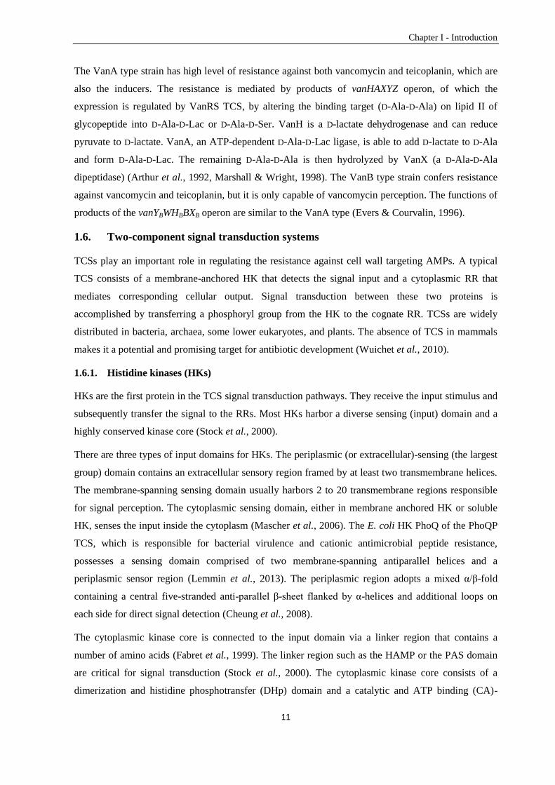
1.6. Two-component signal transduction systems
TCSs play an important role in regulating the resistance against cell wall targeting AMPs. A typical
TCS consists of a membrane-anchored HK that detects the signal input and a cytoplasmic RR that
mediates corresponding cellular output. Signal transduction between these two proteins is
accomplished by transferring a phosphoryl group from the HK to the cognate RR. TCSs are widely
distributed in bacteria, archaea, some lower eukaryotes, and plants. The absence of TCS in mammals
makes it a potential and promising target for antibiotic development (Wuichet et al., 2010).
1.6.1. Histidine kinases (HKs)
HKs are the first protein in the TCS signal transduction pathways. They receive the input stimulus and
subsequently transfer the signal to the RRs. Most HKs harbor a diverse sensing (input) domain and a
highly conserved kinase core (Stock et al., 2000).
There are three types of input domains for HKs. The periplasmic (or extracellular)-sensing (the largest
group) domain contains an extracellular sensory region framed by at least two transmembrane helices.
The membrane-spanning sensing domain usually harbors 2 to 20 transmembrane regions responsible
for signal perception. The cytoplasmic sensing domain, either in membrane anchored HK or soluble
HK, senses the input inside the cytoplasm (Mascher et al., 2006). The E. coli HK PhoQ of the PhoQP
TCS, which is responsible for bacterial virulence and cationic antimicrobial peptide resistance,
possesses a sensing domain comprised of two membrane-spanning antiparallel helices and a
periplasmic sensor region (Lemmin et al., 2013). The periplasmic region adopts a mixed α/β-fold
containing a central five-stranded anti-parallel β-sheet flanked by α-helices and additional loops on
each side for direct signal detection (Cheung et al., 2008).
The cytoplasmic kinase core is connected to the input domain via a linker region that contains a
number of amino acids (Fabret et al., 1999). The linker region such as the HAMP or the PAS domain
are critical for signal transduction (Stock et al., 2000). The cytoplasmic kinase core consists of a
dimerization and histidine phosphotransfer (DHp) domain and a catalytic and ATP binding (CA)-
Chapter I - Introduction
12
domain (Krell et al., 2010) (Fig. 1.4). The DHp domain with a long α-hairpin structure is responsible
for dimerization (Marina et al., 2005). HK catalyzes autophosphorylation on the conserved histidine
residue (located on the first α-helix) in the presence of ATP by the CA domain (West & Stock, 2001).
The phosphoryl group is subsequently transferred to the RR for mediation cellular response.
1.6.2. Response regulators (RRs)
Most RRs contain two domains: a conserved N-terminal receiver (regulatory) domain and a diverse C-
terminal output (effector) domain (Stock et al., 2000). A flexible linker joins the two domains together
(Fig. 1.4). The receiver domain has a modular secondary structure with alternating β-strands and α-
helices adopting a topology with a central five-stranded paralleled β-sheet surrounded by two α-helices
on one side and three on the other (Fig. 1.5) (Bourret, 2010). The highly conserved aspartate residue,
which is responsible for receiving the phosphoryl group from the histidine kinase, is located at the end
of the β3 strand (Lukat et al., 1991, Appleby & Bourret, 1998).
Bacterial RRs have a great variety of output domains to elicit the specific cellular response according
to the input obtained by the HK. They can be assigned into five groups by their functions: DNA-
binding, RNA-binding, ligand-binding, protein-binding, and enzyme (Galperin, 2010). A majority of
RR receiver domains are connected to a DNA-binding output domain and have the function of gene-
transcriptional regulation. The OmpR subfamily is the largest RR group possessing a winged helix-
turn-helix (wHTH) DNA binding output domain (Galperin, 2006). The secondary structure of the
OmpR output domain is β1-β2-β3-β4-α1-β5-α2-α3-β6-β7. The α2-loop-α3 builds up the helix-turn-
helix motif and the loop connecting β6 and β7 is referred as a wing. OmpR can bind to the region
upstream of the -35 element on promoters of two porin genes: ompF and ompC, and regulate the
transcription by interacting with the α subunit of RNA polymerase to adjust to changes in osmolarity
in E. coli (Slauch et al., 1988, Forst et al., 1989, Slauch et al., 1991).
Figure 1.4. Schematic overview of the two-component signal transduction paradigm and the domain
structure of each component. HK, histidine kinase. RR, response regulator. The name of each domain is given
below the corresponding domain structure. The signal transduction between HK and RR is represented as
transferring a phosphoryl group from the histidine residue (H) on the HK DHp domain to the aspartate residue
(D) on the RR receiver domain.
1.6.3. Phosphotransfer between histidine kinase and response regulator
Three phosphotransfer reactions and two phosphoprotein intermediates are involved in the basic two-
component signal transduction pathways. In the first step, the HK executes autophosphorylation of the
Chapter I - Introduction
13
histidine residue by the CA domain in the presence of ATP, creating phosphoramidate. In the second
step, the RR catalyzes the transfer of the phosphoryl group from phospho-His (HK) to Asp (RR),
resulting in a high-energy acyl phosphate. In the final step, the RR can also catalyze
dephosphorylation of phospho-Asp (RR) by transferring the phosphoryl group to a water molecule. A
divalent metal ion (usually Mg2+
in vivo) is required for every step (Stock et al., 2000). The
phosphotransfer between the HK and the RR is mediated by protein-protein interaction via the
cytoplasmic domain of the HK and the receiver domain of the RR (Casino et al., 2010).
Phosphorylation-mediated conformational change of the RR, especially the α4-β5-α5 face on the
receiver domain, passes the signal from the receiver domain to the output domain for further
regulation (Hoch & Silhavy, 1995, Gao et al., 2007, Bourret, 2010, Gao & Stock, 2010).
1.7. Signaling specificity of Bce-type two-component systems in Bacillus subtilis
Harboring numerous highly related TCSs in one genome, such as the three homologous Bce-like TCSs
in B. subtilis, increases the possibility of cross-talk, which can be deleterious. Direct-coupling analysis,
which is based on the co-evolution of inter-protein contact residues, previously predicted a
considerable potential for cross-talk among these three systems (Szurmant & Hoch, 2010, Procaccini
et al., 2011). Instead, a previous in vivo study showed that these systems are generally well insulated
from each other: Only some minor degree of cross-regulation was observed between BceS and PsdR
in the presence of high concentrations of bacitracin (Rietkötter et al., 2008) (Fig. 1.3A). This raises the
questions: How do bacteria simultaneously coordinate the activity of so many highly related signaling
systems to maintain the signal transduction specificity and prevent unwanted cross-talk? How does the
HK discriminate its cognate RR from the non-cognate ones in the pool of homologous RRs? How is
the RR able to discriminate the cognate promoter region from non-cognate ones?
1.7.1. Signaling specificity between the histidine kinase and the response regulator
Myriad mechanisms have been employed by bacterial cell to maintain the intrasystem signal
transduction fidelity and intersystem insulation. Specificity can be achieved by different cellular
localizations as well as by differentiation of temporal expression of different systems (Ubersax &
Ferrell Jr, 2007). At the phosphotransfer level, three mechanisms are applied to maintain the
specificity of TCS. Most HKs are bifunctional, that is, they exhibit both kinase and phosphatase
activities, and can thereby tightly control the activity of the cognate RR by preventing unspecific
phosphorylation through noncognate HKs or small phosphodonors (Boll & Hendrixson, 2011). The
competition between the cognate RR and noncognate ones can also avoid cross-talk (Laub & Goulian,
2007). The molecular recognition between cognate partners, which is the most important mechanism,
enables the HK and the RR of one system to interact specifically in order to avoid accidental
interactions with components from other systems (Podgornaia & Laub, 2013).
Chapter I - Introduction
14
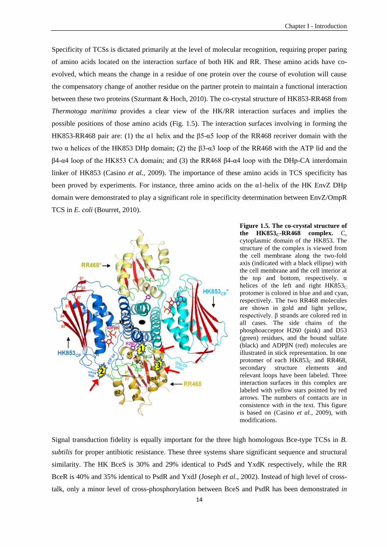
Specificity of TCSs is dictated primarily at the level of molecular recognition, requiring proper paring
of amino acids located on the interaction surface of both HK and RR. These amino acids have co-
evolved, which means the change in a residue of one protein over the course of evolution will cause
the compensatory change of another residue on the partner protein to maintain a functional interaction
between these two proteins (Szurmant & Hoch, 2010). The co-crystal structure of HK853-RR468 from
Thermotoga maritima provides a clear view of the HK/RR interaction surfaces and implies the
possible positions of those amino acids (Fig. 1.5). The interaction surfaces involving in forming the
HK853-RR468 pair are: (1) the α1 helix and the β5-α5 loop of the RR468 receiver domain with the
two α helices of the HK853 DHp domain; (2) the β3-α3 loop of the RR468 with the ATP lid and the
β4-α4 loop of the HK853 CA domain; and (3) the RR468 β4-α4 loop with the DHp-CA interdomain
linker of HK853 (Casino et al., 2009). The importance of these amino acids in TCS specificity has
been proved by experiments. For instance, three amino acids on the α1-helix of the HK EnvZ DHp
domain were demonstrated to play a significant role in specificity determination between EnvZ/OmpR
TCS in E. coli (Bourret, 2010).
Figure 1.5. The co-crystal structure of
the HK853C-RR468 complex. C,
cytoplasmic domain of the HK853. The
structure of the complex is viewed from
the cell membrane along the two-fold
axis (indicated with a black ellipse) with
the cell membrane and the cell interior at
the top and bottom, respectively. α
helices of the left and right HK853C
protomer is colored in blue and and cyan,
respectively. The two RR468 molecules
are shown in gold and light yellow,
respectively. β strands are colored red in
all cases. The side chains of the
phosphoacceptor H260 (pink) and D53
(green) residues, and the bound sulfate
(black) and ADPβN (red) molecules are
illustrated in stick representation. In one
protomer of each HK853C and RR468,
secondary structure elements and
relevant loops have been labeled. Three
interaction surfaces in this complex are
labeled with yellow stars pointed by red
arrows. The numbers of contacts are in
consistence with in the text. This figure
is based on (Casino et al., 2009), with
modifications.
Signal transduction fidelity is equally important for the three high homologous Bce-type TCSs in B.
subtilis for proper antibiotic resistance. These three systems share significant sequence and structural
similarity. The HK BceS is 30% and 29% identical to PsdS and YxdK respectively, while the RR
BceR is 40% and 35% identical to PsdR and YxdJ (Joseph et al., 2002). Instead of high level of cross-
talk, only a minor level of cross-phosphorylation between BceS and PsdR has been demonstrated in
Chapter I - Introduction
15
vivo at high concentrations of bacitracin (Rietkötter et al., 2008). However, the residues on BceRS and
PsdRS TCSs that dictate intrasystem specificity and minimize intersystem cross-talk remain unclear.
The nature and localization of these amino acids still needs to be unraveled. A first insight into this
question is provided by the data described in Chapter V.
1.7.2. Specificity on the response regulator transcriptional regulation level
In bacteria, transcription initiation starts with promoter recognition by the σ subunit of holo RNA
polymerase on the -35 promoter element followed by discerning and unwinding of the DNA double
helix at the -10 promoter element (Lee et al., 2012). For promoters lacking a -35 element or deviating
significantly from the consensus sequence at the appropriate position, the σ subunit can still be
recruited to the promoter by interaction with activators like RRs binding to the upstream region
(Jarmer et al., 2001, Paget & Helmann, 2003).
Specific interaction between a regulator and its target is important for bacteria to trigger the desired
response to the right stimulus, which is primarily determined via molecular recognition between amino
acids of the output domain and nucleotides within the RR binding site. The output domain structure of
OmpR indicates that the α3 helix (recognition helix on the output domain) is responsible for specific
interaction with the DNA major groove, and the β6-β7 loop (wing on the output domain) is
responsible for specific interaction with the DNA minor groove (Martínez-Hackert & Stock, 1997).
In B. subtilis, the transcription of the Bce-type ABC transporter genes is upregulated by binding of the
Bce-like RRs to the promoter. BceR and PsdR belong to the OmpR subfamily (Fabret et al., 1999)
with a winged helix-turn-helix output domain. The known binding sites on PbceA and PpsdA have eleven
out of fourteen identical base pairs. This indicates a considerable potential of cross-regulation at the
RR/promoter level between these two systems. In vivo, however, the regulation is highly specific
between BceR/PbceA and PsdR/PpsdA. This raises the question of how Bce-like RRs specifically regulate
the transcription of the cognate ABC transporters. A clear understanding of the specificity
determinants on bceA and psdA promoters that determine exclusive binding of BceR and PsdR,
respectively, is currently lacking. This question is addressed comprehensively in Chapter IV.
Chapter I - Introduction
16
1.8. Aims of this thesis
This thesis aimed to investigate the cell wall-targeting AMP sensing and resistance modules in two
Firmicutes bacteria: E. faecalis and B. subtilis. We aimed at gaining a deeper understanding on the
signal transduction mechanisms and the determinants of wiring specificity of the underling TCSs-
dependent regulation.
Chapter II
The technical challenges of molecular genetic studies in E. faecalis hinder a deeper understanding of
the molecular mechanism in antibiotic detection, signal transduction, and gene regulation. The
genetically highly tractable Gram-positive model organism B. subtilis on the other hand might be a
suitable candidate as a heterologous host. In this chapter, two fundamentally different regulators of E.
faecalis, the bacitracin sensor BcrR and the vancomycin-sensing two component system VanSB-
VanRB, were introduced into B. subtilis and their functions were monitored using target promoters
fused to reporter genes (lacZ and luxABCDE). We explored and validated B. subtilis as a platform for
studying the regulatory mechanisms of cell wall antibiotic resistance of E. faecalis.
Chapter III
In this chapter, the established B. subtilis platform was subsequently used for an in-depth heterologous
functional analysis of two Bce-type ABC transporters and one Bce-type TCS of E. faecalis. Combined
with studies in the native host, we analyzed the bacitracin sensing and resistance network of E.
faecalis.
Chapter IV
Both the output domains of BceR and PsdR as well as their known binding sites are highly
homologous in B. subtilis. The aim of this chapter was to gain a full comprehension of the mechanism
that dictates specific binding of RR to its cognate promoter (BceR-PbceA, PsdR-PpsdA). In vivo
experiments were used to first dissect the promoter and later identify the specificity dictating elements.
In vitro assays were then performed to further corroborate the specificity determining mechanism.
Chapter V
Due to the high sequence and structure similarity of BceRS and PsdRS TCSs in B. subtilis, the
question of what determines signal transduction specificity between a HK and its cognate RR was
raised. In this chapter, different regions on the receiver domain were exchanged between BceR and
PsdR to rewire the signal transduction in vivo and thereby identify the specificity determinants for
Bce-type TCSs.
17
Chapter II
Bacillus subtilis as a Platform for Molecular Characterisation of
Regulatory Mechanisms of Enterococcus faecalis Resistance
against Cell Wall Antibiotics
Chong Fang, Emanuel Stiegeler, Gregory M. Cook, Thorsten Mascher, Susanne Gebhard
PLOS ONE, March 2014, Volume 9, Issue 3, e93169
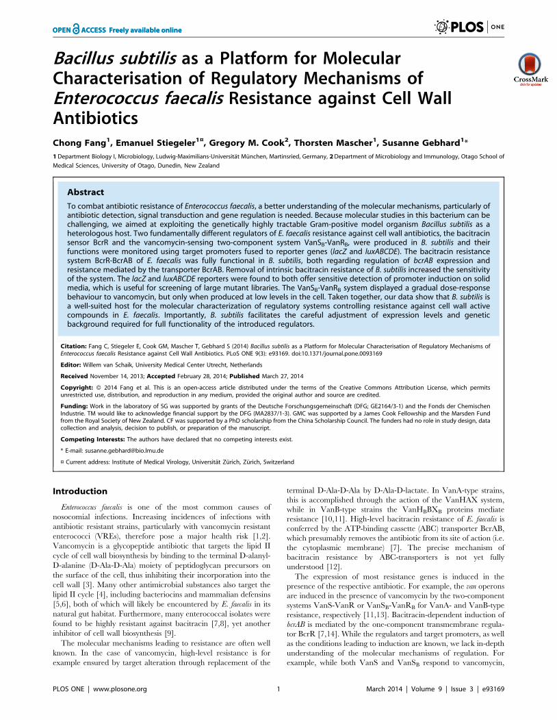
Bacillus subtilis as a Platform for MolecularCharacterisation of Regulatory Mechanisms ofEnterococcus faecalis Resistance against Cell WallAntibioticsChong Fang1, Emanuel Stiegeler1¤, Gregory M. Cook2, Thorsten Mascher1, Susanne Gebhard1*
1 Department Biology I, Microbiology, Ludwig-Maximilians-Universitat Munchen, Martinsried, Germany, 2 Department of Microbiology and Immunology, Otago School of
Medical Sciences, University of Otago, Dunedin, New Zealand
Abstract
To combat antibiotic resistance of Enterococcus faecalis, a better understanding of the molecular mechanisms, particularly ofantibiotic detection, signal transduction and gene regulation is needed. Because molecular studies in this bacterium can bechallenging, we aimed at exploiting the genetically highly tractable Gram-positive model organism Bacillus subtilis as aheterologous host. Two fundamentally different regulators of E. faecalis resistance against cell wall antibiotics, the bacitracinsensor BcrR and the vancomycin-sensing two-component system VanSB-VanRB, were produced in B. subtilis and theirfunctions were monitored using target promoters fused to reporter genes (lacZ and luxABCDE). The bacitracin resistancesystem BcrR-BcrAB of E. faecalis was fully functional in B. subtilis, both regarding regulation of bcrAB expression andresistance mediated by the transporter BcrAB. Removal of intrinsic bacitracin resistance of B. subtilis increased the sensitivityof the system. The lacZ and luxABCDE reporters were found to both offer sensitive detection of promoter induction on solidmedia, which is useful for screening of large mutant libraries. The VanSB-VanRB system displayed a gradual dose-responsebehaviour to vancomycin, but only when produced at low levels in the cell. Taken together, our data show that B. subtilis isa well-suited host for the molecular characterization of regulatory systems controlling resistance against cell wall activecompounds in E. faecalis. Importantly, B. subtilis facilitates the careful adjustment of expression levels and geneticbackground required for full functionality of the introduced regulators.
Citation: Fang C, Stiegeler E, Cook GM, Mascher T, Gebhard S (2014) Bacillus subtilis as a Platform for Molecular Characterisation of Regulatory Mechanisms ofEnterococcus faecalis Resistance against Cell Wall Antibiotics. PLoS ONE 9(3): e93169. doi:10.1371/journal.pone.0093169
Editor: Willem van Schaik, University Medical Center Utrecht, Netherlands
Received November 14, 2013; Accepted February 28, 2014; Published March 27, 2014
Copyright: � 2014 Fang et al. This is an open-access article distributed under the terms of the Creative Commons Attribution License, which permitsunrestricted use, distribution, and reproduction in any medium, provided the original author and source are credited.
Funding: Work in the laboratory of SG was supported by grants of the Deutsche Forschungsgemeinschaft (DFG; GE2164/3-1) and the Fonds der ChemischenIndustrie. TM would like to acknowledge financial support by the DFG (MA2837/1-3). GMC was supported by a James Cook Fellowship and the Marsden Fundfrom the Royal Society of New Zealand. CF was supported by a PhD scholarship from the China Scholarship Council. The funders had no role in study design, datacollection and analysis, decision to publish, or preparation of the manuscript.
Competing Interests: The authors have declared that no competing interests exist.
* E-mail: [email protected]
¤ Current address: Institute of Medical Virology, Universitat Zurich, Zurich, Switzerland
Introduction
Enterococcus faecalis is one of the most common causes of
nosocomial infections. Increasing incidences of infections with
antibiotic resistant strains, particularly with vancomycin resistant
enterococci (VREs), therefore pose a major health risk [1,2].
Vancomycin is a glycopeptide antibiotic that targets the lipid II
cycle of cell wall biosynthesis by binding to the terminal D-alanyl-
D-alanine (D-Ala-D-Ala) moiety of peptidoglycan precursors on
the surface of the cell, thus inhibiting their incorporation into the
cell wall [3]. Many other antimicrobial substances also target the
lipid II cycle [4], including bacteriocins and mammalian defensins
[5,6], both of which will likely be encountered by E. faecalis in its
natural gut habitat. Furthermore, many enterococcal isolates were
found to be highly resistant against bacitracin [7,8], yet another
inhibitor of cell wall biosynthesis [9].
The molecular mechanisms leading to resistance are often well
known. In the case of vancomycin, high-level resistance is for
example ensured by target alteration through replacement of the
terminal D-Ala-D-Ala by D-Ala-D-lactate. In VanA-type strains,
this is accomplished through the action of the VanHAX system,
while in VanB-type strains the VanHBBXB proteins mediate
resistance [10,11]. High-level bacitracin resistance of E. faecalis is
conferred by the ATP-binding cassette (ABC) transporter BcrAB,
which presumably removes the antibiotic from its site of action (i.e.
the cytoplasmic membrane) [7]. The precise mechanism of
bacitracin resistance by ABC-transporters is not yet fully
understood [12].
The expression of most resistance genes is induced in the
presence of the respective antibiotic. For example, the van operons
are induced in the presence of vancomycin by the two-component
systems VanS-VanR or VanSB-VanRB for VanA- and VanB-type
resistance, respectively [11,13]. Bacitracin-dependent induction of
bcrAB is mediated by the one-component transmembrane regula-
tor BcrR [7,14]. While the regulators and target promoters, as well
as the conditions leading to induction are known, we lack in-depth
understanding of the molecular mechanisms of regulation. For
example, while both VanS and VanSB respond to vancomycin,
PLOS ONE | www.plosone.org 1 March 2014 | Volume 9 | Issue 3 | e93169
their sensory domains differ considerably in size with 37 amino
acids for VanS and 103 residues for VanSB, and share only low
sequence similarity [15]. It is therefore difficult to envisage the
same sensing mechanism for both proteins. It is similarly unclear
how BcrR detects bacitracin, because the protein lacks any
obvious extracellular domains but is nevertheless able to directly
interact with its substrate [14,16]. Additionally, it is not known
how a membrane-bound transcriptional regulator like BcrR
activates transcription from its target promoter. While a direct
interaction with RNA-polymerase has been proposed [16],
experimental evidence is lacking to date.
Sensory perception of antimicrobial substances by bacteria is a
first and essential step in antibiotic resistance, and a thorough
understanding of the mechanisms involved would provide an
important basis for the development of new drugs to combat
resistance. However, in many genera, e.g. the enterococci,
investigations are hampered by the difficulty to manipulate these
bacteria genetically. Although more and more genetic tools are
becoming available for enterococci, poor transformability of many
strains, including clinical isolates, still impedes studies involving,
for example, high-throughput or detailed mutagenic approaches.
To circumvent these problems, heterologous hosts have been
chosen, often using E. coli [17], or electro-transformable laboratory
strains of E. faecalis [7,14]. The latter provide improved
transformability, but no additional genetic tools, while the former
host does not appear well suited to study resistance against cell wall
active compounds, due to the major differences between the
Gram-positive and Gram-negative cell envelope. Alternatively,
Bacillus subtilis has been used successfully for the functional
expression of the VanS-VanR two-component system of E. faecalis,
as well as of the VanB-type resistance proteins [1,18]. Like E. coli,
B. subtilis is easy to manipulate and a large number of genetic tools
are available. The G+C contents of B. subtilis (43.5%) and of E.
faecalis (37.5%) are comparable, which is of great advantage for
heterologous gene expression. Furthermore, the transcription
machinery in both organisms is sufficiently similar to facilitate
the interaction of heterologous transcriptional regulators with the
native machinery, as has been shown in vitro for activation of B.
subtilis RNA polymerase by E. faecalis BcrR [16]. Importantly for
the present application, the intrinsic resistance mechanisms of B.
subtilis against cell wall antibiotics are well understood [19,20],
allowing directed deletion of genes to create a clean genetic
background.
In the present study, we have used two well-understood
examples from E. faecalis to develop and validate B. subtilis as a
platform for studying the regulatory mechanisms leading to
resistance against cell wall-active antibiotics. To test the feasibility
of our approach and determine the optimal genetic background of
the host, we chose the one-component regulator BcrR and could
show full functionality with highly similar behaviour to its native
context. This set-up was then applied to the VanSB-VanRB two-
component system. A previous attempt at heterologous expression
of this system in B. subtilis had resulted in a constitutively active
behaviour [18]. Optimization of expression levels and growth
conditions now resulted in vancomycin-dependent induction of the
target promoter, further supporting the suitability of B. subtilis as
host organism.
Materials and Methods
Bacterial strains and growth conditionsAll strains used in this study are listed in Table 1. E. coli
DH5aand XL1-blue were used for cloning. E. coli and B. subtilis
were grown routinely in Luria-Bertani (LB) medium at 37uC with
agitation (200 rpm). B. subtilis was transformed by natural
competence as previously described [21]. Selective media
contained ampicillin (100 mg ml21 for E. coli), chloramphenicol
(5 mg ml21 for B. subtilis), kanamycin (10 mg ml21 for B. subtilis),
erythromycin 1 mg ml21 with lincomycin 25 mg ml21 (for
macrolide-lincosamide-streptogramin B (mls) resistance in B.
subtilis) or spectinomycin (100 mg ml21 for B. subtilis). Bacitracin
was supplied as the Zn2+-salt. Unless otherwise stated, media for
strains carrying pXT-derived constructs contained 0.2% (w/v)
xylose for target gene expression. Solid media contained 1.5% (w/
v) agar. Growth was measured as optical density at 600 nm
wavelength (OD600).
Construction of plasmids and genetic techniquesAll primer sequences used for this study are listed in Table 2; all
plasmid constructs are listed in Table 1.
Transcriptional promoter fusions of PbcrA to lacZ or bacterial
luciferase (luxABCDE) were constructed in vectors pAC6 [22] or
pAH328 [23] by the sites of EcoRI/BamHI and EcoRI/SpeI,
respectively, obtaining plasmids pES601and pNTlux101, respec-
tively. The transcriptional promoter fusion of PvanYB to bacterial
luciferase was cloned into the EcoRI and SpeI sites of vector
pAH328 creating plasmid pCF133. The exact regions contained
in the constructs are given in Table 1.
For heterologous, xylose-inducible expression of bcrR or bcrR-
bcrAB in B. subtilis (pES701 and pES702) the respective DNA
fragments were amplified from the plasmid pAMbcr1 [7] and
cloned in the vector pXT [24] using the BamHI and EcoRI
restriction sites, placing the genes under the control of the vector’s
xylA-promoter. Plasmid pCF132 was constructed by inserting
vanRBSB from E. faecalis V583 into the BamHI and HindIII sites of
vector pXT for heterologous, xylose-inducible expression in B.
subtilis.
Constructs for unmarked gene deletions in B. subtilis were
cloned into the vector pMAD [25]. For each operon to be deleted,
800–1000 bp fragments located immediately before the start
codon of the first gene (‘‘up’’ fragment) and after the stop codon of
the last gene (‘‘down’’ fragment) were amplified. The primers were
designed to create a 17–20 bp overlap between the PCR-products
(Table 2), facilitating fusion of the fragments by PCR overlap
extension and subsequent cloning into pMAD. Gene deletions
were performed as previously described [25].
All constructs were checked for PCR-fidelity by sequencing, and
all created strains were verified by PCR using appropriate primers.
Antimicrobial susceptibility assaysAll cultures were grown in Mueller-Hinton (MH) medium for
antibiotic susceptibility assays [26]. Minimal inhibitory concen-
tration (MIC) of bacitracin and vancomycin were determined by
broth-dilution assays. Freshly grown overnight cultures of B. subtilis
in MH medium were used as inoculum at a dilution of 1:500. After
24 h incubation in the presence of two-fold serial dilutions of the
antibiotic the MIC was scored as the lowest concentration where
no growth was observed.
b-Galactosidase assaysCells were inoculated from fresh overnight cultures and grown
in LB medium at 37uC with aeration until they reached an OD600
between 0.4 and 0.5. The cultures were split into 2 mL aliquots
and challenged with different concentrations of bacitracin with
one aliquot left untreated. After incubation for an additional
30 min at 37uC with aeration, the cultures were harvested and the
cell pellets were frozen at 220uC. b-galactosidase activities were
determined as described, with normalization to cell density [27].
B. subtilis as Host for E. faecalis Systems
PLOS ONE | www.plosone.org 2 March 2014 | Volume 9 | Issue 3 | e93169
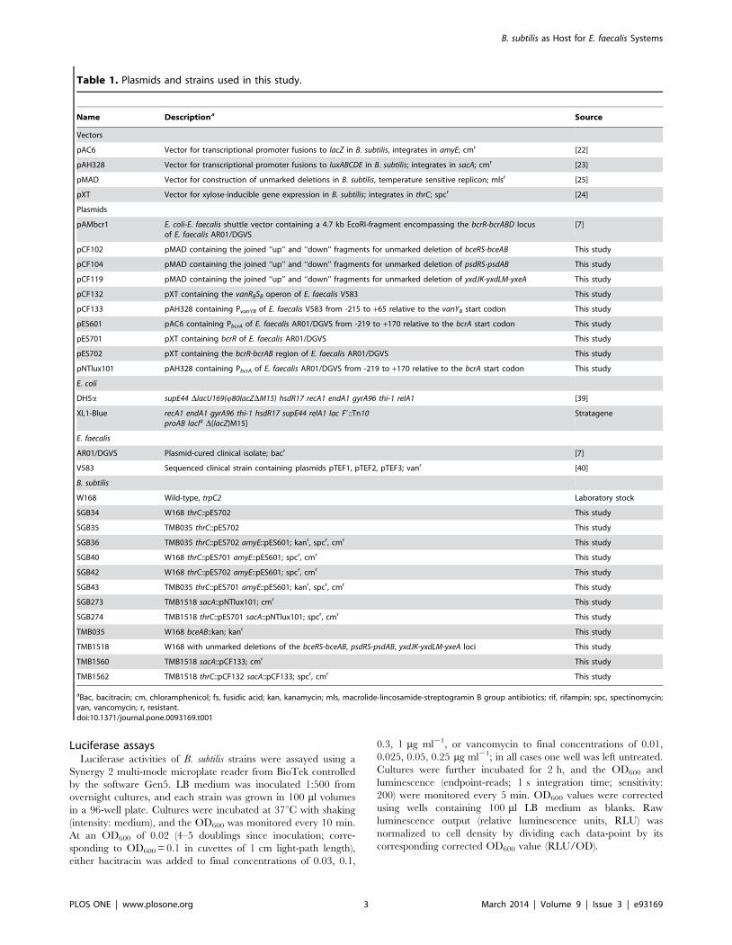
Luciferase assaysLuciferase activities of B. subtilis strains were assayed using a
Synergy 2 multi-mode microplate reader from BioTek controlled
by the software Gen5. LB medium was inoculated 1:500 from
overnight cultures, and each strain was grown in 100 ml volumes
in a 96-well plate. Cultures were incubated at 37uC with shaking
(intensity: medium), and the OD600 was monitored every 10 min.
At an OD600 of 0.02 (4–5 doublings since inoculation; corre-
sponding to OD600 = 0.1 in cuvettes of 1 cm light-path length),
either bacitracin was added to final concentrations of 0.03, 0.1,
0.3, 1 mg ml21, or vancomycin to final concentrations of 0.01,
0.025, 0.05, 0.25 mg ml21; in all cases one well was left untreated.
Cultures were further incubated for 2 h, and the OD600 and
luminescence (endpoint-reads; 1 s integration time; sensitivity:
200) were monitored every 5 min. OD600 values were corrected
using wells containing 100 ml LB medium as blanks. Raw
luminescence output (relative luminescence units, RLU) was
normalized to cell density by dividing each data-point by its
corresponding corrected OD600 value (RLU/OD).
Table 1. Plasmids and strains used in this study.
Name Descriptiona Source
Vectors
pAC6 Vector for transcriptional promoter fusions to lacZ in B. subtilis, integrates in amyE; cmr [22]
pAH328 Vector for transcriptional promoter fusions to luxABCDE in B. subtilis; integrates in sacA; cmr [23]
pMAD Vector for construction of unmarked deletions in B. subtilis, temperature sensitive replicon; mlsr [25]
pXT Vector for xylose-inducible gene expression in B. subtilis; integrates in thrC; spcr [24]
Plasmids
pAMbcr1 E. coli-E. faecalis shuttle vector containing a 4.7 kb EcoRI-fragment encompassing the bcrR-bcrABD locusof E. faecalis AR01/DGVS
[7]
pCF102 pMAD containing the joined ‘‘up’’ and ‘‘down’’ fragments for unmarked deletion of bceRS-bceAB This study
pCF104 pMAD containing the joined ‘‘up’’ and ‘‘down’’ fragments for unmarked deletion of psdRS-psdAB This study
pCF119 pMAD containing the joined ‘‘up’’ and ‘‘down’’ fragments for unmarked deletion of yxdJK-yxdLM-yxeA This study
pCF132 pXT containing the vanRBSB operon of E. faecalis V583 This study
pCF133 pAH328 containing PvanYB of E. faecalis V583 from -215 to +65 relative to the vanYB start codon This study
pES601 pAC6 containing PbcrA of E. faecalis AR01/DGVS from -219 to +170 relative to the bcrA start codon This study
pES701 pXT containing bcrR of E. faecalis AR01/DGVS This study
pES702 pXT containing the bcrR-bcrAB region of E. faecalis AR01/DGVS This study
pNTlux101 pAH328 containing PbcrA of E. faecalis AR01/DGVS from -219 to +170 relative to the bcrA start codon This study
E. coli
DH5a supE44 DlacU169(Q80lacZDM15) hsdR17 recA1 endA1 gyrA96 thi-1 relA1 [39]
XL1-Blue recA1 endA1 gyrA96 thi-1 hsdR17 supE44 relA1 lac F9::Tn10proAB lacIq D(lacZ)M15]
Stratagene
E. faecalis
AR01/DGVS Plasmid-cured clinical isolate; bacr [7]
V583 Sequenced clinical strain containing plasmids pTEF1, pTEF2, pTEF3; vanr [40]
B. subtilis
W168 Wild-type, trpC2 Laboratory stock
SGB34 W168 thrC::pES702 This study
SGB35 TMB035 thrC::pES702 This study
SGB36 TMB035 thrC::pES702 amyE::pES601; kanr, spcr, cmr This study
SGB40 W168 thrC::pES701 amyE::pES601; spcr, cmr This study
SGB42 W168 thrC::pES702 amyE::pES601; spcr, cmr This study
SGB43 TMB035 thrC::pES701 amyE::pES601; kanr, spcr, cmr This study
SGB273 TMB1518 sacA::pNTlux101; cmr This study
SGB274 TMB1518 thrC::pES701 sacA::pNTlux101; spcr, cmr This study
TMB035 W168 bceAB::kan; kanr This study
TMB1518 W168 with unmarked deletions of the bceRS-bceAB, psdRS-psdAB, yxdJK-yxdLM-yxeA loci This study
TMB1560 TMB1518 sacA::pCF133; cmr This study
TMB1562 TMB1518 thrC::pCF132 sacA::pCF133; spcr, cmr This study
aBac, bacitracin; cm, chloramphenicol; fs, fusidic acid; kan, kanamycin; mls, macrolide-lincosamide-streptogramin B group antibiotics; rif, rifampin; spc, spectinomycin;van, vancomycin; r, resistant.doi:10.1371/journal.pone.0093169.t001
B. subtilis as Host for E. faecalis Systems
PLOS ONE | www.plosone.org 3 March 2014 | Volume 9 | Issue 3 | e93169
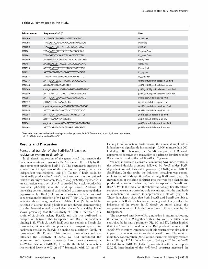
Results and Discussion
Functional transfer of the BcrR-BcrAB bacitracinresistance system to B. subtilis
In E. faecalis, expression of the genes bcrAB that encode the
bacitracin resistance transporter BcrAB is controlled solely by the
one-component regulator BcrR [14]. This regulator is encoded by
a gene directly upstream of the transporter operon, but as an
independent transcriptional unit [7]. To test if BcrR could be
functionally produced in B. subtilis, we introduced a transcriptional
fusion of its target promoter, PbcrA, to lacZ (pES601), together with
an expression construct of bcrR controlled by a xylose-inducible
promoter (pES701), into the wild-type strain. Addition of
increasing concentrations of bacitracin led to a strong upregulation
(approximately 80-fold) of promoter activities with a threshold
concentration for induction of 0.3 mg ml21 (Fig. 1A). No promoter
activities above background (ca. 1 Miller Unit (MU)) could be
detected in a strain lacking BcrR (data not shown), demonstrating
that the observed induction was indeed due to BcrR activity. It was
shown previously that the sensitivity of BcrR is increased in a
strain of E. faecalis lacking BcrAB, and this was attributed to
competition between the transporter and BcrR in bacitracin
binding [14]. While B. subtilis itself does not contain a BcrAB-like
transporter, it nevertheless possesses a transport system for
bacitracin resistance, BceAB, belonging to a different family of
transporters [28]. To test if this unrelated transporter could also
influence the sensitivity of BcrR, we next introduced the
expression and reporter constructs into a strain carrying a
bceAB::kan deletion (TMB035). Here, the threshold for induction
was ten-fold lower at 0.03 mg ml21 bacitracin, with 0.1 mg ml21
leading to full induction. Furthermore, the maximal amplitude of
induction was significantly increased (p = 0.006) to more than 200-
fold (Fig. 1B). Therefore, the BceAB transporter of B. subtilis
appeared to decrease the availability of bacitracin for detection by
BcrR, similar to the effect of BcrAB in E. faecalis.
We next introduced a construct containing bcrR under control of
the xylose-inducible promoter followed by bcrAB under BcrR-
dependent control of its native promoter (pES702) into TMB035
(bceAB::kan). In this strain, the induction behaviour was compa-
rable to that of wild-type B. subtilis carrying BcrR alone (Fig. 1C).
Introduction of the same construct into the wild-type background
produced a strain harbouring both transporters, BceAB and
BcrAB. While the induction threshold was not significantly altered
compared to strains possessing only one transporter, the amplitude
of induction was lowered to approximately 50-fold (Fig. 1D).
These data clearly show that both BceAB and BcrAB are able to
compete with BcrR for bacitracin binding and closely reflect the
behaviour of the system in E. faecalis. As stated above, this
competition is most likely due to removal of bacitracin by the
transporters.
The decreased sensitivity of PbcrA induction in strains harbouring
the construct of bcrR together with bcrAB, with the latter being
controlled by its native promoter (Fig. 1C and D), further implied
that bcrAB was expressed in a BcrR-dependent manner in B.
subtilis. We therefore wanted to test if this construct was also able to
impart bacitracin resistance to the B. subtilis host. The minimal
inhibitory concentration (MIC) of bacitracin was strongly reduced
from 128 mg ml21 in the wild-type to 2–4 mg ml21 in the bceAB-
deleted strain TMB035 (Table 3), consistent with earlier reports
[20,29]. Introduction of the bcrR-bcrAB construct increased the
Table 2. Primers used in this study.
Primer name Sequence (59-39)a Use
TM1569 AGTGGATCCTAGGAACGTTTTTACCAAC bcrAB rev
TM1798 TTAAGGATCCGAAAAACCCGTTGATGGACG bcrR fwd
TM1800 TTAAGAATTCTTTTATTTCATTCCCATCTGC bcrR rev
TM1801 TTAAGAATTCTTTTGCTGTTAATCGGCAAG PbcrA-lacZ fwd
TM1802 TTAAGGATCCCAAGCTGCAACATCATTTTC PbcrA-lacZ rev
TM2450 AAATTGGATCCGGAAACTACAGACTGTTATG vanRB fwd
TM2451 AAATTAAGCTTTATACCTGTCGGTCAAAATC vanSB rev
TM2550 AATTTGAATTCTTTGTTCTGGCTGGATTTAC PvanYB fwd
TM2551 AATTTACTAGTTCCCCAGATTGTTTCATATG PvanYB rev
TM2813 TTAAACTAGTCAAGCTGCAACATCATTTTC PbcrA-lux rev
TM2347 AATTTGGATCCAGTTTAATATCAACGGCCTG yxdJK-yxdLM-yxeA deletion up fwd
TM2348 AGGTAATTCTGCAATAGTCC yxdJK-yxdLM-yxeA deletion up rev
TM2349 ctattgcagaattacctGGAAGAAGTCAAGTTTGAAG yxdJK-yxdLM-yxeA deletion down fwd
TM2350 AATTTGGATCCTTCTGCTTCCGAAAAAACAG yxdJK-yxdLM-yxeA deletion down rev
TM2351 AATTTGGATCCGAGGAAGCAAAAGGAAATC bceRS-bceAB deletion up fwd
TM2352 CTTGATTTCATGAAACAGCG bceRS-bceAB deletion up rev
TM2355 ctgtttcatgaaatcaagATGGTGTTATATACTGCGC bceRS-bceAB deletion down fwd
TM2356 AATTCCATGGACGAATCCAGTTATCATAGC bceRS-bceAB deletion down rev
TM2357 AATTTGGATCCCTACGATCTAAATGGTTTCC psdRS-psdAB deletion up fwd
TM2358 ATTTTTGAAGATGACCGCCC psdRS-psdAB deletion up rev
TM2361 cggtcatcttcaaaaatGTCATATTTATAAGCGTGCTG psdRS-psdAB deletion down fwd
TM2362 AATTCCATGGAGAGATTGAAGCATTCATCG psdRS-psdAB deletion down rev
aRestriction sites are underlined; overlaps to other primers for PCR fusions are shown by lower case letters.doi:10.1371/journal.pone.0093169.t002
B. subtilis as Host for E. faecalis Systems
PLOS ONE | www.plosone.org 4 March 2014 | Volume 9 | Issue 3 | e93169
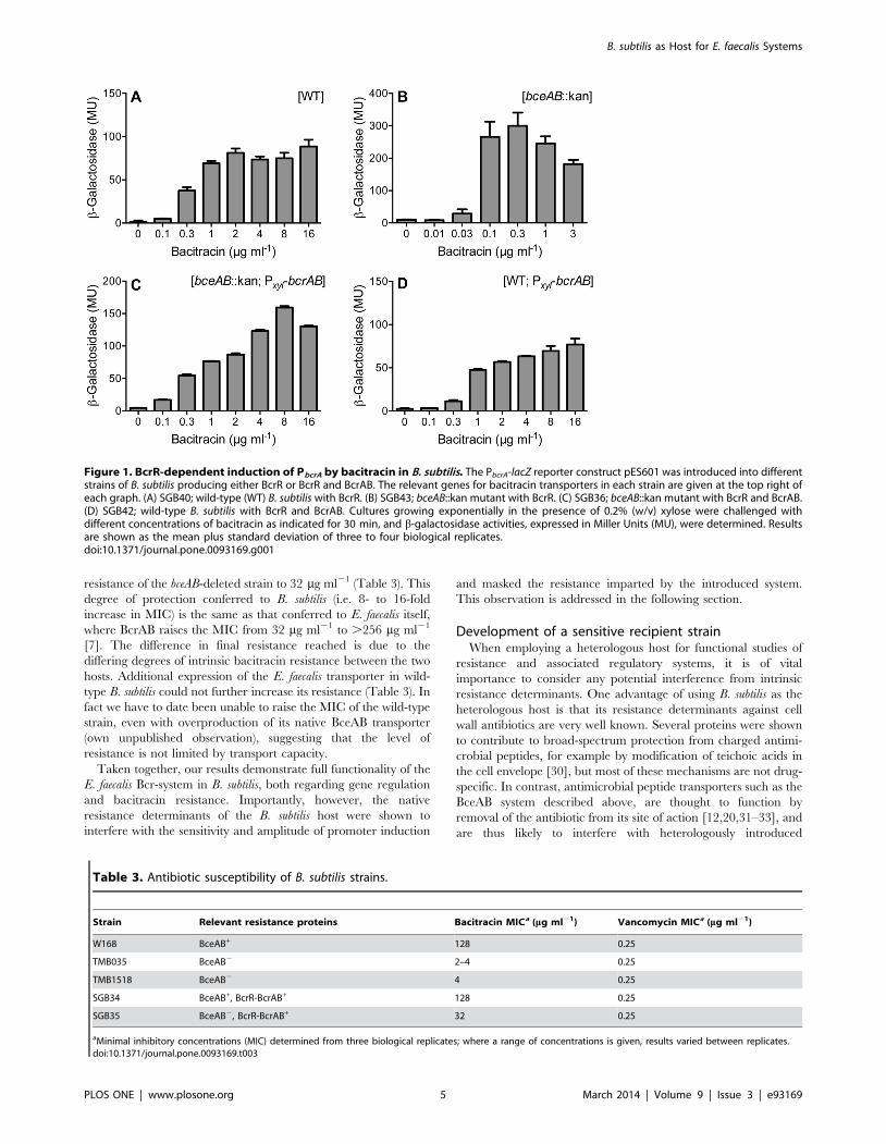
resistance of the bceAB-deleted strain to 32 mg ml21 (Table 3). This
degree of protection conferred to B. subtilis (i.e. 8- to 16-fold
increase in MIC) is the same as that conferred to E. faecalis itself,
where BcrAB raises the MIC from 32 mg ml21 to .256 mg ml21
[7]. The difference in final resistance reached is due to the
differing degrees of intrinsic bacitracin resistance between the two
hosts. Additional expression of the E. faecalis transporter in wild-
type B. subtilis could not further increase its resistance (Table 3). In
fact we have to date been unable to raise the MIC of the wild-type
strain, even with overproduction of its native BceAB transporter
(own unpublished observation), suggesting that the level of
resistance is not limited by transport capacity.
Taken together, our results demonstrate full functionality of the
E. faecalis Bcr-system in B. subtilis, both regarding gene regulation
and bacitracin resistance. Importantly, however, the native
resistance determinants of the B. subtilis host were shown to
interfere with the sensitivity and amplitude of promoter induction
and masked the resistance imparted by the introduced system.
This observation is addressed in the following section.
Development of a sensitive recipient strainWhen employing a heterologous host for functional studies of
resistance and associated regulatory systems, it is of vital
importance to consider any potential interference from intrinsic
resistance determinants. One advantage of using B. subtilis as the
heterologous host is that its resistance determinants against cell
wall antibiotics are very well known. Several proteins were shown
to contribute to broad-spectrum protection from charged antimi-
crobial peptides, for example by modification of teichoic acids in
the cell envelope [30], but most of these mechanisms are not drug-
specific. In contrast, antimicrobial peptide transporters such as the
BceAB system described above, are thought to function by
removal of the antibiotic from its site of action [12,20,31–33], and
are thus likely to interfere with heterologously introduced
Figure 1. BcrR-dependent induction of PbcrA by bacitracin in B. subtilis. The PbcrA-lacZ reporter construct pES601 was introduced into differentstrains of B. subtilis producing either BcrR or BcrR and BcrAB. The relevant genes for bacitracin transporters in each strain are given at the top right ofeach graph. (A) SGB40; wild-type (WT) B. subtilis with BcrR. (B) SGB43; bceAB::kan mutant with BcrR. (C) SGB36; bceAB::kan mutant with BcrR and BcrAB.(D) SGB42; wild-type B. subtilis with BcrR and BcrAB. Cultures growing exponentially in the presence of 0.2% (w/v) xylose were challenged withdifferent concentrations of bacitracin as indicated for 30 min, and b-galactosidase activities, expressed in Miller Units (MU), were determined. Resultsare shown as the mean plus standard deviation of three to four biological replicates.doi:10.1371/journal.pone.0093169.g001
Table 3. Antibiotic susceptibility of B. subtilis strains.
Strain Relevant resistance proteins Bacitracin MICa (mg ml21) Vancomycin MICa (mg ml21)
W168 BceAB+ 128 0.25
TMB035 BceAB2 2–4 0.25
TMB1518 BceAB2 4 0.25
SGB34 BceAB+, BcrR-BcrAB+ 128 0.25
SGB35 BceAB2, BcrR-BcrAB+ 32 0.25
aMinimal inhibitory concentrations (MIC) determined from three biological replicates; where a range of concentrations is given, results varied between replicates.doi:10.1371/journal.pone.0093169.t003
B. subtilis as Host for E. faecalis Systems
PLOS ONE | www.plosone.org 5 March 2014 | Volume 9 | Issue 3 | e93169
resistance determinants. B. subtilis possesses three paralogous
systems of differing substrate specificities: BceAB mediates
resistance against bacitracin, mersacidin, actagardine and plecta-
sin [20,31]; PsdAB confers resistance against a broad-range of lipid
II-binding lantibiotics such as nisin or gallidermin [31]; for
YxdLM no role in resistance has been identified to date, but it’s
expression is induced in response to the human cathelicidin LL-37
[34]. All three transporters are encoded together with an operon
for a two-component regulatory system, BceRS, PsdRS and
YxdJK, respectively, which controls expression of its correspond-
ing transporter operon [28,31,35].
To obtain a recipient strain that is well suited for the study of
resistance mechanisms against cell wall antibiotics from E. faecalis
and potentially also other genetically intractable Gram-positive
bacteria, we therefore created unmarked deletions of all three
entire genetic loci, bceRS-bceAB, psdRS-psdAB and yxdJK-yxdLM-
yxeA. yxeA is a small gene of unknown function that may form a
transcriptional unit with yxdLM and was therefore included in the
deletion. To test for the absence of interference, we then
introduced the bcrR expression construct pES701 used above into
the triple deletion strain, TMB1518. While our study was in
progress, the Losick-laboratory developed a new reporter system
for B. subtilis, based on the bacterial luciferase operon luxABCDE,
which allows time-resolved, semi-automated analyses of transcrip-
tional promoter fusions [23,36]. To test the applicability of this
reporter for our purposes, we inserted the BcrR target promoter
PbcrA upstream of the lux operon and introduced this construct into
the triple deletion strain harbouring BcrR. At high expression
levels of BcrR due to induction by xylose, addition of bacitracin to
growing cultures of this strain resulted in a rapid response, with a
more than ten-fold increase of promoter activity within 5 min after
addition of 1 mg ml21 bacitracin (Fig. 2A). Only background
luminescence (ca. 103 relative luminescence units (RLU) per OD)
was observed in the absence of bacitracin or in a strain lacking
BcrR (Fig. 2A and data not shown). Analysis of promoter activities
30 min post-induction showed a similar dose-response behaviour
(Fig. 2B) compared to the corresponding lacZ reporter strain
shown above (Fig. 1B). While the threshold concentration for
induction appeared slightly increased for the PbcrA-lux construct,
possibly due to the different growth conditions in 96-well plates
compared to test-tubes, the maximal amplitude of induction was
approximately doubled to over 500-fold, which can most likely be
attributed to the very low background luminescence obtained with
luciferase assays. Therefore both the lacZ and lux reporters are
equally suitable to determine dose-response behaviours of regula-
tory systems, while the lux reporter offers higher sensitivity and
additionally allows time-resolved analyses for dynamic studies.
To test if the cellular protein levels of a one-component
regulator like BcrR affected the promoter induction behaviour, the
same experiments were also carried out in the absence of xylose,
relying on the basal activities of the PxylA-promoter for bcrR
expression (Fig. 2C and D). Under these conditions, the maximal
promoter activities were reduced approximately eight-fold
(p = 0.0003). Considering that the difference in PxylA activity in
the presence and absence of xylose is ten-fold under the conditions
used here [36], this difference in BcrR-activity is likely directly due
to a reduced copy number of BcrR in the cell. However, the dose-
response behaviour was again similar to previous results, with a
threshold concentration for induction in the range of 0.03 to
0.1 mg ml21 bacitracin. Thus the overall function of BcrR was
robust to changes in expression, with differences in protein levels
merely affecting the amplitude of induction but not the response to
the stimulus.
Qualitative activity assays on solid media for screeningapplications
To elucidate the molecular mechanisms of stimulus perception
and signal transduction in regulatory systems, random or site-
directed mutagenesis is often used. Particularly in the case of
random mutagenesis approaches, but also for (synthetic) DNA-
libraries, assays performed on solid media greatly facilitate
screening of large numbers of clones. To evaluate the lacZ and
lux reporters for such applications, the derived BcrR/PbcrA reporter
strains were streaked onto agar plates in the absence or presence of
bacitracin. Strains harbouring the PbcrA-lacZ fusions showed a blue
colouration on XGal-containing agar plates in the presence of
inducing concentrations of bacitracin, but remained white in its
absence (Fig. 3A and B). As observed before in the quantitative
assays, presence of the transporters BceAB or BcrAB diminished
the intensity of colouration (Fig. 3B, sectors 1 and 2). In the strain
possessing both transporters, bacitracin concentrations of at least
10 mg ml21 were required to produce blue colonies (data not
shown), consistent with the low promoter activities reported above
for this strain. The reporter strain harbouring BcrR and the PbcrA-
lux construct showed strong luminescence when grown on agar
plates containing 0.3 mg ml21 bacitracin, and no detectable
luminescence in its absence (Fig. 3C and D).
Both reporter constructs are therefore suitable for screening
libraries of clones for promoter induction and are applicable for
high-throughput approaches. In principle, screens for loss-of-
function as well as gain-of-function mutations can be performed,
depending on experimental design. This set-up offers a great
advantage over studies performed directly in E. faecalis, where it is
much more difficult to obtain large numbers of transformants than
in the naturally competent B. subtilis. Importantly, the output of
both promoters is sufficiently sensitive to allow assays to be
performed at sub-lethal concentrations of the antibiotic, at least in
the case of the Bcr-system. The feasibility of this approach was
recently demonstrated in a study that identified essential residues
in the B. subtilis bacitracin resistance transporter BceAB [29], and
the same strategy should be applicable to the heterologous set-up
described here.
Functional transfer of the VanSB-VanRB two-componentsystem to B. subtilis
Following successful transfer of the Bcr-system of E. faecalis to B.
subtilis, we next wanted to test if our set-up could be applied to
other regulatory systems. The two-component system VanS-VanR
regulating VanA-type vancomycin resistance had previously been
shown to be functional in B. subtilis [1]. However, heterologous
expression of vanRBvanSB encoding the regulatory system for
VanB-type resistance had resulted in constitutive expression of the
target promoter, PvanYB, and the authors could show that this was
due to constitutive activity of the sensor kinase VanSB under the
conditions chosen [18]. To test if vancomycin-dependent modu-
lation of VanSB activity could be obtained by optimization of
conditions, we introduced an expression construct of the
vanRBvanSB operon under control of the xylose-inducible promoter
PxylA into TMB1518. The activity of the two-component system
was monitored as activation of a PvanYB-luxABCDE transcriptional
fusion. In the absence of xylose, only low levels of the two-
component systems will be produced in the cell, due to basal
promoter activity of PxylA. Under these conditions, addition of
increasing concentrations of vancomycin to growing cultures of the
reporter strain led to a gradual up-regulation of promoter activity
(Fig. 4A). Importantly, and in contrast to previous data, only
background activity was observed in the absence of vancomycin
B. subtilis as Host for E. faecalis Systems
PLOS ONE | www.plosone.org 6 March 2014 | Volume 9 | Issue 3 | e93169
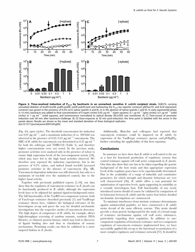
(Fig. 4A, open circles). The threshold concentration for induction
was 0.01 mg ml21, and a maximum induction of ca. 500-fold was
observed in the presence of 0.05–0.25 mg ml21 vancomycin. The
MIC of B. subtilis for vancomycin was determined as 0.25 mg ml21
for both the wild-type and TMB1518 (Table 3), and therefore
higher concentrations were not tested. In the previous study,
promoter activities were analysed only in the presence of xylose to
ensure high expression levels of the two-component system [18],
which may have led to the high basal activities observed. We
therefore next repeated the induction experiments, but in the
presence of 0.2% xylose, and indeed found ten-fold increased
promoter activities in the absence of vancomycin (Fig. 4B).
Vancomycin-dependent induction was still observed, but only to a
maximum of ten-fold over the uninduced control, due to the
higher basal activity.
Together with previously published reports [1,18], our data
show that the regulators of vancomycin resistance in E. faecalis can
be functionally produced in B. subtilis, although the expression
levels have to be adjusted for optimal signal-to-background ratios.
The full functionality of the VanRS two-component systems, both
of VanA-type resistance described previously [1] and VanB-type
resistance shown here, validates the biological relevance of the
heterologous set-up and paves the way for detailed mechanistic
investigations into the respective modes of vancomycin detection.
The high degree of competence of B. subtilis, for example, allows
high-throughput screening of random mutants, synthetic DNA
libraries, or chimeric protein fusions, which may lead to discovery
of ligand binding sites and thus to elucidation of sensory
mechanisms. Promising results can then be validated in a more
targeted fashion in E. faecalis.
Additionally, Bisicchia and colleagues had reported that
vancomycin resistance could be imparted on B. subtilis by
expression of the VanB-type resistance operon vanYBWHBBXB,
further extending the applicability of this host organism.
Conclusions
In summary we here show that B. subtilis is well suited to the use
as a host for functional production of regulatory systems that
control resistance against cell wall active compounds in E. faecalis.
Our data also show that care has to be taken regarding the genetic
background of the host strain and that appropriate expression
levels of the regulator genes have to be experimentally determined.
Due to the availability of a range of inducible and constitutive
promoters, for which strength and dynamic behaviour are very
well characterized [36], B. subtilis offers a vast potential for
optimization of expression levels, again supporting its suitability as
a versatile heterologous host. Full functionality of any newly
introduced system should of course be validated by comparison of
its behaviour between B. subtilis and the native host before detailed
mechanistic investigations are commenced.
To minimize interference from intrinsic resistance determinants
against antimicrobial peptides, we have constructed a B. subtilis
strain devoid of the most efficient systems. This strain should
provide a clean genetic background for the study of a broad range
of resistance mechanisms against cell wall active substances,
particularly regarding their regulation. In addition to one-
component regulation of bacitracin resistance and two-component
regulation of vancomycin resistance implemented here, we have
successfully applied this set-up to the functional reconstitution of a
more complex regulatory and resistance network [37]. It should be
Figure 2. Time-resolved induction of PbcrA by bacitracin in an unmarked, sensitive B. subtilis recipient strain. SGB274, carryingunmarked deletion of bceRS-bceAB, psdRS-psdAB, yxdJK-yxdLM-yxeA and harbouring the PbcrA-lux reporter construct pNTlux101 and bcrR expressionconstruct was grown in the presence of 0.2% (w/v) xylose (panels A and B), or in the absence of xylose (panels C and D). In early exponential phase(t = 0 min), bacitracin was added to final concentrations of 0 (open circles) 0.03 mg ml21 (open squares), 0.1 mg ml21 (grey circles), 0.3 mg ml21 (solidcircles) or 1 mg ml21 (solid squares), and luminescence normalized to optical density (RLU/OD) was monitored. (A, C) Time-course of promoterinduction over 60 min after bacitracin-challenge. (B, D) Dose-response at 30 min post-induction; the time point is labelled with the arrow in thepanels above. Results are shown as the mean and standard deviation of three biological replicates.doi:10.1371/journal.pone.0093169.g002
B. subtilis as Host for E. faecalis Systems
PLOS ONE | www.plosone.org 7 March 2014 | Volume 9 | Issue 3 | e93169
noted that the response of B. subtilis to antibiotics in general is
among the best understood of all bacteria investigated to date [38].
This plethora of available data therefore constitutes an ideal basis
for construction of new sensitive recipient strains adapted to the
study of resistance and regulatory systems also for other classes of
antimicrobials.
Further, we showed that the two reporters, lacZ and luxABCDE,
can both be used for qualitative (high-throughput) screening
approaches, for example of mutant libraries, as well as for the
quantitative characterization of regulators. Complementation
studies with random or directed mutations can thus be initiated
in the genetically accessible, highly competent host B. subtilis, and
promising results then validated directly in E. faealis. Construction
of the desired heterologous strains will be further aided by a
recently established and fully validated tool-box of vectors,
promoters, reporters and epitope-tags for engineering of B. subtilis
[36]. We therefore envisage that the system developed here will aid
investigations into the molecular mechanisms of sensory percep-
tion of antimicrobials and subsequent signal transduction, the first
essential step of antibiotic resistance. Furthermore, this set-up
should also be applicable to the study of unrelated resistance
systems or even regulatory cascades of diverse functions from other
genetically intractable Gram-positive bacteria.
Acknowledgments
The authors would like to thank Richard Losick for supplying vector
pAH328.
Author Contributions
Conceived and designed the experiments: SG TM GC. Performed the
experiments: CF ES. Analyzed the data: CF ES SG. Wrote the paper: SG
CF.
References
1. Ulijasz AT, Grenader A, Weisblum B (1996) A vancomycin-inducible lacZ
reporter system in Bacillus subtilis: induction by antibiotics that inhibit cell wall
synthesis and by lysozyme. J Bacteriol 178: 6305–6309.
2. Arias CA, Murray BE (2012) The rise of the Enterococcus: beyond vancomycin
resistance. Nat Rev Microbiol 10: 266–278. doi:10.1038/nrmicro2761.
3. Barna JC, Williams DH (1984) The structure and mode of action of glycopeptide
antibiotics of the vancomycin group. Annu Rev Microbiol 38: 339–357.
doi:10.1146/annurev.mi.38.100184.002011.
4. Breukink E, de Kruijff B (2006) Lipid II as a target for antibiotics. Nat Rev Drug
Discov 5: 321–323. doi:10.1038/nrd2004.
5. Bevins CL, Martin-Porter E, Ganz T (1999) Defensins and innate host defence
of the gastrointestinal tract. Gut 45: 911–915.
6. Cotter PD, Hill C, Ross RP (2005) Bacteriocins: developing innate immunity for
food. Nat Rev Microbiol 3: 777–788.
7. Manson JM, Keis S, Smith JMB, Cook GM (2004) Acquired bacitracin
resistance in Enterococcus faecalis is mediated by an ABC transporter and a novel
Figure 3. Functionality of the reporter systems on solid media.Strains of B. subtilis harbouring the PbcrA-lacZ reporter (A and B) or thePbcrA-luxABCDE reporter (C and D) were grown on agar plates containing0 mg ml21 (A and C), 0.3 mg ml21 (D) or 1 mg ml21 (B) bacitracin. (A, B)Blue colouration due to reporter induction is depicted by the dark greyshading of bacterial growth. Sector 1, SGB40 (BcrR+, BceAB+); sector 2,SGB36 (BcrR+, BcrAB+); sector 3, SGB43 (BcrR+); sector 4, SGB42 (BcrR+,BceAB+, BcrAB+). Plates contained 200 mg ml21 X-Gal. (C, D) Platesinoculated with SGB237 (BcrR+) were photographed under white light(left sub-panels), followed by detection of luminescence in the dark(right sub-panels); the same sector of the agar plates is shown in bothsub-panels.doi:10.1371/journal.pone.0093169.g003
Figure 4. VanSB-VanRB-dependent induction of PvanYB byvancomycin in B. subtilis. Both the PvanYB-lux reporter constructpCF133 and the PxylA-vanRBSB expression construct pCF132 wereintroduced into B. subtilis strain TMB1518 (unmarked deletion ofbceRS-bceAB, psdRS-psdAB, yxdJK-yxdLM-yxeA). Cultures growing expo-nentially either (A) in the absence of xylose or (B) in the presence of0.2% (w/v) xylose were challenged at t = 0 min with 0.01 mg ml21 (opensquares), 0.025 mg ml21 (grey circles), 0.05 mg ml21 (solid circles),0.25 mg ml21 (solid squares) vancomycin, or left untreated (opencircles). Luminescence normalized to optical density (RLU/OD) wasmonitored over 60 min. Results are shown as the mean and standarddeviation of three biological replicates.doi:10.1371/journal.pone.0093169.g004
B. subtilis as Host for E. faecalis Systems
PLOS ONE | www.plosone.org 8 March 2014 | Volume 9 | Issue 3 | e93169
regulatory protein, BcrR. Antimicrob Agents Chemother 48: 3743–3748.
doi:10.1128/AAC.48.10.3743–3748.2004.
8. Matos R, Pinto VV, Ruivo M, Lopes M de FS (2009) Study on the dissemination
of the bcrABDR cluster in Enterococcus spp. reveals that the BcrAB transporter is
sufficient to confer high-level bacitracin resistance. International Journal of
Antimicrobial Agents 34: 142–147. doi:10.1016/j.ijantimicag.2009.02.008.
9. Storm DR, Strominger JL (1973) Complex formation between bacitracin
peptides and isoprenyl pyrophosphates. The specificity of lipid-peptide
interactions. J Biol Chem 248: 3940–3945.
10. Arthur M, Reynolds P, Courvalin P (1996) Glycopeptide resistance in
enterococci. Trends Microbiol 4: 401–407. doi:10.1016/0966-842X(96)10063-
9.
11. Arthur M, Quintiliani R (2001) Regulation of VanA- and VanB-type
glycopeptide resistance in enterococci. Antimicrob Agents Chemother 45:
375–381.
12. Gebhard S (2012) ABC transporters of antimicrobial peptides in Firmicutes
bacteria - phylogeny, function and regulation. Mol Microbiol 86: 1295–1317.
doi:10.1111/mmi.12078.
13. Baptista M, Depardieu F, Reynolds P, Courvalin P, Arthur M (1997) Mutations
leading to increased levels of resistance to glycopeptide antibiotics in VanB-type
enterococci. Mol Microbiol 25: 93–105.
14. Gauntlett JC, Gebhard S, Keis S, Manson JM, Pos KM, et al. (2008) Molecular
analysis of BcrR, a membrane-bound bacitracin sensor and DNA-binding
protein from Enterococcus faecalis. J Biol Chem 283: 8591–8600. doi:10.1074/
jbc.M709503200.
15. Hong H-J, Hutchings MI, Buttner MJ (2008) Vancomycin resistance VanS/
VanR two-component systems. In: Utsumi R, editor. Bacterial Signal
Transduction: Networks and Drug Targets. Landes Bioscience and Springer
Science-Business Media. pp. 200–213.
16. Gebhard S, Gaballa A, Helmann JD, Cook GM (2009) Direct stimulus
perception and transcription activation by a membrane-bound DNA binding
protein. Mol Microbiol 73: 482–491. doi:10.1111/j.1365-2958.2009.06787.x.
17. Silva JC, Haldimann A, Prahalad MK, Walsh CT, Wanner BL (1998) In vivo
characterization of the type A and B vancomycin-resistant enterococci (VRE)
VanRS two-component systems in Escherichia coli: a nonpathogenic model for
studying the VRE signal transduction pathways. Proc Natl Acad Sci USA 95:
11951–11956.
18. Bisicchia P, Bui NK, Aldridge C, Vollmer W, Devine KM (2011) Acquisition of
VanB-type vancomycin resistance by Bacillus subtilis: the impact on gene
expression, cell wall composition and morphology. Mol Microbiol 81: 157–178.
doi:10.1111/j.1365-2958.2011.07684.x.
19. Mascher T, Margulis NG, Wang T, Ye RW, Helmann JD (2003) Cell wall stress
responses in Bacillus subtilis: the regulatory network of the bacitracin stimulon.
Mol Microbiol 50: 1591–1604. doi:10.1046/j.1365-2958.2003.03786.x.
20. Rietkotter E, Hoyer D, Mascher T (2008) Bacitracin sensing in Bacillus subtilis.
Mol Microbiol 68: 768–785. doi:10.1111/j.1365-2958.2008.06194.x.
21. Harwood CR, Cutting SM (1990) Molecular biological methods for Bacillus.
Chichester, UK: John Wiley & Sons.
22. Stulke J, Martin-Verstraete I, Zagorec M, Rose M, Klier A, et al. (1997)
Induction of the Bacillus subtilis ptsGHI operon by glucose is controlled by a novel
antiterminator, GlcT. Mol Microbiol 25: 65–78.
23. Schmalisch M, Maiques E, Nikolov L, Camp AH, Chevreux B, et al. (2010)
Small genes under sporulation control in the Bacillus subtilis genome. J Bacteriol
192: 5402–5412. doi:10.1128/JB.00534-10.
24. Derre I, Rapoport G, Msadek T (2000) The CtsR regulator of stress response is
active as a dimer and specifically degraded in vivo at 37 degrees C. Mol Microbiol38: 335–347.
25. Arnaud M, Chastanet A, Debarbouille M (2004) New vector for efficient allelic
replacement in naturally nontransformable, low-GC-content, Gram-positivebacteria. Appl Environ Microbiol 70: 6887–6891. doi:10.1128/AEM.70.11.
6887-6891.2004.26. Clinical and Laboratory Standards Institute (2006) Methods for dilution
antimicrobial susceptibility tests for bacteria that grow aerobically: 1–64.
27. Miller JH (1972) Experiments in molecular genetics. Cold Spring Harbor, NY:Cold Spring Harbor Laboratory Press.
28. Ohki R, Giyanto, Tateno K, Masuyama W, Moriya S, et al. (2003) The BceRStwo-component regulatory system induces expression of the bacitracin
transporter, BceAB, in Bacillus subtilis. Mol Microbiol 49: 1135–1144.doi:10.1046/j.1365-2958.2003.03653.x.
29. Kallenberg F, Dintner S, Schmitz R, Gebhard S (2013) Identification of regions
important for resistance and signalling within the antimicrobial peptidetransporter BceAB of Bacillus subtilis. J Bacteriol 195: 3287–3297. doi:10.1128/
JB.00419-13.30. Kingston AW, Liao X, Helmann JD (2013) Contributions of the sW, sM, and
sX regulons to the lantibiotic resistome of Bacillus subtilis. Mol Microbiol.
doi:10.1111/mmi.12380.31. Staron A, Finkeisen DE, Mascher T (2011) Peptide antibiotic sensing and
detoxification modules of Bacillus subtilis. Antimicrob Agents Chemother 55:515–525. doi:10.1128/AAC.00352-10.
32. Hiron A, Falord M, Valle J, Debarbouille M, Msadek T (2011) Bacitracin andnisin resistance in Staphylococcus aureus: a novel pathway involving the BraS/BraR
two-component system (SA2417/SA2418) and both the BraD/BraE and VraD/
VraE ABC transporters. Mol Microbiol 81: 602–622. doi:10.1111/j.1365-2958.2011.07735.x.
33. Gebhard S, Mascher T (2011) Antimicrobial peptide sensing and detoxificationmodules: unravelling the regulatory circuitry of Staphylococcus aureus. Mol
Microbiol 81: 581–587. doi:10.1111/j.1365-2958.2011.07747.x.
34. Pietiainen M (2005) Cationic antimicrobial peptides elicit a complex stressresponse in Bacillus subtilis that involves ECF-type sigma factors and two-
component signal transduction systems. Microbiology 151: 1577–1592.doi:10.1099/mic.0.27761-0.
35. Joseph P (2004) Characterization of the Bacillus subtilis YxdJ response regulatoras the inducer of expression for the cognate ABC transporter YxdLM.
Microbiology 150: 2609–2617. doi:10.1099/mic.0.27155-0.
36. Radeck J, Kraft K, Bartels J, Cikovic T, Durr F, et al. (2013) The Bacillus
BioBrick box: generation and evaluation of essential genetic building blocks for
standardized work with Bacillus subtilis. Journal of Biological Engineering:inpress.
37. Gebhard S, Fang C, Shaaly A, Leslie DJ, Weimar MR, et al. (2013)
Identification and characterisation of a bacitracin resistance network inEnterococcus faecalis. Antimicrob Agents Chemother. doi:10.1128/AAC.02111-13.
38. Wecke T, Mascher T (2011) Antibiotic research in the age of omics: fromexpression profiles to interspecies communication. Journal of Antimicrobial
Chemotherapy 66: 2689–2704. doi:10.1093/jac/dkr373.39. Grant SG, Jessee J, Bloom FR, Hanahan D (1990) Differential plasmid rescue
from transgenic mouse DNAs into Escherichia coli methylation-restriction
mutants. Proc Natl Acad Sci USA 87: 4645–4649.40. Sahm DF, Kissinger J, Gilmore MS, Murray PR, Mulder R, et al. (1989) In vitro
susceptibility studies of vancomycin-resistant Enterococcus faecalis. AntimicrobAgents Chemother 33: 1588–1591.
B. subtilis as Host for E. faecalis Systems
PLOS ONE | www.plosone.org 9 March 2014 | Volume 9 | Issue 3 | e93169
27
Chapter III
Identification and Characterization of a Bacitracin Resistance
Network in Enterococcus faecalis
Susanne Gebhard, Chong Fang, Aishath Shaaly, David J. Leslie, Marion R. Weimar, Falk Kalamorz,
Alan Carne, Gregory M. Cook
Antimicrobial Agents and Chemotherapy, March 2014, Volume 58, Number 3, p. 1425–1433
Identification and Characterization of a Bacitracin Resistance Networkin Enterococcus faecalis
Susanne Gebhard,a Chong Fang,a Aishath Shaaly,b David J. Leslie,b Marion R. Weimar,b Falk Kalamorz,b* Alan Carne,c
Gregory M. Cookb
Department of Biology I, Microbiology, Ludwig-Maximilians-Universität München, Martinsried, Germanya; Department of Microbiology and Immunologyb andDepartment of Biochemistry,c Otago School of Medical Sciences, University of Otago, Dunedin, New Zealand
Resistance of Enterococcus faecalis against antimicrobial peptides, both of host origin and produced by other bacteria of the gutmicroflora, is likely to be an important factor in the bacterium’s success as an intestinal commensal. The aim of this study was toidentify proteins with a role in resistance against the model antimicrobial peptide bacitracin. Proteome analysis of bacitracin-treated and untreated cells showed that bacitracin stress induced the expression of cell wall-biosynthetic proteins and causedmetabolic rearrangements. Among the proteins with increased production, an ATP-binding cassette (ABC) transporter withsimilarity to known peptide antibiotic resistance systems was identified and shown to mediate resistance against bacitracin. Ex-pression of the transporter was dependent on a two-component regulatory system and a second ABC transporter, which wereidentified by genome analysis. Both resistance and the regulatory pathway could be functionally transferred to Bacillus subtilis,proving the function and sufficiency of these components for bacitracin resistance. Our data therefore show that the two ABCtransporters and the two-component system form a resistance network against antimicrobial peptides in E. faecalis, where onetransporter acts as the sensor that activates the TCS to induce production of the second transporter, which mediates the actualresistance.
Enterococcus faecalis is a member of the normal gut microflorain animals and humans. While it is mostly a harmless com-
mensal, opportunistic infections, particularly of immunocom-promised hospital patients, are a major health concern. Due to therise of vancomycin-resistant strains (VREs), treatment of suchinfections is becoming increasingly difficult, and a better under-standing of the resistance of E. faecalis against cell wall-targetingantibiotics is urgently needed. In addition to vancomycin resis-tance, enterococci display a high intrinsic resistance against otherinhibitors of cell wall biosynthesis, including cephalosporins andbacitracin (1–3). Bacitracin is not used clinically for the treatmentof enterococcal infections, yet resistance against this and otherpeptide antibiotics is nevertheless biologically relevant in the hu-man intestinal environment. For example, gut bacteria are ex-posed to defensins that are part of the innate immune defense inthe gastrointestinal tract (4). Additionally, other members of themicroflora, especially lactic acid bacteria, produce a range of bac-teriocins as a means of interspecies competition (5). Thus, resis-tance of E. faecalis against peptide antibiotics is likely to be impor-tant for the success of the bacterium in the human host, which inturn affects the risk of opportunistic infections.
In other Gram-positive bacteria, several resistance mecha-nisms that combat the action of cell wall-active peptides have beenidentified. One strategy can be alterations in the cell’s surfacecharge, for example, by alanylation of teichoic acids catalyzed bythe DltABCD system (6, 7). Accordingly, in a recent study, the dltoperon of E. faecalis was shown to be upregulated in response tobacitracin and vancomycin (8). Alternatively, upregulation of theenzyme inhibited by the antibiotic, e.g., in the case of bacitracin,undecaprenyl-pyrophosphate phosphatases, can confer resis-tance, as has been shown for Bacillus subtilis and E. faecalis (2, 9).A third resistance mechanism is the expression of specific ATP-binding cassette (ABC) transporters. One type of such transport-ers possesses permeases with six transmembrane helices and is
exemplified by the bacitracin transporter BcrAB, which is used forself-immunity by bacitracin-producing strains of Bacillus licheni-formis (10) but has also been shown to confer acquired high-levelbacitracin resistance to E. faecalis (3). An unrelated group of trans-porters, characterized by permeases with 10 transmembrane heli-ces and a large extracellular domain, are involved in resistanceagainst a range of antimicrobial peptides. The best-understoodexample is BceAB of B. subtilis, which confers resistance againstbacitracin, mersacidin, and actagardine (11, 12). These transport-ers form the Peptide-7-Exporter family in the Transport Classifi-cation Database and are collectively referred to as BceAB-typesystems (13, 14). They are usually found in the genetic neighbor-hood of a two-component regulatory system (TCS; BceRS type)that controls expression of the transporter operon (15, 16). Im-portantly, these transporters not only mediate resistance but playan additional role as sensors, because the TCSs alone are unable todetect their substrate peptides (11, 17). Together, transporters andTCSs form peptide antibiotic resistance modules and are foundwidely distributed among low-G�C Gram-positive organisms(13, 16). A comparative genomics analysis identified two BceAB-
Received 27 September 2013 Returned for modification 4 November 2013Accepted 11 December 2013
Published ahead of print 16 December 2013
Address correspondence to Susanne Gebhard, [email protected].
* Present address: Falk Kalamorz, Division of Molecular Microbiology, Institute forBiology/Microbiology, Martin-Luther Universität Halle-Wittenberg, Halle (Saale),Germany.
Supplemental material for this article may be found at http://dx.doi.org/10.1128/AAC.02111-13.
Copyright © 2014, American Society for Microbiology. All Rights Reserved.
doi:10.1128/AAC.02111-13
March 2014 Volume 58 Number 3 Antimicrobial Agents and Chemotherapy p. 1425–1433 aac.asm.org 1425
on Novem
ber 2, 2014 by guesthttp://aac.asm
.org/D
ownloaded from
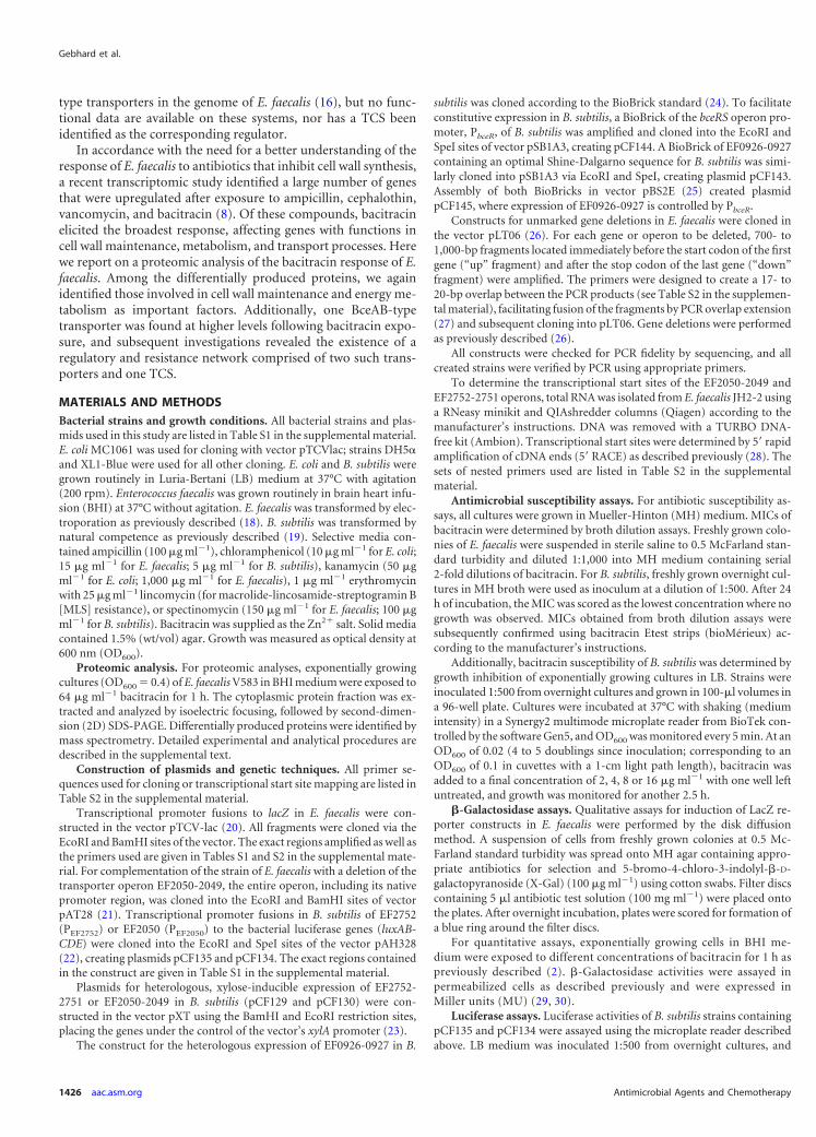
type transporters in the genome of E. faecalis (16), but no func-tional data are available on these systems, nor has a TCS beenidentified as the corresponding regulator.
In accordance with the need for a better understanding of theresponse of E. faecalis to antibiotics that inhibit cell wall synthesis,a recent transcriptomic study identified a large number of genesthat were upregulated after exposure to ampicillin, cephalothin,vancomycin, and bacitracin (8). Of these compounds, bacitracinelicited the broadest response, affecting genes with functions incell wall maintenance, metabolism, and transport processes. Herewe report on a proteomic analysis of the bacitracin response of E.faecalis. Among the differentially produced proteins, we againidentified those involved in cell wall maintenance and energy me-tabolism as important factors. Additionally, one BceAB-typetransporter was found at higher levels following bacitracin expo-sure, and subsequent investigations revealed the existence of aregulatory and resistance network comprised of two such trans-porters and one TCS.
MATERIALS AND METHODSBacterial strains and growth conditions. All bacterial strains and plas-mids used in this study are listed in Table S1 in the supplemental material.E. coli MC1061 was used for cloning with vector pTCVlac; strains DH5�and XL1-Blue were used for all other cloning. E. coli and B. subtilis weregrown routinely in Luria-Bertani (LB) medium at 37°C with agitation(200 rpm). Enterococcus faecalis was grown routinely in brain heart infu-sion (BHI) at 37°C without agitation. E. faecalis was transformed by elec-troporation as previously described (18). B. subtilis was transformed bynatural competence as previously described (19). Selective media con-tained ampicillin (100 �g ml�1), chloramphenicol (10 �g ml�1 for E. coli;15 �g ml�1 for E. faecalis; 5 �g ml�1 for B. subtilis), kanamycin (50 �gml�1 for E. coli; 1,000 �g ml�1 for E. faecalis), 1 �g ml�1 erythromycinwith 25 �g ml�1 lincomycin (for macrolide-lincosamide-streptogramin B[MLS] resistance), or spectinomycin (150 �g ml�1 for E. faecalis; 100 �gml�1 for B. subtilis). Bacitracin was supplied as the Zn2� salt. Solid mediacontained 1.5% (wt/vol) agar. Growth was measured as optical density at600 nm (OD600).
Proteomic analysis. For proteomic analyses, exponentially growingcultures (OD600 � 0.4) of E. faecalis V583 in BHI medium were exposed to64 �g ml�1 bacitracin for 1 h. The cytoplasmic protein fraction was ex-tracted and analyzed by isoelectric focusing, followed by second-dimen-sion (2D) SDS-PAGE. Differentially produced proteins were identified bymass spectrometry. Detailed experimental and analytical procedures aredescribed in the supplemental text.
Construction of plasmids and genetic techniques. All primer se-quences used for cloning or transcriptional start site mapping are listed inTable S2 in the supplemental material.
Transcriptional promoter fusions to lacZ in E. faecalis were con-structed in the vector pTCV-lac (20). All fragments were cloned via theEcoRI and BamHI sites of the vector. The exact regions amplified as well asthe primers used are given in Tables S1 and S2 in the supplemental mate-rial. For complementation of the strain of E. faecalis with a deletion of thetransporter operon EF2050-2049, the entire operon, including its nativepromoter region, was cloned into the EcoRI and BamHI sites of vectorpAT28 (21). Transcriptional promoter fusions in B. subtilis of EF2752(PEF2752) or EF2050 (PEF2050) to the bacterial luciferase genes (luxAB-CDE) were cloned into the EcoRI and SpeI sites of the vector pAH328(22), creating plasmids pCF135 and pCF134. The exact regions containedin the construct are given in Table S1 in the supplemental material.
Plasmids for heterologous, xylose-inducible expression of EF2752-2751 or EF2050-2049 in B. subtilis (pCF129 and pCF130) were con-structed in the vector pXT using the BamHI and EcoRI restriction sites,placing the genes under the control of the vector’s xylA promoter (23).
The construct for the heterologous expression of EF0926-0927 in B.
subtilis was cloned according to the BioBrick standard (24). To facilitateconstitutive expression in B. subtilis, a BioBrick of the bceRS operon pro-moter, PbceR, of B. subtilis was amplified and cloned into the EcoRI andSpeI sites of vector pSB1A3, creating pCF144. A BioBrick of EF0926-0927containing an optimal Shine-Dalgarno sequence for B. subtilis was simi-larly cloned into pSB1A3 via EcoRI and SpeI, creating plasmid pCF143.Assembly of both BioBricks in vector pBS2E (25) created plasmidpCF145, where expression of EF0926-0927 is controlled by PbceR.
Constructs for unmarked gene deletions in E. faecalis were cloned inthe vector pLT06 (26). For each gene or operon to be deleted, 700- to1,000-bp fragments located immediately before the start codon of the firstgene (“up” fragment) and after the stop codon of the last gene (“down”fragment) were amplified. The primers were designed to create a 17- to20-bp overlap between the PCR products (see Table S2 in the supplemen-tal material), facilitating fusion of the fragments by PCR overlap extension(27) and subsequent cloning into pLT06. Gene deletions were performedas previously described (26).
All constructs were checked for PCR fidelity by sequencing, and allcreated strains were verified by PCR using appropriate primers.
To determine the transcriptional start sites of the EF2050-2049 andEF2752-2751 operons, total RNA was isolated from E. faecalis JH2-2 usinga RNeasy minikit and QIAshredder columns (Qiagen) according to themanufacturer’s instructions. DNA was removed with a TURBO DNA-free kit (Ambion). Transcriptional start sites were determined by 5= rapidamplification of cDNA ends (5= RACE) as described previously (28). Thesets of nested primers used are listed in Table S2 in the supplementalmaterial.
Antimicrobial susceptibility assays. For antibiotic susceptibility as-says, all cultures were grown in Mueller-Hinton (MH) medium. MICs ofbacitracin were determined by broth dilution assays. Freshly grown colo-nies of E. faecalis were suspended in sterile saline to 0.5 McFarland stan-dard turbidity and diluted 1:1,000 into MH medium containing serial2-fold dilutions of bacitracin. For B. subtilis, freshly grown overnight cul-tures in MH broth were used as inoculum at a dilution of 1:500. After 24h of incubation, the MIC was scored as the lowest concentration where nogrowth was observed. MICs obtained from broth dilution assays weresubsequently confirmed using bacitracin Etest strips (bioMérieux) ac-cording to the manufacturer’s instructions.
Additionally, bacitracin susceptibility of B. subtilis was determined bygrowth inhibition of exponentially growing cultures in LB. Strains wereinoculated 1:500 from overnight cultures and grown in 100-�l volumes ina 96-well plate. Cultures were incubated at 37°C with shaking (mediumintensity) in a Synergy2 multimode microplate reader from BioTek con-trolled by the software Gen5, and OD600 was monitored every 5 min. At anOD600 of 0.02 (4 to 5 doublings since inoculation; corresponding to anOD600 of 0.1 in cuvettes with a 1-cm light path length), bacitracin wasadded to a final concentration of 2, 4, 8 or 16 �g ml�1 with one well leftuntreated, and growth was monitored for another 2.5 h.
�-Galactosidase assays. Qualitative assays for induction of LacZ re-porter constructs in E. faecalis were performed by the disk diffusionmethod. A suspension of cells from freshly grown colonies at 0.5 Mc-Farland standard turbidity was spread onto MH agar containing appro-priate antibiotics for selection and 5-bromo-4-chloro-3-indolyl-�-D-galactopyranoside (X-Gal) (100 �g ml�1) using cotton swabs. Filter discscontaining 5 �l antibiotic test solution (100 mg ml�1) were placed ontothe plates. After overnight incubation, plates were scored for formation ofa blue ring around the filter discs.
For quantitative assays, exponentially growing cells in BHI me-dium were exposed to different concentrations of bacitracin for 1 h aspreviously described (2). �-Galactosidase activities were assayed inpermeabilized cells as described previously and were expressed inMiller units (MU) (29, 30).
Luciferase assays. Luciferase activities of B. subtilis strains containingpCF135 and pCF134 were assayed using the microplate reader describedabove. LB medium was inoculated 1:500 from overnight cultures, and
Gebhard et al.
1426 aac.asm.org Antimicrobial Agents and Chemotherapy
on Novem
ber 2, 2014 by guesthttp://aac.asm
.org/D
ownloaded from
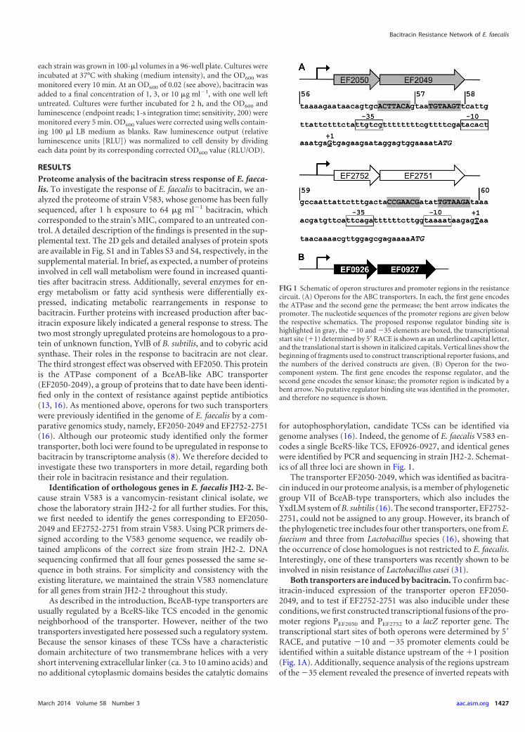
each strain was grown in 100-�l volumes in a 96-well plate. Cultures wereincubated at 37°C with shaking (medium intensity), and the OD600 wasmonitored every 10 min. At an OD600 of 0.02 (see above), bacitracin wasadded to a final concentration of 1, 3, or 10 �g ml�1, with one well leftuntreated. Cultures were further incubated for 2 h, and the OD600 andluminescence (endpoint reads; 1-s integration time; sensitivity, 200) weremonitored every 5 min. OD600 values were corrected using wells contain-ing 100 �l LB medium as blanks. Raw luminescence output (relativeluminescence units [RLU]) was normalized to cell density by dividingeach data point by its corresponding corrected OD600 value (RLU/OD).
RESULTSProteome analysis of the bacitracin stress response of E. faeca-lis. To investigate the response of E. faecalis to bacitracin, we an-alyzed the proteome of strain V583, whose genome has been fullysequenced, after 1 h exposure to 64 �g ml�1 bacitracin, whichcorresponded to the strain’s MIC, compared to an untreated con-trol. A detailed description of the findings is presented in the sup-plemental text. The 2D gels and detailed analyses of protein spotsare available in Fig. S1 and in Tables S3 and S4, respectively, in thesupplemental material. In brief, as expected, a number of proteinsinvolved in cell wall metabolism were found in increased quanti-ties after bacitracin stress. Additionally, several enzymes for en-ergy metabolism or fatty acid synthesis were differentially ex-pressed, indicating metabolic rearrangements in response tobacitracin. Further proteins with increased production after bac-itracin exposure likely indicated a general response to stress. Thetwo most strongly upregulated proteins are homologous to a pro-tein of unknown function, YvlB of B. subtilis, and to cobyric acidsynthase. Their roles in the response to bacitracin are not clear.The third strongest effect was observed with EF2050. This proteinis the ATPase component of a BceAB-like ABC transporter(EF2050-2049), a group of proteins that to date have been identi-fied only in the context of resistance against peptide antibiotics(13, 16). As mentioned above, operons for two such transporterswere previously identified in the genome of E. faecalis by a com-parative genomics study, namely, EF2050-2049 and EF2752-2751(16). Although our proteomic study identified only the formertransporter, both loci were found to be upregulated in response tobacitracin by transcriptome analysis (8). We therefore decided toinvestigate these two transporters in more detail, regarding boththeir role in bacitracin resistance and their regulation.
Identification of orthologous genes in E. faecalis JH2-2. Be-cause strain V583 is a vancomycin-resistant clinical isolate, wechose the laboratory strain JH2-2 for all further studies. For this,we first needed to identify the genes corresponding to EF2050-2049 and EF2752-2751 from strain V583. Using PCR primers de-signed according to the V583 genome sequence, we readily ob-tained amplicons of the correct size from strain JH2-2. DNAsequencing confirmed that all four genes possessed the same se-quence in both strains. For simplicity and consistency with theexisting literature, we maintained the strain V583 nomenclaturefor all genes from strain JH2-2 throughout this study.
As described in the introduction, BceAB-type transporters areusually regulated by a BceRS-like TCS encoded in the genomicneighborhood of the transporter. However, neither of the twotransporters investigated here possessed such a regulatory system.Because the sensor kinases of these TCSs have a characteristicdomain architecture of two transmembrane helices with a veryshort intervening extracellular linker (ca. 3 to 10 amino acids) andno additional cytoplasmic domains besides the catalytic domains
for autophosphorylation, candidate TCSs can be identified viagenome analyses (16). Indeed, the genome of E. faecalis V583 en-codes a single BceRS-like TCS, EF0926-0927, and identical geneswere identified by PCR and sequencing in strain JH2-2. Schemat-ics of all three loci are shown in Fig. 1.
The transporter EF2050-2049, which was identified as bacitra-cin induced in our proteome analysis, is a member of phylogeneticgroup VII of BceAB-type transporters, which also includes theYxdLM system of B. subtilis (16). The second transporter, EF2752-2751, could not be assigned to any group. However, its branch ofthe phylogenetic tree includes four other transporters, one from E.faecium and three from Lactobacillus species (16), showing thatthe occurrence of close homologues is not restricted to E. faecalis.Interestingly, one of these transporters was recently shown to beinvolved in nisin resistance of Lactobacillus casei (31).
Both transporters are induced by bacitracin. To confirm bac-itracin-induced expression of the transporter operon EF2050-2049, and to test if EF2752-2751 was also inducible under theseconditions, we first constructed transcriptional fusions of the pro-moter regions PEF2050 and PEF2752 to a lacZ reporter gene. Thetranscriptional start sites of both operons were determined by 5=RACE, and putative �10 and �35 promoter elements could beidentified within a suitable distance upstream of the �1 position(Fig. 1A). Additionally, sequence analysis of the regions upstreamof the �35 element revealed the presence of inverted repeats with
FIG 1 Schematic of operon structures and promoter regions in the resistancecircuit. (A) Operons for the ABC transporters. In each, the first gene encodesthe ATPase and the second gene the permease; the bent arrow indicates thepromoter. The nucleotide sequences of the promoter regions are given belowthe respective schematics. The proposed response regulator binding site ishighlighted in gray, the �10 and �35 elements are boxed, the transcriptionalstart site (�1) determined by 5=RACE is shown as an underlined capital letter,and the translational start is shown in italicized capitals. Vertical lines show thebeginning of fragments used to construct transcriptional reporter fusions, andthe numbers of the derived constructs are given. (B) Operon for the two-component system. The first gene encodes the response regulator, and thesecond gene encodes the sensor kinase; the promoter region is indicated by abent arrow. No putative regulator binding site was identified in the promoter,and therefore no sequence is shown.
Bacitracin Resistance Network of E. faecalis
March 2014 Volume 58 Number 3 aac.asm.org 1427
on Novem
ber 2, 2014 by guesthttp://aac.asm
.org/D
ownloaded from
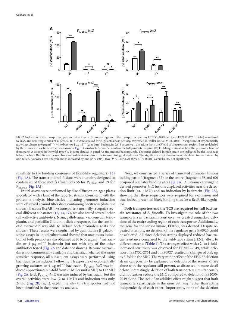
similarity to the binding consensus of BceR-like regulators (16)(Fig. 1A). The transcriptional fusions were therefore designed tocontain all of these motifs (fragments 56 for PEF2050 and 59 forPEF2752 [Fig. 1A]).
Initial assays were performed by disc diffusion on agar platesinoculated with a lawn of the reporter strains. Consistent with theproteome analysis, blue circles indicating promoter inductionwere observed around filter discs containing bacitracin (data notshown). Because BceAB-like transporters normally recognize sev-eral different substrates (12, 13, 17), we also tested several othercell wall-active antibiotics. Nisin, gallidermin, vancomycin, teico-planin, and penicillin G did not elicit a response, but the lantibi-otic mersacidin was able to induce both promoters (data notshown). These results were confirmed by quantitative �-galacto-sidase assays in liquid cultures and showed that maximum induc-tion of both promoters was obtained at 20 to 50 �g ml�1 mersaci-din or 4 �g ml�1 bacitracin but not with any of the otherantibiotics tested (Fig. 2A and data not shown). Because mersaci-din is not commercially available and bacitracin elicited the moresensitive response, all subsequent assays were performed usingbacitracin as an inducer. Following 1 h exposure of exponentiallygrowing cultures to 4 �g ml�1 bacitracin, PEF2050-lacZ was in-duced approximately 5-fold from 23 Miller units (MU) to 112 MU(Fig. 2A, left). PEF2752-lacZ was also induced by bacitracin, but theoverall activities were low (2 to 4 MU) and induction was only2-fold (Fig. 2B, right), explaining why this transporter had notbeen identified in the proteome analysis.
Next, we constructed a series of truncated promoter fusionslacking part of (fragment 57) or the entire (fragments 58 and 60)proposed regulator binding sites (Fig. 1A). All strains carrying thederived promoter-lacZ fusions displayed activities near the detec-tion limit (ca. 1 MU) and no induction by bacitracin (Fig. 2A),showing that these sequences were required for expression andthus indeed presented likely binding sites for a BceR-like regula-tor.
Both transporters and the TCS are required for full bacitra-cin resistance of E. faecalis. To investigate the role of the twotransporters in bacitracin resistance, we created unmarked dele-tions of the entire coding region of each transporter. Additionally,the gene for the sensor kinase, EF0927, was deleted. Despite re-peated attempts, no deletion of the regulator gene EF0926 couldbe achieved. All three deletion strains displayed reduced bacitra-cin resistance compared to the wild-type strain JH2-2, albeit todifferent extents (Table 1). The strongest effect with a 2- to 4-fold-increased sensitivity was observed for EF2050-2049, while dele-tion of EF2752-2751 and of EF0927 resulted in changes of only upto 2-fold in the MIC. The very minor effect of the EF0927 deletionstrain can possibly be explained by deletion of the sensor kinasealone with the regulator still present, as discussed in more detailbelow. Interestingly, deletion of both transporters simultaneouslydid not further reduce the MIC compared to deletion of EF2050-2049 alone. The lack of an additive effect might suggest that bothtransporters participate in the same pathway, rather than actingindependently of each other. Importantly, none of the deletion
FIG 2 Induction of the transporter operons by bacitracin. Promoter regions of the transporter operons EF2050-2049 (left) and EF2752-2751 (right) were fusedto lacZ, and resulting strains of E. faecalis JH2-2 were assayed for �-galactosidase activity, expressed in Miller units (MU), after 1 h exposure of exponentiallygrowing cultures to 0 �g ml�1 (white bars) or 4 �g ml�1 (gray bars) bacitracin. (A) Successive truncations from the 5= end of the promoter region. Bars are labeledby the number of each construct, as shown in Fig. 1. Constructs 56 and 59 contain the full promoter region. (B) Full-length constructs of the promoter fusionsfrom panel A assayed in the wild-type (WT; same data as in panel A) and mutant backgrounds. The genes deleted in each strain are indicated by the locus tagsbelow the bars. Results are means plus standard deviations for three to four biological replicates. The significance of induction was calculated for each strain byone-tailed, pairwise t test analysis and is indicated by one (P � 0.05), two (P � 0.005), or three (P � 0.001) asterisks. ns, not significant.
Gebhard et al.
1428 aac.asm.org Antimicrobial Agents and Chemotherapy
on Novem
ber 2, 2014 by guesthttp://aac.asm
.org/D
ownloaded from
strains displayed a growth defect compared to the wild-type strain(Table 1), showing that the differences in MIC were specificallydue to loss of resistance determinants and not to altered growthkinetics. Complementation of the EF2050-2049 deletion mutantby supplying the transporter operon in trans (strain DLEf16) re-stored bacitracin resistance (Table 1). The higher MIC comparedto the wild-type strain is most likely due to the increase in copynumber by the plasmid-based complementation strategy and sup-ports the role of EF2050-2049 in bacitracin resistance of E. faecalis.
Transporters and TCS form a regulatory network. BceAB-type transporters are often required for their own regulation byacting as the actual sensors of antimicrobial peptides that some-how communicate with the TCS to trigger activation of the signal-ing cascade (11, 17). In such cases, expression of the transporteroperon is abolished in strains carrying transporter deletions (11,12, 32). In some bacteria, exemplified by Staphylococcus aureus,two separate transporters exist, where one acts as the sensor, whilethe second is responsible for resistance (17, 32). We thereforewanted to investigate the role of the two enterococcal transportersin regulation of their own promoters. Additionally, the TCSEF0926-0927 had so far only been implicated in regulation of thetransporters based on sequence predictions, which had to be val-idated experimentally.
Deletion of the sensor kinase EF0927 drastically reduced theexpression levels of PEF2050-lacZ to less than 20% of wild-typeactivities (Fig. 2B, left). Interestingly, a significant induction bybacitracin could still be observed, which was again likely due to thepresence of the response regulator as described below. Expressionof PEF2752-lacZ was also reduced by deletion of the sensor kinase,but due to the overall low activities of this reporter, the differenceswere less pronounced. Nevertheless, the significant induction bybacitracin observed in the wild type was lost in the EF0927 dele-tion strain (Fig. 2B, right). These data show that the TCS EF0926-0927 indeed acts as the regulator for both transporter operons.
Deletion of the transporter EF2050-2049 had no effect on ex-pression of either transporter (Fig. 2B), indicating that this trans-porter’s role was restricted to mediating resistance. In contrast,deletion of EF2752-2751 severely reduced the activities of bothlacZ fusions, and bacitracin-dependent induction was lost (Fig.2B). Thus, the second transporter represents the antibiotic sensorof the resistance network.
In B. subtilis, expression of BceRS, the TCS regulating bceABexpression, is not induced by bacitracin (33). However, the ho-mologous system BraRS from S. aureus (referred to as NsaRS inreference 34) was shown to be upregulated in response to nisin,
one of its substrate peptides (34). To test if EF0926-0927 was in-ducible by bacitracin, we constructed a transcriptional fusion ofthe promoter PEF0926 to lacZ and introduced it into E. faecalisJH2-2 and derived deletion strains. Exposure of exponentiallygrowing cells to bacitracin resulted in a strong upregulation ofpromoter activities from 1 to 16 MU (Fig. 3). Interestingly, dele-tion of neither the TCS nor the two transporters affected promoteractivities or bacitracin-dependent induction (Fig. 3), showing thatregulation of the TCS is mediated by an as-yet-unidentified addi-tional regulator and not due to autoregulation. Importantly, thisregulation by a factor external to the resistance network may offeran explanation for the mild phenotypes of the EF0927 deletionstrain compared to the transporter mutants: in the EF0927 back-ground, expression of the response regulator gene EF0926 is stillupregulated in the presence of bacitracin. Because in the absenceof their cognate sensor kinase many response regulators can beefficiently phosphorylated and thus activated by small-moleculephospho-donors such as acetyl-phosphate (35, 36), this increasedproduction of EF0926 may indirectly lead to an induction of itstarget promoters by bacitracin.
EF2050-2049 can mediate bacitracin resistance in B. subtilis.Because of the mild effects of the gene deletions generated in E.faecalis, we next attempted to transfer parts of the identified resis-tance network to B. subtilis to confirm the individual roles of thecomponents in bacitracin resistance. As a chassis, we employed astrain of B. subtilis W168 carrying unmarked deletions of all threeendogenous Bce-like modules (bceRS-bceAB, psdRS-psdAB, andyxdJK-yxdLM-yxeA). This strain, TMB1518, has been developedand validated as an appropriate platform to investigate resistancemechanisms against inhibitors of cell wall synthesis and the asso-ciated regulatory pathways from E. faecalis in a genetically highlyaccessible set-up (a detailed description will be published else-where). Expression of the EF2050-2049 operon under the controlof a xylose-inducible promoter increased the MIC for bacitracin
FIG 3 Induction of the two-component system operon by bacitracin. Thepromoter region of the two-component system operon EF0926-0927 wasfused to lacZ and introduced into wild-type E. faecalis JH2-2 (WT) and mutantbackgrounds. The resulting strains were assayed for �-galactosidase activity,expressed in Miller units (MU), after 1 h exposure of exponentially growingcultures to 0 �g ml�1 (white bars) or 4 �g ml�1 (gray bars) bacitracin. Thegenes deleted in each strain are indicated by locus tags below the bars. Resultsare means plus standard deviations for three biological replicates. The signifi-cance of induction and strain differences was calculated across the entire dataset by two-way analysis of variance (ANOVA). Significant effects of bacitracincompared to uninduced cells are indicated by three asterisks (P � 0.001); thedifferent mutant backgrounds caused significant differences between strains(P � 0.018; not depicted in the graph).
TABLE 1 Bacitracin sensitivity of E. faecalis strains
Strain or genotype
MIC (�g ml�1)Growth rate(h�1)bBroth dilutiona Etest
JH2-2 32 32 1.06 0.067EF0927 16–32 24 1.2 0.377EF2050-2049 8–16 8 1.11 0.135EF2752-2751 16 16 1.14 0.146EF2050-2049 EF2752-2751 8 8 1.08 0.033DLEf16 64 64 NDa Results are from three independent broth dilution experiments; where a range ofconcentrations is given, results varied between replicates.b Results are means standard errors from three to six independent experiments. ND,not determined.
Bacitracin Resistance Network of E. faecalis
March 2014 Volume 58 Number 3 aac.asm.org 1429
on Novem
ber 2, 2014 by guesthttp://aac.asm
.org/D
ownloaded from
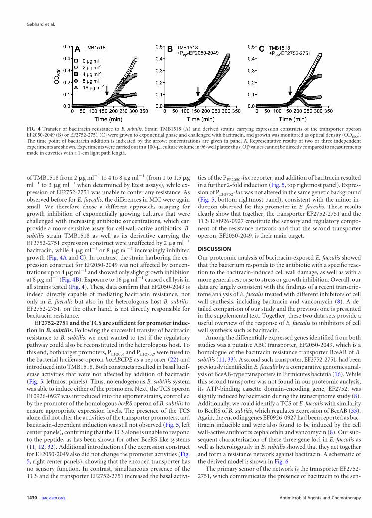
of TMB1518 from 2 �g ml�1 to 4 to 8 �g ml�1 (from 1 to 1.5 �gml�1 to 3 �g ml�1 when determined by Etest assays), while ex-pression of EF2752-2751 was unable to confer any resistance. Asobserved before for E. faecalis, the differences in MIC were againsmall. We therefore chose a different approach, assaying forgrowth inhibition of exponentially growing cultures that werechallenged with increasing antibiotic concentrations, which canprovide a more sensitive assay for cell wall-active antibiotics. B.subtilis strain TMB1518 as well as its derivative carrying theEF2752-2751 expression construct were unaffected by 2 �g ml�1
bacitracin, while 4 �g ml�1 or 8 �g ml�1 increasingly inhibitedgrowth (Fig. 4A and C). In contrast, the strain harboring the ex-pression construct for EF2050-2049 was not affected by concen-trations up to 4 �g ml�1 and showed only slight growth inhibitionat 8 �g ml�1 (Fig. 4B). Exposure to 16 �g ml�1 caused cell lysis inall strains tested (Fig. 4). These data confirm that EF2050-2049 isindeed directly capable of mediating bacitracin resistance, notonly in E. faecalis but also in the heterologous host B. subtilis.EF2752-2751, on the other hand, is not directly responsible forbacitracin resistance.
EF2752-2751 and the TCS are sufficient for promoter induc-tion in B. subtilis. Following the successful transfer of bacitracinresistance to B. subtilis, we next wanted to test if the regulatorypathway could also be reconstituted in the heterologous host. Tothis end, both target promoters, PEF2050 and PEF2752, were fused tothe bacterial luciferase operon luxABCDE as a reporter (22) andintroduced into TMB1518. Both constructs resulted in basal lucif-erase activities that were not affected by addition of bacitracin(Fig. 5, leftmost panels). Thus, no endogenous B. subtilis systemwas able to induce either of the promoters. Next, the TCS operonEF0926-0927 was introduced into the reporter strains, controlledby the promoter of the homologous bceRS operon of B. subtilis toensure appropriate expression levels. The presence of the TCSalone did not alter the activities of the transporter promoters, andbacitracin-dependent induction was still not observed (Fig. 5, leftcenter panels), confirming that the TCS alone is unable to respondto the peptide, as has been shown for other BceRS-like systems(11, 12, 32). Additional introduction of the expression constructfor EF2050-2049 also did not change the promoter activities (Fig.5, right center panels), showing that the encoded transporter hasno sensory function. In contrast, simultaneous presence of theTCS and the transporter EF2752-2751 increased the basal activi-
ties of the PEF2050-lux reporter, and addition of bacitracin resultedin a further 2-fold induction (Fig. 5, top rightmost panel). Expres-sion of PEF2752-lux was not altered in the same genetic background(Fig. 5, bottom rightmost panel), consistent with the minor in-duction observed for this promoter in E. faecalis. These resultsclearly show that together, the transporter EF2752-2751 and theTCS EF0926-0927 constitute the sensory and regulatory compo-nent of the resistance network and that the second transporteroperon, EF2050-2049, is their main target.
DISCUSSION
Our proteomic analysis of bacitracin-exposed E. faecalis showedthat the bacterium responds to the antibiotic with a specific reac-tion to the bacitracin-induced cell wall damage, as well as with amore general response to stress or growth inhibition. Overall, ourdata are largely consistent with the findings of a recent transcrip-tome analysis of E. faecalis treated with different inhibitors of cellwall synthesis, including bacitracin and vancomycin (8). A de-tailed comparison of our study and the previous one is presentedin the supplemental text. Together, these two data sets provide auseful overview of the response of E. faecalis to inhibitors of cellwall synthesis such as bacitracin.
Among the differentially expressed genes identified from bothstudies was a putative ABC transporter, EF2050-2049, which is ahomologue of the bacitracin resistance transporter BceAB of B.subtilis (11, 33). A second such transporter, EF2752-2751, had beenpreviously identified in E. faecalis by a comparative genomics anal-ysis of BceAB-type transporters in Firmicutes bacteria (16). Whilethis second transporter was not found in our proteomic analysis,its ATP-binding cassette domain-encoding gene, EF2752, wasslightly induced by bacitracin during the transcriptome study (8).Additionally, we could identify a TCS of E. faecalis with similarityto BceRS of B. subtilis, which regulates expression of BceAB (33).Again, the encoding genes EF0926-0927 had been reported as bac-itracin inducible and were also found to be induced by the cellwall-active antibiotics cephalothin and vancomycin (8). Our sub-sequent characterization of these three gene loci in E. faecalis aswell as heterologously in B. subtilis showed that they act togetherand form a resistance network against bacitracin. A schematic ofthe derived model is shown in Fig. 6.
The primary sensor of the network is the transporter EF2752-2751, which communicates the presence of bacitracin to the sen-
FIG 4 Transfer of bacitracin resistance to B. subtilis. Strain TMB1518 (A) and derived strains carrying expression constructs of the transporter operonEF2050-2049 (B) or EF2752-2751 (C) were grown to exponential phase and challenged with bacitracin, and growth was monitored as optical density (OD600).The time point of bacitracin addition is indicated by the arrow; concentrations are given in panel A. Representative results of two or three independentexperiments are shown. Experiments were carried out in a 100-�l culture volume in 96-well plates; thus, OD values cannot be directly compared to measurementsmade in cuvettes with a 1-cm light path length.
Gebhard et al.
1430 aac.asm.org Antimicrobial Agents and Chemotherapy
on Novem
ber 2, 2014 by guesthttp://aac.asm
.org/D
ownloaded from
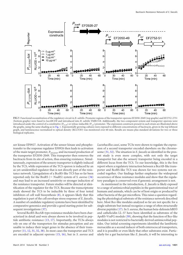
sor kinase EF0927. Activation of the sensor kinase and phospho-transfer to the response regulator EF0926 then leads to activationof the main target promoter, PEF2050, and increased production ofthe transporter EF2050-2049. This transporter then removes thebacitracin from its site of action, thus ensuring resistance. Simul-taneously, expression of the sensory transporter is slightly inducedby the TCS, while expression of the TCS operon is induced by anas-yet-unidentified regulator that is not directly part of the resis-tance network. Upregulation of a BceRS-like TCS has so far beenreported only for the BraRS (� NsaRS) system of S. aureus (34)and may lead to an increased sensitivity or stronger induction ofthe resistance transporter. Future studies will be directed at iden-tification of the regulator for the TCS. Because the transcriptomestudy showed the TCS to be inducible by three of four testedinhibitors of cell wall biosynthesis (8), it appears likely that thisregulation is part of the cell envelope stress response of E. faecalis.A number of candidate regulatory systems have been identified bycomparative genomics and provide a good starting point for sub-sequent investigations (37).
Several BceRS-BceAB-type resistance modules have been char-acterized in detail and were always shown to be involved in pep-tide antibiotic resistance (13, 17). Importantly, the TCSs alwaysrely on one of the transporters for stimulus perception and areunable to induce their target genes in the absence of their trans-porter (11, 12, 31, 32, 38). In most cases the transporter and TCSare encoded in adjacent operons (15, 16), but in S. aureus and
Lactobacillus casei, some TCSs were shown to regulate the expres-sion of a second transporter encoded elsewhere on the chromo-some (31, 32). The situation in E. faecalis as identified in the pres-ent study is even more complex, with not only the targettransporter but also the sensory transporter being encoded in adifferent locus from the TCS. To our knowledge, this is the firstreport where a regulatory interaction between a BceAB-like trans-porter and BceRS-like TCS was shown for two systems not en-coded together. Our findings further emphasize the widespreadoccurrence of these resistance modules and show that the regula-tory paradigm is conserved even if genomic arrangement is not.
As mentioned in the introduction, E. faecalis is likely exposedto a range of antimicrobial peptides in the gastrointestinal tract ofhumans and animals, which can be of host origin or produced byother bacteria of the gut microflora. This raises a question regard-ing the physiological substrate of the resistance network describedhere. Most Bce-like modules analyzed so far are not specific for asingle substrate but instead recognize a range of often structurallydiverse peptides (17). In S. aureus, the human beta-defensin hBD3and cathelicidin LL-37 have been identified as substrates of theApsRS-VraFG module (39), showing that the function of Bce-likemodules is not restricted to bacterially derived antimicrobial pep-tides. Our initial screening experiments identified the lantibioticmersacidin as a second inducer of both enterococcal transporters,and it is possible or even likely that other substrates exist. Partic-ularly for a gut bacterium like E. faecalis, it will be interesting to
FIG 5 Functional reconstitution of the regulatory circuit in B. subtilis. Promoter regions of the transporter operons EF2050-2049 (top graphs) and EF2752-2751(bottom graphs) were fused to luxABCDE and introduced into B. subtilis TMB1518. Additionally, the two-component system and transporter operons wereintroduced under the control of a constitutive (PbceR) or xylose-inducible (Pxyl) promoter. The expression constructs present in each strain are illustrated abovethe graphs, using the same shading as in Fig. 1. Exponentially growing cultures were exposed to different concentrations of bacitracin, given in the top leftmostgraph, and luminescence normalized to optical density (RLU/OD) was monitored over 60 min. Results are means plus standard deviations for two or threebiological replicates.
Bacitracin Resistance Network of E. faecalis
March 2014 Volume 58 Number 3 aac.asm.org 1431
on Novem
ber 2, 2014 by guesthttp://aac.asm
.org/D
ownloaded from
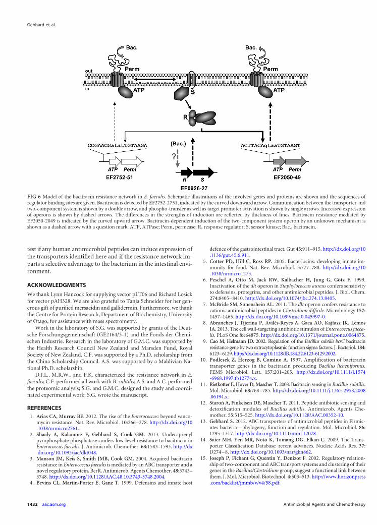
test if any human antimicrobial peptides can induce expression ofthe transporters identified here and if the resistance network im-parts a selective advantage to the bacterium in the intestinal envi-ronment.
ACKNOWLEDGMENTS
We thank Lynn Hancock for supplying vector pLT06 and Richard Losickfor vector pAH328. We are also grateful to Tanja Schneider for her gen-erous gift of purified mersacidin and gallidermin. Furthermore, we thankthe Centre for Protein Research, Department of Biochemistry, Universityof Otago, for assistance with mass spectrometry.
Work in the laboratory of S.G. was supported by grants of the Deut-sche Forschungsgemeinschaft (GE2164/3-1) and the Fonds der Chemi-schen Industrie. Research in the laboratory of G.M.C. was supported bythe Health Research Council New Zealand and Marsden Fund, RoyalSociety of New Zealand. C.F. was supported by a Ph.D. scholarship fromthe China Scholarship Council. A.S. was supported by a Maldivian Na-tional Ph.D. scholarship.
D.J.L., M.R.W., and F.K. characterized the resistance network in E.faecalis; C.F. performed all work with B. subtilis; A.S. and A.C. performedthe proteomic analysis; S.G. and G.M.C. designed the study and coordi-nated experimental work; S.G. wrote the manuscript.
REFERENCES1. Arias CA, Murray BE. 2012. The rise of the Enterococcus: beyond vanco-
mycin resistance. Nat. Rev. Microbiol. 10:266 –278. http://dx.doi.org/10.1038/nrmicro2761.
2. Shaaly A, Kalamorz F, Gebhard S, Cook GM. 2013. Undecaprenylpyrophosphate phosphatase confers low-level resistance to bacitracin inEnterococcus faecalis. J. Antimicrob. Chemother. 68:1583–1593. http://dx.doi.org/10.1093/jac/dkt048.
3. Manson JM, Keis S, Smith JMB, Cook GM. 2004. Acquired bacitracinresistance in Enterococcus faecalis is mediated by an ABC transporter and anovel regulatory protein, BcrR. Antimicrob. Agents Chemother. 48:3743–3748. http://dx.doi.org/10.1128/AAC.48.10.3743-3748.2004.
4. Bevins CL, Martin-Porter E, Ganz T. 1999. Defensins and innate host
defence of the gastrointestinal tract. Gut 45:911–915. http://dx.doi.org/10.1136/gut.45.6.911.
5. Cotter PD, Hill C, Ross RP. 2005. Bacteriocins: developing innate im-munity for food. Nat. Rev. Microbiol. 3:777–788. http://dx.doi.org/10.1038/nrmicro1273.
6. Peschel A, Otto M, Jack RW, Kalbacher H, Jung G, Götz F. 1999.Inactivation of the dlt operon in Staphylococcus aureus confers sensitivityto defensins, protegrins, and other antimicrobial peptides. J. Biol. Chem.274:8405– 8410. http://dx.doi.org/10.1074/jbc.274.13.8405.
7. McBride SM, Sonenshein AL. 2011. The dlt operon confers resistance tocationic antimicrobial peptides in Clostridium difficile. Microbiology 157:1457–1465. http://dx.doi.org/10.1099/mic.0.045997-0.
8. Abranches J, Tijerina P, Avilés-Reyes A, Gaca AO, Kajfasz JK, LemosJA. 2013. The cell wall-targeting antibiotic stimulon of Enterococcus faeca-lis. PLoS One 8:e64875. http://dx.doi.org/10.1371/journal.pone.0064875.
9. Cao M, Helmann JD. 2002. Regulation of the Bacillus subtilis bcrC bacitracinresistance gene by two extracytoplasmic function sigma factors. J. Bacteriol. 184:6123–6129. http://dx.doi.org/10.1128/JB.184.22.6123-6129.2002.
10. Podlesek Z, Herzog B, Comino A. 1997. Amplification of bacitracintransporter genes in the bacitracin producing Bacillus licheniformis.FEMS Microbiol. Lett. 157:201–205. http://dx.doi.org/10.1111/j.1574-6968.1997.tb12774.x.
11. Rietkötter E, Hoyer D, Mascher T. 2008. Bacitracin sensing in Bacillus subtilis.Mol. Microbiol. 68:768 –785. http://dx.doi.org/10.1111/j.1365-2958.2008.06194.x.
12. Staron A, Finkeisen DE, Mascher T. 2011. Peptide antibiotic sensing anddetoxification modules of Bacillus subtilis. Antimicrob. Agents Che-mother. 55:515–525. http://dx.doi.org/10.1128/AAC.00352-10.
13. Gebhard S. 2012. ABC transporters of antimicrobial peptides in Firmic-utes bacteria—phylogeny, function and regulation. Mol. Microbiol. 86:1295–1317. http://dx.doi.org/10.1111/mmi.12078.
14. Saier MH, Yen MR, Noto K, Tamang DG, Elkan C. 2009. The Trans-porter Classification Database: recent advances. Nucleic Acids Res. 37:D274 – 8. http://dx.doi.org/10.1093/nar/gkn862.
15. Joseph P, Fichant G, Quentin Y, Denizot F. 2002. Regulatory relation-ship of two-component and ABC transport systems and clustering of theirgenes in the Bacillus/Clostridium group, suggest a functional link betweenthem. J. Mol. Microbiol. Biotechnol. 4:503–513. http://www.horizonpress.com/backlist/jmmb/v/v4/58.pdf.
FIG 6 Model of the bacitracin resistance network in E. faecalis. Schematic illustrations of the involved genes and proteins are shown and the sequences ofregulator binding sites are given. Bacitracin is detected by EF2752-2751, indicated by the curved downward arrow. Communication between the transporter andtwo-component system is shown by a double arrow, and phospho-transfer as well as target promoter activation is shown by single arrows. Increased expressionof operons is shown by dashed arrows. The differences in the strengths of induction are reflected by thickness of lines. Bacitracin resistance mediated byEF2050-2049 is indicated by the curved upward arrow. Bacitracin-dependent induction of the two-component system operon by an unknown mechanism isshown as a dashed arrow with a question mark. ATP, ATPase; Perm, permease; R, response regulator; S, sensor kinase; Bac., bacitracin.
Gebhard et al.
1432 aac.asm.org Antimicrobial Agents and Chemotherapy
on Novem
ber 2, 2014 by guesthttp://aac.asm
.org/D
ownloaded from
16. Dintner S, Staron A, Berchtold E, Petri T, Mascher T, Gebhard S. 2011.Coevolution of ABC transporters and two-component regulatory systemsas resistance modules against antimicrobial peptides in Firmicutes bacte-ria. J. Bacteriol. 193:3851–3862. http://dx.doi.org/10.1128/JB.05175-11.
17. Gebhard S, Mascher T. 2011. Antimicrobial peptide sensing and detox-ification modules: unravelling the regulatory circuitry of Staphylococcusaureus. Mol. Microbiol. 81:581–587. http://dx.doi.org/10.1111/j.1365-2958.2011.07747.x.
18. Shepard BD, Gilmore MS. 1995. Electroporation and efficient transfor-mation of Enterococcus faecalis grown in high concentrations of glycine.Methods Mol. Biol. 47:217–226.
19. Harwood CR, Cutting SM. 1990. Molecular biological methods forBacillus. John Wiley & Sons, Chichester, United Kingdom.
20. Poyart C, Trieu-Cuot P. 1997. A broad-host-range mobilizable shuttlevector for the construction of transcriptional fusions to beta-galactosidasein gram-positive bacteria. FEMS Microbiol. Lett. 156:193–198. http://dx.doi.org/10.1016/S0378-1097(97)00423-0.
21. Trieu-Cuot P, Carlier C, Poyart-Salmeron C, Courvalin P. 1990. A pairof mobilizable shuttle vectors conferring resistance to spectinomycin formolecular cloning in Escherichia coli and in gram-positive bacteria. Nu-cleic Acids Res. 18:4296. http://dx.doi.org/10.1093/nar/18.14.4296.
22. Schmalisch M, Maiques E, Nikolov L, Camp AH, Chevreux B, MufflerA, Rodriguez S, Perkins J, Losick R. 2010. Small genes under sporulationcontrol in the Bacillus subtilis genome. J. Bacteriol. 192:5402–5412. http://dx.doi.org/10.1128/JB.00534-10.
23. Derré I, Rapoport G, Msadek T. 2000. The CtsR regulator of stressresponse is active as a dimer and specifically degraded in vivo at 37 degreesC. Mol. Microbiol. 38:335–347. http://dx.doi.org/10.1046/j.1365-2958.2000.02124.x.
24. Knight T. Idempotent vector design for standard assembly of BioBricks.MIT Artificial Intelligence Laboratory, MIT Synthetic Biology WorkingGroup, Massachusetts Institute of Technology, Cambridge, MA.
25. Radeck J, Kraft K, Bartels J, Cikovic T, Dürr F, Emenegger J, KelterbornS, Sauer C, Fritz G, Gebhard S, Mascher T. 2013. The Bacillus BioBrickbox: generation and evaluation of essential genetic building blocks forstandardized work with Bacillus subtilis. J. Biol. Eng. 7:29. http://dx.doi.org/10.1186/1754-1611-7-29.
26. Thurlow LR, Thomas VC, Hancock LE. 2009. Capsular polysaccharideproduction in Enterococcus faecalis and contribution of CpsF to capsuleserospecificity. J. Bacteriol. 191:6203– 6210. http://dx.doi.org/10.1128/JB.00592-09.
27. Ho SN, Hunt HD, Horton RM, Pullen JK, Pease LR. 1989. Site-directedmutagenesis by overlap extension using the polymerase chain reaction.Gene 77:51–59. http://dx.doi.org/10.1016/0378-1119(89)90358-2.
28. Gauntlett JC, Gebhard S, Keis S, Manson JM, Pos KM, Cook GM. 2008.
Molecular analysis of BcrR, a membrane-bound bacitracin sensor andDNA-binding protein from Enterococcus faecalis. J. Biol. Chem. 283:8591–8600. http://dx.doi.org/10.1074/jbc.M709503200.
29. Miller JH. 1972. Experiments in molecular genetics. Cold Spring HarborLaboratory, Cold Spring Harbor, NY.
30. Gebhard S, Tran SL, Cook GM. 2006. The Phn system of Mycobacteriumsmegmatis: a second high-affinity ABC-transporter for phosphate. Micro-biology 152:3453–3465. http://dx.doi.org/10.1099/mic.0.29201-0.
31. Revilla-Guarinos A, Gebhard S, Alcántara C, Staron A, Mascher T,Zúñiga M. 2013. Characterization of a regulatory network of peptideantibiotic detoxification modules in Lactobacillus casei BL23. Appl. Envi-ron. Microbiol. 79:3160 –3170. http://dx.doi.org/10.1128/AEM.00178-13.
32. Hiron A, Falord M, Valle J, Débarbouillé M, Msadek T. 2011. Bacitracinand nisin resistance in Staphylococcus aureus: a novel pathway involvingthe BraS/BraR two-component system (SA2417/SA2418) and both theBraD/BraE and VraD/VraE ABC transporters. Mol. Microbiol. 81:602–622. http://dx.doi.org/10.1111/j.1365-2958.2011.07735.x.
33. Ohki R, Giyanto, Tateno K, Masuyama W, Moriya S, Kobayashi K, OgasawaraN. 2003. The BceRS two-component regulatory system induces expression of thebacitracin transporter, BceAB, in Bacillus subtilis. Mol. Microbiol. 49:1135–1144. http://dx.doi.org/10.1046/j.1365-2958.2003.03653.x.
34. Kolar SL, Nagarajan V, Oszmiana A, Rivera FE, Miller HK, DavenportJE, Riordan JT, Potempa J, Barber DS, Koziel J, Elasri MO, Shaw LN.2011. NsaRS is a cell-envelope-stress-sensing two-component system ofStaphylococcus aureus. Microbiology 157:2206 –2219. http://dx.doi.org/10.1099/mic.0.049692-0.
35. Chamnongpol S, Groisman EA. 2000. Acetyl phosphate-dependent ac-tivation of a mutant PhoP response regulator that functions indepen-dently of its cognate sensor kinase. J. Mol. Biol. 300:291–305. http://dx.doi.org/10.1006/jmbi.2000.3848.
36. Schrecke K, Jordan S, Mascher T. 2013. Stoichiometry and perturbationstudies of the LiaFSR system of Bacillus subtilis. Mol. Microbiol. 87:769 –788. http://dx.doi.org/10.1111/mmi.12130.
37. Jordan S, Hutchings MI, Mascher T. 2008. Cell envelope stress responsein Gram-positive bacteria. FEMS Microbiol. Rev. 32:107–146. http://dx.doi.org/10.1111/j.1574-6976.2007.00091.x.
38. Falord M, Karimova G, Hiron A, Msadek T. 2012. GraXSR proteinsinteract with the VraFG ABC transporter to form a five-component sys-tem required for cationic antimicrobial peptide sensing and resistance inStaphylococcus aureus. Antimicrob. Agents Chemother. 56:1047–1058.http://dx.doi.org/10.1128/AAC.05054-11.
39. Li M, Cha DJ, Lai Y, Villaruz AE, Sturdevant DE, Otto M. 2007. The antimi-crobial peptide-sensing system aps of Staphylococcus aureus. Mol. Microbiol.66:1136 –1147. http://dx.doi.org/10.1111/j.1365-2958.2007.05986.x.
Bacitracin Resistance Network of E. faecalis
March 2014 Volume 58 Number 3 aac.asm.org 1433
on Novem
ber 2, 2014 by guesthttp://aac.asm
.org/D
ownloaded from
37
Chapter IV
Insulation and Wiring Specificity Determinants of BceR-like
Response Regulators and their Target Promoters
in Bacillus subtilis
Chong Fang, Anna Staroń, Martin Grafe, Ralf Heermann, Kirsten Jung, Susanne Gebhard and
Thorsten Mascher
Manuscript
Chapter IV - Wiring specificity of Bce-like systems
38
Insulation and Wiring Specificity Determinants of BceR-like Response
Regulators and their Target Promoters in Bacillus subtilis
Summary
BceRS and PsdRS are paralogous two-component systems in Bacillus subtilis. In response to the
extracellular presence of bacitracin and nisin, respectively, the two response regulators (RRs) bind
their target promoters, PbceA and PpsdA, resulting in a strong up-regulation of target gene expression and
ultimately antibiotic resistance. Despite high sequence similarity between BceR and PsdR and their
known binding sites within PbceA and PpsdA, no cross-regulation has been observed between them. We
therefore investigated the specificity determinants of PbceA and PpsdA that ensure the insulation of these
two paralogous pathways at the RR/promoter interface. In vivo and in vitro analyses demonstrate that
the regulatory regions within these two promoters contain three important elements: in addition to the
known (main) binding site, we identified a linker region and a secondary binding site to be crucial for
functionality. The high affinity main binding site enables a tight but rather non-specific binding of
BceR-like RRs to their target promoters. This initial (primary) binding then allows a highly specific
interaction with the low affinity secondary binding site. This second binding event is further
modulated by the linker region to determine binding specificity and thereby ensure the regulatory
insulation between closely related Bce-like systems.
Chapter IV - Wiring specificity of Bce-like systems
39
Introduction
Antimicrobial peptides (AMPs) are majorly produced by Gram-positive microbes to suppress the
growth of competitors in their habitat environment (Berdy, 2005). The main target of AMPs is the
bacterial cell envelope, especially different substrates of the lipid II cycle. By binding to their target
molecules, AMPs inhibit cell wall biosynthesis and cause cell death (Silver, 2003, Breukink & de
Kruijff, 2006, Jordan et al., 2008).
In Firmicutes bacteria, sensing of and resistance against AMPs is usually mediated by highly
conserved Bce-like detoxification modules containing an ATP-binding cassette (ABC) transporter and
a two-component system (TCS) (Coumes-Florens et al., 2011, Dintner et al., 2011). The genome of
Bacillus subtilis encodes three such systems: BceRS-BceAB, PsdRS-PsdAB and YxdJK-YxdLM-
YxeA, of which the last is poorly understood (Joseph et al., 2002, Coumes-Florens et al., 2011,
Gebhard & Mascher, 2011). BceRS-BceAB system is the best understood system and responds to
AMPs such as bacitracin, actagardine and mersacidin (Bernard et al., 2003, Staroń et al., 2011). It
consists of two separate operons: the bceRS operon encodes the TCS comprised of a membrane
anchored histidine kinase (HK), BceS, and a cytoplasmic response regulator (RR), BceR, under the
control of a constitutive promoter. The bceAB operon encodes the ABC transporter under the control
of an inducible BceR dependent promoter, PbceA. In the absence of AMPs, both operons are expressed
at a very low level. In the presence of AMPs such as bacitracin, the ABC transporter BceAB senses
this stimulus and passes the signal on to the HK BceS (Joseph et al., 2002, Ohki et al., 2003, Gebhard,
2012, Dintner et al., 2014). Upon autophosphorylation, BceS then triggers the signal transduction to
its cognate RR BceR by phosphoryl group transfer. Further, the phosphorylated BceR will then bind to
PbceA and strongly induce bceAB transcription, ultimately results in increased BceAB production,
thereby conferring AMP resistance (Mascher et al., 2003, Ohki et al., 2003, Bernard et al., 2007) (Fig.
4.1 black system, BceAB not shown). The main inducers of the Psd system are lipid II-binding
lantibiotics such as nisin, actagardine, gallidermin and subtilin. In turn, the Psd system confers
resistance against nisin, actagardine and subtilin (Staroń et al., 2011). The signal transduction pathway
within Psd system (Fig. 4.1 grey system, PsdAB not shown) is similar to that described for the Bce
system (Gebhard & Mascher, 2011). Despite significant sequence similarity between BceRS-BceAB
and PsdRS-PsdAB, the signal transduction of both systems is generally well insulated from each other.
Only some degree of cross-phosphorylation between BceS and PsdR at high concentrations of
bacitracin was observed in a previous study (Rietkötter et al., 2008) (Fig. 4.1 dotted arrow).
RRs usually contain an N-terminal receiver domain and a C-terminal output domain. The receiver
domain represents the interaction interface to the corresponding HK and catalyzes the phosphorylation
reaction at an invariant Asp residue (Bourret, 2010). The nature of the output domains can be
considerably diverse and range from DNA binding, RNA binding to protein binding or enzymatic
activity (Galperin, 2006, Gao & Stock, 2009). In B. subtilis, the Bce-like RRs belong to OmpR/PhoB
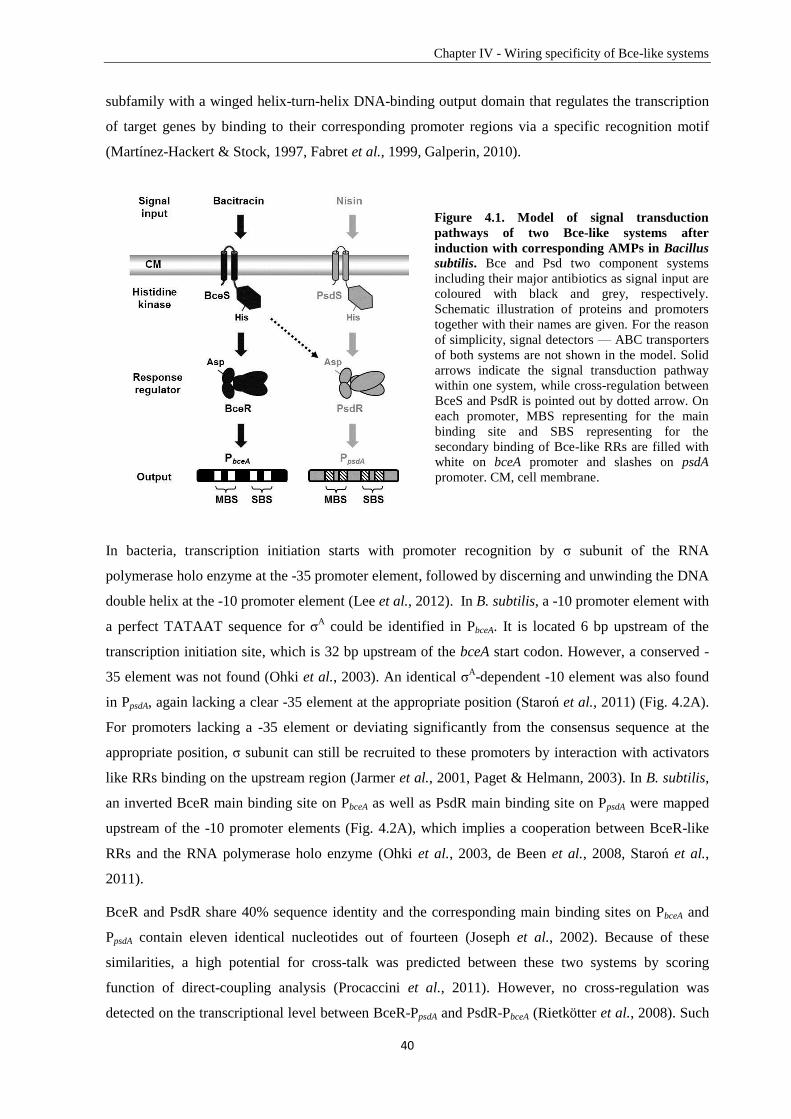
Chapter IV - Wiring specificity of Bce-like systems
40
subfamily with a winged helix-turn-helix DNA-binding output domain that regulates the transcription
of target genes by binding to their corresponding promoter regions via a specific recognition motif
(Martínez-Hackert & Stock, 1997, Fabret et al., 1999, Galperin, 2010).
Figure 4.1. Model of signal transduction
pathways of two Bce-like systems after
induction with corresponding AMPs in Bacillus
subtilis. Bce and Psd two component systems
including their major antibiotics as signal input are
coloured with black and grey, respectively.
Schematic illustration of proteins and promoters
together with their names are given. For the reason
of simplicity, signal detectors — ABC transporters
of both systems are not shown in the model. Solid
arrows indicate the signal transduction pathway
within one system, while cross-regulation between
BceS and PsdR is pointed out by dotted arrow. On
each promoter, MBS representing for the main
binding site and SBS representing for the
secondary binding of Bce-like RRs are filled with
white on bceA promoter and slashes on psdA
promoter. CM, cell membrane.
In bacteria, transcription initiation starts with promoter recognition by σ subunit of the RNA
polymerase holo enzyme at the -35 promoter element, followed by discerning and unwinding the DNA
double helix at the -10 promoter element (Lee et al., 2012). In B. subtilis, a -10 promoter element with
a perfect TATAAT sequence for σA could be identified in PbceA. It is located 6 bp upstream of the
transcription initiation site, which is 32 bp upstream of the bceA start codon. However, a conserved -
35 element was not found (Ohki et al., 2003). An identical σA-dependent -10 element was also found
in PpsdA, again lacking a clear -35 element at the appropriate position (Staroń et al., 2011) (Fig. 4.2A).
For promoters lacking a -35 element or deviating significantly from the consensus sequence at the
appropriate position, σ subunit can still be recruited to these promoters by interaction with activators
like RRs binding on the upstream region (Jarmer et al., 2001, Paget & Helmann, 2003). In B. subtilis,
an inverted BceR main binding site on PbceA as well as PsdR main binding site on PpsdA were mapped
upstream of the -10 promoter elements (Fig. 4.2A), which implies a cooperation between BceR-like
RRs and the RNA polymerase holo enzyme (Ohki et al., 2003, de Been et al., 2008, Staroń et al.,
2011).
BceR and PsdR share 40% sequence identity and the corresponding main binding sites on PbceA and
PpsdA contain eleven identical nucleotides out of fourteen (Joseph et al., 2002). Because of these
similarities, a high potential for cross-talk was predicted between these two systems by scoring
function of direct-coupling analysis (Procaccini et al., 2011). However, no cross-regulation was
detected on the transcriptional level between BceR-PpsdA and PsdR-PbceA (Rietkötter et al., 2008). Such
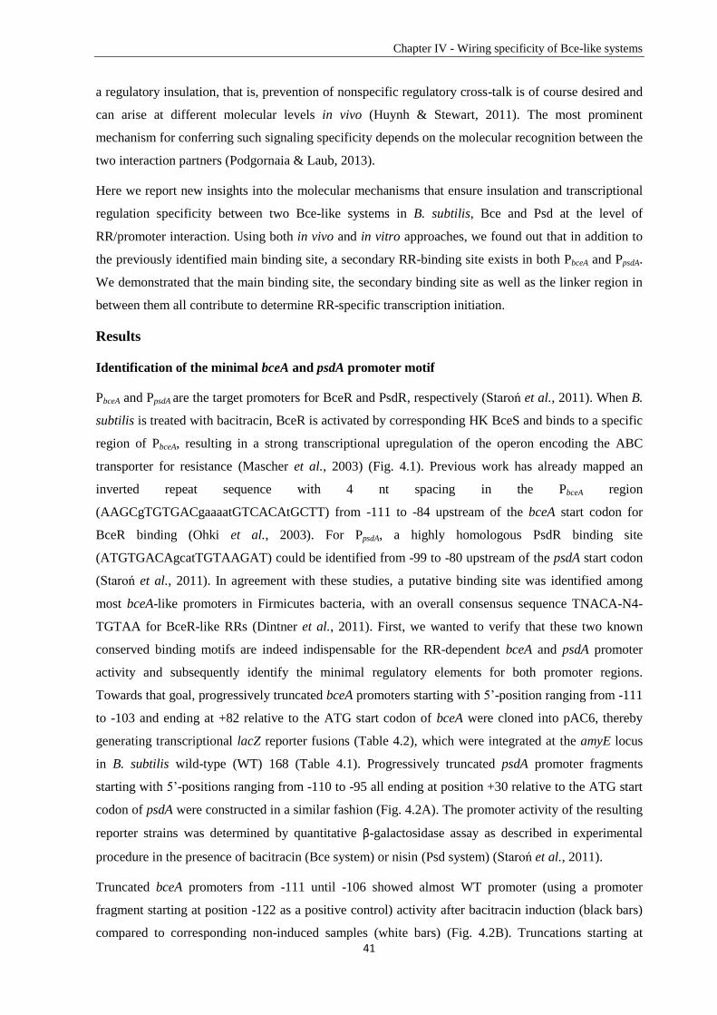
Chapter IV - Wiring specificity of Bce-like systems
41
a regulatory insulation, that is, prevention of nonspecific regulatory cross-talk is of course desired and
can arise at different molecular levels in vivo (Huynh & Stewart, 2011). The most prominent
mechanism for conferring such signaling specificity depends on the molecular recognition between the
two interaction partners (Podgornaia & Laub, 2013).
Here we report new insights into the molecular mechanisms that ensure insulation and transcriptional
regulation specificity between two Bce-like systems in B. subtilis, Bce and Psd at the level of
RR/promoter interaction. Using both in vivo and in vitro approaches, we found out that in addition to
the previously identified main binding site, a secondary RR-binding site exists in both PbceA and PpsdA.
We demonstrated that the main binding site, the secondary binding site as well as the linker region in
between them all contribute to determine RR-specific transcription initiation.
Results
Identification of the minimal bceA and psdA promoter motif
PbceA and PpsdA are the target promoters for BceR and PsdR, respectively (Staroń et al., 2011). When B.
subtilis is treated with bacitracin, BceR is activated by corresponding HK BceS and binds to a specific
region of PbceA, resulting in a strong transcriptional upregulation of the operon encoding the ABC
transporter for resistance (Mascher et al., 2003) (Fig. 4.1). Previous work has already mapped an
inverted repeat sequence with 4 nt spacing in the PbceA region
(AAGCgTGTGACgaaaatGTCACAtGCTT) from -111 to -84 upstream of the bceA start codon for
BceR binding (Ohki et al., 2003). For PpsdA, a highly homologous PsdR binding site
(ATGTGACAgcatTGTAAGAT) could be identified from -99 to -80 upstream of the psdA start codon
(Staroń et al., 2011). In agreement with these studies, a putative binding site was identified among
most bceA-like promoters in Firmicutes bacteria, with an overall consensus sequence TNACA-N4-
TGTAA for BceR-like RRs (Dintner et al., 2011). First, we wanted to verify that these two known
conserved binding motifs are indeed indispensable for the RR-dependent bceA and psdA promoter
activity and subsequently identify the minimal regulatory elements for both promoter regions.
Towards that goal, progressively truncated bceA promoters starting with 5’-position ranging from -111
to -103 and ending at +82 relative to the ATG start codon of bceA were cloned into pAC6, thereby
generating transcriptional lacZ reporter fusions (Table 4.2), which were integrated at the amyE locus
in B. subtilis wild-type (WT) 168 (Table 4.1). Progressively truncated psdA promoter fragments
starting with 5’-positions ranging from -110 to -95 all ending at position +30 relative to the ATG start
codon of psdA were constructed in a similar fashion (Fig. 4.2A). The promoter activity of the resulting
reporter strains was determined by quantitative β-galactosidase assay as described in experimental
procedure in the presence of bacitracin (Bce system) or nisin (Psd system) (Staroń et al., 2011).
Truncated bceA promoters from -111 until -106 showed almost WT promoter (using a promoter
fragment starting at position -122 as a positive control) activity after bacitracin induction (black bars)
compared to corresponding non-induced samples (white bars) (Fig. 4.2B). Truncations starting at
Chapter IV - Wiring specificity of Bce-like systems
42
position -105 and position -104 displayed a decreased promoter activity, while a further truncation of
one additional nucleotide (starting at position -103) led to a complete loss of promoter activity after
bacitracin induction (Fig. 4.2B).
Figure 4.2. Functional analysis of bceA and psdA promoters of B. subtilis. (A) DNA sequence alignment of
the bceA promoter and the psdA promoter. Different motifs are framed and annotated underneath the DNA
sequence. Special positions on each promoter according to the start codon of corresponding regulated gene are
marked with arrows. Half binding sites of Bce-like RRs on each promoter are emphasized in bold style.
Activities of (B) truncated constructions of the bceA promoter (from -122: +82 to -103: +82) and (C) truncated
constructions of the psdA promoter (from -126: +30 to -95: +30) according to the start codon of regulated genes.
Activities of (D) PbceA mutants and (E) PpsdA mutants with MBSR (main binding site random mutation), L
R (linker
random mutation) and SBSR (secondary binding site random mutation) are compared with the corresponding WT
promoters. All promoter constructions were fused to lacZ and introduced into amyE locus of B. subtilis 168.
Cultures growing exponentially in LB were challenged with Zn2+
-bacitracin 30 µg ml-1
(black bars) or nisin 2 µg
ml-1
(grey bars) for 30 min, comparing with the non-induced condition (white bars). β-galactosidase activities are
expressed in Miller Units (MU) (Miller, 1972) and results are shown as the mean plus standard deviation of three
biological replicates. A log scale is applied for reasons of clarity.
Similar results were obtained for truncated psdA promoter fragments after nisin induction (grey bars)
(Fig. 4.2C). Compared to the WT psdA promoter fragment (starting at position -126), no decrease of
promoter activity was observed for truncations with 5’-positions starting from - 110 to -100. The
promoter activities were significantly reduced for fragments chopping at position -99 to -96, while a
truncation at position -95 completely lost its activity after nisin induction.
Our data confirms that the 7-4-7 nt binding motif TGTGACGaaaaTGTCACA of PbceA and the
TGTGACAgcatTGTAAGA binding motif of PpsdA are indeed necessary binding sites (main binding
Chapter IV - Wiring specificity of Bce-like systems
43
site) for BceR and PsdR. Position -104 relative to bceA start codon and position -96 relative to psdA
start codon determine the minimal 5’-end of active RR-dependent promoter fragments. Next, we
addressed the question if there are other binding sites derived from these binding motifs on the
promoter that are also sufficient for RR-dependent promoter activation.
A secondary binding site on bceA and psdA promoters
Sequence analysis of PbceA and PpsdA did not identify a typical -35 region (TTGACA) 17 nt upstream of
the -10 region as normally recognized by σA (Jarmer et al., 2001). However, a 7 nt conserved half
binding site located 13/14 nt downstream of the main binding site (MBS) and 38 nt upstream of the -
10 region was predicted for both the bceA and the psdA promoter regions (Dintner et al., 2011). This
obsession implies the existence of a secondary binding site (SBS) instead of a typical -35 on bceA-like
promoters. Based on this prediction, we annotated a putative 7-4-7 nt secondary binding site and a
linker region (L) between the main and the secondary binding sites on both bceA and psdA promoters
(Fig. 4.2A) and experimentally investigated the function of the predicted promoter motifs by mutating
each of them into a random sequence (randomization). The GC/AT content of the linker region was
kept during the randomization. These mutants were cloned into pAC6 generating transcriptional lacZ
reporter fusions (Table 4.2) and integrated into the chromosome of B. subtilis WT 168 at the amyE
locus (Table 4.1). The promoter activity was determined as described above.
Both the WT bceA promoter (Fig. 4.2D) and the psdA promoter (Fig. 4.2E) showed strong induction
with the corresponding inducers: bacitracin (black bars) or nisin (grey bars), compared to the non-
induced samples (white bars). The weak induction of PpsdA by bacitracin (Fig. 4.2E) was due to the
known cross-phosphorylation of PsdR by BceS (Rietkötter et al., 2008) (Fig. 4.1 dotted arrow).
Mutating the main binding site (MBS) into a random sequence led to a complete loss of activity for
both promoters. The same effect was obtained when randomizing the sequence of the predicted
secondary binding site (Fig. 4.2D and 4.2E). However, activities of both bceA and psdA promoters
only showed a slight decrease by randomly mutating the corresponding linker region (L) between the
two binding sites but keeping the same GC/AT content.
The data demonstrates that on both PbceA and PpsdA, there is a secondary binding site located
downstream of the main binding site with a 13/14 nt linker region in between them. This secondary
binding site seemingly replaced the -35 promoter element, and it is as indispensable as the main
binding site for RR-dependent promoter activity. Additional assays done by randomizing either the
first or the second half of each secondary binding site were in consistency with the results obtained
from the completely randomized secondary binding sites (data not shown), further demonstrating that
each half binding site has the same importance for PbceA and PpsdA activity.
Chapter IV - Wiring specificity of Bce-like systems
44
Major specificity determinants are located in the region containing the linker and the secondary
binding site
So far, we have identified an extended regulatory region in PbceA and PpsA, consisting of two binding
sites and a linker region in between. Since there is no cross-regulation at the RR/promoter interface,
either between BceR-PpsdA or PsdR-PbceA (Rietkötter et al., 2008), further we wanted to analyse the
specificity determinants within bceA/psdA promoters. Towards that aim, a series of chimeric
promoters derived from PbceA and PpsdA were constructed (Table 4.2) and fused with lacZ. Chimeric
promoters BP1-4 are derived from PbceA (black) with gradually substituting PpsdA (grey) at the 3’-
terminal end (Fig. 4.3A). Chimeric promoters PB1-4 are derived from PpsdA (grey) with increasing of
3’-fragments from PbceA (black) (Fig. 4.3B). Again, these constructs were integrated into the amyE
locus of the B. subtilis WT 168 (Table 4.1). To specifically eliminate any cross-talk between the Bce
and Psd systems, these chimeric promoters, WT PbceA and WT PpsdA fragments as references, were
transformed into the ∆bceRS strain (TMB1460) and the ∆psdRS strain (TMB1462) (Table 4.1).
Compared to the WT strain, the ∆bceRS and the ∆psdRS strains remove the effect of cross-
phosphorylation and hence provide a clearer view of RR/promoter specificity.
PbceA showed the same high activity in the ∆psdRS mutant (Fig. 4.3D) as in WT strain (Fig. 4.3C) after
bacitracin induction, but no activity after nisin induction in either the WT (Fig. 4.3C) or the ∆bceRS
background (Fig. 4.3E). PpsdA was also highly induced by nisin in both the WT strain (Fig. 4.3C) and
the ∆bceRS mutant (Fig. 4.3E). Importantly, the moderate induction of PpsdA by bacitracin in WT
background (Fig. 4.3C) was not detected in ∆psdRS mutant (Fig. 4.3D) due to the elimination of cross-
phosphorylation between BceS and PsdR. These results are in agreement with previous studies that
there is no cross-regulation at the RR/promoter level.
Chimeric promoters BP1 and BP2 showed high activity after induction with bacitracin in both the WT
strain (Fig. 4.3C) and the ∆psdRS strain (Fig. 4.3D), but no activity upon nisin induction in either the
WT strain (Fig. 4.3C) or the ∆bceRS strain (Fig. 4.3E). Hence, BP2 could be recognized by BceR, but
not by PsdR. These results indicate that specificity determinants are located within the region
upstream of and including the secondary binding site. The chimeric promoter BP3, could not be
induced either by bacitracin in the ∆psdRS background (Fig. 4.3D) or by nisin in the ∆bceRS
background (Fig. 4.3E). However, it showed moderate activity in the WT background (Fig. 4.3C) after
induction with bacitracin. Surprisingly, BP4, possessing the whole region downstream of the main
binding site of PpsdA, was not only moderately induced by bacitracin in the ∆psdRS background (Fig.
4.3D) but also by nisin in the ∆bceRS background (Fig. 4.3E), indicating a change of specificity from
PbceA to PpsdA. These results of BP2 and BP4 demonstrate that major specificity determinants of PpsdA
are located in the region containing the linker and the secondary binding site.
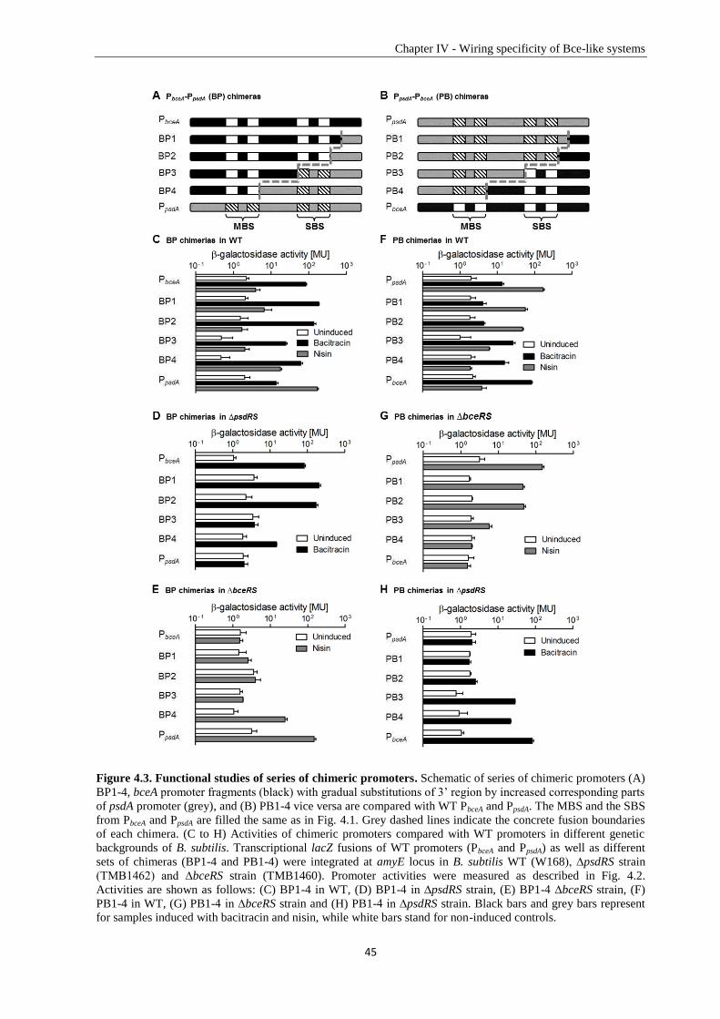
Chapter IV - Wiring specificity of Bce-like systems
45
Figure 4.3. Functional studies of series of chimeric promoters. Schematic of series of chimeric promoters (A)
BP1-4, bceA promoter fragments (black) with gradual substitutions of 3’ region by increased corresponding parts
of psdA promoter (grey), and (B) PB1-4 vice versa are compared with WT PbceA and PpsdA. The MBS and the SBS
from PbceA and PpsdA are filled the same as in Fig. 4.1. Grey dashed lines indicate the concrete fusion boundaries
of each chimera. (C to H) Activities of chimeric promoters compared with WT promoters in different genetic
backgrounds of B. subtilis. Transcriptional lacZ fusions of WT promoters (PbceA and PpsdA) as well as different
sets of chimeras (BP1-4 and PB1-4) were integrated at amyE locus in B. subtilis WT (W168), ∆psdRS strain
(TMB1462) and ∆bceRS strain (TMB1460). Promoter activities were measured as described in Fig. 4.2.
Activities are shown as follows: (C) BP1-4 in WT, (D) BP1-4 in ∆psdRS strain, (E) BP1-4 ∆bceRS strain, (F)
PB1-4 in WT, (G) PB1-4 in ∆bceRS strain and (H) PB1-4 in ∆psdRS strain. Black bars and grey bars represent
for samples induced with bacitracin and nisin, while white bars stand for non-induced controls.
Chapter IV - Wiring specificity of Bce-like systems
46
Chimeric promoters PB1 and PB2 showed a decreased activity after induction with nisin in both the
WT background (Fig. 4.3F) and the ∆bceRS mutant (Fig. 4.3G) relative to PpsdA, and no bacitracin
induction in the ∆psdRS mutant (Fig. 4.3H), indicating no change of specificity. These results
corroborate that the region downstream of the secondary binding site on PbceA is not relevant for the
RR/promoter specificity. Interestingly, PB3 showed a significantly decreased activity in the ∆bceRS
mutant with nisin induction (Fig. 4.3G) and a hugely increased activity in the ∆psdRS mutant with
bacitracin induction (Fig. 4.3H). It therefore behaved differently from the corresponding BP3
construct that had no activity in either. Construct PB4 was not inducible by nisin in the ∆bceRS strain
(Fig. 4.3G), but instead showed high induction by bacitracin in the ∆psdRS strain (Fig. 4.3H). The
promoter activities of PB3 and PB4 in the WT strain (Fig. 4.3F) were in accordance with those
observed in both mutant backgrounds. This data indicates that the change of specificity from PpsdA to
PbceA started at PB3 with the secondary binding site being switched into PbceA, and obtained a further
promotion in construct PB4 by an additional substitution of the linker region.
Taken together, the analysis of chimeric promoter constructs demonstrates that the region containing
the linker and the secondary binding site of PbceA/PpsdA includes major specificity determinants for
BceR/PsdR recognition.
Rewiring specificity from PbceA to PpsdA and dissecting the role of specificity determinants on PpsdA
The analysis of chimeric promoter constructs showed that on the psdA promoter the region
downstream of the main binding site contained the major specificity determinants for PsdR binding.
This region contains two motifs: the linker and the secondary binding site. To further elaborate the
functionality of the main binding site, the linker region and the secondary binding site on psdA
promoter for PsdR recognition, additional chimeric promoters were constructed with different
combinations of these three motifs on PbceA replaced by the corresponding region of PpsdA (Fig. 4.4A) to
rewire specificity from PbceA to PpsdA. Promoter activities were measured as described above in the WT
strain (Fig. 4.4C), the ∆psdRS strain (Fig. 4.4D) and the ∆bceRS strain (Fig. 4.4E).
Compared to PbceA, replacing only the main binding site (M), the linker (L) or both (M+L) of PbceA with
the corresponding region of PpsdA showed decreased promoter activity in the WT strain (Fig. 4.4C) as
well as in the ∆psdRS mutant (Fig. 4.4D) after induction with bacitracin. In contrast, no increasing of
the promoter activity was observed in either the WT strain (Fig. 4.4C) or the ∆bceRS mutant (Fig.
4.4E) after induction by nisin. This data indicates that the main binding site, the linker or both of PpsdA
are not enough to determine specificity. Changing the secondary binding site (S) on PbceA into PpsdA led
to a decrease of promoter activity in the WT strain (Fig. 4.4C) as well as in the ∆psdRS mutant (Fig.
4.4D) after induction with bacitracin, and a slight but detectable increase of promoter activity in the
∆bceRS mutant (Fig. 4.4E) after induction with nisin. This data indicates that exchanging only the
secondary binding site alone already conferred a change of promoter specificity from PbceA to PpsdA.
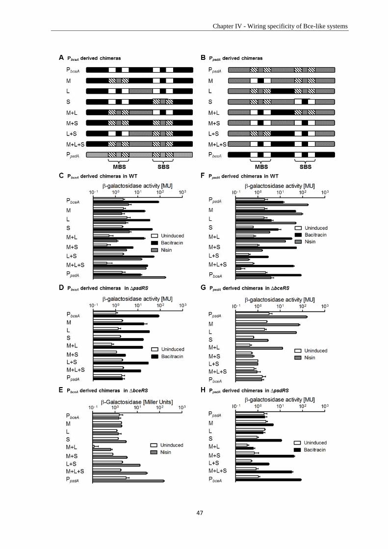
Chapter IV - Wiring specificity of Bce-like systems
47
Chapter IV - Wiring specificity of Bce-like systems
48
Figure 4.4. Unravelling the roles of different promoter elements in RR-promoter specificity. (A and B)
Schematic of chimeric promoters derived from PbceA and PpsdA with specific regions switched into the
corresponding part of PpsdA and PbceA, respectively, compared with two WT promoters. Names on the left side of
each chimeric promoter stand for the exchanging region: M, main binding site; L, linker; S, secondary binding
site. MBS and SBS from PbceA and PpsdA are filled the same as in Fig. 4.1. Activities of WT and chimeric
promoters in different genetic backgrounds of B. subtilis are shown as follows: (C) PbceA derived chimeras in WT,
(D) PbceA derived chimeras in ∆psdRS strain, (E) PbceA derived chimeras in ∆bceRS strain, (F) PpsdA derived
chimeras in WT, (G) PpsdA derived chimeras in ∆bceRS strain and (H) PpsdA derived chimeras in ∆psdRS strain.
Black bars and grey bars represent for samples induced with bacitracin and nisin, while white bars stand for non-
induced controls.
Compared to only the secondary binding site switch (S), exchanging both the main binding site and
the secondary binding site simultaneously (M+S) resulted in a severe decrease of the promoter activity
in the ∆psdRS mutant after induction with bacitracin (Fig. 4.4D), while an increase of the promoter
activity in the ∆bceRS mutant after induction with nisin (Fig. 4.4E). This indicates that based on the
primary specificity determinant — the secondary binding site — the main binding site assists it to
achieve a higher promoter activity with the cognate RR, PsdR, and a lower promoter activity with the
non-cognate RR, BceR. Substitution of the linker together with the secondary binding site (L+S)
resulted in a higher promoter activity compared to only exchanging the secondary binding site (S) in
both the ∆psdRS mutant after bacitracin induction (Fig. 4.4D) and the ∆bceRS mutant after nisin
induction (Fig. 4.4E). This data suggests that the linker region (L) can enhance promoter activity with
both cognate PsdR and non-cognate BceR.
Taken together, these results indicate that the secondary binding site on PpsdA mainly determines PsdR-
PpsdA specificity, even though the intensity of induction with secondary binding site substitution alone
is not very strong. The linker cannot determine specificity by itself but can increase promoter activity
with both BceR and PsdR, which explains the change of specificity that was detected for construct
BP4 including the linker and the secondary binding site but not for construct BP3 with only the
secondary binding site. Despite the fact that the main binding site is absolutely crucial for
RR/promoter interaction, the main binding site of PpsdA alone cannot determine specificity. Instead, it
supports the secondary binding site in strengthening specificity. Not surprisingly, switching all three
elements together (M+L+S) resulted in the highest change of specificity in the ∆bceRS mutant after
induction with nisin (Fig. 4.4E), demonstrating that all three parts together contribute to the specificity.
Rewiring specificity from PpsdA to PbceA and dissecting the role of specificity determinants on PbceA
In order to support the results obtained above, a similar approach was performed towards rewiring the
specificity from PpsdA to PbceA. In contrast to the results obtained for the series of PB chimeras (change
of specificity from PpsdA to PbceA started with PB3), the change of specificity from PbceA to PpsdA started
later with construct BP4. This may indicate a subtle difference in the BceR-PbceA specificity
determination compared to PsdR-PpsdA. To answer this question, chimeric promoters with different
combinations of the main binding site, the linker region and the secondary binding site of PpsdA being
replaced by the corresponding regions of PbceA were constructed (Fig. 4.4B) (Table 4.2) and the
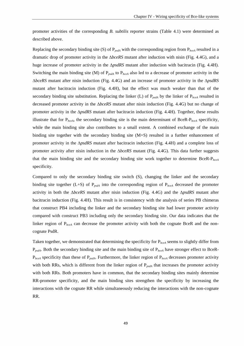
Chapter IV - Wiring specificity of Bce-like systems
49
promoter activities of the corresponding B. subtilis reporter strains (Table 4.1) were determined as
described above.
Replacing the secondary binding site (S) of PpsdA with the corresponding region from PbceA resulted in a
dramatic drop of promoter activity in the ∆bceRS mutant after induction with nisin (Fig. 4.4G), and a
huge increase of promoter activity in the ∆psdRS mutant after induction with bacitracin (Fig. 4.4H).
Switching the main binding site (M) of PpsdA to PbceA also led to a decrease of promoter activity in the
∆bceRS mutant after nisin induction (Fig. 4.4G) and an increase of promoter activity in the ∆psdRS
mutant after bacitracin induction (Fig. 4.4H), but the effect was much weaker than that of the
secondary binding site substitution. Replacing the linker (L) of PpsdA by the linker of PbceA resulted in
decreased promoter activity in the ∆bceRS mutant after nisin induction (Fig. 4.4G) but no change of
promoter activity in the ∆psdRS mutant after bacitracin induction (Fig. 4.4H). Together, these results
illustrate that for PbceA, the secondary binding site is the main determinant of BceR-PbceA specificity,
while the main binding site also contributes to a small extent. A combined exchange of the main
binding site together with the secondary binding site (M+S) resulted in a further enhancement of
promoter activity in the ∆psdRS mutant after bacitracin induction (Fig. 4.4H) and a complete loss of
promoter activity after nisin induction in the ∆bceRS mutant (Fig. 4.4G). This data further suggests
that the main binding site and the secondary binding site work together to determine BceR-PbceA
specificity.
Compared to only the secondary binding site switch (S), changing the linker and the secondary
binding site together (L+S) of PpsdA into the corresponding region of PbceA decreased the promoter
activity in both the ∆bceRS mutant after nisin induction (Fig. 4.4G) and the ∆psdRS mutant after
bacitracin induction (Fig. 4.4H). This result is in consistency with the analysis of series PB chimeras
that construct PB4 including the linker and the secondary binding site had lower promoter activity
compared with construct PB3 including only the secondary binding site. Our data indicates that the
linker region of PbceA can decrease the promoter activity with both the cognate BceR and the non-
cognate PsdR.
Taken together, we demonstrated that determining the specificity for PbceA seems to slightly differ from
PpsdA. Both the secondary binding site and the main binding site of PbceA have stronger effect to BceR-
PbceA specificity than these of PpsdA. Furthermore, the linker region of PbceA decreases promoter activity
with both RRs, which is different from the linker region of PpsdA that increases the promoter activity
with both RRs. Both promoters have in common, that the secondary binding sites mainly determine
RR-promoter specificity, and the main binding sites strengthen the specificity by increasing the
interactions with the cognate RR while simultaneously reducing the interactions with the non-cognate
RR.
Chapter IV - Wiring specificity of Bce-like systems
50
In vitro, BceR has a higher binding affinity for its native PbceA than for PpsdA
Promoter activity assays demonstrated that PpsdA had no activity upon bacitracin induction in the
∆psdRS mutant (Fig. 4.3D), which indicates that there is no cross-regulation between BceR and PpsdA in
vivo. Next, we wanted to investigate if the binding of BceR is also specific to its native promoter PbceA
in vitro. BceR carrying an N-terminal His10-tag with the expected molecular mass around 27 KDa was
produced and purified from the cytoplasmic fraction of E. coli C43 (DE3) containing plasmid pCF120
(Table 4.2), which was in consistent with its observed migration in SDS-PAGE. EMSAs were
performed with this BceR and two bceA-like promoters PbceA and PpsdA. 300 bp promoter DNA
fragments of PbceA or PpsdA containing the main binding site, the linker region and the secondary
binding site were amplified and labeled at the 5’-end with 6FAM by PCR. 6FAM labeled PsigW (the
target promoter of an ECF sigma factor in B. subtilis) was used as a negative control.
Results of band shift assays with BceR and PbceA are shown in Figure 4.5A. Increasing concentrations
of phosphorylated BceR (BceR-P) were incubated with 30 fmol of 6FAM-PbceA (from lane 2 to lane 5),
demonstrating a concentration-dependent binding of BceR-P to PbceA. The first shift was observed at
1.0 µM BceR-P representing the initial binding event of BceR-P to PbceA. An additional shift occurred
at slightly higher BceR-P concentrations presumably represents a second binding event. In contrast,
unphosphorylated BceR performed much weaker binding (data not shown), which demonstrated that
RR-phosphorylation is necessary and seems to promote DNA binding by increasing BceR affinity to
PbceA.
Figure 4.5. In vitro binding of BceR-P to bceA-like promoters. Increasing concentrations of phosphorylated
10×His-BceR were incubated with 30 fmol of different 6FAM-labeled promoter DNA fragments as follows: (A)
PbceA from -122 to +82, (B) PpsdA from -126 to +30, (C) PbceA SBSR (secondary binding site inactivated), (D) PbceA
MBSR (main binding site inactivated), and (E) PsigW as a negative control. Schematics of bceA-like promoters and
corresponding mutants are shown in the lower left corner of each gel. The concentrations of phosphorylated
BceR are indicated above the gel by [BceR-P] in μM. 900 fmol of unlabelled competitor (comp.) DNA
fragments encoding PbceA, PpsdA and PsigW were added to gel (A) and (B) in lane 6, 7 and 8, respectively.
Chapter IV - Wiring specificity of Bce-like systems
51
EMSAs were also performed between BceR-P and PpsdA (Fig. 4.5B). Two successive shifts of PpsdA
band in lane 3 and lane 4 compared to free PpsdA DNA fragment (lane 1) demonstrated that BceR-P can
also bind successively to the non-cognate but highly related PpsdA in vitro. In contrast, no shift was
observed for the PsigW DNA fragment (Fig. 4.5E), confirming the overall specificity of the assay:
BceR-P cannot bind to promoter fragments that do not harbor the binding motifs of a PbceA-like
promoter.
To further illustrate the specificity and affinities of BceR-P binding to PbceA and PpsdA, 900 fmol of
unlabeled promoter fragments were used as competitor DNA (Fig. 4.5A/4.5B lane 6-8). Co-incubation
of BceR-P with 30 fmol 6FAM-PbceA and 900 fmol unlabeled PbceA fragment (Fig. 4.5A lane 6)
completely abolished the retardation of the labeled PbceA fragment due to the competitive binding of
BceR-P to an excess of unlabeled PbceA. However, the shift of 6FAM-PbceA band was not influenced by
adding a 30-fold amount of unlabeled PpsdA (Fig. 4.5A lane 7) or PsigW (Fig. 4.5A lane 8),
demonstrating a much higher affinity of BceR-P for its cognate target promoter. In contrast, the
retardation of the 6FAM-PpsdA DNA fragment was abolished by either addition of an extra 30-fold
unlabeled PbceA (Fig. 4.5B lane 6) or unlabeled PpsdA (Fig. 4.5B lane 7) fragments but not by PsigW (Fig.
4.5B lane 8). These results clearly demonstrate that BceR-P has a higher binding affinity for PbceA and
preferentially binds to its native promoter compared to PpsdA in vitro.
Taken together, our results strongly suggest that phosphorylated BceR undergoes two successive
binding reactions with both PbceA and PpsdA in vitro. However, the binding affinity for its cognate target
promoter PbceA is much higher than for PpsdA that determines the in vivo specific transcription initiation.
Unfortunately, any affects to purify PsdR failed, thereby preventing the performance of similar in vitro
studies on PsdR-PpsdA/PbceA interactions.
Cooperative binding of BceR to two binding sites on PbceA
The in vivo promoter activity assays demonstrated that both binding sites on PbceA are indispensable for
BceR-PbceA interaction (Fig. 4.2D). Moreover, EMSA on complete promoter fragment strongly
suggests BceR has two successive binding events at PbceA in vitro (Fig. 4.5A). To discriminate between
the individual binding reactions, we next performed EMSAs with BceR-P on 6FAM labeled bceA
promoter DNA-fragments carrying random mutation of either the main binding site or the secondary
binding site.
Incubation of BceR-P with labeled PbceA SBSR (PbceA containing a randomized and hence inactive
secondary binding site) obtained only a single shift at a BceR-P concentration of 1.0 µM (Fig. 4.5C), a
concentration comparable to the threshold concentration as the intact PbceA fragment (Fig. 4.5A lane 3).
Increasing the BceR-P concentration did not lead to any additional shift. Hence, PbceA containing only
the main binding site merely allows the first binding event that is the binding of BceR-P to the main
binding site. The identical BceR-P concentrations required for shifting either the WT or the SBSR
Chapter IV - Wiring specificity of Bce-like systems
52
fragments indicates that binding of BceR-P to the main binding site is independent from the secondary
binding site.
Incubation of BceR-P with labeled PbceA MBSR (PbceA containing a randomized and hence inactive
main binding site) basically failed to retard the DNA-fragment within the same concentration range
(Fig. 4.5D). Hence, a non-functional main binding site prevents both binding events of BceR-P to
PbceA. This result indicates that the second binding event, which requires the secondary binding site,
depends on and occurs after BceR-P binding to the main binding site.
Together, these results demonstrate that the two binding sites contribute in a successive manner to the
BceR-PbceA interaction and imply a cooperative binding model of BceR to the two binding sites of
PbceA. BceR first binds to the main binding site independently, which then supports the subsequent
binding to the secondary binding site. In order to challenge this hypothesis we finally analyzed the
binding affinities of BceR-P on PbceA/PpsdA by surface plasmon resonance (SPR) spectroscopy.
Determination of binding kinetics of BceR-promoter interaction unravels the mechanism that
determines BceR promoter specificity
To quantify the binding kinetics of the BceR-promoter interaction we used SPR analysis. As first step
we captured a biotin-labeled DNA-fragment comprising the PbceA region to a sensor chip previously
immobilized with streptavidin. Then, increasing concentrations of His10-BceR and His10-BceR
previously phosphorylated using phosphoramidate (BceR-P) were injected over the chip surface. It can
be clearly seen that non-phosphorylated BceR does not interact with the PbceA promoter (Fig. 4.6A),
whereas BceR-P showed clear binding (Fig. 4.6B). Since BceR has two binding sites on the DNA-
fragment used for SPR, we used the OneToTwo evaluation algorithm. The binding kinetics underlying
this sensorgram could be determined revealing that these two binding events mainly differ in their
association rates (ka1=7.46x10⁴ M*s; ka2=1.58x10
6 M*s) rather than the dissociation rates
(kd1=5.57x10⁻⁴
/s; kd2=8.05x10⁻⁴
/s) resulting in two binding events that differ in their overall affinity
(KD1=7.47 nM; KD2=0.51 nM). As next step, we determined the binding kinetics between BceR-P and
PbceA when the main binding site (MBSR) or the secondary binding site (SBS
R) was destroyed,
respectively. It can be clearly seen that inactivation of the main binding site completely prevented
DNA-binding of BceR-P (Fig. 4.6C). However, when only the secondary binding site was destroyed, a
clear DNA-binding of BceR could be observed (Fig. 4.6D). In contrast to the sensorgram when both
binding sites are intact (Fig. 4.6B), the sensorgram here follows a real 1:1 binding kinetic that we
quantified with an association rate of ka=6.59 x 10⁵ M*s and a dissociation rate kd=9.5x10
-4/s making
an overall binding affinity of KD=1.4 nM. Furthermore, we observed that the overall response units
were reduced approximately 1/3 compared to the sensorgram representing the BceR-P/PbceA, which
probably represents the portion of BceR-P binding to the secondary binding site. Our data clearly
shows that the main binding site of PbceA region is essential for binding of the RR to the DNA. The RR
obviously cannot bind the secondary binding site when the main binding site was not previously
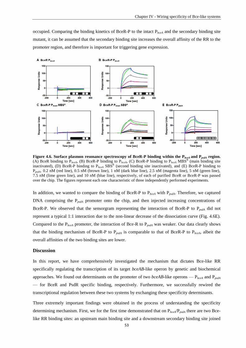
Chapter IV - Wiring specificity of Bce-like systems
53
occupied. Comparing the binding kinetics of BceR-P to the intact PbceA and the secondary binding site
mutant, it can be assumed that the secondary binding site increases the overall affinity of the RR to the
promoter region, and therefore is important for triggering gene expression.
Figure 4.6. Surface plasmon resonance spectroscopy of BceR-P binding within the PbceA and PpsdA region.
(A) BceR binding to PbceA, (B) BceR-P binding to PbceA, (C) BceR-P binding to PbceA MBSR (main binding site
inactivated), (D) BceR-P binding to PbceA SBSR (second binding site inactivated), and (E) BceR-P binding to
PpsdA. 0.2 nM (red line), 0.5 nM (brown line), 1 nM (dark blue line), 2.5 nM (magenta line), 5 nM (green line),
7.5 nM (lime green line), and 10 nM (blue line), respectively, of each of purified BceR or BceR-P was passed
over the chip. The figures represent each one characteristic of three independently performed experiments.
In addition, we wanted to compare the binding of BceR-P to PbceA with PpsdA. Therefore, we captured
DNA comprising the PpsdA promoter onto the chip, and then injected increasing concentrations of
BceR-P. We observed that the sensorgram representing the interaction of BceR-P to PpsdA did not
represent a typical 1:1 interaction due to the non-linear decrease of the dissociation curve (Fig. 4.6E).
Compared to the PbceA promoter, the interaction of Bce-R to PpsdA was weaker. Our data clearly shows
that the binding mechanism of BceR-P to PpsdA is comparable to that of BceR-P to PbceA, albeit the
overall affinities of the two binding sites are lower.
Discussion
In this report, we have comprehensively investigated the mechanism that dictates Bce-like RR
specifically regulating the transcription of its target bceAB-like operon by genetic and biochemical
approaches. We found out determinants on the promoter of two bceAB-like operons — PbceA and PpsdA
— for BceR and PsdR specific binding, respectively. Furthermore, we successfully rewired the
transcriptional regulation between these two systems by exchanging these specificity determinants.
Three extremely important findings were obtained in the process of understanding the specificity
determining mechanism. First, we for the first time demonstrated that on PbceA/PpsdA there are two Bce-
like RR binding sites: an upstream main binding site and a downstream secondary binding site joined
Chapter IV - Wiring specificity of Bce-like systems
54
together by a flexible linker region (Fig. 4.2D and 4.2E). Second, the secondary binding sites on both
PbceA and PpsdA primarily dictate RR-promoter specificity. However, the secondary binding site by itself
is not enough to fully dictate specificity. Together with the high affinity main binding site and the
linker region, they are able to completely mediate RR-promoter specificity (Fig. 4.4). Third, we were
able to demonstrate that BceR has two-step binding event on PbceA. By separating the binding, we
demonstrated that BceR can bind to the main binding site independently with high affinity, which
assists and stabilizes the binding to the low affinity secondary binding site (Fig. 4.5 and 4.6).
Taken together, our findings strongly suggest that BceR has a hierarchical and cooperative binding
model to these two binding sites on PbceA. A BceR dimer first binds tightly to the main binding site
with low specificity, which favors and stabilizes the binding of a second BceR dimer to the secondary
binding site specifically. The linker regions of PbceA and PpsdA with different AT/GC contents may play
a role in maintaining different distances and/or angles of the two binding sites, and hence adjust the
promoter activity. The overall binding affinity of BceR to its cognate promoter PbceA, as a combined
effect of all three promoter motifs – the main binding site, the linker, and the secondary binding site, is
much higher than to the non-cognate promoter, which is an intrinsic ability to discriminate the cognate
promoter from the pool of similar non-cognate ones in a bacterial cell. RR determines the specific
transcription initiation, most likely by direct interaction with the RNA polymerase so that recruits the
polymerase to the promoter. The cooperative binding mechanism has already been confirmed by PhoB
binding to the target pstS promoter containing double pho boxes (Blanco et al., 2012). EMSAs showed
that two PhoBE dimers bind to two consecutive pho boxes in a hierarchical and cooperative manner,
which is at low concentration PhoBE dimer first binds to the high-affinity pho box 1 and with
increasing concentration the first binding assists the subsequent dimer bind to the downstream low-
affinity pho box 2.
RR-promoter specificity is mainly determined by molecular recognition on the interaction surfaces
between amino acids on the RR and bases on the promoter (Rohs et al., 2010). Most of studies about
RR-promoter specificity focused on searching for amino acids on the DNA binding domain of RRs
that can specifically recognize their cognate promoters. Structures of PhoB and OmpR C-terminal
DNA binding domain indicated that these amino acids are located on the C-terminal helix α3
(interaction with the DNA major groove) and the loop of the C-terminal hairpin (interaction with the
DNA minor groove) (Martínez-Hackert & Stock, 1997, Blanco et al., 2002, Rhee et al., 2008). A
previous study showed that a single amino acid, which is Glu215 on the α3 helix of Mycobacterium
tuberculosis PhoP, is an important residue that significantly contributes to specific DNA binding site
recognition. By mutating Glu215 into Ala, PhoP lost the ability to discriminate the specific binding
site from the nonspecific DNA with sequence comparable compositions (Das et al., 2010). Similar
studies about RRs FNR and CRP in E. coli further showed that exchanging Arg180 and Gly184 on the
recognition-helix of CRP into Val and Ser of FNP was able to convert the binding specificity (Bell et
al., 1989, Spiro et al., 1990). These two amino acids can specifically recognize bases (G at position 5
Chapter IV - Wiring specificity of Bce-like systems
55
and C at position 8) on CRP binding site, and discriminate bases T and A on FNR binding site. Instead
of searching for amino acids on the DNA binding domain of the RR, our study focused on the target
promoters of Bce-like RRs and demonstrated that promoter specificity is determined by three
promoter motifs together. The mechanism is different from previous studies that two bases on one
binding site can determine RR-promoter specificity. This is probably due to that the DNA recognition
motifs, the α3 helix and the loop of the C-terminal hairpin, are highly similar between BceR and PsdR.
With only one promoter motif, it is hard to maintain the specificity and the high activity
simultaneously. To solve this problem, B. subtilis developed a complicated regulatory region on the
target promoter of the Bce-like RR, which contains a main binding site determines the binding affinity,
a secondary binding site determines the binding specificity and a linker region ensures the structure of
the promoter that contribute differently to maintain the RR-promoter specificity.
Chapter IV - Wiring specificity of Bce-like systems
56
Experimental procedures
Bacterial strains and growth conditions
All strains used in this study are listed in Table 4.1. E. coli DH5α and XL1-blue were used for cloning. All B.
subtilis strains used in this study are derivatives of the laboratory WT strain 168. E. coli and B. subtilis were
grown routinely in Luria-Bertani (LB) medium at 37°C with aeration. B. subtilis was transformed by natural
competence as previously described (Harwood & Cutting, 1990). Ampicillin (100 µg ml-1
) was used for
selection of all plasmids in E. coli. Chloramphenicol (5 µg ml-1
), spectinomycin (100 µg ml-1
), and erythromycin
(1 µg ml-1
) plus lincomycin (25 µg ml-1
) for macrolide-lincosamide-streptogramin B (mls) resistance were used
for the selection of B. subtilis mutants. Bacitracin was supplied as the Zn2+
-salt. Growth was measured as optical
density at 600 nm wavelength (OD600). Solid media contained 1.5 % (w/v) agar.
Construction of plasmids and genetic techniques
All plasmid constructs in this study are listed in Table 4.2. The corresponding primer sequences are provided in
the supplemental material (Table S4.1). Different promoter fragments derived from PbceA and PpsdA were fused to
lacZ and cloned into the vector pAC6 (Stülke et al., 1997) with EcoRI/BamHI sites. The details of promoter
constructs are given in Table 4.2. For construction of the BceR-production plasmid in E. coli, bceR was
amplified with primers TM2007/2008 and cloned into vector pET16b with XhoI and BamHI obtaining pCF120,
resulting in an N-terminal His10-tag fusion. Constructs for unmarked gene deletion in B. subtilis were cloned into
the vector pMAD (Arnaud et al., 2004). For each operon to be deleted, 800-1000 bp fragments located
immediately before the start codon of the first gene (“up” fragment) and after the stop codon of the last gene
(“down” fragment) were amplified. The primers were designed to create a 17-20 bp overlap between the PCR-
products (Table 4.2), facilitating fusion of the fragments by PCR overlap extension and subsequent cloning into
pMAD. Gene deletions were performed as previously described (Arnaud et al., 2004). All constructs were
checked by sequencing, and all B. subtilis strains created were verified by colony PCR using appropriate primers.
β-galactosidase assays
Assays on promoter activities were performed as described previously (Mascher et al., 2004). In brief, cells were
inoculated from fresh overnight cultures and grown in LB medium at 37°C with aeration until they reached an
OD600 between 0.4 and 0.5. The cultures were split into 2 mL aliquots and challenged with 30 µg ml-1
bacitracin
or 2 µg ml-1
nisin with one aliquot left untreated (non-induced control). After incubation for an additional 30 min
at 37°C with aeration, the cultures were harvested and the cell pellets were frozen at -20°C. The β-galactosidase
activities were determined as described, with normalization to cell density (Miller, 1972).
Expression and purification of His-tagged BceR
To produce BceR carrying an N-terminal His10-tag, E. coli C43 (DE3) cells harboring plasmid pCF120 were
grown at 25 °C with agitation until they reached an OD600 of about 0.4. 0.5 mM of IPTG was added to the
culture and incubation was continued at 18 °C with agitation overnight. Cells were harvested by centrifugation at
4,400 × g for 10 min. The cell pellet was washed with buffer A (20 mM KPi [pH7.5], 100 mM NaCl) and stored
at -20 °C until use.
To purify His10-tagged BceR, cells were resuspended in buffer B (50 mM KPi [pH 7.5], 500 mM NaCl, 5 mM β-
ME, 10 mM imidazole and 10 % (w/v) glycerol) supplemented with 0.1 mM phenylmethylsulfonyl fluoride
Chapter IV - Wiring specificity of Bce-like systems
57
(PMSF) plus 2 mg DNaseI and disrupted by three passages through a French pressure cell (Thermo Fisher) at
20,000 PSI. Unbroken cells were removed by centrifugation at 17,000 × g for 20 min and the cell-free
supernatant was filtered through a 0.45 µm syringe filter before loading onto a 1 ml Ni2+
-NTA resin column
(Qiagen) pre-equilibrated with 5 column volumes (CVs) of buffer B. Loading was followed by washing with 5
CVs of buffer B and then with 5 CVs of buffer B containing 100 mM imidazole. BceR was eluted with buffer B
supplemented with 250 mM imidazole. Fractions containing BceR were pooled and dialyzed in buffer C (50 mM
Tris-HCl [pH 7.5], 150 mM NaCl, 10 mM MgSO4, 5 mM β-ME, 5 mM imidazole and 10 % (w/v) glycerol)
using dialysis membrane (neo Lab) at room temperature for 1 h. Protein concentration was determined with
Roti®-Nanoquant (Carl Roth), and the protein were stored at 4 °C.
Electrophoretic Mobility Shift Assays (EMSA)
For electrophoretic mobility shift assays, different DNA fragments (around 300bp) generated by PCR using
primers TM3146 (5’ terminal 6FAM labeled) and TM3137 were purified by gel extraction. Unlabeled DNA
fragments were generated by PCR using primers TM3136/TM3137 and purified by HiYield® Gel/PCR DNA
Extraction Kit (SLG®). N-terminal His10-BceR samples in the non-phosphorylated state and after
phosphorylation by 50 mM phosphoramidate (PA) at room temperature for 2 h were centrifuged down by 16,060
× g at 4 °C for 10 min to remove the aggregated protein. Protein concentrations of the supernatants were
determined with Roti®-Nanoquant (Carl Roth) and the proteins were stored on ice. Binding reactions were set
by incubating 6FAM-labelled DNA-fragments with different concentrations of His10-BceR at room temperature
for 20 min. The reaction mixture included 30 fmol labeled target DNA and 0, 0.5, 1.0, 1.5, 2.0 µM protein with
binding buffer (20 mM Tris-HCl [pH 7.5], 50 mM KCl, 10 mM MgSO4, 1 mM DTT, 5 µg ml-1
salmon sperm
DNA and 4 % (w/v) glycerol) in a total volume of 5.5 µl. Unlabeled competitor DNA was added to the system to
a final concentration of 900 fmol. Samples were loaded on a 6% native polyacrylamide gel and electrophoresis
was performed by 300 Volt for 15 min in TBE buffer. Gels were detected by PhosphorImager (Typhoon Trio™,
GE Healthcare).
Surface Plasmon Resonance (SPR) spectroscopy
SPR assays were performed in a Biacore T200 using carboxymethyl dextran sensor chips pre-coated with
streptavidin (Xantec SAD500-L, XanTec Bioanalytics GmbH, Düsseldorf, Germany). All experiments were
carried out at a constant temperature of 25°C and using HBS-EP buffer [10 mM HEPES pH 7.4; 150 mM NaCl;
3 mM EDTA; 0.005 % (v/v) detergent P20] as running buffer. Before immobilizing the DNA fragments, the
chips were equilibrated by three injections using 1 M NaCl/50 mM NaOH at a flow rate of 10 µl min-1
. Then, 10
nM of the respective double-stranded biotinylated DNA fragment was injected using a contact time of 420 sec
and a flow rate of 10 µl min-1
. As a final wash step, 1 M NaCl/50 mM NaOH/50% (v/v) isopropanol was
injected. Approximately 100-200 RU of each respective DNA fragment were captured onto the respective flow
cell. All interaction kinetics of BceR or BceR-P with the respective DNA fragment were performed in HBS-EP
buffer at 25°C at a flow rate of 30 µl min-1
. The proteins were diluted in HBS-EP buffer and passed over all flow
cells in different concentrations (0.1 nM-10 nM) using a contact time of 180 sec followed by a 300 sec
dissociation time before the next cycle started. After each cycle the surface was regenerated by injection of 2.5
M NaCl for 30 sec at 60 µl min-1
flow rate followed by a second regeneration step by injection of 0.5% (w/v)
SDS for 30 sec at 60 µl min-1
. All experiments were performed at 25°C. Sensorgrams were recorded using the
Biacore T200 Control software 1.0 and analyzed with the Biacore T200 Evaluation software 1.0. OneToTwo
Chapter IV - Wiring specificity of Bce-like systems
58
evaluation of the sensorgrams was performed with TraceDrawer software 1.5 (Ridgeview Instruments AB,
Uppsala, Sweden). The surface of flow cell 1 was not immobilized with DNA and used to obtain blank
sensorgrams for subtraction of bulk refractive index background. The referenced sensorgrams were normalized
to a baseline of 0. Peaks in the sensorgrams at the beginning and the end of the injection emerged from the
runtime difference between the flow cells of each chip.
Calibration-free concentration analysis (CFCA) was performed using a 5 µM solution of purified BceR-P
(calculated from Lowry-based protein determination), which was stepwise diluted 1:2, 1:5, 1:10, and 1:20. Each
protein dilution was two-time injected, one at 5 µl min-1
as well as 100 µl min-1
flow rate. On the active flow cell
PpsdA-DNA was used for BceR-P-binding. CFCA basically relies on mass transport, which is a diffusion
phenomenon that describes the movement of molecules between the solution and the surface. The CFCA
therefore relies on the measurement of the observed binding rate during sample injection under partially or
complete mass transport limited conditions. Overall, the initial binding rate (dR/dt) is measured at two different
flow rates dependent on the diffusion constant of the protein. The diffusion coefficient of BceR-P was calculated
using the Biacore diffusion constant calculator and converter webtool
(https://www.biacore.com/lifesciences/Application_Support/online_support/Diffusion_Coefficient_Calculator/in
dex.html), whereby a globular shape of the protein was assumed. The diffusion coefficient of BceR-P was
determined as D=1.031x10⁻¹⁰
m2/s. The initial rates of those dilutions that differed in a factor of at least 1.5 were
considered for the calculation of the „active“ concentration, which was determined as 5x10⁻⁸
M (1% of the total
protein concentration) for BceR-P. The „active“ protein concentration was then used for calculation of the
binding kinetic constants.
Acknowledgements
The authors would like to thank Ainhoa Revilla Guarinos for critical reading of the manuscript and Julia Frunzke
for the gift of synthetic phosphoramidate. This work was supported by the grant from the Deutsche
Forschungsgemeinschaft (DFG; grant MA2837/1-3). CF was supported by a PhD scholarship from the China
Scholarship Council. SPR experiments were performed in the Bioanalytics core facility of the LMU biocenter.
Chapter IV - Wiring specificity of Bce-like systems
59
Tables
Table 4.1. Bacterial strains used in this study.
Strain Genotype or characteristic(s)a
Reference or source
E. coli strains
DH5α recA1 endA1 gyrA96 thi-1 hsdR17 (rK- mK
+) relA1 glnV44
Φ80’ ∆lacZ ∆M15 ∆(lacZYA-argF)U169
(Grant et al., 1990)
XL1-Blue endA1 gyrA96 (nalR) thi-1 recA1 relA1 lac supE44 [F’
proAB+ lacI
q Δ(lacZ)M15] hsdR17(rK
- mK
+)
Stratagene
C43 (DE3) F– ompT gal dcm hsdSB (rB
- mB
-)(DE3) (Miroux & Walker, 1996)
B. subtilis strains
W168 Wild type, trpC2 Laboratory stock
TMB279 W168 amyE::pER603; cmr (Rietkötter et al., 2008)
TMB299 W168 amyE::pER605; cmr (Rietkötter et al., 2008)
TMB412 W168 amyE::pCF601; cmr This study
TMB607 W168 amyE::pJS605; cmr This study
TMB805 W168 amyE::pAS601; cmr This study
TMB806 W168 amyE::pAS602; cmr This study
TMB960 W168 amyE::pAS603; cmr This study
TMB961 W168 amyE::pAS604; cmr This study
TMB962 W168 amyE::pAS605; cmr This study
TMB963 W168 amyE::pAS606; cmr This study
TMB964 W168 amyE::pAS607; cmr This study
TMB965 W168 amyE::pAS608; cmr This study
TMB966 W168 amyE::pAS609; cmr This study
TMB967 W168 amyE::pAS610; cmr This study
TMB1047 W168 amyE::pAS613; cmr This study
TMB1048 W168 amyE::pAS614; cmr This study
TMB1049 W168 amyE::pAS615; cmr This study
TMB1050 W168 amyE::pAS616; cmr This study
TMB1051 W168 amyE::pAS617; cmr This study
TMB1052 W168 amyE::pAS618; cmr This study
TMB1053 W168 amyE::pAS619; cmr This study
TMB1054 W168 amyE::pAS620; cmr This study
TMB1460 W168 with unmarked deletions of the bceRS loci This study
TMB1462 W168 with unmarked deletions of the psdRS loci This study
TMB2244 W168 amyE::pMG600; cmr This study
TMB2245 W168 amyE::pMG601; cmr This study
TMB2247 W168 amyE::pMG603; cmr This study
TMB2248 W168 amyE::pMG604; cmr This study
TMB2249 W168 amyE::pMG605; cmr This study
TMB2250 W168 amyE::pMG606; cmr This study
TMB2252 W168 amyE::pMG608; cmr This study
TMB2253 W168 amyE::pMG609; cmr This study
TMB2303 TMB1462 amyE::pER603; cmr This study
TMB2304 TMB1462 amyE::pCF601; cmr This study
TMB2307 TMB1460 amyE::pER603; cmr This study
TMB2308 TMB1460 amyE::pCF601; cmr This study
TMB2382 TMB1460 amyE::pMG600; cmr This study
TMB2383 TMB1460 amyE::pMG601; cmr This study
TMB2385 TMB1460 amyE::pMG603; cmr This study
TMB2386 TMB1460 amyE::pMG604; cmr This study
TMB2387 TMB1462 amyE::pMG600; cmr This study
TMB2388 TMB1462 amyE::pMG601; cmr This study
TMB2390 TMB1462 amyE::pMG603; cmr This study
TMB2391 TMB1462 amyE::pMG604; cmr This study
TMB2392 TMB1460 amyE::pMG605; cmr This study
TMB2393 TMB1460 amyE::pMG606; cmr This study
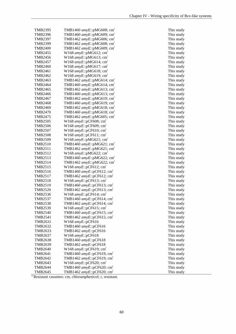
Chapter IV - Wiring specificity of Bce-like systems
60
TMB2395 TMB1460 amyE::pMG608; cmr This study
TMB2396 TMB1460 amyE::pMG609; cmr This study
TMB2397 TMB1462 amyE::pMG606; cmr This study
TMB2399 TMB1462 amyE::pMG608; cmr This study
TMB2400 TMB1462 amyE::pMG609; cmr This study
TMB2455 W168 amyE::pMG612; cmr This study
TMB2456 W168 amyE::pMG613; cmr This study
TMB2457 W168 amyE::pMG614; cmr This study
TMB2460 W168 amyE::pMG617; cmr This study
TMB2461 W168 amyE::pMG618; cmr This study
TMB2462 W168 amyE::pMG619; cmr This study
TMB2463 TMB1462 amyE::pMG614; cmr This study
TMB2464 TMB1460 amyE::pMG614; cmr This study
TMB2465 TMB1462 amyE::pMG613; cmr This study
TMB2466 TMB1460 amyE::pMG613; cmr This study
TMB2467 TMB1462 amyE::pMG619; cmr This study
TMB2468 TMB1460 amyE::pMG619; cmr This study
TMB2469 TMB1462 amyE::pMG618; cmr This study
TMB2470 TMB1460 amyE::pMG618; cmr This study
TMB2475 TMB1462 amyE::pMG605; cmr This study
TMB2505 W168 amyE::pCF608; cmr This study
TMB2506 W168 amyE::pCF609; cmr This study
TMB2507 W168 amyE::pCF610; cmr This study
TMB2508 W168 amyE::pCF611; cmr This study
TMB2509 W168 amyE::pMG621; cmr This study
TMB2510 TMB1460 amyE::pMG621; cmr This study
TMB2511 TMB1462 amyE::pMG621; cmr This study
TMB2512 W168 amyE::pMG622; cmr This study
TMB2513 TMB1460 amyE::pMG622; cmr This study
TMB2514 TMB1462 amyE::pMG622; cmr This study
TMB2515 W168 amyE::pCF612; cmr This study
TMB2516 TMB1460 amyE::pCF612; cmr This study
TMB2517 TMB1462 amyE::pCF612; cmr This study
TMB2518 W168 amyE::pCF613; cmr This study
TMB2519 TMB1460 amyE::pCF613; cmr This study
TMB2520 TMB1462 amyE::pCF613; cmr This study
TMB2536 W168 amyE::pCF614; cmr This study
TMB2537 TMB1460 amyE::pCF614; cmr This study
TMB2538 TMB1462 amyE::pCF614; cmr This study
TMB2539 W168 amyE::pCF615; cmr This study
TMB2540 TMB1460 amyE::pCF615; cmr This study
TMB2541 TMB1462 amyE::pCF615; cmr This study
TMB2631 W168 amyE::pCF616 This study
TMB2632 TMB1460 amyE::pCF616 This study
TMB2633 TMB1462 amyE::pCF616 This study
TMB2637 W168 amyE::pCF618 This study
TMB2638 TMB1460 amyE::pCF618 This study
TMB2639 TMB1462 amyE::pCF618 This study
TMB2640 W168 amyE::pCF619; cmr This study
TMB2641 TMB1460 amyE::pCF619; cmr This study
TMB2642 TMB1462 amyE::pCF619; cmr This study
TMB2643 W168 amyE::pCF620; cmr This study
TMB2644 TMB1460 amyE::pCF620; cmr This study
TMB2645 TMB1462 amyE::pCF620; cmr This study
a Resistant cassettes: cm, chloramphenicol; r, resistant.
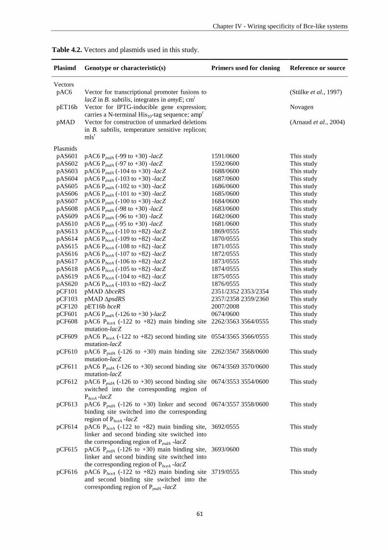
Chapter IV - Wiring specificity of Bce-like systems
61
Table 4.2. Vectors and plasmids used in this study.
Plasimd Genotype or characteristic(s)
Primers used for cloning Reference or source
Vectors
pAC6 Vector for transcriptional promoter fusions to
lacZ in B. subtilis, integrates in amyE; cmr
(Stülke et al., 1997)
pET16b Vector for IPTG-inducible gene expression;
carries a N-terminal His10-tag sequence; ampr
Novagen
pMAD Vector for construction of unmarked deletions
in B. subtilis, temperature sensitive replicon;
mlsr
(Arnaud et al., 2004)
Plasmids
pAS601 pAC6 PpsdA (-99 to +30) -lacZ 1591/0600 This study
pAS602 pAC6 PpsdA (-97 to +30) -lacZ 1592/0600 This study
pAS603 pAC6 PpsdA (-104 to +30) -lacZ 1688/0600 This study
pAS604 pAC6 PpsdA (-103 to +30) -lacZ 1687/0600 This study
pAS605 pAC6 PpsdA (-102 to +30) -lacZ 1686/0600 This study
pAS606 pAC6 PpsdA (-101 to +30) -lacZ 1685/0600 This study
pAS607 pAC6 PpsdA (-100 to +30) -lacZ 1684/0600 This study
pAS608 pAC6 PpsdA (-98 to +30) -lacZ 1683/0600 This study
pAS609 pAC6 PpsdA (-96 to +30) -lacZ 1682/0600 This study
pAS610 pAC6 PpsdA (-95 to +30) -lacZ 1681/0600 This study
pAS613 pAC6 PbceA (-110 to +82) -lacZ 1869/0555 This study
pAS614 pAC6 PbceA (-109 to +82) -lacZ 1870/0555 This study
pAS615 pAC6 PbceA (-108 to +82) -lacZ 1871/0555 This study
pAS616 pAC6 PbceA (-107 to +82) -lacZ 1872/0555 This study
pAS617 pAC6 PbceA (-106 to +82) -lacZ 1873/0555 This study
pAS618 pAC6 PbceA (-105 to +82) -lacZ 1874/0555 This study
pAS619 pAC6 PbceA (-104 to +82) -lacZ 1875/0555 This study
pAS620 pAC6 PbceA (-103 to +82) -lacZ 1876/0555 This study
pCF101 pMAD ∆bceRS 2351/2352 2353/2354 This study
pCF103 pMAD ∆psdRS 2357/2358 2359/2360 This study
pCF120 pET16b bceR 2007/2008 This study
pCF601 pAC6 PpsdA (-126 to +30 )-lacZ 0674/0600 This study
pCF608 pAC6 PbceA (-122 to +82) main binding site
mutation-lacZ
2262/3563 3564/0555 This study
pCF609 pAC6 PbceA (-122 to +82) second binding site
mutation-lacZ
0554/3565 3566/0555 This study
pCF610 pAC6 PpsdA (-126 to +30) main binding site
mutation-lacZ
2262/3567 3568/0600 This study
pCF611 pAC6 PpsdA (-126 to +30) second binding site
mutation-lacZ
0674/3569 3570/0600 This study
pCF612 pAC6 PpsdA (-126 to +30) second binding site
switched into the corresponding region of
PbceA -lacZ
0674/3553 3554/0600 This study
pCF613 pAC6 PpsdA (-126 to +30) linker and second
binding site switched into the corresponding
region of PbceA -lacZ
0674/3557 3558/0600 This study
pCF614 pAC6 PbceA (-122 to +82) main binding site,
linker and second binding site switched into
the corresponding region of PpsdA -lacZ
3692/0555 This study
pCF615 pAC6 PpsdA (-126 to +30) main binding site,
linker and second binding site switched into
the corresponding region of PbceA -lacZ
3693/0600 This study
pCF616 pAC6 PbceA (-122 to +82) main binding site
and second binding site switched into the
corresponding region of PpsdA -lacZ
3719/0555 This study
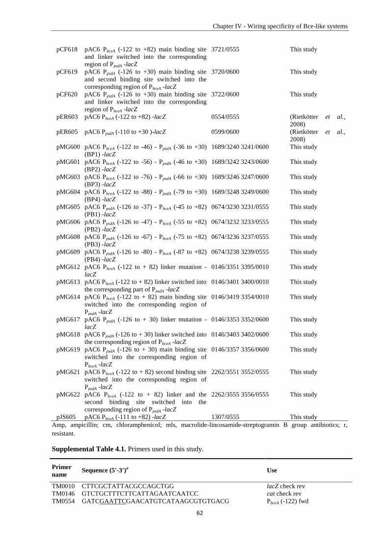
Chapter IV - Wiring specificity of Bce-like systems
62
pCF618 pAC6 PbceA (-122 to +82) main binding site
and linker switched into the corresponding
region of PpsdA -lacZ
3721/0555 This study
pCF619 pAC6 PpsdA (-126 to +30) main binding site
and second binding site switched into the
corresponding region of PbceA -lacZ
3720/0600 This study
pCF620 pAC6 PpsdA (-126 to +30) main binding site
and linker switched into the corresponding
region of PbceA -lacZ
3722/0600 This study
pER603 pAC6 PbceA (-122 to +82) -lacZ 0554/0555 (Rietkötter et al.,
2008)
pER605 pAC6 PpsdA (-110 to +30 )-lacZ 0599/0600 (Rietkötter et al.,
2008)
pMG600 pAC6 PbceA (-122 to -46) - PpsdA (-36 to +30)
(BP1) -lacZ
1689/3240 3241/0600 This study
pMG601 pAC6 PbceA (-122 to -56) - PpsdA (-46 to +30)
(BP2) -lacZ
1689/3242 3243/0600 This study
pMG603 pAC6 PbceA (-122 to -76) - PpsdA (-66 to +30)
(BP3) -lacZ
1689/3246 3247/0600 This study
pMG604 pAC6 PbceA (-122 to -88) - PpsdA (-79 to +30)
(BP4) -lacZ
1689/3248 3249/0600 This study
pMG605 pAC6 PpsdA (-126 to -37) - PbceA (-45 to +82)
(PB1) -lacZ
0674/3230 3231/0555 This study
pMG606 pAC6 PpsdA (-126 to -47) - PbceA (-55 to +82)
(PB2) -lacZ
0674/3232 3233/0555 This study
pMG608 pAC6 PpsdA (-126 to -67) - PbceA (-75 to +82)
(PB3) -lacZ
0674/3236 3237/0555 This study
pMG609 pAC6 PpsdA (-126 to -80) - PbceA (-87 to +82)
(PB4) -lacZ
0674/3238 3239/0555 This study
pMG612 pAC6 PbceA (-122 to + 82) linker mutation -
lacZ
0146/3351 3395/0010 This study
pMG613 pAC6 PbceA (-122 to + 82) linker switched into
the corresponding part of PpsdA -lacZ
0146/3401 3400/0010 This study
pMG614 pAC6 PbceA (-122 to + 82) main binding site
switched into the corresponding region of
PpsdA -lacZ
0146/3419 3354/0010 This study
pMG617 pAC6 PpsdA (-126 to + 30) linker mutation -
lacZ
0146/3353 3352/0600 This study
pMG618 pAC6 PpsdA (-126 to + 30) linker switched into
the corresponding region of PbceA -lacZ
0146/3403 3402/0600 This study
pMG619 pAC6 PpsdA (-126 to + 30) main binding site
switched into the corresponding region of
PbceA -lacZ
0146/3357 3356/0600 This study
pMG621 pAC6 PbceA (-122 to + 82) second binding site
switched into the corresponding region of
PpsdA -lacZ
2262/3551 3552/0555 This study
pMG622 pAC6 PbceA (-122 to + 82) linker and the
second binding site switched into the
corresponding region of PpsdA -lacZ
2262/3555 3556/0555 This study
pJS605 pAC6 PbceA (-111 to +82) -lacZ 1307/0555 This study
Amp, ampicillin; cm, chloramphenicol; mls, macrolide-lincosamide-streptogramin B group antibiotics; r,
resistant.
Supplemental Table 4.1. Primers used in this study.
Primer
name Sequence (5'-3')
a Use
TM0010 CTTCGCTATTACGCCAGCTGG lacZ check rev
TM0146 GTCTGCTTTCTTCATTAGAATCAATCC cat check rev
TM0554 GATCGAATTCGAACATGTCATAAGCGTGTGACG PbceA (-122) fwd
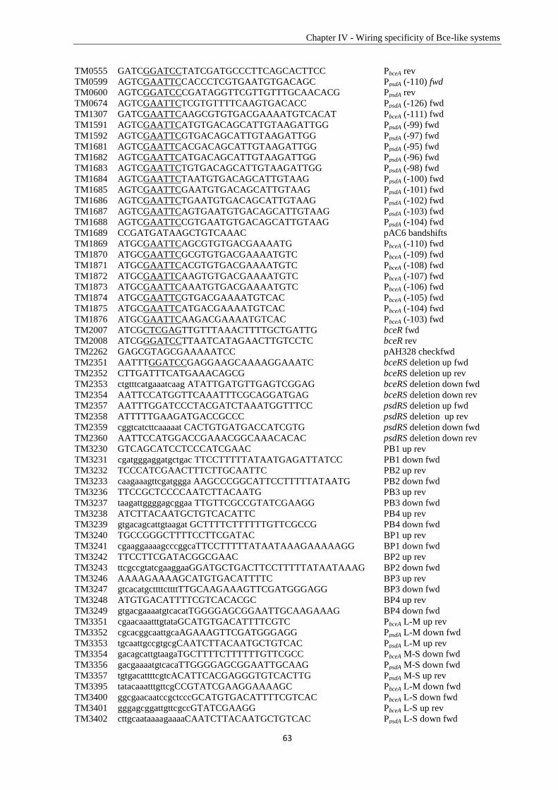
Chapter IV - Wiring specificity of Bce-like systems
63
TM0555 GATCGGATCCTATCGATGCCCTTCAGCACTTCC PbceA rev
TM0599 AGTCGAATTCCACCCTCGTGAATGTGACAGC PpsdA (-110) fwd
TM0600 AGTCGGATCCCGATAGGTTCGTTGTTTGCAACACG PpsdA rev
TM0674 AGTCGAATTCTCGTGTTTTCAAGTGACACC PpsdA (-126) fwd
TM1307 GATCGAATTCAAGCGTGTGACGAAAATGTCACAT PbceA (-111) fwd
TM1591 AGTCGAATTCATGTGACAGCATTGTAAGATTGG PpsdA (-99) fwd
TM1592 AGTCGAATTCGTGACAGCATTGTAAGATTGG PpsdA (-97) fwd
TM1681 AGTCGAATTCACGACAGCATTGTAAGATTGG PpsdA (-95) fwd
TM1682 AGTCGAATTCATGACAGCATTGTAAGATTGG PpsdA (-96) fwd
TM1683 AGTCGAATTCTGTGACAGCATTGTAAGATTGG PpsdA (-98) fwd
TM1684 AGTCGAATTCTAATGTGACAGCATTGTAAG PpsdA (-100) fwd
TM1685 AGTCGAATTCGAATGTGACAGCATTGTAAG PpsdA (-101) fwd
TM1686 AGTCGAATTCTGAATGTGACAGCATTGTAAG PpsdA (-102) fwd
TM1687 AGTCGAATTCAGTGAATGTGACAGCATTGTAAG PpsdA (-103) fwd
TM1688 AGTCGAATTCCGTGAATGTGACAGCATTGTAAG PpsdA (-104) fwd
TM1689 CCGATGATAAGCTGTCAAAC pAC6 bandshifts
TM1869 ATGCGAATTCAGCGTGTGACGAAAATG PbceA (-110) fwd
TM1870 ATGCGAATTCGCGTGTGACGAAAATGTC PbceA (-109) fwd
TM1871 ATGCGAATTCACGTGTGACGAAAATGTC PbceA (-108) fwd
TM1872 ATGCGAATTCAAGTGTGACGAAAATGTC PbceA (-107) fwd
TM1873 ATGCGAATTCAAATGTGACGAAAATGTC PbceA (-106) fwd
TM1874 ATGCGAATTCGTGACGAAAATGTCAC PbceA (-105) fwd
TM1875 ATGCGAATTCATGACGAAAATGTCAC PbceA (-104) fwd
TM1876 ATGCGAATTCAAGACGAAAATGTCAC PbceA (-103) fwd
TM2007 ATCGCTCGAGTTGTTTAAACTTTTGCTGATTG bceR fwd
TM2008 ATCGGGATCCTTAATCATAGAACTTGTCCTC bceR rev
TM2262 GAGCGTAGCGAAAAATCC pAH328 checkfwd
TM2351 AATTTGGATCCGAGGAAGCAAAAGGAAATC bceRS deletion up fwd
TM2352 CTTGATTTCATGAAACAGCG bceRS deletion up rev
TM2353 ctgtttcatgaaatcaag ATATTGATGTTGAGTCGGAG bceRS deletion down fwd
TM2354 AATTCCATGGTTCAAATTTCGCAGGATGAG bceRS deletion down rev
TM2357 AATTTGGATCCCTACGATCTAAATGGTTTCC psdRS deletion up fwd
TM2358 ATTTTTGAAGATGACCGCCC psdRS deletion up rev
TM2359 cggtcatcttcaaaaat CACTGTGATGACCATCGTG psdRS deletion down fwd
TM2360 AATTCCATGGACCGAAACGGCAAACACAC psdRS deletion down rev
TM3230 GTCAGCATCCTCCCATCGAAC PB1 up rev
TM3231 cgatgggaggatgctgac TTCCTTTTTATAATGAGATTATCC PB1 down fwd
TM3232 TCCCATCGAACTTTCTTGCAATTC PB2 up rev
TM3233 caagaaagttcgatggga AAGCCCGGCATTCCTTTTTATAATG PB2 down fwd
TM3236 TTCCGCTCCCCAATCTTACAATG PB3 up rev
TM3237 taagattggggagcggaa TTGTTCGCCGTATCGAAGG PB3 down fwd
TM3238 ATCTTACAATGCTGTCACATTC PB4 up rev
TM3239 gtgacagcattgtaagat GCTTTTCTTTTTTGTTCGCCG PB4 down fwd
TM3240 TGCCGGGCTTTTCCTTCGATAC BP1 up rev
TM3241 cgaaggaaaagcccggcaTTCCTTTTTATAATAAAGAAAAAGG BP1 down fwd
TM3242 TTCCTTCGATACGGCGAAC BP2 up rev
TM3243 ttcgccgtatcgaaggaaGGATGCTGACTTCCTTTTTATAATAAAG BP2 down fwd
TM3246 AAAAGAAAAGCATGTGACATTTTC BP3 up rev
TM3247 gtcacatgcttttcttttTTGCAAGAAAGTTCGATGGGAGG BP3 down fwd
TM3248 ATGTGACATTTTCGTCACACGC BP4 up rev
TM3249 gtgacgaaaatgtcacatTGGGGAGCGGAATTGCAAGAAAG BP4 down fwd
TM3351 cgaacaaatttgtataGCATGTGACATTTTCGTC PbceA L-M up rev
TM3352 cgcacggcaattgcaAGAAAGTTCGATGGGAGG PpsdA L-M down fwd
TM3353 tgcaattgccgtgcgCAATCTTACAATGCTGTCAC PpsdA L-M up rev
TM3354 gacagcattgtaagaTGCTTTTCTTTTTTGTTCGCC PbceA M-S down fwd
TM3356 gacgaaaatgtcacaTTGGGGAGCGGAATTGCAAG PpsdA M-S down fwd
TM3357 tgtgacattttcgtcACATTCACGAGGGTGTCACTTG PpsdA M-S up rev
TM3395 tatacaaatttgttcgCCGTATCGAAGGAAAAGC PbceA L-M down fwd
TM3400 ggcgaacaatccgctcccGCATGTGACATTTTCGTCAC PbceA L-S down fwd
TM3401 gggagcggattgttcgccGTATCGAAGG PbceA L-S up rev
TM3402 cttgcaataaaagaaaaCAATCTTACAATGCTGTCAC PpsdA L-S down fwd
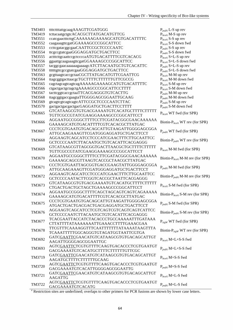
Chapter IV - Wiring specificity of Bce-like systems
64
TM3403 ttttcttttattgcaagAAAGTTCGATGGG PpsdA L-S up rev
TM3419 tcttacaatgctgtcACACGCTTATGACATGTTCG PbceA M-S up rev
TM3551 ccatcgaactttcttgCAAAAAAGAAAAGCATGTGACATTTTC PbceA S-S up rev
TM3552 caagaaagttcgatGGAAAAGCCCGGCATTCC PbceA S-S down fwd
TM3553 ccttcgatacggcgaaCAATTCCGCTCCCCAATC PpsdA S-S up rev
TM3554 ttcgccgtatcgaaGGGAGGATGCTGACTTCC PpsdA S-S down fwd
TM3555 actttcttgcaattccgctccccaATGTGACATTTTCGTCACACG PbceA S+L-S up rev
TM3556 ggaattgcaagaaagttcgatGGAAAAGCCCGGCATTCC PbceA S+L-S down fwd
TM3557 tacggcgaacaaaaaagaaaagcATCTTACAATGCTGTCACATTC PpsdA S+L-S up rev
TM3558 ttttttgttcgccgtatcgaaGGGAGGATGCTGACTTCC PpsdA S+L-S down fwd
TM3563 gcgttaagtcaccgctaaCGCTTATGACATGTTCGAATTCG PbceA M-M up rev
TM3564 ttagcggtgacttaacgcTGCTTTTCTTTTTTGTTCGCCG PbceA M-M down fwd
TM3565 cagctagcagtcagtcagAAAAAGAAAAGCATGTGACATTTTC PbceA S-M up rev
TM3566 ctgactgactgctagctgAAAAGCCCGGCATTCCTTTT PbceA S-M down fwd
TM3567 tacttcggtcaccgctaaTTCACGAGGGTGTCACTTG PpsdA M-M up rev
TM3568 ttagcggtgaccgaagtaTTGGGGAGCGGAATTGCAAG PpsdA M-M down fwd
TM3569 gtcagtcgtcagtcagtcATTCCGCTCCCCAATCTTAC PpsdA S-M up rev
TM3570 gactgactgacgactgacGAGGATGCTGACTTCCTTTT PpsdA S-M down fwd
TM3665 GTCATAAGCGTGTGACGAAAATGTCACATGCTTTTCTTTTT
TGTTCGCCGTATCGAAGGAAAAGCCCGGCATTCCT PbceA WT fwd (for SPR)
TM3666 AGGAATGCCGGGCTTTTCCTTCGATACGGCGAACAAAAAA
GAAAAGCATGTGACATTTTCGTCACACGCTTATGAC Biotin-PbceA WT rev (for SPR)
TM3667 CCCTCGTGAATGTGACAGCATTGTAAGATTGGGGAGCGGA
ATTGCAAGAAAGTTCGATGGGAGGATGCTGACTTCCT PpsdA WT fwd (for SPR)
TM3668 AGGAAGTCAGCATCCTCCCATCGAACTTTCTTGCAATTCC
GCTCCCCAATCTTACAATGCTGTCACATTCACGAGGG Biotin-PpsdA WT rev (for SPR)
TM3669 GTCATAAGCGTTAGCGGTGACTTAACGCTGCTTTTCTTTTT
TGTTCGCCGTATCGAAGGAAAAGCCCGGCATTCCT PbceA M-M fwd (for SPR)
TM3670 AGGAATGCCGGGCTTTTCCTTCGATACGGCGAACAAAAAA
GAAAAGCAGCGTTAAGTCACCGCTAACGCTTATGAC Biotin-PbceA M-M rev (for SPR)
TM3671 CCCTCGTGAATTAGCGGTGACCGAAGTATTGGGGAGCGGA
ATTGCAAGAAAGTTCGATGGGAGGATGCTGACTTCCT PpsdA M-M fwd (for SPR)
TM3672 AGGAAGTCAGCATCCTCCCATCGAACTTTCTTGCAATTCC
GCTCCCCAATACTTCGGTCACCGCTAATTCACGAGGG Biotin-PpsdA M-M rev (for SPR)
TM3673 GTCATAAGCGTGTGACGAAAATGTCACATGCTTTTCTTTTT
CTGACTGACTGCTAGCTGAAAAGCCCGGCATTCCT PbceA S-M fwd (for SPR)
TM3674 AGGAATGCCGGGCTTTTCAGCTAGCAGTCAGTCAGAAAAA
GAAAAGCATGTGACATTTTCGTCACACGCTTATGAC Biotin-PbceA S-M rev (for SPR)
TM3675 CCCTCGTGAATGTGACAGCATTGTAAGATTGGGGAGCGGA
ATGACTGACTGACGACTGACGAGGATGCTGACTTCCT PpsdA S-M fwd (for SPR)
TM3676 AGGAAGTCAGCATCCTCGTCAGTCGTCAGTCAGTCATTCC
GCTCCCCAATCTTACAATGCTGTCACATTCACGAGGG Biotin-PpsdA S-M rev (for SPR)
TM3677 TCACGAATTACCATCTACACCCTGCCAAAAATTTGATAAA
CTTATTTTATAAAAAAATTGAAACCTTTTGAAACGAA PsigW WT fwd (for SPR)
TM3678 TTCGTTTCAAAAGGTTTCAATTTTTTTATAAAATAAGTTTA
TCAAATTTTTGGCAGGGTGTAGATGGTAATTCGTGA Biotin-PsigW WT rev (for SPR)
TM3692 GATCGAATTCGAACATGTCATAAGCGTGTGACAGCATTGT
AAGATTGGGGAGCGGAATTGC PbceA M+L+S-S fwd
TM3693 AGTCGAATTCTCGTGTTTTCAAGTGACACCCTCGTGAATGT
GACGAAAATGTCACATGCTTTTCTTTTTTGTTCGC PpsdA M+L+S-S fwd
TM3719 GATCGAATTCGAACATGTCATAAGCGTGTGACAGCATTGT
AAGATGCTTTTCTTTTTTGCAAG PbceA M+S-S fwd
TM3720 AGTCGAATTCTCGTGTTTTCAAGTGACACCCTCGTGAATGT
GACGAAAATGTCACATTGGGGAGCGGAATTG PpsdA M+S-S fwd
TM3721 GATCGAATTCGAACATGTCATAAGCGTGTGACAGCATTGT
AAGATTG PbceA M+L-S fwd
TM3722 AGTCGAATTCTCGTGTTTTCAAGTGACACCCTCGTGAATGT
GACGAAAATGTCACATG PpsdA M+L-S fwd
a Restriction sites are underlined; overlaps to other primers for PCR fusions are shown by lower case letters.
Chapter IV - Wiring specificity of Bce-like systems
65
References
Arnaud, M., A. Chastanet & M. Débarbouillé, (2004) New vector for efficient allelic replacement in naturally
nontransformable, low-GC-content, Gram-positive bacteria. Applied and Environmental Microbiology
70: 6887-6891.
Bell, A., K. Gaston, J. Cole & S. Busby, (1989) Cloning of binding sequences for the Escherichia coli
transcription activators, FNR and CRP: location of bases involved in discrimination between FNR and
CRP. Nucleic acids research 17: 3865-3874.
Berdy, J., (2005) Bioactive microbial metabolites. J Antibiot (Tokyo) 58: 1-26.
Bernard, R., A. Guiseppi, M. Chippaux, M. Foglino & F. Denizot, (2007) Resistance to bacitracin in Bacillus
subtilis: unexpected requirement of the BceAB ABC transporter in the control of expression of its own
structural genes. Journal of bacteriology 189: 8636-8642.
Bernard, R., P. Joseph, A. Guiseppi, M. Chippaux & F. Denizot, (2003) YtsCD and YwoA, two independent
systems that confer bacitracin resistance to Bacillus subtilis. FEMS microbiology letters 228: 93-97.
Blanco, A.G., A. Canals & M. Coll, (2012) PhoB transcriptional activator binds hierarchically to pho box
promoters.
Blanco, A.G., M. Sola, F.X. Gomis-Rüth & M. Coll, (2002) Tandem DNA recognition by PhoB, a two-
component signal transduction transcriptional activator. Structure 10: 701-713.
Bourret, R.B., (2010) Receiver domain structure and function in response regulator proteins. Current opinion in
microbiology 13: 142-149.
Breukink, E. & B. de Kruijff, (2006) Lipid II as a target for antibiotics. Nature Reviews Drug Discovery 5: 321-
323.
Coumes-Florens, S., C. Brochier-Armanet, A. Guiseppi, F. Denizot & M. Foglino, (2011) A new highly
conserved antibiotic sensing/resistance pathway in firmicutes involves an ABC transporter interplaying
with a signal transduction system. PloS one 6: e15951.
Das, A.K., A. Pathak, A. Sinha, M. Datt, B. Singh, S. Karthikeyan & D. Sarkar, (2010) A single-amino-acid
substitution in the C terminus of PhoP determines DNA-binding specificity of the virulence-associated
response regulator from Mycobacterium tuberculosis. Journal of molecular biology 398: 647-656.
de Been, M., M.J. Bart, T. Abee, R.J. Siezen & C. Francke, (2008) The identification of response
regulator‐specific binding sites reveals new roles of two‐component systems in Bacillus cereus and
closely related low‐GC Gram‐positives. Environmental microbiology 10: 2796-2809.
Dintner, S., R. Heermann, C. Fang, K. Jung & S. Gebhard, (2014) A sensory complex consisting of an ATP-
binding cassette transporter and a two-component regulatory system controls bacitracin resistance in
Bacillus subtilis. J Biol Chem 289: 27899-27910.
Dintner, S., A. Staroń, E. Berchtold, T. Petri, T. Mascher & S. Gebhard, (2011) Coevolution of ABC transporters
and two-component regulatory systems as resistance modules against antimicrobial peptides in
Firmicutes bacteria. Journal of bacteriology 193: 3851-3862.
Fabret, C., V.A. Feher & J.A. Hoch, (1999) Two-component signal transduction in Bacillus subtilis: how one
organism sees its world. Journal of Bacteriology 181: 1975-1983.
Galperin, M.Y., (2006) Structural classification of bacterial response regulators: diversity of output domains and
domain combinations. Journal of Bacteriology 188: 4169-4182.
Galperin, M.Y., (2010) Diversity of structure and function of response regulator output domains. Current
opinion in microbiology 13: 150-159.
Gao, R. & A.M. Stock, (2009) Biological insights from structures of two-component proteins. Annual review of
microbiology 63: 133.
Gebhard, S., (2012) ABC transporters of antimicrobial peptides in Firmicutes bacteria–phylogeny, function and
regulation. Molecular microbiology 86: 1295-1317.
Gebhard, S. & T. Mascher, (2011) Antimicrobial peptide sensing and detoxification modules: unravelling the
regulatory circuitry of Staphylococcus aureus. Molecular microbiology 81: 581-587.
Grant, S.G., J. Jessee, F.R. Bloom & D. Hanahan, (1990) Differential plasmid rescue from transgenic mouse
DNAs into Escherichia coli methylation-restriction mutants. Proceedings of the National Academy of
Sciences 87: 4645-4649.
Harwood, C.R. & S.M. Cutting, (1990) Molecular biological methods for Bacillus.
Huynh, T.N. & V. Stewart, (2011) Negative control in two‐component signal transduction by transmitter
phosphatase activity. Molecular microbiology 82: 275-286.
Jarmer, H., T.S. Larsen, A. Krogh, H.H. Saxild, S. Brunak & S. Knudsen, (2001) Sigma A recognition sites in
the Bacillus subtilis genome. Microbiology 147: 2417-2424.
Jordan, S., M.I. Hutchings & T. Mascher, (2008) Cell envelope stress response in Gram-positive bacteria. FEMS
microbiology reviews 32: 107-146.
Chapter IV - Wiring specificity of Bce-like systems
66
Joseph, P., G. Fichant, Y. Quentin & F. Denizot, (2002) Regulatory relationship of two-component and ABC
transport systems and clustering of their genes in the Bacillus/Clostridium group, suggest a functional
link between them. Journal of molecular microbiology and biotechnology 4: 503-513.
Lee, D.J., S.D. Minchin & S.J. Busby, (2012) Activating transcription in bacteria. Annual review of microbiology
66: 125-152.
Martínez-Hackert, E. & A.M. Stock, (1997) The DNA-binding domain of OmpR: crystal structures of a winged
helix transcription factor. Structure 5: 109-124.
Mascher, T., N.G. Margulis, T. Wang, R.W. Ye & J.D. Helmann, (2003) Cell wall stress responses in Bacillus
subtilis: the regulatory network of the bacitracin stimulon. Molecular microbiology 50: 1591-1604.
Mascher, T., S.L. Zimmer, T.-A. Smith & J.D. Helmann, (2004) Antibiotic-inducible promoter regulated by the
cell envelope stress-sensing two-component system LiaRS of Bacillus subtilis. Antimicrobial agents
and chemotherapy 48: 2888-2896.
Miller, J.H., (1972) Experiments in molecular genetics.
Miroux, B. & J.E. Walker, (1996) Over-production of Proteins in Escherichia coli: Mutant Hosts that Allow
Synthesis of some Membrane Proteins and Globular Proteins at High Levels. Journal of molecular
biology 260: 289-298.
Ohki, R., K. Tateno, W. Masuyama, S. Moriya, K. Kobayashi & N. Ogasawara, (2003) The BceRS
two‐component regulatory system induces expression of the bacitracin transporter, BceAB, in Bacillus
subtilis. Molecular microbiology 49: 1135-1144.
Paget, M. & J.D. Helmann, (2003) The sigma 70 family of sigma factors. Genome Biol 4: 203.
Podgornaia, A.I. & M.T. Laub, (2013) Determinants of specificity in two-component signal transduction.
Current opinion in microbiology 16: 156-162.
Procaccini, A., B. Lunt, H. Szurmant, T. Hwa & M. Weigt, (2011) Dissecting the specificity of protein-protein
interaction in bacterial two-component signaling: orphans and crosstalks. PloS one 6: e19729.
Rhee, J.E., W. Sheng, L.K. Morgan, R. Nolet, X. Liao & L.J. Kenney, (2008) Amino acids important for DNA
recognition by the response regulator OmpR. Journal of Biological Chemistry 283: 8664-8677.
Rietkötter, E., D. Hoyer & T. Mascher, (2008) Bacitracin sensing in Bacillus subtilis. Molecular microbiology
68: 768-785.
Rohs, R., X. Jin, S.M. West, R. Joshi, B. Honig & R.S. Mann, (2010) Origins of specificity in protein-DNA
recognition. Annual review of biochemistry 79: 233.
Silver, L.L., (2003) Novel inhibitors of bacterial cell wall synthesis. Current opinion in microbiology 6: 431-438.
Spiro, S., K. Gaston, A. Bell, R. Roberts, S. Busby & J. Guest, (1990) Interconversion of the DNA‐binding
specificities of two related transcription regulators, CRP and FNR. Molecular microbiology 4: 1831-
1838.
Staroń, A., D.E. Finkeisen & T. Mascher, (2011) Peptide antibiotic sensing and detoxification modules of
Bacillus subtilis. Antimicrobial agents and chemotherapy 55: 515-525.
Stülke, J., I. Martin‐Verstraete, M. Zagorec, M. Rose, A. Klier & G. Rapoport, (1997) Induction of the Bacillus
subtilis ptsGHI operon by glucose is controlled by a novel antiterminator, GlcT. Molecular
microbiology 25: 65-78.
67
Chapter V
Specificity Determinant and Rewiring Signal Transduction of
BceRS-like Two-Component Systems in Bacillus subtilis
Manuscript
Chapter V – The specificity determinant of Bce-like HKs/RRs
68
Specificity Determinant and Rewiring Signal Transduction of BceRS-like
two-component systems in Bacillus subtilis
Summary
Two-component signal transduction systems are comprised of a membrane-anchored histidine kinase
that senses the input stimulus and a cognate response regulator, which binds to specific promoter
regions to trigger a cellular response. In Bacillus subtilis, there are three Bce-like two-component
systems: BceRS, PsdRS and YxdJK, which — together with their cognate ABC transporters —
mediate peptide antibiotic resistance. These three Bce-like two-component systems share significant
sequence similarity and were predicted to have considerable cross-talk. However, in vivo, these three
systems are insulated very well with only low level of cross-regulation between BceS and PsdR. In
this chapter, we focused on the Bce and the Psd systems and investigated the specificity determinants
in BceR and PsdR that ensure the insulation of these two paralogous pathways at the level of
interaction with their histidine kinases. First, we verified that the specificity determinants are located
on the Bce-like response regulator receiver domain. Next chimeric response regulator analysis
demonstrated that the α1 helix, which is speculated to contain one of the interaction surfaces with the
histidine kinase, is not enough to determine specificity. Exchanging this region alone between BceR
and PsdR had no influence on specificity. Surprisingly, we found that the β2-α2 region, which is
located away from the interaction surfaces with the histidine kinase, can influence the specificity with
the histidine kinase. However, replacing this region alone in PsdR by the corresponding region from
BceR was not sufficient to confer a complete change of specificity. By substituting both regions
together, i.e. α1-β2-α2, we successfully rewired signal transduction from BceS to PsdR. These results
demonstrated that the α1-β2-α2 region dictates the specificity of Bce-like two-component systems in
B. subtilis.
Chapter V – The specificity determinant of Bce-like HKs/RRs
69
Introduction
Two-component signal transduction systems (TCSs) are one of the prevalent ways used by bacteria to
respond to environmental changes. They are involved in regulating a wide array of physiological
processes, including chemotaxis, utilization of various nutrients, virulence, quorum sensing, and
antimicrobial peptide (AMP) resistance (Wadhams & Armitage, 2004, Paterson et al., 2006, Williams
et al., 2007, Gooderham & Hancock, 2009). A typical TCS is comprised of a membrane-anchored
histidine kinase (HK) that detects the signal input and a cytoplasmic response regulator (RR) that
mediates the cellular response (Stock et al., 2000). The HK normally harbors a variable periplasmic
sensor (Input) domain and a conserved cytoplasmic autokinase domain linked by a linker region (Fig.
5.1). In addition, most HKs harbor a HAMP domain or a PAS domain at the N-terminal of the
cytoplasmic domain, which is critical for signal transmission from the periplasmic sensor domain to
the cytoplasmic region. The highly conserved HK cytoplasmic region consists of a long α-hairpin
dimerization and histidine phosphotransfer (DHp) domain and a globular catalytic and ATP binding
(CA) domain (Mascher, 2006, Krell et al., 2010, Jung et al., 2012). In the RR, a conserved receiver
domain that typically adopts a (βα)5 topology is connected by a flexible linker to a variable output
domain that frequently has a DNA binding function (Bourret, 2010, Galperin, 2010) (Fig. 5.1).
Signal transduction within TCSs is mediated by three phosphotransfer reactions. Activation of the HK
leads to autophosphorylation of a conserved His residue located in the α1 helix of the DHp domain.
This is followed by transferring the phosphoryl group to a conserved Asp residue located in the β3
strand of the cognate RR receiver domain. Direct protein-protein interaction between the HK DHp
domain and the RR receiver domain is involved in this step (West & Stock, 2001, Gao & Stock, 2009).
Finally, the dephosphorylation of the RR switches off the signal transduction of the system (Parkinson,
1993).
Most bacteria encode dozens, sometimes hundreds of TCSs (Capra & Laub, 2012). In the genome of B.
subtilis, 36 HKs and 34 RRs were found among the open reading frames including three paralogous
Bce-like TCSs that regulate resistance against AMPs (Fabret et al., 1999). The three Bce-like HKs are
intramembrane-sensing histidine kinases, which have short extracellular loops and cannot detect
AMPs by themselves (Mascher, 2006, Mascher, 2014). Instead, they get the signal from Bce-like ABC
transporters, which harbor functions of both AMP perception and resistance, and subsequently pass
the signal to their cognate RRs. Bce-like RRs belong to the OmpR subfamily and possess a winged
helix-turn-helix (wHTH) DNA-binding output domain (Fabret et al., 1999, Galperin, 2006). The
activated RRs in turn bind to the promoter regions of their cognate ABC transporters and upregulate
the transcription to ensure AMP resistance (Ohki et al., 2003, Bernard et al., 2007, Rietkötter et al.,
2008). The Bce-like TCS together with the ABC transporter form an AMP sensing and detoxification
module. In B. subtilis, the BceRS-BceAB system and the PsdRS-PsdAB system can sense and respond
mainly to bacitracin and nisin, respectively (Gebhard & Mascher, 2011, Staroń et al., 2011). The third
Chapter V – The specificity determinant of Bce-like HKs/RRs
70
Bce system, YxdJK-YxdLM, is poorly understood and has been found to be induced only by the
human neutrophil peptide LL-37 (Joseph et al., 2004, Pietiäinen et al., 2005).
The three Bce-like TCSs of B. subtilis share significant sequence and structural similarity. This
indicates the potential of considerable cross-talk between them, which has already been predicted by
direct coupling analysis based on the co-evolution of inter-protein contact residues (Szurmant & Hoch,
2010, Procaccini et al., 2011). Such unwanted cross-talk can be deleterious. Therefore avoiding cross-
talk and maintaining high fidelity of signal transmission within TCSs is necessary to guarantee
bacterial cells respond specifically to each stimulus and produce the desired, beneficial response. A
previous study demonstrated that instead of the predicted high level of cross-talk between these three
paralogous Bce-like TCSs in B. subtilis, there is only some minor degree of cross-regulation in vivo
between BceS and PsdR at high concentrations of bacitracin (Rietkötter et al., 2008) (Fig. 5.1). This
raises two questions: (1) how do bacteria coordinate the activity of so many highly related signaling
systems while maintaining the signal transduction specificity and preventing unwanted cross-talk? (2)
How does the HK discriminate between its cognate RR and the non-cognate ones in the pool of
numerous RRs?
Figure 5.1. Schematic overview of two Bce-like two-component signal transduction systems in B. subtilis. Bce and Psd TCSs including their target promoters, PbceA and PpsdA, are colored with black and grey, respectively.
The major stimulus of each system is given on the left side. The phosphoryl group (P) is transferred from the
conserved histidine residue (H) on the HK to the conserved aspartate residue (D) on the RR. Solid arrows
indicate the signal transduction pathway within one system, while cross-regulation between BceS and PsdR is
pointed out by dashed arrow. HK, histidine kinase; RR, response regulator; Input, input domain of HK; DHp,
dimerization and histidine phosphotransferase domain of HK; CA, catalytic and ATPase domain of HK;
Receiver, receiver domain of RR; Output, output domain of RR.
The predominant way to maintain intrasystem specificity and intersystem insulation is the molecular
recognition between HK and RR (Podgornaia & Laub, 2013). During signal transmission of the TCS,
specific interactions mediated by a few amino acids on the interaction surfaces of both proteins lead to
a global and strong kinetic preference of the HK to its cognate RR. These amino acids are specificity
determinants. The co-crystal structure of HK853-RR468 from Thermotoga maritima provides a clear
view of the HK/RR interaction surfaces and implies the possible positions of those amino acids. The
interaction surfaces involved in forming the HK853-RR468 pair are: the α1 helix and the β5-α5 loop
of the RR468 receiver domain with the two α helices of the HK853 DHp domain; the β3-α3 loop of
Chapter V – The specificity determinant of Bce-like HKs/RRs
71
the RR468 with the ATP lid and the β4-α4 loop of the HK853 CA domain; the RR468 β4-α4 loop with
the DHp-CA interdomain linker of HK853(Casino et al., 2009) (see Fig. 1.5).
In this chapter, we focused on the two Bce-like TCSs BceRS and PsdRS of B. subtilis to explore the
regions on Bce-like RRs that are specifically recognized by the cognate HK and excluded by the non-
cognate HK. We demonstrated that Bce-like RRs containing an extended specificity determinant
compared to the other studied OmpR subfamily members, which suggests a novel mechanism of
protein-protein interactions for Bce-type TCSs.
Results
The receiver domain of Bce-like RRs dictates specific interaction with the cognate HK
Signal transmission from HK to RR is mediated by direct interactions between the HK cytoplasmic
domain and the RR receiver domain (West & Stock, 2001). Here, we first wanted to verify that the
BceR receiver domain is indeed responsible for specific interaction with the cognate BceS instead of
the non-cognate PsdS. To address this question, a chimeric RR, BP1, was constructed with the
receiver domain from BceR fused to the linker and output domain of PsdR. BP1 as well as wild-type
BceR and PsdR (Fig. 5.2) were cloned into vector pBS2E under the control of a constitutive promoter,
PbceR (Table 5.2). To test the signaling between BceS and the RR-constructs, the plasmids were
transformed into B. subtilis reporter strain (TMB 1975 or TMB 1976) lacking all Bce-like TCS
components but BceS (Table 5.1). To test the signaling between PsdS and the RR-constructs, the
plasmids were transformed into B. subtilis reporter strain (TMB 2051 or TMB 2052) harboring PsdS
(Table 5.1). In the B. subtilis reporter strain, PbceA-lux, with the target promoter of BceR fused to the
bacterial promoterless luciferase operon luxABCDE, was the reporter construct for RRs that contained
the BceR output domain, while a PpsdA-lux reporter construct was used for RRs that contained the PsdR
output domain. Signaling via BceS was induced by addition of bacitracin and signaling via PsdS was
induced by addition of nisin (Fig. 5.1).
The strain harboring BceS, BceR and target promoter reporter PbceA-lux showed only basal expression
of the promoter in the absence of bacitracin (ca. 1×10³ RLU/OD), which was induced 50-fold (ca.
5×10⁴ RLU/OD) within 150 min after addition of 2 µg ml
-1 bacitracin (Fig. 5.2A). BceR was not able
to induce the expression of the reporter construct PbceA-lux in the strain containing PsdS in the presence
of 8 µg ml-1
nisn (Fig. 5.2B). PsdR, on the other hand, was not able to induce the expression of the
reporter construct PpsdA-lux in the strain possessing BceS in presence of bacitracin (Fig. 5.2E), but
strongly upregulated the expression of PpsdA-lux in presence of nisin in the strain containing PsdS (Fig.
5.2F). The chimeric RR BP1, with its receiver domain from BceR and the flexible linker as well as the
output domain from PsdR, showed strong upregulation of PpsdA activity (50-fold over the uninduced
control) in the reporter strain containing BceS in the presence of bacitracin (Fig. 5.2C), but no
induction of expression of the reporter construct PpsdA-lux in the strain harboring PsdS after addition of
nisin (Fig. 5.2D).
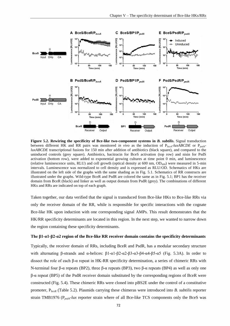
Chapter V – The specificity determinant of Bce-like HKs/RRs
72
Figure 5.2. Rewiring the specificity of Bce-like two-component systems in B. subtilis. Signal transduction
between different HK and RR pairs was monitored in vivo as the induction of PbceA-luxABCDE or PpsdA-
luxABCDE transcriptional fusions for 150 min after addition of antibiotics (black square), and compared to the
uninduced controls (grey square). Antibiotics, bacitracin for BceS activation (top row) and nisin for PsdS
activation (bottom row), were added to exponential growing cultures at time point 0 min, and luminescence
(relative luminescence units, RLU) and cell growth (optical density at 600 nm, OD600) were measured in 5-min
intervals. Luminescence was normalized to cell density and is expressed as RLU/OD. Schematics of HKs are
illustrated on the left side of the graphs with the same shading as in Fig. 5.1. Schematics of RR constructs are
illustrated under the graphs. Wild-type BceR and PsdR are colored the same as in Fig. 5.1; BP1 has the receiver
domain from BceR (black) and linker as well as output domain from PsdR (grey). The combinations of different
HKs and RRs are indicated on top of each graph.
Taken together, our data verified that the signal is transduced from Bce-like HKs to Bce-like RRs via
only the receiver domain of the RR, while is responsible for specific interactions with the cognate
Bce-like HK upon induction with one corresponding signal AMPs. This result demonstrates that the
HK/RR specificity determinants are located in this region. In the next step, we wanted to narrow down
the region containing these specificity determinants.
The β1-α1-β2-α2 region of the Bce-like RR receiver domain contains the specificity determinants
Typically, the receiver domain of RRs, including BceR and PsdR, has a modular secondary structure
with alternating β-strands and α-helices: β1-α1-β2-α2-β3-α3-β4-α4-β5-α5 (Fig. 5.3A). In order to
dissect the role of each β-α repeat in HK-RR specificity determination, a series of chimeric RRs with
N-terminal four β-α repeats (BP2), three β-α repeats (BP3), two β-α repeats (BP4) as well as only one
β-α repeat (BP5) of the PsdR receiver domain substituted by the corresponding regions of BceR were
constructed (Fig. 5.4). These chimeric RRs were cloned into pBS2E under the control of a constitutive
promoter, PbceR (Table 5.2). Plasmids carrying these chimeras were introduced into B. subtilis reporter
strain TMB1976 (PpsdA-lux reporter strain where of all Bce-like TCS components only the BceS was
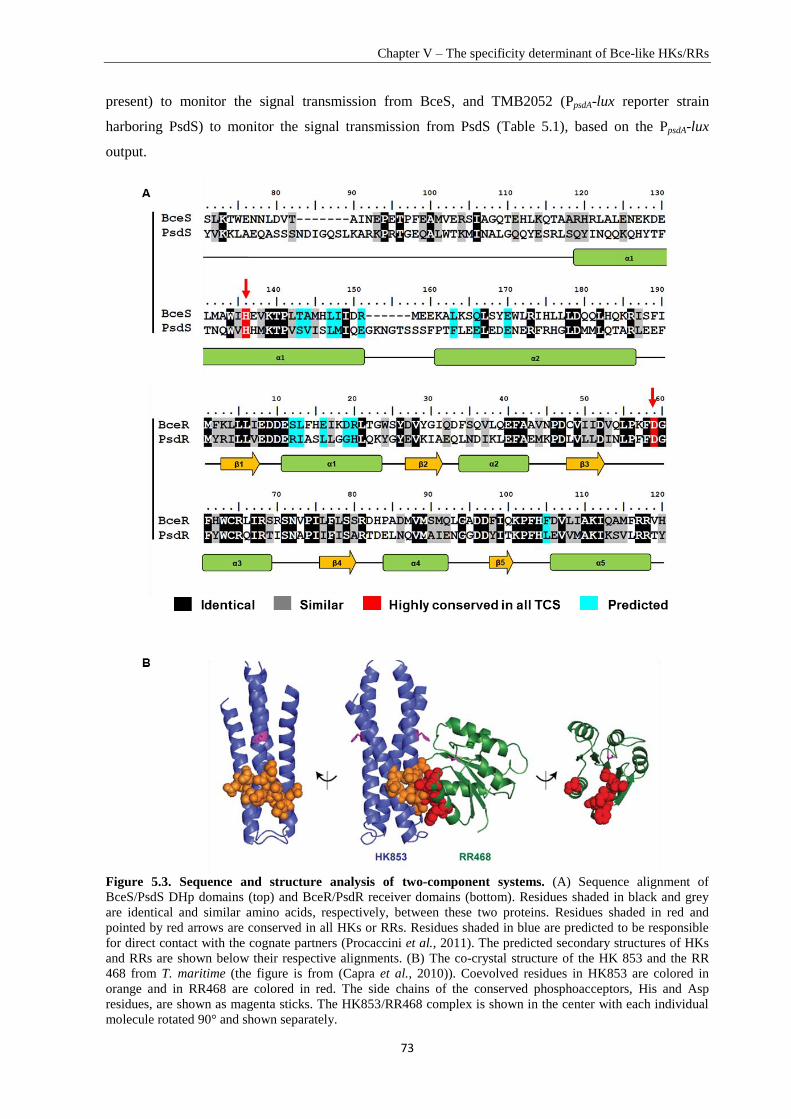
Chapter V – The specificity determinant of Bce-like HKs/RRs
73
present) to monitor the signal transmission from BceS, and TMB2052 (PpsdA-lux reporter strain
harboring PsdS) to monitor the signal transmission from PsdS (Table 5.1), based on the PpsdA-lux
output.
Figure 5.3. Sequence and structure analysis of two-component systems. (A) Sequence alignment of
BceS/PsdS DHp domains (top) and BceR/PsdR receiver domains (bottom). Residues shaded in black and grey
are identical and similar amino acids, respectively, between these two proteins. Residues shaded in red and
pointed by red arrows are conserved in all HKs or RRs. Residues shaded in blue are predicted to be responsible
for direct contact with the cognate partners (Procaccini et al., 2011). The predicted secondary structures of HKs
and RRs are shown below their respective alignments. (B) The co-crystal structure of the HK 853 and the RR
468 from T. maritime (the figure is from (Capra et al., 2010)). Coevolved residues in HK853 are colored in
orange and in RR468 are colored in red. The side chains of the conserved phosphoacceptors, His and Asp
residues, are shown as magenta sticks. The HK853/RR468 complex is shown in the center with each individual
molecule rotated 90° and shown separately.
Chapter V – The specificity determinant of Bce-like HKs/RRs
74
Wild-type PsdR, which can only accept the signal from PsdS but not from BceS, was used as a
negative control (Fig. 5.4A and 5.4B), while wild-type BceR, which can accept the signal from BceS
but not from PsdS served as a positive control (Fig. 5.4K and 5.4L). BP2 and BP3, with the N-terminal
four and three β-α repeats of the PsdR receiver domain, respectively, swapped for the corresponding
region of BceR, showed no induction of PpsdA with either BceS or PsdS in the presence of bacitracin or
nisin (Fig. 5.4C to 5.4F), indicating that BP2 and BP3 were dysfunctional. In contrast, chimera BP4,
with only two β-α repeats (β1-α1-β2-α2) of the PsdR receiver domain replaced by the corresponding
region of BceR, showed a strong upregulation of promoter activity in the presence of bacitracin, which
was dependent on BceS (Fig. 5.4G). This chimera displayed extremely low level of promoter
induction, 3-fold over the uninduced control, after addition of nisin in the presence of PsdS (Fig. 5.4H).
These results showed a change of specificity from PsdR to BceR. BP5, with the β1-α1 region on PsdR
receiver domain changed into BceR, was unable to induce expression of the promoter either in the
presence of BceS or PsdS (Fig. 5.4I and 5.4J).
These results demonstrated that the signal transduction could successfully be redirected between
BceRS and PsdRS sytems by swapping the β1-α1-β2-α2 region on the RR receiver domain. Although
most of the chimeric RRs failed to accept a signal from either BceS or PsdS, the results from BP4
demonstrated that the β1-α1-β2-α2 region on the BceR receiver domain contains the motifs that
determine specific interaction with the cognate BceS. The co-crystal structure of the TCS HK853-
RR468 indicates that the α1 helix on the RR receiver domain directly interacts with the HK DHp
domain (Casino et al., 2009, Capra et al., 2010) (Fig. 5.3B). We therefore next wanted to analyze the
function of the α1 helix for Bce-like HK and RR specificity.
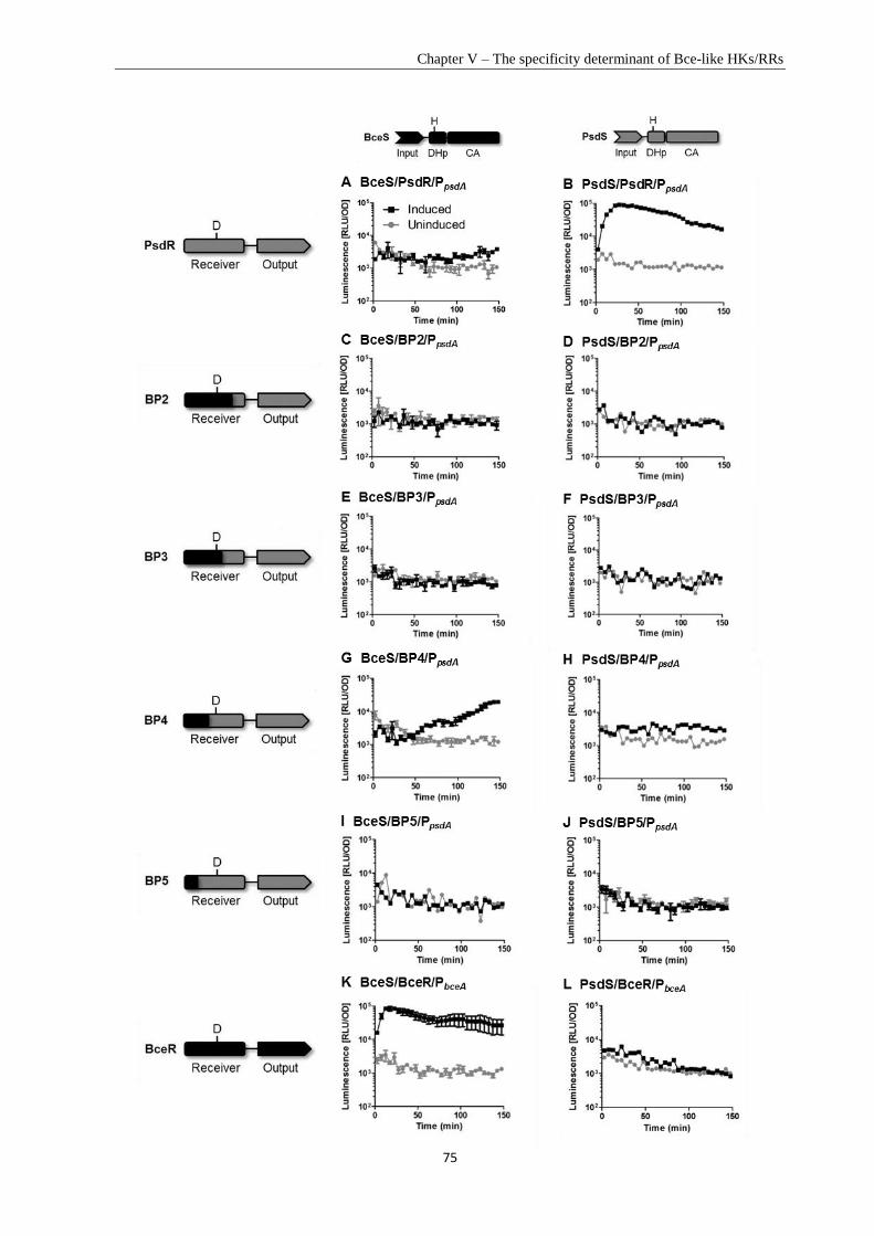
Chapter V – The specificity determinant of Bce-like HKs/RRs
75
Chapter V – The specificity determinant of Bce-like HKs/RRs
76
Figure 5.4. Signal transduction between HKs and series of chimeric RRs. Signal transduction between
different HK and RR pairs was monitored in vivo as the induction of PbceA-luxABCDE or PpsdA-luxABCDE
transcriptional fusions within 150 min in the presence of antibiotics (black square): bacitracin for BceS
activation and nisin for PsdS activation, and compared with the uninduced controls (grey square) as described in
Fig. 5.2. Schematics of HKs are illustrated on top of the graphs with the same shading as in Fig. 5.1. Schematics
of RR constructs are illustrated on the left side of the graphs. BceS/BceR and PsdS/PsdR are with the same
shading as in Fig. 5.1. From chimera BP2 to BP5, the part of the receiver domain from BceR is colored with
black and the region from PsdR is colored with grey. The combinations of different HKs and RRs are indicated
on top of each graph.
The α1 helix together with the β2-α2 region of the Bce-like RR receiver domain dictates
specificity for the cognate Bce-like HK
Based on the direct coupling analysis, five amino acids in the α1 helix and one residue on the β5-α5
loop of BceR/PsdR receiver domain were predicted to dictate specific interactions with eight amino
acids of the BceS/PsdS DHp domains in B. subtilis (Procaccini et al., 2011) (Fig. 5.3A). To
experimentally verify the function of the α1 helix and the β5-α5 loop in specificity determination, we
first constructed a chimera, BPα1, with the whole α1 helix of PsdR substituted by the corresponding
region of BceR (Fig. 5.5). This chimera was dysfunctional, since it was unable to induce the
expression of the promoter construct with either an active BceS or PsdS (Fig. 5.5C and 5.5D). To
investigate if the α1 helix is the specificity determinant, another chimera, PBα1, was constructed with
the α1 helix on BceR receiver domain replaced by the α1 helix of PsdR (Fig. 5.5). This chimera RR
strongly upregulated the activity of PbceA in a BceS-dependent manner in the presence of bacitracin
(Fig. 5.5E), and no induction of the promoter was observed with an active PsdS in the presence of
nisin (Fig. 5.5F). Hence, PBα1 displayed a behavior similar to that of wild-type BceR. This result
indicates that switching the α1 helix alone could not rewire the signal transduction specificity of Bce-
like TCSs.
Next, we wanted to analyze if the β5-α5 loop is involved in determining specificity, that is, the α1
helix together with the β5-α5 loop should be able to determine the specificity between BceS/BceR and
PsdS/PsdR. PBα1+loop (β5-α5), with both the α1 helix and the β5-α5 loop on BceR receiver domain
replaced by corresponding regions of PsdR, was constructed and the signaling was checked from BceS
and PsdS, respectively. Surprisingly, PBα1+loop (β5-α5) was able to highly induce the expression of the
promoter construct with an active BceS (Fig. 5.5G), but had no induction of the promoter construct
with an active PsdS (Fig. 5.5H). PBα1+loop (β5-α5), therefore, behaved similarly as wild-type BceR. These
results demonstrate that the regions containing specificity determining amino acids predicted by direct
coupling analysis are not sufficient to dictate the specificity of Bce-like TCSs in B. subtilis. This
provoked a sense for additional specificity determinant that would allow rewiring the signal
transduction between the Bce and Psd TCSs.
Sequence alignments showed that amino acids on the β2-α2 region (RR receiver domain) vary
considerably between BceR and PsdR (Fig. 5.3A). To further analyze a pontential function of tthis
region in specificity determination, we constructed chimera BPβ2-α2 with this region on PsdR switched
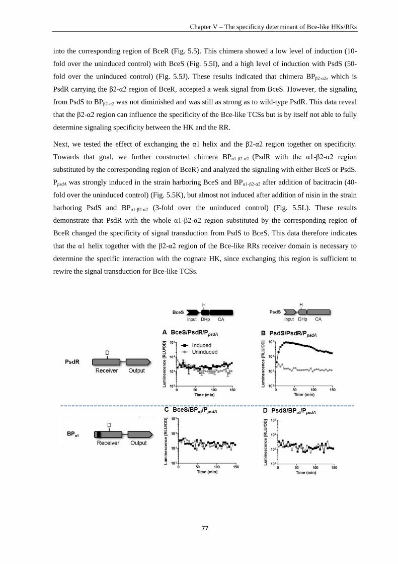
Chapter V – The specificity determinant of Bce-like HKs/RRs
77
into the corresponding region of BceR (Fig. 5.5). This chimera showed a low level of induction (10-
fold over the uninduced control) with BceS (Fig. 5.5I), and a high level of induction with PsdS (50-
fold over the uninduced control) (Fig. 5.5J). These results indicated that chimera BPβ2-α2, which is
PsdR carrying the β2-α2 region of BceR, accepted a weak signal from BceS. However, the signaling
from PsdS to BPβ2-α2 was not diminished and was still as strong as to wild-type PsdR. This data reveal
that the β2-α2 region can influence the specificity of the Bce-like TCSs but is by itself not able to fully
determine signaling specificity between the HK and the RR.
Next, we tested the effect of exchanging the α1 helix and the β2-α2 region together on specificity.
Towards that goal, we further constructed chimera BPα1-β2-α2 (PsdR with the α1-β2-α2 region
substituted by the corresponding region of BceR) and analyzed the signaling with either BceS or PsdS.
PpsdA was strongly induced in the strain harboring BceS and BPα1-β2-α2 after addition of bacitracin (40-
fold over the uninduced control) (Fig. 5.5K), but almost not induced after addition of nisin in the strain
harboring PsdS and BPα1-β2-α2 (3-fold over the uninduced control) (Fig. 5.5L). These results
demonstrate that PsdR with the whole α1-β2-α2 region substituted by the corresponding region of
BceR changed the specificity of signal transduction from PsdS to BceS. This data therefore indicates
that the α1 helix together with the β2-α2 region of the Bce-like RRs receiver domain is necessary to
determine the specific interaction with the cognate HK, since exchanging this region is sufficient to
rewire the signal transduction for Bce-like TCSs.
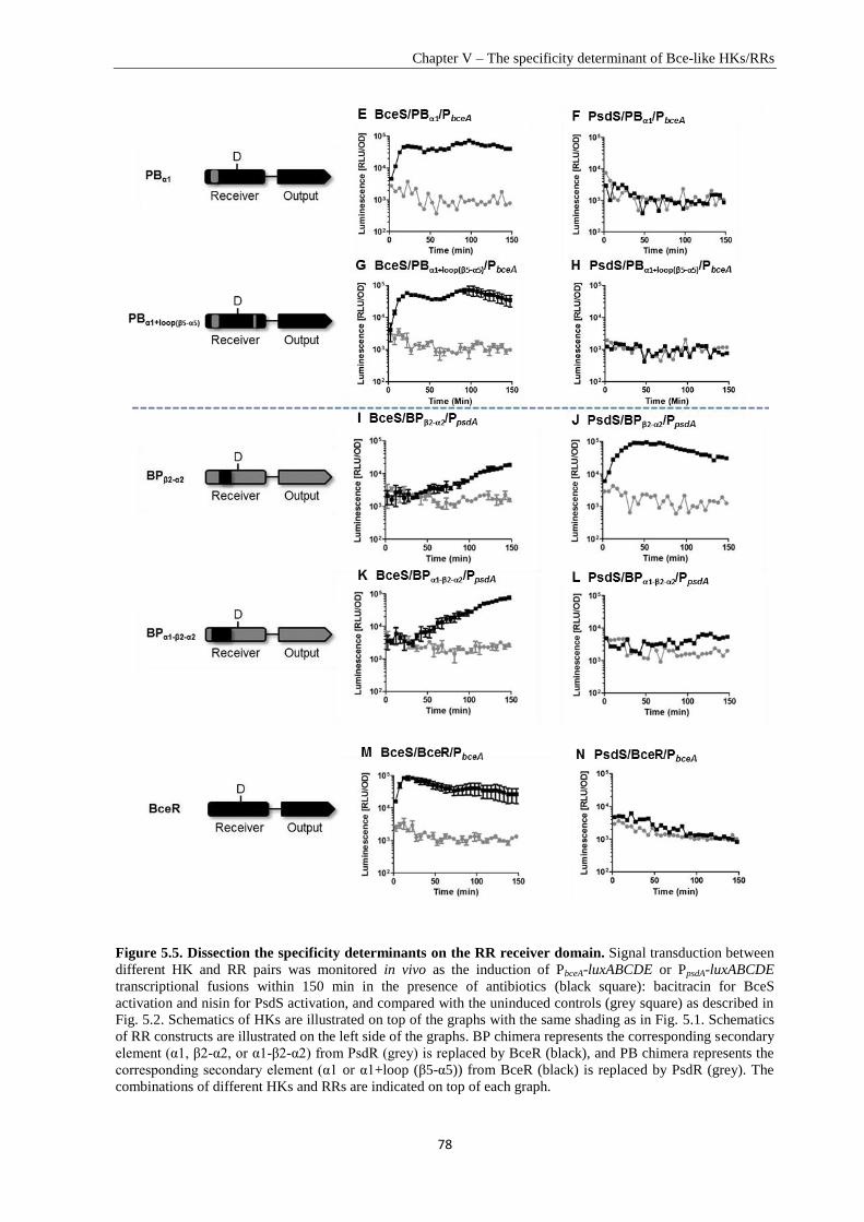
Chapter V – The specificity determinant of Bce-like HKs/RRs
78
Figure 5.5. Dissection the specificity determinants on the RR receiver domain. Signal transduction between
different HK and RR pairs was monitored in vivo as the induction of PbceA-luxABCDE or PpsdA-luxABCDE
transcriptional fusions within 150 min in the presence of antibiotics (black square): bacitracin for BceS
activation and nisin for PsdS activation, and compared with the uninduced controls (grey square) as described in
Fig. 5.2. Schematics of HKs are illustrated on top of the graphs with the same shading as in Fig. 5.1. Schematics
of RR constructs are illustrated on the left side of the graphs. BP chimera represents the corresponding secondary
element (α1, β2-α2, or α1-β2-α2) from PsdR (grey) is replaced by BceR (black), and PB chimera represents the
corresponding secondary element (α1 or α1+loop (β5-α5)) from BceR (black) is replaced by PsdR (grey). The
combinations of different HKs and RRs are indicated on top of each graph.
Chapter V – The specificity determinant of Bce-like HKs/RRs
79
Discussion
The genome of B. subtilis encodes three paralogous Bce-like TCSs, which share significant sequence
similarity and were predicted to have considerable cross-talk (Procaccini et al., 2011). In contrast to
the prediction, experiments clearly demonstrated that they are insulated quite well with only minor
level of cross-regulation between BceS and PsdR in vivo (Rietkötter et al., 2008). The specificity of
TCSs is extremely important for bacteria to ensure the desired responses to specific stimuli, and is
mainly maintained by molecular recognition of interactions between HKs and cognate RRs. A number
of studies were recently performed to understand the determinant of specificity between HKs and RRs
(Skerker et al., 2008, Siryaporn et al., 2010, Capra et al., 2012). The co-crystal structure of HK853-
RR468 from T. maritima showed that the α1 helix and the β5-α5 loop of the RR468 receiver domain
interact directly with two α helices of HK853 DHp domain, implying that specificity is most likely
determined by specific interactions on this surface (Casino et al., 2009) (Fig. 5.3B). Further analysis of
RRs (OmpR, RstA and CpxR) from the OmpR subfamily confirmed the importance of this interaction
surface in specificity determination (Capra et al., 2010). EnvZ/OmpR, CpxA/CpxR and RstB/RstA are
three well-insulated TCSs in E. coli. Substitution of three residues predicted for specificity on the α1
helix of OmpR with the corresponding residues from RstA weaken the phosphotransfer from EnvZ to
the substituted OmpR derivative, but no visible phosphotransfer was observed between RstB and the
substituted OmpR derivative. Based on this three-amino acid substitution, subsequent substitution of
three more amino acids on the OmpR β5-α5 loop by the corresponding residues of RstA diminished
the phosphotransfer from EnvZ to OmpR and established the phosphotransfer from RstB to OmpR.
Similarly, the study between EnvZ-OmpR and CpxA-CpxR systems proved that the α1 helix and the
β5-α5 loop of OmpR/RstA/CpxR receiver domain are specificity determinants (Capra et al., 2010).
However, the mechanism of specificity determination between these three paralogou Bce-like TCSs in
B. subtilis was still poorly understood. In this chapter, we investigated determinants on Bce-like RRs
that dictate specific interactions with their cognate HKs to maintain the signal transduction fidelity
between Bce-like TCSs. In accordance with the other studied OmpR subfamily members (Howell et
al., 2003), we verified that the specificity determinants are located on the receiver domain of Bce-like
RRs. Previous studies of other OmpR subfamily members indicated that normally the α1 helix and the
β5-α5 loop of the RR receiver domain are responsible for specific interactions with the cognate HK
(Podgornaia et al., 2013). In contrast to studies of OmpR, swapping the α1 helix and the β5-α5 loop on
the receiver domain of BceR into the corresponding regions of PsdR failed to rewire the signaling
specificity. This chimera still behaved like wild-type BceR, which accepted the signal from BceS, but
not from PsdS (Fig. 5.5G and 5.5H). The fact that determinants predicted by direct-coupling analysis
(Procaccini et al., 2011) are not sufficient to determine specificity explaining the discrepancy between
in silico modeling and in vivo facts regarding the predicted cross-talk between the BceRS and the
PsdRS systems.
Chapter V – The specificity determinant of Bce-like HKs/RRs
80
Our results demonstrated that to solve the problem of the insufficient discrimination of the α1 helix
and the β5-α5 loop on Bce-like RRs, B. subtilis developed an extended determinant — the β2-α2
region — working together with the α1 helix to guarantee the specificity of these three paralogous
Bce-like TCSs. By exchanging both the α1 helix and β2-α2 region together of PsdR receiver domain
into the corresponding regions of BceR, we successfully redirected the signal transmission from BceS
to the substituted PsdR derivative (Fig. 5.5K and 5.5L). Our results revealed that B. subtilis evolved a
double-insurance mechanism by using two indispensable specificity determinants to ensure the
insulation of these three Bce-like TCSs to mediate the desired and beneficial responses to specific
AMPs.
All chimeras, including exchanging of the complete receiver domain, showed slower dynamics of
target promoter induction. Comparison of the data in Figure 5.2C and Figure 5.5K strongly suggests
that the α1 helix together with the β2-α2 is indeed both necessary and sufficient to determine the
specificity in molecular interactions with the cognate HK and hence the specificity in the
phosphotransfer reaction.
In the co-crystal structure of HK853-RR468, the β2-α2 region of the RR receiver domain is not
located on the interaction surfaces with the cognate HK. Bce-like TCSs are usually functionally
related with ABC transporters, and a previous study suggested a positive interaction between the
BceAB (ABC transporter) and BceR (RR) in vivo in the presence of BceS (Dintner et al., 2014). This
extended specificity determining region possibly forms specific interaction with the ABC transporter.
However this hypothesis still needs to be proved by further experiments.
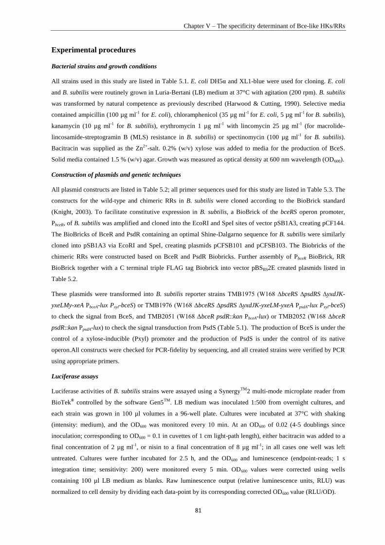
Chapter V – The specificity determinant of Bce-like HKs/RRs
81
Experimental procedures
Bacterial strains and growth conditions
All strains used in this study are listed in Table 5.1. E. coli DH5α and XL1-blue were used for cloning. E. coli
and B. subtilis were routinely grown in Luria-Bertani (LB) medium at 37°C with agitation (200 rpm). B. subtilis
was transformed by natural competence as previously described (Harwood & Cutting, 1990). Selective media
contained ampicillin (100 µg ml-1
for E. coli), chloramphenicol (35 µg ml-1
for E. coli, 5 µg ml-1
for B. subtilis),
kanamycin (10 µg ml-1
for B. subtilis), erythromycin 1 µg ml-1
with lincomycin 25 µg ml-1
(for macrolide-
lincosamide-streptogramin B (MLS) resistance in B. subtilis) or spectinomycin (100 µg ml-1
for B. subtilis).
Bacitracin was supplied as the Zn2+
-salt. 0.2% (w/v) xylose was added to media for the production of BceS.
Solid media contained 1.5 % (w/v) agar. Growth was measured as optical density at 600 nm wavelength (OD600).
Construction of plasmids and genetic techniques
All plasmid constructs are listed in Table 5.2; all primer sequences used for this study are listed in Table 5.3. The
constructs for the wild-type and chimeric RRs in B. subtilis were cloned according to the BioBrick standard
(Knight, 2003). To facilitate constitutive expression in B. subtilis, a BioBrick of the bceRS operon promoter,
PbceR, of B. subtilis was amplified and cloned into the EcoRI and SpeI sites of vector pSB1A3, creating pCF144.
The BioBricks of BceR and PsdR containing an optimal Shine-Dalgarno sequence for B. subtilis were similarly
cloned into pSB1A3 via EcoRI and SpeI, creating plasmids pCFSB101 and pCFSB103. The Biobricks of the
chimeric RRs were constructed based on BceR and PsdR Biobricks. Further assembly of PbceR BioBrick, RR
BioBrick together with a C terminal triple FLAG tag Biobrick into vector pBSBS2E created plasmids listed in
Table 5.2.
These plasmids were transformed into B. subtilis reporter strains TMB1975 (W168 ∆bceRS ∆psdRS ∆yxdJK-
yxeLMy-xeA PbceA-lux Pxyl-bceS) or TMB1976 (W168 ∆bceRS ∆psdRS ∆yxdJK-yxeLM-yxeA PpsdA-lux Pxyl-bceS)
to check the signal from BceS, and TMB2051 (W168 ΔbceR psdR::kan PbceA-lux) or TMB2052 (W168 ΔbceR
psdR::kan PpsdA-lux) to check the signal transduction from PsdS (Table 5.1). The production of BceS is under the
control of a xylose-inducible (Pxyl) promoter and the production of PsdS is under the control of its native
operon.All constructs were checked for PCR-fidelity by sequencing, and all created strains were verified by PCR
using appropriate primers.
Luciferase assays
Luciferase activities of B. subtilis strains were assayed using a SynergyTM
2 multi-mode microplate reader from
BioTek® controlled by the software Gen5
TM. LB medium was inoculated 1:500 from overnight cultures, and
each strain was grown in 100 µl volumes in a 96-well plate. Cultures were incubated at 37°C with shaking
(intensity: medium), and the OD600 was monitored every 10 min. At an OD600 of 0.02 (4-5 doublings since
inoculation; corresponding to OD600 = 0.1 in cuvettes of 1 cm light-path length), either bacitracin was added to a
final concentration of 2 µg ml-1
, or nisin to a final concentration of 8 µg ml-1
; in all cases one well was left
untreated. Cultures were further incubated for 2.5 h, and the OD600 and luminescence (endpoint-reads; 1 s
integration time; sensitivity: 200) were monitored every 5 min. OD600 values were corrected using wells
containing 100 µl LB medium as blanks. Raw luminescence output (relative luminescence units, RLU) was
normalized to cell density by dividing each data-point by its corresponding corrected OD600 value (RLU/OD).
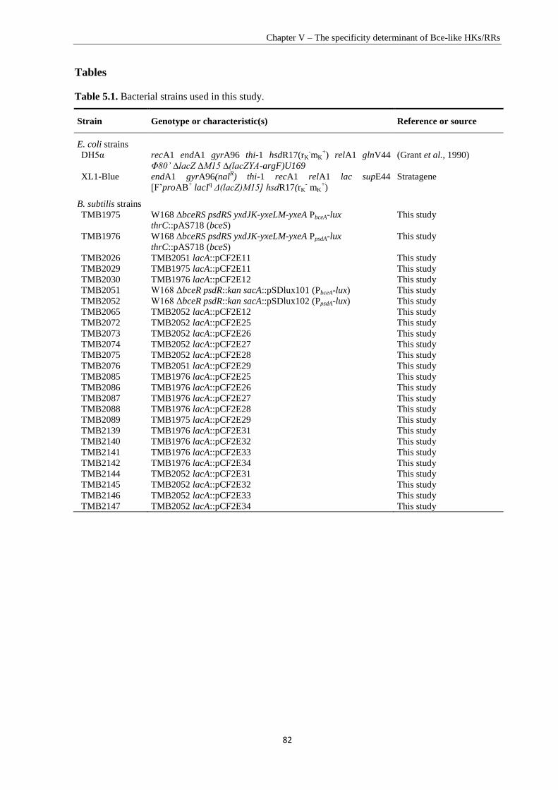
Chapter V – The specificity determinant of Bce-like HKs/RRs
82
Tables
Table 5.1. Bacterial strains used in this study.
Strain Genotype or characteristic(s)
Reference or source
E. coli strains
DH5α recA1 endA1 gyrA96 thi-1 hsdR17(rK-mK
+) relA1 glnV44
Φ80’ ∆lacZ ∆M15 ∆(lacZYA-argF)U169
(Grant et al., 1990)
XL1-Blue endA1 gyrA96(nalR) thi-1 recA1 relA1 lac supE44
[F’proAB+ lacI
q Δ(lacZ)M15] hsdR17(rK
- mK
+)
Stratagene
B. subtilis strains
TMB1975 W168 ∆bceRS psdRS yxdJK-yxeLM-yxeA PbceA-lux
thrC::pAS718 (bceS)
This study
TMB1976 W168 ∆bceRS psdRS yxdJK-yxeLM-yxeA PpsdA-lux
thrC::pAS718 (bceS)
This study
TMB2026 TMB2051 lacA::pCF2E11 This study
TMB2029 TMB1975 lacA::pCF2E11 This study
TMB2030 TMB1976 lacA::pCF2E12 This study
TMB2051 W168 ΔbceR psdR::kan sacA::pSDlux101 (PbceA-lux) This study
TMB2052 W168 ΔbceR psdR::kan sacA::pSDlux102 (PpsdA-lux) This study
TMB2065 TMB2052 lacA::pCF2E12 This study
TMB2072 TMB2052 lacA::pCF2E25 This study
TMB2073 TMB2052 lacA::pCF2E26 This study
TMB2074 TMB2052 lacA::pCF2E27 This study
TMB2075 TMB2052 lacA::pCF2E28 This study
TMB2076 TMB2051 lacA::pCF2E29 This study
TMB2085 TMB1976 lacA::pCF2E25 This study
TMB2086 TMB1976 lacA::pCF2E26 This study
TMB2087 TMB1976 lacA::pCF2E27 This study
TMB2088 TMB1976 lacA::pCF2E28 This study
TMB2089 TMB1975 lacA::pCF2E29 This study
TMB2139 TMB1976 lacA::pCF2E31 This study
TMB2140 TMB1976 lacA::pCF2E32 This study
TMB2141 TMB1976 lacA::pCF2E33 This study
TMB2142 TMB1976 lacA::pCF2E34 This study
TMB2144 TMB2052 lacA::pCF2E31 This study
TMB2145 TMB2052 lacA::pCF2E32 This study
TMB2146 TMB2052 lacA::pCF2E33 This study
TMB2147 TMB2052 lacA::pCF2E34 This study
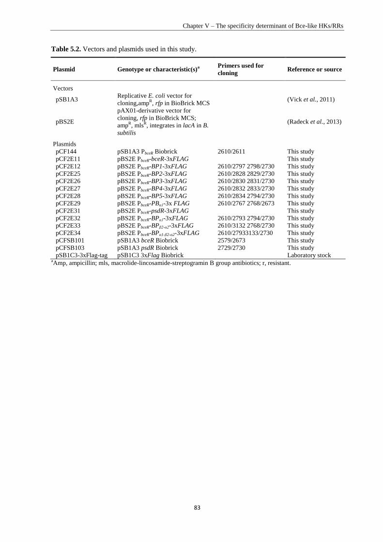
Chapter V – The specificity determinant of Bce-like HKs/RRs
83
Table 5.2. Vectors and plasmids used in this study.
Plasmid Genotype or characteristic(s)a Primers used for
cloning Reference or source
Vectors
pSB1A3 Replicative E. coli vector for
cloning,ampR, rfp in BioBrick MCS
(Vick et al., 2011)
pBS2E
pAX01-derivative vector for
cloning, rfp in BioBrick MCS;
ampR, mls
R, integrates in lacA in B.
subtilis
(Radeck et al., 2013)
Plasmids
pCF144 pSB1A3 PbceR Biobrick 2610/2611 This study
pCF2E11 pBS2E PbceR-bceR-3xFLAG This study
pCF2E12 pBS2E PbceR-BP1-3xFLAG 2610/2797 2798/2730 This study
pCF2E25 pBS2E PbceR-BP2-3xFLAG 2610/2828 2829/2730 This study
pCF2E26 pBS2E PbceR-BP3-3xFLAG 2610/2830 2831/2730 This study
pCF2E27 pBS2E PbceR-BP4-3xFLAG 2610/2832 2833/2730 This study
pCF2E28 pBS2E PbceR-BP5-3xFLAG 2610/2834 2794/2730 This study
pCF2E29 pBS2E PbceR-PBα1-3x FLAG 2610/2767 2768/2673 This study
pCF2E31 pBS2E PbceR-psdR-3xFLAG This study
pCF2E32 pBS2E PbceR-BPα1-3xFLAG 2610/2793 2794/2730 This study
pCF2E33 pBS2E PbceR-BPβ2-α2-3xFLAG 2610/3132 2768/2730 This study
pCF2E34 pBS2E PbceR-BPα1-β2-α2-3xFLAG 2610/27933133/2730 This study
pCFSB101 pSB1A3 bceR Biobrick 2579/2673 This study
pCFSB103 pSB1A3 psdR Biobrick 2729/2730 This study
pSB1C3-3xFlag-tag pSB1C3 3xFlag Biobrick Laboratory stock aAmp, ampicillin; mls, macrolide-lincosamide-streptogramin B group antibiotics; r, resistant.
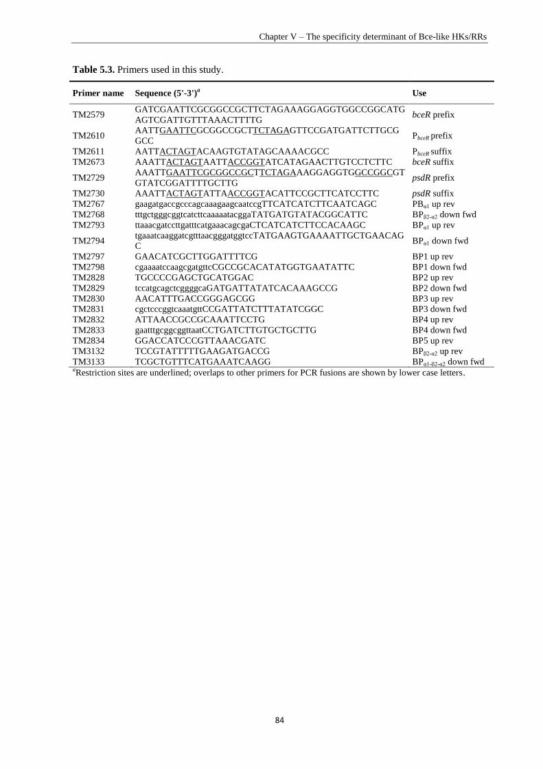
Chapter V – The specificity determinant of Bce-like HKs/RRs
84
Table 5.3. Primers used in this study.
Primer name Sequence (5'-3')a Use
TM2579 GATCGAATTCGCGGCCGCTTCTAGAAAGGAGGTGGCCGGCATG
AGTCGATTGTTTAAACTTTTG bceR prefix
TM2610 AATTGAATTCGCGGCCGCTTCTAGAGTTCCGATGATTCTTGCG
GCC PbceR prefix
TM2611 AATTACTAGTACAAGTGTATAGCAAAACGCC PbceR suffix
TM2673 AAATTACTAGTAATTACCGGTATCATAGAACTTGTCCTCTTC bceR suffix
TM2729 AAATTGAATTCGCGGCCGCTTCTAGAAAGGAGGTGGCCGGCGT
GTATCGGATTTTGCTTG psdR prefix
TM2730 AAATTACTAGTATTAACCGGTACATTCCGCTTCATCCTTC psdR suffix
TM2767 gaagatgaccgcccagcaaagaagcaatccgTTCATCATCTTCAATCAGC PBα1 up rev
TM2768 tttgctgggcggtcatcttcaaaaatacggaTATGATGTATACGGCATTC BPβ2-α2 down fwd
TM2793 ttaaacgatccttgatttcatgaaacagcgaCTCATCATCTTCCACAAGC BPα1 up rev
TM2794 tgaaatcaaggatcgtttaacgggatggtccTATGAAGTGAAAATTGCTGAACAG
C BPα1 down fwd
TM2797 GAACATCGCTTGGATTTTCG BP1 up rev
TM2798 cgaaaatccaagcgatgttcCGCCGCACATATGGTGAATATTC BP1 down fwd
TM2828 TGCCCCGAGCTGCATGGAC BP2 up rev
TM2829 tccatgcagctcggggcaGATGATTATATCACAAAGCCG BP2 down fwd
TM2830 AACATTTGACCGGGAGCGG BP3 up rev
TM2831 cgctcccggtcaaatgttCCGATTATCTTTATATCGGC BP3 down fwd
TM2832 ATTAACCGCCGCAAATTCCTG BP4 up rev
TM2833 gaatttgcggcggttaatCCTGATCTTGTGCTGCTTG BP4 down fwd
TM2834 GGACCATCCCGTTAAACGATC BP5 up rev
TM3132 TCCGTATTTTTGAAGATGACCG BPβ2-α2 up rev
TM3133 TCGCTGTTTCATGAAATCAAGG BPα1-β2-α2 down fwd aRestriction sites are underlined; overlaps to other primers for PCR fusions are shown by lower case letters.
Chapter V – The specificity determinant of Bce-like HKs/RRs
85
References
Bernard, R., A. Guiseppi, M. Chippaux, M. Foglino & F. Denizot, (2007) Resistance to bacitracin in Bacillus
subtilis: unexpected requirement of the BceAB ABC transporter in the control of expression of its own
structural genes. Journal of bacteriology 189: 8636-8642.
Bourret, R.B., (2010) Receiver domain structure and function in response regulator proteins. Current opinion in
microbiology 13: 142-149.
Capra, E.J. & M.T. Laub, (2012) The Evolution of Two-Component Signal Transduction Systems. Annual
review of microbiology 66: 325.
Capra, E.J., B.S. Perchuk, E.A. Lubin, O. Ashenberg, J.M. Skerker & M.T. Laub, (2010) Systematic dissection
and trajectory-scanning mutagenesis of the molecular interface that ensures specificity of two-
component signaling pathways. PLoS genetics 6: e1001220.
Capra, E.J., B.S. Perchuk, J.M. Skerker & M.T. Laub, (2012) Adaptive mutations that prevent crosstalk enable
the expansion of paralogous signaling protein families. Cell 150: 222-232.
Casino, P., V. Rubio & A. Marina, (2009) Structural insight into partner specificity and phosphoryl transfer in
two-component signal transduction. Cell 139: 325-336.
Dintner, S., R. Heermann, C. Fang, K. Jung & S. Gebhard, (2014) A sensory complex consisting of an ATP-
binding cassette transporter and a two-component regulatory system controls bacitracin resistance in
Bacillus subtilis. J Biol Chem 289: 27899-27910.
Fabret, C., V.A. Feher & J.A. Hoch, (1999) Two-component signal transduction in Bacillus subtilis: how one
organism sees its world. Journal of Bacteriology 181: 1975-1983.
Galperin, M.Y., (2006) Structural classification of bacterial response regulators: diversity of output domains and
domain combinations. Journal of Bacteriology 188: 4169-4182.
Galperin, M.Y., (2010) Diversity of structure and function of response regulator output domains. Current
opinion in microbiology 13: 150-159.
Gao, R. & A.M. Stock, (2009) Biological insights from structures of two-component proteins. Annual review of
microbiology 63: 133.
Gebhard, S. & T. Mascher, (2011) Antimicrobial peptide sensing and detoxification modules: unravelling the
regulatory circuitry of Staphylococcus aureus. Molecular microbiology 81: 581-587.
Gooderham, W.J. & R.E. Hancock, (2009) Regulation of virulence and antibiotic resistance by two-component
regulatory systems in Pseudomonas aeruginosa. FEMS microbiology reviews 33: 279-294.
Grant, S.G., J. Jessee, F.R. Bloom & D. Hanahan, (1990) Differential plasmid rescue from transgenic mouse
DNAs into Escherichia coli methylation-restriction mutants. Proceedings of the National Academy of
Sciences 87: 4645-4649.
Harwood, C.R. & S.M. Cutting, (1990) Molecular biological methods for Bacillus.
Howell, A., S. Dubrac, K.K. Andersen, D. Noone, J. Fert, T. Msadek & K. Devine, (2003) Genes controlled by
the essential YycG/YycF two‐component system of Bacillus subtilis revealed through a novel hybrid
regulator approach. Molecular microbiology 49: 1639-1655.
Joseph, P., A. Guiseppi, A. Sorokin & F. Denizot, (2004) Characterization of the Bacillus subtilis YxdJ response
regulator as the inducer of expression for the cognate ABC transporter YxdLM. Microbiology 150:
2609-2617.
Jung, K., L. Fried, S. Behr & R. Heermann, (2012) Histidine kinases and response regulators in networks.
Current opinion in microbiology 15: 118-124.
Knight, T., (2003) Idempotent vector design for standard assembly of biobricks. In.: DTIC Document, pp.
Krell, T., J. Lacal, A. Busch, H. Silva-Jiménez, M.-E. Guazzaroni & J.L. Ramos, (2010) Bacterial sensor
kinases: diversity in the recognition of environmental signals. Annual review of microbiology 64: 539-
559.
Mascher, T., (2006) Intramembrane-sensing histidine kinases: a new family of cell envelope stress sensors in
Firmicutes bacteria. FEMS microbiology letters 264: 133-144.
Mascher, T., (2014) Bacterial (intramembrane-sensing) histidine kinases: signal transfer rather than stimulus
perception. Trends in microbiology 22: 559-565.
Ohki, R., K. Tateno, W. Masuyama, S. Moriya, K. Kobayashi & N. Ogasawara, (2003) The BceRS
two‐component regulatory system induces expression of the bacitracin transporter, BceAB, in Bacillus
subtilis. Molecular microbiology 49: 1135-1144.
Parkinson, J.S., (1993) Signal transduction schemes of bacteria. Cell 73: 857-871.
Paterson, G., C. Blue & T. Mitchell, (2006) Role of two-component systems in the virulence of Streptococcus
pneumoniae. Journal of medical microbiology 55: 355-363.
Pietiäinen, M., M. Gardemeister, M. Mecklin, S. Leskelä, M. Sarvas & V.P. Kontinen, (2005) Cationic
antimicrobial peptides elicit a complex stress response in Bacillus subtilis that involves ECF-type sigma
factors and two-component signal transduction systems. Microbiology 151: 1577-1592.
Chapter V – The specificity determinant of Bce-like HKs/RRs
86
Podgornaia, A.I., P. Casino, A. Marina & M.T. Laub, (2013) Structural basis of a rationally rewired protein-
protein interface critical to bacterial signaling. Structure 21: 1636-1647.
Podgornaia, A.I. & M.T. Laub, (2013) Determinants of specificity in two-component signal transduction.
Current opinion in microbiology 16: 156-162.
Procaccini, A., B. Lunt, H. Szurmant, T. Hwa & M. Weigt, (2011) Dissecting the specificity of protein-protein
interaction in bacterial two-component signaling: orphans and crosstalks. PloS one 6: e19729.
Radeck, J., K. Kraft, J. Bartels, T. Cikovic, F. Dürr, J. Emenegger, S. Kelterborn, C. Sauer, G. Fritz, S. Gebhard,
& Thorsten Mascher, (2013) The Bacillus BioBrick Box: generation and evaluation of essential genetic
building blocks for standardized work with Bacillus subtilis. Journal of Biological Engineering 7.
Rietkötter, E., D. Hoyer & T. Mascher, (2008) Bacitracin sensing in Bacillus subtilis. Molecular microbiology
68: 768-785.
Siryaporn, A., B.S. Perchuk, M.T. Laub & M. Goulian, (2010) Evolving a robust signal transduction pathway
from weak cross‐talk. Molecular systems biology 6: 452.
Skerker, J.M., B.S. Perchuk, A. Siryaporn, E.A. Lubin, O. Ashenberg, M. Goulian & M.T. Laub, (2008)
Rewiring the specificity of two-component signal transduction systems. Cell 133: 1043-1054.
Staroń, A., D.E. Finkeisen & T. Mascher, (2011) Peptide antibiotic sensing and detoxification modules of
Bacillus subtilis. Antimicrobial agents and chemotherapy 55: 515-525.
Stock, A.M., V.L. Robinson & P.N. Goudreau, (2000) Two-component signal transduction. Annual review of
biochemistry 69: 183-215.
Szurmant, H. & J.A. Hoch, (2010) Interaction fidelity in two-component signaling. Current opinion in
microbiology 13: 190-197.
Vick, J.E., E.T. Johnson, S. Choudhary, S.E. Bloch, F. Lopez-Gallego, P. Srivastava, I.B. Tikh, G.T. Wawrzyn
& C. Schmidt-Dannert, (2011) Optimized compatible set of BioBrick™ vectors for metabolic pathway
engineering. Applied microbiology and biotechnology 92: 1275-1286.
Wadhams, G.H. & J.P. Armitage, (2004) Making sense of it all: bacterial chemotaxis. Nature Reviews Molecular
Cell Biology 5: 1024-1037.
West, A.H. & A.M. Stock, (2001) Histidine kinases and response regulator proteins in two-component signaling
systems. Trends in biochemical sciences 26: 369-376.
Williams, P., K. Winzer, W.C. Chan & M. Camara, (2007) Look who's talking: communication and quorum
sensing in the bacterial world. Philosophical Transactions of the Royal Society of London B: Biological
Sciences 362: 1119-1134.
87
Chapter VI
Concluding Discussion
Chapter VI - Concluding discussion
88
6. Concluding discussion
Resistance against antimicrobial peptides (AMPs) largely determines bacterial survival in competitive
habitats. Among the resistance mechanisms developed by bacteria, the most efficient ways often
involve ATP-binding cassette (ABC) transporters. A special type of BceAB-like ABC transporters, is
exclusively contained within and widespread among Firmicutes bacteria (Gebhard, 2012). It
frequently pairs with an adjacent BceRS-like two-component system (TCS), forming the Bce-type
specific AMP sensing and detoxification module (Dintner et al., 2011). Although a number of Bce-
type systems have been studied in Bacillus subtilis and Staphylococcus aureus (reviewed in (Gebhard
& Mascher, 2011)), many questions remain unanswered. The aim of this thesis was to obtain a deep
insight into the regulatory network of Bce-type systems in two Firmicutes bacteria: Enterococcus
faecalis and B. subtilis.
The technical challenges of molecular studies in E. faecalis impedes the understanding of Bce-type
AMP sensing and detoxification system. Therefore, we exploited B. subtilis as a heterologous host to
study the E. faecalis systems (Chapter II). By combining the homologous study in E. faecalis with
the heterologous study in the B. subtilis platform, we successfully identified and characterized the
Bce-type AMP sensing and resistance network of E. faecalis, which provides useful insight for clinical
research (Chapter III). B. subtilis has three paralogous but well insulated Bce-like systems. Focusing
on the Bce and Psd systems, we investigated the molecular mechanisms using by B. subtilis to
maintain the intrasystem signal transduction and intersystem insulation at the response regulator
(RR)/promoter level (Chapter IV) and the histidine kinase (HK)/RR level (Chapter V). At both
interfaces, novel specificity determinants could be identified and characterized.
6.1. Bacillus subtilis as a heterologous host: advantages and considerations
Understanding AMP resistance mechanisms of pathogenic bacteria is important for antibiotic
development. However, for many species, such studies are impeded by the difficulty of genetic
manipulation as well as pathogenicity. Because of the easy genetic manipulation and the well-
understood genetic background, the Gram-negative bacterium Escherichia coli has been used as a
heterologous host for in vivo analysis of the VanRS TCSs in both type A and B vancomycin resistant
enterococci (Silva et al., 1998). However, the differences in cell wall composition between Gram-
positive and Gram-negative bacteria suggest that E. coli is probably not a suitable host for
heterologous studies of enterococci cell wall active AMP resistance systems. B. subtilis, on the other
hand, the best-characterized member of Gram-positive bacteria with well understood AMP resistance
systems (reviewed in (Jordan et al., 2008)), might be a more suitable candidate for such heterologous
studies of E. faecalis AMP resistance mechanisms. It provides numerous established genetic tools, a
comparable GC content (43.5%) with E. faecalis (37.5%), similar transcription machinery, and the
most important ― a comparable cell envelope.
Chapter VI - Concluding discussion
89
The objective of Chapter II was to exploit B. subtilis as a platform for heterologous study of AMP
resistance mechanisms of E. faecalis. Two kinds of AMP resistance systems from E. faecalis, the
BcrR one-component system and the VanSB-VanRB two-component system, were transferred into B.
subtilis and their functionality was demonstrated. The three native Bce-like AMP sensing and
resistance systems in B. subtilis were deleted to minimize the interference with the heterologously
introduced systems. We were also able to demonstrate that the one-component system BcrR from E.
faecalis is fully functional in B. subtilis in both gene regulation and bacitracin resistance. In a previous
study, the VanSB-VanRB TCS system from E. faecalis, which can sense vancomycin and regulate the
resistance operon, showed a constitutive expression of the target promoter PvanYB in B. subtilis
(Bisicchia et al., 2011). In contrast to this result, by adjusting the expression level of the TCS using a
xylose inducible promoter, Pxyl, we demonstrated that the target promoter PvanYB of the VanSB-VanRB
TCS is induced in a vancomycin-dependent manner and the system is functionally produced in B.
subtilis. With this B. subtilis platform, we were also able to functionally characterize a complex Bce-
type system of E. faecalis with two ABC transporters and a regulatory TCS for bacitracin sensing and
resistance. These results are presented in Chapter III and will be discussed in the next section.
Based on these results, we confirmed that B. subtilis is clearly a suitable heterologous host for
studying cell wall-targeting antibiotic resistance systems of E. faecalis. Attentions must be paid to the
genetic background of the host to minimize the interference of the intrinsic resistance system to the
introduced system. The expression level of the introduced system may influence its function, which
means optimization is required and important for functional analysis. In addition to functional
characterization of AMP resistance systems, the high degree of competence of B. subtilis can also be
used for preliminary high-throughput screening of random mutations and synthetic DNA libraries for
deep understanding of the signal transduction mechanism of E. faecalis. Promising results can be
validated in a more targeted fashion further in E. faecalis. B. subtilis is of course also a suitable host
for heterologous studying of cell wall-targeting AMP resistance systems of the other Firmicutes
bacteria.
6.2. The Bce-type modules are arranged differently within Bacillus subtilis and
Enterococcus faecalis
The BceAB-like ABC transporters have been shown to be widely distributed in Firmicutes bacteria for
AMP sensing and resistance (Dintner et al., 2011). Three such ABC transporters, BceAB, PsdAB and
YxdLM, were identified and characterized in B. subtilis by previous studies reviewed in (Gebhard &
Mascher, 2011) (Fig. 6.1 B. subtilis). Each of these BceAB-like ABC transporters is functionally
related with a cognate BceRS-like TCS, and together they form a signal transduction circuit. Taking
the best understood BceRS-BceAB system as an example: the signaling starts when the ABC
transporter BceAB detects the AMP and activates the cognate HK BceS by direct protein-protein
interaction; the subsequent signal transmission within the TCS BceRS will upregulate the transcription
Chapter VI - Concluding discussion
90
of the ABC transporter operon for further resistance (Ohki et al., 2003, Coumes-Florens et al., 2011,
Dintner et al., 2014).
The majority of BceAB-like ABC transporters, such as the above-mentioned three ABC transporters in
B. subtilis, are usually regulated by BceRS-like TCSs encoded in the genomic neighborhood of the
ABC transporters (Dintner et al., 2011). However, there are still a number of such ABC transporters
lacking neighboring TCSs. In the genome of E. faecalis, two putative BceAB-like transporters and one
BceRS-like TCS were identified in previous study, but neither of these ABC transporters was located
next to the TCS (Dintner et al., 2011). To identify the relationship between these two BceAB-like
ABC transporters and the only one BceRS-like TCS, functional analysis was done with these three
gene loci in both E. faecalis and the heterologous host B. subtilis in Chapter III. We showed that
these three gene loci are all required for full bacitracin resistance: (1) one ABC transporter, EF2050-
2049, that was strongly upregulated in response to bacitracin can mediate bacitracin resistance; (2) the
other ABC transporter, EF2752-2751, that was slightly induced by bacitracin is responsible for
bacitracin perception; and (3) the expression of these two ABC transporter operons was differentially
regulated by the TCS EF0927-0926, and the expression of the TCS was induced by an as-yet-
unidentified regulator that is not directly part of the resistance network. We were able to build the
bacitracin sensing and resistance network in E. faecalis (Fig. 6.1 E. faecalis), which starts when the
transporter EF2752-2751 detects the presence of bacitracin and subsequently transfers the signal to the
TCS EF0926-0927. Signaling within the TCS leads to a highly increased production of the transporter
EF2050-2049 to mediate resistance against bacitracin, and a slightly increased production of the
transporter EF2752-2751 to detect bacitracin. However, the mechanism of differentiating the level of
regulation is not known and more investigation is needed to address this question.
Compared to the signaling transduction circuit of B. subtilis for AMP sensing and resistance (Fig. 6.1
B. subtilis), we demonstrated that products of these three gene loci of E. faecalis form another
configuration for the AMP sensing and resistance module (Fig. 6.1 E. faecalis). The Bce-type ABC
transporters and TCSs are arranged differently in B. subtilis and in E. faecalis. The combined
functions of the ABC transporter BceAB with both bacitracin sensing and resistance in B. subtilis is
separated into two in E. faecalis, i.e. one ABC transporter, EF2752-2751, is only responsible for
bacitracin sensing and an additional one, EF2050-2049, is required for bacitracin resistance. The gene
regulation is also split and differentiated in E. faecalis: the TCS is able to strongly upregulate the
transcription of the resistance ABC transporter EF2050-2049 operon and slightly upregulate the
transcription of the sensor ABC transporter EF2752-2751 operon. The expression of the BceRS TCS
operon of B. subtilis is under the control of a constitutive promoter, while the expression of the
EF0927-0926 TCS operon of E. faecalis it is induced by an as-yet-unidentified regulator, which is not
directly part of the resistance network.
Chapter VI - Concluding discussion
91
Figure 6.1. Schematic of signaling network of Bce-type AMP sensing and detoxification systems in B.
subtilis, E. facalis and S. aureus. Coloring is chosen to reflect the assignment of each module to its
phylogenetic group (yellow, group II; red, group III; blue, group IV; green, group VII; grey and black, no group
assigned) (Dintner et al., 2011). Names of genes and proteins of each system are given next to their schematics.
Main substrates of each system are shown on top of the ABC transporter, with black curved downward arrow
representing the perception and grey curved upward arrow representing the detoxification. Signal transfer
between transporters and TCSs is indicated in the membrane bilayer. Phosphotransfer between HKs and RRs,
transcriptional activation, and production of ABC transporters are shown by solid arrows. The minor level of
cross-regulation between BceS and PsdR is shown by a dashed arrow. The induction of EF0926-EF0927 operon
by an unknown regulator in E. faecalis is showen by a dotted arrow. The differences in the strength of induction
are reflected by thickness of lines. This figure was originally based in parts on (Gebhard & Mascher, 2011,
Gebhard et al., 2014), with modifications.
Chapter VI - Concluding discussion
92
Our results of the E. faecalis Bce-type network are similar to the previously described BraDE-BraRS-
VraDE network of S. aureus (Fig. 6.1 S. aureus). In S. aureus, the production of two Bce-type ABC
transporters, VraDE and BraDE, is controlled by one Bce-type TCS BraRS. The BraDE ABC
transporter can sense AMPs (bacitracin and nisin) and activate the TCS BraRS, which will further
induce the expression of both ABC transporter operons braDE and vraDE for AMP detection and
resistance, respectively (Li et al., 2007, Hiron et al., 2011). In addition to this system, the division of
labor between two ABC transporters — one is only for AMP sensing while the other one is only for
AMP resistance — was also observed in other Firmicutes bacteria, such as the ABC12-TCS12-orphan
ABC transporter system in Lactobacillus casei for sensing and resistance against AMPs (Revilla-
Guarinos et al., 2013).
The existence of these two types of arrangement for AMP sensing and resistance network in
Firmicutes bacteria raises the question: why do bacteria sometimes split functions of the ABC
transporter and sometimes not? One explanation is that splitting the functions (AMP perception and
resistance) of the ABC transporter may endow bacteria the possibility of mediating nonspecific
responses, i.e. TCSs from other systems can cross-regulate the resistance ABC transporter operon.
While combining the functions is beneficial for bacteria to maintain the specificity of the signal
transduction pathway, i.e. the expression of the ABC transporter can only be regulated by its cognate
TCS. It may also be that the separation of the AMP sensing and resistance functions and
differentiation of the expression level keep the proper energy distribution between two ABC
transporters. However, these hypotheses still need to be proved by experimental evidence.
6.3. Specificity determination of Bce-like TCSs in Bacillus subtilis
Cross-talk is defined as the communication between two distinct systems, which is often detrimental
and must be kept to a minimum for generating desired responses to specific stimuli. However, there
are examples of beneficial cross-talk under some conditions, such as mediating multiple responses to a
single input, called cross-regulation (Laub & Goulian, 2007). Although minor level of cross-regulation
between the HK BceS and the RR PsdR was observed in vivo under high concentrations of bacitracin
in B. subtilis, the remaining parts of three paralogous Bce-type AMP sensing and detoxification
systems are very well insulated from each other (Rietkötter et al., 2008) (Fig. 6.1 B. subtilis). These
three systems most likely evolved by duplication for a common ancestor followed by sequence
divergence so that each of them can detect specific signal input and mediate corresponding output. In
this evolutionary process, mutations that occurred on the specific interaction surfaces of one protein
need to be compensated by corresponding mutations on its partner to maintain a functional interaction
and simultaneously to avoid cross-talk (Szurmant & Hoch, 2010). This indicates that the predominant
mechanism to ensure specificity is molecular recognition — the intrinsic ability of a protein to
distinguish its cognate partner from the non-cognate ones with a relatively high preference. To
guarantee the fidelity of the whole system, specificity needs to be maintained at every step of the
Chapter VI - Concluding discussion
93
signal transduction pathway. In this thesis, we tried to gain a deeper understanding of mechanisms that
dictate intrasystem fidelity and intersystem insulation of the two Bce-type systems (BceRS-BceAB
and PsdRS-PsdAB) in B. subtilis, on both the HK/RR and the RR/promoter interaction level.
6.3.1. The mechanism of maintaining specificity at the RR and target promoter level
On PbceA and PpsdA, no typical -35 element was found in the appropriate location upstream of the -10
element, indicating that the σ unit of the RNA polymerase cannot bind properly to the promoter by
itself for further transcription initiation. However, this problem can be solved when the σ unit interacts
with RRs, which bind to the up element of the promoter, to compensate its weak binding (Lee et al.,
2012). DNA binding domain structures of both PhoB and OmpR from OmpR subfamily demonstrated
that the α2-α3 loop is essential for direct interaction with the σ subunit of the RNA polymerase
(Martínez-Hackert & Stock, 1997, Blanco et al., 2002). BceR and PsdR, which belong to the same
subfamily, are assumed to assist the transcription initiation of RNA polymerase in a similar way.
Specific transcription initiation by the RR is important for maintaining the insulation of the signaling
system. The similarity of Bce-like RRs DNA-binding domain and their binding sites on target
promoters increases the potential of cross-talk on the transcription initiation level. However, we could
show that Bce-type RRs in B. subtilis are extremely specific in inducing the transcription of their
cognate ABC transporters. Although the binding of BceR to the cognate PbceA and the non-cognate
PpsdA were both observed in vitro, BceR can only induce the transcription of bceAB and not of psdAB
in vivo (Chapter IV). To understand the mechanism of specific regulation, further EMSAs and SPR
assays were performed in Chapter IV and indicated that the in vitro cross-talk and in vivo insulation
are due to the great difference in binding affinities, i.e. BceR has a much higher affinity to its cognate
promoter PbceA than to the non-cognate promoter PpsdA. Although it can bind to PpsdA in vitro, the
binding affinity is still not high enough to recruit the RNA polymerase to the promoter for in vivo
transcription initiation.
The affinity preference is the ability of the RR to distinguish the cognate promoter from non-cognate
ones. Our data strongly suggests that B. subtilis evolved an intelligent mechanism to maintain this
ability, which is a hierarchical and cooperative binding model (Fig. 6.2). Instead of only the one
binding site reported by previous studies (Ohki et al., 2003, de Been et al., 2008), we demonstrated for
the first time the necessary of two binding sites in the regulatory region of the Bce-type RR target
promoters. By performing EMSAs and SPR assays of BceR with PbceA mutants carrying either the
main binding site random mutation or the secondary binding site random mutation, we further
demonstrated that BceR has a high affinity and shows independent binding to the upstream main
binding site. It has a low affinity to the downstream secondary binding site and cannot bind to it alone.
Our data suggests that a BceR dimer first binds to the high affinity main binding site. This first
binding event then assists the subsequent binding of another dimer to the downstream low affinity
secondary binding site. Furthermore, we were able to show that exchanging the secondary binding site
Chapter VI - Concluding discussion
94
resulted in a much stronger influence on promoter specificity than the main binding site by in vivo
promoter activity assays. The hierarchical and cooperative binding model that enables BceR to have
distinct binding affinities to its cognate promoter PbceA from the non-cognate PpsdA is based on: (1) the
main binding sites of these two promoters differ only in three bases, and provide a high affinity, low
specificity docking site; (2) the secondary binding sites of these two promoters harbors five different
bases, and represent a low affinity, but high specificity interaction site; and (3) the combination of the
main binding site and the secondary binding site results in the relatively higher affinity of BceR to its
cognate promoter PbceA compared to the non-cognate PpsdA (Chapter IV).
Figure 6.2. The model of specific transcriptional activation of bceAB operon by BceR and RNA
polymerase. Firstly, BceR dimer (black) not PsdR dimer (gray) binds to the main binding site (MBS) on PbceA.
Secondly, the binding of the first dimer helps another dimer bind to the secondary binding site (SBS) upstream
of the -10 element on PbceA. Finally, the binding of the second BceR dimer to PbceA can assist the binding of σA
subunit of the holo RNA polymerase (RNAP) to the promoter region by direct protein-protein interaction and
hence recruits the RNA polymerase for further transcription. The structure of the DNA is altered by the linker
region between two binding sites.
The linker regions of these two promoters showed characteristically distinct GC/AT contents — PbceA
has high AT content, while PpsdA has high GC content. In Chapter IV we showed that mutating the
linker region into a random sequence but still keeping the GC/AT content of each promoter slightly
affected the promoter activity. However, exchanging linker regions between these two promoters,
which means changing the GC/AT content, obtained more influence on the promoter activity. AT-rich
sequence is known to cause the bending of DNA (Koo et al., 1986). One possibility is that the AT-rich
linker region on PbceA confers a structural difference from PpsdA by bending the promoter between two
binding sites to fit the binding of two BceR dimers.
The high specificity of the secondary binding site is presumably determined mainly by its first half site,
because the first half sites of PbceA and PpsdA differ in four out of seven bases, while the second half
sites show only one. The sequence identity of the second half site and its location at the position of -35
suggest that it can probably be bound by both BceR and the σA subunit of the RNA polymerase. We
showed that a PbceA mutant with the secondary binding site replaced by the main binding site (main
Chapter VI - Concluding discussion
95
binding site-linker-main binding site) lost the promoter activity (data not shown), which further
indicates the importance of the second half for σA subunit binding and transcription initiation. This
mechanism was already demonstrated for the transcription initiation by PhoB and σ70
binding together
to the pho box (Blanco et al., 2011).
This hierarchical and cooperative binding model has already been shown to be widely spread among
the OmpR RR subfamily. For example, PhoB can bind cooperatively on two binding sites of the pstS
promoter with different binding affinities (Blanco et al., 2012). PompF has three OmpR binding sites
with gradually reduced affinity from upstream to downstream, and the binding of OmpR to the first
site is important for subsequent binding to the lower affinity downstream sites (Harlocker et al., 1995).
The cooperative binding model is also demonstrated using by RRs from other families. A recent study
of the RR YpdB by SPR analysis also showed a two-step cooperative binding mechanism to its target
PyhjX (Behr et al., unpublished). The binding of YpdB to the upstream site A initiates a subsequent
binding of YpdB to the downstream site B. Similar as BceR binding to PbceA, binding of YpdB to PyhjX
was completely abolished when the site A was inactivated, but YpdB was still able to bind properly to
the downstream binding site B in the presence of an inactivated site A. The evolution of such complex
regulatory systems is related, to some extent, to the regulatory function of RRs. PhoB and OmpR have
been demonstrated to regulate dozens of operons in E. coli in the presence of certain stimuli. However,
some operons need to be highly upregulated while others require only moderate or subtle regulation.
Control of the desired expression level of these operons can be achieved through assembly of different
numbers of binding sites with sequence divergence. For B. subtilis, a similar mechanism is used to
maintain the signal transduction specificity and the regulatory insulation between three paralogous
Bce-like systems. By evolving the cooperation of a high affinity but low specificity main binding site
and a high specificity but low affinity secondary binding site, B. subtilis is able to ensure the Bce-like
RR a relatively higher preference to its cognate promoter to the non-cognate ones, hence maintains the
signaling fidelity of these three paralogoue Bce-like systems on the transcription level.
6.3.2. The mechanism of maintaining specificity at the HK and the RR interaction level
Bce-type RRs of B. subtilis belong to the OmpR RR subfamily (Fabret et al., 1999). Previous studies
of one family member, PhoP (RR of the PhoPR TCS), showed that substitution of three amino acids
(Ser13, Leu17, and Tyr20) on the α1 helix together with one amino acid (Pro107) on the β5-α5 loop of
PhoP receiver domain with corresponding amino acids from WalR (RR of the WalRK TCS) was
sufficient to rewire the phosphotransfer between these two TCSs (Fig. 6.3A). The PhoP mutant could
be phosphorylated by the non-cognate HK WalK and no longer by the cognate PhoR (Jende et al.,
2010). Similarly, replacing three amino acids (Arg15, Leu16, and Arg22) of the α1 helix and three
amino acids (Pro106, Phe107, and Asn108) of the β5-α5 loop of the OmpR receiver domain (RR from
the EvnZ/OmpR TCS) with corresponding amino acids of RstA (RR of the RstAB TCS) was able to
redirect the phosphotransfer specificity from RstB to OmpR and eliminate the phosphotransfer from
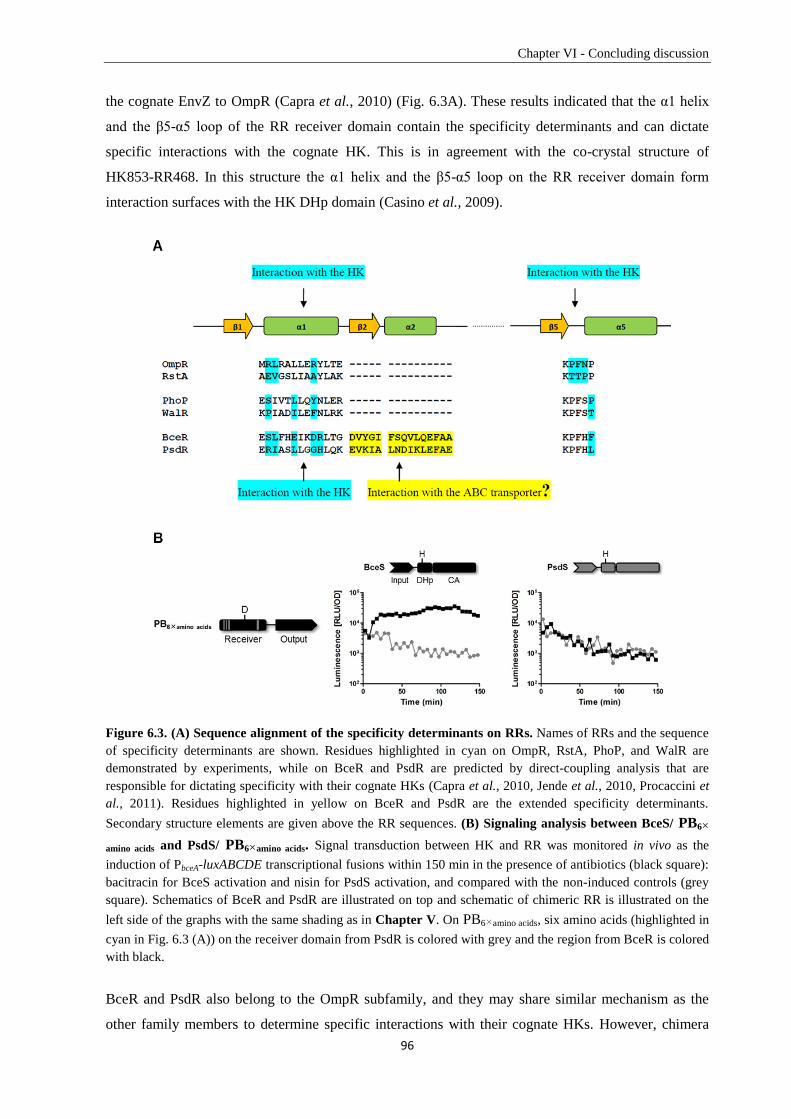
Chapter VI - Concluding discussion
96
the cognate EnvZ to OmpR (Capra et al., 2010) (Fig. 6.3A). These results indicated that the α1 helix
and the β5-α5 loop of the RR receiver domain contain the specificity determinants and can dictate
specific interactions with the cognate HK. This is in agreement with the co-crystal structure of
HK853-RR468. In this structure the α1 helix and the β5-α5 loop on the RR receiver domain form
interaction surfaces with the HK DHp domain (Casino et al., 2009).
Figure 6.3. (A) Sequence alignment of the specificity determinants on RRs. Names of RRs and the sequence
of specificity determinants are shown. Residues highlighted in cyan on OmpR, RstA, PhoP, and WalR are
demonstrated by experiments, while on BceR and PsdR are predicted by direct-coupling analysis that are
responsible for dictating specificity with their cognate HKs (Capra et al., 2010, Jende et al., 2010, Procaccini et
al., 2011). Residues highlighted in yellow on BceR and PsdR are the extended specificity determinants.
Secondary structure elements are given above the RR sequences. (B) Signaling analysis between BceS/ PB6×
amino acids and PsdS/ PB6×amino acids. Signal transduction between HK and RR was monitored in vivo as the
induction of PbceA-luxABCDE transcriptional fusions within 150 min in the presence of antibiotics (black square):
bacitracin for BceS activation and nisin for PsdS activation, and compared with the non-induced controls (grey
square). Schematics of BceR and PsdR are illustrated on top and schematic of chimeric RR is illustrated on the
left side of the graphs with the same shading as in Chapter V. On PB6×amino acids, six amino acids (highlighted in
cyan in Fig. 6.3 (A)) on the receiver domain from PsdR is colored with grey and the region from BceR is colored
with black.
BceR and PsdR also belong to the OmpR subfamily, and they may share similar mechanism as the
other family members to determine specific interactions with their cognate HKs. However, chimera
Chapter VI - Concluding discussion
97
PB6×amino acids, BceR with five amino acids on the α1 helix and one amino acid on the β5-α5 loop
exchanged into corresponding residues of PsdR did not show any change of specificity (Fig. 6.3A and
6.3B). Our data indicates that the α1 helix and the β5-α5 loop of the RR receiver domain are not able
to determine the specificity of Bce-like TCSs in B. subtilis. This is in good agreement with the
prediction done by direct-coupling analysis that BceRS and PsdRS systems have the potential of
considerable cross-talk (Procaccini et al., 2011).
However, a previous study demonstrated that BceRS and PsdRS only have minor level of cross-
regulation between BceS and PsdR at some concentrations of bacitracin (Rietkötter et al., 2008),
which indicates that Bce-like TCSs probably evolved different mechanism to maintain the signaling
specificity. We further explored this hypothesis in Chapter V by making chimeric RRs between BceR
and PsdR on different regions of the receiver domain. We demonstrated that the α1-β2-α2 region on
the Bce-like RR receiver domain is the specificity determinant. BceS, instead of PsdS, strongly
activated PsdR if this region is substituted by the corresponding part of BceR. We showed that the α1
helix of Bce-like RRs is not enough to determine specificity, but it is necessary and indispensable. We
demonstrated that to compensate the lacking of specificity of the α1 helix, B. subtilis developed an
extended determinant with high sequence diversity ― the β2-α2 region. This region can partially
determine the specificity but together with the α1 helix can fully dictate the specificity of Bce-type
TCSs. Compared to the other studied TCSs, B. subtilis uses a different mechanism to maintain the
insulation of Bce-like TCSs (Fig. 6.3A).
Two additional interaction surfaces are also observed in the co-crystal structure of HK853-RR468
(Casino et al., 2009) (Fig. 1.5). One is the interaction between the β3-α3 loop of RR468 receiver
domain and the ATP lid together with the β4-α4 loop of the HK853 CA domain. The other is the
interaction between RR468 β4-α4 loop and the DHp-CA interdomain linker of HK853. However, the
extended β2-α2 region on the RR receiver domain for Bce-type TCSs specificity is not located on any
of these interaction surfaces. The Bce-type TCSs are often functionally related to their ABC
transporters. A bold assumption is proposed that the additional region — β2-α2 — is possibly
responsible for specific interaction with the cognate ABC transporter instead of with the HK.
Supporting this idea, positive interaction between BceR (RR) and BceAB (ABC transporter) was
observed in the presence of BceS (HK) by bacterial two-hybrid assay (BACTH) (Dintner et al., 2014).
However this hypothesis still needs to be proved by further experiments. Taken together, our studies in
Chapter V suggest that B. subtilis evolved an extended determinant (the α1 helix and the β2-α2 region)
to maintain the specificity of three paralogous Bce-like TCSs, which is different from the other studied
OmpR subfamily members (the α1 helix and the β5-α5 loop). Our results provide a clear view of the
location of specific interacting amino acids between HK and RR and a nice starting point for further
studying the mechanism.
Chapter VI - Concluding discussion
98
6.4. Open questions and further research
In this thesis, we demonstrated that B. subtilis is a suitable platform for heterologous studies of the
AMP resistance mechanisms of E. faecalis. Based on this platform, we identified and characterized a
Bce-type AMP sensing and resistance network of E. faecalis. Furthermore, we investigated the
specificity determining mechanisms of two Bce-type systems in B. subtilis. Nevertheless, there are still
several unsolved questions about the Bce-type AMP sensing and detoxification pathways.
6.4.1. Amino acids located on the α1-β2-α2 region for the specificity of Bce-like TCSs
By performing chimeric RR analysis, we were able to demonstrate the determinant ― the α1-β2-α2
region ― on the RR receiver domain that dictates the specificity of Bce-like TCSs. The α1 helix is
comprised of 13 amino acids, and 5 of them are predicted by direct-coupling analysis that can interact
specifically with corresponding amino acids on the cognate HK. However, no information is available
about which amino acids on the β2-α2 region are responsible for specificity. To answer this question,
more chimeras should be made by exchanging these 5 amino acids on the α1 helix together with
different amino acids on the β2-α2 region between BceR and PsdR to construct a chimeric RR library.
The screening can be done by high-throughput promoter-reporter assays on the plate and promising
candidates will be checked further by both in vivo and in vitro experiments.
6.4.2. Is there direct interaction between BceAB and BceR?
Investigation of the specificity determinant between Bce-type TCSs showed an extended α1-β2-α2
region on the RR receiver domain different from the α1 helix and the β5-α5 loop of the other OmpR
subfamily members. According to the co-crystal structure of HK853-RR468, the α1 helix is on the
HK/RR interaction surface but not the β2-α2 region. The functional relation between Bce-type TCSs
and Bce-type ABC transporters implies that this β2-α2 region possibly can form an interaction surface
with the ABC transporter (Fig. 6.3A). BACTH assays suggested a positive interaction between BceR
and BceAB in the presence of BceS (Dintner et al., 2014). This is in good agreement with the previous
study about the GraXSR-VraFG system in S. aureus that these proteins were shown to form a
interaction complex by BACTH assay (Meehl et al., 2007, Falord et al., 2012).
Without BceS, however, no detectable interaction between BceR and BceAB was observed in the
BACTH assay. It is also possible that BceS plays an important role in bringing BceR close to the
cytoplasmic side of the membrane, so that BceR can interact with BceAB. The interaction between
BceR and BceAB probably is not as strong as with BceS, therefore we failed to detect it in the absence
of BceS. Formation of a four-protein complex, demanding that BceR match the specific interaction
with both BceS (by the α1 helix of BceR) and BceAB (by the β2-α2 region of BceR), provides a
double insurance to guarantee the signal transduction specificity of Bce-type systems. This hypothesis
still needs to be proven by further experiments.
Chapter VI - Concluding discussion
99
6.4.3. How does BceR initiate the transcription of the bceAB operon?
We have already demonstrated that BceR binds to two binding sites on PbceA and upregulates
transcription of the bceAB operon. The binding of a BceR dimer to the main binding site can assist
another dimer binding to the low affinity secondary binding site. Does this assistance happen via direct
interaction between two BceR dimers? If yes, on which region do they form the interaction surfaces?
Another open question is whether the RNA polymerase is recruited to the promoter by direct
interaction between the second BceR dimer and the σA subunit. Blanco and colleagues have already
demonstrated that the α2-α3 loop on the PhoB DNA binding domain is essential for transcription
activation by interacting with the σ4 subdomain of the σ70
subunit. The σ4 subdomain can then bind to
the pho box at the -35 position together with the PhoB dimer, but at different sides, and recruit the
RNA polymerase for transcription initiation (Blanco et al., 2002, Blanco et al., 2011). In B. subtilis,
the second halves of the secondary binding sites replace the -35 elements and the sequences are almost
identical between PbceA and PpsdA, which provides a potential low affinity binding region for the σA
subunit. Substitution of the whole secondary binding site by the main binding site resulted in the loss
of promoter activity, which probably undermines the essential binding of the σA subunit to the
promoter. We suggest that the transcription initiation starts with the first dimer binding to the upstream
binding site, which assists and stabilizes the binding of the second dimer to the secondary binding site
by direct dimer-dimer interaction. This is followed by interacting with the σA subunit and recruiting it
to the promoter DNA, which enhance the binding of the σA subunit to the second half of the secondary
binding site and the -10 region to start the transcription of the bceAB operon. Further experiments are
required to verify the interaction between BceR dimers, BceR dimer and the σA subunit of the RNA
polymerase. Furthermore, the binding of the σA subunit to the second half of the downstream
secondary binding site also needs to be proven in this model.
References
100
References of Chapter I and Chapter VI
Appleby, J.L. & R.B. Bourret, (1998) Proposed signal transduction role for conserved CheY residue
Thr87, a member of the response regulator active-site quintet. J Bacteriol 180: 3563-3569.
Arthur, M., F. Depardieu, G. Gerbaud, M. Galimand, R. Leclercq & P. Courvalin, (1997) The VanS
sensor negatively controls VanR-mediated transcriptional activation of glycopeptide resistance
genes of Tn1546 and related elements in the absence of induction. J Bacteriol 179: 97-106.
Arthur, M., C. Molinas & P. Courvalin, (1992) The VanS-VanR two-component regulatory system
controls synthesis of depsipeptide peptidoglycan precursors in Enterococcus faecium
BM4147. J Bacteriol 174: 2582-2591.
Arthur, M. & R. Quintiliani, (2001) Regulation of VanA-and VanB-type glycopeptide resistance in
enterococci. Antimicrob Agents Chemother 45: 375-381.
Azevedo, E., E. Rios, K. Fukushima & G. Campos-Takaki, (1993) Bacitracin production by a new
strain of Bacillus subtilis. Appl Biochem and Biotechnol 42: 1-7.
Bahar, A.A. & D. Ren, (2013) Antimicrobial peptides. Pharmaceuticals 6: 1543-1575.
Berdy, J., (2005) Bioactive microbial metabolites. J Antibiot (Tokyo) 58: 1-26.
Bernlohr, R.W. & G. Novelli, (1963) Bacitracin biosynthesis and spore formation: the physiological
role of an antibiotic. Arch Biochem Biophy 103: 94-104.
Bertsche, U., C. Mayer, F. Götz & A.A. Gust, (2014) Peptidoglycan perception—Sensing bacteria by
their common envelope structure. Int J Med Microbial.
Bhavsar, A.P., L.K. Erdman, J.W. Schertzer & E.D. Brown, (2004) Teichoic acid is an essential
polymer in Bacillus subtilis that is functionally distinct from teichuronic acid. J Bacteriol 186:
7865-7873.
Bisicchia, P., N.K. Bui, C. Aldridge, W. Vollmer & K.M. Devine, (2011) Acquisition of VanB‐type
vancomycin resistance by Bacillus subtilis: the impact on gene expression, cell wall
composition and morphology. Mol Microbiol 81: 157-178.
Blanco, A.G., A. Canals, J. Bernués, M. Solà & M. Coll, (2011) The structure of a transcription
activation subcomplex reveals how σ70
is recruited to PhoB promoters. EMBO J 30: 3776-
3785.
Blanco, A.G., A. Canals & M. Coll, (2012) PhoB transcriptional activator binds hierarchically to pho
box promoters. Biol Chem 393: 1165–1171.
Blanco, A.G., M. Sola, F.X. Gomis-Rüth & M. Coll, (2002) Tandem DNA recognition by PhoB, a
two-component signal transduction transcriptional activator. Structure 10: 701-713.
Boll, J.M. & D.R. Hendrixson, (2011) A specificity determinant for phosphorylation in a response
regulator prevents in vivo cross-talk and modification by acetyl phosphate. Proc Natl Acad Sci
USA 108: 20160-20165.
Bourret, R.B., (2010) Receiver domain structure and function in response regulator proteins. Curr
Opin Microbiol 13: 142-149.
Braun, V., F. Götz, J.E. Schultz & W. Wohlleben, (2014) The bacterial cell envelope: Structure,
function, and infection interface. Int J Med Microbial.
Breukink, E. & B. de Kruijff, (2006) Lipid II as a target for antibiotics. Nat Rev Drug Discov 5: 321-
323.
Brown, S., J.P. Santa Maria Jr & S. Walker, (2013) Wall teichoic acids of Gram-positive bacteria.
Annu Rev Microbiol 67.
Bugg, T.D., D. Braddick, C.G. Dowson & D.I. Roper, (2011) Bacterial cell wall assembly: still an
attractive antibacterial target. Trends Biotechnol 29: 167-173.
Bugg, T.D., G.D. Wright, S. Dutka-Malen, M. Arthur, P. Courvalin & C.T. Walsh, (1991) Molecular
basis for vancomycin resistance in Enterococcus faecium BM4147: biosynthesis of a
depsipeptide peptidoglycan precursor by vancomycin resistance proteins VanH and VanA.
Biochemistry 30: 10408-10415.
Cao, M. & J.D. Helmann, (2002) Regulation of the Bacillus subtilis bcrC bacitracin resistance gene by
two extracytoplasmic function σ factors. J Bacteriol 184: 6123-6129.
Capra, E.J., B.S. Perchuk, E.A. Lubin, O. Ashenberg, J.M. Skerker & M.T. Laub, (2010) Systematic
dissection and trajectory-scanning mutagenesis of the molecular interface that ensures
specificity of two-component signaling pathways. PLoS Genet 6: e1001220.
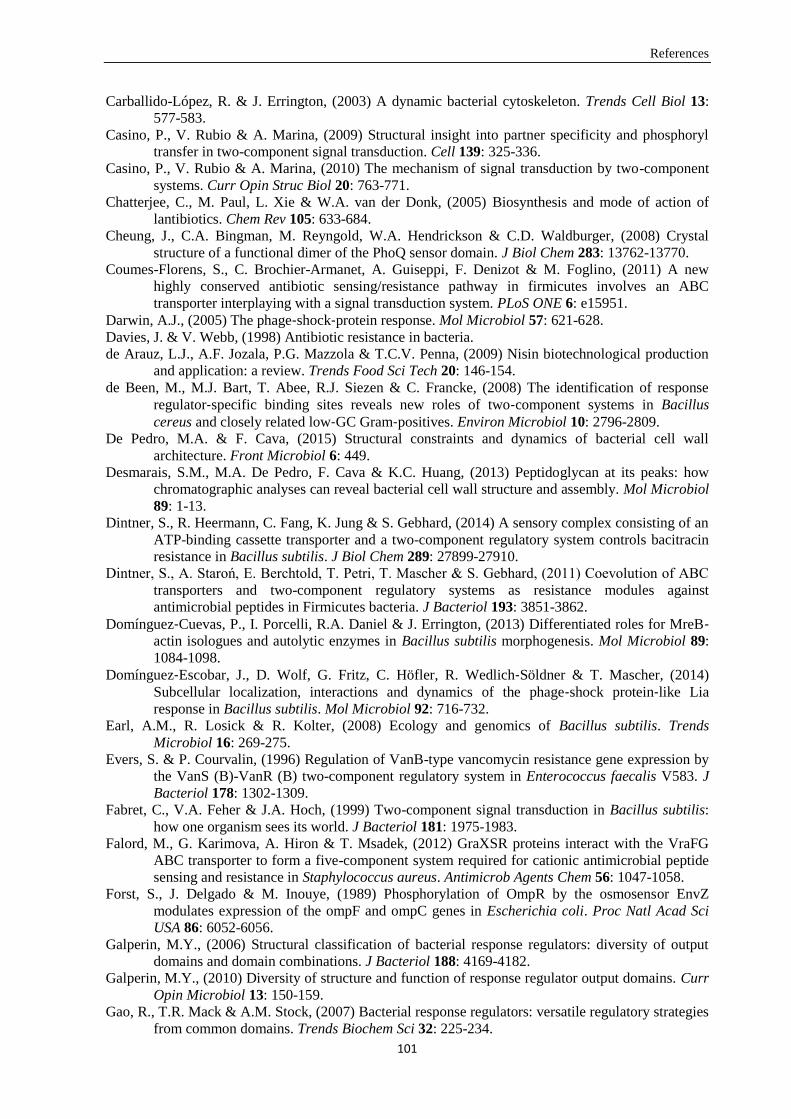
References
101
Carballido-López, R. & J. Errington, (2003) A dynamic bacterial cytoskeleton. Trends Cell Biol 13:
577-583.
Casino, P., V. Rubio & A. Marina, (2009) Structural insight into partner specificity and phosphoryl
transfer in two-component signal transduction. Cell 139: 325-336.
Casino, P., V. Rubio & A. Marina, (2010) The mechanism of signal transduction by two-component
systems. Curr Opin Struc Biol 20: 763-771.
Chatterjee, C., M. Paul, L. Xie & W.A. van der Donk, (2005) Biosynthesis and mode of action of
lantibiotics. Chem Rev 105: 633-684.
Cheung, J., C.A. Bingman, M. Reyngold, W.A. Hendrickson & C.D. Waldburger, (2008) Crystal
structure of a functional dimer of the PhoQ sensor domain. J Biol Chem 283: 13762-13770.
Coumes-Florens, S., C. Brochier-Armanet, A. Guiseppi, F. Denizot & M. Foglino, (2011) A new
highly conserved antibiotic sensing/resistance pathway in firmicutes involves an ABC
transporter interplaying with a signal transduction system. PLoS ONE 6: e15951.
Darwin, A.J., (2005) The phage‐shock‐protein response. Mol Microbiol 57: 621-628.
Davies, J. & V. Webb, (1998) Antibiotic resistance in bacteria.
de Arauz, L.J., A.F. Jozala, P.G. Mazzola & T.C.V. Penna, (2009) Nisin biotechnological production
and application: a review. Trends Food Sci Tech 20: 146-154.
de Been, M., M.J. Bart, T. Abee, R.J. Siezen & C. Francke, (2008) The identification of response
regulator‐specific binding sites reveals new roles of two‐component systems in Bacillus
cereus and closely related low‐GC Gram‐positives. Environ Microbiol 10: 2796-2809.
De Pedro, M.A. & F. Cava, (2015) Structural constraints and dynamics of bacterial cell wall
architecture. Front Microbiol 6: 449.
Desmarais, S.M., M.A. De Pedro, F. Cava & K.C. Huang, (2013) Peptidoglycan at its peaks: how
chromatographic analyses can reveal bacterial cell wall structure and assembly. Mol Microbiol
89: 1-13.
Dintner, S., R. Heermann, C. Fang, K. Jung & S. Gebhard, (2014) A sensory complex consisting of an
ATP-binding cassette transporter and a two-component regulatory system controls bacitracin
resistance in Bacillus subtilis. J Biol Chem 289: 27899-27910.
Dintner, S., A. Staroń, E. Berchtold, T. Petri, T. Mascher & S. Gebhard, (2011) Coevolution of ABC
transporters and two-component regulatory systems as resistance modules against
antimicrobial peptides in Firmicutes bacteria. J Bacteriol 193: 3851-3862.
Domínguez‐Cuevas, P., I. Porcelli, R.A. Daniel & J. Errington, (2013) Differentiated roles for MreB‐actin isologues and autolytic enzymes in Bacillus subtilis morphogenesis. Mol Microbiol 89:
1084-1098.
Domínguez‐Escobar, J., D. Wolf, G. Fritz, C. Höfler, R. Wedlich‐Söldner & T. Mascher, (2014)
Subcellular localization, interactions and dynamics of the phage‐shock protein‐like Lia
response in Bacillus subtilis. Mol Microbiol 92: 716-732.
Earl, A.M., R. Losick & R. Kolter, (2008) Ecology and genomics of Bacillus subtilis. Trends
Microbiol 16: 269-275.
Evers, S. & P. Courvalin, (1996) Regulation of VanB-type vancomycin resistance gene expression by
the VanS (B)-VanR (B) two-component regulatory system in Enterococcus faecalis V583. J
Bacteriol 178: 1302-1309.
Fabret, C., V.A. Feher & J.A. Hoch, (1999) Two-component signal transduction in Bacillus subtilis:
how one organism sees its world. J Bacteriol 181: 1975-1983.
Falord, M., G. Karimova, A. Hiron & T. Msadek, (2012) GraXSR proteins interact with the VraFG
ABC transporter to form a five-component system required for cationic antimicrobial peptide
sensing and resistance in Staphylococcus aureus. Antimicrob Agents Chem 56: 1047-1058.
Forst, S., J. Delgado & M. Inouye, (1989) Phosphorylation of OmpR by the osmosensor EnvZ
modulates expression of the ompF and ompC genes in Escherichia coli. Proc Natl Acad Sci
USA 86: 6052-6056.
Galperin, M.Y., (2006) Structural classification of bacterial response regulators: diversity of output
domains and domain combinations. J Bacteriol 188: 4169-4182.
Galperin, M.Y., (2010) Diversity of structure and function of response regulator output domains. Curr
Opin Microbiol 13: 150-159.
Gao, R., T.R. Mack & A.M. Stock, (2007) Bacterial response regulators: versatile regulatory strategies
from common domains. Trends Biochem Sci 32: 225-234.
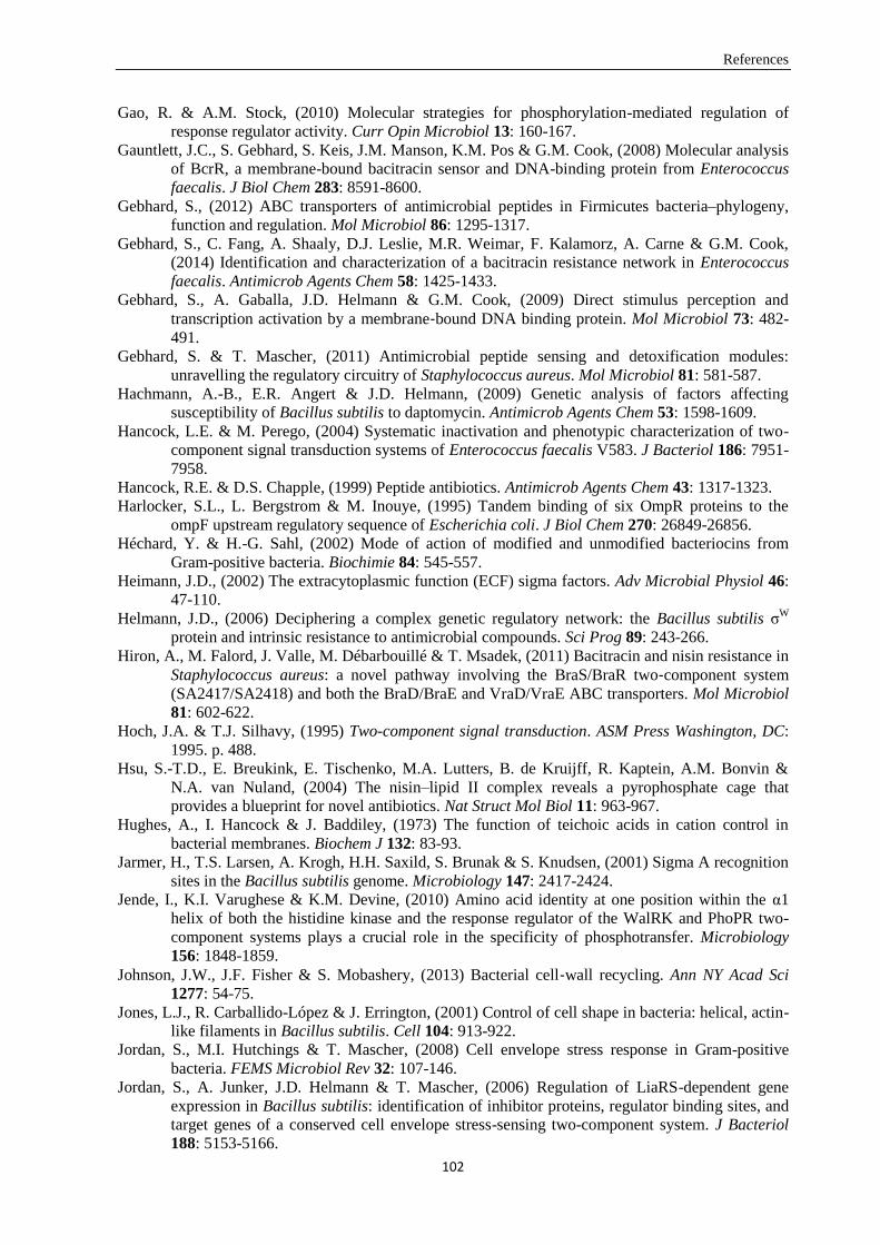
References
102
Gao, R. & A.M. Stock, (2010) Molecular strategies for phosphorylation-mediated regulation of
response regulator activity. Curr Opin Microbiol 13: 160-167.
Gauntlett, J.C., S. Gebhard, S. Keis, J.M. Manson, K.M. Pos & G.M. Cook, (2008) Molecular analysis
of BcrR, a membrane-bound bacitracin sensor and DNA-binding protein from Enterococcus
faecalis. J Biol Chem 283: 8591-8600.
Gebhard, S., (2012) ABC transporters of antimicrobial peptides in Firmicutes bacteria–phylogeny,
function and regulation. Mol Microbiol 86: 1295-1317.
Gebhard, S., C. Fang, A. Shaaly, D.J. Leslie, M.R. Weimar, F. Kalamorz, A. Carne & G.M. Cook,
(2014) Identification and characterization of a bacitracin resistance network in Enterococcus
faecalis. Antimicrob Agents Chem 58: 1425-1433.
Gebhard, S., A. Gaballa, J.D. Helmann & G.M. Cook, (2009) Direct stimulus perception and
transcription activation by a membrane‐bound DNA binding protein. Mol Microbiol 73: 482-
491.
Gebhard, S. & T. Mascher, (2011) Antimicrobial peptide sensing and detoxification modules:
unravelling the regulatory circuitry of Staphylococcus aureus. Mol Microbiol 81: 581-587.
Hachmann, A.-B., E.R. Angert & J.D. Helmann, (2009) Genetic analysis of factors affecting
susceptibility of Bacillus subtilis to daptomycin. Antimicrob Agents Chem 53: 1598-1609.
Hancock, L.E. & M. Perego, (2004) Systematic inactivation and phenotypic characterization of two-
component signal transduction systems of Enterococcus faecalis V583. J Bacteriol 186: 7951-
7958.
Hancock, R.E. & D.S. Chapple, (1999) Peptide antibiotics. Antimicrob Agents Chem 43: 1317-1323.
Harlocker, S.L., L. Bergstrom & M. Inouye, (1995) Tandem binding of six OmpR proteins to the
ompF upstream regulatory sequence of Escherichia coli. J Biol Chem 270: 26849-26856.
Héchard, Y. & H.-G. Sahl, (2002) Mode of action of modified and unmodified bacteriocins from
Gram-positive bacteria. Biochimie 84: 545-557.
Heimann, J.D., (2002) The extracytoplasmic function (ECF) sigma factors. Adv Microbial Physiol 46:
47-110.
Helmann, J.D., (2006) Deciphering a complex genetic regulatory network: the Bacillus subtilis σW
protein and intrinsic resistance to antimicrobial compounds. Sci Prog 89: 243-266.
Hiron, A., M. Falord, J. Valle, M. Débarbouillé & T. Msadek, (2011) Bacitracin and nisin resistance in
Staphylococcus aureus: a novel pathway involving the BraS/BraR two‐component system
(SA2417/SA2418) and both the BraD/BraE and VraD/VraE ABC transporters. Mol Microbiol
81: 602-622.
Hoch, J.A. & T.J. Silhavy, (1995) Two-component signal transduction. ASM Press Washington, DC: 1995. p. 488.
Hsu, S.-T.D., E. Breukink, E. Tischenko, M.A. Lutters, B. de Kruijff, R. Kaptein, A.M. Bonvin &
N.A. van Nuland, (2004) The nisin–lipid II complex reveals a pyrophosphate cage that
provides a blueprint for novel antibiotics. Nat Struct Mol Biol 11: 963-967.
Hughes, A., I. Hancock & J. Baddiley, (1973) The function of teichoic acids in cation control in
bacterial membranes. Biochem J 132: 83-93.
Jarmer, H., T.S. Larsen, A. Krogh, H.H. Saxild, S. Brunak & S. Knudsen, (2001) Sigma A recognition
sites in the Bacillus subtilis genome. Microbiology 147: 2417-2424.
Jende, I., K.I. Varughese & K.M. Devine, (2010) Amino acid identity at one position within the α1
helix of both the histidine kinase and the response regulator of the WalRK and PhoPR two-
component systems plays a crucial role in the specificity of phosphotransfer. Microbiology
156: 1848-1859.
Johnson, J.W., J.F. Fisher & S. Mobashery, (2013) Bacterial cell‐wall recycling. Ann NY Acad Sci
1277: 54-75.
Jones, L.J., R. Carballido-López & J. Errington, (2001) Control of cell shape in bacteria: helical, actin-
like filaments in Bacillus subtilis. Cell 104: 913-922.
Jordan, S., M.I. Hutchings & T. Mascher, (2008) Cell envelope stress response in Gram-positive
bacteria. FEMS Microbiol Rev 32: 107-146.
Jordan, S., A. Junker, J.D. Helmann & T. Mascher, (2006) Regulation of LiaRS-dependent gene
expression in Bacillus subtilis: identification of inhibitor proteins, regulator binding sites, and
target genes of a conserved cell envelope stress-sensing two-component system. J Bacteriol
188: 5153-5166.
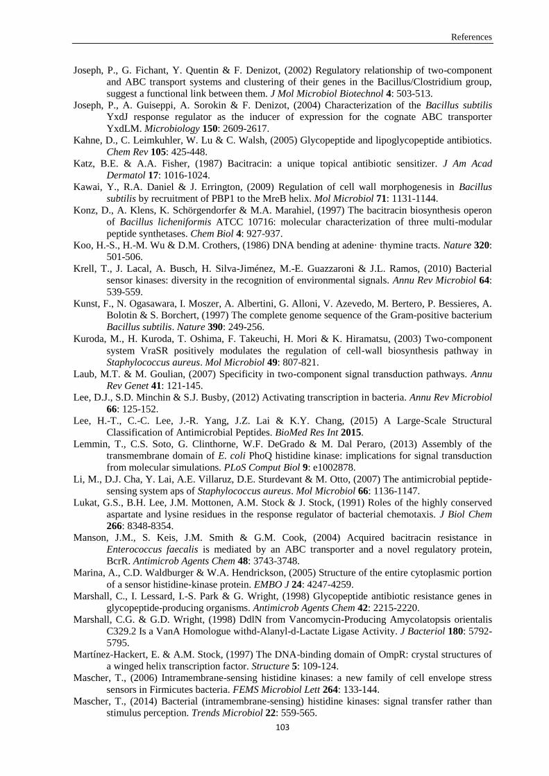
References
103
Joseph, P., G. Fichant, Y. Quentin & F. Denizot, (2002) Regulatory relationship of two-component
and ABC transport systems and clustering of their genes in the Bacillus/Clostridium group,
suggest a functional link between them. J Mol Microbiol Biotechnol 4: 503-513.
Joseph, P., A. Guiseppi, A. Sorokin & F. Denizot, (2004) Characterization of the Bacillus subtilis
YxdJ response regulator as the inducer of expression for the cognate ABC transporter
YxdLM. Microbiology 150: 2609-2617.
Kahne, D., C. Leimkuhler, W. Lu & C. Walsh, (2005) Glycopeptide and lipoglycopeptide antibiotics.
Chem Rev 105: 425-448.
Katz, B.E. & A.A. Fisher, (1987) Bacitracin: a unique topical antibiotic sensitizer. J Am Acad
Dermatol 17: 1016-1024.
Kawai, Y., R.A. Daniel & J. Errington, (2009) Regulation of cell wall morphogenesis in Bacillus
subtilis by recruitment of PBP1 to the MreB helix. Mol Microbiol 71: 1131-1144.
Konz, D., A. Klens, K. Schörgendorfer & M.A. Marahiel, (1997) The bacitracin biosynthesis operon
of Bacillus licheniformis ATCC 10716: molecular characterization of three multi-modular
peptide synthetases. Chem Biol 4: 927-937.
Koo, H.-S., H.-M. Wu & D.M. Crothers, (1986) DNA bending at adenine· thymine tracts. Nature 320:
501-506.
Krell, T., J. Lacal, A. Busch, H. Silva-Jiménez, M.-E. Guazzaroni & J.L. Ramos, (2010) Bacterial
sensor kinases: diversity in the recognition of environmental signals. Annu Rev Microbiol 64:
539-559.
Kunst, F., N. Ogasawara, I. Moszer, A. Albertini, G. Alloni, V. Azevedo, M. Bertero, P. Bessieres, A.
Bolotin & S. Borchert, (1997) The complete genome sequence of the Gram-positive bacterium
Bacillus subtilis. Nature 390: 249-256.
Kuroda, M., H. Kuroda, T. Oshima, F. Takeuchi, H. Mori & K. Hiramatsu, (2003) Two‐component
system VraSR positively modulates the regulation of cell‐wall biosynthesis pathway in
Staphylococcus aureus. Mol Microbiol 49: 807-821.
Laub, M.T. & M. Goulian, (2007) Specificity in two-component signal transduction pathways. Annu
Rev Genet 41: 121-145.
Lee, D.J., S.D. Minchin & S.J. Busby, (2012) Activating transcription in bacteria. Annu Rev Microbiol
66: 125-152.
Lee, H.-T., C.-C. Lee, J.-R. Yang, J.Z. Lai & K.Y. Chang, (2015) A Large-Scale Structural
Classification of Antimicrobial Peptides. BioMed Res Int 2015.
Lemmin, T., C.S. Soto, G. Clinthorne, W.F. DeGrado & M. Dal Peraro, (2013) Assembly of the
transmembrane domain of E. coli PhoQ histidine kinase: implications for signal transduction
from molecular simulations. PLoS Comput Biol 9: e1002878.
Li, M., D.J. Cha, Y. Lai, A.E. Villaruz, D.E. Sturdevant & M. Otto, (2007) The antimicrobial peptide‐sensing system aps of Staphylococcus aureus. Mol Microbiol 66: 1136-1147.
Lukat, G.S., B.H. Lee, J.M. Mottonen, A.M. Stock & J. Stock, (1991) Roles of the highly conserved
aspartate and lysine residues in the response regulator of bacterial chemotaxis. J Biol Chem
266: 8348-8354.
Manson, J.M., S. Keis, J.M. Smith & G.M. Cook, (2004) Acquired bacitracin resistance in
Enterococcus faecalis is mediated by an ABC transporter and a novel regulatory protein,
BcrR. Antimicrob Agents Chem 48: 3743-3748.
Marina, A., C.D. Waldburger & W.A. Hendrickson, (2005) Structure of the entire cytoplasmic portion
of a sensor histidine‐kinase protein. EMBO J 24: 4247-4259.
Marshall, C., I. Lessard, I.-S. Park & G. Wright, (1998) Glycopeptide antibiotic resistance genes in
glycopeptide-producing organisms. Antimicrob Agents Chem 42: 2215-2220.
Marshall, C.G. & G.D. Wright, (1998) DdlN from Vancomycin-Producing Amycolatopsis orientalis
C329.2 Is a VanA Homologue withd-Alanyl-d-Lactate Ligase Activity. J Bacteriol 180: 5792-
5795.
Martínez-Hackert, E. & A.M. Stock, (1997) The DNA-binding domain of OmpR: crystal structures of
a winged helix transcription factor. Structure 5: 109-124.
Mascher, T., (2006) Intramembrane-sensing histidine kinases: a new family of cell envelope stress
sensors in Firmicutes bacteria. FEMS Microbiol Lett 264: 133-144.
Mascher, T., (2014) Bacterial (intramembrane-sensing) histidine kinases: signal transfer rather than
stimulus perception. Trends Microbiol 22: 559-565.
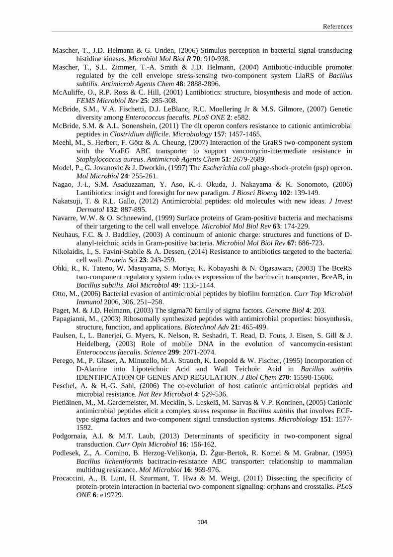
References
104
Mascher, T., J.D. Helmann & G. Unden, (2006) Stimulus perception in bacterial signal-transducing
histidine kinases. Microbiol Mol Biol R 70: 910-938.
Mascher, T., S.L. Zimmer, T.-A. Smith & J.D. Helmann, (2004) Antibiotic-inducible promoter
regulated by the cell envelope stress-sensing two-component system LiaRS of Bacillus
subtilis. Antimicrob Agents Chem 48: 2888-2896.
McAuliffe, O., R.P. Ross & C. Hill, (2001) Lantibiotics: structure, biosynthesis and mode of action.
FEMS Microbiol Rev 25: 285-308.
McBride, S.M., V.A. Fischetti, D.J. LeBlanc, R.C. Moellering Jr & M.S. Gilmore, (2007) Genetic
diversity among Enterococcus faecalis. PLoS ONE 2: e582.
McBride, S.M. & A.L. Sonenshein, (2011) The dlt operon confers resistance to cationic antimicrobial
peptides in Clostridium difficile. Microbiology 157: 1457-1465.
Meehl, M., S. Herbert, F. Götz & A. Cheung, (2007) Interaction of the GraRS two-component system
with the VraFG ABC transporter to support vancomycin-intermediate resistance in
Staphylococcus aureus. Antimicrob Agents Chem 51: 2679-2689.
Model, P., G. Jovanovic & J. Dworkin, (1997) The Escherichia coli phage-shock-protein (psp) operon.
Mol Microbiol 24: 255-261.
Nagao, J.-i., S.M. Asaduzzaman, Y. Aso, K.-i. Okuda, J. Nakayama & K. Sonomoto, (2006)
Lantibiotics: insight and foresight for new paradigm. J Biosci Bioeng 102: 139-149.
Nakatsuji, T. & R.L. Gallo, (2012) Antimicrobial peptides: old molecules with new ideas. J Invest
Dermatol 132: 887-895.
Navarre, W.W. & O. Schneewind, (1999) Surface proteins of Gram-positive bacteria and mechanisms
of their targeting to the cell wall envelope. Microbiol Mol Biol Rev 63: 174-229.
Neuhaus, F.C. & J. Baddiley, (2003) A continuum of anionic charge: structures and functions of D-
alanyl-teichoic acids in Gram-positive bacteria. Microbiol Mol Biol Rev 67: 686-723.
Nikolaidis, I., S. Favini‐Stabile & A. Dessen, (2014) Resistance to antibiotics targeted to the bacterial
cell wall. Protein Sci 23: 243-259.
Ohki, R., K. Tateno, W. Masuyama, S. Moriya, K. Kobayashi & N. Ogasawara, (2003) The BceRS
two‐component regulatory system induces expression of the bacitracin transporter, BceAB, in
Bacillus subtilis. Mol Microbiol 49: 1135-1144.
Otto, M., (2006) Bacterial evasion of antimicrobial peptides by biofilm formation. Curr Top Microbiol
Immunol 2006, 306, 251–258.
Paget, M. & J.D. Helmann, (2003) The sigma70 family of sigma factors. Genome Biol 4: 203.
Papagianni, M., (2003) Ribosomally synthesized peptides with antimicrobial properties: biosynthesis,
structure, function, and applications. Biotechnol Adv 21: 465-499.
Paulsen, I., L. Banerjei, G. Myers, K. Nelson, R. Seshadri, T. Read, D. Fouts, J. Eisen, S. Gill & J.
Heidelberg, (2003) Role of mobile DNA in the evolution of vancomycin-resistant
Enterococcus faecalis. Science 299: 2071-2074.
Perego, M., P. Glaser, A. Minutello, M.A. Strauch, K. Leopold & W. Fischer, (1995) Incorporation of
D-Alanine into Lipoteichoic Acid and Wall Teichoic Acid in Bacillus subtilis
IDENTIFICATION OF GENES AND REGULATION. J Biol Chem 270: 15598-15606.
Peschel, A. & H.-G. Sahl, (2006) The co-evolution of host cationic antimicrobial peptides and
microbial resistance. Nat Rev Microbiol 4: 529-536.
Pietiäinen, M., M. Gardemeister, M. Mecklin, S. Leskelä, M. Sarvas & V.P. Kontinen, (2005) Cationic
antimicrobial peptides elicit a complex stress response in Bacillus subtilis that involves ECF-
type sigma factors and two-component signal transduction systems. Microbiology 151: 1577-
1592.
Podgornaia, A.I. & M.T. Laub, (2013) Determinants of specificity in two-component signal
transduction. Curr Opin Microbiol 16: 156-162.
Podlesek, Z., A. Comino, B. Herzog‐Velikonja, D. Žgur‐Bertok, R. Komel & M. Grabnar, (1995)
Bacillus licheniformis bacitracin‐resistance ABC transporter: relationship to mammalian
multidrug resistance. Mol Microbiol 16: 969-976.
Procaccini, A., B. Lunt, H. Szurmant, T. Hwa & M. Weigt, (2011) Dissecting the specificity of
protein-protein interaction in bacterial two-component signaling: orphans and crosstalks. PLoS
ONE 6: e19729.
References
105
Reichmann, N.T., C.P. Cassona & A. Gründling, (2013) Revised mechanism of D-alanine
incorporation into cell wall polymers in Gram-positive bacteria. Microbiology 159: 1868-
1877.
Revilla-Guarinos, A., S. Gebhard, C. Alcántara, A. Staroń, T. Mascher & M. Zúñiga, (2013)
Characterization of a regulatory network of peptide antibiotic detoxification modules in
Lactobacillus casei BL23. Appl Environ Microbiol 79: 3160-3170.
Revilla‐Guarinos, A., S. Gebhard, T. Mascher & M. Zúñiga, (2014) Defence against antimicrobial
peptides: different strategies in Firmicutes. Environ Microbiol 16: 1225-1237.
Rietkötter, E., D. Hoyer & T. Mascher, (2008) Bacitracin sensing in Bacillus subtilis. Mol Microbiol
68: 768-785.
Saar-Dover, R., A. Bitler, R. Nezer, L. Shmuel-Galia, A. Firon, E. Shimoni, P. Trieu-Cuot & Y. Shai,
(2012) D-alanylation of lipoteichoic acids confers resistance to cationic peptides in group B
streptococcus by increasing the cell wall density. PLoS Pathog 8: e1002891.
Sahm, D.F., J. Kissinger, M. Gilmore, P. Murray, R. Mulder, J. Solliday & B. Clarke, (1989) In vitro
susceptibility studies of vancomycin-resistant Enterococcus faecalis. Antimicrob Agents Chem
33: 1588-1591.
Scheffers, D.-J. & M.G. Pinho, (2005) Bacterial cell wall synthesis: new insights from localization
studies. Microbiol Mol Biol Rev 69: 585-607.
Scott, J.R. & T.C. Barnett, (2006) Surface proteins of Gram-positive bacteria and how they get there.
Annu Rev Microbiol 60: 397-423.
Shockman, G.D. & J. Barren, (1983) Structure, function, and assembly of cell walls of Gram-positive
bacteria. Annu Rev Microbiol 37: 501-527.
Silhavy, T.J., D. Kahne & S. Walker, (2010) The bacterial cell envelope. Cold Spring Harb Perspect
Biol 2, a000414.
Silva, J.C., A. Haldimann, M.K. Prahalad, C.T. Walsh & B.L. Wanner, (1998) In vivo characterization
of the type A and B vancomycin-resistant enterococci (VRE) VanRS two-component systems
in Escherichia coli: a nonpathogenic model for studying the VRE signal transduction
pathways. Proc Natl Acad Sci USA 95: 11951-11956.
Silver, L.L., (2003) Novel inhibitors of bacterial cell wall synthesis. Curr Opin Microbiol 6: 431-438.
Slauch, J., S. Garrett, D. Jackson & T. Silhavy, (1988) EnvZ functions through OmpR to control porin
gene expression in Escherichia coli K-12. J Bacteriol 170: 439-441.
Slauch, J.M., F.D. Russo & T.J. Silhavy, (1991) Suppressor mutations in rpoA suggest that OmpR
controls transcription by direct interaction with the alpha subunit of RNA polymerase. J
Bacteriol 173: 7501-7510.
Staroń, A., D.E. Finkeisen & T. Mascher, (2011) Peptide antibiotic sensing and detoxification modules
of Bacillus subtilis. Annu Rev Microbiol 55: 515-525.
Stein, T., (2005) Bacillus subtilis antibiotics: structures, syntheses and specific functions. Mol
Microbiol 56: 845-857.
Stein, T., S. Heinzmann, I. Solovieva & K.-D. Entian, (2003) Function of Lactococcus lactis nisin
immunity genes nisI and nisFEG after coordinated expression in the surrogate host Bacillus
subtilis. J Biol Chem 278: 89-94.
Stein, T., J. Vater, V. Kruft, A. Otto, B. Wittmann-Liebold, P. Franke, M. Panico, R. McDowell &
H.R. Morris, (1996) The multiple carrier model of nonribosomal peptide biosynthesis at
modular multienzymatic templates. J Biol Chem 271: 15428-15435.
Stock, A.M., V.L. Robinson & P.N. Goudreau, (2000) Two-component signal transduction. Annu Rev
Biochem 69: 183-215.
Sun, Z., J. Zhong, X. Liang, J. Liu, X. Chen & L. Huan, (2009) Novel mechanism for nisin resistance
via proteolytic degradation of nisin by the nisin resistance protein NSR. Antimicrob Agents
Chem 53: 1964-1973.
Szurmant, H. & J.A. Hoch, (2010) Interaction fidelity in two-component signaling. Curr Opin
Microbiol 13: 190-197.
Ton-That, H., L.A. Marraffini & O. Schneewind, (2004) Protein sorting to the cell wall envelope of
Gram-positive bacteria. Biochim Biophys Acta-Mol Cell Res 1694: 269-278.
Typas, A., M. Banzhaf, C.A. Gross & W. Vollmer, (2012) From the regulation of peptidoglycan
synthesis to bacterial growth and morphology. Nat Rev Microbiol 10: 123-136.
References
106
Ubersax, J.A. & J.E. Ferrell Jr, (2007) Mechanisms of specificity in protein phosphorylation. Nat Rev
Mol Cell Biol 8: 530-541.
van Dijl, J.M. & M. Hecker, (2013) Bacillus subtilis: from soil bacterium to super-secreting cell
factory. Microb Cell Fact 12: 3.
Vollmer, W., (2008) Structural variation in the glycan strands of bacterial peptidoglycan. FEMS
Microbiol Rev 32: 287-306.
Vollmer, W., D. Blanot & M.A. De Pedro, (2008) Peptidoglycan structure and architecture. FEMS
Microbiol Rev 32: 149-167.
Vollmer, W. & S.J. Seligman, (2010) Architecture of peptidoglycan: more data and more models.
Trends Microbiol 18: 59-66.
Walsh, C.T., S.L. Fisher, I.-S. Park, M. Prahalad & Z. Wu, (1996) Bacterial resistance to vancomycin:
five genes and one missing hydrogen bond tell the story. Chem Biol 3: 21-28.
West, A.H. & A.M. Stock, (2001) Histidine kinases and response regulator proteins in two-component
signaling systems. Trends Biochem Sci 26: 369-376.
Wheeler, R., G. Chevalier, G. Eberl & I. Gomperts Boneca, (2014) The biology of bacterial
peptidoglycans and their impact on host immunity and physiology. Cell Microbiol 16: 1014-
1023.
Willey, J.M. & W.A. van Der Donk, (2007) Lantibiotics: peptides of diverse structure and function.
Annu Rev Microbiol 61: 477-501.
Wolf, D., P. Domínguez-Cuevas, R.A. Daniel & T. Mascher, (2012) Cell envelope stress response in
cell wall-deficient L-forms of Bacillus subtilis. Antimicrob Agents Chem 56: 5907-5915.
Wolf, D., F. Kalamorz, T. Wecke, A. Juszczak, U. Mäder, G. Homuth, S. Jordan, J. Kirstein, M.
Hoppert & B. Voigt, (2010) In-depth profiling of the LiaR response of Bacillus subtilis. J
Bacteriol 192: 4680-4693.
Wuichet, K., B.J. Cantwell & I.B. Zhulin, (2010) Evolution and phyletic distribution of two-
component signal transduction systems. Curr Opin Microbiol 13: 219-225.
Yeaman, M.R. & N.Y. Yount, (2003) Mechanisms of antimicrobial peptide action and resistance.
Pharmacol Rev 55: 27-55.
Acknowledgments
107
Acknowledgments
As time goes by, I am almost finished with my four-year study in Munich. During this time, there are a
lot people who give me great support and help.
My first and sincere appreciation goes to my supervisor Prof. Dr. Thorsten Mascher, for providing me
the chance of studying and doing scientific research in AG Mascher. I am thankful for the nice
projects and his continuous encouragement, help and support in all stages of my PhD studies.
I want to thank PD Dr. Susanne Gebhard, I am truly grateful for her nice, patient and professional
supervision. Thank you very much for the nice Monday morning meeting with new results discussion.
Special thanks go to my thesis committee especially to PD Dr. Ralf Heermann for being the second
examiner of this thesis.
I want to thank Prof. Dr. K. Jung and PD Dr. Ralf Heermann again for their support and valuable input
during the project in chapter IV. I learned a lot of biochemistry knowledge from PD Dr. Ralf
Heermann and his nice data analysis and explanation really helped me a lot.
I gratefully thank the funding source CSC program that made my PhD work possible.
Many thanks go to Anna Staroń for the nice starting point of the project and for patiently answering
my questions.
I would like to thank PD Dr. Susanne Gebhard, Felix Becker, Jennifer Emenegger, and Ainhoa Revilla
Guarinos for critical reading of this thesis.
Special thanks go to the international team in the 007 lab. I want to thank Sebastian, for the nice
starting point supervision in the lab and scientific discussion, for teaching me a lot of techniques, for
showing me the German language and culture (谢谢弟弟). I want to thank Jenny, my arty American
friend, for the nice English lessons, baking course, pretty lab decorations and relaxing music (谢谢杰
妮弗). Of course I have to thank Dayane, for the nice dance in the lab, super shopping time and the
lovely coffee break (谢谢大鸭梨). I want to thank Sonja, for the great help in the lab, nice hangout
and excellent Bavaria hiking (谢谢小技师). I want to thank Qiang for improving my Chinese a lot and
bring nice Chinese music in the lab as well as the most popular Chinese dance (谢谢强仔). I also want
to thank my master student Martin, for the nice and hard work here as well as a lot of cooking
knowledge (谢谢好学生). Besides, I want to thank Caro and Xiaoluo for the nice office time. Thank
you guys, I learnt a lot from all of you and I enjoyed the time with you. I appreciate the help and
support from you and I will never forget the time in the lab with you.
I want to thank the whole AG Mascher, for the nice time together!
I want to thank my landlady Elli. She is like an aunt who provides me a nice living place, takes care of
me and teaches me the nice German language and culture. She shows me the famous reputation of
German: punctuality and precision. Many thanks also go to her sons and daughters-in-law! I want to
thank all my Chinese friends, especially Yingjie and Wenteng, Qing and Peng, Xiyao and Chun, Lixia,
Yihe, Shuai for the nice time in Munich! Besides, I want to say thank you to all members of our
badminton group for the nice sports time!
Finally, I want to thank my family ― the most important part of my life. I want to thank my mother,
who is in the heaven but still protecting me. I want to thank my father, who cares about me but is also
strict with me. My dear rabbit, who gives me the best support, patience, surprises, and love that I feel I
am the happiest person in the world (谢谢我亲爱的小飞飞). My gratitude also goes to his kind and
lovely parents and his brothers, thanks for all kinds of help and the nice time together in Karlsruhe and
Munich!
Curriculum Vitae
108
Curriculum Vitae
Personal information
Name: Chong Fang
Date of birth: 12.02.1986
Place of birth: Qingdao, Shandong, China
Email: [email protected]
Education and Research Experience
Since Sep 2011
PhD student, Synthetic Biology, Ludwig-Maximilians-Universität Munich
Supervisors: Prof. Thorsten Mascher and PD Susanne Gebhard
Projects:
Interaction specificity between histidine kinase and response regulator of Bce-like two
component signal transduction systems in B. subtilis
Specificity determinants on bceA-like promoters in B. subtilis
Characterization of a bacitracin resistance network from E. faecalis in B. subtilis
B. subtilis as a platform for molecular characterization of regulatory mechanisms of E.
faecalis resistance against cell wall antibiotics
Sep 2008- July 2011
M.Sc. Microbiology, State Key Laboratory for Microbial Technology, Shandong University
Supervisors: Prof. Guanjun Chen and Prof. Lushan Wang
GPA: 86.44/100
Projects:
Investigation of microbial diversity, biomass degradation and corresponding enzymes during
composting process
Sep 2004-July 2008
B.Sc. Biotechnology, Biological & Chemical Faculty, Yantai University
Rank: top 10 of 180 students in Department of Biotechnology
GPA: 85.39/100
Project and practice:
Bachelor thesis: Analysis the quality of four industrial xylanases
Biodiversity investigation: KunYu Mountain
Practice: Changyu winery, brewery, liquor factory and mushroom production base
Curriculum Vitae
109
Publications
Fang C., Staroń A., Grafe M., Heermann R., Jung K., Gebhard S. and Mascher T. Insulation and wiring
specificity determinants of BceR-like response regulators and their target promoters in Bacillus subtilis.
In preparation
Dintner S., Heermann R., Fang C., Jung K. and Gebhard S. (2014) A Sensory Complex Consisting of
an ATP-Binding-Cassette Transporter and a Two-Component Regulatory System Controls Bacitracin
Resistance in Bacillus subtilis. J. Biol. Chem.doi:10.1074/jbc.M114. 596221.
Fang C., Stiegeler E, Cook G.M., Mascher T., Gebhard S. (2014) Bacillus subtilis as a Platform for
Molecular Characterisation of Regulatory Mechanisms of Enterococcus faecalis Resistance against Cell
Wall Antibiotics. PLoS ONE 9(3): e93169. doi:10.1371/ journal.pone.0093169.
Gebhard S., Fang C., Shaaly A., Leslie D.J., Weimar M.R., Kalamorz F.,Carne A. and Cook G.M.
(2014) Identification and characterization of a bacitracin resistance network in Enterococcus
faecalis. Antimicrob. Agents Chemother. Vol. 58 no. 3 1425-1433.
Participated in translating the book: Biomass Recalcitrance——deconstruct the plant cell wall for
bioenergy, Edited by Michael E. Himmel. (Chapter 9\10\15, from English to Chinese; published)
Participated in translating the book: Cellulosome——the second mechanism of cellulose degradation,
Edited by Vladimir Uversky. (Chapter7, from English to Chinese; published)
Academic Meetings and Trainings
Fang C., Grafe M., Staroń A., Gebhard S and Mascher T. Specificity determinants of bceA-like
promoters in Bacillus subtilis. Annual Conference 2015 of the Association for General and Applied
Microbiology (VAAM). Mar. 2015, Marburg, Germany; Poster
Fang C., Grafe M., Staroń A., Gebhard S and Mascher T. Specificity determinants of bceA-like
promoters in Bacillus subtilis. 30th Symposium Mechanisms of Gene Regulation. Oct. 2014,
Düsseldorf, Germany; Oral presentation
Workshop ‘Synthetic Biological Tools for Single Cell Analysis’. May 2014, CASLmu
, Munich,
Germany
Workshop ‘Synthetic Building Blocks and Modules’. Dec. 2013, CASLmu
, Munich, Germany
Fang C., Wang L. Chen G. Development of a novel Native-PAGE method to illustrate the property of
Xylanase. 7th National Conference for Microbiology & 3rd Academic Conference of Microgenetics
2008. Jinan, China; Abstract
Honors and Awards
2006--2007 Academic Achievement Scholarship at Yantai University, first-class
2005--2006 Academic Achievement Scholarship at Yantai University, first-class
2004--2005 Academic Achievement Scholarship at Yantai University, first-class


![Werkstatthandbuch LML · STAR WIRING DIAGRAM AUTO START SCHEMA ELETTR'CO AVViAMENTO AUTOMATICO . Title: Werkstatthandbuch LML Author: Scanner [ AKS_5600I ] Created Date: 3/13/2009](https://img.pdfslide.org/doc/110x75/5e4ae796084dc061323703bf/werkstatthandbuch-lml-star-wiring-diagram-auto-start-schema-elettrco-avviamento.jpg)