Embed Size (px)
Citation preview

In vivo demonstration that α-synuclein oligomersare toxicBeate Winnera,b,1, Roberto Jappellia,1, Samir K. Majic,1, Paula A. Desplatsd, Leah Boyera,e, Stefan Aignera,Claudia Hetzera, Thomas Lohera, Marçal Vilara,2, Silvia Campionic, Christos Tzitzilonisa, Alice Soragnic,Sebastian Jessbergera,f, Helena Miraa,2, Antonella Consiglioa,3, Emiley Phamd, Eliezer Masliahd,Fred H. Gagea,4,5, and Roland Rieka,c,4,5
aSalk Institute for Biological Studies, La Jolla, CA 92037; bJunior Research Group III, Interdisciplinary Center for Clinical Research, Nikolaus-Fiebiger Center forMolecular Medicine, Friedrich-Alexander-Universität Erlangen-Nürnberg, 91054 Erlangen, Germany; cLaboratory of Physical Chemistry, EidgenössischeTechnische Hochschule Zurich, 8093 Zurich, Switzerland; dDepartment of Neurosciences and Department of Pathology and eBiomedical Science GraduateProgram, University of California at San Diego, School of Medicine, La Jolla, CA 92093; and fInstitute of Cell Biology, Eidgenössische Technische HochschuleZurich, 8093 Zurich, Switzerland
Contributed by Fred H. Gage, January 25, 2011 (sent for review December 2, 2010)
The aggregation of proteins into oligomers and amyloid fibrils ischaracteristic of several neurodegenerative diseases, including Par-kinson disease (PD). In PD, the process of aggregation of α-synuclein(α-syn) from monomers, via oligomeric intermediates, into amy-loid fibrils is considered the disease-causative toxic mechanism.We developed α-syn mutants that promote oligomer or fibril for-mation and tested the toxicity of these mutants by using a ratlentivirus system to investigate loss of dopaminergic neurons inthe substantia nigra. The most severe dopaminergic loss in thesubstantia nigra is observed in animals with the α-syn variantsthat form oligomers (i.e., E57K and E35K), whereas the α-syn var-iants that form fibrils very quickly are less toxic. We show thatα-syn oligomers are toxic in vivo and that α-syn oligomers mightinteract with and potentially disrupt membranes.
Parkinson disease (PD) is the most common movement dis-order, currently affecting approximately 2% of the pop-
ulation older than age 60 y. Prominent neuropathologicalhallmarks of PD are the loss of dopaminergic neurons in thesubstantia nigra (SN) region of the midbrain (1) and the pres-ence of α-syn–containing intracellular inclusions: Lewy bodies(LBs) and Lewy neurites (2). α-Syn, a 140-aa protein physio-logically found in presynaptic terminals of neurons, is the majorfibrillar protein in LBs and Lewy neurites in sporadic andinherited PD. Moreover, point mutations (A53T, A30P, E46K)and gene multiplications of human WT (hWT) α-syn are relatedto rare familial autosomal-dominant forms of early-onset PD (3–6), suggesting that increased gene dosage and aberrant proteinstructure may accelerate disease onset and progression.Recent reports indicate that the accumulation of α-syn can
result in the formation of intermediate-state oligomers, andoligomers of different shapes and sizes have been described (7–10). These oligomers interact with lipids, disrupt membranes (7,8), and cause cell death in vitro (10, 11) and in nonmammalianmodels, such as Caenorhabditis elegans and Drosophila mela-nogaster (12). However, we are aware of no previous direct in vivodemonstration of the toxicity of α-syn oligomers in mammals.We aim to establish a model that allows specific testing of the
effects of α-syn oligomerization in vitro and in vivo. To elucidatethe causal structure–toxicity relationship of these oligomericprotein assemblies in a mammalian system, we designed“conformation-trapped” mutants based on structural modelingof α-syn fibrils (13, 14). Structurally, amyloid fibrils of α-syn arecomposed of cross-β-sheets (15). Residues from approximately30 to 110 of α-syn form the core of the fibrils, whereas the ap-proximately 30 N-terminal residues are heterogeneous and theapproximately 30 C-terminal residues are flexible (13, 14, 16, 17).Based on our structural model, recently developed from NMRdata, the core of α-syn fibrils comprises five β-strands reminiscentof a five-layered “β-sandwich” (14). Several loops adjacent to andbetween the strands are conserved pseudorepeats containingboth Lys and Glu side chains (i.e., K32TKEG 36 N-terminal to theproposed β1, K43TKEG47 between β1 and β2, E57KTKEQ62
between β2 and β3, and K80TVEG84 between β4 and β5, re-
spectively). A salt bridge between D23 and K28 in a loop con-necting two β-strands has been observed for Aβ(1–42) amyloidfibrils, and point mutations therein inhibit the formation of Aβ(1–42) fibrils (18, 19).We hypothesized that, similar to the Aβ(1–42) amyloid pro-
tein, disruption of α-syn–specific salt bridges might affect thestructure and toxicity of α-syn. Accordingly, we designed muta-tions with the purpose of interfering with fibril formation andproduced α-syn variants that specifically form oligomers but notfibrils; we show that the ability of α-syn to form oligomers in vitrois accompanied by increased in vivo toxicity.
ResultsDesign and in Vitro Aggregation of hWt α-Syn and α-Syn Variants. Tobe able to investigate α-syn–dependent oligomer toxicity in vivo,we searched for oligomer-forming mutants. Therefore, we in-vestigated the salt bridges between the β-strands of α-syn, spe-cifically testing whether the introduction of Glu-to-Lys mutationsat positions 35, 46 (E46K is a familial variant), 57, 61, and 83,which would prevent the formation of such salt bridges, mightinterfere with the formation of α-syn amyloid fibrils. By usingrecombinant α-syn from preparations that followed an estab-lished purification procedure (17) but omitted the precipitationstep by streptomycin sulfate (protocol B), we found that theα-syn single-point mutants E35K and E57K showed a stronglydecreased tendency to form fibrils compared with hWT α-syn, asevidenced by time-resolved amyloid formation measured bythioflavin T (Thio-T) binding (Fig. S1A). Neither variant E35Knor E57K showed EM-visible amyloid fibrils even after 1 y ofincubation, but they formed very pronounced ring/pore-likestructures (Fig. 1A). In contrast, hWT α-syn and the familialα-syn variants (A30P, E46K, and A53T) formed amyloid fibrils asexpected (Fig. 1A). By using a more rigorous purification pro-tocol (protocol A) that included a streptomycin sulfate pre-cipitation step (Materials and Methods and SI Materials andMethods), we found that both E57K and E35K variants formed
Author contributions: B.W., R.J., S.A., E.M., F.H.G., and R.R. designed research; B.W., R.J.,S.K.M., P.A.D., L.B., S.A., C.H., T.L., M.V., S.C., C.T., S.J., H.M., A.C., and E.P. performedresearch; S.K.M., S.C., and A.S. contributed new reagents/analytic tools; B.W., R.J., S.K.M.,P.A.D., E.P., E.M., and R.R. analyzed data; and B.W., S.K.M., F.H.G., and R.R. wrote thepaper.
The authors declare no conflict of interest.
Freely available online through the PNAS open access option.1B.W., R.J., and S.K.M. contributed equally to this work.2Present address: Area de Biología Celular y Desarrollo, Instituto de Salud Carlos III, 28220Majadahonda-Madrid, Spain.
3Present address: Institute of Biomedicine of the University of Barcelona, 08028Barcelona, Spain.
4F.H.G. and R.R. contributed equally to this work.5To whom correspondence may be addressed. E-mail: [email protected] or [email protected].
This article contains supporting information online at www.pnas.org/lookup/suppl/doi:10.1073/pnas.1100976108/-/DCSupplemental.
4194–4199 | PNAS | March 8, 2011 | vol. 108 | no. 10 www.pnas.org/cgi/doi/10.1073/pnas.1100976108

amyloid fibrils with similar efficiency as the A53T variant (Fig. S1B–D, protocol A). However, when the monomer fractions, rel-ative to size exclusion chromatography (SEC) profiles obtainedfrom streptomycin sulfate-precipitated protein preparations(protocol A), were used as starting material and subjected totime-resolved aggregation of α-syn, only the E57K and E35Kvariants formed oligomer peaks at the void volume at days 5 and10 (Fig. 1B). The EM images in Fig. 1C show the oligomers fromthe SEC fractions of Fig. 1B. They appear to be of a mono-dispersed nature with an estimated size of 100 nm, as measuredby dynamic light scattering, which is in agreement with the lit-erature (Fig. S1E) (20).We compared these oligomers to the fibril-promoting variant
α-syn(30–110), which includes the amyloid core residues fromapproximately 30 to 110 but lacks the aggregation-interfering C-terminal and N-terminal residues of α-syn (n ∼ 30) (14). Indeed,α-syn(30–110) shows an accelerated growth of fibril formationcompared with hWT α-syn (protocol A), and its fibril confor-mation is similar to that of hWT α-syn fibrils (Fig. 1A, protocolB; and Fig. S1D, protocol A) (14). The biophysical properties ofthe three artificial α-syn variants were also compared with thefamilial α-syn mutants A30P, A53T, and E46K. In line withprevious observations (21), A30P formed amyloid fibrils moreslowly and A53T formed them more quickly than hWT α-syn andE46K, respectively (Fig. S1B, protocol A). To further confirmthe presence of oligomers by an independent method, we com-
pared the immunoreactivity of E57K protein versus control andhWT α-syn by immunoblots with an oligomer-specific antibody(A11; protocol A, Fig. S2) (22). In comparison with hWT α-syn,a much stronger reactivity of the E57K variant to the oligomer-specific antibody A11 was detected on dot blots (Fig. S2A).Western blot analysis indicated that E57K (Fig. S2B, protocolA), and specifically the void peak of the SEC (fraction 16) ofE57K recombinant protein (Fig. S2 C and D), reacted stronglywith the A11 antibody at the level of α-syn oligomers (mostlydimers and pentamers; Fig. S2E). In the in vitro cell-free system,we have established a set of familial and structure-based mutantsthat cover oligomer-promoting and fibril-only states in vitro (Fig.1), and we report here on their in vivo behavior.
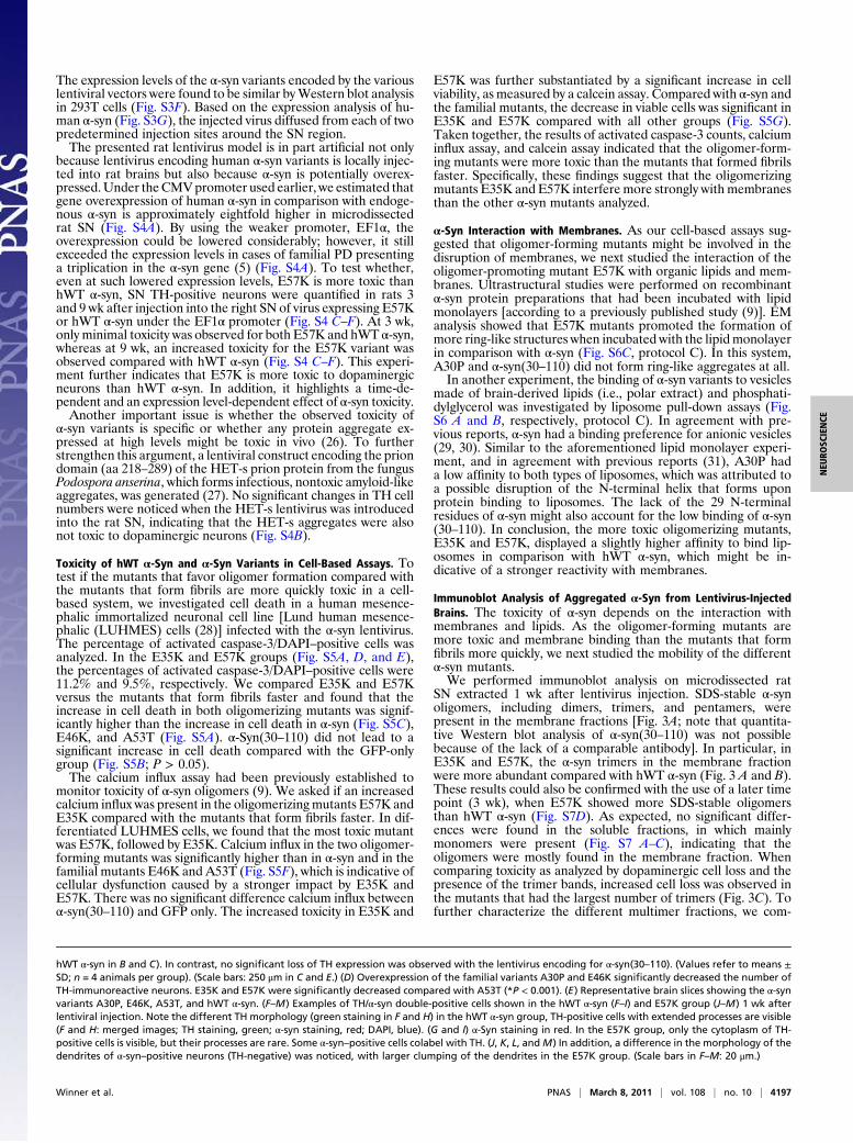
Toxicity of hWT α-Syn and α-Syn Variants in the SN of Lentivirus-Injected Rats. To investigate if the α-syn mutants that favor olig-omer formation are more toxic in vivo than the mutants that formfibrils more quickly, the toxicity of the α-syn variants was studiedin a rat model of synucleinopathies (23). This model is based oninjection of lentivirus expressing the α-syn variants into the SN.Although partly limited by the fact that α-syn is overexpressedlocally, it is well suited to study direct short- to medium-term toxiceffects on dopaminergic neurons in an in vivo system.Tyrosine hydroxylase (TH)-positive neurons were quantified in
rats 3 wk after viral injection into the right SN and were com-pared with the noninjected side (Fig. 2A). Injections of lentivirusencoding the artificial variants E35K and E57K induced a sig-nificant loss of TH expression. The decreases in TH-positive cellsfor E35K was 50% (TH cell number, 2.7 × 10³ ± 0.3 × 10³) and51% for E57K (TH cell number, 2.6 × 10³ ± 0.2 × 10³; Fig. 2 Band C) compared with the uninjected sides. These decreases forE35K and E57K were significantly different from the GFP-onlygroup (P < 0.001; TH cell number, 4.6 × 10³ ± 0.7 × 10³). Inaddition, significantly fewer TH-positive cells were present inthe oligomer-forming mutants E35K and E57K compared withthe hWT α-syn–injected group (32% loss; TH cell number, 3.5 ×10³ ± 0.2 × 10³; P < 0.05). The faster fibril-forming mutant α-syn(30–110) did not show a significant decrease in TH-positive cellnumbers (9% loss; TH cell number, 4.3 × 10³ ± 0.7 × 10³; P >0.05 vs. GFP-only group). The data were obtained by performingcell counts for the entire SN by using a systematic, randomcounting procedure and comparing lesioned and unlesioned SN.The loss of TH-expressing cells caused by α-syn variant expres-sion was more pronounced close to the injection site, as depictedin Fig. 2 C and E. We then compared these results versus thefamilial mutants. The rats injected with A30P (38% loss; TH cellnumber, 3.0 × 10³ ± 0.4 × 10³) and E46K (40% loss; TH cellnumber, 3.2 × 10³ ± 0.4 × 10³) did not differ significantly fromthose injected with E35K and E57K. A53T-injected rats (17%loss; TH cell number, 4.3 × 10³ ± 0.3 × 10³) showed a significantdifference in TH numbers compared with E35K and E57K (Fig.2 D and E).Consistent with the hypothesis that the oligomer-forming
mutants are more toxic, E35K and E57K induced the largestTH-positive cell loss of all mutants studied. In summary, in thelentivirus PD rat model, the following ranking scale from toxic tonontoxic could be determined: E57K > E35K ≥ A30P ≥ E46K ≥hWT α-syn > A53T ≥ α-syn(30–110) ≥ GFP only. A comparisonof brain tissue stained for the panneuronal marker (NeuN) andTH revealed that, despite a significant loss of NeuN-positive cells(Fig. S3A), numerous nondopaminergic neurons survived insidethe lesioned area of the SN, indicating that the loss was moreseverely affecting dopaminergic neurons. The percentage of TH-positive cells among NeuN-positive cells was approximately 75%in controls and decreased to 67% in hWT α-syn and 61% in theE57K group (Fig. S3 A and C).Amore detailed analysis of the effect of α-syn variants in the rat
SN showed that E57K could lead to severe changes in neuronmorphology compared with hWT α-syn (Fig. 2 F–M). Specifically,a dying back of neurites was observed in the E57K group (Fig.2H), resembling the axonal pathology described in PD (24, 25). Inaddition, the neurites contained larger lump-like appearanceswithin the E57K group than in the hWT α-syn (Fig. 2 K and M).
Fig. 1. In vitro aggregation of hWT α-syn and α-syn variants. (A) EM of agedα-syn and α-syn variants purified without the streptomycin sulfate-precipitation step (protocol B). (Scale bars: 500 nm.) Insets: Ring-formingentities are shown. (Scale bar: 100 nm.) (B) Time-resolved SEC of α-syn var-iants: 20 mg/mL α-syn variants were solubilized and their size exclusionprofiles were measured immediately (day 0). Subsequently, after days 5 and10, the samples of the corresponding monomer peak were again put on theSEC column, revealing that E57K and E35K had the capacity to formoligomers over time. For both α-syn variants, we show a 10-fold magnifica-tion of the oligomer peak at day 5. The decrease of the monomer peak inE35K and E57K is because they aggregate into amyloids. For the other var-iants and hWT α-syn, no oligomer peak was observed. (C) EM image of theoligomeric species from the experiment shown in B. (Scale bars: 500 nm.)
Winner et al. PNAS | March 8, 2011 | vol. 108 | no. 10 | 4195
NEU
ROSC
IENCE

Fig. 2. Toxicity of hWT α-syn and α-syn variants in the SN of lentivirus-injected rats. (A) Injection paradigm: rats unilaterally injected with the differentlentiviral constructs in the SN. (B) Histograms representing the decrease in numbers of TH-immunoreactive (TH-IR) neurons in the SN relative to the con-tralateral side, as evaluated by stereology. The nigral dopaminergic neurons were labeled with TH (C and E). Note that the loss of TH-expressing cells causedby α-syn variant expression was more pronounced close to the injection site (C and E). (B) Histogram representing the percentage of TH-positive neuronscompared with the noninjected side. The artificial mutants E35K and E57K led to a significant decrease in TH-positive cells (**P < 0.001 vs. GFP and *P < 0.05 vs.
4196 | www.pnas.org/cgi/doi/10.1073/pnas.1100976108 Winner et al.
The expression levels of the α-syn variants encoded by the variouslentiviral vectors were found to be similar byWestern blot analysisin 293T cells (Fig. S3F). Based on the expression analysis of hu-man α-syn (Fig. S3G), the injected virus diffused from each of twopredetermined injection sites around the SN region.The presented rat lentivirus model is in part artificial not only
because lentivirus encoding human α-syn variants is locally injec-ted into rat brains but also because α-syn is potentially overex-pressed.Under theCMVpromoter used earlier, we estimated thatgene overexpression of human α-syn in comparison with endoge-nous α-syn is approximately eightfold higher in microdissectedrat SN (Fig. S4A). By using the weaker promoter, EF1α, theoverexpression could be lowered considerably; however, it stillexceeded the expression levels in cases of familial PD presentinga triplication in the α-syn gene (5) (Fig. S4A). To test whether,even at such lowered expression levels, E57K is more toxic thanhWT α-syn, SN TH-positive neurons were quantified in rats 3and 9 wk after injection into the right SN of virus expressing E57Kor hWT α-syn under the EF1α promoter (Fig. S4 C–F). At 3 wk,only minimal toxicity was observed for both E57K and hWT α-syn,whereas at 9 wk, an increased toxicity for the E57K variant wasobserved compared with hWT α-syn (Fig. S4 C–F). This experi-ment further indicates that E57K is more toxic to dopaminergicneurons than hWT α-syn. In addition, it highlights a time-de-pendent and an expression level-dependent effect of α-syn toxicity.Another important issue is whether the observed toxicity of
α-syn variants is specific or whether any protein aggregate ex-pressed at high levels might be toxic in vivo (26). To furtherstrengthen this argument, a lentiviral construct encoding the priondomain (aa 218–289) of the HET-s prion protein from the fungusPodospora anserina, which forms infectious, nontoxic amyloid-likeaggregates, was generated (27). No significant changes in TH cellnumbers were noticed when the HET-s lentivirus was introducedinto the rat SN, indicating that the HET-s aggregates were alsonot toxic to dopaminergic neurons (Fig. S4B).
Toxicity of hWT α-Syn and α-Syn Variants in Cell-Based Assays. Totest if the mutants that favor oligomer formation compared withthe mutants that form fibrils are more quickly toxic in a cell-based system, we investigated cell death in a human mesence-phalic immortalized neuronal cell line [Lund human mesence-phalic (LUHMES) cells (28)] infected with the α-syn lentivirus.The percentage of activated caspase-3/DAPI–positive cells wasanalyzed. In the E35K and E57K groups (Fig. S5A, D, and E),the percentages of activated caspase-3/DAPI–positive cells were11.2% and 9.5%, respectively. We compared E35K and E57Kversus the mutants that form fibrils faster and found that theincrease in cell death in both oligomerizing mutants was signif-icantly higher than the increase in cell death in α-syn (Fig. S5C),E46K, and A53T (Fig. S5A). α-Syn(30–110) did not lead to asignificant increase in cell death compared with the GFP-onlygroup (Fig. S5B; P > 0.05).The calcium influx assay had been previously established to
monitor toxicity of α-syn oligomers (9). We asked if an increasedcalcium influx was present in the oligomerizing mutants E57K andE35K compared with the mutants that form fibrils faster. In dif-ferentiated LUHMES cells, we found that the most toxic mutantwas E57K, followed by E35K. Calcium influx in the two oligomer-forming mutants was significantly higher than in α-syn and in thefamilial mutants E46K and A53T (Fig. S5F), which is indicative ofcellular dysfunction caused by a stronger impact by E35K andE57K. There was no significant difference calcium influx betweenα-syn(30–110) and GFP only. The increased toxicity in E35K and
E57K was further substantiated by a significant increase in cellviability, as measured by a calcein assay. Compared with α-syn andthe familial mutants, the decrease in viable cells was significant inE35K and E57K compared with all other groups (Fig. S5G).Taken together, the results of activated caspase-3 counts, calciuminflux assay, and calcein assay indicated that the oligomer-form-ing mutants were more toxic than the mutants that formed fibrilsfaster. Specifically, these findings suggest that the oligomerizingmutants E35K and E57K interfere more strongly with membranesthan the other α-syn mutants analyzed.
α-Syn Interaction with Membranes. As our cell-based assays sug-gested that oligomer-forming mutants might be involved in thedisruption of membranes, we next studied the interaction of theoligomer-promoting mutant E57K with organic lipids and mem-branes. Ultrastructural studies were performed on recombinantα-syn protein preparations that had been incubated with lipidmonolayers [according to a previously published study (9)]. EManalysis showed that E57K mutants promoted the formation ofmore ring-like structures when incubated with the lipidmonolayerin comparison with α-syn (Fig. S6C, protocol C). In this system,A30P and α-syn(30–110) did not form ring-like aggregates at all.In another experiment, the binding of α-syn variants to vesicles
made of brain-derived lipids (i.e., polar extract) and phosphati-dylglycerol was investigated by liposome pull-down assays (Fig.S6 A and B, respectively, protocol C). In agreement with pre-vious reports, α-syn had a binding preference for anionic vesicles(29, 30). Similar to the aforementioned lipid monolayer experi-ment, and in agreement with previous reports (31), A30P hada low affinity to both types of liposomes, which was attributed toa possible disruption of the N-terminal helix that forms uponprotein binding to liposomes. The lack of the 29 N-terminalresidues of α-syn might also account for the low binding of α-syn(30–110). In conclusion, the more toxic oligomerizing mutants,E35K and E57K, displayed a slightly higher affinity to bind lip-osomes in comparison with hWT α-syn, which might be in-dicative of a stronger reactivity with membranes.
Immunoblot Analysis of Aggregated α-Syn from Lentivirus-InjectedBrains. The toxicity of α-syn depends on the interaction withmembranes and lipids. As the oligomer-forming mutants aremore toxic and membrane binding than the mutants that formfibrils more quickly, we next studied the mobility of the differentα-syn mutants.We performed immunoblot analysis on microdissected rat
SN extracted 1 wk after lentivirus injection. SDS-stable α-synoligomers, including dimers, trimers, and pentamers, werepresent in the membrane fractions [Fig. 3A; note that quantita-tive Western blot analysis of α-syn(30–110) was not possiblebecause of the lack of a comparable antibody]. In particular, inE35K and E57K, the α-syn trimers in the membrane fractionwere more abundant compared with hWT α-syn (Fig. 3 A and B).These results could also be confirmed with the use of a later timepoint (3 wk), when E57K showed more SDS-stable oligomersthan hWT α-syn (Fig. S7D). As expected, no significant differ-ences were found in the soluble fractions, in which mainlymonomers were present (Fig. S7 A–C), indicating that theoligomers were mostly found in the membrane fraction. Whencomparing toxicity as analyzed by dopaminergic cell loss and thepresence of the trimer bands, increased cell loss was observed inthe mutants that had the largest number of trimers (Fig. 3C). Tofurther characterize the different multimer fractions, we com-
hWT α-syn in B and C). In contrast, no significant loss of TH expression was observed with the lentivirus encoding for α-syn(30–110). (Values refer to means ±SD; n = 4 animals per group). (Scale bars: 250 μm in C and E.) (D) Overexpression of the familial variants A30P and E46K significantly decreased the number ofTH-immunoreactive neurons. E35K and E57K were significantly decreased compared with A53T (*P < 0.001). (E) Representative brain slices showing the α-synvariants A30P, E46K, A53T, and hWT α-syn. (F–M) Examples of TH/α-syn double-positive cells shown in the hWT α-syn (F–I) and E57K group (J–M) 1 wk afterlentiviral injection. Note the different TH morphology (green staining in F and H) in the hWT α-syn group, TH-positive cells with extended processes are visible(F and H: merged images; TH staining, green; α-syn staining, red; DAPI, blue). (G and I) α-Syn staining in red. In the E57K group, only the cytoplasm of TH-positive cells is visible, but their processes are rare. Some α-syn–positive cells colabel with TH. (J, K, L, andM) In addition, a difference in the morphology of thedendrites of α-syn–positive neurons (TH-negative) was noticed, with larger clumping of the dendrites in the E57K group. (Scale bars in F–M: 20 μm.)
Winner et al. PNAS | March 8, 2011 | vol. 108 | no. 10 | 4197
NEU
ROSC
IENCE

pared recombinant protein (protocol A) and microdissected SNafter performing a SEC. By probing the fraction from the SECthat in the previous characterization of A11-responsive oligom-ers had shown multimer bands (Fig. S2) with an α-syn antibody(Fig. S8), we observed multimer bands under denaturing andnative conditions. Multimer bands were present under denaturingand native conditions (Fig. S8B). Similar to the recombinantmaterial (Fig. S8B), SDS-stable α-syn monomer and multimerswere present in the void peak (fraction 16) of SEC of brain tissueinfected with E57K (Fig. S8 C and D). Hence, α-syn oligomerscomposed of SDS-stable multimers were present in a cell-freesystem, where they formed ring-like aggregates, and in vivo (Fig.S8 C and D).In conclusion, we have generated mutants that favored
oligomerization and compared them with mutants that formedfibrils more quickly. In vivo, the mutants that favored oligo-merization induced increased dopaminergic loss (Fig. 2) andneuronal cell death. The increased calcium influx is indicativeof stronger interactions with cell membranes (Fig. S5). Fur-thermore, the mutants that favored oligomerization stronglybound to lipid membranes and showed multimers when in-mmunoblotted (Fig. S6).
DiscussionIt has been proposed that prefibrillar oligomers, as opposed tomature fibrils, may represent the toxic species in PD (21, 32).Although the presence of such annular protofibrils in vitro hasbeen shown to alter membrane permeability, evidence of thetoxicity of oligomers in mammalian systems is still limited (11,12). To address this issue, both fibril-promoting and oligomer-forming variants of α-syn were designed and analyzed biophysi-cally, and their in vivo toxicity was investigated. Under certain invitro circumstances, both recombinant E35K and E57K variantsformed pronounced ring/pore-like structures, in contrast to hWTα-syn, familial mutants, and the fibril-promoting variant α-syn(30–110) (Fig. 1).In vivo, following lentiviral injection into the rat SN, we ob-
served a decrease in dopaminergic cells in the rat SN, mostprominently for E57K and E35K, followed by the familial mutantsA30P, E46K, and hWT α-syn; in contrast, α-syn(30–110) did notshow any significant toxic effect (Fig. 2 A–E). To strengthen theobserved qualitative correlation between in vitro oligomerizationand in vivo toxicity, immunoblot analysis was performed with
dissected rat brain tissues 1 wk after lentivirus injection. In themembrane fractions, SDS-stable α-syn trimers were more abun-dant in E35K- and E57K-treated animals compared, for example,with hWT α-syn–infected animals (Fig. 3 A and B). As expected,no significant differences were observed among the various var-iants in the soluble fractions (Fig. S7A–C). Overall, these findingsindicate that the toxicity observed in the rat synuclein model usedis related to a membrane-associated α-syn oligomer, with themechanism of toxicity being linked to alterations in the perme-ability and integrity of the cell membrane. In cell-based systems,the oligomer-forming variant E57K induced high toxicity. Inparticular, an increase in cell death and Ca2+ influx and a de-crease in cell viability, indicative of cellular and membrane dys-function, were observed in a neuronal cell line (Fig. S5).Both designed variants also displayed stronger reactivity to
the oligomer-specific antibody A11 (22) than hWT α-syn (shownfor E57K in Fig. S2). Starting with monomer entities, within 1wk both variants showed the accumulation of large oligomericintermediates, in contrast to hWT α-syn, familial mutants, andα-syn(30–110) (Fig. 1C). In the presence of membranes, withina few hours, E57K formed considerably more ring-like aggregatesthan hWT α-syn, the familial mutants studied, and the fibril-promoting variant α-syn(30–110) (Fig. S6C). In summary, undervarious in vitro conditions, both the E35K and E57 variantsappeared to promote α-syn oligomer formation more promi-nently than hWT α-syn and the familial mutants, whereas α-syn(30–110) formed fibrils more quickly (Fig. 1 and Figs. S1 and S6).Although, to our knowledge, this is the first report to directly
link (artificial) membrane-associated α-syn oligomers to an in-creased loss of nigral TH-positive neurons in a murine model,the presented findings are in line with previous data. There isenhanced formation of oligomers in PD (33); in vitro α-synoligomers may interact with lipids and may disrupt or perforatemembranes (7–9, 21, 11) and cause cell death in cell systems (10,11) and in nonmammalian models, such as C. elegans and D.melanogaster (12). In addition, an increased release via vesicle-mediated exocytosis, specifically for misfolded and damagedα-syn, probably by exosomes in a calcium-dependent manner(34), has been described (reviewed in ref. 35). Combining thepresented in vitro and in vivo experiments with our publishedwork on the potential toxicity of α-syn multimers (9), we suggestthat the in vivo-derived SDS-stable multimers may be thebuilding blocks for larger α-syn oligomers and that these species
Fig. 3. Immunoblot analysis of aggregated α-syn fromrecombinant protein and lentivirus-injected brains. Rep-resentative Western blots (A) depict the insoluble frac-tion of microdissected SN for the different groupsindicated after ultracentrifugation. Protein samples (20μg) from two animals per group were run on denaturingpolyacrylamide gels along with the indicated amounts (inng) of purified recombinant hWT α-syn (protocol C). Blotswere probed with an anti–α-syn antibody (Upper) and ananti-actin antibody (Lower) as loading control. The posi-tion of protein size markers (in kDa) is shown (Left). Notethat, in the insoluble fraction, extensive oligomeric bandsare present in the E57K mutant (A), whereas mostlymonomers are present in the soluble fraction. Quantita-tive ECL analysis with a Versadoc imaging system thatallows acquisition of data within a dynamical linearrange (B) revealed a significant increase in the number oftrimers in the insoluble fraction of α-syn (B). The ratio iscalculated based on the actin loading and the immuno-reactivity of the recombinant protein (h-WT α-syn). (C)Structure–toxicity relationship of α-syn: correlation be-tween percent decrease in TH-positive cells in the SNupon lentiviral injection of α-syn variants (x axis) andrelative number of in vivo-derived trimers detected byWestern blot (y axis). Interestingly, a comparison be-tween the 1-wk (Fig. 3A) and 3-wk time points (Fig. S7D)indicated that the size of the SDS-stable oligomersappeared to be time-sensitive.
4198 | www.pnas.org/cgi/doi/10.1073/pnas.1100976108 Winner et al.

cause toxicity by aggregation to the cell membrane. Not allprefibrillar oligomeric species that are present are annular orring-like; therefore, there might be other oligomeric speciespresent and potentially involved in increasing toxicity.In conclusion, the presented structure–toxicity relationship
study of human α-syn supports the notion that amyloid fibrils arenot directly toxic, despite being present in the LBs, which are thepathological hallmark of PD. Our findings implicate large α-synoligomers as the toxic species in PD. The model created here willbe an important tool to further understand the downstreameffects and molecular basis of oligomerization in synucleino-pathies and to develop effective therapeutic strategies.
MethodsRecombinant Protein Expression. The hWT α-syn expression plasmid was a giftof M. Goedert (University of Cambridge, Cambridge, United Kingdom). Theα-syn variants α-syn(30–110), A30P, E35K, E46K, A53T, E57K, E61K, and E83Kwere constructed by site-directed mutagenesis (QuikChange kit; Stratagene)and the mutations were confirmed by DNA sequencing. The detailed prep-arations are described in SI Methods.
Lentiviral Vectors and Virus Preparation. DNA fragments containing the hWTα-syn coding sequence (nucleotides 47–469 of GenBank accession no.NM_000345) and variants were obtained by PCR from the correspondingEscherichia coli expression plasmids (as described earlier) and cloned by re-striction enzymes AgeI and PstI into the lentiviral transfer vector IRES-GFPcontaining an internal ribosome entry site–GFP sequence located down-stream of the α-syn gene. In this third-generation lentiviral vector, the CMVpromoter constitutively drives the expression of both α-syn and GFP.
Stereotaxic Injection and Tissue Preparation. Lentiviral particles (estimatedas 1.5 × 10⁸ U/mL) were stereotaxically injected in the right SN of adult (age3 mo) female Fisher F344 rats (Harlan Laboratories) weighing approxi-mately 200 g as previously described (23). In brief, viral suspensions (2 μLvolumes) were injected with a 10 μL Hamilton syringe at a rate of 0.2 μL/min.
Stereotaxic injections were delivered to two sites within the SN with thefollowing coordinates: anteroposterior, −4.8 mm, mediolateral, −2 mm, dor-soventral, −8.2 mm; and anteroposterior, −5.5 mm, mediolateral, −1.7 mm,and dorsoventral, −8.2 mm, respectively, from bregma. Further informationis provided in SI Methods.
Tissue Culture Experiments. LUHMES cells were used for tissue culture experi-ments. They are a conditionally immortalized human neuronal cell line andwere a gift fromM. Leist andD. Schols (28). The LUHMES cells weremaintainedas described previously under a proliferative state with FGF-2 or under neu-ronal differentiation conditions (28). Activated caspase-3 cell counts, calceinassay, and calcium influx assay are described in detail in SI Methods.
Immunoblot Analysis. To analyze the distribution and levels of α-syn in thebrains of rodents injected with the lentiviral particles, as previously de-scribed, samples were lysed and extracts fractioned into soluble and in-soluble fractions by ultracentrifugation (25). Twenty micrograms of proteinper lane were loaded into 4% to 12% SDS/PAGE gels and blotted onto PVDFmembranes. Membranes were incubated with rabbit polyclonal anti–α-syn(1:1,000; Chemicon; Fig. 3 and Fig. S7) or mouse monoclonal α-syn antibody(1:1,000; BD; Fig. S8) antibody, followed by secondary antibodies taggedwith HRP (1:5,000; Santa Cruz Biotechnology), and bands were visualized byenhanced chemiluminescence and analyzed with a quantitative Versadoc XLimaging apparatus (BioRad) that allows acquisition of data within a dynamiclinear range. Actin was used as a loading control. Immunoreactivity of therecombinant protein was calculated relative to the actin signal (9).
ACKNOWLEDGMENTS. We thank B. Miller, E. Meija, S. Schroeter, andC. Patrick for excellent technical support. We thank M. Goedert for thehWT α-syn plasmid, O. Singer for the EF1a plasmid, and M. Leist and D. Scholsfor the LUHMES cells. We thank O. Bracko, J. Winkler, and K. Danzer forhelpful discussions and M. L. Gage for editing the manuscript. This work wassupported in part by the National Institutes of Health, the Swiss NationalFoundation, the California Institute for Regenerative Medicine, the PicowerFoundation, the Lookout Fund, the Michael J. Fox Foundation, and theAlexander von Humboldt Foundation.
1. Fearnley JM, Lees AJ (1991) Ageing and Parkinson’s disease: Substantia nigra regionalselectivity. Brain 114:2283–2301.
2. Goedert M (2001) Alpha-synuclein and neurodegenerative diseases. Nat Rev Neurosci2:492–501.
3. Polymeropoulos MH, et al. (1996) Mapping of a gene for Parkinson’s disease tochromosome 4q21-q23. Science 274:1197–1199.
4. Zarranz JJ, et al. (2004) The new mutation, E46K, of alpha-synuclein causes Parkinsonand Lewy body dementia. Ann Neurol 55:164–173.
5. Singleton AB, et al. (2003) alpha-Synuclein locus triplication causes Parkinson’sdisease. Science 302:841.
6. Krüger R, et al. (1998) Ala30Pro mutation in the gene encoding alpha-synuclein inParkinson’s disease. Nat Genet 18:106–108.
7. Conway KA, Harper JD, Lansbury PT (1998) Accelerated in vitro fibril formation bya mutant alpha-synuclein linked to early-onset Parkinson disease. Nat Med 4:1318–1320.
8. Conway KA, Harper JD, Lansbury PT, Jr. (2000) Fibrils formed in vitro from alpha-synuclein and two mutant forms linked to Parkinson’s disease are typical amyloid.Biochemistry 39:2552–2563.
9. Tsigelny IF, et al. (2008) Mechanisms of hybrid oligomer formation in the patho-genesis of combined Alzheimer’s and Parkinson’s diseases. PLoS ONE 3:e3135.
10. Caughey B, Lansbury PT (2003) Protofibrils, pores, fibrils, and neurodegeneration:Separating the responsible protein aggregates from the innocent bystanders. AnnuRev Neurosci 26:267–298.
11. Danzer KM, et al. (2007) Different species of alpha-synuclein oligomers inducecalcium influx and seeding. J Neurosci 27:9220–9232.
12. Karpinar DP, et al. (2009) Pre-fibrillar alpha-synuclein variants with impaired beta-structure increase neurotoxicity in Parkinson’s disease models. EMBO J 28:3256–3268.
13. Heise H, et al. (2005) Molecular-level secondary structure, polymorphism, anddynamics of full-length alpha-synuclein fibrils studied by solid-state NMR. Proc NatlAcad Sci USA 102:15871–15876.
14. Vilar M, et al. (2008) The fold of alpha-synuclein fibrils. Proc Natl Acad Sci USA 105:8637–8642.
15. Serpell LC, Berriman J, Jakes R, Goedert M, Crowther RA (2000) Fiber diffraction ofsynthetic alpha-synuclein filaments shows amyloid-like cross-beta conformation. ProcNatl Acad Sci USA 97:4897–4902.
16. Chen M, Margittai M, Chen J, Langen R (2007) Investigation of alpha-synuclein fibrilstructure by site-directed spin labeling. J Biol Chem 282:24970–24979.
17. Der-Sarkissian A, Jao CC, Chen J, Langen R (2003) Structural organization of alpha-synuclein fibrils studied by site-directed spin labeling. J Biol Chem 278:37530–37535.
18. Petkova AT, et al. (2002) A structural model for Alzheimer’s beta -amyloid fibrilsbased on experimental constraints from solid state NMR. Proc Natl Acad Sci USA 99:16742–16747.
19. Lührs T, et al. (2005) 3D structure of Alzheimer’s amyloid-beta(1-42) fibrils. Proc NatlAcad Sci USA 102:17342–17347.
20. Lashuel HA, Hartley D, Petre BM, Walz T, Lansbury PT, Jr. (2002) Neurodegenerativedisease: Amyloid pores from pathogenic mutations. Nature 418:291.
21. Conway KA, et al. (2000) Acceleration of oligomerization, not fibrillization, is ashared property of both alpha-synuclein mutations linked to early-onset Parkinson’sdisease: Implications for pathogenesis and therapy. Proc Natl Acad Sci USA 97:571–576.
22. Kayed R, et al. (2003) Common structure of soluble amyloid oligomers impliescommon mechanism of pathogenesis. Science 300:486–489.
23. Lo Bianco C, Ridet JL, Schneider BL, Deglon N, Aebischer P (2002) alpha-Synucleinopathyand selective dopaminergic neuron loss in a rat lentiviral-based model of Parkinson’sdisease. Proc Natl Acad Sci USA 99:10813–10818.
24. Galvin JE, Uryu K, Lee VM, Trojanowski JQ (1999) Axon pathology in Parkinson’sdisease and Lewy body dementia hippocampus contains alpha-, beta-, and gamma-synuclein. Proc Natl Acad Sci USA 96:13450–13455.
25. Chung CY, Koprich JB, Siddiqi H, Isacson O (2009) Dynamic changes in presynaptic andaxonal transport proteins combined with striatal neuroinflammation precededopaminergic neuronal loss in a rat model of AAV alpha-synucleinopathy. J Neurosci29:3365–3373.
26. Gidalevitz T, Ben-Zvi A, Ho KH, Brignull HR, Morimoto RI (2006) Progressive disruptionof cellular protein folding inmodels of polyglutamine diseases. Science 311:1471–1474.
27. Ritter C, et al. (2005) Correlation of structural elements and infectivity of the HET-sprion. Nature 435:844–848.
28. Schildknecht S, et al. (2009) Requirement of a dopaminergic neuronal phenotype fortoxicity of low concentrations of 1-methyl-4-phenylpyridinium to human cells. ToxicolAppl Pharmacol 241:23–35.
29. Davidson WS, Jonas A, Clayton DF, George JM (1998) Stabilization of alpha-synucleinsecondary structure upon binding to synthetic membranes. J Biol Chem 273:9443–9449.
30. Volles MJ, et al. (2001) Vesicle permeabilization by protofibrillar alpha-synuclein:Implications for the pathogenesis and treatment of Parkinson’s disease. Biochemistry40:7812–7819.
31. Bussell R, Jr., Eliezer D (2004) Effects of Parkinson’s disease-linked mutations on thestructure of lipid-associated alpha-synuclein. Biochemistry 43:4810–4818.
32. Conway KA, et al. (2000) Accelerated oligomerization by Parkinson’s disease linkedalpha-synuclein mutants. Ann N Y Acad Sci 920:42–45.
33. Sharon R, et al. (2003) The formation of highly soluble oligomers of alpha-synuclein isregulated by fatty acids and enhanced in Parkinson’s disease. Neuron 37:583–595.
34. Emmanouilidou E, et al. (2010) Cell-produced alpha-synuclein is secreted in a calcium-dependent manner by exosomes and impacts neuronal survival. J Neurosci 30:6838–6851.
35. Kim C, Lee SJ (2008) Controlling the mass action of alpha-synuclein in Parkinson’sdisease. J Neurochem 107:303–316.
Winner et al. PNAS | March 8, 2011 | vol. 108 | no. 10 | 4199
NEU
ROSC
IENCE