Embed Size (px)
Citation preview
molecules
Article
Inhibition of Staphylococcus aureus LC 554891by Moringa oleifera Seed Extract either Singlyor in Combination with Antibiotics
Gamal Enan 1,* , Abdul-Raouf Al-Mohammadi 2, Samir Mahgoub 3 , Seham Abdel-Shafi 1,* ,Eman Askar 1, Mohamed F. Ghaly 1, Mohamed A. Taha 1 and Nashwa El-Gazzar 1
1 Department of Botany and Microbiology, Faculty of Science, Zagazig University, Zagazig 44519, Egypt;[email protected] (E.A.); [email protected] (M.F.G.); [email protected] (M.A.T.);[email protected] (N.E.-G.)
2 Department of Sciences, King Khalid Military Academy, P.O. Box 22140, Riyadh 11495, Saudi Arabia;[email protected]
3 Department of Agricultural Microbiology, Faculty of Agriculture, Zagazig University, Zagazig 44511, Egypt;[email protected]
* Correspondence: [email protected] (G.E.); [email protected] (S.A.-S.);Tel.: +201009877015 (G.E.); +201289600036 (S.A.-S.)
Academic Editors: Raffaele Capasso and Lorenzo Di Cesare MannelliReceived: 9 September 2020; Accepted: 3 October 2020; Published: 7 October 2020
�����������������
Abstract: Bacterial outbreaks caused by Staphylococcus aureus (S. aureus) are interesting due tothe existence of multidrug resistant (MDR) isolates. Therefore, there is a need to develop novelways to control such MDR S. aureus. In this study, some natural agents such as honey bee (HB),extracts of either Moringa oleifera seeds (MSE), or leaves (MLE) and essential oils of garlic, clove,and moringa were studied for their inhibitory activity against this S. aureus pathogen. About 100 foodsamples including beef luncheon (n = 25), potato chips (n = 50), and corn flakes (n = 25) wereinvestigated for possible pollution with the S. aureus bacteria. The isolated bacteria suspected tobelong S. aureus that grew well onto Baird–Parker agar (Oxoid) and shiny halo zones and positivecoagulase reaction were selected and identified by API-Kits; all of them that were approved belong toS. aureus (18 strains). The sensitivity of the obtained 18 S. aureus bacterial strains to 12 antibioticswere evaluated; all of them were resistant to ofloxacin; however, other antibiotics tested showedvariable results. Interestingly, the S. aureus No. B3 isolated from beef luncheon was resistant to10 antibiotics out of 12 ones tested. Multiple antibiotic resistance index (MAR) of this S. aureus strainwas about 83.3%. Therefore, its identification was confirmed by sequencing of a 16S rRNA genewhich approved a successful biochemical identification carried out by API Kits and such strain wasdesignated S. aureus LC 554891. The genome of such strain appeared to contain mecA gene encodingmethicillin resistance; it was found to contain hla, hlb, tsst-1, and finbA that encode α-blood hemolysis,β-blood hemolysis, toxic shock syndrome gene, and fibrinogen-binding protein gene, respectively.In addition, the virulence factors viz. sea; seb; sec encoding enterotoxins were detected in the DNAextracted from S. aureus B3 strain. Aqueous extract of Moringa oleifera seeds (MSE) showed inhibitoryactivity against S. aureus LC 554891 better than that obtained by tetracycline, essential oils or HB.Minimum inhibitory concentration (MIC) of MSE was 20µg/mL. Instrumental analysis of MSE showed14 bioactive chemical compounds. Combinations of both MSE and tetracycline showed distinctiveinhibitory activity against S. aureus LC 554891 than that obtained by either tetracycline or MSE singly.
Keywords: moringa; Staphylococcus aureus; virulence factors; MSE; GC-MS analysis
Molecules 2020, 25, 4583; doi:10.3390/molecules25194583 www.mdpi.com/journal/molecules
Molecules 2020, 25, 4583 2 of 21
1. Introduction
Staphylococcus aureus is one of the most opportunistic pathogens associated with hospital andcommunity acquired infections [1,2]. It is a common causal pathogen of skin abscesses, pharyngitis,sinusitis, meningitis, pneumonia, osteomyelitis, endocarditis, toxic shock syndrome, sepsis, and woundinfections following surgery [3]. It is also responsible for food poisoning illness as it is capable ofproducing several virulence factors such as enterotoxins, adhesins, hemolysins, invasins, superantigens,and surface factors that inhibit its phagocytic engulfment [4].
S. aureus is a Gram-positive, coccoid-shaped, facultatively anaerobic and catalasepositive bacterium. It forms yellow colonies on routine agar medium and forms black colonieswith halo zones after its growth on its specific medium Baird–Parker agar due to telluride reduction ofthe medium and both lecithinase and coagulase activity. S. aureus is non-motile, non-spore former,ferments glucose, and produces lactic acid; it shows both α- and β- blood hemolysis capability and ischaracterized by positive coagulase reaction [1].
Food handlers carrying enterotoxin-producing S. aureus in their noses or on their hands are regardedas the main source of food contamination via manual contact or through respiratory secretions. S. aureusis the most abundant skin colonizing bacterium and the most important causes of mucosal infectionsand community-associated skin infections [3]. The contamination with S. aureus is due to improperhandling of ready-to-eat foods which allow growth of S. aureus and production of enterotoxin (s) [3,4].
Recent studies have shown different levels of percentage values of incidence of S. aureus in foods [5].It reached 4% in raw pasteurized milk, 40% in beef luncheon, 20–40% in corn flakes, and almost 12.5%in different Chinese foods [6,7]. Therefore S. aureus is a world health problem and there is a need tocontinue research to find out novel ways for its inhibitory therapy either in vivo or in foods.
The severity of S. aureus infection is currently being increased due to emergence of multidrugresistant strains of S. aureus which are becoming endemic worldwide and are spreading into thecommunity at large [8]. Vancomycin intermediate S. aureus (VISA) and vancomycin resistant S. aureus(VRSA) were isolated from different medicinal samples [8]. Therefore, inhibition of S. aureus by otheralternatives is mandatory. In this regard, plant extracts are used nowadays, Moringa oleifera extracts ofeither leaves or seeds inhibited different pathogenic bacteria in vitro, HB and garlic extracts are usedalso as an antibacterial agent [9–11]. This is to concur with the international interest to inhibit themultidrug resistant bacteria, in general, and S. aureus in particular.
M. oleifera is a medicinal plant, a rich source of bioactive compounds and is used in thetreatment of certain diseases. M. oleifera inhibit Gram-positive and Gram-negative bacteria includingStaphylococcus aureus Bacillus cereus, Escherichia coli, Salmonella enteritidis, and Pseudomonas aeruginosa.Its extracts contain alkaloids, steroids, triterpenes, flavinoid, polyphenols with antibacterial activities.Moreover, M. oleifera has several peptides with antimicrobial activities [12]. M. oleifera seed extract exertsits protective effect by decreasing liver lipid peroxides, antihypertensive compounds thiocarbamate andisothiocyanate glycosids which have been isolated from the acetate phase of its ethanolic extract [12,13].
Numerous studies have been published on the antibacterial activities of honey showing itsbiological activities [14]. It is used as antibacterial agent against antibiotic-resistant bacteria [15].Antibiotic susceptible and resistant isolates of S. aureus, S. epidermidis, Enterococcus faecium, E. coli,Strept. pyogenes, P. aeruginosa, Enterobacter cloacae, and K. oxytoca were killed within 24 h by 10–40% (v/v)honey [16].
The aim of the present work was to (i) assess the possible pollution of some ready-to-eat Egyptianfoods S. aureus bacteria, (ii) study the antibiotic sensitivity of the obtained bacteria, and (iii) studythe inhibition of MDR S. aureus LC 554891 strains by natural inhibitory agents either singly or incombination with antibiotics.
Molecules 2020, 25, 4583 3 of 21
2. Results
2.1. Isolation and Identification of Presumptive Staphylococcus aureus Strains from some Egyptian Foods
One hundred food samples including beef luncheon (n = 25), potato chips (n = 50) and cornflakes (n = 25) were tested for existence of the presumptive S. aureus colonies. About 30 food samplesshowed total Staphylococci counts, 18 samples (18% of the total tested) of them showed SAC asfood pollutants. Beef luncheon and chips showed presumptive S. aureus counts above the allowedstandards (>5 × 10³ CFU/g) by about 45.45% and 28.57%, respectively, within the positive samples thatshowed bacterial counts (Supplementary Table S1). The all 18 presumptive S. aureus isolates obtainedwere Gram positive and catalase positive coccid cells. They were identified by API-Kits according tothe manufacturer’s instructions (Biomerereux, Montaliea, France). Those 18 bacterial isolates wereidentified as belonging to S. aureus bacterium (Supplementary Table S2).
2.2. Antibiotic Sensitivity Test
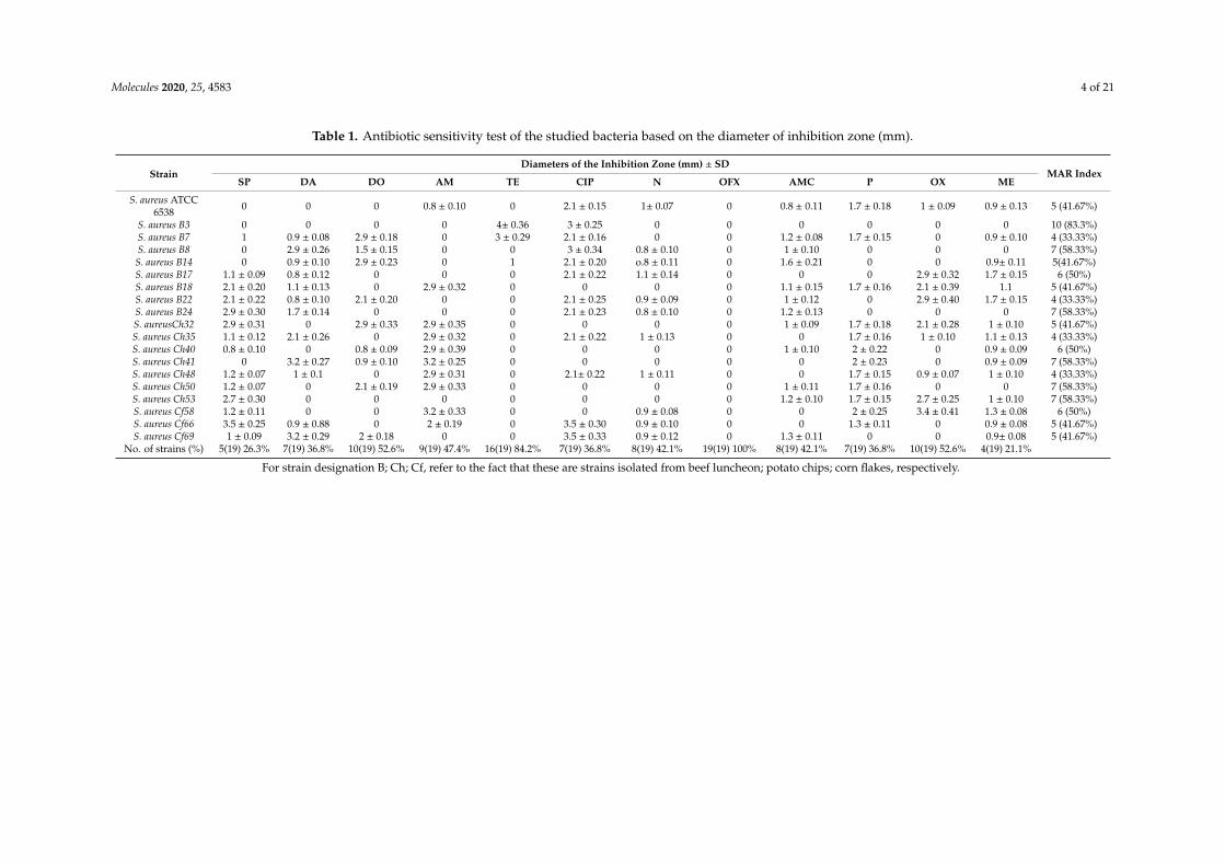
The antibiotic susceptibility of the S. aureus strains (n = 18) was studied. Results are given inTable 1. The MAR index of S. aureus No. B3 isolated from beef luncheon was of about 83.3%; andwas of about 58.33% for the strain B8, B24, and Ch 41. The antibiotics tested could be arrangedin the following descending manner according to their inability to inhibited the indicator bacteria:ofloxacin (100%) > tetracycline (84.2%) > oxacillin and doxycycline (52.63%) > ampicillin (47.36%) >
neomycin and amoxicillin (42.10%) > ciprofloxacine, clindamycin and penicillin (36.84%) > spiramycin(26.31%) > methicillin (21.05 %). Hence, the S. aureus No. B3 strain was resistant to 10 antibiotics testedincluding the antibiotic methicillin. This showed that this strain was preliminary approved to be MDRstrain. For more confirmation of its biochemical identification, this B3 strain showed positive reactionsregarding coagulase, α- and β- blood hemolysis. This strain was selected to be identified by 16S rRNAand tested for its virulence at the molecular level.
Molecules 2020, 25, 4583 4 of 21
Table 1. Antibiotic sensitivity test of the studied bacteria based on the diameter of inhibition zone (mm).
StrainDiameters of the Inhibition Zone (mm) ± SD
MAR IndexSP DA DO AM TE CIP N OFX AMC P OX ME
S. aureus ATCC6538 0 0 0 0.8 ± 0.10 0 2.1 ± 0.15 1± 0.07 0 0.8 ± 0.11 1.7 ± 0.18 1 ± 0.09 0.9 ± 0.13 5 (41.67%)
S. aureus B3 0 0 0 0 4± 0.36 3 ± 0.25 0 0 0 0 0 0 10 (83.3%)S. aureus B7 1 0.9 ± 0.08 2.9 ± 0.18 0 3 ± 0.29 2.1 ± 0.16 0 0 1.2 ± 0.08 1.7 ± 0.15 0 0.9 ± 0.10 4 (33.33%)S. aureus B8 0 2.9 ± 0.26 1.5 ± 0.15 0 0 3 ± 0.34 0.8 ± 0.10 0 1 ± 0.10 0 0 0 7 (58.33%)
S. aureus B14 0 0.9 ± 0.10 2.9 ± 0.23 0 1 2.1 ± 0.20 o.8 ± 0.11 0 1.6 ± 0.21 0 0 0.9± 0.11 5(41.67%)S. aureus B17 1.1 ± 0.09 0.8 ± 0.12 0 0 0 2.1 ± 0.22 1.1 ± 0.14 0 0 0 2.9 ± 0.32 1.7 ± 0.15 6 (50%)S. aureus B18 2.1 ± 0.20 1.1 ± 0.13 0 2.9 ± 0.32 0 0 0 0 1.1 ± 0.15 1.7 ± 0.16 2.1 ± 0.39 1.1 5 (41.67%)S. aureus B22 2.1 ± 0.22 0.8 ± 0.10 2.1 ± 0.20 0 0 2.1 ± 0.25 0.9 ± 0.09 0 1 ± 0.12 0 2.9 ± 0.40 1.7 ± 0.15 4 (33.33%)S. aureus B24 2.9 ± 0.30 1.7 ± 0.14 0 0 0 2.1 ± 0.23 0.8 ± 0.10 0 1.2 ± 0.13 0 0 0 7 (58.33%)S. aureusCh32 2.9 ± 0.31 0 2.9 ± 0.33 2.9 ± 0.35 0 0 0 0 1 ± 0.09 1.7 ± 0.18 2.1 ± 0.28 1 ± 0.10 5 (41.67%)S. aureus Ch35 1.1 ± 0.12 2.1 ± 0.26 0 2.9 ± 0.32 0 2.1 ± 0.22 1 ± 0.13 0 0 1.7 ± 0.16 1 ± 0.10 1.1 ± 0.13 4 (33.33%)S. aureus Ch40 0.8 ± 0.10 0 0.8 ± 0.09 2.9 ± 0.39 0 0 0 0 1 ± 0.10 2 ± 0.22 0 0.9 ± 0.09 6 (50%)S. aureus Ch41 0 3.2 ± 0.27 0.9 ± 0.10 3.2 ± 0.25 0 0 0 0 0 2 ± 0.23 0 0.9 ± 0.09 7 (58.33%)S. aureus Ch48 1.2 ± 0.07 1 ± 0.1 0 2.9 ± 0.31 0 2.1± 0.22 1 ± 0.11 0 0 1.7 ± 0.15 0.9 ± 0.07 1 ± 0.10 4 (33.33%)S. aureus Ch50 1.2 ± 0.07 0 2.1 ± 0.19 2.9 ± 0.33 0 0 0 0 1 ± 0.11 1.7 ± 0.16 0 0 7 (58.33%)S. aureus Ch53 2.7 ± 0.30 0 0 0 0 0 0 0 1.2 ± 0.10 1.7 ± 0.15 2.7 ± 0.25 1 ± 0.10 7 (58.33%)S. aureus Cf58 1.2 ± 0.11 0 0 3.2 ± 0.33 0 0 0.9 ± 0.08 0 0 2 ± 0.25 3.4 ± 0.41 1.3 ± 0.08 6 (50%)S. aureus Cf66 3.5 ± 0.25 0.9 ± 0.88 0 2 ± 0.19 0 3.5 ± 0.30 0.9 ± 0.10 0 0 1.3 ± 0.11 0 0.9 ± 0.08 5 (41.67%)S. aureus Cf69 1 ± 0.09 3.2 ± 0.29 2 ± 0.18 0 0 3.5 ± 0.33 0.9 ± 0.12 0 1.3 ± 0.11 0 0 0.9± 0.08 5 (41.67%)
No. of strains (%) 5(19) 26.3% 7(19) 36.8% 10(19) 52.6% 9(19) 47.4% 16(19) 84.2% 7(19) 36.8% 8(19) 42.1% 19(19) 100% 8(19) 42.1% 7(19) 36.8% 10(19) 52.6% 4(19) 21.1%
For strain designation B; Ch; Cf, refer to the fact that these are strains isolated from beef luncheon; potato chips; corn flakes, respectively.
Molecules 2020, 25, 4583 5 of 21
2.3. Molecular Identification of the B3 Strain by Sequencing of the 16S rRNA Gene
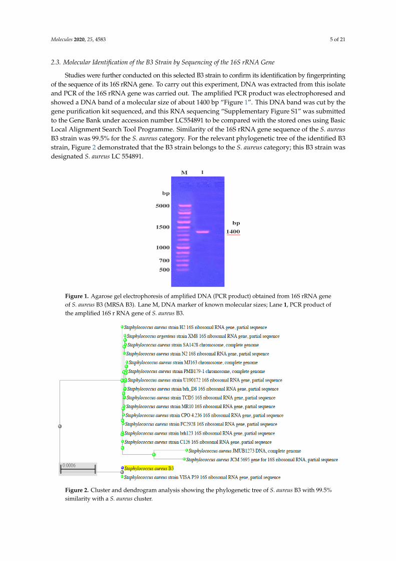
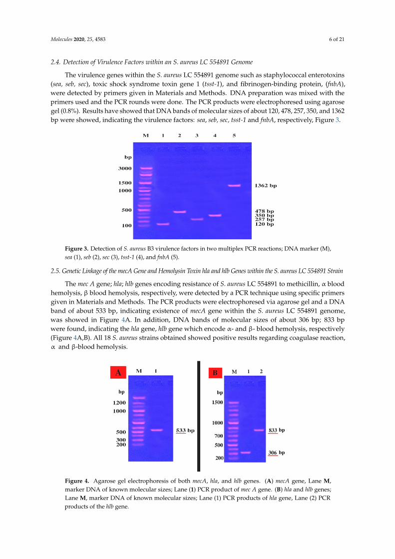
Studies were further conducted on this selected B3 strain to confirm its identification by fingerprintingof the sequence of its 16S rRNA gene. To carry out this experiment, DNA was extracted from this isolateand PCR of the 16S rRNA gene was carried out. The amplified PCR product was electrophoresed andshowed a DNA band of a molecular size of about 1400 bp “Figure 1”. This DNA band was cut by thegene purification kit sequenced, and this RNA sequencing “Supplementary Figure S1” was submittedto the Gene Bank under accession number LC554891 to be compared with the stored ones using BasicLocal Alignment Search Tool Programme. Similarity of the 16S rRNA gene sequence of the S. aureusB3 strain was 99.5% for the S. aureus category. For the relevant phylogenetic tree of the identified B3strain, Figure 2 demonstrated that the B3 strain belongs to the S. aureus category; this B3 strain wasdesignated S. aureus LC 554891.
Figure 1. Agarose gel electrophoresis of amplified DNA (PCR product) obtained from 16S rRNA geneof S. aureus B3 (MRSA B3). Lane M, DNA marker of known molecular sizes; Lane 1, PCR product ofthe amplified 16S r RNA gene of S. aureus B3.
Figure 2. Cluster and dendrogram analysis showing the phylogenetic tree of S. aureus B3 with 99.5%similarity with a S. aureus cluster.
Molecules 2020, 25, 4583 6 of 21
2.4. Detection of Virulence Factors within an S. aureus LC 554891 Genome
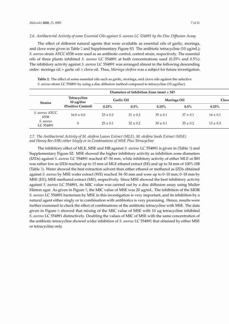
The virulence genes within the S. aureus LC 554891 genome such as staphylococcal enterotoxins(sea, seb, sec), toxic shock syndrome toxin gene 1 (tsst-1), and fibrinogen-binding protein, (fnbA),were detected by primers given in Materials and Methods. DNA preparation was mixed with theprimers used and the PCR rounds were done. The PCR products were electrophoresed using agarosegel (0.8%). Results have showed that DNA bands of molecular sizes of about 120, 478, 257, 350, and 1362bp were showed, indicating the virulence factors: sea, seb, sec, tsst-1 and fnbA, respectively, Figure 3.
Figure 3. Detection of S. aureus B3 virulence factors in two multiplex PCR reactions; DNA marker (M),sea (1), seb (2), sec (3), tsst-1 (4), and fnbA (5).
2.5. Genetic Linkage of the mecA Gene and Hemolysin Toxin hla and hlb Genes within the S. aureus LC 554891 Strain
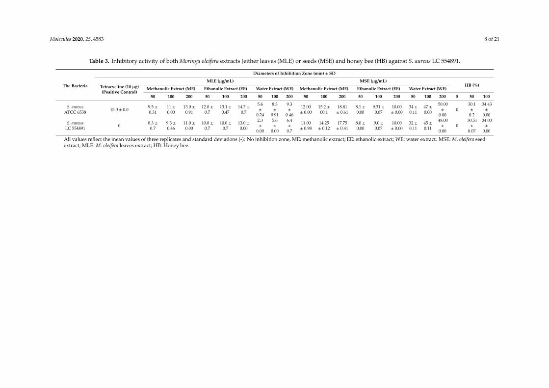
The mec A gene; hla; hlb genes encoding resistance of S. aureus LC 554891 to methicillin, α bloodhemolysis, β blood hemolysis, respectively, were detected by a PCR technique using specific primersgiven in Materials and Methods. The PCR products were electrophoresed via agarose gel and a DNAband of about 533 bp, indicating existence of mecA gene within the S. aureus LC 554891 genome,was showed in Figure 4A. In addition, DNA bands of molecular sizes of about 306 bp; 833 bpwere found, indicating the hla gene, hlb gene which encode α- and β- blood hemolysis, respectively(Figure 4A,B). All 18 S. aureus strains obtained showed positive results regarding coagulase reaction,α and β-blood hemolysis.
Figure 4. Agarose gel electrophoresis of both mecA, hla, and hlb genes. (A) mecA gene, Lane M,marker DNA of known molecular sizes; Lane (1) PCR product of mec A gene. (B) hla and hlb genes;Lane M, marker DNA of known molecular sizes; Lane (1) PCR products of hla gene, Lane (2) PCRproducts of the hlb gene.
Molecules 2020, 25, 4583 7 of 21
2.6. Antibacterial Activity of some Essential Oils against S. aureus LC 554891 by the Disc Diffusion Assay
The effect of different natural agents that were available as essential oils of garlic, moringa,and clove were given in Table 2 and Supplementary Figure S3. The antibiotic tetracycline (10 µg/mL);S. aureus strain ATCC 6538 were used as an antibiotic control, control strain, respectively. The essentialoils of three plants inhibited S. aureus LC 554891 at both concentrations used (0.25% and 0.5%).The inhibitory activity against S. aureus LC 554891 was arranged almost in the following descendingorder: moringa oil > garlic oil > clove oil. Thus, Moringa oleifera was a subject for future investigation.
Table 2. The effect of some essential oils such as garlic, moringa, and clove oils against the selectiveS. aureus strain LC 554891 by using a disc diffusion method compared to tetracycline (10 µg/disc).
Diameters of Inhibition Zone (mm) ± SD
StrainsTetracycline10 µg/disc
(Positive Control)
Garlic Oil Moringa Oil Clove Oil
0.25% 0.5% 0.25% 0.5% 0.25% 0.5%
S. aureus ATCC6538 16.0 ± 0.0 23 ± 0.2 31 ± 0.2 35 ± 0.1 37 ± 0.1 14 ± 0.1 23 ± 0.0
S. aureusLC 554891 0 25 ± 0.1 32 ± 0.2 30 ± 0.1 35 ± 0.2 13 ± 0.3 24 ± 0.1
2.7. The Antibacterial Activity of M. oleifera Leaves Extract (MLE), M. oleifera Seeds Extract (MSE)and Honey Bee (HB) either Singly or in Combinations of MSE Plus Tetracycline
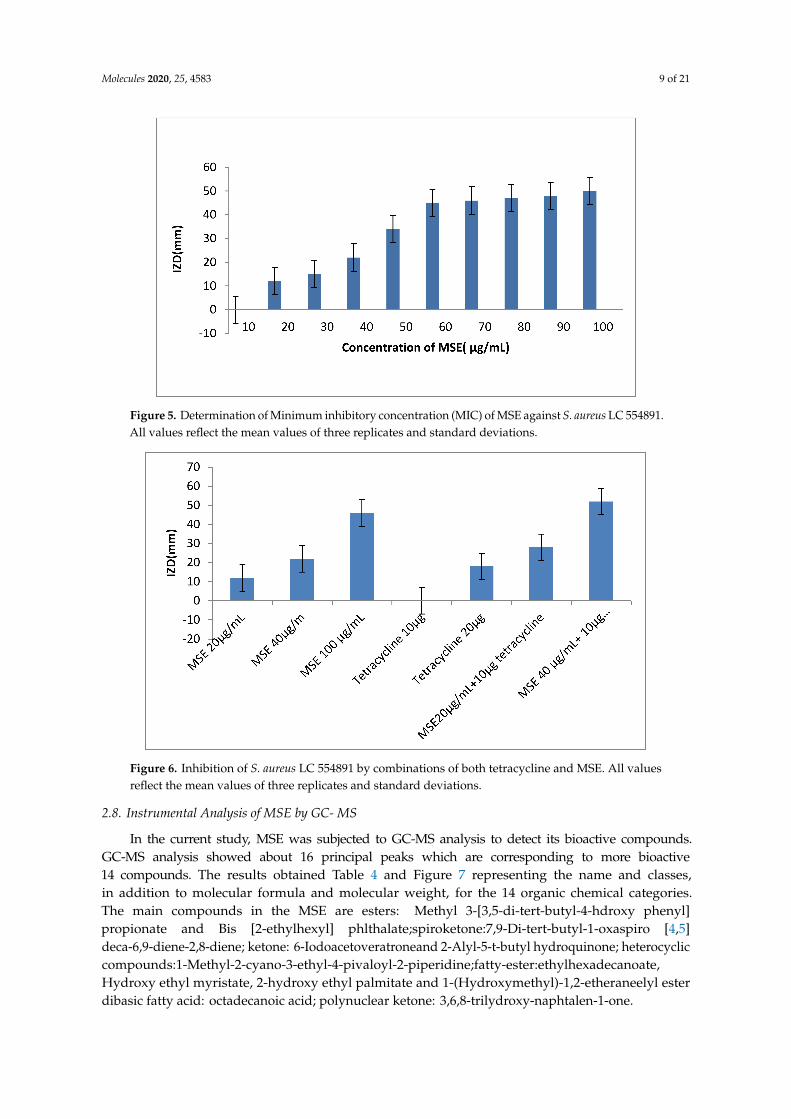
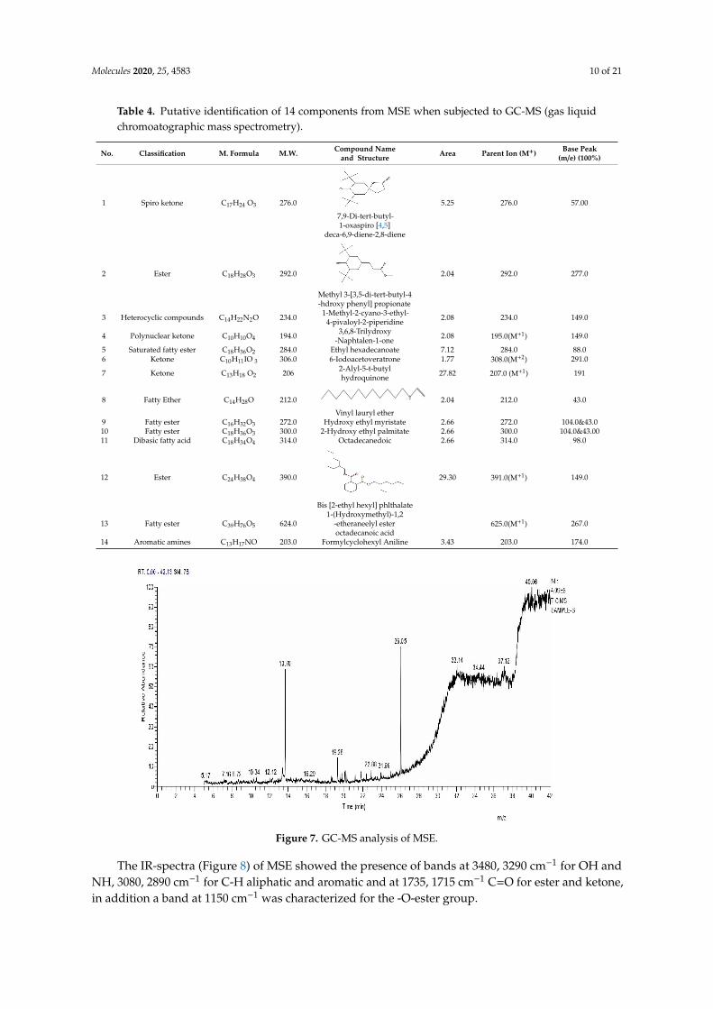
The inhibitory effect of MLE, MSE and HB against S. aureus LC 554891 is given in (Table 3) andSupplementary Figure S2. MSE showed the higher inhibitory activity as inhibition zone diameters(IZDs) against S. aureus LC 554891 reached 47–50 mm, while inhibitory activity of either MLE or BHwas rather low as IZDs reached up to 15 mm of MLE ethanol extract (EE) and up to 34 mm of 100% HB(Table 3). Water showed the best extraction solvent than either ethanol or methanol as IZDs obtainedagainst S. aureus by MSE water extract (WE) reached 34–50 mm and were up to 0–10 mm; 0–18 mm byMSE (EE); MSE methanol extract (ME), respectively. Since MSE showed the best inhibitory activityagainst S. aureus LC 554891, its MIC value was carried out by a disc diffusion assay using MullerHinton agar. As given in Figure 5, the MIC value of MSE was 20 µg/mL. The inhibition of the MDRS. aureus LC 554891 bacterium by MSE in this investigation is very important, and its inhibition by anatural agent either singly or in combination with antibiotics is very promising. Hence, results werefurther examined to check the effect of combinations of the antibiotic tetracycline with MSE. The datagiven in Figure 6 showed that mixing of the MIC value of MSE with 10 µg tetracycline inhibitedS. aureus LC 554891 distinctively. Doubling the values of MIC of MSE with the same concentration ofthe antibiotic tetracycline showed wider inhibition of S. aureus LC 554891 that obtained by either MSEor tetracycline only.
Molecules 2020, 25, 4583 8 of 21
Table 3. Inhibitory activity of both Moringa oleifera extracts (either leaves (MLE) or seeds (MSE) and honey bee (HB) against S. aureus LC 554891.
The Bacteria
Diameters of Inhibition Zone (mm) ± SD
Tetracycline (10 µg)(Positive Control)
MLE (µg/mL) MSE (µg/mL)HB (%)
Methanolic Extract (ME) Ethanolic Extract (EE) Water Extract (WE) Methanolic Extract (ME) Ethanolic Extract (EE) Water Extract (WE)
50 100 200 50 100 200 50 100 200 50 100 200 50 100 200 50 100 200 5 50 100
S. aureusATCC 6538 15.0 ± 0.0 9.5 ±
0.3111 ±0.00
13.0 ±0.91
12.0 ±0.7
13.1 ±0.47
14.7 ±0.7
5.6±
0.24
8.3±
0.91
9.3±
0.46
12.00± 0.00
15.2 ±00.1
18.81± 0.61
8.1 ±0.00
9.31 ±0.07
10.00± 0.00
34 ±0.11
47 ±0.00
50.00±
0.000
30.1±
0.2
34.43±
0.00
S. aureusLC 554891 0 8.3 ±
0.79.3 ±0.46
11.0 ±0.00
10.0 ±0.7
10.0 ±0.7
13.0 ±0.00
2.3±
0.00
5.6±
0.00
6.4±
0.7
11.00± 0.98
14.25± 0.12
17.75± 0.41
8.0 ±0.00
9.0 ±0.07
10.00± 0.00
32 ±0.11
45 ±0.11
48.00±
0.000
30.51±
0.07
34.00±
0.00
All values reflect the mean values of three replicates and standard deviations (-): No inhibition zone, ME: methanolic extract; EE: ethanolic extract; WE: water extract. MSE: M. oleifera seedextract; MLE: M. oleifera leaves extract; HB: Honey bee.
Molecules 2020, 25, 4583 9 of 21
Figure 5. Determination of Minimum inhibitory concentration (MIC) of MSE against S. aureus LC 554891.All values reflect the mean values of three replicates and standard deviations.
Figure 6. Inhibition of S. aureus LC 554891 by combinations of both tetracycline and MSE. All valuesreflect the mean values of three replicates and standard deviations.
2.8. Instrumental Analysis of MSE by GC- MS
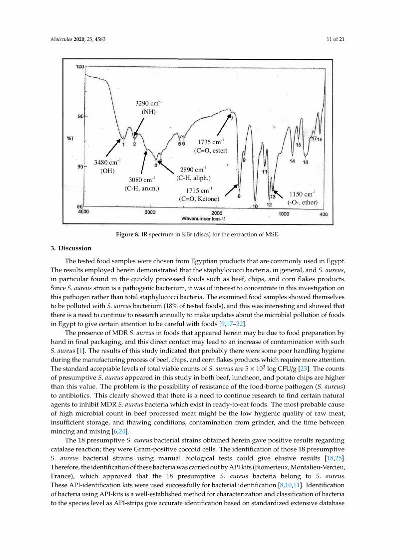
In the current study, MSE was subjected to GC-MS analysis to detect its bioactive compounds.GC-MS analysis showed about 16 principal peaks which are corresponding to more bioactive14 compounds. The results obtained Table 4 and Figure 7 representing the name and classes,in addition to molecular formula and molecular weight, for the 14 organic chemical categories.The main compounds in the MSE are esters: Methyl 3-[3,5-di-tert-butyl-4-hdroxy phenyl]propionate and Bis [2-ethylhexyl] phlthalate;spiroketone:7,9-Di-tert-butyl-1-oxaspiro [4,5]deca-6,9-diene-2,8-diene; ketone: 6-Iodoacetoveratroneand 2-Alyl-5-t-butyl hydroquinone; heterocycliccompounds:1-Methyl-2-cyano-3-ethyl-4-pivaloyl-2-piperidine;fatty-ester:ethylhexadecanoate,Hydroxy ethyl myristate, 2-hydroxy ethyl palmitate and 1-(Hydroxymethyl)-1,2-etheraneelyl esterdibasic fatty acid: octadecanoic acid; polynuclear ketone: 3,6,8-trilydroxy-naphtalen-1-one.
Molecules 2020, 25, 4583 10 of 21
Table 4. Putative identification of 14 components from MSE when subjected to GC-MS (gas liquidchromoatographic mass spectrometry).
No. Classification M. Formula M.W. Compound Nameand Structure Area Parent Ion (M+) Base Peak
(m/e) (100%)
1 Spiro ketone C17H24 O3 276.0
7,9-Di-tert-butyl-1-oxaspiro [4,5]
deca-6,9-diene-2,8-diene
5.25 276.0 57.00
2 Ester C18H28O3 292.0
Methyl 3-[3,5-di-tert-butyl-4-hdroxy phenyl] propionate
2.04 292.0 277.0
3 Heterocyclic compounds C14H22N2O 234.0 1-Methyl-2-cyano-3-ethyl-4-pivaloyl-2-piperidine 2.08 234.0 149.0
4 Polynuclear ketone C10H10O4 194.0 3,6,8-Trilydroxy-Naphtalen-1-one 2.08 195.0(M+1) 149.0
5 Saturated fatty ester C18H36O2 284.0 Ethyl hexadecanoate 7.12 284.0 88.06 Ketone C10H11IO 3 306.0 6-Iodoacetoveratrone 1.77 308.0(M+2) 291.0
7 Ketone C13H18 O2 206 2-Alyl-5-t-butylhydroquinone 27.82 207.0 (M+1) 191
8 Fatty Ether C14H28O 212.0
Vinyl lauryl ether
2.04 212.0 43.0
9 Fatty ester C16H32O3 272.0 Hydroxy ethyl myristate 2.66 272.0 104.0&43.010 Fatty ester C18H36O3 300.0 2-Hydroxy ethyl palmitate 2.66 300.0 104.0&43.0011 Dibasic fatty acid C18H34O4 314.0 Octadecanedoic 2.66 314.0 98.0
12 Ester C24H38O4 390.0
Bis [2-ethyl hexyl] phlthalate
29.30 391.0(M+1) 149.0
13 Fatty ester C39H76O5 624.01-(Hydroxymethyl)-1,2
-etheraneelyl esteroctadecanoic acid
625.0(M+1) 267.0
14 Aromatic amines C13H17NO 203.0 Formylcyclohexyl Aniline 3.43 203.0 174.0
Figure 7. GC-MS analysis of MSE.
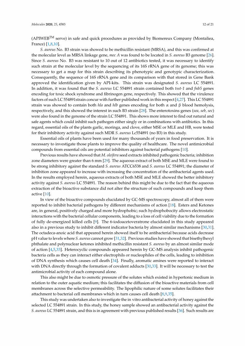
The IR-spectra (Figure 8) of MSE showed the presence of bands at 3480, 3290 cm−1 for OH andNH, 3080, 2890 cm−1 for C-H aliphatic and aromatic and at 1735, 1715 cm−1 C=O for ester and ketone,in addition a band at 1150 cm−1 was characterized for the -O-ester group.
Molecules 2020, 25, 4583 11 of 21
Figure 8. IR spectrum in KBr (discs) for the extraction of MSE.
3. Discussion
The tested food samples were chosen from Egyptian products that are commonly used in Egypt.The results employed herein demonstrated that the staphylococci bacteria, in general, and S. aureus,in particular found in the quickly processed foods such as beef, chips, and corn flakes products.Since S. aureus strain is a pathogenic bacterium, it was of interest to concentrate in this investigation onthis pathogen rather than total staphylococci bacteria. The examined food samples showed themselvesto be polluted with S. aureus bacterium (18% of tested foods), and this was interesting and showed thatthere is a need to continue to research annually to make updates about the microbial pollution of foodsin Egypt to give certain attention to be careful with foods [9,17–22].
The presence of MDR S. aureus in foods that appeared herein may be due to food preparation byhand in final packaging, and this direct contact may lead to an increase of contamination with suchS. aureus [1]. The results of this study indicated that probably there were some poor handling hygieneduring the manufacturing process of beef, chips, and corn flakes products which require more attention.The standard acceptable levels of total viable counts of S. aureus are 5 × 10³ log CFU/g [23]. The countsof presumptive S. aureus appeared in this study in both beef, luncheon, and potato chips are higherthan this value. The problem is the possibility of resistance of the food-borne pathogen (S. aureus)to antibiotics. This clearly showed that there is a need to continue research to find certain naturalagents to inhibit MDR S. aureus bacteria which exist in ready-to-eat foods. The most probable causeof high microbial count in beef processed meat might be the low hygienic quality of raw meat,insufficient storage, and thawing conditions, contamination from grinder, and the time betweenmincing and mixing [6,24].
The 18 presumptive S. aureus bacterial strains obtained herein gave positive results regardingcatalase reaction; they were Gram-positive coccoid cells. The identification of those 18 presumptiveS. aureus bacterial strains using manual biological tests could give elusive results [18,25].Therefore, the identification of these bacteria was carried out by API kits (Biomerieux, Montalieu-Vercieu,France), which approved that the 18 presumptive S. aureus bacteria belong to S. aureus.These API-identification kits were used successfully for bacterial identification [8,10,11]. Identificationof bacteria using API-kits is a well-established method for characterization and classification of bacteriato the species level as API-strips give accurate identification based on standardized extensive database
Molecules 2020, 25, 4583 12 of 21
(APIWEBTM serve) in safe and quick procedures as provided by Biomereux Company (Montaliea,France) [1,8,10].
S. aureus No. B3 strain was showed to be methicillin resistant (MRSA), and this was confirmed atthe molecular level as MRSA linkage gene, mec A was found to be located in S. aureus B3 genome [26].Since S. aureus No. B3 was resistant to 10 out of 12 antibiotics tested, it was necessary to identifysuch strain at the molecular level by the sequencing of its 16S rRNA gene of its genome; this wasnecessary to get a map for this strain describing its phenotypic and genotypic characterization.Consequently, the sequence of 16S rRNA gene and its comparison with that stored in Gene Bankapproved the identification given by API-kits. This strain was designated S. aureus LC 554891.In addition, it was found that the S. aureus LC 554891 strain contained both tsst-1 and fnb5 genesencoding for toxic shock syndrome and fibrinogen gene, respectively. This showed that the virulencefactors of such LC 554891strain concur with further published work in this respect [4,27]. This LC 554891strain was showed to contain both hla and hlb genes encoding for both α and β blood hemolysis,respectively, and this showed the interest in such B3 strain [28]. The enterotoxins genes (sea, seb, sec)were also found in the genome of the strain LC 554891. This shows more interest to find out natural andsafe agents which could inhibit such pathogen either singly or in combinations with antibiotics. In thisregard, essential oils of the plants garlic, moringa, and clove, either MSE or MLE and HB, were testedfor their inhibitory activity against such MDR S. aureus Lc554891 (no B3) in this study.
Essential oils of plants have been used for many thousands of years in food preservation. It isnecessary to investigate those plants to improve the quality of healthcare. The novel antimicrobialcompounds from essential oils are potential inhibitors against bacterial pathogens [10].
Previous results have showed that M. oleifera seed extracts inhibited pathogenic bacteria; inhibitionzone diameters were greater than 6 mm [29]. The aqueous extract of both MSE and MLE were found tobe strong inhibitory against the standard S. aureus ATCC6538 and S. aureus LC 554891; the diameter ofinhibition zone appeared to increase with increasing the concentration of the antibacterial agents used.In the results employed herein, aqueous extracts of both MSE and MLE showed the better inhibitoryactivity against S. aureus LC 554891. The reason behind this might be due to the fact that the aqueousextraction of the bioactive substance did not alter the structure of such compounds and keep themactive [10].
In view of the bioactive compounds elucidated by GC-MS spectroscopy, almost all of them werereported to inhibit bacterial pathogens by different mechanisms of action [18]. Esters and Ketonesare, in general, positively charged and more hydrophobic; such hydrophobocity allows electrostaticinteractions with the bacterial cellular components, leading to a loss of cell viability due to the formationof fully de-energized killed cells [9]. The 6-iodoacetoveratrone elucidated in this study appearedalso in a previous study to inhibit different indicator bacteria by almost similar mechanisms [30,31].The octadeca-anoic acid that appeared herein showed itself to be antibacterial because acids decreasepH value to levels where S. aureus cannot grow [31,32]. Previous studies have showed that bisethylhexylphthalate and polynuclear ketones inhibited methicillin resistant S. aureus by an almost similar modeof action [4,5,33]. Heterocyclic compounds appeared herein by GC-MS analysis inhibit pathogenicbacteria cells as they can interact either electrophils or nucleophiles of the cells, leading to inhibitionof DNA synthesis which causes cell death [34]. Finally, aromatic amines were reported to interactwith DNA directly through the formation of covalent adducts [30,33]. It will be necessary to test theantimicrobial activity of each compound alone.
This also might be due to osmotic pressure of the solutes which existed in hypertonic medium inrelation to the outer aquatic medium; this facilitates the diffusion of the bioactive materials from cellmembranes across the selective permeability. The lipophilic nature of some solutes facilitates theirattachment to bacterial cell membranes which in turn causes cell death [8,9,35].
This study was undertaken also to investigate the in vitro antibacterial activity of honey against theselected LC 554891 strain. In this study, the honey sample showed an antibacterial activity against theS. aureus LC 554891 strain, and this is in agreement with previous published results [36]. Such results are
Molecules 2020, 25, 4583 13 of 21
in confirmation with Mama et al. [37], who declared that the inhibitory activity of honey bees is due to amix of antibacterial agents such as high content of hydrogen peroxide, powerful antioxidants, naturallylow pH, which is unsuitable for bacterial growth, and to the presence of phenolic acids, lysozymes,and flavanoids. The potency of native honey (100% concentration) was found to be inhibitory againstS. aureus LC 554891, and this concentration was the best one giving antibacterial activity; such resultsconcur with previously published work [38]. A previous study [39] discovered that the antimicrobialactivity of honey was more with S. aureus and Acinetobacter spp, both with resistance to some antibioticslike gentamicin, ceftriazone, amikacin, and tobramicin than other bacteria tested. Honey bees havean antibacterial nature due to presence of H2O2, phenolic compounds, and pH [10]. There arepolyphenolic compounds present in honey bees, which are responsible for its antibacterial activity.The common polyphenolic compounds are gallic acid, cinnamic acid, ferulic acid, hydroxyl cinnamicacid, sinapic acid, syringic acid, and chlorogenic acid. These compounds inhibit the bacteria by disruptbacterial membrane, inhibit DNA gyrase, induce topaisomerase IV mediated DNA cleavage, inhibitpeptidoglycan, and ribosome synthesis [40].
Due to the promising inhibition of S. aureus LC 554891 strain by MSE, a mixture of this MSEand the antibiotic tetracycline was used as an inhibitory agent for this B3 strain. The vigorous anddistractive inhibition of S. aureus LC 554891 strain showed an interesting perspective to use suchmixture as a biocontrol agent for S. aureus [10,11,40].
The combinations of both MSE and tetracycline gave broader antibacterial activity because both ofthem may be acted in a synergism; a synergism between antibiotics and plant extracts could be due tobinding of both of them by hydrogen bonding, hydrophobic-hydrophobic interactions, and molecularinteractions (10). In view of the tetracycline molecule, it contains four fused rings (A, B, C&D) towhich a variety of polar groups (5- hydroxyl groups and one amino group) are attached. In addition,the bioactive compounds of MSE elucidated herein contain both polar and non-polar chemical moieties.Hence, an interaction of MSE and tetracycline might occur between polar and non-polar chemicalmoieties [41]. In fact, further experiments will be needed to study such synergism at the molecularchemical level.
Many MDR bacteria including methicillin resistant S. aureus (MRSA) can be controlled usingpharmaceutical potentials in the treatment of infection for example, Bis [2-ethyl hexyl] phlthalate [42].1-(Hydroxymethyl)-1,2-etheraneelyl ester octadecanoic acid is known to have antibacterial activityand is thought to play a more direct role than previously thought in innate immune defense againstepidermal and mucosal bacterial infections [43,44]. The anti-staphylococcal activity of MSE is due tothe mix of the compounds that appeared herein from GC-MS. Similarly, a previous study that bioactivecompounds of M. oleifera seed extract e.g., 4-(α-l-rhamnopyranosyloxy) benzyle isothiocyanate stronglyinhibited the S. aureus BAA-977 strain [45]. This means that M. oleifera could be used as a safe andpotent control of infectious diseases [45]. Extracts of M. oleifera leaf, stem, and seeds were used asinhibitory agents against S. aureus bacteria isolated from human sputum [46].
With regard to the mechanism of action of the bioactive compounds that appeared from theinstrumental analysis used in this study in MSE, the antibacterial activity of fatty acids are attributedto their ability to disrupt the outer bacterial cell membrane, increasing the leakage of electrolytes frombacterial cells and, in turn, cause cell death [47]. In addition, it was approved that the other bioactivecompounds of M. oleifera seed extracts such as minerals, aromatic amines, esters, and ketones inhibitcell wall synthesis of bacteria at its initial stages and accumulate onto a cell membrane, leading tointerruption in the bacterial metabolism and cell death [48,49].
It is necessary to test the anti-staphylococcal activity of each compound alone. Studies in thisregard are under investigations. Further work will be necessary to isolate the bioactive compoundsobtained from MSE and to check the antimicrobial potential for each of them either separately or incombination with antibiotics.
Molecules 2020, 25, 4583 14 of 21
4. Materials and Methods
4.1. Food Sampling
The foods used were ready-to-eat beef luncheon (Egyptian made), potato chips, and corn flakes.A hundred food samples including beef luncheon (n = 25), potato chips (n = 50), and corn flakes (n = 25)were examined in this study. These food samples were purchased from different retail supermarketsof urban areas of the Egyptian cities viz. Belbeis, Zagazig, Abo-Kabir, and Hehia; all of these citiesare located in the Sharkia Governorate (80 km north, Cairo, Egypt). All of these food types weremade in Egyptian companies. The samples were transported immediately in sterile plastic bags(Gomhuria Company, Zagazig, Egypt) to the laboratory of Microbiology Department, Faculty ofScience, Zagazig University, City, Egypt. The samples (25 g) were taken under aseptic conditions to ablender (Gomhuria Company, Zagazig, Egypt), dissolved in 225 mL of sterile buffer peptone water1–10 dilutions (0.1% w/v) and mixed well for 60 s at 25 ◦C.
4.2. Isolation of Bacteria Suspected to Be S. aureus
Serial two-fold dilutions of up to 10−6 were made from the initial dilution (1:10) and 0.1 mLaliquots of these dilutions were inoculated onto Baird Parker agar (Oxoid). The bacteria suspectedto be S. aureus count were found by examining the plates at typical black colonies, convex shape,with a shiny halo zone, and these were checked for positive Gram coagulase reaction and catalase test(Bactident Coagulase Biolife, Milan, Italy). The identification of the bacterial isolates was then carriedout by API-Kits (Biomereux, Montaliea, France) as given by the manufacturer’s instruction.
4.3. Antibiotics Susceptibility Test
The susceptibility of S. aureus ATCC 6538 strain (control), S. aureus (n = 18) strains to 12 antibioticswere tested by standard disc diffusion technique CLSI [47]. The cultures were grown in nutrientbroth (Oxoid) for 12 h. Inocula were adjusted at 105 CFU/mL and then were plated (100 µLper plate) onto Muller Hinton agar (Hi-Media, Mumbai, India). The following antibiotic discswith their concentrations indicated in parenthesis were used (All from Johnson & Johnson, Egypt.Branch, Heliopolis, Cairo, Egypt) viz. spiramycin (SP: 100 µg), clindamycin (DA: 2 µg), doxycycline(DO: 30 µg), ampicillin (AM: 10 µg), tetracycline (TE: 30 µg), ciprofloxacin (CIP: 5 µg), neomycin(N: 30 µg), ofloxacin (OFX: 5 µg), amoxicillin (AMC: 30µg), penicillin G (P: 10 µg) oxacillin (OX: 10µg),methicillin (ME: 5 µg). The antibiotic discs were placed onto Muller Hinton agar plates that seededwith the tested bacteria; plates were then inverted and incubated at 37 ◦C for 24 h. Results wereexpressed by measuring inhibition zone diameters (IZDs) by millimeters. Multiple antibiotic resistanceindex was calculated by using the following formula: MAR Index = Number of antibiotics to whichthe isolate was resistant/Total number of antibiotics tested. Spiramycin (SP: 100 µg), Clindamycin(DA: 2 µg), Doxycycline (DO: 30 µg), Ampicillin (AM: 10 µg), Tetracycline (TE: 30 µg), Ciprofloxacin(CIP: 5 µg), Neomycin (N: 30 µg), Ofloxacin (OFX: 5 µg), Amoxicillin (AMC: 30 µg), Penicillin G(P: 10 µg) Oxacillin (OX: 10 µg), Methicillin (ME: 5 µg) [50,51].
4.4. Molecular Identification of S. aureus No. B3
It was necessary to confirm the identification of the MDR S. aureus No. B3 bacteriumby fingerprinting of the sequence of 16S rRNA gene. Hence, DNA was extracted from theB3 strain [52]. The 16S rRNA gene was amplified by PCR with using universal primers(forward primer [F27] 5’-AGAGTTTGATCCTGGCTCAG-3’ [53] and reverse primer [R1492]5’-GGTTACCTTGTTACGACTT-3’) [54]. The PCR was carried out in a Gene-Amp PCR system9600 thermocycler (Perkin Elmer Co., Jersey, AL, USA). The amplification conditions were as follows:94 ◦C for 10 min and 35 cycles of denaturation at 95 ◦C for 30 s, annealing-extension at 56 ◦C for 1 min,72 ◦C for 1 min and an extension at 72 ◦C for 10 min. The PCR product was electrophoresed using
Molecules 2020, 25, 4583 15 of 21
agarose gel (0.7%) (Gomhuria, Egypt). The 16SrRNA gene band appeared at 1500 bp was cut by GenePurification Kit (Promega Corporation, Madison, WI, USA).
The nucleotide sequence of 16S rRNA gene of the S. aureus LC 554891genome was sequencedby using 3130 X DNA Sequencer (Genetic Analyzer, Applied Biosystems, Hitachi, Ibaraki, Japan) asdescribed previously [55,56]. The way to record such strain in gene bank included the submission ofthe sequence of 16 S rRNA gene of the B3 genome to Gene Bank at http://blast.ncbi.nlm.nih.gov/Blast.cgi?PROGRAM=blastn&PAGE_TYPE=BlastSearch&LINK_LOC=blasthome. By using the Basic LocalAlignment Search Tool Program, a phylogenic tree and cluster analysis were carried out by clusta 1×Program for estimation of the similarity between the isolated strains and the stored S. aureus strainsin the database. It was shown clearly that the B3 strain is similar by > 99.5% to S. aureus category(Figure 2). An accession Number LC554891 on the NCBI web server (http://blast.ncbi.nlm.nih.gov/
Blast.cgi?PROGRAM=blastn&PAGE_TYPE=BlastSearch&LINK_LOC=blasthome) of such strain wasgiven as a record code for this strain. Consequently, such strain was designated as S. aureus LC554891.By using the Basic Local Alignment Search Tool Program, a phylogenic tree and cluster analysis werecarried out by clusta 1× Program for estimation of the similarity between the isolated strains and thestored S. aureus strains in the database.
4.5. Detection of Virulence Factors (sea, seb, sec, tsst-1 and fnbA) of the Strain
Extraction of DNA from S. aureus LC 554891 was performed using DNeasy bacteria Mini Kit(Bio Basic Comp., Toronto, ON, Canada) [57,58]. PCR was performed in 30 µL volume tubes accordingto Williams et al. [57]. The DNA amplifications were performed in an automated thermal cycle(Promega Corporation, Madison, WI, USA) programmed for one cycle at 94 ◦C for 4 min followedby 10 cycles of (4 min at 94 ◦C, 1 min at 52A ◦C, and 1 min at 72 ◦C) then 15 cycles of (4 min at 94◦C, 1 min at 58 ◦C, and 1 min at 72 ◦C) the reaction was finally stored at 72 ◦C for 10 min. The DNAamplified product (15 µL) was loaded in each well of agarose gel electrophoresis equipment (Gomhuria,Egypt) using DNA ladder (100 bp) mix that used as standard DNA with known molecular weights.The run was performed for about 30 min at 80 V in mini submarine gel (Bio-Rad Laboratories, Berkeley,CA, USA). PCR reactions were conducted using 4 simple Sequence Repeat (SSR) primers. Their namesand sequences are shown in Table 5.
Table 5. Virulence genes and the primers used for the detection of S. aureus LC554891 genome.
DetectedVirulence Factors Primer Sequence (Forwarded) Primer Sequence (Reverse) Size of the PCR
Products (bp)
Sea TTGGAAACGGTTAAAACGAA GAACCTTCCGATCAAAAACA 120Seb TCGCATCAAACTGACAAACG GCAGGTACTCTATAAGTGCC 478Sec GACATAAAAGCTAGGAATTT AAATCGGATTAACATTATCC 257
Tsst-1 ATGGCAGCATCAGCTTGATA TTTCCAATAACCACCCGTTT 350fnbA CACAACCAGCAAATATAG CTG TGTGGTAATCAATGT 1362
4.6. Genetic Linkage of mecA, hla, and hlb Genes
Total DNA was extracted from exponentially growing B3strain cells [52,59]. The mecA gene primerswere mecA f (AAAATCGATGGTAAAGGTTGGC) and mecA r (AGTTCTGCAGTACCGGATTTGC) [60].In addition, primers used for hla gene were hla f GCC AAA GCC GAA TCT AAG and hla r GCG ATA TACATC CCA TGG C [61] and those used for hlb gene were hlb f TTGGCTGGGGAGTTGAAGCACA andhlb r CGCCTGCCCAGTAGAAGCCATT (Promega Corporation, Madison, WI, USA) [62]. PCR roundswere carried out by using 5µl of template DNA, 0.025µM of each primer, (Promega Corporation,Madison, WI, USA). DNA amplification was carried out for 40 cycles in 100 µl of reaction mixture asfollows: denaturation of 94 ◦C for 30 s, annealing at 55 ◦C for 30 s, and extension at 72 ◦C for 1 minwith a final at 72 ◦C for 5 min. Ten micro liters aliquots of PCR products were analyzed using 1.5%agarose gel electrophoresis at 90 V (Gomhuria Company, Zagazig, Egypt) for 90 min.
Molecules 2020, 25, 4583 16 of 21
4.7. Screening of the Antibacterial Activity of Essential Oils of Moringa olifera, Allium sativum,and Syzygium aromaticum against S. aureus LC 554891
Essential oils of Moringa olifera, Allium sativum and Syzygium aromaticum were obtained fromEl-Hawag factory, Bader, Egypt, under the supervision of Ministry of Health license no: 150/80 for theyear 2002. Then, they sterilized with 0.45 µm filter paper obtained from a High Lab Company, Zagazig,Sharkia, Egypt. Sterilized filter paper discs (6 mm diameter) were soaked in 1 mL of each essential oilused, for 2 min. They were then placed onto BHI agar plates that were inoculated by cell suspensionof S. aureus LC 554891. After incubation for 24 h at 37 ◦C, diameter of inhibition zones (mm) weremeasured after subtracting diameter of paper disc [50].
4.8. Preparation of the M. oleifera Leaves (MLE) and Seeds (MSE)
The plant M. oleifera was identified by the plant taxonomist, Prof. Dr. Hussein Abdel-Basset,at Department of Botany and Microbiology, Faculty of Science, Zagazig University, Egypt.Both M. oleifera leaves and seeds were collected; the leaves were cleaned from extraneous matterand properly washed then dried in hot air oven (Alexandria Co., Alexandria, Egypt) for 24 h at40 ◦C. The seeds were dried and grounded to powdered form using a clean sterile mortar and pestle(Moulinex, Cairo, Egypt) and packaged in an air tight plastic container (Alexandria Co., Alexandria,Egypt) until used. About 10 g aliquots of either powdered leaves or seed-powdered were maceratedin 100 mL distilled water and allowed to be extracted for 48 h at room temperature; methanolic andethanolic extracts were also carried out by homogenization of either MSE or MLE (10 g for each) with100 mL ethanol or methanol for 40 min [11]; solvents were then evaporated by keeping the extracts inan oven (Alexandria Co., Alexandria, Egypt) adjusted at 60 ◦C overnight. Both leave extracts (MLE)and seed extracts (MSE) were homogenized with sterile water and sterilized by filtration (0.45 miliporeBilters, Amicon, Mumbai, India). Stock preparation of MSE (200 µg/mL) was prepared and then storedin Eppendorf tubes (Gomhuria Co., Zagazig, Egypt) at 5 ◦C until antimicrobial activity tests wereperformed [63].
4.9. Preparation of Honey Bee (HB) Solutions
Native HB used in this study was provided by a bee-keeper from Kafr-Sakr area, Sharkia Governorate(104 km North Cairo), Egypt. It was aseptically collected in 100 mL screw capped bottles, transported tothe laboratory. HB dilutions were prepared immediately prior their testing by diluting native honey tothe required concentrations (10%, 20%, 30%, 40%, 50%, 60%, 70%, 80%, 90% v/v) [10,21]. These dilutionswere made using sterile distilled water. A series of measurable scaled 250-mL screw capped bottles(Gomhuria Company, Zagazig, Egypt) containing 90 mL; 80 mL; 70 mL; 60 mL; 50 mL; 40 mL; 30 mL;20 mL; and 10 mL native HB were prepared and completed to 100 mL sterile distilled water usingsterile pipettes, giving the desired dilutions of HB viz. 10%, 20%, 30%, 40%, 50%, 60%, 70%, 80%,and 90%, respectively.
4.10. Bioassay of the Antibacterial Activity of MLE, MSE, and HB
Brain Heart infusion agar plates (DifcoTM, Maryland, MD, USA) were prepared and seeded logphase cells (10 5 CFU/mL); then, the sterile natural agents concentrations listed in Table 3 were addedby automatic pipette to these filter paper discs (6 mm diameter), which were placed immediately ontothe above plates. The controls were filter paper discs soaked in sterile distilled water. Samples andcontrols were incubated at 37 ◦C for 24–48 h. IZDs were measured after subtracting the diameter of thepaper disc [17,21,64].
4.11. Minimum Inhibitory Concentration (MIC) of the MSE Extract
A stock prepared MSE contained 200 µg/mL. From this MSE concentration, different dilutionswere made to contain 10, 20, 30, 40, 50, 60, 70, 80, and 90, µg/mL, respectively. Serial two-fold
Molecules 2020, 25, 4583 17 of 21
dilutions of MSE were made in sterile deionized water by taking 0.05, 0.1, 0.15, 0.2, 0.25, 0.3, 0.35, 0.4,and 0.45 mL, from the original stock and this equals 10, 20, 30, 40, 50, 60, 70, 80, 90 µg/mL, respectively.Then, sterile filter paper discs were saturated with the MSE dilutions and placed onto Muller Hintonagar (DifcoTM, Maryland, MD, USA) that seeded previously with activity growing cells of S. aureus B3.The antibacterial activity was studied by a disc diffusion assay as described above. MIC was visuallyidentified as the lowest concentration of MSE that inhibited bacterial growth [9–11,40].
4.12. Antibacterial Activity of Combination of Antibiotics and MSE
The antibiotic tetracycline listed in Table 2 that inhibited the S. aureus strain was mixed with MICvalue of MSE. Sterile filter paper discs were impregnated by these combinations and assayed for theirantistaphylococcal activity as described above. In addition, single different concentrations of eithertetracycline or MSE were tested singly for their antistaphylococcal activity. Mixtures of MSE withthe antibiotic tetracycline were made as follows: (20 µg/mL MSE + 10 µg tetracycline) and (40 µg/mLMSE + 10 µg tetracycline). Filter paper discs of 6 mm diameter were soaked in each combination andthe experiment was carried out as described above [8].
4.13. Instrumental Analysis of MSE
To determine and identify the bioactive compounds of MSE, Gas Chromatography–MassSpectroscopic (GC-MS) was used (Trace GC 1310-ISQ Mass Spectrometer, Thermo Scientific, Austin,TX, USA). A direct capillary column TG–5MS (30 m × 0.25 mm × 0.25 µm film thickness) wasused. About 3 µL of MSE was injected automatically to the equipment using Auto sampler AS3000coupled with GC in the split less mode. Then, the instrumental analysis was carried out as describedpreviously [15,65]. The components were identified by comparison of their retention times and massspectra with those of WILEY 09 and NIST 11 mass spectral database [66].
4.14. Statistical Analysis
All the experiments were performed in triplicates and results were expressed by the mean withthe standard error. Data were statically analyzed using ANOVA variance analysis (SAS version 9.1,SAS Institute, Inc., Cary, NC, USA) [67]. Basic Local Alignment Search Tool Program (BLAST) was usedto construct the pairwise similarity of the S. aureus B3 with S. aureus cluster of Gene Bank; Clusta 1XTree Programme was used to construct the phylogenetic tree.
4.15. Ethical Approval
This work was approved by institutional review board at Faculty of Science, Zagazig University,Zagazig, Egypt.
5. Conclusions
Some ready-to-eat Egyptian food showed itself to be polluted with S. aureus bacteria. The obtainedbacteria were studied regarding their susceptibility to different antibiotics; one strain appeared to resistthe action of 10 antibiotics of 12 ones tested; this strain was characterized at the molecular level for itsvirulence capability. Different available natural agents were tested for their inhibitory action againstS. aureus LC 554891. MSE inhibited distinctively such strain. A combination of MSE and the antibiotictetracyclin appeared to be a powerful inhibitory agent against S. aureus LC 554891.
Supplementary Materials: The following are available online: Supplementary Table S1. Incidence of presumptiveStaphylococcus aureus count (SAC) bacteria in different samples of beef luncheon, chips, and corn flakes.Supplementary Table S2. Identification of 30 presumptive S. aureus isolates by the biochemical reactionsby fermentation of different sugars via API system. Supplementary Figure S1. Nucleotide sequence of 16S r RNAgene of S. aureus B3; Supplementary Figure S2. Antibacterial of crude honey using disc diffusion assay was shownat (10%) cause inhibition zone (13 mm) against S. aureus LC 554891. Supplementary Figure S3. Antibacterialactivity of (A) & (B): Moringa oil with inhibition zone (37 and 35 mm) at conc. (0.5%) against S. aureus (ATCC
Molecules 2020, 25, 4583 18 of 21
6538) and S. aureus LC 554891, respectively, (C) and (D): Moringa oil with inhibition zone (35 and 30 mm) at conc.(0.25%) against S. aureus (ATCC 6538) and S. aureus LC 554891 respectively by a disc diffusion method.
Author Contributions: G.E., M.F.G., A.-R.A.-M., and S.A.S. suggested the work protocol; E.A. and S.M. isolatedand determined the incidence of S. aureus in foods; S.A.S. and A-R.A.-M. identified the bacteria obtained; N.E.-G.and M.A.T. carried out the experiments on the antibacterial activity of MLE, MSE, essential oils, honey bee, MIC,and their combinations; G.E, M.F.G, M.A.T., and N.E.-G. elucidated the instrumental analysis; N.E.-G., M.A.T.,and S.A.S. wrote the manuscript; G.E critically revised, assessed, and corrected the manuscript; N.E.-G. and S.A.S.followed the publication procedures; A.R.A.-M. financed the publication fees. All authors have read and agreed tothe published version of the manuscript.
Funding: Zagazig University, Zagazig, Egypt supported the experimental work. King Khalid Military,Academy supported, in part, the instrumental analysis.
Acknowledgments: The authors are indebted to Zagazig University, Egypt for support and facilities for carryingout this work. The authors are indebted to both Ahmed H. Moustafa and Hussein Abdel Basset for their helpinstrumental analysis and plant identification, respectively.
Conflicts of Interest: The authors declare that the research was conducted in the absence of any commercial onfinancial relationships that could be constructed as a potential conflict of Interest.
References
1. Colombari, V.; Mayer, M.D.; Laicini, Z.M.; Mamizuka, E.; Franco, B.D.; Destro, M.T.; Landgraf, M. Food borneoutbreak caused by Staphylococcus aureus: Phenotypic and genotypic characterization of strains of food andhuman sources. J. Food Prot. 2007, 70, 489–493. [CrossRef]
2. Strommenge, B.; Layer, F.; Werner, G. Methicillin-Resistant Staphylococcus aureus in Workers in the Food Industry;Academic Press: Cambridge, MA, USA, 2018; pp. 163–188.
3. Castro, A.; Silva, J.; Teixeira, P. Staphylococcus aureus, a Food Pathogen: Virulence Factors and Antibiotic Resistance.Foodborne Dis. 2018, 1085, 213–238.
4. Ge, B.; Mukherjee, S.; Hsu, C.-H.; Davis, J.A.; Thuy, T.; Tran, Q.; Yang, J.W.; Abbott, S.L.; Ayers, S.R.;Young, E.T.; et al. MRSA and multidrug-resistant Staphylococcus aureus in U.S. retail meats, 2010-2011.Food Microbiol. 2017, 62, 289–297. [CrossRef]
5. Sulley, M.S. The Hygienic Standard of Meat Handling in the Tamale Metropolis. Bachelor’s Thesis,University for Development Studies, Tamale, Ghana, 2006.
6. Aung, K.T.; Hsu, L.Y.; Koh, T.H.; Hapuarachchi, H.C.; Chau, M.L.; Gutiérrez, R.A.; Ng, L.C. Prevalence ofmethicillin-resistant Staphylococcus aureus (MRSA) in retail food in Singapore. Antimicrob Resist Infect Control.2017, 6, 94. [CrossRef]
7. Osman, A.; El-Daidamony, G.; Sitohy, M.; Khalifa, M.; Enan, G. Soybean glycinin basic subunit inhibitsmethicillin resistant-vancomycin intermediate Staphylococcus aureus (MRSA-VISA) in vitro. Int. J. Appl. Res.Nat. Prod. 2016, 9, 17–26.
8. Abdel-Shafi, S.; Al-Mohammadi, A.R.; Osman, A.; Enan, G.; Abdel-Hameid, S.; Sitohy, M. Characterizationand Antibacterial Activity of 7S and 11S Globulins Isolated from Cowpea Seed Protein. Molecules 2019, 24, 1082.[CrossRef] [PubMed]
9. Abdel-Shafi, S.; Al-Mohammadi, A.; Hamdi, S.; Moustafa, A.H.; Enan, G. Biological characterization andinhibition of Streptococcus pyogenesZUH1 causing chronic cystitis by both Crocus sativus methanol extract;bee honey singly or in combination with antibiotics: An in vitro study. Molecules 2019, 24, 2903. [CrossRef][PubMed]
10. Abdel-Shafi, S.; Al-Mohammadi, A.R.; Sitohy, M.; Mousa, B.; Ismaiel, A.; Enan, G.S.; Osman, A.Antimicrobial Activity and Chemical Constitution of the Crude, Phenolic-Rich Extracts of Hibiscus sabdariffa,Brassica oleracea and Beta vulgaris. Molecules 2019, 24, 4280. [CrossRef] [PubMed]
11. Abd Rani, N.Z.; Hussain, K.; Kumolosasi, E. Moringa Genus: A review of phytochemistry and pharmacology.Front. Pharmacol. 2018. [CrossRef] [PubMed]
12. Gomes, F.; Martins, N.; Barros, L.; Rodrigues, M.E.; Oliveira, M.B.; Henriques, M.; Ferreira, I.C. Plant phenolicextracts as an effective strategy to control Staphylococcus aureus, the dairy industry pathogen. Ind. Crops Prod. 2018,112, 515–520. [CrossRef]
Molecules 2020, 25, 4583 19 of 21
13. Maddocks, S.E.; Lopez, R.S.; Rowlands, R.S.; Cooper, R.A. Manuka honey inhibits the developmentof Streptococcus pyogenes biofilms and causes reduced expression of two fibronectin binding proteins.Microbiology 2012, 158, 781–790. [CrossRef] [PubMed]
14. Jenkins, R.; Burton, N.; Cooper, R. Manuka honey inhibits cell division in methicillin-resistant Staphylococcusaureus. J. Antimicrob. Chemother. 2011, 66, 2536–2542. [CrossRef] [PubMed]
15. Ramosa, O.Y.; Salomón, V.; Libonattic, C.; Cepedad, R.; Maldonadob, L.; Basualdoc, M. Effect of botanicaland physicochemical composition of Argentinean honeys on the inhibitory action against food pathogens.LWT Food Sci. Technol. 2018, 87, 457–463. [CrossRef]
16. Enan, G.; El-Essawy, A.A.; Uyttendael, M.; Debevere, J. Antibacterial activity of Lactobacillus planetariumUG1 isolated from dry sausage: Characterization, production and bactericidal action of plantaricin UG1.Int. J. Food Microbiol. 1996, 30, 189–215. [CrossRef]
17. Enan, G.; Abdel-Shafi, S.; Ouda, S.; Negm, S. Novel antibacterial activity of LactococcusLactis subspecieslactis Z11 isolated from Zabady. Int. J. Biomed. Sci. 2013, 9, 144–180.
18. Enan, G.; Seham, A.S.; Abdel-Halem, M.F.; Negm, S. Characterization of probiotic lactic acid bacteria to beused as starter and protective cultures for dairy fermentations. Int. J. Probiotics Prebiotics 2013, 8, 157–163.
19. El-Gazzar, N.; Ismail, A.M. The potential use of Titanium, Silver and Selenium nanoparticles in controllingleaf blight of tomato caused by Alternaria alternata. Biocatal. Agric. Biotechnol. 2020, 27, 101708. [CrossRef]
20. Enan, G.; Abdel-Haliem, M.E.F.; Tartour, E. Evaluation of the antimicrobial activity, starter capability andtechnological properties of some probiotic bacteria isolated from some Egyptian Pickles. Life Sci. J. 2014, 11,976–985.
21. Abdel-Shafi, S.; Osman, A.; Enan, G.; Sitohy, M.Z. Antibacterial activity of methylated egg white proteinsagainst pathogenic G+ and G− bacteria matching antibiotics. Springerplus 2016, 5, 983–996. [CrossRef]
22. FDA. Revised guidelines for the assessment of microbiological quality of processed food. Retrieved from 2013.Available online: http://www.fda.gov.ph/attachments/article/17218/FC2013-010 (accessed on 9 September 2020).
23. Ebert, M. Hygiene principles to avoid contamination/cross-contamination in the kitchen and during foodprocessing. In Staphylococcus aureus; Academic Press: Cambridge, MA, USA, 2018; Chapter 11; pp. 217–234.
24. Ulusoy, B.H.; Sancar, B.C.; Öztürk, M. Prevalence of Staphylococcal Enterotoxins in Ready-to-Eat Foods Soldin Istanbul. J. Food Prot. 2017, 80, 1734–1736. [CrossRef]
25. Ezeamagu, C.; Imanatue, I.; Dosunmu, M.; Odeseye, A.; Baysah, G.; Aina, D.; Odutayo, F.; Mensah-Agyei, G.Detection of methicillin resistant and toxin-associated genes in Staphylococcus aureus. Beni Suef Univ. J. BasicAppl. Sci. 2018, 7, 92–97. [CrossRef]
26. Liu, J.; Wang, Z.; Ma, H.; Wang, S. Probing and quantifying the food-borne pathogens and toxins: From inVitro to in Vivo. J. Agric. Food Chem. 2018, 66, 1061–1066. [CrossRef] [PubMed]
27. Rajendhran, J.; Gunasekaran, P. Microbial phylogeny and diversity: Small sub unit ribosomal RNA sequenceanalysis and beyond. Microbol. Res. 2010, 166, 99–110. [CrossRef] [PubMed]
28. Eilert, U.; Wolters, B.; Nahrstedt, A. The antibiotic principle of seeds of Moringa oleifera and Moringa stenopetala.Planta Med. 1981, 42, 55–61. [CrossRef]
29. Omosa, K.L.; Jacob, O.M.; Armella, M.T.; Mbaveng, M.; Tankeo, S.B.; Seukep, J.A.; Voukehg, I.K.; Dzotam, J.K.;Isemk, J.; Decrese, s.; et al. Antibacterial activities and structure–activity relationships of a panel of48 compounds from Kenyan plants against multidrug resistant phenotypes. Springerplus 2016, 5, 901.[CrossRef] [PubMed]
30. Patra, J.K.; Das, G.; Baek, K.H. Chemical Composition and Antioxidant and Antibacterial Activities ofan essential oil Extracted from an Edible Seaweed, Laminaria japonica L. Molecules 2015, 20, 12093–12113.[CrossRef]
31. Kumar, C.G.; Mongolla, P.; Pombala, S.; Kamle, A.J. Physicochemical characterization and antioxidant activityof melanin from a novel strain of Aspergillus bridgeri ICTF-201. Lett. Appl. Microbiol. 2011, 53, 350–358.[CrossRef]
32. Dolan, N.; Gavin, D.P.; Eshwika, A.; Kavanagh, K.; McGinley, J.; Stephens, J.C. Synthesis, antibacterialand anti-MRSA activity, in vivo toxicity, and a structure–activity relationship study of a quinolinethiourea.Bioorg. Med. Chem. Lett. 2016, 26, 630–635. [CrossRef]
33. Mukhtyar, S.; Kumar, A.; Dwivedi, J.; Singh, R. A review: Biological significance of heterocyclic compounds.Int. J. Pharm. Sci. Res. 2013, 4, 66–76.
Molecules 2020, 25, 4583 20 of 21
34. Anwar, F.; Latif, M.S.; Ashraf., M.; Gilani, A.H. Moringa oleifera: A food Plant with multiple medicinal uses.Phytother. Res. 2007, 21, 17–25. [CrossRef]
35. Bilal, A.N.; Molan, P.C.; Sallal, A.K. Antimicrobial activity of honey on selected microorganisms:A preliminary study. Biomed. Res. 1998, 9, 51–54.
36. Mama, M.; Teshome, T.; Detamo, J. Antibacterial Activity of Honey against Methicillin-ResistantStaphylococcus aureus: A Laboratory-Based Experimental Study. Int. J. Microbiol. 2019, 7686130. [CrossRef][PubMed]
37. Adeleke, O.E.; Olaitan, J.O.; Okpekpe, E.I. Comparative antibacterial activity of honey and gentamicinagainst E. coli and S. aureus. Ann. Burn. Fire Disasters 2006, 19, 201–205.
38. Moundoi, M.A.; Padila-Zakour, O.I.; Worobo, R.W. Antimicrobial activity of honey against food pathogensand food spoilage microorganisms. N. Y. State Agric. Exp. Stn. 2001, 1, 61–71.
39. Abdel-Shafi, S.; Osman, A.; Al-Mohammadi, A.R.; Enan, G.; Kamal, N.; Sitohy, M. Biochemical,biological characteristics and antibacterial activity of glycoprotein extracted from the epidermal mucus ofAfrican catfish (Clariasgariepinus). Int. J. Biol. Macromol. 2019, 138, 773–780. [CrossRef] [PubMed]
40. Aiyegoro, O.A.; Okoh, A.I. Use of bioactive plant products in combinartion with standard antibiotics;implications in antimicrobial chemotherapy. J. Med. Plants Res. 2009, 3, 1147–1152.
41. Oludare, T.O.; Oluduro, A.O.; Idowu, T.O. Assessment of Nephrotoxicity, Anti-inflammatory and Antioxidantproperties of Epigallocatechin, Epicatechin and Stigmasterolphytosterol (synergy) Derived from ethyl acetatestem bark extract of Spondiasmombinon Wister Rats Using Molecular method of analysis. J. Mol. Microbiol.2017, 1, 1–11.
42. Arikawa, J.; Ishibashi, M.; Kawashima, M.; Takaqi, Y.; Ichikawa, Y. Decreased levels of sphingosine, a naturalantimicrobial agent, may be associated with vulnerability of the stratum corneum from patients with atopicdermatitis to colonization by Staphylococcus aureus. J. Invest. Dermatol. 2002, 119, 433–439. [CrossRef]
43. Drake, D.R.; Brogden, K.A.; Dawson, D.V.; Wertz, P.W. Thematic review series: Skin lipids. Antimicrobiallipids at the skin surface. J. Lipid Res. 2008, 49, 4–11. [CrossRef]
44. Wang, L.; Chen, X.; Wu, A. Mini Review on Antimicrobial Activity and Bioactive Compounds of Moringa oleifera.Med. Chem. 2016, 6, 9. [CrossRef]
45. Tirado-Torres, D.; Chan-Keb, C.A.; Perez-Balan, R.A.; Ake-Canché, B.; Gómez-Solano, M.I.;Aragón-Gastélum, J.L.; Gómez-López, I.; Aguirre-Crespo, F.J.; López-Ramos, M.C.; Gutiérrez-Alcantara, E.J.Antimicrobial activity of Moringa oleifera against multidrug-resistant Staphylococcus aureus isolated fromraw milk. Appl. Ecol. Environ. Res. 2019, 17, 587–599. [CrossRef]
46. Othman, A.S. Bactericidal Efficacy of Omega-3 Fatty Acids and Esters Present in Moringa oleifera andPortulaca oleracea Fixed Oils Against Oral and Gastro Enteric Bacteria. Int. J. Pharmacol. 2017, 1811–7775.[CrossRef]
47. Dzotam, J.K.; Touani, F.K.; Kuete, V. Antibacterial and antibiotic-modifying activities of three food plants(Xanthosoma mafaffa Lam., Moringa oleifera (L.) Schott and Passiflora edulis Sims) against multidrug-resistant(MDR) Gram-negative bacteria. BMC Complement. Altern Med. 2016, 16, 9. [CrossRef] [PubMed]
48. Othman, L.; Sleiman, A.; Abdel-Massih, R.M. Antimicrobial Activity of Polyphenols and Alkaloids in MiddleEastern Plants. Front. Microbiol. 2019, 10, 911. [CrossRef] [PubMed]
49. Clinical and Laboratory Standards Institute (CLSI). Performance Standards for Antimicrobial SusceptibilityTesting: Eighteenth Informational Supplement; CLSI: Wayne, PA, USA, 2008.
50. Raja, M.M.M.; John, S.A. Multidrug resistance profile of urinary tract infected Gram positive pathogenicbacterial isolates. Int. J. Infect. 2015, 2, e22774.
51. Sambrook, J.; Russel, D. Molecular Cloning: A Laboratory Manual, 3rd ed.; Cold Springs Harbour LaboratoryPress: Woodbury NY, USA, 2001.
52. Chénbey, D.; Philippot, L.; Hartmann, A.; Hénalut, C.; Germon, J.C. 16S rDNA analysis for characterizationof denitrifying bacterial isolated from three agricultural soils. FEMS Microbiol. Ecol. 2000, 24, 121–128.[CrossRef]
53. Turner, S.; Preyer, K.M.; Mias, V.P.W.; Palmer, D.J. Investigation of phylogenetic relationships amongcyanobacteria and plastids by small subunit rRNA sequence analysis. J. Eukaryot. Microbiol. 1999, 46, 327–338.[CrossRef]
54. Sanger, F.; Nicklen, S.; Coulson, A.R. DNA sequencing with chain-terminating inhibitors. Proc. Nat. Acad.Sci. USA 1977, 74, 5463–5467. [CrossRef]
Molecules 2020, 25, 4583 21 of 21
55. Freeman, K.H.; Hayes, J.M.; Trendel, J.M.; Albrecht, P. Evidence from GC-MS carbon isotopic measurementsfor multiple origins of sedimentary hydrocarbons. Nature 1990, 353, 627–644.
56. Williams, J.K.; Kubelisk, A.R.; Livak, K.J.; Rafalski, J.A.; Tingey, S.V. DNA polymorphisms amplified byarbitrary primers are useful as genetic markers. Nucleic Acids Res. 1990, 18, 6531–6535. [CrossRef]
57. Abdel-Salam, H.A.; El-Khamisssy, T.; Enan, G.A.; Hollenberg, C.P. Expression of mouse anticreatine kinase(MAK33) monoclonal antibody in the yeast Hansenulapolymorpha. Appl. Microbiol. Biotechnol. 2001, 56,157–164. [CrossRef] [PubMed]
58. Purrello, S.M.; Daum, R.S.; Edwards, G.F.S.; Lina, G.; Lindsay, J.; Peters, G.; Stefani, S. Meticillin-ResistantStaphylococcus Aureus (MRSA) Update: New Insights Into Bacterial Adaptation and Therapeutic Targets.J. Glob Antimicrob. Resist. 2014, 2, 61–69. [CrossRef] [PubMed]
59. Murakami, K.; Minamide, W.; Wada, K.; Nakmura, E.; Teraoka, H.; Watanabe, S. Identification of methicillin-resistantstrains of Staphylococci by polymerase chain reaction. J. Clin. Microbiol. 1991, 29, 2240–2244. [CrossRef] [PubMed]
60. Booth, M.; Pence, L.; Mahasresthi, P.; Callegan, M.; Gilmore, M. Clonal Association amongStaphylococcus aureus isolates from Various Sites of Infection. Infect Immun. 2001, 69, 345–352. [CrossRef]
61. Goerke, C.; Flucklger, U.; Steinhuber, A.; Zimmerli, W. Impact of the regulatory loci agr, sarA and saeof Staphylococcus aureus on the induction of a-toxin during device-related infection resolved by directquantitative transcript analysis. Mol. Microbiol. 2001, 40, 1439–1447. [CrossRef]
62. Dub, A.M.; Dugani, A.M. Antithrombotic effect of repeated doses of ethanolic extract of local olive(Oleaeuropaea L.) leaves in Rabbits. Libyan J. Med. 2013, 8, 20947. [CrossRef]
63. Patton, T.; Barrett, J.; Brennan, N.; Moran, N. Use of a spectrophotometric bioassay for determination ofmicrobial sensitivity to manuka honey. J. Microbiol. Methods 2006, 64, 84–95. [CrossRef]
64. Abdel-Shafi, S.; Al-Mohammadi, A.R.; Almanaa, T.N.; Moustafa, A.H.; Saad, T.M.M.; Ghonemy, A.;Anacarso, I.; Enan, G.; El-Gazzar, N. Identification and testing antidermatophytic oxaborole-6-benzenesulphonoamide derivative (OXBS) from Streptomyces atrovirens KM192347 isolated from soil. Antibiotics2020, 9, 176. [CrossRef]
65. Al-Rubaye, A.F.; Hamid, I.H.; Kadhvin, M.J. A Review: Uses of gas chromatography- Mass spectrometry(GC-MS) technique for analysis of bioactive material compounds of some plants. Int. J. Toxicol. Pharmacol.Res. 2017, 9, 81–85. [CrossRef]
66. El-Gazzar, N.; Almaary, K.H.; Ismail, A.; Polizzi, G. Influence of Funneliformis mosseae enhanced with titaniumdioxide nanoparticles (TiO2NPs) on Phaseolus vulgaris L. under salinity stress. PLoS ONE 2020, 15, e0235355.[CrossRef]
67. Victoria, C.N.; Harrison, J.; Cox, J.A.G. Dissecting the antimicrobial compostion of honey. Antibiotics 2019, 8, 251.[CrossRef]
Sample Availability: Not available.
© 2020 by the authors. Licensee MDPI, Basel, Switzerland. This article is an open accessarticle distributed under the terms and conditions of the Creative Commons Attribution(CC BY) license (http://creativecommons.org/licenses/by/4.0/).