Embed Size (px)
Citation preview
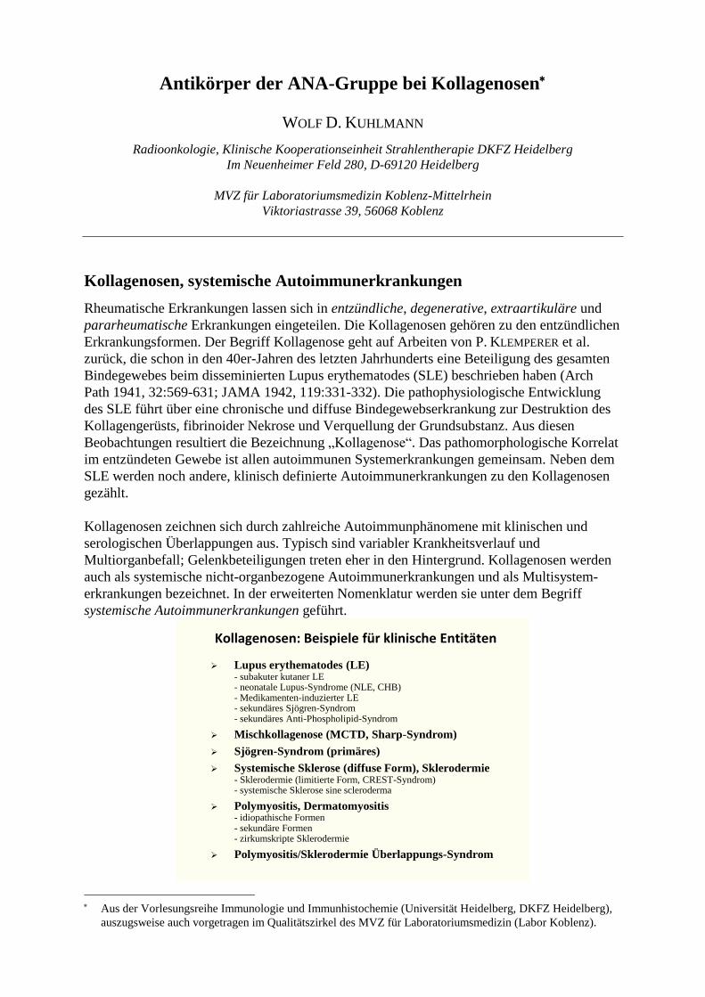
Antikörper der ANA-Gruppe bei Kollagenosen
WOLF D. KUHLMANN
Radioonkologie, Klinische Kooperationseinheit Strahlentherapie DKFZ Heidelberg
Im Neuenheimer Feld 280, D-69120 Heidelberg
MVZ für Laboratoriumsmedizin Koblenz-Mittelrhein
Viktoriastrasse 39, 56068 Koblenz
Kollagenosen, systemische Autoimmunerkrankungen
Rheumatische Erkrankungen lassen sich in entzündliche, degenerative, extraartikuläre und
pararheumatische Erkrankungen eingeteilen. Die Kollagenosen gehören zu den entzündlichen
Erkrankungsformen. Der Begriff Kollagenose geht auf Arbeiten von P. KLEMPERER et al.
zurück, die schon in den 40er-Jahren des letzten Jahrhunderts eine Beteiligung des gesamten
Bindegewebes beim disseminierten Lupus erythematodes (SLE) beschrieben haben (Arch
Path 1941, 32:569-631; JAMA 1942, 119:331-332). Die pathophysiologische Entwicklung
des SLE führt über eine chronische und diffuse Bindegewebserkrankung zur Destruktion des
Kollagengerüsts, fibrinoider Nekrose und Verquellung der Grundsubstanz. Aus diesen
Beobachtungen resultiert die Bezeichnung „Kollagenose“. Das pathomorphologische Korrelat
im entzündeten Gewebe ist allen autoimmunen Systemerkrankungen gemeinsam. Neben dem
SLE werden noch andere, klinisch definierte Autoimmunerkrankungen zu den Kollagenosen
gezählt.
Kollagenosen zeichnen sich durch zahlreiche Autoimmunphänomene mit klinischen und
serologischen Überlappungen aus. Typisch sind variabler Krankheitsverlauf und
Multiorganbefall; Gelenkbeteiligungen treten eher in den Hintergrund. Kollagenosen werden
auch als systemische nicht-organbezogene Autoimmunerkrankungen und als Multisystem-
erkrankungen bezeichnet. In der erweiterten Nomenklatur werden sie unter dem Begriff
systemische Autoimmunerkrankungen geführt.
Kollagenosen: Beispiele für klinische Entitäten
Lupus erythematodes (LE)- subakuter kutaner LE- neonatale Lupus-Syndrome (NLE, CHB)- Medikamenten-induzierter LE- sekundäres Sjögren-Syndrom- sekundäres Anti-Phospholipid-Syndrom
Mischkollagenose (MCTD, Sharp-Syndrom)
Sjögren-Syndrom (primäres)
Systemische Sklerose (diffuse Form), Sklerodermie- Sklerodermie (limitierte Form, CREST-Syndrom)- systemische Sklerose sine scleroderma
Polymyositis, Dermatomyositis- idiopathische Formen- sekundäre Formen- zirkumskripte Sklerodermie
Polymyositis/Sklerodermie Überlappungs-Syndrom
Aus der Vorlesungsreihe Immunologie und Immunhistochemie (Universität Heidelberg, DKFZ Heidelberg),
auszugsweise auch vorgetragen im Qualitätszirkel des MVZ für Laboratoriumsmedizin (Labor Koblenz).
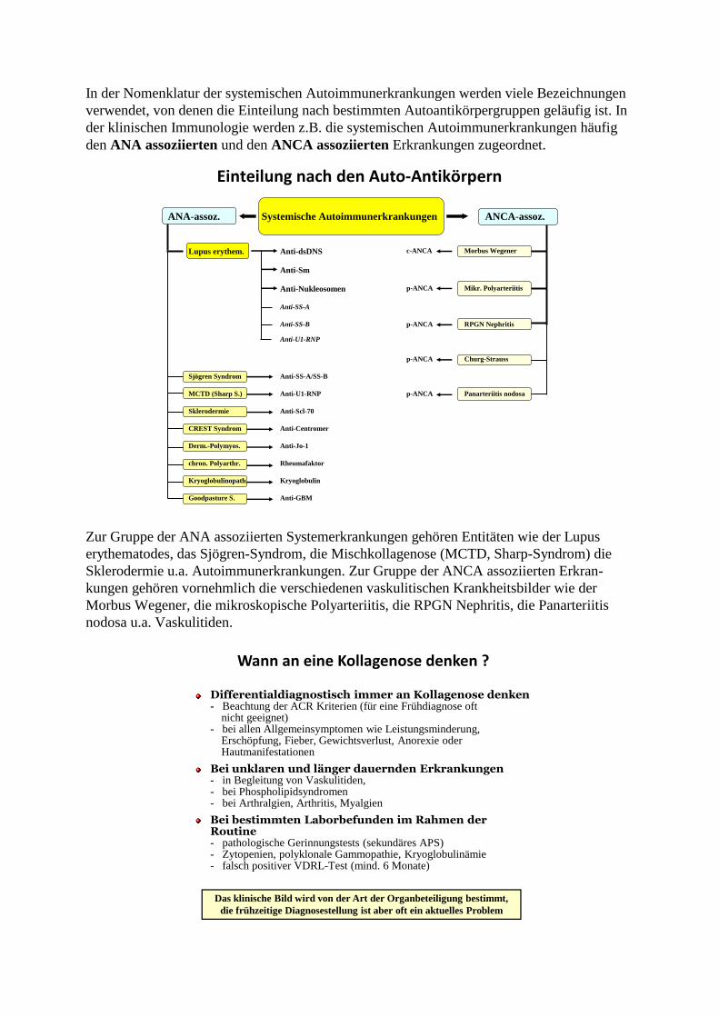
In der Nomenklatur der systemischen Autoimmunerkrankungen werden viele Bezeichnungen
verwendet, von denen die Einteilung nach bestimmten Autoantikörpergruppen geläufig ist. In
der klinischen Immunologie werden z.B. die systemischen Autoimmunerkrankungen häufig
den ANA assoziierten und den ANCA assoziierten Erkrankungen zugeordnet.
Einteilung nach den Auto-Antikörpern
ANA-assoz. Systemische Autoimmunerkrankungen ANCA-assoz.
Lupus erythem. Anti-dsDNS c-ANCA Morbus Wegener
Anti-Sm
Anti-Nukleosomen p-ANCA Mikr. Polyarteriitis
Anti-SS-A
Anti-SS-B p-ANCA RPGN Nephritis
Anti-U1-RNP
p-ANCA Churg-Strauss
Sjögren Syndrom Anti-SS-A/SS-B
MCTD (Sharp S.) Anti-U1-RNP p-ANCA Panarteriitis nodosa
Sklerodermie Anti-Scl-70
CREST Syndrom Anti-Centromer
Derm.-Polymyos. Anti-Jo-1
chron. Polyarthr. Rheumafaktor
Kryoglobulinopath. Kryoglobulin
Goodpasture S. Anti-GBM
Zur Gruppe der ANA assoziierten Systemerkrankungen gehören Entitäten wie der Lupus
erythematodes, das Sjögren-Syndrom, die Mischkollagenose (MCTD, Sharp-Syndrom) die
Sklerodermie u.a. Autoimmunerkrankungen. Zur Gruppe der ANCA assoziierten Erkran-
kungen gehören vornehmlich die verschiedenen vaskulitischen Krankheitsbilder wie der
Morbus Wegener, die mikroskopische Polyarteriitis, die RPGN Nephritis, die Panarteriitis
nodosa u.a. Vaskulitiden.
Differentialdiagnostisch immer an Kollagenose denken- Beachtung der ACR Kriterien (für eine Frühdiagnose oft
nicht geeignet) - bei allen Allgemeinsymptomen wie Leistungsminderung,
Erschöpfung, Fieber, Gewichtsverlust, Anorexie oderHautmanifestationen
Bei unklaren und länger dauernden Erkrankungen - in Begleitung von Vaskulitiden,- bei Phospholipidsyndromen - bei Arthralgien, Arthritis, Myalgien
Bei bestimmten Laborbefunden im Rahmen derRoutine- pathologische Gerinnungstests (sekundäres APS)- Zytopenien, polyklonale Gammopathie, Kryoglobulinämie- falsch positiver VDRL-Test (mind. 6 Monate)
Wann an eine Kollagenose denken ?
Das klinische Bild wird von der Art der Organbeteiligung bestimmt,
die frühzeitige Diagnosestellung ist aber oft ein aktuelles Problem
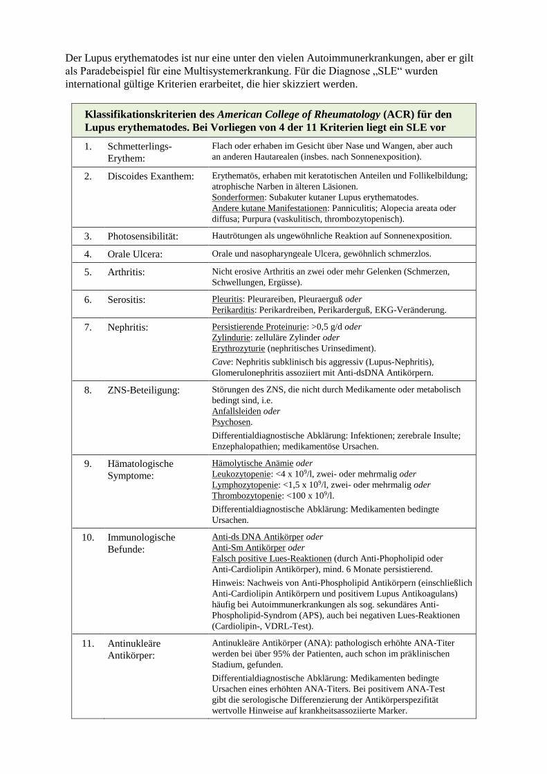
Der Lupus erythematodes ist nur eine unter den vielen Autoimmunerkrankungen, aber er gilt
als Paradebeispiel für eine Multisystemerkrankung. Für die Diagnose „SLE“ wurden
international gültige Kriterien erarbeitet, die hier skizziert werden.
Klassifikationskriterien des American College of Rheumatology (ACR) für den
Lupus erythematodes. Bei Vorliegen von 4 der 11 Kriterien liegt ein SLE vor
1. Schmetterlings-
Erythem:
Flach oder erhaben im Gesicht über Nase und Wangen, aber auch
an anderen Hautarealen (insbes. nach Sonnenexposition).
2. Discoides Exanthem: Erythematös, erhaben mit keratotischen Anteilen und Follikelbildung;
atrophische Narben in älteren Läsionen.
Sonderformen: Subakuter kutaner Lupus erythematodes.
Andere kutane Manifestationen: Panniculitis; Alopecia areata oder
diffusa; Purpura (vaskulitisch, thrombozytopenisch).
3. Photosensibilität: Hautrötungen als ungewöhnliche Reaktion auf Sonnenexposition.
4. Orale Ulcera: Orale und nasopharyngeale Ulcera, gewöhnlich schmerzlos.
5. Arthritis: Nicht erosive Arthritis an zwei oder mehr Gelenken (Schmerzen,
Schwellungen, Ergüsse).
6. Serositis: Pleuritis: Pleurareiben, Pleuraerguß oder
Perikarditis: Perikardreiben, Perikarderguß, EKG-Veränderung.
7. Nephritis: Persistierende Proteinurie: >0,5 g/d oder
Zylindurie: zelluläre Zylinder oder
Erythrozyturie (nephritisches Urinsediment).
Cave: Nephritis subklinisch bis aggressiv (Lupus-Nephritis),
Glomerulonephritis assoziiert mit Anti-dsDNA Antikörpern.
8. ZNS-Beteiligung: Störungen des ZNS, die nicht durch Medikamente oder metabolisch
bedingt sind, i.e.
Anfallsleiden oder
Psychosen.
Differentialdiagnostische Abklärung: Infektionen; zerebrale Insulte;
Enzephalopathien; medikamentöse Ursachen.
9. Hämatologische
Symptome:
Hämolytische Anämie oder
Leukozytopenie: <4 x 10⁹/l, zwei- oder mehrmalig oder
Lymphozytopenie: <1,5 x 10⁹/l, zwei- oder mehrmalig oder
Thrombozytopenie: <100 x 109/l.
Differentialdiagnostische Abklärung: Medikamenten bedingte
Ursachen.
10. Immunologische
Befunde:
Anti-ds DNA Antikörper oder
Anti-Sm Antikörper oder
Falsch positive Lues-Reaktionen (durch Anti-Phopholipid oder
Anti-Cardiolipin Antikörper), mind. 6 Monate persistierend.
Hinweis: Nachweis von Anti-Phospholipid Antikörpern (einschließlich
Anti-Cardiolipin Antikörpern und positivem Lupus Antikoagulans)
häufig bei Autoimmunerkrankungen als sog. sekundäres Anti-
Phospholipid-Syndrom (APS), auch bei negativen Lues-Reaktionen
(Cardiolipin-, VDRL-Test).
11. Antinukleäre
Antikörper:
Antinukleäre Antikörper (ANA): pathologisch erhöhte ANA-Titer
werden bei über 95% der Patienten, auch schon im präklinischen
Stadium, gefunden.
Differentialdiagnostische Abklärung: Medikamenten bedingte
Ursachen eines erhöhten ANA-Titers. Bei positivem ANA-Test
gibt die serologische Differenzierung der Antikörperspezifität
wertvolle Hinweise auf krankheitsassoziierte Marker.
Die systemischen Autoimmunerkrankungen werden in der Regel über serologische
Untersuchungsverfahren eingegrenzt, für die eine Reihe von definierten Antigen-Antikörper-
Systemen zur Verfügung steht. So ist beispielsweise für den systemischen Lupus
erythematodes die Bestimmung von Antikörpern gegen dsDNA ein besonders wichtiger
Marker. Auch für andere Kollagenosen gibt es geeignete Biomarker.
Antigene der ANA-Gruppe sind Polynukleotide, Proteine, Histone und Enzyme. Zahlreiche
Entitäten konnten biochemisch und immunologisch definiert werden, obwohl noch längst
nicht alle potentiellen Antigen-Antikörper-Systeme oder deren diagnostische Bedeutung
bekannt sind. Die wichtigsten ANA-Marker werden im Kapitel Autoimmune System-
erkrankungen beschrieben (http://www.immunologie-labor.com/service_files/fach_autoimmun.pdf).
Diagnostik antinukleärer Antikörper mit dem ANA-IFT
Die Ära des ANA-Immunfluoreszenztests (ANA-IFT) begann 1957 mit der ersten
Beschreibung eines indirekten Immunfluoreszenztests für den Nachweis von antinukleären
Antikörpern im Patientenserum (FRIOU G.J. 1957). Für Jahrzehnte kamen als Antigensubstrate
histologische Gefrierschnitte tierischer Organe zur Anwendung, die später dann durch
Zellkulturpräparate ersetzt wurden. Monolayer-Zellkulturpräparate von HEp-2 Zellen,
basierend auf einer kultivierbaren Tumor-Zelllinie (MOORE A.E. et al. 1955), zeichnen sich im
Vergleich zu den histologischen Tierpräparaten durch wesentlich einfachere Handhabung,
konstante Qualität (antigene Zusammensetzung) und deutlich bessere Reproduzierbarkeit des
ANA-Nachweises aus. HEp-2 Zellkulturpräparate sind noch immer Standard in der ANA-
Diagnostik. Nach wie vor ist der ANA-IFT das gebräuchlichste Verfahren für die
Eingangsdiagnostik bei Verdacht auf eine Erkrankung des rheumatischen Formenkreises.
Diese traditionelle Methode wird vom American College of Rheumatology als Goldstandard
für das ANA-Screening empfohlen.
Expertengruppen haben in den letzten Jahren Vorschläge zur Nomenklatur erarbeitet, die
darüber hinaus auch Empfehlungen zu diagnostischen Methoden enthalten. Die European
Autoimmunity Standardization Initiative (EASI) und die International Union of
Immunological Societies/World Health Organization/Arthritis Foundation/Centers for
Disease Control and Prevention (IUS/WHO/AF/CDC) mit einem Komittee für die
Standardisierung von Antikörpern in rheumatischen und verwandten Erkrankungen
(http://www.autoab.org) haben zu diesen Themen umfangreiche Vorschläge publiziert.
Zusätzlich haben Experten anlässlich der 12th International Workshops on Autoantibodies and
Autoimmunity (IWAA) in Sao Paulo (Brasilien) in einer speziellen Sitzung einen Konsens zur
ANA-Musterbeurteilung mittels Immunfluoreszenz (auf HEp-2-Zellen) erarbeitet. Der
zusammenfassende Bericht zum International Consensus on ANA staining Patterns (ICAP)
ist auf einer eigenen Webseite (www.ANApatterns.org) abrufbar.
Die neue Nomenklatur der ICAP segregiert die HEp-2-IFT-Muster in drei Hauptgruppen.
Definiert und kodiert wurden folgende Muster:
Nukleäre Muster (AC-1 bis AC-14),
Zytoplasmatische Muster (AC-15 bis AC-23),
Mitotische Muster (AC-24 bis AC-28).
Die Systematik umfasst insgesamt 28 IFT-Muster mit der Kodierung von AC-1 bis AC-28.
Der Nomenklatur und Klassifikationsbaum wird als farbige Boxen mit amberfarbigem und
oliv-grünem Hintergrund dargestellt (www.ANApatterns.org). Weitere Einzelheiten sind der
Arbeitsanweisung ANA-Immunfluoreszenz – Zellkern-Antikörper, andere HEp-2-Zell-
Antikörper beschrieben (http://www.immunologie-labor.com/service_files/fach_QM_SOP_ANA_IFT.pdf).
In den letzten Jahren wurden zahlreiche neue Methoden für die Detektion von antinukleären
Antikörpern entwickelt. Es handelt sich dabei um sog. Festphasen-Immunoassays, bei denen
unterschiedliche nukleäre Targets an Festphasen gekoppelt wurden (Mikrotiterplatten,
Latexpartikel, Membranen etc.) und mit Patientenserum inkubiert werden. Die Bindung der
entsprechend reagierenden Antikörper wird mit speziell markierten Detektionsantikörpern
sicht- bzw. messbar gemacht. Solche Immunassays werden z.B. als ELISA (Enzme-Linked-
Immuno-Sorbent-Assay) oder Fluoreszenz-Assays in unterschiedlichen Formaten gehandelt;
sog. Multiplex-Verfahren für die gleichzeitige Detektion einer Vielzahl von Liganden sind
attraktiv für den Hochdurchsatz von Patientenproben in kurzer Zeit.
In vielen Laboratorien werden aufgrund hoher Probenzahlen vermehrt Multiplex-Verfahren
favorisiert, die mit dem aufwendigen und mit hoher Expertise verbundenen mikroskopischen
ANA-IFT konkurrieren. Die Vorteile der mechanisierten Hochdurchsatzverfahren liegen
sicherlich in der schnellen Abarbeitung grosser Probenmengen. Sie haben aber den Nachteil
einer allgemein fehlenden Transparenz der gemessenen Zahlenwerte. Was bedeutet
beispielsweise ein „negatives“ Ergebnis bei nach wie vor bestehendem Verdacht auf eine
Kollagenose? Ist das Messergebnis wirklich „richtig negativ“ oder doch „falsch negativ“, weil
im Testansatz nicht das richtige Antigen angeboten wurde? Ausserdem fehlen bei den
ermittelten Zahlenwerten Hinweise auf evtl. wichtige Zusatzbefunde, die z.B. bei der
Mikroskopie eines HEp-2 Zellpräparates erhoben werden können. Im mikroskopischen HEp-2
Zellpräparat liegt eine Vielzahl von potentiell wichtigen Antigenen vor, die technisch in den
Festphasen-Assays in der gesamten Komplexität nicht abgebildet werden, aber bei der
Mikroskopie und bei entsprechender Expertise bewertet werden können.
Bei einem negativen ANA-IFT erübrigen sich in der Regel weitere Untersuchungen. Dennoch
hängt das weitere Vorgehen wesentlich von der klinischen Symptomatik ab. Ein positives
Testergebnis kann jedenfalls den Verdacht auf eine Kollagenose stützen. Die mikroskopische
Auswertung von positiven ANA-IFTs und deren Befundung ist an Erfahrung gebunden. Die
Erkennung von typischen IFT-Mustern ist bei der Vielfalt der sich ergebenden
Fluoreszenzbilder zuweilen auch für geübte Personen eine Herausforderung. Es reicht aber
nicht aus, den Blick nur auf ein positives IFT Ergebnis zu richten, weil ein negativer ANA-
IFT eine Kollagenose nicht mit Sicherheit ausschliesst. Es wird berichtet, dass bis zu 5% der
SLE-Patienten trotz erfüllter ACR-Kriterien ANA-negativ sein können.
Der ANA-IFT ist ein semiquantitativer Test. Der Test wird als negativ oder positiv beurteilt.
Es gibt keine exakte Grenze für einen ANA-Normalwert; der Normbereich bewegt sich
zwischen 1:40 und 1:160 (Schwellenwert), je nach Probenkollektiv. ANA-Titer in der
Grössenordnung von 1:160 und 1:320 sind wenig aussagekräftig und sollten in einem
zeitversetzten Ansatz kontrolliert werden. Es ist zu beachten, dass auch klinisch unauffällige
Personengruppen (vor allem altersabhängig) kritische ANA-Titer aufweisen können. Hoch-
titrige ANA-Werte (ab 1:320) sind sicherlich als verdächtig zu werten. Sie bedürfen jedenfalls
der Kontrolle und ggf. auch der weiteren Abklärung durch gezielte Bestätigungstests.
Bei einer positiven ANA-Reaktion muss immer die Patientenprobe titriert werden (serielle
Verdünnungsanalyse), um einen groben Hinweis auf die Höhe der Antikörperkonzentration zu
erhalten. Das Ergebnis der Titerendstufe (Titerhöhe) wird protokolliert und zusammen mit der
Art des Fluoreszenzmusters als Ergebnis berichtet. Unterschiedliche IFT-Muster reflektieren
das Reaktionsverhalten unterschiedlicher Antikörper-Entitäten mit dem Gewebe und geben
differentialdiagnostische Hinweise auf mögliche Antigenspezifitäten. Dies ist für den
anfordernden Arzt wichtig zu wissen. Aus diesem Grund muss das Untersuchungslabor
darüber informiren, welches Zellantigen dem IFT-Muster zugrunde liegen kann und
Vorschläge zur weiteren Differenzierung empfehlen.
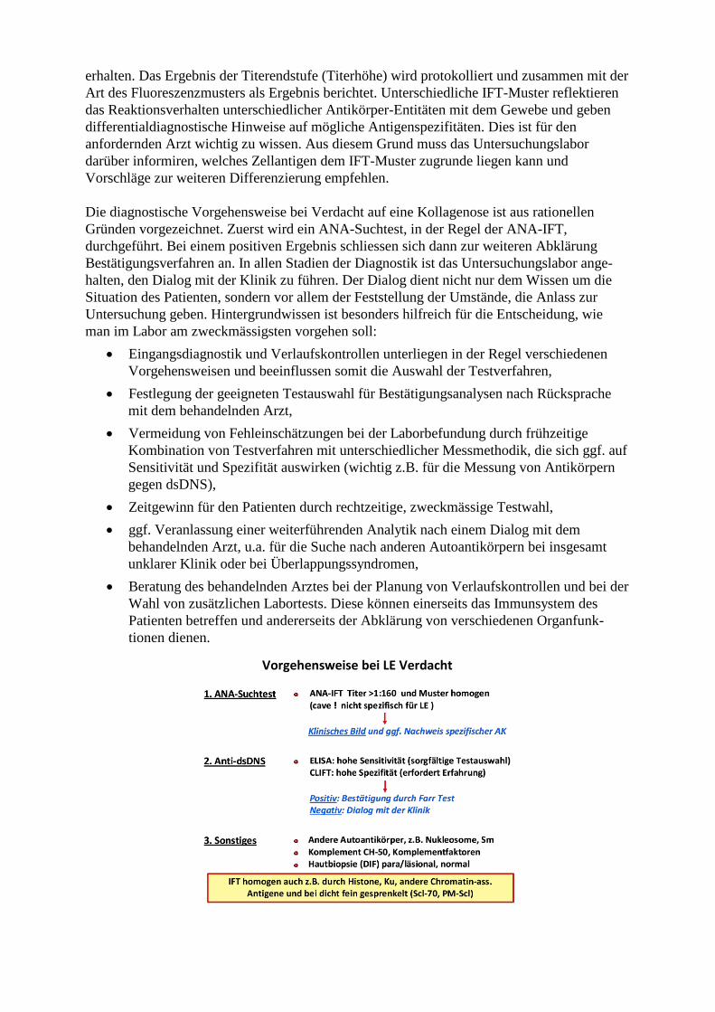
Die diagnostische Vorgehensweise bei Verdacht auf eine Kollagenose ist aus rationellen
Gründen vorgezeichnet. Zuerst wird ein ANA-Suchtest, in der Regel der ANA-IFT,
durchgeführt. Bei einem positiven Ergebnis schliessen sich dann zur weiteren Abklärung
Bestätigungsverfahren an. In allen Stadien der Diagnostik ist das Untersuchungslabor ange-
halten, den Dialog mit der Klinik zu führen. Der Dialog dient nicht nur dem Wissen um die
Situation des Patienten, sondern vor allem der Feststellung der Umstände, die Anlass zur
Untersuchung geben. Hintergrundwissen ist besonders hilfreich für die Entscheidung, wie
man im Labor am zweckmässigsten vorgehen soll:
Eingangsdiagnostik und Verlaufskontrollen unterliegen in der Regel verschiedenen
Vorgehensweisen und beeinflussen somit die Auswahl der Testverfahren,
Festlegung der geeigneten Testauswahl für Bestätigungsanalysen nach Rücksprache
mit dem behandelnden Arzt,
Vermeidung von Fehleinschätzungen bei der Laborbefundung durch frühzeitige
Kombination von Testverfahren mit unterschiedlicher Messmethodik, die sich ggf. auf
Sensitivität und Spezifität auswirken (wichtig z.B. für die Messung von Antikörpern
gegen dsDNS),
Zeitgewinn für den Patienten durch rechtzeitige, zweckmässige Testwahl,
ggf. Veranlassung einer weiterführenden Analytik nach einem Dialog mit dem
behandelnden Arzt, u.a. für die Suche nach anderen Autoantikörpern bei insgesamt
unklarer Klinik oder bei Überlappungssyndromen,
Beratung des behandelnden Arztes bei der Planung von Verlaufskontrollen und bei der
Wahl von zusätzlichen Labortests. Diese können einerseits das Immunsystem des
Patienten betreffen und andererseits der Abklärung von verschiedenen Organfunk-
tionen dienen.
Vorgehensweise bei LE Verdacht
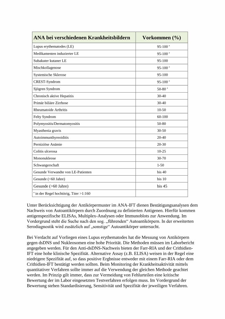
ANA bei verschiedenen Krankheitsbildern Vorkommen (%)
Lupus erythematodes (LE) 95-100
Medikamenten induzierter LE 95-100
Subakuter kutaner LE 95-100
Mischkollagenose 95-100
Systemische Sklerose 95-100
CREST-Syndrom 95-100
Sjögren Syndrom 50-80
Chronisch aktive Hepatitis 30-40
Primär biliäre Zirrhose 30-40
Rheumatoide Arthritis 10-50
Felty Syndrom 60-100
Polymyositis/Dermatomyositis 50-80
Myasthenia gravis 30-50
Autoimmunthyreoiditis 20-40
Perniziöse Anämie 20-30
Colitis ulcerosa 10-25
Mononukleose 30-70
Schwangerschaft 1-50
Gesunde Verwandte von LE-Patienten bis 40
Gesunde (<60 Jahre) bis 10
Gesunde (>60 Jahre) bis 45
in der Regel hochtitrig, Titer >1:160
Unter Berücksichtigung der Antikörpermuster im ANA-IFT dienen Bestätigungsanalysen dem
Nachweis von Autoantikörpern durch Zuordnung zu definierten Antigenen. Hierfür kommen
antigenspezifische ELISAs, Multiplex-Analysen oder Immunoblots zur Anwendung. Im
Vordergrund steht die Suche nach den sog. „führenden“ Autoantikörpern. In der erweiterten
Serodiagnostik wird zusätzlich auf „sonstige“ Autoantikörper untersucht.
Bei Verdacht auf Vorliegen eines Lupus erythematodes hat die Messung von Antikörpern
gegen dsDNS und Nukleosomen eine hohe Priorität. Die Methoden müssen im Laborbericht
angegeben werden. Für den Anti-dsDNS-Nachweis bieten der Farr-RIA und der Crithidien-
IFT eine hohe klinische Spezifität. Alternative Assay (z.B. ELISA) weisen in der Regel eine
niedrigere Spezifität auf, so dass positive Ergbnisse entweder mit einem Farr-RIA oder dem
Crithidien-IFT bestätigt werden sollten. Beim Monitoring der Krankheitsaktivität mittels
quantitativer Verfahren sollte immer auf die Verwendung der gleichen Methode geachtet
werden. Im Prinzip gilt immer, dass zur Vermeidung von Fehlurteilen eine kritische
Bewertung der im Labor eingesetzten Testverfahren erfolgen muss. Im Vordergrund der
Bewertung stehen Standardisierung, Sensitivität und Spezifität der jeweiligen Verfahren.
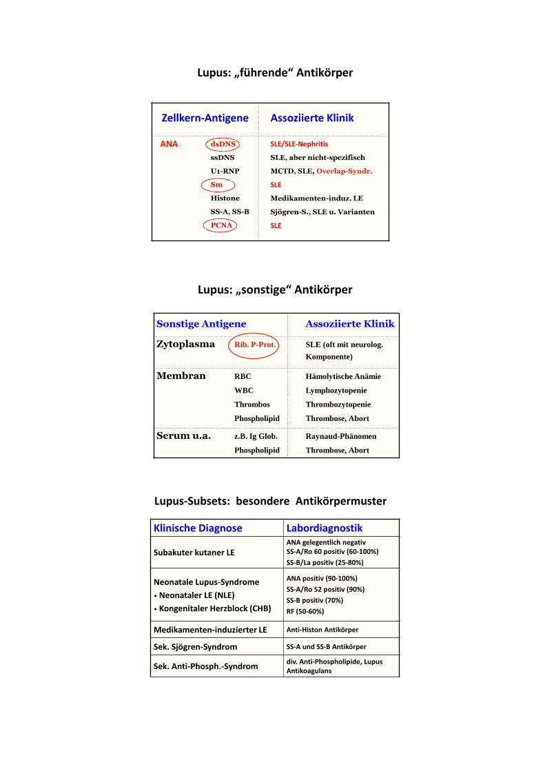
Lupus: „führende“ Antikörper
Zellkern-Antigene Assoziierte Klinik
ANA dsDNS
ssDNS
U1-RNP
Sm
Histone
SS-A, SS-B
PCNA
SLE/SLE-Nephritis
SLE, aber nicht-spezifisch
MCTD, SLE, Overlap-Syndr.
SLE
Medikamenten-induz. LE
Sjögren-S., SLE u. Varianten
SLE
Lupus: „sonstige“ Antikörper
Sonstige Antigene Assoziierte Klinik
Zytoplasma Rib. P-Prot. SLE (oft mit neurolog.
Komponente)
Membran RBC
WBC
Thrombos
Phospholipid
Hämolytische Anämie
Lymphozytopenie
Thrombozytopenie
Thrombose, Abort
Serum u.a. z.B. Ig Glob.
Phospholipid
Raynaud-Phänomen
Thrombose, Abort
Lupus-Subsets: besondere Antikörpermuster
Klinische Diagnose Labordiagnostik
Subakuter kutaner LEANA gelegentlich negativSS-A/Ro 60 positiv (60-100%)
SS-B/La positiv (25-80%)
Neonatale Lupus-Syndrome
• Neonataler LE (NLE)
• Kongenitaler Herzblock (CHB)
ANA positiv (90-100%)
SS-A/Ro 52 positiv (90%)
SS-B positiv (70%)
RF (50-60%)
Medikamenten-induzierter LE Anti-Histon Antikörper
Sek. Sjögren-Syndrom SS-A und SS-B Antikörper
Sek. Anti-Phosph.-Syndromdiv. Anti-Phospholipide, Lupus Antikoagulans
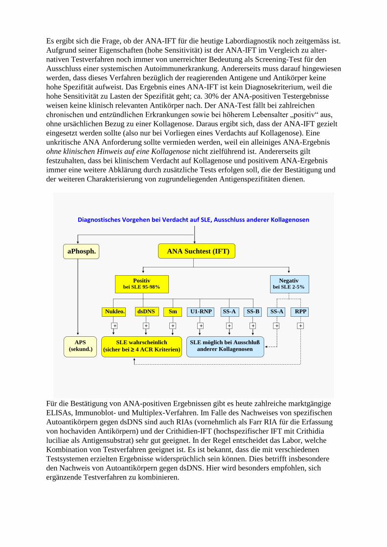
Es ergibt sich die Frage, ob der ANA-IFT für die heutige Labordiagnostik noch zeitgemäss ist.
Aufgrund seiner Eigenschaften (hohe Sensitivität) ist der ANA-IFT im Vergleich zu alter-
nativen Testverfahren noch immer von unerreichter Bedeutung als Screening-Test für den
Ausschluss einer systemischen Autoimmunerkrankung. Andererseits muss darauf hingewiesen
werden, dass dieses Verfahren bezüglich der reagierenden Antigene und Antikörper keine
hohe Spezifität aufweist. Das Ergebnis eines ANA-IFT ist kein Diagnosekriterium, weil die
hohe Sensitivität zu Lasten der Spezifität geht; ca. 30% der ANA-positiven Testergebnisse
weisen keine klinisch relevanten Antikörper nach. Der ANA-Test fällt bei zahlreichen
chronischen und entzündlichen Erkrankungen sowie bei höherem Lebensalter „positiv“ aus,
ohne ursächlichen Bezug zu einer Kollagenose. Daraus ergibt sich, dass der ANA-IFT gezielt
eingesetzt werden sollte (also nur bei Vorliegen eines Verdachts auf Kollagenose). Eine
unkritische ANA Anforderung sollte vermieden werden, weil ein alleiniges ANA-Ergebnis
ohne klinischen Hinweis auf eine Kollagenose nicht zielführend ist. Andererseits gilt
festzuhalten, dass bei klinischem Verdacht auf Kollagenose und positivem ANA-Ergebnis
immer eine weitere Abklärung durch zusätzliche Tests erfolgen soll, die der Bestätigung und
der weiteren Charakterisierung von zugrundeliegenden Antigenspezifitäten dienen.
Diagnostisches Vorgehen bei Verdacht auf SLE, Ausschluss anderer Kollagenosen
aPhosph. ANA Suchtest (IFT)
Positiv bei SLE 95-98%
Negativ bei SLE 2-5%
Nukleo. dsDNS Sm U1-RNP SS-A SS-B SS-A RPP
+ + + + + + + +
APS
(sekund.)
SLE wahrscheinlich
(sicher bei 4 ACR Kriterien)
SLE möglich bei Ausschluß
anderer Kollagenosen
Für die Bestätigung von ANA-positiven Ergebnissen gibt es heute zahlreiche marktgängige
ELISAs, Immunoblot- und Multiplex-Verfahren. Im Falle des Nachweises von spezifischen
Autoantikörpern gegen dsDNS sind auch RIAs (vornehmlich als Farr RIA für die Erfassung
von hochaviden Antikörpern) und der Crithidien-IFT (hochspezifischer IFT mit Crithidia
luciliae als Antigensubstrat) sehr gut geeignet. In der Regel entscheidet das Labor, welche
Kombination von Testverfahren geeignet ist. Es ist bekannt, dass die mit verschiedenen
Testsystemen erzielten Ergebnisse widersprüchlich sein können. Dies betrifft insbesondere
den Nachweis von Autoantikörpern gegen dsDNS. Hier wird besonders empfohlen, sich
ergänzende Testverfahren zu kombinieren.
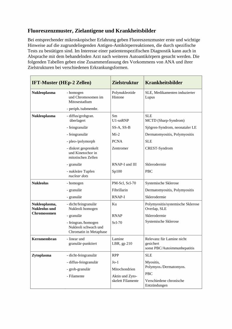
Fluoreszenzmuster, Zielantigene und Krankheitsbilder
Bei entsprechender mikroskopischer Erfahrung geben Fluoreszenzmuster erste und wichtige
Hinweise auf die zugrundeliegenden Antigen-Antikörperreaktionen, die durch spezifische
Tests zu bestätigen sind. Im Interesse einer patientenspezifischen Diagnostik kann auch in
Absprache mit dem behandelnden Arzt nach weiteren Autoantikörpern gesucht werden. Die
folgenden Tabellen geben eine Zusammenfassung des Vorkommens von ANA und ihrer
Zielstrukturen bei verschiedenen Erkrankungsformen.
IFT-Muster (HEp-2 Zellen) Zielstruktur Krankheitsbilder
Nukleoplasma - homogen
und Chromosomen im
Mitosestadium
- periph./submembr.
Polynukleotide
Histone
SLE, Medikamenten induzierter
Lupus
Nukleoplasma - diffus/grobgran.
überlagert
- feingranulär
- feingranulär
- pleo-/polymorph
- diskret gesprenkelt
und Kinetochor in
mitotischen Zellen
- granulär
- nukleäre Tupfen
nuclear dots
Sm
U1-snRNP
SS-A, SS-B
Mi-2
PCNA
Zentromer
RNAP-I und III
Sp100
SLE
MCTD (Sharp-Syndrom)
Sjögren-Syndrom, neonataler LE
Dermatomyositis, Polymyositis
SLE
CREST-Syndrom
Sklerodermie
PBC
Nukleolus - homogen
- granulär
- granulär
PM-Scl, Scl-70
Fibrillarin
RNAP-I
Systemische Sklerose
Dermatomyositis, Polymyositis
Sklerodermie
Nukleoplasma,
Nukleolus und
Chromosomen
- dicht/feingranulär
Nukleoli homogen
- granulär
- feingran./homogen
Nukleoli schwach und
Chromatin in Metaphase
Ku
RNAP
Scl-70
Polymyositis/systemische Sklerose
Overlap, SLE
Sklerodermie
Systemische Sklerose
Kernmembran - linear und
granulär-punktiert
Lamine
LBR, gp 210
Relevanz für Lamine nicht
gesichert
sonst PBC/Autoimmunhepatitis
Zytoplasma - dicht-feingranulär
- diffus-feingranulär
- grob-granulär
- Filamente
RPP
Jo-1
Mitochondrien
Aktin und Zyto-
skelett Filamente
SLE
Myositis,
Polymyos./Dermatomyos.
PBC
Verschiedene chronische
Entzündungen
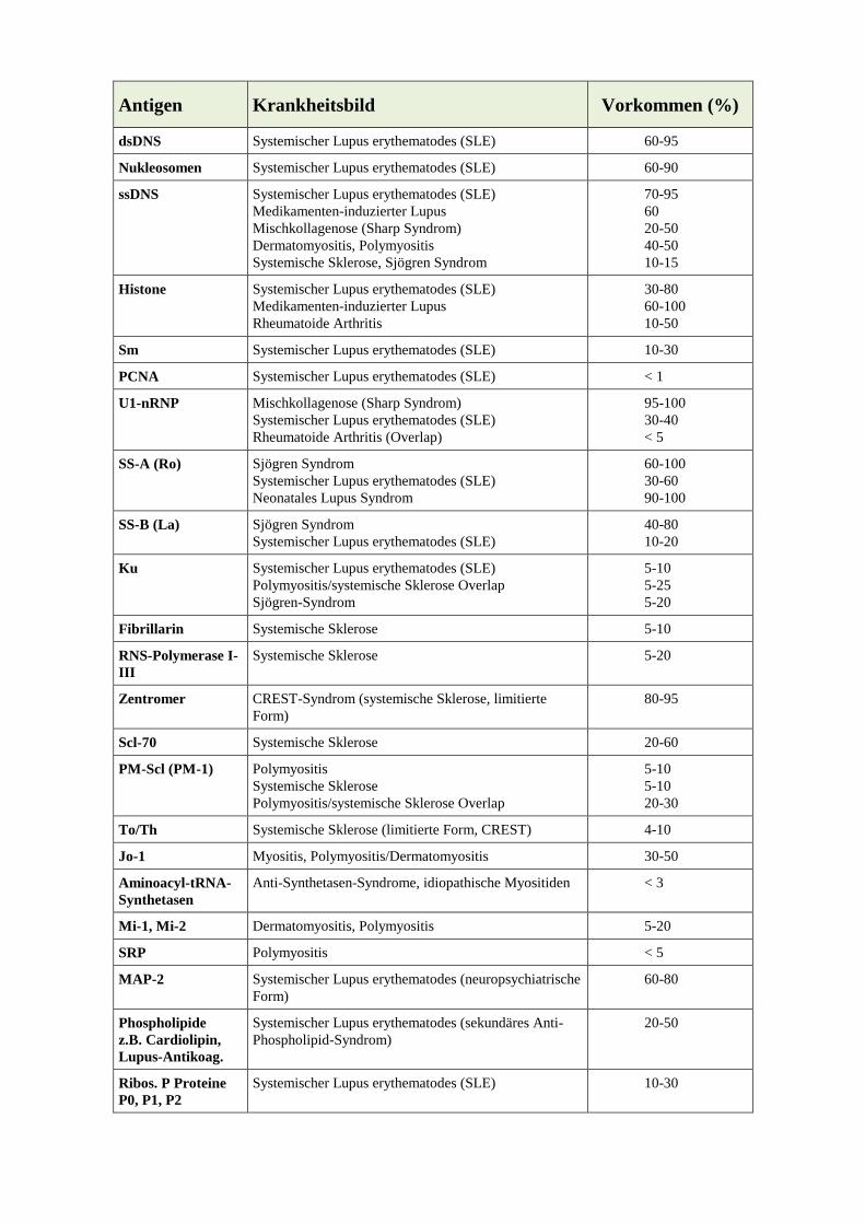
Antigen Krankheitsbild Vorkommen (%)
dsDNS Systemischer Lupus erythematodes (SLE) 60-95
Nukleosomen Systemischer Lupus erythematodes (SLE) 60-90
ssDNS Systemischer Lupus erythematodes (SLE)
Medikamenten-induzierter Lupus
Mischkollagenose (Sharp Syndrom)
Dermatomyositis, Polymyositis
Systemische Sklerose, Sjögren Syndrom
70-95
60
20-50
40-50
10-15
Histone Systemischer Lupus erythematodes (SLE)
Medikamenten-induzierter Lupus
Rheumatoide Arthritis
30-80
60-100
10-50
Sm Systemischer Lupus erythematodes (SLE) 10-30
PCNA Systemischer Lupus erythematodes (SLE) < 1
U1-nRNP Mischkollagenose (Sharp Syndrom)
Systemischer Lupus erythematodes (SLE)
Rheumatoide Arthritis (Overlap)
95-100
30-40
< 5
SS-A (Ro) Sjögren Syndrom
Systemischer Lupus erythematodes (SLE)
Neonatales Lupus Syndrom
60-100
30-60
90-100
SS-B (La) Sjögren Syndrom
Systemischer Lupus erythematodes (SLE)
40-80
10-20
Ku Systemischer Lupus erythematodes (SLE)
Polymyositis/systemische Sklerose Overlap
Sjögren-Syndrom
5-10
5-25
5-20
Fibrillarin Systemische Sklerose 5-10
RNS-Polymerase I-
III
Systemische Sklerose 5-20
Zentromer CREST-Syndrom (systemische Sklerose, limitierte
Form)
80-95
Scl-70 Systemische Sklerose 20-60
PM-Scl (PM-1) Polymyositis
Systemische Sklerose
Polymyositis/systemische Sklerose Overlap
5-10
5-10
20-30
To/Th Systemische Sklerose (limitierte Form, CREST) 4-10
Jo-1 Myositis, Polymyositis/Dermatomyositis 30-50
Aminoacyl-tRNA-
Synthetasen
Anti-Synthetasen-Syndrome, idiopathische Myositiden < 3
Mi-1, Mi-2 Dermatomyositis, Polymyositis 5-20
SRP Polymyositis < 5
MAP-2 Systemischer Lupus erythematodes (neuropsychiatrische
Form)
60-80
Phospholipide
z.B. Cardiolipin,
Lupus-Antikoag.
Systemischer Lupus erythematodes (sekundäres Anti-
Phospholipid-Syndrom)
20-50
Ribos. P Proteine
P0, P1, P2
Systemischer Lupus erythematodes (SLE) 10-30
Überlappungssyndrome
Patienten zeigen oft die klinischen Erscheinungen von mehr als einer definierten Kollagenose,
d.h. im Krankheitsverlauf kommt es zu Änderungen des klinischen Bildes, die dann auch von
Änderungen des Labors begleitet werden. Als anerkannte Überlappungssyndrome gelten:
- MCTD,
- „Rhupus“,
- Polymyositis-Sklerodermie Überlappung,
- SLE-Myositis Überlappung,
- SLE-Sjögren Überlappung.
Überlappungs-Syndrome von Kollagenosen
MCTD wird vielfach nicht als wirklich eigenständige klinische Entität betrachtet; MCTD kann sich
zum SLE und jede andere typische Kollagenose entwickeln.
Rhupus, eine Form des LE mit aggressiver Arthritis, i.e. dem eigentlich typischen Zeichen der rheuma-
toiden Arthritis, aber mit anderen typischen Lupus Zeichen; ungeklärt, ob echtes Overlap-Syndrom
vorliegt.
Polymyositis-Sklerodermie Überlappung: Bilder einer Polymyositis und Sklerodermie bei milder
Myositis.
SLE-Myositis Überlappung: klinisch wie SLE mit deutlicher Myositis.
SLE-Sjögren Überlappung: SLE typische Zeichen (Klinik und Labor), zusätzlich Antikörper gegen
SS-A und SS-B und sekundärem Sicca-Syndrom; neonataler LE mit kongenitalem Herzblock.
Labor (Screening)
s. jeweilige Kollagenose
Labor (ergänzend)
ANA, Anti-nDNS, Anti-Sm, Anti-U1-nRNP, Anti-
SS-A/SS-B, Rheumafaktor, Anti-Scl-70/PM-
Scl/Ku, Kryoglobuline
Raynaud Phänomen
Eine Reihe von Erkrankungen unterschiedlicher Genese führt zu einem Bild, das als Raynaud
Phänomen beschrieben wird. Etwa jeder fünfte Patient mit einer Raynaud Symptomatik hat
eine Grunderkrankung, vornehmlich eine Kollagenose oder eine andere Autoimmunerkran-
kung. Das Raynaud Phänomen ist gekennzeichnet durch lang anhaltende Vasospasmen der
Finger (meist ausgelöst durch Kälte, Stress oder Tragen von Gegenständen). Dabei werden
drei Farb-Phasen unterschieden: zuerst werden die Finger weiß und kalt, dann zyanotisch
(kann in leichten Fällen fehlen), und schließlich färben sich die Finger während der Reperfu-
sion rot. Meist sind ein bis drei Finger betroffen, der Daumen bleibt allgemein ausgespart.
Selten können auch andere Akren wie Zehen, Nase oder Ohren betroffen sein.
Man unterscheidet eine primäre Form des Raynaud-Syndroms (idiopathischer Raynaud), bei
der sich keine Grundkrankheiten finden lassen, von einem sekundären Raynaud. Der primä-
re Raynaud verläuft milder als die sekundären Formen. Der primäre Raynaud manifestiert sich
oft in der zweiten und dritten Lebensdekade, der sekundäre Raynaud meist erst im höheren
Alter. Frauen sind insgesamt häufiger betroffen als Männer. Der sekundäre Raynaud kann mit
verschiedenen Kollagenosen, endokrinologischen Erkrankungen, Malignomen, Infektionen,
Traumen oder mit medikamentöser Therapie assoziiert sein.
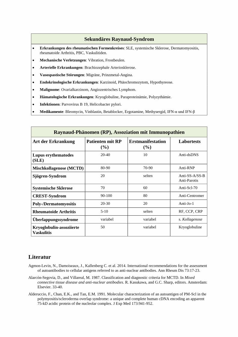
Sekundäres Raynaud-Syndrom
Erkrankungen des rheumatischen Formenkreises: SLE, systemische Sklerose, Dermatomyositis,
rheumatoide Arthritis, PBC, Vaskulitiden.
Mechanische Verletzungen: Vibration, Frostbeulen.
Arterielle Erkrankungen: Brachiozephale Arteriosklerose.
Vasospastische Störungen: Migräne, Prinzmetal-Angina.
Endokrinologische Erkrankungen: Karzinoid, Phäochromozytom, Hypothyreose.
Malignome: Ovarialkarzinom, Angiozentrisches Lymphom.
Hämatologische Erkrankungen: Kryoglobuline, Paraproteinämie, Polyzythämie.
Infektionen: Parvovirus B 19, Helicobacter pylori.
Medikamente: Bleomycin, Vinblastin, Betablocker, Ergotamine, Methysergid, IFN-α und IFN-β
Raynaud-Phänomen (RP), Assoziation mit Immunopathien
Art der Erkrankung Patienten mit RP
(%)
Erstmanifestation
(%)
Labortests
Lupus erythematodes
(SLE)
20-40 10 Anti-dsDNS
Mischkollagenose (MCTD) 80-90 70-90 Anti-RNP
Sjögren-Syndrom 20 selten Anti-SS-A/SS-B
Anti-Parotis
Systemische Sklerose 70 60 Anti-Scl-70
CREST-Syndrom 90-100 80 Anti-Centromer
Poly-/Dermatomyositis 20-30 20 Anti-Jo-1
Rheumatoide Arthritis 5-10 selten RF, CCP, CRP
Überlappungssyndrome variabel variabel s. Kollagenose
Kryoglobulin-assoziierte
Vaskulitis
50 variabel Kryoglobuline
Literatur
Agmon-Levin, N., Damoiseaux, J., Kallenberg C. et al. 2014. International recommendations for the assessment
of autoantibodies to cellular antigens referred to as anti-nuclear antibodies. Ann Rheum Dis 73:17-23.
Alarcón-Segovia, D., and Villareal, M. 1987. Classification and diagnostic criteria for MCTD. In Mixed
connective tissue disease and anti-nuclear antibodies. R. Kasukawa, and G.C. Sharp, editors. Amsterdam:
Elsevier. 33-40.
Alderuccio, F., Chan, E.K., and Tan, E.M. 1991. Molecular characterization of an autoantigen of PM-Scl in the
polymyositis/scleroderma overlap syndrome: a unique and complete human cDNA encoding an apparent
75-kD acidic protein of the nucleolar complex. J Exp Med 173:941-952.
Alexander, E.L., McNicholl, J., Watson, R.M., Bias, W., Reichlin, M., and Provost, T.T. 1989. The
immunogenetic relationship between anti-Ro(SS-A)/La(SS-B) antibody positive Sjogren's/lupus
erythematosus overlap syndrome and the neonatal lupus syndrome. J Invest Dermatol 93:751-756.
Alspaugh, M., and Maddison, P. 1979. Resolution of the identity of certain antigen-antibody systems in systemic
lupus erythematosus and Sjogren's syndrome: an interlaboratory collaboration. Arthritis Rheum 22:796-
798.
Alspaugh, M.A., and Tan, E.M. 1975. Antibodies to cellular antigens in Sjogren's syndrome. J Clin Invest
55:1067-1073.
American College of Rheumatology 2009. Position statement. Methodology of testing for antinuclear antibodies.
Internet: http://www.rheumatology.org/practice/clinical/position/ana_position_stmt.pdf
Amoura, Z., Piette, J.C., Bach, J.F., and Koutouzov, S. 1999. The key role of nucleosomes in lupus. Arthritis
Rheum 42:833-843.
Andrade, L.E., Chan, E.K., Peebles, C.L., and Tan, E.M. 1996. Two major autoantigen-antibody systems of the
mitotic spindle apparatus. Arthritis Rheum 39:1643-1653.
Andrade, L.E., Chan, E.K., Raska, I., Peebles, C.L., Roos, G., and Tan, E.M. 1991. Human autoantibody to a
novel protein of the nuclear coiled body: immunological characterization and cDNA cloning of p80-coilin.
J Exp Med 173:1407-1419.
Antico, A, Platzgummer, S., Bassetti, D., Bizzaro, N., Tozzoli, R., Villalta, D., and Study Group on Autoimmune
Diseases of the Italian Society of Laboratory Medicine. 2010. Diagnosing systemic lupus erythematosus:
new-generation immunoassays for measurement of anti-dsDNA antibodies are an effective alternative to the
Farr technique and the Crithidia luciliae immunofluorescence test. Lupus 19:906-912.
Arnett, F.C., Hamilton, R.G., Reveille, J.D., Bias, W.B., Harley, J.B., and Reichlin, M. 1989. Genetic studies of
Ro (SS-A) and La (SS-B) autoantibodies in families with systemic lupus erythematosus and primary
Sjogren's syndrome. Arthritis Rheum 32:413-419.
Arnett, F.C., Hamilton, R.G., Roebber, M.G., Harley, J.B., and Reichlin, M. 1988. Increased frequencies of Sm
and nRNP autoantibodies in American blacks compared to whites with systemic lupus erythematosus. J
Rheumatol 15:1773-1776.
Arnett, F.C., Reveille, J.D., and Valdez, B.C. 1997. Autoantibodies to a nucleolar RNA helicase protein in
patients with connective tissue diseases. Arthritis Rheum 40:1487-1492.
Auer-Grumbach, P., and Achleitner, B. 1994. Epidemiology and clinical associations of NuMA (nuclear mitotic
apparatus protein) autoantibodies. J Rheumatol 21:1779-1781.
Avaniss-Aghajani, E., Berzon, S., and Sarkissian, A. 2007. Clinical value of multiplexed bead-based
immunoassays for detection of autoantibodies to nuclear antigens. Clin Vaccine Immunol 14:505-509.
Bachmann, M., Mayet, W.J., Schroder, H.C., Pfeifer, K., Meyer zum Buschenfelde, K.H., and Muller, W.E.
1986. Association of La and Ro antigens with intracellular structures in HEp-2 carcinoma cells. Proc Natl
Acad Sci USA 83:7770-7774.
Bachmann, M., Pfeifer, K., Schroder, H.C., and Muller, W.E. 1990. Characterization of the autoantigen La as a
nucleic acid-dependent ATPase/dATPase with melting properties. Cell 60:85-93.
Bauer, R., and Schutz, R. 1979. [The LE cell phenomenon. New aspects in molecular biology and
immunocytology]. Arch Dermatol Res 266:197-204.
Beck, J.S. 1961. Variations in the morphological patterns of "autoimmune" nuclear fluorescence. Lancet 1:1203-
1205.
Ben-Chetrit, E. 1993. The molecular basis of the SSA/Ro antigens and the clinical significance of their
autoantibodies. Br J Rheumatol 32:396-402.
Bernstein, R.M., Hobbs, R.N., Lea, D.J., Ward, D.J., and Hughes, G.R. 1985. Patterns of antihistone antibody
specificity in systemic rheumatic disease. I Systemic lupus erythematosus, mixed connective tissue disease,
primary sicca syndrome, and rheumatoid arthritis with vasculitis. Arthritis Rheum 28:285-293.
Bernstein, R.M., Steigerwald, J.C., and Tan, E.M. 1982. Association of antinuclear and antinucleolar antibodies
in progressive systemic sclerosis. Clin Exp Immunol 48:43-51.
Bizzaro, N., Tozzoli, R., Tonutti, E., Piazza, A., Manoni, F., Ghirardello, A., Bassetti, D., Villalta, D., Pradella,
M., and Rizzotti, P. 1998. Variability between methods to determine ANA, anti-dsDNA and Anti-ENA
autoantibodies: a collaborative study with the biomdical industry. J Immunol Methods 219:99-107.
Bohan, A., and Peter, J.B. 1975. Polymyositis and dermatomyositis (second of two parts). N Engl J Med
292:403-407.
Bohan, A., and Peter, J.B. 1975. Polymyositis and dermatomyositis (first of two parts). N Engl J Med 292:344-
347.
Bonilla, E., Francis, L., Allam, F., Ogrinc, M., Neupane, H., Phillips, P.E., and Perl, A. 2007.
Immunofluorescence microscopy is superior to fluorescent beads for detection of antinuclear antibody
reactivity in systemic lupus erythematosus patients. Clin Immunol 124:18-21.
Bradley, J, and McCluskey, J. 1997. Clinical Immunology. New York: Oxford Press.
Brouwer, R., Vree Egberts, W.T., Hengstman, G.J., Raijmakers, R., van Engelen, B.G., Seelig, H.P., Renz, M.,
Mierau, R., Genth, E., Pruijn, G.J., et al. 2002. Autoantibodies directed to novel components of the PM/Scl
complex, the human exosome. Arthritis Res 4:134-138.
Buyon, J.P. 1992. Neonatal lupus syndromes. Am J Reprod Immunol 28:259-263.
Bylund, D.J., and Nakamura, R.M. 1991. Importance of detection of SS-A/Ro autoantibody in screening
immunofluorescence tests for autoantibodies to nuclear antigens. J Clin Lab Anal 5:212-218.
Cabral, A.R., and Alarcon-Segovia, D. 1998. Autoantibodies in systemic lupus erythematosus. Curr Opin
Rheumatol 10:409-416.
Casiano, C.A., Humbel, R.L., Peebles, C., Covini, G., and Tan, E.M. 1995. Autoimmunity to the cell cycle-
dependent centromere protein p330d/CENP-F in disorders associated with cell proliferation. J Autoimmun
8:575-586.
Casiano, C.A., and Tan, E.M. 1996. Recent developments in the understanding of antinuclear autoantibodies. Int
Arch Allergy Immunol 111:308-313.
Ceppellini, R., Polli, E., and Celada, F. 1957. A DNA-reacting factor in serum of a patient with lupus
erythematosus diffusus. Proc Soc Exp Biol Med 96:572-574.
Chan, E.K., Imai, H., Hamel, J.C., and Tan, E.M. 1991. Human autoantibody to RNA polymerase I transcription
factor hUBF. Molecular identity of nucleolus organizer region autoantigen NOR-90 and ribosomal RNA
transcription upstream binding factor. J Exp Med 174:1239-1244.
Chan, E.K., Tan, E.M., Ward, D.C., and Matera, A.G. 1994. Human 60-kDa SS-A/Ro ribonucleoprotein
autoantigen gene (SSA2) localized to 1q31 by fluorescence in situ hybridization. Genomics 23:298-300.
Chan, E.K., Damoiseaux, J. Carballo, O.G., et al. 2015. Report of the First International Consensus on
Standardized Nomenclature of Antinuclear Antibody HEp-2 Cell Patterns 2014-2015. Front Immunol
6:412. doi 10.3389/fimmu.2015.00412.
Chan E.K., Damoiseaux, J., de Melo Cruvinel, W., Carballo, O.G., et al. 2016. Report on the second
international consensus on ANA pattern (ICAP) workshop in Dresden 2015. Lupus 25:797-804.
Charles, P.J., van Venrooij, W.J., and Maini, R.N. 1992. The Consensus Workshops for the Detection of
Autoantibodies to Intracellular Antigens in Rheumatic Diseases: 1989-1992. Clin Exp Rheumatol 10:507-
511.
Clark, G., Reichlin, M., and Tomasi, T.B., Jr. 1969. Characterization of a soluble cytoplasmic antigen reactive
with sera from patients with systemic lupus erythmatosus. J Immunol 102:117-122.
Clinical and Laboratory Standards Institute (formerly NCCLS) 2006. Quality assurance of laboratory tests for
autoantibodies to nuclear antigens: (1) Indirect fluorescence assay for microscopy and (2) Microtiter
enzyme immunoassay methods. Approved guidelines, 2nd ed. CLSI I/LA-2-A2. 26 (13).
Conrad, K., Humbel, R.L., Meurer, M, Shoenfeld, Y., and Tan, E.M. 2000. Autoantigens and autoantibodies:
diagnostic tools and clues to understanding autoimmunity. Lengerich: Pabst Science Publishers.
Conrad, K., Roggenbuck, D., Lehmann, W., Schedler, U., and Peine, G. 2011. Multiparameteranalytik in
Forschung und Praxis. Lengerich: Pabst Science Publishers.
Conrad, K., Schößler, W., and Hiepe, F. 2012. Autoantikörper bei systemischen Autoimmunerkrankungen. Ein
diagnostischer Leitfaden. Lengerich: Pabst Science Publishers.
Conrad, K., Tan, E.M., Humbel, R.L., and Shoenfeld, Y. 1997. Autoantibodies - diagnostic, pathogenic and
prognostic relevance. Clin Exp Rheumatol 15:457-465.
Cook, L. 1998. New methods for detection of anti-nuclear antibodies. Clin Immunol Immunopathol 88:211-220.
Cooper, G.S., and Stroehla, B.C. 2003. The epidemiology of autoimmune diseases. Autoimmun Rev 2:119-125.
Copple, S.S, Giles, S.R., Jaskowski, T.D., Gardiner, A.E., Wilson, A.M., and Hill, H.R. 2012. Screening for IgG
antinuclear autoantibodies by HEp-2 indirect fluorescent antibody assays and the need for standardization.
Am J Clin Pathol 137:825-830.
Copple, S.S., Martins, T.B., Masterson, C., Joly, E., and Hill, H.R. 2007. Comparison of three multiplex
immunoassays for detection of antibodies to extractable nuclear antibodies using clinicall defined sera. Ann
N Y Acad Sci 1109:464-472.
Copple, S.S., Sawitzke, A.D., Wilson, A.M., Tebo, A.E., and Hill, H.R. 2011. Enzyme-linked immunosorbent
assay screening then indirect immunofluorescence confirmation of antinuclear antibodies. A statistical
analysis. Am J Clin Pathol 135:678-684.
Craft, J., Mimori, T., Olsen, T.L., and Hardin, J.A. 1988. The U2 small nuclear ribonucleoprotein particle as an
autoantigen. Analysis with sera from patients with overlap syndromes. J Clin Invest 81:1716-1724.
Czaja, A.J., Cassani, F., Cataleta, M., Valentini, P., and Bianchi, F.B. 1996. Frequency and significance of
antibodies to actin in type 1 autoimmune hepatitis. Hepatology 24:1068-1073.
Dagenais, A., Bibor-Hardy, V., and Senecal, J.L. 1988. A novel autoantibody causing a peripheral fluorescent
antinuclear antibody pattern is specific for nuclear pore complexes. Arthritis Rheum 31:1322-1327.
Damoiseaux, J., Boesten, K., Giesen, J., Austen, J., and Tervaert, J.W.C. 2005. Evaluation of a novel line-blot
immunoassay for the detection of antibodies to extractable nuclear antigens. Ann NY Acad Sci 1050:340-
347.
Damoiseaux, J., Csernok, E., Rasmussen, N., Moosig, F. et al. 2016. Detection of antineutrophil cytoplasmic
antibodies (ANCAs): a multicentre European Vasculitis Study Group (EUVAS) evaluation of the value of
indirect immunofluorescence (IIF) versus antigen-specific immunoassays. Ann Rheum Dis 76:647-653.
Damoiseaux, J.G., and Tervaert, J.W. 2006. From ANA to ENA: how to proceed? Autoimmun Rev 5:10-17.
Damoiseaux, J., Agmon-Levin, N., Van Blerk, M. et al. 2014. From ANA-Screening to antigen-specificity: an
EASI-survey on the daily practice in European countries. Clin Exp Rheumatol 32:539-546.
Damoiseaux, J., von Mühlen, C.A., Garcia-De La Torre, I., et al. 2016. International consensus on ANA patterns
(ICAP): the bumpy road towards a consensus on reporting ANA results. Autoimmun Highlights 7:1-8.
Dang, C.V., Tan, E.M., and Traugh, J.A. 1988. Myositis autoantibody reactivity and catalytic function of
threonyl-tRNA synthetase. Faseb J 2:2376-2379.
Dellavance, A., Junior A.G., Nuccitelli, B., Taliberti, B.H., von Mühlen, C.A., et al. Thrid Brazilian consensus
for autoantibodies screening in HEp-2 cells (ANA). Recommendations for standardization of
autoantibodies screening trial in HEp-2 cells, quality control and clinical associations. Rev Bras Reumatol
49:89-109.
de Vlam, K., De Keyser, F., Verbruggen, G., Vandenbossche, M., Vanneuville, B., D'Haese, D., and Veys, E.M.
1993. Detection and identification of antinuclear autoantibodies in the serum of normal blood donors. Clin
Exp Rheumatol 11:393-397.
Dorner, T., Feist, E., Pruss, A., Chaoui, R., Goldner, B., and Hiepe, F. 2000. Significance of autoantibodies in
neonatal lupus erythematosus. Int Arch Allergy Immunol 123:58-66.
Douvas, A.S., Achten, M., and Tan, E.M. 1979. Identification of a nuclear protein (Scl-70) as a unique target of
human antinuclear antibodies in scleroderma. J Biol Chem 254:10514-10522.
Earnshaw, W.C., and Rattner, J.B. 1991. The use of autoantibodies in the study of nuclear and chromosomal
organization. Methods Cell Biol 35:135-175.
Earnshaw, W.C., Sullivan, K.F., Machlin, P.S., Cooke, C.A., Kaiser, D.A., Pollard, T.D., Rothfield, N.F., and
Cleveland, D.W. 1987. Molecular cloning of cDNA for CENP-B, the major human centromere autoantigen.
J Cell Biol 104:817-829.
Egner, W. 2000. The use of laboratory tests in the diagnosis of SLE. J Clin Pathol 53:424-432.
Emlen, W., and O'Neill, L. 1997. Clinical significance of antinuclear antibodies: comparison of detection with
immunofluorescence and enzyme-linked immunosorbent assays. Arthritis Rheum 40:1612-1618.
Feltkamp, T.E., Klein, F., and Janssens, M.B. 1988. Standardisation of the quantitative determination of
antinuclear antibodies (ANAs) with a homogeneous pattern. Ann Rheum Dis 47:906-909.
Forman, M.S., Nakamura, M., Mimori, T., Gelpi, C., and Hardin, J.A. 1985. Detection of antibodies to small
nuclear ribonucleoproteins and small cytoplasmic ribonucleoproteins using unlabeled cell extracts. Arthritis
Rheum 28:1356-1361.
Forslid, J., Heigl, Z., Jonsson, J., and Scheynius, A. 1994. The prevalence of antinuclear antibodies in healthy
young persons and adults, comparing rat liver tissue sections with HEp-2 cells as antigen substrate. Clin
Exp Rheumatol 12:137-141.
Francoeur, A.M., Peebles, C.L., Heckman, K.J., Lee, J.C., and Tan, E.M. 1985. Identification of ribosomal
protein autoantigens. J Immunol 135:2378-2384.
Frank, M.B., McCubbin, V., Trieu, E., Wu, Y., Isenberg, D.A., and Targoff, I.N. 1999. The association of anti-
Ro52 autoantibodies with myositis and scleroderma autoantibodies. J Autoimmun 12:137-142.
French, P.W., Penny, R., and Yang, J.L. 1994. A confocal microscopy study of anticytoskeletal antibody activity
in patients with connective tissue disease. Clin Diagn Lab Immunol 1:71-77.
Friou, G.J. 1957. Clinical application of lupus serum - Nucleoprotein reaction using the fluorescent antibody
technique. J Clin Invest 36:890.
Friou, G.J. 1958. Identification of the nuclear component of the interaction of lupus erythematosus globulin and
nuclei. J Immunol 80:476.
Fritzler, M.J. 1985. Antinuclear antibodies in the investigation of rheumatic diseases. Bull Rheum Dis 35:1-10.
Fritzler, M.J., Ayer, L.M., Gohill, J., O'Connor, C., Laxer, R.M., and Humbel, R.L. 1987. An antigen in
metaphase chromatin and the midbody of mammalian cells binds to scleroderma sera. J Rheumatol 14:291-
294.
Fritzler, M.J., and Fritzler, M.L. 2006. The emergence of multiplexed technologies as diagnostic platforms in
systemic autoimmune diseases. Curr Med Chem 13:2503-2512.
Fritzler, M.J., Pauls, J.D., Kinsella, T.D., and Bowen, T.J. 1985. Antinuclear, anticytoplasmic, and anti-Sjogren's
syndrome antigen A (SS-A/Ro) antibodies in female blood donors. Clin Immunol Immunopathol 36:120-
128.
Fritzler, M.J., Wiik, A., Tan, E.M., Smolen, J.S., McDougal, J.S., Chan, E.K., Gordon, T.P., Hardin, J.A.,
Kalden, J.R., Lahita, R.G., et al. 2003. A critical evaluation of enzyme immunoassay kits for detection of
antinuclear autoantibodies of defined specificities. III. Comparative performance characteristics of
academic and manufacturers' laboratories. J Rheumatol 30:2374-2381.
Fujimoto, M., Kikuchi, K., Tamaki, T., Yazawa, N., Kubo, M., Ihn, H., Sato, S., Soma, Y., and Tamaki, K. 1997.
Distribution of anti-p80-coilin autoantibody in collagen diseases and various skin diseases. Br J Dermatol
137:916-920.
Gal, I., Lakos, G., and Zeher, M. 2000. Comparison of the anti-Ro/SSA autoantibody profile between patients
with primary and secondary Sjogren's syndrome. Autoimmunity 32:89-92.
Galeazzi, M. Gasbarrini, G., Ghirardello, A., Grandemange, S., Hoffman, H.M., Manna, R., Podswiadek, M.,
Punzi, L., Sebastiani, G.D., Touito, I, and Doria, A. 2006. Autoinflammatory syndromes. Clin Exp
Rheumatol 24:S79-85.
Garberg, H., Jonsson, R., and Brokstad, K.A. 2005. The serological pattern of autoantibodies to the Ro52, Ro60,
and La48 autoantigens in primary Sjogren's syndrome patients and healthy controls. Scand J Rheumatol
34:49-55.
Garcia Lerma, J.G., Mendoza, A.Z., Ramos, M.J, and Sequi, L. 1995. Evaluation of recombinant Ro/SSA,
La/SSB, Sm, and U1 RNP autoantigens in clinical diagnosis. J Clin Lab Anal 9:52-58.
Gniewek, R.A., Stites, D.P., McHugh, T.M., Hilton, J.F., and Nakagawa, M. 1997. Comparison of antinuclear
antibody testing methods: immunofluorescence assay versus enzyme immunoassay. Clin Diagn Lab
Immunol 4:185-188.
Goldstein, R., Duvic, M., Targoff, I.N., Reichlin, M., McMenemy, A.M., Reveille, J.D., Warner, N.B., Pollack,
M.S., and Arnett, F.C. 1990. HLA-D region genes associated with autoantibody responses to histidyl-
transfer RNA synthetase (Jo-1) and other translation-related factors in myositis. Arthritis Rheum 33:1240-
1248.
Gonzalez, C., Guevara, P., Alarcon, I., Hernando, M., Navajo, J.A., and Gonzalez-Buitrago, J.M. 2002.
Antinuclear antibodies (ANA) screening by enzyme immunoassay with nuclear HEp-2 cell extract and
recombinant antigens: analytical and clinical evaluation. Clin Biochem 35:463-469.
Guldner, H.H. 1992. Mapping of epitopes recognized by anti-(U1) RNP autoantibodies. Mol Biol Rep 16:155-
164.
Gussin, H.A., Ignat, G.P., Varga, J., and Teodorescu, M. 2001. Anti-topoisomerase I (anti-Scl-70) antibodies in
patients with systemic lupus erythematosus. Arthritis Rheum 44:376-383.
Hardin, J.A. 1986. The lupus autoantigens and the pathogenesis of systemic lupus erythematosus. Arthritis
Rheum 29:457-460.
Hardin, J.A., and Mimori, T. 1985. Autoantibodies to ribonucleoproteins. Clin Rheum Dis 11:485-505.
Hargraves, M.M., Richmond, H., and Morton, R. 1948. Presentation of two bone marrow elements, "tart" cell
and "LE" cell. Proc Mayo Clin 23:25-28.
Harley, J.B. 1989. Autoantibodies in Sjogren's syndrome. J Autoimmun 2:383-394.
Harris, E.N., Gharavi, A.E., and Hughes, G.R. 1985. Anti-phospholipid antibodies. Clin Rheum Dis 11:591-609.
Harris, E.N., Hughes, G.R., and Gharavi, A.E. 1986. Anti-cardiolipin antibodies and the lupus anticoagulant.
Clin Exp Rheumatol 4:187-190.
Harris, E.N., Loizou, S., Englert, H., Derue, G., Chan, J.K., Gharavi, A.E., and Hughes, G.R. 1984.
Anticardiolipin antibodies and lupus anticoagulant. Lancet 2:1099.
Harris, E.N., Pierangeli, S.S., and Gharavi, A.E. 1998. Diagnosis of the antiphospholipid syndrome: a proposal
for use of laboratory tests. Lupus 7 Suppl 2:S144-148.
Hartung, K., Ehrfeld, H., Lakomek, H.J., Coldewey, R., Lang, B., Krapf, F., Muller, R., Schendel, D., Deicher,
H., and Seelig, H.P. 1992. The genetic basis of Ro and La antibody formation in systemic lupus
erythematosus. Results of a multicenter study. The SLE Study Group. Rheumatol Int 11:243-249.
Hartung, K. und Seelig, H.P. 2006. Labordiagnostik der systemischen Autoimmunerkrankungen. Teil I:
Kollagenosen. Z Rheumatol 65:709-724.
Hassfeld, W., Chan, E.K., Mathison, D.A., Portman, D., Dreyfuss, G., Steiner, G., and Tan, E.M. 1998.
Molecular definition of heterogeneous nuclear ribonucleoprotein R (hnRNP R) using autoimmune
antibody: immunological relationship with hnRNP P. Nucleic Acids Res 26:439-445.
Hemmerich, P., and von Mikecz, A. 2000. Antinuclear autoantibodies: fluorescent highlights on structure and
function in the nucleus. Int Arch Allergy Immunol 123:16-27.
Hernando, M., Gonzalez, C., Sanchez, A., Guevara, P., Navajo, J.A., Papisch, W., and Gonzalez-Buitrago, J.M.
2002. Clinical evaluation of a new automated anti-dsDNA fluorescent immunoassay. Clin Chem Lab Med
40:1056-1060.
Herold, M., Klotz, W., Demel, U., Endler, G., Forster, E., Griesmacher, A. et al. 2015. International consensus
on ANA determination - what changes in the German-speaking area? LaboratoriumsMedizin 39:145-152.
Hochberg, M.C. 1997. Updating the American College of Rheumatology revised criteria for the classification of
systemic lupus erythematosus. Arthritis Rheum 40:1725.
Holborow, E.J., Weir, D.M., and Johnson, G.D. 1957. A serum factor in lupus erythematosus with affinity for
tissue nuclei. Br Med J 13:732-734.
Holman, H.R., Deicher, H.R., and Kunkel, H.G. 1959. The L. E. cell and the L. E. serum factors. Bull N Y Acad
Med 35:409-418.
Holman, H.R., and Kunkel, H.G. 1957. Affinity between the lupus erythematosus serum factor and cell nuclei
and nucleoprotein. Science 126:162.
Hughes, G.R. 1984. Autoantibodies in lupus and its variants: experience in 1000 patients. Br Med J (Clin Res
Ed) 289:339-342.
Hughes, G.R., Harris, N.N., and Gharavi, A.E. 1986. The anticardiolipin syndrome. J Rheumatol 13:486-489.
Hunder, G. 1990. Immunogenetics and polymyalgia rheumatica. Br J Rheumatol 29:321-322.
Imai, H., Fritzler, M.J., Neri, R., Bombardieri, S., Tan, E.M., and Chan, E.K. 1994. Immunocytochemical
characterization of human NOR-90 (upstream binding factor) and associated antigens reactive with
autoimmune sera. Two MR forms of NOR-90/hUBF autoantigens. Mol Biol Rep 19:115-124.
Isshi, K., and Hirohata, S. 1996. Association of anti-ribosomal P protein antibodies with neuropsychiatric
systemic lupus erythematosus. Arthritis Rheum 39:1483-1490.
Jury, E.C., D'Cruz, D., and Morrow, W.J.W. 2001. Autoantibodies and overlap syndromes in autoimmune
rheumatic disease. J Clin Pathol 54, 340-347.
Kallenberg, C.G., Wouda, A.A., Hoet, M.H., and van Venrooij, W.J. 1988. Development of connective tissue
disease in patients presenting with Raynaud's phenomenon: a six year follow up with emphasis on the
predictive value of antinuclear antibodies as detected by immunoblotting. Ann Rheum Dis 47:634-641.
Kartalov, E.P., Zhong, J.F., Scherer, A., Quake, S.R., Taylor, C.R., and Anderson, W.F. 2006. High-throughput
multi-antigen microfluidic fluorescence immunoassays. Biotechniques 40:85-90.
Kasukawa, R., Tojo, T., and Miyawaki, S. 1987. Preliminary diagnostic criteria for classification of mixed
connective tissue disease. In Mixed connective tissue disease and anti-nuclear antibodies. R. Kasukawa,
and G.C. Sharp, editors. Amsterdam: Elsevier. 41-47.
Kavanaugh, A., Tomar, R., Reveille, J., Solomon, D.H., and Homburger, H.A. 2000. Guidelines for clinical use
of the antinuclear antibody test and tests for specific autoantibodies to nuclear antigens. American College
of Pathologists. Arch Pathol Lab Med 124:71-78.
Klemperer, P., Pollach, A.D., and Baehr, G. 1941. Pathology of disseminated Lupus erythematosus. Arch Path
32:569-631.
Klemperer, P., Pollack, A.D., and Baehr, G. 1942. Diffuse collagen disease. Acute disseminated lupus
erythematosus and diffuse scleroderma. JAMA 119:331-332.
Konstantinov, K., Foisner, R., Byrd, D., Liu, F.T., Tsai, W.M., Wiik, A., and Gerace, L. 1995. Integral
membrane proteins associated with the nuclear lamina are novel autoimmune antigens of the nuclear
envelope. Clin Immunol Immunopathol 74:89-99.
Korbet, S.M., Lewis, E.J., Schwartz, M.M., Reichlin, M., Evans, J., and Rohde, R.D. 2000. Factors predictive of
outcome in severe lupus nephritis. Lupus Nephritis Collaborative Study Group. Am J Kidney Dis 35:904-
914.
Kremer, L., Alvaro-Gracia, J.M., Ossorio, C., and Avila, J. 1988. Proteins responsible for anticentromere activity
found in the sera of patients with CREST-associated Raynaud's phenomenon. Clin Exp Immunol 72:465-
469.
Kumar, Y., Bhatia, A., and Minz, R.W. 2009. Antinuclear antibodies and their detection methods in diagnosis of
connective tissue diseases: a journey revisited. Diagnostic Pathology doi:10.1186/1746-1596-4-1
Kurien, B.T., and Scofield, R.H. 2006. Autoantibody determination in the diagnosis of systemic lupus
erythematosus. Scand J Immunol 64:227-235.
Kuwana, M., Okano, Y., Kaburaki, J., Medsger, T.A., Jr., and Wright, T.M. 1999. Autoantibodies to RNA
polymerases recognize multiple subunits and demonstrate cross-reactivity with RNA polymerase
complexes. Arthritis Rheum 42:275-284.
Landberg, G., Erlanson, M., Roos, G., Tan, E.M., and Casiano, C.A. 1996. Nuclear autoantigen p330d/CENP-F:
a marker for cell proliferation in human malignancies. Cytometry 25:90-98.
Lehmeier, T., Raker, V., Hermann, H., and Luhrmann, R. 1994. cDNA cloning of the Sm proteins D2 and D3
from human small nuclear ribonucleoproteins: evidence for a direct D1-D2 interaction. Proc Natl Acad Sci
U S A 91:12317-12321.
Lerner, M.R., and Steitz, J.A. 1979. Antibodies to small nuclear RNAs complexed with proteins are produced by
patients with systemic lupus erythematosus. Proc Natl Acad Sci USA 76:5495-5499.
Liu, L.F., and Miller, K.G. 1981. Eukaryotic DNA topoisomerases: two forms of type I DNA topoisomerases
from HeLa cell nuclei. Proc Natl Acad Sci USA 78:3487-3491.
Lorber, M., Gershwin, M.E., and Shoenfeld, Y. 1994. The coexistence of systemic lupus erythematosus with
other autoimmune diseases: the kaleidoscope of autoimmunity. Semin Arthritis Rheum 24:105-113.
Mack, G.J., Rees, J., Sandblom, O., Balczon, R., Fritzler, M.J., and Rattner, J.B. 1998. Autoantibodies to a group
of centrosomal proteins in human autoimmune sera reactive with the centrosome. Arthritis Rheum 41:551-
558.
Maerker-Alzer, G. 1990. [Autoimmune reactions in rheumatic diseases]. Internist (Berl) 31:19-25.
Mahler, M. 2011. Sm peptides in differentiation of autoimmune disease. Adv Clin Chem 54:109-128.
Mahler, M., and Fritzler, M.J. 2012. The clinical significance of the dense fine speckled immunofluorescence
pattern on HEp-2 cells for the diagnosis of systemic autoimmune diseases. Clin Dev Immunol
2012:494356. doi: 10.1155/2012/494356.
Mahler, M., Fritzler, M., and Blüthner, M. 2005. Identification of a SmD3 epitope with a single symmetrical
dimethylation of an arginine residue as a specific target of a subpopulation of anti-Sm antibodies. Arthritis
Res Ther 7:R19-R29.
Mahler, M., Hanly, J.G., and Fritzler, M.J. 2012. Importance of the dense fine speckled pattern on HEp-2 cells
and anti-DFS70 antibodies for the diagnosis of systemic autoimmune diseases. Autoimmun Rev 11:642-
645.
Mahler, M., Meroni, P.L., Bossuyt, X., and Fritzler, M.J. 2014. Current concepts and future directions for the
assessment of autoantibodies to cellular antigens referred to as anti-nuclear antibodies. J Immunol Res,
vol.2014, Article ID 315179, 18pages, 2014. doi:10.1155/2014/315179
Mahler, M., Stinton, L.M., and Fritzler, M.J. 2005. Improved serological differentiation between systemic lupus
erythematosus and mixed connective tissue disease by use of a SmD3 peptide-based immunoassay. Clin
Diagn Lab Immunol 12:107-113.
Mannik, M., and Wener, M.H. 1997. Deposition of antibodies to the collagen-like region of C1q in renal
glomeruli of patients with proliferative lupus glomerulonephritis. Arthritis Rheum 40:1504-1511.
Mariz, H.A., Sato, E.I., Barbosa, S.H., Rodrigues, S.H., Dellavance, A., and Andrade, L.E.C. 2011. Pattern on
the antinuclear antibody-HEp-2 test is a critical parameter for discriminating antinuclear antibody-positive
healthy individuals and patients with autoimmune rheumatic diseases. Arthritis Rheum 63:191-200.
Martins, T.B., Burlingame, R., von Mühlen, C.A., Jaskowski, T.D., Litwin, C.M., and Hill, H.R. 2004.
Evaluation of multiplexed fluorescent microsphere immunoassay for detection of autoantibodies to nuclear
antigens. Clin Diagn Lab Immunol 11:1054-1059.
Masi, A.T., Rodnan, G.P., Medsger, T.A., Jr., Altman, R.D., D'Angelo, W.A., Fries, J.F., LeRoy, E.C., Kirsner,
A.B., MacKenzie, A.H., McShane, D.J., et al. 1980. Preliminary criteria for the classification of systemic
sclerosis (scleroderma). Arthritis Rheum 23:581-590.
Mathews, M.B., and Bernstein, R.M. 1983. Myositis autoantibody inhibits histidyl-tRNA synthetase: a model for
autoimmunity. Nature 304:177-179.
Maul, G.G., French, B.T., van Venrooij, W.J., and Jimenez, S.A. 1986. Topoisomerase I identified by
scleroderma 70 antisera: enrichment of topoisomerase I at the centromere in mouse mitotic cells before
anaphase. Proc Natl Acad Sci USA 83:5145-5149.
Mavragani, C.P., Tzioufas, A.G., and Moutsopoulos, H.M. 2000. Sjogren's syndrome: autoantibodies to cellular
antigens. Clinical and molecular aspects. Int Arch Allergy Immunol 123:46-57.
Melegari, A., Bonaguri, C., Russo, A., Luisita, B., Trenti, T., and Lippi, G. 2012. A comparative study on the
reliability of an automated system for the evaluation of cell-based indirect immunofluorescence.
Autoimmun Rev 11:713-716.
Meroni, P.L, Biggioggero, M., Pierangeli, S.S., Sheldon, J., Zegers, I., and Borghi, M.O. 2014. Standardization
of autoantibody testing: a paradigm for serology in rheumatic diseases. Nat Rev Rheumatol 10:35-43.
Meroni, P.L., Bizzaro, N., Cavazzana, I., Borghi, M.O., and Tincani, A. 2014. Automated tests of ANA
immunofluorescence as throughput autoantibody detection technology: strengths and limitations. BMC
Med 12:38
Meroni, P.L., and Schur, P.H. 2010. ANA screening: an old test with new recommendations. Ann Rheum Dis
69:1420-1422.
Mierau, R., and Genth, E. 2002. [New aspects in autoantibody diagnosis in collagen diseases]. Z Rheumatol
61:355-366.
Miescher, P., and Strassle, R. 1957. New serological methods for the detection of the L.E. factor. Vox Sang
2:283-287.
Miller, F.W., Waite, K.A., Biswas, T., and Plotz, P.H. 1990. The role of an autoantigen, histidyl-tRNA
synthetase, in the induction and maintenance of autoimmunity. Proc Natl Acad Sci USA 87:9933-9937.
Miyachi, K., Fritzler, M.J., and Tan, E.M. 1978. Autoantibody to a nuclear antigen in proliferating cells. J
Immunol 121:2228-2234.
Molden, D.P., Nakamura, R.M., and Tan, E.M. 1984. Standardization of the immunofluorescence test for
autoantibody to nuclear antigens (ANA): use of reference sera of defined antibody specificity. Am J Clin
Pathol 82:57-66.
Monier, J.C. 1990. Antinuclear antibodies: detection and diagnostic value. Int J Rad Appl Instrum B 17:713-718.
Moore, A.E., Sabachewsky, L., and Toolan, H.W. 1955. Culture characteristics of four permanent lines of human
cancer cells. Cancer Res 15:598-602.
Moroi, Y., Peebles, C., Fritzler, M.J., Steigerwald, J., and Tan, E.M. 1980. Autoantibody to centromere
(kinetochore) in scleroderma sera. Proc Natl Acad Sci USA 77:1627-1631.
Morrow, J., Nelson, J.L., Watts, R, and Isenberg D. 1999. Autoimmune rheumatic disease. Oxford: Oxford
University Press.
Nakamura, R.M., and Tan, E.M. 1992. Update on autoantibodies to intracellular antigens in systemic rheumatic
diseases. Clin Lab Med 12:1-23.
Neogi, T., Gladman, D.D., Ibanez, D., and Urowitz, M. 2006. Anti-dsDNA antibody testing by Farr and ELISA
techniques is not equivalent. J Rheumatol 33:1785-1788.
Nesher, G., Margalit, R., and Ashkenazi, Y.J. 2001. Anti-nuclear envelope antibodies: Clinical associations.
Semin Arthritis Rheum 30:313-320.
Nishikai, M., and Reichlin, M. 1980. Heterogeneity of precipitating antibodies in polymyositis and
dermatomyositis. Characterization of the Jo-1 antibody system. Arthritis Rheum 23:881-888.
Nossent, H., and Rekvig, O.P. 2001. Antinuclear antibody screening in this new millennium: farewell to the
microscope? Scand J Rheumatol 30:123-126; discussion 127-128.
Ochs, R.L., Lischwe, M.A., Spohn, W.H., and Busch, H. 1985. Fibrillarin: a new protein of the nucleolus
identified by autoimmune sera. Biol Cell 54:123-133.
Ochs, R.L., and Press, R.I. 1992. Centromere autoantigens are associated with the nucleolus. Exp Cell Res
200:339-350.
Okada, N., Mimori, T., Mukai, R., Kashiwagi, H., and Hardin, J.A. 1987. Characterization of human
autoantibodies that selectively precipitate the 7SL RNA component of the signal recognition particle. J
Immunol 138:3219-3223.
Olaussen, E, and Rekvig, O.P. 1999. Screening tests for antinuclear antibodies (ANA): selective use of central
nuclear antigens as a rational basis for screening by ELISA. J Autoimmun 13:95-102.
Peene, I., Meheus, L., Veys, E.M., and de Keyser, F. 2001. Detection and identification of antinuclear antibodies
(ANA) in a large and consecutive cohort of serum samples referred for ANA testing. Ann Rheum Dis
60:1131-1136.
Peter H.H, Pichler, W.J., and Müller-Ladner, U. 2012. Klinische Immunologie. München: Urban &
Schwarzenberg.
Pettersson, I., Wang, G., Smith, E.I., Wigzell, H., Hedfors, E., Horn, J., and Sharp, G.C. 1986. The use of
immunoblotting and immunoprecipitation of (U) small nuclear ribonucleoproteins in the analysis of sera of
patients with mixed connective tissue disease and systemic lupus erythematosus. A cross-sectional,
longitudinal study. Arthritis Rheum 29:986-996.
Pfeifle, J., Anderer, F.A., and Franke, M. 1986. Characterisation of nucleolar proteins as autoantigens using
human autoimmune sera. Ann Rheum Dis 45:978-986.
Pincus, T., Schur, P.H., Rose, J.A., Decker, J.L., and Talal, N. 1969. Measurement of serum DNA-binding
activity in systemic lupus erythematosus. N Engl J Med 281:701-705.
Pisetsky, D.S. 2011.Antinuclear antibodies in healthy people: the tip of autoimmunity's iceberg? Arthritis Res
Ther 13:109-110.
Price, C.M., McCarty, G.A., and Pettijohn, D.E. 1984. NuMA protein is a human autoantigen. Arthritis Rheum
27:774-779.
Rattner, J.B. 1991. The structure of the mammalian centromere. Bioessays 13:51-56.
Rattner, J.B., Mack, G.J., and Fritzler, M.J. 1998. Autoantibodies to components of the mitotic apparatus. Mol
Biol Rep 25:143-155.
Rattner, J.B., Martin, L., Waisman, D.M., Johnstone, S.A., and Fritzler, M.J. 1991. Autoantibodies to the
centrosome (centriole) react with determinants present in the glycolytic enzyme enolase. J Immunol
146:2341-2344.
Rattner, J.B., Rees, J., Whitehead, C.M., Casiano, C.A., Tan, E.M., Humbel, R.L., Conrad, K., and Fritzler, M.J.
1997. High frequency of neoplasia in patients with autoantibodies to centromere protein CENP-F. Clin
Invest Med 20:308-319.
Reeves, W.H., Nigam, S.K., and Blobel, G. 1986. Human autoantibodies reactive with the signal-recognition
particle. Proc Natl Acad Sci USA 83:9507-9511.
Reichlin, M. 2000. ANA negative systemic lupus erythematosus sera revisited serologically. Lupus 9:116-119.
Reichlin, M., Maddison, P.J., Targoff, I., Bunch, T., Arnett, F., Sharp, G., Treadwell, E., and Tan, E.M. 1984.
Antibodies to a nuclear/nucleolar antigen in patients with polymyositis overlap syndromes. J Clin Immunol
4:40-44.
Reichlin, M., and Reichlin, M.W. 1989. Autoantibodies to the Ro/SS-A particle react preferentially with the
human antigen. J Autoimmun 2:359-365.
Reimer, G. 1990. Autoantibodies against nuclear, nucleolar, and mitochondrial antigens in systemic sclerosis
(scleroderma). Rheum Dis Clin North Am 16:169-183.
Reimer, G., Rose, K.M., Scheer, U., and Tan, E.M. 1987. Autoantibody to RNA polymerase I in scleroderma
sera. J Clin Invest 79:65-72.
Riemekasten, G., and Hahn, B.H. 2005. Key autoantigens in SLE. Rheumatology 44:975-982.
Riemekasten, G., Marell, J., Trebeljahr, G., Klein, R., Hausdorf, G., Häupl, Z., Schneider-Mergener, J.,
Burmester, G.R., and Hiepe, F. 1998. A novel epitope on the C-terminus of SmD1 is recognized by the
majority of sera from patients with systemic lupus erythematosus.J Clin Invest 102:754-763.
Rodriguez-Sanchez, J.L., Gelpi, C., Juarez, C., and Hardin, J.A. 1987. Anti-NOR 90. A new autoantibody in
scleroderma that recognizes a 90-kDa component of the nucleolus-organizing region of chromatin. J
Immunol 139:2579-2584.
Rose N.R., and Mackay, I.R. 2006. The autoimmune disease. Amsterdam: Elsevier Academic Press.
Rubin, R.L., Bell, S.A., and Burlingame, R.W. 1992. Autoantibodies associated with lupus induced by diverse
drugs target a similar epitope in the (H2A-H2B)-DNA complex. J Clin Invest 90:165-173.
Rutjes, S.A., Vree Egberts, W.T., Jongen, P., Van Den Hoogen, F., Pruijn, G.J., and Van Venrooij, W.J. 1997.
Anti-Ro52 antibodies frequently co-occur with anti-Jo-1 antibodies in sera from patients with idiopathic
inflammatory myopathy. Clin Exp Immunol 109:32-40.
Sack, U., Conrad, K., Csernok, E., Frank, I., Hiepe, F., Krieger, T., Kromminga, A., von Landenberg, P., Messer,
G., Witte, T., Mierau, R., and German EASI (European Autoimmunity Standardization Initiative). 2009.
Autoantibody detection using indirect immunofluorescence on HEp-2 cells. Ann N Y Acad Sci 1173:166-
173.
Salden, M.H., Van Eekelen, C.A., Habets, W.J., Vierwinden, G., Van de Putte, L.B., and Van Venrooy, W.J.
1982. Anti-nuclear matrix antibodies in mixed connective tissue disease. Eur J Immunol 12:783-786.
Schwartz, R.S., and Datta, S.K. 1989. Autoimmunity and autoimmune disease. In Fundamental Immunology.
W.E. Paul, editor. New York: Raven Press.
Seelig, H.P., Moosbrugger, I., Ehrfeld, H., Fink, T., Renz, M., and Genth, E. 1995. The major dermatomyositis-
specific Mi-2 autoantigen is a presumed helicase involved in transcriptional activation. Arthritis Rheum
38:1389-1399.
Sequi, J., Leigh, I., and Isenberg, D.A. 1991. Relation between antinuclear antibodies and the autoimmune
rheumatic diseases and disease type and activity in systemic lupus erythematosus using a variety of cultured
cell lines. Ann Rheum Dis 50:167-172.
Shanmugan, V.K., Swistowski, D.R., Saddic, N., Wang, H., and Steen, V.D. 2011. Comparison of indirect
immunofluorescence and multiplex antinuclear antibody screening in systemic sclerosis. Clin Rheumatol
30:1363-1368.
Sharp, G.C. 1987. Diagnostic criteria for classification of MCTD. In Mixed connective tissue disease and anti-
nuclear antibodies. R. Kasukawa, and G.C. Sharp, editors. Amsterdam: Elsevier. 33-40.
Sharp, G.C., Irvin, W.S., LaRoque, R.L., Velez, C., Daly, V., Kaiser, A.D., and Holman, H.R. 1971. Association
of autoantibodies to different nuclear antigens with clinical patterns of rheumatic disease and
responsiveness to therapy. J Clin Invest 50:350-359.
Sharp, G.C., Irvin, W.S., May, C.M., Holman, H.R., McDuffie, F.C., Hess, E.V., and Schmid, F.R. 1976.
Association of antibodies to ribonucleoprotein and Sm antigens with mixed connective-tissue disease,
systematic lupus erythematosus and other rheumatic diseases. N Engl J Med 295:1149-1154.
Sharp, G.C., Irvin, W.S., Tan, E.M., Gould, R.G., and Holman, H.R. 1972. Mixed connective tissue disease: an
apparently distinct rheumatic disease syndrome associated with a specific antibody to an extractable nuclear
antigen (ENA). Am J Med 52:148-159.
Sherer, Y., Gorstein, A., Fritzler, M.J., and Shoenfeld, Y. 2004. Autoantibody explosion in systemic lupus
erythematosus: more than 100 different antibodies found in SLE patients. Semin Arthritis Rheum 34:501-
537.
Shero, J.H., Bordwell, B., Rothfield, N.F., and Earnshaw, W.C. 1986. High titers of autoantibodies to
topoisomerase I (Scl-70) in sera from scleroderma patients. Science 231:737-740.
Shi, M.H., Tsui, F.W., and Rubin, L.A. 1991. Cellular localization of the target structures recognized by the anti-
Jo-1 antibody: immunofluorescence studies on cultured human myoblasts. J Rheumatol 18:252-258.
Shoenfeld, Y., Cervera, R., Haass, M., Kallenberg, C., Khamashata, M., Merone, P., Piette, J.C., Schmidt, R.,
and Wiik, A. 2007. EASI - The European Autoimmunity Standardisation Initiative: a new initiative that can
contribute to agreed diagnostic models of diagnosing autoimmune disorders throughout Europe. Ann N Y
Acad Sci 1109:138.144.
Shovman, O., Gilburd, B., Zandman-Goddard, G., Yehiely, A., Langevitz, P., and Shoenfeld, Y. 2005
Multiplexed AtheNA multi-lyte immunoassay for ANA screening in autoimmune diseases. Autoimmunity
38:105-109.
Sinclair, D., Saas, M., Williams, D., Hart, M., and Goswami, R. 2007. Can an ELISA replace
immunofluorescence for the detection of anti-nuclear antibodies? - The routine use of anti-nuclear antibody
screening ELISAs. Clin Lab 53:183-191.
Smith, J., Onley, D., Garey, C., Crowther, S., Cahir, N., Johanson, A., Painter, S, Harradence, G., Davis, R., and
Swarbrick, P. 2005. Determination of ANA specificity using the UltraPlex platform. Ann N Y Acad Sci
1050:286-294.
Smolen, J.S., Butcher, B., Fritzler, M.J., Gordon, T., Hardin, J., Kalden, J.R., Lahita, R., Maini, R.N., Reeves,
W., Reichlin, M., et al. 1997. Reference sera for antinuclear antibodies. II. Further definition of antibody
specificities in international antinuclear antibody reference sera by immunofluorescence and western
blotting. Arthritis Rheum 40:413-418.
Smolen, J.S., Steiner, G., and Tan, E.M. 1997. Standards of care: the value and importance of standardization.
Arthritis Rheum 40:410-412.
Solomon, D.H., Kavanaugh, A.J., Schur, P.H., and the American College of Rheumatology Ad Hoc Committee
on Immunologic Testing Guidelines. 2002. Evidence-based guidelines for the use of immunologic tests:
antinuclear antibody testing. Arthritis Rheum 47, 434-444.
Stanek, D., Vencovsky, J., Kafkova, J., and Raska, I. 1997. Heterogenous nuclear RNP C1 and C2 core proteins
are targets for an autoantibody found in the serum of a patient with systemic sclerosis and psoriatic arthritis.
Arthritis Rheum 40:2172-2177.
Sullivan, K.F., and Glass, C.A. 1991. CENP-B is a highly conserved mammalian centromere protein with
homology to the helix-loop-helix family of proteins. Chromosoma 100:360-370.
Swaak, T., and Smeenk, R. 1985. Detection of anti-dsDNA as a diagnostic tool: a prospective study in 441 non-
systemic lupus erythematosus patients with anti-dsDNA antibody (anti-dsDNA). Ann Rheum Dis 44:245-
251.
Szostecki, C., Krippner, H., Penner, E., and Bautz, F.A. 1987. Autoimmune sera recognize a 100 kD nuclear
protein antigen (sp-100). Clin Exp Immunol 68:108-116.
Szostecki, C., Will, H., Netter, H.J., and Guldner, H.H. 1992. Autoantibodies to the nuclear Sp100 protein in
primary biliary cirrhosis and associated diseases: epitope specificity and immunoglobulin class distribution.
Scand J Immunol 36:555-564.
Tan, E.M. 1982. Autoantibodies to nuclear antigens (ANA): their immunobiology and medicine. Adv Immunol
33:167-240.
Tan, E.M. 1982. Special antibodies for the study of systemic lupus erythematosus: an analysis. Arthritis Rheum
25:753-756.
Tan, E.M. 1989. Antinuclear antibodies: diagnostic markers for autoimmune diseases and probes for cell biology.
Adv Immunol 44:93-151.
Tan, E.M. 1998. The L.E. cell and its legacy. 1948. Clin Exp Rheumatol 16:652-658.
Tan, E.M., Cohen, A.S., Fries, J.F., Masi, A.T., McShane, D.J., Rothfield, N.F., Schaller, J.G., Talal, N., and
Winchester, R.J. 1982. The 1982 revised criteria for the classification of systemic lupus erythematosus.
Arthritis Rheum 25:1271-1277.
Tan, E.M., Feltkamp, T.E., Smolen, J.S., Butcher, B., Dawkins, R., Fritzler, M.J., Gordon, T., Hardin, J.A.,
Kalden, J.R., Lahita, R.G., et al. 1997. Range of antinuclear antibodies in "healthy" individuals. Arthritis
Rheum 40:1601-1611.
Tan, E.M., Fritzler, M.J., McDougal, J.S., McDuffie, F.C., Nakamura, R.M., Reichlin, M., Reimer, C.B., Sharp,
G.C., Schur, P.H., Wilson, M.R., et al. 1982. Reference sera for antinuclear antibodies. I. Antibodies to
native DNA, Sm, nuclear RNP, and SS-B/La. Arthritis Rheum 25:1003-1005.
Tan, E.M., Schur, P.H., Carr, R.I., and Kunkel, H.G. 1966. Deoxybonucleic acid (DNA) and antibodies to DNA
in the serum of patients with systemic lupus erythematosus. J Clin Invest 45:1732-1740.
Tan, E.M., Smolen, J.S., McDougal, J.S., Butcher, B.T., Conn, D., Dawkins, R., Fritzler, M.J., Gordon, T.,
Hardin, J.A., Kalden, J.R., et al. 1999. A critical evaluation of enzyme immunoassays for detection of
antinuclear autoantibodies of defined specificities. I. Precision, sensitivity, and specificity. Arthritis Rheum
42:455-464.
Tan, E.M., Smolen, J.S., McDougal, J.S., Fritzler, M.J., Gordon, T., Hardin, J.A., Kalden, J.R., Lahita, R.G.,
Maini, R.N., Reeves, W.H., et al. 2002. A critical evaluation of enzyme immunoassay kits for detection of
antinuclear autoantibodies of defined specificities. II. Potential for quantitation of antibody content. J
Rheumatol 29:68-74.
Targoff, I.N., Johnson, A.E., and Miller, F.W. 1990. Antibody to signal recognition particle in polymyositis.
Arthritis Rheum 33:1361-1370.
Theissen, H., Etzerodt, M., Reuter, R., Schneider, C., Lottspeich, F., Argos, P., Luhrmann, R., and Philipson, L.
1986. Cloning of the human cDNA for the U1 RNA-associated 70K protein. Embo J 5:3209-3217.
Tomer, Y., Buskila, D., and Shoenfeld, Y. 1993. Pathogenic significance and diagnostic value of lupus
autoantibodies. Int Arch Allergy Immunol 100:293-306.
Tonuttia, E., Bassetti, D., Piazza, A., Visentini, D., Poletto, M., Bassetto, F., Caciagli, P., Villalta, D., Tozzoli,
R., and Bizzaro, N. 2004. Diagnostic accuracy of ELISA methods as an alternative screening test to indirect
immunofluorescence for the detection of antinuclear antibodies. Evaluation of five commercial kits.
Autoimmunity 37:171-176.
Tozzoli, R., Bizzaro, N., Tonutti, E., Villalta, D., Bassetti, D., Manoni, F., Piazza, A., Pradella, M, and Rizzotti,
P. 2002. Guidelines for the laboratory use of autoantibodies tests in the diagnosis and monitoring of
autoimmune rheumatic diseases. Am J Clin Pathol 117, 316-324.
Tozzoli, R., Bonaguri, C., Melegari, A., Antico, A., Bassetti, D., and Bizzaro, N. 2013. Current state of
diagnostic technologies in the autoimmunology laboratory. Clin Chem Lab Med 51:129-138.
van Eenennaam, H., Vogelzangs, J.H., Bisschops, L., Te Boome, L.C., Seelig, H.P., Renz, M., De Rooij, D.J.,
Brouwer, R., Pluk, H., Pruijn, G.J., et al. 2002. Autoantibodies against small nucleolar ribonucleoprotein
complexes and their clinical associations. Clin Exp Immunol 130:532-540.
van Venrooij, W.J., Charles, P., and Maini, R.N. 1991. The consensus workshops for the detection of
autoantibodies to intracellular antigens in rheumatic diseases. J Immunol Methods 140:181-189.
van Venrooij, W.J., and Maini, R.N., editors. 1994. Manual of biological markers of disease. Dordrecht: Kluwer
Academic Publishers.
Venables, P.J. 1997. Antibodies to Jo-1 and Ro-52: why do they go together? Clin Exp Immunol 109:403-405.
Vitali, C., and Bombardieri, S. 1996. Sjogren's syndrome, mixed cryoglobulinaemia and the monoclonal
gammopathies. Clin Exp Rheumatol 14 Suppl 14:S59-63.
Vitali, C., Bombardieri, S., Moutsopoulos, H.M., Balestrieri, G., Bencivelli, W., Bernstein, R.M., Bjerrum, K.B.,
Braga, S., Coll, J., de Vita, S., et al. 1993. Preliminary criteria for the classification of Sjogren's syndrome.
Results of a prospective concerted action supported by the European Community. Arthritis Rheum 36:340-
347.
Vitali, C., Bombardieri, S., Moutsopoulos, H.M., Coll, J., Gerli, R., Hatron, P.Y., Kater, L., Konttinen, Y.T.,
Manthorpe, R., Meyer, O., et al. 1996. Assessment of the European classification criteria for Sjogren's
syndrome in a series of clinically defined cases: results of a prospective multicentre study. The European
Study Group on Diagnostic Criteria for Sjogren's Syndrome. Ann Rheum Dis 55:116-121.
Voigt, J., Krause, C., Rohwäder, E., Saschenbrecker, S., Hahn, M., Danckwardt, M., Feirer, C., Ens, K., Fechner,
K., Barth, E., Martinetz, T., and Stöcker, W. 2012. Automated indirect immunofluorescence evaluation of
antinuclear autoantibodies on HEp-2 cells. Clinical Developmental Immunology, Article ID 651058,
doi:10.1155/2012/651058.
von Muhlen, C.A., and Tan, E.M. 1995. Autoantibodies in the diagnosis of systemic rheumatic diseases. Semin
Arthritis Rheum 24:323-358.
Watanabe, A., Kodera, M., Sugiura, K., Usuda, T., Tan, E.M., Takasaki, Y., Tomita, Y., and Muro, Y. 2004.
Anti-DFS70 antibodies in 597 healthy hospital workers. Arthritis Rheum 50:892-900.
Werle, E., Blazek, M., and Fiehn, W. 1992. The clinical significance of measuring different anti-dsDNA
antibodies by using the Farr assay, an enzyme immunoassay and a Crithidia luciliae immunofluorescence
test. Lupus 1:369-377.
Whitehead, C.M., Fritzler, M.J., and Rattner, J.B. 1998. The relationship of ASE-1 and NOR-90 in autoimmune
sera. J Rheumatol 25:2126-2130.
Whitehead, C.M., and Rattner, J.B. 1998. Expanding the role of HsEg5 within the mitotic and post-mitotic
phases of the cell cycle. J Cell Sci 111 (Pt 17):2551-2561.
Wigand, R., Gottschalk, R., Falkenbach, A., Matthias, T., Kaltwasser, J.P., and Hoelzer, D. 1997. [Detection of
dsDNA antibodies in diagnosis of systemic lupus erythematosus--comparative studies of diagnostic
effectiveness of 3 ELISA methods with different antigens and a Crithidia luciliae immunofluorescence test].
Z Rheumatol 56:53-62.
Wiik, A., Cervera, R., Haass, M., Kallenberg, C., Khamashta, M., Meroni, P.L., Piette, J.C., Schmitt, R., and
Shoenfeld, Y. 2006. European attempts to set guidelines for improving diagnostics of autoimmune
rheumatic disorders. Lupus 15:391-396.
Wiik, A.S., Gordon, T.P., Kavanaugh, A.F., Lahita, R.G., Reeves, W., von Venroij. W., Wilson, M. R., Fritzler,
M., and The IUIS/WHO/AF/CDC Committee for the Standardization of autoantibodies in rheumatic and
related diseases. 2004. Cutting edge diagnostics in rheumatology: the role of patients, clinicians, and
laboratory scientists in optimizing the use of autoimmune serology. Arthritis Reum 51:291-298.
Wiik, A.S., Hoier-Madsen, M., Forslid, J., Charles, P. and Meyrowitsch, J. 2010. Antinuclear antibodies: a
contemporary nomenclature using HEp-2 cells. J Autoimmun 35:276-290.
Willitzki, A., Hiemann, R., Peters, V., Sack, U., Schierak, P., Rödiger, S., Anderer, U., Conrad, K., Bogdanos,
D.P., Reinhold, D., and Roggenbuck, D. 2012. New platform technology for comprehensive serological
diagnostics of autoimmune diseases. Clinical Developmental Immunology, Article ID 284740,
doi:10.1155/2012/284740.
Witte, T., Hartung, K., Matthias, T., Sachse, C., Fricke, M., Deicher, H., Kalden, J.R., Lakomek, H.J., Peter,
H.H., and Schmidt, R.E. 1998. Association of IgA anti-dsDNA antibodies with vasculitis and disease
activity in systemic lupus erythematosus. SLE Study Group. Rheumatol Int 18:63-69.
Yasuma, M., Takasaki, Y., Matsumoto, K., Kodama, A., Hashimoto, H., and Hirose, S. 1990. Clinical
significance of IgG anti-Sm antibodies in patients with systemic lupus erythematosus. J Rheumatol 17:469-
475.

Zandman-Goddard, G., Gilburd, B., Shovman, O., Blank, M., Berdichevski, S., Langevitz, P., Shoenfeld, Y.
2005. The homogeneous multiplexed system - a new method for autoantibody profile in systemic lupus
erythematosus. Clin Dev Immunol 12:107-111.
© Prof. Dr. W. D. Kuhlmann, Koblenz 30.05.2017