Embed Size (px)
Citation preview
© Leitlinienprogramm Onkologie | Leitlinienreport S3 Leitlinie "Diagnostik und Therapie des
Mundhöhlenkarzinoms" | 15.11.2012
1
Leitlinienreport der S3 Leitlinie
"Diagnostik und Therapie des
Mundhöhlenkarzinoms"
AWMF Registernummer 007-100OL
Version 2.0 November 2012
Leitlinienreport
2
© Leitlinienprogramm Onkologie | Leitlinienreport S3 Leitlinie "Diagnostik und Therapie des
Mundhöhlenkarzinoms" | September 2012
Autoren des Leitlinienreports
Prof. Dr. Dr. Wolff K.-D., LL-Koordinator, DGMKG
PD Dr. Nast A., Rosumeck S, Dr. Sammain A. (division of Evidence Based Medi-
cine, Klinik für Dermatologie, Charité - Universitätsmedizin Berlin)
Dr. Follmann M., MPH MSc (Deutsche Krebsgesellschaft (DKG), Leitlinienpro-
gramm Onkologie)
Herausgeber
Leitlinienprogramm Onkologie
der AWMF, Deutschen Krebsgesellschaft e.V.und Deutschen Krebshilfe e.V.
Office: c/o Deutsche Krebsgesellschaft e.V.
Kuno-Fischer-Straße 8
14057 Berlin
www.leitlinienprogramm-onkologie.de
Federführende Fachgesellschaft
Deutsche Gesellschaft für Mund-, Kiefer- und Gesichtschirurgie
Finanzierung der Leitlinie
Diese Leitlinie wurde von der Deutschen Krebshilfe im Rahmen des Onkologi-
schen Leitlinienprogramms gefördert.
Kontakt
Univ.-Prof. Dr. med. Dr. med. dent. Klaus-Dietrich Wolff
Klinik und Poliklinik für Mund-, Kiefer- und Gesichtschirurgie
Klinikum rechts der Isar, Technische Universität München
Ismaninger Str. 22, 81675 München
Tel.: 004989 4140-2921
Fax: 004989 4140-4993
www.med.tum.de
Inhaltsverzeichnis
© Leitlinienprogramm Onkologie | Leitlinienreport S3 Leitlinie "Diagnostik und Therapie des
Mundhöhlenkarzinoms" | 15.11.2012
3
Inhaltsverzeichnis
1. Informationen zu dieser Leitlinie ......................................................... 10
1.1. Projektablauf ........................................................................................................... 10
1.2. Weitere Leitliniendokumente .................................................................................... 11
1.3. Finanzierung der Leitlinie ........................................................................................ 11
2. Geltungsbereich und Zweck ................................................................ 12
2.1. Adressaten ............................................................................................................... 12
2.2. Zielsetzung .............................................................................................................. 12
3. Zusammensetzung der Leitliniengruppe .............................................. 14
3.1. Fachgesellschaften ................................................................................................... 14
4. Fragestellung und Gliederung ............................................................. 18
5. Methodik ........................................................................................... 22
5.1. Evidenzbasierung ..................................................................................................... 22
5.1.1. Leitlinienadaptation ........................................................................................... 25
5.1.1.1. Recherche ................................................................................................... 25
5.1.1.2. Auswahl der Leitlinien ................................................................................ 26
5.1.1.3. Leitlinienbewertung .................................................................................... 26
5.1.1.4. Leitliniensynopsen / Extraktionen .............................................................. 27
5.1.1.5. Adaptierungsprozess .................................................................................. 28
5.1.1.6. Weitere genutzte Leitlinien ......................................................................... 28
5.1.2. Systematische Reviews und Meta-Analysen ....................................................... 28
5.1.3. de Novo ............................................................................................................. 29
5.1.3.1. Recherche ................................................................................................... 29
5.1.3.2. Auswahl der Evidenz................................................................................... 35
5.1.3.3. Bewertung der Evidenz ............................................................................... 36
5.1.3.4. Evidenzsynthese ......................................................................................... 37
5.2. Formulierung der Empfehlung und formale Konsensusfindung ............................... 73
5.2.1. Evidenzgrade ..................................................................................................... 73
Inhaltsverzeichnis
© Leitlinienprogramm Onkologie | Leitlinienreport S3 Leitlinie "Diagnostik und Therapie des
Mundhöhlenkarzinoms" | September 2012
4
5.2.2. Empfehlungsgraduierung .................................................................................. 74
5.2.3. Formale Konsensusverfahren ............................................................................. 76
5.2.4. Konsensuskonferenz ......................................................................................... 76
6. Qualitätsindikatoren ........................................................................... 78
7. Externe Begutachtung und Verabschiedung ......................................... 80
8. Redaktionelle Unabhängigkeit ............................................................. 81
9. Verbreitung und Implementierung ...................................................... 91
10. Gültigkeitsdauer der Leitlinie .............................................................. 92
11. Literaturverzeichnis ............................................................................ 93
12. Anhänge .......................................................................................... 101
12.1. Literaturbeurteilungsformular ................................................................................ 101
Abkürzungsverzeichnis
© Leitlinienprogramm Onkologie | Leitlinienreport S3 Leitlinie "Diagnostik und Therapie des
Mundhöhlenkarzinoms" | 15.11.2012
5
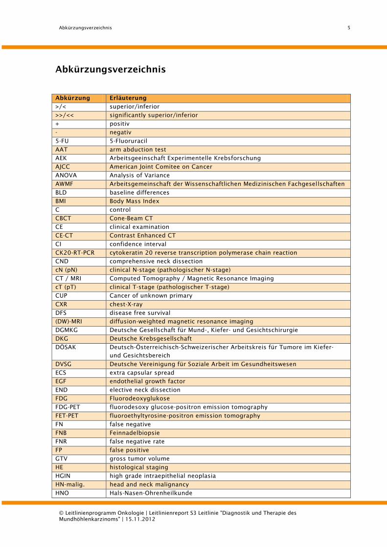
Abkürzungsverzeichnis
Abkürzung Erläuterung
>/< superior/inferior
>>/<< significantly superior/inferior
+ positiv
- negativ
5-FU 5-Fluoruracil
AAT arm abduction test
AEK Arbeitsgeeinschaft Experimentelle Krebsforschung
AJCC American Joint Comitee on Cancer
ANOVA Analysis of Variance
AWMF Arbeitsgemeinschaft der Wissenschaftlichen Medizinischen Fachgesellschaften
BLD baseline differences
BMI Body Mass Index
C control
CBCT Cone-Beam CT
CE clinical examination
CE-CT Contrast Enhanced CT
CI confidence interval
CK20-RT-PCR cytokeratin 20 reverse transcription polymerase chain reaction
CND comprehensive neck dissection
cN (pN) clinical N-stage (pathologischer N-stage)
CT / MRI Computed Tomography / Magnetic Resonance Imaging
cT (pT) clinical T-stage (pathologischer T-stage)
CUP Cancer of unknown primary
CXR chest-X-ray
DFS disease free survival
(DW)-MRI diffusion-weighted magnetic resonance imaging
DGMKG Deutsche Gesellschaft für Mund-, Kiefer- und Gesichtschirurgie
DKG Deutsche Krebsgesellschaft
DÖSAK Deutsch-Österreichisch-Schweizerischer Arbeitskreis für Tumore im Kiefer-
und Gesichtsbereich
DVSG Deutsche Vereinigung für Soziale Arbeit im Gesundheitswesen
ECS extra capsular spread
EGF endothelial growth factor
END elective neck dissection
FDG Fluorodeoxyglukose
FDG-PET fluorodesoxy glucose-positron emission tomography
FET-PET fluoroethyltyrosine-positron emission tomography
FN false negative
FNB Feinnadelbiopsie
FNR false negative rate
FP false positive
GTV gross tumor volume
HE histological staging
HGIN high grade intraepithelial neoplasia
HN-malig. head and neck malignancy
HNO Hals-Nasen-Ohrenheilkunde
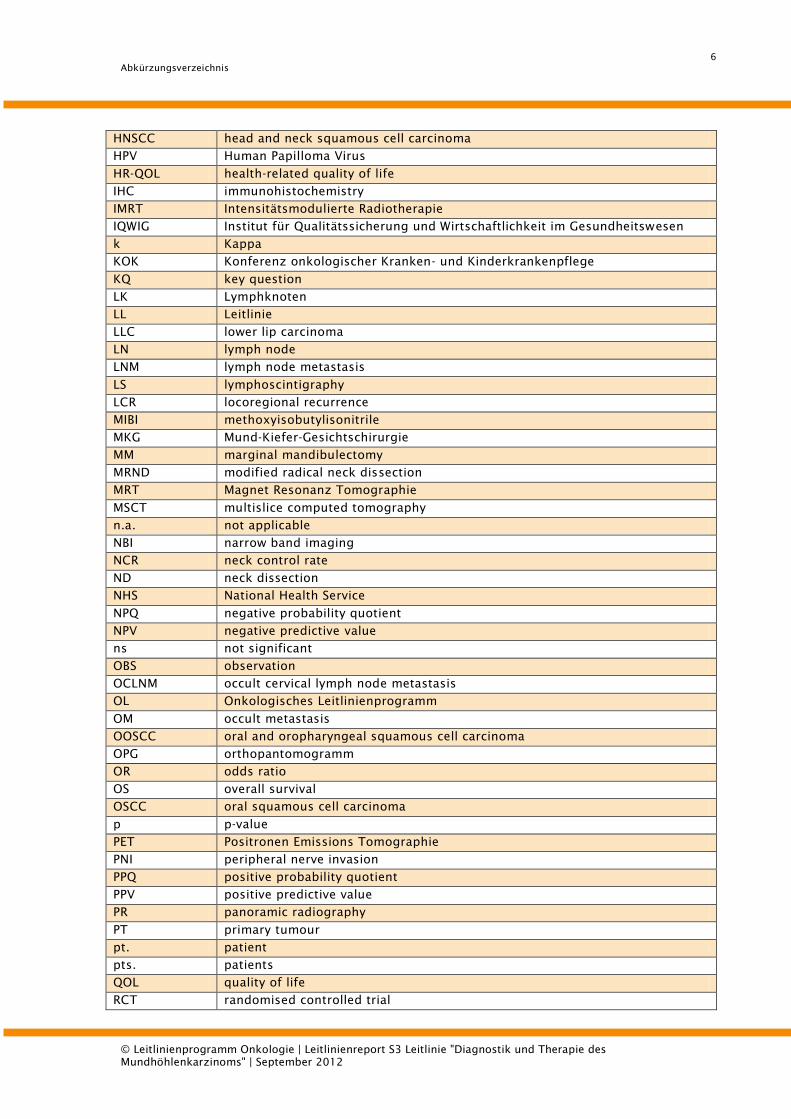
6
Abkürzungsverzeichnis
© Leitlinienprogramm Onkologie | Leitlinienreport S3 Leitlinie "Diagnostik und Therapie des
Mundhöhlenkarzinoms" | September 2012
HNSCC head and neck squamous cell carcinoma
HPV Human Papilloma Virus
HR-QOL health-related quality of life
IHC immunohistochemistry
IMRT Intensitätsmodulierte Radiotherapie
IQWIG Institut für Qualitätssicherung und Wirtschaftlichkeit im Gesundheitswesen
k Kappa
KOK Konferenz onkologischer Kranken- und Kinderkrankenpflege
KQ key question
LK Lymphknoten
LL Leitlinie
LLC lower lip carcinoma
LN lymph node
LNM lymph node metastasis
LS lymphoscintigraphy
LCR locoregional recurrence
MIBI methoxyisobutylisonitrile
MKG Mund-Kiefer-Gesichtschirurgie
MM marginal mandibulectomy
MRND modified radical neck dissection
MRT Magnet Resonanz Tomographie
MSCT multislice computed tomography
n.a. not applicable
NBI narrow band imaging
NCR neck control rate
ND neck dissection
NHS National Health Service
NPQ negative probability quotient
NPV negative predictive value
ns not significant
OBS observation
OCLNM occult cervical lymph node metastasis
OL Onkologisches Leitlinienprogramm
OM occult metastasis
OOSCC oral and oropharyngeal squamous cell carcinoma
OPG orthopantomogramm
OR odds ratio
OS overall survival
OSCC oral squamous cell carcinoma
p p-value
PET Positronen Emissions Tomographie
PNI peripheral nerve invasion
PPQ positive probability quotient
PPV positive predictive value
PR panoramic radiography
PT primary tumour
pt. patient
pts. patients
QOL quality of life
RCT randomised controlled trial
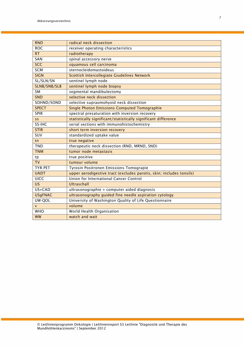
7
Abkürzungsverzeichnis
© Leitlinienprogramm Onkologie | Leitlinienreport S3 Leitlinie "Diagnostik und Therapie des
Mundhöhlenkarzinoms" | September 2012
RND radical neck dissection
ROC receiver operating characteristics
RT radiotherapy
SAN spinal accessory nerve
SCC squamous cell carcinoma
SCM sternocleidomastoideus
SIGN Scottish Intercollegiate Giudelines Network
SL/SLN/SN sentinel lymph node
SLNB/SNB/SLB sentinel lymph node biopsy
SM segmental mandibulectomy
SND selective neck dissection
SOHND/SOND selective supraomohyoid neck dissection
SPECT Single Photon Emissions Computed Tomographie
SPIR spectral presaturation with inversion recovery
ss statistically significant/statistically significant difference
SS-IHC serial sections with immunohistochemistry
STIR short term inversion recovery
SUV standardized uptake value
tn true negative
TND therapeutic neck dissection (RND, MRND, SND)
TNM tumor node metastasis
tp true positive
TV tumour volume
TYR-PET Tyrosin Positronen Emissions Tomograpie
UADT upper aerodigestive tract (excludes parotis, skin; includes tonsils)
UICC Union for International Cancer Control
US Ultraschall
US+CAD ultrasonographie + computer aided diagnosis
USgFNAC ultrasonography guided fine needle aspiration cytology
UW-QOL University of Washington Quality of Life Questionnaire
v volume
WHO World Health Organisation
WW watch and wait
8
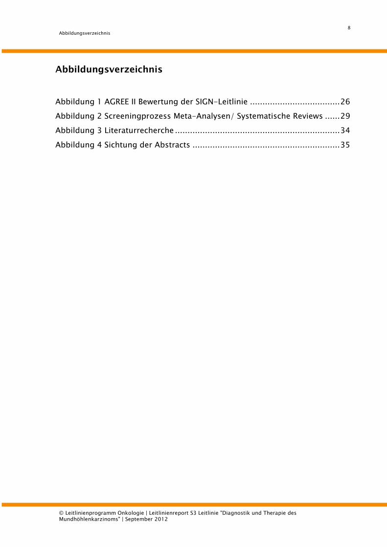
Abbildungsverzeichnis
© Leitlinienprogramm Onkologie | Leitlinienreport S3 Leitlinie "Diagnostik und Therapie des
Mundhöhlenkarzinoms" | September 2012
Abbildungsverzeichnis
Abbildung 1 AGREE II Bewertung der SIGN-Leitlinie .................................... 26
Abbildung 2 Screeningprozess Meta-Analysen/ Systematische Reviews ...... 29
Abbildung 3 Literaturrecherche .................................................................. 34
Abbildung 4 Sichtung der Abstracts ........................................................... 35
9
Tabellenverzeichnis
© Leitlinienprogramm Onkologie | Leitlinienreport S3 Leitlinie "Diagnostik und Therapie des
Mundhöhlenkarzinoms" | September 2012
Tabellenverzeichnis
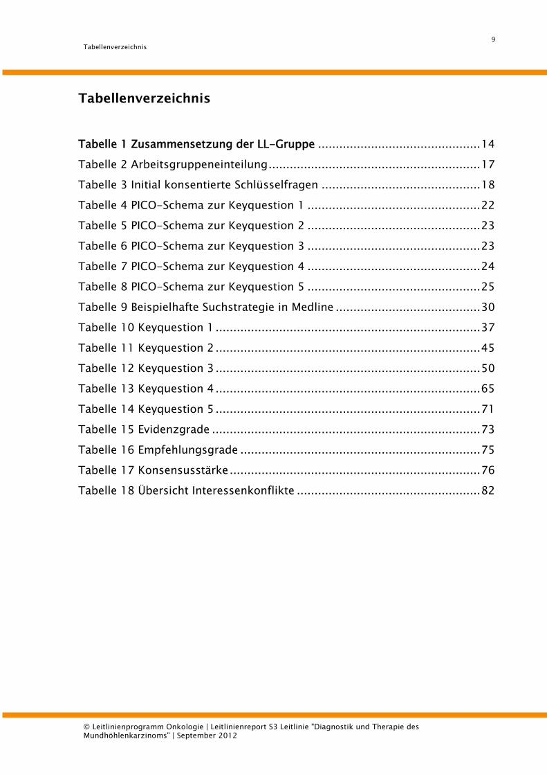
Tabelle 1 Zusammensetzung der LL-Gruppe .............................................. 14
Tabelle 2 Arbeitsgruppeneinteilung ............................................................ 17
Tabelle 3 Initial konsentierte Schlüsselfragen ............................................. 18
Tabelle 4 PICO-Schema zur Keyquestion 1 ................................................. 22
Tabelle 5 PICO-Schema zur Keyquestion 2 ................................................. 23
Tabelle 6 PICO-Schema zur Keyquestion 3 ................................................. 23
Tabelle 7 PICO-Schema zur Keyquestion 4 ................................................. 24
Tabelle 8 PICO-Schema zur Keyquestion 5 ................................................. 25
Tabelle 9 Beispielhafte Suchstrategie in Medline ......................................... 30
Tabelle 10 Keyquestion 1 ........................................................................... 37
Tabelle 11 Keyquestion 2 ........................................................................... 45
Tabelle 12 Keyquestion 3 ........................................................................... 50
Tabelle 13 Keyquestion 4 ........................................................................... 65
Tabelle 14 Keyquestion 5 ........................................................................... 71
Tabelle 15 Evidenzgrade ............................................................................ 73
Tabelle 16 Empfehlungsgrade .................................................................... 75
Tabelle 17 Konsensusstärke ....................................................................... 76
Tabelle 18 Übersicht Interessenkonflikte .................................................... 82
Informationen zu dieser Leitlinie
10
© Leitlinienprogramm Onkologie | Leitlinienreport S3 Leitlinie "Diagnostik und Therapie des
Mundhöhlenkarzinoms" | September 2012
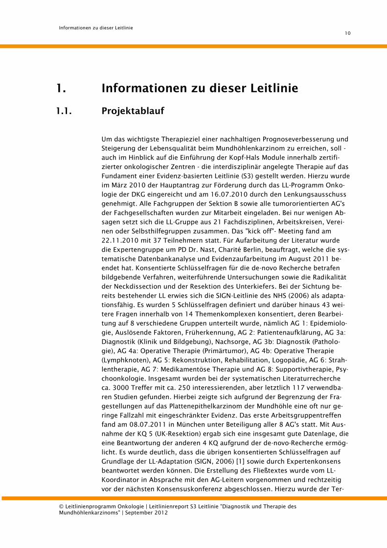
1. Informationen zu dieser Leitlinie
1.1. Projektablauf
Um das wichtigste Therapieziel einer nachhaltigen Prognoseverbesserung und
Steigerung der Lebensqualität beim Mundhöhlenkarzinom zu erreichen, soll -
auch im Hinblick auf die Einführung der Kopf-Hals Module innerhalb zertifi-
zierter onkologischer Zentren - die interdisziplinär angelegte Therapie auf das
Fundament einer Evidenz-basierten Leitlinie (S3) gestellt werden. Hierzu wurde
im März 2010 der Hauptantrag zur Förderung durch das LL-Programm Onko-
logie der DKG eingereicht und am 16.07.2010 durch den Lenkungsausschuss
genehmigt. Alle Fachgruppen der Sektion B sowie alle tumororientierten AG's
der Fachgesellschaften wurden zur Mitarbeit eingeladen. Bei nur wenigen Ab-
sagen setzt sich die LL-Gruppe aus 21 Fachdisziplinen, Arbeitskreisen, Verei-
nen oder Selbsthilfegruppen zusammen. Das "kick off"- Meeting fand am
22.11.2010 mit 37 Teilnehmern statt. Für Aufarbeitung der Literatur wurde
die Expertengruppe um PD Dr. Nast, Charité Berlin, beauftragt, welche die sys-
tematische Datenbankanalyse und Evidenzaufarbeitung im August 2011 be-
endet hat. Konsentierte Schlüsselfragen für die de-novo Recherche betrafen
bildgebende Verfahren, weiterführende Untersuchungen sowie die Radikalität
der Neckdissection und der Resektion des Unterkiefers. Bei der Sichtung be-
reits bestehender LL erwies sich die SIGN-Leitlinie des NHS (2006) als adapta-
tionsfähig. Es wurden 5 Schlüsselfragen definiert und darüber hinaus 43 wei-
tere Fragen innerhalb von 14 Themenkomplexen konsentiert, deren Bearbei-
tung auf 8 verschiedene Gruppen unterteilt wurde, nämlich AG 1: Epidemiolo-
gie, Auslösende Faktoren, Früherkennung, AG 2: Patientenaufklärung, AG 3a:
Diagnostik (Klinik und Bildgebung), Nachsorge, AG 3b: Diagnostik (Patholo-
gie), AG 4a: Operative Therapie (Primärtumor), AG 4b: Operative Therapie
(Lymphknoten), AG 5: Rekonstruktion, Rehabilitation, Logopädie, AG 6: Strah-
lentherapie, AG 7: Medikamentöse Therapie und AG 8: Supportivtherapie, Psy-
choonkologie. Insgesamt wurden bei der systematischen Literaturrecherche
ca. 3000 Treffer mit ca. 250 interessierenden, aber letztlich 117 verwendba-
ren Studien gefunden. Hierbei zeigte sich aufgrund der Begrenzung der Fra-
gestellungen auf das Plattenepithelkarzinom der Mundhöhle eine oft nur ge-
ringe Fallzahl mit eingeschränkter Evidenz. Das erste Arbeitsgruppentreffen
fand am 08.07.2011 in München unter Beteiligung aller 8 AG's statt. Mit Aus-
nahme der KQ 5 (UK-Resektion) ergab sich eine insgesamt gute Datenlage, die
eine Beantwortung der anderen 4 KQ aufgrund der de-novo-Recherche ermög-
licht. Es wurde deutlich, dass die übrigen konsentierten Schlüsselfragen auf
Grundlage der LL-Adaptation (SIGN, 2006) [1] sowie durch Expertenkonsens
beantwortet werden können. Die Erstellung des Fließtextes wurde vom LL-
Koordinator in Absprache mit den AG-Leitern vorgenommen und rechtzeitig
vor der nächsten Konsensuskonferenz abgeschlossen. Hierzu wurde der Ter-
Informationen zu dieser Leitlinie
11
© Leitlinienprogramm Onkologie | Leitlinienreport S3 Leitlinie "Diagnostik und Therapie des
Mundhöhlenkarzinoms" | September 2012
min für den 01. und 02.12.2011 in Berlin festgelegt.
1.2. Weitere Leitliniendokumente
Dieser Report bezieht sich auf die Langversion der S3-Leitlinie „Diagnostik und
Therapie des Mundhöhlenkarzinoms, welche über folgende Seiten zugänglich
ist
http://www.awmf.org/leitlinien/aktuelle-leitlinien.html
http://www.leitlinienprogramm-onkologie.de/OL/leitlinien.html
http://www.krebsgesellschaft.de/wub_llevidenzbasiert,120884.html
http://www.krebshilfe.de/
http://www.mkg-chirurgie.de
Neben diesem Leitlinienreport gibt es folgende Dokumente:
Langversion
Kurzversion
Patientenleitlinie (in Bearbeitung, Publikation ab 2013)
Alle diese Dokumente werden ebenfalls auf den oben genannten Homepages
abrufbar sein.
1.3. Finanzierung der Leitlinie
Diese Leitlinie wurde von der Deutschen Krebshilfe im Rahmen des Onkologi-
schen Leitlinienprogramms gefördert.
Geltungsbereich und Zweck
12
© Leitlinienprogramm Onkologie | Leitlinienreport S3 Leitlinie "Diagnostik und Therapie des
Mundhöhlenkarzinoms" | September 2012
2. Geltungsbereich und Zweck
2.1. Adressaten
Die LL gilt für das Plattenepithelkarzinom der Mundhöhle. Die Patientengrup-
pe, für welche die Leitlinie gelten soll, ist eindeutig beschrieben. Es handelt
sich hierbei um alle Patienten, die aufgrund eigener Beobachtung oder durch
Untersuchungen von Ärzten (MKG, HNO, Zahnarzt, Hausarzt, Dermatologie)
einen wie oben beschrieben abklärungsbedürftigen Befund an der Schleimhaut
der Mundhöhle bieten. Alle Altersgruppen und beide Geschlechter fallen unter
die Leitlinie. Die Leitlinie bezieht sich auf alle Schweregrade (Stadien) incl. der
Frühformen (in situ Karzinome) der Erkrankung. Das Vorliegen von
Komorbiditäten gilt nicht als Ausschlusskriterium für die Anwendung der Leit-
linie.
Anwender der Leitlinie sind Ärzte für Mund-, Kiefer- und Gesichtschirurgie,
Ärzte für Hals-Nasen-Ohrenheilkunde, Strahlentherapeuten, Onkologen, Haus-
ärzte sowie Zahnärzte und Fachzahnärzte für Oralchirurgie, Karzinome oder
Karzinommetastasen aus anderen Ursprungsgeweben als der Mundschleim-
haut fallen nicht unter die vorliegende Leitlinie, ebenso wie Plattenepithelkar-
zinome der Mundhöhle bei bereits bestehendem inkurablem Tumorleiden
oder schweren Erkrankungen anderer Genese. Für die Behandlung von Karzi-
nomen benachbarter Strukturen (Larynx, Pharynx, Lippen) liegen eigenständi-
ge Leitlinien vor.
2.2. Zielsetzung
Die Leitlinie soll für alle beteiligten Fachgruppen Orientierung und Hilfestel-
lung zur Festlegung notwendiger diagnostischer und therapeutischer Maß-
nahmen sein, um Therapieziele zuverlässig zu erreichen. Dies sind eine best-
mögliche Prognose und Lebensqualität für jede individuelle Befundkonstellati-
on. Die Leitlinie soll den aktuellen Stand der Therapie auf dem Boden wissen-
schaftlich gesicherter Erkenntnisse darstellen und somit auch einen Beitrag
zur Fortbildung leisten. Sie soll den interdisziplinären Charakter in Diagnostik,
Therapie und Nachsorge herausstellen und den Beginn einer effektiven Thera-
pie beschleunigen. Patienten und Angehörigen soll die Leitlinie Möglichkeit
geben, zuverlässige und verständliche Informationen zu erhalten, um vorge-
schlagene Therapiekonzepte nachvollziehen und mittragen zu können. Mit der
Anwendung der Leitlinie soll die Häufigkeit vermeidbarer Komplikationen re-
duziert und Voraussetzungen geschaffen werden, die soziale und berufliche
Integration der Patienten zu verbessern.
Geltungsbereich und Zweck
13
© Leitlinienprogramm Onkologie | Leitlinienreport S3 Leitlinie "Diagnostik und Therapie des
Mundhöhlenkarzinoms" | September 2012
Wichtige allgemeine Ziele sind:
die Verbreitung von evidenzbasierten und formal konsentierten Emp-
fehlungen zu Versorgungsbereich übergreifenden Vorgehensweisen;
die Bereitstellung von Lösungsvorschlägen für Nahtstellen sowohl zwi-
schen verschiedenen Disziplinen (MKG/ HNO/ Strahlenthetapie/ Onko-
logie/ Pathologie/ Anästhesie-Intensivmedizin u.a.) als auch verschie-
denen Versorgungssektoren (primäre Prävention - sekundäre Präventi-
on – Kuration –Rehabilitation)
die Verbreitung von LL-basierten Qualitätsindikatoren und Patienten-
Leitlinien, möglichst flächendeckende Implementierung der LL-
Empfehlungen und Qualitätsindikatoren sowie
die Berücksichtigung von LL-Empfehlungen in der ärztlichen Aus-, Fort–
und Weiterbildung und in Qualitätsmanagement-Systemen.
Zusammensetzung der Leitliniengruppe
14
© Leitlinienprogramm Onkologie | Leitlinienreport S3 Leitlinie "Diagnostik und Therapie des
Mundhöhlenkarzinoms" | September 2012
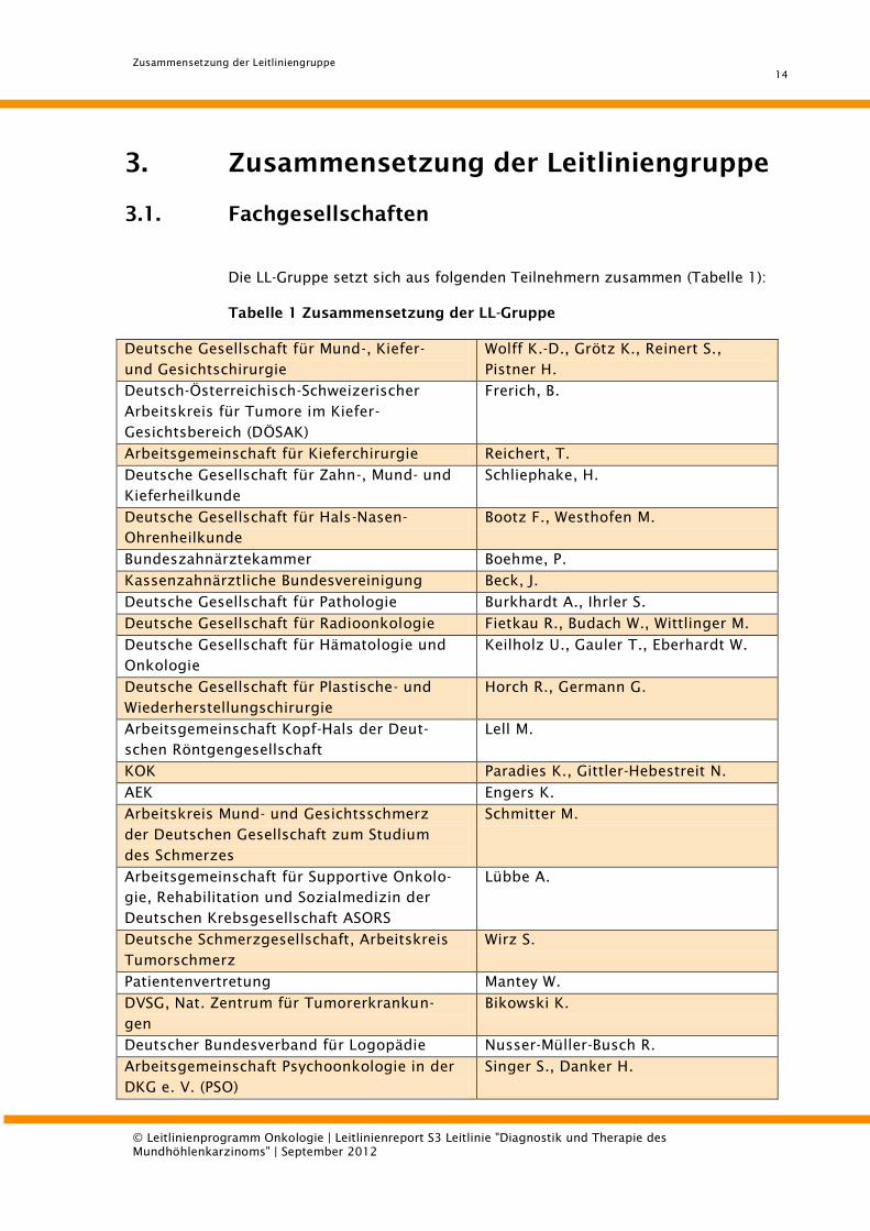
3. Zusammensetzung der Leitliniengruppe
3.1. Fachgesellschaften
Die LL-Gruppe setzt sich aus folgenden Teilnehmern zusammen (Tabelle 1):
Tabelle 1 Zusammensetzung der LL-Gruppe
Deutsche Gesellschaft für Mund-, Kiefer-
und Gesichtschirurgie
Wolff K.-D., Grötz K., Reinert S.,
Pistner H.
Deutsch-Österreichisch-Schweizerischer
Arbeitskreis für Tumore im Kiefer-
Gesichtsbereich (DÖSAK)
Frerich, B.
Arbeitsgemeinschaft für Kieferchirurgie Reichert, T.
Deutsche Gesellschaft für Zahn-, Mund- und
Kieferheilkunde
Schliephake, H.
Deutsche Gesellschaft für Hals-Nasen-
Ohrenheilkunde
Bootz F., Westhofen M.
Bundeszahnärztekammer Boehme, P.
Kassenzahnärztliche Bundesvereinigung Beck, J.
Deutsche Gesellschaft für Pathologie Burkhardt A., Ihrler S.
Deutsche Gesellschaft für Radioonkologie Fietkau R., Budach W., Wittlinger M.
Deutsche Gesellschaft für Hämatologie und
Onkologie
Keilholz U., Gauler T., Eberhardt W.
Deutsche Gesellschaft für Plastische- und
Wiederherstellungschirurgie
Horch R., Germann G.
Arbeitsgemeinschaft Kopf-Hals der Deut-
schen Röntgengesellschaft
Lell M.
KOK Paradies K., Gittler-Hebestreit N.
AEK Engers K.
Arbeitskreis Mund- und Gesichtsschmerz
der Deutschen Gesellschaft zum Studium
des Schmerzes
Schmitter M.
Arbeitsgemeinschaft für Supportive Onkolo-
gie, Rehabilitation und Sozialmedizin der
Deutschen Krebsgesellschaft ASORS
Lübbe A.
Deutsche Schmerzgesellschaft, Arbeitskreis
Tumorschmerz
Wirz S.
Patientenvertretung Mantey W.
DVSG, Nat. Zentrum für Tumorerkrankun-
gen
Bikowski K.
Deutscher Bundesverband für Logopädie Nusser-Müller-Busch R.
Arbeitsgemeinschaft Psychoonkologie in der
DKG e. V. (PSO)
Singer S., Danker H.
Zusammensetzung der Leitliniengruppe
15
© Leitlinienprogramm Onkologie | Leitlinienreport S3 Leitlinie "Diagnostik und Therapie des
Mundhöhlenkarzinoms" | September 2012
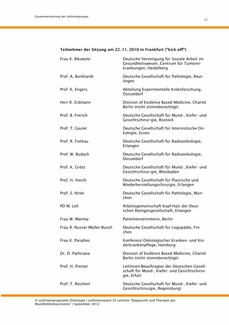
Teilnehmer der Sitzung am 22. 11. 2010 in Frankfurt ("kick-off")
Frau K. Bikowski Deutsche Vereinigung für Soziale Arbeit im
Gesundheitswesen, Centrum für Tumorer-
krankungen, Heidelberg
Prof. A. Burkhardt Deutsche Gesellschaft für Pathologie, Reut-
lingen
Prof. K. Engers Abteilung Experimentelle Krebsforschung,
Düsseldorf
Herr R. Erdmann Division of Evidence Based Medicine, Charité
Berlin (nicht stimmberechtigt)
Prof. B. Frerich Deutsche Gesellschaft für Mund-, Kiefer- und
Gesichtschirur-gie, Rostock
Prof. T. Gauler Deutsche Gesellschaft für Internistische On-
kologie, Essen
Prof. R. Fietkau Deutsche Gesellschaft für Radioonkologie,
Erlangen
Prof. W. Budach Deutsche Gesellschaft für Radioonkologie,
Düsseldorf
Prof. K. Grötz Deutsche Gesellschaft für Mund-, Kiefer- und
Gesichtschirur-gie, Wiesbaden
Prof. H. Horch Deutsche Gesellschaft für Plastische und
Wiederherstellungschirurgie, Erlangen
Prof. S. Ihrler Deutsche Gesellschaft für Pathologie, Mün-
chen
PD M. Lell Arbeitsgemeinschaft Kopf-Hals der Deut-
schen Röntgengesellschaft, Erlangen
Frau W. Mantey Patientenvertreterin, Berlin
Frau R. Nusser-Müller-Busch Deutsche Gesellschaft für Logopädie, Fre-
chen
Frau K. Paradies Konferenz Onkologischer Kranken- und Kin-
derkrankenpflege, Hamburg
Dr. D. Pathirana Division of Evidence Based Medicine, Charité
Berlin (nicht stimmberechtigt)
Prof. H. Pistner Leitlinien-Beauftragter der Deutschen Gesell-
schaft für Mund-, Kiefer- und Gesichtschirur-
gie, Erfurt
Prof. T. Reichert Deutsche Gesellschaft für Mund-, Kiefer- und
Gesichtschirurgie, Regensburg)
Zusammensetzung der Leitliniengruppe
16
© Leitlinienprogramm Onkologie | Leitlinienreport S3 Leitlinie "Diagnostik und Therapie des
Mundhöhlenkarzinoms" | September 2012
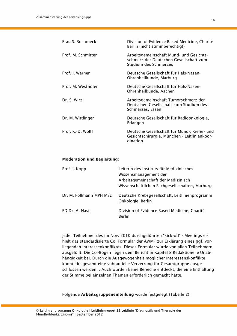
Frau S. Rosumeck Division of Evidence Based Medicine, Charité
Berlin (nicht stimmberechtigt)
Prof. M. Schmitter Arbeitsgemeinschaft Mund- und Gesichts-
schmerz der Deutschen Gesellschaft zum
Studium des Schmerzes
Prof. J. Werner Deutsche Gesellschaft für Hals-Nasen-
Ohrenheilkunde, Marburg
Prof. M. Westhofen Deutsche Gesellschaft für Hals-Nasen-
Ohrenheilkunde, Aachen
Dr. S. Wirz Arbeitsgemeinschaft Tumorschmerz der
Deutschen Gesellschaft zum Studium des
Schmerzes, Essen
Dr. M. Wittlinger Deutsche Gesellschaft für Radioonkologie,
Erlangen
Prof. K.-D. Wolff Deutsche Gesellschaft für Mund-, Kiefer- und
Gesichtschirurgie, München - Leitlinienkoor-
dination
Moderation und Begleitung:
Prof. I. Kopp Leiterin des Instituts für Medizinisches
Wissensmanagement der
Arbeitsgemeinschaft der Medizinisch
Wissenschaftlichen Fachgesellschaften, Marburg
Dr. M. Follmann MPH MSc Deutsche Krebsgesellschaft, Leitlinienprogramm
Onkologie, Berlin
PD Dr. A. Nast Division of Evidence Based Medicine, Charité
Berlin
Jeder Teilnehmer des im Nov. 2010 durchgeführten "kick-off" - Meetings er-
hielt das standardisierte Col Formular der AWMF zur Erklärung eines ggf. vor-
liegenden Interessenkonfliktes. Dieses Formular wurde von allen Teilnehmern
ausgefüllt. Die Col-Bögen liegen dem Bericht in Kapitel 8 Redaktionelle Unab-
hängigkeit bei. Durch die Ausgewogenheit möglicher Interessenskonflikte
konnte insgesamt eine subtantielle Verzerrung für Gesamtgruppe ausge-
schlossen werden. . Auch wurden keine Bereiche entdeckt, die eine Enthaltung
der Stimme bei einzelnen Themen erforderlich gemacht hätte.
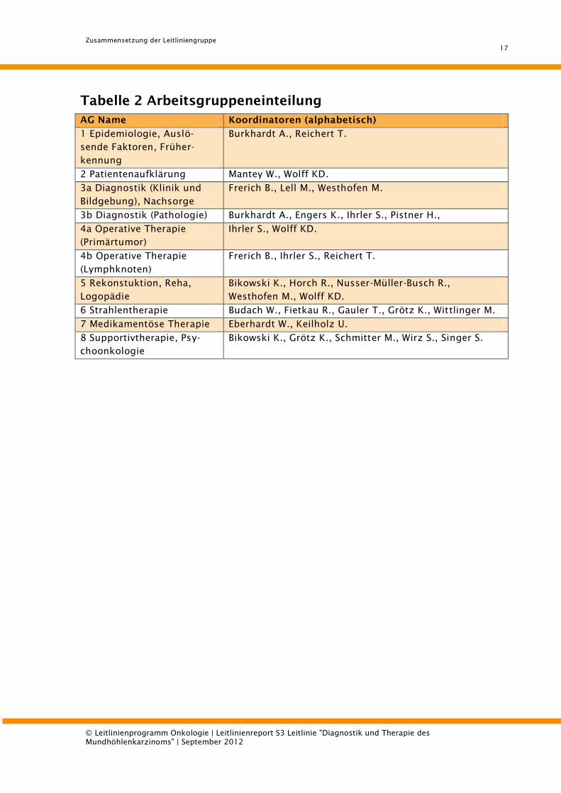
Folgende Arbeitsgruppeneinteilung wurde festgelegt (Tabelle 2):
Zusammensetzung der Leitliniengruppe
17
© Leitlinienprogramm Onkologie | Leitlinienreport S3 Leitlinie "Diagnostik und Therapie des
Mundhöhlenkarzinoms" | September 2012
Tabelle 2 Arbeitsgruppeneinteilung
AG Name Koordinatoren (alphabetisch)
1 Epidemiologie, Auslö-
sende Faktoren, Früher-
kennung
Burkhardt A., Reichert T.
2 Patientenaufklärung Mantey W., Wolff KD.
3a Diagnostik (Klinik und
Bildgebung), Nachsorge
Frerich B., Lell M., Westhofen M.
3b Diagnostik (Pathologie) Burkhardt A., Engers K., Ihrler S., Pistner H.,
4a Operative Therapie
(Primärtumor)
Ihrler S., Wolff KD.
4b Operative Therapie
(Lymphknoten)
Frerich B., Ihrler S., Reichert T.
5 Rekonstuktion, Reha,
Logopädie
Bikowski K., Horch R., Nusser-Müller-Busch R.,
Westhofen M., Wolff KD.
6 Strahlentherapie Budach W., Fietkau R., Gauler T., Grötz K., Wittlinger M.
7 Medikamentöse Therapie Eberhardt W., Keilholz U.
8 Supportivtherapie, Psy-
choonkologie
Bikowski K., Grötz K., Schmitter M., Wirz S., Singer S.
Fragestellung und Gliederung
18
© Leitlinienprogramm Onkologie | Leitlinienreport S3 Leitlinie "Diagnostik und Therapie des
Mundhöhlenkarzinoms" | September 2012
4. Fragestellung und Gliederung
Vor der Ausarbeitung der vorliegenden Leitlinie erfolgte auf dem Kick-off-
Meeting im November 2010 die Festlegung von Schlüsselfragen, anhand derer
die inhaltliche Struktur der Leitlinie gestaltet wurde. Formal-strukturell war der
Aufbau der Leitlinie durch das Template des Leitlinienprogramms Onkologie
vorgegeben. Die Abfolge der Hauptkapitel wurde in Anlehnung an bereits be-
stehende und bewährte onkologische Leitlinien gewählt. Hierbei wurde das
Grundprinzip der Formulierung von Statements und Empfehlungen beibehal-
ten, an die sich jeweils ein erläuternder Fließtext mit weiter gehenden Detail-
informationen anschließt.
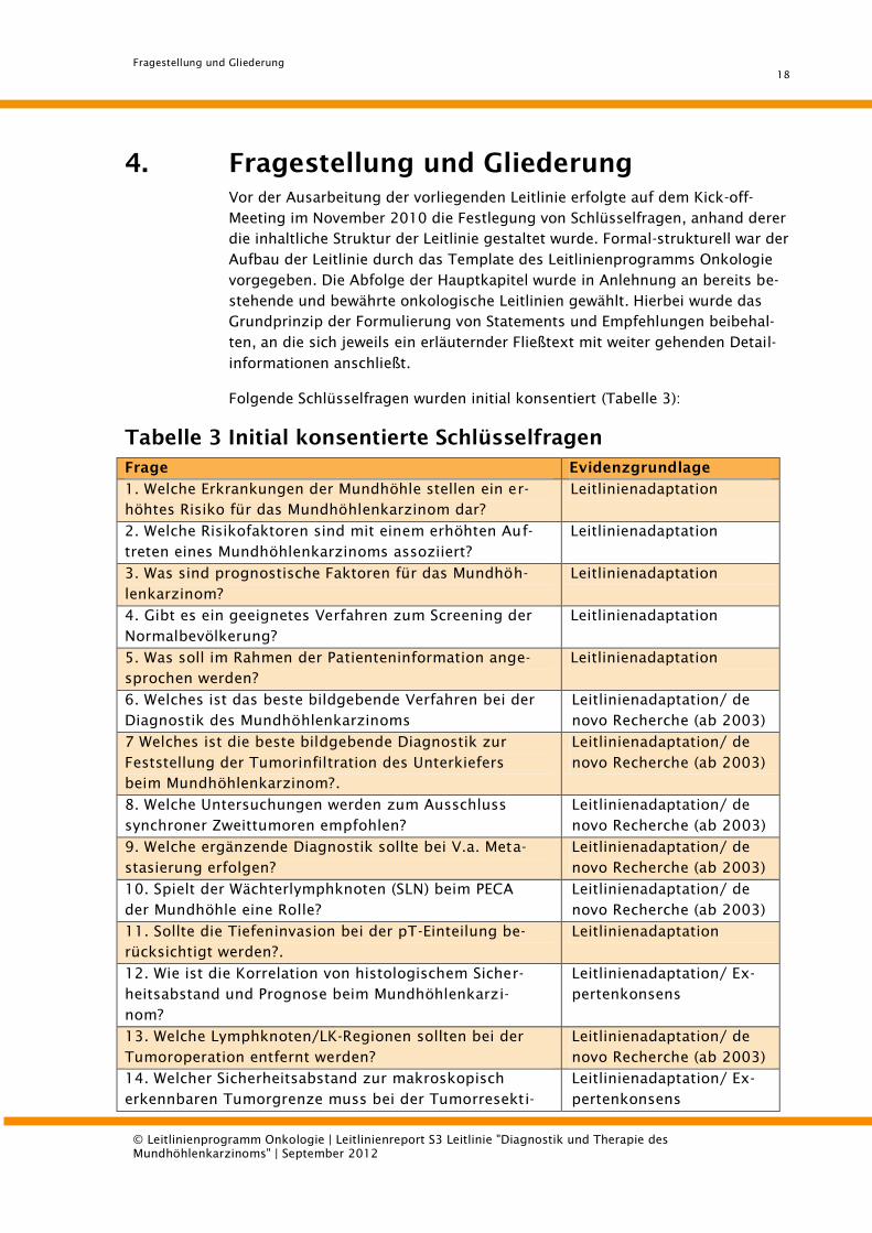
Folgende Schlüsselfragen wurden initial konsentiert (Tabelle 3):
Tabelle 3 Initial konsentierte Schlüsselfragen
Frage Evidenzgrundlage
1. Welche Erkrankungen der Mundhöhle stellen ein er-
höhtes Risiko für das Mundhöhlenkarzinom dar?
Leitlinienadaptation
2. Welche Risikofaktoren sind mit einem erhöhten Auf-
treten eines Mundhöhlenkarzinoms assoziiert?
Leitlinienadaptation
3. Was sind prognostische Faktoren für das Mundhöh-
lenkarzinom?
Leitlinienadaptation
4. Gibt es ein geeignetes Verfahren zum Screening der
Normalbevölkerung?
Leitlinienadaptation
5. Was soll im Rahmen der Patienteninformation ange-
sprochen werden?
Leitlinienadaptation
6. Welches ist das beste bildgebende Verfahren bei der
Diagnostik des Mundhöhlenkarzinoms
Leitlinienadaptation/ de
novo Recherche (ab 2003)
7 Welches ist die beste bildgebende Diagnostik zur
Feststellung der Tumorinfiltration des Unterkiefers
beim Mundhöhlenkarzinom?.
Leitlinienadaptation/ de
novo Recherche (ab 2003)
8. Welche Untersuchungen werden zum Ausschluss
synchroner Zweittumoren empfohlen?
Leitlinienadaptation/ de
novo Recherche (ab 2003)
9. Welche ergänzende Diagnostik sollte bei V.a. Meta-
stasierung erfolgen?
Leitlinienadaptation/ de
novo Recherche (ab 2003)
10. Spielt der Wächterlymphknoten (SLN) beim PECA
der Mundhöhle eine Rolle?
Leitlinienadaptation/ de
novo Recherche (ab 2003)
11. Sollte die Tiefeninvasion bei der pT-Einteilung be-
rücksichtigt werden?.
Leitlinienadaptation
12. Wie ist die Korrelation von histologischem Sicher-
heitsabstand und Prognose beim Mundhöhlenkarzi-
nom?
Leitlinienadaptation/ Ex-
pertenkonsens
13. Welche Lymphknoten/LK-Regionen sollten bei der
Tumoroperation entfernt werden?
Leitlinienadaptation/ de
novo Recherche (ab 2003)
14. Welcher Sicherheitsabstand zur makroskopisch
erkennbaren Tumorgrenze muss bei der Tumorresekti-
Leitlinienadaptation/ Ex-
pertenkonsens
Fragestellung und Gliederung
19
© Leitlinienprogramm Onkologie | Leitlinienreport S3 Leitlinie "Diagnostik und Therapie des
Mundhöhlenkarzinoms" | September 2012
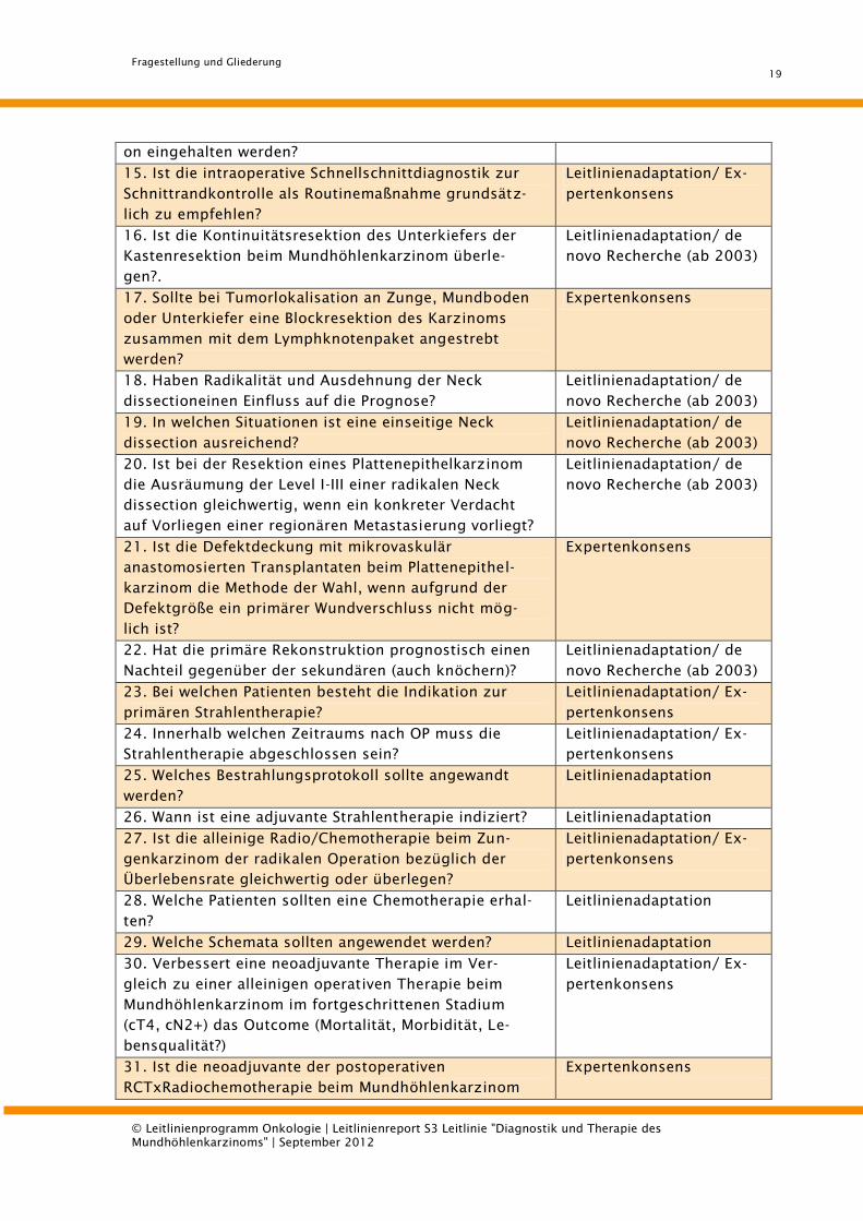
on eingehalten werden?
15. Ist die intraoperative Schnellschnittdiagnostik zur
Schnittrandkontrolle als Routinemaßnahme grundsätz-
lich zu empfehlen?
Leitlinienadaptation/ Ex-
pertenkonsens
16. Ist die Kontinuitätsresektion des Unterkiefers der
Kastenresektion beim Mundhöhlenkarzinom überle-
gen?.
Leitlinienadaptation/ de
novo Recherche (ab 2003)
17. Sollte bei Tumorlokalisation an Zunge, Mundboden
oder Unterkiefer eine Blockresektion des Karzinoms
zusammen mit dem Lymphknotenpaket angestrebt
werden?
Expertenkonsens
18. Haben Radikalität und Ausdehnung der Neck
dissectioneinen Einfluss auf die Prognose?
Leitlinienadaptation/ de
novo Recherche (ab 2003)
19. In welchen Situationen ist eine einseitige Neck
dissection ausreichend?
Leitlinienadaptation/ de
novo Recherche (ab 2003)
20. Ist bei der Resektion eines Plattenepithelkarzinom
die Ausräumung der Level I-III einer radikalen Neck
dissection gleichwertig, wenn ein konkreter Verdacht
auf Vorliegen einer regionären Metastasierung vorliegt?
Leitlinienadaptation/ de
novo Recherche (ab 2003)
21. Ist die Defektdeckung mit mikrovaskulär
anastomosierten Transplantaten beim Plattenepithel-
karzinom die Methode der Wahl, wenn aufgrund der
Defektgröße ein primärer Wundverschluss nicht mög-
lich ist?
Expertenkonsens
22. Hat die primäre Rekonstruktion prognostisch einen
Nachteil gegenüber der sekundären (auch knöchern)?
Leitlinienadaptation/ de
novo Recherche (ab 2003)
23. Bei welchen Patienten besteht die Indikation zur
primären Strahlentherapie?
Leitlinienadaptation/ Ex-
pertenkonsens
24. Innerhalb welchen Zeitraums nach OP muss die
Strahlentherapie abgeschlossen sein?
Leitlinienadaptation/ Ex-
pertenkonsens
25. Welches Bestrahlungsprotokoll sollte angewandt
werden?
Leitlinienadaptation
26. Wann ist eine adjuvante Strahlentherapie indiziert? Leitlinienadaptation
27. Ist die alleinige Radio/Chemotherapie beim Zun-
genkarzinom der radikalen Operation bezüglich der
Überlebensrate gleichwertig oder überlegen?
Leitlinienadaptation/ Ex-
pertenkonsens
28. Welche Patienten sollten eine Chemotherapie erhal-
ten?
Leitlinienadaptation
29. Welche Schemata sollten angewendet werden? Leitlinienadaptation
30. Verbessert eine neoadjuvante Therapie im Ver-
gleich zu einer alleinigen operativen Therapie beim
Mundhöhlenkarzinom im fortgeschrittenen Stadium
(cT4, cN2+) das Outcome (Mortalität, Morbidität, Le-
bensqualität?)
Leitlinienadaptation/ Ex-
pertenkonsens
31. Ist die neoadjuvante der postoperativen
RCTxRadiochemotherapie beim Mundhöhlenkarzinom
Expertenkonsens
Fragestellung und Gliederung
20
© Leitlinienprogramm Onkologie | Leitlinienreport S3 Leitlinie "Diagnostik und Therapie des
Mundhöhlenkarzinoms" | September 2012
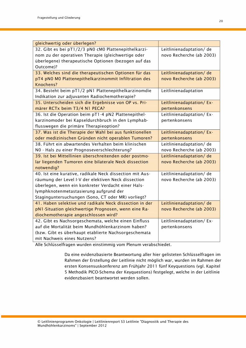
gleichwertig oder überlegen?
32. Gibt es bei pT1/2/3 pN0 cM0 Plattenepithelkarzi-
nom zu der operativen Therapie (gleichwertige oder
überlegene) therapeutische Optionen (bezogen auf das
Outcome)?
Leitlinienadaptation/ de
novo Recherche (ab 2003)
33. Welches sind die therapeutischen Optionen für das
pT4 pN0 M0 Plattenepithelkarzinommit Infiltration des
Knochens?
Leitlinienadaptation/ de
novo Recherche (ab 2003)
34. Besteht beim pT1/2 pN1 Plattenepithelkarzinomdie
Indikation zur adjuvanten Radiochemotherapie?
Leitlinienadaptation
35. Unterscheiden sich die Ergebnisse von OP vs. Pri-
märer RCTx beim T3/4 N1 PECA?
Leitlinienadaptation/ Ex-
pertenkonsens
36. Ist die Operation beim pT1-4 pN2 Plattenepithel-
karzinomoder bei Kapseldurchbruch in den Lymphab-
flusswegen die primäre Therapieoption?
Leitlinienadaptation/ Ex-
pertenkonsens
37. Was ist die Therapie der Wahl bei aus funktionellen
oder medizinischen Gründen nicht operablen Tumoren?
Leitlinienadaptation/ Ex-
pertenkonsens
38. Führt ein abwartendes Verhalten beim klinischen
N0 - Hals zu einer Prognoseverschlechterung?
Leitlinienadaptation/ de
novo Recherche (ab 2003)
39. Ist bei Mittellinien überschreitenden oder postmo-
lar liegenden Tumoren eine bilaterale Neck dissection
notwendig?
Leitlinienadaptation/ de
novo Recherche (ab 2003)
40. Ist eine kurative, radikale Neck dissection mit Aus-
räumung der Level I-V der elektiven Neck dissection
überlegen, wenn ein konkreter Verdacht einer Hals-
lymphknotenmetastasierung aufgrund der
Staginguntersuchungen (Sono, CT oder MR) vorliegt?
Leitlinienadaptation/ de
novo Recherche (ab 2003)
41. Haben selektive und radikale Neck dissection in der
pN1-Situation gleichwertige Prognosen, wenn eine Ra-
diochemotherapie angeschlossen wird?
Leitlinienadaptation/ de
novo Recherche (ab 2003)
42. Gibt es Nachsorgeschemata, welche einen Einfluss
auf die Mortalität beim Mundhöhlenkarzinom haben?
(bzw. Gibt es überhaupt etablierte Nachsorgeschemata
mit Nachweis eines Nutzens?
Leitlinienadaptation/ Ex-
pertenkonsens
Alle Schlüsselfragen wurden einstimmig vom Plenum verabschiedet.
Da eine evidenzbasierte Beantwortung aller hier gelisteten Schlüsselfragen im
Rahmen der Erstellung der Leitlinie nicht möglich war, wurden im Rahmen der
ersten Konsensuskonferenz am Frühjahr 2011 fünf Keyquestions (vgl. Kapitel
5 Methodik PICO-Schema der Keyquestions) festgelegt, welche in der Leitlinie
evidenzbasiert beantwortet werden sollen.
Fragestellung und Gliederung
21
© Leitlinienprogramm Onkologie | Leitlinienreport S3 Leitlinie "Diagnostik und Therapie des
Mundhöhlenkarzinoms" | September 2012
Die systematische Literaturrecherche mit Evidenzaufarbeitung wurde nach Ab-
schluss der vertraglichen Formalitäten im Frühjahr 2011 durch die Arbeits-
gruppe um PD Dr. Nast begonnen und im Juni 2011 abgeschlossen. Aus ca.
3000 relevanten Abstracts wurden ca. 250 Arbeiten identifiziert, von denen
schließlich 117 für die nähere Analyse relevant waren. Hieraus erfolgte die Zu-
sammenfassung der Literatur gemäß der Schlüsselfragen sowie die Darstel-
lung der Ergebnisse in Form von Evidenztabellen. Ein Ausdruck der Tabellen
ist im Anhang beigefügt. Ferner wurde die der zu adaptierenden SIGN-90-
Leitlinie zu Grunde liegende Literatur mit den Resultaten der eigenständigen
de-novo-Recherche abgeglichen, erneut bewertet und bei der Erstellung der
Evidenztabellen berücksichtigt. Die Systematik und Methodik der Recherche,
des Textscreenings, der Bewertung der Studien, der Erstellung der
Evidenztabellen sowie der Formulierung von Empfehlungen und Hintergrund-
texten werden im Folgenden dargestellt.
Methodik
22
© Leitlinienprogramm Onkologie | Leitlinienreport S3 Leitlinie "Diagnostik und Therapie des
Mundhöhlenkarzinoms" | September 2012
5. Methodik
5.1. Evidenzbasierung
In der ersten Konsensuskonferenz am 22.11.2010 wurden die folgenden fünf
Key-questions (vgl. PICO-Schema der Keyquestions) festgelegt, welche in der
Leitlinie evidenzbasiert beantwortet werden sollen:
1. Welche bildgebenden Verfahren sind im Rahmen der Diagnostik des
Primärtumors zu empfehlen?
2. Welche Untersuchungen werden zum Ausschluss synchroner Zweittu-
moren empfohlen?
3. Welche ergänzende Diagnostik sollte bei Verdacht auf eine Metastasie-
rung erfolgen?
4. Welche Lymphknotenregionen sollten bei der Tumoroperation entfernt
werden?
5. Ist die Kontinuitätsesektion des Unterkiefers der Kastenresektion beim
Mundhöhlenkarzinom überlegen?
Um eine standardisierte Suche zu ermöglichen, wurde für die fünf Schlüssel-
fragen zunächst ein PICO-Schema erstellt. (s. Tabelle 4-8)
Weitere Schlüsselfragen sollen zukünftig im Rahmen von Updates der Leitlinie
evidenzbasiert beantwortet werden.
PICO-Schema der Keyquestions
Keyquestion 1: Welche bildgebenden Verfahren sind im Rahmen der Diag-
nostik des Primärtumors zu empfehlen? (Tabelle 4)
Tabelle 4 PICO-Schema zur Keyquestion 1
PICO-Aspekt Beschreibung Nicht relevant
für Suchstrate-
gie
Patient / Population - human, adults
Problem / Krankheit 1
Erkrankung
- Head and Neck Neoplasms, Mouth Neo-
plasms
- cancer, tumor, tumour, carcinoma, neo-
plasm, metastasis, metastases, squamous
cell carcinoma + Lokalisation
Problem / Krankheit 2
Lokalisation
- palate, tongue, mouth mucosa, mouth
floor, uvula, gingival, lips
Intervention / Ver-
gleichs-intervention
- CT, MRT, OPG, DVT, Isotopes, PET, Sono-
graphy, Echography, Ultrasound
Methodik
23
© Leitlinienprogramm Onkologie | Leitlinienreport S3 Leitlinie "Diagnostik und Therapie des
Mundhöhlenkarzinoms" | September 2012
Outcomes x
Studiendesign - SIGN-Filter:
o RCT, Observational Studies
Gesundheitsökonomie x
Gesundheitssystem /
geographischer Bezug
x
Relevanter Zeitraum - ab 2003
Sprache - deutsch, englisch
Keyquestion 2: Welche Untersuchungen werden zum Ausschluss syn-
chroner Zweittumoren empfohlen? (Tabelle 5)
Tabelle 5 PICO-Schema zur Keyquestion 2
PICO-Aspekt Beschreibung Nicht relevant
für Suchstrategie
Patient / Population - human, adults
Problem / Krankheit 1
Erkrankung
- Head and Neck Neoplasms, Mouth Neo-
plasms
- cancer, tumor, tumour, carcinoma, neo-
plasm, metastasis, metastases, squamous
cell carcinoma + Lokalisation
Problem / Krankheit 2
Lokalisation
- palate, tongue, mouth mucosa, mouth floor,
uvula, gingival, lips
Intervention / Ver-
gleichs-intervention
- Endoskopie, Panendoskopie, Bronchoskopie,
ÖGD, CT-Thorax, PET, Staging, secondary
primary
Outcomes x
Studiendesign
- SIGN-Filter:
o RCT, Observational Studies, , Diagnos-
tic Studies
Gesundheitsökonomie x
Gesundheitssystem /
geographischer Bezug
x
Relevanter Zeitraum - ab 2003
Sprache - deutsch, englisch
Keyquestion 3: Welche ergänzende Diagnostik sollte bei V. a. Metastasie-
rung erfolgen? (Tabelle 6)
Tabelle 6 PICO-Schema zur Keyquestion 3
PICO-Aspekt Beschreibung Nicht relevant
für Suchstrategie
Patient / Population - human, adults
Problem / Krankheit 1
- Head and Neck Neoplasms, Mouth Neo-
plasms
Methodik
24
© Leitlinienprogramm Onkologie | Leitlinienreport S3 Leitlinie "Diagnostik und Therapie des
Mundhöhlenkarzinoms" | September 2012
Erkrankung - cancer, tumor, tumour, carcinoma, neo-
plasm, metastasis, metastases, squamous
cell carcinoma + Lokalisation
Problem / Krankheit 2
Lokalisation
- palate, tongue, mouth mucosa, mouth floor,
uvula, gingival, lips
Intervention / Ver-
gleichs-intervention
- CT-Thorax, Szintigraphie, PET-CT, Sonogra-
phy, Echography, Ultrasound, Feinnadel-
biopsie, Feinnadelaspirationsbiopsie, Stag-
ing, secondary primary
Outcomes x
Studiendesign
- SIGN-Filter:
o RCT, Observational Studies, , Diagnos-
tic Studies
Gesundheitsökonomie x
Gesundheitssystem /
geographischer Bezug
x
Relevanter Zeitraum - ab 2003
Sprache - deutsch, englisch
Keyquestion 4: Welche Lymphknotenregionen sollten bei der Tumoroperati-
on entfernt werden? (Tabelle 7)
Tabelle 7 PICO-Schema zur Keyquestion 4
PICO-Aspekt Beschreibung Nicht relevant
für Suchstrategie
Patient / Population - human, adults
Problem / Krankheit 1
Erkrankung
- Head and Neck Neoplasms, Mouth Neo-
plasms
- cancer, tumor, tumour, carcinoma, neo-
plasm, metastasis, metastases, squamous
cell carcinoma + Lokalisation
Problem / Krankheit 2
Lokalisation
- palate, tongue, mouth mucosa, mouth
floor, uvula, gingival, lips
Intervention / Ver-
gleichs-intervention
- Selective Lymphknoten-Dissektion,
Lymphknoten-Dissektion, Neck Dissection,
Modified Neck Dissection, Radikale Neck
Dissection, Lymphknotenausräumung
Outcomes x
Studiendesign - SIGN-Filter:
o RCT, Observational Studies
Gesundheitsökonomie x
Gesundheitssystem /
geographischer Bezug
x
Relevanter Zeitraum - ab 2003
Sprache - deutsch, englisch
Methodik
25
© Leitlinienprogramm Onkologie | Leitlinienreport S3 Leitlinie "Diagnostik und Therapie des
Mundhöhlenkarzinoms" | September 2012
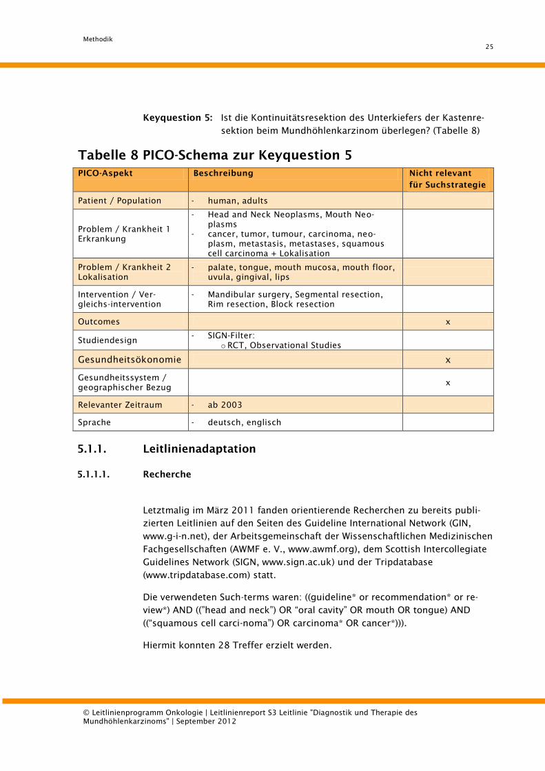
Keyquestion 5: Ist die Kontinuitätsresektion des Unterkiefers der Kastenre-
sektion beim Mundhöhlenkarzinom überlegen? (Tabelle 8)
Tabelle 8 PICO-Schema zur Keyquestion 5
PICO-Aspekt Beschreibung Nicht relevant
für Suchstrategie
Patient / Population - human, adults
Problem / Krankheit 1
Erkrankung
- Head and Neck Neoplasms, Mouth Neo-
plasms
- cancer, tumor, tumour, carcinoma, neo-
plasm, metastasis, metastases, squamous
cell carcinoma + Lokalisation
Problem / Krankheit 2
Lokalisation
- palate, tongue, mouth mucosa, mouth floor,
uvula, gingival, lips
Intervention / Ver-
gleichs-intervention
- Mandibular surgery, Segmental resection,
Rim resection, Block resection
Outcomes x
Studiendesign - SIGN-Filter:
o RCT, Observational Studies
Gesundheitsökonomie x
Gesundheitssystem /
geographischer Bezug
x
Relevanter Zeitraum - ab 2003
Sprache - deutsch, englisch
5.1.1. Leitlinienadaptation
5.1.1.1. Recherche
Letztmalig im März 2011 fanden orientierende Recherchen zu bereits publi-
zierten Leitlinien auf den Seiten des Guideline International Network (GIN,
www.g-i-n.net), der Arbeitsgemeinschaft der Wissenschaftlichen Medizinischen
Fachgesellschaften (AWMF e. V., www.awmf.org), dem Scottish Intercollegiate
Guidelines Network (SIGN, www.sign.ac.uk) und der Tripdatabase
(www.tripdatabase.com) statt.
Die verwendeten Such-terms waren: ((guideline* or recommendation* or re-
view*) AND ((”head and neck”) OR “oral cavity” OR mouth OR tongue) AND
((“squamous cell carci-noma”) OR carcinoma* OR cancer*))).
Hiermit konnten 28 Treffer erzielt werden.
Methodik
26
© Leitlinienprogramm Onkologie | Leitlinienreport S3 Leitlinie "Diagnostik und Therapie des
Mundhöhlenkarzinoms" | September 2012
5.1.1.2. Auswahl der Leitlinien
Die gefundenen Arbeiten wurden in Hinblick auf die thematische Relevanz
(Einschlusskriterium) geprüft. Aufgrund dessen konnte die SIGN-Leitlinie 90
„Diagnosis and management of head and neck cancer“ [1] eingeschlossen
werden.
Ausschlusskriterien waren die Sprache (nicht englisch, französisch oder
deutsch), die Inhalte (nicht die Diagnostik, die Therapie oder die Nachsorge
des Mundhöhlenkarzinoms betreffend). Weiterhin wurden nur Leitlinien be-
rücksichtigt, deren Veröffentlichung nach dem Jahre 2000 stattfand.
So beschäftigten sich weitere, nicht genauer bewertete Leitlinien mit speziel-
len Fragestellungen oder nicht zur Mundhöhle passenden anatomischen Loka-
lisationen, so dass sie für die Erstellung der vorliegenden Leitlinie als Gesamt-
vorlage nicht in Frage kamen. Weitere Leitlinien waren seit langer Zeit abge-
laufen. Aufgrund ihrer umfangreichen Darstellung der Kopf-Hals-Karzinome
mit allen Aspekten in Diagnostik, Therapie und Nachsorge, der systemati-
schen Literaturrecherche mit gut dokumentierter Evidenzprüfung und dem
nachvollziehbaren Konsentierungsprozess fiel die Entscheidung zur Adaptati-
on der SIGN Leitlinie (GIN 170), die im Oktober 2008 publiziert wurde.
5.1.1.3. Leitlinienbewertung
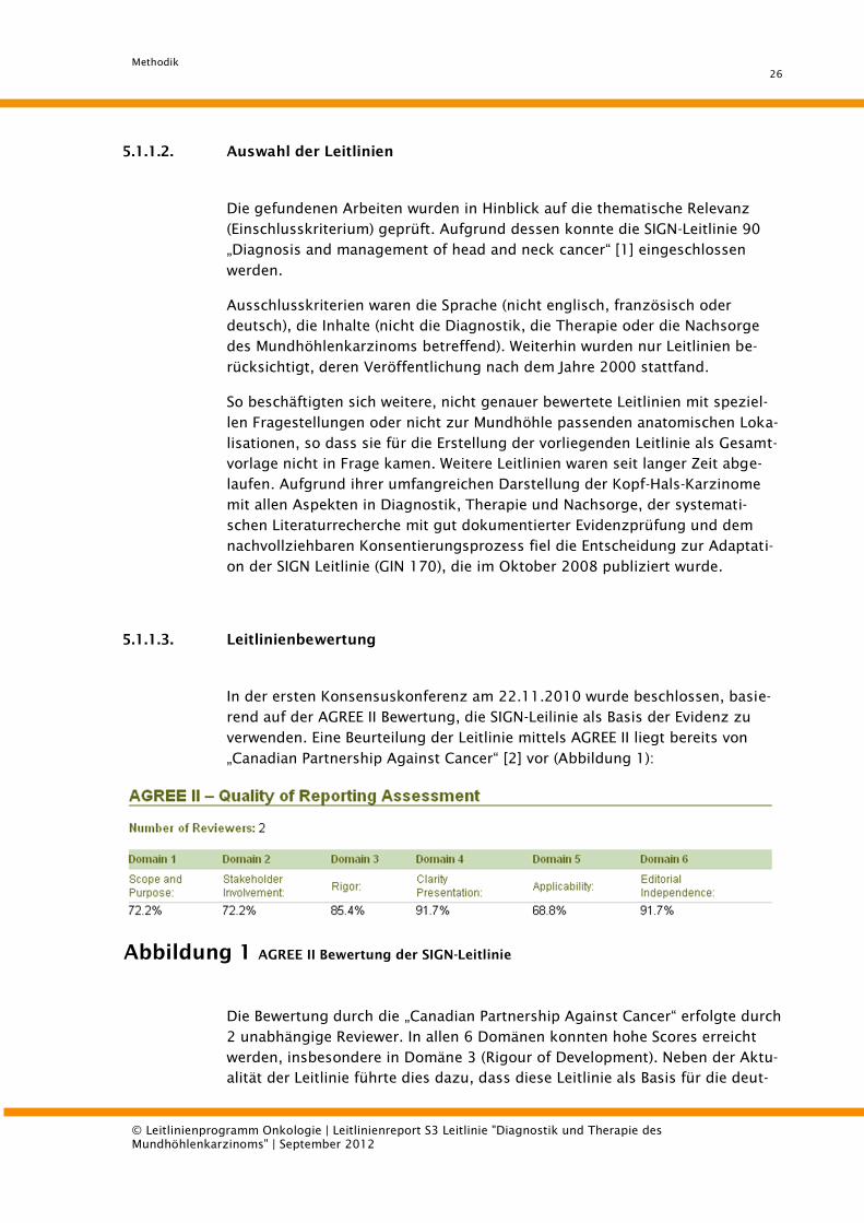
In der ersten Konsensuskonferenz am 22.11.2010 wurde beschlossen, basie-
rend auf der AGREE II Bewertung, die SIGN-Leilinie als Basis der Evidenz zu
verwenden. Eine Beurteilung der Leitlinie mittels AGREE II liegt bereits von
„Canadian Partnership Against Cancer“ [2] vor (Abbildung 1):
Abbildung 1 AGREE II Bewertung der SIGN-Leitlinie
Die Bewertung durch die „Canadian Partnership Against Cancer“ erfolgte durch
2 unabhängige Reviewer. In allen 6 Domänen konnten hohe Scores erreicht
werden, insbesondere in Domäne 3 (Rigour of Development). Neben der Aktu-
alität der Leitlinie führte dies dazu, dass diese Leitlinie als Basis für die deut-
Methodik
27
© Leitlinienprogramm Onkologie | Leitlinienreport S3 Leitlinie "Diagnostik und Therapie des
Mundhöhlenkarzinoms" | September 2012
schen Leitlinien „Diagnostik und Therapie des Mundhöhlenkarzinoms“ heran-
gezogen wurde.
5.1.1.4. Leitliniensynopsen / Extraktionen
Themenrelevante Empfehlungen und Statements aus der SIGN-Leitlinie wurden
übersetzt und unter Angabe der Literatur direkt übernommen. Ebenfalls wur-
de das Schema der Evidenzgraduierung nach SIGN in die vorliegende Leitlinie
übernommen (s. 5.2.1 Evidenzgrade).
Methodik
28
© Leitlinienprogramm Onkologie | Leitlinienreport S3 Leitlinie "Diagnostik und Therapie des
Mundhöhlenkarzinoms" | September 2012
5.1.1.5. Adaptierungsprozess
Die Empfehlungen, die sich aus der Leitlinienadaptation ergeben, basieren auf
den relevanten Empfehlungen der SIGN-Leitlinie aus den Kapiteln 3: Referral
and diagnosis, 4: Histopathology reporting, 5: Overview of the treatment of
the primary tumour, 7: Treatment: surgery as the major treatment modality
sowie Kapitel 14: Oral Cavity Cancer.
5.1.1.6. Weitere genutzte Leitlinien
Eine Adaptation weiterer Leitlinien ist nicht erfolgt. Bei speziellen weiterge-
henden Fragestellungen erfolgten Querverweise auf entsprechende bereits be-
stehende Leitlinien.
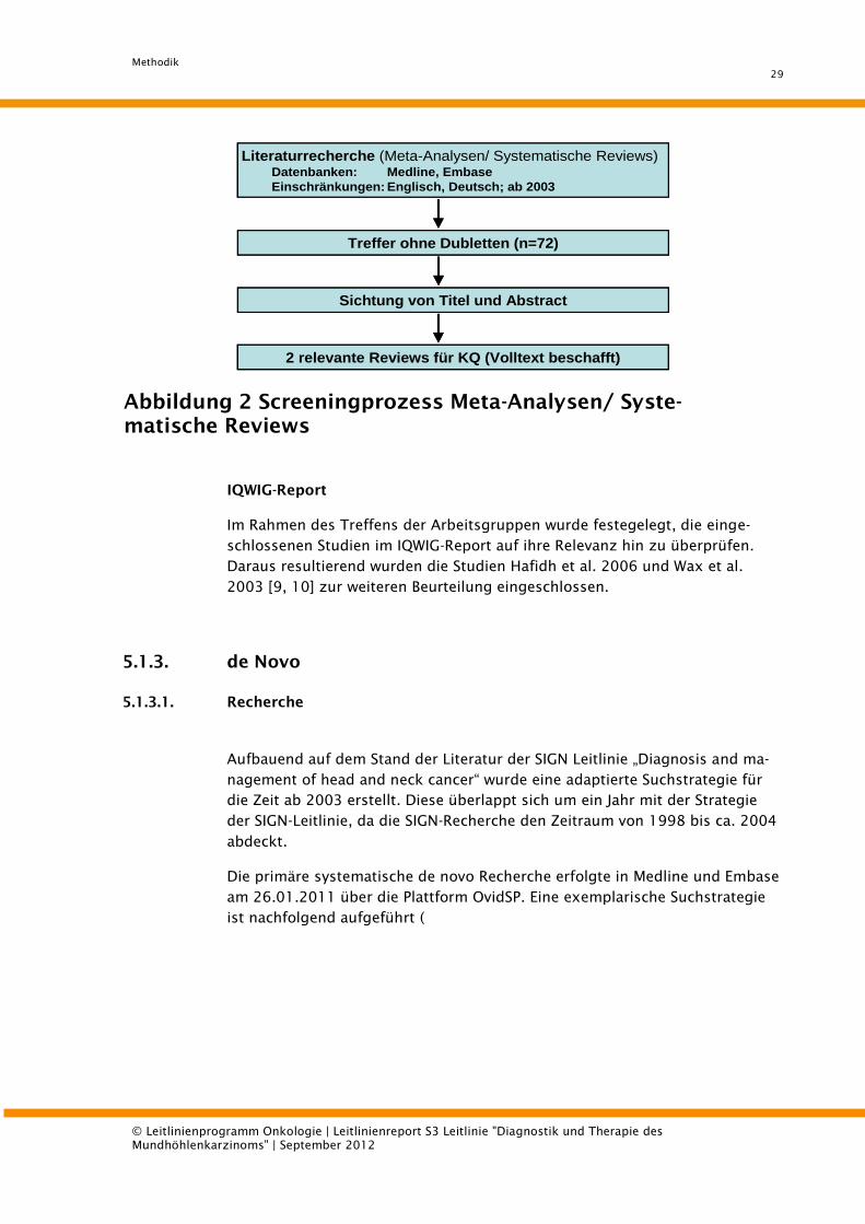
5.1.2. Systematische Reviews und Meta-Analysen
Eine systematische Recherche zu bereits vorhandenen Meta-Analysen und sys-
tematischen Reviews in Medline und Embase (über OvidSP) erfolgte letztmalig
am 09.06.2011. Die thematische Relevanzprüfung, basierend auf Titel und
Abstract der erzielten 72 Treffer, erfolgte von zwei unterschiedlichen Beurtei-
lern (DP und AS vgl. Abbildung 2). Letztendlich sind 2 Reviews für die
Keyquestions relevant und wurden zur weiteren Beurteilung im Volltext be-
schafft [3, 4]. In beiden Artikeln wurden Literaturrecherchen durchgeführt, die
sich zeitlich mit der Recherche der SIGN Leitlinie „Diagnosis and management
of head and neck cancer“ überschneiden, zum einen bis Februar 2005 [3] und
zum anderen von 1970 bis 2007 [4]. Die Referenzen der gefundenen Artikel
wurden deshalb per Hand nach relevanten Studien, publiziert in oder nach
2003, durchsucht. Dies führt zum Einschluss von 4 weiteren Studien [5-8].
Methodik
29
© Leitlinienprogramm Onkologie | Leitlinienreport S3 Leitlinie "Diagnostik und Therapie des
Mundhöhlenkarzinoms" | September 2012
Treffer ohne Dubletten (n=72)
Sichtung von Titel und Abstract
Literaturrecherche (Meta-Analysen/ Systematische Reviews)Datenbanken: Medline, Embase
Einschränkungen: Englisch, Deutsch; ab 2003
2 relevante Reviews für KQ (Volltext beschafft)
Treffer ohne Dubletten (n=72)
Sichtung von Titel und Abstract
Literaturrecherche (Meta-Analysen/ Systematische Reviews)Datenbanken: Medline, Embase
Einschränkungen: Englisch, Deutsch; ab 2003
2 relevante Reviews für KQ (Volltext beschafft)
Abbildung 2 Screeningprozess Meta-Analysen/ Syste-
matische Reviews
IQWIG-Report
Im Rahmen des Treffens der Arbeitsgruppen wurde festegelegt, die einge-
schlossenen Studien im IQWIG-Report auf ihre Relevanz hin zu überprüfen.
Daraus resultierend wurden die Studien Hafidh et al. 2006 und Wax et al.
2003 [9, 10] zur weiteren Beurteilung eingeschlossen.
5.1.3. de Novo
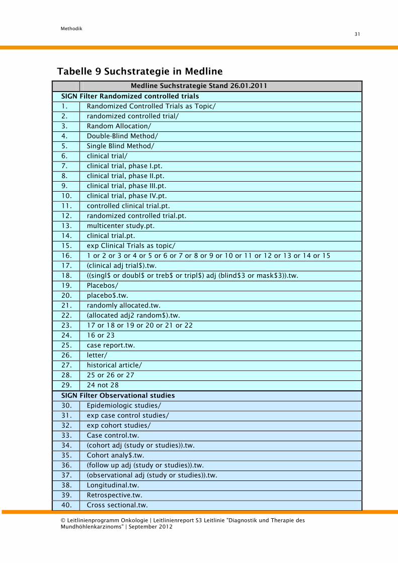
5.1.3.1. Recherche
Aufbauend auf dem Stand der Literatur der SIGN Leitlinie „Diagnosis and ma-
nagement of head and neck cancer“ wurde eine adaptierte Suchstrategie für
die Zeit ab 2003 erstellt. Diese überlappt sich um ein Jahr mit der Strategie
der SIGN-Leitlinie, da die SIGN-Recherche den Zeitraum von 1998 bis ca. 2004
abdeckt.
Die primäre systematische de novo Recherche erfolgte in Medline und Embase
am 26.01.2011 über die Plattform OvidSP. Eine exemplarische Suchstrategie
ist nachfolgend aufgeführt (
Methodik
30
© Leitlinienprogramm Onkologie | Leitlinienreport S3 Leitlinie "Diagnostik und Therapie des
Mundhöhlenkarzinoms" | September 2012
Tabelle 9):
Methodik
31
© Leitlinienprogramm Onkologie | Leitlinienreport S3 Leitlinie "Diagnostik und Therapie des
Mundhöhlenkarzinoms" | September 2012
Tabelle 9 Suchstrategie in Medline
Medline Suchstrategie Stand 26.01.2011
SIGN Filter Randomized controlled trials
1. Randomized Controlled Trials as Topic/
2. randomized controlled trial/
3. Random Allocation/
4. Double-Blind Method/
5. Single Blind Method/
6. clinical trial/
7. clinical trial, phase I.pt.
8. clinical trial, phase II.pt.
9. clinical trial, phase III.pt.
10. clinical trial, phase IV.pt.
11. controlled clinical trial.pt.
12. randomized controlled trial.pt.
13. multicenter study.pt.
14. clinical trial.pt.
15. exp Clinical Trials as topic/
16. 1 or 2 or 3 or 4 or 5 or 6 or 7 or 8 or 9 or 10 or 11 or 12 or 13 or 14 or 15
17. (clinical adj trial$).tw.
18. ((singl$ or doubl$ or treb$ or tripl$) adj (blind$3 or mask$3)).tw.
19. Placebos/
20. placebo$.tw.
21. randomly allocated.tw.
22. (allocated adj2 random$).tw.
23. 17 or 18 or 19 or 20 or 21 or 22
24. 16 or 23
25. case report.tw.
26. letter/
27. historical article/
28. 25 or 26 or 27
29. 24 not 28
SIGN Filter Observational studies
30. Epidemiologic studies/
31. exp case control studies/
32. exp cohort studies/
33. Case control.tw.
34. (cohort adj (study or studies)).tw.
35. Cohort analy$.tw.
36. (follow up adj (study or studies)).tw.
37. (observational adj (study or studies)).tw.
38. Longitudinal.tw.
39. Retrospective.tw.
40. Cross sectional.tw.
Methodik
32
© Leitlinienprogramm Onkologie | Leitlinienreport S3 Leitlinie "Diagnostik und Therapie des
Mundhöhlenkarzinoms" | September 2012
41. Cross-sectional studies/
42. 30 or 31 or 32 or 33 or 34 or 35 or 36 or 37 or 38 or 39 or 40 or 41
SIGN Filter Diagnostic studies
43. exp "Sensitivity and Specificity"/
44. sensitivity.tw.
45. specificity.tw.
46. ((pre-test or pretest) adj probability).tw.
47. post-test probability.tw.
48. predictive value$.tw.
49. likelihood ratio$.tw.
50. 43 or 44 or 45 or 46 or 47 or 48 or 49
51. 29 or 42
52. 29 or 42 or 50
Mundhöhlenkarzinom-Stamm
53. "Head and Neck Neoplasms"/
54. exp Mouth Neoplasms/
55. 53 or 54
56. (cancer* or tumo?r* or carcinoma* or neoplasm* or metastas?s or squamous cell
carcinoma).tw.
57. squamous cell carcinoma/
58. neoplasms, squamous cell/
59. 56 or 57 or 58
60. (palate or palatal).tw.
61. palate/
62. tongue*.tw.
63. tongue/
64. ((oral or buccal or mouth or cheek$) adj (mucous or (mucosa adj membrane$))).tw.
65. mouth mucosa/
66. (mouth adj3 (bottom or floor)).tw.
67. mouth floor/
68. uvula.tw.
69. uvula/
70. (gingival or gum$).tw.
71. gingiva/
72. (lip or lips).tw.
73. lip/
74. 60 or 61 or 62 or 63 or 64 or 65 or 66 or 67 or 68 or 69 or 70 or 71 or 72 or 73
75. 59 and 74
76. 55 or 75
77. 51 and 76
Keyquestion 1
78. Tomography, X-Ray Computed/
79. "comput$ tomograph$".tw.
80. (comput$ adj (axial or assisted) adj tomograph$).tw.
Methodik
33
© Leitlinienprogramm Onkologie | Leitlinienreport S3 Leitlinie "Diagnostik und Therapie des
Mundhöhlenkarzinoms" | September 2012
81. ((ct or cat) adj scan$).tw.
82. exp isotopes/
83. isotope$.tw.
84. exp Magnetic Resonance Imaging/
85. magnetic resonance imaging.tw.
86. mri.tw.
87. (mr adj (imaging or exam$)).tw.
88. diagnostic imaging/
89. Radiography, Panoramic/
90. op$g.tw.
91. exp Tomography, Emission-Computed/
92. positron emission tomography.tw.
93. pet.tw.
94. exp Cone-Beam Computed Tomography/
95. digital volume tomography.tw.
96. ultrasonography/
97. ultrasonics/
98. (ultrasound$ or ultra sound$ or ultrason$).tw.
99. echograph*.tw.
100. 78 or 79 or 80 or 81 or 82 or 83 or 84 or 85 or 86 or 87 or 88 or 89 or 90 or 91
or 92 or 93 or 94 or 95 or 96 or 97 or 98 or 99
101. 77 and 100
102. limit 101 to (yr="2003 -Current" and "all adult (19 plus years)" and (english or ger-
man) and humans)
103. 52 and 76
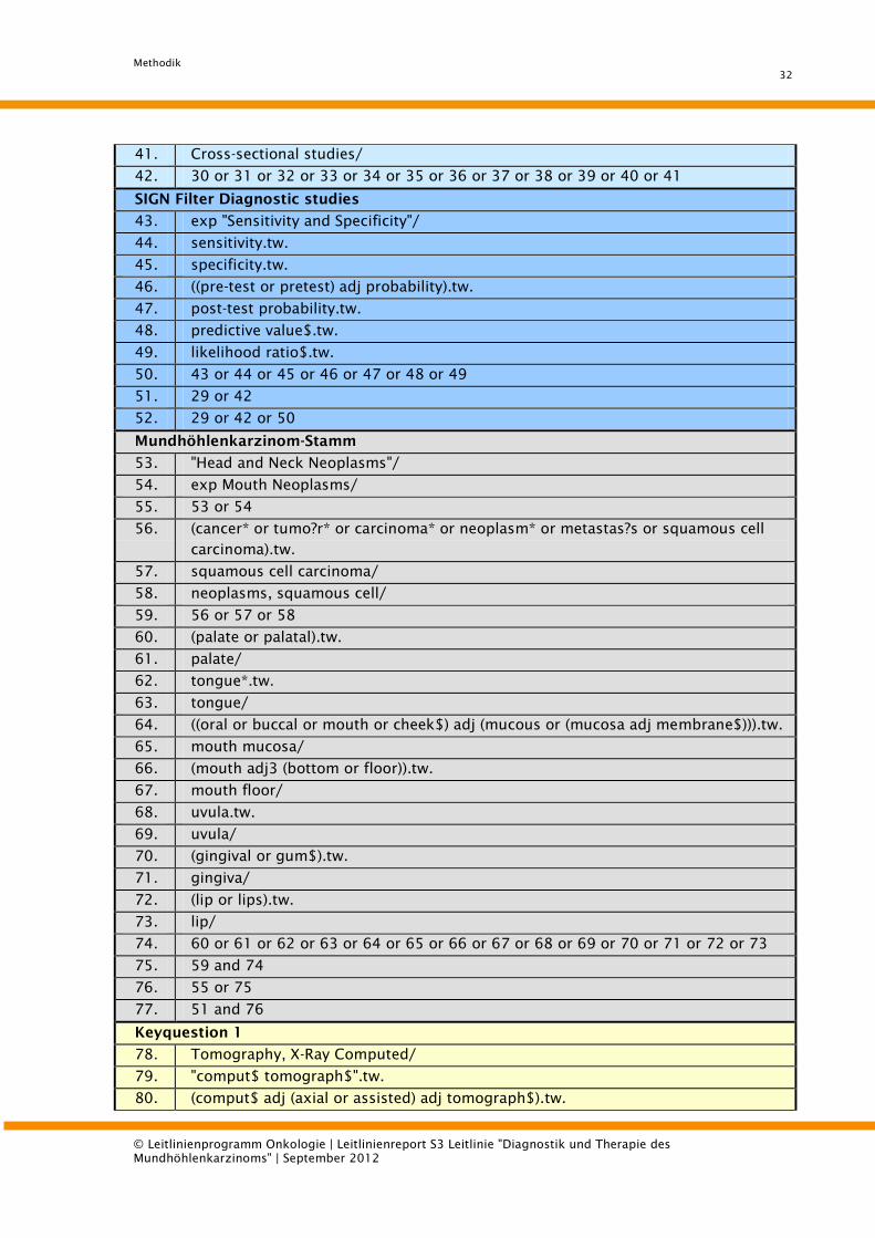
Keyquestion 2
104. bronchoscopy/
105. bronchoscopy.tw.
106. Endoscopy, Digestive System/
107. endoscopy.tw.
108. esophagoscopy.tw.
109. laryngoscopy/
110. laryngoscopy.tw.
111. panendoscopy.tw.
112. Tomography, X-Ray Computed/
113. comput$ tomograph$.tw.
114. (comput$ adj (axial or assisted) adj tomograph$).tw.
115. ((ct or cat) adj scan$).tw.
116. positron emission tomography.tw.
117. pet.tw.
118. 104 or 105 or 106 or 107 or 108 or 109 or 110 or 111 or 112 or 113 or 114 or
115 or 116 or 117
119. 119. 103 and 118
120. limit 119 to (yr="2003 -Current" and "all adult (19 plus years)" and (english or ger-
Methodik
34
© Leitlinienprogramm Onkologie | Leitlinienreport S3 Leitlinie "Diagnostik und Therapie des
Mundhöhlenkarzinoms" | September 2012
man) and humans)
Keyquestion 3
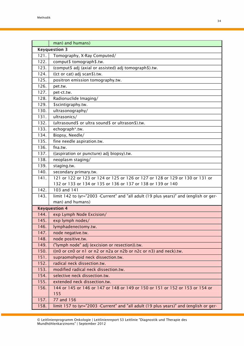
121. Tomography, X-Ray Computed/
122. comput$ tomograph$.tw.
123. (comput$ adj (axial or assisted) adj tomograph$).tw.
124. ((ct or cat) adj scan$).tw.
125. positron emission tomography.tw.
126. pet.tw.
127. pet-ct.tw.
128. Radionuclide Imaging/
129. $scintigraphy.tw.
130. ultrasonography/
131. ultrasonics/
132. (ultrasound$ or ultra sound$ or ultrason$).tw.
133. echograph*.tw.
134. Biopsy, Needle/
135. fine needle aspiration.tw.
136. fna.tw.
137. ((aspiration or puncture) adj biopsy).tw.
138. neoplasm staging/
139. staging.tw.
140. secondary primary.tw.
141. 121 or 122 or 123 or 124 or 125 or 126 or 127 or 128 or 129 or 130 or 131 or
132 or 133 or 134 or 135 or 136 or 137 or 138 or 139 or 140
142. 103 and 141
143. limit 142 to (yr="2003 -Current" and "all adult (19 plus years)" and (english or ger-
man) and humans)
Keyquestion 4
144. exp Lymph Node Excision/
145. exp lymph nodes/
146. lymphadenectomy.tw.
147. node negative.tw.
148. node positive.tw.
149. ("lymph node" adj (excision or resection)).tw.
150. ((n0 or cn0 or n1 or n2 or n2a or n2b or n2c or n3) and neck).tw.
151. supraomohyoid neck dissection.tw.
152. radical neck dissection.tw.
153. modified radical neck dissection.tw.
154. selective neck dissection.tw.
155. extended neck dissection.tw.
156. 144 or 145 or 146 or 147 or 148 or 149 or 150 or 151 or 152 or 153 or 154 or
155
157. 77 and 156
158. limit 157 to (yr="2003 -Current" and "all adult (19 plus years)" and (english or ger-
Methodik
35
© Leitlinienprogramm Onkologie | Leitlinienreport S3 Leitlinie "Diagnostik und Therapie des
Mundhöhlenkarzinoms" | September 2012
man) and humans)
Keyquestion 5
159. Mandible/su [Surgery]
160. rim resection.tw.
161. bloc resection.tw.
162. segmental resection.tw.
163. marginal resection.tw.
164. mandibular resection.tw.
165. 159 or 160 or 161 or 162 or 163 or 164
166. 77 and 165
167. limit 166 to (yr="2003 -Current" and "all adult (19 plus years)" and (english or ger-
man) and humans)
168. 102 or 120 or 143 or 158 or 167
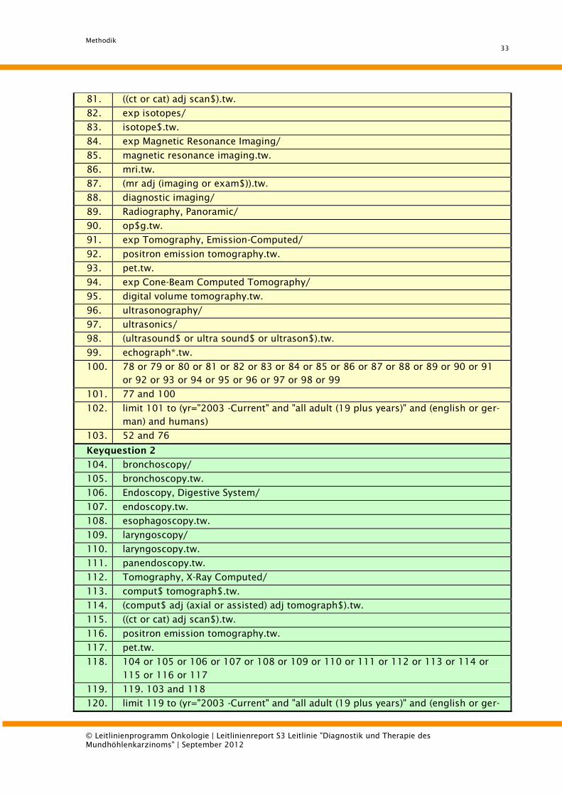
Neben den SIGN-Filtern (vgl. http://www.sign.ac.uk/methodology/filters.html,
letzter Zugriff am 31.01.2011) für „Randomized controlled trials“ und
„Observational studies“ wurden bei Keyquestion 2 und 3 zudem der Filter für
„Diagnostic studies“ verwendet. Abbildung 3 Literaturrecherche zeigt die er-
zielten Hits der Recherchen je Keyquestion sowie die 3014 letztendlich zu
sichtenden Abstracts.
Suchergebnis je Keyquestion (KQ)
Medline
Embase
743
475
KQ 1 KQ 2 KQ 3 KQ 4 KQ 5
650
494
1812
1245
673
286
167
164
Treffer ohne Dubletten
Medline: 2282
Embase: 732
Sichtung von 3014 Abstracts
LiteraturrechercheDatenbanken: Medline, Embase
Einschränkungen: Englisch, Deutsch; ab 2003
Suchergebnis je Keyquestion (KQ)
Medline
Embase
743
475
KQ 1 KQ 2 KQ 3 KQ 4 KQ 5
650
494
1812
1245
673
286
167
164
Treffer ohne Dubletten
Medline: 2282
Embase: 732
Sichtung von 3014 Abstracts
LiteraturrechercheDatenbanken: Medline, Embase
Einschränkungen: Englisch, Deutsch; ab 2003
Abbildung 3 Literaturrecherche
Methodik
36
© Leitlinienprogramm Onkologie | Leitlinienreport S3 Leitlinie "Diagnostik und Therapie des
Mundhöhlenkarzinoms" | September 2012
Handsearch
Zudem wurde die Studie Mucke et al., 2011 [11] von den Experten als weitere
Literaturquelle zur Evaluation eingebracht.
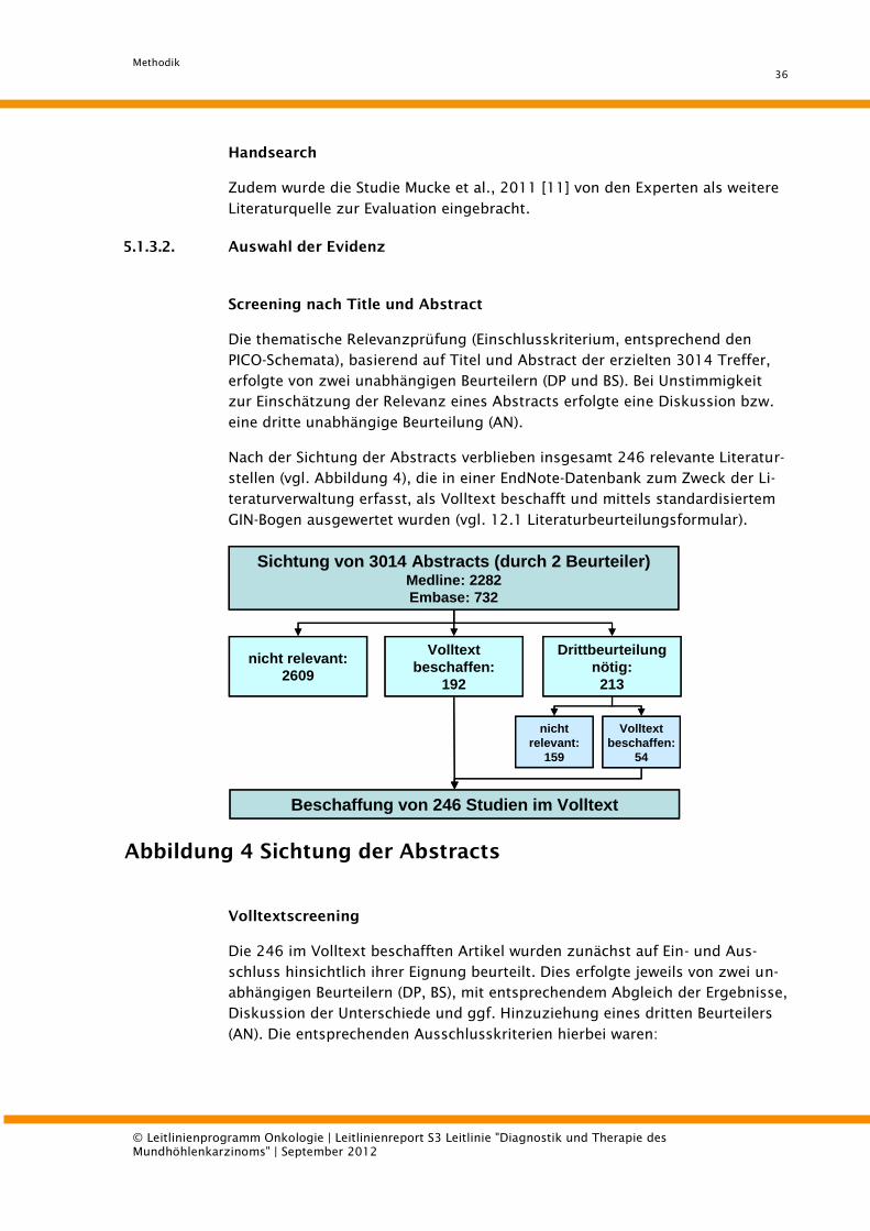
5.1.3.2. Auswahl der Evidenz
Screening nach Title und Abstract
Die thematische Relevanzprüfung (Einschlusskriterium, entsprechend den
PICO-Schemata), basierend auf Titel und Abstract der erzielten 3014 Treffer,
erfolgte von zwei unabhängigen Beurteilern (DP und BS). Bei Unstimmigkeit
zur Einschätzung der Relevanz eines Abstracts erfolgte eine Diskussion bzw.
eine dritte unabhängige Beurteilung (AN).
Nach der Sichtung der Abstracts verblieben insgesamt 246 relevante Literatur-
stellen (vgl. Abbildung 4), die in einer EndNote-Datenbank zum Zweck der Li-
teraturverwaltung erfasst, als Volltext beschafft und mittels standardisiertem
GIN-Bogen ausgewertet wurden (vgl. 12.1 Literaturbeurteilungsformular).
Volltext
beschaffen:
54
nicht relevant:
2609
nicht
relevant:
159
Beschaffung von 246 Studien im Volltext
Sichtung von 3014 Abstracts (durch 2 Beurteiler)Medline: 2282
Embase: 732
Volltext
beschaffen:
192
Drittbeurteilung
nötig:
213
Volltext
beschaffen:
54
nicht relevant:
2609
nicht
relevant:
159
Beschaffung von 246 Studien im Volltext
Sichtung von 3014 Abstracts (durch 2 Beurteiler)Medline: 2282
Embase: 732
Volltext
beschaffen:
192
Drittbeurteilung
nötig:
213
Abbildung 4 Sichtung der Abstracts
Volltextscreening
Die 246 im Volltext beschafften Artikel wurden zunächst auf Ein- und Aus-
schluss hinsichtlich ihrer Eignung beurteilt. Dies erfolgte jeweils von zwei un-
abhängigen Beurteilern (DP, BS), mit entsprechendem Abgleich der Ergebnisse,
Diskussion der Unterschiede und ggf. Hinzuziehung eines dritten Beurteilers
(AN). Die entsprechenden Ausschlusskriterien hierbei waren:
Methodik
37
© Leitlinienprogramm Onkologie | Leitlinienreport S3 Leitlinie "Diagnostik und Therapie des
Mundhöhlenkarzinoms" | September 2012
a) Sprache (nicht Englisch oder Deutsch)
b) Kein passender Studientyp (Fall-Kontroll, Krebsregisterdaten)
c) Falsches Thema/Intervention
d) Keine Originaldaten
e) Surrogatparameter (durch Beschreibung für die LL nicht relevanter Ergeb-
nisse)
f) Kein (definiertes) Mundhöhlen-CA / (<50% Mundhöhlen-CA bzw. keine
Aussage zur Lokalisation im Head and Neck Bereich)
g) Baseline differences (Gruppen nicht vergleichbar)
h) < 10 Patienten pro Studienarm
i) Keine relevanten Wirksamkeitsdaten
j) Sonstiges (mit Angabe des Grundes)
k) Volltext nicht zu beschaffen
5.1.3.3. Bewertung der Evidenz
Von den 246 im Volltext beschafften Artikeln konnten insgesamt 117 Studien
als relevant zur Beantwortung der fünf Keyquestions in die Leitlinie aufge-
nommen werden (inkl. Handsearch). 129 Studien wurden in diesem Schritt von
der weiteren Evaluation ausgeschlossen.
Es erfolgte eine systematische Daten-Extraktion der gefundenen Studien mit-
tels Literaturbeurteilungsformular des Guidelines International Network (GIN,
http://www.g-i-n.net). Hierzu wurde entweder das Template für Diagnostik-
Studien oder für Interventions-Studien angewandt. Dies erfolgte jeweils von
zwei unabhängigen Beurteilern (AS, SS), mit entsprechendem Abgleich der Er-
gebnisse, Diskussion der Unterschiede und ggf. Hinzuziehung eines dritten
Beurteilers (AN). Die Bewertungsbögen können auf Wunsch bei der Devision of
Evidence Based Medicine eingesehen werden.
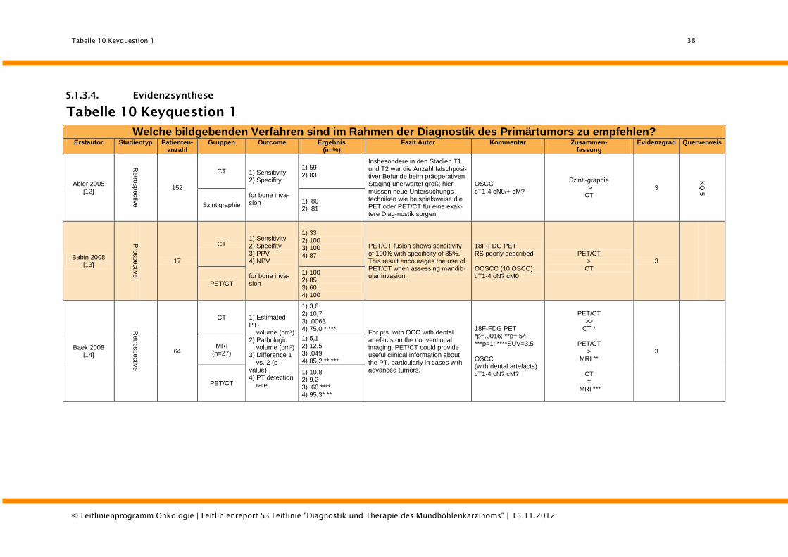
Tabelle 10 Keyquestion 1
© Leitlinienprogramm Onkologie | Leitlinienreport S3 Leitlinie "Diagnostik und Therapie des Mundhöhlenkarzinoms" | 15.11.2012
38
5.1.3.4. Evidenzsynthese
Tabelle 10 Keyquestion 1
Welche bildgebenden Verfahren sind im Rahmen der Diagnostik des Primärtumors zu empfehlen?
Erstautor Studientyp Patienten- anzahl
Gruppen Outcome Ergebnis (in %)
Fazit Autor Kommentar Zusammen- fassung
Evidenzgrad Querverweis
Abler 2005 [12]
Retro
sp
ectiv
e
152
CT 1) Sensitivity 2) Specifity
for bone inva-sion
1) 59 2) 83
Insbesondere in den Stadien T1 und T2 war die Anzahl falschposi-tiver Befunde beim präoperativen Staging unerwartet groß; hier müssen neue Untersuchungs-techniken wie beispielsweise die PET oder PET/CT für eine exak-tere Diag-nostik sorgen.
OSCC cT1-4 cN0/+ cM?
Szinti-graphie
> CT
3
KQ
5
Szintigraphie 1) 80 2) 81
Babin 2008 [13]
Pro
sp
ectiv
e
17
CT 1) Sensitivity 2) Specifity 3) PPV 4) NPV for bone inva-sion
1) 33 2) 100 3) 100 4) 87
PET/CT fusion shows sensitivity of 100% with specificity of 85%. This result encourages the use of PET/CT when assessing mandib-ular invasion.
18F-FDG PET RS poorly described OOSCC (10 OSCC) cT1-4 cN? cM0
PET/CT >
CT 3
PET/CT
1) 100 2) 85 3) 60 4) 100
Baek 2008 [14]
Retro
sp
ectiv
e
64
CT 1) Estimated PT- volume (cm³) 2) Pathologic volume (cm³) 3) Difference 1 vs. 2 (p-value) 4) PT detection rate
1) 3,6 2) 10,7 3) .0063 4) 75,0 * ***
For pts. with OCC with dental artefacts on the conventional imaging, PET/CT could provide useful clinical information about the PT, particularly in cases with advanced tumors.
18F-FDG PET *p=.0016; **p=.54; ***p=1; ****SUV=3.5 OSCC (with dental artefacts) cT1-4 cN? cM?
PET/CT >>
CT *
PET/CT >
MRI **
CT =
MRI ***
3
MRI (n=27)
1) 5,1 2) 12,5 3) .049 4) 85,2 ** ***
PET/CT
1) 10,8 2) 9,2 3) .60 **** 4) 95,3* **
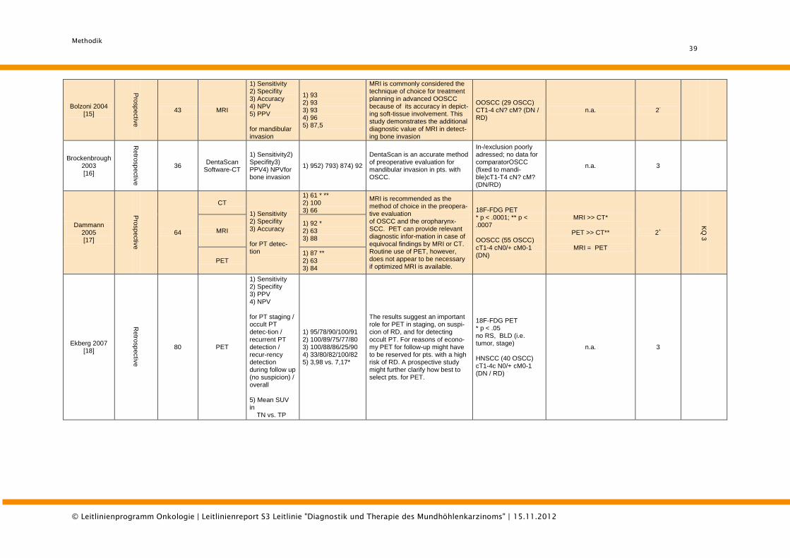
Methodik
39
© Leitlinienprogramm Onkologie | Leitlinienreport S3 Leitlinie "Diagnostik und Therapie des Mundhöhlenkarzinoms" | 15.11.2012
Bolzoni 2004 [15]
Pro
sp
ectiv
e
43 MRI
1) Sensitivity 2) Specifity 3) Accuracy 4) NPV 5) PPV for mandibular invasion
1) 93 2) 93 3) 93 4) 96 5) 87,5
MRI is commonly considered the technique of choice for treatment planning in advanced OOSCC because of its accuracy in depict-ing soft-tissue involvement. This study demonstrates the additional diagnostic value of MRI in detect-ing bone invasion
OOSCC (29 OSCC) CT1-4 cN? cM? (DN / RD)
n.a. 2-
Brockenbrough 2003 [16]
Retro
sp
ectiv
e
36 DentaScan
Software-CT
1) Sensitivity2) Specifity3) PPV4) NPVfor bone invasion
1) 952) 793) 874) 92
DentaScan is an accurate method of preoperative evaluation for mandibular invasion in pts. with OSCC.
In-/exclusion poorly adressed; no data for comparatorOSCC (fixed to mandi-ble)cT1-T4 cN? cM? (DN/RD)
n.a. 3
Dammann 2005 [17]
Pro
sp
ectiv
e
64
CT
1) Sensitivity 2) Specifity 3) Accuracy for PT detec-tion
1) 61 * ** 2) 100 3) 66
MRI is recommended as the method of choice in the preopera-tive evaluation of OSCC and the oropharynx-SCC. PET can provide relevant diagnostic infor-mation in case of equivocal findings by MRI or CT. Routine use of PET, however, does not appear to be necessary if optimized MRI is available.
18F-FDG PET * p < .0001; ** p < .0007 OOSCC (55 OSCC) cT1-4 cN0/+ cM0-1 (DN)
MRI >> CT*
PET >> CT**
MRI = PET
2+
KQ
3
MRI 1) 92 * 2) 63 3) 88
PET 1) 87 ** 2) 63 3) 84
Ekberg 2007 [18]
Retro
sp
ectiv
e
80 PET
1) Sensitivity 2) Specifity 3) PPV 4) NPV for PT staging / occult PT detec-tion / recurrent PT detection / recur-rency detection during follow up (no suspicion) / overall 5) Mean SUV in TN vs. TP
1) 95/78/90/100/91 2) 100/89/75/77/80 3) 100/88/86/25/90 4) 33/80/82/100/82 5) 3,98 vs. 7,17*
The results suggest an important role for PET in staging, on suspi-cion of RD, and for detecting occult PT. For reasons of econo-my PET for follow-up might have to be reserved for pts. with a high risk of RD. A prospective study might further clarify how best to select pts. for PET.
18F-FDG PET * p < .05 no RS, BLD (i.e. tumor, stage) HNSCC (40 OSCC) cT1-4c N0/+ cM0-1 (DN / RD)
n.a. 3
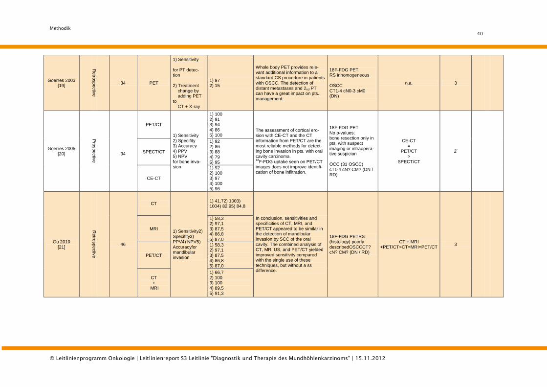
Methodik
40
© Leitlinienprogramm Onkologie | Leitlinienreport S3 Leitlinie "Diagnostik und Therapie des Mundhöhlenkarzinoms" | 15.11.2012
Goerres 2003 [19]
Retro
sp
ectiv
e
34 PET
1) Sensitivity for PT detec-tion 2) Treatment change by adding PET to CT + X-ray
1) 97 2) 15
Whole body PET provides rele-vant additional information to a standard CS procedure in patients with OSCC. The detection of distant metastases and 2nd PT can have a great impact on pts. management.
18F-FDG PET RS inhomogeneous OSCC CT1-4 cN0-3 cM0 (DN)
n.a. 3
Goerres 2005 [20]
Pro
sp
ectiv
e
34
PET/CT
1) Sensitivity 2) Specifity 3) Accuracy 4) PPV 5) NPV for bone inva-sion
1) 100 2) 91 3) 94 4) 86 5) 100
The assessment of cortical ero-sion with CE-CT and the CT information from PET/CT are the most reliable methods for detect-ing bone invasion in pts. with oral cavity carcinoma. 18
F-FDG uptake seen on PET/CT images does not improve identifi-cation of bone infiltration.
18F-FDG PET No p-values; bone resection only in pts. with suspect imaging or intraopera-tive suspicion OCC (31 OSCC) cT1-4 cN? CM? (DN / RD)
CE-CT =
PET/CT >
SPECT/CT
2-
SPECT/CT
1) 92 2) 86 3) 88 4) 79 5) 95
CE-CT
1) 92 2) 100 3) 97 4) 100 5) 96
Gu 2010 [21]
Retro
sp
ectiv
e
46
CT
1) Sensitivity2) Specifity3) PPV4) NPV5) Accuracyfor mandibular invasion
1) 41,72) 1003) 1004) 82,95) 84,8
In conclusion, sensitivities and specificities of CT, MRI, and PET/CT appeared to be similar in the detection of mandibular invasion by SCC of the oral cavity. The combined analysis of CT, MR, US, and PET/CT yielded improved sensitivity compared with the single use of these techniques, but without a ss difference.
18F-FDG PETRS (histology) poorly describedOSCCCT? cN? CM? (DN / RD)
CT + MRI +PET/CT>CT=MRI=PET/CT
3
MRI
1) 58,3 2) 97,1 3) 87,5 4) 86,8 5) 87,0
PET/CT
1) 58,3 2) 97,1 3) 87,5 4) 86,8 5) 87,0
CT +
MRI
1) 66,7 2) 100 3) 100 4) 89,5 5) 91,3
Methodik
41
© Leitlinienprogramm Onkologie | Leitlinienreport S3 Leitlinie "Diagnostik und Therapie des Mundhöhlenkarzinoms" | 15.11.2012
CT +
PET/CT
1) 66,7 2) 100 3) 100 4) 89,5 5) 91,3
MRI +
PET/CT
1) 75,0 2) 100 3) 100 4) 91,9 5) 93,5
CT +
MRI +
PET/CT
1) 83,3 2) 100 3) 100 4) 94,4 5) 95,7
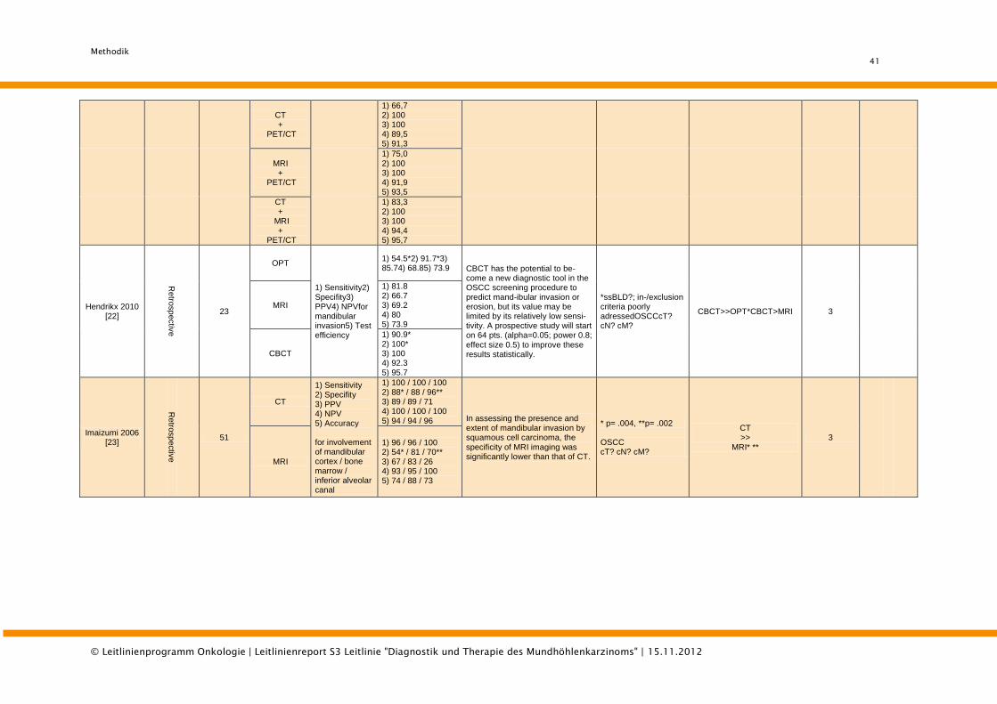
Hendrikx 2010 [22]
Retro
sp
ectiv
e
23
OPT
1) Sensitivity2) Specifity3) PPV4) NPVfor mandibular invasion5) Test efficiency
1) 54.5*2) 91.7*3) 85.74) 68.85) 73.9 CBCT has the potential to be-
come a new diagnostic tool in the OSCC screening procedure to predict mand-ibular invasion or erosion, but its value may be limited by its relatively low sensi-tivity. A prospective study will start on 64 pts. (alpha=0.05; power 0.8; effect size 0.5) to improve these results statistically.
*ssBLD?; in-/exclusion criteria poorly adressedOSCCcT? cN? cM?
CBCT>>OPT*CBCT>MRI 3
MRI
1) 81.8 2) 66.7 3) 69.2 4) 80 5) 73.9
CBCT
1) 90.9* 2) 100* 3) 100 4) 92.3 5) 95.7
Imaizumi 2006 [23]
Retro
sp
ectiv
e
51
CT
1) Sensitivity 2) Specifity 3) PPV 4) NPV 5) Accuracy for involvement of mandibular cortex / bone marrow / inferior alveolar canal
1) 100 / 100 / 100 2) 88* / 88 / 96** 3) 89 / 89 / 71 4) 100 / 100 / 100 5) 94 / 94 / 96 In assessing the presence and
extent of mandibular invasion by squamous cell carcinoma, the specificity of MRI imaging was significantly lower than that of CT.
* p= .004, **p= .002 OSCC cT? cN? cM?
CT >>
MRI* ** 3
MRI
1) 96 / 96 / 100 2) 54* / 81 / 70** 3) 67 / 83 / 26 4) 93 / 95 / 100 5) 74 / 88 / 73
Methodik
42
© Leitlinienprogramm Onkologie | Leitlinienreport S3 Leitlinie "Diagnostik und Therapie des Mundhöhlenkarzinoms" | 15.11.2012
Jones 2005 [24]
Retro
sp
ectiv
e
88 PET
1) Sensitivity 2) Specifity for PT detec-tion (DN / RD)
1) 96,3 / 85,7 2) n.a. / 50,0
Overall PET has a useful role in the diag-nosis of HN-malig., and in the demon-stration of occult / hidden PT, distant & metastatic disease. It should always be used as an adjunct to other clinical in-formation and results must be interpret-ed in the light of clinical findings.
18F-FDG PET In-/exclusion criteria poorly adressed OOSCC (79 OSCC) cT? cN0/+ cM0-1 DN (n=54) / RD (n=34)
n.a. 3
KQ
2 u
nd
3
Krabbe 2008 [25]
Not c
ited
38 PET
1) Sensitivity for PT detection
1) 95
Although PET performed better than conventional imaging modali-ties, sensitivity was lower than desired. As a consequence, clinical appli-cation of PET in the patient staged as cN0 is limited.
18F-FDG PET RS n.a. in 8 pts.; no data for comparator OOSCC (35 OSCC) cT1-4 cN0 cM?
n.a. 3
KQ
3
Nakamoto 2009 [26]
Retro
sp
ectiv
e
46
MRI+PET 1) Sensitivity for PT detection
1) 100 Image fusion from MRI + PET might be useful in evaluating HN-CA, especially in suspected RD rather than in DN.
18F-FDG PETOO-CAcT? cN? cM? (DN / RD)
MRI + PET=MRI 3
KQ
2 u
nd 3
MRI 1) 98
Ng 2005 [27]
Pro
sp
ectiv
e
124
CT +
MRI
1) Accuracy for PT detection
1) 87,1
PET is superior to CT+MRI in the detect-ion of cervical status of OSCC. The sensitivity of PET for the detection of LNM on a level-by-level basis was ss higher than that of CT+MRI, whereas speci-ficities appeared to be similar. Visual correlation of PET+CT+MRI show-ed a trend of increased diagnostic ac-curacy over PET alone but without a ss difference, and its sensitivity was still not high enough to replace pathologic LN staging based on ND.
18F-FDG PET OSCC cT1-4 cN? CM? (DN)
CT +
MRI +
PET >
PET >
CT +
MRI
2++
KQ
3
PET 1) 98,4
CT +
MRI +
PET
1) 99,2
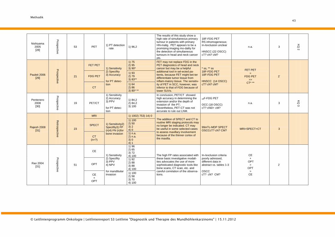
Methodik
43
© Leitlinienprogramm Onkologie | Leitlinienreport S3 Leitlinie "Diagnostik und Therapie des Mundhöhlenkarzinoms" | 15.11.2012
Nishiyama 2005 [28]
Pro
sp
ectiv
e
53 PET 1) PT detection rate
1) 96,2
The results of this study show a high rate of simultaneous primary tumour in patients with primary HN-malig. PET appears to be a promising imaging mo-dality for the detection of simultaneous tumours in head and neck cancer pts.
18F-FDG PET RS inhomogeneous; in-/exclusion unclear HNSCC (22 OSCC) cT? cN? cM?
n.a. 3
KQ
2
Pauleit 2006 [29]
Pro
sp
ectiv
e
21
FET PET 1) Sensitivity 2) Specifity 3) Accuracy for PT detec-tion
1) 75 2) 95 3) 90*
FET may not replace FDG in the PET diagnostics of head and neck cancer but may be a helpful additional tool in sel-ected pa-tients, because FET might bet-ter differentiate tumor tissue from inflam-matory tissue. The sensitiv-ity of FET in SCC, however, was inferior to that of FDG because of lower SUVs.
* ss, ** ss 18F-FDG PET 18F-FDG PET HNSCC (14 OSCC) cT? cN? cM?
FET PET =
FDG PET >>
CT* **
2-
FDG PET 1) 93 2) 79 3) 83**
CT 1) 64 2) 86 3) 80* **
Pentenero 2008 [30]
Pro
sp
ectiv
e
19 PET/CT
1) Sensitivity 2) Accuracy 3) PPV for PT detec-tion
1) 84,2 2) 84,2 3) 100
In conclusion, PET/CT showed high accuracy in determining the extension and/or the depth of invasion of the PT; Nevertheless, PET-CT was not accurate to rule out LNM.
18F-FDG PET OCC (18 OSCC) cT? cN0/+ cM?
n.a. 3
KQ
3
Rajesh 2008 [31]
Retro
sp
ectiv
e
23
MRI
1) Sensitivity2) Specifity3) FP (n)4) FN (n)for
bone invasion
1) 1002) 753) 14) 0
The addition of SPECT and CT to routine MRI staging protocols may no longer be indicated. CT may be useful in some selected cases to assess maxillary involvement because of the thinner cortex of the maxilla.
99mTc-MDP SPECT OSCCcT? cN? CM?
MRI>SPECT+CT 3
SPECT
1) 100 2) 50 3) 2 4) 0
CT (n=?)
1) n.a. 2) n.a. 3) 0 4) 1
Rao 2004 [31]
Pro
sp
ectiv
e
51
CE
1) Sensitivity 2) Specifity 3) PPV 4) NPV for mandibular invasion
1) 96 2) 65 3) 72 4) 100 The high FP rates associated with
these basic investigative modali-ties advocates the use of more sophisticated diagnostic tools like bone scans, CT scan, etc. and careful correlation of the observa-tions.
In-/exclusion criteria poorly adressed, different data in abstract vs. tables 1-3 OSCC cT? cN? CM?
CE +
OPT >
OPT >
CE
2-
OPT
1) 92 2) 88 3) 88 4) 100
CE +
OPT
1) 100 2) 58 3) 70 4) 100
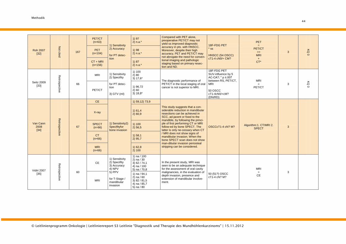
Methodik
44
© Leitlinienprogramm Onkologie | Leitlinienreport S3 Leitlinie "Diagnostik und Therapie des Mundhöhlenkarzinoms" | 15.11.2012
Roh 2007 [32]
Not c
ited
167
PET/CT (n=63)
1) Sensitivity 2) Accuracy for PT detec-tion
1) 97 2) n.a.*
Compared with PET alone, preoperative PET/CT may not yield ss improved diagnostic accuracy in pts. with HNSCC. Moreover, despite their high accuracy, PET and PET/CT may not abrogate the need for conven-tional imaging and pathologic staging based on primary resec-tion and ND.
18F-FDG PET * ss HNSCC (54 OSCC) cT1-4 cN0/+ CM?
PET =
PET/CT >>
MRI +
CT*
3
KQ
3
PET (n=104)
1) 98 2) n.a.*
CT + MRI (n=156)
1) 87 2) n.a.*
Seitz 2009 [33]
Retro
sp
ectiv
e
66
MRI 1) Sensitivity 2) Specifity for PT detec-tion 3) GTV (ml)
1) 100 2) 80 3) 17,6*
The diagnostic performance of PET/CT in the local staging of oral cancer is not superior to MRI.
18F-FDG PET SUV-influence by 5 AC-CA?; * p ≤.007 between RS, PET/CT, MRI 50 OSCC cT1-4cN0/+cM? (DN/RD)
MRI =
PET/CT 3
KQ
3
PET/CT 1) 96,72 2) 60 3) 18,8*
Van Cann 2008 [34]
Retro
sp
ectiv
e
67
CE
1) Sensitivity2) Specifityfor bone invasion
1) 59,12) 73,9
This study suggests that a con-siderable reduction in mandibular resections can be achieved in SCC, ad-jacent or fixed to the mandible, by following the princi-ple of first performing CT or MRI follow-ed by bone SPECT. The latter is only ne-cessary when CT / MRI does not show signs of mandibular invasion. When the bone SPECT scan does not show man-dibular invasion periosteal stripping can be considered.
OSCCcT1-4 cN? M? Algorithm:1. CT/MRI 2.
SPECT 3
X-ray 1) 61,4 2) 60,9
SPECT (n=66)
1) 100 2) 56,5
CT (n=66)
1) 58,1 2) 95,7
MRI (n=66)
1) 62,8 2) 100
Vidiri 2007 [35]
Retro
sp
ectiv
e
60
CE
1) Sensitivity 2) Specifity 3) Accuracy 4) NPV 5) PPV for T-Stage / mandibular invasion
1) na / 100 2) na / 30 3) 62 / 74,1 4) na / 100 5) na / 70,8
In the present study, MRI was seen to be an adequate technique for the assessment of oral cavity malignancies, in the evaluation of depth invasion, presence and extension of mandibular involve-ment.
50 (51?) OSCC cT1-4 cN? M?
MRI >
CE 3
MRI
1) na / 94,1 2) na / 60 3) 82 / 81,5 4) na / 85,7 5) na / 80
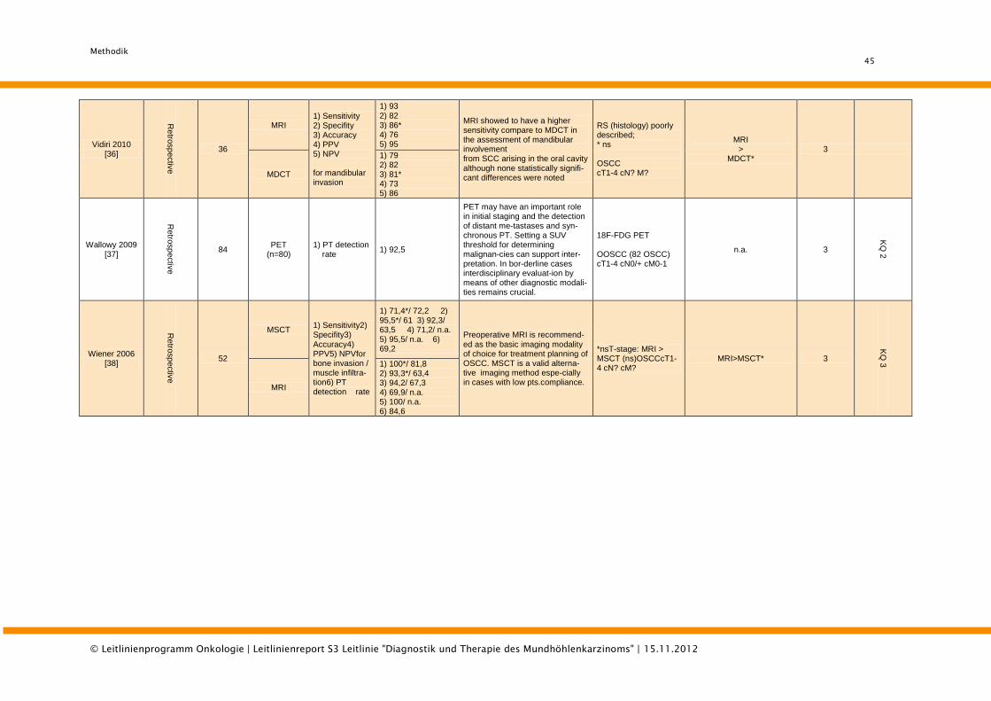
Methodik
45
© Leitlinienprogramm Onkologie | Leitlinienreport S3 Leitlinie "Diagnostik und Therapie des Mundhöhlenkarzinoms" | 15.11.2012
Vidiri 2010 [36]
Retro
sp
ectiv
e
36
MRI 1) Sensitivity 2) Specifity 3) Accuracy 4) PPV 5) NPV for mandibular invasion
1) 93 2) 82 3) 86* 4) 76 5) 95
MRI showed to have a higher sensitivity compare to MDCT in the assessment of mandibular involvement from SCC arising in the oral cavity although none statistically signifi-cant differences were noted
RS (histology) poorly described; * ns OSCC cT1-4 cN? M?
MRI >
MDCT* 3
MDCT
1) 79 2) 82 3) 81* 4) 73 5) 86
Wallowy 2009 [37]
Retro
sp
ectiv
e
84 PET
(n=80) 1) PT detection rate
1) 92,5
PET may have an important role in initial staging and the detection of distant me-tastases and syn-chronous PT. Setting a SUV threshold for determining malignan-cies can support inter-pretation. In bor-derline cases interdisciplinary evaluat-ion by means of other diagnostic modali-ties remains crucial.
18F-FDG PET OOSCC (82 OSCC) cT1-4 cN0/+ cM0-1
n.a. 3
KQ
2
Wiener 2006 [38]
Retro
sp
ectiv
e
52
MSCT 1) Sensitivity2) Specifity3) Accuracy4) PPV5) NPVfor bone invasion / muscle infiltra-tion6) PT detection rate
1) 71,4*/ 72,2 2) 95,5*/ 61 3) 92,3/ 63,5 4) 71,2/ n.a. 5) 95,5/ n.a. 6) 69,2
Preoperative MRI is recommend-ed as the basic imaging modality of choice for treatment planning of OSCC. MSCT is a valid alterna-tive imaging method espe-cially in cases with low pts.compliance.
*nsT-stage: MRI > MSCT (ns)OSCCcT1-4 cN? cM?
MRI>MSCT* 3
KQ
3
MRI
1) 100*/ 81,8 2) 93,3*/ 63,4 3) 94,2/ 67,3 4) 69,9/ n.a. 5) 100/ n.a. 6) 84,6
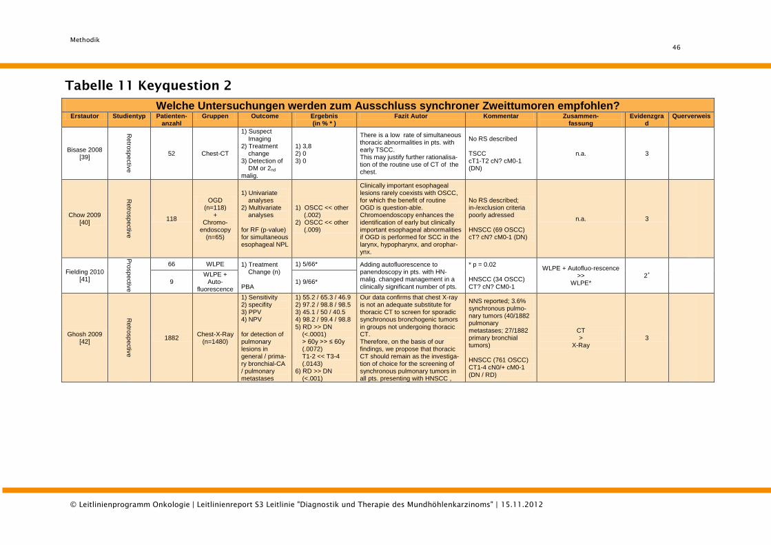
Methodik
46
© Leitlinienprogramm Onkologie | Leitlinienreport S3 Leitlinie "Diagnostik und Therapie des Mundhöhlenkarzinoms" | 15.11.2012
Tabelle 11 Keyquestion 2
Welche Untersuchungen werden zum Ausschluss synchroner Zweittumoren empfohlen?
Erstautor Studientyp Patienten- anzahl
Gruppen Outcome Ergebnis (in % * )
Fazit Autor Kommentar Zusammen- fassung
Evidenzgrad
Querverweis
Bisase 2008 [39]
Retro
sp
ectiv
e
52 Chest-CT
1) Suspect Imaging 2) Treatment change 3) Detection of DM or 2nd
malig.
1) 3,8 2) 0 3) 0
There is a low rate of simultaneous thoracic abnormalities in pts. with early TSCC. This may justify further rationalisa-tion of the routine use of CT of the chest.
No RS described TSCC cT1-T2 cN? cM0-1 (DN)
n.a. 3
Chow 2009 [40]
Retro
sp
ectiv
e
118
OGD (n=118)
+ Chromo-
endoscopy (n=65)
1) Univariate analyses 2) Multivariate analyses for RF (p-value) for simultaneous esophageal NPL
1) OSCC << other (.002) 2) OSCC << other (.009)
Clinically important esophageal lesions rarely coexists with OSCC, for which the benefit of routine OGD is question-able. Chromoendoscopy enhances the identification of early but clinically important esophageal abnormalities if OGD is performed for SCC in the larynx, hypopharynx, and orophar-ynx.
No RS described; in-/exclusion criteria poorly adressed HNSCC (69 OSCC) cT? cN? cM0-1 (DN)
n.a. 3
Fielding 2010 [41]
Pro
sp
ectiv
e
66 WLPE 1) Treatment Change (n) PBA
1) 5/66* Adding autofluorescence to panendoscopy in pts. with HN-malig. changed management in a clinically significant number of pts.
* p = 0.02 HNSCC (34 OSCC) CT? cN? CM0-1
WLPE + Autofluo-rescence >>
WLPE* 2
+
9 WLPE +
Auto-fluorescence
1) 9/66*
Ghosh 2009 [42]
Retro
sp
ectiv
e
1882 Chest-X-Ray
(n=1480)
1) Sensitivity 2) specifity 3) PPV 4) NPV for detection of pulmonary lesions in general / prima-ry bronchial-CA / pulmonary metastases
1) 55.2 / 65.3 / 46.9 2) 97.2 / 98.8 / 98.5 3) 45.1 / 50 / 40.5 4) 98.2 / 99.4 / 98.8 5) RD >> DN (<.0001) > 60y >> ≤ 60y (.0072) T1-2 << T3-4 (.0143) 6) RD >> DN (<.001)
Our data confirms that chest X-ray is not an adequate substitute for thoracic CT to screen for sporadic synchronous bronchogenic tumors in groups not undergoing thoracic CT. Therefore, on the basis of our findings, we propose that thoracic CT should remain as the investiga-tion of choice for the screening of synchronous pulmonary tumors in all pts. presenting with HNSCC ,
NNS reported; 3.6% synchronous pulmo-nary tumors (40/1882 pulmonary metastases; 27/1882 primary bronchial tumors) HNSCC (761 OSCC) CT1-4 cN0/+ cM0-1 (DN / RD)
CT >
X-Ray 3
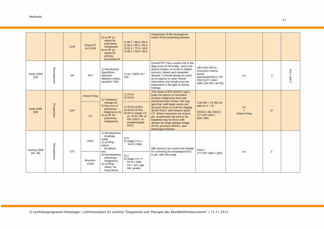
Methodik
47
© Leitlinienprogramm Onkologie | Leitlinienreport S3 Leitlinie "Diagnostik und Therapie des Mundhöhlenkarzinoms" | 15.11.2012
1144 Chest-CT (n=1144)
5) ss RF (p- value) for pulmonary metastases 6) ss RF (p- value) for primary bronchial-CA
1) 89.7 / 96.6 / 85.4 2) 94.2 / 99.1 / 95.3 3) 53.1 / 72.5 / 44.5 4) 99.2 / 99.9 / 99.3
irrespective of the locoregional extent of the presenting disease.
Jones 2005 [24]
Retro
sp
ectiv
e
88 PET
1) Sensitivity2) Specifityfor detection
ofdistant metas-tasis(DN / RD)
1) na / 1002) na / 100
Overall PET has a useful role in the diag-nosis of HN-malig., and in the demon-stration of occult or hidden tumours, distant and metastatic disease. It should always be used
as an adjunct to other clinical information and results must be interpreted in the light of clinical findings
18F-FDG PETIn-/exclusion criteria poorly adressedOOSCC (79 OSCC)cT? cN0/+ cM0-1(54 DN / 34 RD)
n.a. 3
KQ
1 u
nd 3
Keith 2006 [43]
Pro
sp
ectiv
e
116*
Chest-X-Ray
1) Treatment change (n) 2) Detection of
pulmonary malignancy (n) 3) ss RF for pulmonary malignancy
1) 2/116 2) 2/116
This series of DN OOSCC had a lower inci-dence of coincident thoracic malignancy than had previously been shown. We sug-gest that, until larger series are accrued, there is a role for staging all DN OSCC with thoracic spiral CT. Where resources are scarce, pts. at particular risk (and to be targeted) may be those with advanc-ed stage disease (stage III+IV), previous HNSCC, and pharyngeal disease.
*116 DN + 11 RD (no data on 1 + 2) OOSCC (81 OSCC) cT? cN? cM0-1 (DN / RD)
CT >
Chest-X-Ray 2+
CT
1) 4/116 (3,5%) 2) 4/116 (3,5%) 3) All ns (stage I+II vs. III+IV; RD vs. DN; OSCC vs. oropharyngeal SCC)
Kesting 2009 [44, 45]
Retro
sp
ectiv
e
570
OGD
1) Simultaneous esophag. malig. 2) ss RF(p-value) for Barret eso. 3) Simultaneous pulmonary malignancy 4) ss RF(p- value) for lung cancer
1) 0 2) Stage I+II << III+IV (.006)
NBI seems to be useful and reliable for screening for esophageal SCC in pts. with HN-malig.
OSCC cT? cN? CM0-1 (DN)
n.a. 2-
Broncho-scopy
3) 2 4) Stage I+II >> III+IV (.038); (ns = sex, age, site, grade)
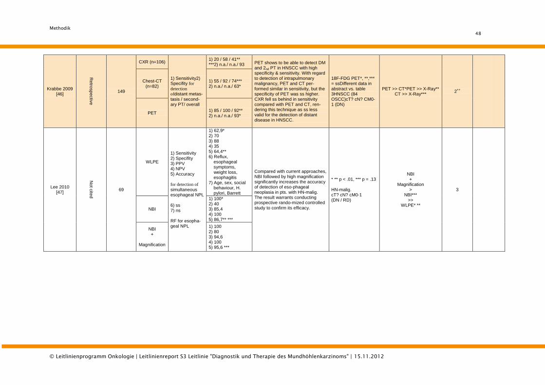
Methodik
48
© Leitlinienprogramm Onkologie | Leitlinienreport S3 Leitlinie "Diagnostik und Therapie des Mundhöhlenkarzinoms" | 15.11.2012
Krabbe 2009 [46]
Retro
sp
ectiv
e
149
CXR (n=106)
1) Sensitivity2) Specifity for
detection
ofdistant metas-tasis / second-ary PT/ overall
1) 20 / 58 / 41** ***2) n.a./ n.a./ 93 PET shows to be able to detect DM
and 2nd PT in HNSCC with high specificity & sensitivity. With regard to detection of intrapulmonary malignancy, PET and CT per-formed similar in sensitivity, but the specificity of PET was ss higher. CXR fell ss behind in sensitivity compared with PET and CT, ren-dering this technique as ss less valid for the detection of distant disease in HNSCC.
18F-FDG PET*, **,*** = ssDifferent data in abstract vs. table 3HNSCC (84 OSCC)cT? cN? CM0-1 (DN)
PET >> CT*PET >> X-Ray** CT >> X-Ray***
2++
Chest-CT (n=82)
1) 55 / 92 / 74*** 2) n.a./ n.a./ 63*
PET 1) 85 / 100 / 92** 2) n.a./ n.a./ 93*
Lee 2010 [47]
Not c
ited
69
WLPE
1) Sensitivity 2) Specifity 3) PPV 4) NPV 5) Accuracy for detection of simultaneous esophageal NPL 6) ss 7) ns RF for esopha-geal NPL
1) 62,9* 2) 70 3) 88 4) 35 5) 64,4** 6) Reflux, esophageal symptoms, weight loss, esophagitis 7) Age, sex, social behaviour, H. pylori, Barrett
Compared with current approaches, NBI followed by high magnification significantly increases the accuracy of detection of eso-phageal neoplasia in pts. with HN-malig. The result warrants conducting prospective rando-mized controlled study to confirm its efficacy.
* ** p < .01, *** p = .13 HN-malig. cT? cN? cM0-1 (DN / RD)
NBI +
Magnification >
NBI*** >>
WLPE* **
3
NBI
1) 100* 2) 40 3) 85,4 4) 100 5) 86,7** ***
NBI +
Magnification
1) 100 2) 80 3) 94,6 4) 100 5) 95,6 ***
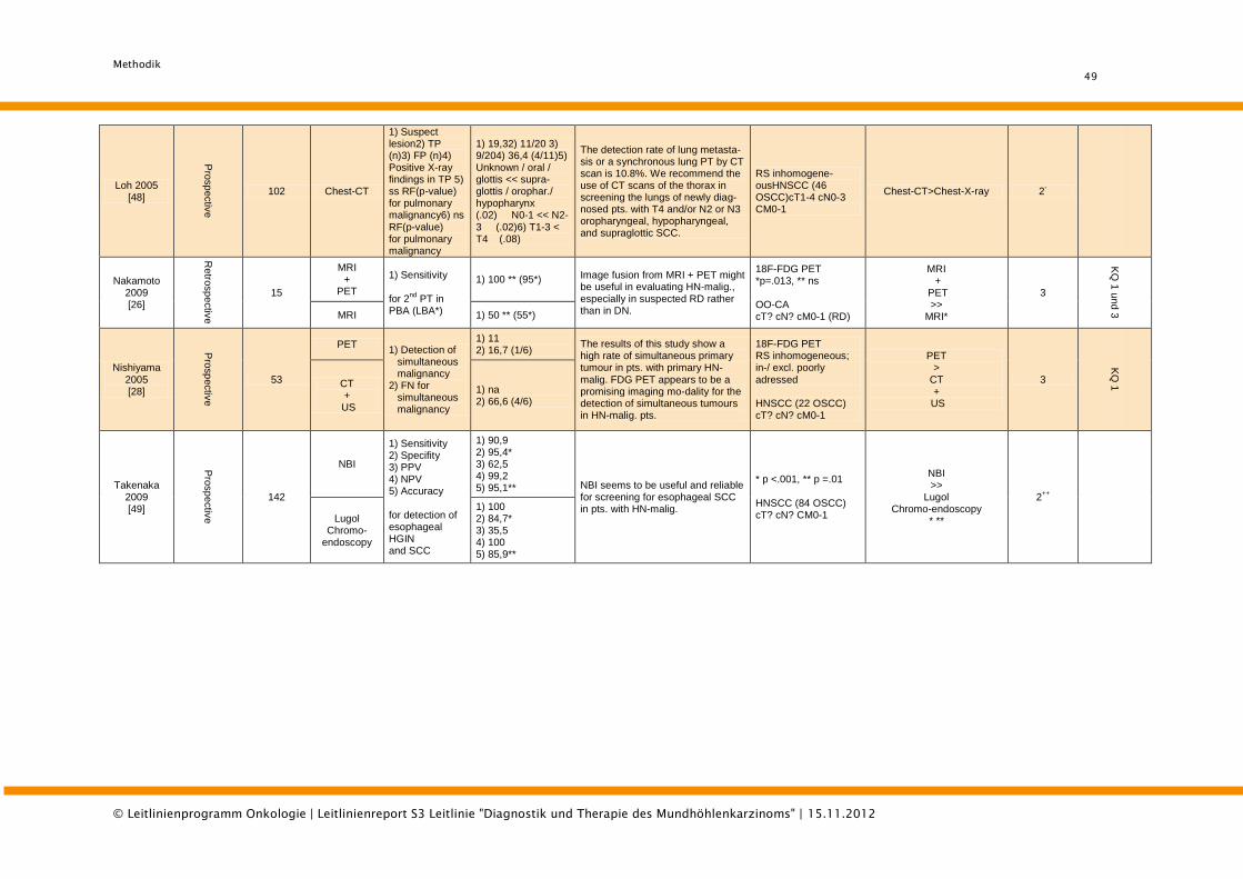
Methodik
49
© Leitlinienprogramm Onkologie | Leitlinienreport S3 Leitlinie "Diagnostik und Therapie des Mundhöhlenkarzinoms" | 15.11.2012
Loh 2005 [48]
Pro
sp
ectiv
e
102 Chest-CT
1) Suspect lesion2) TP (n)3) FP (n)4) Positive X-ray findings in TP 5) ss RF(p-value) for pulmonary malignancy6) ns RF(p-value) for pulmonary malignancy
1) 19,32) 11/20 3) 9/204) 36,4 (4/11)5) Unknown / oral / glottis << supra- glottis / orophar./ hypopharynx (.02) N0-1 << N2-3 (.02)6) T1-3 < T4 (.08)
The detection rate of lung metasta-sis or a synchronous lung PT by CT scan is 10.8%. We recommend the use of CT scans of the thorax in screening the lungs of newly diag-nosed pts. with T4 and/or N2 or N3 oropharyngeal, hypopharyngeal, and supraglottic SCC.
RS inhomogene-ousHNSCC (46 OSCC)cT1-4 cN0-3 CM0-1
Chest-CT>Chest-X-ray 2-
Nakamoto 2009 [26]
Retro
sp
ectiv
e
15
MRI +
PET
1) Sensitivity for 2
nd PT in
PBA (LBA*)
1) 100 ** (95*) Image fusion from MRI + PET might be useful in evaluating HN-malig., especially in suspected RD rather than in DN.
18F-FDG PET *p=.013, ** ns OO-CA cT? cN? cM0-1 (RD)
MRI +
PET >>
MRI*
3
KQ
1 u
nd
3 MRI 1) 50 ** (55*)
Nishiyama 2005 [28]
Pro
sp
ectiv
e
53
PET 1) Detection of simultaneous malignancy 2) FN for simultaneous malignancy
1) 11 2) 16,7 (1/6)
The results of this study show a high rate of simultaneous primary tumour in pts. with primary HN-malig. FDG PET appears to be a promising imaging mo-dality for the detection of simultaneous tumours in HN-malig. pts.
18F-FDG PET RS inhomogeneous; in-/ excl. poorly adressed HNSCC (22 OSCC) cT? cN? cM0-1
PET >
CT +
US
3
KQ
1 CT
+ US
1) na 2) 66,6 (4/6)
Takenaka 2009 [49]
Pro
sp
ectiv
e
142
NBI
1) Sensitivity 2) Specifity 3) PPV 4) NPV 5) Accuracy for detection of esophageal HGIN and SCC
1) 90,9 2) 95,4* 3) 62,5 4) 99,2 5) 95,1** NBI seems to be useful and reliable
for screening for esophageal SCC in pts. with HN-malig.
* p <.001, ** p =.01 HNSCC (84 OSCC) cT? cN? CM0-1
NBI >>
Lugol Chromo-endoscopy
* **
2++
Lugol Chromo-
endoscopy
1) 100 2) 84,7* 3) 35,5 4) 100 5) 85,9**
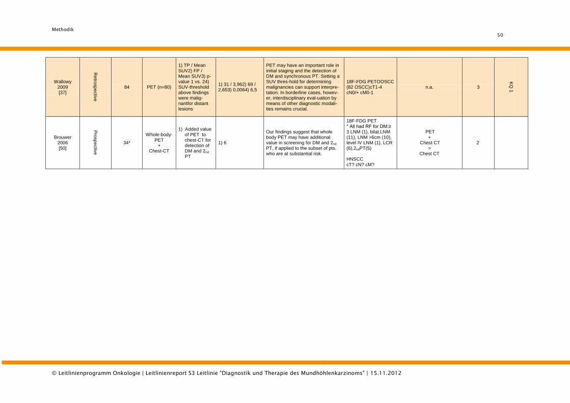
Methodik
50
© Leitlinienprogramm Onkologie | Leitlinienreport S3 Leitlinie "Diagnostik und Therapie des Mundhöhlenkarzinoms" | 15.11.2012
Wallowy 2009 [37]
Retro
sp
ectiv
e
84 PET (n=80)
1) TP / Mean SUV2) FP / Mean SUV3) p-value 1 vs. 24) SUV-threshold above findings were malig-nantfor distant lesions
1) 31 / 3,962) 69 / 2,653) 0,0064) 6,5
PET may have an important role in initial staging and the detection of DM and synchronous PT. Setting a SUV thres-hold for determining malignancies can support interpre-tation. In borderline cases, howev-er, interdisciplinary eval-uation by means of other diagnostic modali-ties remains crucial.
18F-FDG PETOOSCC (82 OSCC)cT1-4 cN0/+ cM0-1
n.a. 3
KQ
1
Brouwer 2006 [50]
Pro
sp
ectiv
e
34*
Whole-body-PET
+ Chest-CT
1) Added value of PET to chest-CT for detection of DM and 2nd PT
1) 6
Our findings suggest that whole body PET may have additional value in screening for DM and 2nd PT, if applied to the subset of pts. who are at substantial risk.
18F-FDG PET * All had RF for DM:≥ 3 LNM (1), bilat.LNM (11), LNM >6cm (10), level IV LNM (1), LCR (6),2ndPT(5) HNSCC cT? cN? cM?
PET +
Chest CT >
Chest CT
2-
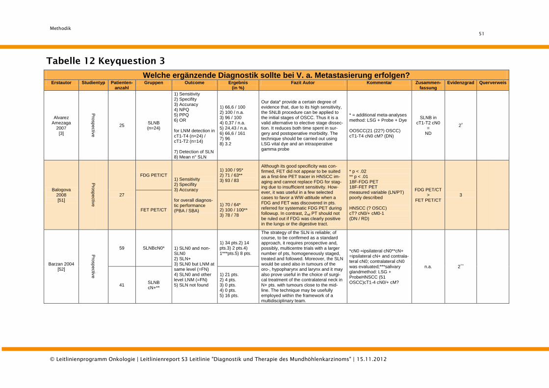
Methodik
51
© Leitlinienprogramm Onkologie | Leitlinienreport S3 Leitlinie "Diagnostik und Therapie des Mundhöhlenkarzinoms" | 15.11.2012
Tabelle 12 Keyquestion 3
Welche ergänzende Diagnostik sollte bei V. a. Metastasierung erfolgen?
Erstautor Studientyp Patienten- anzahl
Gruppen Outcome Ergebnis (in %)
Fazit Autor Kommentar Zusammen- fassung
Evidenzgrad Querverweis
Alvarez Amezaga
2007 [3]
Pro
sp
ectiv
e
25 SLNB (n=24)
1) Sensitivity 2) Specifity 3) Accuracy 4) NPQ 5) PPQ 6) OR for LNM detection in cT1-T4 (n=24) / cT1-T2 (n=14) 7) Detection of SLN 8) Mean n° SLN
1) 66,6 / 100 2) 100 / n.a. 3) 96 / 100 4) 0,37 / n.a. 5) 24,43 / n.a. 6) 66,6 / 161 7) 96 8) 3.2
Our data* provide a certain degree of evidence that, due to its high sensitivity, the SNLB procedure can be applied to the initial stages of OSCC. Thus it is a valid alternative to elective stage dissec-tion. It reduces both time spent in sur-gery and postoperative morbidity. The technique should be carried out using LSG vital dye and an intraoperative gamma probe
* = additional meta-analyses method: LSG + Probe + Dye OOSCC(21 (22?) OSCC) cT1-T4 cN0 cM? (DN)
SLNB in cT1-T2 cN0
= ND
2+
Balogova 2008 [51]
Pro
sp
ectiv
e
27
FDG PET/CT 1) Sensitivity 2) Specifity 3) Accuracy for overall diagnos-tic performance (PBA / SBA)
1) 100 / 95* 2) 71 / 63** 3) 93 / 83
Although its good specificity was con-firmed, FET did not appear to be suited as a first-line PET tracer in HNSCC im-aging and cannot replace FDG for stag-ing due to insufficient sensitivity. How-ever, it was useful in a few selected cases to favor a WW-attitude when a FDG and FET was discovered in pts. referred for systematic FDG PET during followup. In contrast, 2nd PT should not be ruled out if FDG was clearly positive in the lungs or the digestive tract.
* p < .02 ** p < .01 18F-FDG PET 18F-FET PET measured variable (LN/PT) poorly described HNSCC (? OSCC) cT? cN0/+ cM0-1 (DN / RD)
FDG PET/CT >
FET PET/CT 3
FET PET/CT 1) 70 / 64* 2) 100 / 100** 3) 78 / 78
Barzan 2004 [52]
Pro
sp
ectiv
e
59 SLNBcN0* 1) SLN0 and non-SLN0 2) SLN+ 3) SLN0 but LNM at same level (=FN) 4) SLN0 and other level LNM (=FN) 5) SLN not found
1) 34 pts.2) 14 pts.3) 2 pts.4) 1***pts.5) 8 pts.
The strategy of the SLN is reliable; of course, to be confirmed as a standard approach, it requires prospective and, possibly, multicentre trials with a larger number of pts, homogeneously staged, treated and followed. Moreover, the SLN would be used also in tumours of the oro-, hypopharynx and larynx and it may also prove useful in the choice of surgi-cal treatment of the contralateral neck in N+ pts. with tumours close to the mid-line. The technique may be usefully employed within the framework of a multidisciplinary team.
*cN0 =ipsilateral cN0**cN+ =ipsilateral cN+ and contrala-teral cN0; contralateral cN0 was evaluated;***salivary glandmethod: LSG + ProbeHNSCC (51 OSCC)cT1-4 cN0/+ cM?
n.a. 2++
41 SLNB cN+**
1) 21 pts. 2) 4 pts. 3) 0 pts. 4) 0 pts. 5) 16 pts.
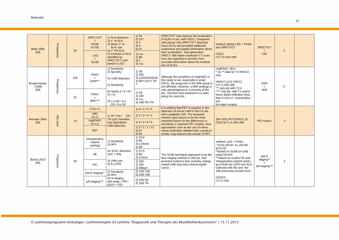
Methodik
52
© Leitlinienprogramm Onkologie | Leitlinienreport S3 Leitlinie "Diagnostik und Therapie des Mundhöhlenkarzinoms" | 15.11.2012
Bilde 2006 [53]
Pro
sp
ectiv
e
34
SPECT/CT +
SLNB (n=33)
1) SLN detection 2) n° of SLN 3) Mean n° of SLN / pts. 4) n° FN SLN 5) Increase of SLN identified by SPECT/CT com-pared to LSG
1) 94 2) 107 3) 3 4) 0 5) 47
SPECT/CT may improve the localization of SLN's in pts. with OSCC. Compared with planar LSG SPECT/CT detected more SLN's and provided additional anatomical and spatial information about their localization. New generation SPECT with higher resolution CT scan-ners are expected to provide more accurate information about the localiza-tion of SLN's.
method: planar LSG + Probe and SPECT/CT OSCC cT1-T2 cN0 cM0
SPECT/CT >
LSG 2
-
LSG +
SLNB
1) n.a. 2) 88 3) 2 4) n.a.
Borgermeester 2008
[54]
Pro
sp
ectiv
e
126 FNAC
+ END***
1) Sensitivity 2) Specifity for LNM detection 3) Sensitivity for levels (I / II / III / IV / V) 4) 1-y OS / 3-y OS / 5-y OS
1) 39 2) 100 3) 12/20/20/25/20 4) 89**/ 81**/ 75* **
Although the sensitivity of UsgFNAC in this study is low, especially in small OSCC, the prognosis in the WW group is not affected. However, a WW strategy is only advantageous to a minority of the pts.. Elective neck treatment is a safer policy for most pts..
UsgFNAC; BLD * ns; ** data for T2 HNSCC only HNSCC (112 OSCC) cT1-4 cN0 cM0 *** only pts with T2-4 **** only pts. with T1 and/or minor depth infiltration (less than 5 mm) or contraindica-tion for major surgery.
END >
WW 2
+
37 FNAC
+ WW****
1) 18 2) 100 3) n.a. 4) 100/ 90 /79*
Brouwer 2004 [55]
Not c
ited
15
CT(n=7)
1) TP / FN / FP / TN (n)2) Sensitivi-ty3) Specifityfor LNM detection
1) 0 / 3 / 0 / 4 It is unlikely that PET is superior in the detection of occult LNM in HN-CA pts. with a palpably cN0. The histopath. method used seems to be the most important factor for the differences in sensitivity in reported PET studies. New approaches such as the use of mono-clonal antibodies labelled with a positron emitter may improve the results of PET.
18F-FDG PETOOSCC (9 OSCC)cT1-4 cN0 cM0
PET=ohers 3
MRI (n=7)
1) 2 / 0 / 0 / 5
UsgFNAC (n=11)
1) 2 / 0 / 0 / 9
PET 1) 2 / 1 / 1 / 11 2) 67 3) 92
Burcia 2010 [56]
Pro
pectiv
e
50
Intraoperative imprint
cytology
1) Sensitivity 2) NPV for SLN+ detection (IHC = RS) 3) LNM size 4) 5-y DSS
1) 20,8 2) 86 3) 4,25mm 4) 85*
The SLNB technique appeared to be the best staging method in cN0 pts. and provided evidence that routinely undiag-nosed LNM may have clinical signifi-cance.
method: LSG + Probe * ns for pSLN+ vs. pSLN0 (p=0,15) **based on SLNB (n=148) using SS-IHC. ***based on routine HE and intraoperative imprint cytolo-gy of both the 1075 non-SLN collected with ND and the 148 previously excised SLN. OOSCC cT1-2 cN0
pSLN staging**
> pN staging***
3
HE 1) 37,5 2) 89 3) 2,5mm
IHC 1) 100 2) 100 3) 350µm
pSLN staging** 5) Sensitivity 6) NPV for N staging (pN stage = RS / pSLN = RS)
5) 100/ 100 6) 100/ 100
pN staging*** 5) 100/ 50 6) 100/ 78
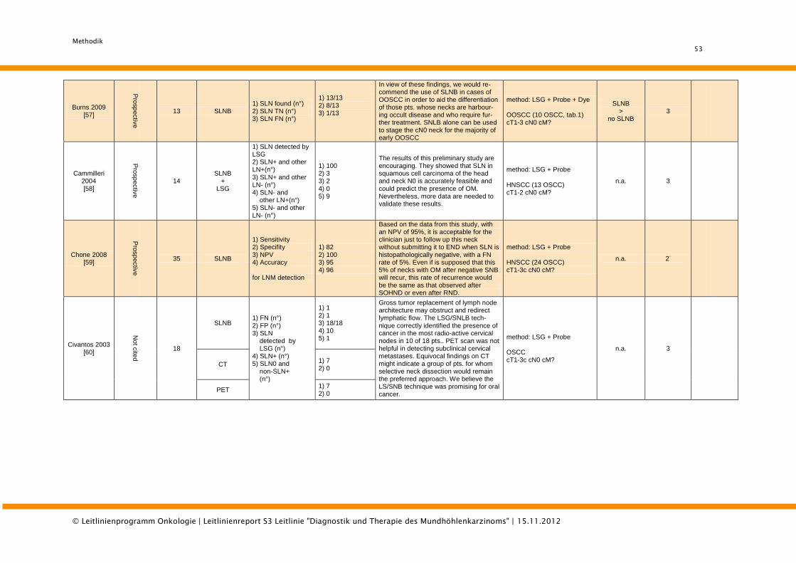
Methodik
53
© Leitlinienprogramm Onkologie | Leitlinienreport S3 Leitlinie "Diagnostik und Therapie des Mundhöhlenkarzinoms" | 15.11.2012
Burns 2009 [57]
Pro
sp
ectiv
e
13 SLNB 1) SLN found (n°) 2) SLN TN (n°) 3) SLN FN (n°)
1) 13/13 2) 8/13 3) 1/13
In view of these findings, we would re-commend the use of SLNB in cases of OOSCC in order to aid the differentiation of those pts. whose necks are harbour-ing occult disease and who require fur-ther treatment. SNLB alone can be used to stage the cN0 neck for the majority of early OOSCC
method: LSG + Probe + Dye OOSCC (10 OSCC, tab.1) cT1-3 cN0 cM?
SLNB >
no SLNB 3
Cammilleri 2004 [58]
Pro
sp
ectiv
e
14 SLNB
+ LSG
1) SLN detected by LSG 2) SLN+ and other LN+(n°) 3) SLN+ and other LN- (n°) 4) SLN- and other LN+(n°) 5) SLN- and other LN- (n°)
1) 100 2) 3 3) 2 4) 0 5) 9
The results of this preliminary study are encouraging. They showed that SLN in squamous cell carcinoma of the head and neck N0 is accurately feasible and could predict the presence of OM. Nevertheless, more data are needed to validate these results.
method: LSG + Probe HNSCC (13 OSCC) cT1-2 cN0 cM?
n.a. 3
Chone 2008 [59]
Pro
sp
ectiv
e
35 SLNB
1) Sensitivity 2) Specifity 3) NPV 4) Accuracy for LNM detection
1) 82 2) 100 3) 95 4) 96
Based on the data from this study, with an NPV of 95%, it is acceptable for the clinician just to follow up this neck without submitting it to END when SLN is histopathologically negative, with a FN rate of 5%. Even if is supposed that this 5% of necks with OM after negative SNB will recur, this rate of recurrence would be the same as that observed after SOHND or even after RND.
method: LSG + Probe HNSCC (24 OSCC) cT1-3c cN0 cM?
n.a. 2-
Civantos 2003 [60]
Not c
ited
18
SLNB 1) FN (n°) 2) FP (n°) 3) SLN detected by LSG (n°) 4) SLN+ (n°) 5) SLN0 and non-SLN+ (n°)
1) 1 2) 1 3) 18/18 4) 10 5) 1
Gross tumor replacement of lymph node architecture may obstruct and redirect lymphatic flow. The LSG/SNLB tech-nique correctly identified the presence of cancer in the most radio-active cervical nodes in 10 of 18 pts.. PET scan was not helpful in detecting subclinical cervical metastases. Equivocal findings on CT might indicate a group of pts. for whom selective neck dissection would remain the preferred approach. We believe the LS/SNB technique was promising for oral cancer.
method: LSG + Probe OSCC cT1-3c cN0 cM?
n.a. 3
CT 1) 7 2) 0
PET 1) 7 2) 0
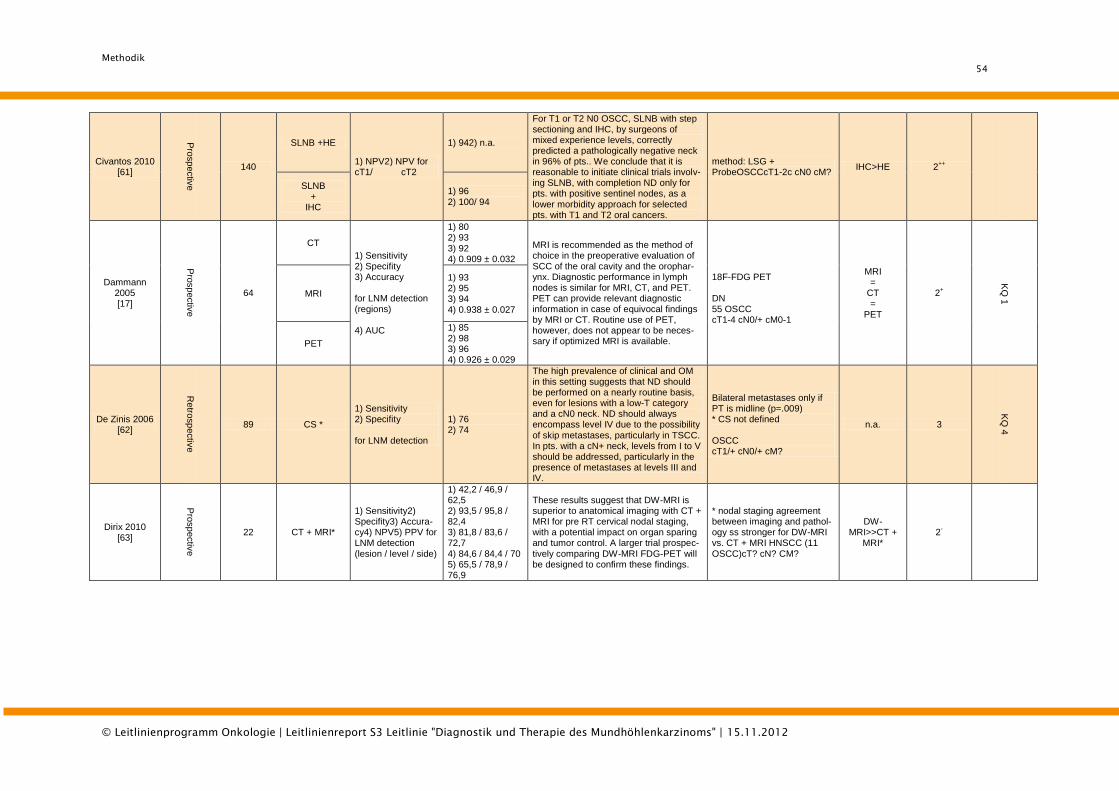
Methodik
54
© Leitlinienprogramm Onkologie | Leitlinienreport S3 Leitlinie "Diagnostik und Therapie des Mundhöhlenkarzinoms" | 15.11.2012
Civantos 2010 [61]
Pro
sp
ectiv
e
140
SLNB +HE
1) NPV2) NPV for cT1/ cT2
1) 942) n.a.
For T1 or T2 N0 OSCC, SLNB with step sectioning and IHC, by surgeons of mixed experience levels, correctly predicted a pathologically negative neck in 96% of pts.. We conclude that it is reasonable to initiate clinical trials involv-ing SLNB, with completion ND only for pts. with positive sentinel nodes, as a lower morbidity approach for selected pts. with T1 and T2 oral cancers.
method: LSG + ProbeOSCCcT1-2c cN0 cM?
IHC>HE 2++
SLNB +
IHC
1) 96 2) 100/ 94
Dammann 2005 [17]
Pro
sp
ectiv
e
64
CT
1) Sensitivity 2) Specifity 3) Accuracy for LNM detection (regions) 4) AUC
1) 80 2) 93 3) 92 4) 0.909 ± 0.032
MRI is recommended as the method of choice in the preoperative evaluation of SCC of the oral cavity and the orophar-ynx. Diagnostic performance in lymph nodes is similar for MRI, CT, and PET. PET can provide relevant diagnostic information in case of equivocal findings by MRI or CT. Routine use of PET, however, does not appear to be neces-sary if optimized MRI is available.
18F-FDG PET DN 55 OSCC cT1-4 cN0/+ cM0-1
MRI =
CT =
PET
2+
KQ
1
MRI
1) 93 2) 95 3) 94 4) 0.938 ± 0.027
PET
1) 85 2) 98 3) 96 4) 0.926 ± 0.029
De Zinis 2006 [62]
Retro
sp
ectiv
e
89 CS *
1) Sensitivity 2) Specifity for LNM detection
1) 76 2) 74
The high prevalence of clinical and OM in this setting suggests that ND should be performed on a nearly routine basis, even for lesions with a low-T category and a cN0 neck. ND should always encompass level IV due to the possibility of skip metastases, particularly in TSCC. In pts. with a cN+ neck, levels from I to V should be addressed, particularly in the presence of metastases at levels III and IV.
Bilateral metastases only if PT is midline (p=.009) * CS not defined OSCC cT1/+ cN0/+ cM?
n.a. 3
KQ
4
Dirix 2010 [63]
Pro
sp
ectiv
e
22 CT + MRI*
1) Sensitivity2) Specifity3) Accura-cy4) NPV5) PPV for LNM detection (lesion / level / side)
1) 42,2 / 46,9 / 62,5 2) 93,5 / 95,8 / 82,4 3) 81,8 / 83,6 / 72,7 4) 84,6 / 84,4 / 70 5) 65,5 / 78,9 / 76,9
These results suggest that DW-MRI is superior to anatomical imaging with CT + MRI for pre RT cervical nodal staging, with a potential impact on organ sparing and tumor control. A larger trial prospec-tively comparing DW-MRI FDG-PET will be designed to confirm these findings.
* nodal staging agreement between imaging and pathol-ogy ss stronger for DW-MRI vs. CT + MRI HNSCC (11 OSCC)cT? cN? CM?
DW-MRI>>CT +
MRI* 2
-
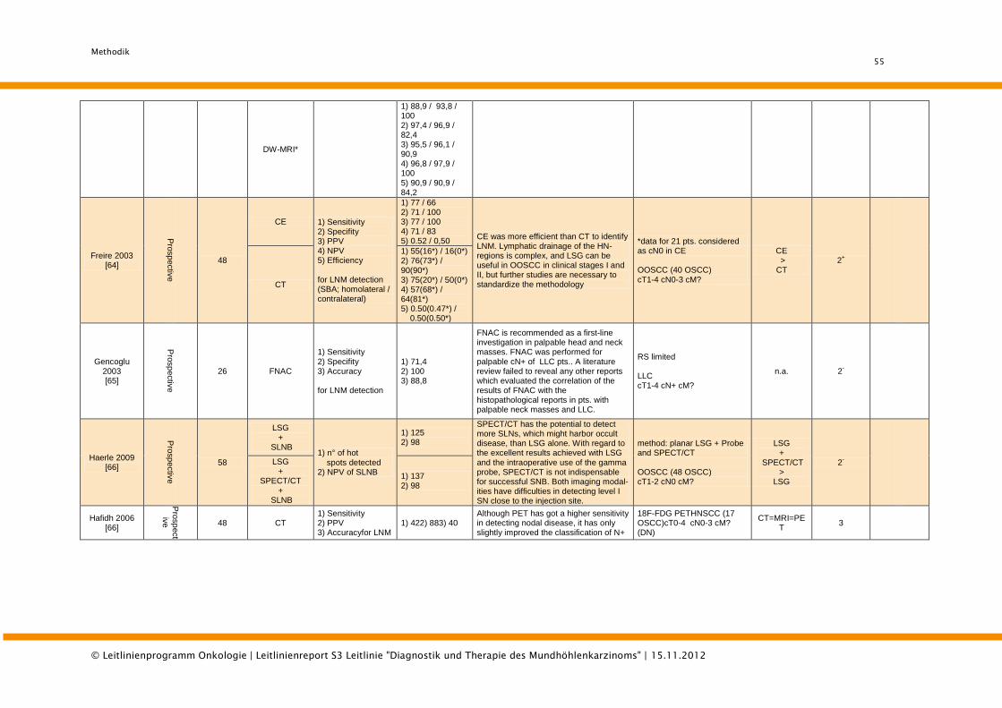
Methodik
55
© Leitlinienprogramm Onkologie | Leitlinienreport S3 Leitlinie "Diagnostik und Therapie des Mundhöhlenkarzinoms" | 15.11.2012
DW-MRI*
1) 88,9 / 93,8 / 100 2) 97,4 / 96,9 / 82,4 3) 95,5 / 96,1 / 90,9 4) 96,8 / 97,9 / 100 5) 90,9 / 90,9 / 84,2
Freire 2003 [64]
Pro
sp
ectiv
e
48
CE 1) Sensitivity 2) Specifity 3) PPV 4) NPV 5) Efficiency for LNM detection (SBA; homolateral / contralateral)
1) 77 / 66 2) 71 / 100 3) 77 / 100 4) 71 / 83 5) 0.52 / 0,50
CE was more efficient than CT to identify LNM. Lymphatic drainage of the HN-regions is complex, and LSG can be useful in OOSCC in clinical stages I and II, but further studies are necessary to standardize the methodology
*data for 21 pts. considered as cN0 in CE OOSCC (40 OSCC) cT1-4 cN0-3 cM?
CE > CT
2+
CT
1) 55(16*) / 16(0*) 2) 76(73*) / 90(90*) 3) 75(20*) / 50(0*) 4) 57(68*) / 64(81*) 5) 0.50(0.47*) / 0.50(0.50*)
Gencoglu 2003 [65]
Pro
sp
ectiv
e
26 FNAC
1) Sensitivity 2) Specifity 3) Accuracy for LNM detection
1) 71,4 2) 100 3) 88,8
FNAC is recommended as a first-line investigation in palpable head and neck masses. FNAC was performed for palpable cN+ of LLC pts.. A literature review failed to reveal any other reports which evaluated the correlation of the results of FNAC with the histopathological reports in pts. with palpable neck masses and LLC.
RS limited LLC cT1-4 cN+ cM?
n.a. 2-
Haerle 2009 [66]
Pro
sp
ectiv
e
58
LSG +
SLNB 1) n° of hot spots detected 2) NPV of SLNB
1) 125 2) 98
SPECT/CT has the potential to detect more SLNs, which might harbor occult disease, than LSG alone. With regard to the excellent results achieved with LSG and the intraoperative use of the gamma probe, SPECT/CT is not indispensable for successful SNB. Both imaging modal-ities have difficulties in detecting level I SN close to the injection site.
method: planar LSG + Probe and SPECT/CT OOSCC (48 OSCC) cT1-2 cN0 cM?
LSG +
SPECT/CT >
LSG
2-
LSG +
SPECT/CT +
SLNB
1) 137 2) 98
Hafidh 2006 [66]
Pro
sp
ect
ive
48 CT 1) Sensitivity 2) PPV 3) Accuracyfor LNM
1) 422) 883) 40 Although PET has got a higher sensitivity in detecting nodal disease, it has only slightly improved the classification of N+
18F-FDG PETHNSCC (17 OSCC)cT0-4 cN0-3 cM? (DN)
CT=MRI=PET
3
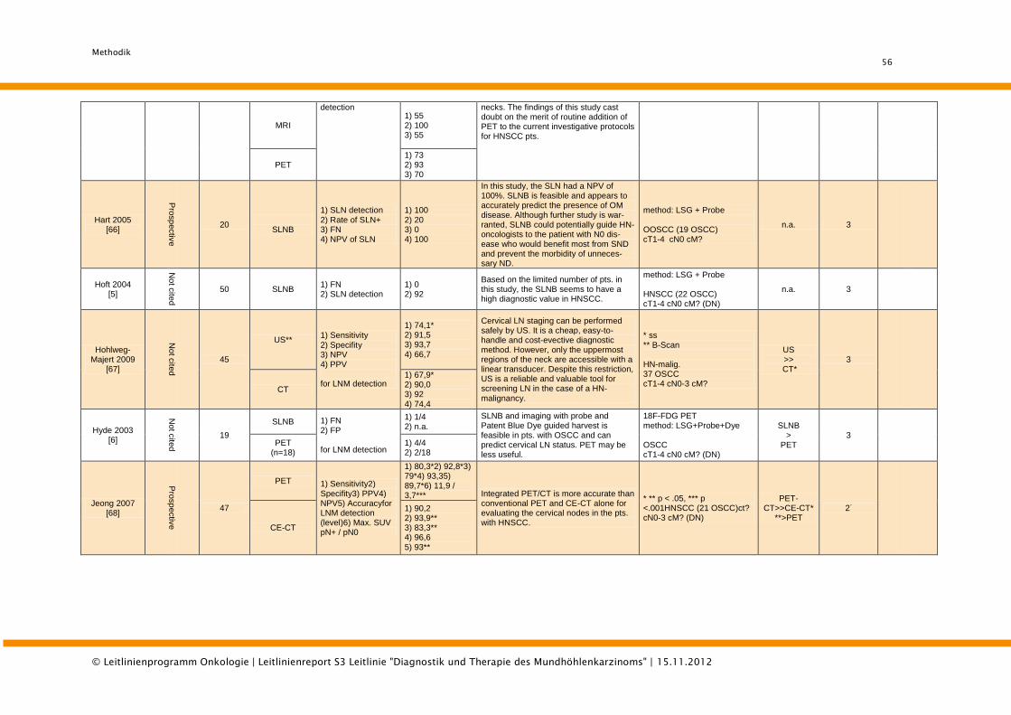
Methodik
56
© Leitlinienprogramm Onkologie | Leitlinienreport S3 Leitlinie "Diagnostik und Therapie des Mundhöhlenkarzinoms" | 15.11.2012
MRI
detection 1) 55 2) 100 3) 55
necks. The findings of this study cast doubt on the merit of routine addition of PET to the current investigative protocols for HNSCC pts.
PET 1) 73 2) 93 3) 70
Hart 2005 [66]
Pro
sp
ectiv
e
20
SLNB
1) SLN detection 2) Rate of SLN+ 3) FN 4) NPV of SLN
1) 100 2) 20 3) 0 4) 100
In this study, the SLN had a NPV of 100%. SLNB is feasible and appears to accurately predict the presence of OM disease. Although further study is war-ranted, SLNB could potentially guide HN-oncologists to the patient with N0 dis-ease who would benefit most from SND and prevent the morbidity of unneces-sary ND.
method: LSG + Probe OOSCC (19 OSCC) cT1-4 cN0 cM?
n.a. 3
Hoft 2004 [5]
Not c
ited
50 SLNB 1) FN 2) SLN detection
1) 0 2) 92
Based on the limited number of pts. in this study, the SLNB seems to have a high diagnostic value in HNSCC.
method: LSG + Probe HNSCC (22 OSCC) cT1-4 cN0 cM? (DN)
n.a. 3
Hohlweg-Majert 2009
[67]
Not c
ited
45
US** 1) Sensitivity 2) Specifity 3) NPV 4) PPV for LNM detection
1) 74,1* 2) 91,5 3) 93,7 4) 66,7
Cervical LN staging can be performed safely by US. It is a cheap, easy-to-handle and cost-evective diagnostic method. However, only the uppermost regions of the neck are accessible with a linear transducer. Despite this restriction, US is a reliable and valuable tool for screening LN in the case of a HN-
malignancy.
* ss ** B-Scan HN-malig. 37 OSCC cT1-4 cN0-3 cM?
US >>
CT* 3
CT
1) 67,9* 2) 90,0 3) 92
4) 74,4
Hyde 2003 [6]
Not c
ited
19
SLNB 1) FN 2) FP for LNM detection
1) 1/4 2) n.a.
SLNB and imaging with probe and Patent Blue Dye guided harvest is feasible in pts. with OSCC and can predict cervical LN status. PET may be less useful.
18F-FDG PET method: LSG+Probe+Dye OSCC cT1-4 cN0 cM? (DN)
SLNB >
PET 3
PET (n=18)
1) 4/4 2) 2/18
Jeong 2007 [68]
Pro
sp
ectiv
e
47
PET 1) Sensitivity2) Specifity3) PPV4) NPV5) Accuracyfor LNM detection (level)6) Max. SUV pN+ / pN0
1) 80,3*2) 92,8*3) 79*4) 93,35) 89,7*6) 11,9 / 3,7*** Integrated PET/CT is more accurate than
conventional PET and CE-CT alone for evaluating the cervical nodes in the pts. with HNSCC.
* ** p < .05, *** p <.001HNSCC (21 OSCC)ct? cN0-3 cM? (DN)
PET-CT>>CE-CT*
**>PET 2
-
CE-CT
1) 90,2 2) 93,9** 3) 83,3** 4) 96,6 5) 93**
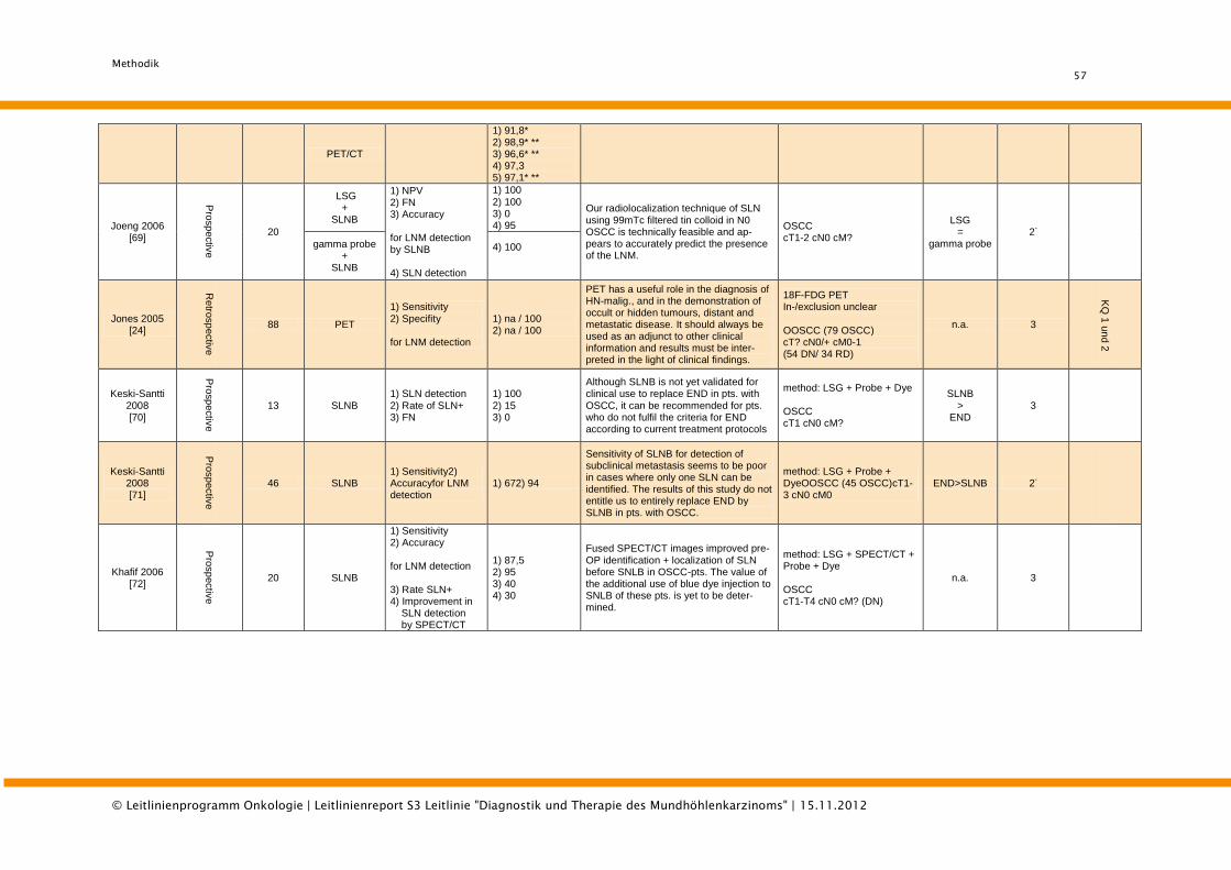
Methodik
57
© Leitlinienprogramm Onkologie | Leitlinienreport S3 Leitlinie "Diagnostik und Therapie des Mundhöhlenkarzinoms" | 15.11.2012
PET/CT
1) 91,8* 2) 98,9* ** 3) 96,6* ** 4) 97,3 5) 97,1* **
Joeng 2006 [69]
Pro
sp
ectiv
e
20
LSG +
SLNB
1) NPV 2) FN 3) Accuracy for LNM detection by SLNB 4) SLN detection
1) 100 2) 100 3) 0 4) 95
Our radiolocalization technique of SLN using 99mTc filtered tin colloid in N0 OSCC is technically feasible and ap-pears to accurately predict the presence of the LNM.
OSCC cT1-2 cN0 cM?
LSG =
gamma probe 2
-
gamma probe +
SLNB
4) 100
Jones 2005 [24]
Retro
sp
ectiv
e
88 PET
1) Sensitivity 2) Specifity for LNM detection
1) na / 100 2) na / 100
PET has a useful role in the diagnosis of HN-malig., and in the demonstration of occult or hidden tumours, distant and metastatic disease. It should always be used as an adjunct to other clinical information and results must be inter-preted in the light of clinical findings.
18F-FDG PET In-/exclusion unclear OOSCC (79 OSCC) cT? cN0/+ cM0-1 (54 DN/ 34 RD)
n.a. 3
KQ
1 u
nd 2
Keski-Santti 2008 [70]
Pro
sp
ectiv
e
13 SLNB 1) SLN detection 2) Rate of SLN+ 3) FN
1) 100 2) 15 3) 0
Although SLNB is not yet validated for clinical use to replace END in pts. with OSCC, it can be recommended for pts. who do not fulfil the criteria for END according to current treatment protocols
method: LSG + Probe + Dye OSCC cT1 cN0 cM?
SLNB >
END 3
Keski-Santti 2008 [71]
Pro
sp
ectiv
e
46 SLNB 1) Sensitivity2) Accuracyfor LNM detection
1) 672) 94
Sensitivity of SLNB for detection of subclinical metastasis seems to be poor in cases where only one SLN can be identified. The results of this study do not entitle us to entirely replace END by SLNB in pts. with OSCC.
method: LSG + Probe + DyeOOSCC (45 OSCC)cT1-3 cN0 cM0
END>SLNB 2-
Khafif 2006 [72]
Pro
sp
ectiv
e
20 SLNB
1) Sensitivity 2) Accuracy for LNM detection 3) Rate SLN+ 4) Improvement in SLN detection by SPECT/CT
1) 87,5 2) 95 3) 40 4) 30
Fused SPECT/CT images improved pre-OP identification + localization of SLN before SNLB in OSCC-pts. The value of the additional use of blue dye injection to SNLB of these pts. is yet to be deter-mined.
method: LSG + SPECT/CT + Probe + Dye OSCC cT1-T4 cN0 cM? (DN)
n.a. 3
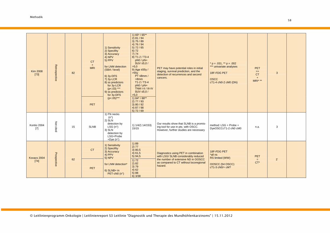
Methodik
58
© Leitlinienprogramm Onkologie | Leitlinienreport S3 Leitlinie "Diagnostik und Therapie des Mundhöhlenkarzinoms" | 15.11.2012
Kim 2008 [73]
Retro
sp
ectiv
e
82
CT +
MRI
1) Sensitivity 2) Specifity 3) Accuracy 4) NPV 5) PPV for LNM detection (SBA / level) 6) 3y-DFS 7) 3y-LCR 8) ss predictors for 3y-LCR (p<.03) *** 9) ss predictors for 3y-DFS (p<.05)***
1) 65* / 65** 2) 81 / 94 3) 75 / 89 4) 76 / 94 5) 72 / 65 6) 72 7) 74 8) T1-2 / T3-4 pN0 / pN+ SUV ≤5,0 / >5,0 9) Age ≤55y / >55y PT ≤8mm / >8mm T1-2 / T3-4 pN0 / pN+ TNM I-II / III-IV SUV ≤5,0 / >5,0
PET may have potential roles in initial staging, survival prediction, and the detection of recurrences and second
cancers.
* p = .031, ** p = .002 *** univariate analyses 18F-FDG PET OSCC cT1-4 cN0-2 cM0 (DN)
PET >> CT
+ MRI* **
3
PET
1) 84* / 88** 2) 77 / 93 3) 80 / 92 4) 87 / 98 5) 72 / 69
Kontio 2004 [7]
Not c
ited
15 SLNB
1) FN necks (n°) 2) SLN detection by LSG (n°) 3) SLN detection by LSG+Probe +Dye (n°)
1) 1/42) 14/153) 15/15
Our results show that SLNB is a promis-ing tool for use in pts. with OSCC. However, further studies are necessary
method: LSG + Probe + DyeOSCCcT1-2 cN0 cM0
n.a. 3
Kovacs 2004 [74]
Pro
sp
ectiv
e
62
CT
1) Sensitivity 2) Specifity 3) Accuracy 4) PPV 5) NPV for LNM detection* 6) SLNB+ in PET-cN0 (n°)
1) 89 2) 77 3) 80,5 4) 61,5 5) 94,5
Diagnostics using PET in combination with LSG/ SLNB considerably reduced the number of extensive ND in OOSCC as compared to CT without locoregional hazard.
18F-FDG PET *all ns RS limited (WW) OOSCC (54 OSCC) cT1-3 cN0/+ cM?
PET =
CT* 2
-
PET
1) 72 2) 82 3) 79 4) 62 5) 88 6) 3/38
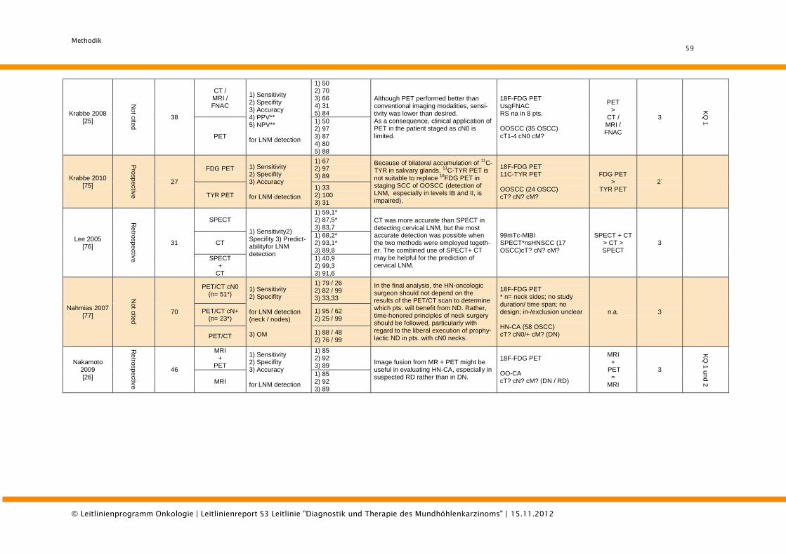
Methodik
59
© Leitlinienprogramm Onkologie | Leitlinienreport S3 Leitlinie "Diagnostik und Therapie des Mundhöhlenkarzinoms" | 15.11.2012
Krabbe 2008 [25]
Not c
ited
38
CT / MRI / FNAC
1) Sensitivity 2) Specifity 3) Accuracy 4) PPV** 5) NPV** for LNM detection
1) 50 2) 70 3) 66 4) 31 5) 84
Although PET performed better than conventional imaging modalities, sensi-tivity was lower than desired. As a consequence, clinical application of PET in the patient staged as cN0 is limited.
18F-FDG PET UsgFNAC RS na in 8 pts. OOSCC (35 OSCC) cT1-4 cN0 cM?
PET >
CT / MRI / FNAC
3
KQ
1
PET
1) 50 2) 97 3) 87 4) 80 5) 88
Krabbe 2010 [75]
Pro
sp
ectiv
e
27
FDG PET 1) Sensitivity 2) Specifity 3) Accuracy for LNM detection
1) 67 2) 97 3) 89
Because of bilateral accumulation of 11
C-TYR in salivary glands,
11C-TYR PET is
not suitable to replace 18
FDG PET in staging SCC of OOSCC (detection of LNM, especially in levels IB and II, is impaired).
18F-FDG PET 11C-TYR PET OOSCC (24 OSCC) cT? cN? cM?
FDG PET >
TYR PET 2
-
TYR PET 1) 33 2) 100 3) 31
Lee 2005 [76]
Retro
sp
ectiv
e
31
SPECT
1) Sensitivity2) Specifity 3) Predict-abilityfor LNM detection
1) 59,1* 2) 87,5* 3) 83,7
CT was more accurate than SPECT in detecting cervical LNM, but the most accurate detection was possible when the two methods were employed togeth-er. The combined use of SPECT+ CT may be helpful for the prediction of cervical LNM.
99mTc-MIBI SPECT*nsHNSCC (17 OSCC)cT? cN? cM?
SPECT + CT > CT > SPECT
3
CT 1) 68,2* 2) 93,1* 3) 89,8
SPECT +
CT
1) 40,9 2) 99,3 3) 91,6
Nahmias 2007 [77]
Not c
ited
70
PET/CT cN0 (n= 51*)
1) Sensitivity 2) Specifity for LNM detection (neck / nodes) 3) OM
1) 79 / 26 2) 82 / 99 3) 33,33
In the final analysis, the HN-oncologic surgeon should not depend on the results of the PET/CT scan to determine which pts. will benefit from ND. Rather, time-honored principles of neck surgery should be followed, particularly with regard to the liberal execution of prophy-lactic ND in pts. with cN0 necks.
18F-FDG PET * n= neck sides; no study duration/ time span; no design; in-/exclusion unclear HN-CA (58 OSCC) cT? cN0/+ cM? (DN)
n.a. 3 PET/CT cN+ (n= 23*)
1) 95 / 62 2) 25 / 99
PET/CT 1) 88 / 48 2) 76 / 99
Nakamoto 2009 [26]
Retro
sp
ectiv
e
46
MRI +
PET
1) Sensitivity 2) Specifity 3) Accuracy for LNM detection
1) 85 2) 92 3) 89
Image fusion from MR + PET might be useful in evaluating HN-CA, especially in suspected RD rather than in DN.
18F-FDG PET OO-CA cT? cN? cM? (DN / RD)
MRI +
PET =
MRI
3
KQ
1 u
nd 2
MRI 1) 85 2) 92 3) 89

Methodik
60
© Leitlinienprogramm Onkologie | Leitlinienreport S3 Leitlinie "Diagnostik und Therapie des Mundhöhlenkarzinoms" | 15.11.2012
Ng 2005 [27]
Pro
sp
ectiv
e
124
CT + MRI
1) Sensitivity2) Specifity3) Accura-cy4) PPV5) NPVfor LNM detection (LBA)6) AUC (ROC)
1) 52,6* 2) 94,5 3) 86,4 4) 69,4 5) 89,3 6) 0.801**
PET is superior to CT+MRI in the detec-tion of cervical status of OSCC. The sensitivity of PET for the detection of LNM on a level-by-level basis was significantly higher than that of CT+MRI, whereas their specificities appeared to be similar. Visual correlation of PET+CT+MRI showed a trend of in-creased diagnostic accuracy over PET alone but without a significant difference, and its sensitivity was still not high enough to replace pathologic LN staging based on ND.
18F-FDG PET* p < .001no major limitations** p = .002 for nodal detectionOSCCcT1-4 cN? CM? (DN)
PET>>CT + MRI* **
2++
KQ
1
PET
1) 74,7* 2) 93 3) 89,5 4) 71,7 5) 93,9 6) 0.896**
CT +
MRI +
PET
1) 77,9 2) 94,5 3) 91,3 4) 77,1 5) 94,7 6) 0.913
Ng 2006 [78]
Pro
sp
ectiv
e
134
CT / MRI (n=25 / 109)
1) Sensitivity 2) Specifity 3) Accuracy 4) PPV 5) NPV for LNM detection (LBA / PBA)
1) 21,6* / 31,4 2) 97,5 / 91,9 3) 89,1 / 76,1 4) 52,4 / 57,9 5) 90,8 / 79,1 PET was superior to CT or MRI for
detecting palpably occult neck metasta-sis of OSCC. Because PET could reduce the probability of occult neck metastasis to less than 15% in T1-T3 tumors, it should be indicated for evaluation of these subpopulations.
18F-FDG PET * p = .021, ** p= .25, *** p= .125 OSCC cT1-4 cN0 cM? (DN)
PET +
CT / MRI >
PET** *** >>
CT / MRI*
2++
PET
1) 41,2* ** / 51,4 2) 96,8*** / 91,9 3) 90,6 / 81,3 4) 61,8 / 69,2 5) 92,9 / 84,3
PET +
CT / MRI
1) 47,1** / 57,1 2) 98*** / 96 3) 92,3 / 85,5 4) 75 / 83,3 5) 93,6 / 86,4
Nieuwenhuis 2005 [79]
Not c
ited
22 SLNB
1) Sensitivity2) Accuracy 3) FN (n)for LNM detec-tion4) SLN detec-tion by LSG
1) 892) 953) 1/224) 78
Our study seems to validate the SN hypothesis for OOSCC. The role of SNLB in the management of the N0 neck in such pts. has yet to be established through prospective trials. SN identifica-tion (and thus biopsy) does not seem to be reliable in pts. with tumors located in or close to the midline.
method: LSG + ProbeOOSCC (18 OSCC)cT2-4 cN0 cM?
SLNB>WW 3
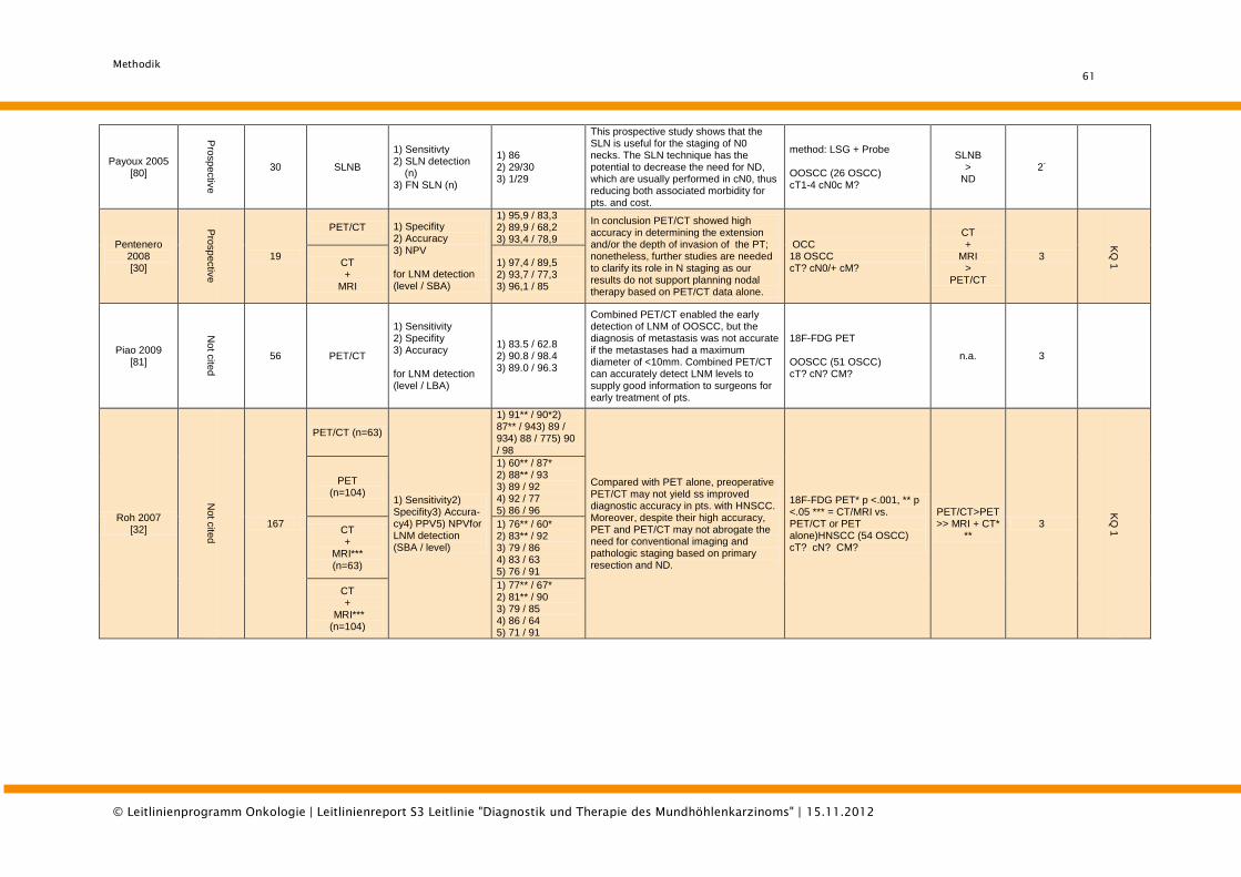
Methodik
61
© Leitlinienprogramm Onkologie | Leitlinienreport S3 Leitlinie "Diagnostik und Therapie des Mundhöhlenkarzinoms" | 15.11.2012
Payoux 2005 [80]
Pro
sp
ectiv
e
30 SLNB
1) Sensitivty 2) SLN detection (n) 3) FN SLN (n)
1) 86 2) 29/30 3) 1/29
This prospective study shows that the SLN is useful for the staging of N0 necks. The SLN technique has the potential to decrease the need for ND, which are usually performed in cN0, thus reducing both associated morbidity for pts. and cost.
method: LSG + Probe OOSCC (26 OSCC) cT1-4 cN0c M?
SLNB >
ND 2
-
Pentenero 2008 [30]
Pro
sp
ectiv
e
19
PET/CT 1) Specifity 2) Accuracy 3) NPV for LNM detection (level / SBA)
1) 95,9 / 83,3 2) 89,9 / 68,2 3) 93,4 / 78,9
In conclusion PET/CT showed high accuracy in determining the extension and/or the depth of invasion of the PT; nonetheless, further studies are needed to clarify its role in N staging as our results do not support planning nodal therapy based on PET/CT data alone.
OCC 18 OSCC cT? cN0/+ cM?
CT +
MRI >
PET/CT
3
KQ
1 CT
+ MRI
1) 97,4 / 89,5 2) 93,7 / 77,3 3) 96,1 / 85
Piao 2009 [81]
Not c
ited
56 PET/CT
1) Sensitivity 2) Specifity 3) Accuracy for LNM detection (level / LBA)
1) 83.5 / 62.8 2) 90.8 / 98.4 3) 89.0 / 96.3
Combined PET/CT enabled the early detection of LNM of OOSCC, but the diagnosis of metastasis was not accurate if the metastases had a maximum diameter of <10mm. Combined PET/CT can accurately detect LNM levels to supply good information to surgeons for early treatment of pts.
18F-FDG PET OOSCC (51 OSCC) cT? cN? CM?
n.a. 3
Roh 2007 [32]
Not c
ited
167
PET/CT (n=63)
1) Sensitivity2) Specifity3) Accura-cy4) PPV5) NPVfor LNM detection (SBA / level)
1) 91** / 90*2) 87** / 943) 89 / 934) 88 / 775) 90 / 98
Compared with PET alone, preoperative PET/CT may not yield ss improved diagnostic accuracy in pts. with HNSCC. Moreover, despite their high accuracy, PET and PET/CT may not abrogate the need for conventional imaging and pathologic staging based on primary resection and ND.
18F-FDG PET* p <.001, ** p <.05 *** = CT/MRI vs. PET/CT or PET alone)HNSCC (54 OSCC) cT? cN? CM?
PET/CT>PET >> MRI + CT*
** 3
KQ
1
PET (n=104)
1) 60** / 87* 2) 88** / 93 3) 89 / 92 4) 92 / 77 5) 86 / 96
CT +
MRI*** (n=63)
1) 76** / 60* 2) 83** / 92 3) 79 / 86 4) 83 / 63 5) 76 / 91
CT +
MRI*** (n=104)
1) 77** / 67* 2) 81** / 90 3) 79 / 85 4) 86 / 64 5) 71 / 91
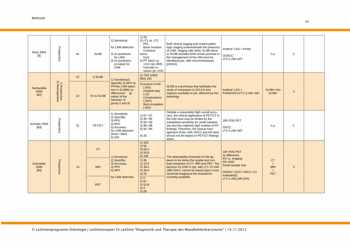
Methodik
62
© Leitlinienprogramm Onkologie | Leitlinienreport S3 Leitlinie "Diagnostik und Therapie des Mundhöhlenkarzinoms" | 15.11.2012
Ross 2004 [8]
Pro
sp
ectiv
e
61 SLNB
1) Sensitivity for LNM detection 2) ss predictors for LNM 3) ns predictors (p-value) for LNM
1) 93 2) cT1 vs. cT2 PNI Bone invasion Cohesive tumor front 3) PT ≤2cm vs. >2cm (p=.063) Vascular in- vasion (p=.214)
Both clinical staging and routine patho-logic staging underestimate the presence of LNM. Staging with either SLNB alone or SLNB-assisted END shows promise in the management of the cN0 neck by identifying pts. with micrometastases (pN1mi).
method: LSG + Probe OOSCC cT1-2 cN0 cM?
n.a. 2-
Santaolalla 2009 [82]
I) Pro
sp
ectiv
e
II) Retro
sp
ectiv
e
22 I) SLNB 1) Sensitivity2)
Specifity 3) NPV 4) PPVfor LNM detec-tion in SLNB5) ss differences (p-value) of the behavior of group I) and II)
1) 732) 1003) 804) 100
SLNB is a technique that facilitates the study of metastasis in OO-CA and reduces morbidity in pts. affected by this pathology
method: LSG + ProbeOSCCCT1-3 cN0 cM0
SLNB>>No-SLNB*
3
22 II) no-SLNB
5) Extend of ND (.003) Hospital stay (.01) Complications (.034*) Bed occupation (.002)
Schoder 2006 [83]
Pro
sp
ectiv
e
31 PET/CT
1) Sensitivity 2) Specifity 3) PPV 4) NPV 5) Accuracy for LNM detection (level / SBA) 6) OM
1) 67 / 67 2) 95 / 85 3) 50 / 60 4) 98 / 88 5) 94 / 80 6) 25
Despite a reasonably high overall accu-racy, the clinical application of PET/CT in the cN0 neck may be limited by the suboptimal sensitivity for small metasta-ses and the relatively high number of FP findings. Therefore, the clinical man-agement of pts. with OSCC and N0 neck should not be based on PET/CT findings alone.
18F-FDG PET OSCC cT1-4 cN0 cM?
n.a. 2-
Schroeder 2008 [84]
Pro
sp
ectiv
e
13
CT
1) Sensitivity 2) Specifity 3) Accuracy 4) PPV 5) NPV for LNM detection
1) 100 2) 50 3) 69.2 4) 55.6 5) 100 The detectability threshold of OM ap-
pears to be below the spatial and con-trast resolution of CT, MRI and PET. The decision for END in pts. with cT1-T2 cN0 cM0 OSCC cannot be based upon cross-sectional imaging at the resolutions currently available.
18F-FDG PET ss difference RS vs. imaging RS: END Small sample size OOSCC (10/17 OSCC (13 evaluated)) cT1-2 cN0 cM0 (DN)
CT =
MRI =
PET
3
MRI
1) 80 2) 12.5 3) 38.5 4) 36.4 5) 50
PET
1) 0 2) 87 3) 53.8 4) 0 5) 59
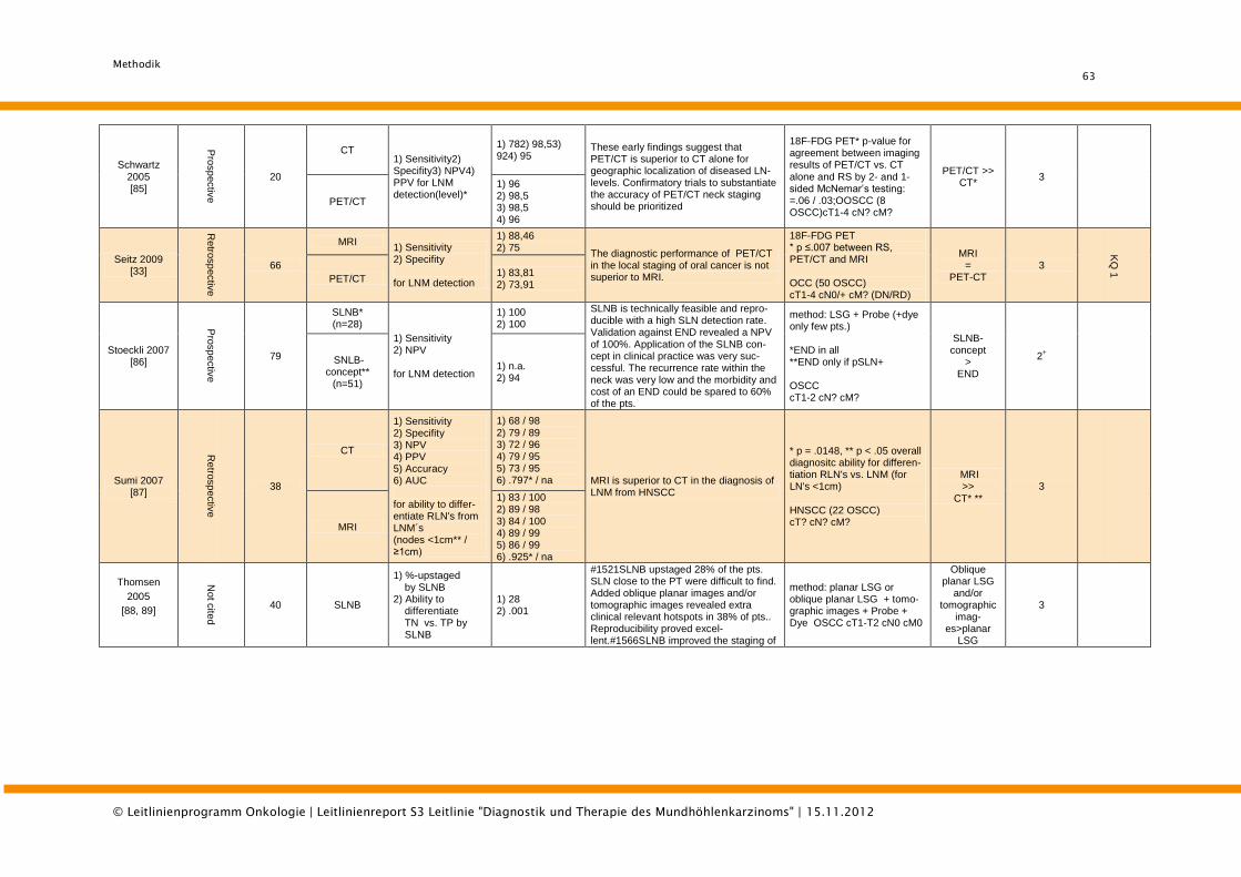
Methodik
63
© Leitlinienprogramm Onkologie | Leitlinienreport S3 Leitlinie "Diagnostik und Therapie des Mundhöhlenkarzinoms" | 15.11.2012
Schwartz 2005 [85]
Pro
sp
ectiv
e
20
CT 1) Sensitivity2) Specifity3) NPV4) PPV for LNM detection(level)*
1) 782) 98,53) 924) 95
These early findings suggest that PET/CT is superior to CT alone for geographic localization of diseased LN-levels. Confirmatory trials to substantiate the accuracy of PET/CT neck staging should be prioritized
18F-FDG PET* p-value for agreement between imaging results of PET/CT vs. CT alone and RS by 2- and 1-sided McNemar’s testing: =.06 / .03;OOSCC (8 OSCC)cT1-4 cN? cM?
PET/CT >> CT*
3
PET/CT
1) 96 2) 98,5 3) 98,5 4) 96
Seitz 2009 [33]
Retro
sp
ectiv
e
66
MRI 1) Sensitivity 2) Specifity for LNM detection
1) 88,46 2) 75
The diagnostic performance of PET/CT in the local staging of oral cancer is not superior to MRI.
18F-FDG PET * p ≤.007 between RS, PET/CT and MRI OCC (50 OSCC) cT1-4 cN0/+ cM? (DN/RD)
MRI =
PET-CT 3
KQ
1 PET/CT
1) 83,81 2) 73,91
Stoeckli 2007 [86]
Pro
sp
ectiv
e
79
SLNB* (n=28)
1) Sensitivity 2) NPV for LNM detection
1) 100 2) 100
SLNB is technically feasible and repro-ducible with a high SLN detection rate. Validation against END revealed a NPV of 100%. Application of the SLNB con-cept in clinical practice was very suc-cessful. The recurrence rate within the neck was very low and the morbidity and cost of an END could be spared to 60% of the pts.
method: LSG + Probe (+dye only few pts.) *END in all **END only if pSLN+ OSCC cT1-2 cN? cM?
SLNB-concept
> END
2+
SNLB-concept**
(n=51)
1) n.a. 2) 94
Sumi 2007 [87]
Retro
sp
ectiv
e
38
CT
1) Sensitivity 2) Specifity 3) NPV 4) PPV 5) Accuracy 6) AUC for ability to differ-entiate RLN's from LNM´s (nodes <1cm** / ≥1cm)
1) 68 / 98 2) 79 / 89 3) 72 / 96 4) 79 / 95 5) 73 / 95 6) .797* / na MRI is superior to CT in the diagnosis of
LNM from HNSCC
* p = .0148, ** p < .05 overall diagnositc ability for differen-tiation RLN's vs. LNM (for LN's <1cm)
HNSCC (22 OSCC) cT? cN? cM?
MRI >>
CT* **
3
MRI
1) 83 / 100 2) 89 / 98 3) 84 / 100 4) 89 / 99 5) 86 / 99 6) .925* / na
Thomsen
2005
[88, 89]
Not c
ited
40 SLNB
1) %-upstaged by SLNB 2) Ability to differentiate TN vs. TP by SLNB
1) 28 2) .001
#1521SLNB upstaged 28% of the pts. SLN close to the PT were difficult to find. Added oblique planar images and/or tomographic images revealed extra clinical relevant hotspots in 38% of pts.. Reproducibility proved excel-lent.#1566SLNB improved the staging of
method: planar LSG or oblique planar LSG + tomo-graphic images + Probe + Dye OSCC cT1-T2 cN0 cM0
Oblique planar LSG
and/or tomographic
imag-es>planar
LSG
3
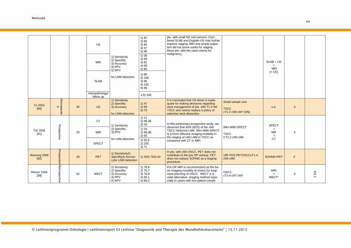
Methodik
64
© Leitlinienprogramm Onkologie | Leitlinienreport S3 Leitlinie "Diagnostik und Therapie des Mundhöhlenkarzinoms" | 15.11.2012
US
1) Sensitivity 2) Specifity 3) Accuracy 4) PPV 5) NPV for LNM detection
1) 87 2) 85 3) 85 4) 57 5) 96
pts. with small N0 oral cancers. Com-bined SLNB and Doppler-US may further improve staging. MRI and simple palpa-tion did not prove useful for staging these pts. with the used criteria for malignancy.
SLNB + US >
MRI (+ CE)
MRI
1) 36 2) 93 3) 81 4) 56 5) 85
SLNB
1) 80 2) 100 3) 96 4) 100 5) 96
Histopathology/ follow up
1-5) 100
To 2003 [90]
Retro
sp
ectiv
e 30 US
1) Sensitivity 2) Specifity 3) Accuracy for LNM detection
1) 47 2) 93 3) 70
It is concluded that US alone is inade-quate for making decisions regarding neck management of pts. with T1-2 N0 TSCC and cannot replace a policy of selective neck dissection.
Small sample size TSCC cT1-2 cN0 cM? (DN)
n.a. 3
Tuli 2008 [91]
Pro
sp
ectiv
e
20
CT
1) Sensitivity 2) Specifity 3) PPV for LNM detection
1) 11 2) 98,38 3) 33 In this preliminary prospective study, we
observed that 40% (8/20) of the cN0 TSCC harbored LNM. 99m-MIBI-SPECT is a more effective imaging modality in the staging of cN0 LNM in TSCC as compared with CT or MRI.
99m-MIBI-SPECT TSCC CT1-2 cN0 cM0
SPECT >
MR > CT
3
MRI 1) 33 2) 98,38 3) 60
SPECT 1) 55,5 2) 100 3) 71
Wensing 2006 [92]
Pro
sp
ectiv
e
28 PET 1) Sensitivity2) Specifity3) Accura-cyfor LNM detection
1) 332) 763) 63
In pts. with cN0 OSCC, PET does not contribute to the pre-OP workup. PET does not replace SOHND as a staging procedure.
18F-FDG PETOSCCcT1-4 cN0 cM0
SOHND>PET 2-
Wiener 2006 [38]
Retro
sp
ectiv
e
52 MSCT
1) Sensitivity 2) Specifity 3) Accuracy 4) PPV 5) NPV
1) 78,9 2) 75,7 3) 76,9 4) 65,1 5) 86,2
Pre-OP MRI is recommended as the ba-sic imaging modality of choice for treat-ment planning of OSCC. MSCT is a valid alternative imaging method espe-cially in cases with low patient compli-
OSCC cT1-4 cN? cM?
MRI >
MSCT* 3
KQ
1
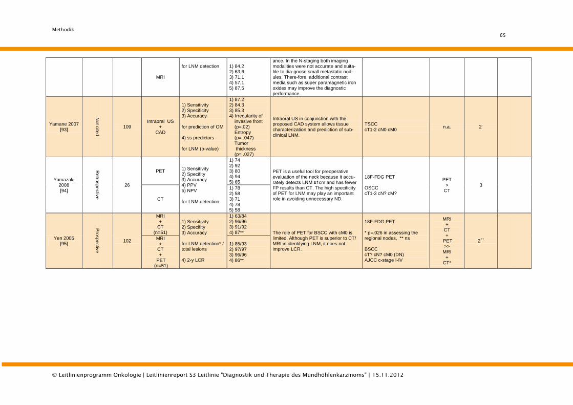
Methodik
65
© Leitlinienprogramm Onkologie | Leitlinienreport S3 Leitlinie "Diagnostik und Therapie des Mundhöhlenkarzinoms" | 15.11.2012
MRI
for LNM detection 1) 84,2
2) 63,6 3) 71,1 4) 57,1 5) 87,5
ance. In the N-staging both imaging modalities were not accurate and suita-ble to dia-gnose small metastatic nod-ules. There-fore, additional contrast media such as super paramagnetic iron oxides may improve the diagnostic performance.
Yamane 2007 [93]
Not c
ited
109 Intraoral US
+ CAD
1) Sensitivity 2) Specificity 3) Accuracy for prediction of OM 4) ss predictors for LNM (p-value)
1) 87.2 2) 84.3 3) 85.3 4) Irregularity of invasive front (p=.02) Entropy (p= .047) Tumor thickness (p= .027)
Intraoral US in conjunction with the proposed CAD system allows tissue characterization and prediction of sub-clinical LNM.
TSCC cT1-2 cN0 cM0
n.a. 2-
Yamazaki 2008 [94]
Retro
sp
ectiv
e
26
PET 1) Sensitivity 2) Specifity 3) Accuracy 4) PPV 5) NPV for LNM detection
1) 74 2) 92 3) 80 4) 94 5) 65
PET is a useful tool for preoperative evaluation of the neck because it accu-rately detects LNM ≥1cm and has fewer FP results than CT. The high specificity of PET for LNM may play an important role in avoiding unnecessary ND.
18F-FDG PET OSCC cT1-3 cN? cM?
PET >
CT 3
CT
1) 78 2) 58 3) 71 4) 78 5) 58
Yen 2005 [95]
Pro
sp
ectiv
e
102
MRI +
CT (n=51)
1) Sensitivity 2) Specifity 3) Accuracy for LNM detection* / total lesions 4) 2-y LCR
1) 63/84 2) 96/96 3) 91/92 4) 87** The role of PET for BSCC with cM0 is
limited. Although PET is superior to CT/ MRI in identifying LNM, it does not improve LCR.
18F-FDG PET * p=.026 in assessing the regional nodes, ** ns BSCC cT? cN? cM0 (DN) AJCC c-stage I-IV
MRI +
CT +
PET >>
MRI +
CT*
2++
MRI +
CT +
PET (n=51)
1) 85/93 2) 97/97 3) 96/96 4) 86**
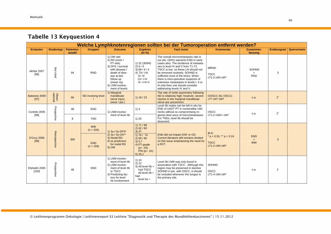
Methodik
66
© Leitlinienprogramm Onkologie | Leitlinienreport S3 Leitlinie "Diagnostik und Therapie des Mundhöhlenkarzinoms" | 15.11.2012
Tabelle 13 Keyquestion 4
Welche Lymphknotenregionen sollten bei der Tumoroperation entfernt werden?
Erstautor Studientyp Patienten- anzahl
Gruppen Outcome Ergebnis (in %)
Fazit Autor Kommentar Zusammen- fassung
Evidenzgrad Querverweis
Akhtar 2007 [96]
Not c
ited
94 RND
1) OM rate 2) RD (neck / PT-site) 3) DFS / survival with disease / death of dise- ase at last follow up (mean 4y) 4) LNM involve- ment of levels
1) 32 (30/94) 2) 6 / 4 3) 89 / 6 / 4 4) 73= I-III 6= III 13= I-IV 6= I+II+V
The overall micrometastases rate in our pts. (32%) warrants END in early cases also. The incidence of metasta-ses to level IV and V from T1-T2 TSCC is low so these LN should not be removed routinely. SOHND is sufficient most of the times. When there is intra-operative suspicion of extensive metastases in levels I, II or III only then one should consider addressing levels IV and V.
MRND TSCC cT1-2 cN0 cM?
SOHND >
RND 3
Batstone 2009 [97]
Obse
r-
vatio
nal
66 ND involving level
I
1) Marginal mandibular nerve injury (neck / pts.)
1) 18 / 23
The rate of smile asymmetry following ND is relatively high; however, severe injuries to the marginal mandibular nerve are uncommon.
OOSCC (61 OSCC) cT? cN? cM?
n.a. 2+
Corlette 2005 [98]
Pro
sp
ectiv
e
48 END 1) LNM involve- ment of level IIb
1) 4
Level IIb nodes can be left in situ for END of UADT PT in nontonsillar cN0 necks without ss compromising re-gional clear-ance of micrometastases. For TNDs, level IIb should be dissected.
OSCC cT1-2 cN0/+ cM?
n.a. 2+
8 TND 1) 25
D'Cruz 2009 [99]
Retro
sp
ectiv
e
359
WW (n = 200)
1) 3y-/ 5y-DFS* 2) 3y-/ 5y-OS** 3) Nodal RD 4) ss predictors for nodal RD 5) OM
1) 71 / 68 2) 62 / 60 3) 47
END did not impact DSF or OS. Current literature still remains divided on this issue emphasizing the need for a RCT.
BLD * p = 0.53, ** p = 0.24 TSCC cT1-2 cN0 cM?
END >
WW 3
END (n = 159)
1) 76 / 74 2) 69 / 60 3) 5,7 4) PT-grade (p= .03) PNI (p= .01) 5) 20,1
Elsheikh 2005 [100]
Pro
sp
ectiv
e
48 SND
1) LNM involve- ment of level IIb 2) LNM involve- ment of level IIb in TSCC 3) Predicting fac- tors for level IIb involvement
1) 10 2) 22 3) All level IIb + had TSCC All level IIb + had level IIa +
Level IIb LNM was only found in association with TSCC. Although this region may be preserved in elective SOHND in pts. with OSCC, it should be included whenever the tongue is the primary site.
SOHND OSCC cT1-4 cN0 cM?
n.a. 2-
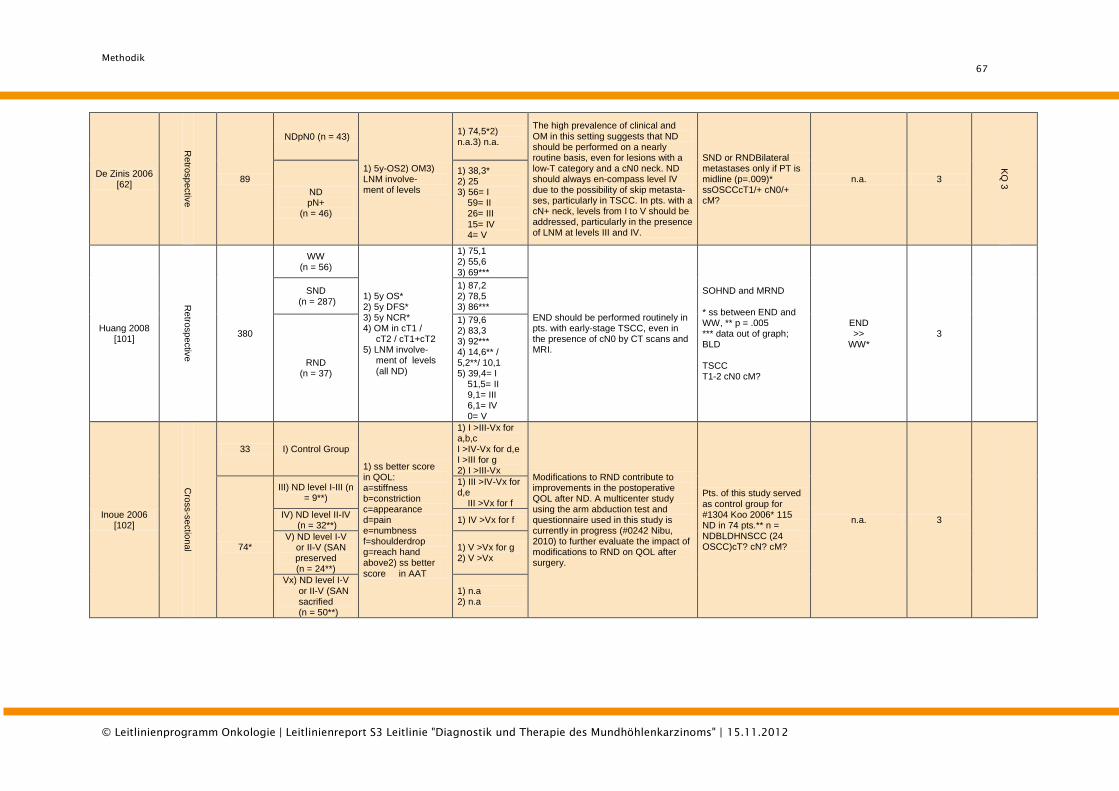
Methodik
67
© Leitlinienprogramm Onkologie | Leitlinienreport S3 Leitlinie "Diagnostik und Therapie des Mundhöhlenkarzinoms" | 15.11.2012
De Zinis 2006 [62]
Retro
sp
ectiv
e
89
NDpN0 (n = 43)
1) 5y-OS2) OM3) LNM involve- ment of levels
1) 74,5*2) n.a.3) n.a.
The high prevalence of clinical and OM in this setting suggests that ND should be performed on a nearly routine basis, even for lesions with a low-T category and a cN0 neck. ND should always en-compass level IV due to the possibility of skip metasta-ses, particularly in TSCC. In pts. with a cN+ neck, levels from I to V should be addressed, particularly in the presence of LNM at levels III and IV.
SND or RNDBilateral metastases only if PT is midline (p=.009)* ssOSCCcT1/+ cN0/+ cM?
n.a. 3
KQ
3
ND pN+
(n = 46)
1) 38,3* 2) 25 3) 56= I 59= II 26= III 15= IV 4= V
Huang 2008 [101]
Retro
sp
ectiv
e
380
WW (n = 56)
1) 5y OS* 2) 5y DFS* 3) 5y NCR* 4) OM in cT1 / cT2 / cT1+cT2 5) LNM involve- ment of levels (all ND)
1) 75,1 2) 55,6 3) 69***
END should be performed routinely in pts. with early-stage TSCC, even in the presence of cN0 by CT scans and MRI.
SOHND and MRND * ss between END and WW, ** p = .005 *** data out of graph; BLD TSCC T1-2 cN0 cM?
END >>
WW* 3
SND (n = 287)
1) 87,2 2) 78,5 3) 86***
RND (n = 37)
1) 79,6 2) 83,3 3) 92*** 4) 14,6** / 5,2**/ 10,1 5) 39,4= I 51,5= II 9,1= III 6,1= IV 0= V
Inoue 2006 [102]
Cro
ss-s
ectio
nal
33 I) Control Group
1) ss better score in QOL: a=stiffness b=constriction c=appearance d=pain e=numbness f=shoulderdrop g=reach hand above2) ss better score in AAT
1) I >III-Vx for a,b,c I >IV-Vx for d,e I >III for g 2) I >III-Vx
Modifications to RND contribute to improvements in the postoperative QOL after ND. A multicenter study using the arm abduction test and questionnaire used in this study is currently in progress (#0242 Nibu, 2010) to further evaluate the impact of modifications to RND on QOL after surgery.
Pts. of this study served as control group for #1304 Koo 2006* 115 ND in 74 pts.** n = NDBLDHNSCC (24 OSCC)cT? cN? cM?
n.a. 3
74*
III) ND level I-III (n = 9**)
1) III >IV-Vx for d,e III >Vx for f
IV) ND level II-IV (n = 32**)
1) IV >Vx for f
V) ND level I-V or II-V (SAN
preserved (n = 24**)
1) V >Vx for g 2) V >Vx
Vx) ND level I-V or II-V (SAN
sacrified (n = 50**)
1) n.a 2) n.a
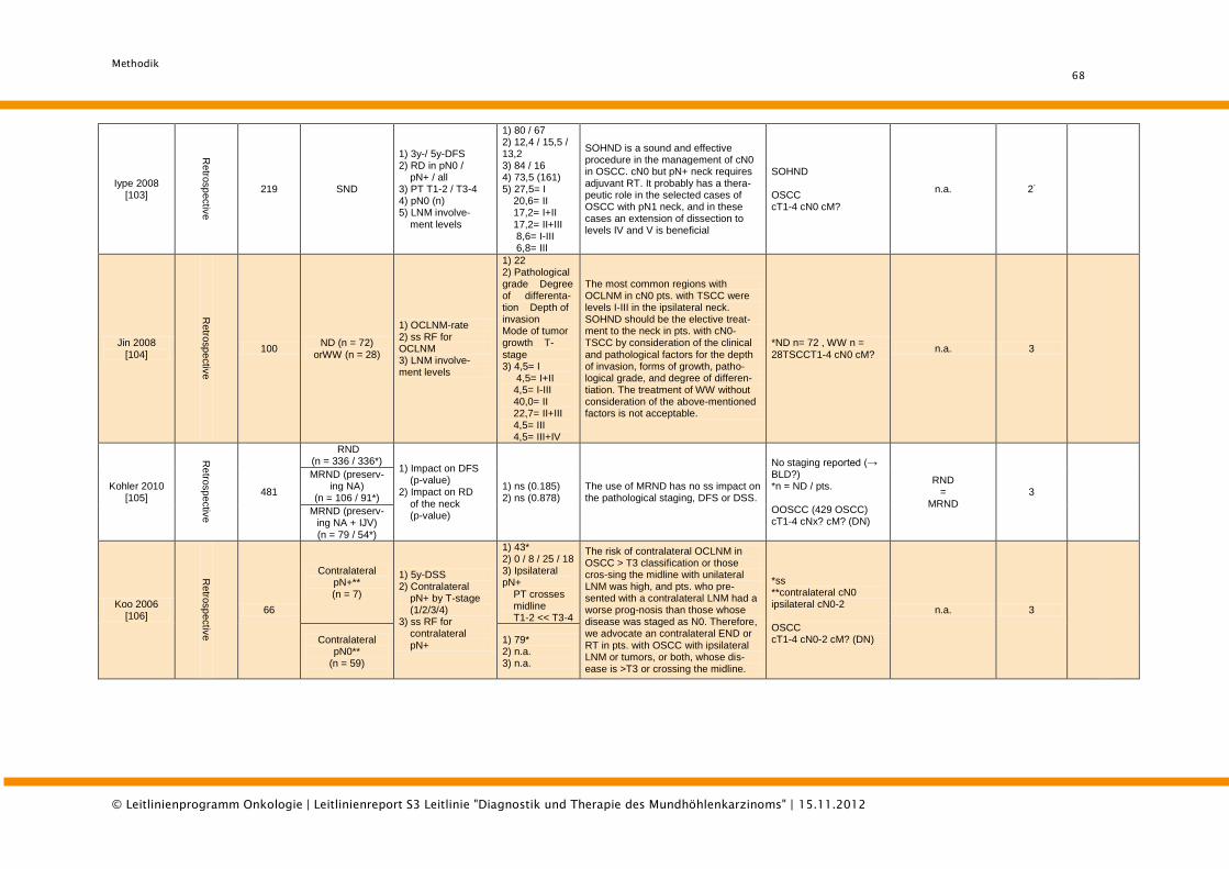
Methodik
68
© Leitlinienprogramm Onkologie | Leitlinienreport S3 Leitlinie "Diagnostik und Therapie des Mundhöhlenkarzinoms" | 15.11.2012
Iype 2008 [103]
Retro
sp
ectiv
e
219 SND
1) 3y-/ 5y-DFS 2) RD in pN0 / pN+ / all 3) PT T1-2 / T3-4 4) pN0 (n) 5) LNM involve- ment levels
1) 80 / 67 2) 12,4 / 15,5 / 13,2 3) 84 / 16 4) 73,5 (161) 5) 27,5= I 20,6= II 17,2= I+II 17,2= II+III 8,6= I-III 6,8= III
SOHND is a sound and effective procedure in the management of cN0 in OSCC. cN0 but pN+ neck requires adjuvant RT. It probably has a thera-peutic role in the selected cases of OSCC with pN1 neck, and in these cases an extension of dissection to levels IV and V is beneficial
SOHND OSCC cT1-4 cN0 cM?
n.a. 2-
Jin 2008 [104]
Retro
sp
ectiv
e
100 ND (n = 72)
orWW (n = 28)
1) OCLNM-rate 2) ss RF for OCLNM 3) LNM involve- ment levels
1) 22 2) Pathological grade Degree of differenta-tion Depth of invasion Mode of tumor growth T-stage 3) 4,5= I 4,5= I+II 4,5= I-III 40,0= II 22,7= II+III 4,5= III 4,5= III+IV
The most common regions with OCLNM in cN0 pts. with TSCC were levels I-III in the ipsilateral neck. SOHND should be the elective treat-ment to the neck in pts. with cN0-TSCC by consideration of the clinical and pathological factors for the depth of invasion, forms of growth, patho-logical grade, and degree of differen-tiation. The treatment of WW without consideration of the above-mentioned factors is not acceptable.
*ND n= 72 , WW n = 28TSCCT1-4 cN0 cM?
n.a. 3
Kohler 2010 [105]
Retro
sp
ectiv
e
481
RND (n = 336 / 336*)
1) Impact on DFS (p-value) 2) Impact on RD of the neck (p-value)
1) ns (0.185) 2) ns (0.878)
The use of MRND has no ss impact on the pathological staging, DFS or DSS.
No staging reported (→ BLD?) *n = ND / pts. OOSCC (429 OSCC) cT1-4 cNx? cM? (DN)
RND =
MRND 3
MRND (preserv- ing NA)
(n = 106 / 91*)
MRND (preserv-ing NA + IJV) (n = 79 / 54*)
Koo 2006 [106]
Retro
sp
ectiv
e
66
Contralateral pN+** (n = 7)
1) 5y-DSS 2) Contralateral pN+ by T-stage (1/2/3/4) 3) ss RF for contralateral pN+
1) 43* 2) 0 / 8 / 25 / 18 3) Ipsilateral pN+ PT crosses midline T1-2 << T3-4
The risk of contralateral OCLNM in OSCC > T3 classification or those cros-sing the midline with unilateral LNM was high, and pts. who pre-sented with a contralateral LNM had a worse prog-nosis than those whose disease was staged as N0. Therefore, we advocate an contralateral END or RT in pts. with OSCC with ipsilateral LNM or tumors, or both, whose dis-ease is >T3 or crossing the midline.
*ss **contralateral cN0 ipsilateral cN0-2 OSCC cT1-4 cN0-2 cM? (DN)
n.a. 3
Contralateral pN0**
(n = 59)
1) 79* 2) n.a. 3) n.a.
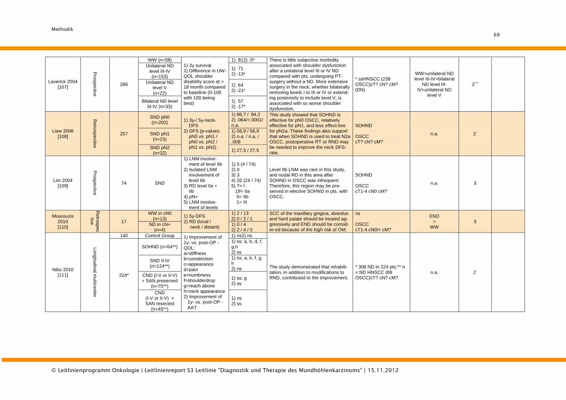
Methodik
69
© Leitlinienprogramm Onkologie | Leitlinienreport S3 Leitlinie "Diagnostik und Therapie des Mundhöhlenkarzinoms" | 15.11.2012
Laverick 2004 [107]
Pro
sp
ectiv
e
266
WW (n=58) 1) 3y survival 2) Difference in UW-QOL shoulder disability score at > 18 month compared to baseline (0-100 with 100 beiing best)
1) 812) -5* There is little subjective morbidity associated with shoulder dysfunction after a unilateral level III or IV ND compared with pts. undergoing PT-surgery without a ND. More extensive surgery in the neck, whether bilaterally removing levels I to III or IV or extend-ing posteriorly to include level V, is associated with ss worse shoulder dysfunction.
* ssHNSCC (238 OSCC)cT? cN? cM? (DN)
WW>unilateral ND level III-IV>bilateral
ND level III-IV>unilateral ND
level V
2++
Unilateral ND level III-IV (n=153)
1) 71 2) -13*
Unilateral ND level V (n=22)
1) 64 2) -21*
Bilateral ND level III-IV (n=33)
1) 57 2) -17*
Liaw 2006 [108]
Retro
sp
ecitv
e
257
SND pN0 (n=202)
1) 3y-/ 5y-neck- DFS 2) DFS (p-values: pN0 vs. pN1 / pN0 vs. pN2 / pN1 vs. pN2)
1) 86,7 / 84,2 2) .064/<.0001/ n.a.
This study showed that SOHND is effective for pN0 OSCC, relatively effective for pN1, and less effect-tive for pN2a. These findings also support that when SOHND is used to treat N2a OSCC, postoperative RT or RND may be needed to improve the neck DFS-rate.
SOHND OSCC cT? cN? cM?
n.a. 2-
SND pN1 (n=23)
1) 56,9 / 56,9 2) n.a. / n.a. / .008
SND pN2 (n=32)
1) 27,5 / 27,5
Lim 2004 [109]
Pro
sp
ectiv
e
74 SND
1) LNM involve- ment of level IIb 2) Isolated LNM involvement of level IIb 3) RD level lIa + IIb 4) pN+ 5) LNM involve- ment of levels
1) 5 (4 / 74) 2) 0 3) 3 4) 32 (24 / 74) 5) 7= I 19= IIa 5= IIb 1= III
Level IIb LNM was rare in this study, and nodal RD in this area after SOHND in OSCC was infrequent. Therefore, this region may be pre-served in elective SOHND in pts. with OSCC.
SOHND OSCC cT1-4 cN0 cM?
n.a. 3
Mourouzis 2010 [110]
Retro
sp
ec
tive
17
WW in cN0 (n=13)
1) 5y-DFS 2) RD (local / neck / distant)
1) 2 / 13 2) 0 / 2 / 1
SCC of the maxillary gingiva, alveolus and hard palate should be treated ag-gressively and END should be consid-er-ed because of the high risk of OM.
ns OSCC cT1-4 cN0/+ cM?
END >
WW 3
ND in cN+ (n=4)
1) 0 / 4 2) 2 / 4 / 0
Nibu 2010 [111]
Lon
gitu
din
al m
ultic
ente
r
140 Control Group 1) Improvement of 1y- vs. post-OP -QOL: a=stiffness b=constriction c=appearance d=pain e=numbness f=shoulderdrop g=reach above h=neck appearance 2) Improvement of 1y- vs. post-OP - AAT
1) ns2) ns
The study demonstrated that rehabili-tation, in addition to modifications to RND, contributed to the improvement.
* 308 ND in 224 pts.** n = ND HNSCC (89 OSCC)cT? cN? cM?
n.a. 2-
224*
SOHND (n=64**) 1) ss: a, b, d, f, g,h 2) ss
SND II-IV (n=124**)
1) ss: a, b, f, g, h 2) ns
CND (I-V or II-V) + SAN preserved
(n=75**)
1) ss: g 2) ss
CND (I-V or II-V) + SAN resected
(n=45**)
1) ns 2) ss
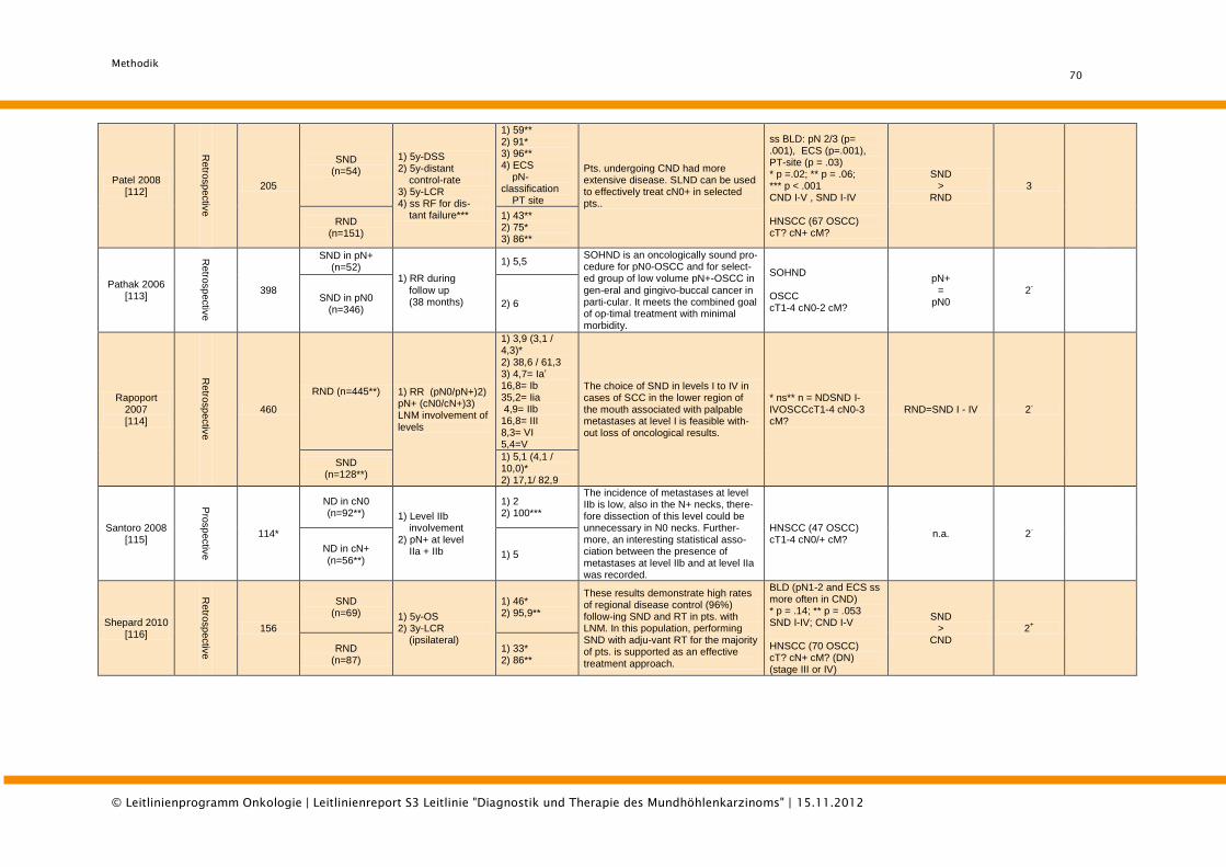
Methodik
70
© Leitlinienprogramm Onkologie | Leitlinienreport S3 Leitlinie "Diagnostik und Therapie des Mundhöhlenkarzinoms" | 15.11.2012
Patel 2008 [112]
Retro
sp
ectiv
e
205
SND (n=54)
1) 5y-DSS 2) 5y-distant control-rate 3) 5y-LCR 4) ss RF for dis- tant failure***
1) 59** 2) 91* 3) 96** 4) ECS pN-classification PT site
Pts. undergoing CND had more extensive disease. SLND can be used to effectively treat cN0+ in selected pts..
ss BLD: pN 2/3 (p= .001), ECS (p=.001), PT-site (p = .03) * p =.02; ** p = .06; *** p < .001 CND I-V , SND I-IV HNSCC (67 OSCC) cT? cN+ cM?
SND >
RND 3
RND (n=151)
1) 43** 2) 75* 3) 86**
Pathak 2006 [113]
Retro
sp
ectiv
e
398
SND in pN+ (n=52)
1) RR during
follow up (38 months)
1) 5,5 SOHND is an oncologically sound pro-cedure for pN0-OSCC and for select-ed group of low volume pN+-OSCC in
gen-eral and gingivo-buccal cancer in parti-cular. It meets the combined goal of op-timal treatment with minimal morbidity.
SOHND OSCC cT1-4 cN0-2 cM?
pN+
= pN0
2-
SND in pN0 (n=346)
2) 6
Rapoport 2007 [114]
Retro
sp
ectiv
e
460
RND (n=445**) 1) RR (pN0/pN+)2) pN+ (cN0/cN+)3) LNM involvement of levels
1) 3,9 (3,1 / 4,3)* 2) 38,6 / 61,3 3) 4,7= Ia’ 16,8= Ib 35,2= Iia 4,9= IIb 16,8= III 8,3= VI 5,4=V
The choice of SND in levels I to IV in cases of SCC in the lower region of the mouth associated with palpable metastases at level I is feasible with-out loss of oncological results.
* ns** n = NDSND I- IVOSCCcT1-4 cN0-3 cM?
RND=SND I - IV 2-
SND (n=128**)
1) 5,1 (4,1 / 10,0)* 2) 17,1/ 82,9
Santoro 2008 [115]
Pro
sp
ectiv
e
114*
ND in cN0 (n=92**) 1) Level IIb
involvement 2) pN+ at level IIa + IIb
1) 2 2) 100***
The incidence of metastases at level IIb is low, also in the N+ necks, there-fore dissection of this level could be unnecessary in N0 necks. Further-more, an interesting statistical asso-ciation between the presence of metastases at level IIb and at level IIa was recorded.
HNSCC (47 OSCC) cT1-4 cN0/+ cM?
n.a. 2-
ND in cN+ (n=56**)
1) 5
Shepard 2010 [116]
Retro
sp
ectiv
e
156
SND (n=69) 1) 5y-OS
2) 3y-LCR (ipsilateral)
1) 46* 2) 95,9**
These results demonstrate high rates of regional disease control (96%) follow-ing SND and RT in pts. with LNM. In this population, performing SND with adju-vant RT for the majority of pts. is supported as an effective treatment approach.
BLD (pN1-2 and ECS ss more often in CND) * p = .14; ** p = .053 SND I-IV; CND I-V HNSCC (70 OSCC) cT? cN+ cM? (DN) (stage III or IV)
SND >
CND 2
+
RND (n=87)
1) 33* 2) 86**
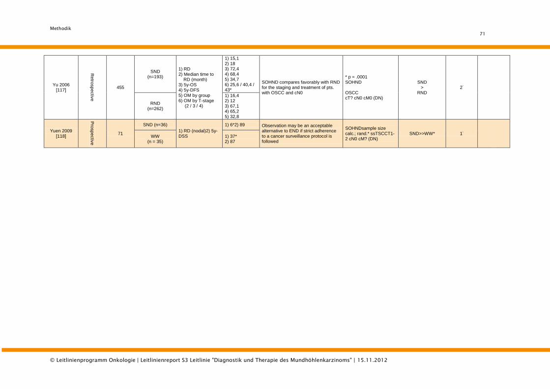
Methodik
71
© Leitlinienprogramm Onkologie | Leitlinienreport S3 Leitlinie "Diagnostik und Therapie des Mundhöhlenkarzinoms" | 15.11.2012
Yu 2006 [117]
Retro
sp
ectiv
e
455
SND (n=193)
1) RD 2) Median time to RD (month) 3) 5y-OS 4) 5y-DFS 5) OM by group 6) OM by T-stage (2 / 3 / 4)
1) 15,1 2) 18 3) 72,4 4) 68,4 5) 34,7 6) 25,6 / 40,4 / 43*
SOHND compares favorably with RND for the staging and treatment of pts. with OSCC and cN0
* p = .0001 SOHND OSCC cT? cN0 cM0 (DN)
SND >
RND 2
-
RND (n=262)
1) 16,4 2) 12 3) 67,1 4) 65,2 5) 32,8
Yuen 2009 [118]
Pro
sp
ectiv
e
71
SND (n=36)
1) RD (nodal)2) 5y-DSS
1) 6*2) 89 Observation may be an acceptable alternative to END if strict adherence to a cancer surveillance protocol is followed
SOHNDsample size calc.; rand.* ssTSCCT1-2 cN0 cM? (DN)
SND>>WW* 1-
WW (n = 35)
1) 37* 2) 87

Methodik
72
© Leitlinienprogramm Onkologie | Leitlinienreport S3 Leitlinie "Diagnostik und Therapie des Mundhöhlenkarzinoms" | 15.11.2012
Tabelle 14 Keyquestion 5
Ist die Kontinuitätsresektion des Unterkiefers der Kastenresektion beim Mundhöhlenkarzinom überlegen?
Erstautor Studien-typ
Patienten- anzahl
Gruppen Outcome Ergebnis (in %)
Fazit Autor Kommentar Zusammen- fassung
Evidenzgrad Querverweis
Abler 2005 [12]
Retro
sp
ectiv
e
152
Kontinuitäts-resektion (n=112)
1) Rezidivrate 2) Wundheilungs- störung
1) 24 2) 40
-pT1-pT3 >5mm Abstand zur Mandibula + Knochenstaging negativ = keine knöcherne Resektion - pT1-pT3 <5mm Abstand zur Mandibula + Knochenstaging negativ intraoperative Periost-Schnellschnittbefundung: → Wenn negativ: keine knöcherne Resektion. → Wenn positiv: Kasten- und/oder Innenspangenresektion - pT1-pT3: bei positivem präoperati-ven Staging bzgl. Knoch-eninfiltration: Kontinuitätsresektion.
BLD (bei kontinuitäts-erhaltenden Eingriffen i.d.R. Ausgangsbefund geringer, teils RT post-OP) OSCC cT1-4 cN0/+ cM?
n.a. 3
KQ
1
Kasten-resektion
(n=40)
1) 12,5 2) 33
Muscatello 2010 [119]
Retro
sp
ectiv
e
56 MM
1) RR (nodal / local) 2) 5y-DFS 3) 5y-OS
1) 10,7 / 8,9 2) 77,3 3) 60,7
MM allows to conduct the resection in a safe tissue or to excise tumors of the FOM with a limited involvement of the alveolar periosteum. Whenev-er the PT is close to the mandible or when it adheres to the alveolar periosteum, MM offers the possibility to perform an oncologically sound procedure.
OSCC cT1-4 cN0-2 cM? (DN)
n.a. 3
Namaki 2004 [120]
Retro
sp
ectiv
e
18
Glossectomy (n=6)
1) Masticatory efficiency pre -treatment vs. post-treatment (12m)** 2) Eating ability pre-treatment vs. posttreat- ment (12m)***
1) 0.58 → 1.04* 2) 4.50 → 6
Results of the present study suggest that the QOL of pts. undergoing marginal mandibulectomy is better than those undergoing segmental mandibulectomy.
** Absorbance unit of ATP-granules ***grade measured using the questionnaire de-vised by Shinohara * ss (intragroup) OSCC T1-4 cN0/+ cM?
MM >
SM 3
MM (n=6)
1) 1.21 → 0.85 2) 6 → 4*
SM (n=6)
1) 1.19 → 0.56 2) 5 → 3*
Rogers 2004 [121]
not c
itied
73 RM
(n=32)
1) QOL pre- → post-surgery; (mean 18m)
1) 83 → 76* (-7)
After SM and reconstruction using composite free tissue transfer, the UW-QOL scores were relatively good. The only difference between RM and SM was noted in the small
BLD * ss (intragroup) OSCC cT1-4 cN? cM? (DN)
n.a. 2++
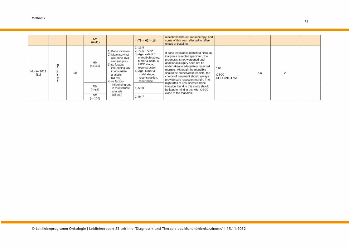
Methodik
73
© Leitlinienprogramm Onkologie | Leitlinienreport S3 Leitlinie "Diagnostik und Therapie des Mundhöhlenkarzinoms" | 15.11.2012
SM (n=41)
1) 78→ 62* (-16)
resections with-out radiotherapy, and some of this was reflected in differ-ences at baseline.
Mucke 2011 [11]
Retro
sp
ectiv
e
334
MM (n=116)
1) Bone invasion 2) Mean survival w/o bone inva- sion (all pts.) 3) ss factors influencing OS in univariate analysis (all pts.) 4) ss factors influencing OS in multivariate analysis (all pts.)
1) 15,5 2) 71,6 / 72,9* 3) Age, extent of mandibulectomy, tumor & nodal & UICC stage, reconstruction 4) Age, tumor & nodal stage, reconstruction, recurrence
If bone invasion is identified histolog-ically in a resected specimen, the prognosis is not worsened and additional surgery need not be undertaken in adequately resected margins. Although the mandible should be preserved if feasible, the choice of treatment should always provide safe resection margin. The high rates of unsuspected bone invasion found in this study should be kept in mind in pts. with OSCC close to the mandible
* ns OSCC cT1-4 cN1-4 cM0
n.a. 2-
RM (n=68)
1) 50,0
SM (n=150)
1) 84,7
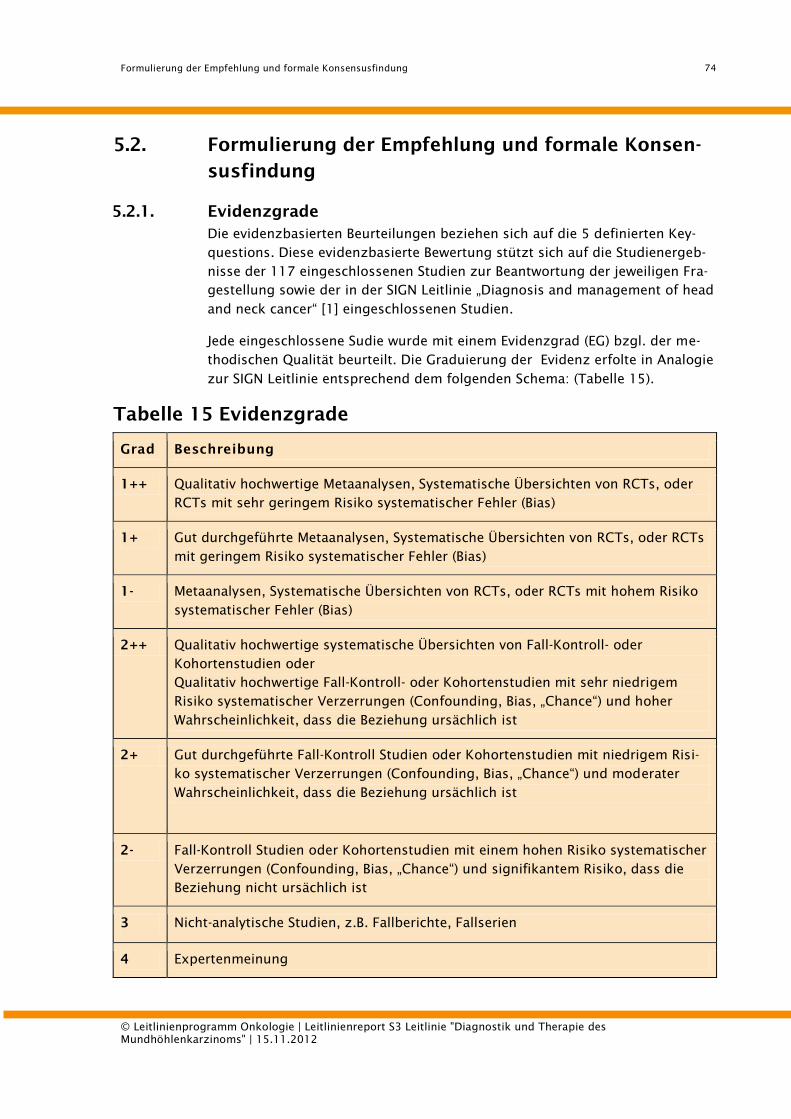
Formulierung der Empfehlung und formale Konsensusfindung
© Leitlinienprogramm Onkologie | Leitlinienreport S3 Leitlinie "Diagnostik und Therapie des
Mundhöhlenkarzinoms" | 15.11.2012
74
5.2. Formulierung der Empfehlung und formale Konsen-
susfindung
5.2.1. Evidenzgrade
Die evidenzbasierten Beurteilungen beziehen sich auf die 5 definierten Key-
questions. Diese evidenzbasierte Bewertung stützt sich auf die Studienergeb-
nisse der 117 eingeschlossenen Studien zur Beantwortung der jeweiligen Fra-
gestellung sowie der in der SIGN Leitlinie „Diagnosis and management of head
and neck cancer“ [1] eingeschlossenen Studien.
Jede eingeschlossene Sudie wurde mit einem Evidenzgrad (EG) bzgl. der me-
thodischen Qualität beurteilt. Die Graduierung der Evidenz erfolte in Analogie
zur SIGN Leitlinie entsprechend dem folgenden Schema: (Tabelle 15).
Tabelle 15 Evidenzgrade
Grad Beschreibung
1++ Qualitativ hochwertige Metaanalysen, Systematische Übersichten von RCTs, oder
RCTs mit sehr geringem Risiko systematischer Fehler (Bias)
1+ Gut durchgeführte Metaanalysen, Systematische Übersichten von RCTs, oder RCTs
mit geringem Risiko systematischer Fehler (Bias)
1- Metaanalysen, Systematische Übersichten von RCTs, oder RCTs mit hohem Risiko
systematischer Fehler (Bias)
2++ Qualitativ hochwertige systematische Übersichten von Fall-Kontroll- oder
Kohortenstudien oder
Qualitativ hochwertige Fall-Kontroll- oder Kohortenstudien mit sehr niedrigem
Risiko systematischer Verzerrungen (Confounding, Bias, „Chance“) und hoher
Wahrscheinlichkeit, dass die Beziehung ursächlich ist
2+ Gut durchgeführte Fall-Kontroll Studien oder Kohortenstudien mit niedrigem Risi-
ko systematischer Verzerrungen (Confounding, Bias, „Chance“) und moderater
Wahrscheinlichkeit, dass die Beziehung ursächlich ist
2- Fall-Kontroll Studien oder Kohortenstudien mit einem hohen Risiko systematischer
Verzerrungen (Confounding, Bias, „Chance“) und signifikantem Risiko, dass die
Beziehung nicht ursächlich ist
3 Nicht-analytische Studien, z.B. Fallberichte, Fallserien
4 Expertenmeinung
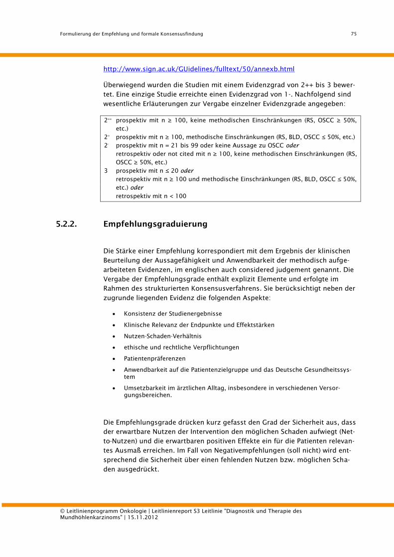
Formulierung der Empfehlung und formale Konsensusfindung
© Leitlinienprogramm Onkologie | Leitlinienreport S3 Leitlinie "Diagnostik und Therapie des
Mundhöhlenkarzinoms" | 15.11.2012
75
http://www.sign.ac.uk/GUidelines/fulltext/50/annexb.html
Überwiegend wurden die Studien mit einem Evidenzgrad von 2++ bis 3 bewer-
tet. Eine einzige Studie erreichte einen Evidenzgrad von 1-. Nachfolgend sind
wesentliche Erläuterungen zur Vergabe einzelner Evidenzgrade angegeben:
2++
prospektiv mit n ≥ 100, keine methodischen Einschränkungen (RS, OSCC ≥ 50%,
etc.)
2+
prospektiv mit n ≥ 100, methodische Einschränkungen (RS, BLD, OSCC ≤ 50%, etc.)
2-
prospektiv mit n = 21 bis 99 oder keine Aussage zu OSCC oder
retrospektiv oder not cited mit n ≥ 100, keine methodischen Einschränkungen (RS,
OSCC ≥ 50%, etc.)
3 prospektiv mit n ≤ 20 oder
retrospektiv mit n ≥ 100 und methodische Einschränkungen (RS, BLD, OSCC ≤ 50%,
etc.) oder
retrospektiv mit n < 100
5.2.2. Empfehlungsgraduierung
Die Stärke einer Empfehlung korrespondiert mit dem Ergebnis der klinischen
Beurteilung der Aussagefähigkeit und Anwendbarkeit der methodisch aufge-
arbeiteten Evidenzen, im englischen auch considered judgement genannt. Die
Vergabe der Empfehlungsgrade enthält explizit Elemente und erfolgte im
Rahmen des strukturierten Konsensusverfahrens. Sie berücksichtigt neben der
zugrunde liegenden Evidenz die folgenden Aspekte:
Konsistenz der Studienergebnisse
Klinische Relevanz der Endpunkte und Effektstärken
Nutzen-Schaden-Verhältnis
ethische und rechtliche Verpflichtungen
Patientenpräferenzen
Anwendbarkeit auf die Patientenzielgruppe und das Deutsche Gesundheitssys-
tem
Umsetzbarkeit im ärztlichen Alltag, insbesondere in verschiedenen Versor-
gungsbereichen.
Die Empfehlungsgrade drücken kurz gefasst den Grad der Sicherheit aus, dass
der erwartbare Nutzen der Intervention den möglichen Schaden aufwiegt (Net-
to-Nutzen) und die erwartbaren positiven Effekte ein für die Patienten relevan-
tes Ausmaß erreichen. Im Fall von Negativempfehlungen (soll nicht) wird ent-
sprechend die Sicherheit über einen fehlenden Nutzen bzw. möglichen Scha-
den ausgedrückt.
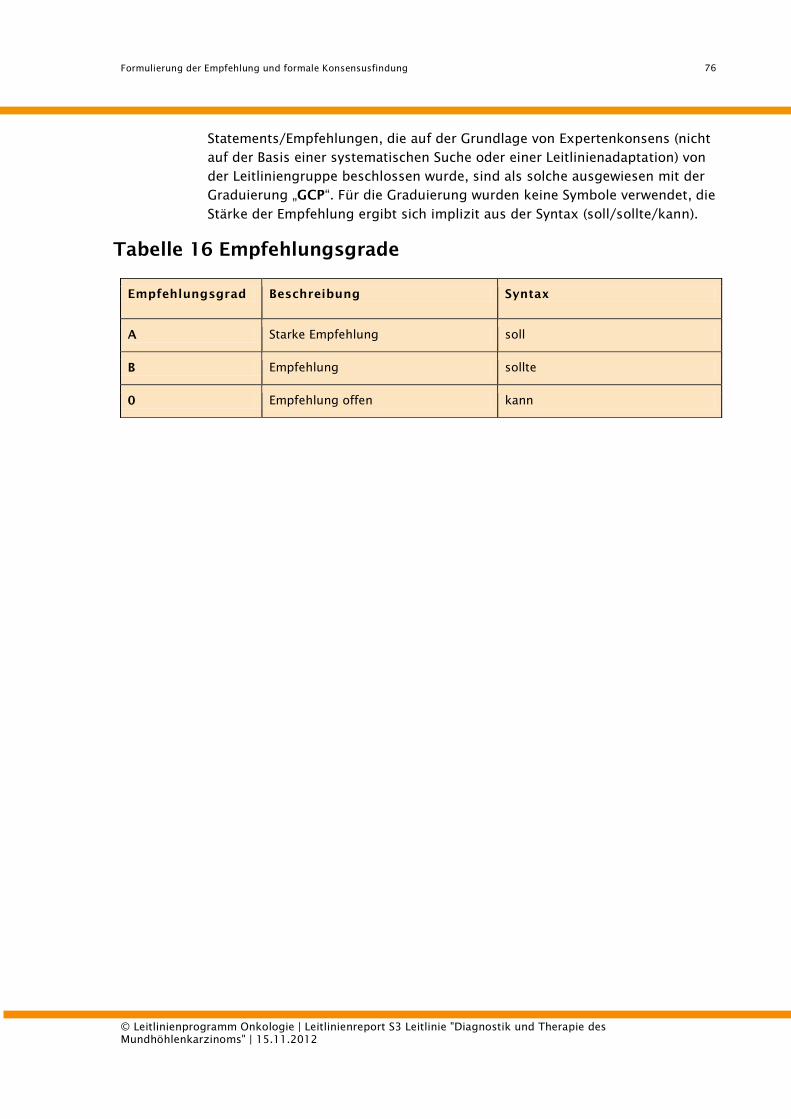
Formulierung der Empfehlung und formale Konsensusfindung
© Leitlinienprogramm Onkologie | Leitlinienreport S3 Leitlinie "Diagnostik und Therapie des
Mundhöhlenkarzinoms" | 15.11.2012
76
Statements/Empfehlungen, die auf der Grundlage von Expertenkonsens (nicht
auf der Basis einer systematischen Suche oder einer Leitlinienadaptation) von
der Leitliniengruppe beschlossen wurde, sind als solche ausgewiesen mit der
Graduierung „GCP“. Für die Graduierung wurden keine Symbole verwendet, die
Stärke der Empfehlung ergibt sich implizit aus der Syntax (soll/sollte/kann).
Tabelle 16 Empfehlungsgrade
Empfehlungsgrad Beschreibung Syntax
A Starke Empfehlung soll
B Empfehlung sollte
0 Empfehlung offen kann
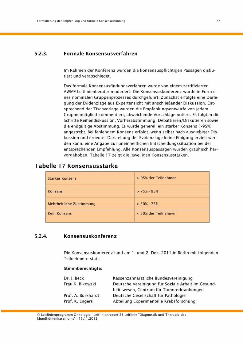
Formulierung der Empfehlung und formale Konsensusfindung
© Leitlinienprogramm Onkologie | Leitlinienreport S3 Leitlinie "Diagnostik und Therapie des
Mundhöhlenkarzinoms" | 15.11.2012
77
5.2.3. Formale Konsensusverfahren
Im Rahmen der Konferenz wurden die konsensuspflichtigen Passagen disku-
tiert und verabschiedet.
Das formale Konsensusfindungsverfahren wurde von einem zertifizierten
AWMF Leitlinienberater moderiert. Die Konsensuskonferenz wurde in Form ei-
nes nominalen Gruppenprozesses durchgeführt. Zunächst erfolgte eine Darle-
gung der Evidenzlage aus Expertensicht mit anschließender Diskussion. Ent-
sprechend der Tischvorlage wurden die Empfehlungsentwürfe von jedem
Gruppenmitglied kommentiert, abweichende Vorschläge notiert. Es folgten die
Schritte Reihendiskussion, Vorherabstimmung, Debattieren/Diskutieren sowie
die endgültige Abstimmung. Es wurde generell ein starker Konsens (>95%)
angestrebt. Bei fehlendem Konsens erfolgt, wenn selbst nach ausgiebiger Dis-
kussion und erneuter Darstellung der Evidenzlage keine Einigung erzielt wer-
den kann, eine Angabe zur uneinheitlichen Entscheidungssituation bei der
entsprechenden Empfehlung. Alle Konsensuspassagen wurden graphisch her-
vorgehoben. Tabelle 17 zeigt die jeweiligen Konsensusstärken.
Tabelle 17 Konsensusstärke
Starker Konsens > 95% der Teilnehmer
Konsens > 75% - 95%
Mehrheitliche Zustimmung > 50% - 75%
Kein Konsens < 50% der Teilnehmer
5.2.4. Konsensuskonferenz
Die Konsensuskonferenz fand am 1. und 2. Dez. 2011 in Berlin mit folgenden
Teilnehmern statt:
Stimmberechtigte:
Dr. J. Beck Kassenzahnärztliche Bundesvereinigung
Frau K. Bikowski Deutsche Vereinigung für Soziale Arbeit im Gesund-
heitswesen, Centrum für Tumorerkrankungen
Prof. A. Burkhardt Deutsche Gesellschaft für Pathologie
Prof. K. Engers Abteilung Experimentelle Krebsforschung
Formulierung der Empfehlung und formale Konsensusfindung
© Leitlinienprogramm Onkologie | Leitlinienreport S3 Leitlinie "Diagnostik und Therapie des
Mundhöhlenkarzinoms" | 15.11.2012
78
Prof. B. Frerich Deutsche Gesellschaft für Mund-, Kiefer- und Ge-
sichtschirurgie
Prof. T. Gauler Deutsche Gesellschaft für Internistische Onkologie
Prof. W. Budach Deutsche Gesellschaft für Radioonkologie
Prof. S. Ihrler Deutsche Gesellschaft für Pathologie
Prof. M. Lell Arbeitsgemeinschaft Kopf-Hals der Deutschen
Röntgenge-sellschaft
Frau W. Mantey Patientenvertreterin
Frau Nusser-Müller- Deutsche Gesellschaft für Logopädie
Busch
Dr. N. Gittler-Hebestreit Konferenz Onkologischer Kranken- und Kinderkran-
kenpflege
Prof. H. Pistner Leitlinien-Beauftragter der Deutschen Gesellschaft für
Mund-, Kiefer- und Gesichtschirurgie
Prof. T. Reichert Deutsche Gesellschaft für Mund-, Kiefer- und Ge-
sichtschirurgie
Prof. H. Schliephake Deutsche Gesellschaft für Zahn-, Mund- und Kiefer-
keilkunde
Prof. M. Westhofen Deutsche Gesellschaft für Hals-Nasen-Ohrenheilkunde
Dr. S. Wirz Arbeitsgemeinschaft Tumorschmerz der Deutschen
Gesellschaft zum Studium des Schmerzes
Dr. M. Wittlinger Deutsche Gesellschaft für Radioonkologie,
Dr. P. Boehme Bundeszahnärztekammer
Prof. F. Bootz Deutsche Gesellschaft für Hals-Nasen-Ohrenheilkunde
Herr H. Danker Arbeitsgemeinschaft Psychoonkologie in der DKG
Prof. A. Lübbe Arbeitsgemeinschaft für Supportive Onkologie, Reha-
bilitation und Sozialmedizin
Prof. K.-D. Wolff Deutsche Gesellschaft für Mund-, Kiefer- und Ge-
sichtschirurgie - Leitlinienkoordination
Moderation und Begleitung (nicht stimmberechtigt):
Prof. I. Kopp Leiterin des Instituts für Medizinisches Wissensma-
nagement der Arbeitsgemeinschaft der Medizinisch
Wissenschaftlichen Fachgesellschaften, Marburg
(AWMF)
Dr. M. Follmann, Deutsche Krebsgesellschaft (DKG),
MPH MSc Leitlinienprogramm Onkologie
Dr. A. Sammain Division of Evidence Based Medicine, Charité -
Universitätsmedzin Berlin
J. Weitz Mitarbeiter der Klinik für MKG-Chirurgie, TU München
M. Keul Sekretariat der Klinik für MKG-Chirurgie, TU München
Die protokollarische Darstellung des Abstimmungsverfahrens ist auf Anfrage
beim Koordinator der Leitlinie, Prof. Wolff, München, abrufbar.
Qualitätsindikatoren
© Leitlinienprogramm Onkologie | Leitlinienreport S3 Leitlinie "Diagnostik und Therapie des
Mundhöhlenkarzinoms" | 15.11.2012
79
6. Qualitätsindikatoren
Qualitätsindikatoren sind Messgrößen, deren Erhebung der Beurteilung der
Qualität der zugrundeliegenden Strukturen, Prozessen bzw. Ergebnisse dient.
[Ärztliches Zentrum für Qualität in der Medizin (ÄZQ), Gramsch E, Hoppe JD,
Jonitz G, Köhler A, Ollenschläger G, Thomeczek C, (eds.). Kompendium Q-M-A.
Qualitätsmanagement in der ambulanten Versorgung. 3rd ed. Köln: Dt. Ärzte-
Verl.; 2008.]. Das vorrangige Ziel ihres Einsatzes ist die stetige Verbesserung
der Versorgung. Qualitätsindikatoren als qualitätsbezogene Kennzahlen sind
damit ein wesentlicher Bestandteil des Qualitätsmanagements. Dabei ist zu
beachten, dass einzelne Indikatoren jeweils nur einen Aspekt des komplexen
Versorgungsgeschehens beleuchten. Der Auswahl geeigneter Indikatoren
kommt deshalb große Bedeutung zu.
Die vorliegende Auswahl von Qualitätsindikatoren orientiert sich an der
Methodik der Nationalen Versorgungsleitlinien (Ärztliches Zentrum für
Qualität in der Medizin (ÄZQ). Manual Qualitätsindikatoren. Manual für
Autoren. Berlin: ÄZQ; 2009. (äzq Schriftenreihe; 36). Available from:
http://www.aezq.de/edocs/pdf/schriftenreihe/schriftenreihe36.pdf).Als
Grundlage für die Qualitätsindikatoren dienten alle starken Empfehlungen
(Empfehlungsgrad A) sowie Statements mit hoher Evidenz (LoE 1). Diese
Empfehlungen und Statements wurden durch einen Methodiker in potentielle
Indikatoren überführt und von den Experten der AG Qualitätsindikatoren
schriftlich nach folgenden definierten Kriterien bewertet:
1. Bedeutung des Indikators für das Versorgungssystem: hat die
durch den Indikator gemessene Maßnahme aufgrund einer hohen Fallzahl, ei-
ner großen Versorgungsvariabilität, einer bekannten Unter- oder Überversor-
gung eine wichtige Bedeutung? Kann dadurch die Morbidität oder auch Morta-
lität bzw. die Lebensqualität verbessert werden?
2. Klarheit der Definition: Ist der mögliche Indikator klar und eindeutig be-
züglich Nenner und Zähler definiert bzw. können benötigte Definitionen ein-
deutig aus der Leitlinie entnommen werden?
3. Beeinflussbarkeit der Indikatorausprägung: kann der bewertete Aspekt
vom Leistungserbringer beeinflusst werden?
4. Evidenz- und Konsensbasierung des Indikators. Dieses Kriterium wurde
nicht bewertet, da eine Vorauswahl getroffen wurde und nur starke Empfeh-
lungen (Empfehlungsgrad A/soll) oder Statements mit Evidenzgrad 1 aufge-
nommen wurden, die Evidenzbasierung des Indikators wurde für die Bewer-
tung dargestellt.)
Qualitätsindikatoren
© Leitlinienprogramm Onkologie | Leitlinienreport S3 Leitlinie "Diagnostik und Therapie des
Mundhöhlenkarzinoms" | 15.11.2012
80
5. Risiken zur Fehlsteuerung. Hier kann bewertet werden, ob durch den Indi-
kator ein Risiko zur Fehlsteuerung gesetzt wird, das nicht ausgeglichen wer-
den kann durch einen Gegenindikator oder andere Informationen.
Als angenommen galten Indikatoren mit mind. 75% Zustimmung bei jedem
Kriterium. Nach der schriftlichen Bewertung erfolgten methodisch moderierte
Telefonkonferenzen, in denen sowohl die angenommenen als auch die wegen
eines Kriteriums nicht angenommenen Indikatoren diskutiert wurden.
Danach erfolgte eine abschließende schriftliche Annahme oder Ablehnung der
Indikatoren.
Die Indikatoren sind als vorläufig bewertete Vorschläge zu verstehen. Eine ab-
schließende Bewertung kann erst nach einem erforderlichen Pilottest mit Da-
tenerhebung erfolgen.
Weitere Informationen zur allgemeinen Methodik sind in der Homepage des
Onkologischen Leitlinienprogramms www.leitlinienprogramm-onkologie.de
hinterlegt.
Die Indikatoren selbst finden sich in der Langversion und in der Kurzversion
der Leitlinie.
Externe Begutachtung und Verabschiedung
© Leitlinienprogramm Onkologie | Leitlinienreport S3 Leitlinie "Diagnostik und Therapie des
Mundhöhlenkarzinoms" | 15.11.2012
81
7. Externe Begutachtung und Verabschie-
dung
Ein externes Peer-Review Verfahren wurde nicht durchgeführt
Der konsentierte Leitlinienentwurf wurde den Vorständen der beteiligten
Fachgesellschaften zur Verabschiedung vorgelegt und anschließend autori-
siert.
Redaktionelle Unabhängigkeit
© Leitlinienprogramm Onkologie | Leitlinienreport S3 Leitlinie "Diagnostik und Therapie des
Mundhöhlenkarzinoms" | 15.11.2012
82
8. Redaktionelle Unabhängigkeit
Die Darstellung eventuell vorhandenen Interessenskonflikte erfolgt tabella-
risch (s. Col-Bögen). Die Finanzierung der Leitlinie erfolgte ausschließlich über
das Onkologische Leitlinienprogramm. Die standardisierten Bögen wurden in
der Gruppe diskutiert. Es konnte kein Teilnehmer identifiiziert werden, der
aufgrund von potentiellen Interessenskonflikten von der LL-Entwicklung aus-
geschlossen werden musste. Auch wurden keine Bereiche entdeckt, die eine
Enthaltung der Stimme bei einzelnen Themen erforderlich gemacht hätte.
Die Literaturrecherche, die Bewertung der Evidenz und die Erstellung der
Evidenztabellen erfolgte neutral durch die dEBM (division of Evidence Based
Medicine, Arbeitsgruppe um PD Dr. Nast, Klinik für Dermatologie, Charité -
Universitätsmedizin Berlin). Die klinischen Experten waren nicht an der Bewer-
tung der Evidenz und der Erstellung der Evidenztabellen beteiligt.
Alle Teilnehmer der Leitliniengruppe haben schriftlich dargelegt, dass keiner-
lei finanzielle oder sonstige kommerzielle Interessenskonflikte gegenüber
Dritten vorliegen, die eine Einflussnahme auf die Formulierung der Leitlinien-
inhalte begründen könnten. Die Leitlinie wurde ausschließlich vom OL finan-
ziert. Die Mittel wurden überwiegend für die externe Literaturbasierung und
die Durchführung der Leitlinientreffen (Kick off Meeting, Arbeitsgruppentref-
fen, Konsensuskonferenz) eingesetzt. Die Deutsche Gesellschaft für Mund-,
Kiefer- und Gesichtschirurgie (DGMKG) und auch die weiteren teilnehmenden
Fachgesellschaften, Arbeitsgruppen oder Institutio-nen haben für diese S3-
Leitlinie keine finanzielle oder andere Unterstützung von kommerziellen Inter-
essensgruppen erhalten.
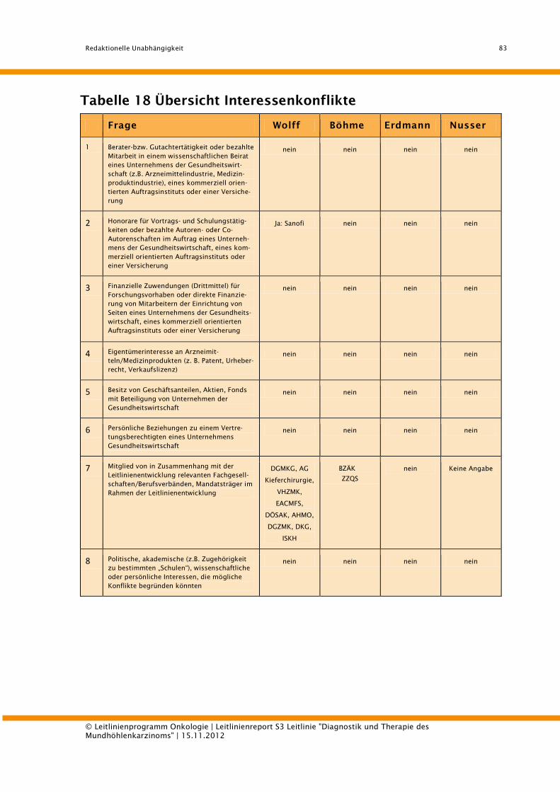
Redaktionelle Unabhängigkeit
© Leitlinienprogramm Onkologie | Leitlinienreport S3 Leitlinie "Diagnostik und Therapie des
Mundhöhlenkarzinoms" | 15.11.2012
83
Tabelle 18 Übersicht Interessenkonflikte
Frage Wolff Böhme Erdmann Nusser
1 Berater-bzw. Gutachtertätigkeit oder bezahlte
Mitarbeit in einem wissenschaftlichen Beirat
eines Unternehmens der Gesundheitswirt-
schaft (z.B. Arzneimittelindustrie, Medizin-
produktindustrie), eines kommerziell orien-
tierten Auftragsinstituts oder einer Versiche-
rung
nein nein nein nein
2 Honorare für Vortrags- und Schulungstätig-
keiten oder bezahlte Autoren- oder Co-
Autorenschaften im Auftrag eines Unterneh-
mens der Gesundheitswirtschaft, eines kom-
merziell orientierten Auftragsinstituts oder
einer Versicherung
Ja: Sanofi nein nein nein
3 Finanzielle Zuwendungen (Drittmittel) für
Forschungsvorhaben oder direkte Finanzie-
rung von Mitarbeitern der Einrichtung von
Seiten eines Unternehmens der Gesundheits-
wirtschaft, eines kommerziell orientierten
Auftragsinstituts oder einer Versicherung
nein nein nein nein
4 Eigentümerinteresse an Arzneimit-
teln/Medizinprodukten (z. B. Patent, Urheber-
recht, Verkaufslizenz)
nein nein nein nein
5 Besitz von Geschäftsanteilen, Aktien, Fonds
mit Beteiligung von Unternehmen der
Gesundheitswirtschaft
nein nein nein nein
6 Persönliche Beziehungen zu einem Vertre-
tungsberechtigten eines Unternehmens
Gesundheitswirtschaft
nein nein nein nein
7 Mitglied von in Zusammenhang mit der
Leitlinienentwicklung relevanten Fachgesell-
schaften/Berufsverbänden, Mandatsträger im
Rahmen der Leitlinienentwicklung
DGMKG, AG
Kieferchirurgie,
VHZMK,
EACMFS,
DÖSAK, AHMO,
DGZMK, DKG,
ISKH
BZÄK
ZZQS
nein Keine Angabe
8 Politische, akademische (z.B. Zugehörigkeit
zu bestimmten „Schulen“), wissenschaftliche
oder persönliche Interessen, die mögliche
Konflikte begründen könnten
nein nein nein nein
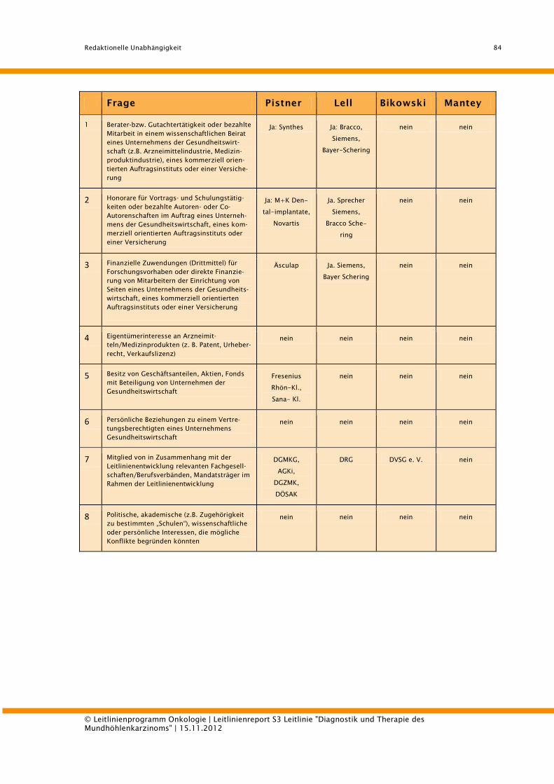
Redaktionelle Unabhängigkeit
© Leitlinienprogramm Onkologie | Leitlinienreport S3 Leitlinie "Diagnostik und Therapie des
Mundhöhlenkarzinoms" | 15.11.2012
84
Frage Pistner Lell Bikowski Mantey
1 Berater-bzw. Gutachtertätigkeit oder bezahlte
Mitarbeit in einem wissenschaftlichen Beirat
eines Unternehmens der Gesundheitswirt-
schaft (z.B. Arzneimittelindustrie, Medizin-
produktindustrie), eines kommerziell orien-
tierten Auftragsinstituts oder einer Versiche-
rung
Ja: Synthes Ja: Bracco,
Siemens,
Bayer-Schering
nein nein
2 Honorare für Vortrags- und Schulungstätig-
keiten oder bezahlte Autoren- oder Co-
Autorenschaften im Auftrag eines Unterneh-
mens der Gesundheitswirtschaft, eines kom-
merziell orientierten Auftragsinstituts oder
einer Versicherung
Ja: M+K Den-
tal-implantate,
Novartis
Ja. Sprecher
Siemens,
Bracco Sche-
ring
nein nein
3 Finanzielle Zuwendungen (Drittmittel) für
Forschungsvorhaben oder direkte Finanzie-
rung von Mitarbeitern der Einrichtung von
Seiten eines Unternehmens der Gesundheits-
wirtschaft, eines kommerziell orientierten
Auftragsinstituts oder einer Versicherung
Äsculap Ja. Siemens,
Bayer Schering
nein nein
4 Eigentümerinteresse an Arzneimit-
teln/Medizinprodukten (z. B. Patent, Urheber-
recht, Verkaufslizenz)
nein nein nein nein
5 Besitz von Geschäftsanteilen, Aktien, Fonds
mit Beteiligung von Unternehmen der
Gesundheitswirtschaft
Fresenius
Rhön-Kl.,
Sana- Kl.
nein nein nein
6 Persönliche Beziehungen zu einem Vertre-
tungsberechtigten eines Unternehmens
Gesundheitswirtschaft
nein nein nein nein
7 Mitglied von in Zusammenhang mit der
Leitlinienentwicklung relevanten Fachgesell-
schaften/Berufsverbänden, Mandatsträger im
Rahmen der Leitlinienentwicklung
DGMKG,
AGKi,
DGZMK,
DÖSAK
DRG DVSG e. V. nein
8 Politische, akademische (z.B. Zugehörigkeit
zu bestimmten „Schulen“), wissenschaftliche
oder persönliche Interessen, die mögliche
Konflikte begründen könnten
nein nein nein nein
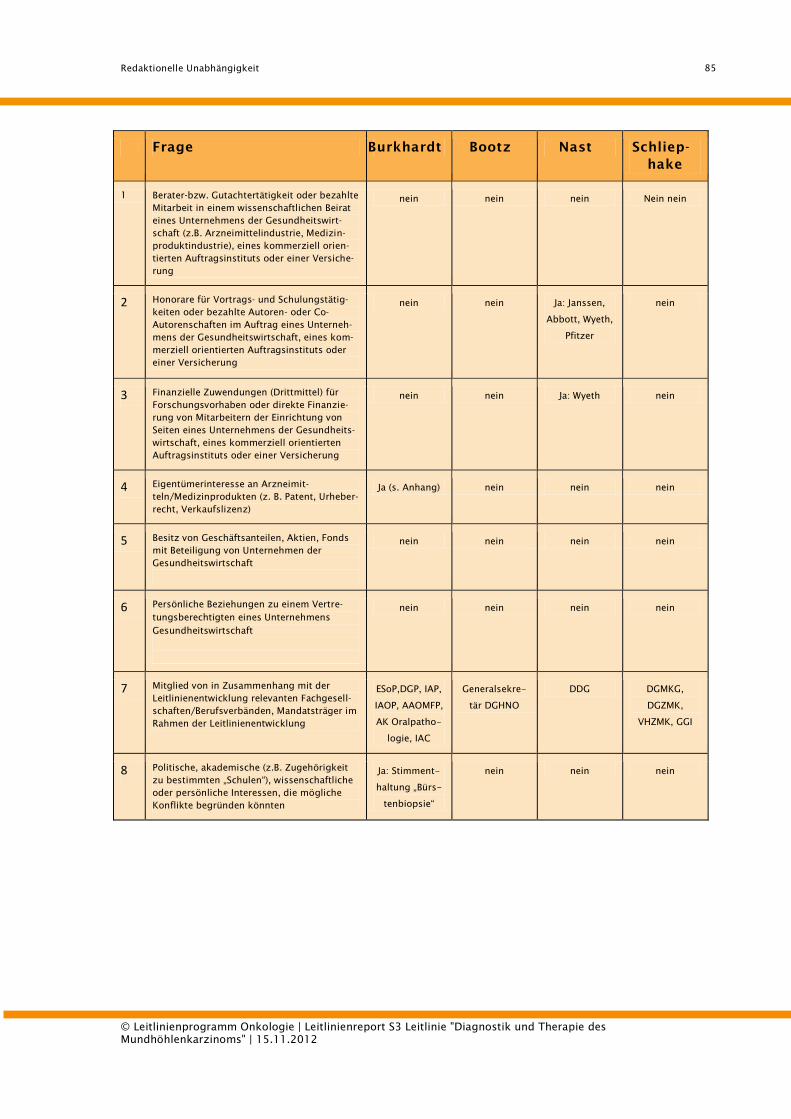
Redaktionelle Unabhängigkeit
© Leitlinienprogramm Onkologie | Leitlinienreport S3 Leitlinie "Diagnostik und Therapie des
Mundhöhlenkarzinoms" | 15.11.2012
85
Frage Burkhardt Bootz Nast Schliep-
hake
1 Berater-bzw. Gutachtertätigkeit oder bezahlte
Mitarbeit in einem wissenschaftlichen Beirat
eines Unternehmens der Gesundheitswirt-
schaft (z.B. Arzneimittelindustrie, Medizin-
produktindustrie), eines kommerziell orien-
tierten Auftragsinstituts oder einer Versiche-
rung
nein nein nein Nein nein
2 Honorare für Vortrags- und Schulungstätig-
keiten oder bezahlte Autoren- oder Co-
Autorenschaften im Auftrag eines Unterneh-
mens der Gesundheitswirtschaft, eines kom-
merziell orientierten Auftragsinstituts oder
einer Versicherung
nein nein Ja: Janssen,
Abbott, Wyeth,
Pfitzer
nein
3 Finanzielle Zuwendungen (Drittmittel) für
Forschungsvorhaben oder direkte Finanzie-
rung von Mitarbeitern der Einrichtung von
Seiten eines Unternehmens der Gesundheits-
wirtschaft, eines kommerziell orientierten
Auftragsinstituts oder einer Versicherung
nein nein Ja: Wyeth nein
4 Eigentümerinteresse an Arzneimit-
teln/Medizinprodukten (z. B. Patent, Urheber-
recht, Verkaufslizenz)
Ja (s. Anhang) nein nein nein
5 Besitz von Geschäftsanteilen, Aktien, Fonds
mit Beteiligung von Unternehmen der
Gesundheitswirtschaft
nein nein nein nein
6 Persönliche Beziehungen zu einem Vertre-
tungsberechtigten eines Unternehmens
Gesundheitswirtschaft
nein nein nein nein
7 Mitglied von in Zusammenhang mit der
Leitlinienentwicklung relevanten Fachgesell-
schaften/Berufsverbänden, Mandatsträger im
Rahmen der Leitlinienentwicklung
ESoP,DGP, IAP,
IAOP, AAOMFP,
AK Oralpatho-
logie, IAC
Generalsekre-
tär DGHNO
DDG DGMKG,
DGZMK,
VHZMK, GGI
8 Politische, akademische (z.B. Zugehörigkeit
zu bestimmten „Schulen“), wissenschaftliche
oder persönliche Interessen, die mögliche
Konflikte begründen könnten
Ja: Stimment-
haltung „Bürs-
tenbiopsie“
nein nein nein
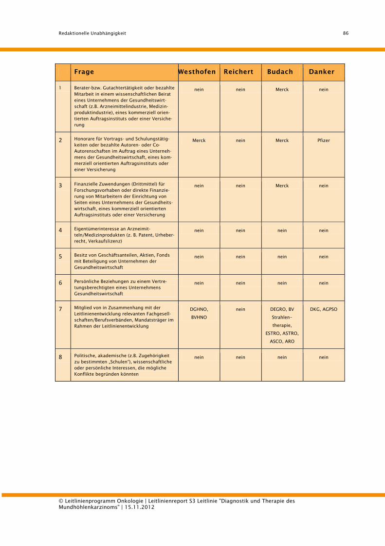
Redaktionelle Unabhängigkeit
© Leitlinienprogramm Onkologie | Leitlinienreport S3 Leitlinie "Diagnostik und Therapie des
Mundhöhlenkarzinoms" | 15.11.2012
86
Frage Westhofen Reichert Budach Danker
1 Berater-bzw. Gutachtertätigkeit oder bezahlte
Mitarbeit in einem wissenschaftlichen Beirat
eines Unternehmens der Gesundheitswirt-
schaft (z.B. Arzneimittelindustrie, Medizin-
produktindustrie), eines kommerziell orien-
tierten Auftragsinstituts oder einer Versiche-
rung
nein nein Merck nein
2 Honorare für Vortrags- und Schulungstätig-
keiten oder bezahlte Autoren- oder Co-
Autorenschaften im Auftrag eines Unterneh-
mens der Gesundheitswirtschaft, eines kom-
merziell orientierten Auftragsinstituts oder
einer Versicherung
Merck nein Merck Pfizer
3 Finanzielle Zuwendungen (Drittmittel) für
Forschungsvorhaben oder direkte Finanzie-
rung von Mitarbeitern der Einrichtung von
Seiten eines Unternehmens der Gesundheits-
wirtschaft, eines kommerziell orientierten
Auftragsinstituts oder einer Versicherung
nein nein Merck nein
4 Eigentümerinteresse an Arzneimit-
teln/Medizinprodukten (z. B. Patent, Urheber-
recht, Verkaufslizenz)
nein nein nein nein
5 Besitz von Geschäftsanteilen, Aktien, Fonds
mit Beteiligung von Unternehmen der
Gesundheitswirtschaft
nein nein nein nein
6 Persönliche Beziehungen zu einem Vertre-
tungsberechtigten eines Unternehmens
Gesundheitswirtschaft
nein nein nein nein
7 Mitglied von in Zusammenhang mit der
Leitlinienentwicklung relevanten Fachgesell-
schaften/Berufsverbänden, Mandatsträger im
Rahmen der Leitlinienentwicklung
DGHNO,
BVHNO
nein DEGRO, BV
Strahlen-
therapie,
ESTRO, ASTRO,
ASCO, ARO
DKG, AGPSO
8 Politische, akademische (z.B. Zugehörigkeit
zu bestimmten „Schulen“), wissenschaftliche
oder persönliche Interessen, die mögliche
Konflikte begründen könnten
nein nein nein nein
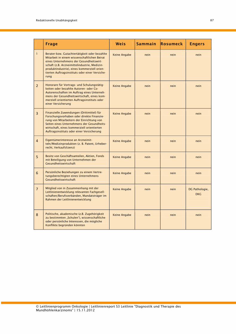
Redaktionelle Unabhängigkeit
© Leitlinienprogramm Onkologie | Leitlinienreport S3 Leitlinie "Diagnostik und Therapie des
Mundhöhlenkarzinoms" | 15.11.2012
87
Frage Weis Sammain Rosumeck Engers
1 Berater-bzw. Gutachtertätigkeit oder bezahlte
Mitarbeit in einem wissenschaftlichen Beirat
eines Unternehmens der Gesundheitswirt-
schaft (z.B. Arzneimittelindustrie, Medizin-
produktindustrie), eines kommerziell orien-
tierten Auftragsinstituts oder einer Versiche-
rung
Keine Angabe nein nein nein
2 Honorare für Vortrags- und Schulungstätig-
keiten oder bezahlte Autoren- oder Co-
Autorenschaften im Auftrag eines Unterneh-
mens der Gesundheitswirtschaft, eines kom-
merziell orientierten Auftragsinstituts oder
einer Versicherung
Keine Angabe nein nein nein
3 Finanzielle Zuwendungen (Drittmittel) für
Forschungsvorhaben oder direkte Finanzie-
rung von Mitarbeitern der Einrichtung von
Seiten eines Unternehmens der Gesundheits-
wirtschaft, eines kommerziell orientierten
Auftragsinstituts oder einer Versicherung
Keine Angabe nein nein nein
4 Eigentümerinteresse an Arzneimit-
teln/Medizinprodukten (z. B. Patent, Urheber-
recht, Verkaufslizenz)
Keine Angabe nein nein nein
5 Besitz von Geschäftsanteilen, Aktien, Fonds
mit Beteiligung von Unternehmen der
Gesundheitswirtschaft
Keine Angabe nein nein nein
6 Persönliche Beziehungen zu einem Vertre-
tungsberechtigten eines Unternehmens
Gesundheitswirtschaft
Keine Angabe nein nein nein
7 Mitglied von in Zusammenhang mit der
Leitlinienentwicklung relevanten Fachgesell-
schaften/Berufsverbänden, Mandatsträger im
Rahmen der Leitlinienentwicklung
Keine Angabe nein nein DG Pathologie,
DKG
n
e
i
n 8 Politische, akademische (z.B. Zugehörigkeit
zu bestimmten „Schulen“), wissenschaftliche
oder persönliche Interessen, die mögliche
Konflikte begründen könnten
Keine Angabe nein nein nein
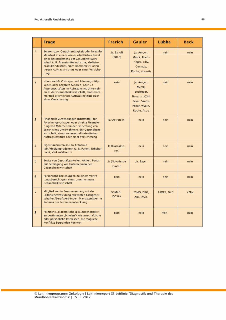
Redaktionelle Unabhängigkeit
© Leitlinienprogramm Onkologie | Leitlinienreport S3 Leitlinie "Diagnostik und Therapie des
Mundhöhlenkarzinoms" | 15.11.2012
88
Frage Frerich Gauler Lübbe Beck
1 Berater-bzw. Gutachtertätigkeit oder bezahlte
Mitarbeit in einem wissenschaftlichen Beirat
eines Unternehmens der Gesundheitswirt-
schaft (z.B. Arzneimittelindustrie, Medizin-
produktindustrie), eines kommerziell orien-
tierten Auftragsinstituts oder einer Versiche-
rung
Ja: Sanofi
(2010)
Ja: Amgen,
Merck, Boeh-
ringer, Lilly,
Genmab,
Roche, Novartis
nein nein
2 Honorare für Vortrags- und Schulungstätig-
keiten oder bezahlte Autoren- oder Co-
Autorenschaften im Auftrag eines Unterneh-
mens der Gesundheitswirtschaft, eines kom-
merziell orientierten Auftragsinstituts oder
einer Versicherung
nein Ja: Amgen,
Merck,
Boehriger,
Novartis, GSH,
Bayer, Sanofi,
Pfizer, Wyeth,
Roche, Astra
nein nein
3 Finanzielle Zuwendungen (Drittmittel) für
Forschungsvorhaben oder direkte Finanzie-
rung von Mitarbeitern der Einrichtung von
Seiten eines Unternehmens der Gesundheits-
wirtschaft, eines kommerziell orientierten
Auftragsinstituts oder einer Versicherung
Ja (Astratech) nein nein nein
4 Eigentümerinteresse an Arzneimit-
teln/Medizinprodukten (z. B. Patent, Urheber-
recht, Verkaufslizenz)
Ja (Bioreakto-
ren)
nein nein nein
5 Besitz von Geschäftsanteilen, Aktien, Fonds
mit Beteiligung von Unternehmen der
Gesundheitswirtschaft
Ja (Novatissue
GmbH)
Ja: Bayer nein nein
6 Persönliche Beziehungen zu einem Vertre-
tungsberechtigten eines Unternehmens
Gesundheitswirtschaft
nein nein nein nein
7 Mitglied von in Zusammenhang mit der
Leitlinienentwicklung relevanten Fachgesell-
schaften/Berufsverbänden, Mandatsträger im
Rahmen der Leitlinienentwicklung
DGMKG
DÖSAK
ESMO, DKG,
AIO, IASLC
ASORS, DKG KZBV
8 Politische, akademische (z.B. Zugehörigkeit
zu bestimmten „Schulen“), wissenschaftliche
oder persönliche Interessen, die mögliche
Konflikte begründen könnten
nein nein nein nein
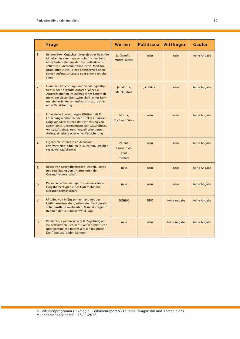
Redaktionelle Unabhängigkeit
© Leitlinienprogramm Onkologie | Leitlinienreport S3 Leitlinie "Diagnostik und Therapie des
Mundhöhlenkarzinoms" | 15.11.2012
89
Frage Werner Pathirana Wittlinger Gauler
1 Berater-bzw. Gutachtertätigkeit oder bezahlte
Mitarbeit in einem wissenschaftlichen Beirat
eines Unternehmens der Gesundheitswirt-
schaft (z.B. Arzneimittelindustrie, Medizin-
produktindustrie), eines kommerziell orien-
tierten Auftragsinstituts oder einer Versiche-
rung
Ja: Sanofi,
Morita, Merck
nein nein Keine Angabe
2 Honorare für Vortrags- und Schulungstätig-
keiten oder bezahlte Autoren- oder Co-
Autorenschaften im Auftrag eines Unterneh-
mens der Gesundheitswirtschaft, eines kom-
merziell orientierten Auftragsinstituts oder
einer Versicherung
Ja: Morita,
Merck, Storz
Ja: Pfitzer nein Keine Angabe
3 Finanzielle Zuwendungen (Drittmittel) für
Forschungsvorhaben oder direkte Finanzie-
rung von Mitarbeitern der Einrichtung von
Seiten eines Unternehmens der Gesundheits-
wirtschaft, eines kommerziell orientierten
Auftragsinstituts oder einer Versicherung
Morita,
Cochlear, Storz
nein nein Keine Angabe
4 Eigentümerinteresse an Arzneimit-
teln/Medizinprodukten (z. B. Patent, Urheber-
recht, Verkaufslizenz)
Patent:
ozone/oxy-
gene
mixture
nein nein Keine Angabe
5 Besitz von Geschäftsanteilen, Aktien, Fonds
mit Beteiligung von Unternehmen der
Gesundheitswirtschaft
nein nein nein Keine Angabe
6 Persönliche Beziehungen zu einem Vertre-
tungsberechtigten eines Unternehmens
Gesundheitswirtschaft
nein nein nein Keine Angabe
7 Mitglied von in Zusammenhang mit der
Leitlinienentwicklung relevanten Fachgesell-
schaften/Berufsverbänden, Mandatsträger im
Rahmen der Leitlinienentwicklung
DGHNO DDG Keine Angabe Keine Angabe
8 Politische, akademische (z.B. Zugehörigkeit
zu bestimmten „Schulen“), wissenschaftliche
oder persönliche Interessen, die mögliche
Konflikte begründen könnten
nein nein Keine Angabe Keine Angabe
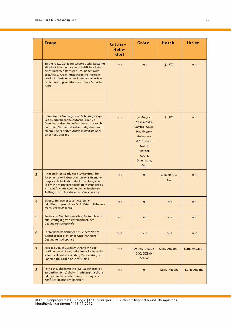
Redaktionelle Unabhängigkeit
© Leitlinienprogramm Onkologie | Leitlinienreport S3 Leitlinie "Diagnostik und Therapie des
Mundhöhlenkarzinoms" | 15.11.2012
90
Frage Gittler-
Hebe-
steit
Grötz Horch Ihrler
1 Berater-bzw. Gutachtertätigkeit oder bezahlte
Mitarbeit in einem wissenschaftlichen Beirat
eines Unternehmens der Gesundheitswirt-
schaft (z.B. Arzneimittelindustrie, Medizin-
produktindustrie), eines kommerziell orien-
tierten Auftragsinstituts oder einer Versiche-
rung
nein nein Ja: KCI nein
2 Honorare für Vortrags- und Schulungstätig-
keiten oder bezahlte Autoren- oder Co-
Autorenschaften im Auftrag eines Unterneh-
mens der Gesundheitswirtschaft, eines kom-
merziell orientierten Auftragsinstituts oder
einer Versicherung
nein Ja: Amgen,
Artoss, Astra,
Camlog, Geist-
lich, Mectron,
Medupdate,
MIP, Novartis,
Nobel,
Riemser,
Roche,
Straumann,
Zepf
Ja: KCI nein
3 Finanzielle Zuwendungen (Drittmittel) für
Forschungsvorhaben oder direkte Finanzie-
rung von Mitarbeitern der Einrichtung von
Seiten eines Unternehmens der Gesundheits-
wirtschaft, eines kommerziell orientierten
Auftragsinstituts oder einer Versicherung
nein nein Ja: Baxter AG,
KCI
nein
4 Eigentümerinteresse an Arzneimit-
teln/Medizinprodukten (z. B. Patent, Urheber-
recht, Verkaufslizenz)
nein nein nein nein
5 Besitz von Geschäftsanteilen, Aktien, Fonds
mit Beteiligung von Unternehmen der
Gesundheitswirtschaft
nein nein nein nein
6 Persönliche Beziehungen zu einem Vertre-
tungsberechtigten eines Unternehmens
Gesundheitswirtschaft
nein nein nein nein
7 Mitglied von in Zusammenhang mit der
Leitlinienentwicklung relevanten Fachgesell-
schaften/Berufsverbänden, Mandatsträger im
Rahmen der Leitlinienentwicklung
nein ASORS, DEGRO,
DKG, DGZMK,
DGMKG
Keine Angabe Keine Angabe
8 Politische, akademische (z.B. Zugehörigkeit
zu bestimmten „Schulen“), wissenschaftliche
oder persönliche Interessen, die mögliche
Konflikte begründen könnten
nein nein Keine Angabe Keine Angabe
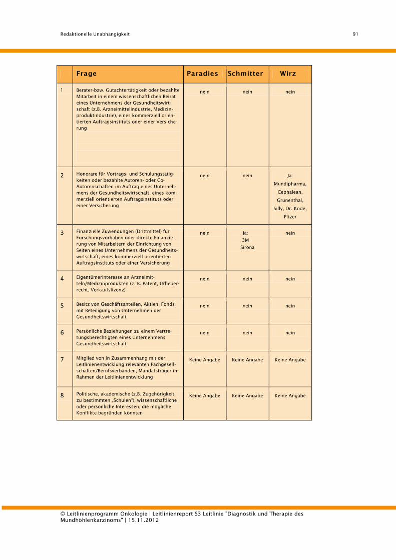
Redaktionelle Unabhängigkeit
© Leitlinienprogramm Onkologie | Leitlinienreport S3 Leitlinie "Diagnostik und Therapie des
Mundhöhlenkarzinoms" | 15.11.2012
91
Frage Paradies Schmitter Wirz
1 Berater-bzw. Gutachtertätigkeit oder bezahlte
Mitarbeit in einem wissenschaftlichen Beirat
eines Unternehmens der Gesundheitswirt-
schaft (z.B. Arzneimittelindustrie, Medizin-
produktindustrie), eines kommerziell orien-
tierten Auftragsinstituts oder einer Versiche-
rung
nein nein nein
2 Honorare für Vortrags- und Schulungstätig-
keiten oder bezahlte Autoren- oder Co-
Autorenschaften im Auftrag eines Unterneh-
mens der Gesundheitswirtschaft, eines kom-
merziell orientierten Auftragsinstituts oder
einer Versicherung
nein nein Ja:
Mundipharma,
Cephalean,
Grünenthal,
Silly, Dr. Kode,
Pfizer
3 Finanzielle Zuwendungen (Drittmittel) für
Forschungsvorhaben oder direkte Finanzie-
rung von Mitarbeitern der Einrichtung von
Seiten eines Unternehmens der Gesundheits-
wirtschaft, eines kommerziell orientierten
Auftragsinstituts oder einer Versicherung
nein Ja:
3M
Sirona
nein
4 Eigentümerinteresse an Arzneimit-
teln/Medizinprodukten (z. B. Patent, Urheber-
recht, Verkaufslizenz)
nein nein nein
5 Besitz von Geschäftsanteilen, Aktien, Fonds
mit Beteiligung von Unternehmen der
Gesundheitswirtschaft
nein nein nein
6 Persönliche Beziehungen zu einem Vertre-
tungsberechtigten eines Unternehmens
Gesundheitswirtschaft
nein nein nein
7 Mitglied von in Zusammenhang mit der
Leitlinienentwicklung relevanten Fachgesell-
schaften/Berufsverbänden, Mandatsträger im
Rahmen der Leitlinienentwicklung
Keine Angabe Keine Angabe Keine Angabe
8 Politische, akademische (z.B. Zugehörigkeit
zu bestimmten „Schulen“), wissenschaftliche
oder persönliche Interessen, die mögliche
Konflikte begründen könnten
Keine Angabe Keine Angabe Keine Angabe
Verbreitung und Implementierung
© Leitlinienprogramm Onkologie | Leitlinienreport S3 Leitlinie "Diagnostik und Therapie des
Mundhöhlenkarzinoms" | 15.11.2012
92
9. Verbreitung und Implementierung
Folgende Elemente sind Bestandteil des Verbreitungs- und Implementie-
rungs¬konzeptes:
- Publikation als „Leitlinien-Set“ (Langfassung + Kurzfassung + Patientenver-
sion + Leitlinienreport);
- Publikation der Kurzfassung im Deutschen Ärzteblatt;
- Verbreitung über die Publikationsorgane und Kongressveranstaltungen der
kooperierenden Fachgesellschaften und Organisationen; Pressekonferen-
zen;
- Informationen an Einrichtungen der gemeinsamen Selbstverwaltung und
an Berufsorganisationen
- Entwicklung von Qualitätsindikatoren und Etablierung dieser in den Erhe-
bungsbögen / Kennzahlbögen von zertifizierten Zentren.
Die Implementierung der LL wird nicht nur durch ihre Publikation in medizini-
schen Fachzeitschriften oder im Internet sondern auch durch eine planvolle
Einbeziehung der jeweiligen Nutzer angestrebt und gewährleistet werden.
Dies geschieht durch Verbreitung der Inhalte auf Fachkongressen, Fortbildun-
gen für Ärzte und Patienteninformationsveranstaltungen genauso wie durch
Übernahme der LL in das Intranet der jeweiligen Kliniken. Um eine direkte Ver-
fügbarkeit der LL beim Patientenkontakt sicher zu stellen wird auf die Fort-
und Weiterbildung aller potentieller Anwender besonders Wert gelegt. Hierzu
bieten etwa die auf regionaler und überregionaler Ebene regelmäßig stattfin-
denden zahnärztlichen Fortbildungsveranstaltungen eine besonders effektive
Basis; auch eine zertifizierte medizinische Fortbildung mit Vergabe von Fort-
bildungspunkten ist zu diesem Zweck möglich. Zur Einführung der Leitlinie
wurden folgende Verfahren von der Leitliniengruppe selbst unterstützt und
veranlasst:
- Disseminierung durch kostenfreien Zugang über das Internet unter den in
1.2. genannten Adressen.
- Drucklegung, Publikation in zahnmedizinischen Fachzeitschriften und den
ZahnärztlichenMitteilungen
- Publikation in Broschürenform für Zahnärzte und als Patienteninformation
in einem Faltblatt,
- Öffentlichkeitsarbeit: Pressemitteilungen,
Gültigkeitsdauer der Leitlinie
© Leitlinienprogramm Onkologie | Leitlinienreport S3 Leitlinie "Diagnostik und Therapie des
Mundhöhlenkarzinoms" | 15.11.2012
93
- Erprobung durch Qualitätszirkel (mit systematischer Begleitung und Aus-
wertung durch die ZZQ, Projekt wird z. Z. konzipiert und im Herbst 2006
in Qualitätszirkeln in Hamburg durchgeführt),
- Kontinuierliche Fortbildung, insbesondere über die Programme der kam-
mereigenen Fortbildungsinstitute, Vorträge der Leitlinienautoren und in-
teraktive Edukation im persönlichen Umfeld
10. Gültigkeitsdauer der Leitlinie
Die vorliegende S3-Leitlinie "Diagnostik und Therapie des Mundhöhlenkarzi-
noms" hat eine Gültigkeit bis zum 31.12. 2015.
Erkenntnisse, die eine Aktualisierung einzelner Abschnitte oder Empfehlungen
erforderlich machen können, werden inzwischen von der Leitliniengruppe be-
obachtet. Entsprechende Hinweise an den unten genannten Ansprechpartner
durch die Adressaten der Leitlinie sind ausdrücklich erwünscht.
Das Datum der Veröffentlichung, die Anmeldung der geplanten Aktualisierung
sowie ggf. zwischenzeitliche Teil-Aktualisierungen (Amendments) werden im
öffentlich zugänglichen Leitlinienregister der AWMF (http://www-awmf-
leitlinien.de) ausgewiesen. Gültig ist nur die jeweils neueste Version gemäß
des AWMF-Registers.
Ansprechpartner für die Aktualisierung der Leitlinie ist der Vorstand der
DGMKG.
Literaturverzeichnis
© Leitlinienprogramm Onkologie | Leitlinienreport S3 Leitlinie "Diagnostik und Therapie des
Mundhöhlenkarzinoms" | 15.11.2012
94
11. Literaturverzeichnis
1. Scottish Intercollegiate Guidelines Network, Diagnosis and management
of head and neck cancer. A national clinical guideline. 2006.
2. Canadian Partnership Against Cancer. Search SAGE (Standards and
Guidelines Evidence). Detailed Result - Diagnosis and management of
head and neck cancer. A national clinical guideline. 2011 15.05.2012];
Available from: http://www.cancerview.ca/.
3. Alvarez Amezaga, J., et al., Diagnostic efficacy of sentinel node biopsy in
oral squamous cell carcinoma. Cohort study and meta-analysis.
Medicina Oral, Patologia Oral y Cirugia Bucal, 2007. 12(3): p. E235-43.
4. O'Neill, J.P., et al., Controversies in the management of tongue base
cancer. Irish Journal of Medical Science, 2009. 178(1): p. 1-5.
5. Hoft, S., et al., Sentinel lymph-node biopsy in head and neck cancer. Br J
Cancer, 2004. 91(1): p. 124-8.
6. Hyde, N.C., et al., A new approach to pre-treatment assessment of the
N0 neck in oral squamous cell carcinoma: the role of sentinel node
biopsy and positron emission tomography. Oral Oncol, 2003. 39(4): p.
350-60.
7. Kontio, R., et al., Sentinel lymph node biopsy in oral cavity squamous
cell carcinoma without clinically evident metastasis. Head Neck, 2004.
26(1): p. 16-21.
8. Ross, G.L., et al., Improved staging of cervical metastases in clinically
node-negative patients with head and neck squamous cell carcinoma.
Ann Surg Oncol, 2004. 11(2): p. 213-8.
9. Hafidh, M.A., et al., Evaluation of the impact of addition of PET to CT
and MR scanning in the staging of patients with head and neck
carcinomas. Eur Arch Otorhinolaryngol, 2006. 263(9): p. 853-9.
10. Wax, M.K., et al., The role of positron emission tomography in the
evaluation of the N-positive neck. Otolaryngol Head Neck Surg, 2003.
129(3): p. 163-7.
11. Mucke, T., et al., The role of tumor invasion into the mandible of oral
squamous cell carcinoma. J Cancer Res Clin Oncol, 2011. 137(1): p. 165-
71.
12. Abler, A., M. Roser, and D. Weingart, [On the indications for and
morbidity of segmental resection of the mandible for squamous cell
carcinoma in the lower oral cavity]. Mund-, Kiefer- und
Gesichtschirurgie, 2005. 9(3): p. 137-42.
13. Babin, E., et al., PET/CT for assessing mandibular invasion by intraoral
squamous cell carcinomas. Clinical Otolaryngology, 2008. 33(1): p. 47-
51.
14. Baek, C.-H., et al., Tumor volume assessment by 18F-FDG PET/CT in
patients with oral cavity cancer with dental artifacts on CT or MR
images. Journal of Nuclear Medicine, 2008. 49(9): p. 1422-8.
15. Bolzoni, A., et al., Diagnostic accuracy of magnetic resonance imaging
in the assessment of mandibular involvement in oral-oropharyngeal
squamous cell carcinoma: a prospective study. Archives of
Otolaryngology -- Head & Neck Surgery, 2004. 130(7): p. 837-43.
16. Brockenbrough, J.M., G.J. Petruzzelli, and L. Lomasney, DentaScan as an
accurate method of predicting mandibular invasion in patients with
squamous cell carcinoma of the oral cavity. Archives of Otolaryngology -
- Head & Neck Surgery, 2003. 129(1): p. 113-7.
Literaturverzeichnis
© Leitlinienprogramm Onkologie | Leitlinienreport S3 Leitlinie "Diagnostik und Therapie des
Mundhöhlenkarzinoms" | 15.11.2012
95
17. Dammann, F., et al., Rational diagnosis of squamous cell carcinoma of
the head and neck region: comparative evaluation of CT, MRI, and
18FDG PET.[Erratum appears in AJR Am J Roentgenol. 2005
Jun;184(6):1968]. AJR, 2005. 184(4): p. 1326-31.
18. Ekberg, T., et al., Clinical impact of positron emission tomography (PET)
with (18F)fluorodeoxyglucose (FDG) in head and neck tumours. Acta Oto-
Laryngologica, 2007. 127(2): p. 186-93.
19. Goerres, G.W., et al., Impact of whole body positron emission
tomography on initial staging and therapy in patients with squamous
cell carcinoma of the oral cavity. Oral Oncology, 2003. 39(6): p. 547-51.
20. Goerres, G.W., et al., Bone invasion in patients with oral cavity cancer:
comparison of conventional CT with PET/CT and SPECT/CT.[Erratum
appears in Radiology. 2006 Apr;239(1):303]. Radiology, 2005. 237(1):
p. 281-7.
21. Gu, D.H., et al., CT, MR, (18)F-FDG PET/CT, and their combined use for
the assessment of mandibular invasion by squamous cell carcinomas of
the oral cavity. Acta Radiologica, 2010. 51(10): p. 1111-9.
22. Hendrikx, A.W.F., et al., Cone-beam CT in the assessment of mandibular
invasion by oral squamous cell carcinoma: results of the preliminary
study. International Journal of Oral and Maxillofacial Surgery, 2010. 39
(5): p. 436-439.
23. Imaizumi, A., et al., A potential pitfall of MR imaging for assessing
mandibular invasion of squamous cell carcinoma in the oral cavity. Ajnr:
American Journal of Neuroradiology, 2006. 27(1): p. 114-22.
24. Jones, J., et al., Positron emission tomography (PET) in the management
of oro-pharyngeal cancer. European Journal of Surgical Oncology, 2005.
31(2): p. 170-6.
25. Krabbe, C.A., et al., FDG PET in oral and oropharyngeal cancer. Value
for confirmation of N0 neck and detection of occult metastases. Oral
Oncology, 2008. 44(1): p. 31-6.
26. Nakamoto, Y., et al., Clinical value of image fusion from MR and PET in
patients with head and neck cancer. Molecular Imaging & Biology, 2009.
11(1): p. 46-53.
27. Ng, S.-H., et al., 18F-FDG PET and CT/MRI in oral cavity squamous cell
carcinoma: a prospective study of 124 patients with histologic
correlation. Journal of Nuclear Medicine, 2005. 46(7): p. 1136-43.
28. Nishiyama, Y., et al., FDG PET as a procedure for detecting simultaneous
tumours in head and neck cancer patients. Nuclear Medicine
Communications, 2005. 26(3): p. 239-44.
29. Pauleit, D., et al., 18F-FET PET compared with 18F-FDG PET and CT in
patients with head and neck cancer. Journal of Nuclear Medicine, 2006.
47(2): p. 256-61.
30. Pentenero, M., et al., Accuracy of 18F-FDG-PET/CT for staging of oral
squamous cell carcinoma. Head & Neck, 2008. 30(11): p. 1488-96.
31. Rajesh, A., et al., Can magnetic resonance imaging replace single photon
computed tomography and computed tomography in detecting bony
invasion in patients with oral squamous cell carcinoma? British Journal
of Oral & Maxillofacial Surgery, 2008. 46(1): p. 11-4.
32. Roh, J.-L., et al., Utility of 2-[18F] fluoro-2-deoxy-D-glucose positron
emission tomography and positron emission tomography/computed
tomography imaging in the preoperative staging of head and neck
squamous cell carcinoma. Oral Oncology, 2007. 43(9): p. 887-93.
33. Seitz, O., et al., 18F-Fluorodeoxyglucose-PET/CT to evaluate tumor,
nodal disease, and gross tumor volume of oropharyngeal and oral cavity
Literaturverzeichnis
© Leitlinienprogramm Onkologie | Leitlinienreport S3 Leitlinie "Diagnostik und Therapie des
Mundhöhlenkarzinoms" | 15.11.2012
96
cancer: comparison with MR imaging and validation with surgical
specimen. Neuroradiology, 2009. 51(10): p. 677-86.
34. Van Cann, E.M., et al., Assessment of mandibular invasion of squamous
cell carcinoma by various modes of imaging: constructing a diagnostic
algorithm. International Journal of Oral & Maxillofacial Surgery, 2008.
37(6): p. 535-41.
35. Vidiri, A., et al., Oral cavity and base of the tongue tumors. Correlation
between clinical, MRI and pathological staging of primary tumor. Journal
of Experimental and Clinical Cancer Research, 2007. 26 (4): p. 575-582.
36. Vidiri, A., et al., Multi-detector row computed tomography (MDCT) and
magnetic resonance imaging (MRI) in the evaluation of the mandibular
invasion by squamous cell carcinomas (SCC) of the oral cavity.
Correlation with pathological data. Journal of Experimental & Clinical
Cancer Research, 2010. 29: p. 73.
37. Wallowy, P., et al., 18F-FDG PET for detecting metastases and
synchronous primary malignancies in patients with oral and
oropharyngeal cancer. Nuclear-Medizin, 2009. 48(5): p. 192-9; quiz
N42.
38. Wiener, E., et al., Comparison of 16-slice MSCT and MRI in the
assessment of squamous cell carcinoma of the oral cavity. European
Journal of Radiology, 2006. 58(1): p. 113-8.
39. Bisase, B., C. Kerawala, and J. Lee, The role of computed tomography of
the chest in the staging of early squamous cell carcinoma of the tongue.
British Journal of Oral & Maxillofacial Surgery, 2008. 46(5): p. 367-9.
40. Chow, T.-L., et al., Prediction of simultaneous esophageal lesions in head
and neck squamous cell carcinoma: a multivariate analysis. Archives of
Otolaryngology -- Head & Neck Surgery, 2009. 135(9): p. 882-5.
41. Fielding, D., et al., Autofluorescence improves pretreatment mucosal
assessment in head and neck cancer patients. Otolaryngology - Head &
Neck Surgery, 2010. 142(3 Suppl 1): p. S20-6.
42. Ghosh, S.K., et al., Detection of synchronous lung tumors in patients
presenting with squamous cell carcinoma of the head and neck. Head &
Neck, 2009. 31(12): p. 1563-70.
43. Keith, D.J.W., T.K. Ong, and I.C. Martin, The role of thoracic computed
tomography in staging newly-diagnosed oral squamous cell carcinoma.
British Journal of Oral & Maxillofacial Surgery, 2006. 44(3): p. 198-202.
44. Kesting, M.R., et al., Bronchoscopy screening in primary oral squamous
cell carcinoma: a 10-year experience. British Journal of Oral &
Maxillofacial Surgery, 2009. 47(4): p. 279-83.
45. Kesting, M.R., et al., Results of esophagogastroduodenoscopy in patients
with oral squamous cell carcinoma--value of endoscopic screening: 10-
year experience. Journal of Oral & Maxillofacial Surgery, 2009. 67(8): p.
1649-55.
46. Krabbe, C.A., et al., FDG-PET and detection of distant metastases and
simultaneous tumors in head and neck squamous cell carcinoma: a
comparison with chest radiography and chest CT. Oral Oncology, 2009.
45(3): p. 234-40.
47. Lee, C.T., et al., Narrow-band imaging with magnifying endoscopy for
the screening of esophageal cancer in patients with primary head and
neck cancers. Endoscopy, 2010. 42(8): p. 613-9.
48. Loh, K.S., et al., A rational approach to pulmonary screening in newly
diagnosed head and neck cancer. Head & Neck, 2005. 27(11): p. 990-4.
49. Takenaka, R., et al., Narrow-band imaging provides reliable screening
for esophageal malignancy in patients with head and neck cancers.
American Journal of Gastroenterology, 2009. 104(12): p. 2942-8.
Literaturverzeichnis
© Leitlinienprogramm Onkologie | Leitlinienreport S3 Leitlinie "Diagnostik und Therapie des
Mundhöhlenkarzinoms" | 15.11.2012
97
50. Brouwer, J., et al., Screening for distant metastases in patients with head
and neck cancer: is there a role for (18)FDG-PET? Oral Oncology, 2006.
42(3): p. 275-80.
51. Balogova, S., et al., Prospective comparison of FDG and FET PET/CT in
patients with head and neck squamous cell carcinoma. Molecular
Imaging & Biology, 2008. 10(6): p. 364-73.
52. Barzan, L., et al., An extended use of the sentinel node in head and neck
squamous cell carcinoma: results of a prospective study of 100 patients.
Acta Otorhinolaryngologica Italica, 2004. 24(3): p. 145-9.
53. Bilde, A., et al., The role of SPECT-CT in the lymphoscintigraphic
identification of sentinel nodes in patients with oral cancer. Acta Oto-
Laryngologica, 2006. 126(10): p. 1096-103.
54. Borgemeester, M.C., et al., Ultrasound-guided aspiration cytology for the
assessment of the clinically N0 neck: factors influencing its accuracy.
Head & Neck, 2008. 30(11): p. 1505-13.
55. Brouwer, J., et al., Positron emission tomography using
[18F]fluorodeoxyglucose (FDG-PET) in the clinically negative neck: is it
likely to be superior? European Archives of Oto-Rhino-Laryngology,
2004. 261(9): p. 479-83.
56. Burcia, V., et al., Neck restaging with sentinel node biopsy in T1-T2N0
oral and oropharyngeal cancer: Why and how? Otolaryngology - Head &
Neck Surgery, 2010. 142(4): p. 592-7.e1.
57. Burns, P., et al., Sentinel lymph node biopsy in node-negative squamous
cell carcinoma of the oral cavity and oropharynx. Journal of Laryngology
& Otology, 2009. 123(4): p. 439-43.
58. Cammilleri, S., et al., Ability of lymphoscintigraphy to direct sentinel
node biopsy in the clinically NO check for patients with head and neck
squamous cell carcinoma; a prospective study (preliminary results).
Bulletin du Cancer, 2004. 91(4): p. E1-4.
59. Chone, C.T., et al., Predictive value of sentinel node biopsy in head and
neck cancer. Acta Oto-Laryngologica, 2008. 128(8): p. 920-4.
60. Civantos, F.J., et al., Sentinel node biopsy in oral cavity cancer:
correlation with PET scan and immunohistochemistry. Head & Neck,
2003. 25(1): p. 1-9.
61. Civantos, F.J., et al., Sentinel lymph node biopsy accurately stages the
regional lymph nodes for T1-T2 oral squamous cell carcinomas: results
of a prospective multi-institutional trial. Journal of Clinical Oncology,
2010. 28(8): p. 1395-400.
62. De Zinis, L.O.R., et al., Prevalence and localization of nodal metastases
in squamous cell carcinoma of the oral cavity: role and extension of neck
dissection. European Archives of Oto-Rhino-Laryngology, 2006. 263(12):
p. 1131-5.
63. Dirix, P., et al., Diffusion-weighted MRI for nodal staging of head and
neck squamous cell carcinoma: impact on radiotherapy planning.
International Journal of Radiation Oncology, Biology, Physics, 2010.
76(3): p. 761-6.
64. Freire, A.R.S., et al., Computed tomography and lymphoscintigraphy to
identify lymph node metastases and lymphatic drainage pathways in
oral and oropharyngeal squamous cell carcinomas. European Archives
of Oto-Rhino-Laryngology, 2003. 260(3): p. 148-52.
65. Gencoglu, U., et al., The coherence between fine needle aspiration
cytology and histopathology of palpable neck nodes in lower lip
carcinoma patients. European Journal of Plastic Surgery, 2003. 26 (2): p.
82-84.
Literaturverzeichnis
© Leitlinienprogramm Onkologie | Leitlinienreport S3 Leitlinie "Diagnostik und Therapie des
Mundhöhlenkarzinoms" | 15.11.2012
98
66. Haerle, S.K., et al., Is there an additional value of SPECT/CT over planar
lymphoscintigraphy for sentinel node mapping in oral/oropharyngeal
squamous cell carcinoma? Annals of Surgical Oncology, 2009. 16(11): p.
3118-24.
67. Hohlweg-Majert, B., et al., Preoperative cervical lymph node size
evaluation in patients with malignant head/neck tumors: comparison
between ultrasound and computer tomography. Journal of Cancer
Research & Clinical Oncology, 2009. 135(6): p. 753-9.
68. Jeong, H.-S., et al., Use of integrated 18F-FDG PET/CT to improve the
accuracy of initial cervical nodal evaluation in patients with head and
neck squamous cell carcinoma. Head & Neck, 2007. 29(3): p. 203-10.
69. Jeong, H.S., et al., Sentinel lymph node radiolocalization with 99mTc
filtered tin colloid in clinically node-negative squamous cell carcinomas
of the oral cavity. Journal of Korean Medical Science, 2006. 21 (5): p.
865-870.
70. Keski-Santti, H., et al., Sentinel lymph node biopsy as an alternative to
wait and see policy in patients with small T1 oral cavity squamous cell
carcinoma. Acta Oto-Laryngologica, 2008. 128(1): p. 98-102.
71. Keski-Santti, H., et al., Sentinel lymph node biopsy or elective neck
dissection for patients with oral squamous cell carcinoma? European
Archives of Oto-Rhino-Laryngology, 2008. 265 (SUPPL. 1): p. S13-S17.
72. Khafif, A., et al., Lymphoscintigraphy for sentinel node mapping using a
hybrid single photon emission CT (SPECT)/CT system in oral cavity
squamous cell carcinoma. Head & Neck, 2006. 28(10): p. 874-9.
73. Kim, S.Y., et al., Utility of FDG PET in patients with squamous cell
carcinomas of the oral cavity. European Journal of Surgical Oncology,
2008. 34(2): p. 208-15.
74. Kovacs, A.F., et al., Positron emission tomography in combination with
sentinel node biopsy reduces the rate of elective neck dissections in the
treatment of oral and oropharyngeal cancer. Journal of Clinical
Oncology, 2004. 22(19): p. 3973-80.
75. Krabbe, C.A., et al., Detection of cervical metastases with (11)C-tyrosine
PET in patients with squamous cell carcinoma of the oral cavity or
oropharynx: A comparison with (18)F-FDG PET. Head & Neck, 2010.
32(3): p. 368-74.
76. Lee, B.-J., et al., Usefulness of technetium-99m methoxyisobutylisonitrile
single-photon emission computed tomography and computed
tomography in the evaluation of cervical lymph node metastasis. Journal
of Laryngology & Otology, 2005. 119(11): p. 882-7.
77. Nahmias, C., et al., Positron emission tomography/computerized
tomography (PET/CT) scanning for preoperative staging of patients with
oral/head and neck cancer. Journal of Oral & Maxillofacial Surgery,
2007. 65(12): p. 2524-35.
78. Ng, S.-H., et al., Prospective study of [18F]fluorodeoxyglucose positron
emission tomography and computed tomography and magnetic
resonance imaging in oral cavity squamous cell carcinoma with palpably
negative neck. Journal of Clinical Oncology, 2006. 24(27): p. 4371-6.
79. Nieuwenhuis, E.J.C., et al., Histopathologic validation of the sentinel
node concept in oral and oropharyngeal squamous cell carcinoma. Head
& Neck, 2005. 27(2): p. 150-8.
80. Payoux, P., et al., Effectiveness of lymphoscintigraphic sentinel node
detection for cervical staging of patients with squamous cell carcinoma
of the head and neck. Journal of Oral & Maxillofacial Surgery, 2005.
63(8): p. 1091-5.
Literaturverzeichnis
© Leitlinienprogramm Onkologie | Leitlinienreport S3 Leitlinie "Diagnostik und Therapie des
Mundhöhlenkarzinoms" | 15.11.2012
99
81. Piao, Y., et al., Evaluation of 18F-FDG PET/CT for diagnosing cervical
nodal metastases in patients with oral cavity or oropharynx carcinoma.
Oral Surgery Oral Medicine Oral Pathology Oral Radiology &
Endodontics, 2009. 108(6): p. 933-8.
82. Santaolalla, F., et al., Comparative study of patients with and without
sentinel lymph node biopsy (SLNB) in oral and oropharyngeal cancer: is
SLNB an accurate and useful procedure? Acta Oto-Laryngologica, 2009.
129(2): p. 199-204.
83. Schoder, H., et al., 18F-FDG PET/CT for detecting nodal metastases in
patients with oral cancer staged N0 by clinical examination and CT/MRI.
Journal of Nuclear Medicine, 2006. 47(5): p. 755-62.
84. Schroeder, U., et al., Is there a need for positron emission tomography
imaging to stage the N0 neck in T1-T2 squamous cell carcinoma of the
oral cavity or oropharynx? Annals of Otology, Rhinology & Laryngology,
2008. 117(11): p. 854-63.
85. Schwartz, D.L., et al., FDG-PET/CT imaging for preradiotherapy staging
of head-and-neck squamous cell carcinoma. International Journal of
Radiation Oncology, Biology, Physics, 2005. 61(1): p. 129-36.
86. Stoeckli, S.J., Sentinel node biopsy for oral and oropharyngeal squamous
cell carcinoma of the head and neck. Laryngoscope, 2007. 117(9): p.
1539-51.
87. Sumi, M., et al., Diagnostic performance of MRI relative to CT for
metastatic nodes of head and neck squamous cell carcinomas. Journal of
Magnetic Resonance Imaging, 2007. 26(6): p. 1626-33.
88. Thomsen, J.B., et al., Staging N0 oral cancer: lymphoscintigraphy and
conventional imaging. Acta Radiologica, 2005. 46(5): p. 492-6.
89. Thomsen, J.B., et al., Sentinel lymph node biopsy in oral cancer:
validation of technique and clinical implications of added oblique planar
lymphoscintigraphy and/or tomography. Acta Radiologica, 2005. 46(6):
p. 569-75.
90. To, E.W.H., et al., Is neck ultrasound necessary for early stage oral
tongue carcinoma with clinically N0 neck? Dento-Maxillo-Facial
Radiology, 2003. 32(3): p. 156-9.
91. Tuli, H.S., et al., Diagnostic accuracy of 99mTc-MIBI-SPECT in the
detection of lymph node metastases in patients with carcinoma of the
tongue: comparison with computed tomography and MRI. Nuclear
Medicine Communications, 2008. 29(9): p. 803-8.
92. Wensing, B.M., et al., FDG-PET in the clinically negative neck in oral
squamous cell carcinoma.[Erratum appears in Laryngoscope. 2006
Jul;116(7 Pt 1):1302]. Laryngoscope, 2006. 116(5): p. 809-13.
93. Yamane, M., et al., Noninvasive quantitative assessment of oral tongue
cancer by intraoral ultrasonography. Head & Neck, 2007. 29(4): p. 307-
14.
94. Yamazaki, Y., et al., Assessment of cervical lymph node metastases
using FDG-PET in patients with head and neck cancer. Annals of Nuclear
Medicine, 2008. 22(3): p. 177-84.
95. Yen, T.-C., et al., Staging of untreated squamous cell carcinoma of
buccal mucosa with 18F-FDG PET: comparison with head and neck
CT/MRI and histopathology. Journal of Nuclear Medicine, 2005. 46(5): p.
775-81.
96. Akhtar, S., M. Ikram, and S. Ghaffar, Neck involvement in early
carcinoma of tongue. Is elective neck dissection warranted? JPMA -
Journal of the Pakistan Medical Association, 2007. 57(6): p. 305-7.
Literaturverzeichnis
© Leitlinienprogramm Onkologie | Leitlinienreport S3 Leitlinie "Diagnostik und Therapie des
Mundhöhlenkarzinoms" | 15.11.2012
100
97. Batstone, M.D., et al., Marginal mandibular nerve injury during neck
dissection and its impact on patient perception of appearance. Head &
Neck, 2009. 31(5): p. 673-8.
98. Corlette, T.H., et al., Neck dissection of level IIb: is it really necessary?
Laryngoscope, 2005. 115(9): p. 1624-6.
99. D'Cruz, A.K., et al., Elective neck dissection for the management of the
N0 neck in early cancer of the oral tongue: need for a randomized
controlled trial. Head & Neck, 2009. 31(5): p. 618-24.
100. Elsheikh, M.N., M.E. Mahfouz, and E. Elsheikh, Level IIb lymph nodes
metastasis in elective supraomohyoid neck dissection for oral cavity
squamous cell carcinoma: a molecular-based study. Laryngoscope,
2005. 115(9): p. 1636-40.
101. Huang, S.-F., et al., Neck treatment of patients with early stage oral
tongue cancer: comparison between observation, supraomohyoid
dissection, and extended dissection. Cancer, 2008. 112(5): p. 1066-75.
102. Inoue, H., et al., Quality of life after neck dissection. Archives of
Otolaryngology -- Head & Neck Surgery, 2006. 132(6): p. 662-6.
103. Iype, E.M., et al., The role of selective neck dissection (I-III) in the
treatment of node negative (N0) neck in oral cancer. Oral Oncology,
2008. 44(12): p. 1134-8.
104. Jin, W.L., et al., Occult cervical lymph node metastases in 100
consecutive patients with cN0 tongue cancer. Chinese Medical Journal,
2008. 121 (19): p. 1871-1874.
105. Kohler, H.F., I.W.d. Cunha, and L.P. Kowalski, Impact of modified radical
neck dissections on the number of retrieved nodes, recurrence and
survival. Revista Brasileira de Otorrinolaringologia, 2010. 76(3): p. 374-
7.
106. Koo, B.S., et al., Management of contralateral N0 neck in oral cavity
squamous cell carcinoma. Head & Neck, 2006. 28(10): p. 896-901.
107. Laverick, S., et al., The Impact of Neck Dissection on Health-Related
Quality of Life. Archives of Otolaryngology - Head and Neck Surgery,
2004. 130 (2): p. 149-154.
108. Liaw, G.-A., et al., Outcome of treatment with total main tumor resection
and supraomohyoid neck dissection in oral squamous cell carcinoma.
Journal of the Formosan Medical Association, 2006. 105(12): p. 971-7.
109. Lim, Y.C., et al., Preserving level IIb lymph nodes in elective
supraomohyoid neck dissection for oral cavity squamous cell carcinoma.
Archives of Otolaryngology -- Head & Neck Surgery, 2004. 130(9): p.
1088-91.
110. Mourouzis, C., C. Pratt, and P.A. Brennan, Squamous cell carcinoma of
the maxillary gingiva, alveolus, and hard palate: is there a need for
elective neck dissection? British Journal of Oral & Maxillofacial Surgery,
2010. 48(5): p. 345-8.
111. Nibu, K.-i., et al., Quality of life after neck dissection: a multicenter
longitudinal study by the Japanese Clinical Study Group on
Standardization of Treatment for Lymph Node Metastasis of Head and
Neck Cancer. International Journal of Clinical Oncology, 2010. 15(1): p.
33-8.
112. Patel, R.S., et al., Effectiveness of selective neck dissection in the
treatment of the clinically positive neck. Head & Neck, 2008. 30(9): p.
1231-6.
113. Pathak, K.A., et al., Selective neck dissection (I-III) for node negative and
node positive necks. Oral Oncology, 2006. 42 (8): p. 837-841.
Literaturverzeichnis
© Leitlinienprogramm Onkologie | Leitlinienreport S3 Leitlinie "Diagnostik und Therapie des
Mundhöhlenkarzinoms" | 15.11.2012
101
114. Rapoport, A., et al., Radical versus supraomohyoid neck dissection in the
treatment of squamous cell carcinoma of the inferior level of the mouth.
Revista Brasileira de Otorrinolaringologia, 2007. 73(5): p. 641-6.
115. Santoro, R., et al., Nodal metastases at level IIb during neck dissection
for head and neck cancer: clinical and pathologic evaluation. Head &
Neck, 2008. 30(11): p. 1483-7.
116. Shepard, P.M., et al., Therapeutic selective neck dissection outcomes.
Otolaryngology - Head & Neck Surgery, 2010. 142(5): p. 741-6.
117. Yu, S., et al., Efficacy of supraomohyoid neck dissection in patients with
oral squamous cell carcinoma and negative neck. American Journal of
Surgery, 2006. 191(1): p. 94-9.
118. Yuen, A.P.-W., et al., Prospective randomized study of selective neck
dissection versus observation for N0 neck of early tongue carcinoma.
Head & Neck, 2009. 31(6): p. 765-72.
119. Muscatello, L., et al., Marginal mandibulectomy in oral cancer surgery:
A 13-year experience. European Archives of Oto-Rhino-Laryngology,
2010. 267 (5): p. 759-764.
120. Namaki, S., et al., Masticatory efficiency before and after surgery in oral
cancer patients: comparative study of glossectomy, marginal
mandibulectomy and segmental mandibulectomy. Journal of Oral
Science, 2004. 46(2): p. 113-7.
121. Rogers, S.N., et al., Longitudinal health-related quality of life after
mandibular resection for oral cancer: a comparison between rim and
segment. Head & Neck, 2004. 26(1): p. 54-62.
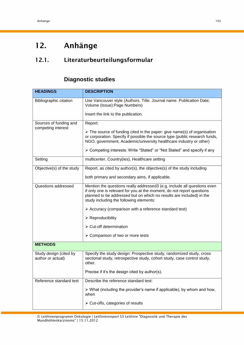
Anhänge
© Leitlinienprogramm Onkologie | Leitlinienreport S3 Leitlinie "Diagnostik und Therapie des
Mundhöhlenkarzinoms" | 15.11.2012
102
12. Anhänge
12.1. Literaturbeurteilungsformular
Diagnostic studies
HEADINGS DESCRIPTION
Bibliographic citation Use Vancouver style (Authors. Title. Journal name. Publication Date; Volume (Issue):Page Numbers)
Insert the link to the publication.
Sources of funding and competing interest
Report:
The source of funding cited in the paper: give name(s) of organisation or corporation. Specify if possible the source type (public research funds, NGO, government, Academic/university healthcare industry or other)
Competing interests: Write “Stated” or “Not Stated” and specify if any
Setting multicenter, Country(ies), Healthcare setting
Objective(s) of the study Report, as cited by author(s), the objective(s) of the study including
both primary and secondary aims, if applicable.
Questions addressed Mention the questions really addressed3 (e.g. include all questions even if only one is relevant for you at the moment, do not report questions planned to be addressed but on which no results are included) in the study including the following elements:
Accuracy (comparison with a reference standard test)
Reproducibility
Cut-off determination
Comparison of two or more tests
METHODS
Study design (cited by author or actual)
Specify the study design: Prospective study, randomized study, cross sectional study, retrospective study, cohort study, case control study, other.
Precise if it’s the design cited by author(s).
Reference standard test Describe the reference standard test:
What (including the provider’s name if applicable), by whom and how, when
Cut-offs, categories of results
Anhänge
© Leitlinienprogramm Onkologie | Leitlinienreport S3 Leitlinie "Diagnostik und Therapie des
Mundhöhlenkarzinoms" | 15.11.2012
103
Blinding (investigator) to clinical information and/or to index test results, if applicable
Diagnostic test(s) evaluated
Describe the evaluated test(s):
What (including the provider’s name if applicable), by whom and how, when
Cut-offs, categories of results
Blinding (investigator) to clinical information and/or to index test results, if applicable
Time interval and treat-ment(s) administered between the test
Specify if any
Investigator(s) and as-sessor(s) training
Report the number, training and expertise of the people executing (inves-tigators) and reading the evaluated test(s) and the reference standard test(s) (assessors)
Study population expected
Describe the:
Aimed eligibility criteria (i.e. inclusion-exclusion criteria, stage/characteristics of the disease)
Prevalence estimation of the disease in the general population
Previous test(s) and/or treatment(s) undertaken
RESULTS
Numbers Report:
Number of patients needed, involved and analysed
Number of patients excluded and reasons (i.e. non-interpretable test(s) results, incomplete or missing data)
Patients and disease characteristics
Describe the actual population involved in the study:
Patients: gender, age, risk factors,…
Disease characteristics
Include the prevalence estimation of the disease in the study popula-tion
Accuracy Give all available figures (including sub-group figures) with 95%
confidence intervals when available:
Sensitivity (Se)
Specificity (Sp)
Positive Predictive Value (PPV)
Negative Predictive Value (NPV)
Anhänge
© Leitlinienprogramm Onkologie | Leitlinienreport S3 Leitlinie "Diagnostik und Therapie des
Mundhöhlenkarzinoms" | 15.11.2012
104
Likelihood ratios (LR+, LR-)
Area under the ROC curve
Reproducability Give all available figures with 95% confidence intervals when available:
Quantitative test:
o Number of repetitions of the evaluated test
o Extent of values tested
o Bland & Altman agreement method
o Intraclass correlation coefficient
Qualitative test:
o Inter-rater reliability
o Test-retest reliability
o Correlation coefficient
Cut-Off determination Threshold tested, if any.
Precise Se and Sp values corresponding to the cut-off selected
Comparison of two or more tests
Quantitative test: report the area under the ROC curve
Qualitative test: report percentage comparison: IC, p values
Adverse effects Describe adverse effects as reported in the paper, if any: from
performing tests, related to participants to the tests or related to the
results of the tests.
CRITICAL APPRAISAL OF THE STUDY QUALITY
Authors conclusion Report the authors’ conclusion
Results validity Discuss the validity of the results and potential bias present:
Internal validity: study design, sample size, blinding, appropriateness of the reference standard test as a gold standard, limitations of the refer-ence standard test (i.e. incomplete reference standard test), interpretation of the results (taking into account the study hypotheses), comment on patients lost to follow-up (if applicable), use of inappropriate statistical analysis, etc.
External validity: setting, population involved, test used, etc. General comments, including own conclusion of the reviewer, if possible.
Other & Addendum Further calculations made by the reviewer
Anhänge
© Leitlinienprogramm Onkologie | Leitlinienreport S3 Leitlinie "Diagnostik und Therapie des
Mundhöhlenkarzinoms" | 15.11.2012
105
Intervention studies
HEADINGS DESCRIPTION
Bibliographic citation Use Vancouver style (Authors. Title. Journal name. Publication
Date; Volume (Issue):Page Numbers)
Insert the link to the publication.
Sources of funding
and competing inter-
est
Report:
The source of funding cited in the paper: give name(s) of organi-
sation or corporation. Specify if possible the source type (public
research funds, NGO, government, Academic/university healthcare
industry or other)
Competing interests: Write “Stated” or “Not Stated” and specify if
any
Setting Number of centres, countries involved, healthcare setting, ur-
ban/rural/mixed
METHODS
Study design (cited by
author or actual)
Specify the study design: Prospective study, randomized study,
cross sectional study, retrospective study, cohort study, case con-
trol study, other.
Precise if it’s the design cited by author(s).
Eligibility criteria State the inclusion and exclusion criteria cited in the paper.
Interventions Precise details of the interventions for each group (including dose,
length, regimen and timing when relevant)
Primary outcome
measure
State primary outcome measure identified by author(s), usually the
one used for sample size calculation
Secondary outcome
measure(s)
State secondary outcome measures identified by the author(s)
Sample size Give the number of patients needed (= the calculated before proto-
col) as cited (described) by the author(s) (should clearly report if it
is numbers by group or not)
Randomisation me-
thod
Describe the randomisation method and the blinding method, if
relevant (as cited by authors)
RESULTS
Numbers Give the number of patients involved in each group as described
by
the author(s),
Give the number of patients analysed by group as described by the
Anhänge
© Leitlinienprogramm Onkologie | Leitlinienreport S3 Leitlinie "Diagnostik und Therapie des
Mundhöhlenkarzinoms" | 15.11.2012
106
author(s), in particular in the intention to treat analysis in com-
parative studies
Study duration Start and end dates of the study (precise if includes follow up or
not), precise inclusion and follow up periods (length rather than
dates)
Patients characteristics
and group comparabil-
ity
Describe baseline characteristics cited in the paper (precise if it is
on involved and/or analysed numbers)
Highlight discrepancies between groups (i.e. involved and ana-
lysed)
Effect size – primary
outcome
Summary of the primary outcome in each and between groups:
effect size and its precision (mean or percentage, p value, CI: If
one or another not reported precise that it is not cited)
Effect size – Secondary
outcome(s)
Summary of the secondary outcome(s) in each and between
groups: effect size and its precision (mean or percentage, p value,
CI: If one or another not reported precise that it is not cited)
Harms (adverse
events)
Define and describe observed harms per groups as reported in the
paper. Precise mean(s) or percentage(s) and p value(s), if available.
CRITICAL APPRAISAL OF THE STUDY QUALITY
Authors conclusion Report the authors’ conclusion
Results validity Detailed comments on:
External validity: setting, inclusion/exclusion criteria, interven-
tions, etc.
Internal validity: sample size (alpha and beta used for calculation),
randomisation and blinding, use of inappropriate statistical analy-
sis, group comparability at baseline, etc. General comments (in-
cluding own conclusion of the reviewer if possible)
Other & Addendum Further calculations made by the reviewer (NNT, RR, OR, CI, ..)


![f6publishing.blob.core.windows.net · Web view9%) response in the fluticasone group[41]. In a second trial, which included 42 adult EoE patients, a histological response after six](https://img.pdfslide.org/doc/110x75/613d2f3d84584d0a6f5b5a7b/web-view-9-response-in-the-fluticasone-group41-in-a-second-trial-which-included.jpg)