Embed Size (px)
Citation preview
Institut für Nutzpflanzenwissenschaften und Ressourcenschutz
der
Rheinischen Friedrich-Wilhelms-Universität Bonn
Biological, chemical and molecular studies on the systemic induced resistance in
tomato against Meloidogyne incognita caused by
the endophytic Fusarium oxysporum, Fo162
Inaugural-Dissertation
zur
Erlangung des Grades
Doktor der Agrarwissenschaften
(Dr. agr.)
der
Hohen Landwirtschaftlichen Fakultät
der
Rheinischen Friedrich-Wilhelms-Universität Bonn
zu Bonn
vorgelegt am
von
Mohamed Elwy Mohamed Selim
aus
El-Minufiya, Ägypten
2010

Referent: Prof. Dr. R. A. Sikora
Korreferent: Prof. Dr. J. Léon
Tag der mündlichen Prüfung: 14-09-2010
http://hss.ulb.uni-bonn.de/diss_online
Selim, Mohamed (2010): Biological, chemical and molecular studies on the systemic induced resistance in tomato against Meloidogyne incognita caused by the endophytic Fusarium oxysporum, Fo162. Institute of Crop Science and Resource Conservation, Faculty of Agriculture, University of Bonn, 110 pages.
In this study, the role of the mutualistic endophyte Fusarium oxysporum (Fo162) in inducing systemic resistance in tomato against the root knot nematode, Meloidogyne incognita, was investigated at the biological, physiological and molecular level. It was determined whether Fo162 was able to colonize Fusarium-wilt resistant cultivars and simultaneously induced resistance against root knot nematodes. The results showed that Fo162 successfully colonized the endorhiza of 8 Fusarium-wilt resistant cultivars. A positive correlation was detected between Fo162 colonization levels and root-knot nematode control on both Fusarium-wilt resistant and susceptible tomato cultivars. Remarkably, the levels of Fo162 colonization were higher on the majority of these resistant cultivars when compared to susceptible cultivars, also causing a greater reduction in nematode infection. Then the influence of root exudates obtained from tomato plants, pre-inoculated with Fo162, on root-knot nematode attraction or repellency was determined. The results showed that these root exudates of tomato plants affected the behaviour and migration pattern of M. incognita J2. The chemical composition of these root exudates was also biochemically evaluated, using RP-HPLC analysis. Fo162 colonization resulted in increasing the accumulation of several different chemical compounds in root exudates of tomato plants which may be responsible for a repelling effect towards the nematode. The defences in Fusarium-wilt susceptible and resistant tomato cultivars, induced by Fo162, against M. incognita were further analyzed with respect to its systemic nature and durability. The results showed that Fo162 was able to induce a systemic resistance of both the Fusarium-wilt resistant and susceptible tomato cultivars tested which could still be detected 7 days after physically separating the endophyte. However, due to the experimental procedure, possible additive effects of wound induced defence responses cannot be ruled out with respect to this prolonged reduction in root-knot nematode infection. The biotic induced resistance, caused by Fo162 was compared with the typical induced systemic resistance (ISR) and systemic acquired resistance (SAR), which can be chemically induced using methyl jasmonate (MJ) and salicylic acid (SA), respectively. The results showed that in a split root experiment these abiotic inducers both increased the levels of systemic resistance and reduced the number of galls of Meloidogyne incognita on tomato plants, similar to Fo162. The similarities in reducing root knot nematode colonization by using the biotic and abiotic elicitors offered new perspectives for further research on the mechanism underlying the systemic induced resistance by using molecular tools. Alterations in the expression of genes caused by these elicitors were monitored using a tomato genome array. This demonstrated that the chemical elicitors, SA and MJ, and the biological inducer, Fo162, all alter the expression of a great number of genes. The highest number of genes that were altered in expression level was detected within the plants leaves, especially the plants inoculated with Fo162. By selection the genes, of which the expression had altered in the same direction with all three elicitor treatments, the number of potentially interesting genes could be significantly reduced. Although some candidate genes were identified, further research is necessary to confirm the role of these genes in the systemic

resistance against root knot nematodes. The elicitors also affected the expression of genes, whose products are associated with chlorophyll synthesis and water stress, a finding that corroborated with the physiological and biological observations. This validated the relevance of expression analysis studies by genome arrays as a relevant approach in studying the resistance mechanisms induced by biotic and abiotic elicitors in tomato plants.
Selim, Mohamed (2010): Biologisch, chemisch und molekularbiologische Untersuchung von systemisch induzierter Resistenz an Tomate gegen Meloidogyne incognita durch den Endophyt Fusarium oxysporum, Fo162 Institut für Nutzpflanzenwissenschaften und Ressourcenschutz, Landwirtschaftliche Fakultät, Universität Bonn, 110 Seiten. In der vorliegenden Arbeit wurde der mutualistische Endophyt Fusarium oxysporum (Fo162) in Bezug auf induzierte systemische Resistenz an Tomate, gegen den Wurzelgallennematoden Meloidogyne incognita, biologisch, physiologisch und molekularbiologisch untersucht. Zudem wurde ermittelt ob Fo162 in der Lage ist Fusarium-welke resistente Tomatensorten zu besiedeln und gleichzeitig Resistenz gegen Wurzelgallennematoden systemisch zu induzieren. Die Ergebnisse zeigen das Fo162 die Endorhiza von 8 Fusarium-welke resistenten Sorten besiedeln konnte. Es wurde gezeigt, dass die Fo162 Kolonisierung in Fusarium-welke resistenten und anfälligen Sorten positiv mit der Kontrolle von M. incognita korrelierte. Bemerkenswert war die Tatsache, dass der Grad der Kolonisierung in welke-resistenten Sorten höher war und die Reduktion der Nematoden stärker als in anfälligen Sorten. Dann wurde der Einfluss von Wurzelexudaten von Fo162 prä-inokulierten Tomaten auf M. incognita Anlockung oder Abstoßung untersucht. Die Ergebnisse zeigten, dass die Wurzelexudate das Verhalten und Bewegungsmuster von M. incognita J2 beeinflußten. Die chemische Zusammensetzung der Wurzelexudate wurde biochemisch mittels RP-HPLC ermittelt. Es konnte gezeigt werden, dass Fo162 Kolonisierung die Akkumulation verschiedener chemischer Verbindungen, die eine abstoßende Wirkung gegen den Nematoden haben könnten, positiv beeinflußt. Die Abwehr von Fusarium-welke anfälligen und resistenten Sorten, induziert durch Fo162 gegen M. incognita wurde im Hinblick auf ihre systemische Eigenschaft und Standhaftigkeit weiterhin untersucht. Die Ergenisse zeigten das die durch Fo162 induzierte systemische Resistenz in anfälligen und resistenten Sorten, selbst sieben Tage nach physischer Trennung von Endophyt und Pflanze messbar war. Jedoch kann ein additiver Effekt durch Verletzungs-induzierte Mechanismen durch die dauerhafte Nematoden Penetration nicht ausgeschlossen werden. Die biotisch induzierte Resistenz durch Fo162 durch induzierte systemische Resistenz (ISR) und systemisch aquirierter Resistenz (SAR) ausgelöst, wurde durch Chemiekalien wie Methyl-Jasmonate (MJ) und Salicylsäure (SA) hervorgerufen. Die Ergebnisse zeigen, dass diese abiotische Induktion in Split-root-Systemen das selbe Level an SAR und ISR, und die selbe Anzahl an M. incognita Gallen an Tomatewurzeln zeigte, vergleichbar mit Fo162 behandelten Pflanzen. Die Reduktion von Nematoden mit Hilfe von biotischen sowie abiotischen Faktoren eröffnet neue Forschungsmöglichkeiten, die mit Hilfe von molekularbiologischen Techniken hier untersucht wurden. Eine Änderung der Genausprägung durch diese Faktoren wurde mit dem Tomaten Genom Array durchgeführt. Dieser zeigte, dass sowohl SA, MJ als auch Fo162 die Expressionslevels vieler Gene änderte. Die signifikantesten Änderungen wurden in Blättern von Fo162 inokulierte Pflanzen gefunden. So konnten potentielle Gene identifiziert werden die bei allen Behandlungen (MJ, SA und Fo162) reguliert wurden. Obwohl einige Gene identifiziert wurden, bedarf es weiterer Forschung um ihre Bedeutung in

der systemischen Resistenz gegen M. incognita zu bestätigen. Außerdem wurden auch Genexpressions Veränderungen von Genen beobachtet die mit der Chlorophyllsynthese und dem Wassertransport assoziiert,sind. Dies wurde mit physiologische-biologischen Veränderungen in Verbindung gebracht. Diese Studie zeigte das Expressionsanalysen mit Hilfe von Genom Arrays ein wichtiger und relevanter Ansatz sind, um Resistenzmechanismen induziert durch biotische und abiotische Faktoren an der Tomate zu untersuchen.
Contents
i
1. General introduction……………………………………………………………………………………1
1.1. The importance of tomato……………………………………………….……………….………….1
1.2. Root-knot nematodes and vegetable crops…………………………………….……….…...……2
1.3. Nematode management………………………………………………………………….….………2
1.4. Biological management……………………………………………………………….……….…….3
1.5. Endophytes………………………………………………………………………………….….….….4
1.6. Interaction between endophytes, pathogens and host plants………………….………….….....5
1.7. Scope of the study……………………………………………………………………………..….….6
1.8. References…………………………………………………………………………………….….…..7
2. General materials and methods………………………………………………………………......…12
2.1. Endophyte fungal inoculum………………………………………………………………………...12
2.2. Nematode inoculum…………………………………………………………………………………12
2.3. High pressure liquid chromatography (PR-HPLC) analysis…………………………………….13
2.4. IGS-RFLP analysis………………………………………………………………………………….13
2.4.1. IGS-PCR fragments amplification………………………………………………………...13
2.4.2. Restriction enzyme analysis……………………………………………………………….14
2.4.3. Gel electrophoresis analysis………………………………….….…………………….….14
2.4.4. Phylogenetic analysis……………………………………….………….………….…….…14
2.5. Culture media and reagents………………….………………………………………...……….....15
2.6. Statistical analysis…………………………....……….....……………………………..……….….15
2.7. References…………………………………………………………………………………………..15
3. Influence of Fusarium-wilt host plant resistance on colonization ability and biological activity of the mutualistic endophyte Fusarium oxysporum 162 in tomato………………………………………………..…………………………..………….………..16
3.1. Introduction…………………………………………………………………………………………..16 3.2. Materials and methods………………………………………………………………………...…...18 3.2.1. Colonization…………………………………………………………………………….…..18
3.2.2. Biological control……………………………………………………………………………19
3.3. Results……………………………………………………………….…………...………………….20
3.3.1. Colonization in absence of root-knot……………………………………………………..20
3.3.2. Colonization in presence of root-knot………..……………………………….….……….21
3.3.3. Biological control……………………………..…………………………………….…….…22
3.4. Discussion………………………………………………………………………………..………….23
3.4.1. Colonization in absence of root-knot………………………………………………..……23
3.4.2. Colonization in presence of nematode……………………..……………………..……..24
3.4.3. Biological control…………………………………………………………………….....…..24
3.5. Conclusions……………………………………………………………………………………..…...25
3.6. References…………………………………………………………………………………...…..….26
Contents
ii
4. Chemical and biological proprieties of root exudates obtained from tomato plants inoculated with Fusarium oxysporum strain 162 and their influence on the behaviour of the root-knot nematode Meloidogyne incognita…………………...…………..………….…29
4.1. Introduction………………………………………………….……..........................................…..29
4.2. Materials and methods…………………………...…………………………...……………………31
4.2.1. Bioassay - Repellency to unconcentrated root exudates………………………………31
4.2.2. Bioassay - Attraction to unconcentrated root exudates ………………………..………32
4.2.3. Bioassay - Attraction to concentrated root exudates ………………………………..…32
4.2.4. HPLC analysis………………………………………………………………………………33
4.3. Results………………………………………………………………………………………….…….34
4.3.1. Bioassay - Repellency to unconcentrated root exudates …………..…………….……34
4.3.2. Bioassay - Attraction to unconcentrated root exudates ………………………………..35
4.3.3. Bioassay - Attraction to concentrated root exudates .................................................36
4.3.4. HPLC analysis………………………………………………………………………....……37
4.3.4.1. Bioassay - Repellency to unconcentrated root exudates……………………37
4.3.4.2. Bioassay - Attraction to unconcentrated and concentrated exudates …..…42
4.4. Discussion……………………………………………………………………………………..…….45
4.4.1. Bioassay - Repellency to unconcentrated root exudates …………………………..….45
4.4.2. Bioassay - Attraction to unconcentrated root exudates ..............................................45
4.4.3. Bioassay - Attraction to concentrated root exudates ………..…………………….……46
4.4.4. HPLC analysis ……………………………………………………………….……………..47
4.5 Conclusions…………………………………………………………………………………………..48
4.6. References…………………………………………………………………………………………..49
5. Fusarium oxysporum 162 colonization behaviour in tomato plants and its impact on the durability of induced resistance toward Meloidogyne incognita ……….……………51
5.1. Introduction…………………………………………………………………………………………..51 5.2. Materials and methods………………………………………………………………….………….53
5.2.1. Shoot detachment and initial Fo162 root colonization ………………….……………..53
5.2.2. Colonization of Fo162 in shoots and newly formed shoot adventitious roots……….53 5.2.3. IGS- RFLP analysis……………………………………………………………..………….54
5.2.4. Durability of systemic resistance signals in detached shoots and adventitious roots.55
5.3. Results……………………………………………………………………………………………….56
5.3.1. Shoot detachment and initial Fo162 root colonization …………………………………56
5.3.2. Colonization of Fo162 in shoots and newly formed shoot adventitious roots …….…57
5.3.3. IGS-RFLP analysis…………………………………………………………………………59
5.3.4. Durability of systemic resistance signals in detached shoots and adventitious roots.62
Contents
iii
5.4. Discussion................................................................................................................................64
5.4.1. Shoot detachment and initial Fo162 root colonization …………………………………64
5.4.2. Colonization of Fo162 in shoots and newly formed shoot adventitious roots………..64
5.4.3. IGS-RFLP analysis…………………………………………………………………...…….65
5.4.4. Durability of systemic resistance signals in detached shoots and adventitious roots.66
5.5. Conclusion………………………………………………………………………………….………..67
5.6. References…………………………………………………………………………………………..68
6. Induction of systemic resistance in tomato toward the root-knot nematode Meloidogyne incognita using biotic and abiotic elicitors………………………………………..…………….70
6.1. Introduction…………………………………………………………………………………………..70
6.2. Materials and methods……………………………………………………………..………………72
6.2.1. Root growth and nematode infection………..………………………………………..….72
6.2.2. Plants transpiration..……………………………………………………………………….73
6.2.3. Chlorophyll content…………………………………………………………………...…….73
6.3. Results……………………………………………………………………………………………….74
6.3.1. Root growth at inducer sides………………………………………………………..…….74
6.3.2. Root growth at responder sides…………………………………………………..………76
6.3.3. Nematode infection……………………………………………………….……….……….78
6.3.4. Plants transpiration.……...………………………………………………….……………..83
6.3.5. Chlorophyll content…………………………………………………………………………85
6.4. Discussion…………………………………………………………………………………………..87
6.4.1. Root growth and nematode infection………..…………………………………….……..87
6.4.2. Plants transpiration ………………………………………….………………………….....88
6.4.3. Chlorophyll content…………………………………………………………………………89
6.5. Conclusion.............................................................................................................................89
6.6. References……………………………………………………………….………………..…..…….90
7. Alterations in gene expression in tomato by using biotic and abiotic elicitors of systemic resistance against root knot nematodes………………………….……………....…..92
7.1. Introduction…………………………………………………………………………………………..92
7.2. Materials and Methods……………………………………………………………………………..93
7.2.1. Experimental design………………………………………………………………………..93
7.2.2. RNA extraction……………………………………………………………………………...94
7.2.3. Gene chip array hybridization and analysis ……………………………….……………94
7.3. Results………………………………………………………………………………………………..95
7.3.1. Gene expression associated with inducing systemic resistance in tomato plants
using biotic and abiotic elicitors…………………………………………………………...95

Contents
iv
7.3.2. Gene expression associated with systemic defences pathways in the elicitor
treated plants…………………………………………………….………………….…….100
7.3.3. Gene expression associated with chlorophyll synthesis……………………………...101
7.3.4. Gene expression associated with water stress……………………………………….102
7.4. Discussion………………………………………………………………………………………….103
7.4.1. Gene expression associated with inducing systemic resistance in tomato plants
using biotic and abiotic elicitors………………………………………………………...103
7.4.2. Gene expression associated with chlorophyll synthesis and water stress ……...…106
7.5. Conclusion……………………………………………………………………………………….…107
7.5. References………………………………………………………………………………………....108
chapter 1 introduction
1
1. General introduction
1.1. The importance of tomato
Vegetables are one of the most important food crops in the world, supplying human with
both energy and a wide array of nutrients. Total world vegetable production was estimated
at 916 million tons in 2008 (FAO 2008, Agricultural Statistics).
Tomato (Lycopersicon esculentum Mill.) is the most important vegetable grown for human
consumption. This crop has increased in importance in countries with rapidly expanding
population, e.g. Africa and Asia, where tomato production has increased since 1990 by 32
and 50 percent, respectively (Sikora and Fernandez, 2005).
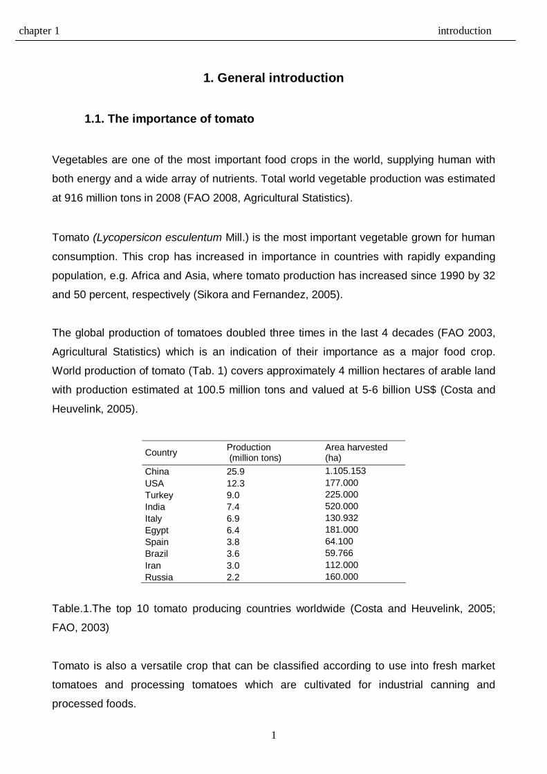
The global production of tomatoes doubled three times in the last 4 decades (FAO 2003,
Agricultural Statistics) which is an indication of their importance as a major food crop. World production of tomato (Tab. 1) covers approximately 4 million hectares of arable land
with production estimated at 100.5 million tons and valued at 5-6 billion US$ (Costa and
Heuvelink, 2005).
Country Production (million tons)
Area harvested (ha)
China 25.9 1.105.153 USA 12.3 177.000 Turkey 9.0 225.000 India 7.4 520.000 Italy 6.9 130.932 Egypt 6.4 181.000 Spain 3.8 64.100 Brazil 3.6 59.766 Iran 3.0 112.000 Russia 2.2 160.000
Table.1.The top 10 tomato producing countries worldwide (Costa and Heuvelink, 2005;
FAO, 2003)
Tomato is also a versatile crop that can be classified according to use into fresh market
tomatoes and processing tomatoes which are cultivated for industrial canning and
processed foods.
chapter 1 introduction
2
1.2. Root-knot nematodes and vegetable crops Many soil macro and micro organisms are parasites or pathogens of a wide spectrum of
vegetable plants where they can cause severe crop losses.
Plant parasitic nematodes are soil borne pests that attack a wide range of economically
important crops where they can affect both yield and quality (Noling, 2005). It has been
estimated that 10 percent of world crop production is lost as a result of parasitic nematode
infection (Whitehead, 1998).
Among the 24 genera of plant parasitic nematodes affecting vegetables, four species of
Meloidogyne, the root-knot nematode, are the most economically important to production
(Sasser and Freckman, 1987). Vegetables in general are known to be extremely
susceptible to root-knot nematode attack. Losses in vegetable production due to
Meloidogyne spp. attack can reach up to 5 percent (Hussey and Janssen, 2002).
Therefore, vegetable production in both tropical and subtropical areas is severely limited
by nematode infestation (Sikora and Fernandez, 2005).
Of all vegetable crops, tomato in particular, is heavily infected with plant parasitic
nematodes and especially with Meloidogyne species (Taylor and Sasser, 1978; Sikora and
Fernandez, 2005). Meloidogyne incognita is the most predominate and widely distributed
representative of this genus. This species has 4 races which can infect selectively more
than 2000 host plants (Taylor and Sasser, 1978; Trudgill, 1997; Manzanilla-Lopez et al.,
2004). The presence of galls on the root system of infected plants is the primary symptom
associated with root-knot nematode infection. These galls affect root functions and reduce
nutrient and water uptake that weakens the plants, causing wilting as well as nutrient
deficiency symptoms.
1.3. Nematode management Different control methods are used in the integrated management of plant parasitic
nematodes on vegetables. Chemical control, solarization, rotation, steam heating and
resistant cultivars are management techniques in wide use. However, all of these
management tools have limitations.
The use of chemical control which depends on using fumigation or systemic nematicides is
often restricted due to high costs and/or adverse effects on environment and human

chapter 1 introduction
3
health. During the last 20 years no new nematicidal compounds have entered the market
that are safer and effective at the same time.
The loss of methyl bromide, a broad spectrum biocide that is extensively used in vegetable
production, is a major loss for effective nematode management. The use of systemic
nematicides is also being limited by their removal from the market due to adverse side
effects.
Agricultural practices like crop rotation, flooding, steam and solar heating as well as
fallowing that have been traditionally used in integrated nematode management also have
become less usable because of pressure on land and economic demands for more
intensification and mass-production of crops and the costs involved that reduces profits.
Moreover, the use of resistant cultivars, which should be a major tool in management of
root-knot nematodes on vegetable crops, is restricted due to the lack of nematode
resistance genes in most cultivated vegetables.
Resistance to root-knot nematode is only used in industrial production of tomato and has
limited use for fresh market tomato. The resistance to root-knot nematode is related to a
single group of Mi–genes (Mi1 to Mi8) (Yaghoobi et al., 1995; Veremis and Roberts,
1996a,b; Williamson, 1998) . Important is the fact that Mi-resistance to root-knot nematode
can be broken-down under temperatures above 28 C°. This limiting factor affects many
tomato crops grown in warm tropical and sub tropical regions that are affected by virulent
nematode races in the field (Roberts and Thomason, 1989). Therefore, new sources of
resistance to Meloidogyne spp. are still needed to improve nematode management on all
vegetable crops (Hussey and Janssen, 2002). Sikora and Fernandez (2005) also been
reported also that the Mi-gene based resistance can be broken by new pathotypes.
1.4. Biological management The limitations on standard control tools for nematode management, mentioned above, in
addition to the limited availability of resistant germplasm for root-knot nematode
management, makes finding suitable alternative biocontrol agents for integrated
management important (Kerry, 1987; Sikora, 1992; 1997; Viaene et al., 2006). Detection
and development of biological agents requires a thorough understanding of the mode of
action and the nature of biological activity of parasitic nematodes.

chapter 1 introduction
4
Soil, besides harbouring nematodes and plant pathogens, also supports many other
beneficial organisms. Many of the antagonists in soil have been screened for their
biocontrol activity against different genera of plant parasitic nematodes on a wide
spectrum of crops (Stirling, 1991). Some of these beneficial organisms also have been
shown to colonize plants without causing disease and are known as mutualistic
endophytes (Petrini 1991; Wilson 1995; Stone et al., 2000).
1.5. Endophytes
Mutualistic endophytic organisms can be either bacteria (Chanway 1996; van Wees et al.,
1999) or fungi (Hallmann and Sikora 1994a,b., Niere et al., 2001., Olivain and Alabouvette,
1997) and either obligate symbionts or mutualistic. The arbuscular mycorrhizal fungi (AMF)
for example are true obligate symbions of the root system of many plants, whereas others
are facultative symbionts (Fo162) that can survive on organic matter in soil or inside the
roots of host plants (Sikora, 1997).
Schulz and Boyle (2005) classified fungal endophytes into three ecological groups: the
mycorrhizal fungi, the balansiaceous or grass endophytes and the non-balansiaceous
taxa. The majority of fungal endophytes isolated from over 500 plants belong to a wide
array of genera with the most common being: Acremonium, Alternaria, Cladosporium,
Phoma and Fusarium (Schulz et al., 1993, 1995 and 1998)
In 1997 Olivain and Alabouvette microscopically demonstrated the ability of non-
pathogenic strains of Fusarium oxysporum that control Fusarium wilt, to colonize root
tissue of tomato varieties. Endophytic colonization was considered important when direct
competition between the endophyte and the fungal pathogen was responsible for
biological control (Alabouvette et al., 2001).
Eparvier and Alabouvette (1994) reported that competition between pathogenic and non-
pathogenic F. oxysporum isolates in roots of tomato plants affected the colonization level
and/or the activity of both pathogenic and non-pathogenic Fusarium isolates. The level of
colonization of an endophytic fungus is important for control activity towards a pathogen
and can be influenced by biotic and abiotic factors.
chapter 1 introduction
5
Furthermore, different strains of the mutualistic endophyte F. oxysporum have been
reported to be antagonistic against both fungal pathogens and plant parasitic nematodes
on tomato (Mandeel and Baker 1991; Alabouvette et al., 1993; Olivain and Alabouvette
1997; Dababat et al., 2008; Dababat and Sikora2007a,b) and more recently against foliar
insects (Vidal, 1996 and Roy-Donald, 2010 ).
Antagonistic activity of other genera and species of mutualistic fungal endophytes has
been detected toward the burrowing nematode Radopholus similis on banana
(Pocasangre et al., 2000; Niere et al.,1998, 2001; Zum Felde, et al., 2005; Vu et al., 2006),
toward the lesion nematode Pratylenchus zeae on maize (Kimenju et al., 1998) and
against the root-knot nematode M. graminicola on rice (Le, 2006).
In 1994, Hallmann and Sikora isolated fungal endophytes from roots of field grown tomato
in Kenya. Many of these endophytic isolates showed biocontrol activity toward the root-
knot nematode M. incognita. The mutualistic endophyte F. oxysporum strain 162 (Fo162)
was the most promising isolate and produced high levels of antagonistic activity against
M. incognita on tomato. They reported a reduction in gall formation between 50 and 75%
within colonized tomato plants by the endophyte in greenhouse trials. The same isolate
has been repeatedly shown to have strong activity toward M. incognita in other
investigations (Dababat et al., 2008; Dababat and Sikora2007a,b).
1.6. Interaction between endophytes, pathogens and host plants In tritrophic interactions between the host plant, fungal pathogen and mutualistic
endophyte, different mechanisms of action were reported and considered responsible for
protecting the host plants including: competition for nutrients, competition for infection site,
cross protection and/ or induction of host plant defences (Alabouvette et al., 1998; Fravel
et al., 2003; Fuchs et al., 1997).
Little is known, however, about the exact mechanism of action involved in tritrophic
interactions between the plant, the fungal endophyte and the root-knot nematode. There is
evidence that metabolites produced by endophytes are involved in some of these
interactions and that the substances influence defence signals and regulation of the
symbiosis (Schulz and Boyle, 2005). Speijer (1993) and Hallmann and Sikora (1996)
demonstrated that fungal endophytes can release toxic compounds that can reduce
nematode infection, when good root colonization takes place. Numerous studies have also
chapter 1 introduction
6
demonstrated that colonization of host plant roots by endophytes results in production of
growth-promoting factors (Petrini, 1991).
Endophytes that colonize plant roots also enhance the tolerance of these hosts to
environmental stresses (Schulz et al., 1999a,b; Tan and Zou, 2001), to fungal pathogens
and plant parasitic nematodes through the production of antimicrobial metabolites
(Hallmann and Sikora,1996; Schulz et al., 1995, 2002).
Recent investigations have demonstrated that the induction of systemic resistance in
plants following the application of mutualistic fungal endophyte is involved in the mode of
action responsible for biological control of nematodes (Vu et al., 2006; Dababat and Sikora
2007a), and to Fusarium wilt (Fuchs et al., 1997).
1.7. Scope of the study The present study was initiated to identify the mode of action involved in the antagonistic
interaction between the mutualistic endophyte Fusarium oxysporum (Fo162) and the root-
knot nematode M. incognita on tomato. Biological, chemical and molecular studies on the
mechanisms related to systemic induced resistance were conducted.
The objectives of these investigations were to determine the:
1- Biological activity of Fo162 toward M. incognita on Fusarium-wilt susceptible and
resistant tomato varieties.
2- Influence of root exudates obtained from tomato plants colonized by Fo162 on root-knot
nematode juvenile behavior.
3- Identity of chemical compounds in root exudates obtained from Fo162 pre-inoculated
tomato plants which might be responsible for altering juvenile movement patterns.
4- Systemic colonization behaviour of Fo162 and durability of the signals related to the
systemic resistance pathways
5- Differences between the biotic Fo162 and abiotic elicitors with respect to form of
systemic resistance induction toward M. incognita.
6- Regulation of gene expression associated with induction of induced systemic resistance
(ISR) and systemic acquired resistance (SAR) using the biotic Fo162 and abiotic (SA
and MJ) elicitors.
chapter 1 introduction
7
1.8. References
- Alabouvette, C., Edel, V., Lemanceau, P., Olivain, C., Recorbet, G. and Steinberg,
C. (2001). Diversity and interactions among strains of Fusarium oxysporum:
Application and biological control. In: M.J. Jeger and N.J. Spence (Eds.): Biotic
interactions in plant-pathogen associations, pp 131-157. CAB International, London,
England.
- Alabouvette, C., Lemanceau, P. and Steinberg, C. (1993). Recent advances in the
biological control of Fusarium wilts. Pestic. Sci. 37: 365-373.
- Alabouvette, C., Schippers, B., Lemanceau, P. and Bakker, P. A. H. M. (1998).
Biological control of Fusarium wilts: Toward development of commercial products.
Pages 16-36 in Plant-microbe interactions and biological control. G. J. Boland and
L. D. Kuykendall, (Eds). Marcel Dekker, New York.
- Chanway, C. P. (1996). Endophytes: they’re not just fungi! Canadian Journal of
Botany 74: 321-322.
- Costa, J. M. and Heuvelink, E. (2005). Introduction: The tomato crop and industry.
In: Heuvelink, E. (Eds.). Tomatoes. CAB International, UK, pp. 1-19.
- Dababat, A. A. and Sikora, R. A. (2007a). Induced resistance by the mutualistic
endophyte, Fusarium oxysporum 162, toward Meloidogyne incognita on tomato.
Biocontrol Sci. Techn. 17, 969-975.
- Dababat, A. A. and Sikora, R. A. (2007b). Influence of the mutualistic endophyte
Fusarium oxysporum 162 on Meloidogyne incognita attraction and invasion.
Nematology 9, 771-776.
- Dababat, A. A., Selim, M. E., Saleh, A. A. and Sikora, R. A. (2008). Influence of
Fusarium wilt resistant tomato cultivars on root colonization of the mutualistic
endophyte Fusarium oxysporum strain 162 and its biological control efficacy toward
the root-knot nematode Meloidogyne incognita. Journal of Plant disease and
protection, 115 (6) 273-278.
- Eparvier, A. and Alabouvette, C. (1994). Use of ELISA and GUS- transformed
strains to study competition between pathogenic and non-pathogenic Fusarium
oxysporum for root colonization. Biocontrol Sci. Techn. 4, 35-47.
- FAO 2003 Agricultural statistics, Home page available at (http://apps.fao.org).
- Fravel, D., Olivain, C. and Alabouvette, C. (2003). Fusarium oxysporum and its
biocontrol. New Phytol. 157: 493-502.
chapter 1 introduction
8
- Fuchs, J.-G., Moënne-Loccoz, Y. and Défago, G. (1997). Non pathogenic Fusarium
oxysporum strain Fo47 induces resistance to Fusarium wilt in tomato. Plant Dis.
81:492-496.
- Hallmann, J. and Sikora, R. A. (1994a). Occurrence of plant parasitic nematodes
and nonpathogenic species of Fusarium in tomato plants in Kenya and their role as
mutualistic synergists for biological control of root knot nematodes. Int. J. Pest
Manage. 40, 321-325.
- Hallmann, J. and Sikora, R. A. (1994b). Influence of Fusarium oxysporum, a
mutualistic fungal endophyte, on Meloidogyne incognita of tomato. J. Plant Dis.
Protect. 101, 475-481.
- Hallmann, J. and Sikora, R. A. (1996). Toxicity of fungal endophyte secondary
metabolites to plant parasitic nematodes and soil-borne plant pathogenic fungi.
European journal of plant pathology 102, 155-162.
- Hussey, R. S. and Janssen, G. J. W. (2002). Root-knot nematodes: Meloidogyne
species. In: Starr, J. L., Cook, R., and Bridge, J. (Eds.) Plant resistance to parasitic
nematodes. CABI UK. pp. 43-70. - Kerry, B. P. (1987). Biological control. In: Brown, R. H. and Kerry, B. P. (Eds.)
Principles and practices of nematode control in crops. Academic Press, New York,
pp. 233-263.
- Kimenju, J. W., Wando, S. W., Mwang’ Ombe, A. W., Sikora, R. A. and Schurter, R.
P. (1998). Distribution of lesion nematodes associated with maize in Kenya and
susceptibility of maize cultivars to Pratylechus zeae. Afr. Crop Sci. J. 6: 367-375.
- Le, T. T. H. (2006). Antagonistic potential of endophytic and rhizosphere fungi
against the rice root-knot nematode Meloidogyne graminicola under upland rice
growing conditions. Master thesis, University of Bonn, Germany.
- Mandeel, Q. and Baker, R. (1991). Mechanisms involved in biological control of
Fusarium wilt of cucumber with strains of non-pathogenic Fusarium oxysporum.
Phytopathology 81, 462-469.
- Manzanilla-Lopez, R. H., Evans, K. and Bridge, J. (2004). Plant diseases caused by
nematodes. In: Chen, Z. X., Chen, S. Y. and Dickson, D. W. (Eds.). Nematology-
Nematode management and utilization. Vol. 2, CABI Publishing, 637-703.
- Niere, B. I., Sikora, R. A. and Speijer, P. R. (2001). Mutualistic endophytic fungi–
role in biocontrol and safety of application. In: Sikora, R. A. (Eds.). Integrated
Control of Soil Pests. IOBC/wprs Bulletin 24, 117–120.
chapter 1 introduction
9
- Niere, B. I., Speijer, P. R., Gold, C. S., and Sikora, R. A. (1998). Fungal endophytes
from banana for the biological control of Radopholus similis. In: Frison, E. A., Gold,
C. S., Karamura, E. B., and Sikora, R. A. (Eds.). Mobilizing IPM for sustainable
production in Africa. Proceedings of a workshop on banana IPM held in Nelspruit,
South Africa, 23-28 Nov. 1998. INIBAP, pp. 313-318. Montpellier, France.
- Noling, J. W. (2005). Nematode management on tomatoes, peppers and eggplant.
Institute of Food and Agricultural Science, University of Florida.
http://edis.ifas.ufl.edu/.
- Olivain, C. and Alabouvette C. (1997). Colonization of tomato root by a non-
pathogenic strain of Fusarium oxysporum. New Phytol. 137, 481-494.
- Petrini, O. (1991). Fungal endophytes of tree leaves. Microbial Ecology of leaves (J.
Andrews & S. Hirano, (Eds.), 179-197. Springer Verlag, New York.
- Pocasangre, L. E., Sikora, R. A., Vilich, V., and Schutster, R-P. (2000). Survey of
banana endophytic fungi from Central America and screening for biological control
of Radopholus similis. In: M. BLANKE and J. POHLAN (Eds.): Proceedings of the
2nd ISHS Conference on Fruit production in the Tropics and Subtropics.
- Roberts, P. A. and Thomason, I. J. (1989). A review of variability in four
Meloidogyne spp. measured by reproduction on several hosts including
Lycopersicon. Agricultural Zoology Reviews 3, 225-252.
- Roy-donald, M. B. (2010). The systemic activity of mutualistic endophytic fungi in
Solanaceae and Cucurbitaceae plants on the behavior of the phloem-feeding
insects Trialeurodes vaporariorum, Aphis gossypii and Myzus persicae. University
of Bonn, thesis. Ph.D. Thesis, university of Bonn.
- Sasser, J. N. and Freckman, D. W. (1987). A world perspective on nematology: the
role of the society. In: Veech, J. A. and Dickson, D. W. (Eds.) Vistas on
Nematology. Society of Nematologists, Hyattsville, Maryland, pp. 7-14.
- Schulz, B. and Boyle, C. (2005). The endophytic continuum. Mycol. Res. 109 (6)
661-686.
- Schulz, B., Boyle, C., Draeger, S., Römmert, A.-K., and Krohn, K. (2002).
Endophytic fungi: a source of biologically active secondary metabolites. Mycological
Research 106: 996-1004.
- Schulz, B., Guske, S., Dammann, U., and Boyle, C. (1998). Endophyte-host
interactions II. Defining symbiosis of the endophyte-host interaction. Symbiosis 25:
213-227.
chapter 1 introduction
10
- Schulz, B., Römmert, A.-K., Dammann, U., Aust, H.-J. and Strack, D. (1999b). The
endophyte-host interaction: a balanced antagonism. Mycological research, 103:
1275-1283.
- Schulz, B., Römmert, A.-K., Dammann, U., Peters, S., Guske, S., Strack, D. and
Boyle, C. (1999a). Endophyte-host symbisos. Bielefelder Ökologischer Beiträge 14:
307-312.
- Schulz, B., Sucker, J., Aust, H.-J., Krohn, K., Ludewig, K., Jones, P. G., and Döring,
D. (1995). Biologically active secondary metabolites of endophytic Pezicula species.
Mycological Research 99: 1007-1015.
- Schulz, B., Wanke, U., Draeger, S. and Aust, H.-J. (1993). Endophytes from
herbaceous plants and shrubs: effectiveness of surface sterilization methods.
Mycological research 97: 1447-1450.
- Sikora, R. A. (1992). Management of the antagonistic potential in agricultural
ecosystems for the biological control of plant-parasitic nematodes. Ann. Rev.
Phytopathology 30, 245-270.
- Sikora, R. A. (1997). Biological system management in the rhizosphere an inside-
out/side-in perspective. Comm. Appl. Biol. Sci. Ghent University, 62: 151-157.
- Sikora, R. A. and Fernandez, E. (2005). Nematode parasites of vegetables. In: Luc,
M. Sikora, R. A. And Bridge, J. (Eds.). Plant parasitic nematodes in subtropical and
tropical agriculture. CABI Publishing: UK, pp. 319-392.
- Speijer, P.R. (1993). Interrelationship between Pratylenchus goodeyi Sher & Allen
and strains of non-pathogenic Fusarium oxysporum Schl. emd. Snyd. & Hans. in
roots of banana cultivars. PhD Thesis, University of Bonn.
- Stirling, G. R. (1991). Biological control of plant parasitic nematodes. CAB
International, Wallington, UK. 282 pp.
- Stone, J. K., Bacon, C. W. and white, J. F. (2000). An overview of endophytic
microbes: endophytism defined. In Microbial Endophytes (C. W. Bacon and J. F.
White, (Eds.). Marcel Dekker, New York. pp.3-30
- Tan, R. X., and Zou, W. X. (2001). Endophytes: a rich source of functional
metabolites. Natural Products Rep. 18: 448-459.
- Taylor, A. L. and Sasser, J. N. (1978). Biology, identification and control of root-knot
nematodes (Meloidogyne spp.). North Carolina University Graphics, cooperative
publication of Department of Plant Pathology, North Carolina State University and
US Agency for International Development, Washington. DC.
chapter 1 introduction
11
- Trudgill, D. L. (1997). Parthenogenetic root-knot nematodes (Meloidogyne spp.);
how can these biotrophic endoparasites have such an enormous host range? Plant
pathology 46, 26-32.
- van Wees, S. C. M., Luijendijk, M., Smoorenburg, I., van Loon, L. C. and Pieterse,
C. M. J. (1999). Rhizobacteria-mediated induced systemic resistance (ISR) in
Arabidopsis is not associated with a direct effect on expression of known defense-
related genes but stimulates the expression of the jasmonate-inducible gene Atvsp
upon challenge. Plant Molecular Biology 41:537-549.
- Veremis, J. C. and Roberts, P. A. (1996a). Relationships between Meloidogyne
incognita resistance genes in Lycopersicon peruvianum differentiated by heat
sensitivity and nematode virulence. Theoretical and applied genetics 93: 950-959.
- Veremis, J. C. and Roberts, P. A. (1996b). Identification of resistance to
Meloidogyne javanica in the Lycopersicon peruvianum complex. Theoretical and
applied genetics 93: 894-901.
- Viaene, N., Coyne, D. L. and Kerry, B. P. (2006). Biological and cultural
management. In: Perry, R. N. and Moens, M. (Eds.). Plant Nematology. CAB
International, London, UK, pp. 347-369.
- Vidal, S. (1996). Changes in suitability of tomato for whiteflies mediated by a non-
pathogenic endophytic fungus. Entomologia Experimentalis et Applicata 80: 272-
274.
- Vu, T. T., Hauschild, R. and Sikora, R. A. (2006). Fusarium oxysporum endophytes
induced systemic resistance against Radopholus similis on banana. Nematology 8,
847-852.
- Whitehead, A. G. (1998). Plant nematode control. CAB International, Uk, pp. 384.
- Williamson, V. M. (1998). Root-knot nematode resistance genes in tomato and their
potential for future use. Annual Review Phytopathology 36. 277-293.
- Wilson, D. (1995). Endophyte-the evolution of a term, and clarification of its use and
definition. Oikos 73: 274-276.
- Yaghoobi, J., Kaloshian, I., Wen, Y. and Williamson, V. M. (1995). Mapping a new
nematode resistance locus in Lycopersicon peruvianum. Theoretical and applied
genetics 91: 457-464.
- Zum Felde, A., Pocasangre, A. L. and Sikora, R. A. (2005). The potential use of microbial communities inside suppressive banana plants for banana root protection. In: Turner, D. W., and Rosales, F. E. (Eds). In Banana root system: toward a better understanding for its productive management. Proceedings of an international symposium. International network for the improvement of banana and plantain (INIBAP), pp. 169-177. Montpellier, France.
Chapter 2 General Materials and Methods
12
2. General Materials and Methods 2.1. Endophyte fungal inoculum Fungal isolate of Fo162 was originally isolated from the cortical tissues of surface sterilized
tomato roots, Lycopersicum esculentum Mill. cv. Moneymaker in Kenya (Hallmann and
Sikora, 1994). The isolate was cultured and reared on potato dextrose agar (PDA) media
amended with 150 mg-1 streptomycin and 150 mg-1 chloramphenicol (DIFCO Company,
Germany) and placed in an incubator at 24 °C for 7 to 14 days. Conidia spores from these
initial plates were permanently stored in micro-bank tubes (CRYOBANKTM, MASTE Group
Ltd., Merseyside, UK) at -80 °C to avoid mutation and contamination. Fungal propagation
for all further experiments was initiated with spores from this stock. Inoculum was obtained
by culturing the fungus on PDA plates. After 2 weeks, the mycelia and spores were
scratched from the surface, suspended in water and sieved through 3 layers of cheese
cloth. Number of colony forming units (CFU) in the spore suspension was counted using a
Haemocytometer slide (Thoma, Germany) and the concentration was adjusted using
sterilized tap water. Desired concentrations for experimentation were suspended in 3 ml
water and injected 2 cm deep into the rhizosphere using 3 holes made around the stem
base with a plastic rod.
2.2. Nematode inoculum Meloidogyne incognita race 3 originating from Florida was used in all experiments.
Nematode inoculum was multiplied on the tomato cultivar Furore grown in the green house
at 27± °C. The plants were grown in large heated controlled boxes (150x80x40 cm) filled
with a sterilized soil:sand substrate (1:2, v/v). 3-4 weeks old tomato seedlings were
transplanted into the infested substrate and fertilized once a week with a 0.3% fertilizer
solution (N:P:K, 14:10:14). Nematode inoculum was obtained through extraction of the
eggs from 8 weeks old galled tomato roots using 1.5% NaOCl as described by Hussey and
Barker (1973). The plants were uprooted and then roots were washed gently with tap
water to remove soil particles. The roots were then cut into 1-2 cm pieces and macerated
for 20 seconds in a small amount of water using a warring blender (Bender and Hohbein).
The macerate was mixed thoroughly for 3 minutes with 1.5% NaOCl solution and shaken
by hand to free eggs from the gelatinous matrix. The egg suspension was poured through
4 sieves: 250 µm followed by 100, 45 and finally 25 µm. Eggs were collected on the 25 µm
aperture sieve and then washed with tap water to remove excess NaOCl. Egg inoculum
Chapter 2 General Materials and Methods
13
was transferred into a beaker and then agitated for 7 to 10 days in the dark at room
temperature (approx. 20C) with a constant supply of oxygen from an aquarium pump to
induce juvenile hatching. The active second stage juveniles (J2) were separated from the
un-hatched eggs and the non-active juveniles using a modified Baermann dish technique.
The J2 in the resulting water suspension were adjusted to 1000 J2/ 3 ml and used as
inoculum. The inoculum was injected 2 cm deep into the rhizosphere using 3 holes made
around the stem base with a plastic rod.
2.3. High pressure liquid chromatography (PR-HPLC) analysis Chromatogram analysis of root exudates collected from F. oxysporum 162 inoculated and
un-inoculated plants was performed on a HEWLETT PACKARD (HP) system using a
LiChrospher® C18 reversed phase guard column (250 by 4.0 mm, 5 µm), preceded by a
LiChrospher® C18 reversed phase guard column (4.0 by 4.0 mm, 5 µm). The HPLC
system consisted of an HP 1050 pump unit, HP 1050 diode array detector, HP 1046A
fluorescence array detector, and a 1050 auto sampler which were controlled by
ChemStation for LC 3D system. Before samples were injected, the column had been
equilibrated with 90% (v/v) water, 0.1% (v/v) trifluoroacetic acid (TFA) (solvent A) and 10%
acetonitril (solvent B). After injection, the samples were eluted at a flow rate of 1.0 ml/min
using an isocratic flow of 90% solvent A and 10% solvent B for 2 min, a linear gradient to
10% solvent A and 90% solvent B for 28 min, followed by an isocratic flow for 5 minutes
with 90% solvent B.
2.4. IGS-RFLP analysis 2.4.1. IGS-PCR fragments ampilification
The PCR Master Mix was prepared in an autoclaved Eppendorf tube kept on ice, by
mixing 12 µL 5 X PCR buffer, 5 µL dNTP (2.5 mM), 1.2 µL PNFO primer, 1.2 µL PN22
primer, 0.25 µL Tag polymerase and 38.35 µL autoclaved MilliQ water plus.
To each PCR vial, 60 µL of the PCR Master Mix was added to 2 µL of DNA sample.
All microfuge vials were closed properly with autoclaved plastic caps and then centrifuged
for 2 seconds in a micro centrifuge (Labnet International). The microfuge vials were placed
on a PCR thermal cycler (T Gradient, Biometric). Amplification was performed by an initial
denaturation at 95 °C for 4 minutes, followed by 36 cycles of 95 °C for 1 minute, 52 °C for
40 seconds, and 72 °C for 1.5 minute, and with a final extension cycle of 72 °C for 5
minutes. After the PCR procedure, samples were stored at 4 °C.
Chapter 2 General Materials and Methods
14
2.4.2. Restriction enzyme analysis In order to characterize the fungal isolates, the IGS PCR amplified fragments, were
digested with 3 restriction enzymes: AluI, HaeIII and RsaI. In a sterile Eppendorf tube, the
Restriction Enzyme Master Mix (REMM) was prepared by adding 0.2 µL acetylated bovine
serum albumin (BSA, Promega), 2 µL R Buffer (10 X) (Promega), and 0.5 µL restriction
enzyme to 0.3 µL autoclaved distilled milliQ water.
For each isolate, 17 µL PCR product were taken and transferred to a new autoclaved
microfuge PCR plastic vial and mixed with 3 µL of the prepared REMM. The plastic vials
were closed and centrifuged for 2 seconds before incubated at 37 °C for 3 hours. After
incubation, samples were stored at 4 °C. For each sample, 5 µL of the digested IGS PCR
product were injected into agarose gel electrophoresis to determine the restriction
fragment length polymorphism (RFLP) patters.
2.4.3. Gel electrophoresis analysis The agarose gel that was used in this analysis was prepared with 1 X Tris-Acetate EDTA-
Buffer (TAE, AppliChem). 2.5 g agarose (Sigma) was added to 250 ml of TAE buffer and
heated for 5 minutes in a microwave (MW800, Continent) at 650 watts. After cooling at
approx. 50°C, 2.5 µL of 10mg ml-1 Ethidium Bromide (AppliChem) was added. This
solution was poured into an electrophoresis tray and left for approx. 30 minutes until the
gel had solidified. The gel was subsequently transferred to the gel electrophoresis
chamber filled with 1 X TAE buffer. After transferring all samples to the wells of the gel, the
electrophoresis analysis was conducted for 60 minutes at 120 Volt. An Ultraviolet
transilluminator was used to visualize the DNA bands and pictures were taken using a
digital camera (S9500, Finepix, Fuji film).
2.4.4. Phylogenetic analysis The IGS-RFLP phylogenetic analysis was processed using NTEdit and NTSYSpc
Numerical Taxonomy System, Version 2.2 (Exeter software). The presence or absence of
markers was scored for each isolate and the results were compiled in a excel file. Prior to
calculation of the genetic similarity (GS), the excel files were converted into an NTSYSpc
compatible data matrix. For each marker system the pair-wise genetic distances between
taxa was calculated using the DICE coefficient of similarity (Dice, 1945). GS matrices were
subjected to the unweighted pair group method with arithmetic average (UPGMA) cluster
analysis (Sneath and Sokal, 1973) followed by the sequential agglomerative hierarchical
nested (SAHN) cluster analysis (Sneath and Sokal, 1973) implemented in NTSYSpc 2.2.
Chapter 2 General Materials and Methods
15
2.5. Culture media and reagents
Potato Dextrose Agar PDA (DIFCO)
24 g Potato Dextrose Broth
18 g Agar (DIFCO)
1000 ml Distilled water
150 ppm streptomycin sulphate and 150 ppm chloramphenicol were added after cooling.
Phloxine B (MERCK)
15 mg Phloxine B
1000 ml Tap water
2.6. Statistical analysis Data were analysed according to the standard analysis of variance procedure with SPSS
14 program for windows. Differences among treatments were tested using one way
analysis of variance (ANOVA) followed by Tukey Test for mean comparison if the F-value
was significant. Statistical differences referred to in the text were significant at (P <0.05).
However, T-test was used for comparing 2 treatments using SigmaStat 3.1.
The data in chapter 3 were analysed using mixed procedure of SAS (2005). Means of
levels of the significantly affecting factors were compared using Tukey’s studentized range
test with SAS.
2.7. References - Hussy, R. and Barker, K. (1973). A comparison of methods of collecting inocula
of Meloidogyne spp., including a new technique. Plant Disease Reporter 57:
1025-1028.
- Dice, L.R. (1945). Measures of the amount of ecologic association between
species. Ecology 26, 297-302.
- Sneath, P. and Sokal, R. (1973). Numerical Taxonomy. Freeman, San
Francisco.
Chapter 3 Fo162 and resistant tomato cultivars
16
3. Influence of Fusarium-wilt host plant resistance on colonization
ability and biological activity of the mutualistic endophyte Fusarium oxysporum 162 in tomato.
3.1. Introduction
Many isolates of Fusarium oxysporum are known to be important plant pathogens that
cause sever damage to many host plants (Olivain and Alabouvette 1997, 1999; Olivain et
al., 2003). This fungus contains highly specialized strains that have been classified into
approximately 120 formae speciales and races, based on the plant species and cultivars
they infect (Armstrong and Armstrong 1981; Gordon and Okamoto 1992; Alabouvette et
al., 2001). The main form of Fusarium disease management is the use of resistant
cultivars which provide full season protection in a number of host plants against the
different pathogenic formae speciales.
In contrast, the majority of Fusarium oxysporum strains in the soil are non-pathogenic and
play beneficial roles in the soil eco-system (Mandeel and Barker 1991; Hallmann and
Sikora 1994a,b; Olivain and Alabouvette 1997; and Alabouvette et al., 2001). Some of
these non-pathogenic strains are considered to be mutualistic endophytes, because they
colonize the root tissues of many plants, live in that tissue without causing disease
symptoms and simultaneously increase resistance to fungal pathogens, plant parasitic
nematodes and insects. Hallmann and Sikora (1994a,b); and Dababat and Sikora
(2007a,b) tested the potential of the mutualistic endophyte F. oxysporum isolate 162
(Fo162) against the root-knot nematode Meloidogyne incognita and obtained significant
levels of control. Fuchs et al., (1997) obtained similar results with Fusarium wilt control
with non-pathogenic strains on tomato and more recently, Roy-donald (2010) detected
systemic biocontrol activity of Fo162 toward whiteflies and aphids on tomato.
It is important to note that all the previous investigations were conducted on Fusarium wilt
susceptible tomato cultivars. This factor is significant in that, the majority of tomato
cultivars used in the field are resistant to the pathogenic forms of Fusarium. This form of
resistance could influence Fo162 efficacy if the mutualistic endophyte is unable to colonize
the root systems.
Chapter 3 Fo162 and resistant tomato cultivars
17
The capability of a mutualistic endophyte agent to colonize the endorhiza of root systems
is essential for providing biological control against pathogens, nematodes and insects.
Extremely important in control is the level of endophyte colonization, especially when
direct competitive effects of the biocontrol agent on the target organism are involved
(Handelsman and Stabb, 1996).
The objectives of the following studies were to determine the:
1- Influence of Fusarium-wilt resistant tomato cultivars on the level of colonization of
the mutualistic endophyte .F. oxysporum 162.
2- Biological activity of Fo162 toward M. incognita on Fusarium-wilt susceptible and
resistant tomato cultivars.
3- Relation between Fo162 colonization levels and nematode gall formation on tomato
plants.
Chapter 3 Fo162 and resistant tomato cultivars
18
3.2. Materials and methods 3.2.1. Colonization
Six tomato cultivars from Seminis Vegetable Seed company, USA; four from Bejo Zaden
b. v., Holland; and one cultivar (which is highly susceptible to Meloidogyne incognita) from
Juliwa Enza GmbH & Co. KG, Germany, (Table 1) were sown at a rate of one seed per pot
(5 x 4 x 3 cm) in 50 g planting mixture containing a sterilized sand: field soil (2:1, v/v)
substrate. Immediately after sowing, the endophytic fungus Fo162 was inoculated at a rate
of 106 cfu g-1 substrate (see chapter 2). Control pots were treated with tap water.
Each treatment consisted of 6 replicates in a randomized complete block design. All
tomatoes were grown under greenhouse conditions at 22 ± 5ºC with 16 h of supplemental
diurnal light per day. The plants were watered as needed, and were fertilized with a slow
release formulation (N: P: K, 14:10:14) at 2 g l-1 of water. The experiment was terminated
3 weeks after fungal inoculation. The roots were separated from soil by gentle washing
with tap water. The roots were then blotted between two paper tissues and fresh weight
was recorded. The roots of each replicate were surface sterilized by soaking them in 0.5%
solution of NaOCl for 3 minutes, followed by three rinses in sterilized water. Four sections
of root approx. 0.5 cm in length were then cut from similar sized roots in each replicate and
each piece pressed onto PDA to check sterilization success (root imprint test). They were
then mounted onto a new PDA plate to determine the percentage of root segments
colonized by Fo162. A total of 20 root pieces were examined per replicate and the mean of
6 replicates was calculated. Surface sterilization was considered successful when no
fungal colonies developed on the medium used for the root imprint. Successful re-isolation
of Fo162 from root segments was confirmed when the fungal culture characteristics and
microscope examination of the fungi growing out of the root pieces corresponded to those
of Fo162.
Chapter 3 Fo162 and resistant tomato cultivars
19
3.2.2. Biological control To evaluate the influence of the Fusarium-wilt resistance on biological control activity of
Fo162, the number of root-knot nematode galls on the susceptible and resistant tomato
cultivars was determined. The 11 cultivars were sown into commercial 96 plug seedling
trays containing the seedling substrate as mentioned above.
After three weeks, the tomato seedlings were transplanted into pots containing 300g of
planting substrate. Tomato seedlings were inoculated with 3 ml of a solution containing
5x10⁸ cfu of Fo162 (see chapter 2), immediately after transplanting. Control pots were
treated with 3 ml of tap water. The plants were inoculated with 1000 J2/pot one week after
Fo162 inoculation (see chapter 2). The experiment was terminated 3 weeks after
nematode inoculation. The roots were then washed with tap water to free them from soil
particles and the number of galls was counted. The root systems were then used to
evaluate the level of Fo162 colonization using the same procedure as mentioned above.
Table 1: Tomato cultivars, origin, supplier and susceptibility rating.
*These numbers will be used in the results and discussion instead of the cultivar names
No. Cultivar Origin Company Feature
1* FLORIDA 47 R India Seminis Vegetable Seeds, USA
Fusarium resistant
2 FLORIDA 91 (XP 10091) Thailand “ “
3 SUNGUARD (XP 10089) Thailand “ “
4 SOLAR SET R Mexico “ “
5 CROWN JEWEL (XP 01407783) Thailand “ “
6 SUNPRIDE Thailand “ “
7 P48024 India Bejo Zaden b.v., Holland “
8 P48025 India “ “
9 P48026 India “ Fusarium susceptible
10 P48027 India “ “
11 HELLFRUCHT/JW FRÜHSTAMM Germany Juliwa Enza GmbH &
Co. KG, Germany “
Chapter 3 Fo162 and resistant tomato cultivars
20
3.3. Results
3.3.1. Colonization in absence of root-knot The Fo162 strain successfully colonized the roots of all tomato cultivars tested regardless
of existence of resistance to the Fusarium-wilt pathogen. Colonization percentage ranged
from 31 to 75% among the different tomato cultivars. The highest levels of root
colonization (approx. 75 %) were detected in the cultivars Sunguard (3), Florida 91 (2) and
P48027 (10), respectively (Fig.1).
0102030405060708090
100
1 2 3 4 5 6 7 8 9 10 11
Tomato Varieties
Colo
niza
tion%
ResistantSusceptible
Figure.1. Fusarium oxysporum 162 colonization in Fusarium-wilt susceptible and resistant
tomato cultivars, three weeks after inoculation (n=6).
Chapter 3 Fo162 and resistant tomato cultivars
21
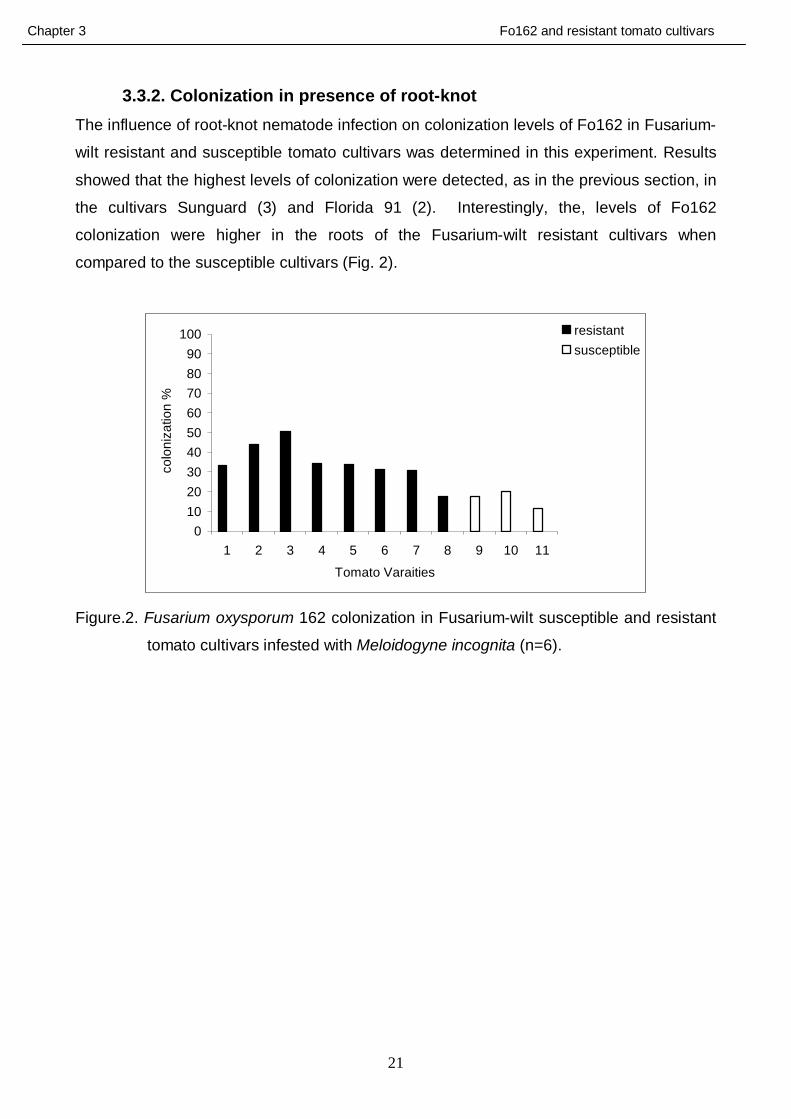
3.3.2. Colonization in presence of root-knot The influence of root-knot nematode infection on colonization levels of Fo162 in Fusarium-
wilt resistant and susceptible tomato cultivars was determined in this experiment. Results
showed that the highest levels of colonization were detected, as in the previous section, in
the cultivars Sunguard (3) and Florida 91 (2). Interestingly, the, levels of Fo162
colonization were higher in the roots of the Fusarium-wilt resistant cultivars when
compared to the susceptible cultivars (Fig. 2).
0102030405060708090
100
1 2 3 4 5 6 7 8 9 10 11
Tomato Varaities
col
oniz
atio
n %
resistantsusceptible
Figure.2. Fusarium oxysporum 162 colonization in Fusarium-wilt susceptible and resistant
tomato cultivars infested with Meloidogyne incognita (n=6).
Chapter 3 Fo162 and resistant tomato cultivars
22
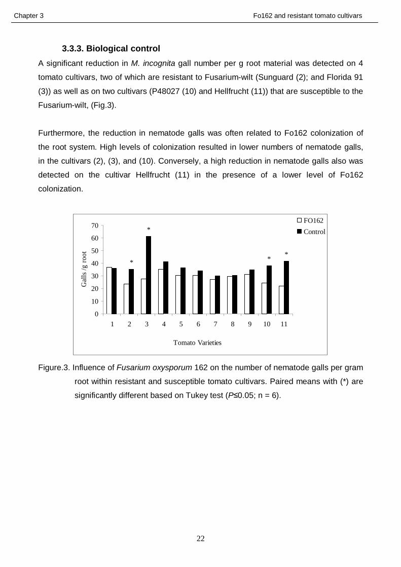
3.3.3. Biological control A significant reduction in M. incognita gall number per g root material was detected on 4
tomato cultivars, two of which are resistant to Fusarium-wilt (Sunguard (2); and Florida 91
(3)) as well as on two cultivars (P48027 (10) and Hellfrucht (11)) that are susceptible to the
Fusarium-wilt, (Fig.3).
Furthermore, the reduction in nematode galls was often related to Fo162 colonization of
the root system. High levels of colonization resulted in lower numbers of nematode galls,
in the cultivars (2), (3), and (10). Conversely, a high reduction in nematode galls also was
detected on the cultivar Hellfrucht (11) in the presence of a lower level of Fo162
colonization.
*
***
0
10
20
30
40
50
60
70
1 2 3 4 5 6 7 8 9 10 11
Tomato Varieties
Gal
ls /g
root
FO162Control
Figure.3. Influence of Fusarium oxysporum 162 on the number of nematode galls per gram
root within resistant and susceptible tomato cultivars. Paired means with (*) are
significantly different based on Tukey test (P≤0.05; n = 6).
Chapter 3 Fo162 and resistant tomato cultivars
23
3.4. Discussion It is well established that the endophytic fungus F. oxysporum strain162 (Fo162) has the
ability to colonize the root endorhiza of Fusarium-wilt susceptible tomato cultivars as well
as roots of other plants (Hallmann and Sikora 1994a; Dababat and Sikora, 2007a,b:
Pocasangre et al., 2000 and Roy-Donald, 2010)
Colonization of the endorhiza was found to be important for self propagation and
reproduction of mutualistic endophytic antagonists, for parasitism of a pest, for release of
toxic compounds as well as for the enhancement of plant defense mechanisms in the root
tissue (Speijer, 1993). The level of colonization is often considered to be an important
factor influencing both the mode of action involved and the level of control attained
(Dababat et al., 2008). Olivain and Alabouvette (1997) were the first to make microscopic
investigations which demonstrated that the non-pathogenic Fusarium oxysporum isolate
(47) was able to colonize the endorhiza of tomato roots. Their study was conducted as in
those by Dababat et al., (2008) on Fusarium-wilt susceptible tomato cultivars.
The mutualistic endophyte F. oxysporum 162 also has been shown to have biological
control activity toward root-knot nematodes on a number of different host plants (Dababat
and Sikora 2007c; Vu et al., 2006; Dababat and Sikora 2007a). Alabouvette et al., (2001)
also demonstrated that non-pathogenic F. oxysporum strain Fo47 successfully colonized
tomato roots and affected the incidence of Fusarium-wilt disease through a process that
they called cross-protection.
The basis for the present study was to determine whether or not tomato cultivars that have
genes responsible for resistance to the Fusarium–wilt pathogen; and that are commonly
used in the field for wilt control, have a negative impact on the ability of the mutualistic
endophyte Fo162 to colonize the endorhiza of tomato roots and thereby reduce or prevent
biological control of root-knot nematode on tomato.
3.4.1. Colonization in absence of root-knot The results obtained from this study revealed that Fusarium-wilt resistant tomato cultivars
do not suppress the ability of Fo162 to colonize tomato root tissue in the absence of the
root-knot nematode. Furthermore, the levels of Fo162 colonization were higher on the
majority of the Fusarium-wilt resistant cultivars tested when compared to the susceptible
cultivars.
Chapter 3 Fo162 and resistant tomato cultivars
24
3.4.2. Colonization in presence of nematode When Fo162 was re-isolated from tomato roots that were infested with M. incognita one
week after endophyte inoculation, the highest levels of Fo162 colonization were detected
in the cultivars Sunguard (3) and Florida 91 (2) which are resistant to wilt-Fusarium.
Fo162 colonization levels were lower in tomato plants that were inoculated with root-knot
nematode when compared to the non-inoculated plants. The negative impact of the
nematode on colonization might be due to the competition for nutrients between Fo162
and nematode in the endorhiza or to abiotic stress present in the greenhouse tests such
as temperature.
Eparvier and Alabouvette (1994) demonstrated that competition occurs between the non-
pathogenic F. oxysporum and the phytopathogens in the root tissue of susceptible plants.
This competition affects the intensity of root colonization and/or the activity of both
pathogen and the non-pathogen agents.
3.4.3. Biological control Significant differences in number of galls per g root between Fo162 inoculated and non-
inoculated tomato plants were recorded on 4 tomato cultivars. Two of those cultivars are
resistant to Fusarium-wilt while the other two are susceptible. Furthermore, the highest
level of root-knot nematode control observed on cultivar Sunguard (3) which is Fusarium-
wilt resistant. These results revealed that Fo162 was more biologically active on resistant
cultivars in reducing gall number and nematode infection.
Moreover, there is evidence that a positive relationship exist between the reduction in
nematode galling and the level of endophyte colonization. For example, where the lowest
Fo162 colonization levels were detected, also the lowest control potential was observed
(cultivar 6, 7, 8 and 9). However, Niere (2001) reported that high level of the endophytic
fungal colonization was not required for the maintenance of long-term biological control of
R. similis on banana.
Investigations conducted with non-pathogenic F. oxysporum for cross-protection against
Fusarium-wilt demonstrated that intense colonization of the root surface occurs very
quickly after inoculation and constitutes a physical barrier preventing direct contact of the
plant pathogen with the root surface. Surface colonization also causes intense competition
for root exudates. Therefore, the parasite might be prevented from obtaining nutrients
required for establishment of infection (Olivain and Alabouvette, 1997; Olivain et al., 2006).
Chapter 3 Fo162 and resistant tomato cultivars
25
3.5. Conclusions
Based on the finding in the present investigation the following can be concluded:
1- The genes in tomato cultivars that are responsible for providing these cultivars with
resistance to the Fusarium-wilt pathogen did not affect the ability of the mutualistic
Fo162 to colonize the endorhiza of tomato roots
2- A positive relation was detected between Fo162 colonization levels and root-knot
nematode control on both Fusarium-wilt resistant and susceptible tomato cultivars
3- The presence of the root-knot nematode M. incognita decreased Fo162
colonization levels in both resistant and susceptible tomato plants.
Chapter 3 Fo162 and resistant tomato cultivars
26
3.6. References - Alabouvette, C., Edel, V., Lemanceau, P., Olivain, C., Recorbet, G., and Steinberg,
C. (2001). Diversity and interactions among strains of Fusarium oxysporum:
Application and biological control. In: M.J. JEGER and N.J. SPENCE (eds.): Biotic
interactions in plant-pathogen associations, pp 131-157. CAB International, London,
England.
- Armstrong, G. M., and Armstrong, J. K. (1981). Formae speciales and races of
Fusarium oxysporum causing wilt diseases. In: P.E. NELSON, T.A. TOUSSOUN,
R.J. COOK (eds.) Fusarium: Diseases, biology, and taxonomy, pp. 391-399.
Pennsylvania State University Press, University Park and London.
- Dababat, A. A., and Sikora, R. A. (2007a). Induced resistance by the mutualistic
endophyte, Fusarium oxysporum 162, toward Meloidogyne incognita on tomato.
Biocontrol Sci. Techn. 17, 969-975.
- Dababat, A. A., and Sikora, R. A. (2007b). Influence of the mutualistic endophyte
Fusarium oxysporum 162 on Meloidogyne incognita attraction and invasion.
Nematology 9, 771-776.
- Dababat, A. A., and Sikora, R. A. (2007c). Importance of application time and
inoculum density of the non-pathogenic endophytic fungus, Fusarium oxysporum
162, for the biological control of the root-knot nematode Meloidogyne incognita on
tomato. Nematropica 2, 267-276.
- Dababat, A. A., Selim, M. E., Saleh, A. A., and Sikora, R. A. (2008). Influence of
Fusarium wilt resistant tomato cultivars on root colonization of the mutualistic
endophyte Fusarium oxysporum strain 162 and its biological control efficacy toward
the root-knot nematode Meloidogyne incognita. Journal of Plant disease and
protection, 115 (6) 273-278.
- Eparvier, A., and Alabouvette, C. (1994). Use of ELISA and GUS- transformed
strains to study competition between pathogenic and non-pathogenic Fusarium
oxysporum for root colonization. Biocontrol Sci. Techn. 4, 35-47.
- Fuchs, J.-G., Moënne-Loccoz, Y., and Défago, G. (1997). Non pathogenic Fusarium
oxysporum strain Fo47 induces resistance to Fusarium wilt in tomato. Plant Dis. 81,
492-496.
- Gordon, T. R. and Okamoto, D. (1992). Population structure and the relationship
between pathogenic and non-pathogenic strains of Fusarium oxysporum.
Phytopathology 82, 73-77.
Chapter 3 Fo162 and resistant tomato cultivars
27
- Hallmann, J. and Sikora, R. A. (1994a). Occurrence of plant parasitic nematodes
and nonpathogenic species of Fusarium in tomato plants in Kenya and their role as
mutualistic synergists for biological control of root knot nematodes. Int. J. Pest
Manage. 40, 321-325.
- Hallmann, J. and Sikora, R. A. (1994b). Influence of Fusarium oxysporum, a
mutualistic fungal endophyte, on Meloidogyne incognita of tomato. J. Plant Dis.
Protect. 101, 475-481.
- Handelsman, J. and Stabb, E. V. (1996). Biocontrol of soilborne plant pathogens.
The Plant Cell 8, 1855-1869.
- Mandeel, Q. and Baker, R. (1991). Mechanisms involved in biological control of
Fusarium wilt of cucumber with strains of non-pathogenic Fusarium oxysporum.
Phytopathology 81, 462-469.
- Niere, B. I. (2001). Significance of non-pathogenic isolates of Fusarium oxysporum
Schlecht: Fries for the biological control of the burrowing nematode Radopholus
similis (Cobb) Thorne on tissue cultured banana. Ph.D. Thesis, University of Bonn.
- Niere, B. I., Sikora, R. A., and Speijer, P. R. (2001). Mutualistic endophytic fungi–
role in biocontrol and safety of application. In: R.A. Sikora (eds.): Integrated Control
of Soil Pests. IOBC/wprs Bulletin 24, 117–120.
- Olivain, C. and Alabouvette, C. (1997). Colonization of tomato root by a non-
pathogenic strain of Fusarium oxysporum. New Phytol. 137, 481-494.
- Olivain, C., and Alabouvette, C. (1999). Process of tomato root colonization by a
pathogenic strain of Fusarium oxysporum f. sp. lycopersici in comparison with a
non-pathogenic strain. New Phytol. 141, 497-510.
- Olivain, C., Humbert, C., Nahalkova, J., Fatehi, J., L’haridon, F., and Alabouvette,
C. (2006). Colonization of tomato by pathogenic and non-pathogenic Fusarium
oxysporum strains inoculated together and separately into the soil. Appl. Environ.
Microbiol. 72, 1-9.
- Olivain, C., Trouvelot, S., Binet, M., Cordier, C., Pugin, A., and Alabouvette, C.
(2003). Colonization of flax roots and early physiological responses of flax cells
inoculated with pathogenic and non-pathogenic strains of Fusarium oxysporum.
Appl. Environ. Microb. 69, 5453-5462.
- Pocasangre, L. E., Sikora, R. A., Vilich, V. and Schutster, R-P. (2000). Survey of
banana endophytic fungi from Central America and screening for biological control
of Radopholus similis. In: M. BLANKE and J. POHLAN (eds.): Proceedings of the
2nd ISHS Conference on Fruit production in the Tropics and Subtropics. Held on 24
– 26 June (1999), pp. 283-289. Bonn – Röttgen, Germany.

Chapter 3 Fo162 and resistant tomato cultivars
28
- Roy-donald, M. B. (2010). The systemic activity of mutualistic endophytic fungi in
Solanaceae and Cucurbitaceae plants on the behavior of the phloem-feeding
insects Trialeurodes vaporariorum, Aphis gossypii and Myzus persicae. University
of Bonn, thesis. Ph.D. Thesis, university of Bonn.
- Speijer, P. R. (1993). Interrelationship between Pratylenchus goodeyi Sher & Allen
and strains of non-pathogenic Fusarium oxysporum Schl. emd. Snyd. & Hans. in
roots of banana cultivars. PhD Thesis, University of Bonn.
- Vu, T. T. (2005). Mode of action of non-pathogenic Fusarium oxysporum
endophytes for bio-enhancement of banana toward Radopholus similis. Ph.D.
Thesis, university of Bonn.
- Vu, T. T., Hauschild, R., and Sikora, R. A. (2006). Fusarium oxysporum endophytes
induced systemic resistance against Radopholus similis on banana. Nematology 8,
847-852.
Chapter 4 biological and chemical studies on root exudates
29
4. Chemical and biological proprieties of root exudates obtained from
tomato plants inoculated with Fusarium oxysporum strain 162 and
their influence on the behaviour of the root-knot nematode
Meloidogyne incognita
4.1. Introduction Past research has shown that specific root exudates of host plants play an important role
in the infection process and at the same time in host defense responses to different
nematodes and pathogens. Ward (1973) demonstrated that nematodes can migrate to
specific compounds, such as Cl- , even in the presence of a high uniform level of another
attractive compound such as Na+. In 1997, Perry reported that plant parasitic nematodes
can detect their host plants through chemoreception mechanisms which are dependent on
chemotactic signals released by the roots of their hosts. Moreover, Bargmann (2006)
demonstrated that Caenorhabditis elegans uses chemo sensation mechanisms either to
find food or to avoid unfavourable conditions.
The presence of microorganisms in the rhizosphere and in the endorhiza has been shown
to influence the attractiveness of host plants to plant parasitic nematodes. Endophytic
bacteria have been demonstrated to reduce root-knot nematode penetration on rice and
on potato (Padgam and Sikora, 2006; Reitz et al., 2000). Obligate symbiotic arbiscular
mycorrhizal fungi also have been shown to alter root-knot nematode host finding behaviour
(Reimann et al., 2008). Saprophytic fungi that colonize the endorhiza also have been
known to reduce nematode penetration on tomato (Dababat and Sikora, 2007), banana
(Zum Felde et al., 2005; Vu et al., 2004), maize (Kimenju et al., 1998) and rice (Le, 2006).
The mutualistic endophyte Fusarium oxysporum isolate (Fo162), that has proven
antagonistic activity toward root-knot nematodes, was isolated from the cortical tissue of
tomato roots (Hallmann and Sikora, 1994). Hallmann and Sikora (1996) and Dababat and
Sikora (2007) showed that effective root colonization by Fo162 affected M. incognita
penetration of the roots. Choice experiments conducted by Dababat and Sikora (2007)
demonstrated that tomato root exudates obtained from plants colonised by Fo162 were
either less attractive or contained substances that had repellent activity toward M.
incognita juveniles. The mechanisms involved in alteration of root-knot nematode
behaviour in endophyte colonized tomato plants, however, have not been determined.
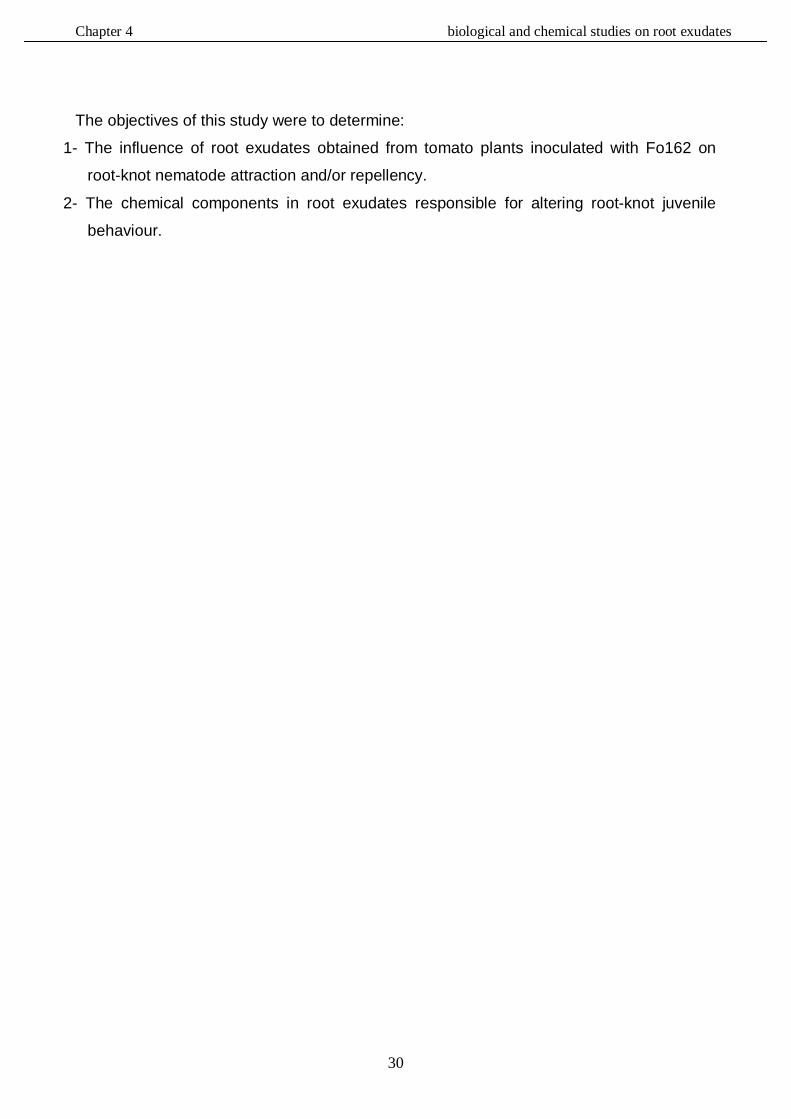
Chapter 4 biological and chemical studies on root exudates
30
The objectives of this study were to determine:
1- The influence of root exudates obtained from tomato plants inoculated with Fo162 on
root-knot nematode attraction and/or repellency.
2- The chemical components in root exudates responsible for altering root-knot juvenile
behaviour.
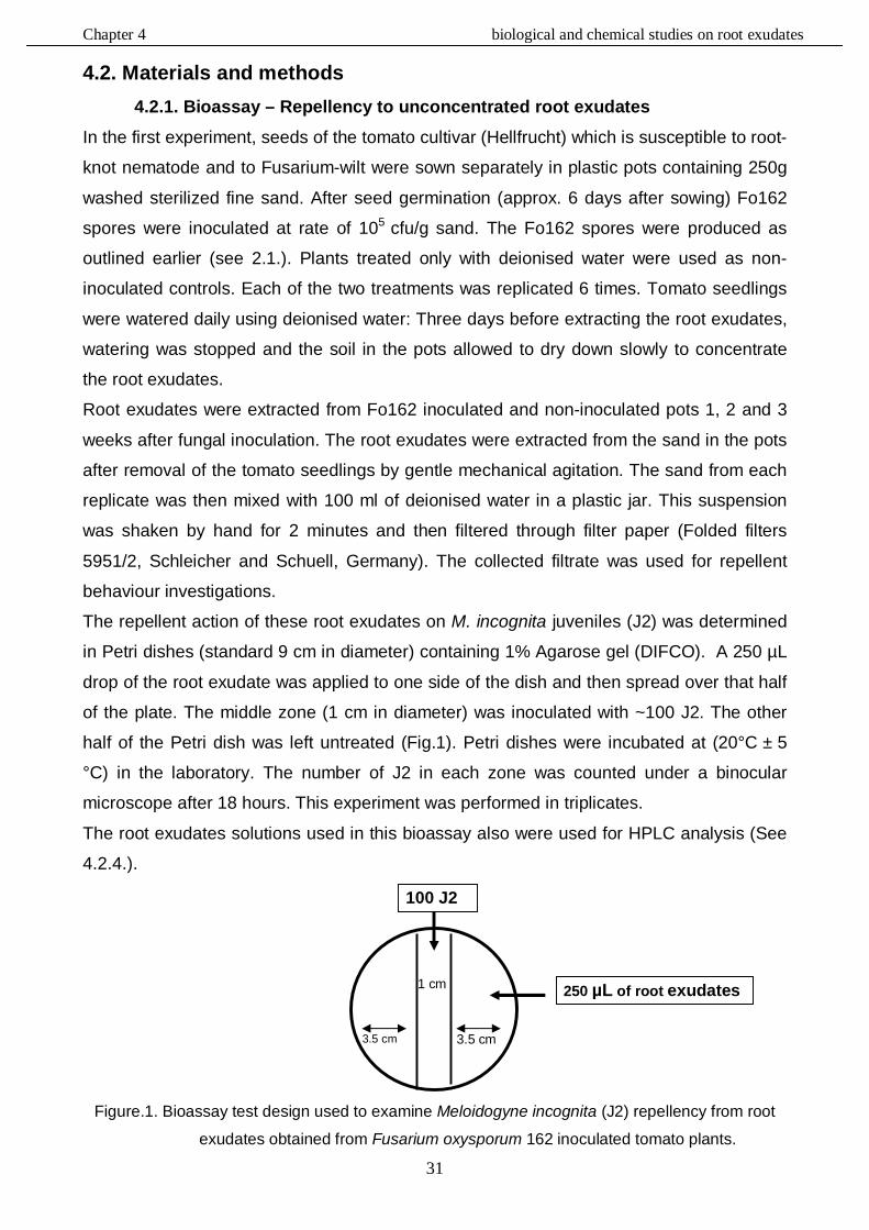
Chapter 4 biological and chemical studies on root exudates
31
4.2. Materials and methods 4.2.1. Bioassay – Repellency to unconcentrated root exudates
In the first experiment, seeds of the tomato cultivar (Hellfrucht) which is susceptible to root-
knot nematode and to Fusarium-wilt were sown separately in plastic pots containing 250g
washed sterilized fine sand. After seed germination (approx. 6 days after sowing) Fo162
spores were inoculated at rate of 105 cfu/g sand. The Fo162 spores were produced as
outlined earlier (see 2.1.). Plants treated only with deionised water were used as non-
inoculated controls. Each of the two treatments was replicated 6 times. Tomato seedlings
were watered daily using deionised water: Three days before extracting the root exudates,
watering was stopped and the soil in the pots allowed to dry down slowly to concentrate
the root exudates.
Root exudates were extracted from Fo162 inoculated and non-inoculated pots 1, 2 and 3
weeks after fungal inoculation. The root exudates were extracted from the sand in the pots
after removal of the tomato seedlings by gentle mechanical agitation. The sand from each
replicate was then mixed with 100 ml of deionised water in a plastic jar. This suspension
was shaken by hand for 2 minutes and then filtered through filter paper (Folded filters
5951/2, Schleicher and Schuell, Germany). The collected filtrate was used for repellent
behaviour investigations.
The repellent action of these root exudates on M. incognita juveniles (J2) was determined
in Petri dishes (standard 9 cm in diameter) containing 1% Agarose gel (DIFCO). A 250 µL
drop of the root exudate was applied to one side of the dish and then spread over that half
of the plate. The middle zone (1 cm in diameter) was inoculated with ~100 J2. The other
half of the Petri dish was left untreated (Fig.1). Petri dishes were incubated at (20°C ± 5
°C) in the laboratory. The number of J2 in each zone was counted under a binocular
microscope after 18 hours. This experiment was performed in triplicates.
The root exudates solutions used in this bioassay also were used for HPLC analysis (See
4.2.4.).
Figure.1. Bioassay test design used to examine Meloidogyne incognita (J2) repellency from root
exudates obtained from Fusarium oxysporum 162 inoculated tomato plants.
100 J2
250 µL of root exudates
3.5 cm 3.5 cm
1 cm
Chapter 4 biological and chemical studies on root exudates
32
4.2.2. Bioassay - Attraction to unconcentrated root exudates In this experiment, the tomato seeds were again sown separately in plastic pots containing
250g fine washed sterilized sand. Four weeks after sowing, the plants were inoculated with
105 cfu/g of Fo162 spores (See chapter 2) and control plants treated with deionised water.
Root exudates were extracted as in the previous experiment with a slight modification. The
soil of all 6 Fo162 inoculated or control pots were combined and mixed thoroughly by hand
with 150 ml deionised water for 2 minutes. The suspension was then filtered through filter
paper (Folded filters 5951/2, Schleicher and Schuell, Germany).
A 250 µL aliquot of the root exudates obtained from the control pots was applied to an
approximately 3cm wide section on one side of a standard 9cm diameter Petri dish. The
root exudate obtained from the Fo162 inoculated pots was applied to the other side (Fig.
2). The same root exudate solutions were used for HPLC analysis (See 4.2.4.).
Figure.2. Choice bioassay design used to examine attraction of Meloidogyne incognita
(J2) to root exudates obtained from Fusarium oxysporum 162 inoculated and
non-inoculated tomato plants.
4.2.3. Bioassay- Attraction to concentrated root exudates In this test an attempt was made to obtain a more concentrated root exudate solution, by
taking 25 ml of the unconcentrated root exudates solution of each treatment and mixing it
with 25ml of ethyl acetate for 2 minutes. After separation, the top phase was transferred to
a clean plastic test tube and evaporated to completion under vacuum. The extracted
compounds in the tubes were re-dissolved in 250 µL tap water and tested as described in
the previous bioassay (4.2.2.).
100 J2
250 µL of root exudates collected from control pots
3.5 cm 3.5 cm
1 cm
agarose gel1%cm Petri dish containing 9
250 µL of root exudates collected from Fo 162 inoculated pots
Chapter 4 biological and chemical studies on root exudates
33
4.2.4. HPLC analysis
HPLC analysis to study the chemical changes induced by the presence of Fo162 within
tomato roots was performed with the same root exudates used in the bioassay-repellency
(4.2.1.) and with root exudates used in bioassays (4.2.2. and 4.2.3.). A 20 ml aliquot of the
root exudate collected from each pot was mixed thoroughly with 20 ml of ethyl acetate plus
20 µL of trifluoroacetic acid (TFA) in plastic test tubes for 5 minutes. After separation, the
top phase was transferred to new tubes and evaporated under vacuum to completion. The
extracted compounds were then dissolved in 200uL methanol and 50 µL of each chemical
extraction analysed by HPLC (See Chapter 2 for details).
Spectral analysis was conducted to compare the detected peaks with similar retention
times in root exudates of the Fo162 inoculated and non-inoculated tomato plants.
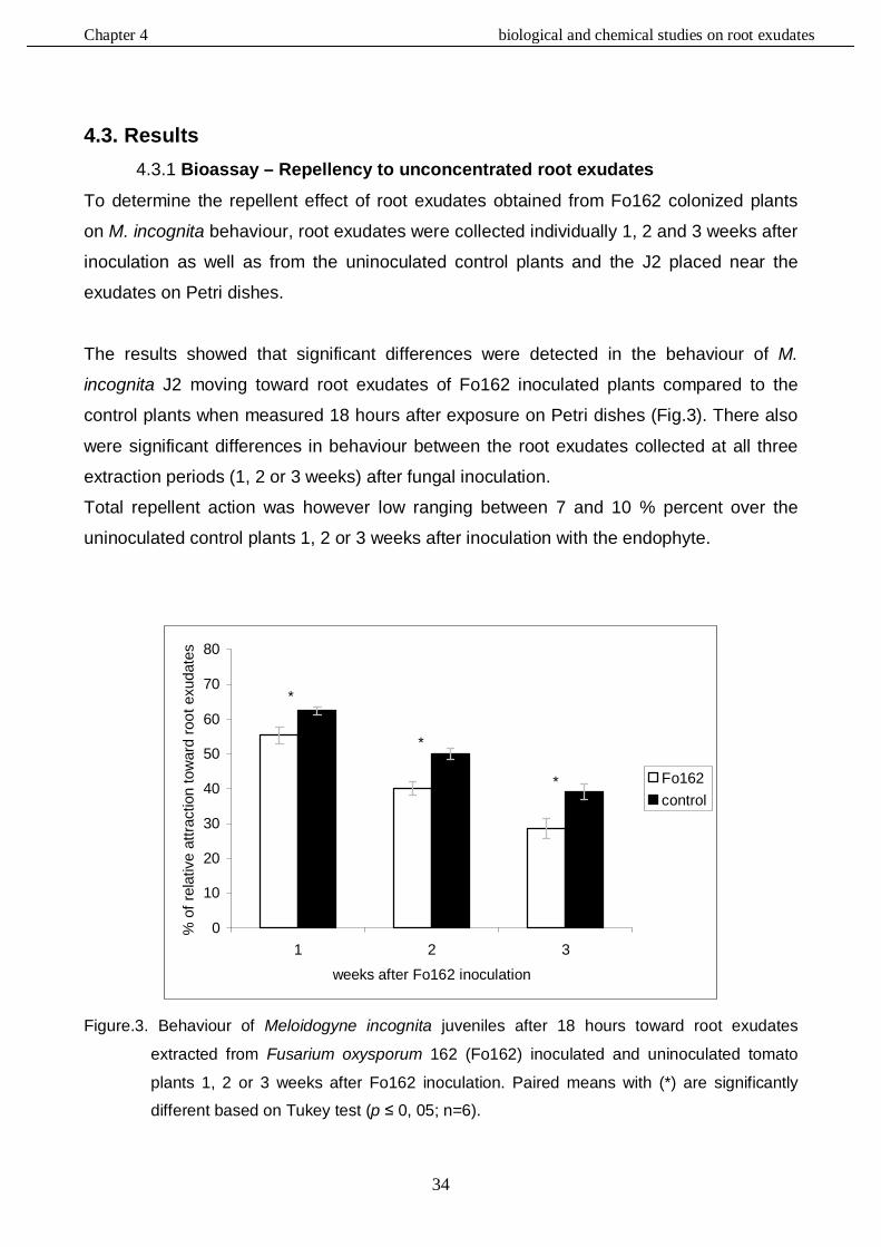
Chapter 4 biological and chemical studies on root exudates
34
4.3. Results 4.3.1 Bioassay – Repellency to unconcentrated root exudates
To determine the repellent effect of root exudates obtained from Fo162 colonized plants
on M. incognita behaviour, root exudates were collected individually 1, 2 and 3 weeks after
inoculation as well as from the uninoculated control plants and the J2 placed near the
exudates on Petri dishes.
The results showed that significant differences were detected in the behaviour of M.
incognita J2 moving toward root exudates of Fo162 inoculated plants compared to the
control plants when measured 18 hours after exposure on Petri dishes (Fig.3). There also
were significant differences in behaviour between the root exudates collected at all three
extraction periods (1, 2 or 3 weeks) after fungal inoculation.
Total repellent action was however low ranging between 7 and 10 % percent over the
uninoculated control plants 1, 2 or 3 weeks after inoculation with the endophyte.
*
*
*
0
10
20
30
40
50
60
70
80
1 2 3
weeks after Fo162 inoculation
% o
f rel
ativ
e at
tract
ion
tow
ard
root
exu
date
s
Fo162control
Figure.3. Behaviour of Meloidogyne incognita juveniles after 18 hours toward root exudates
extracted from Fusarium oxysporum 162 (Fo162) inoculated and uninoculated tomato
plants 1, 2 or 3 weeks after Fo162 inoculation. Paired means with (*) are significantly
different based on Tukey test (p ≤ 0, 05; n=6).
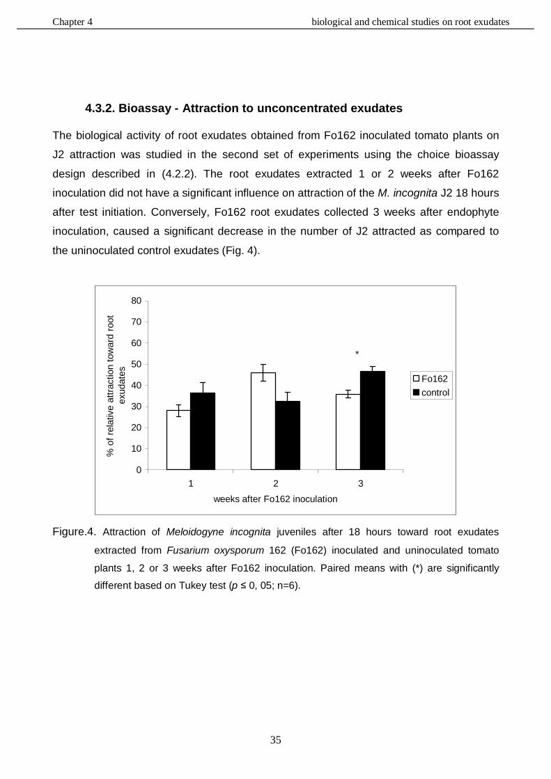
Chapter 4 biological and chemical studies on root exudates
35
4.3.2. Bioassay - Attraction to unconcentrated exudates The biological activity of root exudates obtained from Fo162 inoculated tomato plants on
J2 attraction was studied in the second set of experiments using the choice bioassay
design described in (4.2.2). The root exudates extracted 1 or 2 weeks after Fo162
inoculation did not have a significant influence on attraction of the M. incognita J2 18 hours
after test initiation. Conversely, Fo162 root exudates collected 3 weeks after endophyte
inoculation, caused a significant decrease in the number of J2 attracted as compared to
the uninoculated control exudates (Fig. 4).
*
0
10
20
30
40
50
60
70
80
1 2 3
weeks after Fo162 inoculation
% o
f rel
ativ
e at
tract
ion
tow
ard
root
ex
udat
es Fo162control
Figure.4. Attraction of Meloidogyne incognita juveniles after 18 hours toward root exudates
extracted from Fusarium oxysporum 162 (Fo162) inoculated and uninoculated tomato
plants 1, 2 or 3 weeks after Fo162 inoculation. Paired means with (*) are significantly
different based on Tukey test (p ≤ 0, 05; n=6).
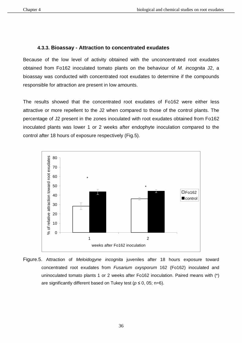
Chapter 4 biological and chemical studies on root exudates
36
4.3.3. Bioassay - Attraction to concentrated exudates Because of the low level of activity obtained with the unconcentrated root exudates
obtained from Fo162 inoculated tomato plants on the behaviour of M. incognita J2, a
bioassay was conducted with concentrated root exudates to determine if the compounds
responsible for attraction are present in low amounts.
The results showed that the concentrated root exudates of Fo162 were either less
attractive or more repellent to the J2 when compared to those of the control plants. The
percentage of J2 present in the zones inoculated with root exudates obtained from Fo162
inoculated plants was lower 1 or 2 weeks after endophyte inoculation compared to the
control after 18 hours of exposure respectively (Fig.5).
*
*
0
10
20
30
40
50
60
70
80
1 2
weeks after Fo162 inoculation
% o
f rel
ativ
e at
tract
ion
tow
ard
root
exu
date
s
Fo162control
Figure.5. Attraction of Meloidogyne incognita juveniles after 18 hours exposure toward
concentrated root exudates from Fusarium oxysporum 162 (Fo162) inoculated and
uninoculated tomato plants 1 or 2 weeks after Fo162 inoculation. Paired means with (*)
are significantly different based on Tukey test (p ≤ 0, 05; n=6).
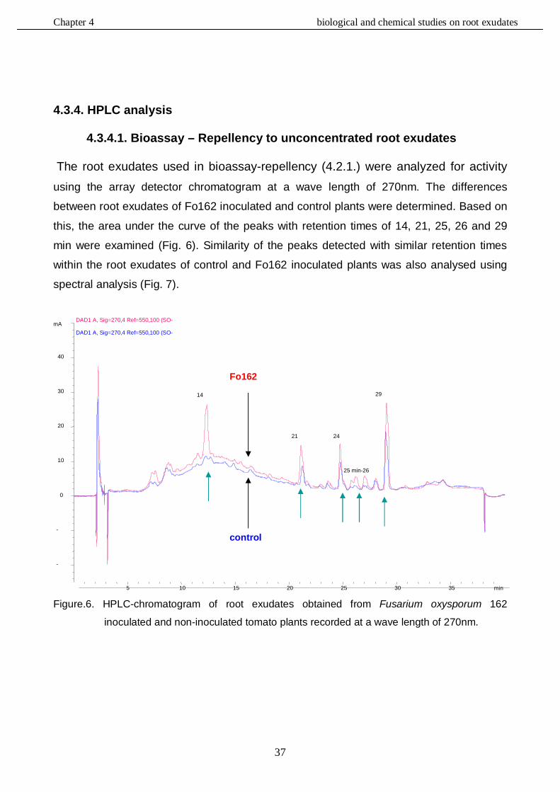
Chapter 4 biological and chemical studies on root exudates
37
4.3.4. HPLC analysis
4.3.4.1. Bioassay – Repellency to unconcentrated root exudates
The root exudates used in bioassay-repellency (4.2.1.) were analyzed for activity using the array detector chromatogram at a wave length of 270nm. The differences
between root exudates of Fo162 inoculated and control plants were determined. Based on
this, the area under the curve of the peaks with retention times of 14, 21, 25, 26 and 29
min were examined (Fig. 6). Similarity of the peaks detected with similar retention times
within the root exudates of control and Fo162 inoculated plants was also analysed using
spectral analysis (Fig. 7).
Figure.6. HPLC-chromatogram of root exudates obtained from Fusarium oxysporum 162
inoculated and non-inoculated tomato plants recorded at a wave length of 270nm.
min 5 10 15 20 25 30 35
mAU
-20
-10
0
10
20
30
40
DAD1 A, Sig=270,4 Ref=550,100 (SO-V113W\SELIM026.D)
DAD1 A, Sig=270,4 Ref=550,100 (SO-
14
21 min
24 min
25 min-26 min
29 min
Fo162
control
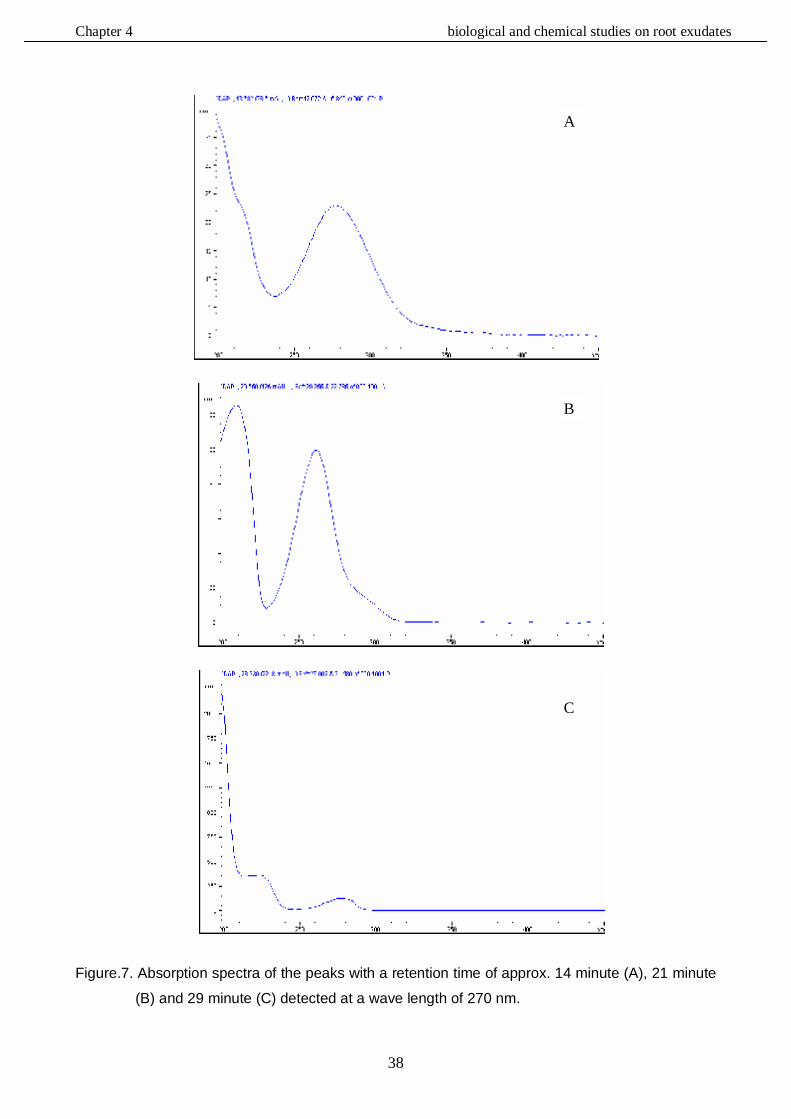
Chapter 4 biological and chemical studies on root exudates
38
Figure.7. Absorption spectra of the peaks with a retention time of approx. 14 minute (A), 21 minute
(B) and 29 minute (C) detected at a wave length of 270 nm.
A
B
C
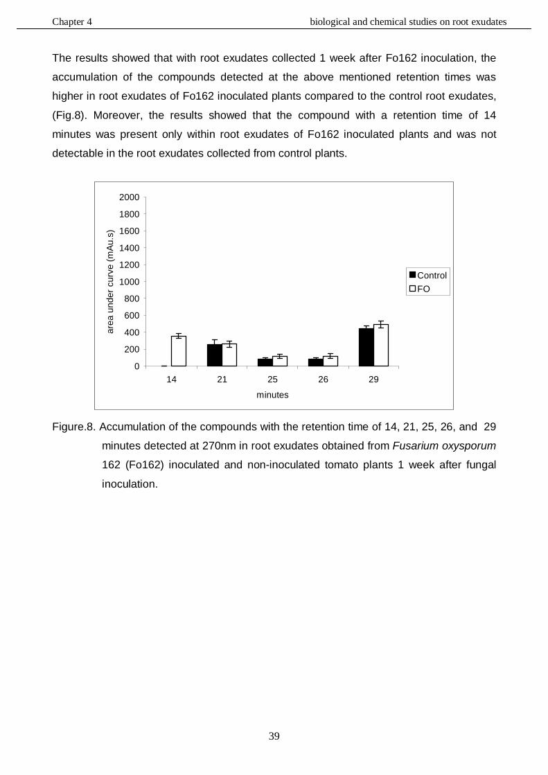
Chapter 4 biological and chemical studies on root exudates
39
The results showed that with root exudates collected 1 week after Fo162 inoculation, the
accumulation of the compounds detected at the above mentioned retention times was
higher in root exudates of Fo162 inoculated plants compared to the control root exudates,
(Fig.8). Moreover, the results showed that the compound with a retention time of 14
minutes was present only within root exudates of Fo162 inoculated plants and was not
detectable in the root exudates collected from control plants.
0
200
400
600
800
1000
1200
1400
1600
1800
2000
14 21 25 26 29
minutes
area
und
er c
urve
(mAu
.s)
ControlFO
Figure.8. Accumulation of the compounds with the retention time of 14, 21, 25, 26, and 29
minutes detected at 270nm in root exudates obtained from Fusarium oxysporum
162 (Fo162) inoculated and non-inoculated tomato plants 1 week after fungal
inoculation.
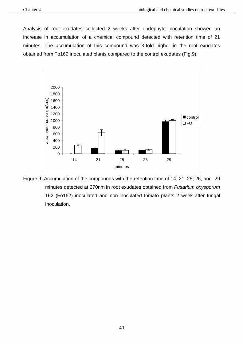
Chapter 4 biological and chemical studies on root exudates
40
Analysis of root exudates collected 2 weeks after endophyte inoculation showed an
increase in accumulation of a chemical compound detected with retention time of 21
minutes. The accumulation of this compound was 3-fold higher in the root exudates
obtained from Fo162 inoculated plants compared to the control exudates (Fig.9).
0200400600800
100012001400160018002000
14 21 25 26 29
minutes
area
und
er c
urve
(mA
u.s)
controlFO
Figure.9. Accumulation of the compounds with the retention time of 14, 21, 25, 26, and 29
minutes detected at 270nm in root exudates obtained from Fusarium oxysporum
162 (Fo162) inoculated and non-inoculated tomato plants 2 week after fungal
inoculation.
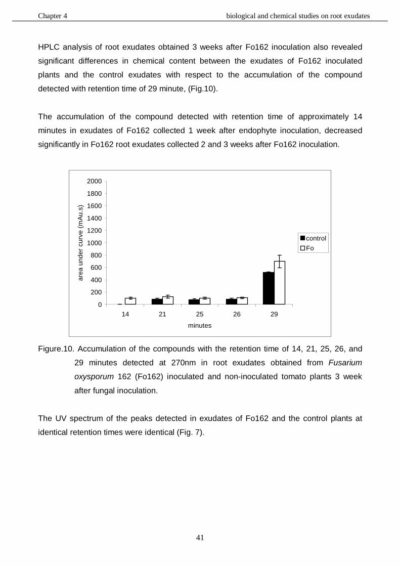
Chapter 4 biological and chemical studies on root exudates
41
HPLC analysis of root exudates obtained 3 weeks after Fo162 inoculation also revealed
significant differences in chemical content between the exudates of Fo162 inoculated
plants and the control exudates with respect to the accumulation of the compound
detected with retention time of 29 minute, (Fig.10).
The accumulation of the compound detected with retention time of approximately 14
minutes in exudates of Fo162 collected 1 week after endophyte inoculation, decreased
significantly in Fo162 root exudates collected 2 and 3 weeks after Fo162 inoculation.
0
200
400
600
800
1000
1200
1400
1600
1800
2000
14 21 25 26 29
minutes
area
und
er c
urve
(mAu
.s)
controlFo
Figure.10. Accumulation of the compounds with the retention time of 14, 21, 25, 26, and
29 minutes detected at 270nm in root exudates obtained from Fusarium
oxysporum 162 (Fo162) inoculated and non-inoculated tomato plants 3 week
after fungal inoculation.
The UV spectrum of the peaks detected in exudates of Fo162 and the control plants at
identical retention times were identical (Fig. 7).
Chapter 4 biological and chemical studies on root exudates
42
4.3.4.2. Bioassay – Attraction to unconcentrated and concentrated exudates
The results obtained from the chromatogram analysis of root exudates collected from
Fo162 inoculated and uninoculated tomato plants 1 week after fungal inoculation and used
in the behavioural bioassay 4.2.2 and 4.2.3, showed a higher level of accumulation of
compounds detected at a wave length of 270nm and retention times of 21, 25 and 26
minutes in Fo162 root exudates compared to the control (Fig.11).
The results also demonstrated that the peak with a retention time of 14 minutes which was
detected in the exudates used in the bioassay for repellency was again detectable only in
root exudates taken from Fo162 inoculated plants. .
0
200
400
600
800
1000
1200
1400
1600
1800
2000
14 21 25 26 29
minutes
area
und
er c
urve
(mAu
.s)
controlFO
Figure.11. Accumulation of the compounds with the retention time of 14, 21, 25, 26, and
29 minutes detected at 270nm in root exudates obtained from Fusarium
oxysporum 162 (Fo162) inoculated and non-inoculated tomato plants 1 week
after fungal inoculation.
Chapter 4 biological and chemical studies on root exudates
43
Similar results were observed again in root exudates collected 2 weeks after Fo162
inoculation. The area under the curve of the peaks with retention times of 21, 25, 26 and
29 minutes was larger in the root exudates of Fo162 inoculated plants than in the control
exudates (Fig.12). Furthermore, the compound detected with retention time of 14 minutes
was only detected in root exudates of Fo162 inoculated plants.
0
200
400
600
800
1000
1200
1400
1600
1800
2000
14 21 25 26 29
minutes
area
und
er c
urve
(mAu
.s)
controlFO
Figure.12. Accumulation of the compounds with the retention time of 14, 21, 25, 26, and
29 minutes detected at 270nm in root exudates obtained from Fusarium
oxysporum 162 (Fo162) inoculated and non-inoculated tomato plants 2 weeks
after fungal inoculation.
Chapter 4 biological and chemical studies on root exudates
44
Results obtained from root exudates collected 3 weeks after Fo162 inoculation were
similar to those obtained from root exudates collected 1 and 2 weeks after fungal
inoculation. The differences in the area under curve of the peaks with retention times of
25, 26 and 29 minutes were detected again between the Fo162 exudates and the control
exudates as in the previous experiment on repellency (Fig.13).
0
200
400
600
800
1000
1200
1400
1600
1800
2000
14 21 25 26 29
minutes
area
und
er c
urve
(mAu
.s)
controlFO
Figure.13. Accumulation of the compounds with the retention time of 14, 21, 25, 26, and
29 minutes detected at 270nm in root exudates obtained from Fusarium
oxysporum 162 (Fo162) inoculated and non-inoculated tomato plants 3 week
after fungal inoculation.
Chapter 4 biological and chemical studies on root exudates
45
4.4. Discussion 4.4.1. Bioassay - Repellency to unconcentrated root exudates The fact that endophytic fungi are able to produce large amounts of toxins in vitro indicates
that they may also exude such compounds into the soil when growing in-planta (Sikora et
al., 2008). In 1996, Hallmann and Sikora reported that the secondary metabolites released
from the non-pathogenic Fusarium oxysporum strain 162 resulted in 80% inactivation of
Meloidogyne incognita juveniles when nematodes were exposed for 30 minutes to these
metabolites. Vu (2005) investigate and tested the effects of inoculated banana with four
isolates of F. oxysporum on the attraction of Radopholus similis using a linked twin-pot
attraction chamber. She showed that the attractiveness of the banana roots was
significantly reduced when the roots were colonized with the endophytic isolates tested.
She did not determine if the effect was due to attraction or repellent activity of the roots nor
did she examine if the effects were due to changes in root exudates. Similar effects on
root-knot behaviour were detected in choice tests with tomato inoculated with F.
oxysporum isolate 162 in the studies of Dababat, (2007) and Dababat and Sikora, (2007).
They demonstrated that 56% of M. incognita juveniles failed to penetrate tomato plants
pre-inoculated with the endophyte.
In the present study, an in vitro bioassay was used in order to determine changes in M.
incognita behaviour induced by exposure to root exudates collected from the rhizosphere
of tomato plants colonized by the same Fo162 isolate.
The results demonstrated that after 18 hours exposure, root exudates obtained from
Fo162 colonized tomato plants 1, 2 or 3 weeks after inoculation, had a significant but low
repellent effect on M. incognita J2 compared to the control exudates. These results further
demonstrated that Fo162 colonization altered proprieties of host plant root exudates and
exerted a certain degree of repellent activity toward root-knot juveniles in vitro.
4.4.2. Bioassay - Attraction to unconcentrated exudates Because of the low level of repellency detected in the previous test, a choice bioassay test
was conducted to detect the attractiveness of root exudates to root-knot J2 obtained from
Fo162 inoculated plants verses the uninoculated controls. The results showed that with
unconcentrated root exudates, significant differences in attractiveness between the root
Chapter 4 biological and chemical studies on root exudates
46
exudates of Fo162 and the control were recorded within exudates collected from plants 3
weeks after Fo162 inoculation. Root exudates obtained 1 or 2 weeks after endophyte
inoculation did not have a significant affect on the J2.
The lack in detection of activity of root exudates obtained from Fo162 inoculated tomato
plants on the behaviour of M. incognita J2 with this choice bioassay could be due to the
high experimental variation in the tests or to low concentrations of the substances
responsible for attraction recovered from the rhizosphere soil.
Bargmann (2006) showed that the orientation of Caenorhabditis elegans (a mechanism
called the pirouette model in C. elegans by Piece-Shimomura et. al., 1999) is quit similar to
the orientation of bacteria during chemotaxis (based random walk by Berg 1993 and Berg
1975). Pierce-Shimomura et al., (2005) demonstrated that C. elegans can not sense a
spatial gradient over its body to decide the direction to turn. Therefore, random movement
behaviour of J2 under the test conditions used in the bioassay could be responsible also
for lack of significant results of attraction and/or repellency of J2 to the exudates of Fo162
inoculated and non inoculated tomato plants.
Conversely, choice experiments conducted by Dababat and Sikora (2007) with sand block
champers demonstrated that tomato root exudates obtained from plants colonised by
Fo162 were either less attractive to M. incognita juveniles. They showed that 80% of root-
knot nematode juveniles placed in the center of a sand-filled chamber moved away from
the arm treated with root exudates obtained from tomato plants inoculated with Fo162 to
the side of the chamber inoculated with root exudates of control plants.
4.4.3. Bioassay - Attraction to concentrated exudates In order to determine if the exudates used in the above tests contained low levels of
attractants due to biotic or abiotic interference a best was conducted in which the exudates
were concentrated. Results of the bioassay using the root exudates concentrated through
ethyl acetate extraction showed that Fo162 root exudates collected 1 or 2 weeks after
fugal inoculation were more less attractive to the M. incognita J2 compared to the control
exudates. The results illustrated that the concentrations of the chemical compounds
accumulated in Fo162 root exudates play a role in the attraction of root-knot nematode
and that the amounts vary between experiments. The variation in concentration probably
due to levels of Fo162 colonization or to abiotic and biotic interactions present in the
greenhouse.
Chapter 4 biological and chemical studies on root exudates
47
Similar results were reported from the studies of Pierce-Shimomura et al., (1999) who
mentioned that C. elegans does not respond to the absolute level of attractants but to
changes in attractants concentration over time.
4.4.4. HPLC analysis of root exudates
HPLC analysis of the biologically active compounds in root exudates of Fo162 inoculated
tomato plants which may have been responsible for changes in nematode orientation
behaviour was conducted. Chromatogram analysis performed at a wave length of 270nm
was the most informative chromatogram and revealed differences between root exudates
of Fo162 inoculated and uninoculated plants.
Results showed that there was a higher level of accumulation of compounds within the
root exudates collected from Fo162 inoculated plants compared to exudates of control.
The results also showed that Fo162 stimulated accumulation of a new compound which
was detected at 270nm and a retention time of 14 min. The accumulation of this
compound decreased in root exudates collected 2 or 3 weeks after Fo162 inoculation.
There also was a correlation between accumulation of the compounds and the biological
activity of root exudates with respect to nematode behaviour. In the first experiment
(bioassay-repellency, 4.2.1), the maximum accumulation of all compounds as well as the
highest level of J2 repellency from Fo162 root exudates was detected within the exudates
obtained from 2 week old Fo162 inoculated plants.
Similar results were obtained when the experiment was repeated (bioassays-attraction to
concentrated exudates, 4.2.3). Thus the highest levels of compound accumulation and the
lowest level of J2 attraction were recorded with concentred root exudates collected 1 week
after Fo162 inoculation.
The analysis of the spectra patterns of the detected compounds in the exudates of Fo162
inoculated and uninoculated plants showed that the detected compounds at the same
retention times were identical.
However, the chemical signals released from host plant root exudates have been shown
previously to play a role in nematode infection processes.
Chapter 4 biological and chemical studies on root exudates
48
Pline and Dusenbery (1987) demonstrated that, long distance attraction of nematodes to
roots is, in general, based on non-specific chemical signals. Wuyts et al., (2006) reported
that plant chemicals released from roots in the rhizosphere can influence nematode
behaviour. These chemicals can either attract nematodes to penetration sites or result in
repellence, inhibition of mobility or even death. They also reported that the effect of plant
metabolites on nematode behaviour can still be detectable after nematodes penetration.
The mechanisms by which compounds in root exudates are active against plant-parasitic
nematodes are largely unknown. Hallmann and Sikora (1996) suggested that inhibitory
compounds produced by the endophyte and transferred into the roots might be involved in
alteration of root exudates pattern. This hypothesis has never been tested for validity.
4.5 Conclusions
Based on the present investigations the following can be concluded:
1- Root exudates of tomato plants colonized by Fo162 affected the behaviour and
migration pattern of M. incognita J2 when nematodes were exposed for 18 hours to
these exudates.
2- Low levels of repellency verses high levels of lack of attraction were detected
3- Fo162 colonization resulted in increased accumulation of several different chemical
compounds in the root exudates of tomato plants.
4- One or more of these compounds could be responsible for lack of J2 attraction.
5- There is a correlation between the accumulation levels of chemical compounds in root
exudates of Fo162 and biological activity of these exudates toward nematode.
6- Further studies have to be conducted to purify the detected compounds accumulating
within root exudates of tomato plants colonized by Fo162.
Chapter 4 biological and chemical studies on root exudates
49
4.6. References
- Bargmann, C. I. (2006). Chemosensation in C. elegans. (October 25, 2006),
wormBook, ed. The C. elegans Research Community, WormBook,
doi/10.1895/wormbook.1.123.1,http://www.wormbook.org.
- Berg, H. C. (1975). Chemotaxis in bacteria. Annu. Rev. Biophys. Bioeng. 4, 119-
136. Abstract Article.
- Berg, H. C. (1993). Random walks in biology (Princeton, N. J. Princeton University
Press).
- Dababat, A. A. (2007). Importance of the mutualistic Endophyte Fusarium
oxysporum 162 for enhancement of tomato transplants and the biological control of
the root-knot nematode Meloidogyne incognita, with Particular reference to mode-
of-action. Ph.D. Thesis. University of Bonn, Germany.
- Dababat, A. A. and Sikora, R. A. (2007). Influence of the mutualistic endophyte
Fusarium oxysporum 162 on the Meloidogyne incognita attraction and invasion.
Nematology, 2007, vol. 9(6), 771-776.
- Hallmann, J. and Sikora, R. A. (1994). Influence of Fusarium oxysporum, a
mutualistic fungal endophyte on Meloidogyne incognita infection of tomato. Journal
of plant disease and protection 101: 475-481.
- Hallmann, J. and Sikora, R. A. (1996). Toxicity of fungal endophyte secondary
metabolites to plant parasitic nematodes and soil-borne plant pathogenic fungi.
European journal of plant pathology 102, 155-162.
- Kimenju, J. W., Wando, S. W., Mwang’ Ombe, A. W., Sikora, R. A., and Schurter, R.
P. (1998). Distribution of lesion nematodes associated with maize in Kenya and
susceptibility of maize cultivars to Pratylechus zeae. Afr. Crop Sci. J. 6, 367-375.
- Le, T. T. H. (2006). Antagonistic potential of endophytic and rhizosphere fungi
against the rice root-knot nematode Meloidogyne graminicola under upland rice
growing conditions. Master thesis, University of Bonn, Germany.
- Padgam, J. and Sikora, R. A. (2006). Biological control potential and modes of
action of Bacillus megaterium against Meloidogyne graminicola on rice. Crop
protection 26, 971-977.
- Perry, R. N. (1997). Plant signals in nematode hatching and attraction. In: Fenoll,
C., Grundler, F. M. W., & Ohl, S .A. (Eds.). Celler and molecular aspects of plant-
nematode interaction. Dorrecht, the Netherlands, Kluwer Acadamic Publishers,
pp.38-50.
Chapter 4 biological and chemical studies on root exudates
50
- Pierce-Shimomura, J. T., Dores, M., and Lockery, S. R. (2005). Analysis of the
effects of turning bias on chemotaxis in C. elegans. J. Exp. Biol. 208, 4727-4733.
Abstract Article.
- Pierce-Shimomura, J. T., Morse, T. M., and Lockery, S. R. (1999). The role of
pirouettes in Caenorhabditis elegans chemotaxis. J. Neurosci. 19, 9557-9569.
Abstract.
- Pline, M. and Dunsenbery, D. B. (1987). Response of plant parasitic nematode
Meloidogyne incognita to carbon dioxide determined by video camera-computer
tracking. Journal of chemical ecology 13, 873-888.
- Reimann, S., Hauschild, R., Hildebrandt, U. and Sikora, R. A. (2008).
Interrelationships between Rhizobium etli G12 and Glomus intraradices and
multitrophic effects in the biological control of the root-knot nematode Meloidogyne
incognita on tomato. Journal of Plant Diseases and Protection, 115 (3), 108–113.
- Reitz, M., Rudolph, K., Schröder, I., Hoffmann-Hergarten, S., Hallmann, J. and
Sikora, R. A. (2000). Lipopolysaccharides of Rhizobium etli strain G12 act in potato
roots as an inducing agent of systemic resistance to infection by the cyst nematode
Globodera pallida. Applied and environmental microbiology, p. 3515-3518.
- Sikora, R. A., Pocasangre, L., zum Felde, A., Niere, B., Vu, T. T., and Dababat, A.
A. (2008). Mutualistic endophytic fungi and in-planta suppressiveness to plant
parasitic nematodes. Biological Control (46), 15-23.
- Vu, T. T. (2005). Mode of action of non-pathogenic Fusarium oxysporum
endophytes for bio-enhancement of banana toward Radopholus similis. Ph.D.
Thesis, University of Bonn, Germany.
- Vu, T. T., Sikora, R. A. and Hauschild, R. (2004). Effect of endophytic Fusarium
oxysporum towards Radopholus similis activity in absence of banana. Comm. Appl.
Sci., Ghent University, 69/3, 381-385.
- Ward, S. (1973). Chemotaxis by the nematode Caenorhabditis elegans:
identification of attractants and analysis of the response by use of mutants. Proc.
Natl. Acad. Sci. USA. 70, 817-821. Abstract Article.
- Wuyts, N., Swennen, R. and De Waele, D. (2006). Effects of plant phenylpropanoid
pathway products and selected terpenoids and alkaloids on the behaviour of the
plant-parasitic nematodes Radopholus similis, Pratylenchus penetrans and
Meloidogyne incognita. Nematology, vol. 8(1), 89-101. - Zum Felde, A., Pocasangre, A. L. and Sikora, R. A. (2005). The potential use of microbial
communities inside suppressive banana plants for banana root protection. In: Turner, D. W., and Rosales, F. E. (Eds.). In Banana root system: toward a better understanding for its productive management. Proceedings of an international symposium. International network for the improvement of banana and plantain (INIBAP), pp. 169-177. Montpellier, France.
Chapter5 Durability of Fo162 protection spectrum
51
5. Fusarium oxysporum 162 colonization behaviour in tomato plants
and its impact on the durability of induced resistance toward Meloidogyne incognita
5.1. Introduction Previous studies (chapter 3) showed that the mutualistic endophyte Fusarium oxysporum
strain 162 (Fo162) can colonize roots of Fusarium-wilt susceptible and resistant tomato
cultivars. The Fo162 fungus has also been shown to induce the systemic defences of the
Fusarium-wilt susceptible tomato cultivars against root-knot nematode (Dababat and
Sikora, 2007a). However, in all previous studies the Fo162 stimulated systemic resistance
mode of action was not determined in Fusarium-wilt resistant tomato cultivars against
Meloidogyne incognita before.
Earlier investigations on the ability of Fo162 to colonize the plant were limited to the root
system and assumed that Fo162 remains in the root systems and does not move upward
into the shoots and leaves (Hallmann and Sikora 1994; Dababat and Sikora 2007).
Whether or not Fo162 also colonizes aboveground stems and adventitious roots growing
from the stem base and prolongs the duration of induced resistance has not been
evaluated. Shoot colonization if influential in the induction process would open up
additional ways of monitoring the durability of the systemic responses of endophytes such
as Fo162 in plants.
Therefore in the present study, the shoots were physically detached from the roots of
Fo162 pre-inoculated tomato plants. After the newly formed adventitious roots had
developed, the presence of Fo162 inside the shoots and the roots was determined and the
colonization of M. incognita in these newly formed adventitious roots was followed over
time.
The stem detachment technique which was used in this study was used before by Fuchs
et al., (1997) to determine the ability of another non pathogenic Fusarium oxysporum
strain (Fo47) to induce resistance to the Fusarium-wilt pathogen in tomato plants. They
reported that the initial inoculation of Fo47 into rock wool, which was used to grow the
tomato plants before detachment, reduced the incidence of Fusarium wilt from 38 to 20%
in the adventitious roots of the tomato stems transplanted later into potting mix inoculated
Chapter5 Durability of Fo162 protection spectrum
52
with the pathogenic isolate of F. oxysporum (Fo18) .They also reported that similar results
were obtained with three different bioassays which were conducted to investigate whether
the non pathogenic isolate Fo47 was able to induce resistance against an isolate of the
Fusarium wilt pathogen (Fo18) when they were introduced separately in time to the
challenge. However the analysis did not determine the presence of Fo47 inside the plants,
nor did they consider wound induced responses.
The objectives of this study were to determine the:
1- Capability of Fo162 to colonize the shoots and the newly formed shoot adventitious
roots of the primary Fo162 inoculated tomato cultivars.
2- Ability of Fo162 to induce the systemic defences in detached shoots of Fusarium-
wilt susceptible and resistant tomato cultivars against M. incognita.
3- Durability of the signals related to the mutualistic endophyte F. oxysporum (Fo162)
systemic resistance in detached shoots of both tomato cultivars.
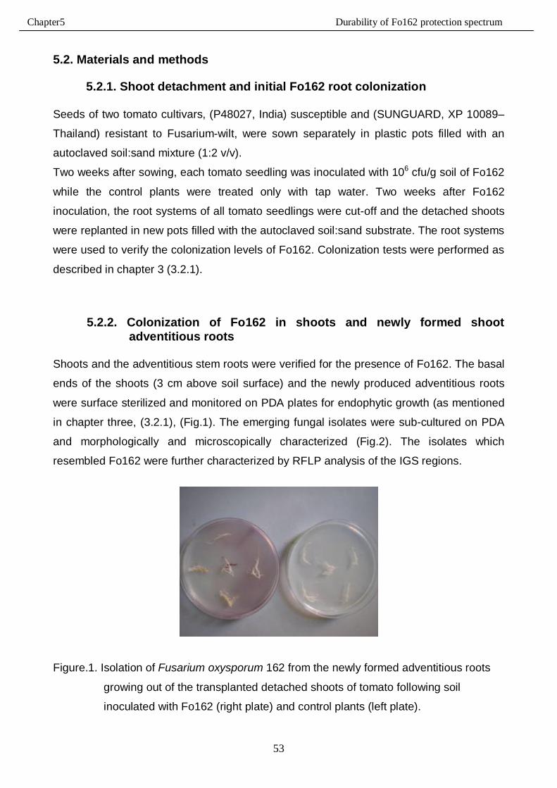
Chapter5 Durability of Fo162 protection spectrum
53
5.2. Materials and methods
5.2.1. Shoot detachment and initial Fo162 root colonization
Seeds of two tomato cultivars, (P48027, India) susceptible and (SUNGUARD, XP 10089–
Thailand) resistant to Fusarium-wilt, were sown separately in plastic pots filled with an
autoclaved soil:sand mixture (1:2 v/v).
Two weeks after sowing, each tomato seedling was inoculated with 106 cfu/g soil of Fo162
while the control plants were treated only with tap water. Two weeks after Fo162
inoculation, the root systems of all tomato seedlings were cut-off and the detached shoots
were replanted in new pots filled with the autoclaved soil:sand substrate. The root systems
were used to verify the colonization levels of Fo162. Colonization tests were performed as
described in chapter 3 (3.2.1).
5.2.2. Colonization of Fo162 in shoots and newly formed shoot adventitious roots
Shoots and the adventitious stem roots were verified for the presence of Fo162. The basal
ends of the shoots (3 cm above soil surface) and the newly produced adventitious roots
were surface sterilized and monitored on PDA plates for endophytic growth (as mentioned
in chapter three, (3.2.1), (Fig.1). The emerging fungal isolates were sub-cultured on PDA
and morphologically and microscopically characterized (Fig.2). The isolates which
resembled Fo162 were further characterized by RFLP analysis of the IGS regions.
Figure.1. Isolation of Fusarium oxysporum 162 from the newly formed adventitious roots
growing out of the transplanted detached shoots of tomato following soil
inoculated with Fo162 (right plate) and control plants (left plate).
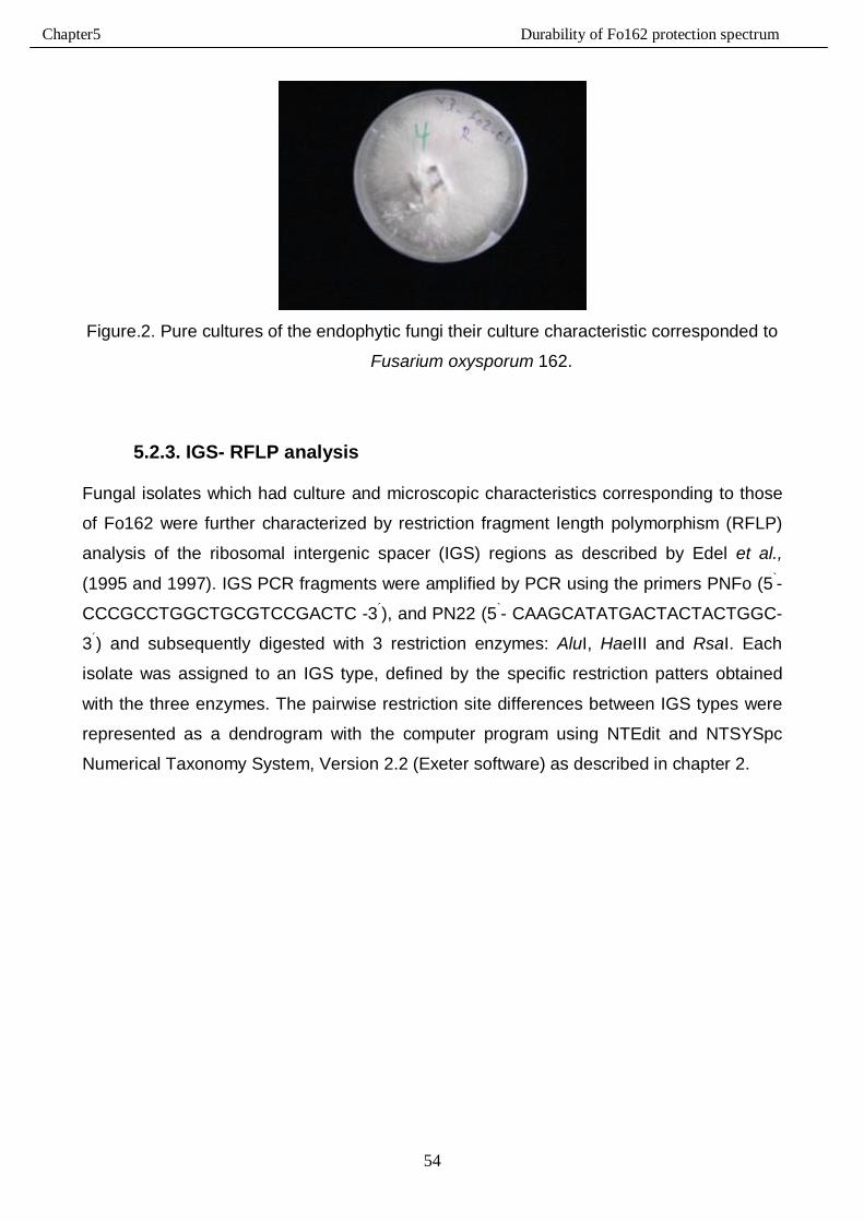
Chapter5 Durability of Fo162 protection spectrum
54
Figure.2. Pure cultures of the endophytic fungi their culture characteristic corresponded to
Fusarium oxysporum 162.
5.2.3. IGS- RFLP analysis Fungal isolates which had culture and microscopic characteristics corresponding to those
of Fo162 were further characterized by restriction fragment length polymorphism (RFLP)
analysis of the ribosomal intergenic spacer (IGS) regions as described by Edel et al.,
(1995 and 1997). IGS PCR fragments were amplified by PCR using the primers PNFo (5`-
CCCGCCTGGCTGCGTCCGACTC -3´), and PN22 (5`- CAAGCATATGACTACTACTGGC-
3´) and subsequently digested with 3 restriction enzymes: AluI, HaeIII and RsaI. Each
isolate was assigned to an IGS type, defined by the specific restriction patters obtained
with the three enzymes. The pairwise restriction site differences between IGS types were
represented as a dendrogram with the computer program using NTEdit and NTSYSpc
Numerical Taxonomy System, Version 2.2 (Exeter software) as described in chapter 2.
Chapter5 Durability of Fo162 protection spectrum
55
5.2.4. Durability of systemic resistance signals in detached shoots and adventitious roots
In order to determine the durability of the signals of Fo162 induced resistance in tomato
plants toward the root-knot nematode a detached shoot bioassay was developed. The
hypothesis was that once the signals moved from the original root system into the shoot,
the shoot of the plant would retain the ability to induce resistance basal pedal into the
newly formed adventitious root system as seen in split-root tests (Dababat and Sikora
2007). Therefore, the detached shoots of 6 plants, which were initially inoculated with
Fo162 and 6 plants of the non-inoculated control, were inoculated separately with 1000
juveniles (J2) of M. incognita (as mentioned in chapter 2). The adventitious roots were
inoculated with J2, 5, 7 or 10 days after transplanting the detached shoots into the
autoclaved soil.
The plants developing out of the stems were grown under greenhouse conditions at 25 ± 5
°C and fertilized by NPK (14:10:14) with concentration of 3g/L one time per week.
Two weeks after nematode inoculation, the tomato plants were removed from the soil and
the newly formed adventitious roots were washed gently with tap water and blotted
between tissue paper. The root weight was recorded and the number of galls was counted
for each tomato plant.
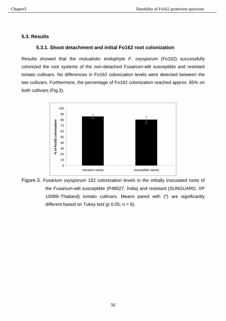
Chapter5 Durability of Fo162 protection spectrum
56
5.3. Results
5.3.1. Shoot detachment and initial Fo162 root colonization Results showed that the mutualistic endophyte F. oxysporum (Fo162) successfully
colonized the root systems of the non-detached Fusarium-wilt susceptible and resistant
tomato cultivars. No differences in Fo162 colonization levels were detected between the
two cultivars. Furthermore, the percentage of Fo162 colonization reached approx. 85% on
both cultivars (Fig.3).
0
10
20
30
40
50
60
70
80
90
100
resistant variety susceptible variety
% o
f Fo1
62 c
olon
izat
ion
Figure.3. Fusarium oxysporum 162 colonization levels in the initially inoculated roots of
the Fusarium-wilt susceptible (P48027, India) and resistant (SUNGUARD, XP
10089–Thailand) tomato cultivars. Means pared with (*) are significantly
different based on Tukey test (p 0.05; n = 6).
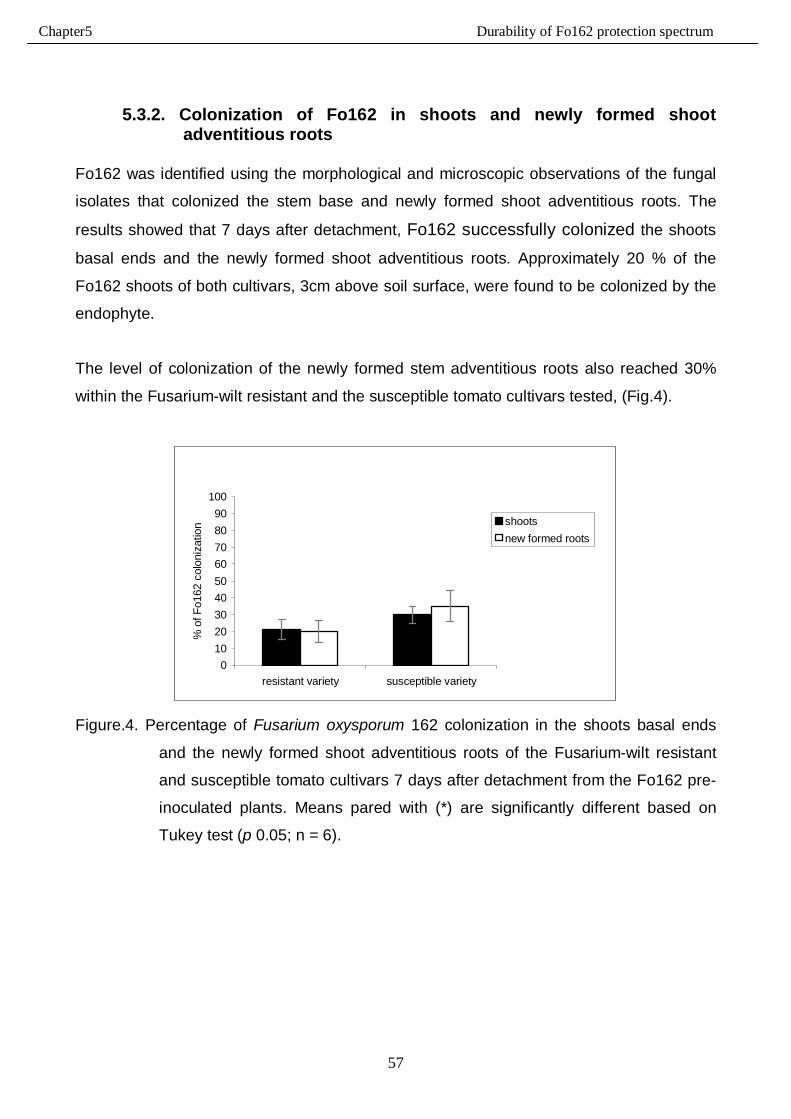
Chapter5 Durability of Fo162 protection spectrum
57
5.3.2. Colonization of Fo162 in shoots and newly formed shoot adventitious roots
Fo162 was identified using the morphological and microscopic observations of the fungal
isolates that colonized the stem base and newly formed shoot adventitious roots. The
results showed that 7 days after detachment, Fo162 successfully colonized the shoots
basal ends and the newly formed shoot adventitious roots. Approximately 20 % of the
Fo162 shoots of both cultivars, 3cm above soil surface, were found to be colonized by the
endophyte.
The level of colonization of the newly formed stem adventitious roots also reached 30%
within the Fusarium-wilt resistant and the susceptible tomato cultivars tested, (Fig.4).
0102030405060708090
100
resistant variety susceptible variety
% o
f Fo1
62 c
olon
izat
ion shoots
new formed roots
Figure.4. Percentage of Fusarium oxysporum 162 colonization in the shoots basal ends
and the newly formed shoot adventitious roots of the Fusarium-wilt resistant
and susceptible tomato cultivars 7 days after detachment from the Fo162 pre-
inoculated plants. Means pared with (*) are significantly different based on
Tukey test (p 0.05; n = 6).
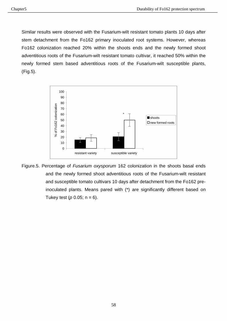
Chapter5 Durability of Fo162 protection spectrum
58
Similar results were observed with the Fusarium-wilt resistant tomato plants 10 days after
stem detachment from the Fo162 primary inoculated root systems. However, whereas
Fo162 colonization reached 20% within the shoots ends and the newly formed shoot
adventitious roots of the Fusarium-wilt resistant tomato cultivar, it reached 50% within the
newly formed stem based adventitious roots of the Fusarium-wilt susceptible plants,
(Fig.5).
*
0
10
20
30
40
50
60
70
80
90
100
resistant variety susceptible variety
% o
f Fo1
62 c
olon
izat
ion
shootsnew formed roots
Figure.5. Percentage of Fusarium oxysporum 162 colonization in the shoots basal ends
and the newly formed shoot adventitious roots of the Fusarium-wilt resistant
and susceptible tomato cultivars 10 days after detachment from the Fo162 pre-
inoculated plants. Means pared with (*) are significantly different based on
Tukey test (p 0.05; n = 6).
Chapter5 Durability of Fo162 protection spectrum
59
5.3.3. IGS-RFLP analysis
To confirm the identification of the isolates which had growth and microscopic
characteristics corresponding to those of Fo162 (5.3.2.), restriction fragment length
polymorphism (RFLP) analysis of the IGS PCR amplified fragments was carried out with
17 isolates obtained from the basal ends of the shoots and from the newly formed shoot
adventitious root segments, (Fig.6).
The results obtained verified that the genotype of only 4 isolates was identical with the
Fo162 genotype. The homogeneity between the rest of the isolates and Fo162 ranged
from 69 to 85%, (Fig.7).
These results revealed that not all fungal isolates that have been identified, in the 5.3.2.
colonization test, as Fo162 according to the morphological and microscopic examination,
represented the Fo162 endophyte fungal.
Therefore, the morphological and microscopically identification of those isolates as Fo162
was partially consistent with the molecular identification.
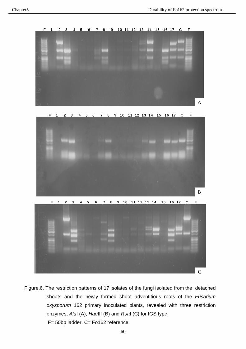
Chapter5 Durability of Fo162 protection spectrum
60
F 1 2 3 4 5 6 7 8 9 10 11 12 13 14 15 16 17 C FF 1 2 3 4 5 6 7 8 9 10 11 12 13 14 15 16 17 C F
F 1 2 3 4 5 6 7 8 9 10 11 12 13 14 15 16 17 C FF 1 2 3 4 5 6 7 8 9 10 11 12 13 14 15 16 17 C F
F 1 2 3 4 5 6 7 8 9 1 0 11 12 13 1 4 15 1 6 17 C FF 1 2 3 4 5 6 7 8 9 1 0 11 12 13 1 4 15 1 6 17 C F
Figure.6. The restriction patterns of 17 isolates of the fungi isolated from the detached
shoots and the newly formed shoot adventitious roots of the Fusarium
oxysporum 162 primary inoculated plants, revealed with three restriction
enzymes, AluI (A), HaeIII (B) and RsaI (C) for IGS type.
F= 50bp ladder. C= Fo162 reference.
C
A
B

Chapter5 Durability of Fo162 protection spectrum
61
Figure.7. Dendrogram of 17 isolates of the fungi isolated from the detached shoots and the
newly formed shoot adventitious roots of Fusarium oxysporum 162 primary
inoculated plants based on the restriction analysis of the IGS. Bars indicate
genetic the similarity [%] amongst taxa based on the Dice coefficient of
similarity. C= Fo162.
1126c
10
71512
348
169
514111713
1126c
10
71512
348
169
514111713
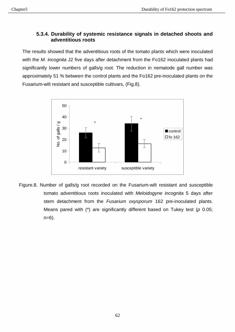
Chapter5 Durability of Fo162 protection spectrum
62
5.3.4. Durability of systemic resistance signals in detached shoots and adventitious roots
The results showed that the adventitious roots of the tomato plants which were inoculated
with the M. incognita J2 five days after detachment from the Fo162 inoculated plants had
significantly lower numbers of galls/g root. The reduction in nematode gall number was
approximately 51 % between the control plants and the Fo162 pre-inoculated plants on the
Fusarium-wilt resistant and susceptible cultivars, (Fig.8).
**
0
10
20
30
40
50
resistant variety susceptible variety
No.
of g
alls
/ g
controlfo 162
Figure.8. Number of galls/g root recorded on the Fusarium-wilt resistant and susceptible
tomato adventitious roots inoculated with Meloidogyne incognita 5 days after
stem detachment from the Fusarium oxysporum 162 pre-inoculated plants.
Means pared with (*) are significantly different based on Tukey test (p 0.05;
n=6).
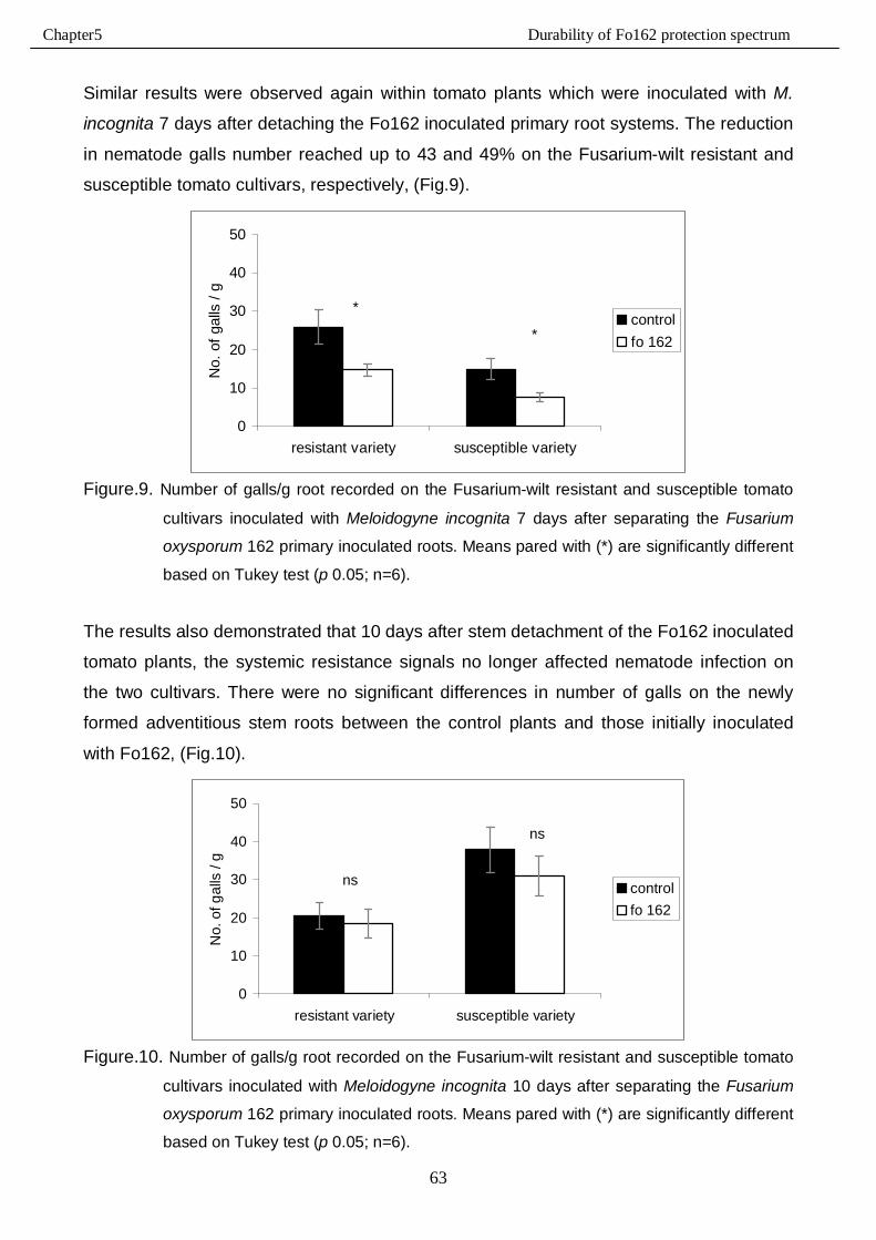
Chapter5 Durability of Fo162 protection spectrum
63
Similar results were observed again within tomato plants which were inoculated with M.
incognita 7 days after detaching the Fo162 inoculated primary root systems. The reduction
in nematode galls number reached up to 43 and 49% on the Fusarium-wilt resistant and
susceptible tomato cultivars, respectively, (Fig.9).
*
*
0
10
20
30
40
50
resistant variety susceptible variety
No.
of g
alls
/ g
controlfo 162
Figure.9. Number of galls/g root recorded on the Fusarium-wilt resistant and susceptible tomato
cultivars inoculated with Meloidogyne incognita 7 days after separating the Fusarium
oxysporum 162 primary inoculated roots. Means pared with (*) are significantly different
based on Tukey test (p 0.05; n=6).
The results also demonstrated that 10 days after stem detachment of the Fo162 inoculated
tomato plants, the systemic resistance signals no longer affected nematode infection on
the two cultivars. There were no significant differences in number of galls on the newly
formed adventitious stem roots between the control plants and those initially inoculated
with Fo162, (Fig.10).
ns
ns
0
10
20
30
40
50
resistant variety susceptible variety
No.
of g
alls
/ g
controlfo 162
Figure.10. Number of galls/g root recorded on the Fusarium-wilt resistant and susceptible tomato
cultivars inoculated with Meloidogyne incognita 10 days after separating the Fusarium
oxysporum 162 primary inoculated roots. Means pared with (*) are significantly different
based on Tukey test (p 0.05; n=6).
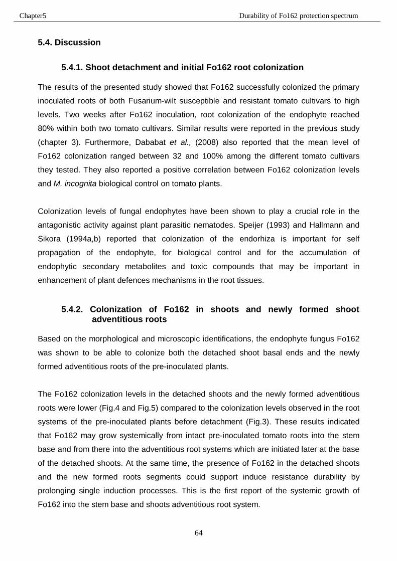
Chapter5 Durability of Fo162 protection spectrum
64
5.4. Discussion
5.4.1. Shoot detachment and initial Fo162 root colonization
The results of the presented study showed that Fo162 successfully colonized the primary
inoculated roots of both Fusarium-wilt susceptible and resistant tomato cultivars to high
levels. Two weeks after Fo162 inoculation, root colonization of the endophyte reached
80% within both two tomato cultivars. Similar results were reported in the previous study
(chapter 3). Furthermore, Dababat et al., (2008) also reported that the mean level of
Fo162 colonization ranged between 32 and 100% among the different tomato cultivars
they tested. They also reported a positive correlation between Fo162 colonization levels
and M. incognita biological control on tomato plants.
Colonization levels of fungal endophytes have been shown to play a crucial role in the
antagonistic activity against plant parasitic nematodes. Speijer (1993) and Hallmann and
Sikora (1994a,b) reported that colonization of the endorhiza is important for self
propagation of the endophyte, for biological control and for the accumulation of
endophytic secondary metabolites and toxic compounds that may be important in
enhancement of plant defences mechanisms in the root tissues.
5.4.2. Colonization of Fo162 in shoots and newly formed shoot adventitious roots
Based on the morphological and microscopic identifications, the endophyte fungus Fo162
was shown to be able to colonize both the detached shoot basal ends and the newly
formed adventitious roots of the pre-inoculated plants.
The Fo162 colonization levels in the detached shoots and the newly formed adventitious
roots were lower (Fig.4 and Fig.5) compared to the colonization levels observed in the root
systems of the pre-inoculated plants before detachment (Fig.3). These results indicated
that Fo162 may grow systemically from intact pre-inoculated tomato roots into the stem
base and from there into the adventitious root systems which are initiated later at the base
of the detached shoots. At the same time, the presence of Fo162 in the detached shoots
and the new formed roots segments could support induce resistance durability by
prolonging single induction processes. This is the first report of the systemic growth of
Fo162 into the stem base and shoots adventitious root system.
Chapter5 Durability of Fo162 protection spectrum
65
5.4.3. IGS-RFLP analysis
To confirm the results obtained from the morphological and microscopic investigations
(5.3.2.), molecular identification of the fungal isolates which resembled Fo162 was
performed using restriction fragment length polymorphism (RFLP) of the ribosomal
intergenic spacer (IGS) PCR amplified fragments.
The data showed that the results of the morphological and microscopic investigations were
not always consistent with the results of the molecular investigation. Of the 17 isolates
tested in this investigation, the genotype of only 4 isolates could be verified as identical
with the Fo162 genotype whereas the other isolates had close similarity to Fo162. The
level of similarity ranged from 69 to 85%. These results illustrated that other non-
pathogenic isolates in the potting soil or water also colonized the shoot basal ends and the
newly formed adventitious roots.
The results obtained from the molecular identification indicated also that the morphological
and microscopic observations are not foolproof and can not be relied upon to give 100
percent characterization of the presence of Fo162 isolates in pre-inoculated plants. The molecular technique (IGS-RFLP) which was used in this study to verify the presence
of Fo162 isolates was used also by Zambounis et al., (2007). They also used the
polymerase chain reaction (PCR) amplification of the ribosomal intergenic spacer (IGS)
regions combined with digestion with three restriction enzymes (AluI, HaeIII, RsaI)
technique to identify and distinguish the isolates of an Australian Fusarium oxysporum f.
sp. vasinfectum among various isolates belonging to Fusarium oxysporum. Furthermore,
Lori et al., (2004) used the intergenic spacer (IGS) typing procedure to study the genetic
diversity among 151 isolates of Fusarium oxysporum that belonged to pathogenic and
non-pathogenic Fusarium populations isolated from carnation fields in Argentina. They
reported that IGS typing is a useful tool in the identification of the Fusarium oxysporum
forma specialis.
Chapter5 Durability of Fo162 protection spectrum
66
5.4.4. Durability of systemic resistance signals in detached shoots and
adventitious roots
The shoot detachment technique was used in this study to investigate the durability of
Fo162 induced systemic defences signals toward M. incognita in the Fusarium-wilt
susceptible and resistant tomato cultivars.
The results from these investigations showed that M. incognita infection was significantly
reduced on detached adventitious roots 5 and 7 days after detachment from the Fo162
pre-inoculated primary root systems. Five days after separation from the Fo162 inoculated
primary roots, the reduction in number of galls/g reached 50 and 51% on the Fusarium-wilt
resistant and susceptible tomato cultivars, respectively.
Similar results were observed when M. incognita juveniles were inoculated 7 days after
detaching the Fo162 inoculated primary roots. The reduction in number of galls/g reached
43 and 49% within the resistant and susceptible tomato cultivars, respectively.
Conversely, when nematodes were inoculated 10 days after detachment, no significant
differences in the number of M. incognita galls were detected between the detached stems
from the Fo162 inoculated plants and the non-inoculated control plants.
The results seem to indicate that the signals of Fo162 based systemic resistance in tomato
plants together with the additive effects of wound induced defences, which could have
been elicited when the shoots were detached from the pre-inoculated roots, were
responsible for the reduction in number of galls observed on the Fo162 shoots 5 and 7
days after detachment. Induced systemic resistance (ISR) has been shown to be wound
inducible thus wounds can elicit the ISR pathways in different host plants (Bostock et al.,
2001).
Other researchers have demonstrated that different strains of endophytic fungi can protect
plants through different mechanisms of action and that induced resistance is considered
important as a triggering factor in the interactions between endophytes, host plants and
plant parasites (Dababat and Sikora, 2007a,b; Cramer et al., 1985; Biles and Martyn,
1989). Matta (1989) reported that induced resistance can be either local (associated with a
hypersensitive reaction) or systemic (provides an entire host plant protection against later
pathogens infections).
Chapter5 Durability of Fo162 protection spectrum
67
In 1989, Strömberg mentioned also that systemic induced resistance can be obtained
when challenge inoculum is applied at a later time and different location on a plant.
Similar results to the obtained results from the present study were obtained by Hallmann
and Sikora (1994a); Dababat and Sikora (2007b); Dababat et al. (2008), who reported that
the non-pathogenic Fusarium oxysporum strain (Fo162) had a high level of biological
control potential in reducing nematode infection on tomato. Dababat and Sikora (2007a)
investigated the mode of action of Fo162 in tomato plants and demonstrated that Fo162
was capable of protecting tomato from root-knot nematode infection by inducing host plant
systemic resistance and by triggering other mechanisms of action.
Two new findings were made in the present investigations: 1) that Fo162 has the ability to
induce systemic resistance to the root-knot nematode in tomato plants that are resistant to
Fusarium-wilt and 2) that the durability of the signals related to the Fo162 systemic
resistance is emitted from the stem into the adventitious roots after detachment from the
initial induced root system of the mother plants.
5.5. Conclusion
Based on the present investigation the following can be concluded:
1- Fo162 was able to colonize with high levels, roots of the Fusarium-wilt susceptible
and resistant tomato cultivars.
2- Fo162 was able to induce the systemic defences toward M. incognita in Fusarium-
wilt susceptible and resistant tomato cultivars when the endophyte was not in the
direct contact with the host plant or with the nematode.
3- The Fo162 signals responsible for systemic induced resistance were responsible for
protecting tomato plants from root-knot nematode invasion for up to 7 days after the
physical separation between the host plant and the endophyte fungus. This
indicates that the Fo162 signals that are based in the shoot also add to durability of
the control mechanism.
Chapter5 Durability of Fo162 protection spectrum
68
5.6. References - Biles, C. L. and Martyn, R. D. (1989). Local and systemic resistance induced in
watermelons by formae speciales of Fusarium oxysporum. Phytopathology, 79:
856-860.
- Bostock, R. M., Karban, R., Thaler, J. S., Weyman, P. D. and Gilchrist, D. (2001).
Signal interactions in induced resistance to pathogens and insect herbivores.
European Journal of Plant Pathology 107: 103-111.
- Cramer, C. L., Ryder, T. B., Bell, J. N. and Lamb, C. J. (1985). Rapid switching of
plant gene expression induced by fungal elicitors. Science 227:1240-1243.
- Dababat, A. A. and Sikora, R. A. (2007a). Induced resistance by the mutualistic
endophyte, Fusarium oxysporum 162, toward Meloidogyne incognita on tomato.
Biocontrol Sci. Techn. 17, 969-975.
- Dababat, A. A. and Sikora, R. A. (2007b). Importance of application time and
inoculum density of the non-pathogenic fungus, Fusarium oxysporum 162, for the
biological control of the root-knot nematode Meloidogyne incognita on tomato.
Nematropica 2, 267-276.
- Dababat, A. A., Selim, M. E., Saleh, A. A. and Sikora, R. A. (2008). Influence of
Fusarium wilt resistant tomato cultivars on root colonization of the mutualistic
endophyte Fusarium oxysporum strain 162 and its biological control efficacy toward
the root-knot nematode Meloidogyne incognita. Journal of Plant disease and
protection, 115 (6) 273-278.
- Edel, V., Steinberg, C., Avelange, I., Laguerre, G. and Alabouvette, C. (1995).
Comparison of 3 molecular methods for the characterization of Fusarium
oxysporum strains. Phytopathology, 85, 579-585.
- Edel, V., Steinberg, C., Gautheron, N., and Alabouvette, C. (1997). Populations of
non-pathogenic Fusarium oxysporum associated with roots of four plants species
compared to soil borne populations. Phytopathology, 87, 693-697.
- Fuchs, J.-G., Moënne-Loccoz, Y. and Défago, G. (1997). Non pathogenic Fusarium
oxysporum strain Fo47 induces resistance to Fusarium wilt in tomato. Plant Dis. 81:
492-496.
- Hallmann, J. and Sikora, R. A. (1994a). Influence of Fusarium oxysporum, a
mutualistic fungal endophyte on Meloidogyne incognita infection of tomato. Journal
of plant disease and protection 101: 475-481.
Chapter5 Durability of Fo162 protection spectrum
69
- Lori, G., Edel-Hermann, V., Gautheron, N. and Alabouvette, C. (2004). Genetic
diversity of pathogenic and nonpathogenic populations of Fusarium oxysporum
isolated from carnation fields in Argentina. Phytopathology 94: 661-668.
- Matta, A. (1989). Induced resistance to Fusarium wilt disease: Vascular wilt disease
of plants. In: Tjamos, E. C. and Beckman, C. H. (eds). Basic studies and control.
NATO ASI series, vol. H28. Springer-Verlag, Berlin. pp. 175-196.
- Speijer, P. R. (1993). Interrelationship between Pratylenchus goodeyi Sher & Allen
and strains of non-pathogenic Fusarium oxysporum Schl. emd. Snyd. & Hans. In
roots of banana cultivars. Ph.D. thesis, University of Bonn.
- Strömberg, A. (1989). Systemically induced resistance in tomatoes, tobacco and
potatoes. A literature review. Växtskyddsrapporter, Jordbruk 55. Research
information center, Swedish University of Agriculture Science, Uppsala.
- Zambounis, A. G., Paplomatas, E. and Tsaftaris, A. S. (2007). Intergenic spacer–
RFLP analysis and direct quantification of Australian Fusarium oxysporum f. sp.
vasinfectum isolates from soil and infected cotton tissues. Plant Dis. 91:1564-1573.
Chapter 6 Induction of different types of systemic resistance
70
6. Induction of systemic resistance in tomato toward the root-knot nematode
Meloidogyne incognita using biotic and abiotic elicitors
6.1. Introduction Various mutualistic endophytes can serve as antagonists against a wide range of plant
pathogenic fungi and nematodes. In such interactions, different modes of action could be
involved in the control of the pathogen and induced resistance in the host, elicited by the
endophyte, is considered one of them (Sikora et al., 2007).
Different types of systemic resistance can be induced within host plants either biologically or
chemically using different kinds of biological and chemical elicitors (Heil and Bostock, 2002;
Pieterse et al., 2001; and Walling, 2000).
Biologically induced resistance in host plants can be obtained using biotic elicitors e.g. viruses,
bacteria and fungi. Leemann et al., (1995) and Zhou and Paulitz (1994), for example,
demonstrated the ability of Pseudomonas bacteria to induce plant systemic resistance against
Fusarium wilt and Pythium aphanidermatum pathogens, respectively.
Hasky et al., (1998) were able to induce system resistance to the potato cyst nematode
Globodera pallida using the endophytic bacteria Agrobacterium radiobacter (G12). In split root
experiments with tomato plants where one half of the roots was treated with cell suspension of
rhizobacteria and the other side was inoculated with Meloidogyne javanica, Siddiqui and
Shaukat (2004), showed that the nematode population in the treated plants was reduced
significantly compared to the nematode population in the non-treated tomato plants.
Induction of systemic defences against various disease organisms can also be obtained through
the application of chemical elicitors which trigger different signalling pathways. Induced
Systemic Resistance (ISR) and Systemic Acquired Resistance (SAR) are the two main
resistance pathways which are mediated through the jasmonic acid metabolic signalling
pathway or the salicylic acid signalling pathway, respectively (Sticher et al., 1997; and Ton et
al., 2002).
In this study, systemic resistance in tomato induced by the fungal endophyte Fo162 was
compared with induced systemic resistance (ISR) and systemic acquired resistance (SAR) that
are chemically induced using methyl jasmonate (MJ) and salicylic acid (SA), respectively.
Chapter 6 Induction of different types of systemic resistance
71
The effects of systemic resistance on nematode infection and egg masses production of the
sedentary nematode M. incognita as well as on the physiology of tomato plants were monitored
using the split-root experimental design.
The objectives of this study were to determine the:
1- Ability of different biological (Fo162) and chemical (SA and MJ) elicitors to induce systemic
resistance in tomato plants against root-knot nematodes.
2- The influence of inducing host plant systemic resistance using biological (Fo162) and
chemical (SA and MJ) elicitors on plant growth and physiological properties of tomato.
Chapter 6 Induction of different types of systemic resistance
72
6.2. Materials and methods: 6.2.1. Root growth and nematode infection
The ability of biotic and abiotic elicitors to induce systemic resistance in tomato against M.
incognita was investigated using the mutualistic endophyte Fusarium oxysporum strain 162
(Fo162), salicylic acid (SA) and methyl jasmonate (MJ). Seeds of the tomato cultivar
´´Hellfrucht-Frühstamm``, which is susceptible to M. incognita, were sown separately in plastic
pots containing an autoclaved soil:sand mixture (1:2 v/v). Three weeks after sowing, or when
the seedlings were about 25-30 cm in height, the shoots were cut-off 5 cm from the basal ends.
The shoots were then longitudinally split into 2 halves and each half was replanted in new
substrate in two neighbouring pots. One week after splitting and replanting the shoots, new
adventitious roots developed in each pot. At this time one pot of the twin pot set was inoculated
with either a biological or a chemical inducer. This pot was termed the inducer side, while the
other pot was termed the responder side, (Fig.1).
To evaluate the influence of the biological and chemical inducers on nematode infection, the
inducer sides of 6 plants were inoculated with 106 cfu/g soil of Fo162 (as described in chapter
2), or treated with 200 µM SA, 100 µM MJ, 10 µM MJ, or with tap water (control). One week
after treating the inducer sides with the elicitors, the roots at the responder side were inoculated
with 450 M. incognita juveniles (as described in chapter 2). The plants treated with the chemical
inducers at the inducer side were watered daily with the respective inducing solutions. The
control plants and the Fo162 inoculated plants were watered daily with tap water.
Stock solutions of salicylic acid and methyl jasmonate substrates were prepared with
concentrations of 20 mM and 10 mM respectively by adding the SA or MJ to water followed by
shaking for 1 h on a magnetic stirrer. These stock solutions were stored in the dark and used for
preparing the desired concentrations immediately prior to watering. All chemicals were
purchased from Sigma-Aldrich. Tomato plants were grown under greenhouse conditions at 25 ±
5 °C with 16 h diurnal light and fertilized once a week with 2g/L NPK (14:10:14).
The experiment was terminated 3 weeks after nematode inoculation. Roots of the responder
side and the inducer side were washed gently with tap water then blotted between tissue paper.
The fresh root weights were recorded and the number of galls per root system of the responder
sides was counted.
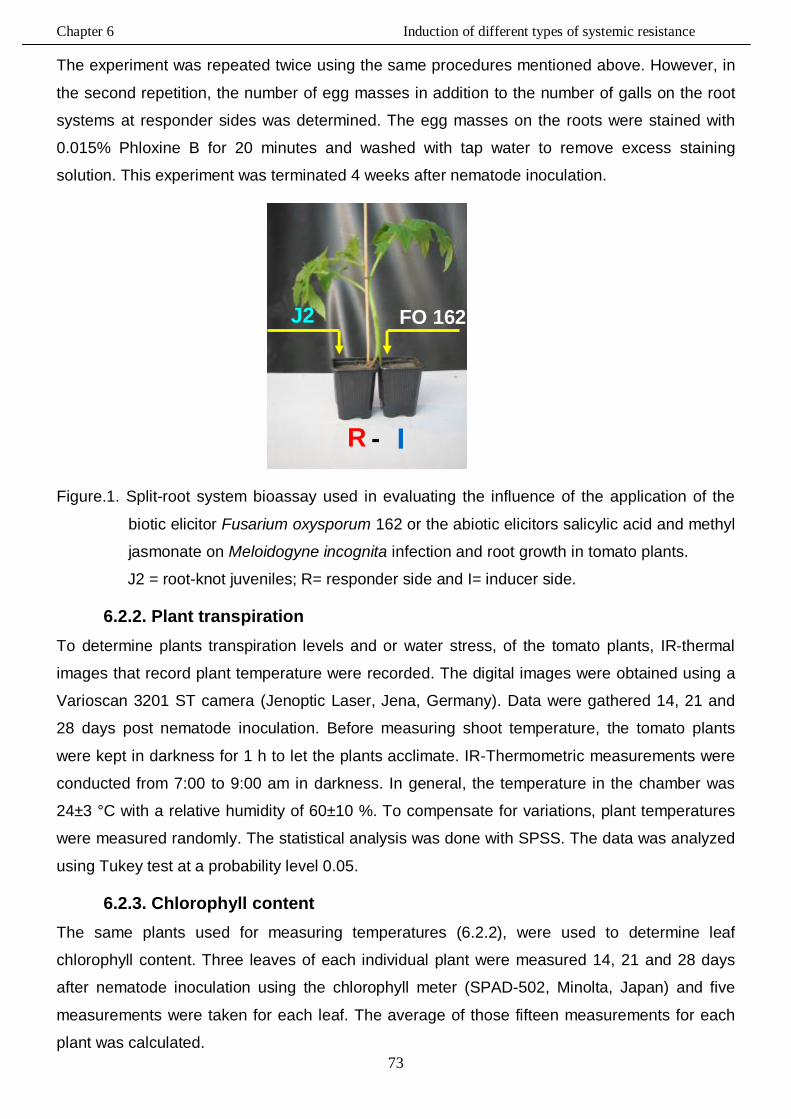
Chapter 6 Induction of different types of systemic resistance
73
The experiment was repeated twice using the same procedures mentioned above. However, in
the second repetition, the number of egg masses in addition to the number of galls on the root
systems at responder sides was determined. The egg masses on the roots were stained with
0.015% Phloxine B for 20 minutes and washed with tap water to remove excess staining
solution. This experiment was terminated 4 weeks after nematode inoculation.
Figure.1. Split-root system bioassay used in evaluating the influence of the application of the
biotic elicitor Fusarium oxysporum 162 or the abiotic elicitors salicylic acid and methyl
jasmonate on Meloidogyne incognita infection and root growth in tomato plants.
J2 = root-knot juveniles; R= responder side and I= inducer side.
6.2.2. Plant transpiration To determine plants transpiration levels and or water stress, of the tomato plants, IR-thermal
images that record plant temperature were recorded. The digital images were obtained using a
Varioscan 3201 ST camera (Jenoptic Laser, Jena, Germany). Data were gathered 14, 21 and
28 days post nematode inoculation. Before measuring shoot temperature, the tomato plants
were kept in darkness for 1 h to let the plants acclimate. IR-Thermometric measurements were
conducted from 7:00 to 9:00 am in darkness. In general, the temperature in the chamber was
24±3 °C with a relative humidity of 60±10 %. To compensate for variations, plant temperatures
were measured randomly. The statistical analysis was done with SPSS. The data was analyzed
using Tukey test at a probability level 0.05.
6.2.3. Chlorophyll content The same plants used for measuring temperatures (6.2.2), were used to determine leaf
chlorophyll content. Three leaves of each individual plant were measured 14, 21 and 28 days
after nematode inoculation using the chlorophyll meter (SPAD-502, Minolta, Japan) and five
measurements were taken for each leaf. The average of those fifteen measurements for each
plant was calculated.
FO 162 J2
I R -
Chapter 6 Induction of different types of systemic resistance
74
6.3. Results
6.3.1. Root growth at inducer side
The influence of the elicitors on the root growth at the inducer side of the treated plants was
determined. The results of trial A showed that 100 µM MJ inhibited significantly the root growth
at the inducer sides when compared to the control and to the other elicitors (Fo162 and SA),
(Fig.2.A). Fo162 and SA had no significant effects on root growth at the inducer side of treated
plants.
In trial B, the root growth at the inducer side of split-root tomato plants treated with the SA or MJ
was significantly reduced when compared to the control. The biotic elicitor, Fo162, did not affect
root growth at the inducer side of inoculated plants, (Fig.2.B).
Trial C showed that all tested three chemical elicitor, 200 µM SA, 100 µM and 10 µM MJ,
reduced root weight at the inducer side of split-root tomato plants. Conversely, the biological
inducer, Fo162, did not affect root growth when compared to the control, (Fig.2.C).
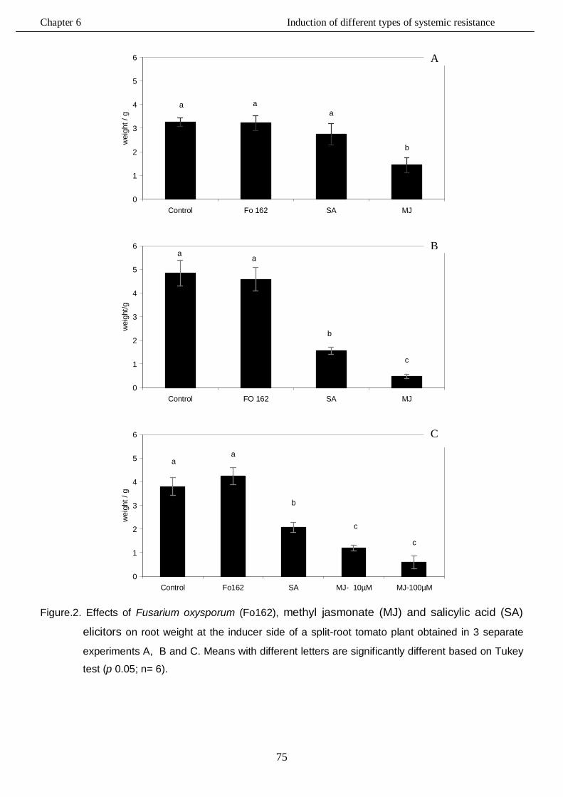
Chapter 6 Induction of different types of systemic resistance
75
a aa
b
0
1
2
3
4
5
6
Control Fo 162 SA MJ
wei
ght /
g
a a
b
c
0
1
2
3
4
5
6
Control FO 162 SA MJ
wei
ght/g
aa
b
c
c
0
1
2
3
4
5
6
Control Fo162 SA MJ- 10µM MJ-100µM
wei
ght /
g
Figure.2. Effects of Fusarium oxysporum (Fo162), methyl jasmonate (MJ) and salicylic acid (SA)
elicitors on root weight at the inducer side of a split-root tomato plant obtained in 3 separate
experiments A, B and C. Means with different letters are significantly different based on Tukey
test (p 0.05; n= 6).
C
A
B
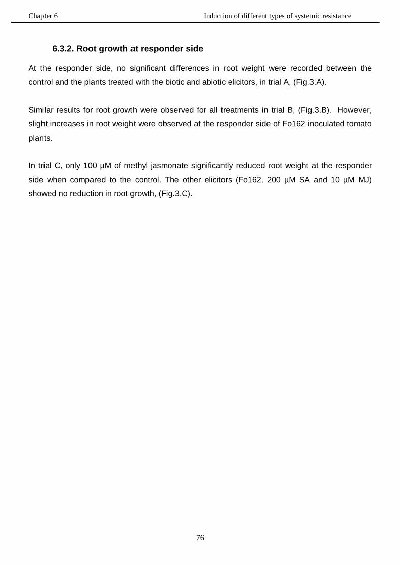
Chapter 6 Induction of different types of systemic resistance
76
6.3.2. Root growth at responder side
At the responder side, no significant differences in root weight were recorded between the
control and the plants treated with the biotic and abiotic elicitors, in trial A, (Fig.3.A).
Similar results for root growth were observed for all treatments in trial B, (Fig.3.B). However,
slight increases in root weight were observed at the responder side of Fo162 inoculated tomato
plants.
In trial C, only 100 µM of methyl jasmonate significantly reduced root weight at the responder
side when compared to the control. The other elicitors (Fo162, 200 µM SA and 10 µM MJ)
showed no reduction in root growth, (Fig.3.C).
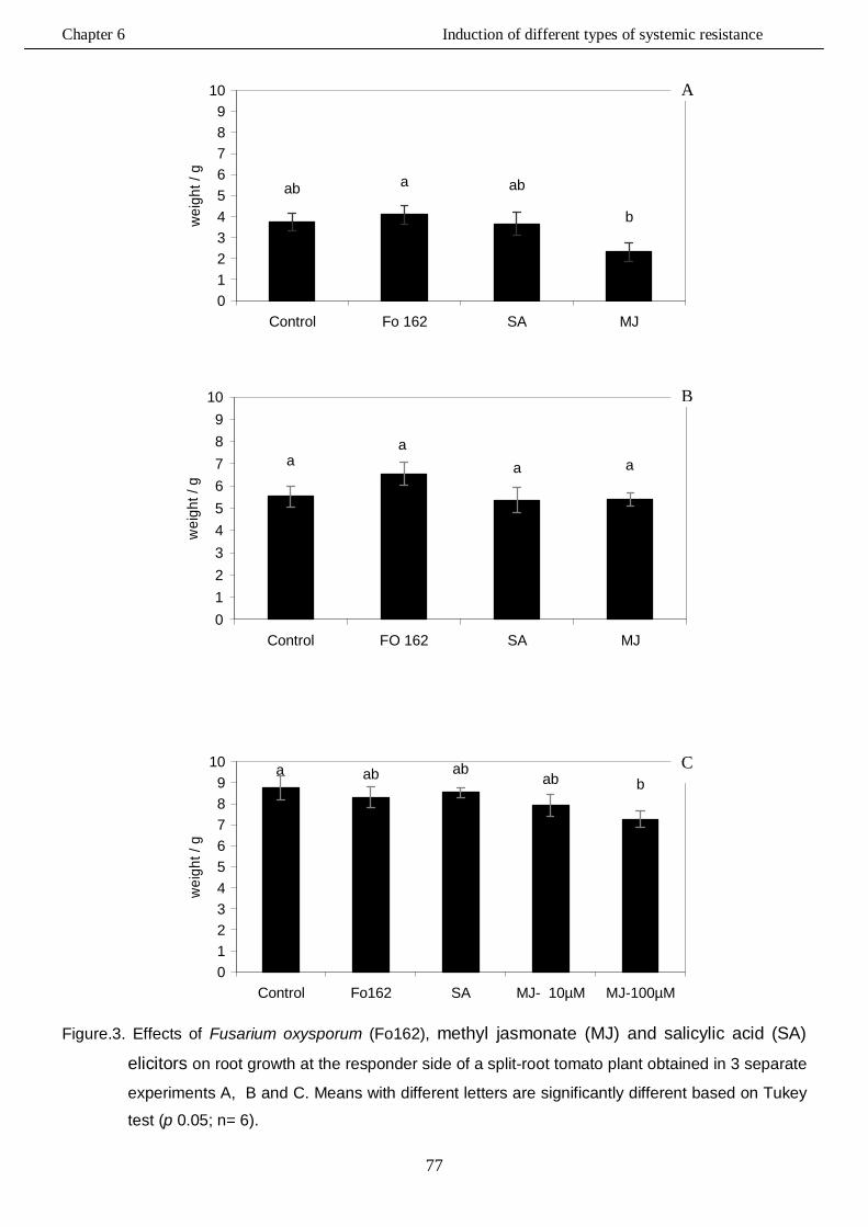
Chapter 6 Induction of different types of systemic resistance
77
ab a ab
b
0123456789
10
Control Fo 162 SA MJ
wei
ght /
g
aa
a a
0123456789
10
Control FO 162 SA MJ
wei
ght /
g
a ab abab b
0123456789
10
Control Fo162 SA MJ- 10µM MJ-100µM
wei
ght /
g
Figure.3. Effects of Fusarium oxysporum (Fo162), methyl jasmonate (MJ) and salicylic acid (SA)
elicitors on root growth at the responder side of a split-root tomato plant obtained in 3 separate
experiments A, B and C. Means with different letters are significantly different based on Tukey
test (p 0.05; n= 6).
C
A
B
Chapter 6 Induction of different types of systemic resistance
78
6.3.3. Nematode infection In trial A, the results showed that the biological, Fo162, and the chemical elicitors, 200 µM SA
and 100µM MJ, inhibited M. incognita infection on the responder side of the split-root tomato
plants. The total number of galls per root system as well as the number of galls/g root was
significantly reduced at the responder side of the tomato plants treated with all three elicitors
when compared to the control, (Fig.4 and 5). The reduction in nematode galls reached
approximately 67, 75, and 91% in the Fo162, SA and MJ, treated plants, respectively.
In trial B, the results showed that both the biological, Fo162, or the chemical, SA and MJ,
elicitors significantly reduced the number of galls/root system, (Fig.6) and the number of galls/g
root, (Fig.7). The reduction in nematode galls reached approximately 22, 40 and 79% in the
Fo162, SA and MJ, treated plants, respectively.
In trial C, the results showed also that Fo162, 200 µM SA and 100 µM MJ significantly reduced
the number of galls per root system compared to the control. The highest reduction in number of
galls (41%) was observed for the plants treated with100µM MJ followed by Fo162 (27%) and
200µM SA (24%), (Fig.8).
Similar results were obtained in trial (C) when the number of galls/ g root was determined on
roots of elicitor treated verses non treated plants, (Fig.9).
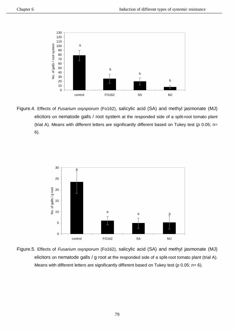
Chapter 6 Induction of different types of systemic resistance
79
a
bb
b
0102030405060708090
100110120130
control FO162 SA MJ
No.
of g
alls
/ ro
ot s
yste
m
Figure.4. Effects of Fusarium oxysporum (Fo162), salicylic acid (SA) and methyl jasmonate (MJ)
elicitors on nematode galls / root system at the responded side of a split-root tomato plant
(trial A). Means with different letters are significantly different based on Tukey test (p 0.05; n=
6).
a
b b b
0
5
10
15
20
25
30
control FO162 SA MJ
No.
of g
alls
/ g
root
Figure.5. Effects of Fusarium oxysporum (Fo162), salicylic acid (SA) and methyl jasmonate (MJ)
elicitors on nematode galls / g root at the responded side of a split-root tomato plant (trial A).
Means with different letters are significantly different based on Tukey test (p 0.05; n= 6).
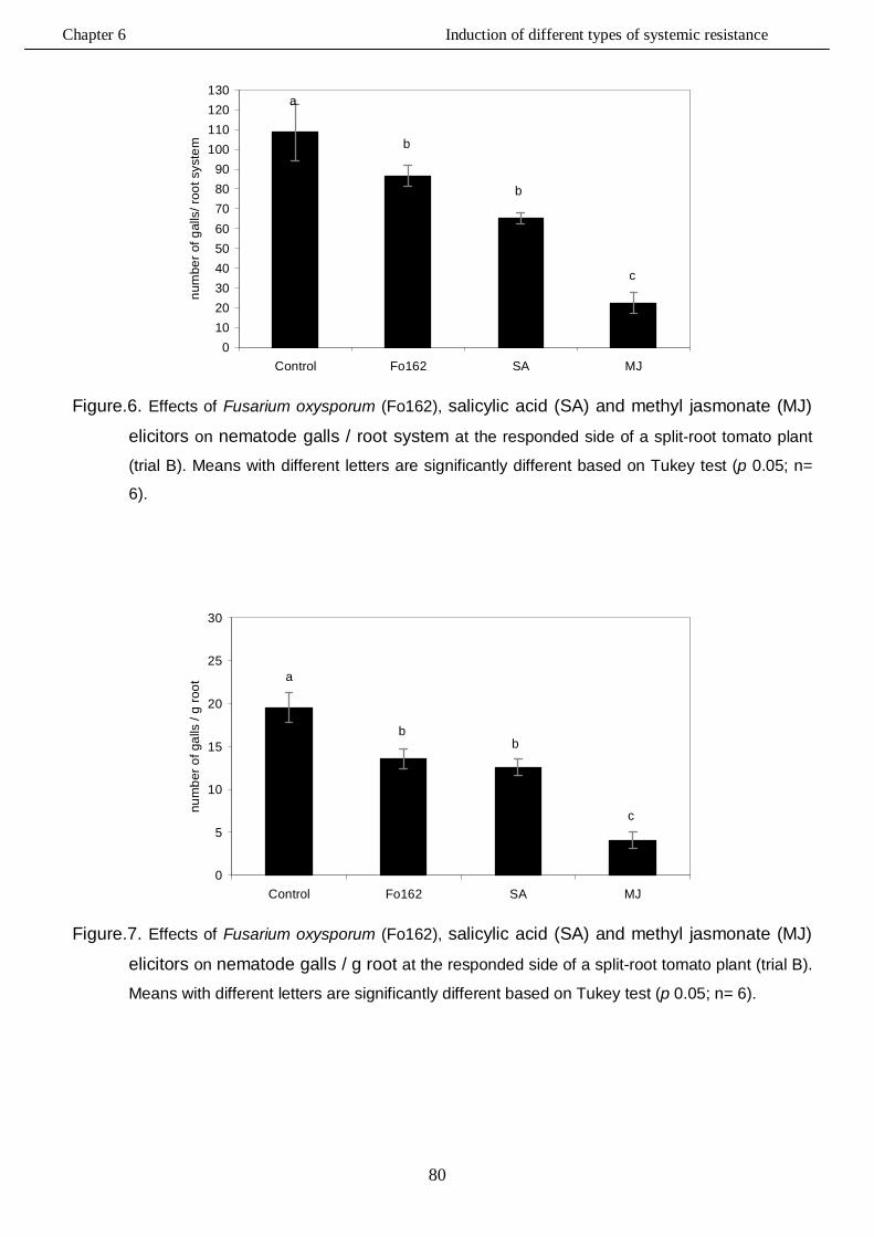
Chapter 6 Induction of different types of systemic resistance
80
a
b
b
c
0102030405060708090
100110120130
Control Fo162 SA MJ
num
ber o
f gal
ls/ r
oot s
yste
m
Figure.6. Effects of Fusarium oxysporum (Fo162), salicylic acid (SA) and methyl jasmonate (MJ)
elicitors on nematode galls / root system at the responded side of a split-root tomato plant
(trial B). Means with different letters are significantly different based on Tukey test (p 0.05; n=
6).
a
bb
c
0
5
10
15
20
25
30
Control Fo162 SA MJ
num
ber o
f gal
ls /
g ro
ot
Figure.7. Effects of Fusarium oxysporum (Fo162), salicylic acid (SA) and methyl jasmonate (MJ)
elicitors on nematode galls / g root at the responded side of a split-root tomato plant (trial B).
Means with different letters are significantly different based on Tukey test (p 0.05; n= 6).
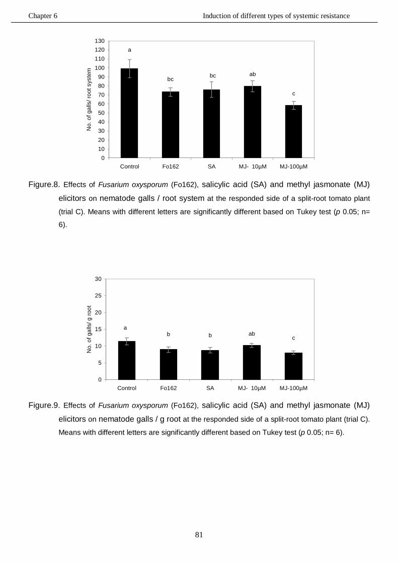
Chapter 6 Induction of different types of systemic resistance
81
a
bc bc ab
c
0102030405060708090
100110120130
Control Fo162 SA MJ- 10µM MJ-100µM
No.
of g
alls
/ roo
t sys
tem
Figure.8. Effects of Fusarium oxysporum (Fo162), salicylic acid (SA) and methyl jasmonate (MJ)
elicitors on nematode galls / root system at the responded side of a split-root tomato plant
(trial C). Means with different letters are significantly different based on Tukey test (p 0.05; n=
6).
ab b ab
c
0
5
10
15
20
25
30
Control Fo162 SA MJ- 10µM MJ-100µM
No.
of g
alls
/ g ro
ot
Figure.9. Effects of Fusarium oxysporum (Fo162), salicylic acid (SA) and methyl jasmonate (MJ)
elicitors on nematode galls / g root at the responded side of a split-root tomato plant (trial C).
Means with different letters are significantly different based on Tukey test (p 0.05; n= 6).
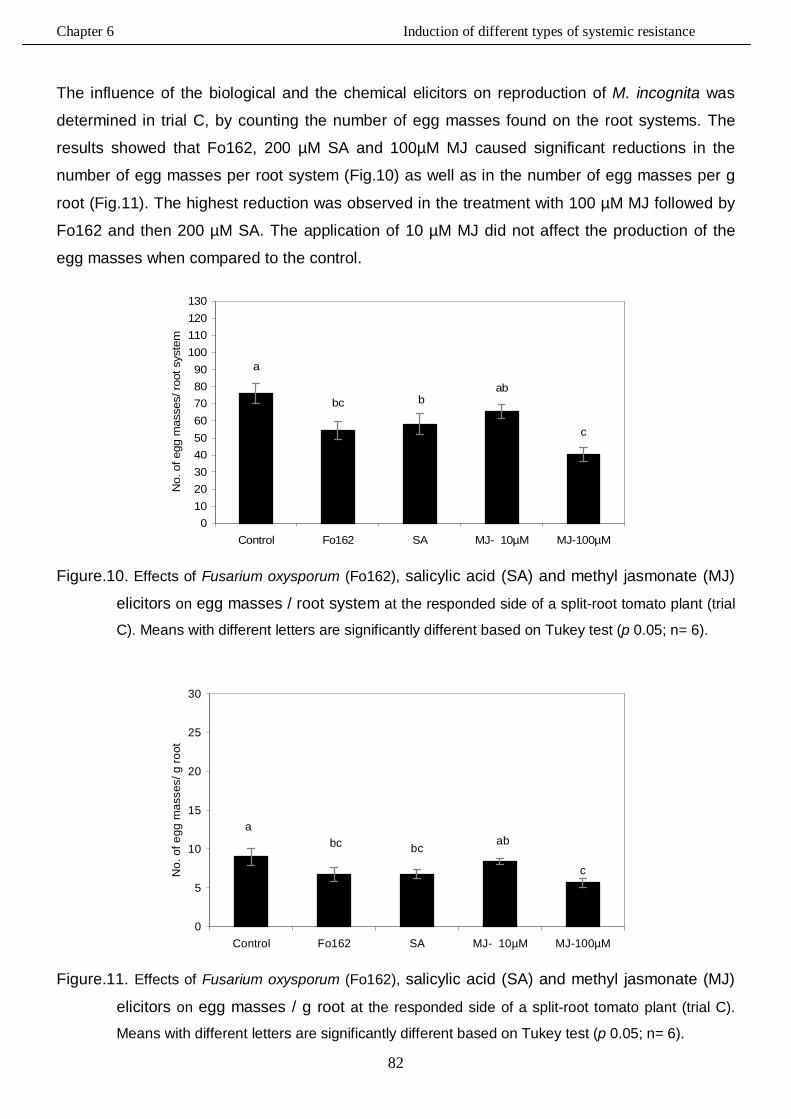
Chapter 6 Induction of different types of systemic resistance
82
The influence of the biological and the chemical elicitors on reproduction of M. incognita was
determined in trial C, by counting the number of egg masses found on the root systems. The
results showed that Fo162, 200 µM SA and 100µM MJ caused significant reductions in the
number of egg masses per root system (Fig.10) as well as in the number of egg masses per g
root (Fig.11). The highest reduction was observed in the treatment with 100 µM MJ followed by
Fo162 and then 200 µM SA. The application of 10 µM MJ did not affect the production of the
egg masses when compared to the control.
a
bc bab
c
0102030405060708090
100110120130
Control Fo162 SA MJ- 10µM MJ-100µM
No.
of e
gg m
asse
s/ ro
ot s
yste
m
Figure.10. Effects of Fusarium oxysporum (Fo162), salicylic acid (SA) and methyl jasmonate (MJ)
elicitors on egg masses / root system at the responded side of a split-root tomato plant (trial
C). Means with different letters are significantly different based on Tukey test (p 0.05; n= 6).
abc bc
ab
c
0
5
10
15
20
25
30
Control Fo162 SA MJ- 10µM MJ-100µM
No.
of e
gg m
asse
s/ g
root
Figure.11. Effects of Fusarium oxysporum (Fo162), salicylic acid (SA) and methyl jasmonate (MJ)
elicitors on egg masses / g root at the responded side of a split-root tomato plant (trial C).
Means with different letters are significantly different based on Tukey test (p 0.05; n= 6).
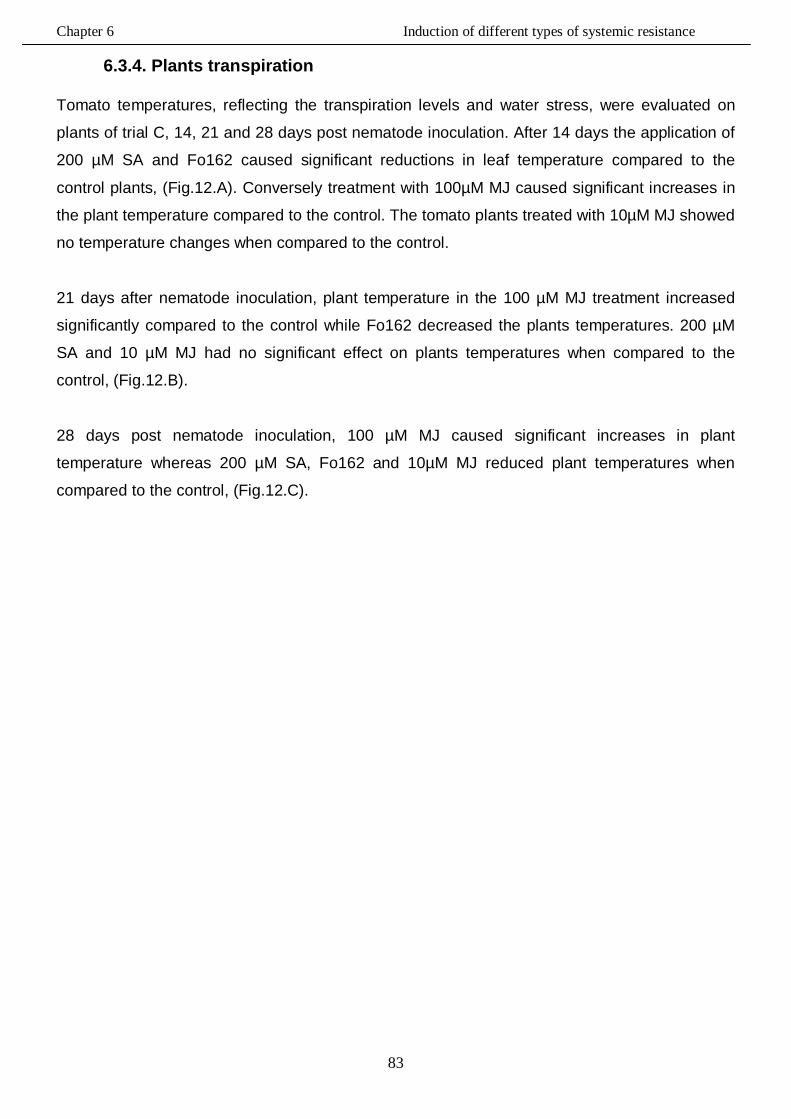
Chapter 6 Induction of different types of systemic resistance
83
6.3.4. Plants transpiration Tomato temperatures, reflecting the transpiration levels and water stress, were evaluated on
plants of trial C, 14, 21 and 28 days post nematode inoculation. After 14 days the application of
200 µM SA and Fo162 caused significant reductions in leaf temperature compared to the
control plants, (Fig.12.A). Conversely treatment with 100µM MJ caused significant increases in
the plant temperature compared to the control. The tomato plants treated with 10µM MJ showed
no temperature changes when compared to the control.
21 days after nematode inoculation, plant temperature in the 100 µM MJ treatment increased
significantly compared to the control while Fo162 decreased the plants temperatures. 200 µM
SA and 10 µM MJ had no significant effect on plants temperatures when compared to the
control, (Fig.12.B).
28 days post nematode inoculation, 100 µM MJ caused significant increases in plant
temperature whereas 200 µM SA, Fo162 and 10µM MJ reduced plant temperatures when
compared to the control, (Fig.12.C).
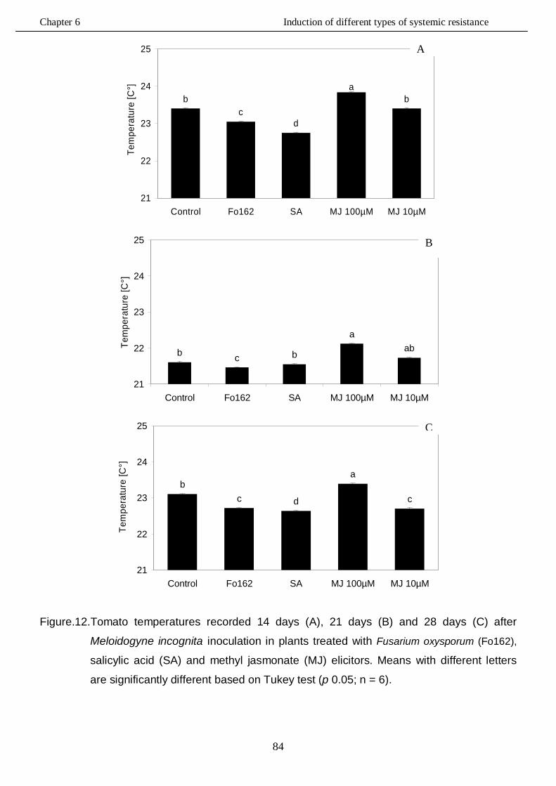
Chapter 6 Induction of different types of systemic resistance
84
ba
dc
b
21
22
23
24
25
Control Fo162 SA MJ 100µM MJ 10µM
Tem
pera
ture
[C°]
aba
bcb
21
22
23
24
25
Control Fo162 SA MJ 100µM MJ 10µM
Tem
pera
ture
[C°]
c
a
dcb
21
22
23
24
25
Control Fo162 SA MJ 100µM MJ 10µM
Tem
pera
ture
[C°]
Figure.12.Tomato temperatures recorded 14 days (A), 21 days (B) and 28 days (C) after
Meloidogyne incognita inoculation in plants treated with Fusarium oxysporum (Fo162),
salicylic acid (SA) and methyl jasmonate (MJ) elicitors. Means with different letters
are significantly different based on Tukey test (p 0.05; n = 6).
A
B
C
Chapter 6 Induction of different types of systemic resistance
85
6.3.5. Chlorophyll content
The intensity of leaf colour, indicative of chlorophyll synthesis, was measured in the plants of
trial C, 14, 21 and 28 days after nematode inoculation. At 14 days post nematode inoculation,
the highest chlorophyll content of in leaves was observed in the Fo162 inoculated plants
compared to the control. In the 100µM MJ treated plants a lower level of chlorophyll was
detected compared to the control. No significant differences with respect to leaf chlorophyll
content were recorded in the presence of either 200 µM SA or 10µM MJ, (Fig.13.A).
Lower levels of chlorophyll were detected 21 days after nematode inoculation in tomato treated
with 100µM MJ compared to the untreated control. The application of SA significantly increased
the level of green content in the tomato leaves, while both Fo162 and 10 mM MJ had no
significant effects, (Fig.13.B).
Similar results were recorded again when chlorophyll content was determined 28 days after
nematode inoculation, (Fig.13.C). At this measurement time, 100 µM MJ reduced the
chlorophyll content in the leaves, while 200 µm SA increased it compared to the control. No
significant differences were recorded for Fo162 and 10µM MJ.
Chapter 6 Induction of different types of systemic resistance
86
ab
c
b b
0
5
10
15
20
25
30
35
40
45
50
Fo 162 Sa MJ 100uM MJ 10uM Control
inte
nsity
of l
eaf c
olou
r
ab a
d cdbc
0
5
10
15
20
25
30
35
40
45
50
Fo 162 Sa MJ 100uM MJ 10uM Control
inte
nsity
of l
eaf c
olou
r
bc ab
dc c
0
5
10
15
20
25
30
35
40
45
50
Fo 162 Sa MJ 100uM MJ 10uM Control
inte
nsity
of l
eaf c
olou
r
Figure.13. Intensity of leaf colour recorded 14 days (A), 21 days (B) and 28 days (C) after
Meloidogyne incognita inoculation in plants treated with Fusarium oxysporum (Fo162),
salicylic acid (SA) and methyl jasmonate (MJ) elicitors. Means with different letters
are significantly different based on Tukey test (p 0.05; n = 6).
A
B
C
Chapter 6 Induction of different types of systemic resistance
87
6.4. Discussion
6.4.1. Root growth and nematode infection
In this study the ability of the mutualistic endophyte Fusarium oxysporum 162 (Fo162), salicylic
acid (SA) and methyl jasmonate (MJ) to induce resistance in tomato against M. incognita has
been tested using a split-root experiment. The influence of the application of Fo162 , the SA and
MJ on root growth at the inducer and responder side of the split-root tomato plant also was
determined.
The results showed that the biological inducer, Fo162, did not affect root growth at the inducer
side while the chemical inducers, MJ and SA, significantly inhibited the root growth when
compared to the control (6.3.1).
At the responder side, no significant differences in root weight were detected between the
control and the Fo162 or SA treated plants. Only the application of 100 µM MJ significantly
reduced root growth at the responder sides in two of the three trials, (6.3.2).
The biotic Fo162 and abiotic elicitors SA and MJ led to significant reductions in nematode
infection. The highest reduction in both nematode galls and egg masses number was observed
on plants treated with 100µM MJ. In trial A, the reduction in number of galls/ root system at the
responder side reached approximately 67, 75, and 91% in the presence of Fo162, SA and MJ,
respectively. In the other two trials, the effect of the biotic and abiotic elicitors on the nematode
infection was less pronounced. However, these trials have been conducted at different times in
the green house where the temperatures and humidity varied with over time. This could explain
some of the variation which was observed between the conducted trials.
Overall, plant treatment with both the biological inducer, Fo162, and the chemical inducers, SA
and MJ, caused significant reductions of M. incognita infection on tomato plants.
These results are similar to those obtained by Dababat and Sikora (2007) who found that
inoculation of tomato plants with the non-pathogenic fungal endophyte F. oxysporum strain 162
resulted in a significant reduction of nematode infection. They hypothesized that this was in part
due to induced resistance in the first 2-3 weeks after fungal inoculation.
Molinari and Loffredo (2006) reported that the ability of exogenously applied SA to induce
resistance to root-knot nematodes in tomato is controversial and it seems to be linked to the
means of application. Malamy and Klessig (1992) as well as Ryals et al., (1996) stated that SA
Chapter 6 Induction of different types of systemic resistance
88
is an essential factor for establishment of SAR in plants. In 1995, Silverman et al., reported that
with the rice cultivars, the resistance levels of plants exhibiting constitutive expression of SA is
positively correlated with SA levels. Similar results were recorded by Coquoz et al., (1995)
within potato plants.
Moreover, Cooper et al., (2005) showed that jasmonic acid application induced the systemic
defence response which led to reducing the avirulent nematode reproduction on susceptible
tomato plants. Stawswick at al., (1998); Vijayan et al., (1998) and Thaler et al., (2004)
mentioned that jasmonates play a crucial rule in plant defenses against plant parasitic
nematodes that invade root systems. Furthermore, Soriano et al., (2004a,b) reported that
methyl jasmonate reduced the susceptibility of spinach and oats plants to different plant
parasitic nematodes. Van Wees et al., (2000) mentioned that disease suppression against
different pathogens was enhanced when ISR and SAR were activated simultaneously using
chemical elicitors.
Therefore, according to the results obtained in the present studies, both ISR and SAR can play
a role in controlling nematode infection on tomato plants when they are induced biologically or
chemically.
6.4.2. Plants transpiration Measuring leaf temperatures which is correlated with plant transpiration and water stress was
carried out in trial C on elicitor treated and non-treaded plants. The thermal images of treated
and untreated tomato plants which were examined 14, 21 and 28 days after M. incognita
inoculation showed that the application of 100µM MJ caused significant increases in leaf
temperatures when compared to the control. Conversely, the biological inducer, Fo162, and the
chemical elicitor, SA, generally caused reductions in temperature when compared to the control.
These results could be due to the influence of the elicitors on root growth of the treated tomato
plants. Severe inhibition in root growth was observed on the plants treated with 100µM MJ,
which might explain the reduced water uptake and water content.
Chapter 6 Induction of different types of systemic resistance
89
6.4.3. Chlorophyll content The results of the present study also showed that Fo162 and SA increased the chlorophyll
content in the leaves of treated plants compared to the control. Conversely, 100µM MJ
significantly reduced the leaf chlorophyll content.
Similar results were obtained in the study of Oka et al., (1999). They found that soil drenching
with AABA, BABA and GABA did not have any phytotoxic effect on foliage of tomato plant while
the younger leaves of plants sprayed with jasmonic acid or methyl jasmonate were slightly
chlorotic. They reported also that JA and MJ soil drenching caused necrotic symptoms on roots
tips and slight chlorosis on leaves whereas SA did not show any phytotoxic symptoms.
The combined results of root growth (see results 6.3.1 and 6.3.2), leaf temperatures (see results
6.3.4) and chlorophyll content (see results 6.3.5) could explain why the highest reduction in
nematode infection (see results 6.3.3) was observed in 100mM MJ treated plants. It is well
known that plant parasitic nematodes are obligate parasites and therefore they are affected by
the factors that affect the host plant vitality. Therefore, the negative effects of 100µM MJ on the
overall vitality as well as on root growth of the treated plants together with the additive effects of
ISR could be responsible for the high levels of reduction in nematode infection observed on the
elicitor treated plants.
6.5. Conclusion
Based on the results of the present investigation the following can be concluded:
1- Different systemic resistance signaling pathways can be stimulated biologically by using
the biocontrol agent Fo162 or chemically via the chemical elicitors SA and MJ in tomato
plants that are responsible for reductions in root-knot nematode infection.
2- Both of ISR and SAR signaling pathways are functional separately in controlling
Meloidogyne incognita infection on tomato plants.
3- Inducing ISR in tomato using the methyl jasmonate elicitor has a negative impact on root
growth and physiological status of treated plants even when low concentrations were
applied.
Chapter 6 Induction of different types of systemic resistance
90
6.6. References
- Cooper, W. R., Jia, L. and Goggin, L. (2005). Effects of Jasmonate-induced defenses on
root-knot nematode infection of resistant and susceptible tomato cultivars. Journal of
Chemical Ecology. 31:1953-1967.
- Coquoz, J. L., Buchala, A. J., Meuwly, P. and Métraux, J. P. (1995). Arachidonic acid
treatment of potato plants induces local synthesis of salicylic acid and confers systemic
resistance to Phytophthora infestans and Alternaria solani. Phytopathology 85: 1219-
1224.
- Dababat, A. A. and Sikora, R. A. (2007). Induced resistance by the mutualistic
endophyte, Fusarium oxysporum strain 162, toward Meloidogyne incognita on tomato,
Biocont. Sci. Tech. 17, pp. 969–975.
- Hasky-Günther, K., Hoffmann-Hergarten, S. and Sikora, R. A. (1998). Resistance against
the potato cyst nematode Globodera pallida systemically induced by the rhizobacteria
Agrobacterium radiobacter (G12) and Bacillus sphaericus (B43). Fund.Appl.Nematol. 21:
511-517.
- Heil, M. and Bostock, R. M. (2002). Induced Systemic Resistance (ISR) against
pathogens in the context of induced plant defences. Annals of Botany. 89:503-512.
- Leemann, M., Van Pelt, J. A., Den Ouden, F. M., Heinsbroek, M., Bakker, P. A. H. M. and
Schippers, B. (1995). Induction of systemic resistance by Pseudomonas fluorescens in
radish cultivars differing in susceptibility to Fusarium wilt, using a novel bioassay. Eur. J.
Plant pathol. 101:655-664.
- Malamy, J. and Klessig, D. F. (1992). Salicylic acid and plant disease resistance. Plant J.
2:643-654.
- Molinari, S. and Loffredo, E. (2006). The role of salicylic acid in defence response of
tomato to root-knot nematodes. Physiological and Molecular Plant Pathology 68:69-78.
- Oka, Y., Cohen, Y. and Spiegel, Y. (1999). Local and systemic resistance to the root-knot
nematode in tomato by DL-β-amino-n-butyric acid. Phytopathology 89:1138-1143.
- Pieterse, C. M. J., van Wees, S. C. M., Ton, J., Léon-Kloosterziel, K., Keurentjes, J. J. B.,
Verhang, B. W. M., Knoester, M., Van der Sluis, I., Bakker, P. A. H. M. and Van Loon, L.
C. (2001). Rhizobacteria-mediated induced systemic resistance: triggering, signalling and
expression. European Journal of Plant Pathology. 107:51-61.
- Ryals, J. A., Neuenschwander, U. H., Willits, M. G., Molina, A., Steiner, H.-Y. and Hunt,
M. D. (1996). Systemic acquired resistance. Plant Cell 8: 1809-1819.
Chapter 6 Induction of different types of systemic resistance
91
- Siddiqui, I. A. and Shaukat, S. S. (2004). Systemic resistance in Tomato induced by
biocontrol bacteria against the root-knot nematode, Meloidogyne javanica is independent
of Salicylic acid production. J. Phytopathology: 152, 48-54.
- Sikora, R. A., Schäfer, K. and Dababat, A. A. (2007). Modes of action associated with
microbially induce in planta suppression of plant-parasitic nematodes. Aust Plant Pathol
36: 124-134.
- Silverman, P., Seskar, M., Kanter, D., Schweizer, P. and Métraux, J. (1995). Salicylic
acid in rice. Plant physiology 108: 633-639.
- Soriano, I. R., Asenstorfer, R. E., Schmidt, O. and Riley, I. T. (2004b). Inducible flavone
in oats (Avena sativa) is a novel defense against plant-parasitic nematodes.
Phytopathology 94: 1207-1214.
- Soriano, I. R., Riley, I. T., Potter, M. J. and Bowers, W. S. (2004a). phytoecdysteroids: A
novel defense against plant-parasitic nematodes. J. Chem. Ecol. 30:1885-1899.
- Staswick, P. E., Yuen, G. Y. and Lehman, C. C. (1998). Jasmonate signalling mutants of
Arabidopsis are susceptible to the soil fungus Pythium irregulare. Plant J. 15:747-754.
- Sticher, L., Mauch-Mani, B. and Métraux, J. (1997). Systemic acquired resistance. Annu.
Rev. Phytopathology. 35:235-270.
- Thaler, J. S., Owen, B. and Higgins, V. J. (2004). The role of the jasmonate response in
plant susceptibility to diverse pathogens with a rang of lifestyles. Plant physiol. 135:530-
538.
- Ton, J., Van Pelt, J. A., Van Loon, L. C. and Pieterse, C. M. J. (2002). Differential
effectiveness of salicylate-dependent and jasmonate/ethylene-dependent induced
resistance in Arabidopsis. Mol. Plant-Microbe Interact. 15:27-34.
- Van Wees, S. C. M., De Swart, E. A. M., Van Pelt, J. A., Van Loon, L. C. and Pieterse, C.
M. J. (2000). Enhancement of induced disease resistance by simultaneous activation of
salicylate- and jasmonate-dependent defense pathway in Arabidopsis thaliana. Proc.
Natl. Acad. Sci. USA 97:8711-8716.
- Vijayan, P., Shockey, J., Levesque, C. A., Cook, R. J. and Browse, J. (1998). Role for
jasmonate in pathogen defense of Arabidopsis. Proc. Natl acad. Sci. USA 95:7209-7214.
- Walling, L. L. (2000). The myriad plant responses to herbivores. Journal of Plant Growth
Regulation. 19: 195-216.
- Zhou, T. and Paulitz, T. C. (1994). Induced resistance in the biocontrol of Pythium
aphanidermatum by Pseudomonas spp. On cucumber. J. Phytopathology. 142: 51-63.
Chapter 7 Array analysis
92
7. Alterations in gene expression in tomato by using biotic and abiotic
elicitors of systemic resistance against root knot nematodes
7.1. Introduction In plants, currently two distinct mechanisms of systemic induced resistance are
recognized, systemic acquired resistance (SAR) and induced systemic resistance (ISR). In
general, SAR results in the accumulation of transcripts coding for pathogenesis related
(PR) proteins, of which especially PR-1 (protein with unknown function), PR-2 (β-1,3-
glucanase) and PR-5 (thaumatin-like protein) are considered key markers (Pieterse et al.,
2002; Vallad and Goodman, 2004) and is generally effective against biotrophic pathogens.
ISR results in the synthesis of secondary metabolites, including proteinase inhibitors and
antifungal compounds known as phytoalexins (Farmer and Ryan, 1992; Tamogami and
Kodama, 2000) and the accumulation of transcripts coding for a specific subset of PR-
proteins, like PR-4 (chitinase) and PR-12 (defensin) and is generally effective against
necrotrophic pathogens and herbivorous insects. The accumulation of these transcripts
and metabolites are therefore important markers for the type of induced resistance. SAR
can be triggered by salicylic acid whereas ISR can be induced by jasmonic acid (JA),
methyl jasmonate (MJ) and ethylene (Sticher et al., 1997).
Although both responses follow different pathways and generally exclude each other, there
is considerable cross-communication between these two defense responses, allowing the
plant to fine tune its response, depending on the attacker (Pieterse and Van Loon, 2004).
Additionally, SAR and ISR can be activated simultaneously in a host plant using specific
elicitors (Glazebrook et al., 2003; van Wees et al., 1999 and 2000; Li et al., 2003; Thomma
et al., 1998; and Ton et al., 2002).
In the previous chapter we showed a systemic reduction in M. incognita colonization and
egg mass production, when exposing tomato roots to methyl jasmonate (MJ) or salicylic
acid (SA). Apparently, both ISR and SAR can play an important role in the defense against
root knot nematode.
The objective of the current study was to compare the changes in gene expression taking
place in tomato plants, in the presence of these chemical resistance elicitors and the
biological resistance elicitor, Fusarium oxysporum strain Fo162, by genome array analysis.
Chapter 7 Array analysis
93
7.2. Materials and Methods 7.2.1. Experimental design
Seeds of the tomato cultivar ´´Hellfrucht / Frühstamm``, which is susceptible to M.
incognita, were sown separately in plastic pots containing autoclaved soil:sand mixture
(1:2 v/v). Three weeks after sowing (when seedlings were a bout 25-30 cm in height), the
shoots and the root systems were longitudinally split into 2 halves and each half was
placed in a separate pot of the twin pots chamber (as described in chapter 6). One week
after splitting the shoots and the roots, One side of the twin pots was subsequently
inoculated with either the biological or with the chemical inducer. This pot was termed the
inducer side, while the other pot was termed as the responder side. The inducer sides of 5
plants were then individually inoculated with 106 cfu/g soil of Fo162 (as described in
chapter 2), or treated with 200 µM SA, 100 µM MJ or with tap water (control).
The plants treated with the chemical inducers at the inducer side were watered daily with
the respective inducing solutions, while the control plants and the Fo162 inoculated plants
were watered with tap water daily. At the same time, the responder sides were in all cases
watered with only tap water.
Stock solutions of salicylic acid and methyl jasmonate substrates were prepared with
concentrations of 20 mM and 10 mM respectively. These stocks were dissolved in water
by shaking for 1 hour at the magnetic stirrer. These stock solutions were stored in the dark
and used for preparing the desired concentrations immediately prior to watering. All
chemicals were purchased from Sigma-Aldrich. Tomato plants were grown under
greenhouse conditions at 25 ± 5 °C with 16 h diurnal light and once a week fertilized with
2g/L NPK (14:10:14).
The experiment was terminated three weeks after starting applying the biological and the
chemical inducers. At the harvesting time, shoots and leaves of each tomato plant were
collected, individually transferred into 15 ml plastic tubes and immediately frozen in liquid
nitrogen. The roots of the inducer and responder sides were removed individually from the
soil, washed gently with tap water for few seconds to release soil particles and also
transferred into 15 ml plastic tubes and immediately frozen in liquid nitrogen. Plant
materials (leaves and roots of responder sides) were lypholized using a freeze drier
(Leybold Heraeus, Lyovac GT2) for 24 hours. The Freeze dried samples were stored at -
80°C until RNA extraction.
Chapter 7 Array analysis
94
7.2.2. RNA extraction Total RNA was extracted from roots of the responder sides and leaves of three
independent plants for each treatment. The selected plant material was ground in a 15 ml
plastic tube using a metal spatula and vortex. Total RNA was extracted using the
NucleoSpin RNA plant extraction kit (Macherey-Nagel, Düren Germany) and further
purified using the NucleoSpin RNA Clean-up XS kit (Macherey-Nagel). This total RNA was
stored at -80°C until further analysis.
7.2.3. Gene chip array hybridization and analysis The concentration and quality control of the total RNA by the Nanodrop ND-1000
Spectrophotometer and Agilent 2100 Bioanalyzer, respectively, together with the
hybridization to the genome array chip was performed by ServiceXS, Leiden, The
Netherlands. The array used was the Affymetrix (Santa Clara, Ca, U.S.A) GeneChip
Tomato Genome Array, containing 10,038 tomato probe sets to monitor the expression of
9,254 Lycopersicon esculentum genes plus 11 control genes. The RNA was labelled using
the Affymetrix 3’IVT Express Kit and hybridized with the gene chip according to the
manufacturer’s recommendations. The Affymetrix Command Console (v1.1) and
Expression Console software (v1.1) were used to measure the performance of the
washing, staining and scanning of the chips. With Command Console Viewer the arrays
were checked for the placing of the grid. For the QC of the labelling, the labelling controls
(added to the RNA before labelling) were evaluated. For the QC of the hybridization,
hybridization controls (added to the hybridization cocktail) were evaluated. All the data are
within QC specs, according Affymetrix Command Console specifications.
The raw data files were further analyzed in Bioconductor (http://www.bioconductor.org/)
using MADMAX (https://madmax.bioinformatics.nl) Significance analysis of microarrays
(SAM) was used as a statistical technique (Tusher, Tibshirani and Chu, 2001) to determine
whether changes in gene expression were statistically significant. For each treatment-
control comparison, the genes with a false discovery rate (FDR) of p <0.05 were declared
significant. The resulting list containing the differentially expressed genes were further
annotated using the NCBI UniGene database (http://www.ncbi.nlm.nih.gov/unigene)
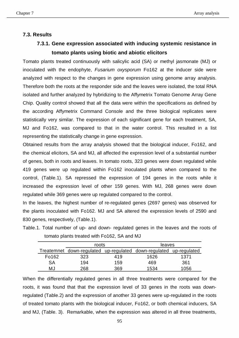
Chapter 7 Array analysis
95
7.3. Results 7.3.1. Gene expression associated with inducing systemic resistance in
tomato plants using biotic and abiotic elicitors Tomato plants treated continuously with salicylic acid (SA) or methyl jasmonate (MJ) or
inoculated with the endophyte, Fusarium oxysporum Fo162 at the inducer side were
analyzed with respect to the changes in gene expression using genome array analysis.
Therefore both the roots at the responder side and the leaves were isolated, the total RNA
isolated and further analyzed by hybridizing to the Affymetrix Tomato Genome Array Gene
Chip. Quality control showed that all the data were within the specifications as defined by
the according Affymetrix Command Console and the three biological replicates were
statistically very similar. The expression of each significant gene for each treatment, SA,
MJ and Fo162, was compared to that in the water control. This resulted in a list
representing the statistically change in gene expression.
Obtained results from the array analysis showed that the biological inducer, Fo162, and
the chemical elicitors, SA and MJ, all affected the expression level of a substantial number
of genes, both in roots and leaves. In tomato roots, 323 genes were down regulated while
419 genes were up regulated within Fo162 inoculated plants when compared to the
control, (Table.1). SA repressed the expression of 194 genes in the roots while it
increased the expression level of other 159 genes. With MJ, 268 genes were down
regulated while 369 genes were up regulated compared to the control.
In the leaves, the highest number of re-regulated genes (2697 genes) was observed for
the plants inoculated with Fo162. MJ and SA altered the expression levels of 2590 and
830 genes, respectively, (Table.1).
Table.1. Total number of up- and down- regulated genes in the leaves and the roots of
tomato plants treated with Fo162, SA and MJ
down-regulated up-regulated down-regulated up-regulatedFo162 323 419 1626 1371
SA 194 159 469 361MJ 268 369 1534 1056
roots leavesTreatemnet
When the differentially regulated genes in all three treatments were compared for the
roots, it was found that that the expression level of 33 genes in the roots was down-
regulated (Table.2) and the expression of another 33 genes were up-regulated in the roots
of treated tomato plants with the biological inducer, Fo162, or both chemical inducers, SA
and MJ, (Table. 3). Remarkable, when the expression was altered in all three treatments,
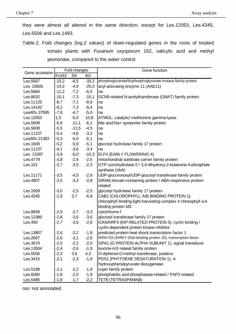
Chapter 7 Array analysis
96
they were almost all altered in the same direction, except for Les.12053, Les.4345,
Les.5506 and Les.1493.
Table.2. Fold changes (log.2 values) of down-regulated genes in the roots of treated
tomato plants with Fusarium oxysporum 162, salicylic acid and methyl
jasmonate, compared to the water control.
Fo162 SA MJLes.5567 -15,2 -6,5 -33,2 phosphoglycerate/bisphosphoglycerate mutase family proteinLes. 23605 -14,2 -4,9 -25,0 acyl-activating enzyme 11 (AAE11)Les.5884 -11,2 -7,2 -6,5 naLes.8632 -10,1 -7,3 -10,1 GCN5-related N-acetyltransferase (GNAT) family proteinLes.11125 -8,7 -7,1 -8,9 naLes.14142 -8,2 -7,3 -6,4 naLesAffx.37595 -7,8 -4,7 -5,0 naLes.12053 1,0 -5,0 10,8 ATMGL; catalytic/ methionine gamma-lyaseLes.5608 -5,9 -11,1 -6,1 bile acid:Na+ symporter family proteinLes.5608 -5,5 -11,5 -4,5 naLes.11107 -5,4 -4,8 -3,2 naLesAffx.21383 -5,3 -6,0 -6,1 naLes.1609 -5,2 -5,9 -5,1 glycosyl hydrolase family 17 proteinLes.11107 -4,1 -3,6 -3,4 naLes. 13397 -3,9 -6,0 -10,3 ELF4 (EARLY FLOWERING 4)Les.4779 -3,8 -2,4 -2,5 mitochondrial substrate carrier family proteinLes.101 -3,7 -3,5 -2,3 GTP cyclohydrolase II / 3,4-dihydroxy-2-butanone-4-phosphate
synthase (ribA)Les.11171 -3,5 -4,5 -2,8 UDP-glucoronosyl/UDP-glucosyl transferase family proteinLes.4807 -3,5 -3,3 -2,8 GRAM domain-containing protein / ABA-responsive protein-
relatedLes.1609 -3,0 -2,5 -2,5 glycosyl hydrolase family 17 protein-Les.4345 -2,9 3,7 -6,8 CAB1 (CHLOROPHYLL A/B BINDING PROTEIN 1);
chlorophyll binding-light-harvesting complex II chlorophyll a-b binding protein M3
Les.8849 -2,9 -2,7 -3,3 cytochrome fLes.12389 -2,8 -3,6 -3,6 glycosyl transferase family 17 proteinLes.450 -2,7 -3,6 -2,6 ICK6/KRP3 (KIP-RELATED PROTEIN 3); cyclin binding /
cyclin-dependent protein kinase inhibitorLes.13807 -2,6 -3,2 -1,8 predicted protein-heat shock transcription factor 1Les.2667 -2,6 -3,1 -2,6 WRKY33 (WRKY DNA-binding protein 33); transcription factorLes.3670 -2,5 -2,2 -2,0 GPA1 (G PROTEIN ALPHA SUBUNIT 1); signal transducerLes.13504 -2,4 -2,6 -1,9 leucine-rich repeat family proteinLes.5506 -2,3 3,6 4,3 O-diphenol-O-methyl transferase, putativeLes.3415 -2,1 -2,3 -1,9 PDS1 (PHYTOENE DESATURATION 1); 4-
hydroxyphenylpyruvate dioxygenaseLes.5168 -2,1 -2,2 -1,9 cupin family proteinLes.6080 -1,8 -2,0 -1,8 phosphatidic acid phosphatase-related / PAP2-relatedLes.5485 -1,6 -1,7 -2,2 TET8 (TETRASPANIN8)
Fold changes Gene functionGene accession
na= not annotated.
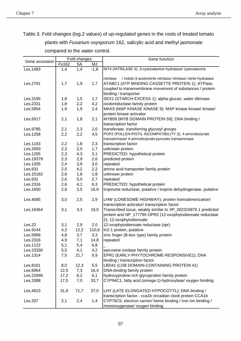
Chapter 7 Array analysis
97
Table.3. Fold changes (log.2 values) of up-regulated genes in the roots of treated tomato
plants with Fusarium oxysporum 162, salicylic acid and methyl jasmonate
compared to the water control.
Fo162 SA MJLes.1493 1,4 1,4 -1,8 NIT4 (NITRILASE 4); 3-cyanoalanine hydratase/ cyanoalanine
nitrilase / indole-3-acetonitrile nitrilase/ nitrilase/ nitrile hydrataseLes.2791 1,7 1,9 1,7 ATABC1 (ATP BINDING CASSETTE PROTEIN 1); ATPase,
coupled to transmembrane movement of substances / protein binding / transporter
Les.3195 1,8 1,5 1,7 SEX1 (STARCH EXCESS 1); alpha-glucan, water dikinaseLes.2331 1,8 2,2 4,2 oxidoreductase family proteinLes.5954 1,9 1,9 2,4 MKK9 (MAP KINASE KINASE 9); MAP kinase kinase/ kinase/
protein kinase activatorLes.5017 2,1 1,8 2,1 MYB59 (MYB DOMAIN PROTEIN 59); DNA binding /
transcription factorLes.9785 2,1 2,3 2,0 transferase, transferring glycosyl groupsLes.1258 2,2 2,2 4,5 POP2 (POLLEN-PISTIL INCOMPATIBILITY 2); 4-aminobutyrate
transaminase/ 4-aminobutyrate:pyruvate transaminaseLes.1163 2,2 1,8 2,3 transcription factorLes.2650 2,3 2,0 1,7 unknown proteinLes.1205 2,3 4,3 3,1 PREDICTED: hypothetical proteinLes.19379 2,3 2,9 2,6 predicted proteinLes.1205 2,4 3,9 3,0 repeatedLes.631 2,5 4,2 2,2 amino acid transporter family proteinLes.15182 2,6 1,8 1,8 unknown proteinLes.631 2,6 5,0 2,7 repeatedLes.2316 2,6 4,1 6,3 PREDICTED: hypothetical proteinLes.1650 2,8 3,5 16,9 tropinone reductase, putative / tropine dehydrogenase, putative
Les.4085 3,0 2,5 2,9 LHW (LONESOME HIGHWAY); protein homodimerization/ transcription activator/ transcription factor
Les.16464 3,1 3,3 19,5 Transcribed locus, weakly similar to XP_002315876.1 predicted protein and NP_177795 OPR2 (12-oxophytodienoate reductase 2); 12-oxophytodienoate
Les.22 3,1 2,9 2,0 12-oxophytodienoate reductase (opr)Les.9144 4,3 12,2 110,6 In2-1 protein, putativeLes.5999 4,8 3,7 3,3 zinc finger (B-box type) family proteinLes.2316 4,9 7,1 14,8 repeatedLes.1122 5,1 5,4 6,8 _Les.23330 5,5 4,1 4,2 sarcosine oxidase family proteinLes.1314 7,5 21,7 9,9 EPR1 (EARLY-PHYTOCHROME-RESPONSIVE1); DNA
binding / transcription factorLes.8161 8,0 12,3 5,5 LBD41 (LOB DOMAIN-CONTAINING PROTEIN 41)Les.6954 12,5 7,3 16,4 DNA-binding family proteinLes.22696 17,2 6,2 6,1 hydroxyproline-rich glycoprotein family proteinLes.3388 17,5 7,0 33,7 CYP94C1; fatty acid (omega-1)-hydroxylase/ oxygen binding
Les.4923 31,8 71,7 37,0 LHY (LATE ELONGATED HYPOCOTYL); DNA binding / transcription factor.- cca1b circadian clock protein CCA1b
Les.207 2,1 2,4 1,4 CYP76C5; electron carrier/ heme binding / iron ion binding / monooxygenase/ oxygen binding
Gene accession Fold changes Gene function
Chapter 7 Array analysis
98
In the leaves of Fo162 inoculated plants, the fold changes in the gene expression
compared to the control ranged from -130 to 45 with the genes of Les.2792 and
Les.19563, respectively. However, the putative function of Les. 2792 was not assigned
while Les.19562 was found to be a member of GCN5-related N-acetyltransferase (GNAT)
family protein and therefore was considered a transcriptional co-activator. With SA treated
plants, the fold changes in the gene expression compared to the control ranged from -57
to 119. This was observed for Les.5608 and Les.4057, respectively. The Les.5608 was
found to be a member of bile acid: Na+ symporter family protein, which is involved in ion
transport. The Les.4057 gene was annotated as Lycopersicon esculentum DB29, but no
further information could be found. In MJ treated plants, the fold changes in the gene
expression compared to the control ranged from -130 to 2081 with Les.2792 and Les.840,
respectively. Until now, both genes were not annotated.
When the differentially regulated genes in all three treatments were compared for the
leaves, it was found that the expression level of 422 genes was altered, of which 146
genes were up-regulated, (Fig.1), and 276 genes were down-regulated, (Fig.2). The
highest number of up regulated genes, 27, was observed for the genes related to
developmental pathways followed by those involved in the signalling pathways (24 genes).
When the down regulated genes were further analyzed, this showed that the highest
number of genes, 60, was related to protein synthesis.
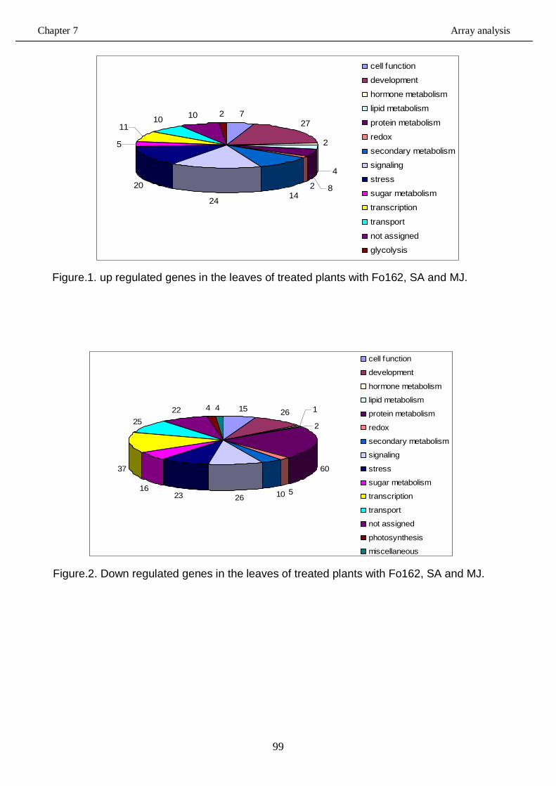
Chapter 7 Array analysis
99
727
2
4
821424
20
5
1110 10 2
cell function
development
hormone metabolism
lipid metabolism
protein metabolism
redox
secondary metabolism
signaling
stress
sugar metabolism
transcription
transport
not assigned
glycolysis
Figure.1. up regulated genes in the leaves of treated plants with Fo162, SA and MJ.
15 26 1
2
60
510262316
37
2522 4 4
cell function
development
hormone metabolism
lipid metabolism
protein metabolism
redox
secondary metabolism
signaling
stress
sugar metabolism
transcription
transport
not assigned
photosynthesis
miscellaneous Figure.2. Down regulated genes in the leaves of treated plants with Fo162, SA and MJ.
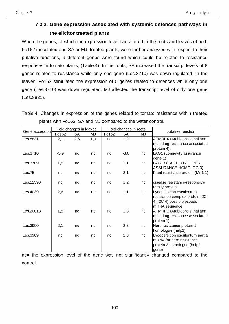
Chapter 7 Array analysis
100
7.3.2. Gene expression associated with systemic defences pathways in
the elicitor treated plants
When the genes, of which the expression level had altered in the roots and leaves of both
Fo162 inoculated and SA or MJ treated plants, were further analyzed with respect to their
putative functions, 9 different genes were found which could be related to resistance
responses in tomato plants, (Table.4). In the roots, SA increased the transcript levels of 8
genes related to resistance while only one gene (Les.3710) was down regulated. In the
leaves, Fo162 stimulated the expression of 5 genes related to defences while only one
gene (Les.3710) was down regulated. MJ affected the transcript level of only one gene
(Les.8831).
Table.4. Changes in expression of the genes related to tomato resistance within treated
plants with Fo162, SA and MJ compared to the water control.
Fo162 SA MJ Fo162 SA MJLes.8831 2,1 2,5 1,9 nc 1,2 nc ATMRP4 (Arabidopsis thaliana
multidrug resistance-associated protein 4).
Les.3710 -5,9 nc nc nc -3,0 nc LAG1 (Longevity assurance gene 1)
Les.3709 1,5 nc nc nc 1,1 nc LAG13 (LAG1 LONGEVITY ASSURANCE HOMOLOG 3)
Les.75 nc nc nc nc 2,1 nc Plant resistance protein (Mi-1.1)
Les.12390 nc nc nc nc 1,2 nc disease resistance-responsive family protein
Les.4039 2,6 nc nc nc 1,1 nc Lycopersicon esculentum resistance complex protein I2C-4 (I2C-4) possible pseudo mRNA sequence
Les.20018 1,5 nc nc nc 1,3 nc ATMRP1 (Arabidopsis thaliana multidrug resistance-associated protein 1);
Les.3990 2,1 nc nc nc 2,3 nc Hero resistance protein 1 homologue (help1)
Les.3989 nc nc nc nc 2,3 nc Lycopersicon esculentum partial mRNA for hero resistance protein 2 homologue (help2 gene)
Gene accession Fold changes in leaves putative functionFold changes in roots
nc= the expression level of the gene was not significantly changed compared to the
control.
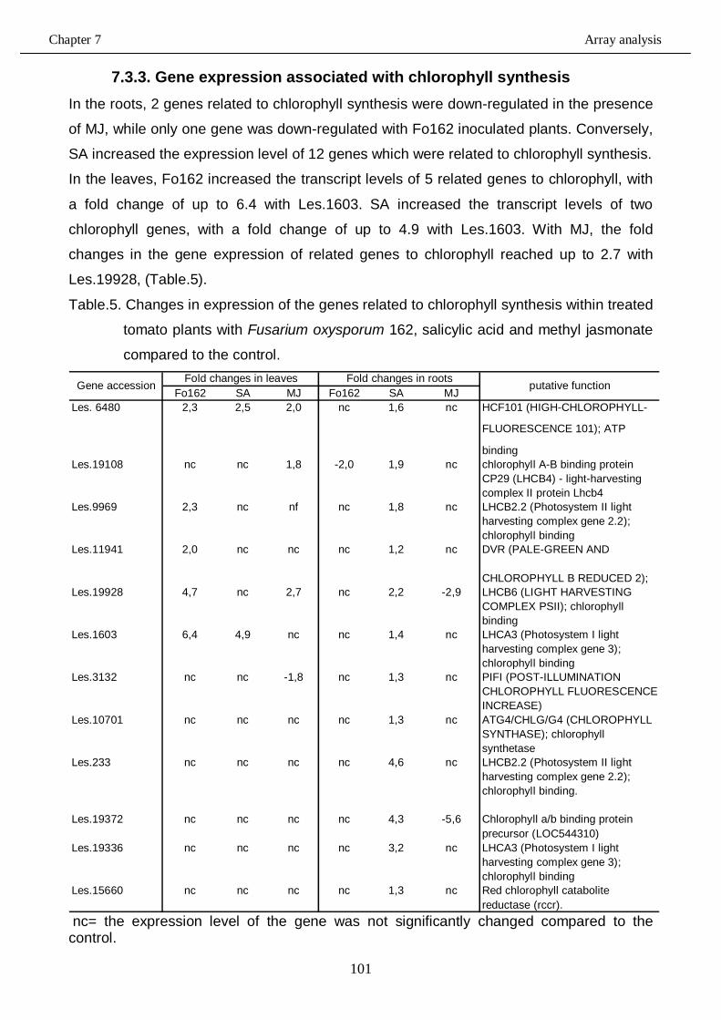
Chapter 7 Array analysis
101
7.3.3. Gene expression associated with chlorophyll synthesis In the roots, 2 genes related to chlorophyll synthesis were down-regulated in the presence
of MJ, while only one gene was down-regulated with Fo162 inoculated plants. Conversely,
SA increased the expression level of 12 genes which were related to chlorophyll synthesis.
In the leaves, Fo162 increased the transcript levels of 5 related genes to chlorophyll, with
a fold change of up to 6.4 with Les.1603. SA increased the transcript levels of two
chlorophyll genes, with a fold change of up to 4.9 with Les.1603. With MJ, the fold
changes in the gene expression of related genes to chlorophyll reached up to 2.7 with
Les.19928, (Table.5).
Table.5. Changes in expression of the genes related to chlorophyll synthesis within treated
tomato plants with Fusarium oxysporum 162, salicylic acid and methyl jasmonate
compared to the control.
Fo162 SA MJ Fo162 SA MJLes. 6480 2,3 2,5 2,0 nc 1,6 nc HCF101 (HIGH-CHLOROPHYLL-
FLUORESCENCE 101); ATP
bindingLes.19108 nc nc 1,8 -2,0 1,9 nc chlorophyll A-B binding protein
CP29 (LHCB4) - light-harvesting complex II protein Lhcb4
Les.9969 2,3 nc nf nc 1,8 nc LHCB2.2 (Photosystem II light harvesting complex gene 2.2); chlorophyll binding
Les.11941 2,0 nc nc nc 1,2 nc DVR (PALE-GREEN AND
CHLOROPHYLL B REDUCED 2); Les.19928 4,7 nc 2,7 nc 2,2 -2,9 LHCB6 (LIGHT HARVESTING
COMPLEX PSII); chlorophyll binding
Les.1603 6,4 4,9 nc nc 1,4 nc LHCA3 (Photosystem I light harvesting complex gene 3); chlorophyll binding
Les.3132 nc nc -1,8 nc 1,3 nc PIFI (POST-ILLUMINATION CHLOROPHYLL FLUORESCENCE INCREASE)
Les.10701 nc nc nc nc 1,3 nc ATG4/CHLG/G4 (CHLOROPHYLL SYNTHASE); chlorophyll synthetase
Les.233 nc nc nc nc 4,6 nc LHCB2.2 (Photosystem II light harvesting complex gene 2.2); chlorophyll binding.
Les.19372 nc nc nc nc 4,3 -5,6 Chlorophyll a/b binding protein precursor (LOC544310)
Les.19336 nc nc nc nc 3,2 nc LHCA3 (Photosystem I light harvesting complex gene 3); chlorophyll binding
Les.15660 nc nc nc nc 1,3 nc Red chlorophyll catabolite reductase (rccr).
Gene accession Fold changes in leaves Fold changes in roots putative function
nc= the expression level of the gene was not significantly changed compared to the control.
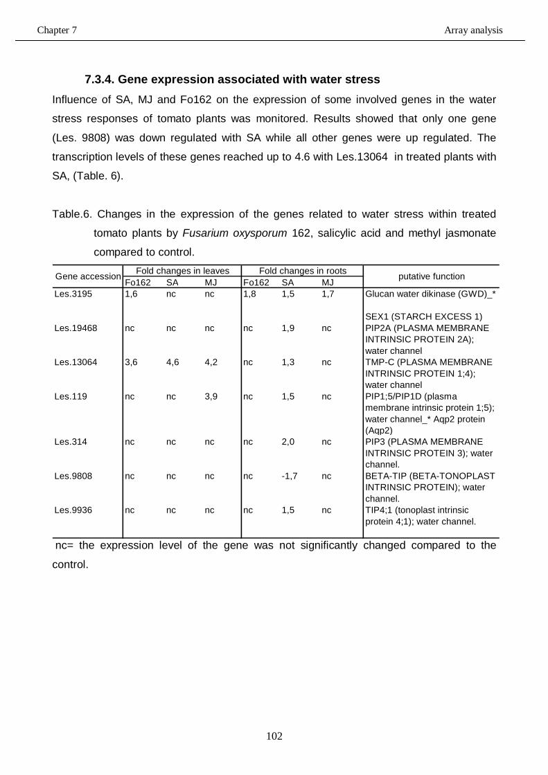
Chapter 7 Array analysis
102
7.3.4. Gene expression associated with water stress Influence of SA, MJ and Fo162 on the expression of some involved genes in the water
stress responses of tomato plants was monitored. Results showed that only one gene
(Les. 9808) was down regulated with SA while all other genes were up regulated. The
transcription levels of these genes reached up to 4.6 with Les.13064 in treated plants with
SA, (Table. 6).
Table.6. Changes in the expression of the genes related to water stress within treated
tomato plants by Fusarium oxysporum 162, salicylic acid and methyl jasmonate
compared to control.
Fo162 SA MJ Fo162 SA MJLes.3195 1,6 nc nc 1,8 1,5 1,7 Glucan water dikinase (GWD)_*
SEX1 (STARCH EXCESS 1)Les.19468 nc nc nc nc 1,9 nc PIP2A (PLASMA MEMBRANE
INTRINSIC PROTEIN 2A); water channel
Les.13064 3,6 4,6 4,2 nc 1,3 nc TMP-C (PLASMA MEMBRANE INTRINSIC PROTEIN 1;4); water channel
Les.119 nc nc 3,9 nc 1,5 nc PIP1;5/PIP1D (plasma membrane intrinsic protein 1;5); water channel_* Aqp2 protein (Aqp2)
Les.314 nc nc nc nc 2,0 nc PIP3 (PLASMA MEMBRANE INTRINSIC PROTEIN 3); water channel.
Les.9808 nc nc nc nc -1,7 nc BETA-TIP (BETA-TONOPLAST INTRINSIC PROTEIN); water channel.
Les.9936 nc nc nc nc 1,5 nc TIP4;1 (tonoplast intrinsic protein 4;1); water channel.
Gene accession Fold changes in leaves Fold changes in roots putative function
nc= the expression level of the gene was not significantly changed compared to the
control.
Chapter 7 Array analysis
103
7.4. Discussion 7.4.1. Gene expression associated with inducing systemic resistance in
tomato plants using biotic and abiotic elicitors In this genome array study, the effect of exogenous application of salicylic acid (as inducer
of SAR pathway), methyl jasmonate (as inducer of ISR pathway) and the mutualistic
endophyte Fusarium oxysporum strain 162 (as biological inducer of systemic resistance
against M. incognita) on the expression of 9,254 Lycopersicon esculentum genes in leaves
and roots was monitored in a split root experiment. This number of genes is by no means
covering the complete gene content of tomato. Furthermore, not all probes found on the
gene chip have been annotated with respect to gene function. Nevertheless, the analysis
gives a good insight in the overall changes taking place in the tomato plant in the presence
of the biotic and abiotic elicitors of systemic resistance against root knot nematodes.
When applied to the roots (inducer side), we observed that all three elicitors, Fo162, SA
and MJ, altered the transcript level of a substantial number of genes in both the responder
roots and leaves.
Fo162 showed an alteration in transcription level of more than 29% of the genes. This
systemic effect is remarkable, since Fo162 behaves like an endophyte and, consequently,
does not cause any substantial phenotypic changes in the plant. This percentage is even
greater that the alterations caused by MJ (25%). As demonstrated in the previous chapter
MJ leads to phenotypic chances in the tomato plant. In this light the effects caused by SA
with 9% re-regulation of gene expression can be considered modest. The result with the
chemical elicitors corroborate with other studies of the past years with microarray analysis
of both compatible and incompatible plant-pathogen interaction showed that hundreds of
genes were up- and down regulated (van Loon et al., 2006). These studies showed that
the exogenous supply with SA and methyl jasmonate resulted in changing the expression
level of many different genes sets which some of them mediate different kinds of tomato
systemic resistance. Nowadays, it is known that defense mechanism is regulated with
complex and separate different pathways (Luo et al., 2009).
These high percentages only applied to the tomato leaves. The number of genes showing
a change in expression level in the roots was significantly lower for al three treatments and
ranged from 3.5 for the SA treatment to 7.3 % for the Fo162 inoculation. Apparently, the
systemic responses to the inducers are more pronounced in leaves when compared to the
roots.
Chapter 7 Array analysis
104
Nevertheless, as demonstrated in the previous chapter, systemic resistance against root
knot nematode colonization did occur with all three elicitors. When the genes were
selected that were altered in expression level in the roots for all three elicitors, we found
only 66 genes in common, which had almost all altered in the same direction with the three
elicitors. However, within this list we could not discover genes that could be directly
associated with nematode resistance. There may be several reasons for that. First, the
function of some of the genes is currently not known. Second, since the array does not
cover all genes within the tomato genome, we may have missed the relevant genes. Third,
the plant may only be primed and the relevant resistance genes will only become visible
when the nematode is entering the root system. In this case, we would have had to include
RNA from a split root experiment, infected with nematodes at the responder side.
Williamson and Hussey (1996) reported that many of the genes involved in the interaction
between root-knot nematode and their hosts. These genes were found to be members of
gene families and have a complex regulation patter. However, to avoid the influences in
the gene expression during the interactions between M. incognita and tomato plants, the
array analysis was conducted using elicitor treated plants in the absence of nematode
colonization. Fourth, the genes playing a role in nematode resistance are expressed
mainly in the leaves and not in the roots.
When the genes were selected that were in common and thus altered in expression level
in the leaves for all three elicitors, we found 422 genes. Like in the roots, almost all these
genes were altered in the same direction with the three elicitors. Their gene products had
an array of functions, ranging from basic cell function and development to stress and
protein metabolism.
In the leaves several interesting alterations in gene expression were found, that may
support the suggestion that nematode defense related factors originate from the leaves.
Two related genes to Meloidogyne-induced giant cell altered in their expression in the
presence of the elicitor, Les.4082 and Les.4032. Les.4082 was always down-regulated
while the other one, Les.4032, was always up-regulated with all treatments (data not
shown). This variation within the expression of those two genes, which gene products can
control or are involved in giant cell formation (Bird and Wilson, 1994) may affect the
pericycle tissues. Parizot et al., (2008) reported that the pericycle is a heterogeneous cell
layer with two groups of cells set up in the root meristem by the same genetic pathway
controlling the diarch organization of the vascular system. Although, additional information
about the true function of these genes is necessary, which potentially leads to new insights
Chapter 7 Array analysis
105
regarding giant cell formation and plant resistance, it indicates that genome array analysis
can support to identify crucial genes playing a role in plant-pathogen interactions.
The transcription of defense-related genes was found to responsible for the accumulation
of different defense compounds and antimicrobial activities with host plant (Hammond-
Kosack and Jones, 1996). It was found also that the exogenous application of salicylic
acid as well as jasmonates resulted in activation of different defense-related genes. For
example, Uknes et al., (1992) reported that SA affecting the gene expression of
pathogenesis-related proteins of the families PR-2, PR-5 and PR-1 while other studies
(Penninckx et al., 1996; Thomma et al., 1998; and Melan et. al., 1993) showed that
jasmonate induces the expression of other different gene sets in Arabidopsis plants i.e.
Hel, ChiB and Pdf1.2 as well as Lox1 and Lox2 which are related to defenses of host
plants.
Although some of the PR-genes e.g. 1b1, p23, 5x, 1a1, P2 and P4 were indeed present on
the tomato array. Significant changes in the expression of these genes were observed only
in the root of SA treated plants while no significant changes were detected with the other
elicitors. This may also be due to the prolonged exposure to the elicitors. Normally the up-
regulation in expression of these genes are observed shortly after the application of the
elicitors.
Our results also agree with those of with van Wees et al., (2000), who demonstrated that
different defence pathways can be activated within plants treated with distinct inducing
agents. Their results illustrated also that both of SAR and ISR systemic resistance which
are mediated by SA and MJ signalling pathways, respectively provided an attractive tool
for the improvement of disease control. Moreover, they added that the SAR- and ISR-
regulatory gene Npr1 was over expressed (and/or activated) when both of ISR and SAR
signalling pathways were stimulated during Arabidopsis infection with non-pathogenic
rhizobacteria and pathogen infection, respectively. In advanced research, Branch et al.,
(2004) investigated the role of SA, which plays a crucial role in alteration of many of R
gene-mediated resistance responses in tomato and they demonstrated that SA was
required for Mi-mediated nematode resistance in tomato roots. SA, JA and ET are three
hormones which accumulate in response to pathogen infection or herbivore damage,
leading to the activation of distinct and partly overlapping sets of defense-related genes.
The studies conducted with Arabidopsis wild type plants and the SA-, JA-, or ET- signalling
mutants Arabidopsis plants that were infected by Pseudomonas syringae pv. maculicola
Chapter 7 Array analysis
106
showed an evidence for the substantial cross-talk between the signalling pathways which
were mediated by the three hormones (Glazebrook et al., 2003).
Moreover, van Wees et al., (1999) reported that inducing SAR pathways in Arabidopsis
plants, by pre-inoculated lower leaves with avirulant isolate of Pseudomonas syringae pv.
tomato, affected the infection with pathogenic Pseudomonas syringae pv. tomato isolate in
the upper leaves and also resulted in increasing the expression levels of different tested
genes which some of them related to ISR pathway while the other are related to SAR
pathway.
7.4.2. Gene expression associated with chlorophyll synthesis and water stress
The regulation levels of genes related to some vital growth signs like chlorophyll
accumulation and transpiration level in tomato plants were also analyzed to find out if there
is a correlation between the induction to different types of tomato systemic resistance and
some of the overall physiology of the plant.
As observed before with related genes to defense pathways, the highest number (12
genes) of up regulated related genes to chlorophyll synthesis was observed with SA
treated plants followed by plants inoculated with Fo162 (5 genes). In contrast, MJ inhibited
the regulation of three genes that were related to chlorophyll pathway. These findings
corroborate with the results obtained from measuring the accumulation of chlorophyll
pigment in the leaves of treated tomato plants with SA, MJ and Fo162 (see previous
chapter).
The changes in the expression of genes related to water stress suggest that plants treated
with SA a less exposed to water stress. This is in agreement with the thermal imaging
data, which reflects the water content in tomato plants and thus indicates the transpiration
levels (see previous chapter). Alfano et al., (2007) used high-density oligonucleotide
microarrays to investigate the expression pattern of 15925 genes in tomato leaves
obtained from inoculated plants with biocontrol agent Trichoderma hamatum 382 prior their
inoculation with the pathogen. Their results detected up-regulation of different genes
associated with biotic or abiotic stress as well as RNA, DNA and protein metabolism was
detected also.
Chapter 7 Array analysis
107
7.5. Conclusion Based on the finding in the present investigation the following can be concluded:
1- Micro array analysis was a useful tool for determining the molecular changes within
treated tomato plants with different inducers responsible for activation different
types of systemic resistance.
2- The chemical elicitors, SA and MJ, and the biological inducer, Fo162, alter the
expression of many genes which can play a certain role in the diseases resistance.
3- The highest number of genes which were altered in expression levels was detected
within the plants leaves, especially the plants inoculated with Fo162.
4- Inducing the defence related genes in tomato plants using the biological or the
chemical inducers also can affect the expression patterns of genes whose products
are associated with chlorophyll synthesis and water stress.
Chapter 7 Array analysis
108
7.6. References
- Alfano, G., Lewis Ivey, M. L., Cakir, C., Bos, J. I. B., Miller, S. A., Madden, L.
V., Kamoun, S. and Hoitink, H. A. J. (2007). Systemic modulation of gene
expression in tomato by Trichoderma hamatum 382. Phytopathology, 97, 429-437.
- Bird, D. M. and Wilson, M A. (1994). DNA sequence ans expression analysis of
root-knot nematode-elicited giant cell transcripts. Mol. Plant Microbe Interaction, 7 (3), 419-424.
- Branch, C., Hwang, C-F., Navarre, D. A. and Williamson, V. M. (2004). Salicylic
acid is part of the Mi-1- mediated defense response to root-knot nematode in
tomato. MPMI, vol. 17. No. 4, pp. 351-356.
- Farmer, E. E. and Ryan, C. A. (1992). Octadecanoid precursors of jasmonic acid
activate the synthesis of wound-inducible proteinase-inhibitors. Plant Cell, 4, 129-
134.
- Glazebrook, J., Chen, W. J., Estes, B., Chang, H-S., Nawrath, C., Métraux, J-P.,
Zhu, T. and Katagiri, F. (2003). Topology of the network integrating salicylate and
jasmonate signal transduction derived from global expression phenotyping. Plant J,
34, 217-228.
- Hammond-Kosack, K. E. and Jones, J. D. G. (1996). Resistance gene-dependent
plant defense responses. Plant Cell, 8, 1773-1791.
- Lawton, K., Weymann, K., Friedrich, L., Vernooij, B., Uknes, S. and Ryals, J.
(1995). Systemic acquired resistance in Arabidopsis requires salicylic acid but not
ethylene. Mol. Plant-Microbe interact, 8, 863-870.
- Li, C. Y., Liu, G. H., Xu, C. C., Lee, G. I., Bauer, P., Ling, H. Q., Ganal, M. W. and
Howe, G. A. (2003). The tomato suppressor of prosystemin-mediated responses 2
gene encodes a fatty acid desaturase required for the biosynthesis of Jasmonic
acid and the production of a systemic wound signal for defense gene expression.
Plant cell, 15, 1646-1661.
- Luo, Z-B., Janz, D., Jiang, X., Göbel, J., Wildhagen, H., Tan, Y., Rennenberg, H.,
Feussner, I. and Polle, A. (2009). Upgrading root physiology for stress tolerance by
ectomycorrhizas: insights from metabolite and transcriptional profiling into
reprogramming for stress anticipation. Plant physiology, Vol. 151, pp. 1902-1917.
Chapter 7 Array analysis
109
- Melan, M. A., Dong, X., Endara, M. E., Davis, K. R., Ausubel, F. M. and Peterman,
T. K. (1993). An Arabidopsis thaliana lipoxygenase gene can be induced by
pathogens, abscisic, and methyl jasmonate. Plant Physiol,101, 441-450.
- Parizot, B., Laplaze, L., Ricaud, L., Boucheron-Dubuisson, E., Bayle, V., Bonke, M.,
De Smet, I., Poethig, S. R., Helariutta, Y., Haseloff, J., Chriqui, D., Beeckman, T.
and Nussaume, L. (2008). Diarch symmetry of the vascular bundle in Arabidopsis
root encompasses the pericycle and is reflected in distich lateral root initiation. Plant
Physiology, Vol. 146, pp. 140–148.
- Penninckx, I. A. M. A., Eggermont, K., Terras, F. R. G., Thomma, B. P. H. J., De
Samblanx, G. W., Buchala, A., Métraux, J.–P., Manners, J. M. and Broekaert, W. F.
(1996). Pathogen-induced systemic activation of a plant defensin gene in
Arabidopsis follows a salicylic acid-independent pathway. Plant cell, 8, 2309-2323.
- Pieterse, C. M. J. and Van Loon, L. (2004). NPR1: the spider in the web of induced
resistance signalling pathways. Current Opinion in Plant Biology, 7, 456-464.
- Pieterse, C. M. J., van Wees, S. C. M., van Pelt, J. A., Knoester, M., Laan, R.,
Gerrits, H., Weisbeek, P. J. and van Loon, L. C. (1998). A novel signalling pathway
controlling induced systemic resistance in Arabidopsis. Plant Cell ,10, 1571-1580.
- Pieterse, C. M. J., Van Wees, S. C. M.,Ton, J., Van Pelt, J. A. and Van Loon, L. C.
(2002). Signalling in rhizobacteria-induced systemic resistance in Arabidopsis
thaliana. Plant Biology, 4, 535-544.
- Sticher, L., Mauch-Mani, B. and Métraux, J. (1997). Systemic acquired resistance.
Annu. Rev. Phytopathology, 35, 235-270.
- Tamogami, S. and Kodama, O. (2000). Coronatine elicits phytoalexin production in
rice leaves (Oryza sativa L.) in the same manner as jasmonic acid. Phytochemistry,
54, 689-694.
- Thomma, B. P. H. J., Eggermont, K., Penninckx, I. A. M. A., Mauch-Mani, B.,
Cammue, B. P. A. and Broekaert, W. F. (1998). Separate jasmonate-dependent
and salicylic acid-dependent defense response pathways in Arabidopsis are
essential for resistance to distinct microbial pathogens. Proc. Natl. Acad. Sci. USA
,95, 15107-15111.
- Ton, J., Van Pelt, J. A., Van Loon, L. C. and Pieterse, C. M. J. (2002). Differential
effectiveness of salicylate-dependent and jasmonate/ethylene-dependent induced
resistance in Arabidopsis. Mol. Plant-Microbe Interact, 15, 27-34.
Chapter 7 Array analysis
110
- Ton, J., Van Pelt, J. A., Van Loon, L. C. and Pieterse, C. M. J. (2002). .
Differential effectiveness of salicylate-dependant and jasmonate/ethylene-
dependant induced resistance in Arabidopsis. Mol. Plant Microbe Interact, 15, 27-
34.
- Uknes, S., Mauch-Mani, B., Moyer, M., Potter, S., Williams, S., Dincher, S.,
Chandler, D., Slusarenko, A., Ward, E. and Ryals, J. (1992). Acquired resistance in
Arabidopsis. Plant cell, 4, 645-656.
- Vallad, G. E. and Goodman, R. M. (2004). Systemic acquired resistance and
induced systemic resistance in conventional agriculture. Crop science, 44, 1920-
1934.
- Van Loon, L. C., Rep, M. and Pieterse, C. M. J. (2006). Significance of inducible
defense-related proteins in elected plants. Annu. Rev. Phytopathology, 44, 135-162.
- van Wees, S. C. M., de Swart, E. A. M., van Pelt, J. A., van Loon, L. C. and
Pieterse, C. M. J. (2000). Enhancement of induced disease resistance by
simultaneous activation of salicylate- and jasmonate-dependant defense pathways
in Arabidopsis thaliana. Proc. Natl. Acad. Sci. USA, vol. 97 no. 15 8711-8716.
- van Wees, S. C. M., Luijendijk, M., Smoorenburg, I., van Loon, L. C., and Pieterse,
C. M. J. (1999). Rhizobacteria-mediated induced systemic resistance (ISR) in
Arabidopsis is not associated with a direct effect on expression of known defense-
related genes but stimulates the expression of the jasmonate-inducible gene Atvsp
upon challenge. Plant Molecular Biology, 41, 537-549.
- Williamson, V. M., and Hussey, R. S. (1996). Nematode pathogenesis and
resistance in plants. The plant cell, Vol.8, 1735-1745.

Acknowledgments Firstly, I extend my sincere and eternal gratitude and thankfulness to Prof. Dr.
Richard Sikora for supporting and guiding me at all times through the entire
process of the doctoral research in Germany.
I would like to thank Prof. Dr. Lens Leon for accepting to be my second
supervisor to evaluate this research.
My special thanks to Dr. Alexander Schouten for his guidance and orientation in
this research as well as for his comments on this manuscript.
Special thanks to Dr. P. van Baarlen for helping and supporting me during array
analysis studies.
I acknowledge the entire INRES-Phytomedizin Böden Ökosystem Team 2006-
2010 for their outstanding support, cooperation and useful discussions.
I am very grateful with the Egyptian high education ministry for funding this
research throughout a scholarship.
Finally, I am very grateful with my family members, especially my father and
my mother that I owe everything I am, bothers and sister as well as my wife and
my daughters for their encouragement and support to reach this goal in my
professional career.


![Untersuchungen zu Kontaminationen mit Fusarium spp. und ... · [1] Brayford D (1996): Fusarium oxysporum f. sp. cepae . Mycopathologia 133: 39-40 [2] Du Toit LJ, Inglis DA, Pelter](https://img.pdfslide.org/doc/110x75/6080bb4f104c926f417b96d1/untersuchungen-zu-kontaminationen-mit-fusarium-spp-und-1-brayford-d-1996.jpg)