Embed Size (px)
Citation preview
Microbacterium insulae sp. nov., isolated from soil
Jung-Hoon Yoon,1 Peter Schumann,2 So-Jung Kang,1 Chang-Soo Lee,1
Soo-Young Lee1 and Tae-Kwang Oh1
Correspondence
Jung-Hoon Yoon
1Korea Research Institute of Bioscience and Biotechnology (KRIBB), PO Box 115, Yusong, Taejon,Republic of Korea
2DSMZ – Deutsche Sammlung von Mikroorganismen und Zellkulturen GmbH, Inhoffenstraße 7B,D-38124 Braunschweig, Germany
A Gram-positive, non-motile, rod- or coccoid-shaped Microbacterium-like bacterium, designated
strain DS-66T, was isolated from soil of Dokdo, Korea, and its exact taxonomic position was
investigated by using a polyphasic approach. Strain DS-66T grew optimally at 30 6C and pH 6.5–
7.0 in the presence of 0.5–1.0 % (w/v) NaCl. Phylogenetic analysis based on 16S rRNA gene
sequences showed that strain DS-66T belonged to the genus Microbacterium. Strain DS-66T had
a peptidoglycan type based on B2b with partial substitution of glutamic acid by 3-hydroxy
glutamic acid (Glu/Hyg–Gly–D-Orn), and galactose, rhamnose and ribose as whole-cell sugars.
The acyl type was glycolyl. Strain DS-66T contained MK-13, MK-12 and MK-14 as predominant
menaquinones and anteiso-C15 : 0, anteiso-C17 : 0, iso-C17 : 0 and iso-C16 : 0 as major fatty acids.
Major polar lipids were diphosphatidylglycerol, phosphatidylglycerol, an unidentified phospholipid
and an unidentified glycolipid. The DNA G+C content was 69.9 mol%. Phylogenetic
distinctiveness, DNA–DNA relatedness data and differential phenotypic properties demonstrated
that strain DS-66T is distinguishable from recognized Microbacterium species. On the basis of
the data presented, strain DS-66T is considered to represent a novel species of the genus
Microbacterium, for which the name Microbacterium insulae sp. nov. is proposed. The type strain
is DS-66T (5KCTC 19247T5CCUG 54523T).
The genus Microbacterium was proposed by Orla-Jensen(1919); at the time of writing, the genus comprised 50species with validly published names, including severaldescribed since 2006: Microbacterium koreense (Lee et al.,2006), M. paludicola (Park et al., 2006), M. aoyamense, M.deminutum and M. pumilum (Kageyama et al., 2006),M. indicum (Shivaji et al., 2007), M. sediminicola and M.marinilacus (Kageyama et al., 2007), M. ginsengisoli (Parket al., 2008) and M. hatanonis (Bakir et al., 2008). Here wereport on the taxonomic characterization of aMicrobacterium-like strain, designated DS-66T, which wasisolated from soil of Dokdo, Korea.
Strain DS-66T was isolated by using the standard dilutionplating technique at 25 uC by using 106 diluted nutrientagar (Difco). The type strains of three recognizedMicrobacterium species were used as reference strains:Microbacterium hominis DSM 12509T and Microbacterium
trichothecenolyticum DSM 8608T were obtained from theDeutsche Sammlung von Mikroorganismen undZellkulturen (DSMZ), Braunschweig, Germany andMicrobacterium xylanilyticum KCTC 19079T was obtainedfrom the Korean Collection for Type Cultures, Taejon,Korea. The morphological, physiological and biochemicalcharacteristics of strain DS-66T were investigated by usingroutine cultivation on trypticase soy agar (TSA; Difco) at30 uC. Cell morphology was examined by using lightmicroscopy (E600; Nikon) and transmission electronmicroscopy (CM-20; Philips). Flagellation was determinedby using transmission electron microscopy on cells fromexponentially growing cultures negatively stained with 1 %(w/v) phosphotungstic acid. Grids were examined afterbeing air-dried. The Gram reaction was determined byusing the bioMerieux Gram stain kit according to themanufacturer’s instructions. Growth at various tempera-tures (4, 10, 15, 20, 25 and 28 uC, and from 30 to 40 uC in1 uC increments) was measured on TSA. To investigatetolerance to NaCl, trypticase soy broth (TSB) was preparedaccording to the formula of the Difco medium and NaClconcentrations were varied [0.5 % (w/v) and 1.0–7.0 %(w/v) at increments of 1.0 %]. The pH range for growth wasdetermined in nutrient broth (Difco) that was adjusted tovarious pH values (pH 4.5–10.5 at intervals of 0.5 pH
The GenBank/EMBL/DDBJ accession number for the 16S rRNA genesequence of strain DS-66T is EU239498.
An extended neighbour-joining tree based on 16S rRNA genesequences showing the phylogenetic positions of strain DS-66T, thetype strains of Microbacterium species and representatives of someother related taxa is available as supplementary material with the onlineversion of this paper.
International Journal of Systematic and Evolutionary Microbiology (2009), 59, 1738–1742 DOI 10.1099/ijs.0.007591-0
1738 007591 G 2009 IUMS Printed in Great Britain
units) prior to sterilization by the addition of HCl orNa2CO3. Growth under anaerobic conditions was deter-mined after incubation in an anaerobic chamber on TSAand on TSA supplemented with potassium nitrate (0.1 %,w/v). Catalase and oxidase activities and hydrolysis ofcasein, gelatin, hypoxanthine, starch, Tweens 20, 40, 60 and80, tyrosine, urea and xanthine were determined asdescribed by Cowan & Steel (1965). Hydrolysis of aesculinand reduction of nitrate were studied as described by Lanyı(1987). Susceptibility to antibiotics was tested on TSAplates using antibiotic discs containing the followingcompounds: polymyxin B (100 U), streptomycin (50 mg),penicillin G (20 U), chloramphenicol (100 mg), ampicillin(10 mg), cephalothin (30 mg), gentamicin (30 mg), novo-biocin (5 mg), tetracycline (30 mg), kanamycin (30 mg),lincomycin (15 mg), oleandomycin (15 mg), neomycin(30 mg) and carbenicillin (100 mg). Utilization of varioussubstrates, enzyme activities, and other physiological andbiochemical properties were tested by using the API 20E,API 20NE, API 50 CH and API ZYM systems(bioMerieux); utilization of various substrates was deter-mined by inoculating API 20NE and API 50 CH strips withcells suspended in AUX medium (bioMerieux).
Cell biomass for DNA extraction and for analyses of cell-wall components, isoprenoid quinones and polar lipids wasobtained from cultures grown by shaking at 150 r.p.m. inTSB at 30 uC. Chromosomal DNA was isolated andpurified according to the method described by Yoon et al.(1996), with the exception that RNase T1 was used incombination with RNase A to minimize contaminationwith RNA. The 16S rRNA gene was amplified by PCR byusing two universal primers as described previously (Yoonet al., 1998). Sequencing of the amplified 16S rRNA geneand phylogenetic analysis were performed as described byYoon et al. (2003). The DNA G+C content wasdetermined according to the method of Tamaoka &Komagata (1984) with the modification that DNA washydrolysed and the resultant nucleotides were analysed byreversed-phase HPLC. The presence or absence of diami-nopimelic acid in the peptidoglycan was determined byusing the method described by Komagata & Suzuki (1987).Preparation of cell walls and determination of peptidogly-can structure were carried out by using methods describedby Schleifer & Kandler (1972), MacKenzie (1987) andGroth et al. (1996). Whole-cell sugars were determined asdescribed by Komagata & Suzuki (1987). The cell-wall acyltype was determined as described by Uchida & Aida(1984). Isoprenoid quinones were extracted according tothe method of Komagata & Suzuki (1987) and wereanalysed by using reversed-phase HPLC on a YMC ODS-A(25064.6 mm) column. Polar lipids were extractedaccording to the procedures described by Minnikin et al.(1984) and were identified by two-dimensional TLCfollowed by spraying with appropriate detection reagents(Minnikin et al., 1984; Komagata & Suzuki, 1987). For fattyacid methyl ester analysis, cell mass of strain DS-66T washarvested from TSA plates after incubation for 7 days at
30 uC. The fatty acid methyl esters were extracted andprepared according to the standard protocol of the MIDI/Hewlett Packard Microbial Identification System (Sasser,1990). DNA–DNA hybridization was performed fluorome-trically according to the method of Ezaki et al. (1989) byusing photobiotin-labelled DNA probes and microdilutionwells. Hybridization was performed with five replications foreach sample. The highest and lowest values obtained in eachsample were excluded, and the means of the remaining threevalues were quoted as levels of DNA–DNA relatedness.
Morphological, cultural, physiological and biochemicalcharacteristics of strain DS-66T are given in the speciesdescription below or are shown in Table 1. The almost-complete 16S rRNA gene sequence of strain DS-66T
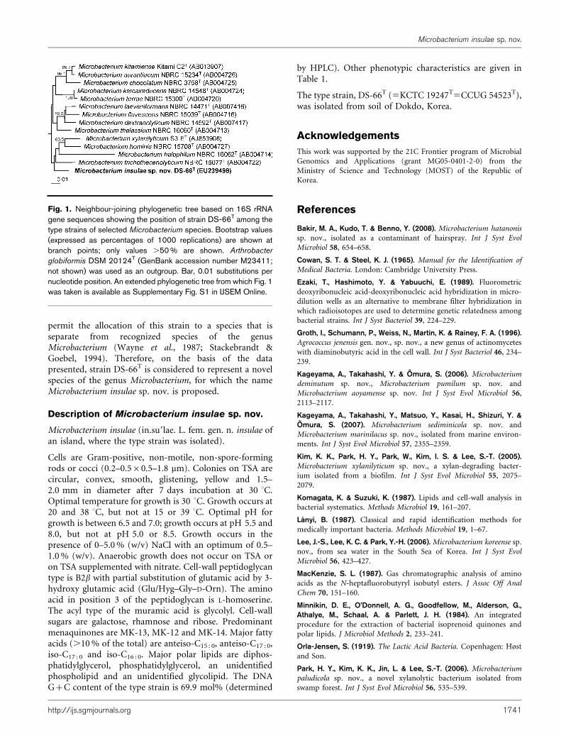
determined in the present study comprised 1447 nt,representing approximately 96 % of the Escherichia coli16S rRNA gene sequence. In the phylogenetic tree based onthe neighbour-joining algorithm, strain DS-66T fell withinthe radiation of the cluster comprising Microbacteriumspecies (Fig. 1; Supplementary Fig. S1 in IJSEM Online).Strain DS-66T exhibited 16S rRNA gene sequence similarityvalues of 98.0, 98.0, 97.6 and 96.7 % with respect to thetype strains of M. hominis, M. trichothecenolyticum, M.xylanilyticum and Microbacterium halophilum, respectively.
Under the condition of total hydrolysis (4 N HCl, 16 h at100 uC), the total hydrolysate of the peptidoglycan of strainDS-66T contained the amino acids ornithine, alanine,glycine, homoserine, glutamic acid and 3-hydroxy glutamicacid (Hyg). In addition, traces of threonine were detected.Quantitative analysis of the peptidoglycan amino acids byTLC, two-dimensional TLC and GC (Schleifer, 1985;MacKenzie, 1987; Groth et al., 1996) showed that strainDS-66T contained alanine, glycine, threonine, homoserine,ornithine and glutamic acid at a ratio of approximately1.5 : 4.9 : 0.2 : 1.5 : 0.6 : 1.0. The amount of 3-hydroxy glu-tamic acid could not be quantified. Dinitrophenylation ofthe peptidoglycan revealed that D-ornithine represents theN terminus of the interpeptide bridge. From these data, itwas concluded that strain DS-66T has peptidoglycan typeB2b with partial substitution of glutamic acid by 3-hydroxyglutamic acid (Glu/Hyg–Gly–D-Orn), as described bySchleifer & Kandler (1972). The amino acids in positions1 and 3 were glycine and L-homoserine, respectively. Theacyl type of the muramic acid was glycolyl. Cell-wall sugarswere galactose, rhamnose and ribose. The predominantmenaquinones detected in strain DS-66T were MK-13,MK-12 and MK-14, at a peak area ratio of approximately64, 17 and 12 %, respectively. Major polar lipids detected instrain DS-66T were diphosphatidylglycerol, phosphatidyl-glycerol, an unidentified phospholipid and an unidentifiedglycolipid. Fatty acids comprising more than 0.5 % of thetotal for strain DS-66T were as follows: branched fatty acidsanteiso-C15 : 0 (50.3 %), anteiso-C17 : 0 (15.9 %), iso-C17 : 0
(10.3 %), iso-C16 : 0 (10.2 %), iso-C15 : 0 (5.7 %), iso-C18 : 0
(3.7 %), anteiso-C19 : 0 (0.8 %) and iso-C14 : 0 (0.8 %); andstraight-chain fatty acids C16 : 0 (1.0 %) and C18 : 0 (0.8 %).This fatty acid profile was similar to those of recognized
Microbacterium insulae sp. nov.
http://ijs.sgmjournals.org 1739
Microbacterium species (Takeuchi & Hatano, 1998a, b; Kimet al., 2005; Kageyama et al., 2006; Lee et al., 2006). TheDNA G+C content of strain DS-66T was 69.9 mol%.Based on the results of phylogenetic and chemotaxonomicinvestigations, it is clear that strain DS-66T is a member ofthe genus Microbacterium (Takeuchi & Hatano, 1998a, b;Kim et al., 2005; Kageyama et al., 2006; Lee et al., 2006).
Strain DS-66T exhibited levels of DNA–DNA relatedness of11–17 % to the type strains of its three closest phylogeneticneighbours, namely M. xylanilyticum KCTC 19079T
(13 %), M. hominis DSM 12509T (17 %) and M. trichothe-cenolyticum DSM 8608T (11 %). These values indicate thatstrain DS-66T represents a genomic species that is distinctfrom M. xylanilyticum, M. hominis and M. trichotheceno-lyticum (Wayne et al., 1987). Strain DS-66T could also bedistinguished from these three recognized Microbacteriumspecies based on differences in several phenotypic prop-erties, as shown in Table 1. The phylogenetic distinctive-ness of strain DS-66T, together with genetic distinctivenessand differential phenotypic properties, is sufficient to
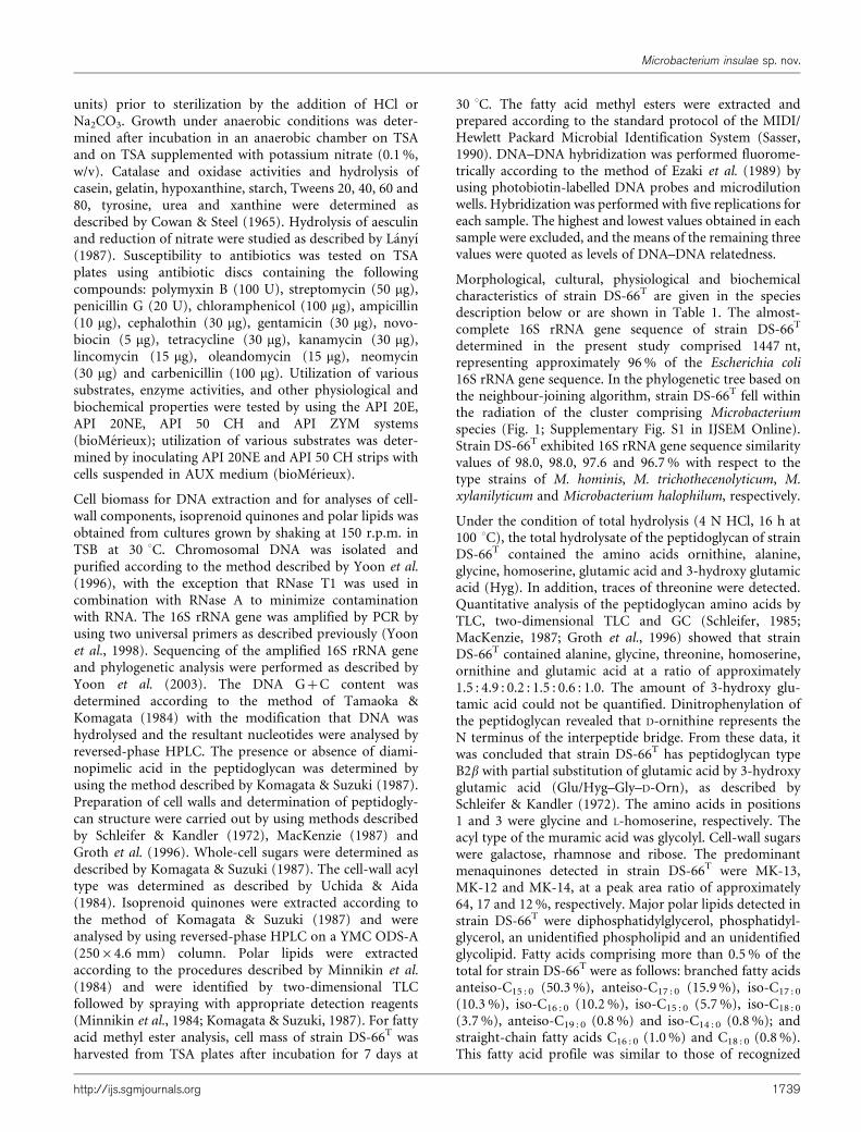
Table 1. Differential phenotypic characteristics between strainDS-66T and phylogenetically related Microbacterium species
Strains: 1, DS-66T (M. insulae sp. nov.); 2, M. hominis DSM 12509T
(data from Takeuchi & Hatano, 1998b; Kim et al., 2005 and the
present study); 3, M. trichothecenolyticum DSM 8608T (Yokota et al.,
1993 and the present study); 4, M. xylanilyticum KCTC 19079T (Kim
et al., 2005 and the present study). +, Positive reaction; 2, negative
reaction; W, weakly positive reaction. All strains are positive for
Gram-staining, hydrolysis of aesculin, starch and Tweens 40, 60 and
80, utilization of glycerol, L-arabinose, D-xylose, galactose, glucose,
fructose, mannose, mannitol, amygdalin, aesculin, salicin, cellobiose,
maltose, lactose, sucrose, trehalose, melezitose, starch, gentiobiose,
turanose and gluconate, activities of leucine arylamidase and a-
glucosidase, and susceptibility to carbenicillin, cephalothin, chlor-
amphenicol, oleandomycin, streptomycin and tetracycline. All strains
are negative for production of H2S and indole, hydrolysis of urea,
utilization of erythritol, L-xylose, adonitol, sorbose, dulcitol, sorbitol,
methyl a-D-mannoside, inulin, xylitol, D-tagatose, D-fucose, D-
arabitol, L-arabitol, caprate and adipate, activities of arginine
dihydrolase, lysine decarboxylase, ornithine decarboxylase, trypto-
phan deaminase, alkaline phosphatase, lipase (C14), valine arylami-
dase, cystine arylamidase, trypsin, a-chymotrypsin, naphthol-AS-BI-
phosphohydrolase, b-glucuronidase and a-fucosidase, and suscept-
ibility to polymyxin B.
Characteristic 1 2 3 4
Catalase* 2 + + +
Oxidase* + 2 2 2
Growth at 37 uC + + 2 +
Growth in 6 % (w/v)
NaCl
2 W 2 +
Nitrate reduction 2 2 + +
Hydrolysis of:*
Casein 2 + + 2
Gelatin + + + 2
Hypoxanthine 2 2 2 +
Tyrosine 2 2 2 +
Tween 20 + + + 2
Xanthine 2 2 2 +
Utilization of:*
D-Arabinose 2 2 2 +
D-Ribose 2 2 2 +
Methyl b-D-xyloside 2 2 + 2
Rhamnose + + + 2
Inositol 2 2 + 2
Methyl a-D-glucoside + + 2 +
N-Acetylglucosamine 2 + + +
Arbutin 2 + + 2
Melibiose 2 + + +
Raffinose 2 + + +
Glycogen W + + +
D-Lyxose 2 + 2 2
L-Fucose 2 2 + +
2-Ketogluconate 2 + 2 +
5-Ketogluconate 2 + + W
Citrate 2 + + 2
Malate 2 + + +
Phenylacetate 2 2 2 +
Characteristic 1 2 3 4
API ZYM*
Esterase (C4) W + + 2
Esterase lipase (C8) W + + 2
Acid phosphatase 2 2 2 +
a-Galactosidase 2 + 2 +
b-Galactosidase 2 + W W
b-Glucosidase + + + 2
N-Acetyl-b-glucos-
aminidase
+ + + 2
a-Mannosidase 2 2 2 +
Susceptibility to
antibiotics*
Penicillin G + + 2 +
Ampicillin + + 2 2
Gentamicin 2 2 2 +
Novobiocin + + + 2
Kanamycin 2 2 2 +
Lincomycin + 2 2 2
Neomycin 2 2 + +
Cell-wall sugarsD Gal, Rha,
Rib
Rha, 6dT,
Gal, Man
Gal, Glc Gal, Glc
Amino acid at
position 3
Homo-
serine
Lysine Homo-
serine
Homo-
serine
Predominant
menaquinones
12, 13, 14 11, 12 12, 13 11, 12, 13
DNA G+C content
(mol%)
69.9 71.2d 69.0 69.7
*Data from the present study.
DGal, Galactose; Glc, glucose; Man, mannose; Rha, rhamnose; Rib,
ribose; 6dT, 6-deoxytalose.
dValue of 71.2 mol% in Takeuchi & Hatano (1998b), but 70.9 mol%
in Kim et al. (2005).
Table 1. cont.
J.-H. Yoon and others
1740 International Journal of Systematic and Evolutionary Microbiology 59
permit the allocation of this strain to a species that isseparate from recognized species of the genusMicrobacterium (Wayne et al., 1987; Stackebrandt &Goebel, 1994). Therefore, on the basis of the datapresented, strain DS-66T is considered to represent a novelspecies of the genus Microbacterium, for which the nameMicrobacterium insulae sp. nov. is proposed.
Description of Microbacterium insulae sp. nov.
Microbacterium insulae (in.su9lae. L. fem. gen. n. insulae ofan island, where the type strain was isolated).
Cells are Gram-positive, non-motile, non-spore-formingrods or cocci (0.2–0.560.5–1.8 mm). Colonies on TSA arecircular, convex, smooth, glistening, yellow and 1.5–2.0 mm in diameter after 7 days incubation at 30 uC.Optimal temperature for growth is 30 uC. Growth occurs at20 and 38 uC, but not at 15 or 39 uC. Optimal pH forgrowth is between 6.5 and 7.0; growth occurs at pH 5.5 and8.0, but not at pH 5.0 or 8.5. Growth occurs in thepresence of 0–5.0 % (w/v) NaCl with an optimum of 0.5–1.0 % (w/v). Anaerobic growth does not occur on TSA oron TSA supplemented with nitrate. Cell-wall peptidoglycantype is B2b with partial substitution of glutamic acid by 3-hydroxy glutamic acid (Glu/Hyg–Gly–D-Orn). The aminoacid in position 3 of the peptidoglycan is L-homoserine.The acyl type of the muramic acid is glycolyl. Cell-wallsugars are galactose, rhamnose and ribose. Predominantmenaquinones are MK-13, MK-12 and MK-14. Major fattyacids (.10 % of the total) are anteiso-C15 : 0, anteiso-C17 : 0,iso-C17 : 0 and iso-C16 : 0. Major polar lipids are diphos-phatidylglycerol, phosphatidylglycerol, an unidentifiedphospholipid and an unidentified glycolipid. The DNAG+C content of the type strain is 69.9 mol% (determined
by HPLC). Other phenotypic characteristics are given inTable 1.
The type strain, DS-66T (5KCTC 19247T5CCUG 54523T),was isolated from soil of Dokdo, Korea.
Acknowledgements
This work was supported by the 21C Frontier program of Microbial
Genomics and Applications (grant MG05-0401-2-0) from the
Ministry of Science and Technology (MOST) of the Republic of
Korea.
References
Bakir, M. A., Kudo, T. & Benno, Y. (2008). Microbacterium hatanonis
sp. nov., isolated as a contaminant of hairspray. Int J Syst Evol
Microbiol 58, 654–658.
Cowan, S. T. & Steel, K. J. (1965). Manual for the Identification of
Medical Bacteria. London: Cambridge University Press.
Ezaki, T., Hashimoto, Y. & Yabuuchi, E. (1989). Fluorometric
deoxyribonucleic acid-deoxyribonucleic acid hybridization in micro-
dilution wells as an alternative to membrane filter hybridization in
which radioisotopes are used to determine genetic relatedness among
bacterial strains. Int J Syst Bacteriol 39, 224–229.
Groth, I., Schumann, P., Weiss, N., Martin, K. & Rainey, F. A. (1996).Agrococcus jenensis gen. nov., sp. nov., a new genus of actinomycetes
with diaminobutyric acid in the cell wall. Int J Syst Bacteriol 46, 234–
239.
Kageyama, A., Takahashi, Y. & Omura, S. (2006). Microbacterium
deminutum sp. nov., Microbacterium pumilum sp. nov. and
Microbacterium aoyamense sp. nov. Int J Syst Evol Microbiol 56,
2113–2117.
Kageyama, A., Takahashi, Y., Matsuo, Y., Kasai, H., Shizuri, Y. &Omura, S. (2007). Microbacterium sediminicola sp. nov. and
Microbacterium marinilacus sp. nov., isolated from marine environ-
ments. Int J Syst Evol Microbiol 57, 2355–2359.
Kim, K. K., Park, H. Y., Park, W., Kim, I. S. & Lee, S.-T. (2005).Microbacterium xylanilyticum sp. nov., a xylan-degrading bacter-
ium isolated from a biofilm. Int J Syst Evol Microbiol 55, 2075–
2079.
Komagata, K. & Suzuki, K. (1987). Lipids and cell-wall analysis in
bacterial systematics. Methods Microbiol 19, 161–207.
Lanyı, B. (1987). Classical and rapid identification methods for
medically important bacteria. Methods Microbiol 19, 1–67.
Lee, J.-S., Lee, K. C. & Park, Y.-H. (2006). Microbacterium koreense sp.
nov., from sea water in the South Sea of Korea. Int J Syst Evol
Microbiol 56, 423–427.
MacKenzie, S. L. (1987). Gas chromatographic analysis of amino
acids as the N-heptafluorobutyryl isobutyl esters. J Assoc Off Anal
Chem 70, 151–160.
Minnikin, D. E., O’Donnell, A. G., Goodfellow, M., Alderson, G.,Athalye, M., Schaal, A. & Parlett, J. H. (1984). An integrated
procedure for the extraction of bacterial isoprenoid quinones and
polar lipids. J Microbiol Methods 2, 233–241.
Orla-Jensen, S. (1919). The Lactic Acid Bacteria. Copenhagen: Høst
and Son.
Park, H. Y., Kim, K. K., Jin, L. & Lee, S.-T. (2006). Microbacterium
paludicola sp. nov., a novel xylanolytic bacterium isolated from
swamp forest. Int J Syst Evol Microbiol 56, 535–539.
Fig. 1. Neighbour-joining phylogenetic tree based on 16S rRNAgene sequences showing the position of strain DS-66T among thetype strains of selected Microbacterium species. Bootstrap values(expressed as percentages of 1000 replications) are shown atbranch points; only values .50 % are shown. Arthrobacter
globiformis DSM 20124T (GenBank accession number M23411;not shown) was used as an outgroup. Bar, 0.01 substitutions pernucleotide position. An extended phylogenetic tree from which Fig. 1was taken is available as Supplementary Fig. S1 in IJSEM Online.
Microbacterium insulae sp. nov.
http://ijs.sgmjournals.org 1741
Park, M.-J., Kim, M. K., Kim, H.-B., Im, W.-T., Yi, T.-H., Soung, N.-K. &
Yang, D.-C. (2008). Microbacterium ginsengisoli sp. nov., a b-
glucosidase-producing bacterium isolated from soil of a ginseng
field. Int J Syst Evol Microbiol 58, 429–433.
Sasser, M. (1990). Identification of bacteria by gas chromatography of
cellular fatty acids, MIDI Technical Note 101. Newark, DE: MIDI Inc.
Schleifer, K. H. (1985). Analysis of the chemical composition and
primary structure of murein. Methods Microbiol 18, 123–156.
Schleifer, K. H. & Kandler, O. (1972). Peptidoglycan types of bacterial
cell walls and their taxonomic implications. Bacteriol Rev 36, 407–477.
Shivaji, S., Bhadra, B., Rao, R. S., Chaturvedi, P., Pindi, P. K. &Raghukumar, C. (2007). Microbacterium indicum sp. nov., isolated
from a deep-sea sediment sample from the Chagos Trench, Indian
Ocean. Int J Syst Evol Microbiol 57, 1819–1822.
Stackebrandt, E. & Goebel, B. M. (1994). Taxonomic note: a place for
DNA-DNA reassociation and 16S rRNA sequence analysis in the
present species definition in bacteriology. Int J Syst Bacteriol 44, 846–
849.
Takeuchi, M. & Hatano, K. (1998a). Union of the genera
Microbacterium Orla-Jensen and Aureobacterium Collins et al. in a
redefined genus Microbacterium. Int J Syst Bacteriol 48, 739–747.
Takeuchi, M. & Hatano, K. (1998b). Proposal of six new species in the
genus Microbacterium and transfer of Flavobacterium marinotypicum
ZoBell and Upham to the genus Microbacterium as Microbacterium
maritypicum comb. nov. Int J Syst Bacteriol 48, 973–982.
Tamaoka, J. & Komagata, K. (1984). Determination of DNA basecomposition by reversed-phase high-performance liquid chromato-graphy. FEMS Microbiol Lett 25, 125–128.
Uchida, K. & Aida, K. (1984). An improved method for the glycolatetest for simple identification of the acyl type of bacterial cell walls.J Gen Appl Microbiol 30, 131–134.
Wayne, L. G., Brenner, D. J., Colwell, R. R., Grimont, P. A. D., Kandler, O.,Krichevsky, M. I., Moore, L. H., Moore, W. E. C., Murray, R. G. E. & otherauthors (1987). International Committee on Systematic Bacteriology.Report of the ad hoc committee on reconciliation of approaches tobacterial systematics. Int J Syst Bacteriol 37, 463–464.
Yokota, A., Takeuchi, M., Sakane, T. & Weiss, N. (1993). Proposal ofsix new species in the genus Aureobacterium and transfer ofFlavobacterium esteraromaticum Omelianski to the genusAureobacterium as Aureobacterium esteraromaticum comb. nov. Int JSyst Bacteriol 43, 555–564.
Yoon, J.-H., Kim, H., Kim, S.-B., Kim, H.-J., Kim, W. Y., Lee, S. T.,Goodfellow, M. & Park, Y.-H. (1996). Identification ofSaccharomonospora strains by the use of genomic DNA fragmentsand rRNA gene probes. Int J Syst Bacteriol 46, 502–505.
Yoon, J.-H., Lee, S. T. & Park, Y.-H. (1998). Inter- and intraspecificphylogenetic analysis of the genus Nocardioides and related taxa basedon 16S rRNA gene sequences. Int J Syst Bacteriol 48, 187–194.
Yoon, J.-H., Kang, K. H. & Park, Y.-H. (2003). Psychrobacter jeotgali sp.nov., isolated from jeotgal, a traditional Korean fermented seafood.Int J Syst Evol Microbiol 53, 449–454.
J.-H. Yoon and others
1742 International Journal of Systematic and Evolutionary Microbiology 59


![EVALUATION OF IMPACT OF SOIL COMPACTION IN DITCH …one of the main reasons that contribute to reduction of soil porosity [5]. Soil compaction intensity is directly dependent on the](https://img.pdfslide.org/doc/110x75/6087fd86f3300b5ca742e69d/evaluation-of-impact-of-soil-compaction-in-ditch-one-of-the-main-reasons-that-contribute.jpg)