Embed Size (px)
Citation preview
Microbial sulfate reduction in the tissue of the
cold-water sponge Geodia barretti
(Tetractinellida, Demospongiae)
Dissertation
zur Erlangung des Doktorgrades der Mathematisch-Naturwissenschaftlichen Fakultäten
der Georg-August-Universität zu Göttingen
vorgelegt von
Friederike Hoffmann
aus Kassel
Göttingen 2003
D 7 Referent: Prof. Dr. Joachim Reitner Korreferent: Dr. Ole Larsen Tag der mündlichen Prüfung: Als Dissertation eingereicht am 07.04.2003
bei den Mathematisch-Naturwissenschaftlichen Fachbereichen
der Georg-August-Universität Göttingen
Abstract
I
EXTENDED ABSTRACT Metabolic rates, community structure and the chemical micro-environment of sulfate-reducing bacteria (SRB) within the tissue of the boreal demosponge Geodia barretti were examined, and implications for biological-chemical and sponge-bacteria interactions are discussed. To enable these investigations, histological, molecular biological and biogeochemical methods were combined, new methods were developed and established methods were modified. Sponges represent the very base of metazoan evolution and can be regarded as the oldest animal phylum still alive. Fossil records indicate that the family Geodiidae has been existing since the early Cambrian. Geodia barretti hosts vast amounts of associated microorganisms in its intercellular matrix (“bacteriosponge”). This species is extremely rich in siliceous spicules, with a pronounced cortex of microscleres at the sponge surface. Various methods in sponge histotechnology were developed and evaluated which allow preparation of tissue sections without removing the spicules. These sections are applicable for examination of the microbial community by fluorescence in situ hybridization (FISH) as well as for histological investigations. A cultivation method for fragments of G. barretti was also developed. Cultivated fragments served as experimental units for microelectrode studies. Histological investigations over a cultivation period of 8 months showed the ability of this sponge to grow and regenerate from a random piece of tissue. FISH on tissue sections showed that G. barretti contains 2.23 *1010 SRB cm-3 tissue, which represent 7.6 % of the bacterial community. The SRB community was found to be different from that in marine sediments. The predominant genus was Desulfovibrio, which comprises specialized feeders favoured by high substrate concentrations. SRB were evenly distributed throughout the choanosomal tissue, but were absent in and directly beneath the cortex. Repeated measurements with oxygen-sensitive microelectrodes showed that in a pumping G. barretti, the cortex and the subcortical spaces were well oxygenated. In the choanosome, oxygen decreased rapidly and was always depleted 4-6 mm below the sponge surface. When the sponge stopped pumping, diffusive oxygen consumption in the overlying water could be observed. Oxygen profiles in cultivated fragments showed a nearly parabolic shape and anoxic conditions already 500µm below the surface, indicating that oxygen supply was solely due to molecular
diffusion with no pumping activity involved. Sulfate reduction rates (SRR) in sponge tissue ranged from 1-1200 nmol SO4
2- cm-3 d-1 with strong variations within the same sponge. Time series with tissue slurry showed strong dependence of SRR on incubation time with 35SO4
2-. This indicates a rapid sulfide reoxidation, and thus a direct coupling between sulfate reduction and sulfide oxidation. Even the highest rates measured may still underestimate true SRR, which were estimated to be up to 5000 nmol cm-3 d-1. From these rates, cell specific SRR (csSRR) up to 0.22 fmol cell-1 d-1 were calculated, which are in a reasonable range for natural environments. My results indicate the presence of an endosymbiotic sulfur cycle in the tissue of G. barretti, driven by the activity of SRB and by oxic/anoxic cycles in the tissue due to varying pumping activity of the sponge. Tissue anoxia may arise due to the active metabolism of sponge cells and aerobic associated bacteria under low pumping activity. Under these conditions, sponge cells will switch their metabolism to fermentation. Fermentation products serve as substrates for SRB, which can be consumed by sponge cells (“bacterial farming”). In the presence of oxygen, the sulfur cycle within the sponge tissue is completed by oxidation of sulfide to sulfate. The involvement of sulfide-oxidizing bacteria in this step is very likely. My results indicate the relevance of anaerobic metabolic processes in the intercellular matrix of G. barretti, with sulfate-reducing bacteria as putative key players in sponge metabolism and nutrition. The possibility of an ancient origin of the symbiosis between SRB and sponges or sponge precursors remains a matter of discussion. However, this efficient system of internal nutrient recycling by symbiotic partners may be the reason for the success of Geodia and other bacteriosponges through earth history until today. ERWEITERTE ZUSAMMENFASSUNG Umsatzraten, Gemeinschaftsstruktur und das chemische Mikromilieu sulfatreduzierender Bakterien (SRB) im Gewebe des borealen Schwammes Geodia barretti (Demospongiae) wurden untersucht, und biologisch-chemische sowie Schwamm-Bakterien-Wechselwirkungen diskutiert. Dafür wurden histologische, molekularbiologische und biogeochemische Methoden kombiniert, neue Methoden entwickelt und bewährte modifiziert.
Abstract
II
Schwämme stehen am Anfang der Metazoenentwicklung und können als ursprünglichster aller jetzt lebender Tierstämme bezeichnet werden. Fossilienfunde zeigen, dass die Familie Geodiidae seit dem frühen Kambrium existiert. Geodia barretti beherbergt eine große Zahl assoziierter Mikroorganismen in seiner interzellulären Matrix. Solche Schwämme werden auch als „bacteriosponges“ („Bakterienschwämme“) bezeichnet. Diese Art besitzt zahlreiche Nadeln aus Silikat, die an der Oberfläche eine dichte Cortex aus Mikroskleren bilden. Verschiedene histotechnologische Methoden wurden erprobt, um Gewebeschnitte ohne vorherige Herauslösung der Skleren zu erhalten. Solche Schnitte wurden für die Untersuchung der Bakteriengemeinschaft mit Fluoreszenz in situ Hybridisierung (FISH) benötigt, sowie auch für histologische Untersuchungen. Ferner wurde eine neue Methode zur Kultivierung von Gewebefragmenten von G. barretti entwickelt. Kultivierte Schwammfragmente dienten als experimentelle Einheiten für Untersuchungen mit Mikroelektroden. Histologische Beobachtungen über einen Kultivierungszeitraum von 8 Monaten hinweg zeigten die Fähigkeit dieses Schwammes, sich aus einem beliebigen Gewebestück zu regenerieren. Bakterienzählungen mit FISH an Gewebeschnitten ergaben 2,23*1010 SRB cm-
3. Das sind 7,6% der gesamten Schwammbakterien. Die Zusammensetzung der SRB-Gemeinschaft in G. barretti unterschied sich von der in marinen Sedimenten. Die Gattung Desulfovibrio war am zahlreichsten vertreten. Diese Gattung beinhaltet Sulfatreduzierer, die auf bestimmte Substrate spezialisiert sind und bevorzugt bei hohen Substratkonzentrationen wachsen. SRB waren gleichmäßig im choanosomalen Gewebe verteilt, fehlten jedoch in und direkt unterhalb der Cortex. Wiederholte Messungen mit Sauerstoff-Mikroelektroden zeigten eine gute Sauerstoffversorgung in Cortex und subcorticalen Kammern eines aktiv pumpenden Schwammes. Im Choanosom nahm der Sauerstoff rapide ab, und war 4-6 mm unter der Oberfläche nicht mehr messbar. In nicht-pumpenden Individuen wurde diffusiver Sauerstoffverbrauch im überliegenden Wasser beobachtet. Sauerstoffprofile an kultivierten Fragmenten zeigten eine nahezu parabolische Form, was darauf hinweist, dass die Sauerstoffversorgung allein durch Diffusion gedeckt wird und die Fragmente nicht aktiv pumpen können. Sulfatreduktionsraten (SRR) im Schwammgewebe lagen zwischen 1-1200
nmol SO42- cm-3 d-1, mit starken Variationen
innerhalb eines Individuums. Zeitserien mit homogenisiertem Schwammgewebe zeigten eine starke Abhängigkeit der SRR von der Inkubationszeit mit 35SO4
2-. Dies deutet auf eine schnelle Reoxidation des Sulfids hin, was bedeutet, dass Sulfatreduktion direkt mit Sulfidoxidation verknüpft ist. Auch die höchsten gemessenen Raten unterschätzen möglicherweise die wahren SRR, welche auf bis zu 5000 nmol cm-3 d-1 geschätzt wurden. Von diesen Raten wurden zellspezifische Sulfatreduktionsraten bis zu 0,22 fmol cell-1 d-1 kalkuliert, welche vergleichbar sind mit solchen in marinen Sedimenten. Meine Ergebnisse lassen auf einen endosymbiontischen Schwefelkreislauf im Gewebe von G. barretti schließen, angetrieben von der Aktivität der SRB und von oxischen/anoxischen Kreisläufen im Gewebe aufgrund von variierender Pumpaktivität des Schwammes. Sind Schwammzellen und aerobe assoziierte Bakterien metabolisch aktiv in einem schwach pumpenden Schwamm, können Gewebebereiche anoxisch werden. Unter diesen Bedingungen betreiben Schwammzellen Fermentierung. Fermentationsprodukte sind Substrate für SRB, welche von Schwammzellen konsumiert werden können. In der Gegenwart von Sauerstoff wird der Schwefelkreislauf im Schwammgewebe durch Oxidation von Sulfid zu Sulfat vervollständigt. Die Beteiligung von sulfidoxidierenden Bakterien an diesem Schritt ist sehr wahrscheinlich. Meine Ergebnisse weisen auf die Bedeutung anaerober Prozesse in der interzellulären Matrix von G. barretti hin, wobei den SRB möglicherweise eine Schlüsselrolle für den Metabolismus und die Ernährung des Schwammes zukommt. Ob diese Symbiose ein Relikt aus der frühen Entwicklungsgeschichte der Tiere ist, bleibt spekulativ. Allerdings könnte dieses effiziente System des internen Nährstoffrecyclings zwischen Symbiosepartnern der Grund für das lange Überleben der Gattung Geodia und anderer „Bakterienschwämme“ sein.
Foreword and acknowledgements
III
FOREWORD AND ACKNOWLEDGEMENTS The investigation of interactions between a boreal deep-water sponge and its microbial symbionts represents an interdisciplinary approach requiring a wide range of sampling strategies, field experiments and laboratory techniques. Financial support, as a prerequisite of this work, was provided by the Bundesministerium für Bildung und Forschung (BMBF), the Deutscher Akademischer Austauschdienst (DAAD) and the European Union. A doctoral student position within the project “BOSMAN” (Boreal sponges as a source of marine natural products – project no. 03F0358 C) at the University of Göttingen enabled a continuous progress of the work. Fieldwork at the Marine Biological Station of the University of Bergen, Norway was funded by the DAAD (July-September 2000) and by the IHP (Improving Human Research Potential) Programme from the European Union through Contract No. HPRI-CT-1999-00056. I am especially grateful to Prof. Dr. Joachim Reitner for scientific support and successful application of research projects, giving me the opportunity to conduct my work at the Geobiology Department. Microelectrode studies and experiments on microbial sulfate reduction have only been possible with the support of Dr. Ole Larsen and the Max Planck Institute for Marine Microbiology in Bremen. Fruitful cooperation within the BOSMAN project significantly contributed to the success of my work. I am especially grateful to Susanne Sölter, Sven Possner, Thomas Pape, Martin Blumenberg, Karsten Fehler, Michael Holzwarth, Ines Kaesler and Inge Gräber for making the fieldwork in Bergen 2001 a delightful success and for continuously supporting my work with helpful suggestions and discussions. Tobias Zöller is kindly acknowledged for assistance and company during fieldwork in 2000. “Tusen takk” to Hans Tore Rapp (University of Bergen, Norway) for permanent and essential support during my stays in Norway, pleasant cooperation and encouraging Norwegian email conversation. The crew of R/V “Hans Brattström” and the staff of Bergen Marine Biological Station are kindly acknowledged for assistance with sponge sampling and field experiments. I am thankful to all members of the Göttingen working group (Dr. Gernot Arp, Dr. Andreas Reimer, Dr. Gabriela Schumann-Kindel, Jun. Prof. Dr. Jörn Peckmann, Jun. Prof. Dr. Gert Wörheide, Prof. Dr. Volker Thiel, Dr. Thomas
Bode, Dr. Volker Bullwinkel, Dipl. - Geol. Meike Pache, Dipl. - Geol. Kathrin Hühne, Dipl. - Geol. Stefan Delecat, Dipl. – Geol. Sandra Klautzsch, Cand. - Biol. Claudia Kamcke) for cooperation, support and scientific discussion. I am indebted to Wolfgang Dröse for support in the laboratory, and to Luna Heider, Tobias Zöller, Sabrina Frölich, Kathrin Hühne and Claudia Kamcke for critical review of the manuscript. My flat- and housemates of Humboldtallee 9 saved me from starving and made me smile when I needed it. My friends in Göttingen, Bremen and around the world are gratefully acknowledged for many years of support. Parts of the present study have been published in Hoffmann et al. (2003) and Osinga et al (2001) or have been submitted for publication (Hoffmann, Janussen et al. submitted; Hoffmann, Rapp et al. submitted; Schönberg et al submitted). Chapter 2.1. corresponds to Hoffmann, Janussen et al (submitted) and chapter 2.2 corresponds to Hoffmann et al. (2003).
Definitions and abbreviations
IV
DEFINITIONS AND ABBREVIATIONS
FISH: Fluorescence in situ hybridization
SAB: Sponge associated bacteria
SAM: Sponge associated microorganisms
SRB: Sulfate-reducing bacteria
SRR: Sulfate reduction rate/s
Sulfide, free sulfide: Sum of H2S, HS-, S2-
Contents
1
CONTENTS
1 INTRODUCTION...........................................................................................................................................................3
1.1 SPONGES ON THE BASE O F METAZOA EVOLUTION..................................................................................................3 1.2 SPONGE-BACTERIA ASSOCIATIONS............................................................................................................................4 1.3 SECONDARY METABOLITES AND SPONGE CULTIVATION.......................................................................................6 1.4 GEODIA BARRETTI (BOWERBANK, 1858) – A „LIVING FOSSIL“ WITH BIOTECHNOLOGICAL POTENTIAL....6 1.5 AIM OF THIS STUDY: EXAMINING THE ROLE OF SULFATE-REDUCING BACTERIA IN THE BACTERIOSPONGE G. BARRETTI.............................................................................................................................................7
2 DEVELOPMENT OF METHODS .............................................................................................................................8
2.1 HISTOLOGICAL METHODS ...........................................................................................................................................8 2.1.1 INTRODUCTION............................................................................................................................................................. 8 2.1.2 MATERIAL AND METHODS........................................................................................................................................ 10 2.1.2.1 Sampling and fixation of sponge species........................................................................................................... 10 2.1.2.2 Embedding, cutting and staining......................................................................................................................... 11 2.1.2.3 FISH and image processing................................................................................................................................. 12 2.1.3 RESULTS AND DISCUSSION........................................................................................................................................ 13 2.2 CULTIVATION AND REGENERATION OF SPONGE FRAGMENTS ...........................................................................15 2.2.1 INTRODUCTION........................................................................................................................................................... 15 2.2.2 MATERIAL AND METHODS........................................................................................................................................ 15 2.2.2.1 Cultivation of tissue samples ............................................................................................................................... 16 2.2.2.2 Unsuccessful cultivation methods....................................................................................................................... 17 2.2.2.3 Histology................................................................................................................................................................. 17 2.2.3 RESULTS...................................................................................................................................................................... 18 2.2.3.1 Growth ..................................................................................................................................................................... 18 2.2.3.2 Reproduction .......................................................................................................................................................... 18 2.2.3.3 Cicatrisation and regeneration............................................................................................................................. 19 2.2.4 DISCUSSION ................................................................................................................................................................ 19 2.2.4.1 Cultivation methods.............................................................................................................................................. 19 2.2.4.2 Growth rates............................................................................................................................................................ 20 2.2.4.3 Reproduction and seasonality.............................................................................................................................. 20 2.2.4.4 Healing and regeneration...................................................................................................................................... 21 2.3 BIOGEOCHEMICAL METHODS ...................................................................................................................................22 2.3.1 EXAMINATION OF MICROBIAL SULFATE REDUCTION IN SEDIMENT BIOGEOCHEMISTRY.................................. 22 2.3.2 MODIFIED METHODS.................................................................................................................................................. 23 2.3.2.1 Microelectrodes...................................................................................................................................................... 23 2.3.2.2 Sulfate reduction rates........................................................................................................................................... 23 2.3.2.3 Anoxic sampling of sponge pore water.............................................................................................................. 24
3 THE ROLE OF MICROBIAL SULFATE REDUCTION IN THE TISSUE OF G. BARRETTI: EXPERIMENTS AND RESULTS ....................................................................................................................................24
3.1 INTRODUCTION.............................................................................................................................................................24 3.2 EXPERIMENTS...............................................................................................................................................................24 3.2.1 SAMPLING AND MAINTAINING OF SPONGES........................................................................................................... 24 3.2.2 BIOGEOCHEMICAL STUDIES...................................................................................................................................... 25 3.2.3 PHYLOGENETIC DETERMINATION AND QUANTIFICATION OF ASSOCIATED MICROORGANISMS ...................... 25 3.3 RESULTS ........................................................................................................................................................................27 3.3.1 OXYGEN PROFILES..................................................................................................................................................... 27 3.3.2 PORE WATER AND SOLID PHASE ANALYSIS............................................................................................................ 29 3.3.3 PHYLOGENETIC DETERMINATION AND QUANTIFICATION OF ASSOCIATED MICROORGANISMS ...................... 29 3.3.4 SULFATE REDUCTION RATES .................................................................................................................................... 30
Contents 2
4 MICROBIAL SULFATE REDUCTION IN SPONGE TISSUE: DISCUSSION AND APPLICATIONS ...................................................................................................................................................................30
4.1 SULFATE REDUCTION, OXYGEN CONCENTRATION, AND SULFIDE REOXIDATION..........................................30 4.2 SULFATE-REDUCING BACTERIA WITHIN THE MICROBIAL COMMUNITY ..........................................................32 4.3 EVIDENCE OF AN ENDOSYMBIOTIC SULFUR CYCLE..............................................................................................34 4.4 MASS BALANCES AND RATES .....................................................................................................................................36 4.5 SPONGES AND SULFATE REDUCERS – AN ANCESTRAL SYMBIOSIS?...................................................................37 4.6 IMPLICATIONS FOR SPONGE BIOTECHNOLOGY AND ECOLOGY .........................................................................40
5 REFERENCES ...............................................................................................................................................................41
LIST OF FIGURES
FIG. 1. Sampling and cultivation sites ............................................................................................................................... 16
FIG. 2. Cumulative weight of cultivated fragments ........................................................................................................ 18
FIG. 3. Device for anoxic sampling of sponge pore water............................................................................................... 23
FIG. 4. Oxygen profiles 1 cm into the tissue of two pumping specimens of G. barretti............................................ 27
FIG. 5. Oxygen profiles 3 mm into the tissue: Pumping, not pumping........................................................................ 28
FIG. 6. Oxygen profile in a 10 days old cultivated fragment.......................................................................................... 28
FIG. 7. Sulfate reduction rates in sponge tissue ............................................................................................................. 30
FIG. 8. Biological-chemical and sponge-bacteria interactions in the tissue of G. barretti ..................................... 34
LIST OF TABLES
TAB. 1. Sponge species used in this study........................................................................................................................ 10
TAB. 2. Overview of different embedding techniques evaluated in this study ............................................................ 11
TAB. 3. Comparison of different embedding methods ..................................................................................................... 13
TAB. 4. Water analysis ........................................................................................................................................................ 17
TAB. 5. Survival of explants with time ............................................................................................................................... 18
TAB. 6. Oligunucleotide probes used in this study ......................................................................................................... 26
TAB. 7. Community structure of associated microorganisms ...................................................................................... 29
TAB. 8. In situ pumping rates of marine and freshwater sponges ............................................................................... 36
Introduction
3
1 INTRODUCTION
1.1 Sponges on the base of metazoa evolution
Sponges are sedentary filter-feeding organisms, characterized by an unusual body plan built
around a system of water canals and chambers. Within the chambers, special flagellated cells
called choanocytes produce a water current that enters the sponge body through surface pores
(Porifera = bearing pores), and leaves it through larger openings called osculi. An external
cell layer (pinacoderm) encloses the sponge mesohyl, a glycosidic matrix containing several
cell types which perform a variety of functions. Sponge cells show such a high degree of
independence that the sponge body resembles a protozoan colony in some respect. However,
the mode of sexual reproduction and embryogenesis, production of an extracellular matrix
with collagen fibrils, possession of adhesion molecules and receptors for cell-cell contact as
well as simple processes for signal transduction and a functional immune system (Ax 1995;
Müller 1998) characterize sponges as true metazoa. Although the monophyly of the Porifera
is still a matter of debate (Reitner and Mehl 1996; Zrzavy et al. 1998; Borchiellini et al. 2001;
Schuetze et al. 1999) they can be grouped into three classes: Calcarea, characterized by
calcareous spicules – this class has recently been suggested to be upgraded to the phylum
level (Borchiellini et al. 2001); Hexactinellida, characterized by siliceous spicules of
hexactine structure and syncytial tissue organization; and Demospongiae, the most numerous
and diverse class. They generally have a mineral skeleton made of silceous spicules, but
several lineages, like the common bath sponge, have no mineral skeleton at all. The sketch of
a typical Demosponge body plan is shown in Fig. 1, Plate 1. In spite of their simple structure,
sponges have been succeeding in nearly all contemporary aquatic environments and have
developed an exciting variety of strategies for competing even under unfavourable ecological
conditions.
Sponges represent the very base of metazoan evolution and can be regarded as the oldest
animal phylum still alive. The spicule record for sponges starts in the Late Proterozoic. The
oldest spicules with demosponge affinities were found in ca. 750 Ma old Noon Day Dolomite
in Nevada, and in the Neoproterozoic Cloudina-Reefs (ca. 555 Ma) of southern Namibia
(Reitner and Wörheide 2002). The fossil record documents that all classes of the Porifera have
been present at least since the lowermost Cambrian (Reitner and Mehl 1995; Reitner and
Mehl 1996). This implies that the evolution of the Porifera must have taken place in the
Precambrian, well before the so called “Cambrian Explosion”. Chemofossil records even
indicate the presence of sponges – or their direct ancestors - already in the Early Proterozoic.
Introduction 4
Specific C30 steranes (24-isopropylcholestanes), which are good biomarkers for sponges, were
found in 1800 Ma old stromatolites (McCaffrey et al. 1994; Moldowan et al. 1994).
1.2 Sponge-bacteria associations
A common strategy among sponges is their close association with prokaryotes. Whereas the
intercellular matrix of higher animals is usually sterile, the sponge tissue may host vast
amounts of microbes. Sponges harbouring large symbiotic bacterial populations have been
termed as “bacteriosponges” (Reiswig 1981).
Symbiosis is defined as “a close and usually obligatory association of organisms of different
species living together” (Lawrence 1989). Symbiosis is often confused with mutualism,
defined as a symbiosis with mutual benefit for both partners. Other possibilities of symbioses
are commensalism (one partner benefits, the other is not harmed) and parasitism (one partner
receives advantage to the detriment of the other). Multifaceted host-symbiont interactions
undoubtedly affect the biology and biochemistry of the sponge animal as a whole. Despite
over 20 years of research on this topic, the particular functions of the microbes within the
eukaryotic and the prokaryotic cell populations largely remain undetermined and the nature of
this symbiosis is still a matter of discussion.
The first approaches were phenotypic studies of sponge associated microorganisms with light
and electron microscopy. These investigations revealed species-specific microbial
communities different from those in the ambient sea water (Vacelet 1970; Vacelet 1975;
Vacelet and Donadey 1977; Wilkinson 1978a; Wilkinson 1978b; Wilkinson 1978c). The
cultivation of sponge microbes showed a surprising physiological and phylogenetic variety of
associated microorganisms (Wilkinson et al. 1981; Santavy et al. 1990; Hentschel et al. 2001).
However, the culturable microbial community represents only a small percentage of the
sponge associated microbes. Estimates of bacterial recoverability from environmental samples
range from 0.01% to 12.5% of the existing community (Olson et al. 2000). The application of
modern molecular biological methods such as fluorescence in situ hybridization (FISH) and
16S rRNA sequencing improved the actual knowledge about the phylogenetic affiliation of
sponge symbionts (Schumann-Kindel et al. 1997; Friedrich et al. 1999; Manz et al. 2000;
Schmidt et al. 2000; Friedrich et al. 2001; Webster et al. 2001a; Webster et al. 2001b;
Webster and Hill 2001; Hentschel et al. 2002; Margot et al. 2002) and facilitated the detection
of specific groups of prokaryotes without cultivation. Recently, microorganisms known to
prefer anaerobic conditions such as Archaea (Preston et al. 1996; Margot et al. 2002; Webster
Introduction 5
et al. 2001a) and sulfate-reducing bacteria (Schumann-Kindel et al. 1997; Manz et al. 2000)
have been detected in sponges. Microbiological and molecular biological studies support the
view that sponge-microbe associations are obligatory and species-specific.
Immunological studies (Wilkinson 1984) and biomarker investigations (Thiel et al. 1999;
Thiel et al. 2002) indicate that sponge associated bacteria are passed on from generation to
generation and over geological time. However, a comparative phylogenetic analysis of
microbial symbionts from different sponges (Hentschel et al. 2002) suggests that the sponge
microbial consortium contains a mixture of evolutionary ancient permanently associated
bacteria and those that are acquired horizontally from the water column.
Associated bacteria undoubtedly benefit from the protected and nutrient-rich environment
within the sponge tissue. Several lines of evidence indicate that some sponges obtain a
significant portion of their nutrition from the symbionts making the symbiosis a true
mutualism. A variety of marine shallow-water sponges show cyanobacteria as autotrophic
symbionts which are known to contribute to host nutrition through extracellular lysis and
phagocytosis (Wilkinson 1978a; Wilkinson and Garrone 1980). Moreover, symbiotic
cyanobacteria appear to be able to fix nitrogen (Wilkinson and Fay 1979) and control the
redox potential within the sponge tissue by photosynthesis (Arillo et al. 1993). Heterotrophic
bacterial symbionts may also contribute to the nutrition of their hosts. Ultrastructural evidence
of intracellular digestion of methanotrophic symbionts in a new species of Cladorhiza has
been reported by Vacelet et al. (1996). In Ceratoporella nicholsoni and Stromatospongia
novae, the numerous symbiotic heterotrophic bacteria are phagocyted in some parts of the
sponge (Vacelet and Donadey 1977). Similar observations were made with Biemnia
ehrenbergi (Ilan and Abelson 1995). Since microbial nutrition is based on dissolved organic
matter (DOM), whereas sponge cells feed on particles, there is no food competition between
symbiotic partners. “Bacterial farming” rather enhances the food spectrum of the host either
by making DOM from the water column available for sponge cells or by DOM recycling
within the sponge tissue. Indeed, the food spectrum of bacteriosponges appears to be
overwhelmingly dominated by DOM (Reiswig 1981). Another possibility for a mutualistic
symbiosis is the involvement of associated bacteria in the production of sponge secondary
metabolites.
Introduction 6
1.3 Secondary metabolites and sponge cultivation
Porifera are one of the richest phyla concerning the biosynthesis of bioactive natural products.
Some of these secondary metabolites, e.g. sterols, have been used for chemotaxonomic
classification of sponges (Bergquist et al. 1980; Bergquist and Wells 1983; Bergquist et al.
1986). The antiviral, antimicrobial and anti-tumor properties of many sponge secondary
metabolites have received increasing attention of organic chemists and pharmacologists
(Faulkner et al. 1994; Garson 1994; Schmitz 1994; Munro et al. 1999). It still remains a
matter of discussion whether secondary metabolites are produced by sponge cells, associated
microorganisms or through an interplay of both. Several studies suggest or demonstrate
secondary metabolite production by sponge symbionts, (e.g. Molinski 1993; Oclarit et al.
1994; Bewley et al. 1996; Hentschel et al. 2001), whereas others indicate the production by
sponge cells (e.g. Uriz et al. 1996b; Uriz et al. 1996a; Garson et al. 1998; Turon et al. 2000).
Even within the same species, some metabolites may be produced by sponge cells and others
by symbionts (Flowers et al. 1998; Debitus et al. 1998). There is also the very interesting
possibility that the sponge cells produce the inactive precursors and the symbionts produce the
enzymes that activate them (Ebel et al. 1997).
As the demand for pharmacologically potent natural products is constantly increasing,
attempts for biotechnological production of sponge tissue have been made. Sponges are
known to possess strong regenerative capacities (reviewed in Simpson 1984), and pieces of
living sponge tissue are able to grow and regenerate into healthy sponges. This potency has
been used for cultivation of sponge tissue samples in both half-open systems and open sea
aquaculture on a broad range of sponge species (reviewed in Osinga et al. 1999). Another
possibility is to use bioreactors containing sponge cells either with or without associated
bacteria (Pomponi and Willoughby 1994; Osinga et al. 2001). The poor success of the latter
approach may reflect our sparse knowledge on (micro-) biological-chemical interactions in
sponge tissue and its impact on the metabolic capacities of the sponge animal as a whole.
1.4 Geodia barretti (Bowerbank, 1858) – a „living fossil“ with biotechnological
potential
Geodia barretti (Geodiidae, Astrophorida, Tetractinellida, Demospongiae) is a massive,
usually globular sponge which can grow up to 50 cm in diameter (Fig. 2, Plate 1). The tissue
of G. barretti is divided into the cortex, a superficial region reinforced by special spicules,
and the medulla, the interior part of the sponge where choanocyte chambers are located within
Introduction 7
the choanosome. The cortex is about 500 µm thick and consists of star- and ball-shaped
microscleres, small spicules called euasters and sterrasters, respectively. Megascleres, larger
spicules, are radially arranged directly beneath the cortex as well as chaotically in the
choanosome.
The Geodiidae are probably one of the most ancestral of the demosponge groups. The oldest
remains of geodiid spicules are known from archaeocyathid mounds of the Mount Scott
Range near Flinders range, Australia, indicating that the family Geodiidae exists since the
early Cambrian (Gruber and Reitner 1991; Reitner and Mehl 1995).
Geodia barretti is one of the most common species in Norwegian fjords and in offshore mass
occurrences of sponges along the North East Atlantic shelf and slope (Klitgaard et al. 1997). It
is a strong producer of antibacterial and antiviral secondary metabolites, with the brominated
alkaloid „Barretin“ as the most prominent compound (Lidgren and Bohlin 1986; Sölter et al.
2002). The tissue of G. barretti hosts vast amounts of associated microorganisms (Plate 1,
Fig. 3 and 4).
1.5 Aim of this study: Examining the role of sulfate-reducing bacteria in the
bacteriosponge G. barretti
Sulfate-reducing bacteria (SRB) are organoheterotrophs which gain energy by oxidation of
fermentation products as simple organic acids, some alcohols and H2. They use sulfate as a
terminal electron acceptor and reduce it to sulfide. A symbiosis between SRB and higher
animals seems unlikely because sulfide is toxic to most areobic organisms. However, sulfate
reducers have been described from termite guts (Kuhnigk et al. 1996), the intestines of some
mammals (Morvan et al. 1996) and as epibionts on a marine nematode (Polz et al. 1999). SRB
as endobionts have been described from a hydrothermal vent polychaet (Cottrell and Cary
1999) and from a marine oligochaet (Dubelier et al. 2001). Sulfate reducers of the genus
Desulfovibrio have been detected in Chondrosia reniformis (Hadromerida) (Manz et al. 2000)
by means of FISH. This is – until now – the first and only report of sulfate-reducing bacteria
in sponge tissue. However, delta-Proteobacteria, which the sulfate reducers belong to, were
detected in many sponges with molecular biological methods, as in Theonella swinhoei (order
“Lithistida”) (Schmidt et al. 2000), Rhopaloeides odorabile (Dictyoceratida) (Webster et al.
2001b), Aplysina aerophoba and A. cavernicola (Verongiida) (Friedrich et al. 1999; Friedrich
et al. 2001). In Aplysina cavernicola, delta-Proteobacteria were the most abundant symbionts.
No previous attempt had been made to investigate the microbial community of G. barretti.
Introduction 8
Previous approaches to sponge-bacteria interactions were indirect studies on fixed specimens.
Physiological properties of associated microorganisms were investigated in isolates, but never
within the living sponge. For a thorough understanding of the bacteriosponge-system, time
has come to focus on the interactions between sponge cells and associated microorganisms, as
well as on the interactions in the microbial community within the living sponge. The aim of
the following study is to elucidate the role of sulfate-reducing symbionts within the
“bacteriosponge” G. barretti. To enable these investigations, new methods in sponge
histology and cultivation were developed and established methods in histology and
biogeochemistry were modified. These will be described and discussed in chapter 2.
Combining these methods, I examined community structure, distribution, activity and the
micro-environment of sulfate-reducing bacteria associated with G. barretti (chapter 3). This
represents a novel approach in sponge science.
The role of sulfate-reducing bacteria for biological-chemical and sponge-bacteria interactions
in the sponge tissue, as well as possible implications of these results for the understanding of
sponge biology and evolution will be discussed in chapter 4.
2 DEVELOPMENT OF METHODS
2.1 Histological methods
2.1.1 Introduction
To investigate the impact of sponge associated microorganisms (SAM) on the metabolism of
the host sponge, a combination of classical histological and modern molecular biological
methods has become necessary. Recently, fluorescence in situ hybridization (FISH) on sponge
sections (e.g. Manz et al. 2000; Webster et al. 2001b) has become a popular method for
classification and localization of SAM within the sponge tissue. In the following chapter I will
elucidate those methods which allow histological investigations and FISH on Geodia barretti
and other cold-water sponges rich in siliceous spicules.
Paraffin is the classical embedding technique for light microscopic examination of any kind of
tissue (Romeis 1989) and is commonly used for sponge histology. A faster and easier way is
the use of cryotechniques which involve the cutting of frozen tissue blocks on a special
cryomicrotome. For both techniques, siliceous spicules must be removed with hydrofluoric
acid (HF) in order to obtain sections of good quality. This procedure includes several
Development of methods 9
disadvantages: First, it makes the observation of the skeletal arrangement of the sections
impossible. Second, if rich in siliceous spicules, as most cold-water sponges are (Barthel
1995), the tissue may collapse after the removal of the spicules. Furthermore, after HF-
treatment, FISH is no longer possible (own observations). For sponge species lacking a
skeleton of siliceous spicules, HF-treatment can be omitted and then FISH is possible on both
paraffin sections (Manz et al. 2000) and cryosections (Webster and Hill 2001; Webster et al.
2001a; Friedrich et al. 1999).
Embedding in resin offers an alternative technique. Some methylmetacrylat (MMA) resins are
used in histology, usually in kits combined with benzoylperoxid (Romeis 1989). Two
common trade names are e.g. Technovit and Histocryl. MMA resins were applied on sponges
lacking a siliceous skeleton for histology and FISH (Manz et al. 2000; Böhm et al. 2001;
Wagner et al. 1998a). Magnino and co-workers (1999) treated the siliceous sponge Theonella
swinhoei with HF prior to embedding in Technovit 8100.
Resins originally designed for transmission electron microscopy (TEM) have also been used
in light microscopy. Calcified biofilms were stained en bloc prior to embedding, either with
lipophilic dyes after dehydration or with water-soluble dyes on hydrated samples, and cut
with a circular saw using a diamond knife (Reitner 1993; Arp 1999). A similar method was
described by Bhattacharyya et al. (1999) for undecalcified rat tibia. Embedding in LR White
resin combined with en bloc - staining was successfully applied on sponges with calcareous
basal skeleton and on siliceous sponges (Reitner 1992; Reitner 1993; Reitner and Gautret
1996; Wörheide 1997; Hoffmann et al. 2003).
A very easy method to obtain sections without HF-treatment is to cut the fixed, but not
embedded sponges into sections of less than 1mm using a sharp scalpel and let them air-dry
(Soest et al. 2000; Hooper 2000). Sections obtained this way are useful for the examination of
skeleton structure for species determination, but the tissue structures will be destroyed.
However, Margot et al. (2002) used this method for FISH on Mediterranean axinellid
demosponges.
In the following study, different methods for embedding, cutting and staining were tested on a
wide range of sponges from the Norwegian Sea. The aim of this study is to evaluate various
methods which allow preparation of tissue sections applicable for histological investigation
and FISH of siliceous sponges without removing the spicules. Although some of the described
embedding techniques may also be used for TEM, I will focus on the application in light- and
fluorescence microscopy.
Development of methods 10
2.1.2 Material and methods
2.1.2.1 Sampling and fixation of sponge species
The specimens in this report were collected from 1999 to 2001 from different localities in the
Norwegian Sea. Species from the Sula Ridge region (64°05'N; 08°05’E) were sampled in
May 1999 with the Norwegian research vessel “Johan Hjort” using a triangular dredge, as
well as in July/August 1999 with the German research vessel “Poseidon” and the manned
submersible “Jago”. Sponges from the Korsfjord near the city of Bergen, Norwegian West
Coast (60°09'12''N; 05°08'52''E) were sampled in August and September 2000, and in October
2001 with the Norwegian research vessels “Hans Brattström” and “Aurelia”, using a
triangular dredge. Sponges from Sula Ridge were growing in association with living and
decaying coral reefs of Lophelia pertusa, on drop stones or in soft bottom sediments near the
reefs in 200 - 300 m depth (Freiwald et al. 2002). In Korsfjord, the sponges were growing on
a hard bottom slope in 100 – 300 m depth. Species used in this report are listed in table 1. All
specimens were fixed in 2% formaldehyde + 0,04% glutardialdehyde in filter sterilized sea
water (FSSW). Fixed samples were subsequently dehydrated in ethanol series (15%, 30%,
50% EtOH in artificial seawater) and stored in 70% ethanol.
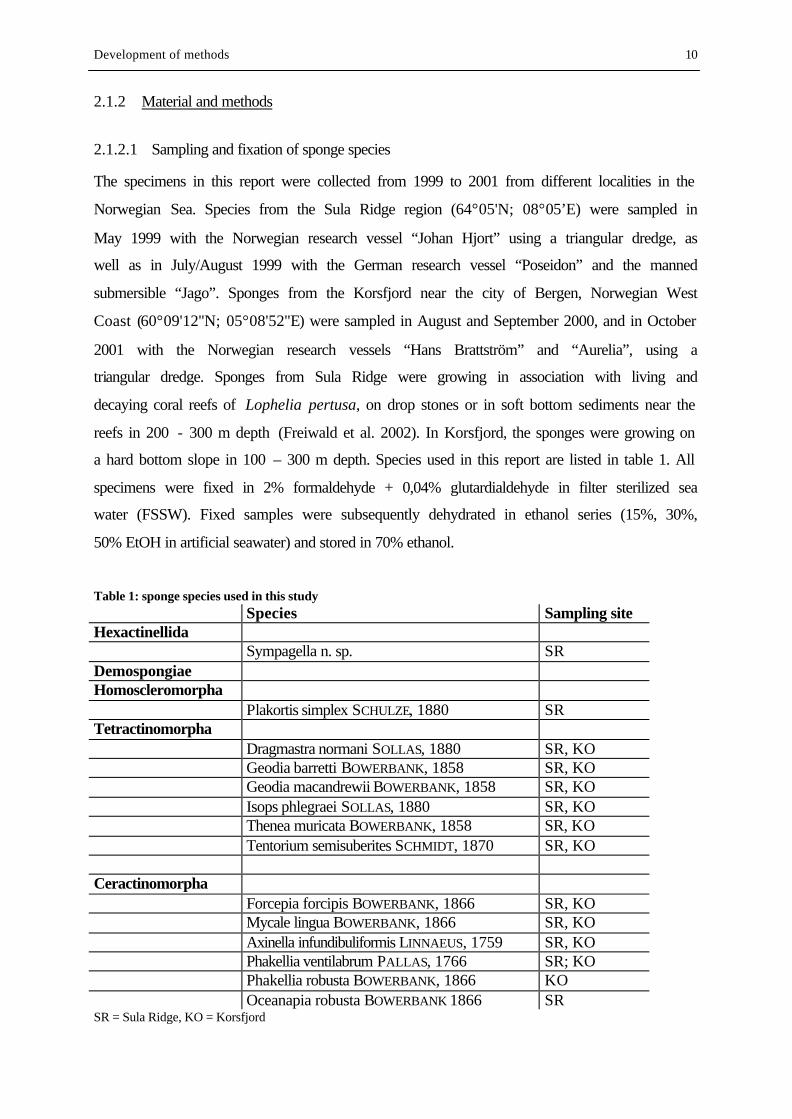
Table 1: sponge species used in this study Species Sampling site Hexactinellida Sympagella n. sp. SR Demospongiae Homoscleromorpha Plakortis simplex SCHULZE, 1880 SR Tetractinomorpha Dragmastra normani SOLLAS, 1880 SR, KO Geodia barretti BOWERBANK, 1858 SR, KO Geodia macandrewii BOWERBANK, 1858 SR, KO Isops phlegraei SOLLAS, 1880 SR, KO Thenea muricata BOWERBANK, 1858 SR, KO Tentorium semisuberites SCHMIDT, 1870 SR, KO
Ceractinomorpha Forcepia forcipis BOWERBANK, 1866 SR, KO Mycale lingua BOWERBANK, 1866 SR, KO Axinella infundibuliformis LINNAEUS, 1759 SR, KO Phakellia ventilabrum PALLAS, 1766 SR; KO Phakellia robusta BOWERBANK, 1866 KO Oceanapia robusta BOWERBANK 1866 SR SR = Sula Ridge, KO = Korsfjord
Development of methods 11
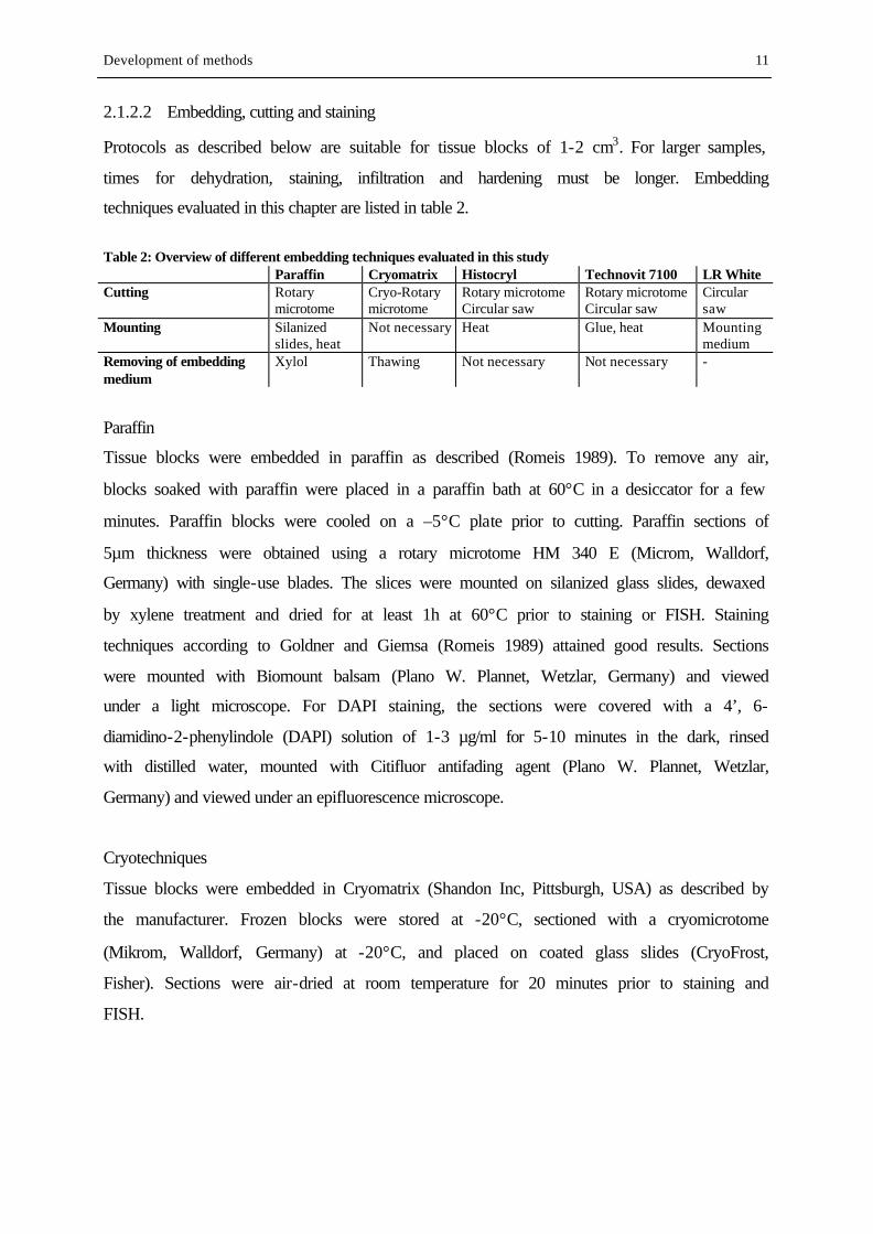
2.1.2.2 Embedding, cutting and staining
Protocols as described below are suitable for tissue blocks of 1-2 cm3. For larger samples,
times for dehydration, staining, infiltration and hardening must be longer. Embedding
techniques evaluated in this chapter are listed in table 2.
Table 2: Overview of different embedding techniques evaluated in this study Paraffin Cryomatrix Histocryl Technovit 7100 LR White Cutting Rotary
microtome Cryo-Rotary microtome
Rotary microtome Circular saw
Rotary microtome Circular saw
Circular saw
Mounting Silanized slides, heat
Not necessary Heat Glue, heat Mounting medium
Removing of embedding medium
Xylol Thawing Not necessary Not necessary -
Paraffin
Tissue blocks were embedded in paraffin as described (Romeis 1989). To remove any air,
blocks soaked with paraffin were placed in a paraffin bath at 60°C in a desiccator for a few
minutes. Paraffin blocks were cooled on a –5°C plate prior to cutting. Paraffin sections of
5µm thickness were obtained using a rotary microtome HM 340 E (Microm, Walldorf,
Germany) with single-use blades. The slices were mounted on silanized glass slides, dewaxed
by xylene treatment and dried for at least 1h at 60°C prior to staining or FISH. Staining
techniques according to Goldner and Giemsa (Romeis 1989) attained good results. Sections
were mounted with Biomount balsam (Plano W. Plannet, Wetzlar, Germany) and viewed
under a light microscope. For DAPI staining, the sections were covered with a 4’, 6-
diamidino-2-phenylindole (DAPI) solution of 1-3 µg/ml for 5-10 minutes in the dark, rinsed
with distilled water, mounted with Citifluor antifading agent (Plano W. Plannet, Wetzlar,
Germany) and viewed under an epifluorescence microscope.
Cryotechniques
Tissue blocks were embedded in Cryomatrix (Shandon Inc, Pittsburgh, USA) as described by
the manufacturer. Frozen blocks were stored at -20°C, sectioned with a cryomicrotome
(Mikrom, Walldorf, Germany) at -20°C, and placed on coated glass slides (CryoFrost,
Fisher). Sections were air-dried at room temperature for 20 minutes prior to staining and
FISH.
Development of methods 12
Methylmetacrylat resins: Technovit, Histocryl
Tissue blocks were embedded in Technovit 7100 (Plano W. Plannet, Wetzlar, Germany) as
described (Manz et al. 2000). Sections of 5-10µm thickness were obtained using a rotary
microtome as described above. Sections of 20µm and more were obtained using a circular
saw (Leica 1600) with diamond knife.
For embedding with Histocryl (Plano W. Plannet, Wetzlar, Germany), dehydrated tissue
blocks where infiltrated with Histocryl and benzoyl peroxide as described by the
manufacturer. Samples were transferred into the polymerization solution (10 ml infiltration
solution + 15-30 µl accelerator) and immediately cooled with running tap water for 5 minutes.
Samples remained in the water bath for a few hours until hardening was completed. Sections
of 5-10µm thickness were obtained using a rotary microtome as described above.
Sections were fixed on glass slides by heat (60°C for at least 1 h) for staining and FISH.
LR White resin
If staining is desired, tissue blocks have to be stained before embedding in LR White. We
developed protocols for Toluidin blue O, Calcein and DAPI as follows: For staining with
Toluidin blue O and Calcein, tissue samples were dehydrated in ascending Ethanol series.
After dehydration, samples were placed in the staining solution over night (Toluidin blue:
1g/100 ml EtOH; Calcein: saturated solution in EtOH). Samples were washed 3-4 times in
Ethanol before embedding. For DAPI staining, tissue blocks were placed in a staining solution
of 10 µg/ml DAPI in 90% EtOH over night. Samples were washed in 99% EtOH, and
dehydrated in 99% Ethanol for a few hours. Tissue blocks from all staining techniques were
infiltrated with LR-White:EtOH 1:2 and 2:1 for at least 18h, finally embedded in LR White
(Plano W. Plannet, Wetzlar, Germany) and hardened for 20h at 60°C. Since all described dyes
are soluble both in EtOH and LR White, infiltration solutions should contain a minor amount
of the used dye. To reduce unintentional extraction of dye during the embedding steps, it is
favourable to use EtOH from the washing steps to prepare infiltration solutions. Tissue
sections from 20µm to 1mm were cut using a circular saw with a diamond knife. Sections
were mounted on glass slides with Biomount balsam.
2.1.2.3 FISH and image processing
Fluorescene in situ hybridization (FISH) was performed on sponge tissue sections as
described (Manz et al. 2000). Samples were viewed under a Zeiss axioplan microscope (Zeiss,
Development of methods 13
Oberkochen, Germany). Microscopical pictures were taken using a CCD Camera and
processed with MetaMorph imaging software.
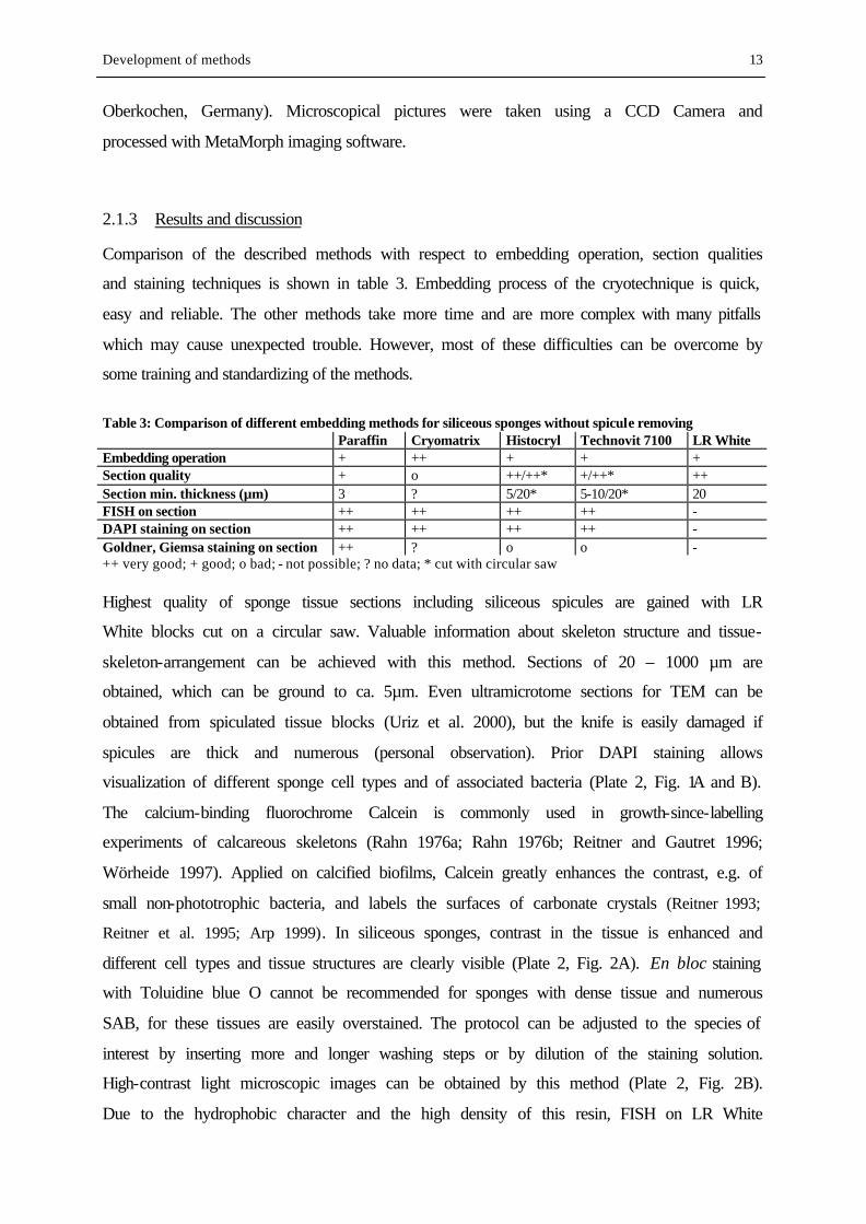
2.1.3 Results and discussion
Comparison of the described methods with respect to embedding operation, section qualities
and staining techniques is shown in table 3. Embedding process of the cryotechnique is quick,
easy and reliable. The other methods take more time and are more complex with many pitfalls
which may cause unexpected trouble. However, most of these difficulties can be overcome by
some training and standardizing of the methods.
Table 3: Comparison of different embedding methods for siliceous sponges without spicule removing Paraffin Cryomatrix Histocryl Technovit 7100 LR White Embedding operation + ++ + + + Section quality + o ++/++* +/++* ++ Section min. thickness (µm) 3 ? 5/20* 5-10/20* 20 FISH on section ++ ++ ++ ++ - DAPI staining on section ++ ++ ++ ++ - Goldner, Giemsa staining on section ++ ? o o - ++ very good; + good; o bad; - not possible; ? no data; * cut with circular saw Highest quality of sponge tissue sections including siliceous spicules are gained with LR
White blocks cut on a circular saw. Valuable information about skeleton structure and tissue-
skeleton-arrangement can be achieved with this method. Sections of 20 – 1000 µm are
obtained, which can be ground to ca. 5µm. Even ultramicrotome sections for TEM can be
obtained from spiculated tissue blocks (Uriz et al. 2000), but the knife is easily damaged if
spicules are thick and numerous (personal observation). Prior DAPI staining allows
visualization of different sponge cell types and of associated bacteria (Plate 2, Fig. 1A and B).
The calcium-binding fluorochrome Calcein is commonly used in growth-since-labelling
experiments of calcareous skeletons (Rahn 1976a; Rahn 1976b; Reitner and Gautret 1996;
Wörheide 1997). Applied on calcified biofilms, Calcein greatly enhances the contrast, e.g. of
small non-phototrophic bacteria, and labels the surfaces of carbonate crystals (Reitner 1993;
Reitner et al. 1995; Arp 1999). In siliceous sponges, contrast in the tissue is enhanced and
different cell types and tissue structures are clearly visible (Plate 2, Fig. 2A). En bloc staining
with Toluidine blue O cannot be recommended for sponges with dense tissue and numerous
SAB, for these tissues are easily overstained. The protocol can be adjusted to the species of
interest by inserting more and longer washing steps or by dilution of the staining solution.
High-contrast light microscopic images can be obtained by this method (Plate 2, Fig. 2B).
Due to the hydrophobic character and the high density of this resin, FISH on LR White
Development of methods 14
sections is impossible. Oligonucleotide probes used for FISH are water soluted and will not
penetrate the resin. The same applies to DAPI and histological stains based on dyes in water
solution.
Using a rotary microtome with sharp steel knife, best section qualities were gained with
Histocryl. This was also true for species and tissue parts containing many spicules, e.g. the
cortex of Geodia species. Sections down to 5 µm were suitable for FISH. From Technovit
blocks, sections of varying thickness and quality were obtained, but usuable parts for FISH
could be found on most sections. Sections of better quality were obtained using a circular saw.
These sections were relatively thick (> 20 µm), which made interpretation of FISH difficult.
Because of the incompatibility of MMA resins with ethanol, only stains in water solution can
be used for Histocryl and Technovit sections. Unfortunately, for both MMA resins, I observed
incomplete and irregular staining with Goldner and Giemsa. (Gerrits 1992) describes good
results for different histological stainings on soft tissue embedded in Technovit 7100. The
failure of these attempts may be due to the rough and uneven surfaces of sections from
spiculated tissue. In Histocryl sections, I sometimes observed massive stain accumulations
between the spicules, whereas the surrounding tissue remained largely unstained.
Paraffin sections without prior dissolution of the siliceous skeleton were of bad quality in
tissue parts which contained numerous, thick or long spicules, e.g. the cortex of Geodia. For
tissue parts containing few or small spicules, results were better, and even though tissue
sections were partly torn, useable areas for FISH and histological staining could be found on
most sections. Careful cooling of the paraffin blocks prior to cutting and the use of a new
blade for each cutting event helped to improve section quality.
Extremely bad sections were achieved from cryoblocks: the sponge tissue was entirely torn
and no tissue structures were visible. This was probably due to differences in toughness
between the spicules and the embedded tissue. No attempt was made on histological staining
of these sections, whereas application of FISH probes and DAPI staining achieved clear
signals. However, information gained from these cryosections is comparable to that from
homogenized sponge tissue. We assume that the quality of cryosections could probably be
optimized by varying the cutting temperature and by better training of the cutting procedure.
For all described techniques on rotary microtomes, section quality is never quite as good as
with prior acid treatment. Although some histological stainings could successfully be applied
on sponge sections with siliceous spicules, we recommend the classical way of spicule
removing for histological investigations where sections < 20 µm are needed, e.g. for the study
of fine tissue structures. Methods for embedding and cutting of siliceous sponges should be
Development of methods 15
chosen according to the aim of investigation and the properties of the examined species. Until
now, there is no standard method available which fits all needs.
Conclusions and recommendations:
1. For overview of tissue and skeleton arrangement: LR White embedding combined with
en bloc staining, cutting with circular saw.
2. For FISH on siliceous sponges: Histocryl embedding for species or tissue parts with high
spicule content, paraffin embedding if spicule content is low. Cutting with rotary
microtome.
3. For detailed tissue examination: classical histological methods (spicule removing by acid
prior to embedding, cutting and staining).
2.2 Cultivation and regeneration of sponge fragments
2.2.1 Introduction
Most research on sponge cultivation was and still is directed towards tropical and
Mediterranean species. Previous cultivation experiments on boreal species have been short-
time scale studies for fundamental research, as for Thenea muricata (Witte 1995) or
Halichondria panicea (Barthel and Theede 1986). No attempt has been made so far on
cultivation of Geodia barretti.
In the following chapter, I describe a cultivation technique in half-open systems for tissue
samples of G. barretti, which were used as experimental units for microelectrode studies
described in chapter 3. Valuable information about healing and regeneration processes and
their coordination in the sponge tissue was gained by parallel microscopic surveying of tissue,
skeleton and canal system over a cultivation period of 8 months. This represents an important
step towards a thorough understanding of sponge biology.
2.2.2 Material and methods
1.1.1.1 Sampling
Sponges were sampled near the city of Bergen on the west coast of Norway, between 100 and
150 m depth on a hard bottom slope in Korsfjord at 60°09’12’’N; 05°08’52’’E (Fig. 1).
Samples were taken in July 2000 and in March and May 2001 with the Norwegian research
Development of methods 16
vessels “Hans Brattström” and “Aurelia” by dredging with a triangular dredge. Sampling is
one of the most critical factors in sponge cultivation experiments, as many sponges are
sensitive to air exposure and shifts in water temperature. To minimize exposure to air, the
wire was stopped when the dredge appeared at the water’s surface. An outboard-working
person placed the sponges in buckets under water, which were subsequently transported to
deck and emptied into larger vessels filled with running seawater. Later experience in the lab
showed that G. barretti tolerates short exposures to air, and sampling in 2001 was done by
emptying the dredge directly in water-filled vessels. The described sampling method should,
however, be applied to extremely air-sensitive species if sampling by SCUBA diving or
submersibles is not possible. After sampling, the sponges were transported immediately to the cultivation site.
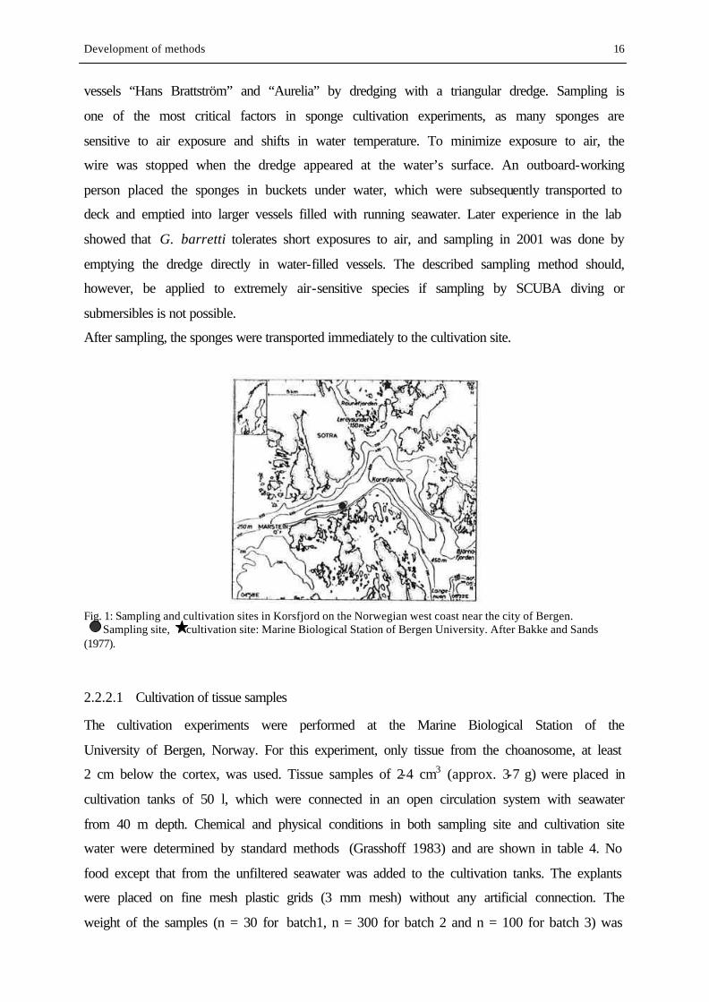
Fig. 1: Sampling and cultivation sites in Korsfjord on the Norwegian west coast near the city of Bergen. Sampling site, cultivation site: Marine Biological Station of Bergen University. After Bakke and Sands (1977).
2.2.2.1 Cultivation of tissue samples
The cultivation experiments were performed at the Marine Biological Station of the
University of Bergen, Norway. For this experiment, only tissue from the choanosome, at least
2 cm below the cortex, was used. Tissue samples of 2-4 cm3 (approx. 3-7 g) were placed in
cultivation tanks of 50 l, which were connected in an open circulation system with seawater
from 40 m depth. Chemical and physical conditions in both sampling site and cultivation site
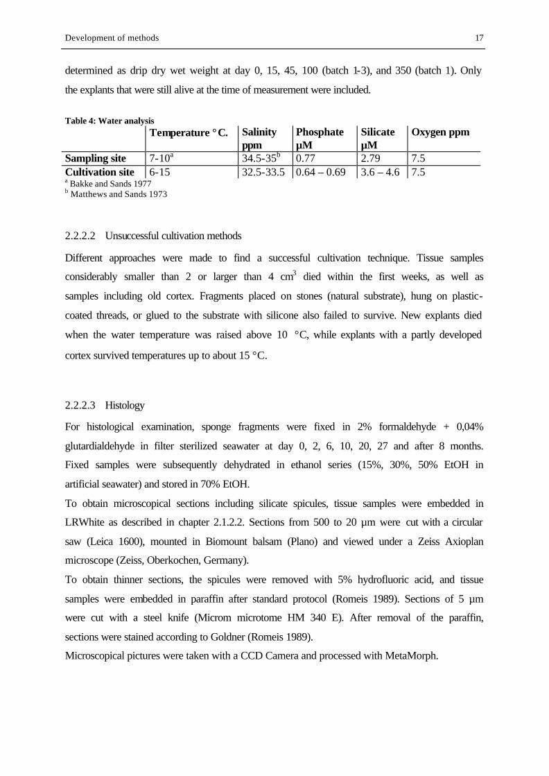
water were determined by standard methods (Grasshoff 1983) and are shown in table 4. No
food except that from the unfiltered seawater was added to the cultivation tanks. The explants
were placed on fine mesh plastic grids (3 mm mesh) without any artificial connection. The
weight of the samples (n = 30 for batch1, n = 300 for batch 2 and n = 100 for batch 3) was
Development of methods 17
determined as drip dry wet weight at day 0, 15, 45, 100 (batch 1-3), and 350 (batch 1). Only
the explants that were still alive at the time of measurement were included.
Table 4: Water analysis Temperature °C. Salinity
ppm Phosphate µM
Silicate µM
Oxygen ppm
Sampling site 7-10a 34.5-35b 0.77 2.79 7.5 Cultivation site 6-15 32.5-33.5 0.64 – 0.69 3.6 – 4.6 7.5 a Bakke and Sands 1977 b Matthews and Sands 1973
2.2.2.2 Unsuccessful cultivation methods
Different approaches were made to find a successful cultivation technique. Tissue samples
considerably smaller than 2 or larger than 4 cm3 died within the first weeks, as well as
samples including old cortex. Fragments placed on stones (natural substrate), hung on plastic-
coated threads, or glued to the substrate with silicone also failed to survive. New explants died
when the water temperature was raised above 10 °C, while explants with a partly developed
cortex survived temperatures up to about 15 °C.
2.2.2.3 Histology
For histological examination, sponge fragments were fixed in 2% formaldehyde + 0,04%
glutardialdehyde in filter sterilized seawater at day 0, 2, 6, 10, 20, 27 and after 8 months.
Fixed samples were subsequently dehydrated in ethanol series (15%, 30%, 50% EtOH in
artificial seawater) and stored in 70% EtOH.
To obtain microscopical sections including silicate spicules, tissue samples were embedded in
LRWhite as described in chapter 2.1.2.2. Sections from 500 to 20 µm were cut with a circular
saw (Leica 1600), mounted in Biomount balsam (Plano) and viewed under a Zeiss Axioplan
microscope (Zeiss, Oberkochen, Germany).
To obtain thinner sections, the spicules were removed with 5% hydrofluoric acid, and tissue
samples were embedded in paraffin after standard protocol (Romeis 1989). Sections of 5 µm
were cut with a steel knife (Microm microtome HM 340 E). After removal of the paraffin,
sections were stained according to Goldner (Romeis 1989).
Microscopical pictures were taken with a CCD Camera and processed with MetaMorph.
Development of methods 18
2.2.3 Results
2.2.3.1 Growth
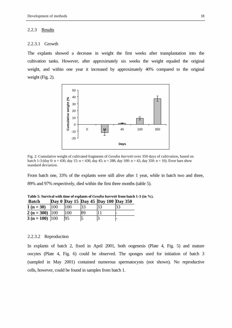
The explants showed a decrease in weight the first weeks after transplantation into the
cultivation tanks. However, after approximately six weeks the weight equaled the original
weight, and within one year it increased by approximately 40% compared to the original
weight (Fig. 2).
-20
-10
0
10
20
30
40
50
0 15 45 100 350
Days
Cu
mu
lati
ve w
eig
ht
(%)
Fig. 2: Cumulative weight of cultivated fragments of Geodia barretti over 350 days of cultivation, based on batch 1-3 (day 0: n = 430, day 15: n = 430, day 45: n = 288, day 100: n = 43, day 350: n = 10). Error bars show standard deviation. From batch one, 33% of the explants were still alive after 1 year, while in batch two and three,
89% and 97% respectively, died within the first three months (table 5).
Table 5: Survival with time of explants of Geodia barretti from batch 1-3 (in %). Batch Day 0 Day 15 Day 45 Day 100 Day 350 1 (n = 30) 100 100 33 33 33 2 (n = 300) 100 100 89 11 - 3 (n = 100) 100 95 5 3 -
2.2.3.2 Reproduction
In explants of batch 2, fixed in April 2001, both oogenesis (Plate 4, Fig. 5) and mature
oocytes (Plate 4, Fig. 6) could be observed. The sponges used for initiation of batch 3
(sampled in May 2001) contained numerous spermatocysts (not shown). No reproductive
cells, however, could be found in samples from batch 1.
Development of methods 19
2.2.3.3 Cicatrisation and regeneration
During cultivation, the shape of the explants changed from cubic to spherical (Plate 3).
Already after two days, the canals at the surface were closed (observation by eye and
microscopic control), the edges of the fragments were rounded, and the entire surface was
covered by a transparent “skin”. After 8 months, the canals within the fragments were
considerably smaller in diameter, and a new cortex had developed.
Closing of the canals at the cut surface was due to initiative growth at the canal walls (Plate 4,
Fig 1 and 2) by a growing front of motile sponge cells, followed by cells with inclusions in an
otherwise cell- and bacteria-poor mesohyl. We observed the same process at canal walls in the
endosome, diminishing the diameter of the canals. Torn tissue at the cut surfaces was covered
by a closed line of motile sponge cells (Plate 4, Fig 3). Remodelling, defined as spontaneous
disorganization and reorganization of the tissue (Simpson 1984) occurred in the superficial
region of the explants (ectosome), where choanocyte chambers had degenerated, and cells
with inclusions accumulated (Plate 4, Fig 4). After 10 to 20 days, sterrasters began to
accumulate in the ectosome, and after 8 months, both sterraster and euaster layers of the new
cortex had developed (Plate 5). Microscleres of all age classes in the subcortical tissue
showed that at this time, the new cortex was still under construction. Broken choanosomal
megascleres were incorporated in the new cortex, whereas radially arranged triaene
megascleres were lacking. Subcortical spaces and ostia started to form.
2.2.4 Discussion
2.2.4.1 Cultivation methods
In contrast to previous studies on Geodia cydonium (Müller et al. 1999c), Tethya lyncurium
(= Tethya aurantium Pallas) (Connes 1966a), and Chondrosia reniformis (Bavestrello et al.
1998) our explants derived from choanosomal tissue regenerated into healthy sponges, while
explants including old cortex failed to survive. Connes (1966a) demonstrated that
choanosomal fragments of T. lyncurium died within a few days. The other studies only
regarded ectosomal fragments. Cultivation methods which have been successfully applied to a
broad range of sponges (e.g. Verdenal and Vacelet 1985; Duckworth et al. 1999; Pronzato et
al. 1999) did not succeed with Geodia barretti. These contrasting results show once more that
“there will probably never be a standard method for sponge cultivation” (Osinga et al. 1999).
Development of methods 20
2.2.4.2 Growth rates
The weight increase of 40% within one year is lower than described for cultivation of warm-
water species. Explants of the commercial bath sponge Spongia officinalis double their weight
in about one year in open-sea aquacultures in the Mediterranean Sea (Verdenal and Vacelet
1985). For Geodia cydonium, Müller et al. (1999c) observed weight increases of 53% and
90% after 3 and 6 months, respectively, both in open-sea aquacultures and half-open systems
in the North Adriatic Sea. The highest growth rates ever reported (5000% in one month for
Lissodendoryx sp.) were found in an aquaculture in New Zealand by Battershill and Page
(1996).
So far, almost nothing is known about growth rates of G. barretti in the field. A specimen of
G. barretti has been observed in situ by scuba diving in the Trondheimsfjord, Mid-Norway,
over a two year period, but no measurable change in size or shape was noticed (Rapp,
personal observation).
Since weight increase starts parallel to intensive spicule production in the explants of G.
barretti, it remains an open question how much of the increase is due to the production of
organic material. However, though not measured, an increase in size of the explants was
observed during the cultivation period.
2.2.4.3 Reproduction and seasonality
The development of egg cells shows that cultivated fragments of G. barretti are able to
continue their reproductive cycle. This observation may be used for harvesting of egg cells to
raise this species in culture, as first described by Wilson (1898). The reproductive cycle for G.
barretti is unknown. Tetractinellid sponges are oviparous: they release eggs and sperm to the
water, where free-swimming larvae develop (Bergquist 1978). Geodia cydonium from the
Mediterranean (Adriatic, Italy) is described as a gonochoristic, oviparous species with
reproductive period from June to October (Liaci and Sciscioli 1969 in Simpson 1984). Boreal
and arctic deep-water sponges usually have seasonal reproduction (Witte 1996; Ereskovsky
2000) with reproductive periods between February and July. We did not find any gametes in
G. barretti sampled in March, July and August from the Korsfjord, and assume that G.
barretti is a seasonal breeder with reproductive period in early summer (April-June). The high
mortality of explants from batch 2 and 3 may imply that the reproductive period is a bad time
for initiation of a cultivation experiment. Tissue regeneration, spicule production, and sexual
reproduction are energy-consuming processes, and may be difficult to perform at the same
Development of methods 21
time. Fröhlich and Barthel (1997) showed seasonal variations in rates of silica uptake in
Halichondria panicea from the Baltic Sea. During the most intense phase of reproduction
activity, female specimens showed a significant drop in their silica uptake, and obviously did
not produce spicules during this time. However, more experimental data are needed to
elucidate a correlation between reproductive season and cultivation success.
2.2.4.4 Healing and regeneration
Only few studies are available about healing and regeneration processes in sponges.
Cicatrisation has been described in cultivated fragments of Hippospongia communis, Spongia
officinalis, Agelas oroides, Axinella damicornis and Petrosia ficiformis in the Mediterranean
Sea (Pronzato et al. 1999). Regenerative processes started immediately after transplantation.
Within 2-3 days the sponges rebuilt their external protective layer, and after 24 hours a thin,
transparent cell layer covered the cut surfaces. After one month, the sponge fragments
developed a rounded shape. In the studies on T. lyncurium (Connes 1966a and b) and C.
reniformis (Bavestrello et al. 1998), the old cortex was observed to surround the entire surface
of the explants after a few weeks. Unfortunately, none of these studies related cicatrisation to
growth rates. The “presence of a cuticle” on explants of Spongia officinalis is suspected to
limit further growth (Verdenal and Vacelet 1985).
Cells with inclusions, also called spherulous cells, possess nutritive-metabolic or storage
functions, and are found in a broad range of sponge species (see Simpson (1984) for review).
Such cells are often connected to growth and secretion processes (Reitner and Gautret 1996).
The generally high amount of these cells in the tissue of G. barretti may be the reason for the
good regenerative capacity of this sponge.
It was suggested by Simpson (1984) that the formation of a highly structured, definitive
cortex may completely limit further growth and thus may act as a signal for senescence. We
demonstrated that tissue from an adult Geodia, though the whole sponge may have reached
senescence, never looses its ability to grow and regenerate. These findings may encourage
starting successful aquacultures of other sponge species with pronounced cortex.
The decrease in weight during the first weeks is most probably due to remodelling of the
tissue, i.e. degeneration of choanocyte chambers and torn tissue in the ectosome, and by
consumption of storage material. Weight increases when cicatrisation is finished, and the new
cortex starts to develop.
Regeneration of all functional units from a random piece of the choanosome requires
totipotency of cells, information transfer, and orientation in the given piece of sponge tissue.
Development of methods 22
Cultivation experiments with parallel histological examinations may be most enlightening for
fundamental research on these still poorly understood aspects of sponge biology.
2.3 Biogeochemical methods
2.3.1 Examination of microbial sulfate reduction in sediment biogeochemistry
Activities of sulfate-reducing bacteria have been investigated intensively in marine sediments
(e.g. Jörgensen 1977; Jörgensen 1982; Jörgensen 1989b; Canfield 1993; Devereux et al. 1996;
Ferdelman et al. 1997; Habicht and Canfield 1997; Hines et al. 1999; Knoblauch et al. 1999;
Boetius et al. 2000; Thamdrup et al. 2000) and microbial mats (Canfield and Marais 1991;
Cypionka 1994; Jörgensen 1994; Habicht and Canfield 1996; Teske et al. 1998) with
radioactively labelled sulfate. Recently, the application of this method on a marine
oligochaete has achieved the first report on endosymbiotic microbial sulfate reduction in a
marine invertebrate (Dubelier et al. 2001).
Anoxic sampling of pore water for determination of sulfide, the metabolic end product of
microbial sulfate reduction, is a difficult approach even in sediment biogeochemistry. The
development of a special sampling device was necessary to obtain sponge pore water that had
not been in contact with oxygen during the sampling.
Studies with oxygen sensitive microelectrodes and recent physiological studies on SRB report
frequent occurrence and activity of SRB in oxic environments (e.g. Dilling and Cypionka
1990; Ramsing et al. 1993; Teske et al. 1998; Minz et al. 1999b; Minz et al. 1999a; Okabe et
al. 1999; Schramm et al. 1999; Sigalevich et al. 2000). From these findings, sulfate reducers
should be perceived as microaerophiles rather than obligate anaerobes, which makes their
association with aerobic organisms more reasonable. However, data on chemical conditions in
sponge tissue are sparse. The use of microsensors to investigate oxygen concentrations in
sponge tissue represents a novel approach in sponge science and has been applied only to a
few species (Gatti et al. 2002; Schönberg et al. submitted). I used oxygen-sensitive
microelctrodes to examine oxygen microprofiles within the sponge tissue. The thick cortex
and the high spicule content of G. barretti caused problems for application of the fragile
microelectrode.
Modifications of established methods in sediment biogeochemistry for the application to
living Geodia barretti are described in the next section.
Development of methods 23
2.3.2 Modified methods
2.3.2.1 Microelectrodes
Oxygen profiles in the tissue of G. barretti were measured with oxygen sensitive Clark-type
microelectrodes (Clark et al. 1953; Revsbech and Jörgensen 1986). The cortex of G. barretti
defied insertion of the electrode. This problem was circumvent by inserting the electrode tip
into pore openings (ostia) or by piercing the cortex with a canula and sliding the sensor in
through the cut. Insertion of the sensor in 10 days old fragments caused no problems, because
the cortex was not regenerated at this state (see chapter 2.2). To avoid any horizontal tension
on the electrode tip during profiling, the examined sponge specimen or fragment had to be
fixed carefully.
2.3.2.2 Sulfate reduction rates
In order to determinate sulfate reduction rates in the tissue of G. barretti, the whole core
incubation method with 35SO42- (Jörgensen 1978) was modified. Tissue cores were taken from
cortex to cortex of pumping sponges, incubated with 300 or 600 kBq 35SO42- for 30 minutes,
sliced, fixed in 20% zinc acetate and frozen. The high amounts of tracer and the short
incubation times were chosen because previous experiments had indicated that immediate
reoxidation of the tracer obscured sulfate reduction rates in the tissue of G. barretti
(unpublished results).
To calculate sulfate reduction rates, reduced sulfur species were analysed with the single-step
chromium reduction method (Fossing and Jörgensen 1989).
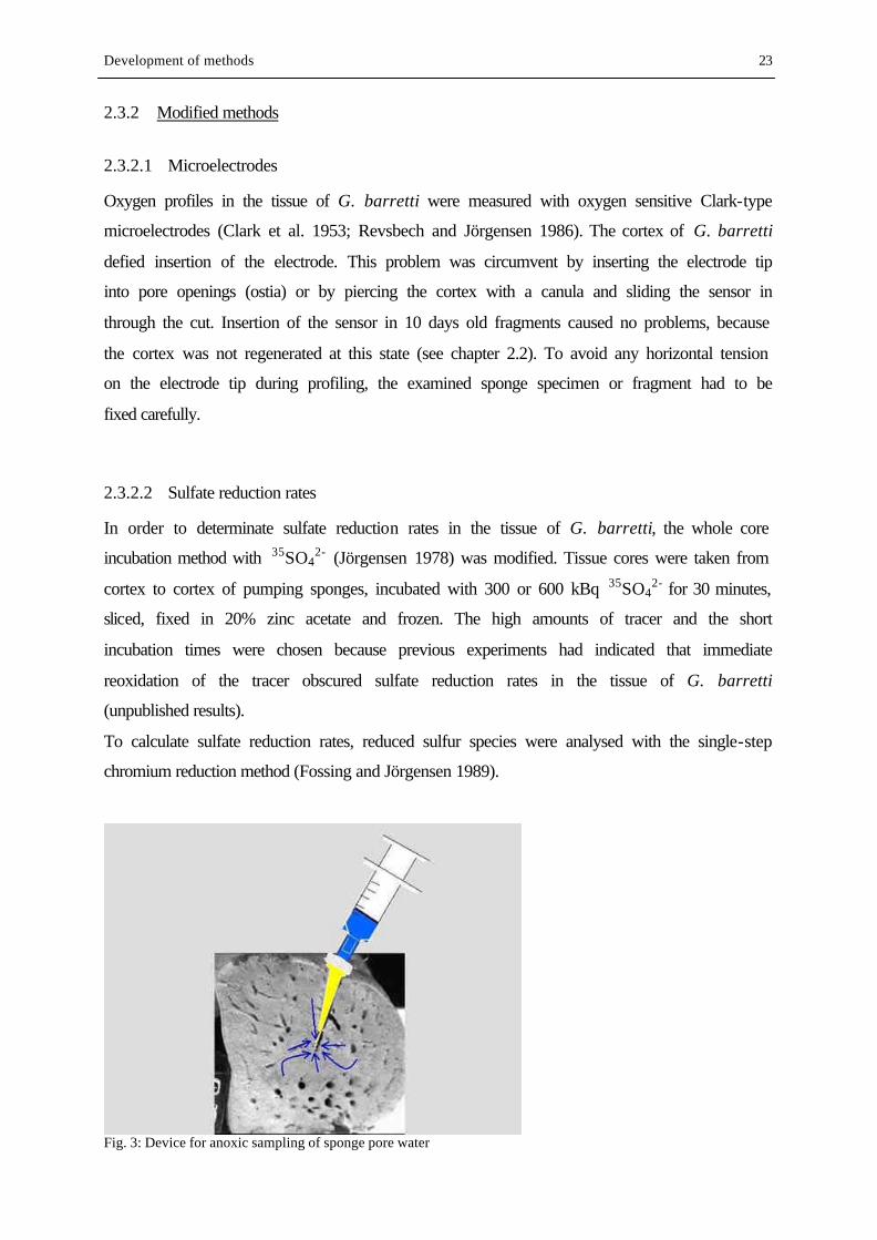
Fig. 3: Device for anoxic sampling of sponge pore water
Development of methods 24
2.3.2.3 Anoxic sampling of sponge pore water
Sponge pore water from the choanosome for hydrogen sulfide analysis was sampled with a
newly developed setup as shown in Fig. 3. A yellow micropipette tip was fixed on a silicon
tube, which was attached to a 10 ml plastic syringe. The tip was pushed into the sponge tissue,
flushed once with the pore water, until 1-5 ml from approximately 5 cm depth were sampled.
The tip with the attached tube could even remain in the sponge tissue for a few days to allow
repeated sampling. With this technique, pore water can be sampled from a living sponge, and
any contact of the pore water with atmospheric oxygen is avoided.
3 THE ROLE OF MICROBIAL SULFATE REDUCTION IN THE TISSUE OF G. BARRETTI:
EXPERIMENTS AND RESULTS
3.1 Introduction
In the following study, I combined the histological, molecular biological and biogeochemical
methods outlined in the last chapter in order to investigate phylogenetic affiliation, metabolic
activity and the chemical micro-environment of sulfate-reducing bacteria in the tissue of G.
barretti. On the basis of these results, the role of sulfate reducers for the metabolism of G.
barretti will be elucidated. Biological-chemical and sponge-bacteria interactions in sponge
tissue will be discussed in chapter 4.
3.2 Experiments
3.2.1 Sampling and maintaining of sponges
Different specimens of Geodia barretti were sampled as described in chapter 2.2. Whole
sponges were maintained in cultivation tanks with running sea water from 40 m depth until
experiments started (max. 1 week). Chemical and physical parameters in the water from
sampling and cultivation site, respectively, are shown in table 4. Filtration activity was
checked regularly with fluorescein dye (Pile et al. 1997). The fragments were cultivated as
described in chapter 2.2, and, after 10 days of cultivation, they were used for oxygen
profiling.
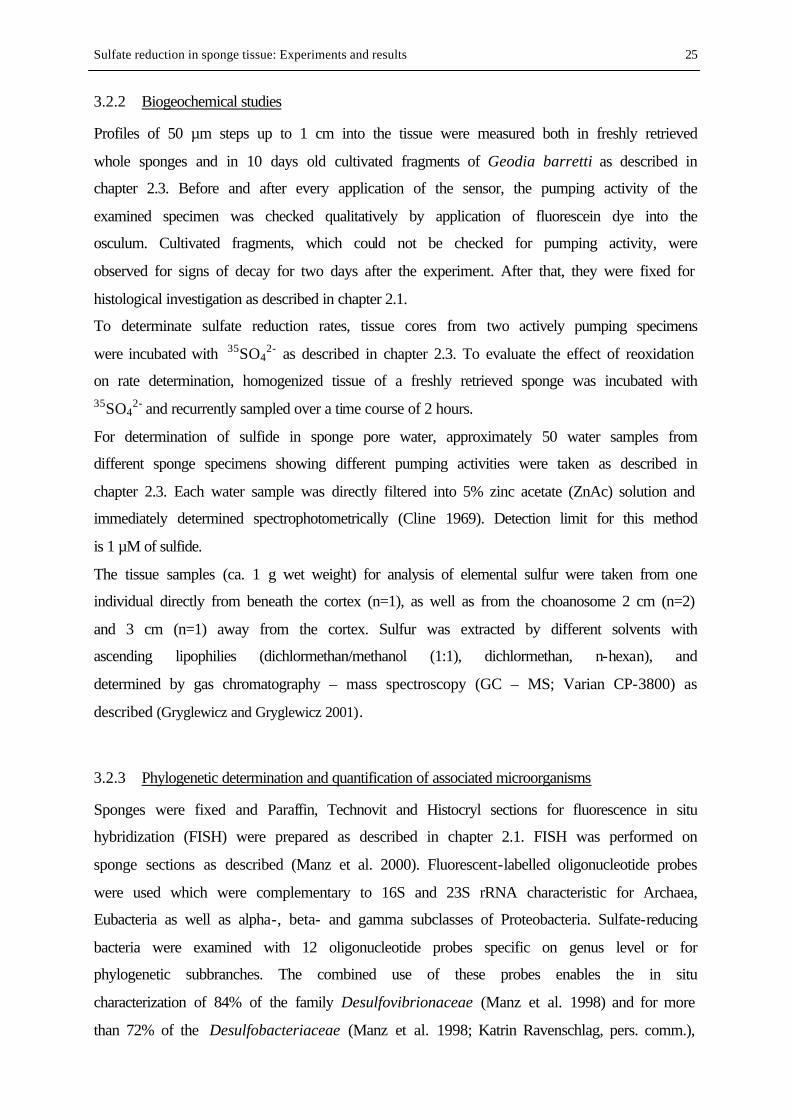
Sulfate reduction in sponge tissue: Experiments and results 25
3.2.2 Biogeochemical studies
Profiles of 50 µm steps up to 1 cm into the tissue were measured both in freshly retrieved
whole sponges and in 10 days old cultivated fragments of Geodia barretti as described in
chapter 2.3. Before and after every application of the sensor, the pumping activity of the
examined specimen was checked qualitatively by application of fluorescein dye into the
osculum. Cultivated fragments, which could not be checked for pumping activity, were
observed for signs of decay for two days after the experiment. After that, they were fixed for
histological investigation as described in chapter 2.1.
To determinate sulfate reduction rates, tissue cores from two actively pumping specimens
were incubated with 35SO42- as described in chapter 2.3. To evaluate the effect of reoxidation
on rate determination, homogenized tissue of a freshly retrieved sponge was incubated with 35SO4
2- and recurrently sampled over a time course of 2 hours.
For determination of sulfide in sponge pore water, approximately 50 water samples from
different sponge specimens showing different pumping activities were taken as described in
chapter 2.3. Each water sample was directly filtered into 5% zinc acetate (ZnAc) solution and
immediately determined spectrophotometrically (Cline 1969). Detection limit for this method
is 1 µM of sulfide.
The tissue samples (ca. 1 g wet weight) for analysis of elemental sulfur were taken from one
individual directly from beneath the cortex (n=1), as well as from the choanosome 2 cm (n=2)
and 3 cm (n=1) away from the cortex. Sulfur was extracted by different solvents with
ascending lipophilies (dichlormethan/methanol (1:1), dichlormethan, n-hexan), and
determined by gas chromatography – mass spectroscopy (GC – MS; Varian CP-3800) as
described (Gryglewicz and Gryglewicz 2001).
3.2.3 Phylogenetic determination and quantification of associated microorganisms
Sponges were fixed and Paraffin, Technovit and Histocryl sections for fluorescence in situ
hybridization (FISH) were prepared as described in chapter 2.1. FISH was performed on
sponge sections as described (Manz et al. 2000). Fluorescent-labelled oligonucleotide probes
were used which were complementary to 16S and 23S rRNA characteristic for Archaea,
Eubacteria as well as alpha-, beta- and gamma subclasses of Proteobacteria. Sulfate-reducing
bacteria were examined with 12 oligonucleotide probes specific on genus level or for
phylogenetic subbranches. The combined use of these probes enables the in situ
characterization of 84% of the family Desulfovibrionaceae (Manz et al. 1998) and for more
than 72% of the Desulfobacteriaceae (Manz et al. 1998; Katrin Ravenschlag, pers. comm.),
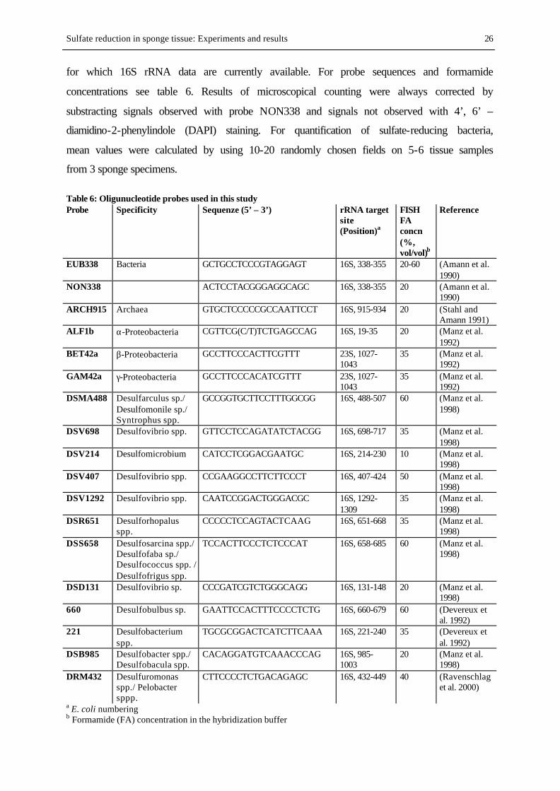
Sulfate reduction in sponge tissue: Experiments and results 26
for which 16S rRNA data are currently available. For probe sequences and formamide
concentrations see table 6. Results of microscopical counting were always corrected by
substracting signals observed with probe NON338 and signals not observed with 4’, 6’ –
diamidino-2-phenylindole (DAPI) staining. For quantification of sulfate-reducing bacteria,
mean values were calculated by using 10-20 randomly chosen fields on 5-6 tissue samples
from 3 sponge specimens.
Table 6: Oligunucleotide probes used in this study Probe Specificity Sequenze (5’ – 3’) rRNA target
site (Position)a
FISH FA concn (%, vol/vol)b
Reference
EUB338 Bacteria GCTGCCTCCCGTAGGAGT 16S, 338-355 20-60 (Amann et al. 1990)
NON338 ACTCCTACGGGAGGCAGC 16S, 338-355 20 (Amann et al. 1990)
ARCH915 Archaea GTGCTCCCCCGCCAATTCCT 16S, 915-934 20 (Stahl and Amann 1991)
ALF1b α-Proteobacteria CGTTCG(C/T)TCTGAGCCAG 16S, 19-35 20 (Manz et al. 1992)
BET42a β-Proteobacteria GCCTTCCCACTTCGTTT 23S, 1027-1043
35 (Manz et al. 1992)
GAM42a γ-Proteobacteria GCCTTCCCACATCGTTT 23S, 1027-1043
35 (Manz et al. 1992)
DSMA488 Desulfarculus sp./ Desulfomonile sp./ Syntrophus spp.
GCCGGTGCTTCCTTTGGCGG 16S, 488-507 60 (Manz et al. 1998)
DSV698 Desulfovibrio spp. GTTCCTCCAGATATCTACGG 16S, 698-717 35 (Manz et al. 1998)
DSV214 Desulfomicrobium CATCCTCGGACGAATGC 16S, 214-230 10 (Manz et al. 1998)
DSV407 Desulfovibrio spp. CCGAAGGCCTTCTTCCCT 16S, 407-424 50 (Manz et al. 1998)
DSV1292 Desulfovibrio spp. CAATCCGGACTGGGACGC 16S, 1292-1309
35 (Manz et al. 1998)
DSR651 Desulforhopalus spp.
CCCCCTCCAGTACTCAAG 16S, 651-668 35 (Manz et al. 1998)
DSS658 Desulfosarcina spp./ Desulfofaba sp./ Desulfococcus spp. / Desulfofrigus spp.
TCCACTTCCCTCTCCCAT 16S, 658-685 60 (Manz et al. 1998)
DSD131 Desulfovibrio sp. CCCGATCGTCTGGGCAGG 16S, 131-148 20 (Manz et al. 1998)
660 Desulfobulbus sp. GAATTCCACTTTCCCCTCTG 16S, 660-679 60 (Devereux et al. 1992)
221 Desulfobacterium spp.
TGCGCGGACTCATCTTCAAA 16S, 221-240 35 (Devereux et al. 1992)
DSB985 Desulfobacter spp./ Desulfobacula spp.
CACAGGATGTCAAACCCAG 16S, 985-1003
20 (Manz et al. 1998)
DRM432 Desulfuromonas spp./ Pelobacter sppp.
CTTCCCCTCTGACAGAGC 16S, 432-449 40 (Ravenschlag et al. 2000)
a E. coli numbering b Formamide (FA) concentration in the hybridization buffer
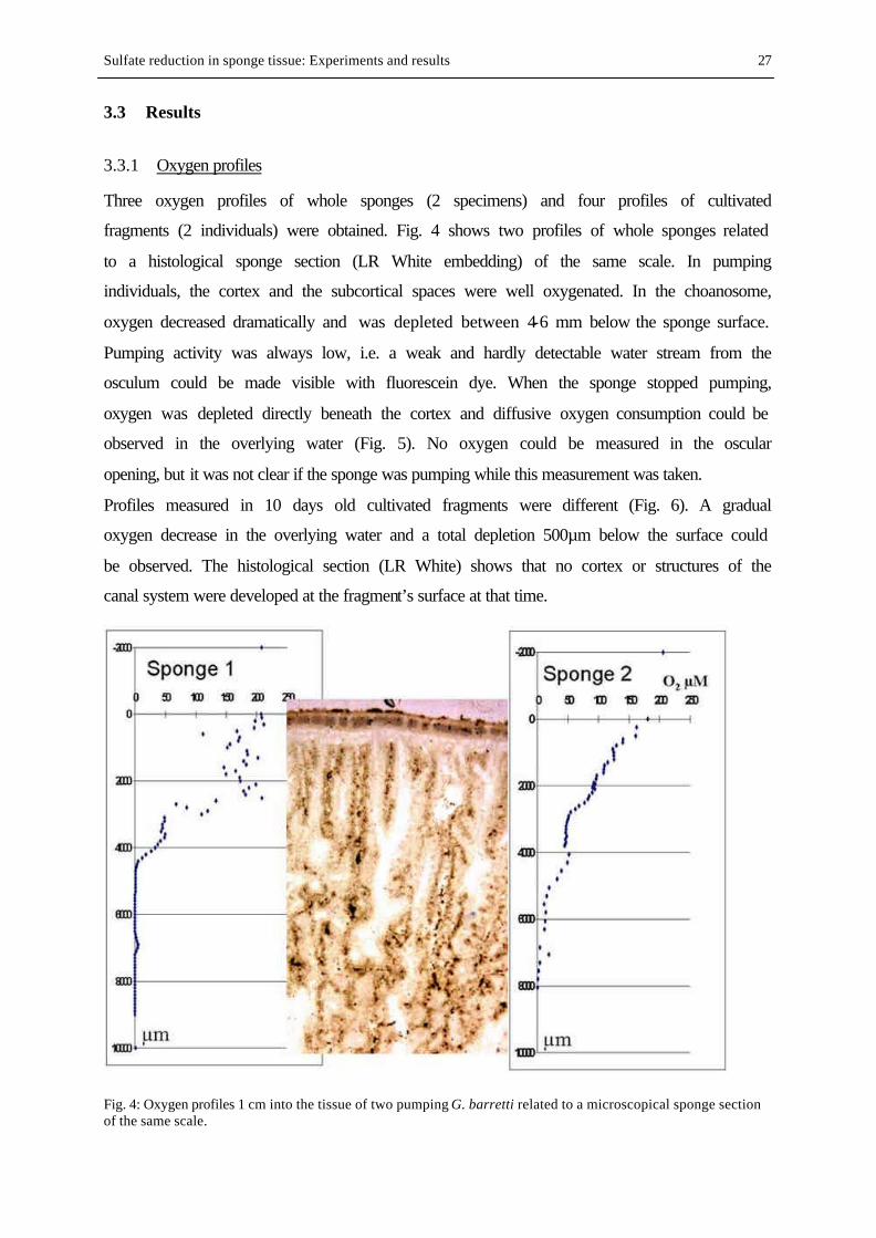
Sulfate reduction in sponge tissue: Experiments and results 27
3.3 Results
3.3.1 Oxygen profiles
Three oxygen profiles of whole sponges (2 specimens) and four profiles of cultivated
fragments (2 individuals) were obtained. Fig. 4 shows two profiles of whole sponges related
to a histological sponge section (LR White embedding) of the same scale. In pumping
individuals, the cortex and the subcortical spaces were well oxygenated. In the choanosome,
oxygen decreased dramatically and was depleted between 4-6 mm below the sponge surface.
Pumping activity was always low, i.e. a weak and hardly detectable water stream from the
osculum could be made visible with fluorescein dye. When the sponge stopped pumping,
oxygen was depleted directly beneath the cortex and diffusive oxygen consumption could be
observed in the overlying water (Fig. 5). No oxygen could be measured in the oscular
opening, but it was not clear if the sponge was pumping while this measurement was taken.
Profiles measured in 10 days old cultivated fragments were different (Fig. 6). A gradual
oxygen decrease in the overlying water and a total depletion 500µm below the surface could
be observed. The histological section (LR White) shows that no cortex or structures of the
canal system were developed at the fragment’s surface at that time.
Fig. 4: Oxygen profiles 1 cm into the tissue of two pumping G. barretti related to a microscopical sponge section of the same scale.
Sulfate reduction in sponge tissue: Experiments and results 28
Fig. 5: Oxygen profiles 3 mm into the tissue of sponge 2, related to a microscopical sponge section of the same scale. Left, not pumping; right, pumping.
Fig. 6: Oxygen profile in a 10 days old cultivated fragment of G. barretti related to a microscopical tissue section of the same scale.
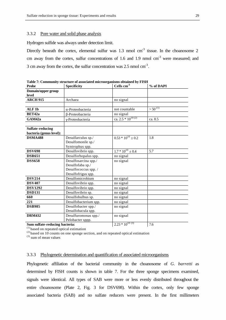
Sulfate reduction in sponge tissue: Experiments and results 29
3.3.2 Pore water and solid phase analysis
Hydrogen sulfide was always under detection limit.
Directly beneath the cortex, elemental sulfur was 1.3 nmol cm-3 tissue. In the choanosome 2
cm away from the cortex, sulfur concentrations of 1.6 and 1.9 nmol cm-3 were measured; and
3 cm away from the cortex, the sulfur concentration was 2.5 nmol cm-3.
Table 7: Community structure of associated microorganisms obtained by FISH Probe Specificity Cells cm-3 % of DAPI Domain/upper group level
ARCH 915 Archaea no signal ALF 1b α-Proteobacteria not countable > 50 (1) BET42a β-Proteobacteria no signal GAM42a γ-Proteobacteria ca. 2.5 * 1010 (2) ca. 8.5 Sulfate-reducing bacteria (genus level):
DSMA488 Desulfarculus sp./ Desulfomonile sp./ Syntrophus spp.
0.53 * 1010 ± 0.2 1.8
DSV698 Desulfovibrio spp. 1.7 * 1010 ± 0.4 5.7 DSR651 Desulforhopalus spp. no signal DSS658 Desulfosarcina spp./
Desulfofaba sp./ Desulfococcus spp. / Desulfofrigus spp.
no signal
DSV214 Desulfomicrobium no signal DSV407 Desulfovibrio spp. no signal DSV1292 Desulfovibrio spp. no signal DSD131 Desulfovibrio sp. no signal 660 Desulfobulbus sp. no signal 221 Desulfobacterium spp. no signal DSB985 Desulfobacter spp./
Desulfobacula spp. no signal
DRM432 Desulfuromonas spp./ Pelobacter sppp.
no signal
Sum sulfate-reducing bacteria: 2.23 * 1010 (3) 7.6 (1) based on repeated optical estimation (2) based on 10 counts on one sponge section, and on repeated optical estimation (3) sum of mean values
3.3.3 Phylogenetic determination and quantification of associated microorganisms
Phylogenetic affiliation of the bacterial community in the choanosome of G. barretti as
determined by FISH counts is shown in table 7. For the three sponge specimens examined,
signals were identical. All types of SAB were more or less evenly distributed throughout the
entire choanosome (Plate 2, Fig. 3 for DSV698). Within the cortex, only few sponge
associated bacteria (SAB) and no sulfate reducers were present. In the first millimeters
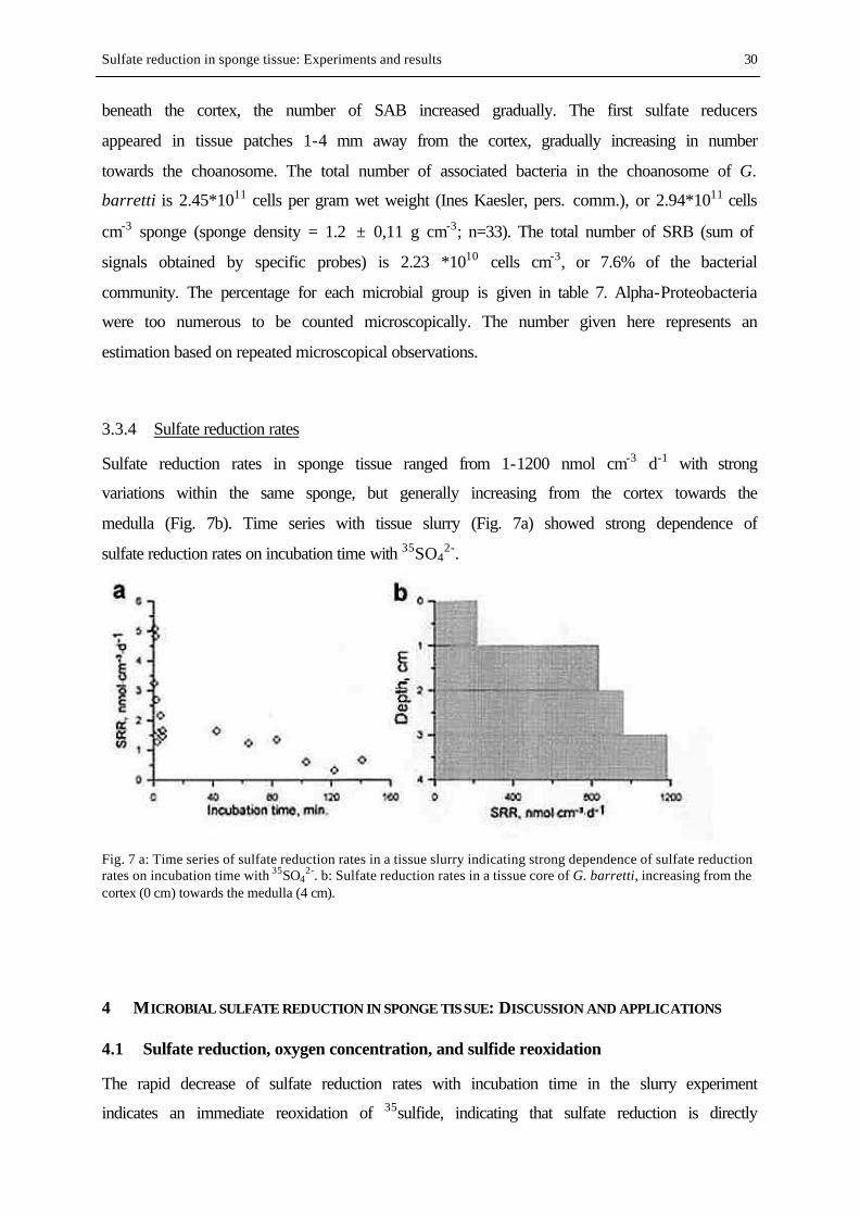
Sulfate reduction in sponge tissue: Experiments and results 30
beneath the cortex, the number of SAB increased gradually. The first sulfate reducers
appeared in tissue patches 1-4 mm away from the cortex, gradually increasing in number
towards the choanosome. The total number of associated bacteria in the choanosome of G.
barretti is 2.45*1011 cells per gram wet weight (Ines Kaesler, pers. comm.), or 2.94*1011 cells
cm-3 sponge (sponge density = 1.2 ± 0,11 g cm-3; n=33). The total number of SRB (sum of
signals obtained by specific probes) is 2.23 *1010 cells cm-3, or 7.6% of the bacterial
community. The percentage for each microbial group is given in table 7. Alpha-Proteobacteria
were too numerous to be counted microscopically. The number given here represents an
estimation based on repeated microscopical observations.
3.3.4 Sulfate reduction rates
Sulfate reduction rates in sponge tissue ranged from 1-1200 nmol cm-3 d-1 with strong
variations within the same sponge, but generally increasing from the cortex towards the
medulla (Fig. 7b). Time series with tissue slurry (Fig. 7a) showed strong dependence of
sulfate reduction rates on incubation time with 35SO42-.
Fig. 7 a: Time series of sulfate reduction rates in a tissue slurry indicating strong dependence of sulfate reduction rates on incubation time with 35SO4
2-. b: Sulfate reduction rates in a tissue core of G. barretti, increasing from the cortex (0 cm) towards the medulla (4 cm).
4 MICROBIAL SULFATE REDUCTION IN SPONGE TISSUE: DISCUSSION AND APPLICATIONS
4.1 Sulfate reduction, oxygen concentration, and sulfide reoxidation
The rapid decrease of sulfate reduction rates with incubation time in the slurry experiment
indicates an immediate reoxidation of 35sulfide, indicating that sulfate reduction is directly
Sulfate reduction in sponge tissue: Discussion 31
coupled with sulfide oxidation. The lower rates in the slurry compared to rates in core
incubations may be explained by the higher content of oxygen in the slurry. However, the
effect of time is obvious. If oxygen is present, the 35S2- will rapidly be reoxidized to 35SO42-,
which will lead to a dramatic underestimation of SRR (Jörgensen and Bak 1991). To keep this
effect as small as possible, high amounts of tracer and short incubation times were used for
core incubations.
The shape of oxygen profiles in whole sponges indicates turbulent oxygen transport in cortex
and subcortical spaces, probably due to the pumping activity of the sponge. The rapid
decrease at ca. 4 mm indicates rapid oxygen consumption, and oxygen transport by molecular
diffusion. This may indicate the moment when the electrode enters the sponge tissue. Oxygen
profiles of cultivated fragments show a nearly parabolic shape as known from the surface of
undisturbed marine sediments (Revsbech et al. 1980), indicating that no pumping activity is
involved in oxygen supply. Obviously, cultivated fragments are completely dependent on
strong water currents to maintain sufficient supply with oxygen by diffusion gradients. This is
consistent with observations from cultivation experiments (chapter 2.2). Within the first
months of cultivation, the canal system of the fragments, especially at the ectosome, is under
rearrangement, without any inhalant or exhalant openings at the surface present until the
cortex is regenerated. Gatti and co-workers (2002) examined oxygen concentrations in the
tissue of Suberites domuncula and in sponge primmorphs (well defined sponge cell
aggregations surrounded by a dermal membrane; Müller et al. 1999b) with oxygen sensitive
microoptodes. They observed that oxygen concentrations were strongly dependent on the
surrounding flow regime, as well as on culturing conditions of the sponges. Oxygen
concentrations in the tissue of adult sponges were between 50 – 60% of the surrounding
water, and the oxygen content in primmorphs was never below 2.3% of the surrounding
water.
I showed that the oxygen concentration in the tissue of G. barretti was strongly dependent on
its pumping activity (Fig. 5). It is reported from many sponges in situ to reduce or even stop
pumping for several hours at irregular intervals (Reiswig 1971; Vogel 1977; Gerodette and
Flechsig 1979; Pile et al. 1997). These patterns were found out to be related to the sediment
load of the water (Gerodette and Flechsig 1979), ambient water flow (Vogel 1977; Pile et al.
1997) or sponge growth form (Reiswig 1971). In G. barretti, reduced pumping activity leads
to anoxia in parts of the tissue and even in the canal- and exhalant water. However, during the
experiments, pumping activity was always low. It can be assumed that G. barretti may show
much higher pumping activities under more favourable conditions, which will lead to oxic
Sulfate reduction in sponge tissue: Discussion 32
conditions at least in the pore water and the exhalant water stream. The tropical
bacteriosponge Verongia fistularis removes only 2.8 to 8.3 (mean 5.33%) of available oxygen
during a single transit of water through the sponge body (Reiswig 1974). Fragments of G.
barretti can be considered to be permanently anoxic both in pore water and tissue, but can
survive with this consistent tissue anoxia for several months. From these results I conclude
that tissue anoxia is a common feature in living G. barretti, although the metabolism of the
sponge is based on oxic respiration and is dependent on regular input of oxygen.
It can be assumed that oxygen present in the pore water of incubated tissue cores rapidly
reoxidizes 35sulfide and still leads to underestimation of SRR. It is not possible to decide
whether the increase of SRR from the cortex to the medulla is due to lower sulfate reduction
or higher sulfide reoxidation in the cortex region. In a decaying sponge, SRR were
considerably higher (data not shown); the lowest rates measured being 5000 nmol cm-3 d-1. I
assume that true SRR in a living sponge are in between the highest rates measured in core
incubations and this value, i.e. between 1000 and 5000 nmol cm-3 d-1. These are among the
highest rates of sulfate reduction measured in a natural system, only comparable to those of
microbial mats (Canfield and Marais 1991; Habicht and Canfield 1996; Teske et al. 1998).
Sulfate reduction rates in continental margin sediments from the coast of Denmark and
Norway are lower by 1-3 orders of magnitude (Canfield et al. 1993).
4.2 Sulfate-reducing bacteria within the microbial community
The results obtained by FISH on sponge sections indicate that associated bacteria mainly
belong to the alpha- and gamma-Proteobacteria. This is consistent with phylogenetic
investigations on pure cultures of microorganisms isolated from G. barretti (Ines Kaesler,
pers. comm.). Alpha- and/or gamma-Proteobacteria are reported to dominate the microbial
communities of many marine sponges (Schumann-Kindel et al. 1996; Friedrich et al. 2001;
Hentschel et al. 2001; Webster et al. 2001b; Webster and Hill 2001).
At least two strains of sulfate reducers could be detected in the tissue of G. barretti. One was
affiliated to the Desulfosarculus/Desulfomonile/Syntrophus group of the family
Desulfobacteriaceae. The other positive signal codes for a Desulfovibrio sp. The only other
sulfate-reducing symbiont described from a marine sponge (Manz et al. 2000) belongs to the
same genus, though affiliated to another strain. This symbiont was detected with the
oligonucleotide probe DSV1292, which gained no signals in G. barretti (compare table 7).
Based on SRB cell numbers as detected by FISH, average cellular SRR were calculated.
Assuming SRR between 1000 and 5000 nmol cm-3 d-1, cell specific SRR (csSRR) for
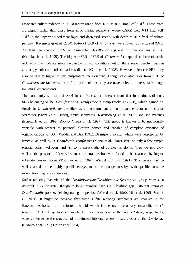
Sulfate reduction in sponge tissue: Discussion 33
associated sulfate reducers in G. barretti range from 0.05 to 0.22 fmol cell-1 d-1. These rates
are slightly higher than those from arctic marine sediments, where csSRR were 0.14 fmol cell-
1 d-1 in the uppermost sediment layer and decreased steeply with depth to 0.02 fmol of sulfate
per day (Ravenschlag et al. 2000). Rates of SRB of G. barretti were lower, by factors of 3.6 to
28, than the specific SRRs of mesophilic Desulfovibrio grown in pure cultures at 8°C
(Knoblauch et al. 1999). The higher csSRR of SRB of G. barretti compared to those of arctic
sediments may indicate more favourable growth conditions within the sponge mesohyl than in
a strongly substrate-limited marine sediment (Glud et al. 1998). However, higher csSRR may
also be due to higher in situ temperatures in Korsfjord. Though calculated rates from SRB of
G. barretti are far below those from pure cultures, they are nevertheless in a reasonable range
for natural environments.
The community structure of SRB in G. barretti is different from that in marine sediments.
SRB belonging to the Desulfosarcina-Desulfococcus group (probe DSS658), which gained no
signals in G. barretti, are described as the predominant group of sulfate reducers in coastal
sediments (Sahm et al. 1999), arctic sediments (Ravenschlag et al. 2000) and salt marshes
(Edgcomb et al. 1999; Rooney-Varga et al. 1997). This group is known to be nutritionally
versatile with respect to potential electron donors and capable of complete oxidation of
organic carbon to CO2 (Widdel and Bak 1991). Desulfovibrio spp, which were detected in G.
barretti as well as in Chondrosia reniformis (Manz et al. 2000), can use only a few simple
organic acids, hydrogen, and (in some cases) ethanol as electron donor. They do not grow
well in the presence of low substrate concentrations but were found to be favoured by higher
substrate concentrations (Trimmer et al. 1997; Widdel and Bak 1991). This group may be
well adapted to the highly specific ecosystem of the sponge mesohyl with specific substrate
molecules in high concentrations.
Sulfate-reducing bacteria of the Desulfosarculus/Desulfomonile/Syntrophus group were also
detected in G. barretti, though in lower numbers than Desulfovibrio spp. Different strains of
Desulfomonile possess dehalogenating properties (Weerdt et al. 1990; Ni et al. 1995; Sun et
al. 2001). It might be possible that these sulfate reducing symbionts are involved in the
Barettin metabolism, a brominated alkaloid which is the main secondary metabolite of G.
barretti. Bacterial symbionts, cyanobacteria or eubacteria of the genus Vibrio, respectively,
were shown to be the producer of brominated biphenyl ethers in two species of the Dysideidae
(Elyakov et al. 1991; Unson et al. 1994).
Sulfate reduction in sponge tissue: Discussion 34
It can be concluded that G. barretti hosts a specific, metabolically active community of at
least two different strains of sulfate reducers. Associated SRB are able to show high sulfate
reduction rates in situ in spite of possible oxic conditions.
4.3 Evidence of an endosymbiotic sulfur cycle
In the dense tissue of G. barretti, hypoxia or anoxia will arise due to reduced pumping
activity and the active metabolism of sponge cells and aerobic SAB. Fermentation products
such as succinate, propionate and acetate accumulate during anaerobic metabolism under low
oxygen concentration in many marine invertebrates (Grieshaber et al. 1994). In G. barretti,
not only sponge cells, but also associated fermenting bacteria may contribute to this process.
Facultative anaerobic bacteria fermenting sucrose and fucose represent the majority of
symbionts within the carribean sclerosponge Ceratoporella nicholsoni (Santavy et al. 1990).
The presence of fermenting bacteria within the diverse microbial community of G. barretti is
likely.
Fig. 8: Biological-chemical and sponge-bacteria interactions in the tissue of G. barretti.
Fermentation products will serve as carbon and electron sources of associated SRB (Fig. 8).
As discussed above, I assume that under normal conditions, the canal system of the sponge is
flushed by oxygenated water. In the few milli- or even micrometers of choanosomal tissue
Sulfate reduction in sponge tissue: Discussion 35
between the canals, all niches will be present from oxic via microoxic to anoxic. Niches are
variable in time and space and strongly dependent on pumping rate and metabolic activity of
the sponge. Anoxic zones may enlarge if pumping activity is low and sponge metabolism is
high, and may even disappear if the conditions are opposite. Sulfate reducers are known to
tolerate or – in some cases – seem to prefer aerobic situations and show high rates of sulfate
reduction under these conditions (Canfield and Marais 1991; Santegoeds et al. 1998; Teske et
al. 1998; Minz et al. 1999a; Minz et al. 1999b; Schramm et al. 1999; Sigalevich et al. 2000;
Wang et al. 2000). Therefore, they are found evenly distributed throughout the choanosome,
only missing in and immediately beneath the cortex.
Free sulfide reacts spontanously with O2 to produce mostly S0, S2O3 2-, and SO4
2-. Elemental
sulfur has been implicated as an important reaction intermediate of sulfide oxidation (Canfield
and Thamdrup 1994; Pyzik and Sommer 1981), usually present as a small but highly dynamic
pool (Howarth 1984). Considerable amounts of elemental sulfur in the tissue of G. barretti
indicate the presence of an oxidative sulfur cycle. The increase of sulfur concentrations
towards the medulla is well in accordance with the number of SRB and indicates higher
sulfide production and reoxidation in the choanosome compared to the cortex region. In many
natural environments, sulfide-oxidizing bacteria are present at the O2/H2S interface to utilize
the energy gained by this oxidative reaction for the fixation of CO2 (Jörgensen 1989a). Many
sulfide oxidizers store elemental sulfur intracellularly. Mutual symbiosis between sulfide-
oxidizing bacteria and gutless marine invertebrates is the basis for ecosystems at
hydrothermal vents based on chemoautotrophy (Cavanaugh 1994). The ability to oxidize
reduced sulfur species is not restricted to a specific phylogenetic group of bacteria (Kelly
1991), which makes it difficult to detect sulfide oxidizers by FISH. Symbiotic sulfide
oxidizers, however, are frequently ascribed to the Gammaproteobacteria by means of
phylogenetic 16S rRNA analysis (e.g. Dubelier et al. 1995; Haygood and Davidson 1997). In
the bacterial community of G. barretti, gamma-Proteobacteria were present showing
approximately the same number as sulfate reducers.
An endosymbiotic sulphur cycle based on the activity of sulfate-reducing and sulfide-
oxidizing symbionts has recently been described in a marine oligochaete (Dubelier et al.
2001). In this example, symbiotic bacteria served as a food source for the gutless host
organism, making this symbiosis a true mutualism. As mentioned in chapter 1.2, many
bacteriosponges consume their associated microorganisms (“bacterial farming”). Mid chain
branched fatty acids, which are assumed to derive from a yet unknown sulfate reducer, have
been detected in the tissue, as well as incorporated in demospongic acids of G. barretti (V.
Sulfate reduction in sponge tissue: Discussion 36
Thiel and T. Pape, pers. comm.). This may indicate a biomass transfer from associated SRB to
sponge cells.
My data indicate the presence of an endosymbiotic sulfur cycle in the tissue of G. barretti
allowing effective nutrient utilization within the sponge–microbe community.
4.4 Mass balances and rates
According to the equation:
(1) S2-
+ 2 O2 → SO42-
2 mol of O2 are necessary to oxidize 1 mol of sulfide. An oxygen concentration of 220 µM in
the ambient water reveals approximately 170 nmol O2 cm-3 sponge (sponge porosity = 0.77 ±
0.26 ml H2O cm-3 sponge; n=33). This would oxidize 85 nmol S2-, which represents 1.7 -
8.5% of the daily SRR. So if the sponge stopped pumping, sulfate reduction could carry on for
30 min to 2 h before sulfide would be released. This explains why it is extremely difficult to
measure SRR in living sponges.
To balance sulfide production from sulfate reduction, a pumping rate of 50 ml oxygen
saturated H2O cm-3 sponge d-1 is needed. In situ pumping rates of G. barretti are not known.
In situ pumping rates of different marine and freshwater sponge species range from 3.8 to
23.8 l H2O cm-3 d-1 (see table 8). From these data it can be assumed that G. barretti will easily
exhibit in situ pumping rates that will balance sulfate reduction and even lead to excess of
oxygen in sponge pore water.
Table 8: In situ pumping rates of marine and freshwater sponges l water cm-3 sponge d-1 Reference Mycale sp 18.14 – 23.32 (Reiswig 1974) Tethya crypta 3.8 (Reiswig 1974) Verongia gigantea 4.32 – 8.6 (Reiswig 1974) Verongia fistularis 10.67 (Reiswig 1981) Baikalospongia bacillifera 3.45 – 5.18 (Pile et al. 1997) However, I showed that consistent anoxia appear in the tissue of G. barretti, and do obviously
not harm the sponge animal as a whole. How does the sponge prevent sulfide toxification
under these conditions?
The presence of considerable amounts of biologically available iron (312 nmol cm-3) indicates
another process of efficient sulfide removal even in the absence of O2. Biologically available
iron describes the pool of iron which is extractable with 6 M HCl, i.e. FeS and different
Sulfate reduction in sponge tissue: Discussion 37
species of Fe(III), either amorphous, mineralised or bound to biological structures. Most of
this pool was due to Fe(III) (Ole Larsen, pers. comm.).
According to the equation:
(2) 2 FeOOH + 3H2S → 2 FeS + S0 + 4 H2O (e.g. Pyzik and Sommer 1981)
2 mol of Fe(III) are needed to remove 3 mol of sulfide, either as FeS or S0. Thus, 312 nmol
cm-3 Fe(III) would remove 468 nmol cm-3 sulfide. This accounts for 10 – 50% of the daily
SRR or 2.5 – 12 h of full sulfate reduction before sulfide will be released. Genes coding for
ferritin, a protein binding and transporting iron, have been described from the hadromerid
sponge Suberites domuncula (Pennec et al. 2003). Localisation of ferritin with in situ
hybridization gained rare signals in the cortex region of the sponge. In the medulla, cells were
frequently stained, especially endopinacocytes which surround the canals. This indicates an
uptake of Fe(III) from pore water and storage within the choanosome. Ferritin would allow
transportation of Fe(III) to zones of intensive sulfate reduction within anoxic tissue parts.
Until now, no oxygen binding and transporting structures in sponges have been described.
Oxygen profiles indicate that oxygen supply into dense tissue parts is dependent on molecular
diffusion.
Reduced iron species can be reoxidized in the presence of O2, and serve again as a sulfide
scavenger if oxygen is absent. The iron pool in G. barretti may represent an efficient buffer
system similar to that in marine sediments (e.g. Jörgensen 1989a) to prevent any accumulation
of toxic H2S if oxygen is not present.
4.5 Sponges and sulfate reducers – an ancestral symbiosis?
Several lines of evidence indicate a long-standing symbiosis between sponges and sulfate
reducing bacteria. SRB were among the oldest organisms on earth (Wagner et al. 1998b; Shen
et al. 2001), dominant players in the Proterozoic ocean and ubiquitous witnesses of the
evolution of eukaryotes and higher organisms. Phylogenetic analyses of associated bacteria in
different sponges showed a deep branching of a sponge-specific sequence cluster within the
division of Deltaproteobacteria, which sulfate reducers belong to (Hentschel et al. 2002).
This means that these symbionts are phylogenetically far away from their free-living relatives,
which might reflect a long-standing existence within sponge tissue. The other sponge-specific
sequence clusters are not deeply branching within their divisions, which indicates that these
groups may have joined the symbiosis later in the evolutionary history of the sponges.
Sulfate reduction in sponge tissue: Discussion 38
The presence of complex isomeric mixtures of specific mid-chain branched carboxylic acids
(MBCA) in the tissue of G. barretti, as mentioned in chapter 4. 3, also gives evidence for an
ancient origin of this symbiosis. These MBCA, which have been detected in different sponge
species (Thiel et al. 1999; Thiel et al. 2002), are potential lipid precursors for complex
mixtures of mid-chain branched monomethylalkanes often observed in fossil sediments and
oils (e.g. Fowler and Douglas 1987; Summons 1987). Thiel and co-workers (1999) suggest
that the bacterial source organisms of the MBCA have been widespread in the geological past,
and are found “inherited” in the protective environment of sponges in recent marine
ecosystems. These biomarkers are unknown in any modern marine environment, but
distinctive isomers of MBCA have been found in strains of sulfate-reducing bacteria (Ueki
and Suto 1979; Kaneda 1991; Parkes and Taylor 1983). Summons (1987) suggests that mid-
chain methylation in fatty acids may be a vestigial strategy for controlling membrane fluidity
which originated prior to the development of an oxic biosphere and the evolution of the now
dominating oxygen-dependent desaturating pathways in the biosynthesis of lipids. It can be
assumed that the characteristic MBCA assemblage in sponges is derived from ancient strains
of sulfate reducing symbionts in the sponge mesohyl. Interestingly, the same MBCA have
recently been discovered in anoxic parts of biofilms from the highly alkaline Walker Lake in
Nevada (Volker Thiel, pers. comm.). Assuming a higher alkalinity of the Early Proterozoic
compared to recent oceans (Kempe and Kazmierczak 1994), microbial mats in modern soda
lakes could serve as a protective environment for proterozoic bacteria, similar as “living
fossil” sponges.
Combined sulfur isotope and molecular biological studies provide indirect evidence for a
connection between the evolution of the microbial sulfur cycle and the metazoans (Habicht
and Canfield 1996; Canfield and Teske 1996; Canfield 1998). These authors suspect an
intimate linkage between the history of atmospheric oxygen and seawater sulfate and a
parallel evolution of bacteria of the oxidative sulfur cycle and metazoan life some time
between 640 and 1050 Ma. Although this pre-dates the appearance of the Ediacaran fauna
(e.g. Seilacher 2000), it is consistent with the hypothesis for evolution of the first metazoans
as part of a major eukaryotic radiation, as indicated by “molecular clock” studies (Morris
1993; Wray et al. 1996; Brohmann et al. 1998; Gu 1998; Suga et al. 1999).
The origin of multicellular animals still remains one of the most enigmatic of all phylogenetic
problems (Willmer 1990; Müller 2001). Monophyly of the Metazoa is now widely accepted
(e.g. Ax 1995; Müller 1995; Westheide and Rieger 1996; Wägele and Rödding 1998;
Schuetze et al. 1999), and sponges definitely are metazoans (compare chapter 1.1). Thus, a
Sulfate reduction in sponge tissue: Discussion 39
common ancestor of all Metazoa, which possessed all typical metazoan characters, must have
existed. A recent molecular biological study suggests that the common ancestor of the
Metazoa was a sponge, or a sponge-like organism (Borchiellini et al. 2001). Although many
hypotheses try to explain the transition from unicellular organisms to multicellular
organization, only three basic models seem possible (Willmer 1990; Westheide and Rieger
1996): Cell divisions of an unicellular ancestor living within a common extracellular matrix;
cellularization of a multinucleated cell; or aggregation of cells attracting each other through
chemical signals.
The nature of this unicellular ancestor is still a matter of debate. Choanoflagellates, which
show morphological similarities to sponge choanocytes and do even form planktonic colonies,
have been proposed for a long time; this view was supported by molecular sequence data
(Wainright et al. 1993; Kumar and Rzhetsky 1996; King and Carroll 2001; Lang et al. 2002).
However, ultrastructural studies did not support these findings (reviewed in Ax 1995; Rieger
and Weyrer 1998).
Reitner and co-workers (1999) and Reitner and Wörheide (2002) offered the hypothesis that
sponges derived from a union of choanoflagellate colonies and different bacteria, in which the
eukaryotic partners evolved mechanisms to control an assemblage of prokaryotes. This
hypothesis is mainly based on morphological similarities between non-phototrophic biofilms
and simple, incrusting (bacterio-)sponges in extreme environments (Reitner 1993). The results
of my study also show noticeable similarities between the tissue of G. barretti and microbial
mats with respect to sulfate reduction rates, chemical gradients and their fluctuations
(day/night vs. pumping/not pumping), bacterial density and the complex interactions of
biochemical processes (Ramsing et al. 1993; Canfield and Marais 1991; Jörgensen 1994;
Habicht and Canfield 1996; Karsten and Kühl 1996; Teske et al. 1998; Minz et al. 1999b).
But different from a microbial mat, all processes and features described from G. barretti are
under biological control of the sponge. Though lacking a central nervous system, sponges
show surprising abilities to coordinate processes within the sponge body, as I showed for
regeneration processes in chapter 2.2. Moreover, sponges are provided with a natural, innate
immune system, acting on both cellular and molecular level (Müller et al. 1999a). This means
that sponges are able to control their microbial population with phagocytosis and the
production of secondary metabolites, directly influencing bacterial number and activity.
Another possibility for active control of chemical gradients within the sponge tissue is the
modulation of the pumping rate.
Sulfate reduction in sponge tissue: Discussion 40
Was the first metazoan a sponge-like organism, an assemblage of different prokaryotes under
biological control of a – yet undefined - protist colony, arisen as part of a major eukaryotic
radiation? In the Proterozoic ocean, where organic carbon can be assumed to be mainly
available in dissolved form, an association of different bacteria would be able to metabolise a
wide array of simple molecules and transfer them into biomass, which would serve as a better
food resource for the eukaryotic partner. A symbiosis with facultative anaerobic or
microaerophilic microorganisms as the ubiquitous sulfate reducers might have been
advantageous under low and unstable oxygen conditions.
Although the establishment of a stable oxygen atmosphere and major changes in ocean
chemistry gave rise for the evolution of other animal phyla with different body plans and
ecological strategies, it would not be surprising if Geodia barretti, the “living fossil”, had
preserved this ancestral symbiosis within its tissue until today.
4.6 Implications for sponge biotechnology and ecology
It is still not decided if the complex interactions between sponge cells, SRB and other sponge
bacteria are preserved features from the origin of metazoan life or if they evolved later in
time. But my results undoubtedly prove the presence and metabolic activity of SRB in the
tissue of present G. barretti. They further indicate the importance of anaerobic processes for
the sponge metabolism, with SRB as putative key players for sponge nutrition. It is possible
that metabolic processes, probably also the production of sponge secondary metabolites, are
dependent on certain chemical conditions in the sponge tissue. This may be an explanation for
the hitherto poor success of in vitro cell cultures of sponges (see chapter 1.3).
Mass settlements of giant Geodia and related sponges around Iceland and the Faroe islands
have been reported by local fishermen for many years. The local name for these sponge
accumulations is “ostur” or “ostebund”, meaning “cheese bottom”. Klitgaard and co-workers
(1997) suggested that at these sites, suspension feeders were favoured by special hydrographic
patterns causing elevated bottom currents and particle resuspension. However, this does not
explain the predominance of Geodia, Isops and Stryphnus species, nor their extraordinary
sizes of up to 72 cm and body weights of more than 20 kg (Tendal et al. 1993; Klitgaard et al.
1997). These sponges are grouped within the same family (Isops) or order (Stryphnus) as
Geodia, and show similar growth forms and tissue structures (own observations). Stryphnus
and Isops species collected in the Norwegian Sea contain high amounts of a mid-chain
branched C-18-carboxylic acid (T. Pape, pers. comm.), which may indicate symbiotic sulfate
reducing bacteria. These sponges may benefit directly from elevated bottom currents which
Sulfate reduction in sponge tissue: Discussion 41
relieves their active pumping for tissue re-oxidation (Vogel 1977). Thus, an efficient
utilization of nutrients by sponge/bacteria symbiosis using different pathways of carbon
mineralization with oxic/anoxic cycles in the tissue becomes possible. This efficient system of
internal nutrient recycling may be the reason for the success of Geodia and other
bacteriosponges through earth history until today.
5 REFERENCES Amann RI, Krumholz L, Stahl DA (1990). Fluorescent-oligonucleotide probing of whole cells for determinative, phylogenetic, and environmental studies in microbiology. Journal of Bacteriology 172(2): 762-770. Arillo A, Bavestrello G, Burlando B, Sarà M (1993). Metabolic integration between symbiotic cyanobacteria and sponges: a possible mechanisms. Marine Biology 117: 159-162. Arp G (1999). Calcification of non-marine cyanobacterial biofilms (USA, PR China, Indonesia, Germany) - Implications for the interpretation of fossil microbialites. Ph. D. thesis, University of Göttingen. Ax P (1995). Das System der Metazoa I. Stuttgart, Fischer. Barthel D (1995). Tissue composition of sponges from the Weddell Sea, Antarctica: not much meat on the bones. Marine Ecology Progress Series 123: 149-153. Bakke JLW, Sands NJ (1977). Hydrographical studies of Korsfjorden, Western Norway, in the period 1972-1977. Sarsia 63: 7-16. Barthel D, Theede H (1986). A new method for the culture of marine sponges and its application for experimetal studies. Ophelia 25(2): 75-82. Battershill CN, Page M (1996). Sponge aquaculture for drug production. Aquaculture update Spring: 5-6. Bavestrello G, Benatti U, Calcinai B, Catteneo-Vietti R, Cerrano C, Favre A, Giovine M, Lanza S, Pronzato R, Sara M (1998). Body polarity and mineral selectivity in the demosponge Chondrosia reniformis. Biological Bulletin 195: 120-125. Bergquist PR (1978). Sponges. London, Hutchinson & Co. Bergquist PR, Hofheinz W, Oesterhelt G (1980). Sterol composition and the classification of the Demospongiae. Biochem. Syst. Ecol. 8: 423-435. Bergquist PR, Wells RJ (1983). Chemotaxonomy of the Porifera: the development and current status of the field. In: Scheurer P.J. (ed). Marine natural products. London, Academic Press: 1-50. Bergquist PR, Lavis A, Cambie RC (1986). Sterol composition and classification of the Porifera. Biochemical Systematics and Ecology 14: 105-112. Bewley CA, Holland ND, Faulkner DJ (1996). Two classes of metabolites from Theonella swinhoei are localized in dis tinct populations of bacterial symbionts. Experientia 52: 716-722. Bhattacharyya TK, Sanchez B, Toriumi D (1999). Evaluation of LR White Resin for Histology of the undecalcified rat tibia. Biotechnic and Histochemistry 75(1): 7-14. Boetius A, Ferdelman T, Lochte K (2000). Bacterial activity in sediments of the deep Arabian Sea in relation to vertical flux. Deep Sea Research II(47): 2835-2875. Böhm M, Hentschel U, Friedrich AB, Fieseler L, Steffen R, Gamulin V, Müller IM, Müller WEG (2001). Molecular response of the sponge Suberites domuncula to bacterial infection. Marine Biology 139: 1037-1045. Borchiellini C, Manuel M, Alivon E, Boury-Esnault N, Vacelet J, Le Parco Y (2001). Sponge paraphyly and the origin of Metazoa. Journal of Evolutionary Biology 14(1): 171-179. Brohmann L, Rambaut A, Fortey R, Cooper A, Penny D (1998). Testing the Cambrian explosion hypothesis by using molecular dating techniques. Proceedings of the National Academy of Sciences USA 95: 12386-12389. Canfield DE (1993). Organic matter oxidation in marine sediments. In: Wollast R., Mackenzie T. Chen I. (eds). Interactions of C, N. P and S Biogeochemical Cycles and Global Change. Berlin Heidelberg, Springer Verlag: 333-363. Canfield DE (1998). A new model for Proterozoic ocean chemistry. Nature 396: 450-453. Canfield DE, Marais DJd (1991). Aerobic sulfate reduction in microbial mats. Science 251: 1471-1473. Canfield DE, Jörgensen BB, Fossing H, Glud R, Gundersen J, Ramsing NB, Thamdrup B, Hansen JW, Nielsen LP, Hall POJ (1993). Pathways of organic carbon oxidation in three continental margin sediments. Marine geology 113: 27-40.
References 42
Canfield DE, Thamdrup B (1994). The production of 34S-depleted sulfide during bacterial disproportionation of elemental sulfur. Science 266: 1973-1975 Canfield DE, Teske A (1996). Late proterozoic rise in atmospheric oxygen concentration inferred from phylogenetic an sulphur-isotope studies. Nature 382: 127-132. Cavanaugh CM (1994). Microbial symbiosis: Pattern of diversity in the marine environment. American Zoologist 34(79-89). Clark LC, Wolf R, Granger D, Taylor A (1953). Continuous recording of blood oxygen tension by polarography. J. Appl. Physiol. 6: 189-193. Cline JD (1969). Spectrophotometric determination of hydrogen sulfide in natural waters. Limnology and Oceanography 14: 454-458. Connes R (1966a). Aspects morphologiques de la régénération de Tethya lyncurium Lamarck. Bulletin de la Société Zoologique de France 91: 43-53. Connes R (1966b). Contribution a l'étude histologique des premiers stades d'embryogenèse somatique chez Thethya lyncurium Lamarck. Bulletin de la Société Zoologique de France 91: 639-645. Cottrell MT, Cary SC (1999). Diversity of dissimilatory bisulfite reductase genes of bacteria associated with the deep-sea hydrothermal vent polychaete annelid Alvinella pompejana. Applied and Environmental Microbiology 65(3): 1127-1132. Cypionka H (1994). Novel metabolic capacities of sulfate-reducing bacteria, and their activities in microbial mats. In: Stal L.J. Caumette P. Microbial mats. Berlin Heidelberg, Springer Verlag. 365-376. Debitus C, Guella G, Mancini I, Waikedre J, Guemas J-P, Nicolas JL, Pietra F (1998). Quinolones from a bacterium and tyrosine metabolites from its host sponge, Suberea creba from the Coral Sea. Journal of Marine Biotechnology 6: 136-141. Devereux R, Kane MD, Winfrey J, Stahl DA (1992). Genus- and group-specific hybridization probes for determinativ an envionmental studies of sulfate-reducing bacteria. Systematic Applied Microbiology 15: 601-609. Devereux R, Hines ME, Stahl DA (1996). S cycling: characterization of natural communities of sulfate-reducing bacteria by 16S rRNA sequence comparisons. Microbial Ecology 32: 283-292. Dilling W, Cypionka H (1990). Aerobic respiration in sulfate-reducing bacteria. FEMS Microbiology Letters 71: 123-128. Dubelier N, Giere O, Distel DL, Cavanaugh CM (1995). Charcterization of chemoautotrophic bacterial symbionts in a gutless marine worm (Oligochaeta, Annelida) by phylogenetic 16S rRNA sequence analysis and in situ hybridization. Applied and Environmental Microbiology 61: 2346-2350. Dubelier N, Mülders C, Ferdelman T, Beer Dd, Pernthaler A, Klein M, Wagner M, Erseus C, Thiermann F, Krieger J, Giere O, Amann R (2001). Endosymbiotic sulphate-reducing and sulfide-oxidizing bacteria in an oligochaete worm. Nature 411: 298-302. Duckworth AR, Battershill CN, Schiel DR, Bergquist PR (1999). Farming sponges for the production of bioactive metabolites. Memoirs of the Queensland Museum 44: 155-159. Ebel R, Brenzinger M, Kunze A, Proksch HJ (1997). Wound activation of protoxins in marine sponge Aplysina aerophoba. Journal of Chemical Ecology 23: 1451-1462. Edgcomb VP, McDonald JH, Devereux R, Smith DM (1999). Estimation of bacterial cell numbers in humic acid-rich salt marsh sediments with probes directed to 16S ribosomal DNA. Applied and Environmental Microbiology 65(4): 1516-1523. Elyakov GB, Kuznetsova T, Mikhailow VV, Maltsev II, Voinov VG, Fedoreyev SA (1991). Brominated diphenyl ethers from a marine bacterium associated with the sponge Dysidea sp . Experientia 47: 632-633. Ereskovsky Av (2000). Reproduction cycles and strategies of the cold-water sponges Halisarca dujardini (Demospongiae, Halisarcida), Myxilla incrustans and Iophon piceus (Demospongiae, Poecilosclerida) from the White Sea. Biological Bulletin 198: 77-87. Faulkner JD, Unson MD, Bewley CA (1994). The chemistry of some sponges and their symbionts. Pure and Applied Chemistry 66(10/11): 1983-1990. Ferdelman TG, Lee C, Pantoja S, Harder J, Bebout BM, Fossing H (1997). Sulfate reduction and methanogenesis in a Thioploca-dominated sediment off the coast of Chile. Geochimica et Cosmochimica Acta 61: 3065-3079. Flowers AE, Garson MJ, Webb RI, Dumdei EJ, Charan RD (1998). Cellular origin of chlorinated diketopiperazines in the dictyoceratid sponge Dysidea herbacea (Keller). Cell Tissue Research 292: 597-607. Fossing H, Jörgensen BB (1989). Measurement of bacterial sulfate reduction in sediments: evaluation of a single-step chromium reduction method. Biogeochemistry 8: 205-222. Fowler MG, Douglas AG (1987). Saturated hydrocarbon biomarkers in oils of Late Precambrian age from Eastern Siberia. Organic Geochemistry 11(3): 201-213. Freiwald A, Hühnerbach V, Lindberg B, Wilson JB, Campbell J (2002). The Sula Reef Complex, Norwegian Shelf. Facies 47: 179-200.
References 43
Friedrich AB, Merkert H, Fendert T, Hacker J, Proksch P, Hentschel U (1999). Microbial diversity in the marine sponge Aplysina cavernicola (formerly Verongia cavernicola) analyzed by fluorescence in situ hybridization (FISH). Marine Biology 134: 461-470. Friedrich AB, Fischer I, Proksch P, Hacker J, Hentschel U (2001). Temporal variation of the microbial community associated with the mediterranean sponge Aplysina aerophoba. FEMS Microbiology Ecology 38: 105-113. Fröhlich H, Barthel D (1997). Silica uptake of the marine sponge Halichondria panicea in Kiel Bight. Marine Biology 128(115-125). Garson MJ (1994). The biosynthesis of sponge secondary metabolites: Why it is important. In: van Soest R.W.M., van Kempen T.M.G. Braekman J. (eds). Sponges in Time and Space. Rotterdam, Balkema: 427-440. Garson MJ, Flowers AE, Webb RI, Charan RD, McCaffrey EJ (1998). A sponge/dinoflaggelate association in the haplosclerid demosponge Haliclona sp: cellular origin of cytotoxic alkaloids by Percoll density gradient fractionation. Cell and Tissue Resarch 293: 365-373. Gatti S, Brey T, Müller WEG, Heilmayer O, Holst G (2002). Oxygen microoptodes: a new tool for oxygen measurements in aquatic animal ecology. Marine Biology. Gerodette T, Flechsig AO (1979). Sediment-induced reduction in the pumping rate of the tropical sponge Verongia lacunosa . Marine Biology 55: 103-110. Gerrits PO (1992). Verfahren zur Färbung von Gewebe, das in 2-Hydroxyethyl-metacrylat eingebettet wird. Wehrheim/Ts, Heraeus Kulzer. Glud RN, Holby O, Hoffmann F, Canfield DE (1998). Benthic mineralization and exchange in Arctic sediments (Svalbard, Norway). Marine Ecology Progress Series 173: 237-251. Grasshoff K (1983). Determination of oxygen. In: Grasshoff K., Ehrhardt M. Kremling K. (eds). Methods of Seawater Analysis. Weinheim, Verlag Chemie: 61-72. Grieshaber MK, Hardewig I, Kreutzer U, Pörtner H-O (1994). Physiological and metabolic responses to hypoxia in invertebrates. Rev. Physiol. Biochem. Pharmacol. 125: 44-129. Gruber G, Reitner J (1991). Isolated poriferan micro- and megascleres from the lower Campanian of Höver (northern Germany) and remarks on the phylogeny of the taxon Geodiidae (Demospongiae). Berliner Geowissenschaftliche Abhandlungen (A) 134: 107-117. Gryglewicz G, Gryglewicz S (2001). Determination of elemental sulfur in coal by gas chromatography - mass spectrometry. Fresenius Journal of Analytical Chemistry 370: 60-63. Gu X (1998). Early Metazoan Divergence Was About 830 Million Years Ago. Journal of Molecular Evolution 47: 369-371. Habicht KS, Canfield DE (1996). Sulphur isotope fractionation in modern microbial mats and the evolution of the sulphur cycle. Nature 382: 342-343. Habicht K, Canfield D (1997). Sulfur isotope fractionation during bacterial sulfate reduction in organic-rich sediments. Geochimica et Cosmochimica Acta 61(24): 5351-5361. Haygood MG, Davidson SK (1997). Small-subunit rRNA genes and in situ hybridization with oligonucleotides specific for the bacterial symbiont in the larvae of the bryozoan Bugula neritina and proposal ot "Candidatus Endobugula sertula". Applied and Environmental Microbiology 63(11): 4612-4616. Hentschel U, Schmid M, Wagner M, Fieseler L, Gernert C, Hacker J (2001). Isolation and phylogenetic analysis of bacteria with antimicrobial activities from the Mediterranean sponges Aplysina aerophoba and Aplysina cavernicola. FEMS Microbiology Ecology 35: 305-312. Hentschel U, Hopke J, Horn M, Friedrich AB, Wagner M, Hacker J, Moore BS (2002). Molecular evidence for a uniform microbial community in sponges from different oceans. Applied and Environmental Microbiology 68(9): 4431-4440. Hines ME, Evans RS, Genthner BRS, Willis SG, Friedmann S, Rooney-Varga JN, Devereux R (1999). Molecular, phylogenetic and biogeochemical studies of sulfate-reducing bacteria in the rhizosphere of Spartina alterniflora . Applied and Environmental Microbiology 65(5): 2209-2216. Hoffmann F, Rapp HT, Zöller T, Reitner J (2003). Growth and regeneration in cultivated fragments of the boreal deep water sponge Geodia barretti Bowerbank, 1858 (Geodiidae, Tetractinellida, Demospongiae). Journal of Biotechnology 100(2): 109-118. Hooper J (2000). "Sponguide" - Guide to sponge collection and identification. Internet source http://www.qmuseum.qld.gov.au/naturewelcome Howarth R W (1984). The ecological significance of sulfur in the energy dynamics of salt marsh and coastal marine sediments. Biogeochemistry 1: 5-27. Ilan M, Abelson A (1995). The life of a sponge in a sandy lagoon. Biological Bullletin 189: 363-369. Jörgensen BB (1977). The sulfur cycle of a coastal marine sediment (Limfjorden, Denmark). Limnology and Oceanography 22(5): 814-832. Jörgensen BB (1978). A comparison of methods for the quantification of bacterial sulfate reduction in coastal marine sediments. I. Measurement with radiotracer techniques. Geomicrobiology Journal 1(1): 11-27. Jörgensen BB (1982). Mineralization of organic matter in the sea bed - the role of sulphate reduction. Nature 296: 643-645.
References 44
Jörgensen BB (1989a). Biogeochemistry of chemoautotrophic bacteria. Autotrophic bacteria. Schlegel H.G. Bowien B. Madison, Science Tech & Springer-Verlag: 117-146. Jörgensen BB (1989b). Sulfate reduction in marine sediments from the Baltic Sea - North Sea transition. Ophelia 31(1): 1-15. Jörgensen BB (1994). Sulfate reduction and thiosulfate transformations in a cyanobacterial mat during a diel oxygen cycle. FEMS Microbiology Ecology 13: 303-312. Jörgensen BB, Bak F (1991). Pathways and microbiology of thiosulfate transformations and sulfate reduction in a marine sediment (Kattegatt, Denmark). Applied and Environmental Microbiology 57(3): 847-856. Kaneda T (1991). Iso- and anteiso-fatty acids in bacteria: biosynthesis, function, and taxonomic significance. Microbiological reviews 55(2): 288-302. Karsten U, Kühl M (1996). Die Mikrobenmatte - das kleinste Ökosystem der Welt. Biologie in unserer Zeit 1: 16-26. Kelly DP (1991). The chemolithotrophic procaryotes. In: Balows A., Trüper H.G., Dworkin M., Harder W. Schleifer K.-H. (eds). The Prokaryotes. Berlin Heidelberg, Springer. 1: 331-343. Kempe S, Kazmierczak J (1994). The role of alkalinity in the evolution of ocean chemistry, organization of life systems, and biocalcification processes. Bulletin de l'Institute océanographique numéro spécial 13: 61-117. King N, Carroll SB (2001). A receptor tyrosine kinase from choanoflagellates: Molecular insights into early animal evolution. PNAS 98(26): 15032-15037. Klitgaard AB, Tendal OS, Westerberg H (1997). Mass occurence of large sponges (Porifera) in Faroe Island (NE Atlantic) shelf and slope areas: characteristics, distribution and possible causes. Proceedings of the 30th European Marine Biological Symposium, Southampton, UK, September 1995: 129-141. Knoblauch C, Jörgensen BB, Harder J (1999). Community size and metabolic rates of psychrophilic sulfate-reducing bacteria in arctic marine sediments. Applied and Environmental Microbiology 65(9): 4189-4196. Kuhnigk T, Branke J, Krekeler D, Cypionka H, König HA (1996). A feasible role of sulfate-reducing bacteria in the termite gut. Systematic Applied Microbiology 19: 139-149. Kumar S, Rzhetsky A (1996). Evolutionary relationships of Eukaryotic kingdoms. Journal of Molecular Evolution 42: 183-193. Lang BF, Kelly CO, Nerad T, Gray MW, Burger G (2002). The closest unicellular relatives of animals. Current Biology 12: 1773-1778. Lawrence E (1989). Henderson's dictionary of biological terms. Singapore, Longman Publishers. Liaci L, Sciscioli M (1969). La riproduzione sessuale di alcuni tetractinellidi (Porifera). Boll. Zool. 36: 61-70. Lidgren G, Bohlin L (1986). Studies of swedish marine organisms VII. A novel biologically active indole alkaloid from the sponge Geodia baretti. Tetrahedron letters 27(28): 3283-3284. Magnino G, Sarà A, Lancioni T, Gaino E (1999). Endobionts of the coral reef sponge Theonella swinhoei (Porifera, Demospongiae). Invertebrate Biology 118(3): 213-220. Manz W, Amann R, Ludwig W, Wagner M, Schleifer K-H (1992). Phylogenetic oligodeoxynucleotide probes for the major subclasses of proteobacteria: problems and solutions. Systematic and Applied Microbiology 15: 593-600. Manz W, Eisenbrecher M, Neu TR, Szewzyk U (1998). Abundance and spatial organization of gram-negative sulfate-reducing bacteria in activated sludge investigated by in situ probing with specific 16S rRNA targeted oligonucleotides. FEMS Microbiology Ecology 25: 43-61. Manz W, Arp G, Schumann-Kindel G, Szewzyk U, Reitner J (2000). Widefield deconvolution epifluorescence microscopy combined with fluorescence in situ hybridization reveals the spatial arrangement of bacteria in sponge tissue. Journal of Microbiological Methods 40: 125-134. Margot H, Acebal C, Toril E, Amils R, Puentes JLF (2002). Consistent association of crenarchaeal Archaea with sponges of the genus Axinella. Marine Biology 140: 739-745. Matthews JBL, Sands NJ (1973). Ecological studies on the deep-water pelagic community of Korsfjorden, Western Norway. Sarsia 52: 29-52. McCaffrey MA, Moldowan JM, Lipton PA, Summons RE, Peters KE, Jeganathan A, Watt DS (1994). Paleoenvironmental implications of novel C30 steranes in Precambrian to Cenozoic age petroleum and bitumen. Geochimica et Cosmochimica Acta 58: 529-532. Minz D, Fishbain S, Green SJ, Muyzer G, Cohen Y, Rittmann BE, Stahl DA (1999a). Unexpected population distribution in a microbial mat community: sulfate-reducing bacteria localized to the highly oxic chemicline in contrast to a eukaryotic preference for anoxia. Applied and Environmental Microbiology 65(10): 4659-4665. Minz D, Flax JL, Green SJ, Muyzer G, Cohen Y, Wagner M, Rittmann BE, Stahl DA (1999b). Diversity of sulfate-reducing bacteria in oxic and anoxic regions of a microbial mat characterized by comparative analysis of dissimilatory sulfite reductase genes. Applied and Environmental Microbiology 65(10): 4666-4671. Moldowan JM, Dahl J, Jacobsen SR, Huizinga BJ, McCaffrey MA, Summons RE (1994). Molecular fossil evidence for late Proterozoic - early Paleozoic environments. Terra Nova Abstracts 14(3): 4. Molinski TF (1993). Marine pyridoacridine alkaloids: structure, synthesis, and biological chemistry. Chemical Review 93: 1825-1838. Morris SC (1993). The fossil record and the early evolution of the Metazoa. Nature 361: 219-225.
References 45
Morvan B, Bonnemoy F, Fonty G, Gouet P (1996). Quantitative determination of H2-utilizing acetogenic and sulfate-reducing bacteria and methanogenic archaea from digestive tracts of different mammals. Current Microbiology 32: 128-133. Müller WEG (1995). Molecular phylogeny of metazoa (animals): Monophyletic origin. Naturwissenschaften 82: 321-329. Müller WEG (1998). Origin of Metazoa: Sponges as living fossils. Naturwissenschaften 85: 11-25. Müller WEG (2001). Review: How was metazoan threshold crossed? The hypothetical Urmetazoa. Comparative Biochemistry and Physiology Part A 129: 433-460. Müller W, Blumbach B, Müller I (1999a). Evolution of the innate and adaptive immune systems: relationships between potential immune molecules in the lowest metazoan phylum (Porifera) and those in vertebrates. Transplantation 68: 1215-1227. Müller W, Wiens M, Batel R, Steffen R, Schröder H, Borojevic R, Custodio M (1999b). Establishment of a primary cell culture from a sponge: primmorphs. Marine Ecology Progress Series 178: 205-219. Müller WEG, Wimmer W, Schatton W, Böhm M, Batel R, Filic Z (1999c). Initiation of an Aquaculture of sponges for the sustainable production of bioactive metabolites in open systems: Example, Geodia cydonium. Marine Biotechnology 1: 569-579. Munro MHG, Blunt JW, Dumdei EJ, Hickford SJH, Lill RE, Li S, Battershill CN, Duckworth AR (1999). The discovery and development of marine compounds with pharmaceutical potential. Journal of Biotechnology 70: 15-25. Ni S, Fredrickson JK, Xun L (1995). Purification and characterization of a novel 3-chlorobenzoate-reductive dehalogenase from the cytoplasmic membrane of Desulfomonile tiedjei DCB-1. Journal of Bacteriology 177(17): 5135-5139. Oclarit JM, Okada H, Ohta S, Kaminura K, Yamaoka Y, Iisuka T, Miyashiro S, Ikegami S (1994). Anti-bacillus substance in the marine sponge Hyatella species, produced by an associated Vibrio species bacterium. Microbios 78: 7-16. Okabe S, Itoh T, Satoh H, Watanabe Y (1999). Analysis of spatial distribution of sulfate-reducing bacteria and their activity in aerobic wastewater biofilms. Applied and Environmental Microbiology 65(11): 5107-5116. Olson JB, Lord CC, McCarthy PJ (2000). Improved recoverability of microbial colonies from marine sponge samples. Microbial Ecology 40: 139-147. Osinga R, Tramper J, Wijffels RH (1999). Cultivation of marine sponges. Marine Biotechnology 1: 509-532. Osinga R, Armstrong E, Burgess JG, Hoffmann F, Reitner J, Schumann-Kindel G (2001). Sponge-microbe associations and their importance for sponge bioprocess engineering. Hydrobiologia 461: 55-62. Parkes RJ, Taylor J (1983). The relationship between fatty acid distributions and bacterial respiratory types in contemporary marine sediments. Estuarine, coastal and shelf sciences 16: 173-189. Pennec GL, Perovic S, Ammar MSA, Grebenjuk VA, Steffen R, Brümmer F, Müller WEG (2003). Cultivation of primmorphs from the marine sponge Suberites domuncula: morphogenesic potential of silicon and iron. A review. Journal of Biotechnology 100(2): 93-108. Pile AJ, Patterson MR, Savarese M, Chernykh VI, Fialkov VA (1997). Trophic effects of sponge feeding within Lake Baikal's littoral zone. 1. In situ pumping rate. Limnology and Oceanography 42(1): 171-178. Polz MF, Harbison C, Cavanaugh CM (1999). Diversity and heterogenity of epibiontic bacterial communities on the marine nematode Eubostrichus dianae. Applied and Environmental Microbiology 65(9): 4271-4275. Pomponi SA, Willoughby R (1994). Sponge cell culture for production of bioactive metabolites. In: Soest R.W.M.v., Kempen T.M.G. Braekman J.C. (eds). Sponges in time and space. Rotterdam, Balkema: 395-400. Preston CM, Wu KY, Molinski TF, DeLong EF (1996). A psychrophilic crenarcheon inhabits a marine sponge: Cenarchaeum symbiosum gen. nov., sp. nov. Proc. Natl. Acad. Sci. USA 93: 6241-6246. Pronzato R, Bavestrello G, Cerrano C, Magnino G, Manconi R, Pantelis J, Sarà A, Sidri M (1999). Sponge farming in the Mediterranean Sea: new perspectives. Memoirs of the Queensland museum 44: 485-491. Pyzik AJ, Sommer SE (1981). Sedimentary iron monosulfides: kinetics and mechanism of formation. Geochimica et Cosmochimica Acta 45: 687-698. Rahn BA (1976a). Die polychrome Fluoreszenzmarkierung des Knochenbaus. Zeiss Information 22(85): 36-39. Rahn BA (1976b). Die polychrome Sequenzmarkierung des Knochens. Nova Acta Leopoldina 44: 249-255. Ramsing NB, Kühl M, Jörgensen BB (1993). Distribution of sulfate-reducing bacteria, O2, and H2S in
Photosynthetic biofilms determined by oligonucleotide probes and microelectrodes. Applied and Environmental Microbiology 59(11): 3840-3849. Ravenschlag K, Sahm K, Knoblauch C, Jörgensen BB, Amann R (2000). Community structure, cellular rRNA content, and activity of sulfate-reducing bacteria in marine arctic sediments. Applied and Environmental Microbiology 66(8): 3592-3602. Reiswig HM (1971). In situ pumping activities of tropical Demospongiae. Marine Biology 9: 38-50. Reiswig HM (1974). Water transport, respiration and energetics of three tropical marine sponges. J. exp. mar. Biol. Ecol. 14: 231-249.
References 46
Reiswig HM (1981). Partial carbon and energy budgets of the bacteriosponge Verongia fistularis (Porifera: Demospongiae) in Barbados. Marine Ecology Progress Series: 273-293. Reitner J (1992). Corraline sponges - an attempt of a phylogenetic-taxonimic analysis. Berliner Geowissenschaftliche Abhandlungen (E) 1: 1-352. Reitner J (1993). Modern cryptic microbialite/metazoan facies from Lizard Island (Great Barrier Reef, Australia) Formation and Concepts. Facies 29: 3-40. Reitner J, Gautret P, Marin F, Neuweiler F (1995). Automicrites in a modern marine microbialite. Formation model via organic matrices (Lizard Island, Great Barrier Reef, Australia). Bulletin de l'Institut Océanographique Monaco 14(2): 237-263. Reitner J, Mehl D (1995). Early paleozoic diversification of sponges: new data and evidences. Geol. Paläontol. Mitt. Innsbruck 20: 335-347. Reitner J, Gautret P (1996). Skeletal formation in the modern but ultraconservative chaetetid sponge Spirastrella (Acanthochaetetes) wellsi (Demospongiae, Porifera). Facies 34: 193-208. Reitner J, Mehl D (1996). Monophyly of the Porifera. Verh. Naturwiss. Ver. Hamburg 36: 5-32. Reitner J, Schumann-Kindel G, Thiel V (1999). Origin and early fossil record of sponges - a geobiological approach. Memoirs of the Queensland Museum 44: 515. Reitner J, Wörheide G (2002). Non-Lithistid fossil Demospongiae - origins of their palaeobiodiversity and highlights in history of preservation. In: Hooper J.N.A. Soest R.W.M.v. (eds). Systema Porifera. New York, Kluwer: 52-68 Revsbech NP, Jörgensen BB (1986). Microelectrodes: their use in microbial ecology. Advances in Microbial Ecology 9: 293-352. Revsbech NP, Jörgensen BB, Blackburn TH (1980). Oxygen in the sea bottom measured with a microelectrode. Science 207: 1355-1356. Rieger R, Weyrer S (1998). The evolution of the lower metazoa: evidence from the phenotype. In: Müller W.E.G. (ed). Molecular evolution: Towards the origin of metazoa. Berlin, Springer. Romeis B (1989). Mikroskopische Technik. München, Urban u. Schwarzenberg. Rooney-Varga JN, Devereux R, Evans RS, Hines ME (1997). Seasonal changes in the relative abundance of uncultivated sulfate-reducing bacteria in a salt marsh sediment and in the rhizosphere of Spartina alterniflora . Applied and Environmental Microbiology 63: 3895-3901. Sahm K, MacGregor BJ, Jörgensen BB, Stahl DA (1999). Sulfate reduction and vertical distribution of sulfate-reducing bacteria quantified by rRNA slot-blot hybridization in a coastal marine sediment. Environmental Microbiology 1(65-74). Santavy DL, Willenz P, Colwell RR (1990). Phenotypic study of bacteria associated with the carribean sclerosponge, Ceratoporella nicholsoni. Applied and Environmental Microbiology 56: 1750-1762. Santegoeds CM, Ferdelman TG, Muyzer G, Beer Dd (1998). Structural and functional dynamics of sulfate-reducing populations in bacterial biofilms. Applied and Environmental Microbiology 64(10): 3731-3739. Schmidt EW, Obraztsova AY, Davidson SK, Faulkner DJ, Haygood MG (2000). Identification of the antifungal peptide-containing symbiont of the marine sponge Theonella swinhoei as a novel delta-Proteobacterium, "Candidatus Entotheonella palauensis". Marine Biology 136: 969-977. Schmitz FJ (1994). Cytotoxic compounds from sponges and associated microfauna. In: Soest R.W.M.v., Kempen T.M.G.v. Braekman J.C. (eds). Sponges in Time and Space. Rotterdam, Balkema: 485-496. Schönberg CHL, Hoffmann F, Gatti S (submitted). How to use microsensors on sponges - and how not to use them. Bollettino dei Musei e degli Istituti biologici dell' Universitá di Genova. Schramm A, Santegoeds CM, Nielsen HK, Ploug H, Wagner M, Pribyl M, Wanner J, Amann R, Beer Dd (1999). On the occurrence of anoxic microniches, denitrification, and sulfate reduction in aerated activated sludge. Applied and Environmental Microbiology 65(9): 4189-4196. Schuetze J, Krasko A, Custodio MR, Efremova SM, Mueller IM, Mueller WEG (1999). Evolutionary relationships of Metazoa within the eukaryotes based on molecular data from Porifera. Proceedings of the Royal Society Biological Sciences Series B 266(1414): 63-73. Schumann-Kindel G, Bergbauer M, Manz W, Szewyk U, Reitner J (1997). Aerobic and anaerobic microorganisms in modern sponges: a possible relationship to fossilization-processes. Facies 36: 268-272. Schumann-Kindel G, Bergbauer M, Reitner J (1996). Bacteria associated with mediterranean sponges. Göttinger Arbeiten zur Geologie und Paläontologie Sb2 : 125-128. Seilacher A (2000). Leben im Präkambrium. Naturwissenschaftliche Rundschau 53(11). Shen Y, Buick R, Canfield DE (2001). Isotopic evidence for microbial sulphate reduction in the early Archaean era. Nature 410: 77-81. Sigalevich P, Meshorer E, Helman Y, Cohen Y (2000). Transition from anaerobic to aerobic growth conditions for the sulfate-reducing bacterium Desulfovibrio oxyclinae results in flocculation. Applied and Environmental Microbiology 66(11): 5005-5012. Simpson TL (1984). The cell biology of sponges. New York, Springer. Soest RWMv, Picton B, Morrow C (2000). Sponges of the North East Atlantic. Paris, Unesco Publishing.
References 47
Sölter S, Dieckmann R, Blumenberg M, Francke W (2002). Barettin, revisited? Tetrahedron Letters 43(18): 3385-3386. Stahl DA, Amann RI (1991). Development and application of nucleic acid probes in bacterial systematics. Sequencing and hybridization techniques in bacterial systematics. Stackebrandt E. Godfellow M. Chicester, UK, John Wiley & Sons: 205-248. Suga H, Koyanagi M, Hoshiyama D, Ono K, Iwabe N, Kuma K-i, Miyata T (1999). Extensive gene duplication in the early evolution of animals before the parazoan-eumetazoan split demonstrated by G protein and protein tyrosin kinases from sponge and hydra. Journal of molecular evolution 48: 646-653. Summons RE (1987). Branched alkanes from ancient and modern sediments: Isomer discrimination by GC/MS with multiple reaction monitoring. Organic Geochemistry 11(4): 281-289. Sun B, Cole J, Tiedje J (2001). Desulfomonile limimaris sp. nov., an anaerobic dehalogenationg bacterium from marine sediments. In J Syst Evol Microbiol 51(2): 365-371. Tendal OS, Klitgaard AB, Westerberg H (1993). Mass occurence of sponges along the Northeast Atlantic shelf and slope: distribution, characteristics and possible causes. In: Soest R.W.M.v, Kempen T.M.G.v, Braekman J. (eds). Sponges in Time and Space. Rotterdam, Balkema Teske A, Ramsing NB, Habicht K, Fukui M, Küver J, Jörgensen BB, Cohen Y (1998). Sulfate-reducing bacteria and their activities in cyanobacterial mats of Solar Lake (Sinai, Egypt). Applied and Environmental Microbiology 64: 2943-2951. Thamdrup B, Rosseló-Mora R, Amann R (2000). Microbial manganese and sulfate reduction in Black Sea shelf sediments. Applied and Environmental Microbiology 66(7): 2888-2897. Thiel V, Jenisch A, Wörheide G, Löwenberg A, Reitner J, Michaelis W (1999). Mid-chain branched alkanoic acids from "living fossil" demosponges: a link to ancient sedimentary lipids? Organic Geochemistry 30: 1-14. Thiel V, Blumenberg M, Hefter J, Pape T, Pomponi S, Reed J, Reitner J, Wörheide G, Michaelis W (2002). A chemical view of the most ancient metazoa - biomarker chemotaxonomy of hexactinellid sponges. Naturwissenschaften 89: 60-66. Trimmer M, Purdy KJ, Nedwell DB (1997). Process measurement and phylogenetic analysis of the sulfate-reducing bacterial communities of two contrasting benthic sites in the upper estuary of the Great Ouse, Norfolk, UK. FEMS Microbiology Ecology 24: 333-342. Turon X, Becerro MA, Uriz MJ (2000). Distribution of brominated compounds within the sponge Aplysina aerophoba: coupling of X-ray microanalysis with cryofixation techniques. Cell and Tissue Research 301: 311-322. Ueki A, Suto T (1979). Cellular fatty acid composition of sulfate-reducing bacteria. Journal of General and Applied Microbiology 25: 185-196. Unson MD, Holland ND, Faulkner DJ (1994). A brominated secondary matabolite synthesized by the cyanobacterial symbiont of a marine sponge and accumulation of the crystalline metabolite in the sponge tissue. Marine Biology: 1-10. Uriz MJ, Becerro MA, Tur JM, Turon X (1996a). Location of toxicity within the Mediterranean sponge Crambe crambe (Demospongiae: Poecilosclerida). Marine Biology 124: 583-590. Uriz MJ, Turon X, Galera J, Tur JM (1996b). New light on the cell location of avarol within the sponge Dysidea avara (Dendroceratida). Cell and Tissue Research 285: 519-527. Uriz MJ, Turon X, Becerro MA (2000). Silica deposition in Demosponges: spiculogenesis in Crambe crambe. Cell and Tissue Research 301: 299-309. Vacelet J (1970). Les éponges pharétronides actuelles. Symposia of the Zooogical Society of London 25: 189-204. Vacelet J (1975). Étude en microscopiie électronique de l'association entre bactéries et spongiaires du genre Verongia (Dyctioceratida). Journal de microscopie Biologie cellulaire 23: 271-288. Vacelet J, Donadey C (1977). Electron microscope study of the association between some sponges and bacteria. Journal of experimental marine Biology and Ecology 30: 301-314. Vacelet J, Fiala-Médioni A, Fisher CR, Boury-Esnault N (1996). Symbiosis between methane-oxidizing bacteria and a deep-sea carnivorous cladorhizid sponge. Marine Ecology Progress Series 145: 77-85. Verdenal B, Vacelet J (1985). Sponge culture on vertical ropes in the Northwestern Mediterranean Sea. In: Rützler K. (ed). New perspectives in sponge biology. Washington D. C., Smithonian Institution Press: 416-424. Vogel S (1977). Current-induced flow through living sponges in nature. PNAS 74(5): 2069-2071. Wägele JW, Rödding F (1998). Origin and phylogeny of Metazoans as reconstructed with rDNA sequences. In: Müller W.E.G. (ed). Molecular Evolution: Towards the origin of Metazoa. Berlin, Springer: 45-70. Wagner C, Steffen R, Konziol C, Batel R, Lacorn M, Steinhart H, Simat T, Müller WEG (1998a). Apoptosis in marine sponges: a biomarker for environmental stress (cadmium and bacteria). Marine Biology 131: 411-421. Wagner M, Roger AJ, Flax JL, Brusseau GA, Stahl DA (1998b). Phylogeny and dissimilatory sulfite reductase supports an early origin of sulfate respiration. Journal of Bacteriology 180(11): 2975-2982. Wainright PO, Hinkle G, Sogin ML, Stickel SK (1993). Monophyletic origins of the Metazoa: An evolutionary link with Fungi. Science 260: 340-342.
References 48
Wang CL, Maratukulam PD, Lum AM, Clark DS, Keasling JD (2000). Metabolic engineering of an aerobic sulfate reduction pathway and its application to precipitation of cadmium on the cell surface. Applied and Environmental Microbiology 66(10): 4497-4502. Webster NS, Hill RT (2001). The culturable microbial community of the Great Barrier Reef sponge Rhopaloeides odorabile is dominated by an α−Proteobacterium. Marine Biology 138: 843-851. Webster NS, Watts JEM, Hill RT (2001a). Detection and phylogenetic analysis of novel Crenarcheote and Euryarcheote 16S Ribosomal RNA gene sequence from a Great Barrier Reef sponge. Marine Biotechnology 3: 600-608. Webster NS, Wilson KJ, Blackall LL, Hill RT (2001b). Phylogenetic diversity of bacteria associated with the marine sponge Rhopaloeides odorabile. Applied and Environmental Microbiology 67(1): 434-444. Weerdt KAd, Mandelco L, Tanner RS, Woese CR, Suflita JM (1990). Desulfomonile tiedjei gen. nov. and sp. nov., a novel anaerobic, dehalogenating, sulfate-reducing bacterium. Archives of Microbiology 154: 23-30. Westheide W, Rieger R (1996). Spezielle Zoologie, part 1: Einzeller und wirbellose Tiere. Stuttgart, Fischer. Widdel F, Bak F (1991). Gram-negative mesophilic sufate-reducing bacteria. In: Balows A., Trüper H.G., Dworkin M., Harder W. Schleifer K.-H. (eds). The Procaryotes. Berlin Heidelberg, Springer. 4: 3352-3378. Wilkinson C, Fay P (1979). Nitrogen fixation in coral reef sponges with symbiotic cyanobacteria. Nature 279: 527-529. Wilkinson CR (1978a). Microbial association in sponges. I. Ecology, physiology and microbial population of coral reef sponges. Marine Biology 49: 161-167. Wilkinson CR (1978b). Microbial association in sponges. II. Numerical analysis of sponge and water bacterial populations. Marine Biology 49: 169-176. Wilkinson CR (1978c). Microbial associations in sponges. III. Ultrastructure of of the in situ associations in coral reef sponges. Marine Biology 49: 177-185. Wilkinson CR (1984). Immunological evidence for Precambrian origin of bacterial symbioses in marine sponges. Proceedings of the Royal Society of London B 220: 509-517. Wilkinson CR, Garrone R (1980). Nutrition of marine sponges. Involvement of symbiotic bacteria in the uptake of dissolved carbon. In: Smith D.C., Tiffon Y. (eds). Nutrition in lower metazoa. Oxford, Pergamon Press: 157-161. Wilkinson CR, Nowak M, Austin B, Colwell RR (1981). Specifity of bacterial symbionts in Mediterranean and Great Barrier Reef sponges. Microbial Ecology 7: 13-21. Willmer P (1990). Invertebrate Relationships. Cambridge, Cambridge University Press. Wilson HV (1898). On the feasibility of raising sponges from the egg. Fish Commission Bull 16: 241-245. Witte U (1995). Reproduktion, Energiestoffwechsel und Biodepositionsleistung dominanter Porifera aus der Tiefsee des Europäischen Nordmeeres. Bericht Sonderforschungsbereich 313, Universität Kiel. Witte U (1996). Seasonal reproduction in deep-sea sponges - triggered by vertical particle flux? Marine Biology 124: 571-581. Wörheide G (1997). The reef cave dwelling ultraconservative corraline demosponge Astrosclera willeyana Lister 1900 from the Indo-Pacific. Ph. D. thesis, University of Göttingen. Wray GA, Levinton JS, Shapiro LH (1996). Molecular evidence for deep Precambrian divergences among metazoa phyla. Science 274: 568-573. Zrzavy J, Mihulka S, Kepka P, Bezdek A, Tietz D (1998). Phylogeny of the Metazoa based on morphological and 18S ribosomal DNA evidence. Cladistics 14(3): 249-285.
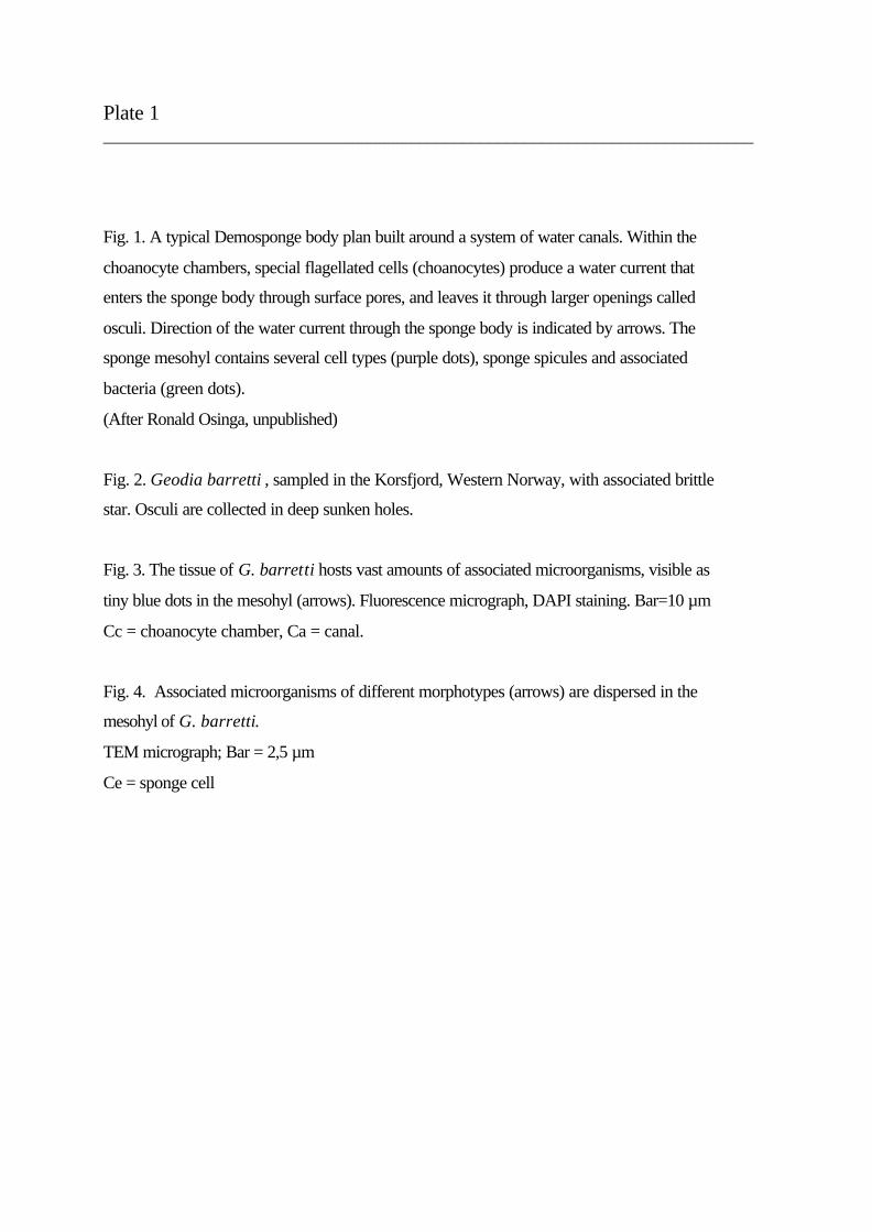
Plate 1 __________________________________________________________________________
Fig. 1. A typical Demosponge body plan built around a system of water canals. Within the
choanocyte chambers, special flagellated cells (choanocytes) produce a water current that
enters the sponge body through surface pores, and leaves it through larger openings called
osculi. Direction of the water current through the sponge body is indicated by arrows. The
sponge mesohyl contains several cell types (purple dots), sponge spicules and associated
bacteria (green dots).
(After Ronald Osinga, unpublished)
Fig. 2. Geodia barretti , sampled in the Korsfjord, Western Norway, with associated brittle
star. Osculi are collected in deep sunken holes.
Fig. 3. The tissue of G. barretti hosts vast amounts of associated microorganisms, visible as
tiny blue dots in the mesohyl (arrows). Fluorescence micrograph, DAPI staining. Bar=10 µm
Cc = choanocyte chamber, Ca = canal.
Fig. 4. Associated microorganisms of different morphotypes (arrows) are dispersed in the
mesohyl of G. barretti.
TEM micrograph; Bar = 2,5 µm
Ce = sponge cell
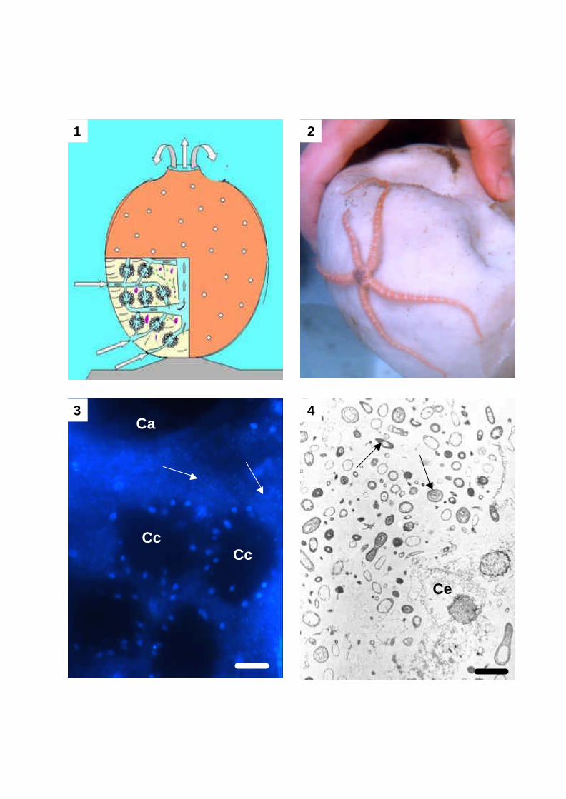
1 2
3 4
CcCc
Ca
Ce
Plate 2 __________________________________________________________________________
Fig. 1. DAPI stained LR White sections (20µm) of Phakellia ventilabrum (A) and Plakortis
simplex (B). Choanocyte chambers (cc), canals (ca), and endopinacocytes lining canal walls
(pi) are visible. This technique clearly shows differences in tissue structure and density of
associated microorganisms (tiny dots in the tissue) in this two sponge species. Bar = 30 µm
Fig. 2. Fluorochrome image (Calcein, A) and light microscopic image (Toluidin blue O, B) of
Phakellia ventilabrum. In this LR White sections (100 µm), bundles of spicules (sp) typical
for this species with accompanying cells (ac) are visible. Please notice choanocyte chambers
(cc) and canals (ca) in the choanosome, and different cell types in the ectosome (ec) at the
sponge surface. Bar = 100 µm
Fig. 3. In situ identification of sponge associated bacteria with fluorescently labelled rRNA-
targeted oligonucleotide probes on a tissue section from the choanosome of G. barretti.
Epifluorescence micrographs of different bacterial groups were visualized using specific filter
sets. Eubacteria (green) were hybridised with Oregon Green-labelled probe EUB338. Sulfate
reducers, which appear orange by image overlap, were hybridised with CY3-labelled probe
DSV698 specific for Desulfovibrio spp. Sulfate reducers of the genus Desulfovibrio are
evenly distributed throughout the choanosome. Bar = 20 µm
1A 1B
2A 2B
3
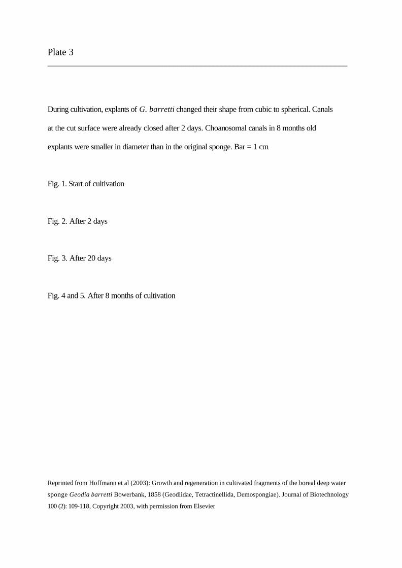
Plate 3 __________________________________________________________________________
During cultivation, explants of G. barretti changed their shape from cubic to spherical. Canals
at the cut surface were already closed after 2 days. Choanosomal canals in 8 months old
explants were smaller in diameter than in the original sponge. Bar = 1 cm
Fig. 1. Start of cultivation
Fig. 2. After 2 days
Fig. 3. After 20 days
Fig. 4 and 5. After 8 months of cultivation
Reprinted from Hoffmann et al (2003): Growth and regeneration in cultivated fragments of the boreal deep water
sponge Geodia barretti Bowerbank, 1858 (Geodiidae, Tetractinellida, Demospongiae). Journal of Biotechnology
100 (2): 109-118, Copyright 2003, with permission from Elsevier
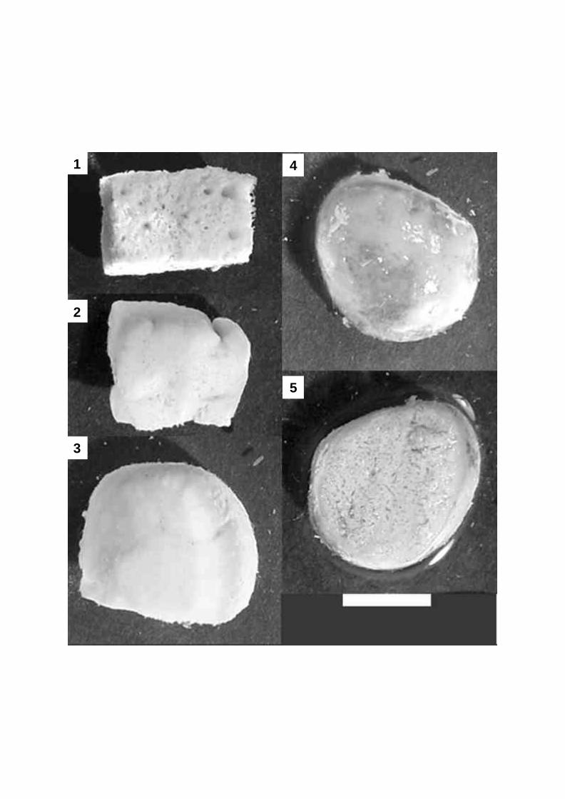
3
2
1 4
5
Plate 4 __________________________________________________________________________
Histological sections of cultivated fragments of G. barretti.
Fig. 1. Cut surface of a two days old explant of G. barretti. A growing front of motile sponge
cells (Cm) is accompanied by cells with inclusions (Ci) in an otherwise cell-and bacteria-poor
mesohyl. Bar = 500 µm
Fig. 2. Detailed view of 4a.
Fig. 3. Torn tissue at the cut surface of a two days old explant is covered by a closed line of
motile sponge cells (Cm). Bar = 30 µm
Fig. 4. During cultivation, the (new) ectosome is remodelled: choanocyte chambers are
regenerated, and cells with inclusions (Ci) accumulate (27 days old explant). Bar = 100 µm
Fig. 5. Oogenesis in cultivated explants sampled 2nd of April 2001. Bar = 30 µm
Fig. 6. Mature oocytes in cultivated explants sampled 10th of April 2001. Bar = 30 µm
Paraffin embedding, Goldner staining, spicules removed (Fig. 1-5)
LR-White embedding (Fig. 6)
Reprinted from Hoffmann et al (2003): Growth and regeneration in cultivated fragments of the boreal deep water
sponge Geodia barretti Bowerbank, 1858 (Geodiidae, Tetractinellida, Demospongiae). Journal of Biotechnology
100 (2): 109-118, Copyright 2003, with permission from Elsevier
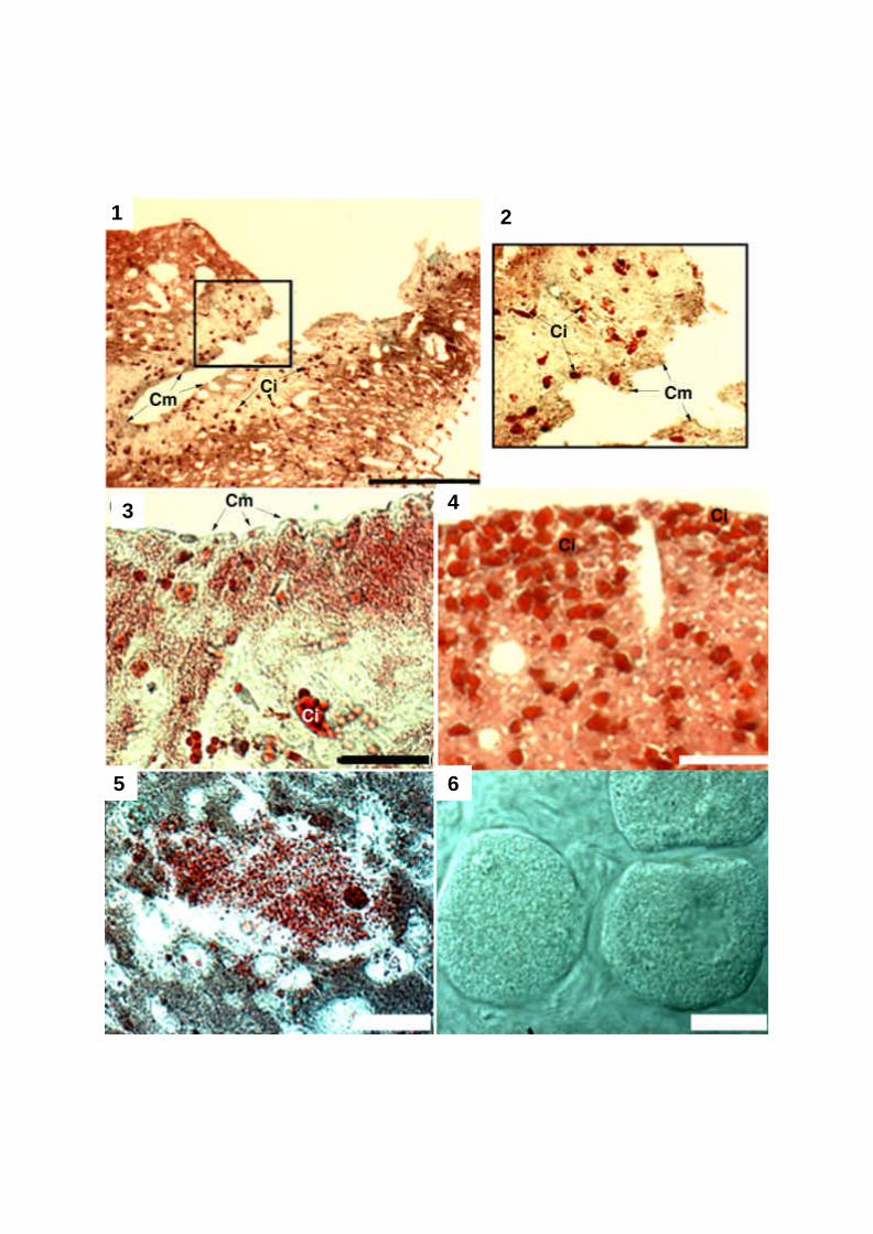
1 2
3
5
4
6
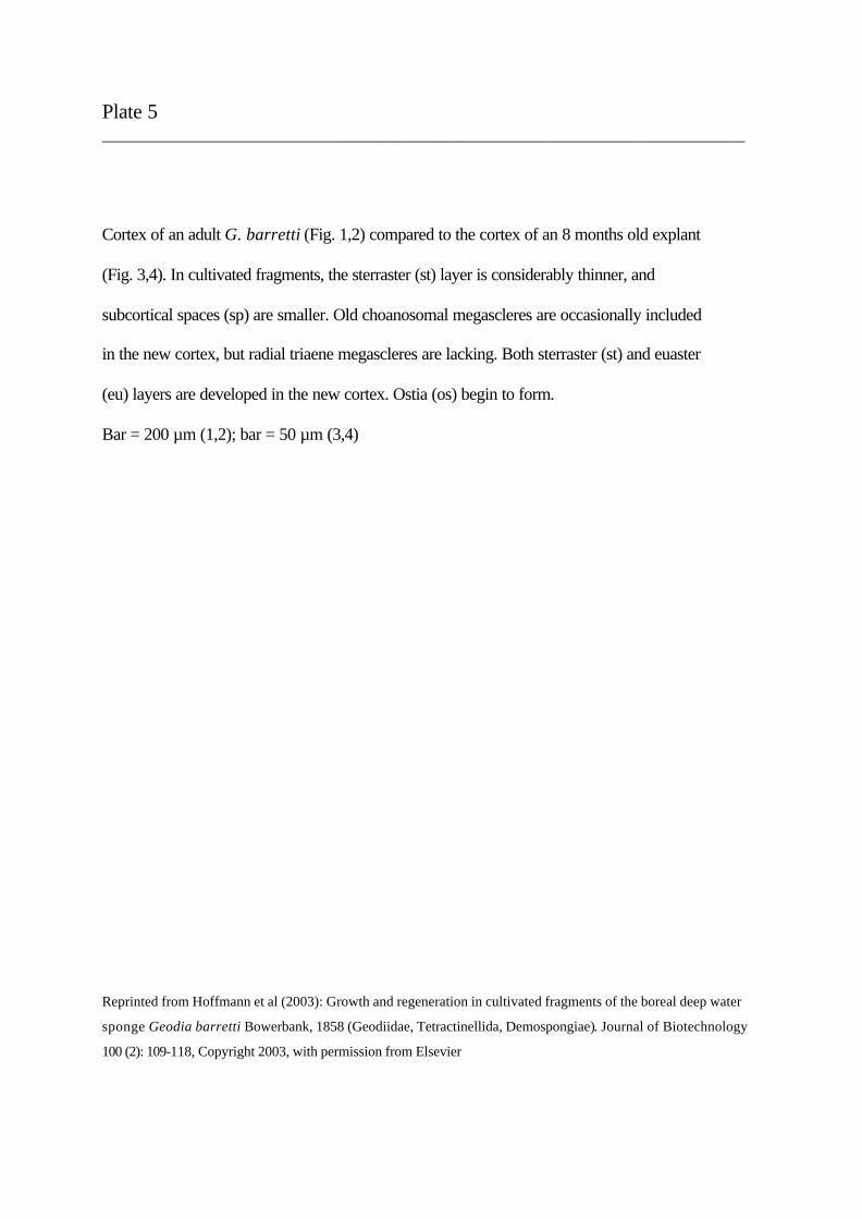
Plate 5 __________________________________________________________________________
Cortex of an adult G. barretti (Fig. 1,2) compared to the cortex of an 8 months old explant
(Fig. 3,4). In cultivated fragments, the sterraster (st) layer is considerably thinner, and
subcortical spaces (sp) are smaller. Old choanosomal megascleres are occasionally included
in the new cortex, but radial triaene megascleres are lacking. Both sterraster (st) and euaster
(eu) layers are developed in the new cortex. Ostia (os) begin to form.
Bar = 200 µm (1,2); bar = 50 µm (3,4)
Reprinted from Hoffmann et al (2003): Growth and regeneration in cultivated fragments of the boreal deep water
sponge Geodia barretti Bowerbank, 1858 (Geodiidae, Tetractinellida, Demospongiae). Journal of Biotechnology
100 (2): 109-118, Copyright 2003, with permission from Elsevier
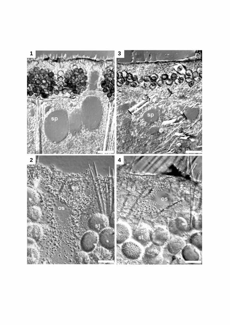
1
2
3
4
Lebenslauf Friederike Hoffmann 19.11.1970 Geboren in Kassel, Staatsangehörigkeit: deutsch
1977 - 1990 Grundschule und Gymnasium in Kassel
Juni 1990 Abitur an Oberstufengymnasium Herderschule, Kassel
August 1990 – September 1991 Praktikum bei Naturschutzgesellschaft “Schutzstation
Wattenmeer”, Hallig Hooge und Wyk auf Föhr
September 1991 – Juli 1994 Studium Biologie (Diplom) und Skandinavistik (Sprache: Norwegisch) an der Universität Göttingen
Oktober 1993 Diplomvorprüfung Biologie
August 1994 – Dezember 1995 Biologiestudium an der Universität Tromsö, Norwegen
Mai 1996 Diplomhauptprüfung Biologie an der Universität Göttingen
Juli/August 1996 Kurs „Arktische Mikrobiologie“ für graduierte Studenten an der Universität Longyearbyen, Svalbard
September 1996 – Oktober 1997 Diplomarbeit am Max-Planck-Institut für Marine Mikrobiologie in Bremen
Oktober 1997 – Dezember 1998 Arbeit als studentische/wissenschaftliche Hilfskraft am Max-Planck-Institut Bremen, Universität Bremen, und Institut für Ostseeforschung Warnemünde
Seit April 1999 Promotion an der Universität Göttingen, Fakultät für Geowissenschaften und Geographie, Abteilung Geobiologie