Embed Size (px)
Citation preview
Zentrum für Innere Medizin
III. Medizinische Klinik und Poliklinik
des Universitätsklinikum Hamburg Eppendorf
Direktor Prof. Dr. med. Rolf Stahl
Nephrotoxic Nephritis (NTN):
Possible new therapeutic strategy using DHMEQ, a nuclear uptake
inhibitor of NF-kappa B
D i s s e r t a t i o n
zur Erlangung des Grades eines Doktors der Medizin
dem Fachbereich Medizin der Universität Hamburg vorgelegt von
Syed Muhammad Fahad Imran
aus Karachi-Pakistan
Hamburg 2012

M.D. thesis Syed Muhammad Fahad Imran
Dedicated to my late Parents

M.D. thesis Syed Muhammad Fahad Imran
Angenommen von der Medizinischen Fakultät
der Universität Hamburg am: 20.09.2012
Veröffentlicht mit Genehmigung der Medizinischen Fakultät
der Universität Hamburg
Prüfungsausschuss, der Vorsitzende: Prof. Dr. F. Thaiss
Prüfungsausschuss: 2. Gutachter: Prof. Dr. T. Strate
Prüfungsausschuss: 3. Gutachter: PD Dr. G. Zahner
M.D. thesis Syed Muhammad Fahad Imran
4
Table of Contents Abbreviation index ..................................................................................................................... 6 Working Hypothesis and Problem: ............................................................................................ 8 Preface ........................................................................................................................................ 9 1.1. Glomeruloneprhitis (GN) ................................................................................................ 9 1.2. Nephrotoxic nephritis (NTN) ............................................................................................ 11
1.3. Nuclear factor-κB (NF-κB) ............................................................................................... 13 1.3.1. Structure of NF-κB ..................................................................................................... 13 1.3.2. Inducible activation of NF-κB .................................................................................... 14
1.4. Dehydroxymethylepoxyquinomicin (DHMEQ) NF-κB inhibitor ..................................... 18 2. Material and Procedure ........................................................................................................ 23
2.1. NTN-induction and treatment of the experimental animals .............................................. 23 2.2. Urine collection in metabolic cages .................................................................................. 23
2.3. Blood collection and kidney harvest ................................................................................. 23 2.4. Freeze and thawing of tissue ............................................................................................. 23 2.5. Protein biochemistry ......................................................................................................... 24 2.6. One-dimensional SDS Polyacrylamide gel electrophoresis .............................................. 24
2.6.1. Standard proteins for the electrophoresis ................................................................... 25 2.6.2. Extraction of cellular and nuclear proteins ................................................................ 25
2.6.2.1. Preparation of sample for cellular protein ..................................................... 25 2.6.2.2 Determination of the cellular protein concentration....................................... 26 2.6.2.3 Preparation of sample for nuclear protein ...................................................... 26 2.6.2.4. Determination of the nuclear protein concentration...................................... 26
2.7. Preparation of samples for Western Blot .......................................................................... 27 2.7.1. Separation of samples ................................................................................................. 27
2.7.2. Western Blot ............................................................................................................... 27 2.7.3. Immunoblot ................................................................................................................ 28
2.8. Molecular biology ............................................................................................................. 29 2.8.1. Isolation of RNA ........................................................................................................ 29
2.8.2. Concentration of RNA determination ........................................................................ 29 2.8.3. RT-PCR ...................................................................................................................... 30
2.9. Enzyme linked immunosorbent assay (ELISA) ................................................................ 30 2.10. Histology ......................................................................................................................... 31
2.10.1. General ..................................................................................................................... 31
2.10.2. Principle of the immune histology ........................................................................... 32 2.10.3. Complex of streptavidin and Biotin ......................................................................... 32
2.10.4. New fuchsine development solution ........................................................................ 32 2.10.5. Nuclear coloring with hematoxylin by Boehmer ..................................................... 33 2.10.6. Analysis .................................................................................................................... 33 2.10.7. PAS-Staining ............................................................................................................ 33 2.10.8. F4/80-Staining .......................................................................................................... 33
2.10.9. CD3-Staining ............................................................................................................ 34 2.10.10. Cell counting of CD3- and F4/80-positive cells ..................................................... 35
3. RESULTS ............................................................................................................................. 36 3.1. Effect of DHMEQ in vivo treatment in mice with nephrotoxic serum nephritis .............. 36
3.1.1. Effect of DHMEQ with vehicle DMSO in NTN treated animals .............................. 36 3.1.1.1. Result of RT-PCR .............................................................................................. 40
3.1.2. Effect of DHMEQ with vehicle DMSO and cremaphor in LPS treated animals ....... 41 3.1.1.1 Result of RT-PCR ............................................................................................... 44
M.D. thesis Syed Muhammad Fahad Imran
5
3.1.3. Effect of DHMEQ with a vehicle Cremaphor in NTN treated animals ..................... 46
3.1.3.1. Result of RT-PCR .............................................................................................. 50 3.2.4. NF-kappa B activation after NTN induction .............................................................. 51
4. Discussion ............................................................................................................................ 54
5. Conclusion ............................................................................................................................ 59 6. References ............................................................................................................................ 60 7. Word of thanks ..................................................................................................................... 64 8. Affidavit ............................................................................................................................... 65 9. Eidesstattliche Versicherung ................................................................................................ 66
M.D. thesis Syed Muhammad Fahad Imran
6
Abbreviation index fig. Figure
BSA Bovine serum album
BCIP 5 - Bromo - 4 - chloro - 3 – indolyl phosphate
bp base pair
°C degree of centigrade
cDNA copy DNA
D, kD Dalton, kilo-dalton
DAG Diacylglycerol
DEPC Diethylpyrocarbonate
D-MEM Dulbecco`s modified Eagle medium
DMF N, N´ - Dimetylformamide
DMSO Dimethyl sulfoxide
DNA Deoxyribonucleic acid
DTT Dithiothreithol
ECL Enhanced Chemoluminescence
EDTA Ethylenediaminetetraacetic acid
ELISA Enzyme linked immunosorbent assay
FCS Fetal calf serum
FSGS Focal segmental glomerulosclerosis
FITC Fluorescein isothiocyanate
g gram
gₒ Gravity / gravity, universal constant, represents the earth attractive
force. The relative centrifugal force (rcf / relative centrifugal force / g
force) is indicated in this work as multiple of g (x g).
GBM glomerular basement membrane
GN glomerulonephritis
Gen genomic
HEPES 4 (2 - Hydroxyethyl) - 1 – piperazinethansulfic acid
IFN Interferon
Ig Immunoglobulin
IgG Immunoglobulin of the class G
IgM Immunoglobulin of the class M
M.D. thesis Syed Muhammad Fahad Imran
7
IgE Immunoglobulin of the class E
kg kilogram (x 10 3)
L Liter
LDS Lithium dodecyl sulfate
LPS Lipopolysaccharide
m milli (x 10 ‾3)
M Molarity
MES Morpholinoethanol sulfonic acid
min Minute(s)
MMLV moloney murine leukemia virus;
MOPS Morpholinopropanol sulfonic acid
mRNA messenger RNA
n nano (x 10 ‾ 9
)
NBT 4 – Nitro blue tetrazolium chloride
PAN Puromycin aminonucleoside Nephritis
PAGE Polyacrylamide Gel electrophoresis
PBS Phosphate buffered saline
PCR Polymerase chain reaction
PFA Paraformaldehyde
PVDF Polyvinylidene fluoride
RNA Ribonucleic acid
SDS Sodium dodecyl sulfate
s second
Tab. Table
TAE Tris - acetate - EDTA
TBS Tris buffered saline
TBST Tris buffered saline + Tween 20
TEMED N,N,N,N-Tetramethylethylenediamine
TGF Transforming growth factor
Tris Tris - (hydroxymethyl) - aminomethan
Tween 20 Polyoxyethylenesorbitanmonolaurate
µ micro (x 10 ‾6)
U Unit
rpm revolutions per minute
M.D. thesis Syed Muhammad Fahad Imran
8
Working Hypothesis and Problem:
Inflammatory glomerular kidney diseases (glomerulonephritis) are causing, in spite of some
progress in the therapy, still in about 20-30% of the cases end stage renal disease.
Understanding the pathogenesis of inflammatory diseases persistently improved further in the
past years through following reasons among others:
By describing the role of transcription factors, that all ahead of NF kappa B, with that
induction of inflammatory diseases occurs and
By showing, that NF kappa B has also a decisive importance in the resolution phase of
inflammation processes.
The team was able to publish recently that it comes after induction of experimental kidney
diseases into a two phase activation of NF kappa B. In the first peak, the p65/p50 heterodimer
protein complex dominates, in the second peak the p50/p50 homodimers. Goal of the
graduation work therefore is to examine the effect of the inhibition of the nuclear reception of
NF kappa B complexes by means of in vivo application of DHMEQ at different times after
induction of the NTN in wild type mice.
M.D. thesis Syed Muhammad Fahad Imran
9
Preface
1.1. Glomeruloneprhitis (GN)
Glomerulonephritis is one of the major causes of chronic renal disease worldwide. Despite the
increasing rate of diabetic renal disease, glomerulonephritis still remains a major problem and
cause of chronic renal disease and end-stage renal failure requiring dialysis and renal
transplantation. It shows a wide spectrum of histological patterns with different severity of
injury and clinical outcome, which may be related to the nature of the nephritogenic immune
response (P.G. Tipping and A. R. Kitching. 2005).
Inflammation is the body’s primary response to infection or injury and is critical for both
innate and adaptive immunity. Upon induction of inflammation resident tissue cells produces
a variety of cytokines, chemokines, lipid mediators and bioactive amines which lead to an
infiltration of inflammatory cells. Primarily these cells are macrophages, dendritic cells, mast
cells and lymphocytes (T. Lawrence and C. Fonga. 2010). There is evidence that in the
majority of cases both humoral and cellular effectors mechanisms play a central role in both
human and animal models of glomerulonephritis (P.G. Tipping and A. R. Kitching. 2005).
In addition to providing help for production of immunoglobulins, that are involved in humoral
effector mechanisms, T cells plays an important role in directing cellular immune mechanisms
in GN. Th1 and Th2 cells are different in function, by their cytokine profile and their ability to
generate different types of immune effector responses. They both are subsets of primed CD4+
T helper cells. Th1 cells produce tumor necrosis factor (TNF), interleukin (IL)-2, interferon
(IFN)-γ and lymphotoxin-α. They activate macrophage and delayed-type hypersensitivity
(DTH) effector responses and at the same time help B cells to produce complement fixing
antibody isotypes that mediate opsonization and phagocytosis. On the other hand Th2 cells
produce interleukin (IL)-4, IL-5, IL-10 (in mice) and IL-13 that stimulate the production of
non-complement fixing IgG isotypes and IgE. They are important for elimination of infection
and mediation of allergic response. While Th1/Th2 subsets do have a clear influence on injury
in experimental GN, the dissection of their role in human GN has proven difficult due to the
diverse range of underlying immune pathogenic mechanisms in human disease (e.g. organ
specific or systemic autoimmunity, innocent bystander injury in the context of antigen
immune responses to planted antigen, probably immune dysregulation as in IgA
M.D. thesis Syed Muhammad Fahad Imran
10
nephropathy). In addition to this there are some other factors, like diversity in effectors
pathways involved and the variable defined genetic influences on immune responses in
human populations. Despite these obstacles, there is evidence which proved that the
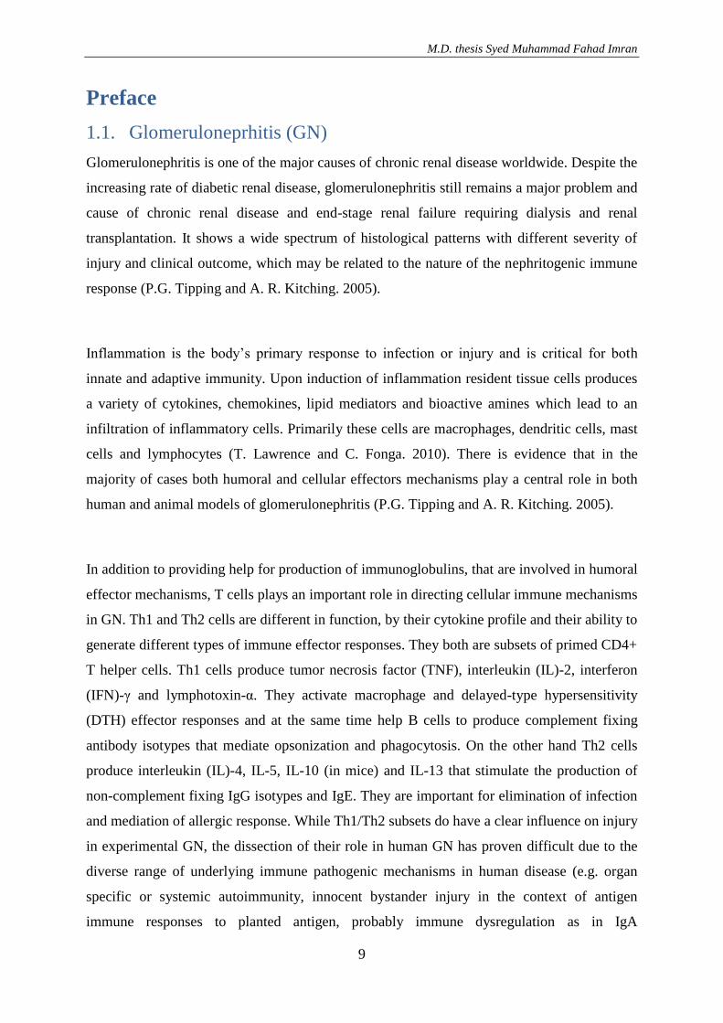
differential activation of Th1 and Th2 mechanism is useful in explaining of human GN (P.G.
Tipping and A. R. Kitching. 2005) (FIGURE1).
Figure1: A simplified scheme of Th1 and Th2 subset and cytokine involvement in glomerulonephritis. APC:
antigen-presenting cell; Mac: macrophage; source: (P.G. Tipping and A.R. Kitching. 2005)
There is increasing evidence, in which glomerular crescent formation results from an
underlying or superimposed Th1 response in severe and rapidly progressive forms of GN. In
previous studies the presence of effector CD4+ cells and macrophages in crescentic lesions,
has been observed. Biopsies of a variety of different forms of proliferative GN shows higher
levels of IL-2 and IFN-γ compared with non proliferative forms and glomerular CCR5
expression associated with crescentic GN. Studies in human anti- GBM GN demonstrated
IFN-γ -predominant antigen-specific effector cell responses in active disease and IL-10
predominance in remission further supporting a role for Th1 in injury. It has also been
demonstrated in these studies a central role for CD25+ regulatory cells in suppressing
pathogenic IFN-γ production human anti-GBM GN (P.G. Tipping and A. R. Kitching. 2005).
T cells responses are able to cause the classical four types of hypersensitivity immune
reactions, while these immune reactions can target the kidney and cause distinct forms of
glomerulonephritis. CD4+ T cells can induce glomerular immunopathology by producing
cytokines, by activating effector cells like macrophage and by mediating auto antibodies or
immune-complexes, whereas CD8+ T cell response and failure of regulatory T cells may
M.D. thesis Syed Muhammad Fahad Imran
11
represent two additional types of anti-renal hypersensitivity. The role of dendritic cells DC
was proven as protective in renal diseases, while it is essential for T cell activation (C. Kurts
et al. 2007). Cells with dendritic morphology and major histocompatibility complex (MHC)
class II expression were described in normal rat and mouse kidney, and they were able to
stimulate T-cell responses in vitro (S. Segerer et al. 2007).
1.2. Nephrotoxic nephritis (NTN)
Nephrotoxic nephritis is one of the most widely studied T cell-dependent models of kidney
injury, which results in immunopathology similar to human crescentic GN. It is induced in
mice, rats, or rabbits by injection of heterologous nephrotoxic serum (NTS), mostly generated
in sheep or rabbits vaccinated with murine or rat cortex glomerular basement membrane
antigens. It has been demonstrated in seminal animal studies by Tipping and Holdsworth that
CD4+ Th1 cells play an important role in a type IV hypersensitivity reaction in murine NTN.
Intravenously or intraperitoneally injected NTS predominantly binds to the glomerular
basement membrane and is taken up and presented by antigen-presenting cells, presumably
DCs to specific naive CD4+ T cells, causing their activation and differentiation into Th1
effectors cells. Depletion of CD4+ T cells attenuated NTN in mice and rats. Whereas CD8+ T
cells depletion was protective only in rats but not in mice (C. Kurts et al. 2007).
Under the influence of TLR ligands IL-12 is secreted by DCs which is essential for murine
NTN. After activation of T-cells, they are looking for their cognate antigen in non lymphoid
tissues. Because of its specificity the planted antigen in the kidney is subsequently presented
to CD4+ Th1 cells by local MHC II-expressing cells. In the kidney CD4+ T cells produced
Th1 effector cytokines like IFNγ or tumor necrosis factor alpha (TNF-α), which at the same
time stimulate DTH-like fashion resident macrophages and are essential effectors in NTN.
Macrophages cause injury of Bowman's capsule and crescent formation by producing ROS
and proinflammatory cytokines. In addition to this they also damage the basement membrane,
which makes it easier for macrophage to escape from glomerluar capillaries (C. Kurts et al.
2007). The importance of Th1-associated cytokines has been shown in TNF-α or IL-12
deficient animals or animals after injecting Th2 cytokines such as IL-4. Besides this the
hallmark Th1-chemokine receptor like CXCR3 was essential for immune pathology in NTN
(C. Kurts et al. 2007).
M.D. thesis Syed Muhammad Fahad Imran
12
However controversial results have been published on the role of Th1-associated cytokine
IFNγ in NTN, where both aggravating and ameliorating effects were described showing that
the role of IFN-γ is more complex than the simple Th1/Th2 dichotomy. One possible
explanation from experiments on Th1 effector cells in various models is that IL-10 has been
produced by Th1 effector cells as a co-product and it is one of the most potent
immunosuppressive cytokines. Based on these results, it has been assumed that the cellular
response and the co-production of further cytokines and chemokines are deciding to define
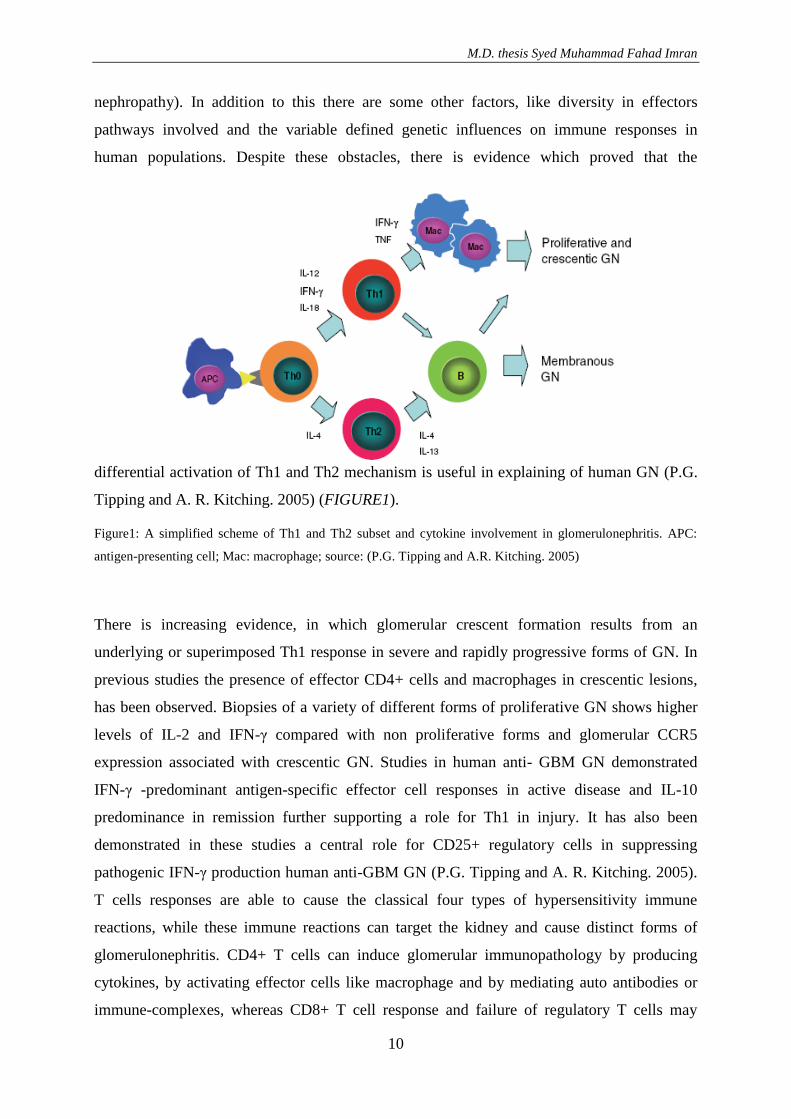
whether INF-γ is protective or harmful (C. Kurts et al. 2007) FIGURE2 (C. Kurts et al. 2007).
Figure2: Anti-renal macrophage-mediated delayed type hypersensitivity (type 4). Left panel DC in secondary lymphatics
activate Th1 effector cells specific for an antigen with kidney tropism. Activated Th1 cells leave the node and travel via the
bloodstream to the kidney. Th1 cells also induce antibody production, which will become relevant in the autologous phase
illustrated in the right panel. Middle panel Tubulointerstitial and glomerular monocytes/macrophages, as well as intrinsic
kidney cells, capture kidney-tropic antigen and present it to infiltrating CD4+ Th1 effector cells, which subsequently activate
them, e.g., by IFNγ . Release of ROS and proinflammatory cytokine causes injury of Bowman’s capsule and possibly in the
glomerular basement membrane, resulting in compensatory crescentic cell growth. Membrane rupture may allow crescent
infiltration by monocytes/macrophages, either from ruptured glomerular capillaries or from the interstitium. Dendritic cells
represent a counter regulator and attenuate kidney disease, presumably by inducing IL-10 production. Left panel After the
Th1 effector cell-mediated DTH phase, the autologous phase commences, in which specific antibodies bind to kidney-tropic
antigen and induce additional type III hypersensitivity reactions (C. Kurts et al. 2007).
One of the master transcription factors in regulating T-cell response and cytokines and
chemokines expression is NF-κB. Activation of NF-κB has been confirmed in a variety of
inflammatory renal disease including NTN in several studies (T. Koska et al. 2008). Therefore
M.D. thesis Syed Muhammad Fahad Imran
13
the focus of our studies was to elucidate the role of NF-κB in the NTN-model and to further
define a possible therapeutic effect of DHMEQ on this model.
1.3. Nuclear factor-κB (NF-κB)
Nuclear factor-κB (NF-κB) was discovered as a nucleoprotein in 1986. It was bound to the
enhancer region of the gene which encodes the immunoglobulin κ chain (Sen and Baltimore,
1986) (K. Umezawa and C. Chaicharoenpong 2002).
1.3.1. Structure of NF-κB
NF-κB is a dimer of members of the rel family of proteins. Each family member contains an
N-terminal 300 amino acid, however the rel homology domain (RHD) known as conserved
region. This is an important region and is responsible for DNA-binding, dimerization, and
interaction with IκB family members. It also contains a nuclear localization sequence (S.
Ghosh et al. 1998). It is one of the best characterized transcription factor. It has been shown
that NF-κB typically exists as a heterodimer of p50 and p65 subunits (T. Suzuki et al. 2008).
This heterodimer complex protein is what we commonly refer to as NF-κB, although the
diversity of other members is also now known. The complex is been made up from the two rel
proteins and each docking one half of the DNA binding site. A possibility of slightly
variations in the 10 base pair consensus sequence, 5-GGGGYNNCCY-3, confer a preference
for selected rel combinations of proteins and also has its own transactivating potential (S.
Ghosh et al. 1998). The p65 and p50 proteins have a homologous sequence at their N-terminal
regions and NF-κB can form various heterodimers or homodimers. There are more proteins
such as p52, c-Rel, Rel B, v-Rel, Dorsal, Dif, and Relish that include to the NF-κB family (A.
Ariga et al. 2002).
Although most of the NF-κB proteins are transcriptionally active there are some combinations
considered as inactive or repressive complexes. Thus, p50/p65, p50/c-rel, p65/p65, and p65/c-
rel are all transcriptionally active, whereas p50 homodimer and p52 homodimer are
transcriptionally repressive. There is a lack of variable C-terminal domain in p50 and p52
found in the activating rel proteins, which is most likely responsible for transactivation of NF-
κB-responsive genes (S. Ghosh et al. 1998).
M.D. thesis Syed Muhammad Fahad Imran
14
1.3.2. Inducible activation of NF-κB
The transcription factor NF-κB has been the focus of intense investigation for more than two
decades. Over this period of time, it has been made a considerable progress to understanding
the function and regulation of NF-κB although there is still much to be learned in this
important signaling pathway to understand (M. S. Hayden and S. Ghosh 2004). There is
evidence that NF-κB plays an important role in both pro- and anti inflammation but the
pathway depends on the cell lineage and pathophysiological context (T.Lawrence and C.
Fonga 2010).
Nuclear factor-κB (NF-κB) is a well known transcriptional factor that induced expression of a
variety of inflammatory genes and cell adhesion molecules such as TNF-α, interleukins,
monocyte chemoattractant protein-1, ICAM-1 and VCAM-1 (T. Kosaka et al. 2008). It binds
to the κB sequence and promotes transcription of cytokines such as IL-1, IL-2, IL-6, IL-8,
TNF-α, and interferon-γ, cell adhesion molecules such as E-selectin, intercellular adhesion
molecule 1 (ICAM-1) and a vascular cell adhesion molecule 1 (VCAM-1), and a viral
proteins (A. Ariga et al. 2002). However, there are significant differences in the precise
sequence of different κB sites (F. Wan and M. J. Lenardo 2010). Activation of NF-κB is
induced by extracellular signals such as TNF-α, IL-1, lipopolysaccharide, UV, and phorbol
esters, in which the TNF-α receptor to NF-κB activation signal transduction pathways is well
known and widely studied (A. Ariga et al. 2002). TNF-α is an apoptosis-inducing factor.
However, in most cases usually TNF-α does not induce apoptosis, because at the same time it
induces the activation of NF-κB, which inhibits the apoptosis (A. Ariga et al. 2002).
TNF-α receptors (TNFRs) include 55-kDa TNFR I and 75-kDa TNFR II. The ligand mainly
uses TNFR-I to act, inducing its trimerization. TNFR-associated death domain protein recruits
receptor-interacting protein and TNFR-associated factor-2 (TRAF-2) for activation of NF-κB.
TRAF-2 activates MEKK-3 which is a mitogen-activated protein kinase. On the other hand
MEKK-3 activates IκB kinase (IKK), which induces the phosphorylation of the inhibitor of
NF-κB (IκB). Phosphorylation of IκB induces its ubiquitination and degradation by
proteasomes. Liberated NF-κB molecules then enter the nucleus where they bind to the κB
site of DNA (A. Ariga et al. 2002) FIGURE3 (K. Umezawa and C. Chaicharoenpong 2002).
M.D. thesis Syed Muhammad Fahad Imran
15
Figure3: Signal transduction pathway for TNF-α-induced activation of NF-κB (K. Umezawa and C.
Chaicharoenpong 2002).
After gaining access to the nucleus, NF-κB must be actively regulated to execute its
fundamental function as a transcription factor. Recent studies have highlighted the importance
of nuclear signaling in the regulation of NF-κB transcriptional activity. There is increasing
evidence that both the recruitment of NF-κB within the nucleus to target genes and the
ensuing transcriptional events are actively and highly regulated. A growing list of novel
molecules has been identified in recent studies, which customize NF-κB transcriptional
activity in the nucleus. All of these observations giving the idea that the nuclear signaling of
NF-κB is more complex than initially considered, adding another level of complexity to the
complicated, but elegantly regulated, NF-κB signaling pathway (F. Wan and M. J. Lenardo
2010).
It is an area of intensive research to examine how NF-κB selectively recognizes certain κB
sites to achieve specific gene transcription, but it remains still incompletely understood. The
κB site sequence in target genes and the specific Rel dimer requirement are not consistently
M.D. thesis Syed Muhammad Fahad Imran
16
correlate, although it has been assumed for a long time that the usage of certain Rel dimers
could be dictated by the variability of κB sequences at specific promoters, based on the
principle that each κB site variant could preferentially recruit one type of Rel dimer over
others. Various κB sequences takes part in specific configurations for NF-κB binding, that
showed if a single nucleotide change in κB sites made, it affected the formation of productive
interactions between Rel subunits and coactivators (F. Wan and M. J. Lenardo 2010).
In several genome-wide studies, a large number of NF-κB-binding motifs have also been
illustrated beyond recognizable κB sequences. These results strongly indicate that other
integral non-Rel components in the NF-κB complex could not only regulate NF-κΒ activity
but also contribute to the strength and specificity of DNA binding as an essential component.
RPS3 has been identified as a one of a non-Rel subunit in certain NF-κB DNA-binding
complexes, where it is essential for the recruitment of NF-κB p65 to the selected κΒ sites.
RPS3 was demonstrated to interact with p65 in a proteomic screen and evidenced to be
instrumental for NF-κΒ transactivation. The featured heterogeneous nuclear protein K
(hnRNP K) homology (KH) domain of RPS3, a structural motif that binds single-stranded
RNA and DNA with some sequence specificity, is essential for its association with p65 (F.
Wan and M. J. Lenardo 2010).
RPS3 functions as an integral part of NF-κΒ revealed in several lines of evidence. It is also
associated with the p65-p50-IκΒα cytoplasmic sequestration complex in resting cells. In
response of the stimulation, RPS3 is also able to translocate specifically to the nucleus and
binds to κΒ sites in a large number of NF-κΒ-dependent genes. In addition to this RPS3 exists
in endogenous NF-κB DNA-binding complexes, as demonstrated by the ability of RPS3-
specific antibodies to supershift or diminish p65-containing DNA complexes in
electrophoretic mobility shift assays (EMSAs). It has been shown a significant correlation
between RPS3-dependence and p65-dependence in transcription of a subset of NF-κB target
genes induced by T-cell receptor ligation. Therefore, RPS3 is an integral subunit of NF-κB
rather than a co activator. It has also been demonstrated that RPS3 protein strikingly exerts a
dramatic synergistic effect on the DNA-binding activity of both p65-p65 and p50-p65, but not
of p50-p50 complexes. Furthermore, RPS3 stabilizes NF-κB association with certain cognate
κB sites, as a DNA-binding component. These phenomena give us an important indication
about unexplained extremely high affinity of native NF-κB complexes for κB DNA. No other
M.D. thesis Syed Muhammad Fahad Imran
17
Rel-interacting protein has been clearly demonstrated to be integrated into endogenous NF-κΒ
complexes or to substantially augment the DNA-binding activity of NF-κB, although over
100 proteins interaction with p65 were known. It has been emphasized that, in certain key
physiological processes, including induction of immunoglobulin-κ light chain gene expression
in B cells, and cell proliferation and cytokine secretion in T cells plays an important role of
RPS3 in regulating NF-κB transcription. The inherent complexity of NF-κΒ itself is been
highlighted by the identification of RPS3 (F. Wan and M. J. Lenardo 2010) FIGURE4 (F.
Wan and M. J. Lenardo 2010).
Figure 4. The nuclear regulation of NF-κB. After it gains access to the nucleus, NF-κB transcriptional activity is elegantly
controlled by a number of nuclear regulators, as illustrated in blue, including non-Rel subunits such as RPS3 (S3) within the
NF-κΒ complex, proteins restricted to the nucleus or translocated from the plasma membrane, other transcription factors, and
chromatin modifiers. Two distinct pathways to terminate NF-κΒ transactivation, as illustrated in red, involve (I) IκB-
mediated restitution of NF-κΒ to the cytoplasm and (II) ubiquitination-dependent nuclear degradation (F. Wan and M. J.
Lenardo 2010)
M.D. thesis Syed Muhammad Fahad Imran
18
Some signaling molecules working close to the plasma membrane have also been shown
novel function in regulating NF-κB transcriptional activity in the nucleus. Akt1, a
serine/threonine protein kinase are the one of the examples and their functions are
immediately downstream of plasma membrane tethered phosphoinositide 3-kinase. It was also
observed that these molecules play an important role to translocate into the nucleus and
complex with p65 and Ki-Ras to enhance NF-κB transcriptional activity (F. Wan and M. J.
Lenardo 2010).
A non-Rel subunit of NF-κB, ribosomal protein S3 (RPS3), and numerous other nuclear
regulators of NF-κB, including Akirin, Nurr1, SIRT6, and others, have recently been
identified, unveiling novel and exciting layers of regulatory specificity for NF-κB in the
nucleus. Further insights into the nuclear events that govern NF-κB function will deepen our
understanding of the elegant control of its transcriptional activity and better inform the
potential rational design of therapeutics for NF-κB associated diseases (F. Wan and M. J.
Lenardo 2010).
Low molecular weight inhibitors of NF-κB therefore should be useful as immune
suppressants and as anti-inflammatory, antiviral, and anticancer agents. Panepoxydone,
cycloepoxydon and gliotoxin are among those inhibitors. Panepoxydone is isolated from the
basidiomycete lentinus crinitus and inhibits TNF-α induction, and in this way inhibits the
activation of NF-κB as shown in a promoter reporter assay by using COS-7 cells. It is been
reported, that Gliotoxin producing by fungi, also inhibit the activation of NF-κB by
preventing the degradation of IκB (A. Ariga et al. 2002).
1.4. Dehydroxymethylepoxyquinomicin (DHMEQ) NF-κB
inhibitor
In the course of our research we therefore have used a new designed NF-κB inhibitor of low
molecular weight inhibitors of NF-κB function. It was derived from the structure of the
antibiotic epoxyquinomicin C (A. Ariga et al. 2002) FIGURE 5 (A. Ariga et al. 2002).
M.D. thesis Syed Muhammad Fahad Imran
19
R=OH; (-)-DHMEQ
R=H; 9-dehydroxy-(-)-DHMEQ
Figure5: Structure of DHMEQ and its inactive analogue. Racemic DHMEQ and 9-dehydroxy-DHMEQ were
used throughout the experiments (A. Ariga et al. 2002).
This novel NF-κB inhibitor, dehydroxymethylepoxyquinomicin, abbreviated as DHMEQ is
synthesized from 2, 5-dihydroxyaniline in five steps as its racemic form. It has been shown
that (-)-DHMEQ is ten times more potent than (+)-DHMEQ [12]. The designed molecule is
called dehydroxymethylepoxyquinonmicin (DHMEQ, formerly DHM2EQ) and inhibits TNF-
α-induced activation of NF-κB. It has been demonstrated in different studies, that DHMEQ
was effective in suppressing rheumatoid arthritis (K. Wakamatsu et al. 2005), in
Hodgkin/Reed-Sternberg cells (M. Watanabe et al. 2007), local and remote organ injury
following intestinal ischemia/reperfusion in rats (T. Suzuki et al. 2008), anti-thy1.1-induced
glomerulonephritis in rats (T. Kosaka et al. 2008), atherosclerosis in ApoE-deficient mice (T.
Chiba et al. 2006) and osteoclastogenesis in mouse arthritis (T. Kubota et al. 2007).
DHMEQ inhibits the tumor necrosis factor (TNF)-α-induced NF-κB binding activity but not
the phosphorylation or degradation of I-κB (T. Suzuki et al. 2008). DHMEQ inhibited the
TNF-α-induced nuclear accumulation of p65. Thus, DHMEQ is a unique inhibitor of NF-κB
that acts at the level of the nuclear translocation. DHMEQ has been reported to be effective on
various hematologic and solid malignancies with constitutively active NF-κB (N.
Dabaghmanesh et al. 2009). In these studies it has been noticed that the molecular target of
M.D. thesis Syed Muhammad Fahad Imran
20
DHMEQ is directly bound covalently to the Rel family proteins through a specific Cys
residue. This discovery may explain its highly selective inhibition of NF-κB and the low toxic
effect of DHMEQ in cells and animals (M. Yamamoto et al. 2008). DHMEQ exhibits anti
inflammatory, anti fibrotic, and anti tumor effects (K. Shinoda et al. 2010). DHMEQ
completely inhibited NF-κB p65 DNA-binding activity. It has also been demonstrated that
this inhibitory effect is dose dependent. The duration of the effect of 2 µg/ml DHMEQ
pretreatment was limited to 6 hours, however pre-treating with 5 µg/ml DHMEQ indicated a
dose-dependent inhibitory effect of DHMEQ of at least 24 hours. Moreover this study
demonstrated that DHMEQ interfered with nuclear translocation of activated NF-κB without
inhibiting phosphorylation or degradation of IκB in Mo-DC. In addition DHMEQ was shown
to be cytotoxic to DC at concentrations of 10 µg/ml or more. The inhibition of cytokines
production through DHMEQ is also dose-dependent. It has been noticed significantly
inhibition of IL-6 and of TNF-α production at concentrations of 2 µg/ml or higher, whereas
IL-12 p70 production was significantly inhibited at concentrations of 1 µg/ml or higher.
Among these cytokines, IL-12 p70 production was most strongly inhibited by DHMEQ. On
the other hand DHMEQ moderately affected IL-10 transcription and its production. There was
an observation that CD40 and HLA-DR expression in DC was inhibited by DHMEQ, whereas
CD80 and CD86 expression was moderately upregulated. DHMEQ also partially inhibited
down-regulation of endocytosis during DC maturation (K. Shinoda et al. 2010).
In another study there is evidence that DHMEQ inhibits LPS-induced histamine secretion and
induction of HDC. DHMEQ inhibits C/EBP transcription factor activity, whereas C/EBP is
known to be involved in the transcription of the HDC gene (E. Suzuki et al. 2009). It has been
observed that DHMEQ significantly effects nuclear factor-κB nuclear translocation in a
murine model of arthritis and cultured human synovial cells (K. Wakamatsu et al. 2005).
Through inhibition of constitutive NF-κB activity DHMEQ reduces cell growth and induces
apoptosis of H-RS cells. In the same study it has been observed that DHMEQ has a potent
inhibitory effect on the growth of H-RS cells in NOG mice model. DHMEQ increase anti-
tumor effect of topoisomerase inhibitors by blocking inducible NF-κB in H-RS cells.
DHMEQ induce apoptosis in cells in which the activation of caspases 3, 8 and 9 is involved
(M. Watanabe et al. 2007). In such a study it has been shown that the activity of NF-κB is
higher in tumor initiating TIC-like cells when compare to control cells. Moreover, DHMEQ, a
highly specific inhibitor for NF-κB, suppressed tumorgenesis in the TIC-like cells in mouse
M.D. thesis Syed Muhammad Fahad Imran
21
model. After treatment with DHMEQ it has been shown a decrease in tumorgenesis in CD24-
/low/CD44+ populations (M. Murohashi et al. 2010).
DHMEQ reduces NF-κB p65 DNA-Binding capacity, cell growth, and induces apoptosis in
human hepatoma cells. ERK1/2 was involved evidenced based in cell proliferation and
apoptosis. DHMEQ enhances the phosphorylation of ERK1/2 and, in combination with the
MEK-specific inhibitor U0126 in order to reduce cell growth. This study showed that
induction of ERK1/2 phosphorylation in both cell lines through DHMEQ is dose-dependent.
MEK/ERK pathway is activated as a pro-survival signaling during DHMEQ treatment. This
antitumor effect of DHMEQ in human liver cancer cells are mediated through a Reactive
Oxygen Species (ROS)-dependent mechanism (N. Lampiasi et al. 2009).
DHMEQ inhibits TNF-mediated localization of c-Rel and RelB, which retain RelA RSAGSI
sequences, but not that of p52 having p50 PSHGGL in the nucleus (M. Watanabe et al. 2008).
In another study it has been demonstrated that DHMEQ suppresses the development of
atherosclerosis in apoE-deficient mice (T. Chiba et al. 2006). DHMEQ does not inhibit TNF-α
induced phosphorylation and degradation of IκB in Jurkat and COS-1 cells (A. Ariga et al.
2002). It does not inhibit the nuclear transport of Smad2 and the large T antigen, which means
that it does not inhibit the TNF-α-induced activation of JNK, but synergistically induced
apoptosis with TNF-α in human T cell leukemia Jurkat cells. In addition to all these, DHMEQ
inhibits 12-O-tetradecanoylphorbol-13-acetate (TPA)-induced NF-κB activation in Jurkat
cells. TPA is considered to activate IKK through the activation of protein kinase C. Therefore,
it was assumed that DHMEQ inhibits the NF-κB activation at a level that is downstream of
IKK. DHMEQ also inhibits the TNF-α-induced DNA binding of nuclear NF-κB in the
electrophoresis mobility shift assay (EMSA). However, it does not inhibit the direct binding
of NF-κB after subcellular fractionation. On the other hand TNF-α induces phosphorylation
and proteasome-mediated degradation of IκB to release NF-κB from the inactive complex.
Interestingly, DHMEQ does not inhibit the TNF-α-induced phosphorylation and degradation
of IκB (K. Umezawa and C. Chaicharoenpong 2002).
It has been illustrated that DHMEQ does not suppress spontaneous expression of RANKL nor
macrophage colony-stimulating factor in culture of fibroblast like synovial cells obtained
M.D. thesis Syed Muhammad Fahad Imran
22
from patients with rheumatoid arthritis. These observations suggest that DHMEQ suppresses
osteoclastogenesis in vivo through down regulation of NFATc1 expression without
significantly affecting expression of upstream molecules of the RANKL/receptor activator of
NF-κB/osteoprotegerin cascade (T. Kubota et al. 2007).
M.D. thesis Syed Muhammad Fahad Imran
23
2. Material and Procedure
2.1. NTN-induction and treatment of the experimental animals
For the experiment male C57Black6 mice were used. The mice were injected with 0.6 ml
NTN antiserum i.p (animal experiments were approved by a positive vote of the local ethical
committee FII 18/04). DHMEQ was injected subcutaneously in a dose of 3g/KG body weight
starting 12 hours before induction of NTN and once every 48 hours. For the vehicle control
animals were injected with 10% Cremaphor solution dissolved in PBS or DMSO with
methylcellulose.
2.2. Urine collection in metabolic cages
Urine was collected at different time points after the induction of NTN while mice were
placed in metabolic cages for 6 hours. Urine collected was transported into prepared 1.5 ml
sample containers and frozen at -20°C until further analysis.
2.3. Blood collection and kidney harvest
Animals were sacrificed at 2, 4, 10, 14 days after NTN-induction. At the end of the
observation period 0.5 ml blood was collected by direct puncture of the Aorta. Blood was
transferred into a 1.5 ml serum sample container (Sarstedt) and stored at -20°C. Kidneys were
harvested and further processed for total protein and RNA isolation and morphological
examination.
2.4. Freeze and thawing of tissue
Kidney tissue for RNA and protein extraction was shock frozen in fluid nitrogen and stored at
-80°C until further processing. Nuclear protein was done immediately after kidney harvest
from fresh isolated tissue.
M.D. thesis Syed Muhammad Fahad Imran
24
2.5. Protein biochemistry
Protein electrophoresis is based on ions migration within an electric field. The proteins have
different entire electrical charge and different pK values that are dependent on the amount of
sour and basic amino acids. If an electrical charge molecule is brought into an electric field,
the electrophoretic mobility depends on the size of molecule, shape, the total net electrical
charge, the pH-value, the pore size of the carrier, the temperature, the ion strength of the
buffer and on the electric field strength. The protein separation is carried out usually in form
of a gel (Agarose, Polyacrylamide) in a so-called carrier matrix. Through that variable cross
linked networking the gels serve as a filter effect, that separates the molecules after its
electrical charge and after its size, shapes and design.
2.6. One-dimensional SDS Polyacrylamide gel electrophoresis
If proteins are charged through the anionic detergent SDS (Sodium-Dodecyl-Sulfate),
separation in the electric field is possible almost solely through molecular weight. SDS bonds
to the hydrophobic regions of proteins, whereby most of the proteins dissociate into its lower
units. Through this bonding, a strongly negative electrical charge establishes in that
polypeptide chains. It emerges a complex with negative sulphate group directed outward, i.e.
the amount of negative charge is directly proportionally to the protein size. The proteins move
differently from the cathode to the anode depends to its electrical charge. Due to the narrow
pore of separation gel, there is an additional molecular filter effect, whereby the proteins are
separated corresponding to its size. To the successful execution of a gel electrophoresis, the
protein samples must be brought into a medium, which reduces or prevents an intermixture of
the samples. The medium may allow neither to react with sample nor inhibiting the movement
of the protein sample. These conditions are fulfilled with polyacrylamide gel. The network of
the gel is constructed through radical polymerization of the acrylamide monomer and the
cross linked networking of bifunctional N,N'-Methylen-Bisacrylamide. As a radical starter,
ammonium peroxide disulphide and the tertiary amine N, N, N N' –Tetramethylethylen
diamine (TEMED) were used. The pore size can be varied by the quantity ratio of both
components. Due to different molecular weights, an optimal separation conditions can be
created.
M.D. thesis Syed Muhammad Fahad Imran
25
2.6.1. Standard proteins for the electrophoresis
A marker is a mixture out of high or low molecular standard proteins whose molecular
weights are well known. It has been applied in the electrophoresis in addition on the gel in
order to detect proteins according to its corresponding molecular weight. For that the marker
SDS-7B has been used from (Sigma). The standard proteins are mixed here with a blue dye
which changes the molecular weight of the proteins. For this reason a renewed calculation of
the molecular weight of the respective standard protein after every staining is necessary. The
sublime has been dissolved in the urea solution and NuPage 4x LDS sample buffer (4x LDS +
0.4 MS DTT), given from the manufacturer (Sigma), heated for 10 min at 80°C and frozen in
form of Aliquots at -20°C.
Molecular weight:
-Macroglobulin, human plasma 190 kD
-Galactosidase, E. coli 108 kD
-Fructose- 6-phosphate-kinase, muscle, rabbit, 84 kD
-Pyruvate-Kinase, muscle, hen, 67 kD
-Fumarase, heart, pig, 55 kD
-Lactate-dehydrokinase, muscle, rabbit, 39 kD
-Triose-phosphate-isomerase, muscle, rabbit, 35 kD
2.6.2. Extraction of cellular and nuclear proteins
2.6.2.1. Preparation of sample for cellular protein
Tissue lysate of mouse kidney were used to separate cellular protein. Tissues have been lysed
with T-PER Tissue protein Extraction Reagent from (Pierce) and with complete Protease
inhibitor from (Roche). In addition to this the tissue was transferred with 10 µl Lysis buffer
per 1 mg tissue and minces mechanically with a pestle. After fifteen minute intubation time
on ice, the suspension should bring to Centrifugation for 15-20 min in (full speed) 16100x g
and at 4°C. That supernatant was transported into a new sample container and frozen on -
80°C.
M.D. thesis Syed Muhammad Fahad Imran
26
2.6.2.2 Determination of the cellular protein concentration
The determination of the protein concentration resulted after Lowry Assay method. Firstly we
prepare a BSA standard with a concentration of 10mg/ml. BSA serum albumin solves in T-
PER Tissue protein Extraction Reagent from (Pierce) and it is dilute 1:2 (10; 5; 2.5; 1.25; and
0.6 mg/ml). After thawing of proteins, they have been mixed gently and well. 5µl proteins
were given into each wells of a 96 holes disk and then 5µl BSA standard, comparably solved
in lysis buffer, was given into each wells of 96 holes disk. Each protein sample were pipetting
twice. 25µl a mixture out of solution A/S (1ml solution A+20µl solution S from
manufacturer) of Bio Rad and 200µl of solution B from Bio Rad were add into in it and
subsequently leave to incubate for 15 min at room temperature (RT). The measurement
resulted in 550nm in the Mithras.
2.6.2.3 Preparation of sample for nuclear protein
Tissue lysate of mouse kidney were used to separate nuclear protein. We prepare firstly a Low
salt buffer (Buffer A) and high salt buffer (Buffer B) solution. Buffer A contained 10mM
HEPES pH 7.9, 10mM KCl, 0,1mM EDTA pH 8.0, 0,1mM EGTA pH 8.0 and 1mM DTT and
subsequently added a freshly Protease Inhibitor Mix M (Serva,1:100) into in it. Buffer B
contained 20mM HEPES pH 7.9, 40mM NaCl, 1mM EDTA pH 8.0, 1mM EGTA pH 8.0 and
1mM DTT with an addition of freshly Protease Inhibitor Mix M (Serva1:100). Firstly the
kidney has been mashed on Petri dish with a scalpel and subsequently homogenized in 1ml
Buffer A in the douncer. The mixture brings into marked Eppi-tubes and subsequently let for
15 min on ice for incubation. An addition of 100µl of 10%NP-40 is to follow. The Eppendorf
tubes were shaken gently and placed immediately at 16000xg for 5 min to centrifuge. The
supernatant was thrown away and tissue pellet has been resuspended in 200-400µl Buffer B
and subsequently let for 15-20 min to incubate at 4°C in the cool room with rotation. Then
centrifugation is to follow with 16000xg for 5 min and subsequently the supernatant has been
aliquot and frozen after fluid nitrogen shock at -80°C.
2.6.2.4. Determination of the nuclear protein concentration
The determination of the protein concentration resulted after Lowry Assay method. First one
prepares a BSA standard with a concentration of 10mg/ml. BSA is solved in Buffer B (even
produced) and is diluted 1:2 (10; 5; 2.5; 1.25; and 0.6 mg/ml). After thawing of proteins, they
have been mixed gently and well. 5 µl proteins were given into each wells of a 96 holes disk
M.D. thesis Syed Muhammad Fahad Imran
27
and then 5µl BSA standard, comparably solved in lysis buffer, was given into each wells of a
96 holes disk. Each Protein sample were pipetting twice. 25 µl a mixture out of solution A/S
(1 ml solution A+20µl solution S from manufacturer) of Bio Rad and 200 µl of solution B
from Bio Rad were add into in it and subsequently leave to incubate for 15 min in room
temperature (RT). The measurement resulted in 550nm in the Mithras.
2.7. Preparation of samples for Western Blot
It has been loaded a 50 µg of cellular protein as well as nuclear protein per lane of a
NuPAGE. The concentration of gels depends on the molecular weight of the protein to be
detected. Volume for 50 µg proteins has been calculated on the basis of protein determination.
Volume difference has been filled with 16µl of corresponding Protein lysis buffer. All
samples were poured in cups with the 8 µl corresponding quantity of 3x sample protein buffer
(NuPage loading buffer + 0.4 MS DTT) and were heated to the complete denaturing for 10
min at 95°C.
2.7.1. Separation of samples
For the separation of proteins, a prefabricated, reducing 4-12% NuPage Bis-Tris gradient gel
was used (Invitrogen). The gel was taken out of its packet and wash shortly with water in
order to remove non pure particles of the Butyl alcohol, which used for the preservation. After
that the pockets of the gel has been washed with MOPS or MES (50 mM of MOPS and/or
MES, 50 mM of Tris base, 0.1% SDS, 1 mM of EDTA) course buffer. The gel is been fixed
into an electrophoresic chamber from (PeqLab) and the chamber is been filled up completely
with course buffer. After loading of the pockets with 4 µl protein marker and 22 µl samples,
the chamber is been attached to a designed power supply (Organic wheel) with a Voltage of
200 V and is been let to run for at least 45-50 minutes. This voltage has retained until the first
proteins reaches the lower end of the gel.
2.7.2. Western Blot
The proteins separated by the SDS PAGE is been transfer out of the gel through
electrophoretic transfer on a positively charged carrier membrane from PVDF (Amersham).
This procedure is known as a wet procedure. Because the proteins are further by SDS
negatively charged, they move from cathode to the anode. A novex mini cell was used to
transfer the proteins. The blotting resulted like in the sandwich procedure between three filter
M.D. thesis Syed Muhammad Fahad Imran
28
papers. PVDF membrane is been activated for 1 min with methanol before use. The
membrane brought air bubble less to the gel and put between three pieces of watman paper
and between three pieces of sponges in the blotting device. The fixation has been followed, in
order that the gel to the cathode side and membrane to the anode side directed. The novex
minis cell was filled up then with blotting buffer (25 mM of Tris base, 0,192 MS Glycin, 20%
methanol in aqua dest). The transfer takes place at a current strength of 65 mA (1mA/cm²)
and at a temperature of 4°C in about 1.5 hours. After successful transfer, the Blot was stored
at 4°C in TBST (10 mM of Tris pH 7.4, 107 mM of 5 MS NaCl, 0.05% Tween 20) for the
further use.
2.7.3. Immunoblot
In the Immunoblot, separated proteins through gel electrophoresis and transferred proteins on
a membrane are detected with the help of specific antibodies. The antibodies do not bind
covalently with its antigen. The PVDF membrane incubates and has been blocked with dried
milk solved 5% in 30ml TBST for approx. 3h on seesaw at RT, after the removal out of the
transfer unit. After short washing with TBST, the membrane is been incubate with the
primary antibody in TBST solved 5% lean dried milk. The length of the incubation time in
which the antibody can bind depended on the assumed affinity of the antibody and the
quantity of the product to be detected. In our experiments, the gels were incubates overnight
at 4°C with primary antibodies IB (C-21 Santa Cruz) or NF kB p65 (F-6 Cell Signaling) in
blocking buffer on a seesaw. After removing the membrane from the primary antibody, the
membrane was washed twice in TBST for over 10 min each time. After that the secondary
antibody coupled with horseradish peroxide has been poured. The antibody was solved in 20
ml 5% lean dried milk in TBST. After removing of the secondary antibody, further wash steps
are followed with TBST for over 30 min and as well as with assay buffer twice for 5 min. All
steps occur on a shake table/seesaw. Immune complexes were visible with the help of enzyme
conjugation. In order to reinforce the effect, the detection takes place indirectly via marked
secondary antibodies, which are directed against the primary antibody bound to the antigen.
There are different possibilities of detection. In the frame of this work, the detection with ECL
super signal resulted. It is based on the principle of a highly sensitive method of
chemiluminescence. This is based on that, in which the secondary antibodies coupled with
peroxide aces oxidize in presence of H2O2 luminol. At the same time light is released, that
after corresponding exposure time on x-ray film exposes. The detection takes place in the
M.D. thesis Syed Muhammad Fahad Imran
29
darkroom with a help of automatic film processor. The sensitivity of the Western Blots lies
between 10 to 100 fmol. This corresponds in a molecular weight of 50 kD approx. 0.5 to 5 ng
proteins.
2.8. Molecular biology
2.8.1. Isolation of RNA
For the extractions of RNA out of mouse kidneys, a Trizol method was used. The one third of
kidneys placed on Petri dish, after detaching the hilus mechanically and minced gently with
scalpel and tweezers. The kidney pieces are homogenized then with pestle in a 1.5 ml sample
container cups containing 200 Trizol. It is given in addition to this 800µl Trizol and mixed
well. Then let the samples to incubate for 5-10 min in RT. It is been mixed gently and
thoroughly in between. After a shortly centrifugation 200µl chloroform has been added. The
sample were mixed gently for 15 second and then let the samples for 3 min at RT to incubate
and subsequently centrifugation does take place with 10000 rpm for 15 min at 4°C. The clear
supernatant of approx. 500 µl led into new RNA Cup with 200 µl pipette. It has been added a
500 µl isopropyl alcohol in it and mixed well. Subsequently let the sample for 15-30 minutes
at RT to incubate. These have been placed for centrifugation at 13000 rpm for 10 minutes in
4°C. Supernatant were through away and RNA pellets were put with 75% 1 ml of
Ethanol/DEPC-H2O in centrifugation at 13000 rpm for 10 minutes and at 4°C. The next both
step of washing is followed with 500µl Ethanol/DEPC-H2O on seesaw. Supernatant is
through away and pellets were dry in speed vac. for 5 minutes without temperature. In
between time a mixture of RNase out and DEPC/H2O for solving the peeling has been made.
30 µl mixture of RNase poured to the peeling and well mixed. For the further processing the
RNA is been stored at -80°C.
2.8.2. Concentration of RNA determination
For inspection of the quality and evaluation of the RNA quantity, a photometric measurement
was carried out. To this the RNA is firstly diluted with RNase and dist. water 1:100 and the
extinction measured in λ=260 nm and λ=280 nm. The quotient of the measurement serves as a
measure for the purity of the RNA. The quotient should lie between 1.5 and 1.8, smaller
values speaks for a contamination of RNA with proteins. The concentrations calculated
themselves out of the extinction after the formula 40 x extinction in 260 nm x diluting factor.
M.D. thesis Syed Muhammad Fahad Imran
30
2.8.3. RT-PCR
The total RNA extracted out of the cells was reverse transcribed in cDNA, which served as a
raw material for the following two steps of RT-PCR reaction. The reverse transcription takes
place with the help of reverse transcriptase out of the Moloney-Murine-Leukemia-virus
(MMLV-RT), which shows the activity of RNA dependent DNA polymerase. Possibly
available RNases is been deactivated through the addition of RNase inhibiting material. It has
been pipetted a control (without MMLV RT) for every extension. To the RT-PCR, first of all
two master mixtures were prepared in a new Eppendorf cup. Master mixture 1 (MI) contained
1µl HEX Primer/sample, 1µl dNTPs/sample, 8µl DEPC-water/sample. Mastermix 2 (MII)
contained 4µl first-strand-buffer 5x/probe, 2µl DTT/sample, 1µl DEPC-water/sample and 1µl
MMLV-RT/sample. The RNAs are diluted to 100ng/µl in a new Eppendorf cup. From them 2
µl were put in a new cDNA-cup and to every cup 10 µl of sample has been added. Oligo dt
served in this extension as a Primer for the reaction. These were heated for 5 min at 65°C, on
which secondary structure of the RNA melt up. Subsequently put all samples for 5 min on ice
to cool down. This prevented the new formation of the RNA secondary structure and enabled
the hybridization of the Primer. 8 µl Mixture II MII was added in it after this. Subsequently
put the Eppendorfer-cups in PCR machine for the synthesis of cDNA. To the RT-PCR, 1.5µl
of prepared cDNA pipetted in a 96 hole disk after pipetting plane of real Time PCR. It has
been added 11 µl of Mastermix of Primer (18S, CCL-2, CCL-5, CXCL-10) per wells, made
with 1.25 µl Primer Fw/sample, 1.25µl Primer Rev/sample, PCR water 2µl/Probe and syber
Green 6.25µl/Probe. It’s been covered with a plastic foil and subsequently brings it to for a
short centrifugation and then put it into a RT-PCR machine. The analysis is followed
thereafter.
2.9. Enzyme linked immunosorbent assay (ELISA)
The ELISA serves for the quantitative determination of different ingredients out of samples.
A specific monoclonal antibody is added to a 96-hole immunosorbent disk. By adding the
sample, these are bound to the detecting components through the antibody. Through a second
antibody coupled horseradish peroxide aces, the sample bound at the first antibody is detected
by supplement of peroxide ace substrate by color development. This color reaction can be
grasped photometric. In this work, an ELISA Kit was used. All related materials and reagent
ions were taken from the Kit. The related 96-hole flat grounds disks had to be poured first of
all with the binding first antibody. An 8-channal-pipette was used in order to pipette possibly
M.D. thesis Syed Muhammad Fahad Imran
31
the same amount of the antibody on the disks. 100µl/well of the antibodies is been added (anti
mouse album out of goat, diluting 1:100) with the binding buffer (0.05 M carbonate
bicarbonate pH 9.6 was transferred) on the micro disk and let the overnight at 4°C to incubate.
Now started three times washing of the not bounded remainders with wash solution
200µl/well (50 mM of Tris, 0.14 MS NaCl, 0.05% Tween 20, pH 8.0), All was treated, which
not occupied binding sites, with 150µl/well post coat Buffer block solution (50 mM of Tris,
0.14 MS NaCl, 1% BSA, pH 8.0). The contained BSA occupied yet free binding sites let for
the thirty minute to incubate. Again a wash step followed. Then 100 µl/well of diluted urine,
samples added to examine and let for 60 min at room temperature for incubation. It followed
five-time thorough wash steps with 200 µl/well wash buffer in order to remove not bounded
albumin. To the bounded albumin, the second antibody (anti mouse album out of goat,
horseradish peroxide aces coupled 1:20,000) has been given and let for 60 min at room
temperature to incubate. It followed a five-time washing with 200 µl/well wash buffer. In
order to make the coupled antibodies visibly, 100 µl/well of the provided enzyme substrate
mixture is been added and let for 20 min to incubate. The horseradish peroxide aces
transformed the substrate. The color reaction is directly proportional to the amount of the
bound Albumins. Through supplement of 2 MS sulfuric acid this reaction was stopped. For
the analysis an ELISA harvest device was used. The measured extinction could be converted
then on the basis of the formula absorption = (A-D)/1+(x/C)B+D) into the concentrations of
the Albumins contained in the samples. The values A to D resulted from the related standard
curve. For the accuracy of the fair results, it was calculated the regression values of the
standard values of the ELISAs. Valued nearly 1 indicate a high accuracy of the fair curve. No
results with a regression were used smaller than 0.9950.
2.10. Histology
2.10.1. General
In this work, routine PAS-stained histology and immune histology were prepared of the
treated animals and the control animals. In general sliced with a thickness from 3 μm to 5 μm
were prepared.
M.D. thesis Syed Muhammad Fahad Imran
32
2.10.2. Principle of the immune histology
The basic goal of the immune histology is to identify the epitope on or in the cells or tissues,
with the help of antigen antibody reaction. The epitope is bound through a specific first
antibody. After corresponding bonding time, the Fc-fragment of the first antibody is detected
by means of a second antibody. The selection of the secondary antibody depended on the
origin species of the related primary antibody. The second antibody is bound either with an
enzyme, with biotin vitamin, or with a fluorescence dye. The so formed complex of antibody
and antibody becomes visible either through enzymatic color reaction or through
fluorescence. The method permits a signal strengthening through the second antibody.
2.10.3. Complex of streptavidin and Biotin
The representation of the antigens takes place partially by the indirect method of avidin and
biotin- complex (ABC). It is based upon the affinity of Streptavidin, a protein out of
Streptomyces avidinii, to the vitamin biotin. The first antibody bound specific at the antigen is
bounded with a secondary antibody coupled with biotin, against the Fc-fragment of the
primary antibody directed to the epitope. The detection of the secondary antibody does occur
through alkaline phosphatase coupled with the Biotin. The related streptavidin complex has
four binding sites for biotin. To the preparation, substance A (Avidin complex) with substance
B (biotin coupled with alkaline phosphatase) was mixed for 30 min at room temperature and
incubates in 5x TBS. In this time three of the four free binding site of the Avidin complex
were occupied biotin in the ideal case. Before the incubation of AB-Complex with sample
takes place, it has to be washed twice with 5x TBS, because the phosphate of PBS could the
further reaction disturb. All following wash steps were followed in absence of phosphate with
5x TBS.
2.10.4. New fuchsine development solution
The sliced, colored with AB-complex is been developed with a bath of new fuchsine solution
in the dark. It has been added 300 mg sodium nitrite solved in 7.5 ml Aqua dist. and mixed
with new fuchsine stem solution (2.5 g new fox and 50 ml 2 MS hydrochloric acid). Then 150
ml TNT buffers (6.35 g Tris, 9 g NaCl, 25 ml became 1 M HCL of all 1 g Tween 20, in 1025
ml Aqua dest) is been added. In this mixture 800 μl of a naphthol solution (20 mg naphthol-
ACE-buses-phosphates and 750 mg NN-dimethylformamid) is been added and mixed. The
alkaline phosphatase coupled with the biotin splits phosphate from the naphthol-ACE-
M.D. thesis Syed Muhammad Fahad Imran
33
biphosphate, which formed then an insoluble color complex with the new fuchsine. Cells that
showed specific colour, represent themselves red after this development.
2.10.5. Nuclear coloring with hematoxylin by Boehmer
The slices have been colored in hematoxylin, in order to make the specifically developed new
fuchsine prominent. 10 g of 12 x aluminum potassium sulphate were solved in H2O and 0.1 g
sodium iodate for overnight in 200 ml Aqua dist were solved. Moreover 1 g Hematoxylin and
10 g NN-dimethylformamid were solved also for overnight. After mixing of these solutions, it
colored the cell nuclei blue. If cells were already colored red through new specific fuchsine,
its cell nucleus becomes represented itself now violet.
2.10.6. Analysis
The analysis of the colorings resulted at a microscope (Axioskop Zeiss) and the
documentation took place with the help of connected camera (Axiocam Zeiss).
2.10.7. PAS-Staining
This standard coloring served to the morphological judgment of the kidney tissue. The tissue
slice is given for 1 min in periodic acid (Merck). After washing off of the remainders for 3
min under flow of tap water and rinsing with Aqua dist. the slice is given for 40 min in shiff
reagent (Sigma). Again it was washed under flow of tap water and in Aqua dist. the slice and
then it were colored for 90 sec in hematoxylin after Boehmer. After washing off of the
residues, the slides is been covered with glass (Menzel).
2.10.8. F4/80-Staining
To detect issue macrophages kidneys were stained with F4/80. The related antibody
(MCAP497) detect the mouse F4/80-antigen, a 160 kD recognizes large glycoprotein which is
expressed on murine macrophages. F4/80 is not expressed on lymphocytes or other
polynuclear cells. After three time washing for respectively 5 min in PBS, the tissues let to
incubate with 0.05% Trypsin EDTA at 37°C for 15 min. To stop the trypsin reaction the slice
were immersed shortly in 100% ethanol. A three time wash step with PBS followed it for 5
min. Subsequently the slices were blocked in blocking buffer (2% serum of horse, 1% BSA,
M.D. thesis Syed Muhammad Fahad Imran
34
0.1% gelatin from fish, 0.1% Triton X-100, and 0.05% Tween 20 in PBS) for 30 min to the
saturation of unspecific binding site. The one-hour incubation with the primary antibody
resulted to a concentration of 1:50 in antibody buffer (1% BSA, 0.1% gelatin in PBS). After
three time of washing with PBS for 5 min, the incubation takes place with the secondary
antibody (biotinylated antibody) in PBS in a concentration of 1:500 for 30 min. After washing
off the not bounded remainders of secondary antibody with PBS twice for 5 min at room
temperature, the tissue slice buffered with 5x TBS and subsequently let to incubate for 30 min
with the prepared streptavidin-biotin-complex. After three times washing of the slice with 5x
TBS, the slide is been developed in new fuchsine solution in the dark. After sufficient
staining, the reaction was stopped in Aqua dist. In the end, the nuclear coloring resulted after
Boehmer for 90 s before the slices were covered with Fluoromount-G (Southern
Biotechnology). The analysis resulted with the help of microscope.
2.10.9. CD3-Staining
The CD3-staining serves to the identification of T cells in the tissue. The staining is a double
staining procedure. The slices were cooked for 35 min at 680 watts in the microwave with
citric buffer to the Permeability of the tissue. For cooling off the slices were given in citric
buffer for 10 min on ice and subsequently washed with Aqua dist. twice for 5 min. It has been
transferred in blocking solution for 30 min (1:20 goat serum in PBS, Vector). Then the slices
were incubated with the secondary antibody (biotinylated anti-rabbit-antibody) in blocking
buffer with a concentration of 1:100. After washing twice with PBS and with 5x TBS for 5
min, the slice were transferred in the prepared streptavidin-biotin-complex with the
concentration of 1:100. After 30 min of incubation and washing with 5x TBS, slices were
developed with NBT/BCIP (Dako) till the glomeruli a grayish to black color showed. The
slices were washed subsequently three times with PBS and were transferred again in blocking
buffer. After 30 min actual primary antibody (anti-CD3, Vector) were incubate with the slices
in blocking buffer with a concentration of 1:20 for overnight at 4°C. At the next morning, not
bounded elements were removed through triple washing with PBS before the secondary
antibody (biotinylated anti-rabbit-antibody, Vector) in block buffer in a concentration with
1:100 added. After 30 min incubation, a new washing process started with PBS. We changed
the buffer to 5x TBS and the incubation with the prepared 1:100 concentrated streptavidin-
biotin-complex is to follow. After triple washing for respectively 5 min with 5x TBS, the slice
has been developed in the dark with a new fuchsine solution, till the T cells became visible as
single red cells. The reaction was stopped in aqua dist. and was colored with hematoxylin
M.D. thesis Syed Muhammad Fahad Imran
35
after Boehmer for 90 s. Subsequently the slices were washed in aqua dist. cleanly and
covered.
2.10.10. Cell counting of CD3- and F4/80-positive cells
The number of the CD3- and F4/80-positive cells was determined in a blinded fashion. At the
same time the number of the specifically colored cells in the glomeruli and in the stroma was
determined in 400 x enlargement in every 20 fields of vision. At least two glomeruli in one
visual field had to be contained per definition. The significance of the results was determined
per T test for independent random samples.
M.D. thesis Syed Muhammad Fahad Imran
36
3. RESULTS
The aim of this M.D. thesis was to demonstrate a potential new therapeutic opportunity using
DHMEQ, a recently described NF-кB inhibitor, in the model of rapid progressive
glomerulonephritis in mice. The NTN-model has been established and described in detail
previously, first pharmacokinetics and the solvent for DHMEQ have been determined.
3.1. Effect of DHMEQ in vivo treatment in mice with nephrotoxic
serum nephritis
3.1.1. Effect of DHMEQ with vehicle DMSO in NTN treated animals
First we used DMSO as vehicle for DHMEQ in vivo NTN experiments. Four groups of
animals were examined: 1) control animals, 2) control animals treated with DHMEQ, 3),
animals injected with NTN only, and 4) animals with NTN and treated with DHMEQ
dissolved in DMSO. Observation period was 2 and 4 days after NTN induction.
3.1.2.2. Analysis of Serum and Result of Urine-ELISA
Mice treated with DHMEQ dissolved in DMSO showed reduced serum creatinine levels at
days 2 and 4 after induction of disease (p values 0.525 and 0.368 respectively). Serum
creatinine in control mice injected with DMSO as a vehicle, however, was slightly, non-
significantly increased when compared to non-treated control mice (FIGURE 6).
Serum-urea levels were also decreased at 2 days (*p value 0.048) and 4 days (p value 0.163)
after NTN-induction in animals treated with DHMEQ when compared with non-treated
nephritic mice (FIGURE 7).
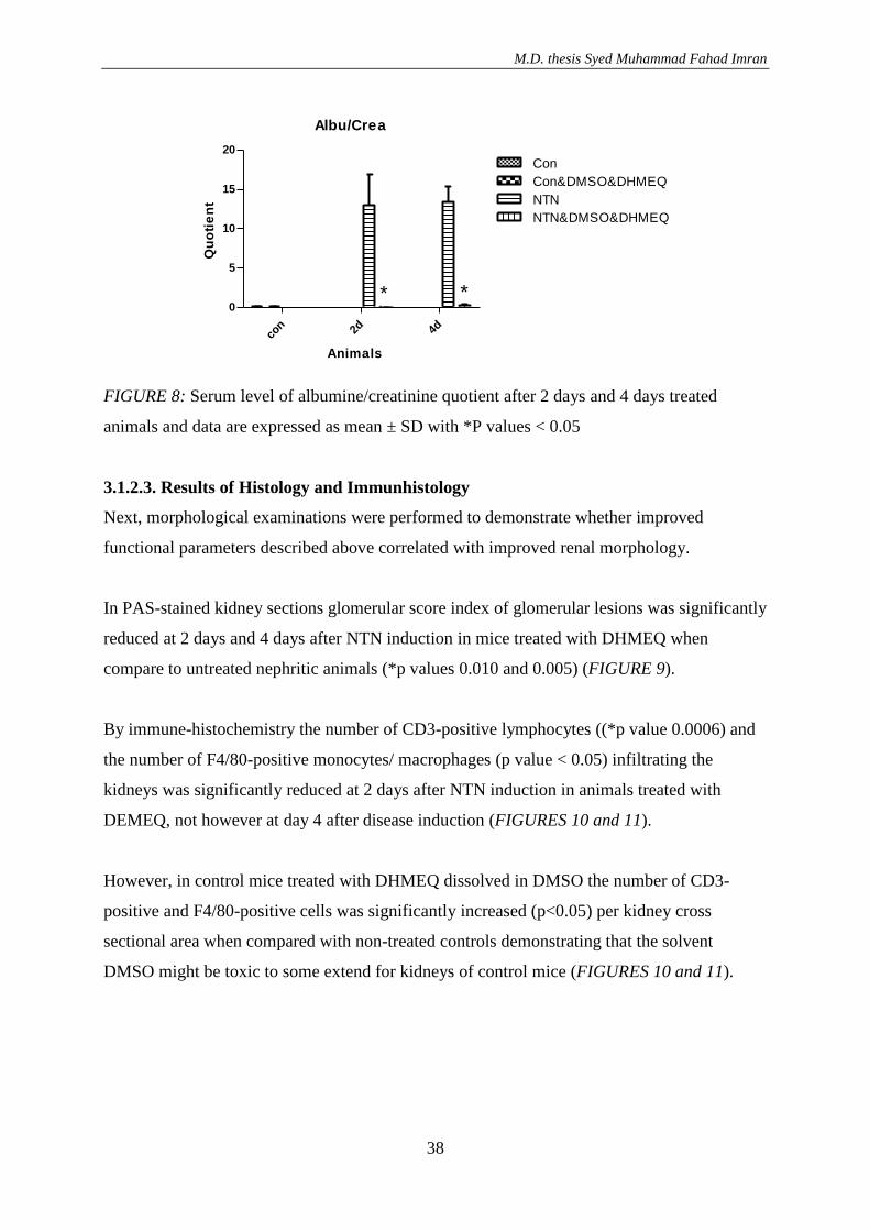
Even more, the alb/crea quotient was significantly reduced both at 2 days and 4 days after
NTN-induction in treated animals with DHMEQ (*p values 0.030 and 0.002) (FIGURE 8).
M.D. thesis Syed Muhammad Fahad Imran
37
Serum-Crea
Con 2d 4d
0.00
0.05
0.10
0.15
0.20
0.25
Con
Con&DMSO&DHMEQ
NTN
NTN&DMSO&DHMEQ
Animals
Co
nce
ntr
ati
on
in
mg
/dl
FIGURE 6: Serum level of creatinine after 2 days and 4 days treated animals and data are
expressed as mean ± SD with P values > 0.05
Serum-Urea
Con 2d 4d
0
50
100
150
Con
Con&DMSO&DHMEQ
NTN
NTN&DMSO&DHMEQ
*
Animals
Co
nc
en
tra
tio
n i
n m
g/d
l
FIGURE 7: Serum level of Urea after 2 days treated animals and data are expressed as mean
± SD with *P values < 0.05 and 4 days treated animals P value > 0.05
M.D. thesis Syed Muhammad Fahad Imran
38
Albu/Crea
con 2d 4d
0
5
10
15
20
Con
Con&DMSO&DHMEQ
NTN
NTN&DMSO&DHMEQ
* *
Animals
Qu
oti
en
t
FIGURE 8: Serum level of albumine/creatinine quotient after 2 days and 4 days treated
animals and data are expressed as mean ± SD with *P values < 0.05
3.1.2.3. Results of Histology and Immunhistology
Next, morphological examinations were performed to demonstrate whether improved
functional parameters described above correlated with improved renal morphology.
In PAS-stained kidney sections glomerular score index of glomerular lesions was significantly
reduced at 2 days and 4 days after NTN induction in mice treated with DHMEQ when
compare to untreated nephritic animals (*p values 0.010 and 0.005) (FIGURE 9).
By immune-histochemistry the number of CD3-positive lymphocytes ((*p value 0.0006) and
the number of F4/80-positive monocytes/ macrophages (p value < 0.05) infiltrating the
kidneys was significantly reduced at 2 days after NTN induction in animals treated with
DEMEQ, not however at day 4 after disease induction (FIGURES 10 and 11).
However, in control mice treated with DHMEQ dissolved in DMSO the number of CD3-
positive and F4/80-positive cells was significantly increased (p<0.05) per kidney cross
sectional area when compared with non-treated controls demonstrating that the solvent
DMSO might be toxic to some extend for kidneys of control mice (FIGURES 10 and 11).
M.D. thesis Syed Muhammad Fahad Imran
39
PAS
Con 2d 4d
0.0
0.5
1.0
1.5
2.0
2.5
Con
Con&DMSO&DHMEQ
NTN
NTN&DMSO&DHMEQ
**
Animals
Qu
oti
en
t o
f
inju
red
Glo
me
ruli
FIGURE 9: Histological scoring of injured glomeruli after 2 days and 4 days treated animals
and data are expressed as mean ± SD with *P values < 0.05
CD3
Con 2d 4d
0
5
10
15
Con
Con&DMSO&DHMEQ
NTN
NTN&DMSO&DHMEQ
*
Animals
ave
rag
e n
o o
f. C
D3 C
ell
s
FIGURE 10: Histological scoring CD-3 cells after 2 days treated animals and data are
expressed as mean ± SD with *P values < 0.05 and 4 days treated animals P value > 0.05
F4/80
Con
2d 4d
0
2
4
6
8
10
Con
Con&DMSO&DHMEQ
NTN
NTN&DMSO&DHMEQ
Animals
av
era
ge
no
of
F-4
/80
Ce
lls
FIGURE 11: Histological scoring F4/80 cells after 2 days and 4 days treated animals and data
are expressed as mean ± SD with P values > 0.05
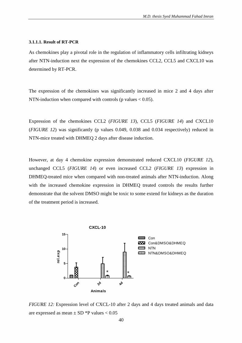
M.D. thesis Syed Muhammad Fahad Imran
40
3.1.1.1. Result of RT-PCR
As chemokines play a pivotal role in the regulation of inflammatory cells infiltrating kidneys
after NTN-induction next the expression of the chemokines CCL2, CCL5 and CXCL10 was
determined by RT-PCR.
The expression of the chemokines was significantly increased in mice 2 and 4 days after
NTN-induction when compared with controls (p values < 0.05).
Expression of the chemokines CCL2 (FIGURE 13), CCL5 (FIGURE 14) and CXCL10
(FIGURE 12) was significantly (p values 0.049, 0.038 and 0.034 respectively) reduced in
NTN-mice treated with DHMEQ 2 days after disease induction.
However, at day 4 chemokine expression demonstrated reduced CXCL10 (FIGURE 12),
unchanged CCL5 (FIGURE 14) or even increased CCL2 (FIGURE 13) expression in
DHMEQ-treated mice when compared with non-treated animals after NTN-induction. Along
with the increased chemokine expression in DHMEQ treated controls the results further
demonstrate that the solvent DMSO might be toxic to some extend for kidneys as the duration
of the treatment period is increased.
CXCL-10
Con 2d 4d
0
5
10
15
Con
Con&DMSO&DHMEQ
NTN
NTN&DMSO&DHMEQ
**
Animals
rel.
ex
p
FIGURE 12: Expression level of CXCL-10 after 2 days and 4 days treated animals and data
are expressed as mean ± SD *P values < 0.05
M.D. thesis Syed Muhammad Fahad Imran
41
CCL-2
Con 2d 4d
0
20
40
60
Con
Con&DMSO&DHMEQ
NTN
NTN&DMSO&DHMEQ
*
Animals
rel.
ex
p
FIGURE 13: Expression level of CCL-2 after 2 days treated animals and data are expressed as
mean ± SD *P value < 0.05 and 4 days treated animals P value > 0.05
CCL-5
Con 2d 4d
0
2
4
6
8
Con
Con&DMSO&DHMEQ
NTN
NTN&DMSO&DHMEQ
*
Animals
rel.
ex
p
FIGURE 14: Expression level of CCL-5 after 2 days treated animals and data are expressed as
mean ± SD *P value < 0.05 and 4 days treated animals P value > 0.05
3.1.2. Effect of DHMEQ with vehicle DMSO and cremaphor in LPS treated
animals
Having seen the results that DMSO might have toxic effects on the kidneys to some extend
we decided to compare the effect of two different solvents of DHMEQ on renal morphology
and immune-histochemistry and also on the expression of chemokines.
The first solvent used was 0.5% carboxymethylcellulose and dimethyl sulfoxid (DMSO) as
established. The second solvent we tested was Cremaphor.
M.D. thesis Syed Muhammad Fahad Imran
42
There is lot of evidences about DHMEQ dissolved in 0.5% carboxymethylcellulose with
DMSO in different studies, but there was no evidence yet in which Cremaphor as a vehicle for
DHMEQ was demonstrated. However, as DMSO itself is toxic for mice in higher
concentration cremaphor was tested to circumvent this problem.
For the experiments to test the different solvents for DHMEQ the LPS (lipopolysaccharide)-
model of nephritis was used.
LPS was injected intraperitoneally and at the same time mice were injected subcutaneously
with DHMEQ dissolved either in 0.5% carboxymethylcellulose with DMSO or in Cremaphor
as a vehicle.
3.1.1.2. Result of Histology and Immunhistology
The results of glomerular morphology score and immune-histochemistry examinations using
Cremaphore as vehicle for DHMEQ were very promising.
Glomerular morphology score was significantly reduced in DHEMQ-Cremaphore treated
animals after LPS-injection when compared with non-treated LPS-injected mice (p<0.019)
(FIGURE 16).
Even more promising were the results of immune-histochemistry demonstrating a significant
(p<0.016 and p<0.006 respectively) reduction of CD3-positive and F4/80-positive renal cells
in animals treated with DHMEQ-Cremaphore after LPS-injection when compared with
DEMEQ-DMSO treated mice after LPS-injection (FIGURES 15 and 17).
Also DHMEQ-Cremaphore did not increase the number of CD3-positive and F4/80-positive
renal cells in control mice which is in contrast to control animals treated with DHMEQ-
DMSO in which the number of infiltrating cells was significantly increased (p<0.05)
(FIGURES 15 and 17).
M.D. thesis Syed Muhammad Fahad Imran
43
CD3
Con
Con&
CRE
Con&
DM
SO
Con&
CRE&DHM
EQ
Con&
DM
SO&DHM
EQ
LPS
LPS&DM
SO&DHM
EQ
LPS&CRE&DHM
EQ
0
1
2
3
4
5
*
*
Animals
av
era
ge
no
of.
CD
3 C
ell
s
FIGURE 15: Histological scoring of CD-3 cells after 24 hours treated animals and data are
expressed as mean ± SD with *P values < 0.05
PAS
Con
Con&
CRE
Con&
DM
SO
Con&
CRE&DHM
EQ
Con&
DM
SO&DHM
EQLP
S
LPS&
DM
SO&DHM
EQ
LPS&
CRE&DHM
EQ
0.0
0.5
1.0
1.5
2.0
2.5
*
*
Animals
Qu
oti
en
t o
f
inju
red
Glo
me
ruli
FIGURE 16: Histological scoring of injured glomeruli after 24 hours treated animals and data
are expressed as mean ± SD with *P values < 0.05
M.D. thesis Syed Muhammad Fahad Imran
44
LPS-F4/80
Con
Con&
CRE
Con&
DM
SO
Con&
CRE&DHM
EQ
Con&
DM
SO&DHM
EQ
LPS
LPS&DM
SO&DHM
EQ
LPS&CRE&DHM
EQ
0
2
4
6
8
10
*
*
Animals
av
era
ge
no
of
F-4
/80
Ce
lls
FIGURE 17: Histological scoring of F4/80 cells after 24 hours treated animals and data are
expressed as mean ± SD with *P values < 0.05
3.1.1.1 Result of RT-PCR
We have seen a high relative expression of CCL2 and CCL5 in mice treated with LPS.
Control mice treated with vehicle cremaphore only did not show a significant expression of
these cytokines, however there is a significant difference between vehicle cremaphor and
methylcellulose/DMSO treated mice (*P value < 0.05). Animals treated with DHMEQ
dissolved in vehicle Cremaphor or DHMEQ did show a significant decrease of relative
expression of CCL2 (*P value 0.026) (FIGURE 19) and CCL5 (*P value 0.031) (FIGURE 18)
24 hours after LPS-injection.
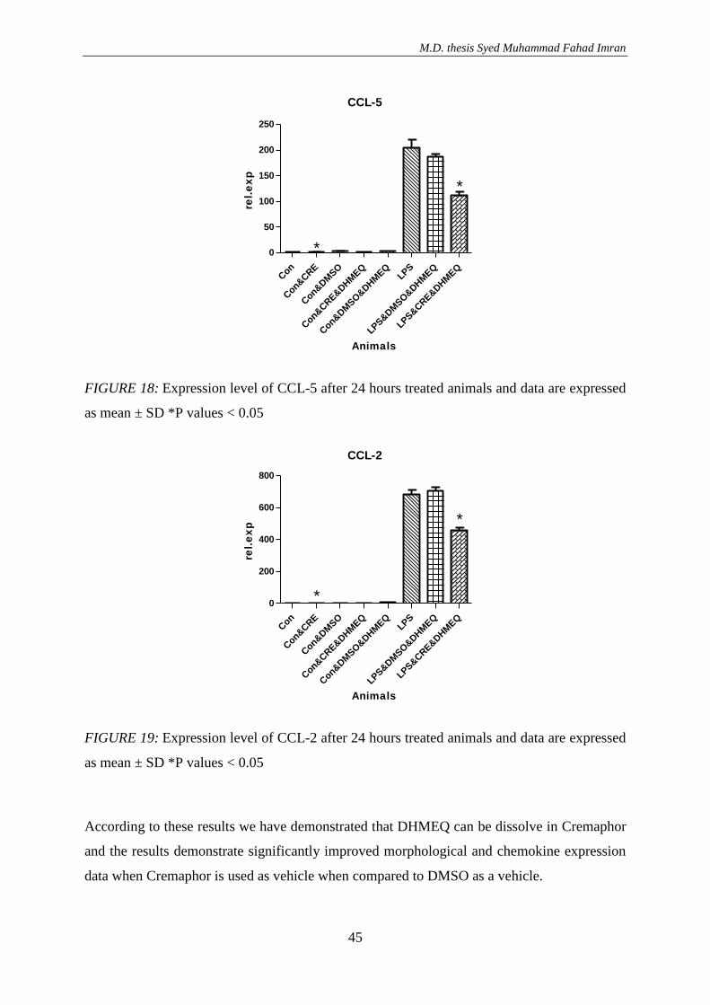
M.D. thesis Syed Muhammad Fahad Imran
45
CCL-5
Con
Con&
CRE
Con&
DM
SO
Con&
CRE&DHM
EQ
Con&
DM
SO&DHM
EQ
LPS
LPS&DM
SO&DHM
EQ
LPS&CRE&DHM
EQ
0
50
100
150
200
250
*
*
Animals
rel.
ex
p
FIGURE 18: Expression level of CCL-5 after 24 hours treated animals and data are expressed
as mean ± SD *P values < 0.05
CCL-2
Con
Con&
CRE
Con&
DM
SO
Con&
CRE&DHM
EQ
Con&
DM
SO&DHM
EQ
LPS
LPS&DM
SO&DHM
EQ
LPS&CRE&DHM
EQ
0
200
400
600
800
*
*
Animals
rel.
ex
p
FIGURE 19: Expression level of CCL-2 after 24 hours treated animals and data are expressed
as mean ± SD *P values < 0.05
According to these results we have demonstrated that DHMEQ can be dissolve in Cremaphor
and the results demonstrate significantly improved morphological and chemokine expression
data when Cremaphor is used as vehicle when compared to DMSO as a vehicle.
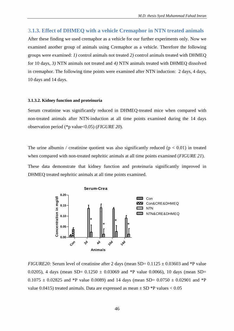
M.D. thesis Syed Muhammad Fahad Imran
46
3.1.3. Effect of DHMEQ with a vehicle Cremaphor in NTN treated animals
After these finding we used cremaphor as a vehicle for our further experiments only. Now we
examined another group of animals using Cremaphor as a vehicle. Therefore the following
groups were examined: 1) control animals not treated 2) control animals treated with DHMEQ
for 10 days, 3) NTN animals not treated and 4) NTN animals treated with DHMEQ dissolved
in cremaphor. The following time points were examined after NTN induction: 2 days, 4 days,
10 days and 14 days.
3.1.3.2. Kidney function and proteinuria
Serum creatinine was significantly reduced in DHMEQ-treated mice when compared with
non-treated animals after NTN-induction at all time points examined during the 14 days
observation period (*p value<0.05) (FIGURE 20).
The urine albumin / creatinine quotient was also significantly reduced (p < 0.01) in treated
when compared with non-treated nephritic animals at all time points examined (FIGURE 21).
These data demonstrate that kidney function and proteinuria significantly improved in
DHMEQ treated nephritic animals at all time points examined.
Serum-Crea
Con 2d 4d 10
d14
d
0.00
0.05
0.10
0.15
0.20
Con
Con&CRE&DHMEQ
NTN
NTN&CRE&DHMEQ
**
**
Animals
Co
nce
ntr
ati
on
in
mg
/dl
FIGURE20: Serum level of creatinine after 2 days (mean SD= 0.1125 ± 0.03603 and *P value
0.0205), 4 days (mean SD= 0.1250 ± 0.03069 and *P value 0.0066), 10 days (mean SD=
0.1075 ± 0.02825 and *P value 0.0089) and 14 days (mean SD= 0.0750 ± 0.02901 and *P
value 0.0415) treated animals. Data are expressed as mean ± SD *P values < 0.05
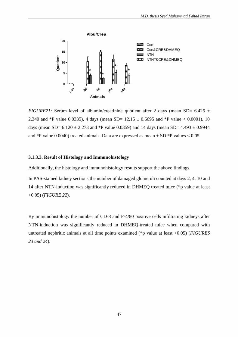
M.D. thesis Syed Muhammad Fahad Imran
47
Albu/Crea
con 2d 4d 10
d14
d
0
5
10
15
20
Con
Con&CRE&DHMEQ
NTN
NTNT&CRE&DHMEQ
**
**
Animals
Qu
oti
en
t
FIGURE21: Serum level of albumin/creatinine quotient after 2 days (mean SD= 6.425 ±
2.340 and *P value 0.0335), 4 days (mean SD= 12.15 ± 0.6695 and *P value < 0.0001), 10
days (mean SD= 6.120 ± 2.273 and *P value 0.0359) and 14 days (mean SD= 4.493 ± 0.9944
and *P value 0.0040) treated animals. Data are expressed as mean ± SD *P values < 0.05
3.1.3.3. Result of Histology and Immunohistology
Additionally, the histology and immunohistology results support the above findings.
In PAS-stained kidney sections the number of damaged glomeruli counted at days 2, 4, 10 and
14 after NTN-induction was significantly reduced in DHMEQ treated mice (*p value at least
<0.05) (FIGURE 22).
By immunohistology the number of CD-3 and F-4/80 positive cells infiltrating kidneys after
NTN-induction was significantly reduced in DHMEQ-treated mice when compared with
untreated nephritic animals at all time points examined (*p value at least <0.05) (FIGURES
23 and 24).
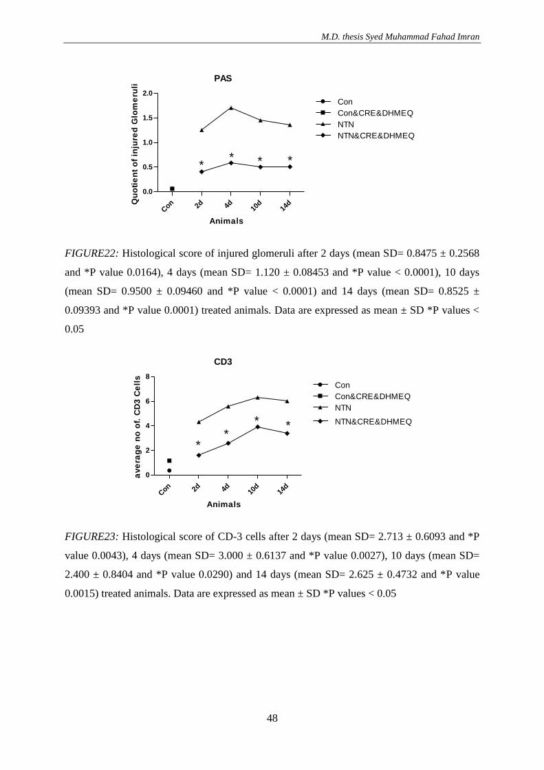
M.D. thesis Syed Muhammad Fahad Imran
48
PAS
Con 2d 4d 10
d14
d
0.0
0.5
1.0
1.5
2.0
Con
Con&CRE&DHMEQ
NTN
NTN&CRE&DHMEQ
** * *
Animals
Qu
oti
en
t o
f in
jure
d G
lom
eru
li
FIGURE22: Histological score of injured glomeruli after 2 days (mean SD= 0.8475 ± 0.2568
and *P value 0.0164), 4 days (mean SD= 1.120 ± 0.08453 and *P value < 0.0001), 10 days
(mean SD= 0.9500 ± 0.09460 and *P value < 0.0001) and 14 days (mean SD= 0.8525 ±
0.09393 and *P value 0.0001) treated animals. Data are expressed as mean ± SD *P values <
0.05
CD3
Con 2d 4d 10
d14
d
0
2
4
6
8
Con
Con&CRE&DHMEQ
NTN
NTN&CRE&DHMEQ
**
* *
Animals
ave
rag
e n
o o
f. C
D3 C
ell
s
FIGURE23: Histological score of CD-3 cells after 2 days (mean SD= 2.713 ± 0.6093 and *P
value 0.0043), 4 days (mean SD= 3.000 ± 0.6137 and *P value 0.0027), 10 days (mean SD=
2.400 ± 0.8404 and *P value 0.0290) and 14 days (mean SD= 2.625 ± 0.4732 and *P value
0.0015) treated animals. Data are expressed as mean ± SD *P values < 0.05
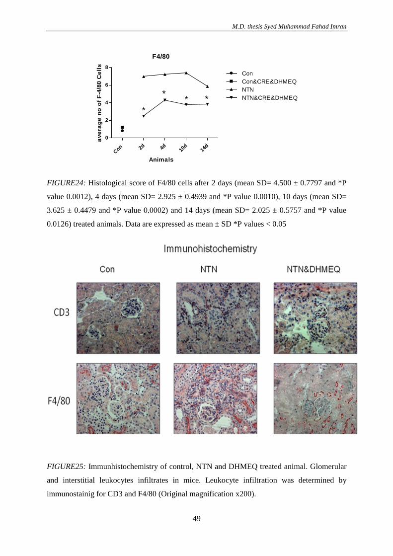
M.D. thesis Syed Muhammad Fahad Imran
49
F4/80
Con
2d 4d 10d
14d
0
2
4
6
8
Con
Con&CRE&DHMEQ
NTN
NTN&CRE&DHMEQ
*
** *
Animals
ave
rag
e n
o o
f F
-4/8
0 C
ell
s
FIGURE24: Histological score of F4/80 cells after 2 days (mean SD= 4.500 ± 0.7797 and *P
value 0.0012), 4 days (mean SD= 2.925 ± 0.4939 and *P value 0.0010), 10 days (mean SD=
3.625 ± 0.4479 and *P value 0.0002) and 14 days (mean SD= 2.025 ± 0.5757 and *P value
0.0126) treated animals. Data are expressed as mean ± SD *P values < 0.05
FIGURE25: Immunhistochemistry of control, NTN and DHMEQ treated animal. Glomerular
and interstitial leukocytes infiltrates in mice. Leukocyte infiltration was determined by
immunostainig for CD3 and F4/80 (Original magnification x200).
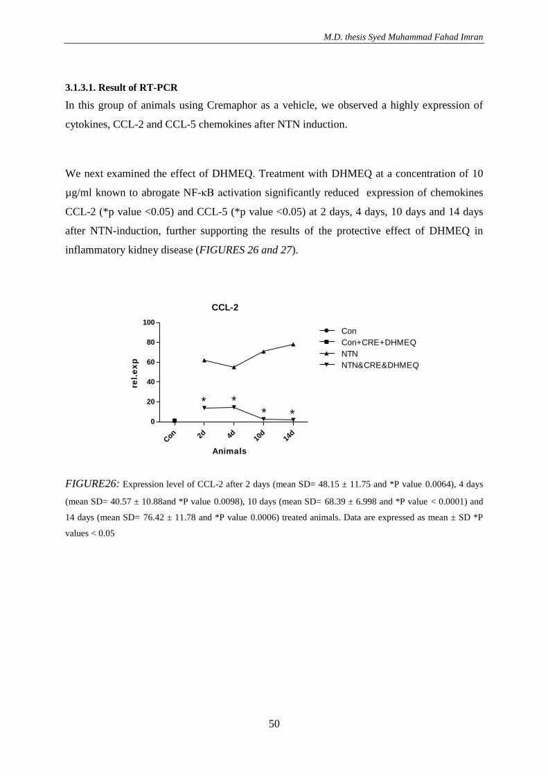
M.D. thesis Syed Muhammad Fahad Imran
50
3.1.3.1. Result of RT-PCR
In this group of animals using Cremaphor as a vehicle, we observed a highly expression of
cytokines, CCL-2 and CCL-5 chemokines after NTN induction.
We next examined the effect of DHMEQ. Treatment with DHMEQ at a concentration of 10
µg/ml known to abrogate NF-κB activation significantly reduced expression of chemokines
CCL-2 (*p value <0.05) and CCL-5 (*p value <0.05) at 2 days, 4 days, 10 days and 14 days
after NTN-induction, further supporting the results of the protective effect of DHMEQ in
inflammatory kidney disease (FIGURES 26 and 27).
CCL-2
Con 2d 4d 10
d14
d
0
20
40
60
80
100
Con
Con+CRE+DHMEQ
NTN
NTN&CRE&DHMEQ
* ** *
Animals
rel.
ex
p
FIGURE26: Expression level of CCL-2 after 2 days (mean SD= 48.15 ± 11.75 and *P value 0.0064), 4 days
(mean SD= 40.57 ± 10.88and *P value 0.0098), 10 days (mean SD= 68.39 ± 6.998 and *P value < 0.0001) and
14 days (mean SD= 76.42 ± 11.78 and *P value 0.0006) treated animals. Data are expressed as mean ± SD *P
values < 0.05
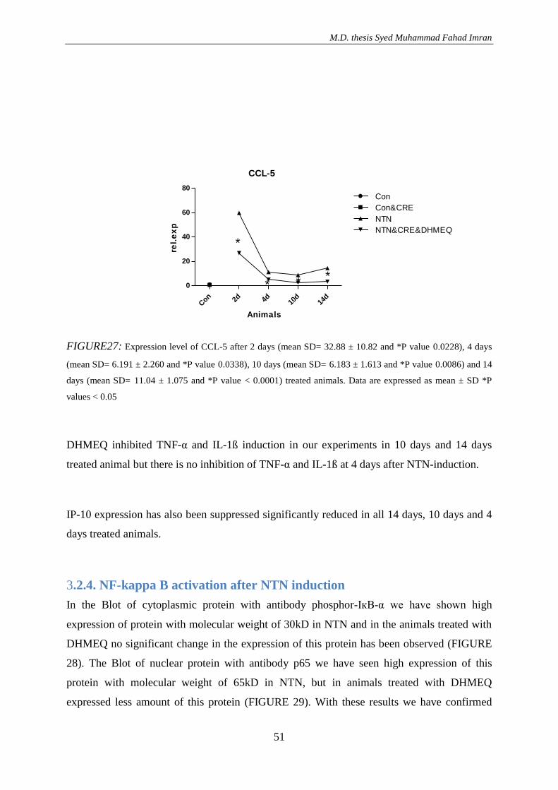
M.D. thesis Syed Muhammad Fahad Imran
51
CCL-5
Con 2d 4d 10
d14
d
0
20
40
60
80
Con
Con&CRE
NTN
NTN&CRE&DHMEQ
*
* * *
Animals
rel.
ex
p
FIGURE27: Expression level of CCL-5 after 2 days (mean SD= 32.88 ± 10.82 and *P value 0.0228), 4 days
(mean SD= 6.191 ± 2.260 and *P value 0.0338), 10 days (mean SD= 6.183 ± 1.613 and *P value 0.0086) and 14
days (mean SD= 11.04 ± 1.075 and *P value < 0.0001) treated animals. Data are expressed as mean ± SD *P
values < 0.05
DHMEQ inhibited TNF-α and IL-1ß induction in our experiments in 10 days and 14 days
treated animal but there is no inhibition of TNF-α and IL-1ß at 4 days after NTN-induction.
IP-10 expression has also been suppressed significantly reduced in all 14 days, 10 days and 4
days treated animals.
3.2.4. NF-kappa B activation after NTN induction
In the Blot of cytoplasmic protein with antibody phosphor-IкB-α we have shown high
expression of protein with molecular weight of 30kD in NTN and in the animals treated with
DHMEQ no significant change in the expression of this protein has been observed (FIGURE
28). The Blot of nuclear protein with antibody p65 we have seen high expression of this
protein with molecular weight of 65kD in NTN, but in animals treated with DHMEQ
expressed less amount of this protein (FIGURE 29). With these results we have confirmed
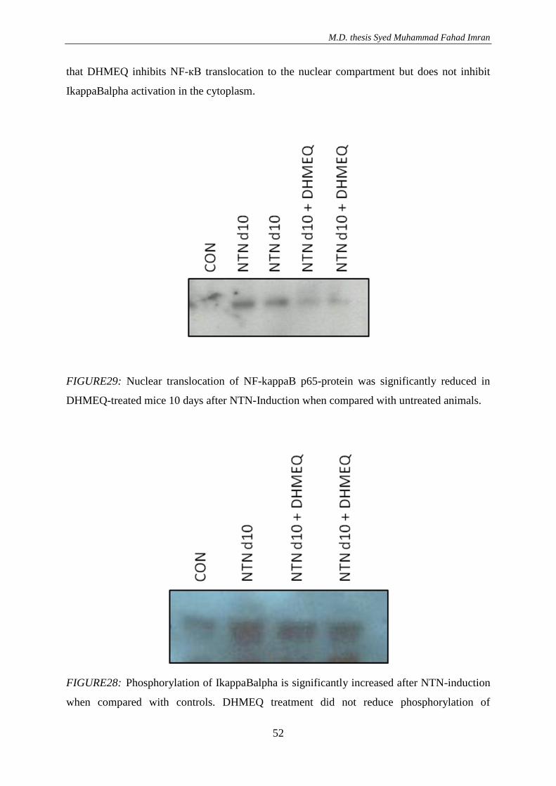
M.D. thesis Syed Muhammad Fahad Imran
52
that DHMEQ inhibits NF-кB translocation to the nuclear compartment but does not inhibit
IkappaBalpha activation in the cytoplasm.
FIGURE29: Nuclear translocation of NF-kappaB p65-protein was significantly reduced in
DHMEQ-treated mice 10 days after NTN-Induction when compared with untreated animals.
FIGURE28: Phosphorylation of IkappaBalpha is significantly increased after NTN-induction
when compared with controls. DHMEQ treatment did not reduce phosphorylation of
M.D. thesis Syed Muhammad Fahad Imran
53
IkappaBalpha significantly when compared with untreated NTN-nephritic mice. Results
shown are at day 10 after NTN-induction.
M.D. thesis Syed Muhammad Fahad Imran
54
4. Discussion
Inflammatory renal diseases such as glomerulonephritis are induced by activation of
proinflammatory signaling pathways leading to the production of proinflammatory mediators
such as cytokines and chemokines. In the last decades the molecular signaling pathways that
initiate these inflammatory reactions have been extensively studied.
NF-kappa B is an inducible hetero- or homodimeric transcription factor composed of
members of the Rel family of DNA-binding proteins that recognize a common sequence
motif. Five mammalian Rel proteins have been identified so far, NF-kappa B1 (p50 and its
precursor p105), NF-kappa B2 (p52 and its precursor p100), c-Rel, RelA (p65), and RelB.
NF-kappa B is an important regulator of inflammation and immune responses by mediating
pro-inflammatory gene expression including chemokines, cytokines, adhesion molecules and
growth factors. Under basal conditions NF-kappa B dimers remain in the cytoplasm as an
inactive form. A family of inhibitory proteins, I-kappa-Bs, binds to NF-kappa B and mask its
nuclear localization signals, thereby blocking nuclear translocation. Exposure of cells to a
variety of proinflammatory stimuli leads to the rapid phosphorylation, ubiquitinylation, and
ultimately proteolytic degradation of I-kappa-B, which allows nuclear translocation of NF-
kappa B and subsequent gene transcription.
We and others have described the activation of NF-kappa B in the induction phase of several
experimental models of glomerulonephritis. Furthermore, it was shown that early NF-kappa B
blockade in an experimental model of glomerulonephritis markedly reduced proteinuria,
glomerular lesions, and monocytes/macrophages infiltration. Therefore, blocking of NF-kappa
B represents an interesting new target for the treatment of renal inflammatory disease.
Numerous studies have shown that NF-кB is a key regulator of inflammation and is activated
in experimental models of kidney inflammation including immune complex nephritis, anti-
glomerular basement membrane nephritis and mesangial proliferative GN. These findings
indicate NF-кB as a potential target for the treatment of renal inflammatory diseases.
M.D. thesis Syed Muhammad Fahad Imran
55
Activation of NF-кB takes place in several steps. In numerous studies have illustrated that
different pharmacological agents, such as glucocorticoids, aspirin, cyclosporin A and FK-506
inhibits various stages of NF-кB activation although these agents are not specific for NF-kB
and have many others effects (T. Kosaka et al. 2008)
To inhibit NF-kappaB activation a recently described, low-molecular-weight NF-kappaB
inhibitor, dehydroxymethylepoxyquinomicin (DHMEQ), which was derived from the
antibiotic epoxyquinomicin C was used. It is a highly specific NF-kappaB inhibitor that
interferes with the nuclear translocation of activated NF-kappaB. DHMEQ exhibits anti-
inflammatory, anti-fibrotic, and antitumor effects. In a macrophage-like cell line, DHMEQ
inhibited phagocytic activity and cytokine production, suggesting that it may have
immunosuppressive effects. However, the effect of DHMEQ on an experimental model of
rapid progressive glomerulonephritis has not been determined. Therefore, we examined the
effects of DHMEQ as a possible immunosuppressive agent by investigating whether DHMEQ
could regulate NTN-nephritis in mice.
Only a limited number of studies testing the in vivo effects of small molecular weight
compounds on nephritis have been reported (Sanz AB, Sanchez-Niño MD, et al. 2010). It has
been shown previously that DHMEQ inhibited TNF-α-induced nuclear translocation of NF-
kappaB, and does not inhibit phosphorylation and degradation of IkappaB, or a c-Jun N-
terminal kinase (JNK) and a caspase-activating pathway in Jurkat T cells. Here, we extended
these studies to test the therapeutic effect of DHMEQ on NTN, and to test the efficacy on the
inhibition of the inflammatory pathway in this model of rapid progressive glomerulonephritis.
The results showed that this unique inhibitor of NF-kappaB nuclear translocation may hold
promise for treatment of NTN.
The number of known roles of NF-κB is rapidly increasing. There is much evidence that
DHMEQ inhibits nuclear translocation. Therefore we examined whether DHMEQ reducing
the inflammatory cytokines (M. Yamamoto et al. 2008).
For our study firstly we used two different vehicles to solve DHMEQ. One was Cremaphor
and the other was DMSO with Methylcellulose. There is lots of evidence, in which it has been
M.D. thesis Syed Muhammad Fahad Imran
56
demonstrated that DHMEQ can solve in DMSO but there is no evidence till yet in which
Cremaphor used as a vehicle for DHMEQ. We have seen slightly better result with
Cremaphor rather than the DMSO. So we used only cremaphor for our further experiments. In
our study we have also demonstrated that the DMSO itself toxic for the animals with high
concentration. We have seen an expression of inflammatory cytokines in control animals
injected only with vehicle DMSO compare to cremaphor.
DHMEQ was derived from antibiotic epoxyquinomicin C and inhibits NF-кB activation in
vitro and in vivo with blockade of the nuclear translocation of NF-кB being a potential
mechanism of action. DHMEQ specifically inhibits NF-кB in various disease models in
different organs in which NF-кB is involved without evidence of toxicity (T. Kosaka et al.
2008)
Results of DHMEQ treated animals with vehicle DMSO at a concentration of 10ug/ml gave
some abrogated constitutive NF-κB activity and at the same time showed expression of
chemokines in different days of treatment some disturbing results. Histology and
immunhistology results are also correlated to the above results. Again the results of control
animal with DMSO as a vehicle control only, supports our above statement of toxic effect of
DMSO in mice and showing as same as above a high number of CD-3 and F4/80 cells
compare to control animals.
After this we have analyzed the serum of treated animal with DHMEQ dissolved in DMSO. It
was showing a less concentration of serum creatinine compare to animal with NTN, however
control animals injected with DMSO only as a vehicle showing a high concentration of serum
creatinine compare to control one. Again, this result give us a hint that DMSO seemed to be
toxic itself for mice.
Now using Cremaphor as a vehicle we next examined the effect of DHMEQ constitutively.
Treatment with DHMEQ at a concentration of 10µg/ml abrogated constitutive NF-κB activity.
It has been demonstrated a strongly decreased expression of cytokines CCL-2 (*P value
<0.05) and CCL-5 (*P value <0.05) in all period of time 2 days, 4 days, 10days and 14days
treated animals, showing the positive and protective effect of DHMEQ in inflammation. The
M.D. thesis Syed Muhammad Fahad Imran
57
vehicle control of cremaphor in control animals doesn't show any expression. It is showing
that the animals endure cremaphor without any problem.
Serum creatinine concentration and the ratio of Urine alb/crea demonstrated very positive
results as so far, in all 2 days, 4 days, 10 days and 14 days treated animals with DHMEQ
dissolved in cremaphor. It has been showing the animal with less protenurie and more
excretion of creatinine in all 2, 4, 10 and 14 days treated animals (*P value <0.05). In other
words it said that the kidney function is improving after treatment with DHMEQ in all days
treated animals. In addition to this the histology and immunhistology results does support the
above results.
Now if we just look at the results of treatment of different time periods. We can see that
DHMEQ with a vehicle cremaphor does inhibit the induction of NF-κB in all 2, 4, 10 and 14
days treated animals Results of RT-PCR has been shown less CCL-5, CCL-2, CXCL-10,
TNF-α and IL-1b expressions significantly.
Same kind of results observed in histology and immunhistology and correlate to the above
results. In Western-Blot it has been showed that the DHMEQ inhibits NF-кB on a
translocation level. In the first peak of inflammation, p65/p50 heterodimer protein complex is
dominant. P65 protein is normally active in nucleus. . We have seen in Western-Blot less
expression of both the protein in the DHMEQ treated animals.
The present study demonstrated the activation of NF-кB in the NTN model at different days.
Treatment with DHMEQ significantly reduced NF-кB activation, glomerular proliferation,
crescent formation, mesangial matrix deposition and significantly increased the level of
glomerular apoptosis. These results suggest that specific inhibition of NF-кB activation by
DHMEQ suppresses the inflammatory response in NTN.
In summary, we demonstrated specific inhibition of NF-kappaB by DHMEQ in the NTN
nephritis model without evidence of toxicity. The inhibition of NF-kappaB was associated
with diminished infiltration of inflammatory cells, reduced glomerular damage index, reduced
M.D. thesis Syed Muhammad Fahad Imran
58
chemokine expression and a significant improvement of renal functional parameters. The
targeting of NF-kappaB activation by DHMEQ may represent a promising new treatment
option for patients with inflammatory renal diseases. So all in general it showing that
DHMEQ solved in Cremaphor and on the initial time and in the later stage of disease does
influence the expression of inflammatory cytokines and chemokines significantly. It should be
carry on in order to find, what factors does influence in it? Is there any other process that
plays an important role?
M.D. thesis Syed Muhammad Fahad Imran
59
5. Conclusion
We have demonstrated in our experiments that DHMEQ suppressed inflammatory cytokines
CCL-2, CCL-5, CXCL-10 and TNF-α at 2 days, 4 days, 10 days and 14 days after the
induction of NTN. These results have been supported by morphology, where PAS staining
showed significantly less damaged glomeruli and significantly reduced numbers of CD-3 and
F4/80 positive cells as demonstrated by immune-histochemistry.
In the present study, we demonstrated that the newly developed NF-kappaB inhibitor
DHMEQ markedly improved renal function and morphology and suppressed pro-
inflammatory cytokine levels in the mice with NTN.
Therefore targeting of NF-kappaB activation by DHMEQ may represent a promising new
treatment option for patients with inflammatory renal diseases.
M.D. thesis Syed Muhammad Fahad Imran
60
6. References
1 A. Ariga, J. Namekawa, N. Matsumoto, J. Inoue, K. Umezawa, “Inhibition of Tumor
Necrosis Factor-α-induced Nuclear Translocation and activation of NF-κB by
Dehydroxymethylepoxyquinomicin“ The Journal of Biological Chemistry 277, 24625-
24630 (2002)
2 C. Kurts, F. Heymann, V. Lukacs-Korneck, P. Boor, and J. Floege „Role of T cells
and dendritic cells in glomerular immunopathology „ Semin Immunopatholog 29, 317-
335 (2007)
3 E. Suzuki, Y. Ninomiya, K. Umezawa,“Induction of histidine decarboxylase in
macrophages inhibited by the novel NF-κB inhibitor (-)-DHMEQ“, Biochemical and
Biophysical Research Communications 379, 379–383(2009)
4 F. Wan, M. J Lenardo “The nuclear signaling of NF-κB: current knowledge, new
insights, and future perspectives“, Cell Research 20:24-33 (2010)
5 K. Umezawa and C. Chaicharoenpong, Molecular Design and Biological Activities of
NF-κB Inhibitors“, Mol. Cells, 14, 163-167 (2002)
6 K. Wakamatsu, T. Nanki, N. Miyasaka, K. Umezawa and T. Kubota, „Effect of a
small molecule inhibitor of nuclear factor-κB nuclear translocation in a murine model
of arthritis and cultured human synovial cells „Arthritis Research & Therapy 7,
R1348-R1359 (2005)
7 K. Shinoda, K. Nakagawa,T. Kosaka, N. Tanaka, T. Maeda, H. Kono, R. Mizuno, E.
Kikuchi, A. Miyajima, K. Umezawa, M. Oya, „Regulation of human dendritic cells by
M.D. thesis Syed Muhammad Fahad Imran
61
a novel specific nuclear factor-κB inhibitor, dehydroxymethylepoxyquinomicin,
Human Immunology 71, 763-770 (2010)
8 M. S. Hayden and S. Ghosh, „Signaling to NF-kappaB“, Genes Dev 18, 2195-2224
(2004)
9 M. Watanabe, M.Z. Dewan, M. Taira, M. Shoda, M. Honda, T. Sata, M. Higashihara,
M. E. Kadin, T. Watanabe, N. Yamamoto, K. Umezawa and R. Horie” IκBa
independent induction of NF-kB and its inhibition by DHMEQ in Hodgkin/Reed-
Sternberg cells“ Laboratory Investigation 87, 372–382 (2007)
10 M. Yamamoto, R. Horie, M. Takeiri, I. Kozawa, and K. Umezawa, „Inactivation of
NF-KB Components by Covalent Binding of (-)-Dehydroxymethylepoxyquinomicin to
Specific Cysteine Residues“, Journal of Medicinal Chemistry 51, 5780–5788 (2008)
11 M. Watanabe, M. Nakashima, T. Togano, M. Higashihara, T. Watanabe, K. Umezawa,
R. Horie, „Identification of the RelA domain responsible for action of a new NF-kB
inhibitor DHMEQ“, Biochemical and Biophysical Research Communications 376,
310–314 (2008)
12 M. Murohashi, K Hinohara1, M Kuroda, T Isagawa, S Tsuji, S Kobayashi, K
Umezawa, A Tojo,H Aburatani and N Gotoh, „Gene set enrichment analysis provides
insight into novel signalling pathways in breast cancer stem cells“, British Journal of
Cancer 102, 206-212 (2010)
13 N. Lampiasi, A. Azzolina, N. D’Alessandro, K. Umezawa, J. A. McCubrey, G.
Montalto, and M. Cervello, „Antitumor Effects of Dehydroxymethylepoxyquinomicin,
a Novel Nuclear Factor-B Inhibitor, in Human Liver Cancer Cells Are Mediated
through a Reactive Oxygen Species-Dependent Mechanism“, Mol Pharmacol 76, 290–
300 (2009)
M.D. thesis Syed Muhammad Fahad Imran
62
14 N. Dabaghmanesh, A. Matsubara, A. Miyake, K. Nakano, T. Ishida, H. Katano, R.
Horie, K. Umezawa and T. Watanabe, „Transient inhibition of NF-κB by DHMEQ
induces cell death of primary effusion lymphoma without HHV-8 reactivation“,
Cancer Science 100 (4), 737-746 (2009)
15 P. G. Tipping and A. R. Kitching „Glomerulonephritis, Th1 and Th2: What's new?“,
Clin Exp Immunol 142, 207-215 (2005)
16 S. Ghosh, M. J. May, and E. B. Kopp „NF-kappa B and Rel proteins: evolutionarily
conserved mediators of immune responses” Annu Rev Immunol 16, 225-260 (1998)
17 S. Segrer, F. Heller, M. T. Lindenmeyer, H. Schmid, C. D. Cohen, D. Draganovici, J.
Mandelbaum, P. J. Nelson, H. J. Grone, E. F. Grone, A. M. Figel, E. Nossner, and D.
Schlondorff „Compartment specific expression of dendritic cell markers in human
glomerulonephritis” Kidney Int 74, 49-62 (2007)
18 Sanz AB, Sanchez-Niño MD, Ramos AM, Moreno JA, Santamaria B, Ruiz-Ortega M,
Egido J, Ortiz A, “NF-kappaB in renal inflammation“,
J Am Soc Nephrol. 21(8), 1254-62 (2010)
19 T. Chiba, Y. Kondo, S. Shinozaki, E. Kaneko, A. Ishigami, N. Maruyama, K.
Umezawa, and K. Shimokado, „ A selective NFκB inhibitor, DHMEQ, reduced
atherosclerosis in ApoE-deficient mice“, Journal of Atherosclerosis and Thrombosis
13, 308-313 (2006)
20 T. Kubota, M. Hoshino, K. Aoki, K. Ohya, Y. Komano, T. Nanki, N. Miyasaka and K.
Umezawa „NF-κB inhibitor dehydroxymethylepoxyquinomicin suppresses
osteoclastogenesis and expression of NFATc1 in mouse arthritis without affecting
M.D. thesis Syed Muhammad Fahad Imran
63
expression of RANKL, osteoprotegerin or macrophage colony-stimulating factor”,
Arthritis Research and Therapy 9, 1186-2298 (2007)
21 T. Kosaka, A. Miyajima, E. Kikuchi, Y. Horiguchi, K. Umezawa, T. Ohigashi, J.
Nakashima, T. Asano, M. Oya „The Novel NF-κB Activation Inhibitor
Dehyroxymethyl-Epoxyquinomicin Suppresses Anti-Thy1.1-Induced
Glomerulonephritis in Rats” Nephron Exp Neprhol 110, 17-24 (2008)
22 T. Suzuki, K. Yamashito, W. Jomen, S. Ueki, T. Aoyagi, M. Fukai, H. Furukawa, K.
Umezawa, M. Ozaki, S. Todo “The Novel NF-κB Inhibitor,
Dehydroxymethylepoxyquinomicin, prevents local and remote organ injury following
intestinal ischemia/reperfusion in rats” Journal of surgical research 149, 69-75 (2008)
23 T. Lawrencea, C. Fonga, „The resolution of inflammation: Anti-inflammatory roles for
NF-kB“, The International Journal of Biochemistry & Cell Biology 42, 519–523
(2010)
M.D. thesis Syed Muhammad Fahad Imran
64
7. Word of thanks
Firstly I am thankful to God and also my parents who always prayed for me and for my
future. My very special thank goes to my father doctor Prof. Dr. Thaiss and laboratory
director Dr. Gunther Zahner for the friendly supervision within the laboratory. I am also very
thankful for my nomination for such an interesting subject. Without outstanding care of my
father doctor in every regard and aid in the preparation of the work, it would have been
difficult for me to complete my work so effectively within a small period of time. Without his
friendly, open and straightforward behaviour to his Doctoral Candidates and colleagues, I as
well as my colleagues would be in a great fiasco. Furthermore, I am extremely grateful and
thankful for his introductions into the work technologies, his steady support in professional,
and for his patience throughout the whole time. The pleasant, sincere and personal climate
within the team allowed my time/experience in the laboratory to be tremendously comfortable
as well as memorable. He was always there to listen to me and tried his level best to help me
wherever possible. Without his extraordinary skills, the execution of the animal attempts
would not have been possible for me to complete successfully. I also want to thank Prof. Dr.
R. Stahl for providing good-quality work conditions in his department. My further thank goes
to Ms. Mariola for the introduction and the allowance of using her Microscope. I would also
like to thank all the colleagues of the nephrology laboratory under the leadership of Dr. G.
Zahner for the assistance in dealing with all sorts of problems and for the comfortable work
atmosphere. I am very thankful to my very good friend Dr. Erdogan Cayroglu. I would
especially like to thank my brother Syed Ayaz Hussain, my family, my sister and parents for
all the support and encouragement for all these years. Without it, I would not be where I am
today.
M.D. thesis Syed Muhammad Fahad Imran
65
8. Affidavit
I do assure formally that I compose this work independently and without any help or
assistance from others. Other as the sources indicated by me and remedy uses and made
would have the places taken out of the used works literally or in substance individually after
edition (edition and year of the appearance), volume and side of the used work recognizable.
Furthermore I assure that I produced the dissertation not yet a specialty representative at
another university for inspection or applied myself other around allowance to the graduation.
M.D. thesis Syed Muhammad Fahad Imran
66
9. Eidesstattliche Versicherung
Ich versichere ausdrücklich, dass ich die Arbeit selbstständig und ohne fremde Hilfe verfasst,
andere als die von mir angegebenen Quellen und Hilfsmittel nicht benutzt und die aus den
benutzten Werken wörtlich oder inhaltlich entnommenen Stellen einzeln nach Ausgabe
(Auflage und Jahr des Erscheinens), Band und Seite des benutzten Werkes kenntlich gemacht
habe. Ferner versichere ich, dass ich die Dissertation bisher nicht einem Fachvertreter an einer
anderen Hochschule zur Überprüfung vorgelegt oder mich anderweitig um Zulassung zur
Promotion beworben habe.