Embed Size (px)
Citation preview
This work has been digitalized and published in 2013 by Verlag Zeitschrift für Naturforschung in cooperation with the Max Planck Society for the Advancement of Science under a Creative Commons Attribution4.0 International License.
Dieses Werk wurde im Jahr 2013 vom Verlag Zeitschrift für Naturforschungin Zusammenarbeit mit der Max-Planck-Gesellschaft zur Förderung derWissenschaften e.V. digitalisiert und unter folgender Lizenz veröffentlicht:Creative Commons Namensnennung 4.0 Lizenz.
On the Release and Action of the Hypertrehalosaemic Hormone from the Cockroach Nauphoeta cinerea
Gerd Gäde Institut für Zoologie IV der Universität Düsseldorf, Universitätsstraße 1, D-4000 Düsseldorf 1, Bundesrepublik Deutschland
Z. Naturforsch. 43c, 108-116 (1988); received July 22/September 21, 1987
Synthetic Hypertrehalosaemic Hormone, Nauphoeta cinerea, Activation of Fat Body Glycogen Phosphorylase, Increase of Blood Carbohydrates. Release of Hypertrehalosaemic Hormone
The corpora cardiaca of the cockroach Nauphoeta cinerea contain a hypertrehalosaemic hor-mone (HTH) which is chemically characterized as a blocked decapeptide. The synthetic HTH shows the same chromatographic behaviour as the material isolated from corpora cardiaca. The synthetic peptide causes hypertrehalosaemia and fat body glycogen phosphorylase-activation in N. cinerea as well as in the American cockroach, Periplaneta americana in a dose-dependent fashion. It is calculated that one gland from N. cinerea stores about 50 pmol of HTH. Roughly 10% of the total available hormone in the gland is released in vitro during exposure to an elevated potassium saline which causes depolarization of the neurosecretory cells.
Introduction
In 1961 the presence of a factor in the corpora cardiaca of the American cockroach Periplaneta americana, was demonstrated which was capable of elevating haemolymph trehalose levels in that species [1]. Recently, two myoactive peptides (desig-nated M I and M II) were isolated from the corpora cardiaca of P. americana [2] and their primary struc-tures elucidated [3]. These peptides proved to be identical to the hypertrehalosaemic factors of the American cockroach isolated by Gäde [4—6] and se-quenced by Scarborough et al. [7], Both peptides are octapeptides (M I: pGlu—Val —Asn—Phe —Ser— Pro—Asn—Trp—NH2; M II: p G l u - L e u - T h r -Phe—Thr—Pro—Asn-Trp—NH2). They have been shown to increase the concentration of the second messenger cyclic AMP in the fat body marginally when injected in vivo [8], but not in vitro [9], and to activate fat body glycogen phosphorylase in vivo [8] and in vitro [9],
Another hypertrehalosaemic peptide (HTF II) was isolated [4, 10] and sequenced [11] from the cor-pus cardiacum of the Indian stick insect, Carausius morosus. It is a decapeptide (HTF II: p G l u - L e u -Thr—Phe—Thr—Pro—Asn—Trp—Gly—Thr—NH2) with close homology to M I and M II.
Last year, a new peptide with hypertrehalosaemic activity was found in the corpus cardiacum of the cockroach, Nauphoeta cinerea [12]. It was isolated
Verlag der Zeitschrift für Naturforschung, D-7400 Tübingen 0341 - 0382/88/0100- 0108 $01.30/0
and purified by reversed-phase high performance liquid chromatography (RP-HPLC), identified as a decapeptide by its amino acid composition data [13] and, subsequently, assigned its primary structure by fast atom bombardment mass spectrometry (FABMS) (HTH: p G l u - V a l - A s n - P h e - S e r -Pro—Gly—Trp—Gly—Thr—NH2) [14], The structure is similar to those of other insect hypertrehalosaemic peptides and all these compounds belong to the so-called adipokinetic/red pigment-concentrating hor-mone family of arthropod peptides (see [15]). An identical HTH has been found in the corpus car-diacum of another cockroach species, Blaberus discoidalis [16].
In our previous studies we assayed the increase of haemolymph carbohydrates and the activation of fat body glycogen phosphorylase not in N. cinerea itself, but in the American cockroach [12, 13]. Further-more, we had always used crude extracts of corpora cardiaca or, at best, HPLC-purified fractions. After elucidation of the primary structure it was possible to synthesize the peptide. With the availability of syn-thetic HTH it was feasible to study the ability of known amounts of this compound to activate glyco-gen phosphorylase and elicite hypertrehalosaemia in the haemolymph. To get insights into the action of HTH in N. cinerea itself, we analyzed these effects in N. cinerea as well as in the American cockroach. In addition, to determine whether HTH plays a physio-logical role in N. cinerea, we developed a rapid and simple method to demonstrate release of HTH from the corpus cardiacum.
G. Gäde • Action of Nauphoeta HTH 109
Materials and Methods
Insects
Adult cockroaches, Nauphoeta cinerea, of both sexes were a gift from Dr. B. Lanzrein (University of Bern, Switzerland). Adult American cockroaches, Periplaneta americana, of both sexes were supplied by Professor Dr. K. Hansen (Universität Regens-burg, FRG) or from Thompson Company (Düssel-dorf, FRG) and male adult migratory locusts, Locu-sta migratoria were purchased from a commercial dealer. All animals were kept in our insectary at about 25 °C with a LD of 14:10 h light cycle and were reared as described previously [12, 13].
Chemicals
Biochemicals for the phosphorylase assay were ob-tained from Boehringer (Mannheim, FRG), chemi-cals for the HPLC-solvents and all other chemicals (analytical grade) came from Merck (Darmstadt, FRG). The N. cinerea hypertrehalosaemic decapep-tide (HTH) was custom-synthesized by Dr. S. Kyin (University of Illinois, Biotechnology Center, Urba-na-Champaign, USA). Natural HTH from N. cinerea corpora cardiaca was purified by RP-HPLC as out-lined elsewhere [13].
Bioassays
Adipokinetic activity was measured by injection of samples (10 [d) into male (14- to 25-day-old) accep-tor locusts as described earlier [17]. Hypertrehalo-saemic activity was assayed by injection of material (10 [d) either into adult male acceptor American cockroaches [17] or into adult acceptor N. cinerea. Haemolymph sampling in the latter species is some-what difficult since the haemolymph clots rapidly. The samples were collected by bending the cock-roaches as nearly in half as possible between the thorax and abdomen. This allows the haemolymph to accumulate in the abdominal sinus (T. K. Hayes, personal communication). An intersegmental mem-brane of the dorsal abdomen was then pierced and a 1 [il capillary filled with the extruding haemolymph and immediately blown into concentrated sulphuric acid. The second haemolymph sample was taken 2 h post-injection.
Concentrations of total haemolymph lipids (vanil-lin-positive material) or carbohydrates (anthrone-positive material) were analyzed according to previ-ously published methods [18, 19].
Fat body glycogen phosphorylase of adult N. cinerea and of adult female P. americana was as-sayed 15 to 20 min after injection of known amounts of synthetic HTH as outlined in detail elsewhere [8] by determining glycogen breakdown spectrophotometri-cally [20], All values for active phosphorylase (in the absence of AMP) are given as the percentage of total phosphorylase activity (in the presence of AMP).
Release experiments
The ability of the corpora cardiaca to release hypertrehalosaemic (N. cinerea) and adipokinetic (L. migratoria) hormones was investigated by in-cubating the glands in saline with low and high potas-sium concentration as depicted in Fig. 1. Whole cor-pora cardiaca from adult N. cinerea and adult L. migratoria were dissected with utmost care, rinsed with saline I (see below), 4 glands (experi-ment 1) or 10 glands (experiment 2) placed into wells of a microtitre plate filled with 200 [il (experiment 1) or 250 |il (experiment 2) of either saline I (140 mM NaCl, 5 mM KCl, 5 mM CaCl2, 1 mM MgCl2, 4 mM NaHC0 3 , 5 mM trehalose, 90 mM sucrose and 5 mM Hepes, pH 7.2) [2] or in saline in which the potas-sium concentration was elevated to 50 mM, replacing an equimolar concentration of sodium (saline II) and incubated in a shaking water bath at 25 °C. After 60 min 80 (il (experiment 1) or 120 [d (experiment 2) of both salines were removed, dried by vacuum cen-trifugation (Speed-Vac, Savant) and chromato-graphed (see below). Appropriate fractions were col-lected manually, dried as above, dissolved in 50 |il (experiment 1) or 80 [d (experiment 2) of distilled water and used for bioassays by injection of a 10 (il dose into 5 or 8 receptor animals. For convenience, the salines from the N. cinerea experiment were in-jected into adult male American cockroaches and the carbohydrate concentration of the haemolymph measured, whereas the salines from the L. migra-toria experiments were tested for hyperlipaemic effects in locusts.
RP-HPLC
The dried material from the release experiments was dissolved in 25 [d of 25% solvent B (0.1% tri-fluoroacetic acid in 60% acetonitrile), vigorously mixed and applied to a Nucleosil C-18 column. De-tails of the equipment used and the conditions em-ployed are described elsewhere [10] and in the legend of Fig. 2.
110 G. Gäde • Action of Nauphoeta H T H
Results
Comparison of natural and synthetic peptides
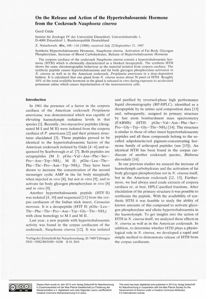
As depicted in Fig. 2 (A versus B) natural HTH isolated from the corpora cardiaca of N. cinerea showed a single absorbance peak at 210 nm with a retention time of 12.0 min and co-migrated with the peak of the synthetic HTH prepared according to the sequence assigned previously by FABMS [14]. Co-injection of the natural and synthetic peptides gave a single larger peak with no hint of resolution (Fig. 1C).
Dose-response relationships for hypertrehalosaemia and phosphorylase activation in N. cinerea
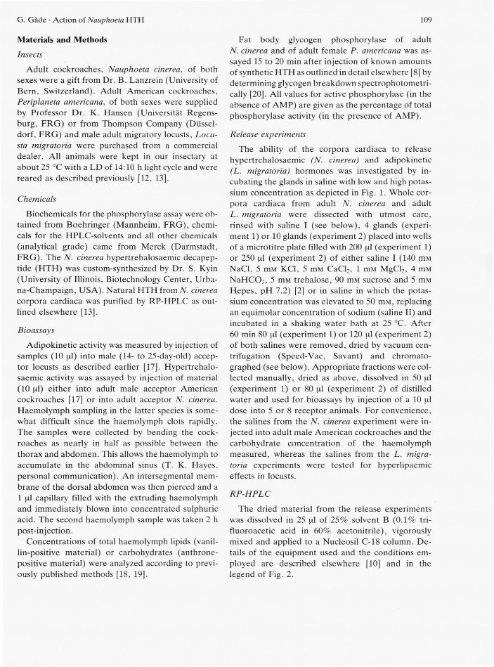
Synthetic HTH was injected into acceptor N. cinerea cockroaches in a variety of doses in order to demonstrate complete dose-response relationships for hypertrehalosaemic and phosphorylase-activating responses. A dose of 5 pmol of HTH is sufficient to elicite a maximal hypertrehalosaemic effect and about 3 pmol are needed for a significant response {p = 0.01, Student's t-test, Fig. 3A). The maximal increase in haemolymph carbohydrate levels was only about 7 mg/ml.
A much lower dose of HTH is needed to activate the glycogen phosphorylase of the fat body from N. cinerea fully when compared to the effect on blood carbohydrates (Fig. 3B versus 3 A); 0.5 pmol cause already a maximal activation and 0.2 pmol of HTH are sufficient for a significant response (p = 0.05, Student's t-test, Fig. 3B). In non-injected or water-injected insects the phosphorylase is about 30% active whereas about 60% of the enzyme is in the active form when maximal stimulated.
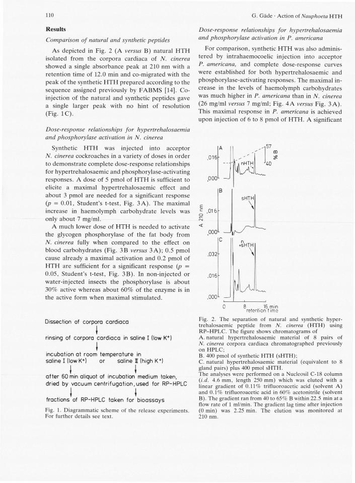
Dissection of corpora cardiaca
I rinsing of corpora cardiaca in saline I (low K+)
I incubation at room temperature in saline I (lowK+) or saline I (high K+)
I I after 60 min aliquot of incubation medium taken, dried by vacuum centrifugation,used for RP-HPLC
I I fractions of RP-HPLC taken for bioassays
Fig. 1. Diagrammatic scheme of the release experiments. For further details see text.
Dose-response relationships for hypertrehalosaemia and phosphorylase activation in P. americana
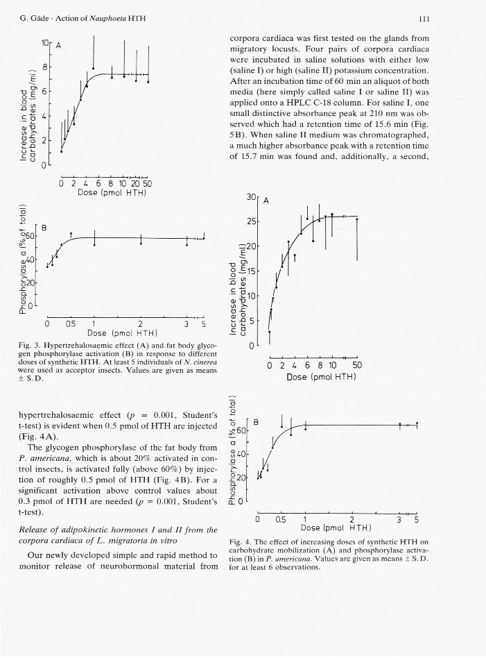
For comparison, synthetic HTH was also adminis-tered by intrahaemocoelic injection into acceptor P. americana, and complete dose-response curves were established for both hypertrehalosaemic and phosphorylase-activating responses. The maximal in-crease in the levels of haemolymph carbohydrates was much higher in P. americana than in N. cinerea (26 mg/ml versus 7 mg/ml; Fig. 4A versus Fig. 3 A). This maximal response in P. americana is achieved upon injection of 6 to 8 pmol of HTH. A significant
.016
000L
.016-
000L
.032-
.016-
.000L
16 min retention t ime
Fig. 2. The separation of natural and synthetic hyper-trehalosaemic peptide from N. cinerea ( H T H ) using R P - H P L C . The figure shows chromatograms of A. natural hypertrehalosaemic material of 8 pairs of N. cinerea corpora cardiaca chromatographed previously on HPLC; B. 400 pmol of synthetic H T H (sHTH); C. natural hypertrehalosaemic material (equivalent to 8 gland pairs) plus 400 pmol sHTH. The analyses were performed on a Nucleosil C - l 8 column (i.d. 4.6 mm, length 250 mm) which was eluted with a linear gradient of 0.11% trifluoroacetic acid (solvent A) and 0.1% trifluoroacetic acid in 60% acetonitrile (solvent B). The gradient ran from 40 to 65% B within 22.5 min at a flow rate of 1 ml/min. The gradient lag time after injection (0 min) was 2.25 min. The elution was monitored at 210 nm.
G. Gäde • Action of Nauphoeta H T H 111
% A
CD "O c 0 - £ o Q)
- ? 0) "O in >>
S i « 8
0 L
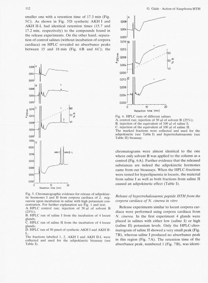
corpora cardiaca was first tested on the glands from migratory locusts. Four pairs of corpora cardiaca were incubated in saline solutions with either low (saline I) or high (saline II) potassium concentration. After an incubation time of 60 min an aliquot of both media (here simply called saline I or saline II) was applied onto a HPLC C-18 column. For saline I, one small distinctive absorbance peak at 210 nm was ob-served which had a retention time of 15.6 min (Fig. 5B). When saline II medium was chromatographed, a much higher absorbance peak with a retention time of 15.7 min was found and, additionally, a second,
°60
U) a o20 _c CL U)
Q_
2 U 6 8 10 2 0 5 0 Dose (pmol HTH)
/
0 0.5 1
Dose (pmol 2 HTH:
Fig. 3. Hypertrehalosaemic effect (A) and fat body glyco-gen phosphorylase activation (B) in response to different doses of synthetic H T H . At least 5 individuals of N. cinerea were used as acceptor insects. Values are given as means ± S . D .
3 0
2 5
= 2 0 cn E "O
g =15 5 £ - 210 (1) TJ CO >> a
— S o
i 1
2 U 6 8 10 5 0
Dose (pmol HTH)
hypertrehalosaemic effect (p = 0.001, Student's t-test) is evident when 0.5 pmol of HTH are injected (Fig. 4A).
The glycogen phosphorylase of the fat body from P. americana, which is about 20% activated in con-trol insects, is activated fully (above 60%) by injec-tion of roughly 0.5 pmol of HTH (Fig. 4B). For a significant activation above control values about 0.3 pmol of HTH are needed (p = 0.001, Student's t-test).
Release of adipokinetic hormones I and II from the corpora cardiaca of L. migratoria in vitro
Our newly developed simple and rapid method to monitor release of neurohormonal material from
o o
CL to o cL 0 L
0 0 .5 1 ^ 2 ^ Dose (pmol HTH)
Fig. 4. The effect of increasing doses of synthetic H T H on carbohydrate mobilization (A) and phosphorylase activa-tion (B) in P. americana. Values are given as means ± S. D. for at least 6 observations.
112 G. Gäde • Action of Nauphoeta H T H 112
smaller one with a retention time of 17.3 min (Fig. 5C). As shown in Fig. 5D synthetic AKH I and AKH II-L had identical retention times (15.7 and 17.2 min, respectively) to the compounds found in the release experiments. On the other hand, separa-tion of control salines (without incubation of corpora cardiaca) on HPLC revealed no absorbance peaks between 15 and 18 min (Fig. 6B and 6C); the
0008
Q004
0,000 0.016
0.012
Q008
0.004
0.000
- J
0.000' 10
Retention time [min] 20
Fig. 5. Chromatographic evidence for release of adipokine-tic hormones I and II from corpora cardiaca of L. mig-ratoria upon incubation in saline with high potassium con-centration. For further explanation see Fig. 1 and text. A. H P L C control run; injection of 50^1 of solvent B (25%); B. HPLC run of saline I from the incubation of 4 locust glands; C. HPLC run of saline II from the incubation of 4 locust glands; D. HPLC run of 50 pmol of synthetic A K H I and A K H II-L. The fractions labelled 1, 2. A K H I and A K H II-L were collected and used for the adipokinetic bioassay (see Table I).
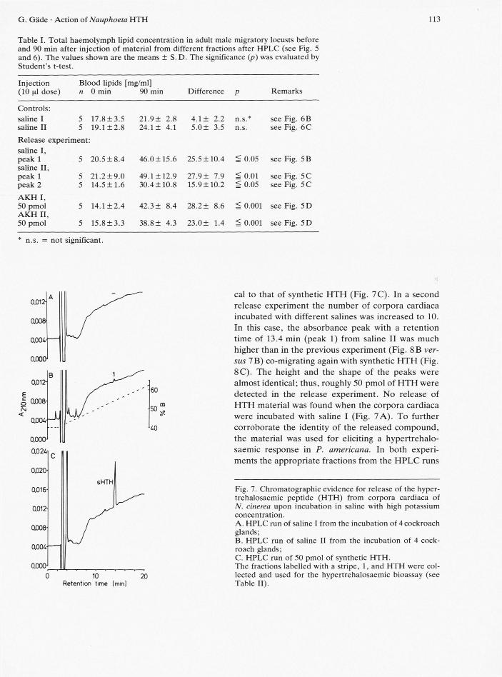
Fig. 6. HPLC runs of different salines A. control run; injection of 50 of solvent B (25%); B. injection of the equivalent of 100 |al of saline I; C. injection of the equivalent of 100 nl of saline II. The marked fractions were collected and used for the adipokinetic (see Table I) and hypertrehalosaemic (see Table II) bioassay.
chromatograms were almost identical to the one where only solvent B was applied to the column as a control (Fig. 6 A). Further evidence that the released substances are indeed the adipokinetic hormones came from our bioassays. When the HPLC-fractions were tested for hyperlipaemia in locusts, the material from saline I as well as both fractions from saline II caused an adipokinetic effect (Table I).
Release of hypertrehalosaemic peptide HTH from the corpora cardiaca of N. cinerea in vitro
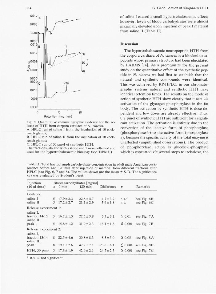
Release experiments similar to locust corpora car-diaca were performed using corpora cardiaca from N. cinerea. In the first experiment 4 glands were placed in salines with either low (saline I) or high (saline II) potassium levels. Only the HPLC-chro-matogram of saline II showed a very small peak (Fig. 7B), whereas saline I produced no absorbance peak in this region (Fig. 7A). The retention time of the absorbance peak, numbered 1 (Fig. 7B), was identi-
AKHI AKHD-L
10 20
Retention time [min]
G. Gäde • Action of Nauphoeta HTH 113
Table I. Total haemolymph lipid concentration in adult male migratory locusts before and 90 min after injection of material from different fractions after HPLC (see Fig. 5 and 6). The values shown are the means ± S. D. The significance (p) was evaluated by Student's t-test.
Injection (10 nl dose)
Blood lipids [mg/ml] n 0 min 90 min Difference Remarks
Controls: saline I 5 17.8±3.5 21.9± 2.8 4.1 ± 2.2 n.s.* see Fig. 6B saline II 5 19.1 ± 2 . 8 24.1 ± 4.1 5 .0± 3.5 n.s. see Fig. 6C
Release experiment saline I, peak 1 5 20.5 ± 8 . 4 46.0 ±15.6 25.5 ± 10.4 ^ 0.05 see Fig. 5B saline II, peak 1 5 21 .2±9 .0 49.1 ±12.9 27.9 ± 7.9 ^ 0.01 see Fig. 5C peak 2 5 14.5± 1.6 30.4 ±10.8 15.9± 10.2 ^ 0.05 see Fig. 5C
AKH I, 50 pmol 5 14.1 ± 2 . 4 42.3 ± 8.4 28.2± 8.6 ^ 0.001 see Fig. 5D AKH II, 50 pmol 5 15.8 ± 3 . 3 38.8± 4.3 23.0 ± 1.4 ^ 0.001 see Fig. 5D
n.s. = not significant.
0,012-
0Ű08Í
0.004
ooocy
B 0,012-
E c o 0.008-fsi <
0,004-
0.000-
0,024-1
0,020-
0.016-
0.012-
Q008-
0,004-
0,000-
0
.JU
60
50
40
10 Retention time [mini
20
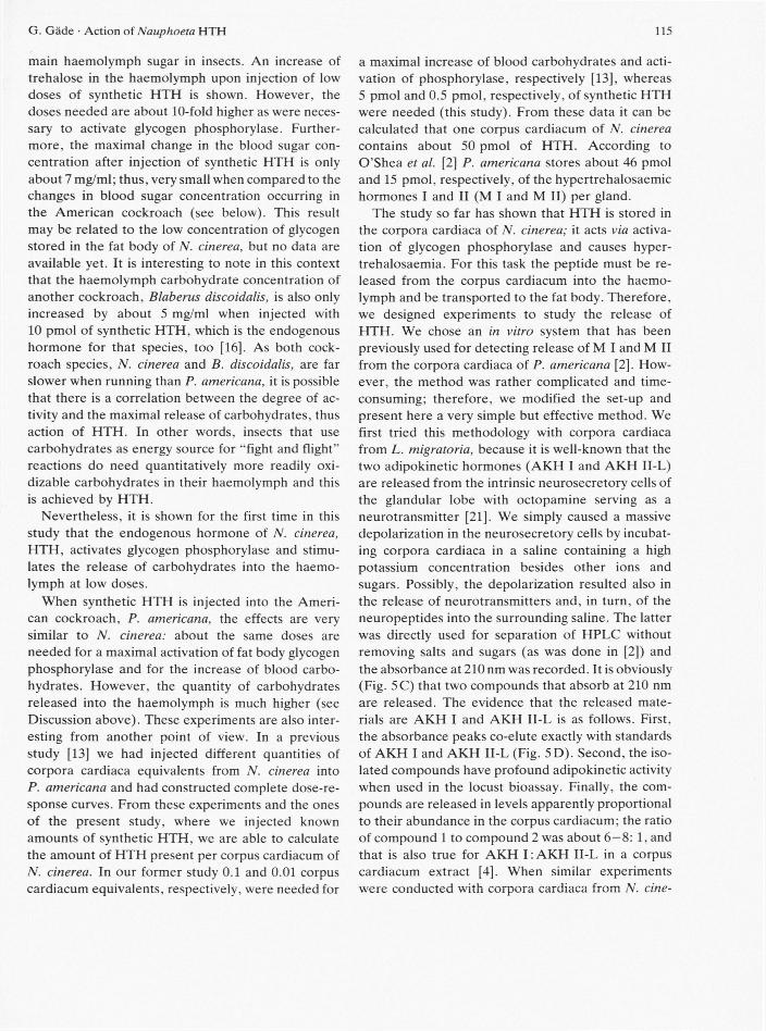
cal to that of synthetic HTH (Fig. 7C). In a second release experiment the number of corpora cardiaca incubated with different salines was increased to 10. In this case, the absorbance peak with a retention time of 13.4 min (peak 1) from saline II was much higher than in the previous experiment (Fig. 8B ver-sus 7B) co-migrating again with synthetic HTH (Fig. 8C). The height and the shape of the peaks were almost identical; thus, roughly 50 pmol of HTH were detected in the release experiment. No release of HTH material was found when the corpora cardiaca were incubated with saline I (Fig. 7A). To further corroborate the identity of the released compound, the material was used for eliciting a hypertrehalo-saemic response in P. americana. In both experi-ments the appropriate fractions from the HPLC runs
Fig. 7. Chromatographic evidence for release of the hyper-trehalosaemic peptide (HTH) from corpora cardiaca of N. cinerea upon incubation in saline with high potassium concentration. A. HPLC run of saline I from the incubation of 4 cockroach glands; B. HPLC run of saline II from the incubation of 4 cock-roach glands; C. HPLC run of 50 pmol of synthetic HTH. The fractions labelled with a stripe, 1, and HTH were col-lected and used for the hvpertrehalosaemic bioassay (see Table II).
114 G. Gäde • Action of Nauphoeta HTH 114
Retention time [min]
Fig. 8. Quantitative chromatographic evidence for the re-lease of HTH from corpora cardiaca of N. cinerea. A. HPLC run of saline I from the incubation of 10 cock-roach glands; B. HPLC run of saline II from the incubation of 10 cock-roach glands; C. HPLC run of 50 pmol of synthetic HTH. The fractions labelled with a stripe and 1 were collected and used for the hypertrehalosaemic bioassay (see Table II).
of saline I caused a small hypertrehalosaemic effect; however, levels of blood carbohydrates were almost maximally elevated upon injection of peak 1 material from saline II (Table II).
Discussion
The hypertrehalosaemic neuropeptide HTH from the corpora cardiaca of N. cinerea is a blocked deca-peptide whose primary structure had been elucidated by FABMS [14]. As a prerequisite for the present study on the quantitative effect of the synthetic pep-tide in N. cinerea we had first to establish that the natural and synthetic compounds were identical. This was achieved by RP-HPLC: in our chromato-graphic systems natural and synthetic HTH have identical retention times. The results on the mode of action of synthetic HTH show clearly that it acts via activation of the glycogen phosphorylase in the fat body. The activation by synthetic HTH is dose-de-pendent and low doses are already effective. Thus, 0.2 pmol of synthetic HTH are sufficient for a signifi-cant activation. The activation is entirely due to the conversion of the inactive form of phosphorylase (phosphorylase b) to the active form (phosporylase a), because the specific activity of the total enzyme is unaffected (unpublished observations). The product of phosphorylase action is glucose-l-phosphate which is converted via several steps to trehalose, the
Table II. Total haemolymph carbohydrate concentration in adult male American cock-roaches before and 120 min after injection of material from different fractions after HPLC (see Fig. 6, 7 and 8). The values shown are the mean ± S.D. The significance (p) was evaluated by Student's t-test.
Injection (10 \i\ dose)
Blood carbohydrates [mg/ml] n 0 min 120 min Difference P Remarks
Controls: saline I 5 17.9±2.3 22.8±4.7 4 .7±3 .2 n.s.* see Fig. 6B saline II 5 17.2 ± 2.7 21.1 ±2 .9 3.9 ±1 .8 n.s. see Fig. 6C
Release experiment 1: saline I, fraction 14/15 5 16.2± 1.5 22.5 ±3 .8 6.3 ±3 .1 ^ 0.01 see Fig. 7 A saline II,
see Fig.
peak 1 5 15.8± 1.2 31 .9±2.3 16.1 ±1 .8 ^ 0.001 see Fig. 7B
Release experiment 2: saline I, fraction 13/14 8 22 .5±4.6 30 .8±8.3 8 .3±5 .0 ^ 0.05 see Fig. 8 A saline II,
see Fig.
peak 1 8 19.1 ±2 .6 42.7 ± 7 . 1 23.6±6.1 ^ 0.001 see Fig. 8B HTH, 50 pmol 5 17.3± 1.9 42 .0±2.1 24.7 ±2 .5 ^ 0.001 see Fig. 7C
* n.s. = not significant.
116 G. Gäde • Action of Nauphoeta H T H 115
main haemolymph sugar in insects. An increase of trehalose in the haemolymph upon injection of low doses of synthetic HTH is shown. However, the doses needed are about 10-fold higher as were neces-sary to activate glycogen phosphorylase. Further-more, the maximal change in the blood sugar con-centration after injection of synthetic HTH is only about 7 mg/ml; thus, very small when compared to the changes in blood sugar concentration occurring in the American cockroach (see below). This result may be related to the low concentration of glycogen stored in the fat body of N. cinerea, but no data are available yet. It is interesting to note in this context that the haemolymph carbohydrate concentration of another cockroach, Blaberus discoidalis, is also only increased by about 5 mg/ml when injected with 10 pmol of synthetic HTH, which is the endogenous hormone for that species, too [16]. As both cock-roach species, N. cinerea and B. discoidalis, are far slower when running than P. americana, it is possible that there is a correlation between the degree of ac-tivity and the maximal release of carbohydrates, thus action of HTH. In other words, insects that use carbohydrates as energy source for "fight and flight" reactions do need quantitatively more readily oxi-dizable carbohydrates in their haemolymph and this is achieved by HTH.
Nevertheless, it is shown for the first time in this study that the endogenous hormone of N. cinerea, HTH, activates glycogen phosphorylase and stimu-lates the release of carbohydrates into the haemo-lymph at low doses.
When synthetic HTH is injected into the Ameri-can cockroach, P. americana, the effects are very similar to N. cinerea: about the same doses are needed for a maximal activation of fat body glycogen phosphorylase and for the increase of blood carbo-hydrates. However, the quantity of carbohydrates released into the haemolymph is much higher (see Discussion above). These experiments are also inter-esting from another point of view. In a previous study [13] we had injected different quantities of corpora cardiaca equivalents from N. cinerea into P. americana and had constructed complete dose-re-sponse curves. From these experiments and the ones of the present study, where we injected known amounts of synthetic HTH, we are able to calculate the amount of HTH present per corpus cardiacum of N. cinerea. In our former study 0.1 and 0.01 corpus cardiacum equivalents, respectively, were needed for
a maximal increase of blood carbohydrates and acti-vation of phosphorylase, respectively [13], whereas 5 pmol and 0.5 pmol, respectively, of synthetic HTH were needed (this study). From these data it can be calculated that one corpus cardiacum of N. cinerea contains about 50 pmol of HTH. According to O'Shea et al. [2] P. americana stores about 46 pmol and 15 pmol, respectively, of the hypertrehalosaemic hormones I and II (M I and M II) per gland.
The study so far has shown that HTH is stored in the corpora cardiaca of N. cinerea; it acts via activa-tion of glycogen phosphorylase and causes hyper-trehalosaemia. For this task the peptide must be re-leased from the corpus cardiacum into the haemo-lymph and be transported to the fat body. Therefore, we designed experiments to study the release of HTH. We chose an in vitro system that has been previously used for detecting release of M I and M II from the corpora cardiaca of P. americana [2]. How-ever, the method was rather complicated and time-consuming; therefore, we modified the set-up and present here a very simple but effective method. We first tried this methodology with corpora cardiaca from L. migratoria, because it is well-known that the two adipokinetic hormones (AKH I and AKH II-L) are released from the intrinsic neurosecretory cells of the glandular lobe with octopamine serving as a neurotransmitter [21]. We simply caused a massive depolarization in the neurosecretory cells by incubat-ing corpora cardiaca in a saline containing a high potassium concentration besides other ions and sugars. Possibly, the depolarization resulted also in the release of neurotransmitters and, in turn, of the neuropeptides into the surrounding saline. The latter was directly used for separation of HPLC without removing salts and sugars (as was done in [2]) and the absorbance at 210 nm was recorded. It is obviously (Fig. 5C) that two compounds that absorb at 210 nm are released. The evidence that the released mate-rials are AKH I and AKH II-L is as follows. First, the absorbance peaks co-elute exactly with standards of AKH I and AKH II-L (Fig. 5D). Second, the iso-lated compounds have profound adipokinetic activity when used in the locust bioassay. Finally, the com-pounds are released in levels apparently proportional to their abundance in the corpus cardiacum; the ratio of compound 1 to compound 2 was about 6—8: 1, and that is also true for AKH I : A K H II-L in a corpus cardiacum extract [4], When similar experiments were conducted with corpora cardiaca from N. cine-
116 G. Gäde • Action of Nauphoeta H T H 116
rea, one compound that had an identical retention time as HTH and caused hypertrehalosaemia was de-tected upon depolarization with an elevated potas-sium saline. The release of H T H is dependent on the number of corpora cardiaca used for the experiment suggesting that one corpus cardiacum always releases the same amount upon stimulation. From our data we can calculate that roughly 50 pmol of H T H are released by 10 glands. As one gland stores about 50 pmol of H T H (see above), we estimate a release of about 10% of the total available hormone in the gland over a 60 min exposure to the elevated potas-sium saline. This number is in good agreement to the 5% released of the hypertrehalosaemic hormones from the American cockroach upon 10 min of depolarization [2].
In summary, it was shown that low doses of syn-thetic H T H causes activation of glycogen phosphory-lase in the fat body of N. cinerea and result in hyper-
trehalosaemia. The biological significance of H T H to the cockroach is further corroborated by the fact that it is releasable in vitro from the corpora cardiaca upon depolarization with an elevated potassium saline. In vivo this would produce a release of H T H directly from the corpora cardiaca into the haemo-lymph.
Acknowledgements
The author gratefully acknowledges the technical assistance of Miss Beate Schumacher and Dipl.-Biol. Volker Pohl. He also wishes to thank Dr. J. H. Spring (University of Southwestern Louisiana, Lafayette, Louisiana, USA), for correcting the Eng-lish manuscript. The work was financially supported by a grant from the Deutsche Forschungsgemein-schaft (Ga 241/6-2) and by a Heisenberg Fellowship awarded by the Deutsche Forschungsgemeinschaft (Ga 241/5-1).
[1] J. E. Steele, Nature 192, 680-681 (1961). [2] M. O'Shea, J. Witten, and M. Schaffer, J. Neurosci. 4,
521-529 (1984). [3] J. L. Witten, M. H. Schaffer, M. O'Shea, J. C. Cook,
M. E. Hemling, and K. L. Rinehart, Biochem. Bio-phys. Res. Commun. 124, 350-358 (1984).
[4] G. Gäde, J. Insect Physiol. 30, 729-736 (1984). [5] G. Gäde, Z. Naturforsch. 40c, 42-46 (1985). [6] G. Gäde, Naturwissenschaften 72, 95-96 (1985). [7] R. M. Scarborough, G. C. Jamieson, F. Kalish, S. J.
Kramer, G. A. McEnroe, C. A. Miller, and D. A. Schooley, Proc. Natl. Acad. Sei. USA 81, 5575-5579 (1984).
[8] G. Gäde, Z. Naturforsch. 40c, 670-676 (1985). [9] G. L. Orr, J. W. D. Gole, A. P. Jahagirdar, R. G. H.
Downer, and J. E. Steele, Insect Biochem. 15, 703-709 (1985).
[10] G. Gäde, Biol. Chem. Hoppe-Seyler 366, 195-199 (1985).
[11] G. Gäde and K. L. Rinehart, Jr., Biol. Chem. Hoppe-Seyler 368, 67-75 (1987).
[12] G. Gäde and M. Scheid, Physiol. Entomol. 11, 145-157 (1986).
[13] G. Gäde, Z. Naturforsch. 42c, 225-230 (1987). [14] G. Gäde and K. L. Rinehart, Jr., Biochem. Biophys.
Res. Commun. 141, 774-781 (1986). [15] G. Gäde, Z. Naturforsch. 41c, 315-320 (1986). [16] T. K. Hayes, L. L. Keeley, and D. W. Knight,
Biochem. Biophys. Res. Commun. 140, 674—678 (1986).
[17] G. Gäde, J. Insect Physiol. 26, 351-360 (1980). [18] N. Zöllner and K. Kirsch, Z. ges. Exp. Med. 135,
545-561 (1962). [19] G. Spik and J. Montreuil, Bull. Soc. Chim. Biol. 46,
739-749 (1964). [20] R. Ziegler, M. Ashida, A. M. Fallon, L. T. Wimer, S.
S. Wyatt, and G. R. Wyatt, J. Comp. Physiol. 131, 321-332 (1979).
[21] T. Pannabecker and I. Orchard, Mol. Cell. Endocrin. 48, 153-159 (1986).