Embed Size (px)
Citation preview
25
Original Article
APPLIED FOOD BIOTECHNOLOGY, 2016, 3 (1): 25-34 Journal homepage: www.journals.sbmu.ac.ir/afb
pISSN: 2345-5357
eISSN: 2423-4214
Optimization of Phospholipase A1 Immobilization on Plasma Surface Modified Chitosan Nanofibrous Mat
Zahra Beig Mohammadi1, Zohreh Hamidi-Esfahani1*, Mohammad Ali Sahari1, Kianoush Khosravi-Darani2
1. Department of Food Science and Technology, Faculty of Agriculture, Tarbiat Modares University, Tehran,
Iran
2. Research Department of Food Technology, National Nutrition and Food Technology Research Institute,
Faculty of Nutrition Sciences and Food Technologies, Shahid Beheshti University of Medical Sciences,
Tehran, Iran
Abstract Article Information
Phospholipase A1 is known as an effective catalyst for hydrolysis of various
phospholipids in enzymatic vegetable oil degumming. Immobilization is one of the
most efficient strategies to improve its activity, recovery and functional properties.
In this study, chitosan-co-polyethylene oxide (90:10) nanofibrous mat was
successfully fabricated and modified with atmospheric plasma at different times (2,
6 and 10 min) to interact with enzyme molecules. Scanning electron microscopy
images revealed that the membranes retained uniform nanofibrous and open porous
structures before and after the treatment. PLA1 was successfully immobilized onto
the membrane surfaces via covalent bonds with the functional groups of chitosan
nanofibrous mat. Response surface methodology was used to optimize the
immobilization conditions for reaching the maximum immobilization efficiency.
Enzyme concentration, pH, and immobilization time were found to be significant
key factors. Under optimum conditions (5.03 h, pH 5.63, and enzyme dosage
654.36 UI), the atmospheric plasma surface modified chitosan nanofibers reached
the highest immobilization efficiency (78.50%). Fourier transform infrared
spectroscopy of the control and plasma surface-modified chitosan nanofibers
revealed the functional groups of nanofibers and their reaction with the enzyme.
The results indicated that surface modification by atmospheric plasma induced an
increase in PLA1 loading on the membrane surfaces.
Article history:
Received 29 Aug 2015
Revised 20 Sep 2015
Accepted 26 Oct 2015
Keywords:
Chitosan nanofibers,
Immobilization,
Phospholipase A1,
Plasma,
Surface modification.
Correspondence to:
Zohreh Hamidi Esfahani
Department of Food
Science and Technology,
Faculty of Agriculture,
Tarbiat Modares
University, P.O. Box:
14115-336, Tehran, Iran,
Tel: +98-21-48292474
Fax: +98-21-48292200
E-mail:
1. Introduction
Phospholipase A1 (PLA1) hydrolyzes the acyl group
of phospholipids at the sn-1 position, liberating the fatty
acids, and producing 2-acyl-1-lyso-phospholipid [1];
this reduces phosphorus level to less than 10 mg kg-1
on the degumming process of vegetable oils [2]. Most
studies on the degumming of crude vegetable oils are
based on free PLA1 [3-5]. Unfortunately, free enzyme
molecules, in most cases, are relatively expensive,
chemically or thermally un-stable, and difficult for
handling, purification and reuse, which limit the large-
scale operations in industrial applications [6-7].
Therefore, immobilization of PLA1 is a promising
technology, and has a large impact on the industrial
scale degumming operations. Immobilization of
enzymes onto solid support matrices including particles,
fibers, porous films, and hydrogels is a hopeful way to
circumvent the disadvantages mentioned above [8].
With the development of nanotechnologies, many
efforts have been made to immobilize enzymes onto the
surfaces of nano-scaled materials such as nanoparticles,
nanotubes, mesoporous materials, and nanofibrous
membranes [9].
The results of immobilization, including the
performance of immobilized enzymes, strongly depend
Phospholipase A1 Immobilization
26 Appl Food Biotechnol, Vol. 3, No. 1 (2016)
on the properties of supports, which are usually referred
to as material types, compositions, structures, etc. So
far, different nanostructured materials have been used as
supports, like mesoporous silica, nanotubes,
nanoparticles, and nanofibers. They stand out of other
supports because of their extremely high surface area-
to-volume ratios, which can provide large specific
surface areas for highly efficient immobilization and
stabilizing enzymes. However, some of the
nanostructured materials have disadvantages that are
difficult to overcome. For example, mesoporous silica
usually confines enzyme molecules on its inner surface,
which limits the diffusion of substrate to/off the enzyme
and results in lower enzyme activity. Nanoparticles and
nanotubes are known to remarkably decrease mass
transfer limitation, while their dispersion and recycling
are more difficult. On the contrary, electrospun
nanofibers have a great potential to overcome these
problems, and may be promising supports for enzyme
immobilization. Nanofibrous membranes exhibit
intrinsic features including open porous structure and
continuous nanofiber formation, enabling the easy
accessibility of reactive sits toward enzyme molecules
and preventing low mass diffusion in catalytic systems.
So, substrate can easily diffuse into open porous
nanofibers comparing to other nanostructures [10-12].
Chitosan is a natural cationic linear poly-saccharide
composed essentially of β (1→4) linked glucosamine
units together with some proportions of N-acetyl-
glucosamine units, and has excellent features due to its
nontoxicity, biodegradability, biocompatibility,
physiological inertness, hydrophilicity, remarkable
affinity to proteins, and high mechanical strength. These
significant chemical and biological characteristics make
chitosan a desirable biomaterial for enzyme
immobilization [13].
Surface treatment of biomaterials is performed to
modify such characteristics as hydrophilicity and
protein adsorption, and consequently, to improve cell
attachment, proliferation and differentiation. Plasma
treatment is a simple process to modify the physical and
chemical characteristics of biomaterials without altering
their bulk properties and without changing the
mutagenic and toxic effects of chemical materials like
glutaraldehyde [9]. Recently, plasma-based surface
modifications of chitosan and chemically-modified
chitosan have been widely studied and shown promising
advantages [14]. Ni et al. found that, using open-air
plasma, surface-modification could introduce chitosan
with more efficient bioactive components [15].
Designing an efficient immobilized enzyme is a
multivariate process involving many factors that could
affect immobilization efficiency. Response surface
methodology (RSM) is a statistically designed
experimental protocol for developing, improving, and
optimizing processes [16]. It helps to identify the effect
of the interactions of different design variables on the
response when they are varied simultaneously.
Recently, RSM has been successfully used for the
optimization of several immobilization processes, such
as immobilization of β-galactosidase into chitosan beads
[17], and onto sephadex and chitosan beads [18].
In this research, PLA1 was immobilized onto
chitosan nanofibers for its application in food
technology with focus on vegetable oil degumming. The
conditions for immobilization of PLA1 on plasma
modified chitosan nanofibrous mats were determined by
RSM for reaching the maximum activity recovery of
enzyme. Various properties (including pH-activity
curve, temperature-activity curve, storage stability, and
reusability) of free and immobilized PLA1 were
investigated.
2. Materials and methods
2.1. Materials
Chitosan (CS, low molecular weight, degree of
deacetylation 91.2%), polyethylene oxide (PEO, MW
900 KDa), PLA1 (Lecitase™ Ultra from Thermomyces
lanuginosus), and soy-phospholipid (PL) were
purchased from Easter Groups (Dong Chen Co., Ltd,
China), Acros Organics Co. (New Jersey, USA),
Novozymes A/S (Bagsvaerd, Denmark) and Behpak
Co. (Behshar, Iran), respectively. All other chemicals
were obtained from Merck Chemical (Darmstadt,
Germany).
2.2. Preparation of CS/PEO nanofibers and plasma
treatment
Chitosan and PEO solutions were prepared
separately by dissolving chitosan and PEO powders in
aqueous acetic acid (80% v/v) under magnetic stirring
at 37°C for 24 h. The obtained solutions were then
mixed together in a weight ratio of CS/PEO, 90:10, as
the required electrospinning solution. The
electrospinning processes were carried out at injection
rate of 3.0 ml h-1
, 20 kV, drum speed of 200 rpm, and
electrospinning time of 1 h, using Electroris (FNM,
Tehran, Iran). For every run, the polymer solution was
placed into a plastic syringe with a stainless steel
needle (18 G). An aluminum sheet was wrapped on
the Electroris rotating drum as collector, and was
located at the distance of 12 cm from the needle [19].
Electrospun fibers were treated by homemade air-
dielectric barrier discharge (DBD) plasma consisting
of two parallel copper-electrodes where the upper
electrode was covered by quartz-dielectric. A DC-
pulsed high voltage power supply with a power of
30W and a frequency of 6 kHz was applied to the
DBD reactor. Electrospun fiber mats, each measuring
2 cm × 2 cm (~200 µm thick), were placed in the
chamber, and plasma treatment was performed for 2, 6
and 10 min [14, 20].
2.3. Characterization
To characterize the morphology of electrospun
nanofibers before and after plasma treatment, a
scanning electron microscope (SEM; Philips XL30,
USA) was used. To test the chemical structure
changes, the fourier transform infrared (FTIR) spectra
of original and atmospheric plasma surface modified
CS/PEO nanofiberous mats (APSM CS/PEO NM)
Beig Mohammdi et al
27 Appl Food Biotechnol, Vol. 3, No. 1 (2016)
were measured from 4000 to 500 cm-1
at a resolution
of 4 cm-1
by a TENSOR 27 spectrometer (Bruker
Optics, USA).
2.4. Enzyme assay
PLA1 assay was performed with soy-phospholipid
emulsion using the method of Yang et al. [21]. One
unit of PLA (UI) is the amount of enzyme, which
releases 1 µmol of titratable free fatty acids per
minute. Four ml of substrate emulsion (25% PL and
4% polyvinyl alcohol solution with a volume ratio of
1:4), 5 of 0.01M citric acid buffer (pH 5.0), and 1
ml of enzyme solution were mixed and incubated at
37°C for 10 min. The reaction was terminated with the
addition of ethanol 95% (15 ml) after incubation, and
the liberated free fatty acids were titrated with 0.05 M
NaOH. Blanks were measured with heat-inactivated
PLA samples (95°C, 10 min). All experiments were
carried out in triplicate
2.5. Immobilization of PLA1
PLA1 solution was prepared by solving appropriate
amount of PLA1 enzyme in phosphate-citrate buffer
(0.05 M, pH 4.5-6.5). Varying amounts of PLA1
enzyme (200-1000 U) were added to the APSM
CS/PEO NM and the control nanofibers (before
plasma treatment), and incubated overnight at 4ºC for
different times (1.5-7.5 h). The efficiency of
immobilization was evaluated for chitosan nanofiber
using Eq. 1:
Y=(A1/ A2) ×100 Eq.1
Where, Y is immobilization efficiency; A1 is
Specific activity of immobilized PLA1 (U mg-1
); and
A2 is the specific activity of soluble free PLA1 (U mg-
1).
Specific activity of the immobilized enzyme was
calculated by subtracting the specific activity of
washed fractions from the specific activity of the
added enzyme [18]. Protein concentration in the
solution was determined with Coomassie Brilliant
Blue reagent following the Bradford’s method with
Bovin Serum Albumin as standard [22].
2.6. Effect of pH and temperature on enzyme
activity
The effect of pH on the activity of free and
immobilized PLA1 was studied by immersing the
enzymes onto 50 mM phosphate-citrate buffer with
pH ranging from 4.5 to 7.5 at 25°C (ambient
temperature). The effect of temperature on enzyme
activity was determined by immersing the enzymes
into phosphate-citrate buffer (pH 5.5) with
temperature ranging from 30 to 70°C.
The initial activity was designated as 100%, and
the activities at all the remaining pH and temperature
values were proportional to it [2, 23].
2.7. Storage stability and Reusability tests
Storage stability of the free and immobilized PLA1
was measured by keeping them in phosphate-citrate
buffer (50 mM, pH 5.5) and storing at 4°C for up to 30
days. The remaining activity of the enzymes was
measured periodically [31, 36]. To evaluate the
reusability, the tested immobilized PLA1 membranes
were washed with phosphate-citrate buffer to remove
any residual chemicals, followed by immersing into
fresh catalytic reaction medium under the same
experimental conditions. This procedure was repeated
up to 10 times. The residual enzyme activity after each
cycle was defined as the value proportional to the
initial activity (100%) [23].
2.8. Experimental designs and statistical analyses
Experimental designs and statistical analyses
reported in this paper were generated using DX7
software (Trial version, Stat-Ease, Inc., USA). Effect
of various parameters was optimized using the
response surface method (RSM). A 20 run central
composite rotatable design (CCRD) was performed
in order to determine optimum conditions for enzyme
immobilization. This design assesses the influence of
the main factors, as well as their interaction. Three
main factors namely pH (4.5, 4.9, 5.5, 6.1, and 6.5),
enzyme concentration (200.0, 362.1, 600.0, 837.8, and
1000 UI), and immobilization time (1.5, 2.72, 4.5,
6.28, and 7.5 h) were studied in five levels. The
response was immobilization efficiency (%) in each
case. Analysis of variance (ANOVA) was used to
compare the other parameters, and Duncan’s test was
used as post hoc. All experiments were carried out in
triplicate, and the data were represented as mean±SD
(SPSS, ver. 16). The significance was considered as
p<0.05. 3. Results and Discussion
3.1. Surface morphologies of CS/PEO and APSM
CS/PEO nanofibrous mat
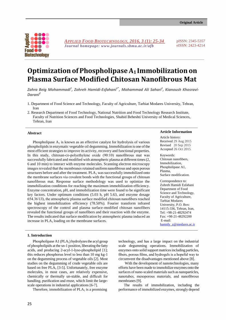
To prepare a support medium for enzyme
immobilization, CS/PEO polymer containing 90%
chitosan and 10% polyethylene oxide was fabricated
into nanofibrous mat. The morphology of the
electrospun mats was investigated through the SEM
micrographs. The original CS/PEO NM presented
homogeneous and uniform network of continuous
nanofibers with a diameter range of 70–240 nm.
(Figure1a). After surface activation by atmospheric plasma at
different times, total fibrous structures of the APSM CS/PEO NM remained intact; however, there were a
few structural deformations, especially at 10 min
plasma treatment, as shown in Figure 1 (b, c and d). The surface modification by atmospheric plasma
caused remarkable change in the stability of CS/PEO nanofibers (Figures 1b', c' and d') in comparison with
Phospholipase A1 Immobilization
28 Appl Food Biotechnol, Vol. 3, No. 1 (2016)
the non-treated CS/PEO nanofibers (Figure 1a') after
immersing in distilled deionized water (DDW).
The FTIR spectra verified that CS/PEO nanofibres are
a combination of chitosan and polyethylene oxide
(Figure 2).
Figure 1. SEM images of control CS/PEO NM(a), and 2 (b), 6 (c) 10 (d) minute APSM-CS/PEO NM before immerging in
DDW, and control CS/PEO NM(a'), and 2 (b'), 6 (c') 10 (d') minute APSM-CS/PEO NM after immerging in DDW
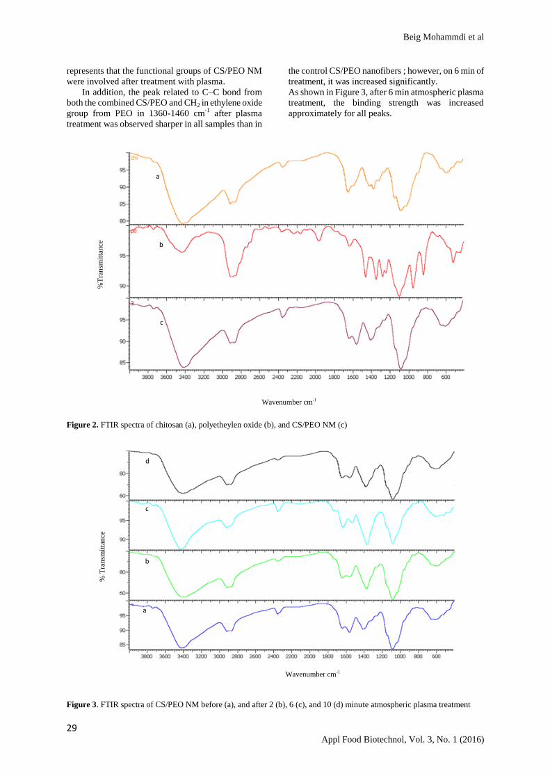
Major characteristic peaks of chitosan were
observed at the wavenumbers of 3450 cm-1
(stretching
N–H and O–H), 1650 cm-1
(stretching C=O of
carbonyl group), 1375 cm-1
(stretching C–H of methyl
group), and 1092 cm-1
(stretching C–O) [24].
According to the FTIR spectra, the functional
group and wavenumbers observed for the CS/PEO
nanofibres were the same as those of chitosan and
PEO alone. The new peaks observed in the 1550-1650
cm-1
were assumed to be related to the functional
groups of asymmetric C=O in the CS/PEO nanofibers
and contingency resonant. This means that double
bond leads to the creation of C–O and C=O structures
to link the carbon and oxygen. Despite the fingerprint,
carbon-oxygen double bond and the hydroxyl group of
chitosan and polyethylene oxide showed the
successful synthesis of CS/PEO nanofiber [24].
In this study, the NH2 groups existing on the
membrane surface were converted into more reactive
NH3+
groups via atmospheric plasma activation, and
the surface NH3+
groups could further form covalent
bonds with the enzyme molecules, leading to the
immobilization of the enzymes [25]. Covalent
immobilization provides strong bonding between the
enzyme and support, and thus reduces enzyme leakage
[26].
A series of atmospheric plasma with different initial
times ranging from 2 to 10 minutes were applied to
investigate the effect of time on surface
functionalization and enzyme immobilization. The
chemical structure changes were characterized via
FTIR. Figure 3 shows the FTIR spectra for the
unmodified and APSM CS/PEO nanofibers with
different treatment times. The original nanofibers
exhibited an stretching C–O bond (C from the
carboxyl and O from the hydroxyl groups) at
approximately 1100 cm-1
, and then an stretching
amino bond at approximately 1600 cm-1
[24]. The C =
O carbonyl stretch of a carboxylic acid bond appears
at approximately 1650 cm-1
[27]. The observed bond
at approximately 3000-3500 cm-1
is contributed by the
stretching vibrations of –NH and –OH groups [28].
Furthermore, the FTIR spectra for the APSM CS/PEO
NM are similar to those of the control CS/PEO
nanofibers; however, during the atmospheric plasma
treatment, the intensity of some links such as hydroxyl
and carboxylic acid at 1100 cm-1
and 1650 cm-1
were
increased on 6 minutes but they were decreased on 2
and 10 minutes.
Also the intensity of –NH group on 6 minutes was
more than that of other samples . This phenomenon
1µm
1µm
1µm 1µm 1µm
1µm 1µm 1µm
a' b' c' d'
a b c d
Beig Mohammdi et al
29 Appl Food Biotechnol, Vol. 3, No. 1 (2016)
represents that the functional groups of CS/PEO NM
were involved after treatment with plasma.
In addition, the peak related to C–C bond from
both the combined CS/PEO and CH2 in ethylene oxide
group from PEO in 1360-1460 cm-1
after plasma
treatment was observed sharper in all samples than in
the control CS/PEO nanofibers ; however, on 6 min of
treatment, it was increased significantly.
As shown in Figure 3, after 6 min atmospheric plasma
treatment, the binding strength was increased
approximately for all peaks.
Figure 2. FTIR spectra of chitosan (a), polyetheylen oxide (b), and CS/PEO NM (c)
Figure 3. FTIR spectra of CS/PEO NM before (a), and after 2 (b), 6 (c), and 10 (d) minute atmospheric plasma treatment
Wavenumber cm-1
% T
ran
smit
tan
ce
a
b
c
d
Wavenumber cm-1
%T
ran
smit
tance
a
b
c
Phospholipase A1 Immobilization
30 Appl Food Biotechnol, Vol. 3, No. 1 (2016)
So, 6 min atmospheric plasma treatment was
chosen as the suitable plasma reaction time for
CS/PEO NM mats for enzyme immobilization. The
results showed that plasma treatment caused to
changes in the surface structure of the functional
groups and increased the ion exchange property of the
membranes without using chemicals.
3.2. Optimization of immobilization conditions
To optimize the balance between enzyme loading
and the activity of the immobilized enzyme, the
properties of the immobilized PLA1 on 6 min APSM
CS/PEO NM were tested. Studies have shown that
enzyme concentration, pH and immobilization time
significantly affected the immobilization of enzyme
into nanofibers [2,18,24]. Thus, these three factors
were further optimized using the RSM to obtain the
maximum activity recovery of immobilized PLA1.
Table 1. Levels of design variables (=1.68)
Variables Range of levels
- -1 0 +1 +
Time (h) 1.50 2.72 4.50 6.28 7.50
PLA1(U) 200 362.16 600 837.84 1000
pH 4.50 4.91 5.5 6.09 6.50
To assess the influence of each factor and their
interactions, a CCRD of 6 central points with 14
experiments was performed. The levels of each factor
are given in Table 1, and the experimental data are
recorded in Table 2. The summary of ANOVA for the
model of immobilization efficiency is given in Table
3. By regression analysis, a quadratic polynomial
equation was established to explain the relationship
between the activity recovery and the independent
variables as follows:
Eq.2
Y= 75.85 + 6.83 X1 + 5.57 X2 + 50.90 X3 + 3.62
X1 X3 – 13.17 X12 – 13.04 X2
2 – 16.41 X3
2
Where, Y, X1, X2, and X3 represent efficiency,
immobilization time, enzyme dosage, and pH,
respectively. The calculated F ratio was 112.50 and P
value was less than 0.0001, implying that the model is
extremely significant and satisfying. A verification experiment was performed under the
optimal conditions to validate the reliability and
accuracy of the model. The experimental activity
recovery was determined to be 78.50%, which is in
excellent agreement with the value predicted by the
model (Table 4).
Accordingly, this model could be considered
reliable and accurate for predicting the immobilization
efficiency and activity recovery of immobilization of
PLA1. The coefficients of the terms along with their p-
values (P<0.01) show which terms contributed
significantly to the responses. A smaller P-value
indicates a higher level of significance for the
corresponding coefficient [17].
According to the ANOVA, which was used to
check the significant of the effects and assess the
goodness of fit (Table 3), a predicted response surface
model was statistically significant (p<0.0001).
The non-significant value of lack-of-fit
(p>0.2781), and high values of R2 (0.9902), adjusted
R2 (0.9814) and predicted R
2 (0.9476) revealed that
the model is statistically significant for the response to
predict and explain the variations in immobilization
efficiency formulation. Central point of CCRD design
(enzyme concentration, 600 UI; pH, 5.50; and
immobilization time, 4.50 h) leads to the maximum
immobilization efficiency of PLA1 on APSM CS/PEO
NM with 72.5-80.06%.
For the control CS/PEO NM, immobilization
efficiency was detected as 59.2-63.50% in the same
conditions. The results confirmed that in this system,
plasma activation is an important factor that affects
the performance of the immobilized PLA1, As shown
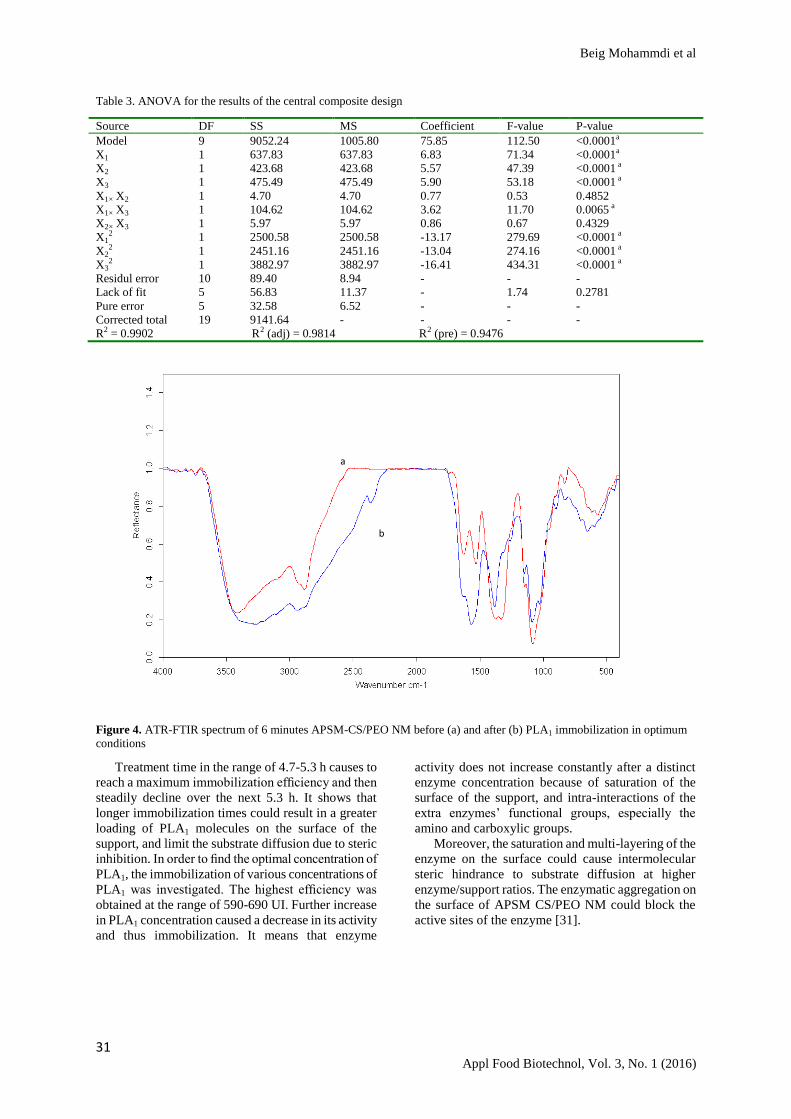
in Figure 4, under optimum conditions, the enzyme
was successfully loaded on APSM CS/PEO NM.
The ATR-FTIR spectra showed that the intensity
of some links such as C=O and C-N in 1500-1650 cm-
1, O-H and N-H in 3000-3500 cm
-1 significantly
increased after PLA1 immobilization Additionally, the
efficiency of PLA1 immobilization in this study is
much higher than that of the immobilized PLA1 on
other different supports, such as magnetic
nanoparticles (64.7%) [2], calcium alginate (56.2%),
calcium alginate-chitosan (65.5%) and calcium
alginate-gelatin (60.2%) [29].
The change in pH value can influence the electrical
charges on the surface of the nanofibers, and limit the
interactions between the functional groups on the
surface of nanofibers and those of PLA1. Protein
denaturation can occur under conditions of excess acid
or alkali [30-31].
Therefore, at pH values under and above 5.3-5.7, the
efficiency of PLA1 immobilization significantly
decreases.
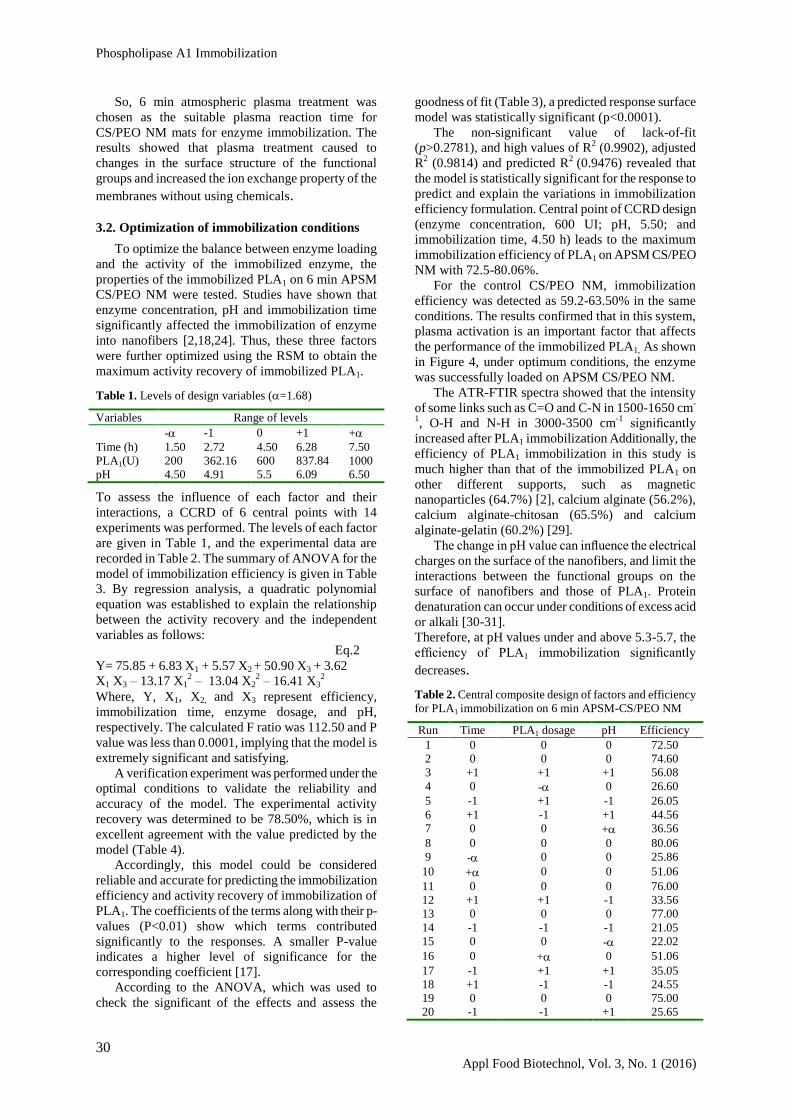
Table 2. Central composite design of factors and efficiency
for PLA1 immobilization on 6 min APSM-CS/PEO NM
Run Time PLA1 dosage pH Efficiency
1 0 0 0 72.50
2 0 0 0 74.60
3 +1 +1 +1 56.08
4 0 - 0 26.60
5 -1 +1 -1 26.05
6 +1 -1 +1 44.56
7 0 0 + 36.56
8 0 0 0 80.06
9 - 0 0 25.86
10 + 0 0 51.06
11 0 0 0 76.00
12 +1 +1 -1 33.56
13 0 0 0 77.00
14 -1 -1 -1 21.05
15 0 0 - 22.02
16 0 + 0 51.06
17 -1 +1 +1 35.05
18 +1 -1 -1 24.55
19 0 0 0 75.00
20 -1 -1 +1 25.65
Beig Mohammdi et al
31 Appl Food Biotechnol, Vol. 3, No. 1 (2016)
Table 3. ANOVA for the results of the central composite design
Source DF SS MS Coefficient F-value P-value
Model 9 9052.24 1005.80 75.85 112.50 <0.0001a
X1 1 637.83 637.83 6.83 71.34 <0.0001a
X2 1 423.68 423.68 5.57 47.39 <0.0001 a
X3 1 475.49 475.49 5.90 53.18 <0.0001 a
X1× X2 1 4.70 4.70 0.77 0.53 0.4852
X1× X3 1 104.62 104.62 3.62 11.70 0.0065 a
X2× X3 1 5.97 5.97 0.86 0.67 0.4329
X12 1 2500.58 2500.58 -13.17 279.69 <0.0001 a
X22 1 2451.16 2451.16 -13.04 274.16 <0.0001 a
X32 1 3882.97 3882.97 -16.41 434.31 <0.0001 a
Residul error 10 89.40 8.94 - - -
Lack of fit 5 56.83 11.37 - 1.74 0.2781
Pure error 5 32.58 6.52 - - -
Corrected total 19 9141.64 - - - -
R2 = 0.9902 R2 (adj) = 0.9814 R2 (pre) = 0.9476
Figure 4. ATR-FTIR spectrum of 6 minutes APSM-CS/PEO NM before (a) and after (b) PLA1 immobilization in optimum
conditions
Treatment time in the range of 4.7-5.3 h causes to
reach a maximum immobilization efficiency and then
steadily decline over the next 5.3 h. It shows that
longer immobilization times could result in a greater
loading of PLA1 molecules on the surface of the
support, and limit the substrate diffusion due to steric
inhibition. In order to find the optimal concentration of
PLA1, the immobilization of various concentrations of
PLA1 was investigated. The highest efficiency was
obtained at the range of 590-690 UI. Further increase
in PLA1 concentration caused a decrease in its activity
and thus immobilization. It means that enzyme
activity does not increase constantly after a distinct
enzyme concentration because of saturation of the
surface of the support, and intra-interactions of the
extra enzymes’ functional groups, especially the
amino and carboxylic groups.
Moreover, the saturation and multi-layering of the
enzyme on the surface could cause intermolecular
steric hindrance to substrate diffusion at higher
enzyme/support ratios. The enzymatic aggregation on
the surface of APSM CS/PEO NM could block the
active sites of the enzyme [31].
a
b
Phospholipase A1 Immobilization
32 Appl Food Biotechnol, Vol. 3, No. 1 (2016)
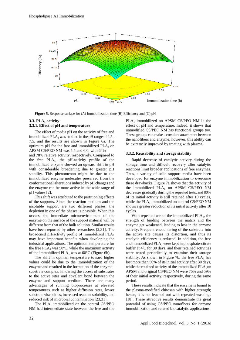
Figure 5. Response surface for (A) Immobilization time (B) Efficiency and (C) pH
3.3. PLA1 activity
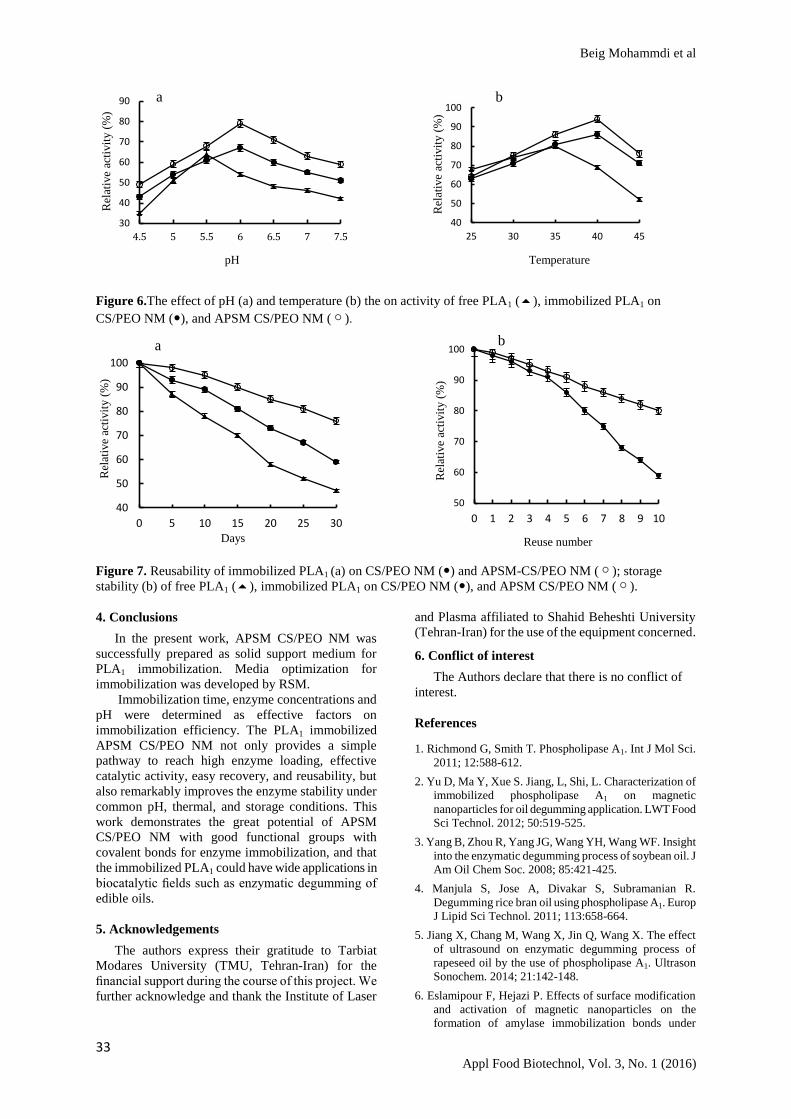
3.3.1. Effect of pH and temperature
The effect of media pH on the activity of free and
immobilized PLA1 was studied in the pH range of 4.5–
7.5, and the results are shown in Figure 6a. The
optimum pH for the free and immobilized PLA1 on
APSM CS/PEO NM was 5.5 and 6.0, with 64%
and 78% relative activity, respectively. Compared to
the free PLA1, the pH-activity profile of the
immobilized enzyme showed an upward shift in pH
with considerable broadening due to greater pH
stability. This phenomenon might be due to the
immobilized enzyme molecules preserved from the
conformational alterations induced by pH changes and
the enzyme can be more active in the wide range of
pH values [2].
This shift was attributed to the partitioning effects
of the supports. Since the reaction medium and the
insoluble support are two different phases, the
depletion in one of the phases is possible. When this
occurs, the immediate microenvironment of the
enzyme on the surface of the support material will be
different from that of the bulk solution. Similar results
have been reported by other researchers [2,31]. The
broadened pH/activity profile of immobilized PLA1
may have important benefits when developing the
industrial applications. The optimum temperature for
the free PLA1 was 50ºC, while the maximum activity
of the immobilized PLA1 was at 60°C (Figure 6b).
The shift in optimal temperature toward higher
values could be due to the immobilization of the
enzyme and resulted in the formation of the enzyme–
substrate complex, hindering the access of substrates
to the active sites and covalent bond between the
enzyme and support medium. There are many
advantages of running bioprocesses at elevated
temperatures such as higher diffusion rates, lower
substrate viscosities, increased reactant solubility, and
reduced risk of microbial contamination [23,31].
The PLA1 immobilized on the control CS/PEO
NM had intermediate state between the free and the
PLA1 immobilized on APSM CS/PEO NM in the
effect of pH and temperature. Indeed, it shows that
unmodified CS/PEO NM has functional groups too.
These groups can make a covalent attachment between
the nanofibers and enzyme; however, this ability can
be extremely improved by treating with plasma.
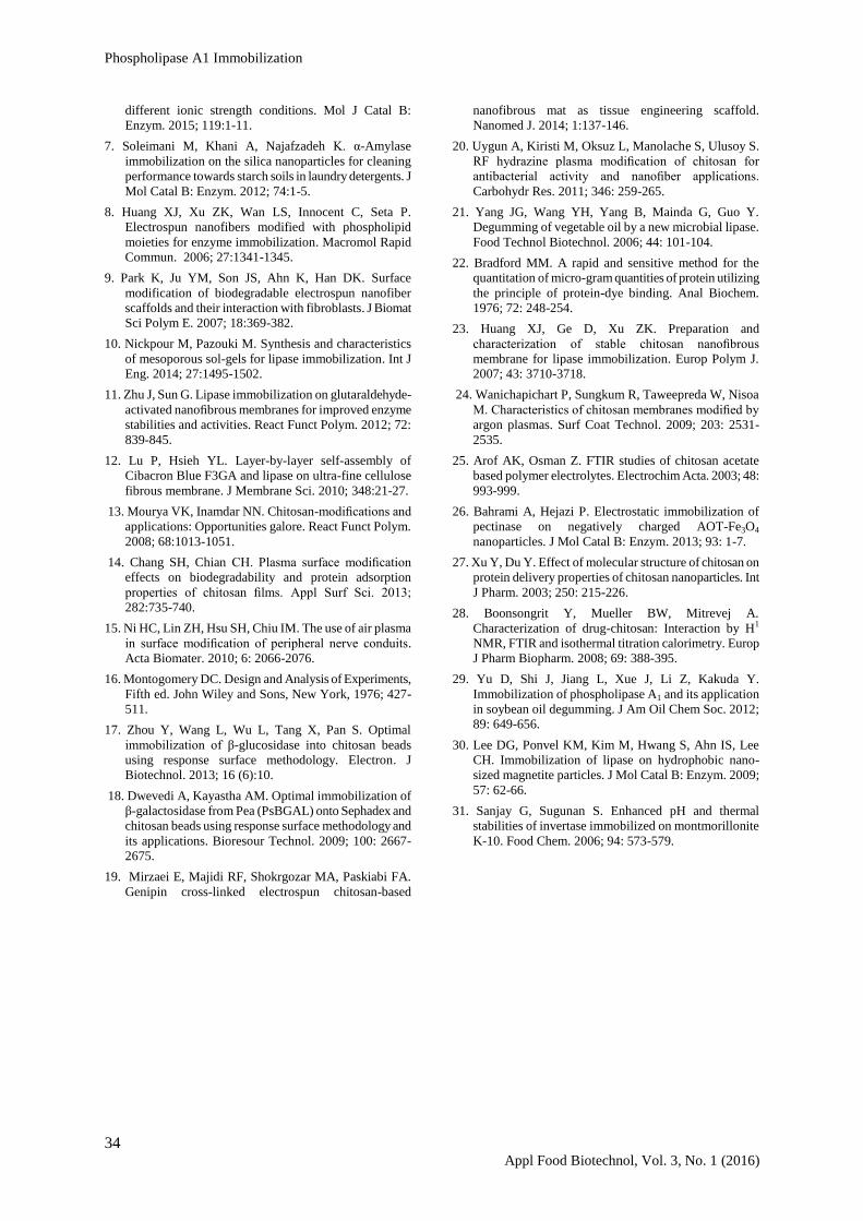
3.3.2. Reusability and storage stability
Rapid decrease of catalytic activity during the
storage time and difficult recovery after catalytic
reactions limit broader applications of free enzymes.
Thus, a variety of solid support media have been
developed for enzyme immobilization to overcome
these drawbacks. Figure 7a shows that the activity of
the immobilized PLA1 on APSM CS/PEO NM
decreases gradually during the repeated tests, and 80%
of its initial activity is still retained after 10 cycles,
while the PLA1 immobilized on control CS/PEO NM
shows a greater reduction of its initial activity after 10
cycles.
With repeated use of the immobilized PLA1, the
strength of binding between the matrix and the
enzyme get weakened, leading to loss in the enzyme
activity. Frequent encountering of the substrate into
the active site causes its distortion, and thus its
catalytic efficiency is reduced. In addition, the free
and immobilized PLA1 were kept in phosphate-citrate
buffer at 4ºC for 30 days, and their retained activities
were tested periodically to examine their storage
stability. As shown in Figure 7b, the free PLA1 has
lost more than 50% of its initial activity after 30 days,
while the retained activity of the immobilized PLA1 on
APSM and original CS/PEO NM were 76% and 59%
of their initial activity, respectively, during the same
period.
These results indicate that the enzyme is bound to
the plasma-modified chitosan with higher strength;
hence, it is not leached out with repeated washings
[18]. These attractive results demonstrate the great
potential of using CS/PEO nanofibers for enzyme
immobilization and related biocatalytic applications.
Eff
icie
ncy
(%
)
Immobilization time (h) pH
Beig Mohammdi et al
33 Appl Food Biotechnol, Vol. 3, No. 1 (2016)
Figure 6.The effect of pH (a) and temperature (b) the on activity of free PLA1 (), immobilized PLA1 on
CS/PEO NM (), and APSM CS/PEO NM ().
Figure 7. Reusability of immobilized PLA1 (a) on CS/PEO NM () and APSM-CS/PEO NM (); storage
stability (b) of free PLA1 (), immobilized PLA1 on CS/PEO NM (), and APSM CS/PEO NM ().
4. Conclusions
In the present work, APSM CS/PEO NM was
successfully prepared as solid support medium for
PLA1 immobilization. Media optimization for
immobilization was developed by RSM.
Immobilization time, enzyme concentrations and
pH were determined as effective factors on
immobilization efficiency. The PLA1 immobilized
APSM CS/PEO NM not only provides a simple
pathway to reach high enzyme loading, effective
catalytic activity, easy recovery, and reusability, but
also remarkably improves the enzyme stability under
common pH, thermal, and storage conditions. This
work demonstrates the great potential of APSM
CS/PEO NM with good functional groups with
covalent bonds for enzyme immobilization, and that
the immobilized PLA1 could have wide applications in
biocatalytic fields such as enzymatic degumming of
edible oils.
5. Acknowledgements
The authors express their gratitude to Tarbiat
Modares University (TMU, Tehran-Iran) for the
financial support during the course of this project. We
further acknowledge and thank the Institute of Laser
and Plasma affiliated to Shahid Beheshti University
(Tehran-Iran) for the use of the equipment concerned.
6. Conflict of interest
The Authors declare that there is no conflict of
interest.
References
1. Richmond G, Smith T. Phospholipase A1. Int J Mol Sci.
2011; 12:588-612.
2. Yu D, Ma Y, Xue S. Jiang, L, Shi, L. Characterization of
immobilized phospholipase A1 on magnetic
nanoparticles for oil degumming application. LWT Food
Sci Technol. 2012; 50:519-525.
3. Yang B, Zhou R, Yang JG, Wang YH, Wang WF. Insight
into the enzymatic degumming process of soybean oil. J
Am Oil Chem Soc. 2008; 85:421-425.
4. Manjula S, Jose A, Divakar S, Subramanian R.
Degumming rice bran oil using phospholipase A1. Europ
J Lipid Sci Technol. 2011; 113:658-664.
5. Jiang X, Chang M, Wang X, Jin Q, Wang X. The effect
of ultrasound on enzymatic degumming process of
rapeseed oil by the use of phospholipase A1. Ultrason
Sonochem. 2014; 21:142-148.
6. Eslamipour F, Hejazi P. Effects of surface modification
and activation of magnetic nanoparticles on the
formation of amylase immobilization bonds under
30
40
50
60
70
80
90
4.5 5 5.5 6 6.5 7 7.5
Rel
ativ
e ac
tivit
y (
%)
40
50
60
70
80
90
100
25 30 35 40 45
Rel
ativ
e ac
tivit
y (
%)
40
50
60
70
80
90
100
0 5 10 15 20 25 30
Rel
ativ
e ac
tivit
y (
%)
Days
50
60
70
80
90
100
0 1 2 3 4 5 6 7 8 9 10
Rel
ativ
e ac
tivit
y (
%)
b
b
a
Temperature
b
pH
Reuse number
a
Phospholipase A1 Immobilization
34 Appl Food Biotechnol, Vol. 3, No. 1 (2016)
different ionic strength conditions. Mol J Catal B:
Enzym. 2015; 119:1-11.
7. Soleimani M, Khani A, Najafzadeh K. α-Amylase
immobilization on the silica nanoparticles for cleaning
performance towards starch soils in laundry detergents. J
Mol Catal B: Enzym. 2012; 74:1-5.
8. Huang XJ, Xu ZK, Wan LS, Innocent C, Seta P.
Electrospun nanofibers modified with phospholipid
moieties for enzyme immobilization. Macromol Rapid
Commun. 2006; 27:1341-1345.
9. Park K, Ju YM, Son JS, Ahn K, Han DK. Surface
modification of biodegradable electrospun nanofiber
scaffolds and their interaction with fibroblasts. J Biomat
Sci Polym E. 2007; 18:369-382.
10. Nickpour M, Pazouki M. Synthesis and characteristics
of mesoporous sol-gels for lipase immobilization. Int J
Eng. 2014; 27:1495-1502.
11. Zhu J, Sun G. Lipase immobilization on glutaraldehyde-
activated nanofibrous membranes for improved enzyme
stabilities and activities. React Funct Polym. 2012; 72:
839-845.
12. Lu P, Hsieh YL. Layer-by-layer self-assembly of
Cibacron Blue F3GA and lipase on ultra-fine cellulose
fibrous membrane. J Membrane Sci. 2010; 348:21-27.
13. Mourya VK, Inamdar NN. Chitosan-modifications and
applications: Opportunities galore. React Funct Polym.
2008; 68:1013-1051.
14. Chang SH, Chian CH. Plasma surface modification
effects on biodegradability and protein adsorption
properties of chitosan films. Appl Surf Sci. 2013;
282:735-740.
15. Ni HC, Lin ZH, Hsu SH, Chiu IM. The use of air plasma
in surface modification of peripheral nerve conduits.
Acta Biomater. 2010; 6: 2066-2076.
16. Montogomery DC. Design and Analysis of Experiments,
Fifth ed. John Wiley and Sons, New York, 1976; 427-
511.
17. Zhou Y, Wang L, Wu L, Tang X, Pan S. Optimal
immobilization of β-glucosidase into chitosan beads
using response surface methodology. Electron. J
Biotechnol. 2013; 16 (6):10.
18. Dwevedi A, Kayastha AM. Optimal immobilization of
β-galactosidase from Pea (PsBGAL) onto Sephadex and
chitosan beads using response surface methodology and
its applications. Bioresour Technol. 2009; 100: 2667-
2675.
19. Mirzaei E, Majidi RF, Shokrgozar MA, Paskiabi FA.
Genipin cross-linked electrospun chitosan-based
nanofibrous mat as tissue engineering scaffold.
Nanomed J. 2014; 1:137-146.
20. Uygun A, Kiristi M, Oksuz L, Manolache S, Ulusoy S.
RF hydrazine plasma modification of chitosan for
antibacterial activity and nanofiber applications.
Carbohydr Res. 2011; 346: 259-265.
21. Yang JG, Wang YH, Yang B, Mainda G, Guo Y.
Degumming of vegetable oil by a new microbial lipase.
Food Technol Biotechnol. 2006; 44: 101-104.
22. Bradford MM. A rapid and sensitive method for the
quantitation of micro-gram quantities of protein utilizing
the principle of protein-dye binding. Anal Biochem.
1976; 72: 248-254.
23. Huang XJ, Ge D, Xu ZK. Preparation and
characterization of stable chitosan nanofibrous
membrane for lipase immobilization. Europ Polym J.
2007; 43: 3710-3718.
24. Wanichapichart P, Sungkum R, Taweepreda W, Nisoa
M. Characteristics of chitosan membranes modified by
argon plasmas. Surf Coat Technol. 2009; 203: 2531-
2535.
25. Arof AK, Osman Z. FTIR studies of chitosan acetate
based polymer electrolytes. Electrochim Acta. 2003; 48:
993-999.
26. Bahrami A, Hejazi P. Electrostatic immobilization of
pectinase on negatively charged AOT-Fe3O4
nanoparticles. J Mol Catal B: Enzym. 2013; 93: 1-7.
27. Xu Y, Du Y. Effect of molecular structure of chitosan on
protein delivery properties of chitosan nanoparticles. Int
J Pharm. 2003; 250: 215-226.
28. Boonsongrit Y, Mueller BW, Mitrevej A.
Characterization of drug-chitosan: Interaction by H1
NMR, FTIR and isothermal titration calorimetry. Europ
J Pharm Biopharm. 2008; 69: 388-395.
29. Yu D, Shi J, Jiang L, Xue J, Li Z, Kakuda Y.
Immobilization of phospholipase A1 and its application
in soybean oil degumming. J Am Oil Chem Soc. 2012;
89: 649-656.
30. Lee DG, Ponvel KM, Kim M, Hwang S, Ahn IS, Lee
CH. Immobilization of lipase on hydrophobic nano-
sized magnetite particles. J Mol Catal B: Enzym. 2009;
57: 62-66.
31. Sanjay G, Sugunan S. Enhanced pH and thermal
stabilities of invertase immobilized on montmorillonite
K-10. Food Chem. 2006; 94: 573-579.