Embed Size (px)
Citation preview
Phase II Metabolism of 3,4-Methylenedioxymethamphetamine
Synthesis, Analysis, and
Enantioselective in vitro and in vivo Kinetics
Dissertation
zur Erlangung des Grades
des Doktors der Naturwissenschaften
der Naturwissenschaftlich-Technischen Fakultät III -
Chemie, Pharmazie, Bio- und Werkstoffwissenschaften
der Universität des Saarlandes
von
Andrea Eva Schwaninger
Saarbrücken
2011

Tag des Kolloquiums: 05.12.2011
Dekan: Univ.-Prof. Dr. Wilhelm F. Maier
Berichterstatter: Univ.-Prof. Dr. Dr. h.c. Hans H. Maurer
Univ.-Prof. Dr. Rolf W. Hartmann
Vorsitz: Univ.-Prof. Dr. Feit Flockerzi
Akad. Mitarbeiter: Dr. Josef Zapp


Die folgende Arbeit entstand unter der Anleitung von Herrn Professor Dr. Dr. h.c. Hans H. Maurer in der Abteilung Experimentelle und Klinische Toxikologie der Fachrichtung 2.4 Experimentelle und Klinische Pharmakologie und Toxikologie der Universität des Saarlandes in Homburg/Saar von März 2008 bis Juni 2011.
Mein besonderer Dank gilt:
Herrn Prof. Dr. Dr. h.c. Hans H. Maurer für die herzliche Aufnahme in seinen Arbeitskreis, die Vergabe dieses interessanten und abwechslungsreichen, aber auch herausfordernden Dissertationsthemas, die Möglichkeit des selbstständigen Arbeitens, der aktiven Teilnahme an nationalen und internationalen Fachkongressen und seine ständige Diskussionsbereitschaft,
Herrn Univ.-Prof. Dr. Rolf W. Hartmann für die Übernahme des Koreferats,
Frau Prof. Dr. Dr. h.c. Marilyn A. Huestis für die Bereitstellung der Urinproben aus der kontrollierten MDMA-Studie und Ihre Begeisterung für das gemeinsame Projekt,
Herrn Dr. Markus R. Meyer für seinen Humor, seine Lebensweisheiten und Theorien, die immer für Gelächter und Unterhaltung gesorgt haben, sowie für seine außerordentliche wissenschaftliche Expertise, und ständige Diskussionsbereitschaft,
Herrn PD Dr. Frank T. Peters für seine aufmunternden Worte, seinen Humor, sowie für fachliche Diskussionen aller Art,
meinen Kolleginnen und Kollegen, für ihre Freundschaft und Hilfsbereitschaft während der Arbeits- und Dienstzeit, und für viele schöne gemeinsam verbrachte Stunden, die die Zeit der Promotion einzigartig gemacht haben,
Herrn Armin Weber für seine Gelassenheit, Ruhe, ständige Einsatzbereitschaft, Wartung und Reparatur der Messgeräte, sowie seinen Rat bei technischen Fragestellungen,
Frau Gabriele Ulrich und Herrn Carsten Schröder für gewissenhaft ausgeführte Laborarbeiten und Betreuung der Messgeräte sowie ein immer offenes Ohr für Probleme aller Art,
Herrn Dr. Josef Zapp für die Messung und Mithilfe bei der Interpretation der NMR-Spektren,
Herrn Dr. Stefan Böttcher für das Ausleihen der chiralen Säule,
Herrn Dr. Peter Wollenberg für seine Diskussionsbereitschaft bei statistischen Problemen,
meinem Freund Christian, der immer für mich da war und an mich geglaubt hat,
meinen Eltern, die mich in meinem Tun gefördert und unterstützt haben,
meinen Freunden und Bekannten, die in den letzten Jahren des Öfteren ohne mich zusammenkommen mussten und mich trotz allem nicht vergessen haben.


Für Euch!


It is our choices (…) that show
what we truly are,
far more than our abilities.
Harry Potter and the Chamber of Secrets

TABLE OF CONTENTS
1 GENERAL PART ......................................................................................... 1
1.1 Introduction .......................................................................................................... 1
1.1.1 3,4-Methylenedioxymethamphetamine ..................................................... 1
1.1.2 Pharmacology and Toxicology ................................................................. 1
1.1.3 Metabolism ............................................................................................... 2
1.1.4 Phase II Metabolizing Enzymes ............................................................... 4
1.1.4.1 UDP-Glucuronyltransferase (UGT) ........................................................... 4 1.1.4.2 Sulfotransferase (SULT) ........................................................................... 5
1.1.5 Synthesis of Phase II Metabolites ............................................................ 7
1.1.5.1 Glucuronides ............................................................................................ 7 1.1.5.2 Sulfates ..................................................................................................... 7
1.1.6 (Enantioselective) In vitro Enzyme Kinetic Studies ................................... 8
1.1.6.1 Product formation approach ..................................................................... 9 1.1.6.2 Substrate depletion approach ................................................................... 9
1.2 Aims and Scopes ............................................................................................... 11
2 PUBLICATIONS OF THE RESULTS ........................................................ 13
2.1 The Role of Human UGT-Glucuronyltransferases on the Formation of the Methylenedioxymethamphetamine (Ecstasy) Phase II Metabolites R- and S-3-Methoxymethamphetamine 4-O-Glucuronides57 (DOI: 10.1124/dmd.109.029215) .................................................................................. 13
2.2 Sulfation of the 3,4-Methylenedioxymethamphetamine (MDMA) Metabolites 3,4-Dihydroxymethamphetamine (DHMA) and 4-Hydroxy-3-Methoxymethamphetamine (HMMA) and their Capability to Inhibit Human Sulfotransferases58 (DOI: 10.1016/jtoxlet.2011.01.026) ................................... 15
2.3 Investigation on the Enantioselectivity of the Sulfation of the Methylenedioxymethamphetamine (MDMA) Metabolites 3,4-Dihydroxymethamphetamine (DHMA) and 4-Hydroxy-3-Methoxymethamphetamine (HMMA) using the Substrate Depletion Approach59 (DOI: 10.1124/dmd.111.041129) .................................................... 17
2.4 Development and Validation of LC-HRMS and GC-NICI-MS Methods for Stereoselective Determination of MDMA and its Phase I and II Metabolites in Human Urine60 (DOI: 10.1002/jms.1929) .......................................................... 19
2.5 Human MDMA and Phase I and Phase II Metabolite Urinary Excretion Kinetics Following Controlled MDMA Administration61 (DOI: 10.1373/clinchem.2011.172254) ........................................................................ 21
2.6 Stereoselective Urinary MDMA and Metabolites Excretion Kinetics following Controlled MDMA Administration to Humans62 (DOI: 10.1016/j.bcp.2011.09.023) ................................................................................ 23
3 CONCLUSIONS ......................................................................................... 25
4 SUMMARY ................................................................................................. 27
5 REFERENCES ........................................................................................... 29
6 ABBREVIATIONS ...................................................................................... 33
7 ZUSAMMENFASSUNG ............................................................................. 35
1 GENERAL PART
1.1 INTRODUCTION
1.1.1 3,4-Methylenedioxymethamphetamine
3,4-Methylenedioxymethamphetamine (MDMA), commonly named as Ecstasy, is a
ring-substituted amphetamine with structural similarities to methamphetamine and
mescaline. As other amphetamines, MDMA is a chiral compound carrying an
asymmetric carbon atom in the side chain. It was first synthesized in Germany by
Merck in 19141,2 and, although patented as an appetite suppressant, never marketed
as a therapeutic drug.3 Since 1985, MDMA is scheduled in the Controlled Drugs and
Substances Act as a restricted drug in the United States and since 1986, in
Germany. It has become popular in the beginning of the 1990s as a drug of abuse
among young people, especially in the dance scene.4,5 After decreasing numbers of
MDMA users in recent years, most likely due to its non-availability on the illicit drug
market, the Substance Abuse and Mental Health Services Administration has
reported on increasing MDMA consumption in the United States again since 2010.6
Usually it is consumed recreationally on weekends (1 to 2 pills of 75 to 120 mg every
1 to 4 weeks) in form of tablets or pills.7 Preparations available on the illicit drug
market usually contain the 1:1 racemate of R- and S-enantiomers.
1.1.2 Pharmacology and Toxicology
Similar to amphetamine or methamphetamine, MDMA acts in the central nervous
system (CNS) as a stimulant through indirect release of monoamine
neurotransmitters from presynaptic nerve terminals into the synaptic cleft where
postsynaptic receptors can be stimulated.3,7 Mainly serotonergic (5-HT),
noradrenergic (NA), and with a smaller effect dopaminergic (DA) neurotransmission
is enhanced.
The distinctive effects are described as an altered state of consciousness, euphoria,
energy and a desire to socialize.3,8 However, MDMA also can induce severe acute
- 1 -
toxic symptoms, such as tachycardia, hypertension, hyperthermia, and
hepatotoxicity. Severe and even fatal intoxications were described.3
Concerning chronic toxicity, preclinical animal data suggest that MDMA causes
irreversible damage to serotonergic nerve terminals in the CNS.3,9-11 In humans,
chronic MDMA toxicity is still controversially discussed, as some recent publications
suggest that animal doses may be too high compared to human
pharmacokinetics.12,13 Other studies with recreational MDMA users, found decreased
levels of 5-hydroxyindoleacetic acid, the main metabolite of 5-HT in the cerebrospinal
fluid14 and a reduced density of serotonin transporters in the brain as determined by
positron emission computed tomography with a ligand selective for these
transporters.15 Unfortunately, these studies were performed with recreational users,
so it cannot be excluded that the indicated neurotoxicity might also be due to use of
other recreational drugs especially since polydrug use is not uncommon. Admittedly,
direct MDMA injection into rat brain failed to reproduce neurotoxic effects seen after
systemic administration.16 Furthermore, alteration of cytochrome P450 (CYP)-
mediated MDMA metabolism influenced MDMA-induced neurotoxicity.16,17 Therefore,
MDMA metabolism may be an important contributor to neurotoxicity.18-21 Metabolites
such as 3,4-dihydroxymethamphetamine (DHMA) can easily be oxidized to their
corresponding quinones which can form adducts with glutathione and other thiol-
containing compounds.18-20 Recently, such adducts have been implicated in MDMA
neurotoxicity.22,23
For the two enantiomers, different pharmacological properties were observed.3 While
S-MDMA is generally more potent and responsible for the described psychostimulant
and empathic effects, the R-isomer exhibits more hallucinogenic-type properties.10 R-
and S-MDMA also differ in their dose-response curves for changes in serotonergic
function and neurotoxicity and their in vivo kinetics are known to be different.3,8,24-27
1.1.3 Metabolism
In vivo and in vitro MDMA studies revealed two main metabolic pathways as shown
in Figure 1. The predominant pathway in humans involves multiple CYP enzyme-
catalyzed O-demethylenation of MDMA to DHMA, followed by catechol-O-
methyltransferase (COMT)-catalyzed O-methylation, primarily to 4-hydroxy-3-
methoxymethamphetamine (HMMA). DHMA and HMMA also may be conjugated by
- 2 -
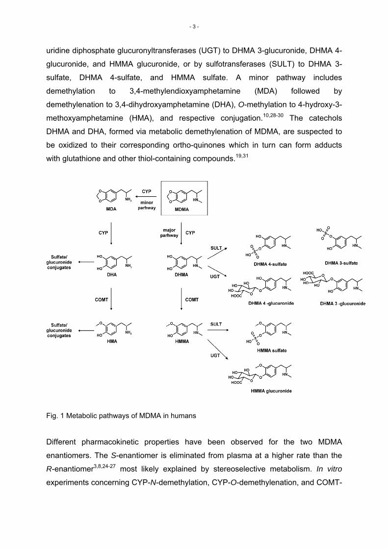
uridine diphosphate glucuronyltransferases (UGT) to DHMA 3-glucuronide, DHMA 4-
glucuronide, and HMMA glucuronide, or by sulfotransferases (SULT) to DHMA 3-
sulfate, DHMA 4-sulfate, and HMMA sulfate. A minor pathway includes
demethylation to 3,4-methylendioxyamphetamine (MDA) followed by
demethylenation to 3,4-dihydroxyamphetamine (DHA), O-methylation to 4-hydroxy-3-
methoxyamphetamine (HMA), and respective conjugation.10,28-30 The catechols
DHMA and DHA, formed via metabolic demethylenation of MDMA, are suspected to
be oxidized to their corresponding ortho-quinones which in turn can form adducts
with glutathione and other thiol-containing compounds.19,31
Fig. 1 Metabolic pathways of MDMA in humans
Different pharmacokinetic properties have been observed for the two MDMA
enantiomers. The S-enantiomer is eliminated from plasma at a higher rate than the
R-enantiomer3,8,24-27 most likely explained by stereoselective metabolism. In vitro
experiments concerning CYP-N-demethylation, CYP-O-demethylenation, and COMT-
- 3 -
methylation of DHMA to HMMA indeed revealed metabolic preferences for the S-
enantiomers.32,33
1.1.4 Phase II Metabolizing Enzymes
Numerous enzymes are capable to metabolize xenobiotics, usually resulting in
decreased toxicity and increased hydrophilicity compared to the parent compounds,
which promotes their excretion. Generally, these biotransformations can be divided in
two steps: phase I and phase II metabolism. Phase I metabolism is referred to as
functionalization which mainly involves oxidation, reduction, or hydrolysis. Phase II
type reactions are conjugative reactions, catalyzing among others, the transfer of
hydrophilic residues such as glucuronic acid or activated sulfate. However,
conjugation is not necessarily a secondary phase reaction as many endogenous
compounds or xenobiotics can be directly glucuronidated or sulfated.
1.1.4.1 UDP-Glucuronyltransferase (UGT)
UGTs represent a superfamily of endoplasmic reticulum membrane-bound enzymes,
postulated to reside on the luminal surface. Based on primary amino acid identity,
they are divided into two families, UGT1 and UGT2. At present, 15 different
isoenzymes are known in humans: UGT1A1, UGT1A3, UGT1A4, UGT1A5, UGT1A6,
UGT1A7, UGT1A8, UGT1A9, and UGT1A10 and UGT2B4, UGT2B7, UGT2B10,
UGT2B11, UGT2B15, and UGT2B17,34-36 whereas UGT1A1, 1A3, 1A4, 1A6, 1A9,
2B7, and 2B15 are considered to be of greatest importance in hepatic drug
elimination.36 Although the liver is recognized as the major site of glucuronidation,
numerous organs, e.g. small intestine, lung, kidney, brain, etc. significantly contribute
to the overall glucuronidation capacity.
- 4 -
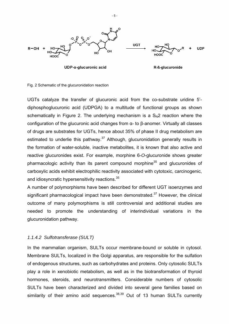
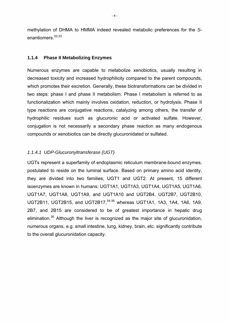
Fig. 2 Schematic of the glucuronidation reaction
UGTs catalyze the transfer of glucuronic acid from the co-substrate uridine 5’-
diphosphoglucuronic acid (UDPGA) to a multitude of functional groups as shown
schematically in Figure 2. The underlying mechanism is a SN2 reaction where the
configuration of the glucuronic acid changes from α- to β-anomer. Virtually all classes
of drugs are substrates for UGTs, hence about 35% of phase II drug metabolism are
estimated to underlie this pathway.37 Although, glucuronidation generally results in
the formation of water-soluble, inactive metabolites, it is known that also active and
reactive glucuronides exist. For example, morphine 6-O-glucuronide shows greater
pharmacologic activity than its parent compound morphine35 and glucuronides of
carboxylic acids exhibit electrophilic reactivity associated with cytotoxic, carcinogenic,
and idiosyncratic hypersensitivity reactions.35
A number of polymorphisms have been described for different UGT isoenzymes and
significant pharmacological impact have been demonstrated.37 However, the clinical
outcome of many polymorphisms is still controversial and additional studies are
needed to promote the understanding of interindividual variations in the
glucuronidation pathway.
1.1.4.2 Sulfotransferase (SULT)
In the mammalian organism, SULTs occur membrane-bound or soluble in cytosol.
Membrane SULTs, localized in the Golgi apparatus, are responsible for the sulfation
of endogenous structures, such as carbohydrates and proteins. Only cytosolic SULTs
play a role in xenobiotic metabolism, as well as in the biotransformation of thyroid
hormones, steroids, and neurotransmitters. Considerable numbers of cytosolic
SULTs have been characterized and divided into several gene families based on
similarity of their amino acid sequences.38,39 Out of 13 human SULTs currently
- 5 -
known, the major isoforms responsible for human xenobiotic metabolism are
SULT1A1, SULT1A3, SULT1B1, SULT1E1, and SULT2A1.40 The widest tissue
distribution was shown for the SULT1A subfamily, with SULT1A1 as the major
isoform present in human liver, but also in the gastrointestinal tract, brain and
placenta.39,40 SULT1A3 is known to be only scarcely expressed in human liver,
however highly expressed in the small intestine, brain and fetal liver.39,40
SULTs catalyze the transfer of a sulfonate group from 3’-phosphoadenosine-5’-
phosphosulfate (PAPS) to nucleophilic sites of their substrates. Sulfation is a high
affinity and low capacity phase II reaction, with overlapping substrates spectra for
glucuronidation. Sulfation predominates at low substrate concentrations and
glucuronidation at high substrate concentrations, when sulfation is saturated.39 The
limiting factor for sulfation is the availability of PAPS. Although it can be rapidly
synthesized, it depends on the hepatic sulfate concentrations, which are largely
dependent on equilibrium with circulating inorganic sulfate.39
Fig. 3 Schematic of the sulfation reaction
Generally, sulfation is a detoxification process, however, labile and chemically
reactive intermediates are sometimes formed, which can undergo DNA binding,
leading to mutagenicity and carcinogenicity. Some sulfate esters including minoxidil,
triamterene and morphine were reported to be more pharmacologically active than
the corresponding parent drugs.41 At least some endogenous sulfate conjugates
seem to play a role in the CNS. For example, dopamine 4-sulfate demonstrated
vasopressor activity in the peripheral and central nervous system, whereas dopamine
3-sulfate acted as a central depressor.41 Several xenobiotics, among them dietary
and environmental chemicals, therapeutic drugs, etc. were shown to inhibit one or
more SULT isoenzymes and may cause adverse effects on human health.41
- 6 -
1.1.5 Synthesis of Phase II Metabolites
Reference standards of metabolites are needed for in vitro and in vivo kinetic studies.
However, the number of commercially available glucuronide or sulfate standards is
limited, hence it usually requires their synthesis prior to kinetic studies.
1.1.5.1 Glucuronides
Synthesis of glucuronides can be achieved either by chemical42,43 or enzymatic
methods.44 Chemical synthesis requires multiple steps, most commonly via acyl-
protected intermediates. Hydrolytic stability of the aglycones is therefore a
prerequisite necessary for the removal of protecting groups. α-Anomers and other
byproducts in addition to the desired β-anomer can occur leading to more
complicated purifications and low yields. In the case of aglycones that contain
several possible glucuronidation sites, without further protecting groups mono- and
polyglucuronides can be formed.42,43,45 Shima et al.46 previously synthesized HMMA
O-glucuronide by chemical synthesis achieving yields of 6%, which seems rather low.
Enzyme-assisted synthesis represents a suitable alternative to chemical synthesis,
especially when milligram scale yields are sufficient. Isolated purified UGT enzymes
or liver microsomes might be applied as convenient catalysts for glucuronidation.
However, liver microsomes of different species (rat, mouse, dog, monkey, human)
seems most appropriate due to easy preparation and handling. Use of enzymes does
not require multiple steps and results in the formation of the natural configuration.
Mainly mono-glucuronides and even regio- and stereoselective glucuronides are
obtained.45 Yields with up to 100% depending on the aglycone and the microsomal
source used could be reached.44,45 Therefore, an enzyme-assisted synthesis was
chosen to produce milligram amounts of the diastereomeric HMMA glucuronides as
described in detail under 2.1.
1.1.5.2 Sulfates
Synthesis of sulfate conjugates is usually performed with chemical methods. Only
few data using enzymatic synthesis are available.47-49 Although enzymatic sulfate
synthesis bears the advantages of regio- and stereoselective conjugation, there are
some major drawbacks limiting its usefulness. The main issue is the need for the co-
substrate. PAPS is rather expensive and unstable. The formed product 3’-
- 7 -
phosphoadenosine 5’-phosphate (PAP) leads to product inhibition.49 Incubations with
subcellular fractions and the addition of PAPS therefore provide only low sulfation
capacities. Uutela et al used rat liver S9 fractions with the addition of PAPS for
regioselective sulfation of 5-HT, 5-HIAA, DOPAC, and HVA. However, the yields
were less than 3 mg (less than 10%) and hence too low for NMR confirmation of the
sulfation side.47 Chemical synthesis seems to be the method of choice for sulfate
synthesis of xenobiotics.47,48 Different strategies have been described, e.g. use of
sulfuric acid47,48 or sulfur trioxide-amine complexes.46,48 As H2SO4 is not amenable to
sulfation for many sensitive scaffolds considering the strong acidity of sulfuric acid,
SO3 adducts with amine containing molecules link pyridine, trimethylamine,
triethylamine, or DMF provide the most straightforward method.48 Usually, yields with
up to 90% could be achieved. Sulfates of DHMA and HMMA were synthesized using
a pyridine SO3 complex as described in detail under 2.2.
1.1.6 (Enantioselective) In vitro Enzyme Kinetic Studies
The characterization of humane enzymes involved in the metabolism of specific
drugs and the determination of their enzyme kinetic parameters, such as KM and Vmax
is an important aspect in toxicological risk assessment. They can be used as
potential determinants of interindividual variability in pharmacokinetics, e.g. drug-drug
interactions or genetic polymorphisms. KM and Vmax values represent descriptors of
the enzyme kinetic behavior of a respective biotransformation reaction. Assuming
simple kinetic systems, Vmax is the maximum enzyme velocity at an infinite substrate
concentration and in general represents the capacity of an enzymatic reaction. The
KM value is defined as the substrate concentration that will yield a reaction velocity
that is half of Vmax and reflects the substrate affinity to a certain enzyme. The overall
effectiveness of a respective reaction is usually described by the Vmax/KM ratio and
should increase the higher this ratio is. This fact sounds reasonable, as the catalytic
efficiency value is getting higher with increasing affinity (low KM) and increasing
velocity (high Vmax). Concerning differences in metabolic clearance of R- or S-
stereoisomers, enantioselectivity can also be evaluated via the Vmax/KM values and
marked enantioselectivity was previously defined as Vmax/KM(S-stereoisomer)/Vmax/KM(R-
stereomer) > 1.5 or < 0.67).32
- 8 -
1.1.6.1 Product formation approach
Conventional determinations of enzyme kinetic parameters are made by assessing
the rate of product (metabolite) formation at several substrate concentrations.
Therefore, methods are required for measurement of metabolite concentrations in in
vitro matrices. Such analytical methods themselves require that metabolites have
been definitely identified, suitable chromatographic separation has been established
and authentic standards prepared.50 The simplest model to describe enzymatic
biotransformation and hence to calculate KM and Vmax is fitting the initial rate velocities
at various substrate concentrations to the Michaelis Menten equation (eq. 1).
][][max
SKSVV
m +×
=
A prerequisite are “initial” rate conditions, meaning protein concentrations and
incubation time should be within the linear range of metabolite formation, and in total
less than 20% of substrate should be consumed.
1.1.6.2 Substrate depletion approach
An alternative to the measurement of product formation is the determination of
substrate depletion, which was successfully used for CYP reactions in both, human
liver microsomes (HLM) and recombinant enzymes.50,51 Substrate consumption over
time can be used to calculate initial substrate depletion rates (kdep) at various
substrate concentrations. In theory, when substrate concentrations are well below
KM, the depletion should follow first-order decay kinetics.52 As the substrate
concentration is elevated through the KM value, the measured values for kdep should
decline and become more zero-order in character. The infliction point of this
relationship represents the KM value and should occur at a substrate concentration
that yields a kdep value that is half of the theoretical maximum kdep at an infinitesimally
low-substrate concentration (kdep([S]=0)).50 Plotting of kdep values versus substrate
concentrations allows calculation of KM according to equation 2.50
)][
][1()0]([SK
SSKKm
depdep +−×==
(1)
(2)
- 9 -
The theoretical validity of this approach has been confirmed by Nath and Atkins,53
who showed on a simulated data set that equation 2 can be derived from the
Michaelis-Menten equation (eq. 1) and, as such, the kinetic parameters obtained
should be comparable with those obtained by the traditional product-formation
approach. The major advantage of the substrate-depletion approach is that reference
standards of metabolites are not required. For some analytes, when
(enantioselective) chromatographic separation of metabolites could not be
accomplished sufficiently, (chiral) measurement of substrate consumption might be a
versatile alternative to the conventional product formation. However, the substrate
depletion approach possesses some practical limitations.50 Substrates exhibiting low-
intrinsic clearance will be difficult to examine, since measurement of substrate
depletion requires a substantial consumption of the initial substrate concentration
during the incubation period. Furthermore, enzyme kinetics of formation of individual
metabolites cannot be determined, as the KM and Vmax values would only represent
the sum of kinetic parameters for all single metabolic pathways.
- 10 -
1.2 AIMS AND SCOPES
Phase II metabolism represents an important detoxification process.34,35,38
Investigation of glucuronidation and sulfation as a secondary metabolic step is
especially important concerning the detoxification of reactive phase I metabolites.
Such metabolites are known to be formed in humans after ingestion of MDMA,
mainly through demethylenation to the catecholic metabolite DHMA and are
suspected to contribute to MDMA’s neurotoxic effects.18,19,19,20,54 The qualitative and
quantitative phase I metabolism of MDMA was studied extensively in vitro and in
vivo.10,28-30,32,33,55 Several pharmacokinetic studies in blood and urine following
controlled MDMA administration to humans were performed, but DHMA, HMMA,
and/or HMA urinary pharmacokinetic data were only obtained after conjugate
cleavage. Only Shima et al. determined intact HMMA conjugates in 25 random urine
samples and found that more than 70% of HMMA was eliminated as glucuronide or
sulfate.30 However, neither systematic in vivo nor in vitro kinetic studies were
available concerning glucuronidation and sulfation of MDMA’s phase I metabolites.
Furthermore, different pharmacological and pharmacokinetic properties were
observed for the two enantiomers of MDMA3,8,24-26 and enantiomeric preferences in
the phase I metabolism were observed in vitro32,33 and in vivo.27 Elucidation whether
the phase II metabolism also contributes to this phenomenon is important from the
toxicological and pharmacological point of view.
Besides this, MDMA is known to be a potent mechanism-based inhibitor of
CYP2D656 which is also assumed to influence MDMA-induced neurotoxicity.16,17
DHMA was also shown to inhibit its own metabolism as well as the methylation of
dopamine.33 The inhibition potential of MDMA and/or its metabolites on other
metabolic enzymes, such as UGTs or SULTs, is still unknown.
- 11 -
Therefore, the aims of the presented studies were:
- (Bio)Synthesis of MDMA’s main phase II metabolites as reference standards for
quantitative in vitro and in vivo kinetic studies
- Investigation of stereoselective enzyme kinetic data in vitro for HMMA
glucuronidation in HLM and recombinant UGTs, and DHMA and HMMA sulfation
in human liver cytosol (HLC) and recombinant SULT
- Determination of the inhibition potential of MDMA, DHMA, and HMMA on SULT
- Development and full validation of gas chromatography-mass spectrometry (GC-
MS) and liquid chromatography-mass spectrometry (LC-MS) methods allowing
the stereoselective analysis of MDMA, its phase I and phase II metabolites in
human urine
- Evaluation of MDMA’s phase II metabolites elimination kinetics in human urine
following controlled oral MDMA administration
- Determination of stereoselective elimination kinetics of MDMA and its phase I and
II metabolites in human urine following controlled oral MDMA administration
- 12 -
2 PUBLICATIONS OF THE RESULTS
The results of the studies were published in the following papers:
2.1 THE ROLE OF HUMAN UGT-GLUCURONYLTRANSFERASES ON THE
FORMATION OF THE METHYLENEDIOXYMETHAMPHETAMINE (ECSTASY) PHASE II METABOLITES R- AND S-3-METHOXYMETHAMPHETAMINE 4-O-GLUCURONIDES57 (DOI: 10.1124/DMD.109.029215)
- 13 -

2.2 SULFATION OF THE 3,4-METHYLENEDIOXYMETHAMPHETAMINE (MDMA) METABOLITES 3,4-DIHYDROXYMETHAMPHETAMINE (DHMA) AND 4-HYDROXY-3-METHOXYMETHAMPHETAMINE (HMMA) AND THEIR
CAPABILITY TO INHIBIT HUMAN SULFOTRANSFERASES58 (DOI: 10.1016/JTOXLET.2011.01.026)
- 15 -

2.3 INVESTIGATION ON THE ENANTIOSELECTIVITY OF THE SULFATION OF THE
METHYLENEDIOXYMETHAMPHETAMINE (MDMA) METABOLITES 3,4-DIHYDROXYMETHAMPHETAMINE (DHMA) AND 4-HYDROXY-3-METHOXYMETHAMPHETAMINE (HMMA) USING THE SUBSTRATE
DEPLETION APPROACH59 (DOI: 10.1124/DMD.111.041129)
- 17 -

2.4 DEVELOPMENT AND VALIDATION OF LC-HRMS AND GC-NICI-MS
METHODS FOR STEREOSELECTIVE DETERMINATION OF MDMA AND ITS
PHASE I AND II METABOLITES IN HUMAN URINE60 (DOI: 10.1002/JMS.1929)
- 19 -

2.5 HUMAN MDMA AND PHASE I AND PHASE II METABOLITE URINARY
EXCRETION KINETICS FOLLOWING CONTROLLED MDMA
ADMINISTRATION61 (DOI: 10.1373/CLINCHEM.2011.172254)
- 21 -

2.6 STEREOSELECTIVE URINARY MDMA AND METABOLITES EXCRETION
KINETICS FOLLOWING CONTROLLED MDMA ADMINISTRATION TO
HUMANS62 (DOI: 10.1016/J.BCP.2011.09.023)
- 23 -

3 CONCLUSIONS
The studies presented here provided systematic data on the in vitro glucuronidation
and sulfation kinetics of the designer drug 3,4-methylenedioxymethamphetamine,
(MDMA, Ecstasy). These data suggested, that sulfation was the predominant
conjugation step with regioselective sulfation of the catecholic metabolite DHMA in
position 3.57,58 Inhibition studies performed with MDMA, DHMA, and HMMA towards
typical sulfation reactions clearly indicated a mixed-type or competitive inhibition of
dopamine sulfation by DHMA and HMMA, respectively, with IC50 values likely to
cause significant inhibition in vivo after recreational MDMA doses.63 In the author’s
opinion, a part of the described neurotoxicity of MDMA3,9-11 could be explained by
inhibition of the dopamine sulfation in the CNS. As MDMA and related drugs are able
to increase the concentration of dopamine and other neurotransmitters in the CNS64
and as they additionally could inhibit the inactivation of these compounds,33 the
described dopamine induced neurotoxicity might be enhanced.65
Additionally, evaluation with respect to a possible enantioselective phase II
metabolism was performed. It could be shown, that HMMA glucuronidation by
UGT1A9 was markedly stereoselective with preferences for the formation of the S-
diastereomer whereas its glucuronidation by UGT2B7 favored the R-isomer.
UGT2B15 and UBT2B17 revealed only slight preferences for S-HMMA. In human
liver microsomes, which contain a physiological mixture of all liver UGT isoenzymes,
and should therefore reflect the in vivo situation, slight preferences for S-HMMA were
observed. Sulfation of HMMA was mainly catalyzed by SULT1A3 and to a minor
extent by SULT1E1. Neither for SULT1A3 nor in human liver cytosol enantiomeric
preferences could be observed. On the other hand, the efficiency for S-DHMA 3-
sulfate formation was twice as high as for its R-enantiomer, both in SULT1A3 and
human liver cytosol. One reason for this difference in enantioselectivity might be the
position for sulfation. DHMA was mainly sulfated in position 3, whereas HMMA could
only be sulfated in position 4.
To further obtain systematic in vivo data on MDMA’s phase II metabolism and its
enantioselectivity, liquid chromatography-high resolution mass spectrometry (LC-
HRMS) and gas chromatography-negative ion chemical ionization- mass
spectrometry (GC-NICI-MS) methods were successfully developed and validated.60
These methods were shown to be applicable for the analysis of urine samples of 10
- 25 -
human subjects collected for up to 7 days following controlled oral placebo, low, and
high dose MDMA administration.61,62 Human MDMA urinary metabolites are primarily
sulfate and glucuronide conjugates, with sulfates present in higher concentrations
than glucuronides. HMMA sulfate was shown to be the major urinary metabolite
providing the longest detection time for MDMA consumption with up to 168 h. All
metabolites exhibited changes in enantiomeric disposition over time. MDMA, DHMA,
and HMMA sulfate revealed preferences for the R-stereoisomers, all other
metabolites showed conversely more S-isomer within the first 24 h after ingestion.
Generally, initial stereoisomer preferences mimicked those observed in previous in
vitro experiments.32,33,57,59 In the later excretion phase (after 24 h), R/S ratios were >1
for all compounds. This is quite remarkable, as the enantiomeric ratios of at least one
metabolite should be reversed from that of MDMA. However, it must be considered
that urinary analysis reflects not only metabolite formation, but also distribution and
elimination processes. Metabolism is represented mainly within the first 12 to 24 h,
whereas later on, elimination is more relevant. One explanation for the observed
time-dependency could be substrate availability. With increasing time, the amount of
R- relative to S-enantiomers could increase, leading to increased metabolism of R-
enantiomers, although affinity for S-enantiomers is higher. However, this only applies
for analytes with initial preferences for S-enantiomers. On the other hand, distribution
processes, including transport protein availability, could play a major role in
enantioselective disposition and metabolite excretion. Changes in the R/S ratios over
time could be used for estimation of ingestion time and to distinguish between recent
(within 24 h) or earlier ingestion MDMA consumption. R/S cut-offs ≥ 2 for MDMA,
HMMA sulfate, and HMMA glucuronide, and ≥ 1 for MDA, HMMA, and DHMA sulfate
correctly predicted time of ingestion in more than 87% of all samples. However, so
far these calculations were only performed after administration of a single MDMA
dose. Recreational users might ingest repeated MDMA doses which would require
further studies to show the applicability of such an estimation model after multiple
doses.
- 26 -
4 SUMMARY
In the presented studies, the phase II metabolism of MDMA was investigated in vitro
and in vivo. Furthermore, evaluation with respect to a possible stereoselective phase
I and II metabolism was performed. The in vitro data indicated that sulfation is the
major conjugation step with regioselective preferences for position 3 of DHMA. Both
MDMA phase I metabolites, DHMA and HMMA, showed inhibition potential towards
dopamine sulfation with IC50 values likely to be reached after recreational MDMA
doses. Inhibition of dopamine degradation occurring in the central nervous system
could be another reason for the drug-induced irreversible damage to central nerve
terminals associated with MDMA consumption. Enantioselectivity was observed for
DHMA sulfation and HMMA glucuronidation, but not for HMMA sulfation. In vivo
urinary data obtained from 10 participants following controlled placebo, low and high
dose MDMA administration supported the results from the in vitro experiments.
HMMA sulfate was shown to be the major urinary metabolite providing the longest
detection time for MDMA consumption. Enantiomeric ratios of all metabolites showed
steady increases of R-isomers as a function of ingestion time allowing distinguishing
between recent or earlier MDMA ingestion.
- 27 -

5 REFERENCES
1. Pentney AR. An exploration of the history and controversies surrounding MDMA and
MDA. J. Psychoactive Drugs 2001; 33: 213. 2. Rosenbaum M. Ecstasy: America's new "reefer madness". J. Psychoactive Drugs
2002; 34: 137. 3. Kalant H. The pharmacology and toxicology of "ecstasy" (MDMA) and related drugs
[review]. Can. Med. Assoc. J. 2001; 165: 917. 4. Schwartz RH, Miller NS. MDMA (ecstasy) and the rave: a review. Pediatrics 1997;
100: 705. 5. Hegadoren KM, Baker GB, Bourin M. 3,4-Methylenedioxy analogues of
amphetamine: defining the risks to humans [review]. Neurosci. Biobehav. Rev. 1999; 23: 539.
6. U.S.Department of Health and Human Services - Substance Abuse and Mental Health Services AdministrationOffice of Applied Studies. Results from the 2009, National Survey on Drug Use and Health: Volume I. Summary of National Findings (http://oas.samhsa.gov/NSDUH/2k9NSDUH/2k9ResultsP.pdf). 2010.
7. Gouzoulis-Mayfrank E, Daumann J. Neurotoxicity of drugs of abuse--the case of methylenedioxyamphetamines (MDMA, ecstasy), and amphetamines. Dialogues. Clin. Neurosci. 2009; 11: 305.
8. Fallon JK, Kicman AT, Henry JA, Milligan PJ, Cowan DA, Hutt AJ. Stereospecific analysis and enantiomeric disposition of 3, 4-methylenedioxymethamphetamine (Ecstasy) in humans. Clin. Chem. 1999; 45: 1058.
9. Monks TJ, Jones DC, Bai F, Lau SS. The role of metabolism in 3,4-(+)-methylenedioxyamphetamine and 3,4-(+)-methylenedioxymethamphetamine (ecstasy) toxicity [review]. Ther. Drug Monit. 2004; 26: 132.
10. de la Torre R, Farre M, Roset PN, Pizarro N, Abanades S, Segura M, Segura J, Cami J. Human pharmacology of MDMA: pharmacokinetics, metabolism, and disposition [review]. Ther. Drug Monit. 2004; 26: 137.
11. Easton N, Marsden CA. Ecstasy: are animal data consistent between species and can they translate to humans? [review]. J. Psychopharmacol. 2006; 20: 194.
12. Baumann MH, Zolkowska D, Kim I, Scheidweiler KB, Rothman RB, Huestis MA. Effects of dose and route of administration on pharmacokinetics of (+ or -)-3,4-methylenedioxymethamphetamine in the rat. Drug Metab Dispos. 2009; 37: 2163.
13. Mueller M, Kolbrich EA, Peters FT, Maurer HH, McCann UD, Huestis MA, Ricaurte GA. Direct Comparison of (±) 3,4-Methylenedioxymethamphetamine (MDMA, "Ecstasy") Disposition and Metabolism in Squirrel Monkeys and Humans. Ther. Drug Monit. 2009; 31: 367.
14. McCann UD, Ridenour A, Shaham Y, Ricaurte GA. Serotonin neurotoxicity after (+/-)3,4- methylenedioxymethamphetamine (MDMA; "Ecstasy"): a controlled study in humans. Neuropsychopharmacology 1994; 10: 129.
15. McCann UD, Szabo Z, Scheffel U, Dannals RF, Ricaurte GA. Positron emission tomographic evidence of toxic effect of MDMA ("Ecstasy") on brain serotonin neurons in human beings. Lancet 1998; 352: 1433.
16. Esteban B, O'Shea E, Camarero J, Sanchez V, Green AR, Colado MI. 3,4-Methylenedioxymethamphetamine induces monoamine release, but not toxicity, when administered centrally at a concentration occurring following a peripherally injected neurotoxic dose. Psychopharmacology (Berl. ) 2001; 154: 251.
17. Gollamudi R, Ali SF, Lipe G, Newport G, Webb P, Lopez M, Leakey JE, Kolta M, Slikker W. Influence of inducers and inhibitors on the metabolism in vitro and neurochemical effects in vivo of MDMA. Neurotoxicology 1989; 10: 455.
- 29 -
18. Bai F, Lau SS, Monks TJ. Glutathione and N-acetylcysteine conjugates of alpha-methyldopamine produce serotonergic neurotoxicity: possible role in methylenedioxyamphetamine-mediated neurotoxicity. Chem. Res. Toxicol. 1999; 12: 1150.
19. Hiramatsu M, Kumagai Y, Unger SE, Cho AK. Metabolism of methylenedioxymethamphetamine: formation of dihydroxymethamphetamine and a quinone identified as its glutathione adduct. J. Pharmacol. Exp. Ther. 1990; 254: 521.
20. Miller RT, Lau SS, Monks TJ. 2,5-Bis-(glutathion-S-yl)-alpha-methyldopamine, a putative metabolite of (+/-)-3,4-methylenedioxyamphetamine, decreases brain serotonin concentrations. Eur. J. Pharmacol. 1997; 323: 173.
21. Mueller M, Yuan J, Felim A, Neudörffer A, Peters FT, Maurer HH, McCann UD, Largeron M, Ricaurte GA. Further Studies on the Role of Metabolites in MDMA-induced Serotonergic Neurotoxicity. Drug Metab. Dispos. 2009; 37: 2079.
22. de la Torre R, Farre M. Neurotoxicity of MDMA (ecstasy): the limitations of scaling from animals to humans. Trends Pharmacol. Sci. 2004; 25: 505.
23. Capela JP, Macedo C, Branco PS, Ferreira LM, Lobo AM, Fernandes E, Remiao F, Bastos ML, Dirnagl U, Meisel A, Carvalho F. Neurotoxicity mechanisms of thioether ecstasy metabolites. Neuroscience 2007; 146: 1743.
24. Kraemer T, Maurer HH. Toxicokinetics of amphetamines: Metabolism and toxicokinetic data of designer drugs, of amphetamine, methamphetamine and their N-alkyl derivatives [review]. Ther. Drug Monit. 2002; 24: 277.
25. Peters FT, Samyn N, Wahl M, Kraemer T, de Boeck G, Maurer HH. Concentrations and Ratios of Amphetamine, Methamphetamine, MDA, MDMA, and MDEA Enantiomers determined in Plasma Samples From Clinical Toxicology and Driving Under the Influence of Drugs Cases by GC-NICI-MS. J. Anal. Toxicol. 2003; 27: 552.
26. Peters FT, Samyn N, Lamers C, Riedel W, Kraemer T, de Boeck G, Maurer HH. Drug Testing in Blood: Validated Negative-Ion Chemical Ionization Gas Chromatographic-Mass Spectrometric Assay for Enantioselective Determination of the Designer Drugs MDA, MDMA (Ecstasy) and MDEA and Its Application to Samples from a Controlled Study with MDMA. Clin. Chem. 2005; 51: 1811.
27. Pizarro N, Farre M, Pujadas M, Peiro AM, Roset PN, Joglar J, de la Torre R. Stereochemical analysis of 3,4-methylenedioxymethamphetamine and its main metabolites in human samples including the catechol-type metabolite (3,4-dihydroxymethamphetamine). Drug Metab. Dispos. 2004; 32: 1001.
28. Maurer HH. On the metabolism and the toxicological analysis of methylenedioxyphenylalkylamine designer drugs by gas chromatography-mass spectrometry. Ther. Drug Monit. 1996; 18: 465.
29. Maurer HH, Bickeboeller-Friedrich J, Kraemer T, Peters FT. Toxicokinetics and analytical toxicology of amphetamine-derived designer drugs ("Ecstasy"). Toxicol. Lett. 2000; 112: 133.
30. Shima N, Katagi M, Kamata H, Zaitsu K, Kamata T, Nishikawa M, Miki A, Tsuchihashi H, Sakuma T, Nemoto N. Urinary excretion of the main metabolites of 3,4-methylenedioxymethamphetamine (MDMA), including the sulfate and glucuronide of 4-hydroxy-3-methoxymethamphetamine (HMMA), in humans and rats. Xenobiotica 2008; 38: 314.
31. Miller RT, Lau SS, Monks TJ. Metabolism of 5-(glutathion-S-yl)-alpha-methyldopamine following intracerebroventricular administration to male Sprague-Dawley rats. Chem. Res. Toxicol. 1995; 8: 634.
32. Meyer MR, Peters FT, Maurer HH. The role of human hepatic cytochrome P450 isozymes in the metabolism of racemic 3,4-methylenedioxy-methamphetamine and its enantiomers. Drug Metab. Dispos. 2008; 36: 2345.
33. Meyer MR, Maurer HH. Enantioselectivity in the Methylation of the Catecholic Phase-I Metabolites of Methylenedioxy Designer Drugs and their Capability to Inhibit COMT Catalyzed Dopamine 3-Methylation. Chem. Res. Toxicol. 2009; 22: 1205.
- 30 -
34. King CD, Rios GR, Green MD, Tephly TR. UDP-glucuronosyltransferases. Curr. Drug Metab. 2000; 1: 143.
35. Ritter JK. Roles of glucuronidation and UDP-glucuronosyltransferases in xenobiotic bioactivation reactions. Chem. Biol. Interact. 2000; 129: 171.
36. Miners JO, Knights KM, Houston JB, Mackenzie PI. In vitro-in vivo correlation for drugs and other compounds eliminated by glucuronidation in humans: pitfalls and promises. Biochem. Pharmacol. 2006; 71: 1531.
37. Guillemette C. Pharmacogenomics of human UDP-glucuronosyltransferase enzymes. Pharmacogenomics. J. 2003; 3: 136.
38. Gamage N, Barnett A, Hempel N, Duggleby RG, Windmill KF, Martin JL, McManus ME. Human sulfotransferases and their role in chemical metabolism. Toxicol. Sci. 2006; 90: 5.
39. Lindsay J, Wang LL, Li Y, Zhou SF. Structure, function and polymorphism of human cytosolic sulfotransferases. Curr. Drug Metab 2008; 9: 99.
40. Riches Z, Stanley EL, Bloomer JC, Coughtrie MW. Quantitative evaluation of the expression and activity of five major sulfotransferases (SULTs) in human tissues: the SULT "pie". Drug Metab Dispos. 2009; 37: 2255.
41. Wang LQ, James MO. Inhibition of sulfotransferases by xenobiotics. Curr. Drug Metab 2006; 7: 83.
42. Stachulski AV, Jenkins GN. The synthesis of O-glucuronides. Nat. Prod. Rep. 1998; 15: 173.
43. Kaspersen FM, van Boeckel CA. A review of the methods of chemical synthesis of sulphate and glucuronide conjugates. Xenobiotica 1987; 17: 1451.
44. Soars MG, Mattiuz EL, Jackson DA, Kulanthaivel P, Ehlhardt WJ, Wrighton SA. Biosynthesis of drug glucuronides for use as authentic standards. J. Pharmacol. Toxicol. Methods 2002; 47: 161.
45. Alonen A, Jansson J, Kallonen S, Kiriazis A, Aitio O, Finel M, Kostiainen R. Enzyme-assisted synthesis and structure characterization of glucuronic acid conjugates of losartan, candesartan, and zolarsartan. Bioorg. Chem. 2008; 36: 148.
46. Shima N, Kamata H, Katagi M, Tsuchihashi H, Sakuma T, Nemoto N. Direct determination of glucuronide and sulfate of 4-hydroxy-3-methoxymethamphetamine, the main metabolite of MDMA, in human urine. J. Chromatogr. B Analyt. Technol. Biomed. Life Sci 2007; 857: 123.
47. Uutela P, Reinila R, Harju K, Piepponen P, Ketola RA, Kostiainen R. Analysis of intact glucuronides and sulfates of serotonin, dopamine, and their phase I metabolites in rat brain microdialysates by liquid chromatography-tandem mass spectrometry. Anal. Chem. 2009; 81: 8417.
48. Al Horani RA, Desai UR. Chemical Sulfation of Small Molecules - Advances and Challenges. Tetrahedron 2010; 66: 2907.
49. Lin Chun-Hung, Shen Gwo-Jenn, Garcia-Junceda Eduardo, Wong Chi-Huey. Enzymatic Synthesis and Regeneration of 3'-Phosphoadenosine 5'-Phosphosulfate (PAPS) for Regioselective Sulfation of Oligosaccharides. J. Am. Chem. Soc 1995; 117: 8031.
50. Obach RS, Reed-Hagen AE. Measurement of Michaelis constants for cytochrome P450-mediated biotransformation reactions using a substrate depletion approach. Drug Metab Dispos. 2002; 30: 831.
51. Youdim K, Dodia R. Comparison between recombinant P450s and human liver microsomes in the determination of cytochrome P450 Michaelis-Menten constants. Xenobiotica 2010; 40: 235.
52. Segel Irwin H., Enzyme Kinetics: Behavior and Analysis of Rapid Equilibrium and Steady-State Enzyme Systems, John Wiley and Sons: New York (NY) 1975;
53. Nath A, Atkins WM. A theoretical validation of the substrate depletion approach to determining kinetic parameters. Drug Metab Dispos. 2006; 34: 1433.
54. Mueller M, Yuan J, Felim A, Neudörffer A, Peters FT, Maurer HH, McCann UD, Largeron M, Ricaurte GA. Further Studies on the Role of Metabolites in MDMA-induced Serotonergic Neurotoxicity. Drug Metab. Dispos. 2009; 37: 2079.
- 31 -
55. Kreth K, Kovar K, Schwab M, Zanger UM. Identification of the human cytochromes P450 involved in the oxidative metabolism of "Ecstasy"-related designer drugs. Biochem. Pharmacol. 2000; 59: 1563.
56. Heydari A, Yeo KR, Lennard MS, Ellis SW, Tucker GT, Rostami-Hodjegan A. Mechanism-based inactivation of CYP2D6 by methylenedioxymethamphetamine. Drug Metab. Dispos. 2004; 32: 1213.
57. Schwaninger AE, Meyer MR, Zapp J, Maurer HH. The role of human UDP-glucuronyltransferases on the formation of the methylenedioxymethamphetamine (ecstasy) phase II metabolites R- and S-3-methoxymethamphetamine 4-O-glucuronides. Drug Metab. Dispos. 2009; 37: 2212.
58. Schwaninger AE, Meyer MR, Zapp J, Maurer HH. Sulfation of the 3,4-methylenedioxymethamphetamine (MDMA) metabolites 3,4-dihydroxymethamphetamine (DHMA) and 4-hydroxy-3-methoxymethamphetamine (HMMA) and their capability to inhibit human sulfotransferases. Toxicol. Lett. 2011; 202: 120.
59. Schwaninger AE, Meyer MR, Maurer HH. Investigation on the enantioselectivity of the sulfation of the methylenedioxymethamphetamine (MDMA) metabolites 3,4-dihydroxymethamphetamine (DHMA) and 4-hydroxy-3-methoxymethamphetamine (HMMA) using the substrate depletion approach. Drug Metab. Dispos. 2011; DOI: 10.1124/dmd.111.041129
60. Schwaninger AE, Meyer MR, Huestis MA, Maurer HH. Development and validation of LC-HRMS and GC-NICI-MS methods for stereoselective determination of methylenedioxymethamphetamine (MDMA) and its phase I and II metabolites in human urine. J. Mass Spectrom. 2011; 46: 603.
61. Schwaninger AE, Meyer MR, Barnes AJ, Kolbrich-Spargo EA, Gorelick DA, Goodwin RS, Huestis MA, Maurer HH. Human MDMA and phase I and phase II metabolite urinary excretion kinetics following controlled MDMA administration. Clin. Chem. 2011; DOI:10.1373/clinchem.2011.172254
62. Schwaninger AE, Meyer MR, Barnes AJ, Kolbrich-Spargo EA, Gorelick DA, Goodwin RS, Huestis MA, Maurer HH. Stereoselective urinary excretion kinetics of MDMA and its metabolites following controlled MDMA administration to humans. Biochem. Pharmacol. 2011; DOI:10.1016/j.bcp.2011.09.023
63. Bachmann KA. Inhibition constants, inhibitor concentrations and the prediction of inhibitory drug drug interactions: pitfalls, progress and promise. Curr. Drug Metab 2006; 7: 1.
64. Nichols DE. Differences between the mechanism of action of MDMA, MBDB, and the classic hallucinogens. Identification of a new therapeutic class: entactogens. J. Psychoactive Drugs 1986; 18: 305.
65. Asanuma M, Miyazaki I, Ogawa N. Dopamine- or L-DOPA-induced neurotoxicity: the role of dopamine quinone formation and tyrosinase in a model of Parkinson's disease. Neurotox. Res. 2003; 5: 165.
- 32 -
6 ABBREVIATIONS
MDMA 3,4-methylenedioxymethamphetamine
NA noradrenaline
5-HT serotonin
DA dopamine
CNS central nervous system
CYP Cytochrome P450
DHMA 3,4-dihydroxymethamphetamine
COMT catechol-O-methyltransferase
HMMA 4-hydroxy-3-methoxymethamphetamine
UGT uridine diphosphate glucuronyltransferase
SULT sulfotransferase
MDA 3,4-methylenedioxyamphetamine
DHA 3,4-dihydroxyamphetamine
HMA 4-hydroxy-3-methoxyamphetamine
UDPGA uridine 5’-diphosphoglucuronic acid
PAPS 3’-phosphoadenosine-5’-phosphosulfate
PAP 3’-phosphoadenosine-5’-phosphate
HLM human liver microsomes
HLC human liver cytosol
GC gas chromatography
MS mass spectrometry
LC liquid chromatography
- 33 -

7 ZUSAMMENFASSUNG
Im Rahmen dieser Dissertation wurde der Phase II Metabolismus von MDMA in vitro
und in vivo untersucht. Darüber hinaus wurden die Daten auf einen möglichen
stereoselektiven Phase I und II Metabolismus hin ausgewertet. Die in vitro
Experimente haben gezeigt, dass die Sulfatierung die Hauptkonjugationsreaktionen
darstellt, wobei für DHMA eine Regioselektivität für die 3 Position beobachtet wurde.
Es wurde ebenfalls gezeigt, dass DHMA und HMMA die Sulfatierung von Dopamin
hemmen können, mit IC50- Werten wie sie nach üblichem Gebrauch von MDMA
erwartet werden. Diese Inhibition könnte, wenn sie im Zentralnervensystem auftritt,
eine weitere Ursache für die MDMA-induzierte irreversible Schädigung von Neuronen
sein. Die Sulfatierung von DHMA und die Glucuronidierung von HMMA, nicht aber
die HMMA Sulfatierung waren enantioselektiv. Die Ergebnisse der in vitro-
Experimente wurden bestätigt durch in vivo Daten von 10 Teilnehmern, die im
Rahmen einer kontrollierten MDMA-Studie jeweils ein Placebo, eine Niedrig- oder
eine Hochdosis erhalten haben. HMMA-Sulfat war in vivo der Hauptmetabolit, der die
längste Nachweisbarkeit einer MDMA Einnahme ermöglicht. Die
Enantiomerenverhältnisse aller untersuchter Verbindungen zeigten eine stetige
Zunahme der R-Enantiomere über die Zeit, was es erlaubt zwischen einem rezenten
und einem länger zurückliegenden MDMA Konsum zu unterscheiden.
- 35 -