Embed Size (px)
Citation preview
Physiology of a marine Beggiatoa strain and the
accompanying organism Pseudovibrio sp. –
a facultatively oligotrophic bacterium
Dissertation
zur Erlangung des Doktorgrades eines
Doktors der Naturwissenschaften (Dr. rer. nat.)
dem Fachbereich Biologie/Chemie der
Universität Bremen
vorgelegt von
Anne Schwedt
Bremen, September 2011

1
2
Diese Arbeit wurde von November 2007 bis September 2011 in der Abteilung Mikrobiologie
(Gruppe Ökophysiologie) am Max-Planck-Institut für Marine Mikrobiologie in Bremen
angefertigt.
1. Gutachterin: Dr. Heide Schulz-Vogt Universität Bremen
Max-Planck-Institut für Marine Mikrobiologie, Bremen
2. Gutachter: Prof. Dr. Ulrich Fischer Universität Bremen
Tag des Promotionskolloquiums: 31. Oktober 2011

Table of contents
3
Table of contents
Summary 6
Zusammenfassung 7
Chapter 1 − General introduction 10
Aims of the study 24
Chapter 2 − Physiology and mat formation of a marine Beggiatoa culture 25
2.1 Sulfur respiration in a marine chemolithoautotrophic Beggiatoa strain 27
2.2 Coordinated movement of Beggiatoa filaments in oxygen-sulfide gradients and the
effect of blue/green light 43
Chapter 3 − Co-cultivation of a marine Beggiatoa strain and Pseudovibrio sp. 47
3.1 A chemolithoautotrophic Beggiatoa strain requiring the presence of a Pseudovibrio sp.
for cultivation 49
3.2 The Pseudovibrio genus contains metabolically versatile and symbiotically interacting
bacteria 53
Chapter 4 − Isolation and cultivation of Pseudovibrio sp. and other facultatively
oligotrophic bacteria 55
4.1 Substrate use of Pseudovibrio sp. growing in extremely oligotrophic seawater 57
Table of contents
4
4.2 Facultatively oligotrophic bacteria isolated from the habitat of large sulfide-oxidizers 77
Chapter 5 − Concluding remarks 88
Conclusions 98
Outlook 99
References 101
List of abbreviations 114
Appendix 115
Acknowledgements 145

5
Summary
6
Summary The oceans cover large parts of the earth’s surface and play an important role in the cycling of
elements. The large filamentous sulfide-oxidizing bacteria are capable of forming huge
microbial mats at the oxic-anoxic interface of the sediment surface, where they oxidize sulfide
using either oxygen or nitrate as electron acceptor. Thereby, they can strongly influence and
connect the different nutrient cycles. The water column above is populated by planktonic
bacteria, which account for a large fraction of biomass on earth. Consequently, these
organisms also strongly influence the turnover of nutrients in the oceans.
The first part of this thesis (Chapter 2) addresses the physiology and mat formation processes
of the large sulfide-oxidizers belonging to the genus Beggiatoa. Until now, it was assumed
that nitrate as an alternative electron acceptor is crucial for the migration of marine
Beggiatoa spp. into deeper anoxic sediment layers. We found that a subpopulation of the
investigated Beggiatoa filaments actively migrates into anoxic, sulfidic layers as a reaction to
high sulfide fluxes without the presence of nitrate. Our experiments show that the reason for
this so far unknown migration behavior seems to be excessive storage of elemental sulfur and
organic carbon due to high sulfide fluxes, which leads to filaments extremely filled with
storage compounds that tend to break easily at this stage. By moving into anoxic regions,
aerobic sulfide oxidation is stopped and storage space is emptied by reducing the stored sulfur
with carbon reserve compounds.
The investigated sulfide-oxidizer (Beggiatoa sp.) depends on the presence of a small hetero-
trophic bacterium (Pseudovibrio sp.). This association is investigated in the second part of
this thesis (Chapter 3). The associated Pseudovibrio sp. mainly populates the oxic part of the
gradient co-culture. This suggests that these bacteria are mainly required for the oxic growth
of the Beggiatoa sp. and might protect them from oxidative stress, as Beggiatoa spp. are
typically known to lack the gene encoding for the enzyme catalase. Supporting this hypothesis,
we found that the genome of the accompanying Pseudivibrio sp. possesses several genes for
enzymes involved in the protection against reactive oxygen species.
In contrast to the large Beggiatoa sp., the associated Pseudovibrio sp. is able to grow in pure
culture. Besides heterotrophic growth on organic-rich media, the bacteria are also able to
grow under extremely oligotrophic (nutrient-poor) conditions. A detailed analysis of the
substrate use under oligotrophic conditions revealed that Pseudovibrio sp. grows on organic

Zusammenfassung
7
contaminations preferentially containing nitrogen (Chapter 4). Interestingly, we could isolate
further facultatively oligotrophic bacteria from water overlaying Namibian sediments, which
are known to inhabit many different large sulfide-oxidizers.
Zusammenfassung Die Ozeane bedecken große Teile der Erdoberfläche und spielen somit eine wichtige Rolle in
Bezug auf die Kreisläufe der Elemente. Große, filamentöse, sulfidoxidierende Bakterien
können enorme mikrobielle Matten auf der Sedimentoberfläche bilden. Diese Bakterien
oxidieren das aufsteigende Sulfid mit Sauerstoff oder Nitrat als Elektronenakzeptor, wodurch
sie die verschiedenen Nährstoffkreisläufe der Ozeane beeinflussen und verbinden. In der
darüber liegenden Wassersäule befinden sich planktonische Bakterien, welche durch die
enorme Größe der Ozeane einen erheblichen Anteil der Biomasse auf der Erde darstellen.
Folglich wird auch der Umsatz der Nährstoffe im Ozean stark von diesen Organismen
beeinflusst.
Der erste Teil dieser Dissertation (Kapitel 2) befasst sich mit der Physiologie und Matten-
bildung der großen, sulfidoxidierenden Bakterien aus dem Genus Beggiatoa. Bisher wurde
angenommen, dass das Vorhandensein von Nitrat als alternativer Elektronenakzeptor
essenziell für die Migration von Beggiatoa sp. in anoxische Sedimentschichten sei. Wir
konnten in unserer Studie zeigen, dass eine Subpopulation der untersuchten Beggiatoa
Filamente ohne zur Verfügung stehendes Nitrat aktiv anoxische, sulfidische Bereiche auf-
suchen kann. Der Grund für dieses bislang unbekannte Migrationsverhalten scheint die
übermäßige Speicherung an internem Schwefel und Kohlenstoff zu sein, welche als Folge von
einem hohen Sulfidflux auftritt. Die erhöhte Speicherung führt dazu, dass die Filamente sehr
mit Speicherstoffen angefüllt sind und dadurch leicht brechen. Die aerobe Sulfidoxidation
kann unterbrochen werden, indem die Filamente sich in anoxische Bereiche bewegen, wo sie
den internen Schwefel mit intern gespeichertem Kohlenstoff reduzieren.
Das Wachstum der untersuchten Sulfidoxidierer (Beggiatoa sp.) ist abhängig von der An-
wesenheit von kleinen heterotrophen Bakterien (Pseudovibrio sp.). Diese Assoziation wurde
im zweiten Teil dieser Dissertation untersucht (Kapitel 3). Die assoziierten Bakterien
(Pseudovibrio sp.) sind vorwiegend im oxischen Bereich der Co-Kultur zu finden, was
vermuten lässt, dass sie besonders für das aerobe Wachstum von Beggiatoa sp. erforderlich
sind. Da Beggiatoa spp. typischerweise nicht über das Gen für das Enzym Katalase verfügen,
Zusammenfassung
8
ist es möglich, dass die assoziierten Bakterien ihre Partner vor oxidativem Stress schützen.
Diese Vermutung wird dadurch unterstützt, dass wir im Genom des Begleitorganismus
(Pseudovibrio sp.) diverse Gene für Enzyme gefunden haben, die vor reaktiven Sauerstoff-
spezies schützen.
Im Gegensatz zu den großen Sulfidoxidierern (Beggiatoa sp.) können die assoziierten
Bakterien (Pseudovibrio sp.) in Reinkultur leben. Neben heterotrophem Wachstum auf
kohlenstoffhaltigen Medien, können die Bakterien unter extrem oligotrophen (nährstoffarmen)
Bedingungen wachsen. Eine detaillierte Analyse der Substrate, die unter diesen
nährstoffarmen Bedingungen benutzt werden, hat gezeigt, dass Pseudovibrio sp. auf
stickstoffhaltigen, organischen Kontaminationen wachsen kann (Kapitel 4). Interessanter-
weise konnten wir weitere fakultativ oligotrophe Bakterien aus dem Wasser über
Namibischen Sedimenten isolieren. Namibische Sedimente sind bekannt für ihre Vielzahl an
verschiedenen Sulfidoxidierern.

9
„Science is built up of facts, as a house is built of stones; but an accumulation
of facts is no more a science than a heap of stones is a house.“
~Jules Henri Poincaré (1854-1912)
Chapter 1 − General introduction
10
Chapter 1
General introduction
Marine element cycles
Nutrients are chemical compounds that are required for the metabolism of living organisms
and have to be taken up from the environment. Bacterial nutrition includes both organic and
inorganic molecules. The turnover of the individual elements in these nutrients is referred to
as ‘element cycling’.
The marine carbon cycle
Carbon is the major element of cellular material (Battley, 1995). In the model organism
Escherichia coli, for instance, the amount of cellular carbon accounts for 48 to 59% of the dry
weight (Battley, 1995; Norland et al., 1995). The production of new organic material, also
referred to as primary production, takes place in the ocean mainly via photosynthesis. In this
process, carbon dioxide from the atmosphere is fixed to form new organic matter (Figure 1.1)
using the energy from sunlight. Primary production is the main source of dissolved organic
carbon (DOC) in the open ocean, which occurs within the euphotic zone (Hansell et al., 2009).
An additional source of DOC is terrestrial organic carbon that is transported into the ocean by
rivers and serves likewise as a fixed carbon source for marine microorganisms (Schlünz and
Schneider, 2000), but accounts for only a minor fraction. The rate of primary production in
the ocean surface waters generally controls the flux of organic matter towards the sediment
(Suess, 1980; Jørgensen, 1983). Sinking to the bottom of the ocean, the fixed organic material
is degraded and transformed by microorganisms and chemical processes.

Chapter 1 − General introduction
11
Figure 1.1: The oceanic carbon cycle. Carbon dioxide from the atmosphere is fixed into organic carbon which
can sink down to the seafloor as particulate organic matter (POM). The labile dissolved organic matter (LDOM)
can be respired to CO2 and the recalcitrant dissolved organic matter (RDOM) is inert to bacterial breakdown.
(Image redrawn from Jiao et al., 2010 and references therein)
The organic matter in the ocean can be divided in particulate organic matter (POM) and
dissolved organic matter (DOM). Part of the POM pool sinks down to the seafloor where it
can be stored for long periods of time (Figure 1.1, Ducklow et al., 2001). The DOM pool
consists of labile dissolved organic matter (LDOM) and recalcitrant dissolved organic matter
(RDOM). The LDOM fraction can partly be transformed by microorganisms, thereby,
LDOM is oxidized by heterotrophic microorganisms within days forming again carbon
dioxide. Molecules like amino acids and monosaccharides as part of the LDOM fraction can
easily be utilized by the marine bacterioplankton (Bauer et al., 1992; Cherrier et al., 1996;
Kirchman et al., 2001) and make up 75% of the DOC that is consumed by marine
microorganisms in the upper layers of the ocean (Cherrier and Bauer, 2004). The RDOM, on
the other hand, is assumed to be resistant to biological degradation and can be stored in the
ocean for millennia (Figure 1.1, Bauer et al., 1992; Kirchman et al., 2001; Hopkinson and
Vallino, 2005; Jiao et al., 2010). The composition of dissolved organic matter in the ocean is
highly diverse and DOM can consist of thousands of different organic compounds of which
only few (<10%) have yet been identified with specific molecular formulas (Koch et al.,
2005; Hertkorn et al., 2006; Dittmar and Paeng, 2009).

Chapter 1 − General introduction
12
The marine sulfur cycle
Sulfur makes up only about 1% of the cellular dry weight (Battley, 1995), however, it is
essential for the formation of amino acids (cysteine, methionine) and vitamins (biotine). In
most marine environments, sulfur is not a limiting factor due to the high sulfate concentration
of 28 mmol L−1 in seawater (Volkov and Rozanov, 1983). In the marine environment, sulfur
can be found in varying oxidation states ranging between [−2] and [+6] (Figure 1.2). The
potential to transform between the different oxidation states represents the importance of this
element as it can serve as an electron donor or acceptor in various key redox reactions.
Figure 1.2: Different oxidation states of the element sulfur ranging from [+6] to [−2]. (Image adapted from
Chameides and Perdue, 1997)
In marine sediments, alternative electron acceptors, like sulfate, are present below the oxygen
penetration depth. In anoxic layers, sulfate is used by microorganisms to oxidize organic and
inorganic electron donors while reducing sulfate to sulfide. In coastal marine sediment from
Aarhus Bay (Denmark) sulfate reduction takes place below 4 cm depth, which was concluded
from hydrogensulfide (H2S) production (Jørgensen and Nelson, 2004). These anoxic sediment
layers are, therefore, characterized by an upwards directed sulfide fulx. When sulfide reaches
the oxic-anoxic interface and reacts with oxygen it gets oxidized back to sulfur or sulfate
either chemically or biologically. The biological oxidation mediated by bacteria, for example
of the genus Beggiatoa, was shown to be three times faster than the chemical oxidation
(Nelson et al., 1986a). Due to the formation of large bacterial mats in certain habitats, the
sulfide-oxidizing bacteria Beggiatoa spp. have a huge potential to oxidize large amounts of
the upwards diffusing sulfide in these areas (Jørgensen, 1977), thereby strongly influencing
the marine sulfur cycle.

Chapter 1 − General introduction
13
The marine nitrogen cycle
Nitrogen, as a component of proteins and nucleic acids, is a fundamental molecule of life and
cellular material consists to about 15% of nitrogen (Battley, 1995). The major nitrogen
reservoir is the atmosphere, consisting of 78% nitrogen in the form of N2 gas (Fiadeiro, 1983).
Only few microorganisms have the ability to fix the atmospheric N2 and make it available
also for other organisms. Nitrogen fixation is an energy consuming process since N2 is triple-
bonded and has to be cleaved during the fixation process. Thereby, nitrogen gets reduced and
is present in organisms in the most reduced form, the particulate organic nitrogen (PON,
Figure 1.3). The PON can be remineralized to ammonia. Nitrifying microorganisms are able
to oxidize ammonia aerobically to nitrate over nitrite, which is a process mediated by two
metabolically different groups of bacteria. The formed nitrate can be used as electon acceptor
in anaerobic environments (Figure 1.3), for example by the large sulfur bacteria of the genus
Beggiatoa. Thereby, nitrate is reduced back to ammonia (dissimilatory nitrate reduction to
ammonia = DNRA) or to gaseous nitrogen compounds (denitrification). Denitrification
removes fixed nitrogen from the system, because the gaseous end-product N2 needs to be
fixed again by microorganisms to make it biologically available. Besides denitrification, fixed
nitrogen can also be removed from the system by anaerobic ammonium oxidation (anammox).
During this process, ammonia is anaerobically oxidized to N2 using nitrite as electron
acceptor (Strous et al., 1999).
Figure 1.3: The marine nitrogen cycle. Nitrogen from the atmosphere is fixed into particulate organic nitrogen
(PON) which can be remineralized to ammonia. Ammonia can be either oxidized aerobically to nitrate or
anaerobically with nitrite (anammox) producing N2 and removing fixed nitrogen from the system. Nitrate can
also be reduced to gaseous nitrogen compounds (denitrification) that leave the system. (Image based in part on
Arrigo, 2005; and is reproduced from Francis et al., 2007)

Chapter 1 − General introduction
14
Connection of marine element cycles
The cycling of the elements ranges from the turnover of single molecules to entire pathways
occurring in living cells, thereby connecting all element cycles. The element cycling of
individual cells does eventually influence the entire ecosystem on a broad scale (Bolin et al.,
1983). Microorganisms are composed of many different elements, such as carbon, nitrogen,
sulfur, phosphorus, oxygen, hydrogen and many microelements like iron or magnesium
(Battley, 1995). As a consequence, the new production or decomposition of biomass will
automatically connect the different element cycles.
The marine element cycles are, furthermore, connected by the diverse metabolisms of bacteria.
Redox reactions always combine the reduction of an electron acceptor with the oxidation of
an electron donor. In nearly all cases, electron acceptor and donor are composed of different
elements. Denitrification, which is the reduction of nitrate (NO3−) to molecular nitrogen (N2,
N-cycle), for example can be coupled to the oxidation of organic carbon compounds (C-cycle)
or the oxidation of reduced inorganic sulfur compounds (S-cycle). Additionally, both organic
carbon and inorganic reduced sulfur compounds can also be oxidized using oxygen (O-cycle)
as an electron acceptor. This is only an excerpt of many metabolic pathways connecting the
cycling of the single elements, including different electron donors (e.g. sulfide, hydrogen,
organic material) and electron acceptors (e.g. oxygen, nitrate, sulfate).
In marine habitats, the mineralization of organic matter, such as dead organic material
consisting of many different elements, is an important process combining nutrient cycles. In
pelagic regions, this mainly occurs in the water column by the metabolic activity of free-
living bacteria (Azam and Hodson, 1977; Tabor and Neihof, 1982; Ishida et al., 1989). There,
nutrient hotspots exist, such as marine snow particles that contain high amounts of organic
matter. Bacteria densely aggregate on these particles (e.g. Smith et al., 1992; Azam and
Malfatti, 2007 and references therein) and can achieve high growth rates (e.g. Alldredge et al.,
1986; Kiørboe and Jackson, 2001). In contrast, organic matter remineralization in shallow
waters, such as fjords or continental shelfs, takes mainly place in the sediment. Thus,
depending on the water depth, these are the substantial regions for nutrient cycling in the
marine environment (Jørgensen, 1983). The connection of nutrient cycles in marine sediments
(reviewed in Jørgensen, 1983) involves a cascade of transformation processes. Aerobic
degradation of organic material in shallow marine sediments takes place within a thin layer at
the sediment surface, where the oxidation of organic matter to carbon dioxide occurs. Below
Chapter 1 − General introduction
15
this oxic zone, anaerobic processes take place that successively oxidize the residual organic
matter via different metabolic pathways by diverse microorganisms. From the top sediment
layers to the deeper regions, the electron acceptor used is determined by its energy yield per
mole carbon being oxidized. From top to bottom, the preferred electron acceptor gradually
decreasing from oxygen to carbon dioxide via nitrate, iron, manganese and sulfate, combining
the C-cycle to the N-, Fe-, Mn- and S-cycle (Jørgensen, 1983). Most importantly in the anoxic
regions are, therefore, the highly abundant inorganic nitrogen and sulfur compounds, which
are concomitantly reduced to N2 and H2S. Reduced substances, such as sulfide and methane
that are produced in deep sediment layers diffuse upwards and become oxidized to form
sulfate and carbon dioxide, thereby closing the cycling of elements (Jørgensen, 1983).
Sulfide-oxidizing bacteria of the genus Beggiatoa
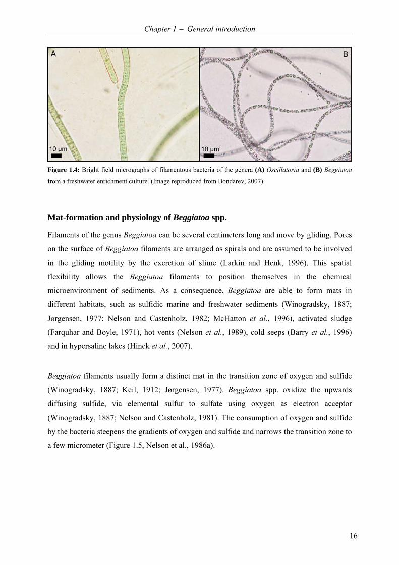
More than two centuries ago, bacteria of the genus Beggiatoa were discovered (Vaucher,
1803). They were originally described as Oscillatoria alba because they feature a similar
filamentous morphology as the cyanobacteria of the genus Oscillatoria, but have a whitish
appearance instead of the blue-green pigments (Figure 1.4). About 40 years later, these
colorless sulfur bacteria were reclassified as Beggiatoa alba, named after the Italian scientist
F. S. Beggiato (Trevisan, 1842). Based on their morphology, different filamentous sulfur
bacteria were assigned to the genus Beggiatoa. Several species were differentiated on the
basis of filament diameter size classes ranging between 1−55 µm (Vaucher, 1803; Trevisan,
1842; Hinze, 1901; Klas, 1937). However, only a small number of 16S rDNA sequences were
available until recently, which made it difficult to phylogenetically classify the large sulfur
bacteria. It was even found that filaments with a similar morphology belong to phylo-
genetically different genera (Ahmad et al., 1999; Ahmad et al., 2006). In a single-cell
16S rDNA gene sequencing approach of large sulfur bacteria, Salman et al. (2011) strongly
extended the amount of available sequences and proposed based on phylogenetic analysis new
candidatus genera names for the members of the family Beggiatoaceae. According to this
reclassification, the genus Beggiatoa contains aerobic or microaerophilic filamentous bacteria
with a diameter of 1−9 µm.
Chapter 1 − General introduction
16
Figure 1.4: Bright field micrographs of filamentous bacteria of the genera (A) Oscillatoria and (B) Beggiatoa
from a freshwater enrichment culture. (Image reproduced from Bondarev, 2007)
Mat-formation and physiology of Beggiatoa spp.
Filaments of the genus Beggiatoa can be several centimeters long and move by gliding. Pores
on the surface of Beggiatoa filaments are arranged as spirals and are assumed to be involved
in the gliding motility by the excretion of slime (Larkin and Henk, 1996). This spatial
flexibility allows the Beggiatoa filaments to position themselves in the chemical
microenvironment of sediments. As a consequence, Beggiatoa are able to form mats in
different habitats, such as sulfidic marine and freshwater sediments (Winogradsky, 1887;
Jørgensen, 1977; Nelson and Castenholz, 1982; McHatton et al., 1996), activated sludge
(Farquhar and Boyle, 1971), hot vents (Nelson et al., 1989), cold seeps (Barry et al., 1996)
and in hypersaline lakes (Hinck et al., 2007).
Beggiatoa filaments usually form a distinct mat in the transition zone of oxygen and sulfide
(Winogradsky, 1887; Keil, 1912; Jørgensen, 1977). Beggiatoa spp. oxidize the upwards
diffusing sulfide, via elemental sulfur to sulfate using oxygen as electron acceptor
(Winogradsky, 1887; Nelson and Castenholz, 1981). The consumption of oxygen and sulfide
by the bacteria steepens the gradients of oxygen and sulfide and narrows the transition zone to
a few micrometer (Figure 1.5, Nelson et al., 1986a).
Chapter 1 − General introduction
17
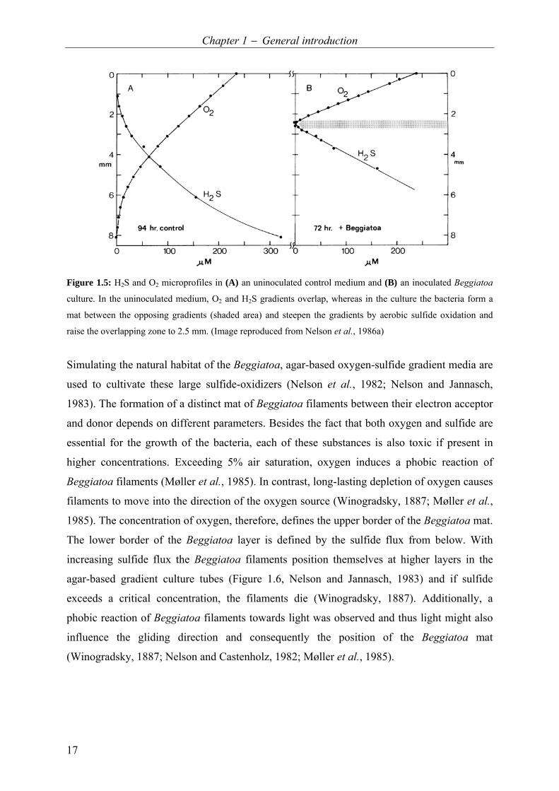
Figure 1.5: H2S and O2 microprofiles in (A) an uninoculated control medium and (B) an inoculated Beggiatoa
culture. In the uninoculated medium, O2 and H2S gradients overlap, whereas in the culture the bacteria form a
mat between the opposing gradients (shaded area) and steepen the gradients by aerobic sulfide oxidation and
raise the overlapping zone to 2.5 mm. (Image reproduced from Nelson et al., 1986a)
Simulating the natural habitat of the Beggiatoa, agar-based oxygen-sulfide gradient media are
used to cultivate these large sulfide-oxidizers (Nelson et al., 1982; Nelson and Jannasch,
1983). The formation of a distinct mat of Beggiatoa filaments between their electron acceptor
and donor depends on different parameters. Besides the fact that both oxygen and sulfide are
essential for the growth of the bacteria, each of these substances is also toxic if present in
higher concentrations. Exceeding 5% air saturation, oxygen induces a phobic reaction of
Beggiatoa filaments (Møller et al., 1985). In contrast, long-lasting depletion of oxygen causes
filaments to move into the direction of the oxygen source (Winogradsky, 1887; Møller et al.,
1985). The concentration of oxygen, therefore, defines the upper border of the Beggiatoa mat.
The lower border of the Beggiatoa layer is defined by the sulfide flux from below. With
increasing sulfide flux the Beggiatoa filaments position themselves at higher layers in the
agar-based gradient culture tubes (Figure 1.6, Nelson and Jannasch, 1983) and if sulfide
exceeds a critical concentration, the filaments die (Winogradsky, 1887). Additionally, a
phobic reaction of Beggiatoa filaments towards light was observed and thus light might also
influence the gliding direction and consequently the position of the Beggiatoa mat
(Winogradsky, 1887; Nelson and Castenholz, 1982; Møller et al., 1985).

Chapter 1 − General introduction
18
Figure 1.6: Position of
Beggiatoa cell layers (mats) in
culture tubes with different
sulfide concentrations in the
bottom agar plug. With in-
creasing sulfide, the filaments
form a mat located higher in the
culture tube. (Image reproduced
from Nelson and Jannasch, 1983)
Elemental sulfur, which is the intermediate of sulfide oxidation, can be stored inside the
Beggiatoa cells (Winogradsky, 1887) and leads to the whitish appearance of the filaments.
Using electron microscopy, it was shown that the sulfur globules in the cells are surrounded
by the cytoplasmic membrane and are located in the periplasm (Figure 1.7 A, Strohl et al.,
1982). The intracellular sulfur can serve as an electron donor and be further oxidized to
sulfate when sulfide gets limited in the environment (Winogradsky, 1887). In addition to the
storage of sulfur, Beggiatoa have the ability to store polyhydroxyalkanoates (PHA, sometimes
specifically denoted as poly-β-hydroxybutyric acid [PHB]) in the cytoplasm of the cell
(Figure 1.7 A, Pringsheim, 1964; Strohl and Larkin, 1978; Strohl et al., 1982). The amount of
PHA in the cell can account for up to 50% of the dry weight of the cell (Güde et al., 1981).
Furthermore, an accumulation of polyphosphate in Beggiatoa cells was shown by trans-
mission electron microscopy and different staining methods (Figure 1.7 C, Maier and Murray,
1965; Strohl and Larkin, 1978; de Albuquerque et al., 2010; Brock and Schulz-Vogt, 2011).
About two decades ago, extremely large marine filamentous sulfur bacteria (116−122 µm in
diameter) containing a central vacuole were found and identified as Beggiatoa spp. based on
morphological similarities to these organisms (Figure 1.7 B, Nelson et al., 1989). Few years
later, the storage of nitrate, an alternative electron acceptor, was detected within the vacuoles
of these large filaments (McHatton et al., 1996). It was proposed that the oxidation of sulfide
can be coupled to either DNRA (Sayama, 2001; Sayama et al., 2005) or denitrification
(Sweerts et al., 1990). The storage of nitrate allows the filaments to inhabit deeper anoxic
sediment layers. Carrying nitrate down into anoxic sediment layers and use it for sulfide
oxidation can lead to the separation of oxygen and sulfide gradients over several centimeters
(Mußmann et al., 2003; Sayama et al., 2005; Kamp et al., 2006). This life strategy enables
large, vacuolated sulfur bacteria like Beggiatoa spp. to outcompete non-vacuolated, non-

Chapter 1 − General introduction
19
motile sulfide-oxidizers in anaerobic environments. Close relatives of Beggiatoa, like bacteria
belonging to the candidate genus “candidatus Marithioploca”, also use and store nitrate and
even show a positive chemotactic response towards nitrate (Huettel et al., 1996; reclassified
by Salman et al., 2011). Thus, the orientation and mat formation of the vacuolated nitrate-
storing sulfur bacteria may also be influenced by the nitrate flux.
Studying the physiology of Beggiatoa, Winogradsky (1887) developed the concept of
chemolithotrophy. He observed that the growth of Beggiatoa was dependent on reduced
inorganic sulfur compounds but not on the presence of organic compounds. The utilization of
CO2 as a sole carbon source was later confirmed by isotope-labeling studies (Nelson and
Jannasch, 1983). Besides these chemolithoautotrophic strains, many chemoorganohetero-
trophic Beggiatoa strains were isolated (Strohl and Larkin, 1978; Güde et al., 1981; Strohl et
al., 1981), which are able to oxidize sulfide only in the presence of organic compounds.
Furthermore, also mixotrophic Beggiatoa strains were isolated (Pringsheim, 1967; Güde et al.,
1981) thus reflecting the diverse metabolisms present within the genus Beggiatoa.
Figure 1.7: Cell structures of Beggiatoa filaments. (A) Schematic representation of Beggiatoa alba strain
B15LD indicating the location of sulfur globules [S] in the periplasm and poly-β-hydroxybutyrate [PHB] in the
cytoplasm. (B) Transmission electron micrograph of a Beggiatoa sp. cross section. The cytoplasm of this large
Beggiatoa filament is restricted to the edge of the cell and the interior mainly consists of a large central vacuole.
(C) Transmission electron micrograph showing electron-dense inclusion bodies in the cytoplasm of Beggiatoa
filaments probably consisting of polyphosphate [P]. (Images adapted and reproduced from Strohl et al., 1982 [A];
Nelson et al., 1989 [B]; de Albuquerque et al., 2010 [C])

Chapter 1 − General introduction
20
The investigated Beggiatoa sp. co-culture
The marine Beggiatoa sp. strain 35Flor investigated in this thesis was isolated in 2002 from a
microbial community associated with scleractinian corals suffering from black band disease
off the coast of Florida. This Beggiatoa sp. strain grows under chemolithoautotrophic con-
ditions in an agar-stabilized oxygen-sulfide gradient medium gaining energy from the aerobic
oxidation of sulfide. Both, a fixed carbon and a fixed nitrogen source are absent from the
medium and nitrogen fixation in the investigated Beggiatoa sp. was determined earlier (Henze,
2005). Typical storage compounds of the genus Beggiatoa, such as sulfur, PHA and
polyphosphate were found in the investigated filaments (Schwedt, unpublished data, Brock
and Schulz-Vogt, 2011). A central vacuole is present (Kamp et al., 2008; Brock and Schulz-
Vogt, 2011), but the storage of nitrate could not be detected (Schwedt et al., unpublished
data).
The Beggiatoa sp. strain 35Flor is accompanied by only one type of organism (Bachmann,
2007), the Pseudovibrio sp. strain FO-BEG1. Unlike the Beggiatoa sp., the associated bac-
teria are able to grow in pure culture and could be isolated in artificial seawater medium. The
investigated Pseudovibrio sp. is able to grow in pure artificial seawater medium under ex-
treme nutrient-poor conditions (Bachmann, 2007) and thus belongs to the few so far cultured
extremely oligotrophic organisms.
Bacterial growth under nutrient deficiency
The term ‘oligotroph’ was introduced by Weber (1907) to describe an organism growing
under nutrient deficiency as opposed to that, bacteria growing under nutrient affluence are
called ‘eutrophs’ (organisms living in nutrient-rich environments are sometimes also referred
to as ‘copiotrophs’). Over time, several definitions of oligotrophy arose and today it is
generally accepted that bacteria are referred to as oligotrophic when they are able to grow in
medium containing less than 0.5 mg C L−1 (e.g. Ishida et al., 1989). When their growth is
inhibited by high substrate concentrations, the bacteria are considered to be obligately
oligotrophic, which is in contrast to facultatively oligotrophic bacteria, which are able to grow
under both nutrient-poor and nutrient-rich conditions (Ishida et al., 1989). Facultative
oligotrophs are, therefore, successful in environments with changing nutrient conditions.

Chapter 1 − General introduction
21
The open ocean, covering large parts of the earth’s surface, is low in nutrients and contains
less than 1 mg DOC in 1 L seawater (Schut et al., 1997; Hansell et al., 2009). Thus, it is
denoted as an oligotrophic environment. 75% of the carbon consumed by the bacteria in the
ocean can be composed of dissolved free amino acids (DFAA), dissolved combined amino
acids (DCAA) and monosaccharides. The utilization of these substances can cover 5 to 93%
of the carbon demand of the bacteria and 9 to 100% of the nitrogen demand (Fuhrman, 1987;
Jørgensen, 1987; Stanley et al., 1987; Keil and Kirchman, 1999; Cherrier and Bauer, 2004).
Attached and free-living marine bacteria
The particulate organic matter (POM) is an important part of the organic matter in the ocean.
Particles larger than half a millimeter are so-called marine snow particles (Suzuki and Kato,
1953; Silver et al., 1978). Besides the larger marine snow particles, there are also smaller
microaggregates (Figure 1.8 A and B) and both consist of detrital organic and inorganic
matter (Azam and Long, 2001), thereby representing hotspots of high nutrient concentration.
The aggregates can be colonized by metazoans (e.g. Shanks and Edmondson, 1990; Kiørboe,
2000), protozoans (e. g. Silver et al., 1978) and prokaryotes (e. g. Alldredge et al., 1986;
Smith et al., 1992; Azam and Malfatti, 2007 and references therein), whereas only the latter
was found on all types of aggregates studied so far. Extracellular hydrolytic enzymes
produced by aggregate-associated bacteria can convert the POM of the sinking aggregates
into cell biomass and non-sinking dissolved organic matter (DOM) (Smith et al., 1992;
Grossart et al., 2007). While sinking down the particles leave behind a DOM plume that is
composed mainly of carbon and nitrogen. The DOM plume is colonized by some of the
attached bacteria but also by free-living bacteria from the surrounding water (Figure 1.8 C,
Azam and Long, 2001; Kiørboe and Jackson, 2001).
Compared to the surrounding water, bacterial cell densities on aggregates are typically
>100 times higher (e. g. Smith et al., 1992; Turley and Mackie, 1994). Nevertheless, the
particle-associated bacteria account only for <5% of the total bacterial numbers in seawater
(e.g. Alldredge et al., 1986; Alldredge and Gotschalk, 1990; Turley and Stutt, 2000) and
contribute to only 3 to 12% of the total bacterial production (Alldredge et al., 1986; Turley
and Stutt, 2000). Even though the total activity is low, the per cell activity of the attached
bacteria is higher compared to free-living bacteria, as demonstrated by higher incorporation
rates and shorter doubling times (Alldredge et al., 1986; Alldredge and Gotschalk, 1990;
Smith et al., 1992; Azam and Long, 2001; Kiørboe and Jackson, 2001). Furthermore, some

Chapter 1 − General introduction
22
studies have shown that the free-living bacteria may either starve and not be active (Boylen
and Ensign, 1970; Novitsky and Morita, 1976; 1977), while other studies show that they may
be metabolically active (Azam and Hodson, 1977; Tabor and Neihof, 1982; Ishida et al.,
1989).
Figure 1.8: In situ photographs of (A) a marine snow aggregate in a pelagic environment and (B) micro-
aggregates in a shallow environment (photos M. Lunau). (C) Scheme of a marine snow particle colonized by
bacteria which excrete hydrolytic enzymes converting marine snow into DOM forming a plume behind the
sinking aggregate that is also colonized by attached and free-living bacteria. The DOM consists mainly of carbon
[C] and nitrogen [N]. (Images adapted and redrawn from Azam and Long, 2001 [C]; and reproduced from Simon
et al., 2002 [A and B])
The majority of the free-living bacteria in the open ocean is exposed to extremely low nutrient
concentrations and many different survival strategies have evolved to cope with this nutrient
limitation. These strategies include concentration-independent enzyme production (cells are
considered to be prepared and have enzymes ready for substrates becoming available), de-
repression of substrates (the use of one substrate is not repressed by another more efficient
one) and the use of multiple substrates simultaneously (use different substrates at the same
time, independent of their efficiency) (Egli, 2010 and references therein). Substrate tests on
organisms grown under carbon limitation revealed that these cells can oxidize a much broader
spectrum of organic compounds than cells that were pre-grown under carbon excess (Upton
and Nedwell, 1989; Ihssen and Egli, 2005). The use of multiple carbon sources enables

Chapter 1 − General introduction
23
growth on extremely low concentrations of each individual compound (Lendenmann et al.,
1996; Kovárová-Kovar and Egli, 1998) and is thus beneficial in an oligotrophic environment
with a frequently changing supply of nutrients.
Cultivation of marine bacteria
So far, only about half of the known bacterial phyla have cultivable representatives
(Hugenholtz, 2002), even though pure cultures are essential to study metabolic pathways of
the different bacteria in detail. Possible reasons for the yet inability to cultivate many bacteria
maybe unsuited growth conditions and could include a lack of nutrients or growth factors,
inappropriate pH, pressure or temperature conditions or unsuitable levels of oxygen (reviewed
in Vartoukian et al., 2010). Furthermore, many of the used media contain very high amounts
of nutrients, compared to most marine environments, and thus favor fast-growing bacteria
rather than slow-growing ones. In turn, such conditions might even inhibit the growth of some
oligotrophic bacteria (Ishida et al., 1989; Koch, 1997; Connon and Giovannoni, 2002).
Consequently, new strategies for the isolation of marine bacteria have to be developed to
understand the different metabolic pathways of marine bacteria and their ecology and
evolution (Grossart, 2010).
One approach to prevent overgrowth of slow-growing bacteria is the dilution-to-extinction
method, that reduces the number of cells per sample until ideally solely single cells are left for
cultivation (e. g. Button et al., 1993; Connon and Giovannoni, 2002). Additionally, the use of
low-nutrient natural seawater for isolation and in vitro simulation of the natural environment
using diffusion chambers placed in natural seawater provoked isolation of new, so far
uncultured bacteria (Connon and Giovannoni, 2002; Kaeberlein et al., 2002; Rappe et al.,
2002; Zengler et al., 2002; Bollmann et al., 2007). However, the utilization of natural
seawater always implies undefined conditions because merely a few percent of the highly
diverse organic compounds in natural seawater is already characterized (Dittmar and Paeng,
2009). Hence, in order to study bacterial metabolism at the lower border of bacterial growth
in detail and to identify the essential substances for growth, a defined artificial seawater
medium is crucial. Those approaches so far reported to isolate and cultivate marine bacteria
using artificial seawater contained either agar or vitamins, both of which represent a fixed
carbon source, or were supplemented with at least 0.1 to 3 mg C L−1 of organic substrates to
support growth (Van der Kooij et al., 1980; Ishida et al., 1982; Schut et al., 1993; Azam and
Long, 2001; Vancanneyt et al., 2001).
Chapter 1 – Aims of the study
24
Aims of this study
This work was initiated by the question of how marine Beggiatoa spp. form mats and succeed
in anoxic habitats. Until today, it was believed that only the presence of nitrate as alternative
electron acceptor allows the population of anoxic environments by the large sulfide-oxidizing
bacteria of the family Beggiatoaceae. Recently, I found that nitrate is not essential for the
thriving of Beggiatoa filaments in anoxic parts. For these experiments, I used the marine
Beggiatoa sp. 35Flor that is cultivated in gradient culture tubes. It was observed that filaments
moved below the oxygen-sulfide interface without the presence of nitrate and aggregated in
anoxic parts of the culture tube. Therefore, the aim of the first part of this thesis (Chapter 2)
was to study this behavior and to reveal how the filaments can survive in the anoxic layers
and why they leave the overlapping zone of oxygen and sulfide, where both electron acceptor
and donor are present.
Already during my diploma thesis (Bachmann, 2007) I was able to show that the investigated
Beggiatoa culture is not a pure culture. Instead, the Beggiatoa sp. 35Flor is in co-culture with
a single accompanying organism, Pseudovibrio sp. FO-BEG1. Accordingly, the second
objective of my PhD thesis (Chapter 3) was to examine whether the growth of the sulfide-
oxidizer is dependent on the presence of the accompanying Pseudovibrio sp. and, if so,
whether the Pseudovibrio denitrificans type strain (DSM number 17465) can also provoke
growth of the Beggiatoa sp. 35Flor.
The accompanying Pseudovibrio sp. FO-BEG1 is able to grow in pure culture without the
Beggiatoa sp. under extreme nutrient deficiency in artificial seawater medium (Bachmann,
2007). The physiology of the Pseudovibrio sp. should now be subject to a detailed phy-
siological analysis. Despite omitting the addition of an energy source, DOC was detected in
the range of 5 µmol C L−1 (0.06 mg C L−1), which is 1 to 2 orders of magnitude below natural
oligotrophic seawater (Schut et al., 1997; Hansell et al., 2009). This contamination could have
potentially been used as an energy source. To address this question, the third objective of this
thesis (Chapter 4) was to analyze the artificial medium used for cultivation, before and after
growth of the Pseudovibrio strain, in order to find out which compounds were used by the
bacteria. Eventually, other heterotrophic bacterial strains were isolated in the course of this
thesis under nutrient limitation to estimate how common the ability among heterotrophic
bacteria (associated with large sulfide-oxidizers) is to grow under nutrient limitation.
Chapter 2 – Physiology and mat formation
25
Chapter 2
Physiology and mat formation
of a marine Beggiatoa culture
This second chapter of my PhD thesis deals with the physiology of the large, sulfide-
oxidizing Beggiatoa sp. strain 35Flor. The focus is laid on mat formation processes and was
motivated by a new observation that filaments migrate into deeper anoxic regions without the
presence of nitrate (Figure 2). In the first part of this chapter, the physiology behind the
observed migration event is discussed in detail in form of a manuscript. The second part of
this chapter deals with the inducibility of this migration process by blue/green light and the
influence of chemical substances on the mat. This part of the chapter is presented in form of a
short communication.

Chapter 2 – Physiology and mat formation
26
Figure 2: Image of marine Beggiatoa cultivated under chemolithotrophic conditions without nitrate at a high
sulfide flux (43.1 mmol m–2 d–1) after two weeks. A subpopulation of filaments migrated downwards into deeper
layers.
Contributions:
2.1 Sulfur respiration in a chemolithoautotrophic marine Beggiatoa strain A. Schwedt, A.-C. Kreutzmann, L. Polerecky and H. N. Schulz-Vogt The concept of the study was developed by me and H. N. Schulz-Vogt. All initial experiments were performed by me. The final experiments and data analysis were performed by me, A.-C. Kreutzmann and L. Polerecky. The manuscript was written together with A.-C. Kreutzmann and with the help of the other two co-authors. 2.2 Coordinated movement of Beggiatoa filaments in oxygen/sulfide gradients and the effect of blue/green light H. N. Schulz-Vogt, T. Hohmann, A.-C. Kreutzmann, L. Polerecky and A. Schwedt The concept of the study was developed by H. N. Schulz-Vogt and T. Hohmann. Major experiments were performed by H. N. Schulz-Vogt and T. Hohmann with the help of L. Polerecky during data analysis. I assisted during migration experiments and filming and performed c-di-GMP experiments.

Chapter 2 – Physiology and mat formation
27
2.1 Sulfur respiration in a marine chemolithoautotrophic
Beggiatoa strain
Anne Schwedt1*, Anne-Christin Kreutzmann1, Lubos Polerecky1, Heide N. Schulz-Vogt1
1Max Planck Institute for Marine Microbiology, Celsiusstr. 1, D-28359 Bremen, Germany
*Corresponding author
Manuscript submitted to Frontiers in Microbiology (22.09.2011), accepted with minor
revisions (23.12.2011), and published online (09.01.2012). doi:10.3389/fmicb.2011.00276
Keywords: Beggiatoa; sulfur reduction; gradient cultivation; microelectrodes; migration
Chapter 2 – Physiology and mat formation
28
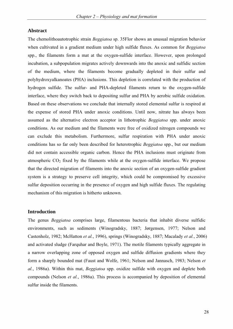
Abstract
The chemolithoautotrophic strain Beggiatoa sp. 35Flor shows an unusual migration behavior
when cultivated in a gradient medium under high sulfide fluxes. As common for Beggiatoa
spp., the filaments form a mat at the oxygen-sulfide interface. However, upon prolonged
incubation, a subpopulation migrates actively downwards into the anoxic and sulfidic section
of the medium, where the filaments become gradually depleted in their sulfur and
polyhydroxyalkanoates (PHA) inclusions. This depletion is correlated with the production of
hydrogen sulfide. The sulfur- and PHA-depleted filaments return to the oxygen-sulfide
interface, where they switch back to depositing sulfur and PHA by aerobic sulfide oxidation.
Based on these observations we conclude that internally stored elemental sulfur is respired at
the expense of stored PHA under anoxic conditions. Until now, nitrate has always been
assumed as the alternative electron acceptor in lithotrophic Beggiatoa spp. under anoxic
conditions. As our medium and the filaments were free of oxidized nitrogen compounds we
can exclude this metabolism. Furthermore, sulfur respiration with PHA under anoxic
conditions has so far only been described for heterotrophic Beggiatoa spp., but our medium
did not contain accessible organic carbon. Hence the PHA inclusions must originate from
atmospheric CO2 fixed by the filaments while at the oxygen-sulfide interface. We propose
that the directed migration of filaments into the anoxic section of an oxygen-sulfide gradient
system is a strategy to preserve cell integrity, which could be compromised by excessive
sulfur deposition occurring in the presence of oxygen and high sulfide fluxes. The regulating
mechanism of this migration is hitherto unknown.
Introduction
The genus Beggiatoa comprises large, filamentous bacteria that inhabit diverse sulfidic
environments, such as sediments (Winogradsky, 1887; Jørgensen, 1977; Nelson and
Castenholz, 1982; McHatton et al., 1996), springs (Winogradsky, 1887; Macalady et al., 2006)
and activated sludge (Farquhar and Boyle, 1971). The motile filaments typically aggregate in
a narrow overlapping zone of opposed oxygen and sulfide diffusion gradients where they
form a sharply bounded mat (Faust and Wolfe, 1961; Nelson and Jannasch, 1983; Nelson et
al., 1986a). Within this mat, Beggiatoa spp. oxidize sulfide with oxygen and deplete both
compounds (Nelson et al., 1986a). This process is accompanied by deposition of elemental
sulfur inside the filaments.

Chapter 2 – Physiology and mat formation
29
Several filamentous and non-filamentous members of the Beggiatoaceae (Salman et al., 2011)
are moreover capable of anaerobic sulfide oxidation with nitrate as an alternative electron
acceptor (Fossing et al., 1995; McHatton et al., 1996; Schulz et al., 1999). Dissimilatory
nitrate reduction enables these organisms to colonize anoxic environments such as deeper
layers in sediments, microbial mats or gradient cultures (Sweerts et al., 1990; Mußmann et al.,
2003; Sayama et al., 2005; Kamp et al., 2006; Hinck et al., 2007). Nitrate-based sulfide
oxidation seems to have been of great importance for some members of the family
Beggiatoaceae, as suggested by their ability to store nitrate within intracellular vacuoles at
concentrations up to 104 fold higher than in the ambient water (Fossing et al., 1995;
McHatton et al., 1996; Schulz et al., 1999; Sayama, 2001; Mußmann et al., 2003; Kalanetra et
al., 2004; Kalanetra et al., 2005; Hinck et al., 2007). However, also non-vacuolated strains
were shown to use externally provided nitrate as a terminal electron acceptor (Sweerts et al.,
1990; Kamp et al., 2006).
We cultivated the chemolithoautotrophic, marine strain Beggiatoa sp. 35Flor in an agar-
stabilized oxygen-sulfide gradient medium. Upon prolonged incubation in the presence of
medium to high sulfide fluxes, we observed an unusual migration behavior, where a
subpopulation of filaments migrated downwards from the oxygen-sulfide interface. These
filaments were able to survive although sulfide concentrations were high and terminal
electron acceptors that are known to be utilized by Beggiatoa spp., i.e., oxygen and nitrate,
were not detectable in medium nor filaments. In this study, we investigated the possibility of
an alternative metabolism of Beggiatoa sp. 35Flor under anoxic, nitrate-free and sulfidic
conditions, and discuss its possible ecological significance and link to the peculiar migration
behavior.
Material and methods
Strain and cultivation
The strain Beggiatoa sp. 35Flor was originally enriched from a black band disease of
scleractinian corals from the coast of Florida, and can so far only be cultivated in the presence
of the Pseudovibrio denitrificans strain FO-BEG1 (Schwedt et al., unpublished). Filaments of
the strain 35Flor are about 6 µm wide, and the cells contain polyphosphate inclusions and a
central vacuole filled with polyphosphate (Kamp et al., 2008; Brock and Schulz-Vogt, 2011).
Chapter 2 – Physiology and mat formation
30
Cultivation was performed in tubes with an agar-based mineral gradient medium modified
after Nelson et al. (1982) and Nelson and Jannasch (1983) using artificial seawater (Kamp et
al., 2008). The medium was composed of a sulfidic bottom agar plug (1.5% w/v agar)
covered with a sulfide-free, semisolid top agar layer (0.25% w/v agar) of 5 cm height (Tables
2.1.1 and 2.1.2). The medium was prepared free of nitrate, nitrite and nitric oxide, as verified
by measurements with an NOX analyzer (CLD 66, Eco Physics, Rösrath, Germany). Gas
exchange between headspace and the atmosphere was possible, and opposing gradients of
oxygen and sulfide were allowed to form for one day before inoculation. The cultures were
inoculated about 1 cm below the air-agar interface using 100 µL filament suspension from an
established mat. Incubations were performed at room temperature in the dark.
During incubations, the distribution of filaments in the tube was measured simultaneously
with the vertical profiles of H2S and pH. Filaments from parallel culture tubes were
subsampled and used for microscopic determination of their sulfur and PHA inclusions.
Oxygen profiles were measured in parallel tubes.
Table 2.1.1 Solutions for the preparation of agar-stabilized gradient media.
Solution Compositiona
Artificial seawater 27.5 g NaCl, 5 g MgCl2 · 6H2O, 4.1 g MgSO4 · 7H2O, 0.66 g CaCl2 · 2H2O, 1.02 g KCl
Mineral solution 555 mg K2HPO4, 28.72 mg Na2MoO4, 750 mg Na2S2O5, 29 mg FeCl3 · 6H2O
Trace element solutionb
5.2 g EDTA, 1.5 g FeCl2 · 4H2O, 70 mg ZnCl2, 100 mg MnCl2 · 4H2O, 62 mg H3BO4, 190 g CoCl2 · 6H2O, 17 mg CuCl2 · 2H2O, 24 mg NiCl2 · 6 H2O, 36 mg Na2MoO4 · 2H2O
Vitamin solutionc 0.1 g cyanocobalamine 0.1 g inositol 0.1 g biotin 0.1 g folic acid 1.0 g para-aminobenzoic acid 10 g nicotinic acid 10 g D-pantothenate 20 g thiamine
a in 1 L distilled water b pH adjusted to 6.5 c All vitamins were dissolved separately and then combined in a final stock solution (1 mL of each vitamin
solution in a final volume of 100 mL distilled water) that was filtersterilized twice.

Chapter 2 – Physiology and mat formation
31
Table 2.1.2 Composition of gradient medium.
Medium Componenta Composition
Bottom agar A 100 mL artificial seawater, 2.9 g NaCl, 1 drop 1 mol L−1 KOH
B 80 mL distilled water, 2.7 g agarb
C 0.72 – 3.6 mL 1 mol L−1 Na2S solution Top agar A 240 mL artificial seawater, 4.32 g NaCl B 96 mL distilled water, 0.9 g agarb
C 24 mL mineral solution, 0.36 ml trace element solution, 7 drops 1 mol L−1 KOH
D 0.72 mL 1 mol L−1 NaHCO3 solution E 150 µL vitamin solution
a All components were sterilized separately before combination. b The agar was washed two times in distilled water before use.
Microsensor measurements
Microsensors for O2 (OX-10 standard), H2S (H2S-10), and pH (PH-10) were purchased from
Unisense A/S (Aarhus, Denmark). The external reference for the pH electrode was
manufactured and connected in-house. Calibration of the H2S sensor was performed in anoxic,
acidified artificial seawater (Table 2.1.1, pH<2) to which an anoxic Na2S stock solution was
added stepwise. The exact sulfide concentration of the Na2S stock solution was determined by
iodometric titration. Total sulfide (Stot) profiles were calculated from measured H2S and pH
profiles using equation Stot = H2S × [1 + K1 / H3O+], with pK1 = 6.569 at 21°C and 39‰
salinity (Millero et al., 1988). The oxygen sensor was two-point calibrated in a calibration
chamber filled with artificial seawater. Signal readings were taken in water saturated with N2
and ambient air. Oxygen concentrations at the respective salinity and temperature were
calculated according to Weiss (1970). The pH electrode was calibrated using buffer solutions
of pH 4.01, pH 7.00, and pH 9.21 (Mettler-Toledo, Giessen, Germany). All sensors were
calibrated immediately before the measurement. In case of long time series measurements the
sensor calibration was checked afterwards and a possible drift was corrected for.
Vertical profiling in 250 µm steps was performed with sensors mounted on a motorized linear
positioner (VT-80, Pollux motor, Micos, Eschbach, Germany) controlled by a computer using
a software for automated microsensor measurements (µ-Profiler, L. Polerecky,
http://www.microsen-wiki.net). The sensors were aligned by manually adjusting their tips to
the air-agar interface using a dissecting microscope (Stemi 2000-C, Zeiss, Jena, Germany).
Chapter 2 – Physiology and mat formation
32
Filament imaging
The distribution of sulfur-containing Beggiatoa sp. 35Flor filaments in the gradient cultures
was monitored using time-lapse photography. An amber light-emitting diode (LXHL-NM98,
Luxeon, Philips, San Jose, CA, USA) was positioned below the culture tube and switched on
for one second when an image was taken with a cooled CCD camera (Sensicam, PCO,
Kelheim, Germany). Illumination and image acquisition in 10 min intervals were controlled
by a computer using a custom-written program (Look@Molli, B. Grunwald,
http://www.microsen-wiki.net).
Intensities of the recorded images were horizontally averaged over an area with visible
filaments (~5 mm wide, ~2 cm high), and the resulting vertical profiles were assembled into a
2D map with the x-axis representing incubation time and the y-axis corresponding to depth.
Since the average image intensity was proportional to the density of sulfur globules, which
were present exclusively inside filaments, vertical movement of sulfur-rich filaments was
detected as a change in the shape of the vertical intensity profile. In contrast, an increase and
decrease in the profile intensity that was not accompanied with the change in the profile shape
indicated accumulation and depletion of sulfur inside the filaments, respectively. Because this
method relied on light scattering from sulfur inclusions, it did not allow visualization of
sulfur-free filaments.
Staining of internal PHA
Staining with Nile Red was used to visualize PHA inclusions in the filaments. A subsample
from the culture tube (volume 90 µL) was incubated for 5 minutes with 10 µL of a Nile Red
(Sigma-Aldrich, Steinheim, Germany) staining solution (25 mg L−1 in dimethyl sulfoxide).
The filament suspension was transferred onto a poly-L-lysine (Sigma-Aldrich) coated
microscope slide for immobilization of the filaments. Fluorescence of Nile Red was excited
with a laser at 546 nm and emission was recorded above 590 nm (filter set 15, Zeiss, Jena,
Germany) using an epifluorescence microscope (Axiophot equipped with AxioCam MRm,
Zeiss, Jena, Germany).
Transfer experiment with sulfur-free filaments
To verify that sulfur-free filaments from the anoxic subpopulation of an aged culture
(cultivated at high sulfide flux conditions) were alive, able to migrate back to the oxygen-
sulfide interface and re-establish their sulfide-oxidizing metabolism, they were transferred

Chapter 2 – Physiology and mat formation
33
into the anoxic section of a fresh gradient medium (cultivated at low sulfide flux conditions).
All cultivation media were prepared in plexiglass tubes (2×12 cm in size) with lateral holes
(Brock and Schulz-Vogt, 2011). Fresh medium for inoculation with sulfur-free filaments was
pre-incubated with the accompanying Pseudovibrio strain. This was done to ensure a
sufficient cell density of Pseudovibrio sp. irrespective of the inoculum as the Pseudovibrio sp.
is required for growth of Beggiatoa sp. 35Flor, but its abundance is negligible in the anoxic
part of the gradient (Schwedt et al., unpublished). Subsequently, sulfur-free filaments were
removed laterally from the aged culture and injected laterally into the fresh medium at a depth
of about 1 cm below the oxygen-sulfide interface. Lateral removal ensured that no sulfur-
containing filaments from the oxygen-sulfide interface of the aged culture were transferred,
whereas lateral injection prevented inoculation of the transferred sulfur-free filaments to the
oxygen-sulfide interface of the fresh gradient media. The development of a mat at the oxygen-
sulfide interface was inspected visually.
Results
Migration of Beggiatoa sp. 35Flor in gradient cultures Beggiatoa sp. 35Flor filaments aggregated and formed a dense mat at the oxygen-sulfide
interface within the gradient medium. In cultures with medium to high sulfide fluxes
(Table 2.1.3 A) a subpopulation of filaments began a downward migration to the anoxic zone
about 3−4 days after establishment of the mat. For medium sulfide fluxes, this migration
resulted in a layer with a homogenous filament density extending up to 2−3 mm below the
mat (Figure 2.1.1). In contrast, for high sulfide fluxes the migrating filaments were not
homogenously distributed, but progressively aggregated in a region distinctly separated from
the mat at the oxygen-sulfide interface (Figure 2.1.1 and 2.1.2 B). Because the aggregation of
filaments in the anoxic part increased the chance of detecting metabolic products all further
experiments were conducted with cultures growing under a high sulfide flux.
Chapter 2 – Physiology and mat formation
34
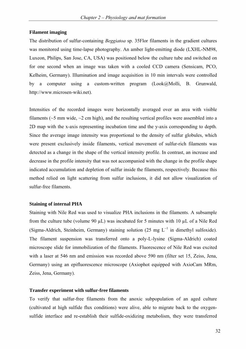
Figure 2.1.1: Distribution of Beggiatoa sp. 35Flor filaments over depth in gradient cultures after 6 (open
symbols) and 12 (closed symbols) days in the presence of different sulfide fluxes. The flux values represent
theoretical maxima under the given cultivation conditions (Table 2.1.3 A).
Migration of filaments in cultures with a high sulfide flux followed a general pattern
(Figure 2.1.2 E). During the initial 3−4 days of incubation, the mat at the oxygen-sulfide
interface gradually formed. After about 6−7 days, the sulfur globule density in the mat
decreased moderately, followed by a more pronounced decrease after 8−9 days. These
decreases were correlated with two pronounced events of downward migration at days 5−6
and 7−8, respectively (arrows 1 and 2 in Figure 2.1.2 E). After reaching a depth of around
10 mm, the migrating filaments formed a layer of increased filament density. These filaments
slowly disappeared from view due to a gradual loss of their internal sulfur granules. The
disappearance of filaments was accompanied by a parallel increase in the sulfur globule
density in the mat at the oxygen-sulfide interface (arrow 3 in Figure 2.1.2 E), suggesting that
the filaments returned to this zone and switched back to sulfide oxidation, thereby depositing
sulfur. This was confirmed by the transfer experiment, which showed that sulfur-free
filaments transferred from the anoxic subpopulation of an aged culture into the anoxic section
of a fresh gradient medium formed, within 12 days, a new mat of sulfur-containing filaments
at the oxygen-sulfide interface.

Chapter 2 – Physiology and mat formation
35
Table 2.1.3 A Diffusive sulfide fluxes in gradient cultures from this study.
c(Na2S) [mmol L−1] in bottom agar
Flux [mmol m−2 d-1]
4 theoretical maximuma 4.6 (low flux) measured (7 d) 8.0 measured (13 d) 8.9 10 theoretical maximuma 19.0 (medium flux) measured (7 d) 20.1 measured (13 d) 16.2 16 theoretical maximuma 40.1 (high flux) measured (7 d) 31.6 measured (13 d) 25.3
a The initial theoretical maximum of the sulfide flux in the gradient cultures, calculated using Fick’s first law of
diffusion (J = −D Δc/Δx). The diffusion coefficient D for HS− was corrected for temperature (21°C) according to
Jørgensen and Revsbech (1983), resulting in a value of 1.56 × 10⁻⁹ m2 s−1. The concentration gradient was
calculated from the height of the top agar (Δx = 5 cm) and the initial sulfide concentration in the bottom agar,
assuming that sulfide was depleted at the agar surface, i.e. Δc = c (Na2S).
Table 2.1.3 B Diffusive sulfide fluxes in natural Beggiatoa spp. mats.
Sediment from Reference Flux [mmol m−2 d−1] Lagoon (Jørgensen and Revsbech, 1983) 38 Lagoon (Mußmann et al., 2003) 0.8 Arctic lagoon (Jørgensen et al., 2010) 34 Coast (Preisler et al., 2007) 4.3 ± 2 Harbor (Fenchel and Bernard, 1995) 12-100 Deep sea mud volcano (Lichtschlag et al., 2010) 11.6 Deep sea mud volcano (Girnth et al., 2011) 40 Deep sea, gas hydrate-rich (Sahling et al., 2002) 63 ± 36
Sulfide production by filaments in the anoxic section
Throughout the incubation, sulfide oxidation in the mat at the oxygen-sulfide interface was
confirmed by pronounced acidification and steep gradients of total sulfide (Figure 2.1.2 C and
D). A small but detectable peak in the H2S profile was observed at a depth of ~10 mm when
the anoxic subpopulation was present (Figure 2.1.2 D). As pH varied only smoothly with
depth in this region, the H2S peak was not linked to pH variation, but indicated a true
production of sulfide at and around this depth. This production was strongly spatially and
Chapter 2 – Physiology and mat formation
36
temporally correlated with the presence of the anoxic subpopulation (Figure 2.1.2 F),
suggesting that it was linked to the metabolic activity of the filaments from this subpopulation.
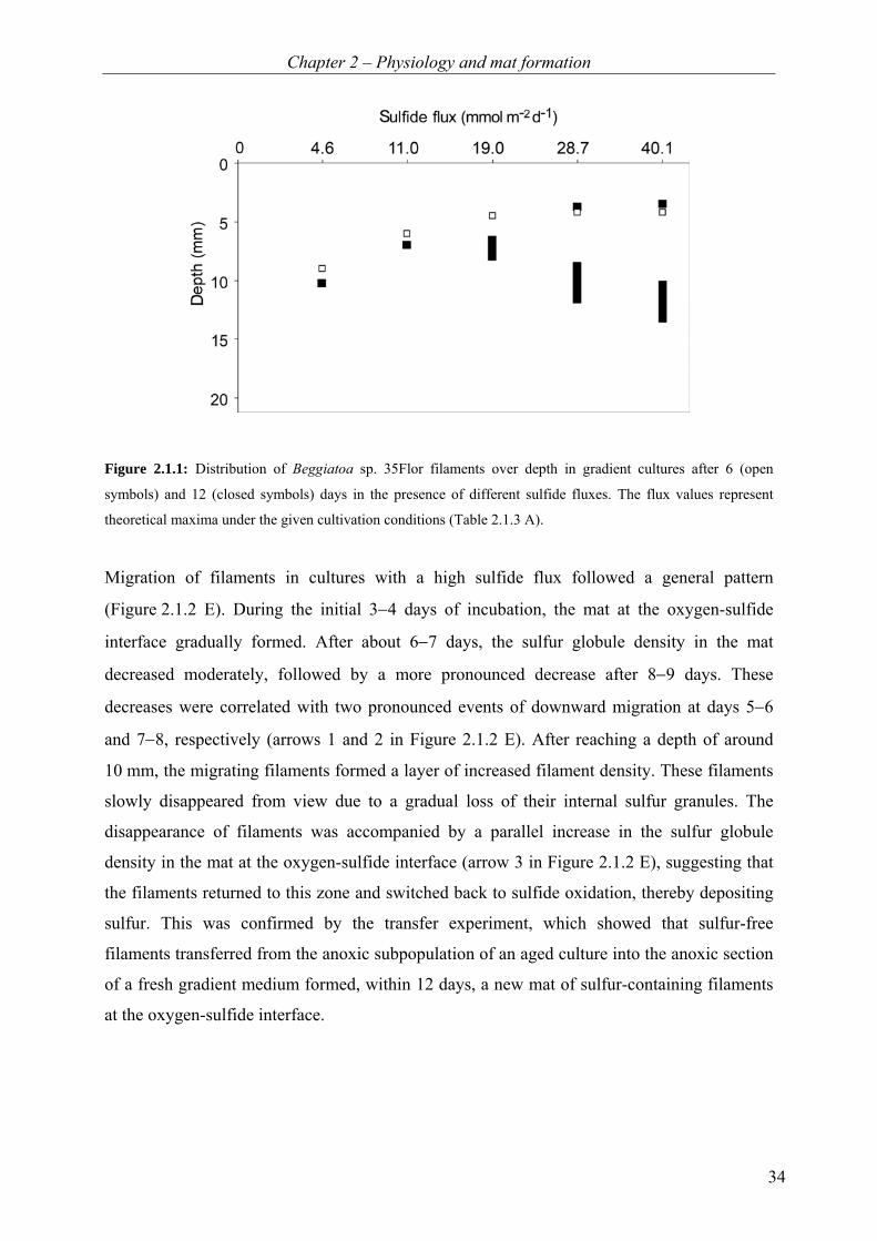
Figure 2.1.2: Relationship between the migration of Beggiatoa sp. 35Flor filaments and the dynamics of O2, pH,
H2S and Stot in the gradient culture tube. (A-B) Images of culture tubes showing the filament distribution after 2
and 11 days, respectively. (C-D) Examples of pH, H2S and total sulfide profiles in the gradient culture incubated
for 8 and 13 days. Shaded areas mark the oxic zone. (E) Average sulfur globule density as a function of time and
depth, showing the dynamics of the filament distribution and their sulfur content. Arrows 1 and 2 indicate the
onset of major downward migration events, arrow 3 indicates the onset of an increase in the filament density in
the mat at the oxygen-sulfide interface. Although the timing of these events varied amongst experimental runs,
the general pattern was reproducible. (F) H2S excess as a function of time and depth, calculated by subtracting
the measured H2S profile from the background trend. The trend was derived from the H2S concentrations
measured above and below the peak (line indicated by arrow in panel D). Contour lines of the sulfur globule
density from panel E are overlaid. Data shown in panels A, B, E and F are from the same culture tube, profiles in
panels C and D are from a parallel culture tube.
Chapter 2 – Physiology and mat formation
37
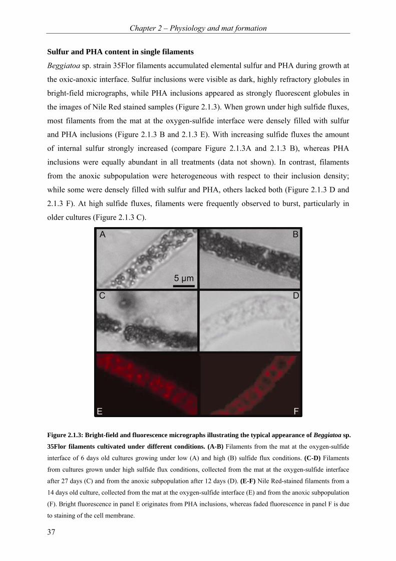
Sulfur and PHA content in single filaments
Beggiatoa sp. strain 35Flor filaments accumulated elemental sulfur and PHA during growth at
the oxic-anoxic interface. Sulfur inclusions were visible as dark, highly refractory globules in
bright-field micrographs, while PHA inclusions appeared as strongly fluorescent globules in
the images of Nile Red stained samples (Figure 2.1.3). When grown under high sulfide fluxes,
most filaments from the mat at the oxygen-sulfide interface were densely filled with sulfur
and PHA inclusions (Figure 2.1.3 B and 2.1.3 E). With increasing sulfide fluxes the amount
of internal sulfur strongly increased (compare Figure 2.1.3A and 2.1.3 B), whereas PHA
inclusions were equally abundant in all treatments (data not shown). In contrast, filaments
from the anoxic subpopulation were heterogeneous with respect to their inclusion density;
while some were densely filled with sulfur and PHA, others lacked both (Figure 2.1.3 D and
2.1.3 F). At high sulfide fluxes, filaments were frequently observed to burst, particularly in
older cultures (Figure 2.1.3 C).
Figure 2.1.3: Bright-field and fluorescence micrographs illustrating the typical appearance of Beggiatoa sp.
35Flor filaments cultivated under different conditions. (A-B) Filaments from the mat at the oxygen-sulfide
interface of 6 days old cultures growing under low (A) and high (B) sulfide flux conditions. (C-D) Filaments
from cultures grown under high sulfide flux conditions, collected from the mat at the oxygen-sulfide interface
after 27 days (C) and from the anoxic subpopulation after 12 days (D). (E-F) Nile Red-stained filaments from a
14 days old culture, collected from the mat at the oxygen-sulfide interface (E) and from the anoxic subpopulation
(F). Bright fluorescence in panel E originates from PHA inclusions, whereas faded fluorescence in panel F is due
to staining of the cell membrane.
Chapter 2 – Physiology and mat formation
38
Discussion
Sulfide production by members of the genus Beggiatoa is known from chemoheterotrophic
strains that were cultivated in liquid medium and artificially exposed to short-term anoxic
conditions (Schmidt et al., 1987). Based on those experiments it was hypothesized that sulfur
respiration may provide Beggiatoa spp. in gradient systems with energy for return from the
anoxic zone to the oxygen-sulfide interface under changing environmental conditions. In this
study, we cultivated the chemolithoautotropic strain Beggiatoa sp. 35Flor in an oxygen-
sulfide gradient medium, and we observed a directed migration of the filaments from the
oxygen-sulfide interface into the anoxic and sulfidic zone where they reduced internal sulfur
to sulfide. This suggested an alternative or additional function of sulfur respiration in
Beggiatoa filaments.
We propose that the observed behavior is a survival strategy of Beggiatoa sp. 35Flor at
prolonged incubation under high sulfide fluxes. Under this condition the filaments become
densely filled with sulfur and were often observed to burst. By moving to the anoxic zone of
the gradient system, the filaments can prevent further deposition of sulfur through aerobic
sulfide oxidation and may even reduce the amount of storage compounds by sulfur respiration
with PHA. We observed that filaments can migrate back to the oxygen-sulfide interface,
where they resume aerobic sulfide oxidation and accumulate new sulfur globules.
Sulfur respiration for regulation of the amount of stored sulfur
The alteration between sulfide oxidation and sulfur reduction in spatially separated
environments seems to allow Beggiatoa sp. 35Flor to control the amount of stored sulfur
beyond the scope of enzymatic regulation. Sulfide is oxidized by Beggiatoa spp. in a two-step
process via internally stored sulfur (2 H2S + O2 2 S0 + 2 H2O) and further to sulfate
(2 S0 + 3 O2 + 2 H2O 2 SO42- + 4 H+). The regulation of these reactions is unknown in
Beggiatoa spp., but the presence of internal sulfur globules demonstrates that the rates of the
two reactions are not always well balanced. In principle, a balanced sulfur content can be
achieved by either down-regulating sulfide oxidation or up-regulating sulfur oxidation. It is
likely that sulfide oxidation is controlled kinetically and cannot be regulated by the cell,
because both O2 and H2S are freely diffusing into the cytoplasm. This is supported by
observations on two related genera Thiomargarita and Marithioploca, which both
immediately increased their respiration rate upon addition of sulfide to the medium (Schulz
and de Beer, 2002; Høgslund et al., 2009). Therefore, up-regulation of the sulfur oxidation
Chapter 2 – Physiology and mat formation
39
seems to be the more likely mechanism for balancing the internal sulfur content. However, at
high sulfide fluxes bursting of Beggiatoa sp. 35Flor filaments densely filled with sulfur
globules indicates that further up-regulation of sulfur oxidation was not possible, e.g. due to
enzymatic rate limitation.
As an alternative to enzymatic regulation, the filaments may leave the overlapping zone of
oxygen and sulfide in order to starve themselves of electron donor or acceptor, thereby
interrupting sulfur deposition. A negative chemotactic response to oxygen (Møller et al.,
1985) presumably prevented the filaments from moving upwards into the oxic section of the
gradient system. Instead, they migrated downwards into the anoxic and sulfidic section, where
sulfide could no longer be oxidized to sulfur due to the lack of an electron acceptor. These
filaments moved into the sulfidic zone, which is surprising, because elevated sulfide
concentrations have previously been reported to be toxic for Beggiatoa spp. (Winogradsky,
1887; Keil, 1912; Nelson et al., 1986a). However, all earlier studies were done under oxic
conditions. Our study indicates that Beggiatoa can tolerate higher sulfide concentrations
under anoxic conditions, whereas under oxic conditions high sulfide concentrations can cause
cell death indirectly by inducing excessive sulfur accumulation.
Metabolism of Beggiatoa in the anoxic zone of gradient systems
The depletion of sulfur and polyhydroxyalkanoate inclusions together with the production of
sulfide suggests that Beggiatoa sp. 35Flor reduced internal sulfur by oxidizing stored carbon
in the anoxic part of the gradient system. It is not known which type of PHA was synthesized
by Beggiatoa sp. 35Flor, but for the most frequent PHA, poly (3-hydroxybutyrate) (PHB), the
reaction ([C4O2H6] + n · 9 S0 + n · 6 H2O → n · 4 CO2 + n · 9 H2S), which is pH-neutral,
would be in agreement with the observed pH profiles. By reducing stored sulfur with stored
PHA that derived from previously fixed CO2, the filaments do not exploit an additional
energy source in the anoxic environment. Instead, they use this process as the only possibility
to empty storage space.
Chapter 2 – Physiology and mat formation
40
Presence of filamentous Beggiatoaceae in the anoxic section of oxygen-sulfide gradient
systems has so far been shown in multiple laboratory and field studies (Jørgensen, 1977;
Sweerts et al., 1990; Mußmann et al., 2003; Kamp et al., 2006; Hinck et al., 2007; Jørgensen
et al., 2010). However, in these systems either external or internal nitrate was present and
could have been used for oxidizing sulfide in the anoxic zone of the sediment. Nitrate
respiration could, however, be excluded in our experiments as NOX compounds were absent
from the medium and filaments.
The role of sulfur reduction by Beggiatoa spp. in the environment
The migration behavior and sulfur reduction by Beggiatoa filaments described in our study
may occur and play the same role also in natural habitats. In the environment, filaments could
respond to high sulfide fluxes either by moving laterally to an adjacent region with a lower
sulfide flux or, if this is not possible, by migrating vertically to the sulfidic and anoxic
sediment section below, where they respire sulfur (Figure 2.1.4). This is supported by the fact
that sulfide fluxes in our culture tubes (Table 2.1.3 A) were in the range of fluxes measured in
different natural Beggiatoa mats (Table 2.1.3 B), and that similar heterogeneity in internal
sulfur content of Beggiatoa filaments was also observed for filaments collected from natural
mats (Sassen et al., 1993; Bernard and Fenchel, 1995). However, the conditions at which
these phenomena occur will depend on the possible maximum oxidation rates of sulfide and
ultimately sulfur, which likely define the tolerance of different Beggiatoa species towards
high sulfide fluxes.
Chapter 2 – Physiology and mat formation
41
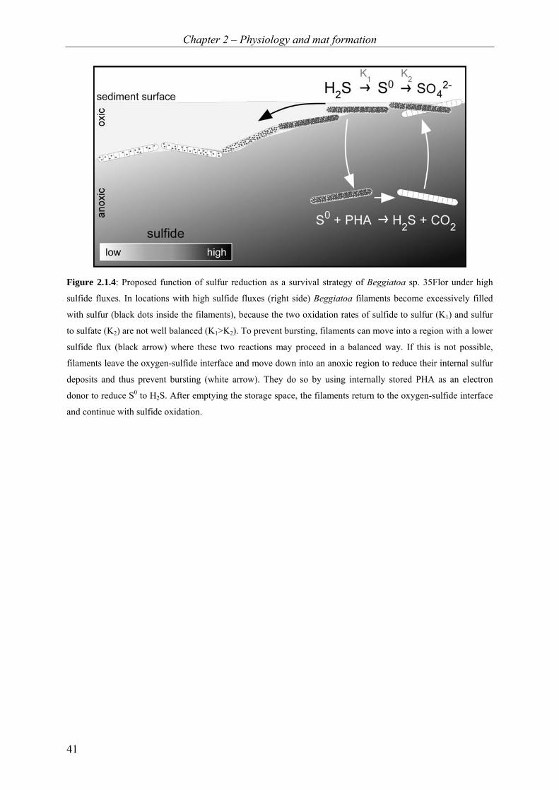
Figure 2.1.4: Proposed function of sulfur reduction as a survival strategy of Beggiatoa sp. 35Flor under high
sulfide fluxes. In locations with high sulfide fluxes (right side) Beggiatoa filaments become excessively filled
with sulfur (black dots inside the filaments), because the two oxidation rates of sulfide to sulfur (K1) and sulfur
to sulfate (K2) are not well balanced (K1>K2). To prevent bursting, filaments can move into a region with a lower
sulfide flux (black arrow) where these two reactions may proceed in a balanced way. If this is not possible,
filaments leave the oxygen-sulfide interface and move down into an anoxic region to reduce their internal sulfur
deposits and thus prevent bursting (white arrow). They do so by using internally stored PHA as an electron
donor to reduce S0 to H2S. After emptying the storage space, the filaments return to the oxygen-sulfide interface
and continue with sulfide oxidation.

Chapter 2 – Physiology and mat formation
42
Acknowledgements
We thank Martina Meyer for technical support. The study was funded by the European
Research Council (No 203364-ELNOX), the Max Planck Society and the German National
Merit Foundation.
Chapter 2 – Physiology and mat formation
43
2.2 Coordinated movement of Beggiatoa filaments in oxygen-
sulfide gradients and the effect of blue/green light
Heide N. Schulz-Vogt1*, Tine Hohmann1, Anne-Christin Kreutzmann1, Lubos Polerecky1,
Anne Schwedt1
1Max Planck Institute for Marine Microbiology, Celsiusstr. 1, D-28359 Bremen, Germany
*Corresponding author
Manuscript in preparation
Keywords: migration; blue/green light; Beggiatoa
Chapter 2 – Physiology and mat formation
44
Abstract
Filamentous sulfide-oxidizing bacteria of the genus Beggiatoa are gradient organisms. When
grown autotrophically in opposing gradients of oxygen and sulfide, the filaments establish a
thin and well-defined mat in the overlapping zone of oxygen and sulfide. We found that cyclic
Adenosine-mono-phosphate (cAMP) and cyclic diguanylate (c-di-GMP) or blue light can
modify or disturb this typical behavior.
Introduction
Large filamentous sulfur bacteria of the genus Beggiatoa form a thin mat in the overlapping
zone of oxygen and sulfide (Nelson et al., 1986a), where they oxidize the upwards diffusing
sulfide with oxygen (e.g. Winogradsky, 1887; Keil, 1912; Jørgensen, 1977; Nelson and
Castenholz, 1981). The formation of a thin mat between steep opposing gradients of oxygen
and sulfide is thought to be the result of toxicity of both compounds for these bacteria
(Winogradsky, 1887; Keil, 1912; Møller et al., 1985). Furthermore, a migrational reaction
was also observed when filaments were illuminated with light (Winogradsky, 1887; Nelson
and Castenholz, 1982; Møller et al., 1985), suggesting that light may also play a role in the
migration of Beggiatoa during mat formation. In this study, we found that, in addition to light,
chemical substances such as cyclic Adenosine-mono-phosphate (cAMP) or cyclic diguanylate
(c-di-GMP) also influence the migrational behavior and mat formation by Beggiatoa. Our
results suggest that light might directly lead to a coordinated migration of a subpopulation of
filaments and that cAMP and c-di-GMP induce a dispersion of the mat.
Results and discussion
Beggiatoa filaments of strain 35Flor were cultivated in oxygen-sulfide gradient media
(chapter 2.1) where they typically form a distinct mat in the overlaying zone of oxygen and
sulfide. We found that several factors can modify or disturb the typical mat formation
behavior of Beggiatoa filaments. The previously formed mat is disturbed and filaments
distribute randomly throughout the gradient medium when cAMP and c-di-GMP are added to
the culture (Figure 2.2.1). When grown under high sulfide fluxes (> 40 mmol m–2 d–1), a part
of the population from the mat in the oxygen-sulfide overlapping zone migrates down into the
anoxic and sulfidic zone after an incubation time of 10–14 days. These filaments were shown
to use sulfur as an electron acceptor for the oxidation of PHA, both stored internally during
the time spent in the oxygen-sulfide overlapping zone (Chapter 2.1). The downward migration
was proposed as the last resort strategy for the filaments to prevent cell disruption, which
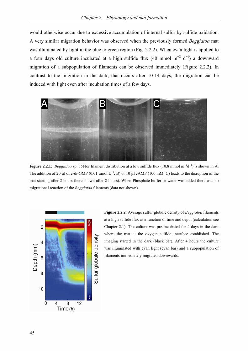
Chapter 2 – Physiology and mat formation
45
would otherwise occur due to excessive accumulation of internal sulfur by sulfide oxidation.
A very similar migration behavior was observed when the previously formed Beggiatoa mat
was illuminated by light in the blue to green region (Fig. 2.2.2). When cyan light is applied to
a four days old culture incubated at a high sulfide flux (40 mmol m–2 d–1) a downward
migration of a subpopulation of filaments can be observed immediately (Figure 2.2.2). In
contrast to the migration in the dark, that occurs after 10-14 days, the migration can be
induced with light even after incubation times of a few days.
Figure 2.2.1: Beggiatoa sp. 35Flor filament distribution at a low sulfide flux (10.8 mmol m–2d–1) is shown in A.
The addition of 20 µl of c-di-GMP (0.01 µmol L–1; B) or 10 µl cAMP (100 mM; C) leads to the disruption of the
mat starting after 2 hours (here shown after 8 hours). When Phosphate buffer or water was added there was no
migrational reaction of the Beggiatoa filaments (data not shown).
Figure 2.2.2: Average sulfur globule density of Beggiatoa filaments
at a high sulfide flux as a function of time and depth (calculation see
Chapter 2.1). The culture was pre-incubated for 4 days in the dark
where the mat at the oxygen sulfide interface established. The
imaging started in the dark (black bar). After 4 hours the culture
was illuminated with cyan light (cyan bar) and a subpopulation of
filaments immediately migrated downwards.
Chapter 2 – Physiology and mat formation
46
The data presented here show that the known signalling molecules also have an effect on an
established Beggiatoa mat and can lead to migrational reactions of the filaments. The mat is
disturbed and the filaments distribute randomly in the culture tube. Therefore, it is possible
that these substances might be used as signalling molecules among Beggiatoa filaments. In
contrast to this, a directed migration known from cultures with a high sulfide flux (Chapter
2.1) can be induced by applying blue/green light to the culture, indicating that light might be
involved in the migration of Beggiatoa filaments.
Acknowledgements
We thank Martina Meyer for technical support. The study was funded by the European
Research Council (No 203364-ELNOX) and the Max Planck Society.

Chapter 3 – Co-cultivation
47
Chapter 3
Co-cultivation of a marine
Beggiatoa strain and Pseudovibrio sp. In this chapter, the association between the large Beggiatoa sp. 35Flor and the accompanying
Pseudovibrio sp. FO-BEG1 (Figure 3) is investigated. The first part of this chapter is a study
on the co-culture of the two organisms written as a short communication. In order to study the
association from the side of the Pseudovibrio sp, the genome of this bacterium was analyzed
in detail. This second part of this chapter comprises an abstract about the genomic analysis
and comparison of the isolated Pseudovibrio sp. and another Pseudovibrio sp. strain
associated with a sponge. Both strains are able to grow in pure culture and both genomes
reveal a versatile metabolism of the bacteria. During the genome analysis of Pseudovibrio sp.
strain FO-BEG1 we found several genes encoding for more than 20 superoxide dismutases,
catalases and peroxidases additionally to genes for the interaction with other cells. The
manuscript to this second part of this chapter is added as an appendix to the thesis.

Chapter 3 – Co-cultivation
48
Figure 3: Differential interference contrast micrograph of a Beggiatoa sp. and Pseudovibrio sp. co-culture.
Contributions:
3.1 A chemolithoautotrophic Beggiatoa strain requiring the presence of a Pseudovibrio sp. for cultivation A. Schwedt and H. N. Schulz-Vogt The concept of this study was developed by both authors. I performed all experiments. The manuscript was written by me with the help of H. N. Schulz-Vogt. 3.2 The Pseudovibrio genus contains metabolically versatile and symbiotically interacting bacteria V. Bondarev, M. Richter, J. Piel, S. Romano, A. Schwedt, and H. N. Schulz-Vogt The study was initiated by V. Bondarev, H. N. Schulz-Vogt and me. Cultivation and DNA extraction for sequencing of the genome was performed by me. I coordinated the sequencing of the genome. Genome analysis was performed by V. Bondarev with assistance of M. Richter. J. Piel did NRPS and PKS cluster analysis. Cultivation experiments were performed by V. Bondarev. The manuscript was written by V. Bondarev with assistance of all co-authors.

Chapter 3 – Co-cultivation
49
3.1 A chemolithoautotrophic Beggiatoa strain requiring the
presence of a Pseudovibrio sp. for cultivation
Anne Schwedt* and Heide N. Schulz-Vogt
Max Planck Institute for Marine Microbiology, Celsiusstr. 1, D-28359 Bremen, Germany
*Corresponding author
Manuscript in preparation
Keywords: co-cultivation; gradient medium; oxidative stress; catalase
Chapter 3 – Co-cultivation
50
Abstract
Numerous attempts to isolate larger chemolithoautotrophic members of the family
Beggiatoaceae into pure culture have so far met little success. Here, we report the successful
cultivation of a marine Beggiatoa sp. strain of 6 µm in diameter, which exclusively grows in
the presence of a Pseudovibrio sp. strain.
Introduction
Beggiatoa spp. are colorless sulfur bacteria forming mats at the oxic-anoxic interface where
they oxidize sulfide with oxygen (e. g. Winogradsky, 1887; Keil, 1912; Jørgensen, 1977;
Nelson and Castenholz, 1981). Smaller chemolithoautotrophic strains (diameter < 5 µm) have
been isolated into pure culture by using an agar based medium with opposing gradients of
sulfide and oxygen (Nelson and Jannasch, 1983; Nelson et al., 1986a). Besides these strains,
only heterotrophic freshwater Beggiatoa spp. strains were obtained in pure culture (Strohl and
Larkin, 1978). The reason for the general difficulty to isolate larger chemolithoautotrophic
members of the family Beggiatoaceae in pure culture is still unknown. We obtained a marine
Beggiatoa sp. strain of 6 µm in diameter into a stable culture, but did not succeed in growing
it axenically. Apart from all attempts to clean the Beggiatoa sp. 35Flor a single type of
organism, which was identified as Pseudovibrio sp. FO-BEG1 remained. Here, we present
experiments indicating that the presence of this accompanying organism is required for the
growth of the Beggiatoa sp. strain.
Results and discussion
Under higher sulfide fluxes (> 40 mmol m−2 d−1) a subpopulation of the Beggiatoa filaments
actively migrated into lower and anoxic parts of the culture tube (Figure 3.1) where they
reduced the amount of internal sulfur by sulfur reduction (Chapter 2.2). As the accompanying
Pseudovibrio sp. FO-BEG1 is predominantly found in the oxic part of the culture tube
(Figure 3.1), these lower filaments were used to inoculate fresh culture tubes with only
Beggiatoa filaments and relatively free of the accompanying Pseudovibrio sp. strain.
When Beggiatoa filaments from the mat at the oxic-anoxic interface were transferred into
fresh medium a new mat developed in 4 out of 5 tubes. However, if filaments from the anoxic
subpopulation with no or very few Pseudovibrio cells were transferred into fresh medium,
only in 1 out of 7 tubes a mat developed. Examining this single mat-containing tube under the
microscope, besides Beggiatoa filaments also Pseudovibrio cells were observed. When

Chapter 3 – Co-cultivation
51
filaments from the anoxic part were transferred into tubes containing already a Pseudovibrio
population, most of the tubes were positive for growth of Beggiatoa sp. (5 positive tubes out
of 7 tubes). Even another Pseudovibrio strain from the culture collection (Pseudovibrio
denitrificans DSM number 17465) had a similar effect on growth (4 positive tubes out of 7).
Therefore, we conclude that the growth of the Beggiatoa sp. depends on the presence of an
accompanying bacterium of the genus Pseudovibrio.
Figure 3.1: Distribution of the accompanying Pseudovibrio sp. strain FO-BEG1 (red squares) in co-culture with
the marine Beggiatoa sp. strain 35Flor (photograph, white filaments). The Beggiatoa filaments form a mat at the
oxic-anoxic interface and a subpopulation migrates into the anoxic part of the tube. The accompanying
Pseudovibrio strain is mainly present above the upper Beggiatoa mat in the oxic part of the culture tube.
There are a few indications about the possible reasons for a positive effect of the
accompanying Pseudovibrio sp. on the growth of the large sulfide-oxidizer. Pseudovibrio
cells populate mainly the oxic part of the culture tube, but Beggiatoa filaments also thrive
well in the anoxic zone (Figure 3.1). Thus, it seems reasonable to assume that Pseudovibrio sp.
is only required during the oxic metabolism of the Beggiatoa filaments. It may be suggested
that Pseudovibrio sp. helps the Beggiatoa sp. in detoxifying oxygen radicals, as Beggiatoa
typically do not possess the ability to produce the enzyme catalase (Larkin and Strohl, 1983).
Moreover, earlier studies have shown a positive effect of catalase on the growth of Beggiatoa

Chapter 3 – Co-cultivation
52
spp. (Burton and Morita, 1964; Strohl and Larkin, 1978; Güde et al., 1981; Nelson et al.,
1986b). In contrast, the accompanying Pseudovibrio strain is catalase-positive and the
genome suggests further potential to detoxify oxygen radicals (Chapter 3.2). Therefore, we
propose that the Beggiatoa sp. benefits from the oxygen protection system of the
accompanying Pseudovibrio sp. strain and is consequently mandatory for the growth of the
large sulfide-oxidizer.
Acknowledgements This study was funded by the European Research Council (No 203364-ELNOX) and the Max Planck Society.

Chapter 3 – Co-cultivation
53
3.2 The Pseudovibrio genus contains metabolically versatile and
symbiotically interacting bacteria
Vladimir Bondarev1*, Michael Richter3, Jörn Piel2, Stefano Romano, Anne Schwedt1 and
Heide N. Schulz-Vogt1
1Max Planck Institute for Marine Microbiology, Celsiusstr. 1, D-28359 Bremen, Germany 2Kekulé-Institut für Organische Chemie und Biochemie, Gerhard-Domagk Str. 1, D-53121
Bonn, Germany 3Ribocon GmbH, Fahrenheitsstr. 1, D-28359 Bremen, Germany
*Corresponding author
Manuscript in preparation, the whole manuscript is attached to this thesis in the appendix.
Keywords: Physiology; Genome; NRPS / PKS; colibactin; Secretion system; Beggiatoa

Chapter 3 – Co-cultivation
54
Abstract
The majority of strains belonging to the genus Pseudovibrio have been isolated from marine
invertebrates like tunicates, corals and especially sponges, but the physiology of these bacteria
is poorly understood. In this study, we analyze the genomes of two Pseudovibrio strains. One
is a required symbiont of a cultivated Beggiatoa strain, a sulfide oxidizing, autotrophic
bacterium. The other one was isolated from a sponge (Enticknap et al., 2006). The data show
that both strains are generalistic bacteria capable of importing and oxidizing a wide range of
organic and inorganic compounds to meet their carbon, nitrogen, phosphorous and energy
requirements under oxic and anoxic conditions. Several physiological traits encoded in the
genome were verified in laboratory experiments with a pure culture of the Pseudovibrio strain
originally associated with Beggiatoa. Besides the versatile metabolic abilities of both
Pseudovibrio strains, our study reveals a number of open reading frames and gene clusters in
the genomes that seem to be involved in symbiont-host interactions. Pseudovibrio has the
genomic potential to attach to host cells, might be capable of interacting with the eukaryotic
cell machinery, produce secondary metabolites and may supply the host with cofactors.
Chapter 4 – Facultatively oligotrophic bacteria
55
Chapter 4
Isolation and cultivation of Pseudovibrio sp.
and other facultatively oligotrophic bacteria The fourth chapter includes a detailed physiological investigation of the growth and substrate
use of the isolated Pseudovibrio sp. FO-BEG1 under oligotrophic conditions. These results
are presented in the first manuscript of this chapter. The investigated Pseudovibrio strain is on
the one hand able to grow under extremely oligotrophic conditions and on the other hand it
can grow on organic-rich media (Figure 4). Eventually, we were able to isolate further
facultatively oligotrophic bacteria from Namibian sediments, which are highly populated by
large sulfur bacteria. The isolation and cultivation of these bacteria belonging to different
phylogenetic groups is presented in the second manuscript of this chapter.
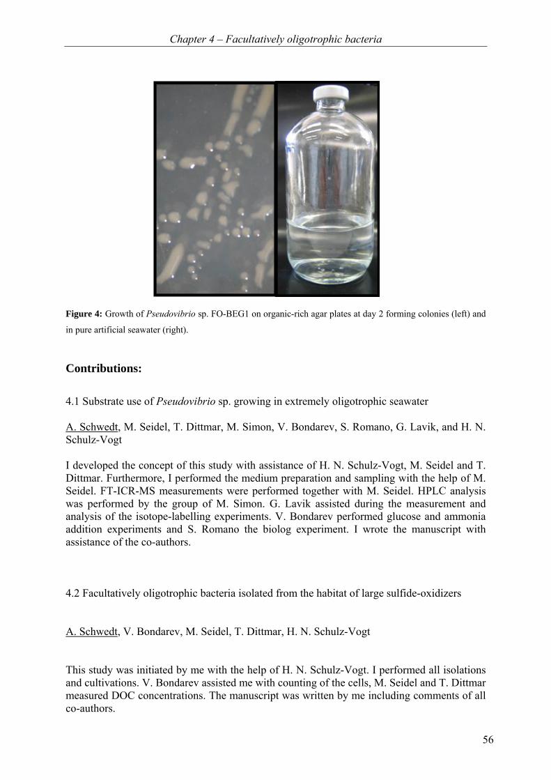
Chapter 4 – Facultatively oligotrophic bacteria
56
Figure 4: Growth of Pseudovibrio sp. FO-BEG1 on organic-rich agar plates at day 2 forming colonies (left) and
in pure artificial seawater (right).
Contributions:
4.1 Substrate use of Pseudovibrio sp. growing in extremely oligotrophic seawater A. Schwedt, M. Seidel, T. Dittmar, M. Simon, V. Bondarev, S. Romano, G. Lavik, and H. N. Schulz-Vogt I developed the concept of this study with assistance of H. N. Schulz-Vogt, M. Seidel and T. Dittmar. Furthermore, I performed the medium preparation and sampling with the help of M. Seidel. FT-ICR-MS measurements were performed together with M. Seidel. HPLC analysis was performed by the group of M. Simon. G. Lavik assisted during the measurement and analysis of the isotope-labelling experiments. V. Bondarev performed glucose and ammonia addition experiments and S. Romano the biolog experiment. I wrote the manuscript with assistance of the co-authors.
4.2 Facultatively oligotrophic bacteria isolated from the habitat of large sulfide-oxidizers
A. Schwedt, V. Bondarev, M. Seidel, T. Dittmar, H. N. Schulz-Vogt
This study was initiated by me with the help of H. N. Schulz-Vogt. I performed all isolations and cultivations. V. Bondarev assisted me with counting of the cells, M. Seidel and T. Dittmar measured DOC concentrations. The manuscript was written by me including comments of all co-authors.

Chapter 4 – Facultatively oligotrophic bacteria
57
4.1 Substrate use of Pseudovibrio sp. growing in extremely
oligotrophic seawater Anne Schwedt1*, Michael Seidel1,2, Thorsten Dittmar1,2, Meinhard Simon2, Vladimir
Bondarev1, Stefano Romano1, Gaute Lavik1, Heide N. Schulz-Vogt1
1Max Planck Institute for Marine Microbiology, Bremen, Germany 2Institute of Chemistry and Biology of the Marine Environment, Carl von Ossietzky
University of Oldenburg, Germany
*Corresponding author
Manuscript in preparation
Keywords: low nutrient medium; detergents; amino acids; oligotrophy; Pseudovibrio
Chapter 4 – Facultatively oligotrophic bacteria
58
Abstract
Marine planktonic bacteria often live in habitats with extremely low concentrations of
dissolved organic matter (DOM). To study the use of trace amounts of DOM by the
facultatively oligotrophic bacterium Pseudovibrio sp. strain FO-BEG1, we investigated the
composition of artificial and natural seawater before and after growth. For this, we
determined dissolved organic carbon (DOC), total dissolved nitrogen (TDN), composition of
DOM by electrospray ionization Fourier transform ion cyclotron resonance mass
spectrometry (ESI FT-ICR-MS) and amino acids by high performance liquid chromatography
(HPLC). The DOC concentration of artificial seawater was 0.06 mg C L−1 (5 µmol C L−1),
which was an order of magnitude below the concentration in the natural seawater
(0.9 mg C L−1 or 75 µmol C L−1). DOC concentrations did not decrease measurably during
growth. Cell numbers increased from about 20 cells mL−1 to 20,000 cells mL−1 in artificial
and to 800,000 cells mL−1 in natural seawater. No nitrogen fixation and minor CO2 fixation
(< 1%) was observed. In both media, amino acids were not the major nutrient. Instead, we
observed a decrease of compounds resembling detergents in artificial seawater, which also
contained nitrogen. In natural seawater, we detected a decrease of two groups of compounds,
one resembling amino- and thiosugars, which are also rich in nitrogen, and another group
containing condensed hydrocarbons. The present study shows that heterotrophic bacteria can
grow with even lower DOC concentrations than available in natural oligotrophic seawater and
may use unexpected organic compounds to fuel their energy, carbon and nitrogen
requirements.
Introduction
In open oceans, the amount of dissolved organic matter (DOM) is typically indicated as the
concentration of dissolved organic carbon (DOC) and is generally below 1 mg C L−1 (Schut et
al., 1997; Hansell et al., 2009). Consequently, marine bacteria are commonly exposed to very
low concentrations of organic material. Marine DOM is an extremely diverse pool of different
compounds, consisting of more than ten thousand types of molecules with different
reactivities (Dittmar and Paeng, 2009). Previous studies demonstrated that most of the DOM
in the ocean is inert to bacterial break-down and cannot be utilized by marine microorganisms
(Bada and Lee, 1977; Ammerman et al., 1984; Zweifel et al., 1993). Measurements of the
consumption of specific substrates revealed that a large fraction of the labile organic material
that is used in the upper ocean can consist of dissolved free amino acids (DFAA), dissolved
combined amino acids (DCAA) and monosaccharides. These substances can account for 5 to
Chapter 4 – Facultatively oligotrophic bacteria
59
100% of the bacterial carbon and nitrogen demand (Fuhrman, 1987; Jørgensen, 1987; Stanley
et al., 1987; Cowie and Hedges, 1992; Keil and Kirchman, 1999; Cherrier and Bauer, 2004).
In many natural systems, amino acids are only present in nanomolar concentrations even
though the flux of amino acids is comparably high due to a close coupling of their release and
uptake by planktonic microorganisms (Fuhrman, 1987; Fuhrman, 1990; Keil and Kirchman,
1999). Still, amino acids and monosaccharides alone cannot fully explain the growth of the
bacteria in all cases, where they represent only a small fraction of the C- and N-requirements
(Fuhrman, 1987; Stanley et al., 1987). The remaining demand must be covered by different
DOM compounds, which might be present at only very low concentrations. Therefore, to
specifically investigate the compounds consumed by the bacteria during growth under
oligotrophic conditions, a method detecting a broad spectrum of different organic compounds
with a low detection limit, such as electrospray ionization Fourier transform ion cyclotron
resonance mass spectrometry (ESI FT-ICR-MS), is necessary.
In contrast to natural systems where several processes supply nutrients simultaneously, a
batch culture containing a single strain of bacteria is a closed system with defined amounts of
nutrients. In such a system, it is possible to analyze compounds that are utilized during the
growth of this strain. For the cultivation of marine bacteria, defined artificial seawater or
natural seawater containing a diverse DOM pool can serve as a medium. In both seawater
media, the risk of contamination especially with amino acids is a well known problem
(Dittmar et al., 2009). In the present study, we chose to investigate the substrate use of the
marine Pseudovibrio sp. strain FO-BEG1, because it is capable to grow under oligotrophic
conditions in extremely pure artificial seawater. In spite of special purification of the artificial
seawater medium, a DOC concentration of 0.06 mg C L−1 (5 µmol C L−1) was measured,
which is, nevertheless, low compared to the natural oligotrophic seawater with a DOC
concentration of 0.9 mg C L−1 (75 µmol C L−1). We compared the substrate use in artificial
seawater with natural oligotrophic seawater. In both media, DOC concentrations did not
decrease measurably during growth of the bacteria. The composition of the DOM before and
after growth in artificial and natural seawater was analyzed by high performance liquid
chromatography (HPLC) measuring amino acids and by ESI FT-ICR-MS revealing changes
on the level of individual intact molecules indicated by molecular formulas. The combination
of these methods with isotope-labeling techniques to study incorporation of N2 and CO2
provides information of compounds used by the bacteria during growth under these extremely
oligotrophic conditions.

Chapter 4 – Facultatively oligotrophic bacteria
60
Material and methods
Medium preparation and bacterial strain
The basal artificial oligotrophic seawater medium is composed of 30.3 g NaCl, 3.3 g
MgCl2 · 6 H2O, 2.8 g MgSO4 · 2 H2O, 0.44 g CaCl2 · 2 H2O, 0.7 g KCl, 50 µl 2 mol L−1
NaOH, 2 mL 1 mol L−1 NaHCO3, 1 mL 1 mmol L−1 K2HPO4 and 1 mL trace elements
solution in 1 L MembraPure water (Optilab-Standard Water System, MembraPure,
Bodenheim, Germany). The trace elements solution was composed of 2.1 mg L−1
FeSO4 · 7 H2O, 13 mL L−1 HCl (25%), 60 mg L−1 H3BO3, 100 mg L−1 MnCl2 · 4 H2O,
190 mg L−1 CoCl2 · 6 H2O, 24 mg L−1 NiCl2 · 6 H2O, 2 mg L−1 CuCl2 · 2 H2O, 144 mg L−1
ZnSO4 · 7 H2O and 36 mg L−1 Na2MoO4 · 2 H2O. All glassware, Teflon stoppers, screw caps
and tubes were acid washed (0.1 mol L−1 HCl, Merck, Darmstadt, Germany) and rinsed with
MembraPure water (Optilab-Standard Water System, MembraPure, Bodenheim, Germany).
Glassware, aluminium foil and NaCl were combusted at 480°C for at least 3 hours. Serum
bottles (Wheaton 125 mL serum bottles clear, Wheaton, Millville, NJ, USA) were used for
cultivation, filled completely with medium and closed with Teflon stoppers. Before bottling
of the medium, the artificial seawater was cleaned by solid phase extraction of dissolved
organic matter (SPE-DOM, Dittmar et al., 2008), i.e. organic contaminations. Incubations
were performed without shaking at 28°C in the dark. For glucose and ammonium addition
experiments the oligotrophic artificial seawater medium was not specifically purified and
glucose and ammonium were added at a final concentration of 30 µmol L−1. These cultures
were incubated gently shaken (shaker GFL-3013, Gesellschaft für Labortechnik mbH,
Burgwedel, Germany) at 28°C in the dark.
Natural surface seawater was collected in the South Pacific during Integrated Ocean Drilling
(IODP) Expedition 329 from IODP Site U1368 (27.9°S ; 137.9°W) with a bucket and stored
at 4°C. Before incubation, seawater was filtered through washed Acrodisc 25 mm syringe
filters with a 0.2 µm GHP membrane (Pall Life Sciences, Ann Arbor, MI, USA) and filled
into pre-combusted serum bottles that were closed with Teflon stoppers. Inoculated cultures
were incubated without shaking at 28°C in the dark.
Media were inoculated with 100 µL of an oligotrophically pre-grown culture (about 2 × 10⁴
cells mL−1) of Pseudovibrio sp. FO-BEG1. Thus, the initial cell number after inoculation
accounts to 4 to 20 cells mL−1
Chapter 4 – Facultatively oligotrophic bacteria
61
Cell counts
Cells were stained with 1:5000 SYBR Green (SYBR Green I 10,000×, Sigma, Taufkirchen,
Germany) for 20 minutes. The stained cells were filtered onto black filters (GTTP Isopore
Membrane Filters 0.22 µm, Millipore, Schwalbach/Ts., Germany) using a Bio-Dot apparatus
(Bio-Rad, München, Germany). Counting of cells was performed using a fluorescence
microscope (Axiophot, Zeiss, Jena, Germany) at 450−490 nm excitation and 515−565 nm
emission (filter set 10, Zeiss, Jena, Germany).
Sampling and solid phase extraction of dissolved organic matter (SPE-DOM)
About 900 mL medium (six serum bottles, 156 mL each) were pooled at four different time
points (blank, t0, t1, t2). The blank was taken directly after medium preparation without
inoculation and inoculated samples were taken before (t0), after 1 week (t1) and after 3 weeks
(t2) of incubation. Samples were filtered into 1 L glass bottles using Acrodisc 25 mm syringe
filters with a 0.2 µm GHP membrane (Pall Life Sciences, Ann Arbor, MI, USA) and acidified
to pH 2 with 25% HCl. Samples for DOC measurements and amino acid analysis were taken
after pooling the six serum bottles into a 1 L glass bottle. DOC samples were measured
directly and samples for amino acid analysis were stored at −20°C. All samples were stored at
4°C until further analysis. From all four time points, DOM was extracted using the solid
phase extraction of dissolved organic matter (SPE-DOM) method (Dittmar et al., 2008). The
extracts of dissolved organic matter were analyzed using ESI FT-ICR-MS (see below).
Measurement of dissolved organic carbon (DOC) and total dissolved nitrogen (TDN)
Dissolved organic carbon (DOC) and total dissolved nitrogen (TDN) were analyzed before,
during and after growth of the bacteria using a Shimadzu TOC-VCPH total organic carbon
analyzer (Shimadzu, Kyoto, Japan) equipped with a TNM-1 total nitrogen measuring unit and
an ASI-V autosampler. To remove cells prior to analysis, samples were filtered through
Acrodisc 25 mm syringe filters with a 0.2 µm GHP membrane (Pall Life Sciences, Ann Arbor,
MI, USA). Within the instrument, samples were acidified with 1% v/v 2 mol L−1 HCl and
sparged with synthetic air for 2 minutes to remove inorganic carbon. Detection limits were
0.5 µmol L−1 for DOC and TDN (0.006 mg C L−1 and 0.007 mg N L−1, respectively).
Analytical errors based on the standard deviations for replicated measurements (at least three
measurements per sample) were within 5% for DOC and TDN. Analytical precision and
accuracy was tested in each run against deep Atlantic seawater reference material and low
carbon water provided by the consensus reference materials program (D.A. Hansell,

Chapter 4 – Facultatively oligotrophic bacteria
62
University of Miami, Coral Gables, FL, USA). Procedural blanks, including the filtration step,
were obtained with ultrapure water.
Amino acids analysis
The concentrations of dissolved free amino acids (DFAA) and total hydrolysable dissolved
amino acids (THDAA) were analyzed using high performance liquid chromatography (HPLC)
as described earlier (Lunau et al., 2006). Derivatization was performed using an ortho-
phthaldiadehyde precolumn (Lindroth and Mopper, 1979). All samples were filtered through
prewashed Acrodisc 25 mm syringe filters with a 0.2 µm GHP membrane (Pall Life Sciences,
Ann Arbor, MI, USA). Prior to the analysis of THDAA, HCl was added to a final
concentration of 1.7 mol L−1 and incubated for 1 hour at 155°C under nitrogen atmosphere.
The concentration of dissolved combined amino acids (DCAA) was calculated by subtracting
the concentration of DFAA from the concentration of THDAA.
H13CO3 and 15N2 labeling experiments
For labeling experiments, oligotrophic artificial seawater medium was prepared as described
above, however, DOM was not extracted prior to use and it was prepared under synthetic air
atmosphere (20% O2 in N2; H2O < 3 ppm-mol, CnHm < 0.1 ppm-mol, CO < 1 ppm-mol,
CO2 < 1 ppm-mol; Air Liquide, Krefeld, Germany). From each 156 mL serum bottle, which
was completely filled with medium, 5 mL medium were exchanged with 5 mL 15N2 gas and
300 µL of 1 mol L−1 NaH13CO3 were added. After inoculation with Pseudovibrio sp. FO-
BEG1 (100 µL of a pre-culture containing about 2 × 10⁴ cells mL−1), samples were incubated
for 12 hours, 3 and 5 days. Then the bacteria were filtered onto pre-combusted Glass
Microfibre Filters (GF/F, Whatman, GE Healthcare, Dassel, Germany), frozen for 12 hours
and dehydrated in an HCl desiccator. Filters were folded into tin cups and flash combusted
(1050°C) within the mass spectrometer to release N2 and CO2. For analysis, we used an
elemental analyzer (with autosampler) coupled to a Delta Plus Advantage mass spectrometer
isotope-ratio monitoring mass spectrometer (ThermoFinnigan, Bremen, Germany). To
calculate the amount of label in the cells the percentage of label added was taken into account.
Fourier transform ion cyclotron resonance mass spectrometry (FT-ICR-MS)
SPE-DOM extracts were diluted 1:1 with ultrapure water to yield DOC concentrations of
10 ppm for natural seawater and about 2 ppm for artificial seawater incubations, respectively.
Diluted samples were analyzed on a solariX Fourier transform ion cyclotron resonance mass

Chapter 4 – Facultatively oligotrophic bacteria
63
spectrometer (FT-ICR-MS; Bruker Daltonik GmbH, Bremen, Germany) connected to a 15
Tesla superconducting magnet (Bruker Biospin, Wissembourg, France). Samples were
infused at a rate of 2 µL min−1 into an electrospray source (ESI; Apollo II ion source, Bruker
Daltonik GmbH, Bremen, Germany) with the capillary voltage set to 4 kV in negative mode
and 4.5 kV in positive mode. Ions were accumulated in the hexapole for 0.3 seconds prior to
transfer into the ICR cell. Data acquisition was done in broadband mode using 4 megaword
data sets and a scanning range of 150–2000 Da. The instrument was externally calibrated with
an in-house marine deep sea DOM standard (mass accuracy of less than 0.1 ppm). Before
each sample set, blank checks with methanol/ultrapure water 1:1 were measured. For one
mass spectrum, 500 scans were added and internally calibrated with the in-house marine deep
sea DOM standard to yield a mass accuracy of less than 0.1 ppm for the internal calibrants
(series of CxHyOz compounds from 281 to 621 Da). The molecular formulas were calculated
in the mass range between 150 and 850 Da by applying the following restrictions: 12C0-130
1H0-200O1-5014N0-4S0-2P0-2 (considering also 23Na clusters in ESI positive mode).
Assignment of molecular formulas was done with the Bruker software DataAnalysis 4.0 SP 4
using the criteria described by Koch et al. (2005). Molecular formulas were considered valid
when the mass error was below 0.5 ppm. Only compounds with a signal-to-noise ratio of 3
and higher were used for further analysis. The achieved resolving power (full width half
maximum) was on average 480,000 at m/z 400.
Relative peak heights were calculated by normalization to the ten most abundant peaks in
each spectrum (triplicate measurements). These peak heights were then used to semi-
quantitatively assess changes in DOM composition in natural oligotrophic seawater
experiments. FT-ICR-MS is not a quantitative tool for DOM analysis due to the lack of
standards and differences in ionization efficiencies for different compounds. However, this
method yields reproducible results because the variability of relative peak heights is low
under similar analytical conditions (Kido Soule et al., 2010).
Sequential standard addition was applied to quantify the amount of polyethylene glycol (PEG)
contamination in the artificial seawater medium using an authentic PEG standard
(octaethylene glycol, C16H34O9, ≥ 99% oligomer purity, 370 g mol−1, Sigma, Taufkirchen,
Germany). PEG was added in concentrations of 1 to 3.6 nmol L−1 and FT-ICR mass spectra
were recorded in ESI positive mode. To yield a linear response range in the MS, ion
accumulation time was lowered to 0.1 seconds and 50 scans were added for one mass
Chapter 4 – Facultatively oligotrophic bacteria
64
spectrum. The amount of PEG in the samples was calculated from a linear calibration derived
from peak heights in FT-ICR mass spectra. To investigate the suppression effect of the
inorganic contamination on organic peaks during FT-ICR-MS, the in-house natural deep sea
DOM standard was added in the range of 0.04 to 4 mg C L−1 to a sample contaminated with
the inorganic substances and measured in ESI negative mode.
Biolog experiment
A substrate respiration test was performed using a biolog GN2 plate (Hayward, CA, USA)
with 95 different substrates. The bacteria were pre-grown in artificial seawater medium in 1 L
bottles (Schott, Mainz, Germany). After 6 days, cells were concentrated by centrifugation for
2 hours at 11,000 × g and 15°C using a J-26XP Beckman centrifuge (Beckman Coulter GmbH,
Krefeld, Germany). All centrifugation tubes were acid washed (0.1 mol L−1 HCl) and rinsed
with water. The obtained pellet was suspended in 20 mL of sterile saline solution (23.5 g L−1
NaCl and 10.6 g L−1 MgCl2 · 6 H2O). To verify the viability of the cells, 50 µL of the cell
suspension was spread on organic-rich medium containing 2 g polypeptone, 0.5 g Bacto yeast
extract (BD Diagnostics, Heidelberg, Germany), 30 g NaCl, 5 g MgCl2 · 6 H2O, 0.005 g
CaCl2 · 2 H2O, 0.005 g Na2MoO4 · 7 H2O, 0.004 g CuCl2 · 2 H2O, 0.006 g FeCl3 · 6 H2O, 15 g
bactoagar in 1 L MembraPure water and the pH was adjusted to 8 with 1 mol L−1 NaOH. The
biolog plate was inoculated with 150 µL of the concentrated cells and incubated at 28oC in the
dark in a humidity chamber to prevent excess evaporation. Activity of cells was checked
visually each day for a total of 14 days.
Results
Growth under oligotrophic conditions
The investigated Pseudovibrio sp. strain FO-BEG1 was able to multiply in both artificial and
natural seawater. The initial cell number was about 20 cells mL−1 and in artificial seawater
cell numbers increased to 2 × 10⁴ cells mL−1 at time points t1 and t2 (one and three weeks after
inoculation, respectively). Natural seawater medium contained about 8 × 10⁵ cell mL−1 at both
time points. In both media, the increase in cell numbers was detected during the first week of
incubation (t0 to t1) and cell numbers did not change in the following two weeks (t1 to t2).

Chapter 4 – Facultatively oligotrophic bacteria
65
Dissolved organic carbon (DOC), total dissolved nitrogen (TDN) and dissolved free and
combined amino acids (DFAA and DCAA)
In pure artificial seawater medium before bottling, the DOC contamination from salts was
only 1 μmol C L−1. During the bottling of the medium and with the addition of NaHCO3,
K2HPO4, NaOH and trace elements, the concentration of DOC increased to about
4.5 µmol C L−1 (Table 4.1.1). The TDN was below limit of detection (0.5 µmol N L−1) in the
sterile artificial medium before and after bottling (Table 4.1.1). Inoculation did not introduce
further measurable DOC or TDN to the medium (t0). After one (t1) and three weeks (t2) of
incubation, the concentrations of DOC and TDN did not change significantly in the medium
(Table 4.1.1). In comparison to the artificial seawater medium, the DOC concentration in
natural seawater was about 16 times higher (71 µmol L−1; Table 4.1.1). However, no
significant change in DOC concentration was detectable during growth, as DOC
concentrations one and three weeks after inoculation were 78.5 and 75 µmol C L−1,
respectively. A similar pattern was observed for TDN concentrations, which were around
12 µmol N L−1 at all time points (Table 4.1.1).
In contrast to DOC and TDN, the amount of DFAA decreased in the artificial seawater
cultures within the first week of growth (from t0 to t1). This decrease represents a DFAA
uptake of 0.06 µmol C L−1 and 0.02 µmol N L−1 (Table 4.1.1). However, the concentrations of
DFAA remained constant until t2. The most abundant amino acids were serine, glycine and
alanine, which together accounted for 0.05 µmol C L−1. The decrease in DFAA (from t0 to t1)
was concurrently observed with an increase of DCAA. During the stationary growth phase
(from t1 to t2), the DCAA concentrations decreased again (Table 4.1.1). Opposed to the
artificial seawater incubations, the amino acids concentrations (THDAA) in natural seawater
were very low and no change during incubation could be observed (Table 4.1.1).
Nitrogen and carbon dioxide fixation
The incorporation of labeled nitrogen (15N2) and carbon (NaH13CO3) into Pseudovibrio sp.
FO-BEG1 growing in pure artificial seawater medium was examined using isotope-ratio
monitoring mass spectrometry. Less than 1% of cellular carbon originated from carbon
dioxide fixation under the oligotrophic growth conditions in artificial seawater. Nitrogen
fixation was not detectable.
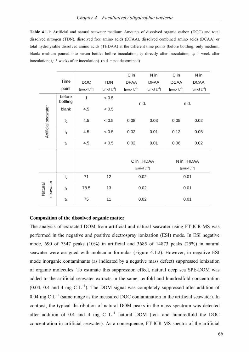
Chapter 4 – Facultatively oligotrophic bacteria
66
Table 4.1.1: Artificial and natural seawater medium: Amounts of dissolved organic carbon (DOC) and total
dissolved nitrogen (TDN), dissolved free amino acids (DFAA), dissolved combined amino acids (DCAA) or
total hydrolysable dissolved amino acids (THDAA) at the different time points (before bottling: only medium;
blank: medium poured into serum bottles before inoculation; t0: directly after inoculation; t1: 1 week after
inoculation; t2: 3 weeks after inoculation). (n.d. = not determined)
Time
point DOC
[µmol L−1] TDN
[µmol L−1]
C in
DFAA [µmol L−1]
N in
DFAA [µmol L−1]
C in
DCAA [µmol L−1]
N in
DCAA [µmol L−1]
before bottling
1 < 0.5
blank 4.5 < 0.5 n.d. n.d.
t0 4.5 < 0.5 0.08 0.03 0.05 0.02
t1 4.5 < 0.5 0.02 0.01 0.12 0.05
Arti
ficia
l sea
wat
er
t2 4.5 < 0.5 0.02 0.01 0.06 0.02
C in THDAA [µmol L−1]
N in THDAA [µmol L−1]
t0 71 12 0.02 0.01
t1 78.5 13 0.02 0.01
Nat
ural
seaw
ater
t2 75 11 0.02 0.01
Composition of the dissolved organic matter
The analysis of extracted DOM from artificial and natural seawater using FT-ICR-MS was
performed in the negative and positive electrospray ionization (ESI) mode. In ESI negative
mode, 690 of 7347 peaks (10%) in artificial and 3685 of 14873 peaks (25%) in natural
seawater were assigned with molecular formulas (Figure 4.1.2). However, in negative ESI
mode inorganic contaminants (as indicated by a negative mass defect) suppressed ionization
of organic molecules. To estimate this suppression effect, natural deep sea SPE-DOM was
added to the artificial seawater extracts in the same, tenfold and hundredfold concentration
(0.04, 0.4 and 4 mg C L−1). The DOM signal was completely suppressed after addition of
0.04 mg C L−1 (same range as the measured DOC contamination in the artificial seawater). In
contrast, the typical distribution of natural DOM peaks in the mass spectrum was detected
after addition of 0.4 and 4 mg C L−1 natural DOM (ten- and hundredfold the DOC
concentration in artificial seawater). As a consequence, FT-ICR-MS spectra of the artificial
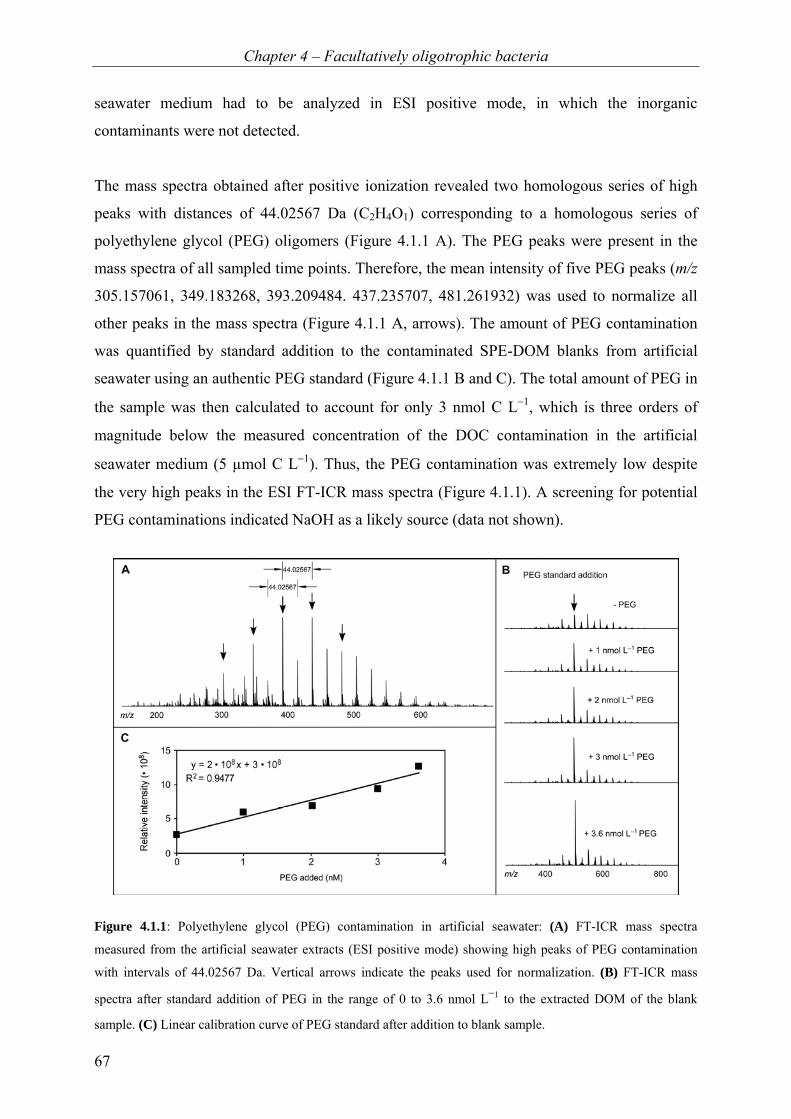
Chapter 4 – Facultatively oligotrophic bacteria
67
seawater medium had to be analyzed in ESI positive mode, in which the inorganic
contaminants were not detected.
The mass spectra obtained after positive ionization revealed two homologous series of high
peaks with distances of 44.02567 Da (C2H4O1) corresponding to a homologous series of
polyethylene glycol (PEG) oligomers (Figure 4.1.1 A). The PEG peaks were present in the
mass spectra of all sampled time points. Therefore, the mean intensity of five PEG peaks (m/z
305.157061, 349.183268, 393.209484. 437.235707, 481.261932) was used to normalize all
other peaks in the mass spectra (Figure 4.1.1 A, arrows). The amount of PEG contamination
was quantified by standard addition to the contaminated SPE-DOM blanks from artificial
seawater using an authentic PEG standard (Figure 4.1.1 B and C). The total amount of PEG in
the sample was then calculated to account for only 3 nmol C L−1, which is three orders of
magnitude below the measured concentration of the DOC contamination in the artificial
seawater medium (5 µmol C L−1). Thus, the PEG contamination was extremely low despite
the very high peaks in the ESI FT-ICR mass spectra (Figure 4.1.1). A screening for potential
PEG contaminations indicated NaOH as a likely source (data not shown).
Figure 4.1.1: Polyethylene glycol (PEG) contamination in artificial seawater: (A) FT-ICR mass spectra
measured from the artificial seawater extracts (ESI positive mode) showing high peaks of PEG contamination
with intervals of 44.02567 Da. Vertical arrows indicate the peaks used for normalization. (B) FT-ICR mass
spectra after standard addition of PEG in the range of 0 to 3.6 nmol L−1 to the extracted DOM of the blank
sample. (C) Linear calibration curve of PEG standard after addition to blank sample.
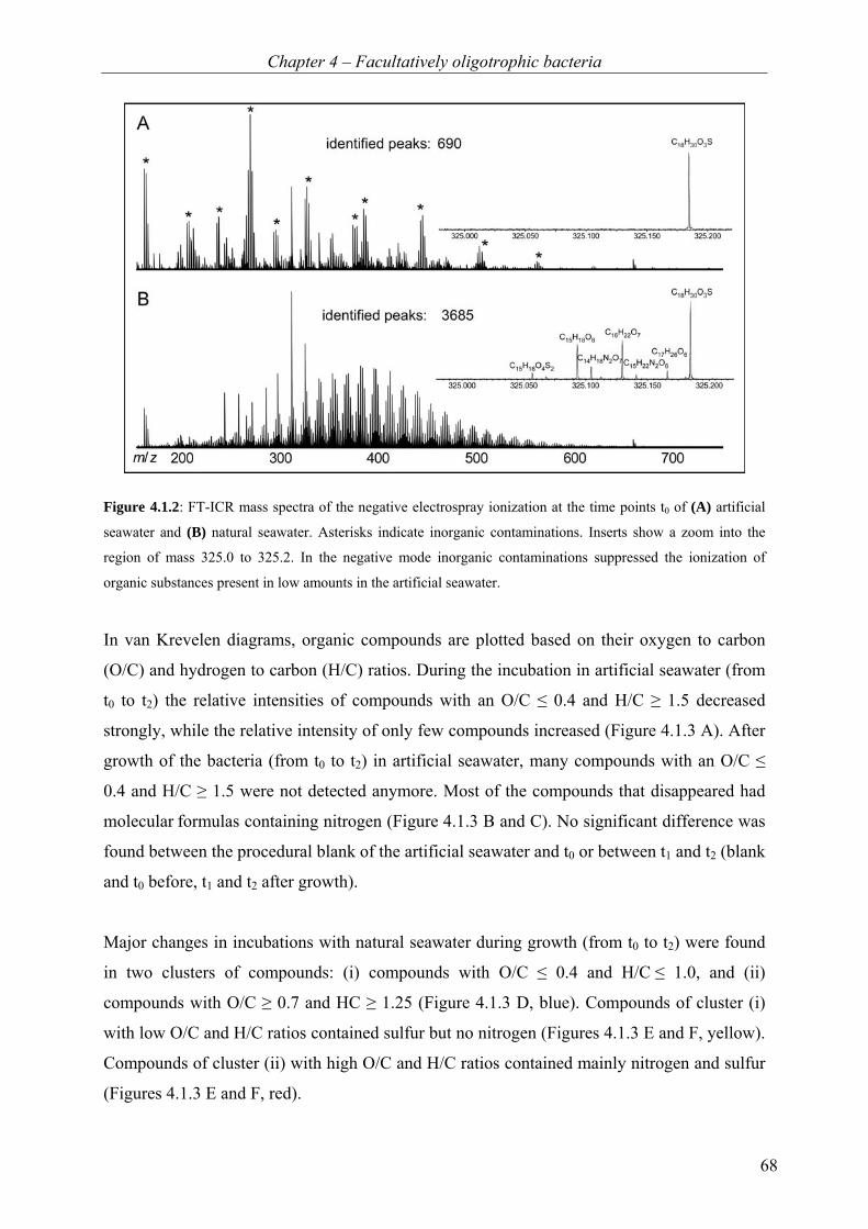
Chapter 4 – Facultatively oligotrophic bacteria
68
Figure 4.1.2: FT-ICR mass spectra of the negative electrospray ionization at the time points t0 of (A) artificial
seawater and (B) natural seawater. Asterisks indicate inorganic contaminations. Inserts show a zoom into the
region of mass 325.0 to 325.2. In the negative mode inorganic contaminations suppressed the ionization of
organic substances present in low amounts in the artificial seawater.
In van Krevelen diagrams, organic compounds are plotted based on their oxygen to carbon
(O/C) and hydrogen to carbon (H/C) ratios. During the incubation in artificial seawater (from
t0 to t2) the relative intensities of compounds with an O/C ≤ 0.4 and H/C ≥ 1.5 decreased
strongly, while the relative intensity of only few compounds increased (Figure 4.1.3 A). After
growth of the bacteria (from t0 to t2) in artificial seawater, many compounds with an O/C ≤
0.4 and H/C ≥ 1.5 were not detected anymore. Most of the compounds that disappeared had
molecular formulas containing nitrogen (Figure 4.1.3 B and C). No significant difference was
found between the procedural blank of the artificial seawater and t0 or between t1 and t2 (blank
and t0 before, t1 and t2 after growth).
Major changes in incubations with natural seawater during growth (from t0 to t2) were found
in two clusters of compounds: (i) compounds with O/C ≤ 0.4 and H/C ≤ 1.0, and (ii)
compounds with O/C ≥ 0.7 and HC ≥ 1.25 (Figure 4.1.3 D, blue). Compounds of cluster (i)
with low O/C and H/C ratios contained sulfur but no nitrogen (Figures 4.1.3 E and F, yellow).
Compounds of cluster (ii) with high O/C and H/C ratios contained mainly nitrogen and sulfur
(Figures 4.1.3 E and F, red).
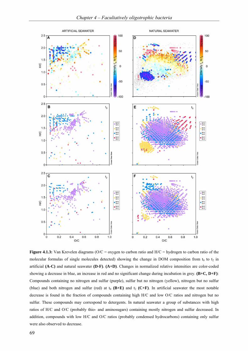
Chapter 4 – Facultatively oligotrophic bacteria
69
Figure 4.1.3: Van Krevelen diagrams (O/C = oxygen to carbon ratio and H/C = hydrogen to carbon ratio of the
molecular formulas of single molecules detected) showing the change in DOM composition from t0 to t2 in
artificial (A-C) and natural seawater (D-F). (A+D). Changes in normalized relative intensities are color-coded
showing a decrease in blue, an increase in red and no significant change during incubation in grey. (B+C, D+F):
Compounds containing no nitrogen and sulfur (purple), sulfur but no nitrogen (yellow), nitrogen but no sulfur
(blue) and both nitrogen and sulfur (red) at t0 (B+E) and t2 (C+F). In artificial seawater the most notable
decrease is found in the fraction of compounds containing high H/C and low O/C ratios and nitrogen but no
sulfur. These compounds may correspond to detergents. In natural seawater a group of substances with high
ratios of H/C and O/C (probably thio- and aminosugars) containing mostly nitrogen and sulfur decreased. In
addition, compounds with low H/C and O/C ratios (probably condensed hydrocarbons) containing only sulfur
were also observed to decrease.
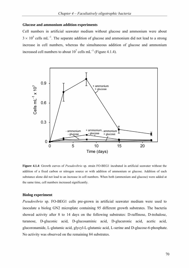
Chapter 4 – Facultatively oligotrophic bacteria
70
Glucose and ammonium addition experiments
Cell numbers in artificial seawater medium without glucose and ammonium were about
3 × 10⁵ cells mL−1. The separate addition of glucose and ammonium did not lead to a strong
increase in cell numbers, whereas the simultaneous addition of glucose and ammonium
increased cell numbers to about 107 cells mL−1 (Figure 4.1.4).
Figure 4.1.4: Growth curves of Pseudovibrio sp. strain FO-BEG1 incubated in artificial seawater without the
addition of a fixed carbon or nitrogen source or with addition of ammonium or glucose. Addition of each
substance alone did not lead to an increase in cell numbers. When both (ammonium and glucose) were added at
the same time, cell numbers increased significantly.
Biolog experiment
Pseudovibrio sp. FO-BEG1 cells pre-grown in artificial seawater medium were used to
inoculate a biolog GN2 microplate containing 95 different growth substrates. The bacteria
showed activity after 8 to 14 days on the following substrates: D-raffinose, D-trehalose,
turanose, D-gluconic acid, D-glucosaminic acid, D-glucuronic acid, acetic acid,
glucoronamide, L-glutamic acid, glycyl-L-glutamic acid, L-serine and D-glucose-6-phosphate.
No activity was observed on the remaining 84 substrates.
Chapter 4 – Facultatively oligotrophic bacteria
71
Discussion
In the present study, the heterotrophic Pseudovibrio sp. strain FO-BEG1 was grown either in
artificial seawater medium with extremely low DOC concentrations or in natural seawater
from an oligotrophic ocean (south Pacific, surface water) representing typical marine DOM.
The bacterial growth was sustained by very low amounts of DOC in both incubation setups.
However, growth in artificial seawater with only 0.06 mg DOC per liter seawater shows that
bacterial growth can be sustained by even lower concentration as present in most natural
oligotrophic seawaters, which typically contain less than 1 mg C L−1 (Schut et al., 1997;
Hansell et al., 2009).
Impurities in oligotrophic media
During the preparation of oligotrophic medium, different potential contamination risks
emerge. In our case, an inorganic contamination was introduced to the artificial seawater after
the incubation during the solid phase extraction of DOM (SPE-DOM). This led to ion
suppression of the organic compounds in the FT-ICR mass spectra in ESI negative mode.
Consequently, it was not possible to analyze the DOM composition in that mode and we
chose the positive mode instead. This problem does not occur in natural seawater samples,
because the concentration of DOC is higher. We could show that the organic substances
present in the artificial medium are detectable if the overall background of DOM is 10 to 100
times higher. This observation indicates that a specific concentration of organic matter has to
be present to be detected by ESI FT-ICR-MS if inorganic substances are introduced in high
amounts during extraction or ionize extremely well.
In ESI positive mode, an organic contamination of the artificial seawater was observed. Two
series of polyethylene glycol (PEG) oligomers were detected. As a potential source NaOH
used for medium preparation was identified. Since PEGs ionize extremely well in ESI
positive mode, the peaks are much higher compared to most other peaks in the sample. Using
the standard addition method (described in material and methods), the total amount of PEG
contaminations was estimated to be 1000fold less than the total DOC concentration in
artificial seawater, and thus represented only a very small fraction of the entire DOC
contamination. These impurities analyzed in the artificial seawater medium were not detected
in the SPE-DOM of natural seawater samples and thus did not affect the FT-ICR-MS analysis
of natural seawater incubations.
Chapter 4 – Facultatively oligotrophic bacteria
72
Growth and substrate use in artificial seawater
During growth in artificial seawater under extremely oligotrophic conditions, Pseudovibrio sp.
strain FO-BEG1 multiplied from about 20 cells mL−1 to 2 × 10⁴ cells mL−1, even though the
overall amount of DOC did not measurably decrease. Thus, the amount of compounds that
were consumed was probably below detection limit (0.5 µmol C L−1). Based on the increase
in cell numbers when grown with glucose and ammonium (supplementary material Table
S.4.1 and Figure S.4.1), it was calculated that about 1 to 3 µmol C L−1 is needed for the
observed cell numbers as carbon and energy source. Apparently, the uptake of amino acids
alone could not sustain bacterial growth because the initial amino acid concentration (0.13
µmol L−1) was already much lower than the required 1 to 3 µmol C L−1. During the initial
growth phase, we found a decrease in dissolved free amino acids (DFAA) concentration
concurrently with an increase in dissolved combined amino acids (DCAA), whereas the
overall amino acid concentration (DFAA + DCAA) did not decrease (Table 4.1.1). Within the
growth phase, the DFAA might have been used as precursors, e.g. for exo-enzymes, which
lead in turn to an increase in the DCAA concentration, but no overall decrease in the amino
acid concentration. During stationary phase, we found a slight decrease of total amino acids.
This may suggest that amino acids were rather used as substrate for maintaining non-growing
cells.
The compositional analysis of DOM with ESI FT-ICR-MS revealed a decrease of nitrogen-
containing compounds during the initial growth phase (Figure 4.1.3 B and C). Inferring from
their molecular compositions (high H/C, low O/C, N-containing), these substances were most
likely detergents, such as ampholytic amino oxides or betaines. Using this method we cannot
quantify the amount of carbon corresponding to this decrease. In addition, a compound with a
decreasing peak in the mass spectrum is not necessarily completely oxidized to CO2. Thus, we
cannot ultimately clarify whether the use of these substrates alone explains the observed
growth. Nevertheless, the preferential decrease of nitrogen-containing compounds suggests
that these substances at least served as nitrogen source. This agrees with the observation that
N2 fixation was not detectable.
Growth and substrate use in natural seawater
The overall concentration of amino acids in the natural seawater was already initially quite
low and did not further decrease during the incubation. Thus, amino acids did not sustain
growth or survival of cells. The compositional analysis of DOM showed a decrease of two

Chapter 4 – Facultatively oligotrophic bacteria
73
groups of compounds. One of them was likely containing carbohydrate-like compounds with
nitrogen and sulfur. These compounds are most likely thiosugars and/or aminosugars and may
therefore have also served as nitrogen source. In addition, we observed a decrease in relative
intensities of compounds with low O/C and H/C ratios containing sulfur but no nitrogen.
Compounds with low amounts of oxygen and hydrogen in comparison to carbon are typically
condensed or aromatic hydrocarbons. Since it cannot be excluded that these compounds were
lost abiotically, e.g. by absorption to the glassware, it needs to be further investigated if the
bacteria indeed metabolized these complex molecules during growth. Notably, during growth
of Pseudovibrio sp. strain FO-BEG1 in natural seawater, we observed a decrease of different
groups of substances compared to artificial seawater and preferentially substances containing
sulfur decreased.
The use of multiple substrates
The potential use of different substrates under oligotrophic conditions was shown by the
biolog experiment. The bacteria were able to metabolize different types of organic compounds,
such as sugars (e.g. D-raffinose and D-trehalose), amino acids (L-serine, glutamic acid),
carboxylic acids (glucuronic acid, acetic acid) and amide (glucuronamide). These compound
classes were also present as contaminations in the artificial seawater medium and decreased
during growth. This is in agreement with studies on E. coli showing a broad potential to use
different substrates after adaptation to carbon starvation (Ihssen and Egli, 2005).
Even though the substances decreasing during growth, as revealed by ESI FT-ICR-MS,
tended to cluster into certain groups with a specific ratio of O/C and H/C, the overall pattern
showed a broad simultaneous use of many substrates both in artificial and natural seawater. In
contrast to the biolog experiment, this does not show the potential to use a substrate, but the
actual decrease of compounds present in original concentrations. Using different compounds
simultaneously may enable bacteria to grow on very low concentrations of each of the
different substrates (Lendenmann et al., 1996; Kovárová-Kovar and Egli, 1998), if a substrate
does not repress enzymes for another less efficient one (reviewed in Egli, 2010). This strategy,
together with the potential to use substrates, which are not present but may become available,
enables bacteria to survive in habitats with a low and fluctuating supply of nutrients, as it is
found in the oceans.
Chapter 4 – Facultatively oligotrophic bacteria
74
Conclusions
The data presented in this study demonstrate that contaminations can arise from many
different sources (e.g. chemicals, bottles, humans or plastics), which in turn might support
growth of bacteria under oligotrophic conditions. Moreover, even under extremely
oligotrophic conditions, the isolated bacteria were not in a resting state, but showed a
moderate growth, even though nitrogen, carbon and energy sources were limiting factors at
the same time. The investigated Pseudovibrio sp. FO-BEG1 uses many different types of
substrates under nutrient-poor conditions as demonstrated by the FT-ICR-MS data (Figure
4.1.3). In our case, amino acids were not the primary substrate for growth, but rather a
complex mixture of organic compounds, preferably containing nitrogen. Furthermore, we
were able to show that ESI FT-ICR-MS is a strong tool to investigate bacterial growth under
low-nutrient conditions.
Chapter 4 – Facultatively oligotrophic bacteria
75
Acknowledgments
We thank S. Menger, M. Meyer, M. Friebe, K. Klaproth, B. Kürzel and T. Max for technical
support and T. G. Ferdelman and W. Ziebis for providing us with natural seawater samples
from the Integrated Ocean Drilling (IODP) Expedition 329. This study is funded by the
European Research Council (No 203364-ELNOX) and the Max Planck Society.

Chapter 4 – Facultatively oligotrophic bacteria
76
Supplementary figure and table Table S.4.1: Cell numbers of isolate Pseudovibrio strain FO-BEG1 derived from different amounts of carbon
added to the medium. Carbon in form of glucose was added to a Pseudovibrio strain FO-BEG1 culture in
2 different concentrations. As a negative control a culture without addition of carbon was used. To ensure
carbon-dependent growth, ammonium was added to the medium (with and without carbon addition) as nitrogen
source.
Figure S.4.1: Amount of carbon needed for specific cell number. From additions of glucose to the medium
(Table S.4.1) the amount of carbon needed for growth of isolate FO-BEG1 was calculated by subtracting the cell
numbers without carbon addition (as negative control) from the cell numbers with carbon addition and plotting
amount of carbon versus cell numbers produced. The amount of carbon needed for 105 cells mL−1 was calculated
to be 3 µmol C L−1 and for 104 cells 1 µmol C L−1.
Carbon added Ammonium added Cells mL−1 (day 7-8)
60 µmol L−1 10 mmol L−1 31 x 105
180 µmol L−1 30 µmol L−1 72 x 105
0 µmol L−1 10 mmol L−1 3 x 10⁵
0 µmol L−1 30 µmol L−1 10 x 105

Chapter 4 – Facultatively oligotrophic bacteria
77
4.2 Facultatively oligotrophic bacteria isolated from the habitat of
large sulfide-oxidizers Anne Schwedt1*, Vladimir Bondarev1, Michael Seidel1,2, Thorsten Dittmar1,2, Heide N.
Schulz-Vogt1
1Max Planck Institute for Marine Microbiology, Celsiusstr. 1, D-28359 Bremen, Germany 2Institute for Chemistry and Biology of the Marine Environment, Carl von Ossietzky
University of Oldenburg, Germany
*Corresponding author
Manuscript in preparation
Keywords: cultivation; facultative oligotrophy; Pseudovibrio; Marinobacter

Chapter 4 – Facultatively oligotrophic bacteria
78
Abstract
Axenic cultivation of large chemolithoautotrophic bacteria belonging to the genus Beggiatoa
succeed only rarely. Growth of the large sulfide-oxidizers often seems to be dependent on the
presence of heterotrophic prokaryotes, similar to the often described associations of
cyanobacteria and heterotrophs. Recently, we observed that the growth of the marine,
chemolithoautotrophic Beggiatoa sp. strain 35Flor depends on the presence of the
α-Proteobacterium Pseudovibrio sp. strain FO-BEG1. Furthermore, we found that this
bacterium, besides heterotrophic growth on organic-rich medium, is capable of growth under
extreme nutrient deficiency in artificial and natural seawater. This observation inspired us to
investigate whether we could isolate other facultative oligotrophs from overlaying water of
Namibian sediment, an environment known to contain a large number of different sulfide-
oxidizers belonging to the family Beggiatoaceae. Indeed, we succeeded to obtain 14 new
strains closely related to known marine bacteria, all of which were capable of growth under
extreme nutrient deficiency. The potential of these isolates to support growth of the large
sulfide-oxidizing bacteria can now be studied in culture-based experiments.
Introduction
Large bacteria, such as Beggiatoa spp. or filamentous cyanobacteria, often live together with
heterotrophic prokaryotes and these associations seem to be the reason for the inability of
axenic cultivation of the large bacteria (Burton and Morita, 1964; Cohen and Rosenberg, 1989;
Palinska et al., 1999; Morris et al., 2008). Different reasons for these interactions have been
proposed, such as the recycling of carbon dioxide or the reduction of the oxygen
concentration (Kuentzel, 1969; Paerl and Pinckney, 1996). It is known that Beggiatoa spp.
typically lack the gene for catalase (Larkin and Strohl, 1983), but since aerobic respiration
produces reactive oxygen species (ROS, Tapley et al., 1999) an efficient protection against
such molecules is needed. Therefore, Beggiatoa spp. might depend on the enzyme catalase,
which catalyzes the disproportionation of hydrogen peroxide to oxygen and water, or other
protection systems of the associated heterotrophs against ROS. The positive effect of catalase
and of accompanying heterotrophic bacteria on the growth of Beggiatoa filaments (Burton
and Morita, 1964; Strohl and Larkin, 1978; Güde et al., 1981; Nelson et al., 1986b) and,
furthermore, the accumulation of peroxides in cultures without catalase or accompanying
bacteria was shown (Burton and Morita, 1964). Recently, we found that both Pseudovibrio sp.
strain FO-BEG1 and Pseudovibrio denitrificans (DSM number 17465) support growth of the
marine chemolithoautotrophic Beggiatoa sp. strain 35Flor (Chapter 3.1). Both bacterial

Chapter 4 – Facultatively oligotrophic bacteria
79
strains are heterotrophic organisms (Chapter 3.2, Shieh et al., 2004). Additionally, we have
shown that the newly isolated Pseudovibrio sp. strain FO-BEG1 can grow under extremely
oligotrophic conditions and its substrate use in pure artificial and natural seawater was studied
in detail (Chapter 4.1).
The aim of the present study was to isolate heterotrophic, facultatively oligotrophic bacteria
from Namibian sediments, the habitat of large sulfide-oxidizers, to investigate how common
facultative oligotrophy is among bacteria associated with large sulfide-oxidizers. Here, we
report the successful isolation of 14 facultatively oligotrophic bacteria from water overlaying
Namibian sediments using a method relying on the change from oligotrophic to eutrophic
growth conditions, called the CANgrow−method (changing availability of nutrients growth−
method). In contrast to earlier methods for the isolation of marine bacteria, the artificial
oligotrophic medium used here, is defined and contains much lower nutrient concentrations.
Three initial transfers strongly pre-select for bacteria, which can grow under extreme nutrient
deficiency. Subsequently, three transfers on nutrient-rich agar plates select for facultatively
oligotrophic bacteria and are used to obtain pure cultures. Finally, the ability of the isolates to
grow oligotrophically is ensured by at least seven transfers in pure artificial seawater.
Material and methods
Samples
The new bacterial strains were isolated from oceanic bottom water overlaying Namibian
sediments that harbor different large sulfur bacteria (sample acquisition described in Salman
et al., 2011). All samples were stored at 4°C. In addition to the new isolates, the Pseudovibrio
denitrificans type strain (DSM number 17465) was purchased from the German culture
collection DSMZ (Deutsche Sammlung von Mikroorganismen und Zellkulturen GmbH,
Braunschweig, Germany) and cultivated under oligotrophic conditions.
Growth media and cultivation conditions
For cultivation and isolation, two different media were used, an oligotrophic and a eutrophic
medium. The liquid, oligotrophic medium was composed as described above (Chapter 4.1),
but prepared under synthetic air atmosphere (20% O2 in N2; H2O < 3 ppm-mol, CnHm < 0.1
ppm-mol, CO < 1 ppm-mol, CO2 < 1 ppm-mol). Furthermore, bottles were filled only with
50 mL medium and the medium was not cleaned using solid phase extraction. This medium
was used for all oligotrophic cultivation experiments. The solid, eutrophic medium was
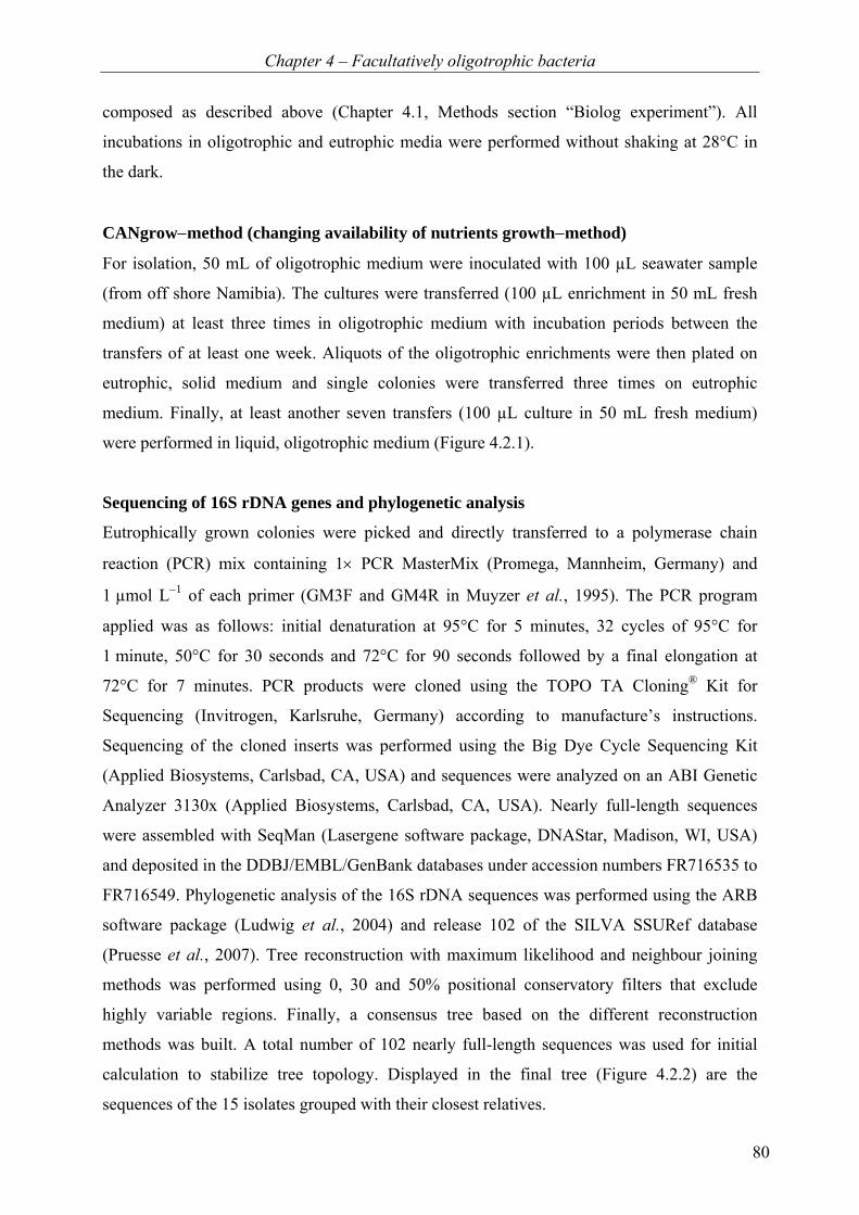
Chapter 4 – Facultatively oligotrophic bacteria
80
composed as described above (Chapter 4.1, Methods section “Biolog experiment”). All
incubations in oligotrophic and eutrophic media were performed without shaking at 28°C in
the dark.
CANgrow−method (changing availability of nutrients growth−method)
For isolation, 50 mL of oligotrophic medium were inoculated with 100 µL seawater sample
(from off shore Namibia). The cultures were transferred (100 µL enrichment in 50 mL fresh
medium) at least three times in oligotrophic medium with incubation periods between the
transfers of at least one week. Aliquots of the oligotrophic enrichments were then plated on
eutrophic, solid medium and single colonies were transferred three times on eutrophic
medium. Finally, at least another seven transfers (100 µL culture in 50 mL fresh medium)
were performed in liquid, oligotrophic medium (Figure 4.2.1).
Sequencing of 16S rDNA genes and phylogenetic analysis
Eutrophically grown colonies were picked and directly transferred to a polymerase chain
reaction (PCR) mix containing 1× PCR MasterMix (Promega, Mannheim, Germany) and
1 µmol L−1 of each primer (GM3F and GM4R in Muyzer et al., 1995). The PCR program
applied was as follows: initial denaturation at 95°C for 5 minutes, 32 cycles of 95°C for
1 minute, 50°C for 30 seconds and 72°C for 90 seconds followed by a final elongation at
72°C for 7 minutes. PCR products were cloned using the TOPO TA Cloning® Kit for
Sequencing (Invitrogen, Karlsruhe, Germany) according to manufacture’s instructions.
Sequencing of the cloned inserts was performed using the Big Dye Cycle Sequencing Kit
(Applied Biosystems, Carlsbad, CA, USA) and sequences were analyzed on an ABI Genetic
Analyzer 3130x (Applied Biosystems, Carlsbad, CA, USA). Nearly full-length sequences
were assembled with SeqMan (Lasergene software package, DNAStar, Madison, WI, USA)
and deposited in the DDBJ/EMBL/GenBank databases under accession numbers FR716535 to
FR716549. Phylogenetic analysis of the 16S rDNA sequences was performed using the ARB
software package (Ludwig et al., 2004) and release 102 of the SILVA SSURef database
(Pruesse et al., 2007). Tree reconstruction with maximum likelihood and neighbour joining
methods was performed using 0, 30 and 50% positional conservatory filters that exclude
highly variable regions. Finally, a consensus tree based on the different reconstruction
methods was built. A total number of 102 nearly full-length sequences was used for initial
calculation to stabilize tree topology. Displayed in the final tree (Figure 4.2.2) are the
sequences of the 15 isolates grouped with their closest relatives.

Chapter 4 – Facultatively oligotrophic bacteria
81
Cell counts
Cell counts were performed as described in Chapter 4.1.
Measurement of dissolved organic carbon (DOC)
Dissolved organic carbon (DOC) was measured in the oligotrophic medium using a Shimadzu
TOC-VCPH total organic carbon analyzer (Shimadzu, Kyoto, Japan). Acidification of
samples was performed with 1% v/v 2 mol L−1 HCl followed by sparging with synthetic air in
order to remove inorganic carbon. The detection limit of the method was 5 µmol C L−1 (0.06
mg C L−1). The analytical accuracy was confirmed with reference material (deep Atlantic
seawater) and low carbon water from the consensus reference materials program (D.A.
Hansell, University of Miami, Coral Gables, FL, USA).
Results
Isolation of facultatively oligotrophic bacteria
Applying the CANgrow−method, which favors facultatively oligotrophic bacteria (Figure
4.2.1), we obtained 14 isolates of marine bacteria that were able to adapt from oligotrophic to
eutrophic growth conditions and vice versa within 3 to 5 days. We were able to obtain pure
cultures of these strains by transferring single colonies from organic-rich agar plates and
could show that these colonies were able to grow oligotrophically by at least seven transfers
in pure artificial seawater.
Figure 4.2.1: Comparison of
strategies for the isolation of
oligotrophic bacteria. Three
different methods for the
isolation of obligately or
facultatively oligotrophic bac-
teria are compared with the
newly developed CANgrow−
method (Changing availa-
bility of nutrients growth−
method).
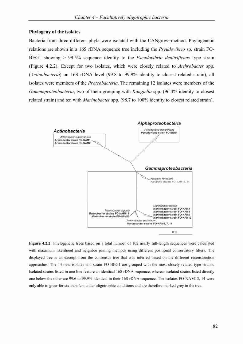
Chapter 4 – Facultatively oligotrophic bacteria
82
Phylogeny of the isolates
Bacteria from three different phyla were isolated with the CANgrow−method. Phylogenetic
relations are shown in a 16S rDNA sequence tree including the Pseudovibrio sp. strain FO-
BEG1 showing > 99.5% sequence identity to the Pseudovibrio denitrificans type strain
(Figure 4.2.2). Except for two isolates, which were closely related to Arthrobacter spp.
(Actinobacteria) on 16S rDNA level (99.8 to 99.9% identity to closest related strain), all
isolates were members of the Proteobacteria. The remaining 12 isolates were members of the
Gammaproteobacteria, two of them grouping with Kangiella spp. (96.4% identity to closest
related strain) and ten with Marinobacter spp. (98.7 to 100% identity to closest related strain).
Figure 4.2.2: Phylogenetic trees based on a total number of 102 nearly full-length sequences were calculated
with maximum likelihood and neighbor joining methods using different positional conservatory filters. The
displayed tree is an excerpt from the consensus tree that was inferred based on the different reconstruction
approaches. The 14 new isolates and strain FO-BEG1 are grouped with the most closely related type strains.
Isolated strains listed in one line feature an identical 16S rDNA sequence, whereas isolated strains listed directly
one below the other are 99.6 to 99.9% identical in their 16S rDNA sequence. The isolates FO-NAM13, 14 were
only able to grow for six transfers under oligotrophic conditions and are therefore marked grey in the tree.
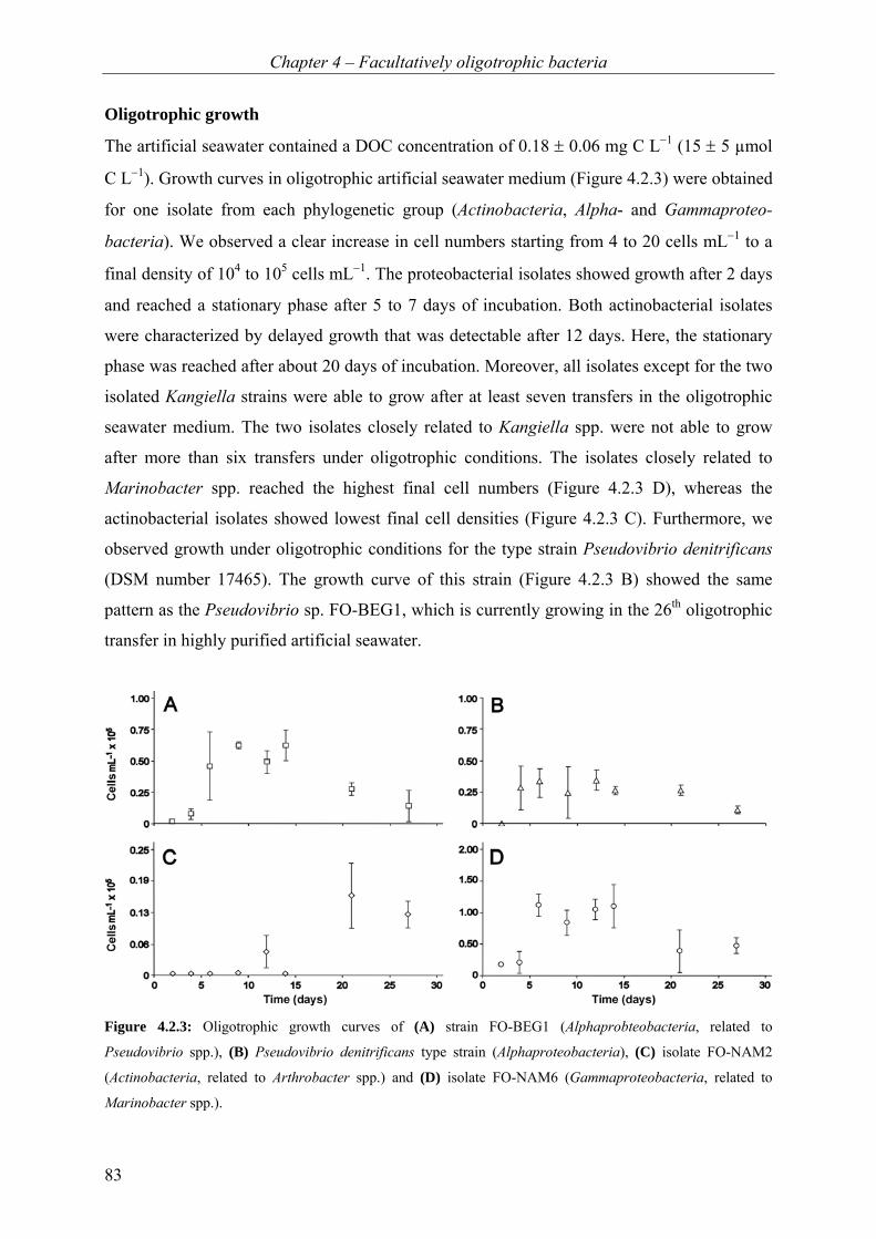
Chapter 4 – Facultatively oligotrophic bacteria
83
Oligotrophic growth
The artificial seawater contained a DOC concentration of 0.18 ± 0.06 mg C L−1 (15 ± 5 µmol
C L−1). Growth curves in oligotrophic artificial seawater medium (Figure 4.2.3) were obtained
for one isolate from each phylogenetic group (Actinobacteria, Alpha- and Gammaproteo-
bacteria). We observed a clear increase in cell numbers starting from 4 to 20 cells mL−1 to a
final density of 104 to 105 cells mL−1. The proteobacterial isolates showed growth after 2 days
and reached a stationary phase after 5 to 7 days of incubation. Both actinobacterial isolates
were characterized by delayed growth that was detectable after 12 days. Here, the stationary
phase was reached after about 20 days of incubation. Moreover, all isolates except for the two
isolated Kangiella strains were able to grow after at least seven transfers in the oligotrophic
seawater medium. The two isolates closely related to Kangiella spp. were not able to grow
after more than six transfers under oligotrophic conditions. The isolates closely related to
Marinobacter spp. reached the highest final cell numbers (Figure 4.2.3 D), whereas the
actinobacterial isolates showed lowest final cell densities (Figure 4.2.3 C). Furthermore, we
observed growth under oligotrophic conditions for the type strain Pseudovibrio denitrificans
(DSM number 17465). The growth curve of this strain (Figure 4.2.3 B) showed the same
pattern as the Pseudovibrio sp. FO-BEG1, which is currently growing in the 26th oligotrophic
transfer in highly purified artificial seawater.
Figure 4.2.3: Oligotrophic growth curves of (A) strain FO-BEG1 (Alphaprobteobacteria, related to
Pseudovibrio spp.), (B) Pseudovibrio denitrificans type strain (Alphaproteobacteria), (C) isolate FO-NAM2
(Actinobacteria, related to Arthrobacter spp.) and (D) isolate FO-NAM6 (Gammaproteobacteria, related to
Marinobacter spp.).
Chapter 4 – Facultatively oligotrophic bacteria
84
Discussion
Isolation of facultatively oligotrophic bacteria with the CANgrow−method
Each strategy that is applied for the isolation of bacteria selects for a specific physiology and
metabolism. Most approaches used recently for the isolation of oligotrophic bacteria are based
on the dilution to extinction method (Button et al., 1993; Connon and Giovannoni, 2002) and
thereby select for the most abundant microorganisms. In contrast, the CANgrow−method
favors bacteria, which might not have been particularly abundant in the original inoculum, but
can adapt fast to changes in nutrient availability. Previous studies have shown that many
bacteria isolated under oligotrophic conditions can adapt to nutrient-rich media (Yanagita et
al., 1977; MacDonell and Hood, 1982; Carlucci et al., 1986). Also, the single-cell
encapsulation method (Zengler et al., 2002) is based on oligotrophic growth followed by
eutrophic growth conditions. This cultivation approach is similar to ours (Figure 4.2.1), but
we used artificial seawater of very low DOC concentration rather than natural seawater. The
measured DOC concentration of 0.18 mg C L−1 is two to five times lower than in natural
seawater (Schut et al., 1997; Hansell et al., 2009). Nevertheless, we are certain to observe true
growth under these extremely oligotrophic conditions, since we performed at least seven
transfers in purified artificial seawater with each of the isolated strains and 26 transfers for
Pseudovibrio sp. FO-BEG1, for which we also identified growth substrates under oligotrophic
conditions (Chapter 4.1). The initial cell number after each transfer was 4 to 20 cells mL−1.
Thus, 9 to 15 divisions must have occurred between two consecutive transfers to account for a
final cell number of 104 to 105 cells mL−1 as observed at the end of the growth phase. This
accounts for 60 to 100 divisions during a total of 7 incubations. Therefore, we conclude that
all isolates are viable under oligotrophic conditions by the definition of Button et al. (1993),
who characterize organisms as viable after having performed 13 divisions which we observe
already after 1 to 2 transfers.
Diverse phylogeny of facultatively oligotrophic bacteria
The isolated bacterial strains belong to different phylogenetic groups, namely Alphaproteo-
bacteria, Gammaproteobacteria and Actinobacteria. Growth under oligotrophic conditions as
observed for the isolates FO-NAM13 and FO-NAM14 (related to Kangiella spp.), has so far
never been described for any member of the genus Kangiella. Long-term starvation and
survival but not growth in the absence of external nutrients has previously been reported for
Arthrobacter spp. and was proposed to be fueled by internally stored reserve material
(Zevenhuizen, 1966). In our study, isolates FO-NAM1, FO-NAM2 (related to Arthrobacter
Chapter 4 – Facultatively oligotrophic bacteria
85
spp.) were transferred in oligotrophic medium more than seven times and active growth was
always observed. Hence, we assume that the cells gained energy and produced biomass from
an external source, since we determined growth and not only survival. Bacteria belonging to
the genus Marinobacter are known to be diverse in physiology (e.g. Gauthier et al., 1992;
Huu et al., 1999). Substrate uptake under low nutrient conditions was shown for Marino-
bacter arcticus (Button et al., 2004), but oligotrophic growth was not studied in detail. In
contrast, the isolates FO-NAM3 to FO-NAM12 (related to Marinobacter spp.) actively grow
under nutrient deficiency.
Cell numbers of the isolated strains growing under oligotrophic conditions differed between
the phylogenetic groups. The cell numbers of isolates related to Marinobacter spp. were
higher than cell numbers of the other isolates, whereas the isolates related to Kangiella spp.
did not grow for more than six transfers. This suggests that the different bacteria vary in their
capabilities of adapting to oligotrophic growth conditions or that the present organic and
inorganic material can be used differently among the isolates. This might be due to the
presence of different metabolic pathways and a different number and type of high affinity
transporters necessary for scavenging nutrients at such low concentrations. Taken together,
our data support earlier findings (Yanagita et al., 1977; MacDonell and Hood, 1982; Carlucci
et al., 1986) that the ability to switch between extreme nutrient deficiency and affluence of
substrate is not unusual or restricted to a certain phylogenetic group, even if the level of
adaptation might differ. Therefore, we propose that the ability to grow under extreme
substrate limitation is much more widespread among known heterotrophic bacteria than
currently recognized.
As expected, the heterotrophic bacteria isolated from the habitat of large sulfide-oxidizers are
phylogenetically diverse. It was possible to isolate bacteria following a similar metabolic
strategy – facultative oligotrophy. If these bacteria are associated with sulfide-oxidizers, if
their metabolic activity is somehow related to the presence or absence of the lithothrophs or
whether they can support growth of the large sulfur bacteria by scavenging ROS needs to be
further investigated.

Chapter 4 – Facultatively oligotrophic bacteria
86
Acknowledgments
This study was funded by the European Research Council (No 203364-ELNOX) and the Max
Planck Society. We thank A.-C. Kreutzmann for helpful remarks on the manuscript, V.
Salman, M. Friebe, S. Menger and M. Meyer for technical support and the members of the
Meteor cruise M76 (April – June 2008) for providing samples.

87
„The important thing in science is not so much to obtain new facts as to
discover new ways of thinking about them.“
~Sir William Lawrence Bragg (1890-1971)
Chapter 5 – Concluding remarks
88
Chapter 5
Concluding remarks and outlook The findings of this thesis contribute to different topics ranging from migration behavior of
mat-forming sulfur bacteria via associations between different bacteria to the lower limits of
bacterial growth. Nevertheless, all these different aspects are linked to each other. The growth
of Beggiatoa sp. filaments, for which we discovered an unusual migration behavior
(Chapter 2), depends on the presence of Pseudovibrio sp. (Chapter 3), which is capable of
growth under extreme nutrient deficiency (Chapter 4). This chapter (Chapter 5) connects all
these different aspects, including preliminary data, which are not presented in the previous
chapters and considers the obtained results in a broader context. Furthermore, a detailed
discussion of special proceedings and precautions during performed experiments is given.
Finally, this chapter ends with an outlook for future research concerning the discussed topics.
Associations between small heterotrophic and larger bacteria Axenic cultivation of large marine Beggiatoa spp. under chemolithoautotrophic conditions is
difficult and only rarely successful (Nelson and Jannasch, 1983; Nelson et al., 1986a). In this
thesis, a successful and stable cultivation of the marine chemolithoautotrophic Beggiatoa sp.
strain 35Flor (6 µm in diameter) is described. This Beggiatoa strain grows solely in the pre-
sence of an accompanying organism, the heterotrophic Pseudovibrio sp. strain FO-BEG1
(Chapters 2 and 3). We propose that the accompanying bacterium protects the Beggiatoa sp.
from oxidative stress because we have shown that the genome of the accompanying organism
possesses more than 20 genes for the enzymes catalase, superoxide dismutase and peroxidase
(Chapter 3.2), whereas sulfide-oxidizing bacteria of the genus Beggiatoa typically do not
possess the gene for the enzyme catalase (reviewed in Larkin and Strohl, 1983).
Similar to large sulfide-oxidizers, also large marine cyanobacteria can often be found
associated with small heterotrophic bacteria and it is difficult to sustain axenic cultures
(Palinska et al., 1999; Morris et al., 2008). The association of small heterotrophic bacteria
Chapter 5 – Concluding remarks
89
with larger bacteria, such as Beggiatoa spp. or cyanobacteria in microbial mats is a common
observation (Cohen and Rosenberg, 1989; van Gemerden, 1993). The complex interactions
between cyanobacteria and heterotrophic bacteria have been studied and different reasons for
their interactions have been proposed. These reasons include recycling of carbon dioxide
needed by the cyanobacteria (Kuentzel, 1969), production of growth factors and formation of
anoxic microniches due to aerobic respiration (Paerl and Pinckney, 1996). Interestingly, for
cyanobacteria of the genus Prochlorococcus, it was also proposed that the function of the
heterotrophs is to scavenge reactive oxygen species (ROS) because the Prochlorococcus spp.
themselves have no gene encoding for a catalase or peroxidase (Morris et al., 2008).
Furthermore, a mutant of the accompanying heterotroph lacking the gene for catalase was
found to not support growth of the cyanobacteria, whereas the addition of catalase had a
positive effect on their growth (Morris et al., 2008). These observations concerning the
association between cyanobacteria and accompanying heterotrophic bacteria point in the same
direction as the observations presented here on the association between Beggiatoa sp. and
Pseudovibrio sp. (Chapter 3). It suggests that the protection system of the large bacteria from
ROS might be less efficient than the ones of smaller heterotrophic bacteria.
Cyanobacteria and Beggiatoa spp. are often found to share one habitat as for example in
microbial mats (van Gemerden, 1993) or in biofilms associated with the black band disease of
scleractinian corals (Richardson, 1996). Therefore, in these common habitats they might also
live together with similar types of heterotrophic bacteria. The bacteria associated with cyano-
bacteria belong to a diverse range of phylogenetic groups, including Actinobacteria,
Bacteroidetes, Alpha-, Beta-, Gamma- and Deltaproteobacteria (Salomon et al., 2003;
Kolmonen et al., 2004; Hube et al., 2009). The isolation of phylogenetically different bacteria
from the habitat of large sulfide-oxidizers (Chapter 4.2) indicates that also the large sulfide-
oxidizers are not restricted to the association with bacteria of one phylogenetic group. It
seems more likely that the large bacteria depend on specialized functions performed by
certain types of the associated bacteria rather than the presence of a specific phylogenetic
group. Very recently, a metagenomic sequence analysis of bacterial communities associated
with the green macroalgae Ulva australis revealed that not the phylogeny of the associated
bacteria but the function of their genes was correlated with the associations (Burke et al.,
2011). Consequently, it was proposed that the functional genes rather than the 16S rDNA
genes are more appropriate to investigate associations in microbial communities (Burke et al.,
2011).

Chapter 5 – Concluding remarks
90
Toxicity factors and migration behavior of Beggiatoa filaments
The growth of Beggiatoa spp. depends on oxygen and sulfide, but both substances can also be
harmful to the bacteria if concentrations exceed a critical threshold (Winogradsky, 1887;
Møller et al., 1985). Close relatives of Beggiatoa spp. belonging to the genus Thiomargarita
can be found in sulfidic sediments with sulfide concentrations of 100 to 800 µmol L−1 (Schulz
et al., 1999) or even up to 22 mmol L−1 (Brüchert et al., 2003). Moreover, these bacteria can
also tolerate atmospheric oxygen levels while being exposed to lower sulfide concentrations
(0 to 100 µmol L−1) at a pH of 7.3 (Schulz et al., 1999; Schulz and de Beer, 2002). However,
Thiomargarita spp. cells are not as motile as Beggiatoa spp. and as a consequence they can
not position themselves in the transition zone of oxygen and sulfide. Instead, the cells wait for
resuspension of the sediment to get into contact with oxygenated, nitrate-containing seawater
(Schulz et al., 1999).
In contrast, Beggiatoa filaments are motile and built up mats between the opposing gradients
of oxygen and sulfide (Winogradsky, 1887; Keil, 1912; Jørgensen, 1977; Nelson et al., 1982;
Nelson and Jannasch, 1983). Consequently, Beggiatoa filaments usually get into contact with
oxygen and sulfide at the same time, since they consume the two gases, they lower the con-
centrations and steepen the gradients (Nelson et al., 1986a). However, depending on the
thickness of the Beggiatoa mat, some filaments might not be in direct contact with oxygen
and sulfide at the same time (Nelson et al., 1986b). Taken this into account, the results
presented in this thesis (Chapter 2.1) suggest that sulfide is probably more harmful to the cells
under oxic conditions because the aerobic sulfide oxidation leads to a decrease in pH
(Winogradsky, 1887) and as a consequence more sulfide outside the cells is present in form of
H2S. In the presented experiments (Chapter 2.1) we measured a pH of 6.5 within the mat,
leading to about 70% of the sulfide being present in form of H2S (compared to only about
30% present as H2S at pH 7.3). This uncharged, harmful gas can easily diffuse into the cells
where it can act as a strong reductant and binds to iron in cytochromes, by this blocking the
cellular respiration. Thus, the bacteria probably have to perform sulfide oxidation to detoxify
the inside of the cells. At high sulfide fluxes the cells obviously accumulate large amounts of
storage compounds during this process and might eventually burst. To prevent this, the
bacteria have to dispose of the internal storage compounds. In the anoxic regions the pH is
higher and sulfide predominantly occurs in form of HS− which can not diffuse into the cells
and might only get inside via ion channels or transport systems.

Chapter 5 – Concluding remarks
91
In this thesis (Chapter 2.1), a new strategy to cope with high sulfide fluxes is proposed. The
bacteria actively migrate into anoxic regions with high sulfide concentrations. Here, the
aerobic sulfide oxidation is stopped and with this also sulfur deposition. The bacteria can then
reduce the intracellular sulfur with internal PHA to form sulfide. This strategy represents a
novel explanation for the presence of Beggiatoa filaments in anaerobic habitats and shows
that filaments actively migrate into anoxic, sulfidic regions. Previously, downwards
migrations were typically observed in habitats containing nitrate and there the anaerobic
sulfide oxidation with nitrate lowered the diffusion zone of sulfide and separated the oxygen
and sulfide gradients over up to a few centimeters (Mußmann et al., 2003; Sayama et al.,
2005; Hinck et al., 2007; Preisler et al., 2007).
Although Beggiatoa spp. also require oxygen for growth (unless an alternative electron
acceptor is present) they show phobic reactions to higher (above 5% air saturation) oxygen
concentrations (Winogradsky, 1887; Møller et al., 1985). During aerobic sulfide oxidation the
production of oxygen radicals and, moreover, chemiluminescence was shown to occur
(Tapley et al., 1999). As mentioned above, we propose that the Beggiatoa sp. 35Flor requires
the accompanying organism to protect themselves against reactive oxygen species (Chapter
3.1). However, no correlation between the catalase activity of heterotrophic, associated cells
and the beneficial effect of their presence on cyanobacteria (Prochlorococcus sp.) was found
(Morris et al., 2008). In fact, the heterotroph with the lowest catalase activity was the only
strain able to support growth of all studied cyanobacteria. Consequently, the dependency of
the large bacteria on small heterotrophic bacteria can not be completely explained by their
possession of a catalase enzyme. During our studies on the co-culture of Beggiatoa sp. and
Pseudovibrio sp., the possibility that nitric oxide (NO) might be involved in the reaction
counteracting oxidative stress arose. In oxygen-sulfide gradient co-cultures with high sulfide
concentrations, the NO signal in Pseudovibrio cells, stained with a copper-based fluorescent
probe (CuFL, Lim et al., 2006), was higher compared to cells exposed to a low sulfide flux
(Figure 5.1). This increase in NO signal was also inducible when hydrogen peroxide (H2O2)
was added to co-cultures with a low sulfide flux or to pure cultures of the isolated
Pseudovibrio sp. (growing in seawater medium containing glucose and ammonia). Under both
conditions, the enhanced signal was visible in the interior of the cells and not in the
surrounding medium. The used dye is specific for NO and no interaction with H2O2 or other
reactive oxygen and nitrogen species, such as HNO, NO2−, NO3
− and ONOO−, could be
detected (Lim et al., 2006).
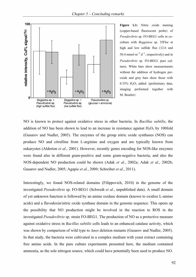
Chapter 5 – Concluding remarks
92
Figure 5.1: Nitric oxide staining
(copper-based fluorescent probe) of
Pseudovibrio sp. FO-BEG1 cells in co-
culture with Beggiatoa sp. 35Flor at
high and low sulfide flux (12.6 and
50.4 mmol m−2 d−1, respectively) and in
Pseudovibrio sp. FO-BEG1 pure cul-
tures. White bars show measurements
without the addition of hydrogen per-
oxide and grey bars show those with
0.35% H2O2 added. (preliminary data,
imaging performed together with
M. Beutler)
NO is known to protect against oxidative stress in other bacteria. In Bacillus subtilis, the
addition of NO has been shown to lead to an increase in resistance against H2O2 by 100fold
(Gusarov and Nudler, 2005). The enzymes of the group nitric oxide synthases (NOS) can
produce NO and citrulline from L-arginine and oxygen and are typically known from
eukaryotes (Alderton et al., 2001). However, recently genes encoding for NOS-like enzymes
were found also in different gram-positive and some gram-negative bacteria, and also the
NOS-dependent NO production could be shown (Adak et al., 2002a; Adak et al., 2002b;
Gusarov and Nudler, 2005; Agapie et al., 2009; Schreiber et al., 2011).
Interestingly, we found NOS-related domains (Filippovich, 2010) in the genome of the
investigated Pseudovibrio sp. FO-BEG1 (Schwedt et al., unpublished data). A small domain
of yet unknown function is followed by an amine oxidase domain (known to oxidize L-amino
acids) and a flavodoxin/nitric oxide synthase domain in the genome sequence. This opens up
the possibility that NO production might be involved in the reaction to ROS in the
investigated Pseudovibrio sp. strain FO-BEG1. The production of NO as a protective measure
against oxidative stress in Bacillus subtilis cells leads to an enhanced catalase activity, which
was shown by comparison of wild type to ∆nos deletion mutants (Gusarov and Nudler, 2005).
In that study, the bacteria were cultivated in a complex medium with yeast extract containing
free amino acids. In the pure culture experiments presented here, the medium contained
ammonia, as the sole nitrogen source, which could have potentially been used to produce NO.

Chapter 5 – Concluding remarks
93
In our co-culture experiments together with Beggiatoa sp., no fixed nitrogen source was
present, but the investigated Beggiatoa sp. are able to fix N2 (Henze, 2005). The genome of
the accompanying organism does not contain any gene encoding for a nitrogenase enzyme
(Schwedt et al., unpublished data). The transfer of fixed nitrogen sources from cyanobacteria
to associated bacteria is a common observation (Paerl, 1984). It may well be that also the
Beggiatoa sp. transfer fixed nitrogen to the accompanying bacteria. Nevertheless, taking into
consideration that dyes can unspecifically bind to different compounds and that the
production of NO in our study was not proven by direct measurements, these considerations
remain speculative and await further investigation (see outlook).
Apart from chemical substances, such as oxygen and sulfide, also light is known to be a
potential cause of phobic migration reactions of Beggiatoa filaments (Winogradsky, 1887;
Nelson and Castenholz, 1982; Møller et al., 1985). The Beggiatoa sp. that we investigated
showed an unusual reaction to blue/green light. Application of a blue or green light source
(intensity: 67 µE m−2 s−1) above or below the culture tube induced an immediate downwards
movement of a subpopulation of filaments into the anoxic part of the culture tube when
cultivated under a high sulfide flux (Chapter 2.2). The downward movement also occurred in
the dark, but solely in cultures where a mat at the oxic-anoxic transition had already been
established for about 10 days. The lower subpopulation was established 2 to 3 days after the
downwards migration started (Chapter 2.1). Applying a blue or green light source in close
proximity to the culture tube induced and also enhanced this filament movement and within a
few hours the lower subpopulation was observed (Chapter 2.2). Because chemiluminescence
occurs during chemical sulfide oxidation (Tapley et al., 1999) at the transition between
oxygen and sulfide, it can be speculated that light at low intensities might be involved in the
migration and/or mat formation processes of Beggiatoa filaments, but this remains to be
studied in the future (see outlook).
The lower limits of bacterial growth In contrast to the large Beggiatoa filaments, most bacterial cells are not visible with the naked
eye. However, bacterial growth can lead to macroscopically visible colonies on nutrient-rich
agar plates or cause turbidity in nutrient-rich liquid media due to high cell densities, whereas
under nutrient limitation cell densities will be low and the small bacterial cells might not
cause turbidity. Therefore, the build-up of bacterial biomass in liquid low-nutrient media has
to be quantified in a different way than optical turbidity measurements. Although, there might
Chapter 5 – Concluding remarks
94
be no measurable growth, the cells might still be metabolically active. This is due to the fact
that there is a difference between biomass production and activity of bacterial cells. Although
the consumption of substrate is essential for the cells to grow and build-up biomass, the
opposite is not the case, as cells do not necessarily grow while utilizing substrate (del Giorgio
and Cole, 1998). Therefore, the formation of biomass, called bacterial production (BP) has to
be separated from the consumption of substrate, called bacterial respiration (BR). The
bacterial growth efficiency (BGE) is defined as the quantity of bacterial biomass resulting
from a certain amount of substrate respired (del Giorgio and Cole, 1998). The substrate is
used by the bacteria for catabolic and anabolic purposes generating ATP (Adenosine-5’-
triposphate) and cell biomass, respectively. The BGE can be determined by isotope-labeling
techniques (BR and BP), rates of protein or DNA synthesis (BP), cell counts (BP), oxygen
consumption (BR), carbon dioxide production (BR) and changes in DOC and POC (BR and
BP) (del Giorgio and Cole, 1998 and references therein).
Under oligotrophic conditions, the concentrations of substrates and cell numbers are
extremely low. Therefore, most of these methods may not be sensitive enough and measured
values are close to or below the limit of detection. Thus, new routines and methods have to be
developed or methods have to be refined to be suitable for studying bacterial growth under
extreme nutrient deficiency. Classical dissolved organic carbon (DOC) measurements, which
are used as a parameter for the amount of dissolved organic matter (DOM) in environments
with a low amount of nutrients, have a detection limit of about 0.5 µmol C L−1
(0.006 mg C L−1). However, the observed cell numbers in our medium (Chapter 4) are so
extremely low that about 1 µmol L−1 of carbon would be enough to explain the observed
growth. Consequently, the classical method might not provide sufficient sensitivity to detect
potential small changes in DOC. The results presented in this thesis show that electospray
ionization Fourier transform ion cyclotron resonance mass spectrometry (ESI FT-ICR-MS)
provides sufficient resolution and can be used to study bacterial growth under oligotrophic
conditions. Furthermore, this technique gives information on both potential fixed carbon and
nitrogen sources of the bacteria. Combined with the measurement of amino acids by high
performance liquid chromatography (HPLC) and isotope-labeling techniques, bacterial
growth in oligotrophic seawater can be studied in detail. However, this technique does
generally not give quantitative information and as a consequence respiration rates under
oligotrophic conditions still remain unknown.
Chapter 5 – Concluding remarks
95
The risk of impurities
Even though no electron donor is added to an oligotrophic medium, there can still be im-
purities present that might support growth and lead to the formation of biomass and/or
metabolic activity of the cells (Chapter 4). Studying bacterial growth under nutrient-poor
conditions is challenging, since many different contaminations, such as macronutrients, trace
elements or energy sources can occur. Accordingly, when studying the physiology of bacteria
growing under oligotrophic conditions, it is crucial to keep the medium and equipment
contamination-free for the mandatory substances for growth. Amino acids represent a serious
contamination risk for seawater samples. Therefore, there are a number of precautions that
have to be taken, such as working with gloves, combustion of glassware, avoidance of dust in
the working room (working in laminar flow hood) and use of chemicals of highest available
quality (Dittmar et al., 2009). Also, during the measurements of quantity and composition of
DOM typically all used equipment is pre-combusted if possible. All these precautions were
applied during the preparation of the oligotrophic seawater medium and all further analyses
presented in Chapter 4.1 of this thesis.
Furthermore, the medium might become contaminated with trace elements, which would
probably not resemble a potential energy source but could be decisive for growth or no
growth of microorganisms. To avoid the contamination with trace elements, equipment is
usually washed with HNO3− (e.g. Fitzwater et al., 1982), which, however, represents an easily
utilizable nitrogen source. For the experiments presented in this thesis (Chapter 4), a fixed
nitrogen source was considered as worse contamination than trace elements, and the
equipment was not washed with HNO3−.
In spite of all precautions, it is still extremely difficult to prepare a seawater medium without
any contaminations. However, if the contaminating substance is not used by the bacteria
during growth, such as the PEG contamination in this study (Chapter 4.1), it might not disturb
or influence further physiological analysis. The contamination can even be used to normalize
other peaks in the mass spectra, if its peaks are high and consistent (as the PEG peaks in this
study, Chapter 4.1).
Chapter 5 – Concluding remarks
96
Facultative oligotrophy
In addition to growth under oligotrophic conditions, the investigated Pseudovibrio sp. FO-
BEG1 (Chapter 4) is capable of growing under eutrophic conditions as well and can switch
within days from one trophic state to the other. Under oligotrophic conditions, the bacterial
growth efficiency is typically very low and varies with the supply of nutrients (del Giorgio
and Cole, 1998). Furthermore, the investigated bacteria are limited in carbon, nitrogen and
energy at the same time (Chapter 4.1). Consequently, they are supposed to have high
maintenance energy costs to maintain crucial transport systems and enzymes prepared (del
Giorgio and Cole, 1998). With increasing nutrient supply, probably also the bacterial growth
efficiency increases because the bacteria can exhibit higher growth rates and produce only
enzymes necessary for the substrates available (reviewed in del Giorgio and Cole, 1998).
Facultatively oligotrophic bacteria have to switch between oligotrophic and eutrophic growth
conditions and as a consequence the cells have to switch also between both strategies for
growth to adapt to the actual amount of substrates present.
The variation in nutrient availability is common in natural environments such as the open
ocean. Bacteria attached to marine snow particles can exhibit very high growth rates
(Alldredge et al., 1986; Alldredge and Gotschalk, 1990; Smith et al., 1992; Azam and Long,
2001; Kiørboe and Jackson, 2001), while free-living bacteria in the open ocean are restricted
in nutrients and consequently their growth rates are low or they even starve (Boylen and
Ensign, 1970; Novitsky and Morita, 1976; Azam and Hodson, 1977; Novitsky and Morita,
1977; Tabor and Neihof, 1982; Ishida et al., 1989). Facultatively oligotrophic bacteria can
adapt fast to nutrient affluence or deficiency, while they have to up- or down-regulate internal
pathways of anabolism and catabolism. The investigated facultatively oligotrophic Pseudo-
vibrio sp. FO-BEG1 is highly versatile with respect to its energy gain (Chapter 3.2 and
Chapter 4.1). This feature makes the bacteria flexible and they are, moreover, capable to
interact with other prokaryotes or possibly even eukaryotes (Chapter 3.2).
Already 30 years ago, it was suspected that two types of oligotrophic bacteria exist (Ishida
and Kadota, 1981): 1. organisms, which disappear with increasing man-made eutrophication
and 2. organisms, which can adapt fast to man-made eutrophication. In this thesis bacteria
were not isolated from the oligotrophic open ocean, but from water directly overlaying marine
sediments (Chapter 4.2). All of the isolated strains were able to adapt fast to the nutrient
deficiency of the initial isolation medium. The observation that facultatively oligotrophic bac-
Chapter 5 – Concluding remarks
97
teria can also be isolated from non-oligotrophic water indicates that these bacteria might be
more widespread and not limited to nutrient-poor environments. For example, they might live
attached to marine snow particles (e. g. Alldredge et al., 1986; Smith et al., 1992; Azam and
Malfatti, 2007 and references therein), which represent nutrient hotspots for heterotrophic
bacteria, and possibly the bacteria sink down to the sediment with these particles. We assume
that many facultatively oligotrophic bacteria have been overlooked so far because they were
not searched for in non-oligotrophic environments. Likewise, many more already known
heterotrophic bacteria may be capable of growing under much poorer nutrient conditions than
currently assumed.
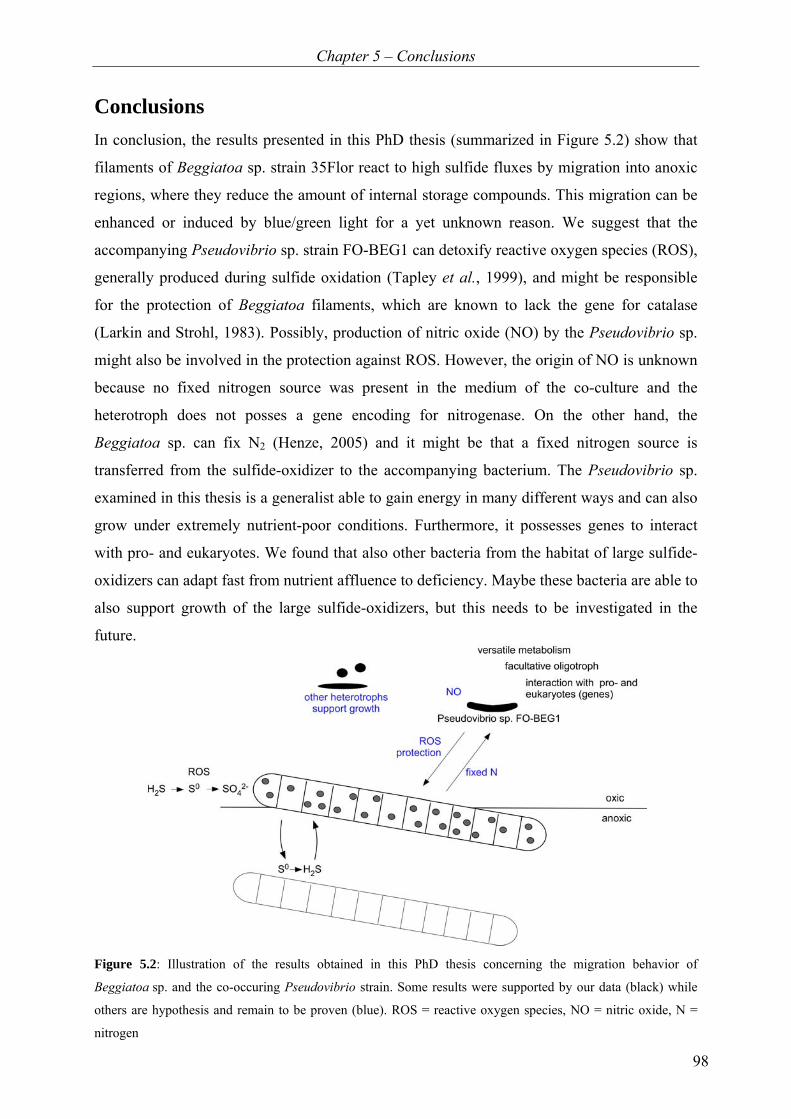
Chapter 5 – Conclusions
98
Conclusions In conclusion, the results presented in this PhD thesis (summarized in Figure 5.2) show that
filaments of Beggiatoa sp. strain 35Flor react to high sulfide fluxes by migration into anoxic
regions, where they reduce the amount of internal storage compounds. This migration can be
enhanced or induced by blue/green light for a yet unknown reason. We suggest that the
accompanying Pseudovibrio sp. strain FO-BEG1 can detoxify reactive oxygen species (ROS),
generally produced during sulfide oxidation (Tapley et al., 1999), and might be responsible
for the protection of Beggiatoa filaments, which are known to lack the gene for catalase
(Larkin and Strohl, 1983). Possibly, production of nitric oxide (NO) by the Pseudovibrio sp.
might also be involved in the protection against ROS. However, the origin of NO is unknown
because no fixed nitrogen source was present in the medium of the co-culture and the
heterotroph does not posses a gene encoding for nitrogenase. On the other hand, the
Beggiatoa sp. can fix N2 (Henze, 2005) and it might be that a fixed nitrogen source is
transferred from the sulfide-oxidizer to the accompanying bacterium. The Pseudovibrio sp.
examined in this thesis is a generalist able to gain energy in many different ways and can also
grow under extremely nutrient-poor conditions. Furthermore, it possesses genes to interact
with pro- and eukaryotes. We found that also other bacteria from the habitat of large sulfide-
oxidizers can adapt fast from nutrient affluence to deficiency. Maybe these bacteria are able to
also support growth of the large sulfide-oxidizers, but this needs to be investigated in the
future.
Figure 5.2: Illustration of the results obtained in this PhD thesis concerning the migration behavior of
Beggiatoa sp. and the co-occuring Pseudovibrio strain. Some results were supported by our data (black) while
others are hypothesis and remain to be proven (blue). ROS = reactive oxygen species, NO = nitric oxide, N =
nitrogen

Chapter 5 – Outlook
99
Outlook The results of my PhD thesis provide new insights into the migration behavior of large sulfur
bacteria, associations between bacteria, and growth under extreme nutrient deficiency.
Nevertheless, the obtained results also raise new questions that need to be addressed in the
future.
We hypothesize that the Beggiatoa sp. filaments depend on the heterotrophic Pseudovibrio sp.
because they do not themselves possess protective measure against oxidative stress
(Chapter 3). However, direct evidence for this theory is still missing. To investigate the
capability of other heterotrophic, catalase-positive bacteria or ROS scavenger to support
growth of the Beggiatoa sp., filaments from the lower subpopulation should be transferred
without (or with only few) accompanying bacteria into fresh media. If growth is positive in
the presence of the other catalase-positive bacteria or ROS scavengers, the transfer of Pseudo-
vibrio sp. from the inoculation source has to be excluded, which can be tested by
Fluorescence in situ hybridization.
The preliminary data on the staining of nitric oxide opens up the possibility that NO might be
involved in the reaction with ROS (Chapter 5). However, to proof this, the production of NO
has to be verified by direct measurement of NO with a NOx-analyzer or detection of NO by its
molecular mass using mass spectrometry. The chemical reaction of NO with oxygen in water
forming mainly nitrite (Ignarro et al., 1993) might complicate the direct measurement of nitric
oxide. The sole nitrogen source in the co-culture is N2. Therefore, it can be speculated that the
interaction between the two bacteria is not only one-sided because of the two partners only
the Beggiatoa sp. are known to fix N2 (Chapter 5). To investigate if a direct transfer of fixed
nitrogen in the co-culture occurs, labeled nitrogen (15N2) can be added and the label can be
searched for in the Pseudovibrio sp. using mass spectrometry (e.g. nano secondary ion mass
spectrometry).
We have shown that the Pseudovibrio sp. FO-BEG1 growing in pure culture under nutrient-
poor conditions can also use typical substrates known from heterotrophic growth (Chap-
ter 4.1). It still remains unclear if the observed growth is the same as typical heterotrophic
growth only with less nutrients or if special regulating enzymes are needed. A proteomic
comparison between bacteria grown under nutrient deficiency and grown under nutrient
Chapter 5 – Outlook
100
affluence might provide more information on the growth and the regulation of enzymes under
oligotrophic conditions.
The detailed analysis of substrates used during oligotrophic growth shows that the bacteria
use multiple substrates at the same time under oligotrophic conditions (Chapter 4.1). However,
only amino acids (which account only for a small fraction) were measured quantitatively and
the overall respiration rate remains unknown. Therefore, the measurement of oxygen
consumption (e.g. using microelectrode respiration chambers) under nutrient-poor conditions
could allow quantification and calculation of the respiration rate under oligotrophic conditions.
Furthermore, the fact that we have both metabolites (detected by ESI FT-ICR-MS) and genes
(closed genome of Pseudovibrio sp. strain FO-BEG1) may allow us to connect metabolites
with genes present in the investigated organism in the future. Maybe, this will also help
predicting new metabolic pathways.
Eventually, several heterotrophic strains, which were able to grow under both nutrient
deficiency and affluence, were isolated during our studies (Chapter 4.2). The possibility of
heterotrophic bacteria to grow under nutrient-deficient conditions might be a more wide-
spread feature. To test this hypothesis, common heterotrophic bacteria from bacterial strain
collections can be ordered and their ability to grow under nutrient-poor conditions can be
tested.
References
101
References Adak S, Aulak KS, Stuehr DJ. (2002a). Direct evidence for nitric oxide production by a nitric-oxide synthase-like protein from Bacillus subtilis. Journal of Biological Chemistry 277: 16167–16171. Adak S, Bilwes AM, Panda K, Hosfield D, Aulak KS, McDonald JF, Tainer JA, Getzoff ED, Crane BR, Stuehr DJ. (2002b). Cloning, expression, and characterization of a nitric oxide synthase protein from Deinococcus radiodurans. Proceedings of the National Academy of Sciences of the United States of America 99: 107–112. Agapie T, Suseno S, Woodward JJ, Stoll S, Britt RD, Marletta MA. (2009). NO formation by a catalytically self-sufficient bacterial nitric oxide synthase from Sorangium cellulosum. Proceedings of the National Academy of Sciences of the United States of America 106: 16221–16226. Ahmad A, Barry JP, Nelson DC. (1999). Phylogenetic affinity of a wide, vacuolate, nitrate-accumulatlng Beggiatoa sp. from Monterey Canyon, California, with Thioploca spp. Applied and Environmental Microbiology 65: 270–277. Ahmad A, Kalanetra KM, Nelson DC. (2006). Cultivated Beggiatoa spp. define the phylogenetic root of morphologically diverse, noncultured, vacuolate sulfur bacteria. Canadian Journal of Microbiology 52: 591–598. Alderton WK, Cooper CE, Knowles RG. (2001). Nitric oxide synthases: Structure, function and inhibition. Biochemical Journal 357: 593–615. Alldredge AL, Cole JJ, Caron DA. (1986). Production of heterotrophic bacteria inhabiting macroscopic organic aggregates (marine snow) from surface waters. Limnology and Oceanography 31: 68–78. Alldredge AL, Gotschalk CC. (1990). The relative contribution of marine snow of different origins to biological processes in coastal waters. Continental Shelf Research 10: 41–58. Ammerman JW, Fuhrman JA, Hagström Å, Azam F. (1984). Bacterioplankton growth in seawater: 1. Growth kinetics and cellular characteristics in seawater cultures. Marine Ecology Progress Series 18: 31–39. Arrigo KR. (2005). Marine microorganisms and global nutrient cycles. Nature 437: 349–355. Azam F, Hodson RE. (1977). Size distribution and activity of marine microheterotrophs. Limnology and Oceanography 22: 492–501. Azam F, Long RA. (2001). Sea snow microcosms. Nature 414: 495–498. Azam F, Malfatti F. (2007). Microbial structuring of marine ecosystems. Nature Reviews Microbiology 5: 782–791. Bachmann A. (2007). Charakterisierung des dominanten Begleitorganismus eines marinen Beggiatoa Konsortiums. Diploma thesis, Leibniz University of Hannover, Germany.

References
102
Bada JL, Lee C. (1977). Decomposition and alteration of organic compounds dissolved in seawater. Marine Chemistry 5: 523–534. Barry JP, Greene HG, Orange DL, Baxter CH, Robison BH, Kochevar RE, Nybakken JW, Reed DL, McHugh CM. (1996). Biologic and geologic characteristics of cold seeps in Monterey bay, California. Deep-Sea Research Part I-Oceanographic Research Papers 43: 1739–1762. Battley EH. (1995). An apparent anomaly in the calculation of ash-free dry weights for the determination of cellular yields. Applied and Environmental Microbiology 61: 1655–1657. Bauer JE, Williams PM, Druffel ERM. (1992). 14C activity of dissolved organic carbon fractions in the north-central Pacific and Sargasso Sea. Nature 357: 667–670. Bernard C, Fenchel T. (1995). Mats of colourless sulphur bacteria 2. Structure, composition of biota and successional patterns. Marine Ecology – Progress Series 128: 171–179. Bolin B, Crutzen PJ, Vitousek PM, Woodmansee RG, Goldberg ED, Cook RB. (1983). Interactions of biogeochemical cycles. In: Bolin B, Cook RB (eds). The major biogeochemical cycles and their interactions: Ipswich, UK. pp 1–39. Bollmann A, Lewis K, Epstein SS. (2007). Incubation of environmental samples in a diffusion chamber increases the diversity of recovered isolates. Applied and Environmental Microbiology 73: 6386–6390. Bondarev V. (2007). Physiologische und molekulare Untersuchungen an filamentösen Bakterien aus Süßwasser-Standorten. Diploma thesis, Leibniz University of Hannover, Germany. Boylen CW, Ensign JC. (1970). Intracellular substrates for endogenous metabolism during long-term starvation of rod and spherical cells of Arthrobacter crystallopoietes. Journal of Bacteriology 103: 578–587. Brock J, Schulz-Vogt HN. (2011). Sulfide induces phosphate release from polyphosphate in cultures of a marine Beggiatoa strain. ISME Journal 5: 497–506. Brüchert V, Jørgensen BB, Neumann K, Riechmann D, Schlösser M, Schulz H. (2003). Regulation of bacterial sulfate reduction and hydrogen sulfide fluxes in the central Namibian coastal upwelling zone. Geochimica Et Cosmochimica Acta 67: 4505–4518. Burke C, Steinberg P, Rusch D, Kjelleberg S, Thomas T. (2011). Bacterial community assembly based on functional genes rather than species. Proceedings of the National Academy of Sciences of the United States of America 108: 14289–14293. Burton SD, Morita RY. (1964). Effect of catalase and cultural conditions on growth of Beggiatoa. Journal of Bacteriology 88: 1755–1761. Button DK, Schut F, Quang P, Martin R, Robertson BR. (1993). Viability and isolation of marine bacteria by dilution culture: Theory, procedures, and initial results. Applied and Environmental Microbiology 59: 881–891.

References
103
Button DK, Robertson B, Gustafson E, Zhao XM. (2004). Experimental and theoretical bases of specific affinity, a cytoarchitecture-based formulation of nutrient collection proposed to supercede the Michaels-Menten paradigm of microbial kinetics. Applied and Environmental Microbiology 70: 5511–5521. Carlucci AF, Shimp SL, Craven DB. (1986). Growth characteristics of low-nutrient bacteria from the Northeast and Central Pacific Ocean. Fems Microbiology Ecology 38: 1–10. Chameides WL, Perdue EM. (1997). Biogeochemical cycles - A computer-interactive study of earth systems and global change. Oxford University Press: New York. Cherrier J, Bauer JE, Druffel ERM. (1996). Utilization and turnover of labile dissolved organic matter by bacterial heterotrophs in eastern north Pacific surface waters. Marine Ecology – Progress Series 139: 267–279. Cherrier J, Bauer JE. (2004). Bacterial utilization of transient plankton-derived dissolved organic carbon and nitrogen inputs in surface ocean waters. Aquatic Microbial Ecology 35: 229–241. Cohen Y, Rosenberg E. (1989). Microbial mats - Physiological ecology of benthic microbial communities: Washington, D.C. Connon SA, Giovannoni SJ. (2002). High-throughput methods for culturing microorganisms in very-low-nutrient media yield diverse new marine isolates. Applied and Environmental Microbiology 68: 3878–3885. Cowie GL, Hedges JI. (1992). Sources and reactivities of amino acids in a coastal marine environment. Limnology and Oceanography 37: 703–724. de Albuquerque JP, Keim CN, Lins U. (2010). Comparative analysis of Beggiatoa from hypersaline and marine environments. Micron 41: 507–517. del Giorgio PA, Cole JJ. (1998). Bacterial growth efficiency in natural aquatic systems. Annual Review of Ecology and Systematics 29: 503–541. Dittmar T, Koch B, Hertkorn N, Kattner G. (2008). A simple and efficient method for the solid-phase extraction of dissolved organic matter (SPE-DOM) from seawater. Limnology and Oceanography: Methods 6: 230–235. Dittmar T, Cherrier J, Ludwichowski K-U. (2009). The analysis of amino acids in seawater. In: Wurl O (ed). Practical guidelines for the analysis of seawater. Dittmar T, Paeng J. (2009). A heat-induced molecular signature in marine dissolved organic matter. Nature Geoscience 2: 175–179. Ducklow HW, Steinberg DK, Buesseler KO. (2001). Upper ocean carbon export and the biological pump. Oceanography 14: 50–58. Egli T. (2010). How to live at very low substrate concentration. Water Research 44: 4826–4837.
References
104
Enticknap JJ, Kelly M, Peraud O, Hill RT. (2006). Characterization of a culturable alphaproteobacterial symbiont common to many marine sponges and evidence for vertical transmission via sponge larvae. Applied and Environmental Microbiology 72: 3724–3732. Farquhar GJ, Boyle WC. (1971). Identification of filamentous microorganisms in activated sludge. Journal Water Pollution Control Federation 43: 604–622. Faust L, Wolfe RS. (1961). Enrichment and cultivation of Beggiatoa alba. Journal of Bacteriology 81: 99–106. Fenchel T, Bernard C. (1995). Mats of colourless sulphur bacteria 1. Major microbial processes. Marine Ecology – Progress Series 128: 161–170. Fiadeiro ME. (1983). Physical-chemical processes in the open ocean. In: Bolin B, Cook RB (eds). The major biogeochemical cycles and their interactions: Ipswich, UK. pp 461–476. Filippovich SY. (2010). Bacterial NO synthases. Biochemistry-Moscow 75: 1217–1224. Fitzwater SE, Knauer GA, Martin JH. (1982). Metal contamination and its effect on primary production measurements. Limnology and Oceanography 27: 544–551. Fossing H, Gallardo VA, Jørgensen BB, Hüttel M, Nielsen LP, Schulz H, Canfield DE, Forster S, Glud RN, Gundersen JK, Küver J, Ramsing NB, Teske A, Thamdrup B, Ulloa O. (1995). Concentration and Transport of Nitrate by the Mat-Forming Sulfur Bacterium Thioploca. Nature 374: 713–715. Francis CA, Beman JM, Kuypers MMM. (2007). New processes and players in the nitrogen cycle: the microbial ecology of anaerobic and archaeal ammonia oxidation. Isme Journal 1: 19–27. Fuhrman JA. (1987). Close coupling between release and uptake of dissolved free amino acids in seawater studied by an isotope dilution approach. Marine Ecology – Progress Series 37: 45–52. Fuhrman JA. (1990). Dissolved free amino acid cycling in an estuarine outflow plume. Marine Ecology – Progress Series 66: 197–203. Gauthier MJ, Lafay B, Christen R, Fernandez L, Acquaviva M, Bonin P, Bertrand JC. (1992). Marinobacter hydrocarbonoclasticus gen. nov., sp. nov., a new, extremely halotolerant, hydrocarbon-degrading marine bacterium. International Journal of Systematic Bacteriology 42: 568–576. Girnth AC, Grünke S, Lichtschlag A, Felden J, Knittel K, Wenzhofer F, de Beer D, Boetius A. (2011). A novel, mat-forming Thiomargarita population associated with a sulfidic fluid flow from a deep-sea mud volcano. Environmental Microbiology 13: 495–505. Grossart HP, Tang KW, Kiørboe T, Ploug H. (2007). Comparison of cell-specific activity between free-living and attached bacteria using isolates and natural assemblages. Fems Microbiology Letters 266: 194–200.

References
105
Grossart HP. (2010). Ecological consequences of bacterioplankton lifestyles: changes in concepts are needed. Environmental Microbiology Reports 2: 706–714. Güde H, Strohl WR, Larkin JM. (1981). Mixotrophic and heterotrophic growth of Beggiatoa alba in continuous culture. Archives of Microbiology 129: 357–360. Gusarov I, Nudler E. (2005). NO-mediated cytoprotection: Instant adaptation to oxidative stress in bacteria. Proceedings of the National Academy of Sciences of the United States of America 102: 13855–13860. Hansell DA, Carlson CA, Repeta DJ, Schlitzer R. (2009). Dissolved organic matter in the ocean a controversy stimulates new insights. Oceanography 22: 202–211. Henze A-T. (2005). Stickstofffixierung bei Beggiatoa. Diploma thesis, University of Hannover, Germany. Hertkorn N, Benner R, Frommberger M, Schmitt-Kopplin P, Witt M, Kaiser K, Kettrup A, Hedges JI. (2006). Characterization of a major refractory component of marine dissolved organic matter. Geochimica Et Cosmochimica Acta 70: 2990–3010. Hinck S, Neu TR, Lavik G, Mussmann M, de Beer D, Jonkers HM. (2007). Physiological adaptation of a nitrate-storing Beggiatoa sp. to diel cycling in a phototrophic hypersaline mat. Applied and Environmental Microbiology 73: 7013–7022. Hinze G. (1901). Über den Bau und der Zellen von Beggiatoa mirabilis Cohn. Berichte der Deutschen Botanischen Gesellschaft: 369–374. Høgslund S, Revsbech NP, Kuenen JG, Jørgensen BB, Gallardo VA, van de Vossenberg JV, Nielsen JL, Holmkvist L, Arning ET, Nielsen LP. (2009). Physiology and behaviour of marine Thioploca. Isme Journal 3: 647–657. Hopkinson CS, Vallino JJ. (2005). Efficient export of carbon to the deep ocean through dissolved organic matter. Nature 433: 142–145. Hube AE, Heyduck-Söller B, Fischer U. (2009). Phylogenetic classification of heterotrophic bacteria associated with filamentous marine cyanobacteria in culture. Systematic and Applied Microbiology 32: 256–265. Huettel M, Forster S, Klöser S, Fossing H. (1996). Vertical migration in the sediment-dwelling sulfur bacteria Thioploca spp. in overcoming diffusion limitations. Applied and Environmental Microbiology 62: 1863–1872. Hugenholtz P. (2002). Exploring prokaryotic diversity in the genomic era. Genome Biology 3: 1–8. Huu NB, Denner EBM, Ha DTC, Wanner G, Stan-Lotter H. (1999). Marinobacter aquaeolei sp. nov., a halophilic bacterium isolated from a Vietnamese oil-producing well. International Journal of Systematic Bacteriology 49: 367–375. Ignarro LJ, Fukuto JM, Griscavage JM, Rogers NE, Byrns RE. (1993). Oxidation of nitric oxide in aqueous solution to nitrite but not nitrate: Comparison with enzymatically formed
References
106
nitric oxide from L-arginine. Proceedings of the National Academy of Sciences of the United States of America 90: 8103–8107. Ihssen J, Egli T. (2005). Global physiological analysis of carbon- and energy-limited growing Escherichia coli confirms a high degree of catabolic flexibility and preparedness for mixed substrate utilization. Environmental Microbiology 7: 1568–1581. Ishida Y, Kadota H. (1981). Growth patterns and substrate requirements of naturally occurring obligate oligotrophs. Microbial Ecology 7: 123–130. Ishida Y, Imai I, Miyagaki T, Kadota H. (1982). Growth and uptake kinetics of a facultatively oligotrophic bacterium at low nutrient concentrations. Microbial Ecology 8: 23–32. Ishida Y, Fukami K, Eguchi M, Yoshinaga I. (1989). Strategies for growth of oligotrophic bacteria in the pelagic environment. In: Hattori T, Ishida Y, Maruyama Y, Morita RY, Uchida A (eds). Recent advances in microbial ecology. Japan Scientific Society Press: Japan. pp 89–93. Jiao N, Herndl GJ, Hansell DA, Benner R, Kattner G, Wilhelm SW, Kirchman DL, Weinbauer MG, Luo TW, Chen F, Azam F. (2010). Microbial production of recalcitrant dissolved organic matter: long-term carbon storage in the global ocean. Nature Reviews Microbiology 8: 593–599. Jørgensen BB. (1977). Distribution of colorless sulfur bacteria (Beggiatoa spp.) in a coastal marine sediment. Marine Biology 41: 19–28. Jørgensen BB. (1983). Processes at the sediment-water interface. In: Bolin B, Cook RB (eds). The major biogeochemical cycles and their interactions: Ipswich, UK. pp 477–509. Jørgensen BB, Revsbech NP. (1983). Colorless sulfur bacteria, Beggiatoa spp. and Thiovulum spp. in O2 and H2S microgradients. Applied and Environmental Microbiology 45: 1261–1270. Jørgensen BB, Nelson DC. (2004). Sulfide oxidation in marine sediments: Geochemistry meets microbiology. In: Amend JP, Edwards KJ, Lyons TW (eds). Sulfur biogeochemistry – Past and present: Geological Society of America pp 63–81. Jørgensen BB, Dunker R, Grünke S, Røy H. (2010). Filamentous sulfur bacteria, Beggiatoa spp., in arctic marine sediments (Svalbard, 79 degrees N). Fems Microbiology Ecology 73: 500-513. Jørgensen NOG. (1987). Free amino acids in lakes: Concentrations and assimilation rates in relation to phytoplankton and bacterial production. Limnology and Oceanography 32: 97–111. Kaeberlein T, Lewis K, Epstein SS. (2002). Isolating "uncultivable" microorganisms in pure culture in a simulated natural environment. Science 296: 1127–1129. Kalanetra KM, Huston SL, Nelson DC. (2004). Novel, attached, sulfur-oxidizing bacteria at shallow hydrothermal vents possess vacuoles not involved in respiratory nitrate accumulation. Applied and Environmental Microbiology 70: 7487–7496.

References
107
Kalanetra KM, Joye SB, Sunseri NR, Nelson DC. (2005). Novel vacuolate sulfur bacteria from the Gulf of Mexico reproduce by reductive division in three dimensions. Environmental Microbiology 7: 1451–1460. Kamp A, Stief P, Schulz-Vogt HN. (2006). Anaerobic sulfide oxidation with nitrate by a freshwater Beggiatoa enrichment culture. Applied and Environmental Microbiology 72: 4755–4760. Kamp A, Røy H, Schulz-Vogt HN. (2008). Video-supported analysis of Beggiatoa filament growth, breakage, and movement. Microbial Ecology 56: 484–491. Keil F. (1912). Beiträge zur Physiologie der farblosen Schwefelbakterien. Beiträge zur Biologie der Pflanzen 11: 335–372. Keil RG, Kirchman DL. (1999). Utilization of dissolved protein and amino acids in the northern Sargasso Sea. Aquatic Microbial Ecology 18: 293–300. Kido Soule MC, Longnecker K, Giovannoni SJ, Kujawinski EB. (2010). Impact of instrument and experiment parameters on reproducibility of ultrahigh resolution ESI FT-ICR mass spectra of natural organic matter. Organic Geochemistry 41: 725–733. Kiørboe T. (2000). Colonization of marine snow aggregates by invertebrate zooplankton: Abundance, scaling, and possible role. Limnology and Oceanography 45: 479–484. Kiørboe T, Jackson GA. (2001). Marine snow, organic solute plumes, and optimal chemosensory behavior of bacteria. Limnology and Oceanography 46: 1309–1318. Kirchman DL, Meon B, Ducklow HW, Carlson CA, Hansell DA, Steward GF. (2001). Glucose fluxes and concentrations of dissolved combined neutral sugars (polysaccharides) in the Ross Sea and Polar Front Zone, Antarctica. Deep-Sea Research Part II 48: 4179–4197. Klas Z. (1937). Über den Formenkreis von Beggiatoa mirabilis. Archives of Microbiology: Aus dem Ozeanographischen Institut Split und den Botanischen Institut Zagreb: 312–320. Koch AL. (1997). Microbial physiology and ecology of slow growth. Microbiology and Molecular Biology Reviews 61: 305–318. Koch BP, Witt MR, Engbrodt R, Dittmar T, Kattner G. (2005). Molecular formulae of marine and terrigenous dissolved organic matter detected by electrospray ionization Fourier transform ion cyclotron resonance mass spectrometry. Geochimica Et Cosmochimica Acta 69: 3299–3308. Kolmonen E, Sivonen K, Rapala J, Haukka K. (2004). Diversity of cyanobacteria and heterotrophic bacteria in cyanobacterial blooms in Lake Joutikas, Finland. Aquatic Microbial Ecology 36: 201–211. Kovárová-Kovar K, Egli T. (1998). Growth kinetics of suspended microbial cells: From single-substrate-controlled growth to mixed-substrate kinetics. Microbiology and Molecular Biology Reviews 62: 646–666.
References
108
Kuentzel LE. (1969). Bacteria, carbon dioxide, and algal blooms. Journal Water Pollution Control Federation 41: 1737–1747. Larkin JM, Strohl WR. (1983). Beggiatoa, Thiothrix, and Thioploca. Annual Review of Microbiology 37: 341–367. Larkin JM, Henk MC. (1996). Filamentous sulfide-oxidizing bacteria at hydrocarbon seeps of the Gulf of Mexico. Microscopy Research & Technique 33: 23–31. Lendenmann U, Snozzi M, Egli T. (1996). Kinetics of the simultaneous utilization of sugar mixtures by Escherichia coli in continuous culture. Applied and Environmental Microbiology 62: 1493–1499. Lichtschlag A, Felden J, Brüchert V, Boetius A, de Beer D. (2010). Geochemical processes and chemosynthetic primary production in different thiotrophic mats of the Hakon Mosby Mud Volcano (Barents Sea). Limnology and Oceanography 55: 931–949. Lim MH, Xu D, Lippard SJ. (2006). Visualization of nitric oxide in living cells by a copper-based fluorescent probe. Nature Chemical Biology 2: 375–380. Lindroth P, Mopper K. (1979). High-performance liquid chromatographic determination of subpicomole amounts of amino acids by precolumn fluorescence derivatization with ortho-phthaldialdehyde. Analytical Chemistry 51: 1667–1674. Ludwig W, Strunk O, Westram R, Richter L, Meier H, Yadhukumar, Buchner A, Lai T, Steppi S, Jobb G, Forster W, Brettske I, Gerber S, Ginhart AW, Gross O, Grumann S, Hermann S, Jost R, Konig A, Liss T, Lussmann R, May M, Nonhoff B, Reichel B, Strehlow R, Stamatakis A, Stuckmann N, Vilbig A, Lenke M, Ludwig T, Bode A, Schleifer KH. (2004). ARB: A software environment for sequence data. Nucleic Acids Research 32: 1363–1371. Lunau M, Lemke A, Dellwig O, Simon M. (2006). Physical and biogeochemical controls of microaggregate dynamics in a tidally affected coastal ecosystem. Limnology and Oceanography 51: 847–859. Macalady JL, Lyon EH, Koffman B, Albertson LK, Meyer K, Galdenzi S, Mariani S. (2006). Dominant microbial populations in limestone-corroding stream biofilms, Frasassi cave system, Italy. Applied and Environmental Microbiology 72: 5596–5609. MacDonell MT, Hood MA. (1982). Isolation and characterization of ultramicrobacteria from a Gulf Coast Estuary. Applied and Environmental Microbiology 43: 566–571. Maier S, Murray RGE. (1965). Fine Structure of Thioploca Ingrica and a comparison with Beggiatoa. Canadian Journal of Microbiology 11: 645–655. McHatton SC, Barry JP, Jannasch HW, Nelson DC. (1996). High nitrate concentrations in vacuolate, autotrophic marine Beggiatoa spp. Applied and Environmental Microbiology 62: 954–958. Millero FJ, Plese T, Fernandez M. (1988). The dissociation of hydrogen sulfide in seawater. Limnology and Oceanography 33: 269–274.
References
109
Møller MM, Nielsen LP, Jørgensen BB. (1985). Oxygen responses and mat formation by Beggiatoa spp. Applied and Environmental Microbiology 50: 373–382. Morris JJ, Kirkegaard R, Szul MJ, Johnson ZI, Zinser ER. (2008). Facilitation of robust growth of Prochlorococcus colonies and dilute liquid cultures by "helper" heterotrophic bacteria. Applied and Environmental Microbiology 74: 4530–4534. Mußmann M, Schulz HN, Strotmann B, Kjær T, Nielsen LP, Rosselló-Mora RA, Amann RI, Jørgensen BB. (2003). Phylogeny and distribution of nitrate-storing Beggiatoa spp. in coastal marine sediments. Environmental Microbiology 5: 523–533. Muyzer G, Teske A, Wirsen CO, Jannasch HW. (1995). Phylogenetic relationships of Thiomicrospira species and their identification in deep-sea hydrothermal vent samples by denaturing gradient gel-electrophoresis of 16S rDNA fragments. Archives of Microbiology 164: 165–172. Nelson DC, Castenholz RW. (1981). Use of reduced sulfur-compounds by Beggiatoa sp. Journal of Bacteriology 147: 140–154. Nelson DC, Castenholz RW. (1982). Light responses of Beggiatoa. Archives of Microbiology 131: 146–155. Nelson DC, Waterbury JB, Jannasch HW. (1982). Nitrogen fixation and nitrate utilization by marine and fresh-water Beggiatoa. Archives of Microbiology 133: 172–177. Nelson DC, Jannasch HW. (1983). Chemoautotrophic growth of a marine Beggiatoa in sulfide-gradient cultures. Archives of Microbiology 136: 262–269. Nelson DC, Jørgensen BB, Revsbech NP. (1986a). Growth-pattern and yield of a chemoautotrophic Beggiatoa sp. in oxygen-sulfide microgradients. Applied and Environmental Microbiology 52: 225–233. Nelson DC, Revsbech NP, Jørgensen BB. (1986b). Microoxic-anoxic niche of Beggiatoa spp. - microelectrode survey of marine and fresh water srains. Applied and Environmental Microbiology 52: 161–168. Nelson DC, Wirsen CO, Jannasch HW. (1989). Characterization of large, autotrophic Beggiatoa spp. abundant at hydrothermal vents of the Guaymas Basin. Applied and Environmental Microbiology 55: 2909–2917. Norland S, Fagerbakke KM, Heldal M. (1995). Light element analysis of Individual bacteria by X-ray microanalysis. Applied and Environmental Microbiology 61: 1357–1362. Novitsky JA, Morita RY. (1976). Morphological characterization of small cells resulting from nutrient starvation of a psychrophilic marine vibrio. Applied and Environmental Microbiology 32: 617–622. Novitsky JA, Morita RY. (1977). Survival of a psychrophilic marine vibrio under long-term nutrient starvation. Applied and Environmental Microbiology 33: 635–641.

References
110
Paerl HW. (1984). Transfer of N2 and CO2 fixation products from Anabaena oscillarioides to associated bacteria during inorganic carbon sufficiency and deficiency. Journal of Phycology 20: 600–608. Paerl HW, Pinckney JL. (1996). A mini-review of microbial consortia: Their roles in aquatic production and biogeochemical cycling. Microbial Ecology 31: 225–247. Palinska KA, Becker C, Krumbein WE. (1999). Isolation and purification techniques for benthic marine cyanobacteria - biotechnology potential. Bulletin de l'Institute océanographique, Monaco 19: 585–592. Preisler A, Lichtschlag A, Lavik G, Boetius A, Jørgensen BB, de Beer D. (2007). Biological and chemical sulfide oxidation in a Beggiatoa inhabited marine sediment. ISME Journal 1: 341–353. Pringsheim EG. (1964). Heterotrophism and species concepts in Beggiatoa. American Journal of Botany 51: 898–913. Pringsheim EG. (1967). Die Mixotrophie von Beggiatoa. Archiv Für Mikrobiologie 59: 247–254. Pruesse E, Quast C, Knittel K, Fuchs BM, Ludwig WG, Peplies J, Glöckner FO. (2007). SILVA: A comprehensive online resource for quality checked and aligned ribosomal RNA sequence data compatible with ARB. Nucleic Acids Research 35: 7188C7196. Rappe MS, Connon SA, Vergin KL, Giovannoni SJ. (2002). Cultivation of the ubiquitous SAR11 marine bacterioplankton clade. Nature 418: 630–633. Richardson LL. (1996). Horizontal and vertical migration patterns of Phormidium corallyticum and Beggiatoa spp. associated with black-band disease of corals. Microbial Ecology 32: 323–335. Sahling H, Rickert D, Lee RW, Linke P, Suess E. (2002). Macrofaunal community structure and sulfide flux at gas hydrate deposits from the Cascadia convergent margin, NE Pacific. Marine Ecology – Progress Series 231: 121–138. Salman V, Amann R, Girnth A-C, Polerecky L, Bailey JV, Høgslund S, Jessen G, Pantoja S, Schulz-Vogt HN. (2011). A single-cell sequencing approach to the classification of large, vacuolated sulfur bacteria. Systematic and Applied Microbiology 34: 243–259. Salomon PS, Janson S, Graneli E. (2003). Molecular identification of bacteria associated with filaments of Nodularia spumigena and their effect on the cyanobacterial growth. Harmful Algae 2: 261–272. Sassen R, Roberts HH, Aharon P, Larkin J, Chinn EW, Carney R. (1993). Chemosynthetic bacterial mats at cold hydrocarbon seeps, Gulf of Mexico continental slope. Organic Geochemistry 20: 77–89. Sayama M. (2001). Presence of nitrate-accumulating sulfur bacteria and their influence on nitrogen cycling in a shallow coastal marine sediment. Applied and Environmental Microbiology 67: 3481–3487.

References
111
Sayama M, Risgaard-Petersen N, Nielsen LP, Fossing H, Christensen PB. (2005). Impact of bacterial NO3- transport on sediment biogeochemistry. Applied and Environmental Microbiology 71: 7575–7577. Schlünz B, Schneider RR. (2000). Transport of terrestrial organic carbon to the oceans by rivers: re-estimating flux- and burial rates. International Journal of Earth Sciences 88: 599–606. Schmidt TM, Arieli B, Cohen Y, Padan E, Strohl WR. (1987). Sulfur metabolism in Beggiatoa alba. Journal of Bacteriology 169: 5466–5472. Schreiber F, Beutler M, Enning D, Lamprecht-Grandio M, Zafra O, Gonzalez-Pastor JE, de Beer D. (2011). The role of nitric-oxide-synthase-derived nitric oxide in multicellular traits of Bacillus subtilis 3610: biofilm formation, swarming, and dispersal. Bmc Microbiology 11: 1–11. Schulz HN, Brinkhoff T, Ferdelman TG, Marine MH, Teske A, Jørgensen BB. (1999). Dense populations of a giant sulfur bacterium in Namibian shelf sediments. Science 284: 493–495. Schulz HN, de Beer D. (2002). Uptake rates of oxygen and sulfide measured with individual Thiomargarita namibiensis cells by using microelectrodes. Applied and Environmental Microbiology 68: 5746-5749. Schut F, Devries EJ, Gottschal JC, Robertson BR, Harder W, Prins RA, Button DK. (1993). Isolation of typical marine bacteria by dilution culture: Growth, maintenance, and characteristics of isolates under laboratory conditions. Applied and Environmental Microbiology 59: 2150–2160. Schut F, Prins RA, Gottschal JC. (1997). Oligotrophy and pelagic marine bacteria: Facts and fiction. Aquatic Microbial Ecology 12: 177–202. Shanks AL, Edmondson EW. (1990). The vertical flux of metazoans (holoplankton, meiofauna, and larval invertebrates) due to their association with marine snow. Limnology and Oceanography 35: 455–463. Shieh WY, Lin YT, Jean WD. (2004). Pseudovibrio denitrificans gen. nov., sp. nov., a marine, facultatively anaerobic, fermentative bacterium capable of denitrification. International Journal of Systematic and Evolutionary Microbiology 54: 2307–2312. Silver MW, Shanks AL, Trent JD. (1978). Marine snow - microplankton habitat and source of small-scale patchiness in pelagic populations. Science 201: 371–373. Simon M, Grossart HP, Schweitzer B, Ploug H. (2002). Microbial ecology of organic aggregates in aquatic ecosystems. Aquatic Microbial Ecology 28: 175–211. Smith DC, Simon M, Alldredge AL, Azam F. (1992). Intense hydrolytic enzyme activity on marine aggregates and implications for rapid particle dissolution. Nature 359: 139–142. Stanley SO, Boto KG, Alongi DM, Gillan FT. (1987). Composition and bacterial utilization of free amino acids in tropical mangrove sediments. Marine Chemistry 22: 13–30.
References
112
Strohl WR, Larkin JM. (1978). Enumeration, isolation, and characterization of Beggiatoa from freshwater sediments. Applied and Environmental Microbiology 36: 755–770. Strohl WR, Cannon GC, Shively JM, Güde H, Hook LA, Lane CM, Larkin JM. (1981). Heterotrophic Carbon Metabolism by Beggiatoa alba. Journal of Bacteriology 148: 572–583. Strohl WR, Howard KS, Larkin JM. (1982). Ultrastructure of Beggiatoa alba strain B15LD. Journal of General Microbiology 128: 73–84. Strous M, Kuenen JG, Jetten MSM. (1999). Key physiology of anaerobic ammonium oxidation. Applied and Environmental Microbiology 65: 3248–3250. Suess E. (1980). Particulate organic-carbon flux in the oceans - surface productivity and oxygen utilization. Nature 288: 260–263. Suzuki N, Kato K. (1953). Studies on suspended materials marnie snow in the sea. Part I. sources of marine snow. Bull Fac Fish Hokkaido Univ: 132–137. Sweerts JPRA, De Beer D, Nielsen LP, Verdouw H, Van den Heuvel JC, Cohen Y, Cappenberg TE. (1990). Denitrification by sulphur oxidizing Beggiatoa spp. mats on freshwater sediments. Nature 344: 762–763. Tabor PS, Neihof RA. (1982). Improved micro-auto-radiographic method to determine individual microorganisms active in substrate uptake in natural waters. Applied and Environmental Microbiology 44: 945–953. Tapley DW, Buettner GR, Shick JM. (1999). Free radicals and chemiluminescence as products of the spontaneous oxidation of sulfide in seawater, and their biological implications. Biological Bulletin 196: 52–56. Trevisan V. (1842). Prospetto della Flora Euganea. Padua: Coi Tipi del Seminario: 1–68. Turley CM, Mackie PJ. (1994). Biogeochemical significance of attached and free-living bacteria and the flux of particles in the NE Atlantic Ocean. Marine Ecology – Progress Series 115: 191–203. Turley CM, Stutt ED. (2000). Depth-related cell-specific bacterial leucine incorporation rates on particles and its biogeochemical significance in the Northwest Mediterranean. Limnology and Oceanography 45: 419–425. Upton AC, Nedwell DB. (1989). Nutritional flexibility of oligotrophic and copiotrophic antarctic bacteria with respect to organic substrates. Fems Microbiology Ecology 62: 1–6. Van der Kooij D, Visser A, Hijnen WAM. (1980). Growth of Aeromonas hydrophila at low concentrations of substrates added to tap water. Applied and Environmental Microbiology 39: 1198–1204. van Gemerden H. (1993). Microbial mats - a joint venture. Marine Geology 113: 3–25.
References
113
Vancanneyt M, Schut F, Snauwaert C, Goris J, Swings J, Gottschal JC. (2001). Sphingomonas alaskensis sp. nov., a dominant bacterium from a marine oligotrophic environment. International Journal of Systematic and Evolutionary Microbiology 51: 73–80. Vartoukian SR, Palmer RM, Wade WG. (2010). Strategies for culture of 'unculturable' bacteria. Fems Microbiology Letters 309: 1–7. Vaucher JP. (1803). Histoire des conferves d´eau douce, contenant leurs différents modes de reproduction, et la description de leurs principales espèces. Paschoud, Geneva: 1–285. Volkov II, Rozanov AG. (1983). The sulphur cycle in the oceans. Part 1. Reservoirs and Fluxes of sulphur. In: Ivanov MV, Freney JR (eds). The global biogeochemical sulfur cycle. Wiley: Chichester. Weber CA. (1907). Aufbau und Vegetation der Moore Norddeutschlands. Botanische Jahrbuecher fuer Systematik, Pflanzengeschichte und Pflanzengeographie / Beiblatt. pp 19–34. Weiss RF. (1970). Solubility of nitrogen, oxygen and argon in water and seawater. Deep-Sea Research 17: 721–735. Winogradsky S. (1887). Ueber Schwefelbacterien. Botanische Zeitung 45: 489–507, 513–523, 529–539, 545–559, 569–576, 585–594, 606–610. Yanagita T, Ichikawa T, Tsuji T, Kamata Y, Ito K, Sasaki M. (1977). Two trophic groups of bacteria, oligotrophs and eutrophs: Their distribution in fresh and sea water areas in the central northern Japan. J Gen Appl Microbiol 24: 59–88. Zengler K, Toledo G, Rappe M, Elkins J, Mathur EJ, Short JM, Keller M. (2002). Cultivating the uncultured. Proceedings of the National Academy of Sciences of the United States of America 99: 15681–15686. Zevenhuizen LPTM. (1966). Formation and function of glycogen-like polysaccharide of Arthrobacter. Antonie Van Leeuwenhoek Journal of Microbiology and Serology 32: 356–372. Zweifel UL, Norrman B, Hagström Å. (1993). Consumption of dissolved organic carbon by marine bacteria and demand for inorganic nutrients. Marine Ecology – Progress Series 101: 23–32.
List of abbreviations
114
List of abbreviations
AHL N-acyl-homoserine lactone
ATP adenosine-5’-triphosphate
BGE bacterial growth efficiency
BP bacterial production
BR bacterial respiration
cAMP cyclic adenosine monophosphate
DCAA dissolved combined amino acids
DFAA dissolved free amino acids
DOC dissolved organic carbon
DOM dissolved organic matter
DSM Deutsche Sammlung von Mikroorganismen (German Collection of
Microorganisms)
DSMZ Deutsche Sammlung von Mikroorganismen und Zellkulturen (German
Collection of Microorganisms and Cell cultures)
ESI electrospray ionization
FT-ICR-MS Fourier transform ion cyclotron resonance mass spectrometry
HPLC high performance liquid chromatography
LDOM labile dissolved organic matter
NO nitric oxide
NOS nitric oxide synthases
NRPS non ribosomal polypeptide synthases
PEG polyethylene glycol
PHA polyhydroxyalkanoates
PHB poly-β-hydroxybutyric acid
PKS polyketide synthases
POC particulate organic carbon
POM particulate organic matter
RDOM recalcitrant dissolved organic matter
ROS reactive oxygen species
THDAA total hydrolysable dissolved amino acids
Appendix
115
Appendix
The genus Pseudovibrio contains metabolically versatile and
symbiotically interacting bacteria Vladimir Bondarev1* , Michael Richter1, Jörn Piel2, Stefano Romano1, Anne Schwedt1 and
Heide N. Schulz-Vogt1
1Max Planck Institute for Marine Microbiology, Bremen, Germany 2Kekulé-Institut für Organische Chemie und Biochemie, Bonn, Germany
*Corresponding author
Manuscript in preparation
Keywords: genomics/functional genomics/comparative genomics; metabolism;
microbe:microbe interactions; primary and secondary production; symbionts; microbe:higher
organism interaction
Appendix
116
Abstract The majority of strains belonging to the genus Pseudovibrio have been isolated from marine
invertebrates like tunicates, corals and especially sponges, but the physiology of these bacteria
is poorly understood. In this study, we analyze the genomes of two Pseudovibrio strains. One
is a required symbiont of a cultivated Beggiatoa strain, a sulfide oxidizing, autotrophic
bacterium. The other one was isolated from a sponge (Enticknap et al., 2006). The data show
that both strains are generalistic bacteria capable of importing and oxidizing a wide range of
organic and inorganic compounds to meet their carbon, nitrogen, phosphorous and energy
requirements under oxic and anoxic conditions. Several physiological traits encoded in the
genome were verified in laboratory experiments with a pure culture of the Pseudovibrio strain
originally associated with Beggiatoa. Besides the versatile metabolic abilities of both
Pseudovibrio strains, our study reveals a number of open reading frames and gene clusters in
the genomes that seem to be involved in symbiont-host interactions. Pseudovibrio has the
genomic potential to attach to host cells, might be capable of interacting with the eukaryotic
cell machinery, produce secondary metabolites and may supply the host with cofactors.
Introduction The first strain of the genus Pseudovibrio has been isolated from coastal seawater in 2004 and
was described as Pseudovibrio denitrificans – a marine, heterotrophic, facultatively anaerobic
bacterium capable of denitrification and fermentation (Shieh et al., 2004). Two further type
strains, P. ascidiaceicola (Fukunaga et al., 2006) and P. japonicus (Hosoya and Yokota,
2007), were isolated from a tunicate and coastal seawater, respectively. Physiologically, these
isolates were not notably different from P. denitrificans. Besides the three type strains,
Pseudovibrio spp.-related bacteria have been found in various studies throughout the world
either by 16S rRNA gene analysis or direct isolation methods (Hentschel et al., 2001; Webster
and Hill, 2001; Olson et al., 2002; Thakur et al., 2003; Thiel and Imhoff, 2003; Thoms et al.,
2003; Agogué et al., 2005; Lafi et al., 2005; Enticknap et al., 2006; Koren and Rosenberg,
2006; Sertan-de Guzman et al., 2007; Muscholl-Silberhorn et al., 2008; Riesenfeld et al.,
2008; Kennedy et al., 2009; Rypien et al., 2010; Santos et al., 2010). Interestingly, besides P.
denitrificans, P. japonicus and a Pseudovibrio spp.-related isolate from coastal, oligotrophic
seawater (Agogué et al., 2005), all other strains belonging to this genus discovered until today
have been found closely associated with marine invertebrates like tunicates, corals, and
sponges. Especially Porifera seem to harbor Pseudovibrio populations, e.g., as the dominating
species of the culturable bacterial community (Webster and Hill, 2001; Muscholl-Silberhorn
Appendix
117
et al., 2008). Additionally, Pseudovibrio has been found in sponge larvae as the most
abundant prokaryote, indicating vertical transmission of these bacteria in their hosts
(Enticknap et al., 2006). Such a consistent pattern of Pseudovibrio spp. associated with
sponges suggests that they are symbionts of those metazoa (Webster and Hill, 2001;
Enticknap et al., 2006). Whether the nature of this symbiosis is mutualistic/ commensalistic or
whether Pseudovibrio spp. rather represent pathogens/parasites is uncertain, but the fact that
Pseudovibrio spp. have been isolated only from healthy sponges indicates that the bacteria do
not harm the host and might be even required for its health (Webster and Hill, 2001). Another
shared feature is the production of secondary metabolites by many of the cultured
Pseudovibrio strains. For instance, heptylprodigiosin, a compound that exhibits antimicrobial
activity, was isolated from a pure culture of P. denitrificans Z143-1 (Sertan-de Guzman et al.,
2007) and the production of additional bioactive compounds could be shown in several other
studies (Hentschel et al., 2001; Muscholl-Silberhorn et al., 2008; Kennedy et al., 2009;
Santos et al., 2010).
Despite the fact that members of the genus Pseudovibrio seem to be ubiquitous and important
associates of marine invertebrates and are also found free-living, very little is known about the
physiology and interactions with the host. In this study, we analyze the genomes of two
Pseudovibrio strains. Pseudovibrio sp. FO-BEG1 has been isolated from an enrichment
culture of a Beggiatoa strain, a filamentous, sulfide oxidizing bacterium (Brock and Schulz-
Vogt, 2011; Chapters 2 and 3). Initially, this Beggiatoa strain was sampled from a coral
suffering from the black band disease off the coast of Florida, which indicates that the strain
Pseudovibrio FO-BEG1 could have been associated with the coral at the time of sampling –
either in a commensalistic/mutualistic or pathogenic relationship – and is now available as an
axenic culture in our lab. Intriguingly, strain FO-BEG1 is also maintained in a co-culture with
a Beggiatoa sp., which seems to be unable to grow without Pseudovibrio and is therefore
dependent on strain FO-BEG1. The second strain, Pseudovibrio sp. JE062, has been isolated
in Florida from the sponge Mycale laxissima in the year 2006 and was described as a sponge
symbiont by Enticknap et al. (2006). The analysis of these genomes gives us an insight into
the physiological and symbiotic potential of both Pseudovibrio strains and reveals fascinating
microorganisms that seem to be adapted to free-living and symbiotic life styles.

Appendix
118
Materials and Methods
Growth conditions For aerobic growth CM medium modified after Shieh et al. (2004) was used. After
autoclaving, the medium was supplemented with K2HPO4 (1.15 mmol L–1), glucose (10 mmol
L–1 unless stated otherwise), 1 mL L–1 tungsten/selenium solution (Brysch et al., 1987), 1 mL
L–1 trace elements (Widdel and Pfennig, 1984), and 1 mL L–1 of four vitamin solutions
prepared according to Aeckersberg et al. (1991). For measurement of SO42– evolution during
S2O32– oxidation, 10 mmol L–1 Na2S2O3 · 5 H2O and 5 mmol L–1 glucose were added and 2 g
L–1 K2SO4 from the original recipe was replaced with 0.75 g L–1 KCl. To compare growth
between a culture with and without Na2S2O3, K2SO4 was not omitted from the medium and
glucose and Na2S2O3 · 5 H2O were used in the same concentrations as described above. To
investigate the growth with 4-hydroxybenzoic acid and benzoate, both compounds were
added in a concentration of 2 mmol L–1, respectively, without any other carbon source.
Growth with phosphonoacetate (1 mmol L–1) as phosphorus source was tested by adding this
compound as the only phosphorus source and all vitamins were omitted from the medium. For
fermentation and denitrification experiments under anoxic conditions, aged North Sea water
was buffered with 50 mmol L–1 TRIS, supplemented with NH4Cl (10 mmol L–1) and the pH
adjusted to 8. Preparation of the medium was performed according to Widdel and Bak (1992)
in order to prepare the medium anoxically. Cooling was performed under N2 atmosphere,
except for experiments in which N2 production was measured, in which Ar was used as the
atmosphere instead. After autoclavation, the medium was supplemented with 10 mmol L–1
glucose, 1 mL L–1 tungsten/selenium solution, 1 mL L–1 trace elements and 1 mL L–1 of four
vitamin solutions prepared as described above. NaNO3 (10 mmol L–1) was added for
experiments investigating denitrification. To test CO oxidation, CM medium was prepared as
described above, containing 400 µmol L–1 glucose and supplied with 500 p.p.m. CO to the
bottle headspace. For aerobic growth experiments, 250 mL Erlenmeyer flasks were filled with
100 mL medium. For anaerobic growth, 156 mL serum bottles (Wheaton, Millville, USA)
were filled anoxically with 50 mL medium and closed with butyl rubber stoppers. For all
experiments, 0.1% or 0.5% of a preculture grown aerobically in CM medium was used as
inoculum. All growth experiments were performed with Pseudovibrio sp. FO-BEG1 in
triplicates at 28°C in the dark with shaking at 110 rpm.
Chemical analyses Bacterial growth was monitored as the optical density (OD600 nm) using an Eppendorf
BioPhotometer (Eppendorf AG, Hamburg, Germany). SO42– was measured with a Metrohm
Appendix
119
761 Compact IC with conductivity detector (Metrohm AG, Herisau, Switzerland) equipped
with a Metrosep A Supp 5-100 column with a carbonate eluent (3.2 mmol L–1 Na2CO3/1
mmol L–1 NaHCO3 in deionised water) at a flow rate of 0.7 mL min–1. Tetrathionate was
measured according to Kamyshny (2009). Glucose and organic acids were determined using a
HPLC system (Sykam GmbH) equipped with an anion neutral pre-column (4x20 mm; Sykam
GmbH) and an Aminex HPX-87H separation column (300x7.8 mm; Biorad, Munich,
Germany) at a temperature of 60 °C. The eluent consisted of 5 mM H2SO4 in HPLC-grade
water with a flow rate of 0.6 mL min–1. Quantification of glucose, succinate, lactate, formate,
acetate, propionate and ethanol was performed with the 7515A RI detector (ERC, Riemerling,
Germany); pyruvate was measured with the Sapphire UV-Vis detector at 210 nm (Ecom,
Praha, Czech Republic). NO3- was quantified with a HPLC system (Sykam GmbH, Eresing,
Germany) containing an anion neutral pre-column (4x20 mm; Sykam GmbH) and an IBJ A3
anion separation column (4x60 mm; Sykam GmbH) with a column temperature of 50 °C. The
eluent consisted of 25 mmol L–1 NaCl and 45% ethanol in deionised water with a flow rate of
1 mL min–1. Detection of NO3– was conducted with Linear Uvis 200 (Thermo Fischer
Scientific GmbH, Dreieich, Germany) at 220 nm. N2 was measured as described by Zedelius
et al. (2011). CO determination was conducted with a Shimadzu GC-8A (Shimadzu, Duisburg,
Germany) gas chromatograph with a Molecular Sieve 5A column (80 to 100; 0.125 in. by 2 m;
Restek, Bellefonte, USA) at a flow of 20 mL of synthetic air per minute at 40°C and an RGD2
reduction gas detector (Trace Analytical, Menlo Park, USA).
DNA extraction and sequencing DNA was extracted from strain FO-BEG1 using the Fast DNA SPIN Kit for Soil (MP
Biomedicals LLC, Illkirch, France), according to manufacturers’ instructions. 454 sequencing
was conducted by LGC Genomics GmbH with a 454 GS FLX System. The Newbler 2.0.00.22
software was used for sequence assembly and quality assessment. Overall, 522919 sequenced
reads with an average length of 336.30 bp lead to a 29-fold sequence coverage. In order to
close the gaps, a fosmid library with a 1.5-fold physical coverage was created and used for
direct sequencing of the fosmid clones. For the residual gaps, 96 specific primers were
designed and used for combinatorial PCR on DNA level, the products of which were
sequenced via the Sanger method.
Gene prediction, annotation and data mining Gene prediction was carried out by using the software Glimmer3 (Delcher et al., 2007).
Ribosomal RNA genes were detected by using the RNAmmer 1.2 software (Lagesen et al.,
2007) and transfer RNAs by tRNAscan-SE (Lowe and Eddy, 1997). Annotation was
Appendix
120
performed by using the GenDB, version 2.2 system (Meyer et al., 2003), supplemented by the
tool JCoast, version 1.6 (Richter et al., 2008). For each predicted open reading frame (ORF)
observations have been collected from similarity searches against sequence databases NCBI-
nr, Swiss-Prot, KEGG and genomesDB (Richter et al., 2008) and for protein family databases
from Pfam (Bateman et al., 2004) and InterPro (Mulder et al., 2005). SignalP has been used
for signal peptide predictions (Bendtsen et al., 2004) and TMHMM for transmembrane helix-
analysis (Krogh et al., 2001). Predicted protein coding sequences were automatically
annotated by the in-house software MicHanThi (Quast, 2006). The MicHanThi software
predicts gene functions based on similarity searches using the NCBI-nr (including Swiss-Prot)
and InterPro database. The annotation of proteins highlighted within the scope of this study
was subject of manual inspection. For all observations regarding putative protein functions, an
e-value cutoff of 10–4 was considered.
Comparison of the shared gene content by reciprocal best matches (RBMs) and functional classification with Kyoto encyclopedia of genes and genomes (KEGG) Determination of the shared gene content has been performed by a BLAST all versus all
search between FO-BEG1 and JE062. Reciprocal best matches were counted by a BLAST
result with an E value <1e–5 each and a subject coverage of over 65%. For metabolic pathway
identification, genes were searched for similarity against the KEGG database. A match was
counted if the similarity search resulted in an expectation E value below 1e–5. All occurring
KO (KEGG Orthology) numbers were mapped against KEGG pathway functional hierarchies
and statistical analyzed.
Functional classification with cluster of orthologous groups (COG) and calculation of the Average nucleotide identity (ANI) All predicted ORFs were also searched for similarity against the COG database (Tatusov et al.,
2003). A match was counted if the similarity search resulted in an E value below 1e–5. ANI
between the whole-genome sequences of strain FO-BEG1 and the draft genome sequences of
strain JE062 was determined by using the in silico DNA-DNA hybridization method of the
JSpecies (Richter and Rosselló-Móra, 2009) software with default parameters.
Creation of circular genome maps and prediction of ABC and TRAP type transporters Comparative circular genome maps of the RBMs shared between JE062 and FO-BEG1 have
been drawn by using JCoast's plugin for CGView (Stothard and Wishart, 2005). Circular GC-
plot and GC-skew representation has been drawn by using DNAPlotter (Carver et al., 2009).
As initial step for the identification of the ABC transporters, genes containing the Pfam
domain ABC_tran (PF00005) were detected in the genome of strain FO-BEG1. For the

Appendix
121
identification of the permease and the periplasmic binding protein, the close proximity of
genes containing the ABC_tran domain was searched. Only ABC systems with at least one
ABC_tran domain, one permease and one periplasmic binding protein were regarded as
functioning ABC transporters and substrate specificity was predicted from the annotations of
the subunits. In several cases, one subunit (e.g. the permease) was missing in close proximity
of genes with the ABC_tran domain. In this case, a single permease gene located on any place
in the genome with the same substrate specificity prediction but not belonging to any
complete ABC system, was used to complement the transporter system. TRAP transporters
were regarded as complete when the subunits DctM, DctQ and DctP were present in close
proximity. When two subunits were identified in close proximity and the third was missing,
the single subunit located on any place in the genome not belonging to any complete TRAP
system was used to complement the transporter system. In the case of fusion of the DctQ and
M subunits in one gene, only the DctP subunit was required to complete the transporter.
Accession numbers The genome shotgun project of strain FO-BEG1 has been deposited at
DDBJ/EMBL/GenBank under the accession number CP003147 for the chromosome and
CP003148 for the plasmid. The draft genome sequence of strain JE062 has the
DDBJ/EMBL/GenBank accession number ABXL00000000.
Results and Discussion
General genome characteristics The genome size of strain FO-BEG1 is 5.9 Mbp, including a large plasmid of 0.4 Mbp (Figure
S.1). The circular chromosome of 5.5 Mbp contains a large stretch of repeats at position
2,707,040. This area of unknown size could not be bridged with a direct sequencing approach
despite the presence of this area on a fosmid, indicating strong secondary structures, and has
been masked with the ambiguous nucleotide code 'N'. The G+C content is 52.5 mol% and is
consistent with the known values of the described Pseudovibrio isolates (Shieh et al., 2004;
Fukunaga et al., 2006; Hosoya and Yokota, 2007). Altogether, we found 5,478 ORFs, 398 of
which were located on the plasmid, which correspond to about 87% of encoding DNA. Six
complete rRNA operons and 69 tRNA encoding regions were annotated, indicating the
capability of a quick response to changing conditions and fast growth when nutrients are
available. The genome of strain JE062 has not been closed, but there are 19 contigs available
with an overall size of 5.7 Mbp, 5,225 ORFs and 52.4 mol% GC content, which is almost
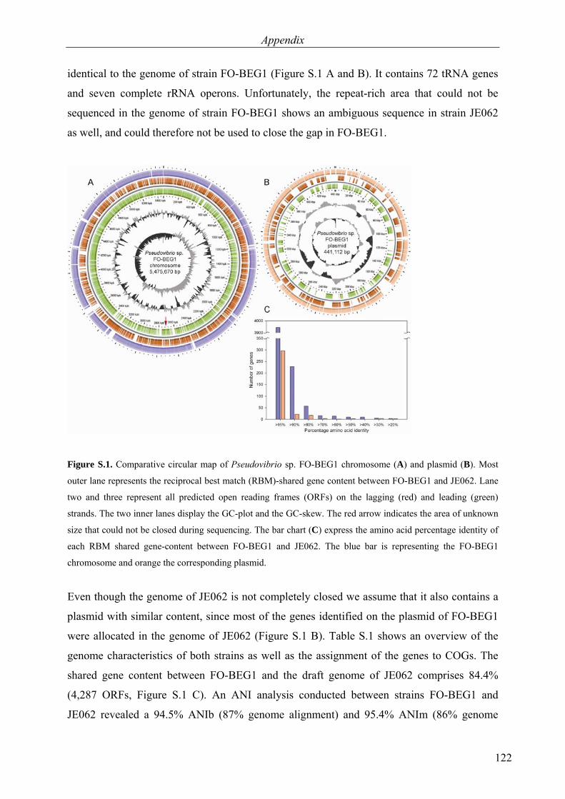
Appendix
122
identical to the genome of strain FO-BEG1 (Figure S.1 A and B). It contains 72 tRNA genes
and seven complete rRNA operons. Unfortunately, the repeat-rich area that could not be
sequenced in the genome of strain FO-BEG1 shows an ambiguous sequence in strain JE062
as well, and could therefore not be used to close the gap in FO-BEG1.
Figure S.1. Comparative circular map of Pseudovibrio sp. FO-BEG1 chromosome (A) and plasmid (B). Most
outer lane represents the reciprocal best match (RBM)-shared gene content between FO-BEG1 and JE062. Lane
two and three represent all predicted open reading frames (ORFs) on the lagging (red) and leading (green)
strands. The two inner lanes display the GC-plot and the GC-skew. The red arrow indicates the area of unknown
size that could not be closed during sequencing. The bar chart (C) express the amino acid percentage identity of
each RBM shared gene-content between FO-BEG1 and JE062. The blue bar is representing the FO-BEG1
chromosome and orange the corresponding plasmid.
Even though the genome of JE062 is not completely closed we assume that it also contains a
plasmid with similar content, since most of the genes identified on the plasmid of FO-BEG1
were allocated in the genome of JE062 (Figure S.1 B). Table S.1 shows an overview of the
genome characteristics of both strains as well as the assignment of the genes to COGs. The
shared gene content between FO-BEG1 and the draft genome of JE062 comprises 84.4%
(4,287 ORFs, Figure S.1 C). An ANI analysis conducted between strains FO-BEG1 and
JE062 revealed a 94.5% ANIb (87% genome alignment) and 95.4% ANIm (86% genome
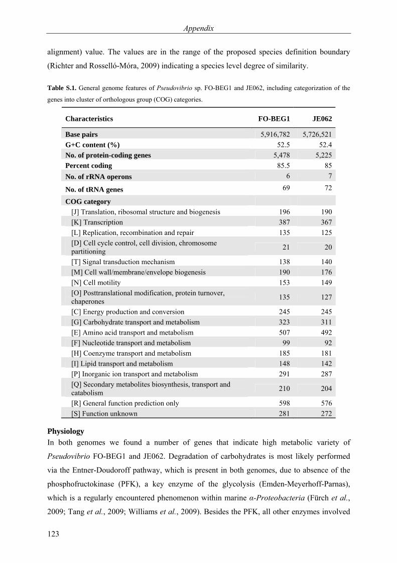
Appendix
123
alignment) value. The values are in the range of the proposed species definition boundary
(Richter and Rosselló-Móra, 2009) indicating a species level degree of similarity.
Table S.1. General genome features of Pseudovibrio sp. FO-BEG1 and JE062, including categorization of the
genes into cluster of orthologous group (COG) categories.
Characteristics FO-BEG1 JE062
Base pairs 5,916,782 5,726,521G+C content (%) 52.5 52.4No. of protein-coding genes 5,478 5,225Percent coding 85.5 85No. of rRNA operons 6 7
No. of tRNA genes 69 72
COG category [J] Translation, ribosomal structure and biogenesis 196 190 [K] Transcription 387 367 [L] Replication, recombination and repair 135 125 [D] Cell cycle control, cell division, chromosome
partitioning 21 20
[T] Signal transduction mechanism 138 140 [M] Cell wall/membrane/envelope biogenesis 190 176 [N] Cell motility 153 149 [O] Posttranslational modification, protein turnover,
chaperones 135 127
[C] Energy production and conversion 245 245 [G] Carbohydrate transport and metabolism 323 311 [E] Amino acid transport and metabolism 507 492 [F] Nucleotide transport and metabolism 99 92 [H] Coenzyme transport and metabolism 185 181 [I] Lipid transport and metabolism 148 142 [P] Inorganic ion transport and metabolism 291 287 [Q] Secondary metabolites biosynthesis, transport and
catabolism 210 204
[R] General function prediction only 598 576 [S] Function unknown 281 272
Physiology In both genomes we found a number of genes that indicate high metabolic variety of
Pseudovibrio FO-BEG1 and JE062. Degradation of carbohydrates is most likely performed
via the Entner-Doudoroff pathway, which is present in both genomes, due to absence of the
phosphofructokinase (PFK), a key enzyme of the glycolysis (Emden-Meyerhoff-Parnas),
which is a regularly encountered phenomenon within marine α-Proteobacteria (Fürch et al.,
2009; Tang et al., 2009; Williams et al., 2009). Besides the PFK, all other enzymes involved
Appendix
124
in glycolysis can be identified in both genomes, including fructose-1,6-bisphosphatase I, the
key enzyme for glyconeogenesis, indicating that the Emden-Meyerhoff-Parnas pathway can
be used for anabolic purposes (see DDBJ/EMBL/GenBank accession numbers CP003147,
CP003148 and ABXL00000000). Genes encoding all enzymes of the citric acid cycle and
pentose phosphate pathway are present. Additionally, both strains have the genetic potential
to degrade aromatic compounds via the β-ketoadipate pathway, which we demonstrated by
growing Pseudovibrio sp. FO-BEG1 with 4-hydroxybenzoate as the only carbon and energy
source under aerobic conditions (Figure S 2.1 A). Benzoate, however, was not degraded,
indicating that either the uptake of benzoate is detained or the hydroxylation of the aromatic
ring structure cannot be performed by Pseudovibrio FO-BEG1. Under anoxic conditions
without nitrate, strain FO-BEG1 metabolized glucose in mixed acid type fermentation, as
suggested by the present genes in both strains (see DDBJ/EMBL/GenBank accession numbers
CP003147, CP003148 and ABXL00000000), resulting in acidification of the medium and
formation of mainly formate, lactate, acetate, and ethanol. Ethanol production during
fermentation has not been described for any Pseudovibrio strain yet. Additionally, pyruvate,
propionate, and succinate have been formed, but to a lesser extent (Figure S 2.2 A).
Production of trace amounts of fumarate was detected, but could not be quantified. As
expected, we found the complete set of genes essential for denitrification, including a
membrane-bound (nar) and a periplasmic nitrate reductase (nap). In agreement, we observed
a complete denitrification to N2 in laboratory experiments with strain FO-BEG1 (see
DDBJ/EMBL/GenBank accession numbers CP003147 and CP003148 and Figure S 2.2 C).
For the type strain P. denitrificans, simultaneous denitrification and fermentation was
described by Shieh et al. (2004) and could be confirmed in our experiments for strain FO-
BEG1 with acetate, formate, lactate, and ethanol as the main fermentation products (Figure S
2.2 B). No genes for assimilatory nitrate reduction could be identified in the genome. A set of
sox genes suggests that both bacteria can use reduced inorganic sulfur compounds as a source
of energy to complement heterotrophy. We could show experimentally that the addition of
thiosulfate to the medium enhances the aerobic growth of the Pseudovibrio sp. FO-BEG1
culture and sulfate is produced over the incubation period (Figure S 2.1 B and C). No
tetrathionate could be measured as an intermediate (results not shown). Therefore, we propose
that thiosulfate is oxidized completely to sulfate without any intermediates, as it is known for
the typical Sox pathway in α-Proteobacteria (for review, see Ghosh and Dam, 2009). We
identified genes encoding a small (cutS), medium (cutM) and large (cutL) subunit of the
aerobic form II carbon monoxide dehydrogenase (CODH) with the accessory gene coxG in
Appendix
125
the operon (see DDBJ/EMBL/GenBank accession numbers CP003147 and CP003148),
indicating the capability of CO oxidation. However, uptake of CO could not be demonstrated
under tested conditions (results not shown). Interestingly, our results confirm the hypothesis
from a recent publication testing CO oxidation in bacteria containing type II CODH genes
(Cunliffe, 2011), in which none of the isolates containing only the type II variant was capable
of CO oxidation. Only bacteria containing the form I CODH have been shown to effectively
oxidize CO, thereby questioning whether form II CODH is involved in the process of carbon
monoxide oxidation, or if it has another primary function not known until now, as suggested
by King and Weber (2007).
In both Pseudovibrio strains, we found genes for phosphonate import and degradation, which
allows the bacteria to cleave the relatively stable C-P bonds of phosphonates
(see DDBJ/EMBL/GenBank accession numbers CP003147, CP003148 and ABXL00000000).
Thereby, they can metabolize a less accessible phosphorous pool in times of phosphate
limitation. We could demonstrate growth of Pseudovibrio sp. FO-BEG1 with
phosphonoacetate as the only source of phosphorous (Figure S 2.3 A). Additionally, we could
show adaptation of Pseudovibrio strain FO-BEG1 to oligotrophic conditions by culturing it
with as little as 15 µmol C L–1 (0.18 mg C L–1) dissolved organic carbon in the medium
(Chapter 4), which shows that Pseudovibrio FO-BEG1 is capable of growth under extreme
nutrient depletion. The high metabolic variety of Pseudovibrio sp. FO-BEG1 and JE062 is
also reflected in the analysis of encoded primary transporters. In the genome of strain FO-
BEG1 we could identify 31 tripartite ATP-independent periplasmic (TRAP) type transporters
(see DDBJ/EMBL/GenBank accession numbers CP003147 and CP003148) that are required
for import of dicarboxylic acids like malate, succinate and fumarate, one of the highest
numbers of TRAP type transporters reported in a genome of a marine prokaryote so far
(Wagner-Döbler et al., 2010). In strain JE062 we identified 27 TRAP transporters. Citric acid
cycle intermediates seem therefore to be an important source of carbon and energy for the
investigated Pseudovibrio strains. In addition, we reconstructed over 80 ATP-binding cassette
(ABC) transporter systems with predicted substrate specificity from the genomic data of the
strain FO-BEG1, including the plasmid, and over 70 ABC transporter systems for JE062 (see
DDBJ/EMBL/GenBank accession numbers CP003147, CP003148 and ABXL00000000).
Sugars, oligopeptides and amino acids are the main substrates that are imported via the ABC
systems. A large number of transporters for oligopeptides and amino acids in combination
with over 85 genes encoding peptidases and proteases (over 75 genes in strain JE062, see
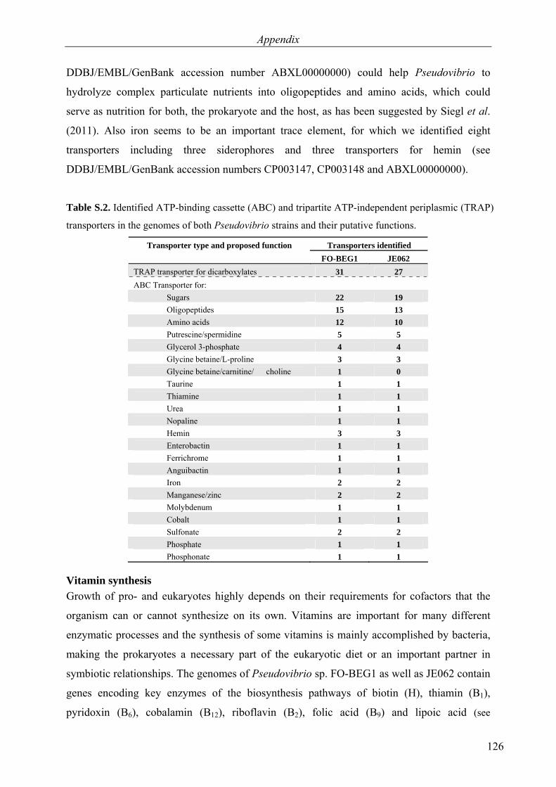
Appendix
126
DDBJ/EMBL/GenBank accession number ABXL00000000) could help Pseudovibrio to
hydrolyze complex particulate nutrients into oligopeptides and amino acids, which could
serve as nutrition for both, the prokaryote and the host, as has been suggested by Siegl et al.
(2011). Also iron seems to be an important trace element, for which we identified eight
transporters including three siderophores and three transporters for hemin (see
DDBJ/EMBL/GenBank accession numbers CP003147, CP003148 and ABXL00000000).
Table S.2. Identified ATP-binding cassette (ABC) and tripartite ATP-independent periplasmic (TRAP)
transporters in the genomes of both Pseudovibrio strains and their putative functions.
Transporter type and proposed function Transporters identified FO-BEG1 JE062
TRAP transporter for dicarboxylates 31 27 ABC Transporter for: Sugars 22 19 Oligopeptides 15 13 Amino acids 12 10 Putrescine/spermidine 5 5 Glycerol 3-phosphate 4 4 Glycine betaine/L-proline 3 3 Glycine betaine/carnitine/ choline 1 0 Taurine 1 1 Thiamine 1 1 Urea 1 1 Nopaline 1 1 Hemin 3 3 Enterobactin 1 1 Ferrichrome 1 1 Anguibactin 1 1 Iron 2 2 Manganese/zinc 2 2 Molybdenum 1 1 Cobalt 1 1 Sulfonate 2 2 Phosphate 1 1 Phosphonate 1 1
Vitamin synthesis Growth of pro- and eukaryotes highly depends on their requirements for cofactors that the
organism can or cannot synthesize on its own. Vitamins are important for many different
enzymatic processes and the synthesis of some vitamins is mainly accomplished by bacteria,
making the prokaryotes a necessary part of the eukaryotic diet or an important partner in
symbiotic relationships. The genomes of Pseudovibrio sp. FO-BEG1 as well as JE062 contain
genes encoding key enzymes of the biosynthesis pathways of biotin (H), thiamin (B1),
pyridoxin (B6), cobalamin (B12), riboflavin (B2), folic acid (B9) and lipoic acid (see

Appendix
127
DDBJ/EMBL/GenBank accession numbers CP003147, CP003148 and ABXL00000000).
Independence of an external vitamin supply was confirmed during aerobic growth in the
defined CM medium without the addition of any vitamins, which implies de novo synthesis of
all required growth factors by strain FO-BEG1 under tested conditions (Figure S 2.3 B).
Pseudovibrio spp. would therefore be beneficial companions for other prokaryotes or marine
invertebrates, since the dependency on an external supply of those vitamins would be relieved.
Bioactive compounds Symbiotic relationships between bacteria and marine invertebrates, especially sponges, are of
special interest, because bacteria associated with sponges often produce novel bioactive
compounds (Piel et al., 2004; Taylor et al., 2007; Fisch et al., 2009). In the chromosome of
Pseudovibrio FO-BEG1 we identified a genomic island of more than 50 kb containing among
others a gene cluster of 20 genes predicted to be involved in secondary metabolite production
(see DDBJ/EMBL/GenBank accession number CP003147). The cluster exhibits high
sequence similarity to an architecturally almost identical hybrid nonribosomal peptide
synthetase-polyketide synthase (NRPS-PKS) system previously reported from many
pathogenic and commensal Escherichia coli strains
(Figure S.2) (Nougayrède et al., 2006). The E. coli metabolite, termed colibactin, remains
structurally uncharacterized. However, transposon mutagenesis of the gene cluster suggested
that colibactin is a pathogenicity determinant that induces DNA double strand breaks in
eukaryotic host cells, eventually resulting in cell death. The only significant difference
between the gene clusters in Pseudovibrio FO-BEG1 and E. coli is an additional set of genes
in the former, encoding putative transporters and the presence of a different
phosphopantetheinyl transferase gene variant likely involved in generating holo-proteins from
apo forms of PKSs and NRPSs (Lambalot et al., 1996). In addition, two E. coli genes are
fused in the Pseudovibrio cluster. Despite these differences, the architecture strongly suggests
that the product of the FO-BEG1 cluster is colibactin, providing new opportunities to unveil
the identity of this elusive and biomedically relevant compound. Interestingly, we find this
more than 50 kb NRPS/PKS fragment only in Pseudovibrio sp. FO-BEG1 but not in the
genome of strain JE062, with flanking regions downstream and upstream of the inserted
fragment highly conserved in synteny in strain JE062 (data not shown), indicating that it has
been acquired via horizontal gene transfer. Additionally, the plasmid of strain FO-BEG1
contained an ORF encoding a type III PKS of a size of 7.4 kb, which could also be detected in
strain JE062 (see DDBJ/EMBL/GenBank accession number CP003148).
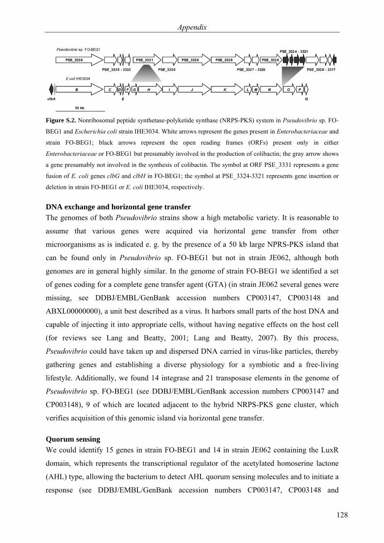
Appendix
128
Figure S.2. Nonribosomal peptide synthetase-polyketide synthase (NRPS-PKS) system in Pseudovibrio sp. FO-
BEG1 and Escherichia coli strain IHE3034. White arrows represent the genes present in Enterobacteriaceae and
strain FO-BEG1; black arrows represent the open reading frames (ORFs) present only in either
Enterobacteriaceae or FO-BEG1 but presumably involved in the production of colibactin; the gray arrow shows
a gene presumably not involved in the synthesis of colibactin. The symbol at ORF PSE_3331 represents a gene
fusion of E. coli genes clbG and clbH in FO-BEG1; the symbol at PSE_3324-3321 represents gene insertion or
deletion in strain FO-BEG1 or E. coli IHE3034, respectively.
DNA exchange and horizontal gene transfer The genomes of both Pseudovibrio strains show a high metabolic variety. It is reasonable to
assume that various genes were acquired via horizontal gene transfer from other
microorganisms as is indicated e. g. by the presence of a 50 kb large NPRS-PKS island that
can be found only in Pseudovibrio sp. FO-BEG1 but not in strain JE062, although both
genomes are in general highly similar. In the genome of strain FO-BEG1 we identified a set
of genes coding for a complete gene transfer agent (GTA) (in strain JE062 several genes were
missing, see DDBJ/EMBL/GenBank accession numbers CP003147, CP003148 and
ABXL00000000), a unit best described as a virus. It harbors small parts of the host DNA and
capable of injecting it into appropriate cells, without having negative effects on the host cell
(for reviews see Lang and Beatty, 2001; Lang and Beatty, 2007). By this process,
Pseudovibrio could have taken up and dispersed DNA carried in virus-like particles, thereby
gathering genes and establishing a diverse physiology for a symbiotic and a free-living
lifestyle. Additionally, we found 14 integrase and 21 transposase elements in the genome of
Pseudovibrio sp. FO-BEG1 (see DDBJ/EMBL/GenBank accession numbers CP003147 and
CP003148), 9 of which are located adjacent to the hybrid NRPS-PKS gene cluster, which
verifies acquisition of this genomic island via horizontal gene transfer.
Quorum sensing We could identify 15 genes in strain FO-BEG1 and 14 in strain JE062 containing the LuxR
domain, which represents the transcriptional regulator of the acetylated homoserine lactone
(AHL) type, allowing the bacterium to detect AHL quorum sensing molecules and to initiate a
response (see DDBJ/EMBL/GenBank accession numbers CP003147, CP003148 and
Appendix
129
ABXL00000000). Intriguingly, we could not find any luxI genes, which code for AHL
quorum sensing molecules. This observation leads us to the hypothesis that both Pseudovibrio
strains do not communicate via AHL within their own species, but seem to use the LuxR as
receptors to react to quorum sensing molecules produced by other species and initiate a
respective answer. Such a scenario has been described before by Case et al. (2008) and was
called ‘eavesdropping’. The response reaction could include the production of bioactive
compounds to repel competing prokaryotes or to protect the host from pathogens or parasites.
Alternatively, such LuxR-family ‘solos’ could participate in interkingdom signaling, as
suggested by Subramoni and Venturi (2009), thereby facilitating prokaryote-host interactions
of Pseudovibrio strains with marine invertebrates.
Growth with Beggiatoa sp. 35Flor Pseudovibrio sp. FO-BEG1 is the single accompanying organism of the Beggiatoa strain
35Flor, which is growing in a chemolithoautotrophic sulfide-oxygen-gradient medium (Brock
and Schulz-Vogt, 2011; Chapters 2 and 3). All attempts to grow Beggiatoa without
Pseudovibrio failed and so far we could not identify the factors required by the Beggiatoa
strain for autonomous growth. It is known, however, that Beggiatoa spp. do not possess
catalases (Larkin and Strohl, 1983) and therefore are susceptible to reactive oxygen molecules
originating from respiration. Addition of catalase to the medium is known to increase the
viability of Beggiatoa sp. (Burton and Morita, 1964). We hence hypothesize that Beggiatoa sp.
35Flor depends on the radical protection system exhibited by Pseudovibrio sp. FO-BEG1
including genes coding for over 20 superoxide dismutases, catalases and peroxidases (see
DDBJ/EMBL/GenBank accession numbers CP003147 and CP003148). The role of
heterotrophic bacteria as scavenger of reactive oxygen species has also been described by
Morris et al. (2008), which could establish robust growth of cyanobacteria after addition of
‘helper’ heterotrophs.
Secretion Systems In the genomes of FO-BEG1 and JE062 we could identify two loci that encode type VI
secretion systems (T6SS) as well as one type III secretion system (T3SS) including effector
molecules, which indicates the capability of specific interactions with eukaryotes and the
possibility of influencing their cell machinery.
The T6SS has been described as a major secretion system in the context of pathogenicity as a
virulence factor in morbific bacteria (Mougous et al., 2006; Pukatzki et al., 2006) and a core
of 13 highly conserved and essential subunits has been identified for this secretion system
Appendix
130
(Boyer et al., 2009). In both genomes of the Pseudovibrio strains, we found two gene clusters
consisting of 12 (cluster I) and 20 (cluster II) genes that encode T6SSs. Cluster II contains the
complete set of core subunits and therefore we assume that cluster II could, if expressed,
produce a complete and functional type VI secretion system. In cluster I, two core genes are
missing in the operon, hcpI and vgrG, which are main components of the injection apparatus
with possible effector functions (Pukatzki et al., 2009; Bönemann et al., 2010). However,
homologues of hcpI and vgrG could be identified in additional copies at other locations in the
genomes of FO-BEG1 and JE062 (see DDBJ/EMBL/GenBank accession numbers CP003147,
CP003148 and ABXL00000000), which is a phenomenon regularly found in genomes
containing T6SS (Pukatzki et al., 2009). The possible role for type VI secretion systems in
bacteria has not been completely elucidated so far, but several functions have been attributed
to it already. Mainly, T6SS is described as a virulence factor of pathogenic bacteria delivering
effector proteins into host cells (Filloux et al., 2008). However, further studies reveal the
involvement of T6SS in biofilm formation (Aschtgen et al., 2008), quorum sensing (Weber et
al., 2009), interbacterial interactions (Hood et al., 2010) and antipathogenesis (Chow and
Mazmanian, 2010; Jani and Cotter, 2010). In conclusion, it can be assumed that the T6SS of
both strains are functional since the genomes contain the main structural components of the
type VI secretion system.
In addition to the T6SS, we identified a type III secretion system in the genomes of both
Pseudovibrio strains, which is located in a genomic region encompassing around 35 ORFs
with various highly conserved proteins known from T3S systems (Cornelis and Van Gijsegem,
2000) (Figure S.3 and see DDBJ/EMBL/GenBank accession numbers CP003147, CP003148
and ABXL00000000). Besides the secretion apparatus we also identified genes encoding
homologues of three types of effector molecules in the genome of strain FO-BEG1 and two
effector molecule types in strain JE062. Those effectors might be directly involved in the
establishment of symbiosis between Pseudovibrio and its host. YpkA, IpgD (found in both
genomes) and YopJ (only in strain FO-BEG1) are effector molecules that affect the
cytoskeleton or the innate immune response of the host, respectively. YpkA is a
serine/threonine kinase, which has negative effects on cytoskeletal dynamics due to its
interaction with actin, thereby contributing to the resistance to phagocytosis (Cornelis, 2002).
YpkA is present in three copies in both genomes. In Porifera, specialized amoeboid cells, the
archaeocytes, resemble macrophages and eliminate non-self material via phagocytosis (Müller
and Müller, 2003). Pseudovibrio, expressing and secreting the YpkA effector, could interfere
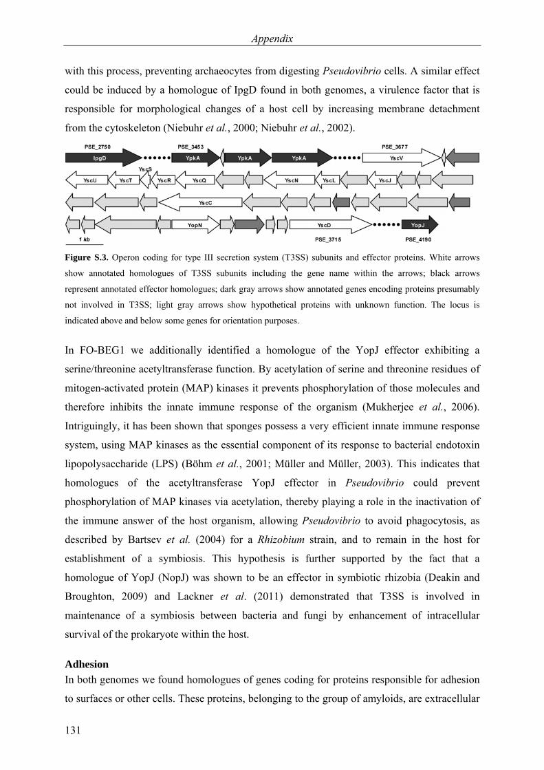
Appendix
131
with this process, preventing archaeocytes from digesting Pseudovibrio cells. A similar effect
could be induced by a homologue of IpgD found in both genomes, a virulence factor that is
responsible for morphological changes of a host cell by increasing membrane detachment
from the cytoskeleton (Niebuhr et al., 2000; Niebuhr et al., 2002).
Figure S.3. Operon coding for type III secretion system (T3SS) subunits and effector proteins. White arrows
show annotated homologues of T3SS subunits including the gene name within the arrows; black arrows
represent annotated effector homologues; dark gray arrows show annotated genes encoding proteins presumably
not involved in T3SS; light gray arrows show hypothetical proteins with unknown function. The locus is
indicated above and below some genes for orientation purposes.
In FO-BEG1 we additionally identified a homologue of the YopJ effector exhibiting a
serine/threonine acetyltransferase function. By acetylation of serine and threonine residues of
mitogen-activated protein (MAP) kinases it prevents phosphorylation of those molecules and
therefore inhibits the innate immune response of the organism (Mukherjee et al., 2006).
Intriguingly, it has been shown that sponges possess a very efficient innate immune response
system, using MAP kinases as the essential component of its response to bacterial endotoxin
lipopolysaccharide (LPS) (Böhm et al., 2001; Müller and Müller, 2003). This indicates that
homologues of the acetyltransferase YopJ effector in Pseudovibrio could prevent
phosphorylation of MAP kinases via acetylation, thereby playing a role in the inactivation of
the immune answer of the host organism, allowing Pseudovibrio to avoid phagocytosis, as
described by Bartsev et al. (2004) for a Rhizobium strain, and to remain in the host for
establishment of a symbiosis. This hypothesis is further supported by the fact that a
homologue of YopJ (NopJ) was shown to be an effector in symbiotic rhizobia (Deakin and
Broughton, 2009) and Lackner et al. (2011) demonstrated that T3SS is involved in
maintenance of a symbiosis between bacteria and fungi by enhancement of intracellular
survival of the prokaryote within the host.
Adhesion In both genomes we found homologues of genes coding for proteins responsible for adhesion
to surfaces or other cells. These proteins, belonging to the group of amyloids, are extracellular

Appendix
132
proteinaceous components and are known in Enterobacteriaceae as curli fibers. They are
involved in adhesion to surfaces, cell aggregation, biofilm formation and mediate cell-cell
adhesion and invasion of host cells (Barnhart and Chapman, 2006). The production of curli
fibers in enteric bacteria is dependent on at least six proteins encoded by the operons csgAB
and csgDEFG (agf in Salmonella) (Hammar et al., 1995), the latter of which is required for
assembly, stability and secretion of the amyloids (Hammar et al., 1995). csgAB encodes the
structural subunits of the curli fibers, both genes containing characteristic repeat motifs
(Hammar et al., 1996). A gene cluster in the genome of Pseudovibrio sp. FO-BEG1 resembles
the curli formation operon in enteric bacteria (Figure S.4).
Figure S.4. Comparison of genes encoding amyloids in Enterobacteriaceae and the operon in Pseudovibrio sp.
FO-BEG1. White arrows represent homologues of genes in enteric bacteria; gray arrows show genes present in
Enterobacteriaceae only; black arrows show genes containing curli repeats, typical motifs of the amyloid
structural subunits. The number within the black arrows shows the amount of curli repeats in the according gene.
Homologues of csgF and csgG, required for stabilization and secretion of the amyloids are
found in direct proximity to three genes containing curlin associated repeats as typical
structural components of the curli fibers. We hypothesize that the identified operon might
code for amyloid structures comparable to curli fibers due to the existence of characteristic
curlin repeat motifs and genes involved in the assembly and secretion of such structures,
therefore allowing Pseudovibrio to attach to other cells or form biofilms or aggregates.
Additionally, we identified 35 genes in strain FO-BEG1 and 37 in JE062 containing domains
mediating prokaryote-eukaryote interactions, supporting the proposed role of Pseudovibrio as
a symbiont with possibilities to attach and interact with the host organism (see
DDBJ/EMBL/GenBank accession numbers CP003147, CP003148 and ABXL00000000).
Conclusions In this study, we analyzed highly similar genomes of two Pseudovibrio strains that originate
from the coast of Florida, the Pseudovibrio sp. FO-BEG1 sampled from a coral and
maintained over 10 years in co-culture with Beggiatoa sp. and Pseudovibrio sp. JE062

Appendix
133
sampled from a sponge in the same region (Enticknap et al., 2006). The physiology of both
strains is extremely versatile and the metabolic traits found in the genome could be partially
verified in experiments with strain FO-BEG1. Here, we describe for the first time a
Pseudovibrio strain that uses aromatic compounds as a carbon and electron source, oxidizes
thiosulfate under aerobic conditions and uses phosphonates as a phosphorous source. Notably,
strain FO-BEG1 grows under extreme nutrient limitation, which emphasizes its adaptation to
life in the open ocean. The metabolic variety is confirmed by the numerous transporter
systems that are encoded in the genome. Compared with other marine bacteria, like the
prominent Roseobacter clade, which is known to be ubiquitous, multitudinous and
physiologically versatile (Newton et al., 2010), Pseudovibrio seems to be capable of a
similarly generalistic life style, exploiting quite a number of sources for energy sources,
nutrients and trace elements.
Aside from metabolic variety, the genomic data of both strains also confirm close associations
with marine invertebrates and indicate several potential mechanisms for establishing and
maintaining a symbiosis. The most striking discovery is the presence of effector homologues
secreted by type III secretion systems, which could affect sponges by interacting with their
immune response system (YopJ) or the cytoskeleton (YpkA, IpgD) and thereby have a drastic
impact on the cell machinery of the host. Another fascinating discovery is the presence of the
hybrid NRPS-PKS system in strain FO-BEG1, which has so far only been described for
members of the Enterobacteriaceae family (Putze et al., 2009), producing the bioactive
compound colibactin with yet unknown in-vivo functions, but arresting eukaryotic cells in the
G2 phase, eventually leading to cell death (Nougayrède et al., 2006). The presence of a gene
cluster coding for a cytopathic compound in strain FO-BEG1 emphasizes the impact that
Pseudovibrio cells might have on marine invertebrates. Intriguingly, strain FO-BEG1 seems
to be a required partner in the Beggiatoa co-culture, indicating its important symbiotic role
not only for marine invertebrates but also for prokaryotes. It is possible that Pseudovibrio has
positive effects for certain bacteria under in-vivo conditions, e.g. by supplying vitamins or
detoxifying metabolic intermediates or radical oxygen species.

Appendix
134
Figure S.5. Schematic overview of the possible life styles and the physiologic capabilities derived from genetic
information of both Pseudovibrio genomes. On the left hand side, physiologic abilities are depicted that could be
used in free-living, oxic and anoxic conditions. On the right hand side, the attached or associated life style is
illustrated. The host organism for the associated life style can be represented by a sponge, coral or tunicate.
Biofilm formation, aggregation and attachment to host cells could be performed via e. g. amyloid-like structures.
The proposed secretion systems could be involved in prokaryote-eukaryote interactions, influencing the cell
machinery of the host. Additionally, Pseudovibrio could supply the host with cofactors like vitamins or
synthesize secondary metabolites as a defense mechanism against other prokaryotes or the host.
The frequent identification and isolation of Pseudovibrio strains in many studies over the last
years implies an important but rather unexplored role for this genus in marine habitats.
According to the genomic and physiological data on Pseudovibrio spp., we propose a free-
living and attached or associated life style model for this genus (Figure S.5). As a denitrifying
heterotroph, Pseudovibrio has an obvious influence on the carbon and nitrogen cycles. Its
ecological impact can now be extended to the sulfur and phosphorus cycles due to its ability
to metabolize thiosulfate and phosphonates. Additionally, we hypothesize that, due to the
predictions based on the genomic data, similar to E. coli in humans, Pseudovibrio is a
commensalistic or even beneficial symbiont of marine invertebrates with a potential to
become pathogenic.
Acknowledgments We thank A. Meyerdierks, M. Mußmann, and R. Amann for comments on the manuscript. For
technical support, we thank M. Meyer, S. Menger, C. Probian, and R. Appel. We also thank A.
Kamyshny for tetrathionate measurements and K. Imhoff for sulfate determination. This study
was funded by the European Research Council and the Max Planck Society.
Appendix
135
References of appendix Aeckersberg F, Bak F, Widdel F. (1991). Anaerobic oxidation of saturated hydrocarbons to
CO2 by a new type of sulfate-reducing bacterium. Arch Microbiol 156: 5–14. Agogué H, Casamayor EO, Bourrain M, Obernosterer I, Joux F, Herndl GJ et al. (2005). A
survey on bacteria inhabiting the sea surface microlayer of coastal ecosystems. FEMS Microbiol Ecol 54: 269–280.
Aschtgen MS, Bernard CS, De Bentzmann S, Lloubès R, Cascales E. (2008). SciN is an outer
membrane lipoprotein required for type VI secretion in enteroaggregative Escherichia coli. J Bacteriol 190: 7523–7531.
Barnhart MM, Chapman MR. (2006). Curli biogenesis and function. Annu Rev Microbiol 60:
131–147. Bartsev AV, Deakin WJ, Boukli NM, McAlvin CB, Stacey G, Malnoe P et al. (2004). NopL,
an effector protein of Rhizobium sp. NGR234, thwarts activation of plant defense reactions. Plant Physiology 134: 871–879.
Bateman A, Coin L, Durbin R, Finn RD, Hollich V, Griffiths-Jones S et al. (2004). The Pfam
protein families database. Nucleic Acids Res 32: D138–D141. Bendtsen JD, Nielsen H, von Heijne G, Brunak S. (2004). Improved prediction of signal
peptides: SignalP 3.0. J Mol Biol 340: 783–795. Böhm M, Hentschel U, Friedrich AB, Fieseler L, Steffen R, Gamulin V et al. (2001).
Molecular response of the sponge Suberites domuncula to bacterial infection. Mar Biol 139: 1037–1045.
Bönemann G, Pietrosiuk A, Mogk A. (2010). Tubules and donuts: a type VI secretion story.
Mol Microbiol 76: 815–821. Boyer F, Fichant G, Berthod J, Vandenbrouck Y, Attree I. (2009). Dissecting the bacterial
type VI secretion system by a genome wide in silico analysis: what can be learned from available microbial genomic resources? BMC Genomics 10: 104.
Brock J, Schulz-Vogt HN. (2011). Sulfide induces phosphate release from polyphosphate in
cultures of a marine Beggiatoa strain. ISME J 5: 497–506. Brysch K, Schneider C, Fuchs G, Widdel F. (1987). Lithoautotrophic growth of sulfate-
reducing bacteria, and description of Desulfobacterium autotrophicum gen. nov., sp. nov. Arch Microbiol 148: 264–274.
Burton SD, Morita RY. (1964). Effect of catalase and cultural conditions on growth of
Beggiatoa. J Bacteriol 88: 1755–1761. Carver T, Thomson N, Bleasby A, Berriman M, Parkhill J. (2009). DNAPlotter: circular and
linear interactive genome visualization. Bioinformatics 25: 119–120. Case RJ, Labbate M, Kjelleberg S. (2008). AHL-driven quorum-sensing circuits: their
frequency and function among the Proteobacteria. ISME J 2: 345–349.
Appendix
136
Chow J, Mazmanian SK. (2010). A pathobiont of the microbiota balances host colonization
and intestinal inflammation. Cell Host Microbe 7: 265–276. Cornelis GR. (2002). The Yersinia Ysc-Yop 'type III' weaponry. Nat Rev Mol Cell Bio 3:
742–752. Cornelis GR, Van Gijsegem F. (2000). Assembly and function of type III secretory systems.
Annu Rev Microbiol 54: 735–774. Cunliffe M. (2011). Correlating carbon monoxide oxidation with cox genes in the abundant
Marine Roseobacter Clade. ISME J 5: 685–691. Deakin WJ, Broughton WJ. (2009). Symbiotic use of pathogenic strategies: rhizobial protein
secretion systems. Nat Rev Microbiol 7: 312–320. Delcher AL, Bratke KA, Powers EC, Salzberg SL. (2007). Identifying bacterial genes and
endosymbiont DNA with Glimmer. Bioinformatics 23: 673–679. Enticknap JJ, Kelly M, Peraud O, Hill RT. (2006). Characterization of a culturable
alphaproteobacterial symbiont common to many marine sponges and evidence for vertical transmission via sponge larvae. Appl Environ Microbiol 72: 3724–3732.
Filloux A, Hachani A, Bleves S. (2008). The bacterial type VI secretion machine: yet another
player for protein transport across membranes. Microbiol-SGM 154: 1570–1583. Fisch KM, Gurgui C, Heycke N, van der Sar SA, Anderson SA, Webb VL et al. (2009).
Polyketide assembly lines of uncultivated sponge symbionts from structure-based gene targeting. Nat Chem Biol 5: 494–501.
Fukunaga Y, Kurahashi M, Tanaka K, Yanagi K, Yokota A, Harayama S. (2006).
Pseudovibrio ascidiaceicola sp. nov., isolated from ascidians (sea squirts). Int J Syst Evol Microbiol 56: 343–347.
Fürch T, Preusse M, Tomasch J, Zech H, Wagner-Döbler I, Rabus R et al. (2009). Metabolic
fluxes in the central carbon metabolism of Dinoroseobacter shibae and Phaeobacter gallaeciensis, two members of the marine Roseobacter clade. BMC Microbiol 9: 209.
Ghosh W, Dam B. (2009). Biochemistry and molecular biology of lithotrophic sulfur
oxidation by taxonomically and ecologically diverse bacteria and archaea. FEMS Microbiol Rev 33: 999–1043.
Hammar M, Bian Z, Normark S. (1996). Nucleator-dependent intercellular assembly of
adhesive curli organelles in Escherichia coli. Proc Natl Acad Sci U S A 93: 6562–6566. Hammar M, Arnqvist A, Bian Z, Olsén A, Normark S. (1995). Expression of two csg operons
is required for production of fibronectin- and congo red-binding curli polymers in Escherichia coli K-12. Mol Microbiol 18: 661–670.
Hentschel U, Schmid M, Wagner M, Fieseler L, Gernert C, Hacker J. (2001). Isolation and
phylogenetic analysis of bacteria with antimicrobial activities from the mediterranean

Appendix
137
sponges Aplysina aerophoba and Aplysina cavernicola. FEMS Microbiol Ecol 35: 305–312.
Hood RD, Singh P, Hsu FS, Güvener T, Carl MA, Trinidad RRS et al. (2010). A type VI
secretion system of Pseudomonas aeruginosa targets a toxin to bacteria. Cell Host Microbe 7: 25–37.
Hosoya S, Yokota A. (2007). Pseudovibrio japonicus sp. nov., isolated from coastal seawater
in Japan. Int J Syst Evol Microbiol 57: 1952–1955. Jani AJ, Cotter PA. (2010). Type VI secretion: not just for pathogenesis anymore. Cell Host
Microbe 8: 2–6. Kamyshny A. (2009). Improved cyanolysis protocol for detection of zero-valent sulfur in
natural aquatic systems. Limnol Oceanogr-Meth 7: 442–448. Kennedy J, Baker P, Piper C, Cotter PD, Walsh M, Mooij MJ et al. (2009). Isolation and
analysis of bacteria with antimicrobial activities from the marine sponge Haliclona simulans collected from irish waters. Mar Biotechnol 11: 384–396.
King GM, Weber CF. (2007). Distribution, diversity and ecology of aerobic CO-oxidizing
bacteria. Nat Rev Microbiol 5: 107–118. Koren O, Rosenberg E. (2006). Bacteria associated with mucus and tissues of the coral
Oculina patagonica in summer and winter. Appl Environ Microbiol 72: 5254–5259. Krogh A, Larsson B, von Heijne G, Sonnhammer EL. (2001). Predicting transmembrane
protein topology with a hidden Markov model: application to complete genomes. J Mol Biol 305: 567–580.
Lackner G, Moebius N, Hertweck C. (2011). Endofungal bacterium controls its host by an hrp
type III secretion system. ISME J 5: 252–261. Lafi FF, Garson MJ, Fuerst JA. (2005). Culturable bacterial symbionts isolated from two
distinct sponge species (Pseudoceratina clavata and Rhabdastrella globostellata) from the Great Barrier Reef display similar phylogenetic diversity. Microb Ecol 50: 213–220.
Lagesen K, Hallin P, Rødland EA, Stærfeldt HH, Rognes T, Ussery DW. (2007). RNAmmer:
consistent and rapid annotation of ribosomal RNA genes. Nucleic Acids Res 35: 3100–3108.
Lambalot RH, Gehring AM, Flugel RS, Zuber P, LaCelle M, Marahiel MA et al. (1996). A
new enzyme superfamily - the phosphopantetheinyl transferases. Chem Biol 3: 923–936. Lang AS, Beatty JT. (2001). The gene transfer agent of Rhodobacter capsulatus and
"constitutive transduction" in prokaryotes. Arch Microbiol 175: 241–249. Lang AS, Beatty JT. (2007). Importance of widespread gene transfer agent genes in α-
Proteobacteria. Trends Microbiol 15: 54–62.
Appendix
138
Larkin JM, Strohl WR. (1983). Beggiatoa, Thiothrix, and Thioploca. Annu Rev Microbiol 37: 341–367.
Lowe TM, Eddy SR. (1997). tRNAscan-SE: a program for improved detection of transfer
RNA genes in genomic sequence. Nucleic Acids Res 25: 955–964. Meyer F, Goesmann A, McHardy AC, Bartels D, Bekel T, Clausen J et al. (2003). GenDB -
an open source genome annotation system for prokaryote genomes. Nucleic Acids Res 31: 2187–2195.
Morris JJ, Kirkegaard R, Szul MJ, Johnson ZI, Zinser ER. (2008). Facilitation of robust
growth of Prochlorococcus colonies and dilute liquid cultures by "helper" heterotrophic bacteria. Appl Environ Microbiol 74: 4530–4534.
Mougous JD, Cuff ME, Raunser S, Shen A, Zhou M, Gifford CA et al. (2006). A virulence
locus of Pseudomonas aeruginosa encodes a protein secretion apparatus. Science 312: 1526–1530.
Mukherjee S, Keitany G, Li Y, Wang Y, Ball HL, Goldsmith EJ et al. (2006). Yersinia YopJ
acetylates and inhibits kinase activation by blocking phosphorylation. Science 312: 1211–1214.
Mulder NJ, Apweiler R, Attwood TK, Bairoch A, Bateman A, Binns D et al. (2005). InterPro,
progress and status in 2005. Nucleic Acids Res 33: D201–D205. Müller WEG, Müller IM. (2003). Origin of the metazoan immune system: identification of
the molecules and their functions in sponges. Integr Comp Biol 43: 281–292. Muscholl-Silberhorn A, Thiel V, Imhoff JF. (2008). Abundance and bioactivity of cultured
sponge-associated bacteria from the mediterranean sea. Microb Ecol 55: 94–106. Newton RJ, Griffin LE, Bowles KM, Meile C, Gifford S, Givens CE et al. (2010). Genome
characteristics of a generalist marine bacterial lineage. ISME J 4: 784–798. Niebuhr K, Jouihri N, Allaoui A, Gounon P, Sansonetti PJ, Parsot C. (2000). IpgD, a protein
secreted by the type III secretion machinery of Shigella flexneri, is chaperoned by IpgE and implicated in entry focus formation. Mol Microbiol 38: 8–19.
Niebuhr K, Giuriato S, Pedron T, Philpott DJ, Gaits F, Sable J et al. (2002). Conversion of
PtdIns(4,5)P-2 into PtdIns(5)P by the S.flexneri effector IpgD reorganizes host cell morphology. EMBO J 21: 5069–5078.
Nougayrède JP, Homburg S, Taieb F, Boury M, Brzuszkiewicz E, Gottschalk G et al. (2006).
Escherichia coli induces DNA double-strand breaks in eukaryotic cells. Science 313: 848–851.
Olson JB, Harmody DK, McCarthy PJ. (2002). α-Proteobacteria cultivated from marine
sponges display branching rod morphology. FEMS Microbiol Lett 211: 169–173.
Appendix
139
Piel J, Hui DQ, Wen GP, Butzke D, Platzer M, Fusetani N et al. (2004). Antitumor polyketide biosynthesis by an uncultivated bacterial symbiont of the marine sponge Theonella swinhoei. Proc Natl Acad Sci U S A 101: 16222–16227.
Pukatzki S, McAuley SB, Miyata ST. (2009). The type VI secretion system: translocation of
effectors and effector-domains. Curr Opin Microbiol 12: 11–17. Pukatzki S, Ma AT, Sturtevant D, Krastins B, Sarracino D, Nelson WC et al. (2006).
Identification of a conserved bacterial protein secretion system in Vibrio cholerae using the Dictyostelium host model system. Proc Natl Acad Sci U S A 103: 1528–1533.
Putze J, Hennequin C, Nougayrède JP, Zhang WL, Homburg S, Karch H et al. (2009).
Genetic structure and distribution of the colibactin genomic island among members of the family Enterobacteriaceae. Infect Immun 77: 4696–4703.
Quast C. (2006). MicHanThi - design and implementation of a system for the prediction of
gene functions in genome annotation projects. Diploma thesis, Universität Bremen, Bremen.
Richter M, Rosselló-Móra R. (2009). Shifting the genomic gold standard for the prokaryotic
species definition. Proc Natl Acad Sci U S A 106: 19126–19131. Richter M, Lombardot T, Kostadinov I, Kottmann R, Duhaime MB, Peplies J et al. (2008).
JCoast - a biologist-centric software tool for data mining and comparison of prokaryotic (meta)genomes. BMC Bioinformatics 9: 177.
Riesenfeld CS, Murray AE, Baker BJ. (2008). Characterization of the microbial community
and polyketide biosynthetic potential in the palmerolide-producing tunicate Synoicum adareanum. J Nat Prod 71: 1812–1818.
Rypien KL, Ward JR, Azam F. (2010). Antagonistic interactions among coral-associated
bacteria. Environ Microbiol 12: 28–39. Santos OCS, Pontes PVML, Santos JFM, Muricy G, Giambiagi-deMarval M, Laport MS.
(2010). Isolation, characterization and phylogeny of sponge-associated bacteria with antimicrobial activities from Brazil. Res Microbiol 161: 604–612.
Sertan-de Guzman AA, Predicala RZ, Bernardo EB, Neilan BA, Elardo SP, Mangalindan GC
et al. (2007). Pseudovibrio denitrificans strain Z143-1, a heptylprodigiosin-producing bacterium isolated from a Philippine tunicate. FEMS Microbiol Lett 277: 188–196.
Shieh WY, Lin YT, Jean WD. (2004). Pseudovibrio denitrificans gen. nov., sp. nov., a marine,
facultatively anaerobic, fermentative bacterium capable of denitrification. Int J Syst Evol Microbiol 54: 2307–2312.
Siegl A, Kamke J, Hochmuth T, Piel J, Richter M, Liang CG et al. (2011). Single-cell
genomics reveals the lifestyle of Poribacteria, a candidate phylum symbiotically associated with marine sponges. ISME J 5: 61–70.
Stothard P, Wishart DS. (2005). Circular genome visualization and exploration using
CGView. Bioinformatics 21: 537–539.

Appendix
140
Subramoni S, Venturi V. (2009). LuxR-family 'solos': bachelor sensors/regulators of
signalling molecules. Microbiol-SGM 155: 1377–1385. Tang KH, Feng XY, Tang YJJ, Blankenship RE. (2009). Carbohydrate metabolism and
carbon fixation in Roseobacter denitrificans OCh114. PLoS One 4: e7233. Tatusov RL, Fedorova ND, Jackson JD, Jacobs AR, Kiryutin B, Koonin EV et al. (2003). The
COG database: an updated version includes eukaryotes. BMC Bioinformatics 4: 41. Taylor MW, Radax R, Steger D, Wagner M. (2007). Sponge-associated microorganisms:
evolution, ecology, and biotechnological potential. Microbiol Mol Biol Rev 71: 295–374. Thakur NL, Hentschel U, Krasko A, Pabel CT, Anil AC, Muller WEG. (2003). Antibacterial
activity of the sponge Suberites domuncula and its primmorphs: potential basis for epibacterial chemical defense. Aquat Microb Ecol 31: 77–83.
Thiel V, Imhoff JF. (2003). Phylogenetic identification of bacteria with antimicrobial
activities isolated from Mediterranean sponges. Biomolecular Engineering 20: 421–423. Thoms C, Horn M, Wagner M, Hentschel U, Proksch P. (2003). Monitoring microbial
diversity and natural product profiles of the sponge Aplysina cavernicola following transplantation. Mar Biol 142: 685–692.
Wagner-Döbler I, Ballhausen B, Berger M, Brinkhoff T, Buchholz I, Bunk B et al. (2010).
The complete genome sequence of the algal symbiont Dinoroseobacter shibae: a hitchhiker's guide to life in the sea. ISME J 4: 61–77.
Weber B, Hasic M, Chen C, Wai SN, Milton DL. (2009). Type VI secretion modulates
quorum sensing and stress response in Vibrio anguillarum. Environ Microbiol 11: 3018–3028.
Webster NS, Hill RT. (2001). The culturable microbial community of the Great Barrier Reef
sponge Rhopaloeides odorabile is dominated by an α-proteobacterium. Mar Biol 138: 843–851.
Widdel F, Pfennig N. (1984). Dissimilatory sulfate- or sulfur-reducing bacteria. In: Krieg NR,
Holt JG (eds). Bergey's manual of systematic bacteriology. Williams & Wilkins: Baltimore. pp 663–379.
Widdel F, Bak F. (1992). Gram-negative mesophilic sulfate-reducing bacteria. In: Balows A,
Trüper HG, Dworkin M, Harder W, Schleifer KH (eds). The Prokaryotes, 2nd edn. Springer: New York. pp 3352–3378.
Williams TJ, Ertan H, Ting L, Cavicchioli R. (2009). Carbon and nitrogen substrate
utilization in the marine bacterium Sphingopyxis alaskensis strain RB2256. ISME J 3: 1036–1052.
Zedelius J, Rabus R, Grundmann O, Werner I, Brodkorb D, Schreiber F et al. (2011). Alkane
degradation under anoxic conditions by a nitrate-reducing bacterium with possible

Appendix
141
involvement of the electron acceptor in substrate activation. Environ Microbiol Rep 3: 125–135.
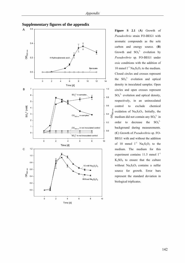
Appendix
142
Supplementary figures of the appendix
Figure S 2.1 (A) Growth of
Pseudovibrio strain FO-BEG1 with
aromatic compounds as the sole
carbon and energy source. (B)
Growth and SO42– evolution by
Pseudovibrio sp. FO-BEG1 under
oxic conditions with the addition of
10 mmol l–1 Na2S2O3 to the medium.
Closed circles and crosses represent
the SO42– evolution and optical
density in inoculated samples. Open
circles and open crosses represent
SO42– evolution and optical density,
respectively, in an uninoculated
control to exclude chemical
oxidation of Na2S2O3. Initially, the
medium did not contain any SO42– in
order to decrease the SO42–
background during measurements.
(C) Growth of Pseudovibrio sp. FO-
BEG1 with and without the addition
of 10 mmol l–1 Na2S2O3 to the
medium. The medium for this
experiment contains 11.5 mmol l–1
K2SO4 to ensure that the culture
without Na2S2O3 contains a sulfur
source for growth. Error bars
represent the standard deviation in
biological triplicates.

Appendix
143
Figure S 2.2 (A) Glucose
consumption, growth and
production of fermentation
products by Pseudovibrio sp. FO-
BEG1 grown under anoxic
conditions without NO3–. (B)
Glucose consumption, growth and
production of fermentation
products during simultaneous
denitrification and fermentation by
Pseudovibrio sp. FO-BEG1 grown
under anoxic conditions with
addition of 10 mmol l–1 NO3–. (C)
Consumption of NO3–, evolution
of gaseous nitrogen and growth
during simultaneous
denitrification and fermentation of
Pseudovibrio sp. FO-BEG1 with
10 mmol l–1 NO3–. Error bars
represent the standard deviation in
biological triplicates.

Appendix
144
Figure S 2.3 (A) Growth of
Pseudovibrio sp. FO-BEG1 with 1
mmol L–1 phosphonoacetate as the
only phosphorus source and without
the addition of any phosphorus to
the medium. (B) Growth of
Pseudovibrio sp. FO-BEG1 without
the addition of vitamins to the
medium. Error bars represent the
standard deviation in biological
triplicates.

Acknowledgements
145
Acknowledgements
I would like to express my deepest thanks to my “Doktormutter” Heide Schulz-Vogt. You
have shown me the beauty of the large sulfide-oxidizers and you have always encouraged me
to follow my ideas and to go into new directions. Thank you for providing me such an
interesting topic and for all your support!
I would also like to thank Prof. Dr. Ulrich Fischer for writing the second review. Furthermore,
I am thankful for the helpful discussion and remarks you made during my thesis committee
meeting and all further discussions.
Thank you Thorsten Dittmar for being a member of my thesis and defense committee and also
for all your help during the last years! I am very happy that I had the possibility to work with
and learn from you and I am looking forward to perform the next experiments.
Moreover, I thank Rudi Amann for joining my thesis and defense committee and for all your
helpful remarks during our discussions.
Anne-Christin Kreutzmann and Sascha Artelt, thank you for being members of my defense
committee.
Dear Ecophysios (Jörg, Verena, Sandra, Stefano, Vladi, Martina, Susanne, Anne-Christin,
Tine, Daniela, Sascha, Arthur), thank you all so much for the last years, I enjoyed working
with you all! Especially I would like to thank Anne-Christin Kreutzmann, I hope we will
write many more manuscripts together in the future! Martina Meyer and Susanne Menger,
you are the best technicians I can imagine!
Michael Seidel, thank you for measuring so many samples with me and for cleaning up the
lab without me late at night, when I already had to catch the last train back to Bremen. I also
thank you for picking up the “Sorgentelefon” when I analyzed the FT-ICR-MS data!
Furthermore, I would like to thank the whole Marine Geochemistry Group in Oldenburg for
helping me (especially Matze, Katrin and Ina). I always enjoyed the time in Oldenburg!
Thanks a lot to Meinhard Simon and Birgit Kürzel for measuring the amino acid samples.

Acknowledgements
146
Thank you, Lubos Polerecky, for analysing all the data with me and for nice colourful figures.
Martin Beutler, thank you for taking many, many pictures with me at the CLSM.
Thanks to Michael Richter for his very spontaneous help with KEGG and his help during
genome annotation.
Gaute Lavik and Thomas Max are thanked for their help during isotope-labelling experiments.
I thank Johannes Zedelius and Frank Schreiber for their help when I had “NO” questions.
I thank Bernd Stickford for his help during literature search. He was able to find also very,
very old articles for me.
Many thanks to the whole Microbiology department at the MPI Bremen for the nice working
atmosphere and all your help, representative I would like to mention Jens Harder and
Prof. Widdel.
My dearest officemates: Jana, Julia and Verena. You were always there – celebrating when
things worked out, discussing when something unexpected happened and encouraging me
when experiments failed. Thank you so much!
Ines and Anna - lunch with you was always awesome ☺
I always enjoyed the “Mädelsabend” with my MPI girls (Anna, Ines, Frauke, Kirsten, Sandra,
Ulli, Verena, Julia, Anne-Christin and Jana). I’ve never been alone with you at my side going
through the ups and downs of the PhD life, thank you!
Meinen Freunden (Julia, Karo und ALLEN anderen) und meiner Familie (Andy, Mama, Papa,
Lina, Bernd, Oma, Opa, Frank, Katha + XX und der GANZEN Sippe) möchte ich danken,
dass sie mir immer zur Seite gestanden, mich unterstützt und wenn nötig auf andere Gedanken
gebracht haben!
Andy – du bist der beste Mann den es gibt und dafür bin ich dir von ganzem Herzen dankbar!!!

147
Erklärung
148
Name: Anne Schwedt Bremen, 23.09.2011 Anschrift: Am Deich 75, 28199 Bremen ERKLÄRUNG Hiermit erkläre ich, dass ich die Arbeit mit dem Titel: „Physiology of a marine Beggiatoa strain and the accompanying organism Pseudovibrio sp. – a facultatively oligotrophic bacterium“ selbstständig verfasst und geschrieben habe und außer den angegebenen Quellen keine weiteren Hilfsmittel verwendet habe. Ebenfalls erkläre ich hiermit eidesstattlich, dass es sich bei den von mir abgegebenen Arbeiten um 3 identische Exemplare handelt. (Unterschrift)


![Experimentalvortrag Anorganik BODEN [Kompatibilitätsmodus] · Katalase •s Htzet 2O 2 um • Umsatzzahl von Molekülen/min eines dffk bh 510⋅ 6 der effektivsten Enzyme überhaupt](https://img.pdfslide.org/doc/110x75/5d0a2d8688c993d32d8b6613/experimentalvortrag-anorganik-boden-kompatibilitaetsmodus-katalase-s-htzet.jpg)