Embed Size (px)
Citation preview
GSJ: Volume 8, Issue 10, October 2020, Online: ISSN 2320-9186
www.globalscientificjournal.com
PHYTOCHEMICAL SCREENING AND IN VITRO ASSESSMENT OF SOME BIOLOGICAL ACTIVITIES OF Ipomoea setosa Ker. Gawl. (COBRA
VINE) SEEDS MAYUMI J. OGAWA, FLORFE M. ACMA, MERCED G. MELENCION, LORELIE GLORIA A.
SAMANIEGO
Department of Biology, College of Arts and Sciences, Central Mindanao University, Musuan, Maramag Bukidnon, Philippines Email: [email protected]
[email protected] Tel: +639357557573
Abstract The potential of Ipomoea setosa Ker. Gawl. seed extract to control some biological activities was evaluated using standard procedures with slight modification. Specifically, the study identified the secondary metabolites present and evaluated the anti-inflammatory, antioxidant, and anticancer activity of the seed methanolic extract. The presence of alkaloids, anthraquinone, flavonoids and catecholic tannins were detected in the extract. The total phenolic content is 5.55 ± 0.23 mg GAE/g sample while the total flavonoid content is 1.56 ± 0.06 mg QE/g. I. setosa Ker. Gawl. seed extract has 21.43 ± 6.48% inhibition of albumin denaturation and 73.79 ± 2.69% inhibition relative to Kortezor (positive control). The seed extract showed a highly significant higher free radical scavenging activity (80.54 ± 0.58%) compared to ascorbic acid (70.87 ± 0.64%). Relative to the radical scavenging activity of ascorbic acid (100.00%), the seed extract showed a 113.10 ± 0.99% inhibition. Furthermore, I. setosa Ker. Gawl. seed extract has a high potential against HCT-116 colorectal carcinoma cell line. Based on the results, it can be concluded that I. setosa Ker. Gawl. seeds can be potential anti-inflammatory, antioxidant and anticancer agent. Key Words: anticancer, anti-inflammation, antioxidant, HCT-116 cell line, phytochemical content 1. Introduction: Plants have been used for centuries as source of medicine because of the phytochemical content, which are reported to be potential therapeutic agents. These phytochemicals can be extracted for the formulation of pharmaceutical medicines but the plant itself can also be used directly (Rane and Patel, 2014). Some biochemical compounds from selected terrestrial plants have already been extracted and reported to have anticancer properties. These include polyphenols, brassinosteroids and taxols (Greenwell and Rahman, 2015). According to Palhares et al. (2015), the World Health Organization (WHO) encouraged the development of new products from natural sources and that out of the estimated 300,000 plant species that exist in the world, only 15% have been evaluated for their pharmacological potential. Ipomoea species which belong to family Convolvulaceae, are vital because of their economic importance and have been in continuous use for different purposes from ornamental, nutritional, medicinal, ritual and agricultural to its culinary value (Folorunso et al., 2013; Meira et al., 2012). Since 1950, the phytochemistry of the genus Ipomoea had been studied and showed that some of the species contain antimicrobial, analgesic, spamolithic, spasmogenic, hypotensive, psychotomimetic and anticancer activities (Meira et al., 2012). One of the potential species is Ipomoea setosa Ker. Gawl., it is a perennial herb with soft stem and fleshy spines and pinkish violet bloom but otherwise glabrous (Wunderlin et al., 2018;Wood et al., 2015). Its seeds are claimed to have an anti-inflammatory, carminative, depurative, purgative and vermifuge properties and can be used for constipation, dyspepsia, bronchitis, fever, skin diseases, scabies and splenopathy (Londhe et al., 2017). Globally, one of the leading causes of morbidity and mortality is cancer (Desai et al., 2008). In the Philippines, colorectal cancer ranked the third most common cancer and the number steadily increases since less focus is given to it (Philippine Cancer Society Forum, 2014; Imanis Life Sciences, 2018). Further, colorectal carcinoma (CRC) screening is costly; thus, only few individuals subject to such test. As the prevalence of colorectal cancer cases is
GSJ: Volume 8, Issue 10, October 2020 ISSN 2320-9186 1595
GSJ© 2020 www.globalscientificjournal.com
2 increasing the use of alternative prevention or treatment, such as plants with anti-oxidative and anticancer properties, is recommended. Ipomoea setosa Ker. Gawl. have been claimed in some local areas to cure cancer. However, there is no scientific foundation to this claim. Hence, this study was conducted to validate the claimed therapeutic effect of this plant. 2. Methods 2.1 Plant Identification and Seed Collection The plant used in this study was identified by a taxonomist from Central Mindanao University, Musuan, Maramag, Bukidnon, Philippines. The seeds were collected from mature pods, air dried, and kept in a paper bag. 2.2 Preparation and Extraction of Phytochemicals The seeds were powdered and soaked with methanol for 48 hours then filtered. The filtrate was concentrated using a rotary evaporator at 40˚C. The concentrated extract was sealed in a labeled container and stored in a refrigerator. 2.3 Phytochemical Screening Phytochemical screening of the seed methanolic extract of the seeds of Ipomoea setosa Ker. Gawl. was carried out following the conventional test tube method (color test). Ten (10) mg of the concentrated extract was dissolved in one hundred (100) ml 15:5:2 DMSO:methanol:water solution and was homogenized using the vortex. The diluted extract was then subjected to phytochemical screening. 2.3.a Visualization of anthraquinones (Evans, 2002; Akhar et al, 2013). Borntrager’s test was used. One (1) ml of the extract was added with 0.5 ml 5% KOH and shaken for 5 minutes. Appearance of orange colored solution indicates the presence of anthraquinones. 2.3.b Visualization of alkaloids (Johansen, 1940) Three (3) ml of the seed extract was added with one (1) ml 1M sulfuric acid. The mixture was shaken for 3-5 minutes and was allowed to settle. About two (2) to three (3) drops of Dragendorff’s A and B reagents were then added. A prominent yellow precipitate confirms the test as positive which is caused by the precipitation from the neutral or slightly acidic solution of Dragendorff’s reagent. 2.3.c Visualization of Flavonoids (Bhandary et al., 2012) To detect the presence of flavonoids, Alkaline reagent test was used. 0.2 ml of 5% sodium hydroxide (NaOH) solution was added in 0.5 ml of the seed extract. An increasing intensity of yellow color which eventually become colorless with the addition of diluted hydrochloric acid (HCl) indicates a positive result. 2.3.d Visualization of Saponins (Johansen, 1940) Sulfuric acid test was used to detect the presence of saponin. In a 0.5 ml extract, three (3) drops of sulfuric acid was added slowly. For a positive result, there must be an initial color reaction from yellow to red then the appearance of a red, violet or blue-green solution. 2.3.e Visualization of Steroids and Terpenoids (Bhandary et al., 2012) To detect the presence of steroids, Liebermann Burchard test was used. One (1) ml extract was mixed with one (1) ml of acetic acid anhydride. Then, one (1) ml of concentrated sulfuric acid was added on the sides while in an ice bath. There should be formation of brown ring at the junction between two layers. A green coloration in the upper layer indicates the presence of steroids while a deep red color formation in the lower layer indicates the presence of terpenoids. 2.3.f Visualization of Tannins (Iyenger, 1995) To detect the presence of tannins, Ferric Chloride test was performed. One (1) to two (2) drops of 10% ferric chloride (FeCl3) were added to 0.5 ml extract. The formation of a blue precipitate is an indication of the presence of gallic tannins and a green-black color for catecholic tannins. 2.3.g Total Phenolics (Ayiegoro and Okoh, 2010; Chatatikun and Chiabchalard, 2013) Folic-Ciocalteu Reagent method with slight modification was used to determine the total phenolic content of the plant extract. The test was performed in quadroplicate. The solvent used was 15DMSO: 5MeOH:2H2O. for the standard solution (gallic acid) 1 mg was dissolved in (15:5:2) DMSO: MeOH: H2O solvent with various concentrations (2.5, 5, 10, 25, 50 ug/ml). For the sample, 2 mg of the extract was dissolved in 500 ul of the solvent
GSJ: Volume 8, Issue 10, October 2020 ISSN 2320-9186 1596
GSJ© 2020 www.globalscientificjournal.com
3 (15DMSO: 5MeOH: 2H2O). In a separate beaker 1 mg of Folin-Ciocalteu reagent was dissolved in a 10 mL HPLC water. 5% Na2CO3 (Sodium Carbonate) was dissolved in HPLC water. For the plating, 20 ul of the sample extract was added and 100 ul Folin-Ciocalteu and 80 ul of 5% NaCO3 as the sample. For the extract blank, 20 ul of the sample extract was added with 180 ul of the solvent (15DMSO: 5MeOH: 2H2O). For the working standard 20 ul of the standard was added with 100 ul Folin-Ciocalteu reagent and 80 ul of 5% NaCO3. After plating, the plate was incubated at room temperature for 2 hours and the absorbance reading was measured at 750 nm using a ThermoScientific spectrophotometer. The total phenolic content is expressed as milligram Gallic Acid Equivalent (GAE) per gram sample in a wet basis. 2.3.h Total Flavonoids (Ayiegoro and Okoh, 2010; Chatatikun and Chiabchalard, 2013) Total flavonoid content was determined using the Aluminum Chloride Colorimetric Assay with modifications. 2 mg of the sample extract was dissolved in 1 ml solvent (15DMSO:5methanol:2water ratio). For the standard solution (quercetin) 1 mg was dissolved in (15:5:2) DMSO: MeOH: H2O solvent with various concentration (6.25, 12.5, 25, 50, 100 µg/ml). 1 g of aluminum chloride was dissolved in a 9 ml water and 0.082 g sodium acetate was dissolved in a 100 ml water. In a 96 well plate, 30 ul of extract and standard were added with 30 ul 1M sodium acetate, 30 ul aluminum chloride and 110 ul HPLC water was added. For 30 minutes all reagents were mixed and incubated at room temperature protected from light. The absorbance was measured using a ThermoScientific spectrophotometer at 415 nm. All samples were analyzed with four replicates. The total flavonoid content was expressed as mg Quercetin Equivalents per gram. 2.4 Egg Albumin Denaturation Assay (Chandrika et al., 2005; Musfiq-Marliyah and Ananthi, 2015) In 1 ml of DMSO:Methanol:Water (15:5:2) solution, 2 mg of the concentrated methanolic seed extract was dissolved. Kortezor served as the positive control and DMSO:Methanol:Water (15:5:2) solution as the negative control. In a 600 µL phosphate buffer saline (PBS), with a pH of 6.4, and 40 µL of egg albumin, 50 µL of the extract was added. In the positive and negative controls, same amount of PBS and 40 µL egg albumin was added. For 15 minutes at 37˚C and for 5 minutes at 72˚C, samples were heated. Samples was cooled for 10 minutes. Then, in a 96-well microplate, 150 µL from every sample was pipetted. Three (3) wells were used for the seed extract, three (3) wells for the positive control and three (3) wells for the negative control. At 660 nm, the absorbance was measured using a ThermoScientific spectrophotometer. The percentage inhibition of protein denaturation was expressed using the following formula:
% inhibition = [(Vf/Vc) – 1 x 100] Where:
Vf = absorbance of test samples Vc= absorbance of negative control
2.5 DPPH Free Radical Scavenging Assay The antioxidant activity of Ipomoea setosa Ker. Gawl. seed extract was assessed using 1,1diphenyl 2-picrylhyorazyl (DPPH) scavenging assay. Two (2) mg of concentrated extract was dissolved in 1 ml solvent (15:5:2) DPPH:methanol:H2O. The negative control used is the DPPH solution (DMSO:methanol:water in 15:2:2 ratio). The standard reference used was ascorbic acid. In this test, a 96-well microplate was used. For the concentrated extract, three (3) wells were used with 50 µl in each well; for the positive control three (3) wells with 50 µl of ascorbic acid each and three (3) wells for the negative control with 50 µl of the solvent system. 50 µl of DPPH was added in each of the tested samples. Each well served as replicate. The absorbance was measured at 517 nm after 30 min at room temperature protected from light using a ThermoScientific spectrophotometer. The scavenging activity of the DPPH free radical was measured by using the following equation:
% inhibition = [ Vc - Vt / Vc)] x 100 Where:
Vc – Absorbance of negative control Vt – Absorbance of sample
2.6 Methylthiazol Tetrazolium (MTT) Anticancer Assay (Mossman, 1983) In this assay, HCT-116, a colorectal human carcinoma cell line was used. In a sterile 96-well microtiter plates, cells were seeded at 4 x 10⁴ cells/ml. The plates were incubated overnight at 37°C and 5% CO2. Eight two-fold dilutions (100, 50, 25, 12.5, 6.25, 3.13, 1.56, 0.78 ug/ml) of the sample were used as treatments. Doxorubicin served as positive control while dimethyl sulfoxide (DMSO) served as negative control. Following incubation, the cells were treated with their respective extract dilution. The treated cells were again incubated for 72 hours at 37°C and 5% CO2. After incubation, the media was removed and 3-(4,5dimethylethylthiazol-2-yl)-2,5-diphenyltetrazolium bromide (MTT) dye at 5 mg/ml PBS was added. The cells were again incubated at 37°C and 5% CO2 for 4 hours. After this, DMSO is used to dissolve the formazan crystals formed by the reduction of the dye by the live cells.
GSJ: Volume 8, Issue 10, October 2020 ISSN 2320-9186 1597
GSJ© 2020 www.globalscientificjournal.com
4 Absorbance was read at 570 nm. The IC50 (inhibition concentration) was computed using GraphPad Prism 6. GraphPad Prism 6 computes for the IC50 of the sample by employing non-linear regression curve fit on the computed percent inhibition per concentration of the sample. Samples with IC50 values less than 30 µg/mL are considered active (Jokhadze et al., 2007). Percentage inhibition of the sample was calculated using the formula used by Mohammed et al. (2013):
% inhibition = 100 - (At/Ab Ac/Ab ) x 100 where:
At = absorbance of treated cells Ab = absorbance of untreated cells Ac = absorbance of control
2.7 Statistical Analysis The data was expressed as mean ± SEM. To determine the significance of the result for Egg Albumin and DPPH assays, One-way Analysis of Variance (ANOVA) using Statistical Package for Social Sciences (SPSS) program version 20 was used. 3. Results and discussion Phytochemical Screening Qualitative Analysis Result of the qualitative analysis for the phytochemicals present in the methanolic extract of Ipomoea setosa Ker. Gawl. seeds is shown in Table 1. The seed methanolic extract of I. setosa Ker. Gawl. used in this study contains anthraquinones. However, Esseitt et al., (2014) mentioned that in some Ipomoea species are absent or present in minute quantity. Anthraquinones showed antiparasitic (Pieters and Vlietinck, 2005), bacteriostatic, antidepressant, and antimicrobial properties (Cowan, 1999). The seed methanolic extract also possesses alkaloids. As reported by Srivastava (2017) alkaloids are a major chemical constituent of the genus Ipomoea. This explains the presence of alkaloids in the sample extract. Alkaloids help in the protection from chronic diseases; have anti-inflammatory, anti‐diabetic and analgesic activities; can reduce headaches associated with hypertension (Mir et al., 2013); showed a potent cytotoxic activity in human cancer (Salamah and Ningsih, 2017); and are also reported to be used traditionally as antiseptics, sedatives, stomatics, treat bleeding disorders and eye diseases and as uterine muscle depressants (Bribi, 2018). In the alkaline reagent test, I. setosa Ker. Gawl. seed methanolic extract showed a positive result for flavonoids. Flavonoids are phenolic compounds that are naturally occurring and known to possess many pharmacological effects such as antiviral, anti-fungal, antioxidant, anti-inflammatory, antiallergenic, antithrombic, anticarcinogenic, hepatoprotective, and cytotoxic activities (Peteros and Uy, 2010). The methanolic seed extract of I. setosa Ker. Gawl. showed the presence of catecholic tannins. Tannins indicate the potential of this plant as antidiarrhoeic, antihemorrhagic, and as wound healing agent (Asquith and Butler, 1986; Okwu and Josiah, 2006). Tannins have also been shown to have antimutagenic, anticarcinogenic, antioxidative, antimicrobial, cytotoxic and antineoplastic activities; accelerate blood clotting, reduce blood pressure, decrease the serum lipid level and modulate immune responses (Chung et al., 1998; Aguinaldo et al., 2005). In pharmaceutical preparations, tannins are used for their astringent action which at low concentration can inhibit the growth of microorganisms and at high concentrations coagulate protoplasm of the microorganism (Adekunle and Ikumapayi, 2006).
Table 1. Phytochemical screening of the methanolic extract from Ipomoea setosa Ker. Gawl. seeds.
Phytochemical substances
Expected color reaction Observed color reaction
Interpretation
Anthraquinones
Orange orange +
Alkaloids Yellow orange precipitate Yellow orange w/ dark orange layer
+
Flavonoids Yellow to colorless Yellow to colorless + Saponins Yellow Red-Violet or
Blue-green Yellow -
Steriods Blue green
Yellow and orange layer
-
GSJ: Volume 8, Issue 10, October 2020 ISSN 2320-9186 1598
GSJ© 2020 www.globalscientificjournal.com
5
Terpenoids Deep red Yellow and orange layer
-
Catecholic Tannins Green-black Green-black + Gallic Tannins Blue Green-black -
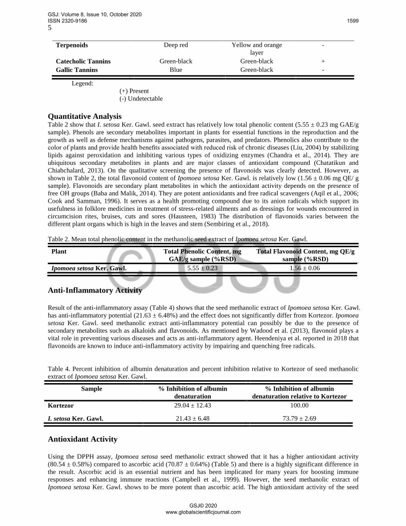
Legend: (+) Present (-) Undetectable Quantitative Analysis Table 2 show that I. setosa Ker. Gawl. seed extract has relatively low total phenolic content (5.55 ± 0.23 mg GAE/g sample). Phenols are secondary metabolites important in plants for essential functions in the reproduction and the growth as well as defense mechanisms against pathogens, parasites, and predators. Phenolics also contribute to the color of plants and provide health benefits associated with reduced risk of chronic diseases (Liu, 2004) by stabilizing lipids against peroxidation and inhibiting various types of oxidizing enzymes (Chandra et al., 2014). They are ubiquitous secondary metabolites in plants and are major classes of antioxidant compound (Chatatikun and Chiabchalard, 2013). On the qualitative screening the presence of flavonoids was clearly detected. However, as shown in Table 2, the total flavonoid content of Ipomoea setosa Ker. Gawl. is relatively low (1.56 ± 0.06 mg QE/ g sample). Flavonoids are secondary plant metabolites in which the antioxidant activity depends on the presence of free OH groups (Baba and Malik, 2014). They are potent antioxidants and free radical scavengers (Aqil et al., 2006; Cook and Samman, 1996). It serves as a health promoting compound due to its anion radicals which support its usefulness in folklore medicines in treatment of stress-related ailments and as dressings for wounds encountered in circumcision rites, bruises, cuts and sores (Hausteen, 1983) The distribution of flavonoids varies between the different plant organs which is high in the leaves and stem (Sembiring et al., 2018). Table 2. Mean total phenolic content in the methanolic seed extract of Ipomoea setosa Ker. Gawl.
Plant Total Phenolic Content, mg GAE/g sample (%RSD)
Total Flavonoid Content, mg QE/g sample (%RSD)
Ipomoea setosa Ker. Gawl. 5.55 ± 0.23 1.56 ± 0.06
Anti-Inflammatory Activity Result of the anti-inflammatory assay (Table 4) shows that the seed methanolic extract of Ipomoea setosa Ker. Gawl. has anti-inflammatory potential (21.63 ± 6.48%) and the effect does not significantly differ from Kortezor. Ipomoea setosa Ker. Gawl. seed methanolic extract anti-inflammatory potential can possibly be due to the presence of secondary metabolites such as alkaloids and flavonoids. As mentioned by Wadood et al. (2013), flavonoid plays a vital role in preventing various diseases and acts as anti-inflammatory agent. Heendeniya et al. reported in 2018 that flavonoids are known to induce anti-inflammatory activity by impairing and quenching free radicals. Table 4. Percent inhibition of albumin denaturation and percent inhibition relative to Kortezor of seed methanolic extract of Ipomoea setosa Ker. Gawl.
Sample % Inhibition of albumin denaturation
% Inhibition of albumin denaturation relative to Kortezor
Kortezor 29.04 ± 12.43 100.00
I. setosa Ker. Gawl. 21.43 ± 6.48 73.79 ± 2.69
Antioxidant Activity Using the DPPH assay, Ipomoea setosa seed methanolic extract showed that it has a higher antioxidant activity (80.54 ± 0.58%) compared to ascorbic acid (70.87 ± 0.64%) (Table 5) and there is a highly significant difference in the result. Ascorbic acid is an essential nutrient and has been implicated for many years for boosting immune responses and enhancing immune reactions (Campbell et al., 1999). However, the seed methanolic extract of Ipomoea setosa Ker. Gawl. shows to be more potent than ascorbic acid. The high antioxidant activity of the seed
GSJ: Volume 8, Issue 10, October 2020 ISSN 2320-9186 1599
GSJ© 2020 www.globalscientificjournal.com
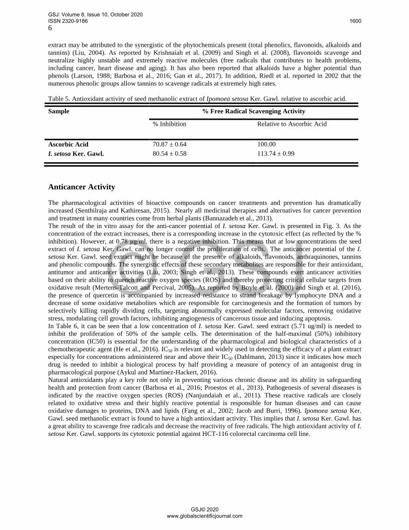
6 extract may be attributed to the synergistic of the phytochemicals present (total phenolics, flavonoids, alkaloids and tannins) (Liu, 2004). As reported by Krishnaiah et al. (2009) and Singh et al. (2008), flavonoids scavenge and neutralize highly unstable and extremely reactive molecules (free radicals that contributes to health problems, including cancer, heart disease and aging). It has also been reported that alkaloids have a higher potential than phenols (Larson, 1988; Barbosa et al., 2016; Gan et al., 2017). In addition, Riedl et al. reported in 2002 that the numerous phenolic groups allow tannins to scavenge radicals at extremely high rates. Table 5. Antioxidant activity of seed methanolic extract of Ipomoea setosa Ker. Gawl. relative to ascorbic acid.
Sample % Free Radical Scavenging Activity
% Inhibition Relative to Ascorbic Acid
Ascorbic Acid 70.87 ± 0.64 100.00 I. setosa Ker. Gawl. 80.54 ± 0.58 113.74 ± 0.99
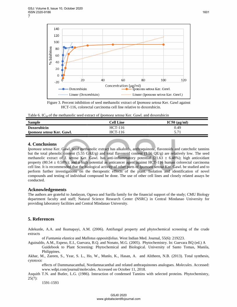
Anticancer Activity The pharmacological activities of bioactive compounds on cancer treatments and prevention has dramatically increased (Senthilraja and Kathiresan, 2015). Nearly all medicinal therapies and alternatives for cancer prevention and treatment in many countries come from herbal plants (Bannazadeh et al., 2013). The result of the in vitro assay for the anti-cancer potential of I. setosa Ker. Gawl. is presented in Fig. 3. As the concentration of the extract increases, there is a corresponding increase in the cytotoxic effect (as reflected by the % inhibition). However, at 0.78 μg/ml, there is a negative inhibition. This means that at low concentrations the seed extract of I. setosa Ker. Gawl. can no longer control the proliferation of cells. The anticancer potential of the I. setosa Ker. Gawl. seed extract might be because of the presence of alkaloids, flavonoids, anthraquinones, tannins and phenolic compounds. The synergistic effects of these secondary metabolites are responsible for their antioxidant, antitumor and anticancer activities (Liu, 2003; Singh et al., 2013). These compounds exert anticancer activities based on their ability to quench reactive oxygen species (ROS) and thereby protecting critical cellular targets from oxidative result (Mertens-Talcott and Percival, 2005). As reported by Boyle et al. (2000) and Singh et al. (2016), the presence of quercetin is accompanied by increased resistance to strand breakage by lymphocyte DNA and a decrease of some oxidative metabolites which are responsible for carcinogenesis and the formation of tumors by selectively killing rapidly dividing cells, targeting abnormally expressed molecular factors, removing oxidative stress, modulating cell growth factors, inhibiting angiogenesis of cancerous tissue and inducing apoptosis. In Table 6, it can be seen that a low concentration of I. setosa Ker. Gawl. seed extract (5.71 ug/ml) is needed to inhibit the proliferation of 50% of the sample cells. The determination of the half-maximal (50%) inhibitory concentration (IC50) is essential for the understanding of the pharmacological and biological characteristics of a chemotherapeutic agent (He et al., 2016). IC50 is relevant and widely used in detecting the efficacy of a plant extract especially for concentrations administered near and above their IC50 (Dahlmann, 2013) since it indicates how much drug is needed to inhibit a biological process by half providing a measure of potency of an antagonist drug in pharmacological purpose (Aykul and Martinez-Hackert, 2016). Natural antioxidants play a key role not only in preventing various chronic disease and its ability in safeguarding health and protection from cancer (Barbosa et al., 2016; Proestos et al., 2013). Pathogenesis of several diseases is indicated by the reactive oxygen species (ROS) (Nanjundaiah et al., 2011). These reactive radicals are closely related to oxidative stress and their highly reactive potential is responsible for human diseases and can cause oxidative damages to proteins, DNA and lipids (Fang et al., 2002; Jacob and Burri, 1996). Ipomoea setosa Ker. Gawl. seed methanolic extract is found to have a high antioxidant activity. This implies that I. setosa Ker. Gawl. has a great ability to scavenge free radicals and decrease the reactivity of free radicals. The high antioxidant activity of I. setosa Ker. Gawl. supports its cytotoxic potential against HCT-116 colorectal carcinoma cell line.
GSJ: Volume 8, Issue 10, October 2020 ISSN 2320-9186 1600
GSJ© 2020 www.globalscientificjournal.com
7
Figure 3. Percent inhibition of seed methanolic extract of Ipomoea setosa Ker. Gawl against HCT-116, colorectal carcinoma cell line relative to doxorubicin.
Table 6. IC50 of the methanolic seed extract of Ipomoea setosa Ker. Gawl. and doxorubicin
Sample Cell Line IC50 (μg/ml) Doxorubicin HCT-116 0.49 Ipomoea setosa Ker. Gawl. HCT-116 5.71
4. Conclusions Ipomoea setosa Ker. Gawl. seed methanolic extract has alkaloids, anthraquinone, flavonoids and catecholic tannins but the total phenolic content (5.55 GAE/g) and total flavonoid content (1.56 QE/g) are relatively low. The seed methanolic extract of I. setosa Ker. Gawl. has anti-inflammatory potential (21.63 ± 6.48%); high antioxidant property (80.54 ± 0.58%); and a high potential as anticancer agent against HCT-116 human colorectal carcinoma cell line. It is recommended that the biological activity of other parts of Ipomoea setosa Ker. Gawl. be studied and to perform further investigations on the therapeutic effects of the plant. Isolation and identification of novel compounds and testing of individual compound be done. The use of other cell lines and closely related assays be conducted. Acknowledgements The authors are grateful to Jandayan, Ogawa and Sarilla family for the financial support of the study; CMU Biology department faculty and staff; Natural Science Research Center (NSRC) in Central Mindanao University for providing laboratory facilities and Central Mindanao University. 5. References Adekunle, A.A. and Ikumapayi, A.M. (2006). Antifungal property and phytochemical screening of the crude extracts
of Funtumia elastica and Mallotus oppositifolius. West Indian Med. Journal, 55(6): 219223. Aguinaldo, A.M., Espeso, E.I., Guevara, B.Q. and Nonato, M.G. (2005). Phytochemistry. In: Guevara BQ (ed.) A
Guidebook to Plant Screening: Phytochemical and Biological. University of Santo Tomas, Manila, Philippines.
Akhar, M., Zareen, S., Year, S. L., Ho, W., Munlo, K., Hasan, A. and Alitheen, N.B. (2013). Total synthesis, cytotoxic
effects of Dammanacanthal, Nordamnacanthal and related anthraquinones analogues. Molecules. Accessed: www.wdpi.com/journal/molecules. Accessed on October 11, 2018.
Asquith T.N. and Butler, L.G. (1986). Interaction of condensed Tannins with selected proteins. Phytochemistry, 25(7):
1591-1593
GSJ: Volume 8, Issue 10, October 2020 ISSN 2320-9186 1601
GSJ© 2020 www.globalscientificjournal.com
8 Aqil, F., Ahmad, I. and Mehmood, Z. (2006). Antioxidant and free radical scavenging properties of twelve
traditionally used Indian medicinal plants. Turk. Journal Biol., 30:177-183. Ayiegoro, O.A. and Okoh, A.I. (2010). Preliminary phytochemical screening and in vitro antioxidant activities of
aqueous extract of Helichrysum longifol ium DC. BMC Compl. And Att . Med. , 10:21. Aykul, S. and Martinez-Hackert, E. (2016). Determination of half-maximal inhibitory concentration using biosensor-
based protein interaction analysis. Analytical Biochemistry, 97-103. http://dx.doi.org/10.1016/j.ab.2016.06.025.
Baba, S.A. and Malik, S.A. (2014). Determination of total phenolic and flavonoid content, antimicrobial and antioxidant activity of a root extract of Arisaema jacquemontii Blume. Journal of Taibah University for Science, 1-6. http://dx.doi.org/10.1016/j.jtusci.2014.11.001.
Bannazadeh, A. M., Rashtchizadeh, N., Nazemiyeh, H., Abdolalizadeh, J., Mohammadnejad, L. and Baradaran, B. (2013). Investigating apoptotic effects of methanolic extract of Dorema glabrum seed on WEHI-164 Cells. ISRN Pharmacology, 2013:949871.
Barbosa, G.B., Peteros, N.P. and Inutan, E.D. (2016). Antioxidant activities and phytochemical screening of Amomum
muricarpum, Hornstedtia conoidea and Etlingera philippinensis. Bulletin of Environment, Pharmacology and Life Sciences, 5(8); 2 2- 32.
Bhandary, S.K., Suchetha, K.N., Bhat, V.S., Sharmila, K.P. and Bekal, M.P. (2012). Preliminary phytochemical screening of various extracts of Punica granatum peel, whole fruit, and seeds. Nite University Journal of Health Science. 2(4): 34-38.
Boyle, S.P., Dobson, V.L., Duthie, S.J., Kyle, J.A.M. and Collins, A.R. (2000). Absorption and DNA protective effects of flavonoid glycosides from an onion meal. European Journal for Nutrition, 39: 213-223.
Bribi, N. (2018). Pharmacological activity of alkaliods:a review. Asian Journal of Botany, 1:1-6. doi:10.63019/ajbv1i2.467.
Campbell, J.D., Cole, M., Bunditrutavorn, B. and Vella, A.T. (1999). Ascorbic acid is a potent inhibitor of various f o r m s o f T c e l l a p o p t o s i s . C e l l u l a r I m m u n o l o g y . 1 9 4 ; 1 - 5 .
Chandra, S., Khan, S., Avula, B., Hemant, L., Yang, M.H., ElSohly, M.A. and Khan, I.A. (2014). Assessment of total
phenolic and flavonoid content, antioxidant properties, and yield of aeroponically and conventionally grown leafy vegetables and fruit crops: a comparative study. Hindawi Publishing Corporation Evidence-Based Complementary and Alternative Medicine, 1-9.
Chandrika, U.G, Jansz, E.R. and Wamasuriya, N.D. (2005). Analysis of Carotenoids in Ripe Jackfruit (Artocarpus heterophyllus) kernel and Study of their Bioconversion in Rats. Journal of the Science of Food and Agriculture, 85:186-190.
Chatatikun M., and Chiabchalard, A. (2013). Phytochemical screening and free radical scavenging activities of orange
baby carrot (Daucus carota Linn,) root crude extracts. Journal and Pharmaceutical Research, 5(4):97-102. Chung, K.T., Wong, T.Y., Wewi, C.T., Huang, Y.W. and Lin, Y. (1998). Tannins and human health: a review. Critical
Reviews in Food Science and Nutrition, 38(6): 421-464. Cook, N.C. and Samman, S. (1996). Flavonoids---chemistry, metabolism, cardioprotective effects, and dietary sources.
Journal of Nutritional Bioichemistry, 7:66-76. Cowan, M.M. (1999). Plant Products as Antimicrobial Agents. Clinical Microbiology Review, 12(4): 564-582. Dahlmann, H.A. (2013). Efficacy of cancer drugs: looking beyond the IC50. Chemical Research in Toxicology, 1776-
1777. dx.doi.org/10.1021/tx400426k. Desai, A.G., Qazi, G.N., Ganju, R.K., El-Tamer, M., Singh, J., Saxena, A.K., Bedi, Y.S., Taneja, S.C. and Bhat, H . K .
(2008). Medicinal plants and cancer chemoprevention. Curr Drug Metab. 9(7):581-591. Evans, W.C. (2002). Trease and Evans Pharmacognosy. 15th ed. Sauders Company Ltd, London. Fang, Y.Z., Yang, S. and Wu, G. (2002). Free radicals, antioxidants, and nutrition. Nutrition, 18: 872–879. Folorunso, A.E., Illoh, H.C. and Olorungbeja, J.A. (2013). Numerical taxonomy of some Ipomoea (Linn.) species in
South-West Nigeria. Life Journal of Science. 15(1):63-74. Gan, J., Feng, Y., He, Z., Li, X. and Zhang, H. (2017). Correlations betweenantioxidant activity and alkaloids and
phenols of Maca (Lepidium meyenii). Journal of Food Quality, 1-10. Greenwell, M. and Rahman, P.K.S.M., (2015). Medicinal plants: their use in anticancer treatment. National Center
for Biology Information, doi: 1 0. 1 3 0 4 0 / I J P S R. 0 9 7 5 - 8232 6 (1 0). 4 1 0 3 – 12
GSJ: Volume 8, Issue 10, October 2020 ISSN 2320-9186 1602
GSJ© 2020 www.globalscientificjournal.com
9 Hausteen, B. (1983). Flavonoids, a class of natural products of high pharmacological potency. Biochemistry
Pharmacology, 32:1141-1148. He, Y., Zhu, Q., Chen, M., Huang, Q., Wang, W., Li, Q., Huang, D. and Di W. (2016). The changing 50% inhibitory
concentration (IC50) of cisplatin: a pilot study on the artifacts of the MTT assay and the precise measurement of density-dependent chemoresistance in ovarian cancer. Oncotarget, 7(43): 70803-70821. d o i : 1 0 . 1 8 6 3 2 / o n c o t a r g e t. 1 2 2 2 3.
Heendeniya, S.N., Ratnasooriya, W.D., and Pathirana,R.N. (2018). In vitro investigation of anti-inflammatory activity
and evaluation of phytochemical profile of Syzygium caryophyllatum. Journal of Pharmacognosy and Phytochemistry, 7(1): 1759-1763.
I m a n i s L i f e S c i e n c e s . ( 2 0 1 8 ) . H C T - 1 1 6 ( c o l o r e c t a l c a r c i n o m a ) . R e t r i e v e d f r o m https://www.imanislife.com/collections/cell-lines/hct116-cells/. Accessed on December 2, 2018.
Iyenger , M.A. (1995). Study of Certain Drugs. 8 th ed. Manipal Power Press, Manipal India. Jacob, R.A. and Burri, B.J. (1996). Oxidative damage and defense. American Journal of Clinical Nutrition, 63, 985–
990. J o h a n s e n , D . A . ( 1 9 4 0 ) . P l a n t M i c r o t e c h n i q u e . M c g r a w H i l l B o o k C o . N e w Y o r k Jokhadze, M., Eristavi, L., Kutchukhidze, J., Chariot, A., Angenot, L., Tits, M., Jansen, O. and Frederich, M. (2007).
In Vitro Cytotoxicity of some Medicinal Plants from Georgian Amaryllidaceae. Phytotherapy Research, 21:622-624.
Krishnaiah, D., Devi, T., Bono, A. and Sarbatly, R. (2009). Studies on phytochemical constituents of six Malaysian medicinal plants. Journal of Medicinal Plants Research, 3(2): 67-72.
Larson, R.A. (1988). Review article number 30 the antioxidants of higher plants. Phytochemistry, 27(4): 969-998. Londhe D.K., Neel, R.S., and Bhuktar A.S. (2017). Ethno-medicinal uses of some species of genus Ipomoea L. from
Maharashtra state. International Journal of Applied Research, 3(10): 82-84. Liu, R.H. (2003). Health benefits of fruit and vegetables are from additive and synergistic combinations of
phytochemicals. American Journal for Clinical Nutrition, 78: 517S-520S. Liu, R.H. (2004). Potential synergy of phytochemicals in cancer prevention: mechanism of action. International
Research Conference on Food, Nutrition, and Cancer, 3479S-3485S. Meira, M., Pereira da Silvall, E., Davidll, J.M., and David, J.P. (2012). Review of the genus Ipomoea: traditional uses,
chemistry and biological activities. Revista Brasileira de Farmacognosia. Retrieved from http://dx.doi.org/10.1590/S0102-695X2012005000025.
Mertens-Talcott, S.U. and Percival, S.S. (2005). Ellagic acid and quercetin interact synergistically with resveratrol in the induction of apoptosis and cause transient cell cycle arrest in human leukemia cells. Cancer Letters, 218: 141-151.
Mir, M.A., Sawhney, S.S. and Jassal, M.M.S. (2013). Qualitative and quantitative analysis of phytochemicals of Tarashaxacum officinale. Wudpecker Journal of Pharmacy and Pharmacology, 2(1):1-5.
Mohammed, A., Liman, M.L., Atiku, M.K. (2013). Chemical composition of the methanolic leaf and stem bark extracts of Senna siamea Lam. Journal of Pharmacognosy and Phytotherapy, 5(5):98-100. D O I : 1 0 . 5 8 9 7 / J P P 1 2 . 0 4 3 . I S S N : 2 1 4 1 - 2 5 0 2
Mossman T. (1983) Rapid colorimetric assay for cellular growth and survival: application to proliferation and cytotoxicity assays. Journal of Immunological Methods, 65:55–63
Musfiq-Marliyah, M. and Ananthi, T. (2015). In vitro anti-inflammatory activity of seed extract of Zea mays (L.). Journal of Global Biosciences, 4(5): 2168-2173.
Nanjundaiah, S.N., Annaiah, H.N. and Dharmesh, S.M. (2011). Gastoprotective effect of ginger rhizome (Zingiber officinale) extract: role of gallic acid and cinnamic acid in H+, K+-ATPase/H.pylori inhibition and anti-oxidative mechanism. Hindawi Publishing Corporation Evidence-Based Complementary and Alternative Medicine, 1 - 1 3. d o i: 1 0. 1 0 9 3 / e c a m / n e p 0 6 0.
Okwu D.E. and Josiah C. (2006). Evaluation of the chemical composition of two Nigerian medicinal plants. Afri. J.Biotechnol. 5 (4):357-361.
Palhares, R.M., Drummond, M.G., Brassil, B.S.A.F., Cosenza, G.P, Brandao, M.G.L. and Oliveira, G. (2015). Medicinal Plants Recommended by the World Health Organization: DNA Barcode Identification Associated with Chemical Analyses Guarantees Their Quality. Plos:One. Retrieved from Plos:One:https://journals.plos.org /plosone/article?id=10.1371/journal.pone.0127866#abstract0
Peteros, N.P. and Uy, M. (2010). Antioxidant and cytotoxic activities and phytochemical screening of four Philippine
medicinal Plants. Journal of Medicinal Plants Research, 407-414. Philippine Cancer Society Forum: Controlling Colorectal Cancer. (2014). Philippine Cancer Society. Retrieved from
GSJ: Volume 8, Issue 10, October 2020 ISSN 2320-9186 1603
GSJ© 2020 www.globalscientificjournal.com
10
http://www.philcancer.org.ph/philippine-cancer-society-forum-controlling-colorectal-cancer/ Pieters, L. and Vlietinck, A.J. (2005). Bioguided isolation of pharmacologically active plant components, still a
valuable strategy for the finding of new lead compounds. Journal of Ethnopharmacology, 100: 57-60. Proestos, C., Lytoudi, K., Mauromelanidou, O.K., Panagiotis, Z. and Sinanoglou, V.J. (2013). Antioxidant capacity
of selected plant extracts and their essential oils. Antioxidants. (2):11-22. doi:10.3390/antiox2010011. Rane, V. A. and Patel, B.B. (2014). In-vitro cytotoxic activity of leaf extracts of Ipomoea Jacq. species against a 549
and HEP-G2 cell lines. International Journal of Pharmaceutical Sciences http://dx.doi.org/10.13040/IJPSR.0975-8232.6(1).294-99.
Riedl, K.M., Carando, S., Alessio, H.M., McCarthy, M. and Hagerman, A.E. (2002). Antioxidant activity of tannins and tannin-protein complexes: assessmemnt in vitro and in vivo. American Chemical Society, 188-200.
Salamah, N. and Ningsih, D.S. (2017). Total alkaloid content in various fractions of Tabernaemonata sphaercarpa Bl. (Jembirit) leaves. IOP Conference Series: Materials Science and Engineering, 259:1-6. d o i : 1 0 . 1 0 8 8 / 1 7 5 7 - 8 9 9 X / 2 5 9 / 1 / 0 1 2 0 1 7
Sembiring, E.N, Elya, B. and Sauriasari, R. (2018). Phytochemical screening, total flavonoid and total phenolic Content and Antioxidant Activity of Different Parts of Caesalpinia bonduc (L.) Roxb. Pharmacognosy Journal, 10(1);123-127.
Senthilraja, P. and Kathiresan, K. (2015). In vitro cytotoxicity MTT assay in Vero, HepG2 and MCF-7 cell lines study
of marine yeast. Journal of Applied Pharmaceutical Science, 5(3); 80-84. Singh, G., Kapoor, I.P.S., Singh, P., Heluani, C.S.D., Lampasona, M.P.D., Catalan, C.A.N. (2008). Chemistry,
antioxidant and antimicrobial investigations on essential oil and oleoresins of Zingiber officinale. Food and Chemical Toxicology, 46: 3295-3302.
Singh S., Jarial R. and Kanwar S.S. (2013). Therapeutic effect of herbal medicines on obesity: herbal pancreatic lipase
inhibitors. Wudpecker Journal of Medicinal Plants, 2:53–65. Singh, S., Sharma, B., Kanwar, S.S. and Kumar, A. (2016). Lead phytochemicals for anticancer drug development.
Frontiers in Plant Science, 7(167): 1-13 Srivastava, D. (2017). Medicinal plant of genus Ipomoea: present scenario, challenges and future prospective.
Research Journal of Recent Sciences, 6(10): 23-26 Wadood, A., Ghufran, M., Jamal, S.B., Naeem, M., Khan, A. Ghaffar, R. and Asnad. (2013). Phytochemical analysis
of medicinal plants occuring in local area of Mardan. Biochemistry and Analytical Biochemistry, 2:1-4. DOI: 10.4172/2161-1009.1000144
Wood, J.R.I., Carine, M.I., Harris, D., Wilkin, P., Williams, B. and Scotland, R.W. (2015). Ipomoea (Convolvulaceae)
in Bolivia. Kew Bulletin. 70:31 1 - 1 2 4. D O I 1 0. 1 0 0 7 / S 1 2 2 2 5 - 0 1 5 - 9592- 7. Wunderlin, R. P., B. F., Hansen, A. R. Franck, and F. B. Essig. (2018). Atlas of Florida Plants
(http://florida.plantatlas.usf.edu/). [S. M. Landry and K. N. Campbell (application development), USF Water Inst i tute. Inst itute for Systematic Botany, University of South Florida, Tampa.
GSJ: Volume 8, Issue 10, October 2020 ISSN 2320-9186 1604
GSJ© 2020 www.globalscientificjournal.com