Embed Size (px)
Citation preview
April 30, 2019
Archives • 2019 • vol.1 • 136-153
http://pharmacologyonline.silae.it
ISSN: 1827-8620
FURTHER PHYTOCHEMICAL SCREENING; NON-CLINICAL EVALUATION OF TOXIC AND ANTI-INFLAMMATORY EFFECTS OF CRUDE AQUEOUS EXTRACT OF GYNURA NEPALENSIS
Anzuman Aktar1 ; S M Hafiz Hassan1 ; Tania Parvin1; Mim Binti Akhlas1; Fatema Khatun1; Muhammad Torequl Islam2,3*; Razina Rouf1
1Department of Pharmacy, Bangabandhu Sheikh MujiburRahman Science and Technology University, Gopalgang-8100, Bangladesh
2Department for Management of Science and Technology Development, Ton DucThang University, Ho Chi Minh City-700000, Vietnam
3Faculty of Pharmacy, Ton DucThang University, Ho Chi Minh City-700000, Vietnam
E-mail address: [email protected]*
Abstract
Gynura nepalensis DC, belonging to the family Compositae, is widely distributed in China. Traditionally, this plant is used in the treatment of various ailments, including infections, hyperglycemia and hypertension. This study aimed to perform a preliminary phytochemical analysis of the crude aqueous leaf extract of G. nepalensis along with its toxic and protective effects by using a number of in vitro and ex vivo test models. Toxicity was tested in Allium cepa, while the protective effects such as anti-inflammatory in egg albumin and membrane stabilizing and clotlysis capacity in human erythrocytes. The results suggest that the plant possesses alkaloids, tannins, flavonoids, steroids, saponins, glycosides and reducing sugar. G. nepalensis exerted time and concentration-dependently toxicity in A. cepa. However, at 72 h the plant extract showed an adaptation capability at low concentration (2.5 – 15% v/v), possibly by reducing DNA damage index in the eukaryotic test system, which was further confirmed in the anti-inflammatory, thrombolytic and membrane stabilizing test, where the extract within the range of 0.12 – 0.36% v/v was found to inhibit protein denaturation and hemolysis, with an enhanced clotlysis capacity in the test systems. A concentration-dependent activity of the extract was seen in the latter cases. In conclusion, G. nepalensis possesses many important phytoconstituents and exerted toxic effects in A. cepa at high concentration, while protective effects other test systems at low concentrations. Further studies are highly appreciable to isolate the responsible phytochemicals for the observed biological effects.
Keywords: Gynura nepalensis; Allium cepa; egg albumin; human erythrocytes; toxicity; protective capacity
PhOL Aktar, et al. 137 (pag 136-153)
http://pharmacologyonline.silae.it
ISSN: 1827-8620
Introduction
Traditional medicinal plants or natural products have been widely used as major sources of drugs in the pharmaceutical industry. A natural product is a chemical compound or substance produced by a living organism that is, found in nature. In the broadest sense, natural products include any substance produced by life (Link: file:///C:/Users/INTER_WAVE/). “All these herbs can work like true medications in our bodies. We’ve given you true scientific reasons why they work, which means they really do work.”– Dr. Mehmet Oz, The Dr. Oz Show (with Chris Kilham as Guest Star).
Approximately 449 plants have been used in Bangladesh (Ghani, 1998). A recent study suggests that 122 compounds isolated from only 94 plants are using drugs globally. It also demonstrates that 80% of these plants have ethnomedical use and identical or related to the current use of plant-derived active ingredients (Fabricant and Farnsworth, 2001). As the people are becoming aware of the potency and the side effect of synthetic drugs, there is an interest in the natural product remedies with a basic approach towards the nature (Verma and Sing, 2012). In developing countries like India, Bangladesh, farmers are practicing cultivation of medicinal (herbal) plants to get additional income. In the 18th century, knowledge about medicinal plant resulted from expansion of drug use, but attempts to identify the active ingredients from plants were unsuccessful. In the early 19th century, the term 'pharmacognosy' was used by Johann Adam Schmid (1759-1809), but the main shift came when it became clear that the pharmaceutical properties of plant molecules can be isolated and characterized. Since 20th century the integration of ethnobotanical, pharmacological and phytochemical studies had been evident (Clardy and Walsh, 2004). Ultimately, herbal remedies and natural products became transformed into chemically defined drugs (Clardy and Walsh, 2004) and modern techniques for separation, structure elucidation, screening and combinatorial synthesis hassled to the revitalization of plant products as a source of new drugs (Ganesan, 2002; Steinbeck, 2004). This has opened up a new opportunity and avenues for drug development.
As our lifestyle is now getting techno-savvy, we are moving away from nature. But we cannot
escape from nature being part of nature. Plants have been used for medicinal purposes from a long time before prehistoric period. Since most medicinal plants occur naturally in a large number of countries a plant with potential importance in one country may have been studied by scientist. Considerable time and effort will be saved if their findings will be made available to all interested people (Farnsworth et al., 1985). Evidence exists that Unani Hakims, Indian Vaids and European and Mediterranean cultures were using herbs for over 4000 years as a medicine. Recently, WHO (World Health Organization) estimated that 80 % of people worldwide rely on herbal medicines for some aspect of their primary health care needs. According to WHO, around 21,000 plant species have the potential for being used as medicinal plants. It has been estimated, that in developed countries such as United States, plant drugs constitute as much as 25% of the total drugs, while in fast developing countries such as India and China, the contribution is as much as 80%. Treatment with medicinal plants is considered very safe as there are no or minimal side effects. Medicinal plants are considered as rich resources of ingredients which can be used in drug development pharmacopoeial, non-pharmacopoeial or synthetic drugs (WHO, 2002).
Increasing the price of synthetic medicines continuously, people are trying to seek or resort to medicinal plants as cheaper, available and less toxic alternatives to get cured of their illness. Such decision of using medicinal plant is based on local wisdom and folk of knowledge (Pena et al., 2018). Gynura nepalensis DC belonging to the family Compositae is a medicinal plant with wide curative applications based on ethnomedical knowledge (Yu et al., 2016). G. nepalensi utilized as an ethno-medicine to cure a wide range of human sickness such as diabetes, cuts or wounds, indigestion, cough, asthma, kidney stones, hepatitis, gall bladder stones, urinary tract bleeding, hemorrhoids, constipation, diarrhea, vomiting, blood poisoning, fertility problems, septicemia, skin allergy, rheumatism, high cholesterol levels and low blood pressure only its hypolipidaemic, hepatoprotective, antimutagenic, antioxidant and radioprotective potentials using either ethanolic or methanolic extracts have been directly investigated based on reported studies (Pena et al., 2018).
PhOL Aktar, et al. 138 (pag 136-153)
http://pharmacologyonline.silae.it
ISSN: 1827-8620
About 40/44 species of Gynura are distributed in Africa, South and East Asia and Australasia (Vanijajiva, 2009; Vanijajiva and Kadereit, 2011; Afroz et al., 2014). It is also known as Philippine Ashitaba or Nepal Gynura (Retano et al., 2018). This plant is also known as G. nudibasis, G. foetens, and Cacalia nepalensis (Link: file:///C:/Users/INTER_WAVE). The phytochemicals, also referred to as secondary metabolites, are responsible for diverse pharmacological activities, many of which have been found useful by humans in the alleviation of the numerous ailments that afflict them (Rahmatullah et al., 2010) Basic and clinical investigations indicate that chlorogenic acid may alleviate type 2 diabetes, obesity and cardiovascular disease as well as other diseases. In vitro studies have also demonstrated a protective effect of chlorogenic acid on cardiomyocytes against damage induced by oxidative stress (Yu et al.,2016). Flower extract of G. nepalensis is used for treatment of the hyperlipidemia along with its hepatoprotective effect (Nigam et al., 2012). The juice of the plant is applied to heal cuts and wounds (Manandhar, 2002). In Bangladesh, the leaves of the plant are used in diabetes. various studies show that antidiabetic drugs and antioxidants delayed or prevented the onset of type 2 diabetes mellitus in patients with impaired glucose tolerance (Montonen et al., 2004; Kawamori, 2009; Phung et al., 2011). More than 400 plants, such as Momordica charantia and Gynura spp. have been shown to have antidiabetic activities in vitro and in vivo (Li et al., 2009; Liu et al., 2010; Deng et al., 2011; Singh et al., 2011; Algariri et al., 2013; Tan et al., 2013) . Antioxidants, such as vitamin E and flavonoids, have also proven beneficial to diabetes as they decrease the risk of complications (Bonnefont and Rousselot, 2004).
This study aims at screening of phytochemical and toxicity analysis along with the investigation of anti-inflammatory and membrane stabilizing, and clotlysis activities of the aqueous crude extract of G. nepalensis.
Plant morphology
Gynura nepalensis DC, belonging to the family Compositae, is widely distributed in China, and its leaves have been used as a folk medicine for the treatment of hyperglycemia and hypertension. Phytochemical studies have revealed the presence
of chlorogenic acid and its derivatives as major components (Yu BW et al., 2016). The leaves are also used to treat diabetes that has been reported in Bangladesh (Afroz et al., 2014) and in the Philippines (Ursulom and Rialubin, 2013) and G. nepalensis’s juice is used to heal cuts and wounds in Nepal (Manandhar, 2002; Afroz et al., 2014) and in external wounds in the Philippines (Ursulom and Rialubin, 2013).
G. nepalensis is a perennial herb with stems erect or ascending, robust, 30-45 cm tall, woody at base, about 1 cm in diameter, corymbosely branched in upper part, densely reddish-yellow woolly. In terms of phenology, “the plant is flowering and fruiting all year round” according to Vanijajiva (2009) but Afroz et al. (2014) reported its phenology from March to August. The leaf extract of G. nepalensis is one of the 40 ethnomedicinal plants used to treat indigestion by the members of the Apatani tribe, in the Ziro Valley of Arunachal Pradesh, one of the 28 states in Northeast India and also considered the 12th mega bio-diverse regions of the world (Kala, 2005). G. nepalensis is utilized as an ethno medicine to cure a wide or vast range of human sickness such as diabetes, cuts or wounds, indigestion, cough, asthma, kidney stones, hepatitis, gallbladder stones, urinary tract bledding, hemorrhoids, constipation, diarrhea, vomiting, blood poisoning, fertility problems, septicemia, skin allergy, rheumatism, high cholesterol levels and low blood pressure only its hypolipidaemic, hepatoprotective,antimutagenic, antioxidant and radioprotective potentials using either ethanolic or methanolic extracts have been directly investigated based on reported studies (Renato et al., 2018). The plant contains catechic tannins, saponins (Gracilla and Bagunu, 2011; San Andres, 2014; Salas, 2016), alkaloids, tannins, flavonoids (Gracilla and Bagunu, 2011; Sales, 2014; San Andres, 2014; Salas, 2016), and steroids (with 2-deoxysugars) (SanAndres, 2014).
Methods
Ethical statement
This study was approved by the Ethical Committee under the Department of Pharmacy, Bangabandhu Sheikh MujiburRahman Science and Technology University (BSMRSTU) (Approval No. 20140109034).
PhOL Aktar, et al. 139 (pag 136-153)
http://pharmacologyonline.silae.it
ISSN: 1827-8620
Collection and identification of plant materials
Leaves and stems of the plant were collected from Gopalganj District, Bangladesh in August 2018 and were identified by, Forest Research Institute, Bangladesh (FRIH, BD). A voucher specimen was deposited there with an accession number BFRIH-5113.
Preparation of plant extract
Freshly collected plant materials were washed with running tap water and crushed with the aid of a mortar and pestle to get 100% crude extract, which was then diluted with a little amount of distilled water.
Sources of reagents and chimicals
Copper sulphate (CuSO4.5H2O) was analytical grade and purchased commercially from Merck, India. Acetyl salicylic acid was kindly provided by the Zenith Pharmaceuticals Ltd., Bangladesh. Streptokinase (Durakinase Powder for Injection, 1.5 million units) was purchased from Dong KookPharm. Ltd., Korea. Other reagents and chemicals were analytical grade and were purchased commercially from Merck, India.
Phytochemical screening
A preliminary phytochemical investigation was done to find out the presence of alkaloids, steroids, triterpenoids, saponins, tannins, glycosides, reducing sugars, and flavonoids (Trease, 1992).
Evaluation of toxic effects in A. cepa and determination of half-minimal inhibitory concentration (IC50)
The outer layers and budding parenchyma of the central crown of onions were carefully removed by making a small circular incision to facilitate root growth. The bulbs were then rinsed with tap water for 20 min and the root portion was soaked in distilled water in previously washed and clean glass containers (capacity: 15-20 mL) for the first 24 h at 25 ± 1 oC in the dark. Only, the Allium with satisfactory root growth were transferred to the controls or sample containers (five for each concentration) for 24, 48 and 72 h of exposure. After the exposure period, the roots were counted and measured in mm. To determine the toxicity of the crude extract root growth inhibition was
calculated (Konuk et al., 2007). IC50 was also determined for the test sample. Distilled water and CuSO4.5H2O were used as negative and positive controls, respectively. CuSO4.5H2O was dissolved in distilled water to attain 6 µg/mL.
Evaluation of anti-inflammatory activity (egg albumin test)
From the Allium cepa test, IC50 was found at 24 h 0.44%. In this study, 1/3th of the IC50 was considered as highest test concentration. Then five successive dilutions of this concentration, such as 0.36, 0.30, 0.24, 0.18 and 0.12%were used in this assay. This study was carried out according to the method described by Konuk et al. (2007). Briefly, the extract was reconstituted with distilled water. Distilled water and acetyl salicylic acid (ASA) were used as negative and positive controls, respectively. ASA was dissolved in distilled water to attain the concentration 100 µg/mL.This test was carried out with a slight modification of Ullah et al. (2014). Briefly, the reaction mixture (5 mL) consisted of 0.2 mL of egg albumin (from hen’s egg), 2.8 mL of phosphate buffered saline (PBS, pH 6.4) and 2 mL of varying concentrations of extract. A similar volume of distilled water served as negative control. Then the mixtures were incubated at (37 °C ± 2) in a BOD incubator (Lab line Technologies) for 15 min and then heated at 70 °C for 5 min. After cooling, their absorbance was measured at 660 nm (LABOCON MODEL: LUVS-201, monochromatic beam) by using the vehicle as blank. The percentage inhibition of protein denaturation was calculated by using the following formula:
Evaluation of membrane stabilization test
This study was carried out according to the method described by Ullah et al. (2014) with a slight modification. Briefly, fresh whole blood (3 mL) collected from the healthy volunteers into heparinized tubes was centrifuged at 2000 rpm for 2 min. A volume of normal saline equivalent to that of the supernatant was used to dissolve the red blood pellets (RBCs). The volume of the dissolved RBCs
PhOL Aktar, et al. 140 (pag 136-153)
http://pharmacologyonline.silae.it
ISSN: 1827-8620
obtained was measured and reconstituted as a 10% (v/v) suspension with an isotonic buffer solution (10 mM sodium phosphate buffer, pH 6.4). The buffer solution contained 3.1202 g of NaH2PO4, 1.7799 g of Na2HPO4 and 4.383 g of NaCl in 500 mL of distilled water. The reconstituted RBCs (resuspended supernatant) was used as such.
Evaluation of anti-atherombosis activity
The aqueous crude extract at 0.36, 0.30, 0.24, 0.18 and 0.12% was used in this study. Experiments for clot lysis were carried as reported earlier Prasad et al. (2006). In brief, 4 mL venous blood drawn from healthy volunteers (human who are not currently using oral contraceptive or anticoagulant therapy) and was distributed in Gr. I: negative control (distilled water); Gr. II: standard (streptokinase) and Gr.III-VII: for 5 different concentrations of the aqueous crude extract pre-weighed sterile microcentrifuge (alpin/ephendorf’s) tubes (0.5 mL/tube) and incubated at 37 °C for 45 min. After clot formation, serum was completely removed without disturbing the clot and each tube having clot was again weighed to determine the clot weight (clot weight = weight of clot containing tube – weight of the tube alone). To each micro-centrifuge tube containing pre-weighed clot, 100 μL of crude extract of the plant were added separately. As a positive control, 100 μL of streptokinase and as a negative control, 100 μL of distilled water were added separately. All the tubes were then incubated at 37 °C for 90 min and observed for clot lysis. After incubation, fluid released was removed and tubes were again weighed to observe the difference in weight after clot disruption. Difference obtained in weight taken before and after clot lysis was expressed as percentage of clot lysis. The experiment was carried out (duplicated) at different days with the blood samples of same healthy volunteers.
Statistical analysis
Values are mean ± standard error mean (SEM) and percentages. The data were analyzed by means of analysis of variance (ANOVA) followed by Tukey tests by using GraphPad Prism (version 6.0), considering p<0.05 at 95% confidence level.
Results
The aqueous crude extract of herb revealed the presence of alkaloids, tannins, saponins, steroids, glycoside, flavonoids, and reducing sugar.
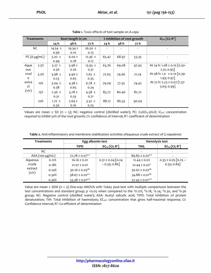
Table 1 suggests a concentration and time-dependent toxic effect in A. cepa at 24 h. The highest root growth (RG) inhibition was observed at exposure time 72 h in 20% concentration, while lowest at 72 h in 2.5% crude extract of the plant. A reduction in RG was seen in 10 – 20% extract at 48 h, while in 2.5 – 15% at 72 h in comparison to the exposure time 24 h. Moreover, a reduction in RG in comparison to the exposure time 48 h at 72 h was also seen in 2.5 – 10% crude extract. The half minimal inhibitory concentrations calculated for 24, 48 and 72 h are 1.08 ± 0.10, 1.01 ± 0.10 and 2.23 ± 0.07, respectively.
In egg albumin (in vitro) test, the crude aqueous extract of the plant showed an anti-inflammatory effect in a concentration-dependent manner. The extract exhibited highest inhibition of albumin denaturation at 0.36% (v/v). The standard ASA at 100 µg/mL exhibited better anti-inflammatory effect than the test sample. The EC50 calculated for the extract was 0.31 ± 0.04% (v/v). Multiple comparison study between the concentrations and standard for each test suggests that the extract at 0.24 to 0.36% produced significant effects in comparison to the 0.12 and 0.18% groups. ASA exerted a significant anti-inflammatory effect when compared to all the test concentrations (Table 2).
The membrane stabilization assay performed in HRBC (ex vivo) method suggests that the extract concentration-dependently inhibited heat-induced hemolysis, where the highest inhibition capacity was seen in 0.36 % (v/v) extract. The EC50 calculated for the extract was 0.33 ± 0.05% (v/v). Multiple comparison study between the concentrations and standard for each test suggests that the extract at 0.18 to 0.36% (v/v) produced significant effects in comparison to the 0.12% group. ASA exerted a significant anti-inflammatory effect when compared to all the test concentrations (Table 2).
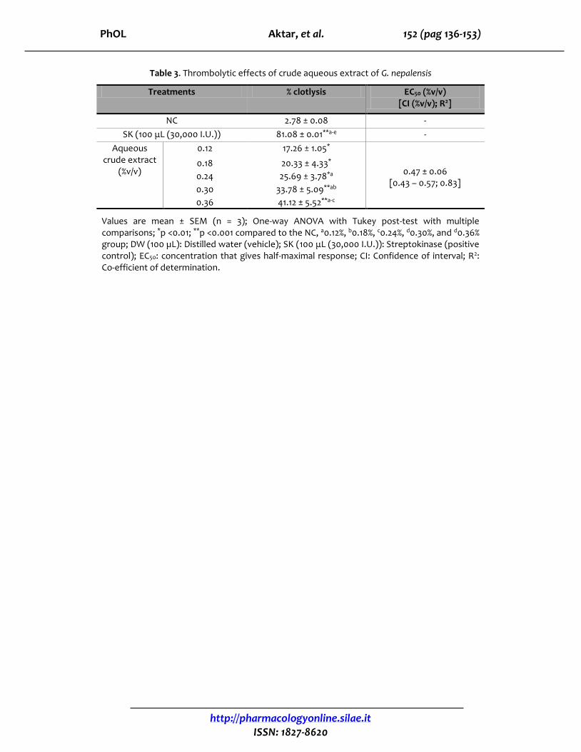
The crude aq. extract of the plant showed clotlysis capacity in a concentration-dependent manner. Highest clotlysis (41.12 ± 5.52) was observed in 0.36% extract. The standard, SK produced clotlysis 81.08 ± 0.01%, while NC group exhibited a negligible
PhOL Aktar, et al. 141 (pag 136-153)
http://pharmacologyonline.silae.it
ISSN: 1827-8620
clotlysis capacity (2.78 ± 0.08). The clotlysis capacity, when compared between the test groups and SK group, suggests that both the extract and SK produced significant (p <0.01, p <0.001) clotlysis effects in comparisons to the NC group. The extract at 0.36% showed significant clotlysis capacity as compared to the 0.12 - 0.24% extract groups, while at 0.30% in comparison to the 0.12 and 0.18% groups, and the 0.24% group only in comparison to the 0.12% group. The SK group exhibited significant (p <0.001) clotlysis capacity when compared with the NC and all the concentrations treated with the plant extract (Table 3).
Pearson's correlation
Pearson’s correlation study suggests that the herb’s aqueous extract mediated anti-inflammatory, anti-atherothrombosis and membrane stabilizing activities are correlated. According to Figure 1A, an increase in membrane stabilizing capacity has been linked to the increase in anti-inflammatory effect gradually till 0.09% concentration. However, a rapid change of both activities was seen at 0.12 and 0.15% concentrations. However, in the case of anti-atherothrombosisvs anti-inflammatory effect (Figure 1B) and membrane stabilizing vs anti-atherothrombosis effect (Figure 1C) sharp changes were observed from 0.12 to 0.15%.
Discussion
The root length, in Allium test is an important parameter, reflecting the toxicity of the additive; elongation of A. cepa and can serve as a sensitive external signal of continuous internal cellular events (Adeyemo and Farinmade, 2013). According to Qin et al. (2015) toxic substance can accumulate in roots of A. cepa and inhibit root growth, results in chromosomal aberrations (e.g., C-mitosis, chromosomal bridges, chromosomal tack and micronuclei). It may be due to the accumulation of toxic agents at the meristematic cell site of the root tip and substantially impaired the microtubule arrangements in this eukaryotic test system. Therefore, the toxicity and cytotoxic effects that cause an inhibition in root growth in A. cepa are generally related to the cell cycle elongation during differentiation (Fusconi et al., 2006), apical meristematic activity (Webster and Macleod, 1996),
and inhibition of protein synthesis (Seth et al., 2007).
Secondary metabolites, especially alkaloids, glycosides and the polyphenols possess antioxidant capacity (Kumar Singh and Patra, 2018). Strong antioxidants at high concentration may act as pro-oxidants and can induce cytotoxic effects in eukaryotic test systems (Melo et al., 2016). Both oxidative (Zhou et al., 2016) and anti-oxidative stress (Zhou et al., 2018) is capable to cause damage to the cell macromolecules, such as protein, carbohydrates, lipids and genetic materials (e.g., DNA and RNA). In this study, G. nepalensis extract was found to exert cytotoxic effects within 2.5 to 20% (v/v), which is equivalent to 25-200 mL/kg for experimental animals. The extract exerted more cytotoxic effect of high concentration.
Each biological system has its own repair systems that can act against oxidative stress and cytogenetic alterations caused by toxicants coming from various origins (Kim et al., 2017). Cytotoxicity can be attained by several ways, including ROS-induced oxidative stress and mitochondrial dysfunctions, DNA damage, apoptosis, necrosis, cell cycle arrest, chromosomal aberrations, and so on (Sharma et al., 2015). Certain protective agents, especially the antioxidants can act against these types of detrimental effects (Sevcovicova et al., 2014). There are reports that plant-based antioxidants (e.g., alkaloids, glycosides, flavonoids) can act against DNA damage (Pistollato et al., 2015). Therefore, the extract-induced cytotoxic effects and the system-mediated adoption at 48 and 72 h may link to the plant containing such types of antioxidant groups. These results are an agreement with the previous reports, demonstration of low-concentration mediated antioxidant (Quiming et al., 2016) and protective (Yu et al., 2016; Renato et al., 2018) effects of the plant.
Inflammation is induced by cyclooxygenase enzymes, which is responsible for the conversion of arachidonic acid to prostaglandin and this enzyme is inhibited by anti-inflammatory drug. But excessive or long-term usage of those drugs tends to result to serious events like gastric or peptic ulcer (Wallace, 2001; Shih and Chang, 2007), cardiovascular sickness, and renal failure (Huerta et al., 2002). Research on anti-inflammatory drug based on many herbal plants has great therapeutic activity and
PhOL Aktar, et al. 142 (pag 136-153)
http://pharmacologyonline.silae.it
ISSN: 1827-8620
fewer side effects compared to synthetic drugs. The experiment on an aqueous crude extract of many medicinal plants for anti-inflammatory activity has done by HRBC membrane stabilization method and egg albumin (in vitro) test. Erythrocyte membrane is analogous to lysosomal membrane and stabilization of lysosomal membrane is urgent in reducing the inflammatory responses by preventing the release of lysosomal chemicals such as bacterial enzymes, proteases histamine, prostaglandin, leukotriene and damage extra cellular release. The acute or chronic inflammation is occurring with the release of extracellular activities of these lysosomal enzymes (Iwueke et al., 2006).
Protein denaturation means protein loss their tertiary and secondary structure of external stress or some substances such as a strong acid or base, inorganic salt, or organic solvent or heat. The most important cause of inflammation is denaturation of protein. To inhibit protein denaturation, indicate the anti-inflammatory effect of a drug (Patel and Desai, 2016). In this study, G.nepalensis extract showed anti-inflammatory and membrane stabilizing capacity by inhibiting the heat-induced lysis of erythrocyte membrane.
Risk assessment for any substance prior to install in any biological system is an important issue (Árvay et al., 2018). In essence, checking of the inflammatory response to a substance and the effects of it on cell membrane stability is two major consequences (Bag et al., 2013). Plants containing secondary metabolites, especially the polyphenols and flavonoids (Karaoğlan et al., 2018; Sdayria et al., 2018), alkaloids (Majinda, 2018) having antioxidant capacity may impart anti-inflammatory effects in biological systems.
Generally, inflammation is a type of protective response, but at severity level, it may cause serious problems in our body, including cardiovascular (Golia et al., 2014) and neurological diseases and disorders (Khansari and Sperlagh, 2012). Previous scientific reports also suggest that the plant has antioxidant, anti-inflammatory and membrane stabilizing (Sen et al., 2013) capability. Examples of plant extract containing antioxidant as well as anti-inflammatory and membrane stabilizing capacity are numerous (Arawwawala et al., 2010; Sreejith et al., 2010; Sen et al., 2013). This study is an agreement
with the previous study (Quiming et al., 2016; Sultana and Bachar, 2018).
Erythrocyte membrane stabilization of lysosomal has great contribution to decrease the inflammatory responses by preventing the release of lysosomal chemicals such as bactericidal enzyme proteases, histamine and prostaglandin (Iwueke et al., 2006). This study indicates a concentration-graded anti-inflammatory and membrane stabilizing capacity of G. nepalensis.The results of the present work suggest that the anti-inflammatory activities of these extracts could be explained, at least in part, by their antioxidant properties (Schinella et al., 2001).
Clot lysis is a process by which coagulated blood or clot is broke with a reflection of the plasmin content of the blood.The temperature is required for clot lysis is at 98.6 °F (37 °C). Anti-thrombotic or thrombolytic drugs can block the pathway of thrombus formation. Atherosclerosis, or arteriosclerotic vascular disease (ASVD) in which the arteries are narrowed due to accumulation of fatty substances called plaque. To build up an atheromatous plaque, it is passed through a complex series of cellular events within the arterial wall (Schwartz et al., 1992). A fibrinolytic agent is plasmin, that lyses the blood clot by breaking down the fibrinogen and fibrin (Banerjee et al., 2004). Nowadays, many drugs are used as a thrombolytic agent to treat the patients with thrombosis (Khan et al., 2011; Baruah et al., 2006), such as alteplase, anistreplase, streptokinase, urokinase, and tissue plasminogen (TPA) etc. among those thrombolytic agents streptokinase, urokinase are widely used because of their low cost as compared to other thrombolytic drugs (Mucklow, 1995; Collen, 1990). But the major side effects of these drugs are systemic fibrinolysis, anaphylactic reaction, and bleeding complication (hemorrhage) (Rouf et al., 1996). Multiple treatments with streptokinase are a band within some patient, because of immunogenicity (Jennings, 1996). Many researchers have discovered several thrombolytic drugs from various sources (Prasad et al., 2007) and few drugs have been modified by the recombinant technology tried to make them more site specific and more effective (Liu et al., 2000). The side effects of some drugs lead to further complications.
PhOL Aktar, et al. 143 (pag 136-153)
http://pharmacologyonline.silae.it
ISSN: 1827-8620
Pharmacological investigation of the plant-based new drug candidates are always interesting and challenging. So, researchers are finding out new sources of herbs and natural foods and many research works have been done on medicinal plants and natural foods having anti-thrombotic (antiplatelet, anticoagulant) effect which leads to the prevention of coronary events and stroke (Joshipura et al., 1999; Liu et al., 2000; Bazzano et al., 2002; Ratnasooriya et al., 2008; Li et al., 2011). Generally, these kinds of medications are cheap and readily available; possess less side effects than the synthetic drugs (Khan et al., 201). Now-a-days, edible plants and those are traditionally used by the various ethnic groups are in the spotlight for plant-based drug discovery and development (Balunas and Kinghorn, 2005; Jaradat et al., 2017). Plant-based polyphenols (Ogston et al., 1985; Booyse et al., 2007), alkaloids (Olatunji et al., 2017), glycosides (Nan et al., 2013; Zhu and Fang, 2014) are already known to possess clotlysis capacity. Moreover, substances acting against oxidative stress (Hugenholtz et al., 2016; Sambola et al., 2017) and inflammation (Bester et al., 2018) are capable to resist formation of clot inside the blood vessels. Therefore, anti-inflammatory capacity of the herb may connect to its anti-atherothrombosis effect on human erythrocytes.
From the beginning of civilization, human normally depend on plants for the treatment of many diseases. Nowadays phyto-pharmacological investigation has created a new field of discovery plant derivative drugs, which are effective in remedial of certain diseases. It is estimated that about 30% of the pharmaceuticals are prepared from plant derivatives (Gillman et al., 1995; Leta et al., 2002). Blood clot a coagulum in the bloodstream formed by an aggregation of blood factors, primarily platelets, and fibrin with entrapment of cellular elements. The process is irreversible, but the clot may be dissolved naturally.
Herbal drugs are a good choice for cardiovascular diseases, especially for the management of atherothrombosis (Li et al., 2011). Generally, these kinds of medications are cheap and readily available, possesses less side effects than the synthetic drugs (Mannan et al., 2011; Bhowmick et al., 2014). Now-a-days, edible plants and those are traditionally used by the various ethnic groups are in the spotlight for
plant-based drug discovery and development (Balunas and Kinghorn, 2005; Jaradat et al., 2017). Plant-based flavonoids (Ogston et al., 1985; Booyse et al., 2007), alkaloids (Olatunji et al., 2017), glycosides (Nan et al., 2013; Zhu and Fang, 2014) are already known to possess clotlysis capacity.
Moreover, substances acting against oxidative stress (Hugenholtz et al., 2016; Sambola et al., 2017) and inflammation (Bester et al., 2018) are capable to resist formation of clot inside the blood vessels. The crude aq. extract of the plant showed a concentration-dependent clotlysis capacity in human erythrocytes. Therefore, the G. nepalensis mediated antioxidant (Quiming et al., 2016) and protective (Yu et al., 2016; Renato et al., 2018) effects may link with its anti-atherothrombosis effect. Moreover, this study is an agreement with the previous study conducted by Sultana and Bachar (2018), where the ethanolic leaf extract was found to show hemolytic, thrombolytic and membrane stabilizing capacity in human erythrocytes.
In summary, G. nepalensis revealed the presence of alkaloids, tannins, saponins, steroids, glycosides, flavonoids, and reducing sugar. The aqueous crude extract of G. nepalensis exerted significant toxic effects in A. cepa. At low concentration, the crude extract exerted anti-inflammatory, membrane stabilizing and anithrombolytic activity. The plant containing important secondary metabolites may be responsible for the observed biological effects. Further investigations are required to find the active components of the herb and to confirm the possible mechanism of action for each biological action.
Acknowledgments
We are owed to the Department of Pharmacy, Life Science Faculty, Bangabandhu Sheikh MujiburRahman Science and Technology University (BSMRSTU), Gopalganj-8100, Bangladesh for providing the laboratory facilities to conduct this study.
References
1. Adams DS, Griffin LA, Nachajko WR, Reddy VB, Wei CM (1991) A synthetic DNA encoding a modified human urokinase resistant to inhibition by serum plasminogen activator inhibitor. J Biol Chem 266(13):8476-8482
PhOL Aktar, et al. 144 (pag 136-153)
http://pharmacologyonline.silae.it
ISSN: 1827-8620
2. Adeyemo OA, Farinmade AE (2013) Genotoxic and cytotoxic effects of food flavor enhancer, monosodium glutamate (MSG) using Allium cepa assay. Afric J Biotechnol 12:1459-1466
3. Afroz S, Uddin MZ, Hassan A (2014) Gynura
nepalensis DC. (Asteraceae) -A New Angiosperm Record for Bangladesh. Bangladesh J Plant Taxon 21(1):101-104
4. Aitadafoun M, Mounieri C, Heyman SF,
Binistic C, Bon C and Godhold J (1996) 4-Alkoxybenzamides as newpotent phosholipase A2 inhibitors. Biochem Pharmacol 51:737-742
5. Algariri K, Meng KY, Atangwho IJ, Asmawi
MZ, Sadikun A, Murugaiyah V, Ismail N (2013) Hypoglycemic and anti-hyperglycemic study of Gynura procumbens leaf extracts. Asian Pac J Trop Biomed 3(5):358-366
6. Arawwawala M, Thabrew I, Arambewela L,
Handunnetti S (2010) Anti-inflammatory activity of Trichosanthes cucumerina Linn. in rats. J Ethnopharmacol 131(3):538-543
7. Árvay J, Šnirc M, Hauptvogl M, Bilčíková J,
Bobková A, Demková L, Hudáček M, Hrstková M, Lošák T, Král M, Kováčik A, Štefániková J (2018) Concentration of Micro- and Macro-Elements in Green and Roasted Coffee: Influence of Roasting Degree and Risk Assessment for the Consumers. Biol Trace Elem Res. doi: 10.1007/s12011-018-1519-3.
8. Bag A, Bhattacharyya SK, Kumar PN,
Chattopadhyay R (2013) Anti-inflammatory, anti-lipid peroxidative, antioxidant and membrane stabilizing activities of hydroalcoholic extract of Terminalia chebula fruits. Pharm Biol 51(12):1515-1520
9. Balunas MJ, Kinghorn AD (2005) Drug
discovery from medicinal plants. Life Sci 78(5):431-441
10. Yu BW, Li JL, Guo B, Fan H, Zhao W , Wang H .2016. Chlorogenic acid analogues from Gynura nepalensis protect H9c2 cardiomyoblasts against H2O2-induced apoptosis. Acta Pharmacol Sinica 37:1413-1422
11. Bester J, Matshailwe C, Pretorius E (2018)
Simultaneous presence of hypercoagulation and increased clotlysis time due to IL-1β, IL-6 and IL-8. Cytokine 110:237-242
12. Bhowmick R, Sarwar MH, Dewan SMR, Das A, Das B, Uddin MMN, Islam MS (2014) In-vivo analgesic, antipyretic and anti-inflammatory potential in Swiss albino mice and in vitro thrombolytic activity of hydroalcoholic extract from Litsea glutinosa leaves. Biol Res pp:34.
13. Bonnefont-Rousselot D (2004) The role of
antioxidant micronutrients in the prevention of diabetic complications. Treat Endocrinol 3(1):41-52
14. Booyse FM, Pan W, Grenett HE, Parks DA,
Darley-Usmar VM, Bradley KM, Tabengwa EM (2007) Mechanism by which alcohol and wine polyphenols affect coronary heart disease risk. Ann Epidemiol 17:S24-S31
15. Breslin A (2017) The Chemical Composition
of Green Plants. Sciencing, Leaf Group Ltd. Link: https://sciencing.com/chemical-composition-green-plants-8336363.html
16. Chatterjee S, Das SN (1996) Anti-arthritic and
Anti-inflammatory effect of a poly-herbal drug its mechanism of action. Indian J Pharmacol 28:116-119
17. Clardy J, Walsh C (2004) Lessons from
natural molecules. Nature 432:829-837.
18. Fabricant DS, Fransworth NR (2001) The value of Plants used in traditional medicine for drug discovery. Environ Health Perspect 109:69-75

PhOL Aktar, et al. 145 (pag 136-153)
http://pharmacologyonline.silae.it
ISSN: 1827-8620
19. Deng XY, Chen YS, Zhang WR, Chen B, Qiu XM, He LK, Mu LL, Yang CH, Chen R (2011) Polysaccharide from Gynura divaricate modulates the activities of intestinal disaccharidases in streptozotocin-induced diabetic rats. Br J Nutr 106:1323-1329
20. Farnsworth NR, Akerele O, Bingel AS,
Soejarto DD, Guo Z (1985) Medicinal plants in therapy. Bull World Health Organ 63:965-981
21. Ferrero-Miliani L, Nielsen OH, Anderson PS,
Girardin SE (2007) Chronic inflammation: importance of NOD2 and NALP3 in interleukin- 1 beta generation. Clin Exp Immunol 147:227-235
22. Fiskesjo G (1985) The Allium test as a
standard in environmental monitoring. Hereditas 102:99-112
23. Fusconi A, Repetto O, Bona E, Massa N,
Gallo C, Dumas-Gaudot E, Berta G (2006) Effect of cadmium on meristem activity and nucleus ploidy in roots of Pisum sativum L. cv, Frisson seedlings. Envirob Exp Bot 58:253-260
24. Ghani A (1998) Medicinal plants of
Bangladesh: chemical constituents and uses. CAB Direct, 20016784329. Link: https://www.cabdirect.org/cabdirect/abstract/20016784329
25. Golia E, Limongelli G, Natale F, Fimiani F,
Maddaloni V, Pariggiano I, Bianchi R, Crisci M, D'Acierno L, Giordano R, Di Palma G, Conte M, Golino P, Russo MG, Calabrò R, Calabrò P (2014) Inflammation and cardiovascular disease: from pathogenesis to therapeutic target. Curr Atheroscler Rep 16:435.
26. Gracilla D, Bagunu J (2014) Radiation
Cytogenetics Studies on Philippine Ashitaba (Gynura nepalensis DC). Int J Sci: Basic Appl Res 17:1-10
27. Harborne JB (1973) London: Chapman and Hall. Phytochem Meth. Link:
28. https://pdfs.semanticscholar.org/8cf0/3b5ce325afe5f071f42aa9f502332e2c376b.pdf
29. Huerta C, Castellsague J, Varas-Lorenzo C, Garcia Rodriguez LA (2002) Nonsteroidal anti-inflammatory drugs and risk of ARF in the general population. Am J Kidney Dis. 45:531-539
30. Hugenholtz GC, Macrae F, Adelmeijer J,
Dulfer S, Porte RJ, Lisman T, Ariëns RA (2016) Procoagulant changes in fibrin clot structure in patients with cirrhosis are associated with oxidative modifications of fibrinogen. J Thromb Haemost 14:1054-1066
31. Ibadan SA (1993) Nigeria: Spectrum Books
Ltd. 1993. Medicinal Plants and Traditional Medicine in Africa. p. 289. Link: www.sciepub.com/reference/41679
32. Ilavarassan R, Mallika M, Venkataraman S
(2005) Anti-inflammatory and antioxidant activities of Cassia fistula Linn bark extracts. Afr J Trad CAM 2:70-85
33. Islam MT, Streck L, de Alencar MVOB, Silva
SWC, Machado KC, Machado KC, Júnior ALG, Paz MFCJ, da Mata AMOF, e Sousa JMC, Junior JSC, Rolim HML, Silva-Junior AA, Melo-Cavalcante AAC (2017) Evaluation of toxic, cytotoxic and genotoxic effects of phytol and its nanoemulsion. Chemosphere 177:93-101
34. Iwueke AV, Nwodo OF, Okoli CO (2006)
Evaluation of the anti-inflammatory and analgesic activities of Vitexdoinana leaves. Afric J Biotechnol 5:1929-1935
35. Jaradat N, Zaid AN, Hussein F, Zaqzouq M,
Aljammal H, Ayesh O (2017) Anti-Lipase Potential of the Organic and Aqueous Extracts of Ten Traditional Edible and Medicinal Plants in Palestine; a Comparison Study with Orlistat. Medicines (Basel). doi: 10.3390/medicines4040089.
PhOL Aktar, et al. 146 (pag 136-153)
http://pharmacologyonline.silae.it
ISSN: 1827-8620
36. Jones WP, Chin YW, Kinghorn AD (2006) The Role Of Pharmacognosy in Modern Medicine and Pharmacy. Curr Drug Targets 7:247-264
37. Kala C (2005) Ethnomedicinal botany of the
Apatani in the Eastern Himalayan region of India. J Ethnobiol Ethnomed1(11). Link: https://ethnobiomed.biomedcentral.com/articles/10.1186/1746-4269-1-11
38. Karaoğlan ES, Albayrak A, Kutlu Z, Bayır Y
(2018) Gastroprotective and antioxidant effects of Eremurus spectabilis Bieb. methanol extract and its isolated component isoorientin on indomethacin induced gastric ulcers in rats1. Acta Cir Bras 33:609-618.
39. Kawamori R, Tajima N, Iwamoto Y,
Kashiwagi A, Shimamoto K, Kaku K (2009) Voglibose for prevention of type 2 diabetes mellitus: a randomized, double-blind trial in Japanese individuals with impaired glucose tolerance. Lancet 373:1607-1614
40. Khansari PS, Sperlagh B (2012) Inflammation
in neurological and psychiatric diseases. Inflammopharmacol 20:103-107
41. Kim DC, Kang M, Biswas A, Yang CR, Wang X,
Gao JX (2017) Effects of low dose ionizing radiation on DNA damage-caused pathways by reverse-phase protein array and Bayesian networks. J Bioinform Comput Biol 15:1750006
42. Konuk M, Liman R, Ciğerci İH (2007)
Determination of genotoxic effect of boron on Allium cepa root meristematic cells. Pak J Bot 39:73-79
43. Kumar Singh S, Patra A (2018) Evaluation of
phenolic composition, antioxidant, anti-inflammatory and anticancer activities of Polygonatum verticillatum (L.). J Integr Med 16:273-282
44. Laurence DR, Bennett PN (1992) Clinical Pharmacology.seventh edition. New York: Churchill Livingstone. p. 483.
45. Li P, Yang Y, Liu M (2011) Xuezhikang, extract
of red yeast rice, inhibited tissue factor and hypercoagulable state through suppressing nicotinamide adenine dinucleotide phosphate oxidase and extracellular signal-regulated kinase activation. J Cardiovasc Pharmacol 58:307-318
46. Li WL, Ren BR, Min-Zhuo, Hu Y, Lu CG, Wu
JL, Chen J, Sun S (2009) The anti-hyperglycemic effect of plants in genus Gynura Cass. Am J Chin Med 37:961-966
47. Lijnen HR, Van Hoef B, De Cock F, Okada K,
Ueshima S, Matsuo O, Collen D (1991) On the mechanism of fibrin-specific plasminogen activation by staphylokinase. J Biol Chem 266:11826-11832
48. Liu W, Yu Y, Yang R, Wan C, Xu B, Cao S
(2010) Optimization of total flavonoid compound extraction from Gynura medica leaf using response surface methodology and chemical composition analysis. Int J Mol Sci 11:4759-4763
49. Lu Y , Li ZH , Ma L , Deng AJ , Wu F , Zhang
ZH , Qin HL (2014) Study on chemical constituents from cultivated Gynura nepalensis .China J Chinese Materia Med 39:3777-3781
50. Luciano S, Giovanni V, Maria LC, Eleonora M,
Laura B, Gabriela M, et al (1999) Inhibition of protein denaturation by fatty acids, bile salts and other natural substances: a new hypothesis for the mechanism of action of fish oil in rheumatic diseases. Jpn J Pharmacol 79:89-99
51. Majinda RRT (2018) An Update of Erythrinan
Alkaloids and Their Pharmacological Activities. Prog Chem Org Nat Prod 107:95-159
PhOL Aktar, et al. 147 (pag 136-153)
http://pharmacologyonline.silae.it
ISSN: 1827-8620
52. Manandhar NP (2002) Plants and People of Nepal. Timber Press, Oregon, USA, 599 pp.
53. Mannan A, Kawser MJ, Ahmed AMA, Islam
NN, Alam SMA, Emon MAEK, Guota SD (2011) Assesment of anti-bacterial ,thrombolytic and cytotoxic potential of Cassia alata seed oil. JAppl Pharm Sci 1:56-59
54. Marder VJ (1993) Recombinant
streptokinase: opportunity for an improved agent. Blood Coagul Fibrinolysis 4:1039-1040
55. McGeer PL, McGeer EG (2002) Local neuro
inflammation and the progression of Alzheimer’s disease. J Neurovirol 8:529-538
56. Mclaughlin JL, Rogers LL, Anderson JE
(1998) The use of biological assays to evaluate botanicals. Drug Inf J 32:513-524
57. Melo PS, Arrivetti LOR, Alencar SM, Skibsted
LH (2016) Antioxidative and prooxidative effects in food lipids and synergism with α-tocopherol of açaí seed extracts and Grape rachis extracts. Food Chem 213:440-449
58. Montonen J, Knekt P, Järvinen R,
Reunanen A (2004) Dietary antioxidant intake and risk of type 2 diabetes. Diabetes Care 27:362-366
59. Mueller MM (2006) Inflammation in
epithelial skin tumors: old stories and new ideas. Eur J Cancer 42:735-744
60. Murugesan D, Deviponnuswamy R (2014)
Potential anti-inflammatory medicinal plants – a review. Int J Pharm Pharm Sci 6:43-49
61. Nan ZD, Zeng KW, Shi SP, Zhao MB, Jiang Y,
Tu PF (2013) Phenylethanoid glycosides with anti-inflammatory activities from the stems of Cistanche deserticola cultured in Tarim desert. Fitoterapia 89:167-174
62. Nicolini FA, Nichols WW, Mehta JL, Saldeen
TG, Schofield R, Ross M, Player DW, Pohl GB, Mattsson C (1992) Sustained reflow in dogs
with coronary thrombosis with K2P, a novel mutant of tissue-plasminogen activator. J Am Coll Cardiol 20:228-235
63. Nigam V, Paarekh PM, Singh S, Goyanar G,
Upmanyu N, Banweer J (2012) Hypolipidaemic and hepatoprotective activity of Gynura nepalensis DC. flower extract in streptozotocin induced diabetic mice. World J Pharm Res 2:112-131
64. Nunes BS, Carvalho FD, Guilhermino LM, Van
Stappen G (2006) Use of the genus Artemia in ecotoxicity testing. Environ Pollut 144:453-462
65. Ogston D, Lea AG, Langhorne P, Wilson SB
(1985) The influence of the polyphenols of cider on plasmin and plasminogen activators. Br J Haematol 60:705-713
66. Olatunji LA, Michael OS, Adeyanju OA,
Areola ED, Soladoye AO (2017) Anti-inflammatory and antithrombotic effects of nicotine exposure in oral contraceptive-induced insulin resistance are glucocorticoid-independent. Pharmacol Rep. 69:512-519
67. Olufunmilayo O, Adeyemi SO, Okpo OO
(2004) The analgesic effect of the methanolic extract of Acanthus montanus. J Ethnopharmacol 90:45-48
68. Organization, World Health Organization
(2002) WHO Traditional Medicine Strategy: 2002-2005 (document WHO/EDM/TRM/2002.1). Geneva. Link: apps.who.int/medicinedocs/en/d/Js2297e/
69. Palladino MA, Bahjat FR, Theodorakis EA,
Moldawer LL (2003) Anti- TNF-α therapies: the next generation. Nat Rev Drug Discov 2:736-746
70. Phung OJ, Sood NA, Sill BE, Coleman CI
(2011) Oral anti-diabetic drugs for the prevention of type 2 diabetes. Diabetic Med 28:948-996
PhOL Aktar, et al. 148 (pag 136-153)
http://pharmacologyonline.silae.it
ISSN: 1827-8620
71. Pistollato F, Sumalla Cano S, Elio I, MasiasVergara M, Giampieri F, Battino M (2015) Plant-Based and Plant-Rich Diet Patterns during Gestation: Beneficial Effects and Possible Shortcomings. Adv Nutr 6:581-591
72. Prasad S, Kashyap RS, Deopujari JY, Purohit
HJ, Taori GM, Daginawala HF (2006) Development of an in vitro model to study clot lysis activity of thrombolytic drugs. Thromb 4:14
73. Qin R, Wang C, Chen D, Bj€orn L.O, Li S
(2015) Copper-induced root growth inhibition of Allium cepa var. agrogarum L. involves disturbances in cell division and DNA damage. Environ Toxicol Chem 34:1045-1055
74. Quiming N, Asis JL, Nicolas M, Versoza D,
Alvarez MR (2016) In vitro α-glucosidase inhibition and antioxidant activities of partially purified Antidesma bunius fruit and Gynura nepalensis leaf extracts. J Appl Pharm Sci 6:097-101
75. Rahmatullah M, Mollik MAH, Islam MK, Islam
MR, Jahan FI, Khatun Z, Seraj S, Chowdhury MH, Islam F, Miajee ZUME, Jahan R (2010) A Survey of Medicinal and Functional Food Plants Used by the Folk Medicinal Practitioners of Three Villages in SreepurUpazilla, Magura District, Bangladesh. American-Eurasian J Sustain Agric 4:363-373
76. Rajendran V, Lakshmi KS (2008) In vitro and
in vivo anti-inflammatory activity of barks of Symplocoscochinchnesis (lour) Moore ssplaurina. J Pharmacol 3:121-124
77. Rajurkar R, Jain R, Matake N, Aswar P,
Khadbadi SS (2009) Antiinflammatory action of Abutilon indicum (L) sweet barks by HRBC membrane stabilization. Res J Pharm Tech 2:415-416
78. Pena RAD (2018) Gynura nepalensis DC: A Potential Wonder Medicinal Plant. ResearchGate 316490815.
79. Salas G (2014) Selected herbal plant extracts
as angiogenesis inhibitors using chick chorioallontoic membrane (CAM) assay. Pampanga Agricultural College. Magalang, Pampanga. Link: https://docplayer.net/53684209-Selected-philippine-herbal-plant-extracts-as-angiogenesis-inhibitors-using-chick-chorioallantoic-membrane-cam-assay.html
80. Sambola A, Ruiz-Meana M, Barba I, Del
Blanco BG, Barrabés JA, Lip GY, Vilardosa Ú, Sansaloni S, Rello P, García-Dorado D (2017) Glycative and oxidative stress are associated with altered thrombus composition in diabetic patients with ST-elevation myocardial infarction. Int J Cardiol 243:9-14
81. Sangian H, Faramarzi H, Yazdinezhad A,
Mousavi SJ, Zamani Z, Noubarani M, Ramazani A (2013) Antiplasmodial activity of ethanolic extracts of some selected medicinal plants from the northwest of Iran. Parasitol Res 112:3697-3701
82. Schinella GR, Tournier HA, Prieto JM,
Mordujovich de Buschiazz P, Ríos JL (2001) Antioxidant activity of anti-inflammatory plant extracts. Life Sci 70:1023-1033
83. Sdayria J, Rjeibi I, Feriani A, Ncib S,
Bouguerra W, Hfaiedh N, Elfeki A, Allagui MS (2018) Chemical Composition and Antioxidant, Analgesic, and Anti-InflammatoryEffects of Methanolic Extract of Euphorbia retusa in Mice. Pain Res Manag 2018:4838413. doi: 10.1155/2018/4838413.
84. Sen S, Chakraborty R, Rekha B, Revathi D,
Ayyanna SC, Hemalatha G, Kumar Reddy GA, Hyndavi S, IkhyathaBabu PJ, Prakash PR, Sridhar C (2013) Anti-inflammatory, analgesic, and antioxidant activities of Pisonia aculeata: folk medicinal use to scientific approach. Pharm Biol 51:426-432
PhOL Aktar, et al. 149 (pag 136-153)
http://pharmacologyonline.silae.it
ISSN: 1827-8620
85. Seth CS, Chaturvedi PK, Misra V (2007) Toxic effect of arsenate and cadmium alone and in combination on Giant Duckweed (Spirodela polyrrhiza L.) in response to its accumulation. Environ Toxicol 22:539-549
86. Sevcovicova A, Bodnarova K, Loderer D,
Imreova P, Galova E, Miadokova E (2014) Dual activities of emodin--DNA protectivityvs mutagenicity. Neuro Endocrinol Lett 35:149-154
87. Sharma R, Kumar R, Kodwani R, Kapoor S,
Khare A, Bansal R, Khurana S, Singh S, Thomas J, Roy B, Phartyal R, Saluja S, Kumar S (2015) A Review on Mechanisms of Anti Tumor Activity of Chalcones. Anticancer Agents Med Chem 16:200-211
88. Shih SC, Chang CW (2007) Nonsteroidal anti-
inflammatory drug related gastrointestinal bleeding in the elderly. Int J Gerontol 1:40-45
89. Shinde UA, Phadke AS, Nair AM,
Mungantiwar AA, Dikshit VJ, Sarsf MN (1989) Membrane stabilization activity- a possible mechanism of action for the anti-inflammatory activity of Cedrusdeodara wood oil. Fitoterapia 70:251-257
90. Sreejith G, Latha PG, Shine VJ, Anuja GI, Suja
SR, Sini S, Shyama S, Pradeep S, Shikha P, Rajasekharan S (2012) Anti-allergic, anti-inflammatory and anti-lipidperoxidant effects of Cassia occidentalis Linn. Indian J Exp Biol 48:494-498
91. Sultana R, Bachar SC (2018) Preliminary
phytochemical screening, in vitro thrombolytic and membrane stabilizing activities of Gynura nepalensis leaves of Bangladeshi origin. Eur J Pharm Med Res 5:46-49
92. Tan C, Wang Q, Luo C, Chen S, Li Q, Li P
(2013) Yeast α-glucosidase inhibitory phenolic compounds isolated from Gynura medica leaf. Int J Mol Sci 14:2551-2558
93. Tapsell LC, Hamphill I, Cobiac L (2006) Health benefits of herbs and spices: The past, the present, the future. Med JAugt 185:14-24
94. Trease GE (1992) A text book of
Pharmacognosy, 13th ed. London: Baillier, Tindal and Caussel.
95. Ullah HMA, Zaman S, Juhara F, Akter L,
Tareq SM, EH, Bhattacharjee R (2014) Evaluation of antinociceptive, in-vivo&in-vitro anti-inflammatory activity of ethanolic extract of Curcuma zedoaria rhizome. BMC Complement Alternat Med 14:346
96. Ursulum F, Rialubin A (2013) Beneficial
Effects of Ashitaba (Gynura nepalensis): Users’ Testimonies. IAMURE Multidisciplin Res 6:88-92
97. Vanijajiva O (2009) The genus Gynura
(Asteraceae: Senecioneae) in Thailand. Thai J Bot 1:25-36
98. Vanijajiva O, Kadereit J (2011)A revision of
Gynura (Asteraceae: Senecioneae). J Systemat Evolut 49:285-314
99. Wallace JL (2011) Pathogenesis of NSAID
induced gastroduodenal mucosal injury. Best Practice Res Clin Gastroenterol 15:691-703
100. Webster PL, Macleod RD (1996) The
root apical meristem and its margin. In: Waishel, Y., Eshel, A., Kafkafi, U. (Eds.), Plant Roots. The Hidden Half, second ed. Marcel Dekker, New York, pp. 51-76
101. Woodward M, Lowe GD, Campbell DJ,
Colman S, Rumley A, Chalmers J, et al. (2005) Associations of inflammatory and hemostatic variables with the risk of recurrent stroke. Stroke 36:2143-2147
102. Wu DH, Shi GY, Chuang WJ, Hsu JM, Young
KC, Chang CW, Wu HL (2001) Coiled coil region of streptokinase gamma-domain is
PhOL Aktar, et al. 150 (pag 136-153)
http://pharmacologyonline.silae.it
ISSN: 1827-8620
essential for plasminogen activation. J Biol Chem 276:15025-15033
103. Zhou J, Chao G, Li Y, Wu M, Zhong S,
Feng Z (2016) Activation of NRF2/ARE by isosilybin alleviates Aβ25-35-induced oxidativestress injury in HT-22 cells. Neurosci Lett 632:92-97
104. Zhou Y, Sun Y, Li P, Qin G, Cheng Q,
Liu Y, Chen Y, Wang G (2018) Monoside antagonizes triptolide-induced hepatocyte apoptosis via the anti-oxidativestress pathway. Nan Fang Yi Ke Da XueXueBao 38:949-955
105. Zhu X, Fang ZH (2014) New
monoterpene glycosides from the root cortex of Paeoniasuffruticosa and their potential anti-inflammatory activity. Nat Prod Res 28:301-305
PhOL Aktar, et al. 151 (pag 136-153)
http://pharmacologyonline.silae.it
ISSN: 1827-8620
Table 1. Toxic effects of test sample on A.cepa
Treatments Root length in cm % inhibition of root growth IC50 [CI; R2]
24 h 48 h 72 h 24 h 48 h 72 h
NC 14.54 ± 0.96
19.34 ± 0.10
26.50 ± 0.13
- - - -
PC (6 µg/mL) 5.02 ± 0.99
6.00 ± 0.18
12.36 ± 0.17
65.47 68.97 53.35 -
Aqueous crud
e extra
ct (v/v)
2.5% 5.27 ± 0.36
5.98 ± 0.26
13.93 ± 0.31
63.76 69.08 47.43 At 24 h: 1.08 ± 0.10 [0.50-2.31; 0.95]
At 48 h: 1.0 ± 0.10 [0.39-1.93; 0.97]
At 72 h: 2.23 ± 0.07 [1.37-3.65; 0.94]
5.0% 3.98 ± 0.23
4.90 ± 0.65
7.62 ± 0.35
72.63 74.66 71.24
10% 3.04 ± 0.38
4.38 ± 0.65
6.78 ± 0.24
79.09 77.35 74.42
15% 2.36 ± 0.31
3.78 ± 0.33
4.58 ± 0.21
83.77 80.46 82.72
20% 1.72 ± 0.30
2.64 ± 0.16
3.32 ± 0.25
88.17 86.35 90.04
Values are mean ± SD (n = 5); NC: negative control (distilled water); PC: CuSO4.5H2O; IC50: concentration required to inhibit 50% of the root growth; CI: confidence of interval; R2: coefficient of determination
Table 2. Anti-inflammatory and membrane stabilization activities ofaqueous crude extract of G.nepalensis
Treatments Egg albumin test Hemolysis test
TIPD EC50 [CI; R2] TIHL EC50 [CI; R2]
NC - - - -
ASA (100 µg/mL) 72.78 ± 0.07a-e - 89.83 ± 0.02a-e -
Aqueous crude
extract (v/v)
0.12% 16.16 ± 0.01 0.31 ± 0.04 [0.24 – 0.33; 0.86]
12.44 ± 0.02 0.33 ± 0.05 [0.25 – 0.35; 0.84] 0.18% 21.07 ± 0.01 22.44 ± 0.02a
0.24% 30.16 ± 0.03ab 30.62 ± 0.02ab
0.30% 38.67 ± 0.02a-c 34.88 ± 0.01ab
0.36% 53.38 ± 0.01a-d 57.43 ± 0.01a-d
Value are mean ± SEM (n = 3) One-way ANOVA with Tukey post-test with multiple comparisons between the test concentrations and standard group; p <0.05 when compared to the a0.12%, b0.18, c0.24, d0.30, and e0.36 group; NC: Negative control (distilled water); ASA: Acetyl salicylic acid; TIPD: Total inhibition of protein denaturation; TIH: Total inhibition of haemolysis; EC50: concentration that gives half-maximal response; CI: Confidence interval; R2: Co-efficient of determination
PhOL Aktar, et al. 152 (pag 136-153)
http://pharmacologyonline.silae.it
ISSN: 1827-8620
Table 3. Thrombolytic effects of crude aqueous extract of G. nepalensis
Treatments % clotlysis EC50 (%v/v) [CI (%v/v); R2]
NC 2.78 ± 0.08 -
SK (100 μL (30,000 I.U.)) 81.08 ± 0.01**a-e -
Aqueous crude extract
(%v/v)
0.12 17.26 ± 1.05*
0.47 ± 0.06 [0.43 – 0.57; 0.83]
0.18 20.33 ± 4.33*
0.24 25.69 ± 3.78*a
0.30 33.78 ± 5.09**ab
0.36 41.12 ± 5.52**a-c
Values are mean ± SEM (n = 3); One-way ANOVA with Tukey post-test with multiple comparisons; *p <0.01; **p <0.001 compared to the NC, a0.12%, b0.18%, c0.24%, d0.30%, and d0.36% group; DW (100 µL): Distilled water (vehicle); SK (100 μL (30,000 I.U.)): Streptokinase (positive control); EC50: concentration that gives half-maximal response; CI: Confidence of interval; R2: Co-efficient of determination.

PhOL Aktar, et al. 153 (pag 136-153)
http://pharmacologyonline.silae.it
ISSN: 1827-8620
Figure 1. Pearson’s correlation [(A) Membrane stabilizing capacity vs Anti-inflammatory effect; (B) Anti-atherothrombosis effect vs Anti-inflammatory effect; and (C) Anti-atherothrombosis effect vs Membrane stabilizing capacity]
16 .16 21 .07 30 .16 38 .67 53 .38
0
2 0
4 0
6 0
8 0
1 0 0
A n ti-in fla m m a to r y e ffe c t
Me
mb
ra
ne
sta
bil
izin
g c
ap
ac
ity (A )
16 .16 21 .07 30 .16 38 .67 53 .38
0
2 0
4 0
6 0
8 0
1 0 0
A n ti-in fla m m a to r y e ffe c t
An
ti-
ath
ero
th
ro
mb
os
is e
ffe
ct (B )
17 .26 20 .33 25 .69 33 .78 41 .12
0
2 0
4 0
6 0
8 0
1 0 0
A n ti-a th e r o th r o m b o s is e ffe c t
Me
mb
ra
ne
sta
bil
izin
g c
ap
ac
ity (C )