Embed Size (px)
Citation preview
Contents lists available at ScienceDirect
Carbohydrate Polymers
journal homepage: www.elsevier.com/locate/carbpol
Preparation of 2,6-diurea-chitosan oligosaccharide derivatives for efficientantifungal and antioxidant activities
Jingjing Zhanga,b,c, Xueqi Suna,b,c, Yuan Chena,b,c, Yingqi Mia,b,c, Wenqiang Tana,b, Qin Miaoa,b,Qing Lia,b, Fang Donga,b, Zhanyong Guoa,b,c,*a Key Laboratory of Coastal Biology and Bioresource Utilization, Yantai Institute of Coastal Zone Research, Chinese Academy of Sciences, Yantai 264003, Chinab Center for Ocean Mega-Science, Chinese Academy of Sciences, 7 Nanhai Road, Qingdao, 266071, ChinacUniversity of Chinese Academy of Sciences, Beijing 100049, China
A R T I C L E I N F O
Keywords:Antifungal activityAntioxidant activityChitosan oligosaccharide derivativesCytotoxicityUrea groups
A B S T R A C T
In this study, 2-urea-chitosan oligosaccharide derivatives (2-urea-COS derivatives) and 2,6-diurea-chitosanoligosaccharide derivatives (2,6-diurea-COS derivatives) were successfully designed and synthesized via inter-mediate 2-methoxyformylated chitosan oligosaccharide. All samples were characterized and compared based onFT-IR, 1H NMR spectroscopy, and elemental analysis. The antifungal effects of COS derivatives were testedagainst Fusarium oxysporum f. sp. niveum, Phomopsis asparagus, and Botrytis cinereal. Their antioxidant properties,including superoxide radicals’ scavenging activity, hydroxyl radicals’ scavenging activity, and DPPH radicals’scavenging activity were also explored within different concentrations. COS derivatives bearing urea groupsshowed improved bioactivity compared with pristine COS and 2,6-diurea-COS derivatives had a higher biolo-gical activity than 2-urea-COS derivatives in tested concentrations. Additionally, L929 cells were used to carryout cytotoxicity test of COS and COS derivatives by CCK-8 assay. The results indicated that some of samplesshowed low cytotoxicity. These findings offered a suggestion that COS derivatives bearing urea groups arepromising biological materials.
1. Introduction
Chitosan, derived from chitin polysaccharide which exists widely incrustacean shells, is a biocompatible, biodegradable, and nontoxicpolymer (Bonilla et al., 2019; Li, Wu, Shi, Li, & Chen, 2019;Sakwanichol, Sungthongjeen, & Puttipipatkhachorn, 2019). Due to thepoor solubility and narrow antimicrobial spectrum, the application ofchitosan is greatly restricted (Govindaraj, Abathodharanan,Ravishankar, & Raghavachari, 2019; Miao, Li, Zheng, & Zhang, 2019).It can be easily functionalized into different derivatives to overcomethese limitations through chemical, enzymatic, and radiation methods(Aljawish, Chevalot, Jasniewski, Scher, & Muniglia, 2015; Alves &Mano, 2008; Huang et al., 2013). For the past few years, chitosan-basedfunctional derivatives have been widely used in diversified biomedicalfields including wound healing (Miguel, Moreira, & Correia, 2019),artificial matrices for tissue engineering (Preethi Soundarya, HarithaMenon, Viji Chandran, & Selvamurugan, 2018), targeted drug delivery(Jia et al., 2014; Mahanta et al., 2019), and gene therapy (Javan,Atyabi, & Shahbazi, 2018; Miao, Li et al., 2019; Miao, Yang et al.,
2019). Moreover, the antimicrobial activity of chitosan derivativesagainst different bacterial, fungal, and viral pathogens as well as theirantioxidant property made them valuable for food preservation appli-cations (Niu, Zhu, Xi, Guo, & Wang, 2020; Yuan, Chen, & Li, 2016).
Chitosan oligosaccharide (COS) has also attracted wide attention indifferent fields, such as pharmaceutics, cosmetics, food processing,wastewater treatment, textile industry, and agriculture fields, due to itsbetter solubility, biocompatibility, and biodegradability (Ajitha et al.,2017; Pérez Córdoba & Sobral, 2017). However, the commercial ap-plication of COS is greatly limited because of its comparatively lowbioactivity. The presence of reactive functional groups (amino groupand hydroxyl group) makes COS easy to chemically modify. Recently,various kinds of structural modification strategies of COS have beenreported and the bioactivity of modified derivatives has indeed im-proved (Liu, Jiang, & Xia, 2018; Liu, Xia, Jiang, Yu, & Yue, 2018; Yueet al., 2020). With the deepening of COS and its derivatives research,the utilization value of this kind of renewable resources would increase.
Urea is of considerable interest because of their good pharmacolo-gical properties and the urea groups often emerge in many
https://doi.org/10.1016/j.carbpol.2020.115903Received 21 October 2019; Received in revised form 12 January 2020; Accepted 20 January 2020
⁎ Corresponding author at: Key Laboratory of Coastal Biology and Bioresource Utilization, Yantai Institute of Coastal Zone Research, Chinese Academy of Sciences,Yantai 264003, China.
E-mail address: [email protected] (Z. Guo).
Carbohydrate Polymers 234 (2020) 115903
Available online 25 January 20200144-8617/ © 2020 Elsevier Ltd. All rights reserved.
T
pharmacologically active derivatives (Kapuriya et al., 2008; Patil et al.,2019). Therefore, in order to increase the biological activity, our re-search team has devoted many efforts on the preparation of functionalpolysaccharide derivatives bearing urea groups by different chemicalmodification pathway (Zhang, Tan, Mi et al., 2019; Zhang, Tan, Wei,Chen et al., 2019; Zhang, Tan, Wei, Dong et al., 2019). For example, theantifungal activity of inulin derivatives was significantly enhancedwhen different substituted urea groups were introduced to C-6 hydroxyl(Zhang, Tan, Mi et al., 2019). Moreover, a series of N-pyridylureachitosan derivatives and quaternized N-pyridylurea chitosan deriva-tives had been synthesized via the intermediate N-methoxyformylatedchitosan. Quite surprisingly, the N-pyridylurea chitosan derivatives andquaternized N-pyridylurea chitosan derivatives investigated hereinturned out to have excellent antioxidant activity against hydroxyl-ra-dical, DPPH-radical, and superoxide-radical, with N-(4-pyridylurea)chitosan derivatives and quaternized N-(4-pyridylurea) chitosan deri-vatives exhibiting the best antioxidant ability because of low sterichindrance and high degree of substitution (Zhang, Tan, Wei, Donget al., 2019). Additionally, we previously carried out studies on thepreparation and bioactivity of 6-urea-2-N,N,N-trimethyl-chitosan deri-vatives and concluded that these chitosan derivatives with strongerelectronegative groups possessed higher biological activity (Zhang,Tan, Wei, Chen et al., 2019). By designing chitosan derivatives con-taining urea groups we recently discovered that, certain products wouldexhibit different outstanding biological activity depending on theirspecific structure. In short, when the urea group is graft onto chitosanas an active molecule, the antifungal activity of products is obviouslyimproved (Zhang et al., 2018; Zhang, Tan, Wei, Chen et al., 2019).While the amino group on chitosan is directly used to form ureastructure, the products present particularly excellent antioxidant
activity (Zhang, Tan, Wei, Dong et al., 2019). In this paper, we syn-thesized 2,6-diurea-COS derivatives in order to obtained several pro-ducts with favourable antifungal and antioxidant activities.
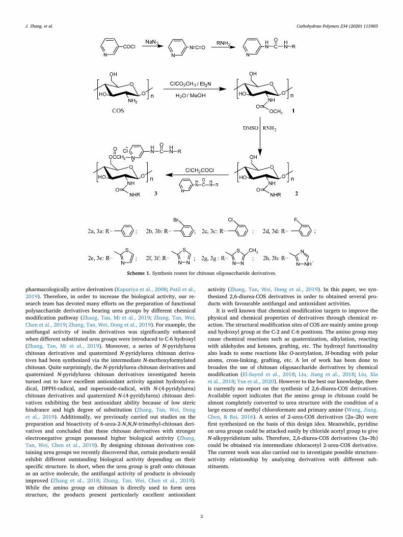
It is well known that chemical modification targets to improve thephysical and chemical properties of derivatives through chemical re-action. The structural modification sites of COS are mainly amino groupand hydroxyl group at the C-2 and C-6 positions. The amino group maycause chemical reactions such as quaternization, alkylation, reactingwith aldehydes and ketones, grafting, etc. The hydroxyl functionalityalso leads to some reactions like O-acetylation, H-bonding with polaratoms, cross-linking, grafting, etc. A lot of work has been done tobroaden the use of chitosan oligosaccharide derivatives by chemicalmodification (El-Sayed et al., 2018; Liu, Jiang et al., 2018; Liu, Xiaet al., 2018; Yue et al., 2020). However to the best our knowledge, thereis currently no report on the synthesis of 2,6-diurea-COS derivatives.Available report indicates that the amino group in chitosan could bealmost completely converted to urea structure with the condition of alarge excess of methyl chloroformate and primary amine (Wang, Jiang,Chen, & Bai, 2016). A series of 2-urea-COS derivatives (2a–2h) werefirst synthesized on the basis of this design idea. Meanwhile, pyridineon urea groups could be attacked easily by chloride acetyl group to giveN-alkypyridinium salts. Therefore, 2,6-diurea-COS derivatives (3a–3h)could be obtained via intermediate chloracetyl 2-urea-COS derivative.The current work was also carried out to investigate possible structure-activity relationship by analyzing derivatives with different sub-stituents.
Scheme 1. Synthesis routes for chitosan oligosaccharide derivatives.
J. Zhang, et al. Carbohydrate Polymers 234 (2020) 115903
2
2. Materials and methods
2.1. Materials
Chitosan oligosaccharide with a molecular weight of5000−8000 Da was supplied by Golden-Shell Pharmaceutical Co. Ltd.(Zhejiang, China). The degree of deacetylation of chitosan oligo-saccharide is 74 %. Chloroacetyl chloride, nicotinoyl chloride hydro-chloride, aniline, 2-fluoroaniline, 2-chloroaniline, 2-bromoaniline, 2-aminothiazole, 2-amino-1,3,4-thiadiazole, 2-amino-5-methyl-1,3,4-thiadiazole, and 3-amino-1,2,4-triazole were purchased from theSigma-Aldrich Chemical Corp (Shanghai, China). The other reagents,such as methyl chloroformate, methanol, triethylamine, sodium azide,methylbenzene, lithium chloride, ethyl alcohol, N,N-dimethyl acet-amide (DMAc), N-methyl pyrrolidone (NMP), and dimethyl sulfoxide(DMSO) were supplied by Sinopharm Chemical Reagent Co., Ltd.(Shanghai, China) and used without purification.
2.2. Synthesis of chitosan oligosaccharide derivatives
As shown in Scheme 1, the synthesis of 2-urea-COS derivatives(2a–2h) and 2,6-diurea-COS derivatives (3a–3h) was carried out by thefollowings steps:
2.2.1. Synthesis of pyridylurea groupsFirstly, nicotinoyl chloride hydrochloride (20mmol) was dispersed
equably in 15mL of acetone and stirred at 0 °C. The mixture was thenadded dropwise to the aqueous solution of sodium azide (3.9 g NaN3
dissolving in 12mL of deionized water). The reaction mixture wasstirred for 3 h under the condition of ice bath. After the reaction wasfinished, the solution had been stratified and the lower layer was re-moved. The remaining solution was poured into 10mL of methylben-zene solvent at 60 °C and stirred for 3 h. Then, the mixture was cooledand pyridine-3-isocyanate were precipitated out.
Pyridine-3-isocyanate (30mmol) and aniline (30mmol) were addedto methylbenzene (20mL) in a 50mL flask, and the mixture was stirredfor 24 h at 60 °C. After the reaction is completed, the formed precipitatewas further purified by crystallization from the solvent that the ratio ofwater and ethanol was 1:1 and N-phenyl-N'-pyridylurea was synthe-sized. N-(2-fluorobenzene)-N'-pyridylurea, N-(2- chlorobenzene)-N'-pyridylurea, N-(2-bromobenzene)-N'-pyridylurea, N-(2-thiazole)-N'-pyridylurea, N-(2-thiadiazole)-N'-pyridylurea, N-(5-methyl-2-thiadia-zole)-N'-pyridylurea, and N-(3-triazole)-N'-pyridylurea were preparedby the same method.
2.2.2. Synthesis of 2-urea-COS derivatives (2a–2h)Chitosan oligosaccharide (2.0 g, 12.4 mmol) was dissolved in 30mL
of deionized water and placed in an ice bath. Methanol (70mL) wasadded with stirring when the temperature of the solution was below10 °C. When the temperature was below 5 °C, methyl chloroformate(7.68 mL, 99.2mmol) was added and the mixture was stirred for 7 h at0 °C. During the reaction, the resulting hydrochloric acid would in-crease the acidity of reaction system. In order to avoid hydrolysis ofproduct, the reaction mixture was maintained slightly acidic (pH=6)by adding triethylamine during this period. After reaction completion,2-methoxyformylated COS was precipitated in ethyl alcohol. The fil-tered 2-methoxyformylated COS (0.44 g, 2mmol) was then dissolved ina solution of LiCl in DMAc (with a mass concentration of 8 %).Subsequently, aniline (16mmol) was added and the solution was stirredfor 12 h at 110 °C. After the reaction, the solution was poured intoethanol. The formed precipitate was filtered and washed with ethanol.Finally, 2-urea-COS derivative (2a) was achieved after vacuum freeze-drying for 24 h. Derivatives 2b–2h were prepared by the same method.
2.2.3. Synthesis of 2,6-diurea-COS derivatives (3a–3h)Firstly, derivative 2a (10mmol) was dispersed in 100mL of N-
methyl pyrrolidone (NMP) at room temperature (r.t.). Then, 20mmolchloracetyl chloride was added. After stirring for 12 h, the precipitate ofchloracetyl 2-urea-COS derivative was obtained by the addition ofether. The precipitate was filtered and washed with ethanol by turns.Then, the precipitate and N-phenyl-N'-pyridylurea dissolved in 20mL ofdimethyl sulfoxide (DMSO) were stirred for 24 h at 60 °C. The solutionwas precipitated in excess acetone. Then, the precipitate was filteredand washed with ethanol for three times. Finally, 2,6-diurea-COS de-rivative (3a) was obtained after vacuum freeze-drying for 24 h.Derivatives 3b–3h were prepared by the same method.
2.3. Analytical methods
2.3.1. Fourier transform infrared (FT-IR) spectroscopyThe infrared spectra of the samples were measured by Jasco-4100
FT-IR spectrometer (Japan, provided by JASCO Co., Ltd., Shanghai,China) with a resolution of 4.0 cm−1 and a range of 4000−400 cm−1.The tested samples were mixed with KBr and prepared in KBr pellet forobservation.
2.3.2. Nuclear magnetic resonance (NMR) spectroscopyThe 1H NMR spectra of samples were recorded by Bruker AVIII-500
Spectrometer (500MHz, Switzerland, provided by Bruker Tech. andServ. Co., Ltd., Beijing, China) at 25 °C. The samples were dissolved in0.6 mL D2O or DMSO for analysis.
2.3.3. Elemental analysisThe elemental analysis was performed on Vario Micro Elemental
Analyzer (Elementar, Berlin, Germany). The degree of substitution (DS)in chitosan oligosaccharide derivatives was evaluated by the carbon-nitrogen ratio and it was according to the following equations:
=× − ×
×DS
n M M Wn M
C N C N
C1
1 /
2
=× + × × − ×
×DS
M W n M DS n Mn M
N C N C C
C2
/ 2 1 1
3
=× + × × − × × − × ×
× × − ×
′
′DSn M n M DS n M W n M DS
n M W n MC C N C N C
N C N C3
1 3 2 1 / 2 1
2 / 4
=
× × + × × × − ×
+ × × − × × − × ××
′ ′
DS
n M W n M W DS n M
n M DS n M DS n M DSn M
N C N N C N C
C C C
C4
1 / 2 / 3 1
2 1 3 2 4 3
5
=
× × + × × × − ×
+ × × − × × − × ×
− × ×× − × ×
′ ′
′DS
n M W n M W DS n M
n M DS n M DS n M DS
n M DSn M n M W
N C N N C N C
C C C
C
C N C N5
1 / 2 / 3 1
2 1 3 2 4 3
5 4
6 3 /
where DS1, DS2, DS3, DS4, and DS5 represent the deacetylation degree ofCOS, the DS of 2-methoxyformylated COS, the DS of 2-urea-COS deri-vatives (2a–2h), the DS of chloracetyl 2-urea-COS derivatives, and theDS of 2,6-diurea-COS derivatives (3a–3h); MC and MN are the molarmass of carbon and nitrogen, MC=12, MN=14; n1, n2, n3, n4, n5, andn6 are the number of carbon of chitin, acetamido group, methox-ycarbonyl group, amino compound, chloroacetyl group, and ureagroup, chitin (n1=8), chitosan oligosaccharide (n1=8, n2=2), deri-vative 1 (n1=8, n2=2, n3=2), derivatives 2a–2d (n1=8, n2=2,n3=2, n4 = 5), derivative 2e (n1=8, n2=2, n3=2, n4 = 2), deri-vative 2f (n1=8, n2=2, n3=2, n4 = 1), derivative 2 g (n1=8,n2=2, n3=2, n4 = 2), derivative 2 h (n1=8, n2=2, n3=2, n4 = 1),derivatives 3a–3d (n1=8, n2=2, n3=2, n4 = 5, n5=2, n6=12),derivative 3e (n1=8, n2=2, n3=2, n4 = 2, n5=2, n6=9), deriva-tive 3f (n1=8, n2=2, n3=2, n4 = 1, n5=2, n6=8), derivative 3 g(n1=8, n2=2, n3=2, n4 = 2, n5=2, n6=9), derivative 3 h (n1=8,
J. Zhang, et al. Carbohydrate Polymers 234 (2020) 115903
3
n2=2, n3=2, n4 = 1, n5=2, n6=8); n’1, n’2, and n’3 are the number ofnitrogen of chitosan oligosaccharide, amino compound, and ureagroup, chitosan oligosaccharide (n’1=1), derivatives 2a–2d (n’1=1, n’2= 1), derivative 2e (n’1=1, n’2 = 2), derivative 2f (n’1=1, n’2 = 3),derivative 2 g (n’1=1, n’2 = 3), derivative 2 h (n’1=1, n’2 = 4), deri-vatives 3a–3d (n’1=1, n’2 = 1, n’3 = 3), derivative 3e (n’1=1, n’2 = 2,n’3 = 4), derivative 3f (n’1=1, n’2 = 3, n’3 = 5), derivative 3 g (n’1=1,n’2 = 3, n’3 = 5), derivative 3 h (n’1=1, n’2 = 4, n’3 = 6); WC/N re-presents the mass ratio between carbon and nitrogen in chitosan oli-gosaccharide derivatives. The degrees of substitution for COS, deriva-tive 1, derivatives 2a–2h, and derivatives 3a–3h are shown in Table 1.The DS4 of the intermediates-chloracetyl 2-urea-COS derivatives are asfollows: a: DS4=0.87, b: DS4=0.52, c: DS4=0.77, d: DS4=0.87, e:DS4=0.50, f: DS4=0.76, g: DS4=0.43, h: DS4 = 0.80.
2.4. Antifungal assay
The antifungal ability was carried out by the method of hyphalmeasurement (Zhang, Tan, Mi et al., 2019). Briefly, the stock solutionsof COS and derivatives were prepared with a concentration of 6mg/mL.Then, each sample solution was added to sterilized potato dextrose agar(PDA) medium to obtain final concentrations of 0.1, 0.5, and 1.0 mg/mL. The culture media containing samples were poured into Petridishes (7 cm). After solidification, 5.0mm diameter of fungi myceliumwas transferred to the test plate and incubated at 28 °C for 2–3 days.When the mycelia of fungi reached the edges of the control plate(without the presence of samples), the inhibition indices of all sampleswere calculated as follows:
= − ×DDAntifungal index (%) (1 ) 100a
b
Where, Da is the diameter of the growth zone in the test plates and Db isthe diameter of the growth zone in the control plate.
2.5. Antioxidant assay
2.5.1. Superoxide-radical scavenging activity assayThe superoxide-radical scavenging ability was assessed following
the previous method (Zhang, Tan, Wei, Dong et al., 2019). The reactionmixture, involving test samples (5 mg/mL, 0.06, 0.12, 0.24, 0.48, and0.96mL), phenazine methosulfate (PMS, 30 μM), nitro blue tetrazolium(NBT, 72 μM), and nicotinamide adenine dinucleotide reduced (NADH,338 μM) in Tris–HCl buffer (16mM, pH 8.0), was incubated 5min atr.t.. The absorbance was measured at 560 nm. Three replicates for each
sample were tested and the superoxide-radical scavenging effect wascalculated according to the following equation:
= ⎡⎣⎢
−− ⎤
⎦⎥×Scavenging effect (%) 1
A AA
100sample 560 nm control 560 nm
blank 560 nm
Where Asample 560 nm is the absorbance of the samples, Acontrol 560 nm isthe absorbance of the control (NADH was substituted with distilledwater), and Ablank 560 nm is the absorbance of the blank (samples weresubstituted with distilled water).
2.5.2. Hydroxyl-radical scavenging activity assayThe test of hydroxyl-radical scavenging ability was carried out ac-
cording to Li's method with minor modification (Li, Wei, Zhang, Gu, &Guo, 2019). The reaction mixture, containing testing samples of COS orchitosan oligosaccharide derivatives (10mg/mL, 0.045, 0.09, 0.18,0.36, and 0.72mL), safranine O (0.23 μM), EDTA-Fe2+ (220 μM), andH2O2 (60 μM) in potassium phosphate buffer (150mM, pH 7.4), wasincubated for 30min at 37 °C. The absorbance of the mixture wasmeasured at 520 nm against blank. Three replicates for each samplewere tested and the hydroxyl-radical scavenging effect was calculatedaccording to the following equation:
=−−
×Scavenging effect (%)A AA A
100sample 520 nm blank 520 nm
control 520 nm blank 520 nm
Where Asample 520 nm is the absorbance of the samples, Acontrol 520 nm isthe absorbance of the control (H2O2 was substituted with potassiumphosphate buffer), and Ablank 520 nm is the absorbance of the blank(samples were substituted with distilled water).
2.5.3. DPPH-radical scavenging activity assayThe DPPH scavenging properties of the products were evaluated by
the following method (Tan et al., 2019): the testing samples in differentconcentrations and 2mL of DPPH ethanol solution (180 μmol/L) wereincubated for 20min at room temperature. The absorbance of the re-mained DPPH radical was measured at 517 nm against a blank. Thecontrol groups containing different concentrations of samples and 2mLof ethanol were measured at 517 nm. Three replicates for each samplewere tested and the DPPH-radical scavenging effect was calculatedaccording to the following equation:
= ⎡⎣⎢
−− ⎤
⎦⎥×Scavenging effect (%) 1
A AA
100sample 517 nm control 517 nm
blank 517 nm
Where Asample 517 nm is the absorbance of the samples, Acontrol 517 nm is
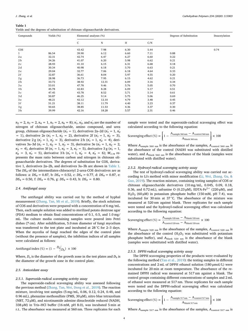
Table 1Yields and the degrees of substitution of chitosan oligosaccharide derivatives.
Compounds Yields (%) Elemental analyses (%) Degrees of Substitution Deacetylation
C N H C/N
COS 43.42 7.98 6.30 5.44 0.741 86.54 39.90 6.12 6.60 7.11 0.882 a 32.31 42.74 6.47 6.27 6.60 0.222 b 34.26 41.07 6.20 5.98 6.62 0.212 c 28.93 41.10 6.15 6.31 6.68 0.182 d 30.34 40.98 6.18 5.70 6.63 0.212 e 29.04 32.77 7.06 6.18 4.64 0.332 f 32.87 36.61 8.04 5.97 4.55 0.202 g 28.98 36.73 7.95 6.15 4.62 0.212 h 33.72 38.92 12.31 6.09 3.16 0.343 a 53.01 47.78 9.46 5.76 5.05 0.703 b 45.78 42.83 8.28 6.09 5.17 0.513 c 47.45 43.78 8.52 5.71 5.14 0.613 d 50.87 46.25 9.14 5.75 5.06 0.693 e 48.21 42.12 12.10 4.79 3.48 0.453 f 51.21 38.11 11.79 4.40 3.23 0.373 g 50.65 38.85 11.53 4.36 3.37 0.303 h 39.99 42.16 18.28 5.57 2.31 0.46
J. Zhang, et al. Carbohydrate Polymers 234 (2020) 115903
4
the absorbance of the control (DPPH was substituted with ethanol), andAblank 517 nm is the absorbance of the blank (samples were substitutedwith distilled water). Vitamin C was used as a positive control.
2.6. Cytotoxicity assay
The cytotoxicity of COS and synthesized COS derivatives on L929cells at different concentrations (1.0, 10.0, 100.0, 500.0, and1000.0 μg/mL) was determined by CCK-8 assay in vitro. L929 cells werecultured at 37 °C in RPMI medium (containing 1 % mixture of penicillin& streptomycin and 10 % fetal calf serum). The cells were seeded on 96-well flat-bottom culture plates at a density of 1.0× 10⁵ cells and in-cubated under 5 % CO2 atmosphere. After 24 h of cell attachment, thesamples with different final concentrations were introduced to cells,
separately. Next, the cells were cultured for 24 h. Afterward, 10 μL ofCCK-8 solution was added in each well and incubated for another 24 hat 37 °C. The absorbance at 450 nm was measured. Cell viability wasrecorded according to the following formula:
=−−
×Cell viability (%)A AA A
100sample blank
negative blank
Where Asample is the absorbance of the samples (containing cells, CCK-8solution, and sample solution), Ablank is the absorbance of the blank(containing RPMI medium and CCK-8 solution), and Anegative is theabsorbance of the negative (containing cells and CCK-8 solution).
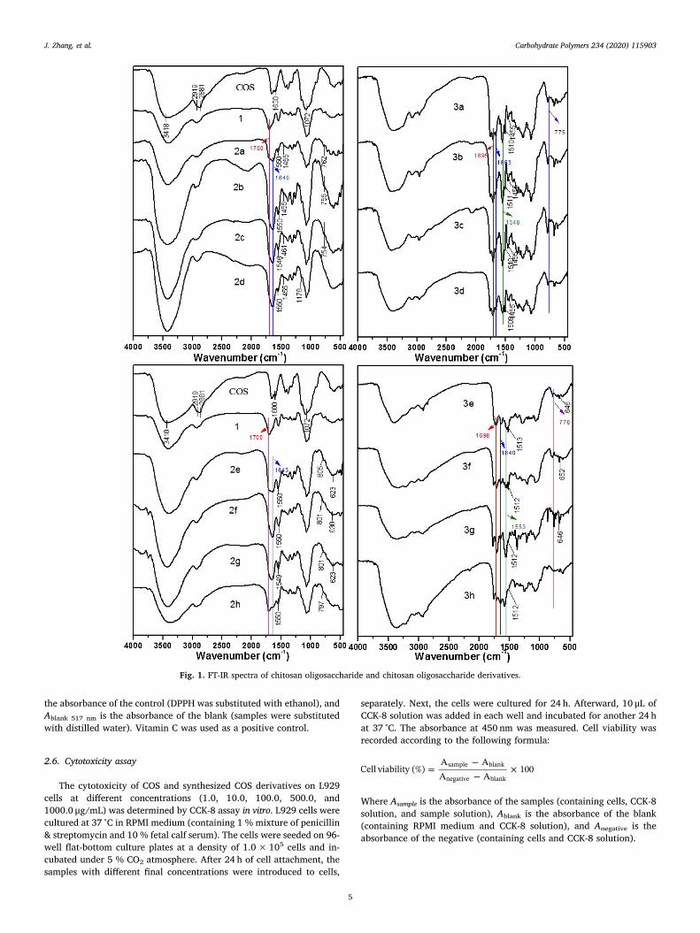
Fig. 1. FT-IR spectra of chitosan oligosaccharide and chitosan oligosaccharide derivatives.
J. Zhang, et al. Carbohydrate Polymers 234 (2020) 115903
5
2.7. Statistical analysis
All the data related to antioxidant activity, antifungal activity, andcytotoxicity assay were illustrated as mean ± standard deviation (SD),n= 3. Significant difference analysis was determined using Scheffe’smultiple range test. The significant differences were defined at p <0.05.
3. Results and discussion
As part of our researches about the preparation of functionalizedchitosan derivatives bearing urea groups, herein we report the synthesis
and characterization of 2,6-diurea-COS derivatives. Halogenated ben-zene and nitrogen-COntaining heterocycles have played an importantrole in research fields of antimicrobial, anticancer, anticonvulsant, anti-inflammatory, and antipsychotic agents (Harish, Mohana, & Mallesha,2013; Kaur et al., 2010; Madhu Sekhar et al., 2018). Therefore, wechose them as substituents and grafted them to chitosan oligo-saccharide by urea structural motif in this study. As shown in Scheme 1,2-urea-COS derivatives (2a–2h) were synthesized firstly by the reactionof 2-methoxyformylated COS (1) and amine compounds. Then, severalurea groups containing pyridine (pyridylurea groups) were obtained. Inorder to attach the pyridylurea groups to 2-urea-COS derivatives, 2-urea-COS derivatives were reacted with chloroacetyl chloride to obtain
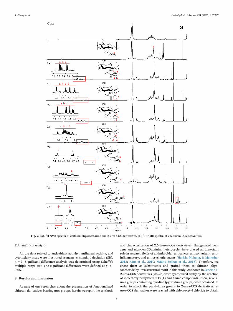
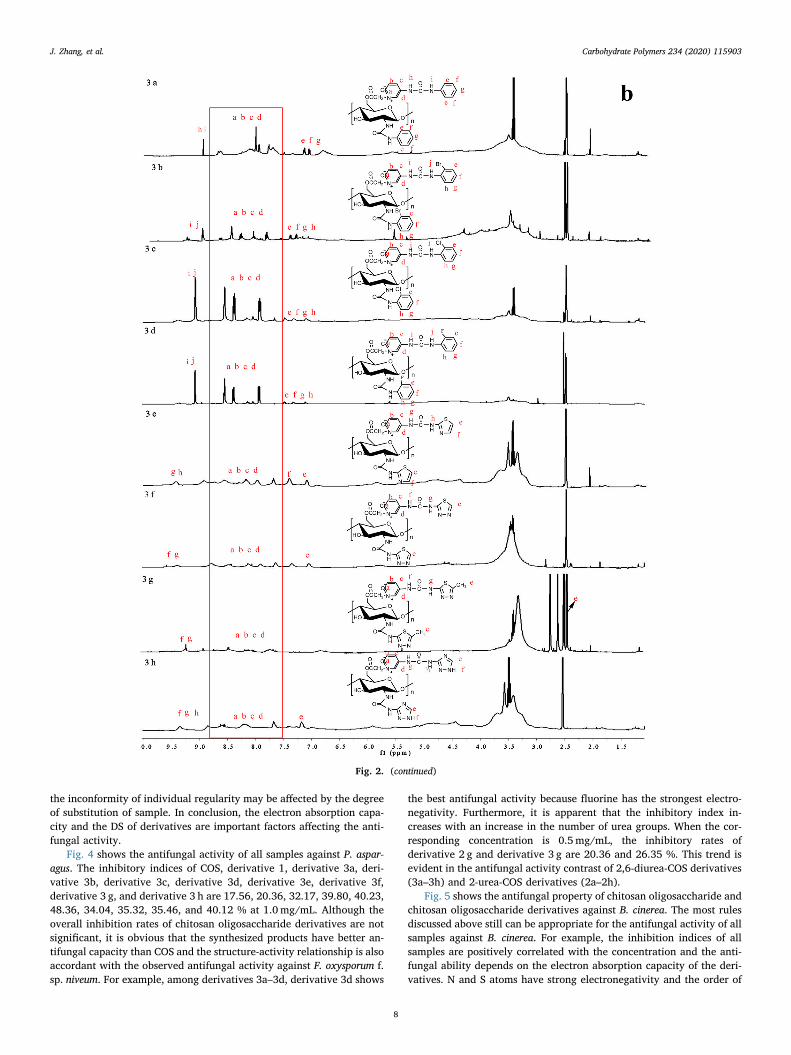
Fig. 2. (a). 1H NMR spectra of chitosan oligosaccharide and 2-urea-COS derivatives. (b). 1H NMR spectra of 2,6-diurea-COS derivatives.
J. Zhang, et al. Carbohydrate Polymers 234 (2020) 115903
6
the intermediate chloracetyl 2-urea-COS derivatives. Eventually, thesederivatives attacked the substituted pyridylurea groups to give N-al-kypyridinium salts and a series of 2,6-diurea-COS derivatives (3a–3h)were synthesized. The products were characterized through FT-IR, 1HNMR spectroscopy, and elemental analysis to confirm the structuralcorrectness. Three phytopathogenic fungi, including Fusarium oxy-sporum f. sp. niveum (F. oxysporum f. sp. niveum), Phomopsis asparagus (P.asparagus), and Botrytis cinerea (B. cinerea) were selected to test theantifungal activity of the chitosan oligosaccharide derivatives in vitro.Meanwhile, we evaluated the antioxidant property of the chitosan oli-gosaccharide derivatives by the assessment of superoxide-radicalscavenging ability, hydroxyl-radical scavenging ability, and DPPH-ra-dical scavenging ability. Besides, the cytotoxicity of the synthesizedcompounds was investigated in vitro on L929 cells.
3.1. Chemical characterization
3.1.1. FT-IR spectraFig. 1 shows the results from the FT-IR spectra of chitosan oligo-
saccharide and chitosan oligosaccharide derivatives. In COS spectrum,the peaks appearing at 3418 cm−1, 2919 cm−1 and 2881 cm−1,1600 cm−1 are assigned to OeH and NeH stretching vibrations, eCHasymmetric stretching, and vibration modes of amino group (Barbosa,Attjioui, Leitao, Moerschbacher, & Cavalheiro, 2019; Braz et al., 2020;Menezes et al., 2020). In spectrum of 2-methoxyformylated COS (de-rivative 1), a new peak appears at 1700 cm−1 and the absorbance ofamino group at 1596 cm−1 disappears, which indicate that the aminogroup of chitosan oligosaccharide was replaced by methoxycarbonylgroup (Wang et al., 2016; Zhang, Tan, Wei, Dong et al., 2019). Inspectra of 2-urea-COS derivatives, the absorption at 1700 cm−1 dis-appears when amino compound was grafted and new peak at about1640 cm−1 appears. The peak found at 1640 cm−1 is attributed to thevibration of the newly formed structure, -NH-CO-NH- (Wang, Xi, Chen,Huang, & Bai, 2017). Besides, for derivatives 2a–2d, the bands at∼1550 cm−1, ∼1455 cm−1, and ∼750 cm−1 are assigned to the ty-pical absorption of benzene ring. For derivatives 2e–2h, the bands at∼1550 cm−1, ∼800 cm−1, and ∼620 cm−1 are assigned to the typicalabsorption of nitrogen-COntaining heterocycles. As to the spectra of2,6-diurea-COS derivatives (3a–3h), there is a new peak at ∼1695 cm-1
which refers to vibration of eNHeCOeNHe of pyridylurea groups(Zhang, Tan, Wei, Chen et al., 2019). Meanwhile, the characteristicabsorptions of aromatic ring of pyridylurea groups appearing at about1550 cm−1, ∼1510 cm−1, ∼775 cm−1, and ∼650 cm−1 are exist (Weiet al., 2019). Hence, these data preliminarily proved the successfulsynthesis of 2,6-diurea-COS derivatives.
3.1.2. NMR spectraFig. 2 (a) and (b) show the 1H NMR spectra of chitosan oligo-
saccharide and chitosan oligosaccharide derivatives. In the 1H NMRspectrum of chitosan oligosaccharide, the signals of C-2 proton, C-3-C-6protons, and C-1 proton appear at δ3.0 ppm, δ3.6–3.9 ppm, andδ5.5 ppm (Dang et al., 2019; Liu et al., 2016). After modification, thepeak assigned to the proton signals of methoxy group is observed atδ3.7 ppm from the 1H NMR spectrum of derivative 1, which suggeststhe correct structure of 2-methoxyformylated chitosan oligosaccharide(Wang et al., 2016, 2017). As for as the 1H NMR spectra of derivatives2a–2h, the absorption signals of the aromatic ring appear in the lowfield. However, the absorption signals are not obvious due to the lowdegrees of substitution. After pyridylurea groups had been grafted ontochloracetyl 2-urea-COS derivatives, changes in the 1H NMR spectra ofderivatives 3a–3h are visible. Generally, the peaks at ∼δ9.0–9.7 ppmare attributed to the protons of eNHe on the pyridylurea groups(Zhang, Tan, Wei, Chen et al., 2019). Other peaks in the range ofδ7.5–8.8 ppm are related to the protons of pyridine ring. The smallpeaks at about δ6.8–7.5 ppm are the absorption signals of the protons ofthiazole, triazole, or benzene ring (Azmy, Hashem, Mohamed, & Negm,
2019; Zhang, Tan, Mi et al., 2019). The specific positions of other sig-nals of the protons are indicated in Fig. 2(a) and (b). Therefore, theseresults are enough to prove the successful synthesis of 2,6-diurea-COSderivatives.
3.1.3. Elemental analysisTable 1 shows the yields and degrees of substitution of chitosan
oligosaccharide derivatives. The DS of chitosan oligosaccharide deri-vatives is estimated by elemental analysis. The DS of derivative 1 is0.88 calculated by the formulas. As to derivatives 2a–2h, their degreesof substitution are relatively low, which are only around 0.2. However,derivatives 3a–3h possess the higher degrees of substitution, which arearound 0.5, and some even as high as 0.7. Sometimes, the amount ofactive component resulting from the degree of substitution plays acrucial role in the biological activity of the product.
3.2. Antifungal activity
Fungal infections in food and agricultural products lead to a seriesof food safety problems and cause considerable economic loss.Concurrently, the fungal toxins produced in the process of infectingfood are harmful to animal and human health. Searching for effectivepreservative is of great significance for food safety and human health.At present, using chemical preservatives is the main means to controlthe incidence of mold. However, the generation and development ofresistant and pollution problems force researchers screening for eco-friendly antifungal agents (Rong et al., 2019; Xu et al., 2019). In thispaper, several biodegradable chitosan oligosaccharide derivatives weresynthesized and the test of antifungal activity was run against a series offungal pathogens: Fusarium oxysporum f. sp. niveum, Phomopsis aspar-agus, and Botrytis cinereal. The test results are shown in Figs. 3–5.
Fig. 3 shows the antifungal activity of chitosan oligosaccharide andchitosan oligosaccharide derivatives against F. oxysporum f. sp. niveumat various concentrations. The results indicate that all the samples showdifferent antifungal activity against the tested fungal pathogen. Withthe increase of concentration, the increases of inhibitory indices of allsamples can be observed. For example, when the test concentration is0.1, 0.5, and 1.0 mg/mL, the corresponding inhibition rate of derivative1 is 0.51, 9.83, and 22.59 %. The inhibitory index of COS is only 19.98% at 1.0 mg/mL and all COS derivatives have better antifungal propertycompared with COS due to the introduction of urea groups. In addition,the inhibitory indices of 2,6-diurea-COS derivatives (3a–3h) are higherthan that of 2-urea-COS derivatives (2a–2h). The inhibitory value ofderivative 3a is enhanced more than 15 % compared with derivative 2a.As evidenced by the improved antifungal activity of compounds 3a–3h,the existence of pyridylurea groups is crucial to the tested bioactivity,which is agree with our previous conclusions. Another phenomenon canbe found in Fig. 3, that is, the different structure of urea groups will leadto a certain biological activity regularity of the derivatives. For ex-ample, the order of inhibitory rates of derivatives containing benzenering is d > c>b > a, which is in accordance with the electron-withdrawing capacity. Meanwhile, the antifungal activity of chitosanoligosaccharide derivatives bearing nitrogen-containing heterocyclicring is also related to their electron absorption capacity, that is, deri-vatives bearing stronger electronegative groups possess better anti-fungal activity, which is consistent with the previous results. It has beenreported that the growth inhibition of fungi is due to the establishedinteraction between the electrophilic groups with substances on cellwall of microorganisms. The interaction can prevent nutrient exchange,damage the cell membrane to cause the leakage of the cell constituents,hinder the metabolism of cell, and ultimately lead to the death of mi-croorganisms (Aktan, Gündüzalp, & Özmen, 2017). Hence, the orders ofthe antifungal activity of COS derivatives, d > c > b > a and h >g> f > e, are consistent with the electron-withdrawing property ofthe different substituted groups on urea (eF>eCl>eBr>eH andtriazole > thiadiazole > methyl-thiadiazole > thiazole). Of course,
J. Zhang, et al. Carbohydrate Polymers 234 (2020) 115903
7
the inconformity of individual regularity may be affected by the degreeof substitution of sample. In conclusion, the electron absorption capa-city and the DS of derivatives are important factors affecting the anti-fungal activity.
Fig. 4 shows the antifungal activity of all samples against P. aspar-agus. The inhibitory indices of COS, derivative 1, derivative 3a, deri-vative 3b, derivative 3c, derivative 3d, derivative 3e, derivative 3f,derivative 3 g, and derivative 3 h are 17.56, 20.36, 32.17, 39.80, 40.23,48.36, 34.04, 35.32, 35.46, and 40.12 % at 1.0mg/mL. Although theoverall inhibition rates of chitosan oligosaccharide derivatives are notsignificant, it is obvious that the synthesized products have better an-tifungal capacity than COS and the structure-activity relationship is alsoaccordant with the observed antifungal activity against F. oxysporum f.sp. niveum. For example, among derivatives 3a–3d, derivative 3d shows
the best antifungal activity because fluorine has the strongest electro-negativity. Furthermore, it is apparent that the inhibitory index in-creases with an increase in the number of urea groups. When the cor-responding concentration is 0.5mg/mL, the inhibitory rates ofderivative 2 g and derivative 3 g are 20.36 and 26.35 %. This trend isevident in the antifungal activity contrast of 2,6-diurea-COS derivatives(3a–3h) and 2-urea-COS derivatives (2a–2h).
Fig. 5 shows the antifungal property of chitosan oligosaccharide andchitosan oligosaccharide derivatives against B. cinerea. The most rulesdiscussed above still can be appropriate for the antifungal activity of allsamples against B. cinerea. For example, the inhibition indices of allsamples are positively correlated with the concentration and the anti-fungal ability depends on the electron absorption capacity of the deri-vatives. N and S atoms have strong electronegativity and the order of
Fig. 2. (continued)
J. Zhang, et al. Carbohydrate Polymers 234 (2020) 115903
8
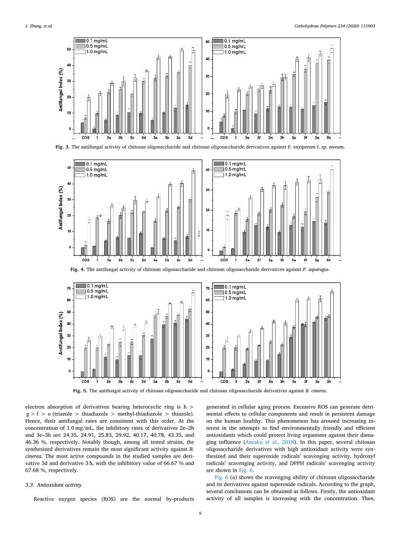
electron absorption of derivatives bearing heterocyclic ring is h >g> f > e (triazole > thiadiazole > methyl-thiadiazole > thiazole).Hence, their antifungal rates are consistent with this order. At theconcentration of 1.0mg/mL, the inhibitory rates of derivatives 2e–2hand 3e–3h are 24.35, 24.91, 25.83, 29.92, 40.17, 40.78, 43.35, and46.36 %, respectively. Notably though, among all tested strains, thesynthesized derivatives remain the most significant activity against B.cinerea. The most active compounds in the studied samples are deri-vative 3d and derivative 3 h, with the inhibitory value of 66.67 % and67.68 %, respectively.
3.3. Antioxidant activity
Reactive oxygen species (ROS) are the normal by-products
generated in cellular aging process. Excessive ROS can generate detri-mental effects to cellular components and result in persistent damageon the human healthy. This phenomenon has aroused increasing in-terest in the attempts to find environmentally friendly and efficientantioxidants which could protect living organisms against their dama-ging influence (Anraku et al., 2018). In this paper, several chitosanoligosaccharide derivatives with high antioxidant activity were syn-thesized and their superoxide radicals’ scavenging activity, hydroxylradicals’ scavenging activity, and DPPH radicals’ scavenging activityare shown in Fig. 6.
Fig. 6 (a) shows the scavenging ability of chitosan oligosaccharideand its derivatives against superoxide radicals. According to the graph,several conclusions can be obtained as follows. Firstly, the antioxidantactivity of all samples is increasing with the concentration. Then,
Fig. 3. The antifungal activity of chitosan oligosaccharide and chitosan oligosaccharide derivatives against F. oxysporum f. sp. niveum.
Fig. 4. The antifungal activity of chitosan oligosaccharide and chitosan oligosaccharide derivatives against P. asparagus.
Fig. 5. The antifungal activity of chitosan oligosaccharide and chitosan oligosaccharide derivatives against B. cinerea.
J. Zhang, et al. Carbohydrate Polymers 234 (2020) 115903
9
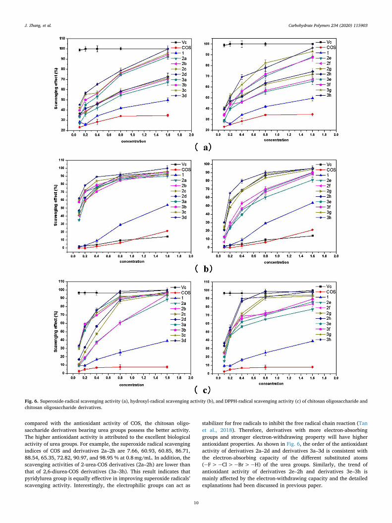
compared with the antioxidant activity of COS, the chitosan oligo-saccharide derivatives bearing urea groups possess the better activity.The higher antioxidant activity is attributed to the excellent biologicalactivity of urea groups. For example, the superoxide radical scavengingindices of COS and derivatives 2a–2h are 7.66, 60.93, 60.85, 86.71,88.54, 65.35, 72.82, 90.97, and 98.95 % at 0.8mg/mL. In addition, thescavenging activities of 2-urea-COS derivatives (2a–2h) are lower thanthat of 2,6-diurea-COS derivatives (3a–3h). This result indicates thatpyridylurea group is equally effective in improving superoxide radicals’scavenging activity. Interestingly, the electrophilic groups can act as
stabilizer for free radicals to inhibit the free radical chain reaction (Tanet al., 2018). Therefore, derivatives with more electron-absorbinggroups and stronger electron-withdrawing property will have higherantioxidant properties. As shown in Fig. 6, the order of the antioxidantactivity of derivatives 2a–2d and derivatives 3a–3d is consistent withthe electron-absorbing capacity of the different substituted atoms(eF>eCl>eBr>eH) of the urea groups. Similarly, the trend ofantioxidant activity of derivatives 2e–2h and derivatives 3e–3h ismainly affected by the electron-withdrawing capacity and the detailedexplanations had been discussed in previous paper.
Fig. 6. Superoxide-radical scavenging activity (a), hydroxyl-radical scavenging activity (b), and DPPH-radical scavenging activity (c) of chitosan oligosaccharide andchitosan oligosaccharide derivatives.
J. Zhang, et al. Carbohydrate Polymers 234 (2020) 115903
10
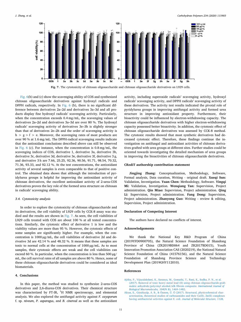
Fig. 6(b) and (c) show the scavenging ability of COS and synthesizedchitosan oligosaccharide derivatives against hydroxyl radicals andDPPH radicals, respectively. In Fig. 6 (b), there is no significant dif-ference between derivatives 2a–2d and derivatives 3a–3d and all pro-ducts display fine hydroxyl radicals’ scavenging activity. Particularly,when the concentration exceeds 0.4 mg/mL, the scavenging values ofderivatives 2a–2d and derivatives 3a–3d are over 80 %. The hydroxylradicals’ scavenging activity of derivatives 3e–3h is slightly strongerthan that of derivatives 2e–2h and the order of scavenging activity ish > g> f > e. Moreover, the scavenging rates of most products areover 90 % at 1.6mg/mL. The DPPH-radical scavenging results indicatethat the antioxidant conclusions described above can still be observedin Fig. 6 (c). For instance, when the concentration is 0.8 mg/mL, thescavenging indices of COS, derivative 1, derivative 3a, derivative 3b,derivative 3c, derivative 3d, derivative 3e, derivative 3f, derivative 3 g,and derivative 3 h are 7.66, 25.25, 92.36, 90.56, 91.71, 98.54, 70.32,71.36, 93.33, and 92.12 %. At the test concentrations, the antioxidantactivity of several samples is even comparable to that of positive con-trol. The obtained data shows that although the introduction of pyr-idylurea groups is helpful for improving the antioxidant activity ofchitosan derivatives, the excellent antioxidant activity of 2-urea-COSderivatives proves the key role of the formed urea structure on chitosanin radicals’ scavenging ability.
3.4. Cytotoxicity analysis
In order to explore the cytotoxicity of chitosan oligosaccharide andits derivatives, the cell viability of L929 cells by CCK-8 assay was stu-died and the results are shown in Fig. 7. As seen, the cell viabilities ofL929 cells treated with COS are about 100 % at all tested concentra-tions. Similarly, the cytotoxic effect of derivative 1 is low and theviability values are more than 95 %. However, the cytotoxic effects ofsome samples are significantly higher. For example, when the con-centration is 1000 μg/mL, the cell viabilities of derivative 2d and de-rivative 3d are 42.14 % and 48.32 %. It means that these samples aretoxic to normal cells at the concentration of 1000 μg/mL. As to mostsamples, their cytotoxic effects are weak and the cell viabilities canexceed 60 %. In particular, when the concentration is less than 500 μg/mL, the cell survival rates of all samples are above 80 %. Hence, some ofthese chitosan oligosaccharide derivatives could be considered as idealbiomaterials.
4. Conclusions
In this paper, the method was studied to synthesize 2-urea-COSderivatives and 2,6-diurea-COS derivatives. Their chemical structurewas characterized by FT-IR, 1H NMR spectroscopy, and elementalanalysis. We also explored the antifungal activity against F. oxysporumf. sp. niveum, P. asparagus, and B. cinereal as well as the antioxidant
activity, including superoxide radicals’ scavenging activity, hydroxylradicals’ scavenging activity, and DPPH radicals’ scavenging activity ofthese derivatives. The activity test results indicated the pivotal role ofpyridylurea groups in improving antifungal activity and formed ureastructure in improving antioxidant property. Furthermore, theirbioactivity could be influenced by electron-withdrawing capacity. Thechitosan oligosaccharide derivatives with higher electron-withdrawingcapacity possessed better bioactivity. In addition, the cytotoxic effect ofchitosan oligosaccharide derivatives was assessed by CCK-8 method.The cytotoxic results showed that most synthetic derivatives had de-creased cytotoxic effect. Therefore, these findings continue the in-vestigation on antifungal and antioxidant activities of chitosan deriva-tives grafted with urea groups at different sites. Further studies could beoriented towards investigating the detailed mechanism of urea groupsin improving the bioactivities of chitosan oligosaccharide derivatives.
CRediT authorship contribution statement
Jingjing Zhang: Conceptualization, Methodology, Software,Formal analysis, Data curation, Writing - original draft. Xueqi Sun:Validation, Investigation. Yuan Chen: Methodology, Software. YingqiMi: Validation, Investigation. Wenqiang Tan: Supervision, Projectadministration. Qin Miao: Supervision, Project administration. QingLi: Supervision, Project administration. Fang Dong: Supervision,Project administration. Zhanyong Guo: Writing - review & editing,Supervision, Project administration.
Declaration of Competing Interest
The authors have declared no conflicts of interest.
Acknowledgements
We thank the National Key R&D Program of China(2019YFD0900705), the Natural Science Foundation of ShandongProvince of China (ZR2019BD064 and ZR2017BD015), YouthInnovation Promotion Association CAS (2020219), the National NaturalScience Foundation of China (41576156), and the Natural ScienceFoundation of Shandong Province Science and TechnologyDevelopment Plan (2019GHY112010).
References
Ajitha, P., Vijayalakshmi, K., Saranya, M., Gomathi, T., Rani, K., Sudha, P. N., et al.(2017). Removal of toxic heavy metal lead (II) using chitosan oligosaccharide-graft-maleic anhydride/polyvinyl alcohol/silk fibroin composite. International Journal ofBiological Macromolecules, 104(Pt B), 1469–1482.
Aktan, E., Gündüzalp, A. B., & Özmen, Ü. Ö (2017). Structural, physicochemical char-acterization, theoretical studies of carboxamides and their Cu(II), Zn(II) complexeshaving antibacterial activities against E. coli. Journal of Molecular Structure, 1128,
Fig. 7. The cytotoxicity of chitosan oligosaccharide and chitosan oligosaccharide derivatives on L929 cells.
J. Zhang, et al. Carbohydrate Polymers 234 (2020) 115903
11
775–784.Aljawish, A., Chevalot, I., Jasniewski, J., Scher, J., & Muniglia, L. (2015). Enzymatic
synthesis of chitosan derivatives and their potential applications. Journal of MolecularCatalysis B, Enzymatic, 112, 25–39.
Alves, N. M., & Mano, J. F. (2008). Chitosan derivatives obtained by chemical mod-ifications for biomedical and environmental applications. International Journal ofBiological Macromolecules, 43(5), 401–414.
Anraku, M., Gebicki, J. M., Iohara, D., Tomida, H., Uekama, K., Maruyama, T., et al.(2018). Antioxidant activities of chitosans and its derivatives in in vitro and in vivostudies. Carbohydrate Polymers, 199, 141–149.
Azmy, E. A. M., Hashem, H. E., Mohamed, E. A., & Negm, N. A. (2019). Synthesis,characterization, swelling and antimicrobial efficacies of chemically modified chit-osan biopolymer. Journal of Molecular Liquids, 284, 748–754.
Barbosa, H. F. G., Attjioui, M., Leitao, A., Moerschbacher, B. M., & Cavalheiro, E. T. G.(2019). Characterization, solubility and biological activity of amphihilic biopoly-meric Schiff bases synthesized using chitosans. Carbohydrate Polymers, 220, 1–11.
Bonilla, F., Chouljenko, A., Lin, A., Young, B. M., Goribidanur, T. S., Blake, J. C., et al.(2019). Chitosan and water-soluble chitosan effects on refrigerated catfish filletquality. Food Bioscience, 31, 100426.
Braz, E. M. A., Silva, S. C. C. C., Sousa Brito, C. A. R., Brito, L. M., Barreto, H. M.,Carvalho, F. A. A., et al. (2020). Spectroscopic, thermal characterizations and bac-teria inhibition of chemically modified chitosan with phthalic anhydride. MaterialsChemistry and Physics, 240, 122053.
Dang, Q., Zhang, Q., Liu, C., Yan, J., Chang, G., Xin, Y., et al. (2019). Decanoic acidfunctionalized chitosan: Synthesis, characterization, and evaluation as potentialwound dressing material. International Journal of Biological Macromolecules, 139,1046–1053.
El-Sayed, N. S., Sharma, M., Aliabadi, H. M., El-Meligy, M. G., El-Zaity, A. K., Nageib, Z.A., et al. (2018). Synthesis, characterization, and in vitro cytotoxicity of fatty acyl-CGKRK-chitosan oligosaccharides conjugates for siRNA delivery. International Journalof Biological Macromolecules, 112, 694–702.
Govindaraj, P., Abathodharanan, N., Ravishankar, K., & Raghavachari, D. (2019). Facilepreparation of biocompatible macroporous chitosan hydrogel by hydrothermal re-action of a mixture of chitosan-succinic acid-urea. Materials Science & Engineering C,Materials for Biological Applications, 104, 109845.
Harish, K. P., Mohana, K. N., & Mallesha, L. (2013). Synthesis of indazole substituted-1,3,4-thiadiazoles and their anticonvulsant activity. Drug Invention Today, 5(2),92–99.
Huang, W., Wang, Y., Zhang, S., Huang, L., Hua, D., & Zhu, X. (2013). A facile approachfor controlled modification of chitosan under γ-Ray irradiation for drug delivery.Macromolecules, 46(3), 814–818.
Javan, B., Atyabi, F., & Shahbazi, M. (2018). Hypoxia-inducible bidirectional shRNAexpression vector delivery using PEI/chitosan-TBA copolymers for colorectal cancergene therapy. Life Sciences, 202, 140–151.
Jia, M., Li, Y., Yang, X., Huang, Y., Wu, H., Huang, Y., et al. (2014). Development of bothmethotrexate and mitomycin C loaded PEGylated chitosan nanoparticles for targeteddrug codelivery and synergistic anticancer effect. ACS Applied Materials & Interfaces,6(14), 11413–11423.
Kapuriya, N., Kapuriya, K., Zhang, X., Chou, T. C., Kakadiya, R., Wu, Y. T., et al. (2008).Synthesis and biological activity of stable and potent antitumor agents, aniline ni-trogen mustards linked to 9-anilinoacridines via a urea linkage. Bioorganic &Medicinal Chemistry, 16(10), 5413–5423.
Kaur, H., Kumar, S., Vishwakarma, P., Sharma, M., Saxena, K. K., & Kumar, A. (2010).Synthesis and antipsychotic and anticonvulsant activity of some new substituted oxa/thiadiazolylazetidinonyl/thiazolidinonylcarbazoles. European Journal of MedicinalChemistry, 45(7), 2777–2783.
Li, Q., Wei, L., Zhang, J., Gu, G., & Guo, Z. (2019). Significantly enhanced antioxidantactivity of chitosan through chemical modification with coumarins. PolymerChemistry, 10(12), 1480–1488.
Li, J., Wu, X., Shi, Q., Li, C., & Chen, X. (2019). Effects of hydroxybutyl chitosan onimproving immunocompetence and antibacterial activities. Materials Science &Engineering C, Materials for Biological Applications, 105, 110086.
Liu, J., Meng, C. G., Yan, Y. H., Shan, Y. N., Kan, J., & Jin, C. H. (2016). Protocatechuicacid grafted onto chitosan: Characterization and antioxidant activity. InternationalJournal of Biological Macromolecules, 89, 518–526.
Liu, X., Jiang, Q., & Xia, W. (2018). One-step procedure for enhancing the antibacterialand antioxidant properties of a polysaccharide polymer: Kojic acid grafted ontochitosan. International Journal of Biological Macromolecules, 113, 1125–1133.
Liu, X., Xia, W., Jiang, Q., Yu, P., & Yue, L. (2018). Chitosan oligosaccharide-N-chlor-okojic acid mannich base polymer as a potential antibacterial material. CarbohydratePolymers, 182, 225–234.
Madhu Sekhar, M., Nagarjuna, U., Padmavathi, V., Padmaja, A., Reddy, N. V., & Vijaya, T.(2018). Synthesis and antimicrobial activity of pyrimidinyl 1,3,4-oxadiazoles, 1,3,4-
thiadiazoles and 1,2,4-triazoles. European Journal of Medicinal Chemistry, 145, 1–10.Mahanta, A. K., Senapati, S., Paliwal, P., Krishnamurthy, S., Hemalatha, S., & Maiti, P.
(2019). Nanoparticle-induced controlled drug delivery using chitosan-based hydrogeland scaffold: Application to bone regeneration. Molecular Pharmaceutics, 16(1),327–338.
Menezes, J. E. S. A., Santos, H. S., Ferreira, M. K. A., Magalhães, F. E. A., da Silva, D. S.,Bandeira, P. N., et al. (2020). Preparation, structural and spectroscopic character-ization of chitosan membranes containing allantoin. Journal of Molecular Structure,1199, 126968.
Miao, Z., Li, D., Zheng, Z., & Zhang, Q. (2019). Synthesis of chitosan-mimicking cationicglycopolymers by Cu(0)-LRP for efficient capture and killing of bacteria. PolymerChemistry, 10(29), 4059–4066.
Miao, J., Yang, X. Q., Gao, Z., Li, Q., Meng, T. T., Wu, J. Y., et al. (2019). Redox-re-sponsive chitosan oligosaccharide-SS-Octadecylamine polymeric carrier for efficientanti-Hepatitis B Virus gene therapy. Carbohydrate Polymers, 212, 215–221.
Miguel, S. P., Moreira, A. F., & Correia, I. J. (2019). Chitosan based-asymmetric mem-branes for wound healing: A review. International Journal of Biological Macromolecules,127, 460–475.
Niu, X., Zhu, L., Xi, L., Guo, L., & Wang, H. (2020). An antimicrobial agent prepared by N-succinyl chitosan immobilized lysozyme and its application in strawberry preserva-tion. Food Control, 108, 106829.
Patil, M., Poyil, A. N., Joshi, S. D., Patil, S. A., Patil, S. A., & Bugarin, A. (2019). Synthesis,molecular docking studies, and antimicrobial evaluation of new structurally diverseureas. Bioorganic Chemistry, 87, 302–311.
Pérez Córdoba, L. J., & Sobral, P. J. A. (2017). Physical and antioxidant properties of filmsbased on gelatin, gelatin-chitosan or gelatin-sodium caseinate blends loaded withnanoemulsified active compounds. Journal of Food Engineering, 213, 47–53.
Preethi Soundarya, S., Haritha Menon, A., Viji Chandran, S., & Selvamurugan, N. (2018).Bone tissue engineering: Scaffold preparation using chitosan and other biomaterialswith different design and fabrication techniques. International Journal of BiologicalMacromolecules, 119, 1228–1239.
Rong, S., Xu, H., Li, L., Chen, R., Gao, X., & Xu, Z. (2019). Antifungal activity of en-dophytic Bacillus safensis B21 and its potential application as a biopesticide tocontrol rice blast. Pesticide Biochemistry and Physiology.
Sakwanichol, J., Sungthongjeen, S., & Puttipipatkhachorn, S. (2019). Preparation andcharacterization of chitosan aqueous dispersion as a pharmaceutical film formingmaterial. Journal of Drug Delivery Science and Technology, 54, 101230.
Tan, W., Zhang, J., Mi, Y., Dong, F., Li, Q., & Guo, Z. (2018). Synthesis, characterization,and evaluation of antifungal and antioxidant properties of cationic chitosan deriva-tive via azide-alkyne click reaction. International Journal of Biological Macromolecules,120(Pt A), 318–324.
Tan, W., Dong, F., Zhang, J., Zhao, X., Li, Q., & Guo, Z. (2019). Physical and antioxidantproperties of edible chitosan ascorbate films. Journal of Agricultural and FoodChemistry, 67(9), 2530–2539.
Wang, J., Jiang, J. Z., Chen, W., & Bai, Z. W. (2016). Synthesis and characterization ofchitosan alkyl urea. Carbohydrate Polymers, 145, 78–85.
Wang, J., Xi, J. B., Chen, W., Huang, S. H., & Bai, Z. W. (2017). High performance chiralseparation materials based on chitosan bis(3,5-dimethylphenylcarbamate)-(alkylurea)s. Carbohydrate Polymers, 156, 481–489.
Wei, L., Tan, W., Wang, G., Li, Q., Dong, F., & Guo, Z. (2019). The antioxidant and an-tifungal activity of chitosan derivatives bearing Schiff bases and quaternary ammo-nium salts. Carbohydrate Polymers, 226, 115256.
Xu, H., Su, X., Liu, X. Q., Zhang, K. P., Hou, Z., & Guo, C. (2019). Design, synthesis andbiological evaluation of novel semicarbazone-selenochroman-4-ones hybrids as po-tent antifungal agents. Bioorganic & Medicinal Chemistry Letter126726.
Yuan, G., Chen, X., & Li, D. (2016). Chitosan films and coatings containing essential oils:The antioxidant and antimicrobial activity, and application in food systems. FoodResearch International, 89(Pt 1), 117–128.
Yue, L., Sun, D., Mahmood Khan, I., Liu, X., Jiang, Q., & Xia, W. (2020). Cinnamyl alcoholmodified chitosan oligosaccharide for enhancing antimicrobial activity. FoodChemistry, 309, 125513.
Zhang, J., Tan, W., Zhang, Z., Song, Y., Li, Q., Dong, F., et al. (2018). Synthesis, char-acterization, and the antifungal activity of chitosan derivatives containing ureagroups. International Journal of Biological Macromolecules, 109, 1061–1067.
Zhang, J., Tan, W., Mi, Y., Luan, F., Wei, L., Li, Q., et al. (2019). Synthesis and char-acterization of inulin derivatives bearing urea groups with promising antifungal ac-tivity. Starch - Stärke, 71(1-2), 1800058.
Zhang, J., Tan, W., Wei, L., Chen, Y., Mi, Y., Sun, X., et al. (2019). Synthesis of urea-functionalized chitosan derivatives for potential antifungal and antioxidant applica-tions. Carbohydrate Polymers, 215, 108–118.
Zhang, J., Tan, W., Wei, L., Dong, F., Li, Q., & Guo, Z. (2019). Synthesis, characterization,and antioxidant evaluation of novel pyridylurea-functionalized chitosan derivatives.Polymers, 11(6).
J. Zhang, et al. Carbohydrate Polymers 234 (2020) 115903
12


![Die Synthese amphiphiler 666-6 ... · Schardinger [6] als zyklische Oligosaccharide charakterisiert. Beweise zur Ausbildung von ... durch das Enzym Cyclodextrin-Glycosyltransferase](https://img.pdfslide.org/doc/110x75/5d606e1088c993b3248bb750/die-synthese-amphiphiler-666-6-schardinger-6-als-zyklische-oligosaccharide.jpg)