Embed Size (px)
Citation preview
Protein phosphatase 2A inhibits interferon
signaling through the Jak STAT pathway and
promotes hepatitis C viral replication
Inauguraldissertation
zur
Erlangung der Würde eines Doktors der Philosophie
vorgelegt der
Philosophisch-Naturwissenschaftlichen Fakultät
der Universität Basel
von
Vijay Shanker
aus Buxar, India
Basel, September 2014
2
Genehmigt von der Philosophisch-Naturwissenschaftlichen Fakultät
auf Antrag von
Dissertationsleiter: Prof. Dr. Med Markus. H. Heim
Koreferent: Prof. Dr. Matthias Wymann
Fakultätsverantwortlicher: Prof. Dr. Michael N. Hall
Basel, den 18.09.2012
Prof. Dr. Phil. J. Schibler
Dekan Phil.-Naturwissenschaftliche Fakultät

3
Dedicated to my beloved
great grandparents
4
Acknowledgments
I would like to thank Markus Heim for giving me the opportunity to pursue my Ph.D. in his
group. I would also like to thank him for the critical and consistent supervision all through
the work.
I would like to thank Francois Duong for his uninterrupted supplies of ideas, prompt
discussions and suggestions as well as practical helps in experiments when needed.
I would also like to thank the other colleagues: Michael Dill, Sylvia Ketterer, Gaia
Trincucci, Sonja Rothweiler, Zuzanna Makowska, Christine Bernsmeier, Magdalena
Filipowicz, Tujana Boldanova, Tanja Blumer, Benedetta Campana, Diego Calabrese, Ilona
Krol, Philippe Megel, David Semela, Marit Straume, Balasubramanian Sivasankaran,
Shanshan Lin, Christen Verena for their help in one or another ways.
I would like to thank to colleagues from the Department of Microbiology, University of
Basel for providing me space and time to carry out my experiments.
Last but not the least, I thank my wife, parents, other family members, teachers and friends
for their support and patience. I am grateful to all of them.
5
Contents Page Number
1. Introduction 1.1 Protein Phosphatase 2A 10
1.1.1 Structure and function of Protein Phosphatase 2A 10
1.1.2 Regulation of PP2A-A, -B, and -C subunits expression 14
1.1.3 Regulation of catalytic activity of PP2A 17
1.1.3.1 Regulation of PP2A activity by posttranslational modifications on C-terminal of catalytic subunit 17
1.1.3.2 Regulation of activity by varying holoenzyme complex 18
1.1.4 PP2A activity modulation by viruses and toxins 20
1.1.5 Model for PP2A over-expression 21
1.1.6 Clinical significance of PP2A 22
1.2 Interferon signaling 23
1.2.1 The Interferon family 23
1.2.2 The Jak-STAT signaling pathway 24
1.2.3 Activation of the Jak-STAT signaling by IFN-α 27
1.2.4 Negative regulation of Jak-STAT pathway 28
1.2.5 Refractoriness of IFN-α signaling 33
1.2.6 IFN-α induced antiviral activity 33
1.2.7 Clinical significance of alteration of the Jak-STAT signaling 34
1.2.8 PP2A and Jak-STAT signaling 35
6
1.3 The Hepatitis C virus 36
1.3.1 Structure of HCV 36
1.3.2 Pathogenesis 39
2. Aims of the PhD thesis project 41 2.1 Analysis of the molecular mechanism used by PP2Ac to inhibit IFN-α
induced antiviral activity 41
2.2 Assessment of the role of PP2Ac over-expression on HCV replication 41
2.3 Identification of the B subunits that modulate the dual effect of PP2Ac 41
3. Materials and Methods 42 3.1 Silencing of PP2A-A and -B subunits 42
3.2 qRT-PCR 45
4. Results 46
4.1 Protein Phosphatase 2A impairs IFN-α induced antiviral activity through inhibition of STAT1 tyrosine phosphorylation 46
4.2 The role of PP2A-A and -B subunits in the regulation of the Jak- STAT signaling pathway 59
4.2.1 Effect of silencing of a specific PP2A subunit on the expression of the other subunits 61
4.2.2 Predication of miRNA targeting PP2A subunits 66
5. Discussion 67
6. Summary 70
7. References 71
7
Abbreviations aa Amino acids AP-1 Activator protein 1 AP2 Activator protein 2 Bcl2 B-cell lymphoma 2 Bim Bcl-2 interacting mediator BRCA-1 Breast cancer (type) 1 CaMKIV Ca2+/calmodulin-dependent protein kinase type IV CBP CREB binding protein CD Cluster of differentiation cDNA Complementary DNA CHC Chronic hepatitis C CLDN-1 Claudin 1 CREB cAMP response element binding protein dsRNA Double stranded RNA EGF Epidermal growth factor EGFR Epidermal growth factor receptor eIF2α Eukaryotic initiation factor 2 alpha EMSA Electrophoretic mobility shift assay EphA2 Ephrin receptor A2 ER Endoplasmic reticulum ETS-1 E-twenty six-1 FERM Band 4.1, ezrin, radixin, moesin GAS Gamma activated sequence GT Genotype HA Haemagglutinin HCC Hepatocellular carcinoma HCV Hepatitis C virus HCVcc Cell-culture derived HCV HCVpp HCV pseudo particle HEAT Huntingtin, elongation factor 3 (EF3), protein
phosphatase 2A (PP2A), and the yeast kinase TOR1 HIV Human Immunodeficiency Virus Huh7 Human hepatoma cell line I1PP2A Inhibitor 1 of PP2A I2PP2A Inhibitor 2 of PP2A IFN Interferon IFN-α Interferon alpha IGBP1 Immunoglobulin-binding protein 1 IFN-ΑR Interferon alpha/beta receptor IFN-γ Interferon gamma IFNGR Interferon gamma receptor IL Interleukin
8
IRES Internal ribosomal entry site IRF Interferon regulatory factor IRG Interferon-regulated gene ISG Interferon-stimulated gene ISG15 Interferon-sensitive gene 15 ISGF3 Interferon-stimulated gene factor 3 ISRE Interferon-stimulated response element Jak Janus kinase JFH Japanese fulminant hepatitis JH Jak homology JNK c-Jun N-terminal kinases kDa kilo Dalton LCMV Lymphocytic Choriomeningitis LDL Low density lipoprotein LDLR LDL-receptor LPS Lipopolysaccharide MAPK Mitogen Activated Protein Kinase MAVS Mitochondrial antiviral-signaling MCM5 Minichromosome maintenance complex component 5 MEF Mouse Embryonic Fibroblasts miRNA Micro RNA MxA Myxovirus A NCp Nucleocapsid protein NCR Non coding region NF-kB Nuclear factor kappa B NK Natural killer (cell) NS Non-structural (protein) NIH3T3 Mouse embryonic fibroblast cell line NTPase Nucleoside triphosphatase OA Ocadaic acid OCLN Occludin OAS 2’-5’ oligoadenylate synthetase ORF Open reading frame PBMC Peripheral blood mononuclear cells PCR Polymerase chain reaction pDCs plasmocytoid dendritic cells pegIFN-α Pegylated interferon alpha PIAS Protein inhibitors of activated STATs PKR Protein kinase R PP Protein Phosphatase PPM Metal dependent Protein Phosphatase PPP Phosphoprotein Phosphatase PP2A Protein phosphatase 2A PR65 Putative Regulatory 65 (kDa protein)
9
Prkaa1 Protein kinase, AMP-activated, alpha 1 catalytic subunit PRMT-1 Protein arginine methyltransferase PTPA PTPase activator Pten phosphatase and tensin homolog PTPase Phosphotyrosyl phosphatase RdRp RNA-dependent RNA-polymerase RIG-I Retinoic-acid inducible gene-I RNAi RNA interference SCID Severe combined immunodeficiency SHP SH2-containing phosphatase SH2 src homology 2 SIE Serum inducible element SOCS Suppressor of cytokine signaling SP1 Specificity Protein-1 SR-B1 Scavenger receptor class B type 1 src Rous sarcoma ssRNA Single stranded RNA STAT Signal transducer and activator of transcription STRN Striatin, calmodulin binding protein SV40 Simian Vacuolating Virus 40 TBK1 TANK-binding kinase 1 TC-PTP T cell protein tyrosine phosphatase TOR Target of rapamycin Tyk2 Tyrosine kinase 2 USP18 /UBP43 Ubiquitin-specific peptidase 18 VISA Virus-induced signaling adaptor VPR Viral Protein R VSV Vesicular stomatitis virus WT Wildtype
10
1. Introduction
1.1 Protein Phosphatase 2A
1.1.1 Structure and function of Protein Phosphatase 2A
Reversible posttranslational modifications are key mechanisms in regulating various
cellular processes such as cell signaling, metabolism, and gene expression. One of the most
important posttranslational modifications is the phosphorylation at serine, tyrosine, and
threonine. The phosphorylated proteins are subjected to dephosphorylation by
phosphatases. These phosphatases are classified into three groups based on their sequences,
structures and catalytic mechanisms. The first group contains phosphoprotein phosphatases
(PPP) and metal (Mg2+ or Mn2+) dependent phosphatases (PPM). PP1, PP2A, PP2B, PP4,
PP5, PP6 and PP7 belong to the PPP family and PP2C belongs to the PPM family. The
second group is the superfamily of the protein tyrosine phosphatases (PTPs) and the third
group is the aspartate based phosphatases (Moorhead et al., 2007).
The protein phosphatase 2A (PP2A) is one of the most widely expressed and extensively
studied phosphatases. PP2A expression level represents up to 1.0 % of total cellular
proteins and constitutes the major serine/threonine phosphatase in the cell (Virshup, 2000).
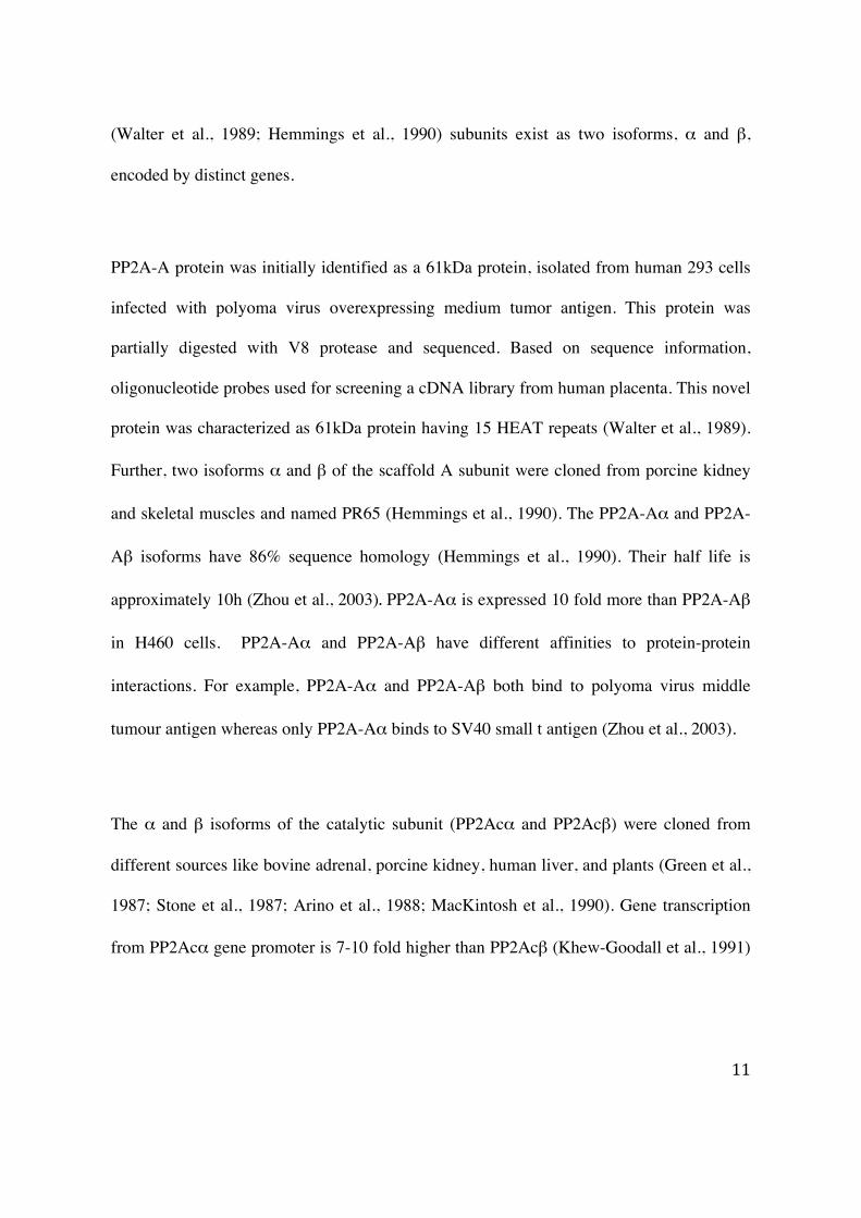
PP2A is a multimeric holoenzyme composed by a scaffold A (65 kDa), a regulatory B, and
a catalytic C (36 kDa) subunit (Janssens and Goris, 2001). Various isoforms of the
regulatory B subunit are required for recognition and recruitment to specific substrates. The
catalytic C (Green et al., 1987; Stone et al., 1987; Arino et al., 1988) and the scaffold A
11
(Walter et al., 1989; Hemmings et al., 1990) subunits exist as two isoforms, α and β,
encoded by distinct genes.
PP2A-A protein was initially identified as a 61kDa protein, isolated from human 293 cells
infected with polyoma virus overexpressing medium tumor antigen. This protein was
partially digested with V8 protease and sequenced. Based on sequence information,
oligonucleotide probes used for screening a cDNA library from human placenta. This novel
protein was characterized as 61kDa protein having 15 HEAT repeats (Walter et al., 1989).
Further, two isoforms α and β of the scaffold A subunit were cloned from porcine kidney
and skeletal muscles and named PR65 (Hemmings et al., 1990). The PP2A-Aα and PP2A-
Aβ isoforms have 86% sequence homology (Hemmings et al., 1990). Their half life is
approximately 10h (Zhou et al., 2003). PP2A-Aα is expressed 10 fold more than PP2A-Aβ
in H460 cells. PP2A-Aα and PP2A-Aβ have different affinities to protein-protein
interactions. For example, PP2A-Aα and PP2A-Aβ both bind to polyoma virus middle
tumour antigen whereas only PP2A-Aα binds to SV40 small t antigen (Zhou et al., 2003).
The α and β isoforms of the catalytic subunit (PP2Acα and PP2Acβ) were cloned from
different sources like bovine adrenal, porcine kidney, human liver, and plants (Green et al.,
1987; Stone et al., 1987; Arino et al., 1988; MacKintosh et al., 1990). Gene transcription
from PP2Acα gene promoter is 7-10 fold higher than PP2Acβ (Khew-Goodall et al., 1991)
12
and this may be the reason of almost 10 fold higher expression of PP2Acα than PP2Acβ at
protein level (Khew-Goodall and Hemmings, 1988).
Recently, a shorter spliced variant of the PP2Acα has been reported from fresh peripheral
blood mononuclear cells (PBMC) (Migueleti et al., 2011). This newly identified variant,
named PP2Acα2, lacks the 5th exon and is catalytically inactive (Migueleti et al., 2011).
The regulatory B subunit has at least 18 different isoforms divided into B, B’, B’’ and B’’’
family (Diagram 1) (Eichhorn et al., 2009).
Diagram 1: Hypothetical structure of PP2A holoenzyme complex (Janssens and Goris,
2001)
13
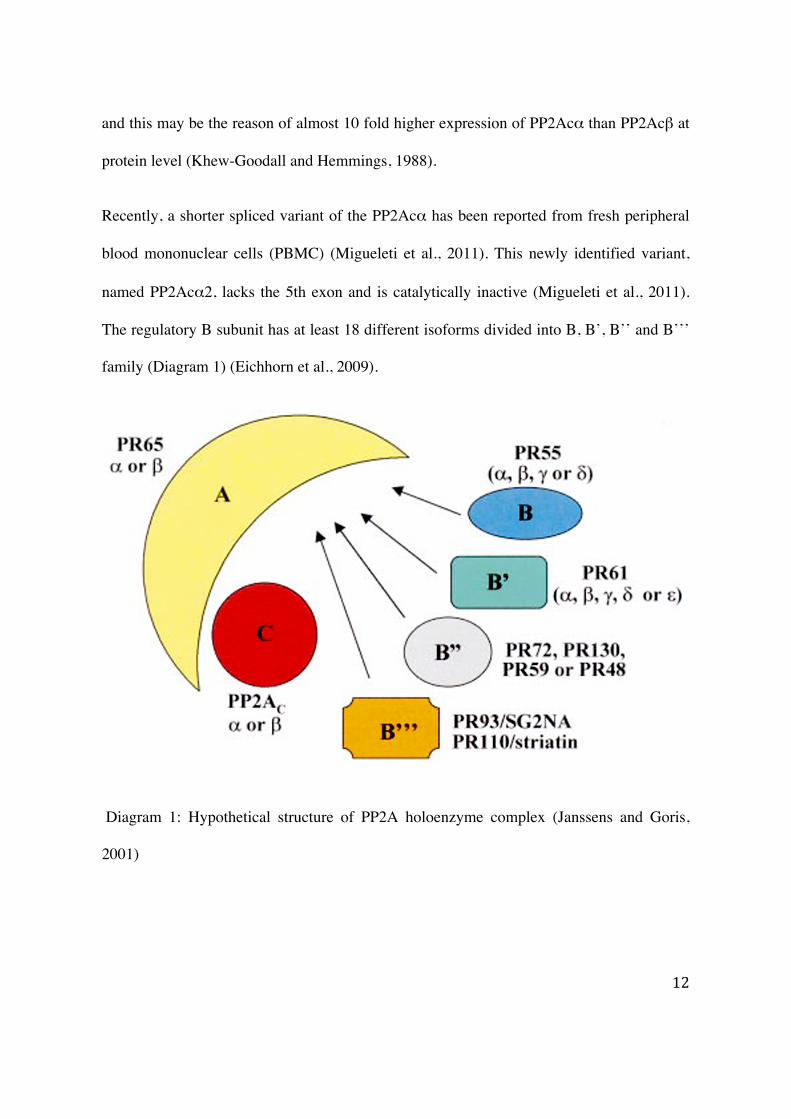
PP2A subunits coexist as dimeric (A-C) or trimeric (A-C-B) complex. The holoenzyme
complex is required for the phosphatase activity on targets (Janssens and Goris, 2001).
Although more than 150 targets are identified, the mechanisms by which B subunits
mediated PP2A association to the target are not fully understood (Eichhorn et al., 2009).
The association of PP2A holoenzyme has been described via the regulatory B subunits.
However, several reports have showed that PP2A C and A subunits can also directly
associate to proteins such as HIV1 NCp7:vpr, SV40 small t, Bcl2, or CaMKIV (Diagram 2)
(Janssens and Goris, 2001).
Diagram 2: PP2A interacts with substrates through A, B, or C subunits (Janssens and Goris,
2001).
14
Generally, PP2A exerts its phosphatase activity through the association with the regulatory
B subunits that determine the substrate specificity and the subcellular localization of the
enzyme. However, the catalytic activity of PP2A can also be controlled by regulatory
molecules such as α4 (see section regulation of catalytic activity of PP2A). The α4 (also
termed as IGBP1) is the mammalian orthologue of yeast Tap42 protein which controls the
TOR signaling (Onda et al., 1997).
PP2A regulates many cellular functions including metabolism, DNA replication,
transcription, RNA splicing, translation, cell cycle progression, cell senescence, apoptosis,
cell transformation, morphogenesis, development, and neurotransmission (Cohen, 1989;
Stemmer and Klee, 1991; Mumby and Walter, 1993; Hunter, 1995; Olsen et al., 2006;
Moorhead et al., 2007).
1.1.2 Regulation of PP2A-A, -B, and -C subunits expression
Several transcription factors like AP1, AP-2α, ETS-1, SP-1, CREB regulate PP2A
expression. The promoters of PP2Acα and PP2Acβ are GC rich and lack TATA and
CCAAT boxes. PP2Acα promoter has a cAMP-regulatory element (CRE) and several SP-1
binding sites (Khew-Goodall et al., 1991). Incubation of phosphorylated CREB with
immunopurified PP2Ac results in a dephosphorylation on serine 133 and consequently the
inhibition of the transcriptional activity of CREB (Wadzinski et al., 1993). Additionally,
treatment of rat liver extract with OA inhibits dephosphorylation of CREB at serine 133
15
(Wadzinski et al., 1993). Further, it is reported that PP2Ac associates with autoactivation
domain of calcium/calmodulin dependent kinase CaMKIVI and prevents phosphorylation
of CREB (Anderson et al., 2004). We have reported that in hepatocytes HCV protein
expression induces an ER stress response that leads to serine 133 phosphorylation on
CREB and PP2Ac up-regulation (Christen et al., 2007). Moreover, down-regulation of
CREB by siRNA did not up-regulate the PP2Ac expression by HCV proteins (Christen et
al., 2007). Considering these observations, it is proposed that the expression of PP2Ac
could form a feedback loop and inhibit the PP2A expression driven by CREB (Christen et
al., 2007). Interestingly, Baharians and Schönthal have shown that PP2Ac protein level is
maintained constant through an autoregulatory mechanism in mouse fibroblast cell line
NIH3T3 that maintains to constant PP2Ac protein expression irrespective to regulation of
transcription or RNA processing. Indeed, partial hepatectomy in mouse shows a constant
protein expression level despite a 30 fold increase in PP2Ac mRNA (Kakinoki et al., 1992).
Several reports have shown an increased PP2A mRNA expression during G1 phase in
mammalian cells (Nakamura et al., 1992; Kikuchi et al., 1997). Consequently to this tight
regulation of the protein expression level, it has been observed that PP2A level is constant
in various mammalian cells and fission yeast through-out the cell cycle (Virshup et al.,
1989; Kinoshita et al., 1990; Ruediger et al., 1991). These data indicates that the
accumulation of PP2Ac in the cell is not feasible because of the tight regulation and
therefore raise the difficulties of PP2Ac expression experimentally. The half-life of the
catalytic C subunit slightly varies from cell line to cell line. It has been reported that the
half-life is approximatively 16.5h in mouse fibroblast NIH3T3 cells (Baharians and
16
Schönthal, 1998).
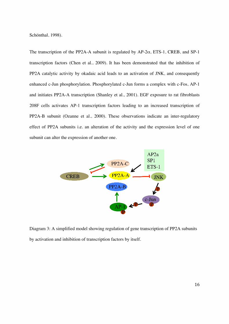
The transcription of the PP2A-A subunit is regulated by AP-2α, ETS-1, CREB, and SP-1
transcription factors (Chen et al., 2009). It has been demonstrated that the inhibition of
PP2A catalytic activity by okadaic acid leads to an activation of JNK, and consequently
enhanced c-Jun phosphorylation. Phosphorylated c-Jun forms a complex with c-Fos, AP-1
and initiates PP2A-A transcription (Shanley et al., 2001). EGF exposure to rat fibroblasts
208F cells activates AP-1 transcription factors leading to an increased transcription of
PP2A-B subunit (Ozanne et al., 2000). These observations indicate an inter-regulatory
effect of PP2A subunits i.e. an alteration of the activity and the expression level of one
subunit can alter the expression of another one.
Diagram 3: A simplified model showing regulation of gene transcription of PP2A subunits
by activation and inhibition of transcription factors by itself.
17
Another possibility of inter-regulation of PP2A subunits is through miRNA. Recent studies
showed that miRNA-19 have multiple target genes as Bim (Bcl2L11), AMP-activated
kinase (Prkaa1), phosphatases Pten and PP2A-B56ε (Mavrakis et al., 2010). Another report
showed that the overexpression of miRNA-1 down-regulates PP2A-B56α leading to an
enhanced Ca2+ release and an increased cardiac arrhythmogenesis (Terentyev et al., 2009).
miRNA-34b is known to suppress the expression of α4 in transformed human embryonic
kidney (HEKTER) and human lung cells. Chen et al., (Chen et al., 2011) have shown in
HEKTER and human lung cells that down-regulation of miRNA-34b leads to an up-
regulation of α4 and consequently to a reduction of PP2A activity (Chen et al., 2011).
1.1.3 Regulation of catalytic activity of PP2A
1.1.3.1 Regulation of PP2A activity by posttranslational modifications on C-terminal of the catalytic subunit
The six amino acid (304TPDYFL309) residues on the C-terminus of the C subunit are
conserved across species. The threonine 304, the tyrosine 307, and the leucine 309 residues
are known to undergo reversible posttranslational modifications. Phosphorylation at
tyrosine 307 inhibits phosphatase activity (Chen et al., 1992). Recently, Wu et al. (Wu and
Wilson, 2009) have reported that cotreatment of skeletal muscle microvascular endothelial
cells with LPS and IFN-γ leads to nitration on tyrosine 307. This nitration increases the
phosphatase activity by preventing tyrosine 307 from phosphorylation. In line with these
observations, several research groups have reported that exposure of cells with exogenous
18
peroxynitrite donor substances increases nitration of tyrosine 307 and subsequently
decreases phosphatase activity (Guner et al., 2009; Kohr et al., 2009). Similarly, casein
kinase 2α has been shown to inhibit PP2A activity by phosphorylating the catalytic subunit
at Y307 (Hériché et al., 1997). The catalytic activity of PP2A could be enhanced by
treatment of cells with urate, a specific peroxynitrite scavenger, (Kohr et al., 2009). The
phosphorylation on threonine 304 residues by auto-phosphorylation-activated protein
kinases has been reported to inactivate the phosphatase activity (Guo and Damuni, 1993;
Guo et al., 1993). An another posttranslational modification, the carboxymethylation on
leucine 309 by methyltransferase (Favre et al., 1994; Floer and Stock, 1994; Xie and
Clarke, 1994; Turowski et al., 1995) has been shown to enhance the interaction of the
catalytic subunit to the PP2A-B55α subunit and activates the phosphatase activity (Bryant
et al., 1999).
1.1.3.2 Regulation of activity by varying holoenzyme complex
PP2A activity also depends on the interaction of the C subunit with B subunits or proteins
other than PP2A components. For example, the interaction of the catalytic subunit with
different isoforms of PP2A-B regulatory subunits forms different holoenzymes and delivers
activity at different physiological level (Csortos et al., 1996; McCright et al., 1996). The
complex mechanism behind substrates selection is still poorly understood. However, the
selection and the association to the substrates are dependent on posttranslational
modifications, as well as on single or multiple mutations in the catalytic region of the
19
PP2Ac subunit. Indeed, the carboxymethylation at leucine 309 on the C subunit is required
for the association to the regulatory PP2A-B55α subunit (Bryant et al., 1999). Moreover
the single mutation at tyrosine 307 or leucine 309 of the C subunit forms mostly A-C
dimer whereas the double mutations at tyrosine 307 and leucine 309 favours association to
the α4 subunit instead of the A subunit (Chung et al., 1999). The regulation of PP2A
activity by α4 is ambiguous. Several reports suggest that α4 inhibits PP2A activity while
others showed an enhanced phosphatase activity. Indeed, Yoo et al. (Yoo et al., 2008) have
reported that α4 forms a complex with PP2Ac in fetal as well as adult liver and inhibits the
catalytic activity. In line with this observation other reports suggest an association of α4
with PP2Ac and therefore an inhibition of the catalytic activity in HEK293 cells
(Nanahoshi et al., 1998, 1999) and MEFs (Kong et al., 2009). In contrast, Nien et al., (Nien
et al., 2007) have shown that the over-expression of mTOR together with α4 protein
increases PP2A activity leading to increased STAT1α-PIAS1 binding. Other reports
confirm α4-mediated PP2A increased activity (Murata et al., 1997; Inui et al., 1998; Chung
et al., 1999). Additional studies, using purified endogenously expressed protein inhibitors
I1PP2A (Li et al., 1996a) and I2PP2A (Li et al., 1996b), have reported a direct association
of the catalytic subunit to these inhibitors and consequently an inactivation of PP2A. An
oncogenic transcription factor Hox11 has been shown to interact with PP2Ac and inhibits
the phosphatase activity (Kawabe et al., 1997).
Despite the constant protein expression level of PP2Ac, there are examples of altered
enzymatic activity of PP2A. Indeed, using purified proteins, Yang et al. (Yang et al., 1991)
20
have shown that simian virus 40 small t antigen (SV40ST) does not bind to either C subunit
or A-B-C complex but rather binds to the A subunit of the A-C complex in the absence of
B subunit and leads to decreased phosphatase activity. Therefore, the ectopic expression of
SV40ST in the CV-1 cells leads to an increased phosphorylation of MAPK through
inhibition of PP2A activity (Sontag et al., 1993).
1.1.4 PP2A activity modulation by viruses and toxins
PP2A subunits are the target of various molecules including drugs, viruses and toxins
(Millward et al., 1999). For instance, protein kinase R (PKR) phosphorylates PP2A-B56α
subunit at multiple serine and threonine residues. Phosphorylation of the PP2A-B56α
subunit enhances PP2A phosphatase activity (Xu and Williams, 2000). Various viral
particles associate to PP2A and modify its substrate specificity and activity. For instance,
the SV40 small t antigen can associate to the C subunit and displaces the B subunits from
A-C complex inhibiting the phosphatase activity (C. Kamibayashi, M. Mumby, Adv. Prot. Phosphatases, 9
(1995), pp. 195–210). Cayla et al. (Cayla et al., 2005) have shown that polyoma virus small t and
middle T antigens bind to PP2A catalytic subunit and impart the tyrosine phosphatase
activity in polyoma virus transformed cells. E4orf4, an adenovirus protein, associates to the
PP2A-B55α and downregulates PP2A expression by modulating the transcriptional activity
of AP-1 during late stages of viral infection (Kleinberger and Shenk, 1993). HIV proteins
NCp7 and Vpr directly associate to the PP2A-B'δ subunit and initiate PP2A catalytic
activity (Tung et al., 1997).
21
Various chemicals such as PP2A inhibitors are used in laboratories to study PP2A activity.
Okadaic acid (from marine sponge) is one of the most widely used among them. The
hydrophobic end of the okadaic acid binds to the hydrophobic cage within the binding
pocket of PP2Acα (Xing et al., 2006). This hydrophobic cage is constituted by 4 amino
acids (Q122, I123, H191, and W200). Other chemicals like calcyculin A (also from marine
sponge), tautomycin (an antibiotic from streptomyces spiroverticillatus), microcystin,
nodularins (hepatotoxins from cynobacteria) cantharidin (from blister beetles) (Millward et
al., 1999) are known as well to inhibit the activity of PP2A.
1.1.5 Model for PP2A over-expression
Various attempts have been made with only minor success to express PP2Ac.
Microinjection of purified PP2Ac in fibroblast F9 cells potentiated the activity of promoters
containing binding site for AP-1 leading to increased c-Jun expression and therefore an
increased expression of PP2Ac (Alberts et al., 1993). However, this approach was
successful but only in a small number of cells (Fernandez et al., 1990). In another approach,
transient transfection of HA tagged PP2Ac at the N-terminus in NIH3T3 cells resulted in
over-expression of PP2A and an increased phosphatase activity (Jaramillo-Babb et al.,
1996). Additionally, Ruediger and colleagues have generated an N-terminal PP2A-A
mutant that binds specifically to C subunit, therefore, allowing the accumulation of core
protein (A-C) in COS cells (Ruediger et al., 1997). Generation of a mouse model of PP2Ac
over-expression is shown to be a challenging and frustrating task. Indeed, PP2Acα gene
deleted mice die at embryonic day 6.5. Moreover, Götz et al. (Götz et al., 1998) found that
22
normal and degenerated embryos from PP2Acα gene deleted mice express comparable
level of total PP2Ac (PP2Acα and PP2Acβ) suggesting that the absence of PP2Acα cannot
be compensated by PP2Acβ. Very recently, conditional null alleles of PP2Acα and
PP2Acβ flanking loxP have been generated (Gu et al., 2012). PP2Acαfl/fl mice did not show
any visible phenotype while Cre-mediated PP2Acα deleted homozygous mice die in the
embryonic stage. PP2Acβ deleted homozygous mice did not show any physiological or
morphological change.
1.1.6 Clinical significance of PP2A
Although, the expression of PP2A is tightly regulated (Baharians and Schönthal, 1998), the
down-regulation of PP2Ac has been found during all-transretinoic acid induced
differentiation of HL-60 cells (Nishikawa et al., 1994), during peroxisome proliferator-
activated receptor-γ induced adipocyte differentiation (Altiok et al., 1997) in hippocampus
of patients with Alzheimer's disease (Vogelsberg-Ragaglia et al., 2001). Over-expression
of PP2Ac has been reported in response to colony-stimulating factor 1 in macrophages
(Wilson et al., 1999), in HCV harboring cells, mice and human individuals (Duong et al.,
2004).
23
1.2 Interferon signaling
1.2.1 The Interferon family
Isaacs and Lindenmann discovered interferon five decades ago during their studies on viral
interference (Isaacs A, Lindenmann J. Virus interference. I. The interferon. Proceedings of the Royal Society of London. Series B,
Containing papers of a Biological character 1957; 147 (927): 258–67). Interferons (IFNs) exercise their role in
modulating antiviral and antiproliferative responses. They are grouped into three types:
type I, type II and type III. Type I IFNs consists of IFNαs, IFNβ, IFNε, IFNκ, IFNω and
IFNν (Heim, 2012). Presently, 13 different IFNαs have been reported in humans. All
subtypes are encoded by distinct genes (Pestka et al., 2004). Interferon-γ is the sole member
of type II IFNs. A recently discovered type III IFN is composed by IFN-λ2, IFN-λ3 and
IFN-λ1, also known as IL28A, IL28B and IL29, respectively (Kotenko et al., 2002;
Sheppard et al., 2002).
Type I and type III IFNs are produced by human plasmacytoid dendritic cells (pDCs) and
monocyte-derived dendritic cells (MDDCs) through a common mechanism involving RIG-
I, IPS1 (also termed as MAVS, VISA and Cardif), TBK1, IRF3 and NF-κB signaling
molecules in response to viral infection (Siegal et al., 1999; Coccia et al., 2004; Österlund
et al., 2007; Onoguchi et al., 2007). In response to antigens or mitogens type II IFNs are
produced by T lymphocytes and natural killer (NK) cells (Dorman and Holland, 2000).
24
1.2.2 The Jak-STAT signaling pathway
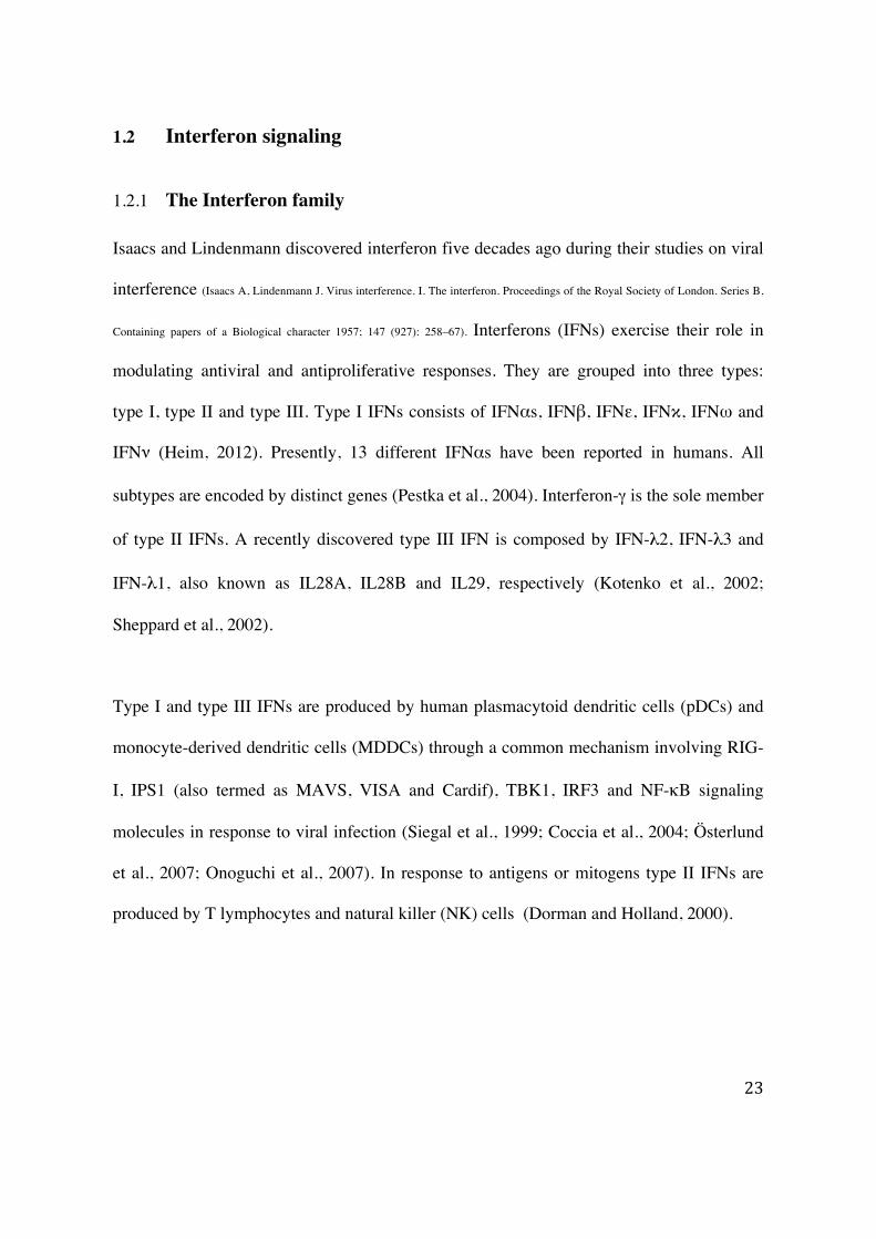
Type I IFNs exert their effect by binding to the interferon alpha/beta receptor (IFNΑR) that
is composed of two subunits - IFNΑR1 (also known as alpha) and IFNΑR2 (known as
beta). Type II IFNs (IFN-γ) binds to distinct receptor IFNGR consisting two subunits
IFNGR1 and IFNGR2 (Aguet et al., 1988; Hemmi et al., 1994; Soh et al., 1994). Type III
IFNs signal through the IFN-λ receptor comprising a unique IFN-λ chain and IL-10 receptor
common with IL-10R2 chain. (Kotenko et al., 2002; Donnelly et al., 2004) (Diagram 4).
Diagram 4: IFN signaling through the Jak-STAT pathway (Heim, 2012).
25
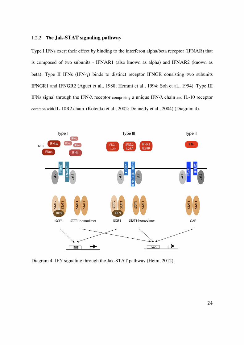
Upon association of IFNs to their cognate receptors, the Jak kinases are phosphorylated and
activated. Jaks (Janus Kinases) are cytoplasmic kinases. There are 4 known human Jaks,
namely – Jak1-3 and Tyk2. Their molecular weight ranges from 120-140 kDa. Jaks are
made up of 7-conserved JH (Jak Homology) domains characterized as JH1-JH7. JH1,
located at the C-terminal end, is the kinase domain. JH2 is pseudokinase domain,
responsible for recruiting substrates (Wang et al., 1995). The JH3-JH7 domains on N-
terminus recognize and bind to the receptor. JH3 and JH4 possess structural features of a
potential SH2 (src homology 2)-like domain (Bernards, 1991). A fraction of JH4 and JH5-
JH7 region termed as FERM domain, bind to the cytoplasmic tail of transmembrane
receptors (Hilkens et al., 2001) (Diagram 5).
Diagram 5: Structure of Jaks. JH - Jak Homology, SH2 – src homology 2, FERM - 4.1,
ezrin, radixin, moesin.
STATs (signal transducers and activator of transcription) act as signal carrier as well as
activator of transcription. In unstimulated cells, they predominantly localize in the
cytoplasm, whereas, upon activation, they translocate to the nucleus and activate
26
transcription. There are 7 mammalian STATs known so far – STAT1-4, STAT5a, STAT5b,
and STAT6. They are ubiquitously expressed except STAT4, which is mainly expressed in
thymus and testes (Darnell et al., 1994; Heim, 1999).
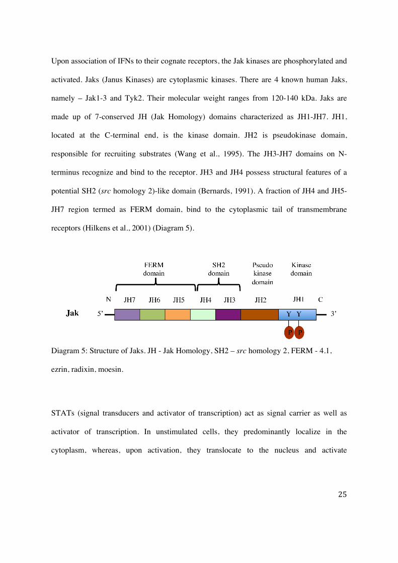
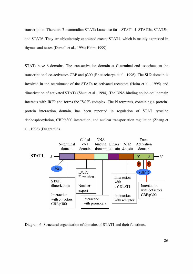
STATs have 6 domains. The transactivation domain at C-terminal end associates to the
transcriptional co-activators CBP and p300 (Bhattacharya et al., 1996). The SH2 domain is
involved in the recruitment of the STATs to activated receptors (Heim et al., 1995) and
dimerization of activated STATs (Shuai et al., 1994). The DNA binding coiled-coil domain
interacts with IRF9 and forms the ISGF3 complex. The N-terminus, containing a protein-
protein interaction domain, has been reported in regulation of STAT tyrosine
dephosphorylation, CBP/p300 interaction, and nuclear transportation regulation (Zhang et
al., 1996) (Diagram 6).
Diagram 6: Structural organization of domains of STAT1 and their functions.
27
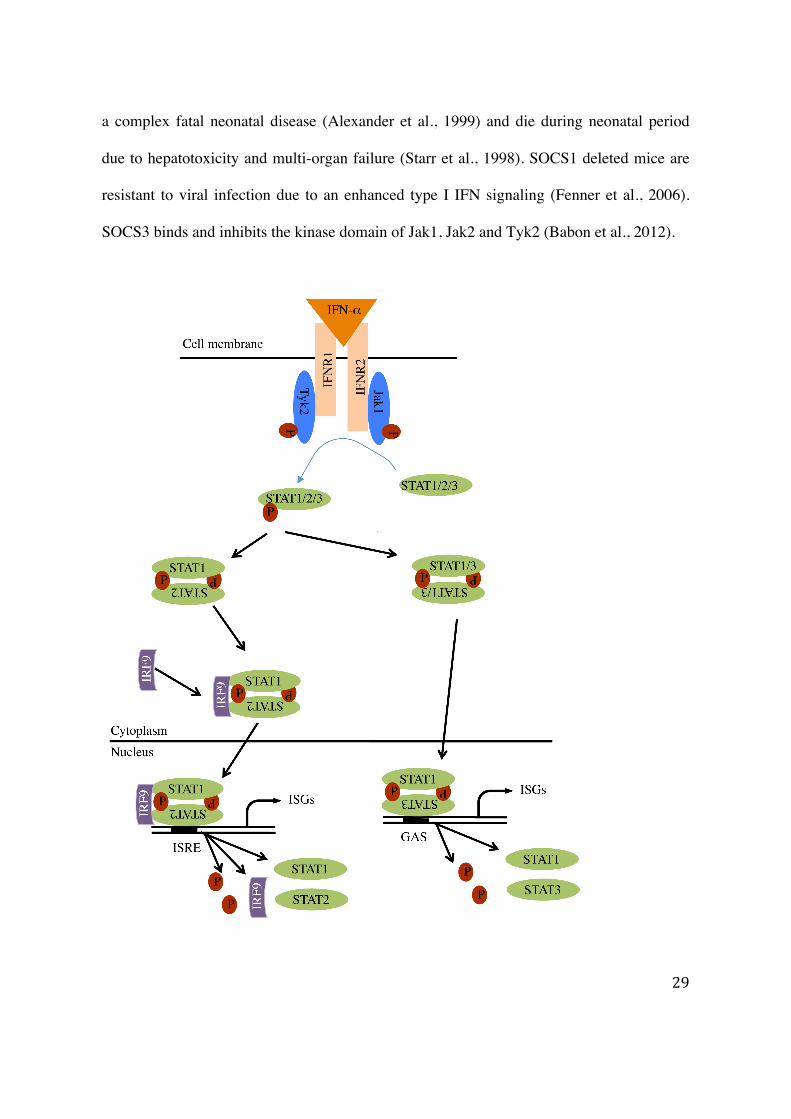
1.2.3 Activation of the Jak-STAT signaling by IFN-α
Cells exposed to IFN-α activate a cascade of events such as cross-linking of receptor chains
(IFNΑR1 and IFNΑR2), activation of kinases (Jak1 and Tyk2), and phosphorylation of
STAT1, STAT2 and STAT3 (Shuai et al., 1993; Heim et al., 1995). Activated STATs
dissociate from receptor-kinase-substrate complex, form homo- or/and heterodimers,
translocate to the nucleus, bind to promoter elements, and induce ISGs transcription
(Horvath, 2000) (Diagram 7). This ISGs transcription includes an array of genes up-
regulation including antiviral genes (PKR, OAS1, MxA, ISG15), signaling genes (STAT1,
STAT2, IRF9) and IFN-α itself (Lehtonen et al., 1997).
STAT1 was first defined based on its role in mediating gene activation in response to
interferon-γ (IFN-γ) (Darnell, 1997; Horvath and Darnell Jr, 1997). STAT1 contains a C-
terminal trans-activation domain, which possesses phosphorylation sites at tyrosine 701 and
serine 727. Tyrosine phosphorylation of STAT1 on C-terminus is important for
dimerization, DNA binding, and transcriptional activation whereas serine phosphorylation
leads to maximal transcriptional activity (Wen et al., 1995; Decker and Kovarik, 2000;
Kovarik et al., 2001). STAT1 activity is also regulated through methylation on arginine 31
residue by protein arginine methyl transferase 1 (PRMT1) (Mowen et al., 2001). Indeed,
methylation of arginine 31 leads to a decrease STAT1-PIAS1 association, thereby,
enhances STAT1 binding to promoters of target genes and consequently increased ISGs
transcription.
28
Recently, it has been reported that several ISGs can be induced by STAT1 in a tyrosine 701
independent manner (Chatterjee-Kishore et al., 2000; Ramana et al., 2001). Indeed, tyrosine
to a phenylalanine mutation of STAT1 does not alter the enhancement of the activity of Fas
and FasL promoters. In contrast, a STAT1 serine 727 to an alanine mutation is less efficient
than wild-type STAT1 in enhancing Fas and FasL promoters, as well as in inducing
apoptotic cell death in cardiac myocytes (Stephanou et al., 2001). Moreover,
phosphorylation on serine 727 has been shown to be critical for the C-terminal region of
STAT1 to act as a transcriptional co-activator by binding to molecules such as MCM5
(Zhang et al., 1998) and BRCA-1 (Ouchi et al., 2000).
1.2.4 Negative regulation of the Jak-STAT pathway
The constitutive activation of interferon signaling is harmful to the cell. Therefore, it is
essential to regulate the interferon signal transduction cascade. As reported before, STATs
are constitutively activated in many hematological malignancies (Gouilleux-Gruart et al.,
1996). The regulation of the Jak-STAT signaling is attributed to its various negative
regulators at different levels. For instance, the inhibition of IFNs signaling by the
Suppressors of Cytokine Signaling (SOCS) which constitutes the first level of inhibition at
the receptor-kinase-STATs complex, occurs either by inhibiting Jaks activation or by
preventing STATs binding to the receptor (Krebs and Hilton, 2001; Larsen and Röpke,
2002). The SOCS family consists of 8 members SOCS1 – SOCS7 and CIS. SOCS1 and
SOCS3 are induced by type I IFNs (Song and Shuai, 1998). SOCS1 deficient mice develop
29
a complex fatal neonatal disease (Alexander et al., 1999) and die during neonatal period
due to hepatotoxicity and multi-organ failure (Starr et al., 1998). SOCS1 deleted mice are
resistant to viral infection due to an enhanced type I IFN signaling (Fenner et al., 2006).
SOCS3 binds and inhibits the kinase domain of Jak1, Jak2 and Tyk2 (Babon et al., 2012).
30
Diagram 7: IFN-α induced Jak-STAT signaling pathway. IFN-α binding to receptors
causes their dimerization. Kinases (Jak1/Tyk2) are recruited to receptors and auto-
activated. Activated kinases phosphorylate STATs leading to their dimerization and ISGF3
formation by association with IRF9, translocation to nucleus and activation of gene
transcription.
Downstream to the SOCSs inhibition, the protein inhibitor of activated STAT (PIAS) binds
to hypomethylated STAT dimers and inhibits STAT-DNA interaction (Liu et al., 1998;
Shuai, 2000; Mowen et al., 2001). PIAS1 and PIAS3 bind to STAT1 and STAT3,
respectively. PIAS1 selectively inhibits interferon inducible gene and is essential for innate
immunity. PIAS1 deleted mice show an enhanced protection against viruses and microbes
(Liu et al., 2004).
Another important negative regulator of the Jak-STAT pathway is the ubiquitin specific
peptidase 18 (USP18/UBP43). Initially, USP18/UBP43 was considered as protease
cleaving ubiquitin-like modifier ISG15 (Liu et al., 1999). USP18/UBP43 was recently
found to exert a negative regulatory role independent to its ISG-deconjugating ability
(Malakhov et al., 2002). USP18/UBP43 inhibits the activation of Jak1 by disrupting Jak1
interaction with IFNΑR2 (Malakhova et al., 2006). USP18/UBP43 is induced by IFN-α
(Der et al., 1998; Malakhova et al., 2002) and provides a negative feedback loop that
restricts IFN-α signals. It has been reported that UBP43 deficient mice display a critical
phenotypic characteristic as brain cell injury, hypersensitivity to poly-(I:C) and premature
31
death (Ritchie et al., 2002; Malakhova et al., 2003). Interestingly, these animals are
resistant to fatal cerebral infections caused by VSV and LCMV (Ritchie et al., 2004). We
and others have shown that USP18/UBP43 is up-regulated in liver of CHC non-responders
to pegIFN-α therapy (Chen et al., 2005a; Sarasin-Filipowicz et al., 2008) and contributes to
the refractoriness to IFN-α stimulation in mouse liver (Sarasin-Filipowicz et al., 2009).
Furthermore, USP18/UBP43 knockdown in cells enhances the antiviral activity of
interferon against hepatitis C virus infection (Randall et al., 2006).
STATs are dephosphorylated by a 45kDa isoform nuclear T-cell protein tyrosine
phosphatase (TC-PTP), followed by nuclear export (ten Hoeve et al., 2002; Zhu et al.,
2002). It has been reported that SHP-2, a SH2 containing phosphatase, dephosphorylates at
both tyrosine and serine residues on STAT5 and STAT1 in the nucleus (Wu et al., 2002;
Chen et al., 2003). Recently, TC-PTP has been reported as a negative regulator of cytokine
signaling. TC-PTP deleted mice display immune defects and developed systemic
inflammatory diseases like sialadenitis, gastritis, chronic myocarditis and nephritis. These
animals have an elevated serum of IFN-γ causing death of animals within five weeks.
(Heinonen et al., 2004) (Diagram 8). Additionally, we have demonstrated that PP2A
inhibits the Jak-STAT pathway at the level of STAT-DNA interaction by altering the
methylation status of STAT1 (Duong et al., 2004).
32
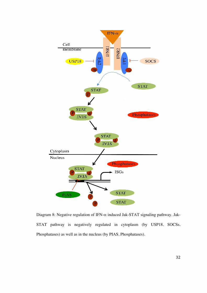
Diagram 8: Negative regulation of IFN-α induced Jak-STAT signaling pathway. Jak-
STAT pathway is negatively regulated in cytoplasm (by USP18, SOCSs,
Phosphatases) as well as in the nucleus (by PIAS, Phosphatases).
33
1.2.5 Refractoriness of the IFN signaling
It is well known that the IFNs induction becomes rapidly refractory. Indeed, maximal
induction of the IFN signaling pathways occurs within the initial 2 hours of IFN stimulation
and continuous exposure to IFNs leads to a “desensitization” and consequently to an
absence of response to further stimulation for the next 2-3 days (Larner et al., 1986).
Refractoriness has been also reported in the liver of mice injected with mouse IFN-α
(Sarasin-Filipowicz et al., 2009). In vivo, subcutaneous injection of IFN-α make
hepatocytes refractory within hours after the first injection and remain so for at least 2 days.
Studies on negative regulator of IFN-α have revealed SOCSs as early inhibitors of STAT
phosphorylation within 2-4 hours of IFN-α stimulation - whereas USP18/UBP43 is
involved in long lasting refractoriness. Interestingly, IFN-β induced signaling in mice liver
and IFN-γ induced signaling in intestine are not subjected to refractoriness (Makowska et
al., 2011).
1.2.6 IFN-α induced antiviral activity
The Jak-STAT signaling cascade plays a major antiviral role by inducing the ISGs such as
double-stranded RNA (dsRNA) activated protein kinase R (PKR), 2'-5' oligoadenylate
synthetase (OAS), and Mx proteins (Liu et al., 2004). In virally infected cells, viral dsRNA
34
triggers the phosphorylation of PKR resulting in the phosphorylation of the α subunit of
eukaryotic initiation factor (eIF2α). The phosphorylation of eIF2α leads to the inhibition of
translation of both viral and cellular proteins, providing the antiviral and antiproliferative
effect of IFNs (Sadler and Williams, 2008). OAS-dependent activated RNase L catalyses
the cleavage of viral and cellular single-stranded RNAs (ssRNAs) (Carroll et al., 1996).
RNase L plays an important role in positive feedback mechanisms and enhancement of the
innate immunity in the cells. Presence of small RNA in the virally infected cells induces the
antiviral innate immunity. Indeed, small self-RNA generated by RNase L amplifies the
antiviral innate immunity by producing antiviral cytokines like IFN-β (Malathi et al.,
2007). Marschall and colleagues have reported that the multiplication of negative-stranded
RNA viruses such as influenza C viruses was suppressed in cells expressing Mx proteins,
an IFN-α inducible GTPases (Marschall et al., 2000).
1.2.7 Clinical significance of alteration of the Jak-STAT signaling
Defect of Jak-STAT1 signaling is beneficial for HCV to escape from the host antiviral
activity and to establish persistent infection. HCV infection results in an inhibition of the
IFN-induced antiviral effector system through the phosphorylation of PKR and eIF2α, and
a decrease of IFN-α receptor expression in the liver through disruption of the IFN-α
signaling (Yatsuhashi et al., 1999). The expression of the entire HCV ORF in cell lines
inhibits the formation of ISGF3 and its subsequent DNA binding (Heim et al., 1999). Lack
of functional Tyk2 in melanoma cell reduces tyrosine phosphorylation of STAT1 and
STAT3 upon IFN-α induction (Pansky et al., 2000). STAT1 deficient mice do not show
35
any developmental defects but have compromised innate immunity to viral disease (Durbin
et al., 1996). Impaired Jak-STAT signaling due to mutation in IL-2 receptor and Jak3 has
been reported in human X-Linked Severe Combined Immunodeficiency (SCID) (Puck et
al., 1997) syndrome. Moreover, a V617F mutation has been reported in the coding
sequence of Jak2 in human myeloproliferative disorders, which leads a gain of function
causing expansion of the myeloproliferative disorder (Kralovics et al., 2005).
1.2.8 PP2A and the Jak-STAT signaling
We have reported previously that the expression of HCV proteins in osteosarcoma cell lines
or in transgenic mice liver leads to an up-regulation of PP2A catalytic subunit and inhibits
the IFN-α signaling at the STAT-DNA interactions level (Heim et al., 1999; Blindenbacher
et al., 2003). We have consistently observed an up-regulation of PP2Ac in human cell lines
that inducibly express HCV proteins, HCV transgenic mice, and human liver biopsy from
chronic hepatitis C (CHC) patients (Duong et al., 2004). The over-expression of PP2Acα in
Huh7 cells results in a reduction of PRMT1 activity, a hypomethylation of STAT1, an
increased STAT1-PIAS1 association, and consequently, an inhibition of STAT1-DNA
binding (Duong et al., 2006). The PP2A-induced inactivation of PRMT1 can be reversed in
vitro by treatment with a methyl-group donor S-adenosyl-L-methionine (SAMe) (Duong et
al., 2006). It is reported that treatment of cellular extracts from U973 cells stimulated with
IFN-γ with purified PP2Ac resulted in a decreased GAF-DNA binding (Eilers et al., 1995).
Further, Fielhabler and colleagues (Fielhaber et al., 2009) have shown that PP2A forms a
macromolecular complex with STAT1, α4, mTOR in HEK 293T and silencing of PP2Ac
36
increases the activated STAT1 content into the nucleus and up-regulates of Jak-STAT
signaling in response to IFN-γ. Additionally, it is shown that inhibition of PP2A in human
CD4+ T or T lymphoma or Jurkat T cells increases the serine phosphorylation on STAT6
and STAT3 (Woetmann et al., 1999, 2003).
1.3 The Hepatitis C virus
1.3.1 Structure of HCV
Hepatitis C virus is a member of Hepacivirus genus of the Flaviviridae family.
(Moradpour et al., 2007). HCV is small (55–65 nm in size) (Shimizu et al., 1996),
enveloped, positive-sense single-stranded RNA virus. HCV was identified as a causative
agent of a liver disease and CHC. It was previously defined as a non-A, non-B hepatitis
(Choo et al., 1989). CHC can lead to liver cirrhosis and hepatocellular carcinoma. Six
distinct genotypes (GT) of HCV with 65-70 % sequence homology have been reported so
far (Simmonds et al., 2005).
The HCV genome is approximately 9.6kb long and is composed of a 5’-non-coding region
(NCR), an open reading frame (ORF) for polyprotein and a 3’-NCR. The HCV ORF
encodes approximately 3000 amino acids (aa) (Suzuki et al., 1999) long precursor
polyprotein which is processed into at least ten proteins by viral and cellular proteases.
These proteins are grouped into structural (core, E1 and E2) and non-structural (p7, NS2,
NS3, NS4A, NS4B, NS5A and NS5B) proteins. The core protein forms a nucleocapsid
37
whereas E1 and E2 are embedded into lipid and are responsible for recognition of receptor
and entry of the virus into the host cell (Grakoui et al., 1993b). The p7 is an ion channel
protein. It has been shown using an expression plasmid encoded for NS2 and NS3 that the
major part of NS2 and the N-terminal part of NS3 constitutes a zinc-dependent
metalloproteinase and cleaves the NS2/NS3 junction. The cleavage of the remaining non-
structural polyprotein to generate individual protein is performed by NS3-4A protease.
(Grakoui et al., 1993a; Hijikata et al., 1993). NS3 contains a serine protease and a
NTPase/RNA helicase activity. The NS4A is a cofactor for NS3 proteases. It is shown that
the processing of the polyprotein occurs at ER membranous web which is formed by NS4B
(Egger et al., 2002; Gosert et al., 2003). The function of NS5A is not clear though it has
been proposed that NS5A (hyper)phosphorylation plays an important role in the regulation
of NS5A interaction to the host protein (hVAP-A) as well as to viral proteins (NS3, NS4B).
The hyperphosphorylation of NS5A disrupts the interaction with hVAP-A leading to a
decrease viral replication (Evans et al., 2004) In line with this observation, substitution of
serine by alanine residue within the central cluster 1 led to a reduction of phosphorylation
and an enhance RNA replication (Appel et al., 2005). Furthermore, NS5A interacts with
IFN-α inducible host protein PKR and inhibits its activity leading to a decrease of IFN-α
mediated antiviral activity (Gale et al., 1998). NS5B contains an RNA-dependent-RNA-
polymerase activity (RdRp) and is essential for HCV replication (Behrens et al., 1996; Ishii
et al., 1999).
38
The highly conserved NCRs are required for viral replication. The 5’-NCR contains an
internal ribosomal entry site (IRES) that is responsible for the initiation of a cap
independent translation of viral protein. Recently, it has been reported that the liver
specific miRNA-122 associates to the 5’-NCR of HCV RNA and positively modulates
HCV replication (Jopling et al., 2005). The 3’-NCR consists of three regions. A poorly
conserved 40 bases among various genotypes, a variable length poly(U)/poly-pyrimidine
tract, and an element of highly conserved 98 bases (Tanaka et al., 1995, 1996; Yamada et
al., 1996; Kolykhalov et al., 2000). Similarly to the 5’-NCR, the 3’-NCR is required for
HCV replication (Diagram 9).
Diagram 9: Structural organization of HCV genome (Moradpour et al., 2007).
39
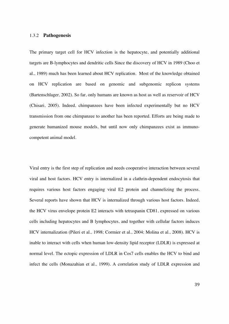
1.3.2 Pathogenesis
The primary target cell for HCV infection is the hepatocyte, and potentially additional
targets are B-lymphocytes and dendritic cells Since the discovery of HCV in 1989 (Choo et
al., 1989) much has been learned about HCV replication. Most of the knowledge obtained
on HCV replication are based on genomic and subgenomic replicon systems
(Bartenschlager, 2002). So far, only humans are known as host as well as reservoir of HCV
(Chisari, 2005). Indeed, chimpanzees have been infected experimentally but no HCV
transmission from one chimpanzee to another has been reported. Efforts are being made to
generate humanized mouse models, but until now only chimpanzees exist as immuno-
competent animal model.
Viral entry is the first step of replication and needs cooperative interaction between several
viral and host factors. HCV entry is internalized in a clathrin-dependent endocytosis that
requires various host factors engaging viral E2 protein and channelizing the process.
Several reports have shown that HCV is internalized through various host factors. Indeed,
the HCV virus envelope protein E2 interacts with tetraspanin CD81, expressed on various
cells including hepatocytes and B lymphocytes, and together with cellular factors induces
HCV internalization (Pileri et al., 1998; Cormier et al., 2004; Molina et al., 2008). HCV is
inable to interact with cells when human low-density lipid receptor (LDLR) is expressed at
normal level. The ectopic expression of LDLR in Cos7 cells enables the HCV to bind and
infect the cells (Monazahian et al., 1999). A correlation study of LDLR expression and
40
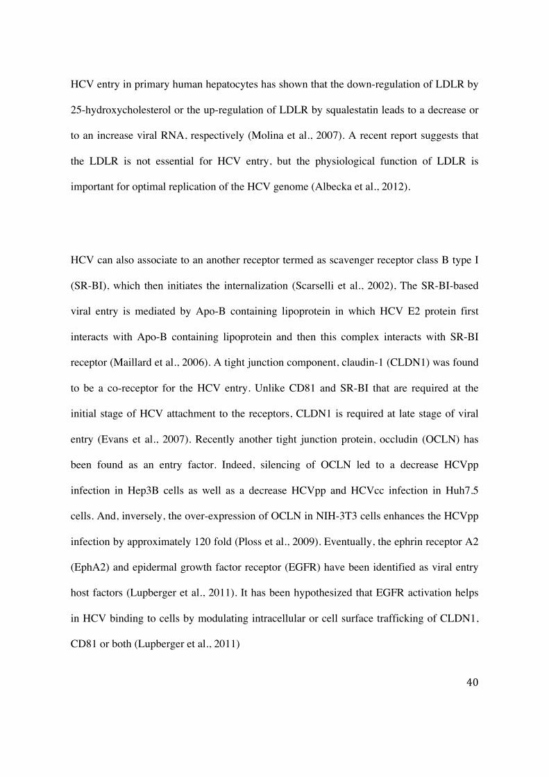
HCV entry in primary human hepatocytes has shown that the down-regulation of LDLR by
25-hydroxycholesterol or the up-regulation of LDLR by squalestatin leads to a decrease or
to an increase viral RNA, respectively (Molina et al., 2007). A recent report suggests that
the LDLR is not essential for HCV entry, but the physiological function of LDLR is
important for optimal replication of the HCV genome (Albecka et al., 2012).
HCV can also associate to an another receptor termed as scavenger receptor class B type I
(SR-BI), which then initiates the internalization (Scarselli et al., 2002), The SR-BI-based
viral entry is mediated by Apo-B containing lipoprotein in which HCV E2 protein first
interacts with Apo-B containing lipoprotein and then this complex interacts with SR-BI
receptor (Maillard et al., 2006). A tight junction component, claudin-1 (CLDN1) was found
to be a co-receptor for the HCV entry. Unlike CD81 and SR-BI that are required at the
initial stage of HCV attachment to the receptors, CLDN1 is required at late stage of viral
entry (Evans et al., 2007). Recently another tight junction protein, occludin (OCLN) has
been found as an entry factor. Indeed, silencing of OCLN led to a decrease HCVpp
infection in Hep3B cells as well as a decrease HCVpp and HCVcc infection in Huh7.5
cells. And, inversely, the over-expression of OCLN in NIH-3T3 cells enhances the HCVpp
infection by approximately 120 fold (Ploss et al., 2009). Eventually, the ephrin receptor A2
(EphA2) and epidermal growth factor receptor (EGFR) have been identified as viral entry
host factors (Lupberger et al., 2011). It has been hypothesized that EGFR activation helps
in HCV binding to cells by modulating intracellular or cell surface trafficking of CLDN1,
CD81 or both (Lupberger et al., 2011)
41
2. Aims of the PhD thesis project
The hepatitis C virus is a major pathogen that causes liver cirrhosis and hepatocellular
carcinoma. Current standard IFN-α based therapies are costly and elicit severe side effects.
Moreover, existing IFN-α based therapies are not effective in all HCV infected individuals.
We have previously reported that HCV infected patients who had a constitutive up-
regulation of ISGs do not respond to IFN-α based therapies (Sarasin-Filipowicz et al.,
2008). We have also reported that HCV infection induces PP2Acα over-expression in
human individuals (Duong et al., 2004) which encourages us to study further.
The aims of this PhD thesis are:
2.1 Analysis of the molecular mechanism used by PP2Ac to inhibit IFN-α induced antiviral activity.
2.2 Assessment of the role of PP2Ac over-expression on HCV replication
2.3 Identification of B subunits that modulate the dual effect of PP2Ac
42
3. Materials and methods
Materials and methods for the aims (section 2.1 and 2.2) - ‘‘Analysis of the molecular
mechanism used by PP2Ac to inhibit IFN-α induced antiviral activity’’ and ‘‘Assessment of
the role of PP2Ac over-expression on HCV replication’’ is incorporated with the
manuscript.
Materials and methods for the aim (section 2.3) ‘‘Identification of B subunits that modulate
the dual effect of PP2Ac’’ is described in further sections 3.1 and 3.2.
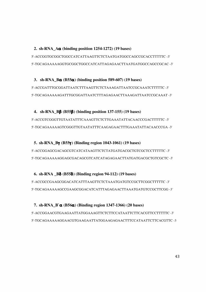
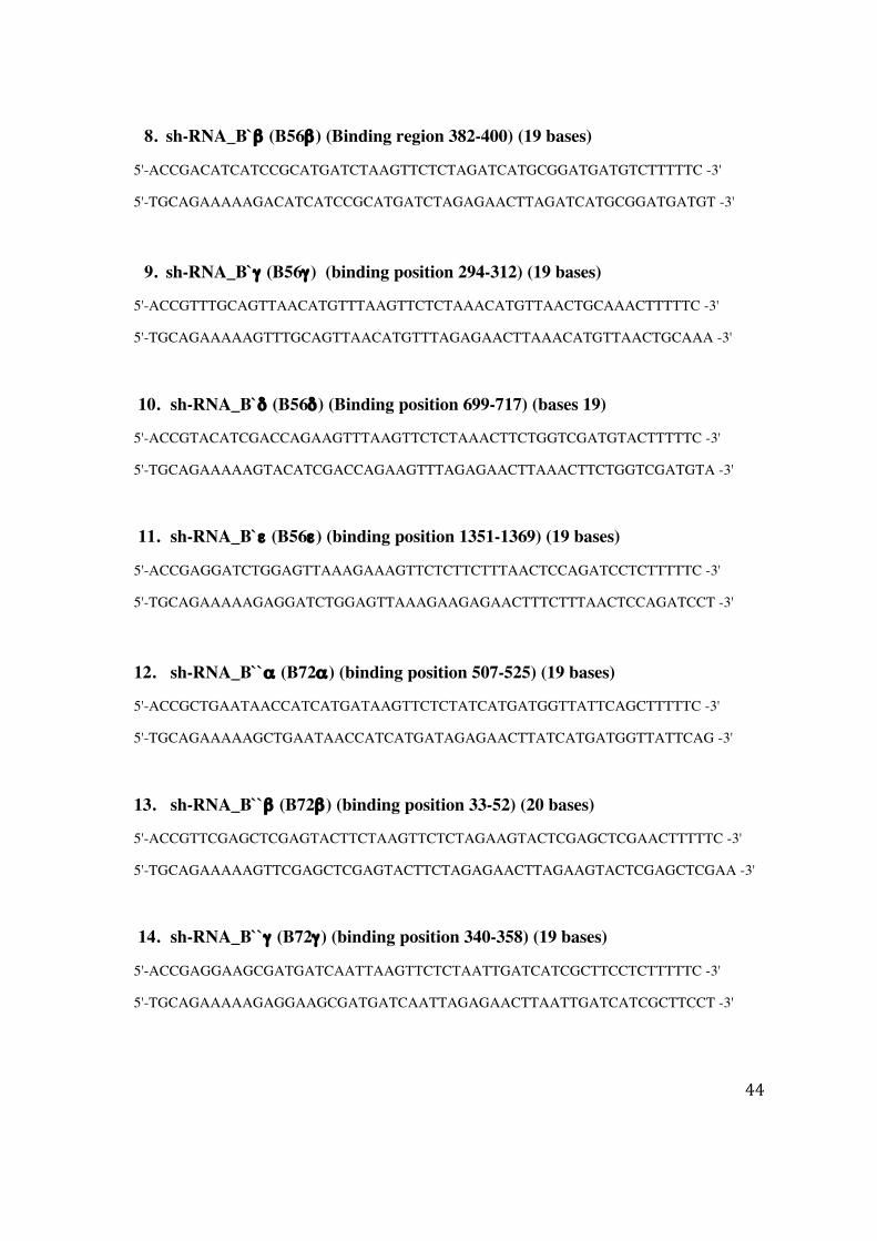
3.1 Silencing of PP2A-A and -B subunits
The sh-RNA sequence for various subunits were designed using online software
from Promega. The short-hairpin oligos targeting PP2A-A, -B subunits and
scrambled oligos were cloned into psiSTRIKE-Puromycin vector (Promega) that
allows contitutive expression of inserts. Plasmids encoding sh-RNA against various
B and A subunits were transfected into Huh7 cells and clones were selected into
puromycin containing medium. Details of sh-RNA oligos for silencing of PP2A-A
and -B subunits are listed below.
Sh-RNA oligos
1. sc-RNA_Aα
5'-ACCGCGTGTGCGGCGCTACTTAAAGTTCTCTTAAGTAGCGCCGCACACGCTTTTTC -3'
5'-TGCAGAAAAAGCGTGTGCGGCGCTACTTAAGAGAACTTTAAGTAGCGCCGCACACG -3'
43
2. sh-RNA_Aα (binding position 1254-1272) (19 bases)
5'-ACCGGTGCGGCTGGCCATCATTAAGTTCTCTAATGATGGCCAGCCGCACCTTTTTC -3'
5'-TGCAGAAAAAGGTGCGGCTGGCCATCATTAGAGAACTTAATGATGGCCAGCCGCAC -3'
3. sh-RNA_Bα (B55α) (binding position 589-607) (19 bases)
5'-ACCGATTTGCGGATTAATCTTTAAGTTCTCTAAAGATTAATCCGCAAATCTTTTTC -3'
5'-TGCAGAAAAAGATTTGCGGATTAATCTTTAGAGAACTTAAAGATTAATCCGCAAAT -3'
4. sh-RNA_Bβ (B55β) (binding position 137-155) (19 bases)
5'-ACCGTCGGGTTGTAATATTTCAAAGTTCTCTTGAAATATTACAACCCGACTTTTTC -3'
5'-TGCAGAAAAAGTCGGGTTGTAATATTTCAAGAGAACTTTGAAATATTACAACCCGA -3'
5. sh-RNA_Bγ (B55γ) (Binding region 1043-1061) (19 bases)
5'-ACCGGAGCGACAGCGTCATCATAAGTTCTCTATGATGACGCTGTCGCTCCTTTTTC -3'
5'-TGCAGAAAAAGGAGCGACAGCGTCATCATAGAGAACTTATGATGACGCTGTCGCTC -3'
6. sh-RNA _Bδ (B55δ) (Binding region 94-112) (19 bases)
5'-ACCGCCGAAGCGGACATCATTTAAGTTCTCTAAATGATGTCCGCTTCGGCTTTTTC -3'
5'-TGCAGAAAAAGCCGAAGCGGACATCATTTAGAGAACTTAAATGATGTCCGCTTCGG -3’
7. sh-RNA_B`α (B56α) (Binding region 1347-1366) (20 bases)
5'-ACCGGAACGTGAAGAATTATGGAAAGTTCTCTTCCATAATTCTTCACGTTCCTTTTTC -3'
5'-TGCAGAAAAAGGAACGTGAAGAATTATGGAAGAGAACTTTCCATAATTCTTCACGTTC -3
44
8. sh-RNA_B`β (B56β) (Binding region 382-400) (19 bases)
5'-ACCGACATCATCCGCATGATCTAAGTTCTCTAGATCATGCGGATGATGTCTTTTTC -3'
5'-TGCAGAAAAAGACATCATCCGCATGATCTAGAGAACTTAGATCATGCGGATGATGT -3'
9. sh-RNA_B`γ (B56γ) (binding position 294-312) (19 bases)
5'-ACCGTTTGCAGTTAACATGTTTAAGTTCTCTAAACATGTTAACTGCAAACTTTTTC -3'
5'-TGCAGAAAAAGTTTGCAGTTAACATGTTTAGAGAACTTAAACATGTTAACTGCAAA -3'
10. sh-RNA_B`δ (B56δ) (Binding position 699-717) (bases 19)
5'-ACCGTACATCGACCAGAAGTTTAAGTTCTCTAAACTTCTGGTCGATGTACTTTTTC -3'
5'-TGCAGAAAAAGTACATCGACCAGAAGTTTAGAGAACTTAAACTTCTGGTCGATGTA -3'
11. sh-RNA_B`ε (B56ε) (binding position 1351-1369) (19 bases)
5'-ACCGAGGATCTGGAGTTAAAGAAAGTTCTCTTCTTTAACTCCAGATCCTCTTTTTC -3'
5'-TGCAGAAAAAGAGGATCTGGAGTTAAAGAAGAGAACTTTCTTTAACTCCAGATCCT -3'
12. sh-RNA_B``α (B72α) (binding position 507-525) (19 bases)
5'-ACCGCTGAATAACCATCATGATAAGTTCTCTATCATGATGGTTATTCAGCTTTTTC -3'
5'-TGCAGAAAAAGCTGAATAACCATCATGATAGAGAACTTATCATGATGGTTATTCAG -3'
13. sh-RNA_B``β (B72β) (binding position 33-52) (20 bases)
5'-ACCGTTCGAGCTCGAGTACTTCTAAGTTCTCTAGAAGTACTCGAGCTCGAACTTTTTC -3'
5'-TGCAGAAAAAGTTCGAGCTCGAGTACTTCTAGAGAACTTAGAAGTACTCGAGCTCGAA -3'
14. sh-RNA_B``γ (B72γ) (binding position 340-358) (19 bases)
5'-ACCGAGGAAGCGATGATCAATTAAGTTCTCTAATTGATCATCGCTTCCTCTTTTTC -3'
5'-TGCAGAAAAAGAGGAAGCGATGATCAATTAGAGAACTTAATTGATCATCGCTTCCT -3'
45
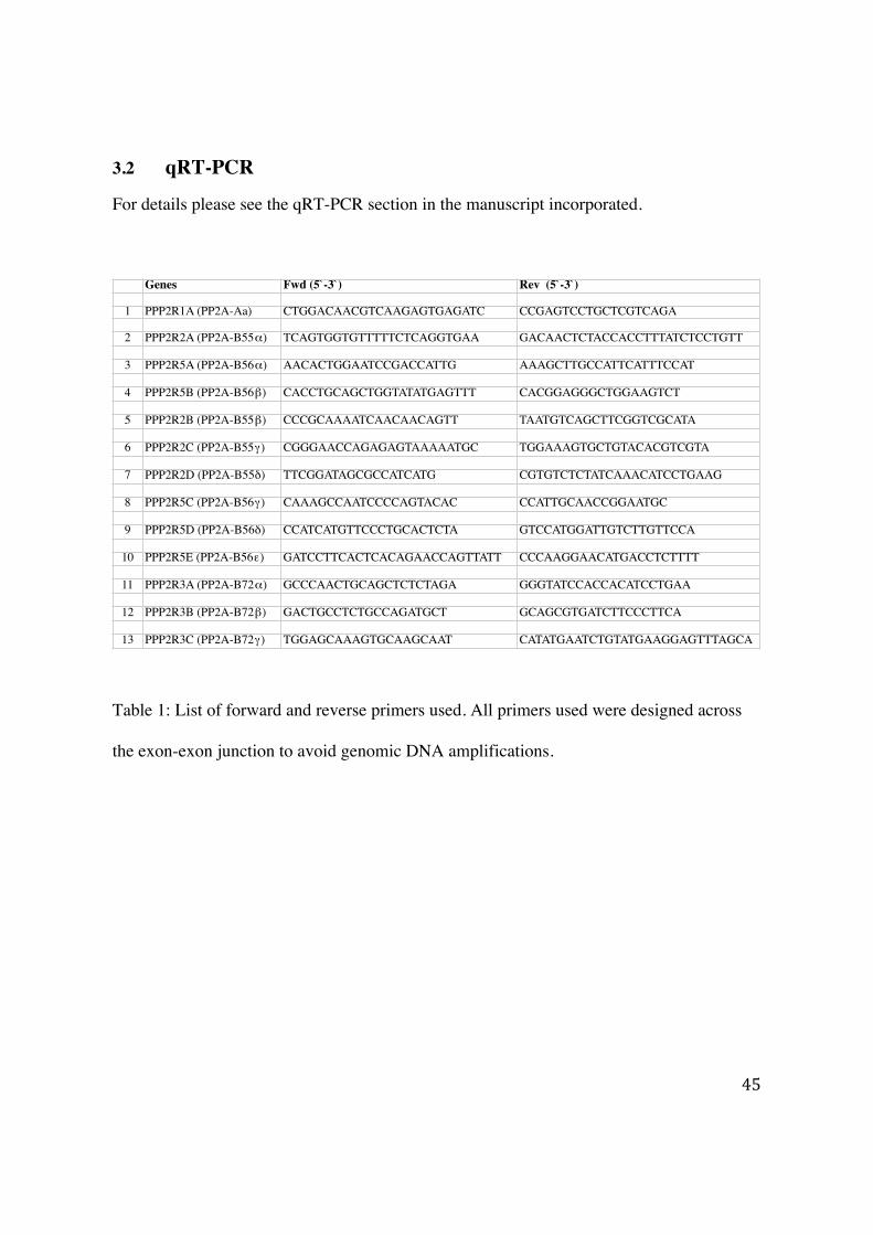
3.2 qRT-PCR
For details please see the qRT-PCR section in the manuscript incorporated.
Table 1: List of forward and reverse primers used. All primers used were designed across
the exon-exon junction to avoid genomic DNA amplifications.
Genes Fwd (5`-3`) Rev (5`-3`)
1 PPP2R1A (PP2A-Aa) CTGGACAACGTCAAGAGTGAGATC CCGAGTCCTGCTCGTCAGA
2 PPP2R2A (PP2A-B55!) TCAGTGGTGTTTTTCTCAGGTGAA GACAACTCTACCACCTTTATCTCCTGTT
3 PPP2R5A (PP2A-B56!) AACACTGGAATCCGACCATTG AAAGCTTGCCATTCATTTCCAT
4 PPP2R5B (PP2A-B56") CACCTGCAGCTGGTATATGAGTTT CACGGAGGGCTGGAAGTCT
5 PPP2R2B (PP2A-B55") CCCGCAAAATCAACAACAGTT TAATGTCAGCTTCGGTCGCATA
6 PPP2R2C (PP2A-B55#) CGGGAACCAGAGAGTAAAAATGC TGGAAAGTGCTGTACACGTCGTA
7 PPP2R2D (PP2A-B55$) TTCGGATAGCGCCATCATG CGTGTCTCTATCAAACATCCTGAAG
8 PPP2R5C (PP2A-B56#) CAAAGCCAATCCCCAGTACAC CCATTGCAACCGGAATGC
9 PPP2R5D (PP2A-B56$) CCATCATGTTCCCTGCACTCTA GTCCATGGATTGTCTTGTTCCA
10 PPP2R5E (PP2A-B56%) GATCCTTCACTCACAGAACCAGTTATT CCCAAGGAACATGACCTCTTTT
11 PPP2R3A (PP2A-B72!) GCCCAACTGCAGCTCTCTAGA GGGTATCCACCACATCCTGAA
12 PPP2R3B (PP2A-B72") GACTGCCTCTGCCAGATGCT GCAGCGTGATCTTCCCTTCA
13 PPP2R3C (PP2A-B72#) TGGAGCAAAGTGCAAGCAAT CATATGAATCTGTATGAAGGAGTTTAGCA
46
4. Results
4.1 Protein Phosphatase 2A impairs IFNα-induced antiviral activity against the hepatitis C virus through the inhibition of STAT1 tyrosine phosphorylation
V. Shanker,1 G. Trincucci,1 H. M. Heim1,2 and H. T. F. Duong1 1Department of Biomedicine, University and University Hospital Basel, Basel, Switzerland; and 2Division of Gastroenterology and Hepatology, University of Basel, Basel, Switzerland
47
Protein phosphatase 2A impairs IFNa-induced antiviralactivity against the hepatitis C virus through the inhibitionof STAT1 tyrosine phosphorylationV. Shanker,1 G. Trincucci,1 H. M. Heim1,2 and H. T. F. Duong1 1Department of Biomedicine, University and
University Hospital Basel, Basel, Switzerland; and 2Division of Gastroenterology and Hepatology, University of Basel, Basel, Switzerland
Received November 2012; accepted for publication January 2013
SUMMARY. Mammalian cells have developed several mech-
anisms to sense viruses and initiate adequate responses
such as production of interferons. Interferons activate the
antiviral response through the Jak-STAT signalling path-
way. To establish a chronic infection, viruses need to coun-
teract this barrier of defence. The hepatitis C and hepatitis
B viruses are known to up-regulate the expression of pro-
tein phosphatase 2A (PP2A). In this study, we show that
PP2Ac associates with Jak1/Tyk2/STAT1 and reduces
Jak1/Tyk2/STAT1 phosphorylation resulting in an impair-
ment of the IFNa-induced HCV antiviral response. Using
the fully infectious HCV cell culture system (HCVcc), we
demonstrate that the PP2A catalytic activity is not
required to block the antiviral effect of IFNa, although it is
needed to support HCVcc replication. Our data suggest an
important contribution of virus-induced PP2Ac up-regula-
tion in the establishment of a chronic infection.
Keywords: antiviral activity, HCV replication, interferon a,protein phosphatase 2A, STAT1.
INTRODUCTION
The innate immune system is the first response of the
host against invasion by pathogens. Cells of the immune
system have developed detection methods to sense viruses
and activate the production of interferons (IFNs) [1,2].
IFNs associate with their cognate receptors and initiate
the Jak-STAT signalling pathway. The binding of IFNs to
the receptor leads to receptor dimerization and activation
of the Janus kinases (Jaks) [3]. Signal transducer and
activator of transcription (STATs) are then recruited and
phosphorylated by the Jaks. Activated STATs regulate
transcription by association with the promoters of IFN-
stimulated genes (ISGs) [4,5]. IFNa activates important
antiviral effector systems such as double-stranded RNA
(dsRNA), activated protein kinase R (PKR), 2!-5! oligoade-nylate synthetase (OAS) and Mx proteins [6–8]. The regu-
lation of the Jak-STAT pathway occurs at various levels
and is crucial to switch off the interferon signal. Several
inhibitors are involved such as suppressor of cytokine sig-
nalling (SOCS) [9], protein inhibitor of activated STAT
(PIAS) [10], T-cell protein tyrosine phosphatases (TC-PTP-
ases) [11], and ubiquitin-specific peptidase 18 (USP18)
[12].
The major serine/threonine phosphatase, protein phos-
phatase 2A (PP2A) is ubiquitously expressed in all cell
types. PP2A is a trimeric enzyme composed of a scaffolding
subunit A, a regulatory subunit B and a catalytic C sub-
unit [13]. The C subunit associates with the A subunit to
form the catalytic core [14]. Heretofore attempts to over-
express the fully active PP2A catalytic subunit (PP2Ac)
failed because of the autoregulatory mechanism that main-
tains PP2A expression level constant [15]. However, addi-
tion of a peptide sequence derived from the influenza
haemagglutinin protein (HA) at the N-terminal end of the
PP2Ac sequence has been reported to increase the phos-
phatase activity [16] and resulting in an impairment of the
enzymatic activity of the protein arginine methyl transfer-
ase 1 (PRMT1) [17,18]. This autoregulatory mechanism is
disrupted in chronic hepatitis C (CHC) and chronic hepati-
tis B (CHB) viral infection leading to an over-expression of
the catalytic subunit [19,20]. The regulatory B subunit
recognizes a similar sequence of the A subunit and deter-
mines the subcellular localization of the heterotrimeric
enzyme as well as the substrate specificity [21]. The activ-
ity of PP2A is regulated by post-translational modifications
on the C subunit. Indeed, the phosphorylation on tyrosine
307 inhibits PP2A activity [22], and the carboxymethyla-
tion on leucine 309 controls the interaction of the catalytic
Abbreviations: CHC, chronic hepatitis C; ISGs, IFN-stimulated
genes; OAS, oligoadenylate synthetase; PKR, protein kinase R.
Correspondence: Dr Francois Duong, Department of Biomedicine,
University Hospital Basel, Hebelstrasse 20, 4031 Basel,
Switzerland.
E-mail: [email protected]
© 2013 John Wiley & Sons Ltd
Journal of Viral Hepatitis, 2013, 20, 612–621 doi:10.1111/jvh.12083
48
subunit with the Ba subunit [23]. Previously, we have
reported that PP2Ac over-expression impairs the IFNasignalling at the STAT1-DNA-binding level by a direct
association of PP2Ac with PRMT1 leading to the inhibition
of PRMT1-induced STAT1 methylation [18].
We show here that PP2Ac associates with Jak1/Tyk2/
STAT1 upon IFNa stimulation and blocks Jaks/STAT1
phosphorylation. Interestingly, this inhibitory effect is inde-
pendent of the catalytic activity and is abolished by high
concentrations of IFNa. Using a functional assay based on
the HCV fully infectious system, we demonstrate that the
over-expression of a PP2Ac mutant lacking the catalytic
activity is sufficient to impair the antiviral activity of IFNa.Our data reveal a dual role for PP2Ac; first to support HCV
replication and second to impair the IFNa-mediated
antiviral activity favouring the establishment of a chronic
infection.
EXPERIMENTAL PROCEDURES
Reagents, antibodies and plasmids
Human IFNa and the protease inhibitor cocktail were
obtained from Roche (Hoffmann La Roche, Basel, Switzer-
land). Mouse IFNa was a generous gift from Prof. Radek
Skoda. Lipofectamine 2000 was from Invitrogen (LuBio-
Science GmbH, Buchs, Switzerland). Protein A-Sepharose
4B beads, okadaic acid and Actin antibody were from
Sigma-Aldrich (Fluka Chemie GmbH, Lucerne, Switzer-
land). PP2Ac and pY1022/23-Jak1 antibodies were from
Millipore (Millipore AG, Zug, Switzerland). HCV NS3A and
Tyk2 antibodies were from Abcam (Cambridge, UK). HCV
core antibody was from Thermo Scientific (Perbio Science,
Lausanne, Switzerland). STAT1 antibody was from Santa-
Cruz Biotechnologie (LabForce AG, Nunningen, Switzer-
land). pS473-Akt, Akt, pS727-STAT1, pY1054/55-Tyk2,
pY701-STAT1 and PRMT1 antibodies were from Cell Sig-
nalling (BioConcept, Allschwil, Switzerland). H4 and
MetR3H4 antibodies were purchased from Abcam. HRP-
conjugated secondary antibody (NA931V) for HCV detec-
tion was from GE Healthcare (GE Healthcare Europe
GmbH, Glattbrugg, Switzerland).
Wild type (pDE3-HA-Ca-WT) and mutated PP2Aca plas-
mids (pDE3-HA-Ca-D88N and pDE3-HA-Ca-H118N) taggedwith single HA coding sequence immediately downstream
to the human PP2Aca initiation codon were a gift from
Dr. Brian Hemmings. pSIH1-puro-shPP2Ac and pSIH1-
puro-shPRMT1 were generated by digestion of the pSIH1-
puro-STAT3 shRNA plasmid (Addgene, no. 26596) and
insertion of short-hairpin RNA sequence against PP2Acaor PRMT1, respectively. The control plasmid was pur-
chased from Addgene (no. 26597). Lentiviral packaging
plasmids were a gift from Prof. Martin Stern. Plasmid Luc-
JC1 containing codons 1-846 of J6/CF and 847-3033 of
JFH1 was kindly provided by Prof. Ralf Bartenschlager.
Mice
Eight weeks old male C57BL/6 mice were used for the
experiment. The animals were bred and maintained in the
University Hospital Basel animal facility. They were injected
(n = 2) subcutaneously with 1000 IU/mL mIFNa or PBS
for 1 h and the liver was collected. All animal experiments
were conducted with the approval of the animal care com-
mittee of the canton Basel-Stadt (Switzerland).
Cell lines
Huh7, Huh7.5.1, UPP2A-C8 and stable silencing PP2Acacell lines are described elsewhere [17].
SDS-PAGE, immunoblotting and immunoprecipitation
Cells were lysed in whole-cell lysis buffer containing 50 mM
Tris HCl, pH 7.5, 100 mM NaCl, 1 mM EDTA, 0.1% Triton
X-100, 10 mM PMSF and 1 mM orthovanadate. Lysates were
cleared by centrifugation and quantified by Bradford (Bio-
Rad Protein Assay, Bio-Rad Laboratories AG, Reinach, Swit-
zerland). Proteins were resolved by SDS-PAGE, transferred
onto nitrocellulose membrane and probed with specific anti-
bodies. Immunoprecipitation was performed as described pre-
viously [18]. Densitometry analysis of protein bands was
performed using the public domain NIH Image program
(developed at the U.S. National Institutes of Health and avail-
able on the Internet at http://rsb.info.nih.gov/nih-image/).
Electrophoretic mobility shift assay
Nuclear extracts and mobility shift assay (EMSA) were per-
formed as described previously [18].
Lentiviral particle production and transduction
pSIH1-puro-control shRNA, pSIH1-puro-shPP2Ac and
pSIH1-puro-shPRMT1 were cotransfected with packaging
vectors into HEK293T cells using Lipofectamine 2000
according to the manufacturer’s instructions. Supernatants
containing lentiviral particles were cleared by centrifuga-
tion and stored at !70 °C.Huh7.5.1 cells were transduced with 1MOI of lentiviral
particles for 3 days, and PP2Ac or PRMT1 expression was
analysed by immunoblotting and quantitative PCR.
PP2Ac primers were 5!-CCACAGCAAGTCACACATTGG-3! and 5!-CAGAGCACTTGATCGCCTACAA-3!. PRMT1
primers were 5!-GACATCCAAAGATTACTACTTTGACTCCTA-3! and 5!-GCGCACCTCGTCCTTCAG-3!.
Hepatitis C virus particle production in cell culture(HCVcc) and infection of Huh7.5.1 cells
RNA preparation and electroporation were performed as
described [24]. Huh7.5.1 cells were infected with 1MOI of
© 2013 John Wiley & Sons Ltd
PP2A blocks IFNa-induced antiviral response 613
49
HCVcc strain JC1 particles for 3 days, and replication was
analysed by immunoblotting, qPCR and immunostaining
[24]. HCV primers were 5!-AGGAGGCCCGCACTGCCATA-3!and 5!-CTGGCGCGGCAACGTCTGTA-3!.
Analysis of interferon-stimulated gene expression
Total RNA extraction, cDNA synthesis and SYBR-based
quantitative PCR were performed as described elsewhere
[17]. Primers were designed across exon–exon sequences
to avoid genomic DNA amplification. STAT1 primers were
5!-TCCCCAGGCCCTTGTTG-3! and 5!-CAAGCTGCTGAAGTTCGTACC-3!. IP10 primers were 5!-CGATTCTGATTTGCTGCCTTATC-3! and 5!-GCAGGTACAGCGTACGGTTCT-3!.GBP1 primers were 5!- TGAACAAGCTGGCTGGAAAGA-3!and 5!-ACATCCAGATTCCTTTAGTGTGAGACT-3!. SOCS1
primers were 5!- CCCCTTCTGTAGGATGGTAGCA-3! and
5!-TGCTGTGGAGACTGCATTGTC-3!. GAPDH primers were
5!- GCTCCTCCTGTTCGACAGTCA-3! and 5!- ACCTTCCCCATGGTGTCTGA-3!.
Cell proliferation assays
Viable cell quantification was performed using CellTiter 96
Aqueous One Solution Cell Proliferation Assay (Promega
AG, D!udendorf, Switzerland), according to the manufac-
turer’s instructions.
Statistical analysis
Statistical analysis was performed using Prism software
(GraphPad Software, Inc., La Jolla, CA, USA). Continuous
data are expressed as the mean ! standard error of the mean
(SEM) and were analysed using a Fisher’s t-test. A two-tailed
P value < 0.05 was considered statistically significant.
RESULTS
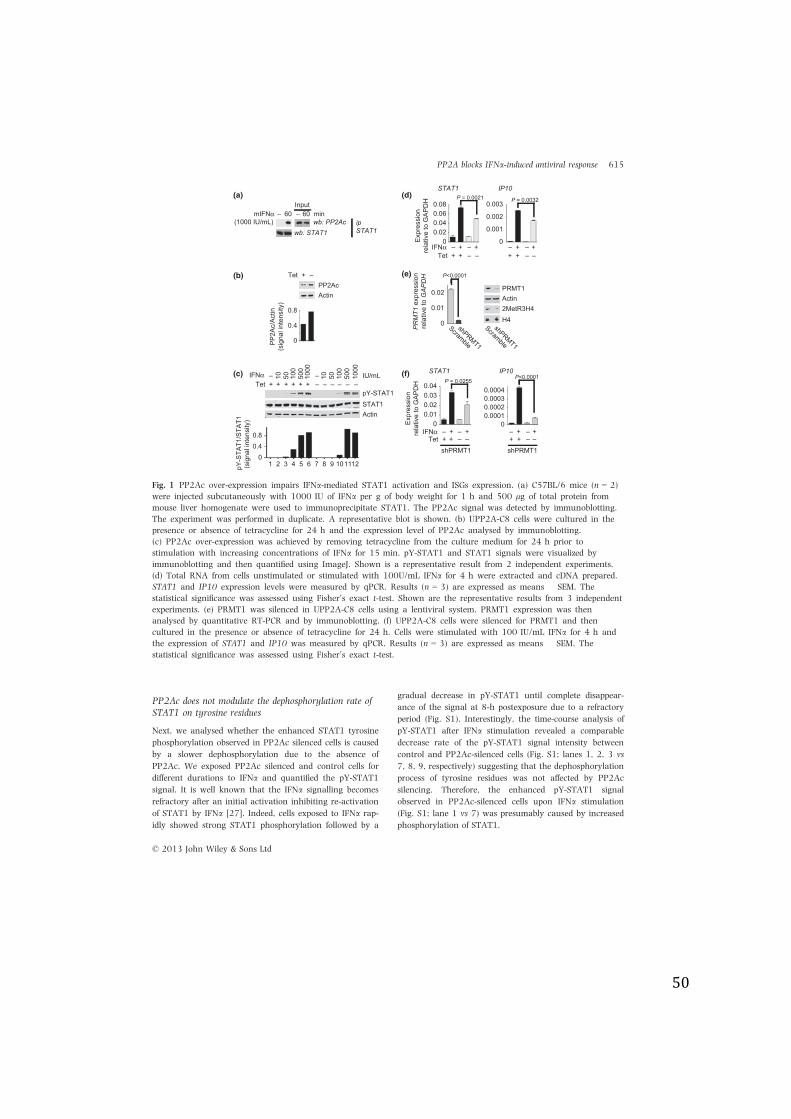
Alteration of PP2Ac expression modulates STAT1tyrosine phosphorylation
We have previously reported that PP2Ac is up-regulated in
patients with CHC and impairs IFNa-mediated Jak-STAT
signalling through the alteration of PRMT1-induced
STAT1 arginine methylation [18]. We now show that in
the mouse liver, PP2Ac is bound to STAT1 upon IFNastimulation suggesting a potential role of PP2A in the
direct regulation of STAT1 activation (Fig. 1a). PP2A is a
major serine/threonine phosphatase, however, its auto-
dephosphorylation capacity on tyrosine residues suggests
that it could be a tyrosine phosphatase under particular
conditions [25]. We therefore investigated the effect of
PP2Ac over-expression on STAT1 phosphorylation using
UPP2A-C8 cells that allow the inducible over-expression of
PP2Ac (Fig. 1b). We first performed a dose-response
experiment. Control (Tet+) and PP2Ac over-expressing
(Tet-) cells were exposed to increasing doses of IFNa and
pY-STAT1 signal was analysed by immunoblotting. Fig-
ure 1c shows enhanced STAT1 phosphorylation that
reached the maximal level with 500 IU/mL of IFNa in con-
trol and PP2Ac over-expressing cells (lanes 5 vs 11 and 6
vs 12). We did not observe any difference of the pY-STAT1
signal between control and PP2Ac over-expressing cells at
500 and 1000 IU/mL IFNa. However, we noticed a reduc-
tion in pY-STAT1 signal in PP2Ac over-expressing cells
treated with 50 and 100 IU/mL IFNa compared with con-
trol cells (Fig. 1c; lanes 3 vs 9 and 4 vs 10). Thus, to
investigate further the effect of PP2Ac on STAT1 phos-
phorylation, we decided to perform further experiments
using 100 UI/mL of IFNa.Next, we studied the effect of PP2Ac over-expression on
IFNa-induced STAT1 and IP10 expression. Our results
show a significant diminution of STAT1 and IP10 expres-
sion in PP2Ac over-expressing cells (Fig. 1d).
We have previously reported that PP2Ac negatively
modulates the IFNa-induced ISG expression by reducing
the methylation on STAT1 via the inhibition of PRMT1
activity [18]. Therefore, to distinguish the effect of
PP2Ac over-expression on STAT1 phosphorylation from
its inhibitory effect on PRMT1, we performed experiments
knocking down PRMT1. UPP2A-C8 were silenced for
PRMT1 using a lentiviral expression system (Fig. 1e) and
cultured in the presence or absence of tetracycline to
induce PP2Ac over-expression. The functional effect of
shPRMT1 knockdown was evaluated by analysis of meth-
ylation on histone H4 at arginine 3, a known substrate
for PRMT1 [26]. Figure 1e shows that PRMT1 silencing
leads to a decrease in methylation on H4. The cells were
then stimulated with 100 IU/mL of IFNa, and the ISG
expression was quantified by RT-qPCR. Figure 1f shows a
significant diminution of STAT1 and IP10 expression
upon IFNa stimulation in PP2Ac over-expressing cells in
the absence of PRMT1 demonstrating that the reduction
in ISG expression in PP2Ac over-expressing cells was
caused by the inhibitory effect of PP2Ac on STAT1
phosphorylation.
We further validated these results on a cell line with a
stable silencing of PP2Ac. The decrease in PP2Ac expres-
sion level was verified by immunoblotting (Fig. 2a). We
then analysed STAT1 tyrosine and serine phosphoryla-
tion upon IFNa stimulation. The reduction in PP2Ac
expression enhanced STAT1 phosphorylation on serine
and tyrosine residues in response to IFNa stimulation
(Fig. 2b; lanes 2, 3, 4 vs 6, 7, 8, respectively). The
increased STAT1 activation in PP2Ac-silenced cells
resulted in a stronger STAT1-probe binding shown by
EMSA (Fig. 2c; lanes 2, 3, 4 vs 6, 7, 8 respectively).
Consequently, we observed a significant enhanced expres-
sion of the ISGs in PP2Ac-silenced cells upon IFNa stim-
ulation (Fig. 2d).
© 2013 John Wiley & Sons Ltd
614 V. Shanker et al.
50
PP2Ac does not modulate the dephosphorylation rate ofSTAT1 on tyrosine residues
Next, we analysed whether the enhanced STAT1 tyrosine
phosphorylation observed in PP2Ac silenced cells is caused
by a slower dephosphorylation due to the absence of
PP2Ac. We exposed PP2Ac silenced and control cells for
different durations to IFNa and quantified the pY-STAT1
signal. It is well known that the IFNa signalling becomes
refractory after an initial activation inhibiting re-activation
of STAT1 by IFNa [27]. Indeed, cells exposed to IFNa rap-
idly showed strong STAT1 phosphorylation followed by a
gradual decrease in pY-STAT1 until complete disappear-
ance of the signal at 8-h postexposure due to a refractory
period (Fig. S1). Interestingly, the time-course analysis of
pY-STAT1 after IFNa stimulation revealed a comparable
decrease rate of the pY-STAT1 signal intensity between
control and PP2Ac-silenced cells (Fig. S1; lanes 1, 2, 3 vs
7, 8, 9, respectively) suggesting that the dephosphorylation
process of tyrosine residues was not affected by PP2Ac
silencing. Therefore, the enhanced pY-STAT1 signal
observed in PP2Ac-silenced cells upon IFNa stimulation
(Fig. S1; lane 1 vs 7) was presumably caused by increased
phosphorylation of STAT1.
IFN! +– +–0
0.020.040.060.08
Expr
essi
onre
lativ
e to
GAP
DH
STAT1P = 0.0021 P = 0.0032
Tet ++ ––
0
0.001
0.002
0.003
IP10
+– +–++ ––
PRM
T1 e
xpre
ssio
nre
lativ
e to
GAP
DH
0
0.01
0.02
Scramble
shPRMT1
00.010.020.030.04
Expr
essi
onre
lativ
e to
GAP
DH
STAT1
IFN! +– +–
P = 0.0255
00.00010.00020.0003
IP10
+– +–
0.0004
P<0.0001
shPRMT1 shPRMT1Tet ++ –– ++ ––
P<0.0001
PRMT1Actin
Scramble
shPRMT1
2MetR3H4H4
PP2AcActin
Tet –+
pY-STAT1STAT1Actin
Tet ++ ––IFN! 10– 10
050
PP2A
c/Ac
tin(s
igna
l int
ensi
ty)
pY-S
TAT1
/STA
T1(s
igna
l int
ensi
ty)
0
0.4
0.8
00.40.8
wb: PP2Acwb: STAT1
ipSTAT1
––––++ ++
500
1000
10– 100
50 500
1000
IU/mL
mIFN!(1000 IU/mL)
60–
21 43 65 87 109 1211
60–Input
min
(a)
(b)
(c)
(d)
(e)
(f)
Fig. 1 PP2Ac over-expression impairs IFNa-mediated STAT1 activation and ISGs expression. (a) C57BL/6 mice (n = 2)were injected subcutaneously with 1000 IU of IFNa per g of body weight for 1 h and 500 lg of total protein frommouse liver homogenate were used to immunoprecipitate STAT1. The PP2Ac signal was detected by immunoblotting.The experiment was performed in duplicate. A representative blot is shown. (b) UPP2A-C8 cells were cultured in thepresence or absence of tetracycline for 24 h and the expression level of PP2Ac analysed by immunoblotting.(c) PP2Ac over-expression was achieved by removing tetracycline from the culture medium for 24 h prior tostimulation with increasing concentrations of IFNa for 15 min. pY-STAT1 and STAT1 signals were visualized byimmunoblotting and then quantified using ImageJ. Shown is a representative result from 2 independent experiments.(d) Total RNA from cells unstimulated or stimulated with 100U/mL IFNa for 4 h were extracted and cDNA prepared.STAT1 and IP10 expression levels were measured by qPCR. Results (n = 3) are expressed as means ! SEM. Thestatistical significance was assessed using Fisher’s exact t-test. Shown are the representative results from 3 independentexperiments. (e) PRMT1 was silenced in UPP2A-C8 cells using a lentiviral system. PRMT1 expression was thenanalysed by quantitative RT-PCR and by immunoblotting. (f) UPP2A-C8 cells were silenced for PRMT1 and thencultured in the presence or absence of tetracycline for 24 h. Cells were stimulated with 100 IU/mL IFNa for 4 h andthe expression of STAT1 and IP10 was measured by qPCR. Results (n = 3) are expressed as means ! SEM. Thestatistical significance was assessed using Fisher’s exact t-test.
© 2013 John Wiley & Sons Ltd
PP2A blocks IFNa-induced antiviral response 615
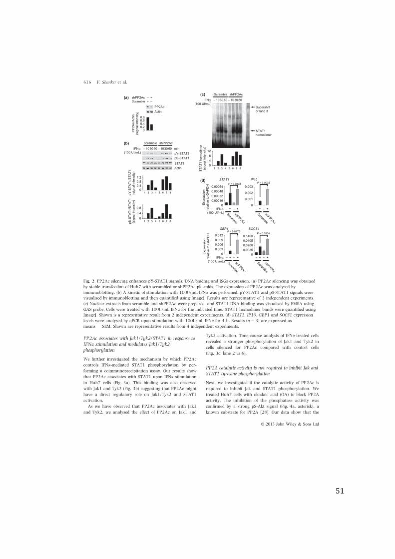
51
PP2Ac associates with Jak1/Tyk2/STAT1 in response toIFNa stimulation and modulates Jak1/Tyk2phosphorylation
We further investigated the mechanism by which PP2Ac
controls IFNa-mediated STAT1 phosphorylation by per-
forming a coimmunoprecipitation assay. Our results show
that PP2Ac associates with STAT1 upon IFNa stimulation
in Huh7 cells (Fig. 3a). This binding was also observed
with Jak1 and Tyk2 (Fig. 3b) suggesting that PP2Ac might
have a direct regulatory role on Jak1/Tyk2 and STAT1
activation.
As we have observed that PP2Ac associates with Jak1
and Tyk2, we analysed the effect of PP2Ac on Jak1 and
Tyk2 activation. Time-course analysis of IFNa-treated cells
revealed a stronger phosphorylation of Jak1 and Tyk2 in
cells silenced for PP2Ac compared with control cells
(Fig. 3c; lane 2 vs 6).
PP2A catalytic activity is not required to inhibit Jak andSTAT1 tyrosine phosphorylation
Next, we investigated if the catalytic activity of PP2Ac is
required to inhibit Jak and STAT1 phosphorylation. We
treated Huh7 cells with okadaic acid (OA) to block PP2A
activity. The inhibition of the phosphatase activity was
confirmed by a strong pS-Akt signal (Fig. 4a, asterisk), a
known substrate for PP2A [28]. Our data show that the
PP2AcActin
ScrambleshPP2Ac
–++–
STAT1Actin
pY-STAT1pS-STAT1
IFN!(100 UI/mL)
10– 3060 10– 3060 min
Scramble shPP2Ac
10– 3060 10– 3060
Scramble shPP2Ac
STAT1homodimer
Supershiftof lane 3
+– +–0
0.000160.000320.000480.00064
Expr
essi
onre
lativ
e to
GAP
DH
STAT1P = 0.0134
0
0.001
0.002
0.003
IP10
+– +–Scramble
shPP2Ac
GBP1
+– +–0
0.0030.0060.0090.012
Expr
essi
onre
lativ
e to
GAP
DH
SOCS1
+– +–0
0.00350.07000.01050.1400
00.20.40.60.8
PP2A
c/Ac
tin(s
igna
l int
ensi
ty)
pY-S
TAT1
/STA
T1(s
igna
l int
ensi
ty)
00.40.81.2
pS-S
TAT1
/STA
T1(s
igna
l int
ensi
ty)
00.40.8
048
12
STAT
1 ho
mod
imer
(sig
nal i
nten
sity
)
IFN!(100 UI/mL)
IFN!(100 UI/mL)
IFN!(100 UI/mL)
51 2 3 4 6 7 8
51 2 3 4 6 7 8
51 2 3 4 6 7 8
Scramble
shPP2Ac
Scramble
shPP2Ac
Scramble
shPP2Ac
P = 0.0032
P = 0.0175P = 0.0004
(a)
(b)
(c)
(d)
Fig. 2 PP2Ac silencing enhances pY-STAT1 signals, DNA binding and ISGs expression. (a) PP2Ac silencing was obtainedby stable transfection of Huh7 with scrambled or shPP2Ac plasmids. The expression of PP2Ac was analysed byimmunoblotting. (b) A kinetic of stimulation with 100U/mL IFNa was performed. pY-STAT1 and pS-STAT1 signals werevisualized by immunoblotting and then quantified using ImageJ. Results are representative of 3 independent experiments.(c) Nuclear extracts from scramble and shPP2Ac were prepared, and STAT1-DNA binding was visualized by EMSA usingGAS probe. Cells were treated with 100U/mL IFNa for the indicated time. STAT1 homodimer bands were quantified usingImageJ. Shown is a representative result from 2 independent experiments. (d) STAT1, IP10, GBP1 and SOCS1 expressionlevels were analysed by qPCR upon stimulation with 100U/mL IFNa for 4 h. Results (n = 3) are expressed asmeans ! SEM. Shown are representative results from 4 independent experiments.
© 2013 John Wiley & Sons Ltd
616 V. Shanker et al.
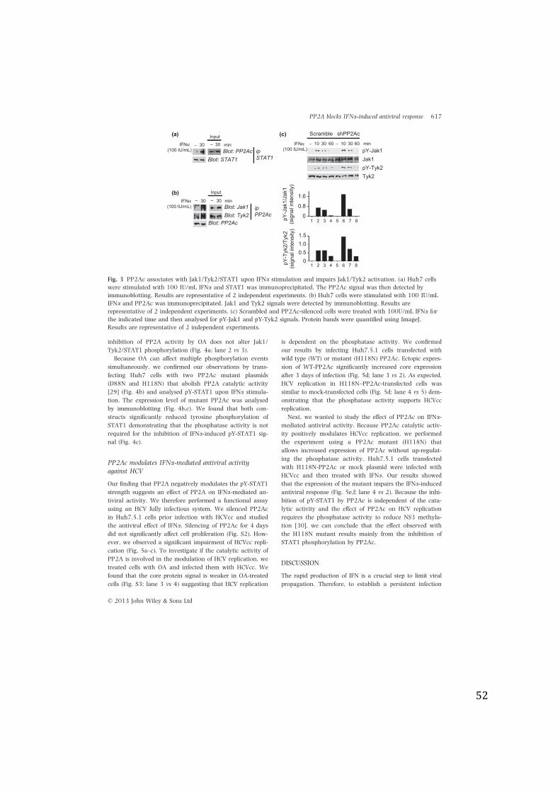
52
inhibition of PP2A activity by OA does not alter Jak1/
Tyk2/STAT1 phosphorylation (Fig. 4a; lane 2 vs 3).
Because OA can affect multiple phosphorylation events
simultaneously, we confirmed our observations by trans-
fecting Huh7 cells with two PP2Ac mutant plasmids
(D88N and H118N) that abolish PP2A catalytic activity
[29] (Fig. 4b) and analysed pY-STAT1 upon IFNa stimula-
tion. The expression level of mutant PP2Ac was analysed
by immunoblotting (Fig. 4b,c). We found that both con-
structs significantly reduced tyrosine phosphorylation of
STAT1 demonstrating that the phosphatase activity is not
required for the inhibition of IFNa-induced pY-STAT1 sig-
nal (Fig. 4c).
PP2Ac modulates IFNa-mediated antiviral activityagainst HCV
Our finding that PP2A negatively modulates the pY-STAT1
strength suggests an effect of PP2A on IFNa-mediated an-
tiviral activity. We therefore performed a functional assay
using an HCV fully infectious system. We silenced PP2Ac
in Huh7.5.1 cells prior infection with HCVcc and studied
the antiviral effect of IFNa. Silencing of PP2Ac for 4 days
did not significantly affect cell proliferation (Fig. S2). How-
ever, we observed a significant impairment of HCVcc repli-
cation (Fig. 5a–c). To investigate if the catalytic activity of
PP2A is involved in the modulation of HCV replication, we
treated cells with OA and infected them with HCVcc. We
found that the core protein signal is weaker in OA-treated
cells (Fig. S3; lane 3 vs 4) suggesting that HCV replication
is dependent on the phosphatase activity. We confirmed
our results by infecting Huh7.5.1 cells transfected with
wild type (WT) or mutant (H118N) PP2Ac. Ectopic expres-
sion of WT-PP2Ac significantly increased core expression
after 3 days of infection (Fig. 5d; lane 3 vs 2). As expected,
HCV replication in H118N–PP2Ac-transfected cells was
similar to mock-transfected cells (Fig. 5d; lane 4 vs 5) dem-
onstrating that the phosphatase activity supports HCVcc
replication.
Next, we wanted to study the effect of PP2Ac on IFNa-mediated antiviral activity. Because PP2Ac catalytic activ-
ity positively modulates HCVcc replication, we performed
the experiment using a PP2Ac mutant (H118N) that
allows increased expression of PP2Ac without up-regulat-
ing the phosphatase activity. Huh7.5.1 cells transfected
with H118N-PP2Ac or mock plasmid were infected with
HCVcc and then treated with IFNa. Our results showed
that the expression of the mutant impairs the IFNa-inducedantiviral response (Fig. 5e,f; lane 4 vs 2). Because the inhi-
bition of pY-STAT1 by PP2Ac is independent of the cata-
lytic activity and the effect of PP2Ac on HCV replication
requires the phosphatase activity to reduce NS3 methyla-
tion [30], we can conclude that the effect observed with
the H118N mutant results mainly from the inhibition of
STAT1 phosphorylation by PP2Ac.
DISCUSSION
The rapid production of IFN is a crucial step to limit viral
propagation. Therefore, to establish a persistent infection
pY-Tyk2
pY-Jak110– 30 60 10– 30 min
Scramble shPP2Ac
60Blot: PP2Ac
Blot: STAT1
– 30 minIFN!(100 IU/mL)
IFN!(100 IU/mL)
Blot: Jak1Blot: Tyk2
Blot: PP2Ac
– min30IFN!(100 IU/mL) ip
PP2Ac
ipSTAT1
00.51.0
0
0.8
1.6
51 2 3 4 6 7 8
51 2 3 4 6 7 8
Tyk2
1.5
pY-T
yk2/
Tyk2
(sig
nal i
nten
sity
)
Jak1
pY-J
ak1/
Jak1
(sig
nal i
nten
sity
)
– 30Input
– 30
Input
(a)
(b)
(c)
Fig. 3 PP2Ac associates with Jak1/Tyk2/STAT1 upon IFNa stimulation and impairs Jak1/Tyk2 activation. (a) Huh7 cellswere stimulated with 100 IU/mL IFNa and STAT1 was immunoprecipitated. The PP2Ac signal was then detected byimmunoblotting. Results are representative of 2 independent experiments. (b) Huh7 cells were stimulated with 100 IU/mLIFNa and PP2Ac was immunoprecipitated. Jak1 and Tyk2 signals were detected by immunoblotting. Results arerepresentative of 2 independent experiments. (c) Scrambled and PP2Ac-silenced cells were treated with 100U/mL IFNa forthe indicated time and then analysed for pY-Jak1 and pY-Tyk2 signals. Protein bands were quantified using ImageJ.Results are representative of 2 independent experiments.
© 2013 John Wiley & Sons Ltd
PP2A blocks IFNa-induced antiviral response 617
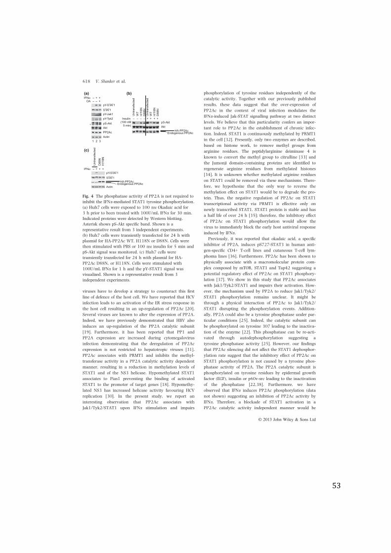
53
viruses have to develop a strategy to counteract this first
line of defence of the host cell. We have reported that HCV
infection leads to an activation of the ER stress response in
the host cell resulting in an up-regulation of PP2Ac [20].
Several viruses are known to alter the expression of PP2A.
Indeed, we have previously demonstrated that HBV also
induces an up-regulation of the PP2A catalytic subunit
[19]. Furthermore, it has been reported that PP1 and
PP2A expression are increased during cytomegalovirus
infection demonstrating that the deregulation of PP2Ac
expression is not restricted to hepatotropic viruses [31].
PP2Ac associates with PRMT1 and inhibits the methyl-
transferase activity in a PP2A catalytic activity dependent
manner, resulting in a reduction in methylation levels of
STAT1 and of the NS3 helicase. Hypomethylated STAT1
associates to Pias1 preventing the binding of activated
STAT1 to the promoter of target genes [18]. Hypomethy-
lated NS3 has increased helicase activity favouring HCV
replication [30]. In the present study, we report an
interesting observation that PP2Ac associates with
Jak1/Tyk2/STAT1 upon IFNa stimulation and impairs
phosphorylation of tyrosine residues independently of the
catalytic activity. Together with our previously published
results, these data suggest that the over-expression of
PP2Ac in the context of viral infection modulates the
IFNa-induced Jak-STAT signalling pathway at two distinct
levels. We believe that this particularity confers an impor-
tant role to PP2Ac in the establishment of chronic infec-
tion. Indeed, STAT1 is continuously methylated by PRMT1
in the cell [32]. Presently, only two enzymes are described,
based on histone work, to remove methyl groups from
arginine residues. The peptidylarginine deiminase 4 is
known to convert the methyl group to citrulline [33] and
the Jumonji domain-containing proteins are identified to
regenerate arginine residues from methylated histones
[34]. It is unknown whether methylated arginine residues
on STAT1 could be removed via these mechanisms. There-
fore, we hypothesize that the only way to reverse the
methylation effect on STAT1 would be to degrade the pro-
tein. Thus, the negative regulation of PP2Ac on STAT1
transcriptional activity via PRMT1 is effective only on
newly transcribed STAT1. STAT1 protein is stable and has
a half life of over 24 h [35]; therefore, the inhibitory effect
of PP2Ac on STAT1 phosphorylation would allow the
virus to immediately block the early host antiviral response
induced by IFNa.Previously, it was reported that okadaic acid, a specific
inhibitor of PP2A, induces pS727-STAT3 in human anti-
gen-specific CD4+ T-cell lines and cutaneous T-cell lym-
phoma lines [36]. Furthermore, PP2Ac has been shown to
physically associate with a macromolecular protein com-
plex composed by mTOR, STAT1 and Tap42 suggesting a
potential regulatory effect of PP2Ac on STAT1 phosphory-
lation [37]. We show in this study that PP2Ac associates
with Jak1/Tyk2/STAT1 and impairs their activation. How-
ever, the mechanism used by PP2A to reduce Jak1/Tyk2/
STAT1 phosphorylation remains unclear. It might be
through a physical interaction of PP2Ac to Jak1/Tyk2/
STAT1 disrupting the phosphorylation events. Addition-
ally, PP2A could also be a tyrosine phosphatase under par-
ticular conditions [25]. Indeed, the catalytic subunit can
be phosphorylated on tyrosine 307 leading to the inactiva-
tion of the enzyme [22]. This phosphatase can be re-acti-
vated through autodephosphorylation suggesting a
tyrosine phosphatase activity [25]. However, our findings
that PP2Ac silencing did not affect the STAT1 dephosphor-
ylation rate suggest that the inhibitory effect of PP2Ac on
STAT1 phosphorylation is not caused by a tyrosine phos-
phatase activity of PP2A. The PP2A catalytic subunit is
phosphorylated on tyrosine residues by epidermal growth
factor (EGF), insulin or p60v-src leading to the inactivation
of the phosphatase [22,38]. Furthermore, we have
observed that IFNa induces PP2Ac phosphorylation (data
not shown) suggesting an inhibition of PP2Ac activity by
IFNa. Therefore, a blockade of STAT1 activation in a
PP2Ac catalytic activity independent manner would be
STAT1
Actin
pY-STAT1
PP2AcAktpS-AktpY-Tyk2pY-Jak1
OA –– +IFN! +– +
STAT1pY-STAT1
ActinEndogenous PP2Ac
IFN! +– + +
D88
NH
118N
Unt
rans
fect
ed
HA-PP2Ac
*
21 3
AktpS-Akt
Endogenous PP2AcHA-PP2Ac
Insulin(100 nM
5 min)
–– – +– + + +
H11
8ND
88N
Unt
rans
fect
edW
T
H11
8ND
88N
Unt
rans
fect
edW
T
(a) (b)
(c)
Fig. 4 The phosphatase activity of PP2A is not required toinhibit the IFNa-mediated STAT1 tyrosine phosphorylation.(a) Huh7 cells were exposed to 100 nM Okadaic acid for3 h prior to been treated with 100U/mL IFNa for 30 min.Indicated proteins were detected by Western blotting.Asterisk shows pS-Akt specific band. Shown is arepresentative result from 3 independent experiments.(b) Huh7 cells were transiently transfected for 24 h withplasmid for HA-PP2Ac WT, H118N or D88N. Cells werethen stimulated with PBS or 100 nM insulin for 5 min andpS-Akt signal was monitored. (c) Huh7 cells weretransiently transfected for 24 h with plasmid for HA-PP2Ac D88N, or H118N. Cells were stimulated with100U/mL IFNa for 1 h and the pY-STAT1 signal wasvisualized. Shown is a representative result from 3independent experiments.
© 2013 John Wiley & Sons Ltd
618 V. Shanker et al.
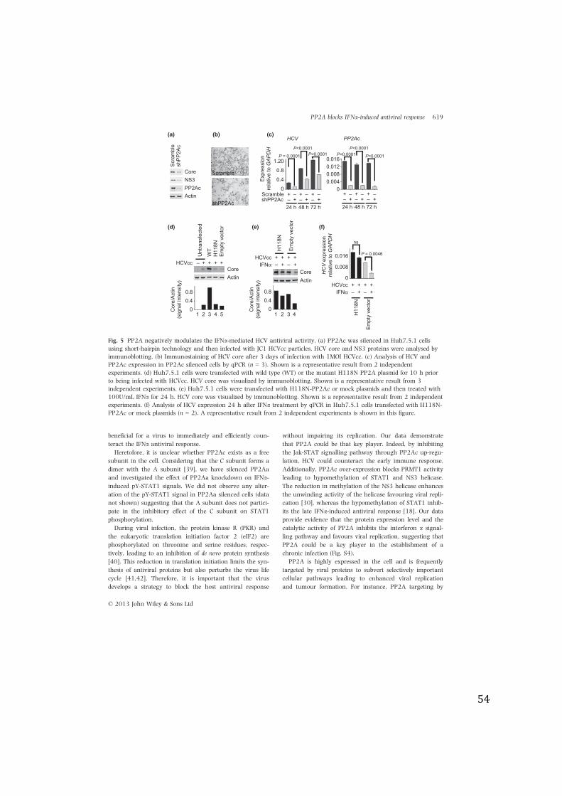
54
beneficial for a virus to immediately and efficiently coun-
teract the IFNa antiviral response.
Heretofore, it is unclear whether PP2Ac exists as a free
subunit in the cell. Considering that the C subunit forms a
dimer with the A subunit [39], we have silenced PP2Aa
and investigated the effect of PP2Aa knockdown on IFNa-induced pY-STAT1 signals. We did not observe any alter-
ation of the pY-STAT1 signal in PP2Aa silenced cells (data
not shown) suggesting that the A subunit does not partici-
pate in the inhibitory effect of the C subunit on STAT1
phosphorylation.
During viral infection, the protein kinase R (PKR) and
the eukaryotic translation initiation factor 2 (eIF2) are
phosphorylated on threonine and serine residues, respec-
tively, leading to an inhibition of de novo protein synthesis
[40]. This reduction in translation initiation limits the syn-
thesis of antiviral proteins but also perturbs the virus life
cycle [41,42]. Therefore, it is important that the virus
develops a strategy to block the host antiviral response
without impairing its replication. Our data demonstrate
that PP2A could be that key player. Indeed, by inhibiting
the Jak-STAT signalling pathway through PP2Ac up-regu-
lation, HCV could counteract the early immune response.
Additionally, PP2Ac over-expression blocks PRMT1 activity
leading to hypomethylation of STAT1 and NS3 helicase.
The reduction in methylation of the NS3 helicase enhances
the unwinding activity of the helicase favouring viral repli-
cation [30], whereas the hypomethylation of STAT1 inhib-
its the late IFNa-induced antiviral response [18]. Our data
provide evidence that the protein expression level and the
catalytic activity of PP2A inhibits the interferon a signal-
ling pathway and favours viral replication, suggesting that
PP2A could be a key player in the establishment of a
chronic infection (Fig. S4).
PP2A is highly expressed in the cell and is frequently
targeted by viral proteins to subvert selectively important
cellular pathways leading to enhanced viral replication
and tumour formation. For instance, PP2A targeting by
ActinPP2AcNS3Core
Scra
mbl
esh
PP2A
cScramble
shPP2Ac
ActinCore
HCVcc +– + + +
WT
H11
8NEm
pty
vect
or
Unt
rans
fect
ed
IFN! +– – +HCVcc ++ + +
H11
8N
Empt
y ve
ctor
CoreActin
0
0.4
0.8
1.20
Expr
essi
onre
lativ
e to
GAP
DH
HCV PP2Ac
00.0040.0080.0120.016
48 h24 h 72 h
Scramble ++ –– –+shPP2Ac –– ++ +–
48 h24 h 72 h
++ –– –+–– ++ +–
P = 0.0001
P<0.0001P<0.0001 P<0.0001
P<0.0001
P<0.0001
Cor
e/Ac
tin(s
igna
l int
ensi
ty)
00.40.8
Cor
e/Ac
tin(s
igna
l int
ensi
ty)
00.40.8
HC
V ex
pres
sion
rela
tive
to G
APD
H
0
0.016
0.008
P = 0.0046
ns
IFN! +– – +HCVcc ++ + +
H11
8N
Empt
y ve
ctor
21 3 4 5 21 3 4
(a) (b) (c)
(d) (e) (f)
Fig. 5 PP2A negatively modulates the IFNa-mediated HCV antiviral activity. (a) PP2Ac was silenced in Huh7.5.1 cellsusing short-hairpin technology and then infected with JC1 HCVcc particles. HCV core and NS3 proteins were analysed byimmunoblotting. (b) Immunostaining of HCV core after 3 days of infection with 1MOI HCVcc. (c) Analysis of HCV andPP2Ac expression in PP2Ac silenced cells by qPCR (n = 3). Shown is a representative result from 2 independentexperiments. (d) Huh7.5.1 cells were transfected with wild type (WT) or the mutant H118N PP2A plasmid for 10 h priorto being infected with HCVcc. HCV core was visualized by immunoblotting. Shown is a representative result from 3independent experiments. (e) Huh7.5.1 cells were transfected with H118N-PP2Ac or mock plasmids and then treated with100U/mL IFNa for 24 h. HCV core was visualized by immunoblotting. Shown is a representative result from 2 independentexperiments. (f) Analysis of HCV expression 24 h after IFNa treatment by qPCR in Huh7.5.1 cells transfected with H118N-PP2Ac or mock plasmids (n = 2). A representative result from 2 independent experiments is shown in this figure.
© 2013 John Wiley & Sons Ltd
PP2A blocks IFNa-induced antiviral response 619
55
the MCV small T-Antigen is required for the accomplish-
ment of the virus life cycle [43]. Furthermore, it has
been reported that the small t antigens and the polyoma-
virus middle T proteins can associate with the PP2A core
and induce cell transformation [44]. These observations
identify PP2A as a potential key target for therapeutic
developments. Presently, two natural antitumour mole-
cules, cantharidin and fostriecin that specifically bind and
inhibit PP2A have been tested for hepatoma and oesoph-
ageal carcinomas [45,46]. Although these compounds
are potent antitumour molecules, their use has severe
side effects probably due to the inhibition of PP2A that
is the major phosphatase in the cell. Therefore, a precise
mechanistic analysis of PP2A will permit the develop-
ment of better drugs improving the benefit to side effects
ratio.
ACKNOWLEDGEMENTS
We are grateful to Dr. Sinicrope for the pSIH1-puro-control
shRNA plasmid [47]. This study was funded by grants
from the Swiss National Science Foundation (grant
3L0030_130243) and the Swiss Cancer League grant
(KLS-02522-02-2010).
REFERENCES
1 Kanneganti TD. Central roles ofNLRs and inflammasomes in viralinfection. Nat Rev Immunol 2010;10: 688–698.
2 Wang Y, Swiecki M, McCartney SA,Colonna M. dsRNA sensors andplasmacytoid dendritic cells in hostdefense and autoimmunity. ImmunolRev 2011; 243: 74–90.
3 Silvennoinen O, Ihle JN, Schlessinger J,Levy DE. Interferon-induced nuclearsignalling by Jak protein tyrosinekinases. Nature 1993; 366: 583–585.
4 Darnell JE Jr, Kerr IM, Stark GR. Jak-STAT pathways and transcriptionalactivation in response to IFNs andother extracellular signaling proteins.Science 1994; 264: 1415–1421.
5 Shuai K, Horvath CM, Huang LHet al. Interferon activation of thetranscription factor Stat91 involvesdimerization through SH2-phospho-tyrosyl peptide interactions. Cell1994; 76: 821–828.
6 Arnheiter H, Skuntz S, Noteborn M,Chang S, Meier E. Transgenic micewith intracellular immunity to influ-enza virus. Cell 1990; 62: 51–61.
7 Carroll K, Elroy-Stein O, Moss B, Ja-gus R. Recombinant vaccinia virusK3L gene product prevents activa-tion of double-stranded RNA-depen-dent, initiation factor 2 alpha-specific protein kinase. J Biol Chem1993; 268: 12837–12842.
8 Marie I, Rebouillat D, HovanessianAG. The expression of both domainsof the 69/71 kDa 2!,5! oligoadeny-late synthetase generates a catalyti-cally active enzyme and mediates ananti-viral response. Eur J Biochem1999; 262: 155–165.
9 Starr R, Willson TA, Viney EM et al.A family of cytokine-inducible inhib-itors of signalling. Nature 1997;387: 917–921.
10 Liu B, Liao J, Rao X et al. Inhibitionof Stat1-mediated gene activationby PIAS1. Proc Natl Acad Sci USA1998; 95: 10626–10631.
11 Ten Hoeve J, De Jesus Ibarra-San-chez M, Fu Y et al. Identification ofa nuclear Stat1 protein tyrosinephosphatase. Mol Cell Biol 2002;22: 5662–5668.
12 Malakhov MP, Malakhova OA, KimKI, Ritchie KJ, Zhang DE. UBP43(USP18) specifically removes ISG15from conjugated proteins. J BiolChem 2002; 277: 9976–9981.
13 Hendrix P, Turowski P, Mayer-Jae-kel RE et al. Analysis of subunit iso-forms in protein phosphatase 2Aholoenzymes from rabbit and Xeno-pus. J Biol Chem 1993; 268: 7330–7337.
14 Zhou J, Pham HT, Walter G. Theformation and activity of PP2Aholoenzymes do not depend onthe isoform of the catalytic sub-unit. J Biol Chem 2003; 278:8617–8622.
15 Baharians Z, Schonthal AH. Autore-gulation of protein phosphatasetype 2A expression. J Biol Chem1998; 273: 19019–19024.
16 Voorhoeve PM, Hijmans EM, Ber-nards R. Functional interactionbetween a novel protein phospha-tase 2A regulatory subunit, PR59,and the retinoblastoma-relatedp107 protein. Oncogene 1999; 18:515–524.
17 Duong FH, Christen V, Lin S, HeimMH. Hepatitis C virus-induced
up-regulation of protein phospha-tase 2A inhibits histone modifica-tion and DNA damage repair.Hepatology 2010; 51: 741–751.
18 Duong FH, Filipowicz M, Tripodi M,La Monica N, Heim MH. Hepatitis Cvirus inhibits interferon signalingthrough up-regulation of proteinphosphatase 2A. Gastroenterology2004; 126: 263–277.
19 Christen V, Duong F, Bernsmeier C,Sun D, Nassal M, Heim MH. Inhibi-tion of alpha interferon signaling byhepatitis B virus. J Virol 2007; 81:159–165.
20 Christen V, Treves S, Duong FH,Heim MH. Activation of endoplas-mic reticulum stress response byhepatitis viruses up-regulates pro-tein phosphatase 2A. Hepatology2007; 46: 558–565.
21 Kamibayashi C, Estes R, LickteigRL, Yang SI, Craft C, Mumby MC.Comparison of heterotrimeric pro-tein phosphatase 2A containing dif-ferent B subunits. J Biol Chem1994; 269: 20139–20148.
22 Chen J, Parsons S, Brautigan DL.Tyrosine phosphorylation of proteinphosphatase 2A in response togrowth stimulation and v-src trans-formation of fibroblasts. J Biol Chem1994; 269: 7957–7962.
23 Bryant JC, Westphal RS, WadzinskiBE. Methylated C-terminal leucineresidue of PP2A catalytic subunit isimportant for binding of regulatoryBalpha subunit. Biochem J 1999;339(Pt 2): 241–246.
24 Pietschmann T, Kaul A, Koutsouda-kis G et al. Construction and char-acterization of infectious intrageno-typic and intergenotypic hepatitis C
© 2013 John Wiley & Sons Ltd
620 V. Shanker et al.
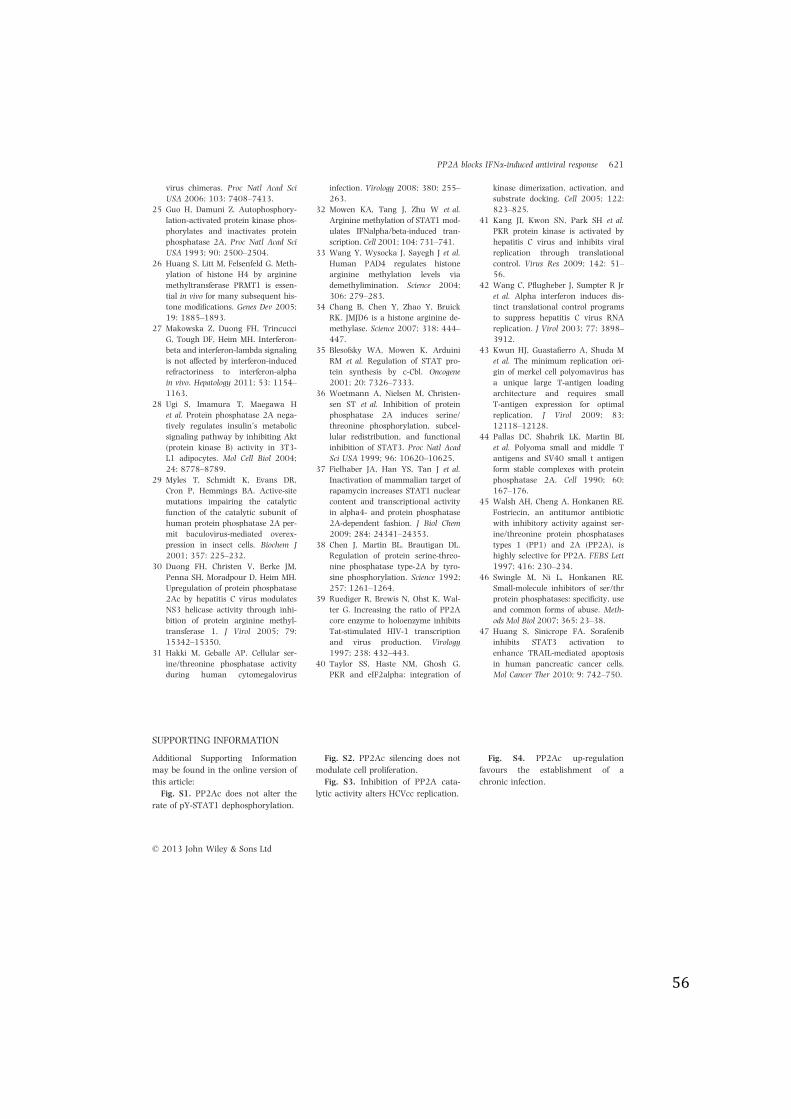
56
virus chimeras. Proc Natl Acad SciUSA 2006; 103: 7408–7413.
25 Guo H, Damuni Z. Autophosphory-lation-activated protein kinase phos-phorylates and inactivates proteinphosphatase 2A. Proc Natl Acad SciUSA 1993; 90: 2500–2504.
26 Huang S, Litt M, Felsenfeld G. Meth-ylation of histone H4 by argininemethyltransferase PRMT1 is essen-tial in vivo for many subsequent his-tone modifications. Genes Dev 2005;19: 1885–1893.
27 Makowska Z, Duong FH, TrincucciG, Tough DF, Heim MH. Interferon-beta and interferon-lambda signalingis not affected by interferon-inducedrefractoriness to interferon-alphain vivo. Hepatology 2011; 53: 1154–1163.
28 Ugi S, Imamura T, Maegawa Het al. Protein phosphatase 2A nega-tively regulates insulin’s metabolicsignaling pathway by inhibiting Akt(protein kinase B) activity in 3T3-L1 adipocytes. Mol Cell Biol 2004;24: 8778–8789.
29 Myles T, Schmidt K, Evans DR,Cron P, Hemmings BA. Active-sitemutations impairing the catalyticfunction of the catalytic subunit ofhuman protein phosphatase 2A per-mit baculovirus-mediated overex-pression in insect cells. Biochem J2001; 357: 225–232.
30 Duong FH, Christen V, Berke JM,Penna SH, Moradpour D, Heim MH.Upregulation of protein phosphatase2Ac by hepatitis C virus modulatesNS3 helicase activity through inhi-bition of protein arginine methyl-transferase 1. J Virol 2005; 79:15342–15350.
31 Hakki M, Geballe AP. Cellular ser-ine/threonine phosphatase activityduring human cytomegalovirus
infection. Virology 2008; 380: 255–263.
32 Mowen KA, Tang J, Zhu W et al.Arginine methylation of STAT1 mod-ulates IFNalpha/beta-induced tran-scription. Cell 2001; 104: 731–741.
33 Wang Y, Wysocka J, Sayegh J et al.Human PAD4 regulates histonearginine methylation levels viademethylimination. Science 2004;306: 279–283.
34 Chang B, Chen Y, Zhao Y, BruickRK. JMJD6 is a histone arginine de-methylase. Science 2007; 318: 444–447.
35 Blesofsky WA, Mowen K, ArduiniRM et al. Regulation of STAT pro-tein synthesis by c-Cbl. Oncogene2001; 20: 7326–7333.
36 Woetmann A, Nielsen M, Christen-sen ST et al. Inhibition of proteinphosphatase 2A induces serine/threonine phosphorylation, subcel-lular redistribution, and functionalinhibition of STAT3. Proc Natl AcadSci USA 1999; 96: 10620–10625.
37 Fielhaber JA, Han YS, Tan J et al.Inactivation of mammalian target ofrapamycin increases STAT1 nuclearcontent and transcriptional activityin alpha4- and protein phosphatase2A-dependent fashion. J Biol Chem2009; 284: 24341–24353.
38 Chen J, Martin BL, Brautigan DL.Regulation of protein serine-threo-nine phosphatase type-2A by tyro-sine phosphorylation. Science 1992;257: 1261–1264.
39 Ruediger R, Brewis N, Ohst K, Wal-ter G. Increasing the ratio of PP2Acore enzyme to holoenzyme inhibitsTat-stimulated HIV-1 transcriptionand virus production. Virology1997; 238: 432–443.
40 Taylor SS, Haste NM, Ghosh G.PKR and eIF2alpha: integration of
kinase dimerization, activation, andsubstrate docking. Cell 2005; 122:823–825.
41 Kang JI, Kwon SN, Park SH et al.PKR protein kinase is activated byhepatitis C virus and inhibits viralreplication through translationalcontrol. Virus Res 2009; 142: 51–56.
42 Wang C, Pflugheber J, Sumpter R Jret al. Alpha interferon induces dis-tinct translational control programsto suppress hepatitis C virus RNAreplication. J Virol 2003; 77: 3898–3912.
43 Kwun HJ, Guastafierro A, Shuda Met al. The minimum replication ori-gin of merkel cell polyomavirus hasa unique large T-antigen loadingarchitecture and requires smallT-antigen expression for optimalreplication. J Virol 2009; 83:12118–12128.
44 Pallas DC, Shahrik LK, Martin BLet al. Polyoma small and middle Tantigens and SV40 small t antigenform stable complexes with proteinphosphatase 2A. Cell 1990; 60:167–176.
45 Walsh AH, Cheng A, Honkanen RE.Fostriecin, an antitumor antibioticwith inhibitory activity against ser-ine/threonine protein phosphatasestypes 1 (PP1) and 2A (PP2A), ishighly selective for PP2A. FEBS Lett1997; 416: 230–234.
46 Swingle M, Ni L, Honkanen RE.Small-molecule inhibitors of ser/thrprotein phosphatases: specificity, useand common forms of abuse. Meth-ods Mol Biol 2007; 365: 23–38.
47 Huang S, Sinicrope FA. Sorafenibinhibits STAT3 activation toenhance TRAIL-mediated apoptosisin human pancreatic cancer cells.Mol Cancer Ther 2010; 9: 742–750.
SUPPORTING INFORMATION
Additional Supporting Information
may be found in the online version of
this article:
Fig. S1. PP2Ac does not alter the
rate of pY-STAT1 dephosphorylation.
Fig. S2. PP2Ac silencing does not
modulate cell proliferation.
Fig. S3. Inhibition of PP2A cata-
lytic activity alters HCVcc replication.
Fig. S4. PP2Ac up-regulation
favours the establishment of a
chronic infection.
© 2013 John Wiley & Sons Ltd
PP2A blocks IFNa-induced antiviral response 621
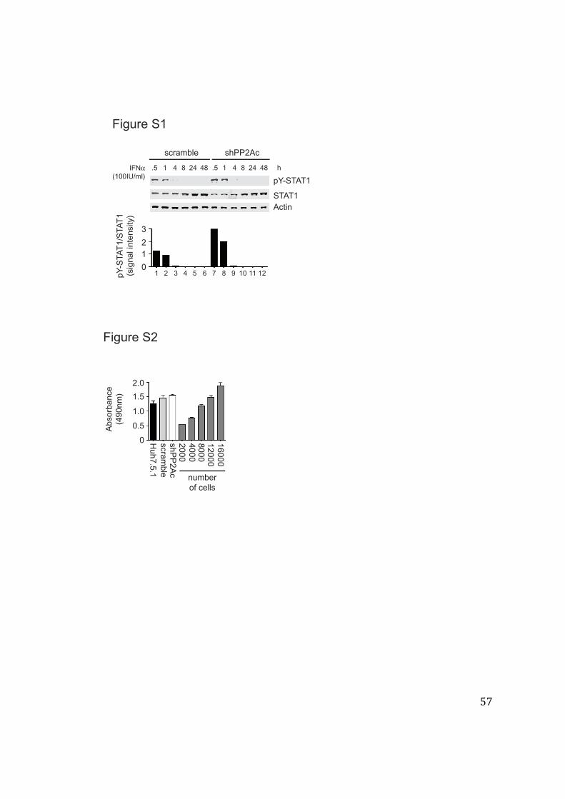
57
Figure S1
1.5 4 8 4824 .5 48 h
scramble shPP2Ac
1 4 8 24
STAT1Actin
pY-STAT1
0
12
3
pY-S
TAT
1/S
TAT
1(s
igna
l int
ensi
ty)
IFN!(100IU/ml)
51 2 3 4 106 7 8 9 11 12
Figure S2
0
0.5
1.0
1.5
2.0
Ab
so
rba
nce
(49
0n
m)
16
00
0
number
of cells
20
00
40
00
80
00
12
00
0
Hu
h7
.5.1
scra
mb
le
sh
PP
2A
c
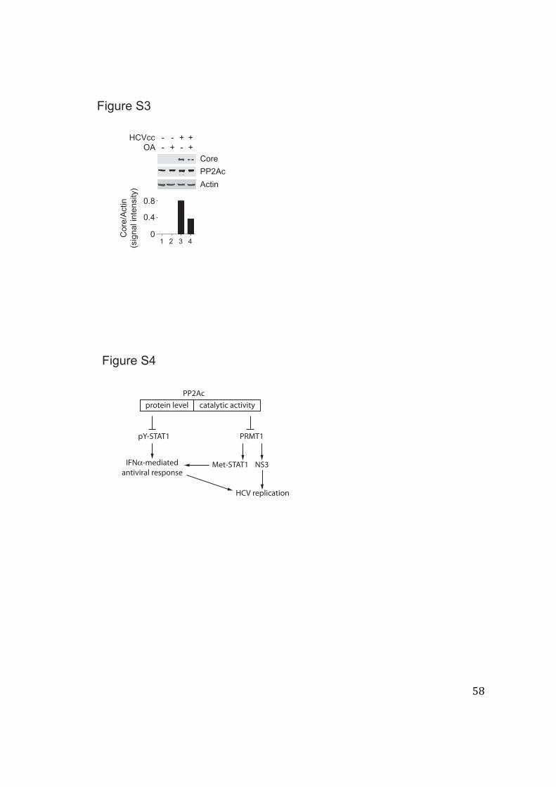
58
Actin
PP2Ac
CoreOA +- -
HCVcc -- +++
Cor
e/A
ctin
(sig
nal i
nten
sity
)
0
0.4
0.8
1 2 3 4
Figure S3
PP2Acprotein level catalytic activity
pY-STAT1
IFN!-mediatedantiviral response
PRMT1
NS3Met-STAT1
HCV replication
Figure S4
59
4.2 The role of PP2A-A and -B subunits in the regulation of the Jak-STAT signaling pathway.
Because PP2A is a multimeric holoenzyme and the substrates recognition is determined
through the association to specific regulatory B subunits, we have tried to identify which B
subunit could potentially be an important component in PP2Ac-mediated inhibition of the
IFN-α induced Jak-STAT signaling. For that purpose, we have serially silenced several B
subunits using short-hairpin technology (Figure 1).
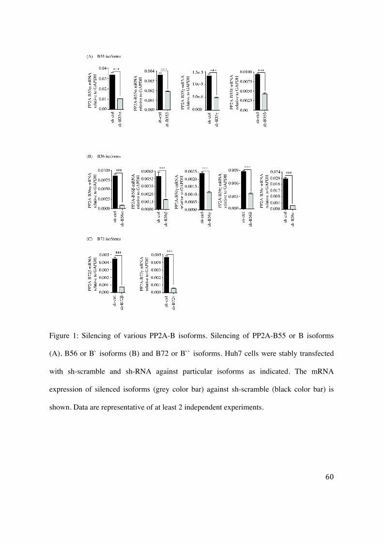
60
Figure 1: Silencing of various PP2A-B isoforms. Silencing of PP2A-B55 or B isoforms
(A), B56 or B` isoforms (B) and B72 or B`` isoforms. Huh7 cells were stably transfected
with sh-scramble and sh-RNA against particular isoforms as indicated. The mRNA
expression of silenced isoforms (grey color bar) against sh-scramble (black color bar) is
shown. Data are representative of at least 2 independent experiments.
61
4.2.1 Effect of silencing of a specific PP2A subunit on the expression of the other subunits
The phosphatase activity is mainly provided by the α isoform of the catalytic C subunit in
specific association with B subunits. Therefore, it is likely that alteration of a particular B
subunit could modify the proportion of holoenzyme complexes and localization into
subcellular compartments. This could affect the various cellular processes including genes
transcription, RNA/protein stability. Therefore, in order to identify which specific PP2A-B
subunits regulate INF-α signaling and HCV replication we serially silenced various
isoforms of PP2A subunits. Interestingly, qRT-PCR results show that silencing of a
particular isoform of B subunit leads to a down-regulation of majority of the other
isoforms. Additionally, this impairment is not only restricted to A, B or C subunits but also
deregulates the expression of other regulatory proteins such as α4. As shown in figure 2,
we observed a down-regulation of PP2A-Aα in cells silenced for PP2A-cα, B55α, and
B56β. We also observed that PP2A-Aα expression was up-regulated in PP2A-B56α
silenced cells (figure 2A). Furthermore, PP2A-B55α expression was unchanged in PP2A-
Aα silenced cells though we observed significant down-regulation of PP2A-B55α in
PP2A-B56α/β cells (figure 2B). Interestingly, we found that silencing of PP2A-B55α
resulted in a decrease in PP2A-Aα expression (figure 2A) whereas silencing of PP2A-Aα
did not impair PP2A-B55α expression (figure 2B). Our observation revealed that, silencing
of PP2A-B55α and B56β down-regulates the expression of PP2A-B56α (figure 2C).
Interestingly, silencing of PP2A-B56α led to an increased expression of PP2-Aα (figure
62
2A) and vice-versa (figure 2C). Finally, silencing of PP2A-Aα, B55α, B56α, and B56β
resulted in a decreased α4 expression (figure 2d).
Additionally, silencing of the α isoform of the catalytic subunit attenuated the mRNA
expression of various isoforms of PP2A including B56α-ε and B72α-γ (figure 3). This
reduction is ranging from slight to moderate. These observations are summarized in table 2.
Earlier, Sablina and colleagues have reported that silencing of PP2Acα leads to a
significant reduction of the basal level of B55α, B56β, B56γ, PR93, and PP2A-A subunits
whereas silencing of PP2A-Cβ exerts marginal effect on the expression of the other
subunits (Sablina et al., 2010). Furthermore, it has been reported that an association of the
PP2A-A with Cα subunit is required for their stability (Silverstein et al., 2002; Chen et al.,
2005b). Our observations are in the line with previous observations and show inter-
regulatory mechanisms governing the expression of various PP2A subunits. Such
regulatory mechanism could be obtained through miRNAs that mediate mRNA degradation
or/and activation/inhibition of various transcription factors. Because of the lack of
commercially available antibodies against all the B subunits, we were not able to analyze
the effect of silencing at protein level.

63
Figure 2: Inter-regulation of different isoforms of PP2A. The X-axis represents the
stably silenced cells while the Y-axis represents mRNA expression from indicated PP2A
subunit coding genes and α4 gene.
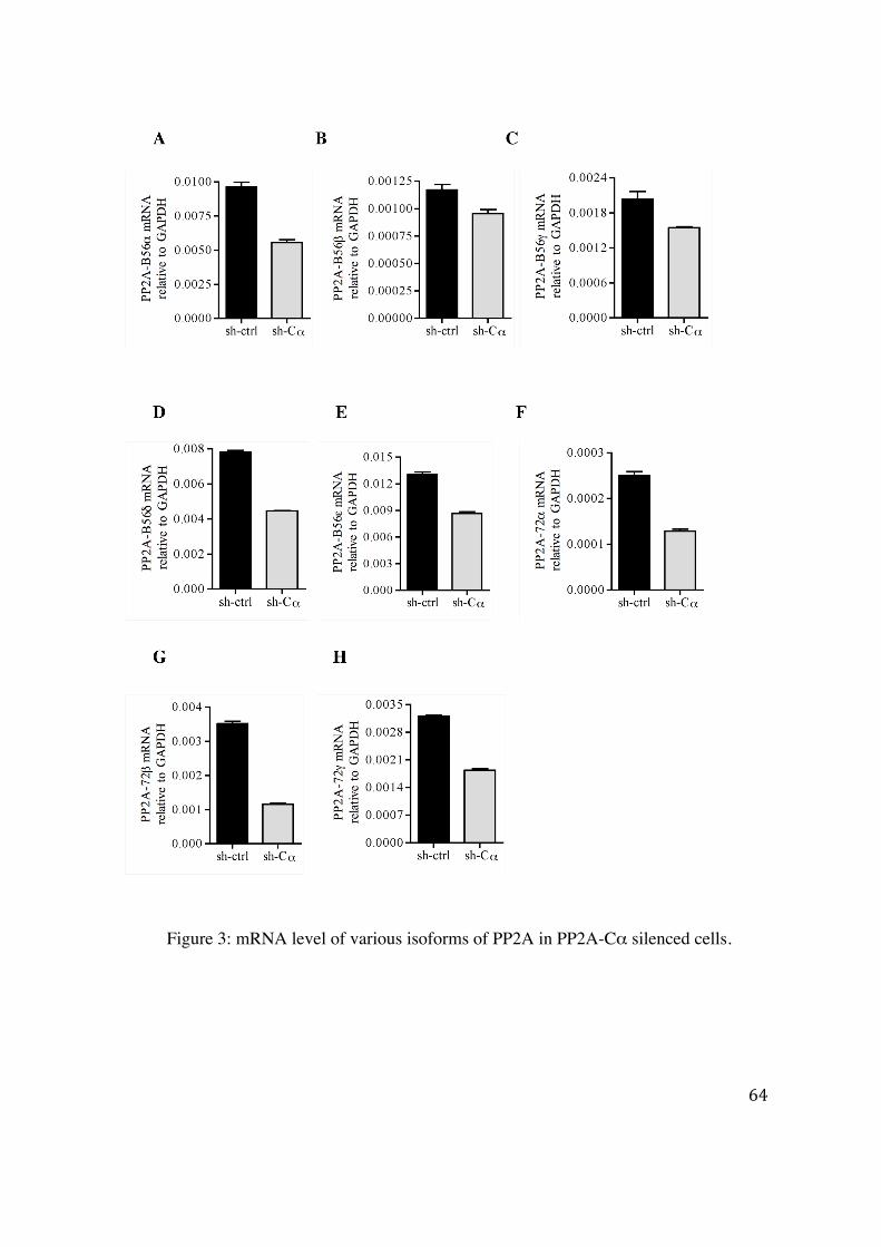
64
Figure 3: mRNA level of various isoforms of PP2A in PP2A-Cα silenced cells.
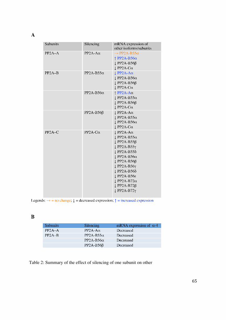
65
Table 2: Summary of the effect of silencing of one subunit on other
66
4.2.2 Predication of miRNA targeting PP2A subunits
One potential mechanism to explain the inter-regulation of the subunits expression could be
through miRNAs. We, therefore, performed a miRNA targeting PP2A subunits analysis
using online bioinformatics softwares such as miRBase
(http://www.mirbase.org/index.shtml) or miRWalk (http://www.umm.uni-
heidelberg.de/apps/zmf/mirwalk/). Using this approach potential miRNAs was predicted
against each subunit. We found that many subunits have multiple miRNA target sites.
Interestingly, our miRNA and target genes data analysis revealed that miRNA-19 targets 4
PP2A isoforms including B56α, B56ε, Aβ, and STRN3. Furthermore, we found that
miRNA-34b targets Aβ and STRN isoforms. The role of various miRNA including
miRNA-19, miRNA-1, and miRNA-34b involved in expression of PP2A subunits have
been previously described (Terentyev et al., 2009; Mavrakis et al., 2010; Chen et al., 2011).
67
5. Discussion PP2A is known to regulate several signaling pathways via dephosphorylation of
serine/threonine residues (Eichhorn et al., 2009). It has been shown that inhibition of PP2A
increases serine phosphorylation on STAT6 and STAT3 (Woetmann et al., 1999, 2003) and
also increases IFN-γ mediated serine and tyrosine phosphorylation of STAT1 in mTOR
dependent fashion (Fielhaber et al., 2009). Recently, it has been reported that PP2A inhibits
the Jak-STAT1 signaling by forming a macromolecular complex with mTOR, STAT1 and
α4 and (Fielhaber et al., 2009). Moreover, it has been also shown that PP2A associates to
Jak2 in myeloid progenitor cells and modulates the Jak2-STAT5 signaling pathway in
response to IL-3 (Yokoyama et al., 2003). Presently, it is not clear how PP2Ac associates to
Jak1 and Tyk2 and inhibits their activation. One could hypothesize that PP2Ac-Jak1/Tyk2
binding shares similar molecular mechanism like PP2Ac-Jak2 association (Yokoyama et
al., 2003).
Production of IFNs by cells in response to viral infection leads to initiation of antiviral
activity. For the establishment of persistent infection, viruses develop strategies to
counteract this first line of defense. We have reported that HCV proteins inhibit the IFN-α
signaling (Heim et al., 1999; Blindenbacher et al., 2003). The inhibition is mediated
through an up-regulation of PP2Ac that leads to a hypomethylation of STAT1 (Duong et
al., 2004). Hypomethylated STAT1 associates with PIAS1 and prevents activated STAT1
binding to promoter elements of the ISGs (Mowen et al., 2001; Liu et al., 2004).
68
In this work, we demonstrated that the phosphatase activity of PP2A is not required for the
inhibition of IFN-α induced Jak-STAT signaling. However, it is required to promote
HCVcc replication. Indeed, using different approaches to reduce PP2A catalytic activity
(pharmacological inhibitors, catalytic dead or dominant negative mutants, siRNA against
PP2Ac), we have observed a reduction of HCV replication. The mechanism underlying
regulation of viral life cycle by PP2A remains unclear. Three hypothesis could be proposed:
(1) PP2A reduces the methylation of NS3 helicase by inhibiting PRMT1 leading to an
enhanced unwinding activity and therefore viral replication (Duong et al., 2005); (2) PP2A
dephosphorylates NS5A and induces viral replication (Evans et al., 2004); (3) PP2A
dephosphorylates eIF2α (Groskreutz et al., 2010) and releases the inhibitory effect of
eIF2α on protein translation thus favoring HCV replication. Further experiments are
needed to clarify this issue.
We have made an interesting observation that tyrosine phosphorylation of Jak1/Tyk2 and
STAT1 is independent of PP2A phosphatase activity while it is needed to positively
modulate HCV replication. This phenomenon can be explained by the complexity of
substrate selection by PP2A. Indeed, depending on the B subunit that associates to the A-C
core complex, PP2A holoenzyme could preferentially target the IFN-α signaling pathway
or the HCV replication process. We have made an attempt to identify these B subunits
using short-hairpin siRNA technology. Unfortunately, we found that silencing of a specific
B subunit alters the expression of the C, A, or B subunits showing the difficulties to
69
identify the B subunits that are required to direct PP2A towards IFN-α signaling inhibition
or towards HCV replication.
The mechanism of subunits inter-regulation is not known. One possibility is that down-
regulation of a specific subunit alters the expression of some so far undiscovered miRNAs
that then mediate RNA degradation of the other subunits. We have performed a PP2A and
miRNA target gene analysis and observed that several subunits can be targeted by the same
miRNA. Indeed, we found that miRNA-19 targets PP2A-B56ε subunits therefore one can
hypothesize that silencing of one specific subunit increases the proportion of miRNA-19
which then down-regulates the expression of the other subunits. Further experimentations
are needed to clarify the mechanism underlying the inter-regulation of the expression of
PP2A subunits. Another possible mechanism of inter-regulation of PP2A subunits could be
via the regulation of transcription through epigenetic changes. Indeed, we have previously
reported that PP2Ac can modify the post-translational modification status on histones such
as methylation/acetylation/phosphorylation (Duong et al., 2010). There are several reports
showing that modifications on histone can alter gene transcription by modifying the
chromatin structure. These chromatin structure rearrangements lead to open/close
chromatin status throughout the nucleosome facilitating/inhibiting the accessibility of
transcription factors to the promoters (Berger, 2010). Therefore, the alteration of the
expression of a specific PP2A subunit can change the modification status on histones and
thus induces a change in the chromatin structure leading to an impairment of the expression
of the other subunits.
70
6. Summary
The first part of the work demonstrate the role of PP2A in IFN-α induced Jak-STAT1
signaling and HCV replication. We show here that PP2Ac activity is not required for IFN-α
induced tyrosine phosphorylation of Jak1/Tyk2 and STAT1. In response to IFN-α
induction, PP2Ac associates with Jak1/Tyk2 and STAT1. This association modulates the
Jak1/Tyk2 and STAT1 tyrosine phosphorylation. However, this study shows that PP2A
activity is required for the HCV replication.
The selective behavior of PP2A for Jak-STAT1 signaling pathway inhibition and
promotion of HCV replication could be due to the targeted substrate selection by PP2A
holoenzyme complex. In the second part of this work, attempts were made to determine the
specific regulatory B subunits involved in IFN-α induced Jak-STAT1 signaling inhibition
and HCV replication. We observed the inter-regulatory behavior of PP2A subunits. During
the course of this study, due to unavailability of specific antibodies for various isoforms of
B subunit, the aim to determine specific holoenzyme complex involved in regulation of
Jak-STAT1 signaling and HCV replication was not achieved.
Further studies are required to investigate the specific B subunits and thereby holoenzyme
complex responsible for IFN-α induced Jak-STAT signaling inhibition and HCV
replication by PP2A.
71
7. References
Aguet, M., Dembić, Z., and Merlin, G. (1988). Molecular cloning and expression of the human interferon-‐γ receptor. Cell 55, 273–280.
Albecka, A., Belouzard, S., de Beeck, A.O., Descamps, V., Goueslain, L., Bertrand-‐Michel, J., Tercé, F., Duverlie, G., Rouillé, Y., and Dubuisson, J. (2012). Role of low-‐density lipoprotein receptor in the hepatitis C virus life cycle. Hepatology 55, 998–1007.
Alberts, A.S., Deng, T., Lin, A., Meinkoth, J.L., Schönthal, A., Mumby, M.C., Karin, M., and Feramisco, J.R. (1993). Protein Phosphatase 2A Potentiates Activity of Promoters Containing AP-‐1-‐Binding Elements. Mol. Cell. Biol. 13, 2104–2112.
Alexander, W.S., Starr, R., Fenner, J.E., Scott, C.L., Handman, E., Sprigg, N.S., Corbin, J.E., Cornish, A.L., Darwiche, R., Owczarek, C.M., et al. (1999). SOCS1 Is a Critical Inhibitor of Interferon γ Signaling and Prevents the Potentially Fatal Neonatal Actions of this Cytokine. Cell 98, 597–608.
Altiok, S., Xu, M., and Spiegelman, B.M. (1997). PPARγ Induces Cell Cycle Withdrawal: Inhibition of E2F/DP DNA-‐Binding Activity Via down-‐Regulation of PP2A. Genes Dev. 11, 1987–1998.
Anderson, K.A., Noeldner, P.K., Reece, K., Wadzinski, B.E., and Means, A.R. (2004). Regulation and Function of the Calcium/Calmodulin-‐dependent Protein Kinase IV/Protein Serine/Threonine Phosphatase 2A Signaling Complex. J. Biol. Chem. 279, 31708–31716.
Appel, N., Pietschmann, T., and Bartenschlager, R. (2005). Mutational Analysis of Hepatitis C Virus Nonstructural Protein 5A: Potential Role of Differential Phosphorylation in RNA Replication and Identification of a Genetically Flexible Domain. J. Virol. 79, 3187–3194.
Arino, J., Woon, C.W., Brautigan, D.L., Miller, T.B., and Johnson, G.L. (1988). Human Liver Phosphatase 2A: cDNA and Amino Acid Sequence of Two Catalytic Subunit Isotypes. PNAS 85, 4252–4256.
Babon, J.J., Kershaw, N.J., Murphy, J.M., Varghese, L.N., Laktyushin, A., Young, S.N., Lucet, I.S., Norton, R.S., and Nicola, N.A. (2012). Suppression of Cytokine Signaling by SOCS3: Characterization of the Mode of Inhibition and the Basis of Its Specificity. Immunity 36, 239–250.
Baharians, Z., and Schönthal, A.H. (1998). Autoregulation of Protein Phosphatase Type 2A Expression. J. Biol. Chem. 273, 19019–19024.
72
Bartenschlager, R. (2002). Hepatitis C virus replicons: potential role for drug development. Nature Reviews Drug Discovery 1, 911–916.
Behrens, S.E., Tomei, L., and De Francesco, R. (1996). Identification and properties of the RNA-‐dependent RNA polymerase of hepatitis C virus. EMBO J 15, 12–22.
Berger, S.L. (2010). Cell Signaling and Transcriptional Regulation via Histone Phosphorylation. Cold Spring Harb Symp Quant Biol 75, 23–26.
Bernards, A. (1991). Predicted tyk2 protein contains two tandem protein kinase domains. Oncogene 6, 1185–1187.
Bhattacharya, S., Eckner, R., Grossman, S., Oldread, E., Arany, Z., D’Andrea, A., and Livingston, D.M. (1996). Cooperation of Stat2 and p300/CBP in signalling induced by interferon-‐|[alpha]|. , Published Online: 26 September 1996; | Doi:10.1038/383344a0 383, 344–347.
Blindenbacher, A., Duong, F.H.T., Hunziker, L., Stutvoet, S.T.D., Wang, X., Terracciano, L., Moradpour, D., Blum, H.E., Alonzi, T., Tripodi, M., et al. (2003). Expression of hepatitis c virus proteins inhibits interferon α signaling in the liver of transgenic mice. Gastroenterology 124, 1465–1475.
Bryant, J.C., Westphal, R.S., and Wadzinski, B.E. (1999). Methylated C-‐terminal leucine residue of PP2A catalytic subunit is important for binding of regulatory Balpha subunit. Biochem J 339, 241–246.
Carroll, S.S., Chen, E., Viscount, T., Geib, J., Sardana, M.K., Gehman, J., and Kuo, L.C. (1996). Cleavage of Oligoribonucleotides by the 2′,5′-‐Oligoadenylate-‐ Dependent Ribonuclease L. J. Biol. Chem. 271, 4988–4992.
Cayla, X., Ballmer‐hofer, K., Merlevede, W., and Goris, J. (2005). Phosphatase 2A associated with polyomavirus small‐T or middle‐T antigen is an okadaic acid‐sensitive tyrosyl phosphatase. European Journal of Biochemistry 214, 281–286.
Chatterjee-‐Kishore, M., Wright, K.L., Ting, J.P.-‐Y., and Stark, G.R. (2000). How Stat1 mediates constitutive gene expression: a complex of unphosphorylated Stat1 and IRF1 supports transcription of the LMP2 gene. The EMBO Journal 19, 4111–4122.
Chen, H.-‐G., Han, W.-‐J., Deng, M., Qin, J., Yuan, D., Liu, J.-‐P., Xiao, L., Gong, L., Liang, S., Zhang, J., et al. (2009). Transcriptional Regulation of PP2A-‐Aα Is Mediated by Multiple Factors Including AP-‐2α, CREB, ETS-‐1, and SP-‐1. PLoS ONE 4, e7019.
Chen, J., Martin, B.L., and Brautigan, D.L. (1992). Regulation of protein serine-‐threonine phosphatase type-‐2A by tyrosine phosphorylation. Science 257, 1261–1264.
73
Chen, L., Borozan, I., Feld, J., Sun, J., Tannis, L.-‐L., Coltescu, C., Heathcote, J., Edwards, A.M., and McGilvray, I.D. (2005a). Hepatic Gene Expression Discriminates Responders and Nonresponders in Treatment of Chronic Hepatitis C Viral Infection. Gastroenterology 128, 1437–1444.
Chen, L.-‐P., Lai, Y.-‐D., Li, D.-‐C., Zhu, X.-‐N., Yang, P., Li, W.-‐X., Zhu, W., Zhao, J., Li, X.-‐D., Xiao, Y.-‐M., et al. (2011). α4 is highly expressed in carcinogen-‐transformed human cells and primary human cancers. Oncogene 30, 2943–2953.
Chen, W., Arroyo, J.D., Timmons, J.C., Possemato, R., and Hahn, W.C. (2005b). Cancer-‐Associated PP2A A{alpha} Subunits Induce Functional Haploinsufficiency and Tumorigenicity. Cancer Res 65, 8183–8192.
Chen, Y., Wen, R., Yang, S., Schuman, J., Zhang, E.E., Yi, T., Feng, G.-‐S., and Wang, D. (2003). Identification of Shp-‐2 as a Stat5A Phosphatase. J. Biol. Chem. 278, 16520–16527.
Chisari, F.V. (2005). Unscrambling hepatitis C virus–host interactions. Nature 436, 930–932.
Choo, Q.L., Kuo, G., Weiner, A.J., Overby, L.R., Bradley, D.W., and Houghton, M. (1989). Isolation of a cDNA clone derived from a blood-‐borne non-‐A, non-‐B viral hepatitis genome. Science 244, 359–362.
Christen, V., Treves, S., Duong, F.H.T., and Heim, M.H. (2007). Activation of endoplasmic reticulum stress response by hepatitis viruses up-‐regulates protein phosphatase 2A. Hepatology 46, 558–565.
Chung, H., Nairn, A.C., Murata, K., and Brautigan, D.L. (1999). Mutation of Tyr307 and Leu309 in the Protein Phosphatase 2A Catalytic Subunit Favors Association with the α4 Subunit Which Promotes Dephosphorylation of Elongation Factor-‐2†. Biochemistry 38, 10371–10376.
Coccia, E.M., Severa, M., Giacomini, E., Monneron, D., Remoli, M.E., Julkunen, I., Cella, M., Lande, R., and Uzé, G. (2004). Viral infection and Toll‐like receptor agonists induce a differential expression of type I and λ interferons in human plasmacytoid and monocyte‐derived dendritic cells. European Journal of Immunology 34, 796–805.
Cohen, P. (1989). The Structure and Regulation of Protein Phosphatases. Annual Review of Biochemistry 58, 453–508.
Cormier, E.G., Tsamis, F., Kajumo, F., Durso, R.J., Gardner, J.P., and Dragic, T. (2004). CD81 is an entry coreceptor for hepatitis C virus. PNAS 101, 7270–7274.
74
Csortos, C., Zolnierowicz, S., Bakó, E., Durbin, S.D., and DePaoli-‐Roach, A.A. (1996). High Complexity in the Expression of the B′ Subunit of Protein Phosphatase 2A0 EVIDENCE FOR THE EXISTENCE OF AT LEAST SEVEN NOVEL ISOFORMS. J. Biol. Chem. 271, 2578–2588.
Darnell, J.E. (1997). STATs and Gene Regulation. Science 277, 1630–1635.
Darnell, J.E., Kerr, I.M., and Stark, G.R. (1994). Jak-‐STAT Pathways and Transcriptional Activation in Response to IFNs and Other Extracellular Signaling Proteins. Science 264, 1415–1421.
Decker, T., and Kovarik, P. (2000). Serine phosphorylation of STATs. Oncogene 19, 2628–2637.
Der, S.D., Zhou, A., Williams, B.R.G., and Silverman, R.H. (1998). Identification of genes differentially regulated by interferon α, β, or γ using oligonucleotide arrays. Proc Natl Acad Sci U S A 95, 15623–15628.
Donnelly, R.P., Sheikh, F., Kotenko, S.V., and Dickensheets, H. (2004). The Expanded Family of Class II Cytokines That Share the IL-‐10 Receptor-‐2 (IL-‐10R2) Chain. J Leukoc Biol 76, 314–321.
Dorman, S.E., and Holland, S.M. (2000). Interferon-‐γ and interleukin-‐12 pathway defects and human disease. Cytokine & Growth Factor Reviews 11, 321–333.
Duong, F.H.T., Christen, V., Filipowicz, M., and Heim, M.H. (2006). S‐adenosylmethionine and betaine correct hepatitis C virus induced inhibition of interferon signaling in vitro. Hepatology 43, 796–806.
Duong, F.H.T., Christen, V., Lin, S., and Heim, M.H. (2010). Hepatitis C virus–induced up-‐regulation of protein phosphatase 2A inhibits histone modification and DNA damage repair. Hepatology 51, 741–751.
Duong, F.H.T., Filipowicz, M., Tripodi, M., La Monica, N., and Heim, M.H. (2004). Hepatitis C virus inhibits interferon signaling through up-‐regulation of protein phosphatase 2A. Gastroenterology 126, 263–277.
Durbin, J.E., Hackenmiller, R., Simon, M.C., and Levy, D.E. (1996). Targeted Disruption of the Mouse Stat1 Gene Results in Compromised Innate Immunity to Viral Disease. Cell 84, 443–450.
Egger, D., Wölk, B., Gosert, R., Bianchi, L., Blum, H.E., Moradpour, D., and Bienz, K. (2002). Expression of Hepatitis C Virus Proteins Induces Distinct Membrane Alterations Including a Candidate Viral Replication Complex. J. Virol. 76, 5974–5984.
75
Eichhorn, P.J.A., Creyghton, M.P., and Bernards, R. (2009). Protein phosphatase 2A regulatory subunits and cancer. Biochimica Et Biophysica Acta (BBA) -‐ Reviews on Cancer 1795, 1–15.
Eilers, A., Georgellis, D., Klose, B., Schindler, C., Ziemiecki, A., Harpur, A.G., Wilks, A.F., and Decker, T. (1995). Differentiation-‐regulated serine phosphorylation of STAT1 promotes GAF activation in macrophages. Molecular and Cellular Biology 15, 3579 –3586.
Evans, M.J., Hahn, T. von, Tscherne, D.M., Syder, A.J., Panis, M., W|[ouml]|lk, B., Hatziioannou, T., McKeating, J.A., Bieniasz, P.D., and Rice, C.M. (2007). Claudin-‐1 is a hepatitis C virus co-‐receptor required for a late step in entry. Nature 446, 801–805.
Evans, M.J., Rice, C.M., and Goff, S.P. (2004). Phosphorylation of Hepatitis C Virus Nonstructural Protein 5A Modulates Its Protein Interactions and Viral RNA Replication. PNAS 101, 13038–13043.
Favre, B., Zolnierowicz, S., Turowski, P., and Hemmings, B.A. (1994). The Catalytic Subunit of Protein Phosphatase 2A Is Carboxyl-‐Methylated in Vivo. J. Biol. Chem. 269, 16311–16317.
Fenner, J.E., Starr, R., Cornish, A.L., Zhang, J.-‐G., Metcalf, D., Schreiber, R.D., Sheehan, K., Hilton, D.J., Alexander, W.S., and Hertzog, P.J. (2006). Suppressor of cytokine signaling 1 regulates the immune response to infection by a unique inhibition of type I interferon activity. Nat. Immunol. 7, 33–39.
Fernandez, A., Brautigan, D.L., Mumby, M., and Lamb, N.J. (1990). Protein Phosphatase Type-‐1, Not Type-‐2A, Modulates Actin Microfilament Integrity and Myosin Light Chain Phosphorylation in Living Nonmuscle Cells. J Cell Biol 111, 103–112.
Fielhaber, J.A., Han, Y.-‐S., Tan, J., Xing, S., Biggs, C.M., Joung, K.-‐B., and Kristof, A.S. (2009). Inactivation of Mammalian Target of Rapamycin Increases STAT1 Nuclear Content and Transcriptional Activity in Α4-‐ and Protein Phosphatase 2A-‐Dependent Fashion. J. Biol. Chem. 284, 24341–24353.
Floer, M., and Stock, J. (1994). Carboxyl methylation of protein phosphatase 2A from Xenopus eggs is stimulated by cAMP and inhibited by okadaic acid. Biochem. Biophys. Res. Commun. 198, 372–379.
Gale, M., Blakely, C.M., Kwieciszewski, B., Tan, S.-‐L., Dossett, M., Tang, N.M., Korth, M.J., Polyak, S.J., Gretch, D.R., and Katze, M.G. (1998). Control of PKR Protein Kinase by Hepatitis C Virus Nonstructural 5A Protein: Molecular Mechanisms of Kinase Regulation. Mol. Cell. Biol. 18, 5208–5218.
76
Gosert, R., Egger, D., Lohmann, V., Bartenschlager, R., Blum, H.E., Bienz, K., and Moradpour, D. (2003). Identification of the Hepatitis C Virus RNA Replication Complex in Huh-‐7 Cells Harboring Subgenomic Replicons. J. Virol. 77, 5487–5492.
Götz, J., Probst, A., Ehler, E., Hemmings, B., and Kues, W. (1998). Delayed Embryonic Lethality in Mice Lacking Protein Phosphatase 2A Catalytic Subunit Cα. PNAS 95, 12370–12375.
Gouilleux-‐Gruart, V., Gouilleux, F., Desaint, C., Claisse, J.F., Capiod, J.C., Delobel, J., Weber-‐Nordt, R., Dusanter-‐Fourt, I., Dreyfus, F., Groner, B., et al. (1996). STAT-‐Related Transcription Factors Are Constitutively Activated in Peripheral Blood Cells from Acute Leukemia Patients. Blood 87, 1692–1697.
Grakoui, A., McCourt, D.W., Wychowski, C., Feinstone, S.M., and Rice, C.M. (1993a). A second hepatitis C virus-‐encoded proteinase. PNAS 90, 10583–10587.
Grakoui, A., Wychowski, C., Lin, C., Feinstone, S.M., and Rice, C.M. (1993b). Expression and Identification of Hepatitis C Virus Polyprotein Cleavage Products. J. Virol. 67, 1385–1395.
Green, D.D., Yang, S.I., and Mumby, M.C. (1987). Molecular Cloning and Sequence Analysis of the Catalytic Subunit of Bovine Type 2A Protein Phosphatase. PNAS 84, 4880–4884.
Gu, P., Qi, X., Zhou, Y., Wang, Y., and Gao, X. (2012). Generation of Ppp2Ca and Ppp2Cb conditional null alleles in mouse. Genesis 50, 429–436.
Guner, Y.S., Ochoa, C.J., Wang, J., Zhang, X., Steinhauser, S., Stephenson, L., Grishin, A., and Upperman, J.S. (2009). Peroxynitrite-‐induced p38 MAPK pro-‐apoptotic signaling in enterocytes. Biochemical and Biophysical Research Communications 384, 221–225.
Guo, H., and Damuni, Z. (1993). Autophosphorylation-‐Activated Protein Kinase Phosphorylates and Inactivates Protein Phosphatase 2A. PNAS 90, 2500–2504.
Guo, H., Reddy, S.A., and Damuni, Z. (1993). Purification and characterization of an autophosphorylation-‐activated protein serine threonine kinase that phosphorylates and inactivates protein phosphatase 2A. J. Biol. Chem. 268, 11193–11198.
Heim, M. (2012). Interferons and hepatitis C virus. Swiss Medical Weekly.
Heim, M.H. (1999). The Jak-‐Stat Pathway: Cytokine Signalling from the Receptor to the Nucleus. Journal of Receptors and Signal Transduction 19, 75–120.
77
Heim, M.H., Kerr, I.M., Stark, G.R., and Darnell, J.E. (1995). Contribution of STAT SH2 Groups to Specific Interferon Signaling by the Jak-‐STAT Pathway. Science 267, 1347–1349.
Heim, M.H., Moradpour, D., and Blum, H.E. (1999). Expression of hepatitis C virus proteins inhibits signal transduction through the Jak-‐STAT pathway. J. Virol. 73, 8469–8475.
Heinonen, K.M., Nestel, F.P., Newell, E.W., Charette, G., Seemayer, T.A., Tremblay, M.L., and Lapp, W.S. (2004). T-‐Cell Protein Tyrosine Phosphatase Deletion Results in Progressive Systemic Inflammatory Disease. Blood 103, 3457–3464.
Hemmi, S., Böhni, R., Stark, G., Marco, F.D., and Aguet, M. (1994). A novel member of the interferon receptor family complements functionality of the murine interferon γ receptor in human cells. Cell 76, 803–810.
Hemmings, B.A., Adams-‐Pearson, C., Maurer, F., Müller, P., Goris, J., Merlevede, W., Hofsteenge, J., and Stone, S.R. (1990). alpha-‐ and beta-‐forms of the 65-‐kDa subunit of protein phosphatase 2A have a similar 39 amino acid repeating structure. Biochemistry 29, 3166–3173.
Hériché, J.-‐K., Lebrin, F., Rabilloud, T., Leroy, D., Chambaz, E.M., and Goldberg, Y. (1997). Regulation of Protein Phosphatase 2A by Direct Interaction with Casein Kinase 2α. Science 276, 952–955.
Hijikata, M., Mizushima, H., Akagi, T., Mori, S., Kakiuchi, N., Kato, N., Tanaka, T., Kimura, K., and Shimotohno, K. (1993). Two Distinct Proteinase Activities Required for the Processing of a Putative Nonstructural Precursor Protein of Hepatitis C Virus. J. Virol. 67, 4665–4675.
Hilkens, C.M.U., Is’harc, H., Lillemeier, B.F., Strobl, B., Bates, P.A., Behrmann, I., and Kerr, I.M. (2001). A region encompassing the FERM domain of Jak1 is necessary for binding to the cytokine receptor gp130. FEBS Letters 505, 87–91.
ten Hoeve, J., de Jesus Ibarra-‐Sanchez, M., Fu, Y., Zhu, W., Tremblay, M., David, M., and Shuai, K. (2002). Identification of a Nuclear Stat1 Protein Tyrosine Phosphatase. Mol Cell Biol 22, 5662–5668.
Horvath, C.M. (2000). STAT proteins and transcriptional responses to extracellular signals. Trends in Biochemical Sciences 25, 496–502.
Horvath, C.M., and Darnell Jr, J.E. (1997). The state of the STATs: recent developments in the study of signal transduction to the nucleus. Current Opinion in Cell Biology 9, 233–239.
78
Hunter, T. (1995). Protein kinases and phosphatases: The Yin and Yang of protein phosphorylation and signaling. Cell 80, 225–236.
Inui, S., Sanjo, H., Maeda, K., Yamamoto, H., Miyamoto, E., and Sakaguchi, N. (1998). Ig Receptor Binding Protein 1 (α4) Is Associated With a Rapamycin-‐Sensitive Signal Transduction in Lymphocytes Through Direct Binding to the Catalytic Subunit of Protein Phosphatase 2A. Blood 92, 539–546.
Ishii, K., Tanaka, Y., Yap, C.-‐C., Aizaki, H., Matsuura, Y., and Miyamura, T. (1999). Expression of hepatitis C virus NS5B protein: Characterization of its RNA polymerase activity and RNA binding. Hepatology 29, 1227–1235.
Janssens, V., and Goris, J. (2001). Protein phosphatase 2A: a highly regulated family of serine/threonine phosphatases implicated in cell growth and signalling. Biochem. J. 353, 417–439.
Jaramillo-‐Babb, V.L., Sugarman, J.L., Scavetta, R., Wang, S.-‐J., Berndt, N., Born, T.L., Glass, C.K., and Schönthal, A.H. (1996). Positive Regulation of Cdc2 Gene Activity by Protein Phosphatase Type 2A. J. Biol. Chem. 271, 5988–5992.
Jopling, C.L., Yi, M., Lancaster, A.M., Lemon, S.M., and Sarnow, P. (2005). Modulation of Hepatitis C Virus RNA Abundance by a Liver-‐Specific MicroRNA. Science 309, 1577–1581.
Kakinoki, Kitamura, K., Matsuzawa, S., Mizuno, Y., Miyazaki, T., and Kikuchi, K. (1992). Gene expressions and activities of protein phosphatases PP1 and PP2A in rat liver regeneration after partial hepatectomy. Biochemical and Biophysical Research Communications 185, 291–297.
Kawabe, T., Muslin, A.J., and Korsmeyer, S.J. (1997). HOX11 interacts with protein phosphatases PP2A and PP1 and disrupts a G2/M cell-‐cycle checkpoint. , Published Online: 30 January 1997; | Doi:10.1038/385454a0 385, 454–458.
Khew-‐Goodall, Y., and Hemmings, B.A. (1988). Tissue-‐specific expression of mRNAs encoding α-‐ and β-‐catalytic subunits of protein phosphatase 2A. FEBS Letters 238, 265–268.
Khew-‐Goodall, Y., Mayer, R.E., Maurer, F., Stone, S.R., and Hemmings, B.A. (1991). Structure and transcriptional regulation of protein phosphatase 2A catalytic subunit genes. Biochemistry 30, 89–97.
Kikuchi, Kitamura, K., Kakinoki, Y., Nakamura, K., Matsuzawa, S., Saadat, M., and Mizuno, Y. (1997). Gene expressions and activities of protein phosphatases 1 alpha, 2A and 2C in hepatocarcinogenesis and regeneration after partial hepatectomy. Cancer Detection and Prevention 21, 36–43.
79
Kinoshita, N., Ohkura, H., and Yanagida, M. (1990). Distinct, essential roles of type 1 and 2A protein phosphatases in the control of the fission yeast cell division cycle. Cell 63, 405–415.
Kleinberger, T., and Shenk, T. (1993). Adenovirus E4orf4 Protein Binds to Protein Phosphatase 2A, and the Complex down Regulates E1A-‐Enhanced junB Transcription. J. Virol. 67, 7556–7560.
Kohr, M.J., Davis, J.P., and Ziolo, M.T. (2009). Peroxynitrite Increases Protein Phosphatase Activity and Promotes the Interaction of Phospholamban with Protein Phosphatase 2a in the Myocardium. Nitric Oxide 20, 217–221.
Kolykhalov, A.A., Mihalik, K., Feinstone, S.M., and Rice, C.M. (2000). Hepatitis C Virus-‐Encoded Enzymatic Activities and Conserved RNA Elements in the 3′ Nontranslated Region Are Essential for Virus Replication In Vivo. J. Virol. 74, 2046–2051.
Kong, M., Ditsworth, D., Lindsten, T., and Thompson, C.B. (2009). α4 is an essential regulator of PP2A phosphatase activity. Mol Cell 36, 51–60.
Kotenko, S.V., Gallagher, G., Baurin, V.V., Lewis-‐Antes, A., Shen, M., Shah, N.K., Langer, J.A., Sheikh, F., Dickensheets, H., and Donnelly, R.P. (2002). IFN-‐|[lambda]|s mediate antiviral protection through a distinct class II cytokine receptor complex. Nature Immunology 4, 69–77.
Kovarik, P., Mangold, M., Ramsauer, K., Heidari, H., Steinborn, R., Zotter, A., Levy, D.E., Müller, M., and Decker, T. (2001). Specificity of signaling by STAT1 depends on SH2 and C-‐terminal domains that regulate Ser727 phosphorylation, differentially affecting specific target gene expression. EMBO J 20, 91–100.
Kralovics, R., Passamonti, F., Buser, A.S., Teo, S.-‐S., Tiedt, R., Passweg, J.R., Tichelli, A., Cazzola, M., and Skoda, R.C. (2005). A gain-‐of-‐function mutation of JAK2 in myeloproliferative disorders. N. Engl. J. Med. 352, 1779–1790.
Krebs, D.L., and Hilton, D.J. (2001). SOCS Proteins: Negative Regulators of Cytokine Signaling. STEM CELLS 19, 378–387.
Larner, A.C., Chaudhuri, A., and Darnell, J.E. (1986). Transcriptional Induction by Interferon. New Protein(s) Determine the Extent and Length of the Induction. J. Biol. Chem. 261, 453–459.
Larsen, L., and Röpke, C. (2002). Suppressors of cytokine signalling: SOCS. APMIS 110, 833–844.
80
Lehtonen, A., Matikainen, S., and Julkunen, I. (1997). Interferons up-‐Regulate STAT1, STAT2, and IRF Family Transcription Factor Gene Expression in Human Peripheral Blood Mononuclear Cells and Macrophages. J Immunol 159, 794–803.
Li, M., Makkinje, A., and Damuni, Z. (1996a). Molecular Identification of I1PP2A, a Novel Potent Heat-‐Stable Inhibitor Protein of Protein Phosphatase 2A†. Biochemistry 35, 6998–7002.
Li, M., Makkinje, A., and Damuni, Z. (1996b). The Myeloid Leukemia-‐Associated Protein SET Is a Potent Inhibitor of Protein Phosphatase 2A. J. Biol. Chem. 271, 11059–11062.
Liu, B., Liao, J., Rao, X., Kushner, S.A., Chung, C.D., Chang, D.D., and Shuai, K. (1998). Inhibition of Stat1-‐mediated gene activation by PIAS1. Proc Natl Acad Sci U S A 95, 10626–10631.
Liu, B., Mink, S., Wong, K.A., Stein, N., Getman, C., Dempsey, P.W., Wu, H., and Shuai, K. (2004). PIAS1 selectively inhibits interferon-‐inducible genes and is important in innate immunity. Nature Immunology 5, 891–898.
Liu, L.-‐Q., Ilaria, R., Kingsley, P.D., Iwama, A., van Etten, R.A., Palis, J., and Zhang, D.-‐E. (1999). A Novel Ubiquitin-‐Specific Protease, UBP43, Cloned from Leukemia Fusion Protein AML1-‐ETO-‐Expressing Mice, Functions in Hematopoietic Cell Differentiation. Mol Cell Biol 19, 3029–3038.
Lupberger, J., Zeisel, M.B., Xiao, F., Thumann, C., Fofana, I., Zona, L., Davis, C., Mee, C.J., Turek, M., Gorke, S., et al. (2011). EGFR and EphA2 are host factors for hepatitis C virus entry and possible targets for antiviral therapy. Nature Medicine 17, 589–595.
MacKintosh, R.W., Haycox, G., Hardie, D.G., and Cohen, P.T.W. (1990). Identification by molecular cloning of two cDNA sequences from the plant Brassica napus which are very similar to mammalian protein phosphatases-‐1 and -‐2A. FEBS Letters 276, 156–160.
Maillard, P., Huby, T., Andréo, U., Moreau, M., Chapman, J., and Budkowska, A. (2006). The interaction of natural hepatitis C virus with human scavenger receptor SR-‐BI/Cla1 is mediated by ApoB-‐containing lipoproteins. FASEB J 20, 735–737.
Makowska, Z., Duong, F.H.T., Trincucci, G., Tough, D.F., and Heim, M.H. (2011). Interferon‐β and interferon‐λ signaling is not affected by interferon‐induced refractoriness to interferon‐α in vivo. Hepatology 53, 1171–1180.
Malakhov, M.P., Malakhova, O.A., Kim, K.I., Ritchie, K.J., and Zhang, D.-‐E. (2002). UBP43 (USP18) Specifically Removes ISG15 from Conjugated Proteins. J. Biol. Chem. 277, 9976–9981.
81
Malakhova, O., Malakhov, M., Hetherington, C., and Zhang, D.-‐E. (2002). Lipopolysaccharide Activates the Expression of ISG15-‐Specific Protease UBP43 Via Interferon Regulatory Factor 3. J. Biol. Chem. 277, 14703–14711.
Malakhova, O.A., Kim, K.I.I., Luo, J.-‐K., Zou, W., Kumar, K.G.S., Fuchs, S.Y., Shuai, K., and Zhang, D.-‐E. (2006). UBP43 is a novel regulator of interferon signaling independent of its ISG15 isopeptidase activity. EMBO J 25, 2358–2367.
Malakhova, O.A., Yan, M., Malakhov, M.P., Yuan, Y., Ritchie, K.J., Kim, K.I., Peterson, L.F., Shuai, K., and Zhang, D.-‐E. (2003). Protein ISGylation Modulates the JAK-‐STAT Signaling Pathway. Genes Dev. 17, 455–460.
Malathi, K., Dong, B., Gale, M., and Silverman, R.H. (2007). Small self-‐RNA generated by RNase L amplifies antiviral innate immunity. Nature 448, 816–819.
Marschall, M., Zach, A., Hechtfischer, A., Foerst, G., Meier-‐Ewert, H., and Haller, O. (2000). Inhibition of influenza C viruses by human MxA protein. Virus Research 67, 179–188.
Mavrakis, K.J., Wolfe, A.L., Oricchio, E., Palomero, T., Keersmaecker, K. de, McJunkin, K., Zuber, J., James, T., Khan, A.A., Leslie, C.S., et al. (2010). Genome-‐wide RNA-‐mediated interference screen identifies miR-‐19 targets in Notch-‐induced T-‐cell acute lymphoblastic leukaemia. Nature Cell Biology 12, 372–379.
McCright, B., Rivers, A.M., Audlin, S., and Virshup, D.M. (1996). The B56 Family of Protein Phosphatase 2A (PP2A) Regulatory Subunits Encodes Differentiation-‐induced Phosphoproteins That Target PP2A to Both Nucleus and Cytoplasm. Journal of Biological Chemistry 271, 22081–22089.
Migueleti, D.L.S., Smetana, J.H.C., Nunes, H.F., Kobarg, J., and Zanchin, N.I.T. (2011). Identification and characterization of an alternatively spliced isoform of the human protein phosphatase 2A α catalytic subunit (PP2Acα). Journal of Biological Chemistry.
Millward, T.A., Zolnierowicz, S., and Hemmings, B.A. (1999). Regulation of protein kinase cascades by protein phosphatase 2A. Trends in Biochemical Sciences 24, 186–191.
Molina, S., Castet, V., Fournier-‐Wirth, C., Pichard-‐Garcia, L., Avner, R., Harats, D., Roitelman, J., Barbaras, R., Graber, P., Ghersa, P., et al. (2007). The low-‐density lipoprotein receptor plays a role in the infection of primary human hepatocytes by hepatitis C virus. Journal of Hepatology 46, 411–419.
Molina, S., Castet, V., Pichard-‐Garcia, L., Wychowski, C., Meurs, E., Pascussi, J.-‐M., Sureau, C., Fabre, J.-‐M., SaCunha, A., Larrey, D., et al. (2008). Serum-‐Derived Hepatitis C
82
Virus Infection of Primary Human Hepatocytes Is Tetraspanin CD81 Dependent. J. Virol. 82, 569–574.
Monazahian, M., Böhme, I., Bonk, S., Koch, A., Scholz, C., Grethe, S., and Thomssen, R. (1999). Low density lipoprotein receptor as a candidate receptor for hepatitis C virus. Journal of Medical Virology 57, 223–229.
Moorhead, G.B.G., Trinkle-‐Mulcahy, L., and Ulke-‐Lem|[eacute]|e, A. (2007). Emerging roles of nuclear protein phosphatases. Nature Reviews Molecular Cell Biology 8, 234–244.
Moradpour, D., Penin, F., and Rice, C.M. (2007). Replication of hepatitis C virus. Nat Rev Micro 5, 453–463.
Mowen, K.A., Tang, J., Zhu, W., Schurter, B.T., Shuai, K., Herschman, H.R., and David, M. (2001). Arginine Methylation of STAT1 Modulates IFNα/β-‐Induced Transcription. Cell 104, 731–741.
Mumby, M.C., and Walter, G. (1993). Protein Serine/Threonine Phosphatases: Structure, Regulation, and Functions in Cell Growth. Physiol Rev 73, 673–699.
Murata, K., Wu, J., and Brautigan, D.L. (1997). B cell receptor-‐associated protein α4 displays rapamycin-‐sensitive binding directly to the catalytic subunit of protein phosphatase 2A. Proc Natl Acad Sci U S A 94, 10624–10629.
Nakamura, K., Koda, T., Kakinuma, M., Matsuzawa, S., Kitamura, K., Mizuno, Y., and Kikuchi, K. (1992). Cell cycle dependent gene expressions and activities of protein phosphatases PP1 and PP2A in mouse NIH3T3 fibroblasts. Biochemical and Biophysical Research Communications 187, 507–514.
Nanahoshi, M., Nishiuma, T., Tsujishita, Y., Hara, K., Inui, S., Sakaguchi, N., and Yonezawa, K. (1998). Regulation of Protein Phosphatase 2A Catalytic Activity by alpha4 Protein and Its Yeast Homolog Tap42. Biochemical and Biophysical Research Communications 251, 520–526.
Nanahoshi, M., Tsujishita, Y., Tokunaga, C., Inui, S., Sakaguchi, N., Hara, K., and Yonezawa, K. (1999). Alpha4 protein as a common regulator of type 2A-‐related serine/threonine protein phosphatases. FEBS Letters 446, 108–112.
Nien, W.L., Dauphinee, S.M., Moffat, L.D., and Too, C.K.L. (2007). Overexpression of the mTOR alpha4 phosphoprotein activates protein phosphatase 2A and increases Stat1α binding to PIAS1. Molecular and Cellular Endocrinology 263, 10–17.
Nishikawa, M., Omay, S.B., Toyoda, H., Tawara, I., Shima, H., Nagao, M., Hemmings, B.A., Mumby, M.C., and Deguchi, K. (1994). Expression of the Catalytic and Regulatory
83
Subunits of Protein Phosphatase Type 2A May Be Differentially Modulated During Retinoic Acid-‐Induced Granulocytic Differentiation of HL-‐60 Cells. Cancer Res 54, 4879–4884.
Olsen, J.V., Blagoev, B., Gnad, F., Macek, B., Kumar, C., Mortensen, P., and Mann, M. (2006). Global, In Vivo, and Site-‐Specific Phosphorylation Dynamics in Signaling Networks. Cell 127, 635–648.
Onda, M., Inui, S., Maeda, K., Suzuki, M., Takahashi, E., and Sakaguchi, N. (1997). Expression and Chromosomal Localization of the Human α4/IGBP1 Gene, the Structure of Which Is Closely Related to the Yeast TAP42 Protein of the Rapamycin-‐Sensitive Signal Transduction Pathway. Genomics 46, 373–378.
Onoguchi, K., Yoneyama, M., Takemura, A., Akira, S., Taniguchi, T., Namiki, H., and Fujita, T. (2007). Viral Infections Activate Types I and III Interferon Genes Through a Common Mechanism. J. Biol. Chem. 282, 7576–7581.
Österlund, P.I., Pietilä, T.E., Veckman, V., Kotenko, S.V., and Julkunen, I. (2007). IFN Regulatory Factor Family Members Differentially Regulate the Expression of Type III IFN (IFN-‐Λ) Genes. J Immunol 179, 3434–3442.
Ouchi, T., Lee, S.W., Ouchi, M., Aaronson, S.A., and Horvath, C.M. (2000). Collaboration of Signal Transducer and Activator of Transcription 1 (STAT1) and BRCA1 in Differential Regulation of IFN-‐Γ Target Genes. PNAS 97, 5208–5213.
Ozanne, B.W., McGarry, L., Spence, H.J., Johnston, I., Winnie, J., Meagher, L., and Stapleton, G. (2000). Transcriptional regulation of cell invasion: AP-‐1 regulation of a multigenic invasion programme. European Journal of Cancer 36, 1640–1648.
Pansky, A., Hildebrand, P., Fasler‐Kan, E., Baselgia, L., Ketterer, S., Beglinger, C., and Heim, M.H. (2000). Defective Jak‐STAT signal transduction pathway in melanoma cells resistant to growth inhibition by interferon‐α. International Journal of Cancer 85, 720–725.
Pestka, S., Krause, C.D., and Walter, M.R. (2004). Interferons, interferon‐like cytokines, and their receptors. Immunological Reviews 202, 8–32.
Pileri, P., Uematsu, Y., Campagnoli, S., Galli, G., Falugi, F., Petracca, R., Weiner, A.J., Houghton, M., Rosa, D., Grandi, G., et al. (1998). Binding of Hepatitis C Virus to CD81. Science 282, 938–941.
Ploss, A., Evans, M.J., Gaysinskaya, V.A., Panis, M., You, H., Jong, Y.P. de, and Rice, C.M. (2009). Human occludin is a hepatitis C virus entry factor required for infection of mouse cells. Nature 457, 882–886.
84
Puck, J.M., Pepper, A.E., Henthorn, P.S., Candotti, F., Isakov, J., Whitwam, T., Conley, M.E., Fischer, R.E., Rosenblatt, H.M., Small, T.N., et al. (1997). Mutation Analysis of IL2RG in Human X-‐Linked Severe Combined Immunodeficiency. Blood 89, 1968–1977.
Ramana, C.V., Gil, M.P., Han, Y., Ransohoff, R.M., Schreiber, R.D., and Stark, G.R. (2001). Stat1-‐Independent Regulation of Gene Expression in Response to IFN-‐Γ. PNAS 98, 6674–6679.
Randall, G., Chen, L., Panis, M., Fischer, A.K., Lindenbach, B.D., Sun, J., Heathcote, J., Rice, C.M., Edwards, A.M., and McGilvray, I.D. (2006). Silencing of USP18 Potentiates the Antiviral Activity of Interferon Against Hepatitis C Virus Infection. Gastroenterology 131, 1584–1591.
Ritchie, K.J., Hahn, C.S., Kim, K.I., Yan, M., Rosario, D., Li, L., Torre, J.C. de la, and Zhang, D.-‐E. (2004). Role of ISG15 protease UBP43 (USP18) in innate immunity to viral infection. Nature Medicine 10, 1374–1378.
Ritchie, K.J., Malakhov, M.P., Hetherington, C.J., Zhou, L., Little, M.-‐T., Malakhova, O.A., Sipe, J.C., Orkin, S.H., and Zhang, D.-‐E. (2002). Dysregulation of protein modification by ISG15 results in brain cell injury. Genes Dev 16, 2207–2212.
Ruediger, R., Brewis, N., Ohst, K., and Walter, G. (1997). Increasing the Ratio of PP2A Core Enzyme to Holoenzyme Inhibits Tat-‐Stimulated HIV-‐1 Transcription and Virus Production. Virology 238, 432–443.
Ruediger, R., Hood, V.W., E, J., Mumby, M., and Walter, G. (1991). Constant Expression and Activity of Protein Phosphatase 2A in Synchronized Cells. Mol. Cell. Biol. 11, 4282–4285.
Sablina, A.A., Hector, M., Colpaert, N., and Hahn, W.C. (2010). Identification of PP2A Complexes and Pathways Involved in Cell Transformation. Cancer Research 70, 10474 –10484.
Sadler, A.J., and Williams, B.R.G. (2008). Interferon-‐inducible antiviral effectors. Nat. Rev. Immunol. 8, 559–568.
Sarasin-‐Filipowicz, M., Oakeley, E.J., Duong, F.H.T., Christen, V., Terracciano, L., Filipowicz, W., and Heim, M.H. (2008). Interferon signaling and treatment outcome in chronic hepatitis C. Proc. Natl. Acad. Sci. U.S.A. 105, 7034–7039.
Sarasin-‐Filipowicz, M., Wang, X., Yan, M., Duong, F.H.T., Poli, V., Hilton, D.J., Zhang, D.-‐E., and Heim, M.H. (2009). Alpha Interferon Induces Long-‐Lasting Refractoriness of JAK-‐STAT Signaling in the Mouse Liver through Induction of USP18/UBP43. Mol Cell Biol 29, 4841–4851.
85
Scarselli, E., Ansuini, H., Cerino, R., Roccasecca, R.M., Acali, S., Filocamo, G., Traboni, C., Nicosia, A., Cortese, R., and Vitelli, A. (2002). The human scavenger receptor class B type I is a novel candidate receptor for the hepatitis C virus. The EMBO Journal 21, 5017–5025.
Shanley, T.P., Vasi, N., Denenberg, A., and Wong, H.R. (2001). The Serine/Threonine Phosphatase, PP2A: Endogenous Regulator of Inflammatory Cell Signaling. J Immunol 166, 966–972.
Sheppard, P., Kindsvogel, W., Xu, W., Henderson, K., Schlutsmeyer, S., Whitmore, T.E., Kuestner, R., Garrigues, U., Birks, C., Roraback, J., et al. (2002). IL-‐28, IL-‐29 and their class II cytokine receptor IL-‐28R. Nature Immunology 4, 63–68.
Shimizu, Y.K., Feinstone, S.M., Kohara, M., Purcell, R.H., and Yoshikura, H. (1996). Hepatitis C virus: Detection of intracellular virus particles by electron microscop. Hepatology 23, 205–209.
Shuai, K. (2000). Modulation of STAT signaling by STAT-‐interacting proteins. Oncogene 19, 2638–2644.
Shuai, K., Horvath, C.M., Huang, L.H.T., Qureshi, S.A., Cowburn, D., and Darnell Jr., J.E. (1994). Interferon activation of the transcription factor Stat91 involves dimerization through SH2-‐phosphotyrosyl peptide interactions. Cell 76, 821–828.
Shuai, K., Stark, G.R., Kerr, I.M., and Darnell, J.E. (1993). A Single Phosphotyrosine Residue of Stat91 Required for Gene Activation by Interferon-‐Gamma. Science 261, 1744–1746.
Siegal, F.P., Kadowaki, N., Shodell, M., Fitzgerald-‐Bocarsly, P.A., Shah, K., Ho, S., Antonenko, S., and Liu, Y.-‐J. (1999). The Nature of the Principal Type 1 Interferon-‐Producing Cells in Human Blood. Science 284, 1835–1837.
Silverstein, A.M., Barrow, C.A., Davis, A.J., and Mumby, M.C. (2002). Actions of PP2A on the MAP Kinase Pathway and Apoptosis Are Mediated by Distinct Regulatory Subunits. PNAS 99, 4221–4226.
Simmonds, P., Bukh, J., Combet, C., Deléage, G., Enomoto, N., Feinstone, S., Halfon, P., Inchauspé, G., Kuiken, C., Maertens, G., et al. (2005). Consensus proposals for a unified system of nomenclature of hepatitis C virus genotypes. Hepatology 42, 962–973.
Soh, J., Donnelly, R.J., Kotenko, S., Mariano, T.M., Cook, J.R., Wang, N., Emanuel, S., Schwartz, B., Miki, T., and Pestka, S. (1994). Identification and sequence of an accessory factor required for activation of the human interferon γ receptor. Cell 76, 793–802.
86
Song, M.M., and Shuai, K. (1998). The Suppressor of Cytokine Signaling (SOCS) 1 and SOCS3 but Not SOCS2 Proteins Inhibit Interferon-‐Mediated Antiviral and Antiproliferative Activities. J. Biol. Chem. 273, 35056–35062.
Sontag, E., Fedorov, S., Kamibayashi, C., Robbins, D., Cobb, M., and Mumby, M. (1993). The interaction of SV40 small tumor antigen with protein phosphatase 2A stimulates the map kinase pathway and induces cell proliferation. Cell 75, 887–897.
Starr, R., Metcalf, D., Elefanty, A.G., Brysha, M., Willson, T.A., Nicola, N.A., Hilton, D.J., and Alexander, W.S. (1998). Liver degeneration and lymphoid deficiencies in mice lacking suppressor of cytokine signaling-‐1. Proc Natl Acad Sci U S A 95, 14395–14399.
Stemmer, P., and Klee, C.B. (1991). Serine/threonine phosphatases in the nervous system. Current Opinion in Neurobiology 1, 53–64.
Stephanou, A., Scarabelli, T.M., Brar, B.K., Nakanishi, Y., Matsumura, M., Knight, R.A., and Latchman, D.S. (2001). Induction of Apoptosis and Fas Receptor/Fas Ligand Expression by Ischemia/Reperfusion in Cardiac Myocytes Requires Serine 727 of the STAT-‐1 Transcription Factor but Not Tyrosine 701. J. Biol. Chem. 276, 28340–28347.
Stone, S.R., Hofsteenge, J., and Hemmings, B.A. (1987). Molecular cloning of cDNAs encoding two isoforms of the catalytic subunit of protein phosphatase 2A. Biochemistry 26, 7215–7220.
Suzuki, R., Suzuki, T., Ishii, K., Matsuura, Y., and Miyamura, T. (1999). Processing and Functions of Hepatitis C Virus Proteins. Intervirology 42, 145–152.
Tanaka, T., Kato, N., Cho, M.J., and Shimotohno, K. (1995). A Novel Sequence Found at the 3′-‐Terminus of Hepatitis C Virus Genome. Biochemical and Biophysical Research Communications 215, 744–749.
Tanaka, T., Kato, N., Cho, M.J., Sugiyama, K., and Shimotohno, K. (1996). Structure of the 3’ terminus of the hepatitis C virus genome. J Virol 70, 3307–3312.
Terentyev, D., Belevych, A.E., Terentyeva, R., Martin, M.M., Malana, G.E., Kuhn, D.E., Abdellatif, M., Feldman, D.S., Elton, T.S., and Györke, S. (2009). miR-‐1 Overexpression Enhances Ca2+ Release and Promotes Cardiac Arrhythmogenesis by Targeting PP2A Regulatory Subunit B56α and Causing CaMKII-‐Dependent Hyperphosphorylation of RyR2. Circulation Research 104, 514–521.
Tung, H.Y.L., De Rocquigny, H., Zhao, L.-‐J., Cayla, X., Roques, B.P., and Ozon, R. (1997). Direct activation of protein phosphatase-‐2A0 by HIV-‐1 encoded protein complex NCp7:vpr. FEBS Letters 401, 197–201.
87
Turowski, P., Fernandez, A., Favre, B., Lamb, N.J., and Hemmings, B.A. (1995). Differential Methylation and Altered Conformation of Cytoplasmic and Nuclear Forms of Protein Phosphatase 2A During Cell Cycle Progression. J Cell Biol 129, 397–410.
Virshup, D.M. (2000). Protein phosphatase 2A: a panoply of enzymes. Current Opinion in Cell Biology 12, 180–185.
Virshup, D.M., Kauffman, M.G., and Kelly, T.J. (1989). Activation of SV40 DNA replication in vitro by cellular protein phosphatase 2A. EMBO J 8, 3891–3898.
Vogelsberg-‐Ragaglia, V., Schuck, T., Trojanowski, J.Q., and Lee, V.M.-‐Y. (2001). PP2A mRNA Expression Is Quantitatively Decreased in Alzheimer’s Disease Hippocampus. Experimental Neurology 168, 402–412.
Wadzinski, B.E., Wheat, W.H., Jaspers, S., Peruski, L.F., Lickteig, R.L., Johnson, G.L., and Klemm, D.J. (1993). Nuclear protein phosphatase 2A dephosphorylates protein kinase A-‐phosphorylated CREB and regulates CREB transcriptional stimulation. Mol Cell Biol 13, 2822–2834.
Walter, G., Ferre, F., Espiritu, O., and Carbone-‐Wiley, A. (1989). Molecular Cloning and Sequence of cDNA Encoding Polyoma Medium Tumor Antigen-‐Associated 61-‐kDa Protein. PNAS 86, 8669–8672.
Wang, Y.-‐D., Wong, K., and Wood, W.I. (1995). Intracellular Tyrosine Residues of the Human Growth Hormone Receptor Are Not Required for the Signaling of Proliferation or Jak-‐STAT Activation. J. Biol. Chem. 270, 7021–7024.
Wen, Z., Zhong, Z., and Darnell Jr., J.E. (1995). Maximal activation of transcription by statl and stat3 requires both tyrosine and serine phosphorylation. Cell 82, 241–250.
Wilson, N.J., Moss, S.T., Csar, X.F., Ward, A.C., and Hamilton, J.A. (1999). Protein phosphatase 2A is expressed in response to colony-‐stimulating factor 1 in macrophages and is required for cell cycle progression independently of extracellular signal-‐regulated protein kinase activity. Biochem. J. 339 ( Pt 3), 517–524.
Woetmann, A., Brockdorff, J., Lovato, P., Nielsen, M., Leick, V., Rieneck, K., Svejgaard, A., Geisler, C., and Ødum, N. (2003). Protein Phosphatase 2A (PP2A) Regulates Interleukin-‐4-‐Mediated STAT6 Signaling. J. Biol. Chem. 278, 2787–2791.
Woetmann, A., Nielsen, M., Christensen, S.T., Brockdorff, J., Kaltoft, K., Engel, A.-‐M., Skov, S., Brender, C., Geisler, C., Svejgaard, A., et al. (1999). Inhibition of Protein Phosphatase 2A Induces Serine/Threonine Phosphorylation, Subcellular Redistribution, and Functional Inhibition of STAT3. PNAS 96, 10620–10625.
88
Wu, F., and Wilson, J.X. (2009). Peroxynitrite-‐dependent activation of protein phosphatase type 2A mediates microvascular endothelial barrier dysfunction. Cardiovasc Res 81, 38–45.
Wu, T.R., Hong, Y.K., Wang, X.-‐D., Ling, M.Y., Dragoi, A.M., Chung, A.S., Campbell, A.G., Han, Z.-‐Y., Feng, G.-‐S., and Chin, Y.E. (2002). SHP-‐2 Is a Dual-‐Specificity Phosphatase Involved in Stat1 Dephosphorylation at Both Tyrosine and Serine Residues in Nuclei. J. Biol. Chem. 277, 47572–47580.
Xie, H., and Clarke, S. (1994). Protein Phosphatase 2A Is Reversibly Modified by Methyl Esterification at Its C-‐Terminal Leucine Residue in Bovine Brain. J. Biol. Chem. 269, 1981–1984.
Xing, Y., Xu, Y., Chen, Y., Jeffrey, P.D., Chao, Y., Lin, Z., Li, Z., Strack, S., Stock, J.B., and Shi, Y. (2006). Structure of Protein Phosphatase 2A Core Enzyme Bound to Tumor-‐Inducing Toxins. Cell 127, 341–353.
Xu, Z., and Williams, B.R.G. (2000). The B56α Regulatory Subunit of Protein Phosphatase 2A Is a Target for Regulation by Double-‐Stranded RNA-‐Dependent Protein Kinase PKR. Mol. Cell. Biol. 20, 5285–5299.
Yamada, N., Tanihara, K., Takada, A., Yorihuzi, T., Tsutsumi, M., Shimomura, H., Tsuji, T., and Date, T. (1996). Genetic Organization and Diversity of the 3′ Noncoding Region of the Hepatitis C Virus Genome. Virology 223, 255–261.
Yang, S.I., Lickteig, R.L., Estes, R., Rundell, K., Walter, G., and Mumby, M.C. (1991). Control of Protein Phosphatase 2A by Simian Virus 40 Small-‐T Antigen. Mol. Cell. Biol. 11, 1988–1995.
Yatsuhashi, H., Fujino, T., Matsumoto, T., Inoue, O., Koga, M., and Yano, M. (1999). Immunohistochemical analysis of hepatic interferon alpha-‐beta receptor level: relationship between receptor expression and response to interferon therapy in patients with chronic hepatitis C. Journal of Hepatology 30, 995–1003.
Yoo, S.J.-‐S., Jimenez, R.H., Sanders, J.A., Boylan, J.M., Brautigan, D.L., and Gruppuso, P.A. (2008). The α4-‐containing form of protein phosphatase 2A in liver and hepatic cells. Journal of Cellular Biochemistry 105, 290–300.
Zhang, J.J., Vinkemeier, U., Gu, W., Chakravarti, D., Horvath, C.M., and Darnell, J.E. (1996). Two contact regions between Stat1 and CBP/p300 in interferon γ signaling. Proc Natl Acad Sci U S A 93, 15092–15096.
Zhang, J.J., Zhao, Y., Chait, B.T., Lathem, W.W., Ritzi, M., Knippers, R., and Darnell, J.E. (1998). Ser727-‐dependent recruitment of MCM5 by Stat1|[alpha]| in IFN-‐|[gamma]|-‐induced transcriptional activation. The EMBO Journal 17, 6963–6971.
89
Zhou, J., Pham, H.T., Ruediger, R., and Walter, G. (2003). Characterization of the Aα and Aβ subunit isoforms of protein phosphatase 2A: differences in expression, subunit interaction, and evolution. Biochemical Journal 369, 387.
Zhu, W., Mustelin, T., and David, M. (2002). Arginine Methylation of STAT1 Regulates Its Dephosphorylation by T Cell Protein Tyrosine Phosphatase. J. Biol. Chem. 277, 35787–35790.
90
CURRICULUM VITAE – VIJAY SHANKER Tech Development and Discovery Group Bioprocess Laboratory 1058.3.40 ETH Zurich/ D-BSSE Basel Mattenstrasse 26 4058 Basel http://www.bsse.ethz.ch/ Phone: +41 61 387 33 89 E-Mail: [email protected]
Current Position
Postdoctoral fellow at ETH, Zurich / DBSSE, Basel, Nov 2012 – Sept 2014. Working on High throughput cloning, expression & screening of antibodies for the establishement of antibodyone
Higher Education
PhD in Biochemistry, University of Basel, Switzerland, 2012 MSc in Biotechnology, University of Hyderabad, India, 2004 BSc in Zoology (Honors), Veer Kunwar Singh University, India, 2001
Fellwoships
Qualified Junior Research Fellowship, Department of Biotechnology, Govt. Of India, 2004 Scholarship for MSc by Department of Biotechnology, Govt. Of India, 2002-2004
Work Experince
PhD Biochemistry (2008 –2012) Worked with bacteria, hepatitis C virus (HCV), mammalian cells, and mice.
Generated numerous lentiviral constructs that were used for gene silencing or over-expression by transient or stable transfection.
Established hepatitis C virus (HCV) infectious system (bio safety level 3) and performed HCV infection experiments on cellular level
Actively participated in generation of Alb-Cre and Mx-Cre mouse models for the silencing and over-expression of PP2Ac protein
Predicted and analyzed the micro-RNAs for various PP2A subunits
Studied the role of PP2A on interferon-α induced Jak-STAT signaling and HCV replication
Evaluated small (siRNA) and large (plasmid) molecules embedding & carrying capacity of biocompatible novel peptide beads and analyzed the release of encapsulated molecules into the cells
91
Project Assistant (2006 –2007) Cloned and expressed numerous CCR5 and RANTES promoters from human and monkey
Studied polymorphism and functional activities of CCR5 and RANTES promoters
Master’s dissertation and project student (2003 –2005)
Studied bio-transformation of aromatic toxic commpound – anilines by photosynthetic bacteria
Extracted and purified the secondary metabolites by organic solvents based column chromatography
Actively played role in characterization of purified compounds by Mass spectroscopy, NMR
Patent applications and Publications
de Bruyn Ouboter D, Schuster T, Shanker V, Meier W. European patent application (EP11172558) - "Peptide Beads"
Shanker V, Trincucci G, Heim HM, and Duong HT, Protein phosphatase 2A impairs IFNα-induced antiviral activity against the hepatitis C virus through the inhibition of STAT1 tyrosine phosphorylation. J Viral Hepat. 2013 Sep;20(9):612-21.
de Bruyn Ouboter D, Schuster T, Shanker V, Heim M, Meier W. Multicompartmentized peptideparticles as a biocompatible gene and drug delivery tool. J Biomed Mater Res A, 2014 Apr; 102(4):1155-63.
Vikas Sood, Anurag Rathore, Sajid Husain, Sohrab Khan, Shruti Patra, Shanker Vijay, Harsh Kumar, Neha Rani, Aalia S Bano, Ujjwal Neogi, V G Ramachandran and Akhil C Banerjea. Host genes that affect progression of AIDS/HIV in India and novel gene therapeutic approaches against HIV. Indian Journal of Biochemistry & Biophysics, 2008 Jun; 45 (3):141-148. (Review)
Sood V, Gupta N, Shanker V, Bano A S, Banerjea A C. Basal RANTES promoter activity differs considerably among different species of monkeys: implications for HIV-1/AIDS progression. AIDS, 2007 Jan 2; 21(1):116-8
Shanker V, Rayabandala SM, Kumavath RN, Chintalapati S, Chintalapati R.; Light-Dependent Transformation of Aniline to Indole Esters by the Purple Bacterium Rhodobacter sphaeroides OU5. Curr Microbiol, 2006 Jun; 52(6):413-417
Participations and Presentations at Scientific meetings
SGG/SSG meeting Interlaken, Switzerland, 20-21, September 2012. Oral
Schauinsland Retreat. May-June 2012, Schauinsland, Germany. Oral
Jak-Stat Signaling: From Basics to Diseases, FEBS-Special Meeting. Feb. 2010, Vienna, Austria. Oral
Hepatology Gastrointestinal research retreat, Jan 2010, Vulpera, Switzerland. Oral
Conference on Human Viruses and Translational medicine, November 2008, National Institute of Immunology, New Delhi, India. Poster
Attended various conference relating Gastroenterology / Hepatology /HCV at Vulpera, Switzerland; Schauinsland, Germany; Strasbourg, France
Attended many science conferences and congress in India druing 2002 to 2006


![Untitled-1 [repository.lppm.unila.ac.id]repository.lppm.unila.ac.id/6364/1/19-StatusKesubEnzim.pdf · Keywords: soil enzymes, acid phosphatase, alkaline phosphatase,ß-glucosidase,](https://img.pdfslide.org/doc/110x75/60785730b2a6f94f170d5886/untitled-1-keywords-soil-enzymes-acid-phosphatase-alkaline-phosphatase-glucosidase.jpg)