Embed Size (px)
Citation preview
TECHNISCHE UNIVERSITÄT MÜNCHEN
Lehrstuhl für molekulare Allergologie
Evaluation of Anti-inflammatory Potential of Tesalin® on Airway Epithelium and
Innate Immune Effector Cells
Sabrina Alexandra Steiert
Vollständiger Abdruck der von der Fakultät für Medizin der Technischen Universität Mün-
chen zur Erlangung des akademischen Grades eines
Doktors der Naturwissenschaften (Dr. rer. nat.)
genehmigten Dissertation.
Vorsitzender: Univ.-Prof. Dr. P.A. Knolle
Prüfer der Dissertation:
1. Univ.-Prof. Dr. C. Schmidt-Weber
2. Univ.-Prof. Dr. K. Schümann
Die Dissertation wurde am 26.11.2015 bei der Technischen Universität München
eingereicht und durch die Fakultät für Medizin am 25.02.2016 angenommen.

II
Gewidmet an Angelika Steiert und Oliver Wagner
III
Table of content
Table of content ...................................................................................................................III
Talks and Poster .................................................................................................................. 1
Abbreviations ...................................................................................................................... 3
List of tables ........................................................................................................................ 6
List of figures ...................................................................................................................... 7
1 Introduction .................................................................................................................. 9
1.1 Inflammation and allergy ........................................................................................... 9
1.1.1 Inflammation ....................................................................................................... 9
1.1.2 Allergy ................................................................................................................10
1.1.3 Current treatment of allergic diseases ................................................................12
1.1.4 Allergy immunotherapy (AIT) using specific immunotherapy (SIT) .....................13
1.1.5 Phytotherapy ......................................................................................................15
1.2 Cellular players in the airway epithelium ..................................................................18
1.2.1 Cells of the airway epithelium ............................................................................18
1.2.2 The physical barrier of the airway epithelium .....................................................20
1.2.3 The chemical barrier of the airway epithelium ....................................................20
1.2.4 The immunologic barrier of the airway epithelium ..............................................21
1.2.5 The infiltrating immune cells of the airway epithelium .........................................23
1.3 Cytokine signalling in airway epithelium ...................................................................26
1.3.1 The nuclear factor κB pathway and its functions in airway cells .........................27
1.3.2 The signal transducer and activator of transcription pathway and its function
in airway cells ....................................................................................................29
2 Aim of the study ..........................................................................................................31
2.1 The nasal epithelium – an important player in innate immunity ................................31
2.2 Biochemistry and pharmacology of the phytodrugs Ze 339 ......................................31
IV
3 Methods and material..................................................................................................34
3.2 Media .......................................................................................................................38
3.3 Isolation of primary human nasal epithelial cells ......................................................41
3.4 Scanning electron micrograph .................................................................................41
3.5 Nasal epithelial cell stimulation ................................................................................41
3.6 Lactate dehydrogenase assay .................................................................................42
3.7 Isolation neutrophil granulocytes ..............................................................................42
3.8 RNA extraction kit ....................................................................................................43
3.9 Comassie Protein Assay ..........................................................................................43
3.10 Microarray................................................................................................................43
3.11 Neutrophil migration assays .....................................................................................44
3.12 Cell Fractionation .....................................................................................................44
3.13 Measurement of cytokine and chemokine levels (Luminex) .....................................45
3.14 Analysis of cytokine secretion by enzyme-linked immunosorbent assay (ELISA) .....45
3.15 Western Blot ............................................................................................................45
3.16 Immunofluorescence ...............................................................................................46
3.17 Real time (RT) - PCR ...............................................................................................47
3.18 Statistics ..................................................................................................................49
4 Results .........................................................................................................................50
4.1 Establishment of human primary nasal epithelial cells .............................................50
4.2 Characterisation of nasal epithelial cells via scanning electron micrograph..............51
4.3 Characterisation of nasal epithelial cells via fluorescence-activated cell sorting
(FACS) ....................................................................................................................52
4.4 Stimulation of HNECs with Ze 339 or the single isoforms ........................................52
4.5 Ze 339, but not the single isoforms, decreased the PolyIC-induced IL-8
expression in HNECs ...............................................................................................54
4.6 Ze 339, but not the single isoforms, reduced the PolyIC-induced neutrophil
chemotaxis towards the supernatant ........................................................................56
4.7 Anti-inflammatory potential of Ze 339 is not restricted to cells derived from atopic
donors .....................................................................................................................58
4.8 Microarray analysis ..................................................................................................60
4.9 Real time PCR- Verification of microarray analysis results .......................................62
4.10 Ze 339 inhibits viral-induced pro-inflammatory cytokine and chemokine
response in HNECs .................................................................................................63
V
4.11 Ze 339 does not inhibit bacterial induced pro-inflammatory cytokine and
chemokine response in HNECs ...............................................................................66
4.12 Ze 339 modulates the IL-4-, IL-6- and IFN-γ-induced pro-inflammatory cytokine
and chemokine expression in HNECs ......................................................................68
4.13 Ze 339 inhibits the cytokine-induced STAT signalling in HNECs ..............................69
4.14 Modulation of total STAT and cytokine-induced phospho-STAT expression and
translocation in HNECs by Ze 339 ...........................................................................72
5 Discussion ...................................................................................................................75
Literature.............................................................................................................................84
Summary ........................................................................................................................... 106
Zusammenfassung ........................................................................................................... 107
TALKS AND POSTER
1
Talks and Poster
Poster
Creation of a 2D and 3D model of human nasal epithelial cells and evaluation of the anti-
inflammatory effect of butterbur extract
Sabrina Steiert, Ulrich Zissler, Adam Chaker, Henning Bier, Jürgen Drewe, Catherine Zah-
ner, Carsten Schmidt-Weber, Claudia Traidl-Hoffmann, Stefanie Gilles
Mainzer Allergie Workshop, Mainz, Germany, March 2013
Butterbur extract exerts anti-inflammatory effects on primary human nasal epithelial cells in
vitro
Sabrina Steiert, Ulrich Zissler, Adam Chaker, Henning Bier, Jürgen Drewe, Catherine Zah-
ner, Carsten Schmidt-Weber, Claudia Traidl-Hoffmann, Stefanie Gilles
EAACI Congress, Milan, Italy, June 2013
Petasites hybridus (butterbur) extract exerts anti-inflammatory effects on primary human na-
sal epithelial cells in vitro
Sabrina Steiert, Ulrich Zissler, Adam Chaker, Henning Bier, Jürgen Drewe, Catherine Zah-
ner, Carsten Schmidt-Weber, Claudia Traidl-Hoffmann, Stefanie Gilles
ADF Winterschool, Zugspitze, Schneefernerhaus, Germany, January 2014
Talks
Petasites hybridus (butterbur) extract exerts anti-inflammatory effects on primary human na-
sal epithelial cells in vitro
Sabrina Steiert, Ulrich Zissler, Adam Chaker, Henning Bier, Jürgen Drewe, Catherine Zah-
ner, Claudia Traidl-Hoffmann, Carsten Schmidt-Weber, Stefanie Gilles
Mainzer Allergie Workshop, Mainz, Germany, March 2014
TALKS AND POSTER
2
The herbal extract Ze 339 exerts anti-inflammatory effects on primary human nasal epithelial
cells in vitro
Sabrina Steiert, Ulrich Zissler, Adam Chaker, Henning Bier, Jürgen Drewe, Catherine Zah-
ner, Claudia Traidl-Hoffmann, Carsten Schmidt-Weber, Stefanie Gilles
Deutscher Allergie Kongress, Wiesbaden, Germany, October 2014
Potential anti-inflammatory and anti-viral effects of Petasites hybridus (butterbur extract) on
primary human nasal epithelial cells in vitro
Sabrina Steiert, Adam Chaker, Ulrich Zissler, Henning Bier, Jürgen Drewe, Catherine Zah-
ner, Claudia Traidl-Hoffmann, Carsten Schmidt-Weber, Stefanie Gilles
Mainzer Allergie Workshop, Mainz, Germany, March 2015
ABBREVIATIONS
3
Abbreviations
µg Microgram
AHR Airway hyper-responsiveness
AIT Allergy immunotherapy
BisTris 1,3-bis(tris(hydroxymethyl)methylamino)propane
CCL C-Chemokine ligand
CCR C-Chemokine receptor
CD Cluster of differentiation
CO Carbon oxide
COPD Chronic obstructive pulmonary disease
CXCL CX-Chemokine ligand
DC Dendritic cells
DMEM Dulbecco's Modified Eagle Medium
D-PBS Dulbecco's phosphate-buffered saline
EDTA Ethylenediaminetetraacetic acid
ELISA Enzyme linked immune absorbent assay
FACS Fluorescence-activated cell sorting
GAPDH Glycerinaldehyd-3-phosphat-Dehydrogenase
G-CSF Granulocyte colony stimulating factor
GM-CSF Granulocyte macrophage colony-stimulating fac-
tor
H Hours
HMDS Hexamethyldisilazan
HNEC Primary human nasal epithelial cells
HRP Horseradish peroxidase
ICAM-1 Intercellular adhesion molecule 1
ICS Inhaled corticosteroids
ABBREVIATIONS
4
IFN Interferon
Ig Immunoglobulin
IL Interleukin
IP Interferon gamma-induced protein
Jak Janus kinase
LABA Long-acting β2 agonist
LDH Lactate dehydrogenase assay
LPS Lipopolysaccharide
LTB4 Leukotriene B4
MCP Monocyte chemoattractant protein
MDA Melanoma differentiation-associated protein
Min Minutes
ml Millilitre
NF-κB Nuclear factor kappa-light-chain-enhancer of
activated B cells
PAF Platelet-activating factor
PAMPs Pathogen-associated microbial patterns
PBMC Peripheral blood mononuclear cells
PCR polymerase chain reaction
PEG Polyethylenglycol
PGE Prostaglandin
PM Petasin mixture
PMN Human neutrophil granulocytes
PNEC Pulmonary neuroendocrine cells
PolyIC Polyinosinic:polycytidylic acid
PVDF Polyvinylidenfluorid
RIG retinoic acid-inducible gene
RNA Ribonucleic acid
ABBREVIATIONS
5
RPMI Roswell Park Memorial Institute medium
RSV Respiratory Syncytial Virus
SCIT Subcutaneous immunotherapy
SD standard deviation
SEM standard error of the mean
SIT Specific immunotherapy
SLIT Sublingual immunotherapy
STAT Signal transducer and activators of transcription
Th T helper cell
TLR Toll-Like-Receptor
TNF Tumor necrosis factor
TSLP Thymic stromal lymphopoietin
VEGF Vascular endothelial growth factor

LIST OF TABLES
6
List of tables
Table 1: Anti-inflammatory activation of phytochemicals.......................................................17
LIST OF FIGURES
7
List of figures
Figure 1: Mechanism of high-dose allergen and low-dose allergen at mucosal surface in
atopic individuals .....................................................................................................14
Figure 2: Cellular players of the airway epithelium. ...............................................................19
Figure 3: The different Toll-like receptors of the epithelium and their corresponding
signalling pathways in the cell. .................................................................................22
Figure 4: The different cytokine receptor family members and their attendant agonists. .......27
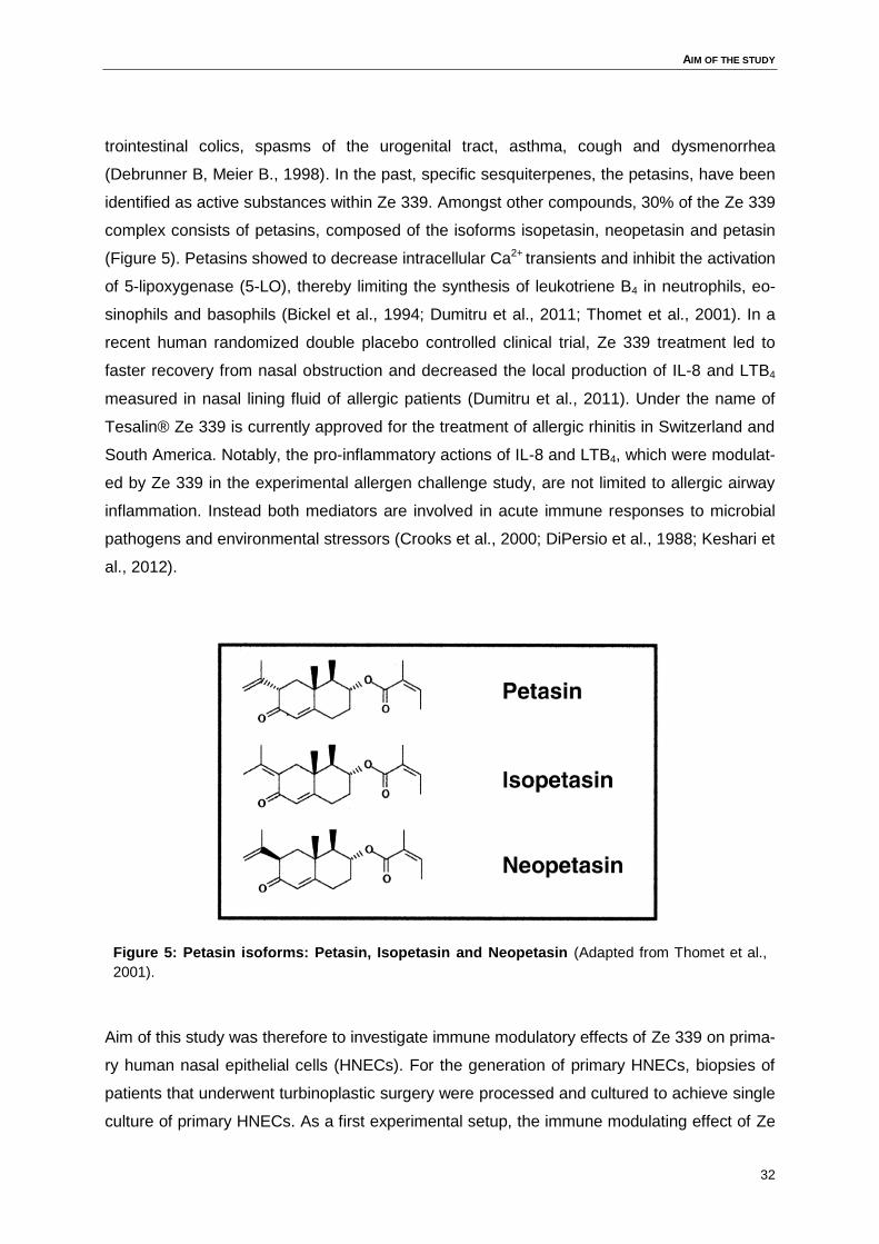
Figure 5: Petasin isoforms: Petasin, Isopetasin and Neopetasin ..........................................32
Figure 6: Neutrophil migration assay ...................................................................................44
Figure 7: The culturing of HNECs using different techniques. ...............................................50
Figure 8: Scanning electron micrograph ...............................................................................51
Figure 9: Characterisation of HNECs via FACS analysis. .....................................................52
Figure 10: IL-8 expression level of HNECs treated with Ze 339 (3µg/ml) or the solvent
control PEG. ............................................................................................................53
Figure 11: IL-8 expression level of HNECs treated with different isoforms of petasin and
the solvent controls. .................................................................................................53
Figure 12: Measurement of lactate dehydrogenase (LDH) activity in stimulated HNECs. .....54
Figure 13: IL-8 expression in PolyIC-treated HNECs in combination with Ze 339,
petasinmix or the single isoforms. ............................................................................55
Figure 14: Neutrophil migration towards supernatants ..........................................................57
Figure 15: Cytokine release of stimulated HNECs divided in atopic and non-atopic
donors. ....................................................................................................................59
Figure 16: Venn diagram of regulated genes: Ze 339/Montelukast .......................................60
Figure 17: Real time (RT)-PCR of selected cytokines in HNECs treated with PolyIC +/-
Ze 339. ....................................................................................................................63
Figure 18: Viral stimulation model in HNECs. .......................................................................65
Figure 19: Bacterial stimulation model in HNECs. ................................................................67
Figure 20: Cytokine-induced stimulation model in HNECs. ...................................................69
LIST OF FIGURES
8
Figure 21: Expression of cytokine-induced STAT1, STAT3 and STAT6 is reduced by Ze
339 in HNECs. .........................................................................................................71
Figure 22: Immunohistochemistry phospho-STATs/ total STATs in stimulated HNECs. .......72
Figure 23: Ze 339 inhibits cytokine-induced nuclear translocation of phospho-STATs. .........74
Figure 24: Ze339 - Mode of action ........................................................................................81
INTRODUCTION
9
1 Introduction
1.1 Inflammation and allergy
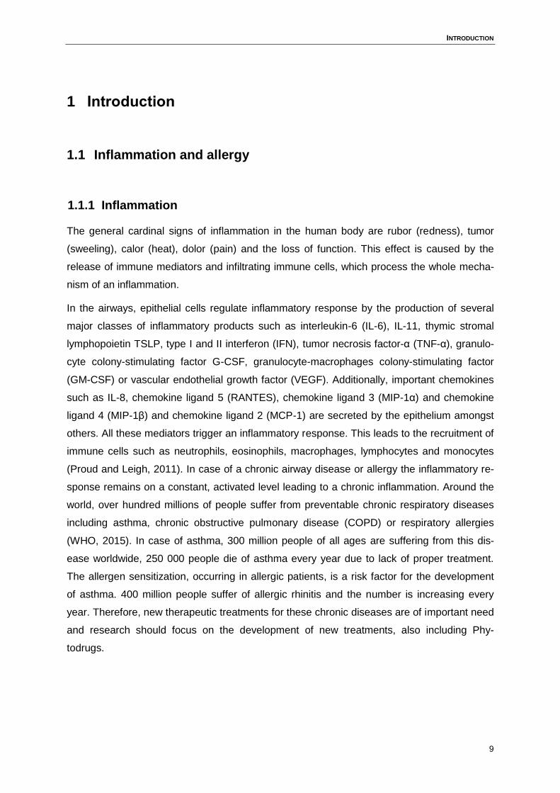
1.1.1 Inflammation
The general cardinal signs of inflammation in the human body are rubor (redness), tumor
(sweeling), calor (heat), dolor (pain) and the loss of function. This effect is caused by the
release of immune mediators and infiltrating immune cells, which process the whole mecha-
nism of an inflammation.
In the airways, epithelial cells regulate inflammatory response by the production of several
major classes of inflammatory products such as interleukin-6 (IL-6), IL-11, thymic stromal
lymphopoietin TSLP, type I and II interferon (IFN), tumor necrosis factor-α (TNF-α), granulo-
cyte colony-stimulating factor G-CSF, granulocyte-macrophages colony-stimulating factor
(GM-CSF) or vascular endothelial growth factor (VEGF). Additionally, important chemokines
such as IL-8, chemokine ligand 5 (RANTES), chemokine ligand 3 (MIP-1α) and chemokine
ligand 4 (MIP-1β) and chemokine ligand 2 (MCP-1) are secreted by the epithelium amongst
others. All these mediators trigger an inflammatory response. This leads to the recruitment of
immune cells such as neutrophils, eosinophils, macrophages, lymphocytes and monocytes
(Proud and Leigh, 2011). In case of a chronic airway disease or allergy the inflammatory re-
sponse remains on a constant, activated level leading to a chronic inflammation. Around the
world, over hundred millions of people suffer from preventable chronic respiratory diseases
including asthma, chronic obstructive pulmonary disease (COPD) or respiratory allergies
(WHO, 2015). In case of asthma, 300 million people of all ages are suffering from this dis-
ease worldwide, 250 000 people die of asthma every year due to lack of proper treatment.
The allergen sensitization, occurring in allergic patients, is a risk factor for the development
of asthma. 400 million people suffer of allergic rhinitis and the number is increasing every
year. Therefore, new therapeutic treatments for these chronic diseases are of important need
and research should focus on the development of new treatments, also including Phy-
todrugs.
INTRODUCTION
10
1.1.2 Allergy
In case of an allergic reaction the human body is exposed to a harmless antigen or allergen.
It gets in contact with the antigen or allergen for the first time and responds with the produc-
tion of immunoglobulin E (IgE) antibodies. This process is called sensitization; the body rec-
ognizes the allergen as foreign protein or glycoprotein without any allergic reaction. If the
body gets in contact with the same antigen or allergen again, a possible hyper-reaction of the
immune system can then lead to an allergic reaction (Janeway et al., 2002).
The epithelium plays a crucial role in the sensitization process. The immune mediators IL-33,
IL-25 and TSLP and additionally alarmins such as defensins or cathelicidin are secreted by
the epithelium to induce chemotactic migration and activate antigen-presenting cells (Chertov
et al., 1996; Ganz et al., 1985; Van Wetering et al., 1997; Yang et al., 1999). IL-33, IL-25 and
TSLP have shown to implicate the activation of type 2 innate lymphoid cells (ILC2) within the
epithelium (Suzukawa et al., 2012; Wills-Karp et al., 2012). Furthermore, T helper 2 cells
(Th2), IgE, mast cells and eosinophils play a major role in the development of an allergic
reaction. Dendritic cells (DCs) capture the entered antigens, process them and transport
them to draining lymph nodes. Here, naïve CD4+ T cells recognize the DC-presented pep-
tides of the antigen on the surface and differentiate into Th2 cells and follicular helper T cells
(TFh). Naïve CD4+ T cells can polarize into different Th-cell subsets through implementing
signals from their T-cell receptor, co-stimulatory molecules and cytokine receptors. This ena-
bles the host immune system to react on a broad range of pathogens and to specialise the
clearance of those pathogens (Holgate, 1998; Holt et al., 1990). The presence of IL-4, pro-
duced by the activated Th2 cells itself and various surrounding cell types as basophils, fur-
ther drives the differentiation into Th2 cells. Th2 cells, ILC2s and TFH cells activate B cells
that are specific for allergens through the actions of cluster of differentiation 40 (CD40) ligand
and the cytokines IL-4 and IL-13. IL-4, IL-13 and IL-5 are all cytokines produced by the acti-
vated Th2 cell. The activated ILC2 are able to produce IL-5 and IL-13 (Neill et al., 2010; Price
et al., 2010). Now, the activated B cells undergo heavy chain isotype switching from IgM to
IgE or IgG2 to IgG4 in humans. There are four types of hypersensitivity: type I-IV, whereby
type I hypersensitivity is associated with isotype switch from IgM to IgE. The produced IgE is
then present in the plasma, circulates in the system and binds to high affinity IgE receptors
(FcεRI) present, amongst others, on tissue mast cells and circulating basophilic granulo-
cytes. IgE binds to the Ig-like domain of the α-chain of the polypeptide chain structure. The β-
and γ-chain mediates the signal transduction responding to the IgE binding. The resting IgE-
coated mast cell gets activated by the cross-linking of bound IgE upon repeated antigen ex-
INTRODUCTION
11
posure. The antigen binds to IgE/FcεRI complex and finally activates the mast cell (Abbas et
al., 2012).
This process begins with the first exposure to the allergen and the activation of the Th2 cells
and the stimulation of B cells. This results in the production and secretion of IgE from the B
cells, continuing with the binding of IgE to the FcεRI on mast cells. The activation of these
mast cells after a repeated exposure to the allergen is called immediate hypersensitivity. This
reaction happens minutes after the repeated exposure to the antigen. But there is also a late
phase reaction, occurring 2-4 hours after the repeated exposure.
The physiological reaction of the body in the immediate reaction is the wheal and flare reac-
tion, which is dependent on IgE and mast cells. This reaction appears when the mast cell
gets activated by the antigen- IgE-FcεRI complex described above, resulting in the release of
the mediator histamine. Subsequently, histamine binds to the histamine receptors on endo-
thelial cells, leading to the synthesis and release of lipid mediators such as prostaglandin I2
(prostacyclin, PGI2), leukotriene C4 (LTC4), nitric oxide (NO) and platelet-activating factor
(PAF). All these mediators cause vascular leakage and vasodilation, and trigger leukocyte
extravasation. This inflammatory response, occurring a few hours after the immediate reac-
tion, is the so-called late-phase reaction. This phase includes the release of cytokines such
as TNF-α, IL-4, IL-13, IL-3, GM-CSF and chemokines such as MIP-1α and the already men-
tioned lipid mediators LTC4, LTD4, LTE4 and PAF. TNF-α activates endothelial cells to upreg-
ulate leukocyte adhesion molecules such as E-selectin and intracellular adhesion molecule-I
(ICAM-1) to accumulate inflammatory leucocytes, thereby promoting inflammatory reactions.
IL-4 and IL-13 promote the differentiation towards Th2 cells, IL-3, IL-5 and GM-CSF trigger
the development and activity of eosinophils. MIP-1α recruits monocytes, macrophages and
neutrophils to the side of inflammation. The release of leukotrienes causes contraction of the
smooth muscles (e.g. bronchial construction or intestinal construction) (Dahlén et al., 1980),
increase mucus production and maintain inflammatory processes in the tissue (Henderson et
al., 1996). PAF is chemotactic for leukocytes; it increases the production of lipid mediators
and activates neutrophils and eosinophils. Therefore, typical leucocytes of late-phase reac-
tion are eosinophils, Th2 cells and neutrophils (Abbas et al., 2012; Eyerich and Zielinski,
2014; Janeway et al., 2002).
The most common forms of allergic diseases nowadays are bronchial asthma, atopic derma-
titis (eczema), allergic rhinitis and food allergy (NIH, 2015). All forms of the allergic reaction
result in the described pro-inflammatory process in the body leading to pain and over-
reactivity of the immune system. Beside the common treatments of allergy, novel drug devel-
INTRODUCTION
12
opments and strategies focus on drugs which interrupt these inflammatory processes to pre-
vent an over-reaction of the immune system.
1.1.3 Current treatment of allergic diseases
In case of bronchial asthma, a repeated immediate-phase hypersensitivity and late-phase
allergic reaction of the lower airways causes the disease. It often coexists with bronchitis or
emphysema, which can lead to a severe damage of the lung tissue. Viral and bacterial infec-
tions in the respiratory tract can promote the development of asthma or exacerbate pre-
existing asthma. The current Global Initiative for Asthma guidelines (GINA) recommend
stepwise treatment to reach optimal asthma control. The treatment starts with inhaled corti-
costeroids (ICS), followed by an increase in the ICS dose or the addition of other controller
therapy, including long-acting β2-agonists (LABAs), leukotriene modifiers, or theophylline
(methylxanthine drug) to achieve control of resistant diseases. If the state of asthma is too
severe and add-on therapies cannot be achieved, anti-immunoglobulin E or oral glucocorti-
costeroids are recommended for the treatment (McIvor, 2015). Glucocorticosteroids (also
called glucocorticoids, corticosteroids or steroids) are the most common and effective thera-
py for patients suffering from asthma. They are acting on various cellular levels, e.g. sup-
pressing the production of adhesion molecules and chemotactic mediators and therefore
reducing the recruitment of inflammatory cells, including eosinophils, T cells, mast cells and
dendritic cells into the airways. Epithelial cells are one of the important players of the immune
defence in the airways, and regular treatment with ICS re-establishes the integrity of the epi-
thelial barrier of patients suffering from severe asthma (Kharitonov et al., 2002; Lilly et al.,
1997).
A key problem are patients suffering from a mild uncontrolled asthma, which have a high risk
of future exacerbations of the disease. Additionally, there are different impacts that can influ-
ence the progress of asthma as allergen exposure such as allergic rhinitis, concurrent smok-
ing or incorrect inhaler techniques. The different allergic asthma subgroups show a broad
range of variance in the pathophysiology of the disease. There are emerging therapeutic
options for the treatment of asthma in different development phases (1-3), including anti-
interleukin agents, chemoattractant receptor-homologous molecules expressed on T-helper
type 2 lymphocytes (CRTH2) antagonist, phosphodiesterase-4 inhibitors, and long-acting
muscarinic antagonists (LAMAs). A recent study suggested that the long-acting muscarinic
antagonist Tiotropium could be a promising option for the treatment of asthma and, in addi-
tion for the treatment of chronic obstructive pulmonary disease (COPD), helps as an add-on
INTRODUCTION
13
therapeutic to the common ICS (McIvor, 2015). LAMAs block the muscarinic acetylcholine
receptor and therefore have an impact on smooth muscle contraction, mucus secretion and
vasodilation (Beakes, 1997).
1.1.4 Allergy immunotherapy (AIT) using specific immunotherapy (SIT)
To control the progress of allergic asthma very few therapies are available. Current therapies
focus on controlling the inflammatory processes and controlling the symptoms. During allergy
immunotherapy (AIT), a treatment using a specific immunotherapy (SIT), patients get a grad-
ually increasing dose of a specific allergen over a long time period. This approach can poten-
tially modify the basic allergic mechanism of the disease. Indeed, in patients with mild and
moderate asthma, associated with allergic rhinoconjunctivitis and controlled by pharma-
cotherapy, subcutaneous immunotherapy (SCIT) and sublingual immunotherapy (SLIT) can
be a useful therapeutic regimen. In 1911, there was the first reported use of SCIT in the
treatment of allergic rhinitis by Leonard Noon, but in 1986, the British Committee of Safety of
Medicines reported 26 deaths connected with SCIT, thus different delivery methods (oral,
nasal and sublingual) had to be analysed in randomized controlled trials (Aboshady and
Elghanam, 2014; Brozek et al., 2010; Cox et al., 2006; Jutel et al., 2015; Wilson et al., 2003).
After years of research on different delivery methods, SLIT has been identified as a thera-
peutic option for patients suffering from seasonal rhinitis (hay fever) or perennial rhinitis
(house dust-mite), and patients uncontrolled by pharmacological treatments or patients that
are affected with systemic reaction from drug treatments. In many clinical studies, SLIT has
shown to modulate allergen-specific immune responses by induction of IL-10 producing al-
lergen-specific regulatory T cells, associated with a reduced IgE to IgG4 ratio. The blocking
antibody IgG competes with the present IgE antibody and prohibits the inflammatory re-
sponse to IgE release (Strait, 2006). Thus, SIT can lead to a shift in the immune response
from Th2 to Th1/ Tregs (Figure 1).
INTRODUCTION
14
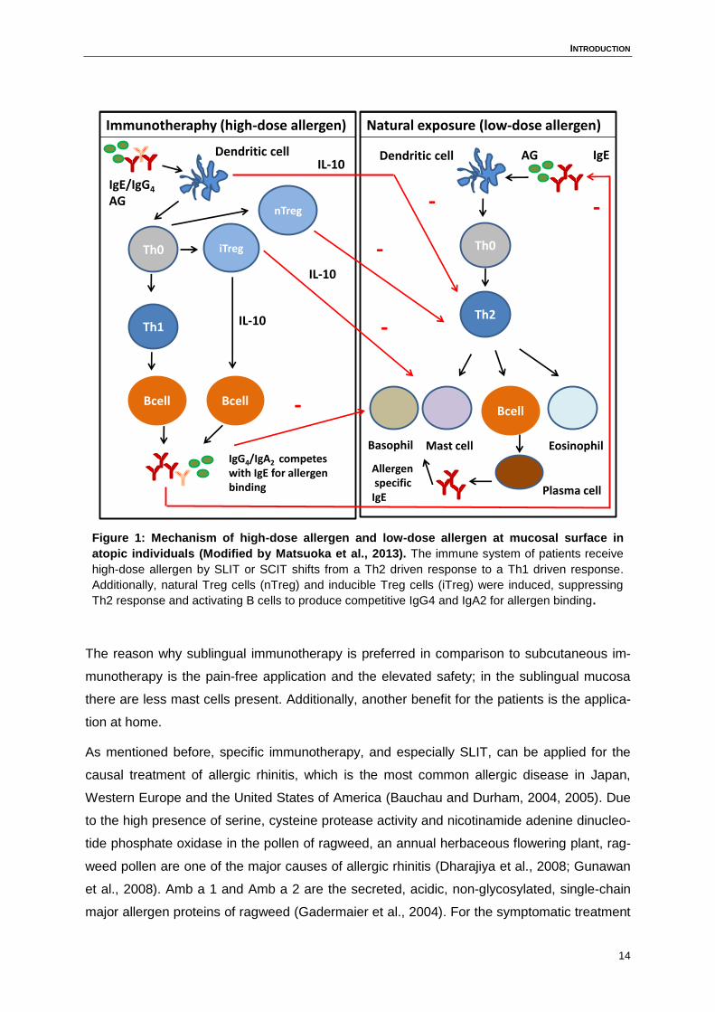
Figure 1: Mechanism of high-dose allergen and low-dose allergen at mucosal surface in
atopic individuals (Modified by Matsuoka et al., 2013). The immune system of patients receive
high-dose allergen by SLIT or SCIT shifts from a Th2 driven response to a Th1 driven response.
Additionally, natural Treg cells (nTreg) and inducible Treg cells (iTreg) were induced, suppressing
Th2 response and activating B cells to produce competitive IgG4 and IgA2 for allergen binding.
The reason why sublingual immunotherapy is preferred in comparison to subcutaneous im-
munotherapy is the pain-free application and the elevated safety; in the sublingual mucosa
there are less mast cells present. Additionally, another benefit for the patients is the applica-
tion at home.
As mentioned before, specific immunotherapy, and especially SLIT, can be applied for the
causal treatment of allergic rhinitis, which is the most common allergic disease in Japan,
Western Europe and the United States of America (Bauchau and Durham, 2004, 2005). Due
to the high presence of serine, cysteine protease activity and nicotinamide adenine dinucleo-
tide phosphate oxidase in the pollen of ragweed, an annual herbaceous flowering plant, rag-
weed pollen are one of the major causes of allergic rhinitis (Dharajiya et al., 2008; Gunawan
et al., 2008). Amb a 1 and Amb a 2 are the secreted, acidic, non-glycosylated, single-chain
major allergen proteins of ragweed (Gadermaier et al., 2004). For the symptomatic treatment
Immunotheraphy (high-dose allergen) Natural exposure (low-dose allergen)
Th0 iTreg
Th1
Bcell Bcell
Th0
Th2
nTreg
Bcell
Basophil Mast cell Eosinophil
IgE
IgE/IgG4
AG
AllergenspecificIgE
Plasma cell
IgG4/IgA2 competeswith IgE for allergen binding
Dendritic cell AG
-
-
-
-
Dendritic cell
-
IL-10
IL-10
IL-10
INTRODUCTION
15
of ragweed-induced rhinitis, first-generation H1-antihistamine, diphenhydramine and the
third-generation H1-antihistamine, desloratadine, applied orally, were effective in reducing
nasal allergic symptoms. This was evaluated in several clinical trials (Wilken et al., 2003).
Beside the oral application of antihistamines, a topical treatment with glucocorticosteroids is
the most commonly used and clinically effective treatment for allergic rhinitis. But the treat-
ment with glucocorticosteroids and antihistamine can also cause side effect such as osteopo-
rosis, growth retardation, croakiness or liver stress. To avoid this, new strategies beside the
conventional medicine strategies have to be developed.
1.1.5 Phytotherapy
An alternative way to treat allergies and inflammatory diseases such as asthma is the use of
phytodrugs, also called phytochemicals. Phytodrugs represent the active compounds of plant
products, gained via different procedures. Plants have been the basis of many medicinal
regimens worldwide for the last thousands of years and researchers still discover new active
beneficial compounds in plants. Newly found chemical plant substances are the basis to de-
velop novel innovative drugs (Jachak S and Saklani A, 2007). The first isolated active com-
pound in the early 19th century was morphine from opium, followed by the isolation of early
drugs as cocaine, codeine, digitoxin and quinine (Mann, 1999; Wainwright, 1991), of which
some are still in use. 250,000 flowering plants species exist worldwide. Half of them are
found in the tropical forest. A big success in the field for drug discovery from plants, and the
proven beneficial effects in humans resulted in the development of anti-cancer and anti-
bacterial agents, e.g. chemotherapeutic agents such as paclitaxel, hycamptamine or 9-
aminocamptothecin (Cragg et al., 1993) and the antibacterial filtrate penicillin by Fleming in
1928 (Joklik, 1996). To discover the active drug compounds from plants, various methods of
analysis and multidisciplinary approaches are involved, including botanical, ethnobotanical,
phytochemical and biological techniques (Jachak S and Saklani A, 2007). Plants produce a
wide range of diverse organic compounds, classified into primary and secondary metabolites.
The primary metabolites, such as phytosterols, acyl lipids, nucleotides, amino acids and or-
ganic acids are found in all plants and are essential for growth and development. The sec-
ondary metabolites are not directly involved in the process of growth and development, but
appear in a wide variety within the plants (Croteau et al., 2000). Due to their biosynthetic
origins, phytochemicals can be classified into terpenoides (carotenoids), phenolics, alkaloids,
nitrogen-containing compounds and organosulfur compounds (Liu, 2004). Nowadays, sever-
al studies worldwide have proven the protective effects of phytochemicals present in plants
INTRODUCTION
16
against acute, chronic and degenerative diseases (Arya and Arya, 2011; Nichenametla et al.,
2006).
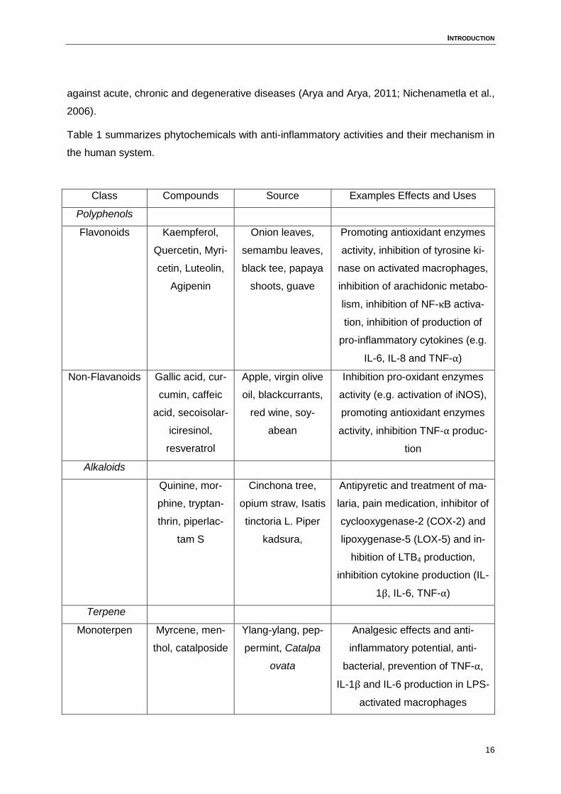
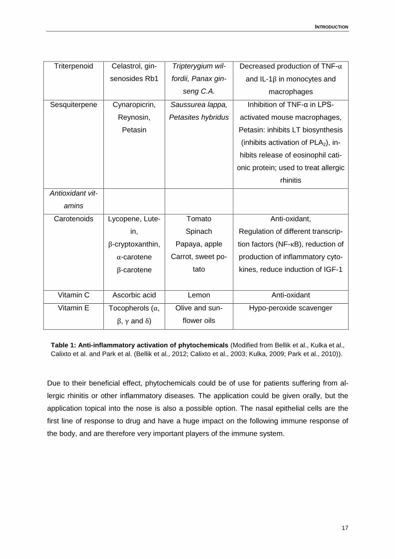
Table 1 summarizes phytochemicals with anti-inflammatory activities and their mechanism in
the human system.
Class Compounds Source Examples Effects and Uses
Polyphenols
Flavonoids Kaempferol,
Quercetin, Myri-
cetin, Luteolin,
Agipenin
Onion leaves,
semambu leaves,
black tee, papaya
shoots, guave
Promoting antioxidant enzymes
activity, inhibition of tyrosine ki-
nase on activated macrophages,
inhibition of arachidonic metabo-
lism, inhibition of NF-κB activa-
tion, inhibition of production of
pro-inflammatory cytokines (e.g.
IL-6, IL-8 and TNF-α)
Non-Flavanoids Gallic acid, cur-
cumin, caffeic
acid, secoisolar-
iciresinol,
resveratrol
Apple, virgin olive
oil, blackcurrants,
red wine, soy-
abean
Inhibition pro-oxidant enzymes
activity (e.g. activation of iNOS),
promoting antioxidant enzymes
activity, inhibition TNF-α produc-
tion
Alkaloids
Quinine, mor-
phine, tryptan-
thrin, piperlac-
tam S
Cinchona tree,
opium straw, Isatis
tinctoria L. Piper
kadsura,
Antipyretic and treatment of ma-
laria, pain medication, inhibitor of
cyclooxygenase-2 (COX-2) and
lipoxygenase-5 (LOX-5) and in-
hibition of LTB4 production,
inhibition cytokine production (IL-
1β, IL-6, TNF-α)
Terpene
Monoterpen Myrcene, men-
thol, catalposide
Ylang-ylang, pep-
permint, Catalpa
ovata
Analgesic effects and anti-
inflammatory potential, anti-
bacterial, prevention of TNF-α,
IL-1β and IL-6 production in LPS-
activated macrophages
INTRODUCTION
17
Triterpenoid Celastrol, gin-
senosides Rb1
Tripterygium wil-
fordii, Panax gin-
seng C.A.
Decreased production of TNF-α
and IL-1β in monocytes and
macrophages
Sesquiterpene Cynaropicrin,
Reynosin,
Petasin
Saussurea lappa,
Petasites hybridus
Inhibition of TNF-α in LPS-
activated mouse macrophages,
Petasin: inhibits LT biosynthesis
(inhibits activation of PLA2), in-
hibits release of eosinophil cati-
onic protein; used to treat allergic
rhinitis
Antioxidant vit-
amins
Carotenoids Lycopene, Lute-
in,
β-cryptoxanthin,
α-carotene
β-carotene
Tomato
Spinach
Papaya, apple
Carrot, sweet po-
tato
Anti-oxidant,
Regulation of different transcrip-
tion factors (NF-κB), reduction of
production of inflammatory cyto-
kines, reduce induction of IGF-1
Vitamin C Ascorbic acid Lemon Anti-oxidant
Vitamin E Tocopherols (α,
β, γ and δ)
Olive and sun-
flower oils
Hypo-peroxide scavenger
Table 1: Anti-inflammatory activation of phytochemicals (Modified from Bellik et al., Kulka et al.,
Calixto et al. and Park et al. (Bellik et al., 2012; Calixto et al., 2003; Kulka, 2009; Park et al., 2010)).
Due to their beneficial effect, phytochemicals could be of use for patients suffering from al-
lergic rhinitis or other inflammatory diseases. The application could be given orally, but the
application topical into the nose is also a possible option. The nasal epithelial cells are the
first line of response to drug and have a huge impact on the following immune response of
the body, and are therefore very important players of the immune system.
INTRODUCTION
18
1.2 Cellular players in the airway epithelium
1.2.1 Cells of the airway epithelium
As already mentioned, the immune system provides an enormous variety of cellular compo-
nents protecting the host and regulating the immune reactions. The epithelial cells of the air-
ways regulate both the innate and the adaptive immune system by controlling the production
of immune molecules and by managing the physical interaction with cells of the immune sys-
tem. To protect the airways from environmental hazards, the airway epithelium provides a
vital protective layer between the internal milieu of the lung and the external environment
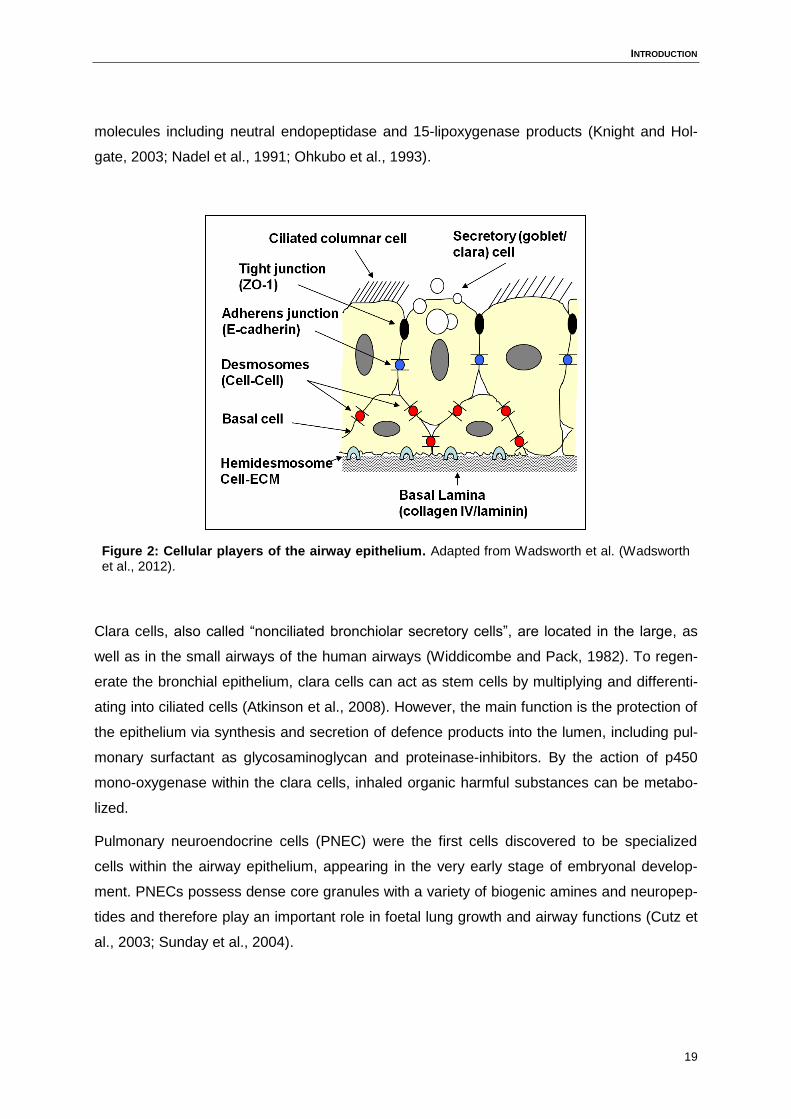
(Boucher, 1994). Regarding the difference in function, ultrastructure and biochemical criteria,
the distinct epithelial cell types can be classified in three categories: basal, ciliated and se-
cretory cells (Figure 2). Dependent on epithelium-produced mediators, inflammatory cells,
immune cells and phagocytic cells are allowed to transmigrate to the lumen (Schon-Hegrad
et al., 1991). The most frequent cell types in the airways are the columnar ciliated epithelial
cells. They arise from either basal or secretory cells and represent 50% of all epithelial cells.
The surface of the cells is covered with up to 300 cilia per cell, indicating their main function
which is the directional transport of mucus from the lung to the throat.
Mucous cells, also called goblet cells, are important for the production and secretion of acid
mucin to prevent the airways from invading pathogens and noxious particles. The correct
amount of mucus and the viscoelasticity are important for efficient mucociliary clearance. An
increased mucus release can be caused by the inhalation of noxious substances including
sulphur dioxide, occurring with coal combustion, or tobacco smoke. In chronic airway in-
flammatory diseases, such as asthma or chronic bronchitis, the disease pattern shows a mu-
cous cell hyperplasia and metaplasia, which is thought to be a reason for abundant cough
and elevated mucus production, accompanying these diseases (Lumsden et al., 1984).
Basal cells play an important role in the structure of the airway epithelium, they are ubiqui-
tous and the only cells that are firmly attached to the basement membrane (Evans and Plop-
per, 1988; Hicks et al., 1997). Thus, they are important for the attachment of other cells to
the basement membrane via hemidesmosomal complexes (Evans et al., 1989), and basal
cells are the only cells expressing hemidesmosomes. The interepithelial attachment between
the cells of the epithelium is mediated by desmosomes. Mucous and ciliated cells arise from
basal cells in the large airways, although basal cells are less present in the smaller airways
and clara cells also perform the stem cell role in these segments. Beside the progenitor and
structural role of the basal cells, they also have a functional impact via secreting bioactive
INTRODUCTION
19
molecules including neutral endopeptidase and 15-lipoxygenase products (Knight and Hol-
gate, 2003; Nadel et al., 1991; Ohkubo et al., 1993).
Figure 2: Cellular players of the airway epithelium. Adapted from Wadsworth et al. (Wadsworth et al., 2012).
Clara cells, also called “nonciliated bronchiolar secretory cells”, are located in the large, as
well as in the small airways of the human airways (Widdicombe and Pack, 1982). To regen-
erate the bronchial epithelium, clara cells can act as stem cells by multiplying and differenti-
ating into ciliated cells (Atkinson et al., 2008). However, the main function is the protection of
the epithelium via synthesis and secretion of defence products into the lumen, including pul-
monary surfactant as glycosaminoglycan and proteinase-inhibitors. By the action of p450
mono-oxygenase within the clara cells, inhaled organic harmful substances can be metabo-
lized.
Pulmonary neuroendocrine cells (PNEC) were the first cells discovered to be specialized
cells within the airway epithelium, appearing in the very early stage of embryonal develop-
ment. PNECs possess dense core granules with a variety of biogenic amines and neuropep-
tides and therefore play an important role in foetal lung growth and airway functions (Cutz et
al., 2003; Sunday et al., 2004).
INTRODUCTION
20
The cells of the airway epithelium have a tendency to form adhesive contact with adjacent
epithelial cells to generate epithelial cell-cell junctions, thus they form a physical barrier. This
is another important mechanism of defence against inhaled pathogens.
1.2.2 The physical barrier of the airway epithelium
The apical junction complex of the physical barrier of the cells consists of tight junctions and
adherens junctions. The tight junctions are intracellular junctions, located at the lateral mem-
brane surface at the apical end of the cell. The central function consist of the variable per-
meable selectively barrier, the gate function, controlling the passage of ions, water and vari-
ous macromolecules, and the fence function, assigning the polarity of the epithelium through
function as a demarcation between the apical and basolateral domains of the membrane
(Sawada et al., 2003). The transmembrane proteins of the tight junctions consist of occludin,
claudins and ZO-1 (Zonula Occludens), ZO-2 and ZO-3 which are linked to the actin cyto-
skeleton (Fanning et al., 1998). The permeability of tight junctions can be modified by cyto-
kines such as TNF-α, IFN-γ, interleukins and growth factors. The second part of the apical
junction complex, the adherens junctions, is composed of E-cadherin, linked to the actin cy-
toskeleton via the α- and β-catenin adapter complex (Figure 2). Adherence junctions play an
important role in the development of the epithelial layer and tight junction formation (Fristrom,
1988). This barrier function of the epithelium is deficient in patients suffering from asthma, as
a post-transcriptional loss of junctional ZO-1 and occluding was observed in lung biopsies of
asthma patients. Furthermore, in differentiated bronchial epithelial cells from donors with
severe asthma, a reduced level of E-cadherin and an increased epithelial macromolecular
permeability was detected (Xiao et al., 2011). Additionally, the barrier of cell-cell junctions
can prohibit the entrance and dissemination of virus particles through the epithelium (Yoon
and Spear, 2002).
Beside the physical barrier of the airway epithelium, there is also a chemical barrier to protect
the host from pathogens. The production of mucus is the most important part of this chemical
defence against microbes.
1.2.3 The chemical barrier of the airway epithelium
As mentioned before, goblet cells are the source of mucus, which creates a semipermeable
barrier at the epithelial surface. The exchange of nutrients, water and gases is enabled, but
most pathogens are not capable to penetrate the mucus. Beside a large variety of different
INTRODUCTION
21
protective proteins such as antimicrobial peptides, cytokines and antioxidant substances,
mucus is rich of mucins, extensively glycosylated proteins, which vary in cellular location,
complex formation and glycosylation states (Williams et al., 2006). Nowadays, 11 mucins
have been detected in human airways. However, MUC5AC and MUC5B are the important
mucins in human sputum and are mainly regulated by nuclear factor κB (NF-κB) and/or
Specificity Protein 1 (Sp1) (Rose et al., 2001). MUC5B represents the principal mucin under
normal condition, whereas in asthma, MUC5AC production appears to be up-regulated lead-
ing to an asthmatic mucus hypersecretion (Evans et al., 2009). New therapeutic strategies
for patients suffering from a mucus overexpression due to an airway disease are of big need.
Treatments could interrupt the signalling pathway of NF-κB or Sp1 and therefore regulate the
mucus production.
1.2.4 The immunologic barrier of the airway epithelium
When inhaled pathogenic substances penetrate the physical and chemical barriers of the
airway epithelium, cells of the airways have the ability to recognize pathogen-associated mo-
lecular pattern (PAMPs) rapidly through pattern recognition receptors (PRRs) such as Toll-
like receptors (TLRs) and intracellular receptors. PAMPs, which are recognized by TLRs, are
pathogen specific molecules, derived from viruses, fungi, bacteria and parasites (Poltorak,
1998; Underhill et al., 1999). The recognition of PAMPs and the initiation of immune re-
sponses via binding and activation of the TLR pathways are essential for the respiratory epi-
thelial defence. TLRs trigger their signal pathway via several key transcription factors such
as NF-κB and interferon regulatory factor 3 and 7 (IRF-3 and IRF-7) or via activating mito-
gen-activated protein kinase (MAP), resulting in the induction of numerous pro-inflammatory
cytokines, chemokines and type I and type III interferons (IFNs) (Iwamura and Nakayama,
2008).
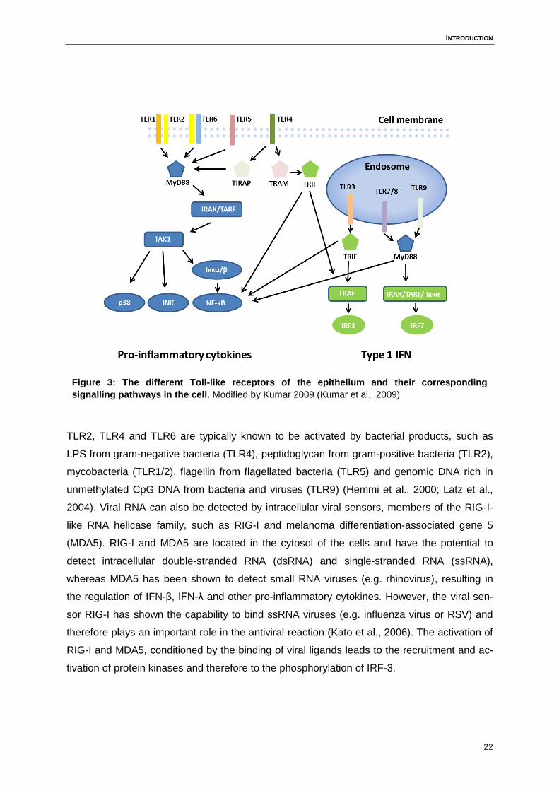
The localisation of the TLRs differs depending on their preference for intra- or extracellular
pathogens: TLR1, TLR2, TLR4, TLR5 and TLR6 are expressed on the cell surface of the
epithelium, whereas TLR3, TLR7, TLR8 and TLR9 are located in intracellular vesicles. The
signalling cascade of TLR7 and TLR9 act via using the adaptor MyD88, the myeloid differen-
tiation primary response gene 88, for the activation of NF-κB and IFR-7 to initiate the produc-
tion of pro-inflammatory cytokines such as IL-6 and TNF-α, and the production of type I inter-
ferons (Honda et al., 2004). Except for TLR3 signalling, all Toll-like receptors signal via
MyD88/IRAK-dependent pathway. TLR3 signalling instead functions via the TRIF pathway
(Figure 3).
INTRODUCTION
22
Figure 3: The different Toll-like receptors of the epithelium and their corresponding
signalling pathways in the cell. Modified by Kumar 2009 (Kumar et al., 2009)
TLR2, TLR4 and TLR6 are typically known to be activated by bacterial products, such as
LPS from gram-negative bacteria (TLR4), peptidoglycan from gram-positive bacteria (TLR2),
mycobacteria (TLR1/2), flagellin from flagellated bacteria (TLR5) and genomic DNA rich in
unmethylated CpG DNA from bacteria and viruses (TLR9) (Hemmi et al., 2000; Latz et al.,
2004). Viral RNA can also be detected by intracellular viral sensors, members of the RIG-I-
like RNA helicase family, such as RIG-I and melanoma differentiation-associated gene 5
(MDA5). RIG-I and MDA5 are located in the cytosol of the cells and have the potential to
detect intracellular double-stranded RNA (dsRNA) and single-stranded RNA (ssRNA),
whereas MDA5 has been shown to detect small RNA viruses (e.g. rhinovirus), resulting in
the regulation of IFN-β, IFN-λ and other pro-inflammatory cytokines. However, the viral sen-
sor RIG-I has shown the capability to bind ssRNA viruses (e.g. influenza virus or RSV) and
therefore plays an important role in the antiviral reaction (Kato et al., 2006). The activation of
RIG-I and MDA5, conditioned by the binding of viral ligands leads to the recruitment and ac-
tivation of protein kinases and therefore to the phosphorylation of IRF-3.
INTRODUCTION
23
1.2.5 The infiltrating immune cells of the airway epithelium
In addition to the protection of the host via the mechanisms of the airway epithelium itself,
infiltrating immune cells play a major role in the barrier defence. The coordination and regula-
tion of migrating immune cells is dependent on the presence of chemokines. Chemokines do
not only control the migration of immune cells, but also the interaction between the immune
cells. The rhodopsin-like seven-transmembrane-spanning chemokine receptors are G-
protein-coupled and located at the surface of the cells. The chemokine receptors have a dif-
ferent specificity of binding to their ligands, meaning that CC chemokine ligands (CCL) only
bind to the CCR chemokine receptors whereas CXC chemokine ligands (CXCL) can only
bind to CXCR receptors.
To promote an efficient antiviral response, the recruitment of various immune cells is essen-
tial. If the airway epithelium is infected by a virus, secreted chemokines and cytokines are
crucial to promote neutrophil, eosinophil, NK cell and monocyte recruitment from the blood
stream to the side of infection in the tissue.
Early in response, neutrophils are recruited in response to IL-8/CXCL8, Gro-α/CXCL1 and
ENA-78/CXCL5. Pizzichini et al. showed an increase of neutrophils in induced sputum at day
4 of a natural cold in asthmatics and non-asthmatics in line with an increasing IL-8 level with-
in the sputum (Pizzichini et al., 1998). A recent study reported that CXCR4 signalling may
promote the accumulation of neutrophils in the lung vasculature so that they can be rapidly
mobilized (Devi et al., 2013). However, the up-regulation of CXCR4 on neutrophils may also
promote re-entry into the bone marrow and therefore eliminate the neutrophils via apoptotic
cell death (Martin et al., 2003).
The recruitment and activation of eosinophils to the lung epithelium is dependent on the in-
fluence of RANTES/CCL5, eotaxin-1/CCL11, eotaxin-2/CCL24, granulocyte-macrophage
colony-stimulating factor (GM-CSF) and IL-5. The antiviral, cytotoxic effect of eosinophils is
due to the release of eosinophil-derived neurotoxin and eosinophil cationic protein (ECP)
which are both members of the ribonuclease A superfamily (RNase A) (Kimpen et al., 1992;
Rosenberg and Domachowske, 2001; Slifman et al., 1986). Levels of RANTES are increased
in nasal secretions of children with natural virus-induced asthma (Teran et al., 1999) and in
primary nasal epithelial cell cultures, infected with respiratory syncytial virus (RSV) (Saito et
al., 1997).
The function of NK cells is to eliminate infected target cells and to modulate the adaptive
immunity towards an effective antiviral response. NK cells itself rapidly produce pro-
inflammatory cytokines, such as IFN-γ, at an early point of an anti-viral immune response,
INTRODUCTION
24
which is important for the antigen-independent activation of antigen-presenting cells. The
recruitment and activation of NK cells at the site of infection is dependent on IFN-α/β and
macrophage inflammatory protein-1α (MIP-1α/CCL3) (Biron et al., 1999).
The production of IL-1β, MIP-1α/CCL3, monocyte chemoattractant protein 1 (MCP-1)/CCL2
and TNF-α by the airway epithelium during viral infection lead to the recruitment of mono-
cytes/macrophages. Macrophages play a central role in antiviral immune responses by inter-
nalizing and processing viral antigens for presentation to cells of the adaptive immune sys-
tem. Additionally, the release of cytokines such as IL-12 and IL-10 leads to different immune
modulations such as NK cell activation, regulation of Th1 responses and elevated B-cell sur-
vival, proliferation and antibody production (D’Andrea et al., 1992, 1993; See and Wark,
2008).
Beside the regulation of the innate immune response via the airway epithelium, the control
and modulation of the adaptive immune response through interaction of DCs, T cells and B
cells is also concerted by the epithelium.
Infiltrating and local DCs have an important role in the initiation of the adaptive immune re-
sponse via inducing the proliferation and activation of T cells in the lung epithelium. There
are two major types of pulmonary DCs, the conventional DCs (cDCs or myeloid DCs/mDCs)
and plasmacytoid DCs (pDCs) (Siegal, 1999). Viral infections increase the numbers of cDCs
and pDCs in the lung (Smit et al., 2006). The main task of cDCs is the uptake of foreign anti-
gens, leading to the maturation process of the cDCs. Starting with the down-regulation of
chemokine receptors, involved in homing to the lungs, cDCs up-regulate the expression of
CCR7, which is a chemokine receptor that leads to the migration of the cDCs to the lymph
nodes. Reaching the lymph nodes, cDCs mature into antigen-presenting cells characterized
by the expression of CD80, CD86 and major histocompatibility complex (MHC) I and II to
stimulate CD8+ and CD4+ T cells. The secretion of airway epithelium derived cytokines, such
as IL-15 or TSLP plays an important role in the differentiation and maturation of DCs. IL-15
was shown to induce the differentiation of DCs from monocytes, while TSLP has been shown
to induce DC-mediated Th2 differentiation (Rate et al., 2009; Regamey et al., 2007). cDCs
themselves are major producers of IL-12. Compared to the immune function of cDCs, pDCs
lack the ability to process and present antigens. Instead, the main function of pDCs is the
production of IFN-α in large amounts. The produced IFN-α triggers a powerful innate immune
response and therefore limits the viral replication. pDCs also migrate into draining lymph
nodes where they secrete type I IFNs and IL-12, thus promoting a protective Th1 response
(Cella et al., 1999; Smit et al., 2008). pDCs and cDCs also differ in the expression of TLRs
on their surface: human cDCs express primarily TLR2 and TLR4, responding to viral proteins
INTRODUCTION
25
(e.g. RSV fusion protein), whereas pDCs express TLR7 and TLR9 and therefore recognize
single-stranded RNA and viral DNA.
In case of a viral infection, the airway epithelium recruits T cells into the mucosa by produc-
ing RANTES/CCL5 and IP-10/CXCL10 for the infiltration of Th1 cells. Additionally, CCL1 and
CCL17/TARC are produced to induce the migration of Th2 cells (Montes-Vizuet et al., 2006;
Saito et al., 1997; Sauty et al., 1999; Spurrell et al., 2005). Th1 and Th2 cells are both im-
portant for the antiviral adaptive immune response, Th1 cells via production of IFN-γ, IL-12,
TNF-α and IL-2. Macrophages get activated by IFN-γ to efficiently kill intracellular microbes,
to produce co-stimulatory molecules and to up-regulate the expression of MHC-II receptors.
Additionally, IL-12 and IFN-γ are important for the differentiation of Th1 cells. Infiltrated Th2
cells in the airway epithelium produce IL-4, IL-5 and IL-13, which initiates the activation of
mast cells and eosinophils and induces mucus secretion. Furthermore, secreted Th2 cyto-
kines play an important role in humoral immunity against viruses by co-activating B cells. The
upstream regulation of IL-4, IL-5 and IL-13 is dependent on epithelial derived cytokines such
as IL-25, IL-33 and TSLP, which are important mediators in allergic inflammation (Schmitz et
al., 2005; Soumelis et al., 2002; Wang et al., 2007). The airway epithelium has been identi-
fied as a major source of these cytokines. When the epithelium responds to an environmen-
tal exposure, released IL-33 is activating mast cells in the epithelium and additionally pro-
motes the development of type 2 innate lymphoid cells (ILC2s) in combination with IL-25.
Activated ILC2s secrete a Th2 cytokine profile, including IL-9, IL-15 and IL-13, thus inducing
a Th2 response (Angkasekwinai et al., 2007). In case of severe asthma, levels of TSLP, IL-
33 and IL-25 are elevated in the airways of the patients (Ballantyne et al., 2007; Préfontaine
et al., 2010; Ying et al., 2005). In a controlled and normal immune system, there is an im-
portant balance between Th1 and Th2. The normal T-cell response to virus infection is regu-
lated by a Th1 response of the host.
A structural change of the airway epithelium in patients suffering for airway diseases such as
asthma, is called airway remodelling. It is characterised by changes including disruption and
thickening of the epithelium together with goblet cell metaplasia (Aikawa et al., 1992; Be-
nayoun et al., 2003). These modifications result downstream in changes regarding the depo-
sition of collagens and other extracellular matrix proteins. This includes amongst others
smooth muscle hyperplasia and hypertrophy, generation of myofibroblasts, subepithelial fi-
brosis and increasing angiogenesis (Siddiqui et al., 2007). In the histopathology in the lungs
of asthmatics, all these structural changes are present, thus airway remodelling is a hallmark
of asthma and often occurs in parallel with airway inflammation (Busse, 2010). The possibility
INTRODUCTION
26
to development asthma in early childhood (first 2-3 years of life) is significantly promoted by
the presence of viral infections (Sigurs et al., 2000).
Infiltrating immune cells, which migrate into the tissue in case of an inflammation, control and
regulate the response of the immune system. If there is an imbalance due to a chronic or
inflammatory disease, the overreaction leads to an abnormal recruitment of immune cells
and in a second consequence it can lead to airway remodelling. Thus, the regulation of the
migration rate of immune cells, direct or indirect by mediators, represents an interesting ap-
proach in the design of new drug developments.
1.3 Cytokine signalling in airway epithelium
As described before, the recognition of pathogens is mediated via binding of the molecules to
PRRs on innate immune cells or the epithelium, which triggers a rapid intracellular signal
transduction. These complex processes alter and educate the host immune system. One of
the most essential responses initiated by PRR signalling is the synthesis and secretion of
cytokines. Cytokine receptors can be classified in six groups; Interleukin type I cytokine re-
ceptor (1), interleukin type II cytokine receptor (2), immunoglobulin (Ig) superfamily (3), tu-
mour necrosis factor receptor (TNFR) family (4), IL-17 family receptor (5) and the chemokine
receptors that couple to G proteins (6) (Abbas et al., 2012; Yao et al., 1997).
Cytokines binding to the type I cytokine receptors are 4-α-helical bundle proteins, such as G-
CSF, IL-3, IL-5, IL-6 and IL-11, but there is also IL-2 that binds to the type I receptor, which
requires the common γ chain of the receptor. The cytokine release after binding and activa-
tion of the common γ chain receptor play an important role in B and T cell development and
polarization and therefore in the immune defence against pathogens.
Cytokines with the ability to bind to type II cytokine receptors can be divided into four sub-
groups; IL-10 family members, type I, II and type III interferon family members (e.g. IFN-α,
IFN-β, IFN-γ and IFN-λ). Members of the IL-10 family including IL-19, IL-20, IL-22 and IL-24
have been shown to have various effects on the skin such as proliferation and differentiation
of keratinocytes (Blumberg et al., 2001; Kunz et al., 2006; Poindexter et al., 2010).
The Ig superfamily receptor provides an extracellular immunoglobulin (Ig)-like domain, and
IL-1 family cytokines can bind to the receptor. The IL-1 family members IL-1α and IL-1β have
been shown to play a role in epidermal differentiation and skin disease (Wood et al., 1992;
Yano et al., 2008). A member of the IL-1 family, IL-33, has the ability to bind to the IL-1 re-
ceptor related protein ST2, which is specifically expressed on Th2 cells. This leads to the
INTRODUCTION
27
enhanced production of Th2 cell cytokines including IL-4, IL-5 and IL-13 (Schmitz et al.,
2005).
TNF receptors bind ligands belonging to the TNF family, expressed by various cells of the
immune system including B cells, T cells, NK cells, dendritic cells and monocytes.
IL-17A and IL-25 (IL-17E) bind to the IL-17 family receptors, both cytokines are important
regarding the skin barrier.
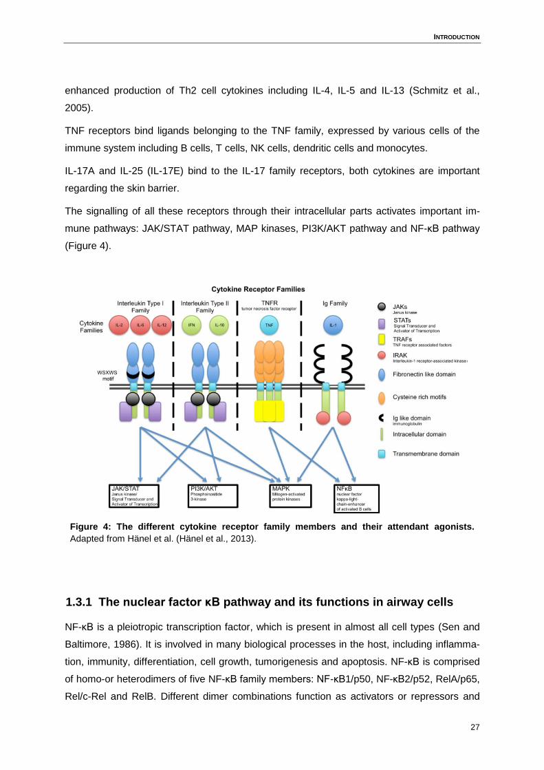
The signalling of all these receptors through their intracellular parts activates important im-
mune pathways: JAK/STAT pathway, MAP kinases, PI3K/AKT pathway and NF-κB pathway
(Figure 4).
Figure 4: The different cytokine receptor family members and their attendant agonists.
Adapted from Hänel et al. (Hänel et al., 2013).
1.3.1 The nuclear factor κB pathway and its functions in airway cells
NF-κB is a pleiotropic transcription factor, which is present in almost all cell types (Sen and
Baltimore, 1986). It is involved in many biological processes in the host, including inflamma-
tion, immunity, differentiation, cell growth, tumorigenesis and apoptosis. NF-κB is comprised
of homo-or heterodimers of five NF-κB family members: NF-κB1/p50, NF-κB2/p52, RelA/p65,
Rel/c-Rel and RelB. Different dimer combinations function as activators or repressors and
INTRODUCTION
28
this is dependent on post-translational modifications and subcellular location as well as on
the interaction with co-factors and co-repressors. If NF-κB binds to a family member of the
NF-κB inhibitor (IκB), it remains at an inactive state and is hold in the cytoplasm of the cell.
The conversion into the active form occurs if IκB is phosphorylated by IκB kinase (IKK) and
therefore dissociates from the NF-κB complex (DiDonato et al., 1997; Mercurio et al., 1997).
This leads to the translocation into the nucleus where the complex binds to the promoter of
NF-κB, followed by the transcription of various important genes. The signalling of NF-κB can
be mediated through the classical or the non-classical pathway. The classical pathway in-
volves IKK and is activated by TNF-α, the non-classical pathway is IKK complex independent
and is triggered by cytokines such as lymphotoxin, CD40 ligand or the Epstein-Barr virus.
Notably, beside these two pathways, NF-κB activation can also be triggered by divergent
stimuli including IL-2, IL-4, IL-8, IL-12, LPS, bacteria, dsRNA or if the epithelium is exposed
to environmental hazards such as cigarette smoke, lead or extreme pH (Batra et al., 2011).
In airway diseases including asthma and COPD, infiltrating immune cells express specific
mediators, such as cytokines, chemokines and cell adhesion molecules which act in an auto-
crine or paracrine fashion on the inflammatory cells themselves, but in addition also act on
epithelial cells. NF-κB plays an important role in regulating the expression of inflammatory
genes such as IL-6, IL-8, MIP-2 and TNF-α in these airway cells (Collart et al., 1990; Driscoll
et al., 1995; Harant et al., 1996; Lee et al., 2004; Poynter et al., 2002; Sancéau et al., 1995;
Shakhov et al., 1990).
Infiltrated lymphocytes have important immune-modulating effects in different asthma endo-
types. In neutrophilic asthma, there is a dominant Th1 and Th17 response; Th17 cells pro-
duce elevated levels of IL-17A, IL-17F and IL-22. IL-17A has been shown to induce epithelial
mucus production through the NF-κB pathway (Fujisawa et al., 2011), whereas IL-22 is in-
volved in the survival of airway smooth muscle cells, triggered through the NF-κB pathway
(Chang et al., 2012). The production of IL-4, IL-5 and IL-13 by Th2 cells in the airways of
patients suffering from eosinophilic allergic asthma is NF-κB dependent. The expressed cy-
tokines lead then to an enhanced IgE production by stimulated B cells. The enhanced pro-
duction of IL-8, TNF-α and intercellular adhesion molecule (ICAM)-1 through eosinophils is
also dependent on the NF-κB pathway. The recruitment of neutrophils in airway diseases is
amongst others IL-8 mediated, which is a NF-κB regulated gene. The reduction of IL-8 ex-
pression, as well as the downregulation of the transcriptional activity of NF- κB, could be of
benefit for patients suffering from inflammatory airway diseases and are therefore an inter-
esting point of action for therapeutic strategies.
INTRODUCTION
29
1.3.2 The signal transducer and activator of transcription pathway and
its function in airway cells
Other important pathways of the immune system are the signal transducer and activator of
transcription (STAT) pathways. To date, seven STAT family members in mammals are
known: STAT1, STAT2, STAT3, STAT4, STAT5A, STAT5B and STAT6. The activation of
STATs is dependent on tyrosine Janus kinases (JAKs), which are associated with several
cytokine receptors. The activated tyrosine kinase phosphorylates tyrosine residues within the
intracellular domain of the receptors. This modulation provides docking sites for the SH2-
domain of STAT proteins, which are localised in the cytoplasm of the cell. After the associa-
tion with the receptor, STATs undergo Jak-mediated tyrosine phosphorylation, which leads
then to the formation of hetero- and homodimers of the STATs via SH2-phosphotyrosine
interaction. Now, the dimerized STATs translocate into the nucleus where they bind to a spe-
cific DNA binding site and regulate the expression of various genes for cytokine response
(Bazan, 1990; Darnell et al., 1994; Ihle et al., 1997; Xu et al., 1996).
As described before, members of the interferons (IFNs) and IL-10 family have the ability to
interact with the class II receptors, which then can activate the JAK/STAT cascade (Green-
lund et al., 1994; Lackmann et al., 1998; Schindler and Darnell, 1995). Certainly, there is also
a negative feedback loop mechanism to regulate the activated STATs and the subsequent
protein expression. A group of proteins, called negative regulator suppressor of cytokine sig-
nalling (SOCS), inhibit STAT activation in different ways such as by associating with cytokine
receptors, blocking the recruitment of STATs to the receptor, inactivating the enzymatic activ-
ity of JAK through binding to JAK or by targeting STATs for proteosomal degradation (Nara-
zaki et al., 1998; Nicholson et al., 1999).
In several cancer cell lines, as well as in many tumour species, STAT1, STAT3 and STAT5
has often been found constitutively phosphorylated and activated (Kirito et al., 2002; Watson
and Miller, 1995; Weber-Nordt et al., 1996). The transcription factors STAT3 as well as
STAT5 may be directly involved in tumorigenesis, as they promote the expression of several
genes, which are important for cell proliferation, cell growth and angiogenesis. Of note,
Bromberg et al. identified STAT3 as an oncogene (Bromberg et al., 1999). STAT3 has also
been shown to induce the transcription of many genes beneficial for tumour cells, such as
vascular endothelial growth factor (VEGF), cyclin D1 or chemokines and is therefore fre-
quently discussed as a target in tumour defence (Yue and Turkson, 2009). Instead, STAT1
has been shown to induce tumour suppressor genes downstream of IFN signalling and
therefore was thought to be protective in tumour cells. But another study suggested, that
INTRODUCTION
30
under certain cellular environmental conditions, STAT1 can also promote tumour growth and
development (Kovacic et al., 2006).
STAT6 is activated by interaction of IL-4 and IL-13 with their receptor. Elevated levels of IL-
13 and IL-4 are detected in airway smooth muscle cells of patients suffering from asthma as
mentioned before. STAT6 is involved in the differentiation of naïve T cells into Th2 cells. In
fibroblasts, smooth muscle cells and airway epithelial cells, STAT6 regulates the IL-4 and IL-
13 induced production of various type 2 cytokines and eotaxin (Mathew et al., 2001;
Matsukura et al., 2001). STAT6 plays a major role in the development of airway inflammation
and airway hyperresponsiveness (AHR); an increased sensitivity of the airways, and mucus
production (Hoshino et al., 2004; Kuperman et al., 1998; Mathew et al., 2001). It could be
shown that a cell-penetrating STAT6 inhibitor protein, which binds to the SH2 domain of
STAT6 with high affinity, inhibits STAT-6 dependent production of IL-4 and IL-13 from anti-
gen-stimulated primary murine splenocytes. Additionally, the STAT6 inhibitor reduces IL-4
induced production of eotaxin-3 in cultured bronchial epithelial cells. It could also be demon-
strated that the STAT6 inhibitor is specific to inhibit Th2 cytokine production as it reduces the
ovalbumin-induced production of IL-4 and IL-13, but did not modulate the production of IFN-γ
(McCusker et al., 2007). Moreover, in a murine model of allergic rhinitis and asthma, a
STAT6 inhibitor was applied directly into the nose. This led to a reduced mucus production,
accumulation of eosinophils, lung inflammation and IL-13 production in bronchial alveolar
fluids (BAL) (McCusker et al., 2007). This local application of a cell-penetrating STAT6 inhibi-
tor peptide demonstrates the high potential of this peptide in reference to the common treat-
ment of asthma or allergic rhinitis.
New therapeutic approaches within the field of inflammatory airway diseases or chronic air-
way diseases have to be developed. The point of action for a drug could be in modulation of
the important mediators which trigger inflammation. As described above, inflammatory medi-
ators such as IL-8, IL-6, IL-1α or LTB4 play a major role in the immune response and the se-
lected regulation of these cytokines could be of benefit for several inflammatory diseases.
Additionally, the interruption of specific significant pathways within an inflammation could be
a target for a new drug. The selective regulation of specific STAT signalling pathways within
an inflammatory process could represent an interesting target for upcoming therapeutic
strategies. Beside synthetic drugs, the use of phytodrugs in various inflammatory diseases
have proven their strong potential as a therapy concept.
AIM OF THE STUDY
31
2 Aim of the study
2.1 The nasal epithelium – an important player in innate immunity
The nasal epithelial cell represents an important component of the airway epithelium. Nasal
epithelial cells express an array of various pattern recognition receptors (PRRs) such as
TLR1/2, TLR3, TLR5, TLR7, TLR9, RIG-I and MDA-5 (Lin et al., 2007; Tengroth et al., 2014).
These PRRs have the ability to sense multiple PAMPs and metabolites such as synthetic
triacylated lipopeptide Pam3CSK4 (TLR1/2), dsRNA Poly I:C (TLR3), flagellin (TLR5), R848
(imidazoquinoline compound) (TLR7), CpG DNA (TLR9) and viral RNA (RIG-I/MDA5) (Sha et
al., 2004), thereby contributing to the initiation and control of immune responses. In the con-
text of inflammation, immune cell-derived cytokines, such as IL-4, IFN-γ and IL-6 act on acti-
vated epithelia via their respective receptors, providing a feedback mechanism for the ad-
justment of epithelial immune responses (Galy and Spits, 1991; Maloney and Gao, 2015;
Voehringer et al., 2004; Zissler et al., 2015).
A disease-driven change in the expression of several cytokine and chemokine genes can
lead to an abnormal modification of the immune response in nasal epithelium cells or other
airway epithelial cells.
However, little is known about direct effect of medical treatment on human nasal epithelial
cells, and the implication on the immune response. The nasal epithelium plays an important
role but the local immunological mode of action of many drugs is poorly discovered yet.
Brashier et al. showed an improvement in diabetes treatment through inhaled insulin (Brash-
ier et al., 2015), Boateng et al. shows off the advantages of a drug delivery system of the
mucosal routes (Boateng et al., 2015). All these finding indicates that strategies in drug de-
velopment should include topic application and improve the understanding of mechanism of
the immune responses in the cells of the nasal epithelium.
2.2 Biochemistry and pharmacology of the phytodrugs Ze 339
Nowadays, patients suffering from allergic or non-allergic diseases are searching for alterna-
tive treatments to combat immune over-reactivity. Due to their high acceptance in the popula-
tion, plant-derived drugs (“phytodrugs”) have a great potential for medical innovations. Ze
339 is an carbon dioxide extract obtained from the leaves of butterbur (Petasites hybridus), a
plant that has been applied therapeutically for more than 2000 years for the treatment of gas-
AIM OF THE STUDY
32
trointestinal colics, spasms of the urogenital tract, asthma, cough and dysmenorrhea
(Debrunner B, Meier B., 1998). In the past, specific sesquiterpenes, the petasins, have been
identified as active substances within Ze 339. Amongst other compounds, 30% of the Ze 339
complex consists of petasins, composed of the isoforms isopetasin, neopetasin and petasin
(Figure 5). Petasins showed to decrease intracellular Ca2+ transients and inhibit the activation
of 5-lipoxygenase (5-LO), thereby limiting the synthesis of leukotriene B4 in neutrophils, eo-
sinophils and basophils (Bickel et al., 1994; Dumitru et al., 2011; Thomet et al., 2001). In a
recent human randomized double placebo controlled clinical trial, Ze 339 treatment led to
faster recovery from nasal obstruction and decreased the local production of IL-8 and LTB4
measured in nasal lining fluid of allergic patients (Dumitru et al., 2011). Under the name of
Tesalin® Ze 339 is currently approved for the treatment of allergic rhinitis in Switzerland and
South America. Notably, the pro-inflammatory actions of IL-8 and LTB4, which were modulat-
ed by Ze 339 in the experimental allergen challenge study, are not limited to allergic airway
inflammation. Instead both mediators are involved in acute immune responses to microbial
pathogens and environmental stressors (Crooks et al., 2000; DiPersio et al., 1988; Keshari et
al., 2012).
Figure 5: Petasin isoforms: Petasin, Isopetasin and Neopetasin (Adapted from Thomet et al.,
2001).
Aim of this study was therefore to investigate immune modulatory effects of Ze 339 on prima-
ry human nasal epithelial cells (HNECs). For the generation of primary HNECs, biopsies of
patients that underwent turbinoplastic surgery were processed and cultured to achieve single
culture of primary HNECs. As a first experimental setup, the immune modulating effect of Ze
AIM OF THE STUDY
33
339 and the single components were analysed on the level of IL-8 production by HNECs.
Furthermore, the cytotoxicity of Ze 339, or the single isoforms, for HNECs was determined, in
the absence and presents of the viral mimic PolyIC. Subsequently, we treated the cultured
HNECs with different inflammatory settings, such as viral, bacterial and cytokine stimulation
to analyse immune modulation regarding specific cytokines and chemokines produced by
HNECs. Additionally, a microarray analysis of pre-stimulated HNECs with Ze 339 and PolyIC
was performed to assess changes on gene expression level for detectable genes involved in
the immune response. HNECs treated with the leukotriene receptor antagonist Montelukast
served as a control drug. To verify the received data quantitative Real time-PCR of modulat-
ed genes was processed.
To date, little is known about the signalling pathway of Ze 339. Therefore, the effect of Ze
339 on cytokine induced phosphorylation and intracellular localisation of STAT1, STAT3 and
STAT6 was assessed. The results of this study could lead to a better understanding of the
immune modulating effects and signalling pathway of Ze 339 in human primary epithelial
cells and therefore assess whether the compound could be applied for treatment of inflam-
matory conditions other than allergic airway disease, e.g. non-allergic rhinitis or sinusitis.
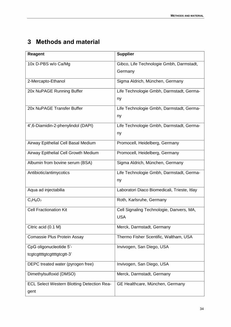
METHODS AND MATERIAL
34
3 Methods and material
Reagent Supplier
10x D-PBS w/o Ca/Mg Gibco, Life Technologie Gmbh, Darmstadt,
Germany
2-Mercapto-Ethanol Sigma Aldrich, München, Germany
20x NuPAGE Running Buffer Life Technologie Gmbh, Darmstadt, Germa-
ny
20x NuPAGE Transfer Buffer Life Technologie Gmbh, Darmstadt, Germa-
ny
4′,6-Diamidin-2-phenylindol (DAPI) Life Technologie Gmbh, Darmstadt, Germa-
ny
Airway Epithelial Cell Basal Medium Promocell, Heidelberg, Germany
Airway Epithelial Cell Growth Medium Promocell, Heidelberg, Germany
Albumin from bovine serum (BSA) Sigma Aldrich, München, Germany
Antibiotic/antimycotics Life Technologie Gmbh, Darmstadt, Germa-
ny
Aqua ad injectabilia Laboratori Diaco Biomedicali, Trieste, Itlay
C2H8O7 Roth, Karlsruhe, Germany
Cell Fractionation Kit Cell Signaling Technologie, Danvers, MA,
USA
Citric acid (0.1 M) Merck, Darmstadt, Germany
Comassie Plus Protein Assay Thermo Fisher Scentific, Waltham, USA
CpG oligonucleotide 5’-
tcgtcgttttgtcgttttgtcgtt-3’
Invivogen, San Diego, USA
DEPC treated water (pyrogen free) Invivogen, San Diego, USA
Dimethylsulfoxid (DMSO) Merck, Darmstadt, Germany
ECL Select Western Blotting Detection Rea-
gent
GE Healthcare, München, Germany
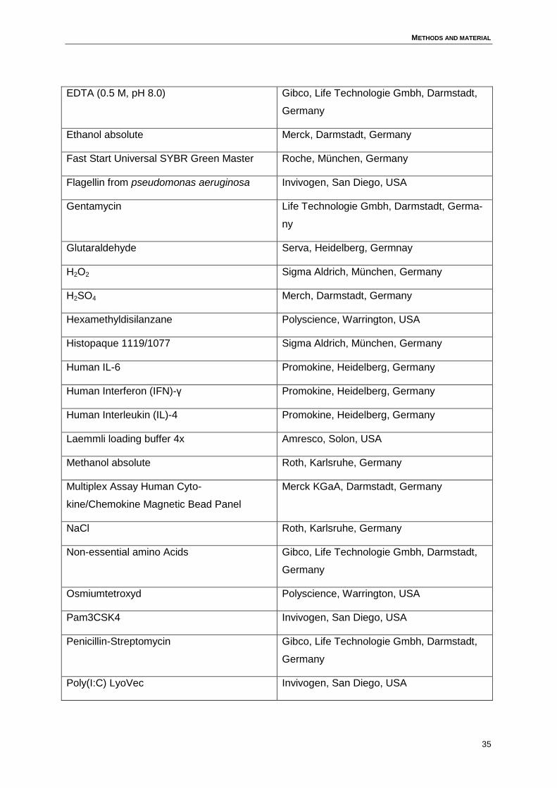
METHODS AND MATERIAL
35
EDTA (0.5 M, pH 8.0) Gibco, Life Technologie Gmbh, Darmstadt,
Germany
Ethanol absolute Merck, Darmstadt, Germany
Fast Start Universal SYBR Green Master Roche, München, Germany
Flagellin from pseudomonas aeruginosa Invivogen, San Diego, USA
Gentamycin Life Technologie Gmbh, Darmstadt, Germa-
ny
Glutaraldehyde Serva, Heidelberg, Germnay
H2O2 Sigma Aldrich, München, Germany
H2SO4 Merch, Darmstadt, Germany
Hexamethyldisilanzane Polyscience, Warrington, USA
Histopaque 1119/1077 Sigma Aldrich, München, Germany
Human IL-6 Promokine, Heidelberg, Germany
Human Interferon (IFN)-γ Promokine, Heidelberg, Germany
Human Interleukin (IL)-4 Promokine, Heidelberg, Germany
Laemmli loading buffer 4x Amresco, Solon, USA
Methanol absolute Roth, Karlsruhe, Germany
Multiplex Assay Human Cyto-
kine/Chemokine Magnetic Bead Panel
Merck KGaA, Darmstadt, Germany
NaCl Roth, Karlsruhe, Germany
Non-essential amino Acids Gibco, Life Technologie Gmbh, Darmstadt,
Germany
Osmiumtetroxyd Polyscience, Warrington, USA
Pam3CSK4 Invivogen, San Diego, USA
Penicillin-Streptomycin Gibco, Life Technologie Gmbh, Darmstadt,
Germany
Poly(I:C) LyoVec Invivogen, San Diego, USA
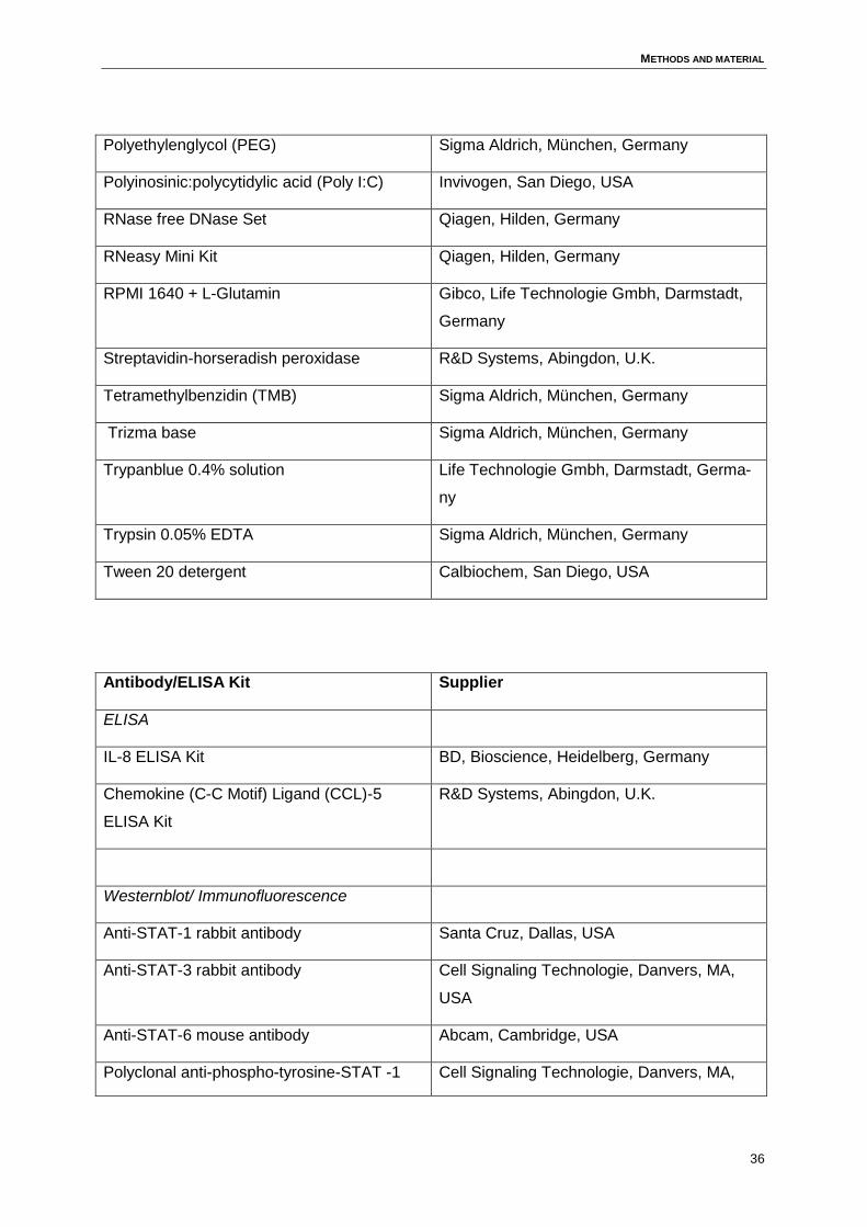
METHODS AND MATERIAL
36
Polyethylenglycol (PEG) Sigma Aldrich, München, Germany
Polyinosinic:polycytidylic acid (Poly I:C) Invivogen, San Diego, USA
RNase free DNase Set Qiagen, Hilden, Germany
RNeasy Mini Kit Qiagen, Hilden, Germany
RPMI 1640 + L-Glutamin Gibco, Life Technologie Gmbh, Darmstadt,
Germany
Streptavidin-horseradish peroxidase R&D Systems, Abingdon, U.K.
Tetramethylbenzidin (TMB) Sigma Aldrich, München, Germany
Trizma base Sigma Aldrich, München, Germany
Trypanblue 0.4% solution Life Technologie Gmbh, Darmstadt, Germa-
ny
Trypsin 0.05% EDTA Sigma Aldrich, München, Germany
Tween 20 detergent Calbiochem, San Diego, USA
Antibody/ELISA Kit Supplier
ELISA
IL-8 ELISA Kit BD, Bioscience, Heidelberg, Germany
Chemokine (C-C Motif) Ligand (CCL)-5
ELISA Kit
R&D Systems, Abingdon, U.K.
Westernblot/ Immunofluorescence
Anti-STAT-1 rabbit antibody Santa Cruz, Dallas, USA
Anti-STAT-3 rabbit antibody Cell Signaling Technologie, Danvers, MA,
USA
Anti-STAT-6 mouse antibody Abcam, Cambridge, USA
Polyclonal anti-phospho-tyrosine-STAT -1 Cell Signaling Technologie, Danvers, MA,
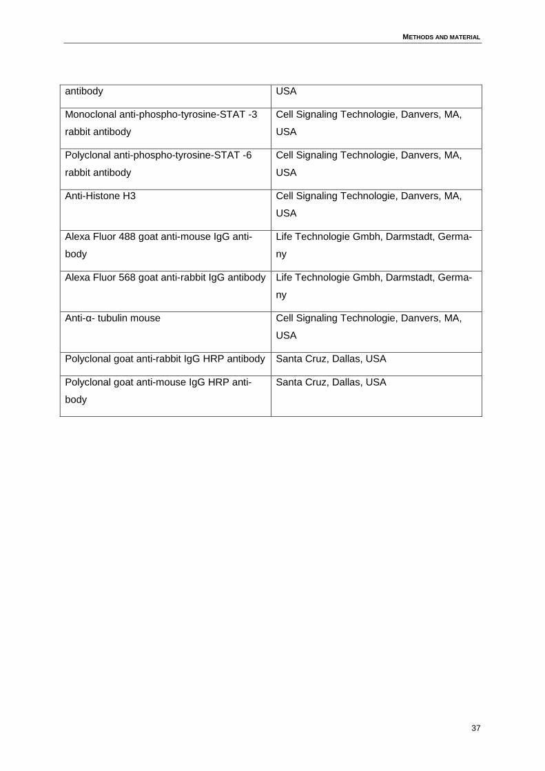
METHODS AND MATERIAL
37
antibody USA
Monoclonal anti-phospho-tyrosine-STAT -3
rabbit antibody
Cell Signaling Technologie, Danvers, MA,
USA
Polyclonal anti-phospho-tyrosine-STAT -6
rabbit antibody
Cell Signaling Technologie, Danvers, MA,
USA
Anti-Histone H3 Cell Signaling Technologie, Danvers, MA,
USA
Alexa Fluor 488 goat anti-mouse IgG anti-
body
Life Technologie Gmbh, Darmstadt, Germa-
ny
Alexa Fluor 568 goat anti-rabbit IgG antibody Life Technologie Gmbh, Darmstadt, Germa-
ny
Anti-α- tubulin mouse Cell Signaling Technologie, Danvers, MA,
USA
Polyclonal goat anti-rabbit IgG HRP antibody Santa Cruz, Dallas, USA
Polyclonal goat anti-mouse IgG HRP anti-
body
Santa Cruz, Dallas, USA
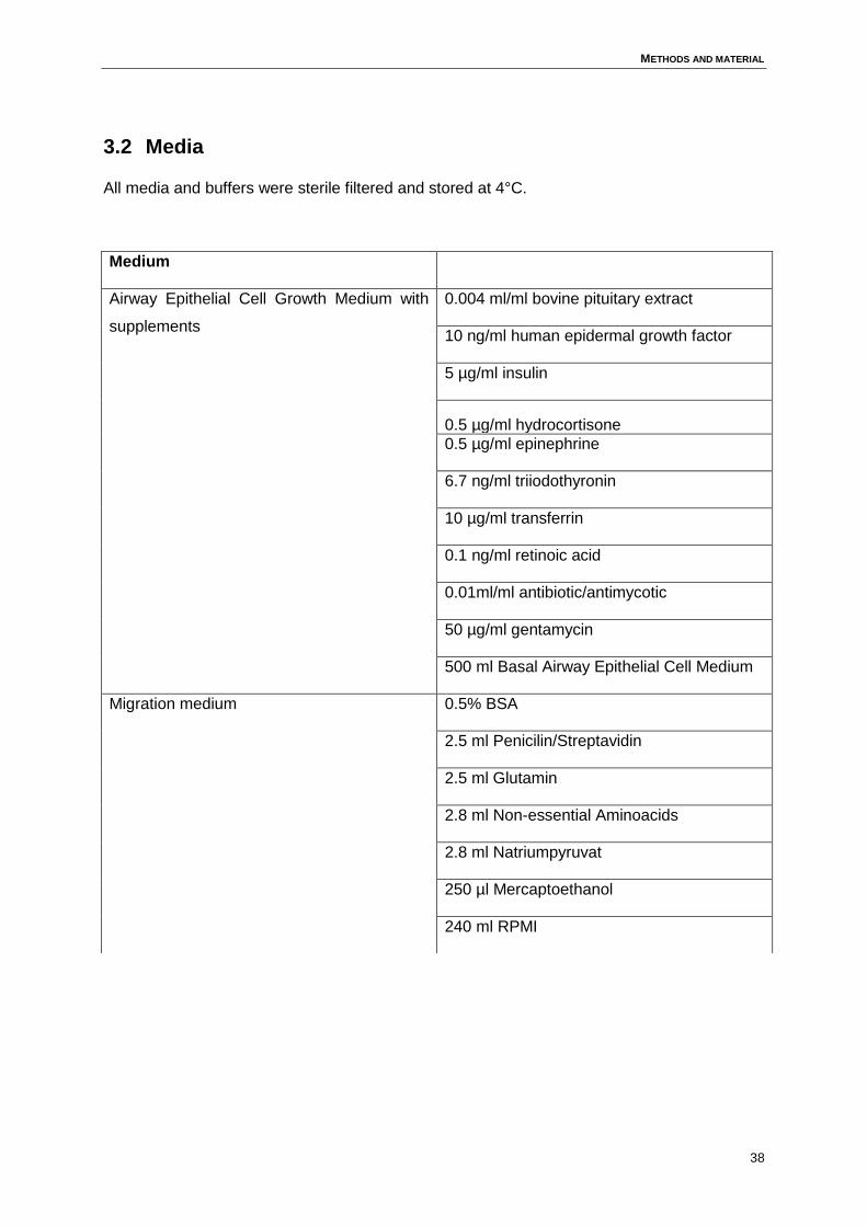
METHODS AND MATERIAL
38
3.2 Media
All media and buffers were sterile filtered and stored at 4°C.
Medium
Airway Epithelial Cell Growth Medium with
supplements
0.004 ml/ml bovine pituitary extract
10 ng/ml human epidermal growth factor
5 µg/ml insulin
0.5 µg/ml hydrocortisone
0.5 µg/ml epinephrine
6.7 ng/ml triiodothyronin
10 µg/ml transferrin
0.1 ng/ml retinoic acid
0.01ml/ml antibiotic/antimycotic
50 µg/ml gentamycin
500 ml Basal Airway Epithelial Cell Medium
Migration medium 0.5% BSA
2.5 ml Penicilin/Streptavidin
2.5 ml Glutamin
2.8 ml Non-essential Aminoacids
2.8 ml Natriumpyruvat
250 µl Mercaptoethanol
240 ml RPMI
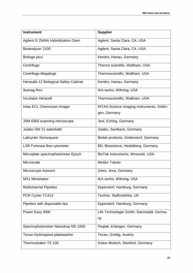
METHODS AND MATERIAL
39
Instrument Supplier
Agilent G 2545A Hybridization Oven Agilent, Santa Clara, CA, USA
Bioanalyzer 2100 Agilent, Santa Clara, CA, USA
Biofuge pico Kendro, Hanau, Germany
Centrifuge Thermo scientific, Waltham, USA
Centrifuge Megafuge Thermoscientific, Waltham, USA
Herasafe 12 Biological Saftey Cabinet Kendro, Hanau, Germany
Ikamag Reo IKA works, Wilming, USA
Incubator Heracell Thermoscientific, Waltham, USA
Intas ECL Chemocam Imager INTAS Science Imaging Instruments, Göttin-
gen, Germany
JSM 6300 scanning microscope Jeol, Eching, Germany
Julabo SW 21 waterbath Julabo, Seelbach, Germany
Labcycler Sensoquest Biolab products, Gödenstorf, Germany
LSR Fortessa flow cytometer BD, Bioscience, Heidelberg, Germany
Microplate spectrophotometer Epoch BioTek Instruments, Winooski, USA
Microscale Mettler Toledo
Microscope Axiovert Zeiss, Jena, Germany
MS1 Minishaker IKA works, Wilming, USA
Multichannel Pipettes Eppendorf, Hamburg, Germany
PCR Cycler TC412 Techne, Staffordshire, UK
Pipettes with disposable tips Eppendorf, Hamburg, Germany
Power Easy 90W Life Technologie Gmbh, Darmstadt, Germa-
ny
Spectrophotometer Nanodrop ND 1000 Peqlab, Erlangen, Germany
Tecan Hydrospeed platewasher Tecan, Grödig, Austria
Thermoshaker TS 100 Kisker Biotech, Steinfurt, Germany
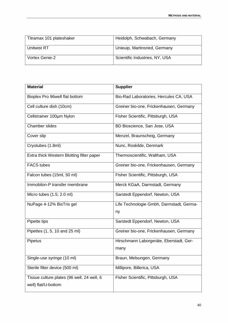
METHODS AND MATERIAL
40
Titramax 101 plateshaker Heidolph, Schwabach, Germany
Unitwist RT Unieuip, Martinsried, Germany
Vortex Genie-2 Scientific Industries, NY, USA
Material Supplier
Bioplex Pro 96well flat bottom Bio-Rad Laboratories, Hercules CA, USA
Cell culture dish (10cm) Greiner bio-one, Frickenhausen, Germany
Cellstrainer 100µm Nylon Fisher Scientific, Pittsburgh, USA
Chamber slides BD Bioscience, San Jose, USA
Cover slip Menzel, Braunscheig, Germany
Cryotubes (1.8ml) Nunc, Roskilde, Denmark
Extra thick Western Blotting filter paper Thermoscientific, Waltham, USA
FACS tubes Greiner bio-one, Frickenhausen, Germany
Falcon tubes (15ml, 50 ml) Fisher Scientific, Pittsburgh, USA
Immobilon-P transfer membrane Merck KGaA, Darmstadt, Germany
Micro tubes (1.5; 2.0 ml) Sarstedt Eppendorf, Newton, USA
NuPage 4-12% BisTris gel Life Technologie Gmbh, Darmstadt, Germa-
ny
Pipette tips Sarstedt Eppendorf, Newton, USA
Pipettes (1, 5, 10 and 25 ml) Greiner bio-one, Frickenhausen, Germany
Pipetus Hirschmann Laborgeräte, Eberstadt, Ger-
many
Single-use syringe (10 ml) Braun, Melsungen, Germany
Sterile filter device (500 ml) Millipore, Billerica, USA
Tissue culture plates (96 well, 24 well, 6
well) flat/U-bottom
Fisher Scientific, Pittsburgh, USA
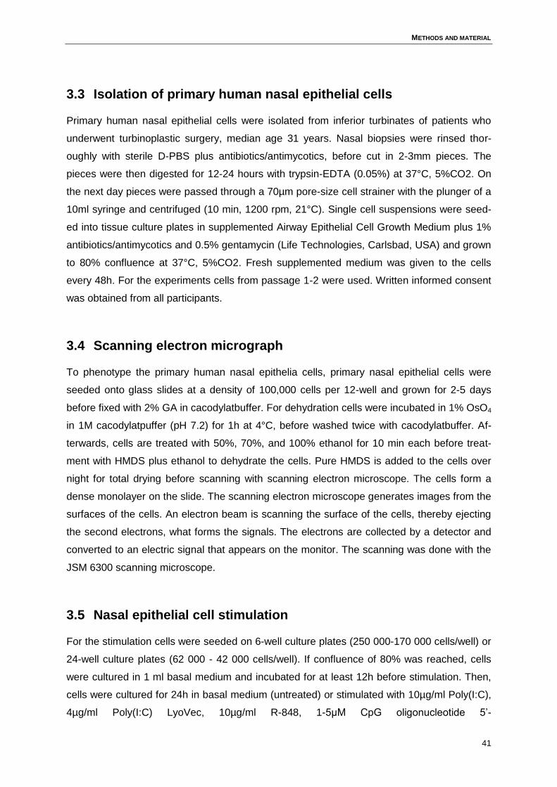
METHODS AND MATERIAL
41
3.3 Isolation of primary human nasal epithelial cells
Primary human nasal epithelial cells were isolated from inferior turbinates of patients who
underwent turbinoplastic surgery, median age 31 years. Nasal biopsies were rinsed thor-
oughly with sterile D-PBS plus antibiotics/antimycotics, before cut in 2-3mm pieces. The
pieces were then digested for 12-24 hours with trypsin-EDTA (0.05%) at 37°C, 5%CO2. On
the next day pieces were passed through a 70µm pore-size cell strainer with the plunger of a
10ml syringe and centrifuged (10 min, 1200 rpm, 21°C). Single cell suspensions were seed-
ed into tissue culture plates in supplemented Airway Epithelial Cell Growth Medium plus 1%
antibiotics/antimycotics and 0.5% gentamycin (Life Technologies, Carlsbad, USA) and grown
to 80% confluence at 37°C, 5%CO2. Fresh supplemented medium was given to the cells
every 48h. For the experiments cells from passage 1-2 were used. Written informed consent
was obtained from all participants.
3.4 Scanning electron micrograph
To phenotype the primary human nasal epithelia cells, primary nasal epithelial cells were
seeded onto glass slides at a density of 100,000 cells per 12-well and grown for 2-5 days
before fixed with 2% GA in cacodylatbuffer. For dehydration cells were incubated in 1% OsO4
in 1M cacodylatpuffer (pH 7.2) for 1h at 4°C, before washed twice with cacodylatbuffer. Af-
terwards, cells are treated with 50%, 70%, and 100% ethanol for 10 min each before treat-
ment with HMDS plus ethanol to dehydrate the cells. Pure HMDS is added to the cells over
night for total drying before scanning with scanning electron microscope. The cells form a
dense monolayer on the slide. The scanning electron microscope generates images from the
surfaces of the cells. An electron beam is scanning the surface of the cells, thereby ejecting
the second electrons, what forms the signals. The electrons are collected by a detector and
converted to an electric signal that appears on the monitor. The scanning was done with the
JSM 6300 scanning microscope.
3.5 Nasal epithelial cell stimulation
For the stimulation cells were seeded on 6-well culture plates (250 000-170 000 cells/well) or
24-well culture plates (62 000 - 42 000 cells/well). If confluence of 80% was reached, cells
were cultured in 1 ml basal medium and incubated for at least 12h before stimulation. Then,
cells were cultured for 24h in basal medium (untreated) or stimulated with 10µg/ml Poly(I:C),
4µg/ml Poly(I:C) LyoVec, 10µg/ml R-848, 1-5µM CpG oligonucleotide 5’-
METHODS AND MATERIAL
42
tcgtcgttttgtcgttttgtcgtt-3’, 5µg/ml purified flagellin from pseudomonas aeruginosa, 200ng/ml
synthetic triacylated lipoprotein Pam3CSK4 (Invivogen, San Diego, USA), 15ng/ml IFN-γ,
50ng/ml Interleukin (IL)-4, 800U/ml IL-6 (Promokine, Heidelberg, Germany) with or without
addition of Ze 339. The high pressure carbon dioxide (CO2) extract of Petasites hybridus (Ze
339) and its isolated active compounds, petasin, neopetasin and isopetasin, were provided
by Max Zeller Söhne AG (Zeller, Analytical Department, Romanshorn, Schwitzerland).
3.6 Lactate dehydrogenase assay
To measure the cytotoxicity level of the cells after stimulation with indicated stimuli, we cul-
tured the cells until 80% confluent. Afterwards, cells are cultured for 24h in basal medium
(untreated) or stimulated with indicated concentrations of Ze 339, or the isolated active com-
pounds petasin, neopetasin and isopetasin in the absence or presence of PolyI:C (10µg/ml).
100µl supernatant was taken and 100µl reaction mixture was added and LDH activity was
quantified with Cytotoxicity Detection Kit. For this absorbance of the samples at 490 nm us-
ing ELISA reader was measured.
3.7 Isolation neutrophil granulocytes
For the isolation of human neutrophil granulocytes (PMN), 50 ml EDTA-whole blood was
taken form healthy volunteers. PMNs were separated via density gradient centrifugation. For
this, 13 ml Histopaque 1119 and 13ml Histopaque are carefully layered in a 50 ml falcon. On
top, 20 ml of the EDTA-whole blood was layered before centrifuged for 45 min 1900 rpm
without brake to separate the cells based on their different density. Whole blood gets sepa-
rated in plasma, mononuclear cells, Histopaque 1077, polymorphonuclear cells, Histopaque
1119 and erythrocytes. The top three layers are getting discarded, polymorphonuclear cells
in Histopaque 1119 are carefully collected with a pipette and transferred in a 50 ml falcon.
Cells were washed with D-PBS with centrifugation for 20 min 1400 rpm 20°C. Supernatant
was discarded; cells were washed again with D-PBS and centrifuged for 10 min at 1200 rpm
20°C. Afterwards, to lyse erythrocytes, cells were resuspended in 3 ml aqua injectabilia for
25 sec. Directly after this 3 ml 1.8 % NaCl is added to the cells to neutralize. Cells were
washed again with D-PBS and centrifuged for 10 min 1200 rpm 20°C. Cells were resuspend-
ed in neutrophil migration medium and counted with Trypanblue 0.4% solution in a Neubauer
chamber. Cells were adjusted to 1 million per 1 ml migration medium for neutrophil migration
assay.
METHODS AND MATERIAL
43
3.8 RNA extraction kit
For RNA extraction confluent pre-stimulated cells harvested with trypsin-EDTA (0,05%) are
collected in 350µl RLT-buffer/ 2% β-mecaptoethanol and homogenised by vortexing 1min.
Then cells were pipetted on RNeasy Spin column collection tube and centrifuged 15 sec
10000 rpm. Flow-through got discarded and 350 µl RW1 buffer was added on the tube.
Tubes were centrifuged for 15 sec 10000 rpm. 10 µL Dnase and 70 µL RDD buffer was add-
ed on the tube and incubated on room temperature for 15 min. Afterwards, 350 µL RW1
buffer were added on the tube and centrifuged 15 sec 10000 rpm. Flow-through got discard-
ed, 500 µL RPE buffer was added on the tube before centrifuged 15 sec 10000 rpm. Flow-
through got discarded, 500 µL RPE buffer was added and tube was centrifuged for 2 min
10000 rpm. Column was put on a fresh tube and centrifuged again with maximal speed for 1
min to dry the column. Then, 30 µL RNase free water was given on the column before cen-
trifugation for 1 min 10000 rpm to elute the RNA from the column. The Flow-through was
collected and the RNA concentration was measured by spectrophotometer Nanodrop.
3.9 Comassie Protein Assay
To quantify the protein concentration of the samples used for Westernblotting, “Comassie
Plus Protein Kit” was used in accordance with the manufacturer's data (Thermo Scientific).
3.10 Microarray
RNA was isolated as described above. Quality and integrity of RNA samples was checked
with the Agilent 2100 Bioanalyzer platform following the manufacturer’s protocol (Agilent
Technologies). 25 ng of each total RNA sample was amplified and Cyanine-3 CTP-dye la-
belled using the Agilent Low RNA Input Quick Amp Labeling Kit One Color (Agilent Technol-
ogies) following the manufacturer’s protocol. The hybridization procedure was performed
according to the Agilent microarray processing protocol using the Agilent Gene Expression
Hybridization Kit and Agilent Whole Human Genome Microarrays 8 x 60K (Agilent Technolo-
gies). Fluorescence signals of the hybridized arrays were detected using Agilent’s Microarray
Scanner System (Agilent Technologies). The Agilent Feature Extraction Software was used
to read out and process the microarray image files, and differential gene expression Feature
Extraction Software–derived output data files were further analyzed using the Gene Spring
data analysis system (GeneSpring GX Software).
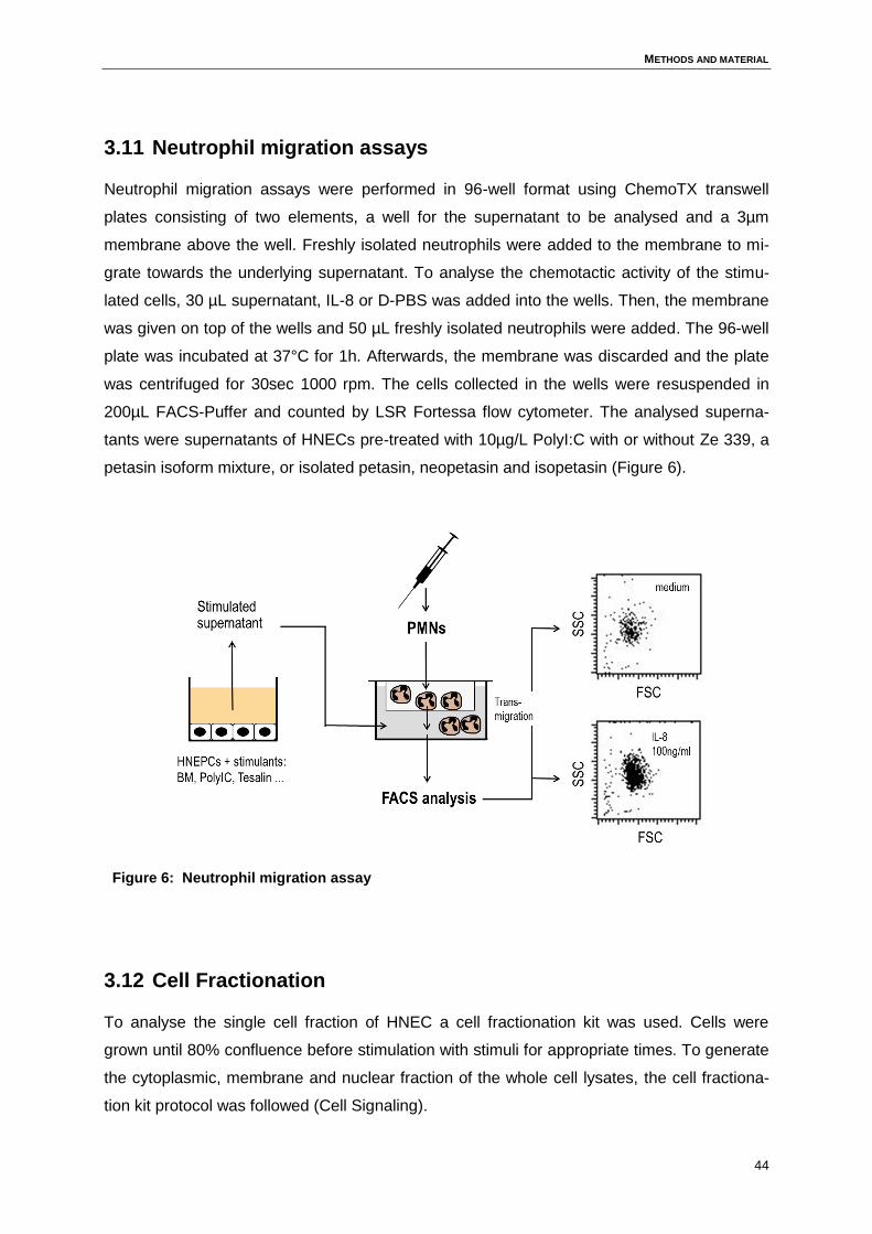
METHODS AND MATERIAL
44
3.11 Neutrophil migration assays
Neutrophil migration assays were performed in 96-well format using ChemoTX transwell
plates consisting of two elements, a well for the supernatant to be analysed and a 3µm
membrane above the well. Freshly isolated neutrophils were added to the membrane to mi-
grate towards the underlying supernatant. To analyse the chemotactic activity of the stimu-
lated cells, 30 µL supernatant, IL-8 or D-PBS was added into the wells. Then, the membrane
was given on top of the wells and 50 µL freshly isolated neutrophils were added. The 96-well
plate was incubated at 37°C for 1h. Afterwards, the membrane was discarded and the plate
was centrifuged for 30sec 1000 rpm. The cells collected in the wells were resuspended in
200µL FACS-Puffer and counted by LSR Fortessa flow cytometer. The analysed superna-
tants were supernatants of HNECs pre-treated with 10µg/L PolyI:C with or without Ze 339, a
petasin isoform mixture, or isolated petasin, neopetasin and isopetasin (Figure 6).
Figure 6: Neutrophil migration assay
3.12 Cell Fractionation
To analyse the single cell fraction of HNEC a cell fractionation kit was used. Cells were
grown until 80% confluence before stimulation with stimuli for appropriate times. To generate
the cytoplasmic, membrane and nuclear fraction of the whole cell lysates, the cell fractiona-
tion kit protocol was followed (Cell Signaling).
METHODS AND MATERIAL
45
3.13 Measurement of cytokine and chemokine levels (Luminex)
Levels of CCL-3 (MIP-1α), CCL-4 (MIP-1β), MCP-1 (CCL-2), IP-10 (CXCL-10), IL-1α, IL-6,
G-CSF, TNF-α were measured using a multiplex assay (MILLIPLEX MAP Human Cyto-
kine/Chemokine Magnetic Bead Panel). The magnetic bead assay was run according to the
manufacturers' instructions. Data were acquired on a validated and calibrated Bio-Plex 200
system (Bio-Rad) and analysed with Bio-Plex Manager 6.0 software (Bio-Rad).
3.14 Analysis of cytokine secretion by enzyme-linked
immunosorbent assay (ELISA)
Cytokine concentration of IL-8 and CCL-5 in HNEC cell culture free supernatants was meas-
ured by ELISA. The assay was run according to the manufacturers' instructions using com-
mercially available sandwich ELISA kits (BD and R&D Systems).
3.15 Western Blot
80% confluent HNECs were stimulated for 30 min (STAT1 and STAT3) and 60 min (STAT6)
with medium, 800U/ml IL-6, 15 ng/ml IFN-γ, or 50 ng/ml IL-4 in the absence or presence of
3µM Ze 339 (Zeller). As PEG was used for solubilizing Ze 339, it was added to all wells at a
dilution of 1:10,000. HNECs stimulated with PEG alone served as control. Total extracts and
sub-cellular fractions were prepared from cells as described above. The protein samples,
whole cell lysate and the cell fractionation lysates, were treated with 4x laemmli loading buff-
er before heating for 5 min at 95 °C. To separate proteins by size, samples were loaded to
SDS-PAGE under reducing conditions before being transferred to a nitrocellulose membrane
through an electric field by Western Blotting. The Wet-Blot-System consists of a methanol-
activated PDVF-membrane, whatman paper, sponges and the gel runs for 1.5 h at 120 V
before blotted PDVF-membrane is blocked with 1 % BSA, 0.1% Tween in 1x TBS buffer for 1
h. Afterwards specific primary antibodies were allowed to bind to the proteins of interest
(concentrations listed below). A secondary peroxidase-linked antibody was used for detec-
tion of the bound primary antibody and chemiluminescence signals were quantitated with the
ChemoStarTM software (Intas). To detect the phospho-STATs the membrane was cut into two
pieces, the upper part was incubated with monoclonal anti-phospho-tyrosine-STAT3, poly-
clonal anti-phospho- tyrosine-STAT6 and monoclonal anti-phospho-tyrosine-STAT1, the low-
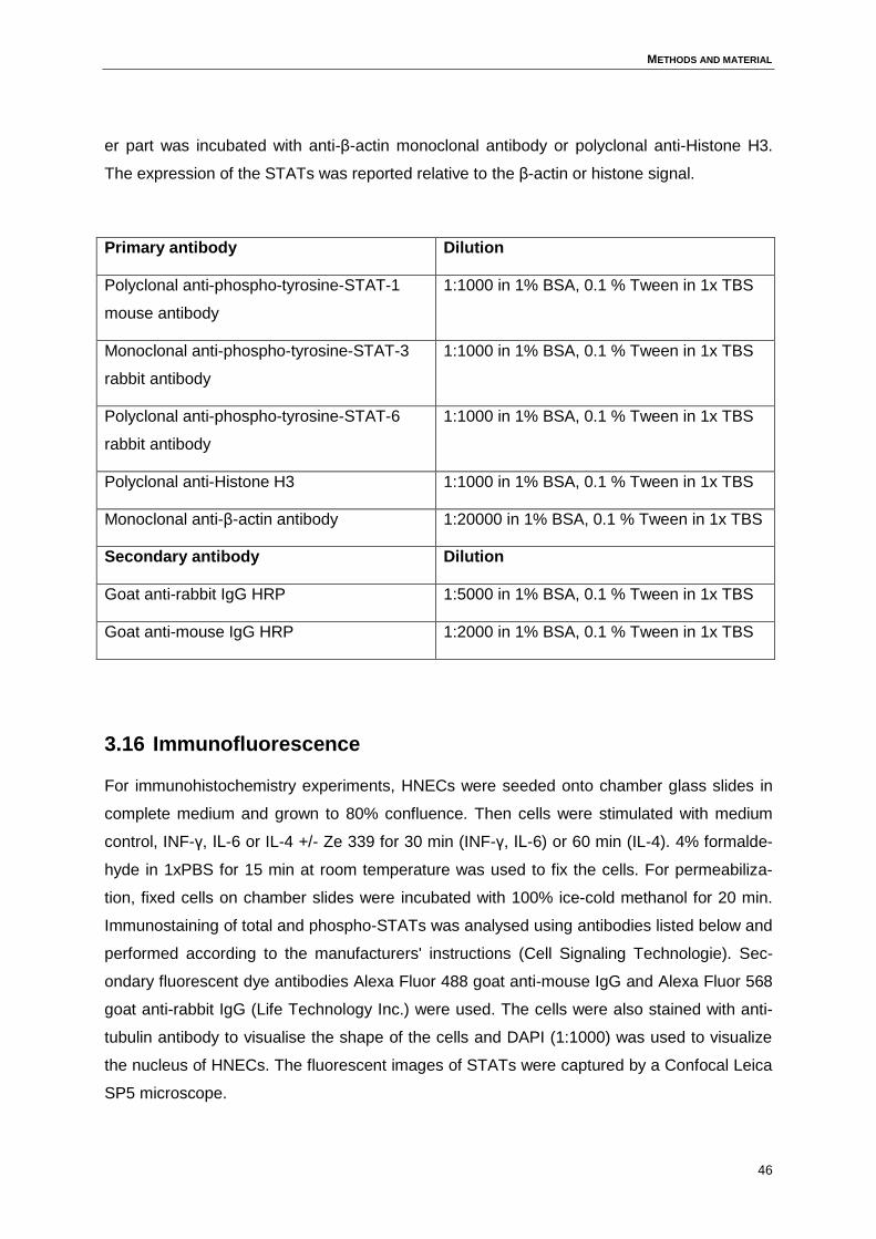
METHODS AND MATERIAL
46
er part was incubated with anti-β-actin monoclonal antibody or polyclonal anti-Histone H3.
The expression of the STATs was reported relative to the β-actin or histone signal.
Primary antibody Dilution
Polyclonal anti-phospho-tyrosine-STAT-1
mouse antibody
1:1000 in 1% BSA, 0.1 % Tween in 1x TBS
Monoclonal anti-phospho-tyrosine-STAT-3
rabbit antibody
1:1000 in 1% BSA, 0.1 % Tween in 1x TBS
Polyclonal anti-phospho-tyrosine-STAT-6
rabbit antibody
1:1000 in 1% BSA, 0.1 % Tween in 1x TBS
Polyclonal anti-Histone H3 1:1000 in 1% BSA, 0.1 % Tween in 1x TBS
Monoclonal anti-β-actin antibody 1:20000 in 1% BSA, 0.1 % Tween in 1x TBS
Secondary antibody Dilution
Goat anti-rabbit IgG HRP 1:5000 in 1% BSA, 0.1 % Tween in 1x TBS
Goat anti-mouse IgG HRP 1:2000 in 1% BSA, 0.1 % Tween in 1x TBS
3.16 Immunofluorescence
For immunohistochemistry experiments, HNECs were seeded onto chamber glass slides in
complete medium and grown to 80% confluence. Then cells were stimulated with medium
control, INF-γ, IL-6 or IL-4 +/- Ze 339 for 30 min (INF-γ, IL-6) or 60 min (IL-4). 4% formalde-
hyde in 1xPBS for 15 min at room temperature was used to fix the cells. For permeabiliza-
tion, fixed cells on chamber slides were incubated with 100% ice-cold methanol for 20 min.
Immunostaining of total and phospho-STATs was analysed using antibodies listed below and
performed according to the manufacturers' instructions (Cell Signaling Technologie). Sec-
ondary fluorescent dye antibodies Alexa Fluor 488 goat anti-mouse IgG and Alexa Fluor 568
goat anti-rabbit IgG (Life Technology Inc.) were used. The cells were also stained with anti-
tubulin antibody to visualise the shape of the cells and DAPI (1:1000) was used to visualize
the nucleus of HNECs. The fluorescent images of STATs were captured by a Confocal Leica
SP5 microscope.
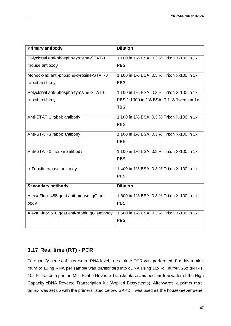
METHODS AND MATERIAL
47
Primary antibody Dilution
Polyclonal anti-phospho-tyrosine-STAT-1
mouse antibody
1:100 in 1% BSA, 0.3 % Triton X-100 in 1x
PBS
Monoclonal anti-phospho-tyrosine-STAT-3
rabbit antibody
1:100 in 1% BSA, 0.3 % Triton X-100 in 1x
PBS
Polyclonal anti-phospho-tyrosine-STAT-6
rabbit antibody
1:100 in 1% BSA, 0.3 % Triton X-100 in 1x
PBS 1:1000 in 1% BSA, 0.1 % Tween in 1x
TBS
Anti-STAT-1 rabbit antibody 1:100 in 1% BSA, 0.3 % Triton X-100 in 1x
PBS
Anti-STAT-3 rabbit antibody 1:100 in 1% BSA, 0.3 % Triton X-100 in 1x
PBS
Anti-STAT-6 mouse antibody 1:100 in 1% BSA, 0.3 % Triton X-100 in 1x
PBS
α-Tubulin mouse antibody 1:400 in 1% BSA, 0.3 % Triton X-100 in 1x
PBS
Secondary antibody Dilution
Alexa Fluor 488 goat anti-mouse IgG anti-
body
1:600 in 1% BSA, 0.3 % Triton X-100 in 1x
PBS
Alexa Fluor 568 goat anti-rabbit IgG antibody 1:600 in 1% BSA, 0.3 % Triton X-100 in 1x
PBS
3.17 Real time (RT) - PCR
To quantify genes of interest on RNA level, a real time PCR was performed. For this a mini-
mum of 10 ng RNA per sample was transcribed into cDNA using 10x RT buffer, 25x dNTPs,
10x RT random primer, MultiScribe Reverse Transkriptase and nuclear free water of the High
Capacity cDNA Reverse Transcription Kit (Applied Biosystems). Afterwards, a primer mas-
termix was set up with the primers listed below. GAPDH was used as the housekeeper gene.
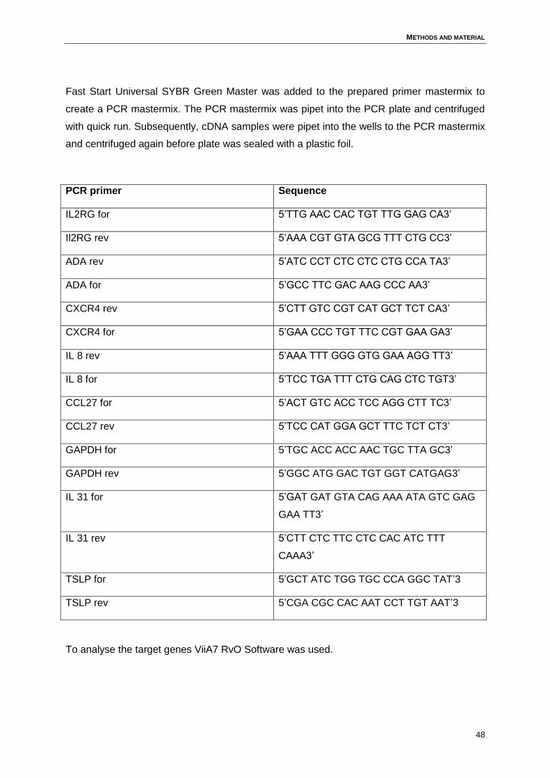
METHODS AND MATERIAL
48
Fast Start Universal SYBR Green Master was added to the prepared primer mastermix to
create a PCR mastermix. The PCR mastermix was pipet into the PCR plate and centrifuged
with quick run. Subsequently, cDNA samples were pipet into the wells to the PCR mastermix
and centrifuged again before plate was sealed with a plastic foil.
PCR primer Sequence
IL2RG for 5’TTG AAC CAC TGT TTG GAG CA3’
Il2RG rev 5’AAA CGT GTA GCG TTT CTG CC3’
ADA rev 5’ATC CCT CTC CTC CTG CCA TA3’
ADA for 5’GCC TTC GAC AAG CCC AA3’
CXCR4 rev 5’CTT GTC CGT CAT GCT TCT CA3’
CXCR4 for 5’GAA CCC TGT TTC CGT GAA GA3’
IL 8 rev 5’AAA TTT GGG GTG GAA AGG TT3’
IL 8 for 5’TCC TGA TTT CTG CAG CTC TGT3’
CCL27 for 5’ACT GTC ACC TCC AGG CTT TC3’
CCL27 rev 5’TCC CAT GGA GCT TTC TCT CT3’
GAPDH for 5’TGC ACC ACC AAC TGC TTA GC3’
GAPDH rev 5’GGC ATG GAC TGT GGT CATGAG3’
IL 31 for 5’GAT GAT GTA CAG AAA ATA GTC GAG
GAA TT3’
IL 31 rev 5’CTT CTC TTC CTC CAC ATC TTT
CAAA3’
TSLP for 5’GCT ATC TGG TGC CCA GGC TAT’3
TSLP rev 5’CGA CGC CAC AAT CCT TGT AAT’3
To analyse the target genes ViiA7 RvO Software was used.
METHODS AND MATERIAL
49
3.18 Statistics
Data are presented as mean +/- SD (bar charts) or medians +/- 5-95 percentiles (box plots).
To detect differences between treatment groups, samples were compared by Wilcoxon test.
Differences between treatment groups were considered significant if p0.05.
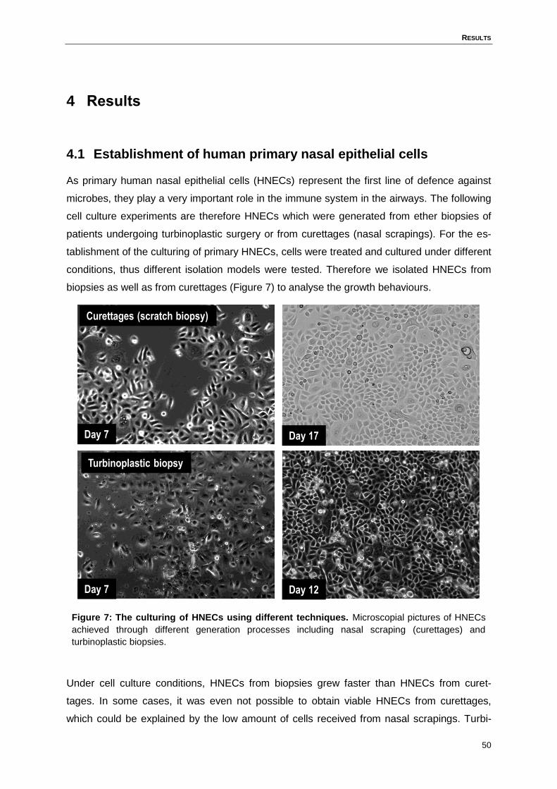
RESULTS
50
4 Results
4.1 Establishment of human primary nasal epithelial cells
As primary human nasal epithelial cells (HNECs) represent the first line of defence against
microbes, they play a very important role in the immune system in the airways. The following
cell culture experiments are therefore HNECs which were generated from ether biopsies of
patients undergoing turbinoplastic surgery or from curettages (nasal scrapings). For the es-
tablishment of the culturing of primary HNECs, cells were treated and cultured under different
conditions, thus different isolation models were tested. Therefore we isolated HNECs from
biopsies as well as from curettages (Figure 7) to analyse the growth behaviours.
Figure 7: The culturing of HNECs using different techniques. Microscopial pictures of HNECs
achieved through different generation processes including nasal scraping (curettages) and
turbinoplastic biopsies.
Under cell culture conditions, HNECs from biopsies grew faster than HNECs from curet-
tages. In some cases, it was even not possible to obtain viable HNECs from curettages,
which could be explained by the low amount of cells received from nasal scrapings. Turbi-
Curettages (scratch biopsy)
Turbinoplastic biopsy
Day 12
Day 17
Day 7
Day 7
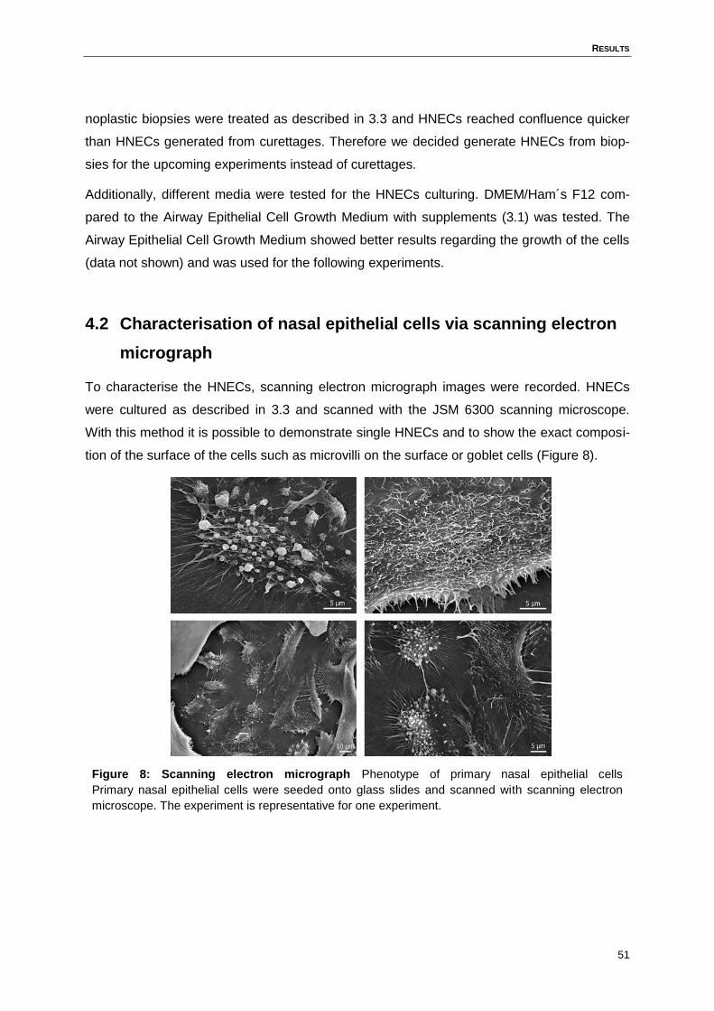
RESULTS
51
noplastic biopsies were treated as described in 3.3 and HNECs reached confluence quicker
than HNECs generated from curettages. Therefore we decided generate HNECs from biop-
sies for the upcoming experiments instead of curettages.
Additionally, different media were tested for the HNECs culturing. DMEM/Ham´s F12 com-
pared to the Airway Epithelial Cell Growth Medium with supplements (3.1) was tested. The
Airway Epithelial Cell Growth Medium showed better results regarding the growth of the cells
(data not shown) and was used for the following experiments.
4.2 Characterisation of nasal epithelial cells via scanning electron
micrograph
To characterise the HNECs, scanning electron micrograph images were recorded. HNECs
were cultured as described in 3.3 and scanned with the JSM 6300 scanning microscope.
With this method it is possible to demonstrate single HNECs and to show the exact composi-
tion of the surface of the cells such as microvilli on the surface or goblet cells (Figure 8).
Figure 8: Scanning electron micrograph Phenotype of primary nasal epithelial cells
Primary nasal epithelial cells were seeded onto glass slides and scanned with scanning electron
microscope. The experiment is representative for one experiment.
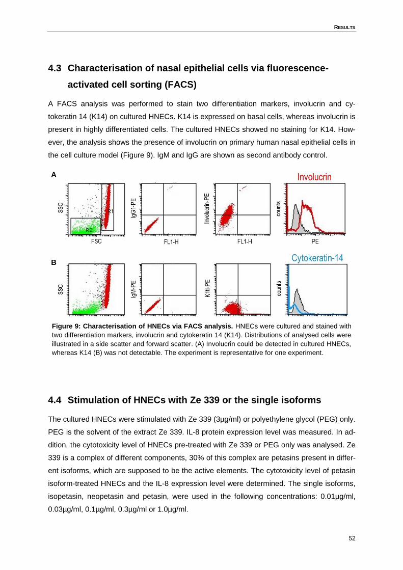
RESULTS
52
4.3 Characterisation of nasal epithelial cells via fluorescence-
activated cell sorting (FACS)
A FACS analysis was performed to stain two differentiation markers, involucrin and cy-
tokeratin 14 (K14) on cultured HNECs. K14 is expressed on basal cells, whereas involucrin is
present in highly differentiated cells. The cultured HNECs showed no staining for K14. How-
ever, the analysis shows the presence of involucrin on primary human nasal epithelial cells in
the cell culture model (Figure 9). IgM and IgG are shown as second antibody control.
Figure 9: Characterisation of HNECs via FACS analysis. HNECs were cultured and stained with
two differentiation markers, involucrin and cytokeratin 14 (K14). Distributions of analysed cells were
illustrated in a side scatter and forward scatter. (A) Involucrin could be detected in cultured HNECs,
whereas K14 (B) was not detectable. The experiment is representative for one experiment.
4.4 Stimulation of HNECs with Ze 339 or the single isoforms
The cultured HNECs were stimulated with Ze 339 (3µg/ml) or polyethylene glycol (PEG) only.
PEG is the solvent of the extract Ze 339. IL-8 protein expression level was measured. In ad-
dition, the cytotoxicity level of HNECs pre-treated with Ze 339 or PEG only was analysed. Ze
339 is a complex of different components, 30% of this complex are petasins present in differ-
ent isoforms, which are supposed to be the active elements. The cytotoxicity level of petasin
isoform-treated HNECs and the IL-8 expression level were determined. The single isoforms,
isopetasin, neopetasin and petasin, were used in the following concentrations: 0.01µg/ml,
0.03µg/ml, 0.1µg/ml, 0.3µg/ml or 1.0µg/ml.
A
B
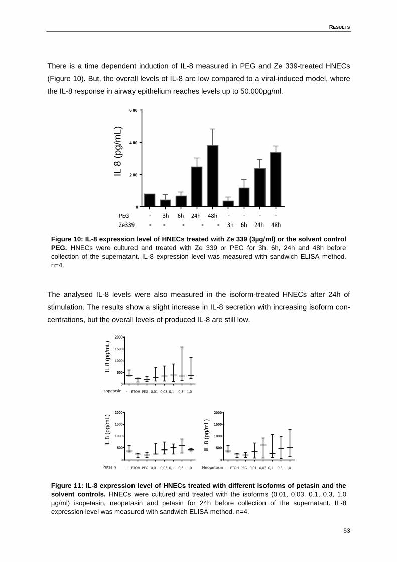
RESULTS
53
There is a time dependent induction of IL-8 measured in PEG and Ze 339-treated HNECs
(Figure 10). But, the overall levels of IL-8 are low compared to a viral-induced model, where
the IL-8 response in airway epithelium reaches levels up to 50.000pg/ml.
Figure 10: IL-8 expression level of HNECs treated with Ze 339 (3µg/ml) or the solvent control
PEG. HNECs were cultured and treated with Ze 339 or PEG for 3h, 6h, 24h and 48h before
collection of the supernatant. IL-8 expression level was measured with sandwich ELISA method.
n=4.
The analysed IL-8 levels were also measured in the isoform-treated HNECs after 24h of
stimulation. The results show a slight increase in IL-8 secretion with increasing isoform con-
centrations, but the overall levels of produced IL-8 are still low.
Figure 11: IL-8 expression level of HNECs treated with different isoforms of petasin and the
solvent controls. HNECs were cultured and treated with the isoforms (0.01, 0.03, 0.1, 0.3, 1.0
µg/ml) isopetasin, neopetasin and petasin for 24h before collection of the supernatant. IL-8
expression level was measured with sandwich ELISA method. n=4.
- 3h 6h 24h 48h - - - -PEG
- - - - - 3h 6h 24h 48hZe339
IL 8
(pg/m
L)
0
500
1000
1500
2000
Isopetasin - ETOH PEG 0,01 0,03 0,1 0,3 1,0
IL 8
(pg/m
L)
0
500
1000
1500
2000
IL 8
(pg/m
L)
Petasin - ETOH PEG 0,01 0,03 0,1 0,3 1,0
0
500
1000
1500
2000
IL 8
(pg/m
L)
Neopetasin - ETOH PEG 0,01 0,03 0,1 0,3 1,0
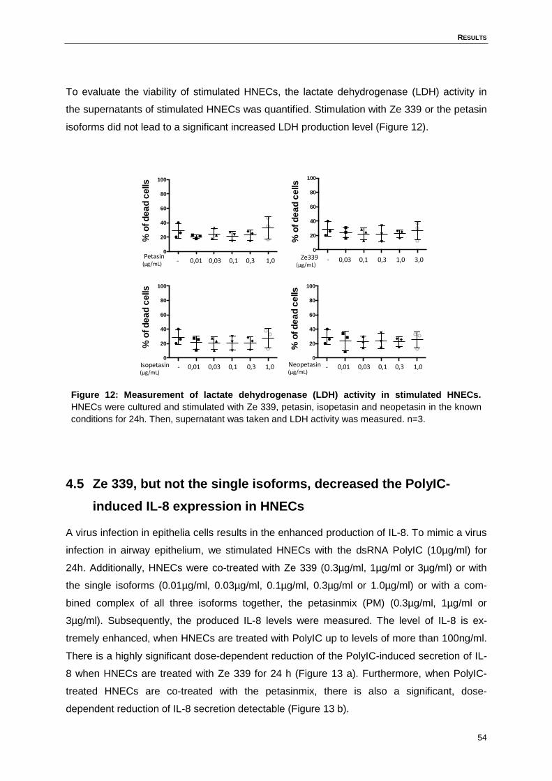
RESULTS
54
To evaluate the viability of stimulated HNECs, the lactate dehydrogenase (LDH) activity in
the supernatants of stimulated HNECs was quantified. Stimulation with Ze 339 or the petasin
isoforms did not lead to a significant increased LDH production level (Figure 12).
Figure 12: Measurement of lactate dehydrogenase (LDH) activity in stimulated HNECs.
HNECs were cultured and stimulated with Ze 339, petasin, isopetasin and neopetasin in the known
conditions for 24h. Then, supernatant was taken and LDH activity was measured. n=3.
4.5 Ze 339, but not the single isoforms, decreased the PolyIC-
induced IL-8 expression in HNECs
A virus infection in epithelia cells results in the enhanced production of IL-8. To mimic a virus
infection in airway epithelium, we stimulated HNECs with the dsRNA PolyIC (10µg/ml) for
24h. Additionally, HNECs were co-treated with Ze 339 (0.3µg/ml, 1µg/ml or 3µg/ml) or with
the single isoforms (0.01µg/ml, 0.03µg/ml, 0.1µg/ml, 0.3µg/ml or 1.0µg/ml) or with a com-
bined complex of all three isoforms together, the petasinmix (PM) (0.3µg/ml, 1µg/ml or
3µg/ml). Subsequently, the produced IL-8 levels were measured. The level of IL-8 is ex-
tremely enhanced, when HNECs are treated with PolyIC up to levels of more than 100ng/ml.
There is a highly significant dose-dependent reduction of the PolyIC-induced secretion of IL-
8 when HNECs are treated with Ze 339 for 24 h (Figure 13 a). Furthermore, when PolyIC-
treated HNECs are co-treated with the petasinmix, there is also a significant, dose-
dependent reduction of IL-8 secretion detectable (Figure 13 b).
0
20
40
60
80
100
%o
fd
ea
dc
ells
0
20
40
60
80
100
%o
fd
ea
dc
ells
Petasin (µg/mL) - 0,01 0,03 0,1 0,3 1,0
Isopetasin (µg/mL)
- 0,01 0,03 0,1 0,3 1,0
0
20
40
60
80
100
%o
fd
ea
dc
ells
Neopetasin (µg/mL)
- 0,01 0,03 0,1 0,3 1,0
0
20
40
60
80
100
%o
fd
ea
dc
ells
Ze339 (µg/mL)
- 0,03 0,1 0,3 1,0 3,0
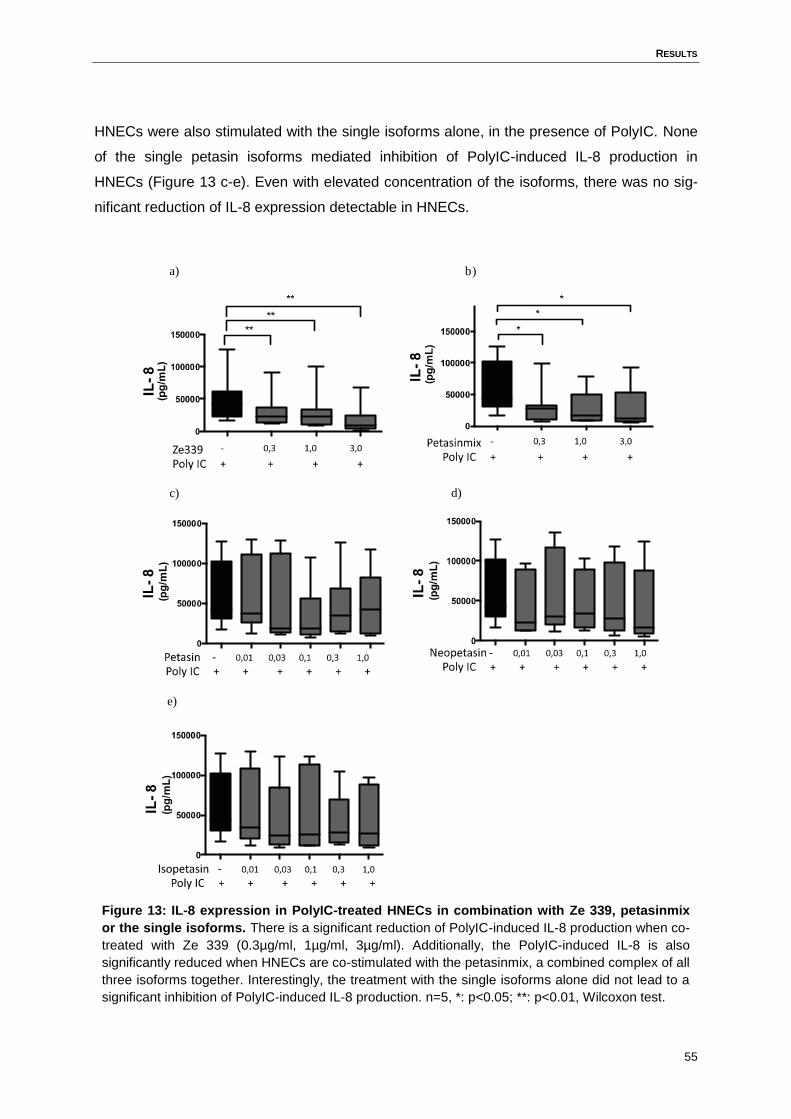
RESULTS
55
HNECs were also stimulated with the single isoforms alone, in the presence of PolyIC. None
of the single petasin isoforms mediated inhibition of PolyIC-induced IL-8 production in
HNECs (Figure 13 c-e). Even with elevated concentration of the isoforms, there was no sig-
nificant reduction of IL-8 expression detectable in HNECs.
Figure 13: IL-8 expression in PolyIC-treated HNECs in combination with Ze 339, petasinmix
or the single isoforms. There is a significant reduction of PolyIC-induced IL-8 production when co-
treated with Ze 339 (0.3µg/ml, 1µg/ml, 3µg/ml). Additionally, the PolyIC-induced IL-8 is also
significantly reduced when HNECs are co-stimulated with the petasinmix, a combined complex of all
three isoforms together. Interestingly, the treatment with the single isoforms alone did not lead to a
significant inhibition of PolyIC-induced IL-8 production. n=5, *: p<0.05; **: p<0.01, Wilcoxon test.
a) b)
c) d)
e)
RESULTS
56
4.6 Ze 339, but not the single isoforms, reduced the PolyIC-induced
neutrophil chemotaxis towards the supernatant
To address the question whether the reduction of the IL-8 response translates into reduced
recruitment of inflammatory cells, neutrophil migration towards conditioned media from stimu-
lated HNECs was assessed. There was enhanced neutrophil migration towards supernatants
of PolyIC (10µg/ml) stimulated HNECs detectable as compared to supernatants of medium-
treated HNECs. The supernatants of HNECs stimulated with a combination of PolyIC and Ze
339 were less potent in inducing neutrophil migration than supernatants of PolyIC-treated
cells (Figure 14 a).
As shown in Figure 14 b the petasinmix was sufficient to inhibit the PolyIC-induced IL-8 re-
sponse of HNECs. In line with this, HNEC supernatants stimulated with PolyIC and PM were
less potent in mediating neutrophil chemotaxis than supernatants of cells stimulated with
PolyIC only.
After 24h stimulation of HNECs with the single isoforms only in combination with PolyIC, su-
pernatant was taken and neutrophil migration towards the supernatant was measured. The
neutrophil migration towards the supernatant of the single isoforms stimulated cells did not
reach a significant reduction compare to the PolyIC only stimulated cells (Figure 14 c-e).
None of the supernatant of the single isoforms treated cells could show the effect of an en-
hanced neutrophil chemotaxis.
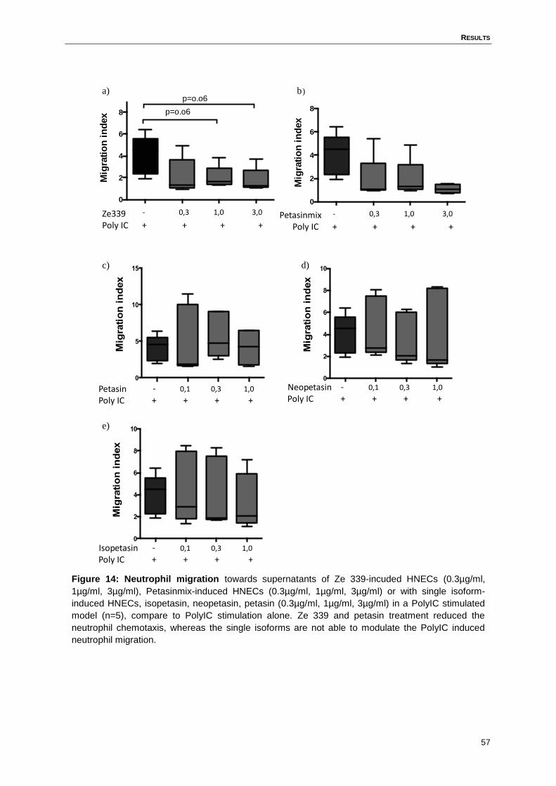
RESULTS
57
Figure 14: Neutrophil migration towards supernatants of Ze 339-incuded HNECs (0.3µg/ml,
1µg/ml, 3µg/ml), Petasinmix-induced HNECs (0.3µg/ml, 1µg/ml, 3µg/ml) or with single isoform-
induced HNECs, isopetasin, neopetasin, petasin (0.3µg/ml, 1µg/ml, 3µg/ml) in a PolyIC stimulated
model (n=5), compare to PolyIC stimulation alone. Ze 339 and petasin treatment reduced the
neutrophil chemotaxis, whereas the single isoforms are not able to modulate the PolyIC induced
neutrophil migration.
0
2
4
6
8
Mig
rati
on
in
dex p=o.o6
p=o.o6
Ze339Poly IC
- 0,3 1,0 3,0
+ + + +
0
2
4
6
8
Mig
rati
on
in
dex
PetasinmixPoly IC
- 0,3 1,0 3,0
+ + + +
PetasinPoly IC
- 0,1 0,3 1,0
+ + + +
NeopetasinPoly IC
- 0,1 0,3 1,0
+ + + +
IsopetasinPoly IC
- 0,1 0,3 1,0
+ + + +
a)
e)
c)
b)
d)
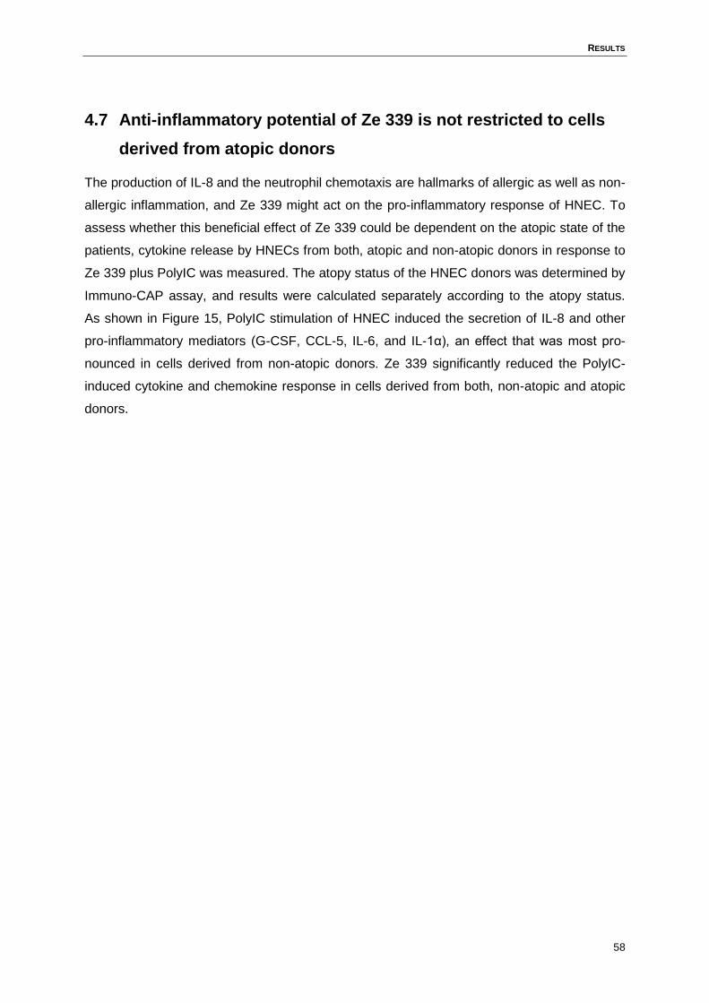
RESULTS
58
4.7 Anti-inflammatory potential of Ze 339 is not restricted to cells
derived from atopic donors
The production of IL-8 and the neutrophil chemotaxis are hallmarks of allergic as well as non-
allergic inflammation, and Ze 339 might act on the pro-inflammatory response of HNEC. To
assess whether this beneficial effect of Ze 339 could be dependent on the atopic state of the
patients, cytokine release by HNECs from both, atopic and non-atopic donors in response to
Ze 339 plus PolyIC was measured. The atopy status of the HNEC donors was determined by
Immuno-CAP assay, and results were calculated separately according to the atopy status.
As shown in Figure 15, PolyIC stimulation of HNEC induced the secretion of IL-8 and other
pro-inflammatory mediators (G-CSF, CCL-5, IL-6, and IL-1α), an effect that was most pro-
nounced in cells derived from non-atopic donors. Ze 339 significantly reduced the PolyIC-
induced cytokine and chemokine response in cells derived from both, non-atopic and atopic
donors.
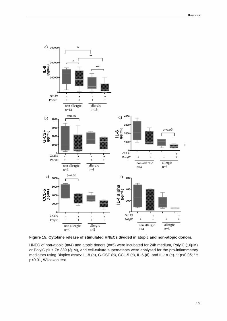
RESULTS
59
Figure 15: Cytokine release of stimulated HNECs divided in atopic and non-atopic donors.
HNEC of non-atopic (n=4) and atopic donors (n=5) were incubated for 24h medium, PolyIC (10µM)
or PolyIC plus Ze 339 (3µM), and cell-culture supernatants were analysed for the pro-inflammatory
mediators using Bioplex assay: IL-8 (a), G-CSF (b), CCL-5 (c), IL-6 (d), and IL-1α (e). *: p<0.05; **:
p<0.01, Wilcoxon test.
0
2000
4000
6000
8000
CC
L-5
(pg
/mL
)
p=o.o6
- + - +PolyIC
Ze339 + + + +
non alle rgic
n=5
allergic
n=5
- + - +PolyIC
Ze339 + + + +
non alle rgic
n=4
allergic
n=5
- + - +PolyIC
Ze339 + + + +
non alle rgic
n=4
allergic
n=5
0
1000
2000
3000
4000
G-C
SF
(pg
/mL
)
p=o.o6
- + - +PolyIC
Ze339 + + + +
non alle rgic
n=5
allergic
n=4
0
100000
200000
300000
IL-8
(pg
/mL
)**
***
*
**
- + - +PolyIC
Ze339 + + + +
non alle rgic
n=13
allergic
n=16
a)
b)
c)
d)
e)
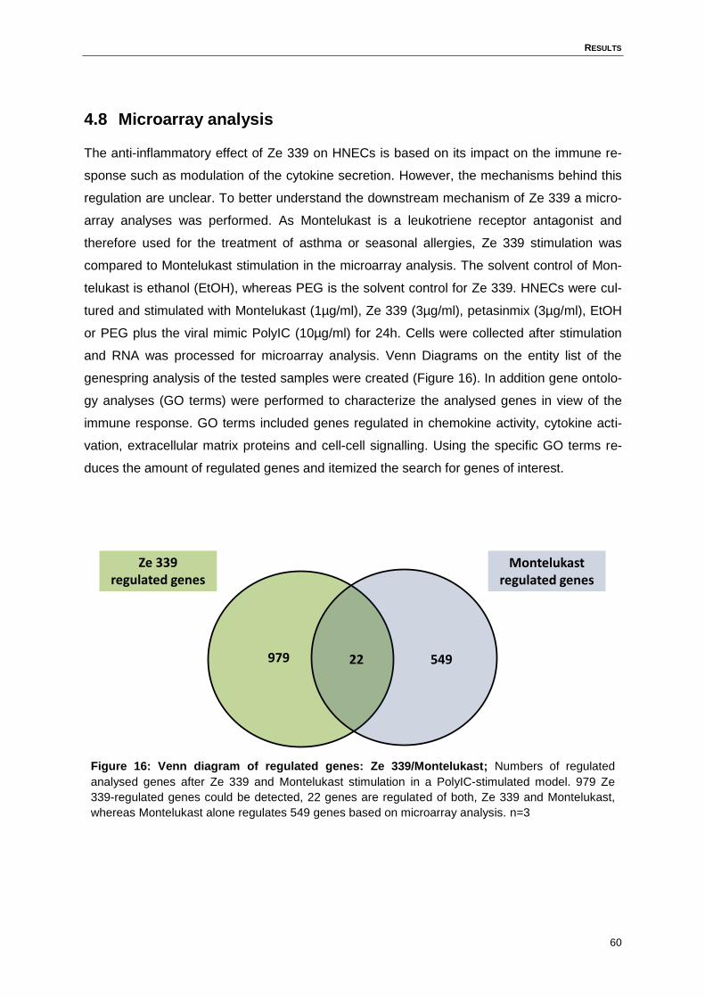
RESULTS
60
4.8 Microarray analysis
The anti-inflammatory effect of Ze 339 on HNECs is based on its impact on the immune re-
sponse such as modulation of the cytokine secretion. However, the mechanisms behind this
regulation are unclear. To better understand the downstream mechanism of Ze 339 a micro-
array analyses was performed. As Montelukast is a leukotriene receptor antagonist and
therefore used for the treatment of asthma or seasonal allergies, Ze 339 stimulation was
compared to Montelukast stimulation in the microarray analysis. The solvent control of Mon-
telukast is ethanol (EtOH), whereas PEG is the solvent control for Ze 339. HNECs were cul-
tured and stimulated with Montelukast (1µg/ml), Ze 339 (3µg/ml), petasinmix (3µg/ml), EtOH
or PEG plus the viral mimic PolyIC (10µg/ml) for 24h. Cells were collected after stimulation
and RNA was processed for microarray analysis. Venn Diagrams on the entity list of the
genespring analysis of the tested samples were created (Figure 16). In addition gene ontolo-
gy analyses (GO terms) were performed to characterize the analysed genes in view of the
immune response. GO terms included genes regulated in chemokine activity, cytokine acti-
vation, extracellular matrix proteins and cell-cell signalling. Using the specific GO terms re-
duces the amount of regulated genes and itemized the search for genes of interest.
Figure 16: Venn diagram of regulated genes: Ze 339/Montelukast; Numbers of regulated
analysed genes after Ze 339 and Montelukast stimulation in a PolyIC-stimulated model. 979 Ze
339-regulated genes could be detected, 22 genes are regulated of both, Ze 339 and Montelukast,
whereas Montelukast alone regulates 549 genes based on microarray analysis. n=3
979 54922
Montelukast regulated genes
Ze 339regulated genes
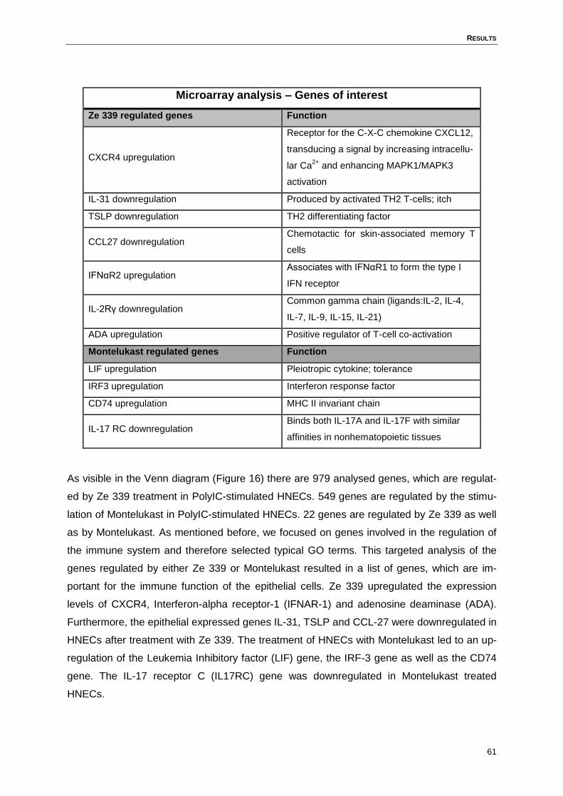
RESULTS
61
Microarray analysis – Genes of interest
Ze 339 regulated genes Function
CXCR4 upregulation
Receptor for the C-X-C chemokine CXCL12,
transducing a signal by increasing intracellu-
lar Ca2+
and enhancing MAPK1/MAPK3
activation
IL-31 downregulation Produced by activated TH2 T-cells; itch
TSLP downregulation TH2 differentiating factor
CCL27 downregulation Chemotactic for skin-associated memory T
cells
IFNαR2 upregulation Associates with IFNαR1 to form the type I
IFN receptor
IL-2Rγ downregulation Common gamma chain (ligands:IL-2, IL-4,
IL-7, IL-9, IL-15, IL-21)
ADA upregulation Positive regulator of T-cell co-activation
Montelukast regulated genes Function
LIF upregulation Pleiotropic cytokine; tolerance
IRF3 upregulation Interferon response factor
CD74 upregulation MHC II invariant chain
IL-17 RC downregulation Binds both IL-17A and IL-17F with similar
affinities in nonhematopoietic tissues
As visible in the Venn diagram (Figure 16) there are 979 analysed genes, which are regulat-
ed by Ze 339 treatment in PolyIC-stimulated HNECs. 549 genes are regulated by the stimu-
lation of Montelukast in PolyIC-stimulated HNECs. 22 genes are regulated by Ze 339 as well
as by Montelukast. As mentioned before, we focused on genes involved in the regulation of
the immune system and therefore selected typical GO terms. This targeted analysis of the
genes regulated by either Ze 339 or Montelukast resulted in a list of genes, which are im-
portant for the immune function of the epithelial cells. Ze 339 upregulated the expression
levels of CXCR4, Interferon-alpha receptor-1 (IFNAR-1) and adenosine deaminase (ADA).
Furthermore, the epithelial expressed genes IL-31, TSLP and CCL-27 were downregulated in
HNECs after treatment with Ze 339. The treatment of HNECs with Montelukast led to an up-
regulation of the Leukemia Inhibitory factor (LIF) gene, the IRF-3 gene as well as the CD74
gene. The IL-17 receptor C (IL17RC) gene was downregulated in Montelukast treated
HNECs.
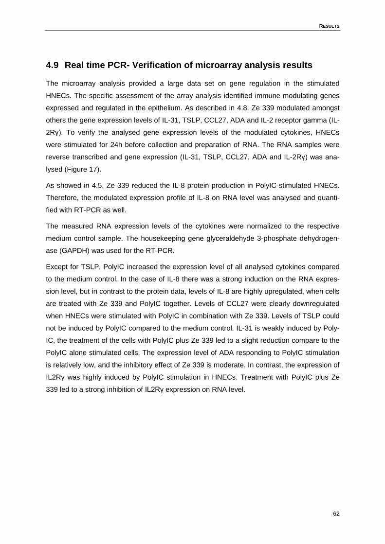
RESULTS
62
4.9 Real time PCR- Verification of microarray analysis results
The microarray analysis provided a large data set on gene regulation in the stimulated
HNECs. The specific assessment of the array analysis identified immune modulating genes
expressed and regulated in the epithelium. As described in 4.8, Ze 339 modulated amongst
others the gene expression levels of IL-31, TSLP, CCL27, ADA and IL-2 receptor gamma (IL-
2Rγ). To verify the analysed gene expression levels of the modulated cytokines, HNECs
were stimulated for 24h before collection and preparation of RNA. The RNA samples were
reverse transcribed and gene expression (IL-31, TSLP, CCL27, ADA and IL-2Rγ) was ana-
lysed (Figure 17).
As showed in 4.5, Ze 339 reduced the IL-8 protein production in PolyIC-stimulated HNECs.
Therefore, the modulated expression profile of IL-8 on RNA level was analysed and quanti-
fied with RT-PCR as well.
The measured RNA expression levels of the cytokines were normalized to the respective
medium control sample. The housekeeping gene glyceraldehyde 3-phosphate dehydrogen-
ase (GAPDH) was used for the RT-PCR.
Except for TSLP, PolyIC increased the expression level of all analysed cytokines compared
to the medium control. In the case of IL-8 there was a strong induction on the RNA expres-
sion level, but in contrast to the protein data, levels of IL-8 are highly upregulated, when cells
are treated with Ze 339 and PolyIC together. Levels of CCL27 were clearly downregulated
when HNECs were stimulated with PolyIC in combination with Ze 339. Levels of TSLP could
not be induced by PolyIC compared to the medium control. IL-31 is weakly induced by Poly-
IC, the treatment of the cells with PolyIC plus Ze 339 led to a slight reduction compare to the
PolyIC alone stimulated cells. The expression level of ADA responding to PolyIC stimulation
is relatively low, and the inhibitory effect of Ze 339 is moderate. In contrast, the expression of
IL2Rγ was highly induced by PolyIC stimulation in HNECs. Treatment with PolyIC plus Ze
339 led to a strong inhibition of IL2Rγ expression on RNA level.
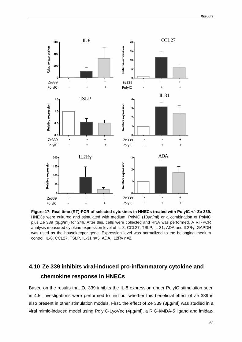
RESULTS
63
Figure 17: Real time (RT)-PCR of selected cytokines in HNECs treated with PolyIC +/- Ze 339.
HNECs were cultured and stimulated with medium, PolyIC (10µg/ml) or a combination of PolyIC
plus Ze 339 (3µg/ml) for 24h. After this, cells were collected and RNA was performed. A RT-PCR
analysis measured cytokine expression level of IL-8, CCL27, TSLP, IL-31, ADA and IL2Rγ. GAPDH
was used as the housekeeper gene. Expression level was normalized to the belonging medium
control. IL-8, CCL27, TSLP, IL-31 n=5; ADA, IL2Rγ n=2.
4.10 Ze 339 inhibits viral-induced pro-inflammatory cytokine and
chemokine response in HNECs
Based on the results that Ze 339 inhibits the IL-8 expression under PolyIC stimulation seen
in 4.5, investigations were performed to find out whether this beneficial effect of Ze 339 is
also present in other stimulation models. First, the effect of Ze 339 (3µg/ml) was studied in a
viral mimic-induced model using PolyIC-LyoVec (4µg/ml), a RIG-I/MDA-5 ligand and imidaz-
PolyIC
Ze339 - + +
- - +
PolyIC
Ze339 - + +
- - +
PolyIC
Ze339 - + +
- - +
PolyIC
Ze339 - + +
- - +
PolyIC
Ze339 - + +
- - +
PolyIC
Ze339 - + +
- - +
IL-8 CCL27
TSLPIL-31
ADAIL2Rγ
RESULTS
64
oquinoline (R-848) (10µg/ml) a TLR7/8 ligand. For this purpose, HNECs were treated with
the indicated viral stimuli in the absence or presence of Ze 339. After 24h, supernatants were
collected and tested for a panel of pro-inflammatory cytokines and chemokines (IL-8,
CCL2/MCP-1, CCL3/MIP-1α, CCL4/MIP-1β, CCL5/RANTES, CXCL10/IP-10, G-CSF, TNF-α,
IL-1α, IL-6). Stimulation with PolyIC and PolyIC-LyoVec led to the induction of a pro-
inflammatory response in HNECs, whereas the TLR7/8 ligand R-848 did not induce any
measurable response (Figure 18). Ze 339 significantly reduced PolyIC-induced cytokine and
chemokine response including G-CSF, CCL3, IL-8, CCL4, CCL2, TNF- α, CCL5, IL-6 and
IL1- α. The expression level of IP-10 was not modulated by Ze 339 stimulation. The inhibitory
effect of Ze 339 on the pro-inflammatory response to PolyIC-LyoVec was less pronounced
than the effect on the response to PolyIC. Specifically, Ze 339 decreased only the PolyIC-
LyoVec induced production of IL-8, CCL3, and IL-6, whereas the production of other cyto-
kines and chemokines remained unaffected by Ze 339 (Figure 18).
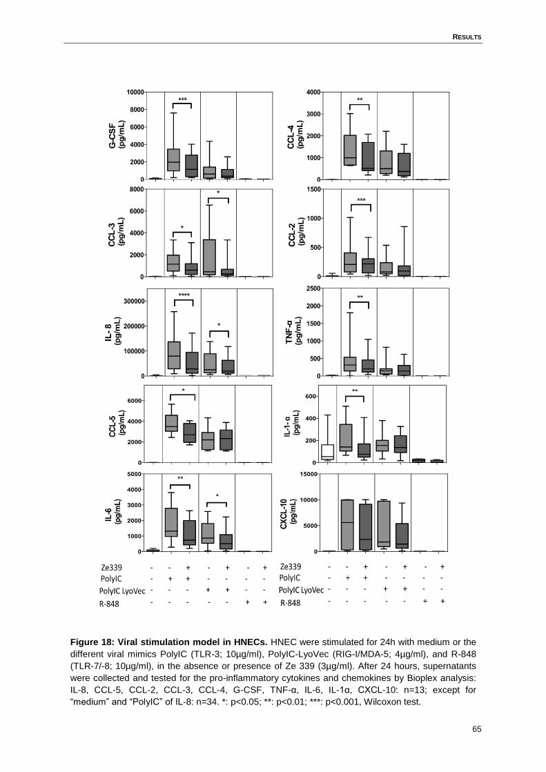
RESULTS
65
Figure 18: Viral stimulation model in HNECs. HNEC were stimulated for 24h with medium or the
different viral mimics PolyIC (TLR-3; 10µg/ml), PolyIC-LyoVec (RIG-I/MDA-5; 4µg/ml), and R-848
(TLR-7/-8; 10µg/ml), in the absence or presence of Ze 339 (3µg/ml). After 24 hours, supernatants
were collected and tested for the pro-inflammatory cytokines and chemokines by Bioplex analysis:
IL-8, CCL-5, CCL-2, CCL-3, CCL-4, G-CSF, TNF-α, IL-6, IL-1α, CXCL-10: n=13; except for
“medium” and “PolyIC” of IL-8: n=34. *: p<0.05; **: p<0.01; ***: p<0.001, Wilcoxon test.
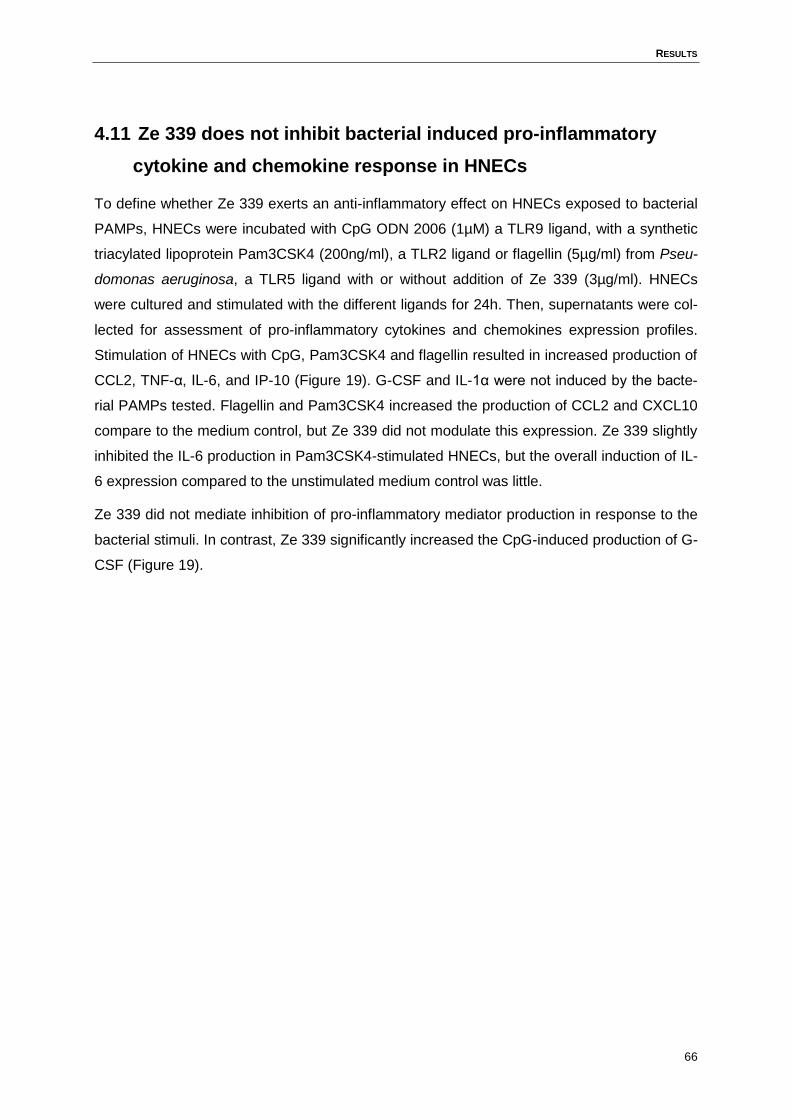
RESULTS
66
4.11 Ze 339 does not inhibit bacterial induced pro-inflammatory
cytokine and chemokine response in HNECs
To define whether Ze 339 exerts an anti-inflammatory effect on HNECs exposed to bacterial
PAMPs, HNECs were incubated with CpG ODN 2006 (1µM) a TLR9 ligand, with a synthetic
triacylated lipoprotein Pam3CSK4 (200ng/ml), a TLR2 ligand or flagellin (5µg/ml) from Pseu-
domonas aeruginosa, a TLR5 ligand with or without addition of Ze 339 (3µg/ml). HNECs
were cultured and stimulated with the different ligands for 24h. Then, supernatants were col-
lected for assessment of pro-inflammatory cytokines and chemokines expression profiles.
Stimulation of HNECs with CpG, Pam3CSK4 and flagellin resulted in increased production of
CCL2, TNF-α, IL-6, and IP-10 (Figure 19). G-CSF and IL-1α were not induced by the bacte-
rial PAMPs tested. Flagellin and Pam3CSK4 increased the production of CCL2 and CXCL10
compare to the medium control, but Ze 339 did not modulate this expression. Ze 339 slightly
inhibited the IL-6 production in Pam3CSK4-stimulated HNECs, but the overall induction of IL-
6 expression compared to the unstimulated medium control was little.
Ze 339 did not mediate inhibition of pro-inflammatory mediator production in response to the
bacterial stimuli. In contrast, Ze 339 significantly increased the CpG-induced production of G-
CSF (Figure 19).
RESULTS
67
Figure 19: Bacterial stimulation model in HNECs. HNEC were stimulated for 24h with medium or
the different bacterial mimics CpG-ODN 2006 (TLR-9; 5µg/ml), Pam3CSK4 (TLR-1/-2, TLR-2/-6;
200ng/ml), and Pseudomonas aeruginosa flagellin (TLR-5; 5µg/ml), in the absence or presence of
Ze 339 (3µg/ml). After 24 hours, supernatants were collected and tested for the pro-inflammatory
cytokines and chemokines G-CSF, CCL2, IL-1α, TNF-α, IL-6, CXCL-10. n=13; *: p<0.05, Wilcoxon
test.
0
200
400
600
800
G-C
SF
(pg
/mL
)
*
0
50
100
150
200
250
TN
F-α
(pg
/mL
)
- - + - + - + - + + - - - - - - - + + - -
- - - - - + +
0
100
200
300
IL-1
α(p
g/m
L)
0
200
400
600
CC
L-2
(pg
/mL
)
*
*
0
200
400
600
800
IL-6
(pg
/mL
)
p=0.06
p=0.06
0
1000
2000
CX
CL
-10
(pg
/mL
)
*
p=0.07
RESULTS
68
4.12 Ze 339 modulates the IL-4-, IL-6- and IFN-γ-induced pro-
inflammatory cytokine and chemokine expression in HNECs
Ze 339 shows an anti-inflammatory effect in the viral-induced stimulation model, but not in
the bacterial-induced stimulation model. In addition to the two tested models, the anti-
inflammatory effect of Ze 339 in a model of HNECs under Th1-, Th2- or pro-inflammatory
cytokine exposure was investigated. Therefore, we stimulated HNECs with IL-4 (50ng/ml)
(Th2 condition), INF-γ (15ng/ml) (Th1 condition) or IL-6 (800U/ml) (pro-inflammatory condi-
tion) for 24h in the absence and presence of Ze 339 to assess the pro-inflammatory cytokine
and chemokine response. Supernatant was taken to analyse the expressed cytokine levels in
stimulated HNECs. IL-4 induced low-level secretion of CCL2 (p<0.01) and IL-6 (p=0.1), and
Ze 339 significantly inhibited this response. IFN-γ stimulation, in contrast, led to significant
induction of IP-10, which was decreased by Ze 339. Under IL-6 stimulation, Ze 339 de-
creased the expression levels of CCL2, IL-1α and IL-6; however, the effect did not reach sta-
tistical significance (Figure 20).
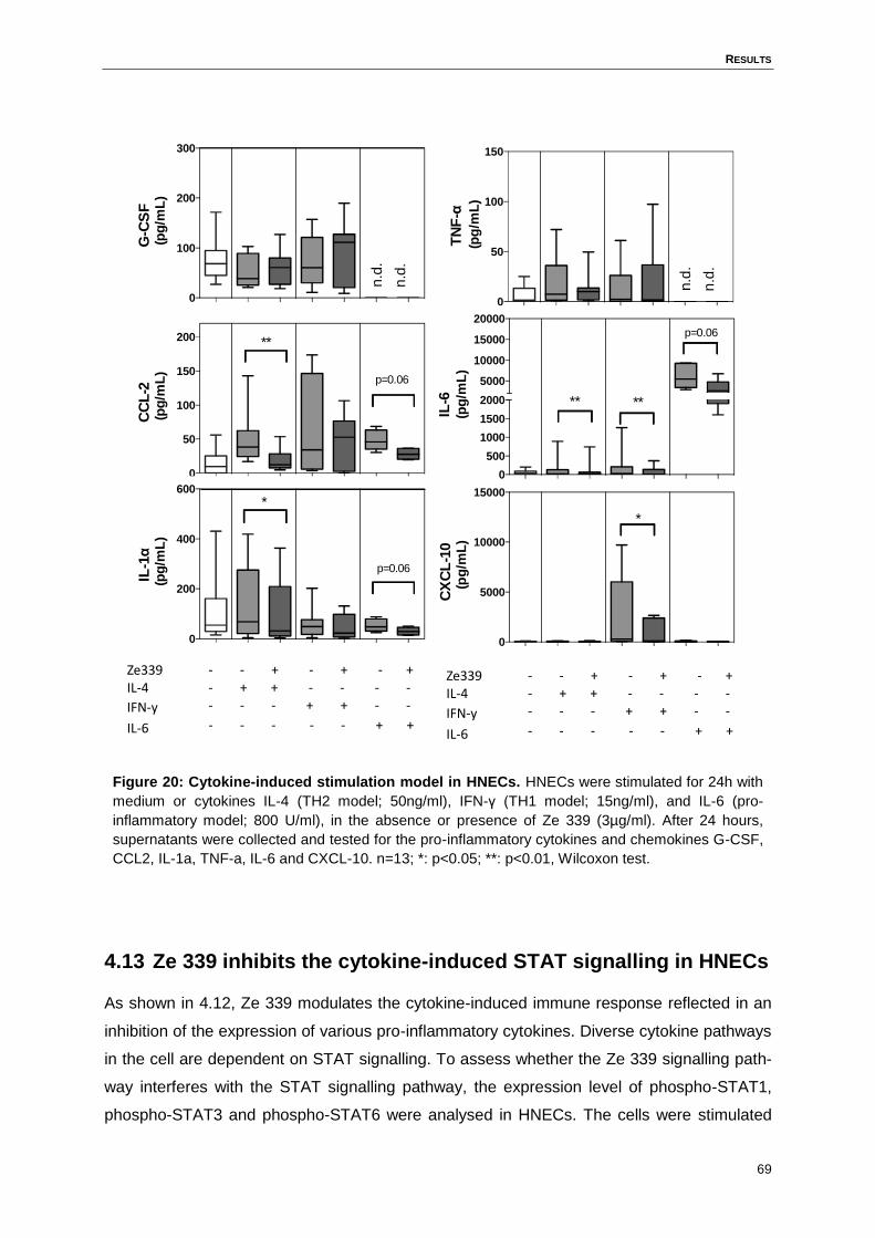
RESULTS
69
Figure 20: Cytokine-induced stimulation model in HNECs. HNECs were stimulated for 24h with
medium or cytokines IL-4 (TH2 model; 50ng/ml), IFN-γ (TH1 model; 15ng/ml), and IL-6 (pro-
inflammatory model; 800 U/ml), in the absence or presence of Ze 339 (3µg/ml). After 24 hours,
supernatants were collected and tested for the pro-inflammatory cytokines and chemokines G-CSF,
CCL2, IL-1a, TNF-a, IL-6 and CXCL-10. n=13; *: p<0.05; **: p<0.01, Wilcoxon test.
4.13 Ze 339 inhibits the cytokine-induced STAT signalling in HNECs
As shown in 4.12, Ze 339 modulates the cytokine-induced immune response reflected in an
inhibition of the expression of various pro-inflammatory cytokines. Diverse cytokine pathways
in the cell are dependent on STAT signalling. To assess whether the Ze 339 signalling path-
way interferes with the STAT signalling pathway, the expression level of phospho-STAT1,
phospho-STAT3 and phospho-STAT6 were analysed in HNECs. The cells were stimulated
0
100
200
300G
-CS
F(p
g/m
L)
n.d
.
n.d
.
0
50
100
150
TN
F-α
(pg
/mL
)
n.d
.
n.d
.
0
50
100
150
200
CC
L-2
(pg
/mL
)
**
p=0.06
0
200
400
600
IL-1
α(p
g/m
L)
*
p=0.06
Ze339IL-4
IFN-y
IL-6
- - + - + - + - + + - - - - - - - + + - -
- - - - - + +
0
500
1000
1500
2000
5000
10000
15000
20000
IL-6
(pg
/mL
)
** **
p=0.06
0
5000
10000
15000
CX
CL
-10
(pg
/mL
)
*
Ze339IL-4
IFN-y
IL-6
- - + - + - + - + + - - - - - - - + + - -
- - - - - + +
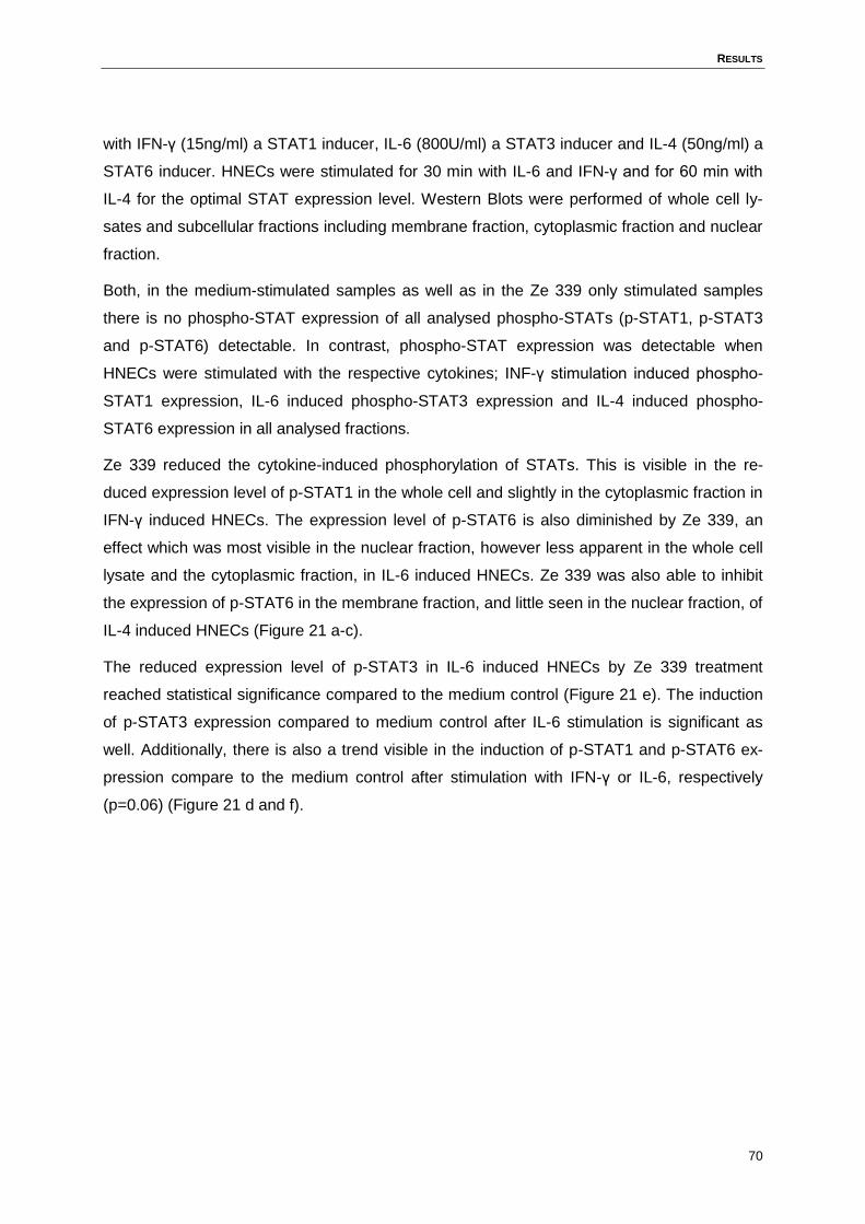
RESULTS
70
with IFN-γ (15ng/ml) a STAT1 inducer, IL-6 (800U/ml) a STAT3 inducer and IL-4 (50ng/ml) a
STAT6 inducer. HNECs were stimulated for 30 min with IL-6 and IFN-γ and for 60 min with
IL-4 for the optimal STAT expression level. Western Blots were performed of whole cell ly-
sates and subcellular fractions including membrane fraction, cytoplasmic fraction and nuclear
fraction.
Both, in the medium-stimulated samples as well as in the Ze 339 only stimulated samples
there is no phospho-STAT expression of all analysed phospho-STATs (p-STAT1, p-STAT3
and p-STAT6) detectable. In contrast, phospho-STAT expression was detectable when
HNECs were stimulated with the respective cytokines; INF-γ stimulation induced phospho-
STAT1 expression, IL-6 induced phospho-STAT3 expression and IL-4 induced phospho-
STAT6 expression in all analysed fractions.
Ze 339 reduced the cytokine-induced phosphorylation of STATs. This is visible in the re-
duced expression level of p-STAT1 in the whole cell and slightly in the cytoplasmic fraction in
IFN-γ induced HNECs. The expression level of p-STAT6 is also diminished by Ze 339, an
effect which was most visible in the nuclear fraction, however less apparent in the whole cell
lysate and the cytoplasmic fraction, in IL-6 induced HNECs. Ze 339 was also able to inhibit
the expression of p-STAT6 in the membrane fraction, and little seen in the nuclear fraction, of
IL-4 induced HNECs (Figure 21 a-c).
The reduced expression level of p-STAT3 in IL-6 induced HNECs by Ze 339 treatment
reached statistical significance compared to the medium control (Figure 21 e). The induction
of p-STAT3 expression compared to medium control after IL-6 stimulation is significant as
well. Additionally, there is also a trend visible in the induction of p-STAT1 and p-STAT6 ex-
pression compare to the medium control after stimulation with IFN-γ or IL-6, respectively
(p=0.06) (Figure 21 d and f).
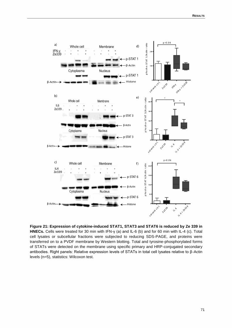
RESULTS
71
Figure 21: Expression of cytokine-induced STAT1, STAT3 and STAT6 is reduced by Ze 339 in
HNECs. Cells were treated for 30 min with IFN-γ (a) and IL-6 (b) and for 60 min with IL-4 (c). Total
cell lysates or subcellular fractions were subjected to reducing SDS-PAGE, and proteins were
transferred on to a PVDF membrane by Western blotting. Total and tyrosine-phosphorylated forms
of STATs were detected on the membrane using specific primary and HRP-conjugated secondary
antibodies. Right panels: Relative expression levels of STATs in total cell lysates relative to β-Actin
levels (n=5), statistics: Wilcoxon test.
RESULTS
72
4.14 Modulation of total STAT and cytokine-induced phospho-STAT
expression and translocation in HNECs by Ze 339
As seen in 4.13 Ze 339 regulates the protein expression level of several STATs unto cyto-
kine stimulation. To visualize the effect of Ze 339 on STAT signalling, HNECs were cultured
and stimulated on chamber glass slides for fixation, staining and imaging by a confocal mi-
croscope.
First, HNECs were stimulated with Ze 339 (3µg/ml) for 1h to analyse the expression profiles
of p-STAT1, p-STAT3, p-STAT6 and total STAT1, STAT3 and STAT6 under Ze 339 condi-
tions only. As a control, tubulin was stained in HNECs to visualize the shape of the cells. As
seen in Figure 22, treatment with Ze 339 without co-stimulation had nearly no effect on the
expression level of p-STAT1, p-STAT3 and p-STAT6. But, the expression level of total
STAT1, STAT3 and STAT6 is induced by the stimulation of Ze 339 alone in HNECs. Total
STAT3 and total STAT1 were detectable in both, cytoplasmic and nuclear fractions, whereas
total STAT6 seems to be only expressed in the cytoplasm of HNECs when stimulated with
Ze 339 only.
Figure 22: Immunohistochemistry phospho-STATs/ total STATs in stimulated HNECs. HNECs
were cultured on glass chamber slides and stimulated with Ze 339 (3µg/ml) for 1h before
immunostaining with STAT antibodies. Fluorescence images of p-STAT1, p-STAT3, p-STAT6 and
total STAT1, STAT3 and STAT6 were captured by confocal microscope.
RESULTS
73
Additionally, HNECs were stimulated with the cytokines IL-4 (60 min), IFN-γ (30 min), and IL-
6 (30 min). Stimulation of HNECs with cytokines led to the induction of total STATs and to
nuclear translocation of phospho-STATs (Figure 23).
In IFN-γ-induced HNECs, p-STAT1 expression is weakly induced compared to solvent con-
trol and IFN-γ plus Ze 339 treated HNECs, in which no expression of p-STAT1 was detecta-
ble. Total STAT1 is expressed in every condition, but the nuclear expression is enhanced in
the IFN-γ-treated cells. This nuclear expression is in turn also present, but reduced in IFN-γ
plus Ze 339 stimulated cells.
IL-6 stimulated HNECs highly expressed p-STAT3 in the nucleus, whereas there was no
expression in the solvent control. In IL-6 plus Ze 339 treated cells, there is a strongly reduced
expression of p-STAT3 detectable. Total STAT3 is detected under all conditions. In the sol-
vent control it is expressed in the cytoplasmic fraction of the cells, whereas in IL-6 induced
HNECs it is highly upregulated in the nucleus. A combination of IL-6 with Ze 339 leads to a
less present total STAT3 expression in the nucleus and nearly not expression of total STAT3
in the cytoplasmic fraction of HNECs.
Stimulation with IL-4 induced total and p-STAT6 expression in stimulated and control
HNECs. P-STAT6 is expressed in the cytoplasmic and nuclear fraction of the cells when
stimulated with IL-4 alone. In combination with Ze 339, p-STAT6 was less abundant, espe-
cially in the nucleus. Total STAT6 expression is induced in the solvent control sample, and
was almost only detectable in the cytoplasmic fraction. The translocation into the nucleus of
total STAT6 is increased in IL-4 stimulated HNECs. This translocation is less detectable
when cells are stimulated with IL-4 together with Ze 339. Total STAT was more abundant in
the cytoplasmic fraction of the stimulated HNECs.
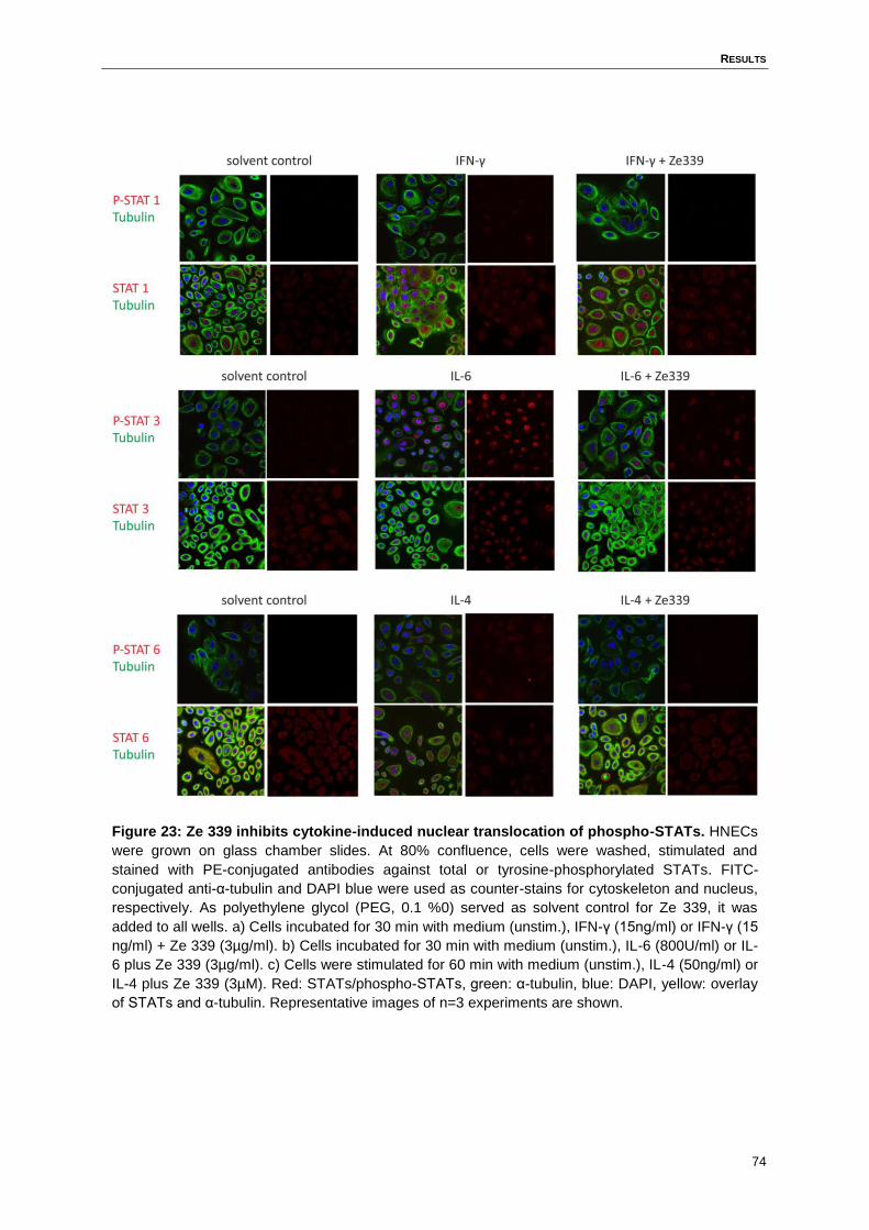
RESULTS
74
Figure 23: Ze 339 inhibits cytokine-induced nuclear translocation of phospho-STATs. HNECs
were grown on glass chamber slides. At 80% confluence, cells were washed, stimulated and
stained with PE-conjugated antibodies against total or tyrosine-phosphorylated STATs. FITC-
conjugated anti-α-tubulin and DAPI blue were used as counter-stains for cytoskeleton and nucleus,
respectively. As polyethylene glycol (PEG, 0.1 %0) served as solvent control for Ze 339, it was
added to all wells. a) Cells incubated for 30 min with medium (unstim.), IFN-γ (15ng/ml) or IFN-γ (15
ng/ml) + Ze 339 (3µg/ml). b) Cells incubated for 30 min with medium (unstim.), IL-6 (800U/ml) or IL-
6 plus Ze 339 (3µg/ml). c) Cells were stimulated for 60 min with medium (unstim.), IL-4 (50ng/ml) or
IL-4 plus Ze 339 (3µM). Red: STATs/phospho-STATs, green: α-tubulin, blue: DAPI, yellow: overlay
of STATs and α-tubulin. Representative images of n=3 experiments are shown.
DISCUSSION
75
5 Discussion
The aim of this thesis was to study the immune modulatory effects of Ze 339 in primary hu-
man nasal epithelial cells, to enlarge the knowledge and possibly extend the area of applica-
tion of this phytodrug. The beneficial effects of the petasins, constituents of Ze 339, are
known and were studied in clinical settings of airway inflammation and human myeloid cells
such as eosinophils (Dumitru et al., 2011; Thomet et al., 2001). The exact mode of action on
airway epithelial cells had not been studied before.
As IL-8 is expressed in airway epithelia cells in response to various stimuli such as respirato-
ry viruses, bacteria, fungi, cytokines, allergens and mechanical strain (Cromwell et al., 1992;
King et al., 1998; Shibata et al., 1996; Subauste et al., 1995; Tomee et al., 1997), HNECs
are stimulated with dsRNA Poly IC as a viral mimic and to induce a immune response result-
ing in enhanced IL-8 expression. But to analyse whether Ze 339 itself induces an increased
IL-8 secretion in HNECs, the IL-8 expression of Ze 339-only stimulated cells are analysed
over a time period. PEG serves as the solvent control of Ze 339. There is a slightly induced
IL-8 expression level visible, especially at the later time points. But compared to the IL-8 ex-
pression level responding to a viral stimulus, the expressed levels of IL-8 after Ze 339 treat-
ment are insignificant. Thus, Ze 339 itself does not induce a considerable IL-8 expression
level in cultured HNECs. The tested single isoforms all belong to the sesquiterpene ester
family (Debrunner B, Meier B., 1998) that are known to modulate different intracellular signal-
ling processes in human eosinophils (Thomet et al., 2001). To investigate whether the
isoforms as a single stimulus play a role in the immune response in stimulated HNECs, cells
are treated with the single isoforms only. First, IL-8 expression was tested. There is also no
massive induced IL-8 expression level detectable when HNECs were treated with the petasin
single isoforms alone.
As the treatment of HNECs with Ze 339 and the isoforms (separately and combined) could
lead to an immune reaction of the cells also including the increased apoptosis of the cells
due to the treatment, the cytotoxicity level of stimulated HNECs were analysed. Different
concentrations were tested and the fact that the survival rate of treated HNECs did not signif-
icantly differ compare to the medium control, lead to the conclusion that the treatment with
Ze 339 or the single isoforms has no effect on the survival rate of treated HNECs.
In a previous study, Dumitru et al. showed a significant reduction of IL-8 and LTB4 in nasal
fluids of allergic patients treated with Ze 339 in a human allergen challenge model (Dumitru
et al., 2011). In cultured HNECs Ze 339 decrease the IL-8 release induced by PolyIC and
this effect is also observed, when cells are treated with a mixture of the combined petasin
DISCUSSION
76
isoforms. Therefore, petasins, which make up 30% of Ze 339, are the active components in
this readout. However, the treatment with the single petasin isoforms does not show the
same effect. This discrepancy suggests that petasins can be recognized by several receptors
or a hetero-multimeric receptor, but the combined action of three or maybe at least two peta-
sin isoforms is needed for the inhibitory effect on the IL-8 expression level. Of note, several
anti-inflammatory sesquiterpenes are ligands for the nuclear receptors peroxisome prolifera-
tor-activated receptor (PPAR)-γ (Lin, 2012), retinoid x receptor (RXR)-α (Zhang et al., 2011)
or farnesoid X receptor (FXR) (Lin, 2015), classically acting as heterodimers in anti-
inflammatory signalling. Thus, the heterodimerisation of two receptors could be essential for
the modulation of the immune response regarding the IL-8 expression level.
Petasins were previously shown to act on human granulocytes, inhibiting cysteinyl-
leukotriene and LTB4 synthesis and intracellular calcium transient induction by PAF and C5a
(Thomet et al., 2002). These effects could be related to the reduction of local LTB4 release
into nasal lining fluids of allergic patients under treatment with Ze 339 in a human random-
ized double placebo controlled clinical trial (Dumitru et al., 2011). This hypothesis is support-
ed by the present study, which shows that Ze 339 has a clear effect not only on leukocytes
but also on nasal epithelial cells under viral conditions. Therefore it could be possible that Ze
339 has a direct impact on infiltrating granulocytes and resident cells including epithelial
cells, which can all release LTs in response to inflammatory stimuli (Jame et al., 2007). Leu-
kotrienes play an important role in the pathogenesis of asthma, in allergic inflammation and
in innate immunity (Hallstrand and Henderson, 2010). There are two receptors for LTB4, the
G-protein-coupled receptor leukotriene B4 receptor type 1 (BLT1) and type 2 (BLT2). BLT1 is
mainly expressed in leukocytes (Tager and Luster, 2003), whereas BLT2 expression is pre-
sent on the surface of epithelial cells (Ishii et al., 2015). LTs have the ability to bind on neu-
trophils, leading to a neutrophil recruitment (Kim et al., 2006). A potential “anti-neutrophil”
effect of Ze 339 (due to its capacity to decrease LTB4 synthesis) is supported by the finding
that Ze 339 decreases IL-8 production by nasal epithelial cells. Thus, Ze 339 could have a
beneficial effect in asthmatic patients through decreasing the level of LTB4.
The finding that Ze 339 decreased neutrophil migration towards PolyIC-stimulated HNECs
supernatants is most likely explained by the inhibitory effect of Ze 339 on IL-8 production,
which is usually produced in higher amounts by epithelial cells as compared to the “leukocyte
mediator” LTB4. However, this would have to be tested by administration of an IL-8 neutraliz-
ing antibody to our chemotaxis assay. Again, the single isoforms are not able to inhibit the
neutrophil migration towards the stimulated supernatant, but in combination, the petasinmix
show reduced neutrophil migration in the tested concentrations. This finding encourages the
DISCUSSION
77
assumption that more than one petasin isoforms is needed to achieve immune modulation
effects. Additionally, as mentioned before, BLT1 is present on neutrophils, resulting in neu-
trophil recruitment after the binding of LTB4. The decreased production and release of LTB4
due to Ze 339 could therefore reduce the recruitment of neutrophils, an effect, which could
be addressed by performing chemotaxis assays in the presence of a BLT1 antagonist.
Notably, there is a very important role of neutrophils in multiple respiratory diseases including
COPD, bronchiolitis, cystic fibrosis and asthma. Research data support the association be-
tween neutrophils and the severity and progression of inflammatory airway diseases such as
COPD (Keatings et al., 1996). In asthma, neutrophils get recruited into the airways where
they release several inflammatory mediators, including arachidonic acid metabolites such as
LTB4, which promote airway inflammation (Rabier et al., 1991; Radeau et al., 1990). The fact
that Ze 339 decreases the neutrophil migration rate in the viral-induced PolyIC model could
also identify Ze 339 as a possible tool to control the up-regulated neutrophil recruitment in
inflammatory airway diseases such as asthma or COPD.
Montelukast is a leukotriene receptor antagonist, used as a treatment for chronic mild to
moderate asthma and allergies (Knorr et al., 1998). Here, a microarray analysis of stimulated
HNECs compared the treatments of Ze 339 with the treatments of Montelukast to identify
differences in the gene expression profile. IL-8, TSLP, IL-31 and CCL27 are proteins secret-
ed by the epithelium in response to a microbe or other pro-inflammatory stimuli (Cromwell et
al., 1992; Neis et al., 2006; Sonkoly et al., 2006; Vestergaard et al., 2005; Ying et al., 2005).
On the RNA level, Ze 339 reduces the gene expression of IL-31, TSLP and CCL27 and this
result is corroborated by RT-PCR analysis. In atopic dermatitis (AD), levels of IL-31 are ele-
vated and it leads i.e. to a reduced epidermal thickness and disturbed epidermal constitution
(Kim et al., 2011). Thus a reduced IL-31 level by Ze 339 could be beneficial for AD patients.
CCL27 is involved as a chemokine in the recruitment of T cells to the side of infection (Mo-
rales et al., 1999). Ze 339 reduces the expression of CCL27 and therefore might reduce the
recruitment of T cells in a variety of diseases with a central role for T-cells. TSLP is a media-
tor in the development of allergic sensitization through the induction of DC-mediated Th2
differentiation (Omori and Ziegler, 2007; Rate et al., 2009), thus a reduction in the expression
level of TSLP could prevent allergic sensitization.
The present study shows that Ze 339 reduces the expression of the common γ-chain in a
viral-induced model on RNA level. The IL-2 receptor γ-chain, also known as common γ-chain
composes a subunit of IL-2, IL-4, IL-7, IL-9, IL-15 and IL-21 receptors (Asao et al., 2001).
Hvid et al. showed a significant down-regulation of protease caspase-14 when keratinocytes
were treated with IL-4. This leads to a modulation in the natural moisturizing process in the
DISCUSSION
78
skin, correlating with the induction of cornification (Denecker et al., 2008; Hoste et al., 2011;
Hvid et al., 2011). Thus the reduced expression of the common γ-chain due to Ze 339 treat-
ment may diminish the process of cornification by prohibiting the IL-4 pathway.
The beneficial effect of Ze 339 assessed in the human allergen challenge clinical trial (Du-
mitru et al., 2011) was only analysed in allergic patients. To find out, whether Ze 339 might
also act in HNECs of healthy patients, PolyIC-stimulated HNECs form healthy as well as
from allergic patients are analysed regarding their expression profile of selected chemokines
and cytokines. IL-8 is significantly reduced in HNECs of allergic and non-allergic patients
demonstrating that the effect of Ze 339 is not just present in HNECs of allergic patients but
also in HNECS of non-allergic patients. G-CSF, IL-6, CCL5 and IL-1α showed a trend to-
wards decreased expression of pro-inflammatory mediators in both allergic and non-allergic
HNEC. Whereas the overall PolyIC-induced expression level of the measured cytokines and
chemokines is much higher in the non-allergic HNECs. This could be due to the fact that
HNECs from allergic patients have a higher level of pro-inflammatory mediators at baseline
and therefore react less strongly to a viral mimic compared to HNECs from healthy donors.
Additionally, Ze 339 decreases the induced secretion of further pro-inflammatory chemo-
kines, e.g. CCL2 and CCL3 in PolyIC-treated HNECs, all of which are implicated in neutro-
phil chemotaxis (Reichel et al., 2009). This broad capacity of inhibiting neutrophil chemotaxis
suggests a potential of Ze 339 treatments of inflammatory states involving extensive recruit-
ment of neutrophils such as neutrophilic asthma.
The treatment of HNECs with Ze 339 leads to a clear reduction in the pro-inflammatory cyto-
kine and chemokine response to TLR3 stimulation, whereas the inhibitory effect on RIG-I and
MDA-5 ligand-induced responses are present but less pronounced. This suggests that Ze
339 might act on the endosomal pathway or might be taken up by endosomes and therefore
cannot bind to RIG-I and MDA-5. Being a CO2 extract of butterbur leafs, Ze 339 is mainly
composed of hydrophobic compounds, among them terpenoids, lipids and glycolipids
(Thomet et al., 2001). Therefore Ze 339 could also intercalate in cell membranes, which
could decrease its bioavailability in the cytosol. Bound to the membrane it could interact or
block receptors located in the membrane of epithelial cells and modulate the immune re-
sponse.
In contrast to the inhibitory effect of Ze 339 in the viral models, comparable effects under
stimulation with selected bacterial PAMPs are not observed. The used bacterial ligands are
chosen on the basis of published data on TLR expression profiles of HNECs (Lin et al., 2007;
van Tongeren et al., 2015). The TLR-4 ligand LPS does not induce any of the cytokines or
DISCUSSION
79
chemokines tested (data not shown). In contrast to a previous study that showed TLR-4 ex-
pression in HNECs on mRNA level (Lin et al., 2007), the present results argue against func-
tionally active TLR-4 in HNECs. The finding that Ze 339 did not inhibit bacterial TLR ligand-
induced responses therefore suggests that Ze 339 could be a promising tool for treatment of
acute inflammatory symptoms during viral rather than bacterial infection.
Airway remodelling is present in airway diseases such as asthma. The according structural
changes in the airways are characterized through modifications such as smooth muscle hy-
perplasia or the generation of myofibroblasts. The development of asthma and the comprised
airway remodelling in early childhood can be promoted by a viral infection (Sigurs et al.,
2000). The results of the present study suggest beneficial effects of Ze 339 in the prevention
of virus-triggered asthma, due to its broad effects on pro-inflammatory mediator production.
Ze 339 could therefore prevent a chronic inflammatory immune response due to viral infec-
tions, which may result in asthma later in life and therefore decrease the risk to develop air-
way remodelling (Crimi et al., 1998).
Under stimulation with the cytokines IL-4, IL-6 and IFN-γ, Ze 339 diminished the release of
pro-inflammatory cytokines and chemokines from HNECs. The receptors for IL-4, IL-6 and
IFN-γ act via signal transducers and activators of transcription (STATs), and the phosphory-
lation of STATs is important for nuclear translocation and activity. STAT signalling pathways
are key regulators of various allergic and non-allergic inflammatory diseases (Darnell, 1997;
Santos and Costa-Pereira, 2011) such as tumour defence. STAT 3 could demonstrate to be
of benefit for the tumour development by inducing the production of VEGF, cyclin D1 or other
chemokines in the tissue and therefore promote the growth of the tumour (Yue and Turkson,
2009). It could be demonstrated that Ze 339 significantly reduced the level of phospho-
STAT3 in IL-6 stimulated HNECs. The STAT6 signalling pathway is activated by IL-4 binding,
a cytokine which is overexpressed in patients suffering from asthma. The IL-4 induced
STAT6 phosphorylation leads to the expression of various type 2 cytokines (Mathew et al.,
2001). Ze 339 inhibited the expression level of phosphorylated STAT6 in IL-4 induced
HNECs and could therefore represent an interesting therapeutic tool for patients suffering
from type 2 cytokine driven nasal and potentially bronchial inflammation. The finding that Ze
339 modulated cytokine-induced expression and translocation of all analysed phospho-
STATs suggests that the compound interferes with a signalling step common to all three cy-
tokine receptors.
Cytokine receptor signalling via STATs involves the assembly of a multi-protein complex in
lipid rafts or lipid microdomains, located in restricted membrane areas of the cell (Brown and
London, 2000; Subramaniam and Johnson, 2002). Several glycosphingolipids, which are
DISCUSSION
80
important in the function as modulators of signal transduction pathways in the cell, such as
cell proliferation, cell differentiation or cell survival, are located in the lipid microdomains
(Kasahara and Sanai, 2000). The glycohydrolase β-hexosaminidase is involved in the degra-
dation of glycosphingolipids. A recent in vitro study demonstrate that S-petasin inhibited the
degranulation of the β-hexosaminidase in a rat basophilic cell line independently from intra-
cellular calcium inhibition (Lee et al., 2015). Notably, β-hexosaminidase is localized in endo-
somal vesicles in epithelial cells and was found to be associated to lipid rafts in Jurkat T lym-
phocytes (Magini et al., 2012). Hydrophobic substances within Ze 339, including petasins,
could be retained in the plasma membrane or become selectively enriched in lipid rafts
where they might interfere with assembly of the JAK/STAT signalling complex. The natural
oily monocyclic sesquiterpene α-bisabolol is a natural product, which can be found in differ-
ent plants such as chamomile flowers. It is used in cosmetics for skin healing properties and
it is reported to have anti-inflammatory and anti-microbial properties. Interestingly, the α-
bisabolol lactone is incorporated in lipid rafts, where it induces apoptosis in tumour cells by
interfering with the pro-apoptotic Bcl-2 family member Bid (Darra et al., 2008). Future studies
need to show whether Ze 339 interferes with lipid raft signalling complexes.
The process of Th1 cell differentiation is amongst others dependent on the transcription fac-
tors STAT1, STAT4 and T-bet. The presence of IL-4 guides the differentiation process of T
helper cells towards a Th2 differentiation (Kaplan et al., 1996; Thierfelder et al., 1996). In this
process the transcription factors STAT6 and the trans-acting T cell-specific transcription fac-
tor GATA-3 (GATA-3) play a major role. The development of Th17 cells from naïve CD4+ is
dependent on RAR-related orphan receptor gamma t (RORγt) but also on the transcription
factor STAT3 and the presence of IL-6 (Bettelli et al., 2006). It could be shown that the level
of IL-17 expression is increased in patients suffering from inflammatory bowel disease (Fu-
jino et al., 2003). STAT3 is necessary for the Th17 differentiation process in response to
stimulation with IL-6 together with IL-21. Chen et al. showed a direct binding of STAT3 to the
IL-17A and IL-17F-promotor (Chen et al., 2006). The results of the present study show that
Ze 339 exerted the most pronounced effects on the IL-6 induced nuclear translocation of
phospho-STAT3 in HNECs. Therefore it can be speculated that Ze 339 could alter the prim-
ing of naïve T cells in the process of Th17 differentiation and Th17 mediated immune reac-
tions. This mechanism could support a beneficial effect of Ze 339 via regulating phospho-
STAT3. Ze 339 might therefore modulate disease-induced Th17 responses such as fatal
intestinal inflammation or psoriasis (Chaudhry et al., 2009; Kikly et al., 2006). Additionally,
STAT3 activity plays an important role in the IL-6 induced T cell migration (McLoughlin et al.,
2005). Beside the question if Ze 339 has an impact on the priming of naïve T cells into Th17
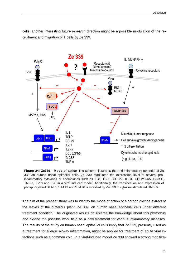
DISCUSSION
81
cells, another interesting future research direction might be a possible modulation of the re-
cruitment and migration of T cells by Ze 339.
Figure 24: Ze339 - Mode of action The scheme illustrates the anti-inflammatory potential of Ze
339 on human nasal epithelial cells. Ze 339 modulates the expression level of several pro-
inflammatory cytokines or chemokines such as IL-8, TSLP, CCL27, IL-31, CCL2/3/4/5, G-CSF,
TNF-α, IL-1α and IL-6 in a viral induced model. Additionally, the translocation and expression of
phosphorylated STAT1, STAT3 and STAT6 is modified by Ze 339 in cytokine stimulated HNECs.
The aim of the present study was to identify the mode of action of a carbon dioxide extract of
the leaves of the butterbur plant, Ze 339, on human nasal epithelial cells under different
treatment condition. The originated results do enlarge the knowledge about this phytodrug
and extend the possible work field as a new treatment for various inflammatory diseases.
The results of the study on human nasal epithelial cells imply that Ze 339, presently used as
a treatment for allergic airway inflammation, might be applied for treatment of acute viral in-
fections such as a common cold. In a viral-induced model Ze 339 showed a strong modifica-
PolyIC
IL-8
TSLP
CCL27
IL-31
IL2Rγ
CCL 2/3/4/5
G-CSF
TNF-α
NFkB
+
AP-1
NFAT
TLR3
MAPKs, IKKs
Receptor(s)?Direct uptake?
Membrane-bound?
Ze 339
Ca2+i _
P-STAT1/3/6
RIG-1MDA5
_
_
_
STATs
Microbial, tumor response
Cell survival/growth, Angiogenesis
Th2 differentiation
Cytokine/chemokine synthesis
(e.g. IL-1α, IL-6)
Cytokine receptors
+
IL-4/IL-6/IFN-γ
Virus
IRF-1
?
5-LO
LTB4
_
DISCUSSION
82
tion in the expression level of important mediators such as IL-8, IL-6, IL-1α, CCL2, CCL3,
CCL4, CCL5, TNF-α and G-CSF. Thus, Ze 339 could be of benefit in the treatment of im-
mune deviations associated with continuous cytokine-induced inflammation. Additionally, the
neutrophil migration rate towards conditioned medium from Ze 339-stimulated HNECs was
strongly decreased. In addition to its established anti-inflammatory characteristics in the
treatment of seasonal allergic rhinitis and the anti-chemotactic property in a viral stimulation
model, Ze 339 appears to exert broad anti-cytokine effects by interfering with phospho-STAT
nuclear translocation. The level of phospho-STAT3, a central transcription factor in the innate
immune response, is significantly reduced in Ze 339-treated HNECs after IL-6 stimulation.
STAT3 plays an important role in various diseases such as psoriasis, rheumatoid arthritis or
other chronic inflammatory diseases via the Th17 pathway.
Taken together the results support the view that Ze 339 could be suitable for application in
inflammatory diseases such as sinusitis. The mechanistic results provided in this thesis have
to be translated in new clinical studies to confirm the potential of Ze 339 as a therapeutic
agent exceeding allergic rhinitis.

83
LITERATURE
84
Literature
Abbas, A., Lichtman, A., and Pillai, S. (2012). Cellular and Molecular Immunology (Elsevier
Saunders).
Aboshady, O.A., and Elghanam, K.M. (2014). Sublingual immunotherapy in allergic rhinitis:
efficacy, safety, adherence and guidelines. Clin. Exp. Otorhinolaryngol. 7, 241–249.
Aikawa, T., Shimura, S., Sasaki, H., Ebina, M., and Takishima, T. (1992). Marked goblet cell
hyperplasia with mucus accumulation in the airways of patients who died of severe acute
asthma attack. Chest 101, 916–921.
Angkasekwinai, P., Park, H., Wang, Y.-H., Wang, Y.-H., Chang, S.H., Corry, D.B., Liu, Y.-J.,
Zhu, Z., and Dong, C. (2007). Interleukin 25 promotes the initiation of proallergic type 2
responses. J. Exp. Med. 204, 1509–1517.
Arya, V., and Arya, m (2011). A Review on Anti-Inflammatory Plant Barks. Nternational J.
PharmTech Res. Vol. 3, pp 899–908.
Asao, H., Okuyama, C., Kumaki, S., Ishii, N., Tsuchiya, S., Foster, D., and Sugamura, K.
(2001). Cutting edge: the common gamma-chain is an indispensable subunit of the IL-21
receptor complex. J. Immunol. Baltim. Md 1950 167, 1–5.
Atkinson, J.J., Adair-Kirk, T.L., Kelley, D.G., deMello, D., and Senior, R.M. (2008). Clara cell
adhesion and migration to extracellular matrix. Respir. Res. 9, 1.
Ballantyne, S.J., Barlow, J.L., Jolin, H.E., Nath, P., Williams, A.S., Chung, K.F., Sturton, G.,
Wong, S.H., and McKenzie, A.N.J. (2007). Blocking IL-25 prevents airway
hyperresponsiveness in allergic asthma. J. Allergy Clin. Immunol. 120, 1324–1331.
Batra, S., Balamayooran, G., and Sahoo, M.K. (2011). Nuclear factor-κB: a key regulator in
health and disease of lungs. Arch. Immunol. Ther. Exp. (Warsz.) 59, 335–351.
Bauchau, V., and Durham, S.R. (2004). Prevalence and rate of diagnosis of allergic rhinitis in
Europe. Eur. Respir. J. 24, 758–764.
Bauchau, V., and Durham, S.R. (2005). Epidemiological characterization of the intermittent
and persistent types of allergic rhinitis. Allergy 60, 350–353.
LITERATURE
85
Bazan, J.F. (1990). Structural design and molecular evolution of a cytokine receptor
superfamily. Proc. Natl. Acad. Sci. U. S. A. 87, 6934–6938.
Beakes, D.E. (1997). The use of anticholinergics in asthma. J. Asthma Off. J. Assoc. Care
Asthma 34, 357–368.
Bellik, Y., Boukraâ, L., Alzahrani, H.A., Bakhotmah, B.A., Abdellah, F., Hammoudi, S.M., and
Iguer-Ouada, M. (2012). Molecular Mechanism Underlying Anti-Inflammatory and Anti-
Allergic Activities of Phytochemicals: An Update. Molecules 18, 322–353.
Benayoun, L., Druilhe, A., Dombret, M.-C., Aubier, M., and Pretolani, M. (2003). Airway
Structural Alterations Selectively Associated with Severe Asthma. Am. J. Respir. Crit. Care
Med. 167, 1360–1368.
Bettelli, E., Carrier, Y., Gao, W., Korn, T., Strom, T.B., Oukka, M., Weiner, H.L., and
Kuchroo, V.K. (2006). Reciprocal developmental pathways for the generation of pathogenic
effector TH17 and regulatory T cells. Nature 441, 235–238.
Bickel, D., Röder, T., Bestmann, H.J., and Brune, K. (1994). Identification and
characterization of inhibitors of peptido-leukotriene-synthesis from Petasites hybridus. Planta
Med. 60, 318–322.
Biron, C.A., Nguyen, K.B., Pien, G.C., Cousens, L.P., and Salazar-Mather, T.P. (1999).
Natural killer cells in antiviral defense: function and regulation by innate cytokines. Annu.
Rev. Immunol. 17, 189–220.
Blumberg, H., Conklin, D., Xu, W.F., Grossmann, A., Brender, T., Carollo, S., Eagan, M.,
Foster, D., Haldeman, B.A., Hammond, A., et al. (2001). Interleukin 20: discovery, receptor
identification, and role in epidermal function. Cell 104, 9–19.
Boateng, J., Okeke, O., and Khan, S. (2015). Polysaccharide Based Formulations for
Mucosal Drug Delivery: A Review. Curr. Pharm. Des.
Boucher, R.C. (1994). Human airway ion transport. Part one. Am. J. Respir. Crit. Care Med.
150, 271–281.
Brashier, D.B.S., Khadka, A., Anantharamu, T., Sharma, A.K., Gupta, A.K., Sharma, S., and
Dahiya, N. (2015). Inhaled insulin: A “puff” than a “shot” before meals. J. Pharmacol.
Pharmacother. 6, 126–129.
LITERATURE
86
Bromberg, J.F., Wrzeszczynska, M.H., Devgan, G., Zhao, Y., Pestell, R.G., Albanese, C.,
and Darnell, J.E. (1999). Stat3 as an oncogene. Cell 98, 295–303.
Brown, D.A., and London, E. (2000). Structure and function of sphingolipid- and cholesterol-
rich membrane rafts. J. Biol. Chem. 275, 17221–17224.
Brozek, J.L., Bousquet, J., Baena-Cagnani, C.E., Bonini, S., Canonica, G.W., Casale, T.B.,
van Wijk, R.G., Ohta, K., Zuberbier, T., Schünemann, H.J., et al. (2010). Allergic Rhinitis and
its Impact on Asthma (ARIA) guidelines: 2010 revision. J. Allergy Clin. Immunol. 126, 466–
476.
Busse, W.W. (2010). The relationship of airway hyperresponsiveness and airway
inflammation: Airway hyperresponsiveness in asthma: its measurement and clinical
significance. Chest 138, 4S – 10S.
Bystrom, J., Taher, T.E., Muhyaddin, M.S., Clanchy, F.I., Mangat, P., Jawad, A.S., Williams,
R.O., and Mageed, R.A. (2015). Harnessing the Therapeutic Potential of Th17 Cells.
Mediators Inflamm. 2015, 1–11.
Calixto, J.B., Otuki, M.F., and Santos, A.R.S. (2003). Anti-inflammatory compounds of plant
origin. Part I. Action on arachidonic acid pathway, nitric oxide and nuclear factor kappa B
(NF-kappaB). Planta Med. 69, 973–983.
Cella, M., Jarrossay, D., Facchetti, F., Alebardi, O., Nakajima, H., Lanzavecchia, A., and
Colonna, M. (1999). Plasmacytoid monocytes migrate to inflamed lymph nodes and produce
large amounts of type I interferon. Nat. Med. 5, 919–923.
Chang, Y., Al-Alwan, L., Risse, P.-A., Halayko, A.J., Martin, J.G., Baglole, C.J., Eidelman,
D.H., and Hamid, Q. (2012). Th17-associated cytokines promote human airway smooth
muscle cell proliferation. FASEB J. Off. Publ. Fed. Am. Soc. Exp. Biol. 26, 5152–5160.
Chaudhry, A., Rudra, D., Treuting, P., Samstein, R.M., Liang, Y., Kas, A., and Rudensky,
A.Y. (2009). CD4+ regulatory T cells control TH17 responses in a Stat3-dependent manner.
Science 326, 986–991.
Chen, Z., Laurence, A., Kanno, Y., Pacher-Zavisin, M., Zhu, B.-M., Tato, C., Yoshimura, A.,
Hennighausen, L., and O’Shea, J.J. (2006). Selective regulatory function of Socs3 in the
formation of IL-17-secreting T cells. Proc. Natl. Acad. Sci. U. S. A. 103, 8137–8142.
LITERATURE
87
Chertov, O., Michiel, D.F., Xu, L., Wang, J.M., Tani, K., Murphy, W.J., Longo, D.L., Taub,
D.D., and Oppenheim, J.J. (1996). Identification of defensin-1, defensin-2, and
CAP37/azurocidin as T-cell chemoattractant proteins released from interleukin-8-stimulated
neutrophils. J. Biol. Chem. 271, 2935–2940.
Collart, M.A., Baeuerle, P., and Vassalli, P. (1990). Regulation of tumor necrosis factor alpha
transcription in macrophages: involvement of four kappa B-like motifs and of constitutive and
inducible forms of NF-kappa B. Mol. Cell. Biol. 10, 1498–1506.
Cox, L.S., Linnemann, D.L., Nolte, H., Weldon, D., Finegold, I., and Nelson, H.S. (2006).
Sublingual immunotherapy: A comprehensive review. J. Allergy Clin. Immunol. 117, 1021–
1035.
Cragg, G.M., Schepartz, S.A., Suffness, M., and Grever, M.R. (1993). The taxol supply crisis.
New NCI policies for handling the large-scale production of novel natural product anticancer
and anti-HIV agents. J. Nat. Prod. 56, 1657–1668.
Crimi, E., Spanevello, A., Neri, M., Ind, P.W., Rossi, G.A., and Brusasco, V. (1998).
Dissociation between airway inflammation and airway hyperresponsiveness in allergic
asthma. Am. J. Respir. Crit. Care Med. 157, 4–9.
Cromwell, O., Hamid, Q., Corrigan, C.J., Barkans, J., Meng, Q., Collins, P.D., and Kay, A.B.
(1992). Expression and generation of interleukin-8, IL-6 and granulocyte-macrophage
colony-stimulating factor by bronchial epithelial cells and enhancement by IL-1 beta and
tumour necrosis factor-alpha. Immunology 77, 330–337.
Crooks, S.W., Bayley, D.L., Hill, S.L., and Stockley, R.A. (2000). Bronchial inflammation in
acute bacterial exacerbations of chronic bronchitis: the role of leukotriene B4. Eur. Respir. J.
15, 274–280.
Croteau, R., Kutchan, T., and Lewis, N. (2000). Natural Products (Secondary Metabolites).
Biochem. Mol. Biol. Plants Chapter 24.
Cutz, E., Fu, X.W., and Nurse, C.A. (2003). Ionotropic receptors in pulmonary neuroepithelial
bodies (NEB) and their possible role in modulation of hypoxia signalling. Adv. Exp. Med. Biol.
536, 155–161.
Dahlén, S.E., Hedqvist, P., Hammarström, S., and Samuelsson, B. (1980). Leukotrienes are
potent constrictors of human bronchi. Nature 288, 484–486.
LITERATURE
88
D’Andrea, A., Rengaraju, M., Valiante, N.M., Chehimi, J., Kubin, M., Aste, M., Chan, S.H.,
Kobayashi, M., Young, D., and Nickbarg, E. (1992). Production of natural killer cell
stimulatory factor (interleukin 12) by peripheral blood mononuclear cells. J. Exp. Med. 176,
1387–1398.
D’Andrea, A., Aste-Amezaga, M., Valiante, N.M., Ma, X., Kubin, M., and Trinchieri, G. (1993).
Interleukin 10 (IL-10) inhibits human lymphocyte interferon gamma-production by
suppressing natural killer cell stimulatory factor/IL-12 synthesis in accessory cells. J. Exp.
Med. 178, 1041–1048.
Darnell, J.E. (1997). STATs and gene regulation. Science 277, 1630–1635.
Darnell, J.E., Kerr, I.M., and Stark, G.R. (1994). Jak-STAT pathways and transcriptional
activation in response to IFNs and other extracellular signaling proteins. Science 264, 1415–
1421.
Darra, E., Abdel-Azeim, S., Manara, A., Shoji, K., Marechal, J.D., Mariotto, S., Cavalieri, E.,
Perbellini, L., Pizza, C., Perahia, D., et al. (2008). Insight into the apoptosis-inducing action
of alpha-bisabolol towards malignant tumor cells: involvement of lipid rafts and Bid. Arch
Biochem Biophys 476, 113–123.
Debrunner B, Meier B. (1998). Petasites hybridus: a tool for interdisciplinary research in
phytotherapy. Pharm Acta Helv 72, 359–362.
Denecker, G., Ovaere, P., Vandenabeele, P., and Declercq, W. (2008). Caspase-14 reveals
its secrets. J. Cell Biol. 180, 451–458.
Devi, S., Wang, Y., Chew, W.K., Lima, R., A-González, N., Mattar, C.N.Z., Chong, S.Z.,
Schlitzer, A., Bakocevic, N., Chew, S., et al. (2013). Neutrophil mobilization via plerixafor-
mediated CXCR4 inhibition arises from lung demargination and blockade of neutrophil
homing to the bone marrow. J. Exp. Med. 210, 2321–2336.
Dharajiya, N., Boldogh, I., Cardenas, V., and Sur, S. (2008). Role of pollen NAD(P)H oxidase
in allergic inflammation. Curr. Opin. Allergy Clin. Immunol. 8, 57–62.
DiDonato, J.A., Hayakawa, M., Rothwarf, D.M., Zandi, E., and Karin, M. (1997). A cytokine-
responsive IkappaB kinase that activates the transcription factor NF-kappaB. Nature 388,
548–554.
LITERATURE
89
DiPersio, J.F., Billing, P., Williams, R., and Gasson, J.C. (1988). Human granulocyte-
macrophage colony-stimulating factor and other cytokines prime human neutrophils for
enhanced arachidonic acid release and leukotriene B4 synthesis. J. Immunol. Baltim. Md
1950 140, 4315–4322.
Driscoll, K.E., Hassenbein, D.G., Howard, B.W., Isfort, R.J., Cody, D., Tindal, M.H.,
Suchanek, M., and Carter, J.M. (1995). Cloning, expression, and functional characterization
of rat MIP-2: a neutrophil chemoattractant and epithelial cell mitogen. J. Leukoc. Biol. 58,
359–364.
Dumitru, A.F., Shamji, M., Wagenmann, M., Hindersin, S., Scheckenbach, K., Greve, J.,
Klenzner, T., Hess, L., Nebel, S., Zimmermann, C., et al. (2011). Petasol butenoate complex
(Ze 339) relieves allergic rhinitis–induced nasal obstruction more effectively than
desloratadine. J. Allergy Clin. Immunol. 127, 1515–1521.e6.
Evans, M.J., and Plopper, C.G. (1988). The role of basal cells in adhesion of columnar
epithelium to airway basement membrane. Am. Rev. Respir. Dis. 138, 481–483.
Evans, C., Kim, K., Tuvim, M., and Dickey, B. (2009). Mucus hypersecretion in asthma:
causes and effects. Curr Opin Pulm Med 15 (1), 4–11.
Evans, M.J., Cox, R.A., Shami, S.G., Wilson, B., and Plopper, C.G. (1989). The role of basal
cells in attachment of columnar cells to the basal lamina of the trachea. Am. J. Respir. Cell
Mol. Biol. 1, 463–469.
Eyerich, S., and Zielinski, C.E. (2014). Defining Th-cell subsets in a classical and tissue-
specific manner: Examples from the skin. Eur J Immunol 44, 3475–3483.
Fanning, A.S., Jameson, B.J., Jesaitis, L.A., and Anderson, J.M. (1998). The tight junction
protein ZO-1 establishes a link between the transmembrane protein occludin and the actin
cytoskeleton. J. Biol. Chem. 273, 29745–29753.
Fristrom, D. (1988). The cellular basis of epithelial morphogenesis. A review. Tissue Cell 20,
645–690.
Fujino, S., Andoh, A., Bamba, S., Ogawa, A., Hata, K., Araki, Y., Bamba, T., and Fujiyama,
Y. (2003). Increased expression of interleukin 17 in inflammatory bowel disease. Gut 52, 65–
70.
LITERATURE
90
Fujisawa, T., Chang, M.M.-J., Velichko, S., Thai, P., Hung, L.-Y., Huang, F., Phuong, N.,
Chen, Y., and Wu, R. (2011). NF-κB mediates IL-1β- and IL-17A-induced MUC5B expression
in airway epithelial cells. Am. J. Respir. Cell Mol. Biol. 45, 246–252.
Gadermaier, G., Dedic, A., Obermeyer, G., Frank, S., Himly, M., and Ferreira, F. (2004).
Biology of weed pollen allergens. Curr. Allergy Asthma Rep. 4, 391–400.
Galy, A.H., and Spits, H. (1991). IL-1, IL-4, and IFN-gamma differentially regulate cytokine
production and cell surface molecule expression in cultured human thymic epithelial cells. J.
Immunol. Baltim. Md 1950 147, 3823–3830.
Ganz, T., Selsted, M.E., Szklarek, D., Harwig, S.S., Daher, K., Bainton, D.F., and Lehrer, R.I.
(1985). Defensins. Natural peptide antibiotics of human neutrophils. J. Clin. Invest. 76, 1427–
1435.
Greenlund, A.C., Farrar, M.A., Viviano, B.L., and Schreiber, R.D. (1994). Ligand-induced IFN
gamma receptor tyrosine phosphorylation couples the receptor to its signal transduction
system (p91). EMBO J. 13, 1591–1600.
Gunawan, H., Takai, T., Ikeda, S., Okumura, K., and Ogawa, H. (2008). Protease Activity of
Allergenic Pollen of Cedar, Cypress, Juniper, Birch and Ragweed. Allergol. Int. 57, 83–91.
Hallstrand, T.S., and Henderson, W.R. (2010). An update on the role of leukotrienes in
asthma. Curr. Opin. Allergy Clin. Immunol. 10, 60–66.
Harant, H., de Martin, R., Andrew, P.J., Foglar, E., Dittrich, C., and Lindley, I.J. (1996).
Synergistic activation of interleukin-8 gene transcription by all-trans-retinoic acid and tumor
necrosis factor-alpha involves the transcription factor NF-kappaB. J. Biol. Chem. 271,
26954–26961.
Hemmi, H., Takeuchi, O., Kawai, T., Kaisho, T., Sato, S., Sanjo, H., Matsumoto, M., Hoshino,
K., Wagner, H., Takeda, K., et al. (2000). A Toll-like receptor recognizes bacterial DNA.
Nature 408, 740–745.
Henderson, W.R., Lewis, D.B., Albert, R.K., Zhang, Y., Lamm, W.J., Chiang, G.K., Jones, F.,
Eriksen, P., Tien, Y.T., Jonas, M., et al. (1996). The importance of leukotrienes in airway
inflammation in a mouse model of asthma. J. Exp. Med. 184, 1483–1494.
LITERATURE
91
Hicks, W., Hall, L., Sigurdson, L., Stewart, C., Hard, R., Winston, J., and Lwebuga-Mukasa,
J. (1997). Isolation and characterization of basal cells from human upper respiratory
epithelium. Exp. Cell Res. 237, 357–363.
Holgate, S.T. (1998). Asthma and allergy--disorders of civilization? QJM 91, 171–184.
Holt, P.G., Schon-Hegrad, M.A., Oliver, J., Holt, B.J., and McMenamin, P.G. (1990). A
Contiguous Network of Dendritic Antigen-Presenting Cells within the Respiratory Epithelium.
Int. Arch. Allergy Immunol. 91, 155–159.
Honda, K., Yanai, H., Mizutani, T., Negishi, H., Shimada, N., Suzuki, N., Ohba, Y., Takaoka,
A., Yeh, W.-C., and Taniguchi, T. (2004). Role of a transductional-transcriptional processor
complex involving MyD88 and IRF-7 in Toll-like receptor signaling. Proc. Natl. Acad. Sci. U.
S. A. 101, 15416–15421.
Hoshino, A., Tsuji, T., Matsuzaki, J., Jinushi, T., Ashino, S., Teramura, T., Chamoto, K.,
Tanaka, Y., Asakura, Y., Sakurai, T., et al. (2004). STAT6-mediated signaling in Th2-
dependent allergic asthma: critical role for the development of eosinophilia, airway hyper-
responsiveness and mucus hypersecretion, distinct from its role in Th2 differentiation. Int.
Immunol. 16, 1497–1505.
Hoste, E., Kemperman, P., Devos, M., Denecker, G., Kezic, S., Yau, N., Gilbert, B., Lippens,
S., De Groote, P., Roelandt, R., et al. (2011). Caspase-14 is required for filaggrin
degradation to natural moisturizing factors in the skin. J. Invest. Dermatol. 131, 2233–2241.
Hvid, M., Johansen, C., Deleuran, B., Kemp, K., Deleuran, M., and Vestergaard, C. (2011).
Regulation of caspase 14 expression in keratinocytes by inflammatory cytokines--a possible
link between reduced skin barrier function and inflammation? Exp. Dermatol. 20, 633–636.
Ihle, J.N., Nosaka, T., Thierfelder, W., Quelle, F.W., and Shimoda, K. (1997). Jaks and Stats
in cytokine signaling. Stem Cells Dayt. Ohio 15 Suppl 1, 105–111; discussion 112.
Ishii, Y., Saeki, K., Liu, M., Sasaki, F., Koga, T., Kitajima, K., Meno, C., Okuno, T., and
Yokomizo, T. (2015). Leukotriene B4 receptor type 2 (BLT2) enhances skin barrier function
by regulating tight junction proteins. FASEB J. Off. Publ. Fed. Am. Soc. Exp. Biol.
Iwamura, C., and Nakayama, T. (2008). Toll-like receptors in the respiratory system: their
roles in inflammation. Curr. Allergy Asthma Rep. 8, 7–13.
LITERATURE
92
Jachak S, and Saklani A (2007). Challenges and opportunities in drug discovery from plants.
Jame, A.J., Lackie, P.M., Cazaly, A.M., Sayers, I., Penrose, J.F., Holgate, S.T., and
Sampson, A.P. (2007). Human bronchial epithelial cells express an active and inducible
biosynthetic pathway for leukotrienes B4 and C4. Clin. Exp. Allergy J. Br. Soc. Allergy Clin.
Immunol. 37, 880–892.
Janeway, C., Travers, P., Walport, M., and Shlomdrik, M. (2002). Immunologie (Spektrum,
Akad. Verlag).
Joklik, W.K. (1996). The story of penicillin: the view from Oxford in the early 1950s. FASEB J.
Off. Publ. Fed. Am. Soc. Exp. Biol. 10, 525–528.
Jutel, M., Agache, I., Bonini, S., Burks, A.W., Calderon, M., Canonica, W., Cox, L., Demoly,
P., Frew, A.J., O’Hehir, R., et al. (2015). International consensus on allergy immunotherapy.
J. Allergy Clin. Immunol.
Kaplan, M.H., Schindler, U., Smiley, S.T., and Grusby, M.J. (1996). Stat6 is required for
mediating responses to IL-4 and for development of Th2 cells. Immunity 4, 313–319.
Kasahara, K., and Sanai, Y. (2000). Functional roles of glycosphingolipids in signal
transduction via lipid rafts. Glycoconj. J. 17, 153–162.
Kato, H., Takeuchi, O., Sato, S., Yoneyama, M., Yamamoto, M., Matsui, K., Uematsu, S.,
Jung, A., Kawai, T., Ishii, K.J., et al. (2006). Differential roles of MDA5 and RIG-I helicases in
the recognition of RNA viruses. Nature 441, 101–105.
Keatings, V.M., Collins, P.D., Scott, D.M., and Barnes, P.J. (1996). Differences in interleukin-
8 and tumor necrosis factor-alpha in induced sputum from patients with chronic obstructive
pulmonary disease or asthma. Am. J. Respir. Crit. Care Med. 153, 530–534.
Keshari, R.S., Jyoti, A., Dubey, M., Kothari, N., Kohli, M., Bogra, J., Barthwal, M.K., and
Dikshit, M. (2012). Cytokines induced neutrophil extracellular traps formation: implication for
the inflammatory disease condition. PLoS One 7, e48111.
Kharitonov, S.A., Donnelly, L.E., Montuschi, P., Corradi, M., Collins, J.V., and Barnes, P.J.
(2002). Dose-dependent onset and cessation of action of inhaled budesonide on exhaled
nitric oxide and symptoms in mild asthma. Thorax 57, 889–896.

LITERATURE
93
Kikly, K., Liu, L., Na, S., and Sedgwick, J.D. (2006). The IL-23/Th17 axis: therapeutic targets
for autoimmune inflammation. Curr. Opin. Immunol. 18, 670–675.
Kim, N.D., Chou, R.C., Seung, E., Tager, A.M., and Luster, A.D. (2006). A unique
requirement for the leukotriene B4 receptor BLT1 for neutrophil recruitment in inflammatory
arthritis. J. Exp. Med. 203, 829–835.
Kim, S., Kim, H.-J., Yang, H.S., Kim, E., Huh, I.-S., and Yang, J.-M. (2011). IL-31 Serum
Protein and Tissue mRNA Levels in Patients with Atopic Dermatitis. Ann. Dermatol. 23, 468–
473.
Kimpen, J.L.L., Garofalo, R., Welliver, R.C., and Ogra, P.L. (1992). Activation of Human
Eosinophils In Vitro by Respiratory Syncytial Virus. Pediatr. Res. 32, 160–164.
King, C., Brennan, S., Thompson, P.J., and Stewart, G.A. (1998). Dust mite proteolytic
allergens induce cytokine release from cultured airway epithelium. J. Immunol. Baltim. Md
1950 161, 3645–3651.
Kirito, K., Nagashima, T., Ozawa, K., and Komatsu, N. (2002). Constitutive activation of Stat1
and Stat3 in primary erythroleukemia cells. Int. J. Hematol. 75, 51–54.
Knight, D.A., and Holgate, S.T. (2003). The airway epithelium: Structural and functional
properties in health and disease. Respirology 8, 432–446.
Knorr, B., Matz, J., Bernstein, J.A., Nguyen, H., Seidenberg, B.C., Reiss, T.F., and Becker,
A. (1998). Montelukast for chronic asthma in 6- to 14-year-old children: a randomized,
double-blind trial. Pediatric Montelukast Study Group. JAMA 279, 1181–1186.
Kovacic, B., Stoiber, D., Moriggl, R., Weisz, E., Ott, R.G., Kreibich, R., Levy, D.E., Beug, H.,
Freissmuth, M., and Sexl, V. (2006). STAT1 acts as a tumor promoter for leukemia
development. Cancer Cell 10, 77–87.
Kulka, M. (2009). The potential of natural products as effective treatments for allergic
inflammation: implications for allergic rhinitis. Curr. Top. Med. Chem. 9, 1611–1624.
Kumar, H., Kawai, T., and Akira, S. (2009). Toll-like receptors and innate immunity. Biochem.
Biophys. Res. Commun. 388, 621–625.
LITERATURE
94
Kunz, S., Wolk, K., Witte, E., Witte, K., Doecke, W.-D., Volk, H.-D., Sterry, W., Asadullah, K.,
and Sabat, R. (2006). Interleukin (IL)-19, IL-20 and IL-24 are produced by and act on
keratinocytes and are distinct from classical ILs. Exp. Dermatol. 15, 991–1004.
Kuperman, D., Schofield, B., Wills-Karp, M., and Grusby, M.J. (1998). Signal transducer and
activator of transcription factor 6 (Stat6)-deficient mice are protected from antigen-induced
airway hyperresponsiveness and mucus production. J. Exp. Med. 187, 939–948.
Lackmann, M., Harpur, A.G., Oates, A.C., Mann, R.J., Gabriel, A., Meutermans, W.,
Alewood, P.F., Kerr, I.M., Stark, G.R., and Wilks, A.F. (1998). Biomolecular interaction
analysis of IFN gamma-induced signaling events in whole-cell lysates: prevalence of latent
STAT1 in high-molecular weight complexes. Growth Factors Chur Switz. 16, 39–51.
Latz, E., Schoenemeyer, A., Visintin, A., Fitzgerald, K.A., Monks, B.G., Knetter, C.F., Lien,
E., Nilsen, N.J., Espevik, T., and Golenbock, D.T. (2004). TLR9 signals after translocating
from the ER to CpG DNA in the lysosome. Nat. Immunol. 5, 190–198.
Lee, K.-P., Kang, S., Noh, M.-S., Park, S.-J., Kim, J.-M., Chung, H.Y., Je, N.K., Lee, Y.-G.,
Choi, Y.-W., and Im, D.-S. (2015). Therapeutic effects of s-petasin on disease models of
asthma and peritonitis. Biomol. Ther. 23, 45–52.
Lee, Y.C., Lee, K.S., Park, S.J., Park, H.S., Lim, J.S., Park, K.-H., Im, M.-J., Choi, I.-W., Lee,
H.-K., and Kim, U.-H. (2004). Blockade of airway hyperresponsiveness and inflammation in a
murine model of asthma by a prodrug of cysteine, L-2-oxothiazolidine-4-carboxylic acid.
FASEB J. Off. Publ. Fed. Am. Soc. Exp. Biol. 18, 1917–1919.
Lilly, C.M., Nakamura, H., Kesselman, H., Nagler-Anderson, C., Asano, K., Garcia-Zepeda,
E.A., Rothenberg, M.E., Drazen, J.M., and Luster, A.D. (1997). Expression of eotaxin by
human lung epithelial cells: induction by cytokines and inhibition by glucocorticoids. J. Clin.
Invest. 99, 1767–1773.
Lin, H.-R. (2012). Sesquiterpene lactones from Tithonia diversifolia act as peroxisome
proliferator-activated receptor agonists. Bioorg. Med. Chem. Lett. 22, 2954–2958.
Lin, H.-R. (2015). Lepidozenolide from the liverwort Lepidozia fauriana acts as a farnesoid X
receptor agonist. J. Asian Nat. Prod. Res. 17, 149–158.
LITERATURE
95
Lin, C.-F., Tsai, C.-H., Cheng, C.-H., Chen, Y.-S., Tournier, F., and Yeh, T.-H. (2007).
Expression of Toll-like receptors in cultured nasal epithelial cells. Acta Otolaryngol. (Stockh.)
127, 395–402.
Liu, R.H. (2004). Potential synergy of phytochemicals in cancer prevention: mechanism of
action. J. Nutr. 134, 3479S – 3485S.
Lumsden, A.B., McLean, A., and Lamb, D. (1984). Goblet and Clara cells of human distal
airways: evidence for smoking induced changes in their numbers. Thorax 39, 844–849.
Magini, A., Polchi, A., Tancini, B., Urbanelli, L., Hasilik, A., and Emiliani, C. (2012).
Glycohydrolases beta-hexosaminidase and beta-galactosidase are associated with lipid
microdomains of Jurkat T-lymphocytes. Biochimie 94, 684–694.
Maloney, J.P., and Gao, L. (2015). Proinflammatory Cytokines Increase Vascular Endothelial
Growth Factor Expression in Alveolar Epithelial Cells. Mediators Inflamm. 2015, 387842.
Mann, J. (1999). The Elusive Magic Bullet: The Search for the Perfect Drug (Oxford ; New
York: Oxford University Press).
Martin, C., Burdon, P.C.E., Bridger, G., Gutierrez-Ramos, J.C., Williams, T.J., and Rankin,
S.M. (2003). Chemokines acting via CXCR2 and CXCR4 control the release of neutrophils
from the bone marrow and their return following senescence. Immunity 19, 583–593.
Mathew, A., MacLean, J.A., DeHaan, E., Tager, A.M., Green, F.H., and Luster, A.D. (2001).
Signal transducer and activator of transcription 6 controls chemokine production and T helper
cell type 2 cell trafficking in allergic pulmonary inflammation. J. Exp. Med. 193, 1087–1096.
Matsukura, S., Stellato, C., Georas, S.N., Casolaro, V., Plitt, J.R., Miura, K., Kurosawa, S.,
Schindler, U., and Schleimer, R.P. (2001). Interleukin-13 upregulates eotaxin expression in
airway epithelial cells by a STAT6-dependent mechanism. Am. J. Respir. Cell Mol. Biol. 24,
755–761.
McCusker, C.T., Wang, Y., Shan, J., Kinyanjui, M.W., Villeneuve, A., Michael, H., and
Fixman, E.D. (2007). Inhibition of experimental allergic airways disease by local application
of a cell-penetrating dominant-negative STAT-6 peptide. J. Immunol. Baltim. Md 1950 179,
2556–2564.
LITERATURE
96
McIvor, R.A. (2015). Emerging therapeutic options for the treatment of patients with
symptomatic asthma. Ann. Allergy. Asthma. Immunol.
McIvor, R.A. Emerging therapeutic options for the treatment of patients with symptomatic
asthma. Ann. Allergy. Asthma. Immunol.
McLoughlin, R.M., Jenkins, B.J., Grail, D., Williams, A.S., Fielding, C.A., Parker, C.R., Ernst,
M., Topley, N., and Jones, S.A. (2005). IL-6 trans-signaling via STAT3 directs T cell
infiltration in acute inflammation. Proc. Natl. Acad. Sci. U. S. A. 102, 9589–9594.
Mercurio, F., Zhu, H., Murray, B.W., Shevchenko, A., Bennett, B.L., Li, J., Young, D.B.,
Barbosa, M., Mann, M., Manning, A., et al. (1997). IKK-1 and IKK-2: cytokine-activated
IkappaB kinases essential for NF-kappaB activation. Science 278, 860–866.
Message, S.D., and Johnston, S.L. (2004). Host defense function of the airway epithelium in
health and disease: clinical background. J. Leukoc. Biol. 75, 5–17.
Montes-Vizuet, R., Vega-Miranda, A., Valencia-Maqueda, E., Negrete-García, M.C.,
Velásquez, J.R., and Teran, L.M. (2006). CC chemokine ligand 1 is released into the airways
of atopic asthmatics. Eur. Respir. J. 28, 59–67.
Morales, J., Homey, B., Vicari, A.P., Hudak, S., Oldham, E., Hedrick, J., Orozco, R.,
Copeland, N.G., Jenkins, N.A., McEvoy, L.M., et al. (1999). CTACK, a skin-associated
chemokine that preferentially attracts skin-homing memory T cells. Proc. Natl. Acad. Sci. U.
S. A. 96, 14470–14475.
Nadel, J.A., Conrad, D.J., Ueki, I.F., Schuster, A., and Sigal, E. (1991). Immunocytochemical
localization of arachidonate 15-lipoxygenase in erythrocytes, leukocytes, and airway cells. J.
Clin. Invest. 87, 1139–1145.
Narazaki, M., Fujimoto, M., Matsumoto, T., Morita, Y., Saito, H., Kajita, T., Yoshizaki, K.,
Naka, T., and Kishimoto, T. (1998). Three distinct domains of SSI-1/SOCS-1/JAB protein are
required for its suppression of interleukin 6 signaling. Proc. Natl. Acad. Sci. U. S. A. 95,
13130–13134.
Neill, D.R., Wong, S.H., Bellosi, A., Flynn, R.J., Daly, M., Langford, T.K.A., Bucks, C., Kane,
C.M., Fallon, P.G., Pannell, R., et al. (2010). Nuocytes represent a new innate effector
leukocyte that mediates type-2 immunity. Nature 464, 1367–1370.
LITERATURE
97
Neis, M., Peters, B., Dreuw, A., Wenzel, J., Bieber, T., Mauch, C., Krieg, T., Stanzel, S.,
Heinrich, P., and Merk, H. (2006). Enhanced expression levels of IL-31 correlate with IL-4
and IL-13 in atopic and allergic contact dermatitis. J. Allergy Clin. Immunol. 118, 930–937.
Nichenametla, S.N., Taruscio, T.G., Barney, D.L., and Exon, J.H. (2006). A review of the
effects and mechanisms of polyphenolics in cancer. Crit. Rev. Food Sci. Nutr. 46, 161–183.
Nicholson, S.E., Willson, T.A., Farley, A., Starr, R., Zhang, J.G., Baca, M., Alexander, W.S.,
Metcalf, D., Hilton, D.J., and Nicola, N.A. (1999). Mutational analyses of the SOCS proteins
suggest a dual domain requirement but distinct mechanisms for inhibition of LIF and IL-6
signal transduction. EMBO J. 18, 375–385.
Ohkubo, K., Baraniuk, J.N., Hohman, R.J., Kaulbach, H.C., Hausfeld, J.N., Merida, M., and
Kaliner, M.A. (1993). Human nasal mucosal neutral endopeptidase (NEP): location,
quantitation, and secretion. Am. J. Respir. Cell Mol. Biol. 9, 557–567.
Omori, M., and Ziegler, S. (2007). Induction of IL-4 expression in CD4(+) T cells by thymic
stromal lymphopoietin. J. Immunol. Baltim. Md 1950 178, 1396–1404.
Park, H.S., Kim, S.R., Kim, J.O., and Lee, Y.C. (2010). The roles of phytochemicals in
bronchial asthma. Mol. Basel Switz. 15, 6810–6834.
Peters-Golden, M., and Henderson, W.R., Jr (2007). Leukotrienes. N. Engl. J. Med. 357,
1841–1854.
Pizzichini, M.M., Pizzichini, E., Efthimiadis, A., Chauhan, A.J., Johnston, S.L., Hussack, P.,
Mahony, J., Dolovich, J., and Hargreave, F.E. (1998). Asthma and natural colds.
Inflammatory indices in induced sputum: a feasibility study. Am. J. Respir. Crit. Care Med.
158, 1178–1184.
Poindexter, N.J., Williams, R.R., Powis, G., Jen, E., Caudle, A.S., Chada, S., and Grimm,
E.A. (2010). IL-24 is expressed during wound repair and inhibits TGFalpha-induced migration
and proliferation of keratinocytes. Exp. Dermatol. 19, 714–722.
Poltorak, A. (1998). Defective LPS Signaling in C3H/HeJ and C57BL/10ScCr Mice: Mutations
in Tlr4 Gene. Science 282, 2085–2088.
LITERATURE
98
Poynter, M.E., Irvin, C.G., and Janssen-Heininger, Y.M.W. (2002). Rapid activation of
nuclear factor-kappaB in airway epithelium in a murine model of allergic airway inflammation.
Am. J. Pathol. 160, 1325–1334.
Préfontaine, D., Nadigel, J., Chouiali, F., Audusseau, S., Semlali, A., Chakir, J., Martin, J.G.,
and Hamid, Q. (2010). Increased IL-33 expression by epithelial cells in bronchial asthma. J.
Allergy Clin. Immunol. 125, 752–754.
Price, A.E., Liang, H.-E., Sullivan, B.M., Reinhardt, R.L., Eisley, C.J., Erle, D.J., and
Locksley, R.M. (2010). Systemically dispersed innate IL-13-expressing cells in type 2
immunity. Proc. Natl. Acad. Sci. U. S. A. 107, 11489–11494.
Rabier, M., Damon, M., Chanez, P., Mencia Huerta, J.M., Braquet, P., Bousquet, J., Michel,
F.B., and Godard, P. (1991). Neutrophil chemotactic activity of PAF, histamine and
neuromediators in bronchial asthma. J. Lipid Mediat. 4, 265–275.
Radeau, T., Chavis, C., Damon, M., Michel, F.B., Crastes de Paulet, A., and Godard, P.H.
(1990). Enhanced arachidonic acid metabolism and human neutrophil migration in asthma.
Prostaglandins Leukot. Essent. Fatty Acids 41, 131–138.
Rate, A., Upham, J.W., Bosco, A., McKenna, K.L., and Holt, P.G. (2009). Airway epithelial
cells regulate the functional phenotype of locally differentiating dendritic cells: implications for
the pathogenesis of infectious and allergic airway disease. J. Immunol. Baltim. Md 1950 182,
72–83.
Regamey, N., Obregon, C., Ferrari-Lacraz, S., van Leer, C., Chanson, M., Nicod, L.P., and
Geiser, T. (2007). Airway epithelial IL-15 transforms monocytes into dendritic cells. Am. J.
Respir. Cell Mol. Biol. 37, 75–84.
Reichel, C.A., Rehberg, M., Lerchenberger, M., Berberich, N., Bihari, P., Khandoga, A.G.,
Zahler, S., and Krombach, F. (2009). Ccl2 and Ccl3 Mediate Neutrophil Recruitment via
Induction of Protein Synthesis and Generation of Lipid Mediators. Arterioscler. Thromb.
Vasc. Biol. 29, 1787–1793.
Rose, M.C., Nickola, T.J., and Voynow, J.A. (2001). Airway Mucus Obstruction: Mucin
Glycoproteins, MUC Gene Regulation and Goblet Cell Hyperplasia. Am. J. Respir. Cell Mol.
Biol. 25, 533–537.
LITERATURE
99
Rosenberg, H.F., and Domachowske, J.B. (2001). Eosinophils, eosinophil ribonucleases,
and their role in host defense against respiratory virus pathogens. J. Leukoc. Biol. 70, 691–
698.
Saito, T., Deskin, R.W., Casola, A., Häeberle, H., Olszewska, B., Ernst, P.B., Alam, R., Ogra,
P.L., and Garofalo, R. (1997). Respiratory syncytial virus induces selective production of the
chemokine RANTES by upper airway epithelial cells. J. Infect. Dis. 175, 497–504.
Sancéau, J., Kaisho, T., Hirano, T., and Wietzerbin, J. (1995). Triggering of the human
interleukin-6 gene by interferon-gamma and tumor necrosis factor-alpha in monocytic cells
involves cooperation between interferon regulatory factor-1, NF kappa B, and Sp1
transcription factors. J. Biol. Chem. 270, 27920–27931.
Santos, C.I., and Costa-Pereira, A.P. (2011). Signal transducers and activators of
transcription-from cytokine signalling to cancer biology. Biochim Biophys Acta 1816, 38–49.
Sauty, A., Dziejman, M., Taha, R.A., Iarossi, A.S., Neote, K., Garcia-Zepeda, E.A., Hamid,
Q., and Luster, A.D. (1999). The T cell-specific CXC chemokines IP-10, Mig, and I-TAC are
expressed by activated human bronchial epithelial cells. J. Immunol. Baltim. Md 1950 162,
3549–3558.
Sawada, N., Murata, M., Kikuchi, K., Osanai, M., Tobioka, H., Kojima, T., and Chiba, H.
(2003). Tight junctions and human diseases. Med. Electron Microsc. Off. J. Clin. Electron
Microsc. Soc. Jpn. 36, 147–156.
Schindler, C., and Darnell, J.E. (1995). Transcriptional responses to polypeptide ligands: the
JAK-STAT pathway. Annu. Rev. Biochem. 64, 621–651.
Schmitz, J., Owyang, A., Oldham, E., Song, Y., Murphy, E., McClanahan, T.K., Zurawski, G.,
Moshrefi, M., Qin, J., Li, X., et al. (2005). IL-33, an interleukin-1-like cytokine that signals via
the IL-1 receptor-related protein ST2 and induces T helper type 2-associated cytokines.
Immunity 23, 479–490.
Schon-Hegrad, M.A., Oliver, J., McMenamin, P.G., and Holt, P.G. (1991). Studies on the
density, distribution, and surface phenotype of intraepithelial class II major histocompatibility
complex antigen (Ia)-bearing dendritic cells (DC) in the conducting airways. J. Exp. Med.
173, 1345–1356.
LITERATURE
100
See, H., and Wark, P. (2008). Innate immune response to viral infection of the lungs.
Paediatr. Respir. Rev. 9, 243–250.
Sen, R., and Baltimore, D. (1986). Inducibility of kappa immunoglobulin enhancer-binding
protein Nf-kappa B by a posttranslational mechanism. Cell 47, 921–928.
Sha, Q., Truong-Tran, A.Q., Plitt, J.R., Beck, L.A., and Schleimer, R.P. (2004). Activation of
airway epithelial cells by toll-like receptor agonists. Am J Respir Cell Mol Biol 31, 358–364.
Shakhov, A.N., Collart, M.A., Vassalli, P., Nedospasov, S.A., and Jongeneel, C.V. (1990).
Kappa B-type enhancers are involved in lipopolysaccharide-mediated transcriptional
activation of the tumor necrosis factor alpha gene in primary macrophages. J. Exp. Med. 171,
35–47.
Shibata, Y., Nakamura, H., Kato, S., and Tomoike, H. (1996). Cellular detachment and
deformation induce IL-8 gene expression in human bronchial epithelial cells. J. Immunol.
Baltim. Md 1950 156, 772–777.
Siddiqui, S., Sutcliffe, A., Shikotra, A., Woodman, L., Doe, C., McKenna, S., Wardlaw, A.,
Bradding, P., Pavord, I., and Brightling, C. (2007). Vascular remodeling is a feature of
asthma and nonasthmatic eosinophilic bronchitis. J. Allergy Clin. Immunol. 120, 813–819.
Siegal, F.P. (1999). The Nature of the Principal Type 1&nbsp;Interferon-Producing Cells
in Human Blood. Science 284, 1835–1837.
Sigurs, N., Bjarnason, R., Sigurbergsson, F., and Kjellman, B. (2000). Respiratory syncytial
virus bronchiolitis in infancy is an important risk factor for asthma and allergy at age 7. Am. J.
Respir. Crit. Care Med. 161, 1501–1507.
Slifman, N.R., Loegering, D.A., McKean, D.J., and Gleich, G.J. (1986). Ribonuclease activity
associated with human eosinophil-derived neurotoxin and eosinophil cationic protein. J.
Immunol. Baltim. Md 1950 137, 2913–2917.
Smit, J.J., Rudd, B.D., and Lukacs, N.W. (2006). Plasmacytoid dendritic cells inhibit
pulmonary immunopathology and promote clearance of respiratory syncytial virus. J. Exp.
Med. 203, 1153–1159.
LITERATURE
101
Smit, J.J., Lindell, D.M., Boon, L., Kool, M., Lambrecht, B.N., and Lukacs, N.W. (2008). The
Balance between Plasmacytoid DC versus Conventional DC Determines Pulmonary
Immunity to Virus Infections. PLoS ONE 3.
Sonkoly, E., Muller, A., Lauerma, A.I., Pivarcsi, A., Soto, H., Kemeny, L., Alenius, H., Dieu-
Nosjean, M.-C., Meller, S., Rieker, J., et al. (2006). IL-31: A new link between T cells and
pruritus in atopic skin inflammation. J. Allergy Clin. Immunol. 117, 411–417.
Soumelis, V., Reche, P.A., Kanzler, H., Yuan, W., Edward, G., Homey, B., Gilliet, M., Ho, S.,
Antonenko, S., Lauerma, A., et al. (2002). Human epithelial cells trigger dendritic cell
mediated allergic inflammation by producing TSLP. Nat. Immunol. 3, 673–680.
Spurrell, J.C.L., Wiehler, S., Zaheer, R.S., Sanders, S.P., and Proud, D. (2005). Human
airway epithelial cells produce IP-10 (CXCL10) in vitro and in vivo upon rhinovirus infection.
Am. J. Physiol. Lung Cell. Mol. Physiol. 289, L85–L95.
Strait, R.T. (2006). IgG-blocking antibodies inhibit IgE-mediated anaphylaxis in vivo through
both antigen interception and Fc RIIb cross-linking. J. Clin. Invest. 116, 833–841.
Subauste, M.C., Jacoby, D.B., Richards, S.M., and Proud, D. (1995). Infection of a human
respiratory epithelial cell line with rhinovirus. Induction of cytokine release and modulation of
susceptibility to infection by cytokine exposure. J. Clin. Invest. 96, 549–557.
Subramaniam, P.S., and Johnson, H.M. (2002). Lipid microdomains are required sites for the
selective endocytosis and nuclear translocation of IFN-gamma, its receptor chain IFN-
gamma receptor-1, and the phosphorylation and nuclear translocation of STAT1alpha. J.
Immunol. Baltim. Md 1950 169, 1959–1969.
Sunday, M.E., Shan, L., and Subramaniam, M. (2004). Immunomodulatory functions of the
diffuse neuroendocrine system: implications for bronchopulmonary dysplasia. Endocr.
Pathol. 15, 91–106.
Suzukawa, M., Morita, H., Nambu, A., Arae, K., Shimura, E., Shibui, A., Yamaguchi, S.,
Suzukawa, K., Nakanishi, W., Oboki, K., et al. (2012). Epithelial Cell-Derived IL-25, but Not
Th17 Cell-Derived IL-17 or IL-17F, Is Crucial for Murine Asthma. J. Immunol. 189, 3641–
3652.
Tager, A.M., and Luster, A.D. (2003). BLT1 and BLT2: the leukotriene B(4) receptors.
Prostaglandins Leukot. Essent. Fatty Acids 69, 123–134.
LITERATURE
102
Tengroth, L., Millrud, C.R., Kvarnhammar, A.M., Kumlien Georen, S., Latif, L., and Cardell,
L.O. (2014). Functional effects of Toll-like receptor (TLR)3, 7, 9, RIG-I and MDA-5 stimulation
in nasal epithelial cells. PLoS One 9, e98239.
Teran, L.M., Seminario, M.C., Shute, J.K., Papi, A., Compton, S.J., Low, J.L., Gleich, G.J.,
and Johnston, S.L. (1999). RANTES, macrophage-inhibitory protein 1alpha, and the
eosinophil product major basic protein are released into upper respiratory secretions during
virus-induced asthma exacerbations in children. J. Infect. Dis. 179, 677–681.
Thierfelder, W.E., van Deursen, J.M., Yamamoto, K., Tripp, R.A., Sarawar, S.R., Carson,
R.T., Sangster, M.Y., Vignali, D.A., Doherty, P.C., Grosveld, G.C., et al. (1996). Requirement
for Stat4 in interleukin-12-mediated responses of natural killer and T cells. Nature 382, 171–
174.
Thomet, O. a. R., Wiesmann, U.N., Blaser, K., and Simon, H.-U. (2001). Differential inhibition
of inflammatory effector functions by petasin, isopetasin and neopetasin in human
eosinophils. Clin. Exp. Allergy 31, 1310–1320.
Thomet, O.A.R., Schapowal, A., Heinisch, I.V.W.M., Wiesmann, U.N., and Simon, H.-U.
(2002). Anti-inflammatory activity of an extract of Petasites hybridus in allergic rhinitis. Int.
Immunopharmacol. 2, 997–1006.
Tomee, J.F., Wierenga, A.T., Hiemstra, P.S., and Kauffman, H.K. (1997). Proteases from
Aspergillus fumigatus induce release of proinflammatory cytokines and cell detachment in
airway epithelial cell lines. J. Infect. Dis. 176, 300–303.
van Tongeren, J., Golebski, K., Van Egmond, D., de Groot, E.J., Fokkens, W.J., and van
Drunen, C.M. (2015). Synergy between TLR-2 and TLR-3 signaling in primary human nasal
epithelial cells. Immunobiology 220, 445–451.
Underhill, D.M., Ozinsky, A., Smith, K.D., and Aderem, A. (1999). Toll-like receptor-2
mediates mycobacteria-induced proinflammatory signaling in macrophages. Proc. Natl.
Acad. Sci. 96, 14459–14463.
Van Wetering, S., Mannesse-Lazeroms, S.P., Dijkman, J.H., and Hiemstra, P.S. (1997).
Effect of neutrophil serine proteinases and defensins on lung epithelial cells: modulation of
cytotoxicity and IL-8 production. J. Leukoc. Biol. 62, 217–226.
LITERATURE
103
Vestergaard, C., Johansen, C., Otkjaer, K., Deleuran, M., and Iversen, L. (2005). Tumor
necrosis factor-alpha-induced CTACK/CCL27 (cutaneous T-cell-attracting chemokine)
production in keratinocytes is controlled by nuclear factor kappaB. Cytokine 29, 49–55.
Voehringer, D., Shinkai, K., and Locksley, R.M. (2004). Type 2 immunity reflects
orchestrated recruitment of cells committed to IL-4 production. Immunity 20, 267–277.
Wadsworth, S., Yang, S.J., and Dorscheid, D.R. (2012). IL-13, Asthma and Glycosylation in
Airway Epithelial Repair. In Carbohydrates - Comprehensive Studies on Glycobiology and
Glycotechnology, C.-F. Chang, ed. (InTech),.
Wainwright, M. (1991). Miracle Cure : The Story of Penicillin and the Golden Age of
Antibiotics (Oxford, UK ; Cambridge, Mass., USA: Basil Blackwell).
Wang, Y.-H., Angkasekwinai, P., Lu, N., Voo, K.S., Arima, K., Hanabuchi, S., Hippe, A.,
Corrigan, C.J., Dong, C., Homey, B., et al. (2007). IL-25 augments type 2 immune responses
by enhancing the expansion and functions of TSLP-DC-activated Th2 memory cells. J. Exp.
Med. 204, 1837–1847.
Watson, C.J., and Miller, W.R. (1995). Elevated levels of members of the STAT family of
transcription factors in breast carcinoma nuclear extracts. Br. J. Cancer 71, 840–844.
Weber-Nordt, R.M., Egen, C., Wehinger, J., Ludwig, W., Gouilleux-Gruart, V., Mertelsmann,
R., and Finke, J. (1996). Constitutive activation of STAT proteins in primary lymphoid and
myeloid leukemia cells and in Epstein-Barr virus (EBV)-related lymphoma cell lines. Blood
88, 809–816.
Widdicombe, J.G., and Pack, R.J. (1982). The Clara cell. Eur. J. Respir. Dis. 63, 202–220.
Wilken, J.A., Kane, R.L., Ellis, A.K., Rafeiro, E., Briscoe, M.P., Sullivan, C.L., and Day, J.H.
(2003). A comparison of the effect of diphenhydramine and desloratadine on vigilance and
cognitive function during treatment of ragweed-induced allergic rhinitis. Ann. Allergy. Asthma.
Immunol. 91, 375–385.
Williams, O., Sharafkhaneh, A., and Evans, C. (2006). Airway mucus: From production to
secretion. Am J Respir Cell Mol Biol 34(5), 527–536.
Wills-Karp, M., Rani, R., Dienger, K., Lewkowich, I., Fox, J.G., Perkins, C., Lewis, L.,
Finkelman, F.D., Smith, D.E., Bryce, P.J., et al. (2012). Trefoil factor 2 rapidly induces
LITERATURE
104
interleukin 33 to promote type 2 immunity during allergic asthma and hookworm infection. J.
Exp. Med. 209, 607–622.
Wilson, D., Torres-Lima, M., and Durham, S. (2003). Sublingual immunotherapy for allergic
rhinitis. In Cochrane Database of Systematic Reviews, (John Wiley & Sons, Ltd),.
Wood, L.C., Jackson, S.M., Elias, P.M., Grunfeld, C., and Feingold, K.R. (1992). Cutaneous
barrier perturbation stimulates cytokine production in the epidermis of mice. J. Clin. Invest.
90, 482–487.
Xiao, C., Puddicombe, S.M., Field, S., Haywood, J., Broughton-Head, V., Puxeddu, I.,
Haitchi, H.M., Vernon-Wilson, E., Sammut, D., Bedke, N., et al. (2011). Defective epithelial
barrier function in asthma. J. Allergy Clin. Immunol. 128, 549–556.e1–e12.
Xu, X., Sun, Y.L., and Hoey, T. (1996). Cooperative DNA binding and sequence-selective
recognition conferred by the STAT amino-terminal domain. Science 273, 794–797.
Yang, D., Chertov, O., Bykovskaia, S.N., Chen, Q., Buffo, M.J., Shogan, J., Anderson, M.,
Schröder, J.M., Wang, J.M., Howard, O.M., et al. (1999). Beta-defensins: linking innate and
adaptive immunity through dendritic and T cell CCR6. Science 286, 525–528.
Yano, S., Banno, T., Walsh, R., and Blumenberg, M. (2008). Transcriptional responses of
human epidermal keratinocytes to cytokine interleukin-1. J. Cell. Physiol. 214, 1–13.
Yao, Z., Spriggs, M.K., Derry, J.M., Strockbine, L., Park, L.S., VandenBos, T., Zappone, J.D.,
Painter, S.L., and Armitage, R.J. (1997). Molecular characterization of the human interleukin
(IL)-17 receptor. Cytokine 9, 794–800.
Ying, S., O’Connor, B., Ratoff, J., Meng, Q., Mallett, K., Cousins, D., Robinson, D., Zhang,
G., Zhao, J., Lee, T.H., et al. (2005). Thymic stromal lymphopoietin expression is increased
in asthmatic airways and correlates with expression of Th2-attracting chemokines and
disease severity. J. Immunol. Baltim. Md 1950 174, 8183–8190.
Yoon, M., and Spear, P.G. (2002). Disruption of Adherens Junctions Liberates Nectin-1 To
Serve as Receptor for Herpes Simplex Virus and Pseudorabies Virus Entry. J. Virol. 76,
7203–7208.
Yue, P., and Turkson, J. (2009). Targeting STAT3 in cancer: how successful are we? Expert
Opin. Investig. Drugs 18, 45–56.

LITERATURE
105
Zhang, H., Li, L., Chen, L., Hu, L., Jiang, H., and Shen, X. (2011). Structure Basis of
Bigelovin as a Selective RXR Agonist with a Distinct Binding Mode. J. Mol. Biol. 407, 13–20.
Zissler, U.M., Chaker, A.M., Effner, R., Ulrich, M., Guerth, F., Piontek, G., Dietz, K., Regn,
M., Knapp, B., Theis, F.J., et al. (2015). Interleukin-4 and interferon-γ orchestrate an
epithelial polarization in the airways. Mucosal Immunol.
SUMMARY
106
Summary
Human nasal epithelial cells initiate the first line of defence against respiratory pathogens,
leading to an inflammatory immune response. Chronic inflammatory airway diseases such as
asthma or allergies like allergic rhinitis are nowadays a big public health issue worldwide.
The use of phytodrugs beside common treatments is getting more and more popular. Ze 339,
an herbal extract of the butterbur (Petasites hybridus) leafs has previously proven effective in
the treatment of allergic rhinitis. This was shown in reduced local production of IL-8 and LTB4
in nasal secretion of allergen-challenged patients, while the mechanisms underlying this suc-
cessful treatment are still unknown. Aim of the present study was to assess the anti-
inflammatory mechanism of Ze 339 on human primary nasal epithelium cells (HNEC) for po-
tential expansion of the therapeutic spectrum.
As an experimental setup, HNEC were stimulated with viral mimics, bacterial TLR-ligands or
cytokines, in absence or presence of Ze 339. Additionally, HNECs were also treated with a
mixture of three different petasin isoforms, the potential active component of Ze 339. Pro-
inflammatory mediators and neutrophil chemotaxis towards stimulated supernatants were
quantified. A microarray analysis was performed to compare the gene expression profile of
Ze 339 stimulated HNECs with Montelukast stimulated HNECs, a common leukotriene re-
ceptor antagonist. Furthermore, the impact of Ze 339 on total and phosphorylated STAT1,
STAT3 and STAT6 in cytokine-stimulated HNECs was analysed.
Ze 339 reduced the PolyIC-induced production of pro-inflammatory mediator IL-8. The neu-
trophil chemotaxis towards supernatants of PolyIC/Ze 339-treated cells was decreased as
well. The same result was visible when HNECs were treated with a combination of the three
isoforms, but not when treated with just one single isoform alone. The analysed gene ex-
pression profile totally differs from Ze 339 treated cells compare to Montelukast treated cells,
assuming a different activation profile of Ze 339 in the cell. Additionally, Ze 339 modulated
HNEC responses to stimulation of RIG-1/MDA5, TLR9 and TLR3, but not to TLR1/2- and
TLR5 stimulation, visible in the production of pro-inflammatory mediators such as IL-1α, TNF-
α, IL-6, CCL2, CCL3 and CCL4. Likewise, Ze 339 reduced the nuclear translocation and ex-
pression levels of total and phospho-STATs. Especially the expression level of phospho-
STAT3 was significantly reduced by Ze 339 in IL-6 stimulated HNECs.
The present study demonstrates a broad anti-inflammatory potential of the herbal drug and a
potential to extend the therapeutic use of Ze 339. STAT signalling pathways are specifically
targeted in nasal epithelial cells. Therefore, Ze 339 could be effective in the treatment of non-
allergic rhinitis, sinusitis or other inflammatory diseases.
ZUSAMMENFASSUNG
107
Zusammenfassung
Humane nasale Epithelzellen stellen in der Abwehr gegen respiratorische Pathogene die
erste immunologische Hürde dar. Das Pathogen trifft auf die Zellen der Atemwege wodurch
eine entzündliche immunologische Antwort hervorgerufen wird. Chronische Atemwegser-
krankungen, wie Asthma oder allergische Rhinitis, stellen heutzutage ein großes Problem in
unserem Gesundheitssystem dar. Neben den bekannten Therapiemöglichkeiten erfreuen
sich pflanzliche Arzneimittel, sogenannte Phytopharmaka, immer größerer Beliebtheit. Ze
339, ein pflanzliches Extrakt aus den Blättern der Pestwurz (Petasites hybridus), zeigt in ei-
ner humanen Allergenexpositionsstudie reduzierte IL-8 und LTB4 Spiegel in nasalen Sekre-
ten der Probanden. Ziel der gegenwärtigen Studie ist die Aufklärung der Mechanismen die
dem anti-entzündlichen Potentials von Ze 339 in primären humanen nasalen Epithelzellen
(HNEC) unterliegen. Dieses Wissen ist Voraussetzung für die Erweiterung des Applikations-
feldes.
Im vorliegenden Versuchsablauf werden HNEC sowohl mit verschiedenen viralen Stimulan-
zien, als auch mit verschiedenen bakteriellen TLR-Liganden sowie mit verschiedenen Ent-
zündungs-relevanten Zytokinen, jeweils mit oder ohne Ze 339, stimuliert. Die vermutete akti-
ve Komponente in Ze 339 sind die Petasine, die in drei verschiedenen Isoformen vorkom-
men und ebenfalls untersucht wurden. Gemessen wurden pro-entzündliche Mediatoren des
Immunsystems und die Migrationsaktivität von Neutrophilen auf vorstimulierte Überstände.
Um die Unterschiede im Genexpressionsprofil von Ze 339-stimuliverten Zellen im Vergleich
mit einem Kontrollmedikament (Montelukast) festzustellen wurden genomweite Expressions-
analysen der unterschiedlich stimulierten Zellen durchgeführt. Zusätzlich wurde der Einfluss
von Ze 339 auf das Expressionsmuster der Transkriptionsfaktoren STAT1, STAT3 und
STAT6 in Zytokine-stimulierten Zellen untersucht.
Die Studie zeigt erstmals, dass Ze 339 die Poly-IC induzierte IL-8 Sekretion in HNEC verrin-
gert und die Migrationsaktivität von Neutrophilen hemmt. Dabei erwies sich die Kombination
der drei Isoformen als ebenso effektiv wie der Gesamtextrakt, während die einzelne Isofor-
men keine signifikanten Effekte zeigten. Der Vergleich der durch Ze 339- und Montelukast
regulierten Gene, zeigt ein grundlegend unterschiedliches Expressionsprofil. Somit scheinen
die intrazellulären Wirkungen auf verschiedene Mechanismen zu beruhen. Zusätzlich kommt
es zu einer Veränderung der Immunantwort in RIG-1/MDA5, TLR9 und TLR3 stimulierten
HNECs in Kombination mit Ze 339. Dies kann in einem verringerten Sekretionsprofil der pro-
entzündlichen Mediatoren IL-1α, TNF-α, IL-6, CCL2, CCL3 und CCL4 nachgewiesen wer-
den. Eine Stimulation der TLR1/2 und TLR5 in Kombination mit Ze 339 ergibt keine signifi-
ZUSAMMENFASSUNG
108
kante Veränderung der gemessenen Mediatoren. Ze 339 weißt ebenfalls einen Effekt auf die
Translokation und Expression der Transkriptionsfaktoren STAT1, STAT3 und STAT6 auf.
Dieses Resultat kann vor allem in dem Expressionslevel von Phospho-STAT3 in IL-6 stimu-
lierten HNEC nachgewiesen werden.
Die erzielten Ergebnisse lassen darauf schließen, dass Ze 339, zusätzlich zum momentanen
Anwendungsgebiet in der Therapie von anti-entzündlichen Erkrankungen, nicht-allergischer
Rhinitis oder Sinusitis eine positive Auswirkung haben könnte. Ze 339 wirkt auf den STAT
Signalweg in nasalen Epithelzellen und bestätigt zudem sein anti-entzündliches Potential,
welches in den genomweiten Expressionsprofilen sichtbar wurde. Diese Fähigkeiten heben
das vorhandene Potential von Ze 339 hervor und machen es somit zu einem vielverspre-
chenden pflanzlichen Arzneimittel mit weitreichender therapeutischer Anwendung.

109
110
DANKSAGUNG
Am Ende möchte ich mich bei all denen bedanken, ohne die diese Arbeit nicht möglich ge-
wesen wäre:
Mein besonderer Dank gilt Herr Prof. Dr. Carsten Schmidt-Weber, der Leiter des ZAUM -
Zentrum Allergie und Umwelt, für die Überlassung des Themas, den wissenschaftlichen kre-
ativen Freiraum und die Möglichkeit, an seinem Institut zu promovieren.
Ein sehr großes Dankeschön geht an die Max Zeller Söhne AG, insbesondere an Catherine
Zahner, Georg Boonen und Jürgen Drewe. Die Zusammenarbeit mit Ihnen hat mir unglaubli-
che Freude bereitet und ich bin froh mit der Zeller AG zusammen gearbeitet zu haben.
An Prof. Dr. Traidl-Hoffmann, die mich herzlich in ihrer Gruppe aufgenommen hat und mir
immer mit Rat und Tat zur Seite stand und mich in meinem Projekt stets menschlich und
fachlich unterstütz hat.
Ein riesen Dankeschön geht an Stefanie Gilles, die mich in der Zeit meiner Doktorarbeit be-
treut hat und mich durch die Höhen und Tiefen einer Doktorarbeit begleitet und unterstützt
hat. Ich habe sehr viel gelernt, und dafür möchte ich mich bedanken.
Danke an das wunderbare ZAUM-Team und die besondere Arbeitsatmosphäre die somit
geschaffen wurde. Ich bin jeden Tag gerne ins ZAUM gekommen und das verdanke ich dem
tollen Team das einem stets mit Rat und Zuspruch zur Seite steht – vielen, vielen Dank!
Ein ganz besonderen Dank gilt meinen Kollegen Daniela Dittlein, Sarah Lehmann, Isabelle
Beck und Julia Hiller mit denen ich einen sehr schöne Zeit verbringen durfte und die stets
auch mit wissenschaftlichem Input zur Stelle waren. Ihr wart die beste Arbeitsgruppe die man
sich wünschen kann.
111
Oli, danke für deine Unterstützung, deine Hilfe und deine Geduld. Ich weiß, dass die Zeiten
nicht immer leicht waren, aber du hast sie zusammen mit mir gemeistert. Danke dafür dass
du immer versucht hast mich aufzubauen und das Positive zu sehen. Danke, dass ich immer
auf Dich zählen kann.
Mein allergrößter Dank geht an meine Eltern:
An meine Mutter, für ihre grenzenlose Unterstützung und ihr großes Vertrauen in jeglicher
Hinsicht. Du hast es geschafft mich stets optimistisch zu stimmen und hast mich auf meinem
Weg begleitet. Ohne dich wäre ich nicht dort wo ich jetzt bin.
An meinen Vater, der es mir ermöglicht hat diesen Weg ohne größere Hindernisse zu gehen.
Ich danke dir dafür sehr.

112