Embed Size (px)
Citation preview
This work has been digitalized and published in 2013 by Verlag Zeitschrift für Naturforschung in cooperation with the Max Planck Society for the Advancement of Science under a Creative Commons Attribution4.0 International License.
Dieses Werk wurde im Jahr 2013 vom Verlag Zeitschrift für Naturforschungin Zusammenarbeit mit der Max-Planck-Gesellschaft zur Förderung derWissenschaften e.V. digitalisiert und unter folgender Lizenz veröffentlicht:Creative Commons Namensnennung 4.0 Lizenz.
The Molecular Structure and Hydrogen Bond Geometry in Liquid Formamide: Electron, Neutron, and X-Ray Diffraction Studies E. Kálmán, I. Serke, and G. Pálinkás
Central Research Institute for Chemistry of the Hungarian Academy of Sciences, Budapest
M. D. Zeidler and F. J. Wiesmann
Institut für Physikalische Chemie der Rheinisch-Westfälischen Technischen Hochschule, Aachen
H. Bertagnolli
Institut für Physikalische Chemie der Universität, Würzburg
P. Chieux
Institut Max von L a u e - P a u l Langevin, Grenoble
Z. Naturforsch. 38 a, 231 - 236 (1983); received October 4, 1982
Dedicated to Professor Alfred Klemm on the occasion of his 70th birthday Electron, neutron and X-ray diffraction patterns of l iquid fo rmamide have been measured at a
temperature of 25 °C. Analysis of the diffraction data yields the molecular structure and the average geometry of the hydrogen bond. The molecular parameters obtained from liquid diffraction experiments are in good agreement with those f rom gas electron diffraction for the free molecule. The mean O . . . N and O . . . H hydrogen bond distances are 2.9 A and 1.9 A, respectively. Four H-bonds per molecule are found on the average. The deviation of the H-bonds from the linearity is estimated.
I. Introduction
The molecular structure of formamide was deter-mined in the vapour phase by Kitano and Kuchitsu using electron diffraction [1] and in the molecular crystal by Ladell and Post [2] using X-ray diffrac-tion. A comparison between the bond distances of the planar molecule in both phases shows a length-ening of about 0.07 A of C - N and a shortening of about 0.04 A of C = 0 bonds in the gas phase. The increase and decrease in double bond character of the C—N and C = 0 bonds can be ascribed to the strong hydrogen bonds in the crystal.
The crystal structure of formamide consists of puckered sheets of formamide molecules [2]. Within the sheets, pairs of formamide molecules associate around the symmetry centers to form almost co-planar dimers. Within each sheet two types of hydrogen bond are found. One of them is 2.93 A long and links molecules together to form bi-molecular units, the other one, which is 0.05 A shorter (i.e. 2.88 A ) , links the bimolecular units together to form sheets. It is reasonable to suppose
Reprint requests to Dr. E. Kälmän, 1525 Budapest, P.O. Box 17, Hungary.
that the longer bond in the crystal is also weaker and can more easily be broken on melting.
Indications concerning the structure of liquid formamide were obtained from 'H N M R chemical shift measurements [3], These measurements indi-cate a tetramer structure with a cyclic dimer(l-cis tetramer). Other indications for the structure of the liquid were obtained by comparing theoretical results with ESCA data [4]. This comparison allowed both linear chains and a 1 -eis tetramer. Ab initio calculations [5] led to the conclusion that the dominant unit in the structure of liquid formamide should contain both a cyclic dimer and linear chains. The same was concluded from the infrared and Raman studies [6]. Two X-ray studies on liquid formamide are known already. The first one has been done by De Sando and Brown [7], The authors assigned to the N . . . O interaction a length of 3.05 A and suggested that formamide in the liquid state has a short range order which resembles that in the crystalline state. A second X-ray study has been reported by Ohtaki [8] in this year. He concludes that the formamide molecules form in the liquid an open-chain structure. The ring structure found in the solid phase has not been observed.
0340-4811 / 83 / 0200-247 $ 01.3 0 /0 . - Please order a reprint rather than making your own copy.
232 E. Kälmän et al. • Electron, Neutron, and X-Ray Diffract ion Studies
As a part of a systematic study of l iquid amides , we present here results of combined electron, neutron and X-ray scattering experiments, concern-ing the structure of molecules and the geometry of hydrogen bonds in l iquid fo rmamide . The hydrogen bond geometry will be discussed in the present paper while a detai led analysis of the l iquid struc-ture following f rom this geometry will be given in another paper .
II. Experimental and Data Evaluation
The differential cross section for 68 keV electrons (/. = 0.05 A ) scattering by liquid fo rmamide was measured at 25 ° C using a special electron d i f f rac-tion appara tus for liquid samples described in detail earlier [9]. Briefly, the main part of the equip-ment is a ch ambe r under pressure producing a stable thin liquid film for the dif f ract ion of a penetrat ing electron beam. The scattering of l iquid f o r m a m i d e was measured with three d i f ferent camera lengths and their normalizat ion in over-lapping ranges of the scattering variable resulted in a scattering intensity function ranging f rom 0.7 A - 1
^ k ^ 23.6 A - 1 (scattering variable k = (4 ;r//.) sin ,9). In order to compensate the p redominance of scatter-ing at small angles a rotating sector was used [10]. Diff ract ion patterns were recorded on photoplates . Dur ing the exper iments the t empera ture and pres-sure in the chamber were controlled. Optical absor-bancies were measured by a microdensi tometer . The absorbancies were converted into intensities using the intensity-density characteristics of Agfa-Gevaer t photoplates . The measured intensities were corrected for extraneous scattering (blank plate) of the apparatus , for the sector shape and flat plates [11]. The intensity functions were de termined at equal spacings in k by Lagrangian interpolation. The separat ion of the structure independent background scattering, which is composed of a tomic self-scatter-ing, inelastic scattering and mult iple scattering and also the conversion of the experimental intensities to absolute scale were carried out by an iterative procedure together with a ref inement of the pa ram-eters of the molecular structure.
Neut ron scattering studies of the deuterized liquid fo rmamide C D O N D 2 were carried out at 25 °C and with a wavelength of 0.7 A . The scattering inten-sities were measured with the d i f f rac tometer D 4 at the high-flux reactor of the Institute Laue-Langevin . Grenoble . The liquid was placed in vacuum-t ight
vanadium cylinders. The angular range 1.2° ^ 2 $ ^ 1 1 0 ° was covered at intervals of 0.1875 using a mult idetector and the average counting level achieved was 2000 counts per point. Addit ional runs with the empty container and a background run were also per formed. The experimental data were corrected for background, container, selfabsorption, mult iple scattering and inelasticity. The data were converted to an absolute scale by determining a normalizat ion factor f rom a vanad ium run. The details of the correction procedure are described elsewhere [ 1 2 - 1 4 ] ,
X-ray d i f f rac t ion measurements were carried out on flat plane-paral lel specimens using transmission geometry and MoK* (/. = 0.711 A ) radiat ion mono-chromated by a flat L iF crystal in the primary beam. The plane parallel windows of the specimen holder thermosta ted at 25 °C have been prepared f rom 0.1 m m thick plates of a quar tz single crystal. The scattering data were measured in the range of the scattering var iable 0.3 A - 1 ̂ k ^ 14.5 A - 1 . The measured intensities were corrected for background, polarization, absorpt ion and Compton scattering. The intensity of the scattered X-rays was collected at equidis tant steps with an increment A/c = 0 .05Ä _ 1
and 2 - 105 impulses at each point. The absolute scale was established by the method of Krogh-Moe and by the high angle method.
The details of the data processing and correction procedure are discussed in [15].
The normalized intensities for experimentally not accessible values were extrapolated empirically to k = 0 using the l imit ing value 7(0) calculated f rom the isothermal compressibi l i ty of 3.736 x l O - 1 0
m 2 N - 1 . The total exper imental structure functions were
de termined for all three types of experiment by the following relat ion:
1 norm 1 self H\k) = I / i
a 2 (1)
where /nom\{k), /"elf (k), f*{k) are respectively the normalized coherent intensities, calculated self scat-tering of the a toms in one f o r m a m i d e molecule and tabulated [ 1 4 - 1 7 ] coherent scattering ampli tudes for a toms /. Index a stands for the three different types of d i f f rac t ion experiments. A final correction to el iminate systematic errors, arising from in-accuracies in correction, was applied to the experi-mental s tructure funct ions [11-12] ,
233 E. Kälmän et al. • Electron, Neutron, and X-Ray Diffract ion Studies
III. The Molecular Structure
The total structure functions derived from experi-ments may be written as the sum of two terms:
H{k) = Hm{k) + Hd{k). (2)
The first term in Eq. (2) is the molecular part of the scattering due to independent molecules. The second term is the distinct part of the scattering which contains contributions from intermolecular spatial and orientational correlations. Since the distinct structure function Hd(k) decays much faster to zero than the molecular structure function Hm, the large k behaviour of the total structure function H(k) in the upper k-range is determined by its molecular component i.e. H{k) = Hm(k). This property of the total structure functions can be used for deter-mination of the molecular structure by a least-square fitting of the following analytical expression for the molecular structure function to the large k part of the total structure function [11, 18].
Hm(k) = Mkr,j) exp{— l)jk2/l}, (3) '* ./ j 2-i J i
/
where are the intramolecular distances and the root mean square amplitudes of distances, / 0 is the spherical Bessel function of zero order.
Since the measured electron structure function HED(k) is given in a wide range of the scattering variable k, we have used this method to determine the structure of formamide molecules by least-squares refinement of expression (3) against the
kH(k) ED
.2 t : A
#
....TOTAL MOLECULAR
—2
5 < 1 . 1 ( 1 ! 10 l i I i 15 I 1 l I
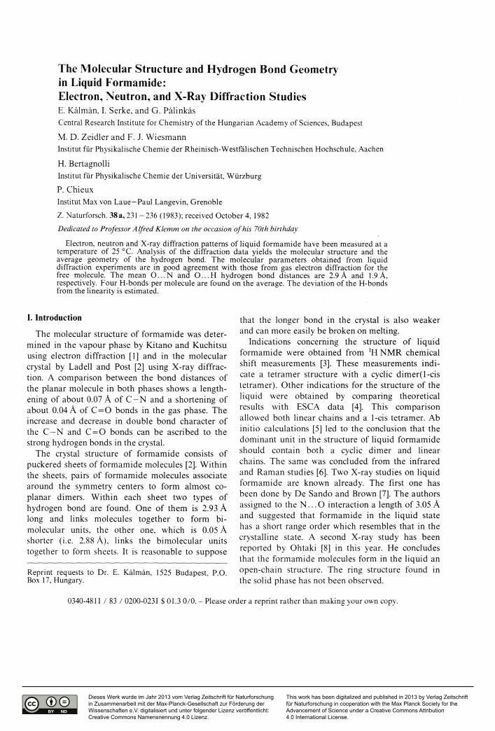
20 L J i i i i I i
Fig. 1. The k weighted experimental and molecular struc-ture functions from electron diffraction. The molecular structure function is fitted to the experimental one in the range 7 A"1 Si k si 22.5 A"1 .
7 A"1 k 22.5 A - 1 range of the HED(k). The func-tion kHm(k) for liquid formamide together with the total electron structure function is shown in Figure 1. The two curves are in agreement for values k ^ 8 A - 1 , except for small deviations at lower values of k, which are due to the contribu-tions of the H-bond interactions. In order to have an independent determination of the molecular param-eters, the X-ray molecular structure function was derived by another method based on the properties of the total pair correlation function, G(r), given by the following relation:
00 G(r)=\+(2n2
Qoy^k2H(k)Mkr)dk, (4) o
where is the number density. Regarding Eq. (2), G (r) can also be written as the
sum of molecular and distinct terms
G(r) = Gm(r) + Gd(r). (5)
If there is no overlap between the intramolecular and intermolecular distance contributions, Eq. (5) renders possible the separation of the Gm(r) func-tion.
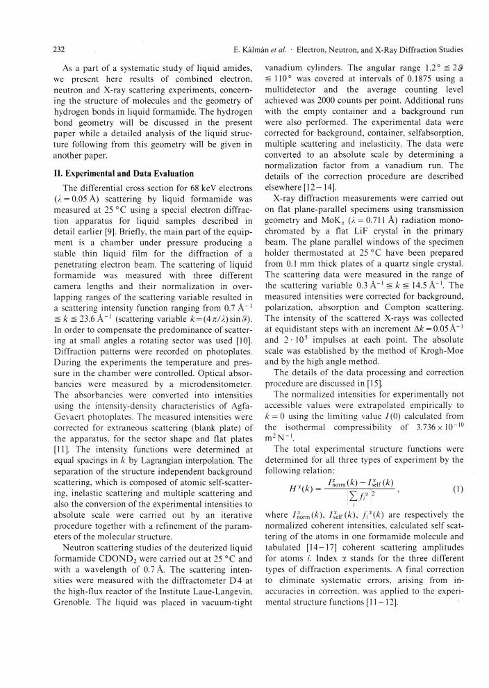
The total X-ray pair correlation function (not shown in this paper) approached a value of zero at a distance of 2.6 A. Because no intermolecular con-tributions, except O . . . H interactions were expected in the total X-ray pair correlation function below r = 2.6 A, an "experimental" molecular structure function H ^ k was derived by inverse Fourier trans-formation of G (r) in the range 0 ^ r ^ 2.6 A. The parameters r%p and for the molecular structure were then obtained by least-squares refinement of expression (3) against the H ^ ( k ) function in the range 0 ^ k ^ 13.5 A"1. The molecular structure function Hm(k), calculated from these parameters, is shown in Fig.2, together with the H&(k) molec-ular structure function calculated from the molec-ular part of G(r) and the total X-ray structure function derived from experiment. The molecular parameters obtained from both electron and X-ray scattering data are listed in Table 1. For com-parison, the results obtained by gas electron diffrac-tion for the free molecule [1] are also presented in Table 1.
The molecular parameters resulting from the liquid electron diffraction experiment are in good agreement with those from liquid X-ray and gas electron diffraction. The structure of the formamide
234 E. Kälmän et al. • Electron, Neu t ron , and X-Ray Dif f rac t ion Studies
Table 1. Mean distances /-, rms variations / in A and bond angles for the f o r m a m i d e molecule in vapour [1] and liquid phase.
Ref. [1] ED X D
r / r / r /
c = o 1.212(3) 0.039 1.213(6) 0.067 1.219(6) 0.065 C - N 1.368(3) 0.043 1.360(10) 0.067 1.375(10) 0.062 C - H 1.125(12 ) 0.080 1.120(22) 0.076 1.125« 0.080 a
N - H 1.027(6) 0.090 1.020(15) 0.077 1.027« 0.090 a
< O C N 125.0(4) 125.2(5) 124.2(5) < N C H 112.7(1) 112.7 a 112.7« < C N H C 118.7(1) 118.7 a 118.7« < C N H T 119.7(1) 119.7 ^ 119.7 a
a Values were kept constant in the fitting procedure.
molecule is found to be the same as that of a free molecule. Changes of the double bond characters of the C - N and C = 0 bonds appearing in the crystal [2] are not observable in the liquid phase.
The molecular structure function for neutron scattering has been calculated from the parameters listed in Table 1 for electron diffraction.
IV. The Hydrogen Bond Interactions
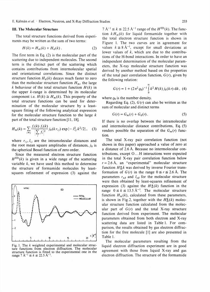
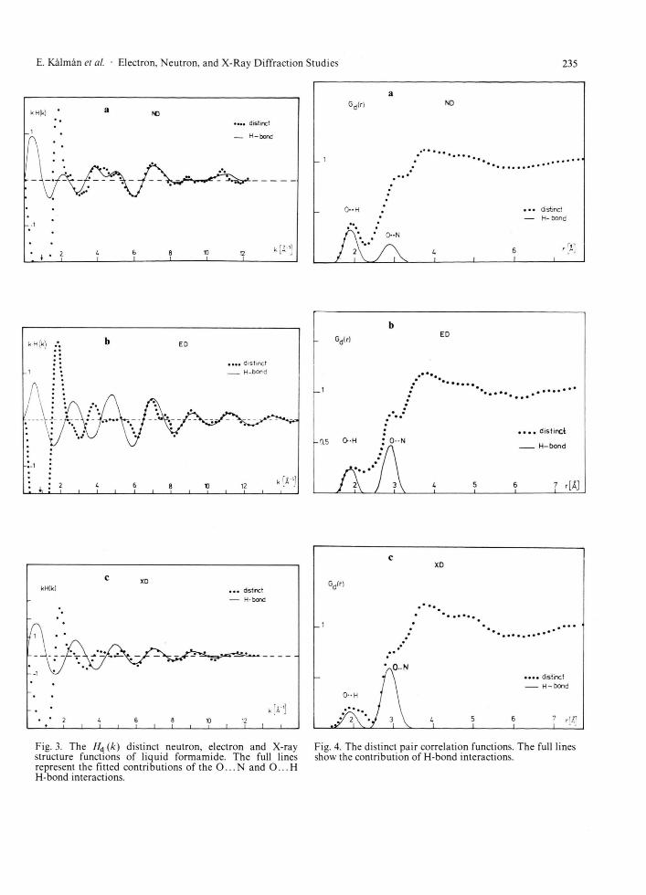
The distinct structure functions H\{k) are now obtained as the difference between the total struc-ture functions and the molecular terms Hd(k) = H*(k) - //m(A'). The three Hl(k) functions (ND. ED, XD) are presented in Figure 3.
The most striking feature of these functions is the presence of oscillations in the large k range. This feature is characteristic for H-bonded molecular liquids [18],
kH|k) XD •••• TOTAL
MOLECULAR —- FIT
- 2 / V • / H
-0 • \\ * / \
\ S, /
.-2
k[Al 2 1 1
L 1 1 6 l 1 8 10 12 1 I I I I I
Fig. 2. The k weighted experimental and molecular struc-ture functions f rom X-ray diffract ion. The full line re-presents the numerical Four ier t ransform of the intra-molecular peaks in the total pair correlation function. The dashed line shows the fitted molecular structure function.
Simple molecular liquids as CC14 and CS2 [19. 20] yield distinct structure functions which decay to zero much faster, almost no oscillations could be detected for k > 6 A - 1 . Based on this argument we assume that at high k values the O . . . N and O . . . H H-bond interactions contribute most to the Hd(k) functions
Hd(k) ^ //H-bond (k) = / /O. . .N (k) + H0 H(k).
By least-squares fitting of expression (3) to the large k part of the distinct structure functions beyond k ^ 4 A - 1 , we have obtained the structural parameters of H-bond interactions listed in Table 2. The H-bond structure functions calculated from the parameters given in Table 2 for the three different scattering experiments are also shown in Figure 3. As a result of the calculations beyond A: = 6 A"1
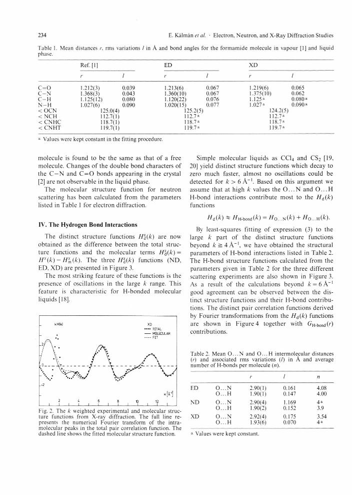
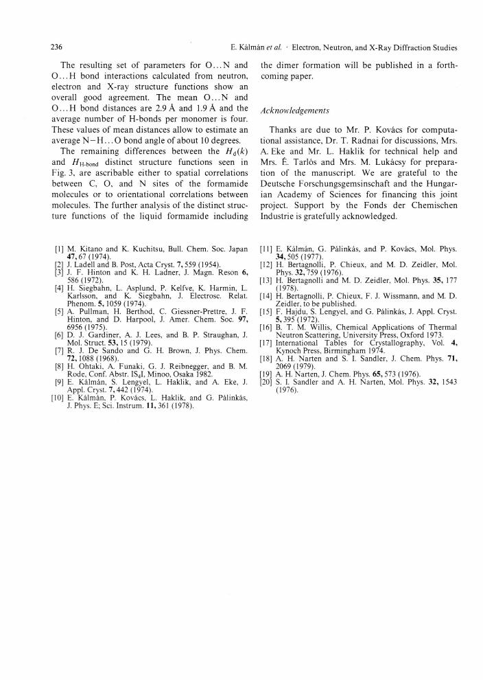
good agreement can be observed between the dis-tinct structure functions and their H-bond contribu-tions. The distinct pair correlation functions derived by Fourier transformations from the H d (k ) functions are shown in Figure 4 together with GH-bond('") contributions.
Table 2. Mean O . . . N and O . . . H intermolecular distances (/•) and associated rms variations (/) in A and average number of H-bonds per molecule (n).
r / n
ED O . . .N 2.90(1) 0.161 4.08 O . . . H 1.90(1) 0.147 4.00
N D O . . 2.90(4) 1.169 4« O . . H 1.90(2) 0.152 3.9
XD O . . . N 2.92(4) 0.175 3.54 O . . . H 1.93(6) 0.070 4«
a Values were kept constant.
235 E. Kälmän et al. • Electron, Neutron, and X-Ray Diffraction Studies
. 1
Gd(r) a
ND
-O-H
••
y 2 \ 7 1 V
• 0--N
/ \ U y i v i i
• •• distinct — H- bond
r«1 6 r A 1 1
C XD
Gdlr)
_ 1
O-H
' 0 . . N « . . . distinct
H-bond
. » / ^ l Y / 3 \ L 5
I V 1 1 6 7 r m 1 1 L -
Fig. 3. The Hd (k ) distinct neutron, electron and X-ray structure functions of liquid formamide. The full lines represent the fitted contributions of the O . . . N and O . . . H H-bond interactions.
Fig. 4. The distinct pair correlation functions. The full lines show the contribution of H-bond interactions.
236 E. Kälmän et al. • Electron, Neutron, and X-Ray Diffraction Studies
The resulting set of parameters for O . . . N and O . . . H bond interactions calculated f rom neutron, electron and X-ray s tructure functions show an overall good agreement . The mean O . . . N and O . . . H bond distances are 2.9 A and 1.9 A and the average n u m b e r of H-bonds per m o n o m e r is four. These values of mean distances allow to est imate an average N —H. . . O bond angle of about 10 degrees.
The remain ing dif ferences between the Hd(k) and HH.bond distinct structure functions seen in Fig. 3, are ascr ibable ei ther to spatial correlations between C, O, and N sites of the fo rmamide molecules or to orientat ional correlations between molecules. The fur ther analysis of the distinct struc-ture funct ions of the l iquid f o r m a m i d e including
the d imer fo rmat ion will be publ ished in a forth-coming paper.
Acknowledgements
Thanks are due to Mr. P. Koväcs for computa -tional assistance. Dr . T. Radna i for discussions, Mrs. A. Eke and Mr. L. Haklik for technical help and Mrs. £. Tarlös and Mrs. M. Lukäcsy for prepara-tion of the manuscr ip t . We are grateful to the Deutsche Forschungsgemsinschaf t and the Hungar-ian Academy of Sciences for f inancing this joint project. Support by the Fonds der Chemischen Industrie is grateful ly acknowledged.
[1] M. Kitano and K. Kuchitsu, Bull. Chem. Soc. Japan 47 ,67 (1974).
[2] J. Ladell and B. Post, Acta Cryst. 7,559 (1954). [3] J. F. Hinton and K. H. Ladner, J. Magn. Reson 6,
586 (1972). [4] H. Siegbahn, L. Asplund. P. Kelfve, K. Harmin, L.
Karlsson, and K Siegbahn, J. Electrosc. Relat. Phenom. 5,1059 (1974).
[5] A. Pullman, H. Berthod. C. Giessner-Prettre, J. F. Hinton, and D. Harpool, J. Amer. Chem. Soc. 97, 6956 (1975).
[6] D. J. Gardiner, A. J. Lees, and B. P. Straughan, J. Mol. Struct. 53, 15 (1979).
[7] R. J. De Sando and G. H. Brown, J. Phys. Chem. 72,1088 (1968).
[8] H. Ohtaki. A. Funaki, G. J. Reibnegger, and B. M. Rode, Conf. Abstr. IS4I, Minoo, Osaka 1982.
[9] E. Kälmän, S. Lengyel, L. Haklik, and A. Eke, J. Appl. Cryst. 7 ,442 (1974).
[10] E. Kälmän, P. Koväcs, L. Haklik, and G. Pälinkäs, J. Phys. E; Sei. Instrum. 11,361 (1978).
[11] E. Kälmän, G. Pälinkäs, and P. Koväcs, Mol. Phys. 34,505 (1977).
[12] H. Bertagnolli, P. Chieux, and M. D. Zeidler, Mol. Phys. 32,759 (1976).
[13] H. Bertagnolli and M. D. Zeidler, Mol. Phys. 35, 177 (1978).
[14] H. Bertagnolli, P. Chieux, F. J. Wissmann, and M. D. Zeidler, to be published.
[15] F. Hajdu, S. Lengyel, and G. Pälinkäs, J. Appl. Cryst. 5 ,395 (1972).
[16] B. T. M. Willis, Chemical Applications of Thermal Neutron Scattering, University Press, Oxford 1973.
[17] International Tables for Crystallography, Vol. 4, Kynoch Press, Birmingham 1974.
[18] A. H. Narten and S. I. Sandler, J. Chem. Phys. 71, 2069(1979).
[19] A. H. Narten, J. Chem. Phys. 65, 573 (1976). [20] S. I. Sandler and A. H. Narten, Mol. Phys. 32, 1543
(1976).