Embed Size (px)
Citation preview
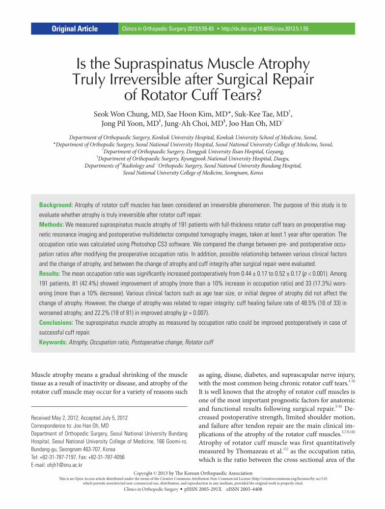
Is the Supraspinatus Muscle Atrophy Truly Irreversible after Surgical Repair
of Rotator Cuff Tears?Seok Won Chung, MD, Sae Hoon Kim, MD*, Suk-Kee Tae, MD†,
Jong Pil Yoon, MD‡, Jung-Ah Choi, MD§, Joo Han Oh, MD∥
Department of Orthopaedic Surgery, Konkuk University Hospital, Konkuk University School of Medicine, Seoul, *Department of Orthopedic Surgery, Seoul National University Hospital, Seoul National University College of Medicine, Seoul,
†Department of Orthopaedic Surgery, Dongguk University Ilsan Hospital, Goyang, ‡Department of Orthopaedic Surgery, Kyungpook National University Hospital, Daegu,
Departments of §Radiology and ∥Orthopedic Surgery, Seoul National University Bundang Hospital, Seoul National University College of Medicine, Seongnam, Korea
Original Article Clinics in Orthopedic Surgery 2013;5:55-65 • http://dx.doi.org/10.4055/cios.2013.5.1.55
Received May 2, 2012; Accepted July 5, 2012Correspondence to: Joo Han Oh, MDDepartment of Orthopedic Surgery, Seoul National University Bundang Hospital, Seoul National University College of Medicine, 166 Goomi-ro, Bundang-gu, Seongnam 463-707, KoreaTel: +82-31-787-7197, Fax: +82-31-787-4056E-mail: [email protected]
Muscle atrophy means a gradual shrinking of the muscle tissue as a result of inactivity or disease, and atrophy of the rotator cuff muscle may occur for a variety of reasons such
Background: Atrophy of rotator cuff muscles has been considered an irreversible phenomenon. The purpose of this study is to evaluate whether atrophy is truly irreversible after rotator cuff repair.Methods: We measured supraspinatus muscle atrophy of 191 patients with full-thickness rotator cuff tears on preoperative mag-netic resonance imaging and postoperative multidetector computed tomography images, taken at least 1 year after operation. The occupation ratio was calculated using Photoshop CS3 software. We compared the change between pre- and postoperative occu-pation ratios after modifying the preoperative occupation ratio. In addition, possible relationship between various clinical factors and the change of atrophy, and between the change of atrophy and cuff integrity after surgical repair were evaluated.Results: The mean occupation ratio was significantly increased postoperatively from 0.44 ± 0.17 to 0.52 ± 0.17 (p < 0.001). Among 191 patients, 81 (42.4%) showed improvement of atrophy (more than a 10% increase in occupation ratio) and 33 (17.3%) wors-ening (more than a 10% decrease). Various clinical factors such as age tear size, or initial degree of atrophy did not affect the change of atrophy. However, the change of atrophy was related to repair integrity: cuff healing failure rate of 48.5% (16 of 33) in worsened atrophy; and 22.2% (18 of 81) in improved atrophy (p = 0.007).Conclusions: The supraspinatus muscle atrophy as measured by occupation ratio could be improved postoperatively in case of successful cuff repair.Keywords: Atrophy, Occupation ratio, Postoperative change, Rotator cuff
as aging, disuse, diabetes, and suprascapular nerve injury, with the most common being chronic rotator cuff tears.1-4) It is well known that the atrophy of rotator cuff muscles is one of the most important prognostic factors for anatomic and functional results following surgical repair.5-8) De-creased postoperative strength, limited shoulder motion, and failure after tendon repair are the main clinical im-plications of the atrophy of the rotator cuff muscles.5,7,9,10) Atrophy of rotator cuff muscle was first quantitatively measured by Thomazeau et al.11) as the occupation ratio, which is the ratio between the cross sectional area of the
Copyright © 2013 by The Korean Orthopaedic AssociationThis is an Open Access article distributed under the terms of the Creative Commons Attribution Non-Commercial License (http://creativecommons.org/licenses/by-nc/3.0)
which permits unrestricted non-commercial use, distribution, and reproduction in any medium, provided the original work is properly cited.Clinics in Orthopedic Surgery • pISSN 2005-291X eISSN 2005-4408
56
Chung et al. Supraspinatus Muscle Atrophy after Rotator Cuff RepairClinics in Orthopedic Surgery • Vol. 5, No. 1, 2013 • www.ecios.org
muscle belly and that of its fossa on oblique sagittal Y-view of magnetic resonance imaging (MRI). The correlation of the occupation ratio between MRI and multidetector com-puted tomography (MDCT) was validated in our previous study.12)
To date, even though some studies suggest that surgical cuff repair may halt the atrophy of rotator cuff muscles,9,11) it has been considered an irreversible phe-nomenon, and the function of the shoulder joint remains impaired, especially in higher stages of atrophy.5,6,13,14) However, we do observe recovery of the atrophy of rota-tor cuff muscles on a regular follow-up examination after surgical repair, and the question is whether the atrophy is truly irreversible after rotator cuff repair.
The hypothesis of this study is that the atrophy of rotator cuff muscles can be improved after surgical repair. We investigated whether there was a significant improve-ment of the supraspinatus muscle atrophy postoperatively by comparing the pre- and postoperative occupation ra-tios. We further assessed the factors associated with the possible improvement of the atrophy.
METHODS
DemographicsThis was a retrospective study. Among 412 patients who underwent surgical cuff repair for full-thickness rotator cuff tears confirmed by arthroscopic findings at the au-thors’ institution between January 2005 and July 2008, 191 patients who underwent both preoperative MRI and post-operative MDCT at least one year after the operation were included. The postoperative MDCT was performed at a mean of 13.2 ± 3.8 months after operation (range, 12 to 16 months), without special indication. We excluded patients with isolated subscapularis tear (n = 5), previous operation on the same shoulder joint (n = 4), and incomplete repair (n = 6). Patients who did not have preoperative MRI (n = 8) or postoperative MDCT (n = 166), and those with MRI or MDCT images that were not usable due to poor quality or absence of appropriate Y-view were also excluded (n = 32). Most of the patients without the postoperative MDCT un-derwent ultrasonography as a postoperative imaging mo-dality instead of MDCT (136 of 166), and thirty patients refused to undergo either ultrasonography or MDCT. There were no special criteria for the selection of postop-erative imaging modality; however, ultrasonography was usually performed for those with financial difficulties. The average age at the time of operation was 59.7 ± 7.9 years (range, 39 to 80 years), and the postoperative MDCT was performed at a mean of 13.2 ± 3.8 months after operation.
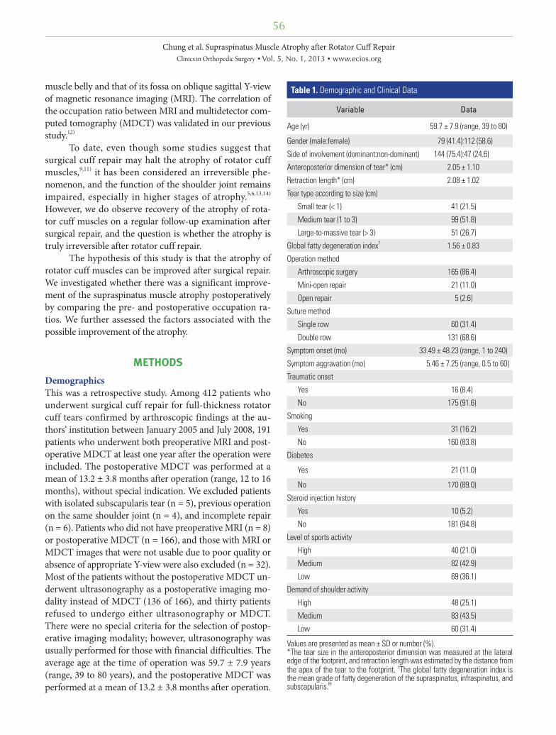
Table 1. Demographic and Clinical Data
Variable Data
Age (yr) 59.7 ± 7.9 (range, 39 to 80)
Gender (male:female) 79 (41.4):112 (58.6)
Side of involvement (dominant:non-dominant) 144 (75.4):47 (24.6)
Anteroposterior dimension of tear* (cm) 2.05 ± 1.10
Retraction length* (cm) 2.08 ± 1.02
Tear type according to size (cm)
Small tear (< 1) 41 (21.5)
Medium tear (1 to 3) 99 (51.8)
Large-to-massive tear (> 3) 51 (26.7)
Global fatty degeneration index† 1.56 ± 0.83
Operation method
Arthroscopic surgery 165 (86.4)
Mini-open repair 21 (11.0)
Open repair 5 (2.6)
Suture method
Single row 60 (31.4)
Double row 131 (68.6)
Symptom onset (mo) 33.49 ± 48.23 (range, 1 to 240)
Symptom aggravation (mo) 5.46 ± 7.25 (range, 0.5 to 60)
Traumatic onset
Yes 16 (8.4)
No 175 (91.6)
Smoking
Yes 31 (16.2)
No 160 (83.8)
Diabetes
Yes 21 (11.0)
No 170 (89.0)
Steroid injection history
Yes 10 (5.2)
No 181 (94.8)
Level of sports activity
High 40 (21.0)
Medium 82 (42.9)
Low 69 (36.1)
Demand of shoulder activity
High 48 (25.1)
Medium 83 (43.5)
Low 60 (31.4)
Values are presented as mean ± SD or number (%). *The tear size in the anteroposterior dimension was measured at the lateral edge of the footprint, and retraction length was estimated by the distance from the apex of the tear to the footprint. †The global fatty degeneration index is the mean grade of fatty degeneration of the supraspinatus, infraspinatus, and subscapularis.6)
57
Chung et al. Supraspinatus Muscle Atrophy after Rotator Cuff RepairClinics in Orthopedic Surgery • Vol. 5, No. 1, 2013 • www.ecios.org
The demographic and clinical data were summarized in Table 1. We obtained the institutional review board ap-proval for the study protocol.
Surgical ProceduresAll surgical procedures were conducted by one senior au-thor (JHO). Subacromial decompression and acromioplas-ty were performed to create a flat acromion. Distal clavicle resection was performed on the patients who experienced symptomatic acromioclavicular arthritis. Biceps tenotomy or tenodesis was performed for a symptomatic biceps tear involving greater than 50% of the tendon and for a symp-tomatic degenerative superior labral anterior and poste-rior (SLAP) lesion according to age or activity level of the patient. The operative technique was arthroscopic repair, mini-open repair, or open repair according to the arthros-copy learning curve and arthroscopical reparability. Repair technique was either single or double row according to the tear size, tendon status, and tear configuration and repa-rability. All knots were tied securely using a self-locking, sliding knot.
Immobilization after cuff repair was maintained with an abduction brace, and duration of immobilization was based on tear size as measured at the time of opera-tion, from 4 weeks to 6 weeks. Passive motion was per-formed immediately after the operation, except in cases of large-to-massive tears. Active-assisted range of motion ex-ercise was allowed after the weaning of the brace. Muscle strengthening exercises began between 9 and 12 weeks, and sports activities were permitted at 6 months after the operation.
MRI and MDCTEven though MRI still remains the standard preoperative imaging modality for evaluation of rotator cuff, MDCT
has been widely used recently for its advantages, such as cost-effectiveness, short examination time, and absence of foreign body artifact in the postoperative evaluation for patients who have undergone rotator cuff repair surgery. Our previous study showed that MDCT arthrography (MDCTA) is a very valuable method to confirm postop-erative integrity after rotator cuff repair using suture an-chors.15)
As such, we have used MRI as a preoperative imaging modality and MDCT as a postoperative imaging modality. MRI was performed on the Gyroscan Intera 1.5-T system (Philips Medical Systems, Utrecht, the Netherlands). From the MRI examination, we obtained T1-weighted and T2-weighted spin-echo oblique sagittal images parallel to the joint surface of the glenoid. CT was also performed using a 16-multidetector CT system (Mx8000 IDT, Philips Med-ical Systems). We generated oblique sagittal reconstruc-tion images as well as oblique coronal and axial images at a three-dimensional workstation. The oblique sagittal images were reconstructed parallel to the glenohumeral joint surface, same as the MRI oblique sagittal images. The patient positioning for MRI and MDCT examinations was also same with the arm placed alongside the body in a neutral supine position.
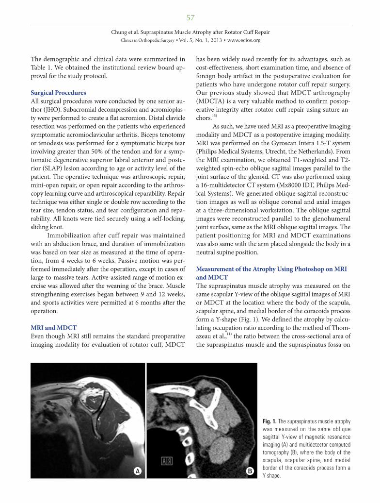
Measurement of the Atrophy Using Photoshop on MRI and MDCTThe supraspinatus muscle atrophy was measured on the same scapular Y-view of the oblique sagittal images of MRI or MDCT at the location where the body of the scapula, scapular spine, and medial border of the coracoids process form a Y-shape (Fig. 1). We defined the atrophy by calcu-lating occupation ratio according to the method of Thom-azeau et al.,11) the ratio between the cross-sectional area of the supraspinatus muscle and the supraspinatus fossa on
Fig. 1. The supraspinatus muscle atrophy was measured on the same oblique sagittal Y-view of magnetic resonance imaging (A) and multidetector computed tomography (B), where the body of the scapula, scapular spine, and medial border of the coracoids process form a Y-shape.
58
Chung et al. Supraspinatus Muscle Atrophy after Rotator Cuff RepairClinics in Orthopedic Surgery • Vol. 5, No. 1, 2013 • www.ecios.org
the scapular Y-view. The boundary of the supraspinatus fossa was defined as the area from the inner margins of the Y-shape to the inner limit of the clavicle and the acro-mion. Originally, the occupation ratio is measured on the picture archiving and communications systems (PACS) workstation by drawing the boundary of the supraspinatus muscle and supraspinatus fossa.11) However, this method is limited in its accuracy and reliability for measuring occu-pation ratio, because the outlining process is conducted by subjective manual hand drawing. For that reason, in our previous study,12) we introduced a new measuring method for the supraspinatus muscle atrophy using the Photo-shop software (magic selection tool, Adobe Systems Inc., San Jose, CA, USA), which is the method that automati-cally designates the regions of interest and calculates the number of pixels in the selected area. With the Photoshop method, we solved the reliability issue for the measure-ment of the atrophy, showing very high intra- and interob-server reliability: interclass correlation coefficient (ICC) of
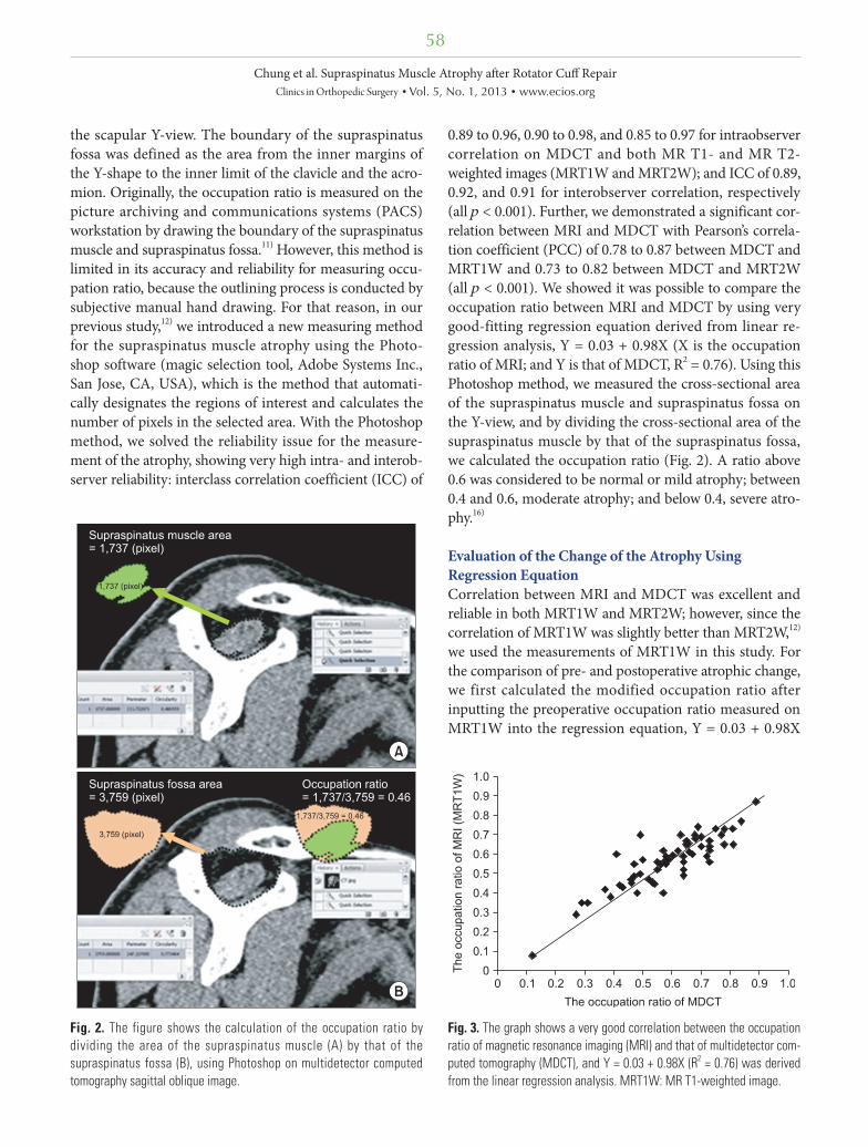
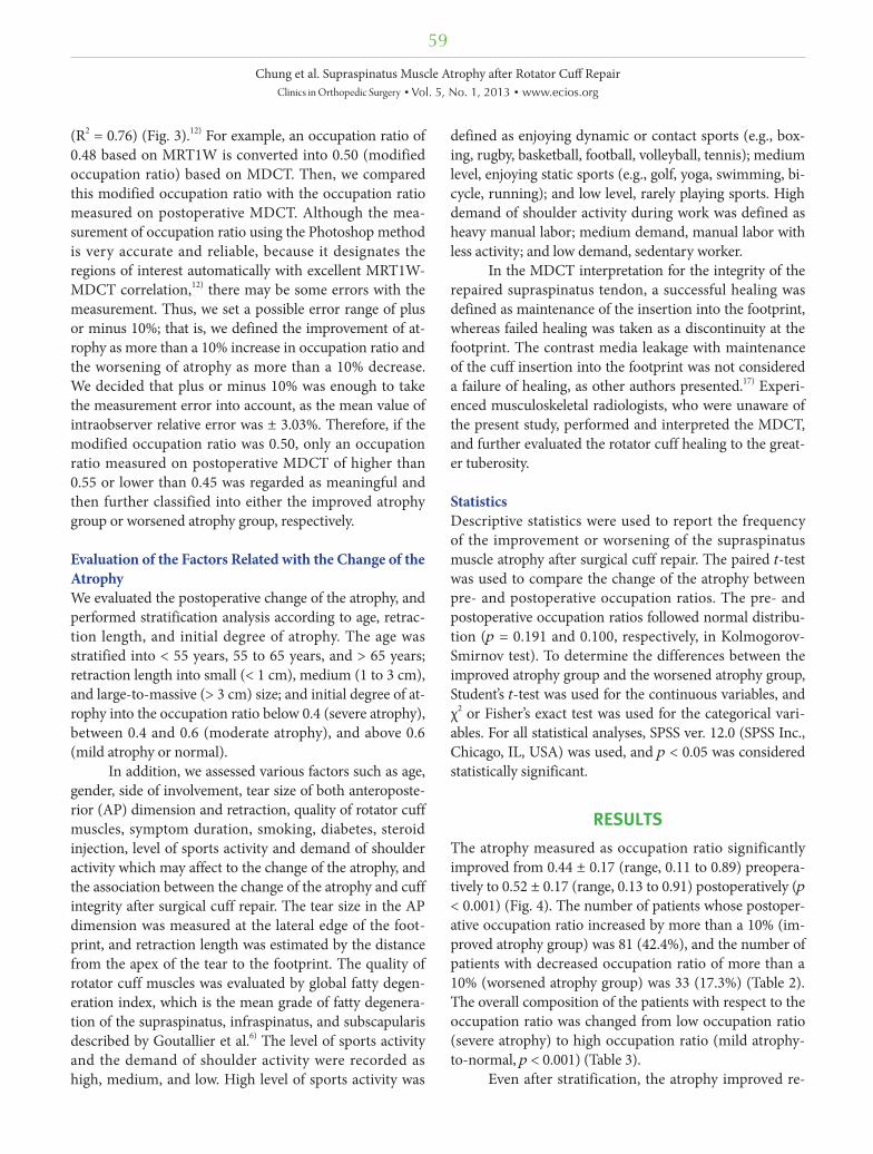
0.89 to 0.96, 0.90 to 0.98, and 0.85 to 0.97 for intraobserver correlation on MDCT and both MR T1- and MR T2-weighted images (MRT1W and MRT2W); and ICC of 0.89, 0.92, and 0.91 for interobserver correlation, respectively (all p < 0.001). Further, we demonstrated a significant cor-relation between MRI and MDCT with Pearson’s correla-tion coefficient (PCC) of 0.78 to 0.87 between MDCT and MRT1W and 0.73 to 0.82 between MDCT and MRT2W (all p < 0.001). We showed it was possible to compare the occupation ratio between MRI and MDCT by using very good-fitting regression equation derived from linear re-gression analysis, Y = 0.03 + 0.98X (X is the occupation ratio of MRI; and Y is that of MDCT, R2 = 0.76). Using this Photoshop method, we measured the cross-sectional area of the supraspinatus muscle and supraspinatus fossa on the Y-view, and by dividing the cross-sectional area of the supraspinatus muscle by that of the supraspinatus fossa, we calculated the occupation ratio (Fig. 2). A ratio above 0.6 was considered to be normal or mild atrophy; between 0.4 and 0.6, moderate atrophy; and below 0.4, severe atro-phy.16)
Evaluation of the Change of the Atrophy Using Regression EquationCorrelation between MRI and MDCT was excellent and reliable in both MRT1W and MRT2W; however, since the correlation of MRT1W was slightly better than MRT2W,12) we used the measurements of MRT1W in this study. For the comparison of pre- and postoperative atrophic change, we first calculated the modified occupation ratio after inputting the preoperative occupation ratio measured on MRT1W into the regression equation, Y = 0.03 + 0.98X
Fig. 2. The figure shows the calculation of the occupation ratio by dividing the area of the supraspinatus muscle (A) by that of the supraspinatus fossa (B), using Photoshop on multidetector computed tomography sagittal oblique image.
Fig. 3. The graph shows a very good correlation between the occupation ratio of magnetic resonance imaging (MRI) and that of multidetector com-puted tomography (MDCT), and Y = 0.03 + 0.98X (R2 = 0.76) was derived from the linear regression analysis. MRT1W: MR T1-weighted image.
59
Chung et al. Supraspinatus Muscle Atrophy after Rotator Cuff RepairClinics in Orthopedic Surgery • Vol. 5, No. 1, 2013 • www.ecios.org
(R2 = 0.76) (Fig. 3).12) For example, an occupation ratio of 0.48 based on MRT1W is converted into 0.50 (modified occupation ratio) based on MDCT. Then, we compared this modified occupation ratio with the occupation ratio measured on postoperative MDCT. Although the mea-surement of occupation ratio using the Photoshop method is very accurate and reliable, because it designates the regions of interest automatically with excellent MRT1W-MDCT correlation,12) there may be some errors with the measurement. Thus, we set a possible error range of plus or minus 10%; that is, we defined the improvement of at-rophy as more than a 10% increase in occupation ratio and the worsening of atrophy as more than a 10% decrease. We decided that plus or minus 10% was enough to take the measurement error into account, as the mean value of intraobserver relative error was ± 3.03%. Therefore, if the modified occupation ratio was 0.50, only an occupation ratio measured on postoperative MDCT of higher than 0.55 or lower than 0.45 was regarded as meaningful and then further classified into either the improved atrophy group or worsened atrophy group, respectively.
Evaluation of the Factors Related with the Change of the AtrophyWe evaluated the postoperative change of the atrophy, and performed stratification analysis according to age, retrac-tion length, and initial degree of atrophy. The age was stratified into < 55 years, 55 to 65 years, and > 65 years; retraction length into small (< 1 cm), medium (1 to 3 cm), and large-to-massive (> 3 cm) size; and initial degree of at-rophy into the occupation ratio below 0.4 (severe atrophy), between 0.4 and 0.6 (moderate atrophy), and above 0.6 (mild atrophy or normal).
In addition, we assessed various factors such as age, gender, side of involvement, tear size of both anteroposte-rior (AP) dimension and retraction, quality of rotator cuff muscles, symptom duration, smoking, diabetes, steroid injection, level of sports activity and demand of shoulder activity which may affect to the change of the atrophy, and the association between the change of the atrophy and cuff integrity after surgical cuff repair. The tear size in the AP dimension was measured at the lateral edge of the foot-print, and retraction length was estimated by the distance from the apex of the tear to the footprint. The quality of rotator cuff muscles was evaluated by global fatty degen-eration index, which is the mean grade of fatty degenera-tion of the supraspinatus, infraspinatus, and subscapularis described by Goutallier et al.6) The level of sports activity and the demand of shoulder activity were recorded as high, medium, and low. High level of sports activity was
defined as enjoying dynamic or contact sports (e.g., box-ing, rugby, basketball, football, volleyball, tennis); medium level, enjoying static sports (e.g., golf, yoga, swimming, bi-cycle, running); and low level, rarely playing sports. High demand of shoulder activity during work was defined as heavy manual labor; medium demand, manual labor with less activity; and low demand, sedentary worker.
In the MDCT interpretation for the integrity of the repaired supraspinatus tendon, a successful healing was defined as maintenance of the insertion into the footprint, whereas failed healing was taken as a discontinuity at the footprint. The contrast media leakage with maintenance of the cuff insertion into the footprint was not considered a failure of healing, as other authors presented.17) Experi-enced musculoskeletal radiologists, who were unaware of the present study, performed and interpreted the MDCT, and further evaluated the rotator cuff healing to the great-er tuberosity.
StatisticsDescriptive statistics were used to report the frequency of the improvement or worsening of the supraspinatus muscle atrophy after surgical cuff repair. The paired t-test was used to compare the change of the atrophy between pre- and postoperative occupation ratios. The pre- and postoperative occupation ratios followed normal distribu-tion (p = 0.191 and 0.100, respectively, in Kolmogorov-Smirnov test). To determine the differences between the improved atrophy group and the worsened atrophy group, Student’s t-test was used for the continuous variables, and χ2 or Fisher’s exact test was used for the categorical vari-ables. For all statistical analyses, SPSS ver. 12.0 (SPSS Inc., Chicago, IL, USA) was used, and p < 0.05 was considered statistically significant.
RESULTS
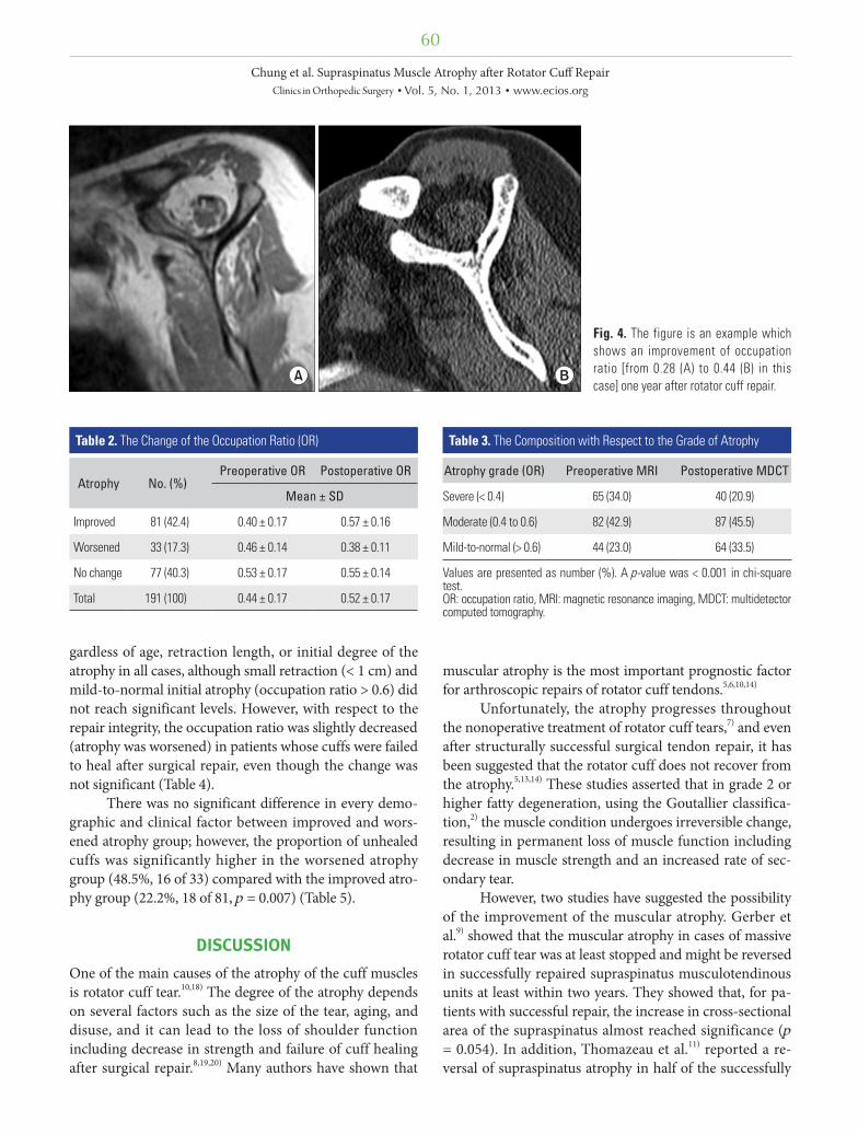
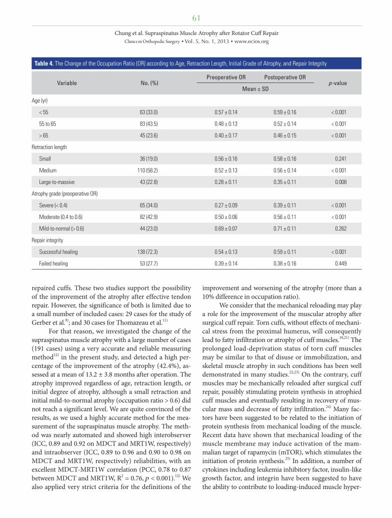
The atrophy measured as occupation ratio significantly improved from 0.44 ± 0.17 (range, 0.11 to 0.89) preopera-tively to 0.52 ± 0.17 (range, 0.13 to 0.91) postoperatively (p < 0.001) (Fig. 4). The number of patients whose postoper-ative occupation ratio increased by more than a 10% (im-proved atrophy group) was 81 (42.4%), and the number of patients with decreased occupation ratio of more than a 10% (worsened atrophy group) was 33 (17.3%) (Table 2). The overall composition of the patients with respect to the occupation ratio was changed from low occupation ratio (severe atrophy) to high occupation ratio (mild atrophy-to-normal, p < 0.001) (Table 3).
Even after stratification, the atrophy improved re-
60
Chung et al. Supraspinatus Muscle Atrophy after Rotator Cuff RepairClinics in Orthopedic Surgery • Vol. 5, No. 1, 2013 • www.ecios.org
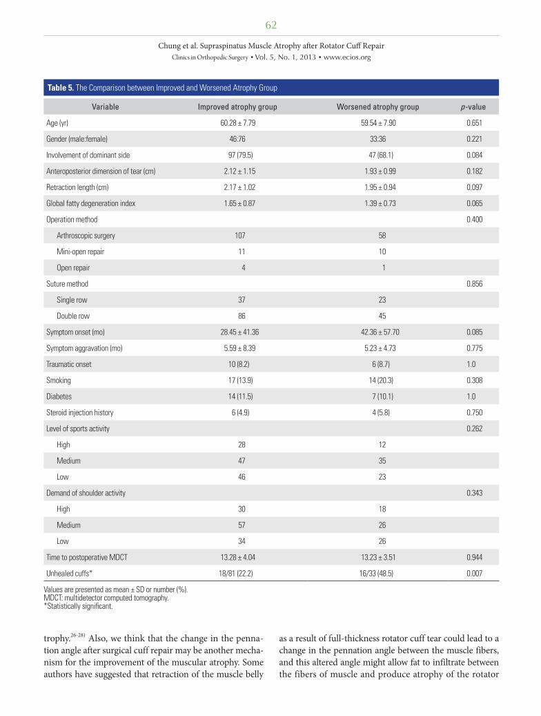
gardless of age, retraction length, or initial degree of the atrophy in all cases, although small retraction (< 1 cm) and mild-to-normal initial atrophy (occupation ratio > 0.6) did not reach significant levels. However, with respect to the repair integrity, the occupation ratio was slightly decreased (atrophy was worsened) in patients whose cuffs were failed to heal after surgical repair, even though the change was not significant (Table 4).
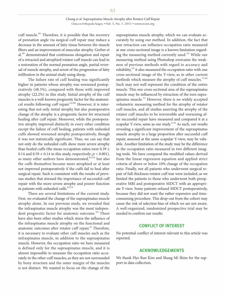
There was no significant difference in every demo-graphic and clinical factor between improved and wors-ened atrophy group; however, the proportion of unhealed cuffs was significantly higher in the worsened atrophy group (48.5%, 16 of 33) compared with the improved atro-phy group (22.2%, 18 of 81, p = 0.007) (Table 5).
DISCUSSION
One of the main causes of the atrophy of the cuff muscles is rotator cuff tear.10,18) The degree of the atrophy depends on several factors such as the size of the tear, aging, and disuse, and it can lead to the loss of shoulder function including decrease in strength and failure of cuff healing after surgical repair.8,19,20) Many authors have shown that
muscular atrophy is the most important prognostic factor for arthroscopic repairs of rotator cuff tendons.5,6,10,14)
Unfortunately, the atrophy progresses throughout the nonoperative treatment of rotator cuff tears,7) and even after structurally successful surgical tendon repair, it has been suggested that the rotator cuff does not recover from the atrophy.5,13,14) These studies asserted that in grade 2 or higher fatty degeneration, using the Goutallier classifica-tion,2) the muscle condition undergoes irreversible change, resulting in permanent loss of muscle function including decrease in muscle strength and an increased rate of sec-ondary tear.
However, two studies have suggested the possibility of the improvement of the muscular atrophy. Gerber et al.9) showed that the muscular atrophy in cases of massive rotator cuff tear was at least stopped and might be reversed in successfully repaired supraspinatus musculotendinous units at least within two years. They showed that, for pa-tients with successful repair, the increase in cross-sectional area of the supraspinatus almost reached significance (p = 0.054). In addition, Thomazeau et al.11) reported a re-versal of supraspinatus atrophy in half of the successfully
Fig. 4. The figure is an example which shows an improvement of occupation ratio [from 0.28 (A) to 0.44 (B) in this case] one year after rotator cuff repair.
Table 2. The Change of the Occupation Ratio (OR)
Atrophy No. (%)Preoperative OR Postoperative OR
Mean ± SD
Improved 81 (42.4) 0.40 ± 0.17 0.57 ± 0.16
Worsened 33 (17.3) 0.46 ± 0.14 0.38 ± 0.11
No change 77 (40.3) 0.53 ± 0.17 0.55 ± 0.14
Total 191 (100) 0.44 ± 0.17 0.52 ± 0.17
Table 3. The Composition with Respect to the Grade of Atrophy
Atrophy grade (OR) Preoperative MRI Postoperative MDCT
Severe (< 0.4) 65 (34.0) 40 (20.9)
Moderate (0.4 to 0.6) 82 (42.9) 87 (45.5)
Mild-to-normal (> 0.6) 44 (23.0) 64 (33.5)
Values are presented as number (%). A p-value was < 0.001 in chi-square test.OR: occupation ratio, MRI: magnetic resonance imaging, MDCT: multidetector computed tomography.
61
Chung et al. Supraspinatus Muscle Atrophy after Rotator Cuff RepairClinics in Orthopedic Surgery • Vol. 5, No. 1, 2013 • www.ecios.org
repaired cuffs. These two studies support the possibility of the improvement of the atrophy after effective tendon repair. However, the significance of both is limited due to a small number of included cases: 29 cases for the study of Gerber et al.9); and 30 cases for Thomazeau et al.11)
For that reason, we investigated the change of the supraspinatus muscle atrophy with a large number of cases (191 cases) using a very accurate and reliable measuring method12) in the present study, and detected a high per-centage of the improvement of the atrophy (42.4%), as-sessed at a mean of 13.2 ± 3.8 months after operation. The atrophy improved regardless of age, retraction length, or initial degree of atrophy, although a small retraction and initial mild-to-normal atrophy (occupation ratio > 0.6) did not reach a significant level. We are quite convinced of the results, as we used a highly accurate method for the mea-surement of the supraspinatus muscle atrophy. The meth-od was nearly automated and showed high interobserver (ICC, 0.89 and 0.92 on MDCT and MRT1W, respectively) and intraobserver (ICC, 0.89 to 0.96 and 0.90 to 0.98 on MDCT and MRT1W, respectively) reliabilities, with an excellent MDCT-MRT1W correlation (PCC, 0.78 to 0.87 between MDCT and MRT1W, R2 = 0.76, p < 0.001).12) We also applied very strict criteria for the definitions of the
improvement and worsening of the atrophy (more than a 10% difference in occupation ratio).
We consider that the mechanical reloading may play a role for the improvement of the muscular atrophy after surgical cuff repair. Torn cuffs, without effects of mechani-cal stress from the proximal humerus, will consequently lead to fatty infiltration or atrophy of cuff muscles.18,21) The prolonged load-deprivation status of torn cuff muscles may be similar to that of disuse or immobilization, and skeletal muscle atrophy in such conditions has been well demonstrated in many studies.22,23) On the contrary, cuff muscles may be mechanically reloaded after surgical cuff repair, possibly stimulating protein synthesis in atrophied cuff muscles and eventually resulting in recovery of mus-cular mass and decrease of fatty infiltration.24) Many fac-tors have been suggested to be related to the initiation of protein synthesis from mechanical loading of the muscle. Recent data have shown that mechanical loading of the muscle membrane may induce activation of the mam-malian target of rapamycin (mTOR), which stimulates the initiation of protein synthesis.25) In addition, a number of cytokines including leukemia inhibitory factor, insulin-like growth factor, and integrin have been suggested to have the ability to contribute to loading-induced muscle hyper-
Table 4. The Change of the Occupation Ratio (OR) according to Age, Retraction Length, Initial Grade of Atrophy, and Repair Integrity
Variable No. (%)Preoperative OR Postoperative OR
p-valueMean ± SD
Age (yr)
< 55 63 (33.0) 0.57 ± 0.14 0.59 ± 0.16 < 0.001
55 to 65 83 (43.5) 0.48 ± 0.13 0.52 ± 0.14 < 0.001
> 65 45 (23.6) 0.40 ± 0.17 0.46 ± 0.15 < 0.001
Retraction length
Small 36 (19.0) 0.56 ± 0.16 0.58 ± 0.16 0.241
Medium 110 (58.2) 0.52 ± 0.13 0.56 ± 0.14 < 0.001
Large-to-massive 43 (22.8) 0.28 ± 0.11 0.35 ± 0.11 0.008
Atrophy grade (preoperative OR)
Severe (< 0.4) 65 (34.0) 0.27 ± 0.09 0.39 ± 0.11 < 0.001
Moderate (0.4 to 0.6) 82 (42.9) 0.50 ± 0.06 0.56 ± 0.11 < 0.001
Mild-to-normal (> 0.6) 44 (23.0) 0.69 ± 0.07 0.71 ± 0.11 0.262
Repair integrity
Successful healing 138 (72.3) 0.54 ± 0.13 0.59 ± 0.11 < 0.001
Failed healing 53 (27.7) 0.39 ± 0.14 0.38 ± 0.16 0.449
62
Chung et al. Supraspinatus Muscle Atrophy after Rotator Cuff RepairClinics in Orthopedic Surgery • Vol. 5, No. 1, 2013 • www.ecios.org
trophy.26-28) Also, we think that the change in the penna-tion angle after surgical cuff repair may be another mecha-nism for the improvement of the muscular atrophy. Some authors have suggested that retraction of the muscle belly
as a result of full-thickness rotator cuff tear could lead to a change in the pennation angle between the muscle fibers, and this altered angle might allow fat to infiltrate between the fibers of muscle and produce atrophy of the rotator
Table 5. The Comparison between Improved and Worsened Atrophy Group
Variable Improved atrophy group Worsened atrophy group p-value
Age (yr) 60.28 ± 7.79 59.54 ± 7.90 0.651
Gender (male:female) 46:76 33:36 0.221
Involvement of dominant side 97 (79.5) 47 (68.1) 0.084
Anteroposterior dimension of tear (cm) 2.12 ± 1.15 1.93 ± 0.99 0.182
Retraction length (cm) 2.17 ± 1.02 1.95 ± 0.94 0.097
Global fatty degeneration index 1.65 ± 0.87 1.39 ± 0.73 0.065
Operation method 0.400
Arthroscopic surgery 107 58
Mini-open repair 11 10
Open repair 4 1
Suture method 0.856
Single row 37 23
Double row 86 45
Symptom onset (mo) 28.45 ± 41.36 42.36 ± 57.70 0.085
Symptom aggravation (mo) 5.59 ± 8.39 5.23 ± 4.73 0.775
Traumatic onset 10 (8.2) 6 (8.7) 1.0
Smoking 17 (13.9) 14 (20.3) 0.308
Diabetes 14 (11.5) 7 (10.1) 1.0
Steroid injection history 6 (4.9) 4 (5.8) 0.750
Level of sports activity 0.262
High 28 12
Medium 47 35
Low 46 23
Demand of shoulder activity 0.343
High 30 18
Medium 57 26
Low 34 26
Time to postoperative MDCT 13.28 ± 4.04 13.23 ± 3.51 0.944
Unhealed cuffs* 18/81 (22.2) 16/33 (48.5) 0.007
Values are presented as mean ± SD or number (%). MDCT: multidetector computed tomography.*Statistically significant.
63
Chung et al. Supraspinatus Muscle Atrophy after Rotator Cuff RepairClinics in Orthopedic Surgery • Vol. 5, No. 1, 2013 • www.ecios.org
cuff muscle.29) Therefore, it is possible that the recovery of pennation angle via surgical cuff repair may induce a decrease in the amount of fatty tissue between the muscle fibers and an improvement of muscular atrophy. Gerber et al.29) demonstrated that continuous elongation and repair of a retracted and atrophied rotator cuff muscle can lead to a restoration of the normal pennation angle, partial rever-sal of muscle atrophy, and arrest of the progression of fatty infiltration in the animal study using sheep.
The failure rate of cuff healing was significantly higher in patients whose atrophy was worsened postop-eratively (48.5%), compared with those with improved atrophy (22.2%) in this study. Initial atrophy of the cuff muscles is a well-known prognostic factor for the anatomi-cal results following cuff repair.6,9,30) However, it is inter-esting that not only initial atrophy but also postoperative change of the atrophy is a prognostic factor for structural healing after cuff repair. Moreover, while the postopera-tive atrophy improved distinctly in every other condition except the failure of cuff healing, patients with unhealed cuffs showed worsened atrophy postoperatively, though it was not statistically significant. Thus, we can say that not only do the unhealed cuffs show more severe atrophy than healed cuffs (the mean occupation ratios were 0.39 ± 0.14 and 0.59 ± 0.14 in this study, respectively; p < 0.001), as many other authors have demonstrated,6,9,14) but also the cuffs themselves become more atrophied or at least not improved postoperatively if the cuffs fail to heal after surgical repair. Such is consistent with the results of previ-ous studies that stressed the importance of successful cuff repair with the more severe atrophy and poorer function in patients with unhealed cuffs.6,9,13)
There are several limitations of the current study. First, we evaluated the change of the supraspinatus muscle atrophy alone. In our previous study, we revealed that the infraspinatus muscle atrophy was the most indepen-dent prognostic factor for anatomic outcome.10) There have also been other studies which stress the influence of the infraspinatus muscle atrophy on the functional and anatomic outcomes after rotator cuff repair.6) Therefore, it is necessary to evaluate other cuff muscles such as the infraspinatus muscle, in addition to the supraspinatus muscle. However, the occupation ratio we have measured is defined only for the supraspinatus muscle, and it is almost impossible to measure the occupation ratio accu-rately in the other cuff muscles, as they are not surrounded by bony structure and the outer margin of the muscles is not distinct. We wanted to focus on the change of the
supraspinatus muscle atrophy, which we can evaluate ac-curately by using our method. In addition, the fact that tear retraction can influence occupation ratio measured at one cross-sectional image is a known limitation regard-ing the measuring method currently used.14) While our measuring method using Photoshop overcame the weak-ness of previous methods with regard to accuracy and reliability,12) it also measured the occupation ratio with one cross-sectional image of the Y-view, as in other current methods which measure the atrophy of cuff muscles.11,16) Such may not well represent the condition of the entire muscle. This one cross-sectional area of the supraspinatus muscle may be influenced by retraction of the torn supra-spinatus muscle.11) However, there is no widely accepted volumetric measuring method for the atrophy of rotator cuff muscles, and all studies asserting the atrophy of the rotator cuff muscles to be irreversible and worsening af-ter successful repair have measured and compared it at a scapular Y-view, same as our study.5,14) As such, our results revealing a significant improvement of the supraspinatus muscle atrophy in a large proportion after successful cuff repair, assessed at the same scapular Y-view, may be valu-able. Another limitation of the study may be the difference in the occupation ratio measured in two different imag-ing tools. We have compared the modified values derived from the linear regression equation and applied strict criteria of above or below 10% change of the occupation ratio. Finally, not all patients who underwent surgical re-pair of full-thickness rotator cuff tear were included, as we limited the patients to those who underwent both preop-erative MRI and postoperative MDCT with an appropri-ate Y-view. Some patients refused MDCT postoperatively, because they did not want the rather expensive and time-consuming procedure. This drop-out from the cohort may cause the risk of selection bias of which we are not aware. A well-organized, randomized prospective trial may be needed to confirm our results.
CONFLICT OF INTEREST
No potential conflict of interest relevant to this article was reported.
ACKNOWLEDGEMENTS
We thank Hye Ran Kim and Shang Mi Shim for the sup-port in data collection.
64
Chung et al. Supraspinatus Muscle Atrophy after Rotator Cuff RepairClinics in Orthopedic Surgery • Vol. 5, No. 1, 2013 • www.ecios.org
1. Bastard JP, Maachi M, Lagathu C, et al. Recent advances in the relationship between obesity, inflammation, and insulin resistance. Eur Cytokine Netw. 2006;17(1):4-12.
2. Goutallier D, Postel JM, Bernageau J, Lavau L, Voisin MC. Fatty muscle degeneration in cuff ruptures: pre- and post-operative evaluation by CT scan. Clin Orthop Relat Res. 1994;(304):78-83.
3. Jost B, Zumstein M, Pfirrmann CW, Gerber C. Long-term outcome after structural failure of rotator cuff repairs. J Bone Joint Surg Am. 2006;88(3):472-9.
4. Meyer DC, Hoppeler H, von Rechenberg B, Gerber C. A pathomechanical concept explains muscle loss and fatty muscular changes following surgical tendon release. J Or-thop Res. 2004;22(5):1004-7.
5. Gladstone JN, Bishop JY, Lo IK, Flatow EL. Fatty infiltration and atrophy of the rotator cuff do not improve after rotator cuff repair and correlate with poor functional outcome. Am J Sports Med. 2007;35(5):719-28.
6. Goutallier D, Postel JM, Gleyze P, Leguilloux P, Van Driess-che S. Influence of cuff muscle fatty degeneration on ana-tomic and functional outcomes after simple suture of full-thickness tears. J Shoulder Elbow Surg. 2003;12(6):550-4.
7. Melis B, DeFranco MJ, Chuinard C, Walch G. Natural history of fatty infiltration and atrophy of the supraspi-natus muscle in rotator cuff tears. Clin Orthop Relat Res. 2010;468(6):1498-505.
8. Mellado JM, Calmet J, Olona M, et al. Surgically repaired massive rotator cuff tears: MRI of tendon integrity, muscle fatty degeneration, and muscle atrophy correlated with in-traoperative and clinical findings. AJR Am J Roentgenol. 2005;184(5):1456-63.
9. Gerber C, Fuchs B, Hodler J. The results of repair of massive tears of the rotator cuff. J Bone Joint Surg Am. 2000;82(4):505-15.
10. Oh JH, Kim SH, Ji HM, Jo KH, Bin SW, Gong HS. Prognos-tic factors affecting anatomic outcome of rotator cuff repair and correlation with functional outcome. Arthroscopy. 2009;25(1):30-9.
11. Thomazeau H, Rolland Y, Lucas C, Duval JM, Langlais F. Atrophy of the supraspinatus belly: assessment by MRI in 55 patients with rotator cuff pathology. Acta Orthop Scand. 1996;67(3):264-8.
12. Tae SK, Oh JH, Kim SH, Chung SW, Yang JY, Back YW. Evaluation of fatty degeneration of the supraspinatus muscle using a new measuring tool and its correlation between
multidetector computed tomography and magnetic reso-nance imaging. Am J Sports Med. 2011;39(3):599-606.
13. Fuchs B, Gilbart MK, Hodler J, Gerber C. Clinical and struc-tural results of open repair of an isolated one-tendon tear of the rotator cuff. J Bone Joint Surg Am. 2006;88(2):309-16.
14. Liem D, Lichtenberg S, Magosch P, Habermeyer P. Magnetic resonance imaging of arthroscopic supraspinatus tendon repair. J Bone Joint Surg Am. 2007;89(8):1770-6.
15. Oh JH, Kim JY, Choi JA, Kim WS. Effectiveness of multi-detector computed tomography arthrography for the di-agnosis of shoulder pathology: comparison with magnetic resonance imaging with arthroscopic correlation. J Shoulder Elbow Surg. 2010;19(1):14-20.
16. Khoury V, Cardinal E, Brassard P. Atrophy and fatty infiltra-tion of the supraspinatus muscle: sonography versus MRI. AJR Am J Roentgenol. 2008;190(4):1105-11.
17. Lafosse L, Brozska R, Toussaint B, Gobezie R. The outcome and structural integrity of arthroscopic rotator cuff repair with use of the double-row suture anchor technique. J Bone Joint Surg Am. 2007;89(7):1533-41.
18. Bjorkenheim JM. Structure and function of the rabbit's supraspinatus muscle after resection of its tendon. Acta Or-thop Scand. 1989;60(4):461-3.
19. Jost B, Pfirrmann CW, Gerber C, Switzerland Z. Clinical outcome after structural failure of rotator cuff repairs. J Bone Joint Surg Am. 2000;82(3):304-14.
20. Meyer DC, Pirkl C, Pfirrmann CW, Zanetti M, Gerber C. Asymmetric atrophy of the supraspinatus muscle following tendon tear. J Orthop Res. 2005;23(2):254-8.
21. Nakagaki K, Ozaki J, Tomita Y, Tamai S. Fatty degeneration in the supraspinatus muscle after rotator cuff tear. J Shoul-der Elbow Surg. 1996;5(3):194-200.
22. Rittweger J, Frost HM, Schiessl H, et al. Muscle atrophy and bone loss after 90 days' bed rest and the effects of flywheel resistive exercise and pamidronate: results from the LTBR study. Bone. 2005;36(6):1019-29.
23. Tyml K, Mathieu-Costello O. Structural and functional changes in the microvasculature of disused skeletal muscle. Front Biosci. 2001;6:D45-52.
24. Baar K, Nader G, Bodine S. Resistance exercise, muscle loading/unloading and the control of muscle mass. Essays Biochem. 2006;42:61-74.
25. Reynolds TH 4th, Bodine SC, Lawrence JC Jr. Control of Ser2448 phosphorylation in the mammalian target of ra-
REFERENCES
65
Chung et al. Supraspinatus Muscle Atrophy after Rotator Cuff RepairClinics in Orthopedic Surgery • Vol. 5, No. 1, 2013 • www.ecios.org
pamycin by insulin and skeletal muscle load. J Biol Chem. 2002;277(20):17657-62.
26. Burridge K, Chrzanowska-Wodnicka M. Focal adhe-sions, contractility, and signaling. Annu Rev Cell Dev Biol. 1996;12:463-518.
27. Philippou A, Halapas A, Maridaki M, Koutsilieris M. Type I insulin-like growth factor receptor signaling in skeletal muscle regeneration and hypertrophy. J Musculoskelet Neu-ronal Interact. 2007;7(3):208-18.
28. Spangenburg EE, Booth FW. Leukemia inhibitory factor re-stores the hypertrophic response to increased loading in the
LIF(-/-) mouse. Cytokine. 2006;34(3-4):125-30.
29. Gerber C, Meyer DC, Frey E, et al. Neer Award 2007: rever-sion of structural muscle changes caused by chronic rotator cuff tears using continuous musculotendinous traction. An experimental study in sheep. J Shoulder Elbow Surg. 2009;18(2):163-71.
30. Chung SW, Oh JH, Gong HS, Kim JY, Kim SH. Factors af-fecting rotator cuff healing after arthroscopic repair: osteo-porosis as one of the independent risk factors. Am J Sports Med. 2011;39(10):2099-107.