Embed Size (px)
Citation preview
Or thovoices 1/10
2
In den letzten Jahren hat die Arthroskopie und die arthroskopische Chirurgie der Hüfte welt-weit enorme Beachtung erfahren. Die seit vielen Jahren etablierten Verfahren der Knie-
und Schulter arthroskopie werden nun auch in spezialisierten Zentren im Bereich der Hüft-gelenksspiegelung eingesetzt. Knorpelthera-pien, Labrumchirurgie und knochen korrigierende Verfahren gehören mittlerweile zum Standard-Handwerkszeug eines Hüftarthroskopikers.Hüftschmerzen sind nicht nur beim Sportler keine Seltenheit. Nicht alle Schmerzen im Bereich der Hüfte nehmen Ihren Ursprung im Hüftgelenk, aber nur bei diesen ist eine Hüftgelenksarthroskopie, nach entsprechender Abklärung, medizinisch indi-ziert.
Abklärung: Die Bausteine der Diagnosesicherung sind neben der Anamnese die klinische Untersuchung und die Röntgendiagnostik. Darüber hinaus führen wir eine MR-Arthrographie des betroffenen Hüftgelen-kes zur weiteren Diagnosesicherung und Feststellung des Ausmaßes der Verletzung durch. Bei gesicherter Pathologie und konservativer Therapieresistenz be-steht dann die Indikation zur Hüftarthroskopie.Operationstechnik: Die Arthroskopie kann in Rü-cken- oder Seitenlage durchgeführt werden. Bei der bei uns favorisierten Rückenlagerung liegt der Patient auf einem Standardextensionstisch. Der Pa-tient wird über einen gut gepolsterten Gegenzugstab extendiert, so kann eine ausreichende Öffnung des Gelenkspalts erzielt werden. Es kann dann unter Bildwandlerkontrolle ohne Verletzung der Gelenk-binnenstrukturen eine Kamera und Arbeitsinstru-mente in das Hüftgelenk eingebracht werden.
Indikationen und Technik der Hüftarthroskopie
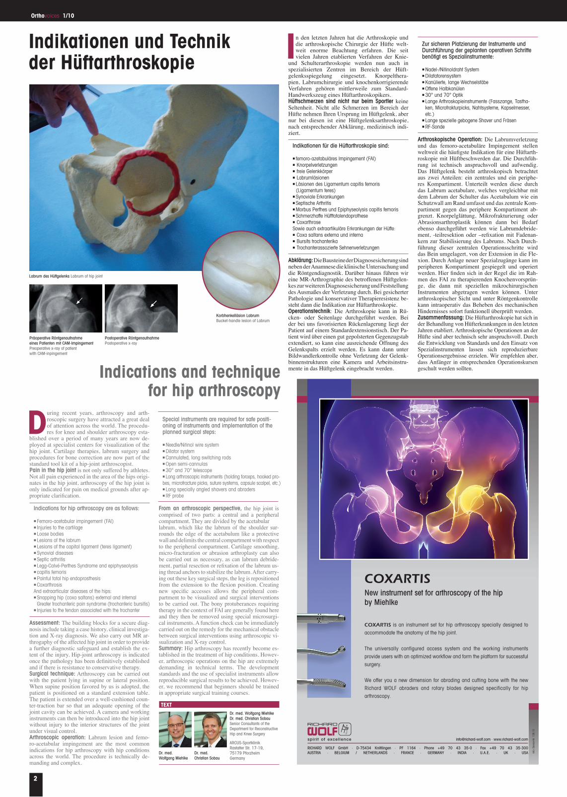
Labrum des Hüftgelenks Labrum of hip joint
Arthroskopische Operation: Die Labrumverletzung und das femoro-acetabuläre Impingement stellen weltweit die häufigste Indikation für eine Hüftarth-roskopie mit Hüftbeschwerden dar. Die Durchfüh-rung ist technisch anspruchsvoll und aufwendig. Das Hüftgelenk besteht arthroskopisch betrachtet aus zwei Anteilen: ein zentrales und ein periphe-res Kompartiment. Unterteilt werden diese durch das Labrum acetabulare, welches vergleichbar mit dem Labrum der Schulter das Acetabulum wie ein Schutzwall am Rand umfasst und das zentrale Kom-partiment gegen das periphere Kompartiment ab-grenzt. Knorpelglättung, Mikrofrakturierung oder Abrasionsarthroplastik können dann bei Bedarf ebenso durchgeführt werden wie Labrumdebride-ment, -teilresektion oder –refixation mit Fadenan-kern zur Stabilisierung des Labrums. Nach Durch-führung dieser zentralen Operationsschritte wird das Bein umgelagert, von der Extension in die Fle-xion. Durch Anlage neuer Spezialzugänge kann im peripheren Kompartiment gespiegelt und operiert werden. Hier finden sich in der Regel die im Rah-men des FAI zu therapierenden Knochenvorsprün-ge, die dann mit speziellen mikrochirurgischen Instrumenten abgetragen werden können. Unter arthroskopischer Sicht und unter Röntgenkontrolle kann intraoperativ das Beheben des mechanischen Hindernisses sofort funktionell überprüft werden.Zusammenfassung: Die Hüftarthroskopie hat sich in der Behandlung von Hüfterkrankungen in den letzten Jahren etabliert. Arthroskopische Operationen an der Hüfte sind aber technisch sehr anspruchsvoll. Durch die Entwicklung von Standards und den Einsatz von Spezialinstrumenten lassen sich reproduzierbare Operationsergebnisse erzielen. Wir empfehlen aber, dass Anfänger in entsprechenden Operationskursen geschult werden sollten.Indications and technique
for hip arthroscopy
During recent years, arthroscopy and arth-roscopic surgery have attracted a great deal of attention across the world. The procedu-res for knee and shoulder arthroscopy esta-
blished over a period of many years are now de-ployed at specialist centers for visualization of the hip joint. Cartilage therapies, labrum surgery and procedures for bone correction are now part of the standard tool kit of a hip-joint arthroscopist.Pain in the hip joint is not only suffered by athletes. Not all pain experienced in the area of the hips origi-nates in the hip joint, arthroscopy of the hip joint is only indicated for pain on medical grounds after ap-propriate clarification.
Assessment: The building blocks for a secure diag-nosis include taking a case history, clinical investiga-tion and X-ray diagnosis. We also carry out MR ar-throgaphy of the affected hip joint in order to provide a further diagnostic safeguard and establish the ex-tent of the injury. Hip-joint arthroscopy is indicated once the pathology has been definitively established and if there is resistance to conservative therapy.Surgical technique: Arthroscopy can be carried out with the patient lying in supine or lateral position. When supine position favored by us is adopted, the patient is positioned on a standard extension table. The patient is extended over a well-cushioned coun-ter-traction bar so that an adequate opening of the joint cavity can be achieved. A camera and working instruments can then be introduced into the hip joint without injury to the interior structures of the joint under visual control.Arthroscopic operation: Labrum lesion and femo-ro-acetabular impingement are the most common indications for hip arthroscopy with hip conditions across the world. The procedure is technically de-manding and complex.
From an arthroscopic perspective, the hip joint is comprised of two parts: a central and a peripheral compartment. They are divided by the acetabularlabrum, which like the labrum of the shoulder sur-rounds the edge of the acetabulum like a protective wall and delimits the central compartment with respect to the peripheral compartment. Cartilage smoothing, micro-fracturation or abrasion arthroplasty can also be carried out as necessary, as can labrum debride-ment, partial resection or refixation of the labrum us-ing thread anchors to stabilize the labrum. After carry-ing out these key surgical steps, the leg is repositioned from the extension to the flexion position. Creating new specific accesses allows the peripheral com-partment to be visualized and surgical interventions to be carried out. The bony protuberances requiring therapy in the context of FAI are generally found here and they then be removed using special microsurgi-cal instruments. A function check can be immediately carried out on the remedy for the mechanical obstacle between surgical interventions using arthroscopic vi-sualization and X-ray control.Summary: Hip arthroscopy has recently become es-tablished in the treatment of hip conditions. Howev-er, arthroscopic operations on the hip are extremely demanding in technical terms. The development standards and the use of specialist instruments allow reproducible surgical results to be achieved. Howev-er, we recommend that beginners should be trained in appropriate surgical training courses.
Präoperative Röntgenaufnahme eines Patienten mit CAM-Impingement Preoperative x-ray of patient with CAM-inpingement
Postoperative Röntgenaufnahme Postoperative x-ray
Dr. med. Wolfgang Miehlke Dr. med. Christian Sobau Senior Consultants of the Department for Reconstructive Hip and Knee Surgery
ARCUS-Sportklinik Rastatter Str. 17-19, 75179 Pforzheim Germany
TExT
Indikationen für die Hüftarthroskopie sind:
n femoro-azetabuläres Impingement (FAI)n Knorpelverletzungenn freie Gelenkkörpern Labrumläsionenn Läsionen des Ligamentum capitis femoris
(Ligamentum teres)n Synoviale Erkrankungenn Septische Arthritis n Morbus Perthes und Epiphyseolysis capitis femorisn Schmerzhafte Hüfttotalendoprothesen CoxarthroseSowie auch extraartikuläre Erkrankungen der Hüfte:n Coxa saltans externa und internan Bursits trochanterika n Trochanterassozierte Sehnenverletzungen
Indications for hip arthroscopy are as follows:
n Femoro-acetabular impingement (FAI)n Injuries to the cartilagen Loose bodiesn Lesions of the labrumn Lesions of the capital ligament (teres ligament)n Synovial diseasesn Septic arthritisn Legg-Calvé-Perthes Syndrome and epiphyseolysis n capitis femorisn Painful total hip endoprosthesisn CoxarthrosisAnd extraarticular diseases of the hips:n Snapping hip (coxa saltans) external and internal
Greater trochanteric pain syndrome (trochanteric bursitis)n Injuries to the tendon associated with the trochanter
Zur sicheren Platzierung der Instrumente und Durchführung der geplanten operativen Schritte benötigt es Spezialinstrumente:
n Nadel-/Nitinoldraht Systemn Dilatatorensystemn Kanülierte, lange Wechselstäben Offene Halbkanülenn 30° und 70° Optikn Lange Arthroskopieinstrumente (Fasszange, Tastha-
ken, Microfrakturpicks, Nahtsysteme, Kapselmesser, etc.)
n Lange spezielle gebogene Shaver und Fräsenn RF-Sonde
Special instruments are required for safe positi-oning of instruments and implementation of the planned surgical steps:
n Needle/Nitinol wire systemn Dilator systemn Cannulated, long switching rodsn Open semi-cannulasn 30° and 70° telescopen Long arthroscopic instruments (holding forceps, hooked pro-bes, microfracture picks, suture systems, capsule scalpel, etc.)n Long specially angled shavers and abradersn RF probe
Korbhenkelläsion Labrum Bucket-handle lesion of Labrum
RICHARD WOLF GmbH · D-75434 Knittlingen · PF 1164 · Phone +49 70 43 35-0 · Fax +49 70 43 35-300AUSTRIA · BELGIUM / NETHERLANDS · FRANCE · GERMANY · INDIA · U.A.E. · UK · USA
[email protected] · www.richard-wolf.comspirit of excellence
COXARTISNew instrument set for arthroscopy of the hipby Miehlke
COXARTIS is an instrument set for hip arthroscopy specially designed to
accommodate the anatomy of the hip joint.
The universally configured access system and the working instruments
provide users with an optimized workflow and form the platform for successful
surgery.
We offer you a new dimension for abrading and cutting bone with the new
Richard WOLF abraders and rotary blades designed specifically for hip
arthroscopy.
Info
/Service-N
r.13
9.10
139_10__129_206_GB:Layout 2 26.04.2010 14:51 Seite 1
Dr. med. Wolfgang Miehlke
Dr. med. Christian Sobau