Embed Size (px)
Citation preview
A role of nucleolin in human hematopoietic progenitor cells
Inaugural-Dissertation
zur Erlangung des Doktorgrades der Mathematisch-Naturwissenschaftlichen Fakultät
der Heinrich-Heine-Universität Düsseldorf
vorgelegt von
Sanil Bhatia aus Indien, Ludhiana
Düsseldorf, Dezember 2014
aus der Klinik für Kinder-Onkologie, -Hämatologie und Klinische Immunologie
des Universitätsklinikums Düsseldorf
Erstgutachter: Prof. Dr. Arndt Borkhardt
Zweitgutachter: Prof. Dr. William Martin
Gedruckt mit der Genehmigung der
Mathematisch-Naturwissenschaftlichen Fakultät der
Heinrich-Heine-Universität Düsseldorf
Tag der mündlichen Prüfung: 9.02.2015
Table of Contents Summary 10
Zusammenfassung 11
1 Introduction 13
1.1 Preface 13
1.2 Hematopoetic stem cells (HSCs) 14 1.2.1 Isolation and characterization 14
1.3 CD34 14 1.3.1 Structural organization 14 1.3.2 Expression 15 1.3.3 Regulation 15 1.3.4 Function 15 1.3.5 CD34 as a marker 16
1.4 CD133 16 1.4.1 Expression 16 1.4.2 Structural Organization 17 1.4.3 Regulation 18 1.4.4 Function 19 1.4.5 CD133 as a marker 20
1.5 Nucleolin 21 1.5.1 Structure of nucleolin 21 1.5.2 Function of nucleolin 22 1.5.3 Expression of nucleolin in stem and cancer cells 23
1.6 Wnt signaling 24 1.6.1 Introduction 24 1.6.2 Role of Wnt signaling in HSPSc development 26 1.6.3 Wnt signaling in leukemia 27
1.7 PI3K / Akt signaling 28 1.7.1 Introduction 28 1.7.2 PI3K / Akt signaling in HSPSc 29 1.7.3 PI3K / Akt signaling in leukemia 29
1.8 Aims of the PhD thesis 30
2 Materials 31
2.1 Patients 31
2.2 Human, adherent cell lines 31
2.3 Human, suspension cell lines 32
2.4 Chemicals 33 Table 2.1 General chemicals 33
2.5 Specific reagents/Media 34
2.6 Restriction Enzymes 34
2.7 Nucleic acids 35 Table 2.4 Oligonucleotides 35
2.8 Cytokines 36
2.9 Antibodies 36 Table 2.7 Western Blot Antibodies 36
2.10 Kits, size markers and other materials 37
2.11 Software and hardware 38
2.12 Buffers for Western Blot 38
3 Methods 39
3.1 Cell cultivation 39 3.1.1 Isolation and culture of human CD34+ Stem & Progenitor Cells (HSPSc) 39 3.1.2 Cultivation of human cell line 39
3.2 Cryopreservation of human cells 40
3.3 Lentiviral vector construct 40
3.4 Lentiviral transduction and selection 42
3.5 Nucleofection 42
3.6 Gelelectrophoretic separation and detection of proteins 42 3.6.1 SDS-PAGE 42 3.6.2 Western Blot analysis 42
3.7 Quantitative reverse transcription real time PCR (qRT-PCR) 43
3.8 Flourescence-activated cell sorting (FACS) 43
3.9 Colony forming assay (CFC-assay) 43 3.9.1 Primary CFC assay 43 3.9.2 Secondary CFC assay 44
3.10 Long term culture initiating assay (LTC-IC assay) 44
3.11 Ex vivo assay for B cell development 44
4 Results 45
4.1 Nucleolin dependent expression of AC133 and CD133 in HSPCs 45 4.1.1 Overexpression 45 4.1.2 Down-modulation 49
4.2 Nucleolin dependent expression of AC133 and CD133 in Mutz-2 cell line 52 4.2.1 Overexpression 52 4.2.2 Down-modulation 54
4.3 Nucleolin dependent expression of AC133 and CD133 in SEM cell line 56
4.4 Nucleolin increases the frequency of hematopoietic colony forming units (CFUs) and impacts myeloid differentiation in HSPCs 58
4.4.1 Overexpression 58 4.4.2 Down-modulation 63
4.5 Nucleolin increases the frequency of hematopoietic colony forming units (CFUs) and impacts myeloid differentiation in Mutz-2 cells 67
4.5.1 Overexpression 67 4.5.2 Down-modulation 71
4.6 Nucleolin amplifies the number of cells with LTC-IC activity 74
4.7 Nucleolin supports long-term maintenance of HSPCs in stroma free cultures 76
4.8 Nucleolin promotes B cell development 78
4.9 Functional effects of nucleolin on HSPCs partially relies on -catenin activity 79
5 Discussion 85
5.1 AC133 & CD133 expression in the hematopoietic system 85
5.2 Nucleolin mediated AC133 and CD133 expression 86
5.3 Nucleolin facilitates polarization of HSPCs 87
5.4 Nucleolin regulates hematopoietic colony formation in HSPCs and in Mutz-2 cells 88
5.5 Impact of nucleolin on LTC-IC frequencies and long-term maintenance of HSPSc 88
5.6 Effect of nucleolin on B cell differentiation 90
5.7 Nuclolin mediated regulation of Wnt / β-catenin, PI3K / Akt and BCL-2 axis 90 5.7.1 Wnt / β-catenin signaling during hemetopoiesis 91 5.7.2 PI3K / Akt signaling during hematopoiesis 92
5.8 Nucleolin as a therapeutic target in leukemia 93
6 Conclusion 95
7 Outlook 96
8 References 97
9 Abbreviations 114
10 Publications 116
11 Acknowledgements 117
Curriculum vitae 118 Affirmation 119
Figure Directory
Figure 1.1 Genomic arrangement of human CD133 promoter region. ................................................ 17
Figure 1.2 Schematic drawing of CD133 .............................................................................................. 18
Figure 1.3 Schematic drawing of nucleolin ........................................................................................... 22
Figure 1.4 Schematic drawing of canonical Wnt signaling. .................................................................. 26
Figure 3.1 Schematic drawing of lentiviral overexpression constructs. ................................................ 41
Figure 3.2 Schematic drawing of lentiviral down-modulation constructs. ............................................. 41
Figure 4.1 AC133 and CD133 expression in HSPC-NCL, HSPC-NCL-289-709 and HSPC-mock cells
after transduction. .................................................................................................................................. 46
Figure 4.2 CD34 expression in HSPC-NCL, HSPC-NCL-289-709 and HSPC-mock cells . ................ 47
Figure 4.3 Influence of nucelolin on cell morphology. .......................................................................... 48
Figure 4.4 Validation of nucleolin-targeting shRNA. ............................................................................. 49
Figure 4.5 AC133 and CD133 expression in HSPC-ctrl shRNA, HSPC-NCL-shRNA1 and HSPC-
empty cells after nucleofection. ............................................................................................................. 50
Figure 4.6 CD34 expression in HSPC-ctrl shRNA, HSPC-NCL-shRNA1 and HSPC-empty cells after
nucleofection. ........................................................................................................................................ 51
Figure 4.7 AC133 and CD133 expression in Mutz-2-NCL, Mutz-2-NCL-289-709 and Mutz-2-mock
cells after transduction. .......................................................................................................................... 53
Figure 4.8 AC133 and CD133 expression in Mutz-2-ctrl shRNA, Mutz-2-NCL-shRNA1, Mutz-2-NCL-
shRNA2 and Mutz-2-empty cells after transduction. ............................................................................. 55
Figure 4.9 AC133 and CD133 expression in SEM-NCL, SEM-mock (overexpression) and SEM-ctrl
shRNA, SEM-NCL-shRNA1 and SEM-NCL-shRNA2 (down-modulation) cells. ................................... 57
Figure 4.10 Clonogenic capacity of HSPC-NCL, HSPC-NCL-289-709 and HSPC-mock cells. .......... 59
Figure 4.11 Impact on differerntiation of myeloid progenitors in HSPC-NCL, HSPC-NCL-289-709 and
HSPC-mock cells after CFU-GEMM assay. .......................................................................................... 60
Figure 4.12 Impact on sizes of individual colonies detected in HSPC-NCL, HSPC-NCL-289-709 and
HSPC-mock cells after CFU-GEMM assay. .......................................................................................... 61
Figure 4.13 Impact on cellularity of individual colonies detected in HSPC-NCL, HSPC-NCL-289-709
and HSPC-mock cells after CFU-GEMM assay. ................................................................................... 62
Figure 4.14 Clonogenic capacity of HSPC-ctrl-shRNA, HSPC-NCL-shRNA-1, HSPC-NCL-shRNA-2
and HSPC-empty cells. ......................................................................................................................... 63
Figure 4.15 Impact on differerntiation of myeloid progenitors in HSPC-ctrl-shRNA, HSPC-NCL-
shRNA-1, HSPC-NCL-shRNA-2 and HSPC-empty cells after CFU-GEMM assay. .............................. 64
Figure 4.16 Impact on sizes of individual colonies detected in HSPC-ctrl-shRNA, HSPC-NCL-shRNA-
1, HSPC-NCL-shRNA-2 and HSPC-empty cells after CFU-GEMM assay. .......................................... 65
Figure 4.17 Impact on cellularity of individual colonies detected in HSPC-ctrl-shRNA, HSPC-NCL-
shRNA-1, HSPC-NCL-shRNA-2 and HSPC-empty cells after CFU-GEMM assay. .............................. 66
Figure 4.18 Clonogenic capacity of Mutz-2-NCL, Mutz-2-NCL-289-709 and Mutz-2-mock cells. ....... 67
Figure 4.19 Impact on differentiation of myeloid progenitors in Mutz-2-NCL, Mutz-2-NCL-289-709 and
Mutz-2-mock cells after CFU-GEMM assay. ......................................................................................... 68
Figure 4.20 Impact on sizes of individual colonies detected in Mutz-2-NCL, Mutz-2-NCL-289-709 and
Mutz-2-mock cells after CFU-GEMM assay. ......................................................................................... 69
Figure 4.21 Impact on cellularity of individual colonies detected in Mutz-2-NCL, Mutz-2-NCL-289-709
and Mutz-2-mock cells after CFU-GEMM assay. .................................................................................. 70
Figure 4.22 Clonogenic capacity of Mutz-2-ctrl shRNA, Mutz-2-NCL-shRNA1, Mutz-2-NCL-shRNA-2
and Mutz-2-mock cells. .......................................................................................................................... 71
Figure 4.23 Impact on sizes of individual colonies detected in Mutz-2-ctrl shRNA, Mutz-NCL-shRNA2,
Mutz-NCL-shRNA1 and Mutz-2-empty cells after CFU-GEMM assay. ................................................. 72
Figure 4.24 Influence of silencing of the nucleolin on the early differentiation of Mutz-2 cells ............ 73
Figure 4.25 Levels of nucleolin in cells employed for LTC-IC assay. ................................................... 74
Figure 4.26 LTC-IC activity of HSPC-NCL, HSPC-NCL-289-709 and HSPC-mock cells. ................... 75
Figure 4.27 Long-term maintenance of HSPC-NCL, HSPC-NCL-289-709 and HSPC-mock cells on
stroma free cultures. .............................................................................................................................. 76
Figure 4.28 Long-term maintenance of HSPC-NCL, HSPC-NCL-289-709 and HSPC-mock cells on
stroma free cultures. .............................................................................................................................. 77
Figure 4.29 Functional effects of nucleolin on HSPCs partially rely on -catenin activity (Immunobots).
............................................................................................................................................................... 80
Figure 4.30 Functional effects of nucleolin on HSPCs partially rely on -catenin activity (CFU-GEMM
assay). ................................................................................................................................................... 81

Figure 4.31 Functional effects of nucleolin on HSPCs partially rely on -catenin activity (Colony
pictures). ................................................................................................................................................ 82
Figure 4.32 Functional effects of nucleolin on HSPCs partially rely on -catenin activity (Cellularity). 83
Figure 4.33 Functional effects of nucleolin on HSPCs partially rely on -catenin activity (Immunoblot &
liquid culture). ........................................................................................................................................ 84
Table Directory
Table 1.1 Characterization of cancer stem cells from various cancer tissues by AC133 ..................... 20
Table 1.2 Multiple role of nucleolin ....................................................................................................... 23
Table 2.1 General chemicals ................................................................................................................ 33
Table 2.2 Specfific reagents and media ................................................................................................ 34
Table 2.3 Restriction enzymes ............................................................................................................. 34
Table 2.4 Oligonucleotides.................................................................................................................... 35
Table 2.5 shRNA seuences .................................................................................................................. 35
Table 2.6 Cytokines .............................................................................................................................. 36
Table 2.7 Western Blot Antibodies ........................................................................................................ 36
Table 2.8 FACS antibodies ................................................................................................................... 37
Table 2.9 Kits, size markers and other material ................................................................................... 37
Table 2.10 Software and database ....................................................................................................... 38
Table 2.11 Hardware ............................................................................................................................. 38
Table 4.1 Effects of nucleolin on B cell differentiation .......................................................................... 78
10
Summary Still a major challenge in the hematopoietic stem cell biology is to precisely determine the factors that
regulate self-renewal and lineage commitment of hematopoietic stem and progenitor cells (HSPCs). A
better understanding of the mechanisms regulating HSPCs is required, because of their clinical
therapeutic applications and their aberrant regulation during leukemia.
AC133 is a (117 kD) pentaspan transmembrane surface glycoprotein, a specific glycoform of CD133
(prominin-1) predominantly expressed on a subset of CD34+ and a small fraction of CD34– HSPCs.
AC133+ HSPCs are enriched for colony-forming units (CFUs), long-term culture initiating cells (LTC-
IC) and are capable of hematopoietic reconstitution. Beyond that, AC133 is used for prospective
isolation of tumour-initiating cells in certain hematologic malignancies. However, the mechanism
underlying the expression of AC133 and CD133 needs to be further explored. CD133 is transcribed
from alternative tissue dependent promoters, and of these promoters, P1 is active in HSPCs as
opposed to differentiated hematopoietic cells, and this is paralleled by CD133 mRNA and protein
levels. Recently, a direct link of CD133 expression with activation of Wnt / -catenin and PI3K / Akt
signaling was shown (Mak et al., 2012; Wei et al., 2013). Wnt signaling strength regulates normal
haematopoiesis, and its deregulation is involved in leukemia development. Therefore, working out
mechanisms controlling AC133 expression and other mechanisms influencing the functional properties
of HSPCs is important to understand homeostasis in normal and dysregulated hematopoietic tissue at
molecular level. In a previous study from our research group, it came out that nucleolin act as a CD34
promoter factor, and was found to be enriched in mobilized peripheral blood (MPB)-derived
undifferentiated CD34+ HSPCs as opposed to differentiated CD34– cells (Grinstein et al., 2007).
Nucleolin is a multifunctional nucleolar phosphoprotein, overexpressed in actively growing and cancer
cells, involved in transcriptional regulation, chromatin remodeling, and RNA metabolism. Aberrant
activity of nucleolin is generally associated with several haematological malignancies.
The present study dissects nucleolin-dependent activation of AC133 and CD133 expression via
promoter P1 in MPB-derived CD34+ HSPCs and in leukemic cell line models. Overexpression of
nucleolin was likely associated with polarization in HSPCs, and was probably resulted from higher
cellular PI3K / Akt levels. In CD34+ HSPCs and in an acute myeloid leukemia (AML) derived cell line
Mutz-2; nucleolin elevates hematopoietic CFU frequencies and predominately impacts common
primitive granulocytes-macrophage (GM) progenitors. In down-modulation experiments, silencing of
nucleolin in Mutz-2 cells leads to the early differentiation, and into a myeloid phenotype. Furthermore,
in HSPCs; nucleolin amplifies numbers of LTC-IC and supported long-term maintenance of
hematopoietic progenitors in cytokine-dependent stroma-free cultures. Beyond that, growth promoting
effects of nucleolin extended to lymphoid lineage as well, with an increase output of CD19+ &
CD34+CD19+ cells under conditions permissive for B lymphoid development. Levels of active β-
catenin (Wnt / β-catenin), active Akt (PI3K / Akt) and BCL-2 in HSPCs are nucleolin dependent and
functional effects of nucleolin on HSPCs partially relies on β-catenin activity.
Wnt/β-catenin and BCL-2 is found to be aberrantly activated in leukemia, providing opportunities for
therapeutic intervention. In addition, elevated levels of nucleolin stabilize bcl-2 mRNA in leukemic
cells, which allows cells to overproduce BCL-2 and thereby protecting them from apoptosis.

Overall, this report strongly indicates that the deregulation of nucleolin via activation of Wnt / β-
catenin, and BCL-2 can be implicated in leukemia, and providing opportunities for therapeutic
interventions.
12
Zusammenfassung
Die Bestimmung von Faktoren, die für Selbsterneuerung oder Differenzierung von hämatopoietischen
Stamm- und Vorläuferzellen (HSPCs) zuständig sind, ist immer noch eine große Herausforderung. Ein
besseres Verständnis der zugrunde liegenden Kontrollmechanismen ist notwendig, da eine aberrante
Regulation dieser Faktoren einen Einfluss auf die Entstehung von Leukämien haben kann.
Eine spezielle Glykoform des Transmembranproteins CD133 (Prominin-1) ist AC133, die vor allem in
CD34+-HSPCs aber auch in einer kleinen Population von CD34--HSPCs exprimiert wird. Die
Untergruppe der AC133+-HSPCs zeigt eine erhöhte Anzahl an colony-forming units (CFUs), long-term
culture initiating cells (LTC-IC) und ist in der Lage das hämatopoietische System zu rekonstituieren.
Zudem wird AC133 zur prospektiven Isolierung von tumorinitiierenden Zellen bei bestimmten malignen
hämatopoietischen Erkrankungen verwendet. Die Mechanismen, die zur Expression von AC133 und
CD133 führen, sind jedoch noch nicht komplett verstanden. Die Expression von CD133 wird durch
verschiedene gewebespezifische Promotoren reguliert. Dies ist in hämatopoietischen Zellen der
Promotor P1. Analysen der Genexpression in hämatopoietischen Zellen zeigte, dass P1 zwar in
HSPCs jedoch nicht in differenzierten Zellen aktiv ist, was auf mRNA- sowie auf Proteinebene
bestätigt werden konnte. Vor kurzem wurde ein Zusammenhang zwischen CD133-Expression und der
Aktivierung von Wnt / β-Catenin- sowie PI3K / Akt-Signalwegen gezeigt. Der Wnt-Signalweg hat eine
große Bedeutung während der Hämatopoese, wobei seine Missregulation eine Rolle in der
Leukämieentwicklung spielt. Um Homöostase im gesunden und kranken Gewebe auf molekularer
Ebene zu verstehen, ist ein besseres Verständnis der AC133-Expression und anderer Faktoren
unabdingbar. Es zeigte sich, dass Nucleolin als CD34-Promoterfaktor agiert und in undifferenzierten
CD34+-HSPCs aus mobilisiertem peripherem Blut (MPB) im Vergleich zu ausdifferenzierten CD34--
Zellen angereichert ist (Grinstein et al., 2007). Nucleolin ist ein multifunktionelles nukleäres
Phosphoprotein, welches in proliferierenden und kanzerogenen Zellen überexpremiert ist. Es ist an
transkriptioneller Regulation, Chromatin-Umstrukturierung sowie am RNA-Metabolismus beteiligt.
Aberrante Nucleolin-Aktivität wird mit bestimmten malignen hämatopoietischen Erkrankungen in
Verbindung gebracht.
Die hier präsentierte Arbeit untersucht die Nucleolin-abhängige Aktivierung von AC133- und CD133-
Expression via den P1-Promotor in CD34+-HSPCs aus mobilisiertem peripherem Blut (MPB) sowie in
leukämischen Zelllinien. Es zeigte sich, dass Nucleolin-Überexpression wahrscheinlich mit
Zellpolarisierung assoziiert war und zu erhöhter PI3K / Akt-Aktivität in HSPCs führt. In CD34+-HSPCs
und der akute myeloische Leukämie (AML) Zelllinie Mutz-2 führte eine Überexpression von Nucleolin
zu einer erhöhten Anzahl an CFUs und beeinflusste die myeloide Differenzierung. Entsprechend
führte eine Reduzierung der Nucleolin-Expression in Mutz-2-Zellen zu einer raschen Differenzierung
zu myeloiden Zellen. Außerdem zeigte sich in Experimenten mit Nucleolin-Überexpression in HSPCs
ein positiver Einfluss auf LTC-ICs, und dass das Überleben hämatopoietischer Vorläuferzellen in
Stroma-freien Kulturen dauerhaft unterstützt wurde. Darüber hinaus zeigte eine Nucleolin-
Überexpression auch in lymphoiden Zellen einen wachstumsstimulierenden Effekt, was durch eine
erhöhte Anzahl an CD19+ und CD34+CD19+-Zellen unter lymphoidstimulierenden Bedingungen
gezeigt werden konnte. Des Weiteren ist das Level von aktivem β-Catenin, aktivem Akt und BCL-2 in

13
HSPCs Nucleolin-abhängig, wobei funktionelle Effekte von Nucleolin in diesen Zellen zum Teil von β-
Catenin-Aktivität abhängig sind.
Da Wnt / β-Catenin sowie BCL-2 in Leukämien aberrant aktiv ist, bietet eine Nucleolin-Überexpression
möglicherweise einen Ansatz, Leukämie-relevante therapeutische Ansatzpunkte zu erforschen. In
diesem Zusammenhang bietet diese Arbeit einen ersten Einblick in die funktionelle Rolle von Nucleolin
sowohl in HSPCs als auch in leukämischen Zelllinien.
13
1 Introduction
1.1 Preface
Despite extensive investigation on determining the phenotype and functional characteristics of
Hematopoietic stem cells (HSCs) over the years (Morrison et al., 1995; Weissman, 2000), still a major
challenge in the hematopoietic stem cell biology is to precisely determine the factors that regulate their
self-renewal and lineage commitment. A better understanding of the mechanisms regulating these
cells is required because of their importance in the clinical therapeutic applications, such as targeted
gene based therapy, reconstitution of hematopoietic system after myeloablative therapies and
dysregulation during leukemia.
After the discovery of the nucleolus, major focus of the research was to understand its dynamic
structure and function. However ribosomal biogenesis is one of its predominant functions (Hadjiolov,
1985) but there is growing evidence confirming its involvement in other cellular activities (Pederson,
1998). In addition to the components of the ribosomes, nucleolus is the home for many other RNAs
and proteins. In this report, I focus on one of its abundant & well-studied phosphoprotein, Nucleolin, a
highly mobile protein that can freely transport in several cell compartments, including nucleoplasm,
cytoplasm, and the cell surface. Nucleolin expression is found to be enriched in actively growing and
cancerous cells (Lapeyre et al., 1987; Derenzini et al., 1995; Borer et al., 1989). Elevated levels of
nucleolin have been reported in several hematological malignancies, thus making nucleolin an
interesting candidate to investigate its potential role in proliferation and differentiation of early
hematopoetic stem and progenitor cells (HSPCs) as well as in leukemia.
The underlying introductory section will summarize some of the key factors influencing hematopoetic
stem cell biology as well as structure and function of nucleolin and its role in regulating normal growing
or cancerous cells.
1 Introduction
14
1.2 Hematopoetic stem cells (HSCs)
1.2.1 Isolation and characterization
Hematopoiesis is a hierarchical process of generating entire lineages of specialized blood cells in a
stepwise manner from immature cells and subsequently releasing them into circulating blood and
peripheral organs for further maturation or effector function. At top of the hierarchical process are
HSCs, first functionally identified by Till and McCulloch in 1961 having self-renewal, multipotent and
differentiation potential. During hematopoiesis, HSCs first give rises to common early lymphoid and
myeloid progenitors, which further differentiate into different lineages of specialized blood cells such
as monocytes, macrophages, eosinophils, basophils, neutrophils, erythrocytes, platelets, B & T
lymphocytes and natural killer (NK) cells (Bhatia et al., 1997; Spangrude et al., 1988; Yang et al.,
2005). HSCs are predominantly found in the bone marrow of adults but can also be isolated from other
sources like umbilical cord blood (CB) and peripheral blood (PB). HSCs primarily resides in the
endosteal region of the bones, which comprises of different types of cells making hematopoietic
niches and tightly regulating directly or indirectly their self-renewal, differentiation and localization
(Morrison et al., 2008).
The factors like multilineage reconstitution, level & time of engraftment has led us to characterize
strictly within the HSCs population, such as long-term engrafting HSC, short term engrafting or
multipotent progenitor cells (MPP) and more mature lineage commited progenitors that have lost self-
renewal capacity (Morrison et al.,1994; Weissman et al.,2000). It is estimated that the frequency of
HSCs in the blood is approximately 1 in 100,000 and 1 in 10,000 in bone marrow; therefore it’s a
major challenge to isolate these cells from the total pool of cells. HSCs are usually characterized and
isolated by the presence or absence of some surface markers.
From the past decades CD34 is the marker of choice to enumerate hematopoietic stem and progenitor
cells and even clinically for autologous and allogenic bone marrow transplantation (Krause et al.,
1996; Lee et al., 2004). However, in addition to CD34 there are other markers used to further
characterize long-term-HSCs (LT-HSCs), including CD90, TIE, HLA-DR ,CD117, CD50, CD71,CD38
(Murray et al.,1995; Terstappen et al.,1991; Terstappen et al.,1994; Gunji et al.,1993; Hashiyama et
al.,1996; Batard et al., 1996; Recktenwald et al.,1996) and primitive HSCs populatons, such as
CD34bright, CD38dim/-, HLA-DRdim/-, CD90+, CD117+, lineage−, rhodamine 123lo , CD133 (Baum et al.,
1992; Fritsch et al., 1993; Olweus et al., 1996;; Miraglia et al.,1997; Yin et al., 1997).
1.3 CD34
1.3.1 Structural organization
Cluster of differentiation molecule 34 (CD34) is a surface protein, belongs to the family of sialomucin
surface antigens. CD34 molecule is a 115 kD type 1 integral transmembrane phosphoglycoprotein. It
has a 40 kD protein backbone as predicted by cDNA and harbours nine potential N & O linked
glycosylation sites in its extracellular domain, two protein kinase C and one tyrosine kinase
phosphorylation sites in its cytoplasmic region (Krause et al., 1996).
1 Introduction
15
1.3.2 Expression
CD34 is predominantly expressed on the undifferentiated hematopoietic stem and progenitors cells
and its expression decreases as the cells undergo differentiation (Caux et al., 1989). However in
addition, its expression can also be detected on other non-hematopoietic cell types, including
embryonic fibroblasts (Brown et al., 1991), endothelial cells in small vessels (Fina et al., 1990) and at
mRNA level in spleen, thymus, BM, liver (Brown et al., 1991) and furthermore both in fetal and adult
lungs, heart, kidney, skeletal muscels and brain (Fina et al., 1990).
1.3.3 Regulation
CD34 antigen has a significant importance in the hematopoietic stem cell biology; however the
mechanisms underlying its regulation and function are still not well understood. Regulation of the
CD34 gene occurs both at transcriptional and post transcriptional level (Krause et al., 1996). Studies
have shown that the CD34 gene is regulated at transcriptional level and its extent of regulation is
higher in CD34+ cell lines as compared to the CD34– cell lines (Brown et al., 1991; He et al., 1992;
Simmons et al., 1992; Satterthwaite et al., 1992; Yaamaguchi et al., 1994). In the human CD34 gene,
its 5' UTR is 258 base pairs long but lacks CAAT and TAAT motifs (Satterthwaite et al., 1992).
Furthermore, upstream sequence of transcriptional start site contains consensus sequences for the
binding of transcriptional regulators like nuclear factor Y, myc, myb, Ets-2 and MZF-1 to modulate the
promoter region of CD34 gene. (Radomska et al., 1999); Brown et al., 1991; He et al., 1992; Simmons
et al., 1992; Satterthwaite et al., 1992). For instance, c-myb is primarily expressed in the hematopoetic
cells (Gonda et al., 1984) and can transactivate the CD34 promoter region (Melotti et al., 1994).
However, c-myb knockout embryonic stem cells have shown normal levels of CD34 expression
(Mucenski et al., 1991), thus suggesting there are additional factors which can independently activate
human CD34 transcription, like Ets-2 (Melotti et al., 1994). In one of the study it came out that
nucleolin binds to the promoter region of CD34 gene through direct sequence specific interaction and
modulates both the endogenous and the cell surface level of CD34 in human CD34+ cells (Grinstein et
al., 2007). In this study, it has been suggested that the recruitment of nucleolin takes place directly at
the CD34 promoter region, which inturn activate the promoter through nucleosomal particle in CD34+
peripheral blood mononuclear cells (PBMCs), and these nucleosomal particles are absent in the
promotor region of CD34– PBMSc. Additionally CD34 gene has putative DNA binding sites located in
its upstrem region, including for the transcription factors Pu.1, Sp-1, and C-EBP (He et al., 1992; Burn
et al., 1992).
1.3.4 Function
Studies on the role of CD34 have revealed the presence of phosphorylation sites in the cytoplasmic
region of CD34 protein, but still devoid of any constitutive enzymatic activity (He et al., 1992).
However, induction of actin polymerization and isotypic adhesion through crosslinking of monoclonal
anti CD34 antibody to specific epitope of CD34 in CD34+ cells can results in an increase tyrosine
phosphorylation (Majdic et al., 1994; Gordon et al., 2000; Bullock et al., 2007). Therefore, indicating its
potential role in the signal transduction through the cell membrane. In the hematopoietic
microenvironment, natural ligand of CD34 is not well specified but a murine CD34+ endothelial cells
1 Introduction
16
(venule) in the lymph node adhere to L-selectin, a lymphocyte homing receptor (Baumheter et al.,
1993). Moreover, CD34 expression on the endothelial cells may assist in the leukocyte adhesion &
mobilization during inflammatory process, thus suggesting its potential role in the homing of HSPCs
through proteins like L-selectin (Majdic et al., 1994). Beside that, the backbone of CD34 molecule may
act as a scaffold for binding of stromal lectins to specific glycans, which in turn can mediate the
attachment of progenitor population to the stromal cells in the hematopoietic niche (Healy et al., 1995).
In fact, a study in CD34 knockout mice & in embryoid bodies has displayed its potential involvement in
the maintainance of progenitor cells, both during embryonic & adult hematopoiesis (Cheng et al.,
1996).
1.3.5 CD34 as a marker
CD34 plays a key role in the leukemia diagnosis and classification. Around 40% of acute myeloid
leukemia (AML) cases and virtually all t (8:21) translocation cases express CD34 (Civin et al., 1984;
Civin et al., 1989; Borowitz et al., 1989; Soligo et al., 1991; Vaughan et al., 1988; Geller et al., 1990;
Hurwitz et al., 1992). Elevated percentage of CD34+ cells in the case of myelodyplasia, can be used
as a tool to measure and predict the blast crisis (Guyotat et al., 1990). In fact, there is strong
correlation between the expression of CD34 with the multi drug resistance protein (MDR) and
aldehyde dehydrogenase (ALDH) expression in the case of AML, both of which are famously known to
protect against antineoplastic therapies (Campos et al., 1992; Chaudhary et al., 1991, Kastan et al.,
1990). CD34 is expressed in approximately 70% of childhood B-cell ALL and comparatively less in the
case T-cell ALL (Pui et al., 1993; Hurwitz et al., 1988; Hurwitz et al., 1992; Borowitz et al., 1990; Gore
et al., 1991) . However its expression is not detected in the malignancies originated from the mature
cells, like in the case of chronic lymphocytic leukemia (CLL) (Silvestri et al., 1992).
1.4 CD133
1.4.1 Expression
CD133 (Prominin-1) is a pentaspan transmembrane glycoprotein, first isolated in mouse embryonic
and epithelial cells (Weigmann et al., 1997). In the same year, mouse homolog of CD133 was
identified on the human CD34 expressing HSCs. It was carried out by immunizing mice with human
CD34+ cells and recovering a hybridomas producing monoclonal antibody, named AC133 (Yin et al.,
1997). CD133 transcripts are expressed in different cell lines and nearly all adult tissues; however the
expression of AC133 antigen is restricted predominantly to undifferentiated cells, such as
hematopoetic stem cells (Yin et al., 1997), fetal brain stem cells (Uchida et al., 2000),endothelial
progenitor cells (Peichev et al., 2000), myogenic cells (Torrente et al., 2004), prostatic epithelial stem
cells (Richardson et al., 2004), embryonic epithelium (Weigmann et al., 1997) and certain types of
carcinomas like retinoblastomas (Maw et al., 2000; Mirgali et al., 1997), leukemias (Bhatia, 2001) and
tetracarcinomas (Mirgali et al., 1997). In various cellular compartments, CD133 expression is enriched
in fiopodoia, lamellipodia, cell protrusions, and microvilli of neuroepithelial cells and microspikes of the
non-epithelial cells. In addition to humans and mice, proteins resembling CD133 are expressed in
other vertebrates and inverterbrates, such as Drosophila melanogaster (Zelhof et al., 2006),
1 Introduction
17
Caenorhabditis elegans (Corbeil et al., 1998; Weigmann et al., 1997) a, Danio rerio (McGrail et al.,
2010) and Rattus novegicus (Corbeil et al., 2001).
1.4.2 Structural Organization
Human CD133 is a single copy gene located on Chromosome 4 (4p15.33), consists of 37 exons and
is driven by five alternative promoters. Out of five, three promoters are located in the CpG islands and
partially controlled by methylation (Shmelkov et al., 2004). The CD133 transcript consists of one of the
five alternative 5’ untranslated regions (UTR) in the first exon that are usually expressed in a tissue
specific manner (Shmelkov et al., 2004) (Figure 1.1). 5’ UTR inclusion might affect the alternative
splicing in the coding regions, which can regulates the expression of different prominin-1 isoforms
through alternative promoters. Interestingly, CD34+ HSCs use only one the 5’ UTR and its
corresponding promoter (P1), which is found to be active in HSPCs as opposed to differentiated
hematopoietic cells, and this is paralleled by CD133 mRNA and protein levels (Shmelkov et al., 2004)
(Figure 1.1). In addition to the alternative 5’ UTR prominin-1 isoforms, there are some novel isoforms
expressed in tissue specific manner, both in fetal and adult tissues (Yu et al., 2002; Fargeas et al.,
2004; Corbeil et al., 2009).
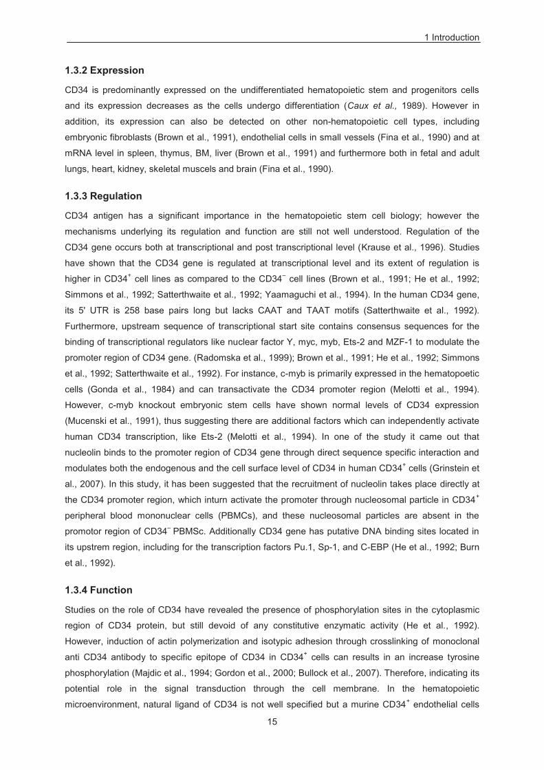
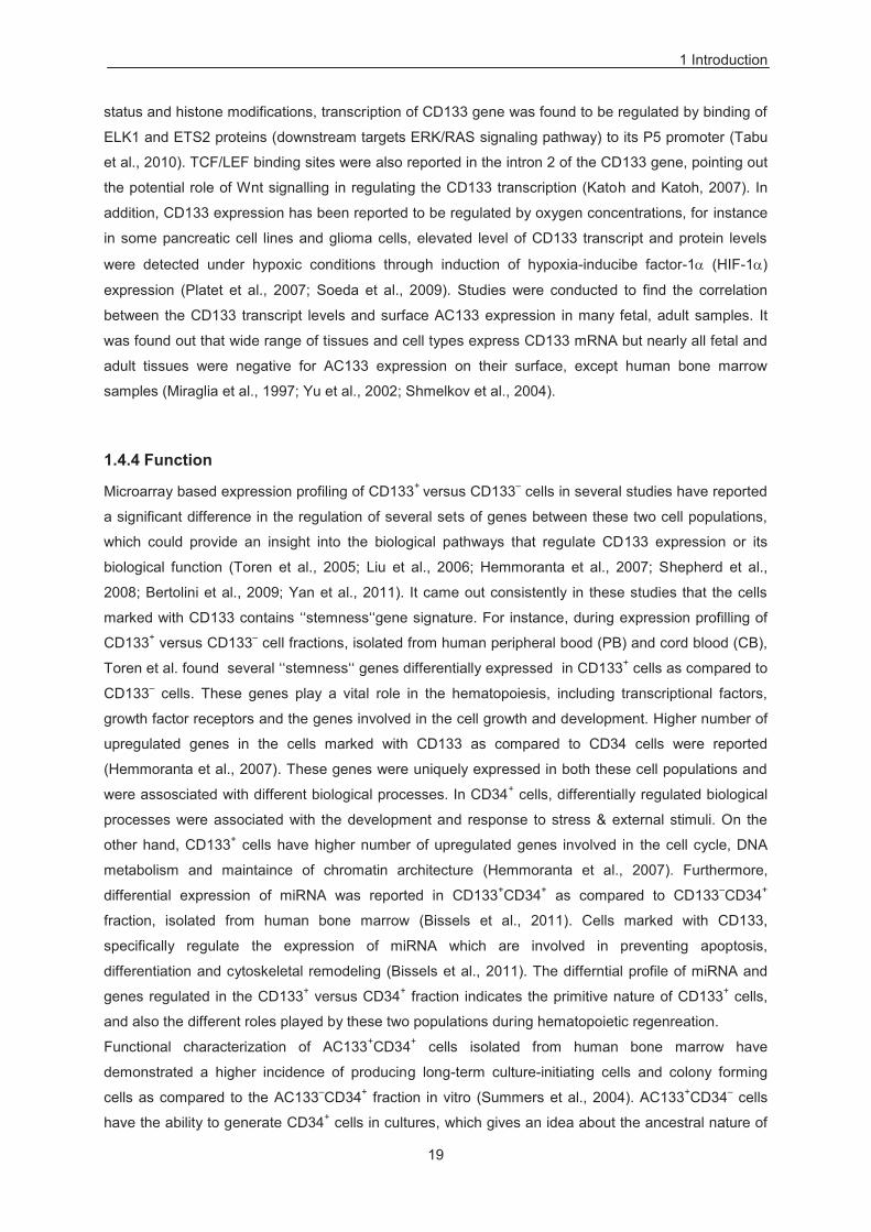
Figure 1.1 Genomic arrangement of the human CD133 promoter region The above figure shows five alternative promoters (P5, P1, P2, P3 and P4) and out of this promoter P1, P2 and
P3 are located in Cpg islands. Moreover, the figure is depicting that the exon 1A and exon 1B are alternatively
spliced to common exon2, for the initiation of translation. CD34+ HSCs uses only one 5’ UTR and its
corresponding promoter (P1), which is found to active in the HSPCs as opposed to differentiated hematopoietic
cells, and this is paralleled by CD133 mRNA and protein levels .
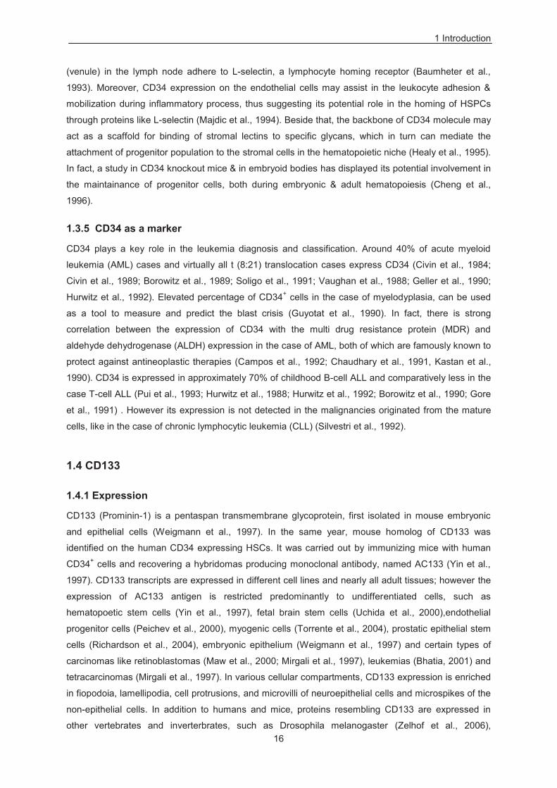
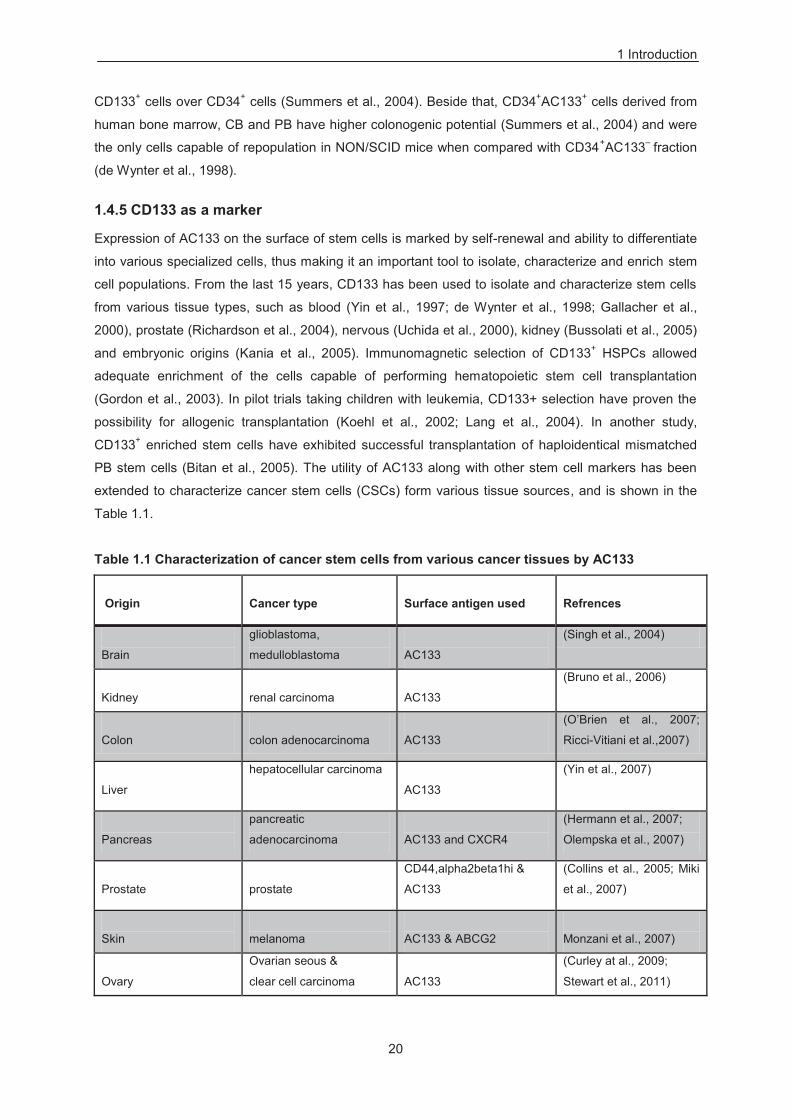
Human CD133 is an 865 amino acid (aa) long glycoprotein with an estimated molecular weight of 96.8
kD; however migrating at approx. 120kD size in the western blot due to N-terminal glycosylation
(Mirgali et al., 1997). Amino acid sequence prediction revealed that CD133 has a unique structure
comprised of five hydrophobic transmembrane domains, with two extracellular loops, two intracellular
1 Introduction
18
loops and an intracellular C-terminal tail. CD133 protein has eight potential N-terminal glycosylation
sites with 4 in each of its extracellular loops (Mirgali et al., 1997) (Figure 1.2). Mass spectrometry of
CD133 has revealed the presence of tyrosine kinase phosphorylation sites in its cytoplasmic tail
region, suggesting its potential role in signal transduction (Mirgali et al., 1997).
Figure 1.2 Schematic drawing of CD133. This drawing represents the predicted topology of the CD133 protein. CD133 protein consists of five
transmembrane domains and consist eight potential N-terminal glycosylation sites with four in each of the
extracellular loops.
1.4.3 Regulation
The AC133 antigen has significant importance in the hematopoietic stem cell biology originally
identified as an additional marker to isolate CD34+ HSCs. However mechanism regulating its
transcription is not well understood. Most of the studies on CD133 transcriptional regulation have been
focused on the methylation status of its promoter. CD133 promoter has been found to be
hypomethylated in numerous tumors & cancer cell lines, which express detectable levels of CD133,
like in the case of liver cancer, ovarian cancer, colon cancer and glioblastoma (Pleshkan et al., 2008;
Tabu et al., 2008; Yi et al., 2008; Baba et al., 2009; You et al., 2010). However, CD133 promoter is
hypermethylated in certain cell lines, which express low or undetectable levels of CD133, and the
expression of CD133 in these cell lines can be induced by molecules targeting to DNA
methyltransferases (Tabu et al., 2008; Yi et al., 2008; Baba et al., 2009; Friel et al., 2010; Jeon et al.,
2010; Schiapparelli et al., 2010). On the contrary, CD133 promoter methylation is not linked with the
CD133 expression in some cases, such as in primary prostate tumor samples and prostate cancer
cells (Pellacani et al., 2011). Therfore, caution should be taken while correlating CD133 expression
with the methylation status of its promoter (Yi et al., 2008; Tabu et al., 2008). In addition to methylation
1 Introduction
19
status and histone modifications, transcription of CD133 gene was found to be regulated by binding of
ELK1 and ETS2 proteins (downstream targets ERK/RAS signaling pathway) to its P5 promoter (Tabu
et al., 2010). TCF/LEF binding sites were also reported in the intron 2 of the CD133 gene, pointing out
the potential role of Wnt signalling in regulating the CD133 transcription (Katoh and Katoh, 2007). In
addition, CD133 expression has been reported to be regulated by oxygen concentrations, for instance
in some pancreatic cell lines and glioma cells, elevated level of CD133 transcript and protein levels
were detected under hypoxic conditions through induction of hypoxia-inducibe factor-1 (HIF-1 )
expression (Platet et al., 2007; Soeda et al., 2009). Studies were conducted to find the correlation
between the CD133 transcript levels and surface AC133 expression in many fetal, adult samples. It
was found out that wide range of tissues and cell types express CD133 mRNA but nearly all fetal and
adult tissues were negative for AC133 expression on their surface, except human bone marrow
samples (Miraglia et al., 1997; Yu et al., 2002; Shmelkov et al., 2004).
1.4.4 Function
Microarray based expression profiling of CD133+ versus CD133– cells in several studies have reported
a significant difference in the regulation of several sets of genes between these two cell populations,
which could provide an insight into the biological pathways that regulate CD133 expression or its
biological function (Toren et al., 2005; Liu et al., 2006; Hemmoranta et al., 2007; Shepherd et al.,
2008; Bertolini et al., 2009; Yan et al., 2011). It came out consistently in these studies that the cells
marked with CD133 contains ‘‘stemness‘‘gene signature. For instance, during expression profilling of
CD133+ versus CD133– cell fractions, isolated from human peripheral bood (PB) and cord blood (CB),
Toren et al. found several ‘‘stemness‘‘ genes differentially expressed in CD133+ cells as compared to
CD133– cells. These genes play a vital role in the hematopoiesis, including transcriptional factors,
growth factor receptors and the genes involved in the cell growth and development. Higher number of
upregulated genes in the cells marked with CD133 as compared to CD34 cells were reported
(Hemmoranta et al., 2007). These genes were uniquely expressed in both these cell populations and
were assosciated with different biological processes. In CD34+ cells, differentially regulated biological
processes were associated with the development and response to stress & external stimuli. On the
other hand, CD133+ cells have higher number of upregulated genes involved in the cell cycle, DNA
metabolism and maintaince of chromatin architecture (Hemmoranta et al., 2007). Furthermore,
differential expression of miRNA was reported in CD133+CD34+ as compared to CD133–CD34+
fraction, isolated from human bone marrow (Bissels et al., 2011). Cells marked with CD133,
specifically regulate the expression of miRNA which are involved in preventing apoptosis,
differentiation and cytoskeletal remodeling (Bissels et al., 2011). The differntial profile of miRNA and
genes regulated in the CD133+ versus CD34+ fraction indicates the primitive nature of CD133+ cells,
and also the different roles played by these two populations during hematopoietic regenreation.
Functional characterization of AC133+CD34+ cells isolated from human bone marrow have
demonstrated a higher incidence of producing long-term culture-initiating cells and colony forming
cells as compared to the AC133–CD34+ fraction in vitro (Summers et al., 2004). AC133+CD34– cells
have the ability to generate CD34+ cells in cultures, which gives an idea about the ancestral nature of
1 Introduction
20
CD133+ cells over CD34+ cells (Summers et al., 2004). Beside that, CD34+AC133+ cells derived from
human bone marrow, CB and PB have higher colonogenic potential (Summers et al., 2004) and were
the only cells capable of repopulation in NON/SCID mice when compared with CD34+AC133– fraction
(de Wynter et al., 1998).
1.4.5 CD133 as a marker
Expression of AC133 on the surface of stem cells is marked by self-renewal and ability to differentiate
into various specialized cells, thus making it an important tool to isolate, characterize and enrich stem
cell populations. From the last 15 years, CD133 has been used to isolate and characterize stem cells
from various tissue types, such as blood (Yin et al., 1997; de Wynter et al., 1998; Gallacher et al.,
2000), prostate (Richardson et al., 2004), nervous (Uchida et al., 2000), kidney (Bussolati et al., 2005)
and embryonic origins (Kania et al., 2005). Immunomagnetic selection of CD133+ HSPCs allowed
adequate enrichment of the cells capable of performing hematopoietic stem cell transplantation
(Gordon et al., 2003). In pilot trials taking children with leukemia, CD133+ selection have proven the
possibility for allogenic transplantation (Koehl et al., 2002; Lang et al., 2004). In another study,
CD133+ enriched stem cells have exhibited successful transplantation of haploidentical mismatched
PB stem cells (Bitan et al., 2005). The utility of AC133 along with other stem cell markers has been
extended to characterize cancer stem cells (CSCs) form various tissue sources, and is shown in the
Table 1.1.
Table 1.1 Characterization of cancer stem cells from various cancer tissues by AC133 Origin
Cancer type
Surface antigen used
Refrences
Brain
glioblastoma,
medulloblastoma
AC133
(Singh et al., 2004)
Kidney
renal carcinoma
AC133
(Bruno et al., 2006)
Colon
colon adenocarcinoma
AC133
(O’Brien et al., 2007;
Ricci-Vitiani et al.,2007)
Liver
hepatocellular carcinoma
AC133
(Yin et al., 2007)
Pancreas
pancreatic
adenocarcinoma
AC133 and CXCR4
(Hermann et al., 2007;
Olempska et al., 2007)
Prostate
prostate
CD44,alpha2beta1hi &
AC133
(Collins et al., 2005; Miki
et al., 2007)
Skin
melanoma
AC133 & ABCG2
Monzani et al., 2007)
Ovary
Ovarian seous &
clear cell carcinoma
AC133
(Curley at al., 2009;
Stewart et al., 2011)
1 Introduction
21
In addition to AC133 as a marker to characterize CSCs, there are various reports which associate its
expression with poor clinical prognosis, such as in the case of stomach, lung, endometrial cancer and
liver cancers (Horst et al., 2008; Song et al., 2008; Zeppernick et al., 2008 Bertolini et al., 2009; Horst
et al., 2009a; Nakamura et al., 2010; Ishigami et al., 2010). Furthermore, expression of CD133 is
directly linked with the liver metastases in colon cancer (Horst et al., 2009b). However the correlation
of AC133 expression and poor prognosis is not only restricted to the detection of cell surface antigen
AC133, but is also extended to CD133 mRNA transcript levels (Mehra et al., 2006; Raso et al., 2011).
Moreover, CD133 expressing CSCs & cancer cells associated with the poor clinical prognosis are
resistant to traditional cancer therapies (Ferrandina et al., 2009b). In fact the measurement of
chemoresistance and radioresistance is measured by the number of CD133+ versus CD133– cells
during the administration of drug or radiation. Number of pathways including mTOR/AKT1, Notch
signaling, Sonic Hedgehog (SHH), and insulin pathways get activated in CD133+ chemoresistant cells
(Ma et al., 2008; Dallas et al., 2009; Pistollato et al., 2010; Latifi et al., 2011; Ulasov et al., 2011; Steg
et al., 2012). Similarly to chemoresistance; COX2, polycomb group oncogene BMI1, epidermal growth
factor receptor (EGFR) and signal transducer and activator of transcription 3 (STAT3) pathways
getting activated in CD133+ radioresistant cells (Diaz et al., 2009; Facchino et al., 2010; Ma et al.,
2011; Hsu et al., 2011; Yang et al.,2011). Importantly in these studies, downregulation of these
pathways in CD133+ chemoresistant or radioresistant cells made them sensitive to chemotherapy or
radiotherapy, confirming the role of these pathways in conferring resistance to CD133+ cells.
1.5 Nucleolin
Nucleolin, first described by Orick et al. in 1973, and is an abundant nucleolar phosphoprotein of
eukaryotic cells and is known to be involved in several cellular processes beside its central role in
ribosomal biogenesis (Lapeyre et al., 1987; Bourborn et al., 1988; Bourborn et al., 1990; Srivastava et
al., 1989; Ginsty et al., 1998). Nucleolin is one of the best studied nucleolar protein and represents
upto 5% of the total proteins in the nucelolus. Beside that, nucleolin expression can also be detected
in other cellular compartments including, cytoplasm and cell surface (Laperye et al., 1987; Borer et al.,
1989; Hovanessian et al., 2000). Nucelolin is a highly mobile multifunctional protein that can binds to
DNA, RNA and proteins and is found to be enriched in exponentially growing and cancerous cells
(Laperye et al., 1987; Derenzini et al., 1995).
1.5.1 Structure of nucleolin
The human nucleolin gene is located at chromosome 2q12-qter on single copy per haploid genome
and comprises 13 introns and 14 exons (Srivastava et al., 1990). Primary sequence of nucleolin and
prediction of its structural motifs revealed the presence of a long acidic stretch in its N terminus, which
includes unintrupted runs of 16, 21 and 38 aa residues. These residues can potentially serve as an
‘acidic blobs’ for regulating transcription (Hanakahi et al., 1997). The four acidic stretches in N
terminus are encoded within exon 2 to 4 whereas the nuclear localization sequnce (NLS) lies in exon 5
(Ginsty et al., 1999). The N terminus domain has sequence homology to histone H1, and includes 9
TPXKK motifs, which are potential sites of phosphorylation for CDC2 kinase (Hanakahi et al., 1997)
(Figure 1.3). It has been proposed that the phophorylation of H1 sites during mitosis promote
1 Introduction
22
chromatin condensation, and in the nucleolus, nucleolin facilitates condensation of actively transcribed
rDNA in mitosis (Peter et al., 1990; Belenguer et al., 1990). The central domain of nucleolin consists of
four RNA recognition motifs (RRMs) or RNA binding domains (RBDs), a highly conseved domain of
90-100 aa that makes -plated structures which serve as a platform for RNA protein contact. Resulting
exposed RNA on the platform can be implicated in various RNA interactions (Kenan et al., 1991; Burd
et al., 1994) (Figure 1.3). The C terminus is composed of Arg-Gly-Gly (RGG) motifs. RGG motifs are
usually found in RNA binding proteins (RBPs), these motifs could provide nucleolin to perform several
functions including, RNA binding, RNA helix destabilization and nucelar localization (Heine et al.,
1993; Schmidt-Zachmann et al., 1991; Ghisolfi et al., 1992) (Figure 1.3).
Nucleolin is extensively modified through post trancripitional modification including, N terminal
glycosylation, phophorylation and methylation sites in the RGG domain (Lapeyre et al., 1987;
Bourborn et al., 1988; Bourborn et al., 1990; Srivastava et al., 1989; Sollner-Webb et al., 1991; Melese
et al., 1995). Interestingly, N terminal glycosylation in the cytoplasm is essential for expression of
nucleolin on the surface of certain types of cells (Losfeld et al., 2009). Nucleolin acts as a substrate
for cyclin dependent kinase 2 (CDC2) and casein kinase 2 (CK2) during mitosis. Successive
phosphorylation of nucleolin by CDC2 and CK2 could regulate its cell cycle dependent nucleolar
function and localization (Schwab & Dreyer, 1997). Taken together, the structure of nucleolin gives us
an impression regarding its importance in the various cellular processes.
Figure 1.3 Schematic drawing of nucleolin.
Nucleolin consists of four acidic streches in the N terminus. The central domain of nucleolin consists of 4 RNA
recognition motifs. The C terminus is composed of Arg-Gly-Gly (RGG) motifs.
1.5.2 Function of nucleolin
The most characteristic feature of nucleolin is its multifunctionality. Localization of nucleolin within the
nucleolus, and its transient interaction with preribosomes and ribosomal RNA (rRNA) suggest its
1 Introduction
23
prominent role in ribosomal biogensesis. However, most of its functions are based on the hypothesis
and speculation although there are direct and indirect evidences that nucleolin has been implicated in
various cellular activities, depicted in table 1.2.
Table 1.2 Multiple role of nucleolin
Proposed function/interactions
Refrences
Regulation of rDNA transcription and processing of pre-rna Bouche et al., 1984, 1987; Jordan,1987; Egyhazi
et al., 1988; Ginisty et al., 1998
Maturation & assembly of ribosomes
Herrera & Olson, 1986; Bugler et al., 1987
Regulation of chromatin & certain types of DNA condensation during mitosis
Erard et al., 1988; Kharrat et al., 1991
DNA helicase, RNA helicase, DNA dependent ATPase
Tuteja et al., 1991, 1995; Tuteja & Tuteja, 1996
Cell growth and proliferation
Hoffman & Schwach, 1989; Ohmori et al., 1990; Lee et al., 1991; Yokoyama et al., 1998
Apoptosis Pasternack et al. (1991)
Nuclear matrix
Caizergues-Ferrer et al., 1984; Gotzmann et al., 1997; de Carcer et al. (1997)
Differentiation & sustainance of neural tissue
Zaidi and Malter, 1995
Regulating hepatitis delta virus (HDV) replication Lee et al., 1998
Laminin binding protein
Kleinman et al. 1991; Kibbey et al. 1995; Yu et al. 1998
Topoisomerase I localization
Bharti et al., 1996
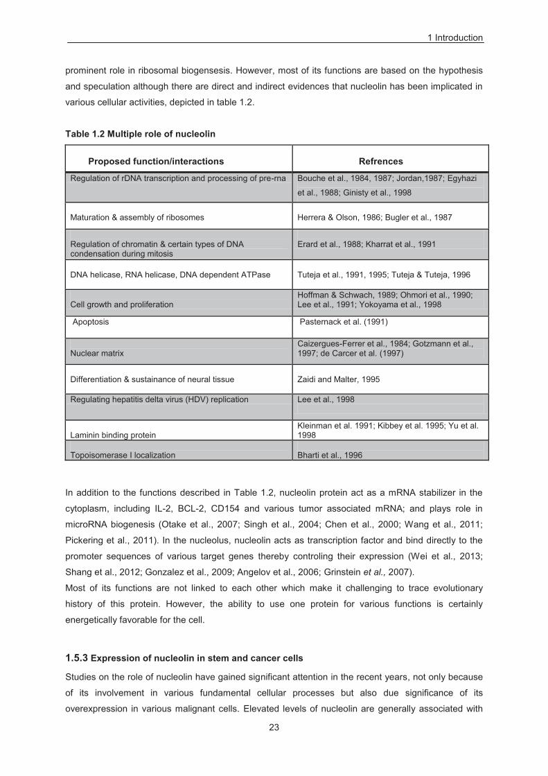
In addition to the functions described in Table 1.2, nucleolin protein act as a mRNA stabilizer in the
cytoplasm, including IL-2, BCL-2, CD154 and various tumor associated mRNA; and plays role in
microRNA biogenesis (Otake et al., 2007; Singh et al., 2004; Chen et al., 2000; Wang et al., 2011;
Pickering et al., 2011). In the nucleolus, nucleolin acts as transcription factor and bind directly to the
promoter sequences of various target genes thereby controling their expression (Wei et al., 2013;
Shang et al., 2012; Gonzalez et al., 2009; Angelov et al., 2006; Grinstein et al., 2007).
Most of its functions are not linked to each other which make it challenging to trace evolutionary
history of this protein. However, the ability to use one protein for various functions is certainly
energetically favorable for the cell.
1.5.3 Expression of nucleolin in stem and cancer cells
Studies on the role of nucleolin have gained significant attention in the recent years, not only because
of its involvement in various fundamental cellular processes but also due significance of its
overexpression in various malignant cells. Elevated levels of nucleolin are generally associated with
1 Introduction
24
the cell proliferation. In all nucleolar proteins (silver stained), nucleolin is one of the major proteins
which can lead to the poor prognosis in various cancers (Derenzini, 2000). Flow cytometry and
confocal microscopy studies have revealed that at least 90% of human CLL cells express nucleolin in
their cytoplasm and on the cell surface when compared to CD19+ B-cells (5%) from the healthy
volunteers (Otake et al., 2007; Soundararajan et al., 2008). Elevated levels of nucleolin stabilize BCL-
2 mRNA in the leukemic cells, including human chronic lymphocytic leukemia (CLL) and HL-60 cells
(Sengupta et al., 2004; Otake et al., 2005, 2007). In another study, apoptosis of U937 leukemia cells is
accompanied by differential levels and localization of nucleolin (Mi et al., 2002). Furthermore,
endothelial cells incubated with anti nucleolin antibody lead to the down regulation of BCL-2 mRNA
and induce apoptosis (Fogal et al., 2009). The stabilization of bcl-2 mRNA through nucleolin, allows
cells to overproduce BCL-2 protein and thus protecting them from apoptosis.
Since the first report of expression of nucleolin on the surface of hepatocarcinoma cells (Semenkovich
et al., 1990), emerging evidence suggest a role of surface nucleolin in angiogenesis, tumour cell
growth and proliferation. Nucleolin is found to be expressed on the surface of various types of tumour
cells (Hovanessian et al., 2000; Sinclair and O’Brien, 2002; Otake et al., 2007; Chen et al., 2008;
Soundararajan et al., 2008). Elevated levels of nucleolin are reported both in vito and in vivo on
activated lymphocytes in the lypmphoid organs and angiogenic endothelial cells in the tumour
vasculature (Hovanessian et al., 2000; Krust et al., 2001; Christian et al., 2003; Destouches et al.,
2008). Surface nucleolin could bind and shuttle several ligands and thus act as mediator of
extracellular regulation for nuclear activities. Interestingly, the majority of these ligands which interact
with surface nucleolin play an important role in angiogenesis and tumorigenesis. For example, growth
factors, pleiotropin and midkines can transform cells, and exert mitogenic and angiogenic effect on
endothelial cells (Kadomatsu et al., 2004; Perez-Penira et al., 2008). In addition, several surface
nucleolin binding proteins, such as factor J, L & P selectins, laminin 1 and hepatocyte growth factor
(HGF) are implicated in the tumor development, angiogenesis, cell differentiation and cell adhesion
(Kleinmann et al., 1991; Turck et al., 2006; Larrucea et al., 1998; Harms et al., 2006; Reyes-Reyes et
al., 2008; Tate et al., 2006). Although a high expression of nucleolin has been reported in several
malignancies including leukemia, but still the mechanism underlying induction of leukemogenesis and
aiding tumor growth is still not well understood.
1.6 Wnt signaling
1.6.1 Introduction
Wnt proteins belong to a family of secreted signal transduction molecules, which are expressed in
diverse tissue types and have been involved in the invertebrate and vertebrate development (Cadigan
and Nusse, 1997). Wnt signaling regulates wide variety of cellular processes including motility,
polarity, organogenesis, cell fate determination, primary axis formation, stem cell renewal and also
plays a crucial role in the embryogenesis, therefore control of this pathway is tightly regulated. In
addition to its key role in the normal development of an organism, mutation in the Wnt gene can result
into defects in axis, limbs, and somite formation and abnormal development of kidney, reproductive
1 Introduction
25
tract and brain (Parr and Mcmohan, 1994; Monkley et al., 1996; Yoshikawa et al., 1997; Miller and
Sasoon, 1998; Liu et al., 1999). Dysregulation of Wnt signaling can have significant oncogenic effects
on the tissue, including breast, prostate, skin and colon (Korinek et al., 1997; Morin et al., 1997;
Tsukamoto et al., 1998; Polakis et al., 2000).
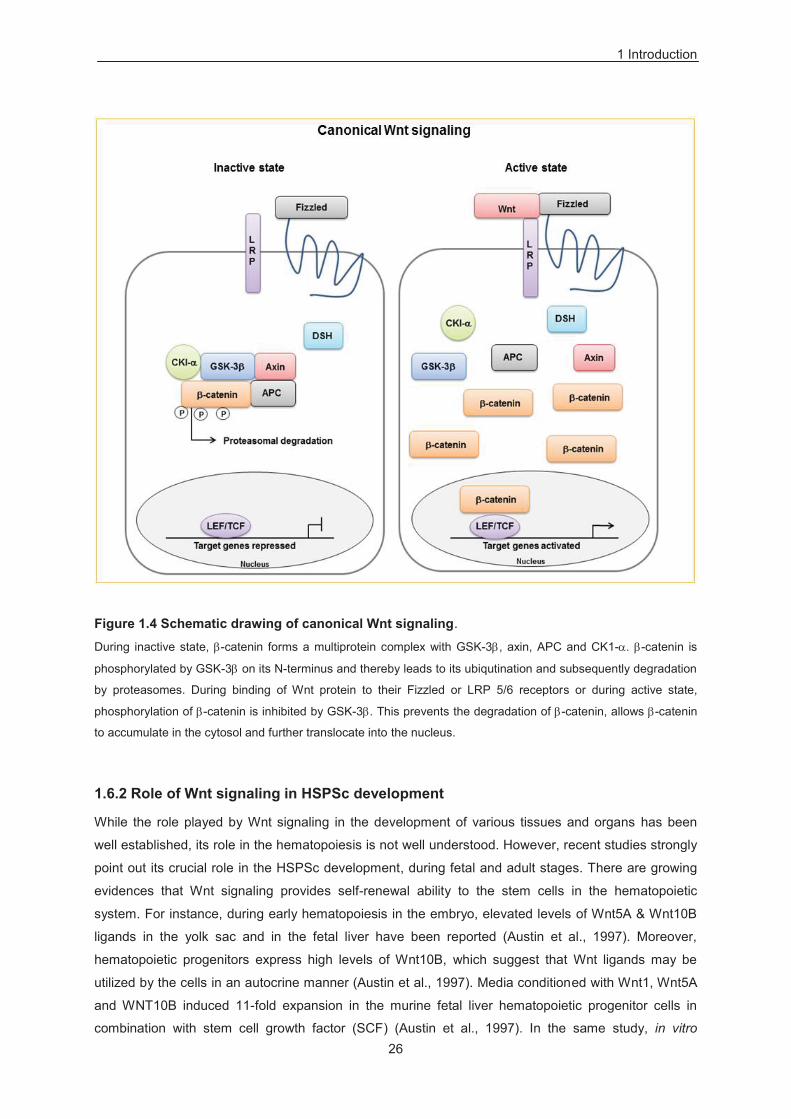
Wnt signaling is broadly divided in two categories: Canonical Wnt signaling or beta/ -catenin
dependent pathway and non-canonical pathway/ -catenin independent pathway. Wnt signaling
pathway is regulated by binding of Wnt protein / ligands to two types of receptor molecules expressed
on the cell surface: these two types of receptor molecules are categorized into the Fizzled family (Fz)
of seven pass transmembrane protein family, which comprises a cysteine rich extracellular domain
(Wodarz and Nusse, 1998) and low density lipoprotein receptor protein (LRP) family where especially
LRP5 and LRP6 of this family harbours only a single pass transmembrane protein (Pinson et al., 2000;
Tamai et al., 2000; Werli et al., 2000) (Figure 1.4). Fizzled and LRP 5/6 recepetors are required to
activate downstream components of the canonical Wnt signaling pathway. During its inactive state, -
catenin form multiprotein complex with glycogen synthase kinase-3 beta (GSK-3 ), axin,
adenomatosis polyposis coli (APC) and casein kinase1- . -catenin is phosphorylated by GSK-3 on
its N-terminus and thereby leads to its ubiqutination and subsequently degradation by proteasomes
(Cadigan and Nusse, 1997) (Figure 1.4). Axin is a key member of this multiprotein complex and it acts
as a scaffold and thereby significantly enhances the ability of GSK-3 to phosphorylate -catenin.
During binding of Wnt protein to their Fizzled or LRP 5/6 receptor or during active state,
phosphorylation of -catenin is inhbited by GSK-3 . This prevents the degradation of -catenin, which
allows -catenin to accumulate in the cytosol and further translocate into the nucleus (Figure 1.4). In
the nucleus, -catenin binds to LEF/ T-cell receptor family (TCF) proteins and thereby regulates the
cellular response through targeted expression of appropriate genes (Eastman and Grosschedl, 1991)
(Figure 1.4).
1 Introduction
26
Figure 1.4 Schematic drawing of canonical Wnt signaling.
During inactive state, -catenin forms a multiprotein complex with GSK-3 , axin, APC and CK1- . -catenin is
phosphorylated by GSK-3 on its N-terminus and thereby leads to its ubiqutination and subsequently degradation
by proteasomes. During binding of Wnt protein to their Fizzled or LRP 5/6 receptors or during active state,
phosphorylation of -catenin is inhibited by GSK-3 . This prevents the degradation of -catenin, allows -catenin
to accumulate in the cytosol and further translocate into the nucleus.
1.6.2 Role of Wnt signaling in HSPSc development
While the role played by Wnt signaling in the development of various tissues and organs has been
well established, its role in the hematopoiesis is not well understood. However, recent studies strongly
point out its crucial role in the HSPSc development, during fetal and adult stages. There are growing
evidences that Wnt signaling provides self-renewal ability to the stem cells in the hematopoietic
system. For instance, during early hematopoiesis in the embryo, elevated levels of Wnt5A & Wnt10B
ligands in the yolk sac and in the fetal liver have been reported (Austin et al., 1997). Moreover,
hematopoietic progenitors express high levels of Wnt10B, which suggest that Wnt ligands may be
utilized by the cells in an autocrine manner (Austin et al., 1997). Media conditioned with Wnt1, Wnt5A
and WNT10B induced 11-fold expansion in the murine fetal liver hematopoietic progenitor cells in
combination with stem cell growth factor (SCF) (Austin et al., 1997). In the same study, in vitro
1 Introduction
27
analysis have also shown increased colony forming ability and especially given blast colonies, which
suggest an immature nature of these cells proliferated due to Wnt induction. These effects of soluble
Wnt ligands were also consistently reported during human hematopoiesis (Van Den Berg et al., 1998).
When human CD34+ lin– HSPSc growing on the stromal cells have been exposed to Wnt5A,
expansion of an undifferentiated progenitor population was noticed which was determined by
production of 10 to 20 fold more colony forming units (CFU) in ex vivo colony forming assay (Van Den
Berg et al., 1998). Moreover, human HSCs overexpressing WNT5A provided three fold increased
engraftment in the mice (Murdoch et al., 2003). Furthermore, overexpression of active -catenin in
murine HSCs led to the expansion of undifferentiated stem cell pool for long-term in vitro and also
allowed constant reconstitution of myeloid and lymphoid lineages in vivo (Reya et al., 2003). In the
same report, authors were able to show the activation of a LEF/TCF reporter in HSCs progenitor
population in their natural microenvironment, suggests that the Wnt pathway can also be induced in
vivo. Independently, increased proliferation of a multipotent progenitor population was observed in
vivo upon constitutive expression -catenin (Baba et al., 2006). Long-term reconstitution and better
recovery of neutrophils and megakaryocytes was observed when mouse or human HSCs transplanted
to recipient mice were administered with GSK-3 inhibitor (Trowbridge et al., 2006b).
1.6.3 Wnt signaling in leukemia
Role of Wnt signaling in the development of leukemia has been well established over the last couple
of years. Although the mechanism underlying the induction of leukemia is rather unclear, mutations
resulting in the elevated expression of Wnt genes or mutations in the key molecules of Wnt cascade
appear to be crucial. Epigenetic changes in the Wnt signaling molecules have been studied, but still its
functional impact on the Wnt cascade and development of leukemia is not well understood (Roman-
Gomez et al., 2007; Bennett et al., 2010). For instance, differential methylation pattern of Wnt
inhibitors have prognostic implications in the case of AML (Valencia et al., 2009). Furthermore,
inactivation by hypermethylation in the promoter region of secreted fizzled related protein genes
(sFRPs) have been reported in the case of acute lymphoblastic leukemia (ALL) and AML (Jost et al.,
2008). In chronic myeloid leukemia (CML) having the translocation t (9, 22), activation of Wnt signaling
was generally obsereved during blast crisis (Jamieson et al., 2004), but the exact reasons for this
abberant induction of Wnt signaling are predominantly unknown.
In a mouse model, deletion of -catenin significantly abolishes the CML development (Zhao et al.,
2007). Moreover, in the case of drug resistant BCR-ABL+ CML cells, -catenin plays a crucial role in
the survival of these leukemic populations (Hu et al., 2009). There are reports categorizing AML on the
basis of dysregulated Wnt activation (Muller-Tidow et al., 2004; Wang et al., 2010), suggesting that
Wnt signaling could provide stem cell characteristics to the leukemic stem cells (LSCs) (Lane et al.,
2011). Therefore, it could be possible that likewise during early development of HSCs in the embryo,
LSCs may require higher activation of Wnt signaling as compared to normal HSCs in the bone
marrow. In the case of T-acute lymphoblastic leukemia (T-ALL), constitutive expression of activated -
catenin in the mouse model under the control of a thymus promoter leads to the development of
thymic lymphoma (Guo et al., 2007). However, this work needs to be extended to a cohort of patients
1 Introduction
28
and rigorous assessment is required to find out whether dysregulated Wnt signaling play significant
role in the human leukemia as well (Weerkamp et al., 2006).
In summary, these studies suggest that Wnt signaling is implicated in providing stem cell
characteristics to the LSCs (Lane et al., 2011). Therefore specific targeting of impaired Wnt signaling
could be useful way to target LSCs, unlike conventional chemotherapies rather targeting bulk of
malignant cells than leukemia initiating cells.
1.7 PI3K / Akt signaling
1.7.1 Introduction
Cells can sense whether the conditions are favorable for their proliferation and growth by inducing
signaling pathways in response to interactions between ligands and their respective receptors. One
such category of interactions involves binding of the growth factors to their respective receptor
tyrosine kinase, which in turn directly interacts and activates a lipid kinase, phophotidylinositide-3-
kinase (PI3K) (Schlessinger, 2000). PI3 kinases belong to a family of enzymes, which function as an
intracellular signal transducer involved in many cellular responses, including proliferation, cell growth,
motility, intracellular trafficking and survival through the activation of several downstream pathways
(Yuan and Cantley, 2008). In addition to the growth factors, the PI3 kinase can also be activated by
insulin receptor substrate-1 (IRS-1) and thereby plays a crucial role in regulating glucose intake by
cells through a series of phosphorylation events. The PI3K family can be categorized into three
different classes, on the basis of their lipid substrate, primary structure and regulation: Class I, Class II
& Class III. PI3K belongs to class I which acts by converting phosphatidylinositol-4,5-bisphosphate
(PIP2) into phosphatidylinositol-3,4,5-trisphosphate (PIP3), which further interacts with downstream
proteins having pleckstrin-homology domains, such as protein kinase B (PKB) / Akt and protein
dependent kinase 1 (PDK1) (Yuan and Cantley, 2008). These direct interactions of PIP3 produced by
PI3K lead to the recruitment of Akt to the plasma membrane, which later induces the activation of Akt
through phosphorylation on its Threonine-308 residue by PDK1 (Yuan and Cantley, 2008).
Since the initial discovery of Akt as a proto-oncogene, attention is focused on its role in various cellular
mechanisms. Akt belongs to the family of serine / threonine kinases and regulates key cellular
functions, including cellular metabolism, proliferation and survival, cell migration and apoptosis
(Manning and Cantley, 2007). The Akt signaling pathway can be activated by stimuli inducing PI3K via
PIP3, B- and T-cell receptors, receptor tyrosine kinases and G protein coupled receptors. There are
three structurally related isoforms of Akt, Akt 1, Akt 2, and Akt 3. Akt act on the cell growth through
regulating the mammalian target of rapamycin (mTOR) and TSC 1/TSC 2 complexes (Tang et al.,
1999; Inoki et al., 2002; Bhaskar and Hay, 2007). Furtermore, Akt can acts on the cell proliferation and
cell cycle by directly modulating the expression of cyclin dependent kinase (CDK) inhibitors p27& p21,
and indirectly by the expression of p53 & cyclin D1 (Manning and Cantley, 2007). Akt also acts as a
significant mediator of cell survival through its direct inhibiton of pro-apoptotic regulators, such as
Foxo, Myc and Bad. The tumor supressor protein phosphatase and tensin homologue (PTEN) was
found to be a major inhibitor of PI3K / Akt pathway which is frequently absent in tumor cells (Salmena
1 Introduction
29
et al., 2008). Deletion of PTEN results in the overactivation of Akt that can induce proliferation, growth
and better survival of the cells through inhibiting apoptosis (Datta et al., 1997; Salmena et al., 2008).
1.7.2 PI3K / Akt signaling in HSPSc
Akt signaling has been implicated in various cellular processes; but its role in regulating HSCs is still
not well understood. However, among all signal transduction pathways, PI3K / Akt signaling has
recently attracted major attention as possibly being involved in the self-renewal of HSCs. In general,
PI3K activity regulates erythropoiesis and helps in the survival and proliferation of erythroid
progenitors (Myklebust et al., 2002; Bouscary et al., 2003). Available evidences suggest that Akt
signaling regulates the determination of lineage during myleopoiesis. Differently from the studies
mentioned above, a recent study reported that the simultaneous activation of Wnt / -catenin and
PTEN / PI3K / Akt promotes primitive HSCs population with increase self-renewal and proliferation
capacity (Perry et al., 2011). However the activation of either of these pathways is insufficient to
sustain primitive HSCs population (Perry et al., 2011). Furthermore, HSCs with reduced PI3K activity
exhibit impaired hematopoietic reconstitution ability and reduced proliferation (Haneline et al., 2006).
1.7.3 PI3K / Akt signaling in leukemia
Correlation between dysregulated PI3K signaling and cancer is well documented. PI3K activity is
associated to several human tumours, including lung cancer, breast cancer, leukemia, and melanoma
among others (Krasilnikov et al., 1999; Fry et al., 2001; Martinez-Lorenzo et al., 2000). Moreover the
PI3K / Akt signaling pathway is frequently induced in patients with AML (Tamburini et al., 2007; Xu et
al., 2003). Around 50-80% AML patient samples exhibits Akt phosphorylation at Ser473 residues in
their purified blast cells and around 50% de novo AML patient samples have constitutively active
PI3K / Akt (Tamburini et al., 2007; Xu et al., 2003; Grandage et al., 2005). However, the mechanism
underlying the activation of Akt signaling during AML is not clear. Constitutive active AKT 1 has been
found in cancers resulted from mutation in its PIH domain (Greenman et al., 2007). Dysregulated
activity of the Akt mutant is due to PIP2 and PIP3 independent recruitment of Akt to the membrane,
which can result in the activation of the downstream signaling cascade and induce leukemia in mice
(Carpten at al., 2007). There are studies showing that patients with high PI3K activity have poor
prognosis compared to the patients with low PI3K activity (Min et al., 2003; Kornblau et al., 2006). In
addition, it was reported that PI3K / Akt signaling regulates clonogenicity of leukemic cells and
proliferation of the blast cells (Sujobert et al., 2005; Xu et al., 2003). The PI3K / Akt / PTEN signaling
pathway also plays a significant role in regulating angiogenesis, mediated by vascular endothelial
growth factor (VEGF) in many tumors including leukemia (Naoko et al., 2012). Furthermore, mutated
p85, a subunit of PI3K is reported to be expressed in CO, a Hodgkin’s lymphoma derived cell line
(Jucker et al., 2002). In a different study, PTEN knockout mice die due to leukemia or
myeloproliferative disorder (MPD), transplantable T-ALL and AML (Yilmaz et al., 2006; Zhang et al.,
2006; Guo et al., 2008).
30
1.8 Aims of the PhD thesis
The former introductory sections illustrate the role of several molecular factors involved in the
regulation of HSPCs and in the case of leukemia. AC133 is a specific glycoform of CD133 that marks
the uncommitted CD34+ HSPCs and CD34+ GM progenitors (Yin et al., 1997). Human AC133+ HSPCs
are enriched for colony-forming units, LTC-ICs and are capable of hematopoietic reconstitution.
AC133 is used as marker for the prospective isolation of clinical relevant cell populations in case of
AML and ALL AML (Cox et al., 2009; Medina et al., 2014; Beghini et al., 2012). However, the
mechanism regulating the expression AC133 and CD133 is not clearly understood. Thus, AC133 an
interesting candidate to investigate mechanism implicated in its regulation, and its related potential
effects on the proliferation and differentiation of early HSPCs. Recently, a direct link of CD133
expression with the activation of Wnt / -catenin and PI3K / Akt signaling was shown (Mak et al., 2012;
Wei et al., 2013). Wnt signaling strength regulates normal haematopoiesis, and its deregulation is
involved in the leukemia development. In our previous study, it came out that nucleolin acts as a CD34
promoter factor, and is enriched in the mobilized peripheral blood (MPB) derived undifferentiated
CD34+ HSPCs as opposed to differentiated CD34- cells (Grinstein et al., 2007). Nucleolin is a
multifunctional nucleolar phosphoprotein, overexpressed in actively growing and cancer cells, involved
in transcriptional regulation, chromatin remodeling, and RNA metabolism. Abberant activity of
nucleolin is generally associated with several haematological malignancies.
Working out mechanisms controlling AC133 expression and factors influencing functional properties of
HSPCs is important to understand homeostasis in normal and dysregulated hematopoietic tissues at
molecular level.
Therefore, the first aim of this work is to investigate whether nucleolin regulate the expression
of AC133 and CD133 both in human MPB derived HSPCs, and in leukemic cell line model.
In order to gain more insights into functional effects of nucleolin in HSPCs and in a leukemic
cell line model, the second aim is to look for the impact of nucleolin on its proliferative capacity
by assaying functional properties like colony forming ability and effects on lineage
commitment.
Following, the third aim is to examine whether in HSPCs, nucleolin affects the number of long-
term culture initiating cells (LTC-IC) and supports their long-term maintenance on stroma free
culture.
Final aim is to look for a mechanism implicated in nucleolin-dependent functional properties to
HSPCs.
31
2 Materials
2.1 Patients
The patient samples were provided by the Children’s University Hospital Duesseldorf under the
direction of Prof. Dr. Roland Meisel. Leukapheresis samples used for HSPCs isolation were
specimens from deceased patients with nonhematologic cancer, which were designated to be
discarded. Donors' informed consent was taken, in agreement with our Faculty Ethic Committee. The
agreement for the molecular characterization of these samples was acquired in the scope of previous
therapy protocols. All personal data were encrypted and concealed for privacy reasons.
2.2 Human, adherent cell lines
Cell line: 5637
DSMZ no.: ACC 35 (DSMZ Braunschweig, Germany)
Species: human
Cell type: urinary bladder carcinoma
Morphology: epithelial-like adherent cells
Medium: 90% RPMI 1640 + 10% h.i. FBS
Subculture: split confluent cultures 1:4 to 1:5 using trypsin/EDTA); seed out at ca. 2 x 10⁶
cells/80 cm2
Incubation: at 37 °C with 5% CO2
Doubling time: ca.24 hours
References: Fogh et al., 1974; Welte et al., 1985; Kaashoek et al., 1991; Quentmeier et al.,
1997
Cell line: 293T
DSMZ no.: ACC 635 (DSMZ Braunschweig, Germany)
Species: human
Cell type: embryonal kidney
Morphology: fibroblastoid cells growing adherently as monolayer
Medium: 90% Dulbecco´s MEM + 10% h.i. FBS
2 Materials
32
Culture: seed out at ca. 2-3 x 10⁶ cells/80 cm2; split ratioo 1:4 - 1:5 every 2-3 days
Incubation: at 37 °C with 5% CO2
Doubling time: ca.24-30 hours
References: Rio et al., 1985; DuBridge et al., 1987; Pear et al., 1997
Cell line: M2-10B4
Catalogue no.: ATCC® CRL-1972™ (CLS, Eppelheim, Germany)
Species: mouse
Cell type: bone marrow/stroma
Morphology: fibroblast, adherent
Medium: RPMI-1640 + 10% FBS
Subculture: Subcultivation ratio 1:6 to 1:10, medium renewal every 2-3 days
Incubation: at 37 °C with 5% CO2
References: Lemoine et al., 1988, 1990; Sutherland et al., 1991
2.3 Human, suspension cell lines
Cell line: MUTZ-2
DSMZ no.: ACC 271 (DSMZ Braunschweig, Germany)
Species: human
Cell type: acute myeloid leukemia
Morphology: single cells in suspension, growing partly in clumps
Medium: 60% alpha-MEM + 20% h.i. FBS + 20% vol conditioned medium of cell line
5637 (DSM ACC 35) (cells grow well with 50 ng/ml SCF)
Subculture: optimal split ratio of 1:2 to 1:3 twice a week; seed out at about 0.5 x 10⁶
cells/ml; cells should be grown in 24-well-plates.
Incubation: at 37 °C with 5% CO2
Doubling time: about 47 hours
References: Hu et al., 1996; Drexler et al., 1997
Cell line: SEM
DSMZ no.: ACC 546 (DSMZ Braunschweig, Germany)
Species: human
Cell type: B cell precursor leukemia
Morphology: round to polygonal single rather small cells in suspension
Medium: 90% Iscove's MDM + 10% h.i. FBS
Subculture: split saturated culture 1:2 to 1:6 every 2-3 days; seed out at ca. 0.5 x 10⁶
cells/ml; maintain at ca. 0.2-2.0 x 10⁶ cells/ml; maximum density at ca. 3-4 x
106 cells/ml
Incubation: at 37 °C with 5% CO2
2 Materials
33
Doubling time: ca. 30 hours
Harvest: cell harvest of ca. 3-4 x 10⁶ cells/ml
Storage: frozen with 70% medium, 20% FBS, 10% DMSO at about 9 x 10⁶
cells/ampoule
References Reichel M, 1998; Drexler, H.G, 2004;
2.4 Chemicals
Table 2.1 General chemicals
Identifier Company Ordernumber Bis-Acrylamide 30% (19:1) Bio-Rad, München, Germany 161-0154
Agarose Biozym, Hessisch Odendorf, Germany 840004
Ammonium Persulfate Sigma-Aldrich, St. Louis, MO, USA A-3678
Albumin fraction V (BSA) Roth, Karlsruhe, Germany 8076.3
Chloroform 99.4% Merck, Darmstadt, Germany 67-66-3
DMSO (Dimethylsulfoxide) Sigma-Aldrich, St. Louis, MO, USA D2650
EDTA (Ethylenediamine-tetraacetic acid) Sigma-Aldrich, St. Louis, MO, USA 60-00-4
Ethanol Merck, Darmstadt, Germany 1.00983.2511
Ethidiumbromide Solution Sigma-Aldrich, St. Louis, MO, USA E1510-10ML
Glycerine Merck, Darmstadt, Germany 1.04094.2500
HCl (Hydrochloric acid) Merck, Darmstadt, Germany 1.09911.0001
KCl (Potassium chloride) Merck, Darmstadt, Germany 1.04938.
KOH (Potassium hydroxide) Merck, Darmstadt, Germany 105012
Magnesium (Mg2) Merck, Darmstadt, Germany 105815
Mg2Cl Merck, Darmstadt, Germany 1.05833.0250
NaCl (Sodium chloride) Merck, Darmstadt, Germany 1.06404.1000
NaF (Sodium fluoride) AppliChem, Darmstadt, Germany A3904.0500
NaOH (Sodiumhydroxide) Merck, Darmstadt, Germany 1.06498.100
SDS 20% Ambion, Huntingdon, UK AM9820
50 x TAE (Tris/Acetic Acid/EDTA) Buffer Bio-Rad, München, Germany 161-0743
TEMED (Tetramethylethylendiamin) Merck, Darmstadt, Germany 1.10732.0100
Tris Roth, Karlsruhe, Germany 5429.3
Tween 20 Roth, Karlsruhe, Germany 9127,1
β-Mercaptoethanol Merck, Darmstadt, Germany 15433.0100
Ponceau S Solution Sigma-Aldrich, St. Louis, MO, USA P-7170
Na2EDTA Sigma-Aldrich, St. Louis, MO, USA E5134-500G
Nonidet P40 Substitute Fluka Biochemica 74385
Na2HPO4 Merck, Darmstadt, Germany 6580
2 Materials
34
2.5 Specific reagents/Media
Table 2.2 Specfific reagents and media
Reagents/Media Company Ordernumber Retronectin TAKARA Clontech, Saint-Germain-en-Laye France FRT100A
Solucortef STEMCELL Technologies, Vancouver, Canada 07904
Power SYBR Green ABI, Carlsbad, CA, USA 4368702
MACS LNGFR Microbeads Miltenyi,Gladbach, Germany 130-091-330
MACS Seperation Columns (LS) Miltenyi,Gladbach, Germany 130-042-4010
MACS Seperation Buffer Miltenyi,Gladbach, Germany 130-091-221
FcR Blocking Reagent, Human Miltenyi,Gladbach, Germany 130-059-901
GelPilot 1kb plus DNA ladder Qiagen, Hilden, Germany 239095
Page Ruler Prestained Protein Ladder Fermentas, St. Leon-Rot, Germany SM0671
Myelocult H5100 STEMCELL Technologies, Vancouver, Canada 05150
X-Vivo 20 w/Gentamycin Lonza, Cologne, Germany BE04-448QEA
Methocult H4100 STEMCELL Technologies, Vancouver, Canada 04100
DMEM Gibco, Invitrogen, Carlsbad, CA, USA 31966-021
RPMI-1640 Gibco, Invitrogen, Carlsbad, CA, USA 61870-010
-MEM Gibco, Invitrogen, Carlsbad, CA, USA 22571038
IMDM Sigma-Aldrich, St. Louis, MO, USA I3390
FBS Heat Inactivated Sigma-Aldrich, St. Louis, MO, USA F4135-500ML
L-Glutamine Gibco, Invitrogen, Carlsbad, CA, USA 25030-024
Dulbecco’s PBS Sigma-Aldrich, St. Louis, MO, USA D8537
Sodium pyruvate Gibco, Invitrogen, Carlsbad, CA, USA 11360-039
Trypsin-EDTA Gibco, Invitrogen, Carlsbad, CA, USA 25300-054
Pen Strep Gibco, Invitrogen, Carlsbad, CA, USA 15140-122
Puromycin Sigma-Aldrich, St. Louis, MO, USA P8833
DMSO Sigma-Aldrich, St. Louis, MO, USA D2650
Cercosporin Bioaustralis, NSW, Australia BIA-C1521
Caliphostin C, PKF11584 Tocris Biosciences, bristol, UK 1626
2.6 Restriction Enzymes
Table 2.3 Restriction enzymes
Name Company Ordernumber Ecor1 NEB, Frankfurt a. M., Germany R0101
Xba1 NEB, Frankfurt a. M., Germany R0154
Bamh1 NEB, Frankfurt a. M., Germany R0136
Sal1 NEB, Frankfurt a. M., Germany R0138
HindIII NEB, Frankfurt a. M., Germany R0104
Cla1 NEB, Frankfurt a. M., Germany R0197
BspEI NEB, Frankfurt a. M., Germany R0540
ACCI NEB, Frankfurt a. M., Germany R0161
Abbreviations: NEB - New England Biolabs, a.M. - am Main,
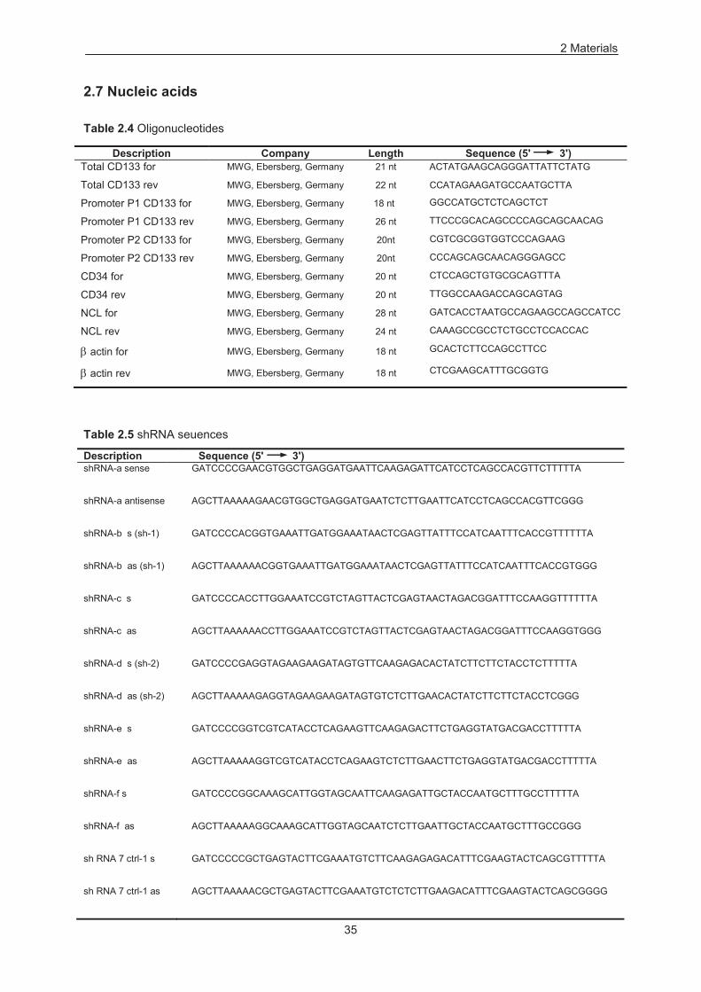
2 Materials
35
2.7 Nucleic acids
Table 2.4 Oligonucleotides
Description Company Length Sequence (5' 3') Total CD133 for MWG, Ebersberg, Germany 21 nt ACTATGAAGCAGGGATTATTCTATG
Total CD133 rev MWG, Ebersberg, Germany 22 nt CCATAGAAGATGCCAATGCTTA
Promoter P1 CD133 for MWG, Ebersberg, Germany 18 nt GGCCATGCTCTCAGCTCT
Promoter P1 CD133 rev MWG, Ebersberg, Germany 26 nt TTCCCGCACAGCCCCAGCAGCAACAG
Promoter P2 CD133 for MWG, Ebersberg, Germany 20nt CGTCGCGGTGGTCCCAGAAG
Promoter P2 CD133 rev MWG, Ebersberg, Germany 20nt CCCAGCAGCAACAGGGAGCC
CD34 for MWG, Ebersberg, Germany 20 nt CTCCAGCTGTGCGCAGTTTA
CD34 rev MWG, Ebersberg, Germany 20 nt TTGGCCAAGACCAGCAGTAG
NCL for MWG, Ebersberg, Germany 28 nt GATCACCTAATGCCAGAAGCCAGCCATCC
NCL rev MWG, Ebersberg, Germany 24 nt CAAAGCCGCCTCTGCCTCCACCAC
actin for MWG, Ebersberg, Germany 18 nt GCACTCTTCCAGCCTTCC
actin rev MWG, Ebersberg, Germany 18 nt CTCGAAGCATTTGCGGTG
Table 2.5 shRNA seuences
Description Sequence (5' 3') shRNA-a sense GATCCCCGAACGTGGCTGAGGATGAATTCAAGAGATTCATCCTCAGCCACGTTCTTTTTA
shRNA-a antisense AGCTTAAAAAGAACGTGGCTGAGGATGAATCTCTTGAATTCATCCTCAGCCACGTTCGGG
shRNA-b s (sh-1) GATCCCCACGGTGAAATTGATGGAAATAACTCGAGTTATTTCCATCAATTTCACCGTTTTTTA
shRNA-b as (sh-1) AGCTTAAAAAACGGTGAAATTGATGGAAATAACTCGAGTTATTTCCATCAATTTCACCGTGGG
shRNA-c s GATCCCCACCTTGGAAATCCGTCTAGTTACTCGAGTAACTAGACGGATTTCCAAGGTTTTTTA
shRNA-c as AGCTTAAAAAACCTTGGAAATCCGTCTAGTTACTCGAGTAACTAGACGGATTTCCAAGGTGGG
shRNA-d s (sh-2) GATCCCCGAGGTAGAAGAAGATAGTGTTCAAGAGACACTATCTTCTTCTACCTCTTTTTA
shRNA-d as (sh-2) AGCTTAAAAAGAGGTAGAAGAAGATAGTGTCTCTTGAACACTATCTTCTTCTACCTCGGG
shRNA-e s GATCCCCGGTCGTCATACCTCAGAAGTTCAAGAGACTTCTGAGGTATGACGACCTTTTTA
shRNA-e as AGCTTAAAAAGGTCGTCATACCTCAGAAGTCTCTTGAACTTCTGAGGTATGACGACCTTTTTA
shRNA-f s GATCCCCGGCAAAGCATTGGTAGCAATTCAAGAGATTGCTACCAATGCTTTGCCTTTTTA
shRNA-f as AGCTTAAAAAGGCAAAGCATTGGTAGCAATCTCTTGAATTGCTACCAATGCTTTGCCGGG
sh RNA 7 ctrl-1 s GATCCCCCGCTGAGTACTTCGAAATGTCTTCAAGAGAGACATTTCGAAGTACTCAGCGTTTTTA
sh RNA 7 ctrl-1 as AGCTTAAAAACGCTGAGTACTTCGAAATGTCTCTCTTGAAGACATTTCGAAGTACTCAGCGGGG
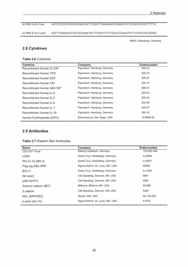
2 Materials
36
sh RNA 8 ctrl-2 sen GATCCCCCGTACGCGGAATACTTCGATTCAAGAGATCGAAGTATTCCGCGTACGTTTTTA
sh RNA 8 ctrl-2 asen AGCTTAAAAACGTACGCGGAATACTTCGATCTCTTGAATCGAAGTATTCCGCGTACGGGG
MWG, Ebersberg, Germany
2.8 Cytokines
Table 2.6 Cytokines
Cytokine Company Ordernumber Recombinant Human G-CSF Peprotech, Hamburg, Germany 300-23
Recombinant Human TPO Peprotech, Hamburg, Germany 300-18
Recombinant Human SCF Peprotech, Hamburg, Germany 300-07
Recombinant Human Flt3 Peprotech, Hamburg, Germany 300-19
Recombinant Human GM-CSF Peprotech, Hamburg, Germany 300-03
Recombinant Human IL-3 Peprotech, Hamburg, Germany 200-03
Recombinant Human IL-2 Peprotech, Hamburg, Germany 200-02
Recombinant Human IL-6 Peprotech, Hamburg, Germany 200-06
Recombinant Human IL-7 Peprotech, Hamburg, Germany 200-07
Recombinant Human IL-15 Peprotech, Hamburg, Germany 200-15
Human Erythropoietin (EPO) Ebiosciences, San Diego, USA 14-8992-80
2.9 Antibodies
Table 2.7 Western Blot Antibodies
Name Company Ordernumber CD133/1 Pure Miltenyi,Gladbach, Germany 130-092-395
CD34 Santa Cruz, Heidelberg, Germany sc-9095
NCL/C-23 (MS-3) Santa Cruz, Heidelberg, Germany sc-8031
Flag tag (M2) HRP Sigma-Aldrich, St. Louis, MO, USA A8592
BCL-2 Santa Cruz, Heidelberg, Germany sc-7382
Akt (pan) Cell Signaling, Danvers, MA, USA 4691
pAkt Ser473 Cell Signaling, Danvers, MA, USA 4060
Active- -catenin (8E7) Millipore, Billerica, MA, USA 05-665
-catenin Cell Signaling, Danvers, MA, USA 8408
NCL (EPR7952) Abcam, MA, USA Ab-129-200
-actin (AC-74) Sigma-Aldrich, St. Louis, MO, USA A1978
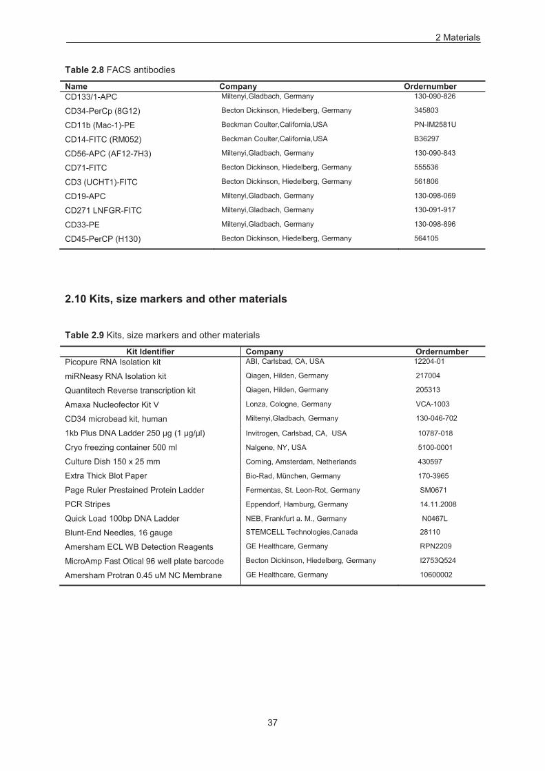
2 Materials
37
Table 2.8 FACS antibodies
Name Company Ordernumber CD133/1-APC Miltenyi,Gladbach, Germany 130-090-826
CD34-PerCp (8G12) Becton Dickinson, Hiedelberg, Germany 345803
CD11b (Mac-1)-PE Beckman Coulter,California,USA PN-IM2581U
CD14-FITC (RM052) Beckman Coulter,California,USA B36297
CD56-APC (AF12-7H3) Miltenyi,Gladbach, Germany 130-090-843
CD71-FITC Becton Dickinson, Hiedelberg, Germany 555536
CD3 (UCHT1)-FITC Becton Dickinson, Hiedelberg, Germany 561806
CD19-APC Miltenyi,Gladbach, Germany 130-098-069
CD271 LNFGR-FITC Miltenyi,Gladbach, Germany 130-091-917
CD33-PE Miltenyi,Gladbach, Germany 130-098-896
CD45-PerCP (H130) Becton Dickinson, Hiedelberg, Germany 564105
2.10 Kits, size markers and other materials
Table 2.9 Kits, size markers and other materials
Kit Identifier Company Ordernumber Picopure RNA Isolation kit ABI, Carlsbad, CA, USA 12204-01
miRNeasy RNA Isolation kit Qiagen, Hilden, Germany 217004
Quantitech Reverse transcription kit Qiagen, Hilden, Germany 205313
Amaxa Nucleofector Kit V Lonza, Cologne, Germany VCA-1003
CD34 microbead kit, human Miltenyi,Gladbach, Germany 130-046-702
1kb Plus DNA Ladder 250 μg (1 μg/μl) Invitrogen, Carlsbad, CA, USA 10787-018
Cryo freezing container 500 ml Nalgene, NY, USA 5100-0001
Culture Dish 150 x 25 mm Corning, Amsterdam, Netherlands 430597
Extra Thick Blot Paper Bio-Rad, München, Germany 170-3965
Page Ruler Prestained Protein Ladder Fermentas, St. Leon-Rot, Germany SM0671
PCR Stripes Eppendorf, Hamburg, Germany 14.11.2008
Quick Load 100bp DNA Ladder NEB, Frankfurt a. M., Germany N0467L
Blunt-End Needles, 16 gauge STEMCELL Technologies,Canada 28110
Amersham ECL WB Detection Reagents GE Healthcare, Germany RPN2209
MicroAmp Fast Otical 96 well plate barcode Becton Dickinson, Hiedelberg, Germany I2753Q524
Amersham Protran 0.45 uM NC Membrane GE Healthcare, Germany 10600002
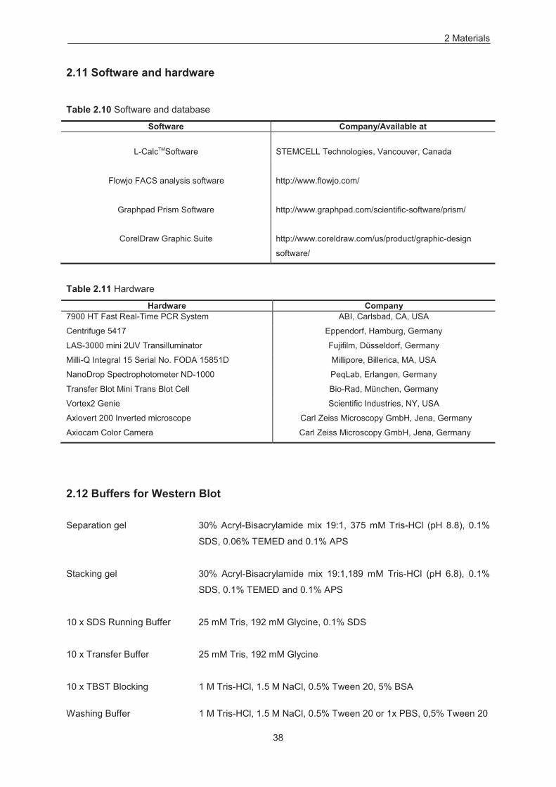
2 Materials
38
2.11 Software and hardware
Table 2.10 Software and database
Software Company/Available at
L-CalcTMSoftware
STEMCELL Technologies, Vancouver, Canada
Flowjo FACS analysis software
Graphpad Prism Software
http://www.flowjo.com/
http://www.graphpad.com/scientific-software/prism/
CorelDraw Graphic Suite
http://www.coreldraw.com/us/product/graphic-design
software/
Table 2.11 Hardware
Hardware Company 7900 HT Fast Real-Time PCR System ABI, Carlsbad, CA, USA
Centrifuge 5417 Eppendorf, Hamburg, Germany
LAS-3000 mini 2UV Transilluminator Fujifilm, Düsseldorf, Germany
Milli-Q Integral 15 Serial No. FODA 15851D Millipore, Billerica, MA, USA
NanoDrop Spectrophotometer ND-1000 PeqLab, Erlangen, Germany
Transfer Blot Mini Trans Blot Cell Bio-Rad, München, Germany
Vortex2 Genie Scientific Industries, NY, USA
Axiovert 200 Inverted microscope Carl Zeiss Microscopy GmbH, Jena, Germany
Axiocam Color Camera Carl Zeiss Microscopy GmbH, Jena, Germany
2.12 Buffers for Western Blot
Separation gel 30% Acryl-Bisacrylamide mix 19:1, 375 mM Tris-HCl (pH 8.8), 0.1%
SDS, 0.06% TEMED and 0.1% APS
Stacking gel 30% Acryl-Bisacrylamide mix 19:1,189 mM Tris-HCl (pH 6.8), 0.1%
SDS, 0.1% TEMED and 0.1% APS
10 x SDS Running Buffer 25 mM Tris, 192 mM Glycine, 0.1% SDS
10 x Transfer Buffer 25 mM Tris, 192 mM Glycine
10 x TBST Blocking 1 M Tris-HCl, 1.5 M NaCl, 0.5% Tween 20, 5% BSA
Washing Buffer 1 M Tris-HCl, 1.5 M NaCl, 0.5% Tween 20 or 1x PBS, 0,5% Tween 20

39
3 Methods
3.1 Cell cultivation
The handling of mobilized peripheral blood (MPB) derived human HSCs and cell lines were carried out
under sterile conditions. The rules for dealing with “Genetically Modified Organisms” (GMO) were
followed as prescribed. Biological waste and working equipment were autoclaved.
3.1.1 Isolation and culture of human CD34+ Stem & Progenitor Cells (HSPSc)
Human CD34+ cells were isolated from aliquots of leukapheresis samples, using either direct Isolex
300i HSC concentration system (Nexell) or indirectly using immunomagnetic technique from
mononuclear cells (MNC) fraction (Miltenyi Biotech). Purity of cell preparations employed for
experiments were monitored by flow cytometry (FACS) and was always >90% CD34+ cells. After
isolation, HSPCs were stimulated prior to transduction for 24 h in X-vivo 20 medium containing human
recombinant cytokines: 100 ng/ml stem cell factor (SCF), 100 ng/ml thrombopoietin (TPO), 100 ng/ml
fms-like tyrosine kinase 3 ligand (Flt-3) and 20 ng/ml interleukin (IL-3) in 24 well plates at a
concentration of 2 x10⁶ cells/ml. The resulted stimulated cells were washed after 24h and grown in X-
vivo medium containing 50 ng/ml SCF, 50 ng/ml Flt-3, 10ng/ml TPO, and 10 ng/ml IL-6. HSPCs were
also monitored for CD133 expression (~85% AC133+) on their surface by FACS in addition to the
CD34 expression prior to transduction.
3.1.2 Cultivation of human cell line
Mutz-2 (Hu et al., 1996), a human acute myeloid leukemia cell line, and was cultured in alpha-MEM
medium supplemented with 20% fetal calf serum (FCS), 20% conditioned medium from 5637 cells,
1% 200 mM L-Glutamine, 1% sodium pyruvate, 1% Pen Strep. Mutz-2 cells were grown in 12 well
plate and at seeding density of 1 x 10⁶ cells/ml. SEM, a human acute lymphoblastic leukemia (ALL)
cell line, and was cultured in IMDM medium supplemented with 10%FCS and 1% Pen Strep in T-
75cm2 flask at seeding density of 1 x 10⁶ cells/ml. 5637, a human bladder carcinoma cell line known to
secrete functional cytokines, and was grown in RPMI1640 medium supplemented with 10% FCS and
1% Pen Strep. 293T was cultured in DMEM medium supplemented with 10% FCS and 1% Pen Strep
(all cell lines mentioned above were obtained from DSMZ, Braunschweig, Germany). M2-10B4, a
murine fibroblast cell line, and was maintained in RPMI1640 medium supplemented with 10% FCS
and 1% Pen Strep (CLS, Eppelheim, Germany).
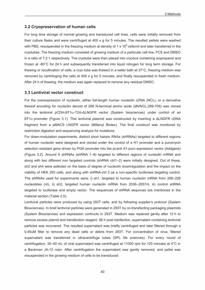
3 Methods
40
3.2 Cryopreservation of human cells
For long time storage of normal growing and transduced cell lines, cells were initially removed from
their culture flasks and were centrifuged at 400 x g for 5 minutes. The resulted pellets were washed
with PBS, resuspended in the freezing medium at density of 1 x 10⁶ cells/ml and later transferred in the
cryotubes. The freezing medium consisted of growing medium of a particular cell line, FCS and DMSO
in a ratio of 7:2:1 respectively. The cryotube were then placed into cryobox containing isopropanol and
frozen at -80°C for 24 h and subsequently transferred into liquid nitrogen for long term storage. For
thawing or recultivation of cells, a cryo tube was thawed in a water bath at 37°C, freezing medium was
removed by centrifuging the cells at 400 x g for 5 minutes, and finally resuspended in fresh medium.
After 24 h of thawing, the medium was again replaced to remove any residual DMSO.
3.3 Lentiviral vector construct
For the overexpression of nucleolin, either full-length human nucleolin cDNA (NCL), or a derivative
thereof encoding for nucleolin devoid of 288 N-terminal amino acids (dN/NCL-289-709) was cloned
into the lentiviral pCDH-EF1 -T2A-ΔLNGFR vector (System biosciences) under control of an
EF1 promoter (Figure 3.1). This lentiviral plasmid was constructed by inserting a ΔLNGFR cDNA
fragment from a pMACS LNGFR vector (Miltenyi Biotec). The final construct was monitored by
restiriction digestion and sequencing analysis for mutations.
For down-modulation experiments, distinct short hairpin RNAs (shRNAs) targeted to different regions
of human nucleolin were designed and cloned under the control of a H1 promoter and a puromycin
selection resistant gene driven by PGK promoter into the pLenti X1-puro expression vector (Addgene)
(Figure 3.2). Around 6 shRNAs (shRNA 1–8) targeted to different regions of nucleolin mRNA and
along with two different non targeted controls (shRNA ctrl1–2) were initially designed. Out of these,
sh2 and sh4 were selected on the basis of degree of nucleolin downregulation and the impact on the
viability of HEK 293 cells, and along with shRNA-ctrl 2 as a non-specific luciferase targeting control.
The shRNAs used for experiments were: i) sh1, targeted to human nucleolin mRNA from 208–226
nucleotides (nt), ii) sh2, targeted human nucleolin mRNA from 2036–2057nt, iii) control shRNA
targeted to luciferase and empty vector. The sequences of shRNA sequnces are mentioned in the
material section (Table 2.5).
Lentiviral particles were produced by using 293T cells, and by following supplier‘s protocol (System
Biosciences). In brief lentiviral particles were generated in 293T by co-transfecting packaging plasmids
(System Biosciences) and expression contructs in 293T. Medium was replaced gently after 12 h to
remove excess plamid and transfection reagent. 56 h post tranfection, supernatant containing lentiviral
particles was recovered. The resulted supernatant was briefly centrifuged and later filtered through a
0.45uM filter to remove any dead cells or debris from 293T. For concentration of virus, filtered
supernatant was transferred in ultracentrifuge tubes (SPL life sciences). For every round of
centrifugation, 30–40 mL of viral supernatant was centrifuged at 11000 rpm for 120 minutes at 4°C in
a Beckman JA-12 rotor. After centrifugation the supernatant was gently removed, and pellet was
resuspended in the growing medium of cells to be transduced.
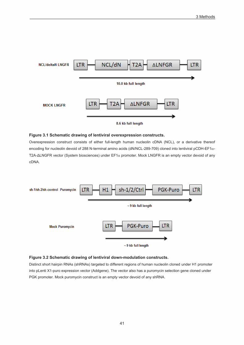
3 Methods
41
Figure 3.1 Schematic drawing of lentiviral overexpression constructs. Overexpression construct consists of either full-length human nucleolin cDNA (NCL), or a derivative thereof
encoding for nucleolin devoid of 288 N-terminal amino acids (dN/NCL-289-709) cloned into lentiviral pCDH-EF1 -
T2A-ΔLNGFR vector (System biosciences) under EF1 promoter. Mock LNGFR is an empty vector devoid of any
cDNA.
Figure 3.2 Schematic drawing of lentiviral down-modulation constructs. Distinct short hairpin RNAs (shRNAs) targeted to different regions of human nucleolin cloned under H1 promoter
into pLenti X1-puro expression vector (Addgene). The vector also has a puromycin selection gene cloned under
PGK promoter. Mock puromycin construct is an empty vector devoid of any shRNA.
3 Methods
42
3.4 Lentiviral transduction and selection
For transduction of HSPCs with minimum ex vivo manipulation, published protocol was followed
(Millington et al., 2009). In brief, after prestimulation in X-vivo medium, cells were incubated for 18h in
lentivirus containing medium on retronectin (Takara Bio Inc.) coated plates. Subsequently, transduced
cells were enriched by ΔLNGFR expression using MACselect LNGFR microbeads 72 h post
transduction (Miltenyi Biotec) (Wiehe et al., 2006). Resulting cell preparations containing ~ 90%
LNGFR+ cells monitored by FACS, were cultured in X-vivo medium supplemented with 50 ng/ml SCF,
50 ng/ml Flt-3, 10 ng/ml TPO, 10 ng/ml IL-6 (Peprotech), and aliquots for further analysis were taken
at indicated time points. Mutz-2 and SEM cells were also transduced as described above and were
either enriched by LNGFR microbeads or subjected to antibiotic selection with puromycin (250 ng/ml,
Sigma-Aldrich) for shRNA expression.
3.5 Nucleofection
Nucleofection of HSPSc with shRNA expression constructs was performed with CD34+ cell, human
Nucleofector kit (Lonza). Cells were prepared according to manufacturer's specifications (Lonza) and
as described (Salati et al., 2008). 48–72h post-nucleofection, aliquots were taken for further analysis.
Cell viability was assessed by trypan exclusion method and by immunoblotting with antibodies (Ab)
specific to caspases-3, 1:5000 (R&D Systems).
3.6 Gelelectrophoretic separation and detection of proteins
3.6.1 SDS-PAGE
The separation of proteins by SDS-PAGE was performed following standard procedure (Laemmli,
1970). Protein samples for loading SDS-PAGE were prepared by mixing 1 x SDS-Sample Buffer and
denatured at 95°C for 5 minutes. The gelelectrophoresis was carried out in 1x SDS Running buffer in
a Mini-Cell gel chamber (Bio-Rad) with 25 mA per gel.
3.6.2 Western Blot analysis
After running SDS-PAGE, the gel was blotted to a protran 0.45uM nitrocellulose membrane (GE
Healthcare) with 100 mA per blot for 2 hours with 1 x Transfer Buffer, using a wet transfer (Bio-rad).
Subsequently, the membrane was blocked in 1 x TBST buffer with 5% BSA for 1 hour. The blocked
membrane was further incubated over-night with primary antibodies (Abs). The following are Abs and
their dilutions: mAb specific for FLAG-tag (Sigma), 1:1000; Ab specific for a N-terminal peptide of
nucleolin, purified on the peptide column (Grinstein et al., 2007), 1:10000; mAb specific for AC133
(W6B3C1; Miltenyi Biotech), 1:500; mAb specific for -actin (AC-74; Sigma), 1:2000; Ab specific for
Caspase-3 (R&D Systems); 1:5000, mAb specific for active -catenin (Millipore), 1:1000; Ab specific
for -catenin (Cell Signaling), 1:2000; mAb specific for Bcl-2 (Santa Cruz Biotechnology), 1:2000; mAb
specific for Pan-Akt (Cell signalling) 1:4000; mAb specific for Akt activated phosphorylated at the
Thr308 (Cell Signaling), 1:3000; mAb specific for Akt phosphorylated at Ser473 (Cell Signaling).
3 Methods
43
Afterwards, the membrane was washed again three times for 5 minutes with 1 x TBST buffer, and
incubated with appropriate secondary ab labeled with HRP for 1 h. The membrane was again washed
with 1 x TBST buffer for 5 minutes three times. Proteins were detected by incubation Amersham ECL
WB Detection Reagents for 1 minute, subsequently pictures were taken by using LAS 3000 UV mini
system (Fujifilm).
3.7 Quantitative reverse transcription real time PCR (qRT-PCR)
Total RNA was isolated with RNeasy kit (Qiagen), reverse transcribed using QuantiTect Reverse
Transcription Kit (Qiagen), and levels of gene expression were quantified by real-time PCR, using
Power SYBR® Green Real-Time PCR Master Mix on a 7900HT Fast Real Time PCR system (Life
Technologies). Primer sequences are summarized in material section (Table 2.4) and specificity
thereof was monitored by melting curve analysis as well as loading PCR products on agarose gels.
The comparative threshold cycle (ΔΔCt) method and an internal control ( -actin) were used to
determine differential expression.
3.8 Flourescence-activated cell sorting (FACS)
´The cells to be analysed were resuspended and washed with FACS buffer (PBS + 0.5% FCS). After
spinning, the cells were incubated in 50 ul of FACS buffer containing either uncoupled primary
antibody or primary antibody coupled to flourochrome, and later incubated at ice in the dark for 25–30
minutes. This was followed by washing the stained cells thrice with the FACS buffer for 5 minutes
each. After the last wash, the cells were either measured by adding 200ul of FACS buffer in case of
primary coupled antibody or incubated with secondary antibody coupled to flourochrome in case of
uncoupled primary antibody for additional half an hour on ice in the dark. The cells were again washed
thrice and measured using FACSCalibur (Becton Dickinson, Heidelberg, Germany). FACS analysis
used the following fluorochrome coupled monoclonal antibodies (mAbs): anti-AC133 (CD133/1;
Miltenyi Biotec), anti-CD34 (8G12; BD Biosciences), anti-CD11b (Bear1; Beckman Coulter), anti-CD14
(RMO52; Beckman Coulter), anti-LNGFR-FITC (ME20.4-1.H4; Miltenyi Biotec), CD33 (Miltenyi
Biotec), CD3 (UCHT1; BD Biosciences), CD19 (Miltenyi Biotec), CD71 (BD Biosciences), CD45 (BD
Biosciences) and CD56 (Miltenyi Biotec) as well as isotype-matched control mAbs.
3.9 Colony forming assay (CFC-assay)
3.9.1 Primary CFC assay
CFC activity was assessed by plating transduced cells in methylcellulose medium (MethocultTM
H4100, STEMCELL Technologies) supplemented with 50 ng/ml SCF, 10 ng/ml granulocyte-
macrophage colony-stimulating factor (GM-CSF), 10 ng/ml IL-3 and 3 U/ml erythropoietin
(ebiosciences). After 14 days of culture, granulocyte-erythrocyte-macrophage-megakaryocyte colony
forming units (CFU-GEMM), granulocyte-macrophage colony forming units (CFU-GM), erythroid burst-
forming units (BFU-E), erythroid colony forming units (CFU-E), granulocyte colony forming units (CFU-
3 Methods
44
G), and macrophage colony forming units (CFU-M) were scored (STEMCELL Technologies). Cell
morphology was determined by Wright-Giemsa staining by plucking individual colonies.
3.9.2 Secondary CFC assay
For assessing secondary colony forming capacity, colonies from the primary plating after 14 days of
cultures were collected and washed with phosphate buffer saline (PBS) solution for three times. The
resulted cells were seeded for additional 2 weeks in the fresh methylcellulose medium containing
human recombinant cytokines (50 ng/ml SCF, 10 ng/ml IL-3, 10 ng/ml GMCSF, 3 U/ml EPO).
3.10 Long term culture initiating assay (LTC-IC assay)
LTC-IC assays were performed by seeding transduced cells on irradiated murine M2-10B4, as a
feeder layer (STEMCELL Technologies). Long term cultures were maintained in the Myelocult H5100
medium (STEMCELL Technologies) supplemented with hydrocortisone (10−6 M) (STEMCELL
Technologies), after 6 to 9 weeks, cells were seeded into methylcellulose medium containing
cytokines mentioned above. LTC-IC frequencies were calculated by limiting dilution analysis, using L-
calc™ software (STEMCELL Technologies).
3.11 Ex vivo assay for B cell development
To determine B cell progenitor activity, 2 x 10⁴ transduced HSPSc were seeded similarly on irradiated
M2-10B4 feeder layer cells in X-vivo 20 (Lonza) medium containing 50 ng/ml SCF, 100 ng/ml Flt-3,
50 ng/ml TPO, 10 ng/ml IL-2, 10 ng/ml IL-7 and 10 ng/ml IL-15 (Peprotech), favoring conditions for B-
lymphoid cell development in addition to conditions permissive for myeloid development (Buske et al.,
2002). Fresh media containing cytokines was replaced after every 4 days. After 3 weeks, FACS
analysis for CD19 (B lymphoid), CD3 (T lymphoid), CD56 (NK-cells) and CD34 was performed.
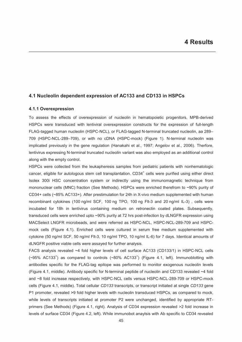
45
4 Results
4.1 Nucleolin dependent expression of AC133 and CD133 in HSPCs
4.1.1 Overexpression
To assess the effects of overexpression of nucleolin in hematopoietic progenitors, MPB-derived
HSPCs were transduced with lentiviral overexpression constructs for the expression of full-length
FLAG-tagged human nucleolin (HSPC-NCL), or FLAG-tagged N-terminal truncated nucleolin, aa 289–
709 (HSPC-NCL-289–709), or with no cDNA (HSPC-mock) (Figure 1). N-terminal nucleolin was
implicated previously in the gene regulation (Hanakahi et al., 1997; Angelov et al., 2006). Therfore,
lentivirus expressing N-terminal truncated nucleolin variant was also employed as an additional control
along with the empty control. HSPCs were collected from the leukapheresis samples from pediatric patients with nonhematologic
cancer, eligible for autologous stem cell transplantation. CD34+ cells were purified using either direct
Isolex 300i HSC concentration system or indirectly using the immunomagnetic technique from
mononuclear cells (MNC) fraction (See Methods). HSPCs were enriched therefrom to ~90% purity of
CD34+ cells (~85% AC133+). After prestimulation for 24h in X-vivo medium supplemented with human
recombinant cytokines (100 ng/ml SCF, 100 ng TPO, 100 ng Flt-3 and 20 ng/ml IL-3) , cells were
incubated for 18h in lentivirus containing medium on retronectin coated plates. Subsequently,
transduced cells were enriched upto ~90% purity at 72 hrs post-infection by dLNGFR expression using
MACSelect LNGFR microbeads, and were referred as HSPC-NCL, HSPC-NCL-289-709 and HSPC-
mock cells (Figure 4.1). Enriched cells were cultured in serum free medium supplemented with
cytokine (50 ng/ml SCF, 50 ng/ml Flt-3, 10 ng/ml TPO, 10 ng/ml IL-6) for 7 days. Identical amounts of
dLNGFR positive viable cells were assayed for further analysis.
FACS analysis revealed ~4 fold higher levels of cell surface AC133 (CD133/1) in HSPC-NCL cells
(~95% AC133+) as compared to controls (~80% AC133+) (Figure 4.1, left). Immunoblotting with
antibodies specific for the FLAG-tag epitope was performed to monitor exogenous nucleolin levels
(Figure 4.1, middle). Antibody specific for N-terminal peptide of nucleolin and CD133 revealed ~4 fold
and ~8 fold increase respectively, with HSPC-NCL cells versus HSPC-NCL-289-709 or HSPC-mock
cells (Figure 4.1, middle). Total cellular CD133 transcripts, or transcript initiated at single CD133 gene
P1 promoter, revealed >9 fold higher levels with nucleolin transduced HSPCs, as compared to mock,
while levels of transcripts initiated at promoter P2 were unchanged, identified by appropriate RT-
primers (See Methods) (Figure 4.1, right). Analysis of CD34 expression revealed >2 fold increase in
levels of surface CD34 (Figure 4.2, left). While immunobot anaylsis with Ab specific to CD34 revealed
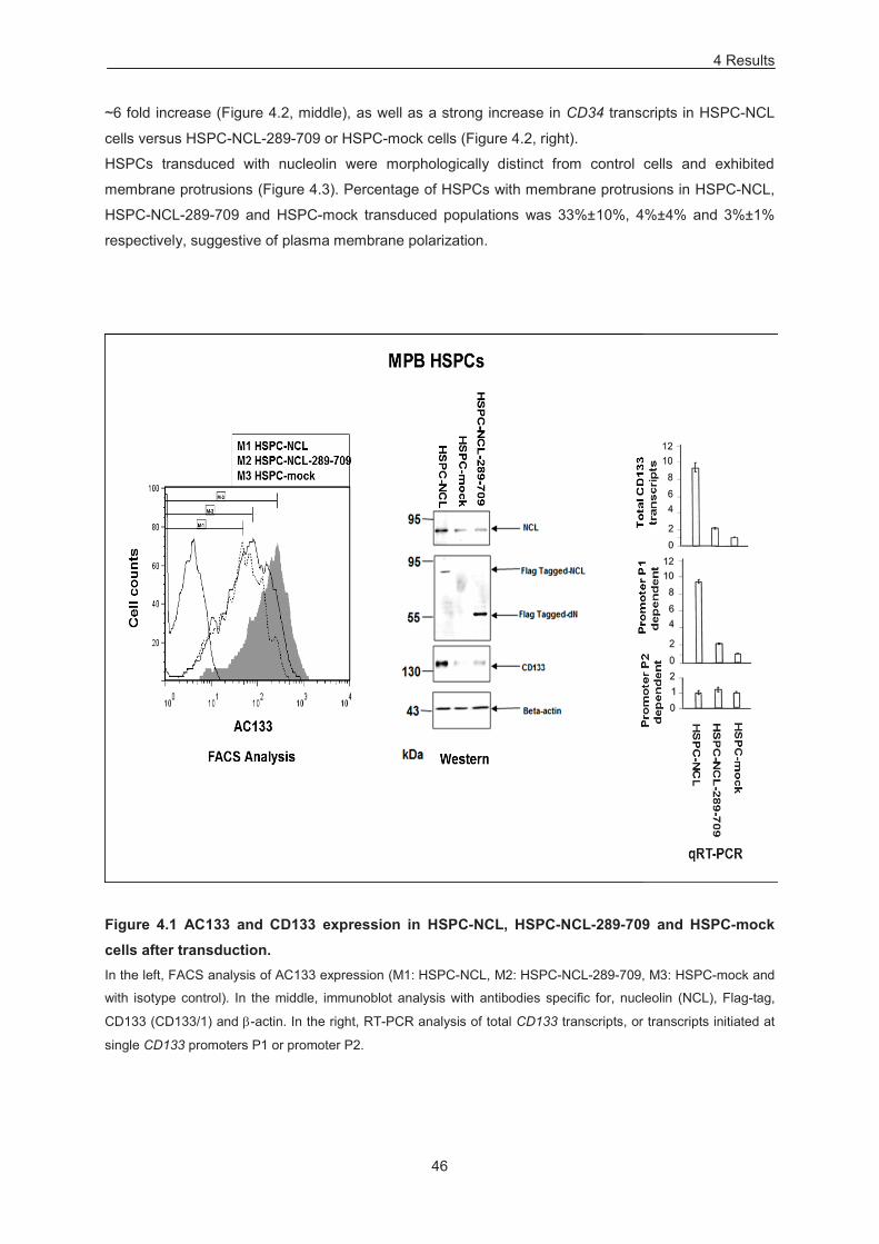
4 Results
46
~6 fold increase (Figure 4.2, middle), as well as a strong increase in CD34 transcripts in HSPC-NCL
cells versus HSPC-NCL-289-709 or HSPC-mock cells (Figure 4.2, right).
HSPCs transduced with nucleolin were morphologically distinct from control cells and exhibited
membrane protrusions (Figure 4.3). Percentage of HSPCs with membrane protrusions in HSPC-NCL,
HSPC-NCL-289-709 and HSPC-mock transduced populations was 33%±10%, 4%±4% and 3%±1%
respectively, suggestive of plasma membrane polarization.
Figure 4.1 AC133 and CD133 expression in HSPC-NCL, HSPC-NCL-289-709 and HSPC-mock cells after transduction. In the left, FACS analysis of AC133 expression (M1: HSPC-NCL, M2: HSPC-NCL-289-709, M3: HSPC-mock and
with isotype control). In the middle, immunoblot analysis with antibodies specific for, nucleolin (NCL), Flag-tag,
CD133 (CD133/1) and -actin. In the right, RT-PCR analysis of total CD133 transcripts, or transcripts initiated at
single CD133 promoters P1 or promoter P2.
4 Results
47
Figure 4.2 CD34 expression in HSPC-NCL, HSPC-NCL-289-709 and HSPC-mock cells In the left, FACS analysis of CD34 expression on the surface (M1: HSPC-NCL, M2: HSPC-NCL-289-709, M3:
HSPC-mock and with isotype control). In the middle, immunoblotting analysis with antibodies specific for CD34
and -actin. In the right, qRT-PCR analysis of CD34 transcripts.
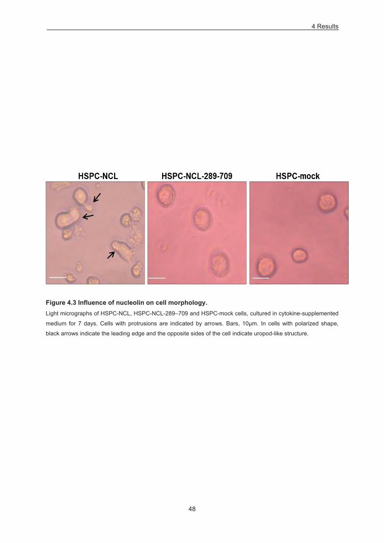
4 Results
48
Figure 4.3 Influence of nucleolin on cell morphology. Light micrographs of HSPC-NCL, HSPC-NCL-289–709 and HSPC-mock cells, cultured in cytokine-supplemented
medium for 7 days. Cells with protrusions are indicated by arrows. Bars, 10μm. In cells with polarized shape,
black arrows indicate the leading edge and the opposite sides of the cell indicate uropod-like structure.
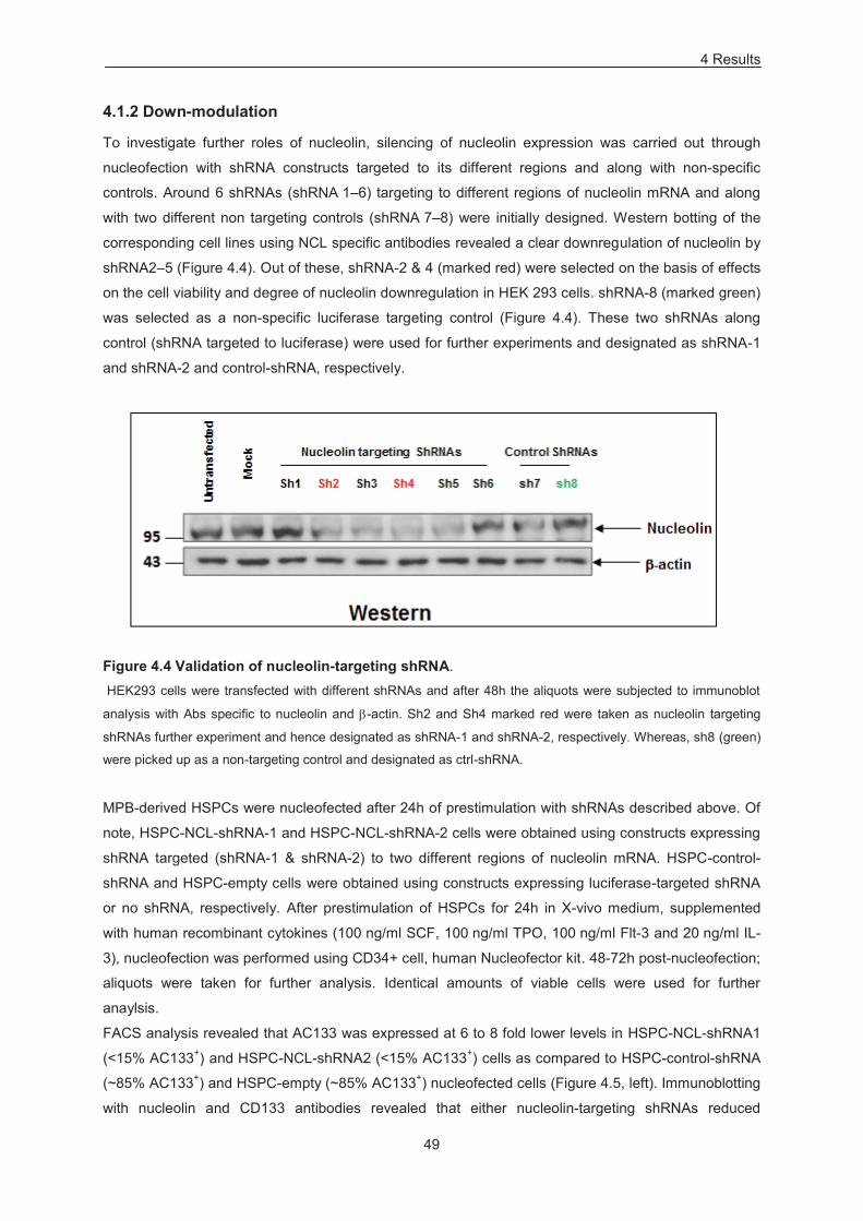
4 Results
49
4.1.2 Down-modulation
To investigate further roles of nucleolin, silencing of nucleolin expression was carried out through
nucleofection with shRNA constructs targeted to its different regions and along with non-specific
controls. Around 6 shRNAs (shRNA 1–6) targeting to different regions of nucleolin mRNA and along
with two different non targeting controls (shRNA 7–8) were initially designed. Western botting of the
corresponding cell lines using NCL specific antibodies revealed a clear downregulation of nucleolin by
shRNA2–5 (Figure 4.4). Out of these, shRNA-2 & 4 (marked red) were selected on the basis of effects
on the cell viability and degree of nucleolin downregulation in HEK 293 cells. shRNA-8 (marked green)
was selected as a non-specific luciferase targeting control (Figure 4.4). These two shRNAs along
control (shRNA targeted to luciferase) were used for further experiments and designated as shRNA-1
and shRNA-2 and control-shRNA, respectively.
Figure 4.4 Validation of nucleolin-targeting shRNA. HEK293 cells were transfected with different shRNAs and after 48h the aliquots were subjected to immunoblot
analysis with Abs specific to nucleolin and -actin. Sh2 and Sh4 marked red were taken as nucleolin targeting
shRNAs further experiment and hence designated as shRNA-1 and shRNA-2, respectively. Whereas, sh8 (green)
were picked up as a non-targeting control and designated as ctrl-shRNA.
MPB-derived HSPCs were nucleofected after 24h of prestimulation with shRNAs described above. Of
note, HSPC-NCL-shRNA-1 and HSPC-NCL-shRNA-2 cells were obtained using constructs expressing
shRNA targeted (shRNA-1 & shRNA-2) to two different regions of nucleolin mRNA. HSPC-control-
shRNA and HSPC-empty cells were obtained using constructs expressing luciferase-targeted shRNA
or no shRNA, respectively. After prestimulation of HSPCs for 24h in X-vivo medium, supplemented
with human recombinant cytokines (100 ng/ml SCF, 100 ng/ml TPO, 100 ng/ml Flt-3 and 20 ng/ml IL-
3), nucleofection was performed using CD34+ cell, human Nucleofector kit. 48-72h post-nucleofection;
aliquots were taken for further analysis. Identical amounts of viable cells were used for further
anaylsis.
FACS analysis revealed that AC133 was expressed at 6 to 8 fold lower levels in HSPC-NCL-shRNA1
(<15% AC133+) and HSPC-NCL-shRNA2 (<15% AC133+) cells as compared to HSPC-control-shRNA
(~85% AC133+) and HSPC-empty (~85% AC133+) nucleofected cells (Figure 4.5, left). Immunoblotting
with nucleolin and CD133 antibodies revealed that either nucleolin-targeting shRNAs reduced
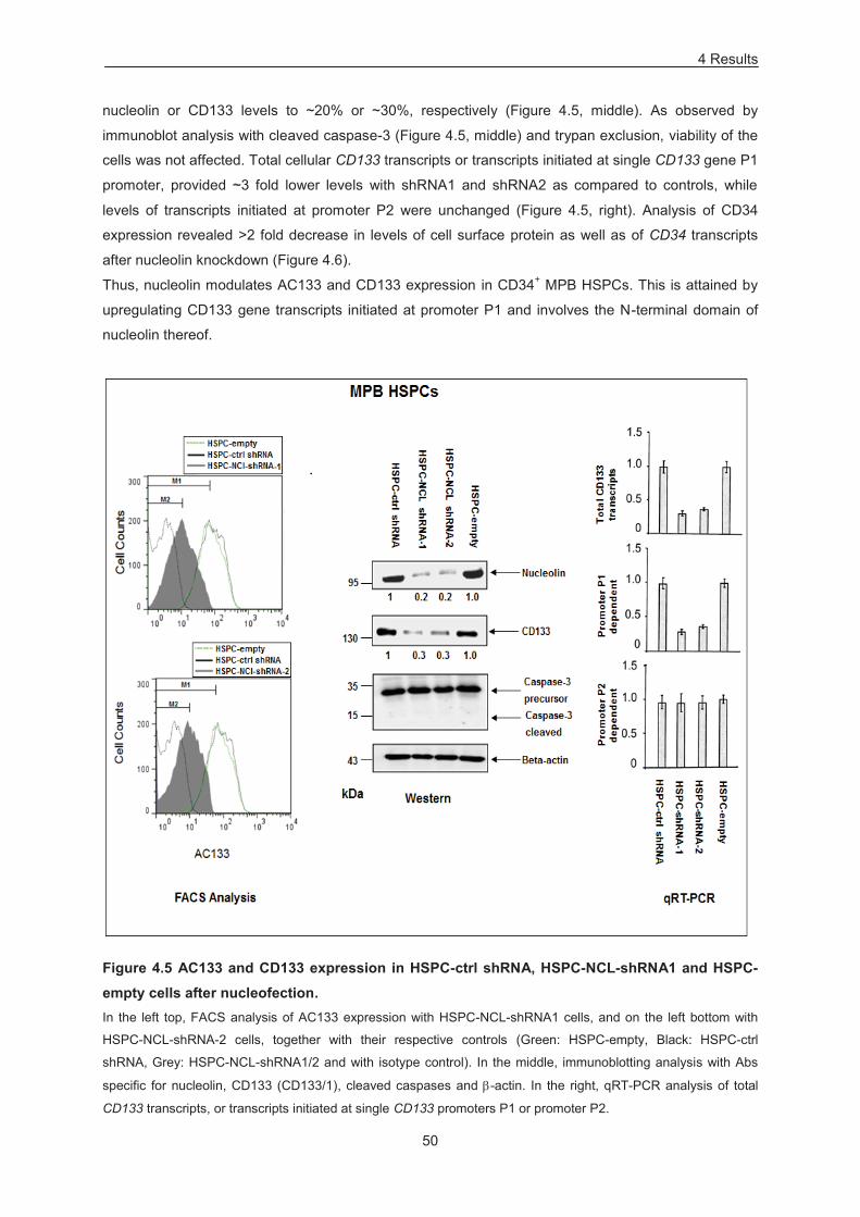
4 Results
50
nucleolin or CD133 levels to ~20% or ~30%, respectively (Figure 4.5, middle). As observed by
immunoblot analysis with cleaved caspase-3 (Figure 4.5, middle) and trypan exclusion, viability of the
cells was not affected. Total cellular CD133 transcripts or transcripts initiated at single CD133 gene P1
promoter, provided ~3 fold lower levels with shRNA1 and shRNA2 as compared to controls, while
levels of transcripts initiated at promoter P2 were unchanged (Figure 4.5, right). Analysis of CD34
expression revealed >2 fold decrease in levels of cell surface protein as well as of CD34 transcripts
after nucleolin knockdown (Figure 4.6).
Thus, nucleolin modulates AC133 and CD133 expression in CD34+ MPB HSPCs. This is attained by
upregulating CD133 gene transcripts initiated at promoter P1 and involves the N-terminal domain of
nucleolin thereof.
Figure 4.5 AC133 and CD133 expression in HSPC-ctrl shRNA, HSPC-NCL-shRNA1 and HSPC-empty cells after nucleofection. In the left top, FACS analysis of AC133 expression with HSPC-NCL-shRNA1 cells, and on the left bottom with
HSPC-NCL-shRNA-2 cells, together with their respective controls (Green: HSPC-empty, Black: HSPC-ctrl
shRNA, Grey: HSPC-NCL-shRNA1/2 and with isotype control). In the middle, immunoblotting analysis with Abs
specific for nucleolin, CD133 (CD133/1), cleaved caspases and -actin. In the right, qRT-PCR analysis of total
CD133 transcripts, or transcripts initiated at single CD133 promoters P1 or promoter P2.
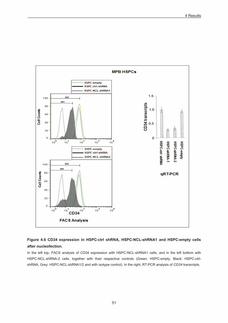
4 Results
51
Figure 4.6 CD34 expression in HSPC-ctrl shRNA, HSPC-NCL-shRNA1 and HSPC-empty cells after nucleofection. In the left top, FACS analysis of CD34 expression with HSPC-NCL-shRNA1 cells, and in the left bottom with
HSPC-NCL-shRNA-2 cells, together with their respective controls (Green: HSPC-empty, Black: HSPC-ctrl-
shRNA, Grey: HSPC-NCL-shRNA1/2 and with isotype control). In the right, RT-PCR analysis of CD34 transcripts.
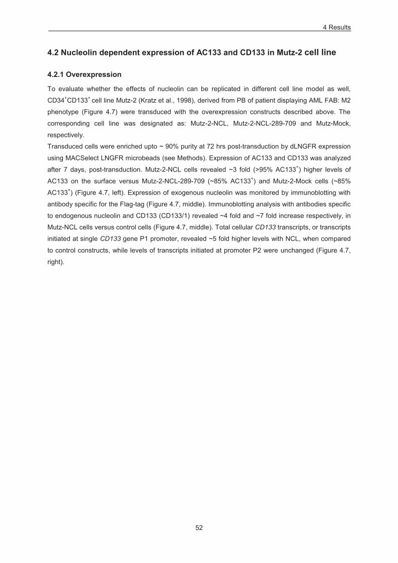
4 Results
52
4.2 Nucleolin dependent expression of AC133 and CD133 in Mutz-2 cell line
4.2.1 Overexpression
To evaluate whether the effects of nucleolin can be replicated in different cell line model as well,
CD34+CD133+ cell line Mutz-2 (Kratz et al., 1998), derived from PB of patient displaying AML FAB: M2
phenotype (Figure 4.7) were transduced with the overexpression constructs described above. The
corresponding cell line was designated as: Mutz-2-NCL, Mutz-2-NCL-289-709 and Mutz-Mock,
respectively.
Transduced cells were enriched upto ~ 90% purity at 72 hrs post-transduction by dLNGFR expression
using MACSelect LNGFR microbeads (see Methods). Expression of AC133 and CD133 was analyzed
after 7 days, post-transduction. Mutz-2-NCL cells revealed ~3 fold (>95% AC133+) higher levels of
AC133 on the surface versus Mutz-2-NCL-289-709 (~85% AC133+) and Mutz-2-Mock cells (~85%
AC133+) (Figure 4.7, left). Expression of exogenous nucleolin was monitored by immunoblotting with
antibody specific for the Flag-tag (Figure 4.7, middle). Immunoblotting analysis with antibodies specific
to endogenous nucleolin and CD133 (CD133/1) revealed ~4 fold and ~7 fold increase respectively, in
Mutz-NCL cells versus control cells (Figure 4.7, middle). Total cellular CD133 transcripts, or transcripts
initiated at single CD133 gene P1 promoter, revealed ~5 fold higher levels with NCL, when compared
to control constructs, while levels of transcripts initiated at promoter P2 were unchanged (Figure 4.7,
right).
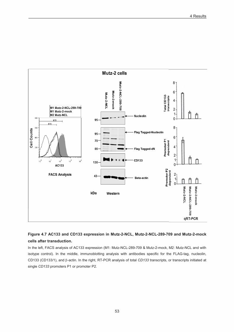
4 Results
53
Figure 4.7 AC133 and CD133 expression in Mutz-2-NCL, Mutz-2-NCL-289-709 and Mutz-2-mock cells after transduction. In the left, FACS analysis of AC133 expression (M1: Mutz-NCL-289-709 & Mutz-2-mock, M2: Mutz-NCL and with
isotype control). In the middle, immunoblotting analysis with antibodies specific for the FLAG-tag, nucleolin,
CD133 (CD133/1), and -actin. In the right, RT-PCR analysis of total CD133 transcripts, or transcripts initiated at
single CD133 promoters P1 or promoter P2.
4 Results
54
4.2.2 Down-modulation
To attain knockdown of nucleolin, Mutz-2 cells were transduced with the shRNA expression constructs
targeting different regions of nucleolin as mentioned above in the Method section (3.3). Transduced
cells were subjected to puromycin selection for 2 weeks. Resulting selected cells were immediately
employed for further analysis, and were designated as: Mutz-NCL-shRNA-1, Mutz-NCL-shRNA-2,
Mutz-2-control-shRNA and Mutz-2-empty. AC133 was expressed at ~8 fold lower levels in both Mutz-
NCL-shRNA1 (~35% AC133+) and Mutz-NCL-shRNA2 (~35% AC133+) as compared to Mutz-2-
control- shRNA (~95% AC133+) and Mutz-2-control (~95% AC133+) transduced cells (Figure 4.8, left).
Whereas immunoblot analysis revealed that nucleolin and CD133 protein levels were reduced to
~30% in Mutz2-NCL-shRNA-1 and Mutz2-NCL-shRNA-2 cells versus control cells (Figure 4.8, middle).
Immunoblot analysis with cleaved caspase-3 (Figure 4.8, middle) and by trypan exclusion revealed
that the viability of the cells was not affected. Total cellular CD133 transcripts or transcripts initiated at
single CD133 gene P1 promoter, revealed ~3 fold lower levels with shRNA1 and shRNA2 as
compared to controls, while levels of transcripts initiated at promoter P2 were unchanged (Figure 4.8,
right). Doubling time of Mutz-2-control-shRNA and Mutz-2 empty cells were approximately 2–3 days,
however the doubling time of Mutz-2-NCL-shRNA-1 and Mutz-2-NCL-shRNA-2 cells were
approximately 16 days.
Thus, nucleolin modulates AC133 and CD133 expression in Mutz-2 cells. This is attained by
upregulating CD133 gene transcripts initiated at promoter P1 and involves the N-terminal domain of
nucleolin thereof.
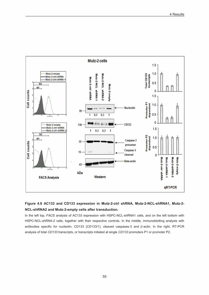
4 Results
55
Figure 4.8 AC133 and CD133 expression in Mutz-2-ctrl shRNA, Mutz-2-NCL-shRNA1, Mutz-2-NCL-shRNA2 and Mutz-2-empty cells after transduction. In the left top, FACS analysis of AC133 expression with HSPC-NCL-shRNA1 cells, and on the left bottom with
HSPC-NCL-shRNA-2 cells, together with their respective controls. In the middle, immunoblotting analysis with
antibodies specific for nucleolin, CD133 (CD133/1), cleaved caspases-3 and -actin. In the right, RT-PCR
analysis of total CD133 transcripts, or transcripts initiated at single CD133 promoters P1 or promoter P2.
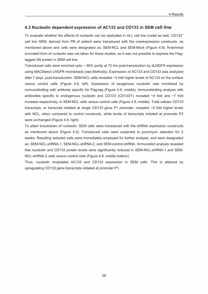
4 Results
56
4.3 Nucleolin dependent expression of AC133 and CD133 in SEM cell line
To evaluate whether the effects of nucleolin can be replicated in ALL cell line model as well, CD133+
cell line SEM, derived from PB of patient were transduced with the overexpression constructs, as
mentioned above and cells were designated as: SEM-NCL and SEM-Mock (Figure 4.9). N-terminal
truncated form of nucleolin was not taken for these studies, as it was not possible to express the Flag-
tagged dN protein in SEM cell line.
Transduced cells were enriched upto ~ 90% purity at 72 hrs post-transduction by dLNGFR expression
using MACSelect LNGFR microbeads (see Methods). Expression of AC133 and CD133 was analyzed
after 7 days, post-transduction. SEM-NCL cells revealed ~3 fold higher levels of AC133 on the surface
versus control cells (Figure 4.9, left). Expression of exogenous nucleolin was monitored by
immunoblotting with antibody specific for Flag-tag (Figure 4.9, middle). Immunoblotting analysis with
antibodies specific to endogenous nucleolin and CD133 (CD133/1) revealed ~4 fold and ~7 fold
increase respectively, in SEM-NCL cells versus control cells (Figure 4.9, middle). Total cellular CD133
transcripts, or transcript initiated at single CD133 gene P1 promoter, revealed ~5 fold higher levels
with NCL, when compared to control constructs, while levels of transcripts initiated at promoter P2
were unchanged (Figure 4.9, right).
To attain knockdown of nucleolin, SEM cells were transduced with the shRNA expression constructs
as mentioned above (Figure 4.9). Transduced cells were subjected to puromycin selection for 2
weeks. Resulting selected cells were immediately employed for further analysis, and were designated
as: SEM-NCL-shRNA-1, SEM-NCL-shRNA-2, and SEM-control-shRNA. Immunoblot analysis revealed
that nucleolin and CD133 protein levels were significantly reduced in SEM-NCL-shRNA-1 and SEM-
NCL-shRNA-2 cells versus control cells (Figure 4.9, middle bottom).
Thus, nucleolin modulates AC133 and CD133 expression in SEM cells. This is attained by
upregulating CD133 gene transcripts initiated at promoter P1.
4 Results
57
Figure 4.9 AC133 and CD133 expression in SEM-NCL, SEM-mock (overexpression) and SEM-ctrl shRNA, SEM-NCL-shRNA1 and SEM-NCL-shRNA2 (down-modulation) cells.
In the left, FACS analysis for AC133 expression in SEM-NCL and SEM-mock cells.On the middle (top),
immunoblotting analysis with antibodies specific for FLAG-tag, nucleolin, CD133 (CD133/1), and -actin with
SEM-NCL and SEM-mock sample (overexpression). In the middle (bottom), immunoblotting analysis with
antibodies specific for nucleolin, CD133 (CD133/1), and -actin with SEM-ctrl shRNA, SEM-NCL-shRNA1 and
SEM-NCL-shRNA2 (down-modulation). In the right, RT-PCR analysis of total CD133 transcripts, or transcripts
initiated at single CD133 promoters P1 or promoter P2 with SEM-mock and SEM-NCL cells. (qRTs provided by
Sven Reister).
4 Results
58
4.4 Nucleolin increases the frequency of hematopoietic colony forming units (CFUs) and impacts myeloid differentiation in HSPCs
4.4.1 Overexpression
To test the possible effects of nucleolin on HSPCs proliferative potential, HSPC-NCL, HSPC-NCL-
289-709 and HSPC-mock cells were seeded in methylcellulose medium supplemented with human
recombinant cytokines (50 ng/ml SCF, 10 ng/ml IL-3, 10 ng/ml GMCSF, 3 U/ml EPO) for 14 days.
Primary colony forming assay with nucleolin revealed significant difference in both total colony count
and individual colony types after 14 days (Figure 4.10, A). Increase in the clonogenic cells generated
by HSPC-NCL versus HSPC-NCL-289-709 and HSPC-mock: total CFU ~4 fold, CFU-GEMM ~7 fold,
CFU-GM ~9 fold, BFU-E ~6 fold, CFU-G ~8 fold, CFU-E ~3 fold and CFU-M ~2 fold (Figure 4.10, A).
In addition, enrichment of ~2 fold CFU-GM together with ~2 fold depletion of CFU-M was observed
(Figure 4.11). With the exception of CFU-E, individual colony type produced by HSPC-NCL cells
exhibited a significant increase in the size (Figure 4.12). Moreover, the cellularity (cell number per
colony) was determined by plucking individual colonies during primary plating and was found
significantly increased, with exception of CFU-E colony type (Figure 4.13). Wright-Giemsa staining of
individual plucked colonies did not reveal difference in cell morphology between different samples. For
assessing secondary colony forming capacity, colonies from the primary plating after 14 days of
cultures were collected and washed. The resulted cells were seeded for additional 2 weeks in the
fresh methylcellulose medium containing human recombinant cytokines. When compared to HSPC-
NCL-289-709 and HSPC-mock cells, HSPC-NCL exhibited increased replating capacity with an
increase in the both total and indvidual colony output (Figure 4.10, B)
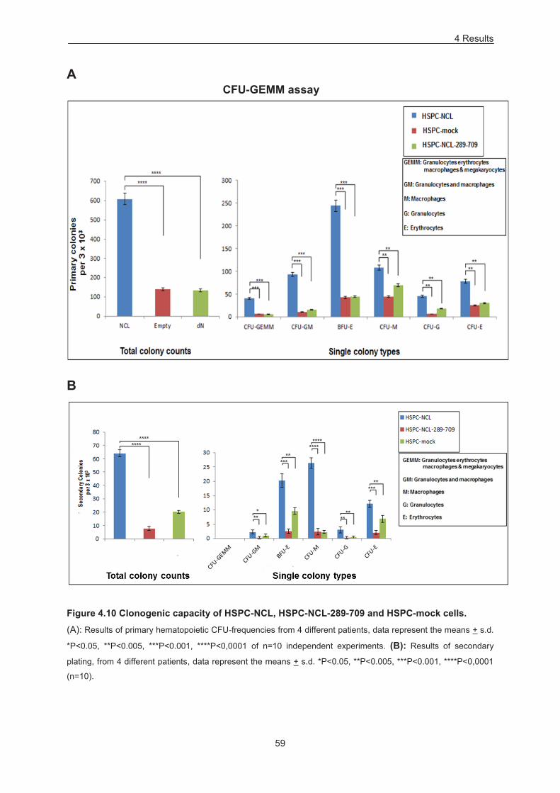
4 Results
59
A
B
Figure 4.10 Clonogenic capacity of HSPC-NCL, HSPC-NCL-289-709 and HSPC-mock cells.
(A): Results of primary hematopoietic CFU-frequencies from 4 different patients, data represent the means + s.d.
*P<0.05, **P<0.005, ***P<0.001, ****P<0,0001 of n=10 independent experiments. (B): Results of secondary
plating, from 4 different patients, data represent the means + s.d. *P<0.05, **P<0.005, ***P<0.001, ****P<0,0001
(n=10).
CFU-GEMM assay
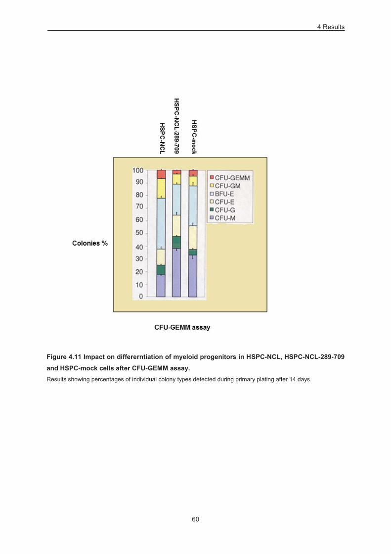
4 Results
60
Figure 4.11 Impact on differerntiation of myeloid progenitors in HSPC-NCL, HSPC-NCL-289-709 and HSPC-mock cells after CFU-GEMM assay. Results showing percentages of individual colony types detected during primary plating after 14 days.
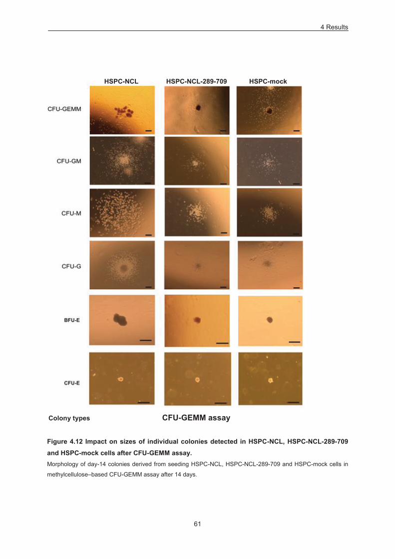
4 Results
61
Figure 4.12 Impact on sizes of individual colonies detected in HSPC-NCL, HSPC-NCL-289-709 and HSPC-mock cells after CFU-GEMM assay. Morphology of day-14 colonies derived from seeding HSPC-NCL, HSPC-NCL-289-709 and HSPC-mock cells in
methylcellulose–based CFU-GEMM assay after 14 days.
Colony types CFU-GEMM assay
HSPC-NCL HSPC-NCL-289-709 HSPC-mock
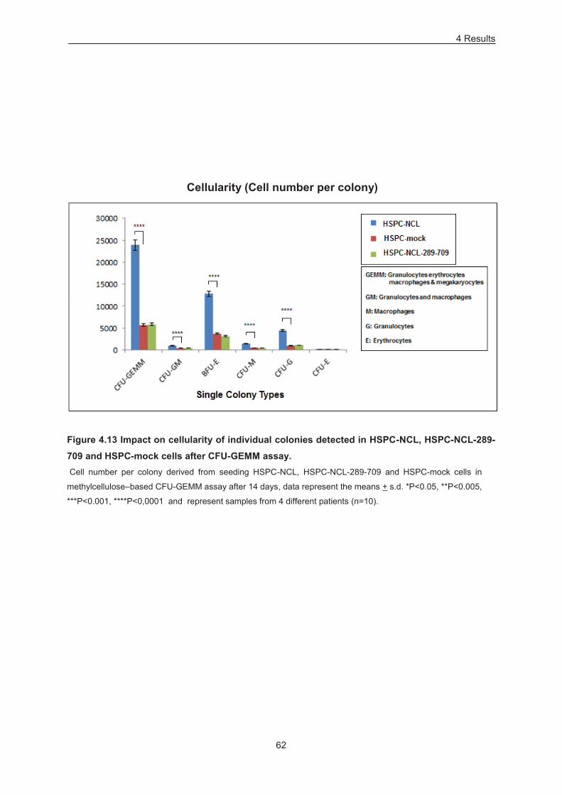
4 Results
62
Cellularity (Cell number per colony)
Figure 4.13 Impact on cellularity of individual colonies detected in HSPC-NCL, HSPC-NCL-289-709 and HSPC-mock cells after CFU-GEMM assay. Cell number per colony derived from seeding HSPC-NCL, HSPC-NCL-289-709 and HSPC-mock cells in
methylcellulose–based CFU-GEMM assay after 14 days, data represent the means + s.d. *P<0.05, **P<0.005,
***P<0.001, ****P<0,0001 and represent samples from 4 different patients (n=10).
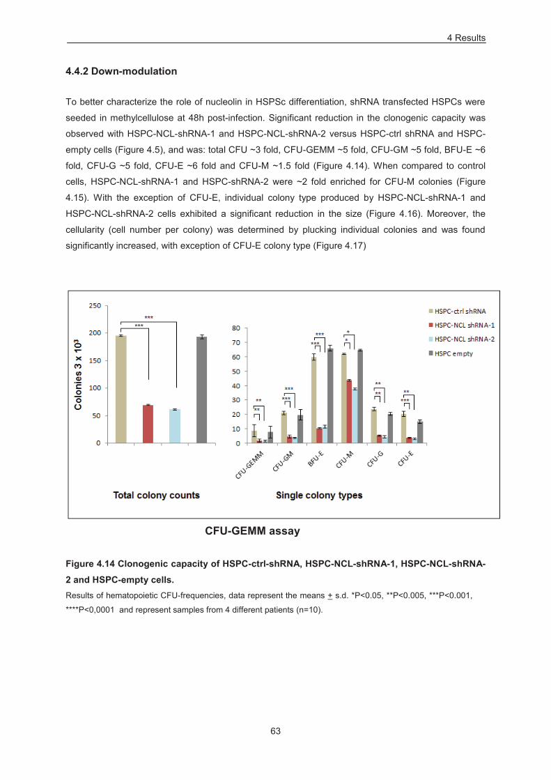
4 Results
63
4.4.2 Down-modulation
To better characterize the role of nucleolin in HSPSc differentiation, shRNA transfected HSPCs were
seeded in methylcellulose at 48h post-infection. Significant reduction in the clonogenic capacity was
observed with HSPC-NCL-shRNA-1 and HSPC-NCL-shRNA-2 versus HSPC-ctrl shRNA and HSPC-
empty cells (Figure 4.5), and was: total CFU ~3 fold, CFU-GEMM ~5 fold, CFU-GM ~5 fold, BFU-E ~6
fold, CFU-G ~5 fold, CFU-E ~6 fold and CFU-M ~1.5 fold (Figure 4.14). When compared to control
cells, HSPC-NCL-shRNA-1 and HSPC-shRNA-2 were ~2 fold enriched for CFU-M colonies (Figure
4.15). With the exception of CFU-E, individual colony type produced by HSPC-NCL-shRNA-1 and
HSPC-NCL-shRNA-2 cells exhibited a significant reduction in the size (Figure 4.16). Moreover, the
cellularity (cell number per colony) was determined by plucking individual colonies and was found
significantly increased, with exception of CFU-E colony type (Figure 4.17)
Figure 4.14 Clonogenic capacity of HSPC-ctrl-shRNA, HSPC-NCL-shRNA-1, HSPC-NCL-shRNA-2 and HSPC-empty cells. Results of hematopoietic CFU-frequencies, data represent the means + s.d. *P<0.05, **P<0.005, ***P<0.001,
****P<0,0001 and represent samples from 4 different patients (n=10).
CFU-GEMM assay
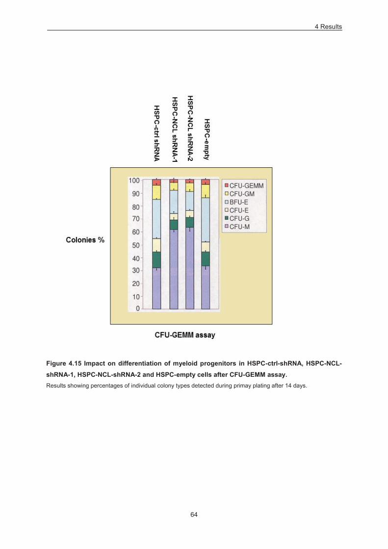
4 Results
64
Figure 4.15 Impact on differentiation of myeloid progenitors in HSPC-ctrl-shRNA, HSPC-NCL-shRNA-1, HSPC-NCL-shRNA-2 and HSPC-empty cells after CFU-GEMM assay. Results showing percentages of individual colony types detected during primay plating after 14 days.
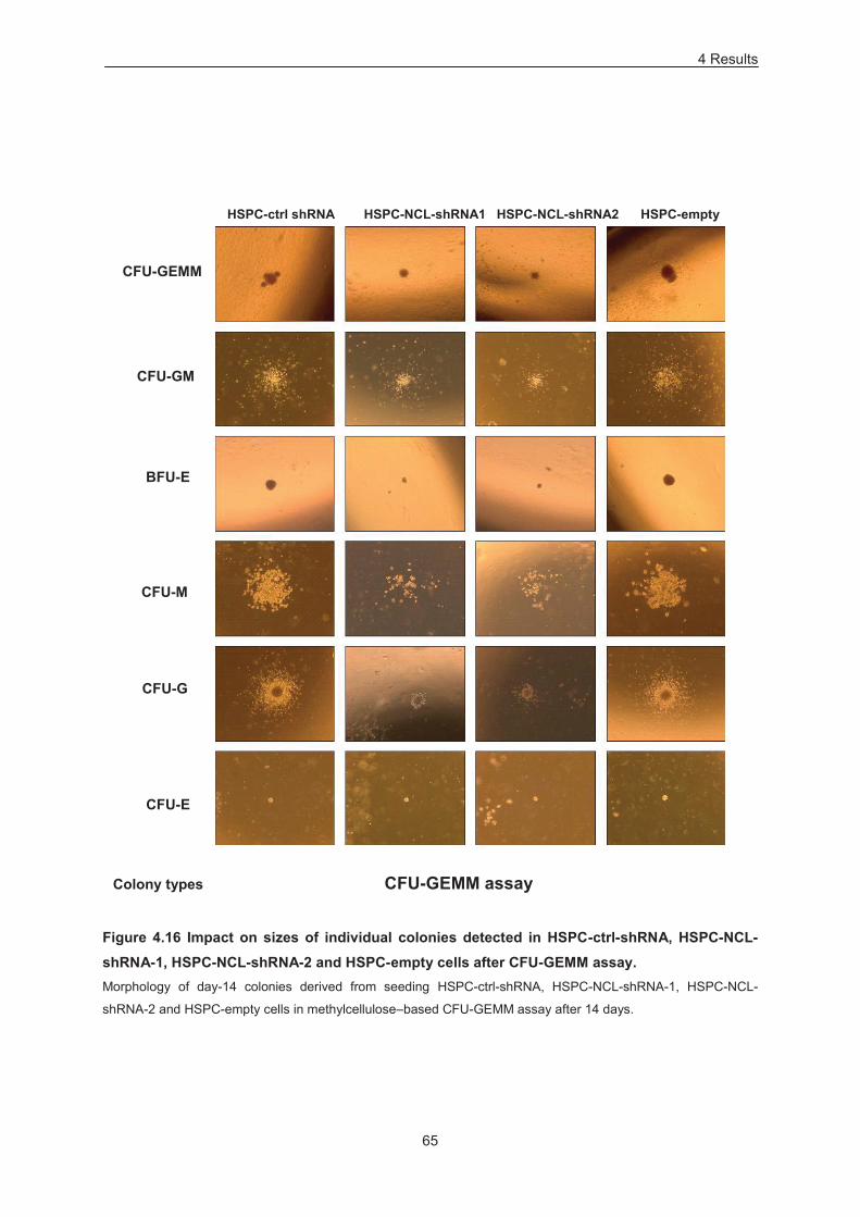
4 Results
65
Figure 4.16 Impact on sizes of individual colonies detected in HSPC-ctrl-shRNA, HSPC-NCL-shRNA-1, HSPC-NCL-shRNA-2 and HSPC-empty cells after CFU-GEMM assay. Morphology of day-14 colonies derived from seeding HSPC-ctrl-shRNA, HSPC-NCL-shRNA-1, HSPC-NCL-
shRNA-2 and HSPC-empty cells in methylcellulose–based CFU-GEMM assay after 14 days.
CFU-GEMM
CFU-GM
BFU-E
CFU-M
HSPC-ctrl shRNA HSPC-NCL-shRNA1 HSPC-NCL-shRNA2 HSPC-empty
CFU-G
CFU-E
Colony types CFU-GEMM assay
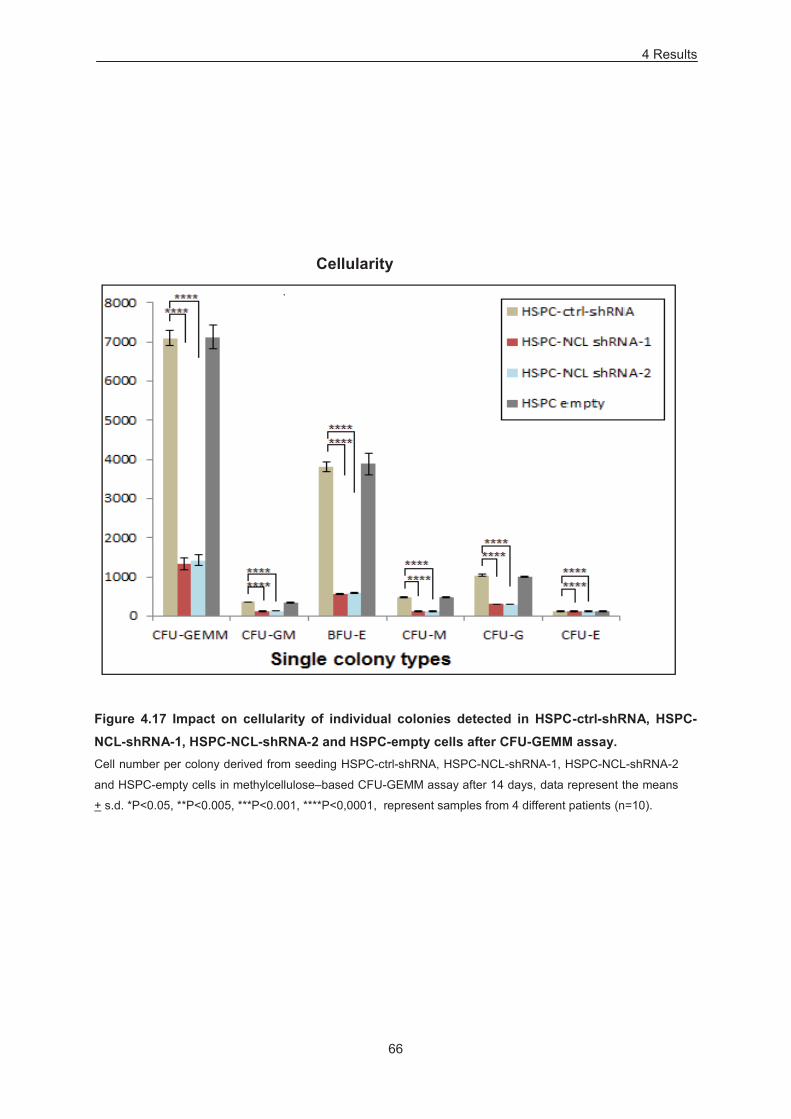
4 Results
66
Cellularity
Figure 4.17 Impact on cellularity of individual colonies detected in HSPC-ctrl-shRNA, HSPC-NCL-shRNA-1, HSPC-NCL-shRNA-2 and HSPC-empty cells after CFU-GEMM assay. Cell number per colony derived from seeding HSPC-ctrl-shRNA, HSPC-NCL-shRNA-1, HSPC-NCL-shRNA-2
and HSPC-empty cells in methylcellulose–based CFU-GEMM assay after 14 days, data represent the means
+ s.d. *P<0.05, **P<0.005, ***P<0.001, ****P<0,0001, represent samples from 4 different patients (n=10).
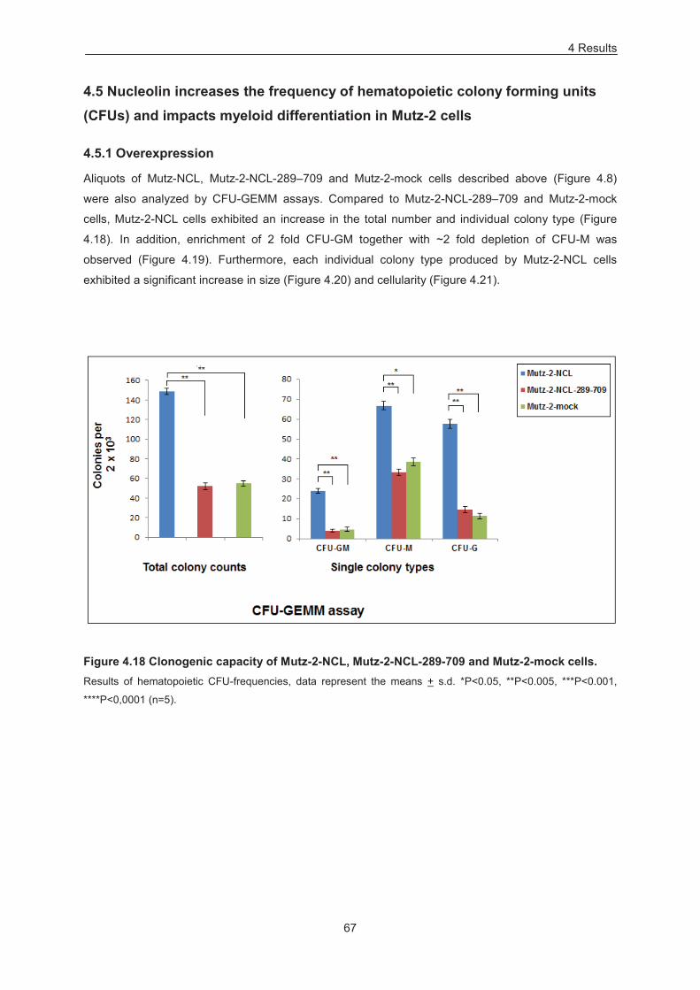
4 Results
67
4.5 Nucleolin increases the frequency of hematopoietic colony forming units (CFUs) and impacts myeloid differentiation in Mutz-2 cells
4.5.1 Overexpression
Aliquots of Mutz-NCL, Mutz-2-NCL-289–709 and Mutz-2-mock cells described above (Figure 4.8)
were also analyzed by CFU-GEMM assays. Compared to Mutz-2-NCL-289–709 and Mutz-2-mock
cells, Mutz-2-NCL cells exhibited an increase in the total number and individual colony type (Figure
4.18). In addition, enrichment of 2 fold CFU-GM together with ~2 fold depletion of CFU-M was
observed (Figure 4.19). Furthermore, each individual colony type produced by Mutz-2-NCL cells
exhibited a significant increase in size (Figure 4.20) and cellularity (Figure 4.21).
Figure 4.18 Clonogenic capacity of Mutz-2-NCL, Mutz-2-NCL-289-709 and Mutz-2-mock cells. Results of hematopoietic CFU-frequencies, data represent the means + s.d. *P<0.05, **P<0.005, ***P<0.001,
****P<0,0001 (n=5).
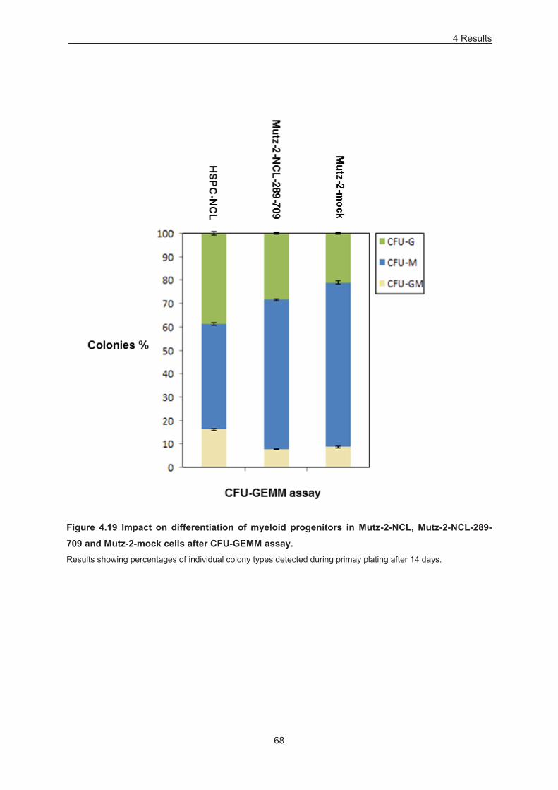
4 Results
68
Figure 4.19 Impact on differentiation of myeloid progenitors in Mutz-2-NCL, Mutz-2-NCL-289-709 and Mutz-2-mock cells after CFU-GEMM assay. Results showing percentages of individual colony types detected during primay plating after 14 days.
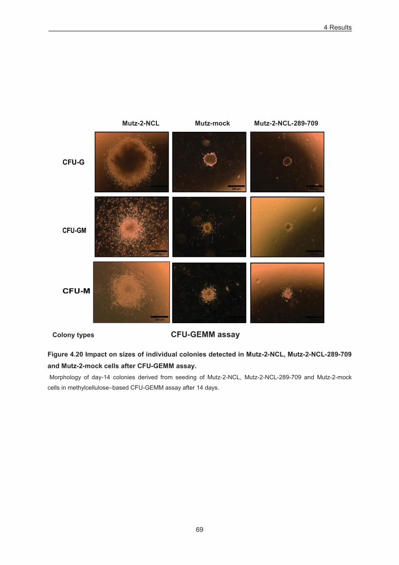
4 Results
69
Figure 4.20 Impact on sizes of individual colonies detected in Mutz-2-NCL, Mutz-2-NCL-289-709 and Mutz-2-mock cells after CFU-GEMM assay. Morphology of day-14 colonies derived from seeding of Mutz-2-NCL, Mutz-2-NCL-289-709 and Mutz-2-mock
cells in methylcellulose–based CFU-GEMM assay after 14 days.
Mutz-2-NCL Mutz-mock Mutz-2-NCL-289-709
Colony types CFU-GEMM assay
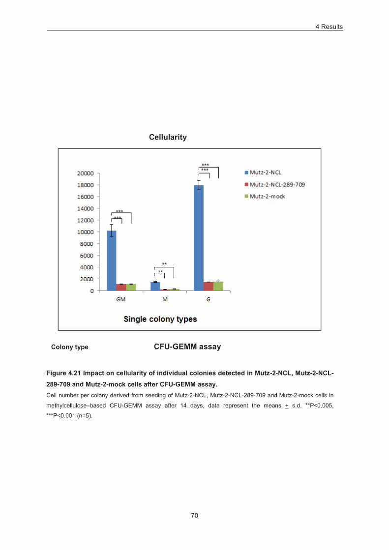
4 Results
70
Cellularity
Figure 4.21 Impact on cellularity of individual colonies detected in Mutz-2-NCL, Mutz-2-NCL-289-709 and Mutz-2-mock cells after CFU-GEMM assay. Cell number per colony derived from seeding of Mutz-2-NCL, Mutz-2-NCL-289-709 and Mutz-2-mock cells in
methylcellulose–based CFU-GEMM assay after 14 days, data represent the means + s.d. **P<0.005,
***P<0.001 (n=5).
CFU-GEMM assay Colony type
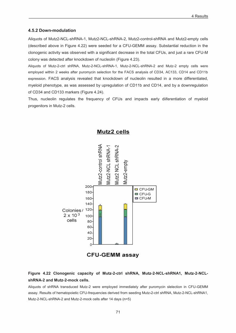
4 Results
71
4.5.2 Down-modulation
Aliquots of Mutz2-NCL-shRNA-1, Mutz2-NCL-shRNA-2, Mutz2-control-shRNA and Mutz2-empty cells
(described above in Figure 4.22) were seeded for a CFU-GEMM assay. Substantial reduction in the
clonogenic activity was observed with a significant decrease in the total CFUs, and just a rare CFU-M
colony was detected after knockdown of nucleolin (Figure 4.23). Aliquots of Mutz-2-ctrl shRNA, Mutz-2-NCL-shRNA-1, Mutz-2-NCL-shRNA-2 and Mutz-2 empty cells were
employed within 2 weeks after puromycin selection for the FACS analysis of CD34, AC133, CD14 and CD11b
expression. FACS analysis revealed that knockdown of nucleolin resulted in a more differentiated,
myeloid phenotype, as was assessed by upregulation of CD11b and CD14, and by a downregulation
of CD34 and CD133 markers (Figure 4.24).
Thus, nucleolin regulates the frequency of CFUs and impacts early diiferentiation of myeloid
progenitors in Mutz-2 cells.
Figure 4.22 Clonogenic capacity of Mutz-2-ctrl shRNA, Mutz-2-NCL-shRNA1, Mutz-2-NCL-shRNA-2 and Mutz-2-mock cells. Aliquots of shRNA transduced Mutz-2 were employed immediately after puromycin slelection in CFU-GEMM
assay. Results of hematopoietic CFU-frequencies derived from seeding Mutz-2-ctrl shRNA, Mutz-2-NCL-shRNA1,
Mutz-2-NCL-shRNA-2 and Mutz-2-mock cells after 14 days (n=5)

4 Results
72
Figure 4.23 Impact on sizes of individual colonies detected in Mutz-2-ctrl shRNA, Mutz-NCL-shRNA2, Mutz-NCL-shRNA1 and Mutz-2-empty cells after CFU-GEMM assay. Morphology of day-14 colonies derived from seeding Mutz-2-ctrl shRNA, Mutz-NCL-shRNA2, Mutz-NCL-shRNA1
and Mutz-2-empty cells in methylcellulose–based CFU-GEMM assay after 14 days.
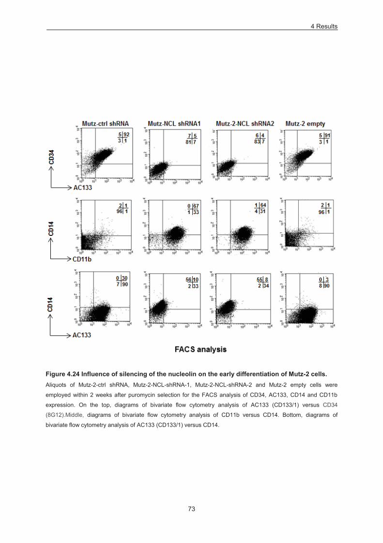
4 Results
73
Figure 4.24 Influence of silencing of the nucleolin on the early differentiation of Mutz-2 cells. Aliquots of Mutz-2-ctrl shRNA, Mutz-2-NCL-shRNA-1, Mutz-2-NCL-shRNA-2 and Mutz-2 empty cells were
employed within 2 weeks after puromycin selection for the FACS analysis of CD34, AC133, CD14 and CD11b
expression. On the top, diagrams of bivariate flow cytometry analysis of AC133 (CD133/1) versus CD34
(8G12).Middle, diagrams of bivariate flow cytometry analysis of CD11b versus CD14. Bottom, diagrams of
bivariate flow cytometry analysis of AC133 (CD133/1) versus CD14.
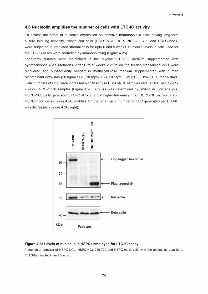
4 Results
74
4.6 Nucleolin amplifies the number of cells with LTC-IC activity
To assess the effect of nucleolin expression on primitive hematopoietic cells having long-term
culture initating capacity, transduced cells (HSPC-NCL, HSPC-NCL-289-709 and HSPC-mock)
were subjected to irradiated stromal cells for upto 6 and 9 weeks. Nucleolin levels in cells used for
the LTC-IC assay were controlled by immunoblotting (Figure 4.25).
Long-term cultures were maintained in the Myelocult H5100 medium supplemented with
hydrocortisone (See Methods). After 6 to 9 weeks culture on the feeder, transduced cells were
recovered and subsequently seeded in methylcellulose medium supplemented with human
recombinant cytokines (50 ng/ml SCF, 10 ng/ml IL-3, 10 ng/ml GMCSF, 3 U/ml EPO) for 14 days.
Total numbers of CFU were increased significantly in HSPC-NCL samples versus HSPC-NCL-289-
709 or HSPC-mock samples (Figure 4.26, left). As was determined by limiting dilution analysis,
HSPC-NCL cells generated LTC-IC at 5- to 9 fold higher frequency, than HSPC-NCL-289-709 and
HSPC-mock cells (Figure 4.26, middle). On the other hand, number of CFC generated per LTC-IC
was decreased (Figure 4.26, right).
Figure 4.25 Levels of nucleolin in HSPCs employed for LTC-IC assay. Immunoblot anaylsis of HSPC-NCL, HSPC-NCL-289-709 and HSPC-mock cells with the antibodies specific to
FLAG-tag, nucleolin and -actin.
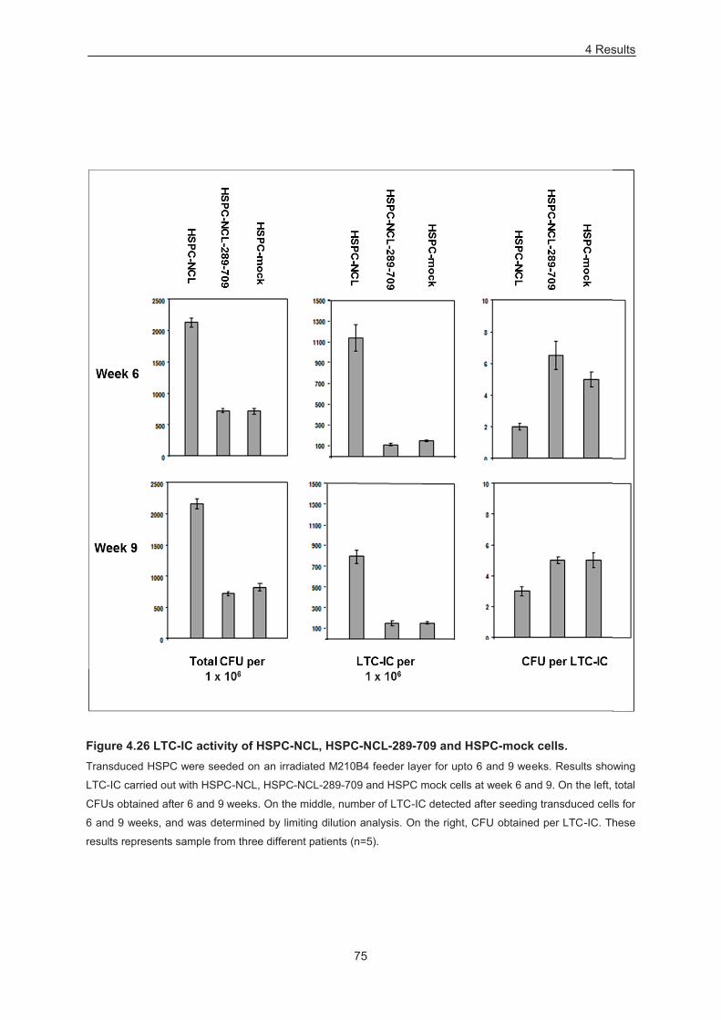
4 Results
75
Figure 4.26 LTC-IC activity of HSPC-NCL, HSPC-NCL-289-709 and HSPC-mock cells. Transduced HSPC were seeded on an irradiated M210B4 feeder layer for upto 6 and 9 weeks. Results showing
LTC-IC carried out with HSPC-NCL, HSPC-NCL-289-709 and HSPC mock cells at week 6 and 9. On the left, total
CFUs obtained after 6 and 9 weeks. On the middle, number of LTC-IC detected after seeding transduced cells for
6 and 9 weeks, and was determined by limiting dilution analysis. On the right, CFU obtained per LTC-IC. These
results represents sample from three different patients (n=5).
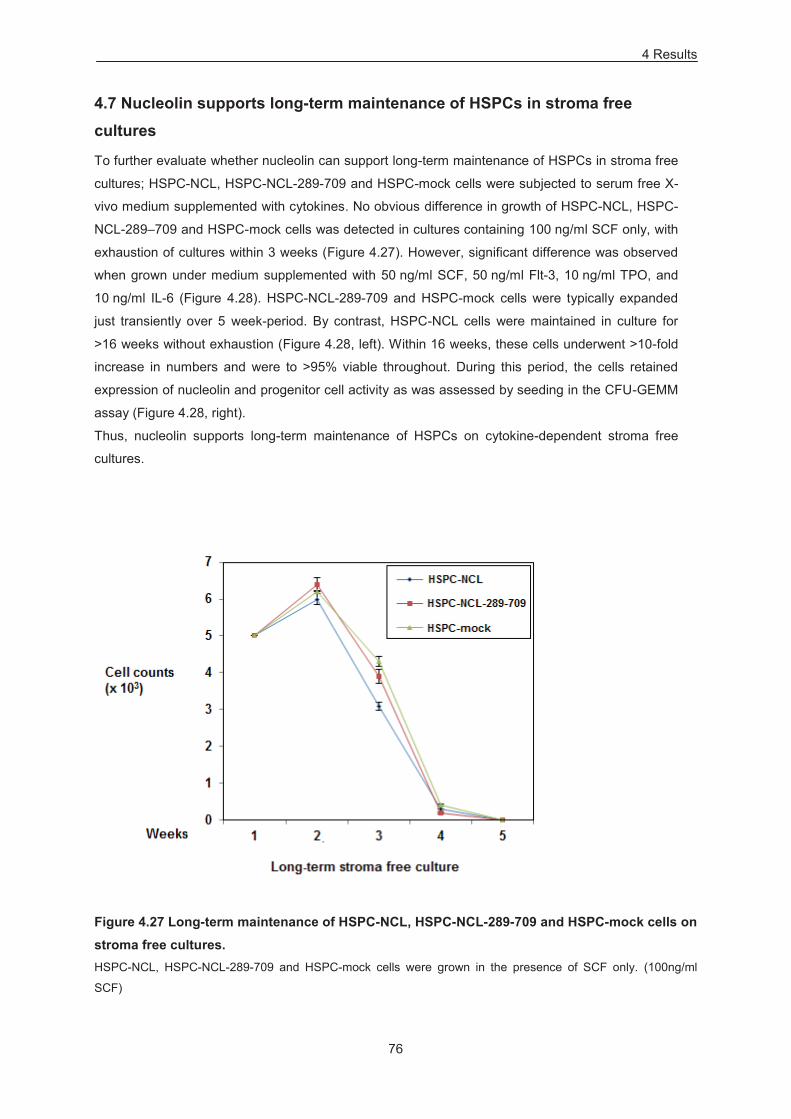
4 Results
76
4.7 Nucleolin supports long-term maintenance of HSPCs in stroma free cultures
To further evaluate whether nucleolin can support long-term maintenance of HSPCs in stroma free
cultures; HSPC-NCL, HSPC-NCL-289-709 and HSPC-mock cells were subjected to serum free X-
vivo medium supplemented with cytokines. No obvious difference in growth of HSPC-NCL, HSPC-
NCL-289–709 and HSPC-mock cells was detected in cultures containing 100 ng/ml SCF only, with
exhaustion of cultures within 3 weeks (Figure 4.27). However, significant difference was observed
when grown under medium supplemented with 50 ng/ml SCF, 50 ng/ml Flt-3, 10 ng/ml TPO, and
10 ng/ml IL-6 (Figure 4.28). HSPC-NCL-289-709 and HSPC-mock cells were typically expanded
just transiently over 5 week-period. By contrast, HSPC-NCL cells were maintained in culture for
>16 weeks without exhaustion (Figure 4.28, left). Within 16 weeks, these cells underwent >10-fold
increase in numbers and were to >95% viable throughout. During this period, the cells retained
expression of nucleolin and progenitor cell activity as was assessed by seeding in the CFU-GEMM
assay (Figure 4.28, right).
Thus, nucleolin supports long-term maintenance of HSPCs on cytokine-dependent stroma free
cultures.
Figure 4.27 Long-term maintenance of HSPC-NCL, HSPC-NCL-289-709 and HSPC-mock cells on stroma free cultures. HSPC-NCL, HSPC-NCL-289-709 and HSPC-mock cells were grown in the presence of SCF only. (100ng/ml
SCF)
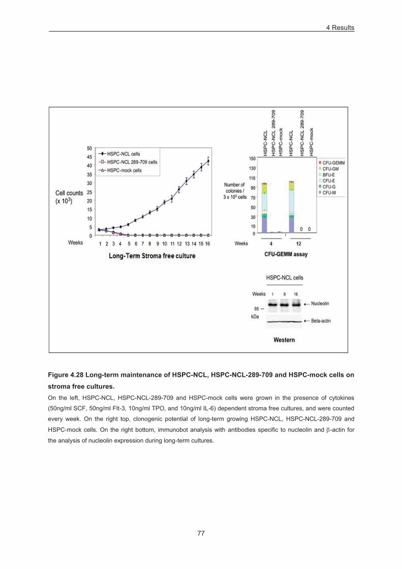
4 Results
77
Figure 4.28 Long-term maintenance of HSPC-NCL, HSPC-NCL-289-709 and HSPC-mock cells on stroma free cultures. On the left, HSPC-NCL, HSPC-NCL-289-709 and HSPC-mock cells were grown in the presence of cytokines
(50ng/ml SCF, 50ng/ml Flt-3, 10ng/ml TPO, and 10ng/ml IL-6) dependent stroma free cultures, and were counted
every week. On the right top, clonogenic potential of long-term growing HSPC-NCL, HSPC-NCL-289-709 and
HSPC-mock cells. On the right bottom, immunobot analysis with antibodies specific to nucleolin and -actin for
the analysis of nucleolin expression during long-term cultures.
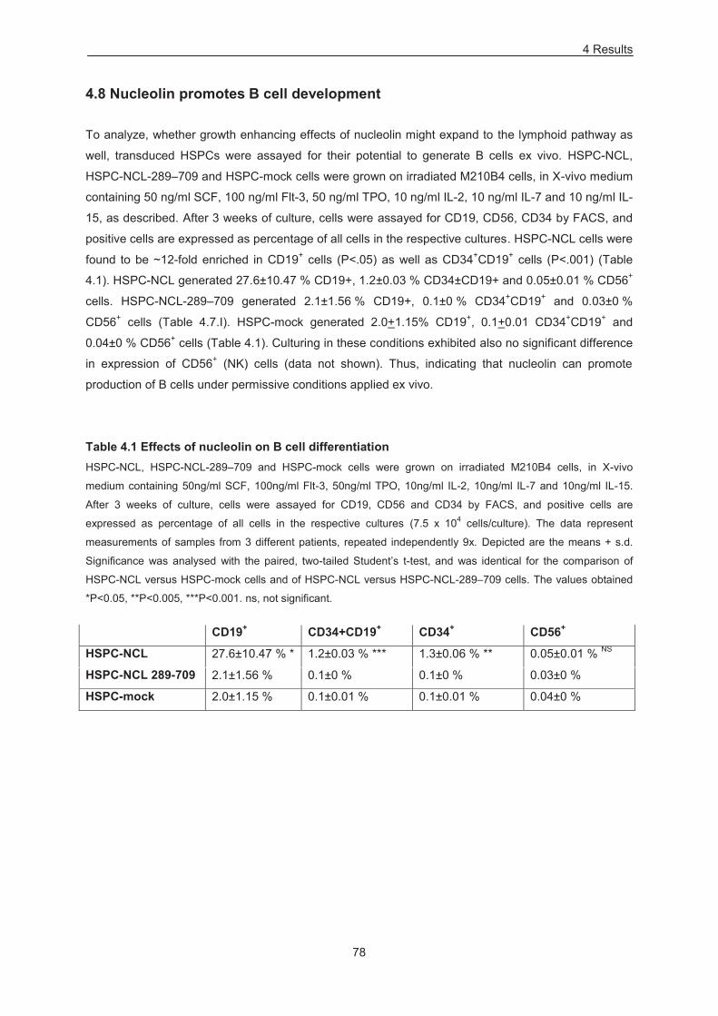
4 Results
78
4.8 Nucleolin promotes B cell development
To analyze, whether growth enhancing effects of nucleolin might expand to the lymphoid pathway as
well, transduced HSPCs were assayed for their potential to generate B cells ex vivo. HSPC-NCL,
HSPC-NCL-289–709 and HSPC-mock cells were grown on irradiated M210B4 cells, in X-vivo medium
containing 50 ng/ml SCF, 100 ng/ml Flt-3, 50 ng/ml TPO, 10 ng/ml IL-2, 10 ng/ml IL-7 and 10 ng/ml IL-
15, as described. After 3 weeks of culture, cells were assayed for CD19, CD56, CD34 by FACS, and
positive cells are expressed as percentage of all cells in the respective cultures. HSPC-NCL cells were
found to be ~12-fold enriched in CD19+ cells (P<.05) as well as CD34+CD19+ cells (P<.001) (Table
4.1). HSPC-NCL generated 27.6±10.47 % CD19+, 1.2±0.03 % CD34±CD19+ and 0.05±0.01 % CD56+
cells. HSPC-NCL-289–709 generated 2.1±1.56 % CD19+, 0.1±0 % CD34+CD19+ and 0.03±0 %
CD56+ cells (Table 4.7.I). HSPC-mock generated 2.0+1.15% CD19+, 0.1+0.01 CD34+CD19+ and
0.04±0 % CD56+ cells (Table 4.1). Culturing in these conditions exhibited also no significant difference
in expression of CD56+ (NK) cells (data not shown). Thus, indicating that nucleolin can promote
production of B cells under permissive conditions applied ex vivo.
Table 4.1 Effects of nucleolin on B cell differentiation HSPC-NCL, HSPC-NCL-289–709 and HSPC-mock cells were grown on irradiated M210B4 cells, in X-vivo
medium containing 50ng/ml SCF, 100ng/ml Flt-3, 50ng/ml TPO, 10ng/ml IL-2, 10ng/ml IL-7 and 10ng/ml IL-15.
After 3 weeks of culture, cells were assayed for CD19, CD56 and CD34 by FACS, and positive cells are
expressed as percentage of all cells in the respective cultures (7.5 x 10⁴ cells/culture). The data represent
measurements of samples from 3 different patients, repeated independently 9x. Depicted are the means + s.d.
Significance was analysed with the paired, two-tailed Student’s t-test, and was identical for the comparison of
HSPC-NCL versus HSPC-mock cells and of HSPC-NCL versus HSPC-NCL-289–709 cells. The values obtained
*P<0.05, **P<0.005, ***P<0.001. ns, not significant.
CD19+ CD34+CD19+ CD34+ CD56+
HSPC-NCL 27.6±10.47 % * 1.2±0.03 % *** 1.3±0.06 % ** 0.05±0.01 % NS
HSPC-NCL 289-709 2.1±1.56 % 0.1±0 % 0.1±0 % 0.03±0 %
HSPC-mock 2.0±1.15 % 0.1±0.01 % 0.1±0.01 % 0.04±0 %
4 Results
79
4.9 Functional effects of nucleolin on HSPCs partially relies on -catenin
activity
To evaluate the potential effect of nucleolin on Wnt / β-catenin and PI3K / Akt pathway, aliquots of
HSPC-NCL, HSPC-NCL-289-709 and HSPC-mock cells were taken and subjected to immunoblotting
with Abs specific for active β-catenin, total β-catenin, and for two distinct forms of Akt activated
through phosphorylation, pSer308-Akt or pSer473-Akt (Manning and Cantley, 2007) (Figure 4.29, left).
Levels of active β-catenin, pSer308-Akt and pSer473-Akt, were increased ~4 fold each, and total -
catenin ~3 fold in HSPC-NCL versus HSPC-NCL-289-709 and HSPC-empty cells (Figure 4.29, left).
No increase in total Akt levels was detected, while BCL2, an established target of nucleolin (Otake et
al., 2007; Grinstein et al., 2007) was increased ~5 fold (Figure 4.29, left). Similarly, immunoblot
analysis with aliquots of cells subjected to down-modulation of nucleolin expression (HSPC-NCL-
shRNA-1, HSPC-NCL-shRNA-2, HSPC-control-shRNA and HSPC-empty cells) confirmed its
involvement in Wnt / β-catenin and PI3K / Akt pathway (Figure 4.29, right).
To investigate, whether the nucleolin dependent increase of β-catenin would, at least in part, accounts
for any effect on its clonogenic potential, a β-catenin antagonist PKF-115-584 was administered during
a CFU-GEMM assay (Figure 4.30). At a concentration >8 fold below IC50 (Leourcelet et al., 2004),
PKF-115-584 exhibited no toxicity on HSPCs, both in liquid and semisolid cultures. Upon inhibitor
addition to semisolid media, HSPC-empty cells exhibited ~2 fold decrease in total colonies, CFU-
GEMM and CFU-GM (Figure 4.30). By contrast, there was a significantly stronger effect of the inhibitor
on HSPC-NCL cells, whereby most effects of nucleolin on HSPCs, detectable by CFU-GEMM assays,
were abrogated (Figure 4.30). These effects were: ~4-fold decrease in total CFU and CFU-GEMM, ~5-
fold depletion of CFU-GM and ~3-fold enrichment of CFU-M. Inhibitor treatment to HSPC-mock cells
showed no or little effect on colony size (Figure 4.31) and cellularity (Figure 4.32) whereas HSPC-NCL
cells exhibited an evident decrease in size (Figure 4.31) and cellularity of CFU-GM (Figure 4.32).
Furthermore, HSPC-NCL cells maintained the expression of active β-catenin in long-term stroma-free
cultures for at least 16 weeks (Figure 4.33A). Treatment of liquid cultures with PKF-115-584 or with
another, structurally distinct, β-catenin inhibitor CGP049090 (Leourcelet et al., 2004) at a
concentrations >20-fold below IC50, resulted in exhaustion of HSPC-NCL cells within 2-4 weeks of
treatment. In Figure 4.33, B, the effect of each inhibitor on a 16-week-culture is exemplarily shown.
Thus, effects of nucleolin on HPCs partially rely on β-catenin activity.
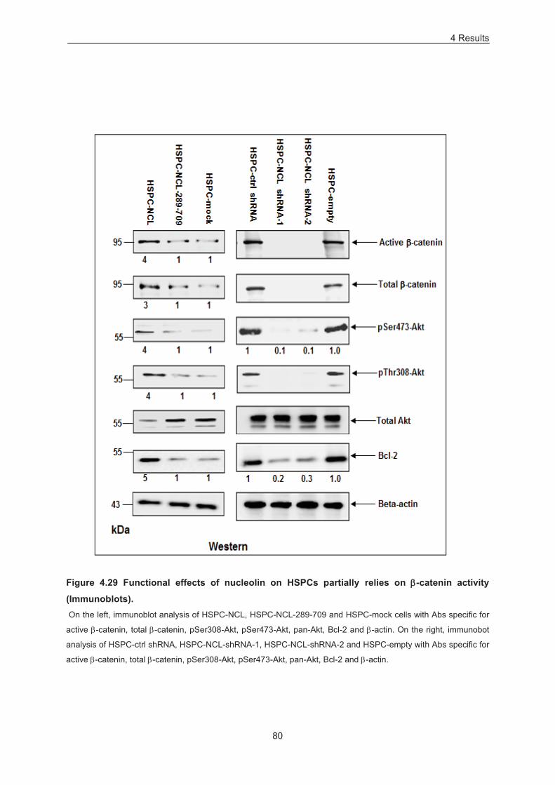
4 Results
80
Figure 4.29 Functional effects of nucleolin on HSPCs partially relies on -catenin activity
(Immunoblots). On the left, immunoblot analysis of HSPC-NCL, HSPC-NCL-289-709 and HSPC-mock cells with Abs specific for
active -catenin, total -catenin, pSer308-Akt, pSer473-Akt, pan-Akt, Bcl-2 and -actin. On the right, immunobot
analysis of HSPC-ctrl shRNA, HSPC-NCL-shRNA-1, HSPC-NCL-shRNA-2 and HSPC-empty with Abs specific for
active -catenin, total -catenin, pSer308-Akt, pSer473-Akt, pan-Akt, Bcl-2 and -actin.
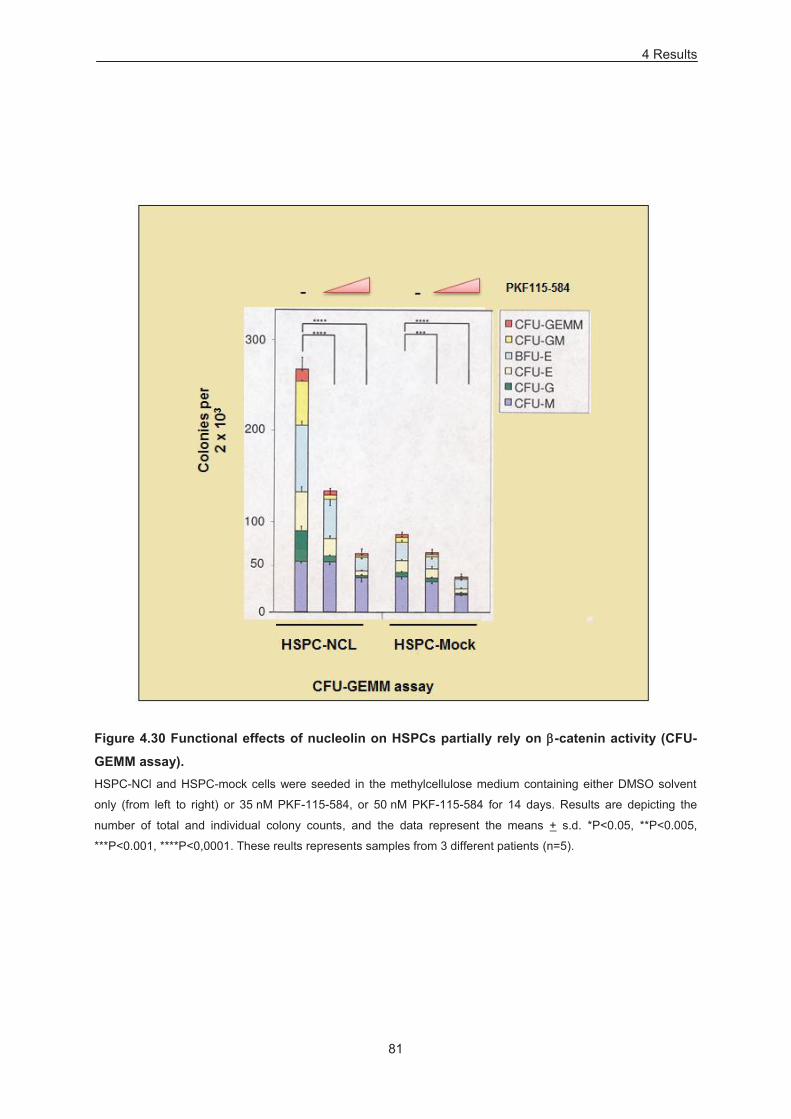
4 Results
81
Figure 4.30 Functional effects of nucleolin on HSPCs partially rely on -catenin activity (CFU-
GEMM assay). HSPC-NCl and HSPC-mock cells were seeded in the methylcellulose medium containing either DMSO solvent
only (from left to right) or 35 nM PKF-115-584, or 50 nM PKF-115-584 for 14 days. Results are depicting the
number of total and individual colony counts, and the data represent the means + s.d. *P<0.05, **P<0.005,
***P<0.001, ****P<0,0001. These reults represents samples from 3 different patients (n=5).
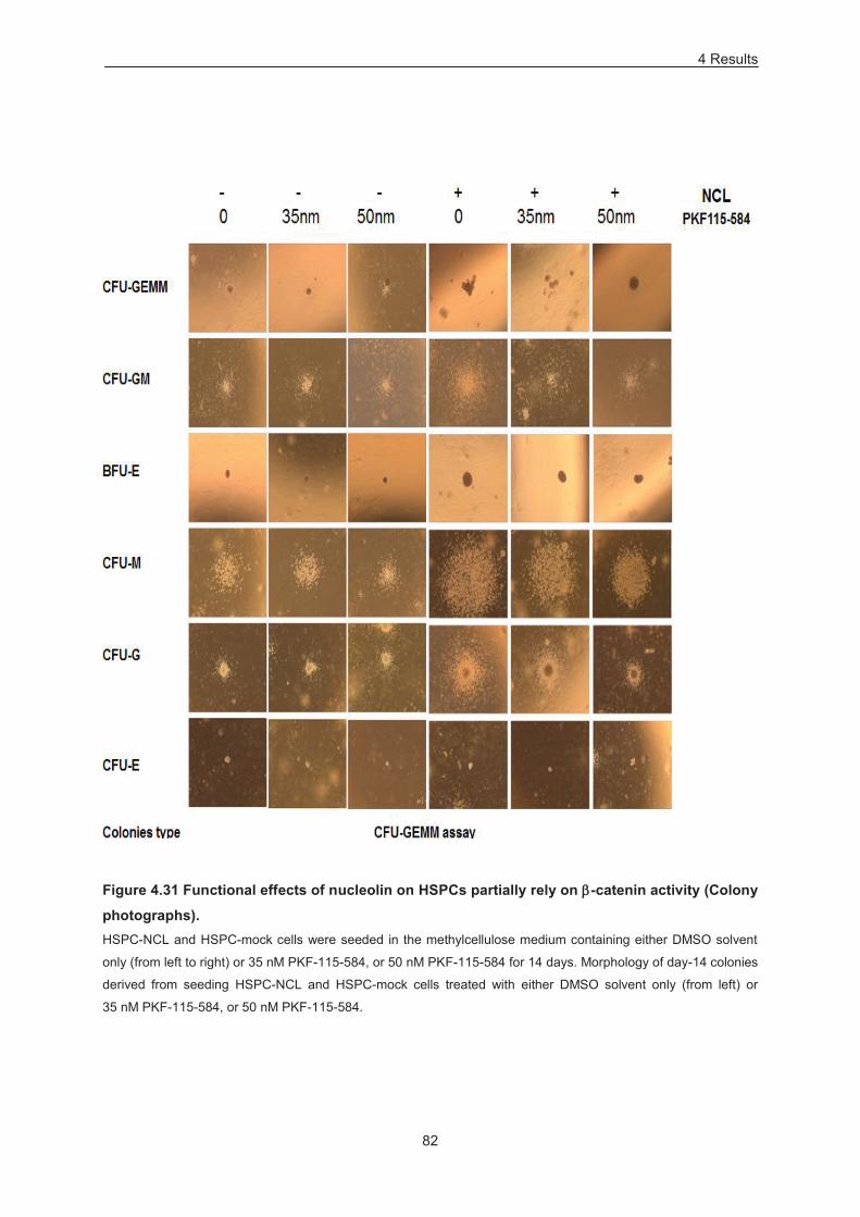
4 Results
82
Figure 4.31 Functional effects of nucleolin on HSPCs partially rely on -catenin activity (Colony
photographs). HSPC-NCL and HSPC-mock cells were seeded in the methylcellulose medium containing either DMSO solvent
only (from left to right) or 35 nM PKF-115-584, or 50 nM PKF-115-584 for 14 days. Morphology of day-14 colonies
derived from seeding HSPC-NCL and HSPC-mock cells treated with either DMSO solvent only (from left) or
35 nM PKF-115-584, or 50 nM PKF-115-584.
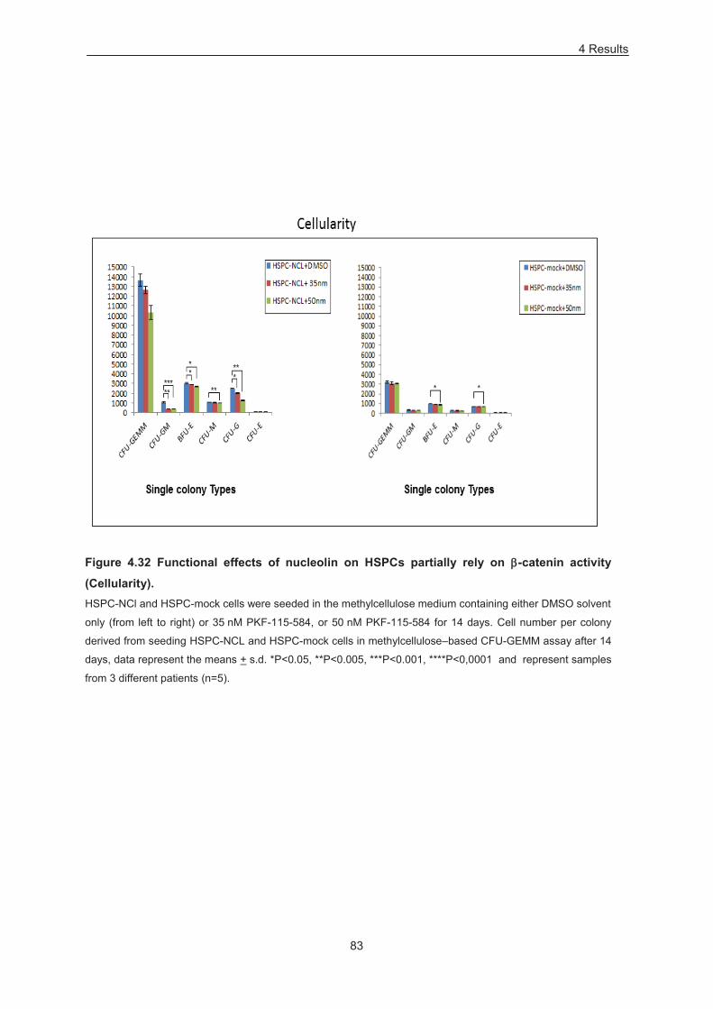
4 Results
83
Figure 4.32 Functional effects of nucleolin on HSPCs partially rely on -catenin activity
(Cellularity). HSPC-NCl and HSPC-mock cells were seeded in the methylcellulose medium containing either DMSO solvent
only (from left to right) or 35 nM PKF-115-584, or 50 nM PKF-115-584 for 14 days. Cell number per colony
derived from seeding HSPC-NCL and HSPC-mock cells in methylcellulose–based CFU-GEMM assay after 14
days, data represent the means + s.d. *P<0.05, **P<0.005, ***P<0.001, ****P<0,0001 and represent samples
from 3 different patients (n=5).
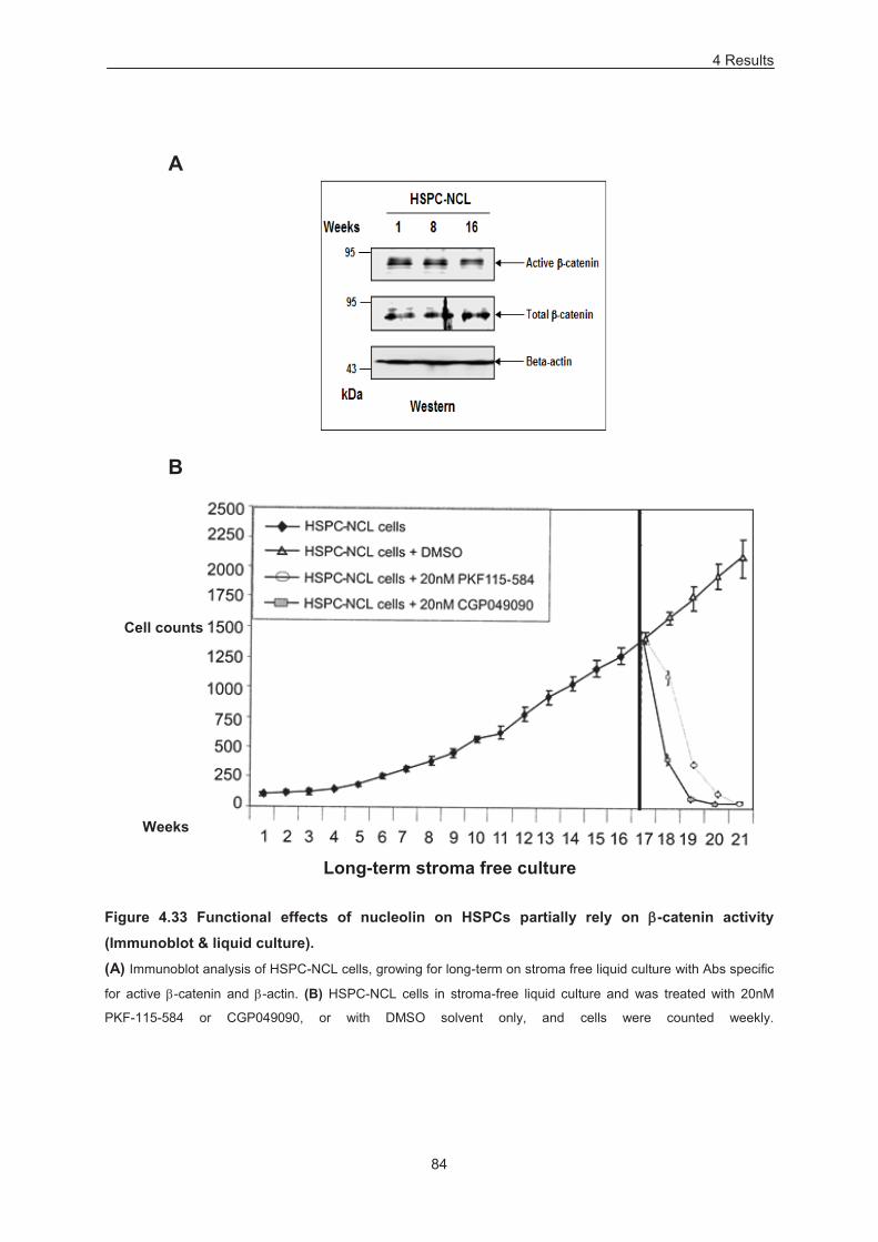
4 Results
84
A
B
Figure 4.33 Functional effects of nucleolin on HSPCs partially rely on -catenin activity
(Immunoblot & liquid culture). (A) Immunoblot analysis of HSPC-NCL cells, growing for long-term on stroma free liquid culture with Abs specific
for active -catenin and -actin. (B) HSPC-NCL cells in stroma-free liquid culture and was treated with 20nM
PKF-115-584 or CGP049090, or with DMSO solvent only, and cells were counted weekly.
Weeks
Cell counts
Long-term stroma free culture
85
5 Discussion
5.1 AC133 & CD133 expression in the hematopoietic system
Hematopoiesis is carried out of a few numbers of uncommitted HSPCs, which are capable of self
renewing and differentiate into entire lineages of the hematopoietic system. Since the number of this
rare primitive population is very low in the large heterogenous population, their selective enrichment
and in vivo chraracterization in the bone marrow compartment depends upon the expression or the
absence of some specific surface markers. From the past decades, CD34 is the marker of choice to
enumerate HSPCs and even clinically for autologous and allogenic bone marrow transplantation
(Krause et al., 1996; Lee et al., 2004).
CD133 (Prominin-1), a pentaspan transmembrane glycoprotein has obtained a considerable interest in
the recent times because of its expression on the stem and progenitor cells from various sources,
including the hematopoietic system (Yin et al., 1997). AC133 antigen is a specific glycoform of CD133,
and has significant importance in hematopoietic stem cell biology, originally identified as an additional
marker to isolate CD34+ HSCs, and later found on CD34– HSCs fraction as well (Yin et al., 1997;
Gallacher et al., 2000; Bauer et al., 2008; Takahashi et al., 2014). CD34+ HSCs marked with AC133
exhibit primitive nature, and possess elevated clonogenic potential and long term maintenance (Yin et
al., 1997; de Wynter et al., 1998; Gallacher et al., 2000; Gordon et al., 2003). Furthermore, CD133 is
released from HSPCs in membrane vesicles upon differentiation (Bauer et al., 2011). CD133 functions
as modifier of hematopoietic progenitor cell (HPC) frequencies in a mouse model (Arndt et al., 2013).
Microarray expression profiling of CD133+ versus CD133– cells in several studies have reported a
significant difference in the regulation of several sets of genes between these two cell populations,
which could provide a insight into the biological pathways that regulate CD133 expression or its
biological function (Toren et al., 2005; Liu et al., 2006; Hemmoranta et al., 2007; Sepherd et al., 2008;
Bertolini et al., 2009; Yan et al., 2011). These studies indicate primitive nature of CD133+ cells over
CD34+ cells, as well as the different roles played by these two populations during hematopoietic
regeneration.
Beside that, CD133+ HSPCs have the capacity to reconstitute the whole immune system of a lethally
irradiated mouse (de Wynter et al., 1998). AC133 is frequently referred, but still a debated marker of
stem/progenitor cells in normal and cancerous tissues (Irollo and Pirozzi, 2013). However, AC133 is
used in the prospective isolation of tumour-initiating cells in childhood ALL, mantle cell lymphoma,
AML (Cox et al., 2009; Medina et al., 2014; Beghini et al., 2012) and its expression is required for the
growth of AC133+ ALL cells (Mak et al., 2012). Immunomagnetic selection of CD133+ HSPCs allowed
5 Discussion
86
adequate enrichment of the cells capable of performing hematopoietic stem cell transplantation
(Gordon et al., 2003). In pilot trials with children suffering from leukemia, CD133+ selection has proven
the possibility for allogenic transplantion (Koehl et al., 2002; Lang et al., 2004). In a different study,
CD133+ enriched stem cells have exhibited successful transplantation of haploidentical mismatched
PB stem cells (Bitan et al., 2005).
Nevertheless, mechanisms that activate AC133 and CD133 expression in HSPCs and their potential
relationship to properties of these cells need to be further explored. Human AC133 expression is
controlled at multiple levels, partly in a tissue-specific fashion (Irollo and Pirozzi, 2013; Tabu et al.,
2013). Transcriptional, epigenetic and post-transcriptional control of AC133 expression has been well
demonstrated (Shmelkov et al., 2004; De Felice et al., 2005; Florek et al., 2005; Kemper et al; 2010;
Bauer et al., 2011; Irollo and Pirozzi, 2013). CD133 is transcribed from alternative tissue-dependent
promoters and, of these, promoter P1 is utilized by CD34+ HSPC, as opposed to more differentiated
hematopoietic cells, thus paralleling CD133 mRNA and protein levels (Shmelkov et al., 2004; Florek et
al., 2005). Beside that, wide range of tissues and cell types express the CD133 mRNA but nearly all
fetal and adult tissues are negative for AC133 expression on their surface, except in the case of
human bone marrow samples (Miraglia et al., 1997; Yu et al., 2002; Shmelkov et al., 2004; Florek et
al., 2005).
5.2 Nucleolin mediated AC133 and CD133 expression
The present study dissects the activation of AC133 and CD133 expression by nucleolin, a nucleolar
phosphoprotein found at elevated levels in human and murine HSCs as opposed to differentiated
hematopoietic tissues (Terskikh et al., 2001; Grinstein et al., 2007). In previous studies, it was
demonstrated that nucleolin binds to the promoter region of the CD34 gene through direct sequence
specific interaction and modulates both the endogenous and the cell surface level of the CD34 in
human CD34+ cells (Grinstein et al., 2007). In this present study, nucleolin enriched HSPCs were
found to express higher levels AC133 on the surface in contrast to a N-terminally truncated nucleolin
variant (aa 289–709) and control cells (Figure 4.1). Beside that, there was a significant increase of cell
surface CD34 level, CD34 transcripts and endogenous CD34 was observed (Figure 4.2). Similar
effects of nucleolin on AC133 expression was observed in leukemic cell line models (Figure 4.7 & 4.9).
Total cellular CD133 transcripts, or transcripts initiated at single CD133 gene P1 promoter, revealed
higher levels with nucleolin overexpressing HSPCs, Mutz-2 and also SEM cells, while the level of
transcripts initiated at promoter P2 (Shmelkov et al., 2004) were unaffected (Figure 4.1, 4.7 & 4.9). To
further confirm the role of nucleolin in the regulation of AC133 and CD133 expression, knockdown of
nucleolin was carried out either by nucleofecting shRNA constructs targeting different regions of
nucleolin in the case of HSPCs or stable expression of shRNA in the case of leukemic cell line
models. In line with the overexpression analyses, down-modulation of nucleolin resulted in a
significant decrease of AC133 and CD34 surface expression, both in case of HSPCs and Mutz-2 cells
(Figure 4.5 & 4.6). To add further, endogenous CD133 protein and CD133 transcripts revealed
significant reduction upon nucleolin down-modulation in primary HSPCs and in Mutz-2 cells (Figure
4.5 & 4.6). Total cellular CD133 transcripts or transcripts initiated at single CD133 gene promoter P1
revealed lower levels of transcripts in HSPCs and Mutz-2 cells, when treated with the nucleolin
5 Discussion
87
targeting shRNA versus controls shRNA. Again the levels of transcripts initiated at promoter P2 were
identical (Figure 4.5 & 4.6). Immunoblot analyses and trypan exclusion revealed that the viability of the
HSPCs and Mutz-2 was not affected.
The results in this work demonstrate the role of nucleolin in the activation of AC133 and CD133
expression by nucleolin via its promoter P1, which is a novel mechanism that crucially contributes to
AC133 expression in CD34+ HSPCs. Expression of endogenous CD133 protein and CD133 transcripts
in HSPCs and in Mutz-2 cells behaved in line with the AC133 surface expression and also in
agreement with the previous studies (Miraglia et al., 1997; Yu et al., 2002; Shmelkov et al., 2004;
Florek et al., 2005). Beside that, surface CD34 and endogenous CD34 protein levels were found
upregulated in the context with the previous report (Grinstein et al., 2007). N-terminal truncated
nucleolin variant has not regulated the expression of AC133 and it cognate gene CD133. These
results are in line with the previous studies, as N-terminal of nucleolin was implicated in the gene
regulation (Hanakahi et al., 1997; Angelov et al., 2006).
Thus, nucleolin modulates AC133 and CD133 expression of CD34+ MPB HSPCs and leukemic cell
line models. This is attained by upregulating CD133 gene transcripts initiated at promoter P1 and
involves the N-terminal domain of nucleolin thereof.
5.3 Nucleolin likely facilitates polarization of HSPCs
Better understanding of trafficking mechanism regulating the migration of HSPCs during an organism’s
development and later maintaining hemtopoeisis is not only important from a biological point of view,
but also comprises therapeutic purpose. For instance, bone marrow transplantation is a renowned
procedure during hematological diseases; however the mechanism involved in the migration and later
on engrafment of HSPCs in the bone marrow niche is not well understood. Among novel biological
features of migrating HSPCs, polarization of their membrane and cytoplasmic components, which later
give rise to the development of a specific sub-cellular organization located at their rear pole (uropod),
has gained significant attention. As a pentaspan transmembrane molecule, AC133 predominantly
localizes to plasma membrane protrusions, thereby associating with membrane microdomains, and
segregation of AC133 can be viewed as a marker of HSPC polarity (Röper et al., 2000; Giebel et al.,
2004; Bauer et al., 2011).
In this study, it came out that the nucleolin overexpressing HSPCs were morphologically distinct from
control cells and exhibited membrane protrusions (Figure 4.3). Percentage of HSPCs with membrane
protrusions in nucleolin overexpressing cells was significantly higher versus control transduced cells.
This indicates that membrane polarization likely occurred, potentially due to increase active Akt levels
(Figure 4.29). Further studies are required to investigate nucleolin-dependent HSPC polarization and
functional significance thereof.
5 Discussion
88
5.4 Nucleolin regulates hematopoietic colony formation in HSPCs and in Mutz-2 cells
To evaluate whether nucleolin mediated modulation of AC133 and CD133 expression might extend to
colony forming ability as well, nucleolin transduced HSPCs along with the controls cells were seeded
in the standard CFU-GEMM assay. Primary plating with nucleolin enriched HSPCs revealed significant
increase in both total colony count and individual colony types after 14 days (Figure 4.10, A). As was
found in earlier reports, AC133+ HSPCs are of primitive nature and are enriched for CFUs (Yin et al.,
1997; de Wynter et al., 1998; Gallacher et al., 2000; Gordon et al., 2003). Beside that, a MPB derived
AC133+CD34+ HSPCs fraction is mainly enriched in CFU-GM and CFU-GEMM colony types, while
AC133–CD34+ in BFU-E (Gordon et al., 2003). In another report, the CD34 molecule supports
differentiation of HSPCs toward erythroid lineage (Salati et al., 2008). Thus, in line with the activation
of AC133 and CD34 expression, in our study nucleolin significantly increased the frequency of total
CFUs, CFU-GM, CFU-GEMM and BFU-E output (Figure 4.10, A). In addition, nucleolin
overexpressing HSPCs were enriched for CFU-GM colony type and to a lesser extent, CFU-GEMM
(Figure 4.11). With the exception of CFU-E, each colony type produced with nucleolin exhibited
significantly increased cellularity and size (Figure 4.12 & 4.13). Compared to control cells, HSPCs
transduced with NCL exhibited an increased replating capacity (Figure 4.10, B). Similarly, Mutz-2 cells
also exhibited increase in the frequency of total CFUs, size & cellularity of colonies together with the
enrichment of CFU-GM colony type upon nucleolin overexpression (Figure 4.18–4.21).
To further confirm the dependence of colony formation on nucleolin, shRNA transfected HSPCs were
seeded in a methylcellulose assay. Significant reduction of clonogenic cells was observed with a
decrease in the total CFUs and individual colony types (Figure 4.14). When compared to control
shRNA transfected HSPCs, both nucleolin shRNA tramsfected (shRNA-1 & shRNA-2) HSPCs
exhibited enrichment of CFU-M (Figure 4.15). Similar results were obtained when Mutz-2 cells treated
with shRNA were seeded in standard CFU-GEMM assay. The knockdown of nucleolin led to the
strong decrease in the total CFC numbers and just rare CFU-M colonies was detected (Figure 4.22).
Beside that, knockdown of nucleolin in Mutz-2 cells resulted in a more differentiated phenotype as was
monitored by the decrease of CD133 and CD34 levels, and upregulation of CD14 and CD11b levels
(Figure 4.24). These reults provide an independent proof for the role played by nucleolin in
maintaining undifferentiated phenotype of Mutz-2 cells
Thus, nucleolin mediated increase of AC133 and CD133 expression in HSPCs and Mutz-2 cells lead
to the increases of hematopoietic CFU frequencies, individual colony types, and their respective sizes
& cellularity (with exception of CFU-E in HSPCs). In addition, nucleolin also impacted the enrichment
of CFU-GM colony type in HSPCs and Mutz-2 cells. Furthermore, knockdown of nucleolin in Mutz-2
cells can lead to the early differentiation into myeloid phenotype.
5.5 Impact of nucleolin on LTC-IC frequencies and long-term maintenance of HSPSc
The unique property of primitive progenitors within the hematopoietic system is the ability of
generating blood cells during the entire lifetime of an organism. Characterization of intrinsic and
5 Discussion
89
extrinsic factors influencing self renewal, lineage commitment and early differentiation of HSPCs
remain a key challenge in clinical and experimental biology. The factors like multilineage
reconstitution, level & time of engraftment has led us to characterize strictly within the HSCs
population, such as long-term engrafting HSCs, short term engrafting or multipotent progenitor cells
(MPP) and more mature lineage commited progenitors that have lost self-renewal capacity (Morrison
et al.,1994; Weissman et al.,2000). Long-term culture initiating cells (LTC-ICs) are a discrete and rare
type of primitive hemopoeitic progenitor fraction, found in the bone marrow and also in the circulating
blood. They are capable of generating clonogenic progeny noticeable after a minimum growth on
appropriate feeder layer cells for 5 weeks.
To assess the effect of nucleolin expression on primitive hematopoietic cells, nucleolin enriched
HSPCs along with control samples were subjected to irradiated stromal cells for upto 6 and 9 weeks
(Figure 4.26). After 6 to 9 weeks culturing on the feeder, transduced cells were recovered and
subsequently seeded in methylcellulose medium supplemented with human recombinant cytokines for
14 days. Limiting dilution analysis provided significant higher frequencies of LTC-IC generated by
nucleolin enriched HSPCs (Figure 4.26, middle). On the other hand, the number of CFC generated per
LTC-IC was decreased (Figure 4.26, right). Both the endogenous and exogenous nucleolin levels in
the cells administered for the LTC-IC assay were controlled for the expression of its endogenous and
exogenous levels (Figure 4.25).
To further evaluate whether nucleolin can support long-term maintenance of HSPCs in stroma free
cultures; nucleolin enriched HSPCs along with the control were subjected to serum free medium
supplemented with cytokines (Figure 4.28). Control HSPCs were typically expanded just transiently
over 5 week-period. By contrast, nucleolin enriched HSPCs were maintained in culture for >16 weeks
without exhaustion (Figure 4.28, left). During this period, the cells retained expression of nucleolin and
progenitor cell activity (Figure 4.28, right). In addition, nucleolin enriched cells underwent a significant
increase in numbers and were predominantly viable throughout the long-term culture. However, no
noticeable difference in growth of nucleolin enriched HSPCs versus control cells was detected in
cultures containing SCF only, with the exhaustion of cultures within 3–4 weeks (Figure 4.27). This
suggests that the nucleolin mediated long-term maintenance of HSPCs is dependent on a mixture of
cytokines (SCF, Flt-3, TPO, and IL-6), because similar effects were not replicated if grown in SCF
only.
In this context, AC133+ HSPCs are of primitive nature and are enriched for LTC-ICs (Yin et al., 1997;
de Wynter et al., 1998; Gallacher et al., 2000; Gordon et al., 2003). Furthermore in CD34+ HSPCs,
nucleolin transcripts are commonly upregulated by HOXB4 and HOXC4 homeobox proteins (Buske et
al., 2002; Auvrav et al., 2012). Therefore the effect on the amplification of LTC-ICs number with
overexpression of HOXB4 and HOXC4 in CD34+ HSPCs has been well established (Buske et al.,
2002; Auvrav et al., 2012). This study explicitly provides an indirect link between nucleolin and
homeobox protein expression. Serial transplantation analysis in mouse revealed a crucial role of Akt1
and Akt2 in the maintenance of long term HSCs (Juntilla et al., 2010). Moreover, β-catenin in
cooperation with PI3K / Akt or with Bcl-2 can promote expansion of HSPCs with long term function
(Reya et al., 2003; Perry et al., 2011). To add further, activation of PI3K / Akt or Wnt / β-catenin is
5 Discussion
90
inadequate to expand primitive HSCs, though combination of both these pathway is required to
expand HSCs for long-term (Perry et al., 2011).
The increase in active β-catenin, active Akt and BCL-2 likely provides an explanation for the higher
LTC-IC frequencies and long-term maintenance of hematopoietic progentiors (Figure 4.1 & 4.29).
5.6 Effect of nucleolin on B cell differentiation
To assess, whether growth enhancing effects of nucleolin might expand to the lymphoid pathway as
well, transduced HSPCs were assayed for their potential to generate B cells ex vivo. Nucleolin
enriched HSPCs provided significant enrichment in CD19+ and CD19+CD34+ cells versus control cells,
after three week of culture in conditions permissive for B cell development (Table 4.1). However there
was no significant increase of CD56+ NK-cells was observed. As mentioned above, nucleolin
transcripts are commonly upregulated by HOXB4 and HOXC4 homeobox proteins (Buske et al., 2002;
Auvrav et al., 2012). The effect on the amplification of B cells via overexpression of HOXB4 and
HOXC4 in CD34+ HSPCs has been established (Buske et al., 2002; Auvrav et al., 2012). Moreover, in
mouse studies the role of Bcl-2 has been well established in the development of lymphoid cells from
HSCs (Matsuzaki et al., 1997). Therefore, the observed increase in the number of CD19+ and
CD19+CD34+ cells by nucleolin in the conditions permissive for B cell development is in line with the
previous reports.
5.7 Nucleolin mediated regulation of β-catenin, PI3K / Akt and BCL-2 axis
Here we show that, in CD34+ HSPCs, levels of active β-catenin, active Akt and BCL-2 are nucleolin
dependent. In brief, nucleolin overexpressing HSPCs exhibited elevated levels of active and total β-
catenin, as assessed by immunobloting analysis (Figure 4.29, left). Similar dependence of nucleolin
was observed in HSPCs, when treated with shRNA targeted nucleolin (Figure 4.29, right). To further
investigate, whether the nucleolin dependent increase of β-catenin would, at least in part, accounts for
its effect on clonogenic potential, a β-catenin antagonist PKF-115-584 was administered during a
CFU-GEMM assay (Figure 4.30). A significant effect of the inhibitor on HSPCs overexpressing
nucleolin was observed, whereby most effects of nucleolin on HSPCs, detectable by CFU-GEMM
assays, were abrogated (Figure 4.30). Inhibitor treatment to HSPCs transduced with control cells
showed no or little effect on colony sizes and cellularity whereas, NCL enriched HSPCs exhibited
evident decrease in sizes and cellularity of CFU-GM (Figure 4.31 & Figure 4.32). Furthermore, NCL
enriched HSPCs maintained expression of active β-catenin in long-term stroma-free cultures for at
least 16 weeks (Figure 4.33, A). Treatment of liquid cultures with PKF-115-584 (Lepourcelet et al.,
2004) or with another, structurally distinct, β-catenin inhibitor CGP049090 (Lepourcelet et al., 2004)
resulted in exhaustion of nucleolin overexpressing HSPCs within 2–4 weeks of treatment (4.33, B).
The dependence of active β-catenin expression on nucleolin is a novel finding of this study. In our
studies, nucleolin mediated regulation of PI3K / Akt signaling and BCL-2 expression was also
examined in HSPCs, as assessed by immunoboltting analysis (Figure 4.29).
These observations are in line with the previous reports, proposing either by crosstalk between
different signaling cascades or by direct cooperation of these pathways, and thereby conferring
5 Discussion
91
HSPCs with the observed functional properties. For instance, TCF / LEF binding sites were reported in
the intron 2 of CD133 gene; point out the potential role of Wnt signaling in regulating the CD133
transcription (Katoh and Katoh, 2007). Akt can activate β-catenin directly or indirectly (Fang et al.,
2007). Recently, BCL-2 has been shown to increase survival, clonogenic potential and regenerative
capacity of HSCs (McCubrey et al., 2014). To add further, a direct link of CD133 expression with the
activation of Wnt / β-catenin and PI3K/Akt signaling was shown (Mak et al., 2012; Wei et al., 2013).
Beside that, in cooperation with PI3K/Akt or with BCL-2, β-catenin can promote expansion of HSPCs
with long term function (Reya et al., 2003; Pery et al., 2011).
Thus, herein-reported dependence of β-catenin on nucleolin is novel, and observations that nucleolin
activates BCL2 and Akt are in line with the published data (Otake et al., 2007; Grinstein et al., 2007;
Pichiorri et al., 2013).
5.7.1 Wnt / β-catenin signaling during hemetopoiesis
While the role of Wnt signaling in the development of various tissues and organs is well established,
relatively its function in the hematopoiesis is not well understood. However, recent studies strongly
suggest its crucial role in the HSPSc development both during fetal and adult stages. There are
growing evidences that Wnt signaling provides self-renewal ability to the stem cells in the
hematopoietic system. For instance, in hematopoietic niche, Wnt/β-catenin signaling is important for
HSPCs development, while impacts it in a complex fashion, depending on stage of development,
signal strength and on regeneration (Luis et al., 2012; Malhotra and Kincade, 2009; Lento et al.,
2014). Human CD34+ lin- HSPSc growing on stromal cells exposed to Wnt5A promoted the expansion
of human an undifferentiated progenitor population and was determined by production of 10 to 20 fold
more colony forming units in ex vivo colony forming assay (Van Den Berg et al., 1998). Transient
levels of β-catenin are present on MPB derived CD34+ HSPCs from healthy donors (Simon et al.,
2005). Thus, ecotopic expression of active β-catenin preserves CD34 levels and impairs
myelomonocytic differentiation (Simon et al., 2005). Furthermore, overexpression of active -catenin in
murine HSCs resulted in the expansion of an undifferentiated stem cell pool for long-term in ex vivo
and also constant reconstitution of myeloid and lymphoid lineages in vivo (Reya et al., 2003).
Independently, increased proliferation of a multipotent progenitor population was noticed in vivo upon
constitutive expression of -catenin (Baba et al., 2006).
In contrast, some studies have reported that enforced and persistent activation of canonical Wnt
signaling can lead to hematopoietic failure with defects in the differentiation of HSCs in the transgenic
mice (Kirstetter et al., 2006; Scheller et al., 2006). For instance, enforced expression of -catenin led
to hematopoietic failure, lost repopulating ability of stem cells, blocked myeloid and erythroid lineage
commitment (Kirstetter et al., 2006). In another set of studies, deletion of some key components of the
Wnt / -catenin cascade under hemostatic conditions has shown no or little effect on the differentiation
of HSCs (Cobas et al., 2004; Jeannet et al., 2008; Koch et al., 2008). For instance, simultaneous
deletion of and catenin in hematopoietic progenitor cells did not abolish the abilty to self-renew and
repopulate of all three set of lineages, including myeloid, lymphoid and erythroid lineage (Koch et al.,
2008). In one of the report, while investigating the role of Wnt signaling in context with stromal cell
microenvironment, authors used media conditioned with Wnt3A during dexter culture (in vitro culture
5 Discussion
92
of mouse bone marrow) and have reported reduction in the myeloid and B lineage cells. However this
report is very hard to interpret due to complex nature of the experimental setup, as it involves
exposure of Wnt3A ligands to both stromal and hematopoietic stem cells simultaneously and inability
to discriminate its direct and indirect effects (Yamane et al., 2001). In another study, exposure of
Wnt11 to avain whole bone marrow induced erythroid differentiation (Brandon et al., 2000). These
reports concluded the role of Wnt signaling on the lineage commitment; however, purified stem cell
population was not used in both these studies in relation to the purified Wnt ligands. Furthermore,
direct comparison is difficult due to wide variation in the species used in these studies. Use of a
defined primary stem cells population is required to assess the effect of Wnt signaling on HSCs, since
the indirect effects of non-HSC cells could make it hard to distinguish and interpret the results.
Furthermore, variation observed in different studies may be due to the differential influences exerted
by the canonical versus non canonical Wnt signals. For instance, a non-canonical Wnt ligand, Wnt5A
inhibited canonical Wnt signaling mediated by Wnt3A and maintained both the short and long term
repopulating hematopoietic progenitors by enforcing the cells in quiescent G0 cell cycle phase
(Nemeth et al., 2007). Beside that, in a recent study, while studying effects of Wnt signaling on murine
HSCs in their natural microenvironment or niche, using transgenic mice expressing Dickkopf1 (Dkk1),
a Wnt / -catenin cascade antagonist, it was proposed that activated Wnt signaling is required to
maintain cell cycle quiescence in hematopoietic niche and for their reconstitution ability in irradiated
recipient mice (Fleming et al., 2008).
5.7.2 PI3K / Akt signaling during hematopoiesis
As a major effector of PI3K signaling, AKT is critical to many cellular processes including survival,
proliferation and metabolism (Manning and Cantley, 2007; Jabbour et al., 2014). Therefore, in the
context of metabolic plasticity, PI3K / Akt signaling is actively involved in the proliferation and
commitment of HSPCs (Kohli and Passegue, 2014). Moreover, by using gain or loss of function
approaches, it came out that the signaling via PI3K / Akt pathway is essential for the proliferation,
survival and differentiation of HSCs during early hematopoietic development (Haneline et al., 2006,
Yilmaz et al., 2006; Zhang et al., 2006; Juntilla et al., 2007).
Although self-renewal ability of adult HSCs exceeds with PI3K / Akt signaling, which is physiologically
essential during a normal life span of an organism, but the number of adult HSCs quickly deplete
when they are forced to repeatedly divide (Orford and Scadden, 2008; North and Goessling, 2010).
Majority of long-term reconstitution ability of HSCs resides within quiescent population (Fleming et al.,
1993). Studies in mice with conditional knockout of the tumor suppressor gene PTEN have illustrated
importance of PI3K / Akt signaling in the maintenance of relatively rare and quiescent HSCs
population (Rossi and Weissman 2006; Yilmaz et al. 2006; Zhang et al. 2006). Simultaneous deletion
of phosphatase and PTEN promotes the differentiation and proliferation of HSCs at the expense of
self-renewing property, which results in the depletion of the stem cell pool (Yilmaz et al., 2006; Zhang
et al., 2006). PTEN deleted HSCs population was not able to sustain long term multilineage
reconstitution in the irradiated recipient mice (Yilmaz et al., 2006). Constitutively active maristyolated
Akt (myr-Akt) overexpression in bone marrow progenitors displays higher cell division and depletion of
progenitor fraction over time in the transplantation model (Kharas et al., 2010). The role of
5 Discussion
93
downstream effectors of PI3K / Akt pathway has also been examined. For example, FOXO, a
transcription factor, negatively regulated by Akt, this controls HSCs quiescence by maintaining low
level of reactive oxygen species (ROS) (Tothova et al., 2007).
Recently it was reported that the simultaneous activation of Wnt / β-catenin and PTEN / PI3K / Akt
promotes a primitive HSCs population with increase self-renewal and proliferation capacity (Perry et
al., 2011). However, activation of either of these pathways is insufficient to sustain a primitive HSCs
population (Perry et al., 2011). Serial transplantation analyses in mice revealed the crucial role of Akt1
and Akt2 in the maintenance of long term HSCs (Juntilla et al., 2010). Furthermore, HSCs with
reduced PI3K activity exhibit impaired hematopoietic reconstitution ability and reduced proliferation
(Haneline et al., 2006).
5.8 Nucleolin as a therapeutic target in leukemia
This study has illustrated that levels of active β-catenin (Wnt/β-catenin) and active Akt are nucleolin-
dependent in MPB derived CD34+ HSPCs. The role of these regulated pathways (Wnt / β-catenin,
PI3K / Akt and BCL-2) in the leukemia development is well acknowledged. For instance, Wnt signaling
strength regulates normal haematopoiesis, and its deregulation is involved in leukemia development
(Luis et al., 2012). In the mouse model, deletion of β-catenin significantly abolished the CML
development (Zhao et al., 2007). Moreover, in the case of drug resistant BCR-ABL+ CML cells, β-
catenin plays a crucial role in the survival of these leukemic populations (Hu et al., 2009). There are
reports categorizing AML on the basis of dysregulated Wnt activation (Muller-Tidow et al., 2004; Wang
et al., 2010), suggesting that Wnt signaling could provide stem cell characteristics to the leukemic
stem cells (LSCs) (Lane et al., 2011). In the case of T-acute lymphoblastic leukemia, constitutive
expression of activated -catenin in the mouse model under thymus promoter leads to the
development of thymic lymphoma (Guo et al., 2007). Wnt / β-catenin is aberrantly activated in
leukemia stem cells (LSCs) derived from chronic myelogenous leukemia (CML), AML and mixed-
lineage leukemia (MLL), and is tightly regulated in the development of LSCs, providing new
opportunities for therapeutic intervention (Luis et al., 2012; McCubrey et al., 2014). Moreover the
PI3K / Akt signaling pathway is frequently induced in patients with AML (Tamburini et al., 2007; Xu et
al., 2003). However, the mechanism underlying the activation of Akt signaling during AML is not clear
(Tambuirini et al., 2007). In addition, it was reported that PI3K / Akt signaling regulates clonogenicity of
leukemic cells and proliferation of the blast cells (Sujobert et al., 2005; Xu et al., 2003). Moreover,
BCL-2 is upregulated in functionally defined human LSCs, and has been suggested as a potential
therapeutic target for their selective eradication (Lagadinou et al., 2013). Elevated levels of nucleolin
stabilize bcl-2 mRNA in leukemic cells, including human chronic lymphocytic leukemia (CLL) and in
HL-60 cells (Sengupta et al., 2004; Otake et al., 2005, 2007). These studies strongly suggest that the
stabilization of bcl-2 mRNA through nucleolin allows cells to overproduce BCL-2 protein and thereby
protecting them from apoptosis.
Differential expression levels of nucleolin can be a reliable parameter to predict cell proliferation in
tumour growth or a surface marker to target tumor cells. For example AS1411, a 26 oligomer DNA
aptamer that binds to the cell surface nucleolin. Several studies have confirmed the binding of AS1411
5 Discussion
94
to the cell surface nucleolin with high specificity, which inturn possess antitumor activity (Bates et al.,
1999; Soundararajan et al., 2008). AS1411 acts as a molecular decoy and thus competes the binding
of nucleolin to the bcl-2 mRNA in the cytoplasm; as a result it induces bcl-2 mRNA instability and
apoptosis (Soundararajan et al., 2008). Interestingly, this phenomenon occurs more in the acute
myeloid leukemia (AML) cells as compared to normal cells, because normal cells do not express high
levels of nucleolin in their cytoplasm and their survival may not be solely dependent on the stability of
bcl-2 mRNA (Soundararajan et al., 2008). AS1411 is the first aptamer to reach clinical trial phase I and
II for the potential treatment of leukemia including AML (Antisoma plc). Another example of antitumor
activity which involves surface nucleolin is HB-19, a pseudopeptide that binds to RGG domain located
at the C-terminal region of nucleolin (Nisole et al., 2002; Destouches et al., 2008). Targeting surface
nucleolin by HB-19 impaired tumor growth and angiogenesis. HB-19 significantly reduced the colony
forming ability of various carcinoma cells in the soft agar; prevented migration of endothelial cells
(Nisole et al., 2002; Destouches et al., 2008). Furthermore, HB-19 treatment reduced the xenograft of
human breast tumour cells in the aythmic nude mice and in some instances eliminating the
measurable tumor without affecting the normal tissues (Destouches et al., 2008). The inhibitory action
of HB19 pseudopeptide on endothelial cells, tumor growth and metastasis without displaying
cytotoxicity to normal cells fulfills the criteria to make it an efficient and non-toxic therapeutic drug for
intervention of cancer.
Therefore, future studies will address functional significance of nucleolin for the maintenance of
normal and abnormal tissues.
95
6 Conclusion In general, AC133 is a key surface marker detected predominantly on a subset of CD34+ and a small
fraction of CD34– hematopoietic stem/progenitor cells. AC133 marked HSPCs are enriched for colony-
forming units, long-term culture initiating cells, and are capable of hematopoietic reconstitution.
Beyond that, AC133 is used for prospective isolation of tumour-initiating cells in certain hematologic
malignancies. However, the mechanism regulating the expression of AC133 and CD133 is not clearly
understood. In a previous study by Grinstein et al.,2007, it came out that the nucleolin acts as a CD34
promoter factor, and is enriched in the mobilized peripheral blood (MPB) derived undifferentiated
CD34+ HSPCs as opposed to differentiated CD34– cells. Nucleolin is a multifunctional nucleolar
phosphoprotein, overexpressed in actively growing and cancer cells, involved in transcriptional
regulation, chromatin remodeling, and RNA metabolism. Abberant activity of nucleolin is generally
associated with several haematological malignancies.
The present study dissects nucleolin-dependent activation of AC133 and CD133 expression in
mobilized peripheral blood (MPB) derived CD34+ HSPCs and in leukemic cell line models. Beside that,
surface CD34 and endogenous CD34 protein levels were also found out to be nucleolin-dependent
and behaved in the context with former studies. It appeared that nucleolin is likely assosciated with
polarization in HSPCs, and probably involve higher cellular PI3K / Akt levels. In MPB-derived CD34+
HSPCs and in the AML derived cell line Mutz-2, nucleolin increased hematopoietic CFU frequencies,
individual colony types and their respective sizes and cellularity (with exception of CFU-E in HSPCs).
In addition, nucleolin also impacted the enrichment of CFU-GM colony type in HSPCs and Mutz-2
cells. In down-modulation experiments, silencing of nucleolin in Mutz-2 cells led to an early
differentiation into the myeloid phenotype. Furthermore, in HSPCs; nucleolin amplified numbers of
LTC-IC and supported long-term maintenance of hematopoietic progenitors in cytokine-dependent
stroma-free cultures. Beyond that, a growth promoting effect of nucleolin extended to lymphoid lineage
as well, with an increased output of CD19+ and CD34+CD19+ cells under condition permissive for B
lymphoid development. Levels of active β-catenin, active Akt and BCL-2 in HSPCs was nucleolin
dependent and functional effects of nucleolin on these cells partially relied on β-catenin activity. Since
most of the differential functional effects observed in a CFU-GEMM assay with nucleolin was
abrogated with the application of a β-catenin antagonist. To add further, in long-term liquid culture,
nucleolin maintained the expression of β-catenin and early progenitor activity. However, these liquid
cultures could be abolished at any time point upon administration of β-catenin antagonists. The
dependence of active β-catenin levels on nucleolin in HSPCs is a novel finding of this study.
Overall, this report strongly indicates that the deregulation of nucleolin, via activation of Wnt / β-
catenin, PI3K / Akt and BCL-2, can be implicated in leukemia, suggesting potential therapeutic
implications.
96
7 Outlook To our knowledge, this report provides a first revelation of the functional role associated with nucleolin
in the human MPB derived HSPCs. The dependence of AC133 / CD133 expression in nucleolin
enrichred HSPCs and leukemic cell line models is a novel finding of this study. Influence of nucleolin
on the functional properties of HSPCs such as colony formation, LTC-IC and long-term maintenance is
also novel. Beside that, levels of active β-catenin, active Akt and BCL-2 are nucleolin-dependent.
However, mechanism underlying the regulation of Wnt / β-catenin and PI3K / Akt through nucleolin
needs to be further explored. It would be interesting to monitor in vivo effects of nucleolin on the
proliferation and differentiation ability of human HSPCs. In addition, the potential relationship between
nucleolin and polarization of these cells should be investigated in more detail. It remains a challenge
to establish the position of nucleolin in a molecular network, which is implicated in conferring
proliferative properties to HSPCs. Therefore, search for the novel nucleolin dependent molecular
targets involved in normal and abnormal hematopoiesis appears to be relevant.
Wnt / β-catenin and PI3K / Akt signaling along with BCL-2 play a crucial role in the survival of leukemic
populations and nucleolin can be viewed as a potentially significant therapeutic target. On the basis of
the present work, further studies appear relevant that will address functional significance of nucleolin
for the maintenance of normal and abnormal hematopoietic tissues.
97
8 References Angelov D, Bondarenko VA, Almagro S, Menoni H, Mongelard F, Hans F, Mietton F, Studitsky VM, Hamiche A, Dimitrov S and Bouvet P. Nucleolin is a histone chaperone with FACT-like activity and assists remodeling of nucleosomes. The EMBO journal. 2006; 25(8):1669- 1679. Arndt K, Grinenko T, Mendea N, Reichert D, Portz M, Ripicha T et al. CD133 is a modifier of hematopoietic progenitor frequencies but is dispensable for the maintenance of mouse hematopoietic stem cells. Proc Natl Acad Sci USA 2013; 110: 5582–5587. Austin TW, Solar GP, Ziegler FC, Liem L, Matthews W 1997 A role for the Wnt gene family in hematopoiesis: expansion of multilineage progenitor cells. Blood 89:3624–3635. Auvray C, Delahaye A, Pflumio F, Haddad R, Amsellem S, Miri-Nezhad A et al. HOXC4 homeoprotein efficiently expands human hematopoietic stem cells and triggers similar molecular alterations as HOXB4. Hematologica 2012; 97: 168-178. Baba, Y., Yokota, T., Spits, H., Garrett, K.P., Hayashi, S., and Kincade, P.W. (2006). Constitutively active beta-catenin promotes expansion of multipotent hematopoietic progenitors in culture. J. Immunol. 177, 2294–2303. Baba,T., Convery,P.A., Matsumura,N., Whitaker,R.S., Kondoh,E., Perry,T., Huang,Z., Bentley,R.C., Mori,S., Fujii,S., Marks,J.R., Berchuck,A., and Murphy,S.K. (2009). Epigenetic regulation of CD133 and tumorigenicity of CD133+ ovarian cancer cells. Oncogene 28, 209-218. Batard P, Sansilvestri P, Scheinecker C, Knapp W, Debili N, Vainchenker W, Bu¨hring HJ, Monier MN, Kukk E, Partanen J, MatikainenMT, Alitalo R, Hatzfeld J, Alitalo K: The Tie receptor tyrosine kinase is expressed by human hematopoietic progenitor cells and by a subset of megakaryocytic cells. Blood 87:2212, 1996. Bates PJ, Kahlon JB, Thomas SD, Trent JO, and Miller DM (1999) Antiproliferative activity of G-rich oligonucleotides correlates with protein binding. J Biol Chem 274:26369–26377. Bauer N, Fonseca AV, Florek M, Freund D, Jászai J, Bornhäuser M et al. New Insights into the Cell Biology of Hematopoietic Progenitors by Studying Prominin-1 (CD133). Cells Tissues Organs 2008; 188: 127–138. Bauer N, Wilsch-Bräuninger M, Karbanova J, Fonseca AV, Strauss D, Freund D et al. Haematopoietic stem cell differentiation promotes the release of prominin-1/CD133- containing membrane vesicles - a role of the endocytic - exocytic pathway. EMBO Mol Med 2011; 3: 398–409. Baum CM, Weissman IL, Tsukamoto AS, Buckle AM, Peault B: Isolation of a candidate human hematopoietic stem cell popula- tion. Proc Natl Acad Sci USA 89:2804, 1992 Baumheter S, Singer MS, Henzel W et al. Binding of L-selectin to the vascular sialomucin CD34. Science 1993;262:436–438. Beghini A, Corlazzoli F, Del Giacco L, Re M, Lazzaroni F, Brioschi M et al. Regeneration-associated WNT Signaling Is Activated in Long-term Reconstituting AC133bright Acute Myeloid Leukemia Cells. Neoplasia 2012; 14: 1236–1248. Belenguer, P., Caizergues-Ferrer, M., Labbe, J.-C., Doree, M. & Amalric, F. (1990) Mol. Cell. Biol. 10, 3607–3617. Bennett LB, Taylor KH, Arthur GL, Rahmatpanah FB, Hooshmand SI, Caldwell CW. Epigenetic regulation of WNT signaling in chronic lymphocytic leukemia. Epigenomics 2010; 2: 53– 70. Bertolini,G., Roz,L., Perego,P., Tortoreto,M., Fontanella,E., Gatti,L., Pratesi,G., Fabbri,A.,Andriani,F., Tinelli,S., Roz,E., Caserini,R., Lo,V.S., Camerini,T., Mariani,L., Delia,D.,Calabro,E., Pastorino,U., and Sozzi,G. (2009). Highly tumorigenic lung cancer CD133+ cells display stem-like features and are spared by cisplatin treatment. Proc. Natl. Acad. Sci. U. S. A 106, 16281-16286. Bharti, A. K., Olson, M. O., Kufe, D. W. and Rubin, E. H. (1996).Identification of a nucleolin binding site in human topoisomerase I. J. Biol.Chem. 271, 1993-1997. Bhaskar PT, Hay N (2007) The two TORCs and Akt. Dev. Cell 12(4), 487–502. Bhatia, M. (2001).AC133 expression in human stem cells. Leukemia,15(11), 1685–1688.
8 Refrences
98
Bhatia, M., Wang, J.C.Y. Kapp, U., Bonnet, D. and Dick, J.E. (1997). Purification of primitive human hematopoietic cells capable of repopulating immune-deficient mice. Proc Natl Acad Sci U S A, 94, 5320-5325.
Bissels U, Wild S, Tomiuk S, et al. Combined characterization of microRNA and mRNA profiles delineates early differentiation pathways of CD133+ and CD34+ hematopoietic stem and progenitor cells. Stem Cells 2011;29(5):847-857.
Bitan, M. et al. (2005) Successful transplantation of haploidentically mismatched peripheral blood stem cells using CD133+-purified stem cells. Exp. Hematol. 33: 713–718. Borer, R. A., Lehner, C. F., Eppenberger, H. M., and Nigg, E. A. (1989) Cell 56, 379–390.
Borowitz MJ, Gockerman JP, Moore JO, Civin CI, Pagew SO, Robertson J, Bigner SH: Clinicopathologic and Cytogenetic Features of Cd34 my lo)-positive acute nonlymphocytic leukemia. Am J Clin Pathol 91:265, 1989. Borowitz MJ, Shuster JJ, Civin Cl, Carroll AJ, Look AT, Behm FG, Land VJ, Pullen DJ, Crist WM: Prognostic significance of CD34 expression in childhood B-precursor acute lymphocytic leukemia: A Pediatric Oncology Group study. J Clin Oncol 8:1389.1990. Bouche, G., Caizergues-Ferrer, M., Bugler, B., and Amalric, F. 1984. Interrelation between the maturation of a 100 kDa nucleolar protein and pre rRNA synthesis in CHO cells. Nucleic Acids Res. 12: 3025–3035. Bouche, G., Gas, N., Prats, H., Baldin, V., Tauber,J-P., Teissie, J., and Amalric, F. 1987. Basic fibroblast growth factor enters the nucleolus and stimulates the transcription of ribosomal genes in ABAE cells undergoing GO-G1 transition.Proc. Natl. Acad. Sci. USA. 84: 6770–6774. Bourbon, H.-M., Lapeyre, B. & Amalric, F. (1988) J. Mol. Biol.200, 627–638.
Bourbon, H.-M. & Amalric, F. (1990) Gene 88, 187–196. Brandon C, Eisenberg LM, Eisenberg CA 2000 WNT signaling modulates the diversification of hematopoietic cells. Blood 96:4132–4141.
Brown J, Greaves MF, Molgaard HV: The gene encoding the stem cell antigen CD34 is conserved in mouse and expressed in haemopoietic progenitor cell lines, brain, and embryonic fibroblasts. Int Immunol 3:175, 1991. Bruno,S., Bussolati,B., Grange,C., Collino,F., Graziano,M.E., Ferrando,U., and Camussi,G. (2006). CD133+ renal progenitor cells contribute to tumor angiogenesis. Am. J. Pathol. 169, 2223-2235. Bouscary, D. Lecoq-Lafon, C. Chretien, S. Zompi, S. Fichelson, O. Muller, F. Porteu, I. Dusanter-Fourt, S. Gisselbrecht, P. Mayeux, C. Lacombe, Role of Gab proteins in phosphatidylinositol 3-kinase activation by thrombopoietin (TPO), Oncogene 20 (2001) 2197–2204. Bugler, B., Bourbon, H. M., Lapeyre, B., Wallace,M. O., Chang, J. H., Amalric, F., and Olson, M. O. J. 1987. RNA binding fragments from nucleolin contain the ribonucleoprotein consensus sequence. J. Biol. Chem. 262: 10922–10925.
Bullock TE, Wen B, Marley SB et al. Potential of CD34 in the regulation of symmetrical and asymmetrical divisions by hematopoietic progenitor cells. STEM CELLS 2007;25:844–851. Bum T, Satterwaithe A, Tenen D: The human CD34 hematopoietic stem cell antigen promoter and a 3’ enhancer direct hematopoietic expression in tissue culture. Blood 80:3051, 1992. Burd, C. G. & Dreyfuss, G. (1994) Science 265, 615–621. Buske C, Feuring-Buske M, Abramovich C, Spiekermann K, Eaves CJ, Coulomb L et al. Deregulated expression of HOXB4 enhances the primitive growth activity of human hematopoietic cells. Blood 2002; 100: 862-868. Bussolati,B., Bruno,S., Grange,C., Buttiglieri,S., Deregibus,M.C., Cantino,D., and Camussi,G. (2005). Isolation of renal progenitor cells from adult human kidney. Am. J. Pathol. 166, 545-555. Cadigan KM, Nusse R 1997 Wnt signaling: a common theme in animal development, Genes Dev 11:3286–2305. Caizergues-Ferrer, M., Dousseau, F., Gas, N.,Bouche, G., Stevens, B., and Amalric, F 1984. Induction of new proteins in the nuclear matrix of CHO cells by a heat shock: detection of a specific set in the nucleolar matrix. Biochem. Biophys. Res. Commun. 118: 444–450.
8 Refrences
99
Campos L, Guyotat D, Archimbaud E, Calmard-Oriol P, Tsuruo T, Troncy J, Treille D, Fiere D: Clinical significance of multidrug resistance P-glycoprotein expression on acute nonlymphoblastic leukemia cells at diagnosis. Blood 79:473. 1992. Carpten JD, Faber AL, Horn C, Donoho GP, Briggs SL, Robbins CM, et al. A transforming mutation in the pleckstrin homology domain of AKT1 in cancer. Nature.2007;448(7152):439-44. Caux C, Favre C, Saeland S, Duvert V, Mannoni P, Durand I, Aubry J-P, de Vries J: Sequential loss of CD34 and class II MHC antigens on purified cord blood hematopoietic progenitors cultured with IL-3: Characterization of CD340, HLA-DR/ cells. Blood 74:1287, 1989. Chaudhary PM, Roninson IB: Expression and activity of Pglycoprotein, a multidrug efflux pump, in human hematopoietic stem cells. Cell 6635, 1991. Chen CY, Gherzi R, Andersen JS, Gaietta G, Jurchott K, Royer HD, Mann M and Karin M. Nucleolin and YB-1 are required for JNK-mediated interleukin-2 mRNA stabilization during T-cell activation. Genes & development. 2000; 14(10):1236-1248.
Chen X, Kube DM, Cooper MJ, and Davis PB (2008) Cell surface nucleolin serves as receptor for DNA nanoparticles composed of pegylated polylysine and DNA. Mol Ther 16:333–342. Cheng J, Baumhueter S, Cacalano G et al. Hematopoietic defects in mice lacking the sialomucin CD34. Blood 1996;87:479–490. Christian S, Pilch J, Akerman ME, Porkka K, Laakkonen P, Ruoslahti E. Nucleolin expressed at the cell surface is a marker of endothelial cells in angiogenic blood vessels. J Cell Biol 2003;163:871–8. Civin C, Trischmann T, Fackler M, Bernstein I, Brunning J,Campos L, Greaves M, Kamoun M, Katz D, Lansdorp P, Look A,Seed B, Sutherland D, Tindle R, Uchanska-Ziegler B: Report on the CD34 cluster workshop, in Knapp W, Dorken B, Gilk W, Rieber E, Stein H, Schmidt R, von den Borne A (eds): Leukocyte Typing IV. Oxford, UK, Oxford University, 1989, p 818. Civin CI, Strauss LC, Brovall C, Fackler MJ, Schwartz JF,Shaper JH: Antigenic analysis of hematopoiesis 111. A hematopoietic progenitor cell surface antigen defined by a monoclonal antibody raised against KG-la cells. J Immunol 133:157, 1984.
Cobas, M., Wilson, A., Ernst, B., Mancini, S.C., MacDonald, H.R., Kemler, R., and Radtke, F. (2004). {beta}-catenin is dispensable for hematopoiesis and lymphopoiesis. J. Exp. Med. 199, 221–229. Collins,A.T., Berry,P.A., Hyde,C., Stower,M.J., and Maitland,N.J. (2005). Prospective identification of tumorigenic prostate cancer stem cells. Cancer Res. 65, 10946-10951. Corbeil,D., Roper,K., Weigmann,A., and Huttner,W.B. (1998). AC133 hematopoietic stem cell antigen: human homologue of mouse kidney prominin or distinct member of a novel protein family? Blood 91, 2625-2626. Corbeil, D., Roper, K., Fargeas, C. A., Joester, A., & Huttner, W. B. (2001). Prominin: a story of cholesterol, plasma membrane protrusions and human pathology. Traffic, 2(2), 82–91. Corbeil,D., Fargeas,C.A., and Huttner,W.B. (2001). Rat prominin, like its mouse and human orthologues, is a pentaspan membrane glycoprotein. Biochem. Biophys. Res. Commun. 285, 939-944.
Corbeil,D., Joester,A., Fargeas,C.A., Jaszai,J., Garwood,J., Hellwig,A., Werner,H.B., and Huttner,W.B. (2009). Expression of distinct splice variants of the stem cell marker prominin-1 (CD133) in glial cells. Glia 57, 860-874. Cox CV, Diamanti P, Evely RS, Kearns PR, Blair A. Expression of CD133 on leukemia-initiating cells in childhood ALL. Bood 2009; 113: 3287-3296.
Curley,M.D., Therrien,V.A., Cummings,C.L., Sergent,P.A., Koulouris,C.R., Friel,A.M., Roberts,D.J., Seiden,M.V., Scadden,D.T., Rueda,B.R., and Foster,R. (2009). CD133 expression defines a tumor initiating cell population in primary human ovarian cancer. Stem Cells 27, 2875-2883. Dallas,N.A., Xia,L., Fan,F., Gray,M.J., Gaur,P., Van,B.G., Samuel,S., Kim,M.P., Lim,S.J., and Ellis,L.M. (2009). Chemoresistant colorectal cancer cells, the cancer stem cell phenotype, and increased sensitivity to insulin-like growth factor-I receptor inhibition. Cancer Res. 69, 1951-1957. Datta SR, Dudek H, Tao X, Masters S, Fu H, Gotoh Y, Greenberg ME. 1997. Akt phosphorylation of BAD couples survival signals to the cell-intrinsic deathmachinery. Cell 91: 231–241.
8 Refrences
100
deCarcer, G., Cerdido, A., and Medina, F. J. 1997. NopA64, a novel nucleolar phosphoprotein from proliferating onion cells, sharing immunological determinants with mammalian nucleolin. Planta 201: 487–495. De Felice L, Tatarelli C, Mascolo MG, Gregorj C, Agostini F et al. Histone Deacetylase Inhibitor Valproic Acid Enhances the Cytokine-Induced Expansion of Human Hematopoietic Stem Cells. Cancer Res 2005; 65: 1505-1513. de Wynter, E.A., Buck,D., Hart,C., Heywood,R., Coutinho,L.H., Clayton,A., Rafferty,J.A., Burt,D., Guenechea,G., Bueren,J.A., Gagen,D., Fairbairn,L.J., Lord,B.I., and Testa,N.G. (1998). CD34+AC133+ cells isolated from cord blood are highly enriched in long-term culture-initiating cells, NOD/SCID-repopulating cells and dendritic cell progenitors. Stem Cells 16, 387-396. Derenzini, M. (2000) Micron 31, 117–120. Derenzini, M., Sirri, V., Trere, D., and Ochs, R. L. (1995) Lab. Investig. 73, 497–502 Destouches D, El Khoury D, Hamma-Kourbali Y, Krust B, Albanese P, Katsoris P, Guichard G, Briand JP, Courty J, Hovanessian AG: Suppression of tumor growth and angiogenesis by a specific antagonist of the cellsurface expressed nucleolin. PLoS ONE 2008, 3(6):e2518. Diaz,M.A., Rolff,J., Lemm,M., Fichtner,I., Perez,R., and Montero,E. (2009). Radiosensitisation of U87MG brain tumours by anti-epidermal growth factor receptor monoclonal antibodies. Br. J. Cancer 100, 950-958. Drexler, H. G., Zaborski, M., Quentmeier, H. (1997). Cytokine response profiles of human myeloid factor-dependent leukemia cell lines. Leukemia : official journal of the Leukemia Society of America, Leukemia Research Fund, U.K 11 ( 5 ): 701-708. Drexler, H. G., Quentmeier, H., MacLeod, R. A. (2004). Malignant hematopoietic cell lines: in vitro models for the study of MLL gene alterations. Leukemia : official journal of the Leukemia Society of America, Leukemia Research Fund, U.K 18 ( 2 ): 227-232. DuBridge, R. B., Tang, P., Hsia, H. C., Leong, P. M., Miller, J. H., Calos, M. P. (1987). Analysis of mutation in human cells by using an Epstein-Barr virus shuttle system. Molecular and cellular biology 7 ( 1 ): 379-387. Eastman Q, Grosschedl R 1999 Regulation of LEF-1/TCF transcription factors by Wnt and other signals. Curr Opin Cell Biol 11:233–240. Egyhazi, E., Pigon, A., Chang, J.-H, Ghaffari, S. H., Dreesen, T. D., Wellman, S. E., Case, S. T., and Olson, M. O. J. 1988. Effects of anti-C23 (nucleolin) antibody on transcription of ribosomal DNA in Chironomus salivary gland cells. Exp. Cell Res. 178: 264–672. Erard, M., Belenguer, P., Caizergues-Ferrer, M.,Pantaloni, A., and Amalric, F. 1988. A major nucleolar protein, nucleolin, induces chromatin decondensation by binding to histone H1. Eur. J. Biochem. 175: 525–530. Facchino,S., Abdouh,M., Chatoo,W., and Bernier,G. (2010). BMI1 confers radioresistance to normal and cancerous neural stem cells through recruitment of the DNA damage response machinery. J. Neurosci. 30, 10096-10111. Fang D, Hawke D, Zheng Y, Xia Y, Meisenhelder J, Nika N et al. Phosphorylation of beta-Catenin by AKT Promotes beta-Catenin Transcriptional Activity. J Biol Chem 2007: 282; 11221-11229. Fargeas,C.A., Joester,A., Missol-Kolka,E., Hellwig,A., Huttner,W.B., and Corbeil,D. (2004). Identification of novel Prominin-1/CD133 splice variants with alternative C-termini and their expression in epididymis and testis. J. Cell Sci. 117, 4301-4311. Ferrandina,G. (2009). Cells with characteristics of cancer stem/progenitor cells express the CD133 antigen in human endometrial tumors. Clin. Cancer Res. 15, 4299-4311. Fina L, Molgaard HV, Robertson D, Bradley NJ, Monaghan P, Delia D, Sutherland DR, Baker MA, Greaves MF: Expression of the CD34 gene in vascular endothelial cells. Blood 75:2417, 1990. Fleming, H.E., Janzen, V., Lo Celso, C., Guo, J.,Leahy, K.M., Kronenberg, H.M., and Scadden, D.T. (2008). Cell Stem Cell 3, 274–283. Fleming, W.H., Alpern, E.J., Uchida, N., Ikuta, K., Spangrude, G.J., and Weissman, I.L.(1993). Functional heterogeneity is associated with the cell cycle status of murine hematopoietic stem cells. Journal of Cell Biology 122, 897-902.
8 Refrences
101
Florek M, Haase M, Marzesco AM, Freund D, Ehninger G, Huttner WB et al. Prominin-1/CD133, a neural and hematopoietic stem cell marker, is expressed in adult human differentiated cells and certain types of kidney cancer. Cell Tissue Res 2005; 319: 15-26. Fogal V, Sugahara KN, Ruoslahti E, and Christian S (2009) Cell surface nucleolin antagonist causes endothelial cell apoptosis and normalization of tumor vasculature. Angiogenesis 12:91–100. Fogh, J. (1978). Cultivation, characterization, and identification of human tumor cells with emphasis on kidney, testis, and bladder tumors. National Cancer Institute monograph ( 49 ): 5-9 Friel,A.M., Zhang,L., Curley,M.D., Therrien,V.A., Sergent,P.A., Belden,S.E., Borger,D.R., Mohapatra,G., Zukerberg,L.R., Foster,R., and Rueda,B.R. (2010). Epigenetic regulation of CD133 and tumorigenicity of CD133 positive and negative endometrial cancer cells. Reprod. Biol. Endocrinol. 8, 147. Fritsch G, Buchinger P, Printz D, Fink M, Mann G, Peters C, Wagner T, Adler A, Gadner H: Rapid discrimination of early CD34/ myeloid progenitors using CD45RA analysis. Blood 81:2301, 1993. Fry MJ. Phosphoinositide 3-kinase signalling in breast cancer: how big a role might it play? Breast Cancer Res 2001; 3: 304–312.
Gallacher,L., Murdoch,B., Wu,D.M., Karanu,F.N., Keeney,M., and Bhatia,M. (2000). Isolation and characterization of human CD34(-)Lin(-) and CD34(+)Lin(-) hematopoietic stem cells using cell surface markers AC133 and CD7. Blood 95, 2813-282hurwit0. Geller RB, Zahurak M, Hurwitz CA, Burke PJ, Karp JE, Piantddosi S, Civin Cl: Prognostic importance of immunophenotyping in adults with acute myelocytic leukaemia: The significance of the stem-cell glycoprotein CD34 (My10) [see comments]. Br J Haematol 76:340, 1990. Ghisolfi, L., Joseph, G., Amalric, F. & Erard, M. (1992) J. Biol.Chem. 267, 2955–2959. Giebel B, Corbeil D, Beckmann J, Höhn J, Freund J, Giesen K et al. Segregation of lipid raft markers including CD133 in polarized human hematopoietic stem and progenitor cells. Blood 2004; 104: 2332 – 2338. Ginisty H, Sicard H, Roger B and Bouvet P. Structure and functions of nucleolin. Journal of cell science. 1999; 112 ( Pt 6):761-772.
Ginisty, H., Amalric, F. and Bouvet, P. (1998). Nucleolin functions in the first step of ribosomal RNA processing. EMBO J. 17, 1476-1486. Gonda T, Metcalf D: Expression of myb, myc, and fos protooncogenes during the differentiation of the murine myeloid leukemia. Nature 310:249, 1984. Gonzalez V, Guo K, Hurley L and Sun D. Identification and characterization of nucleolin as a c-myc G-quadruplex-binding protein. The Journal of biological chemistry. 2009; 284(35):23622-23635. Gordon MY, Marley SB, Davidson RJ et al. Contact-mediated inhibition of human haematopoietic progenitor cell proliferation may be conferred by stem cell antigen, CD34. Hematol J 2000;1:77– 86. Gordon PR, Leimig T, Babarin-Dorner A, Houston J, Holladay M, Mueller I et al. Large-scale isolation of CD133+ progenitor cells from G-CSF mobilized peripheral blood stem cells. Bone Marrow Transplant 2003; 31: 17–22. Gore SD, Kastan MB, Civin Cl: Normal human bone marrow precursors that express terminal deoxynucleotidyl transferase include T-cell precursors and possible lymphoid stem cells. Blood 77: I68I.1991. Gotzman, J., Eger, A., Meissner, M., Grimm, R.,Gerner, C., Sauermann, G., and Foisner, R. 1997. Two-dimensional electrophoresis reveals a nuclear matrix-associated nucleolin complex of basic isoelectric point. Electrophoresis 18:2645–2653. Grandage VL, Gale RE, Linch DC, Khwaja A. PI3-kinase/Akt is constitutively active in primary acute myeloid leukaemia cells and regulates survival and chemoresistance via NF-kappaB, Mapkinase and p53 pathways.Leukemia. 2005;19(4):586-94. Greenman C, Stephens P, Smith R, Dalgliesh GL, Hunter C, Bignell G, et al. Patterns of somatic mutation in human cancer genomes. Nature. 2007;446(7132):153-8. Grinstein E, Du Y, Santourlidis S, Christ J, Uhrberg M, Wernet P.. Nucleolin regulating gene expression in
CD34 positive hematopoietic cells. J Biol Chem 2007; 282: 12439-12449.
8 Refrences
102
Gunji Y, Nakamura M, Osawa H, Nagayoshi K, Nakauchi H, Miura Y, Yanagisawa M, Suda T: Human primitive hematopoietic progenitor cells are more enriched in KITlow cells than in KIThigh cells. Blood 82:3283, 1993. Guo Z, Dose M, Kovalovsky D, Chang R, O’Neil J, Look AT et al. Beta-catenin stabilization stalls the transition from double-positive to single-positive stage and predisposes thymocytes to malignant transformation. Blood 2007; 109: 5463–5472. Guo, W., Lasky, J.L., Chang, C.J., Mosessian, S., Lewis, X., Xiao, Y., Yeh, J.E., Chen,J.Y., Iruela-Arispe, M.L., Varella-Garcia, M., et al. (2008). Multi-genetic events collaboratively contribute to Pten-null leukaemia stem-cell formation. Nature 453, 529-533.
Guyotat D, Campos L, Thomas Z, Vila L, Shi Z, Chanin C, Gentilhomme 0, Fiere D: Myelodysplastic syndromes: A study of surface markers and in vitro growth patterns. Am J Hematol 34:26,1990.
Hadjiolov, A. A. (1985). The Nucleolus and Ribosome Biogenesis, Vol. 12.New York: Springer-Verlag Pederson, T. (1998). The plurifunctional nucleolus. Nucleic Acids Res. 26,3871-3876. Hanakahi, L. A., Dempsey, L. A., Li, M-J., and Maizels, N. 1997. Nucleolin is one component of the B-cell specific transcription factor and switch region binding protein, LR1. Proc. Natl. Acad. Sci. USA 94: 3605–3610. Haneline LS, White H, Yang FC, et al. Genetic reduction of class IA PI-3 kinase activity alters fetal hematopoiesis and competitive repopulating ability of hematopoietic stem cells in vivo. Blood. 2006;107(4):1375-1382. Harms G, Kraft R, Grelle G, Volz B, Dernedde J, et al. (2001) Identification of nucleolin as a new L-selectin ligand. Biochem J 360: 531–538. Hashiyama M, Iwama A, Ohshiro K, Kurozumi K, Yasunaga K, Shimizu Y, Masuho Y, Matsuda I, Yamaguchi N, Suda T: Pre- dominant expression of a receptor tyrosine kinase, TIE, in hemato- poietic stem cells and B cells. Blood 87:93, 1996. He X, Antao V, Basila Marx J, Davis B: Isolation and molecular characterization of the human CD34 gene. Blood 792296, 1992. Healy L, May G, Gale K et al. The stem cell antigen CD34 functions as a regulator of hemopoietic cell adhesion. Proc Natl Acad Sci U S A 1995;92:12240 –12244. Heine, M. A., Rankin, M. L. & DiMario, P. J. (1993) Mol. Biol.Cell 4, 1189–1204. Hemmoranta H., Hautaniemi S., Niemi J, et al. Transcriptional profiling reflects shared and unique characters for CD34+ and CD133+ cells. Stem Cells Dev. 2006;15:839–851. Hermann,P.C., Huber,S.L., Herrler,T., Aicher,A., Ellwart,J.W., Guba,M., Bruns,C.J., and Heeschen,C. (2007). Distinct populations of cancer stem cells determine tumor growth and metastatic activity in human pancreatic cancer. Cell Stem Cell 1, 313-323. Herrera, A. and Olson, M. O. J. 1986. Association of protein C23 with rapidly labeled nucleolar RNA. Biochemistry 25: 6258–6264. Hoffmann, J. and Schwach, G. 1989. Co-ordinated changes in the cyclic AMP signaling system and the phosphorylation of two nuclear proteins of Mr 130,000 and 110,000 during proliferative stimulation of rat parotid gland by isoprenaline. Possible identity of the two proteins of pp135 and nucleolin. Biochem J. 263:785–793. Horst,D., Kriegl,L., Engel,J., Kirchner,T., and Jung,A. (2008). CD133 expression is an independent prognostic marker for low survival in colorectal cancer. Br. J. Cancer 99, 1285-1289. Horst,D., Scheel,S.K., Liebmann,S., Neumann,J., Maatz,S., Kirchner,T., and Jung,A. (2009b). The cancer stem cell marker CD133 has high prognostic impact but unknown functional relevance for the metastasis of human colon cancer. J. Pathol. 219, 427-434. Hovanessian AG, Puvion-Dutilleul F, Nisole S, Svab J, Perret E, Deng JS, and Krust B (2000) The cell-surface-expressed nucleolin is associated with the actin cytoskeleton. Exp Cell Res 261:312–328. Hsu,H.S., Huang,P.I., Chang,Y.L., Tzao,C., Chen,Y.W., Shih,H.C., Hung,S.C., Chen,Y.C., Tseng,L.M., and Chiou,S.H. (2011). Cucurbitacin I inhibits tumorigenic ability and enhances radiochemosensitivity in nonsmall cell lung cancer-derived CD133-positive cells. Cancer 117, 2970-2985. Hu, Z. B., Ma, W., Zaborski, M., MacLeod, R., Quentmeier, H., Drexler, H. G. (1996). Establishment and characterization of two novel cytokine-responsive acute myeloid and monocytic leukemia cell lines, MUTZ-2 and
8 Refrences
103
MUTZ-3. Leukemia : official journal of the Leukemia Society of America, Leukemia Research Fund, U.K 10 ( 6 ): 1025-1040. Hu Y, Chen Y, Douglas L, Li S. beta-Catenin is essential for survival of leukemic stem cells insensitive to kinase inhibition in mice with BCR-ABL-induced chronic myeloid leukemia. Leukemia 2009; 23: 109–116. Hurwitz C, Raimondi S, Head D, Krance R, Mirro J, Kalwinsky D, Ayers G, Behm F: Distinctive immunophenotypic features of t(8;21)(q22;q22) acute myeloblastic leukemia in children. Blood 80:3182, 1992. Hurwitz CA, Gore SD, Stone KD, Civin Cl: Flow cytometric detection of rare normal human marrow cells with immunophenotypes characteristic of acute lymphoblastic leukemia cells. Leukemia 6:233, 1992. Hurwitz CA, Loken MR, Graham ML, Karp JE, Borowitz MJ, Pullen DJ, Civin CI: Asynchronous antigen expression in B lineage acute lymphoblastic leukemia. Blood 72:299, 1988. Inoki, K., Li, Y., Zhu, T., Wu, J., and Guan, K.L. (2002). TSC2 is phosphorylated and inhibited by Akt and suppresses mTOR signalling. Nat Cell Biol 4, 648-657. Irollo E, Pirozzi G. CD133: to be or not to be, is this the real question? Am J Transl Res 2013; 5: 563-581. Ishigami,S., Ueno,S., Arigami,T., Uchikado,Y., Setoyama,T., Arima,H., Kita,Y., Kurahara,H., Okumura,H., Matsumoto,M., Kijima,Y., and Natsugoe,S. (2010). Prognostic impact of CD133 expression in gastric carcinoma. Anticancer Res. 30, 2453-2457. Jabbour E, Ottmann OG, Deininger M, Hochhaus A. Targeting the phosphoinositide 3-kinase pathway in hematologic malignancies. Haematologica 2014 99: 7-18. Jamieson CH, Ailles LE, Dylla SJ, Muijtjens M, Jones C, Zehnder JL et al. Granulocyte-macrophage progenitors as candidate leukemic stem cells in blast-crisis CML. N Engl J Med 2004;351: 657– 667. Jeannet, G., Scheller, M., Scarpellino, L., Duboux, S., Gardiol, N., Back, J., Kuttler, F., Malanchi, I., Birchmeier, W., Leutz, A., et al. (2008). Long-term, multilineage hematopoiesis occurs in the combined absence of {beta}-catenin and {gamma}-catenin. Blood 111, 142–149. Jeon,Y.K., Kim,S.H., Choi,S.H., Kim,K.H., Yoo,B.C., Ku,J.L., and Park,J.G. (2010). Promoter hypermethylation and loss of CD133 gene expression in colorectal cancers. World J. Gastroenterol. 16, 3153-3160. Jordan, E. G. 1987. At the heart of the nucleolus.Nature 329: 489–490. Jost E, Schmid J, Wilop S, Schubert C, Suzuki H, Herman JG et al. Epigenetic inactivation of secreted Frizzled-related proteins in acute myeloid leukaemia. Br J Haematol 2008; 142:745–753. Jucker M, Sudel K, Horn S, Sickel M, Wegner W, Fiedler W et al. Expression of a mutated form of the p85alpha regulatory subunit of phosphatidylinositol 3-kinase in a Hodgkin’s lymphomaderived cell line (CO). Leukemia 2002; 16: 894–901. Juntilla MM,Wofford JA, Birnbaum MJ, Rathmell JC,Koretzky GA. Akt1 and Akt2 are required for {alpha}beta thymocyte survival and differentiation.Proc Natl Acad Sci U S A. 2007; 104(29): 12105-12110.
Juntilla M.M, Patil V.D, Calamito M, Joshi R.P, Birnbaum M.J, Koretzky G.A. AKT1 and AKT2 maintain hematopoietic stem cell function by regulating reactive oxygen species Blood, 115 (2010), pp. 4030–4038.
Kaashoek, J. G., Mout, R., Falkenburg, J. H., Willemze, R., Fibbe, W. E., Landegent, J. E. (1991). Cytokine production by the bladder carcinoma cell line 5637: rapid analysis of mRNA expression levels using a cDNA-PCR procedure. Lymphokine and cytokine research 10 ( 3 ): 231-235 .
Kadomatsu K, Muramatsu T (2004) Midkine and pleiotrophin in neural development and cancer. Cancer Lett 204: 127–143. Kania,G., Corbeil,D., Fuchs,J., Tarasov,K.V., Blyszczuk,P., Huttner,W.B., Boheler,K.R., and Wobus,A.M. (2005). Somatic stem cell marker prominin-1/CD133 is expressed in embryonic stem cell-derived progenitors. Stem Cells 23, 791-804. Kastan MB, Schaffer E, Russo JE, Colvin OM, Civin Cl, Hilton J: Direct demonstration of elevated aldehydehydrogenase in human hematopoietic progenitor cells. Blood 75: 1947, 1990.
8 Refrences
104
Katoh,Y. and Katoh,M. (2007). Comparative genomics on PROM1 gene encoding stem cell marker CD133. Int. J. Mol. Med. 19, 967-970. Kemper,K., Sprick,M.R., de,B.M., Scopelliti,A., Vermeulen,L., Hoek,M., Zeilstra,J., Pals,S.T., Mehmet,H., Stassi,G., and Medema,J.P. (2010). The AC133 epitope, but not the CD133 protein, is lost upon cancer stem cell differentiation. Cancer Res. 70, 719-729. Kenan, D. J., Query, C. C.&Keene, J. D. (1991) Trends Biochem.Sci. 16, 214–220. Kharas, M.G., Okabe, R., Ganis, J.J., Gozo, M., Khandan, T., PAktinat, M., Gilliland, D.G., and Gritsman, K. (2010). Constitutively active AKT depletes hematopoietic stem cells and induces leukemia in mice. Blood 115, 1406-1415. Kharrat, A., Derancourt, J., Doree, M., Amalric, F., and Erard, M. 1991. Synergistic effect of histone H1 and nucleolin on chromatic condensation in mitosis: role of phosphorylated heterodimer. Biochemistry 30: 10329–10336. Kibbey, M. C., Johnson, B., Petryshyn, R., Jucker, M. and Kleinman, H.K. (1995). A 110-kD nuclear shuttling protein, nucleolin, binds to the neurite- promoting IKVAV site of laminin-1. J. Neurosci. Res. n42, 314-322. Kirstetter, P., Anderson, K., Porse, B.T., Jacobsen, S.E., and Nerlov, C. (2006). Activation of the canonical Wnt pathway leads to loss of hematopoietic stem cell repopulation and multilineage differentiation block. Nat. Immunol. 7,1048–1056. Kleinman HK, Weeks BS, Cannon FB, Sweeney TM, Sephel GC, et al. (1991) Identification of a 110-kDa nonintegrin cell surface laminin-binding protein which recognizes an A chain neurite-promoting peptide. Arch Biochem Biophys 290: 320–325. Koch, U., Wilson, A., Cobas, M., Kemler, R., Macdonald, H.R., and Radtke, F. (2008). Simultaneous loss of {beta}- and {gamma}-catenin does not perturb hematopoiesis or lymphopoiesis. Blood 111, 160–164. Koehl, U. et al. (2002) Autologous transplantation of CD133 selected hematopoietic progenitor cells in a pediatric patient with relapsed leukemia. Bone Marrow Transplant. 29: 927–930. Korinek V, Barker N, Morin PJ, van Wichen D, de Weger R, Kinzler KW, Vogelstein B, Clevers H 1997 Constitutive transcriptional activation by a beta-catenin-Tcf complex in APC_/_ colon carcinoma. Science 275:1784–1787. Kornblau SM,Womble M, Qiu YH, Jackson CE, Chen W, Konopleva M, et al. Simultaneous activation of multiple signal transduction pathways confers poor prognosis in acute myelogenous leukemia.Blood. 2006;108(7):2358-65. Krasilnikov M, Adler V, Fuchs SY, Dong Z, Haimovitz-Friedman A, Herlyn M et al. Contribution of phosphatidylinositol 3-kinase to radiation resistance in human melanoma cells. Mol Carcinog 1999; 24: 64–69. Kratz-Albers K, Zuhlsdorp M, Leo R, Berdel WL, Buchner T, Serve H. Expression of AC133, a novel stem cell marker, on human leukemic blasts lacking CD34-antigen and on a human CD34+ leukemic line: MUTZ-2. Blood 1998; 92: 4485-4487. Krause, D. S., Fackler, M. J., Civin, C. I., and May. W. S. (1996) Blood 87, 1–13. Laemmli, U.K. (1970). Cleavage of structural proteins during the assembly of the head of bacteriophage T4. Nature 227, 680-685. Lagadinou ED, Sach A, Callahan K, Rossi RM, Neering SJ, Minhajuddin M et al. BCL-2 inhibition targets oxidative phosphorylation and selectively eradicates quiescent human leukemia stem cells. Cell Stem Cell 2013; 12: 329–334. Lane SW, Wang YJ, Lo Celso C, Ragu C, Bullinger L, Sykes SM et al. Differential niche and Wnt requirements during acute myeloid leukemia progression. Blood 2011; 118: 2849– 2856. Lang, P., Bader, P., Schumm, M., Feuchtinger, T., Einsele, H., Fuhrer, M., et al. (2004). Transplantation of a combination of CD133+ and CD34+ selected progenitor cells from alternative donors. British Journal of Haematology, 124(1), 72–79. Lapeyre, B., Bourbon, H., and Amalric, F. (1987) Proc. Natl. Acad. Sci.U. S. A. 84, 1472–1476. Larrucea S, Gonzalez-Rubio C, Cambronero R, Ballou B, Bonay P, et al. (1998) Cellular adhesion mediated by factor J, a complement inhibitor. Evidence for nucleolin involvement. J Biol Chem 273: 31718–31725.
8 Refrences
105
Latifi,A., Abubaker,K., Castrechini,N., Ward,A.C., Liongue,C., Dobill,F., Kumar,J.,Thompson,E.W., Quinn,M.A., Findlay,J.K., and Ahmed,N. (2011). Cisplatin treatment of primary and metastatic epithelial ovarian carcinomas generates residual cells with mesenchymal stem cell-like profile. J. Cell Biochem. 112, 2850-2864. Lee, C.-H., Chang, S. C., Chen, C.-J., and Chang,M.-F 1998. The nucleolin binding activity of hepatitis delta antigen is associated with nucleolus targeting. J. Biol. Chem. 273: 7650–7656. Lee, S. J. Joffe, S., Kim, H. T., Socie, G., Gilman, A. L., Wingard, J. R.,Horowitz, M. M., Cella, D., and Syrjala, K. L. (2004) Blood 104, 2194–2200. Lee, W.-C., Xue, Z., and Melese, T. 1991. The NSR1 gene encodes a protein that specifically binds nuclear localization sequences and has two RNA recognition motifs. J. Cell. Biol.113: 1–12. Lemoine FM et al. Autocrine production of pre-B-cell stimulating activity by a variety of transformed murine pre-B-cell lines. Cancer Res 48: 6438-43, 1988. Lemoine FM et al. Transformation-associated alterations in interactions between pre-B cells and fibronectin. Blood 76: 2311-20, 1990. Lento W , Ito T, Zhao C, Harris JR, Huang W, Jiang C et al. Loss of b-catenin triggers oxidative stress and impairs hematopoietic regeneration. Genes Dev. 2014 28: 995-1004. Lepourcelet M, Chen YN, France DS, Wang H, Crews P, Petersen F. Small-molecule antagonists of the oncogenic Tcf/beta-catenin protein complex. Cancer Cell 2004; 5: 91-102.
Liu,G., Yuan,X., Zeng,Z., Tunici,P., Ng,H., Abdulkadir,I.R., Lu,L., Irvin,D., Black,K.L., and Yu,J.S. (2006). Analysis of gene expression and chemoresistance of CD133+ cancer stem cells in glioblastoma. Mol. Cancer 5, 67. Liu P, Wakamiya M, Shea MJ, Albrecht U, Behringer RR, Bradley A 1999 Requirement for Wnt3 in vertebrate axis formation. Nat Genet 22:361–365. Losfeld ME, El Khoury D, Mariot P, Carpentier M, Krust B, et al. (2009) The cell surface expressed nucleolin is a glycoprotein that triggers calcium entry into mammalian cells. Exp Cell Res 315: 357–369. Luis TC, Ichii M, Brugman MH, Kincade P, Staal FJT. Wnt signaling strength regulates normal hematopoiesis and its deregulation is involved in leukemia development. Leukemia 2012: 26; 414–421. Ma,H.I., Chiou,S.H., Hueng,D.Y., Tai,L.K., Huang,P.I., Kao,C.L., Chen,Y.W., and Sytwu,H.K. (2011). Celecoxib and radioresistant glioblastoma-derived CD133+ cells: improvement in radiotherapeutic effects. Laboratory investigation. J. Neurosurg. 114, 651-662. Ma,S., Lee,T.K., Zheng,B.J., Chan,K.W., and Guan,X.Y. (2008). CD133+ HCC cancer stem cells confer chemoresistance by preferential expression of the Akt/PKB survival pathway. Oncogene 27, 1749-1758. Majdic O, Stockl J, Pickl WF et al. Signaling and induction of enhanced cytoadhesiveness via the hematopoietic progenitor cell surface molecule CD34. Blood 1994;83:1226 –1234. Mak AB, Nixon AML, Moffat J. The mixed lineage leukemia (MLL) fusion-associated gene AF4 promotes CD133 transcription. Cancer Res 2012; 72: 1929-1934. Malhotra S, Kincade PW (2009) Wnt-related molecules and signaling pathway equilibrium in hematopoiesis. Cell Stem Cell 4:27–36. Manning BD, Cantley LC (2007) AKT/PKB signaling:navigating downstream Cell 129(7), 1261–74. Martinez-Lorenzo MJ, Anel A, Monleon I, Sierra JJ, Pineiro A,Naval J et al. Tyrosine phosphorylation of the p85 subunit of phosphatidylinositol 3-kinase correlates with high proliferation rates in sublines derived from the Jurkat leukemia. Int J Biochem Cell Biol 2000; 32: 435–445. Matsuzaki Y, Nakayama K-I, Nakayama K, Tomita T, Isoda M, Loh DY, Nakauchi H. 1997. Role of bcl-2 in the development of lymphoid cells from the hematopoietic stem cell. Blood 89:853– 62. Maw, M. A., Corbeil, D., Koch, J., Hellwig, A., Wilson-Wheeler,J. C., Bridges, R. J., et al. (2000). A frameshift mutation in prominin (mouse)-like 1 causes human retinal degeneration. Human Molecular Genetics, 9(1), 27–34. McCubrey JA, Steelman LS, Bertrand FE, Davis NM, Abrams SL, Montalto G et al.
8 Refrences
106
Multifaceted roles of GSK-3 and Wnt/b-catenin in hematopoiesis and leukemogenesis: opportunities for therapeutic intervention. Leukemia 2014; 28: 15–33. McGrail,M., Batz,L., Noack,K., Pandey,S., Huang,Y., Gu,X., and Essner,J.J. (2010). Expression of the zebrafish CD133/prominin1 genes in cellular proliferation zones in the embryonic central nervous system and sensory organs. Dev. Dyn. 239, 1849-1857. Medina DJ, Abass-Shereef J, Walton K, Goodell L, Aviv H, Strair R et al. Cobblestone-rea forming cells derived from patients with mantle cell lymphoma are enriched for CD133+ tumor-initiating cells. PLOS ONE 2014; 9: e91042. Mehra,N., Penning,M., Maas,J., Beerepoot,L.V., van,D.N., van Gils,C.H., Giles,R.H., and Voest,E.E. (2006). Progenitor marker CD133 mRNA is elevated in peripheral blood of cancer patients with bone metastases. Clin. Cancer Res. 12, 4859-4866. Melese, T. & Xue, Z. (1995) Curr. Opin. Cell Biol. 7, 319–324. Melotti P, Calabretta B: Ets-2 and c-myb act independently in regulating expression of the hematopoietic stem cell antigen CD34. JBiol Chem 269:25303, 1994. Melotti P, Ku D-H, Calabretta B: Regulation of the expression of the hematopoietic stem cell antigen CD34: Role of c-myb. J Exp Med 179:1023, 1994 . Mi Y, Thomas SD, Xu X, Casson LK, Miller DM, Bates PJ. Apoptosis in leukemia cells is accompanied by alterations in the levels and localization of nucleolin. J Biol Chem 2003;278:8572–9. Miki,J., Furusato,B., Li,H., Gu,Y., Takahashi,H., Egawa,S., Sesterhenn,I.A., McLeod,D.G., Srivastava,S., and Rhim,J.S. (2007). Identification of putative stem cell markers, CD133 and CXCR4, in hTERT-immortalized primary nonmalignant and malignant tumor-derived human prostate epithelial cell lines and in prostate cancer specimens. Cancer Res. 67, 3153-3161. Miller C, Sassoon DA 1998 Wnt-7a maintains appropriate uterine patterning during the development of the mouse female reproductive tract. Development 125:3201–3211. Millington M, Arndt A, Boyd M, Applegate T, Shen S. Towards a Clinically Relevant Lentiviral Transduction Protocol for Primary Human CD34+ Hematopoietic Stem/Progenitor Cells. PLoS ONE 4(7): e6461. Min YH, Eom JI, Cheong JW, Maeng HO, Kim JY, Jeung HK, et al. Constitutive phosphorylation of Akt/PKB protein in acute myeloid leukemia: its significance as a prognostic variable. Leukemia. 2003;17(5):995-7. Miraglia, S., Godfrey, W.,Yin, A. H., Atkins, K.,Warnke, R., Holden, J. T., et al. (1997). A Novel Five Transmembrane Hematopoietic Stem Cell Antigen: Isolation, Characterization, and Molecular Cloning. Blood, 90(12), 5013–5021. Monkley SJ, Delaney SJ, Pennisi DJ, Christiansen JH, Wainwright BJ 1996 Targeted disruption of the Wnt2 gene results in placentation defects. Development 122:3343–3353. Monzani,E., Facchetti,F., Galmozzi,E., Corsini,E., Benetti,A., Cavazzin,C., Gritti,A., Piccinini,A., Porro,D., Santinami,M., Invernici,G., Parati,E., Alessandri,G., and La Porta,C.A. (2007). Melanoma contains CD133 and ABCG2 positive cells with enhanced tumourigenic potential. Eur. J. Cancer 43, 935-946. Morin PJ, Sparks AB, Korinek V, Barker N, Clevers H, Vogelstein B, Kinzler KW 1997 Activation of beta-catenin-Tcf signaling in colon cancer by mutations in beta-catenin or APC. Science 275:1787–1790. Morrison SJ, Spradling AC. Stem cells and niches: mechanisms that promote stem cell maintenance throughout life. Cell 2008, 132:598–611. Morrison SJ, Uchida N, Weissman IL 1995 The biology of hematopoietic stem cells. Annu Rev Cell Dev Biol 11:35–71.
Morrison SJ, Weissman IL. The long-term repopulating subset of hematopoietic stem cells is deterministic and isolatable by phenotype. Immunity. 1994;1(8):661-673. Mucenski M, McLain K, Kier A, Swerdlou S, Schieiner C, Miller T, Pietryga D, Scott W, Potter S: A functional c-myb gene is required for normal mouse fetal hepatic hematopoiesis. Cell 65:677, 1991. Muller-Tidow C, Steffen B, Cauvet T, Tickenbrock L, Ji P, Diederichs S et al. Translocation products in acute myeloid leukemia activate the Wnt signaling pathway in hematopoietic cells. Mol Cell Biol 2004; 24: 2890– 2904.
8 Refrences
107
Murdoch, B., Chadwick, K., Martin, M., Shojaei, F., Shah, K.V., Gallacher, L., Moon, R.T., and Bhatia, M. (2003). Wnt-5A augments repopulating capacity and primitive hematopoietic development of human blood stem cells in vivo. Proc. Natl. Acad. Sci. USA 100, 3422–3427. Murray L, Chen B, Galy A, Chen S, Tushinski R, Uchida N, Negrin R, Tricot G, Jagannath S, Vesole D, Barlogie B, Hoffman R, Tsukamoto A: Enrichment of human hematopoietic stem cell activity in the CD34/ Thy-1/Lin0 subpopulation from mobilized peripheral blood. Blood 85:368, 1995. Myklebust, J.H., Blomhoff, H.K., Rusten L.S., Stokke T, Smeland E.B. Activation of phosphatidylinositol 3-kinase is important for erythropoietin-induced erythropoiesis from CD34+ hematopoietic progenitor cells, Exp. Hematol. 30 (2002) 990–1000. Nakamura,M., Kyo,S., Zhang,B., Zhang,X., Mizumoto,Y., Takakura,M., Maida,Y., Mori,N., Hashimoto,M., Ohno,S., and Inoue,M. (2010). Prognostic impact of CD133 expression as a tumor-initiating cell marker in endometrial cancer. Hum. Pathol. 41, 1516-1529. Naoko Okumura, Hitomi Yoshida, Yasuko Kitagishi, Mutsumi Murakami, Yuri Nishimura, and Satoru Matsuda, “PI3K/AKT/PTEN Signaling as a Molecular Target in Leukemia Angiogenesis,” Advances in Hematology, vol. 2012, Article ID 843085, 6 pages, 2012. doi:10.1155/2012/843085. Nemeth, M.J., Topol, L., Anderson, S.M., Yang, Y., and Bodine, D.M. (2007). Wnt5a inhibits canonical Wnt signaling in hematopoietic stem cells and enhances repopulation. Proc. Natl. Acad. Sci. USA 104, 15436–15441. Nisole S, Krust B, Hovanessian AG: Anchorage of HIV on permissive cells leads to co-aggregation of viral particles with surface nucleolin at membrane raft microdomains. Exp Cell Res 2002, 276:155-173. North TE, GoesslingW. 2010. Previews. NOTCHing an arrow at cord blood: translating stem cell knowledge into clinical practice. Cell Stem Cell 6: 186–187. O'Brien,C.A., Pollett,A., Gallinger,S., and Dick,J.E. (2007). A human colon cancer cell capable of initiating tumour growth in immunodeficient mice. Nature 445, 106-110. Ohmori, H., Tadamasa, M., Furutani, A., Higashi,K., Hirano, H., Gotoh, H., Kuroino, A., Masui,A., Nakamura, T., and Amalric, F. 1990. Simultaneous activation of heat shock protein (Hsp 70) and nucleolin genes during in vivo and in vitro prereplicative stages of rat hepatocytes. Exp. Cell Res. 189: 227–232. Olempska, M., Eisenach,P.A., Ammerpohl,O., Ungefroren,H., Fandrich,F., and Kalthoff,H. (2007). Detection of tumor stem cell markers in pancreatic carcinoma cell lines. Hepatobiliary. Pancreat. Dis. Int. 6, 92-97. Olweus J, Terstappen WMM, Thompson PA, Lund-Johanssen F:Expression and function of receptors for stem cell factor and erythropoietin during lineage commitment of human hematopoietic progenitor cells. Blood 88:1594, 1996. Orford, K.W., and Scadden, D.T. (2008). Deconstructing stem cell self-renewal: genetic insights into cell-cycle regulation. Nat Rev Genet 9, 115-128. Orrick, L. R., Olson, M. O. J., and Busch, H. 1973. Comparison of nucleolar proteins of normal rat liver and Novikoff hepatoma ascites cells by two-dimensional polyacrylamide gel electrophoresis. Proc. Natl. Acad. Sci. USA 84:1472–1476. Otake Y, Sengupta TK, Bandyopadhyay S, Spicer EK, and Fernandes DJ (2005) Retinoid-induced apoptosis in HL-60 cells is associated with nucleolin downregulation and destabilization of Bcl-2 mRNA. Mol Pharmacol 67:319–326. Otake Y, Soundararajan S, Sengupta TK, Kio EA, Smith JC, Pineda-Roman M, Stuart RK, Spicer EK, and Fernandes DJ (2007) Overexpression of nucleolin in chronic lymphocytic leukemia cells induces stabilization of bcl2 mRNA. Blood 109:3069–3075. Parr BA, McMahon AP 1994 Wnt genes and vertebrate development. Curr Opin Genet Dev 4:523–528. Pasternack, M. S., Bleier, K. J. and McInerney, T. N. (1991). Granzyme A binding to target cell proteins. Granzyme A binds to and cleaves nucleolin in vitro. J. Biol. Chem. 266, 14703-8. Pear, W. S., Nolan, G. P., Scott, M. L., Baltimore, D. (1993). Production of high-titer helper-free retroviruses by transient transfection. Proceedings of the National Academy of Sciences of the United States of America 90 ( 18 ): 8392-8396.
8 Refrences
108
Pederson, T. (1998). The plurifunctional nucleolus. Nucleic Acids Res. 26, 3871–3876. Peichev, M., Naiyer,A. J., Pereira, D., Zhu, Z., Lane,W. J.,Williams, M., et al. (2000). Expression of VEGFR-2 and AC133 by circulating human CD34(+) cells identifies a population of functional endothelial precursors. Blood, 95(3), 952–958. Pellacani,D., Packer,R.J., Frame,F.M., Oldridge,E.E., Berry,P.A., Labarthe,M.C., Stower,M.J., Simms,M.S., Collins,A.T., and Maitland,N.J. (2011). Regulation of the stem cell marker CD133 is independent of promoter hypermethylation in human epithelial differentiation and cancer. Mol. Cancer 10, 94. Perez-Pinera P, Berenson JR, Deuel TF (2008) Pleiotrophin, a multifunctional angiogenic factor: mechanisms and pathways in normal and pathological angiogenesis. Curr Opin Hematol 15: 210–214. Perry, J.M., X.C. He, R. Sugimura, J.C. Grindley, J.S. Haug, S. Ding, and L. Li. 2011. Cooperation between both Wnt/beta-catenin and PTEN/PI3K/Akt signaling promotes primitive hematopoietic stem cell selfrenewal and expansion. Genes Dev. 25:1928–1942. Peter, M., Nakagawa, J., Doree, M., Labbe, J. C. & Nigg, E. A.(1990) Cell 60, 791–801. Pichiorri F, Palmieri D, De Luca L, Consiglio J, You J, Rocci A et al. In vivo NCL targeting affects breast cancer aggressiveness through miRNA regulation. J Exp Med 2013; 210: 951-968. Pickering BF, Yu D and Van Dyke MW. Nucleolin protein interacts with microprocessor complex to affect biogenesis of microRNAs 15a and 16. The Journal of biological chemistry. 2011; 286(51):44095-44103. Pinson KI, Brennan J, Monkley S, Avery BJ, Skarnes WC 2000 An LDL-receptor-related protein mediates Wnt signalling in mice. Nature 407:535–538. Pistollato,F., Abbadi,S., Rampazzo,E., Persano,L., Della,P.A., Frasson,C., Sarto,E., Scienza,R., D'Avella,D., and Basso,G. (2010). Intratumoral hypoxic gradient drives stem cells distribution and MGMT expression in glioblastoma. Stem Cells 28, 851-862. Platet,N., Liu,S.Y., Atifi,M.E., Oliver,L., Vallette,F.M., Berger,F., and Wion,D. (2007). Influence of oxygen tension on CD133 phenotype in human glioma cell cultures. Cancer Lett. 258, 286-290. Pleshkan,V.V., Vinogradova,T.V., and Sverdlov,E.D. (2008). Methylation of the prominin 1 TATA-less main promoters and tissue specificity of their transcript content. Biochim. Biophys. Acta 1779, 599-605. Polakis P 2000 Wnt signaling and cancer. Genes Dev 14:1837–1851. Pui CH, Behm FG, Crist WM: Clinical and biologic relevance of immunologic marker studies in childhood acute lymphoblastic leukemia. Blood 82:343, 1993. Quentmeier, H., Zaborski, M., Drexler, H. G. (1997). The human bladder carcinoma cell line 5637 constitutively secretes functional cytokines. Leukemia research 21 ( 4 ): 343-350 . Radomska, H. S., Satterthwaite, A. B., Taranenko, N. Narravula, S., Krause, D. S., and Tenen, D. G. (1999) Blood 94, 3772–3780. Raso,A., Mascelli,S., Biassoni,R., Nozza,P., Kool,M., Pistorio,A., Ugolotti,E., Milanaccio,C., Pignatelli,S., Ferraro,M., Pavanello,M., Ravegnani,M., Cama,A., Garre,M.L., and Capra,V. (2011). High levels of PROM1 (CD133) transcript are a potential predictor of poor prognosis in medulloblastoma. Neuro. Oncol. 13, 500-508. Recktenwald DJ, Waller EK: Flow cytometric analysis, in Morstyn G, Sheridan W (eds): Cell Therapy, Stem Cell Transplanta- tion,Gene Therapy and Cellular Immunotherapy. New York, NY, Cambridge University Press, 1996, p 75. Reichel, M., Gillert, E., Nilson, I., Siegler, G., Greil, J., Fey, G. H., Marschalek, R. (1998). Fine structure of translocation breakpoints in leukemic blasts with chromosomal translocation t(4;11): the DNA damage-repair model of translocation. Oncogene 17 ( 23 ): 3035-3044. Reya, T., Duncan, A.W., Ailles, L., Domen, J.,Scherer, D.C., Willert, K., Hintz, L., Nusse, R., and Weissman, I.L. (2003). Nature 423, 409–414. Reyes-Reyes EM, Akiyama SK (2008) Cell-surface nucleolin is a signal transducing P-selectin binding protein for human colon carcinoma cells. Exp Cell Res 314: 2212–2223. Ricci-Vitiani,L., Lombardi,D.G., Pilozzi,E., Biffoni,M., Todaro,M., Peschle,C., and De,M.R.(2007). Identification and expansion of human colon-cancer-initiating cells. Nature 445, 111-115.
8 Refrences
109
Richardson, G. D., Robson, C. N., Lang, S. H., Neal, D. E., Maitland,N. J., & Collins, A. T. (2004). CD133, a novel marker for human prostatic epithelial stem cells. Journal of Cell Science, 117(16), 3539–3545. Richardson,G.D., Robson,C.N., Lang,S.H., Neal,D.E., Maitland,N.J., and Collins,A.T. (2004). CD133, a novel marker for human prostatic epithelial stem cells. J. Cell Sci. 117, 3539-3545. Rio, D. C., Clark, S. G., Tjian, R. (1985). A mammalian host-vector system that regulates expression and amplification of transfected genes by temperature induction. Science (New York, N.Y.) 227 ( 4682 ): 23-28 . Roman-Gomez J, Cordeu L, Agirre X, Jimenez-Velasco A, San Jose- Eneriz E, Garate L et al. Epigenetic regulation of Wnt-signaling pathway in acute lymphoblastic leukemia. Blood 2007; 109: 3462– 3469. Röper K, Corbeil D, Huttner WB. Retention of prominin in microvilli reveals distinct cholesterol-based lipid micro-domains in the apical plasma membrane. Nature Cell Biol 2000; 2: 582–592. Rossi DJ, Weissman IL. 2006. Pten, tumorigenesis, and stem cell self-renewal. Cell 125: 229–231. Salati S, Zini R, Bianchi E, Testa A, Mavilio F, Manfredini R et al. Role of CD34 Antigen in Myeloid Differentiation of Human Hematopoietic Progenitor Cells. Stem Cells 2008; 26: 950-959. Salmena L, Carracedo A, Pandolfi PP (2008) Tenets of PTEN tumor suppression. Cell 133(3), 403 14. Satterthwaite A, Bum T, LeBeau M, Tenen D: Structure of the gene encoding CD34, a human hematopoietic stem cell antigen.Genomics 12:788, 1992. Scheller, M., Huelsken, J., Rosenbauer, F., Taketo, M.M., Birchmeier, W., Tenen, D.G., and Leutz, A. (2006). Hematopoietic stem cell and multilineage defects generated by constitutive beta-catenin activation. Nat. Immunol. 7, 1037–1047. Schiapparelli,P., Enguita-German,M., Balbuena,J., Rey,J.A., Lazcoz,P., and Castresana,J.S. (2010). Analysis of stemness gene expression and CD133 abnormal methylation in neuroblastoma cell lines. Oncol. Rep. 24, 1355-1362. Schlessinger, J. (2000). Cell signaling by receptor tyrosine kinases. Cell 103, 211-225. Yuan, T.L., and Cantley, L.C. (2008). PI3K pathway alterations in cancer: variations on a theme. Oncogene 27, 5497-5510. Schmidt-Zachmann, M. S. & Nigg, E. A. (1991) J. Cell Science 105, 799–806. Schwab, M. S. and Dreyer, C. 1997. Protein phosphorylation sites regulate the function of the bipartite NLS of nucleolin. Eur. J. Cell Biol. 73: 287–297. Semenkovich C. F., Ostlund R. E. Jr., Olson M. O., Yang J. W., Biochemistry,29, 9708—9713 (1990). Sengupta TK, Bandyopadhyay S, Fernandes DJ, and Spicer EK (2004) Identification of nucleolin as an AU-rich element binding protein involved in bcl-2 mRNA stabilization. J Biol Chem 279:10855–10863. Shang Y, Kakinuma S, Nishimura M, Kobayashi Y, Nagata K and Shimada Y. Interleukin-9 receptor gene is transcriptionally regulated by nucleolin in T-cell lymphoma cells. Molecular carcinogenesis. 2012; 51(8):619-627. Shepherd,C.J., Rizzo,S., Ledaki,I., Davies,M., Brewer,D., Attard,G., de,B.J., and Hudson,D.L. (2008). Expression profiling of CD133+ and CD133- epithelial cells from human prostate. Prostate 68, 1007-1024. Shmelkov,S.V., Jun,L., St,C.R., McGarrigle,D., Derderian,C.A., Usenko,J.K., Costa,C.,Zhang, F., Guo,X., and Rafii,S. (2004). Alternative promoters regulate transcription of the gene that encodes stem cell surface protein AC133. Blood 103, 2055-2061. Silvestri F. Banavali S, Baccarani M, Preisler HD: The CD34 hemopoietic progenitor cell associated antigen: Biology and clinical applications. Haematologica 77:265, 1992. Simmons DL, Satterthwaite AB, Tenen DC, Seed B: Molecular cloning of a cDNA encoding CD34, a sialomucin of human hematopoietic stem cells. J Immunol 148:267, 1992. Simon M, Grandage VL, Linch DC, Khwaja A. Constitutive activation of the Wnt/b-catenin signalling pathway in acute myeloid leukaemia. Oncogene 2005; 24: 2410–2420. Sinclair JF and O’Brien AD (2002) Cell surface-localized nucleolin is a eukaryotic receptor for the adhesin intimin-_ of enterohemorrhagic Escherichia coli O157:H7. J Biol Chem 277:2876–2885.
8 Refrences
110
Singh,S.K., Hawkins,C., Clarke,I.D., Squire,J.A., Bayani,J., Hide,T., Henkelman,R.M., Cusimano,M.D., and Dirks,P.B. (2004). Identification of human brain tumour initiating cells. Nature 432, 396-401. Soeda,A., Park,M., Lee,D., Mintz,A., ndroutsellis-Theotokis,A., McKay,R.D., Engh,J., Iwama,T., Kunisada,T., Kassam,A.B., Pollack,I.F., and Park,D.M. (2009). Hypoxia promotes expansion of the CD133-positive glioma stem cells through activation of HIF-1alpha. Oncogene 28, 3949-3959. Soligo D, Delia D, Oriani A, Cattoretti G, Orazi A, Bertolli V, Quiric N, Deliliers G: Identification of the CD34+ cells in normal and pathologic bone marrow biopsies by QBEND IO monoclonal antibody. Leukemia 5:1026, 1991. Sollner-Webb, B. & Mougey, E. B. (1991) Trends Biochem. Sci.16, 58–62. Song,W., Li,H., Tao,K., Li,R., Song,Z., Zhao,Q., Zhang,F., and Dou,K. (2008). Expression and clinical significance of the stem cell marker CD133 in hepatocellular carcinoma. Int. J. Clin. Pract. 62, 1212-1218. Soundararajan S, Chen W, Spicer EK, Courtenay-Luck N, and Fernandes DJ (2008) The nucleolin targeting aptamer AS1411 destabilizes Bcl-2 messenger RNA in human breast cancer cells. Cancer Res 68:2358–2365. Spangrude, G.J. Heimfeld, S. and Weissman, I.L. (1988). Purification and characterization of mouse hematopoietic stem cells. Science, 241, 58-62. Srivastava, M., Fleming, P. J., Pollard, H. B. & Bruns, A. L.(1989) FEBS Lett. 250, 99–105. Srivastava, M., McBride, O. W., Fleming, P. J., Pollard, H. B. and Burns, A. L. (1990). Genomic organization and chromosomal localization of the human nucleolin gene. J. Biol. Chem. 265, 14922 14931. Steg,A.D., Bevis,K.S., Katre,A.A., Ziebarth,A., Dobbin,Z.C., Alvarez,R.D., Zhang,K.,Conner,M., and Landen,C.N. (2012). Stem cell pathways contribute to clinical chemoresistance in ovarian cancer. Clin. Cancer Res. 18, 869-881. Stewart,J.M., Shaw,P.A., Gedye,C., Bernardini,M.Q., Neel,B.G., and Ailles,L.E. (2011). Phenotypic heterogeneity and instability of human ovarian tumor-initiating cells. Proc. Natl. Acad. Sci. U. S. A 108, 6468-6473. Summers YJ, Heyworth CM, de Wynter EA, Hart CA, Chang J, Testa NG. AC133+ G0 cells from cord blood show a high incidence of long-term culture-initiating cells and a capacity for more than 100 million-fold amplification of colony-forming cells in vitro. Stem Cells 2004; 22: 704–715. Sujobert P, Bardet V, Cornillet-Lefebvre P, Hayflick JS, Prie N, Verdier F, et al. Essential role for the p110delta isoform in phosphoinositide 3-kinase activation and cell proliferation in acute myeloid leukemia. Blood.2005;106(3):1063-6. Sutherland HJ et al. Differential regulation of primitive human hematopoietic cells in long- term cultures maintained on genetically engineered murine stromal cells. Blood 78: 666-72, 1991. Tabu,K., Kimura,T., Sasai,K., Wang,L., Bizen,N., Nishihara,H., Taga,T., and Tanaka,S. (2010). Analysis of an alternative human CD133 promoter reveals the implication of Ras/ERK pathway in tumor stem-like hallmarks. Mol. Cancer 9, 39. Tabu K, Bizen N, Taga T, Tanaka S. Gene Regulation of Prominin-1 (CD133) in Normal and Cancerous Tissues. In: Corbeil D (ed). Prominin-1 (CD133): New Insights on Stem & Cancer Stem Cell Biology, Springer, New York, USA, 2013; pp 73-85. Tabu,K., Sasai,K., Kimura,T., Wang,L., Aoyanagi,E., Kohsaka,S., Tanino,M., Nishihara,H., and Tanaka,S. (2008). Promoter hypomethylation regulates CD133 expression in human gliomas. Cell Res. 18, 1037-1046. Tamai K, Semenov M, Kato Y, Spokony R, Liu C, Katsuyama Y, Hess F, Saint-Jeannet JP, He X 2000 LDL-receptor-related proteins in Wnt signal transduction. Nature 407:530–535. Tamburini J, Elie C, Bardet V, Chapuis N,Park S, Broet P, et al. Constitutive phosphoinositide 3-kinase/Akt activation represents a favorable prognostic factor in de novo acute myelogenous leukemia patients.Blood. 2007;110(3):1025-8. Tang, E.D., Nunez, G., Barr, F.G., and Guan, K.L. (1999). Negative regulation of the forkhead transcription factor FKHR by Akt. The Journal of biological chemistry 274, 16741-16746. Tate A, Isotani S, Bradley MJ, Sikes RA, Davis R, et al. (2006) Met-independent hepatocyte growth factor-mediated regulation of cell adhesion in human prostate cancer cells. BMC Cancer 6: 197–212.
8 Refrences
111
Terskikh AV, Easterday MC, Li L, Hood L, Kornblum HI, Geschwind DH, Weissman I L.From hematopoiesis to neuropoiesis: evidence of overlapping genetic programs. Proc Natl Acad Sci USA 2001; 98: 7934-7939. Terstappen LWMM, Huang S, Safford M, Lansdorp PM, Lo- ken MR: Sequential generations of hematopoietic colonies derived from single non lineage-committed CD34/CD380 progenitor cells. Blood 77:1218, 1991. Terstappen LWMM, Huang S: Lymphoid and myeloid differ- entiation of single human CD34/, HLA-DR/, CD380 hematopoietic stem cells. Blood 83:1515, 1994. Toren A, Bielorai B, Jacob-Hirsch J, et al. CD133-positive hematopoietic stem cell “stemness” genes contain many genes mutated or abnormally expressed in leukemia. Stem Cells. 2005; 23:1142-1153. Torrente, Y., Belicchi, M., Sampaolesi, M., Pisati, F., Meregalli, M., D’Antona, G., et al. (2004). Human circulating AC133+ stem cells restore dystrophin expression and ameliorate function in dystrophic skeletal muscle. The Journal of Clinical Investigation.,114(2), 182–195. Tothova Z, Kollipara R, Huntly BJ, et al. FoxOs are critical mediators of hematopoietic stem cell resistance to physiologic oxidative stress. Cell.2007;128(2):325. Trowbridge, J.J., Xenocostas, A., Moon, R.T., and Bhatia, M. (2006b). Glycogen synthase kinase-3 is an in vivo regulator of hematopoietic stem cell repopulation. Nat. Med. 12, 89–98. Tsukamoto A, Grosschedl R, Guzman R, Parslow T, Varmus HE 1988 Expression of the int-1 gene in transgenic mice is associated with mammary gland hyperplasia and adenocarcinomas in male and female mice. Cell 55:619–625. Turck N, Lefebvre O, Gross I, Gendry P, Kedinger M, et al. (2006) Effect of laminin-1 on intestinal cell differentiation involves inhibition of nuclear nucleolin. J Cell Physiol 206: 545–555. Tuteja, N., and Tuteja, R. 1996. DNA helicases:the long unwinding road. Nature Genetics 13:11–12. Tuteja, N., Huang, N. W., Skopac, D., Tuteja, R., Hrvatic, S., Zhang, J., Pongor, S., Joseph, G., Faucher, C., Amalric, F., and Falaschi, A. 1995. Human DNA helicase IV is nucleolin, an RNA helicase modulated by phosphorylation. Gene 160: 143–148. Tuteja, N., Rahman, K., Tuteja, R., and Falaschi, A. 1991. DNA helicase IV from HeLa cells. Nucleic Acids Res. 19: 3613–3618. Uchida, N., Buck, D. W., He, D., Reitsma, M. J., Masek, M., Phan, T. V., et al. (2000). Direct isolation of human central nervous system stem cells. Proceedings of the National Academy of Sciences of the United States of America, 97(26), 14720–14725. Ulasov,I.V., Nandi,S., Dey,M., Sonabend,A.M., and Lesniak,M.S. (2011). Inhibition of Sonic hedgehog and Notch pathways enhances sensitivity of CD133(+) glioma stem cells to temozolomide therapy. Mol. Med. 17, 103-112. Valencia A, Roman-Gomez J, Cervera J, Such E, Barragan E, Bolufer P et al. Wnt signaling pathway is epigenetically regulated by methylation of Wnt antagonists in acute myeloid leukemia. Leukemia 2009; 23: 1658–1666. Van Den Berg DJ, Sharma AK, Bruno E, Hoffman R 1998 Role of members of the Wnt gene family in human hematopoiesis. Blood 92:3189–3202. Vaughan WP, Civin CI, Weisenburger DD, Karp JE, Graham ML, Sanger WG, Grierson HL, Joshi SS, Burke PJ: Acute leukemia expressing the normal human hematopoietic stem cell membrane glycoprotein CD34 (MYIO). Leukemia 2:661, 1988. Wang Y, Krivtsov AV, Sinha AU, North TE, Goessling W, Feng Z et al. The Wnt/beta-catenin pathway is required for the development of leukemia stem cells in AML. Science 2010; 327:1650– 1653. Weerkamp F, van Dongen JJ, Staal FJ. Notch and Wnt signaling in T-lymphocyte development and acute lymphoblastic leukemia.Leukemia 2006; 20: 1197– 1205. Wehrli M, Dougan ST, Caldwell K, O’Keefe L, Schwartz S, Vaizel-Ohayon D, Schejter E, Tomlinson A, DiNardo S 2000 arrow encodes an LDL-receptor-related protein essential for Wingless signalling. Nature 407:527–530. Wei PC, Wang ZF, Lo WT, Su MI, Shew JY, Chang TC and Lee WH. A cis-element with mixed G-quadruplex structure of NPGPx promoter is essential for nucleolin-mediated transactivation on non-targeting siRNA stress. Nucleic acids research. 2013; 41(3):1533-1543.
8 Refrences
112
Weigmann, A., Corbeil, D., Hellwig, A., & Huttner, W. B. (1997). Prominin, a novel microvilli-specific polytopic membrane protein of the apical surface of epithelial cells, is targeted to plasmalemmal protrusions of non-epithelial cells. Proceedings of the National Academy of Sciences of the United States of America, 94(23), 12425–12430. Weissman IL. Stem cells: units of development,units of regeneration, and units in evolution. Cell.2000;100(1):157-168. Welte, K., Platzer, E., Lu, L., Gabrilove, J. L., Levi, E., Mertelsmann, R., Moore, M. A. (1985). Purification and biochemical characterization of human pluripotent hematopoietic colony-stimulating factor. Wiehe JM, et al. Efficient transient genetic labeling of human CD34+ progenitor cells for in vivo application. Regen Med. 2006;1:223–34. Wodarz A, Nusse R 1998 Mechanisms of Wnt signaling in development. Annu Rev Cell Dev Biol 14:59–88. Xu Q, Simpson SE, Scialla TJ, Bagg A,Carroll M. Survival of acute myeloid leukemia cells requires PI3 kinase activation.Blood. 2003;102(3):972-80. Yamaguchi Y, Suda T: Functional characterization of the murine CD34 promoter. Blood 84: 15a, 1994 (abstr, suppl 1). Yamane T, Kunisada T, Tsukamoto H, Yamazaki H, Niwa H, Takada S, Hayashi SI 2001 Wnt signaling regulates hemopoiesis through stromal cells. J Immunol 167:765–772. Yan,X., Ma,L., Yi,D., Yoon,J.G., Diercks,A., Foltz,G., Price,N.D., Hood,L.E., and Tian,Q. (2011). A CD133-related gene expression signature identifies an aggressive glioblastoma subtype with excessive mutations. Proc. Natl. Acad. Sci. U. S. A 108, 1591-1596. Yang, L., Bryder, D., Adolfsson, J., Nygren, J., Mansson, R., Sigvardsson, M. and Jacobsen, S.E.W. (2005). Identification of Lin-Sca1+kit+CD34+Flt3- short-term hematopoietic stem cells capable of rapidly reconstituting and rescuing myeloablated transplant recipients. Blood, 105, 2717-2723. Yang,Y.P., Chang,Y.L., Huang,P.I., Chiou,G.Y., Tseng,L.M., Chiou,S.H., Chen,M.H., Chen,M.T., Shih,Y.H., Chang,C.H., Hsu,C.C., Ma,H.I., Wang,C.T., Tsai,L.L., Yu,C.C., and Chang,C.J. (2011). Resveratrol suppresses tumorigenicity and enhances radiosensitivity in primary glioblastoma tumor initiating cells by inhibiting the STAT3 axis. J. Cell Physiol. Yi,J.M., Tsai,H.C., Glockner,S.C., Lin,S., Ohm,J.E., Easwaran,H., James,C.D., Costello,J.F., Riggins,G., Eberhart,C.G., Laterra,J., Vescovi,A.L., Ahuja,N., Herman,J.G., Schuebel,K.E., and Baylin,S.B. (2008). Abnormal DNA methylation of CD133 in colorectal and glioblastoma tumors. Cancer Res. 68, 8094-8103. Yilmaz OH, Valdez R, Theisen BK, Guo W, Ferguson DO, Wu H, Morrison SJ. 2006. Pten dependence distinguishes haematopoietic stem cells from leukaemia-initiating cells. Nature 441: 475–482. Yin, A. H., Miraglia, S., Zanjani, E. D., Almeida-Porada, G., Ogawa, M., Leary, A. G., et al. (1997). AC133, a novel marker for human hematopoietic stem and progenitor cells. Blood, 90(12), 5002–5012. Yin,S., Li,J., Hu,C., Chen,X., Yao,M., Yan,M., Jiang,G., Ge,C., Xie,H., Wan,D., Yang,S., Zheng,S., and Gu,J. (2007). CD133 positive hepatocellular carcinoma cells possess high capacity for tumorigenicity. Int. J. Cancer 120, 1444-1450. Yokoyama, Y., Takahashi, Y., Hashimoto, M.,Shinohara, A., Lian, Z., and Tamaya, T. 1998. Effects of sex steroids on silver stained proteins of nucleolar organizer regions (Ag-NOR) in the rabbit uterus. Biotech. Histochem. 73:202–210. Yoshikawa Y, Fujimori T, McMahon AP, Takada S 1997 Evidence that absence of Wnt-3a signaling promotes neuralization instead of paraxial mesoderm development in the mouse. Dev Biol 183:234–242. You,H., Ding,W., and Rountree,C.B. (2010). Epigenetic regulation of cancer stem cell marker CD133 by transforming growth factor-beta. Hepatology 51, 1635-1644. Yu, D., Schwartz, M. Z. and Petryshyn, R. (1998). Effect of laminin on the nuclear localization of nucleolin in rat intestinal epithelial IEC-6 cells.Biochem. Biophys. Res. Commun. 247, 186-192. Yu,Y., Flint,A., Dvorin,E.L., and Bischoff,J. (2002). AC133-2, a novel isoform of human AC133 stem cell antigen. J. Biol. Chem. 277, 20711-20716.
8 Refrences
113
Yuan, T.L., and Cantley, L.C. (2008). PI3K pathway alterations in cancer: variations on a theme. Oncogene 27, 5497-5510. Zaidi, S. H. and Malter, J. S. (1995). Nucleolin and heterogeneous nuclear ribonucleoprotein C proteins specifically interact with the 3¢-untranslated region of amyloid protein precursor mRNA. J. Biol. Chem. 270, 17292-17298. Zelhof,A.C., Hardy,R.W., Becker,A., and Zuker,C.S. (2006). Transforming the architecture of compound eyes. Nature 443, 696-699. Zeppernick,F., Ahmadi,R., Campos,B., Dictus,C., Helmke,B.M., Becker,N., Lichter,P., Unterberg,A., Radlwimmer,B., and Herold-Mende,C.C. (2008). Stem cell marker CD133 affects clinical outcome in glioma patients. Clin. Cancer Res. 14, 123-129. Zhang, J., Grindley, J.C., Yin, T., Jayasinghe, S., He, X.C., Ross, J.T., Haug, J.S., Rupp, D., Porter-Westpfahl, K.S., Wiedemann, L.M., et al. (2006). PTEN maintains haematopoietic stem cells and acts in lineage choice and leukaemia prevention. Nature 441, 518-522. Zhao C, Blum J, Chen A, Kwon HY, Jung SH, Cook JM et al. Loss of beta-catenin impairs the renewal of normal and CML stem cells in vivo. Cancer Cell 2007; 12: 528– 541.
114
9 Abbreviations
ALL Acute lymphoid leukemia
AML Acute myeloid leukemia
BFU-E Burst forming unit-Erythroid
BM Bone marrow
bp Base pair
BSA Bovine serum albumin
C. elegans Caenorhabditis elegans
CD Cluster of differentiation
cDNA Complementary DNA
CFU Colony forming unit
CFU-E Colony forming unit-Erythroid
CFU-G Colony forming unit-Granulocyte
CFU-GEMM Colony forming unit-Granulocyte Erythrocyte Macrophage Megakaryocyte
CFU-GM Colony forming unit-Granulocyte Macrophage
CFU-M Colony forming unit-Macrophage
CLL Chronic lymphocytic leukemia
CML Chronic myeloid leukemia
CSC Cancer stem cell
Ct Cycle threshold
D. melanogaster Drosophila melanogaster
DMEM Dulbecco’s modified Eagle’s medium
DNA Deoxyribonucleic acid
dNTP Deoxyribonucleotide triphosphate
EPO Erythropoietin
FACS Fluorescent-activated cell sorting
FBS Fetal bovine serum
FC Fold change
Flt-3 Fms-like tyrosine kinase 3 ligand
G-CSF Granulocyte-colony stimulating factor
GM-CSF Granulocyte Macrophage-colony stimulating factor
HEK293 Human embryonic kidney 293
HSC Hematopoietic stem cell
HSPC Hematopoietic stem and progenitor cell
IL-2 Interleukin-2
IL-3 Interleukin-3
IL-6 Interleukin-6
IL-7 Interleukin-7
IL-15 Interleukin-15
9 Abbreviations
115
IMDM Iscove's Modified Dulbecco Media
kDa Kilodalton
LSC Leukemia stem cells
LTC-IC Long-term culture initiating cell
MLL Mixed lineage leukemia
MPB Mobilized peripheral blood
mRNA Messenger ribonucleic acid
NCL Nucleolin
NCL Nucleolin
NP40 Nonidet P-40
nt Nucleotide
orf Open reading frame
PAGE Polyacrylamide gel electrophoresis
PCR Polymerase chain reaction
RBP RNA binding protein
RNA Ribonucleic acid
RNAi RNA interference
SCF Stem cell factor
SDS Sodium dodecyl sulfate
TPO Thrombopoietin
WB Western Blot
116
10 Publications 1st Publication submitted Control of AC133 / CD133 and impact on human hematopoietic progenitor cells through nucleolin
Sanil Bhatia, Sven Riester, Csaba Mahotka , Roland Meisel, Arndt Borkhardt, Edgar Grinstein
Department of Pediatric Oncology, Hematology and Clinical Immunology, Heinrich-Heine University Düsseldorf, Düsseldorf, Germany 2nd Publication in preparation Nucleolin mediated genome-wide analysis among human-hematopoietic progenitors. Sanil Bhatia, Arndt Borkhardt, Edgar Grinstein
Department of Pediatric Oncology, Hematology and Clinical Immunology, Heinrich-Heine University Düsseldorf, Düsseldorf, Germany
117
11 Acknowledgements First of all, I wish to thank all persons involved in this project for their great support during my PhD
thesis. Special thanks go to Prof. Dr. Arndt Borkhardt for giving me the opportunity to successfully
complete my PhD thesis in the Department of Pediatric Oncology, Hematology and Immunology and
for excellent supervision during these four years of my project. I would also like to thank Prof. Dr.
William Martin for the evaluation of my thesis as secondary referee.
Furthermore, I would like to thank Dr. Edgar Grinstein who gave me the possibility to investigate such
an interesting and fascinating topic in the Research Group Signal Transduction and Stem Cell Biology,
and also effective monitoring of my thesis. The present PhD thesis was carried out within and was
funded by the the Deutsche Forschungsgemainschaft - research grant GR 3581/2-1 (granted to Dr. E.
Grinstein) as well as the Jose Carreras Leukemia Foundation - research grant DJCLS R 12/32
(Principal investigator Dr. E. Grinstein).
I would extend my thanks to Prof. Dr. Roland Meisel in the Children’s University Hospital Duesseldorf
for the allocation of patient leukapheresis samples.
Additionally, I would like to thank Dr René Linka for proofreading my PhD thesis and providing me
valuable suggestions. I would like to thank Sven Reister for giving me a lot of scientific advices and
helping me throughout my PhD thesis. I want to thank Daniel Scholtyssik for excellent practical and
technical support. I would like to thank Cyril, Nadja for helping me in writing Zusammenfassung.
Finally, I thank all my lab colleagues (Andrea, Özer, Deborah, Franzi, Sujji, Kathrina, Yvonne, Ute,
Michael, Kebria, Shafiq, Kunal, Svenja, Julia, Daniel & David) for their great support and providing a
friendly environment in the lab.
Finally, I wish to thank my parents for personal encouragement and for their invaluable support.
118
SANIL BHATIA Moorenstrasse 5
Geb. 17.15, Zimmer 311
40225 Düsseldorf
+49 172 68 42 555
Education
2011 – Present Universitätsklinikum Düsseldorf, Germany
Ph.D. Candidate Title: Role of nucleolin in hematopoietic stem and progenitor cells.
2009 – 2010 School of Life Sciences, University of Hertfordshire, UK
M.Sc. (Research) Title: Transcriptional regulation of embryonic stem cell differentiation into
cardiomyocytes.
2005 – 2008 G.N.D.U, Amritsar, India
B.Sc (Biotechnology) Title: Transcriptional regulation of mesenchymal stem cells differentiation.
Publication
Bhatia S, Riester.S, Borkhardt A and Grinstein E (2014). Control of AC133 / CD133 and impact on human hematopoietic progenitor cells through nucleolin. (In preparation) Bhatia S, Borkard A and Grinstein E (2014). Nucleolin mediated genome-wide analysis among human-hematopoietic progenitors. (In preparation) Work experience & additional qualifications
11/2011 Good manufacturing practice (GMP) & risk management course
4/2010 – 8/2010 Worked as a teaching assistant in cell culture & molecular biology lab
University of Hertfordshire, UK
6/2007 – 7/2007 Trainee as a quality control assistant (Health Biotech (P) Ltd.)
Awards
2010 Travel award, ‘Celebrating Computational Biology Conference: A tribute to Frank Blaney’, Oxford
University, UK
Duesseldorf, 2.12.2014
119
Affirmation Hereby, I declare on oath that I composed this dissertation independently by myself. I used only the
references and resources indicated in this thesis. With the exception of such quotations, the work
presented in this thesis is my own. I have accredited all the sources of help. This PhD thesis was
never submitted or presented in a similar form to any other institution or examination board. I have not
undertaken a doctoral examination without success so far.
Düsseldorf, 2.12.2014
Sanil Bhatia

120