Embed Size (px)
Citation preview
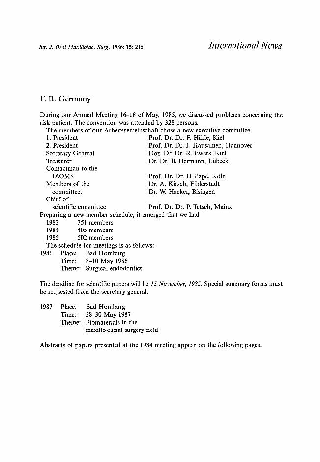
Int. J. Oral Maxillofac. Surg, 1986: 15: 215
F. R. Germany
International News
Prof. Dr. Dr. D. Pape, K61nDr. A. Kirsch, FilderstadtDr. W. Hacker, Bisingen
During our Annual Meeting 16-18 of May, 1985, we discussed problems concerning therisk patient. The convention was attended by 328 persons.
The members of our Arbeitsgemeinschaft chose a new executive committee1. President Prof. Dr. Dr. F. Harle, Kie12. President Prof. Dr. Dr. J. Hausamen, HannoverSecretary General Doz. Dr. Dr. R. Ewers, Kie1Treasurer Dr. Dr. B. Hermann, LiibeckContactman to the
IAOMSMembers of the
committee:Chief of
scientific committee Prof. Dr. Dr. P. Tetsch, MainzPreparing a new member schedule, it emerged that we had
1983 351 members1984 405 members1985 502 membersThe schedule for meetings is as follows:
1986 Place: Bad HomburgTime: 8-10 May 1986Theme: Surgical endodontics
The deadline for scientific papers will be 15 November, 1985. Special summary forms mustbe requested from the secretary general.
1987 Place:Time:Theme:
Bad Homburg28-30 May 1987Biomaterials in themaxilla-facial surgery field
Abstracts of papers presented at the 1984 meeting appear on the following pages.
Int. J. Oral Maxillofac, Surg, 1986: 15: 216-222
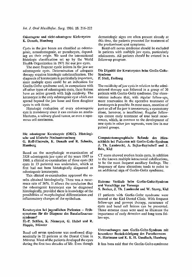
Odontogcnc und nicht-odontogene KieferzystcnK. Donath, Hamburg
Cysts in the jaw bones arc classified as odontogenic, nonodontogenic, or pseudocysts, depending on their origin. We used the internationalhistologic classification set up by the WorldHealth Organization in 1971 for our jaw cysts .
The most frequent cystic lesions in the jaw arcodontogenic cysts. Determination of adequatetherapy requires histologic subclassification. Thediagnosis ofkeratocysts is particularly important,since multiple cysts could be an indication forGorlin-Goltz syndrome and, in comparison withall other types of odontogenic cysts, these lesionshave an active growth with high recidivity. Thekeratocyst is the only odentogenic cyst which canspread beyond the jaw bone and form daughtercysts in soft tissue.
Histologic evaluation of every odontogeniccyst is mandatory since it can contain an ameloblastoma, a salivary gland tumor, or even a squamous cell carcinoma.
Die odontogene Keratozyste (OKC). Histologischc und klinische NachuntersuchungG. Rcff-Ebcrwein, K. Donath und R. Schmitz,Hamburg
Based on the morphologic re-examin ation of3328 odontogenic jaw cysts of the years 1967 to1980, a clinical re-examination of those cysts (82cysts in 73 patients) was undertaken, which atfirst had not been histologically diagnosed asodontogenic keratocysts.
This clinical re-examination approved the results obtained histologically. There was a recurrence rate of 56%. It allows the conclusion thatthe odontogenic keratocyst can be diagnosedhistologically, provided there is knowledge of thepossibilities of morphological differentiation andinflammatory changes of the epithelium.
Keratozystcn bei jugendlichen Patienten - Friihsymptorne flir die Diagnose des Basalzcllnacvussyndroms?H.-P. Schlien, K. Niemeyer, G. Habel und R.Happlc, 1\Iiinster
Basal cell nevus syndrome was confirmed diagnostically in 22 patients at the Dental Clinic inMunster, Most of the patients developed the cystsduring the first two decades of life. Even though
dermatologic signs arc often present already atthis time, the patients presented for treatment ofthe predominant cyst symptoms.
Basal cell nevus syndrome should be excludedin patients with multiple jaw cysts, particularlyadolescents. All patients should be entered in afollow-up program.
Die Dignitat der Kcratozystcn beim Gorlin-GoltzSyndromP. Stoll, Frciburg
The recidivity ofjaw cysts in relation to the administered therapy was followed in a group of 20patients with Gorlin-Goltz syndrome. Our observations indicate that, with regular follow-ups,more reservation in the operative treatment ofkeratocysts is possible. In most cases, resection ofpart or all of the jaw is unnecessary; a cystostomyalone, however, is insufficient. Regular followups ensure early treatment of true local recurrences, which , in contrast to the development ofnew cysts in other jaw segments, were low in ourpatient groups.
Computcrtomographlsche Befunde des Himschadels bei Patienten mit Gorlin-Goltz-SyudromJ. Th, Lambrecht, A. Sojka-Raytscheff und F.Brix, Kicl
CT scans showed median brain cysts , in additionto the known multiple intracranial calcifications,to be the most frequent auxiliary findings . The 'frequency of these alterations tends to point toan additional sign of Gorlin-Goltz syndrome.
Extreme Verlliufe beim Gorlln-Goltz-Syndromund Vorschlagc zur VorsorgeS. Becker, J. Th. Lambrecht und W. Sterry, Kid
17 patients with Gorlin-Goltz syndrome weretreated at the Kiel Dental Clinic. With frequentfollow-ups and prompt therapy, recurrence ofcysts and basal cell lesions can be prevented.Three extreme cases were used to illustrate theimportance of early detection and long-term follow-ups.
Untersuchungen zum Gorlln-Goltz-Syndrom mitbcsonderer Beriieksichtigung des ParathormoosN. Hartmann und K. K. H. Gundlach, Hamburg
It has been said that the Gorlin-Goltz-syndrome
in some respects is closely related to pscudohypoparathyroidism. However, considering all clinical , radiological, and biochemical data, we realizethat there is a sharp borderline between bothdisorders. In the nevoid basal cell carcinoma syndrome, calcium levels in blood serum were foundto be normal, alkaline phosphatase was raisedand anorganic phosphorus in urine was not lowas would be the case in pseudohypoparathyroidism.
Klinik und Pathomorphologie odontogener follikuHirer Zysten - Naehuntersuehung "on 239 FallenR. Roggan und K. Donath, Ilamburg
239 odontogenic dentigerous cysts from 1971 to1980 at the University of Hamburg Departmentof Pathology were histologically re-examined; 12of these cysts were additionally ultrastructurallyexamined.
Over 60% (38.9% were not infected, 23.4%only partly infected) could be histologically identified due to the typical structure of the cysts'epithelium. Approximately 40% were unable tobe positively diagnosed as a result of the epithelium change under inflammation. A histological diagnosis of inflamated dentigerous cysts istherefore possible only in connection with clinicaldetails.
The peaks in age distribution indicated the 2ndto 4th life-decade. There was a disposition ofmale sex of 1.5:I.
The incidence rate of dentigerous cysts in thearea of the lower wisdom teeth and the maxillarycanines shows an analogy to the incidence rateof odontoms and odontogenic tumors within thisregion, and signifies therefore, an increased vulnerability in this area concerning the differcntiation of this tissue and organs.
Zur Frage der Induktion follikuHirer Zysten imWeehsclgebiB durch Wurzelbehandlung "onl\ IilchzahncnP. Gebhardt und W. Lenz, Dusseldorf
After root canal therapy on deciduous teeth 65and 75, follicular cysts developed in two patientsat the corresponding second dentition teeth, thedental germ being malposed and/or retained. Thehistologic picture of these infected follicular cystsbasically resembled the morphologic pattern ofradicular cysts. Consequently, the classificationof the histologic findings for odontogenic cystsalso requires knowledge of the clinical findings.
217
Possible injury of the permanent tooth dentalgerm by endodontic treatment of deciduous teethcannot be excluded with certainty.
Karzinomentstehung auf dem Roden des zystenblldenden odontogenen EpilhelsJ.-E. Otten, U. Joos und W. Schilli, Freiburg
The origin of 5 squamous cell carcinomas observed over a ltl-ycar period in patients at theUniversity Dental Clinic in Frciburg was presumed to be the pericoronal epithelium of mal posed teeth or the epithelium of odontogenic cysts.Related to the Frciburg patients, 1.6% of all malignancies of the oral cavity were carcinomas incysts. During this IO-year period in which 17,200third molars were surgically removed, two carcinomas originating from impacted lower third molars were found.
Karzinogenese in Klcferzysten, 2 FallbcriehtcJ. Kreidler, S. Haase und W. Kamp, VIm
Within 4 years between 1980 and 1984, there werefound 758 odontogenic cysts at the departmentof Ulm. The case histories of 2 patients, whoshowed malignant change of the follicular epithelium of unerupted teeth, are presented. Thefrequeney of carcinogenesis in odentogenic cystsis discussed including the rare communicationsin the literature.
llistologischc Untersuchungen zur Vcrctilung Malassezscher Epithelncstcr zwischen dem 10. und90. LebcnsjahrD. Tertel-Kalwclt und K. Donath, Hamburg
The material consisted of 403 tccth and 41 eden tulous jaw regions from 68 subjects aged 10-90.
The epithelial nests are found in the periodontal ligament among the fiber bundles. Somelie in the root canal especially of front teeth, inthe bone marrow next to the alveolus and veryfrequently along the eruption path of the teethin close relation to the neurovascular bundle.
The number of epithelial nests is reduced withage, the quickest decrease already taking placeup to the 4th decade.
The majority of the nests are found in thecervical and middle third of the periodontium.Mainly those in the apical and middle third arcreduced with age.
As to the frequ ency and location in mandible
218
and maxilla as well as in male and female patients, there are no significant dilTerences.
The frequent manifestation of radicular cystsin the maxilla does not correspond to the distribution of the epithelial nests . There is no correlation between number and location of the epithelium and the occurrence of epithelial odontogenic tumors.
Radikulare Zyste im klinischen BiJd der medianenUnterkieferHisionenII. Kniha und 1\1. Gokel, 1\liinchen
Histologic evaluation of 2 cases of large median mandibular cystic lesions showed the lesionsto be radicular cysts. The diagnosis of gobletcell metaplasia in one case and ciliated epithelialmetaplasia in the other were unusual findings.
Zur Diffcrcntialdiagnosc median gclcgcner, zystischer Vcranderungcn im ObcrkicfcrJ. Beck-l\fannagetta, Salzburg und Z. Roscie,Linz
Cystie lesions in the maxillary midline can bedivided into 3 groups:I. Nasopalatine cysts (also including the so-cal
led "median alveolar cyst" and the " medianpalatine cyst");
2.odontogenic cysts;3.cyst-like lesions of dilTerent origin.It is very important to separate these lesions froma wide incisive fossa (an anatomic variant) thatcan reach a maximum diameter of 10 mm .
Although precise clinical data (symptoms, vitality test, radiograms, cyst contents and histopathology) will render valuable information, therewill still remain some cases where no unequivocaldiagnosis can be made.
Hiiuflgkelt und Rezidimeigung der solitiircn Knochenzyste1\1. Brandt und W. Lehmann, Kicl
The frequency and recidivity of solitary bonecysts are higher than has been previously presumed. 19 (0.6%) of3 105 bone cysts (Hoffmeisterand Hiirle 1984) were solit ary bone cysts. Inagreement with the literature, solitary bone cystswere found only in th e dentulous jaw. 2 recurrences were observed in the follow-up of our operated patients.
Beitrag zur Diagnostik und Therapie solitarerKnochenzystenE. Fischer-Brandies und E. Dielert, 1\1iinchen
18 cases of solitary bone cysts are presented.These include rare findings sueh as 2 cysts lyingcaudally of the mandibular canal, a pathologicfracture and a cementifying fibroma occurring inthe cystic wall . Therapeutic procedure is discussed in det ail.
Ubcr die sogenannte solitare KnochenzysteN. Schwenzer, 1\1. Ehrenfeld und R. Roos, Tiibingen
Based on 388 cases compiled from the literatureand 10 of our own cases, the clinical picture ofthe so-called solitary bone cyst, for whieh thereare several different terms, was described. Ourelectron microscopic studies on the ultrastructureof these cysts showed that they are definitely notlined with epithelium. The pathogenesis shouldbe clarified in further observations by a thoroughinvestigation of associations between solitarybone cyst format ion and tooth eruption and/ororofacial dysfunction.
Eine aneurysmale Knochcnzyste des Kiefers. Therapic und VerIauf iibcr 11 JahreI. Sikken, F. Schmid und H.-J. Loblich, Hannover
Aneurysmal bone cysts usually develop on thelong tubular bon es and the spine. Its occurrencein the mandible is extremely rare. As a rule, thetherapeutic aim in sueh cases is total cystectomyand, if necessary, immediate reconstruction of themandible. The course of sueh an extensive lesionin the left body of the mandible of a 6Y1-yearold boy was described. This case is remarkablebecause the cyst remnants in the mandible remained inactive during the II-year observationperiod. The therapeutie and histologic aspectswere discu ssed in detail.
Zum Vorgehen bei Statischen KnochenhohlenE. Dielert und E. Fischer-Brandies, Miinchen
The static bone eavity cannot be looked upon aspathologic. Therefore surgery is only indicated inspecial cases for dilTerential diagnostic purposes.
Tierexpcrimentelle Untersuchungen zur Entstehung kongenitaler Pscudozystcn der KieferR. Schmitz, Hamburg
Teratogenesis in the rat after treatment with Nmethyl-Nvnitrosourca is well suited as an experimental animal model for investigating the formation of pseudocysts. After administration of 20mg/kg of the teratogen on the 13th day of gestation. pseudocysts, in addition to the cmbryopathic syndromes, develop in the maxilla and mandible. The cytogenetic processes were describedin light of the histologic studies. Most cysts healed prior to delivery; those attached to vesselspersisted until birth.
Immunhistologisehe Untersuchungen von chronisch apikalen Parodontitiden und radikuHiren ZystenJ. Becker, P. Reichart und R. Ernst, Berlin undTh. Loning, Hamburg
36 dental granulomas and 6 periapical cysts wereexamined immunohistochemically. Epithelial andmesenchymal cells were stained using monoclonalantibodies against intermediate-sized filaments.Basal cells in the odontogenic epithelium werelabelled using an antibody against TPA. Byapplication of monoclonal antibodies, the distributionof immunocompetent cells in stroma and especially in the odontogenic epithelium was studied. Multiple HLA-DR positive and few Leu 4positive cells were observed in the epithelium andat the basement membrane.
Das Keimspcktrum der infizierten Zyste1-1.-G. Rudell, Hamburg
The clinical diagnosis "infected cyst" was established in 27.4% of our patients who were hospitalized for treatment ofa "cyst". The bacteriologic evaluation showed that the bacterial spectrum,which was composed predominantly of greenstaining streptococci (77.4%). was sensitive topenicillin.
Prostaglandin(PG)-8ynthese im Balg odontogenerZysten1\1. Matejka, II. Porteder, W. LiII, G. Watzekund H. Sinzinger, Wien
Prostaglandin synthesis in' odontogenic keratincysts was investigated by thin layer radiochroma-
219
tography. C'<labcled arachidonic acid is converted into prostaglandins PGE 2• 6-oxo-PGHla•
PGFla • and PGD2• Prostaglandins E2 and Iz areconsidered to be the most important. both qualitatively and quantitatively, for the osteolytic effect of such pathologic processes in the jaw.
Untersuchungen fiber Prostaglandine bei Kieferzysten, Zur Schmerzausschaltung bei ZystcktomleH. Hauenstein und D. Schettler, Essen
2 phenomena associated with jaw cysts requireextensive clarification: osteolysis. which ischaracteristic of and often pathognomonic fordifferent types ofcysts, and pain sensation, whichcannot be eliminated even with exact local anesthesia. Continuing our earlier studies on the effectof prostaglandins on the form of growth and therecidivity of keratocysts, including those developing in cancellous bone transplants from the pelvis, we determined prostaglandin and leukotrienconcentrations in cyst material obtained from 38patients. These patients were not selected andwere entered in the study in the order of theiradmission to our hospital. The effects of thesemetabolic products from fatty acids in the cellmembrane on osteolytic activity and activation ofthe "bone resorption factor" were experimentallydemonstrated. This, however, does not clarify thetypical growth form of the cysts.
Based on the unequivocally confirmed bioactivity of eicosanoate in all inflammatory processes and algogenesis, different patient groupswere treated preoperatively and intraoperativelywith local and systemic inhibitors of prostaglandin and leukotrien synthesis. Since clinical symptoms, which were recorded in the context of adouble blind test, corresponded in each case withthe respective prostaglandin concentration, a causal association can be assumed between elevatedprostaglandin concentration and the observedsensation of pain.
Rontgenbefunde bei odontogenen KeratozystcnW. J. Spitzer und E. W. Steinhauser, Erlangen
Based on the radiologic assessment of 30 keratocysts in 26 patients, the distinctive radiologicsigns of odontogenic keratocysts were presented.Our findings indicate that a monolocular ormultilocular cystic cavity distal of the third molarand in the ascending ramus of the mandible isvery probably a keratocyst. Other signs of'keratocysts, which cannot be radiologically differen-
220
tiated with certainty from ameloblastoma, in particular, are clear delineation of the cyst lumen,the contour of which is smooth or garland-like,and corticalization of the margin.
Die Beurteilung odontogener Kicfcrhdhlcnzystcnauf dem PanoramaschichtbildW. Bahr und J. Diiker, Freiburg i. Br.
After illustrating the radiologic anatomy of themaxillary sinus on the pantomogram, the efficacyof the information provided by this diagnosticmethod was determined in 68 patients with odontogenie maxillary sinus cysts. In almost 30% ofall evaluated pantomograms, the diagnosis"maxillary sinus cyst" was incidentally established after review of the film. The cystic alterations were discernible in almost 84% of theevaluated pantomograms. The most reliable diagnostic parameter for characterization of maxillary sinus cysts was the osseous lamellae of theantrum. In conclusion, the possibility of error inassessing maxillary sinus cysts was pointed out.The laterobasal nasal line should not be interpreted as the cranial border; the septa of thesinuses should not be interpreted as the lateralborder of a cyst.
Zysten im Kiefer-Gesichtsbcreich - cine katamnestische Studie an 3353 ZystenB. Hoffmeister und F. Harle, Kiel
2998 (98.4%) of the 3353 cysts diagnosed at theDental Clinic in Kiel between 1963 and 1983 wereodontogenic cysts; the remaining 355 (1.6%),nonodontogenic cysts. The different types of cystwere classified according to age, sex, localization,and clinical parameters. The largest group wasthe radicular cyst (1954 or 65.1%), followed bythe recurrent cyst (599 or 20%) and then thefollicular cyst (376 or 12.6%). The findings werecompared with data reported in the literature.
Statistische Auswertung griificrer KieferzystcnG. Dhom und P. Tetsch, Mainz
Data from 240 cystectomized patients with jawbone defeets of at least 20 mm in one plane wereanalyzed. The cysts were localized predominantlyin the maxilla-anterior tooth and mandible-molarregions. No association could be established between the duration of postoperative care aftercystectomy with primary closure and the size ofthe cyst.
Aplkale Veranderungen - Verteilung, Entwlcklungund DlfferentialdlagnoscW. Hacker, Bisingen und H. Fischbach, TIibingen
Among 102inflammatory processes of the dentalapex, 63 showed radicular cysts, 33 granulomasand 6 transitions from granulomas to radicularcysts. Concerning the etiology of inflammationsof the dental apex, 40% result from caries, 60%occur after endodontie treatment, following preparations under local anesthesia or following traumata. The development of both granuloma andradicular cysts takes 2 to 7 years on average. Thefollowing pathogenetical considerations can bemade: Inflammation of the pulp can proceed tothe adjacent bony tissue. In cases of root-canalfilling, necrobiotie material can be displaced fromthe pulp. Ischemic lesion of the pulp can occurfollowing preparations under local anesthesia.Furthermore, a bone lesion might be the consequence of instability after tooth trauma.
Eine ungcwdhnllchc Zystc im rctromaxillarcnRaumH. L. Obwegescr, M. Farmand und M. Makek,Ziirich
An unusually large mucocele in the retromaxillary space was reported. This cyst extended cranially to the floor of the middle cranial fossa, where,in the absence of the cranial bone, it was separated in places from the temporal lobes only bythe dura mater. Laterally, it impinged on themedial surface of the ramus of the mandible andobstructed mouth opening. Medially, it extendedfar into the nasopharynx. Anteriorly, it displacedthe maxillary sinus. The topography and extension of the process was documented on conventional radiographs and CT scans. On the basisof the findings, we presumed the lesion to be amucocele in the sphenoidal sinus. Therapy consisted of marsupialization of the mucocc1e to thesinus.
Indikation und Ergebnisse der Marsupialisationgroller Zystcn1\1. Farmand, Ziirich
The results of cyst marsupialization at the Hospital for Jaw Surgery in Zurich between 1981 and1983 were evaluated retrospectively and analyzedwith respect to subjective and objective criteria.The indication for marsupialization was established for all large cysts in the mandible and
maxilla. The resected and altered cyst epitheliumwas always subjected to histologic evaluation.Most lesions were radicular cysts. The main advantages of this operative method are the lowcomplication rate and simple execution. Our experience, therefore, indicates that marsupialization of large cysts is still advisable.
Scnslbilltat und i\Iobilitat von Zahnen nach ZystenoperationenR. Fenner und P. Tctsch, Malnz
Between 1978 and 1983,90 patients at the University Clinic for Oral Surgery in Mainz were followed to determine loss of sensitivity and/or vitality in primarily vital teeth after cystectomy.Whereas desensitized or nonvital teeth were extracted preoperatively or managed by apicoectomy with intraoperative root canal filling, suchtreatment was omitted in all teeth in direct contact with the cyst sac or bone defect which wereprimarily sensitive to stimuli. The aim of the follow-up was to clarify the extent to which loss ofsensitivity or postoperative complications developed after cystectomy.
Bchandlungsmoglichkclten und -ergebnlssc nachFrakturen bel UntcrklefcrzystenJ. Dumbach und E. W. Steinhauser, Erlangen
10 cases of fractures of the mandible that weredirectly related to cysts were presented. Since thecase material was otherwise extremely inhomogeneous, a wide variety ofconservative and surgical measures were used. These methods were described and discussed. The advantage of managing such fractures by miniplate osteosynthesiswas emphasized.
Bchandlungscrgcbnlsse und Langzcitbeobachtungcn bel 62 Patienten mit KeratozystenK. Niemeyer, H.-P. Schlien, G. Habel und C.Mentlcr, Miinster
The recidivity of keratocysts in 62 patients wasrelated to the operative procedure employed. 33of the patients were followed for at least 6 years.The highest recidivity was found in those patientstreated by cystostomy. A comparison of 2 patient groups managed after cystectomy by eithertamponade of the would cavity ("open") or immediate wound closure ("closed") showed an im-
221
pressively higher recurrence rate in those patientswith open management.
The incredible keratocyst: a new approach to treatmentR. A. C. A. Voorsmit, Nijmegcn
A nell' approach to the treatment of keratocystsis proposed, based on the theories regarding thepathogenesis of recurrent keratocysts and on theresults of a clinical and experimental study onCarnoy's solution, a fixing agent.
The clue for this new approach is, that thekeratocyst is fixed with Carney's solution beforeenucleation.
A protocol of treatment and the main advantages of this new method are outlined.
Die exspcktativ therapcutische Entfernung derKeratozystenR. Ewers und F. Harle, Kiel
In spite of the expected recurrence of keratocystsafter cystectomy, we, on the basis of our statistical data and that presented in the literature, recommend the use of this procedure to avoid jawresections.
Hat sich die Operation der KieferhOhle nach Wassmund bcwahrt? Ein ErfahrungsberichtG. Gehrke, R. Schmelzle und N. Schwenzer, Tilbingen
Our retrospective study over a IS-year periodshowed the procedure for extirpation of largemaxillary sinus cysts, introduced by Wassmundin 1927, to be reliable. In the light of a conceivable focal event and the rare possibility of carcinomatous degeneration of the cyst epithelium, werecommend that, in the case of infected cysts, theoriginal procedure be modified by cystectomy.Postoperative local or generalized complaints arerare because the physiologic lining of the maxillary sinus is preserved.
Die Rhinozystostomie. Ein ErfahrungsberichtK. Holzner und E. PUll, Salzburg
From 1980 to 1983, 68 patients with extensiveodontogenic cysts in the anterior maxilla weretreated by marsupialization to the nasal cavity
222
(with partial enucleation). 35 patients appearedfor follow-up. Except for 3 patients, all of themrevealed no evidence of disease within the operated area. Advantages of this method includethe independence both from postoperative oralhygiene and from self-care of the patient. In thecase of large cysts, no extensive shortening ofthe apices is necessary. A thorough histologicevaluation is always possible.
Knochcndefektfiillung mit Humanfibrinkonzentratbei groficn KicferzysteaB. DickmeiB, II. Hauenstein und D. Schettler,Essen
Tissucol- (Fa. Immuno), a human fibrin concentrate, was used to treat large cystic defects in thejaw bone. The results of 31 cases were reported.Due to the biochemical characteristics and thegood results, the concentrate appears well suitedfor defect filling with immediate primary closure.
Die Behandlung von Kieferzysten unter Verwendung von FibrinkleberR. Schmelzle, D. Riediger und U. Schmidt, Tlibingen
Experimental results in animal models and clinical findings regarding homologous fibrin adhesion in bone defects were reported. Both inanimal experiments and in the clinical setting,the wound usually healed by first intention withuneventful ossification of the jaw bone defect.Due to stable adhesion in the wall of the bonedefect, postoperative bleeding is minimal. Fibrinfilling of bone defects is indicated, particularly inpatients with clotting disorders or a tendency tovascular hemorrhage.
Einheilung und Pharmakokinetik eincr p-Trikalziumphosphat-Gentamicin-Kombination im Tierversuch (vorlauflge Mitteilung)J. Randzio, K. Thoma, R. Alex und B. Rhomberg,Munehen
Recently, locally effective antibiotic drugs havebeen recommended for the filling bone defects atrisk of infection and for the operative therapy ofchronic osteomyelitis. Good healing was ob-
tained with one of these "drug depots", a gentamicin-containing P-TCP sintered ceramic, after 3weeks' implantation in the rat mandible. The release of active gentamicin is detectable in theurine for approximately I week. Gentamicin-containing TCP ceramic, therefore, is well suited fortesting in the therapy of bone infections and asbone replacement material for infected sites.
Erfahrungen mit dcm resorbierbaren TCP, Keramikgranulat zur Fultung graficrer Knochendefektenach Zystcktomle im Kieferbereich11.-11. Horch, Munchen und B. Steegmann, Kaln
In a clinical trial extending over 8 years, an absorbable TCP ceramic powder was used to filllarger postcystectomy defects in the jaw bones of50 patients. The ceramic healed without reactionin 44 patients; complications in the form of fistulization developed in 6 patients (12%). In approximately 50% of the cases, the implant wascompletely absorbed after approximately 2 years.Complete absorption with replacement by lamellar bone was present in most patients after approximately 4 years. A definite correlation wasestablished between absorbability of the implantand new bone formation, depending on ceramicdensity, defect size, implant site, and individualosteogenic potential of the bone.
Our clinical experience substantiated the experimental investigations. Absorbable TCP ceramic powder, therefore, is recommended as bonereplacement material for fil1ing larger postcystectorny defects in the jaw bone.
Kalziumsulfat - cin Knochencnsatzmittcl?H.-P. Busch, Kicl
Many publications describing good bone regeneration after plaster filling of bone defects appearedbetween the 19th Century and 1966. Even thoughnegative experiences were not reported, no suchstudies have been published since 1966. Bone regeneration after plaster filling of bones in beagledogs and in postcystectomy bone defects in asmall group of patients was investigated. Sincethe regeneration of the jaw bone is good, werecommend that bone defects created by cystectomy not be filled with material of any kind.