Embed Size (px)
Citation preview
Abstraktsammlung
zum CESAR Symposium am 14. bis 16. Juni 2006
in der
Klinik für Tumorbiologie
1150 Wien, Hanglüssgasse 4/1-3
CESAR Symposium 14. bis 16. Juni 2007 in Freiburg
CESAR Central European Society for Anticancer Drug Research-EWIV
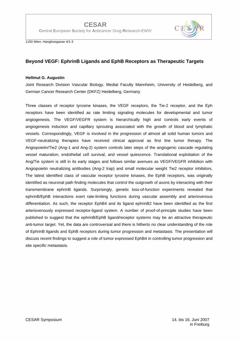
Beyond VEGF: EphrinB Ligands and EphB Receptors as Therapeutic Targets Hellmut G. Augustin Joint Research Division Vascular Biology, Medial Faculty Mannheim, University of Heidelberg, and
German Cancer Research Center (DKFZ) Heidelberg, Germany
Three classes of receptor tyrosine kinases, the VEGF receptors, the Tie-2 receptor, and the Eph
receptors have been identified as rate limiting signaling molecules for developmental and tumor
angiogenesis. The VEGF/VEGFR system is hierarchically high and controls early events of
angiogenesis induction and capillary sprouting associated with the growth of blood and lymphatic
vessels. Correspondingly, VEGF is involved in the progression of almost all solid human tumors and
VEGF-neutralizing therapies have received clinical approval as first line tumor therapy. The
Angiopoietin/Tie2 (Ang-1 and Ang-2) system controls later steps of the angiogenic cascade regulating
vessel maturation, endothelial cell survival, and vessel quiescence. Translational exploitation of the
Ang/Tie system is still in its early stages and follows similar avenues as VEGF/VEGFR inhibition with
Angiopoietin neutralizing antibodies (Ang-2 trap) and small molecular weight Tie2 receptor inhibitors.
The latest identified class of vascular receptor tyrosine kinases, the EphB receptors, was originally
identified as neuronal path finding molecules that control the outgrowth of axons by interacting with their
transmembrane ephrinB ligands. Surprisingly, genetic loss-of-function experiments revealed that
ephrinB/EphB interactions exert rate-limiting functions during vascular assembly and arteriovenous
differentiation. As such, the receptor EphB4 and its ligand ephrinB2 have been identified as the first
arteriovenously expressed receptor-ligand system. A number of proof-of-principle studies have been
published to suggest that the ephrinB/EphB ligand/receptor systems may be an attractive therapeutic
anti-tumor target. Yet, the data are controversial and there is hitherto no clear understanding of the role
of EphrinB ligands and EphB receptors during tumor progression and metastasis. The presentation will
discuss recent findings to suggest a role of tumor expressed EphB4 in controlling tumor progression and
site specific metastasis.
1150 Wien, Hanglüssgasse 4/1-3
CESAR Symposium 14. bis 16. Juni 2007 in Freiburg
CESAR Central European Society for Anticancer Drug Research-EWIV
Albumin as a Drug Carrier: The development of the (6-maleimido)caproyl-hydrazone derivative of doxorubicin, DOXO-EMCH (INNO-206)
F. Kratz Tumor Biology Center, Macromolecular Prodrugs, Breisacher Strasse 117, Freiburg, Germany
Albumin is playing an increasing role as a drug carrier in the clinical setting. Principally, three drug
delivery technologies can be distinguished: coupling of low-molecular-weight drugs to exogenous or
endogenous albumin, conjugation with bioactive proteins and encapsulation of drugs into albumin
nanoparticles.
The accumulation of albumin in solid tumors forms the rationale for developing drug albumin conjugates.
At present, an albumin-binding prodrug of doxorubicin, i.e. the (6-maleimido)caproylhydrazone
derivative of doxorubicin, DOXO-EMCH (INNO-206), is undergoing phase II studies (1-3).
DOXO-EMCH is an albumin-binding prodrug of doxorubicin with acid-sensitive properties that
demonstrates superior antitumor efficacy in murine tumor models and a favorable toxicity profile in mice,
rats and dogs, including significantly reduced cardiotoxicity. After intravenous administration, DOXO-
EMCH binds rapidly to the Cys-34 position of circulating albumin and accumulates in solid tumors due to
passive targeting. In a clinical Phase I study, the dose of doxorubicin could be increased by a factor of
4.5 – 340 mg/m² when 75 mg/m² of free doxorubicin is considered to be the dose that can be
administered as a single agent concomitant with the typical spectrum of side effects (i.e., myelotoxicity
and mucositis). DOXO-EMCH was able to induce tumor regressions in anthracycline-sensitive tumors
(i.e., breast cancer, small cell lung cancer and sarcoma).
This presentation gives an account of the different drug delivery systems that make use of albumin as a
drug carrier with a focus on albumin-binding prodrugs for treating malignant disease.
1. Kratz, F. (2007): F. DOXO-EMCH (INNO-206), the first albumin-binding prodrug of doxorubicin to enter clinical trials. Expert Opin. Investig. Drugs, 16, 855-866.
2. [Kratz et al. (2002): Probing the Cysteine-34 Position of Endogenous Serum Albumin with Thiol-binding Doxorubicin Derivatives: Improved Efficacy of an Acid-sensitive Doxorubicin Derivative with Specific Albumin-binding Properties Compared to the Parent Compound, J.Med.Chem. 45, 5523-5533.
3. Lebrecht et al. (2006): The 6-maleimidocaproyl hydrazone derivative of doxorubicin (DOXO-EMCH) is superior to free doxorubicin with respect to cardiotoxicity and mitochondrial damage, International Journal of Cancer, 120, 927-934.
1150 Wien, Hanglüssgasse 4/1-3
CESAR Symposium 14. bis 16. Juni 2007 in Freiburg
CESAR Central European Society for Anticancer Drug Research-EWIV
Multi-Target Protein Kinase Inhibitors: Novel Drugs for a Personalized Cancer Therapy?
Michael H.G. Kubbutat ProQinase GmbH, Tumor Biology Center Freiburg, Breisacher Strasse 117, D-79106 Freiburg,
Germany
Protein kinases represent today one of the most promising target families for the development of novel
anti-cancer drugs. Several inhibitors of protein kinases have been successfully developed and approved
for the treatment of different human cancers. So far, the majority of protein kinase inhibitors have been
or are being developed as mono-specific agents against one selected target protein kinase.
Due to the fact that most of the current protein kinases inhibitors are ATP-competing agents and the
similarity of the ATP-binding site of different protein kinases, it turned out that most of these compounds
can inhibit not only the initial protein kinase of choice but several additional protein kinases. For many
years unspecificity of protein kinase inhibitors was thought to be of disadvantage due to a higher risk of
possible side effects.
Our increase in the understanding of the molecular circuitry of the protein kinases dependent signalling
network and its deregulation in cancer, however, revealed that often simultaneous deregulation of
several protein kinases drives the proliferation and survival of tumour cells as well as the formation of
novel blood vessels and of metastasis. For example, simultaneous inhibition of tumour cell proliferation-
promoting EGF-R and angiogenesis-promoting VEGF-R2 results in enhanced growth-inhibiting effects in
murine tumour models compared to inhibition of just one of these kinases.
Based on this insight a novel strategy for the development of protein kinase inhibitor for the treatment of
human cancers arose. This strategy aims at the simultaneous inhibition of different protein kinases of
which deregulated activities promote the growth of tumour cells. The hope for the future is that such
multi-target specific reagents might actually prove superior in efficacy when compared to mono-specific
kinase inhibitors.
In addition to our greater knowledge of the role of protein kinase signalling in cancer, technical
improvements in the ability to test compounds against large panels of protein kinases made it now
possible to rationally identify and develop compounds with a therapeutically favourable inhibitory profile.
However, although in vitro inhibitory profiles provide important information of protein kinase inhibitors a
number of additional important issues to be addressed for rational development of multi-target inhibitor
are still awaiting to be solved. These include questions to which extent in vitro inhibitory profiles actually
translate into cellular and in vivo inhibitory profiles, which are suitable combinations of targets, and how
patient populations can be stratified to enable the identification of those patients which probably benefit
from these approaches.
1150 Wien, Hanglüssgasse 4/1-3
CESAR Symposium 14. bis 16. Juni 2007 in Freiburg
CESAR Central European Society for Anticancer Drug Research-EWIV
To address the first question we performed exploratory studies in which we compared the IC50 in vitro
inhibitory profiles of about 50 compounds against different protein kinases with those inhibitory profiles
obtained in cellular phosphorylation assays. Results suggest that remarkable differences between in
vitro and cellular inhibitory profiles can occur indicating the need to adapt early preclinical development
strategies to this specific type of protein kinase inhibitors.
1150 Wien, Hanglüssgasse 4/1-3
CESAR Symposium 14. bis 16. Juni 2007 in Freiburg
CESAR Central European Society for Anticancer Drug Research-EWIV
Structure-Transport and structure-activity relationships of oxaliplatin analogues Buß I.1, Garmann D.1, Galanski M.2, Keppler BK.2, Jaehde U.1
1Institute of Pharmacy, Clinical Pharmacy, University of Bonn, D-53121 Bonn 2Institute of Inorganic Chemistry, University of Vienna, A-1090 Vienna
The use of platinum complexes in cancer chemotherapy is often limited by pre-existing or acquired
resistance. One resistance mechanism is a decreased cellular accumulation. To reveal whether
physicochemical properties affect the cellular influx and the cytotoxicity of platinum complexes we
investigated the intracellular platinum concentrations and the IC50 values after incubation with oxaliplatin
and several analogues in the ovarian carcinoma cell line A2780, the ileum carcinoma cell line HCT-8
WT and their platinum-resistant variants.
2x10⁶ cells were incubated with a platinum complex (100 µM) up to 2 h. Intracellular platinum
concentrations were measured by flameless atomic absorption spectrometry (FAAS). MTT assays were
performed to estimate cytotoxicity (EC50 values).
Intracellular platinum concentrations increased rapidly within the first minutes and later approximately
proportional with time. To characterise the “early influx” we analyzed the relationship between
lipophilicity (log P) and platinum accumulation after 10 minutes of incubation. A clear correlation was
found in all cell lines indicating passive diffusion (Kendall´s tau rank correlation: A2780 r = 1.00, p <
0.05; A2780cis r = 0.80, p = 0.05; HCT-8 WT r = 0.80, p = 0.05; HCT-8 Ox r = 0.80, p = 0.05). Linear
regression analysis was performed to assess the rate of influx. The slopes did not correlate with the log
P values, but were considerably reduced in the resistant cell lines. These data suggest that lipophilicity
is not rate-determining for the influx of oxaliplatin analogues. Investigating the structure-activity
relationships an inverse correlation between lipophilicity and cytotoxicity was observed in all tested cell
lines (Kendall´s tau rank correlation: A2780 r = 0.80, p = 0.05; A2780cis r = 0.80, p = 0.05; HCT-8 WT r
= 0.80, p = 0.05; HCT-8 Ox r = 1.00, p < 0.05). In the ileum carcinoma cell line a decreased resistance
factor (EC50 resistant cells/EC50 sensitive cells) with increasing lipophilicity was found (Kendall´s tau rank
correlation: r = -1.000, p = 0,014).
In conclusion, the early influx phase is mainly due to passive diffusion and nearly not altered in resistant
cells. Subsequently, other mechanisms, e.g. active transport, seem to be responsible for cellular influx.
The observed relationship between lipophilicity and cytotoxicity suggests that the more lipophilic
complexes form less DNA adducts. Nevertheless, a cell-dependent circumvention of resistance with
more lipophilic oxaliplatin analogues seems to be feasible.
1150 Wien, Hanglüssgasse 4/1-3
CESAR Symposium 14. bis 16. Juni 2007 in Freiburg
CESAR Central European Society for Anticancer Drug Research-EWIV
Anticancer activity of hydroxylated resveratrol analogues Walter Jäger Department of Clinical Pharmacy and Diagnostics, University of Vienna, A-1090 Vienna; Austria
Resveratrol (3,5,4'-trihydroxy-trans-stilbene), an ingredient of wine, shows a broad spectrum of cytotoxic
effects against human cancer cells. In order to enhance these effects, we introduced additional hydroxyl
moieties into the molecule. The most potent resveratrol compound was 3,3’,4,4’,5,5’-hexa-hydroxy-
trans-stilbene (M8) which exerts a 1600-fold higher rate of cyclooxygenase II inhibition (IC50: 0.00104
µM and 1.67 µM) and a more than 6600-fold higher antiradical activity than resveratrol. Furthermore, M8
also exhibited a threefold higher cytostatic activity in HL-60 leukemic cells compared to resveratrol
(4.2+0.09 µM and 12.1+0.17 µM). In HL-60 cells, M8 depleted the deoxynucleoside triphosphates dATP
and dTTP pools to 41% and 21% of control values, whereas dCTP pools increased to 199% of
untreated controls. In addition, TTP, ATP, CTP, and GTP concentrations were decreased while UTP
concentrations increased. M8 induced apoptosis at concentrations significantly lower than resveratrol
and could remarkably inhibit the activation of NF-�B. M8 arrested cells in the S phase of the cell cycle
while depleting cells in the G2-M-phase. Besides leukemia cells, M8 also demonstrated a more than
four-fold higher anti-proliferative effect against human 518A2 melanoma cells. Single agent treatment by
M8 significantly reduced the growth of 518A2 xenografts when administered i.p. daily at doses of 2.5 or
5 mg/kg/d. Moreover, if combined with the standard melanoma chemotherapeutic dacarbacine, M8 co-
treatment led to eradication of melanoma xenografts in 50% of SCID mice. Due to these promising
results, the novel resveratrol analogue M8 might serve as a drug candidate for the treatment of leukemia
and malignant melanoma.
Key words: resveratrol, leukemia, melanoma, xenograft
1150 Wien, Hanglüssgasse 4/1-3
CESAR Symposium 14. bis 16. Juni 2007 in Freiburg
CESAR Central European Society for Anticancer Drug Research-EWIV
Population pharmacokinetics of intravenous cyclosporine A in bone marrow transplant paediatric patients.
S. Saleh 1 2, L Grigull3, C Linderkamp3, H Schmid3, A Sander3, A Beilken3, K Wonigeit5, KW Sykora3, L
Hoy6, M Schrappe4, K Welte3, A Schrauder4, J. Boos1, G. Hempel 1 2
1 Department of Paediatric Haematology and Oncology, University Children's Hospital, Muenster, Germany 2 University of Muenster, Institute of Pharmaceutical and Medicinal Chemistry, Muenster, Germany 3 Department of Paediatric Hematology and Oncology, Children’s Hospital, Medical University, Hannover, Germany 4 Department of Paediatric Hematology and Oncology, Children’s Hospital, Medical University, Kiel, Germany 5 Visceral and Transplantation Surgery, Medical University, Hannover, Germany 6 Department of Biometrics, Medical University, Hannover, Germany
email: Georg Hempel ([email protected])
Cyclosporine A (CsA) is an immunosuppressive drug widely used in bone marrow transplant paediatric
patients for the prevention of GvHD. Therapeutic drug monitoring of CsA is necessary because of its
large inter-individual variability and its narrow therapeutic range.
CsA monitoring after HCT is usually performed by measuring CsA trough levels (C0). However, data
from solid organ transplant recipients have shown a better correlation of 2-h blood level concentrations
(C2) with the CsA exposure as compared to the trough level (C0). The aim of our investigation was to
estimate the population and individual PK parameters for the patients and to calculate the AUC for each
patient to test the correlation between the AUC and the various withdrawal points. NONMEM was used
to develop a pharmacokinetic model. A total of 289 concentrations from 27 patients were obtained. To
describe the pharmacokinetic parameters of CsA, a 2-compartmental model with first order elimination
was used. By applying forward inclusion and backward elimination in NONMEM, the covariate models
were built. The population parameters were CL 15.3 L/h, V1 12.9 L, Q 6.28 L/h and V2 83.3 L. Covariate
analysis identified body weight, age and the co- administration with itraconazole and tobramycine as
factors influencing the clearance CL. The analysis of the graphics between AUC and C0, C2 indicate a
better correlation between AUC and C2 than between the AUC and C0, r = 0.24 (P= 0.146) vs. r= 0.526
(P= 0.000692), respectively. Our results indicate that there is no significant relationship between AUC
and C0, and CsA trough level do not reflect the drug exposure. C3 and C6 plasma concentrations
appear to be more predictive for AUC and can be used to individualise CsA dose.
1150 Wien, Hanglüssgasse 4/1-3
CESAR Symposium 14. bis 16. Juni 2007 in Freiburg
CESAR Central European Society for Anticancer Drug Research-EWIV
Combined chemo-radiotherapy with gemcitabine in patients with locally advanced inoperable transitional cell carcinoma (TCC) of the urinary bladder and/or in patients ineligible for operation: a phase I trial LBI-ACR VIEnna Protokoll N°001/ CESAR Protokoll N° 001
Maria De Santis1,3, Gerhard Kametriser2, Tanja Steininger3, Maria Cerveny4, Margit Gneist3, Felix
Sedlmayer2,6, Gerd Fastner2, Marcus Hudec5, Annemarie Schratter-Sehn4, Christian Dittrich1,3,6. 1 Kaiser Franz Josef-Spital, 3rd Medical Department - Centre for Oncology and Hematology. 2 Landeskrankenhaus Salzburg, Department for Radiotherapy und Radio-Oncology. 3 Ludwig Boltzmann-Institute for Applied Cancer Research (LBI-ACR VIEnna) and ACR-ITR VIEnna. 4 Kaiser Franz Josef-Spital, Institute for Radiooncology. 5 University Vienna, Institute for Scientific Computing. 6 CESAR Central European Society for Anticancer Drug Research - EWIV.
It is generally assumed, that up to 50% of patients with urothelial cancer are unsuitable for cisplatin-
based chemotherapy. For this patient group standard radical cystectomy as well rarely is an option.
These clinical problems on the one hand and the assumed improved local control rates with concurrent
chemo-radiation therapy on the other hand, encouraged us to initiate a phase I trial in 2003. Preclinical
as well as clinical data suggest promising effectiveness and toxicity profile of gemcitabine in urothelial
cancer patients as well as a major radiation sensitizing effect of gemcitabine on various human cell
lines. The main aim of this study is to determine the maximum tolerated dose (MTD) of gemcitabine
combined with radiation therapy in TCC of the urinary bladder. In view of the literature and possible
toxicity at rather low dose levels in the combined treatment modality, we chose the starting dose of 20
mg/m² gemcitabine twice weekly with fixed standard 55.5 Gray radiation therapy of the urinary bladder.
Only gemcitabine escalation was aimed at. The primary endpoint of the study is the MTD of gemcitabine
at which not more than 20% of patients have to rest radiation therapy for ≥ one week. The secondary
endpoint is late toxicity. The dose limiting toxicity is defined as discontinuation of radiotherapy for >1
week in total in 2 or more patients out of 6 due to grade 3 or 4 non-hematologic and/or hematologic
toxicity. The MTD is defined as the dose level at which two or more patients out of 6 (≥2/6) expierience
dose limiting toxicity (DLT), and/or the dose level at which not more than two out of 18 patients develop
late toxicity.
Preliminary data of 5 dose levels including the DLTs will be presented at the meeting.
1150 Wien, Hanglüssgasse 4/1-3
CESAR Symposium 14. bis 16. Juni 2007 in Freiburg
CESAR Central European Society for Anticancer Drug Research-EWIV
Methods for proteome-wide identification and validation of targets within subfractions of tumor cells
Lembcke C1, Bien S1, Hammer E2, Steil L2, Völker U2, Kroemer HK1, and Ritter CA1,3
1Department of Pharmacology, Research Center of Pharmacology and Experimental Therapeutics, 2Department of Functional Genomics, and 3Institute of Pharmacy, Ernst-Moritz-Arndt-University of Greifswald, Germany.
With the increasing knowledge of biological processes in tumorigenesis individualised approaches of
pharmacotherapy of neoplastic malignancies are emerging. In parallel, screening techniques have been
developed to identify target structures of therapeutic relevance. These techniques can be separated into
genomic and proteomic approaches. While genomic methods are advantageous in that regulations
within the whole genome are detectable, proteomic methods allow for detection of structures that carry
the actual function and their post-translational modifications. However, proteomic approaches are limited
by chemical and structural characteristics of the proteins as well as the abundance within the cell. One
possibility to overcome some of these obstacles is to minimize the complexity of the sample by
subfractionation of the cells. This is of further interest since modification of subcellular compartments
such as mitochondria have been shown to be associated with tumorigenesis.
We therefore developed a method to fractionate tumor cells into nuclear, mitochondrial, lysosomal and
cytosolic subfractions using differential centrifugation combined with magnetic bead separation.
Enrichment of subfractions was controlled by Western blotting using antibodies against fraction specific
proteins. Proteomes of subcellular fractions were then analysed using either 2D-gel electrophoresis with
subsequent in-gel digestion of isolated protein spots and identification by matrix assisted laser
desorption and ionisation (MALDI) – time of flight (TOF) mass spectrometry or 1D-gel electrophoresis
and subsequent identification with Fourier-transform ion cyclotron resonance (FTICR) mass
spectrometry. Protein patterns generated by 2D-gel electrophoresis for each subcellular fraction
demonstrated a minimum of overlap between the different subfractions. Identification of selected
proteins revealed that compartment specific proteins are enriched within the respective subfractions. In
order to proof that differential expression within subfractions can be detected, the hepatoma cell line
HepG2 was treated with 1 µM doxorubicin or DMSO for control purposes and fractionated into
subcellular compartments. Differentially expressed proteins were detected and identified within the
mitochondrial/lysosomal fraction using differential 2D-gel electrophoresis (DIGE). These results were
cross-validated with gel free methods of whole cell lysates, where proteins were separated by liquid
chromatography and identified using FTICR or MALDI-TOF-TOF. Comparison of the different methods
revealed that an overlap of regulated proteins exists, but that disproportionately more mitochondrial
proteins are detected within the mitochondrial/lysosomal fraction.
In summary, reducing complexity of protein samples for proteome analyses by subcellular fractionation
is applicable to tumor cells and may provide a helpful tool for the identification of novel target structures
for an individualised pharmacological treatment of neoplastic malignancies.
1150 Wien, Hanglüssgasse 4/1-3
CESAR Symposium 14. bis 16. Juni 2007 in Freiburg
CESAR Central European Society for Anticancer Drug Research-EWIV
Die Biologie des Kolorektalkarzinoms aus tumorpharmakologischer Sicht
Robert M. Mader Universitätsklinik für Innere Medizin I der Medizinischen Universität Wien, Österreich
Der Erkenntnisgewinn der letzten Jahre im Bereich der Tumorbiologie hat unser Verständnis der
zellulären Vorgänge im Malignom und seines Mikromilieus grundlegend verändert. Das dabei Anfang
der neunziger Jahre entwickelte Modell der Adenom-Karzinomsequenz des Kolorektalkarzinoms von
Fearon und Vogelstein war aber gleichzeitig bahnbrechend für ein verändertes Herangehen an
therapeutische Fragestellungen. Der Versuch kausale Verknüpfungen zwischen molekularbiologischen
und biochemischen Prozessen einerseits und deren Auswirkungen auf Progression und Metastasierung
herzustellen, hat zur Entwicklung zielgerichteter Therapien geführt. Dabei stehen Rückschläge wie die
von Gefitinib beim nicht-kleinzelligen Bronchialkarzinom Erfolgen beim Kolonkarzinom gegenüber
(Bevacizumab bzw. Cetuximab).
Die Schwierigkeiten einer rationalen Entwicklung auf diesem Gebiet sind unter anderem im
ungenügenden Wissen zur Unterscheidung kausal tumorpromovierender Veränderungen von
Epiphänomenen zu suchen. Dies betrifft zahlreiche der klassischen Genmutationen des Kolons wie
RAS, APC oder SMAD4, deren Interaktion mit dem System Cyclooxygenase-2 / VEGF erst relativ spät
in experimentellen Modellen verstanden wurde. Da die klinische Validierung dieser Hypothesen
naturgemäß um Jahre dahinter zurückbleibt, tut sich eine Lücke auf, die immer größer wird. Darüber
hinaus werden für die seit Jahrzehnten etablierte Therapie mit 5-Fluoruracil prädiktive Faktoren wie
Mikrosatelliteninstabilität, Expression von Thymidilatsynthase oder mutierter Rezeptor für Transforming
Growth Factor � in der Literatur noch immer kontroversiell diskutiert. Ergänzt wurde unser Wissen allein
in den letzten drei Jahren um eine Reihe grundlegender Ansätze aus den Themenkreisen
Tumorstammzelle, epigenetische Vorläuferzellen der kolorektalen Tumorigenese, Kommunikation und
Interaktion von Signalübertragungswegen sowie als völlig neue regulatorische Komponente mikro-RNA.
Von letzteren wurden bei einer genomweiten Analyse von humanen Kolonläsionen auf 133 neue mikro-
RNAs beschrieben, deren Relevanz als tumortreibende Kraft aber auch als mögliches neues Target in
der Therapie völlig unbekannt ist. Die Tatsache, dass einige mikro-RNAs mit Apoptosewegen
interagieren (miR-15 und 16 mit dem antiapoptotischen Molekül bcl-2 bzw. miR-17 mit dem
Überlebenssignalweg Akt/PTEN) wird die experimentelle Forschung zum Verständnis der Tumorbiologie
als treibende Kraft für therapeutische Interventionen weiter vorantreiben. Die Umsetzung dieser
Erkenntnisse in therapeutische Optionen bedarf daher dringend klinischer Evidenz aus
Biomarkerstudien, die die Relevanz der beobachteten Phänomene für Therapieansprechen und
Überleben in prospektiven klinischen Studien validieren. Die überzeugende Darstellung dieser
Phänomene wiederum erfordert von Seiten der präklinischen Forschung neue kooperative Ansätze, die
weit über die zurzeit gängige Form des Einzelprojektes hinausgehen. Ohne dieses Zusammenwirken
1150 Wien, Hanglüssgasse 4/1-3
CESAR Symposium 14. bis 16. Juni 2007 in Freiburg
CESAR Central European Society for Anticancer Drug Research-EWIV
bleiben der rationalen Entwicklung neuer Behandlungsstrategien die engen Grenzen der empirischen
Wirkstoffentwicklung auferlegt.
1150 Wien, Hanglüssgasse 4/1-3
CESAR Symposium 14. bis 16. Juni 2007 in Freiburg
CESAR Central European Society for Anticancer Drug Research-EWIV
Design of molecular therapies
Uwe Fuhr Department of Pharmacology, Clinical Pharmacology, University Hospital, University of Cologne,
Germany
Targets for antineoplastic therapy may have essential or non-essential functions. Traditional cytotoxics
as well as “neocytotoxics” both attack essential targets. Such drugs are likely to have narrow therapeutic
windows but confer the ability to kill tumors cells with high potency. Drugs acting on non-essential
targets usually are well tolerated but have limited efficacy. There is little chance to find a target which
has as clear differences in the importance for healthy tissues and for tumor cells as the BRC-ABL
kinase. Still, some tumors may express a given target at high levels or may for other reasons depend
more on a target than healthy tissue. This divergence may apply to both different tumor entities and to
interindividual differences in the same tumor type. If there is genetic heterogeneity in the target, all the
more if there are differences between tumor cells and healthy tissue, the effect of drugs should be
compared between genotypes to generate a drug which has selectivity for the respective genetic
variants. From a pharmacokinetic point of view, for low molecular drugs those not undergoing main
metabolism via CYP3A4 or via genetically polymorphic enzymes should be preferred, as well as those
who have no major inhibitory effects on drug metabolizing enzymes. Drugs affected by known
mechanisms of resistance such as p-glycoprotein or target modifications should be avoided. Also, it
should be tested whether a drug may reach the target, e.g. by microdialysis.
A major requirement for the development of directed molecular therapy is the availability of a validated
assay of the target. Such assays may be based on different technologies, such as
immunohistochemistry, fluorescence in situ hybridization, and chromogenic in situ hybridization and
enable the selection of the optimal position and agent (e.g. by low molecular inhibitors, antibodies, or
micro-RNA) to attack the signaling pathway of the target. The assay may also show whether there are
compensatory mechanism in signal transduction. Once there is an established therapy involving this
target, the assay may be applied as a biomarker to identify patients with a high probability to benefit,
and to monitor therapeutic effects.
Selecting the optimal therapy for the individual patients remains a major challenge. Currently, for many
clinical cancer entities several therapeutic strategies are used in parallel which have similar mean
effects but which may have different effects in individual patients. The application of the HER-2 antibody
trastuzumab and antiestrogens are successful examples for a selection based on the presence of
molecular targets, but in many cases there is no such rationale for a selection. The need to obtain viable
tumor cells however is a general limitation to tests for the targets in most solid tumors. Easy access to
1150 Wien, Hanglüssgasse 4/1-3
CESAR Symposium 14. bis 16. Juni 2007 in Freiburg
CESAR Central European Society for Anticancer Drug Research-EWIV
malignant cells is also a major reason why individualization of therapy based on molecular targets
and/or biomarkers currently is most advanced in hematological malignancies.
Still, for most antineoplastic drug therapies even if a target is attacked with high specificity, selectivity for
tumor cells over healthy tissue is limited, und usually therapeutic effects and toxicity are closely linked.
Therefore, independent of the class of antineoplastic agent, it is desirable to take all known sources of
variation of pharmacokinetics and pharmacodynamics into account in order to achieve the most uniform
therapeutic response possible, which makes both toxicity and efficacy more predictable.
1150 Wien, Hanglüssgasse 4/1-3
CESAR Symposium 14. bis 16. Juni 2007 in Freiburg
CESAR Central European Society for Anticancer Drug Research-EWIV
Non small cell lung cancer xenografts as models for the prediction of response to tyrosine kinase inhibitors
I. Fichtner, J. Rolff, J. Merk*, S. Lee°, R. Soong°, M. Becker
MDC Berlin, Robert-Rössle-Str. 10, 13092 Berlin; *ELK, Lindenberger Weg 27, 13125 Berlin; °National
University of Singapore, 10 Medical Drive, Singapore 117597
The epidermal growth factor receptor (EGFR) plays an important role in cell proliferation and
differentiation. It is expressed in allmost all tissues and is overexpressed in many cancer types. In lung
cancer the EGFR is overexpressed in 50-80% of the patients with squamous or adenocarcinomas. With
the newly developed tyrosine kinase inhibitors (TKI) Gefitinib and Erlotinib as well as the monoclonal
antibody Cetuximab new drugs are available for the treatment of patients with lung cancer. The
evaluation of clinical trials using Erlotinib and Gefitinib revealed no correlation between the EGFR
expression and the response to therapy. It was demonstrated that only a small group (women, never-
smokers and people with asian origin) did benefit from the treatment with TKIs. In addition, patients with
mutations in the exon 19 or 21 of the EGFR gene showed a better response to a therapy with TKIs.
We have developed a series of novel lung cancer xenograft models. Fresh tumor material of patients
with non small cell lung cancer (NSCLC) was subcutaneously transplanted in immunodeficient mice
shortly after removal. Up to now more than 100 tumors have been transplanted from which 23
passagable models could be generated. It could be demonstrated that the murine passages coincide
with the original tumor regarding histology, the expression of the surface proteins E-Cadherin, EpCAM,
the cell proliferation marker Ki-67 and in gene profiling. The analysis of the EGFR gene revealed no
mutations relating to a better response to TKIs. With the exception of two models all express a wild type
EGFR. Four models with K-ras mutations were found among the xenografts and in ten different models
mutations in the p53 gene could be located. Furthermore, the sensitivity of the xenografts was tested
against five clinically used cytotoxic agents (Etoposid, Carboplatin, Gemcitabine, Taxol and Navelbine)
and two EGFR inhibitors (Erlotinib and Cetuximab). It could be shown that there exist strong differences
in responses among the xenografts.
Biomarkers suitable for a prediction of response to the treatment with tyrosine kinase inhibitors are
analysed in the xenografts.
1150 Wien, Hanglüssgasse 4/1-3
CESAR Symposium 14. bis 16. Juni 2007 in Freiburg
CESAR Central European Society for Anticancer Drug Research-EWIV
Die Rolle des Angiopoietin – Tie Systems in physiologischer und pathologischer Angiogenese
Ulrike Fiedler ProQinase GmbH, Freiburg
Das Angiopoietin / Tie-System reguliert die Blutgefäßreifung, Umgestaltung und Homeostase. Das
Angiopoietin / Tie System besteht aus zwei Rezeptoren Tie1 und Tie2 und vier Liganden Angiopoietin-1,
-2, -3, und -4. Die Rezeptoren Tie1 und Tie2 sind Rezeptor-Tyrosinkinasen, die zueinander homolog
sind. Beide Rezeptoren sind entscheidend für die Ausbildung eines funktionellen Gefäßsystems
während der Embryonalentwicklung. Die bisher identifizierten Angiopoietine sind Liganden der
Rezeptortyrosinkinase Tie2. Interessanterweise konnte bisher noch kein Ligand für Tie1 identifiziert
werden. Die Stabilität des bestehenden Gefäßsystems, sowie die Neubildung und Umgestaltung sind
abhängig von einem ganz genau ausbalancierten Wechselspiel der Aktivierung der
Rezeptortyrosinkinase Tie2 durch die Liganden Ang-1 und Ang-2. Ang-1 wird konstitutiv von
perivaskulären Zellen exprimiert und aktiviert so durch Bindung an Tie2 Signalwege, die die Integrität
des Gefäßbettes aufrechterhalten. Im Gegensatz zu Ang-1, ist die Expression und die Verfügbarkeit von
Ang-2 strikt reguliert. Ang-2 wird nicht im ruhenden Gefäßbett exprimiert, sondern nur nach Aktivierung
von Endothelzellen. Darüber hinaus liegt Ang-2 in Endothelzellen in Weibel-Palade Körperchen
gespeichert vor und wird nach Aktivierung ausgeschüttet. Die Ausschüttung von in Endothelzellen
gespeichertem Ang-2 und die Überexpression von Ang-2, aktiviert das Gefäßbett und versetzt es in
einen Zustand, in dem es auf angiogene und inflammatorische Zytokine reagieren kann. Dies zeigt,
dass der Aktivierungszustand von Tie2 und die Integrität des Gefäßbettes von einem wohlbalancierten
Verhältnis und der Verfügbarkeit von Ang-1 und Ang-2 abhängt. Allerdings ist bisher ist noch nicht
bekannt, durch welchen molekularen Mechanismus Ang-2 das Gefäßbett aktiviert. Vieles deutet darauf
hin, dass Ang-2 nicht nur einfach die Ang-1 abhängige Aktivierung von Tie2 inhibiert, sondern auch
unter bestimmten Bedingungen agonistisch wirkt. Ang-2 aktiviert u.a. die Sprossung von Endothelzellen
und wirkt agonistisch auf die Entwicklung von Lymphgefäßen.
Eine therapeutische Intervention mit dem Angiopoietin / Tie-System verspricht neben der Intervention
mit dem VEGF / VEGF-Rezeptor-System ein neuer Angriffspunkt für eine anti-angiogene Therapie zu
werden. Allerdings wird zur Zeit noch kontrovers diskutiert, wie mit dem System interferiert werden
sollte. Erste Experiment mit löslichen Tie2-Rezeptoren, Inhibitoren von Ang-2 und Überexpression von
Ang-1 und Ang-2 wurden durchgeführt, liefern aber noch kein klares Bild darüber welcher Ansatz am
erfolgversprechensten sein wird. Weitere Untersuchungen des Angiopoietin / Tie-Systems werden
notwendig sein, um das therapeutische Potential einer Intervention mit dem Angiopoietin / Tie-System
besser abschätzen zu können und festzulegen, welche Krankheitsbilder durch eine Intervention mit
Angiopoietin / Tie-System behandelt werden können.
1150 Wien, Hanglüssgasse 4/1-3
CESAR Symposium 14. bis 16. Juni 2007 in Freiburg
CESAR Central European Society for Anticancer Drug Research-EWIV
Dimethylamino-Modifizierte Titanocene: Neuartige Antitumorwirkstoffe
Dr. Matthias Tacke UCD School of Chemistry and Chemical Biology, Belfield, Dublin 4, Ireland,
e-mail: [email protected]
Titanocendichlorid (Cp2TiCl2) ist eine anti-angionetische Verbindung mit geringer Zytotoxizität, die zwei
klinische Phase II Studien gegen metastasierten Brust- und Nierenkrebs erreicht hat. Aber aufgrund zu
niedriger Aktivität sind diese klinischen Studien gescheitert.
In Dublin versuchen wir durch geeignete Substitution der Cyclopentadienylgruppe die Löslichkeit und
Zytotoxizität zu erhöhen sowie die Wirkmechanismen zu untersuchen. Die beste Verbindung -
Titanocene Y - ist durch systematische in-vitro Tests [1], ex-vivo [2] und in-vivo [3,4] Untersuchungen
charakterisiert und steht daher für neue klinische Studien bereits zur Verfügung. Derzeit wird Titanocene
Y durch Anionenaustausch optimiert.
Kürzlich haben wir ausgehend von Carbolithiierung von 6-Dimethylaminofulven mit
Aryllithiumverbindungen gefolgt von Transmetallierung mit Titantetrachlorid dimethyl-modifizierte
Titanocene hergestellt, die die bisherige Zytotoxizität aller früheren Derivate übertreffen [5,6]. Die
entsprechenden (patentierten) Substitutionsmuster werden vorgestellt und der modifizierte
Wirkmechanismus dieser neuen Titanocenklasse diskutiert.
Literatur: [1] G. Kelter, N. Sweeney, K. Strohfeldt, H.-H. Fiebig, M. Tacke, Anti-Cancer Drugs, 2005, 16, 1091 - 1098. [2] O. Oberschmidt, A.-R. Hanauske, C. Pampillón, K. Strohfeldt, N. J. Sweeney, M. Tacke, Anti-Cancer Drugs, 2007, 18, 317 - 321. [3] I. Fichtner, C. Pampillón, N. J. Sweeney, K. Strohfeldt, M. Tacke, Anti-Cancer Drugs, 2006, 17, 333 - 336. [4] P. Beckhove, A.-R. Hanauske, O. Oberschmidt, C. Pampillón, V. Schirrmacher, N. J. Sweeney, K. Strohfeldt, M. Tacke, Anti-Cancer Drugs, 2007, 18, 311 - 315. [5] C. Pampillón, N. J. Sweeney, K. Strohfeldt, M. Tacke, J. Organomet. Chem., 2007, 692, 2153 - 2159. [6] M. Hogan, J. Claffey, C. Pampillón, R. W. G. Watson, M. Tacke, Organometallics, 2007, http://dx.doi.org/10.1021/om070088q
1150 Wien, Hanglüssgasse 4/1-3
CESAR Symposium 14. bis 16. Juni 2007 in Freiburg
CESAR Central European Society for Anticancer Drug Research-EWIV
In vitro and in vivo study of a new potent non-porphyrin Photodynamic Therapeutic Agent
Donal F. O’Shea School of Chemistry and Chemical Biology, Centre for Synthesis and Chemical Biology, University
College Dublin, Belfield, Dublin 4, Ireland
Photodynamic therapy (PDT) is an emerging treatment modality for a range of diseases including
cancer. We have developed a new class of non-porphyrin PDT agents, the BF2-chelated tetraaryl-
azadipyrromethenes (ADPMs). Previously, we have demonstrated that the ADPM class of compounds
display excellent photochemical and photophysical properties (1). Here, a specific focus on the
biological and in vivo activity of two lead compounds, ADPM01 and ADPM06, is presented (2). Both
drugs are exclusively localised to the cytosolic compartment in cells, with specific accumulation in the
endoplasmic reticulum and to a lesser extent in the mitochondria. ADPM01 and ADPM06 exhibited
potent cytotoxic activity following photo-irradiation in a broad range of human tumour cell lines, including
those derived from breast (HT-1080, T47-D, MCF-7, MDA-MB-231), ovarian (A2780, A2780cp70), colon
(SW480, SW620) and cervical (HeLa) tissue. Both compounds showed light dose-dependent
cytotoxicity. EC50 values for each compound varied between the different cell lines examined and were
in the nanomolar to micromolar range. Under hypoxic conditions, ADPM06 retained considerable
efficacy with EC50 values of 1.5 x 10⁻⁶ M. Both photosensitisers showed low to non-determinable dark
toxicity. By flow cytometric analysis, ADPM01 and ADPM06 at respective EC50 values induced
apoptosis at 4h and 16h post-irradiation in HeLa and MRC5 cells. In vivo studies demonstrate that
treatment of human tumour-bearing nude mice with ADPM06 and light, the latter delivered via a
coherent fibre-optic light source, leads to eschar development at tumour site followed by tumour ablation
and subsequent lesion healing. Taken together, our data shows the ADPM family of compounds to be a
promising new class of photosensitiser, with potent activity against a broad range of tumour cell types,
good tolerability in vivo and significant potential for further translational development.
1. Gorman A et al. J Am Chem Soc. 2004, 126(34):10619-31.
2. Gallagher et al. Br J Cancer. 2005, 92(9):1702-10.
1150 Wien, Hanglüssgasse 4/1-3
CESAR Symposium 14. bis 16. Juni 2007 in Freiburg
CESAR Central European Society for Anticancer Drug Research-EWIV
Drawbacks of recist criteria for tumor response and biomarker analysis
Burkholder Iris, [email protected]
Dept. Biostatistics, German Cancer Research Center, Im Neuenheimer Feld 280, D-69120 Heidelberg,
Germany
Tumor response is still the most widely used primary endpoint in Phase II trials. In 2000, the RECIST
criteria solely based on unidimensional measurements of tumor lesions are published to assure
uniformity in determination of response for patients with different types of cancer and to simplify tumor
assessment [1]. According to them, all lesions are subdivided at baseline into target and non-target
lesions to be assessed in subsequent evaluations separately and then combined for assessment of
overall tumor response of the patient. Meanwhile RECIST criteria are widely-used. However, almost
since the time they were introduced, these new criteria have been critized; mainly for the following three
reasons: 1) Tumor-specific properties were not considered. Due to the low number of measurable
lesions appearing for example in prostate cancer clinical trials, eligible criteria and outcome measure
according to RECIST are not adequate to detect important treatment effects [2]. 2) The objective of an
increasing number of drugs in development is not shrinkage but inhibition of cell growth. This aspect is
not covered, since RECIST criteria rely on changes in tumor size. 3) RECIST criteria are based on the
sum of longest diameter of all target lesions. However, new imaging techniques can show changes in
tumor density and therefore allow for differentiation between tumor and necrosis tissue being important
for example in osteosarcomas.
The criticism of RECIST raises the fundamental question: Do we need new defined tumor-specific
response criteria or do we need new endpoints? Both alternatives were discussed in scientific
community. Some study groups turned away from RECIST, defined their own tumor-specific response
criteria, e.g. the Choi-Criteria for gastrointestinal stromal tumors [3], whereas others introduced indirect
assessment by using biomarkers as indicator of response.
Obvious deficiencies of RECIST criteria will be presented for selected cancer types. Possible solutions
correcting these drawbacks including disease-specific response criteria, alternative endpoints like time-
to-progression using RECIST as measurement instrument and revised version of RECIST will be
discussed in detail. Further, the use of biomarkers as alternative endpoint is demonstrated exemplarily
for the prostate-specific antigen (PSA) in prostate cancer. It has been shown that PSA can not serve as
reliable single endpoint in clinical trials.
1150 Wien, Hanglüssgasse 4/1-3
CESAR Symposium 14. bis 16. Juni 2007 in Freiburg
CESAR Central European Society for Anticancer Drug Research-EWIV
Hence, a more complex tumor response definition for prostate cancer clinical trials will be proposed
combining the information on measurable lesions with time courses of PSA levels under therapy [4].
Finally, a general survey of tumor response as endpoint in Phase II and surrogate-endpoint in Phase III
will be given.
[1] [2] [3] [4]
Therasse P, Arbuck SG, Eisenhauer EA, Wanders J, Kaplan RS, Rubinstein L, Verweij J, van Glabbecke M, van Oosterom AT, Christian MC, Gwyther SG. New guidelines to evaluate the response to treatment in solid tumors. JNCI 2000; 92:205-16. Scher HI, Morris MJ, Kelly WK, Schwartz LH, Heller G. Prostate cancer clinical trial end points: “RECIST”ing a step backwards. Clin Cancer Res. 2005; 11:5223-32. Tuma R.S. Sometimes size doesn’t matter: reevaluating RECIST and Tumor Response Rate EndPoints, JNCI 2006; 98(18): 1272-4 Bubley GJ, Carducci M, Dahut W, Dawson N, Daliani D, Eisenberger M, Figg WD, Freidlin B, Halabi S, Hudes G, Hussain M, Kaplan R, Myers C, Oh W, Petrylak DP, Redd E, Roth B, Sartor O, Scher H, Simons J, Sinibaldi V, Small EJ, Smith MR, Trump DL, Vollmer R, Wilding G. Eligibility and response guidelines for phase II clinical trials in androgen-independent prostate cancer: recommendations from the prostate-specific antigen working group. JCO 1999: 17:3461-7.
1150 Wien, Hanglüssgasse 4/1-3
CESAR Symposium 14. bis 16. Juni 2007 in Freiburg
CESAR Central European Society for Anticancer Drug Research-EWIV
Statistical Study Designs and Their Optimization
Lutz Edler and Christina Wunder
German Cancer Research Center, Department of Biostatistics Im Neuenheimer Feld 280, D-69120 Heidelberg
The statistical design of a clinical study is an important and peremptory element of clinical research
determined by previous knowledge of the disease, study aims and hypotheses, study endpoints and the
capacity of the study in terms of number of patient and resources. Conceptual or technical errors in the
design of a clinical trial risk irreparable damage and may render it inconclusive. Development of
statistical designs for oncological studies went together with the development of clinical trials
methodology for drug development and the search for best treatment of cancer patients. Statistical
designs have so been developed for the three steps of Phase I, Phase II and Phase III.
Phase I dose finding studies have been based on specific dose-escalation schemes set up between a
conservatively chosen safe starting dose and the maximum tolerated dose (MTD) fixed through a set of
criteria defining the dose limiting toxicity (DLT). Pragmatic designs such as the "3+3-rule" and sequential
statistical designs such as the Continual Reassessment Method (CRM) have been applied for cytotoxic
compounds, both with numerous modifications. Phase II activity demonstrating studies were based from
the beginning on a stepwise approach which allows for an early abandoning of an inactive drug and for
the selection of drugs with promising activity to be examined further in Phase III efficacy proving studies.
In confirmatory phase III studies, testing for superiority has been for a long time the rule for proving
efficacy of single agents over placebo and for demonstrating superiority of experimental treatment
combinations over established standard treatments. From the beginning on, methods of sequential
testing were discussed and implemented in order to cope with interim analyses in a statistically sound
manner. Recently, equivalence and non-inferiority designs are used for studies with effect modifiers,
cytostatic drugs and the "small molecules" with mechanistically defined target. Extension and adaptation
of statistical methods for multiple testing allow not only for more sophisticated interim analysis plans but
also for testing of multiple hypotheses and multiple primary endpoints in one single phase III study.
State of the art designs optimized for practical needs will be presented for each of the three study types.
More recently, the design of oncological studies has been challenged by two tendencies: First to
mention, the need of shortening of the time span of drug development accompanied by a massively
increased regulatory load has lead to the demand of "seamless" studies with a stronger linking together
phases I, II, and III, in particular when the study aims at the same endpoint in phase II and III, e.g. time-
to-disease progression. The application of the adaptive design paradigm offers new solutions but
requires at the same time a higher degree of project planning. Secondly, aiming for individualized
therapy using marker data or gene expression profiling has brought up a new type of phase III studies
when both, treatment and marker are part of primary statistical hypotheses. More complex interaction
type designs await implementation and practical experience.
1150 Wien, Hanglüssgasse 4/1-3
CESAR Symposium 14. bis 16. Juni 2007 in Freiburg
CESAR Central European Society for Anticancer Drug Research-EWIV
Studienmanagement aus Sicht eines ZKS Claudia Marx Zentrum Klinische Studien, Universitätsklinikum, Albert-Ludwigs-Universität Freiburg
Einleitung: Das Zentrum Klinische Studien (ZKS) als Kompetenzzentrum für die Durchführung
klinischer Studien berät alle Abteilungen innerhalb des Universitätsklinikums Freiburg und bei externen
Anfragen und bietet Unterstützung und Hilfestellung bei Planung, Vorbereitung, Durchführung,
Abschluss und Auswertung klinischer Studien. Die umfangreiche Infrastruktur des ZKS (personelle
Kompetenz und GCP-adäquate Ausstattung) bietet den entsprechenden Hintergrund. Durch die
langjährige Erfahrung der Mitarbeiter in allen Aspekten einer klinischen Studie kann eine
fachkompetente Unterstützung von der Konzeption bis zu deren Abschluss angeboten werden.
Vorhandene methodische, organisatorische, regulatorische und finanzielle Anforderungen im
Zusammenhang mit der Good Clinical Practice (GCP) -konformen Durchführung einer klinischen Studie
werden dabei im Beratungsprozess aufgezeigt und Lösungen angeboten. Diese Dienstleistung erbringt
das ZKS in erster Linie gegenüber den potentiellen Prüfern/Prüfärzten/Sponsoren des Klinikums. Sie
wird aber auch als externe Beratung angeboten. Dabei werden klinikeigene Projekte bevorzugt
behandelt. Ziel ist es, das gesamte Spektrum der patientenorientierten klinischen Forschung, von
Arzneimittelstudien vor und nach der Zulassung, Untersuchungen von nicht-medikamentösen Therapien
bis hin zu Medizinprodukte-, Diagnose-, Prognosestudien sowie epidemiologischen Studien, kompetent
zu unterstützen.
Gesetzliche Vorgaben: Der international seit Jahren in der klinischen Forschung, vor allem in der
englischen Fachsprache, verwendete Begriff "sponsor" betrifft die Gesamtverantwortung für die Planung
und Durchführung einer klinischen Prüfung (siehe auch die Definition des "sponsor" bereits in den ICH-
GCP Guidelines, 1997). Durch die 12. Novellierung des Arzneimittelgesetzes (12. AMG-Novelle) vom
30.07.2004 (seit 06.08.2004 in Kraft) ist der Begriff des Sponsors einer klinischen Prüfung erstmals für
Deutschland rechtsverbindlich definiert: Sponsor ist eine natürliche oder juristische Person, die die
Verantwortung für die Veranlassung, Organisation und Finanzierung einer klinischen Prüfung beim
Menschen übernimmt (§4 Abs. 24, AMG). Diese Definition geht mit weit reichenden Verpflichtungen
einher und ist verbindlich für alle klinischen Prüfungen im Anwendungsbereich des AMG, d.h. für alle
klinischen Prüfungen mit (zugelassenen wie nicht zugelassenen) Arzneimitteln in Deutschland. Dabei ist
zu beachten, dass dies auch Studien mit anderen Diagnostik- und/oder Therapieverfahren als
eigentliche Prüfzielvorgabe einschließt, wenn dabei Arzneimittel z.B. in einer Kontrollgruppe oder nach
Prüfplanvorgabe in vorbestimmter Weise als Vergleichs– und/oder Basistherapie angeordnet werden
(§3 Abs. 3, GCP-Verordnung). Ausgenommen sind ausschließlich nicht-interventionelle Studien (z.B.
epidemiologische Studien, Anwendungsbeobachtungen) (§4 Abs. 23, AMG).
1150 Wien, Hanglüssgasse 4/1-3
CESAR Symposium 14. bis 16. Juni 2007 in Freiburg
CESAR Central European Society for Anticancer Drug Research-EWIV
Anwendung auf Nicht-AMG-Bereiche: Auch klinische Studien, die weder unter das AMG, noch unter
das Medizinproduktegesetz (MPG) oder z.B. das Transplantationsgesetz fallen, sind nach den
Vorgaben internationaler Regularien und dem Stand von Wissenschaft und Forschung durchzuführen.
Es gilt unter Berücksichtigung der Deklaration von Helsinki und den internationalen Richtlinien in der
Klinischen Forschung, dass sämtliche klinischen Studien nach den Grundsätzen von ICH-GCP
durchzuführen sind und dem Stand der wissenschaftlichen Erkenntnis entsprechen müssen. Daher ist
zu erwarten, dass im Falle von sog. "investigator initiated trials" (IITs) jeglicher Art auch die
Sponsordefinition analog angewandt werden wird.
Diskussion: Mit der 12. AMG-Novelle ist die deutsche Studienlandschaft von tief greifenden
Änderungen betroffen. Auch für Universitätsklinika ergeben sich sowohl bei den sog. IITs als auch bei
den von einem Klinikum übernommenen Zulassungsstudien Neuerungen. Bei den IITs, d.h. den nicht-
kommerziellen Studien im akademischen Umfeld aus wissenschaftlichem Interesse, stellt sich die Frage
nach Sponsorverantwortung und –pflichten: Im Universitiätsklinikum Freiburg ist die Regelung dahin
gehend, dass bei IITs, die von Angehörigen des Universitätsklinikums Freiburg i. d. R. im Rahmen der
Dienstaufgabe initiiert und durchgeführt werden, die Sponsorfunktion dem Universitätsklinikum zufällt.
Die Übernahme dieser Verantwortung ist zudem Voraussetzung, um öffentliche oder sonstige, z.B.
industrielle, Fördermittel für die Durchführung der Studie zu erhalten. Somit kommt dem
Universitätsklinikum gemäß der Definition der Sponsorfunktion bei der Durchführung von IITs nicht nur
eine finanzierende, sondern auch eine umfangreiche verantwortungstragende Rolle mit einer Vielzahl
an Pflichten zu. Dies gilt auch dann, wenn finanzielle Unterstützung von externer Stelle (z.B. von einem
pharmazeutischen Unternehmen) gewährt wird. Die Pflichten, die mit der Sponsorschaft verbunden
sind, werden in der Regel vom Universitätsklinikum an eine fachkompetente Einrichtung delegiert. Bei
Zulassungsstudien wird hingegen die Funktion des Sponsors durch den Hersteller des
Arzneimittels/Medizinprodukts ausgeübt. Dieser kann dabei die Wahrnehmung seiner Verpflichtungen
an Dritte, wie z.B. den Prüfarzt am Klinikum, delegieren. Egal ob diese Verantwortung bei
Zulassungsstudien vom Arzneimittelhersteller an einen Prüfarzt am Klinikum delegiert wird oder nicht,
obliegt den Prüfärzten und sonstigen beteiligten Abteilungen des Universitätsklinikums Freiburg jedoch
immer die Verpflichtung zur Durchführung der Studien nach GCP.
Schlussfolgerung: Um die Qualität der Studiendurchführung weiter und nachhaltig zu optimieren und
so auch die mit einer klinischen Studie verbundenen Risiken zu minimieren, wird am
Universitätsklinikum Freiburg empfohlen, die klinischen Arzneimittelprüfungen in Kooperation mit dem
ZKS durchzuführen. Neben den Arzneimittel-Studien, die durch das ZKS beraten werden sollen, können
selbstverständlich auch andere klinische Studien fakultativ beraten werden. Zu Beginn muss
sichergestellt werden, dass die Prüfer-/Prüfarzt- und Sponsorverpflichtungen unter Einhaltung von
Gesetzen und Richtlinien vollständig übernommen werden können. Diese Unterstützung bietet das ZKS
allen potentiellen Prüfern/Prüfärzten/Sponsoren an. Die Beratung richtet sich somit an alle Kliniker, die
ein Studienprojekt durchführen möchten, sei es als IIT oder im Rahmen einer von der Industrie als
1150 Wien, Hanglüssgasse 4/1-3
CESAR Symposium 14. bis 16. Juni 2007 in Freiburg
CESAR Central European Society for Anticancer Drug Research-EWIV
Sponsor betreuten klinischen Studie. Daneben bietet das ZKS seine Unterstützung auch bei der
konkreten Planung, Durchführung, Auswertung und dem Abschluss einer klinischen Studie an. Das
Leistungsspektrum des ZKS beinhaltet Biometrie, Projektmanagement einschließlich Prüfplanerstellung,
SAE-Management/-Meldewesen, Studienassistenz, Monitoring und Qualitätssicherung. Das ZKS
offeriert darüber hinaus wissenschaftliche und logistische Dienstleistungen wie u.a. Zentrallabor,
Auditing, Zertifizierung, Beratung und Begutachtung z.T. in Kooperation mit weiteren Klinikabteilungen,
Universitätsinstituten und Transferzentren
Manual für die Kooperation mit dem Zentrum Klinische Studien (ZKS) bei klinischen Studien am UKF
(http://www.uniklinik-freiburg.de/zks/live/leistungen/beratung.html)
1150 Wien, Hanglüssgasse 4/1-3
CESAR Symposium 14. bis 16. Juni 2007 in Freiburg
CESAR Central European Society for Anticancer Drug Research-EWIV
Studienmanagement aus Sicht einer CRO
N. Marschner, Freiburg
Die CRO ist der Qualität und der Performance des Studienprojektes verpflichtet. Der erfolgreiche Ablauf
einer Studie hängt von vielen verschiedenen Faktoren ab. Der bei weitem wichtigste Faktor ist die
Motivation des Zentrums. Motivationsfaktoren für das Zentrum sind:
a) eine interessante wissenschaftliche Idee
b) eine optimierte, stringente und einfache Logistik der Studie und
c) eine faire Honorierung der Leistung des Zentrums.
Die Aufgabe der CRO ist es, die Logistik maximal zu unterstützen, für eine vollständige Datenerfassung
zu sorgen, Implausibilitäten zu erkennen und zu klären und die Einhaltung des Protokolls zu prüfen.
Diese Aufgabe ist für die Zentren in den letzten Jahren durch die Einführung elektronischer, Internet
basierter Dokumentationssysteme (EDC) stark vereinfacht worden. Voll programmierte EDC Systeme
halten im Hintergrund das Protokoll zur Verfügung und prüfen über Plausibilitätschecks permanent die
Dateneingabe. An vielen Punkten werden Redundanzen vermieden. Dies spart Zeit des Prüfarztes und
seines Teams und erhöht die Qualität durch die Vermeidung von überflüssigen Fehlern. Die Eingabe
der Daten in die Internet basierte Datenbank erlaubt die unmittelbare Weiterverarbeitung der Daten
durch das Datenmanagement, welches im Rahmen von zusätzlichen systematischen Checks die
Datenqualität prüft und rechtzeitig mit Fragen an das Zentrum treten kann.
Auch die Arbeit des Monitors vor Ort wird optimiert. Ihm/Ihr steht die Komplexizität des gesamten EDC
Systems zur Verfügung. Die Arbeitszeiten vor Ort verkürzen sich, die Anzahl der Queries ist im
Vergleich zur Papiererfassung um bis zu 90% rückläufig. Die Vermeidung von Queries ist ein starker
Motivationsfaktor für das Zentrum und essentiell für die Datenqualität.
Neben der EDC Technologie ist es möglich, durch „Studienassistenten“ die Zentren zu unterstützen,
falls erwünscht. Die sogenannten „Site Manager“ bereiten für jeden Pat. alle notwendigen Dokumente
vor jedem Visit vor. Dies reicht von der Präparation der Termincheckliste bis hin zur Vorbereitung der
Laborkarten und Kontrolluntersuchungen (z.B. Röntgenanforderungen etc…). Der Prüfarzt erfährt
hierdurch eine bedeutende Unterstützung seines Zeitmanagements und delegiert ganz wesentliche
Anteile der Protokolladhärenz an den Site Manager, bzw. die Study nurse. Der Einsatz von Site
Managern ist auch in Zentren mit exzellenter vorhandener Logistik beliebt, da die eigenen Ressourcen
verstärkt werden und im gegenseitigen Austausch so mancher Tipp die eigene Logistik unterstützt.
Der konsequente Nutzung weit fortgeschrittener EDC Systeme mit und ohne Einsatz von Site Managern
kann die Durchführung komplexer Studienprojekte stark vereinfachen, die Qualität verbessern und das
Projekt für das Zentrum deutlich in der Attraktivität steigern und damit die Motivation zur
wissenschaftlichen Tätigkeit verstärken.
1150 Wien, Hanglüssgasse 4/1-3
CESAR Symposium 14. bis 16. Juni 2007 in Freiburg
CESAR Central European Society for Anticancer Drug Research-EWIV
Pharmacovigilance in Investigator-Initiated clinical Trials (IITs)
Berta Moritz, CESAR Central Office, Vienna
Based on the EU Clinical Trials Directive (2001/20/EC), pharmacovigilance (PV) is an important task for
all Clinical Trial Sponsors, including Non-Commercial Sponsors (NCS). Pharmacovigilance
responsibilities refer to:
• Implementation of an SAE Management System (database and documentation)
• Independent assessment of causality for all SAEs
• Assessment whether a Serious Suspected Adverse Reaction (SSAR) is expected (= SESAR) or
unexpected (= SUSAR)
• Expedited Reporting of SUSARs including reporting to EudraVigilance (where required)
• Preparation and Submission of Annual Safety Reports.
This presentation will review applicable regulations, requirements and challenges and present CESAR’s
approach in establishing a pharmacovigilance system for national and multinational trials that comprises
several steps:
• General Documents: SOP on Adverse Event Reporting; Templates for: PV section of protocol, SAE-
Reporting Form, SAE-Manual and Annual Safety Reports.
• General Procedures: Establishing a Pharmacovigilance Database; Procedures to assess, evaluate
and process all SAE reports, to report SUSARs and to submit ASRs.
• Study-specific Pharmacovigilance Manual to include: Protocol-specific requirements; reference
document(s): Investigator’s Brochure, Summary of Product Characteristics; company-specific
requirements of companies providing study medication and grants; study specific SAE form and
manual.
Procedures adapted to the needs, the size and to limited funding of NCS are an important prerequisite
to ensure patient’s safety, to support Manufacturers and/or Marketing Authorisation Holders and to fulfil
all regulatory requirements in investigator-initiated clinical trials.
1150 Wien, Hanglüssgasse 4/1-3
CESAR Symposium 14. bis 16. Juni 2007 in Freiburg
CESAR Central European Society for Anticancer Drug Research-EWIV
Investigator initiated trials und off label use Möglichkeiten und Grenzen der Therapiefreiheit in der Onkologie
Dr. med. Marc Azémar Klinik für Tumorbiologie, Freiburg
Krebserkrankungen werden in den nächsten Jahren die häufigsten Todesursachen in unseren
Bevölkerungen werden da der zunehmende Lebensstandard weltweit die meisten anderen
Todesursachen in den Hintergrund drängen wird. Insgesamt sind die Fortschritte der Medizin in den
letzten Jahrtausenden sehr Bescheiden geblieben da die meisten Erkrankungen unheilbar bleiben.
Neue Entwicklungen werden entsprechend dringend benötigt und entstehen immer aus der klinischen
Beobachtung die aus dem Einzelfall zu Hypothesen führt, die entsprechend in Studien geprüft werden
sollen. Die von der Pharmaindustrie betriebene Forschung kann diese Aufgaben nicht erfüllen da sie
Markt und Produkt orientiert ist und sich somit nicht den klinischen Fragestellungen widmen kann.
Investigator Initiated Trials (IIT) auch Therapieoptimierungsstudien genannt sind das Grundgerüst der
klinischen, Patienten orientierten, Forschung. Sie ist einzig geeignet relevante Entwicklungen zu
erreichen, die unseren Patienten tatsächlich Erleichterung bringen können. Doch seit der 12. AMG
Novelle, die vor wenigen Monaten in Kraft getreten ist wurde die Arbeit von IIT erheblich erschwert. Die
12. AMG Novelle gleicht sich nun den Europäischen Richtlinien an, was zu einer massiven Zunahme
der bürokratischen Hürden geführt und die klinische Forschung in Deutschland zum erliegen gebracht
hat. Der dadurch entstehende Aufwand und damit verbundenen Kosten übersteigen um ein vielfaches
die Möglichkeiten der meisten forschenden Institutionen in Europa. Es werden nämlich die selben
administrativen Regeln angewandt wie für de forschende Pharmaindustrie, allerdings ohne die
entsprechenden finanziellen Mitteln, was de Facto das Einstellen von Therapieoptimierungsstudien zur
Folge hat. Die EU Richtlinie 2001/20/EG kommt hiermit zur Geltung nach den Paragraphen 40-42a des
AMGs. Weiterhin ist Paragraph 3 Abs. 3 von GCP V geltend. Die GCP Konformität wir geregelt durch
CPMP/ICH/135/95. Ein mühsames Einbinden von Ethikkommissionen, BfArm, Paul Ehrlich Institut und
Regierungspräsidien verlängert die Genehmigungsprozeduren erheblich.
Off label use ist die individualisierte Therapie par exellence. Dabei entfaltet sich die vollendete Kunst
des ärztlichen Handelns denn es werden Verfahren und Arzneimittel eingesetzt, die an die ganz
speziellen Bedürfnissen eines einzelnen Patienten angepasst und dafür zusammengestellt werden.
Allerdings sind die eingesetzten Verfahren für die behandelte Erkrankung nicht zugelassen. Somit sind
die Krankenversicherungen nicht zwingend verpflichtet die Kosten einer solchen Behandlung zu
erstatten. Allerdings sagt das Urteil vom Bundessozialgericht vom 19.03.2002 (BSG B1 KR 37/00), dass
eine Erstattungspflicht von Seiten der Krankenversicherungen besteht wenn die folgende Kriterien der
zu behandelnden Erkrankung erfüllt sind: Sie sollte singulär sein, schwerwiegend bzw.
Lebensbedrohlich, die Lebensqualität nachhaltig beeinträchtigend. Es sollen keine
1150 Wien, Hanglüssgasse 4/1-3
CESAR Symposium 14. bis 16. Juni 2007 in Freiburg
CESAR Central European Society for Anticancer Drug Research-EWIV
Behandlungsalternativen existieren. Die vorgeschlagene Therapie sollte dem Stand der
wissenschaftlichen Erkenntnis entsprechen, Weiterhin sollten die begründete Aussicht bestehen dass
ein Behandlungserfolg erzielt werden kann. Dies kann untermauert werden durch die Erfahrung des
behandelnden Arztes aber auch durch Publikationen in renomierten Fachzeitschriften oder Ergebnissen
aus Phase III Studien sowie Einreichung zur Zulassung. Das Bundesverfassungsgericht hat das
Vorgehen in seinem Urteil vom 06.12.2005 (BVR 347/98) noch etwas klarer formuliert indem es das
Grundrecht auf Leben und körperlicher Unversehrtheit betont. Laut diesem Urteil reicht der mangelnde
Wirksamkeitsnachweis nicht aus um eine Erstattung durch Krankenversicherungen zu verweigern. Es
sollte vielmehr geprüft werden ob spürbare positive Einwirkungen auf den Krankheitsverlauf zu
beobachten sind. Diese Gerichtsurteile erlauben dem behandelnden Arzt eine individualisierte Therapie
dem Patienten zukommen zu lassen.
1150 Wien, Hanglüssgasse 4/1-3
CESAR Symposium 14. bis 16. Juni 2007 in Freiburg
CESAR Central European Society for Anticancer Drug Research-EWIV
IITs aus Sicht der Pharmaindustrie – wem nutzen IITs Hans J Weber, Lilly Deutschland GmbH, Bad Homburg
Investigator Initiated Trials (IITs) können einen hohen Nutzen für die betroffenen Patienten haben
(Richtlinie 2001/20/EG, (14) der Erwägungen) und ergänzen die Produktoptimierung des
Pharmazeutischen Unternehmers (PU) im Interesse der Therapieoptimierung und der informierten
Behandlung. Der PU kann die Studie finanziell oder mit der kostenlosen Bereitstellung der
Prüfpräparate unterstützen, ohne damit selbst zum Sponsor zu werden (Draft Guidance on „specific
modalities“ for non-commercial clinical trials, 2006). Eine solche Zusammenarbeit als
„Drittmittelforschung“ muss sich an Qualität, Transparenz und Unabhängigkeit orientieren mit den
folgenden Schwerpunkten: (1) Auch IITs folgen den gültigen Kriterien des Erkenntnisgewinns.
Kooperative Studiengruppen ermöglichen dabei ausreichende Fallzahlen nach biometrischen Kriterien
und „die Fähigkeit, für spezielle klinische Fragestellungen datenbasierte Antworten zu finden“ (Dtsch.
Ärzteblatt 103, 38, 2006). Pilotstudien sind zwar zur Hypothesengenerierung notwendig, reichen aber
allein nicht aus, um neue Therapiestandards zu begründen. (2) Die administrativen
Rahmenbedingungen der GCP-Verordnung werden im klinischen Alltag als teuer und aufwändig und
daher als Hemmnis unabhängiger Studien gesehen (u. a. Dtsch. Med. Wschr. 132, 2007). Andererseits
gelten sie als Voraussetzung für die Sicherheit der Patienten und die Glaubwürdigkeit der Daten (GCP-
V § 1.1). Eine Anpassung der Klinikstrukturen an die neuen Gegebenheiten könnte die Durchführung
der Studien optimieren: etwa durch die Trennung der Kompetenzen für Krankenversorgung und
klinische Forschung, durch den Aufbau einer Studieninfrastruktur (DFG/AWMF Empfehlungen) oder die
konsequente Beteiligung der vom BMBF geförderten KKS der Universitäten. Für die Kennzeichnung
und Bereitstellung der Prüfpräparate gibt es bereits Erleichterungen. Die Finanzierung der Prüfpräparate
kann in der Klinik nach § 8 Abs. 1 Krankenhauentgeltgesetz erfolgen. Im ambulanten Bereich muss sich
die auf der Grundlage des neu geschaffenen § 35 c SGB V unter Zustimmung des G-BA noch
bewähren. (3) Die Zusammenarbeit zwischen der Industrie und der Akademia ist z. T. erheblicher Kritik
ausgesetzt. Dem „schleichenden Glaubwürdigkeitsverlust der medizinischen Wissenschaft“ (Dtsch.
Ärzteblatt 101, 27, 2004). kann mit der „guten Zusammenarbeit“ begegnet werden, welche auf den
Prinzipien der wissenschaftlichen Orientierung, der angemessenen Bezahlung (etwa nach dem
Austauschprinzip der MBO bzw. nach den Statuten des VFA Kodex) sowie auf einem Bekenntnis zur
Publikation der Ergebnisse beruht, unabhängig ob die Resultate positiv oder negativ für ein Produkt
oder ein Forschungsprojekt ausfallen. Die Glaubwürdigkeit unserer Zusammenarbeit ist auf die
konsequente Anwendung der 3 Punkte angewiesen, welche die Möglichkeit von weniger aber besseren
Studien einschließt, bei denen der Nutzen für den Patienten im Vordergrund steht.
1150 Wien, Hanglüssgasse 4/1-3
CESAR Symposium 14. bis 16. Juni 2007 in Freiburg
CESAR Central European Society for Anticancer Drug Research-EWIV
Pharmacokinetic Study of S-1
Bastian Mende (1), Ralf A. Hilger (1), Jürgen Krauss (1), Dagmar Thyssen (1), Susanne Kredtke (1),
Max E., Scheulen (1), Dirk Strumberg (2), Axel Hanauske (3), Lukas Makris (4), Paul Scigalla (4), Peter.
D. Urrea (4)
(1) Department of Internal Medicine (Cancer Research), University of Essen Medical School, West German Cancer Center, Essen, Germany; (2) Department of Medical Oncology, Marienhospital Herne, University of Bochum, Germany; (3) Department of Medicine, Asklepios Hospital St. Georg, D-20099 Hamburg, Germany; (4) Taiho Pharma USA, Inc, Princeton, NJ, USA
Introduction: S-1 is a new oral fluoropyrimidine anticancer agent that combines Tegafur (FT), a prodrug
of 5-fluorouracil (5- FU), with two modulators, Gimeracil (CDHP) which inhibits 5-FU degeneration by
dihydropyrimidine dehydrogenase (DPD) and Oteracil Potassium (Oxo), which inhibits 5-FU
phosphorylation in the digestive tract to reduce GI toxicity. The stability of Oxo and its bioavailability is
affected by gastric pH and fed condition when S-1 is administrated orally. This study is to investigate the
food effects on the PK profile of Oxo and other S-1 components and metabolite 5-FUat different gastric
pH adjusted by proton pump inhibitor (PPI). Methods: In the two groups, four arm, cross over design,
patients with and without PPI (pantoprazole 40 mg p.o. bid daily starting from day -3) were treated with
S-1 at 30 mg/m2 p.o. bid (single S-1 dose on day 1 and 15 for PK sampling) on day 1-7 under either fed
(S-1 dosing within 30 mins of standard meal) or fasting condition (nothing by mouth 2 hr prior to and 2 hr
after S-1 dosing), then were crossed over fast/fed conditions on day 15-21 with washout on day 8-14
and 22-28. PK sampling were taken on Day 1-2, 7-8, 15-16 and 21-22 at 0 and 30 minutes, and 1, 2, 4,
6, 8 and 24 hours post dosing. Thereafter, all patients received S-1 at the dose of 30 mg/m2 BID for 14
day followed by 1 week recovery period, q3wks.
Results: The study enrolled 31 patients including 27 PK evaluable patients of which 13 were treated with
PPI and 14 without PPI. The Cmax and AUC (0-24) of FT, 5-FU, CDHP and Oxo were obtained (see
table).
Conclusions: Oxo and CDHP exposure was reduced under fed compared to fasting condition, but food
intake did not affect the exposure (AUC (0-24)) of FT and 5-FU. To increase the bioavailability of Oxo,
administration of S-1 under fasting condition was more effective. PPI administration together with S-1
does not significantly change the bioavailability of S-1.
1150 Wien, Hanglüssgasse 4/1-3
CESAR Symposium 14. bis 16. Juni 2007 in Freiburg
CESAR Central European Society for Anticancer Drug Research-EWIV
VEGF/VEGF-receptor signalling controls vascular formation and function
Martin Moser Innere Medizin III, Kardiologie, Universität Freiburg, Germany
Vascular endothelial growth factors comprise a family of mostly homodimeric ligands that bind and
signal through their receptor tyrosin kinases. Their essential role in vascular formation and remodelling
has been studied in a number of loss of function animal models all of which display vascular phenotypes
- particularly during embryonic development. Not only blood vessel development also the formation of
lymphatic vessels is dependent of VEGF signalling. Although VEGF signals control vascular formation at
a hierarchically high level they are nonetheless involved at multiple steps during blood vessel formation
and are still essential in the adult organism for vascular events such as angiogenic sprouting. VEGF
signals are modulated at several levels including expression of ligands and receptors, ligand splicing,
receptor degradation, as well as activation of intracellular signalling cascades and feedback loops. Most
interestingly, VEGF signals undergo additional regulation by the neuropilin class of co-receptors.
Neuropilins not only interact with VEGF directly but also with their cognate semaphorin type ligands and
with plexin receptors. These molecules play a particular role in tumor vessel formation. The presentation
will discuss VEGF signalling in development, adult physiology and disease.
1150 Wien, Hanglüssgasse 4/1-3
CESAR Symposium 14. bis 16. Juni 2007 in Freiburg
CESAR Central European Society for Anticancer Drug Research-EWIV
Pharmacokinetic Study of the Humanized Anti-EGF-Receptor Monoclonal Antibody Nimotuzumab
Ralf A. Hilger (1), Ferdinand Bach (2), Susanne Kredtke (1), Dirk Strumberg (3)
(1) Department of Internal Medicine (Cancer Research), University of Essen Medical School, West German Cancer Center, Essen (2) Oncoscience AG, Wedel (3) Department of Medical Oncology, Marienhospital Herne, University of Bochum
Introduction: Nimotuzumab (Theraloc®) is a humanized antibody (mAb) that recognizes an epitope
located in the extracellular domain of the human EGFR. In previous clinical phase I trials in pts with
head and neck as well as nasopharyngeal cancer, nimotuzumab significantly improved the effects of
simultaneous radiotherapy. In addition, efficacy was demonstrated in a phase II study in children with
recurrent high malignant brain cancer utilizing nimotuzumab as monotherapy, and a phase II study in
recurrent, locally advanced and /or metastatic pancreatic carcinoma.
Purpose: This study was designed to evaluate the impact of gemcitabine on the pharmacokinetics of
nimotuzumab in pts with locally advanced or metastatic PC. The primary objective for PK analyses was
the determination of the area under the serum concentration versus time curve (AUC) from end of first
infusion (time point: 0 h) up to 168h (AUC0-168h) and the elimination half-life (t1/2) and the maximum
serum concentration (Cmax). Secondary objectives were the determination of the trough nimotuzumab
serum levels before subsequent infusions of the antibody and the possible accumulation.
Methods: Blood samples were collected prior to first dose, at the end of a 30 to 60 min-infusion, 3h, 6h,
48h, as well as before the second dose at time point 168h. Nimotuzumab was measured by the use of a
cellular ELISA and pharmacokinetic calculations were done using the program Kinetica®.
Results: Currently, blood samples from 6 pts were analyzed at the fixed dose of 400 mg nimotuzumab in
combination with 1000 mg/m² gemcitabine and compared to the pharmacokinetics of a monotherapeutic
treated group (25 pts divided to four dose levels, 200, 400, 600 and 800 mg, respectively). For the 200
mg dose level, the mean value of Cmax was calculated to 149 ± 60 µg/ml (mean ± SEM, n = 22
applications), t1/2 was calculated to 53 ± 19 h, volume of distribution to 1.54 ± 0.58 l, respectively. The
total clearance was determined to 22 ± 11 ml/h (n = 22). The trough values after 168 h were 13 ± 9
µg/ml (n = 24). These values changed with increasing the dose up to 800 mg. As a consequence,
terminal half life increased about 2fold to 112 ± 38 h (range: 87 to 156 h, n = 3), whereas total clearance
was reduced.
1150 Wien, Hanglüssgasse 4/1-3
CESAR Symposium 14. bis 16. Juni 2007 in Freiburg
CESAR Central European Society for Anticancer Drug Research-EWIV
Conclusions: The pharmacokinetic of nimotuzumab is non-linear. Combined with gemcitabine, the
elevated pharmacokinetic parameters at the given dose of 400 mg were in the expected range. The
differences in AUC (33945 ± 10090 µg/ml*h (monotherapy) vs. 29488 ± 12641 µg/ml*h (combination
with gemcitabine)), t1/2 (83 ± 28 h vs. 108 ± 42 h) and clearance (13 ± 4 h vs. 17 ± 11 ml/h) were not
significant. Thus, the results did not show any pharmacokinetic interaction between nimotuzumab and
gemcitabine. A randomized first line clinical investigation with gemcitabine vs. gemcitabine +
nimotuzumab is planned to start quarter 3/2007.
1150 Wien, Hanglüssgasse 4/1-3
CESAR Symposium 14. bis 16. Juni 2007 in Freiburg
CESAR Central European Society for Anticancer Drug Research-EWIV
In vitro Study of Bortezomib in Combination Therapies of Multiple Myeloma – Synergistic Effect with Dexamethasone Anna Huser (1), Ralf A. Hilger (1), Mariam Scharifi (1), Siegfried Seeber (1), Ralf Angermund (2), Dirk
Strumberg (3)
(1) Department of Internal Medicine (Cancer Research), University of Essen Medical School, West German Cancer Center, Essen (2) Ortho Biotech, Division of Janssen-Cilag GmbH (3) Medizinische Klinik III (Hämatologie/ Internistische Onkologie), Marienhospital Herne, Klinik der Ruhr-Universität Bochum, Herne
Introduction: Glucocorticoids have been widely used in the treatment of multiple myeloma, both as
single agents and combined with conventional and novel agents. Dexamethasone is active in 25% of
patients with disease resistant to alkylating agents. Blockade of IL-6 signaling seems to be essential for
dexamethasone-induced cell death because apoptotic pathways activated by dexamethasone can be
abrogated by IL-6. The dipeptide boronic acid analogue bortezomib is a potent, highly selective, and
reversible proteasome inhibitor that targets 26S proteasome complex and inhibits its function. Preclinical
in vitro and in vivo studies show remarkable anti–multiple myeloma activity of the proteasome inhibitor
bortezomib even in multiple myeloma cells refractory to multiple prior therapies, including
dexamethasone, melphalan, and thalidomide.
Purpose of the study: In these studies, we have examined the effects of combining dexamethasone,
melphalan, bendamustine, doxorubicin and thalidomide with bortezomib in the chemotherapy refractory
cell line RPMI-8226 and the chemotherapy sensitive human multiple myeloma cell line OPM-2.
Methods: The sensitivity of the different cell lines against the tested chemotherapeutic drugs was
assessed using the MTT test system. The therapeutic index was calculated from isobologram analyses.
Results: Comparing two cell lines from multiple myeloma (IgG lambda type), the RPMI-8226 line was
much more resistant to the tested conventionally used drugs doxorubicin (resistance factor (RF) = 2),
melphalan (RF = 15), bendamustine (RF = 1.5), thalidomide (RF = 2.5) and dexamethasone (RF = 300)
compared to the OPM-2 cell line. Relative resistance to bortezomib of the RPMI-8226 cell line in this
comparison was determined to a RF of 3. Isobologram analysis of cytotoxicity assays revealed that
there is a strong synergistic interaction between dexamethasone and bortezomib in both cell lines. In
general, a sequence of short time exposure (1 h) of conventional cytostatics (ctx) to the tested cell lines
prior to the administration of bortezomib shows a higher therapeutic index than the sequence
bortezomib prior to ctx.
Conclusion: Our findings of a strong synergistic interaction between dexamethasone and bortezomib
give evidence for a combination therapy in patients suffering multiple myeloma even in a
dexamethasone resistant situation.
Keywords: Bortezomib, cell line, in vitro
1150 Wien, Hanglüssgasse 4/1-3
CESAR Symposium 14. bis 16. Juni 2007 in Freiburg
CESAR Central European Society for Anticancer Drug Research-EWIV
Anti-tumoral Activity of CLU-502 in NMRI Nude Mice
Ulrike Koch, David Dìaz-Carballo, Siegfried Seeber, Ralf A. Hilger
Department of Internal Medicine (Cancer Research), University of Essen Medical School, West German Cancer Center, Essen
Introduction: We have characterized a polyisoprenylated benzophenone isolated from Clusia rosea,
collected in Florida, USA. This novel substance, called CLU-502, was isolated from methanolic extracts
of the plant using RP-HPLC coupled to a PDA-detector. CLU-502 has shown potent cytotoxic activity in
a panel of human tumor cell lines such as breast, colon, stomach, ovarian, prostate, neuroblastoma and
leukemia lines. Both, wild-type cells and cells resistant to cytostatic therapy with doxorubicin
(Adriamycin®), cis-platinum, etoposide (Vepesid®, VP-16), SN-38 (the active metabolite of CPT-11
(CAMPTOSAR®, Irinotecan), raltitrexed (Tomudex®) or 5-fluorouracil are highly sensitive to CLU-502.
Initial toxicological studies in nude mice revealed that treatment with CLU-502 was well tolerated up to
100 mg/kg.
Purpose: The present study has conducted to analyze the anti-tumoral activity of CLU-502 on the
growth of human neuroblastoma (SKNAS), of colon cancer (HCT-8) as well as prostate cancer (PC3)
transplanted into NMRI nu/nu mice.
Methods: In a first set of toxicological experiments in NMRI nu/nu mice, the MTD of CLU-502 was
defined to 100 mg/kg body weight in a daily intra peritoneal (i.p.) treatment schedule. Effectivity of CLU-
502 against colon carcinoma was analyzed at half of the MTD (50 mg/kg body weight) and in
neuroblastoma at the MTD of CLU-502 in subcutaneous xenograft models. In addition, an orthotopic
xenograft model was chosen for testing activity against prostate cancer at the MTD. The treatment of
the colon cancer group was performed i.p. for seven days on a daily base. In the neuroblastoma groups,
daily drug application was performed either i.p. or oral at a dose of 100 mg/kg body weight. Treatment
duration in these groups was 14 days.
Results: Using the subcutaneous xenograft model, tumor growth inhibition was obtained after oral as
well as i.p. application of CLU-502 compared to the control groups. The evaluation of efficacy of CLU-
502 in the orthotopic xenograft model is not completed up to know.
Conclusions: The polyisoprenylated benzophenone CLU-502 is active against a panel of human tumor
cell lines in vitro and against the colon cancer cell line HCT-8 as well as the neuroblastoma cell line
SKNAS in vivo. The efficacy after oral application demonstrated at least in part bioavailability of the
drug. Further experiments regarding pharmacokinetics as well as efficacy are on the way.
1150 Wien, Hanglüssgasse 4/1-3
CESAR Symposium 14. bis 16. Juni 2007 in Freiburg
CESAR Central European Society for Anticancer Drug Research-EWIV
Pharmacokinetic Study of Liposomal Entrapped Paclitaxel – Easy to Use (LEP-ETU)
Jens Klöpper, Ralf A. Hilger, Jürgen Krauss, Matthias Grubert, Dagmar Thyssen, Max E. Scheulen
Department of Internal Medicine (Cancer Research), University of Essen Medical School, West German Cancer Center, Essen
Introduction: Paclitaxel has become an effective tool in the treatment of various tumors. It was
discovered at Research Triangle Institute (RTI) in 1967 when Monroe E. Wall and Mansukh C. Wani
isolated the compound from the bark of the Pacific yew tree, Taxus brevifolia, and noted its antitumor
activity in a broad range of rodent tumors. Unfortunately, the drug is highly insoluble in aqueous
solutions and thus has to be given in formulations containing polyoxyethylated castor oils which can
induce dyspnea, flush, hypotension and urticaria. The liposomal entrapped formulation should reduce
these adverse reactions.
Purpose: This study was designed to evaluate the pharmacokinetics of LEP-ETU in comparison to
conventionally applied Paclitaxel in a randomized cross-over study. The primary objective for PK
analyses was the determination of the area under the serum concentration versus time curve (AUC), the
elimination half-life (t1/2), total and renal clearance (CL) and the maximum serum concentration (Cmax).
Methods: Patients were randomized in two arms. One arm was treated on day 1 with Paclitaxel first,
switched to LEP-ETU on day 21, the other arm vice versa. Infusion duration was given to 180 min. For
each application, 16 blood samples were drawn. In addition, urinary elimination was analyzed from
spontaneous excreted urine fractions.
Results: Blood and urine samples from 12 pts were analyzed at doses of 175 mg/m² Paclitaxel and LEP-
ETU each. The mean values of Cmax were calculated to 2902 ± 886 ng/ml (mean ± SEM) for Paclitaxel
and 2550 ± 860 ng/ml for LEP-ETU application. Terminal half life was calculated to 18.1 ± 5.3 h vs. 18.2
± 9.3 h, the volume of distribution to 102.4 ± 40.0 l/m² vs. 110.7 ± 47.4 l/m², respectively. The median
AUC was 12209 ng/ml * h for Paclitaxel vs. 8573 ng/ml * h in case of the liposomal application. The total
median clearance was determined to 239.2 ml/min vs. 340.2. Using the paired Wilcoxon-Rang Sum test
for evaluation of significant differences in both application forms, AUC as well as clearance revealed
different.
Conclusions: Infusion of liposomal encapsulated Paclitaxel, LEP-ETU, over 180 min lead to significant
lower AUC within the 72 h post-infusion period included with correspondingly higher total clearance
rates. Thus liposomes seem to be destabilized in vivo immediately after infusion, so their was no
liposomal induced slow release effect of the drug observed. In how far this affects the Paclitaxel tumor
tissue uptake has to be investigated in further clinical trials. A dose adjustment of the liposomal
application might be needed to reach equivalent pharmacological efficacy.
Keywords: pharmacokinetics, LEP-ETU, Paclitaxel
1150 Wien, Hanglüssgasse 4/1-3
CESAR Symposium 14. bis 16. Juni 2007 in Freiburg
CESAR Central European Society for Anticancer Drug Research-EWIV
Altered localization of transport proteins associated with cisplatin resistance in ovarian carcinoma cells Ganna V. Kalayda1, Irina Buß1, Bernhard Fuß2, Michael Hoch2, Ulrich Jaehde1
1 Department of Clinical Pharmacy, Institute of Pharmacy, University of Bonn, An der Immenburg 4, 53121, Bonn, Germany, tel. + 49 228 735781, e-mail: [email protected] Department of Molecular Biology, Institute of Molecular Physiology and Development Biology, University of Bonn, Poppelsdorfer Schloss, 53113, Bonn, Germany
Cisplatin is routinely used for the treatment of various solid tumors being especially effective against
testicular and ovarian cancer. However, development of resistance to cisplatin after repeated
administrations of the drug significantly limits its therapeutic effect. The most common resistance
mechanism is reduced cellular accumulation of cisplatin, through either decreased uptake or increased
efflux or a combination of both. Recently, copper homeostasis proteins have been found to be involved
in uptake (e.g. CTR1) and efflux of cisplatin (e.g. ATP7A and ATP7B). There is evidence that changes in
expression of these proteins contribute to cisplatin resistance.1
In the present study, localization of CTR1, ATP7A and ATP7B in A2780 human ovarian carcinoma cell
line and its cisplatin-resistant variant A2780cis cell line was investigated.
In both cell lines, CTR1 was found to localize in the cell membrane. Treatment of A2780 cells with
cisplatin had no influence on CTR1 localization, while in the A2780cis cell line cisplatin exposure
resulted in irreversible internalisation of CTR1 and its accumulation in vesicular structures in the
cytoplasm.
In sensitive cells, both ATP7A and ATP7B were both found in the perinuclear region that apparently
represents the trans-Golgi network. In cisplatin-resistant cells, ATP7A was relocalized away from the
trans-Golgi to the cell surface, and ATP7B was found to localize in more peripherally located vesicles.
Exposure of A2780 cells to cisplatin triggered reversible relocalization of both proteins from trans-Golgi
network to the above-mentioned peripheral sites in the cytoplasm. Bafilomycin A1, an inhibitor of
vacuolar H+-ATPase, also induced relocalization of the transporters suggesting that defects in
acidification of lysosomes or endosomes may be responsible for altered localization of ATP7A and
ATP7B in A2780cis cell line.
Ganna V. Kalayda thanks the Alexander von Humboldt Foundation for her research fellowship.
References
1. Safaei, R.; Howell, S. B. Crit. Rev. Oncol. Hematol. 2005, 53, 13.
1150 Wien, Hanglüssgasse 4/1-3
CESAR Symposium 14. bis 16. Juni 2007 in Freiburg
CESAR Central European Society for Anticancer Drug Research-EWIV
Discovery of novel anticancer agents using panels of hematological and non-hematological human tumor cell lines
Kelter, G., Metz, T., Fehr, J., Korrat, A., Greiner, T., Schüler, J., Fiebig, H.H.
Oncotest Institute for Experimental Oncology, D - 79108 Freiburg, Germany
At Oncotest Institute we have developed a secondary cellular screen in which new compounds and
extracts from microbial origin are being tested in a standard cell line panel consisting of 36 cell lines
from all major solid tumor types. The IC50 and IC70 pattern of the new compounds is subsequently
compared with the pattern of 100 agents with a known mechanism of action. In recent years novel target
directed agents have been developed for hematological malignancies. Therefore we extended our cell
line panel with 24 cell lines from hematological malignancies (9 acute leukemias, 5 chronic leukemias, 5
NHLs, 5 multiple myelomas) and among the 100 standard agents, 24 were additionally characterised in
the hematological panel for anti-cancer activity in vitro. Overall, the standard agents being active in the
clinic in leukemias and lymphomas were also most active in the hematological cell line panel. Gene
expression (Affymetrix chip HG-U133 Plus 2.0 consisting of 38,500 genes) and protein expression
profiles (bead suspension assays for total and phospho signal transduction proteins) have been
determined for all 60 cell lines. All cell lines from solid tumors grew well in nude mice, whereas the
hematological cell lines grew mainly in NOD/SCID mice, e.g. 2/3 ALL, 3/6 AML, 5/5 CML, 4/5
lymphomas and 2/5 myelomas.
For the cellular screen 4,000 to 10,000 adherent cells and 20,000 to 100,000 suspension cells were
seeded in 96 well plates, compounds at 5 different concentrations were added 1 day later (all tests in
triplicate) and left over for 4 days. The read-out was propidium iodide–based fluorescence, which is a
measure of viable cell number. Based on IC50 and IC70 values tumor selectivity of test compounds was
analyzed. Results of individual compounds as analyzed in all 60 cell lines will be presented.
Gleevec, an inhibitor of Bcr-Abl, PDGFR and c-Kit, displayed mean IC50 values of 26 µg/mL and
5.7 µg/mL, respectively, in the 36 and 24 cell line panels. The five CML cell lines presenting the bcr/abl
translocation were highly sensitive, exhibiting individual IC50s in the range of <0.01 µg/mL to
1.78 µg/mL. Furthermore, the acute leukemia lines HL60 (0.01 µg/mL) and CCRF-CEM (1.1 µg/mL), the
renal cell cancer line RXF 393NL (4.5 µg/mL) and the CNS line CNXF 498NL (4.9 µg/mL) displayed
above-average sensitivity. These cell lines could represent indicator cell lines to detect novel
compounds resembling Gleevec in their mechanism of action.
Acetyldinaline, an HDAC inhibitor, exhibited mean IC70 values of 6.2 µg/mL for the non-hematological
and 5.5 µg/mL for the hematological cell line panel. It was most active in 3/6 AMLs, 3/4 NHLs and 3/5
myelomas. Among the non-hematological cell lines, 3/4 prostate and 2/3 ovarian cancer cell lines, as
well as 2/5 melanoma derived cell lines were particularly sensitive. The Compare Analysis revealed
similarities to other HDAC inhibitors like M344, SAHA, Apicidin and SBHA.
1150 Wien, Hanglüssgasse 4/1-3
CESAR Symposium 14. bis 16. Juni 2007 in Freiburg
CESAR Central European Society for Anticancer Drug Research-EWIV
Proliferative and Antiproliferative Effects in Substituted Titanocene Anticancer Drugs K. Strohfeldta,b, C. Pampillónb, N.J. Sweeneyb, M. Tackeb,* a School of Pharmacy, Whiteknights, PO Box 224, Reading, RG6 6AD, UK. b UCD School of Chemistry and Chemical Biology, Conway Institute of Biomolecular and Biomedical Research, CSCB, University College Dublin, Belfield, Dublin, Ireland * correspondence to: [email protected]
The introduction of cis-platin in 1978 in the clinic has started a broad search for further cytotoxic metal
complexes like titanocene dichloride Cp2TiCl2. Cp2TiCl2 shows a medium-high cytotoxicity in vitro, a high
efficacy in the animal model and entered Phase II clinical trials against breast and renal-cell cancer. In
order to increase the cytotoxicity different substitution pattern at the cyclopentadiene rings have been
introduced. We are currently working on three different types of substituted titanocenes, which are ansa-
titanocenes, diarylmethyl substituted titanocenes and benzyl substituted titanocenes.
Within this conference we present the synthesis of these three classes of titanocene dichlorides using 4-
morpholin-4yl-phenyl, bis-(2-methoxyethyl)amino]phenyl, 4-(2-methoxyethoxy)-phenyl and 4-[2-
(dimethylamino)ethoxy] substituted fulvenes as starting material. These substituents have been chosen
to improve the water solubility and drug availability in the cell of the corresponding titanocene
dichlorides. The reactivity of these 6-arylfulvenes was very different and therefore, unexpected reaction
pathways had to be explored and the synthetic possibilities seem to be limited. Very surprising results
have been obtained as well, when the titanocene dichlorides have been tested against pig kidney cells
(LLC-PK) for their cytotoxicity. The IC50 values range from 25 �M, which is in the lower range of
cytotoxicities evaluated for substituted titanocene dichlorides, up to clearly proliferative effects, shown
by ansa-titanocenes.
Proliferative and Antiproliferative Effects in Substituted Titanocene Anticancer Drugs, K. Strohfeldt, H. Müller-Bunz, C. Pampillón, M. Tacke, Transition Metal Chem. 2007, in print. Glycol Methyl Ether and Glycol Amine Substituted Titanocenes as Anti-Tumor Agents, K. Strohfeldt, H. Müller-Bunz, C. Pampillion, N.J. Sweeney, M. Tacke, Eur. J. Inorg. Chem. 2006, 4621.
1150 Wien, Hanglüssgasse 4/1-3
CESAR Symposium 14. bis 16. Juni 2007 in Freiburg
CESAR Central European Society for Anticancer Drug Research-EWIV
Anti-metastatic activity of liposomes demonstrated on an orthotopic mouse model of pancreatic cancer. R.Graeser1, C. Bornmann2, N. Esser1, V. Ziroli3, C. Unger3, E. von Dobschuetz2, C. Schaechtele1, and
U. Massing3
1ProQinase GmbH, Freiburg, Germany 2Department of General and Visceral Surgery, Albert-Ludwigs-University Freiburg 3Tumor Biology Center, Freiburg, Germany
We have previously shown the activity of a formulation of Gemcitabine encapsulated within vesicular
phospholipid gels, GemLip, on pancreatic cancer in an orthotopic mouse model using luciferase
expressing MIA-PaCa-2 cells. Treatment of these tumors with GemLip resulted in stable disease for 4
weeks, and was superior to the free drug. Both treatments reduced the mean luciferase activity,
measured as a proxy for the metastatic burden, in the liver. Surprisingly, also the free liposomes, used
as a control for the GemLip treatment, reduced the luciferase activity detected in the liver.
To check whether these results reflected real anti-tumor and anti-metastatic activities, the compounds
were tested on a different orthotopic mouse model of pancreas cancer using the cell line AsPC-1, also
transduced with a luciferase expressing construct. The cell line was chosen for its increased metastatic
activity compared to MIA-PaCa-2. As in the previous experiment, the free drug, at 240 mg/kg was to be
compared to GemLip at 8 mg/kg, both dosages equivalent to ½ MTD. Empty liposomes were applied at
two concentrations, one corresponding to that in the GemLip formulation (8mg/kg), and one at 12 mg/kg.
All drugs were injected intravenously once weekly for three consecutive weeks.
Mice bearing tumors were randomized and the drug treatment was monitored using in vivo
bioluminescence. Drug effects were monitored weekly, and an effect of the GemLip, as well as the
empty liposome (at 8 mg/kg) treatment on the tumor burden was observed in the pancreas area of the
mice. Necropsy confirmed the anti-tumoral activity of GemLip (-64%, SD +/- 27%; ***p<0.001), which
was, again, superior to that of the free Gemcitabine (-36%, SD +/- 37%; ns/p=0.075). The empty
liposomes only had a marginal effect on the primary tumor (-16%, SD +/- 37%; p=0.48). However, they
drastically reduced the spread of metastases in the liver, the primary target of AsPC-1 cells (-75%, SD
+/- 120%; *p=0.038), as detected using in vitro luciferase assays to detect tumor cells in potential target
organs. GemLip was similarly effective (-80%, SD +/- 170%; *p=0.028), but Gemcitabine failed to reduce
the luciferase activity detected in the liver (-20%, SD +/- 120%; p=0.32).
Thus, GemLip shows promising activity against primary tumor and metastases in pancreatic cancer.
Surprisingly, the empty liposomes seem to contribute to that effect. Potential modes of action for this
observation will be discussed on the poster. Finally, luciferase marked tumor cells are a powerful tool to
detect and quantitate metastases, and to follow a drug treatment in the live animal even if orthotopically
implanted.
1150 Wien, Hanglüssgasse 4/1-3
CESAR Symposium 14. bis 16. Juni 2007 in Freiburg
CESAR Central European Society for Anticancer Drug Research-EWIV
Large scale screening to identify novel pro- and antiangiogenic compounds using a spheroid based angiogenesis assay
Holger Weber1,3, Mats Hellström2, Matthias Kalen2, Tom Korff1, Norbert Esser3, and Hellmut G.
Augustin1,4
1Department of Vascular Biology and Angiogenesis Research, Tumor Biology Center, Freiburg, Germany; 2AngioGenetics, Stockholm, Sweden; 3ProQinase GmbH, Tumor Biology Center, Freiburg, Germany; 4Joint Research Division Vascular Oncology and Metastasis, Medical Faculty Mannheim, University of Heidelberg and German Cancer Research Center, Heidelberg, Germany
We developed a 3D spheroid angiogenesis assay which is based on the use of endothelial cell (EC)
spheroids as a focal delivery device in a complex extracellular matrix. EC apoptosis is prevented in
spheroid cultures compared to single cell suspended ECs and EC spheroids establish capillary-like
sprouts over a period of 24-48h. We were interested to find inhibitors or stimulators of angiogenesis and
therefore used the spheroid-based 3D angiogenesis assay to screen a library with a size of around 1000
compounds, which are all off patent. The screen of the library would have the advantage that the safety
and bioavailability is known and therefore rapid testing in patients would be possible resulting in a use-
patent. We identified in the screen 83 inhibitors and 19 stimulators of EC sprouting. To exclude
compounds that also effect the scattering of fibroblasts we tested the identified inhibitors and stimulators
on collagen embedded fibroblast spheroids, which led to the final result of 46 “EC specific” inhibitors and
11 “specific” stimulators. In further testings one identified inhibitor, Perhexiline, displayed a 10fold
difference in the 3D angiogenesis assay between HUVEC sprouting and fibroblast scattering. HUVEC
sprouting was inhibited with an IC50 value of 1,5 x 10⁻⁶ M, whereas the fibroblast scattering IC50 was 1,9
x 10-5 M. Further exploration of Perhexiline in two tumor experiments was accomplished: a
subcutaneous hepatocellular carcinoma model and an orthotopic renal cell carcinoma (RENCA) model.
Perhexiline showed in both studies no reduction in tumor volume. However, a significant anti-angiogenic
effect was demonstrated in both tumor experiments. The anti-angiogenic effect was further increased in
the combination treatment of Perhexiline with the known anti-angiogenic inhibitor PTK/ZK. Taken
together, the 3D spheroid angiogenesis assay demonstrated high relevance with a reasonable
throughput capacity and led to the identification of Perhexiline as an anti-angiogenic drug.
1150 Wien, Hanglüssgasse 4/1-3
CESAR Symposium 14. bis 16. Juni 2007 in Freiburg
CESAR Central European Society for Anticancer Drug Research-EWIV
Angiogenetischer Effekt des chemoresistenten Kolonkarzinoms auf Endothelzellen in vitro Kristina K. Schönau, Günther G. Steger, Robert M. Mader
Universitätsklinik für Innere Medizin I der Medizinischen Universität Wien, Österreich
Die tumorassoziierte Neovaskularisation ist ein komplexer Prozess, an dem sowohl stimulierende als
auch inhibierende Faktoren beteiligt sind. Trotz intensiver Bemühungen auf dem Gebiet der
Neoangiogenese ist diese Interaktion in vitro nur wenig erforscht. Zu diesem Zweck wurde der Einfluss
von Tumorzellüberständen auf das Wachstum von mikrovaskulären und makrovaskulären
Endothelzellen mittels MTT-Assay untersucht. Verwendet wurden dabei die primäre Adenokarzinomlinie
des Kolons CCL228, ihre Lymphknotenmetastase CCL227 und 4 Subklone mit unterschiedlicher
Resistenz gegen 5-Fluoruracil.
Das Wachstum der Endothelzellen wurde vom Überstand der Tumorzellen nur mäßig beeinflusst.
Dieser Effekt was unabhängig vom Resistenzgrad. Die Stimulation von Endothel in vitro durch die
Wachstumsfaktoren Vascular Endothelial Growth Factor (VEGF), basic Fibroblast Growth Factor
(bFGF) und Platelet Derived Endothelial Growth Factor (PD-ECGF) in der Gegenwart von
Tumorzellüberständen war generell größer im makrovaskulären System als in mikrovaskulären Zellen.
Die Sekretion von VEGF aus Kolonkarzinomzellen in vitro war indirekt proportional zum Grad der
Chemoresistenz, wobei der niedrig resistente Phänotyp 8,7-mal mehr VEGF produzierte als der
hochresistente Subklon. Mit einer maximalen Sekretion von 1500 pg VEGF/ml Zellüberstand wurde
allerdings die zur direkten Wachstumsstimulation endothelialer Zellen notwendige Konzentration knapp
nicht erreicht.
Zusammenfassend lässt sich daher feststellen, dass Chemoresistenz das Wechselspiel zwischen
Kolonkarzinomen und Endothel in vitro in Abhängigkeit vom endothelialen Zelltyp beeinflusst. Obwohl
der Grad der Chemoresistenz einen hochsignifikanten Einfluss auf die Produktion von VEGF durch
Krebszellen mit niedriger, mittlerer und hochgradiger Resistenz ausübt, sind differentielle Auswirkungen
auf Wachstum oder Inhibierung endothelialer Zellen in vitro nicht zu erwarten.
1150 Wien, Hanglüssgasse 4/1-3
CESAR Symposium 14. bis 16. Juni 2007 in Freiburg
CESAR Central European Society for Anticancer Drug Research-EWIV
DCE-MRI Applications in Angiogenetic Research Martin Buechert MR Development and Application Center, University Hospital Freiburg, Germany
There are several detectable changes on molecular and micro circular level related to the process of
angiogenesis. This leads to an increasing demand on non invasive imaging methods for monitoring
purposes during cancer treatment.
Imaging strategies in the area of (anti-) angiogenesis may be divided into two groups. One approach
tries to measure physiological or functional changes caused by (anti-) angiogenesis within tumours. This
includes techniques measuring blood volume, blood flow / perfusion, vascular transit times, vascular
permeability and the effects of specific vascular and cellular membrane transport agents. Additional
imaging paradigms are the measurement of oxygen partial pressure, oxygen metabolism and glucose
utilization.
Reduced blood flow / volume, diminished oxygen consumption, hypoxia and inefficient glucose
metabolism may all indicate that tumour growth is behind the stage of sufficient blood supply and
therefore angiogenesis is essential for its continuity and further growing. In the context of an anti
angiogenetic therapy the same parameters may serve as indicators for successful interruption of tumour
angiogenesis.
Among different published methods for quantitative measurement of perfusion parameters Magnetic
Resonance Imaging (MRI) offers several techniques. Dynamic Contrast Enhanced MRI (DCE-MRI)
measures signal changes at the tumour area during application of paramagnetic contrast agent (CA).
Typical measurements acquire imaging time series over 5 minutes while CA is applied after 30s. Data
processing includes direct determination of time curve characteristics as well as advanced modelling
with pharmacokinetic models [3].
On one hand imaging methods are complementary and on the other hand they are competitive to other
methods gathering pharmacokinetic and pharmacodynamic information. Not or minimal invasive
procedure like MRI have ethical advantages and therefore especially applicable for repeated
examinations during clinical trials.
[1,2] give overviews of the current methodological development status and lists studies of angiogenesis
inhibitors in clinical trials monitored with DCE-MRI. While a single DCE-MRI measurement may carried
out on any clinical MR scanner a multi centre study to acquire reliable and comparable data has to be
carefully planned and performed. This includes quality assurance measures on a regular basis as well
as establishing and effecting cross-site standardization of measurements and evaluation. Combined
efforts of MR-scientist, clinicians and industry to standardize these procedures resulted in general
advocacies [4].
1150 Wien, Hanglüssgasse 4/1-3
CESAR Symposium 14. bis 16. Juni 2007 in Freiburg
CESAR Central European Society for Anticancer Drug Research-EWIV
1. Rehman S, et.al. The Oncologist 2005; 10:92-103. 2. Miller JC, et al. J National Cancer Institute, 2005; V0l.97/3: 172-187. 3. Tofts PS, et. al. J Magn Reson Imaging. 1999 Sep;10(3): 223-32. 4. Leach MO, et. al. British J of Radiology 2004; 77 1-5. Keywords: MRI, Imaging, angiogenesis, clinical trial, drug development
1150 Wien, Hanglüssgasse 4/1-3
CESAR Symposium 14. bis 16. Juni 2007 in Freiburg
CESAR Central European Society for Anticancer Drug Research-EWIV
Individualisierte Krebstherapie: Initiative für eine innovative, patientenorientierte Krebsmedizin Prof. Dr. Dieter Marmé
Wir stehen heute an der Schwelle eines Paradigmenwechsels in der Krebstherapie. Die Fortschritte der
Genom- und Proteomtechnologie, insbesondere die Entwicklung neuer Generationen von DNA- und
Proteinchips, haben neue Möglichkeiten zur molekularen Klassifizierung von Tumoren eröffnet. Hierbei
zeigt sich, dass die molekulare Signatur eines Tumors - hierunter versteht man die am
Krankheitsprozess beteiligten Gene und Proteine - von Tumortyp zu Tumortyp unterschiedlich ist. Mehr
noch, auch innerhalb eines Tumortyps gibt es gravierende Unterschiede von Patient zu Patient. Ebenso
ist die Erkenntnis gewachsen, dass in einem Patienten mehrere Proteine an der Krebsentstehung und
Krebsprogression beteiligt sind. Diese Proteine sind in der Regel in verschiedene Mechanismen des
Krankheitsprozesses eingebunden. Hemmt man nur eines dieser onkogenen Proteine, so zeigt die
Erfahrung, dass dies nicht ausreicht, um die Erkrankung wirkungsvoll zu bekämpfen.
Konsequenterweise muss also von jedem Patienten die individuelle molekulare Signatur seiner
Tumorerkrankung ermittelt werden. Daraus ergeben sich die für diesen Patienten spezifischen
Angriffspunkte (Targets) für eine Behandlung. Neue therapeutische Strategien, sogenannte Multi-
Target-Therapien, erlauben nun, gegen das für diesen Patienten charakteristische Ensemble von
Targets gleichermaßen therapeutisch vorzugehen. Mit der Verfügbarkeit neuer, Target-spezifischer
Wirkstoffe ist dies bereits zum jetzigen Zeitpunkt möglich. Beispiele für solche Wirkstoffe sind: Gleevec
(Targets: cAbl, cKit, PDGF-Receptor); Iressa (Target: EGF-Receptor); Avastin (Target: VEGF); Erbitux
(Target: EGF-Receptor).
1150 Wien, Hanglüssgasse 4/1-3
CESAR Symposium 14. bis 16. Juni 2007 in Freiburg
CESAR Central European Society for Anticancer Drug Research-EWIV
Die Rolle der Pathologie bei der Individualisierung der Therapie Axel zur Hausen Institut für Pathologie Ludwig-Aschoff-Haus, Universitätsklinikum Freiburg
Der Pathologie kommt traditionsgemäß eine zentrale Rolle bei der Individualisierung der Tumortherapie
zu. An erster Stelle ist diesbezüglich die klassische Morphologie – sowohl Makroskopie als auch
Histologie – zu nennen, die letztendlich den Ausschlag gibt über z.B. das TNM-Stadium, Blut- und
Lymphgefäßinvasion und den Resektionsstatus. Diese schon seit langer Zeit in die Routinediagnostik
eingeführten und standardisierten Parameter haben sich bewährt und stellen die Grundlage für weitere
Therapien dar. Darüberhinaus hat in den letzten Jahren der Nachweis prädiktiv bedeutsamer Marker in
die Routinediagnostik der Pathologie Einzug gehalten und eine zentrale Rolle in der
Therapieindividualisierung eingenommen. So gehört der Nachweis therapeutisch adressierbarer Marker
im paraffineingebetteten und formalinfixierten Gewebe zum Standard bei entsprechenden
Tumorerkrankungen. Die Beispiele des Nachweises für Her2/Neu am Mamma- und Magenkarzinom,
CD117 (c-kit) am gastrointestinalen Stromatumor (GIST), PDGF-R und VEGF im Kontext mit Histologie
und molekularen Untersuchungen der pathologischen Routinediagnostik sind nur einzelne
Untersuchungen unter einer Vielzahl neuer Marker, die zur Individualisierung der Tumortherapie
beitragen.
Seit einigen Jahren hat die Molekularpathologie einen wichtigen Platz in der Routinediagnostik der
Pathologie eingenommen. Diagnostische molekulare Untersuchungen können bis zu 4-5% der
Gesamtuntersuchungen eines pathologischen Instituts ausmachen und dienen zu einem Großteil der
patientenorientierten Therapieindividualisierung. Hier sind neben prognostisch wichtigen
Erregernachweisen bei Tumorerkrankungen, wie z.B. der Nachweis des Epstein-Barr Virus in
Magenkarzinomen, auch Mutationsanalysen (z.B. JAK2), Klonalitätsanalysen und
Amplifikationsnachweise (Her2/Neu) zu nennen. Hierzu werden therapierelevante Fallbeispiele aus der
Routinediagnostik exemplarisch diskutiert.
Neben den Routinetätigkeiten der Pathologie zur Therapieindividualisierung von Tumorpatienten kommt
der Pathologie eine zentrale Rolle in der Identifikation neuer prädiktiver und somit therapierelevanter
Marker zu. Am Beispiel der Expression der Transketolase L1 und deren Bedeutung für das Kolon-,
Urothel- und Bronchialkarzinom wird beschrieben wie ein neuer prädiktiver Marker zur onkologischen
Therapie-individualisierung durch die Pathologie etabliert wird.
1150 Wien, Hanglüssgasse 4/1-3
CESAR Symposium 14. bis 16. Juni 2007 in Freiburg
CESAR Central European Society for Anticancer Drug Research-EWIV
Gene Signatures Predict Tumor Response to Cytotoxic Drugs
Heinz-Herbert Fiebig, Julia Schüler, Niko Bausch, Michael Hofmann, Thomas Metz, Andre Korrat
Oncotest Institute for Experimental Oncology, D-79108 Freiburg, Germany
Patient tumors established subcutaneously in serial passage in nude mice were characterized for their
sensitivity towards 12 standard cytotoxic anti-cancer agents. The latter include the alkylating agents
cyclophosphamide, ifosfamide, mitomycin C, cisplatin and CCNU, the antimetabolites 5-FU, gemcitabine
and methotrexate; the topoisomerase II inhibitors adriamycin and etoposide as, well as the tubulin
binders paclitaxel and vindesine. The mean number of tumors treated with any of the various drugs was
54 (range 31–78). The tumor xenografts’ gene expression profiles were determined using the Affymetrix
HG-U133 plus 2.0 mRNA expression array representing ~38.500 human genes. The hypothesis was
that the correlation of drug response to gene expression would identify gene signatures that can predict
the drug response of individual tumors to these agents. Predictive gene signatures were found and
subsequently verified using the leave-one-out cross-validation (LOOCV) technique.
Tumors were considered as responsive if the drugs effected a tumor volume inhibition to less than 11-
41% of the volume of vehicle control tumors (T/C%). The median cut-off over all drugs was a T/C of
25%. Using these criteria, on average one third of the test tumors were sensitive (responders) and two
thirds were resistant (non-responders). The bio-informatic analysis yielded predictive gene signatures
consisting of 20–129 genes (mean for the 12 drugs: 87 genes). On average, the response rate for
predicted responders (83%) was 2.45 fold higher than that for all test tumors (random testing, 34%).
This increase of response rates, following signature-guided testing, was consistent for all agents.
Conversely, 94% of the predicted non-responders (range: 84-100%) proved to be non-responders in
nude mouse studies.
The majority of genes (59%) making up the predictive gene signatures had an unknown function.
Known genes were implicated in cell proliferation, apoptosis, DNA repair, cell cycle, metabolism and
transcription. The predictive gene signatures presented here for 12 cytotoxic agents have the potential,
to substantially increase tumor response rates compared to empirical drug treatment. However, they
need to be further validated.
At the present time target directed agents are being studied.
Tel.: +49-(0)761-51559-11
Fax: +49-(0)761-51559-55
E-Mail: [email protected]
1150 Wien, Hanglüssgasse 4/1-3
CESAR Symposium 14. bis 16. Juni 2007 in Freiburg
CESAR Central European Society for Anticancer Drug Research-EWIV
Personalized Medicine: What’s the value and application of Biomarkers?
Michael Zühlsdorf Novartis Pharma AG, Biomarker Development, Global head Integrative expression profiling, 4057Basel,
Switzerland.
Developments in biology and technologies over the last years led to an enormous increase of molecular
and medical understanding of diseases. Especially the decryption of the human genome and the
introduction of genomics/genetics technologies promised a deeper understanding of involved patho-
mechanisms and the individual response factors. Applications of personalization are e.g. diagnosis
(screening), stratification of patient populations (efficacy and safety), disease staging and therapy
monitoring. The first molecular applications started around the beginning of this millennium and the
monoclonal anto-her2neu antibody trastuzumab Herceptin was one of the first products with a
diagnostics guided personalized therapy development and therapy. Since then, only a few so-called
theranostics approaches have been introduced to the market. One reason perhaps is the heterogeneity
of the majority of diseases and the limitation of appropriate registered diagnostics tools. The example of
PSA where a single diagnostic marker should be qualified for prediction of prostate cancer risks may be
an example of the difficulty to validate such single prognostic markers.
Recent approvals of high density (microarray based) technologies demonstrate that nowadays the
complexity of diseases may be now measurable by appropriate complex diagnostics tests. The need for
personalized medicine and adequate patient stratification have been already identified by the
authorities. Beside from several white papers like the critical path they released also several
guidelines/guidances in this area. Especially the IVDMIA and the Drug-Diagnostic Co-Development
Concept Paper offer options for the diagnostic tests as well as potential ways to develop and register
personalized therapies.
1150 Wien, Hanglüssgasse 4/1-3
CESAR Symposium 14. bis 16. Juni 2007 in Freiburg
CESAR Central European Society for Anticancer Drug Research-EWIV
PK/PD-based Individualization of Drug Therapy U. Jaehde Institute of Pharmacy, Clinical Pharmacy, University of Bonn
Therapeutic outcome in the individual patient not only depends on the selection of effective drugs but is
also determined by appropriate drug dosing. While current research is primarily focused on
individualized drug selection the search for individualized dosage strategies is poorly recognized by
scientists as well as practitioners.
The individual dose of most anticancer agents is usually defined according to the patient´s body surface
area although this practice has been shown to be useless for most anticancer drugs when administered
to adult patients. During the last three decades several pharmacokinetic (PK) strategies have been
investigated for anticancer drugs such as fluorouracil, methotrexate, carboplatin, etoposide and
busulfan. However, only for two of these drugs individual dosage strategies have entered clinical
routine. Plasma levels of methotrexate are measured after administration of high doses in order to
identify patients with low clearance and hence high risk of toxicity. The dose of carboplatin in most
protocols is adapted based on a defined target exposure (AUC).
Several methodologies have been developed facilitating dosage individualization. Particularly the
Bayesian estimation approach has been widely used by researchers in oncology combining population
pharmacokinetic parameters with individual observations, mostly plasma levels. This method allows the
estimation of individual pharmacokinetic parameters with relatively high precision from only few plasma
samples.
For some drugs, pharmacodynamic (PD) parameters have been investigated for dosage
individualization. One example is the measurement of DNA platination in white blood cells in order to
adapt the dose of cisplatin. The introduction of targeted therapies offers the possibility to include
biomarkers as pharmacodynamic parameters in PK/PD models.
In conclusion, PK-based dose individualization strategies are not widely established in oncology so far
although it is well known that many anticancer drugs exhibit a large interindividual PK variability.
Moreover, there is still a lack of randomized controlled trials demonstrating the effectiveness of adaptive
dosing strategies with regard to the improvement of clinical endpoints. Novel integrated PK/PD models,
possibly combined with pharmacogenetic and pharmacogenomic approaches, offer new perspectives
for the development of individualized dosing strategies.
1150 Wien, Hanglüssgasse 4/1-3
CESAR Symposium 14. bis 16. Juni 2007 in Freiburg
CESAR Central European Society for Anticancer Drug Research-EWIV
Phase I study of BAY 73-4506, a multikinase inhibitor, administered for 21 days on/7 days off in patients with advanced solid tumors
S. Hedbom1, S. Steinbild1, A. Frost1, M. Büchert2, C. Unger1, O. Christensen3, M. Kornacker4, D.
Voliotis4, R. Heinig4, K. Mross1; 1Tumor Biology Center, Freiburg, GERMANY, 2University Hospital, Freiburg, GERMANY, 3Bayer
Pharmaceuticals Corporation, West Haven, CT, 4Bayer HealthCare, Wuppertal, GERMANY.
Background: BAY 73-4506 is a multikinase inhibitor targeting both the tumor and its vasculature. BAY
73-4506 inhibits VEGFR-2 and -3, and tumor cell signaling kinases (RET, KIT, PDGFR, and Raf). This
drug shows potent, oral activity in a wide variety of preclinical xenograft models.
Methods: This phase I dose-escalation trial investigated the safety, pharmacokinetic (PK), and
pharmacodynamic (PD) profile of BAY 73-4506 given orally in 21 days on/7 days off cycles, until
discontinuation due to toxicity or tumor progression. PK was assessed on days 1 and 21 of cycle 1. PD
markers included dynamic contrast-enhanced MRI (DCE-MRI) and circulating sVEGFR-2 and VEGF
levels assessed at each cycle. Tumor response was evaluated per RECIST criteria.
Results: 22 patients (pts) with documented progressive disease were enrolled at doses of 10 mg to 120
mg (as solution) once daily. Tumor types included CRC (27%), RCC (18%) and pancreatic cancer
(14%). Pts had received a median of 3 prior therapies, including anti-VEGF agents in 5 pts. BAY 73-
4506 PK appeared linear with dose; the AUC target exposure level of 13 mg*h/L (from preclinical
models) was reached at 30 mg. The major metabolite of BAY 73-4506 (active in vitro) reached a similar
AUC(0-24)ss as the parent drug at 120 mg. Commonly reported drug-related adverse events (≥10% of
pts) were hoarseness (7 [32%], all CTC grade 1), hypertension (5 [23%], all CTC grade 1-2), fatigue (3
[14%], CTC grade 3 in 1 pt [5%]), hand-foot-skin reaction (HFSR) (3 [14%], CTC grade 3 in 1 pt [5%],
mucositis (3 [14%], all CTC grade 1). Maximum tolerated dose was exceeded at 120 mg with dose-
limiting toxicities including fever without documented infection, HFSR, fatigue, and leukopenia. 2 pts
(RCC & osteosarcoma) achieved RECIST partial response. 4 pts had stable disease, one of them a
cervical cancer pt with extensive tumor cavitation. PD parameters (decrease in sVEGFR-2 levels,
decrease in iAUC60s of Gd-DTPA by DCE-MRI) correlated with drug exposure. Conclusions: BAY 73-
4506 was well tolerated at 60 mg with report of dose-limiting toxicities at 120 mg oral solution. 6 (28%)
of 22 pts demonstrated antitumor activity. Optimal dosing with tablets is under evaluation to establish a
firm dose recommendation for phase II trials
1150 Wien, Hanglüssgasse 4/1-3
CESAR Symposium 14. bis 16. Juni 2007 in Freiburg
CESAR Central European Society for Anticancer Drug Research-EWIV
Metronomische Therapiekonzepte
Simone Steinbild Tumor Biology Center at the Albert-Ludwigs-University Freiburg
Seit mehr als einem halben Jahrhundert besteht die systemische Therapie von Tumorerkrankungen in
der Gabe von zytotoxischen Substanzen, die die DNA schädigen oder den Spindelapparat inhibieren
mit dem Ziel, schnell proliferierende Zellen zu zerstören. Die Zieldosis ist jeweils die MTD -maximum
tolerated dose. Mit dieser Dosis ist zwischen den Applikationen eine Therapiepause notwendig, damit
Akuttoxizitäten an normal proliferierenden Zellen wieder abklingen können. Obwohl bei vielen Tumoren
des Erwachsenenalters eine vorübergehende Tumorregression erzielt werden kann, ist eine Heilung nur
selten möglich, da es zu erneutem Tumorwachstum zwischen den Therapiezyklen kommt.
In den letzten Jahrzehnten konnten zunehmende Erkenntnisse im Bereich molekularer Charakteristika
von Tumorzellen gewonnen werden. Die meisten Neoplasien stellen das Ergebnis von genetischen
Abnormalitäten dar und präsentieren sich mit Unterschieden im Bereich Zellkinetik, Angiogenese,
invasiven und metastasierenden Eigenschaften. Aufgrund der Heterogenität der Tumoren sowie auch
der Tatsache, dass im Laufe der Zeit eine Resistenz gegen Zytostatika eintritt, sind multimodale
Therapiekonzepte gefordert, die die verschiedenen Targets der Tumorzellen berücksichtigen. Ein Ziel
sollte sein, über eine lange Zeit eine Kontrolle des Tumorwachstums zu erzielen, basierend auf
molekular ausgerichteten Therapien, die selektiv den malignen Phänotyp angreifen. Solche
Therapieansätze müssten ein verändertes Dosierungsintervall beinhalten wie z.B. intravenöse
Dauerinfusion, tägliche orale oder dosisdichte, intravenöse Applikation von Zytostatika ohne größere
Therapiepausen. Diese Konzept bezeichnet man als metronomische Therapie.
Hinsichtlich Tumorwachstum und Metastasierung von sind verschiedene Voraussetzungen notwendig,
ein wichtiger Faktor ist die Neoangiogenese. Dabei nehmen die Endothelzellen eine herausragende
Stellung ein. Neben targetspezifischer Endothelzellinhibition konnte in experimentellen Untersuchungen
gezeigt werden, dass sich die Angiogenese durch eine spezifische Applikation von Zytostatika
inhibieren lässt, insbesondere wenn sie niedrig dosiert und häufig appliziert werden. Einzelne, kleine
und dosisdichte Applikationen sind effektiver im Hinblick auf Inhibition der Endothelzellproliferation als
größere Bolusapplikationen in großen Intervallen.
Die klinische Bedeutung der niedrig dosierten, kontinuierlichen, metronomischen Therapie mit
Zytostatika und /oder Nicht-Zytostatika sowie in Kombination mit verschiedenen Targets der
Signaltransduktion und Angiogenese wurde und wird weiterhin intensiv in klinischen Studien zu
unterschiedlichen Tumorentitäten evaluiert.
1150 Wien, Hanglüssgasse 4/1-3
CESAR Symposium 14. bis 16. Juni 2007 in Freiburg
CESAR Central European Society for Anticancer Drug Research-EWIV
Dem Ziel, eine akute progrediente Tumorerkrankung in eine die Proliferation kontrollierende chronische
Erkrankungsphase zu überführen, mit der der Patient länger stabil leben kann, kommt man
möglicherweise mit metronomischen Behandlungskonzepten näher.
Für die Zukunft wird es Aufgabe sein, die optimale (minimierte) Dosis und zeitliche Abfolge von
Zytostatika zu ermitteln, die eine Tumorkontrolle im Sinne einer Proliferationshemmung ermöglicht
sowie die Frequenz der Applikationen, um diesen Effekt zu erhöhen.
1150 Wien, Hanglüssgasse 4/1-3
CESAR Symposium 14. bis 16. Juni 2007 in Freiburg
CESAR Central European Society for Anticancer Drug Research-EWIV
Bevacizumab – Stand der klinischen Entwicklung Harald Weber, F. Hoffmann
La Roche Ltd., Global Drug Development, Basel (Schweiz)
Bevacizumab (BV, Avastin®) ist ein rekombinanter, humanisierter monoklonaler Antikörper, der gegen
VEGF/VEGF-A, ein wesentliches Mitglied der VEGF-Familie, gerichtet ist. BV verhindert die Bindung
von VEGF an die VEGF-Rezeptoren 1 und 2 und an Neuropilin, einem nicht-Tyrosinkinase-Rezeptor.
BV besitzt eine Vielzahl von potentiellen Wirkmechanismen, welche ihn zu einer neuen und
vielversprechenden Komponente der Therapie solider Tumoren macht: Hemmung von
Überlebenssignalen in VEGF-abhängigen, unreifen Gefäßen, Normalisierung der Gefäßarchitektur im
Tumor und damit verbesserte Zugänglichkeit des Tumors für Chemotherapie, Hemmung des
Wachstums neuer Gefäße sowie der Rekrutierung zirkulierender endothelialer Progenitorzellen, und
direkte Effekte auf VEGFR2-exprimierende Tumorzellen.
Die erste prospektiv randomisierte, placebo-kontrollierte klinische Phase-III-Studie mit BV wurde bei 116
Patienten mit metastasiertem Nierenzellkarzinom durchgeführt. BV wurde in 2 Dosierungsstufen (3
mg/kg KG und 10 mg/kg KG) intravenös appliziert (1). In der 10-mg-Gruppe wurde eine objektive
Remission bei 10% der Patienten erreicht, die mit einer statistisch signifikanten Verlängerung des
progressionsfreien Intervalls von 2,5 auf 4,8 Monaten einherging.
In der Phase II Studie bei Patienten mit metastasierten kolorektalen Karzinoms (mKRK) mit BV plus
bolus 5-FU/LV waren 5 mg/kg KG effektiver als 10 mg/kg (2). In der nachfolgenden Phase III Studie
wurde BV in Kombination mit bolus 5-FU/LV und Irinotecan (IFL), in Vergleich zu IFL plus Placebo
untersucht. Die Addition von BV zu IFL verbesserte das progressionsfreie Überleben (PFS; 10,6 Monate
vs. 6,2 Monate) und das mediane Überleben (20,3 Monate vs. 15,3 Monate) signifikant im Vergleich zur
Kontrollgruppe (3). Diese Studie bildete die Grundlage zur weltweiten Zulassung von BV zur 1st Line
Behandlung des mKRK in Kombination mit 5-FU basierter intravenöser Chemotherapie.
In der 1st Line Therapie von Patienten mit nichtkleinzelligem Bronchialkarzinom (NSCLC) ergab die
Interimanalyse einer Phase-III-Studie zur Kombination von Paclitaxel mit Carboplatin und BV, dass der
primäre Endpunkt, die Verbesserung des Gesamtüberlebens, erreicht wurde (12,5 Monate im BV-Arm
vs. 10,2 Monate im Kontroll-Arm). Auch die Tumoransprechrate (27% vs. 10%) und die Zeit bis zum
Tumorprogress (6,4 Monate vs. 4,5 Monate) wurden verlängert (4). Eine weitere Phase-III-Studie zur
Kombination von Paclitaxel mit BV in der Erstlinientherapie von Patienten mit metastasiertem
Mammakarzinom zeigte, dass das progressionsfreie Überleben um fast 5 Monate verlängert werden
kann (10,97 Monate vs. 6,11 Monate). Die Tumoransprechrate verdoppelte sich durch Hingabe von BV
von 14% auf 28% (5).
1150 Wien, Hanglüssgasse 4/1-3
CESAR Symposium 14. bis 16. Juni 2007 in Freiburg
CESAR Central European Society for Anticancer Drug Research-EWIV
Generell ist die Therapie mit BV gut verträglich. Es können jedoch auch schwerwiegende
Nebenwirkungen auftreten. Die gravierendsten umfassen gastrointestinale Perforationen,
Wundheilungsstörungen, Blutungen, Herzversagen, v.a. bei kardiotoxischer Chemotherapie,
Nierenschäden und thrombembolische Ereignisse. Darüber hinaus können Bluthochdruck, Diarrhoe,
und Neutropenie auftreten.
Der Vortrag wird über neueste klinische Ergebnisse aus Phase III Studien informieren und einen
Ausblick auf die weitere klinische Entwicklung geben.
1 Yang JC et al., New Engl J Med 2003; 349: 427 2 Kabbinavar, F. et al., J Clin Oncol 2003; 21: 60 3 Hurwitz, H. et al., N Engl J Med 2004; 350: 2335 4 Sandler et al., Proc ASCO 2005, LBA4 5 Miller et al., Proc ASCO 2005, Late breaking session
1150 Wien, Hanglüssgasse 4/1-3
CESAR Symposium 14. bis 16. Juni 2007 in Freiburg
CESAR Central European Society for Anticancer Drug Research-EWIV
Polo-like kinase 1 inhibition as a new therapeutic modality in anticancer treatment
Gerd Munzert Boehringer Ingelheim Pharma GmbH & Co KG, Biberach, Germany
Polo-like kinase 1 (Plk1) is a key regulator of multiple steps during mitotic progression
such as initiation of mitotic entry, centrosome maturation and separation necessary for
the formation of a bipolar spindle, metaphase to anaphase transition, mitotic exit and
onset of cytokinesis. As a target of the DNA damage checkpoint Plk1 also plays a
unique role in the reentry of the cell cycle after checkpoint activation. Plk1 is a serine-
threonine kinase that is highly conserved across species and is abundantly expressed
in neoplastic cells. Plk1 expression has been described as a negative prognostic
marker in several human cancer types, such as Non-small cell lung cancer or
colorectal cancer1. Therefore considerable interest has focussed on Plk1 as a potential
target for innovative anticancer treatment approaches1.
BI 2536 is the first compound of the chemical class of dihydropteridinones with
properties of a highly potent and selective inhibitor of Plk12. BI 2536 shows
antiproliferative activity in vitro and in vivo in a variety of xenograft models 2. The
exquisite selectivity of this compound has allowed a detailed characterisation of the
role of Plk1 in mitosis 3,4.
BI 2536 has been administered to patients with advanced solid tumors and neoplastic
malignancies in a Phase I program focussing on safety and pharmacokinetics. A
favorable safety profile was demonstrated with dose dependent, reversible and non-
cumulating neutropenia as the predominant dose limiting toxicity. Neutropenia is
considered a mechanism related effect. No relevant non-specific toxicity was
observed. Pharmacokinetic analysis showed no deviation from dose proportionality of
Cmax and AUC0-∞ with a high clearance and a high volume of distribution. Antitumor
activity was observed in Phase I trials.
1150 Wien, Hanglüssgasse 4/1-3
CESAR Symposium 14. bis 16. Juni 2007 in Freiburg
CESAR Central European Society for Anticancer Drug Research-EWIV
The favorable profile of this first in class Plk1 inhibitor prompted the testing of BI 2536
in a series of disease specific Phase I combination and Phase II trials which are
currently ongoing.
1. Strebhardt and Ulrich, Nat Rev Cancer 6, 321, 2006 2. Steegmaier et al, Current Biology 17, 316, 2007 3. Lenart et al, Current Biology 17, 304, 2007 4. Petronczki et al, Developmental Cell 12, 713, 2007
1150 Wien, Hanglüssgasse 4/1-3
CESAR Symposium 14. bis 16. Juni 2007 in Freiburg
CESAR Central European Society for Anticancer Drug Research-EWIV
Entwicklung des liposomalen Gemcitabines (GemLip)
Ulrich Massing Tumor Biology Center, Freiburg
Gemcitabine (2’,2’-Difluoro-2’-deoxycytidin) ist ein häufig eingesetztes Zytostatikum der Gruppe der
Nukleosidanaloga mit einer leider nur limitierten Wirkung bei verschiedenen Tumoren, z.B. dem
Pankreaskarzinom und dem Prostatakarzinom. Dies ist insofern auffällig, als das Gemcitabine in vitro
meist sehr gute Effekte zeigt. Die Substanz hat eine sehr kurze Serum-Halbwertszeit, sie wird durch
Deaminasen rasch zum Uridin-anlogen abgebaut (t1/2: 4-26 min im Menschen, 8-13 min in der Maus).
Entsprechend hohe Dosen werden für die Therapie benötigt (1 g/m2).
Durch den Einschluss in Liposomen (GemLip) konnte die Halbwertszeit von Gemcitabine um den Faktor
86 verlängert (9 min auf 13 Stunden) und die AUC um den Faktor 35 erhöht werden (48 µg*h/ml auf
1680 µg*h/ml). Entsprechend dieser dramatischen PK-Veränderungen konnte die MTD im
Tierexperiment um den Faktor 45 gesenkt werden (von 360 mg/kg auf 8 mg/kg). An in vivo Modellen
des Pankreas- und Prostatakarzinoms wurde eine höhere Wirksamkeit von GemLip gegenüber freiem
Gemcitabine gefunden. Neben einer guten anti-tumoralen Wirksamkeit wurde auch eine gute anti-
metastatische Wirksamkeit gefunden.
In diesem Kurzvortrag wird der Weg der Entwicklung von GemLip dargestellt und die besonderen
technologischen Herausforderungen diskutiert, die letztendlich zu einer klinisch verwendbaren
liposomalen Formulierung führten, die jetzt auch klinisch geprüft werden kann. Neue präklinische Daten
der Wirkung von GemLip werden präsentiert (orthotopes Pankreaskarzinom auf der Maus).
Die CESAR dankt: __________________________________________________________________
Klinik für Tumorbiologie und KIO
TUMORZENTRUM LUDWIG-HEILMEYER
des Universitätsklinikums Freiburg
sowie den Sponsoren ___________________________________________________________________
Boehringer-Ingelheim Pharma
Hoffmann-La-Roche AG
Division Bayer-Schering Pharma
Pfizer AG
Amgen GmbH
Lilly Deutschland GmbH
Bristol-Myers-Squibb
Nycomed GmbH
Merck-Serono Pharma GmbH