1
NATURE REVIEWS | NEUROSCIENCE VOLUME 2 | JANUARY 2001 | 7 Traditional concepts of vertebrate neurogenesis, in which neurons and glia arise from separate, irreversibly committed lineages, are gradually being eroded. It has been shown that a subset of astrocytic cells in the adult brain can give rise to neurons, suggesting either that these cells are able to de-differentiate or that they are less committed to the glial lineage than was previously supposed. In a new paper in Development, Malatesta et al. show that radial glial cells also demonstrate a rarely recognized potential for neuronal differentiation. The radial glia appear in the developing cerebral cortex around the time of onset of neurogenesis, which is embryonic day 11 (E11) in the mouse brain. They send out long processes that extend from the ventricular zone to the pial surface, forming a scaffold to guide neuronal migration. Their marker expression profile has placed them in the astrocytic lineage, and a role as glial progenitor cells has already been established. Using fluorescence-activated cell sorting, Malatesta et al. isolated individual radial glia from the mouse cortex at various stages of embryonic development. Two different criteria were used to select the cells: the expression of green fluorescent protein (GFP) under the human promoter of the astrocytic marker GFAP and the presence of long radial processes. These processes could be labelled from the pial surface of the intact brain using a fluorescent retrograde tracer. It was confirmed that the sorted cells expressed markers characteristic of radial glia, including RC2, GLAST and BLBP. The differentiation potential of the isolated cells was analysed in co-culture with rat cortical cells to replicate their normal developmental environment. Of the cells isolated from the E14 cortex, half gave rise only to neuronal clones under these conditions and the rest generated mostly astroglia. By contrast, radial glia isolated from the cortex later during development (E18) gave rise mostly to astrocytes, suggesting that the differentiation potential of the radial glia becomes more restricted as development proceeds. Intriguingly, the radial glia demonstrating the highest level of neurogenic potential were isolated at a stage during which the brain is most active in neurogenesis, raising the possibility that these cells could be acting as neuronal precursors in vivo as well as in vitro. Heather Wood References and links ORIGINAL RESEARCH PAPER Malatesta, P. et al. Isolation of radial glial cells by fluorescent- activated cell sorting reveals a neuronal lineage. Development 127, 5253–5263 (2000) FURTHER READING Doetsch, F. et al. Subventricular zone astrocytes are neural stem cells in the adult mammalian brain. Cell 97, 703–716 (1999) ENCYCLOPEDIA OF LIFE SCIENCES Cells of the nervous system Achtung baby! PERCEPTION The study of the object processing abilities of infants is a growing area of research that extends our under- standing of object cognition by com- plementing the numerous studies in adults and non-human primates. Not surprisingly, probing the mind of a 6-month old baby presents a series of problems that have led to some ingenious experimental proce- dures. One commonly used method involves measuring the amount of time that an infant will orientate to a given stimulus. This ‘preferential looking’ paradigm provides a mea- sure of how interesting the infant finds a given stimulus and thereby offers a window into their previous experience with that object and their developing perceptual and cognitive abilities. Although this type of behavioural paradigm has been used with great success, the ability to make more direct measurements of brain processing in infants would provide a significant step forward for this area of research and would strengthen the link with the adult and non-human primate literatures. Csibra and colleagues provide an example of how non-invasive imag- ing can be combined with more tra- ditional approaches to bring the development of object processing systems in the infant brain into the neuroscience arena. Their experiment focused on one of the key questions in visual percep- tion — if the visual features of an object such as form and colour are, to a large degree, processed separately, how is this information ‘bound’ together in the brain to allow the perception of a unified object? One theory suggests a prominent role for gamma-band (40 Hz) oscillatory activity in perceptual binding. This form of binding can be stud- ied through the use of illusory per- cepts such as the Kanizsa square, in which an illusory square can be induced between four appropriately orientated ‘pacman’ elements as shown in the figure. In adults the per- ception of illusory objects induces a burst of 40 Hz oscillations shortly after stimulus onset. Csibra et al. used electroencephalography to measure the oscillatory brain activity of 6 and 8-month old infants while they viewed either a Kanizsa square or a control stimulus consisting of mis- aligned elements that did not induce the illusory percept. There was an enhancement of gamma-band activi- ty in response to the Kanizsa square in 8-month-old infants but not in 6-month-old infants. This activity was not observed in response to the control stimulus. This work is consistent with, and extends, the behavioural studies that suggest that perception of the Kanizsa square begins at around 7 months of age. But the wider importance of this work may reside in the demonstration that direct measurements of neural processing can be undertaken in very young infants during the performance of behavioural tasks. By encouraging the dialogue between cognitive devel- opment and cognitive neuroscience, this study may therefore bind these two areas together and so open the doors of infant perception. Peter Collins References and links ORIGINAL RESEARCH PAPER Csibra, G. et al. Gamma oscillations and object processing in the infant brain. Science 290, 1582–1584 (2000) FURTHER READING Singer, W. & Gray, C. M. Visual feature integration and the temporal correlation hypothesis. Annu. Rev. Neurosci. 18, 555–586 (1995) WEB SITE Gergely Csibra Changing glia NEUROGENESIS Two neurons generated from a mouse radial glial cell, double labelled with a mouse-specific antibody (red) and the neuron-specific anti-β-tubulin-III (green). Image supplied by Paolo Malatesta, Max-Planck Institute of Neurobiology, Germany. © 2001 Macmillan Magazines Ltd

Achtung baby!

  • Upload
    peter

  • View
    212

  • Download
    0

Embed Size (px)

Citation preview

NATURE REVIEWS | NEUROSCIENCE VOLUME 2 | JANUARY 2001 | 7

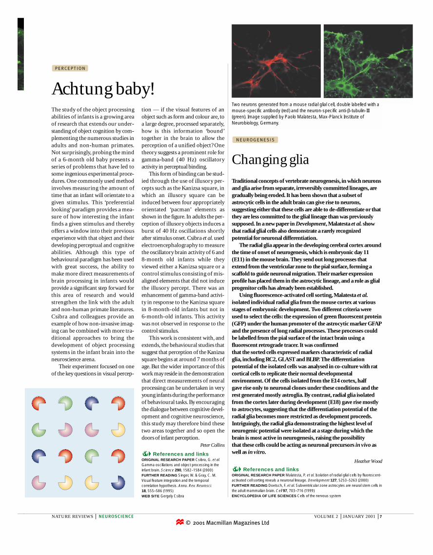
Traditional concepts of vertebrate neurogenesis, in which neuronsand glia arise from separate, irreversibly committed lineages, aregradually being eroded. It has been shown that a subset ofastrocytic cells in the adult brain can give rise to neurons,suggesting either that these cells are able to de-differentiate or thatthey are less committed to the glial lineage than was previouslysupposed. In a new paper in Development, Malatesta et al. showthat radial glial cells also demonstrate a rarely recognizedpotential for neuronal differentiation.

The radial glia appear in the developing cerebral cortex aroundthe time of onset of neurogenesis, which is embryonic day 11(E11) in the mouse brain. They send out long processes thatextend from the ventricular zone to the pial surface, forming ascaffold to guide neuronal migration. Their marker expressionprofile has placed them in the astrocytic lineage, and a role as glialprogenitor cells has already been established.

Using fluorescence-activated cell sorting, Malatesta et al.isolated individual radial glia from the mouse cortex at variousstages of embryonic development. Two different criteria wereused to select the cells: the expression of green fluorescent protein(GFP) under the human promoter of the astrocytic marker GFAPand the presence of long radial processes. These processes couldbe labelled from the pial surface of the intact brain using afluorescent retrograde tracer. It was confirmed that the sorted cells expressed markers characteristic of radialglia, including RC2, GLAST and BLBP. The differentiationpotential of the isolated cells was analysed in co-culture with ratcortical cells to replicate their normal developmentalenvironment. Of the cells isolated from the E14 cortex, halfgave rise only to neuronal clones under these conditions and therest generated mostly astroglia. By contrast, radial glia isolatedfrom the cortex later during development (E18) gave rise mostlyto astrocytes, suggesting that the differentiation potential of theradial glia becomes more restricted as development proceeds.Intriguingly, the radial glia demonstrating the highest level ofneurogenic potential were isolated at a stage during which thebrain is most active in neurogenesis, raising the possibility that these cells could be acting as neuronal precursors in vivo aswell as in vitro.

Heather Wood

References and linksORIGINAL RESEARCH PAPER Malatesta, P. et al. Isolation of radial glial cells by fluorescent-activated cell sorting reveals a neuronal lineage. Development 127, 5253–5263 (2000) FURTHER READING Doetsch, F. et al. Subventricular zone astrocytes are neural stem cells inthe adult mammalian brain. Cell 97, 703–716 (1999)ENCYCLOPEDIA OF LIFE SCIENCES Cells of the nervous system

Achtung baby!

P E R C E P T I O N

The study of the object processingabilities of infants is a growing areaof research that extends our under-standing of object cognition by com-plementing the numerous studies inadults and non-human primates.Not surprisingly, probing the mindof a 6-month old baby presents aseries of problems that have led tosome ingenious experimental proce-dures. One commonly used methodinvolves measuring the amount oftime that an infant will orientate to agiven stimulus. This ‘preferentiallooking’ paradigm provides a mea-sure of how interesting the infantfinds a given stimulus and therebyoffers a window into their previousexperience with that object and theirdeveloping perceptual and cognitiveabilities. Although this type ofbehavioural paradigm has been usedwith great success, the ability tomake more direct measurements ofbrain processing in infants wouldprovide a significant step forward forthis area of research and wouldstrengthen the link with the adultand non-human primate literatures.Csibra and colleagues provide anexample of how non-invasive imag-ing can be combined with more tra-ditional approaches to bring thedevelopment of object processingsystems in the infant brain into theneuroscience arena.

Their experiment focused on oneof the key questions in visual percep-

tion — if the visual features of anobject such as form and colour are, toa large degree, processed separately,how is this information ‘bound’together in the brain to allow the perception of a unified object? Onetheory suggests a prominent role forgamma-band (40 Hz) oscillatoryactivity in perceptual binding.

This form of binding can be stud-ied through the use of illusory per-cepts such as the Kanizsa square, inwhich an illusory square can beinduced between four appropriatelyorientated ‘pacman’ elements asshown in the figure. In adults the per-ception of illusory objects induces aburst of 40 Hz oscillations shortlyafter stimulus onset. Csibra et al. usedelectroencephalography to measurethe oscillatory brain activity of 6 and8-month old infants while theyviewed either a Kanizsa square or acontrol stimulus consisting of mis-aligned elements that did not inducethe illusory percept. There was anenhancement of gamma-band activi-ty in response to the Kanizsa squarein 8-month-old infants but not in 6-month-old infants. This activity was not observed in response to thecontrol stimulus.

This work is consistent with, andextends, the behavioural studies thatsuggest that perception of the Kanizsasquare begins at around 7 months ofage. But the wider importance of thiswork may reside in the demonstrationthat direct measurements of neuralprocessing can be undertaken in veryyoung infants during the performanceof behavioural tasks. By encouragingthe dialogue between cognitive devel-opment and cognitive neuroscience,this study may therefore bind thesetwo areas together and so open thedoors of infant perception.

Peter Collins

References and linksORIGINAL RESEARCH PAPER Csibra, G. et al.Gamma oscillations and object processing in theinfant brain. Science 290, 1582–1584 (2000)FURTHER READING Singer, W. & Gray, C. M.Visual feature integration and the temporalcorrelation hypothesis. Annu. Rev. Neurosci.18, 555–586 (1995)WEB SITE Gergely Csibra

Changing glia

N E U R O G E N E S I S

Two neurons generated from a mouse radial glial cell, double labelled with amouse-specific antibody (red) and the neuron-specific anti-β-tubulin-III(green). Image supplied by Paolo Malatesta, Max-Planck Institute ofNeurobiology, Germany.

© 2001 Macmillan Magazines Ltd