Embed Size (px)
Citation preview
Aus dem Max von Pettenkofer-Institut für Hygiene und Medizinische Mikrobiologie
Lehrstuhl: Bakteriologie
der Ludwig-Maximilians-Universität München
Kommissarische Leitung: Prof. Dr. Rainer Haas
Analysis of Helicobacter pylori VacA-containing vacuoles
and VacA intracellular trafficking
Dissertation
zum Erwerb des Doktorgrades der Naturwissenschaften
an der Medizinischen Fakultät
der Ludwig-Maximilians-Universität München
vorgelegt von
Beate Kern
aus Schrobenhausen
2014
II
Gedruckt mit Genehmigung der Medizinischen Fakultät
der Ludwig-Maximilians-Universität München
Betreuer: Prof. Dr. Rainer Haas
Zweitgutachter: Prof. Dr. Heinrich Jung
Dekan: Prof. Dr. med. Dr.h.c. Maximilian Reiser, FACR, FRCR
Tag der mündlichen Prüfung: 03.06.2015
Eidesstattliche Versicherung
III
Eidesstattliche Versicherung
Ich, Beate Kern, erkläre hiermit an Eides statt, dass ich die vorliegende Dissertation mit dem
Thema
Analysis of Helicobacter pylori VacA-containing vacuoles and VacA intracellular trafficking
selbständig verfasst, mich außer der angegebenen keiner weiteren Hilfsmittel bedient und alle
Erkenntnisse, die aus dem Schrifttum ganz oder annähernd übernommen sind, als solche
kenntlich gemacht und nach ihrer Herkunft unter Bezeichnung der Fundstelle einzeln
nachgewiesen habe.
Ich erkläre des Weiteren, dass die hier vorgelegte Dissertation nicht in gleicher oder in ähnlicher
Form bei einer anderen Stelle zur Erlangung eines akademischen Grades eingereicht wurde.
Ort, Datum Unterschrift
IV
Teile dieser Arbeit werden veröffentlicht:
Kern B., Jain U., Utsch C., Otto A., Busch B., Jiménez-Soto L.F., Becher D., Haas R.:
Characterization of Helicobacter pylori VacA-containing vacuoles (VCVs), VacA intracellular
trafficking and interference with calcium signalling in T-lymphocytes. Cell Microbiol 2015, doi:
10.1111/cmi.12474 (accepted)
Publikation im Promotionszeitraum, die nicht in der Arbeit enthalten ist:
Fischer W., Breithaupt U., Kern B., Smith S.I., Spicher C., Haas R.: A comprehensive analysis of
Helicobacter pylori plasticity zones reveals that they are integrating conjugative elements with
intermediate integration specificity. BMC Genomics 2014, 15:310
Table of Contents
V
Table of Contents
Eidesstattliche Versicherung ..................................................................................................... III
Table of Contents ........................................................................................................................ V
List of Figures .......................................................................................................................... VIII
Summary .................................................................................................................................... IX
Zusammenfassung ..................................................................................................................... XI
1 Introduction ........................................................................................................................... 1
1.1 H. pylori Epidemiology .................................................................................................................... 1
1.2 Overview of the Infection Process .................................................................................................. 1
1.3 H. pylori Immune Evasion............................................................................................................... 2
1.4 The Vacuolating Cytotoxin VacA .................................................................................................. 3
1.4.1 Vacuolation .............................................................................................................................. 3
1.4.2 VacA Protein Structure and Channel Formation ............................................................... 4
1.4.3 Allelic Diversity of VacA ........................................................................................................ 6
1.4.4 VacA Internalization and Trafficking .................................................................................. 7
1.5 VacA-Induced Effects on Host Cells.............................................................................................. 8
1.5.1 Mitochondrial Effects and Apoptosis ................................................................................... 8
1.5.2 Immunomodulatory Effects .................................................................................................. 9
1.5.3 CagA Effects and CagA-VacA Interplay ............................................................................ 10
1.5.4 VacA and Intracellular Calcium Signaling ........................................................................ 11
1.5.5 VacA - A Multifunctional Mystery ..................................................................................... 12
1.6 Endosomes as Signaling Platforms ............................................................................................... 14
1.7 Aim of This Study ........................................................................................................................... 14
2 Materials and Methods ........................................................................................................ 17
2.1 Materials .......................................................................................................................................... 17
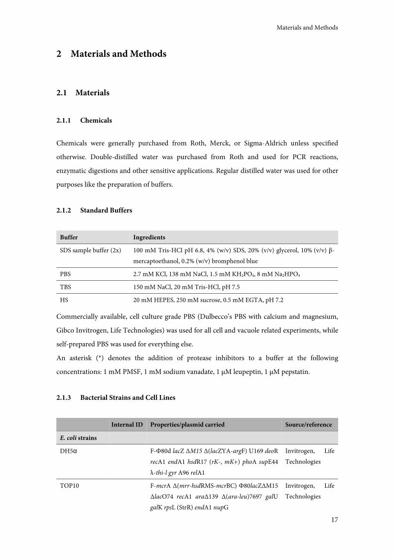
2.1.1 Chemicals ............................................................................................................................... 17
2.1.2 Standard Buffers .................................................................................................................... 17
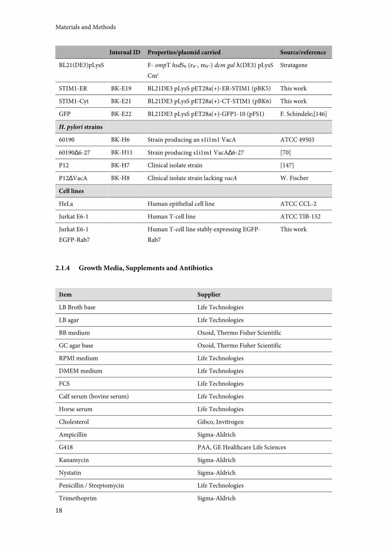
2.1.3 Bacterial Strains and Cell Lines ........................................................................................... 17
2.1.4 Growth Media, Supplements and Antibiotics ................................................................... 18
2.1.5 Commercially Available Kits ............................................................................................... 19
VI
2.1.6 Plasmids ................................................................................................................................. 19
2.1.7 Oligonucleotides ................................................................................................................... 19
2.1.8 Enzymes and Proteins .......................................................................................................... 20
2.1.9 Antibodies and Antisera ...................................................................................................... 20
2.2 Methods ........................................................................................................................................... 21
2.2.1 Escherichia coli Methods ...................................................................................................... 21
2.2.2 Helicobacter pylori Cultivation and Strain Maintenance ................................................. 22
2.2.3 Cell Culture ............................................................................................................................ 22
2.2.4 Cloning ................................................................................................................................... 24
2.2.5 Protein Biochemical Methods ............................................................................................. 26
2.2.6 Purification of VacA ............................................................................................................. 29
2.2.7 Preparation of Concentrated Culture Supernatant (CCS) .............................................. 30
2.2.8 Vacuolation Assay................................................................................................................. 30
2.2.9 Vacuolation Time Course .................................................................................................... 31
2.2.10 Production of α-VacA_nat .................................................................................................. 31
2.2.11 Vacuolation Inhibition Assay .............................................................................................. 31
2.2.12 Pull-Down Experiments ....................................................................................................... 32
2.2.13 Immunostaining Experiments ............................................................................................ 33
2.2.14 Microscopy and Image Analysis ......................................................................................... 34
2.2.15 Homogenization of Jurkat E6-1 Cells ................................................................................ 34
2.2.16 Sequential VCV Centrifugation .......................................................................................... 34
2.2.17 TurboBeads Methods ........................................................................................................... 35
2.2.18 Vacuole Sorting by Flow Cytometry .................................................................................. 36
2.2.19 Isolation of VCVs by Immunomagnetic Separation ........................................................ 37
2.2.20 Mass Spectrometry ................................................................................................................ 38
2.2.21 Mass Spectrometry Data Processing .................................................................................. 38
2.2.22 Isolation of Endoplasmic Reticulum from Jurkat E6-1 Cells .......................................... 38
3 Results .................................................................................................................................. 41
3.1 VacA Purification and Labeling ................................................................................................... 41
3.2 Characterization of α-VacA_nat .................................................................................................. 42
3.2.1 α-VacA_nat in Immunoblotting ........................................................................................ 43
3.2.2 Inhibition of Vacuolation by Various Antisera ................................................................ 43
3.2.3 α-VacA_nat in Immunostaining Experiments ................................................................. 45
3.3 Stable Cell Line Jurkat E6-1 EGFP-Rab7 ..................................................................................... 45
3.4 VCV Isolation ................................................................................................................................. 46
3.4.1 Strategies ................................................................................................................................ 46
3.4.2 General Optimization Steps................................................................................................. 48
Table of Contents
VII
3.4.3 Strategy A: TurboBeads Strategy ......................................................................................... 50
3.4.4 Strategy B: VCV Sorting by Flow Cytometry .................................................................... 53
3.4.5 Strategy C: Immunomagnetic Purification of VCVs ....................................................... 55
3.5 Mass Spectrometry Results ............................................................................................................ 58
3.6 Investigation of Possible Interactions of VacA with STIM1..................................................... 61
3.7 VacA Localization in Intoxicated Cells ....................................................................................... 63
3.7.1 Colocalization of VacA with ER and Golgi Apparatus Markers and CTxB ................. 63
3.7.2 Isolation of Endoplasmic Reticulum .................................................................................. 64
4 Discussion ............................................................................................................................ 67
4.1 VCV Isolation Strategies and Tools ............................................................................................. 67
4.2 The VCV Proteome ........................................................................................................................ 69
4.2.1 VCV Analysis by MS - Strengths and Weaknesses .......................................................... 69
4.2.2 Individual Proteins Found on VCVs .................................................................................. 71
4.2.3 The VCV Proteome Decoded by Subcellular Localization and Biological Function .. 77
4.2.4 Interorganellar Crosstalk between VCVs and Mitochondria/ER .................................. 78
4.2.5 VCVs as Multifunctional Platforms ................................................................................... 79
4.3 Influence of VacA on Store-Operated Calcium Entry (SOCE) ................................................ 79
4.4 VacA Partially Localizes to the ER and the Golgi Apparatus ................................................... 82
4.5 Is VacA Transported in a Retrograde Manner? ......................................................................... 83
4.6 Conclusion and Outlook ............................................................................................................... 87
5 List of Abbreviations ........................................................................................................... 89
6 Appendix .............................................................................................................................. 91
6.1 VCV-specific proteins .................................................................................................................... 91
6.2 Wild-type sample 1 (WT1) proteins ............................................................................................ 95
6.3 Wild-type sample 2 (WT2) proteins .......................................................................................... 123
6.4 Background proteins .................................................................................................................... 146
7 Literature ............................................................................................................................ 169
8 Danksagung ........................................................................................................................ 183
9 Lebenslauf .......................................................................................................................... 185
VIII
List of Figures
Figure 1: Structure of VacA and cellular vacuolation. .......................................................................... 5
Figure 2: VacA uptake, intracellular trafficking, and effects.............................................................. 13
Figure 3: VacA gel filtration. .................................................................................................................. 42
Figure 4: Characterization of α-VacA_nat........................................................................................... 44
Figure 5: Creation of the stable cell line Jurkat E6-1 EGFP-Rab7. .................................................... 46
Figure 6: Three different strategies for VCV isolation. ...................................................................... 47
Figure 7: Optimization of VCV isolation conditions. ........................................................................ 49
Figure 8: TurboBeads strategy, internalization, and aggregation. .................................................... 52
Figure 9: VCV homogenate prepared for flow cytometry sorting. ................................................... 54
Figure 10: Analysis of VCV fractions sorted by flow cytometry. ...................................................... 55
Figure 11: Monitoring the VCV isolation process. ............................................................................. 57
Figure 12: Mass spectrometry data sets. ............................................................................................... 58
Figure 13: Graphic and numerical display of the composition of VCV-specific and background
subsets. ...................................................................................................................................................... 60
Figure 14: Investigation of a possible VacA-STIM1 interaction. ...................................................... 62
Figure 15: VacA localization in intoxicated cells................................................................................. 65
Figure 16: Internalization and trafficking of bacterial toxins. ........................................................... 84
Figure 17: VacA uptake, intracellular trafficking, and effects, continued. ....................................... 87
Summary
IX
Summary
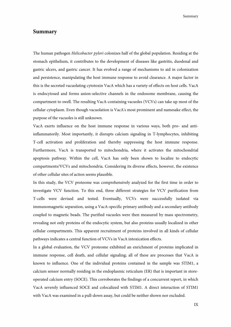
The human pathogen Helicobacter pylori colonizes half of the global population. Residing at the
stomach epithelium, it contributes to the development of diseases like gastritis, duodenal and
gastric ulcers, and gastric cancer. It has evolved a range of mechanisms to aid in colonization
and persistence, manipulating the host immune response to avoid clearance. A major factor in
this is the secreted vacuolating cytotoxin VacA which has a variety of effects on host cells. VacA
is endocytosed and forms anion-selective channels in the endosome membrane, causing the
compartment to swell. The resulting VacA-containing vacuoles (VCVs) can take up most of the
cellular cytoplasm. Even though vacuolation is VacA's most prominent and namesake effect, the
purpose of the vacuoles is still unknown.
VacA exerts influence on the host immune response in various ways, both pro- and anti-
inflammatorily. Most importantly, it disrupts calcium signaling in T-lymphocytes, inhibiting
T-cell activation and proliferation and thereby suppressing the host immune response.
Furthermore, VacA is transported to mitochondria, where it activates the mitochondrial
apoptosis pathway. Within the cell, VacA has only been shown to localize to endocytic
compartments/VCVs and mitochondria. Considering its diverse effects, however, the existence
of other cellular sites of action seems plausible.
In this study, the VCV proteome was comprehensively analyzed for the first time in order to
investigate VCV function. To this end, three different strategies for VCV purification from
T-cells were devised and tested. Eventually, VCVs were successfully isolated via
immunomagnetic separation, using a VacA-specific primary antibody and a secondary antibody
coupled to magnetic beads. The purified vacuoles were then measured by mass spectrometry,
revealing not only proteins of the endocytic system, but also proteins usually localized in other
cellular compartments. This apparent recruitment of proteins involved in all kinds of cellular
pathways indicates a central function of VCVs in VacA intoxication effects.
In a global evaluation, the VCV proteome exhibited an enrichment of proteins implicated in
immune response, cell death, and cellular signaling; all of these are processes that VacA is
known to influence. One of the individual proteins contained in the sample was STIM1, a
calcium sensor normally residing in the endoplasmic reticulum (ER) that is important in store-
operated calcium entry (SOCE). This corroborates the findings of a concurrent report, in which
VacA severely influenced SOCE and colocalized with STIM1. A direct interaction of STIM1
with VacA was examined in a pull-down assay, but could be neither shown nor excluded.
X
Immunofluorescence experiments conducted in HeLa cells confirmed the presence of VacA in
the ER and also found it to traffic to the Golgi apparatus, identifying these two cellular
compartments as novel VacA target structures. The exact route of VacA transport remains
unclear, but the involvement of both the ER and the Golgi suggests the possibility of retrograde
trafficking, analogous to other bacterial toxins like shiga and cholera toxins.
In summary, the elucidation of the VCV proteome and the discovery of the ER and the Golgi
apparatus as VacA target structures have generated intriguing starting points for future studies.
The detection of many proteins implicated in VacA intoxication effects in the VCV proteome
leads to the proposal of VCVs as signaling hubs that may coordinate the complex meshwork of
VacA effects. Further investigation of individual proteins is expected to help greatly in
illuminating this matter.
Introduction
XI
Zusammenfassung
Etwa die Hälfte der Weltbevölkerung ist mit dem humanpathogenen Bakterium Helicobacter
pylori infiziert. Es kolonisiert das Magenepithel und trägt dort zur Entstehung von Krankheiten
wie Gastritis, Magen- und Zwölffingerdarmgeschwüren und Magenkrebs bei. Um erfolgreich zu
kolonisieren und zu persistieren, hat H. pylori eine Reihe von Mechanismen entwickelt, die
unter anderem die Immunantwort des Wirts manipulieren und so die Beseitigung durch das
Immunsystem verhindern. Ein wichtiger Faktor hierbei ist das sekretierte vakuolisierende
Zytotoxin (vacuolating cytotoxin) VacA, das vielfältige Auswirkungen auf Wirtszellen hat. Nach
der Endozytose bildet VacA Anionenkanäle in der Endosomenmembran, was zum Anschwellen
der Endosomen zu sogenannten VacA-beinhaltenden Vakuolen (VacA-containing vacuoles,
VCVs) führt. Diese können fast das gesamte Zytoplasma einnehmen. Obwohl Vakuolisierung
der markanteste und namensgebende Effekt von VacA ist, konnte die Funktion der Vakuolen
bislang nicht geklärt werden.
VacA beeinflusst die Immunantwort des Wirts sowohl stimulierend als auch supprimierend.
Am wichtigsten erscheint dabei, dass VacA den Calcium-Stoffwechsel in T-Lymphozyten stört,
dadurch die T-Zell-Aktivierung und -Proliferation hemmt und so die Immunantwort
unterdrückt. Des Weiteren wird VacA zu Mitochondrien transportiert, wo es den
mitochondrialen Apoptoseweg aktiviert. Bisher wurde VacA nur dort und in endosomalen
Kompartimenten/VCVs beobachtet. Angesichts der diversen Auswirkungen der VacA-
Intoxikation liegt es jedoch nahe, dass das Toxin auch anderswo in der Zelle agiert.
Um die Funktion von VCVs herauszufinden, wurde in der vorliegenden Arbeit das VCV-
Proteom erstmals umfassend charakterisiert. Zu diesem Zweck wurden drei Strategien für die
VCV-Aufreinigung aus T-Zellen entworfen und getestet. Die erfolgreiche Isolation von VCVs
erfolgte mittels einer immunomagnetischen Methode, bestehend aus einem VacA-spezifischen
primären Antikörper und einem sekundären Antikörper, der an magnetische Kügelchen
gebunden ist. Anschließend wurden die aufgereinigten Vakuolen massenspektrometrisch
gemessen. In den analysierten Proben befanden sich nicht nur Proteine des endozytischen
Systems, sondern auch Proteine, die normalerweise in anderen zellulären Kompartimenten
lokalisiert sind. Diese Rekrutierung von Proteinen vieler zellulärer Vorgänge impliziert, dass
VCVs in der VacA-Intoxikation eine zentrale Rolle spielen.
Eine allgemeine Untersuchung des VCV-Proteoms zeigte eine Anreicherung von Proteinen, die
an Immunantwort, Zelltod und Signaltransduktion beteiligt sind; all dies sind Prozesse, die
XII
VacA bekanntermaßen beeinflusst. Eines der in der Probe vorhandenen Proteine war STIM1,
ein Ca2+-Sensor, der sich gewöhnlich im endoplasmatischen Retikulum (ER) befindet und
wesentlich für den speicherabhängigen Calciumeinstrom (store-operated calcium entry, SOCE)
ist. Dies unterstützt die Ergebnisse einer gleichzeitig durchgeführten Studie, in der VacA SOCE
beeinträchtigte und mit STIM1 kolokalisierte. Eine direkte Interaktion von STIM1 mit VacA
wurde mit Hilfe von pull-down Experimenten analysiert, konnte aber weder nachgewiesen noch
widerlegt werden.
Immunfluoreszenzversuche in HeLa-Zellen bestätigten die Anwesenheit von VacA im ER und
zeigten es außerdem auch im Golgi-Apparat. Dadurch wurden ER und Golgi-Apparat als neue
Zielstrukturen von VacA identifiziert. Der genaue Transportweg von VacA ist noch ungeklärt,
doch die Beteiligung von ER und Golgi-Apparat deutet auf die Möglichkeit des retrograden
Transports hin, analog zu anderen bakteriellen Toxinen wie Shiga oder Cholera Toxin.
Zusammenfassend liefern die Aufklärung des VCV-Proteoms und die Entdeckung von ER und
Golgi-Apparat als VacA-Zielstrukturen interessante Startpunkte für zukünftige Studien. Da im
VCV-Proteom viele Proteine aufgefunden wurden, die für VacA-Intoxikationseffekte von
Bedeutung sind, ist es denkbar, dass VCVs als Signalplattformen das komplizierte Geflecht von
VacA-Effekten koordinieren. Weiterführende Untersuchungen der einzelnen Proteine im VCV-
Proteom könnten bei der Erforschung dieser Hypothese von großem Nutzen sein.
Introduction
1
1 Introduction
The relationship between humans and Helicobacter pylori is a complex one. The human
stomach is H. pylori's ecological niche, and it has been for at least 60 000 years, when humans
emigrated from Africa [1]. By contrast, it has only been thirty years since the discovery of
H. pylori as a colonizer of the human stomach [2], which was thought to be sterile before. In
these thirty years, H. pylori infection has been found to be a major factor in various gastric
diseases like gastritis, gastric ulcers and gastric cancer [3,4], and antibiotic therapy is readily
available [5]. But lately, more and more evidence is emerging for its beneficial role for the host -
H. pylori infection may protect from inflammatory bowel disease and immune disorders like
asthma [6–8]. Elucidation of this ambivalent relationship may therefore not only help treat and
cure H. pylori-induced diseases, but also enable the constructive use of its favorable effects.
1.1 H. pylori Epidemiology
About half of the world's population is infected with this Gram-negative pathogen, with the
prevalence varying between 20% in developed countries and 90% in some developing countries
[9,10]. Infection always leads to a chronic gastritis that remains asymptomatic in most carriers
[11]. About 10-20% of infected individuals develop symptoms ranging from peptic ulcer disease
to atrophic gastritis to gastric adenocarcinoma [9]. The implication of H. pylori in
carcinogenesis lead to its classification as a group 1 carcinogen by the World Health
Organization in 1994 [12]. Infection usually happens in early childhood, is transmitted via the
gastric-oral route within families [13,14], and persists for life if not eradicated with antibiotics
[9]. Therapy is only administered in symptomatic patients or in individuals with an increased
risk for gastric cancer. It consists of two antibiotics and a proton pump inhibitor and is about
80% effective, with the growing problem of resistant strains [5].
1.2 Overview of the Infection Process
Helicobacter pylori has evolved a variety of features that help the bacterium colonize and persist
in the hostile environment of the human stomach. It is not an acidophile and can only survive
for minutes in the acidic stomach lumen where the pH is as low as 2 [15]. For this reason, it

Introduction
2
must quickly relocate to the mucus layer lining the gastric epithelial surface. The mucus is about
300 µm thick and its pH increases gradually, reaching neutral conditions at the epithelium [16].
H. pylori employs chemotaxis to navigate towards the epithelium. Its namesake spiral shape and
its flagella enable the bacterium to move through the mucus in a corkscrew-like motion [17]. A
further aid in this process is the urease enzyme expressed by H. pylori: it permits the organism to
buffer its microenvironment and periplasm [18], thereby resisting the acidic conditions and
modifying the mucus texture, making it less gel-like and thus easier to penetrate [19]. A small
number of bacterial cells has been detected inside of epithelial cells, but H. pylori is generally
considered an extracellular pathogen [20]. Attachment to the epithelium is facilitated by several
adhesins, the most well-researched among them being BabA and SabA. Both bind glycosylated
blood group antigens [21,22]. When adherent, H. pylori uses a type IV secretion system to inject
CagA, a protein encoded by the cytotoxin-associated gene A (cagA), into the host cell [23,24].
CagA has various effects on epithelial cells, altering cell signaling, cell polarity, extrusion,
motility, and proliferation, and is heavily implicated in the development of cancer [4,25,26].
Another protein toxin of H. pylori and central to this work is the vacuolating cytotoxin VacA,
which is secreted and then affects different host cell types in diverse ways, contributing to initial
colonization and immune evasion, and thereby persistence [27]. Among other things, it can act
on both pro- and anti-inflammatory pathways, modulating the host immune response. The
complex interplay of CagA, VacA, and other factors to manipulate the host suggests that an
escalation is usually avoided to prevent clearance by the host's immune system [14]. The effects
of these two main H. pylori toxins on host cells, with particular focus on VacA, will be outlined
in further detail below.
1.3 H. pylori Immune Evasion
In order to persist, H. pylori escapes, manipulates, and counteracts the host immune response,
employing diverse strategies. Usually, bacteria are recognized by pattern recognition receptors
on cells of the innate immune system, but H. pylori avoids this with modified pathogen-
associated molecular patterns. Examples of this are modifications in flagellins and
lipopolysaccharide [28,29], two bacterial components that are usually detected by the host.
H. pylori also actively modulates the immune response by activating the adaptor protein Myd88,
causing expression of the anti-inflammatory cytokine IL-10 [30]. The adaptive immune system
produces antibodies against H. pylori, but these do not confer sufficient immunity, which is also
Introduction
3
a problem in vaccination research [31]. In fact, antibodies and B-cells can be neglected in the
immune response to H. pylori infection [32]. Macrophages are recruited to the site of infection
and produce nitric oxide that is toxic to bacteria, but H. pylori-induced arginase II and ornithine
decarboxylase also cause macrophage apoptosis [33,34]. Neutrophils infiltrate and release
reactive oxygen species, which is simultaneously induced and combated by the H. pylori factor
NapA [35]. Another instance where the bacterium actively interferes is the T-cell response. By
acting on dendritic cells, H. pylori can cause the preferential development of regulatory T-cells
(Treg) over immunostimulatory TH1/TH17 cells, again downregulating the immune response [36].
These are just a few examples of how H. pylori avoids clearance by the host immune system. At
the same time, a low level of inflammation is maintained, which may be necessary for
persistence by providing nutrients for the bacteria [14,27,37]. This complex evasion and
manipulation of innate and adaptive immune responses, together with its supply of virulence
factors, enable H. pylori to establish an optimal niche for colonization.
1.4 The Vacuolating Cytotoxin VacA
1.4.1 Vacuolation
As is evident from its name, one effect of VacA on intoxicated cells is the induction of large
cytoplasmic vesicles, also termed vacuoles [38,39]. Vacuolation has been observed in various
types of cultured and primary cells [38,40,41]. Even though this effect was the first one to be
noticed, and remains important as a convenient phenotype for assessing toxin activity and
susceptibility, its purpose continues to be unknown. It is still unclear whether vacuolation is an
effect in and of itself, or whether it is just a by-product of other processes.
In contrast to most other vacuole-inducing bacteria, vacuoles do not serve as the main site for
survival or replication of H. pylori. Although it is capable of invading cells, it does so rarely [20],
and vacuolation is purely a result of the VacA toxin, not the presence of bacteria.
In the current model of the process of vacuole formation (reviewed in [27]), VacA binds to host
cells, and during or after internalization, inserts into the membrane of the newly formed
endocytic compartments. In these membranes, it oligomerizes to form anion-selective channels
[42] that facilitate the influx of Cl- ions, a process that the vacuolar ATPase (vATPase)
compensates by importing protons. This results in a low lumenal pH, which is balanced by the
diffusion of membrane-permeable weak bases like ammonia into the endocytic compartments,
Introduction
4
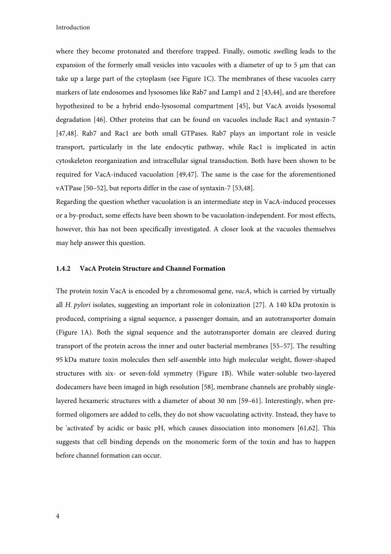
where they become protonated and therefore trapped. Finally, osmotic swelling leads to the
expansion of the formerly small vesicles into vacuoles with a diameter of up to 5 µm that can
take up a large part of the cytoplasm (see Figure 1C). The membranes of these vacuoles carry
markers of late endosomes and lysosomes like Rab7 and Lamp1 and 2 [43,44], and are therefore
hypothesized to be a hybrid endo-lysosomal compartment [45], but VacA avoids lysosomal
degradation [46]. Other proteins that can be found on vacuoles include Rac1 and syntaxin-7
[47,48]. Rab7 and Rac1 are both small GTPases. Rab7 plays an important role in vesicle
transport, particularly in the late endocytic pathway, while Rac1 is implicated in actin
cytoskeleton reorganization and intracellular signal transduction. Both have been shown to be
required for VacA-induced vacuolation [49,47]. The same is the case for the aforementioned
vATPase [50–52], but reports differ in the case of syntaxin-7 [53,48].
Regarding the question whether vacuolation is an intermediate step in VacA-induced processes
or a by-product, some effects have been shown to be vacuolation-independent. For most effects,
however, this has not been specifically investigated. A closer look at the vacuoles themselves
may help answer this question.
1.4.2 VacA Protein Structure and Channel Formation
The protein toxin VacA is encoded by a chromosomal gene, vacA, which is carried by virtually
all H. pylori isolates, suggesting an important role in colonization [27]. A 140 kDa protoxin is
produced, comprising a signal sequence, a passenger domain, and an autotransporter domain
(Figure 1A). Both the signal sequence and the autotransporter domain are cleaved during
transport of the protein across the inner and outer bacterial membranes [55–57]. The resulting
95 kDa mature toxin molecules then self-assemble into high molecular weight, flower-shaped
structures with six- or seven-fold symmetry (Figure 1B). While water-soluble two-layered
dodecamers have been imaged in high resolution [58], membrane channels are probably single-
layered hexameric structures with a diameter of about 30 nm [59–61]. Interestingly, when pre-
formed oligomers are added to cells, they do not show vacuolating activity. Instead, they have to
be 'activated' by acidic or basic pH, which causes dissociation into monomers [61,62]. This
suggests that cell binding depends on the monomeric form of the toxin and has to happen
before channel formation can occur.
Introduction
5
Figure 1: Structure of VacA and cellular vacuolation.
(A) Signal sequence (also called s-region) and autotransporter domain are cleaved during VacA secretion. The
remaining mature toxin consists of the p33 and p55 domain. The p33 domain contains the i-region and a
hydrophobic stretch of about 30 aa, which is essential for channel formation. The p55 domain contains the m-region,
which is implicated in cell binding specificity. Amino acid counts of H. pylori strain 60190 VacA are indicated at the
top. (B) VacA channels comprise six monomers and are shaped like flowers, with the p33 domains making up the
channel core and the p55 domains representing the petals. (A) and (B) modified from [54]. (C) VacA intoxication in
the presence of weak bases causes the formation of large cytoplasmic vacuoles that take up most of the cytoplasm and
can reach a diameter of up to 5 µm. Vacuoles are acidic and can therefore be stained with neutral red. Top image,
Jurkat E6-1 cells; bottom image, HeLa cells. Scale bars represent 10 µm.
The mature VacA protein can be further subdivided into two domains termed p33 and p55
according to their approximate molecular weights (Figure 1A). The crystal structure of the p55
domain has been solved, showing secondary structural features characteristic for
autotransporter passenger domains [63]. Structural information on the p33 domain is still
lacking, but the p55 crystal structure in combination with multiple electron microscopy studies
on VacA oligomers allows at least some inferences. VacA has been called sock-shaped, with the
p55 domain being the heel and foot and the p33 domain representing the calf portion (see
Figure 1B; [63]). To continue the analogies, in the flower-shaped oligomer or channel, p33
subunits make up the core, while p55 subunits form the petals. The two subunits have been
assigned different functions over the years. The p33 domain was originally considered
responsible for VacA channel formation and therefore vacuolation [64], consistent with its
Introduction
6
position at the center of oligomeric structures. However, p55 was later also found to play a part
in this [65]. On the other hand, p33 was observed to contribute to host cell binding [65], a
function initially assigned to the p55 domain [66]. Apparently, the two domains' functions
cannot easily be separated. Still, p33 is commonly considered the toxic subunit and p55 the cell
binding subunit, analogously to AB toxin terminology. Proteolytic cleavage can naturally
separate the two domains, but they remain non-covalently attached [56]. When the subunits are
expressed independently and then mixed before addition to cells, the wild-type effect is
preserved [67,68,65], and the biological significance of cleavage is not clear.
An interesting detail is the protein's only strongly hydrophobic region. It is contained within the
first thirty amino acids of p33, and it is presumed to insert into membranes for channel
formation [69]. A mutant version of VacA lacking this hydrophobic region (VacA∆6-27) forms
channels considerably more slowly than the wild-type, and fails to induce vacuolation [70].
Recently, electron microscopy experiments have shown that oligomers containing VacA∆6-27
have organization defects at their core that may account for this [71]. The fact that mixed
mutant/wild-type oligomers exhibit these defects, too, helps explain the dominant negative
effect of the mutant that was observed early on [70]. This hydrophobic region contains three
tandem repeats of a GXXXG motif characteristic for membrane dimerization domains, one of
which (G14XXXG18) was found to be essential for channel formation and cytotoxicity [69,72].
1.4.3 Allelic Diversity of VacA
H. pylori are genetically extremely diverse, due at least in part to their natural DNA uptake
competence, which facilitates horizontal gene transfer. This diversity is mirrored in VacA
sequences. VacA does not show significant homology to any other known protein [39]. Three
regions exhibit a particular allelic diversity: the N-terminal signal region (with s1 and s2 alleles),
the intermediate region in the p33 domain (i1, i2, and possibly i3), and the mid-region in the
p55 domain (m1 and m2) [73–75]. The s1, i1, and m1 alleles are associated with more severe
diseases in humans (summarized in [76]) and the s1 and i1 alleles also correlate with the
presence of CagA, which also means more severe disease outcomes [75]. The varying medical
consequences have at least partially been explained by experimental findings. Different VacA
variants affect different cell types in vitro, and a stretch of 148 amino acids within the m-region
was found to determine cell type specificity through binding [40,77]. This agrees with earlier
reports mentioned above that p55 mediates binding to host cells. The s2 allele has an additional
hydrophilic segment at the N-terminus that the s1 allele lacks; these 12 amino acids seem to
Introduction
7
prevent vacuolation by altering channel formation [78,79], concurring with the lowered toxicity
of s2 VacA. The i-region was only discovered recently and has not been thoroughly studied on a
molecular level, but seems to determine VacA toxicity specifically for T-cells [76]. Taken
together, the molecular differences of distinct forms of VacA may explain the variance in strain
pathogenicity, especially in combination with other virulence factors like CagA (see 1.5.3).
The VacA protein produced by the strain 60190 is somewhat of a standard in VacA research. It
has s1/i1/m1 alleles, is highly toxic, and is produced in comparatively large amounts by the
bacterium. The sequence details depicted in Figure 1 refer to this variant.
1.4.4 VacA Internalization and Trafficking
A substantial amount of research has focused on VacA binding to host cells and its subsequent
internalization, and even though much is now known about both processes, they are far from
clear. Multiple studies have searched for the VacA receptor and have yielded different results.
Cellular structures that bind VacA include phospho- and glycosphingolipids [80,81,60,82],
sphingomyelin [83], and heparan sulphate [84]. Several protein receptors have also been found,
some conferring cell-type specificity. The epidermal growth factor receptor (EGFR) was
observed to mediate VacA uptake in HeLa cells [85]. Receptor protein-tyrosine phosphatases α
and β (RPTPα/β) were also identified as receptors on kidney and stomach epithelium cells,
respectively [62,86]. The β1 integrin subunit CD29 acts as a complemental receptor on epithelial
cells [87]. On T-cells, VacA is endocytosed via the β2 integrin subunit CD18 [88], and most
recently, VacA was reported to bind to multimerin-1 on platelet cells [89]. Additionally, VacA is
known to bind the extracellular matrix protein fibronectin [90]. Whatever the receptor may be,
there is consensus that VacA localizes to lipid rafts in the host cell membrane, which are
necessary for internalization [91,92]. Since for example RPTPα/β usually reside outside lipid
rafts, it has been speculated whether the VacA-receptor complex relocates to lipid rafts after
VacA binding [93]. Glycosylphosphatidylinositol-anchored proteins (GPI-APs), a specific
component of lipid rafts, also play a role in VacA internalization, although they are probably not
an actual receptor [94,95]. VacA presumably does not induce its own uptake, but instead
exploits a constitutive cellular pathway [96,88].
The VacA uptake process appears to be similar in epithelial cells and lymphocytes [97] and is
dependent on temperature, energy, and actin, but independent of clathrin and dynamin
[98,99,95,94,100]. One group of researchers specifically investigated the VacA-containing
compartments shortly after uptake and found these compartments to lack common markers for
Introduction
8
known types of endosomes. Also, uptake unusually was independent of most known
endocytosis modulators. Due to the presence of the above mentioned GPI-APs, these
compartments were therefore identified as a relatively new type of compartments called GEECs
(GPI-AP-enriched early endosomal compartments) [96,101]. Most VacA is contained in GEECs
approximately 10 min after uptake. Later on, the GPI-APs are transported to recycling
endosomes, while VacA is sorted to the degradative pathway, arriving in early endosomes after
about 30 min. By way of late endosomes, after 120 min it finally reaches the endo-lysosomal
hybrid compartments that then become vacuoles (time points taken from [96,102]) (see Figure
2).
1.5 VacA-Induced Effects on Host Cells
Generally, for many intoxication effects, the internalization of VacA seems to be required, but
they may also be the result of signaling cascades triggered by VacA binding to receptors. The
latter is most probably the case for rapid effects that occur before internalization can even be
complete (30-60 min after intoxication) [27]. In some cases, however, the distinction between
the two has not been established. This study's focus on VacA-induced vacuoles naturally puts an
emphasis on internalization-dependent effects, but does not omit those caused purely by cell
binding in order to give a thorough overview.
1.5.1 Mitochondrial Effects and Apoptosis
Besides endosomal compartments, VacA is known to localize only to mitochondria. It first
accumulates in endosomes and is later transported to the organelle [103–105], but how the toxin
gets there is still a matter of debate. When it was discovered that VacA-containing endosomal
compartments attract actin, which forms tails and is able to move the endosomes through the
cytosol [102], a way of transport to mitochondria appeared to be found. However, how VacA is
then translocated from the endosome to the mitochondria is still unclear. Membrane fusion and
direct membrane-to-membrane transfer have both been speculated about, especially since
endosomes and mitochondria come into very close physical contact upon VacA intoxication
[106]. Emergence into the cytosol and subsequent uptake into mitochondria is also still a
possibility [107]. In any case, targeting of VacA to mitochondria depends on the first 32 amino
acids in the protein's N-terminus, the same stretch that is responsible for correct channel
formation, and this seems to function analogously to a signal sequence [103,105]. The most
Introduction
9
prominent effect of the VacA-mitochondria interaction is the induction of apoptosis. The
molecular mechanism of this has not been fully clarified, but a current model integrating the
available experimental evidence is as follows [108]: VacA is imported into the inner
mitochondrial membrane (IMM) [105], where it forms channels. This leads to the influx of Cl-
ions into the mitochondrial matrix and consequential loss of the mitochondrial membrane
potential [109,104]. Mitochondria with a defect membrane potential recruite Drp1, which
induces their fission and also the fragmentation of the mitochondrial network [110]. Alongside
Drp1, the pro-apoptotic factor Bax is recruited to mitochondria via VacA-containing
endosomes [106] and triggers the release of cytochrome c, eventually causing the cell to undergo
apoptosis [103,111,112]. Localization of VacA to mitochondria can happen as early as 60 min
after intoxication, but is most evident after at least 12 h and has been reported to precede the
induction of both mitochondrial fission and apoptosis [106,110].
Mitochondrial effects of VacA and vacuolation are independent of each other, but both depend
on VacA channel forming activity [111,106]. This was shown by using channel blocking
substances, which abolished apoptotic effects [111]. The use of mutant VacA∆6-27 is not
meaningful in this context, because the mutant protein also lacks the sequence necessary for
mitochondrial import, causing it to accumulate in endosomes [106]. However, it has not been
investigated whether the mitochondrial effects depend on VacA forming a channel in the IMM,
or whether VacA needs to form a channel to escape from endosomes [113].
Apoptosis caused by H. pylori infection has been observed in epithelial cells and cells of the
immune system, including T-, B-, and dendritic cells [112,114,115]. It has been argued to be a
major reason for ulcer formation in the stomach, but this is unlikely since a parallel
hyperproliferative response maintains epithelial integrity [116]. However, apoptosis of
epithelium does lead to faster turnover of cells, possibly providing more nutrients for the
bacteria and preventing cancer formation due to damaged cells, while apoptosis of immune cells
helps suppress the immune response [108]. VacA-induced apoptosis may therefore be an
important factor in persistence.
1.5.2 Immunomodulatory Effects
VacA effects on the host immune response are both pro- and anti-inflammatory. The clustering
of endocytic compartments in response to VacA intoxication [44] may purely be a way of
getting VacA to its destination, but such hijacking of host vesicular trafficking greatly disrupts
natural cellular transport processes and could also explain some of the effects observed in
Introduction
10
immune cells. In macrophages, VacA impairs vesicular maturation, leading to the formation of
large vesicular compartments called megasomes. This may prevent efficient killing of
phagocytosed bacteria [117] and therefore contribute to intracellular survival. In B-cells, VacA
interferes with antigen presentation, likely also due to alterations in vesicular trafficking [118].
As mentioned before, H. pylori influences the host T-cell response, partly through VacA.
Intoxication leads to a downregulation of IL-2 production, which is important for T-cell
viability and proliferation [119–121]. VacA inhibits the Ca2+-calmodulin-dependent
phosphatase calcineurin and, as a consequence of this, the nuclear translocation of the
transcription factor NFAT, which controls IL-2 expression. In the T-cell line Jurkat, altered
expression of 46 genes was observed following VacA intoxication [119]. VacA is also involved in
skewing the T-cell response, causing the differentiation of naïve T-cells into Treg cells instead of
immunostimulatory types of T-cells [36]. Fascinatingly, both Treg cells isolated from H. pylori-
infected individuals and purified VacA can be used to prevent asthma in mice [7,8,122], again
illustrating the complex relationship of the bacterium and its host.
1.5.3 CagA Effects and CagA-VacA Interplay
Unlike VacA, the cytotoxin-associated gene (cag) product CagA is not a secreted toxin, but is
injected into the host cell via a type IV secretion system (T4SS) apparatus [24]. Both the toxin
and the T4SS are encoded on the cag pathogenicity island (cag PAI), a 40-kb-sequence that was
probably acquired through horizontal gene transfer [123]. The T4SS pilus and CagA itself
interact with β1 integrin on host cells [124,125]. After translocation of CagA into the host cell
cytosol, it is phosphorylated by the host kinases Src and Abl [126,127]. CagA binds at least 20
known host cell proteins in either its phosphorylated or unphosphorylated form. One current
hypothesis is that phosphorylated CagA acts like a masterkey, mimicking a phosphorylated host
cell protein and thereby hijacking various cellular signaling pathways [128]. The most visually
impressive effect of CagA intoxication is the so-called hummingbird phenotype, a distinctive
cellular morphology characterized by cell elongation and cell scattering [128]. Other CagA
effects are the disruption of cell-cell junctions, loss of cell polarity, changes in motility and
proliferation, and the induction of a pro-inflammatory response, namely IL-8 expression
(reviewed in [128,4,25]).
From an epidemiological point of view, the presence of the cagA gene in H. pylori strains
infecting humans is associated with more severe forms of disease and cancer [129,130], and
there seems to be a connection between CagA and VacA. H. pylori strains have been grouped
Introduction
11
into two categories, where type I strains produce an active VacA and carry the cag PAI and type
II strains produce a non-functional (mutated or truncated) VacA and lack the cag PAI. Type I
strains cause more severe clinical outcomes [131], further highlighting the importance of both
pathogenicity factors.
Interestingly, on a cellular level, CagA and VacA have some opposing effects. This starts with
the two effects that were observed first for both toxins: cells that show VacA-induced
vacuolation show less CagA-induced hummingbird cell morphology, and vice versa [132]. Even
though vacuolation and hummingbird phenotype have no meaning per se, this illustrates on an
easily comrehensible level that the two toxins counteract each other's effects in the cell. It has
been observed that CagA activates and VacA downregulates the pleiotropic transcription factor
NFAT [133,119,120]; similarly, VacA induces apoptosis, while CagA suppresses it, leading to its
classification as an oncoprotein [26,134]. Moreover, CagA was shown to inhibit VacA uptake
into cells and also to interfere with intracellular VacA trafficking, stopping VacA in GEECs and
preventing its advancement into late endosomes and mitochondria [135,136], thereby
controlling the apoptotic effects of VacA. This suggests that H. pylori may, via CagA, control the
intracellular distribution of VacA and its resulting effects. Considering that VacA is a secreted
toxin that can diffuse away from bacteria while physical contact between bacteria and host cells
is necessary for CagA injection, an interesting possibility emerges: H. pylori may harness VacA-
induced effects like apoptosis for initial colonization or to dispose of immune cells recruited to
the site of infection [132,135,136,113]. At the same time, directly infected epithelial cells would
be protected to avoid loss of bacterial attachment and to limit overall tissue damage.
In most instances where VacA and CagA exert influence on the same pathway, their effects seem
to be antagonistic, but one example of a synergistic effect has also been reported. CagA
promotes uptake and transcytosis of the iron transporter transferrin while VacA provokes
mislocalizatiocan of transferrin to sites of bacterial attachment - the two toxins seem to
collaborate to make iron available to H. pylori [37]. Another case of collaboration, albeit not on
the same pathway, is that the disruption of the epithelial cell layer by CagA (and also VacA) may
enable VacA to reach and affect immune cells in lower cell layers [137,138,108].
1.5.4 VacA and Intracellular Calcium Signaling
A role of VacA in intracellular Calcium (Ca2+) signaling has been suggested by several groups,
but their results are somewhat contradictory. VacA was shown to cause a rapid, transient
increase in cytosolic calcium concentrations in epithelial cells, leading to pepsinogen secretion
Introduction
12
[139]. In another report, the intracellular Ca2+ concentration was found to oscillate as a response
to VacA intoxication in mast cells, resulting in TNFα transcription and granule secretion [140].
In both cases, the additional calcium was shown to come, at least in part, from intracellular
stores. A third publication investigated the increase in cytosolic calcium concentrations
following stimulation with the ionophore ionomycin in the Jurkat T-cell line and found that
pre-incubation with VacA abrogated this increase [120]. As mentioned in 1.5.2, VacA blocks the
proliferation of T-cells at the level of the Ca2+-calmodulin-dependent phosphatase calcineurin
[119–121]. Usually, during T-cell activation, intracellular calcium is abundant and binds
calmodulin, enabling it to activate calcineurin. Calcineurin then dephosphorylates nuclear
factor of activated T-cells (NFAT), exposing a nuclear localization sequence. This leads to
translocation of NFAT into the nucleus, where it induces the expression of several genes
important for T-cell activation, among them IL-2 (reviewed in [141]). What exactly VacA does
to inhibit calcineurin is not clear. Some have suggested that VacA channels depolarize the T-cell
cytoplasmic membrane, thereby disrupting calcium signaling altogether [120,121]. This
hypothesis is supported by the fact that these effects depend on VacA channel forming activity.
In conclusion, calcium signaling is essential in T-cell activation, and by disrupting such an
important process, VacA can severely impact the host immune response. The molecular details
of this, however, need to be further investigated.
1.5.5 VacA - A Multifunctional Mystery
As is evident from all these examples, VacA intoxication has diverse consequences in host cells
(see Figure 2). Channel formation has been proposed to be VacA's key mechanism as it is
essential for those effects presumed to be most important, namely, vacuolation, induction of
apoptosis, and inhibition of T-cell proliferation [92]. There are, however, other effects that are
channel-independent, including degradation of epithelial growth factor (EGF), inhibition of
procathepsin D maturation, clustering and redistribution of late endocytic compartments, and
impaired antigen presentation [142,44,118]. For others, channel dependency has not been
shown (all this is reviewed in [92]). It has also been speculated that all VacA-induced effects may
in fact be attributed to the hijacking of the host vesicular trafficking system which may cause an
extensive disruption of natural intracellular transport processes [113]. Also, importantly, for
several effects, the site of VacA action inside the cell remains unclear. There are still many
details to be learned to fully understand the complex array of VacA effects.
Introduction
13
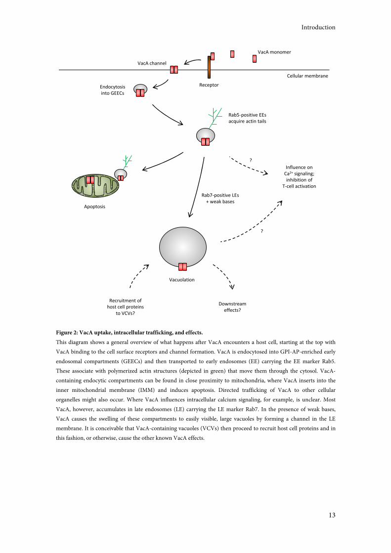
Figure 2: VacA uptake, intracellular trafficking, and effects.
This diagram shows a general overview of what happens after VacA encounters a host cell, starting at the top with
VacA binding to the cell surface receptors and channel formation. VacA is endocytosed into GPI-AP-enriched early
endosomal compartments (GEECs) and then transported to early endosomes (EE) carrying the EE marker Rab5.
These associate with polymerized actin structures (depicted in green) that move them through the cytosol. VacA-
containing endocytic compartments can be found in close proximity to mitochondria, where VacA inserts into the
inner mitochondrial membrane (IMM) and induces apoptosis. Directed trafficking of VacA to other cellular
organelles might also occur. Where VacA influences intracellular calcium signaling, for example, is unclear. Most
VacA, however, accumulates in late endosomes (LE) carrying the LE marker Rab7. In the presence of weak bases,
VacA causes the swelling of these compartments to easily visible, large vacuoles by forming a channel in the LE
membrane. It is conceivable that VacA-containing vacuoles (VCVs) then proceed to recruit host cell proteins and in
this fashion, or otherwise, cause the other known VacA effects.
Vacuolation
Endocytosis
into GEECs
Apoptosis
Rab5-positive EEs
acquire actin tails
?
Recruitment of
host cell proteins
to VCVs?
Downstream
effects?
Rab7-positive LEs
+ weak bases
Influence on
Ca2+ signaling;
inhibition of
T-cell activation
Cellular membrane
VacA monomer
Receptor
VacA channel
?
Introduction
14
1.6 Endosomes as Signaling Platforms
The endosomal network is conventionally viewed as a sorting and trafficking system. It is
responsible for the transport of a wide variety of cargo from the cell surface to sites of
degradation or recycling. Depending on the cargo, this implies an indispensable role of
endosomes in such essential cellular processes as nutrient absorption and hormone-mediated
signal transduction [143]. Moreover, a role of endosomes as signaling platforms is now
recognized, assuming that signaling complexes are assembled on endocytic vesicles as a way of
locally arranging all molecules necessary to trigger a specific signaling cascade [143–145]. The
protein content of an endosome may therefore allow conclusions about the signaling pathway or
network that the endosome is currently acting on. In the context of VacA intoxication, the
presence of VacA on endocytic vesicles alters vesicular protein content [45], thereby possibly
changing cellular signaling processes. The elucidation of the proteome of VacA-containing
vacuoles (VCVs) could therefore provide information on signaling cascades influenced by VacA
intoxication.
1.7 Aim of This Study
Even though a large amount of research done on H. pylori is concerned with VacA, it is still
unclear whether the toxin's most prominent effect, cellular vacuolation, is an effect in and of
itself, or just a by-product. Also, for some VacA effects, the cellular site of action is unknown.
Integrating these two problems, a hypothesis is proposed: that VacA-containing vacuoles
(VCVs) may function as a control center, constituting a platform for intracellular signaling to
aid in VacA's multiple actions (see Figure 2). The idea that VCVs do in fact have a purpose is
particularly supported by four experimental findings: a) VacA greatly alters vesicular trafficking
[142,44,118]; b) VacA causes changes in the protein content of endocytic compartments in
intoxicated cells [45], both indicating more than simple toxin endocytosis; c) VacA-containing
vesicles acquire an actin tail that propels them through the cell cytoplasm [102]; and d) physical
proximity of VacA-containing vesicles and mitochondria may explain how VacA reaches
mitochondria [106], and may be how VacA gets to other, yet unknown, sites of action.
The aim of this work was therefore to isolate VCVs from VacA-intoxicated cells and investigate
their proteome by mass spectrometric analysis. The types of proteins found on VCVs could help
understand not only the purpose of VacA-induced vacuolation, but also elucidate more of the
Introduction
15
cellular processes that VacA influences. These experiments were to be conducted in T-cells,
which are an important target of VacA. Also, under physiological conditions, T-cells are not
usually directly infected by H. pylori, so the antagonistic effects of CagA and other infection-
related consequences do not have to be considered.
Additionally, the intracellular localization of VacA in intoxicated cells was to be investigated
further (see Figure 2). This may lead to a better understanding of known VacA effects and the
identification of possible new VacA target structures.
Introduction
16
Materials and Methods
17
2 Materials and Methods
2.1 Materials
2.1.1 Chemicals
Chemicals were generally purchased from Roth, Merck, or Sigma-Aldrich unless specified
otherwise. Double-distilled water was purchased from Roth and used for PCR reactions,
enzymatic digestions and other sensitive applications. Regular distilled water was used for other
purposes like the preparation of buffers.
2.1.2 Standard Buffers
Buffer Ingredients
SDS sample buffer (2x) 100 mM Tris-HCl pH 6.8, 4% (w/v) SDS, 20% (v/v) glycerol, 10% (v/v) β-
mercaptoethanol, 0.2% (w/v) bromphenol blue
PBS 2.7 mM KCl, 138 mM NaCl, 1.5 mM KH2PO4, 8 mM Na2HPO4
TBS 150 mM NaCl, 20 mM Tris-HCl, pH 7.5
HS 20 mM HEPES, 250 mM sucrose, 0.5 mM EGTA, pH 7.2
Commercially available, cell culture grade PBS (Dulbecco’s PBS with calcium and magnesium,
Gibco Invitrogen, Life Technologies) was used for all cell and vacuole related experiments, while
self-prepared PBS was used for everything else.
An asterisk (*) denotes the addition of protease inhibitors to a buffer at the following
concentrations: 1 mM PMSF, 1 mM sodium vanadate, 1 µM leupeptin, 1 µM pepstatin.
2.1.3 Bacterial Strains and Cell Lines
Internal ID Properties/plasmid carried Source/reference
E. coli strains
DH5α F-Φ80d lacZ ΔM15 Δ(lacZYA-argF) U169 deoR
recA1 endA1 hsdR17 (rK-, mK+) phoA supE44
λ-thi-l gyr A96 relA1
Invitrogen, Life
Technologies
TOP10 F-mcrA Δ(mrr-hsdRMS-mcrBC) Φ80lacZΔM15
ΔlacO74 recA1 araΔ139 Δ(ara-leu)7697 galU
galK rpsL (StrR) endA1 nupG
Invitrogen, Life
Technologies
Materials and Methods
18
Internal ID Properties/plasmid carried Source/reference
BL21(DE3)pLysS F- ompT hsdSB (rB-, mB-) dcm gal λ(DE3) pLysS
Cmr
Stratagene
STIM1-ER BK-E19 BL21DE3 pLysS pET28a(+)-ER-STIM1 (pBK5) This work
STIM1-Cyt BK-E21 BL21DE3 pLysS pET28a(+)-CT-STIM1 (pBK6) This work
GFP BK-E22 BL21DE3 pLysS pET28a(+)-GFP1-10 (pFS1) F. Schindele;[146]
H. pylori strains
60190 BK-H6 Strain producing an s1i1m1 VacA ATCC 49503
60190∆6-27 BK-H11 Strain producing s1i1m1 VacA∆6-27 [70]
P12 BK-H7 Clinical isolate strain [147]
P12∆VacA BK-H8 Clinical isolate strain lacking vacA W. Fischer
Cell lines
HeLa Human epithelial cell line ATCC CCL-2
Jurkat E6-1 Human T-cell line ATCC TIB-152
Jurkat E6-1
EGFP-Rab7
Human T-cell line stably expressing EGFP-
Rab7
This work
2.1.4 Growth Media, Supplements and Antibiotics
Item Supplier
LB Broth base Life Technologies
LB agar Life Technologies
BB medium Oxoid, Thermo Fisher Scientific
GC agar base Oxoid, Thermo Fisher Scientific
RPMI medium Life Technologies
DMEM medium Life Technologies
FCS Life Technologies
Calf serum (bovine serum) Life Technologies
Horse serum Life Technologies
Cholesterol Gibco, Invitrogen
Ampicillin Sigma-Aldrich
G418 PAA, GE Healthcare Life Sciences
Kanamycin Sigma-Aldrich
Nystatin Sigma-Aldrich
Penicillin / Streptomycin Life Technologies
Trimethoprim Sigma-Aldrich
Materials and Methods
19
2.1.5 Commercially Available Kits
Kit name Supplier
Amaxa Cell Line Nucleofector Kit V Lonza
QIAprep Spin Miniprep Kit Qiagen
illustra GFX PCR DNA and Gel Band Purification Kit GE Healthcare
Alexa Fluor 647 Monoclonal Antibody Labeling Kit Invitrogen
2.1.6 Plasmids
Plasmid name Properties Source
pET28a(+) E. coli expression vector with N- and C-terminal 6xHis-tags Novagen
pBK5 pET28a(+)-ER-STIM1 (ER-luminal part of STIM1) with N- and C-
terminal 6xHis-tags
This work
pBK6 pET28a(+)-CT-STIM1 (cytoplasmic part of STIM1) with N-
terminal 6xHis-tag
This work
pEGFP-C1 Rab7A Eukaryotic expression vector carrying an EGFP-Rab7 fusion
protein and a G418 resistance cassette
X. Sewald
2.1.7 Oligonucleotides
Name Sequence Purpose and properties
BK11 TTC TCT ACA CTC TCT TTT TTT TTT
TTT TTT-C6H12-NH2
Coupled to TurboBeads (see 2.2.17)
BK16 GAT CGC GGC CGC CTA CTT CTT AAG
AGG CTT C
RP for the cytoplasmic part of STIM1; NotI
BK29 GAT CGA ATT CTC TGA GGA GTC CAC
TG
FP for the ER-luminal part of STIM1; EcoRI
BK30 GAT CGC GGC CGC GCG AGT CAA
GAG AGG A
RP for the ER-luminal part of STIM1; NotI
BK32 GAT CGA ATT CCG TTA CTC CAA GGA
GCA C
FP for the cytoplasmic part of STIM1; EcoRI
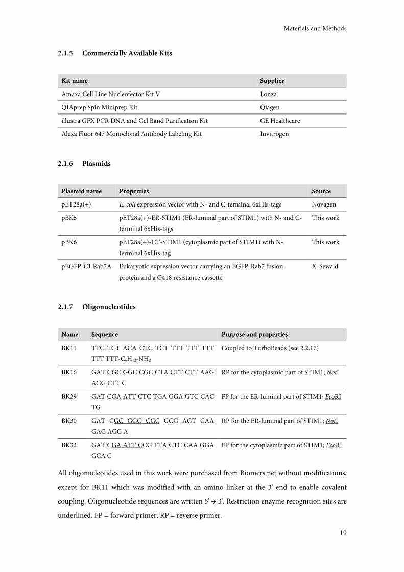
All oligonucleotides used in this work were purchased from Biomers.net without modifications,
except for BK11 which was modified with an amino linker at the 3' end to enable covalent
coupling. Oligonucleotide sequences are written 5' → 3'. Restriction enzyme recognition sites are
underlined. FP = forward primer, RP = reverse primer.
Materials and Methods
20
2.1.8 Enzymes and Proteins
Enzyme/protein Source
Restriction enzymes Roche Applied Science or Thermo Fisher Scientific
Trypsin-EDTA Gibco, Invitrogen
T4 DNA ligase Thermo Fisher Scientific
Ex Taq polymerase Takara, Clontech
LA Taq polymerase Takara, Clontech
VacA from H. pylori strain P76 Purified by I. Barwig
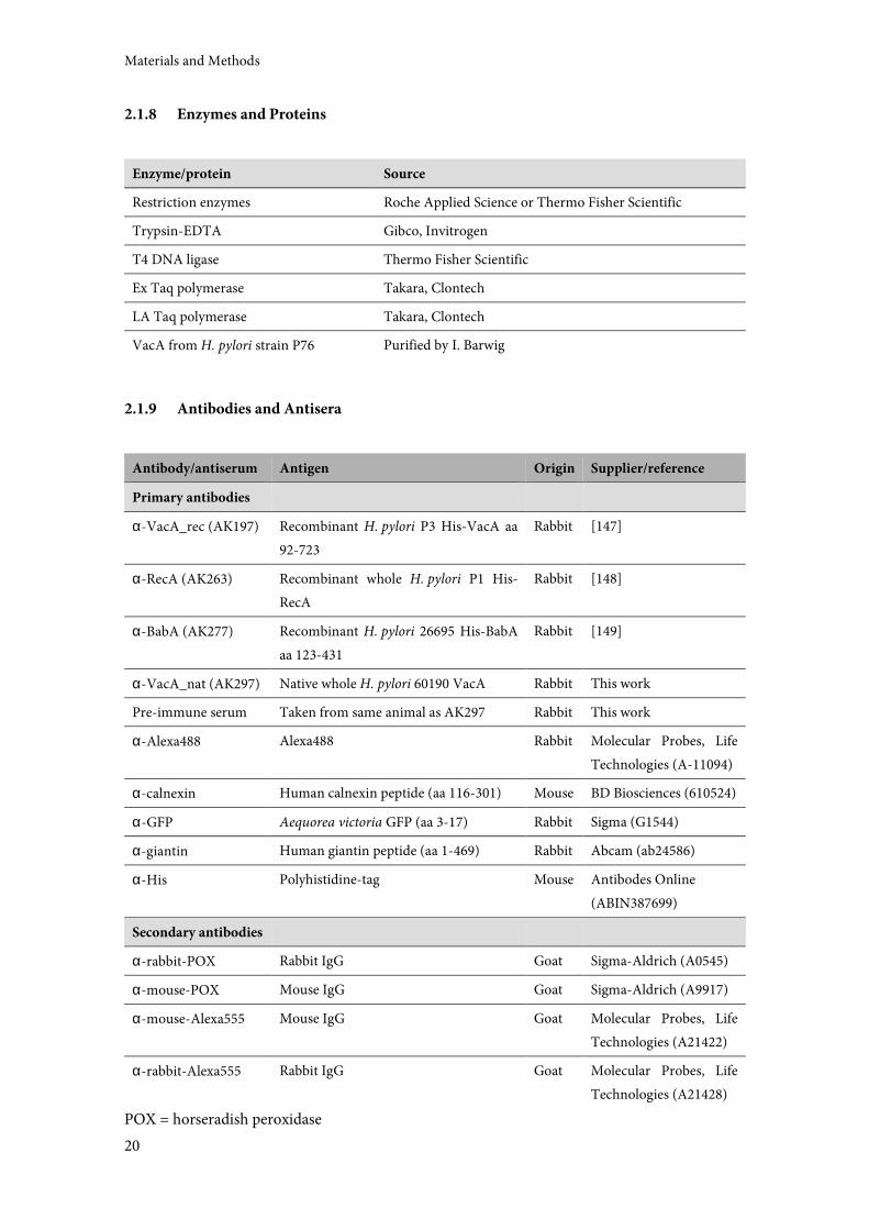
2.1.9 Antibodies and Antisera
Antibody/antiserum Antigen Origin Supplier/reference
Primary antibodies
α-VacA_rec (AK197) Recombinant H. pylori P3 His-VacA aa
92-723
Rabbit [147]
α-RecA (AK263) Recombinant whole H. pylori P1 His-
RecA
Rabbit [148]
α-BabA (AK277) Recombinant H. pylori 26695 His-BabA
aa 123-431
Rabbit [149]
α-VacA_nat (AK297) Native whole H. pylori 60190 VacA Rabbit This work
Pre-immune serum Taken from same animal as AK297 Rabbit This work
α-Alexa488 Alexa488 Rabbit Molecular Probes, Life
Technologies (A-11094)
α-calnexin Human calnexin peptide (aa 116-301) Mouse BD Biosciences (610524)
α-GFP Aequorea victoria GFP (aa 3-17) Rabbit Sigma (G1544)
α-giantin Human giantin peptide (aa 1-469) Rabbit Abcam (ab24586)
α-His Polyhistidine-tag Mouse Antibodes Online
(ABIN387699)
Secondary antibodies
α-rabbit-POX Rabbit IgG Goat Sigma-Aldrich (A0545)
α-mouse-POX Mouse IgG Goat Sigma-Aldrich (A9917)
α-mouse-Alexa555 Mouse IgG Goat Molecular Probes, Life
Technologies (A21422)
α-rabbit-Alexa555 Rabbit IgG Goat Molecular Probes, Life
Technologies (A21428)
POX = horseradish peroxidase

Materials and Methods
21
2.2 Methods
2.2.1 Escherichia coli Methods
2.2.1.1 Cultivation and Strain Maintenance
E. coli were grown either on LB agar plates (LB Agar, Life Technologies) at 37 °C for cloning and
other routine experiments or in LB liquid medium (LB Broth Base, Life Technologies) at 200
rpm and 27 °C for protein expression; see table below for concentrations of relevant antibiotics.
Culture stocks were generated by collecting the bacteria from agar plates with sterile cotton
swabs, resuspending in LB liquid media supplemented with 20% glycerol, and freezing at -70 °C
in cryogenic tubes (Nalgene, Thermo Fisher Scientific).
Antibiotic Final concentration
Ampicillin 100 mg/l
Kanamycin 50 mg/l
2.2.1.2 Preparation of Chemically Competent E. coli
E. coli DH5α and BL21(DE3)pLysS were rendered chemically competent using the method of
Hanahan 1983. Aliquots of 50 µl were stored at -70 °C until further use. For higher
transformation efficiencies, commercially obtained One Shot TOP10 competent cells
(Invitrogen, Life Technologies) were used in some experiments.
2.2.1.3 Transformation of Chemically Competent E. coli
Aliquots of all strains of chemically competent E. coli were thawed on ice, mixed with DNA
(100-500 ng of a ligation reaction or 10-100 ng plasmid DNA) and incubated on ice for another
30 min, followed by a heat shock of 30-90 s at 42 °C. 1 ml warm LB medium was added to enable
bacterial recovery (37 °C, 200 rpm, 1 h). Bacteria were then plated on selective LB agar plates
containing the appropriate antibiotic for selection of transformants.
Materials and Methods
22
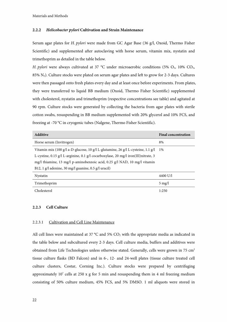
2.2.2 Helicobacter pylori Cultivation and Strain Maintenance
Serum agar plates for H. pylori were made from GC Agar Base (36 g/l, Oxoid, Thermo Fisher
Scientific) and supplemented after autoclaving with horse serum, vitamin mix, nystatin and
trimethoprim as detailed in the table below.
H. pylori were always cultivated at 37 °C under microaerobic conditions (5% O2, 10% CO2,
85% N2). Culture stocks were plated on serum agar plates and left to grow for 2-3 days. Cultures
were then passaged onto fresh plates every day and at least once before experiments. From plates,
they were transferred to liquid BB medium (Oxoid, Thermo Fisher Scientific) supplemented
with cholesterol, nystatin and trimethoprim (respective concentrations see table) and agitated at
90 rpm. Culture stocks were generated by collecting the bacteria from agar plates with sterile
cotton swabs, resuspending in BB medium supplemented with 20% glycerol and 10% FCS, and
freezing at -70 °C in cryogenic tubes (Nalgene, Thermo Fisher Scientific).
Additive Final concentration
Horse serum (Invitrogen) 8%
Vitamin mix (100 g/l a-D-glucose, 10 g/l L-glutamine, 26 g/l L-cysteine, 1.1 g/l
L-cystine, 0.15 g/l L-arginine, 0.1 g/l cocarboxylase, 20 mg/l iron(III)nitrate, 3
mg/l thiamine, 13 mg/l p-aminobenzoic acid, 0.25 g/l NAD, 10 mg/l vitamin
B12, 1 g/l adenine, 30 mg/l guanine, 0.5 g/l uracil)
1%
Nystatin 4400 U/l
Trimethoprim 5 mg/l
Cholesterol 1:250
2.2.3 Cell Culture
2.2.3.1 Cultivation and Cell Line Maintenance
All cell lines were maintained at 37 °C and 5% CO2 with the appropriate media as indicated in
the table below and subcultured every 2-3 days. Cell culture media, buffers and additives were
obtained from Life Technologies unless otherwise stated. Generally, cells were grown in 75 cm2
tissue culture flasks (BD Falcon) and in 6-, 12- and 24-well plates (tissue culture treated cell
culture clusters, Costar, Corning Inc.). Culture stocks were prepared by centrifuging
approximately 107 cells at 250 x g for 5 min and resuspending them in 4 ml freezing medium
consisting of 50% culture medium, 45% FCS, and 5% DMSO. 1 ml aliquots were stored in
Materials and Methods
23
cryogenic tubes (Nalgene, Thermo Fisher Scientific) at -70 °C for at least 24 h and then
transferred to liquid nitrogen tanks for long term storage.
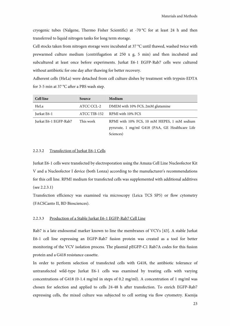
Cell stocks taken from nitrogen storage were incubated at 37 °C until thawed, washed twice with
prewarmed culture medium (centrifugation at 250 x g, 5 min) and then incubated and
subcultured at least once before experiments. Jurkat E6-1 EGFP-Rab7 cells were cultured
without antibiotic for one day after thawing for better recovery.
Adherent cells (HeLa) were detached from cell culture dishes by treatment with trypsin-EDTA
for 3-5 min at 37 °C after a PBS wash step.
Cell line Source Medium
HeLa ATCC CCL-2 DMEM with 10% FCS, 2mM glutamine
Jurkat E6-1 ATCC TIB-152 RPMI with 10% FCS
Jurkat E6-1 EGFP-Rab7 This work RPMI with 10% FCS, 10 mM HEPES, 1 mM sodium
pyruvate, 1 mg/ml G418 (PAA, GE Healthcare Life
Sciences)
2.2.3.2 Transfection of Jurkat E6-1 Cells
Jurkat E6-1 cells were transfected by electroporation using the Amaxa Cell Line Nucleofector Kit
V and a Nucleofector I device (both Lonza) according to the manufacturer's recommendations
for this cell line. RPMI medium for transfected cells was supplemented with additional additives
(see 2.2.3.1)
Transfection efficiency was examined via microscopy (Leica TCS SP5) or flow cytometry
(FACSCanto II, BD Biosciences).
2.2.3.3 Production of a Stable Jurkat E6-1 EGFP-Rab7 Cell Line
Rab7 is a late endosomal marker known to line the membranes of VCVs [43]. A stable Jurkat
E6-1 cell line expressing an EGFP-Rab7 fusion protein was created as a tool for better
monitoring of the VCV isolation process. The plasmid pEGFP-C1 Rab7A codes for this fusion
protein and a G418 resistance cassette.
In order to perform selection of transfected cells with G418, the antibiotic tolerance of
untransfected wild-type Jurkat E6-1 cells was examined by treating cells with varying
concentrations of G418 (0-1.4 mg/ml in steps of 0.2 mg/ml). A concentration of 1 mg/ml was
chosen for selection and applied to cells 24-48 h after transfection. To enrich EGFP-Rab7
expressing cells, the mixed culture was subjected to cell sorting via flow cytometry. Ksenija
Materials and Methods
24
Jovanovic at the Institute for Immunology (Ludwig-Maximilians-Universität München) kindly
did this with a FACSAria I. Since cell sorting took place in a non-sterile environment, a mixture
of penicillin and streptomycin was added to the cells after sorting to avoid bacterial
contamination (final concentrations 100 U/ml and 100µg/ml, respectively).
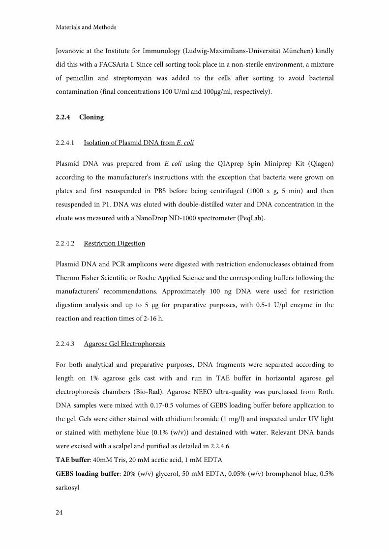
2.2.4 Cloning
2.2.4.1 Isolation of Plasmid DNA from E. coli
Plasmid DNA was prepared from E. coli using the QIAprep Spin Miniprep Kit (Qiagen)
according to the manufacturer's instructions with the exception that bacteria were grown on
plates and first resuspended in PBS before being centrifuged (1000 x g, 5 min) and then
resuspended in P1. DNA was eluted with double-distilled water and DNA concentration in the
eluate was measured with a NanoDrop ND-1000 spectrometer (PeqLab).
2.2.4.2 Restriction Digestion
Plasmid DNA and PCR amplicons were digested with restriction endonucleases obtained from
Thermo Fisher Scientific or Roche Applied Science and the corresponding buffers following the
manufacturers' recommendations. Approximately 100 ng DNA were used for restriction
digestion analysis and up to 5 µg for preparative purposes, with 0.5-1 U/µl enzyme in the
reaction and reaction times of 2-16 h.
2.2.4.3 Agarose Gel Electrophoresis
For both analytical and preparative purposes, DNA fragments were separated according to
length on 1% agarose gels cast with and run in TAE buffer in horizontal agarose gel
electrophoresis chambers (Bio-Rad). Agarose NEEO ultra-quality was purchased from Roth.
DNA samples were mixed with 0.17-0.5 volumes of GEBS loading buffer before application to
the gel. Gels were either stained with ethidium bromide (1 mg/l) and inspected under UV light
or stained with methylene blue (0.1% (w/v)) and destained with water. Relevant DNA bands
were excised with a scalpel and purified as detailed in 2.2.4.6.
TAE buffer: 40mM Tris, 20 mM acetic acid, 1 mM EDTA
GEBS loading buffer: 20% (w/v) glycerol, 50 mM EDTA, 0.05% (w/v) bromphenol blue, 0.5%
sarkosyl
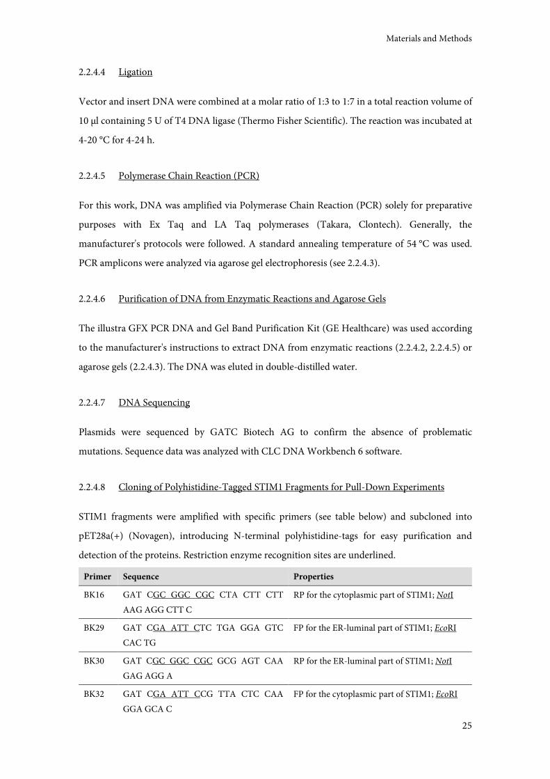
Materials and Methods
25
2.2.4.4 Ligation
Vector and insert DNA were combined at a molar ratio of 1:3 to 1:7 in a total reaction volume of
10 µl containing 5 U of T4 DNA ligase (Thermo Fisher Scientific). The reaction was incubated at
4-20 °C for 4-24 h.
2.2.4.5 Polymerase Chain Reaction (PCR)
For this work, DNA was amplified via Polymerase Chain Reaction (PCR) solely for preparative
purposes with Ex Taq and LA Taq polymerases (Takara, Clontech). Generally, the
manufacturer's protocols were followed. A standard annealing temperature of 54 °C was used.
PCR amplicons were analyzed via agarose gel electrophoresis (see 2.2.4.3).
2.2.4.6 Purification of DNA from Enzymatic Reactions and Agarose Gels
The illustra GFX PCR DNA and Gel Band Purification Kit (GE Healthcare) was used according
to the manufacturer's instructions to extract DNA from enzymatic reactions (2.2.4.2, 2.2.4.5) or
agarose gels (2.2.4.3). The DNA was eluted in double-distilled water.
2.2.4.7 DNA Sequencing
Plasmids were sequenced by GATC Biotech AG to confirm the absence of problematic
mutations. Sequence data was analyzed with CLC DNA Workbench 6 software.
2.2.4.8 Cloning of Polyhistidine-Tagged STIM1 Fragments for Pull-Down Experiments
STIM1 fragments were amplified with specific primers (see table below) and subcloned into
pET28a(+) (Novagen), introducing N-terminal polyhistidine-tags for easy purification and
detection of the proteins. Restriction enzyme recognition sites are underlined.
Primer Sequence Properties
BK16 GAT CGC GGC CGC CTA CTT CTT
AAG AGG CTT C
RP for the cytoplasmic part of STIM1; NotI
BK29 GAT CGA ATT CTC TGA GGA GTC
CAC TG
FP for the ER-luminal part of STIM1; EcoRI
BK30 GAT CGC GGC CGC GCG AGT CAA
GAG AGG A
RP for the ER-luminal part of STIM1; NotI
BK32 GAT CGA ATT CCG TTA CTC CAA
GGA GCA C
FP for the cytoplasmic part of STIM1; EcoRI
Materials and Methods
26
2.2.5 Protein Biochemical Methods
2.2.5.1 SDS Polyacrylamide Gel Electrophoresis (SDS-PAGE)
Proteins were separated according to size via SDS polyacrylamide gel electrophoresis (SDS-
PAGE). The size of the proteins of interest determined the polyacrylamide concentration of the
separating gel (6-10%). Gel composition was taken from [150]. Gels were cast and run with a
Mini-Protean III vertical gel electrophoresis system (Bio-Rad).
Protein samples were mixed with SDS sample buffer and boiled (10 min, 98 °C) before
application to the gel. After electrophoresis, the proteins in the gels were either stained directly
(see 2.2.5.3) or transferred to membranes and analyzed by immunoblotting (see 2.2.5.4 and
2.2.5.6, respectively).
SDS-PAGE running buffer: 5 mM Tris-HCl pH8.3, 50 mM glycerol, 0.02% SDS
SDS sample buffer (2x): 100 mM Tris-HCl pH 6.8, 4% (w/v) SDS, 20% (v/v) glycerol, 10% (v/v)
β-mercaptoethanol, 0.2% (w/v) bromphenol blue
2.2.5.2 Preparation of Standardized Bacterial and Cell Lysates for SDS-PAGE
In order to be able to compare different bacterial and cell lysates on an SDS polyacrylamide gel
or a Western Blot, the samples were prepared in a standardized manner. For bacteria, 1 ml of a
bacterial suspension with an OD550 of 0.2 (corresponding to approximately 6 x 10⁷ bacterial
cells) was centrifuged at 1000 x g for 5 min. For mammalian cells, 106 cells were centrifuged at
425 x g for 5 min at 4 °C. The cell pellets were resuspended in 20 µl RIPA buffer supplemented
with protease inhibitors and mixed with 25 µl SDS sample buffer before boiling (98 °C, 10 min).
Usually, 5 µl of bacterial and 3 µl of mammalian cell lysates were used for SDS-PAGE.
RIPA buffer: 50 mM Tris-HCl pH 7.4, 125 mM NaCl, 1 mM EDTA, 1% NP-40, 6 mM sodium
deoxycholate
2.2.5.3 Coomassie Staining of SDS Polyacrylamide Gels
In order to visualize proteins on SDS polyacrylamide gels directly, the gels were stained in a
Coomassie solution (0.275% (w/v) Coomassie Brilliant Blue R250 (Biomol), 50% methanol, 10%
acetic acid) and subsequently treated with destaining solution (10% methanol, 10% ethanol,
7.5% acetic acid) until all relevant bands were visible. Stained gels were photographed with a
Bio-Rad ChemiDoc XRS.
Materials and Methods
27
2.2.5.4 Transfer of Proteins from SDS Polyacrylamide Gels onto Membrane
For the purpose of immunostaining, proteins were transferred from SDS polyacrylamide gels
onto membrane (Immun-Blot PVDF Membrane, Bio-Rad) using semi-dry blotting chambers
(Biotec-Fischer). Gel and membrane were sandwiched between pieces of filter paper that had
been soaked in anode I/II and cathode buffers on the anode/cathode side of gel and membrane,
respectively. A current of 1.2 mA/cm2 was applied for 60-70 min. After transfer, the membrane
was either dried (1 h at 37 °C or over night at RT) or directly used for immunoblotting. Dried
membrane was briefly reactivated with methanol before further use.
Anode I buffer: 300 mM Tris-HCl pH 10.4, 10% methanol
Anode II buffer: 25 mM Tris-HCl pH 10.4, 10% methanol
Cathode buffer: 25 mM Tris-HCl pH 9.6, 40 mM 6-amino caproic acid, 10% methanol
2.2.5.5 Spotting of Protein Samples onto Membrane for Dot Blots
Purified protein samples were also spotted directly onto membrane without prior SDS-PAGE in
order to keep the proteins' native conformation. To this end, PVDF membrane (Immun-Blot
PVDF Membrane, Bio-Rad) was briefly activated in methanol and kept moistened with TBS. A
self-made dot blot apparatus was used to immobilize the membrane and to administer samples
(1-10 ng of protein mixed with 50-100 µl of TBS for better dispersal of sample within the well).
Vacuum was applied to remove excess liquid and the membrane was subsequently used for
immunoblotting.
2.2.5.6 Immunoblotting (Western Blot)
Membranes were incubated in blocking solution (5% skim milk in TBS) for at least 1 h to
saturate unspecific binding sites. Primary and secondary antibodies were applied as detailed in
the table below. Primary antibodies were diluted in blocking solution and incubated for at least
1.5 h at RT (or for longer periods at 4 °C), while secondary antibodies were diluted in 1% skim
milk in TBS and incubated for 45-60 min at RT. After antibody incubation steps, membranes
were washed with TBS-T (TBS containing 0.075% Tween 20), usually three times for 7 min each.
Addition of substrate (Immobilon Chemiluminescent HRP Substrate, Millipore) initiates a
chemiluminescence reaction which is catalyzed by horseradish peroxidase (POX) bound to the
secondary antibody. This reaction was detected with X-ray films (Fuji Medical X-Ray Films
Super RX, Fujifilm) or via a Bio-Rad ChemiDoc XRS.
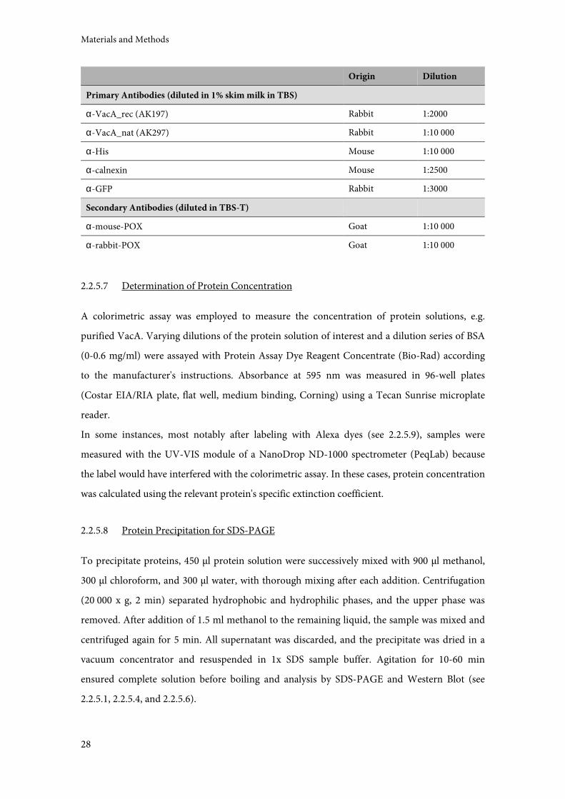
Materials and Methods
28
Origin Dilution
Primary Antibodies (diluted in 1% skim milk in TBS)
α-VacA_rec (AK197) Rabbit 1:2000
α-VacA_nat (AK297) Rabbit 1:10 000
α-His Mouse 1:10 000
α-calnexin Mouse 1:2500
α-GFP Rabbit 1:3000
Secondary Antibodies (diluted in TBS-T)
α-mouse-POX Goat 1:10 000
α-rabbit-POX Goat 1:10 000
2.2.5.7 Determination of Protein Concentration
A colorimetric assay was employed to measure the concentration of protein solutions, e.g.
purified VacA. Varying dilutions of the protein solution of interest and a dilution series of BSA
(0-0.6 mg/ml) were assayed with Protein Assay Dye Reagent Concentrate (Bio-Rad) according
to the manufacturer's instructions. Absorbance at 595 nm was measured in 96-well plates
(Costar EIA/RIA plate, flat well, medium binding, Corning) using a Tecan Sunrise microplate
reader.
In some instances, most notably after labeling with Alexa dyes (see 2.2.5.9), samples were
measured with the UV-VIS module of a NanoDrop ND-1000 spectrometer (PeqLab) because
the label would have interfered with the colorimetric assay. In these cases, protein concentration
was calculated using the relevant protein's specific extinction coefficient.
2.2.5.8 Protein Precipitation for SDS-PAGE
To precipitate proteins, 450 µl protein solution were successively mixed with 900 µl methanol,
300 µl chloroform, and 300 µl water, with thorough mixing after each addition. Centrifugation
(20 000 x g, 2 min) separated hydrophobic and hydrophilic phases, and the upper phase was
removed. After addition of 1.5 ml methanol to the remaining liquid, the sample was mixed and
centrifuged again for 5 min. All supernatant was discarded, and the precipitate was dried in a
vacuum concentrator and resuspended in 1x SDS sample buffer. Agitation for 10-60 min
ensured complete solution before boiling and analysis by SDS-PAGE and Western Blot (see
2.2.5.1, 2.2.5.4, and 2.2.5.6).
Materials and Methods
29
2.2.5.9 Labeling of VacA with Alexa Dyes
Purified VacA was labeled with different Alexa fluorophores obtained from Invitrogen, using
either the Alexa Fluor 647 Monoclonal Antibody Labeling Kit or individually bought Alexa
Fluor carboxylic acid succinimidyl esters. The kit was used according to the manufacturer's
instructions while the separate dyes were used following a modified protocol. The desiccated dye
was resuspended in 100 µl MeOH for aliquoting into four portions and re-dried in a vacuum
concentrator. 112 µg VacA were mixed with 50 µl 1 M NaHCO3 before adding PBS(-) to a total
volume of 500 µl. One portion of dye (250 µg) per labeling reaction was solved in 20 µl DMSO
and added to the mix, which was then incubated at RT in the dark for 45 min under gentle
agitation. To remove unbound dye, the sample was extensively dialyzed against PBS(-) at 4 °C
(over night against 4 l and another 4 h against 1 l) in Slide-A-Lyzer dialysis cassettes (100K
MWCO, Thermo Fisher Scientific). Protein concentration and degree of labeling were assessed
using a NanoDrop ND-1000 spectrometer according to Invitrogen's instructions, and toxin
activity was assayed via neutral red uptake (see 2.2.8)
PBS(-): 7.7 mM Na2HPO4, 2.5 mM NaH2PO4, 150 mM NaCl
2.2.6 Purification of VacA
To produce large amounts of VacA, H. pylori was grown in sequential liquid cultures of 30-250
ml for total final culture volumes of about 1 l. These cultures typically had a final OD550 of
around 0.35 and were harvested by centrifugation (6000 x g, 20 min, 4 °C). All further steps were
performed at 4 °C or on ice. The cell pellet was discarded and the supernatant, containing the
secreted toxin, was sterile-filtrated (Nalgene Rapid-Flow Sterile Disposable Bottle Top Filters
with PES Membrane, 0.45 µm pore size, Thermo Fisher Scientific). VacA was precipitated by the
slow addition of one volume of cold, saturated ammonium sulfate solution under continuous
slow stirring. After 4-16 h of further stirring, the precipitate was sedimented by centrifugation
(20 000 x g, 30 min, 4 °C) and thoroughly resuspended in a total volume of 30 ml cold PBS.
Sample volume was reduced to 4 ml with a centrifugal filter unit (Amicon Ultra, Ultracel, 100K
MWCO, Millipore) (4000 x g, 4 °C) to simplify gel filtration, which was carried out with a
Sephacryl S300 16/60 column on an ÄKTAexplorer system (Amersham Biosciences, GE
Healthcare Life Sciences), using a running buffer containing 100 mM NaCl and 50 mM NaPO4
at pH 7.4. The resulting fractions were examined for purity via SDS-PAGE and Coomassie
staining, pooled accordingly, and again concentrated by centrifugation to a final volume of
about 1-1.5 ml. The final protein concentration was determined as described in 0 and usually
Materials and Methods
30
ranged between 0.4 and 1 mg/ml in a volume of 0.5-1.5 ml. For every preparation of VacA, the
toxin's activity was tested in a vacuolation assay (see 2.2.8).
After purification, VacA is oligomerized and thus biologically inactive. Before all intoxication
experiments, purified VacA was therefore acid-activated. This was done by adding 0.2 volumes
of 0.3 M HCl, incubating at 37 °C for 20-30 min, and neutralizing with 0.2 volumes of 0.3 M
NaOH.
2.2.7 Preparation of Concentrated Culture Supernatant (CCS)
Concentrated H. pylori culture supernatant (CCS) was prepared as a faster and cheaper
alternative to purified VacA for experiments that did not necessitate absolute purity of the toxin.
To this end, liquid cultures were grown and subjected to ammonium sulfate precipitation as
described in 2.2.6. The resuspended precipitate was then directly dialyzed against PBS (dialysis
tubing purchased from Medicell) (over night against 5 l and another 4 h against 1 l).
Vacuolating activity of CCS was also verified in a vacuolation assay (see 2.2.8) every time, and
the volume of CCS to be used in experiments was adjusted so that its vacuolating activity
equaled that of 1 µg/ml purified VacA. Contrary to purified VacA, CCS does not have to be
acid-activated.
2.2.8 Vacuolation Assay
Cytotoxic activity of newly prepared VacA or CCS, or activity of labeled VacA, was confirmed in
a vacuolation assay. The fact that VacA-induced vacuoles can be stained with neutral red
enables the quantification of toxin activity via neutral red uptake. This assay was performed on
HeLa cells with a confluency of 70-90%, usually seeded one or two days before the experiment in
12-well tissue culture plates. VacA was acid-activated and added to cells to a final concentration
of 1 µg/ml. In the case of CCS, different volumes were used (5-20 µl/ml) and compared to the
last preparation of VacA. As a non-intoxicated control, one well was treated with 'acid-activated'
water instead of VacA. After 4 h of incubation, NH4Cl was added to a final concentration of 2
mM to enlarge vacuoles (30-60 min). The cell medium was then replaced with RPMI
supplemented with 10% FCS and 1:50 neutral red solution (0.4% in PBS, Merck). After another
10 min, the cells were washed twice with PBS containing 0.5% BSA. The neutral red taken up
into vacuoles was then extracted with 70% ethanol/0.37% HCl and its absorbance was measured
in a 96-well plate (Costar EIA/RIA plate, flat well, medium binding, Corning) at 534 nm
(reference wavelength 405 nm) using a Tecan Sunrise microplate reader.
Materials and Methods
31
A simpler variation of this approach was also used to ensure successful intoxication in other
experiments, e.g. before vacuole isolation (see 2.2.19). Here, neutral red was added directly to a
small aliquot of intoxicated cells and vacuolation was confirmed via microscopy.
2.2.9 Vacuolation Time Course
In order to ensure ideal VCVs for vacuole isolation, vacuolation in Jurkat E6-1 cells was
observed over time. Neutral red uptake was used as an indicator for both number and size of
vacuoles, and a variation of the neutral red uptake vacuolation assay (2.2.8) was performed with
Jurkat E6-1 cells for different periods of intoxication. 2 x 10⁵ cells were seeded in 1 ml medium
in 12-well plates. For each time point (4, 8, 24h), one well was intoxicated with 20 µl CCS (see
2.2.7), while another well was not. NH4Cl was added to a final concentration of 2 mM to all wells
at the time of intoxication. After 4, 8, and 24 h, neutral red solution was added directly to the
cells (1:50), and another 10 min later, the cells were washed twice with PBS/0.5% BSA by
centrifuging at 200 x g for 5 min. Neutral red was extracted from vacuoles with 70%
ethanol/0.37% HCl and its absorbance was measured in a 96-well plate at 534 nm (reference
wavelength 405 nm). The arithmetic mean of the absorbance of all non-intoxicated wells was
used as a blank value to normalize the measurements of the intoxicated cells. A one way
ANOVA was performed on the data, followed by a Kruskal-Wallis test and a comparison of all
pairs of columns. The data was analyzed with GraphPad Prism 5.
2.2.10 Production of αααα-VacA_nat
An antiserum was raised against purified, native VacA in a rabbit by Gramsch Laboratories
according to their standardized protocol. Purified VacA (see 2.2.6) was injected on day one and
21 (75 µg, in dorsum, neck and shoulder) and again on day 42, 49, and 56 (50 µg,
intramuscularly in the hind thigh). Serum was taken on day 63 (boost #1) and on day 70 (boost
#2), stabilized with azide, and received from Gramsch Laboratories. The second boost was
named AK297 (α-VacA_nat) and characterized for this work. Also, pre-immune serum of the
same animal was used for comparison (see 2.2.11).
2.2.11 Vacuolation Inhibition Assay
It was investigated whether α-VacA_nat had the capacity to inhibit VacA-induced vacuolation
through binding to VacA. To this end, a vacuolation assay was performed as in 2.2.8 but with
Materials and Methods
32
two added elements: one, heat-inactivation of complement proteins in antisera (56 °C, 30 min)
parallel to VacA acid-activation, and two, incubation of (acid-activated) VacA with (heat-
inactivated) sera to allow binding (30 min on ice). Prior to this experiment, the total protein
content of all antisera was measured (2.2.5.7). For each antiserum, a volume equivalent to
600 µg protein was used to (potentially) inhibit 1 µg VacA.
Since neutral red uptake of unintoxicated cells varies considerably from one experiment to the
next, absolute measured values cannot be used. Instead, an unintoxicated cell sample treated
with NH4Cl served as a negative background control. Another sample intoxicated with VacA
and treated with NH4Cl but without any antiserum served as a positive control that was set to
100%, while all other samples were expressed in relation. This was done for all three
independent experiments separately. The data was analyzed with Graph Pad Prism 5.
2.2.12 Pull-Down Experiments
Pull-down experiments with the aim of investigating a possible interaction of VacA with STIM1
were performed in two directions for verification. For both directions, VacA was incubated with
lysates of E. coli expressing STIM1 protein fragments. In the first approach, Ni-NTA agarose
(Qiagen) was used to precipitate the His-tagged STIM1 fragments, while in the second,
α-VacA_nat and Protein G agarose (Roche Applied Science) were used to precipitate VacA.
To express the polyhistidine-tagged STIM1 fragments or controls, the relevant E. coli strains
(BK-E19, 21, 22, 23) were grown at 27 °C and 200 rpm in LB medium supplemented with
kanamycin until they reached an OD550 of 0.5-0.6, at which point expression was induced by the
addition of IPTG to a final concentration of 1 mM. Three hours later, the cultures were
centrifuged (4000 x g, 20 min, 4 °C) and the bacterial pellet was resuspended in 1/50 volume
lysis buffer (specified below). Samples were handled on ice from here onwards. Cells were lysed
via sonication (Sonifier 250, Branson; 10-50% duty cycle, output control 5, three 20 s intervals
followed by 20 s rest) and the lysate was centrifuged (15 000 x g, 30 min, 4 °C) to remove cell
debris.
For the precipitation of 6xHis-tagged STIM1 fragments, PBS* was used as lysis buffer. The
debris-free lysate was mixed with acid-activated VacA at 10 µg/ml and one volume of Ni-NTA
beads that had been washed with PBS twice (centrifugation at 500 x g, 5 min, 4 °C). Samples
were incubated over night at 4 °C under gentle agitation. After three washes with PBS*, the
beads were resuspended in 1/2 volume of PBS*, of which 10 µl (1/50 of the total sample) were
applied to an SDS polyacrylamide gel for subsequent analysis via Western Blot.
Materials and Methods
33
For the precipitation of VacA, PBS*/0.01% NP-40 was used as lysis buffer. After the removal of
cell debris, the supernatant was incubated with acid-activated VacA at a final concentration of 4
µg/ml for one hour at 4 °C under gentle agitation to enable the STIM1 protein fragments to bind
to VacA. α-VacA_nat was then added (1:100) to bind VacA (over night, 4 °C), and 1/10 volume
Protein G agarose was incubated with the samples for another 2 h to bind the primary antibody.
The agarose beads were washed three times with PBS*/0.01% NP-40 (centrifugation at full speed,
30-60 s) and resuspended directly in 2/25 volumes 2x SDS sample buffer. 10 µl of this (1/5 of the
total sample) were applied to an SDS polyacrylamide gel for subsequent analysis via Western
Blot.
2.2.13 Immunostaining Experiments
To investigate the localization of VacA in intoxicated HeLa cells, cells were treated with Alexa-
labeled VacA and subsequently stained for Golgi apparatus marker giantin and ER marker
calnexin. Similarly, co-intoxication with VacA and cholera toxin subunit B (CTxB), both
fluorescently labeled, was performed.
Also, to further characterize the new antibody α-VacA_nat, it was used to stain Alexa-labeled
VacA in intoxicated HeLa cells. Using a secondary antibody labeled with a different fluorophore
than the one coupled to VacA facilitated a direct comparison of the signals of the immunostain
with that of labeled VacA.
50 000 cells were seeded onto untreated glass cover slips (Menzel-Gläser, 12 mm, Omnilab) in
24-well plates on the day before the experiment. Before intoxication, the cell medium was
changed to ensure equal volumes of 500 µl in each well. 3 µl VacA-Alexa488 or 2 µl VacA-
Alexa568 (all acid-activated) were applied per well; this had previously been titrated as the ideal
volumes for these batches of labeled VacA. CTxB-Alexa555 (Molecular Probes, Life
Technologies) was applied at a concentration of 1.6 µg/ml. Samples were kept in the dark as
much as possible to avoid fluorphore bleaching and washed three times with PBS between
treatments and antibody incubation steps. Intoxication was done for 4 h at 37 °C and 5% CO2, as
usual, but all subsequent incubation steps were carried out at room temperature and normal
atmosphere. Cells were fixed with PFA (4% in PBS, 20 min), followed by permeabilization with
Triton X-100 (0.1% in PBS, 10 min) and a blocking step with blocking buffer (8% calf serum in
PBS, 30 min). Cover slips were then sequentially incubated with 70 µl primary and secondary
antibody solution on parafilm for 50-60 min. After a final wash, the cover slips were mounted
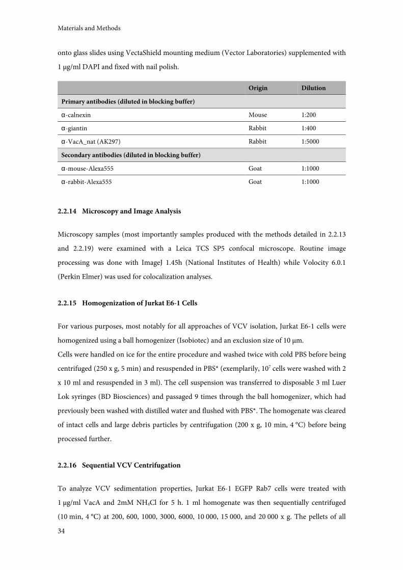
Materials and Methods
34
onto glass slides using VectaShield mounting medium (Vector Laboratories) supplemented with
1 µg/ml DAPI and fixed with nail polish.
Origin Dilution
Primary antibodies (diluted in blocking buffer)
α-calnexin Mouse 1:200
α-giantin Rabbit 1:400
α-VacA_nat (AK297) Rabbit 1:5000
Secondary antibodies (diluted in blocking buffer)
α-mouse-Alexa555 Goat 1:1000
α-rabbit-Alexa555 Goat 1:1000
2.2.14 Microscopy and Image Analysis
Microscopy samples (most importantly samples produced with the methods detailed in 2.2.13
and 2.2.19) were examined with a Leica TCS SP5 confocal microscope. Routine image
processing was done with ImageJ 1.45h (National Institutes of Health) while Volocity 6.0.1
(Perkin Elmer) was used for colocalization analyses.
2.2.15 Homogenization of Jurkat E6-1 Cells
For various purposes, most notably for all approaches of VCV isolation, Jurkat E6-1 cells were
homogenized using a ball homogenizer (Isobiotec) and an exclusion size of 10 µm.
Cells were handled on ice for the entire procedure and washed twice with cold PBS before being
centrifuged (250 x g, 5 min) and resuspended in PBS* (exemplarily, 107 cells were washed with 2
x 10 ml and resuspended in 3 ml). The cell suspension was transferred to disposable 3 ml Luer
Lok syringes (BD Biosciences) and passaged 9 times through the ball homogenizer, which had
previously been washed with distilled water and flushed with PBS*. The homogenate was cleared
of intact cells and large debris particles by centrifugation (200 x g, 10 min, 4 °C) before being
processed further.
2.2.16 Sequential VCV Centrifugation
To analyze VCV sedimentation properties, Jurkat E6-1 EGFP Rab7 cells were treated with
1 µg/ml VacA and 2mM NH4Cl for 5 h. 1 ml homogenate was then sequentially centrifuged
(10 min, 4 °C) at 200, 600, 1000, 3000, 6000, 10 000, 15 000, and 20 000 x g. The pellets of all
Materials and Methods
35
steps were resuspended and boiled in 50 µl SDS sample buffer, 10 µl of which were applied to an
SDS polyacrylamide gel. Another 1 ml each of cell suspension and homogenate were centrifuged
and treated as described above to serve as controls. All fractions were analyzed for the presence
of VacA and EGFP-Rab7 by Western Blot.
2.2.17 TurboBeads Methods
2.2.17.1 Coating of TurboBeads
One approach for the isolation of VCVs was to couple VacA to nanoscale magnetic beads
(Carboxyl coated TurboBeads, TurboBeads LLC) that would be endocytosed along with VacA.
The vacuoles were then supposed to be extracted from cell homogenate via magnetic separation.
According to the manufacturer, the beads have a mean diameter of 30 nm, an average weight of
1.26 x 10⁻¹⁶g, and 7560 carboxy groups per bead. 70 µg VacA and VacA-Alexa488 were coupled
to 330 µl TurboBeads (approximately 0.12 µmol beads), aiming to couple one VacA molecule to
one bead.
The manufacturer's protocol for the coupling of protein to the TurboBeads was modified
slightly. Beads were subjected to an ultrasonic bath for a few minutes to dissipate aggregates and
washed three times with PBS and twice with MES buffer before being resuspended in 1 volume
MES buffer. To this were added 1 volume each of 52 mM CMC (1-(3-Dimethylaminopropyl)-3-
ethylcarbodiimide hydrochloride) (substituted for EDC, solved in MES buffer) and 87 mM NHS
(solved in MES buffer). This mixture was incubated for 20 min at RT under agitation to activate
carboxyl groups. Protein ligand was added and the coupling reaction was again incubated for
20-60 min, after which the beads were washed three times with PBS-Tbeads and resuspended in 1
volume saturation solution (10 mM ethanolamine, 0.05% sodium azide in PBS). Another 20 min
incubation period followed to ensure saturation.
Seeking to minimize bead aggregation, ssDNA with a 3' aminolinker (TTC TCT ACA CTC TCT
TTT TTT TTT TTT TTT-C6H12-NH2) was used as a ligand instead of protein (Maye 2007). In
this case, 20 µl of ssDNA at 234 µM were used with 20 µl TurboBeads according to the protocol
detailed above. Another experiment was done in which the ssDNA was added to the saturation
solution with the 'ligand' being PBS.
MES buffer: 54 mM in H2O, pH 5.5
PBS-Tbeads: 3.7 mM NaH2PO4, 18 mM Na2HPO4, 314 mM NaCl, 0.05% Tween 20, pH 7.4
Materials and Methods
36
2.2.17.2 Intoxication with TurboBeads for Microscopy
VacA TurboBeads were subjected to sonication (10 min) and acid-activation before addition to
cells. 10 µl of beads, corresponding to a theoretical 2.1 µg VacA, were used to intoxicate 1.5 x 10⁵
Jurkat E6-1 cells in 1 ml culture medium. 40 min of incubation on ice were followed by another
200 min at 37 °C for a total of the usual 4 h before addition of NH4Cl (30-60 min). Cells were
then washed once with culture medium without FCS and centrifuged onto untreated glass cover
slips (Menzel-Gläser, 12 mm, Omnilab). PFA was added to a final concentration of 15%. After
30 min incubation at RT, the cover slips were washed twice with PBS and once with distilled
water and then mounted onto glass slides.
2.2.17.3 Intoxication of TurboBeads for Quenching Assay
2.5 x 10⁵ Jurkat E6-1 cells were seeded in a volume of 500 µl in 24-well plates. VacA TurboBeads
were again subjected to sonication (10 min) and acid-activation before addition to cells. 10 µl
beads were used per well, as were 0.5 µg of VacA-Alexa488. Intoxication was carried out for 40
min at RT. After this, all samples were handled on ice and washed twice with cold PBS
(centrifugation at 830 x g for 6 min). Cells were resuspended in 400 µl PBS supplemented with
4µg α-Alexa488 where appropriate, followed by 30 min of gentle agitation at 4 °C. Fluorphor
internalization was then analyzed by flow cytometry (FACSCanto II, BD Biosciences).
2.2.18 Vacuole Sorting by Flow Cytometry
One approach to purify VCVs was to make use of different fluorescent labels and sort VCVs
from homogenate via flow cytometry. The late endosomal marker Rab7 is known to line VCV
membranes [43], so a cell line stably expressing an EGFP-Rab7 fusion protein (2.2.3.3) was
intoxicated with Alexa647-labeled VacA. In this setting, VCVs (and only VCVs) should be
positive for both EGFP and Alexa647 fluorophores and therefore could be unambiguously
sorted via flow cytometry.
Cells were intoxicated with varying amounts of labeled, acid-activated VacA (depending on the
labeling batch and its specific activity) and 2 mM NH4Cl over night and homogenized as
described above (2.2.15). The homogenate was sometimes centrifuged and resuspended in a
smaller volume of PBS* to increase VCV concentration; various centrifugation conditions were
tried, for example 5000 x g, 15 min, 4 °C. Three different FACS devices were used with the
generous help of Dr. Matthias Schiemann and Lynette Henkel at the Flow Cytometry Core Unit
Materials and Methods
37
of the Institute for Medical Microbiology, Immunology and Hygiene (Technische Universität
München): a FACSAria IIIu (BD Biosciences), an S3 Cell Sorter (Bio-Rad), and a MoFlo Legacy
(Beckman Coulter). All of these are equipped with the standard laser setup and filters needed,
but differ regarding nozzle diameter (70-100 µm), optical sensitivity, internal pressure and
resulting shearing forces. Particles positive for both fluorophores were sorted into one tube, and
particles positive for EGFP only were sorted into another. Sorted fractions were then analyzed
by microscopy (Leica TCS SP5) and SDS-PAGE.
2.2.19 Isolation of VCVs by Immunomagnetic Separation
Generally, the protocol for the isolation of VCVs was adapted from [151] and modified as
needed. For the purpose of mass spectrometric analysis of VCVs, eight 75 cm2 flasks of cells
(corresponding to approximately 8 x 10⁷ cells) were used. The following protocol describes this
scale of experiment.
Jurkat E6-1 or Jurkat E6-1 EGFP-Rab7 cells were intoxicated with CCS or VacA (1 µg/ml) with
simultaneous administration of NH4Cl and incubated over night. Starting with homogenization
(2.2.15), all steps were carried out on ice or at 4 °C. The homogenate was blocked with 2% calf
serum (30 min) and then incubated with α-VacA_nat (1:500, 1 h). A centrifugation step was
done to remove primary antibody (3000 x g, 30 min) and the pellet was resuspended in 4 ml
PBS*. MACS Anti-Rabbit IgG MicroBeads (Miltenyi) were added at 1:15 and incubated once
more for 30 min (all binding incubation steps were performed under gentle agitation). The
beads were then applied to three MACS MS separation columns (Miltenyi) equilibrated with 0.5
ml PBS*. After sample binding, the columns were washed twice with 0.5 ml PBS* and elution
was achieved by removing the column from the magnetic holder and pressing with the supplied
piston.
Intact cells, homogenate, column flowthrough and eluate were examined by microscopy and
SDS-PAGE to monitor the procedure. For microscopy, 200/200/33/13 µl of the respective
fraction were applied to an 8-well slide (µ-Slide 8 well ibiTreat microscopy chamber, Ibidi) in a
total volume of at least 200 µl per well, centrifuged (600 x g, 10 min, 4 °C) and analyzed shortly
thereafter. Proportions of the individual fractions were kept similar to facilitate comparison of
samples with respect to VCV numbers. For SDS-PAGE, the same fractions (250/250/750/750 µl,
respectively) were processed by centrifugation (15 000 x g, 10 min, 4 °C), resuspension in 50 µl
2x SDS sample buffer, and boiling. 20 µl of each fraction were applied to SDS polyacrylamide
gels. In this case, unequal proportions of the individual fractions were used to ensure that

Materials and Methods
38
protein would be visible in all samples after Coomassie staining for comparison of protein
composition.
Finally, for analysis by mass spectrometry, 750 µl eluate were centrifuged (15 000 x g, 10 min,
4 °C) and the supernatant was removed. The sample was rapidly frozen in liquid nitrogen before
being stored at -70 °C until it was shipped to the mass spectrometry facility on dry ice.
2.2.20 Mass Spectrometry
Mass spectrometric analysis of isolated vacuoles (see 2.2.19) was kindly done by Andreas Otto at
the Institute for Microbiology of the Ernst-Moritz-Arndt-University Greifswald as detailed in
[152]. Data base searching was adjusted according to the organism of vacuole origin, using the
human UniProt data base.
2.2.21 Mass Spectrometry Data Processing
The results of the mass spectrometic tests were obtained in the form of Excel files with the
names of proteins matching identified peptides and corresponding UniProt accession numbers.
Protein information was retrieved from UniProt using these accession numbers, and analysis
was restricted to reviewed (Swiss-Prot) entries. Proteins' subcellular location was taken directly
from UniProt's Subcellular Location column, while information about proteins' biological
process was extracted from the Gene Ontology (GO) column and the GO tree provided at the
UniProt web page.
2.2.22 Isolation of Endoplasmic Reticulum from Jurkat E6-1 Cells
A protocol established by Jo-Ana Herweg (Department of Microbiology, Biozentrum der
Universität Würzburg) was adapted in order to investigate whether VacA localizes to the
endoplasmic reticulum (ER) of intoxicated cells. 2 x 10⁷ Jurkat E6-1 cells were seeded in a total
volume of 20 ml (two 75 cm2 flasks) and intoxicated with 3 µg/ml acid-activated VacA (see
2.2.6). After 4 h of incubation, the cells were washed twice with cold PBS and resuspended in a
total of 5.5 ml of IEB* before being homogenized with a ball homogenizer (see 2.2.15). All steps
after this were performed on ice or at 4 °C. The homogenate was centrifuged three times,
yielding three fractions: 1000 x g/10 min (pellet = nuclear fraction, NF), 12 000 x g/15 min
(pellet = mitochondrial fraction, MF), and 540 000 x g/1 h (pellet = crude microsomal fraction,
CF). MF and NF pellets were resuspended in 80 µl and the CF pellet was resuspended in 200 µl

Materials and Methods
39
2x SDS sample buffer. Part of the final supernatant (450 µl) was subjected to protein
precipitation (see 2.2.5.8) and the precipitate was solved in 120 µl 1x SDS sample buffer. All
samples were boiled and analyzed by SDS-PAGE and Western Blot (see 2.2.5.1, 2.2.5.4, and
2.2.5.6). Sample volumes applied to the SDS polyacrylamide gel were: 16 µl NF/MF
(representing 20% of the sample), 20µl CF (10%), 35µl supernatant (9.5%).
IEB*: 5 mM HEPES, 0.5 mM EGTA pH 8, 12.5 mM KCl, 125 mM sucrose, with added protease
inhibitors
Materials and Methods
40
Results
41
3 Results
3.1 VacA Purification and Labeling
The production of purified, active VacA was a necessary prerequisite for most of the
experiments conducted for this work. H. pylori secretes the VacA precursor protein into the
culture medium via a type Va autotransporter mechanism, after which it is cleaved to yield the
95 kDa mature toxin [55–57]. Its self-oligomerization into high molecular weight structures can
be utilized for purification since it enables separation from other proteins by ammonium sulfate
precipitation and subsequent gel filtration. After purification and before use in intoxication
experiments, VacA has to be acid-activated. This dissociates the oligomers, and only monomers
are biologically active [61,62].
VacA was isolated from liquid culture supernatant of the H. pylori strain 60190, which produces
large amounts of a highly toxic, s1m1i1 VacA. An isogenic strain producing an inactive mutant
VacA was also used. This protein variant, VacA∆6-27, lacks the hydrophobic region responsible
for channel formation. VacA was precipitated from the culture supernatant using ammonium
sulfate and further purified by gel filtration on a Sephacryl S300 column (see 2.2.6).
Figure 3 shows the chromatogram of such a gel filtration. The absorption at 280 nm (blue line)
was monitored as a measure of protein concentration. VacA can be seen to elute from the
column starting at about 36 ml. Fractions (indicated in red) were collected until the peak had
passed and then examined via SDS-PAGE/Coomassie staining for purity. VacA monomer bands
can be seen at approx. 100 kDa (Figure 3, lower panel). Also visible are bands at lower molecular
weights, assumed to be VacA degradation products or contaminants, but purity was sufficient
for the purpose of this work. All fractions containing VacA of adequate purity were pooled and
concentrated to a final concentration typically ranging between 0.4 and 1 mg/ml in a volume of
0.5-1.5 ml. Every batch of purified VacA was analyzed for toxin activity in a vacuolation assay
(see 2.2.8) before being used in experiments. VacA∆6-27 did not cause cellular vacuolation (data
not shown).
Small amounts of purified VacA were routinely labeled with fluorescent Alexa dyes (see 2.2.5.9).
The concentration of labeled VacA was measured spectrometrically with a NanoDrop
instrument to calculate the degree of labeling, which usually ranged between 0.1 and 1 Alexa
molecule per VacA molecule. For every labeled batch, toxin activity was again examined in a
vacuolation assay. Activity of labeled VacA was considerably lower than that of unlabeled VacA
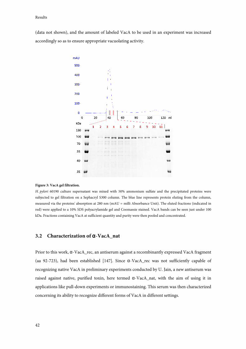
Results
42
(data not shown), and the amount of labeled VacA to be used in an experiment was increased
accordingly so as to ensure appropriate vacuolating activity.
Figure 3: VacA gel filtration.
H. pylori 60190 culture supernatant was mixed with 50% ammonium sulfate and the precipitated proteins were
subjected to gel filtration on a Sephacryl S300 column. The blue line represents protein eluting from the column,
measured via the proteins' absorption at 280 nm (mAU = milli Absorbance Unit). The eluted fractions (indicated in
red) were applied to a 10% SDS polyacrylamide gel and Coomassie stained. VacA bands can be seen just under 100
kDa. Fractions containing VacA at sufficient quantity and purity were then pooled and concentrated.
3.2 Characterization of αααα-VacA_nat
Prior to this work, α-VacA_rec, an antiserum against a recombinantly expressed VacA fragment
(aa 92-723), had been established [147]. Since α-VacA_rec was not sufficiently capable of
recognizing native VacA in preliminary experiments conducted by U. Jain, a new antiserum was
raised against native, purified toxin, here termed α-VacA_nat, with the aim of using it in
applications like pull-down experiments or immunostaining. This serum was then characterized
concerning its ability to recognize different forms of VacA in different settings.
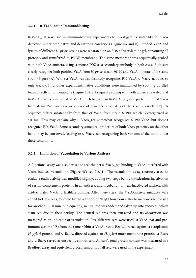
Results
43
3.2.1 αααα-VacA_nat in Immunoblotting
α-VacA_nat was used in immunoblotting experiments to investigate its suitability for VacA
detection under both native and denaturing conditions (Figure 4A and B). Purified VacA and
lysates of different H. pylori strains were separated on an SDS polyacrylamide gel, denaturing all
proteins, and transferred to PVDF membrane. The same membrane was sequentially probed
with both VacA antisera, using α-mouse-POX as a secondary antibody in both cases. Both sera
clearly recognize both purified VacA from H. pylori strain 60190 and VacA in lysate of the same
strain (Figure 4A). While α-VacA_rec also distinctly recognizes P12 VacA, α-VacA_nat does so
only weakly. In another experiment, native conditions were maintained by spotting purified
toxin directly onto membrane (Figure 4B). Subsequent probing with both antisera revealed that
α-VacA_nat recognizes native VacA much better than α-VacA_rec, as expected. Purified VacA
from strain P76 can serve as a proof of principle, since it is of the s1i2m2 variety [87]. Its
sequence differs substantially from that of VacA from strain 60190, which is categorized as
s1i1m1. This may explain why α-VacA_rec somewhat recognizes 60190 VacA but doesn't
recognize P76 VacA. Some secondary structural properties of both VacA proteins, on the other
hand, may be conserved, leading to α-VacA_nat recognizing both variants of the toxin under
these conditions.
3.2.2 Inhibition of Vacuolation by Various Antisera
A functional assay was also devised to see whether α-VacA_nat binding to VacA interfered with
VacA induced vacuolation (Figure 4C; see 2.2.11). The vacuolation assay routinely used to
evaluate toxin activity was modified slightly, adding two steps before intoxication: inactivation
of serum complement proteins in all antisera, and incubation of heat-inactivated antisera with
acid-activated VacA to facilitate binding. After these steps, the VacA/antisera mixtures were
added to HeLa cells, followed by the addition of NH4Cl four hours later to increase vacuole size
for another 30-60 min. Subsequently, neutral red was added and taken up into vacuoles, which
stain red due to their acidity. The neutral red was then extracted and its absorption was
measured as an indicator of vacuolation. Five different sera were used: α-VacA_nat and pre-
immune serum (PIS) from the same rabbit; α-VacA_rec; α-RecA, directed against a cytoplasmic
H. pylori protein; and α-BabA, directed against an H. pylori outer membrane protein. α-RecA
and α-BabA served as unspecific control sera. All sera's total protein content was measured in a
Bradford assay and equivalent protein amounts of all sera were used in the experiment.
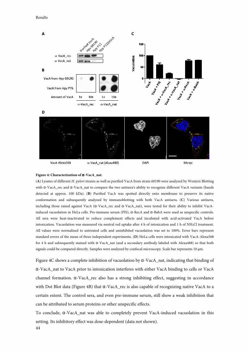
Results
44
Figure 4: Characterization of αααα-VacA_nat.
(A) Lysates of different H. pylori strains as well as purified VacA from strain 60190 were analyzed by Western Blotting
with α-VacA_rec and α-VacA_nat to compare the two antisera's ability to recognize different VacA variants (bands
detected at approx. 100 kDa). (B) Purified VacA was spotted directly onto membrane to preserve its native
conformation and subsequently analyzed by immunoblotting with both VacA antisera. (C) Various antisera,
including those raised against VacA (α-VacA_rec and α-VacA_nat), were tested for their ability to inhibit VacA-
induced vacuolation in HeLa cells. Pre-immune serum (PIS), α-RecA and α-BabA were used as unspecific controls.
All sera were heat-inactivated to reduce complement effects and incubated with acid-activated VacA before
intoxication. Vacuolation was measured via neutral red uptake after 4 h of intoxication and 1 h of NH4Cl treatment.
All values were normalized to untreated cells and uninhibited vacuolation was set to 100%. Error bars represent
standard errors of the mean of three independent experiments. (D) HeLa cells were intoxicated with VacA-Alexa568
for 4 h and subsequently stained with α-VacA_nat (and a secondary antibody labeled with Alexa488) so that both
signals could be compared directly. Samples were analyzed by confocal microscopy. Scale bar represents 10 µm.
Figure 4C shows a complete inhibition of vacuolation by α-VacA_nat, indicating that binding of
α-VacA_nat to VacA prior to intoxication interferes with either VacA binding to cells or VacA
channel formation. α-VacA_rec also has a strong inhibiting effect, suggesting in accordance
with Dot Blot data (Figure 4B) that α-VacA_rec is also capable of recognizing native VacA to a
certain extent. The control sera, and even pre-immune serum, still show a weak inhibition that
can be attributed to serum proteins or other unspecific effects.
To conclude, α-VacA_nat was able to completely prevent VacA-induced vacuolation in this
setting. Its inhibitory effect was dose-dependent (data not shown).
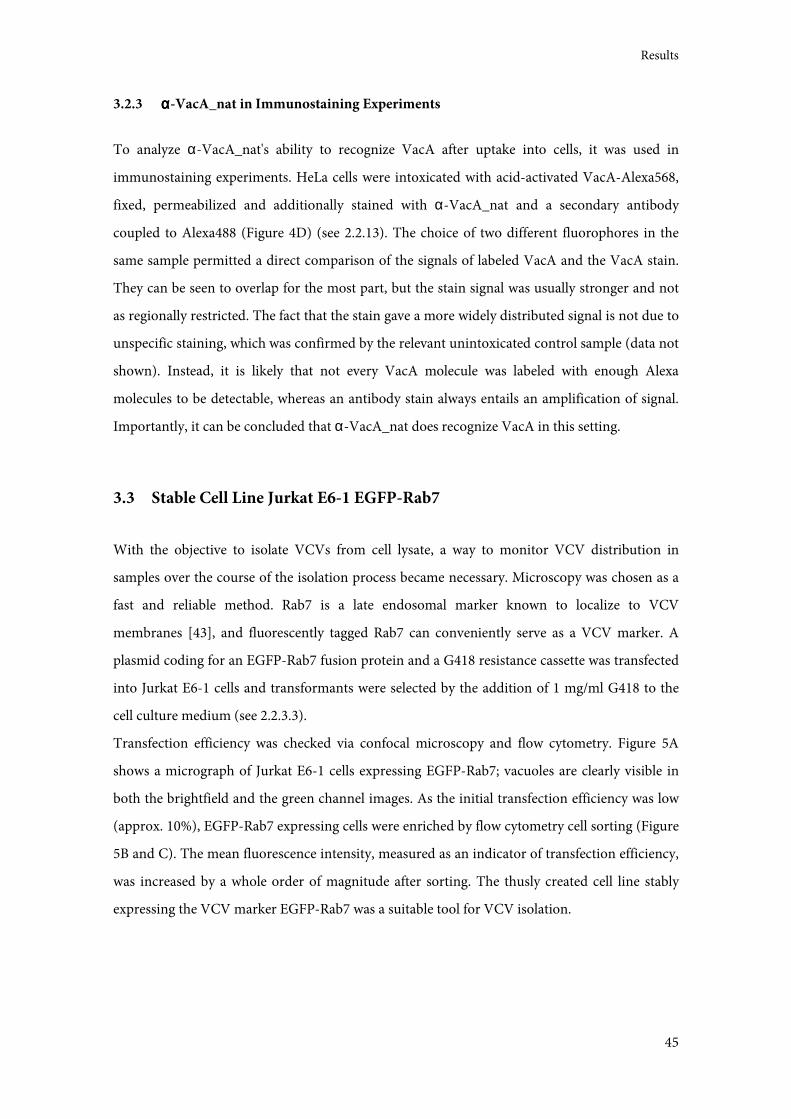
Results
45
3.2.3 αααα-VacA_nat in Immunostaining Experiments
To analyze α-VacA_nat's ability to recognize VacA after uptake into cells, it was used in
immunostaining experiments. HeLa cells were intoxicated with acid-activated VacA-Alexa568,
fixed, permeabilized and additionally stained with α-VacA_nat and a secondary antibody
coupled to Alexa488 (Figure 4D) (see 2.2.13). The choice of two different fluorophores in the
same sample permitted a direct comparison of the signals of labeled VacA and the VacA stain.
They can be seen to overlap for the most part, but the stain signal was usually stronger and not
as regionally restricted. The fact that the stain gave a more widely distributed signal is not due to
unspecific staining, which was confirmed by the relevant unintoxicated control sample (data not
shown). Instead, it is likely that not every VacA molecule was labeled with enough Alexa
molecules to be detectable, whereas an antibody stain always entails an amplification of signal.
Importantly, it can be concluded that α-VacA_nat does recognize VacA in this setting.
3.3 Stable Cell Line Jurkat E6-1 EGFP-Rab7
With the objective to isolate VCVs from cell lysate, a way to monitor VCV distribution in
samples over the course of the isolation process became necessary. Microscopy was chosen as a
fast and reliable method. Rab7 is a late endosomal marker known to localize to VCV
membranes [43], and fluorescently tagged Rab7 can conveniently serve as a VCV marker. A
plasmid coding for an EGFP-Rab7 fusion protein and a G418 resistance cassette was transfected
into Jurkat E6-1 cells and transformants were selected by the addition of 1 mg/ml G418 to the
cell culture medium (see 2.2.3.3).
Transfection efficiency was checked via confocal microscopy and flow cytometry. Figure 5A
shows a micrograph of Jurkat E6-1 cells expressing EGFP-Rab7; vacuoles are clearly visible in
both the brightfield and the green channel images. As the initial transfection efficiency was low
(approx. 10%), EGFP-Rab7 expressing cells were enriched by flow cytometry cell sorting (Figure
5B and C). The mean fluorescence intensity, measured as an indicator of transfection efficiency,
was increased by a whole order of magnitude after sorting. The thusly created cell line stably
expressing the VCV marker EGFP-Rab7 was a suitable tool for VCV isolation.
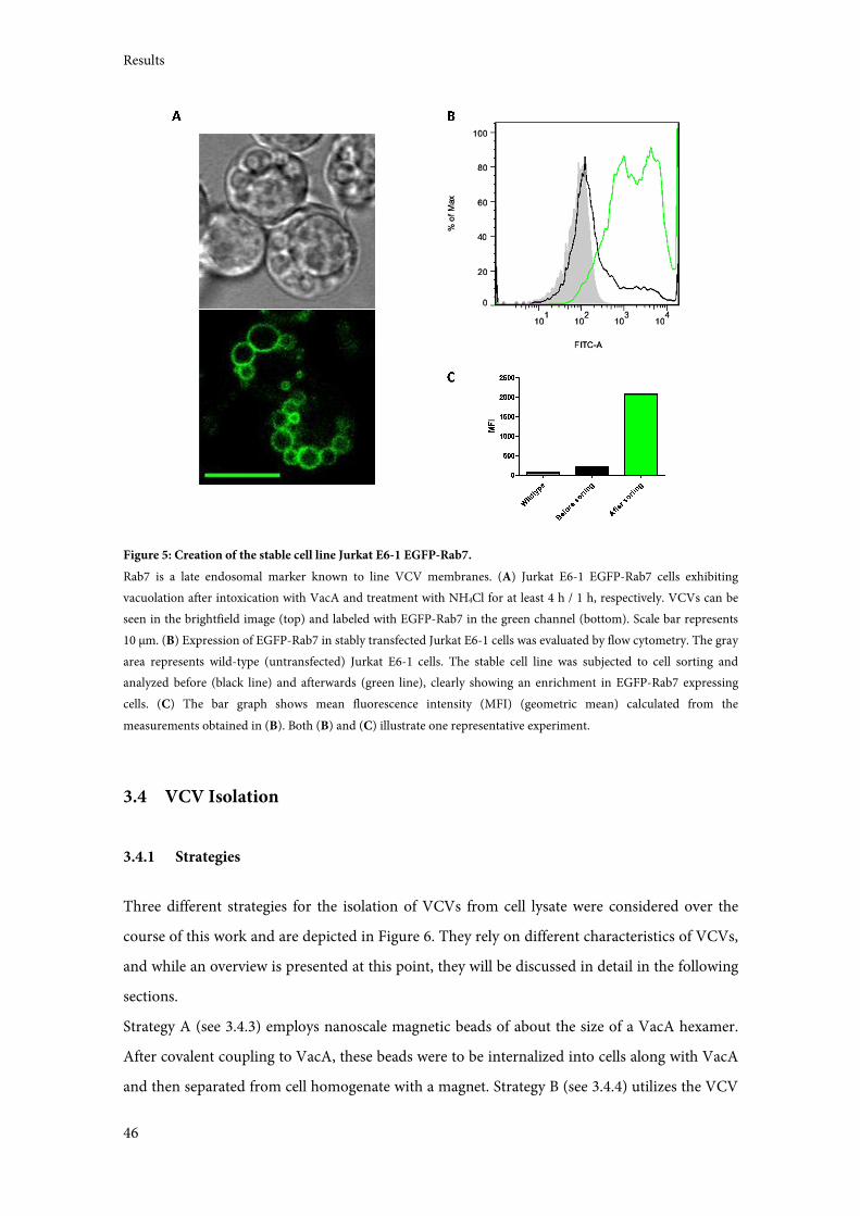
Results
46
Figure 5: Creation of the stable cell line Jurkat E6-1 EGFP-Rab7.
Rab7 is a late endosomal marker known to line VCV membranes. (A) Jurkat E6-1 EGFP-Rab7 cells exhibiting
vacuolation after intoxication with VacA and treatment with NH4Cl for at least 4 h / 1 h, respectively. VCVs can be
seen in the brightfield image (top) and labeled with EGFP-Rab7 in the green channel (bottom). Scale bar represents
10 µm. (B) Expression of EGFP-Rab7 in stably transfected Jurkat E6-1 cells was evaluated by flow cytometry. The gray
area represents wild-type (untransfected) Jurkat E6-1 cells. The stable cell line was subjected to cell sorting and
analyzed before (black line) and afterwards (green line), clearly showing an enrichment in EGFP-Rab7 expressing
cells. (C) The bar graph shows mean fluorescence intensity (MFI) (geometric mean) calculated from the
measurements obtained in (B). Both (B) and (C) illustrate one representative experiment.
3.4 VCV Isolation
3.4.1 Strategies
Three different strategies for the isolation of VCVs from cell lysate were considered over the
course of this work and are depicted in Figure 6. They rely on different characteristics of VCVs,
and while an overview is presented at this point, they will be discussed in detail in the following
sections.
Strategy A (see 3.4.3) employs nanoscale magnetic beads of about the size of a VacA hexamer.
After covalent coupling to VacA, these beads were to be internalized into cells along with VacA
and then separated from cell homogenate with a magnet. Strategy B (see 3.4.4) utilizes the VCV
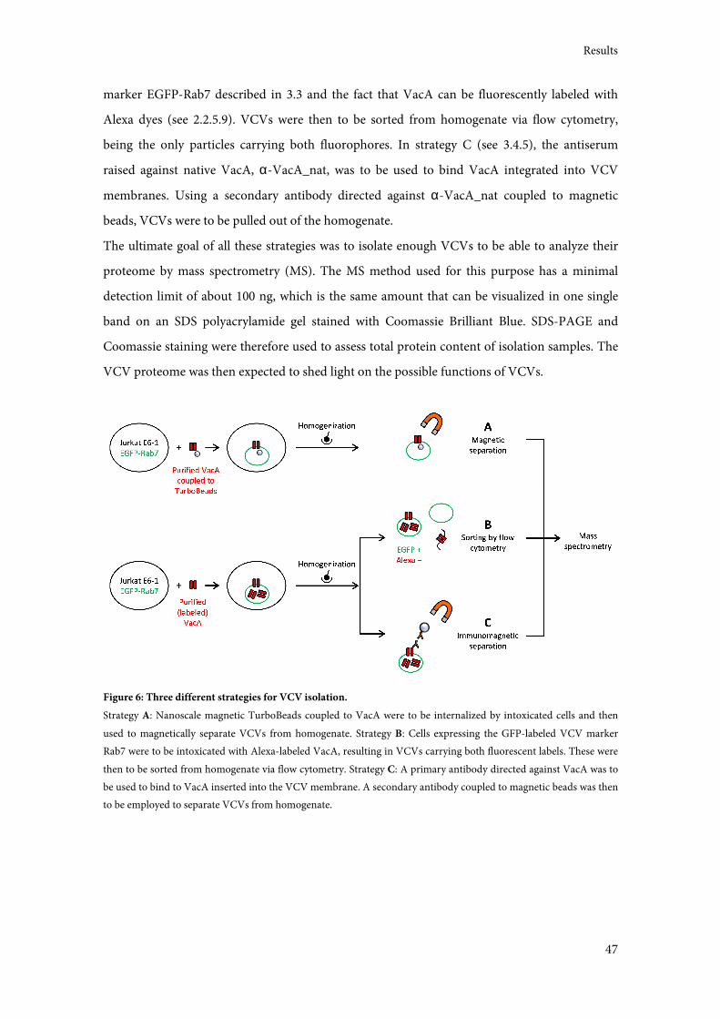
Results
47
marker EGFP-Rab7 described in 3.3 and the fact that VacA can be fluorescently labeled with
Alexa dyes (see 2.2.5.9). VCVs were then to be sorted from homogenate via flow cytometry,
being the only particles carrying both fluorophores. In strategy C (see 3.4.5), the antiserum
raised against native VacA, α-VacA_nat, was to be used to bind VacA integrated into VCV
membranes. Using a secondary antibody directed against α-VacA_nat coupled to magnetic
beads, VCVs were to be pulled out of the homogenate.
The ultimate goal of all these strategies was to isolate enough VCVs to be able to analyze their
proteome by mass spectrometry (MS). The MS method used for this purpose has a minimal
detection limit of about 100 ng, which is the same amount that can be visualized in one single
band on an SDS polyacrylamide gel stained with Coomassie Brilliant Blue. SDS-PAGE and
Coomassie staining were therefore used to assess total protein content of isolation samples. The
VCV proteome was then expected to shed light on the possible functions of VCVs.
Figure 6: Three different strategies for VCV isolation.
Strategy A: Nanoscale magnetic TurboBeads coupled to VacA were to be internalized by intoxicated cells and then
used to magnetically separate VCVs from homogenate. Strategy B: Cells expressing the GFP-labeled VCV marker
Rab7 were to be intoxicated with Alexa-labeled VacA, resulting in VCVs carrying both fluorescent labels. These were
then to be sorted from homogenate via flow cytometry. Strategy C: A primary antibody directed against VacA was to
be used to bind to VacA inserted into the VCV membrane. A secondary antibody coupled to magnetic beads was then
to be employed to separate VCVs from homogenate.
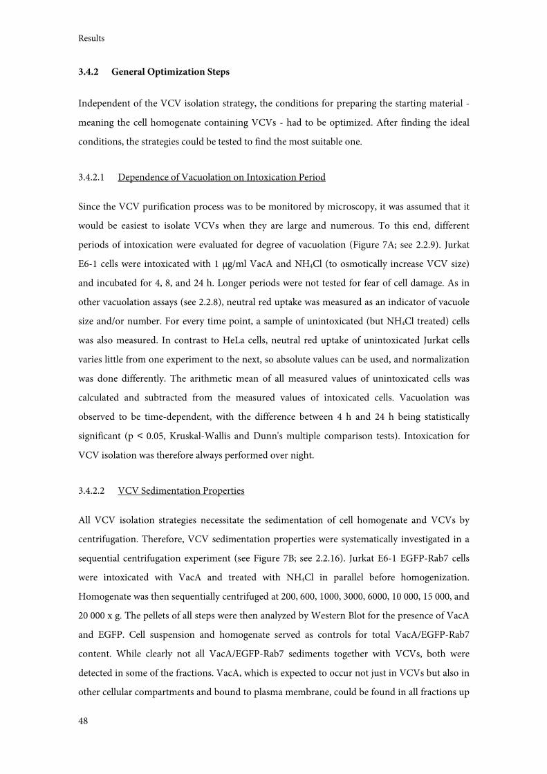
Results
48
3.4.2 General Optimization Steps
Independent of the VCV isolation strategy, the conditions for preparing the starting material -
meaning the cell homogenate containing VCVs - had to be optimized. After finding the ideal
conditions, the strategies could be tested to find the most suitable one.
3.4.2.1 Dependence of Vacuolation on Intoxication Period
Since the VCV purification process was to be monitored by microscopy, it was assumed that it
would be easiest to isolate VCVs when they are large and numerous. To this end, different
periods of intoxication were evaluated for degree of vacuolation (Figure 7A; see 2.2.9). Jurkat
E6-1 cells were intoxicated with 1 µg/ml VacA and NH4Cl (to osmotically increase VCV size)
and incubated for 4, 8, and 24 h. Longer periods were not tested for fear of cell damage. As in
other vacuolation assays (see 2.2.8), neutral red uptake was measured as an indicator of vacuole
size and/or number. For every time point, a sample of unintoxicated (but NH4Cl treated) cells
was also measured. In contrast to HeLa cells, neutral red uptake of unintoxicated Jurkat cells
varies little from one experiment to the next, so absolute values can be used, and normalization
was done differently. The arithmetic mean of all measured values of unintoxicated cells was
calculated and subtracted from the measured values of intoxicated cells. Vacuolation was
observed to be time-dependent, with the difference between 4 h and 24 h being statistically
significant (p < 0.05, Kruskal-Wallis and Dunn's multiple comparison tests). Intoxication for
VCV isolation was therefore always performed over night.
3.4.2.2 VCV Sedimentation Properties
All VCV isolation strategies necessitate the sedimentation of cell homogenate and VCVs by
centrifugation. Therefore, VCV sedimentation properties were systematically investigated in a
sequential centrifugation experiment (see Figure 7B; see 2.2.16). Jurkat E6-1 EGFP-Rab7 cells
were intoxicated with VacA and treated with NH4Cl in parallel before homogenization.
Homogenate was then sequentially centrifuged at 200, 600, 1000, 3000, 6000, 10 000, 15 000, and
20 000 x g. The pellets of all steps were then analyzed by Western Blot for the presence of VacA
and EGFP. Cell suspension and homogenate served as controls for total VacA/EGFP-Rab7
content. While clearly not all VacA/EGFP-Rab7 sediments together with VCVs, both were
detected in some of the fractions. VacA, which is expected to occur not just in VCVs but also in
other cellular compartments and bound to plasma membrane, could be found in all fractions up
Results
49
to 6000 x g. Most of EGFP-Rab7, on the other hand, was expected to be localized in VCVs, based
on previous microscopy observations (see for example Figure 5A). It was detected most
prominently in the 3000 x g fraction. Since this fraction was also positive for VacA, it was
therefore concluded to contain the majority of VCVs. This experiment was monitored by
microscopy in parallel, detecting only EGFP-Rab7, not VacA, and confirming this conclusion
(data not shown).
Figure 7: Optimization of VCV isolation conditions.
(A) As an indicator for vacuolation over time, neutral red uptake was measured in Jurkat E6-1 cells after 4, 8, and 24 h
of intoxication with VacA and parallel treatment with NH4Cl. Measurements were normalized to unintoxicated cells.
The graph was compiled with data obtained in three independent experiments. Error bars represent standard errors
of the mean. * = p < 0.05 (Kruskal-Wallis and Dunn's multiple comparison tests). (B) To investigate VCV
sedimentation properties, Jurkat E6-1 EGFP-Rab7 cells were intoxicated with VacA and treated with NH4Cl (both
5 h) before homogenization. 1 ml homogenate was then sequentially centrifuged. One fifth of centrifugation pellets
was loaded onto an SDS polyacrylamide gel and subsequently analyzed for the presence of VacA and EGFP-Rab7 by
Western Blotting. (C) Different buffers were tested for homogenization of intoxicated, NH4Cl treated Jurkat E6-1
EGFP-Rab7 cells. VCVs are clearly in a better condition in PBS*. The asterisk denotes addition of protease inhibitors
to the buffer. (D) Various fixation methods were tested for both homogenized and intact intoxicated cells. Samples
fixed with PFA for 10 min at RT are shown as representative examples. Scale bars in (C) and (D) represent 10 µm.
3.4.2.3 Homogenization and Fixation Conditions
The idea to isolate VCVs originally stemmed from a cooperation with the group of Hubert Hilbi,
who have established a protocol to isolate Legionella-containing vacuoles from cells infected
with Legionella pneumophila [151]. Some of the conditions detailed in their protocol were used
as initial starting points for this work, including the homogenization buffer, HS* (20 mM
Results
50
HEPES, 250 mM sucrose, 0.5 mM EGTA, pH 7.2; with added protease inhibitors). However, this
turned out to not be ideal for the purpose of isolating VCVs, as it did not preserve vacuole shape
in homogenate (see Figure 7C), making it difficult to identify VCVs in the later steps of the
isolation process. PBS* (also supplemented with protease inhibitors) was found to fulfill this
requirement. HS buffer seems to have the wrong osmotic pressure, leading to a collapse of VCVs,
which is in agreement with the fact that it was developed for vacuoles containing whole bacteria
unlike VCVs.
Fixation presented similar problems since vacuole shape was not retained in either intact cells or
homogenate, but vacuole shape was a necessary identifying feature of VCVs alongside EGFP
fluorescence. Various fixation reagents were tested at different concentrations and conditions,
but none proved succesful. Among the reagents tested were glutaraldehyde, cyclohexylamine,
methanol, ethanol, PFA with and without sucrose, PFA in HEPES, and commercially available
CellFIX (BD Biosciences). Cells fixed with 0.5% PFA and homogenate fixed with 3% PFA for 10
min at RT are shown in Figure 7D as an example. During fixation, VCVs lost their characteristic
round shape and shrank in size, making non-ambiguous identification impossible. The fact that
fixation turned out to be unfeasible was especially problematic as it eliminated the possibility of
validating the presence of specific proteins on isolated VCVs by immunostaining, as done in
[151]. All monitoring of VCV isolation was henceforth performed with unfixed samples in 8-
well slides.
3.4.3 Strategy A: TurboBeads Strategy
The first strategy for VCV isolation was based on nanoscale magnetic beads (Carboxyl coated
TurboBeads, TurboBeads LLC). These beads have a mean particle size of 30 nm, about the same
size as a VacA hexamer (see Figure 8A; [61]). Uptake of VacA coupled to beads was expected to
happen normally, undisturbed by the extremely small beads. After intoxication and cell
homogenization, the bead-containing VCVs were to be extracted from homogenate via
magnetic separation.
3.4.3.1 TurboBead Uptake
For detection purposes, VacA was labeled with Alexa488 before being coupled to beads. Bead
uptake into Jurkat E6-1 cells was investigated using two methods: confocal microscopy and flow
cytometry (see 2.2.17.2 and 2.2.17.3). For the first, cells were intoxicated with VacA-Alexa488
coupled to TurboBeads for 40 min at 4 °C to minimize particle movement and therefore bead
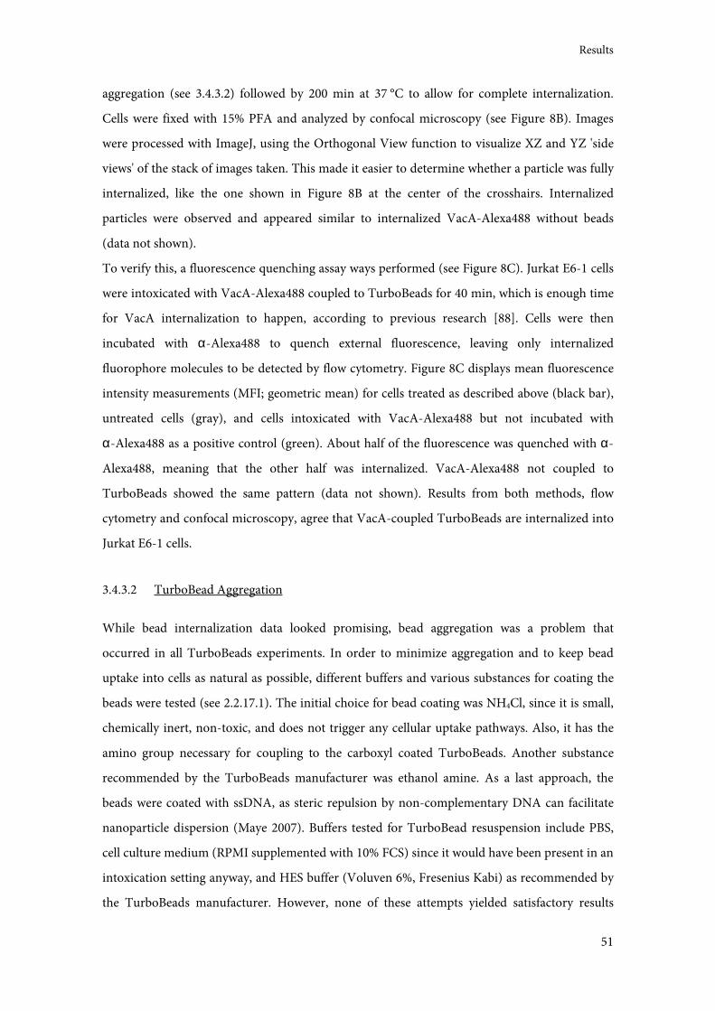
Results
51
aggregation (see 3.4.3.2) followed by 200 min at 37 °C to allow for complete internalization.
Cells were fixed with 15% PFA and analyzed by confocal microscopy (see Figure 8B). Images
were processed with ImageJ, using the Orthogonal View function to visualize XZ and YZ 'side
views' of the stack of images taken. This made it easier to determine whether a particle was fully
internalized, like the one shown in Figure 8B at the center of the crosshairs. Internalized
particles were observed and appeared similar to internalized VacA-Alexa488 without beads
(data not shown).
To verify this, a fluorescence quenching assay ways performed (see Figure 8C). Jurkat E6-1 cells
were intoxicated with VacA-Alexa488 coupled to TurboBeads for 40 min, which is enough time
for VacA internalization to happen, according to previous research [88]. Cells were then
incubated with α-Alexa488 to quench external fluorescence, leaving only internalized
fluorophore molecules to be detected by flow cytometry. Figure 8C displays mean fluorescence
intensity measurements (MFI; geometric mean) for cells treated as described above (black bar),
untreated cells (gray), and cells intoxicated with VacA-Alexa488 but not incubated with
α-Alexa488 as a positive control (green). About half of the fluorescence was quenched with α-
Alexa488, meaning that the other half was internalized. VacA-Alexa488 not coupled to
TurboBeads showed the same pattern (data not shown). Results from both methods, flow
cytometry and confocal microscopy, agree that VacA-coupled TurboBeads are internalized into
Jurkat E6-1 cells.
3.4.3.2 TurboBead Aggregation
While bead internalization data looked promising, bead aggregation was a problem that
occurred in all TurboBeads experiments. In order to minimize aggregation and to keep bead
uptake into cells as natural as possible, different buffers and various substances for coating the
beads were tested (see 2.2.17.1). The initial choice for bead coating was NH4Cl, since it is small,
chemically inert, non-toxic, and does not trigger any cellular uptake pathways. Also, it has the
amino group necessary for coupling to the carboxyl coated TurboBeads. Another substance
recommended by the TurboBeads manufacturer was ethanol amine. As a last approach, the
beads were coated with ssDNA, as steric repulsion by non-complementary DNA can facilitate
nanoparticle dispersion (Maye 2007). Buffers tested for TurboBead resuspension include PBS,
cell culture medium (RPMI supplemented with 10% FCS) since it would have been present in an
intoxication setting anyway, and HES buffer (Voluven 6%, Fresenius Kabi) as recommended by
the TurboBeads manufacturer. However, none of these attempts yielded satisfactory results
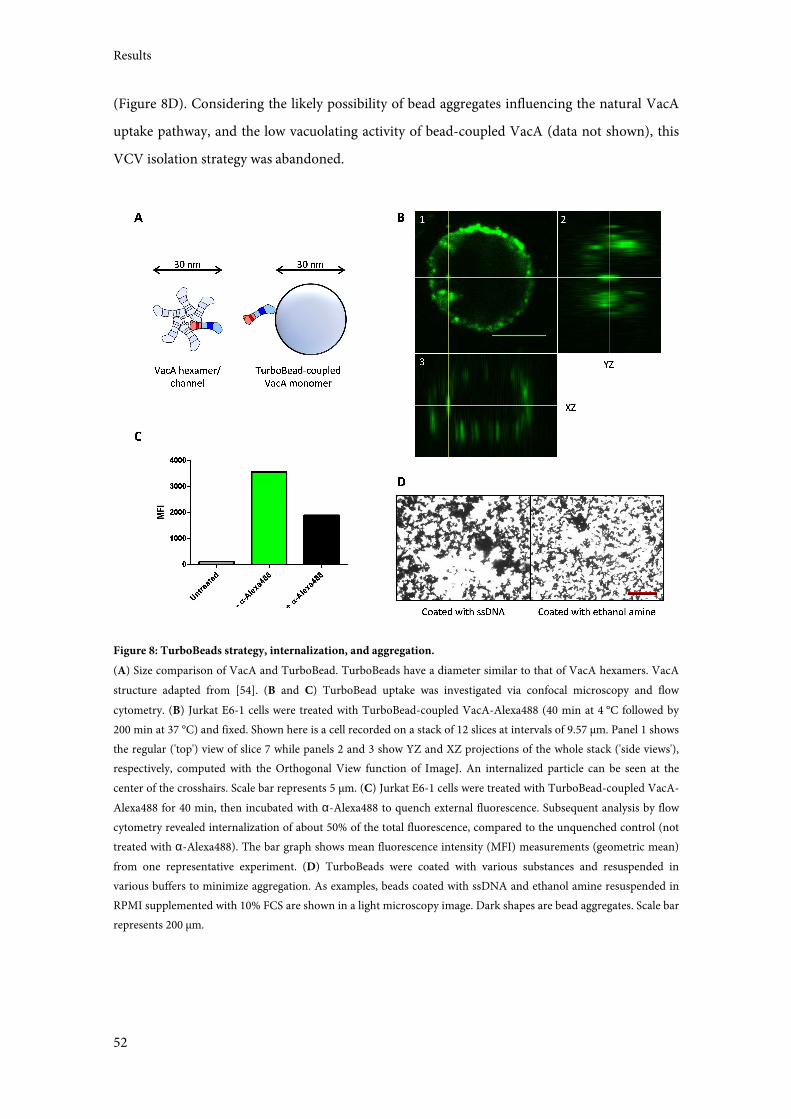
Results
52
(Figure 8D). Considering the likely possibility of bead aggregates influencing the natural VacA
uptake pathway, and the low vacuolating activity of bead-coupled VacA (data not shown), this
VCV isolation strategy was abandoned.
Figure 8: TurboBeads strategy, internalization, and aggregation.
(A) Size comparison of VacA and TurboBead. TurboBeads have a diameter similar to that of VacA hexamers. VacA
structure adapted from [54]. (B and C) TurboBead uptake was investigated via confocal microscopy and flow
cytometry. (B) Jurkat E6-1 cells were treated with TurboBead-coupled VacA-Alexa488 (40 min at 4 °C followed by
200 min at 37 °C) and fixed. Shown here is a cell recorded on a stack of 12 slices at intervals of 9.57 µm. Panel 1 shows
the regular ('top') view of slice 7 while panels 2 and 3 show YZ and XZ projections of the whole stack ('side views'),
respectively, computed with the Orthogonal View function of ImageJ. An internalized particle can be seen at the
center of the crosshairs. Scale bar represents 5 µm. (C) Jurkat E6-1 cells were treated with TurboBead-coupled VacA-
Alexa488 for 40 min, then incubated with α-Alexa488 to quench external fluorescence. Subsequent analysis by flow
cytometry revealed internalization of about 50% of the total fluorescence, compared to the unquenched control (not
treated with α-Alexa488). The bar graph shows mean fluorescence intensity (MFI) measurements (geometric mean)
from one representative experiment. (D) TurboBeads were coated with various substances and resuspended in
various buffers to minimize aggregation. As examples, beads coated with ssDNA and ethanol amine resuspended in
RPMI supplemented with 10% FCS are shown in a light microscopy image. Dark shapes are bead aggregates. Scale bar
represents 200 µm.
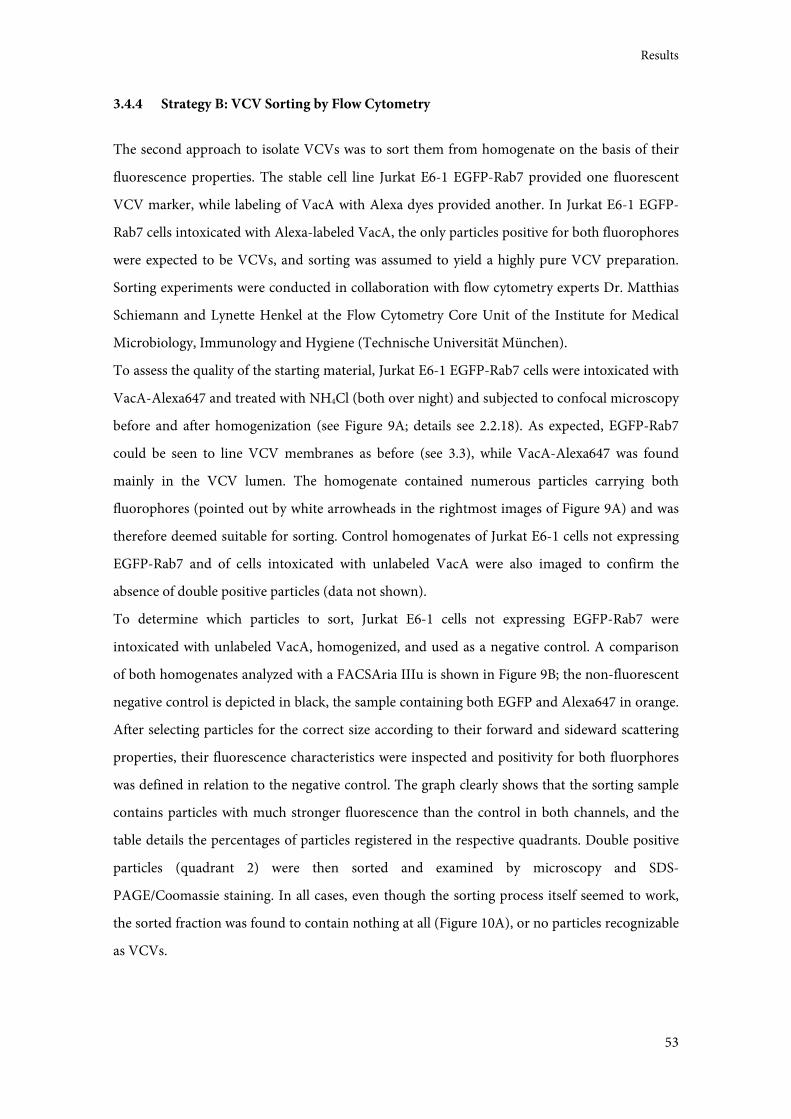
Results
53
3.4.4 Strategy B: VCV Sorting by Flow Cytometry
The second approach to isolate VCVs was to sort them from homogenate on the basis of their
fluorescence properties. The stable cell line Jurkat E6-1 EGFP-Rab7 provided one fluorescent
VCV marker, while labeling of VacA with Alexa dyes provided another. In Jurkat E6-1 EGFP-
Rab7 cells intoxicated with Alexa-labeled VacA, the only particles positive for both fluorophores
were expected to be VCVs, and sorting was assumed to yield a highly pure VCV preparation.
Sorting experiments were conducted in collaboration with flow cytometry experts Dr. Matthias
Schiemann and Lynette Henkel at the Flow Cytometry Core Unit of the Institute for Medical
Microbiology, Immunology and Hygiene (Technische Universität München).
To assess the quality of the starting material, Jurkat E6-1 EGFP-Rab7 cells were intoxicated with
VacA-Alexa647 and treated with NH4Cl (both over night) and subjected to confocal microscopy
before and after homogenization (see Figure 9A; details see 2.2.18). As expected, EGFP-Rab7
could be seen to line VCV membranes as before (see 3.3), while VacA-Alexa647 was found
mainly in the VCV lumen. The homogenate contained numerous particles carrying both
fluorophores (pointed out by white arrowheads in the rightmost images of Figure 9A) and was
therefore deemed suitable for sorting. Control homogenates of Jurkat E6-1 cells not expressing
EGFP-Rab7 and of cells intoxicated with unlabeled VacA were also imaged to confirm the
absence of double positive particles (data not shown).
To determine which particles to sort, Jurkat E6-1 cells not expressing EGFP-Rab7 were
intoxicated with unlabeled VacA, homogenized, and used as a negative control. A comparison
of both homogenates analyzed with a FACSAria IIIu is shown in Figure 9B; the non-fluorescent
negative control is depicted in black, the sample containing both EGFP and Alexa647 in orange.
After selecting particles for the correct size according to their forward and sideward scattering
properties, their fluorescence characteristics were inspected and positivity for both fluorphores
was defined in relation to the negative control. The graph clearly shows that the sorting sample
contains particles with much stronger fluorescence than the control in both channels, and the
table details the percentages of particles registered in the respective quadrants. Double positive
particles (quadrant 2) were then sorted and examined by microscopy and SDS-
PAGE/Coomassie staining. In all cases, even though the sorting process itself seemed to work,
the sorted fraction was found to contain nothing at all (Figure 10A), or no particles recognizable
as VCVs.
Results
54
Figure 9: VCV homogenate prepared for flow cytometry sorting.
(A) Jurkat E6-1 EGFP-Rab7 cells were intoxicated with VacA-Alexa647 and treated with NH4Cl (both over night)
before homogenization. Aliquots of intact cells and homogenate were examined by confocal microscopy for VCVs
positive for both EGFP and Alexa647 (highlighted by white arrowheads) as a prerequisite for VCV sorting by flow
cytometry. Scale bars represent 10 µm. (B) Cells were treated with VacA and NH4Cl over night and homogenized, and
the homogenate was subjected to flow cytometry. Jurkat E6-1 cells not expressing EGFP-Rab7 were intoxicated with
unlabeled VacA as a negative control (black); Jurkat E6-1 EGFP-Rab7 cells were intoxicated with VacA-Alexa647
(orange; compare bottom row in (A)). Particles in quadrant 2 were defined as VCVs, being positive for both EGFP
and Alexa647, and were sorted. The table shows particle counts (%) of all quadrants; quadrant numbering as
indicated in the graph. Representative sorting experiment performed on a FACSAria IIIu.
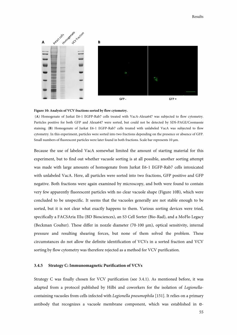
Results
55
Figure 10: Analysis of VCV fractions sorted by flow cytometry.
(A) Homogenate of Jurkat E6-1 EGFP-Rab7 cells treated with VacA-Alexa647 was subjected to flow cytometry.
Particles positive for both GFP and Alexa647 were sorted, but could not be detected by SDS-PAGE/Coomassie
staining. (B) Homogenate of Jurkat E6-1 EGFP-Rab7 cells treated with unlabeled VacA was subjected to flow
cytometry. In this experiment, particles were sorted into two fractions depending on the presence or absence of GFP.
Small numbers of fluorescent particles were later found in both fractions. Scale bar represents 10 µm.
Because the use of labeled VacA somewhat limited the amount of starting material for this
experiment, but to find out whether vacuole sorting is at all possible, another sorting attempt
was made with large amounts of homogenate from Jurkat E6-1 EGFP-Rab7 cells intoxicated
with unlabeled VacA. Here, all particles were sorted into two fractions, GFP positive and GFP
negative. Both fractions were again examined by microscopy, and both were found to contain
very few apparently fluorescent particles with no clear vacuole shape (Figure 10B), which were
concluded to be unspecific. It seems that the vacuoles generally are not stable enough to be
sorted, but it is not clear what exactly happens to them. Various sorting devices were tried,
specifically a FACSAria IIIu (BD Biosciences), an S3 Cell Sorter (Bio-Rad), and a MoFlo Legacy
(Beckman Coulter). These differ in nozzle diameter (70-100 µm), optical sensitivity, internal
pressure and resulting shearing forces, but none of them solved the problem. These
circumstances do not allow the definite identification of VCVs in a sorted fraction and VCV
sorting by flow cytometry was therefore rejected as a method for VCV purification.
3.4.5 Strategy C: Immunomagnetic Purification of VCVs
Strategy C was finally chosen for VCV purification (see 3.4.1). As mentioned before, it was
adapted from a protocol published by Hilbi and coworkers for the isolation of Legionella-
containing vacuoles from cells infected with Legionella pneumophila [151]. It relies on a primary
antibody that recognizes a vacuole membrane component, which was established in α-
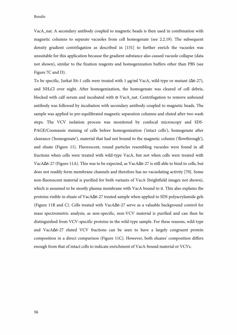
Results
56
VacA_nat. A secondary antibody coupled to magnetic beads is then used in combination with
magnetic columns to separate vacuoles from cell homogenate (see 2.2.19). The subsequent
density gradient centrifugation as described in [151] to further enrich the vacuoles was
unsuitable for this application because the gradient substance also caused vacuole collapse (data
not shown), similar to the fixation reagents and homogenization buffers other than PBS (see
Figure 7C and D).
To be specific, Jurkat E6-1 cells were treated with 1 µg/ml VacA, wild-type or mutant (∆6-27),
and NH4Cl over night. After homogenization, the homogenate was cleared of cell debris,
blocked with calf serum and incubated with α-VacA_nat. Centrifugation to remove unbound
antibody was followed by incubation with secondary antibody coupled to magnetic beads. The
sample was applied to pre-equilibrated magnetic separation columns and eluted after two wash
steps. The VCV isolation process was monitored by confocal microscopy and SDS-
PAGE/Coomassie staining of cells before homogenization ('intact cells'), homogenate after
clearance ('homogenate'), material that had not bound to the magnetic column ('flowthrough'),
and eluate (Figure 11). Fluorescent, round particles resembling vacuoles were found in all
fractions when cells were treated with wild-type VacA, but not when cells were treated with
VacA∆6-27 (Figure 11A). This was to be expected, as VacA∆6-27 is still able to bind to cells, but
does not readily form membrane channels and therefore has no vacuolating activity [70]. Some
non-fluorescent material is purified for both variants of VacA (brightfield images not shown),
which is assumed to be mostly plasma membrane with VacA bound to it. This also explains the
proteins visible in eluate of VacA∆6-27 treated sample when applied to SDS polyacrylamide gels
(Figure 11B and C). Cells treated with VacA∆6-27 serve as a valuable background control for
mass spectrometric analysis, as non-specific, non-VCV material is purified and can then be
distinguished from VCV-specific proteins in the wild-type sample. For these reasons, wild-type
and VacA∆6-27 eluted VCV fractions can be seen to have a largely congruent protein
composition in a direct comparison (Figure 11C). However, both eluates' composition differs
enough from that of intact cells to indicate enrichment of VacA-bound material or VCVs.
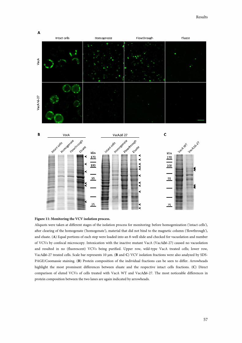
Results
57
Figure 11: Monitoring the VCV isolation process.
Aliquots were taken at different stages of the isolation process for monitoring: before homogenization ('intact cells'),
after clearing of the homogenate ('homogenate'), material that did not bind to the magnetic column ('flowthrough'),
and eluate. (A) Equal portions of each step were loaded into an 8-well slide and checked for vacuolation and number
of VCVs by confocal microscopy. Intoxication with the inactive mutant VacA (VacA∆6-27) caused no vacuolation
and resulted in no (fluorescent) VCVs being purified. Upper row, wild-type VacA treated cells; lower row,
VacA∆6-27 treated cells. Scale bar represents 10 µm. (B and C) VCV isolation fractions were also analyzed by SDS-
PAGE/Coomassie staining. (B) Protein composition of the individual fractions can be seen to differ. Arrowheads
highlight the most prominent differences between eluate and the respective intact cells fractions. (C) Direct
comparison of eluted VCVs of cells treated with VacA WT and VacA∆6-27. The most noticeable differences in
protein composition between the two lanes are again indicated by arrowheads.
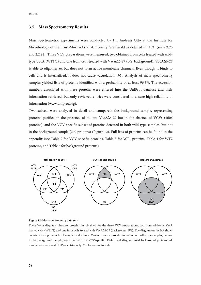
Results
58
3.5 Mass Spectrometry Results
Mass spectrometric experiments were conducted by Dr. Andreas Otto at the Institute for
Microbiology of the Ernst-Moritz-Arndt-University Greifswald as detailed in [152] (see 2.2.20
and 2.2.21). Three VCV preparations were measured, two obtained from cells treated with wild-
type VacA (WT1/2) and one from cells treated with VacA∆6-27 (BG, background). VacA∆6-27
is able to oligomerize, but does not form active membrane channels. Even though it binds to
cells and is internalized, it does not cause vacuolation [70]. Analysis of mass spectrometry
samples yielded lists of proteins identified with a probability of at least 96.3%. The accession
numbers associated with these proteins were entered into the UniProt database and their
information retrieved, but only reviewed entries were considered to ensure high reliability of
information (www.uniprot.org).
Two subsets were analyzed in detail and compared: the background sample, representing
proteins purified in the presence of mutant VacA∆6-27 but in the absence of VCVs (1606
proteins), and the VCV-specific subset of proteins detected in both wild-type samples, but not
in the background sample (240 proteins) (Figure 12). Full lists of proteins can be found in the
appendix (see Table 2 for VCV-specific proteins, Table 3 for WT1 proteins, Table 4 for WT2
proteins, and Table 5 for background proteins).
Figure 12: Mass spectrometry data sets.
These Venn diagrams illustrate protein hits obtained for the three VCV preparations, two from wild-type VacA
treated cells (WT1/2) and one from cells treated with VacA∆6-27 (background, BG). The diagram on the left shows
counts of total proteins in all samples and subsets. Center diagram: proteins found in both wild-type samples, but not
in the background sample, are expected to be VCV-specific. Right hand diagram: total background proteins. All
numbers are reviewed UniProt entries only. Circles are not to scale.
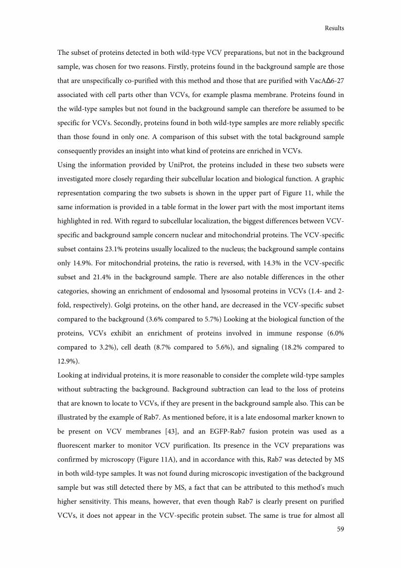
Results
59
The subset of proteins detected in both wild-type VCV preparations, but not in the background
sample, was chosen for two reasons. Firstly, proteins found in the background sample are those
that are unspecifically co-purified with this method and those that are purified with VacA∆6-27
associated with cell parts other than VCVs, for example plasma membrane. Proteins found in
the wild-type samples but not found in the background sample can therefore be assumed to be
specific for VCVs. Secondly, proteins found in both wild-type samples are more reliably specific
than those found in only one. A comparison of this subset with the total background sample
consequently provides an insight into what kind of proteins are enriched in VCVs.
Using the information provided by UniProt, the proteins included in these two subsets were
investigated more closely regarding their subcellular location and biological function. A graphic
representation comparing the two subsets is shown in the upper part of Figure 11, while the
same information is provided in a table format in the lower part with the most important items
highlighted in red. With regard to subcellular localization, the biggest differences between VCV-
specific and background sample concern nuclear and mitochondrial proteins. The VCV-specific
subset contains 23.1% proteins usually localized to the nucleus; the background sample contains
only 14.9%. For mitochondrial proteins, the ratio is reversed, with 14.3% in the VCV-specific
subset and 21.4% in the background sample. There are also notable differences in the other
categories, showing an enrichment of endosomal and lysosomal proteins in VCVs (1.4- and 2-
fold, respectively). Golgi proteins, on the other hand, are decreased in the VCV-specific subset
compared to the background (3.6% compared to 5.7%) Looking at the biological function of the
proteins, VCVs exhibit an enrichment of proteins involved in immune response (6.0%
compared to 3.2%), cell death (8.7% compared to 5.6%), and signaling (18.2% compared to
12.9%).
Looking at individual proteins, it is more reasonable to consider the complete wild-type samples
without subtracting the background. Background subtraction can lead to the loss of proteins
that are known to locate to VCVs, if they are present in the background sample also. This can be
illustrated by the example of Rab7. As mentioned before, it is a late endosomal marker known to
be present on VCV membranes [43], and an EGFP-Rab7 fusion protein was used as a
fluorescent marker to monitor VCV purification. Its presence in the VCV preparations was
confirmed by microscopy (Figure 11A), and in accordance with this, Rab7 was detected by MS
in both wild-type samples. It was not found during microscopic investigation of the background
sample but was still detected there by MS, a fact that can be attributed to this method's much
higher sensitivity. This means, however, that even though Rab7 is clearly present on purified
VCVs, it does not appear in the VCV-specific protein subset. The same is true for almost all
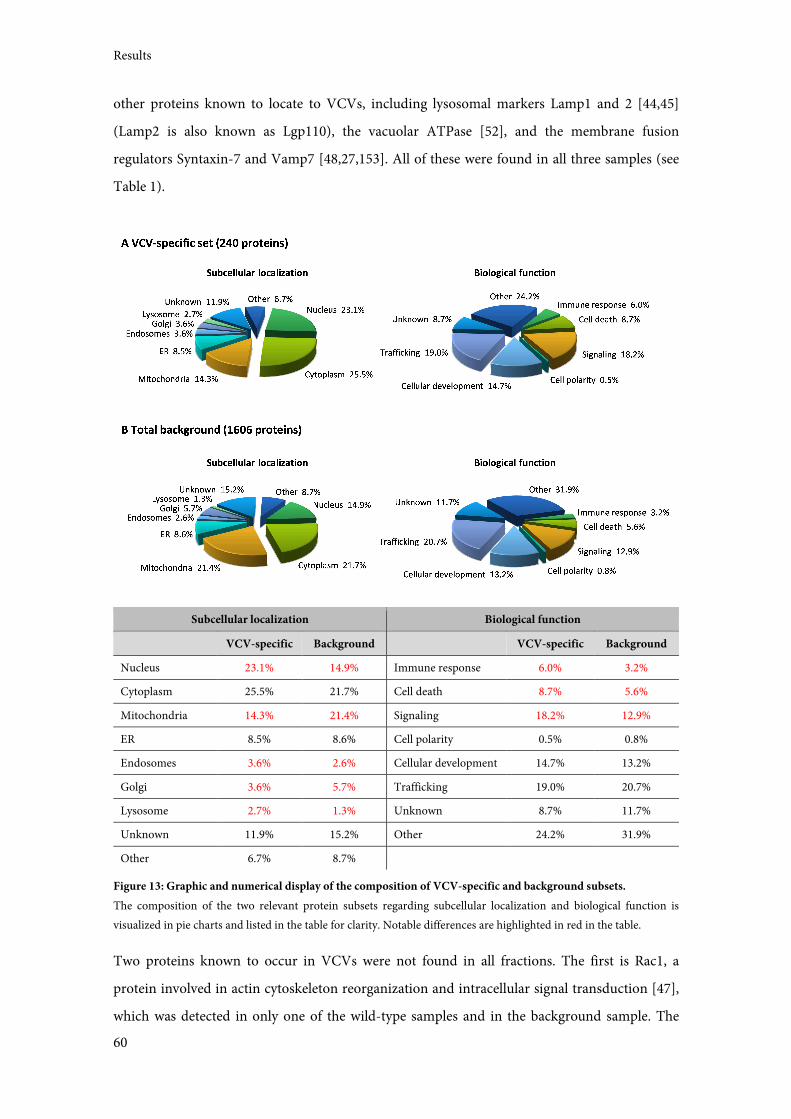
Results
60
other proteins known to locate to VCVs, including lysosomal markers Lamp1 and 2 [44,45]
(Lamp2 is also known as Lgp110), the vacuolar ATPase [52], and the membrane fusion
regulators Syntaxin-7 and Vamp7 [48,27,153]. All of these were found in all three samples (see
Table 1).
Subcellular localization Biological function
VCV-specific Background VCV-specific Background
Nucleus 23.1% 14.9% Immune response 6.0% 3.2%
Cytoplasm 25.5% 21.7% Cell death 8.7% 5.6%
Mitochondria 14.3% 21.4% Signaling 18.2% 12.9%
ER 8.5% 8.6% Cell polarity 0.5% 0.8%
Endosomes 3.6% 2.6% Cellular development 14.7% 13.2%
Golgi 3.6% 5.7% Trafficking 19.0% 20.7%
Lysosome 2.7% 1.3% Unknown 8.7% 11.7%
Unknown 11.9% 15.2% Other 24.2% 31.9%
Other 6.7% 8.7%
Figure 13: Graphic and numerical display of the composition of VCV-specific and background subsets.
The composition of the two relevant protein subsets regarding subcellular localization and biological function is
visualized in pie charts and listed in the table for clarity. Notable differences are highlighted in red in the table.
Two proteins known to occur in VCVs were not found in all fractions. The first is Rac1, a
protein involved in actin cytoskeleton reorganization and intracellular signal transduction [47],
which was detected in only one of the wild-type samples and in the background sample. The
Results
61
second is β-glucosaminidase, an LE/lysosomal enzyme [45] that was found in none of the
samples. Overall, the presence of almost all expected VCV-associated proteins in the wild-type
samples validates the VCV purification method to a large extent. However, the above examples
also illustrate that for some analyses, the background sample does not serve its purpose perfectly.
The MS results lists were also scanned for host cell proteins known to be influenced by VacA or
necessary for VacA-induced effects. Some of these were found in the VCV-specific sample,
among them Cdc42, which is important for VacA uptake [96], Bak, a pro-apoptotic protein
activated during VacA intoxication [154], the ER marker calnexin, and Drp1, a mitochondrial
fission protein recruited and activated by VacA [110]. Others that were found in only one wild-
type sample, or also in the background sample, include several members of the protein kinase C
(PKC) family and Phosphoinositide-3 kinase (PI-3-K), which regulate T-cell activation, tubulin,
and the small GTPase Rac1. PKCs, tubulin, and Rac1 are all known to play a role in VacA
uptake [97,96]. Further analysis of MS measured samples can yield interesting targets for further
investigation, as exemplified in the next section.
3.6 Investigation of Possible Interactions of VacA with STIM1
Previous work showed localization of VacA to the ER and an influence of VacA on intracellular
calcium signaling in T-cells, where it disrupts store-operated calcium entry (SOCE) [155]. A
closer look at ER proteins in the VCV-specific protein subset revealed the presence of classic ER
markers calnexin and Sec61, indicating that ER was not generally co-purified unspecifically.
Also found there were stromal interaction molecule 1 (STIM1) and the Inositol 1,4,5-
trisphosphate receptor type 3 (ITPR3), both of which play an essential part in calcium signaling.
Stimulation of a T-cell through the T-cell receptor results in calcium efflux from the ER into the
cytosol via ITPR3. STIM1 senses the ER luminal Ca2+ concentration, and in the event of low
calcium initiates calcium influx from the extracellular milieu through activation of Orai1, a
SOCE channel [156]. The presence of STIM1 and ITPR3 in the VCV-specific subset provides
additional evidence of an involvement of VacA in calcium signaling. Due to its role in SOCE,
STIM1 seemed a likely candidate for VacA to interact with if VacA is transported to the ER and
interferes with calcium signaling there. STIM1 (Figure 14) possesses an ER luminal EF hand
domain, a helix-loop-helix motif common in calcium binding proteins which enables STIM1 to
sense the ER luminal Ca2+ concentration [156]. Another important feature is a region termed
CAD (Ca2+ release-activated Ca2+ activation domain, [157]) or SOAR (STIM-Orai activating
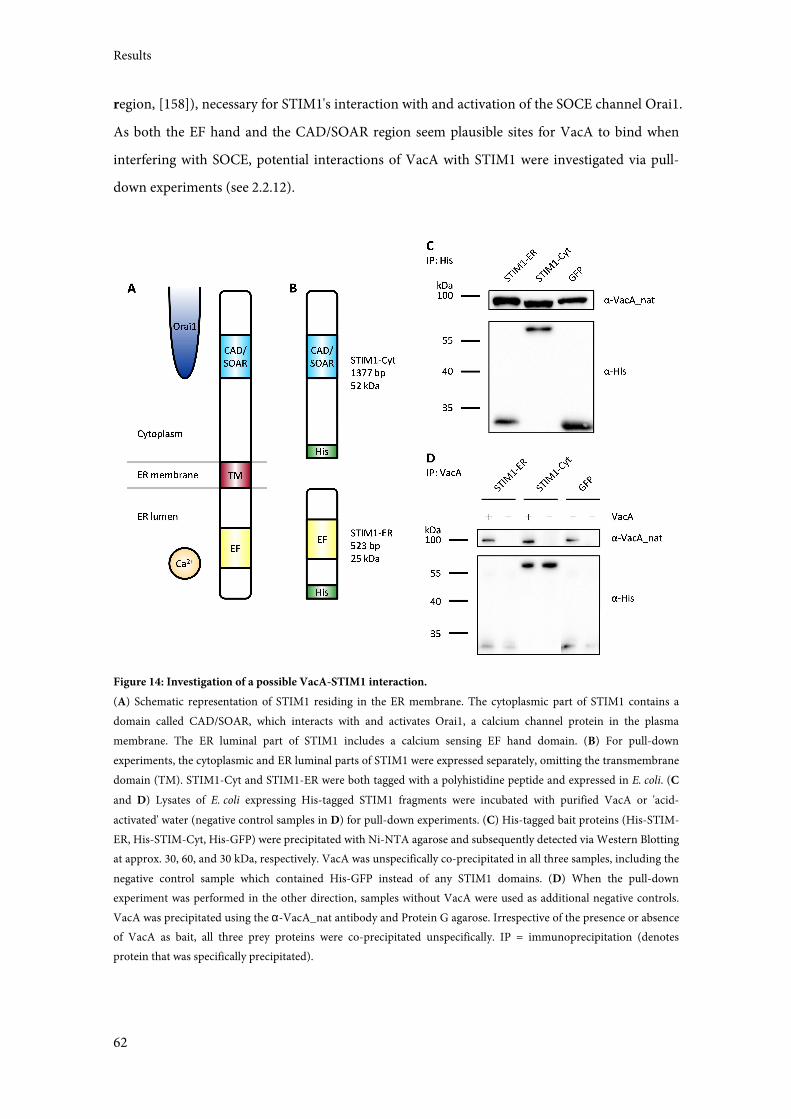
Results
62
region, [158]), necessary for STIM1's interaction with and activation of the SOCE channel Orai1.
As both the EF hand and the CAD/SOAR region seem plausible sites for VacA to bind when
interfering with SOCE, potential interactions of VacA with STIM1 were investigated via pull-
down experiments (see 2.2.12).
Figure 14: Investigation of a possible VacA-STIM1 interaction.
(A) Schematic representation of STIM1 residing in the ER membrane. The cytoplasmic part of STIM1 contains a
domain called CAD/SOAR, which interacts with and activates Orai1, a calcium channel protein in the plasma
membrane. The ER luminal part of STIM1 includes a calcium sensing EF hand domain. (B) For pull-down
experiments, the cytoplasmic and ER luminal parts of STIM1 were expressed separately, omitting the transmembrane
domain (TM). STIM1-Cyt and STIM1-ER were both tagged with a polyhistidine peptide and expressed in E. coli. (C
and D) Lysates of E. coli expressing His-tagged STIM1 fragments were incubated with purified VacA or 'acid-
activated' water (negative control samples in D) for pull-down experiments. (C) His-tagged bait proteins (His-STIM-
ER, His-STIM-Cyt, His-GFP) were precipitated with Ni-NTA agarose and subsequently detected via Western Blotting
at approx. 30, 60, and 30 kDa, respectively. VacA was unspecifically co-precipitated in all three samples, including the
negative control sample which contained His-GFP instead of any STIM1 domains. (D) When the pull-down
experiment was performed in the other direction, samples without VacA were used as additional negative controls.
VacA was precipitated using the α-VacA_nat antibody and Protein G agarose. Irrespective of the presence or absence
of VacA as bait, all three prey proteins were co-precipitated unspecifically. IP = immunoprecipitation (denotes
protein that was specifically precipitated).
Results
63
Previous attempts to immunoprecipitate native STIM1 from cell lysate had been unsuccessful
(U. Jain, personal communication), presumably due to its transmembrane domain being
difficult to solubilize. For this reason, a different approach was devised to express STIM1
recombinantly, separated into its cytoplasmic and ER luminal parts and omitting the
transmembrane domain (Figure 14A and B). The ER luminal part contains the EF hand domain,
while the cytoplasmic part contains the CAD/SOAR region. Both parts were cloned into
pET28a(+), equipping them with polyhistidine-tags for easier purification and detection. An
identically tagged truncated GFP (GFP1-10; [146]) expressed from the same plasmid backbone
was used as a control for unspecific interactions. Lysates of E. coli expressing the 6xHis-tagged
proteins were incubated with VacA to allow protein interactions to develop. Interactions were
tested in both directions. First, the 6xHis-tagged proteins were used as bait, precipitating them
with Ni-NTA agarose and then testing for co-precipitated VacA in a Western Blot (Figure 14C).
VacA was detected in all three samples, indicating an unspecific interaction. Reciprocally, VacA
was precipitated as bait with α-VacA_nat and Protein G agarose. Samples without VacA were
handled the same way and served as additional negative controls. After the pull-down, the
6xHis-tagged proteins were detected via Western Blot (see Figure 14D). For each 6xHis-tagged
protein (STIM1-ER, STIM1-Cyt, GFP), the signal in the α-His blot is of comparative strength in
samples with and without VacA, again suggesting unspecific interactions. In conclusion,
interactions of VacA and the two parts of STIM1 could be neither shown nor refuted in this
assay.
3.7 VacA Localization in Intoxicated Cells
3.7.1 Colocalization of VacA with ER and Golgi Apparatus Markers and CTxB
Late trafficking of VacA inside intoxicated cells is not very well-researched. VacA is taken up
into endocytic compartments that were shown to be GEECs (GIP-AP-enriched early endosomal
compartments, [96]) and is then routed through early and late endosomes, finally reaching
compartments carrying both endosomal and lysosomal markers [45,44], but avoiding
degradation [46]. After four hours, VacA is found in vesicles scattered throughout the cytosol
(Figure 15A, first column), which become enlarged VCVs in the presence of weak bases [159].
VacA is also known to localize to mitochondria, more specifically to the inner mitochondrial
membrane [103–105], and it may to be transported there via VCVs [106]. However, a
Results
64
localization of VacA to other intracellular compartments has not yet been reported. Evidence of
VacA involvement in intracellular calcium signaling suggested a possible trafficking of VacA to
the ER, which was investigated in immunostaining experiments (see 2.2.13). HeLa cells were
intoxicated with VacA-Alexa488, fixed, permeabilized, and stained for the ER marker calnexin
or the Golgi apparatus marker giantin. In a similar setup, cells were intoxicated simultaneously
with Alexa-labeled cholera toxin subunit B (CTxB-Alexa555). Although its uptake differs from
that of VacA [96,160], CTxB is transported via the retrograde endocytic pathway to the Golgi
[161], and might therefore provide an interesting comparison. DAPI staining of cell nuclei
served as a general indicator of cell health. The results are shown in Figure 15A. In all three
instances, VacA and the marker protein exhibit partial colocalization, suggesting that at least
part of the internalized VacA localizes to the ER and the Golgi apparatus, respectively.
Colocalization of VacA and CTxB not only in the large area representing the Golgi implies
similarities in the transport of both toxins, but this may only be the case for CTxB that has not
yet reached its final destination.
Interestingly, a substantial amount of VacA was found to localize in compartments closely
associated with the Golgi apparatus (Figure 15B). These compartments may represent VacA
being transported to or from the Golgi, possibly on its way to the ER via retrograde trafficking.
3.7.2 Isolation of Endoplasmic Reticulum
Another approach to verify whether VacA indeed localizes to the ER was to purify ER from
VacA-intoxicated cells. Jurkat E6-1 cells were intoxicated with VacA for 4 h, homogenized, and
subjected to sequential centrifugation (see 2.2.22). This resulted in a nuclear fraction, a
mitochondrial fraction, and a crude microsomal fraction expected to contain the ER. All
fractions were analyzed for the presence of VacA and the ER marker calnexin by
immunoblotting (Figure 15C). Calnexin was found mostly in the mitochondrial fraction,
indicating that the protocol needs to be optimized for this specific purpose. However,
mitochondria and the ER are known to associate (reviewed in [162]), and the presence of VacA
in both the mitochondrial and crude microsomal fractions supports the notion that at least
some toxin localizes to the ER.
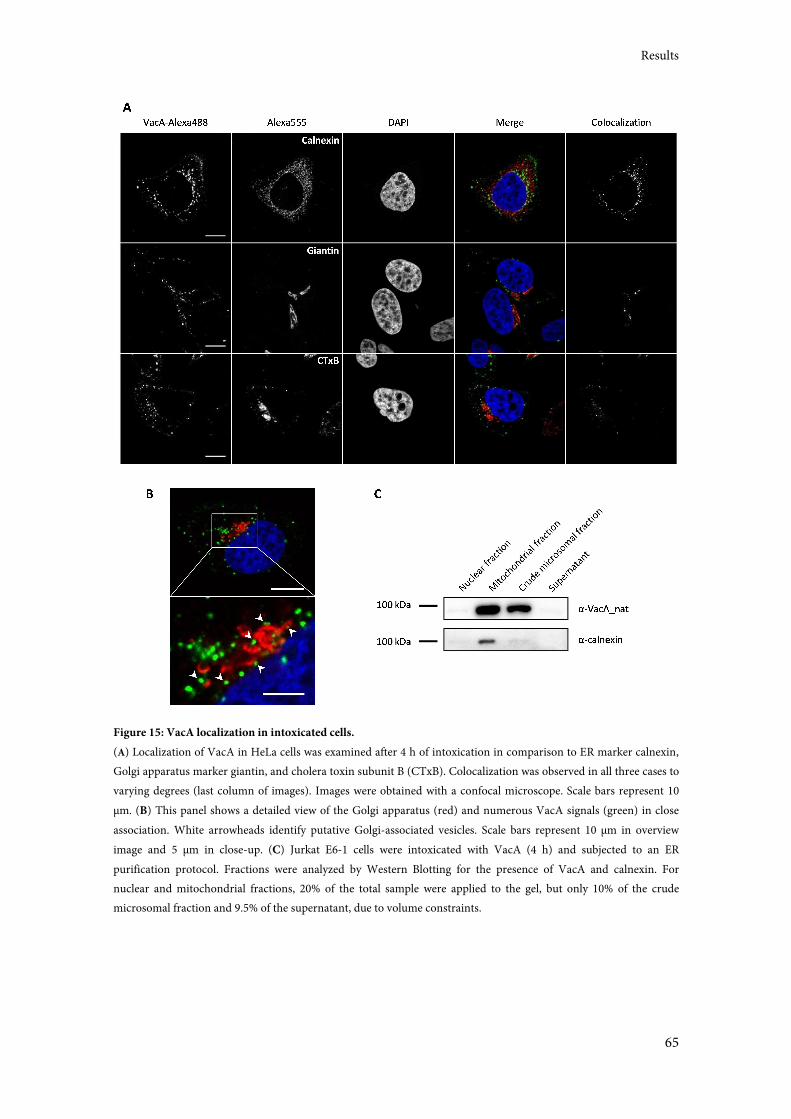
Results
65
Figure 15: VacA localization in intoxicated cells.
(A) Localization of VacA in HeLa cells was examined after 4 h of intoxication in comparison to ER marker calnexin,
Golgi apparatus marker giantin, and cholera toxin subunit B (CTxB). Colocalization was observed in all three cases to
varying degrees (last column of images). Images were obtained with a confocal microscope. Scale bars represent 10
µm. (B) This panel shows a detailed view of the Golgi apparatus (red) and numerous VacA signals (green) in close
association. White arrowheads identify putative Golgi-associated vesicles. Scale bars represent 10 µm in overview
image and 5 µm in close-up. (C) Jurkat E6-1 cells were intoxicated with VacA (4 h) and subjected to an ER
purification protocol. Fractions were analyzed by Western Blotting for the presence of VacA and calnexin. For
nuclear and mitochondrial fractions, 20% of the total sample were applied to the gel, but only 10% of the crude
microsomal fraction and 9.5% of the supernatant, due to volume constraints.
Results
66
Discussion
67
4 Discussion
The namesake cellular effect of intoxication with Helicobacter pylori vacuolating cytotoxin VacA
is the formation of large cytoplasmic vacuoles [38,39]. Until now, the function of these vacuoles
is not known. They arise from endocytic vesicles containing VacA channels in their limiting
membrane, which allow the influx of chloride ions, causing acidification and subsequent
osmotic swelling of the vesicles [27]. VacA also exerts a variety of other effects on intoxicated
cells, including the induction of apoptosis, the inhibition of T-cell activation and proliferation,
and interference with intracellular calcium signaling (see sections 1.5.1, 1.5.2, and 1.5.4 for
details). All of these depend on VacA channel forming activity [92]. Whether vacuolation is only
a by-product of VacA channel formation, or whether it is its own effect, remains to be
elucidated. In this study, VacA-containing vacuoles (VCVs) were isolated and their proteome
was analyzed by mass spectrometry. This revealed information about the vacuoles' specific
protein content and may help understand their purpose. Another part of this study further
investigated VacA involvement in calcium signaling. Moreover, additional possible cellular
target structures of VacA, besides vacuoles and mitochondria, were investigated.
4.1 VCV Isolation Strategies and Tools
The bulk of research investigating pathogen-induced vacuoles naturally focuses on vacuoles
containing the pathogen. In those cases, purification methods often rely on density gradient
centrifugation to separate pathogen-containing vacuoles (PCVs) from other cellular organelles.
The difficulty in this technique is to overcome the similarity of PCVs and other organelles
regarding their density, often requiring multiple centrifugation steps. This can be addressed by
adding an immunological step to specifically target vacuoles, as done by the group of Hubert
Hilbi for the isolation of Legionella-containing vacuoles [151]. Another challenge is the fact that
PCVs interact with other organelles, leading to PCV preparations with considerable impurities
[163]. The purity of isolated fractions is commonly monitored by electron microscopy, a
method that was not available for this study but which may help evaluate VCV preparations in
future experiments.
Other researchers have published PCV proteomes containing anywhere between 140 to 2400
proteins. The large differences in numbers can be attributed to the varying species of pathogens
Discussion
68
and of host cells used for the isolation experiments, and importantly, the sensitivity of the MS
technology. A comparison of protein numbers between different systems is therefore not
advisable (all this is reviewed in [164]).
In this study, three different strategies were devised for VCV purification. Strategy A consisted
of nanoscale magnetic beads called TurboBeads coupled to VacA monomers (see 3.4.2.3). After
uptake of bead-bound VacA and vacuolation, the cells were to be homogenized and the vacuoles
isolated with the help of a magnet. The main reason why this strategy was abandoned was the
tendency of the beads to aggregate, preventing natural uptake (see Figure 8D). This problem was
approached by using several different coating substances, but could not be solved. Other
researchers have noted it to be an effect of the high specific surface energy of nanoparticles,
making their use in biological settings difficult [165]. Recently, the TurboBeads manufacturer
released new, polyethylene glycol (PEG) coated beads, specifically recommended for biomedical
applications. These may be helpful in future experiments.
Strategy B was to sort VCVs from cell homogenate via flow cytometry (see 3.4.4). A cell line
stably transfected to produce the fluorescent VCV marker EGFP-Rab7 [43] (see below) and
intoxication with Alexa-labeled VacA provided the necessary characteristics for sorting, since
only VCVs were expected to carry both EGFP and Alexa fluorophores. However, VCVs are
apparently not stable enough to withstand the physical strain of flow cytometry (see Figure 10),
even though the sorting conditions were adjusted to make the process as gentle as possible. This
strategy was expected to yield VCV preparations with the highest purity and is still worth
pursuing for similar applications.
VCVs were eventually successfully purified immunomagnetically, adapting strategy C from a
protocol published by the group of Hubert Hilbi for the isolation of Legionella-containing
vacuoles (LCVs) [151]. To this end, two indispensable tools were established. The first is a
polyclonal antiserum which was shown to recognize VacA in its native conformation by native
immunoblotting, functional inhibition of VacA-induced vacuolation, and immunostaining in
this study (see section 3.2 and Figure 4). It was also used in pull-down experiments investigating
possible protein-protein interactions of VacA (see 3.6). The antiserum was conclusively shown
to be suitable for VCV purification. The second tool is the Jurkat T-cell line Jurkat E6-1 EGFP-
Rab7, which stably expresses an EGFP-Rab7 fusion protein (see 3.3). Rab7 is a late endosomal
protein that lines VCV membranes [43] and it served as a VCV marker to monitor the vacuole
purification process (see Figure 5 and Figure 11). This way, it was possible to follow the
distribution of VCVs through the successive steps of purification. In conclusion, these two tools
laid the foundation for immunomagnetic VCV isolation.
Discussion
69
The immunomagnetic method for VCV purification has one considerable flaw in that VacA is
not exclusively situated on the VCV membrane, but is also for example still bound to the
cytoplasmic membrane (see Figure 9A). This is in contrast to the original Legionella protocol,
which uses a bacterial effector protein exclusively present on the LCV membrane as target. An
appropriate control consisting of cells treated with mutant VacA was used to address this factor.
This mutant VacA∆6-27 lacks 22 aa in its N-terminus which are necessary for proper channel
formation and therefore vacuolation, although cellular uptake still happens [70]. Treatment of
cells with this mutant toxin and their subjection to the VCV isolation protocol was expected to
yield a purified fraction containing regular endocytic vesicles unaltered by VacA channel
activity, and of course the aforementioned cytoplasmic membrane and other cell parts binding
VacA. It was also expected to contain proteins unspecifically purified by the method, for
instance proteins or cell parts that bind to the magnetic beads. This sample served as a valuable
background control for the mass spectrometric anaysis of VCVs (see 3.5).
4.2 The VCV Proteome
One past publication has reported an attempt to characterize the protein composition of VacA-
containing vesicles by purifying them [45]. This publication, like this study, reasoned that
determining the vacuolar protein content would help to find intracellular targets of VacA and to
understand the details of VacA intoxication. The researchers showed that VacA intoxication
causes a redistribution of markers of the endocytic network, specifically the recruitment to
VCVs of the late endosomal marker Rab7 and the lysosomal marker Lamp2 and the absence of
the late endosomal marker CI-M6PR. This was the first indication of VCVs being endo-
lysosomal hybrid compartments. Due to the methods used, however, the analysis was
constricted to checking for the presence of individual proteins. The present study for the first
time performed a comprehensive investigation of the total VCV proteome by mass
spectrometric (MS) analysis, seeking to find not only proteins that can be expected on an
endocytic compartment, but various proteins involved in VacA intoxication effects.
4.2.1 VCV Analysis by MS - Strengths and Weaknesses
Two biological replicates of purified VCVs resulting from intoxication with wild-type VacA
were measured. The results showed high variations between samples, with the first (WT1)
returning 2078 protein hits and the second (WT2) returning 1754 (see Figure 12). The two
Discussion
70
samples overlapped for 1123 proteins, clearly showing success, but also hinting that the analysis
has to be performed several times, and only proteins found every time should be regarded as
definite hits. The numbers are comparable to the results reported for Legionella-containing
vacuoles (LCVs) purified with the same method, and analyzed on the same mass spectrometry
platform. Around 1000-2000 host and Legionella proteins were identified, occurring once, twice,
or in all three preparations that were examined [166,152]. As a background control for VCV
purification, cells were treated with inactive mutant VacA∆6-27, which still binds to cells and is
internalized, but does not cause vacuolation [70]. Subjecting this sample to the VCV isolation
protocol should purify anything that unspecifically co-purifies with this method and any VacA-
carrying cellular compartments that aren't vacuoles, for instance plasma membrane. The
background sample contained 1606 proteins, indicating that the purity of the isolated VCV
fraction is suboptimal. The researchers working on LCVs did not report the performance of a
similar control experiment, precluding a direct comparison of preparation purity. However,
subtraction of the proteins found in the background sample from the wild-type protein sets
solves this problem. Considering only the proteins found in both wild-type samples (1123) and
subtracting those found also in the background sample (883) leaves 240 proteins in the VCV-
specific subset which was used for further interpretation (see Figure 12).
It has to be noted that this method is not quantitative and therefore does not permit inferences
about the amount of any particular protein present in a sample. Also, there was no possibility to
validate protein hits. The original protocol [151] intends immunofluorescence staining to
confirm the presence of individual proteins on isolated, fixed vacuoles. This setup allows
looking for colocalization of a target protein with the vacuole membrane marker, in the case of
this study, EGFP-Rab7. Unfortunately, due to problems with fixation, this could not be done in
intact VacA-intoxicated cells or isolated VCVs (see 3.4.2.3 and Figure 7D). None of the fixation
reagents and protocols that were tried could preserve VCV membrane shape, eliminating an
important characteristic for the identification of VCVs. Other methods, for example
immunoblotting, do not facilitate the distinction between proteins present on the vacuole
membrane, within the vacuole lumen, or elsewhere in the sample. However, a method of
validation is needed to verify individual hits. One possibility to do this is to use different cells for
immunostaining experiments, for instance adherent cells like HeLa cells, which can be fixed in a
vacuolated state, with VCVs retaining their shape (data not shown). This then of course
precludes the analysis of processes specific to T-cells. Another possibility is the transfection of
T-cells with plasmids coding for target proteins fused to a fluorescent protein. This is more
laborious but would have the added bonus of allowing the microscopic analysis of unfixed cells.
Discussion
71
The presence of a protein in a VCV preparation of course assumes the presence of the respective
protein on VCVs. However, another possibility that has to be considered is the co-purification
of compartments that VacA has already been transported to prior to cell homogenization. Since
trafficking of VacA to mitochondria depends on its first 32 amino acids [103,105], the use of
VacA∆6-27 eliminates the co-purification of mitochondria in the background control sample.
In the wild-type sample, mitochondria may be co-purified, and proteins localized to
mitochondria could then appear in the VCV-specific subset. In the case of possible trafficking of
VacA to other, yet unknown, cellular compartments, co-purification (and detection after
background subtraction) would again depend on whether the VacA N-terminus acts as a
targeting sequence in each particular case. Even though the presence of a protein in a VCV
preparation does not guarantee its presence on VCVs, it nevertheless provides valuable
information on VacA-induced cellular processes.
4.2.2 Individual Proteins Found on VCVs
Table 1 shows a list of proteins whose presence in the three MS samples is of special interest.
This includes: proteins that localize to VCVs; proteins that explicitly do not localize to VCVs;
proteins that are required for VacA effects; proteins that are known not to be required for VacA
effects; and other proteins of significance.
All but two proteins that are known to localize to VCVs were found in all MS samples: the small
GTPase (and purification tool) Rab7 [43], the lysosomal markers Lamp1 and 2 (Lamp2 is also
known as Lgp110) [44,45], the vacuolar ATPase (vATPase) [52], and the membrane fusion
regulators Syntaxin-7 and Vamp7 [48,27,153]. The two exceptions are Rac1, a signal transductor
[47], which was found in only one wild-type and the background sample, and β-
glucosaminidase, an LE/lysosomal enzyme [45], which was not found at all. Rab7, vATPase,
Vamp7, and Rac1 are also necessary for vacuolation [49,51,50,153,47]. The fact that virtually all
proteins which are definitely known to be contained in VCVs were found in the MS
measurements validates the isolation technique. The most notable exception, β-glucosaminidase,
was only reported to reside in VCVs in a single study that used baby hamster kidney cells, which
could explain the disagreement. Also, most proteins known not to localize to VCVs were not
detected (these proteins are marked with the comment "not on VCVs" in Table 1), showing that
VCVs were successfully enriched with this method.
The MS results were also scanned for individual proteins involved in VacA cell binding and
internalization. Several protein receptors for VacA have previously been identified on different
Discussion
72
cell types, including EGFR, RPTPα/β, CD18, CD29, and Multimerin-1 [85,62,86,88,87,89].
None of these were detected in the wild-type VCV samples. In T-cells, specifically, CD18 is
known to mediate internalization as part of the lymphocyte function-associated antigen 1 (LFA-
1) [88]. Its absence can be explained by the following findings: LFA-1 is recycled back to the
plasma membrane in Rab11-positive structures, whereas VCVs never carry Rab11 [160]. This
suggests that after joint uptake, LFA-1 (and thus CD18) and VacA part ways. It is unknown
when exactly this happens, but since cells were treated with VacA over night before VCV
isolation, it is quite possible that differential sorting of LFA-1 and VacA has already occurred at
the point of cell homogenization.
VacA uptake is known to be independent of endocytic regulators clathrin, dynamin, and
caveolin [94,96]. While dynamin and caveolin were not detected in any MS samples, clathrin
was found in all three, indicating a possible unspecific co-purification. Interestingly, flotillin-2, a
protein that forms microdomains in the plasma membrane and associates with lipid rafts, was
detected in the VCV-specific subset. It is not clear whether flotillins actively participate in a
distinct type of endocytosis, or if they are cargo, but they are required for the uptake and
intracellular trafficking of cholera toxin [167–169]. An implication of flotillin-2 in VacA
intoxication is especially intruiging, since VacA and cholera toxin uptake are thought to differ
[96,160]. On the other hand, if VacA∆6-27 is indeed internalized just like wild-type VacA as the
literature suggests [70], the absence of flotillin-2 in the background control sample raises the
question where and when exactly VacA and VacA∆6-27 transport start to differ.
The cytoskeletal component actin is necessary for VacA uptake and known to polymerize at
early endosomes containing VacA, moving them through the cytoplasm [95,102]. Even though
no association of actin with late endosomes (including VCVs) could previously be shown via
microscopy [102], this does not eliminate the possibility. Cytoplasmic actin was detected in the
VCV-specific subset, strongly suggesting that VCVs still associate with actin to move through
the cell. CD2AP, which was shown to bridge actin structures and endocytic vesicles [102], was
not found in any of the MS samples. Its function could however simply be fulfilled by a different
docking protein.
Other proteins involved in endocytic processes that have been investigated with regard to VacA
uptake include: tubulin; the small GTPases Cdc42, Arf6, and RhoA; the RhoA-regulated kinases
ROCK and MLCK; several members of the protein kinase C family (PKC); the protease calpain;
and phosphoinositide 3-kinase (PI3K). Of these, calpain, PKC, and Cdc42 are required for
VacA uptake [160,96]. All three were detected in at least one wild-type sample; Cdc42 is even in
the VCV-specific subset. Again, as in the case of flotillin-2, the exact differences of VacA and
Discussion
73
VacA∆6-27 uptake and transport need to be clarified. The others were found to have no
influence on VacA internalization [160,96]. Some of the aforementioned proteins were not
detected by MS at all (ROCK, MLCK), while another was found in all samples (RhoA), all in
agreement with the fact that these proteins don't play a role in VCV formation. Of interest are
PI3K and Arf6, both of which occur in one wild-type sample, but not in the background. This
suggests a possible VCV specificity that needs to be verified in future experiments. Although
PI3K is not required for VacA uptake, its inhibition resulted in a slight reduction in VacA
internalization [160]. Both PI3K and Arf6 play a role in intracellular trafficking [170,171], which
makes them appealing possible targets for VacA.
Regarding VacA-induced apoptosis, the mass spectrometry results reveal two out of three
known mitochondrial effector proteins in the VCV-specific sample. The dynamin-related
protein Drp1 and the multi-domain pro-apoptotic factors Bak and Bax are activated by VacA
intoxication, leading to mitochondrial fission and cytochrome c release via two separate
mechanisms, and eventually, to apoptosis [110,106]. Bax is recruited to mitochondria via early
endosomes containing VacA, but was not found in late endosomes [106], consistent with its
absence in MS samples. VacA also causes mitochondrial recruitment of Drp1, and the presence
of Drp1 in the VCV-specific protein subset suggests that this may also happen via VacA-
containing vesicles. Bak, however, is naturally already located in mitochondria, and its
endosomal localization was not observed by Calore and coworkers [106]. It is conceivable that
their methods were not as sensitive as mass spectrometry, or that the investigated time points
differed, as Bak was detected in the VCV-specific subset, clearly showing its presence on VCVs
and making it an intriguing candidate for further inspection. Another regulator of
mitochondrial fission, MTFR1 [172], was found in the VCV-specific subset, further
substantiating the connection between VacA intoxication, mitochondrial fission, and the
mitochondrial apoptosis pathway.
As previous reports and research conducted during the course of this study demonstrated an
influence of VacA on intracellular calcium signaling [139,120,140,119,155], the MS results lists
were also scanned for proteins involved in this process. Calcineurin, a phosphatase important in
T-cell activation and reported to be inhibited during VacA intoxication [119,120], was not
detected in any of the samples. This is unsurprising because it is a cytoplasmic protein, and a
direct interaction with VacA was never shown. Two very interesting proteins found in the VCV-
specific subset are STIM1 and ITPR3. Both usually locate to the ER and are essential in T-cell
calcium signaling (reviewed in [141,156]). Their significance will be discussed in detail in
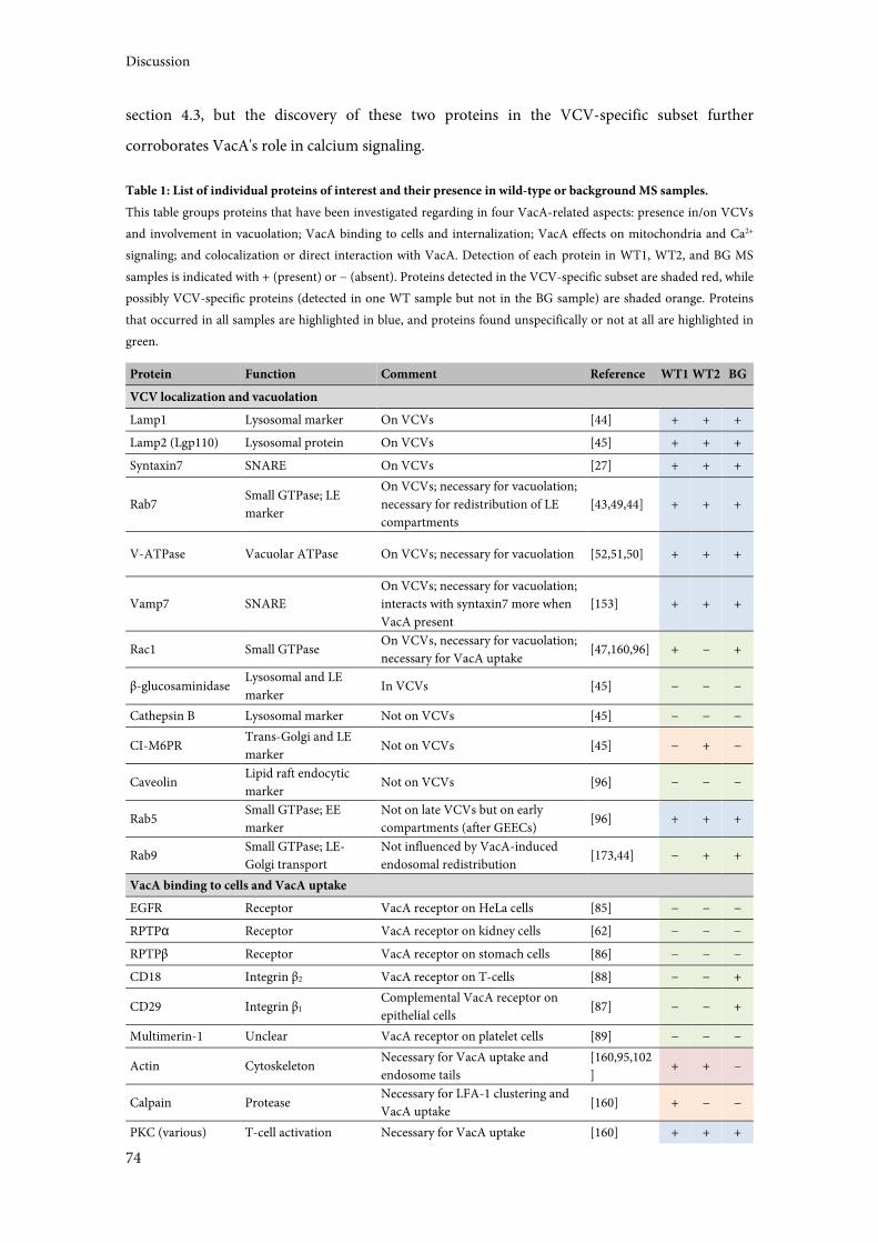
Discussion
74
section 4.3, but the discovery of these two proteins in the VCV-specific subset further
corroborates VacA's role in calcium signaling.
Table 1: List of individual proteins of interest and their presence in wild-type or background MS samples.
This table groups proteins that have been investigated regarding in four VacA-related aspects: presence in/on VCVs
and involvement in vacuolation; VacA binding to cells and internalization; VacA effects on mitochondria and Ca2+
signaling; and colocalization or direct interaction with VacA. Detection of each protein in WT1, WT2, and BG MS
samples is indicated with + (present) or − (absent). Proteins detected in the VCV-specific subset are shaded red, while
possibly VCV-specific proteins (detected in one WT sample but not in the BG sample) are shaded orange. Proteins
that occurred in all samples are highlighted in blue, and proteins found unspecifically or not at all are highlighted in
green.
Protein Function Comment Reference WT1 WT2 BG
VCV localization and vacuolation
Lamp1 Lysosomal marker On VCVs [44] + + +
Lamp2 (Lgp110) Lysosomal protein On VCVs [45] + + +
Syntaxin7 SNARE On VCVs [27] + + +
Rab7 Small GTPase; LE
marker
On VCVs; necessary for vacuolation;
necessary for redistribution of LE
compartments
[43,49,44] + + +
V-ATPase Vacuolar ATPase On VCVs; necessary for vacuolation [52,51,50] + + +
Vamp7 SNARE
On VCVs; necessary for vacuolation;
interacts with syntaxin7 more when
VacA present
[153] + + +
Rac1 Small GTPase On VCVs, necessary for vacuolation;
necessary for VacA uptake [47,160,96] + − +
β-glucosaminidase Lysosomal and LE
marker In VCVs [45] − − −
Cathepsin B Lysosomal marker Not on VCVs [45] − − −
CI-M6PR Trans-Golgi and LE
marker Not on VCVs [45] − + −
Caveolin Lipid raft endocytic
marker Not on VCVs [96] − − −
Rab5 Small GTPase; EE
marker
Not on late VCVs but on early
compartments (after GEECs) [96] + + +
Rab9 Small GTPase; LE-
Golgi transport
Not influenced by VacA-induced
endosomal redistribution [173,44] − + +
VacA binding to cells and VacA uptake
EGFR Receptor VacA receptor on HeLa cells [85] − − −
RPTPα Receptor VacA receptor on kidney cells [62] − − −
RPTPβ Receptor VacA receptor on stomach cells [86] − − −
CD18 Integrin β2 VacA receptor on T-cells [88] − − +
CD29 Integrin β1 Complemental VacA receptor on
epithelial cells [87] − − +
Multimerin-1 Unclear VacA receptor on platelet cells [89] − − −
Actin Cytoskeleton Necessary for VacA uptake and
endosome tails
[160,95,102
] + + −
Calpain Protease Necessary for LFA-1 clustering and
VacA uptake [160] + − −
PKC (various) T-cell activation Necessary for VacA uptake [160] + + +
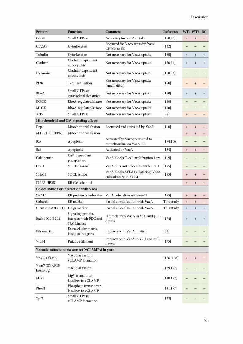
Discussion
75
Protein Function Comment Reference WT1 WT2 BG
Cdc42 Small GTPase Necessary for VacA uptake [160,96] + + −
CD2AP Cytoskeleton Required for VacA transfer from
GEECs to EE [102] − − −
Tubulin Cytoskeleton Not necessary for VacA uptake [160] + + +
Clathrin Clathrin-dependent
endocytosis Not necessary for VacA uptake [160,94] + + +
Dynamin Clathrin-dependent
endocytosis Not necessary for VacA uptake [160,94] − − −
PI3K T-cell activation Not necessary for VacA uptake
(small effect) [160] − + −
RhoA Small GTPase;
cytoskeletal dynamics Not necessary for VacA uptake [160] + + +
ROCK RhoA-regulated kinase Not necessary for VacA uptake [160] − − −
MLCK RhoA-regulated kinase Not necessary for VacA uptake [160] − − −
Arf6 Small GTPase Not necessary for VacA uptake [96] + − −
Mitochondrial and Ca2+ signaling effects
Drp1 Mitochondrial fission Recruited and activated by VacA [110] + + −
MTFR1 (CHPPR) Mitochondrial fission + + −
Bax Apoptosis Activated by VacA; recruited to
mitochondria via VacA-EE [154,106] − − −
Bak Apoptosis Activated by VacA [154] + + −
Calcineurin Ca2+-dependent
phosphatase VacA blocks T-cell proliferation here [119] − − −
Orai1 SOCE channel VacA does not colocalize with Orai1 [155] − − −
STIM1 SOCE sensor VacA blocks STIM1 clustering; VacA
colocalizes with STIM1 [155] + + −
ITPR3 (IP3R) ER Ca2+ channel
+ + −
Colocalization or interaction with VacA
Sec61α ER protein translocator VacA colocalizes with Sec61 [155] + + −
Calnexin ER marker Partial colocalization with VacA This study + + −
Giantin (GOLGB1) Golgi marker Partial colocalization with VacA This study + + +
Rack1 (GNB2L1)
Signaling protein,
interacts with PKC and
SRC kinases
Interacts with VacA in Y2H and pull-
downs [174] + + +
Fibronectin Extracellular matrix,
binds to integrins interacts with VacA in vitro [90] − − +
Vip54 Putative filament interacts with VacA in Y2H and pull-
downs [175] − − −
Vacuole-mitochondria contact (vCLAMPs) in yeast
Vps39 (Vam6) Vacuolar fusion;
vCLAMP formation [176–178] + + −
Vam7 (SNAP25
homolog) Vacuolar fusion [179,177] − − −
Mnr2 Mg2+ transporter;
localizes to vCLAMP [180,177] − − −
Pho91 Phosphate transporter;
localizes to vCLAMP [181,177] − − −
Ypt7 Small GTPase;
vCLAMP formation [178] − − −
Discussion
76
Three proteins were reported to interact with VacA directly in biochemical assays in vitro: the
receptor for activated C kinase 1 Rack1 [174]; the extracellular matrix component fibronectin
[90]; and a putative filament protein termed Vip54 [175].
Rack1, also known as GNB2L1, interacts with multiple cellular targets, including PKC and Src
kinases and the integrin subunits CD18 and CD29 [182–184]. Its additional capacity to bind
VacA is thus especially intriguing. It was detected in all three MS samples, possibly implying
unspecific co-purification, but in the light of its proven interaction with VacA, this seems
unlikely. GNB2L1 modifies both PKC and Src activity, activating the first and inhibiting the
latter [182,183]. Albeit indirectly, this connects VacA with CagA phosphorylation by these two
kinases. GNB2L1 interaction with CD18 and CD29, on the other hand, is interesting because
both are known receptors for VacA. Upon VacA binding to the extracellular integrin domains,
GNB2L1 may trigger intracellular signaling cascades that cause VacA effects. It is however
unclear at which point GNB2L1 and VacA interact with each other, since they should be located
at opposing sides of the cellular or endocytic membrane. Cytoplasmic localization of VacA via
outer membrane vesicle delivery [185,186] or after endosome escape may be a way for the two
proteins to come into direct contact. The same is true for the phosphatase calcineurin
mentioned above.
Fibronectin was unsurprisingly not found in the wild-type VCV preparations. It is part of the
extracellular matrix and therefore not expected to occur inside cells in significant amounts. Even
though it directly binds VacA [90], this interaction would likely take place outside of cells, not in
the context of VCVs.
Vip54 is a novel protein assumed to be an intermediate filament component on the basis of its
structural characteristics and its interaction and co-distribution with known filament proteins
[175]. The investigators suggest that it may mediate interactions between cytoskeletal filaments
and VacA-containing endocytic compartments, but could not show the presence of Vip54 on
VCVs. In accordance with this, it was also not detected in any of the MS samples. The
hypothesis of VCV-filament interaction is compelling, but cannot be supported by the present
evidence. Nevertheless, the interaction may occur at different points in time, or it may get lost
during the physically rigorous process of VCV isolation.
The two individual proteins that remain to be discussed are the Golgi apparatus marker giantin
and the ER marker calnexin. Both showed partial colocalization with VacA in intoxicated HeLa
cells in immunofluorescence experiments (see 3.7.1). Colocalization may stem from three
occurrences: trafficking of VacA to the respective compartment; recruitment of the marker
proteins to VCVs; or very close physical contact of Golgi or ER with VCVs beyond microscopic
Discussion
77
resolution. VacA trafficking to the Golgi apparatus or the ER will be discussed in a later section,
but the latter two may also explain the presence of the marker proteins in VCV preparations.
Both calnexin and giantin were detected in the wild-type MS samples. Calnexin is even
contained in the VCV-specific protein subset, indicating either its presence in VCVs or VacA-
induced co-purification of calnexin-carrying compartments alongside VCVs. Both of these
incidents would suggest a connection between VacA/VCVs and the ER that has not been shown
so far, and which will also be discussed later in the context of VacA trafficking. Giantin, on the
other hand, was also found in the background sample. This means that, although unspecific co-
purification cannot be ruled out, giantin may be present on VCVs. Alternatively, VacA∆6-27,
although vacuolation-inactive, may still be transported to the Golgi and lead to the isolation of
Golgi-derived compartments in the background control. Given the close proximity of VacA-
containing vesicles to the Golgi apparatus observed via immunofluorescence (Figure 15B), a co-
purification of Golgi-derived compartments also in wild-type samples is conceivable. Whether
this happens in a VacA-dependent manner has to be investigated in future experiments.
Surprisingly, the share of Golgi proteins was reduced in the VCV-specific sample compared to
the background (see Figure 13). However, immunostaining experiments and VCV isolation
were conducted after different periods of intoxication (4 h vs 16 h), leaving ample time for
transport processes to occur, which could explain the seemingly low amount of Golgi proteins in
VCVs.
4.2.3 The VCV Proteome Decoded by Subcellular Localization and Biological Function
When looking at the general composition of the VCV-specific protein subset in comparison
with the background sample (see Figure 13), the largest differences can be seen for nuclear and
mitochondrial proteins. Nuclear proteins are enriched in the VCV-specific subset (23.1%
compared to 14.9% in the background sample). A recruitment of nuclear proteins to VCVs is
imaginable, but unlikely to happen in such large numbers. This is more likely to be a side-effect
of the method. The disruption of nuclei during homogenization leads to the presence of sticky
DNA in the sample, as was observed in the isolation process (data not shown). This DNA may
then co-purify with VCVs but not with the smaller particles in the background sample, bringing
along DNA-bound nuclear proteins. Using DNase during homogenization may help minimize
this problem in future experiments. The reduction of mitochondrial proteins (14.3% compared
to 21.4%) may be explained by a similar unspecific effect. Originally, an enrichment of
mitochondrial proteins in the VCV-specific sample was anticipated, since VacA localizes to
Discussion
78
mitochondria, and VCVs have been speculated to transport it there directly and/or interact with
mitochondria [103–106].
Considering that VacA is known to alter vesicular trafficking and to change the protein content
of endocytic compartments [142,44,118,45], the increase of endo- and lysosomal and signaling
proteins in VCVs is as expected. The same is true for the enrichment in proteins involved in
immune processes and cell death, since VacA greatly modulates the host immune response and
induces apoptosis [107,108].
4.2.4 Interorganellar Crosstalk between VCVs and Mitochondria/ER
Recent research, mostly conducted in yeast, has illuminated the importance of organelle-
organelle contact sites for the transport of substances. Mitochondria are not connected to
cellular metabolism via vesicular transport, but instead rely on direct contact with the ER for
phospolipid and Ca2+ transport through so-called ER-mitochondria encounter structures
(ERMES) (reviewed in [187]). Earlier this year, an additional contact site termed vCLAMP
(vacuole and mitochondria patch) was identified, connecting mitochondria with vacuoles, the
yeast lysosomal compartment [178,177]. vCLAMPs and ERMES were found to be reciprocally
regulated, implying that each can restore loss of the other, and only loss of both caused lethal
deficiencies in phospholipid transport. Establishment of vCLAMPs depends on Vps39, a
component of the yeast vacuolar fusion process, and Ypt7, a small GTPase. The authors of these
studies speculate that such contact sites may be conserved in higher eukaryotes, and Vps39 was
indeed detected in the VCV-specific protein subset. While this may imply the formation of a
VCV-mitochondria contact site, Ypt7 was absent in all MS samples. Also not detected were
three other proteins reported to be present in vCLAMPs: Mnr2, a Mg2+ transporter, Pho91, a
phosphate transporter, and Vam7, another vacuolar fusion contributor. VacA-containing
vesicles and mitochondria have been shown to juxtapose [106], but whether this close proximity
is used to form transport channels analogous to those found in yeast remains elusive. Further
studies are needed to investigate putative contact sites, which can be expected to have a protein
composition at least slightly different from vCLAMPs.
Similar contact sites have also been observed in cells infected with chlamydial organisms and
Legionella [188–190]. These pathogens establish a replicative vacuole that associates closely with
the ER, for example to obtain lipids necessary for bacterial growth. In the case of Legionella,
fusion of ER vesicles with the bacterial vacuole has been reported [190]. Therefore, such contact
Discussion
79
sites may not only serve in lipid or ion transport, but may be involved in the translocation of
VacA from VCVs to mitochondria or the ER.
4.2.5 VCVs as Multifunctional Platforms
In general, the information obtained from VCV purification agrees with the literature. The sheer
number of non-endocytic proteins detected specifically in VCVs strongly supports the
hypothesis of VCVs having a purpose. Their identity as signaling hubs needs to be confirmed
with experiments validating the presence of individual proteins on or in VCVs. An investigation
of 'obvious' signaling regulators like members of the Rab superfamily [191] on VCVs proved
difficult. Even though many Rab proteins were found, most are present on various
compartments of the endo-membrane system and were therefore not detected specifically on
VCVs. Besides signaling, some researchers are now suggesting an even bigger function for
endosomes as assembly sites for molecular machinery. This has been described for instance for
cytokinesis, where the abscission machinery that physically separates dividing cells is
accumulated at the site of action by endocytic and secretory vesicles [143]. Observations like this
have also been made for cell migration and polarity. This has lead to the proposal of endosomes
as specialized multi-purpose platforms, able to coordinate in space and time all the molecular
actors necessary to execute such complex cellular processes [143]. If we turn this around, the
proteins carried by an endocytic vesicle may offer information on the vesicle's purpose. Taken
further, the protein cargo of an endocytic vesicle or a subpopulation of vesicles could therefore
allow conclusions on what is happening in a cell at a given time point. In this light, the VCV
proteome should be investigated from all points of view, since it could potentially give insight
into all VacA-induced cellular processes. A comparison of the VCV proteome after different
periods of intoxication and after intoxication with different VacA variants could also provide
valuable information on this question. Proteome data confirmed by several measurements and
an improved bioinformatic evaluation approach are necessary to assist in this endeavour.
4.3 Influence of VacA on Store-Operated Calcium Entry (SOCE)
VacA has been reported to influence intracellular calcium signaling, severely disrupting T-cell
activation and proliferation [139,120,140,119]. A possible intermediate in these processes is the
Ca2+-calmodulin-dependent phosphatase calcineurin [119,120], which needs sustained high
intracellular calcium levels in order to function properly [141]. During the course of the present
Discussion
80
study, the VacA-induced inhibition of ionophore-stimulated Ca2+ influx shown by Boncristiano
and coworkers [120] was confirmed [155]. Additionally, a connection of this phenomenon with
store-operated calcium entry (SOCE) was revealed. SOCE is defined as the influx of calcium
into the cytosol from the extracellular milieu caused by the depletion of intracellular calcium
stores, for instance the endoplasmic reticulum (ER). A main actor in this process, especially in
T-cells, is the stromal interaction molecule STIM1, which localizes to the ER membrane and
senses the calcium concentration in the ER lumen (see Figure 14A). Upon calcium depletion,
STIM1 clusters in so-called puncta and relocates to ER-plasma membrane junctions. There, it
interacts with and activates the plasma membrane-situated SOCE channel Orai1, which then
opens to let Ca2+ into the cytosol (reviewed in [156]). The expression of both STIM1 and Orai1
is upregulated during T-cell activation [192], confirming their substantial role in this process.
VacA was shown to colocalize with STIM1 and to interfere with STIM1 clustering in
immunofluorescence experiments, clearly implicating VacA in SOCE inhibition [155]. This also
for the first time demonstrated a possible localization of VacA to the ER. In contrast to the
studies mentioned before, mutant VacA deficient in channel forming activity (VacA∆6-27) also
showed an effect on cytoplasmic calcium concentrations, albeit less pronounced than the wild-
type. Since SOCE-induced high intracellular calcium levels are required for calcineurin activity,
an interference of VacA with SOCE offers a new possible explanation for calcineurin inhibition.
At this point, it is not clear whether VacA effects on calcium signaling are really due to VacA
channels as has been suggested [120,121], and if so, whether these channels are located in the
plasma membrane or in the membrane of a cellular compartment like VacA-induced vacuoles
or even the ER. Alternatively, SOCE effects may be the result of an interaction of monomeric
VacA with STIM1. Irrespective of whether VacA needs to be present as a functional channel or
not, the finding that the toxin is apparently transported to the ER needs to be addressed and will
be discussed in the context of this present study's results in section 4.4.
The study that discovered VacA's involvement in SOCE and VacA-STIM1 colocalization also
investigated a possible direct protein-protein interaction between VacA and STIM1 [155]. An
interaction observed in a yeast two-hybrid (Y2H) screen was later refuted, leading to re-
assessment via pull-down experiments in the present study (see 3.6). For these experiments,
STIM1 was recombinantly expressed in E. coli in two separate parts, the cytoplasmic half and
the ER luminal half. The cytoplasmic half included the CAD/SOAR domain that interacts with
and activates the SOCE channel Orai1, and the ER luminal half contained the Ca2+-sensing EF
hand domain (see Figure 14A and B). The central transmembrane domain of STIM1 was
omitted. Preceding experiments attempted but failed to precipitate STIM1 from lysate of VacA-
Discussion
81
intoxicated kidney epithelium (HEK-293) cells, presumably because of the transmembrane
domain being difficult to solubilize (U. Jain, personal communication). The two recombinantly
expressed STIM1 parts were separately tested for VacA binding, but an interaction could neither
be shown nor excluded. An interaction seems very likely considering VacA's colocalization with
ER-situated STIM1 and its inhibition of STIM1 clustering, so several options remain to be
investigated. Firstly, the interaction may be too weak to be detected with the techniques used so
far. Secondly, the experimental environment, particularly the recombinant expression of STIM1
in E. coli, may be too different from physiological conditions to facilitate binding. This includes
the possiblity that the interaction may not be direct, but mediated by additional binding
partners in a larger protein complex that are not present in this artificial setting. Thirdly, the
interacting section of STIM1 may actually be the transmembrane domain itself. Since VacA is
capable of inserting into membranes to form channels, this seems plausible. Many of these
problems could be solved by pull-down experiments with VacA-intoxicated cells, but as
mentioned before, this had been unsuccessful. A recent publication detailed a possible protocol
for this purpose [193]. In this report, STIM1 was precipitated from both HEK-293 and Jurkat
cells, revealing an interaction with thrombospondins, a family of matricellular proteins. The
discovered interaction was reportedly robust, and it is not certain whether VacA and STIM1
bind as strongly. There are, of course, other methods to examine the interactions of membrane
proteins, but many of them are not suitable for testing a protein like VacA. Expression of VacA
in mammalian cells is difficult due to its cytotoxicity, and can also not guarantee its natural
localization. This rules out experiments like mammalian membrane two-hybrid [194]. On the
other hand, tagging VacA with larger fusion proteins, as would be required for techniques like
fluorescence resonance energy transfer (FRET), is difficult in H. pylori, and might also
compromise VacA activity. In these rather complex circumstances where the two proteins of
interest come from two different kingdoms of life, and one of them is a cytotoxin, an
experimental set-up like the HEK-293/Jurkat cell pull-down described by Duquette and
coworkers [193] seems the most applicable.
Another interesting aspect in this is the link between mitochondria and cellular calcium
homeostasis. Mitochondria can act as calcium buffers, taking up calcium from the cytosol if
levels are high. This is the case especially at the immunological synapse and close to the ER. Ca2+
release from the ER, for example during T-cell activation, leads to high local concentrations
around release sites, deactivating release channels like ITPR3 as an act of self-regulation. By
taking up some of this calcium, mitochondria can prolong calcium release from the ER, and
reduce the amount of cytosolic calcium that is available for transport back into the ER. Together,
Discussion
82
these effects result in a stronger activation of SOCE and a greater overall mobilization of calcium
(all this is reviewed in [141]). Derogating mitochondrial buffering capacities is therefore an
additional way to disrupt cellular calcium signaling and T-cell activation. This can be brought
about by a reduction of the mitochondrial transmembrane potential, since this is the driving
force behind mitochondrial Ca2+ uptake. VacA has been shown to do exactly that (Kimura 1999,
Willhite 2004). Mitochondrial depolarization also diminishes STIM1 relocation to puncta at ER-
plasma membrane junctions (Singaravelu 2011), which is necessary for Orai1 activation and
therefore SOCE. For these reasons, it is conceivable that VacA influence on SOCE happens via
binding to STIM1 or via the depolarization of mitochondria, or both. This further highlights the
complexity and intricacy of the connections between mitochondrial function, calcium signaling,
and VacA intoxication.
4.4 VacA Partially Localizes to the ER and the Golgi Apparatus
Immunofluorescence staining experiments were conducted in HeLa cells to analyze intracellular
trafficking of VacA. Even though a substantial amount of research has focused on VacA
trafficking, only a few basic points are clear (see Figure 2 and Figure 17). Shortly after
endocytosis, VacA can be found in GPI-AP-enriched early endosomal compartments (GEECs),
from where it is transported to Rab5-positive early endosomes and later Rab7-positive late
endosomes (Gauthier 2005). After about four hours, VacA-containing compartments are
distributed throughout the cytoplasm and carry endo- and lysosomal markers (Molinari 1997, Li
2004). These compartments then enlarge to become vacuoles. Whether VacA goes anywhere
from here is unknown. Earlier VacA-containing vesicles acquire actin tails that confer mobility
(Gauthier 2007) and come into close physical contact with mitochondria (Calore 2010), but it
was not conclusively shown that this is VacA's mode of transportation. VacA does reach the
inner mitochondrial membrane thanks to its hydrophobic N-terminus, which acts like a
targeting sequence [103,105]. Prior to the present study, no other cellular target structures of
VacA had been reported. Recently, VacA localization to the ER was shown via colocalization
with STIM1 and the ER-membrane protein transporter Sec61 in immunofluorescence
experiments (Jain 2013). This was confirmed in the present study by demonstrating a partial
colocalization of VacA with the ER marker calnexin after four hours of intoxication (see 3.7.1
and Figure 15A). Both VacA and calnexin can be seen in vesicles scattered throughout the
cytosol, with VacA being more prominent near the nucleus (Figure 15A, first row). Similarly, a
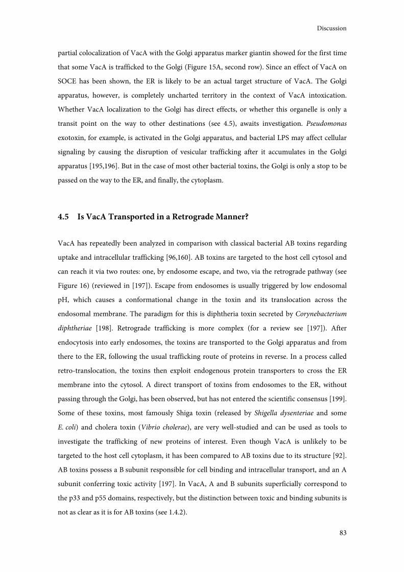
Discussion
83
partial colocalization of VacA with the Golgi apparatus marker giantin showed for the first time
that some VacA is trafficked to the Golgi (Figure 15A, second row). Since an effect of VacA on
SOCE has been shown, the ER is likely to be an actual target structure of VacA. The Golgi
apparatus, however, is completely uncharted territory in the context of VacA intoxication.
Whether VacA localization to the Golgi has direct effects, or whether this organelle is only a
transit point on the way to other destinations (see 4.5), awaits investigation. Pseudomonas
exotoxin, for example, is activated in the Golgi apparatus, and bacterial LPS may affect cellular
signaling by causing the disruption of vesicular trafficking after it accumulates in the Golgi
apparatus [195,196]. But in the case of most other bacterial toxins, the Golgi is only a stop to be
passed on the way to the ER, and finally, the cytoplasm.
4.5 Is VacA Transported in a Retrograde Manner?
VacA has repeatedly been analyzed in comparison with classical bacterial AB toxins regarding
uptake and intracellular trafficking [96,160]. AB toxins are targeted to the host cell cytosol and
can reach it via two routes: one, by endosome escape, and two, via the retrograde pathway (see
Figure 16) (reviewed in [197]). Escape from endosomes is usually triggered by low endosomal
pH, which causes a conformational change in the toxin and its translocation across the
endosomal membrane. The paradigm for this is diphtheria toxin secreted by Corynebacterium
diphtheriae [198]. Retrograde trafficking is more complex (for a review see [197]). After
endocytosis into early endosomes, the toxins are transported to the Golgi apparatus and from
there to the ER, following the usual trafficking route of proteins in reverse. In a process called
retro-translocation, the toxins then exploit endogenous protein transporters to cross the ER
membrane into the cytosol. A direct transport of toxins from endosomes to the ER, without
passing through the Golgi, has been observed, but has not entered the scientific consensus [199].
Some of these toxins, most famously Shiga toxin (released by Shigella dysenteriae and some
E. coli) and cholera toxin (Vibrio cholerae), are very well-studied and can be used as tools to
investigate the trafficking of new proteins of interest. Even though VacA is unlikely to be
targeted to the host cell cytoplasm, it has been compared to AB toxins due to its structure [92].
AB toxins possess a B subunit responsible for cell binding and intracellular transport, and an A
subunit conferring toxic activity [197]. In VacA, A and B subunits superficially correspond to
the p33 and p55 domains, respectively, but the distinction between toxic and binding subunits is
not as clear as it is for AB toxins (see 1.4.2).
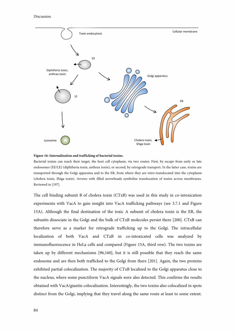
Discussion
84
Figure 16: Internalization and trafficking of bacterial toxins.
Bacterial toxins can reach their target, the host cell cytoplasm, via two routes: First, by escape from early or late
endosomes (EE/LE) (diphtheria toxin, anthrax toxin), or second, by retrograde transport. In the latter case, toxins are
transported through the Golgi apparatus and to the ER, from where they are retro-translocated into the cytoplasm
(cholera toxin, Shiga toxin). Arrows with filled arrowheads symbolize translocation of toxins across membranes.
Reviewed in [197].
The cell binding subunit B of cholera toxin (CTxB) was used in this study in co-intoxication
experiments with VacA to gain insight into VacA trafficking pathways (see 3.7.1 and Figure
15A). Although the final destination of the toxic A subunit of cholera toxin is the ER, the
subunits dissociate in the Golgi and the bulk of CTxB molecules persist there [200]. CTxB can
therefore serve as a marker for retrograde trafficking up to the Golgi. The intracellular
localization of both VacA and CTxB in co-intoxicated cells was analyzed by
immunofluorescence in HeLa cells and compared (Figure 15A, third row). The two toxins are
taken up by different mechanisms [96,160], but it is still possible that they reach the same
endosome and are then both trafficked to the Golgi from there [201]. Again, the two proteins
exhibited partial colocalization. The majority of CTxB localized to the Golgi apparatus close to
the nucleus, where some punctiform VacA signals were also detected. This confirms the results
obtained with VacA/giantin colocalization. Interestingly, the two toxins also colocalized in spots
distinct from the Golgi, implying that they travel along the same route at least to some extent.
Toxin endocytosis
EE
LE
Lysosome
Golgi apparatus
ER
Diphtheria toxin,
anthrax toxin
Cholera toxin,
Shiga toxin
Cellular membrane
Discussion
85
Whether these spots represent toxin on the way to the Golgi, to the ER, or to a third location
needs to be explored.
Strikingly, the ER membrane protein transporter Sec61 was found specifically in the VCV
proteome. It is discussed as the possible transporter system responsible for cholera toxin retro-
translocation from the ER to the cytosol [202] and may therefore also be a candidate for a
potential VacA retro-translocation. Further corroborating this possibility, Sec61 exhibited a
limited colocalization with VacA in another study [155]. An additional intriguing detail are the
VacA-containing compartments closely associated with the Golgi apparatus that were observed
in the giantin colocalization experiments (Figure 15B). This resembles the juxtaposition of
VacA-containing vesicles with mitochondria observed by Calore and coworkers [106]. VacA
translocation from VCVs into mitochondria could happen via membrane fusion or direct
membrane-to-membrane transfer, and the same is true for translocation to the Golgi. But again,
it is not clear whether these compartments contain VacA on its way to or from the Golgi. A
compelling possibility to examine in the future is whether VacA reaches the ER directly,
withouth using the common route through the Golgi apparatus. Direct transport from
endosomes to the ER seems unlikely, but remains possible [199]. Endosomes have been shown
to form contact sites with the ER, especially as they mature [203]. This may facilitate the
translocation of VacA as discussed for mitochondria and Golgi earlier. If this is the case, the
Golgi apparatus and the ER may be two separate target structures of VacA (see Figure 17).
By investigating only one time point, this study focused on identifying novel VacA target
structures. To fully elucidate VacA trafficking, different time points need to be analyzed, for
instance using markers for different known trafficking pathways or time points. Preliminary
experiments were conducted using the Legionella effector protein and inhibitor of retrograde
trafficking RidL [204], but its presence did not seem to influence VacA localization (data not
shown). This result is tentative and may be due to the small amounts of VacA that are
potentially transported in a retrograde manner. Rab9, a small GTPase responsible for the
transport between late endosomes and the Golgi [173], was detected by MS in one VCV
preparation (see Table 1), supplying some evidence for retrograde transport.
Retrograde toxin trafficking is mediated by the transport of the respective toxin receptors. These
receptors are often lipids, like the ganglioside GM1 in the case of cholera toxin [205]. VacA is
known to bind many different receptor structures (see 1.4.3), including glycosphingolipids, a
component of gangliosides [82]. This may account for its transport to the Golgi and the ER.
Similarly, the wide variety of VacA receptors and their specific trafficking routes may explain
VacA's multiple cellular targets. One way to find out whether this is the case is to use different
Discussion
86
VacA allelic variants in experiments similar to the ones conducted for this study. The cell
binding or receptor specificity conferred mostly by the p55 domain/the m-region (see 1.4.2 and
1.4.3) should then lead to a differing cellular distribution of different VacA variants. Moreover,
VacA binding to different receptors may result in their crosslinking and the combinatorial
activation of several internalization or signaling pathways [27].
The observation that VacA does indeed localize to the ER supports the idea that it influences
intracellular calcium signaling from there (see 4.3). Interesting questions that need to be
addressed are whether VacA does this as a monomeric toxin or as a hexameric channel, and
whether it indeed interacts with STIM1 or instead forms pores in the ER membrane. One
possibility to approach these questions is to conduct (cryo-)electron microscopy studies similar
to those that gave great insight into VacA channel structure [61,58,71], but imaging fragments
of intoxicated cells or their organelles instead of artificial membranes. Another possibility is
stimulated emission depletion (STED) fluorescence microscopy, which brings the microscopic
resolution down to 20 nm [206]. While this may not be sufficient to resolve VacA channel
structure, it may still be precise enough to distinguish between VacA monomers and hexamers.
To this end, it may be necessary to label VacA p33 and p55 with different fluorophores. This can
be accomplished by expressing them separately in E. coli and mixing them to restore VacA
activity before cell intoxication [65].
Performing the colocalization immunofluorescence experiments with mutant VacA∆6-27 as a
control could be illuminating in two respects: First, it could give some hints as to whether
channel formation is essential for VacA trafficking. Second, the VacA N-terminus acts as a
targeting sequence for the transport to mitochondria [103,105]. There is evidence that some
targeting sequences can facilitate so-called dual targeting to more than one cellular
compartment [207], and it is conceivable that this is also the case for VacA. Analyzing the
trafficking of mutant VacA lacking this sequence may therefore shed light on transport
processes to various compartments. Isolation of ER, Golgi apparatus, or other organelles as
tested briefly during this study (see 3.7.2), may also be useful to complement
immunofluorescence experiments.
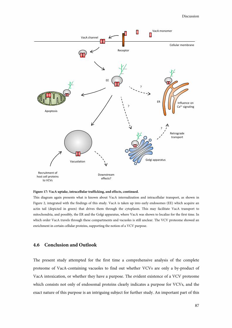
Discussion
87
Figure 17: VacA uptake, intracellular trafficking, and effects, continued.
This diagram again presents what is known about VacA internalization and intracellular transport, as shown in
Figure 2, integrated with the findings of this study. VacA is taken up into early endosomes (EE) which acquire an
actin tail (depicted in green) that drives them through the cytoplasm. This may facilitate VacA transport to
mitochondria, and possibly, the ER and the Golgi apparatus, where VacA was shown to localize for the first time. In
which order VacA travels through these compartments and vacuoles is still unclear. The VCV proteome showed an
enrichment in certain cellular proteins, supporting the notion of a VCV purpose.
4.6 Conclusion and Outlook
The present study attempted for the first time a comprehensive analysis of the complete
proteome of VacA-containing vacuoles to find out whether VCVs are only a by-product of
VacA intoxication, or whether they have a purpose. The evident existence of a VCV proteome
which consists not only of endosomal proteins clearly indicates a purpose for VCVs, and the
exact nature of this purpose is an intriguing subject for further study. An important part of this
Vacuolation
Apoptosis
?
Recruitment of
host cell proteins
to VCVs
Downstream
effects?
ER Influence on
Ca2+ signaling
Cellular membrane
VacA monomer
Receptor
VacA channel
Golgi apparatus
Retrograde
transport
?
?
EE

Discussion
88
will be the validation of the presence of individual proteins on VCVs with special focus on
VCVs as putative signaling platforms. Numerous proteins involved in known VacA intoxication
effects were detected in VCV preparations, and their validation will strengthen the connections
between VacA effects and their underlying cellular pathways. Of particular interest are proteins
that are implicated in cellular calcium homeostasis (including STIM1 and ITPR3), the
mitochondrial apoptotic pathway (including Drp1 and Bak), and intracellular signaling and
trafficking processes (including Rack1/GNB2L1 and Rab superfamily proteins), which may all
contribute to immune modulation by VacA. VCV proteome analysis may therefore also prove
to be a valuable tool to interconnect existing knowledge about VacA and its effects.
Additionally, the Golgi apparatus and the ER were identified as novel VacA target structures.
The retrograde pathway is an obvious candidate for VacA transport to these compartments,
analogous to the trafficking of other bacterial toxins. However, other routes, including direct
endosome-ER transport, cannot yet be dismissed. In general, there are many details of VacA
trafficking left to discover (see Figure 17). This encompasses the potential dependence of VacA
transport on its N-terminus, and the matter of sequence: is VacA transported to all other
compartments, including VCVs, from early endosomes? Or are vacuoles an intermediate step
not only with regard to VacA function, but also with regard to transport? Also, which effects are
caused by the trafficking of VacA to the Golgi and the ER? Does the toxin persist there, or is the
cytoplasm its final target? And which effects are instead caused by the recruitment of cellular
proteins to VCVs? All of these questions provide extensive potential for future investigations.
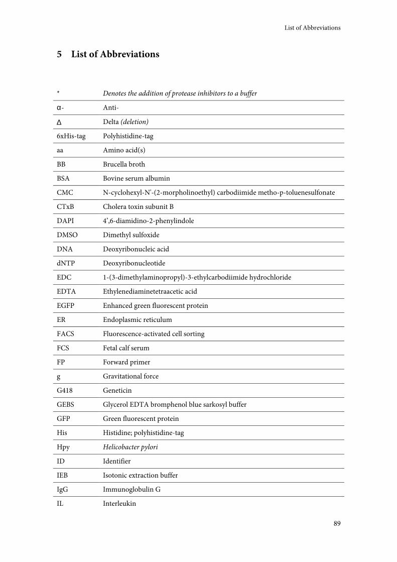
List of Abbreviations
89
5 List of Abbreviations
* Denotes the addition of protease inhibitors to a buffer
α- Anti-
∆ Delta (deletion)
6xHis-tag Polyhistidine-tag
aa Amino acid(s)
BB Brucella broth
BSA Bovine serum albumin
CMC N-cyclohexyl-N'-(2-morpholinoethyl) carbodiimide metho-p-toluenesulfonate
CTxB Cholera toxin subunit B
DAPI 4’,6-diamidino-2-phenylindole
DMSO Dimethyl sulfoxide
DNA Deoxyribonucleic acid
dNTP Deoxyribonucleotide
EDC 1-(3-dimethylaminopropyl)-3-ethylcarbodiimide hydrochloride
EDTA Ethylenediaminetetraacetic acid
EGFP Enhanced green fluorescent protein
ER Endoplasmic reticulum
FACS Fluorescence-activated cell sorting
FCS Fetal calf serum
FP Forward primer
g Gravitational force
G418 Geneticin
GEBS Glycerol EDTA bromphenol blue sarkosyl buffer
GFP Green fluorescent protein
His Histidine; polyhistidine-tag
Hpy Helicobacter pylori
ID Identifier
IEB Isotonic extraction buffer
IgG Immunoglobulin G
IL Interleukin
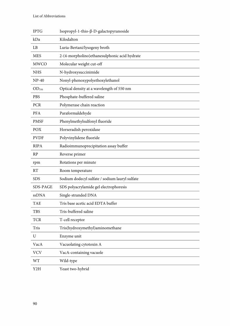
List of Abbreviations
90
IPTG Isopropyl-1-thio-β-D-galactopyranoside
kDa Kilodalton
LB Luria-Bertani/lysogeny broth
MES 2-(4-morpholino)ethanesulphonic acid hydrate
MWCO Molecular weight cut-off
NHS N-hydroxysuccinimide
NP-40 Nonyl-phenoxypolyethoxylethanol
OD550 Optical density at a wavelength of 550 nm
PBS Phosphate-buffered saline
PCR Polymerase chain reaction
PFA Paraformaldehyde
PMSF Phenylmethylsulfonyl fluoride
POX Horseradish peroxidase
PVDF Polyvinylidene fluoride
RIPA Radioimmunoprecipitation assay buffer
RP Reverse primer
rpm Rotations per minute
RT Room temperature
SDS Sodium dodecyl sulfate / sodium lauryl sulfate
SDS-PAGE SDS polyacrylamide gel electrophoresis
ssDNA Single-stranded DNA
TAE Tris base acetic acid EDTA buffer
TBS Tris-buffered saline
TCR T-cell receptor
Tris Tris(hydroxymethyl)aminomethane
U Enzyme unit
VacA Vacuolating cytotoxin A
VCV VacA-containing vacuole
WT Wild-type
Y2H Yeast two-hybrid
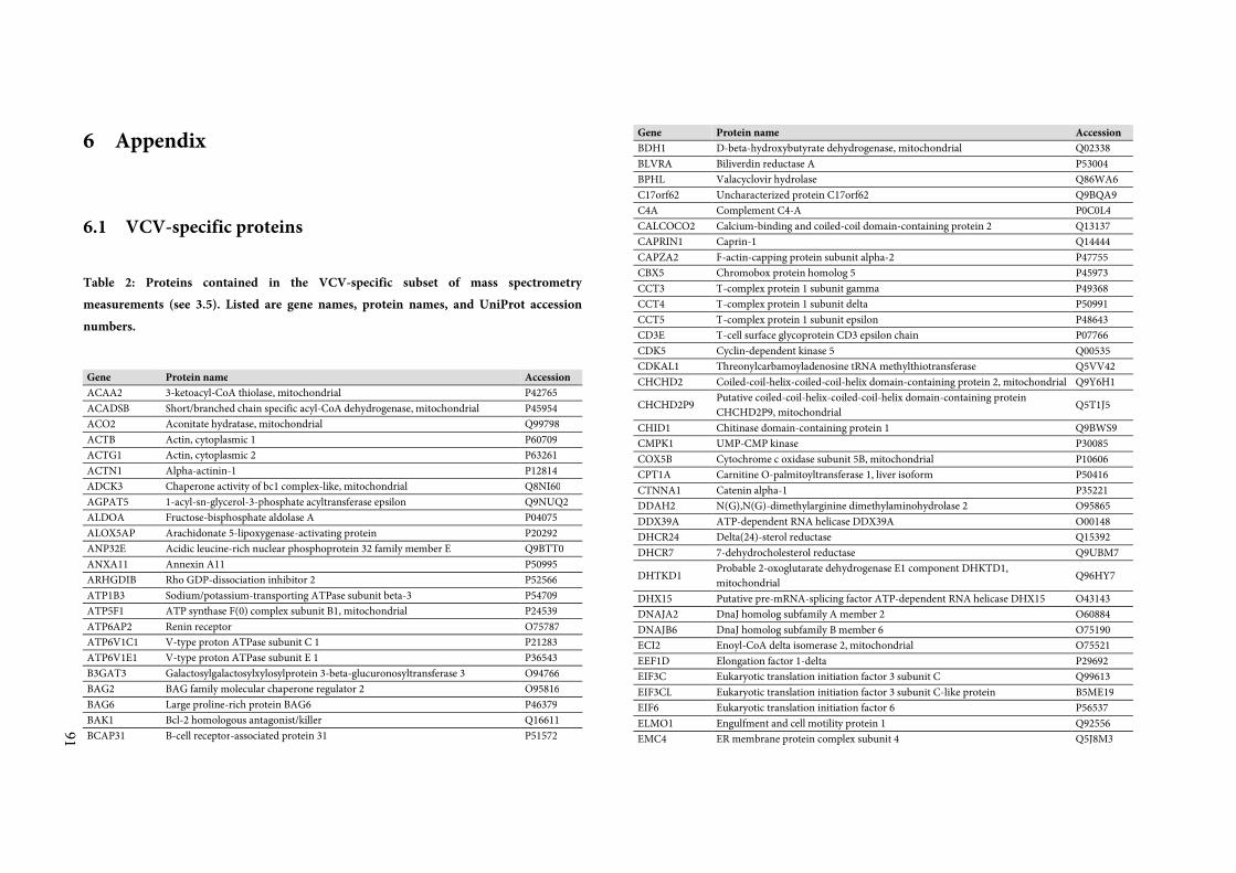
91
6 Appendix
6.1 VCV-specific proteins
Table 2: Proteins contained in the VCV-specific subset of mass spectrometry
measurements (see 3.5). Listed are gene names, protein names, and UniProt accession
numbers.
Gene Protein name Accession
ACAA2 3-ketoacyl-CoA thiolase, mitochondrial P42765
ACADSB Short/branched chain specific acyl-CoA dehydrogenase, mitochondrial P45954
ACO2 Aconitate hydratase, mitochondrial Q99798
ACTB Actin, cytoplasmic 1 P60709
ACTG1 Actin, cytoplasmic 2 P63261
ACTN1 Alpha-actinin-1 P12814
ADCK3 Chaperone activity of bc1 complex-like, mitochondrial Q8NI60
AGPAT5 1-acyl-sn-glycerol-3-phosphate acyltransferase epsilon Q9NUQ2
ALDOA Fructose-bisphosphate aldolase A P04075
ALOX5AP Arachidonate 5-lipoxygenase-activating protein P20292
ANP32E Acidic leucine-rich nuclear phosphoprotein 32 family member E Q9BTT0
ANXA11 Annexin A11 P50995
ARHGDIB Rho GDP-dissociation inhibitor 2 P52566
ATP1B3 Sodium/potassium-transporting ATPase subunit beta-3 P54709
ATP5F1 ATP synthase F(0) complex subunit B1, mitochondrial P24539
ATP6AP2 Renin receptor O75787
ATP6V1C1 V-type proton ATPase subunit C 1 P21283
ATP6V1E1 V-type proton ATPase subunit E 1 P36543
B3GAT3 Galactosylgalactosylxylosylprotein 3-beta-glucuronosyltransferase 3 O94766
BAG2 BAG family molecular chaperone regulator 2 O95816
BAG6 Large proline-rich protein BAG6 P46379
BAK1 Bcl-2 homologous antagonist/killer Q16611
BCAP31 B-cell receptor-associated protein 31 P51572
Gene Protein name Accession
BDH1 D-beta-hydroxybutyrate dehydrogenase, mitochondrial Q02338
BLVRA Biliverdin reductase A P53004
BPHL Valacyclovir hydrolase Q86WA6
C17orf62 Uncharacterized protein C17orf62 Q9BQA9
C4A Complement C4-A P0C0L4
CALCOCO2 Calcium-binding and coiled-coil domain-containing protein 2 Q13137
CAPRIN1 Caprin-1 Q14444
CAPZA2 F-actin-capping protein subunit alpha-2 P47755
CBX5 Chromobox protein homolog 5 P45973
CCT3 T-complex protein 1 subunit gamma P49368
CCT4 T-complex protein 1 subunit delta P50991
CCT5 T-complex protein 1 subunit epsilon P48643
CD3E T-cell surface glycoprotein CD3 epsilon chain P07766
CDK5 Cyclin-dependent kinase 5 Q00535
CDKAL1 Threonylcarbamoyladenosine tRNA methylthiotransferase Q5VV42
CHCHD2 Coiled-coil-helix-coiled-coil-helix domain-containing protein 2, mitochondrial Q9Y6H1
CHCHD2P9 Putative coiled-coil-helix-coiled-coil-helix domain-containing protein
CHCHD2P9, mitochondrial Q5T1J5
CHID1 Chitinase domain-containing protein 1 Q9BWS9
CMPK1 UMP-CMP kinase P30085
COX5B Cytochrome c oxidase subunit 5B, mitochondrial P10606
CPT1A Carnitine O-palmitoyltransferase 1, liver isoform P50416
CTNNA1 Catenin alpha-1 P35221
DDAH2 N(G),N(G)-dimethylarginine dimethylaminohydrolase 2 O95865
DDX39A ATP-dependent RNA helicase DDX39A O00148
DHCR24 Delta(24)-sterol reductase Q15392
DHCR7 7-dehydrocholesterol reductase Q9UBM7
DHTKD1 Probable 2-oxoglutarate dehydrogenase E1 component DHKTD1,
mitochondrial Q96HY7
DHX15 Putative pre-mRNA-splicing factor ATP-dependent RNA helicase DHX15 O43143
DNAJA2 DnaJ homolog subfamily A member 2 O60884
DNAJB6 DnaJ homolog subfamily B member 6 O75190
ECI2 Enoyl-CoA delta isomerase 2, mitochondrial O75521
EEF1D Elongation factor 1-delta P29692
EIF3C Eukaryotic translation initiation factor 3 subunit C Q99613
EIF3CL Eukaryotic translation initiation factor 3 subunit C-like protein B5ME19
EIF6 Eukaryotic translation initiation factor 6 P56537
ELMO1 Engulfment and cell motility protein 1 Q92556
EMC4 ER membrane protein complex subunit 4 Q5J8M3
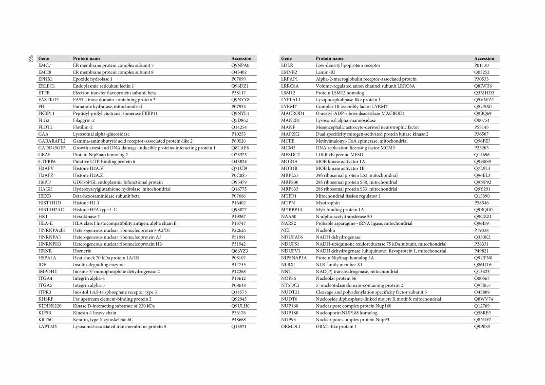
92 Gene Protein name Accession
EMC7 ER membrane protein complex subunit 7 Q9NPA0
EMC8 ER membrane protein complex subunit 8 O43402
EPHX1 Epoxide hydrolase 1 P07099
ERLEC1 Endoplasmic reticulum lectin 1 Q96DZ1
ETFB Electron transfer flavoprotein subunit beta P38117
FASTKD2 FAST kinase domain-containing protein 2 Q9NYY8
FH Fumarate hydratase, mitochondrial P07954
FKBP11 Peptidyl-prolyl cis-trans isomerase FKBP11 Q9NYL4
FLG2 Filaggrin-2 Q5D862
FLOT2 Flotillin-2 Q14254
GAA Lysosomal alpha-glucosidase P10253
GABARAPL2 Gamma-aminobutyric acid receptor-associated protein-like 2 P60520
GADD45GIP1 Growth arrest and DNA damage-inducible proteins-interacting protein 1 Q8TAE8
GBAS Protein NipSnap homolog 2 O75323
GTPBP6 Putative GTP-binding protein 6 O43824
H2AFV Histone H2A.V Q71UI9
H2AFZ Histone H2A.Z P0C0S5
H6PD GDH/6PGL endoplasmic bifunctional protein O95479
HAGH Hydroxyacylglutathione hydrolase, mitochondrial Q16775
HEXB Beta-hexosaminidase subunit beta P07686
HIST1H1D Histone H1.3 P16402
HIST1H2AC Histone H2A type 1-C Q93077
HK1 Hexokinase-1 P19367
HLA-E HLA class I histocompatibility antigen, alpha chain E P13747
HNRNPA2B1 Heterogeneous nuclear ribonucleoproteins A2/B1 P22626
HNRNPA3 Heterogeneous nuclear ribonucleoprotein A3 P51991
HNRNPH3 Heterogeneous nuclear ribonucleoprotein H3 P31942
HRNR Hornerin Q86YZ3
HSPA1A Heat shock 70 kDa protein 1A/1B P08107
IDE Insulin-degrading enzyme P14735
IMPDH2 Inosine-5'-monophosphate dehydrogenase 2 P12268
ITGA4 Integrin alpha-4 P13612
ITGA5 Integrin alpha-5 P08648
ITPR3 Inositol 1,4,5-trisphosphate receptor type 3 Q14573
KHSRP Far upstream element-binding protein 2 Q92945
KIDINS220 Kinase D-interacting substrate of 220 kDa Q9ULH0
KIF5B Kinesin-1 heavy chain P33176
KRT6C Keratin, type II cytoskeletal 6C P48668
LAPTM5 Lysosomal-associated transmembrane protein 5 Q13571
Gene Protein name Accession
LDLR Low-density lipoprotein receptor P01130
LMNB2 Lamin-B2 Q03252
LRPAP1 Alpha-2-macroglobulin receptor-associated protein P30533
LRRC8A Volume-regulated anion channel subunit LRRC8A Q8IWT6
LSM12 Protein LSM12 homolog Q3MHD2
LYPLAL1 Lysophospholipase-like protein 1 Q5VWZ2
LYRM7 Complex III assembly factor LYRM7 Q5U5X0
MACROD1 O-acetyl-ADP-ribose deacetylase MACROD1 Q9BQ69
MAN2B1 Lysosomal alpha-mannosidase O00754
MANF Mesencephalic astrocyte-derived neurotrophic factor P55145
MAP2K2 Dual specificity mitogen-activated protein kinase kinase 2 P36507
MCEE Methylmalonyl-CoA epimerase, mitochondrial Q96PE7
MCM3 DNA replication licensing factor MCM3 P25205
MESDC2 LDLR chaperone MESD Q14696
MOB1A MOB kinase activator 1A Q9H8S9
MOB1B MOB kinase activator 1B Q7L9L4
MRPL53 39S ribosomal protein L53, mitochondrial Q96EL3
MRPS30 28S ribosomal protein S30, mitochondrial Q9NP92
MRPS33 28S ribosomal protein S33, mitochondrial Q9Y291
MTFR1 Mitochondrial fission regulator 1 Q15390
MTPN Myotrophin P58546
MYBBP1A Myb-binding protein 1A Q9BQG0
NAA50 N-alpha-acetyltransferase 50 Q9GZZ1
NARS2 Probable asparagine--tRNA ligase, mitochondrial Q96I59
NCL Nucleolin P19338
NDUFAF6 NADH dehydrogenase Q330K2
NDUFS1 NADH-ubiquinone oxidoreductase 75 kDa subunit, mitochondrial P28331
NDUFV1 NADH dehydrogenase [ubiquinone] flavoprotein 1, mitochondrial P49821
NIPSNAP3A Protein NipSnap homolog 3A Q9UFN0
NLRX1 NLR family member X1 Q86UT6
NNT NAD(P) transhydrogenase, mitochondrial Q13423
NOP56 Nucleolar protein 56 O00567
NT5DC2 5'-nucleotidase domain-containing protein 2 Q9H857
NUDT21 Cleavage and polyadenylation specificity factor subunit 5 O43809
NUDT8 Nucleoside diphosphate-linked moiety X motif 8, mitochondrial Q8WV74
NUP160 Nuclear pore complex protein Nup160 Q12769
NUP188 Nucleoporin NUP188 homolog Q5SRE5
NUP93 Nuclear pore complex protein Nup93 Q8N1F7
ORMDL1 ORM1-like protein 1 Q9P0S3
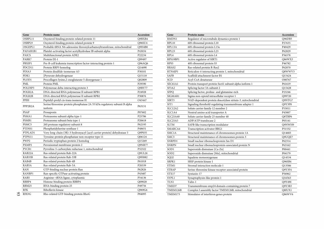
93
Gene Protein name Accession
OSBPL11 Oxysterol-binding protein-related protein 11 Q9BXB4
OSBPL9 Oxysterol-binding protein-related protein 9 Q96SU4
OSGEPL1 Probable tRNA N6-adenosine threonylcarbamoyltransferase, mitochondrial Q9H4B0
PAFAH1B1 Platelet-activating factor acetylhydrolase IB subunit alpha P43034
PAICS Multifunctional protein ADE2 P22234
PARK7 Protein DJ-1 Q99497
PBXIP1 Pre-B-cell leukemia transcription factor-interacting protein 1 Q96AQ6
PDCD11 Protein RRP5 homolog Q14690
PDIA3 Protein disulfide-isomerase A3 P30101
PDK1 [Pyruvate dehydrogenase Q15118
PLOD1 Procollagen-lysine,2-oxoglutarate 5-dioxygenase 1 Q02809
PML Protein PML P29590
POLDIP3 Polymerase delta-interacting protein 3 Q9BY77
POLR2A DNA-directed RNA polymerase II subunit RPB1 P24928
POLR2B DNA-directed RNA polymerase II subunit RPB2 P30876
PPIH Peptidyl-prolyl cis-trans isomerase H O43447
PPP2R2A Serine/threonine-protein phosphatase 2A 55 kDa regulatory subunit B alpha
isoform P63151
PSAP Prosaposin P07602
PSMA1 Proteasome subunit alpha type-1 P25786
PSMB1 Proteasome subunit beta type-1 P20618
PSMC5 26S protease regulatory subunit 8 P62195
PTDSS1 Phosphatidylserine synthase 1 P48651
PTPLAD1 Very-long-chain (3R)-3-hydroxyacyl-[acyl-carrier protein] dehydratase 3 Q9P035
PTPN11 Tyrosine-protein phosphatase non-receptor type 11 Q06124
PWP2 Periodic tryptophan protein 2 homolog Q15269
PXMP2 Peroxisomal membrane protein 2 Q9NR77
PYCR1 Pyrroline-5-carboxylate reductase 1, mitochondrial P32322
RAB22A Ras-related protein Rab-22A Q9UL26
RAB33B Ras-related protein Rab-33B Q9H082
RAB4B Ras-related protein Rab-4B P61018
RAB5A Ras-related protein Rab-5A P20339
RAN GTP-binding nuclear protein Ran P62826
RANBP1 Ran-specific GTPase-activating protein P43487
RARS Arginine--tRNA ligase, cytoplasmic P54136
RBBP4 Histone-binding protein RBBP4 Q09028
RBM25 RNA-binding protein 25 P49756
RFK Riboflavin kinase Q969G6
RHOG Rho-related GTP-binding protein RhoG P84095
Gene Protein name Accession
RMDN1 Regulator of microtubule dynamics protein 1 Q96DB5
RPL10 60S ribosomal protein L10 P27635
RPL13A 60S ribosomal protein L13a P40429
RPL23 60S ribosomal protein L23 P62829
RPL4 60S ribosomal protein L4 P36578
RPS19BP1 Active regulator of SIRT1 Q86WX3
RPS5 40S ribosomal protein S5 P46782
RRAS2 Ras-related protein R-Ras2 P62070
RTN4IP1 Reticulon-4-interacting protein 1, mitochondrial Q8WWV3
SAFB Scaffold attachment factor B1 Q15424
SCD Acyl-CoA desaturase O00767
SEC61A1 Protein transport protein Sec61 subunit alpha isoform 1 P61619
SF3A2 Splicing factor 3A subunit 2 Q15428
SFPQ Splicing factor, proline- and glutamine-rich P23246
SIGMAR1 Sigma non-opioid intracellular receptor 1 Q99720
SIRT3 NAD-dependent protein deacetylase sirtuin-3, mitochondrial Q9NTG7
SIT1 Signaling threshold-regulating transmembrane adapter 1 Q9Y3P8
SLC12A2 Solute carrier family 12 member 2 P55011
SLC1A4 Neutral amino acid transporter A P43007
SLC25A40 Solute carrier family 25 member 40 Q8TBP6
SLC25A5 ADP/ATP translocase 2 P05141
SLTM SAFB-like transcription modulator Q9NWH9
SMARCA4 Transcription activator BRG1 P51532
SMC1A Structural maintenance of chromosomes protein 1A Q14683
SMC3 Structural maintenance of chromosomes protein 3 Q9UQE7
SNRPD1 Small nuclear ribonucleoprotein Sm D1 P62314
SNRPN Small nuclear ribonucleoprotein-associated protein N P63162
SOD1 Superoxide dismutase [Cu-Zn] P00441
SOD2 Superoxide dismutase [Mn], mitochondrial P04179
SQLE Squalene monooxygenase Q14534
SRPK1 SRSF protein kinase 1 Q96SB4
STIM1 Stromal interaction molecule 1 Q13586
STRAP Serine-threonine kinase receptor-associated protein Q9Y3F4
STX17 Syntaxin-17 P56962
SYPL1 Synaptophysin-like protein 1 Q16563
TLN1 Talin-1 Q9Y490
TMED7 Transmembrane emp24 domain-containing protein 7 Q9Y3B3
TMEM126B Complex I assembly factor TMEM126B, mitochondrial Q8IUX1
TMEM173 Stimulator of interferon genes protein Q86WV6
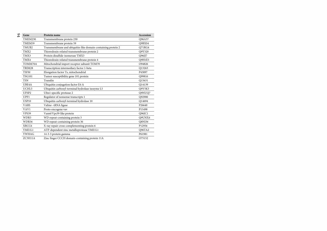
94 Gene Protein name Accession
TMEM230 Transmembrane protein 230 Q96A57
TMEM59 Transmembrane protein 59 Q9BXS4
TMUB2 Transmembrane and ubiquitin-like domain-containing protein 2 Q71RG4
TMX2 Thioredoxin-related transmembrane protein 2 Q9Y320
TMX3 Protein disulfide-isomerase TMX3 Q96JJ7
TMX4 Thioredoxin-related transmembrane protein 4 Q9H1E5
TOMM70A Mitochondrial import receptor subunit TOM70 O94826
TRIM28 Transcription intermediary factor 1-beta Q13263
TSFM Elongation factor Ts, mitochondrial P43897
TSG101 Tumor susceptibility gene 101 protein Q99816
TSN Translin Q15631
UBE4A Ubiquitin conjugation factor E4 A Q14139
UCHL5 Ubiquitin carboxyl-terminal hydrolase isozyme L5 Q9Y5K5
UFSP2 Ufm1-specific protease 2 Q9NUQ7
UPF1 Regulator of nonsense transcripts 1 Q92900
USP10 Ubiquitin carboxyl-terminal hydrolase 10 Q14694
VARS Valine--tRNA ligase P26640
VAV1 Proto-oncogene vav P15498
VPS39 Vam6/Vps39-like protein Q96JC1
WDR3 WD repeat-containing protein 3 Q9UNX4
WDR36 WD repeat-containing protein 36 Q8NI36
XRCC6 X-ray repair cross-complementing protein 6 P12956
YME1L1 ATP-dependent zinc metalloprotease YME1L1 Q96TA2
YWHAG 14-3-3 protein gamma P61981
ZC3H11A Zinc finger CCCH domain-containing protein 11A O75152
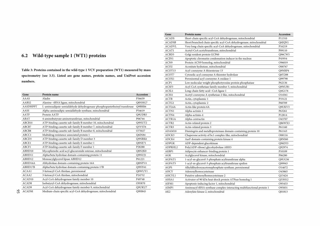
95
6.2 Wild-type sample 1 (WT1) proteins
Table 3: Proteins contained in the wild-type 1 VCV preparation (WT1) measured by mass
spectrometry (see 3.5). Listed are gene names, protein names, and UniProt accession
numbers.
Gene Protein name Accession
AAAS Aladin P36639
AARS2 Alanine--tRNA ligase, mitochondrial Q8NHG7
AASDHPPT L-aminoadipate-semialdehyde dehydrogenase-phosphopantetheinyl transferase Q9BRR6
AASS Alpha-aminoadipic semialdehyde synthase, mitochondrial P54619
AATF Protein AATF Q9UDR5
ABAT 4-aminobutyrate aminotransferase, mitochondrial P06744
ABCB10 ATP-binding cassette sub-family B member 10, mitochondrial Q9NUT2
ABCB7 ATP-binding cassette sub-family B member 7, mitochondrial Q5VST6
ABCB8 ATP-binding cassette sub-family B member 8, mitochondrial O75027
ABCC1 Multidrug resistance-associated protein 1 Q6IN84
ABCD3 ATP-binding cassette sub-family D member 3 Q9NRK6
ABCE1 ATP-binding cassette sub-family E member 1 Q8NE71
ABCF1 ATP-binding cassette sub-family F member 1 P28288
ABHD10 Mycophenolic acid acyl-glucuronide esterase, mitochondrial Q8N2K0
ABHD11 Alpha/beta hydrolase domain-containing protein 11 Q9NUJ1
ABHD12 Monoacylglycerol lipase ABHD12 P61221
ABHD16A Abhydrolase domain-containing protein 16A Q8NFV4
ABHD17B Alpha/beta hydrolase domain-containing protein 17B Q9NY61
ACAA1 3-ketoacyl-CoA thiolase, peroxisomal Q8WUY1
ACAA2 3-ketoacyl-CoA thiolase, mitochondrial P24752
ACAD10 Acyl-CoA dehydrogenase family member 10 P49748
ACAD8 Isobutyryl-CoA dehydrogenase, mitochondrial O95870
ACAD9 Acyl-CoA dehydrogenase family member 9, mitochondrial Q9UKU7
ACADM Medium-chain specific acyl-CoA dehydrogenase, mitochondrial Q9H845
Gene Protein name Accession
ACADS Short-chain specific acyl-CoA dehydrogenase, mitochondrial P11310
ACADSB Short/branched chain specific acyl-CoA dehydrogenase, mitochondrial Q6JQN1
ACADVL Very long-chain specific acyl-CoA dehydrogenase, mitochondrial P16219
ACAT1 Acetyl-CoA acetyltransferase, mitochondrial P09110
ACBD3 Golgi resident protein GCP60 Q96CW5
ACIN1 Apoptotic chromatin condensation inducer in the nucleus P45954
ACN9 Protein ACN9 homolog, mitochondrial O96019
ACO2 Aconitate hydratase, mitochondrial O00767
ACOT13 Acyl-coenzyme A thioesterase 13 Q9NRP4
ACOT7 Cytosolic acyl coenzyme A thioester hydrolase Q6Y288
ACOX1 Peroxisomal acyl-coenzyme A oxidase 1 Q99798
ACP1 Low molecular weight phosphotyrosine protein phosphatase P62136
ACSF3 Acyl-CoA synthetase family member 3, mitochondrial Q9NUB1
ACSL1 Long-chain-fatty-acid--CoA ligase 1 Q4G176
ACSS1 Acetyl-coenzyme A synthetase 2-like, mitochondrial O14561
ACTB Actin, cytoplasmic 1 P33121
ACTG1 Actin, cytoplasmic 2 P60709
ACTL6A Actin-like protein 6A Q9UKV3
ACTN1 Alpha-actinin-1 P63261
ACTN4 Alpha-actinin-4 P12814
ACTR1A Alpha-centractin O43707
ACTR2 Actin-related protein 2 Q86WX3
ACTR3 Actin-related protein 3 P61160
ADAM10 Disintegrin and metalloproteinase domain-containing protein 10 P61163
ADCK3 Chaperone activity of bc1 complex-like, mitochondrial O00116
ADCK4 AarF domain-containing protein kinase 4 Q8NI60
ADPGK ADP-dependent glucokinase Q96D53
ADPRHL2 Poly(ADP-ribose) glycohydrolase ARH3 Q92974
AEBP1 Adipocyte enhancer-binding protein 1 P10109
AGK Acylglycerol kinase, mitochondrial P06280
AGPAT1 1-acyl-sn-glycerol-3-phosphate acyltransferase alpha Q9UG56
AGPAT5 1-acyl-sn-glycerol-3-phosphate acyltransferase epsilon Q99943
AGPS Alkyldihydroxyacetonephosphate synthase, peroxisomal O14672
AHCY Adenosylhomocysteinase O43865
AHCYL1 Putative adenosylhomocysteinase 2 Q15424
AHSA1 Activator of 90 kDa heat shock protein ATPase homolog 1 Q53H12
AIFM1 Apoptosis-inducing factor 1, mitochondrial O95433
AIMP1 Aminoacyl tRNA synthase complex-interacting multifunctional protein 1 O95831
AK2 Adenylate kinase 2, mitochondrial Q01813
96 Gene Protein name Accession
AK3 GTP:AMP phosphotransferase AK3, mitochondrial P54819
AKR7A2 Aflatoxin B1 aldehyde reductase member 2 Q9NX46
ALAS1 5-aminolevulinate synthase, nonspecific, mitochondrial Q9H583
ALB Serum albumin P20292
ALDH18A1 Delta-1-pyrroline-5-carboxylate synthase Q96C36
ALDH3A2 Fatty aldehyde dehydrogenase Q9NWT8
ALDH4A1 Delta-1-pyrroline-5-carboxylate dehydrogenase, mitochondrial P51648
ALDH5A1 Succinate-semialdehyde dehydrogenase, mitochondrial Q13242
ALDH6A1 Methylmalonate-semialdehyde dehydrogenase [acylating], mitochondrial Q8N4V1
ALDOA Fructose-bisphosphate aldolase A P02768
ALG1 Chitobiosyldiphosphodolichol beta-mannosyltransferase Q9NP73
ALG13 Putative bifunctional UDP-N-acetylglucosamine transferase and deubiquitinase
ALG13 P04075
ALG5 Dolichyl-phosphate beta-glucosyltransferase Q9BT22
ALKBH7 Alpha-ketoglutarate-dependent dioxygenase alkB homolog 7, mitochondrial Q9Y673
ALOX5AP Arachidonate 5-lipoxygenase-activating protein P30038
ALYREF THO complex subunit 4 Q8NI27
ANKLE2 Ankyrin repeat and LEM domain-containing protein 2 Q9BTT0
ANO6 Anoctamin-6 O14744
ANP32E Acidic leucine-rich nuclear phosphoprotein 32 family member E P30533
ANXA11 Annexin A11 P01008
ANXA2 Annexin A2 P50995
ANXA6 Annexin A6 P07355
AP1B1 AP-1 complex subunit beta-1 P08133
AP1G1 AP-1 complex subunit gamma-1 Q10567
AP1M1 AP-1 complex subunit mu-1 O43747
AP1S1 AP-1 complex subunit sigma-1A Q9BXS5
AP2A1 AP-2 complex subunit alpha-1 P61966
AP2A2 AP-2 complex subunit alpha-2 O95782
AP2B1 AP-2 complex subunit beta O94973
AP2M1 AP-2 complex subunit mu P63010
AP2S1 AP-2 complex subunit sigma Q96CW1
APMAP Adipocyte plasma membrane-associated protein P50583
APOA1BP NAD(P)H-hydrate epimerase P22307
APOL2 Apolipoprotein L2 Q9HDC9
APOO Apolipoprotein O Q6UXV4
APOOL Apolipoprotein O-like Q9BQE5
APRT Adenine phosphoribosyltransferase Q9BUR5
ARCN1 Coatomer subunit delta P53618
Gene Protein name Accession
ARF1 ADP-ribosylation factor 1 P07741
ARF3 ADP-ribosylation factor 3 P84077
ARF4 ADP-ribosylation factor 4 P61204
ARF5 ADP-ribosylation factor 5 P18085
ARF6 ADP-ribosylation factor 6 P84085
ARG2 Arginase-2, mitochondrial P62330
ARHGAP15 Rho GTPase-activating protein 15 Q15382
ARHGDIB Rho GDP-dissociation inhibitor 2 P50395
ARHGEF2 Rho guanine nucleotide exchange factor 2 P78540
ARL2 ADP-ribosylation factor-like protein 2 O43488
ARL3 ADP-ribosylation factor-like protein 3 P36404
ARL8B ADP-ribosylation factor-like protein 8B P36405
ARMC1 Armadillo repeat-containing protein 1 Q8N2F6
ARMC10 Armadillo repeat-containing protein 10 Q9NVJ2
ARMCX3 Armadillo repeat-containing X-linked protein 3 Q9NVT9
ARPC3 Actin-related protein 2/3 complex subunit 3 P61158
ARPC4 Actin-related protein 2/3 complex subunit 4 O15145
ASAH1 Acid ceramidase P59998
ASF1A Histone chaperone ASF1A Q13510
ASNA1 ATPase ASNA1 Q9Y294
ASPH Aspartyl/asparaginyl beta-hydroxylase O43681
ATAD1 ATPase family AAA domain-containing protein 1 P24539
ATAD3A ATPase family AAA domain-containing protein 3A Q8NBU5
ATAD3B ATPase family AAA domain-containing protein 3B Q9NVI7
ATG3 Ubiquitin-like-conjugating enzyme ATG3 Q5T9A4
ATIC Bifunctional purine biosynthesis protein PURH P22234
ATL2 Atlastin-2 Q9NT62
ATL3 Atlastin-3 Q8NHH9
ATP13A1 Probable cation-transporting ATPase 13A1 Q12797
ATP1A1 Sodium/potassium-transporting ATPase subunit alpha-1 Q9HD20
ATP1A3 Sodium/potassium-transporting ATPase subunit alpha-3 P05023
ATP1B3 Sodium/potassium-transporting ATPase subunit beta-3 P13637
ATP2A2 Sarcoplasmic/endoplasmic reticulum calcium ATPase 2 P54709
ATP2A3 Sarcoplasmic/endoplasmic reticulum calcium ATPase 3 P16615
ATP2B4 Plasma membrane calcium-transporting ATPase 4 Q93084
ATP5A1 ATP synthase subunit alpha, mitochondrial O75964
ATP5B ATP synthase subunit beta, mitochondrial P25705
ATP5D ATP synthase subunit delta, mitochondrial P06576
ATP5F1 ATP synthase F(0) complex subunit B1, mitochondrial Q9NW81
97
Gene Protein name Accession
ATP5H ATP synthase subunit d, mitochondrial Q6DD88
ATP5L ATP synthase subunit g, mitochondrial Q99766
ATP5O ATP synthase subunit O, mitochondrial Q8N5M1
ATP5S ATP synthase subunit s, mitochondrial O75947
ATP5SL ATP synthase subunit s-like protein P23634
ATP6AP2 Renin receptor Q96HR9
ATP6V0A2 V-type proton ATPase 116 kDa subunit a isoform 2 Q9H9H4
ATP6V0D1 V-type proton ATPase subunit d 1 Q9UBK9
ATP6V1A V-type proton ATPase catalytic subunit A Q99536
ATP6V1B2 V-type proton ATPase subunit B, brain isoform P38606
ATP6V1C1 V-type proton ATPase subunit C 1 P21281
ATP6V1D V-type proton ATPase subunit D P21283
ATP6V1E1 V-type proton ATPase subunit E 1 Q9Y5K8
ATP6V1F V-type proton ATPase subunit F P36543
ATP6V1G1 V-type proton ATPase subunit G 1 Q16864
ATP6V1H V-type proton ATPase subunit H O75348
ATPAF1 ATP synthase mitochondrial F1 complex assembly factor 1 P30049
ATPAF2 ATP synthase mitochondrial F1 complex assembly factor 2 Q5TC12
ATXN10 Ataxin-10 P48047
AUH Methylglutaconyl-CoA hydratase, mitochondrial Q9UBB4
AUP1 Ancient ubiquitous protein 1 Q13825
AURKAIP1 Aurora kinase A-interacting protein Q16352
AZI1 5-azacytidine-induced protein 1 Q9Y679
B3GALTL Beta-1,3-glucosyltransferase O94766
B3GAT3 Galactosylgalactosylxylosylprotein 3-beta-glucuronosyltransferase 3 Q9BXK5
BAG2 BAG family molecular chaperone regulator 2 O75531
BAG5 BAG family molecular chaperone regulator 5 O95816
BAG6 Large proline-rich protein BAG6 Q9UL15
BAK1 Bcl-2 homologous antagonist/killer P46379
BANF1 Barrier-to-autointegration factor O00154
BAZ1B Tyrosine-protein kinase BAZ1B P35613
BCAP31 B-cell receptor-associated protein 31 Q16611
BCAS2 Pre-mRNA-splicing factor SPF27 P61009
BCAT2 Branched-chain-amino-acid aminotransferase, mitochondrial Q9UIG0
BCCIP BRCA2 and CDKN1A-interacting protein O15382
BCKDHA 2-oxoisovalerate dehydrogenase subunit alpha, mitochondrial P11182
BCKDK [3-methyl-2-oxobutanoate dehydrogenase [lipoamide]] kinase, mitochondrial Q9P287
BCL2 Apoptosis regulator Bcl-2 O14874
BCL2L1 Bcl-2-like protein 1 Q9UPN4
Gene Protein name Accession
BCL2L13 Bcl-2-like protein 13 Q07817
BCS1L Mitochondrial chaperone BCS1 P10415
BDH1 D-beta-hydroxybutyrate dehydrogenase, mitochondrial Q9Y276
BET1L BET1-like protein Q02338
BID BH3-interacting domain death agonist Q9NYM9
BIRC5 Baculoviral IAP repeat-containing protein 5 Q96LW7
BLOC1S1 Biogenesis of lysosome-related organelles complex 1 subunit 1 O15392
BLVRA Biliverdin reductase A P55957
BMP2K BMP-2-inducible protein kinase P78537
BMS1 Ribosome biogenesis protein BMS1 homolog Q9NSY1
BNIP1 Vesicle transport protein SEC20 Q9UBV2
BOLA1 BolA-like protein 1 Q14692
BPHL Valacyclovir hydrolase Q53HL2
BPNT1 3'(2'),5'-bisphosphate nucleotidase 1 Q86WA6
BRI3BP BRI3-binding protein Q5VTR2
BRIX1 Ribosome biogenesis protein BRX1 homolog Q8WY22
BSG Basigin P51572
BST2 Bone marrow stromal antigen 2 Q8TDN6
BTN2A1 Butyrophilin subfamily 2 member A1 Q10589
BUD31 Protein BUD31 homolog Q7KYR7
BYSL Bystin P41223
BZW1 Basic leucine zipper and W2 domain-containing protein 1 Q13895
BZW2 Basic leucine zipper and W2 domain-containing protein 2 Q7L1Q6
C10orf35 Uncharacterized protein C10orf35 Q8N5K1
C12orf10 UPF0160 protein MYG1, mitochondrial Q99417
C15orf40 UPF0235 protein C15orf40 Q7Z7A1
C15orf61 Uncharacterized protein C15orf61 Q8WUR7
C16orf58 UPF0420 protein C16orf58 P09669
C16orf80 UPF0468 protein C16orf80 Q96GQ5
C17orf62 Uncharacterized protein C17orf62 Q9H3G5
C19orf10 UPF0556 protein C19orf10 Q5TZA2
C19orf52 Uncharacterized protein C19orf52 Q969H8
C1orf21 Uncharacterized protein C1orf21 Q6UB35
C1QBP Complement component 1 Q subcomponent-binding protein, mitochondrial Q9Y6E2
C21orf33 ES1 protein homolog, mitochondrial Q9BS26
C2orf43 UPF0554 protein C2orf43 P52907
C2orf47 Uncharacterized protein C2orf47, mitochondrial Q9H6V9
C3 Complement C3 A6NNL5
C3orf33 Protein C3orf33 P45973
98 Gene Protein name Accession
C3orf58 Deleted in autism protein 1 Q08211
C4A Complement C4-A P01024
C6orf120 UPF0669 protein C6orf120 O15182
C6orf203 Uncharacterized protein C6orf203 Q7Z4R8
C7orf50 Uncharacterized protein C7orf50 Q9P0P8
C8orf82 UPF0598 protein C8orf82 Q9BRJ6
C9 Complement component C9 P0C0L4
C9orf114 Uncharacterized protein C9orf114 Q9NPF2
C9orf89 Bcl10-interacting CARD protein P53004
CACYBP Calcyclin-binding protein P00167
CAD CAD protein P11498
CALCOCO2 Calcium-binding and coiled-coil domain-containing protein 2 Q9BRK5
CALM1 Calmodulin Q13112
CALR Calreticulin P62158
CAMLG Calcium signal-modulating cyclophilin ligand P27824
CAND1 Cullin-associated NEDD8-dissociated protein 1 P07384
CANX Calnexin P27797
CAP1 Adenylyl cyclase-associated protein 1 Q86VP6
CAPG Macrophage-capping protein Q01518
CAPN1 Calpain-1 catalytic subunit P49069
CAPRIN1 Caprin-1 P40121
CAPZA1 F-actin-capping protein subunit alpha-1 P08311
CARHSP1 Calcium-regulated heat stable protein 1 Q96BS2
CARS2 Probable cysteine--tRNA ligase, mitochondrial Q5JTZ9
CAT Catalase Q13948
CBR4 Carbonyl reductase family member 4 O75976
CBX3 Chromobox protein homolog 3 Q8N4T8
CBX5 Chromobox protein homolog 5 Q13185
CCAR2 Cell cycle and apoptosis regulator protein 2 Q9GZT6
CCBL2 Kynurenine--oxoglutarate transaminase 3 P22694
CCDC115 Coiled-coil domain-containing protein 115 Q6P1S2
CCDC127 Coiled-coil domain-containing protein 127 Q96NT0
CCDC134 Coiled-coil domain-containing protein 134 Q96BQ5
CCDC167 Coiled-coil domain-containing protein 167 Q9H6E4
CCDC47 Coiled-coil domain-containing protein 47 Q8N163
CCDC51 Coiled-coil domain-containing protein 51 Q96A33
CCDC58 Coiled-coil domain-containing protein 58 Q96ER9
CCDC59 Thyroid transcription factor 1-associated protein 26 P37802
CCDC90B Coiled-coil domain-containing protein 90B, mitochondrial Q9NV96
Gene Protein name Accession
CCNK Cyclin-K P53701
CCNY Cyclin-Y O75909
CCSMST1 Protein CCSMST1 P41240
CCT2 T-complex protein 1 subunit beta P17987
CCT3 T-complex protein 1 subunit gamma P48643
CCT4 T-complex protein 1 subunit delta P78371
CCT5 T-complex protein 1 subunit epsilon P50991
CCT6A T-complex protein 1 subunit zeta P50990
CCT7 T-complex protein 1 subunit eta P49368
CCT8 T-complex protein 1 subunit theta Q99832
CD1C T-cell surface glycoprotein CD1c Q8ND76
CD2 T-cell surface antigen CD2 P29017
CD247 T-cell surface glycoprotein CD3 zeta chain P09693
CD3D T-cell surface glycoprotein CD3 delta chain P06729
CD3E T-cell surface glycoprotein CD3 epsilon chain P04234
CD3G T-cell surface glycoprotein CD3 gamma chain P07766
CD47 Leukocyte surface antigen CD47 P20963
CD48 CD48 antigen Q08722
CD5 T-cell surface glycoprotein CD5 P09326
CD82 CD82 antigen P06127
CD84 SLAM family member 5 P63208
CD97 CD97 antigen P27701
CDC37 Hsp90 co-chaperone Cdc37 P48960
CDC42 Cell division control protein 42 homolog Q16543
CDC5L Cell division cycle 5-like protein P60953
CDCA8 Borealin Q9Y3E2
CDH2 Cadherin-2 P43155
CDIPT CDP-diacylglycerol--inositol 3-phosphatidyltransferase Q99459
CDK1 Cyclin-dependent kinase 1 O14735
CDK2 Cyclin-dependent kinase 2 P06493
CDK5 Cyclin-dependent kinase 5 P24941
CDK5RAP2 CDK5 regulatory subunit-associated protein 2 Q96D05
CDK5RAP3 CDK5 regulatory subunit-associated protein 3 Q96SN8
CDK6 Cyclin-dependent kinase 6 Q00535
CDK9 Cyclin-dependent kinase 9 Q00534
CDKAL1 Threonylcarbamoyladenosine tRNA methylthiotransferase P50750
CDKN2AIP CDKN2A-interacting protein Q14444
CECR5 Cat eye syndrome critical region protein 5 O94986
CENPM Centromere protein M Q9BXW7
99
Gene Protein name Accession
CENPV Centromere protein V Q9NSP4
CEP135 Centrosomal protein of 135 kDa Q9Y6A4
CEP152 Centrosomal protein of 152 kDa Q5VV42
CEP250 Centrosome-associated protein CEP250 Q6UW02
CEP41 Centrosomal protein of 41 kDa Q7Z7K6
CEP57 Centrosomal protein of 57 kDa Q9BYV8
CEP72 Centrosomal protein of 72 kDa Q86XR8
CETN3 Centrin-3 Q9P209
CFL1 Cofilin-1 Q96BR5
CHAF1B Chromatin assembly factor 1 subunit B Q5T440
CHCHD1 Coiled-coil-helix-coiled-coil-helix domain-containing protein 1 P10809
CHCHD2 Coiled-coil-helix-coiled-coil-helix domain-containing protein 2, mitochondrial Q96BP2
CHCHD2P9 Putative coiled-coil-helix-coiled-coil-helix domain-containing protein
CHCHD2P9, mitochondrial Q9BRQ6
CHCHD3 Coiled-coil-helix-coiled-coil-helix domain-containing protein 3, mitochondrial Q9Y6H1
CHCHD4 Mitochondrial intermembrane space import and assembly protein 40 Q5JRA6
CHCHD6 Coiled-coil-helix-coiled-coil-helix domain-containing protein 6, mitochondrial Q9NX63
CHD1 Chromodomain-helicase-DNA-binding protein 1 Q5T1J5
CHI3L2 Chitinase-3-like protein 2 P61604
CHMP2A Charged multivesicular body protein 2a O14646
CHP1 Calcineurin B homologous protein 1 O43633
CHST11 Carbohydrate sulfotransferase 11 Q9Y2V2
CIB1 Calcium and integrin-binding protein 1 Q9Y375
CISD1 CDGSH iron-sulfur domain-containing protein 1 Q99828
CISD2 CDGSH iron-sulfur domain-containing protein 2 Q9NZ45
CIT Citron Rho-interacting kinase P35221
CKAP4 Cytoskeleton-associated protein 4 Q96JB5
CKAP5 Cytoskeleton-associated protein 5 Q07065
CLIC1 Chloride intracellular channel protein 1 Q00610
CLINT1 Clathrin interactor 1 P29320
CLPB Caseinolytic peptidase B protein homolog O00299
CLPP ATP-dependent Clp protease proteolytic subunit, mitochondrial Q9H078
CLPX ATP-dependent Clp protease ATP-binding subunit clpX-like, mitochondrial Q16740
CLTC Clathrin heavy chain 1 Q14008
CLYBL Citrate lyase subunit beta-like protein, mitochondrial O76031
CMAS N-acylneuraminate cytidylyltransferase Q9UMX5
CMPK1 UMP-CMP kinase Q13303
CMPK2 UMP-CMP kinase 2, mitochondrial Q9UJS0
CNNM3 Metal transporter CNNM3 P09543
Gene Protein name Accession
CNOT1 CCR4-NOT transcription complex subunit 1 Q9UKZ1
CNOT11 CCR4-NOT transcription complex subunit 11 Q8NE01
CNOT7 CCR4-NOT transcription complex subunit 7 A5YKK6
CNP 2',3'-cyclic-nucleotide 3'-phosphodiesterase Q5EBM0
CNPY3 Protein canopy homolog 3 Q9UIV1
CNTRL Centriolin Q9BT09
COA1 Cytochrome c oxidase assembly factor 1 homolog P02748
COA3 Cytochrome c oxidase assembly factor 3 homolog, mitochondrial Q9GZY4
COA6 Cytochrome c oxidase assembly factor 6 homolog Q9Y2R0
COA7 Cytochrome c oxidase assembly factor 7 Q5JTJ3
COLGALT1 Procollagen galactosyltransferase 1 P09211
COMMD2 COMM domain-containing protein 2 P23528
COPA Coatomer subunit alpha Q86X83
COPB1 Coatomer subunit beta P35606
COPB2 Coatomer subunit beta' P53621
COPG1 Coatomer subunit gamma-1 P48444
COPS4 COP9 signalosome complex subunit 4 Q4G0I0
COPS8 COP9 signalosome complex subunit 8 Q9BT78
COPZ1 Coatomer subunit zeta-1 Q9Y678
COQ3 Hexaprenyldihydroxybenzoate methyltransferase, mitochondrial P61923
COQ5 2-methoxy-6-polyprenyl-1,4-benzoquinol methylase, mitochondrial Q9NZJ6
COQ6 Ubiquinone biosynthesis monooxygenase COQ6 Q5HYK3
COQ7 Ubiquinone biosynthesis protein COQ7 homolog Q9Y2Z9
COQ9 Ubiquinone biosynthesis protein COQ9, mitochondrial Q99807
CORO1A Coronin-1A O75208
COTL1 Coactosin-like protein P31146
COX11 Cytochrome c oxidase assembly protein COX11, mitochondrial Q14019
COX15 Cytochrome c oxidase assembly protein COX15 homolog Q9Y6N1
COX16 Cytochrome c oxidase assembly protein COX16 homolog, mitochondrial Q7KZN9
COX18 Mitochondrial inner membrane protein COX18 Q9P0S2
COX4I1 Cytochrome c oxidase subunit 4 isoform 1, mitochondrial Q8N8Q8
COX5A Cytochrome c oxidase subunit 5A, mitochondrial P13073
COX5B Cytochrome c oxidase subunit 5B, mitochondrial P20674
COX6B1 Cytochrome c oxidase subunit 6B1 Q9HCG8
COX6C Cytochrome c oxidase subunit 6C P10606
COX7A2 Cytochrome c oxidase subunit 7A2, mitochondrial P14854
CPD Carboxypeptidase D Q8WWC4
CPNE3 Copine-3 Q9BV73
CPOX Oxygen-dependent coproporphyrinogen-III oxidase, mitochondrial P13196
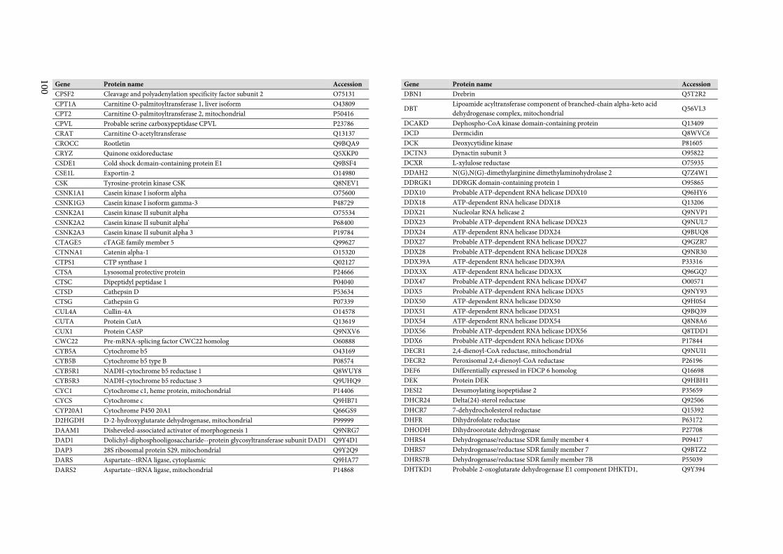
100
Gene Protein name Accession
CPSF2 Cleavage and polyadenylation specificity factor subunit 2 O75131
CPT1A Carnitine O-palmitoyltransferase 1, liver isoform O43809
CPT2 Carnitine O-palmitoyltransferase 2, mitochondrial P50416
CPVL Probable serine carboxypeptidase CPVL P23786
CRAT Carnitine O-acetyltransferase Q13137
CROCC Rootletin Q9BQA9
CRYZ Quinone oxidoreductase Q5XKP0
CSDE1 Cold shock domain-containing protein E1 Q9BSF4
CSE1L Exportin-2 O14980
CSK Tyrosine-protein kinase CSK Q8NEV1
CSNK1A1 Casein kinase I isoform alpha O75600
CSNK1G3 Casein kinase I isoform gamma-3 P48729
CSNK2A1 Casein kinase II subunit alpha O75534
CSNK2A2 Casein kinase II subunit alpha' P68400
CSNK2A3 Casein kinase II subunit alpha 3 P19784
CTAGE5 cTAGE family member 5 Q99627
CTNNA1 Catenin alpha-1 O15320
CTPS1 CTP synthase 1 Q02127
CTSA Lysosomal protective protein P24666
CTSC Dipeptidyl peptidase 1 P04040
CTSD Cathepsin D P53634
CTSG Cathepsin G P07339
CUL4A Cullin-4A O14578
CUTA Protein CutA Q13619
CUX1 Protein CASP Q9NXV6
CWC22 Pre-mRNA-splicing factor CWC22 homolog O60888
CYB5A Cytochrome b5 O43169
CYB5B Cytochrome b5 type B P08574
CYB5R1 NADH-cytochrome b5 reductase 1 Q8WUY8
CYB5R3 NADH-cytochrome b5 reductase 3 Q9UHQ9
CYC1 Cytochrome c1, heme protein, mitochondrial P14406
CYCS Cytochrome c Q9HB71
CYP20A1 Cytochrome P450 20A1 Q66GS9
D2HGDH D-2-hydroxyglutarate dehydrogenase, mitochondrial P99999
DAAM1 Disheveled-associated activator of morphogenesis 1 Q9NRG7
DAD1 Dolichyl-diphosphooligosaccharide--protein glycosyltransferase subunit DAD1 Q9Y4D1
DAP3 28S ribosomal protein S29, mitochondrial Q9Y2Q9
DARS Aspartate--tRNA ligase, cytoplasmic Q9HA77
DARS2 Aspartate--tRNA ligase, mitochondrial P14868
Gene Protein name Accession
DBN1 Drebrin Q5T2R2
DBT Lipoamide acyltransferase component of branched-chain alpha-keto acid
dehydrogenase complex, mitochondrial Q56VL3
DCAKD Dephospho-CoA kinase domain-containing protein Q13409
DCD Dermcidin Q8WVC6
DCK Deoxycytidine kinase P81605
DCTN3 Dynactin subunit 3 O95822
DCXR L-xylulose reductase O75935
DDAH2 N(G),N(G)-dimethylarginine dimethylaminohydrolase 2 Q7Z4W1
DDRGK1 DDRGK domain-containing protein 1 O95865
DDX10 Probable ATP-dependent RNA helicase DDX10 Q96HY6
DDX18 ATP-dependent RNA helicase DDX18 Q13206
DDX21 Nucleolar RNA helicase 2 Q9NVP1
DDX23 Probable ATP-dependent RNA helicase DDX23 Q9NUL7
DDX24 ATP-dependent RNA helicase DDX24 Q9BUQ8
DDX27 Probable ATP-dependent RNA helicase DDX27 Q9GZR7
DDX28 Probable ATP-dependent RNA helicase DDX28 Q9NR30
DDX39A ATP-dependent RNA helicase DDX39A P33316
DDX3X ATP-dependent RNA helicase DDX3X Q96GQ7
DDX47 Probable ATP-dependent RNA helicase DDX47 O00571
DDX5 Probable ATP-dependent RNA helicase DDX5 Q9NY93
DDX50 ATP-dependent RNA helicase DDX50 Q9H0S4
DDX51 ATP-dependent RNA helicase DDX51 Q9BQ39
DDX54 ATP-dependent RNA helicase DDX54 Q8N8A6
DDX56 Probable ATP-dependent RNA helicase DDX56 Q8TDD1
DDX6 Probable ATP-dependent RNA helicase DDX6 P17844
DECR1 2,4-dienoyl-CoA reductase, mitochondrial Q9NUI1
DECR2 Peroxisomal 2,4-dienoyl-CoA reductase P26196
DEF6 Differentially expressed in FDCP 6 homolog Q16698
DEK Protein DEK Q9HBH1
DESI2 Desumoylating isopeptidase 2 P35659
DHCR24 Delta(24)-sterol reductase Q92506
DHCR7 7-dehydrocholesterol reductase Q15392
DHFR Dihydrofolate reductase P63172
DHODH Dihydroorotate dehydrogenase P27708
DHRS4 Dehydrogenase/reductase SDR family member 4 P09417
DHRS7 Dehydrogenase/reductase SDR family member 7 Q9BTZ2
DHRS7B Dehydrogenase/reductase SDR family member 7B P55039
DHTKD1 Probable 2-oxoglutarate dehydrogenase E1 component DHKTD1, Q9Y394
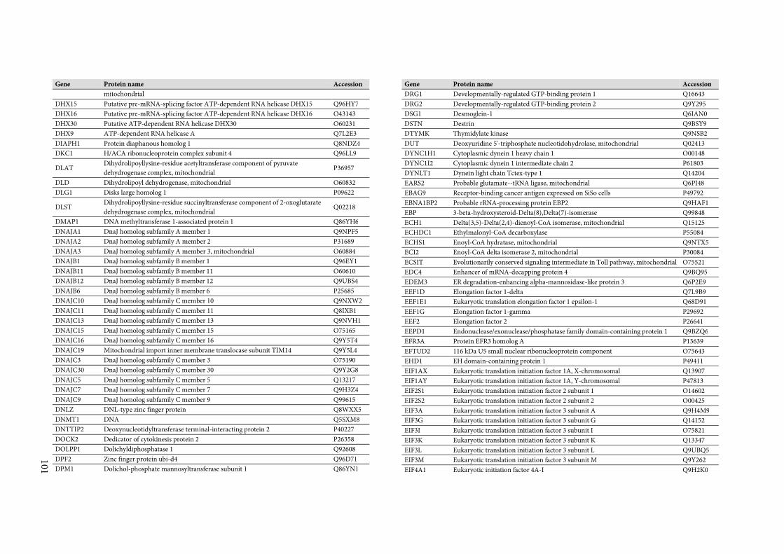
101
Gene Protein name Accession
mitochondrial
DHX15 Putative pre-mRNA-splicing factor ATP-dependent RNA helicase DHX15 Q96HY7
DHX16 Putative pre-mRNA-splicing factor ATP-dependent RNA helicase DHX16 O43143
DHX30 Putative ATP-dependent RNA helicase DHX30 O60231
DHX9 ATP-dependent RNA helicase A Q7L2E3
DIAPH1 Protein diaphanous homolog 1 Q8NDZ4
DKC1 H/ACA ribonucleoprotein complex subunit 4 Q96LL9
DLAT Dihydrolipoyllysine-residue acetyltransferase component of pyruvate
dehydrogenase complex, mitochondrial P36957
DLD Dihydrolipoyl dehydrogenase, mitochondrial O60832
DLG1 Disks large homolog 1 P09622
DLST Dihydrolipoyllysine-residue succinyltransferase component of 2-oxoglutarate
dehydrogenase complex, mitochondrial Q02218
DMAP1 DNA methyltransferase 1-associated protein 1 Q86YH6
DNAJA1 DnaJ homolog subfamily A member 1 Q9NPF5
DNAJA2 DnaJ homolog subfamily A member 2 P31689
DNAJA3 DnaJ homolog subfamily A member 3, mitochondrial O60884
DNAJB1 DnaJ homolog subfamily B member 1 Q96EY1
DNAJB11 DnaJ homolog subfamily B member 11 O60610
DNAJB12 DnaJ homolog subfamily B member 12 Q9UBS4
DNAJB6 DnaJ homolog subfamily B member 6 P25685
DNAJC10 DnaJ homolog subfamily C member 10 Q9NXW2
DNAJC11 DnaJ homolog subfamily C member 11 Q8IXB1
DNAJC13 DnaJ homolog subfamily C member 13 Q9NVH1
DNAJC15 DnaJ homolog subfamily C member 15 O75165
DNAJC16 DnaJ homolog subfamily C member 16 Q9Y5T4
DNAJC19 Mitochondrial import inner membrane translocase subunit TIM14 Q9Y5L4
DNAJC3 DnaJ homolog subfamily C member 3 O75190
DNAJC30 DnaJ homolog subfamily C member 30 Q9Y2G8
DNAJC5 DnaJ homolog subfamily C member 5 Q13217
DNAJC7 DnaJ homolog subfamily C member 7 Q9H3Z4
DNAJC9 DnaJ homolog subfamily C member 9 Q99615
DNLZ DNL-type zinc finger protein Q8WXX5
DNMT1 DNA Q5SXM8
DNTTIP2 Deoxynucleotidyltransferase terminal-interacting protein 2 P40227
DOCK2 Dedicator of cytokinesis protein 2 P26358
DOLPP1 Dolichyldiphosphatase 1 Q92608
DPF2 Zinc finger protein ubi-d4 Q96D71
DPM1 Dolichol-phosphate mannosyltransferase subunit 1 Q86YN1
Gene Protein name Accession
DRG1 Developmentally-regulated GTP-binding protein 1 Q16643
DRG2 Developmentally-regulated GTP-binding protein 2 Q9Y295
DSG1 Desmoglein-1 Q6IAN0
DSTN Destrin Q9BSY9
DTYMK Thymidylate kinase Q9NSB2
DUT Deoxyuridine 5'-triphosphate nucleotidohydrolase, mitochondrial Q02413
DYNC1H1 Cytoplasmic dynein 1 heavy chain 1 O00148
DYNC1I2 Cytoplasmic dynein 1 intermediate chain 2 P61803
DYNLT1 Dynein light chain Tctex-type 1 Q14204
EARS2 Probable glutamate--tRNA ligase, mitochondrial Q6PI48
EBAG9 Receptor-binding cancer antigen expressed on SiSo cells P49792
EBNA1BP2 Probable rRNA-processing protein EBP2 Q9HAF1
EBP 3-beta-hydroxysteroid-Delta(8),Delta(7)-isomerase Q99848
ECH1 Delta(3,5)-Delta(2,4)-dienoyl-CoA isomerase, mitochondrial Q15125
ECHDC1 Ethylmalonyl-CoA decarboxylase P55084
ECHS1 Enoyl-CoA hydratase, mitochondrial Q9NTX5
ECI2 Enoyl-CoA delta isomerase 2, mitochondrial P30084
ECSIT Evolutionarily conserved signaling intermediate in Toll pathway, mitochondrial O75521
EDC4 Enhancer of mRNA-decapping protein 4 Q9BQ95
EDEM3 ER degradation-enhancing alpha-mannosidase-like protein 3 Q6P2E9
EEF1D Elongation factor 1-delta Q7L9B9
EEF1E1 Eukaryotic translation elongation factor 1 epsilon-1 Q68D91
EEF1G Elongation factor 1-gamma P29692
EEF2 Elongation factor 2 P26641
EEPD1 Endonuclease/exonuclease/phosphatase family domain-containing protein 1 Q9BZQ6
EFR3A Protein EFR3 homolog A P13639
EFTUD2 116 kDa U5 small nuclear ribonucleoprotein component O75643
EHD1 EH domain-containing protein 1 P49411
EIF1AX Eukaryotic translation initiation factor 1A, X-chromosomal Q13907
EIF1AY Eukaryotic translation initiation factor 1A, Y-chromosomal P47813
EIF2S1 Eukaryotic translation initiation factor 2 subunit 1 O14602
EIF2S2 Eukaryotic translation initiation factor 2 subunit 2 O00425
EIF3A Eukaryotic translation initiation factor 3 subunit A Q9H4M9
EIF3G Eukaryotic translation initiation factor 3 subunit G Q14152
EIF3I Eukaryotic translation initiation factor 3 subunit I O75821
EIF3K Eukaryotic translation initiation factor 3 subunit K Q13347
EIF3L Eukaryotic translation initiation factor 3 subunit L Q9UBQ5
EIF3M Eukaryotic translation initiation factor 3 subunit M Q9Y262
EIF4A1 Eukaryotic initiation factor 4A-I Q9H2K0
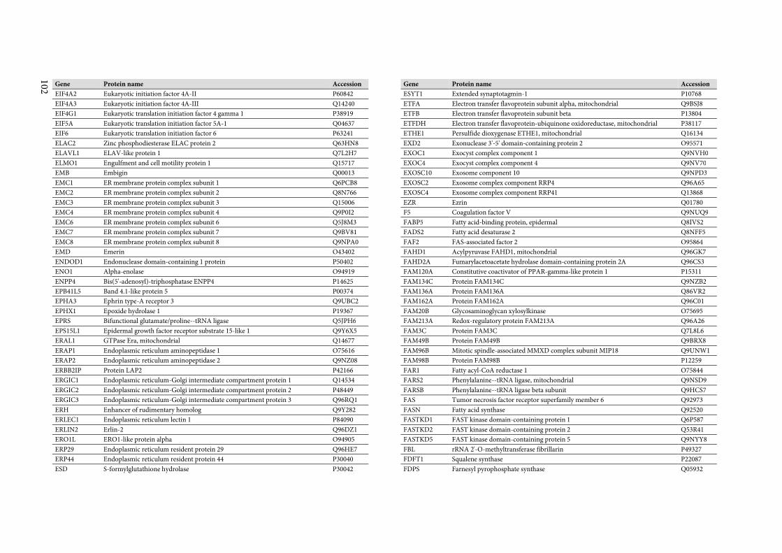
102
Gene Protein name Accession
EIF4A2 Eukaryotic initiation factor 4A-II P60842
EIF4A3 Eukaryotic initiation factor 4A-III Q14240
EIF4G1 Eukaryotic translation initiation factor 4 gamma 1 P38919
EIF5A Eukaryotic translation initiation factor 5A-1 Q04637
EIF6 Eukaryotic translation initiation factor 6 P63241
ELAC2 Zinc phosphodiesterase ELAC protein 2 Q63HN8
ELAVL1 ELAV-like protein 1 Q7L2H7
ELMO1 Engulfment and cell motility protein 1 Q15717
EMB Embigin Q00013
EMC1 ER membrane protein complex subunit 1 Q6PCB8
EMC2 ER membrane protein complex subunit 2 Q8N766
EMC3 ER membrane protein complex subunit 3 Q15006
EMC4 ER membrane protein complex subunit 4 Q9P0I2
EMC6 ER membrane protein complex subunit 6 Q5J8M3
EMC7 ER membrane protein complex subunit 7 Q9BV81
EMC8 ER membrane protein complex subunit 8 Q9NPA0
EMD Emerin O43402
ENDOD1 Endonuclease domain-containing 1 protein P50402
ENO1 Alpha-enolase O94919
ENPP4 Bis(5'-adenosyl)-triphosphatase ENPP4 P14625
EPB41L5 Band 4.1-like protein 5 P00374
EPHA3 Ephrin type-A receptor 3 Q9UBC2
EPHX1 Epoxide hydrolase 1 P19367
EPRS Bifunctional glutamate/proline--tRNA ligase Q5JPH6
EPS15L1 Epidermal growth factor receptor substrate 15-like 1 Q9Y6X5
ERAL1 GTPase Era, mitochondrial Q14677
ERAP1 Endoplasmic reticulum aminopeptidase 1 O75616
ERAP2 Endoplasmic reticulum aminopeptidase 2 Q9NZ08
ERBB2IP Protein LAP2 P42166
ERGIC1 Endoplasmic reticulum-Golgi intermediate compartment protein 1 Q14534
ERGIC2 Endoplasmic reticulum-Golgi intermediate compartment protein 2 P48449
ERGIC3 Endoplasmic reticulum-Golgi intermediate compartment protein 3 Q96RQ1
ERH Enhancer of rudimentary homolog Q9Y282
ERLEC1 Endoplasmic reticulum lectin 1 P84090
ERLIN2 Erlin-2 Q96DZ1
ERO1L ERO1-like protein alpha O94905
ERP29 Endoplasmic reticulum resident protein 29 Q96HE7
ERP44 Endoplasmic reticulum resident protein 44 P30040
ESD S-formylglutathione hydrolase P30042
Gene Protein name Accession
ESYT1 Extended synaptotagmin-1 P10768
ETFA Electron transfer flavoprotein subunit alpha, mitochondrial Q9BSJ8
ETFB Electron transfer flavoprotein subunit beta P13804
ETFDH Electron transfer flavoprotein-ubiquinone oxidoreductase, mitochondrial P38117
ETHE1 Persulfide dioxygenase ETHE1, mitochondrial Q16134
EXD2 Exonuclease 3'-5' domain-containing protein 2 O95571
EXOC1 Exocyst complex component 1 Q9NVH0
EXOC4 Exocyst complex component 4 Q9NV70
EXOSC10 Exosome component 10 Q9NPD3
EXOSC2 Exosome complex component RRP4 Q96A65
EXOSC4 Exosome complex component RRP41 Q13868
EZR Ezrin Q01780
F5 Coagulation factor V Q9NUQ9
FABP5 Fatty acid-binding protein, epidermal Q8IVS2
FADS2 Fatty acid desaturase 2 Q8NFF5
FAF2 FAS-associated factor 2 O95864
FAHD1 Acylpyruvase FAHD1, mitochondrial Q96GK7
FAHD2A Fumarylacetoacetate hydrolase domain-containing protein 2A Q96CS3
FAM120A Constitutive coactivator of PPAR-gamma-like protein 1 P15311
FAM134C Protein FAM134C Q9NZB2
FAM136A Protein FAM136A Q86VR2
FAM162A Protein FAM162A Q96C01
FAM20B Glycosaminoglycan xylosylkinase O75695
FAM213A Redox-regulatory protein FAM213A Q96A26
FAM3C Protein FAM3C Q7L8L6
FAM49B Protein FAM49B Q9BRX8
FAM96B Mitotic spindle-associated MMXD complex subunit MIP18 Q9UNW1
FAM98B Protein FAM98B P12259
FAR1 Fatty acyl-CoA reductase 1 O75844
FARS2 Phenylalanine--tRNA ligase, mitochondrial Q9NSD9
FARSB Phenylalanine--tRNA ligase beta subunit Q9HCS7
FAS Tumor necrosis factor receptor superfamily member 6 Q92973
FASN Fatty acid synthase Q92520
FASTKD1 FAST kinase domain-containing protein 1 Q6P587
FASTKD2 FAST kinase domain-containing protein 2 Q53R41
FASTKD5 FAST kinase domain-containing protein 5 Q9NYY8
FBL rRNA 2'-O-methyltransferase fibrillarin P49327
FDFT1 Squalene synthase P22087
FDPS Farnesyl pyrophosphate synthase Q05932
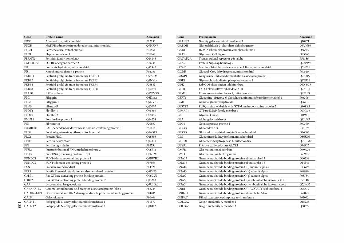
103
Gene Protein name Accession
FDX1 Adrenodoxin, mitochondrial P12236
FDXR NADPH:adrenodoxin oxidoreductase, mitochondrial Q9NRN7
FECH Ferrochelatase, mitochondrial P36551
FEN1 Flap endonuclease 1 P37268
FERMT3 Fermitin family homolog 3 Q14146
FGFR1OP2 FGFR1 oncogene partner 2 P39748
FH Fumarate hydratase, mitochondrial Q92945
FIS1 Mitochondrial fission 1 protein P02751
FKBP11 Peptidyl-prolyl cis-trans isomerase FKBP11 Q9Y3D6
FKBP2 Peptidyl-prolyl cis-trans isomerase FKBP2 Q9NYL4
FKBP4 Peptidyl-prolyl cis-trans isomerase FKBP4 P26885
FKBP8 Peptidyl-prolyl cis-trans isomerase FKBP8 Q02790
FLAD1 FAD synthase Q8WVX9
FLG Filaggrin Q5D862
FLG2 Filaggrin-2 Q9NVK5
FLNB Filamin-B Q15007
FLOT1 Flotillin-1 O75369
FLOT2 Flotillin-2 O75955
FMNL1 Formin-like protein 1 Q14254
FN1 Fibronectin P20930
FOXRED1 FAD-dependent oxidoreductase domain-containing protein 1 P51114
FPGS Folylpolyglutamate synthase, mitochondrial Q96DP5
FRG1 Protein FRG1 Q16595
FTH1 Ferritin heavy chain Q14331
FTL Ferritin light chain P02794
FTSJ2 Putative ribosomal RNA methyltransferase 2 Q96E11
FTSJ3 pre-rRNA processing protein FTSJ3 Q8NB90
FUNDC1 FUN14 domain-containing protein 1 Q9BWH2
FUNDC2 FUN14 domain-containing protein 2 P07954
FXN Frataxin, mitochondrial P14324
FXR1 Fragile X mental retardation syndrome-related protein 1 Q8IVP5
G3BP1 Ras GTPase-activating protein-binding protein 1 Q96CU9
G3BP2 Ras GTPase-activating protein-binding protein 2 Q13283
GAA Lysosomal alpha-glucosidase Q9UHA4
GABARAPL2 Gamma-aminobutyric acid receptor-associated protein-like 2 P63244
GADD45GIP1 Growth arrest and DNA damage-inducible proteins-interacting protein 1 P04406
GALK1 Galactokinase P80404
GALNT1 Polypeptide N-acetylgalactosaminyltransferase 1 P51570
GALNT2 Polypeptide N-acetylgalactosaminyltransferase 2 Q10472
Gene Protein name Accession
GALNT7 N-acetylgalactosaminyltransferase 7 Q10471
GAPDH Glyceraldehyde-3-phosphate dehydrogenase Q9UN86
GAR1 H/ACA ribonucleoprotein complex subunit 1 Q86SF2
GARS Glycine--tRNA ligase O95363
GATAD2A Transcriptional repressor p66-alpha P54886
GBAS Protein NipSnap homolog 2 Q9BPW8
GCAT 2-amino-3-ketobutyrate coenzyme A ligase, mitochondrial Q6YP21
GCDH Glutaryl-CoA dehydrogenase, mitochondrial P60520
GDAP1 Ganglioside-induced differentiation-associated protein 1 Q9H3P7
GDE1 Glycerophosphodiester phosphodiesterase 1 Q8TB36
GDI2 Rab GDP dissociation inhibitor beta Q9NZC3
GFER FAD-linked sulfhydryl oxidase ALR Q9BT30
GFM2 Ribosome-releasing factor 2, mitochondrial Q9P2E9
GFPT1 Glutamine--fructose-6-phosphate aminotransferase [isomerizing] 1 P06396
GGH Gamma-glutamyl hydrolase Q06210
GIGYF2 PERQ amino acid-rich with GYF domain-containing protein 2 Q96RR1
GIMAP1 GTPase IMAP family member 1 Q9H936
GK Glycerol kinase P04921
GLA Alpha-galactosidase A Q8IUX7
GLG1 Golgi apparatus protein 1 P00390
GLRX3 Glutaredoxin-3 P32189
GLRX5 Glutaredoxin-related protein 5, mitochondrial O76003
GLS Glutaminase kidney isoform, mitochondrial Q86SX6
GLUD1 Glutamate dehydrogenase 1, mitochondrial Q9UBM7
GLYR1 Putative oxidoreductase GLYR1 O94925
GMFB Glia maturation factor beta Q49A26
GMFG Glia maturation factor gamma P60983
GNA13 Guanine nucleotide-binding protein subunit alpha-13 O60234
GNA15 Guanine nucleotide-binding protein subunit alpha-15 Q14344
GNAI2 Guanine nucleotide-binding protein G(i) subunit alpha-2 P30679
GNAI3 Guanine nucleotide-binding protein G(k) subunit alpha P04899
GNAQ Guanine nucleotide-binding protein G(q) subunit alpha P08754
GNAS Guanine nucleotide-binding protein G(s) subunit alpha isoforms XLas P50148
GNAS Guanine nucleotide-binding protein G(s) subunit alpha isoforms short Q5JWF2
GNB1 Guanine nucleotide-binding protein G(I)/G(S)/G(T) subunit beta-1 O75879
GNB2L1 Guanine nucleotide-binding protein subunit beta-2-like 1 P62873
GNPAT Dihydroxyacetone phosphate acyltransferase P63092
GOLGA2 Golgin subfamily A member 2 O15228
GOLGA3 Golgin subfamily A member 3 Q08379
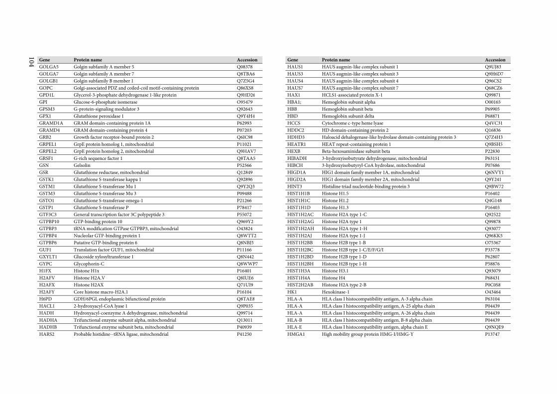
104
Gene Protein name Accession
GOLGA5 Golgin subfamily A member 5 Q08378
GOLGA7 Golgin subfamily A member 7 Q8TBA6
GOLGB1 Golgin subfamily B member 1 Q7Z5G4
GOPC Golgi-associated PDZ and coiled-coil motif-containing protein Q86XS8
GPD1L Glycerol-3-phosphate dehydrogenase 1-like protein Q9HD26
GPI Glucose-6-phosphate isomerase O95479
GPSM3 G-protein-signaling modulator 3 Q92643
GPX1 Glutathione peroxidase 1 Q9Y4H4
GRAMD1A GRAM domain-containing protein 1A P62993
GRAMD4 GRAM domain-containing protein 4 P07203
GRB2 Growth factor receptor-bound protein 2 Q6IC98
GRPEL1 GrpE protein homolog 1, mitochondrial P11021
GRPEL2 GrpE protein homolog 2, mitochondrial Q9HAV7
GRSF1 G-rich sequence factor 1 Q8TAA5
GSN Gelsolin P52566
GSR Glutathione reductase, mitochondrial Q12849
GSTK1 Glutathione S-transferase kappa 1 Q92896
GSTM1 Glutathione S-transferase Mu 1 Q9Y2Q3
GSTM3 Glutathione S-transferase Mu 3 P09488
GSTO1 Glutathione S-transferase omega-1 P21266
GSTP1 Glutathione S-transferase P P78417
GTF3C3 General transcription factor 3C polypeptide 3 P55072
GTPBP10 GTP-binding protein 10 Q969Y2
GTPBP3 tRNA modification GTPase GTPBP3, mitochondrial O43824
GTPBP4 Nucleolar GTP-binding protein 1 Q8WTT2
GTPBP6 Putative GTP-binding protein 6 Q8NBJ5
GUF1 Translation factor GUF1, mitochondrial P11166
GXYLT1 Glucoside xylosyltransferase 1 Q8N442
GYPC Glycophorin-C Q8WWP7
H1FX Histone H1x P16401
H2AFV Histone H2A.V Q8IUE6
H2AFX Histone H2AX Q71UI9
H2AFY Core histone macro-H2A.1 P16104
H6PD GDH/6PGL endoplasmic bifunctional protein Q8TAE8
HACL1 2-hydroxyacyl-CoA lyase 1 Q9P035
HADH Hydroxyacyl-coenzyme A dehydrogenase, mitochondrial Q99714
HADHA Trifunctional enzyme subunit alpha, mitochondrial Q13011
HADHB Trifunctional enzyme subunit beta, mitochondrial P40939
HARS2 Probable histidine--tRNA ligase, mitochondrial P41250
Gene Protein name Accession
HAUS1 HAUS augmin-like complex subunit 1 Q9UJ83
HAUS3 HAUS augmin-like complex subunit 3 Q9H6D7
HAUS4 HAUS augmin-like complex subunit 4 Q96CS2
HAUS7 HAUS augmin-like complex subunit 7 Q68CZ6
HAX1 HCLS1-associated protein X-1 Q99871
HBA1; Hemoglobin subunit alpha O00165
HBB Hemoglobin subunit beta P69905
HBD Hemoglobin subunit delta P68871
HCCS Cytochrome c-type heme lyase Q4VC31
HDDC2 HD domain-containing protein 2 Q16836
HDHD3 Haloacid dehalogenase-like hydrolase domain-containing protein 3 Q7Z4H3
HEATR1 HEAT repeat-containing protein 1 Q9BSH5
HEXB Beta-hexosaminidase subunit beta P22830
HIBADH 3-hydroxyisobutyrate dehydrogenase, mitochondrial P63151
HIBCH 3-hydroxyisobutyryl-CoA hydrolase, mitochondrial P07686
HIGD1A HIG1 domain family member 1A, mitochondrial Q6NVY1
HIGD2A HIG1 domain family member 2A, mitochondrial Q9Y241
HINT3 Histidine triad nucleotide-binding protein 3 Q9BW72
HIST1H1B Histone H1.5 P16402
HIST1H1C Histone H1.2 Q4G148
HIST1H1D Histone H1.3 P16403
HIST1H2AC Histone H2A type 1-C Q92522
HIST1H2AG Histone H2A type 1 Q99878
HIST1H2AH Histone H2A type 1-H Q93077
HIST1H2AJ Histone H2A type 1-J Q96KK5
HIST1H2BB Histone H2B type 1-B O75367
HIST1H2BC Histone H2B type 1-C/E/F/G/I P33778
HIST1H2BD Histone H2B type 1-D P62807
HIST1H2BH Histone H2B type 1-H P58876
HIST1H3A Histone H3.1 Q93079
HIST1H4A Histone H4 P68431
HIST2H2AB Histone H2A type 2-B P0C0S8
HK1 Hexokinase-1 O43464
HLA-A HLA class I histocompatibility antigen, A-3 alpha chain P63104
HLA-A HLA class I histocompatibility antigen, A-25 alpha chain P04439
HLA-A HLA class I histocompatibility antigen, A-26 alpha chain P04439
HLA-B HLA class I histocompatibility antigen, B-8 alpha chain P04439
HLA-E HLA class I histocompatibility antigen, alpha chain E Q9NQE9
HMGA1 High mobility group protein HMG-I/HMG-Y P13747
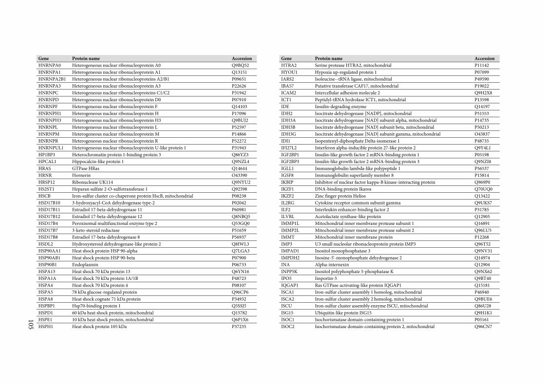
105
Gene Protein name Accession
HNRNPA0 Heterogeneous nuclear ribonucleoprotein A0 Q9BQ52
HNRNPA1 Heterogeneous nuclear ribonucleoprotein A1 Q13151
HNRNPA2B1 Heterogeneous nuclear ribonucleoproteins A2/B1 P09651
HNRNPA3 Heterogeneous nuclear ribonucleoprotein A3 P22626
HNRNPC Heterogeneous nuclear ribonucleoproteins C1/C2 P31942
HNRNPD Heterogeneous nuclear ribonucleoprotein D0 P07910
HNRNPF Heterogeneous nuclear ribonucleoprotein F Q14103
HNRNPH1 Heterogeneous nuclear ribonucleoprotein H P17096
HNRNPH3 Heterogeneous nuclear ribonucleoprotein H3 Q9BUJ2
HNRNPL Heterogeneous nuclear ribonucleoprotein L P52597
HNRNPM Heterogeneous nuclear ribonucleoprotein M P14866
HNRNPR Heterogeneous nuclear ribonucleoprotein R P52272
HNRNPUL1 Heterogeneous nuclear ribonucleoprotein U-like protein 1 P31943
HP1BP3 Heterochromatin protein 1-binding protein 3 Q86YZ3
HPCAL1 Hippocalcin-like protein 1 Q9NZL4
HRAS GTPase HRas Q14644
HRNR Hornerin O43390
HRSP12 Ribonuclease UK114 Q9NYU2
HS2ST1 Heparan sulfate 2-O-sulfotransferase 1 Q92598
HSCB Iron-sulfur cluster co-chaperone protein HscB, mitochondrial P08238
HSD17B10 3-hydroxyacyl-CoA dehydrogenase type-2 P02042
HSD17B11 Estradiol 17-beta-dehydrogenase 11 P60981
HSD17B12 Estradiol 17-beta-dehydrogenase 12 Q8NBQ5
HSD17B4 Peroxisomal multifunctional enzyme type 2 Q53GQ0
HSD17B7 3-keto-steroid reductase P51659
HSD17B8 Estradiol 17-beta-dehydrogenase 8 P56937
HSDL2 Hydroxysteroid dehydrogenase-like protein 2 Q8IWL3
HSP90AA1 Heat shock protein HSP 90-alpha Q7LGA3
HSP90AB1 Heat shock protein HSP 90-beta P07900
HSP90B1 Endoplasmin P06733
HSPA13 Heat shock 70 kDa protein 13 Q6YN16
HSPA1A Heat shock 70 kDa protein 1A/1B P48723
HSPA4 Heat shock 70 kDa protein 4 P08107
HSPA5 78 kDa glucose-regulated protein Q96CP6
HSPA8 Heat shock cognate 71 kDa protein P34932
HSPBP1 Hsp70-binding protein 1 Q5SSJ5
HSPD1 60 kDa heat shock protein, mitochondrial Q15782
HSPE1 10 kDa heat shock protein, mitochondrial Q6P1X6
HSPH1 Heat shock protein 105 kDa P37235
Gene Protein name Accession
HTRA2 Serine protease HTRA2, mitochondrial P11142
HYOU1 Hypoxia up-regulated protein 1 P07099
IARS2 Isoleucine--tRNA ligase, mitochondrial P49590
IBA57 Putative transferase CAF17, mitochondrial P19022
ICAM2 Intercellular adhesion molecule 2 Q9H2X8
ICT1 Peptidyl-tRNA hydrolase ICT1, mitochondrial P13598
IDE Insulin-degrading enzyme Q14197
IDH2 Isocitrate dehydrogenase [NADP], mitochondrial P51553
IDH3A Isocitrate dehydrogenase [NAD] subunit alpha, mitochondrial P14735
IDH3B Isocitrate dehydrogenase [NAD] subunit beta, mitochondrial P50213
IDH3G Isocitrate dehydrogenase [NAD] subunit gamma, mitochondrial O43837
IDI1 Isopentenyl-diphosphate Delta-isomerase 1 P48735
IFI27L2 Interferon alpha-inducible protein 27-like protein 2 Q9Y4L1
IGF2BP1 Insulin-like growth factor 2 mRNA-binding protein 1 P05198
IGF2BP3 Insulin-like growth factor 2 mRNA-binding protein 3 Q9NZI8
IGLL1 Immunoglobulin lambda-like polypeptide 1 P56537
IGSF8 Immunoglobulin superfamily member 8 P15814
IKBIP Inhibitor of nuclear factor kappa-B kinase-interacting protein Q969P0
IKZF1 DNA-binding protein Ikaros Q70UQ0
IKZF2 Zinc finger protein Helios Q13422
IL2RG Cytokine receptor common subunit gamma Q9UKS7
ILF2 Interleukin enhancer-binding factor 2 P31785
ILVBL Acetolactate synthase-like protein Q12905
IMMP1L Mitochondrial inner membrane protease subunit 1 Q16891
IMMP2L Mitochondrial inner membrane protease subunit 2 Q96LU5
IMMT Mitochondrial inner membrane protein P12268
IMP3 U3 small nucleolar ribonucleoprotein protein IMP3 Q96T52
IMPAD1 Inositol monophosphatase 3 Q9NV31
IMPDH2 Inosine-5'-monophosphate dehydrogenase 2 Q14974
INA Alpha-internexin Q12904
INPP5K Inositol polyphosphate 5-phosphatase K Q9NX62
IPO5 Importin-5 Q9BT40
IQGAP1 Ras GTPase-activating-like protein IQGAP1 Q15181
ISCA1 Iron-sulfur cluster assembly 1 homolog, mitochondrial P46940
ISCA2 Iron-sulfur cluster assembly 2 homolog, mitochondrial Q9BUE6
ISCU Iron-sulfur cluster assembly enzyme ISCU, mitochondrial Q86U28
ISG15 Ubiquitin-like protein ISG15 Q9H1K1
ISOC1 Isochorismatase domain-containing protein 1 P05161
ISOC2 Isochorismatase domain-containing protein 2, mitochondrial Q96CN7
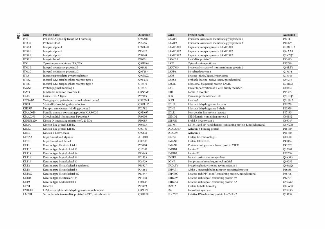
106
Gene Protein name Accession
ISY1 Pre-mRNA-splicing factor ISY1 homolog Q96AB3
ITFG3 Protein ITFG3 P05556
ITGA4 Integrin alpha-4 Q9ULR0
ITGA5 Integrin alpha-5 P13612
ITGAL Integrin alpha-L P08648
ITGB1 Integrin beta-1 P20701
ITK Tyrosine-protein kinase ITK/TSK Q9H0X4
ITM2B Integral membrane protein 2B Q08881
ITM2C Integral membrane protein 2C Q9Y287
ITPA Inosine triphosphate pyrophosphatase Q9NQX7
ITPR2 Inositol 1,4,5-trisphosphate receptor type 2 Q9BY32
ITPR3 Inositol 1,4,5-trisphosphate receptor type 3 Q14571
JAGN1 Protein jagunal homolog 1 Q14573
JAM3 Junctional adhesion molecule C Q8N5M9
KARS Lysine--tRNA ligase P57105
KCNAB2 Voltage-gated potassium channel subunit beta-2 Q9Y6M4
KDSR 3-ketodihydrosphingosine reductase Q9ULH0
KHSRP Far upstream element-binding protein 2 P02792
KIAA0020 Pumilio domain-containing protein KIAA0020 Q9BX67
KIAA0391 Mitochondrial ribonuclease P protein 3 P49006
KIDINS220 Kinase D-interacting substrate of 220 kDa P30085
KIF2A Kinesin-like protein KIF2A P46013
KIF2C Kinesin-like protein KIF2C O00139
KIF5B Kinesin-1 heavy chain Q99661
KPNA3 Importin subunit alpha-4 A1L0T0
KPNB1 Importin subunit beta-1 O00505
KRT1 Keratin, type II cytoskeletal 1 P35908
KRT10 Keratin, type I cytoskeletal 10 Q15397
KRT14 Keratin, type I cytoskeletal 14 P13645
KRT16 Keratin, type I cytoskeletal 16 P02533
KRT17 Keratin, type I cytoskeletal 17 P08779
KRT2 Keratin, type II cytoskeletal 2 epidermal P35527
KRT5 Keratin, type II cytoskeletal 5 P04264
KRT6C Keratin, type II cytoskeletal 6C P13647
KRT84 Keratin, type II cuticular Hb4 P14618
KRT9 Keratin, type I cytoskeletal 9 Q04695
KTN1 Kinectin P23919
L2HGDH L-2-hydroxyglutarate dehydrogenase, mitochondrial Q86UP2
LACTB Serine beta-lactamase-like protein LACTB, mitochondrial Q9H9P8
Gene Protein name Accession
LAMP1 Lysosome-associated membrane glycoprotein 1 P83111
LAMP2 Lysosome-associated membrane glycoprotein 2 P11279
LAMTOR1 Ragulator complex protein LAMTOR1 Q3MHD2
LAMTOR2 Ragulator complex protein LAMTOR2 Q6IAA8
LAMTOR3 Ragulator complex protein LAMTOR3 Q9Y2Q5
LANCL2 LanC-like protein 2 P13473
LAP3 Cytosol aminopeptidase P55789
LAPTM5 Lysosomal-associated transmembrane protein 5 Q96RT1
LARP4 La-related protein 4 Q13571
LARS Leucine--tRNA ligase, cytoplasmic Q15046
LARS2 Probable leucine--tRNA ligase, mitochondrial Q9P2J5
LAS1L Ribosomal biogenesis protein LAS1L Q71RC2
LAT Linker for activation of T-cells family member 1 Q01650
LBR Lamin-B receptor P05455
LCK Tyrosine-protein kinase Lck Q9UIQ6
LCP1 Plastin-2 Q9HBL7
LDHA L-lactate dehydrogenase A chain P06239
LDHB L-lactate dehydrogenase B chain P00338
LDLR Low-density lipoprotein receptor P07195
LEMD2 LEM domain-containing protein 2 O00182
LEPRE1 Prolyl 3-hydroxylase 1 O95747
LETM1 LETM1 and EF-hand domain-containing protein 1, mitochondrial Q8NC56
LGALS3BP Galectin-3-binding protein P16150
LGALS9 Galectin-9 P01130
LIN7C Protein lin-7 homolog C Q08380
LMAN1 Protein ERGIC-53 P43034
LMAN2 Vesicular integral-membrane protein VIP36 P49257
LMNB1 Lamin-B1 Q12907
LMNB2 Lamin-B2 P20700
LNPEP Leucyl-cystinyl aminopeptidase Q9Y383
LONP1 Lon protease homolog, mitochondrial Q03252
LPCAT1 Lysophosphatidylcholine acyltransferase 1 Q96AQ6
LRPAP1 Alpha-2-macroglobulin receptor-associated protein P28838
LRPPRC Leucine-rich PPR motif-containing protein, mitochondrial P36776
LRRC59 Leucine-rich repeat-containing protein 59 P42704
LRRC8A Leucine-rich repeat-containing protein 8A Q96AG4
LSM12 Protein LSM12 homolog Q8IWT6
LSS Lanosterol synthase Q969X5
LUC7L2 Putative RNA-binding protein Luc7-like 2 Q14739
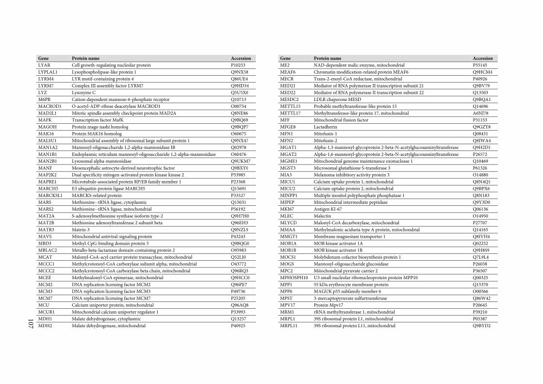
107
Gene Protein name Accession
LYAR Cell growth-regulating nucleolar protein P10253
LYPLAL1 Lysophospholipase-like protein 1 Q9NX58
LYRM4 LYR motif-containing protein 4 Q86UE4
LYRM7 Complex III assembly factor LYRM7 Q9HD34
LYZ Lysozyme C Q5U5X0
M6PR Cation-dependent mannose-6-phosphate receptor Q10713
MACROD1 O-acetyl-ADP-ribose deacetylase MACROD1 O00754
MAD2L1 Mitotic spindle assembly checkpoint protein MAD2A Q8NE86
MAFK Transcription factor MafK Q9BQ69
MAGOH Protein mago nashi homolog Q9BQP7
MAK16 Protein MAK16 homolog O60675
MALSU1 Mitochondrial assembly of ribosomal large subunit protein 1 Q9NX47
MAN1A2 Mannosyl-oligosaccharide 1,2-alpha-mannosidase IB Q02978
MAN1B1 Endoplasmic reticulum mannosyl-oligosaccharide 1,2-alpha-mannosidase O60476
MAN2B1 Lysosomal alpha-mannosidase Q9UKM7
MANF Mesencephalic astrocyte-derived neurotrophic factor Q9BXY0
MAP2K2 Dual specificity mitogen-activated protein kinase kinase 2 P53985
MAPRE1 Microtubule-associated protein RP/EB family member 1 P23368
MARCH5 E3 ubiquitin-protein ligase MARCH5 Q15691
MARCKSL1 MARCKS-related protein P33527
MARS Methionine--tRNA ligase, cytoplasmic Q15031
MARS2 Methionine--tRNA ligase, mitochondrial P56192
MAT2A S-adenosylmethionine synthase isoform type-2 Q9H7H0
MAT2B Methionine adenosyltransferase 2 subunit beta Q96EH3
MATR3 Matrin-3 Q9NZL9
MAVS Mitochondrial antiviral-signaling protein P43243
MBD3 Methyl-CpG-binding domain protein 3 Q9BQG0
MBLAC2 Metallo-beta-lactamase domain-containing protein 2 O95983
MCAT Malonyl-CoA-acyl carrier protein transacylase, mitochondrial Q52LJ0
MCCC1 Methylcrotonoyl-CoA carboxylase subunit alpha, mitochondrial O43772
MCCC2 Methylcrotonoyl-CoA carboxylase beta chain, mitochondrial Q96RQ3
MCEE Methylmalonyl-CoA epimerase, mitochondrial Q9HCC0
MCM2 DNA replication licensing factor MCM2 Q96PE7
MCM3 DNA replication licensing factor MCM3 P49736
MCM7 DNA replication licensing factor MCM7 P25205
MCU Calcium uniporter protein, mitochondrial Q96AQ8
MCUR1 Mitochondrial calcium uniporter regulator 1 P33993
MDH1 Malate dehydrogenase, cytoplasmic Q13257
MDH2 Malate dehydrogenase, mitochondrial P40925
Gene Protein name Accession
ME2 NAD-dependent malic enzyme, mitochondrial P55145
MEAF6 Chromatin modification-related protein MEAF6 Q9HCM4
MECR Trans-2-enoyl-CoA reductase, mitochondrial P40926
MED21 Mediator of RNA polymerase II transcription subunit 21 Q9BV79
MED22 Mediator of RNA polymerase II transcription subunit 22 Q13503
MESDC2 LDLR chaperone MESD Q9BQA1
METTL15 Probable methyltransferase-like protein 15 Q14696
METTL17 Methyltransferase-like protein 17, mitochondrial A6NJ78
MFF Mitochondrial fission factor P31153
MFGE8 Lactadherin Q9GZY8
MFN1 Mitofusin-1 Q08431
MFN2 Mitofusin-2 Q8IWA4
MGAT1 Alpha-1,3-mannosyl-glycoprotein 2-beta-N-acetylglucosaminyltransferase Q9H2D1
MGAT2 Alpha-1,6-mannosyl-glycoprotein 2-beta-N-acetylglucosaminyltransferase P26572
MGME1 Mitochondrial genome maintenance exonuclease 1 Q10469
MGST3 Microsomal glutathione S-transferase 3 P61326
MIA3 Melanoma inhibitory activity protein 3 O14880
MICU1 Calcium uptake protein 1, mitochondrial Q8N4Q1
MICU2 Calcium uptake protein 2, mitochondrial Q9BPX6
MINPP1 Multiple inositol polyphosphate phosphatase 1 Q8N183
MIPEP Mitochondrial intermediate peptidase Q9Y3D0
MKI67 Antigen KI-67 Q06136
MLEC Malectin O14950
MLYCD Malonyl-CoA decarboxylase, mitochondrial P27707
MMAA Methylmalonic aciduria type A protein, mitochondrial Q14165
MMGT1 Membrane magnesium transporter 1 Q8IVH4
MOB1A MOB kinase activator 1A Q02252
MOB1B MOB kinase activator 1B Q9H8S9
MOCS1 Molybdenum cofactor biosynthesis protein 1 Q7L9L4
MOGS Mannosyl-oligosaccharide glucosidase P26038
MPC2 Mitochondrial pyruvate carrier 2 P36507
MPHOSPH10 U3 small nucleolar ribonucleoprotein protein MPP10 Q00325
MPP1 55 kDa erythrocyte membrane protein Q15370
MPP6 MAGUK p55 subfamily member 6 O00566
MPST 3-mercaptopyruvate sulfurtransferase Q86W42
MPV17 Protein Mpv17 P20645
MRM1 rRNA methyltransferase 1, mitochondrial P39210
MRPL1 39S ribosomal protein L1, mitochondrial P05387
MRPL11 39S ribosomal protein L11, mitochondrial Q9BYD2
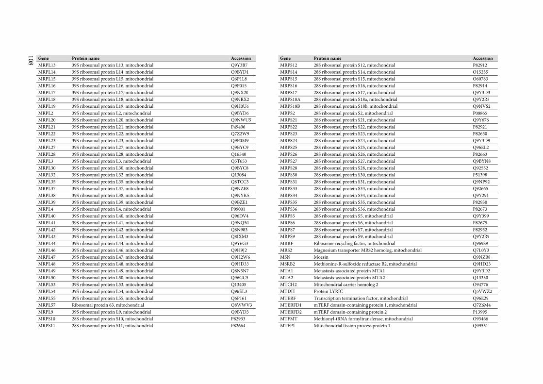
108
Gene Protein name Accession
MRPL13 39S ribosomal protein L13, mitochondrial Q9Y3B7
MRPL14 39S ribosomal protein L14, mitochondrial Q9BYD1
MRPL15 39S ribosomal protein L15, mitochondrial Q6P1L8
MRPL16 39S ribosomal protein L16, mitochondrial Q9P015
MRPL17 39S ribosomal protein L17, mitochondrial Q9NX20
MRPL18 39S ribosomal protein L18, mitochondrial Q9NRX2
MRPL19 39S ribosomal protein L19, mitochondrial Q9H0U6
MRPL2 39S ribosomal protein L2, mitochondrial Q9BYD6
MRPL20 39S ribosomal protein L20, mitochondrial Q9NWU5
MRPL21 39S ribosomal protein L21, mitochondrial P49406
MRPL22 39S ribosomal protein L22, mitochondrial Q7Z2W9
MRPL23 39S ribosomal protein L23, mitochondrial Q9P0M9
MRPL27 39S ribosomal protein L27, mitochondrial Q9BYC9
MRPL28 39S ribosomal protein L28, mitochondrial Q16540
MRPL3 39S ribosomal protein L3, mitochondrial Q5T653
MRPL30 39S ribosomal protein L30, mitochondrial Q9BYC8
MRPL32 39S ribosomal protein L32, mitochondrial Q13084
MRPL35 39S ribosomal protein L35, mitochondrial Q8TCC3
MRPL37 39S ribosomal protein L37, mitochondrial Q9NZE8
MRPL38 39S ribosomal protein L38, mitochondrial Q9NYK5
MRPL39 39S ribosomal protein L39, mitochondrial Q9BZE1
MRPL4 39S ribosomal protein L4, mitochondrial P09001
MRPL40 39S ribosomal protein L40, mitochondrial Q96DV4
MRPL41 39S ribosomal protein L41, mitochondrial Q9NQ50
MRPL42 39S ribosomal protein L42, mitochondrial Q8N983
MRPL43 39S ribosomal protein L43, mitochondrial Q8IXM3
MRPL44 39S ribosomal protein L44, mitochondrial Q9Y6G3
MRPL46 39S ribosomal protein L46, mitochondrial Q9H9J2
MRPL47 39S ribosomal protein L47, mitochondrial Q9H2W6
MRPL48 39S ribosomal protein L48, mitochondrial Q9HD33
MRPL49 39S ribosomal protein L49, mitochondrial Q8N5N7
MRPL50 39S ribosomal protein L50, mitochondrial Q96GC5
MRPL53 39S ribosomal protein L53, mitochondrial Q13405
MRPL54 39S ribosomal protein L54, mitochondrial Q96EL3
MRPL55 39S ribosomal protein L55, mitochondrial Q6P161
MRPL57 Ribosomal protein 63, mitochondrial Q8WWV3
MRPL9 39S ribosomal protein L9, mitochondrial Q9BYD3
MRPS10 28S ribosomal protein S10, mitochondrial P82933
MRPS11 28S ribosomal protein S11, mitochondrial P82664
Gene Protein name Accession
MRPS12 28S ribosomal protein S12, mitochondrial P82912
MRPS14 28S ribosomal protein S14, mitochondrial O15235
MRPS15 28S ribosomal protein S15, mitochondrial O60783
MRPS16 28S ribosomal protein S16, mitochondrial P82914
MRPS17 28S ribosomal protein S17, mitochondrial Q9Y3D3
MRPS18A 28S ribosomal protein S18a, mitochondrial Q9Y2R5
MRPS18B 28S ribosomal protein S18b, mitochondrial Q9NVS2
MRPS2 28S ribosomal protein S2, mitochondrial P08865
MRPS21 28S ribosomal protein S21, mitochondrial Q9Y676
MRPS22 28S ribosomal protein S22, mitochondrial P82921
MRPS23 28S ribosomal protein S23, mitochondrial P82650
MRPS24 28S ribosomal protein S24, mitochondrial Q9Y3D9
MRPS25 28S ribosomal protein S25, mitochondrial Q96EL2
MRPS26 28S ribosomal protein S26, mitochondrial P82663
MRPS27 28S ribosomal protein S27, mitochondrial Q9BYN8
MRPS28 28S ribosomal protein S28, mitochondrial Q92552
MRPS30 28S ribosomal protein S30, mitochondrial P51398
MRPS31 28S ribosomal protein S31, mitochondrial Q9NP92
MRPS33 28S ribosomal protein S33, mitochondrial Q92665
MRPS34 28S ribosomal protein S34, mitochondrial Q9Y291
MRPS35 28S ribosomal protein S35, mitochondrial P82930
MRPS36 28S ribosomal protein S36, mitochondrial P82673
MRPS5 28S ribosomal protein S5, mitochondrial Q9Y399
MRPS6 28S ribosomal protein S6, mitochondrial P82675
MRPS7 28S ribosomal protein S7, mitochondrial P82932
MRPS9 28S ribosomal protein S9, mitochondrial Q9Y2R9
MRRF Ribosome-recycling factor, mitochondrial Q969S9
MRS2 Magnesium transporter MRS2 homolog, mitochondrial Q7L0Y3
MSN Moesin Q9NZB8
MSRB2 Methionine-R-sulfoxide reductase B2, mitochondrial Q9HD23
MTA1 Metastasis-associated protein MTA1 Q9Y3D2
MTA2 Metastasis-associated protein MTA2 Q13330
MTCH2 Mitochondrial carrier homolog 2 O94776
MTDH Protein LYRIC Q5VWZ2
MTERF Transcription termination factor, mitochondrial Q96E29
MTERFD1 mTERF domain-containing protein 1, mitochondrial Q7Z6M4
MTERFD2 mTERF domain-containing protein 2 P13995
MTFMT Methionyl-tRNA formyltransferase, mitochondrial O95466
MTFP1 Mitochondrial fission process protein 1 Q99551
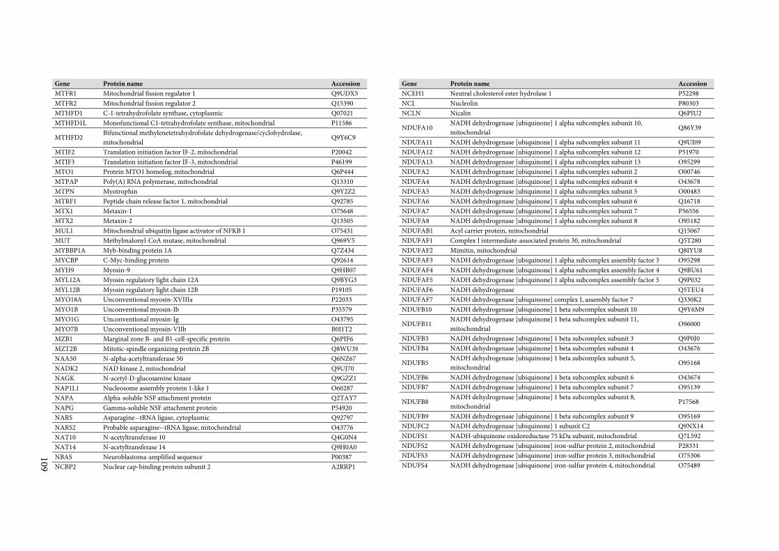
109
Gene Protein name Accession
MTFR1 Mitochondrial fission regulator 1 Q9UDX5
MTFR2 Mitochondrial fission regulator 2 Q15390
MTHFD1 C-1-tetrahydrofolate synthase, cytoplasmic Q07021
MTHFD1L Monofunctional C1-tetrahydrofolate synthase, mitochondrial P11586
MTHFD2 Bifunctional methylenetetrahydrofolate dehydrogenase/cyclohydrolase,
mitochondrial Q9Y6C9
MTIF2 Translation initiation factor IF-2, mitochondrial P20042
MTIF3 Translation initiation factor IF-3, mitochondrial P46199
MTO1 Protein MTO1 homolog, mitochondrial Q6P444
MTPAP Poly(A) RNA polymerase, mitochondrial Q13310
MTPN Myotrophin Q9Y2Z2
MTRF1 Peptide chain release factor 1, mitochondrial Q92785
MTX1 Metaxin-1 O75648
MTX2 Metaxin-2 Q13505
MUL1 Mitochondrial ubiquitin ligase activator of NFKB 1 O75431
MUT Methylmalonyl-CoA mutase, mitochondrial Q969V5
MYBBP1A Myb-binding protein 1A Q7Z434
MYCBP C-Myc-binding protein Q92614
MYH9 Myosin-9 Q9HB07
MYL12A Myosin regulatory light chain 12A Q9BYG3
MYL12B Myosin regulatory light chain 12B P19105
MYO18A Unconventional myosin-XVIIIa P22033
MYO1B Unconventional myosin-Ib P35579
MYO1G Unconventional myosin-Ig O43795
MYO7B Unconventional myosin-VIIb B0I1T2
MZB1 Marginal zone B- and B1-cell-specific protein Q6PIF6
MZT2B Mitotic-spindle organizing protein 2B Q8WU39
NAA50 N-alpha-acetyltransferase 50 Q6NZ67
NADK2 NAD kinase 2, mitochondrial Q9UJ70
NAGK N-acetyl-D-glucosamine kinase Q9GZZ1
NAP1L1 Nucleosome assembly protein 1-like 1 O60287
NAPA Alpha-soluble NSF attachment protein Q2TAY7
NAPG Gamma-soluble NSF attachment protein P54920
NARS Asparagine--tRNA ligase, cytoplasmic Q92797
NARS2 Probable asparagine--tRNA ligase, mitochondrial O43776
NAT10 N-acetyltransferase 10 Q4G0N4
NAT14 N-acetyltransferase 14 Q9H0A0
NBAS Neuroblastoma-amplified sequence P00387
NCBP2 Nuclear cap-binding protein subunit 2 A2RRP1
Gene Protein name Accession
NCEH1 Neutral cholesterol ester hydrolase 1 P52298
NCL Nucleolin P80303
NCLN Nicalin Q6PIU2
NDUFA10 NADH dehydrogenase [ubiquinone] 1 alpha subcomplex subunit 10,
mitochondrial Q86Y39
NDUFA11 NADH dehydrogenase [ubiquinone] 1 alpha subcomplex subunit 11 Q9UI09
NDUFA12 NADH dehydrogenase [ubiquinone] 1 alpha subcomplex subunit 12 P51970
NDUFA13 NADH dehydrogenase [ubiquinone] 1 alpha subcomplex subunit 13 O95299
NDUFA2 NADH dehydrogenase [ubiquinone] 1 alpha subcomplex subunit 2 O00746
NDUFA4 NADH dehydrogenase [ubiquinone] 1 alpha subcomplex subunit 4 O43678
NDUFA5 NADH dehydrogenase [ubiquinone] 1 alpha subcomplex subunit 5 O00483
NDUFA6 NADH dehydrogenase [ubiquinone] 1 alpha subcomplex subunit 6 Q16718
NDUFA7 NADH dehydrogenase [ubiquinone] 1 alpha subcomplex subunit 7 P56556
NDUFA8 NADH dehydrogenase [ubiquinone] 1 alpha subcomplex subunit 8 O95182
NDUFAB1 Acyl carrier protein, mitochondrial Q15067
NDUFAF1 Complex I intermediate-associated protein 30, mitochondrial Q5T280
NDUFAF2 Mimitin, mitochondrial Q8IYU8
NDUFAF3 NADH dehydrogenase [ubiquinone] 1 alpha subcomplex assembly factor 3 O95298
NDUFAF4 NADH dehydrogenase [ubiquinone] 1 alpha subcomplex assembly factor 4 Q9BU61
NDUFAF5 NADH dehydrogenase [ubiquinone] 1 alpha subcomplex assembly factor 5 Q9P032
NDUFAF6 NADH dehydrogenase Q5TEU4
NDUFAF7 NADH dehydrogenase [ubiquinone] complex I, assembly factor 7 Q330K2
NDUFB10 NADH dehydrogenase [ubiquinone] 1 beta subcomplex subunit 10 Q9Y6M9
NDUFB11 NADH dehydrogenase [ubiquinone] 1 beta subcomplex subunit 11,
mitochondrial O96000
NDUFB3 NADH dehydrogenase [ubiquinone] 1 beta subcomplex subunit 3 Q9P0J0
NDUFB4 NADH dehydrogenase [ubiquinone] 1 beta subcomplex subunit 4 O43676
NDUFB5 NADH dehydrogenase [ubiquinone] 1 beta subcomplex subunit 5,
mitochondrial O95168
NDUFB6 NADH dehydrogenase [ubiquinone] 1 beta subcomplex subunit 6 O43674
NDUFB7 NADH dehydrogenase [ubiquinone] 1 beta subcomplex subunit 7 O95139
NDUFB8 NADH dehydrogenase [ubiquinone] 1 beta subcomplex subunit 8,
mitochondrial P17568
NDUFB9 NADH dehydrogenase [ubiquinone] 1 beta subcomplex subunit 9 O95169
NDUFC2 NADH dehydrogenase [ubiquinone] 1 subunit C2 Q9NX14
NDUFS1 NADH-ubiquinone oxidoreductase 75 kDa subunit, mitochondrial Q7L592
NDUFS2 NADH dehydrogenase [ubiquinone] iron-sulfur protein 2, mitochondrial P28331
NDUFS3 NADH dehydrogenase [ubiquinone] iron-sulfur protein 3, mitochondrial O75306
NDUFS4 NADH dehydrogenase [ubiquinone] iron-sulfur protein 4, mitochondrial O75489
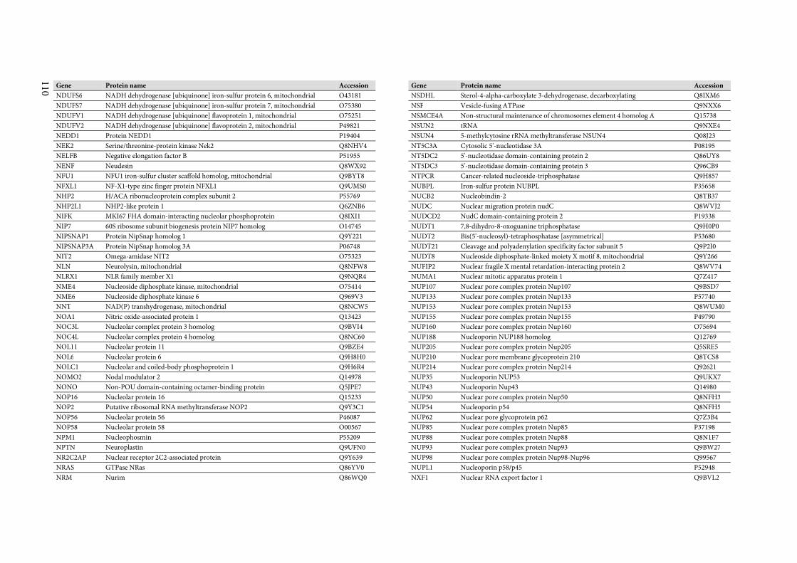
110
Gene Protein name Accession
NDUFS6 NADH dehydrogenase [ubiquinone] iron-sulfur protein 6, mitochondrial O43181
NDUFS7 NADH dehydrogenase [ubiquinone] iron-sulfur protein 7, mitochondrial O75380
NDUFV1 NADH dehydrogenase [ubiquinone] flavoprotein 1, mitochondrial O75251
NDUFV2 NADH dehydrogenase [ubiquinone] flavoprotein 2, mitochondrial P49821
NEDD1 Protein NEDD1 P19404
NEK2 Serine/threonine-protein kinase Nek2 Q8NHV4
NELFB Negative elongation factor B P51955
NENF Neudesin Q8WX92
NFU1 NFU1 iron-sulfur cluster scaffold homolog, mitochondrial Q9BYT8
NFXL1 NF-X1-type zinc finger protein NFXL1 Q9UMS0
NHP2 H/ACA ribonucleoprotein complex subunit 2 P55769
NHP2L1 NHP2-like protein 1 Q6ZNB6
NIFK MKI67 FHA domain-interacting nucleolar phosphoprotein Q8IXI1
NIP7 60S ribosome subunit biogenesis protein NIP7 homolog O14745
NIPSNAP1 Protein NipSnap homolog 1 Q9Y221
NIPSNAP3A Protein NipSnap homolog 3A P06748
NIT2 Omega-amidase NIT2 O75323
NLN Neurolysin, mitochondrial Q8NFW8
NLRX1 NLR family member X1 Q9NQR4
NME4 Nucleoside diphosphate kinase, mitochondrial O75414
NME6 Nucleoside diphosphate kinase 6 Q969V3
NNT NAD(P) transhydrogenase, mitochondrial Q8NCW5
NOA1 Nitric oxide-associated protein 1 Q13423
NOC3L Nucleolar complex protein 3 homolog Q9BVI4
NOC4L Nucleolar complex protein 4 homolog Q8NC60
NOL11 Nucleolar protein 11 Q9BZE4
NOL6 Nucleolar protein 6 Q9H8H0
NOLC1 Nucleolar and coiled-body phosphoprotein 1 Q9H6R4
NOMO2 Nodal modulator 2 Q14978
NONO Non-POU domain-containing octamer-binding protein Q5JPE7
NOP16 Nucleolar protein 16 Q15233
NOP2 Putative ribosomal RNA methyltransferase NOP2 Q9Y3C1
NOP56 Nucleolar protein 56 P46087
NOP58 Nucleolar protein 58 O00567
NPM1 Nucleophosmin P55209
NPTN Neuroplastin Q9UFN0
NR2C2AP Nuclear receptor 2C2-associated protein Q9Y639
NRAS GTPase NRas Q86YV0
NRM Nurim Q86WQ0
Gene Protein name Accession
NSDHL Sterol-4-alpha-carboxylate 3-dehydrogenase, decarboxylating Q8IXM6
NSF Vesicle-fusing ATPase Q9NXX6
NSMCE4A Non-structural maintenance of chromosomes element 4 homolog A Q15738
NSUN2 tRNA Q9NXE4
NSUN4 5-methylcytosine rRNA methyltransferase NSUN4 Q08J23
NT5C3A Cytosolic 5'-nucleotidase 3A P08195
NT5DC2 5'-nucleotidase domain-containing protein 2 Q86UY8
NT5DC3 5'-nucleotidase domain-containing protein 3 Q96CB9
NTPCR Cancer-related nucleoside-triphosphatase Q9H857
NUBPL Iron-sulfur protein NUBPL P35658
NUCB2 Nucleobindin-2 Q8TB37
NUDC Nuclear migration protein nudC Q8WVJ2
NUDCD2 NudC domain-containing protein 2 P19338
NUDT1 7,8-dihydro-8-oxoguanine triphosphatase Q9H0P0
NUDT2 Bis(5'-nucleosyl)-tetraphosphatase [asymmetrical] P53680
NUDT21 Cleavage and polyadenylation specificity factor subunit 5 Q9P2I0
NUDT8 Nucleoside diphosphate-linked moiety X motif 8, mitochondrial Q9Y266
NUFIP2 Nuclear fragile X mental retardation-interacting protein 2 Q8WV74
NUMA1 Nuclear mitotic apparatus protein 1 Q7Z417
NUP107 Nuclear pore complex protein Nup107 Q9BSD7
NUP133 Nuclear pore complex protein Nup133 P57740
NUP153 Nuclear pore complex protein Nup153 Q8WUM0
NUP155 Nuclear pore complex protein Nup155 P49790
NUP160 Nuclear pore complex protein Nup160 O75694
NUP188 Nucleoporin NUP188 homolog Q12769
NUP205 Nuclear pore complex protein Nup205 Q5SRE5
NUP210 Nuclear pore membrane glycoprotein 210 Q8TCS8
NUP214 Nuclear pore complex protein Nup214 Q92621
NUP35 Nucleoporin NUP53 Q9UKX7
NUP43 Nucleoporin Nup43 Q14980
NUP50 Nuclear pore complex protein Nup50 Q8NFH3
NUP54 Nucleoporin p54 Q8NFH5
NUP62 Nuclear pore glycoprotein p62 Q7Z3B4
NUP85 Nuclear pore complex protein Nup85 P37198
NUP88 Nuclear pore complex protein Nup88 Q8N1F7
NUP93 Nuclear pore complex protein Nup93 Q9BW27
NUP98 Nuclear pore complex protein Nup98-Nup96 Q99567
NUPL1 Nucleoporin p58/p45 P52948
NXF1 Nuclear RNA export factor 1 Q9BVL2
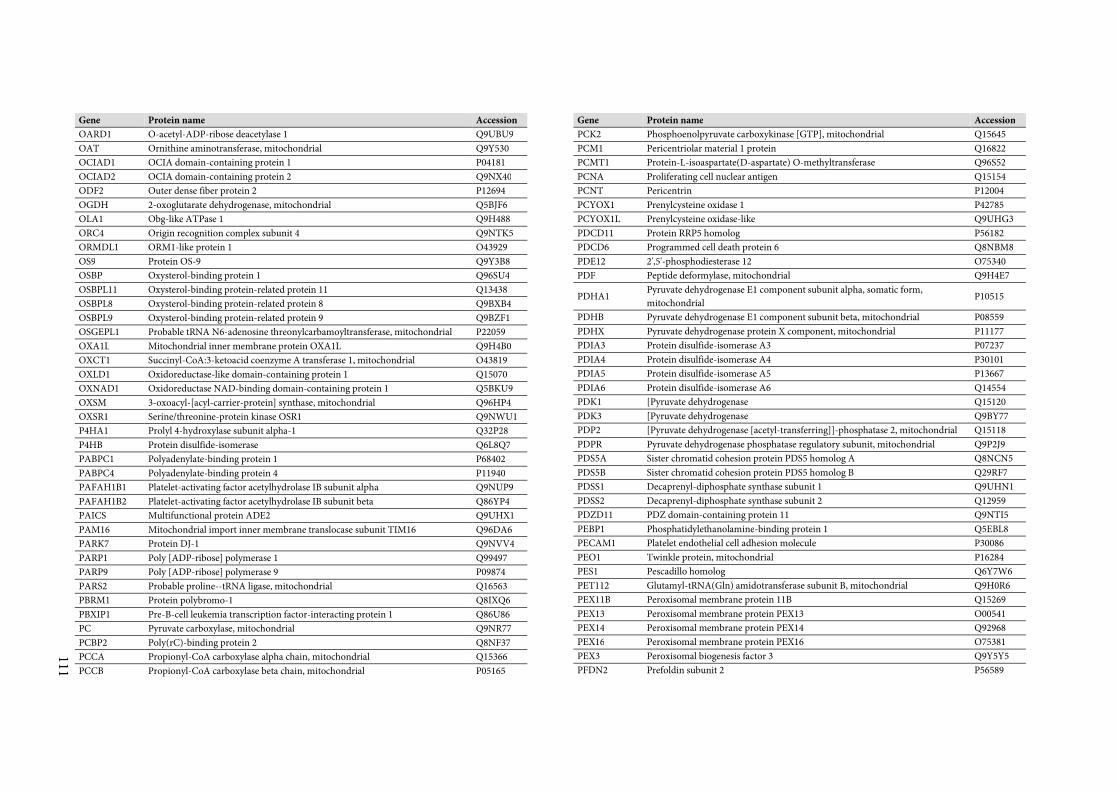
111
Gene Protein name Accession
OARD1 O-acetyl-ADP-ribose deacetylase 1 Q9UBU9
OAT Ornithine aminotransferase, mitochondrial Q9Y530
OCIAD1 OCIA domain-containing protein 1 P04181
OCIAD2 OCIA domain-containing protein 2 Q9NX40
ODF2 Outer dense fiber protein 2 P12694
OGDH 2-oxoglutarate dehydrogenase, mitochondrial Q5BJF6
OLA1 Obg-like ATPase 1 Q9H488
ORC4 Origin recognition complex subunit 4 Q9NTK5
ORMDL1 ORM1-like protein 1 O43929
OS9 Protein OS-9 Q9Y3B8
OSBP Oxysterol-binding protein 1 Q96SU4
OSBPL11 Oxysterol-binding protein-related protein 11 Q13438
OSBPL8 Oxysterol-binding protein-related protein 8 Q9BXB4
OSBPL9 Oxysterol-binding protein-related protein 9 Q9BZF1
OSGEPL1 Probable tRNA N6-adenosine threonylcarbamoyltransferase, mitochondrial P22059
OXA1L Mitochondrial inner membrane protein OXA1L Q9H4B0
OXCT1 Succinyl-CoA:3-ketoacid coenzyme A transferase 1, mitochondrial O43819
OXLD1 Oxidoreductase-like domain-containing protein 1 Q15070
OXNAD1 Oxidoreductase NAD-binding domain-containing protein 1 Q5BKU9
OXSM 3-oxoacyl-[acyl-carrier-protein] synthase, mitochondrial Q96HP4
OXSR1 Serine/threonine-protein kinase OSR1 Q9NWU1
P4HA1 Prolyl 4-hydroxylase subunit alpha-1 Q32P28
P4HB Protein disulfide-isomerase Q6L8Q7
PABPC1 Polyadenylate-binding protein 1 P68402
PABPC4 Polyadenylate-binding protein 4 P11940
PAFAH1B1 Platelet-activating factor acetylhydrolase IB subunit alpha Q9NUP9
PAFAH1B2 Platelet-activating factor acetylhydrolase IB subunit beta Q86YP4
PAICS Multifunctional protein ADE2 Q9UHX1
PAM16 Mitochondrial import inner membrane translocase subunit TIM16 Q96DA6
PARK7 Protein DJ-1 Q9NVV4
PARP1 Poly [ADP-ribose] polymerase 1 Q99497
PARP9 Poly [ADP-ribose] polymerase 9 P09874
PARS2 Probable proline--tRNA ligase, mitochondrial Q16563
PBRM1 Protein polybromo-1 Q8IXQ6
PBXIP1 Pre-B-cell leukemia transcription factor-interacting protein 1 Q86U86
PC Pyruvate carboxylase, mitochondrial Q9NR77
PCBP2 Poly(rC)-binding protein 2 Q8NF37
PCCA Propionyl-CoA carboxylase alpha chain, mitochondrial Q15366
PCCB Propionyl-CoA carboxylase beta chain, mitochondrial P05165
Gene Protein name Accession
PCK2 Phosphoenolpyruvate carboxykinase [GTP], mitochondrial Q15645
PCM1 Pericentriolar material 1 protein Q16822
PCMT1 Protein-L-isoaspartate(D-aspartate) O-methyltransferase Q96S52
PCNA Proliferating cell nuclear antigen Q15154
PCNT Pericentrin P12004
PCYOX1 Prenylcysteine oxidase 1 P42785
PCYOX1L Prenylcysteine oxidase-like Q9UHG3
PDCD11 Protein RRP5 homolog P56182
PDCD6 Programmed cell death protein 6 Q8NBM8
PDE12 2',5'-phosphodiesterase 12 O75340
PDF Peptide deformylase, mitochondrial Q9H4E7
PDHA1 Pyruvate dehydrogenase E1 component subunit alpha, somatic form,
mitochondrial P10515
PDHB Pyruvate dehydrogenase E1 component subunit beta, mitochondrial P08559
PDHX Pyruvate dehydrogenase protein X component, mitochondrial P11177
PDIA3 Protein disulfide-isomerase A3 P07237
PDIA4 Protein disulfide-isomerase A4 P30101
PDIA5 Protein disulfide-isomerase A5 P13667
PDIA6 Protein disulfide-isomerase A6 Q14554
PDK1 [Pyruvate dehydrogenase Q15120
PDK3 [Pyruvate dehydrogenase Q9BY77
PDP2 [Pyruvate dehydrogenase [acetyl-transferring]]-phosphatase 2, mitochondrial Q15118
PDPR Pyruvate dehydrogenase phosphatase regulatory subunit, mitochondrial Q9P2J9
PDS5A Sister chromatid cohesion protein PDS5 homolog A Q8NCN5
PDS5B Sister chromatid cohesion protein PDS5 homolog B Q29RF7
PDSS1 Decaprenyl-diphosphate synthase subunit 1 Q9UHN1
PDSS2 Decaprenyl-diphosphate synthase subunit 2 Q12959
PDZD11 PDZ domain-containing protein 11 Q9NTI5
PEBP1 Phosphatidylethanolamine-binding protein 1 Q5EBL8
PECAM1 Platelet endothelial cell adhesion molecule P30086
PEO1 Twinkle protein, mitochondrial P16284
PES1 Pescadillo homolog Q6Y7W6
PET112 Glutamyl-tRNA(Gln) amidotransferase subunit B, mitochondrial Q9H0R6
PEX11B Peroxisomal membrane protein 11B Q15269
PEX13 Peroxisomal membrane protein PEX13 O00541
PEX14 Peroxisomal membrane protein PEX14 Q92968
PEX16 Peroxisomal membrane protein PEX16 O75381
PEX3 Peroxisomal biogenesis factor 3 Q9Y5Y5
PFDN2 Prefoldin subunit 2 P56589
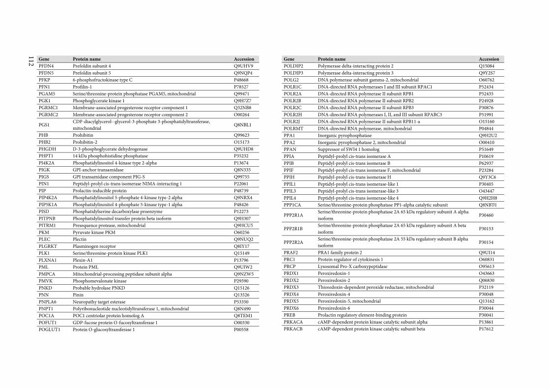
112
Gene Protein name Accession
PFDN4 Prefoldin subunit 4 Q9UHV9
PFDN5 Prefoldin subunit 5 Q9NQP4
PFKP 6-phosphofructokinase type C P48668
PFN1 Profilin-1 P78527
PGAM5 Serine/threonine-protein phosphatase PGAM5, mitochondrial Q99471
PGK1 Phosphoglycerate kinase 1 Q9H7Z7
PGRMC1 Membrane-associated progesterone receptor component 1 Q32NB8
PGRMC2 Membrane-associated progesterone receptor component 2 O00264
PGS1 CDP-diacylglycerol--glycerol-3-phosphate 3-phosphatidyltransferase,
mitochondrial Q8NBL1
PHB Prohibitin Q99623
PHB2 Prohibitin-2 O15173
PHGDH D-3-phosphoglycerate dehydrogenase Q9UHD8
PHPT1 14 kDa phosphohistidine phosphatase P35232
PI4K2A Phosphatidylinositol 4-kinase type 2-alpha P13674
PIGK GPI-anchor transamidase Q8N335
PIGS GPI transamidase component PIG-S Q99755
PIN1 Peptidyl-prolyl cis-trans isomerase NIMA-interacting 1 P22061
PIP Prolactin-inducible protein P48739
PIP4K2A Phosphatidylinositol 5-phosphate 4-kinase type-2 alpha Q9NRX4
PIP5K1A Phosphatidylinositol 4-phosphate 5-kinase type-1 alpha P48426
PISD Phosphatidylserine decarboxylase proenzyme P12273
PITPNB Phosphatidylinositol transfer protein beta isoform Q9H307
PITRM1 Presequence protease, mitochondrial Q9HCU5
PKM Pyruvate kinase PKM O60256
PLEC Plectin Q9NUQ2
PLGRKT Plasminogen receptor Q8IY17
PLK1 Serine/threonine-protein kinase PLK1 Q15149
PLXNA1 Plexin-A1 P13796
PML Protein PML Q9UIW2
PMPCA Mitochondrial-processing peptidase subunit alpha Q9NZW5
PMVK Phosphomevalonate kinase P29590
PNKD Probable hydrolase PNKD Q15126
PNN Pinin Q13526
PNPLA6 Neuropathy target esterase P53350
PNPT1 Polyribonucleotide nucleotidyltransferase 1, mitochondrial Q8N490
POC1A POC1 centriolar protein homolog A Q8TEM1
POFUT1 GDP-fucose protein O-fucosyltransferase 1 O00330
POGLUT1 Protein O-glucosyltransferase 1 P00558
Gene Protein name Accession
POLDIP2 Polymerase delta-interacting protein 2 Q15084
POLDIP3 Polymerase delta-interacting protein 3 Q9Y2S7
POLG2 DNA polymerase subunit gamma-2, mitochondrial O60762
POLR1C DNA-directed RNA polymerases I and III subunit RPAC1 P52434
POLR2A DNA-directed RNA polymerase II subunit RPB1 P52435
POLR2B DNA-directed RNA polymerase II subunit RPB2 P24928
POLR2C DNA-directed RNA polymerase II subunit RPB3 P30876
POLR2H DNA-directed RNA polymerases I, II, and III subunit RPABC3 P51991
POLR2J DNA-directed RNA polymerase II subunit RPB11-a O15160
POLRMT DNA-directed RNA polymerase, mitochondrial P04844
PPA1 Inorganic pyrophosphatase Q9H2U2
PPA2 Inorganic pyrophosphatase 2, mitochondrial O00410
PPAN Suppressor of SWI4 1 homolog P51649
PPIA Peptidyl-prolyl cis-trans isomerase A P10619
PPIB Peptidyl-prolyl cis-trans isomerase B P62937
PPIF Peptidyl-prolyl cis-trans isomerase F, mitochondrial P23284
PPIH Peptidyl-prolyl cis-trans isomerase H Q9Y3C6
PPIL1 Peptidyl-prolyl cis-trans isomerase-like 1 P30405
PPIL3 Peptidyl-prolyl cis-trans isomerase-like 3 O43447
PPIL4 Peptidyl-prolyl cis-trans isomerase-like 4 Q9H2H8
PPP1CA Serine/threonine-protein phosphatase PP1-alpha catalytic subunit Q8NBT0
PPP2R1A Serine/threonine-protein phosphatase 2A 65 kDa regulatory subunit A alpha
isoform P30460
PPP2R1B Serine/threonine-protein phosphatase 2A 65 kDa regulatory subunit A beta
isoform P30153
PPP2R2A Serine/threonine-protein phosphatase 2A 55 kDa regulatory subunit B alpha
isoform P30154
PRAF2 PRA1 family protein 2 Q9UI14
PRC1 Protein regulator of cytokinesis 1 O60831
PRCP Lysosomal Pro-X carboxypeptidase O95613
PRDX1 Peroxiredoxin-1 O43663
PRDX2 Peroxiredoxin-2 Q06830
PRDX3 Thioredoxin-dependent peroxide reductase, mitochondrial P32119
PRDX4 Peroxiredoxin-4 P30048
PRDX5 Peroxiredoxin-5, mitochondrial Q13162
PRDX6 Peroxiredoxin-6 P30044
PREB Prolactin regulatory element-binding protein P30041
PRKACA cAMP-dependent protein kinase catalytic subunit alpha P13861
PRKACB cAMP-dependent protein kinase catalytic subunit beta P17612
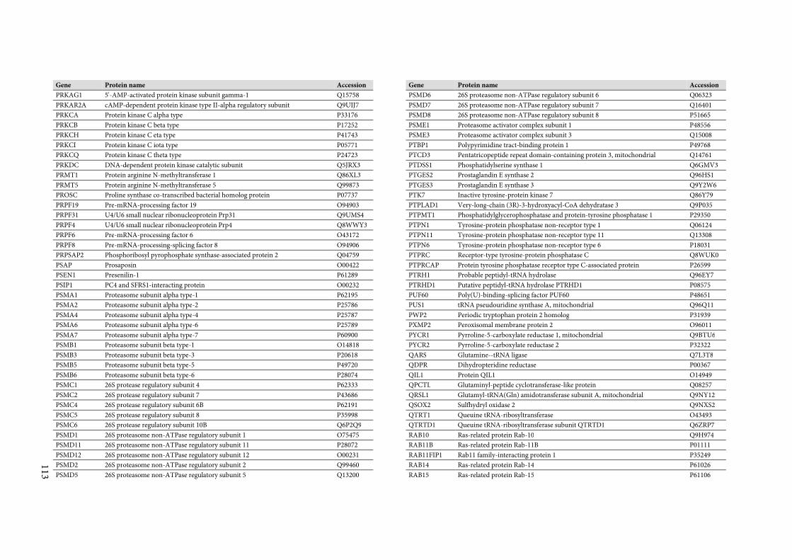
113
Gene Protein name Accession
PRKAG1 5'-AMP-activated protein kinase subunit gamma-1 Q15758
PRKAR2A cAMP-dependent protein kinase type II-alpha regulatory subunit Q9UIJ7
PRKCA Protein kinase C alpha type P33176
PRKCB Protein kinase C beta type P17252
PRKCH Protein kinase C eta type P41743
PRKCI Protein kinase C iota type P05771
PRKCQ Protein kinase C theta type P24723
PRKDC DNA-dependent protein kinase catalytic subunit Q5JRX3
PRMT1 Protein arginine N-methyltransferase 1 Q86XL3
PRMT5 Protein arginine N-methyltransferase 5 Q99873
PROSC Proline synthase co-transcribed bacterial homolog protein P07737
PRPF19 Pre-mRNA-processing factor 19 O94903
PRPF31 U4/U6 small nuclear ribonucleoprotein Prp31 Q9UMS4
PRPF4 U4/U6 small nuclear ribonucleoprotein Prp4 Q8WWY3
PRPF6 Pre-mRNA-processing factor 6 O43172
PRPF8 Pre-mRNA-processing-splicing factor 8 O94906
PRPSAP2 Phosphoribosyl pyrophosphate synthase-associated protein 2 Q04759
PSAP Prosaposin O00422
PSEN1 Presenilin-1 P61289
PSIP1 PC4 and SFRS1-interacting protein O00232
PSMA1 Proteasome subunit alpha type-1 P62195
PSMA2 Proteasome subunit alpha type-2 P25786
PSMA4 Proteasome subunit alpha type-4 P25787
PSMA6 Proteasome subunit alpha type-6 P25789
PSMA7 Proteasome subunit alpha type-7 P60900
PSMB1 Proteasome subunit beta type-1 O14818
PSMB3 Proteasome subunit beta type-3 P20618
PSMB5 Proteasome subunit beta type-5 P49720
PSMB6 Proteasome subunit beta type-6 P28074
PSMC1 26S protease regulatory subunit 4 P62333
PSMC2 26S protease regulatory subunit 7 P43686
PSMC4 26S protease regulatory subunit 6B P62191
PSMC5 26S protease regulatory subunit 8 P35998
PSMC6 26S protease regulatory subunit 10B Q6P2Q9
PSMD1 26S proteasome non-ATPase regulatory subunit 1 O75475
PSMD11 26S proteasome non-ATPase regulatory subunit 11 P28072
PSMD12 26S proteasome non-ATPase regulatory subunit 12 O00231
PSMD2 26S proteasome non-ATPase regulatory subunit 2 Q99460
PSMD5 26S proteasome non-ATPase regulatory subunit 5 Q13200
Gene Protein name Accession
PSMD6 26S proteasome non-ATPase regulatory subunit 6 Q06323
PSMD7 26S proteasome non-ATPase regulatory subunit 7 Q16401
PSMD8 26S proteasome non-ATPase regulatory subunit 8 P51665
PSME1 Proteasome activator complex subunit 1 P48556
PSME3 Proteasome activator complex subunit 3 Q15008
PTBP1 Polypyrimidine tract-binding protein 1 P49768
PTCD3 Pentatricopeptide repeat domain-containing protein 3, mitochondrial Q14761
PTDSS1 Phosphatidylserine synthase 1 Q6GMV3
PTGES2 Prostaglandin E synthase 2 Q96HS1
PTGES3 Prostaglandin E synthase 3 Q9Y2W6
PTK7 Inactive tyrosine-protein kinase 7 Q86Y79
PTPLAD1 Very-long-chain (3R)-3-hydroxyacyl-CoA dehydratase 3 Q9P035
PTPMT1 Phosphatidylglycerophosphatase and protein-tyrosine phosphatase 1 P29350
PTPN1 Tyrosine-protein phosphatase non-receptor type 1 Q06124
PTPN11 Tyrosine-protein phosphatase non-receptor type 11 Q13308
PTPN6 Tyrosine-protein phosphatase non-receptor type 6 P18031
PTPRC Receptor-type tyrosine-protein phosphatase C Q8WUK0
PTPRCAP Protein tyrosine phosphatase receptor type C-associated protein P26599
PTRH1 Probable peptidyl-tRNA hydrolase Q96EY7
PTRHD1 Putative peptidyl-tRNA hydrolase PTRHD1 P08575
PUF60 Poly(U)-binding-splicing factor PUF60 P48651
PUS1 tRNA pseudouridine synthase A, mitochondrial Q96Q11
PWP2 Periodic tryptophan protein 2 homolog P31939
PXMP2 Peroxisomal membrane protein 2 O96011
PYCR1 Pyrroline-5-carboxylate reductase 1, mitochondrial Q9BTU6
PYCR2 Pyrroline-5-carboxylate reductase 2 P32322
QARS Glutamine--tRNA ligase Q7L3T8
QDPR Dihydropteridine reductase P00367
QIL1 Protein QIL1 O14949
QPCTL Glutaminyl-peptide cyclotransferase-like protein Q08257
QRSL1 Glutamyl-tRNA(Gln) amidotransferase subunit A, mitochondrial Q9NY12
QSOX2 Sulfhydryl oxidase 2 Q9NXS2
QTRT1 Queuine tRNA-ribosyltransferase O43493
QTRTD1 Queuine tRNA-ribosyltransferase subunit QTRTD1 Q6ZRP7
RAB10 Ras-related protein Rab-10 Q9H974
RAB11B Ras-related protein Rab-11B P01111
RAB11FIP1 Rab11 family-interacting protein 1 P35249
RAB14 Ras-related protein Rab-14 P61026
RAB15 Ras-related protein Rab-15 P61106
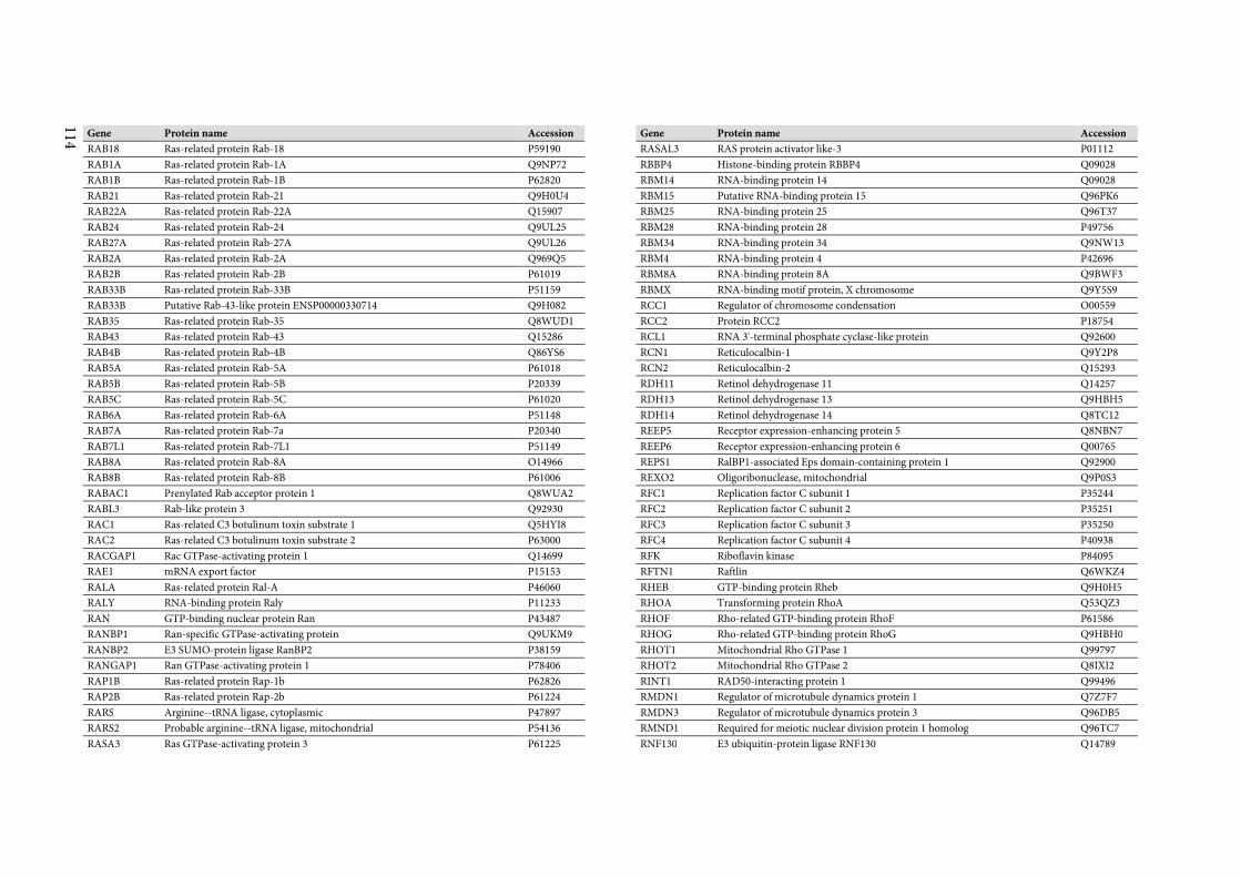
114
Gene Protein name Accession
RAB18 Ras-related protein Rab-18 P59190
RAB1A Ras-related protein Rab-1A Q9NP72
RAB1B Ras-related protein Rab-1B P62820
RAB21 Ras-related protein Rab-21 Q9H0U4
RAB22A Ras-related protein Rab-22A Q15907
RAB24 Ras-related protein Rab-24 Q9UL25
RAB27A Ras-related protein Rab-27A Q9UL26
RAB2A Ras-related protein Rab-2A Q969Q5
RAB2B Ras-related protein Rab-2B P61019
RAB33B Ras-related protein Rab-33B P51159
RAB33B Putative Rab-43-like protein ENSP00000330714 Q9H082
RAB35 Ras-related protein Rab-35 Q8WUD1
RAB43 Ras-related protein Rab-43 Q15286
RAB4B Ras-related protein Rab-4B Q86YS6
RAB5A Ras-related protein Rab-5A P61018
RAB5B Ras-related protein Rab-5B P20339
RAB5C Ras-related protein Rab-5C P61020
RAB6A Ras-related protein Rab-6A P51148
RAB7A Ras-related protein Rab-7a P20340
RAB7L1 Ras-related protein Rab-7L1 P51149
RAB8A Ras-related protein Rab-8A O14966
RAB8B Ras-related protein Rab-8B P61006
RABAC1 Prenylated Rab acceptor protein 1 Q8WUA2
RABL3 Rab-like protein 3 Q92930
RAC1 Ras-related C3 botulinum toxin substrate 1 Q5HYI8
RAC2 Ras-related C3 botulinum toxin substrate 2 P63000
RACGAP1 Rac GTPase-activating protein 1 Q14699
RAE1 mRNA export factor P15153
RALA Ras-related protein Ral-A P46060
RALY RNA-binding protein Raly P11233
RAN GTP-binding nuclear protein Ran P43487
RANBP1 Ran-specific GTPase-activating protein Q9UKM9
RANBP2 E3 SUMO-protein ligase RanBP2 P38159
RANGAP1 Ran GTPase-activating protein 1 P78406
RAP1B Ras-related protein Rap-1b P62826
RAP2B Ras-related protein Rap-2b P61224
RARS Arginine--tRNA ligase, cytoplasmic P47897
RARS2 Probable arginine--tRNA ligase, mitochondrial P54136
RASA3 Ras GTPase-activating protein 3 P61225
Gene Protein name Accession
RASAL3 RAS protein activator like-3 P01112
RBBP4 Histone-binding protein RBBP4 Q09028
RBM14 RNA-binding protein 14 Q09028
RBM15 Putative RNA-binding protein 15 Q96PK6
RBM25 RNA-binding protein 25 Q96T37
RBM28 RNA-binding protein 28 P49756
RBM34 RNA-binding protein 34 Q9NW13
RBM4 RNA-binding protein 4 P42696
RBM8A RNA-binding protein 8A Q9BWF3
RBMX RNA-binding motif protein, X chromosome Q9Y5S9
RCC1 Regulator of chromosome condensation O00559
RCC2 Protein RCC2 P18754
RCL1 RNA 3'-terminal phosphate cyclase-like protein Q92600
RCN1 Reticulocalbin-1 Q9Y2P8
RCN2 Reticulocalbin-2 Q15293
RDH11 Retinol dehydrogenase 11 Q14257
RDH13 Retinol dehydrogenase 13 Q9HBH5
RDH14 Retinol dehydrogenase 14 Q8TC12
REEP5 Receptor expression-enhancing protein 5 Q8NBN7
REEP6 Receptor expression-enhancing protein 6 Q00765
REPS1 RalBP1-associated Eps domain-containing protein 1 Q92900
REXO2 Oligoribonuclease, mitochondrial Q9P0S3
RFC1 Replication factor C subunit 1 P35244
RFC2 Replication factor C subunit 2 P35251
RFC3 Replication factor C subunit 3 P35250
RFC4 Replication factor C subunit 4 P40938
RFK Riboflavin kinase P84095
RFTN1 Raftlin Q6WKZ4
RHEB GTP-binding protein Rheb Q9H0H5
RHOA Transforming protein RhoA Q53QZ3
RHOF Rho-related GTP-binding protein RhoF P61586
RHOG Rho-related GTP-binding protein RhoG Q9HBH0
RHOT1 Mitochondrial Rho GTPase 1 Q99797
RHOT2 Mitochondrial Rho GTPase 2 Q8IXI2
RINT1 RAD50-interacting protein 1 Q99496
RMDN1 Regulator of microtubule dynamics protein 1 Q7Z7F7
RMDN3 Regulator of microtubule dynamics protein 3 Q96DB5
RMND1 Required for meiotic nuclear division protein 1 homolog Q96TC7
RNF130 E3 ubiquitin-protein ligase RNF130 Q14789
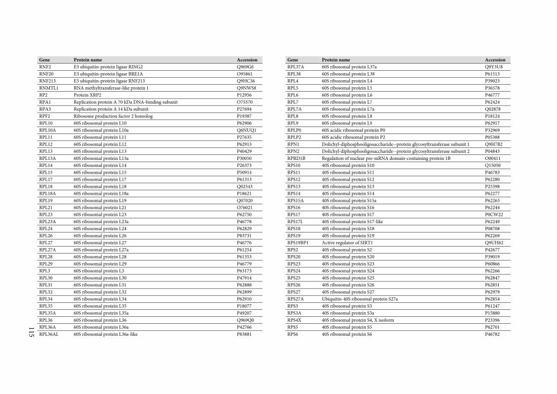
115
Gene Protein name Accession
RNF2 E3 ubiquitin-protein ligase RING2 Q969G6
RNF20 E3 ubiquitin-protein ligase BRE1A O95861
RNF213 E3 ubiquitin-protein ligase RNF213 Q9HC36
RNMTL1 RNA methyltransferase-like protein 1 Q9NWS8
RP2 Protein XRP2 P12956
RPA1 Replication protein A 70 kDa DNA-binding subunit O75570
RPA3 Replication protein A 14 kDa subunit P27694
RPF2 Ribosome production factor 2 homolog P19387
RPL10 60S ribosomal protein L10 P62906
RPL10A 60S ribosomal protein L10a Q6NUQ1
RPL11 60S ribosomal protein L11 P27635
RPL12 60S ribosomal protein L12 P62913
RPL13 60S ribosomal protein L13 P40429
RPL13A 60S ribosomal protein L13a P30050
RPL14 60S ribosomal protein L14 P26373
RPL15 60S ribosomal protein L15 P50914
RPL17 60S ribosomal protein L17 P61313
RPL18 60S ribosomal protein L18 Q02543
RPL18A 60S ribosomal protein L18a P18621
RPL19 60S ribosomal protein L19 Q07020
RPL21 60S ribosomal protein L21 O76021
RPL23 60S ribosomal protein L23 P62750
RPL23A 60S ribosomal protein L23a P46778
RPL24 60S ribosomal protein L24 P62829
RPL26 60S ribosomal protein L26 P83731
RPL27 60S ribosomal protein L27 P46776
RPL27A 60S ribosomal protein L27a P61254
RPL28 60S ribosomal protein L28 P61353
RPL29 60S ribosomal protein L29 P46779
RPL3 60S ribosomal protein L3 P63173
RPL30 60S ribosomal protein L30 P47914
RPL31 60S ribosomal protein L31 P62888
RPL32 60S ribosomal protein L32 P62899
RPL34 60S ribosomal protein L34 P62910
RPL35 60S ribosomal protein L35 P18077
RPL35A 60S ribosomal protein L35a P49207
RPL36 60S ribosomal protein L36 Q969Q0
RPL36A 60S ribosomal protein L36a P42766
RPL36AL 60S ribosomal protein L36a-like P83881
Gene Protein name Accession
RPL37A 60S ribosomal protein L37a Q9Y3U8
RPL38 60S ribosomal protein L38 P61513
RPL4 60S ribosomal protein L4 P39023
RPL5 60S ribosomal protein L5 P36578
RPL6 60S ribosomal protein L6 P46777
RPL7 60S ribosomal protein L7 P62424
RPL7A 60S ribosomal protein L7a Q02878
RPL8 60S ribosomal protein L8 P18124
RPL9 60S ribosomal protein L9 P62917
RPLP0 60S acidic ribosomal protein P0 P32969
RPLP2 60S acidic ribosomal protein P2 P05388
RPN1 Dolichyl-diphosphooligosaccharide--protein glycosyltransferase subunit 1 Q9H7B2
RPN2 Dolichyl-diphosphooligosaccharide--protein glycosyltransferase subunit 2 P04843
RPRD1B Regulation of nuclear pre-mRNA domain-containing protein 1B O00411
RPS10 40S ribosomal protein S10 Q15050
RPS11 40S ribosomal protein S11 P46783
RPS12 40S ribosomal protein S12 P62280
RPS13 40S ribosomal protein S13 P25398
RPS14 40S ribosomal protein S14 P62277
RPS15A 40S ribosomal protein S15a P62263
RPS16 40S ribosomal protein S16 P62244
RPS17 40S ribosomal protein S17 P0CW22
RPS17L 40S ribosomal protein S17-like P62249
RPS18 40S ribosomal protein S18 P08708
RPS19 40S ribosomal protein S19 P62269
RPS19BP1 Active regulator of SIRT1 Q9UH62
RPS2 40S ribosomal protein S2 P42677
RPS20 40S ribosomal protein S20 P39019
RPS23 40S ribosomal protein S23 P60866
RPS24 40S ribosomal protein S24 P62266
RPS25 40S ribosomal protein S25 P62847
RPS26 40S ribosomal protein S26 P62851
RPS27 40S ribosomal protein S27 P62979
RPS27A Ubiquitin-40S ribosomal protein S27a P62854
RPS3 40S ribosomal protein S3 P61247
RPS3A 40S ribosomal protein S3a P15880
RPS4X 40S ribosomal protein S4, X isoform P23396
RPS5 40S ribosomal protein S5 P62701
RPS6 40S ribosomal protein S6 P46782
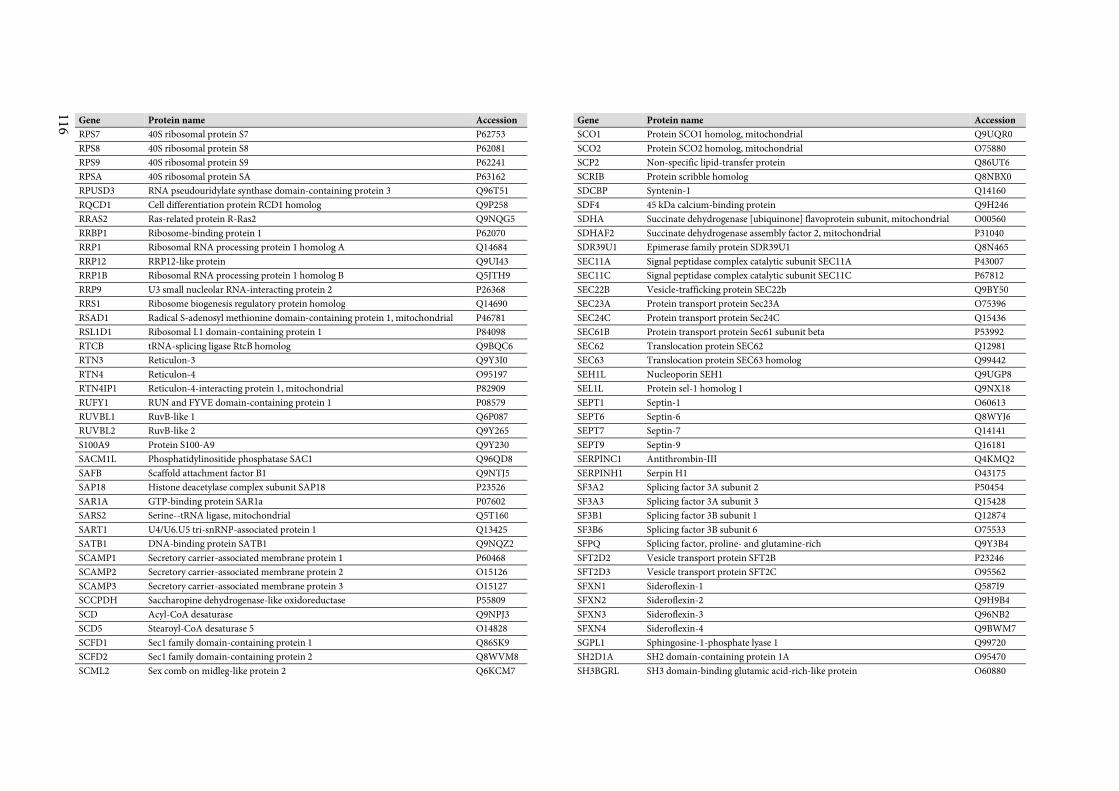
116
Gene Protein name Accession
RPS7 40S ribosomal protein S7 P62753
RPS8 40S ribosomal protein S8 P62081
RPS9 40S ribosomal protein S9 P62241
RPSA 40S ribosomal protein SA P63162
RPUSD3 RNA pseudouridylate synthase domain-containing protein 3 Q96T51
RQCD1 Cell differentiation protein RCD1 homolog Q9P258
RRAS2 Ras-related protein R-Ras2 Q9NQG5
RRBP1 Ribosome-binding protein 1 P62070
RRP1 Ribosomal RNA processing protein 1 homolog A Q14684
RRP12 RRP12-like protein Q9UI43
RRP1B Ribosomal RNA processing protein 1 homolog B Q5JTH9
RRP9 U3 small nucleolar RNA-interacting protein 2 P26368
RRS1 Ribosome biogenesis regulatory protein homolog Q14690
RSAD1 Radical S-adenosyl methionine domain-containing protein 1, mitochondrial P46781
RSL1D1 Ribosomal L1 domain-containing protein 1 P84098
RTCB tRNA-splicing ligase RtcB homolog Q9BQC6
RTN3 Reticulon-3 Q9Y3I0
RTN4 Reticulon-4 O95197
RTN4IP1 Reticulon-4-interacting protein 1, mitochondrial P82909
RUFY1 RUN and FYVE domain-containing protein 1 P08579
RUVBL1 RuvB-like 1 Q6P087
RUVBL2 RuvB-like 2 Q9Y265
S100A9 Protein S100-A9 Q9Y230
SACM1L Phosphatidylinositide phosphatase SAC1 Q96QD8
SAFB Scaffold attachment factor B1 Q9NTJ5
SAP18 Histone deacetylase complex subunit SAP18 P23526
SAR1A GTP-binding protein SAR1a P07602
SARS2 Serine--tRNA ligase, mitochondrial Q5T160
SART1 U4/U6.U5 tri-snRNP-associated protein 1 Q13425
SATB1 DNA-binding protein SATB1 Q9NQZ2
SCAMP1 Secretory carrier-associated membrane protein 1 P60468
SCAMP2 Secretory carrier-associated membrane protein 2 O15126
SCAMP3 Secretory carrier-associated membrane protein 3 O15127
SCCPDH Saccharopine dehydrogenase-like oxidoreductase P55809
SCD Acyl-CoA desaturase Q9NPJ3
SCD5 Stearoyl-CoA desaturase 5 O14828
SCFD1 Sec1 family domain-containing protein 1 Q86SK9
SCFD2 Sec1 family domain-containing protein 2 Q8WVM8
SCML2 Sex comb on midleg-like protein 2 Q6KCM7
Gene Protein name Accession
SCO1 Protein SCO1 homolog, mitochondrial Q9UQR0
SCO2 Protein SCO2 homolog, mitochondrial O75880
SCP2 Non-specific lipid-transfer protein Q86UT6
SCRIB Protein scribble homolog Q8NBX0
SDCBP Syntenin-1 Q14160
SDF4 45 kDa calcium-binding protein Q9H246
SDHA Succinate dehydrogenase [ubiquinone] flavoprotein subunit, mitochondrial O00560
SDHAF2 Succinate dehydrogenase assembly factor 2, mitochondrial P31040
SDR39U1 Epimerase family protein SDR39U1 Q8N465
SEC11A Signal peptidase complex catalytic subunit SEC11A P43007
SEC11C Signal peptidase complex catalytic subunit SEC11C P67812
SEC22B Vesicle-trafficking protein SEC22b Q9BY50
SEC23A Protein transport protein Sec23A O75396
SEC24C Protein transport protein Sec24C Q15436
SEC61B Protein transport protein Sec61 subunit beta P53992
SEC62 Translocation protein SEC62 Q12981
SEC63 Translocation protein SEC63 homolog Q99442
SEH1L Nucleoporin SEH1 Q9UGP8
SEL1L Protein sel-1 homolog 1 Q9NX18
SEPT1 Septin-1 O60613
SEPT6 Septin-6 Q8WYJ6
SEPT7 Septin-7 Q14141
SEPT9 Septin-9 Q16181
SERPINC1 Antithrombin-III Q4KMQ2
SERPINH1 Serpin H1 O43175
SF3A2 Splicing factor 3A subunit 2 P50454
SF3A3 Splicing factor 3A subunit 3 Q15428
SF3B1 Splicing factor 3B subunit 1 Q12874
SF3B6 Splicing factor 3B subunit 6 O75533
SFPQ Splicing factor, proline- and glutamine-rich Q9Y3B4
SFT2D2 Vesicle transport protein SFT2B P23246
SFT2D3 Vesicle transport protein SFT2C O95562
SFXN1 Sideroflexin-1 Q587I9
SFXN2 Sideroflexin-2 Q9H9B4
SFXN3 Sideroflexin-3 Q96NB2
SFXN4 Sideroflexin-4 Q9BWM7
SGPL1 Sphingosine-1-phosphate lyase 1 Q99720
SH2D1A SH2 domain-containing protein 1A O95470
SH3BGRL SH3 domain-binding glutamic acid-rich-like protein O60880
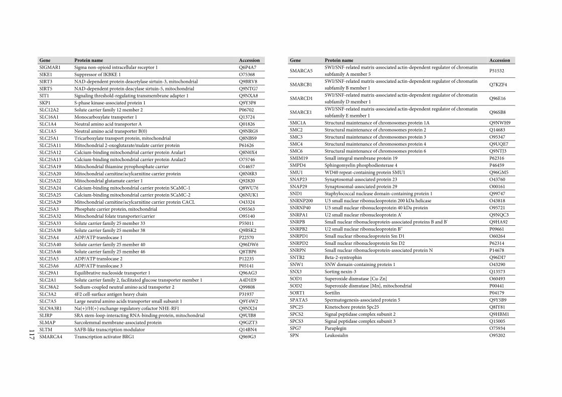
117
Gene Protein name Accession
SIGMAR1 Sigma non-opioid intracellular receptor 1 Q6P4A7
SIKE1 Suppressor of IKBKE 1 O75368
SIRT3 NAD-dependent protein deacetylase sirtuin-3, mitochondrial Q9BRV8
SIRT5 NAD-dependent protein deacylase sirtuin-5, mitochondrial Q9NTG7
SIT1 Signaling threshold-regulating transmembrane adapter 1 Q9NXA8
SKP1 S-phase kinase-associated protein 1 Q9Y3P8
SLC12A2 Solute carrier family 12 member 2 P06702
SLC16A1 Monocarboxylate transporter 1 Q13724
SLC1A4 Neutral amino acid transporter A Q01826
SLC1A5 Neutral amino acid transporter B(0) Q9NRG9
SLC25A1 Tricarboxylate transport protein, mitochondrial Q8NBS9
SLC25A11 Mitochondrial 2-oxoglutarate/malate carrier protein P61626
SLC25A12 Calcium-binding mitochondrial carrier protein Aralar1 Q8N0X4
SLC25A13 Calcium-binding mitochondrial carrier protein Aralar2 O75746
SLC25A19 Mitochondrial thiamine pyrophosphate carrier O14657
SLC25A20 Mitochondrial carnitine/acylcarnitine carrier protein Q8N8R3
SLC25A22 Mitochondrial glutamate carrier 1 Q92820
SLC25A24 Calcium-binding mitochondrial carrier protein SCaMC-1 Q8WU76
SLC25A25 Calcium-binding mitochondrial carrier protein SCaMC-2 Q6NUK1
SLC25A29 Mitochondrial carnitine/acylcarnitine carrier protein CACL O43324
SLC25A3 Phosphate carrier protein, mitochondrial O95563
SLC25A32 Mitochondrial folate transporter/carrier O95140
SLC25A33 Solute carrier family 25 member 33 P55011
SLC25A38 Solute carrier family 25 member 38 Q9BSK2
SLC25A4 ADP/ATP translocase 1 P22570
SLC25A40 Solute carrier family 25 member 40 Q96DW6
SLC25A46 Solute carrier family 25 member 46 Q8TBP6
SLC25A5 ADP/ATP translocase 2 P12235
SLC25A6 ADP/ATP translocase 3 P05141
SLC29A1 Equilibrative nucleoside transporter 1 Q96AG3
SLC2A1 Solute carrier family 2, facilitated glucose transporter member 1 A4D1E9
SLC38A2 Sodium-coupled neutral amino acid transporter 2 Q99808
SLC3A2 4F2 cell-surface antigen heavy chain P31937
SLC7A5 Large neutral amino acids transporter small subunit 1 Q9Y4W2
SLC9A3R1 Na(+)/H(+) exchange regulatory cofactor NHE-RF1 Q9NX24
SLIRP SRA stem-loop-interacting RNA-binding protein, mitochondrial Q9UIB8
SLMAP Sarcolemmal membrane-associated protein Q9GZT3
SLTM SAFB-like transcription modulator Q14BN4
SMARCA4 Transcription activator BRG1 Q969G3
Gene Protein name Accession
SMARCA5 SWI/SNF-related matrix-associated actin-dependent regulator of chromatin
subfamily A member 5 P51532
SMARCB1 SWI/SNF-related matrix-associated actin-dependent regulator of chromatin
subfamily B member 1 Q7KZF4
SMARCD1 SWI/SNF-related matrix-associated actin-dependent regulator of chromatin
subfamily D member 1 Q96E16
SMARCE1 SWI/SNF-related matrix-associated actin-dependent regulator of chromatin
subfamily E member 1 Q96SB8
SMC1A Structural maintenance of chromosomes protein 1A Q9NWH9
SMC2 Structural maintenance of chromosomes protein 2 Q14683
SMC3 Structural maintenance of chromosomes protein 3 O95347
SMC4 Structural maintenance of chromosomes protein 4 Q9UQE7
SMC6 Structural maintenance of chromosomes protein 6 Q9NTJ3
SMIM19 Small integral membrane protein 19 P62316
SMPD4 Sphingomyelin phosphodiesterase 4 P46459
SMU1 WD40 repeat-containing protein SMU1 Q96GM5
SNAP23 Synaptosomal-associated protein 23 O43760
SNAP29 Synaptosomal-associated protein 29 O00161
SND1 Staphylococcal nuclease domain-containing protein 1 Q99747
SNRNP200 U5 small nuclear ribonucleoprotein 200 kDa helicase O43818
SNRNP40 U5 small nuclear ribonucleoprotein 40 kDa protein O95721
SNRPA1 U2 small nuclear ribonucleoprotein A' Q9NQC3
SNRPB Small nuclear ribonucleoprotein-associated proteins B and B' Q9HA92
SNRPB2 U2 small nuclear ribonucleoprotein B'' P09661
SNRPD1 Small nuclear ribonucleoprotein Sm D1 O60264
SNRPD2 Small nuclear ribonucleoprotein Sm D2 P62314
SNRPN Small nuclear ribonucleoprotein-associated protein N P14678
SNTB2 Beta-2-syntrophin Q96DI7
SNW1 SNW domain-containing protein 1 O43290
SNX3 Sorting nexin-3 Q13573
SOD1 Superoxide dismutase [Cu-Zn] O60493
SOD2 Superoxide dismutase [Mn], mitochondrial P00441
SORT1 Sortilin P04179
SPATA5 Spermatogenesis-associated protein 5 Q9Y5B9
SPC25 Kinetochore protein Spc25 Q8IY81
SPCS2 Signal peptidase complex subunit 2 Q9HBM1
SPCS3 Signal peptidase complex subunit 3 Q15005
SPG7 Paraplegin O75934
SPN Leukosialin O95202
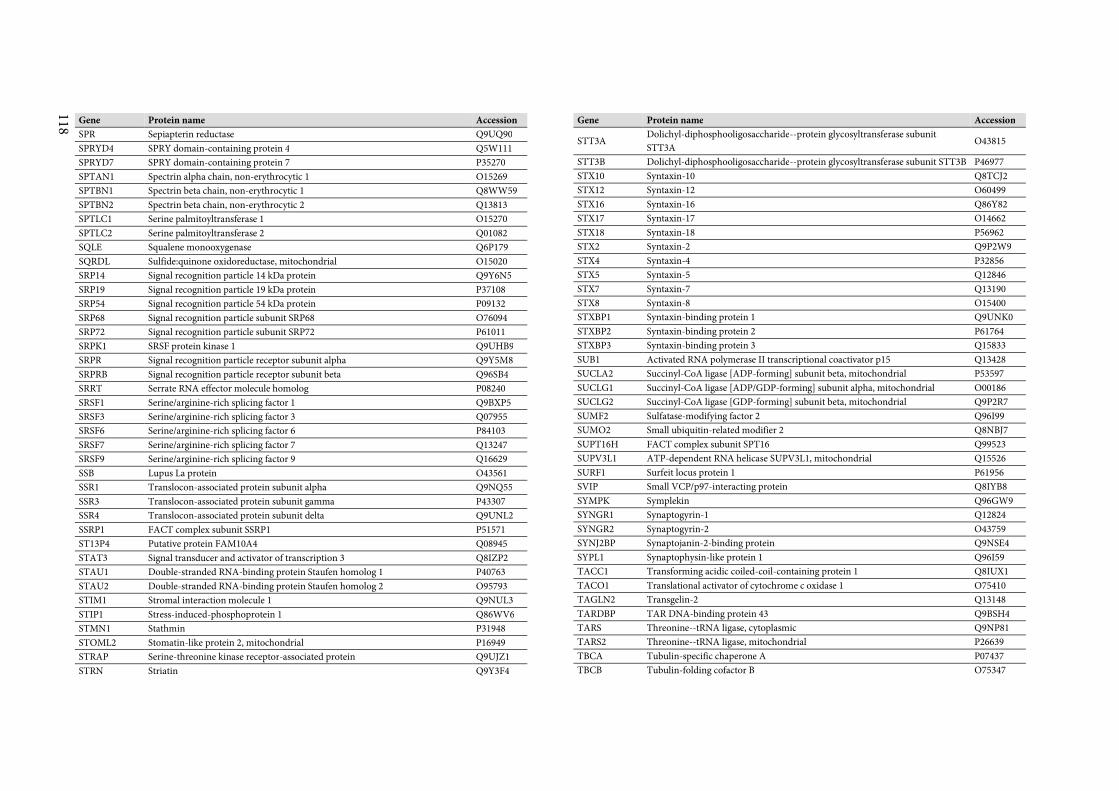
118
Gene Protein name Accession
SPR Sepiapterin reductase Q9UQ90
SPRYD4 SPRY domain-containing protein 4 Q5W111
SPRYD7 SPRY domain-containing protein 7 P35270
SPTAN1 Spectrin alpha chain, non-erythrocytic 1 O15269
SPTBN1 Spectrin beta chain, non-erythrocytic 1 Q8WW59
SPTBN2 Spectrin beta chain, non-erythrocytic 2 Q13813
SPTLC1 Serine palmitoyltransferase 1 O15270
SPTLC2 Serine palmitoyltransferase 2 Q01082
SQLE Squalene monooxygenase Q6P179
SQRDL Sulfide:quinone oxidoreductase, mitochondrial O15020
SRP14 Signal recognition particle 14 kDa protein Q9Y6N5
SRP19 Signal recognition particle 19 kDa protein P37108
SRP54 Signal recognition particle 54 kDa protein P09132
SRP68 Signal recognition particle subunit SRP68 O76094
SRP72 Signal recognition particle subunit SRP72 P61011
SRPK1 SRSF protein kinase 1 Q9UHB9
SRPR Signal recognition particle receptor subunit alpha Q9Y5M8
SRPRB Signal recognition particle receptor subunit beta Q96SB4
SRRT Serrate RNA effector molecule homolog P08240
SRSF1 Serine/arginine-rich splicing factor 1 Q9BXP5
SRSF3 Serine/arginine-rich splicing factor 3 Q07955
SRSF6 Serine/arginine-rich splicing factor 6 P84103
SRSF7 Serine/arginine-rich splicing factor 7 Q13247
SRSF9 Serine/arginine-rich splicing factor 9 Q16629
SSB Lupus La protein O43561
SSR1 Translocon-associated protein subunit alpha Q9NQ55
SSR3 Translocon-associated protein subunit gamma P43307
SSR4 Translocon-associated protein subunit delta Q9UNL2
SSRP1 FACT complex subunit SSRP1 P51571
ST13P4 Putative protein FAM10A4 Q08945
STAT3 Signal transducer and activator of transcription 3 Q8IZP2
STAU1 Double-stranded RNA-binding protein Staufen homolog 1 P40763
STAU2 Double-stranded RNA-binding protein Staufen homolog 2 O95793
STIM1 Stromal interaction molecule 1 Q9NUL3
STIP1 Stress-induced-phosphoprotein 1 Q86WV6
STMN1 Stathmin P31948
STOML2 Stomatin-like protein 2, mitochondrial P16949
STRAP Serine-threonine kinase receptor-associated protein Q9UJZ1
STRN Striatin Q9Y3F4
Gene Protein name Accession
STT3A Dolichyl-diphosphooligosaccharide--protein glycosyltransferase subunit
STT3A O43815
STT3B Dolichyl-diphosphooligosaccharide--protein glycosyltransferase subunit STT3B P46977
STX10 Syntaxin-10 Q8TCJ2
STX12 Syntaxin-12 O60499
STX16 Syntaxin-16 Q86Y82
STX17 Syntaxin-17 O14662
STX18 Syntaxin-18 P56962
STX2 Syntaxin-2 Q9P2W9
STX4 Syntaxin-4 P32856
STX5 Syntaxin-5 Q12846
STX7 Syntaxin-7 Q13190
STX8 Syntaxin-8 O15400
STXBP1 Syntaxin-binding protein 1 Q9UNK0
STXBP2 Syntaxin-binding protein 2 P61764
STXBP3 Syntaxin-binding protein 3 Q15833
SUB1 Activated RNA polymerase II transcriptional coactivator p15 Q13428
SUCLA2 Succinyl-CoA ligase [ADP-forming] subunit beta, mitochondrial P53597
SUCLG1 Succinyl-CoA ligase [ADP/GDP-forming] subunit alpha, mitochondrial O00186
SUCLG2 Succinyl-CoA ligase [GDP-forming] subunit beta, mitochondrial Q9P2R7
SUMF2 Sulfatase-modifying factor 2 Q96I99
SUMO2 Small ubiquitin-related modifier 2 Q8NBJ7
SUPT16H FACT complex subunit SPT16 Q99523
SUPV3L1 ATP-dependent RNA helicase SUPV3L1, mitochondrial Q15526
SURF1 Surfeit locus protein 1 P61956
SVIP Small VCP/p97-interacting protein Q8IYB8
SYMPK Symplekin Q96GW9
SYNGR1 Synaptogyrin-1 Q12824
SYNGR2 Synaptogyrin-2 O43759
SYNJ2BP Synaptojanin-2-binding protein Q9NSE4
SYPL1 Synaptophysin-like protein 1 Q96I59
TACC1 Transforming acidic coiled-coil-containing protein 1 Q8IUX1
TACO1 Translational activator of cytochrome c oxidase 1 O75410
TAGLN2 Transgelin-2 Q13148
TARDBP TAR DNA-binding protein 43 Q9BSH4
TARS Threonine--tRNA ligase, cytoplasmic Q9NP81
TARS2 Threonine--tRNA ligase, mitochondrial P26639
TBCA Tubulin-specific chaperone A P07437
TBCB Tubulin-folding cofactor B O75347
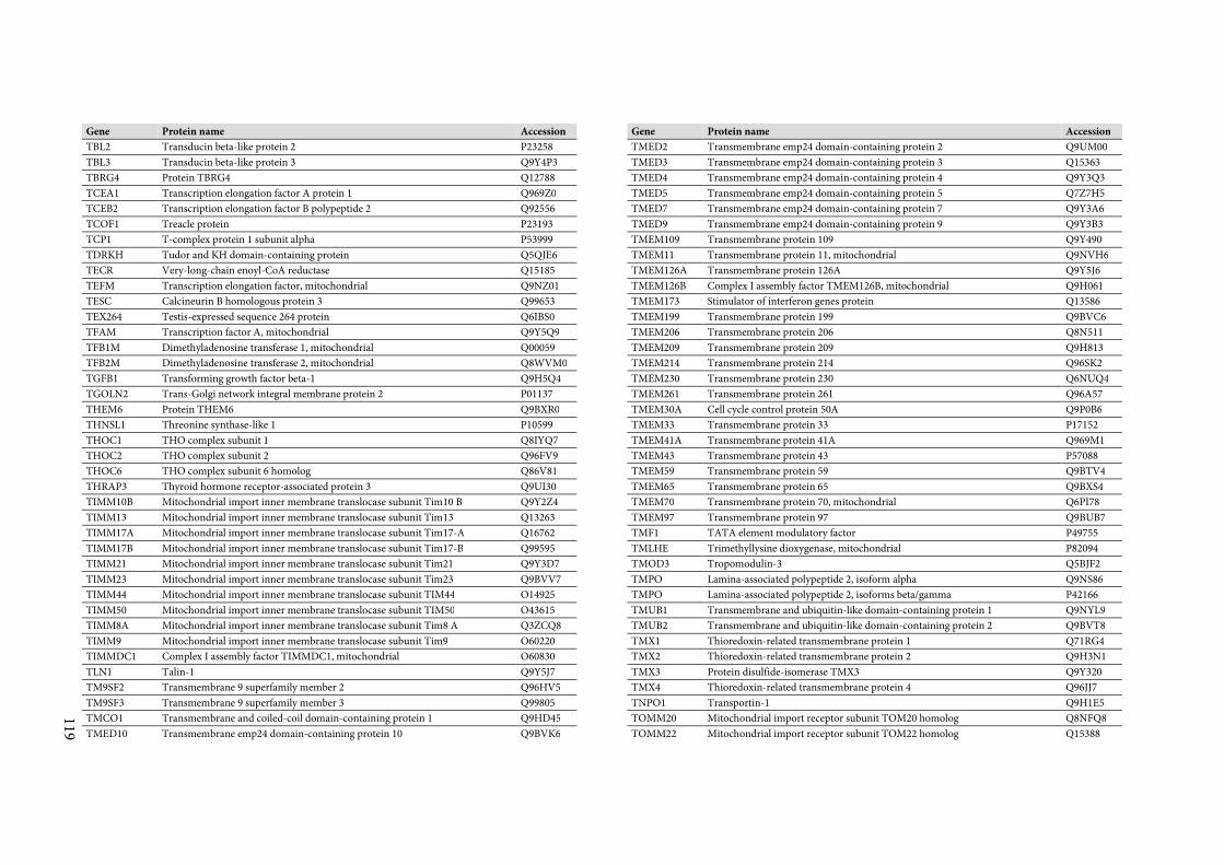
119
Gene Protein name Accession
TBL2 Transducin beta-like protein 2 P23258
TBL3 Transducin beta-like protein 3 Q9Y4P3
TBRG4 Protein TBRG4 Q12788
TCEA1 Transcription elongation factor A protein 1 Q969Z0
TCEB2 Transcription elongation factor B polypeptide 2 Q92556
TCOF1 Treacle protein P23193
TCP1 T-complex protein 1 subunit alpha P53999
TDRKH Tudor and KH domain-containing protein Q5QJE6
TECR Very-long-chain enoyl-CoA reductase Q15185
TEFM Transcription elongation factor, mitochondrial Q9NZ01
TESC Calcineurin B homologous protein 3 Q99653
TEX264 Testis-expressed sequence 264 protein Q6IBS0
TFAM Transcription factor A, mitochondrial Q9Y5Q9
TFB1M Dimethyladenosine transferase 1, mitochondrial Q00059
TFB2M Dimethyladenosine transferase 2, mitochondrial Q8WVM0
TGFB1 Transforming growth factor beta-1 Q9H5Q4
TGOLN2 Trans-Golgi network integral membrane protein 2 P01137
THEM6 Protein THEM6 Q9BXR0
THNSL1 Threonine synthase-like 1 P10599
THOC1 THO complex subunit 1 Q8IYQ7
THOC2 THO complex subunit 2 Q96FV9
THOC6 THO complex subunit 6 homolog Q86V81
THRAP3 Thyroid hormone receptor-associated protein 3 Q9UI30
TIMM10B Mitochondrial import inner membrane translocase subunit Tim10 B Q9Y2Z4
TIMM13 Mitochondrial import inner membrane translocase subunit Tim13 Q13263
TIMM17A Mitochondrial import inner membrane translocase subunit Tim17-A Q16762
TIMM17B Mitochondrial import inner membrane translocase subunit Tim17-B Q99595
TIMM21 Mitochondrial import inner membrane translocase subunit Tim21 Q9Y3D7
TIMM23 Mitochondrial import inner membrane translocase subunit Tim23 Q9BVV7
TIMM44 Mitochondrial import inner membrane translocase subunit TIM44 O14925
TIMM50 Mitochondrial import inner membrane translocase subunit TIM50 O43615
TIMM8A Mitochondrial import inner membrane translocase subunit Tim8 A Q3ZCQ8
TIMM9 Mitochondrial import inner membrane translocase subunit Tim9 O60220
TIMMDC1 Complex I assembly factor TIMMDC1, mitochondrial O60830
TLN1 Talin-1 Q9Y5J7
TM9SF2 Transmembrane 9 superfamily member 2 Q96HV5
TM9SF3 Transmembrane 9 superfamily member 3 Q99805
TMCO1 Transmembrane and coiled-coil domain-containing protein 1 Q9HD45
TMED10 Transmembrane emp24 domain-containing protein 10 Q9BVK6
Gene Protein name Accession
TMED2 Transmembrane emp24 domain-containing protein 2 Q9UM00
TMED3 Transmembrane emp24 domain-containing protein 3 Q15363
TMED4 Transmembrane emp24 domain-containing protein 4 Q9Y3Q3
TMED5 Transmembrane emp24 domain-containing protein 5 Q7Z7H5
TMED7 Transmembrane emp24 domain-containing protein 7 Q9Y3A6
TMED9 Transmembrane emp24 domain-containing protein 9 Q9Y3B3
TMEM109 Transmembrane protein 109 Q9Y490
TMEM11 Transmembrane protein 11, mitochondrial Q9NVH6
TMEM126A Transmembrane protein 126A Q9Y5J6
TMEM126B Complex I assembly factor TMEM126B, mitochondrial Q9H061
TMEM173 Stimulator of interferon genes protein Q13586
TMEM199 Transmembrane protein 199 Q9BVC6
TMEM206 Transmembrane protein 206 Q8N511
TMEM209 Transmembrane protein 209 Q9H813
TMEM214 Transmembrane protein 214 Q96SK2
TMEM230 Transmembrane protein 230 Q6NUQ4
TMEM261 Transmembrane protein 261 Q96A57
TMEM30A Cell cycle control protein 50A Q9P0B6
TMEM33 Transmembrane protein 33 P17152
TMEM41A Transmembrane protein 41A Q969M1
TMEM43 Transmembrane protein 43 P57088
TMEM59 Transmembrane protein 59 Q9BTV4
TMEM65 Transmembrane protein 65 Q9BXS4
TMEM70 Transmembrane protein 70, mitochondrial Q6PI78
TMEM97 Transmembrane protein 97 Q9BUB7
TMF1 TATA element modulatory factor P49755
TMLHE Trimethyllysine dioxygenase, mitochondrial P82094
TMOD3 Tropomodulin-3 Q5BJF2
TMPO Lamina-associated polypeptide 2, isoform alpha Q9NS86
TMPO Lamina-associated polypeptide 2, isoforms beta/gamma P42166
TMUB1 Transmembrane and ubiquitin-like domain-containing protein 1 Q9NYL9
TMUB2 Transmembrane and ubiquitin-like domain-containing protein 2 Q9BVT8
TMX1 Thioredoxin-related transmembrane protein 1 Q71RG4
TMX2 Thioredoxin-related transmembrane protein 2 Q9H3N1
TMX3 Protein disulfide-isomerase TMX3 Q9Y320
TMX4 Thioredoxin-related transmembrane protein 4 Q96JJ7
TNPO1 Transportin-1 Q9H1E5
TOMM20 Mitochondrial import receptor subunit TOM20 homolog Q8NFQ8
TOMM22 Mitochondrial import receptor subunit TOM22 homolog Q15388
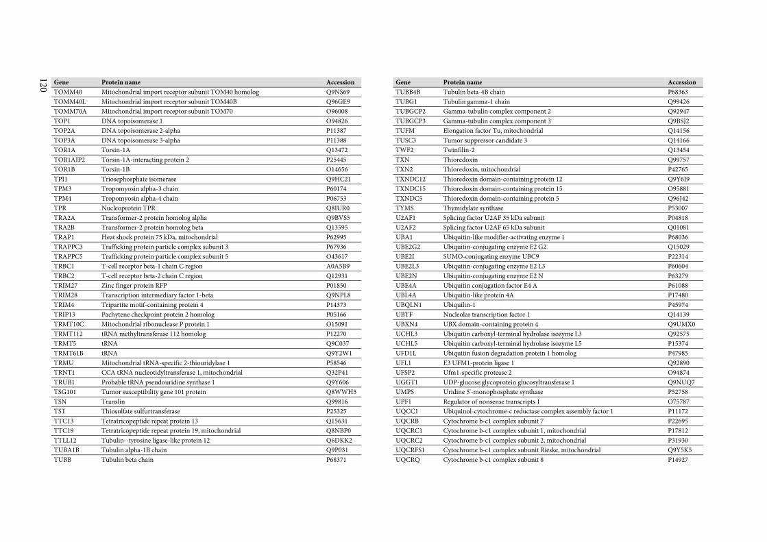
120
Gene Protein name Accession
TOMM40 Mitochondrial import receptor subunit TOM40 homolog Q9NS69
TOMM40L Mitochondrial import receptor subunit TOM40B Q96GE9
TOMM70A Mitochondrial import receptor subunit TOM70 O96008
TOP1 DNA topoisomerase 1 O94826
TOP2A DNA topoisomerase 2-alpha P11387
TOP3A DNA topoisomerase 3-alpha P11388
TOR1A Torsin-1A Q13472
TOR1AIP2 Torsin-1A-interacting protein 2 P25445
TOR1B Torsin-1B O14656
TPI1 Triosephosphate isomerase Q9HC21
TPM3 Tropomyosin alpha-3 chain P60174
TPM4 Tropomyosin alpha-4 chain P06753
TPR Nucleoprotein TPR Q8IUR0
TRA2A Transformer-2 protein homolog alpha Q9BVS5
TRA2B Transformer-2 protein homolog beta Q13595
TRAP1 Heat shock protein 75 kDa, mitochondrial P62995
TRAPPC3 Trafficking protein particle complex subunit 3 P67936
TRAPPC5 Trafficking protein particle complex subunit 5 O43617
TRBC1 T-cell receptor beta-1 chain C region A0A5B9
TRBC2 T-cell receptor beta-2 chain C region Q12931
TRIM27 Zinc finger protein RFP P01850
TRIM28 Transcription intermediary factor 1-beta Q9NPL8
TRIM4 Tripartite motif-containing protein 4 P14373
TRIP13 Pachytene checkpoint protein 2 homolog P05166
TRMT10C Mitochondrial ribonuclease P protein 1 O15091
TRMT112 tRNA methyltransferase 112 homolog P12270
TRMT5 tRNA Q9C037
TRMT61B tRNA Q9Y2W1
TRMU Mitochondrial tRNA-specific 2-thiouridylase 1 P58546
TRNT1 CCA tRNA nucleotidyltransferase 1, mitochondrial Q32P41
TRUB1 Probable tRNA pseudouridine synthase 1 Q9Y606
TSG101 Tumor susceptibility gene 101 protein Q8WWH5
TSN Translin Q99816
TST Thiosulfate sulfurtransferase P25325
TTC13 Tetratricopeptide repeat protein 13 Q15631
TTC19 Tetratricopeptide repeat protein 19, mitochondrial Q8NBP0
TTLL12 Tubulin--tyrosine ligase-like protein 12 Q6DKK2
TUBA1B Tubulin alpha-1B chain Q9P031
TUBB Tubulin beta chain P68371
Gene Protein name Accession
TUBB4B Tubulin beta-4B chain P68363
TUBG1 Tubulin gamma-1 chain Q99426
TUBGCP2 Gamma-tubulin complex component 2 Q92947
TUBGCP3 Gamma-tubulin complex component 3 Q9BSJ2
TUFM Elongation factor Tu, mitochondrial Q14156
TUSC3 Tumor suppressor candidate 3 Q14166
TWF2 Twinfilin-2 Q13454
TXN Thioredoxin Q99757
TXN2 Thioredoxin, mitochondrial P42765
TXNDC12 Thioredoxin domain-containing protein 12 Q9Y6I9
TXNDC15 Thioredoxin domain-containing protein 15 O95881
TXNDC5 Thioredoxin domain-containing protein 5 Q96J42
TYMS Thymidylate synthase P53007
U2AF1 Splicing factor U2AF 35 kDa subunit P04818
U2AF2 Splicing factor U2AF 65 kDa subunit Q01081
UBA1 Ubiquitin-like modifier-activating enzyme 1 P68036
UBE2G2 Ubiquitin-conjugating enzyme E2 G2 Q15029
UBE2I SUMO-conjugating enzyme UBC9 P22314
UBE2L3 Ubiquitin-conjugating enzyme E2 L3 P60604
UBE2N Ubiquitin-conjugating enzyme E2 N P63279
UBE4A Ubiquitin conjugation factor E4 A P61088
UBL4A Ubiquitin-like protein 4A P17480
UBQLN1 Ubiquilin-1 P45974
UBTF Nucleolar transcription factor 1 Q14139
UBXN4 UBX domain-containing protein 4 Q9UMX0
UCHL3 Ubiquitin carboxyl-terminal hydrolase isozyme L3 Q92575
UCHL5 Ubiquitin carboxyl-terminal hydrolase isozyme L5 P15374
UFD1L Ubiquitin fusion degradation protein 1 homolog P47985
UFL1 E3 UFM1-protein ligase 1 Q92890
UFSP2 Ufm1-specific protease 2 O94874
UGGT1 UDP-glucose:glycoprotein glucosyltransferase 1 Q9NUQ7
UMPS Uridine 5'-monophosphate synthase P52758
UPF1 Regulator of nonsense transcripts 1 O75787
UQCC1 Ubiquinol-cytochrome-c reductase complex assembly factor 1 P11172
UQCRB Cytochrome b-c1 complex subunit 7 P22695
UQCRC1 Cytochrome b-c1 complex subunit 1, mitochondrial P17812
UQCRC2 Cytochrome b-c1 complex subunit 2, mitochondrial P31930
UQCRFS1 Cytochrome b-c1 complex subunit Rieske, mitochondrial Q9Y5K5
UQCRQ Cytochrome b-c1 complex subunit 8 P14927
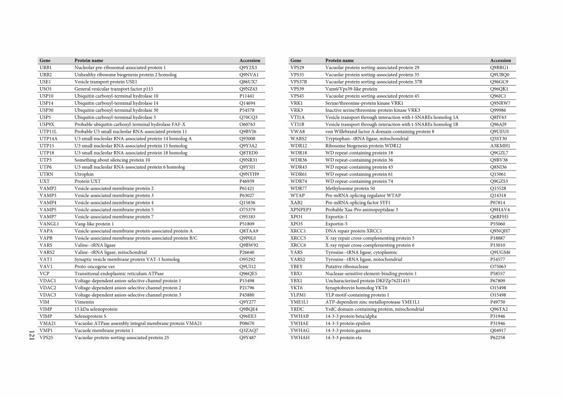
121
Gene Protein name Accession
URB1 Nucleolar pre-ribosomal-associated protein 1 Q9Y2X3
URB2 Unhealthy ribosome biogenesis protein 2 homolog Q9NVA1
USE1 Vesicle transport protein USE1 Q86UX7
USO1 General vesicular transport factor p115 Q9NZ43
USP10 Ubiquitin carboxyl-terminal hydrolase 10 P11441
USP14 Ubiquitin carboxyl-terminal hydrolase 14 Q14694
USP30 Ubiquitin carboxyl-terminal hydrolase 30 P54578
USP5 Ubiquitin carboxyl-terminal hydrolase 5 Q70CQ3
USP9X Probable ubiquitin carboxyl-terminal hydrolase FAF-X O60763
UTP11L Probable U3 small nucleolar RNA-associated protein 11 Q9BVJ6
UTP14A U3 small nucleolar RNA-associated protein 14 homolog A Q93008
UTP15 U3 small nucleolar RNA-associated protein 15 homolog Q9Y3A2
UTP18 U3 small nucleolar RNA-associated protein 18 homolog Q8TED0
UTP3 Something about silencing protein 10 Q9NR31
UTP6 U3 small nucleolar RNA-associated protein 6 homolog Q9Y5J1
UTRN Utrophin Q9NYH9
UXT Protein UXT P46939
VAMP2 Vesicle-associated membrane protein 2 P61421
VAMP3 Vesicle-associated membrane protein 3 P63027
VAMP4 Vesicle-associated membrane protein 4 Q15836
VAMP5 Vesicle-associated membrane protein 5 O75379
VAMP7 Vesicle-associated membrane protein 7 O95183
VANGL1 Vang-like protein 1 P51809
VAPA Vesicle-associated membrane protein-associated protein A Q8TAA9
VAPB Vesicle-associated membrane protein-associated protein B/C Q9P0L0
VARS Valine--tRNA ligase Q9BW92
VARS2 Valine--tRNA ligase, mitochondrial P26640
VAT1 Synaptic vesicle membrane protein VAT-1 homolog O95292
VAV1 Proto-oncogene vav Q9UI12
VCP Transitional endoplasmic reticulum ATPase Q96QE5
VDAC1 Voltage-dependent anion-selective channel protein 1 P15498
VDAC2 Voltage-dependent anion-selective channel protein 2 P21796
VDAC3 Voltage-dependent anion-selective channel protein 3 P45880
VIM Vimentin Q9Y277
VIMP 15 kDa selenoprotein Q9BQE4
VIMP Selenoprotein S Q96EE3
VMA21 Vacuolar ATPase assembly integral membrane protein VMA21 P08670
VMP1 Vacuole membrane protein 1 Q3ZAQ7
VPS25 Vacuolar protein-sorting-associated protein 25 Q9Y487
Gene Protein name Accession
VPS29 Vacuolar protein sorting-associated protein 29 Q9BRG1
VPS35 Vacuolar protein sorting-associated protein 35 Q9UBQ0
VPS37B Vacuolar protein sorting-associated protein 37B Q96GC9
VPS39 Vam6/Vps39-like protein Q96QK1
VPS45 Vacuolar protein sorting-associated protein 45 Q96JC1
VRK1 Serine/threonine-protein kinase VRK1 Q9NRW7
VRK3 Inactive serine/threonine-protein kinase VRK3 Q99986
VTI1A Vesicle transport through interaction with t-SNAREs homolog 1A Q8IV63
VTI1B Vesicle transport through interaction with t-SNAREs homolog 1B Q96AJ9
VWA8 von Willebrand factor A domain-containing protein 8 Q9UEU0
WARS2 Tryptophan--tRNA ligase, mitochondrial Q5ST30
WDR12 Ribosome biogenesis protein WDR12 A3KMH1
WDR18 WD repeat-containing protein 18 Q9GZL7
WDR36 WD repeat-containing protein 36 Q9BV38
WDR43 WD repeat-containing protein 43 Q8NI36
WDR61 WD repeat-containing protein 61 Q15061
WDR74 WD repeat-containing protein 74 Q9GZS3
WDR77 Methylosome protein 50 Q15528
WTAP Pre-mRNA-splicing regulator WTAP Q14318
XAB2 Pre-mRNA-splicing factor SYF1 P07814
XPNPEP3 Probable Xaa-Pro aminopeptidase 3 Q9HAV4
XPO1 Exportin-1 Q6RFH5
XPO5 Exportin-5 P55060
XRCC1 DNA repair protein XRCC1 Q9NQH7
XRCC5 X-ray repair cross-complementing protein 5 P18887
XRCC6 X-ray repair cross-complementing protein 6 P13010
YARS Tyrosine--tRNA ligase, cytoplasmic Q9UGM6
YARS2 Tyrosine--tRNA ligase, mitochondrial P54577
YBEY Putative ribonuclease O75063
YBX1 Nuclease-sensitive element-binding protein 1 P58557
YBX1 Uncharacterized protein DKFZp762I1415 P67809
YKT6 Synaptobrevin homolog YKT6 O15498
YLPM1 YLP motif-containing protein 1 O15498
YME1L1 ATP-dependent zinc metalloprotease YME1L1 P49750
YRDC YrdC domain-containing protein, mitochondrial Q96TA2
YWHAB 14-3-3 protein beta/alpha P31946
YWHAE 14-3-3 protein epsilon P31946
YWHAG 14-3-3 protein gamma Q04917
YWHAH 14-3-3 protein eta P62258
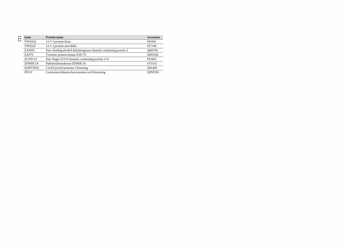
122
Gene Protein name Accession
YWHAQ 14-3-3 protein theta P61981
YWHAZ 14-3-3 protein zeta/delta P27348
ZADH2 Zinc-binding alcohol dehydrogenase domain-containing protein 2 Q86U90
ZAP70 Tyrosine-protein kinase ZAP-70 Q8N4Q0
ZC3H11A Zinc finger CCCH domain-containing protein 11A P43403
ZDHHC18 Palmitoyltransferase ZDHHC18 O75152
ZMPSTE24 CAAX prenyl protease 1 homolog Q01469
ZW10 Centromere/kinetochore protein zw10 homolog Q9NUE0
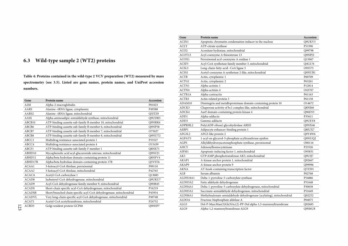
123
6.3 Wild-type sample 2 (WT2) proteins
Table 4: Proteins contained in the wild-type 2 VCV preparation (WT2) measured by mass
spectrometry (see 3.5). Listed are gene names, protein names, and UniProt accession
numbers.
Gene Protein name Accession
A2M Alpha-2-macroglobulin P01023
AARS Alanine--tRNA ligase, cytoplasmic P49588
AARS2 Alanine--tRNA ligase, mitochondrial Q5JTZ9
AASS Alpha-aminoadipic semialdehyde synthase, mitochondrial Q9UDR5
ABCB10 ATP-binding cassette sub-family B member 10, mitochondrial Q9NRK6
ABCB6 ATP-binding cassette sub-family B member 6, mitochondrial Q9NP58
ABCB7 ATP-binding cassette sub-family B member 7, mitochondrial O75027
ABCB8 ATP-binding cassette sub-family B member 8, mitochondrial Q9NUT2
ABCC1 Multidrug resistance-associated protein 1 P33527
ABCC4 Multidrug resistance-associated protein 4 O15439
ABCF1 ATP-binding cassette sub-family F member 1 Q8NE71
ABHD10 Mycophenolic acid acyl-glucuronide esterase, mitochondrial Q9NUJ1
ABHD11 Alpha/beta hydrolase domain-containing protein 11 Q8NFV4
ABHD17B Alpha/beta hydrolase domain-containing protein 17B Q5VST6
ACAA1 3-ketoacyl-CoA thiolase, peroxisomal P09110
ACAA2 3-ketoacyl-CoA thiolase, mitochondrial P42765
ACACA Acetyl-CoA carboxylase 1 Q13085
ACAD8 Isobutyryl-CoA dehydrogenase, mitochondrial Q9UKU7
ACAD9 Acyl-CoA dehydrogenase family member 9, mitochondrial Q9H845
ACADS Short-chain specific acyl-CoA dehydrogenase, mitochondrial P16219
ACADSB Short/branched chain specific acyl-CoA dehydrogenase, mitochondrial P45954
ACADVL Very long-chain specific acyl-CoA dehydrogenase, mitochondrial P49748
ACAT1 Acetyl-CoA acetyltransferase, mitochondrial P24752
ACBD3 Golgi resident protein GCP60 Q9H3P7
Gene Protein name Accession
ACIN1 Apoptotic chromatin condensation inducer in the nucleus Q9UKV3
ACLY ATP-citrate synthase P53396
ACO2 Aconitate hydratase, mitochondrial Q99798
ACOT13 Acyl-coenzyme A thioesterase 13 Q9NPJ3
ACOX1 Peroxisomal acyl-coenzyme A oxidase 1 Q15067
ACSF3 Acyl-CoA synthetase family member 3, mitochondrial Q4G176
ACSL3 Long-chain-fatty-acid--CoA ligase 3 O95573
ACSS1 Acetyl-coenzyme A synthetase 2-like, mitochondrial Q9NUB1
ACTB Actin, cytoplasmic 1 P60709
ACTG1 Actin, cytoplasmic 2 P63261
ACTN1 Alpha-actinin-1 P12814
ACTN4 Alpha-actinin-4 O43707
ACTR1A Alpha-centractin P61163
ACTR3 Actin-related protein 3 P61158
ADAM10 Disintegrin and metalloproteinase domain-containing protein 10 O14672
ADCK3 Chaperone activity of bc1 complex-like, mitochondrial Q8NI60
ADCK4 AarF domain-containing protein kinase 4 Q96D53
ADD1 Alpha-adducin P35611
ADD3 Gamma-adducin Q9UEY8
ADPRHL2 Poly(ADP-ribose) glycohydrolase ARH3 Q9NX46
AEBP1 Adipocyte enhancer-binding protein 1 Q8IUX7
AFG3L2 AFG3-like protein 2 Q9Y4W6
AGPAT5 1-acyl-sn-glycerol-3-phosphate acyltransferase epsilon Q9NUQ2
AGPS Alkyldihydroxyacetonephosphate synthase, peroxisomal O00116
AHCY Adenosylhomocysteinase P23526
AIFM1 Apoptosis-inducing factor 1, mitochondrial O95831
AK3 GTP:AMP phosphotransferase AK3, mitochondrial Q9UIJ7
AKAP1 A-kinase anchor protein 1, mitochondrial Q92667
AKAP9 A-kinase anchor protein 9 Q99996
AKNA AT-hook-containing transcription factor Q7Z591
ALB Serum albumin P02768
ALDH18A1 Delta-1-pyrroline-5-carboxylate synthase P54886
ALDH3A2 Fatty aldehyde dehydrogenase P51648
ALDH4A1 Delta-1-pyrroline-5-carboxylate dehydrogenase, mitochondrial P30038
ALDH5A1 Succinate-semialdehyde dehydrogenase, mitochondrial P51649
ALDH6A1 Methylmalonate-semialdehyde dehydrogenase [acylating], mitochondrial Q02252
ALDOA Fructose-bisphosphate aldolase A P04075
ALG3 Dol-P-Man:Man(5)GlcNAc(2)-PP-Dol alpha-1,3-mannosyltransferase Q92685
ALG9 Alpha-1,2-mannosyltransferase ALG9 Q9H6U8
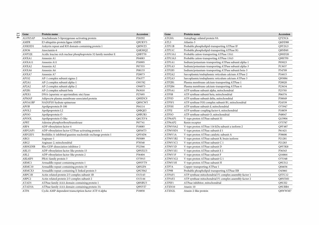
124
Gene Protein name Accession
ALOX5AP Arachidonate 5-lipoxygenase-activating protein P20292
AMFR E3 ubiquitin-protein ligase AMFR Q9UKV5
ANKHD1 Ankyrin repeat and KH domain-containing protein 1 Q8IWZ3
ANO6 Anoctamin-6 Q4KMQ2
ANP32E Acidic leucine-rich nuclear phosphoprotein 32 family member E Q9BTT0
ANXA1 Annexin A1 P04083
ANXA11 Annexin A11 P50995
ANXA2 Annexin A2 P07355
ANXA6 Annexin A6 P08133
ANXA7 Annexin A7 P20073
AP1S2 AP-1 complex subunit sigma-2 P56377
AP2A1 AP-2 complex subunit alpha-1 O95782
AP2A2 AP-2 complex subunit alpha-2 O94973
AP2B1 AP-2 complex subunit beta P63010
APEX1 DNA-(apurinic or apyrimidinic site) lyase P27695
APMAP Adipocyte plasma membrane-associated protein Q9HDC9
APOA1BP NAD(P)H-hydrate epimerase Q8NCW5
APOB Apolipoprotein B-100 P04114
APOL2 Apolipoprotein L2 Q9BQE5
APOO Apolipoprotein O Q9BUR5
APOOL Apolipoprotein O-like Q6UXV4
APRT Adenine phosphoribosyltransferase P07741
ARF4 ADP-ribosylation factor 4 P18085
ARFGAP1 ADP-ribosylation factor GTPase-activating protein 1 Q8N6T3
ARFGEF1 Brefeldin A-inhibited guanine nucleotide-exchange protein 1 Q9Y6D6
ARG1 Arginase-1 P05089
ARG2 Arginase-2, mitochondrial P78540
ARHGDIB Rho GDP-dissociation inhibitor 2 P52566
ARL15 ADP-ribosylation factor-like protein 15 Q9NXU5
ARL2 ADP-ribosylation factor-like protein 2 P36404
ARL6IP5 PRA1 family protein 3 O75915
ARMC1 Armadillo repeat-containing protein 1 Q9NVT9
ARMC10 Armadillo repeat-containing protein 10 Q8N2F6
ARMCX3 Armadillo repeat-containing X-linked protein 3 Q9UH62
ARPC1B Actin-related protein 2/3 complex subunit 1B O15143
ARPC2 Actin-related protein 2/3 complex subunit 2 O15144
ATAD1 ATPase family AAA domain-containing protein 1 Q8NBU5
ATAD3A ATPase family AAA domain-containing protein 3A Q9NVI7
ATF6 Cyclic AMP-dependent transcription factor ATF-6 alpha P18850
Gene Protein name Accession
ATG9A Autophagy-related protein 9A Q7Z3C6
ATL3 Atlastin-3 Q6DD88
ATP11B Probable phospholipid-transporting ATPase IF Q9Y2G3
ATP11C Probable phospholipid-transporting ATPase IG Q8NB49
ATP13A1 Probable cation-transporting ATPase 13A1 Q9HD20
ATP13A3 Probable cation-transporting ATPase 13A3 Q9H7F0
ATP1A1 Sodium/potassium-transporting ATPase subunit alpha-1 P05023
ATP1A3 Sodium/potassium-transporting ATPase subunit alpha-3 P13637
ATP1B3 Sodium/potassium-transporting ATPase subunit beta-3 P54709
ATP2A2 Sarcoplasmic/endoplasmic reticulum calcium ATPase 2 P16615
ATP2A3 Sarcoplasmic/endoplasmic reticulum calcium ATPase 3 Q93084
ATP2B1 Plasma membrane calcium-transporting ATPase 1 P20020
ATP2B4 Plasma membrane calcium-transporting ATPase 4 P23634
ATP5A1 ATP synthase subunit alpha, mitochondrial P25705
ATP5B ATP synthase subunit beta, mitochondrial P06576
ATP5D ATP synthase subunit delta, mitochondrial P30049
ATP5F1 ATP synthase F(0) complex subunit B1, mitochondrial P24539
ATP5H ATP synthase subunit d, mitochondrial O75947
ATP5J ATP synthase-coupling factor 6, mitochondrial P18859
ATP5O ATP synthase subunit O, mitochondrial P48047
ATP6AP1 V-type proton ATPase subunit S1 Q15904
ATP6AP2 Renin receptor O75787
ATP6V0A2 V-type proton ATPase 116 kDa subunit a isoform 2 Q9Y487
ATP6V0D1 V-type proton ATPase subunit d 1 P61421
ATP6V1A V-type proton ATPase catalytic subunit A P38606
ATP6V1B2 V-type proton ATPase subunit B, brain isoform P21281
ATP6V1C1 V-type proton ATPase subunit C 1 P21283
ATP6V1D V-type proton ATPase subunit D Q9Y5K8
ATP6V1E1 V-type proton ATPase subunit E 1 P36543
ATP6V1F V-type proton ATPase subunit F Q16864
ATP6V1G1 V-type proton ATPase subunit G 1 O75348
ATP6V1H V-type proton ATPase subunit H Q9UI12
ATP7A Copper-transporting ATPase 1 Q04656
ATP9B Probable phospholipid-transporting ATPase IIB O43861
ATPAF1 ATP synthase mitochondrial F1 complex assembly factor 1 Q5TC12
ATPAF2 ATP synthase mitochondrial F1 complex assembly factor 2 Q8N5M1
ATPIF1 ATPase inhibitor, mitochondrial Q9UII2
ATXN10 Ataxin-10 Q9UBB4
ATXN2L Ataxin-2-like protein Q8WWM7
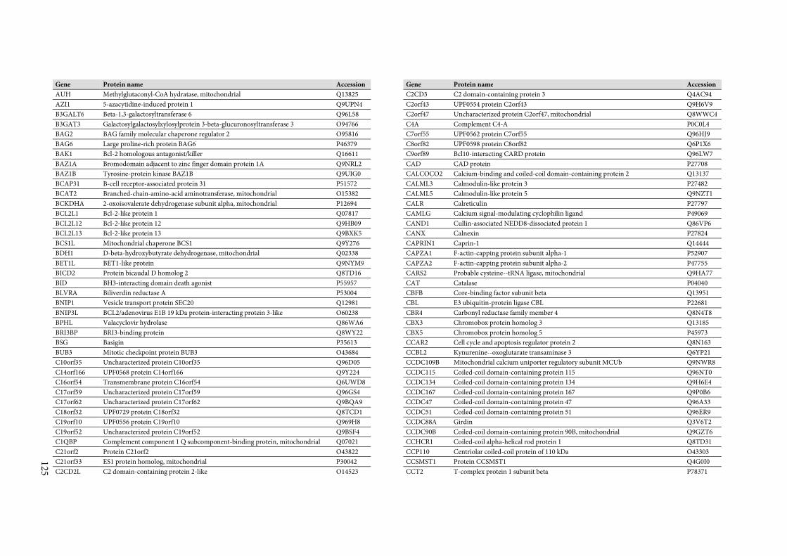
125
Gene Protein name Accession
AUH Methylglutaconyl-CoA hydratase, mitochondrial Q13825
AZI1 5-azacytidine-induced protein 1 Q9UPN4
B3GALT6 Beta-1,3-galactosyltransferase 6 Q96L58
B3GAT3 Galactosylgalactosylxylosylprotein 3-beta-glucuronosyltransferase 3 O94766
BAG2 BAG family molecular chaperone regulator 2 O95816
BAG6 Large proline-rich protein BAG6 P46379
BAK1 Bcl-2 homologous antagonist/killer Q16611
BAZ1A Bromodomain adjacent to zinc finger domain protein 1A Q9NRL2
BAZ1B Tyrosine-protein kinase BAZ1B Q9UIG0
BCAP31 B-cell receptor-associated protein 31 P51572
BCAT2 Branched-chain-amino-acid aminotransferase, mitochondrial O15382
BCKDHA 2-oxoisovalerate dehydrogenase subunit alpha, mitochondrial P12694
BCL2L1 Bcl-2-like protein 1 Q07817
BCL2L12 Bcl-2-like protein 12 Q9HB09
BCL2L13 Bcl-2-like protein 13 Q9BXK5
BCS1L Mitochondrial chaperone BCS1 Q9Y276
BDH1 D-beta-hydroxybutyrate dehydrogenase, mitochondrial Q02338
BET1L BET1-like protein Q9NYM9
BICD2 Protein bicaudal D homolog 2 Q8TD16
BID BH3-interacting domain death agonist P55957
BLVRA Biliverdin reductase A P53004
BNIP1 Vesicle transport protein SEC20 Q12981
BNIP3L BCL2/adenovirus E1B 19 kDa protein-interacting protein 3-like O60238
BPHL Valacyclovir hydrolase Q86WA6
BRI3BP BRI3-binding protein Q8WY22
BSG Basigin P35613
BUB3 Mitotic checkpoint protein BUB3 O43684
C10orf35 Uncharacterized protein C10orf35 Q96D05
C14orf166 UPF0568 protein C14orf166 Q9Y224
C16orf54 Transmembrane protein C16orf54 Q6UWD8
C17orf59 Uncharacterized protein C17orf59 Q96GS4
C17orf62 Uncharacterized protein C17orf62 Q9BQA9
C18orf32 UPF0729 protein C18orf32 Q8TCD1
C19orf10 UPF0556 protein C19orf10 Q969H8
C19orf52 Uncharacterized protein C19orf52 Q9BSF4
C1QBP Complement component 1 Q subcomponent-binding protein, mitochondrial Q07021
C21orf2 Protein C21orf2 O43822
C21orf33 ES1 protein homolog, mitochondrial P30042
C2CD2L C2 domain-containing protein 2-like O14523
Gene Protein name Accession
C2CD3 C2 domain-containing protein 3 Q4AC94
C2orf43 UPF0554 protein C2orf43 Q9H6V9
C2orf47 Uncharacterized protein C2orf47, mitochondrial Q8WWC4
C4A Complement C4-A P0C0L4
C7orf55 UPF0562 protein C7orf55 Q96HJ9
C8orf82 UPF0598 protein C8orf82 Q6P1X6
C9orf89 Bcl10-interacting CARD protein Q96LW7
CAD CAD protein P27708
CALCOCO2 Calcium-binding and coiled-coil domain-containing protein 2 Q13137
CALML3 Calmodulin-like protein 3 P27482
CALML5 Calmodulin-like protein 5 Q9NZT1
CALR Calreticulin P27797
CAMLG Calcium signal-modulating cyclophilin ligand P49069
CAND1 Cullin-associated NEDD8-dissociated protein 1 Q86VP6
CANX Calnexin P27824
CAPRIN1 Caprin-1 Q14444
CAPZA1 F-actin-capping protein subunit alpha-1 P52907
CAPZA2 F-actin-capping protein subunit alpha-2 P47755
CARS2 Probable cysteine--tRNA ligase, mitochondrial Q9HA77
CAT Catalase P04040
CBFB Core-binding factor subunit beta Q13951
CBL E3 ubiquitin-protein ligase CBL P22681
CBR4 Carbonyl reductase family member 4 Q8N4T8
CBX3 Chromobox protein homolog 3 Q13185
CBX5 Chromobox protein homolog 5 P45973
CCAR2 Cell cycle and apoptosis regulator protein 2 Q8N163
CCBL2 Kynurenine--oxoglutarate transaminase 3 Q6YP21
CCDC109B Mitochondrial calcium uniporter regulatory subunit MCUb Q9NWR8
CCDC115 Coiled-coil domain-containing protein 115 Q96NT0
CCDC134 Coiled-coil domain-containing protein 134 Q9H6E4
CCDC167 Coiled-coil domain-containing protein 167 Q9P0B6
CCDC47 Coiled-coil domain-containing protein 47 Q96A33
CCDC51 Coiled-coil domain-containing protein 51 Q96ER9
CCDC88A Girdin Q3V6T2
CCDC90B Coiled-coil domain-containing protein 90B, mitochondrial Q9GZT6
CCHCR1 Coiled-coil alpha-helical rod protein 1 Q8TD31
CCP110 Centriolar coiled-coil protein of 110 kDa O43303
CCSMST1 Protein CCSMST1 Q4G0I0
CCT2 T-complex protein 1 subunit beta P78371
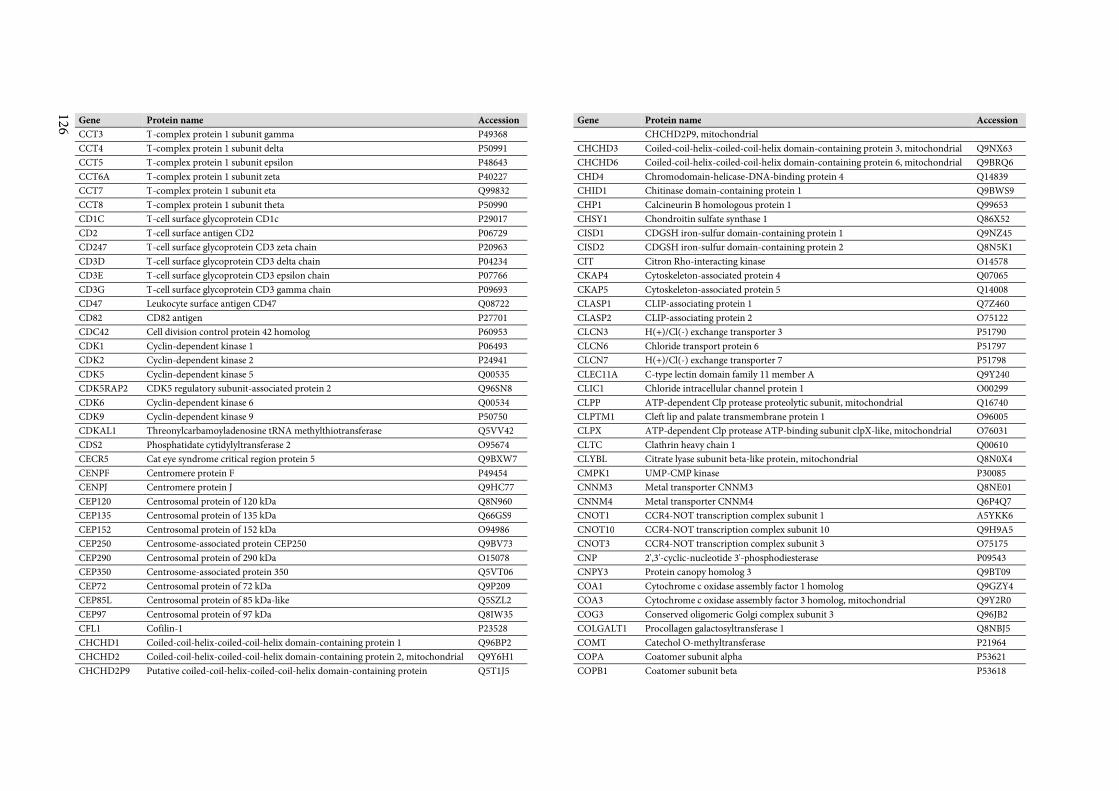
126
Gene Protein name Accession
CCT3 T-complex protein 1 subunit gamma P49368
CCT4 T-complex protein 1 subunit delta P50991
CCT5 T-complex protein 1 subunit epsilon P48643
CCT6A T-complex protein 1 subunit zeta P40227
CCT7 T-complex protein 1 subunit eta Q99832
CCT8 T-complex protein 1 subunit theta P50990
CD1C T-cell surface glycoprotein CD1c P29017
CD2 T-cell surface antigen CD2 P06729
CD247 T-cell surface glycoprotein CD3 zeta chain P20963
CD3D T-cell surface glycoprotein CD3 delta chain P04234
CD3E T-cell surface glycoprotein CD3 epsilon chain P07766
CD3G T-cell surface glycoprotein CD3 gamma chain P09693
CD47 Leukocyte surface antigen CD47 Q08722
CD82 CD82 antigen P27701
CDC42 Cell division control protein 42 homolog P60953
CDK1 Cyclin-dependent kinase 1 P06493
CDK2 Cyclin-dependent kinase 2 P24941
CDK5 Cyclin-dependent kinase 5 Q00535
CDK5RAP2 CDK5 regulatory subunit-associated protein 2 Q96SN8
CDK6 Cyclin-dependent kinase 6 Q00534
CDK9 Cyclin-dependent kinase 9 P50750
CDKAL1 Threonylcarbamoyladenosine tRNA methylthiotransferase Q5VV42
CDS2 Phosphatidate cytidylyltransferase 2 O95674
CECR5 Cat eye syndrome critical region protein 5 Q9BXW7
CENPF Centromere protein F P49454
CENPJ Centromere protein J Q9HC77
CEP120 Centrosomal protein of 120 kDa Q8N960
CEP135 Centrosomal protein of 135 kDa Q66GS9
CEP152 Centrosomal protein of 152 kDa O94986
CEP250 Centrosome-associated protein CEP250 Q9BV73
CEP290 Centrosomal protein of 290 kDa O15078
CEP350 Centrosome-associated protein 350 Q5VT06
CEP72 Centrosomal protein of 72 kDa Q9P209
CEP85L Centrosomal protein of 85 kDa-like Q5SZL2
CEP97 Centrosomal protein of 97 kDa Q8IW35
CFL1 Cofilin-1 P23528
CHCHD1 Coiled-coil-helix-coiled-coil-helix domain-containing protein 1 Q96BP2
CHCHD2 Coiled-coil-helix-coiled-coil-helix domain-containing protein 2, mitochondrial Q9Y6H1
CHCHD2P9 Putative coiled-coil-helix-coiled-coil-helix domain-containing protein Q5T1J5
Gene Protein name Accession
CHCHD2P9, mitochondrial
CHCHD3 Coiled-coil-helix-coiled-coil-helix domain-containing protein 3, mitochondrial Q9NX63
CHCHD6 Coiled-coil-helix-coiled-coil-helix domain-containing protein 6, mitochondrial Q9BRQ6
CHD4 Chromodomain-helicase-DNA-binding protein 4 Q14839
CHID1 Chitinase domain-containing protein 1 Q9BWS9
CHP1 Calcineurin B homologous protein 1 Q99653
CHSY1 Chondroitin sulfate synthase 1 Q86X52
CISD1 CDGSH iron-sulfur domain-containing protein 1 Q9NZ45
CISD2 CDGSH iron-sulfur domain-containing protein 2 Q8N5K1
CIT Citron Rho-interacting kinase O14578
CKAP4 Cytoskeleton-associated protein 4 Q07065
CKAP5 Cytoskeleton-associated protein 5 Q14008
CLASP1 CLIP-associating protein 1 Q7Z460
CLASP2 CLIP-associating protein 2 O75122
CLCN3 H(+)/Cl(-) exchange transporter 3 P51790
CLCN6 Chloride transport protein 6 P51797
CLCN7 H(+)/Cl(-) exchange transporter 7 P51798
CLEC11A C-type lectin domain family 11 member A Q9Y240
CLIC1 Chloride intracellular channel protein 1 O00299
CLPP ATP-dependent Clp protease proteolytic subunit, mitochondrial Q16740
CLPTM1 Cleft lip and palate transmembrane protein 1 O96005
CLPX ATP-dependent Clp protease ATP-binding subunit clpX-like, mitochondrial O76031
CLTC Clathrin heavy chain 1 Q00610
CLYBL Citrate lyase subunit beta-like protein, mitochondrial Q8N0X4
CMPK1 UMP-CMP kinase P30085
CNNM3 Metal transporter CNNM3 Q8NE01
CNNM4 Metal transporter CNNM4 Q6P4Q7
CNOT1 CCR4-NOT transcription complex subunit 1 A5YKK6
CNOT10 CCR4-NOT transcription complex subunit 10 Q9H9A5
CNOT3 CCR4-NOT transcription complex subunit 3 O75175
CNP 2',3'-cyclic-nucleotide 3'-phosphodiesterase P09543
CNPY3 Protein canopy homolog 3 Q9BT09
COA1 Cytochrome c oxidase assembly factor 1 homolog Q9GZY4
COA3 Cytochrome c oxidase assembly factor 3 homolog, mitochondrial Q9Y2R0
COG3 Conserved oligomeric Golgi complex subunit 3 Q96JB2
COLGALT1 Procollagen galactosyltransferase 1 Q8NBJ5
COMT Catechol O-methyltransferase P21964
COPA Coatomer subunit alpha P53621
COPB1 Coatomer subunit beta P53618
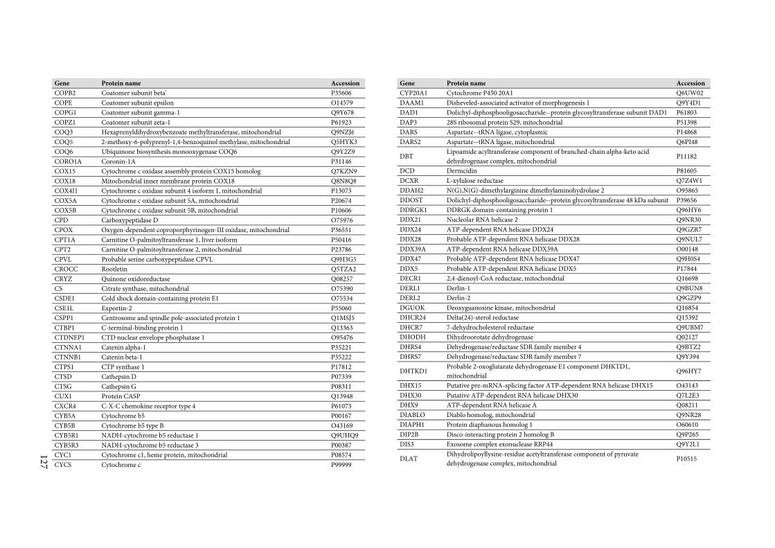
127
Gene Protein name Accession
COPB2 Coatomer subunit beta' P35606
COPE Coatomer subunit epsilon O14579
COPG1 Coatomer subunit gamma-1 Q9Y678
COPZ1 Coatomer subunit zeta-1 P61923
COQ3 Hexaprenyldihydroxybenzoate methyltransferase, mitochondrial Q9NZJ6
COQ5 2-methoxy-6-polyprenyl-1,4-benzoquinol methylase, mitochondrial Q5HYK3
COQ6 Ubiquinone biosynthesis monooxygenase COQ6 Q9Y2Z9
CORO1A Coronin-1A P31146
COX15 Cytochrome c oxidase assembly protein COX15 homolog Q7KZN9
COX18 Mitochondrial inner membrane protein COX18 Q8N8Q8
COX4I1 Cytochrome c oxidase subunit 4 isoform 1, mitochondrial P13073
COX5A Cytochrome c oxidase subunit 5A, mitochondrial P20674
COX5B Cytochrome c oxidase subunit 5B, mitochondrial P10606
CPD Carboxypeptidase D O75976
CPOX Oxygen-dependent coproporphyrinogen-III oxidase, mitochondrial P36551
CPT1A Carnitine O-palmitoyltransferase 1, liver isoform P50416
CPT2 Carnitine O-palmitoyltransferase 2, mitochondrial P23786
CPVL Probable serine carboxypeptidase CPVL Q9H3G5
CROCC Rootletin Q5TZA2
CRYZ Quinone oxidoreductase Q08257
CS Citrate synthase, mitochondrial O75390
CSDE1 Cold shock domain-containing protein E1 O75534
CSE1L Exportin-2 P55060
CSPP1 Centrosome and spindle pole-associated protein 1 Q1MSJ5
CTBP1 C-terminal-binding protein 1 Q13363
CTDNEP1 CTD nuclear envelope phosphatase 1 O95476
CTNNA1 Catenin alpha-1 P35221
CTNNB1 Catenin beta-1 P35222
CTPS1 CTP synthase 1 P17812
CTSD Cathepsin D P07339
CTSG Cathepsin G P08311
CUX1 Protein CASP Q13948
CXCR4 C-X-C chemokine receptor type 4 P61073
CYB5A Cytochrome b5 P00167
CYB5B Cytochrome b5 type B O43169
CYB5R1 NADH-cytochrome b5 reductase 1 Q9UHQ9
CYB5R3 NADH-cytochrome b5 reductase 3 P00387
CYC1 Cytochrome c1, heme protein, mitochondrial P08574
CYCS Cytochrome c P99999
Gene Protein name Accession
CYP20A1 Cytochrome P450 20A1 Q6UW02
DAAM1 Disheveled-associated activator of morphogenesis 1 Q9Y4D1
DAD1 Dolichyl-diphosphooligosaccharide--protein glycosyltransferase subunit DAD1 P61803
DAP3 28S ribosomal protein S29, mitochondrial P51398
DARS Aspartate--tRNA ligase, cytoplasmic P14868
DARS2 Aspartate--tRNA ligase, mitochondrial Q6PI48
DBT Lipoamide acyltransferase component of branched-chain alpha-keto acid
dehydrogenase complex, mitochondrial P11182
DCD Dermcidin P81605
DCXR L-xylulose reductase Q7Z4W1
DDAH2 N(G),N(G)-dimethylarginine dimethylaminohydrolase 2 O95865
DDOST Dolichyl-diphosphooligosaccharide--protein glycosyltransferase 48 kDa subunit P39656
DDRGK1 DDRGK domain-containing protein 1 Q96HY6
DDX21 Nucleolar RNA helicase 2 Q9NR30
DDX24 ATP-dependent RNA helicase DDX24 Q9GZR7
DDX28 Probable ATP-dependent RNA helicase DDX28 Q9NUL7
DDX39A ATP-dependent RNA helicase DDX39A O00148
DDX47 Probable ATP-dependent RNA helicase DDX47 Q9H0S4
DDX5 Probable ATP-dependent RNA helicase DDX5 P17844
DECR1 2,4-dienoyl-CoA reductase, mitochondrial Q16698
DERL1 Derlin-1 Q9BUN8
DERL2 Derlin-2 Q9GZP9
DGUOK Deoxyguanosine kinase, mitochondrial Q16854
DHCR24 Delta(24)-sterol reductase Q15392
DHCR7 7-dehydrocholesterol reductase Q9UBM7
DHODH Dihydroorotate dehydrogenase Q02127
DHRS4 Dehydrogenase/reductase SDR family member 4 Q9BTZ2
DHRS7 Dehydrogenase/reductase SDR family member 7 Q9Y394
DHTKD1 Probable 2-oxoglutarate dehydrogenase E1 component DHKTD1,
mitochondrial Q96HY7
DHX15 Putative pre-mRNA-splicing factor ATP-dependent RNA helicase DHX15 O43143
DHX30 Putative ATP-dependent RNA helicase DHX30 Q7L2E3
DHX9 ATP-dependent RNA helicase A Q08211
DIABLO Diablo homolog, mitochondrial Q9NR28
DIAPH1 Protein diaphanous homolog 1 O60610
DIP2B Disco-interacting protein 2 homolog B Q9P265
DIS3 Exosome complex exonuclease RRP44 Q9Y2L1
DLAT Dihydrolipoyllysine-residue acetyltransferase component of pyruvate
dehydrogenase complex, mitochondrial P10515
128
Gene Protein name Accession
DLG1 Disks large homolog 1 Q12959
DLST Dihydrolipoyllysine-residue succinyltransferase component of 2-oxoglutarate
dehydrogenase complex, mitochondrial P36957
DNAJA1 DnaJ homolog subfamily A member 1 P31689
DNAJA2 DnaJ homolog subfamily A member 2 O60884
DNAJA3 DnaJ homolog subfamily A member 3, mitochondrial Q96EY1
DNAJB11 DnaJ homolog subfamily B member 11 Q9UBS4
DNAJB12 DnaJ homolog subfamily B member 12 Q9NXW2
DNAJB2 DnaJ homolog subfamily B member 2 P25686
DNAJB6 DnaJ homolog subfamily B member 6 O75190
DNAJC10 DnaJ homolog subfamily C member 10 Q8IXB1
DNAJC13 DnaJ homolog subfamily C member 13 O75165
DNAJC15 DnaJ homolog subfamily C member 15 Q9Y5T4
DNAJC19 Mitochondrial import inner membrane translocase subunit TIM14 Q96DA6
DNAJC3 DnaJ homolog subfamily C member 3 Q13217
DNAJC30 DnaJ homolog subfamily C member 30 Q96LL9
DNAJC5 DnaJ homolog subfamily C member 5 Q9H3Z4
DNLZ DNL-type zinc finger protein Q5SXM8
DNTTIP2 Deoxynucleotidyltransferase terminal-interacting protein 2 Q5QJE6
DOCK2 Dedicator of cytokinesis protein 2 Q92608
DOCK7 Dedicator of cytokinesis protein 7 Q96N67
DPM1 Dolichol-phosphate mannosyltransferase subunit 1 O60762
DRG1 Developmentally-regulated GTP-binding protein 1 Q9Y295
DSG1 Desmoglein-1 Q02413
DSG2 Desmoglein-2 Q14126
DSP Desmoplakin P15924
DTYMK Thymidylate kinase P23919
DUT Deoxyuridine 5'-triphosphate nucleotidohydrolase, mitochondrial P33316
DYNC1H1 Cytoplasmic dynein 1 heavy chain 1 Q14204
EARS2 Probable glutamate--tRNA ligase, mitochondrial Q5JPH6
EBAG9 Receptor-binding cancer antigen expressed on SiSo cells O00559
ECH1 Delta(3,5)-Delta(2,4)-dienoyl-CoA isomerase, mitochondrial Q13011
ECHDC1 Ethylmalonyl-CoA decarboxylase Q9NTX5
ECHS1 Enoyl-CoA hydratase, mitochondrial P30084
ECI2 Enoyl-CoA delta isomerase 2, mitochondrial O75521
ECSIT Evolutionarily conserved signaling intermediate in Toll pathway, mitochondrial Q9BQ95
EDC4 Enhancer of mRNA-decapping protein 4 Q6P2E9
EDEM3 ER degradation-enhancing alpha-mannosidase-like protein 3 Q9BZQ6
EEA1 Early endosome antigen 1 Q15075
Gene Protein name Accession
EEF1A1 Elongation factor 1-alpha 1 P68104
EEF1A1P5 Putative elongation factor 1-alpha-like 3 Q5VTE0
EEF1D Elongation factor 1-delta P29692
EEF1E1 Eukaryotic translation elongation factor 1 epsilon-1 O43324
EEF1G Elongation factor 1-gamma P26641
EEF2 Elongation factor 2 P13639
EFCAB4B EF-hand calcium-binding domain-containing protein 4B Q9BSW2
EFHD2 EF-hand domain-containing protein D2 Q96C19
EFTUD2 116 kDa U5 small nuclear ribonucleoprotein component Q15029
EIF3C Eukaryotic translation initiation factor 3 subunit C Q99613
EIF3CL Eukaryotic translation initiation factor 3 subunit C-like protein B5ME19
EIF3E Eukaryotic translation initiation factor 3 subunit E P60228
EIF3H Eukaryotic translation initiation factor 3 subunit H O15372
EIF3I Eukaryotic translation initiation factor 3 subunit I Q13347
EIF4A1 Eukaryotic initiation factor 4A-I P60842
EIF4A3 Eukaryotic initiation factor 4A-III P38919
EIF4E2 Eukaryotic translation initiation factor 4E type 2 O60573
EIF4G1 Eukaryotic translation initiation factor 4 gamma 1 Q04637
EIF5A Eukaryotic translation initiation factor 5A-1 P63241
EIF5AL1 Eukaryotic translation initiation factor 5A-1-like Q6IS14
EIF6 Eukaryotic translation initiation factor 6 P56537
ELAC2 Zinc phosphodiesterase ELAC protein 2 Q9BQ52
ELAVL1 ELAV-like protein 1 Q15717
ELMO1 Engulfment and cell motility protein 1 Q92556
ELMOD2 ELMO domain-containing protein 2 Q8IZ81
EMC1 ER membrane protein complex subunit 1 Q8N766
EMC3 ER membrane protein complex subunit 3 Q9P0I2
EMC4 ER membrane protein complex subunit 4 Q5J8M3
EMC6 ER membrane protein complex subunit 6 Q9BV81
EMC7 ER membrane protein complex subunit 7 Q9NPA0
EMC8 ER membrane protein complex subunit 8 O43402
EMD Emerin P50402
ENDOD1 Endonuclease domain-containing 1 protein O94919
ENO1 Alpha-enolase P06733
ENPP4 Bis(5'-adenosyl)-triphosphatase ENPP4 Q9Y6X5
EPB41 Protein 4.1 P11171
EPDR1 Mammalian ependymin-related protein 1 Q9UM22
EPHX1 Epoxide hydrolase 1 P07099
EPPK1 Epiplakin P58107
129
Gene Protein name Accession
EPRS Bifunctional glutamate/proline--tRNA ligase P07814
EPS15L1 Epidermal growth factor receptor substrate 15-like 1 Q9UBC2
ERAL1 GTPase Era, mitochondrial O75616
ERAP1 Endoplasmic reticulum aminopeptidase 1 Q9NZ08
ERAP2 Endoplasmic reticulum aminopeptidase 2 Q6P179
ERBB2IP Protein LAP2 Q96RT1
ERGIC1 Endoplasmic reticulum-Golgi intermediate compartment protein 1 Q969X5
ERGIC3 Endoplasmic reticulum-Golgi intermediate compartment protein 3 Q9Y282
ERLEC1 Endoplasmic reticulum lectin 1 Q96DZ1
ERLIN2 Erlin-2 O94905
ERO1L ERO1-like protein alpha Q96HE7
ERP29 Endoplasmic reticulum resident protein 29 P30040
ERP44 Endoplasmic reticulum resident protein 44 Q9BS26
ESYT1 Extended synaptotagmin-1 Q9BSJ8
ETFA Electron transfer flavoprotein subunit alpha, mitochondrial P13804
ETFB Electron transfer flavoprotein subunit beta P38117
ETHE1 Persulfide dioxygenase ETHE1, mitochondrial O95571
EXOC1 Exocyst complex component 1 Q9NV70
EXOG Nuclease EXOG, mitochondrial Q9Y2C4
F5 Coagulation factor V P12259
FABP5 Fatty acid-binding protein, epidermal Q01469
FADS2 Fatty acid desaturase 2 O95864
FAF2 FAS-associated factor 2 Q96CS3
FAHD1 Acylpyruvase FAHD1, mitochondrial Q6P587
FAHD2A Fumarylacetoacetate hydrolase domain-containing protein 2A Q96GK7
FAM120A Constitutive coactivator of PPAR-gamma-like protein 1 Q9NZB2
FAM134C Protein FAM134C Q86VR2
FAM160B1 Protein FAM160B1 Q5W0V3
FAM162A Protein FAM162A Q96A26
FAM20B Glycosaminoglycan xylosylkinase O75063
FAM210A Protein FAM210A Q96ND0
FAM213A Redox-regulatory protein FAM213A Q9BRX8
FAM3C Protein FAM3C Q92520
FAM49B Protein FAM49B Q9NUQ9
FAM96B Mitotic spindle-associated MMXD complex subunit MIP18 Q9Y3D0
FANCD2 Fanconi anemia group D2 protein Q9BXW9
FANCI Fanconi anemia group I protein Q9NVI1
FAR1 Fatty acyl-CoA reductase 1 Q8WVX9
FARS2 Phenylalanine--tRNA ligase, mitochondrial O95363
Gene Protein name Accession
FAS Tumor necrosis factor receptor superfamily member 6 P25445
FASN Fatty acid synthase P49327
FASTKD1 FAST kinase domain-containing protein 1 Q53R41
FASTKD2 FAST kinase domain-containing protein 2 Q9NYY8
FAT1 Protocadherin Fat 1 Q14517
FDFT1 Squalene synthase P37268
FDPS Farnesyl pyrophosphate synthase P14324
FDX1 Adrenodoxin, mitochondrial P10109
FDXR NADPH:adrenodoxin oxidoreductase, mitochondrial P22570
FECH Ferrochelatase, mitochondrial P22830
FEN1 Flap endonuclease 1 P39748
FERMT3 Fermitin family homolog 3 Q86UX7
FGA Fibrinogen alpha chain P02671
FGFR1OP FGFR1 oncogene partner O95684
FH Fumarate hydratase, mitochondrial P07954
FIS1 Mitochondrial fission 1 protein Q9Y3D6
FKBP11 Peptidyl-prolyl cis-trans isomerase FKBP11 Q9NYL4
FKBP15 FK506-binding protein 15 Q5T1M5
FKBP2 Peptidyl-prolyl cis-trans isomerase FKBP2 P26885
FLG2 Filaggrin-2 Q5D862
FLII Protein flightless-1 homolog Q13045
FLNA Filamin-A P21333
FLNB Filamin-B O75369
FLOT1 Flotillin-1 O75955
FLOT2 Flotillin-2 Q14254
FLVCR1 Feline leukemia virus subgroup C receptor-related protein 1 Q9Y5Y0
FMNL1 Formin-like protein 1 O95466
FN1 Fibronectin P02751
FNDC3A Fibronectin type-III domain-containing protein 3A Q9Y2H6
FOXRED1 FAD-dependent oxidoreductase domain-containing protein 1 Q96CU9
FRG1 Protein FRG1 Q14331
FTH1 Ferritin heavy chain P02794
FTSJ3 pre-rRNA processing protein FTSJ3 Q8IY81
FUNDC2 FUN14 domain-containing protein 2 Q9BWH2
FUT8 Alpha-(1,6)-fucosyltransferase Q9BYC5
FXN Frataxin, mitochondrial Q16595
FYCO1 FYVE and coiled-coil domain-containing protein 1 Q9BQS8
G6PD Glucose-6-phosphate 1-dehydrogenase P11413
GAA Lysosomal alpha-glucosidase P10253
130
Gene Protein name Accession
GABARAPL2 Gamma-aminobutyric acid receptor-associated protein-like 2 P60520
GADD45GIP1 Growth arrest and DNA damage-inducible proteins-interacting protein 1 Q8TAE8
GALNT2 Polypeptide N-acetylgalactosaminyltransferase 2 Q10471
GALNT7 N-acetylgalactosaminyltransferase 7 Q86SF2
GAPDH Glyceraldehyde-3-phosphate dehydrogenase P04406
GARS Glycine--tRNA ligase P41250
GART Trifunctional purine biosynthetic protein adenosine-3 P22102
GATC Glutamyl-tRNA(Gln) amidotransferase subunit C, mitochondrial O43716
GBAS Protein NipSnap homolog 2 O75323
GBF1 Golgi-specific brefeldin A-resistance guanine nucleotide exchange factor 1 Q92538
GCC2 GRIP and coiled-coil domain-containing protein 2 Q8IWJ2
GCSH Glycine cleavage system H protein, mitochondrial P23434
GDAP1 Ganglioside-induced differentiation-associated protein 1 Q8TB36
GDPD1 Glycerophosphodiester phosphodiesterase domain-containing protein 1 Q8N9F7
GFM2 Ribosome-releasing factor 2, mitochondrial Q969S9
GGH Gamma-glutamyl hydrolase Q92820
GHITM Growth hormone-inducible transmembrane protein Q9H3K2
GIGYF2 PERQ amino acid-rich with GYF domain-containing protein 2 Q6Y7W6
GIMAP1 GTPase IMAP family member 1 Q8WWP7
GIMAP6 GTPase IMAP family member 6 Q6P9H5
GLG1 Golgi apparatus protein 1 Q92896
GLIPR2 Golgi-associated plant pathogenesis-related protein 1 Q9H4G4
GLRX3 Glutaredoxin-3 O76003
GLRX5 Glutaredoxin-related protein 5, mitochondrial Q86SX6
GLS Glutaminase kidney isoform, mitochondrial O94925
GLUD1 Glutamate dehydrogenase 1, mitochondrial P00367
GNA13 Guanine nucleotide-binding protein subunit alpha-13 Q14344
GNAI2 Guanine nucleotide-binding protein G(i) subunit alpha-2 P04899
GNAI3 Guanine nucleotide-binding protein G(k) subunit alpha P08754
GNAQ Guanine nucleotide-binding protein G(q) subunit alpha P50148
GNAS Guanine nucleotide-binding protein G(s) subunit alpha isoforms short P63092
GNB2 Guanine nucleotide-binding protein G(I)/G(S)/G(T) subunit beta-2 P62879
GNB2L1 Guanine nucleotide-binding protein subunit beta-2-like 1 P63244
GNPTAB N-acetylglucosamine-1-phosphotransferase subunits alpha/beta Q3T906
GNS N-acetylglucosamine-6-sulfatase P15586
GOLGA2 Golgin subfamily A member 2 Q08379
GOLGA3 Golgin subfamily A member 3 Q08378
GOLGA5 Golgin subfamily A member 5 Q8TBA6
GOLGA7 Golgin subfamily A member 7 Q7Z5G4
Gene Protein name Accession
GOLGB1 Golgin subfamily B member 1 Q14789
GOLPH3 Golgi phosphoprotein 3 Q9H4A6
GOPC Golgi-associated PDZ and coiled-coil motif-containing protein Q9HD26
GORASP2 Golgi reassembly-stacking protein 2 Q9H8Y8
GOSR1 Golgi SNAP receptor complex member 1 O95249
GOSR2 Golgi SNAP receptor complex member 2 O35165
GOT2 Aspartate aminotransferase, mitochondrial P00505
GPR107 Protein GPR107 Q5VW38
GRAP2 GRB2-related adapter protein 2 O75791
GRB2 Growth factor receptor-bound protein 2 P62993
GRHPR Glyoxylate reductase/hydroxypyruvate reductase Q9UBQ7
GRPEL1 GrpE protein homolog 1, mitochondrial Q9HAV7
GRPEL2 GrpE protein homolog 2, mitochondrial Q8TAA5
GRSF1 G-rich sequence factor 1 Q12849
GSN Gelsolin P06396
GSR Glutathione reductase, mitochondrial P00390
GSTK1 Glutathione S-transferase kappa 1 Q9Y2Q3
GSTP1 Glutathione S-transferase P P09211
GTPBP10 GTP-binding protein 10 A4D1E9
GTPBP3 tRNA modification GTPase GTPBP3, mitochondrial Q969Y2
GTPBP6 Putative GTP-binding protein 6 O43824
GTPBP8 GTP-binding protein 8 Q8N3Z3
GUK1 Guanylate kinase Q64520
H2AFV Histone H2A.V Q71UI9
H2AFX Histone H2AX P16104
H2AFY Core histone macro-H2A.1 O75367
H2AFZ Histone H2A.Z P0C0S5
H3F3A Histone H3.3 P84243
H6PD GDH/6PGL endoplasmic bifunctional protein O95479
HADH Hydroxyacyl-coenzyme A dehydrogenase, mitochondrial Q16836
HADHA Trifunctional enzyme subunit alpha, mitochondrial P40939
HADHB Trifunctional enzyme subunit beta, mitochondrial P55084
HAGH Hydroxyacylglutathione hydrolase, mitochondrial Q16775
HAL Histidine ammonia-lyase P42357
HARS2 Probable histidine--tRNA ligase, mitochondrial P49590
HAUS1 HAUS augmin-like complex subunit 1 Q96CS2
HAUS2 HAUS augmin-like complex subunit 2 Q9NVX0
HAUS4 HAUS augmin-like complex subunit 4 Q9H6D7
HAUS6 HAUS augmin-like complex subunit 6 Q7Z4H7
131
Gene Protein name Accession
HAUS7 HAUS augmin-like complex subunit 7 Q99871
HAUS8 HAUS augmin-like complex subunit 8 Q9BT25
HAX1 HCLS1-associated protein X-1 O00165
HBA1; Hemoglobin subunit alpha P69905
HBB Hemoglobin subunit beta P68871
HCCS Cytochrome c-type heme lyase P53701
HCFC1 Host cell factor 1 P51610
HDHD3 Haloacid dehalogenase-like hydrolase domain-containing protein 3 Q9BSH5
HDLBP Vigilin Q00341
HEATR1 HEAT repeat-containing protein 1 Q9H583
HEXB Beta-hexosaminidase subunit beta P07686
HIBADH 3-hydroxyisobutyrate dehydrogenase, mitochondrial P31937
HIBCH 3-hydroxyisobutyryl-CoA hydrolase, mitochondrial Q6NVY1
HINT1 Histidine triad nucleotide-binding protein 1 P49773
HIST1H1B Histone H1.5 P16401
HIST1H1C Histone H1.2 P16403
HIST1H1D Histone H1.3 P16402
HIST1H2AC Histone H2A type 1-C Q93077
HIST1H2BB Histone H2B type 1-B P33778
HIST1H2BC Histone H2B type 1-C/E/F/G/I P62807
HIST1H2BD Histone H2B type 1-D P58876
HIST1H2BH Histone H2B type 1-H Q93079
HIST1H2BJ Histone H2B type 1-J P06899
HIST1H4A Histone H4 P62805
HK1 Hexokinase-1 P19367
HK2 Hexokinase-2 P52789
HLA-A HLA class I histocompatibility antigen, A-3 alpha chain P04439
HLA-B HLA class I histocompatibility antigen, B-8 alpha chain P30460
HLA-E HLA class I histocompatibility antigen, alpha chain E P13747
HM13 Minor histocompatibility antigen H13 Q8TCT9
HMGB1 High mobility group protein B1 P09429
HMGB1P1 Putative high mobility group protein B1-like 1 B2RPK0
HMGB2 High mobility group protein B2 P26583
HMGCL Hydroxymethylglutaryl-CoA lyase, mitochondrial P35914
HMGCR 3-hydroxy-3-methylglutaryl-coenzyme A reductase P04035
HNRNPA0 Heterogeneous nuclear ribonucleoprotein A0 Q13151
HNRNPA1 Heterogeneous nuclear ribonucleoprotein A1 P09651
HNRNPA2B1 Heterogeneous nuclear ribonucleoproteins A2/B1 P22626
HNRNPA3 Heterogeneous nuclear ribonucleoprotein A3 P51991
Gene Protein name Accession
HNRNPC Heterogeneous nuclear ribonucleoproteins C1/C2 P07910
HNRNPD Heterogeneous nuclear ribonucleoprotein D0 Q14103
HNRNPF Heterogeneous nuclear ribonucleoprotein F P52597
HNRNPH1 Heterogeneous nuclear ribonucleoprotein H P31943
HNRNPH3 Heterogeneous nuclear ribonucleoprotein H3 P31942
HNRNPM Heterogeneous nuclear ribonucleoprotein M P52272
HPCAL1 Hippocalcin-like protein 1 P37235
HPRT1 Hypoxanthine-guanine phosphoribosyltransferase P00492
HRNR Hornerin Q86YZ3
HRSP12 Ribonuclease UK114 P52758
HS2ST1 Heparan sulfate 2-O-sulfotransferase 1 Q7LGA3
HSD17B10 3-hydroxyacyl-CoA dehydrogenase type-2 Q99714
HSD17B11 Estradiol 17-beta-dehydrogenase 11 Q8NBQ5
HSD17B12 Estradiol 17-beta-dehydrogenase 12 Q53GQ0
HSD17B4 Peroxisomal multifunctional enzyme type 2 P51659
HSD17B7 3-keto-steroid reductase P56937
HSD17B8 Estradiol 17-beta-dehydrogenase 8 Q92506
HSDL1 Inactive hydroxysteroid dehydrogenase-like protein 1 Q3SXM5
HSDL2 Hydroxysteroid dehydrogenase-like protein 2 Q6YN16
HSP90AA1 Heat shock protein HSP 90-alpha P07900
HSP90AB1 Heat shock protein HSP 90-beta P08238
HSP90B1 Endoplasmin P14625
HSPA1A Heat shock 70 kDa protein 1A/1B P08107
HSPA5 78 kDa glucose-regulated protein P11021
HSPA8 Heat shock cognate 71 kDa protein P11142
HSPA9 Stress-70 protein, mitochondrial P38646
HSPB1 Heat shock protein beta-1 P04792
HSPD1 60 kDa heat shock protein, mitochondrial P10809
HSPE1 10 kDa heat shock protein, mitochondrial P61604
HSPH1 Heat shock protein 105 kDa Q92598
HTRA2 Serine protease HTRA2, mitochondrial O43464
HVCN1 Voltage-gated hydrogen channel 1 Q96D96
HYOU1 Hypoxia up-regulated protein 1 Q9Y4L1
IARS Isoleucine--tRNA ligase, cytoplasmic P41252
IBA57 Putative transferase CAF17, mitochondrial Q5T440
ICAM2 Intercellular adhesion molecule 2 P13598
ICT1 Peptidyl-tRNA hydrolase ICT1, mitochondrial Q14197
IDE Insulin-degrading enzyme P14735
IDH2 Isocitrate dehydrogenase [NADP], mitochondrial P48735
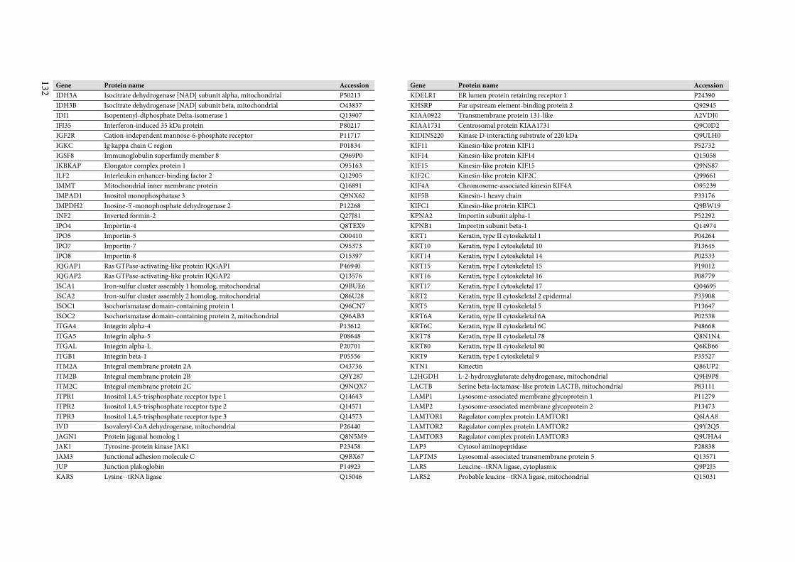
132
Gene Protein name Accession
IDH3A Isocitrate dehydrogenase [NAD] subunit alpha, mitochondrial P50213
IDH3B Isocitrate dehydrogenase [NAD] subunit beta, mitochondrial O43837
IDI1 Isopentenyl-diphosphate Delta-isomerase 1 Q13907
IFI35 Interferon-induced 35 kDa protein P80217
IGF2R Cation-independent mannose-6-phosphate receptor P11717
IGKC Ig kappa chain C region P01834
IGSF8 Immunoglobulin superfamily member 8 Q969P0
IKBKAP Elongator complex protein 1 O95163
ILF2 Interleukin enhancer-binding factor 2 Q12905
IMMT Mitochondrial inner membrane protein Q16891
IMPAD1 Inositol monophosphatase 3 Q9NX62
IMPDH2 Inosine-5'-monophosphate dehydrogenase 2 P12268
INF2 Inverted formin-2 Q27J81
IPO4 Importin-4 Q8TEX9
IPO5 Importin-5 O00410
IPO7 Importin-7 O95373
IPO8 Importin-8 O15397
IQGAP1 Ras GTPase-activating-like protein IQGAP1 P46940
IQGAP2 Ras GTPase-activating-like protein IQGAP2 Q13576
ISCA1 Iron-sulfur cluster assembly 1 homolog, mitochondrial Q9BUE6
ISCA2 Iron-sulfur cluster assembly 2 homolog, mitochondrial Q86U28
ISOC1 Isochorismatase domain-containing protein 1 Q96CN7
ISOC2 Isochorismatase domain-containing protein 2, mitochondrial Q96AB3
ITGA4 Integrin alpha-4 P13612
ITGA5 Integrin alpha-5 P08648
ITGAL Integrin alpha-L P20701
ITGB1 Integrin beta-1 P05556
ITM2A Integral membrane protein 2A O43736
ITM2B Integral membrane protein 2B Q9Y287
ITM2C Integral membrane protein 2C Q9NQX7
ITPR1 Inositol 1,4,5-trisphosphate receptor type 1 Q14643
ITPR2 Inositol 1,4,5-trisphosphate receptor type 2 Q14571
ITPR3 Inositol 1,4,5-trisphosphate receptor type 3 Q14573
IVD Isovaleryl-CoA dehydrogenase, mitochondrial P26440
JAGN1 Protein jagunal homolog 1 Q8N5M9
JAK1 Tyrosine-protein kinase JAK1 P23458
JAM3 Junctional adhesion molecule C Q9BX67
JUP Junction plakoglobin P14923
KARS Lysine--tRNA ligase Q15046
Gene Protein name Accession
KDELR1 ER lumen protein retaining receptor 1 P24390
KHSRP Far upstream element-binding protein 2 Q92945
KIAA0922 Transmembrane protein 131-like A2VDJ0
KIAA1731 Centrosomal protein KIAA1731 Q9C0D2
KIDINS220 Kinase D-interacting substrate of 220 kDa Q9ULH0
KIF11 Kinesin-like protein KIF11 P52732
KIF14 Kinesin-like protein KIF14 Q15058
KIF15 Kinesin-like protein KIF15 Q9NS87
KIF2C Kinesin-like protein KIF2C Q99661
KIF4A Chromosome-associated kinesin KIF4A O95239
KIF5B Kinesin-1 heavy chain P33176
KIFC1 Kinesin-like protein KIFC1 Q9BW19
KPNA2 Importin subunit alpha-1 P52292
KPNB1 Importin subunit beta-1 Q14974
KRT1 Keratin, type II cytoskeletal 1 P04264
KRT10 Keratin, type I cytoskeletal 10 P13645
KRT14 Keratin, type I cytoskeletal 14 P02533
KRT15 Keratin, type I cytoskeletal 15 P19012
KRT16 Keratin, type I cytoskeletal 16 P08779
KRT17 Keratin, type I cytoskeletal 17 Q04695
KRT2 Keratin, type II cytoskeletal 2 epidermal P35908
KRT5 Keratin, type II cytoskeletal 5 P13647
KRT6A Keratin, type II cytoskeletal 6A P02538
KRT6C Keratin, type II cytoskeletal 6C P48668
KRT78 Keratin, type II cytoskeletal 78 Q8N1N4
KRT80 Keratin, type II cytoskeletal 80 Q6KB66
KRT9 Keratin, type I cytoskeletal 9 P35527
KTN1 Kinectin Q86UP2
L2HGDH L-2-hydroxyglutarate dehydrogenase, mitochondrial Q9H9P8
LACTB Serine beta-lactamase-like protein LACTB, mitochondrial P83111
LAMP1 Lysosome-associated membrane glycoprotein 1 P11279
LAMP2 Lysosome-associated membrane glycoprotein 2 P13473
LAMTOR1 Ragulator complex protein LAMTOR1 Q6IAA8
LAMTOR2 Ragulator complex protein LAMTOR2 Q9Y2Q5
LAMTOR3 Ragulator complex protein LAMTOR3 Q9UHA4
LAP3 Cytosol aminopeptidase P28838
LAPTM5 Lysosomal-associated transmembrane protein 5 Q13571
LARS Leucine--tRNA ligase, cytoplasmic Q9P2J5
LARS2 Probable leucine--tRNA ligase, mitochondrial Q15031
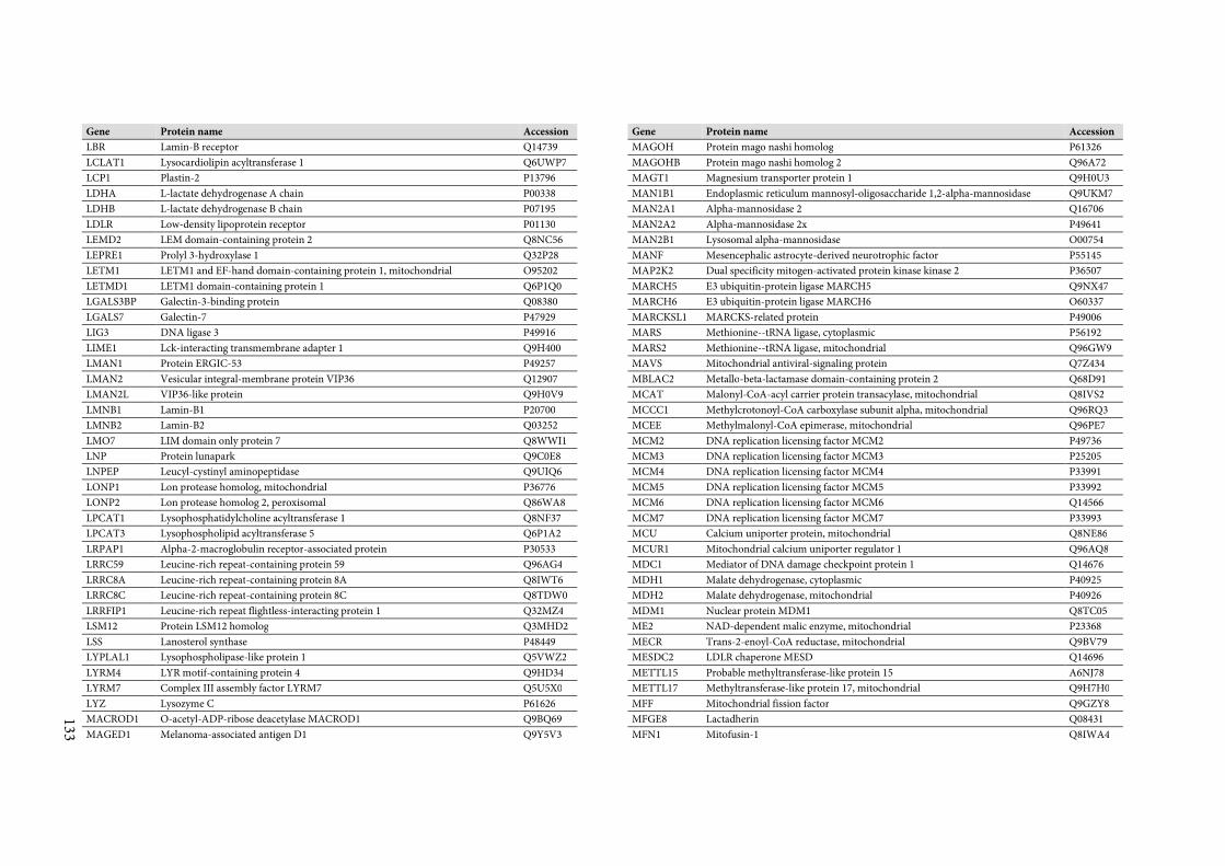
133
Gene Protein name Accession
LBR Lamin-B receptor Q14739
LCLAT1 Lysocardiolipin acyltransferase 1 Q6UWP7
LCP1 Plastin-2 P13796
LDHA L-lactate dehydrogenase A chain P00338
LDHB L-lactate dehydrogenase B chain P07195
LDLR Low-density lipoprotein receptor P01130
LEMD2 LEM domain-containing protein 2 Q8NC56
LEPRE1 Prolyl 3-hydroxylase 1 Q32P28
LETM1 LETM1 and EF-hand domain-containing protein 1, mitochondrial O95202
LETMD1 LETM1 domain-containing protein 1 Q6P1Q0
LGALS3BP Galectin-3-binding protein Q08380
LGALS7 Galectin-7 P47929
LIG3 DNA ligase 3 P49916
LIME1 Lck-interacting transmembrane adapter 1 Q9H400
LMAN1 Protein ERGIC-53 P49257
LMAN2 Vesicular integral-membrane protein VIP36 Q12907
LMAN2L VIP36-like protein Q9H0V9
LMNB1 Lamin-B1 P20700
LMNB2 Lamin-B2 Q03252
LMO7 LIM domain only protein 7 Q8WWI1
LNP Protein lunapark Q9C0E8
LNPEP Leucyl-cystinyl aminopeptidase Q9UIQ6
LONP1 Lon protease homolog, mitochondrial P36776
LONP2 Lon protease homolog 2, peroxisomal Q86WA8
LPCAT1 Lysophosphatidylcholine acyltransferase 1 Q8NF37
LPCAT3 Lysophospholipid acyltransferase 5 Q6P1A2
LRPAP1 Alpha-2-macroglobulin receptor-associated protein P30533
LRRC59 Leucine-rich repeat-containing protein 59 Q96AG4
LRRC8A Leucine-rich repeat-containing protein 8A Q8IWT6
LRRC8C Leucine-rich repeat-containing protein 8C Q8TDW0
LRRFIP1 Leucine-rich repeat flightless-interacting protein 1 Q32MZ4
LSM12 Protein LSM12 homolog Q3MHD2
LSS Lanosterol synthase P48449
LYPLAL1 Lysophospholipase-like protein 1 Q5VWZ2
LYRM4 LYR motif-containing protein 4 Q9HD34
LYRM7 Complex III assembly factor LYRM7 Q5U5X0
LYZ Lysozyme C P61626
MACROD1 O-acetyl-ADP-ribose deacetylase MACROD1 Q9BQ69
MAGED1 Melanoma-associated antigen D1 Q9Y5V3
Gene Protein name Accession
MAGOH Protein mago nashi homolog P61326
MAGOHB Protein mago nashi homolog 2 Q96A72
MAGT1 Magnesium transporter protein 1 Q9H0U3
MAN1B1 Endoplasmic reticulum mannosyl-oligosaccharide 1,2-alpha-mannosidase Q9UKM7
MAN2A1 Alpha-mannosidase 2 Q16706
MAN2A2 Alpha-mannosidase 2x P49641
MAN2B1 Lysosomal alpha-mannosidase O00754
MANF Mesencephalic astrocyte-derived neurotrophic factor P55145
MAP2K2 Dual specificity mitogen-activated protein kinase kinase 2 P36507
MARCH5 E3 ubiquitin-protein ligase MARCH5 Q9NX47
MARCH6 E3 ubiquitin-protein ligase MARCH6 O60337
MARCKSL1 MARCKS-related protein P49006
MARS Methionine--tRNA ligase, cytoplasmic P56192
MARS2 Methionine--tRNA ligase, mitochondrial Q96GW9
MAVS Mitochondrial antiviral-signaling protein Q7Z434
MBLAC2 Metallo-beta-lactamase domain-containing protein 2 Q68D91
MCAT Malonyl-CoA-acyl carrier protein transacylase, mitochondrial Q8IVS2
MCCC1 Methylcrotonoyl-CoA carboxylase subunit alpha, mitochondrial Q96RQ3
MCEE Methylmalonyl-CoA epimerase, mitochondrial Q96PE7
MCM2 DNA replication licensing factor MCM2 P49736
MCM3 DNA replication licensing factor MCM3 P25205
MCM4 DNA replication licensing factor MCM4 P33991
MCM5 DNA replication licensing factor MCM5 P33992
MCM6 DNA replication licensing factor MCM6 Q14566
MCM7 DNA replication licensing factor MCM7 P33993
MCU Calcium uniporter protein, mitochondrial Q8NE86
MCUR1 Mitochondrial calcium uniporter regulator 1 Q96AQ8
MDC1 Mediator of DNA damage checkpoint protein 1 Q14676
MDH1 Malate dehydrogenase, cytoplasmic P40925
MDH2 Malate dehydrogenase, mitochondrial P40926
MDM1 Nuclear protein MDM1 Q8TC05
ME2 NAD-dependent malic enzyme, mitochondrial P23368
MECR Trans-2-enoyl-CoA reductase, mitochondrial Q9BV79
MESDC2 LDLR chaperone MESD Q14696
METTL15 Probable methyltransferase-like protein 15 A6NJ78
METTL17 Methyltransferase-like protein 17, mitochondrial Q9H7H0
MFF Mitochondrial fission factor Q9GZY8
MFGE8 Lactadherin Q08431
MFN1 Mitofusin-1 Q8IWA4
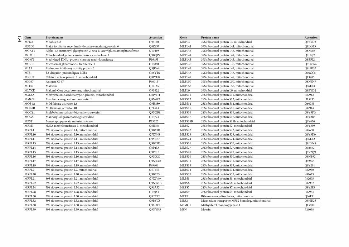
134
Gene Protein name Accession
MFN2 Mitofusin-2 O95140
MFSD6 Major facilitator superfamily domain-containing protein 6 Q6ZSS7
MGAT2 Alpha-1,6-mannosyl-glycoprotein 2-beta-N-acetylglucosaminyltransferase Q10469
MGME1 Mitochondrial genome maintenance exonuclease 1 Q9BQP7
MGMT Methylated-DNA--protein-cysteine methyltransferase P16455
MGST3 Microsomal glutathione S-transferase 3 O14880
MIA3 Melanoma inhibitory activity protein 3 Q5JRA6
MIB1 E3 ubiquitin-protein ligase MIB1 Q86YT6
MICU2 Calcium uptake protein 2, mitochondrial Q8IYU8
MKI67 Antigen KI-67 P46013
MLEC Malectin Q14165
MLYCD Malonyl-CoA decarboxylase, mitochondrial O95822
MMAA Methylmalonic aciduria type A protein, mitochondrial Q8IVH4
MMGT1 Membrane magnesium transporter 1 Q8N4V1
MOB1A MOB kinase activator 1A Q9H8S9
MOB1B MOB kinase activator 1B Q7L9L4
MOCS1 Molybdenum cofactor biosynthesis protein 1 Q9NZB8
MOGS Mannosyl-oligosaccharide glucosidase Q13724
MPST 3-mercaptopyruvate sulfurtransferase P25325
MRM1 rRNA methyltransferase 1, mitochondrial Q6IN84
MRPL1 39S ribosomal protein L1, mitochondrial Q9BYD6
MRPL10 39S ribosomal protein L10, mitochondrial Q7Z7H8
MRPL11 39S ribosomal protein L11, mitochondrial Q9Y3B7
MRPL13 39S ribosomal protein L13, mitochondrial Q9BYD1
MRPL14 39S ribosomal protein L14, mitochondrial Q6P1L8
MRPL15 39S ribosomal protein L15, mitochondrial Q9P015
MRPL16 39S ribosomal protein L16, mitochondrial Q9NX20
MRPL17 39S ribosomal protein L17, mitochondrial Q9NRX2
MRPL19 39S ribosomal protein L19, mitochondrial P49406
MRPL2 39S ribosomal protein L2, mitochondrial Q5T653
MRPL20 39S ribosomal protein L20, mitochondrial Q9BYC9
MRPL21 39S ribosomal protein L21, mitochondrial Q7Z2W9
MRPL22 39S ribosomal protein L22, mitochondrial Q9NWU5
MRPL24 39S ribosomal protein L24, mitochondrial Q96A35
MRPL28 39S ribosomal protein L28, mitochondrial Q13084
MRPL30 39S ribosomal protein L30, mitochondrial Q8TCC3
MRPL32 39S ribosomal protein L32, mitochondrial Q9BYC8
MRPL38 39S ribosomal protein L38, mitochondrial Q96DV4
MRPL39 39S ribosomal protein L39, mitochondrial Q9NYK5
Gene Protein name Accession
MRPL4 39S ribosomal protein L4, mitochondrial Q9BYD3
MRPL41 39S ribosomal protein L41, mitochondrial Q8IXM3
MRPL43 39S ribosomal protein L43, mitochondrial Q8N983
MRPL44 39S ribosomal protein L44, mitochondrial Q9H9J2
MRPL45 39S ribosomal protein L45, mitochondrial Q9BRJ2
MRPL46 39S ribosomal protein L46, mitochondrial Q9H2W6
MRPL47 39S ribosomal protein L47, mitochondrial Q9HD33
MRPL48 39S ribosomal protein L48, mitochondrial Q96GC5
MRPL49 39S ribosomal protein L49, mitochondrial Q13405
MRPL50 39S ribosomal protein L50, mitochondrial Q8N5N7
MRPL53 39S ribosomal protein L53, mitochondrial Q96EL3
MRPL9 39S ribosomal protein L9, mitochondrial Q9BYD2
MRPS11 28S ribosomal protein S11, mitochondrial P82912
MRPS12 28S ribosomal protein S12, mitochondrial O15235
MRPS14 28S ribosomal protein S14, mitochondrial O60783
MRPS15 28S ribosomal protein S15, mitochondrial P82914
MRPS16 28S ribosomal protein S16, mitochondrial Q9Y3D3
MRPS17 28S ribosomal protein S17, mitochondrial Q9Y2R5
MRPS18B 28S ribosomal protein S18b, mitochondrial Q9Y676
MRPS2 28S ribosomal protein S2, mitochondrial Q9Y399
MRPS22 28S ribosomal protein S22, mitochondrial P82650
MRPS23 28S ribosomal protein S23, mitochondrial Q9Y3D9
MRPS24 28S ribosomal protein S24, mitochondrial Q96EL2
MRPS26 28S ribosomal protein S26, mitochondrial Q9BYN8
MRPS27 28S ribosomal protein S27, mitochondrial Q92552
MRPS28 28S ribosomal protein S28, mitochondrial Q9Y2Q9
MRPS30 28S ribosomal protein S30, mitochondrial Q9NP92
MRPS31 28S ribosomal protein S31, mitochondrial Q92665
MRPS33 28S ribosomal protein S33, mitochondrial Q9Y291
MRPS34 28S ribosomal protein S34, mitochondrial P82930
MRPS35 28S ribosomal protein S35, mitochondrial P82673
MRPS5 28S ribosomal protein S5, mitochondrial P82675
MRPS6 28S ribosomal protein S6, mitochondrial P82932
MRPS7 28S ribosomal protein S7, mitochondrial Q9Y2R9
MRPS9 28S ribosomal protein S9, mitochondrial P82933
MRRF Ribosome-recycling factor, mitochondrial Q96E11
MRS2 Magnesium transporter MRS2 homolog, mitochondrial Q9HD23
MSMO1 Methylsterol monooxygenase 1 Q15800
MSN Moesin P26038
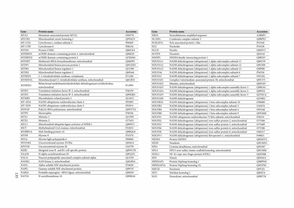
135
Gene Protein name Accession
MTA2 Metastasis-associated protein MTA2 O94776
MTCH2 Mitochondrial carrier homolog 2 Q9Y6C9
MT-CO2 Cytochrome c oxidase subunit 2 P00403
MT-CYB Cytochrome b P00156
MTDH Protein LYRIC Q86UE4
MTERFD1 mTERF domain-containing protein 1, mitochondrial Q96E29
MTERFD2 mTERF domain-containing protein 2 Q7Z6M4
MTFMT Methionyl-tRNA formyltransferase, mitochondrial Q96DP5
MTFP1 Mitochondrial fission process protein 1 Q9UDX5
MTFR1 Mitochondrial fission regulator 1 Q15390
MTFR2 Mitochondrial fission regulator 2 Q6P444
MTHFD1 C-1-tetrahydrofolate synthase, cytoplasmic P11586
MTHFD1L Monofunctional C1-tetrahydrofolate synthase, mitochondrial Q6UB35
MTHFD2 Bifunctional methylenetetrahydrofolate dehydrogenase/cyclohydrolase,
mitochondrial P13995
MTIF2 Translation initiation factor IF-2, mitochondrial P46199
MTIF3 Translation initiation factor IF-3, mitochondrial Q9H2K0
MTMR3 Myotubularin-related protein 3 Q13615
MT-ND4 NADH-ubiquinone oxidoreductase chain 4 P03905
MT-ND5 NADH-ubiquinone oxidoreductase chain 5 P03915
MTPAP Poly(A) RNA polymerase, mitochondrial Q9NVV4
MTPN Myotrophin P58546
MTX1 Metaxin-1 Q13505
MTX2 Metaxin-2 O75431
MUL1 Mitochondrial ubiquitin ligase activator of NFKB 1 Q969V5
MUT Methylmalonyl-CoA mutase, mitochondrial P22033
MYBBP1A Myb-binding protein 1A Q9BQG0
MYH9 Myosin-9 P35579
MYL6 Myosin light polypeptide 6 P60660
MYO18A Unconventional myosin-XVIIIa Q92614
MYO1B Unconventional myosin-Ib O43795
MZB1 Marginal zone B- and B1-cell-specific protein Q8WU39
NAA50 N-alpha-acetyltransferase 50 Q9GZZ1
NACA Nascent polypeptide-associated complex subunit alpha Q13765
NADK2 NAD kinase 2, mitochondrial Q4G0N4
NAPA Alpha-soluble NSF attachment protein P54920
NAPG Gamma-soluble NSF attachment protein Q99747
NARS2 Probable asparagine--tRNA ligase, mitochondrial Q96I59
NAT10 N-acetyltransferase 10 Q9H0A0
Gene Protein name Accession
NBAS Neuroblastoma-amplified sequence A2RRP1
NCAPG Condensin complex subunit 3 Q9BPX3
NCKAP1L Nck-associated protein 1-like P55160
NCL Nucleolin P19338
NCLN Nicalin Q969V3
NCSTN Nicastrin Q92542
NDFIP1 NEDD4 family-interacting protein 1 Q9BT67
NDUFA11 NADH dehydrogenase [ubiquinone] 1 alpha subcomplex subunit 11 Q86Y39
NDUFA12 NADH dehydrogenase [ubiquinone] 1 alpha subcomplex subunit 12 Q9UI09
NDUFA13 NADH dehydrogenase [ubiquinone] 1 alpha subcomplex subunit 13 Q9P0J0
NDUFA6 NADH dehydrogenase [ubiquinone] 1 alpha subcomplex subunit 6 P56556
NDUFA7 NADH dehydrogenase [ubiquinone] 1 alpha subcomplex subunit 7 O95182
NDUFAF1 Complex I intermediate-associated protein 30, mitochondrial Q9Y375
NDUFAF2 Mimitin, mitochondrial Q8N183
NDUFAF3 NADH dehydrogenase [ubiquinone] 1 alpha subcomplex assembly factor 3 Q9BU61
NDUFAF4 NADH dehydrogenase [ubiquinone] 1 alpha subcomplex assembly factor 4 Q9P032
NDUFAF5 NADH dehydrogenase [ubiquinone] 1 alpha subcomplex assembly factor 5 Q5TEU4
NDUFAF6 NADH dehydrogenase Q330K2
NDUFB10 NADH dehydrogenase [ubiquinone] 1 beta subcomplex subunit 10 O96000
NDUFB3 NADH dehydrogenase [ubiquinone] 1 beta subcomplex subunit 3 O43676
NDUFB4 NADH dehydrogenase [ubiquinone] 1 beta subcomplex subunit 4 O95168
NDUFB9 NADH dehydrogenase [ubiquinone] 1 beta subcomplex subunit 9 Q9Y6M9
NDUFS1 NADH-ubiquinone oxidoreductase 75 kDa subunit, mitochondrial P28331
NDUFS2 NADH dehydrogenase [ubiquinone] iron-sulfur protein 2, mitochondrial O75306
NDUFS3 NADH dehydrogenase [ubiquinone] iron-sulfur protein 3, mitochondrial O75489
NDUFS6 NADH dehydrogenase [ubiquinone] iron-sulfur protein 6, mitochondrial O75380
NDUFS8 NADH dehydrogenase [ubiquinone] iron-sulfur protein 8, mitochondrial O00217
NDUFV1 NADH dehydrogenase [ubiquinone] flavoprotein 1, mitochondrial P49821
NEDD1 Protein NEDD1 Q8NHV4
NENF Neudesin Q9UMX5
NFS1 Cysteine desulfurase, mitochondrial Q9Y697
NFU1 NFU1 iron-sulfur cluster scaffold homolog, mitochondrial Q9UMS0
NFXL1 NF-X1-type zinc finger protein NFXL1 Q6ZNB6
NIN Ninein Q8N4C6
NIPSNAP1 Protein NipSnap homolog 1 Q9BPW8
NIPSNAP3A Protein NipSnap homolog 3A Q9UFN0
NISCH Nischarin Q9Y2I1
NIT1 Nitrilase homolog 1 Q86X76
NLN Neurolysin, mitochondrial Q9BYT8
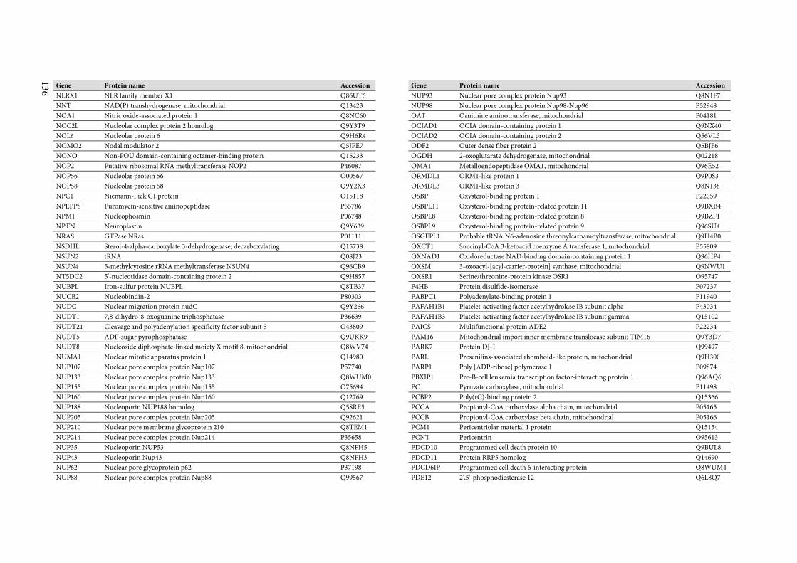
136
Gene Protein name Accession
NLRX1 NLR family member X1 Q86UT6
NNT NAD(P) transhydrogenase, mitochondrial Q13423
NOA1 Nitric oxide-associated protein 1 Q8NC60
NOC2L Nucleolar complex protein 2 homolog Q9Y3T9
NOL6 Nucleolar protein 6 Q9H6R4
NOMO2 Nodal modulator 2 Q5JPE7
NONO Non-POU domain-containing octamer-binding protein Q15233
NOP2 Putative ribosomal RNA methyltransferase NOP2 P46087
NOP56 Nucleolar protein 56 O00567
NOP58 Nucleolar protein 58 Q9Y2X3
NPC1 Niemann-Pick C1 protein O15118
NPEPPS Puromycin-sensitive aminopeptidase P55786
NPM1 Nucleophosmin P06748
NPTN Neuroplastin Q9Y639
NRAS GTPase NRas P01111
NSDHL Sterol-4-alpha-carboxylate 3-dehydrogenase, decarboxylating Q15738
NSUN2 tRNA Q08J23
NSUN4 5-methylcytosine rRNA methyltransferase NSUN4 Q96CB9
NT5DC2 5'-nucleotidase domain-containing protein 2 Q9H857
NUBPL Iron-sulfur protein NUBPL Q8TB37
NUCB2 Nucleobindin-2 P80303
NUDC Nuclear migration protein nudC Q9Y266
NUDT1 7,8-dihydro-8-oxoguanine triphosphatase P36639
NUDT21 Cleavage and polyadenylation specificity factor subunit 5 O43809
NUDT5 ADP-sugar pyrophosphatase Q9UKK9
NUDT8 Nucleoside diphosphate-linked moiety X motif 8, mitochondrial Q8WV74
NUMA1 Nuclear mitotic apparatus protein 1 Q14980
NUP107 Nuclear pore complex protein Nup107 P57740
NUP133 Nuclear pore complex protein Nup133 Q8WUM0
NUP155 Nuclear pore complex protein Nup155 O75694
NUP160 Nuclear pore complex protein Nup160 Q12769
NUP188 Nucleoporin NUP188 homolog Q5SRE5
NUP205 Nuclear pore complex protein Nup205 Q92621
NUP210 Nuclear pore membrane glycoprotein 210 Q8TEM1
NUP214 Nuclear pore complex protein Nup214 P35658
NUP35 Nucleoporin NUP53 Q8NFH5
NUP43 Nucleoporin Nup43 Q8NFH3
NUP62 Nuclear pore glycoprotein p62 P37198
NUP88 Nuclear pore complex protein Nup88 Q99567
Gene Protein name Accession
NUP93 Nuclear pore complex protein Nup93 Q8N1F7
NUP98 Nuclear pore complex protein Nup98-Nup96 P52948
OAT Ornithine aminotransferase, mitochondrial P04181
OCIAD1 OCIA domain-containing protein 1 Q9NX40
OCIAD2 OCIA domain-containing protein 2 Q56VL3
ODF2 Outer dense fiber protein 2 Q5BJF6
OGDH 2-oxoglutarate dehydrogenase, mitochondrial Q02218
OMA1 Metalloendopeptidase OMA1, mitochondrial Q96E52
ORMDL1 ORM1-like protein 1 Q9P0S3
ORMDL3 ORM1-like protein 3 Q8N138
OSBP Oxysterol-binding protein 1 P22059
OSBPL11 Oxysterol-binding protein-related protein 11 Q9BXB4
OSBPL8 Oxysterol-binding protein-related protein 8 Q9BZF1
OSBPL9 Oxysterol-binding protein-related protein 9 Q96SU4
OSGEPL1 Probable tRNA N6-adenosine threonylcarbamoyltransferase, mitochondrial Q9H4B0
OXCT1 Succinyl-CoA:3-ketoacid coenzyme A transferase 1, mitochondrial P55809
OXNAD1 Oxidoreductase NAD-binding domain-containing protein 1 Q96HP4
OXSM 3-oxoacyl-[acyl-carrier-protein] synthase, mitochondrial Q9NWU1
OXSR1 Serine/threonine-protein kinase OSR1 O95747
P4HB Protein disulfide-isomerase P07237
PABPC1 Polyadenylate-binding protein 1 P11940
PAFAH1B1 Platelet-activating factor acetylhydrolase IB subunit alpha P43034
PAFAH1B3 Platelet-activating factor acetylhydrolase IB subunit gamma Q15102
PAICS Multifunctional protein ADE2 P22234
PAM16 Mitochondrial import inner membrane translocase subunit TIM16 Q9Y3D7
PARK7 Protein DJ-1 Q99497
PARL Presenilins-associated rhomboid-like protein, mitochondrial Q9H300
PARP1 Poly [ADP-ribose] polymerase 1 P09874
PBXIP1 Pre-B-cell leukemia transcription factor-interacting protein 1 Q96AQ6
PC Pyruvate carboxylase, mitochondrial P11498
PCBP2 Poly(rC)-binding protein 2 Q15366
PCCA Propionyl-CoA carboxylase alpha chain, mitochondrial P05165
PCCB Propionyl-CoA carboxylase beta chain, mitochondrial P05166
PCM1 Pericentriolar material 1 protein Q15154
PCNT Pericentrin O95613
PDCD10 Programmed cell death protein 10 Q9BUL8
PDCD11 Protein RRP5 homolog Q14690
PDCD6IP Programmed cell death 6-interacting protein Q8WUM4
PDE12 2',5'-phosphodiesterase 12 Q6L8Q7
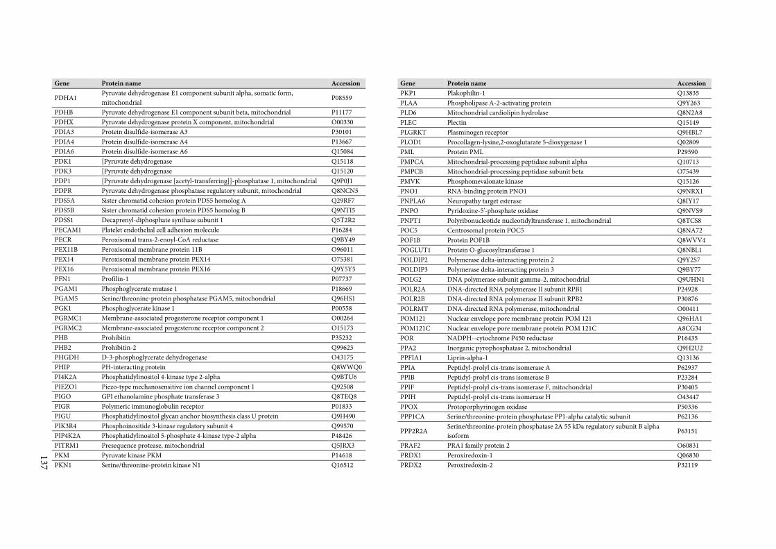
137
Gene Protein name Accession
PDHA1 Pyruvate dehydrogenase E1 component subunit alpha, somatic form,
mitochondrial P08559
PDHB Pyruvate dehydrogenase E1 component subunit beta, mitochondrial P11177
PDHX Pyruvate dehydrogenase protein X component, mitochondrial O00330
PDIA3 Protein disulfide-isomerase A3 P30101
PDIA4 Protein disulfide-isomerase A4 P13667
PDIA6 Protein disulfide-isomerase A6 Q15084
PDK1 [Pyruvate dehydrogenase Q15118
PDK3 [Pyruvate dehydrogenase Q15120
PDP1 [Pyruvate dehydrogenase [acetyl-transferring]]-phosphatase 1, mitochondrial Q9P0J1
PDPR Pyruvate dehydrogenase phosphatase regulatory subunit, mitochondrial Q8NCN5
PDS5A Sister chromatid cohesion protein PDS5 homolog A Q29RF7
PDS5B Sister chromatid cohesion protein PDS5 homolog B Q9NTI5
PDSS1 Decaprenyl-diphosphate synthase subunit 1 Q5T2R2
PECAM1 Platelet endothelial cell adhesion molecule P16284
PECR Peroxisomal trans-2-enoyl-CoA reductase Q9BY49
PEX11B Peroxisomal membrane protein 11B O96011
PEX14 Peroxisomal membrane protein PEX14 O75381
PEX16 Peroxisomal membrane protein PEX16 Q9Y5Y5
PFN1 Profilin-1 P07737
PGAM1 Phosphoglycerate mutase 1 P18669
PGAM5 Serine/threonine-protein phosphatase PGAM5, mitochondrial Q96HS1
PGK1 Phosphoglycerate kinase 1 P00558
PGRMC1 Membrane-associated progesterone receptor component 1 O00264
PGRMC2 Membrane-associated progesterone receptor component 2 O15173
PHB Prohibitin P35232
PHB2 Prohibitin-2 Q99623
PHGDH D-3-phosphoglycerate dehydrogenase O43175
PHIP PH-interacting protein Q8WWQ0
PI4K2A Phosphatidylinositol 4-kinase type 2-alpha Q9BTU6
PIEZO1 Piezo-type mechanosensitive ion channel component 1 Q92508
PIGO GPI ethanolamine phosphate transferase 3 Q8TEQ8
PIGR Polymeric immunoglobulin receptor P01833
PIGU Phosphatidylinositol glycan anchor biosynthesis class U protein Q9H490
PIK3R4 Phosphoinositide 3-kinase regulatory subunit 4 Q99570
PIP4K2A Phosphatidylinositol 5-phosphate 4-kinase type-2 alpha P48426
PITRM1 Presequence protease, mitochondrial Q5JRX3
PKM Pyruvate kinase PKM P14618
PKN1 Serine/threonine-protein kinase N1 Q16512
Gene Protein name Accession
PKP1 Plakophilin-1 Q13835
PLAA Phospholipase A-2-activating protein Q9Y263
PLD6 Mitochondrial cardiolipin hydrolase Q8N2A8
PLEC Plectin Q15149
PLGRKT Plasminogen receptor Q9HBL7
PLOD1 Procollagen-lysine,2-oxoglutarate 5-dioxygenase 1 Q02809
PML Protein PML P29590
PMPCA Mitochondrial-processing peptidase subunit alpha Q10713
PMPCB Mitochondrial-processing peptidase subunit beta O75439
PMVK Phosphomevalonate kinase Q15126
PNO1 RNA-binding protein PNO1 Q9NRX1
PNPLA6 Neuropathy target esterase Q8IY17
PNPO Pyridoxine-5'-phosphate oxidase Q9NVS9
PNPT1 Polyribonucleotide nucleotidyltransferase 1, mitochondrial Q8TCS8
POC5 Centrosomal protein POC5 Q8NA72
POF1B Protein POF1B Q8WVV4
POGLUT1 Protein O-glucosyltransferase 1 Q8NBL1
POLDIP2 Polymerase delta-interacting protein 2 Q9Y2S7
POLDIP3 Polymerase delta-interacting protein 3 Q9BY77
POLG2 DNA polymerase subunit gamma-2, mitochondrial Q9UHN1
POLR2A DNA-directed RNA polymerase II subunit RPB1 P24928
POLR2B DNA-directed RNA polymerase II subunit RPB2 P30876
POLRMT DNA-directed RNA polymerase, mitochondrial O00411
POM121 Nuclear envelope pore membrane protein POM 121 Q96HA1
POM121C Nuclear envelope pore membrane protein POM 121C A8CG34
POR NADPH--cytochrome P450 reductase P16435
PPA2 Inorganic pyrophosphatase 2, mitochondrial Q9H2U2
PPFIA1 Liprin-alpha-1 Q13136
PPIA Peptidyl-prolyl cis-trans isomerase A P62937
PPIB Peptidyl-prolyl cis-trans isomerase B P23284
PPIF Peptidyl-prolyl cis-trans isomerase F, mitochondrial P30405
PPIH Peptidyl-prolyl cis-trans isomerase H O43447
PPOX Protoporphyrinogen oxidase P50336
PPP1CA Serine/threonine-protein phosphatase PP1-alpha catalytic subunit P62136
PPP2R2A Serine/threonine-protein phosphatase 2A 55 kDa regulatory subunit B alpha
isoform P63151
PRAF2 PRA1 family protein 2 O60831
PRDX1 Peroxiredoxin-1 Q06830
PRDX2 Peroxiredoxin-2 P32119
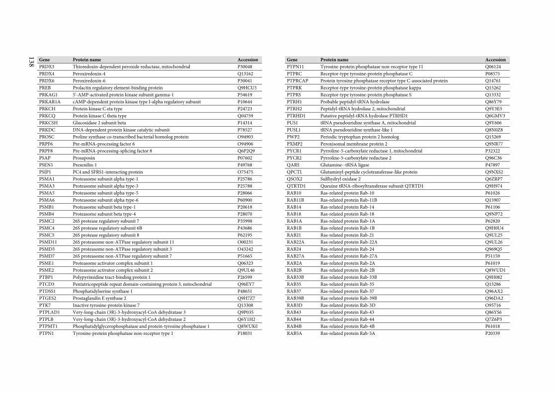
138
Gene Protein name Accession
PRDX3 Thioredoxin-dependent peroxide reductase, mitochondrial P30048
PRDX4 Peroxiredoxin-4 Q13162
PRDX6 Peroxiredoxin-6 P30041
PREB Prolactin regulatory element-binding protein Q9HCU5
PRKAG1 5'-AMP-activated protein kinase subunit gamma-1 P54619
PRKAR1A cAMP-dependent protein kinase type I-alpha regulatory subunit P10644
PRKCH Protein kinase C eta type P24723
PRKCQ Protein kinase C theta type Q04759
PRKCSH Glucosidase 2 subunit beta P14314
PRKDC DNA-dependent protein kinase catalytic subunit P78527
PROSC Proline synthase co-transcribed bacterial homolog protein O94903
PRPF6 Pre-mRNA-processing factor 6 O94906
PRPF8 Pre-mRNA-processing-splicing factor 8 Q6P2Q9
PSAP Prosaposin P07602
PSEN1 Presenilin-1 P49768
PSIP1 PC4 and SFRS1-interacting protein O75475
PSMA1 Proteasome subunit alpha type-1 P25786
PSMA3 Proteasome subunit alpha type-3 P25788
PSMA5 Proteasome subunit alpha type-5 P28066
PSMA6 Proteasome subunit alpha type-6 P60900
PSMB1 Proteasome subunit beta type-1 P20618
PSMB4 Proteasome subunit beta type-4 P28070
PSMC2 26S protease regulatory subunit 7 P35998
PSMC4 26S protease regulatory subunit 6B P43686
PSMC5 26S protease regulatory subunit 8 P62195
PSMD11 26S proteasome non-ATPase regulatory subunit 11 O00231
PSMD3 26S proteasome non-ATPase regulatory subunit 3 O43242
PSMD7 26S proteasome non-ATPase regulatory subunit 7 P51665
PSME1 Proteasome activator complex subunit 1 Q06323
PSME2 Proteasome activator complex subunit 2 Q9UL46
PTBP1 Polypyrimidine tract-binding protein 1 P26599
PTCD3 Pentatricopeptide repeat domain-containing protein 3, mitochondrial Q96EY7
PTDSS1 Phosphatidylserine synthase 1 P48651
PTGES2 Prostaglandin E synthase 2 Q9H7Z7
PTK7 Inactive tyrosine-protein kinase 7 Q13308
PTPLAD1 Very-long-chain (3R)-3-hydroxyacyl-CoA dehydratase 3 Q9P035
PTPLB Very-long-chain (3R)-3-hydroxyacyl-CoA dehydratase 2 Q6Y1H2
PTPMT1 Phosphatidylglycerophosphatase and protein-tyrosine phosphatase 1 Q8WUK0
PTPN1 Tyrosine-protein phosphatase non-receptor type 1 P18031
Gene Protein name Accession
PTPN11 Tyrosine-protein phosphatase non-receptor type 11 Q06124
PTPRC Receptor-type tyrosine-protein phosphatase C P08575
PTPRCAP Protein tyrosine phosphatase receptor type C-associated protein Q14761
PTPRK Receptor-type tyrosine-protein phosphatase kappa Q15262
PTPRS Receptor-type tyrosine-protein phosphatase S Q13332
PTRH1 Probable peptidyl-tRNA hydrolase Q86Y79
PTRH2 Peptidyl-tRNA hydrolase 2, mitochondrial Q9Y3E5
PTRHD1 Putative peptidyl-tRNA hydrolase PTRHD1 Q6GMV3
PUS1 tRNA pseudouridine synthase A, mitochondrial Q9Y606
PUSL1 tRNA pseudouridine synthase-like 1 Q8N0Z8
PWP2 Periodic tryptophan protein 2 homolog Q15269
PXMP2 Peroxisomal membrane protein 2 Q9NR77
PYCR1 Pyrroline-5-carboxylate reductase 1, mitochondrial P32322
PYCR2 Pyrroline-5-carboxylate reductase 2 Q96C36
QARS Glutamine--tRNA ligase P47897
QPCTL Glutaminyl-peptide cyclotransferase-like protein Q9NXS2
QSOX2 Sulfhydryl oxidase 2 Q6ZRP7
QTRTD1 Queuine tRNA-ribosyltransferase subunit QTRTD1 Q9H974
RAB10 Ras-related protein Rab-10 P61026
RAB11B Ras-related protein Rab-11B Q15907
RAB14 Ras-related protein Rab-14 P61106
RAB18 Ras-related protein Rab-18 Q9NP72
RAB1A Ras-related protein Rab-1A P62820
RAB1B Ras-related protein Rab-1B Q9H0U4
RAB21 Ras-related protein Rab-21 Q9UL25
RAB22A Ras-related protein Rab-22A Q9UL26
RAB24 Ras-related protein Rab-24 Q969Q5
RAB27A Ras-related protein Rab-27A P51159
RAB2A Ras-related protein Rab-2A P61019
RAB2B Ras-related protein Rab-2B Q8WUD1
RAB33B Ras-related protein Rab-33B Q9H082
RAB35 Ras-related protein Rab-35 Q15286
RAB37 Ras-related protein Rab-37 Q96AX2
RAB39B Ras-related protein Rab-39B Q96DA2
RAB3D Ras-related protein Rab-3D O95716
RAB43 Ras-related protein Rab-43 Q86YS6
RAB44 Ras-related protein Rab-44 Q7Z6P3
RAB4B Ras-related protein Rab-4B P61018
RAB5A Ras-related protein Rab-5A P20339
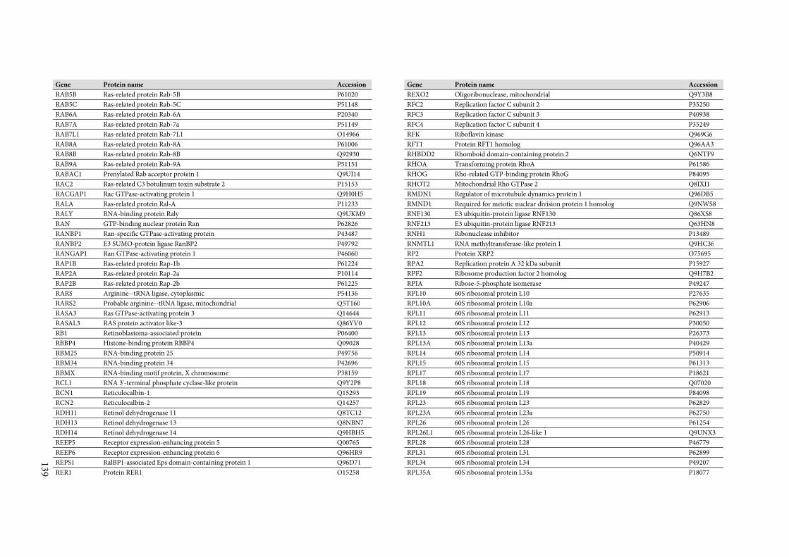
139
Gene Protein name Accession
RAB5B Ras-related protein Rab-5B P61020
RAB5C Ras-related protein Rab-5C P51148
RAB6A Ras-related protein Rab-6A P20340
RAB7A Ras-related protein Rab-7a P51149
RAB7L1 Ras-related protein Rab-7L1 O14966
RAB8A Ras-related protein Rab-8A P61006
RAB8B Ras-related protein Rab-8B Q92930
RAB9A Ras-related protein Rab-9A P51151
RABAC1 Prenylated Rab acceptor protein 1 Q9UI14
RAC2 Ras-related C3 botulinum toxin substrate 2 P15153
RACGAP1 Rac GTPase-activating protein 1 Q9H0H5
RALA Ras-related protein Ral-A P11233
RALY RNA-binding protein Raly Q9UKM9
RAN GTP-binding nuclear protein Ran P62826
RANBP1 Ran-specific GTPase-activating protein P43487
RANBP2 E3 SUMO-protein ligase RanBP2 P49792
RANGAP1 Ran GTPase-activating protein 1 P46060
RAP1B Ras-related protein Rap-1b P61224
RAP2A Ras-related protein Rap-2a P10114
RAP2B Ras-related protein Rap-2b P61225
RARS Arginine--tRNA ligase, cytoplasmic P54136
RARS2 Probable arginine--tRNA ligase, mitochondrial Q5T160
RASA3 Ras GTPase-activating protein 3 Q14644
RASAL3 RAS protein activator like-3 Q86YV0
RB1 Retinoblastoma-associated protein P06400
RBBP4 Histone-binding protein RBBP4 Q09028
RBM25 RNA-binding protein 25 P49756
RBM34 RNA-binding protein 34 P42696
RBMX RNA-binding motif protein, X chromosome P38159
RCL1 RNA 3'-terminal phosphate cyclase-like protein Q9Y2P8
RCN1 Reticulocalbin-1 Q15293
RCN2 Reticulocalbin-2 Q14257
RDH11 Retinol dehydrogenase 11 Q8TC12
RDH13 Retinol dehydrogenase 13 Q8NBN7
RDH14 Retinol dehydrogenase 14 Q9HBH5
REEP5 Receptor expression-enhancing protein 5 Q00765
REEP6 Receptor expression-enhancing protein 6 Q96HR9
REPS1 RalBP1-associated Eps domain-containing protein 1 Q96D71
RER1 Protein RER1 O15258
Gene Protein name Accession
REXO2 Oligoribonuclease, mitochondrial Q9Y3B8
RFC2 Replication factor C subunit 2 P35250
RFC3 Replication factor C subunit 3 P40938
RFC4 Replication factor C subunit 4 P35249
RFK Riboflavin kinase Q969G6
RFT1 Protein RFT1 homolog Q96AA3
RHBDD2 Rhomboid domain-containing protein 2 Q6NTF9
RHOA Transforming protein RhoA P61586
RHOG Rho-related GTP-binding protein RhoG P84095
RHOT2 Mitochondrial Rho GTPase 2 Q8IXI1
RMDN1 Regulator of microtubule dynamics protein 1 Q96DB5
RMND1 Required for meiotic nuclear division protein 1 homolog Q9NWS8
RNF130 E3 ubiquitin-protein ligase RNF130 Q86XS8
RNF213 E3 ubiquitin-protein ligase RNF213 Q63HN8
RNH1 Ribonuclease inhibitor P13489
RNMTL1 RNA methyltransferase-like protein 1 Q9HC36
RP2 Protein XRP2 O75695
RPA2 Replication protein A 32 kDa subunit P15927
RPF2 Ribosome production factor 2 homolog Q9H7B2
RPIA Ribose-5-phosphate isomerase P49247
RPL10 60S ribosomal protein L10 P27635
RPL10A 60S ribosomal protein L10a P62906
RPL11 60S ribosomal protein L11 P62913
RPL12 60S ribosomal protein L12 P30050
RPL13 60S ribosomal protein L13 P26373
RPL13A 60S ribosomal protein L13a P40429
RPL14 60S ribosomal protein L14 P50914
RPL15 60S ribosomal protein L15 P61313
RPL17 60S ribosomal protein L17 P18621
RPL18 60S ribosomal protein L18 Q07020
RPL19 60S ribosomal protein L19 P84098
RPL23 60S ribosomal protein L23 P62829
RPL23A 60S ribosomal protein L23a P62750
RPL26 60S ribosomal protein L26 P61254
RPL26L1 60S ribosomal protein L26-like 1 Q9UNX3
RPL28 60S ribosomal protein L28 P46779
RPL31 60S ribosomal protein L31 P62899
RPL34 60S ribosomal protein L34 P49207
RPL35A 60S ribosomal protein L35a P18077
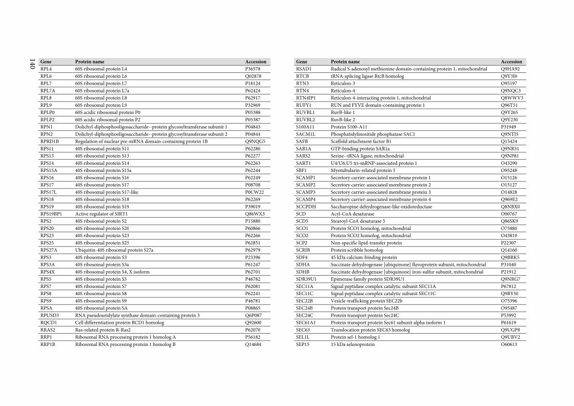
140
Gene Protein name Accession
RPL4 60S ribosomal protein L4 P36578
RPL6 60S ribosomal protein L6 Q02878
RPL7 60S ribosomal protein L7 P18124
RPL7A 60S ribosomal protein L7a P62424
RPL8 60S ribosomal protein L8 P62917
RPL9 60S ribosomal protein L9 P32969
RPLP0 60S acidic ribosomal protein P0 P05388
RPLP2 60S acidic ribosomal protein P2 P05387
RPN1 Dolichyl-diphosphooligosaccharide--protein glycosyltransferase subunit 1 P04843
RPN2 Dolichyl-diphosphooligosaccharide--protein glycosyltransferase subunit 2 P04844
RPRD1B Regulation of nuclear pre-mRNA domain-containing protein 1B Q9NQG5
RPS11 40S ribosomal protein S11 P62280
RPS13 40S ribosomal protein S13 P62277
RPS14 40S ribosomal protein S14 P62263
RPS15A 40S ribosomal protein S15a P62244
RPS16 40S ribosomal protein S16 P62249
RPS17 40S ribosomal protein S17 P08708
RPS17L 40S ribosomal protein S17-like P0CW22
RPS18 40S ribosomal protein S18 P62269
RPS19 40S ribosomal protein S19 P39019
RPS19BP1 Active regulator of SIRT1 Q86WX3
RPS2 40S ribosomal protein S2 P15880
RPS20 40S ribosomal protein S20 P60866
RPS23 40S ribosomal protein S23 P62266
RPS25 40S ribosomal protein S25 P62851
RPS27A Ubiquitin-40S ribosomal protein S27a P62979
RPS3 40S ribosomal protein S3 P23396
RPS3A 40S ribosomal protein S3a P61247
RPS4X 40S ribosomal protein S4, X isoform P62701
RPS5 40S ribosomal protein S5 P46782
RPS7 40S ribosomal protein S7 P62081
RPS8 40S ribosomal protein S8 P62241
RPS9 40S ribosomal protein S9 P46781
RPSA 40S ribosomal protein SA P08865
RPUSD3 RNA pseudouridylate synthase domain-containing protein 3 Q6P087
RQCD1 Cell differentiation protein RCD1 homolog Q92600
RRAS2 Ras-related protein R-Ras2 P62070
RRP1 Ribosomal RNA processing protein 1 homolog A P56182
RRP1B Ribosomal RNA processing protein 1 homolog B Q14684
Gene Protein name Accession
RSAD1 Radical S-adenosyl methionine domain-containing protein 1, mitochondrial Q9HA92
RTCB tRNA-splicing ligase RtcB homolog Q9Y3I0
RTN3 Reticulon-3 O95197
RTN4 Reticulon-4 Q9NQC3
RTN4IP1 Reticulon-4-interacting protein 1, mitochondrial Q8WWV3
RUFY1 RUN and FYVE domain-containing protein 1 Q96T51
RUVBL1 RuvB-like 1 Q9Y265
RUVBL2 RuvB-like 2 Q9Y230
S100A11 Protein S100-A11 P31949
SACM1L Phosphatidylinositide phosphatase SAC1 Q9NTJ5
SAFB Scaffold attachment factor B1 Q15424
SAR1A GTP-binding protein SAR1a Q9NR31
SARS2 Serine--tRNA ligase, mitochondrial Q9NP81
SART1 U4/U6.U5 tri-snRNP-associated protein 1 O43290
SBF1 Myotubularin-related protein 5 O95248
SCAMP1 Secretory carrier-associated membrane protein 1 O15126
SCAMP2 Secretory carrier-associated membrane protein 2 O15127
SCAMP3 Secretory carrier-associated membrane protein 3 O14828
SCAMP4 Secretory carrier-associated membrane protein 4 Q969E2
SCCPDH Saccharopine dehydrogenase-like oxidoreductase Q8NBX0
SCD Acyl-CoA desaturase O00767
SCD5 Stearoyl-CoA desaturase 5 Q86SK9
SCO1 Protein SCO1 homolog, mitochondrial O75880
SCO2 Protein SCO2 homolog, mitochondrial O43819
SCP2 Non-specific lipid-transfer protein P22307
SCRIB Protein scribble homolog Q14160
SDF4 45 kDa calcium-binding protein Q9BRK5
SDHA Succinate dehydrogenase [ubiquinone] flavoprotein subunit, mitochondrial P31040
SDHB Succinate dehydrogenase [ubiquinone] iron-sulfur subunit, mitochondrial P21912
SDR39U1 Epimerase family protein SDR39U1 Q9NRG7
SEC11A Signal peptidase complex catalytic subunit SEC11A P67812
SEC11C Signal peptidase complex catalytic subunit SEC11C Q9BY50
SEC22B Vesicle-trafficking protein SEC22b O75396
SEC24B Protein transport protein Sec24B O95487
SEC24C Protein transport protein Sec24C P53992
SEC61A1 Protein transport protein Sec61 subunit alpha isoform 1 P61619
SEC63 Translocation protein SEC63 homolog Q9UGP8
SEL1L Protein sel-1 homolog 1 Q9UBV2
SEP15 15 kDa selenoprotein O60613
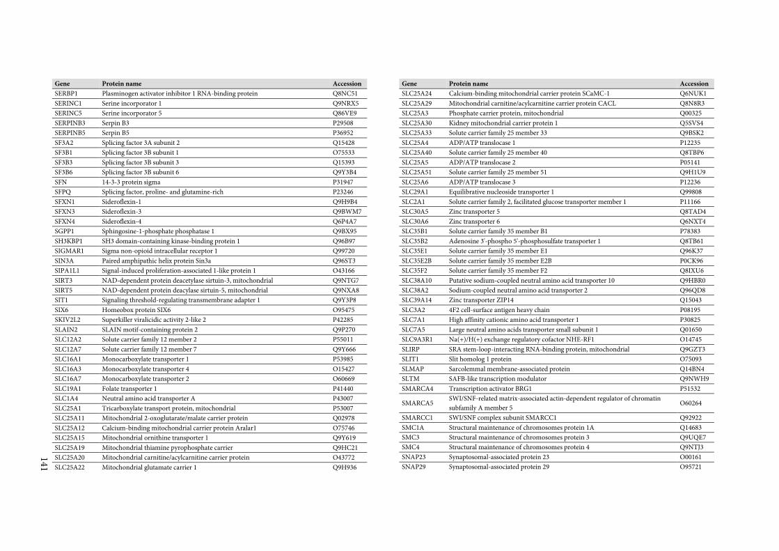
141
Gene Protein name Accession
SERBP1 Plasminogen activator inhibitor 1 RNA-binding protein Q8NC51
SERINC1 Serine incorporator 1 Q9NRX5
SERINC5 Serine incorporator 5 Q86VE9
SERPINB3 Serpin B3 P29508
SERPINB5 Serpin B5 P36952
SF3A2 Splicing factor 3A subunit 2 Q15428
SF3B1 Splicing factor 3B subunit 1 O75533
SF3B3 Splicing factor 3B subunit 3 Q15393
SF3B6 Splicing factor 3B subunit 6 Q9Y3B4
SFN 14-3-3 protein sigma P31947
SFPQ Splicing factor, proline- and glutamine-rich P23246
SFXN1 Sideroflexin-1 Q9H9B4
SFXN3 Sideroflexin-3 Q9BWM7
SFXN4 Sideroflexin-4 Q6P4A7
SGPP1 Sphingosine-1-phosphate phosphatase 1 Q9BX95
SH3KBP1 SH3 domain-containing kinase-binding protein 1 Q96B97
SIGMAR1 Sigma non-opioid intracellular receptor 1 Q99720
SIN3A Paired amphipathic helix protein Sin3a Q96ST3
SIPA1L1 Signal-induced proliferation-associated 1-like protein 1 O43166
SIRT3 NAD-dependent protein deacetylase sirtuin-3, mitochondrial Q9NTG7
SIRT5 NAD-dependent protein deacylase sirtuin-5, mitochondrial Q9NXA8
SIT1 Signaling threshold-regulating transmembrane adapter 1 Q9Y3P8
SIX6 Homeobox protein SIX6 O95475
SKIV2L2 Superkiller viralicidic activity 2-like 2 P42285
SLAIN2 SLAIN motif-containing protein 2 Q9P270
SLC12A2 Solute carrier family 12 member 2 P55011
SLC12A7 Solute carrier family 12 member 7 Q9Y666
SLC16A1 Monocarboxylate transporter 1 P53985
SLC16A3 Monocarboxylate transporter 4 O15427
SLC16A7 Monocarboxylate transporter 2 O60669
SLC19A1 Folate transporter 1 P41440
SLC1A4 Neutral amino acid transporter A P43007
SLC25A1 Tricarboxylate transport protein, mitochondrial P53007
SLC25A11 Mitochondrial 2-oxoglutarate/malate carrier protein Q02978
SLC25A12 Calcium-binding mitochondrial carrier protein Aralar1 O75746
SLC25A15 Mitochondrial ornithine transporter 1 Q9Y619
SLC25A19 Mitochondrial thiamine pyrophosphate carrier Q9HC21
SLC25A20 Mitochondrial carnitine/acylcarnitine carrier protein O43772
SLC25A22 Mitochondrial glutamate carrier 1 Q9H936
Gene Protein name Accession
SLC25A24 Calcium-binding mitochondrial carrier protein SCaMC-1 Q6NUK1
SLC25A29 Mitochondrial carnitine/acylcarnitine carrier protein CACL Q8N8R3
SLC25A3 Phosphate carrier protein, mitochondrial Q00325
SLC25A30 Kidney mitochondrial carrier protein 1 Q5SVS4
SLC25A33 Solute carrier family 25 member 33 Q9BSK2
SLC25A4 ADP/ATP translocase 1 P12235
SLC25A40 Solute carrier family 25 member 40 Q8TBP6
SLC25A5 ADP/ATP translocase 2 P05141
SLC25A51 Solute carrier family 25 member 51 Q9H1U9
SLC25A6 ADP/ATP translocase 3 P12236
SLC29A1 Equilibrative nucleoside transporter 1 Q99808
SLC2A1 Solute carrier family 2, facilitated glucose transporter member 1 P11166
SLC30A5 Zinc transporter 5 Q8TAD4
SLC30A6 Zinc transporter 6 Q6NXT4
SLC35B1 Solute carrier family 35 member B1 P78383
SLC35B2 Adenosine 3'-phospho 5'-phosphosulfate transporter 1 Q8TB61
SLC35E1 Solute carrier family 35 member E1 Q96K37
SLC35E2B Solute carrier family 35 member E2B P0CK96
SLC35F2 Solute carrier family 35 member F2 Q8IXU6
SLC38A10 Putative sodium-coupled neutral amino acid transporter 10 Q9HBR0
SLC38A2 Sodium-coupled neutral amino acid transporter 2 Q96QD8
SLC39A14 Zinc transporter ZIP14 Q15043
SLC3A2 4F2 cell-surface antigen heavy chain P08195
SLC7A1 High affinity cationic amino acid transporter 1 P30825
SLC7A5 Large neutral amino acids transporter small subunit 1 Q01650
SLC9A3R1 Na(+)/H(+) exchange regulatory cofactor NHE-RF1 O14745
SLIRP SRA stem-loop-interacting RNA-binding protein, mitochondrial Q9GZT3
SLIT1 Slit homolog 1 protein O75093
SLMAP Sarcolemmal membrane-associated protein Q14BN4
SLTM SAFB-like transcription modulator Q9NWH9
SMARCA4 Transcription activator BRG1 P51532
SMARCA5 SWI/SNF-related matrix-associated actin-dependent regulator of chromatin
subfamily A member 5 O60264
SMARCC1 SWI/SNF complex subunit SMARCC1 Q92922
SMC1A Structural maintenance of chromosomes protein 1A Q14683
SMC3 Structural maintenance of chromosomes protein 3 Q9UQE7
SMC4 Structural maintenance of chromosomes protein 4 Q9NTJ3
SNAP23 Synaptosomal-associated protein 23 O00161
SNAP29 Synaptosomal-associated protein 29 O95721
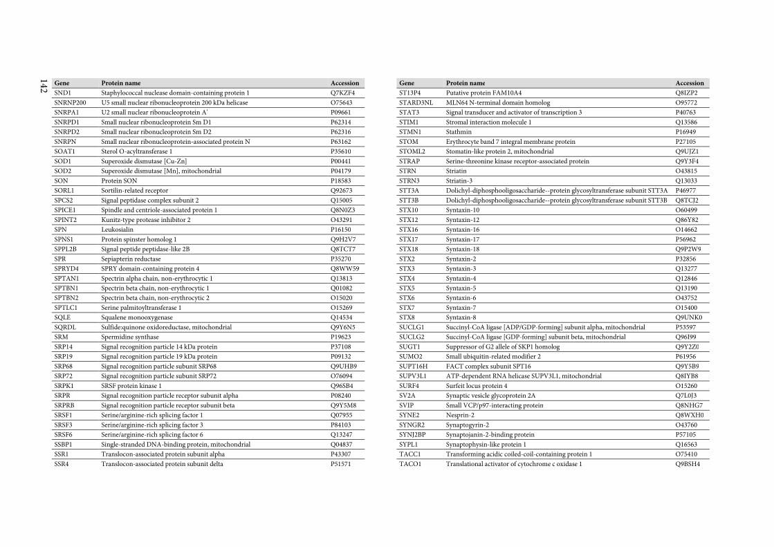
142
Gene Protein name Accession
SND1 Staphylococcal nuclease domain-containing protein 1 Q7KZF4
SNRNP200 U5 small nuclear ribonucleoprotein 200 kDa helicase O75643
SNRPA1 U2 small nuclear ribonucleoprotein A' P09661
SNRPD1 Small nuclear ribonucleoprotein Sm D1 P62314
SNRPD2 Small nuclear ribonucleoprotein Sm D2 P62316
SNRPN Small nuclear ribonucleoprotein-associated protein N P63162
SOAT1 Sterol O-acyltransferase 1 P35610
SOD1 Superoxide dismutase [Cu-Zn] P00441
SOD2 Superoxide dismutase [Mn], mitochondrial P04179
SON Protein SON P18583
SORL1 Sortilin-related receptor Q92673
SPCS2 Signal peptidase complex subunit 2 Q15005
SPICE1 Spindle and centriole-associated protein 1 Q8N0Z3
SPINT2 Kunitz-type protease inhibitor 2 O43291
SPN Leukosialin P16150
SPNS1 Protein spinster homolog 1 Q9H2V7
SPPL2B Signal peptide peptidase-like 2B Q8TCT7
SPR Sepiapterin reductase P35270
SPRYD4 SPRY domain-containing protein 4 Q8WW59
SPTAN1 Spectrin alpha chain, non-erythrocytic 1 Q13813
SPTBN1 Spectrin beta chain, non-erythrocytic 1 Q01082
SPTBN2 Spectrin beta chain, non-erythrocytic 2 O15020
SPTLC1 Serine palmitoyltransferase 1 O15269
SQLE Squalene monooxygenase Q14534
SQRDL Sulfide:quinone oxidoreductase, mitochondrial Q9Y6N5
SRM Spermidine synthase P19623
SRP14 Signal recognition particle 14 kDa protein P37108
SRP19 Signal recognition particle 19 kDa protein P09132
SRP68 Signal recognition particle subunit SRP68 Q9UHB9
SRP72 Signal recognition particle subunit SRP72 O76094
SRPK1 SRSF protein kinase 1 Q96SB4
SRPR Signal recognition particle receptor subunit alpha P08240
SRPRB Signal recognition particle receptor subunit beta Q9Y5M8
SRSF1 Serine/arginine-rich splicing factor 1 Q07955
SRSF3 Serine/arginine-rich splicing factor 3 P84103
SRSF6 Serine/arginine-rich splicing factor 6 Q13247
SSBP1 Single-stranded DNA-binding protein, mitochondrial Q04837
SSR1 Translocon-associated protein subunit alpha P43307
SSR4 Translocon-associated protein subunit delta P51571
Gene Protein name Accession
ST13P4 Putative protein FAM10A4 Q8IZP2
STARD3NL MLN64 N-terminal domain homolog O95772
STAT3 Signal transducer and activator of transcription 3 P40763
STIM1 Stromal interaction molecule 1 Q13586
STMN1 Stathmin P16949
STOM Erythrocyte band 7 integral membrane protein P27105
STOML2 Stomatin-like protein 2, mitochondrial Q9UJZ1
STRAP Serine-threonine kinase receptor-associated protein Q9Y3F4
STRN Striatin O43815
STRN3 Striatin-3 Q13033
STT3A Dolichyl-diphosphooligosaccharide--protein glycosyltransferase subunit STT3A P46977
STT3B Dolichyl-diphosphooligosaccharide--protein glycosyltransferase subunit STT3B Q8TCJ2
STX10 Syntaxin-10 O60499
STX12 Syntaxin-12 Q86Y82
STX16 Syntaxin-16 O14662
STX17 Syntaxin-17 P56962
STX18 Syntaxin-18 Q9P2W9
STX2 Syntaxin-2 P32856
STX3 Syntaxin-3 Q13277
STX4 Syntaxin-4 Q12846
STX5 Syntaxin-5 Q13190
STX6 Syntaxin-6 O43752
STX7 Syntaxin-7 O15400
STX8 Syntaxin-8 Q9UNK0
SUCLG1 Succinyl-CoA ligase [ADP/GDP-forming] subunit alpha, mitochondrial P53597
SUCLG2 Succinyl-CoA ligase [GDP-forming] subunit beta, mitochondrial Q96I99
SUGT1 Suppressor of G2 allele of SKP1 homolog Q9Y2Z0
SUMO2 Small ubiquitin-related modifier 2 P61956
SUPT16H FACT complex subunit SPT16 Q9Y5B9
SUPV3L1 ATP-dependent RNA helicase SUPV3L1, mitochondrial Q8IYB8
SURF4 Surfeit locus protein 4 O15260
SV2A Synaptic vesicle glycoprotein 2A Q7L0J3
SVIP Small VCP/p97-interacting protein Q8NHG7
SYNE2 Nesprin-2 Q8WXH0
SYNGR2 Synaptogyrin-2 O43760
SYNJ2BP Synaptojanin-2-binding protein P57105
SYPL1 Synaptophysin-like protein 1 Q16563
TACC1 Transforming acidic coiled-coil-containing protein 1 O75410
TACO1 Translational activator of cytochrome c oxidase 1 Q9BSH4
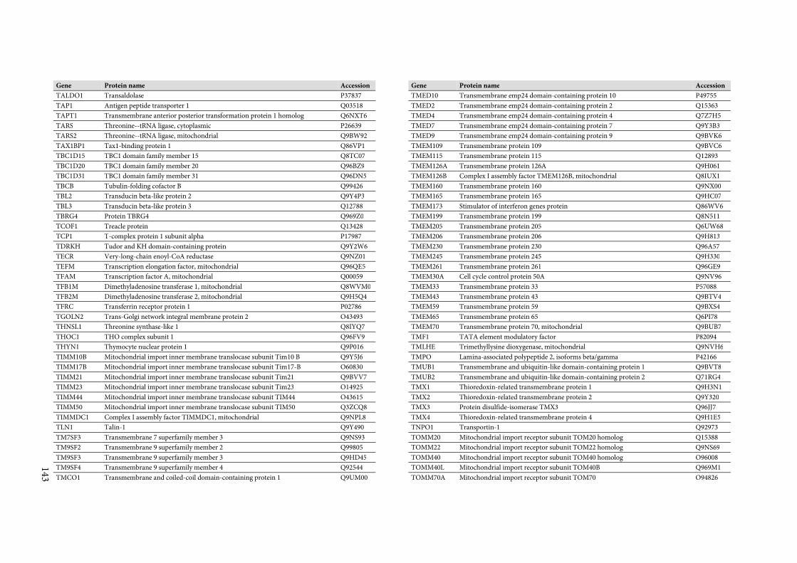
143
Gene Protein name Accession
TALDO1 Transaldolase P37837
TAP1 Antigen peptide transporter 1 Q03518
TAPT1 Transmembrane anterior posterior transformation protein 1 homolog Q6NXT6
TARS Threonine--tRNA ligase, cytoplasmic P26639
TARS2 Threonine--tRNA ligase, mitochondrial Q9BW92
TAX1BP1 Tax1-binding protein 1 Q86VP1
TBC1D15 TBC1 domain family member 15 Q8TC07
TBC1D20 TBC1 domain family member 20 Q96BZ9
TBC1D31 TBC1 domain family member 31 Q96DN5
TBCB Tubulin-folding cofactor B Q99426
TBL2 Transducin beta-like protein 2 Q9Y4P3
TBL3 Transducin beta-like protein 3 Q12788
TBRG4 Protein TBRG4 Q969Z0
TCOF1 Treacle protein Q13428
TCP1 T-complex protein 1 subunit alpha P17987
TDRKH Tudor and KH domain-containing protein Q9Y2W6
TECR Very-long-chain enoyl-CoA reductase Q9NZ01
TEFM Transcription elongation factor, mitochondrial Q96QE5
TFAM Transcription factor A, mitochondrial Q00059
TFB1M Dimethyladenosine transferase 1, mitochondrial Q8WVM0
TFB2M Dimethyladenosine transferase 2, mitochondrial Q9H5Q4
TFRC Transferrin receptor protein 1 P02786
TGOLN2 Trans-Golgi network integral membrane protein 2 O43493
THNSL1 Threonine synthase-like 1 Q8IYQ7
THOC1 THO complex subunit 1 Q96FV9
THYN1 Thymocyte nuclear protein 1 Q9P016
TIMM10B Mitochondrial import inner membrane translocase subunit Tim10 B Q9Y5J6
TIMM17B Mitochondrial import inner membrane translocase subunit Tim17-B O60830
TIMM21 Mitochondrial import inner membrane translocase subunit Tim21 Q9BVV7
TIMM23 Mitochondrial import inner membrane translocase subunit Tim23 O14925
TIMM44 Mitochondrial import inner membrane translocase subunit TIM44 O43615
TIMM50 Mitochondrial import inner membrane translocase subunit TIM50 Q3ZCQ8
TIMMDC1 Complex I assembly factor TIMMDC1, mitochondrial Q9NPL8
TLN1 Talin-1 Q9Y490
TM7SF3 Transmembrane 7 superfamily member 3 Q9NS93
TM9SF2 Transmembrane 9 superfamily member 2 Q99805
TM9SF3 Transmembrane 9 superfamily member 3 Q9HD45
TM9SF4 Transmembrane 9 superfamily member 4 Q92544
TMCO1 Transmembrane and coiled-coil domain-containing protein 1 Q9UM00
Gene Protein name Accession
TMED10 Transmembrane emp24 domain-containing protein 10 P49755
TMED2 Transmembrane emp24 domain-containing protein 2 Q15363
TMED4 Transmembrane emp24 domain-containing protein 4 Q7Z7H5
TMED7 Transmembrane emp24 domain-containing protein 7 Q9Y3B3
TMED9 Transmembrane emp24 domain-containing protein 9 Q9BVK6
TMEM109 Transmembrane protein 109 Q9BVC6
TMEM115 Transmembrane protein 115 Q12893
TMEM126A Transmembrane protein 126A Q9H061
TMEM126B Complex I assembly factor TMEM126B, mitochondrial Q8IUX1
TMEM160 Transmembrane protein 160 Q9NX00
TMEM165 Transmembrane protein 165 Q9HC07
TMEM173 Stimulator of interferon genes protein Q86WV6
TMEM199 Transmembrane protein 199 Q8N511
TMEM205 Transmembrane protein 205 Q6UW68
TMEM206 Transmembrane protein 206 Q9H813
TMEM230 Transmembrane protein 230 Q96A57
TMEM245 Transmembrane protein 245 Q9H330
TMEM261 Transmembrane protein 261 Q96GE9
TMEM30A Cell cycle control protein 50A Q9NV96
TMEM33 Transmembrane protein 33 P57088
TMEM43 Transmembrane protein 43 Q9BTV4
TMEM59 Transmembrane protein 59 Q9BXS4
TMEM65 Transmembrane protein 65 Q6PI78
TMEM70 Transmembrane protein 70, mitochondrial Q9BUB7
TMF1 TATA element modulatory factor P82094
TMLHE Trimethyllysine dioxygenase, mitochondrial Q9NVH6
TMPO Lamina-associated polypeptide 2, isoforms beta/gamma P42166
TMUB1 Transmembrane and ubiquitin-like domain-containing protein 1 Q9BVT8
TMUB2 Transmembrane and ubiquitin-like domain-containing protein 2 Q71RG4
TMX1 Thioredoxin-related transmembrane protein 1 Q9H3N1
TMX2 Thioredoxin-related transmembrane protein 2 Q9Y320
TMX3 Protein disulfide-isomerase TMX3 Q96JJ7
TMX4 Thioredoxin-related transmembrane protein 4 Q9H1E5
TNPO1 Transportin-1 Q92973
TOMM20 Mitochondrial import receptor subunit TOM20 homolog Q15388
TOMM22 Mitochondrial import receptor subunit TOM22 homolog Q9NS69
TOMM40 Mitochondrial import receptor subunit TOM40 homolog O96008
TOMM40L Mitochondrial import receptor subunit TOM40B Q969M1
TOMM70A Mitochondrial import receptor subunit TOM70 O94826
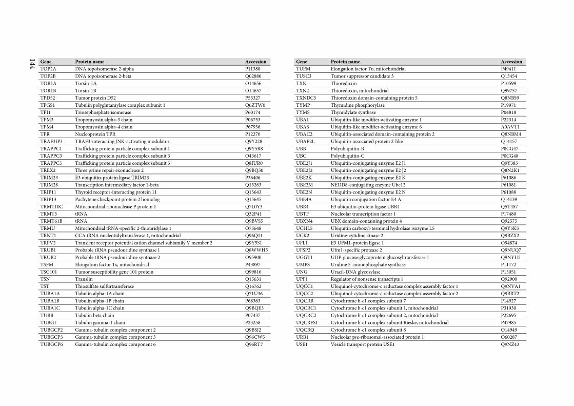
144
Gene Protein name Accession
TOP2A DNA topoisomerase 2-alpha P11388
TOP2B DNA topoisomerase 2-beta Q02880
TOR1A Torsin-1A O14656
TOR1B Torsin-1B O14657
TPD52 Tumor protein D52 P55327
TPGS1 Tubulin polyglutamylase complex subunit 1 Q6ZTW0
TPI1 Triosephosphate isomerase P60174
TPM3 Tropomyosin alpha-3 chain P06753
TPM4 Tropomyosin alpha-4 chain P67936
TPR Nucleoprotein TPR P12270
TRAF3IP3 TRAF3-interacting JNK-activating modulator Q9Y228
TRAPPC1 Trafficking protein particle complex subunit 1 Q9Y5R8
TRAPPC3 Trafficking protein particle complex subunit 3 O43617
TRAPPC5 Trafficking protein particle complex subunit 5 Q8IUR0
TREX2 Three prime repair exonuclease 2 Q9BQ50
TRIM23 E3 ubiquitin-protein ligase TRIM23 P36406
TRIM28 Transcription intermediary factor 1-beta Q13263
TRIP11 Thyroid receptor-interacting protein 11 Q15643
TRIP13 Pachytene checkpoint protein 2 homolog Q15645
TRMT10C Mitochondrial ribonuclease P protein 1 Q7L0Y3
TRMT5 tRNA Q32P41
TRMT61B tRNA Q9BVS5
TRMU Mitochondrial tRNA-specific 2-thiouridylase 1 O75648
TRNT1 CCA tRNA nucleotidyltransferase 1, mitochondrial Q96Q11
TRPV2 Transient receptor potential cation channel subfamily V member 2 Q9Y5S1
TRUB1 Probable tRNA pseudouridine synthase 1 Q8WWH5
TRUB2 Probable tRNA pseudouridine synthase 2 O95900
TSFM Elongation factor Ts, mitochondrial P43897
TSG101 Tumor susceptibility gene 101 protein Q99816
TSN Translin Q15631
TST Thiosulfate sulfurtransferase Q16762
TUBA1A Tubulin alpha-1A chain Q71U36
TUBA1B Tubulin alpha-1B chain P68363
TUBA1C Tubulin alpha-1C chain Q9BQE3
TUBB Tubulin beta chain P07437
TUBG1 Tubulin gamma-1 chain P23258
TUBGCP2 Gamma-tubulin complex component 2 Q9BSJ2
TUBGCP3 Gamma-tubulin complex component 3 Q96CW5
TUBGCP6 Gamma-tubulin complex component 6 Q96RT7
Gene Protein name Accession
TUFM Elongation factor Tu, mitochondrial P49411
TUSC3 Tumor suppressor candidate 3 Q13454
TXN Thioredoxin P10599
TXN2 Thioredoxin, mitochondrial Q99757
TXNDC5 Thioredoxin domain-containing protein 5 Q8NBS9
TYMP Thymidine phosphorylase P19971
TYMS Thymidylate synthase P04818
UBA1 Ubiquitin-like modifier-activating enzyme 1 P22314
UBA6 Ubiquitin-like modifier-activating enzyme 6 A0AVT1
UBAC2 Ubiquitin-associated domain-containing protein 2 Q8NBM4
UBAP2L Ubiquitin-associated protein 2-like Q14157
UBB Polyubiquitin-B P0CG47
UBC Polyubiquitin-C P0CG48
UBE2J1 Ubiquitin-conjugating enzyme E2 J1 Q9Y385
UBE2J2 Ubiquitin-conjugating enzyme E2 J2 Q8N2K1
UBE2K Ubiquitin-conjugating enzyme E2 K P61086
UBE2M NEDD8-conjugating enzyme Ubc12 P61081
UBE2N Ubiquitin-conjugating enzyme E2 N P61088
UBE4A Ubiquitin conjugation factor E4 A Q14139
UBR4 E3 ubiquitin-protein ligase UBR4 Q5T4S7
UBTF Nucleolar transcription factor 1 P17480
UBXN4 UBX domain-containing protein 4 Q92575
UCHL5 Ubiquitin carboxyl-terminal hydrolase isozyme L5 Q9Y5K5
UCK2 Uridine-cytidine kinase 2 Q9BZX2
UFL1 E3 UFM1-protein ligase 1 O94874
UFSP2 Ufm1-specific protease 2 Q9NUQ7
UGGT1 UDP-glucose:glycoprotein glucosyltransferase 1 Q9NYU2
UMPS Uridine 5'-monophosphate synthase P11172
UNG Uracil-DNA glycosylase P13051
UPF1 Regulator of nonsense transcripts 1 Q92900
UQCC1 Ubiquinol-cytochrome-c reductase complex assembly factor 1 Q9NVA1
UQCC2 Ubiquinol-cytochrome-c reductase complex assembly factor 2 Q9BRT2
UQCRB Cytochrome b-c1 complex subunit 7 P14927
UQCRC1 Cytochrome b-c1 complex subunit 1, mitochondrial P31930
UQCRC2 Cytochrome b-c1 complex subunit 2, mitochondrial P22695
UQCRFS1 Cytochrome b-c1 complex subunit Rieske, mitochondrial P47985
UQCRQ Cytochrome b-c1 complex subunit 8 O14949
URB1 Nucleolar pre-ribosomal-associated protein 1 O60287
USE1 Vesicle transport protein USE1 Q9NZ43
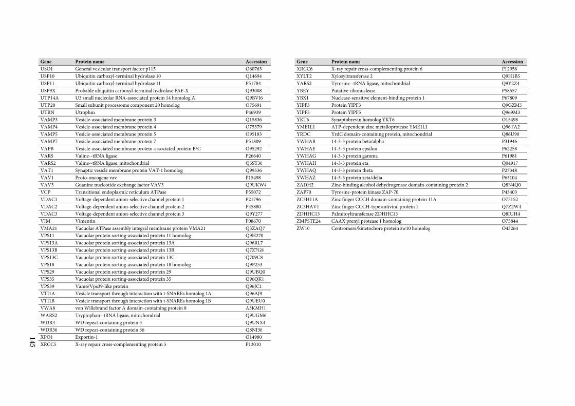
145
Gene Protein name Accession
USO1 General vesicular transport factor p115 O60763
USP10 Ubiquitin carboxyl-terminal hydrolase 10 Q14694
USP11 Ubiquitin carboxyl-terminal hydrolase 11 P51784
USP9X Probable ubiquitin carboxyl-terminal hydrolase FAF-X Q93008
UTP14A U3 small nucleolar RNA-associated protein 14 homolog A Q9BVJ6
UTP20 Small subunit processome component 20 homolog O75691
UTRN Utrophin P46939
VAMP3 Vesicle-associated membrane protein 3 Q15836
VAMP4 Vesicle-associated membrane protein 4 O75379
VAMP5 Vesicle-associated membrane protein 5 O95183
VAMP7 Vesicle-associated membrane protein 7 P51809
VAPB Vesicle-associated membrane protein-associated protein B/C O95292
VARS Valine--tRNA ligase P26640
VARS2 Valine--tRNA ligase, mitochondrial Q5ST30
VAT1 Synaptic vesicle membrane protein VAT-1 homolog Q99536
VAV1 Proto-oncogene vav P15498
VAV3 Guanine nucleotide exchange factor VAV3 Q9UKW4
VCP Transitional endoplasmic reticulum ATPase P55072
VDAC1 Voltage-dependent anion-selective channel protein 1 P21796
VDAC2 Voltage-dependent anion-selective channel protein 2 P45880
VDAC3 Voltage-dependent anion-selective channel protein 3 Q9Y277
VIM Vimentin P08670
VMA21 Vacuolar ATPase assembly integral membrane protein VMA21 Q3ZAQ7
VPS11 Vacuolar protein sorting-associated protein 11 homolog Q9H270
VPS13A Vacuolar protein sorting-associated protein 13A Q96RL7
VPS13B Vacuolar protein sorting-associated protein 13B Q7Z7G8
VPS13C Vacuolar protein sorting-associated protein 13C Q709C8
VPS18 Vacuolar protein sorting-associated protein 18 homolog Q9P253
VPS29 Vacuolar protein sorting-associated protein 29 Q9UBQ0
VPS35 Vacuolar protein sorting-associated protein 35 Q96QK1
VPS39 Vam6/Vps39-like protein Q96JC1
VTI1A Vesicle transport through interaction with t-SNAREs homolog 1A Q96AJ9
VTI1B Vesicle transport through interaction with t-SNAREs homolog 1B Q9UEU0
VWA8 von Willebrand factor A domain-containing protein 8 A3KMH1
WARS2 Tryptophan--tRNA ligase, mitochondrial Q9UGM6
WDR3 WD repeat-containing protein 3 Q9UNX4
WDR36 WD repeat-containing protein 36 Q8NI36
XPO1 Exportin-1 O14980
XRCC5 X-ray repair cross-complementing protein 5 P13010
Gene Protein name Accession
XRCC6 X-ray repair cross-complementing protein 6 P12956
XYLT2 Xylosyltransferase 2 Q9H1B5
YARS2 Tyrosine--tRNA ligase, mitochondrial Q9Y2Z4
YBEY Putative ribonuclease P58557
YBX1 Nuclease-sensitive element-binding protein 1 P67809
YIPF3 Protein YIPF3 Q9GZM5
YIPF5 Protein YIPF5 Q969M3
YKT6 Synaptobrevin homolog YKT6 O15498
YME1L1 ATP-dependent zinc metalloprotease YME1L1 Q96TA2
YRDC YrdC domain-containing protein, mitochondrial Q86U90
YWHAB 14-3-3 protein beta/alpha P31946
YWHAE 14-3-3 protein epsilon P62258
YWHAG 14-3-3 protein gamma P61981
YWHAH 14-3-3 protein eta Q04917
YWHAQ 14-3-3 protein theta P27348
YWHAZ 14-3-3 protein zeta/delta P63104
ZADH2 Zinc-binding alcohol dehydrogenase domain-containing protein 2 Q8N4Q0
ZAP70 Tyrosine-protein kinase ZAP-70 P43403
ZC3H11A Zinc finger CCCH domain-containing protein 11A O75152
ZC3HAV1 Zinc finger CCCH-type antiviral protein 1 Q7Z2W4
ZDHHC13 Palmitoyltransferase ZDHHC13 Q8IUH4
ZMPSTE24 CAAX prenyl protease 1 homolog O75844
ZW10 Centromere/kinetochore protein zw10 homolog O43264
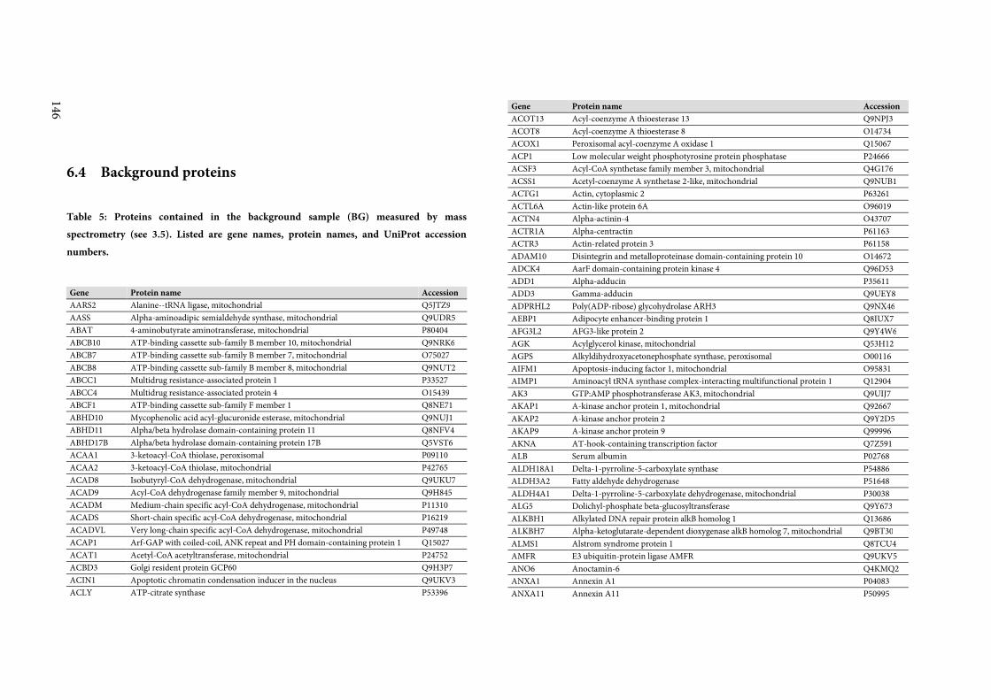
146
6.4 Background proteins
Table 5: Proteins contained in the background sample (BG) measured by mass
spectrometry (see 3.5). Listed are gene names, protein names, and UniProt accession
numbers.
Gene Protein name Accession
AARS2 Alanine--tRNA ligase, mitochondrial Q5JTZ9
AASS Alpha-aminoadipic semialdehyde synthase, mitochondrial Q9UDR5
ABAT 4-aminobutyrate aminotransferase, mitochondrial P80404
ABCB10 ATP-binding cassette sub-family B member 10, mitochondrial Q9NRK6
ABCB7 ATP-binding cassette sub-family B member 7, mitochondrial O75027
ABCB8 ATP-binding cassette sub-family B member 8, mitochondrial Q9NUT2
ABCC1 Multidrug resistance-associated protein 1 P33527
ABCC4 Multidrug resistance-associated protein 4 O15439
ABCF1 ATP-binding cassette sub-family F member 1 Q8NE71
ABHD10 Mycophenolic acid acyl-glucuronide esterase, mitochondrial Q9NUJ1
ABHD11 Alpha/beta hydrolase domain-containing protein 11 Q8NFV4
ABHD17B Alpha/beta hydrolase domain-containing protein 17B Q5VST6
ACAA1 3-ketoacyl-CoA thiolase, peroxisomal P09110
ACAA2 3-ketoacyl-CoA thiolase, mitochondrial P42765
ACAD8 Isobutyryl-CoA dehydrogenase, mitochondrial Q9UKU7
ACAD9 Acyl-CoA dehydrogenase family member 9, mitochondrial Q9H845
ACADM Medium-chain specific acyl-CoA dehydrogenase, mitochondrial P11310
ACADS Short-chain specific acyl-CoA dehydrogenase, mitochondrial P16219
ACADVL Very long-chain specific acyl-CoA dehydrogenase, mitochondrial P49748
ACAP1 Arf-GAP with coiled-coil, ANK repeat and PH domain-containing protein 1 Q15027
ACAT1 Acetyl-CoA acetyltransferase, mitochondrial P24752
ACBD3 Golgi resident protein GCP60 Q9H3P7
ACIN1 Apoptotic chromatin condensation inducer in the nucleus Q9UKV3
ACLY ATP-citrate synthase P53396
Gene Protein name Accession
ACOT13 Acyl-coenzyme A thioesterase 13 Q9NPJ3
ACOT8 Acyl-coenzyme A thioesterase 8 O14734
ACOX1 Peroxisomal acyl-coenzyme A oxidase 1 Q15067
ACP1 Low molecular weight phosphotyrosine protein phosphatase P24666
ACSF3 Acyl-CoA synthetase family member 3, mitochondrial Q4G176
ACSS1 Acetyl-coenzyme A synthetase 2-like, mitochondrial Q9NUB1
ACTG1 Actin, cytoplasmic 2 P63261
ACTL6A Actin-like protein 6A O96019
ACTN4 Alpha-actinin-4 O43707
ACTR1A Alpha-centractin P61163
ACTR3 Actin-related protein 3 P61158
ADAM10 Disintegrin and metalloproteinase domain-containing protein 10 O14672
ADCK4 AarF domain-containing protein kinase 4 Q96D53
ADD1 Alpha-adducin P35611
ADD3 Gamma-adducin Q9UEY8
ADPRHL2 Poly(ADP-ribose) glycohydrolase ARH3 Q9NX46
AEBP1 Adipocyte enhancer-binding protein 1 Q8IUX7
AFG3L2 AFG3-like protein 2 Q9Y4W6
AGK Acylglycerol kinase, mitochondrial Q53H12
AGPS Alkyldihydroxyacetonephosphate synthase, peroxisomal O00116
AIFM1 Apoptosis-inducing factor 1, mitochondrial O95831
AIMP1 Aminoacyl tRNA synthase complex-interacting multifunctional protein 1 Q12904
AK3 GTP:AMP phosphotransferase AK3, mitochondrial Q9UIJ7
AKAP1 A-kinase anchor protein 1, mitochondrial Q92667
AKAP2 A-kinase anchor protein 2 Q9Y2D5
AKAP9 A-kinase anchor protein 9 Q99996
AKNA AT-hook-containing transcription factor Q7Z591
ALB Serum albumin P02768
ALDH18A1 Delta-1-pyrroline-5-carboxylate synthase P54886
ALDH3A2 Fatty aldehyde dehydrogenase P51648
ALDH4A1 Delta-1-pyrroline-5-carboxylate dehydrogenase, mitochondrial P30038
ALG5 Dolichyl-phosphate beta-glucosyltransferase Q9Y673
ALKBH1 Alkylated DNA repair protein alkB homolog 1 Q13686
ALKBH7 Alpha-ketoglutarate-dependent dioxygenase alkB homolog 7, mitochondrial Q9BT30
ALMS1 Alstrom syndrome protein 1 Q8TCU4
AMFR E3 ubiquitin-protein ligase AMFR Q9UKV5
ANO6 Anoctamin-6 Q4KMQ2
ANXA1 Annexin A1 P04083
ANXA11 Annexin A11 P50995
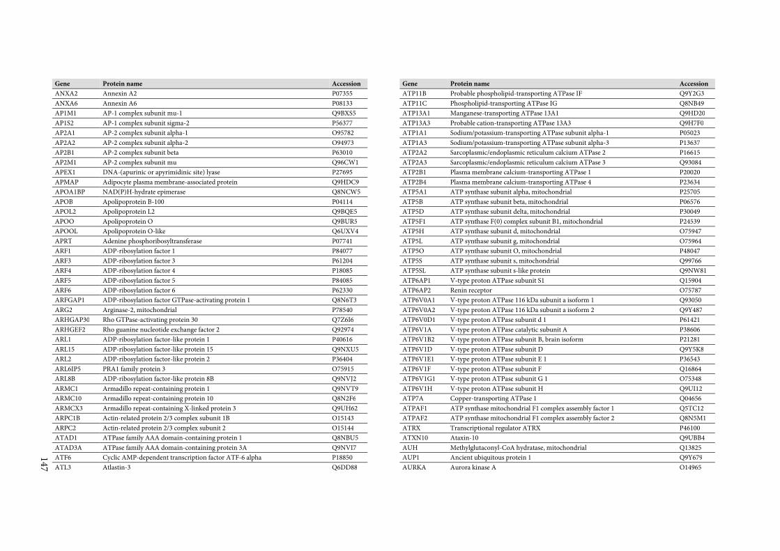
147
Gene Protein name Accession
ANXA2 Annexin A2 P07355
ANXA6 Annexin A6 P08133
AP1M1 AP-1 complex subunit mu-1 Q9BXS5
AP1S2 AP-1 complex subunit sigma-2 P56377
AP2A1 AP-2 complex subunit alpha-1 O95782
AP2A2 AP-2 complex subunit alpha-2 O94973
AP2B1 AP-2 complex subunit beta P63010
AP2M1 AP-2 complex subunit mu Q96CW1
APEX1 DNA-(apurinic or apyrimidinic site) lyase P27695
APMAP Adipocyte plasma membrane-associated protein Q9HDC9
APOA1BP NAD(P)H-hydrate epimerase Q8NCW5
APOB Apolipoprotein B-100 P04114
APOL2 Apolipoprotein L2 Q9BQE5
APOO Apolipoprotein O Q9BUR5
APOOL Apolipoprotein O-like Q6UXV4
APRT Adenine phosphoribosyltransferase P07741
ARF1 ADP-ribosylation factor 1 P84077
ARF3 ADP-ribosylation factor 3 P61204
ARF4 ADP-ribosylation factor 4 P18085
ARF5 ADP-ribosylation factor 5 P84085
ARF6 ADP-ribosylation factor 6 P62330
ARFGAP1 ADP-ribosylation factor GTPase-activating protein 1 Q8N6T3
ARG2 Arginase-2, mitochondrial P78540
ARHGAP30 Rho GTPase-activating protein 30 Q7Z6I6
ARHGEF2 Rho guanine nucleotide exchange factor 2 Q92974
ARL1 ADP-ribosylation factor-like protein 1 P40616
ARL15 ADP-ribosylation factor-like protein 15 Q9NXU5
ARL2 ADP-ribosylation factor-like protein 2 P36404
ARL6IP5 PRA1 family protein 3 O75915
ARL8B ADP-ribosylation factor-like protein 8B Q9NVJ2
ARMC1 Armadillo repeat-containing protein 1 Q9NVT9
ARMC10 Armadillo repeat-containing protein 10 Q8N2F6
ARMCX3 Armadillo repeat-containing X-linked protein 3 Q9UH62
ARPC1B Actin-related protein 2/3 complex subunit 1B O15143
ARPC2 Actin-related protein 2/3 complex subunit 2 O15144
ATAD1 ATPase family AAA domain-containing protein 1 Q8NBU5
ATAD3A ATPase family AAA domain-containing protein 3A Q9NVI7
ATF6 Cyclic AMP-dependent transcription factor ATF-6 alpha P18850
ATL3 Atlastin-3 Q6DD88
Gene Protein name Accession
ATP11B Probable phospholipid-transporting ATPase IF Q9Y2G3
ATP11C Phospholipid-transporting ATPase IG Q8NB49
ATP13A1 Manganese-transporting ATPase 13A1 Q9HD20
ATP13A3 Probable cation-transporting ATPase 13A3 Q9H7F0
ATP1A1 Sodium/potassium-transporting ATPase subunit alpha-1 P05023
ATP1A3 Sodium/potassium-transporting ATPase subunit alpha-3 P13637
ATP2A2 Sarcoplasmic/endoplasmic reticulum calcium ATPase 2 P16615
ATP2A3 Sarcoplasmic/endoplasmic reticulum calcium ATPase 3 Q93084
ATP2B1 Plasma membrane calcium-transporting ATPase 1 P20020
ATP2B4 Plasma membrane calcium-transporting ATPase 4 P23634
ATP5A1 ATP synthase subunit alpha, mitochondrial P25705
ATP5B ATP synthase subunit beta, mitochondrial P06576
ATP5D ATP synthase subunit delta, mitochondrial P30049
ATP5F1 ATP synthase F(0) complex subunit B1, mitochondrial P24539
ATP5H ATP synthase subunit d, mitochondrial O75947
ATP5L ATP synthase subunit g, mitochondrial O75964
ATP5O ATP synthase subunit O, mitochondrial P48047
ATP5S ATP synthase subunit s, mitochondrial Q99766
ATP5SL ATP synthase subunit s-like protein Q9NW81
ATP6AP1 V-type proton ATPase subunit S1 Q15904
ATP6AP2 Renin receptor O75787
ATP6V0A1 V-type proton ATPase 116 kDa subunit a isoform 1 Q93050
ATP6V0A2 V-type proton ATPase 116 kDa subunit a isoform 2 Q9Y487
ATP6V0D1 V-type proton ATPase subunit d 1 P61421
ATP6V1A V-type proton ATPase catalytic subunit A P38606
ATP6V1B2 V-type proton ATPase subunit B, brain isoform P21281
ATP6V1D V-type proton ATPase subunit D Q9Y5K8
ATP6V1E1 V-type proton ATPase subunit E 1 P36543
ATP6V1F V-type proton ATPase subunit F Q16864
ATP6V1G1 V-type proton ATPase subunit G 1 O75348
ATP6V1H V-type proton ATPase subunit H Q9UI12
ATP7A Copper-transporting ATPase 1 Q04656
ATPAF1 ATP synthase mitochondrial F1 complex assembly factor 1 Q5TC12
ATPAF2 ATP synthase mitochondrial F1 complex assembly factor 2 Q8N5M1
ATRX Transcriptional regulator ATRX P46100
ATXN10 Ataxin-10 Q9UBB4
AUH Methylglutaconyl-CoA hydratase, mitochondrial Q13825
AUP1 Ancient ubiquitous protein 1 Q9Y679
AURKA Aurora kinase A O14965
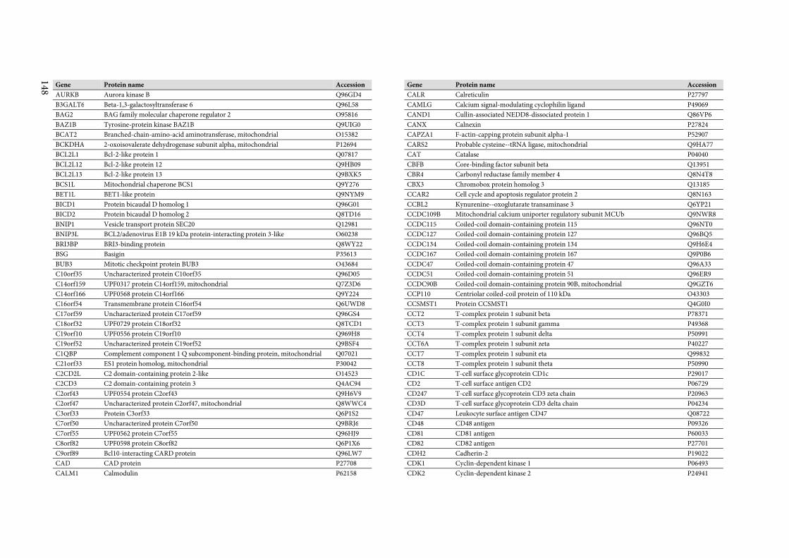
148
Gene Protein name Accession
AURKB Aurora kinase B Q96GD4
B3GALT6 Beta-1,3-galactosyltransferase 6 Q96L58
BAG2 BAG family molecular chaperone regulator 2 O95816
BAZ1B Tyrosine-protein kinase BAZ1B Q9UIG0
BCAT2 Branched-chain-amino-acid aminotransferase, mitochondrial O15382
BCKDHA 2-oxoisovalerate dehydrogenase subunit alpha, mitochondrial P12694
BCL2L1 Bcl-2-like protein 1 Q07817
BCL2L12 Bcl-2-like protein 12 Q9HB09
BCL2L13 Bcl-2-like protein 13 Q9BXK5
BCS1L Mitochondrial chaperone BCS1 Q9Y276
BET1L BET1-like protein Q9NYM9
BICD1 Protein bicaudal D homolog 1 Q96G01
BICD2 Protein bicaudal D homolog 2 Q8TD16
BNIP1 Vesicle transport protein SEC20 Q12981
BNIP3L BCL2/adenovirus E1B 19 kDa protein-interacting protein 3-like O60238
BRI3BP BRI3-binding protein Q8WY22
BSG Basigin P35613
BUB3 Mitotic checkpoint protein BUB3 O43684
C10orf35 Uncharacterized protein C10orf35 Q96D05
C14orf159 UPF0317 protein C14orf159, mitochondrial Q7Z3D6
C14orf166 UPF0568 protein C14orf166 Q9Y224
C16orf54 Transmembrane protein C16orf54 Q6UWD8
C17orf59 Uncharacterized protein C17orf59 Q96GS4
C18orf32 UPF0729 protein C18orf32 Q8TCD1
C19orf10 UPF0556 protein C19orf10 Q969H8
C19orf52 Uncharacterized protein C19orf52 Q9BSF4
C1QBP Complement component 1 Q subcomponent-binding protein, mitochondrial Q07021
C21orf33 ES1 protein homolog, mitochondrial P30042
C2CD2L C2 domain-containing protein 2-like O14523
C2CD3 C2 domain-containing protein 3 Q4AC94
C2orf43 UPF0554 protein C2orf43 Q9H6V9
C2orf47 Uncharacterized protein C2orf47, mitochondrial Q8WWC4
C3orf33 Protein C3orf33 Q6P1S2
C7orf50 Uncharacterized protein C7orf50 Q9BRJ6
C7orf55 UPF0562 protein C7orf55 Q96HJ9
C8orf82 UPF0598 protein C8orf82 Q6P1X6
C9orf89 Bcl10-interacting CARD protein Q96LW7
CAD CAD protein P27708
CALM1 Calmodulin P62158
Gene Protein name Accession
CALR Calreticulin P27797
CAMLG Calcium signal-modulating cyclophilin ligand P49069
CAND1 Cullin-associated NEDD8-dissociated protein 1 Q86VP6
CANX Calnexin P27824
CAPZA1 F-actin-capping protein subunit alpha-1 P52907
CARS2 Probable cysteine--tRNA ligase, mitochondrial Q9HA77
CAT Catalase P04040
CBFB Core-binding factor subunit beta Q13951
CBR4 Carbonyl reductase family member 4 Q8N4T8
CBX3 Chromobox protein homolog 3 Q13185
CCAR2 Cell cycle and apoptosis regulator protein 2 Q8N163
CCBL2 Kynurenine--oxoglutarate transaminase 3 Q6YP21
CCDC109B Mitochondrial calcium uniporter regulatory subunit MCUb Q9NWR8
CCDC115 Coiled-coil domain-containing protein 115 Q96NT0
CCDC127 Coiled-coil domain-containing protein 127 Q96BQ5
CCDC134 Coiled-coil domain-containing protein 134 Q9H6E4
CCDC167 Coiled-coil domain-containing protein 167 Q9P0B6
CCDC47 Coiled-coil domain-containing protein 47 Q96A33
CCDC51 Coiled-coil domain-containing protein 51 Q96ER9
CCDC90B Coiled-coil domain-containing protein 90B, mitochondrial Q9GZT6
CCP110 Centriolar coiled-coil protein of 110 kDa O43303
CCSMST1 Protein CCSMST1 Q4G0I0
CCT2 T-complex protein 1 subunit beta P78371
CCT3 T-complex protein 1 subunit gamma P49368
CCT4 T-complex protein 1 subunit delta P50991
CCT6A T-complex protein 1 subunit zeta P40227
CCT7 T-complex protein 1 subunit eta Q99832
CCT8 T-complex protein 1 subunit theta P50990
CD1C T-cell surface glycoprotein CD1c P29017
CD2 T-cell surface antigen CD2 P06729
CD247 T-cell surface glycoprotein CD3 zeta chain P20963
CD3D T-cell surface glycoprotein CD3 delta chain P04234
CD47 Leukocyte surface antigen CD47 Q08722
CD48 CD48 antigen P09326
CD81 CD81 antigen P60033
CD82 CD82 antigen P27701
CDH2 Cadherin-2 P19022
CDK1 Cyclin-dependent kinase 1 P06493
CDK2 Cyclin-dependent kinase 2 P24941
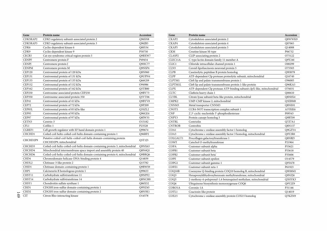
149
Gene Protein name Accession
CDK5RAP2 CDK5 regulatory subunit-associated protein 2 Q96SN8
CDK5RAP3 CDK5 regulatory subunit-associated protein 3 Q96JB5
CDK6 Cyclin-dependent kinase 6 Q00534
CDK9 Cyclin-dependent kinase 9 P50750
CECR5 Cat eye syndrome critical region protein 5 Q9BXW7
CENPF Centromere protein F P49454
CENPJ Centromere protein J Q9HC77
CENPM Centromere protein M Q9NSP4
CEP120 Centrosomal protein of 120 kDa Q8N960
CEP131 Centrosomal protein of 131 kDa Q9UPN4
CEP135 Centrosomal protein of 135 kDa Q66GS9
CEP152 Centrosomal protein of 152 kDa O94986
CEP162 Centrosomal protein of 162 kDa Q5TB80
CEP250 Centrosome-associated protein CEP250 Q9BV73
CEP350 Centrosome-associated protein 350 Q5VT06
CEP41 Centrosomal protein of 41 kDa Q9BYV8
CEP72 Centrosomal protein of 72 kDa Q9P209
CEP85L Centrosomal protein of 85 kDa-like Q5SZL2
CEP95 Centrosomal protein of 95 kDa Q96GE4
CEP97 Centrosomal protein of 97 kDa Q8IW35
CETN3 Centrin-3 O15182
CFL1 Cofilin-1 P23528
CGREF1 Cell growth regulator with EF hand domain protein 1 Q99674
CHCHD1 Coiled-coil-helix-coiled-coil-helix domain-containing protein 1 Q96BP2
CHCHD2P9 Putative coiled-coil-helix-coiled-coil-helix domain-containing protein
CHCHD2P9, mitochondrial Q5T1J5
CHCHD3 Coiled-coil-helix-coiled-coil-helix domain-containing protein 3, mitochondrial Q9NX63
CHCHD4 Mitochondrial intermembrane space import and assembly protein 40 Q8N4Q1
CHCHD6 Coiled-coil-helix-coiled-coil-helix domain-containing protein 6, mitochondrial Q9BRQ6
CHD4 Chromodomain-helicase-DNA-binding protein 4 Q14839
CHI3L2 Chitinase-3-like protein 2 Q15782
CHID1 Chitinase domain-containing protein 1 Q9BWS9
CHP1 Calcineurin B homologous protein 1 Q99653
CHST11 Carbohydrate sulfotransferase 11 Q9NPF2
CHST14 Carbohydrate sulfotransferase 14 Q8NCH0
CHSY1 Chondroitin sulfate synthase 1 Q86X52
CISD1 CDGSH iron-sulfur domain-containing protein 1 Q9NZ45
CISD2 CDGSH iron-sulfur domain-containing protein 2 Q8N5K1
CIT Citron Rho-interacting kinase O14578
Gene Protein name Accession
CKAP2 Cytoskeleton-associated protein 2 Q8WWK9
CKAP4 Cytoskeleton-associated protein 4 Q07065
CKAP5 Cytoskeleton-associated protein 5 Q14008
CKM Creatine kinase M-type P06732
CLASP2 CLIP-associating protein 2 O75122
CLEC11A C-type lectin domain family 11 member A Q9Y240
CLIC1 Chloride intracellular channel protein 1 O00299
CLN5 Ceroid-lipofuscinosis neuronal protein 5 O75503
CLPB Caseinolytic peptidase B protein homolog Q9H078
CLPP ATP-dependent Clp protease proteolytic subunit, mitochondrial Q16740
CLPTM1 Cleft lip and palate transmembrane protein 1 O96005
CLPTM1L Cleft lip and palate transmembrane protein 1-like protein Q96KA5
CLPX ATP-dependent Clp protease ATP-binding subunit clpX-like, mitochondrial O76031
CLTC Clathrin heavy chain 1 Q00610
CLYBL Citrate lyase subunit beta-like protein, mitochondrial Q8N0X4
CMPK2 UMP-CMP kinase 2, mitochondrial Q5EBM0
CNNM3 Metal transporter CNNM3 Q8NE01
CNOT1 CCR4-NOT transcription complex subunit 1 A5YKK6
CNP 2',3'-cyclic-nucleotide 3'-phosphodiesterase P09543
CNPY3 Protein canopy homolog 3 Q9BT09
CNTRL Centriolin Q7Z7A1
CNTROB Centrobin Q8N137
COA1 Cytochrome c oxidase assembly factor 1 homolog Q9GZY4
COA3 Cytochrome c oxidase assembly factor 3 homolog, mitochondrial Q9Y2R0
COLGALT1 Procollagen galactosyltransferase 1 Q8NBJ5
COMT Catechol O-methyltransferase P21964
COPA Coatomer subunit alpha P53621
COPB1 Coatomer subunit beta P53618
COPB2 Coatomer subunit beta' P35606
COPE Coatomer subunit epsilon O14579
COPG1 Coatomer subunit gamma-1 Q9Y678
COPZ1 Coatomer subunit zeta-1 P61923
COQ10B Coenzyme Q-binding protein COQ10 homolog B, mitochondrial Q9H8M1
COQ3 Hexaprenyldihydroxybenzoate methyltransferase, mitochondrial Q9NZJ6
COQ5 2-methoxy-6-polyprenyl-1,4-benzoquinol methylase, mitochondrial Q5HYK3
COQ6 Ubiquinone biosynthesis monooxygenase COQ6 Q9Y2Z9
CORO1A Coronin-1A P31146
COTL1 Coactosin-like protein Q14019
COX15 Cytochrome c oxidase assembly protein COX15 homolog Q7KZN9
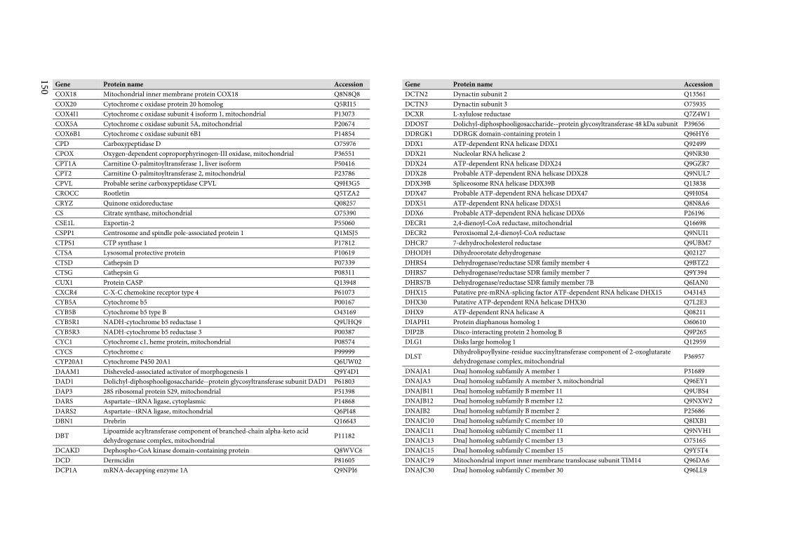
150
Gene Protein name Accession
COX18 Mitochondrial inner membrane protein COX18 Q8N8Q8
COX20 Cytochrome c oxidase protein 20 homolog Q5RI15
COX4I1 Cytochrome c oxidase subunit 4 isoform 1, mitochondrial P13073
COX5A Cytochrome c oxidase subunit 5A, mitochondrial P20674
COX6B1 Cytochrome c oxidase subunit 6B1 P14854
CPD Carboxypeptidase D O75976
CPOX Oxygen-dependent coproporphyrinogen-III oxidase, mitochondrial P36551
CPT1A Carnitine O-palmitoyltransferase 1, liver isoform P50416
CPT2 Carnitine O-palmitoyltransferase 2, mitochondrial P23786
CPVL Probable serine carboxypeptidase CPVL Q9H3G5
CROCC Rootletin Q5TZA2
CRYZ Quinone oxidoreductase Q08257
CS Citrate synthase, mitochondrial O75390
CSE1L Exportin-2 P55060
CSPP1 Centrosome and spindle pole-associated protein 1 Q1MSJ5
CTPS1 CTP synthase 1 P17812
CTSA Lysosomal protective protein P10619
CTSD Cathepsin D P07339
CTSG Cathepsin G P08311
CUX1 Protein CASP Q13948
CXCR4 C-X-C chemokine receptor type 4 P61073
CYB5A Cytochrome b5 P00167
CYB5B Cytochrome b5 type B O43169
CYB5R1 NADH-cytochrome b5 reductase 1 Q9UHQ9
CYB5R3 NADH-cytochrome b5 reductase 3 P00387
CYC1 Cytochrome c1, heme protein, mitochondrial P08574
CYCS Cytochrome c P99999
CYP20A1 Cytochrome P450 20A1 Q6UW02
DAAM1 Disheveled-associated activator of morphogenesis 1 Q9Y4D1
DAD1 Dolichyl-diphosphooligosaccharide--protein glycosyltransferase subunit DAD1 P61803
DAP3 28S ribosomal protein S29, mitochondrial P51398
DARS Aspartate--tRNA ligase, cytoplasmic P14868
DARS2 Aspartate--tRNA ligase, mitochondrial Q6PI48
DBN1 Drebrin Q16643
DBT Lipoamide acyltransferase component of branched-chain alpha-keto acid
dehydrogenase complex, mitochondrial P11182
DCAKD Dephospho-CoA kinase domain-containing protein Q8WVC6
DCD Dermcidin P81605
DCP1A mRNA-decapping enzyme 1A Q9NPI6
Gene Protein name Accession
DCTN2 Dynactin subunit 2 Q13561
DCTN3 Dynactin subunit 3 O75935
DCXR L-xylulose reductase Q7Z4W1
DDOST Dolichyl-diphosphooligosaccharide--protein glycosyltransferase 48 kDa subunit P39656
DDRGK1 DDRGK domain-containing protein 1 Q96HY6
DDX1 ATP-dependent RNA helicase DDX1 Q92499
DDX21 Nucleolar RNA helicase 2 Q9NR30
DDX24 ATP-dependent RNA helicase DDX24 Q9GZR7
DDX28 Probable ATP-dependent RNA helicase DDX28 Q9NUL7
DDX39B Spliceosome RNA helicase DDX39B Q13838
DDX47 Probable ATP-dependent RNA helicase DDX47 Q9H0S4
DDX51 ATP-dependent RNA helicase DDX51 Q8N8A6
DDX6 Probable ATP-dependent RNA helicase DDX6 P26196
DECR1 2,4-dienoyl-CoA reductase, mitochondrial Q16698
DECR2 Peroxisomal 2,4-dienoyl-CoA reductase Q9NUI1
DHCR7 7-dehydrocholesterol reductase Q9UBM7
DHODH Dihydroorotate dehydrogenase Q02127
DHRS4 Dehydrogenase/reductase SDR family member 4 Q9BTZ2
DHRS7 Dehydrogenase/reductase SDR family member 7 Q9Y394
DHRS7B Dehydrogenase/reductase SDR family member 7B Q6IAN0
DHX15 Putative pre-mRNA-splicing factor ATP-dependent RNA helicase DHX15 O43143
DHX30 Putative ATP-dependent RNA helicase DHX30 Q7L2E3
DHX9 ATP-dependent RNA helicase A Q08211
DIAPH1 Protein diaphanous homolog 1 O60610
DIP2B Disco-interacting protein 2 homolog B Q9P265
DLG1 Disks large homolog 1 Q12959
DLST Dihydrolipoyllysine-residue succinyltransferase component of 2-oxoglutarate
dehydrogenase complex, mitochondrial P36957
DNAJA1 DnaJ homolog subfamily A member 1 P31689
DNAJA3 DnaJ homolog subfamily A member 3, mitochondrial Q96EY1
DNAJB11 DnaJ homolog subfamily B member 11 Q9UBS4
DNAJB12 DnaJ homolog subfamily B member 12 Q9NXW2
DNAJB2 DnaJ homolog subfamily B member 2 P25686
DNAJC10 DnaJ homolog subfamily C member 10 Q8IXB1
DNAJC11 DnaJ homolog subfamily C member 11 Q9NVH1
DNAJC13 DnaJ homolog subfamily C member 13 O75165
DNAJC15 DnaJ homolog subfamily C member 15 Q9Y5T4
DNAJC19 Mitochondrial import inner membrane translocase subunit TIM14 Q96DA6
DNAJC30 DnaJ homolog subfamily C member 30 Q96LL9
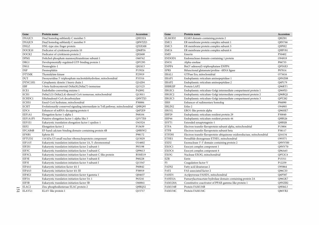
151
Gene Protein name Accession
DNAJC5 DnaJ homolog subfamily C member 5 Q9H3Z4
DNAJC9 DnaJ homolog subfamily C member 9 Q8WXX5
DNLZ DNL-type zinc finger protein Q5SXM8
DOCK10 Dedicator of cytokinesis protein 10 Q96BY6
DOCK2 Dedicator of cytokinesis protein 2 Q92608
DPM1 Dolichol-phosphate mannosyltransferase subunit 1 O60762
DRG1 Developmentally-regulated GTP-binding protein 1 Q9Y295
DSG1 Desmoglein-1 Q02413
DSP Desmoplakin P15924
DTYMK Thymidylate kinase P23919
DUT Deoxyuridine 5'-triphosphate nucleotidohydrolase, mitochondrial P33316
DYNC1H1 Cytoplasmic dynein 1 heavy chain 1 Q14204
EBP 3-beta-hydroxysteroid-Delta(8),Delta(7)-isomerase Q15125
ECE1 Endothelin-converting enzyme 1 P42892
ECH1 Delta(3,5)-Delta(2,4)-dienoyl-CoA isomerase, mitochondrial Q13011
ECHDC1 Ethylmalonyl-CoA decarboxylase Q9NTX5
ECHS1 Enoyl-CoA hydratase, mitochondrial P30084
ECSIT Evolutionarily conserved signaling intermediate in Toll pathway, mitochondrial Q9BQ95
EDC4 Enhancer of mRNA-decapping protein 4 Q6P2E9
EEF1A1 Elongation factor 1-alpha 1 P68104
EEF1A1P5 Putative elongation factor 1-alpha-like 3 Q5VTE0
EEF1E1 Eukaryotic translation elongation factor 1 epsilon-1 O43324
EEF2 Elongation factor 2 P13639
EFCAB4B EF-hand calcium-binding domain-containing protein 4B Q9BSW2
EFNB1 Ephrin-B1 P98172
EFTUD2 116 kDa U5 small nuclear ribonucleoprotein component Q15029
EIF1AY Eukaryotic translation initiation factor 1A, Y-chromosomal O14602
EIF2S1 Eukaryotic translation initiation factor 2 subunit 1 P05198
EIF3C Eukaryotic translation initiation factor 3 subunit C Q99613
EIF3CL Eukaryotic translation initiation factor 3 subunit C-like protein B5ME19
EIF3E Eukaryotic translation initiation factor 3 subunit E P60228
EIF3I Eukaryotic translation initiation factor 3 subunit I Q13347
EIF4A1 Eukaryotic initiation factor 4A-I P60842
EIF4A3 Eukaryotic initiation factor 4A-III P38919
EIF4G1 Eukaryotic translation initiation factor 4 gamma 1 Q04637
EIF5A Eukaryotic translation initiation factor 5A-1 P63241
EIF5B Eukaryotic translation initiation factor 5B O60841
ELAC2 Zinc phosphodiesterase ELAC protein 2 Q9BQ52
ELAVL1 ELAV-like protein 1 Q15717
Gene Protein name Accession
ELMOD2 ELMO domain-containing protein 2 Q8IZ81
EMC1 ER membrane protein complex subunit 1 Q8N766
EMC3 ER membrane protein complex subunit 3 Q9P0I2
EMC6 ER membrane protein complex subunit 6 Q9BV81
EMD Emerin P50402
ENDOD1 Endonuclease domain-containing 1 protein O94919
ENO1 Alpha-enolase P06733
ENPP4 Bis(5'-adenosyl)-triphosphatase ENPP4 Q9Y6X5
EPRS Bifunctional glutamate/proline--tRNA ligase P07814
ERAL1 GTPase Era, mitochondrial O75616
ERAP1 Endoplasmic reticulum aminopeptidase 1 Q9NZ08
ERAP2 Endoplasmic reticulum aminopeptidase 2 Q6P179
ERBB2IP Protein LAP2 Q96RT1
ERGIC1 Endoplasmic reticulum-Golgi intermediate compartment protein 1 Q969X5
ERGIC2 Endoplasmic reticulum-Golgi intermediate compartment protein 2 Q96RQ1
ERGIC3 Endoplasmic reticulum-Golgi intermediate compartment protein 3 Q9Y282
ERH Enhancer of rudimentary homolog P84090
ERLIN2 Erlin-2 O94905
ERO1L ERO1-like protein alpha Q96HE7
ERP29 Endoplasmic reticulum resident protein 29 P30040
ERP44 Endoplasmic reticulum resident protein 44 Q9BS26
ESYT1 Extended synaptotagmin-1 Q9BSJ8
ETFA Electron transfer flavoprotein subunit alpha, mitochondrial P13804
ETFB Electron transfer flavoprotein subunit beta P38117
ETFDH Electron transfer flavoprotein-ubiquinone oxidoreductase, mitochondrial Q16134
ETHE1 Persulfide dioxygenase ETHE1, mitochondrial O95571
EXD2 Exonuclease 3'-5' domain-containing protein 2 Q9NVH0
EXOC1 Exocyst complex component 1 Q9NV70
EXOC4 Exocyst complex component 4 Q96A65
EXOG Nuclease EXOG, mitochondrial Q9Y2C4
EZR Ezrin P15311
F5 Coagulation factor V P12259
FADS2 Fatty acid desaturase 2 O95864
FAF2 FAS-associated factor 2 Q96CS3
FAHD1 Acylpyruvase FAHD1, mitochondrial Q6P587
FAHD2A Fumarylacetoacetate hydrolase domain-containing protein 2A Q96GK7
FAM120A Constitutive coactivator of PPAR-gamma-like protein 1 Q9NZB2
FAM134B Protein FAM134B Q9H6L5
FAM134C Protein FAM134C Q86VR2
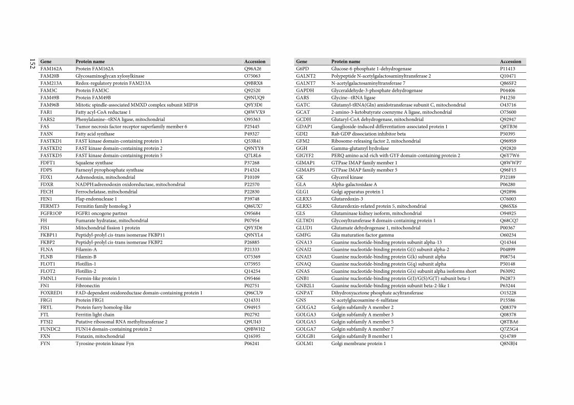
152
Gene Protein name Accession
FAM162A Protein FAM162A Q96A26
FAM20B Glycosaminoglycan xylosylkinase O75063
FAM213A Redox-regulatory protein FAM213A Q9BRX8
FAM3C Protein FAM3C Q92520
FAM49B Protein FAM49B Q9NUQ9
FAM96B Mitotic spindle-associated MMXD complex subunit MIP18 Q9Y3D0
FAR1 Fatty acyl-CoA reductase 1 Q8WVX9
FARS2 Phenylalanine--tRNA ligase, mitochondrial O95363
FAS Tumor necrosis factor receptor superfamily member 6 P25445
FASN Fatty acid synthase P49327
FASTKD1 FAST kinase domain-containing protein 1 Q53R41
FASTKD2 FAST kinase domain-containing protein 2 Q9NYY8
FASTKD5 FAST kinase domain-containing protein 5 Q7L8L6
FDFT1 Squalene synthase P37268
FDPS Farnesyl pyrophosphate synthase P14324
FDX1 Adrenodoxin, mitochondrial P10109
FDXR NADPH:adrenodoxin oxidoreductase, mitochondrial P22570
FECH Ferrochelatase, mitochondrial P22830
FEN1 Flap endonuclease 1 P39748
FERMT3 Fermitin family homolog 3 Q86UX7
FGFR1OP FGFR1 oncogene partner O95684
FH Fumarate hydratase, mitochondrial P07954
FIS1 Mitochondrial fission 1 protein Q9Y3D6
FKBP11 Peptidyl-prolyl cis-trans isomerase FKBP11 Q9NYL4
FKBP2 Peptidyl-prolyl cis-trans isomerase FKBP2 P26885
FLNA Filamin-A P21333
FLNB Filamin-B O75369
FLOT1 Flotillin-1 O75955
FLOT2 Flotillin-2 Q14254
FMNL1 Formin-like protein 1 O95466
FN1 Fibronectin P02751
FOXRED1 FAD-dependent oxidoreductase domain-containing protein 1 Q96CU9
FRG1 Protein FRG1 Q14331
FRYL Protein furry homolog-like O94915
FTL Ferritin light chain P02792
FTSJ2 Putative ribosomal RNA methyltransferase 2 Q9UI43
FUNDC2 FUN14 domain-containing protein 2 Q9BWH2
FXN Frataxin, mitochondrial Q16595
FYN Tyrosine-protein kinase Fyn P06241
Gene Protein name Accession
G6PD Glucose-6-phosphate 1-dehydrogenase P11413
GALNT2 Polypeptide N-acetylgalactosaminyltransferase 2 Q10471
GALNT7 N-acetylgalactosaminyltransferase 7 Q86SF2
GAPDH Glyceraldehyde-3-phosphate dehydrogenase P04406
GARS Glycine--tRNA ligase P41250
GATC Glutamyl-tRNA(Gln) amidotransferase subunit C, mitochondrial O43716
GCAT 2-amino-3-ketobutyrate coenzyme A ligase, mitochondrial O75600
GCDH Glutaryl-CoA dehydrogenase, mitochondrial Q92947
GDAP1 Ganglioside-induced differentiation-associated protein 1 Q8TB36
GDI2 Rab GDP dissociation inhibitor beta P50395
GFM2 Ribosome-releasing factor 2, mitochondrial Q969S9
GGH Gamma-glutamyl hydrolase Q92820
GIGYF2 PERQ amino acid-rich with GYF domain-containing protein 2 Q6Y7W6
GIMAP1 GTPase IMAP family member 1 Q8WWP7
GIMAP5 GTPase IMAP family member 5 Q96F15
GK Glycerol kinase P32189
GLA Alpha-galactosidase A P06280
GLG1 Golgi apparatus protein 1 Q92896
GLRX3 Glutaredoxin-3 O76003
GLRX5 Glutaredoxin-related protein 5, mitochondrial Q86SX6
GLS Glutaminase kidney isoform, mitochondrial O94925
GLT8D1 Glycosyltransferase 8 domain-containing protein 1 Q68CQ7
GLUD1 Glutamate dehydrogenase 1, mitochondrial P00367
GMFG Glia maturation factor gamma O60234
GNA13 Guanine nucleotide-binding protein subunit alpha-13 Q14344
GNAI2 Guanine nucleotide-binding protein G(i) subunit alpha-2 P04899
GNAI3 Guanine nucleotide-binding protein G(k) subunit alpha P08754
GNAQ Guanine nucleotide-binding protein G(q) subunit alpha P50148
GNAS Guanine nucleotide-binding protein G(s) subunit alpha isoforms short P63092
GNB1 Guanine nucleotide-binding protein G(I)/G(S)/G(T) subunit beta-1 P62873
GNB2L1 Guanine nucleotide-binding protein subunit beta-2-like 1 P63244
GNPAT Dihydroxyacetone phosphate acyltransferase O15228
GNS N-acetylglucosamine-6-sulfatase P15586
GOLGA2 Golgin subfamily A member 2 Q08379
GOLGA3 Golgin subfamily A member 3 Q08378
GOLGA5 Golgin subfamily A member 5 Q8TBA6
GOLGA7 Golgin subfamily A member 7 Q7Z5G4
GOLGB1 Golgin subfamily B member 1 Q14789
GOLM1 Golgi membrane protein 1 Q8NBJ4
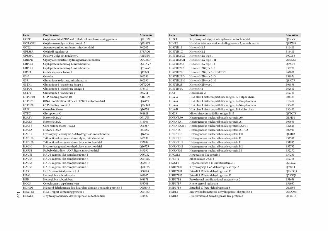
153
Gene Protein name Accession
GOPC Golgi-associated PDZ and coiled-coil motif-containing protein Q9HD26
GORASP2 Golgi reassembly-stacking protein 2 Q9H8Y8
GOT2 Aspartate aminotransferase, mitochondrial P00505
GPR89A Golgi pH regulator A B7ZAQ6
GPR89C Putative Golgi pH regulator C A6NKF9
GRHPR Glyoxylate reductase/hydroxypyruvate reductase Q9UBQ7
GRPEL1 GrpE protein homolog 1, mitochondrial Q9HAV7
GRPEL2 GrpE protein homolog 2, mitochondrial Q8TAA5
GRSF1 G-rich sequence factor 1 Q12849
GSN Gelsolin P06396
GSR Glutathione reductase, mitochondrial P00390
GSTK1 Glutathione S-transferase kappa 1 Q9Y2Q3
GSTO1 Glutathione S-transferase omega-1 P78417
GSTP1 Glutathione S-transferase P P09211
GTPBP10 GTP-binding protein 10 A4D1E9
GTPBP3 tRNA modification GTPase GTPBP3, mitochondrial Q969Y2
GTPBP8 GTP-binding protein 8 Q8N3Z3
GUK1 Guanylate kinase Q16774
GYPC Glycophorin-C P04921
H2AFV Histone H2A.V Q71UI9
H2AFX Histone H2AX P16104
H2AFY Core histone macro-H2A.1 O75367
H2AFZ Histone H2A.Z P0C0S5
HADH Hydroxyacyl-coenzyme A dehydrogenase, mitochondrial Q16836
HADHA Trifunctional enzyme subunit alpha, mitochondrial P40939
HADHB Trifunctional enzyme subunit beta, mitochondrial P55084
HAGH Hydroxyacylglutathione hydrolase, mitochondrial Q16775
HARS2 Probable histidine--tRNA ligase, mitochondrial P49590
HAUS1 HAUS augmin-like complex subunit 1 Q96CS2
HAUS4 HAUS augmin-like complex subunit 4 Q9H6D7
HAUS6 HAUS augmin-like complex subunit 6 Q7Z4H7
HAUS8 HAUS augmin-like complex subunit 8 Q9BT25
HAX1 HCLS1-associated protein X-1 O00165
HBA1; Hemoglobin subunit alpha P69905
HBB Hemoglobin subunit beta P68871
HCCS Cytochrome c-type heme lyase P53701
HDHD3 Haloacid dehalogenase-like hydrolase domain-containing protein 3 Q9BSH5
HEATR1 HEAT repeat-containing protein 1 Q9H583
HIBADH 3-hydroxyisobutyrate dehydrogenase, mitochondrial P31937
Gene Protein name Accession
HIBCH 3-hydroxyisobutyryl-CoA hydrolase, mitochondrial Q6NVY1
HINT2 Histidine triad nucleotide-binding protein 2, mitochondrial Q9BX68
HIST1H1B Histone H1.5 P16401
HIST1H1C Histone H1.2 P16403
HIST1H2AG Histone H2A type 1 P0C0S8
HIST1H2AH Histone H2A type 1-H Q96KK5
HIST1H2AJ Histone H2A type 1-J Q99878
HIST1H2BB Histone H2B type 1-B P33778
HIST1H2BC Histone H2B type 1-C/E/F/G/I P62807
HIST1H2BD Histone H2B type 1-D P58876
HIST1H2BH Histone H2B type 1-H Q93079
HIST1H2BJ Histone H2B type 1-J P06899
HIST1H4A Histone H4 P62805
HK2 Hexokinase-2 P52789
HLA-A HLA class I histocompatibility antigen, A-3 alpha chain P04439
HLA-A HLA class I histocompatibility antigen, A-25 alpha chain P18462
HLA-A HLA class I histocompatibility antigen, A-26 alpha chain P30450
HLA-B HLA class I histocompatibility antigen, B-8 alpha chain P30460
HM13 Minor histocompatibility antigen H13 Q8TCT9
HNRNPA0 Heterogeneous nuclear ribonucleoprotein A0 Q13151
HNRNPA1 Heterogeneous nuclear ribonucleoprotein A1 P09651
HNRNPA2B1 Heterogeneous nuclear ribonucleoproteins A2/B1 P22626
HNRNPC Heterogeneous nuclear ribonucleoproteins C1/C2 P07910
HNRNPD Heterogeneous nuclear ribonucleoprotein D0 Q14103
HNRNPF Heterogeneous nuclear ribonucleoprotein F P52597
HNRNPH1 Heterogeneous nuclear ribonucleoprotein H P31943
HNRNPH2 Heterogeneous nuclear ribonucleoprotein H2 P55795
HNRNPM Heterogeneous nuclear ribonucleoprotein M P52272
HPCAL1 Hippocalcin-like protein 1 P37235
HRSP12 Ribonuclease UK114 P52758
HS2ST1 Heparan sulfate 2-O-sulfotransferase 1 Q7LGA3
HSD17B10 3-hydroxyacyl-CoA dehydrogenase type-2 Q99714
HSD17B11 Estradiol 17-beta-dehydrogenase 11 Q8NBQ5
HSD17B12 Estradiol 17-beta-dehydrogenase 12 Q53GQ0
HSD17B4 Peroxisomal multifunctional enzyme type 2 P51659
HSD17B7 3-keto-steroid reductase P56937
HSD17B8 Estradiol 17-beta-dehydrogenase 8 Q92506
HSDL1 Inactive hydroxysteroid dehydrogenase-like protein 1 Q3SXM5
HSDL2 Hydroxysteroid dehydrogenase-like protein 2 Q6YN16
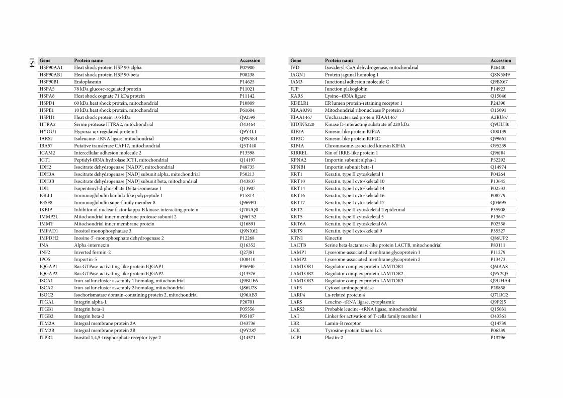
154
Gene Protein name Accession
HSP90AA1 Heat shock protein HSP 90-alpha P07900
HSP90AB1 Heat shock protein HSP 90-beta P08238
HSP90B1 Endoplasmin P14625
HSPA5 78 kDa glucose-regulated protein P11021
HSPA8 Heat shock cognate 71 kDa protein P11142
HSPD1 60 kDa heat shock protein, mitochondrial P10809
HSPE1 10 kDa heat shock protein, mitochondrial P61604
HSPH1 Heat shock protein 105 kDa Q92598
HTRA2 Serine protease HTRA2, mitochondrial O43464
HYOU1 Hypoxia up-regulated protein 1 Q9Y4L1
IARS2 Isoleucine--tRNA ligase, mitochondrial Q9NSE4
IBA57 Putative transferase CAF17, mitochondrial Q5T440
ICAM2 Intercellular adhesion molecule 2 P13598
ICT1 Peptidyl-tRNA hydrolase ICT1, mitochondrial Q14197
IDH2 Isocitrate dehydrogenase [NADP], mitochondrial P48735
IDH3A Isocitrate dehydrogenase [NAD] subunit alpha, mitochondrial P50213
IDH3B Isocitrate dehydrogenase [NAD] subunit beta, mitochondrial O43837
IDI1 Isopentenyl-diphosphate Delta-isomerase 1 Q13907
IGLL1 Immunoglobulin lambda-like polypeptide 1 P15814
IGSF8 Immunoglobulin superfamily member 8 Q969P0
IKBIP Inhibitor of nuclear factor kappa-B kinase-interacting protein Q70UQ0
IMMP2L Mitochondrial inner membrane protease subunit 2 Q96T52
IMMT Mitochondrial inner membrane protein Q16891
IMPAD1 Inositol monophosphatase 3 Q9NX62
IMPDH2 Inosine-5'-monophosphate dehydrogenase 2 P12268
INA Alpha-internexin Q16352
INF2 Inverted formin-2 Q27J81
IPO5 Importin-5 O00410
IQGAP1 Ras GTPase-activating-like protein IQGAP1 P46940
IQGAP2 Ras GTPase-activating-like protein IQGAP2 Q13576
ISCA1 Iron-sulfur cluster assembly 1 homolog, mitochondrial Q9BUE6
ISCA2 Iron-sulfur cluster assembly 2 homolog, mitochondrial Q86U28
ISOC2 Isochorismatase domain-containing protein 2, mitochondrial Q96AB3
ITGAL Integrin alpha-L P20701
ITGB1 Integrin beta-1 P05556
ITGB2 Integrin beta-2 P05107
ITM2A Integral membrane protein 2A O43736
ITM2B Integral membrane protein 2B Q9Y287
ITPR2 Inositol 1,4,5-trisphosphate receptor type 2 Q14571
Gene Protein name Accession
IVD Isovaleryl-CoA dehydrogenase, mitochondrial P26440
JAGN1 Protein jagunal homolog 1 Q8N5M9
JAM3 Junctional adhesion molecule C Q9BX67
JUP Junction plakoglobin P14923
KARS Lysine--tRNA ligase Q15046
KDELR1 ER lumen protein-retaining receptor 1 P24390
KIAA0391 Mitochondrial ribonuclease P protein 3 O15091
KIAA1467 Uncharacterized protein KIAA1467 A2RU67
KIDINS220 Kinase D-interacting substrate of 220 kDa Q9ULH0
KIF2A Kinesin-like protein KIF2A O00139
KIF2C Kinesin-like protein KIF2C Q99661
KIF4A Chromosome-associated kinesin KIF4A O95239
KIRREL Kin of IRRE-like protein 1 Q96J84
KPNA2 Importin subunit alpha-1 P52292
KPNB1 Importin subunit beta-1 Q14974
KRT1 Keratin, type II cytoskeletal 1 P04264
KRT10 Keratin, type I cytoskeletal 10 P13645
KRT14 Keratin, type I cytoskeletal 14 P02533
KRT16 Keratin, type I cytoskeletal 16 P08779
KRT17 Keratin, type I cytoskeletal 17 Q04695
KRT2 Keratin, type II cytoskeletal 2 epidermal P35908
KRT5 Keratin, type II cytoskeletal 5 P13647
KRT6A Keratin, type II cytoskeletal 6A P02538
KRT9 Keratin, type I cytoskeletal 9 P35527
KTN1 Kinectin Q86UP2
LACTB Serine beta-lactamase-like protein LACTB, mitochondrial P83111
LAMP1 Lysosome-associated membrane glycoprotein 1 P11279
LAMP2 Lysosome-associated membrane glycoprotein 2 P13473
LAMTOR1 Ragulator complex protein LAMTOR1 Q6IAA8
LAMTOR2 Ragulator complex protein LAMTOR2 Q9Y2Q5
LAMTOR3 Ragulator complex protein LAMTOR3 Q9UHA4
LAP3 Cytosol aminopeptidase P28838
LARP4 La-related protein 4 Q71RC2
LARS Leucine--tRNA ligase, cytoplasmic Q9P2J5
LARS2 Probable leucine--tRNA ligase, mitochondrial Q15031
LAT Linker for activation of T-cells family member 1 O43561
LBR Lamin-B receptor Q14739
LCK Tyrosine-protein kinase Lck P06239
LCP1 Plastin-2 P13796
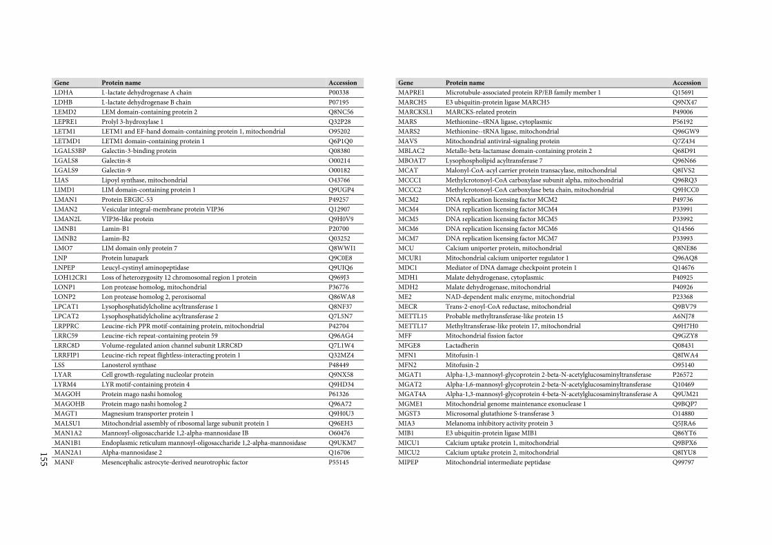
155
Gene Protein name Accession
LDHA L-lactate dehydrogenase A chain P00338
LDHB L-lactate dehydrogenase B chain P07195
LEMD2 LEM domain-containing protein 2 Q8NC56
LEPRE1 Prolyl 3-hydroxylase 1 Q32P28
LETM1 LETM1 and EF-hand domain-containing protein 1, mitochondrial O95202
LETMD1 LETM1 domain-containing protein 1 Q6P1Q0
LGALS3BP Galectin-3-binding protein Q08380
LGALS8 Galectin-8 O00214
LGALS9 Galectin-9 O00182
LIAS Lipoyl synthase, mitochondrial O43766
LIMD1 LIM domain-containing protein 1 Q9UGP4
LMAN1 Protein ERGIC-53 P49257
LMAN2 Vesicular integral-membrane protein VIP36 Q12907
LMAN2L VIP36-like protein Q9H0V9
LMNB1 Lamin-B1 P20700
LMNB2 Lamin-B2 Q03252
LMO7 LIM domain only protein 7 Q8WWI1
LNP Protein lunapark Q9C0E8
LNPEP Leucyl-cystinyl aminopeptidase Q9UIQ6
LOH12CR1 Loss of heterozygosity 12 chromosomal region 1 protein Q969J3
LONP1 Lon protease homolog, mitochondrial P36776
LONP2 Lon protease homolog 2, peroxisomal Q86WA8
LPCAT1 Lysophosphatidylcholine acyltransferase 1 Q8NF37
LPCAT2 Lysophosphatidylcholine acyltransferase 2 Q7L5N7
LRPPRC Leucine-rich PPR motif-containing protein, mitochondrial P42704
LRRC59 Leucine-rich repeat-containing protein 59 Q96AG4
LRRC8D Volume-regulated anion channel subunit LRRC8D Q7L1W4
LRRFIP1 Leucine-rich repeat flightless-interacting protein 1 Q32MZ4
LSS Lanosterol synthase P48449
LYAR Cell growth-regulating nucleolar protein Q9NX58
LYRM4 LYR motif-containing protein 4 Q9HD34
MAGOH Protein mago nashi homolog P61326
MAGOHB Protein mago nashi homolog 2 Q96A72
MAGT1 Magnesium transporter protein 1 Q9H0U3
MALSU1 Mitochondrial assembly of ribosomal large subunit protein 1 Q96EH3
MAN1A2 Mannosyl-oligosaccharide 1,2-alpha-mannosidase IB O60476
MAN1B1 Endoplasmic reticulum mannosyl-oligosaccharide 1,2-alpha-mannosidase Q9UKM7
MAN2A1 Alpha-mannosidase 2 Q16706
MANF Mesencephalic astrocyte-derived neurotrophic factor P55145
Gene Protein name Accession
MAPRE1 Microtubule-associated protein RP/EB family member 1 Q15691
MARCH5 E3 ubiquitin-protein ligase MARCH5 Q9NX47
MARCKSL1 MARCKS-related protein P49006
MARS Methionine--tRNA ligase, cytoplasmic P56192
MARS2 Methionine--tRNA ligase, mitochondrial Q96GW9
MAVS Mitochondrial antiviral-signaling protein Q7Z434
MBLAC2 Metallo-beta-lactamase domain-containing protein 2 Q68D91
MBOAT7 Lysophospholipid acyltransferase 7 Q96N66
MCAT Malonyl-CoA-acyl carrier protein transacylase, mitochondrial Q8IVS2
MCCC1 Methylcrotonoyl-CoA carboxylase subunit alpha, mitochondrial Q96RQ3
MCCC2 Methylcrotonoyl-CoA carboxylase beta chain, mitochondrial Q9HCC0
MCM2 DNA replication licensing factor MCM2 P49736
MCM4 DNA replication licensing factor MCM4 P33991
MCM5 DNA replication licensing factor MCM5 P33992
MCM6 DNA replication licensing factor MCM6 Q14566
MCM7 DNA replication licensing factor MCM7 P33993
MCU Calcium uniporter protein, mitochondrial Q8NE86
MCUR1 Mitochondrial calcium uniporter regulator 1 Q96AQ8
MDC1 Mediator of DNA damage checkpoint protein 1 Q14676
MDH1 Malate dehydrogenase, cytoplasmic P40925
MDH2 Malate dehydrogenase, mitochondrial P40926
ME2 NAD-dependent malic enzyme, mitochondrial P23368
MECR Trans-2-enoyl-CoA reductase, mitochondrial Q9BV79
METTL15 Probable methyltransferase-like protein 15 A6NJ78
METTL17 Methyltransferase-like protein 17, mitochondrial Q9H7H0
MFF Mitochondrial fission factor Q9GZY8
MFGE8 Lactadherin Q08431
MFN1 Mitofusin-1 Q8IWA4
MFN2 Mitofusin-2 O95140
MGAT1 Alpha-1,3-mannosyl-glycoprotein 2-beta-N-acetylglucosaminyltransferase P26572
MGAT2 Alpha-1,6-mannosyl-glycoprotein 2-beta-N-acetylglucosaminyltransferase Q10469
MGAT4A Alpha-1,3-mannosyl-glycoprotein 4-beta-N-acetylglucosaminyltransferase A Q9UM21
MGME1 Mitochondrial genome maintenance exonuclease 1 Q9BQP7
MGST3 Microsomal glutathione S-transferase 3 O14880
MIA3 Melanoma inhibitory activity protein 3 Q5JRA6
MIB1 E3 ubiquitin-protein ligase MIB1 Q86YT6
MICU1 Calcium uptake protein 1, mitochondrial Q9BPX6
MICU2 Calcium uptake protein 2, mitochondrial Q8IYU8
MIPEP Mitochondrial intermediate peptidase Q99797
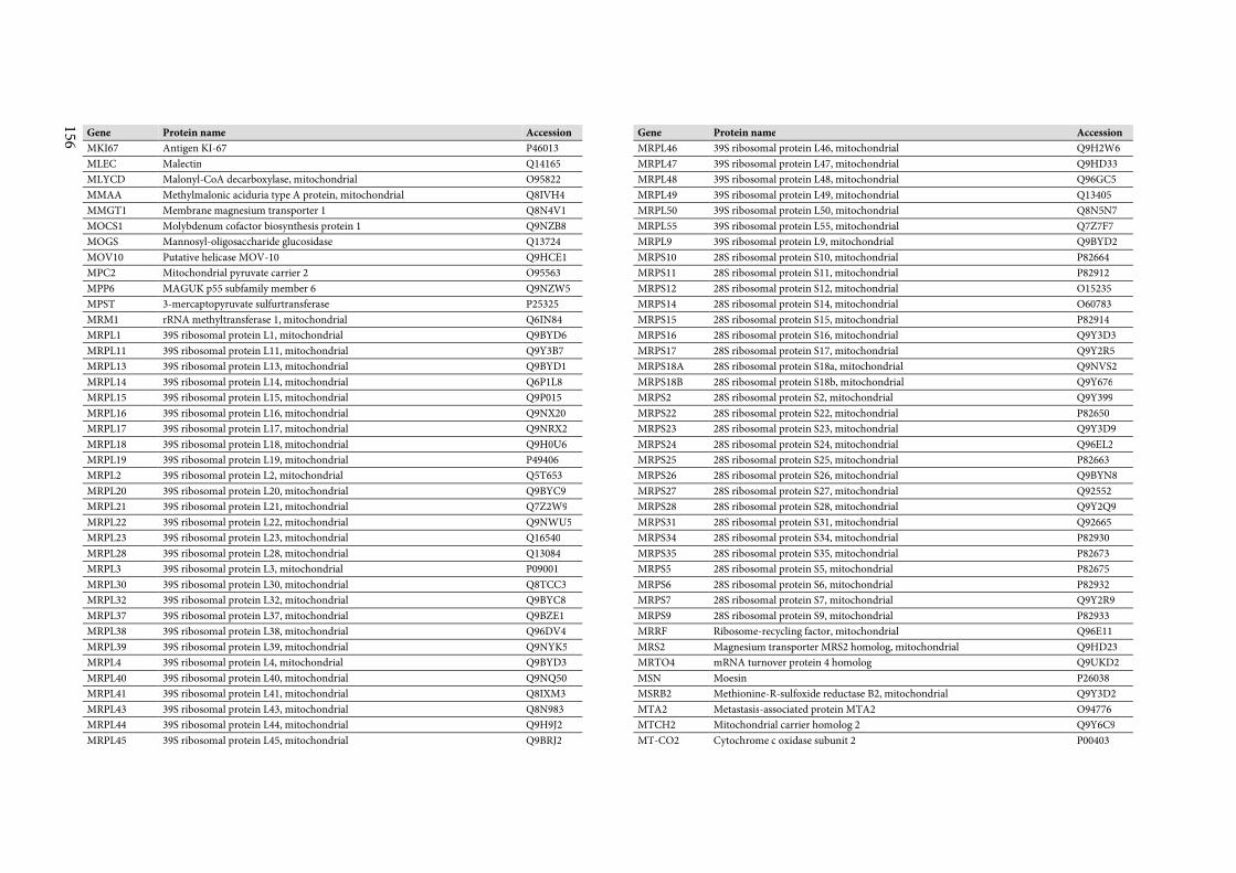
156
Gene Protein name Accession
MKI67 Antigen KI-67 P46013
MLEC Malectin Q14165
MLYCD Malonyl-CoA decarboxylase, mitochondrial O95822
MMAA Methylmalonic aciduria type A protein, mitochondrial Q8IVH4
MMGT1 Membrane magnesium transporter 1 Q8N4V1
MOCS1 Molybdenum cofactor biosynthesis protein 1 Q9NZB8
MOGS Mannosyl-oligosaccharide glucosidase Q13724
MOV10 Putative helicase MOV-10 Q9HCE1
MPC2 Mitochondrial pyruvate carrier 2 O95563
MPP6 MAGUK p55 subfamily member 6 Q9NZW5
MPST 3-mercaptopyruvate sulfurtransferase P25325
MRM1 rRNA methyltransferase 1, mitochondrial Q6IN84
MRPL1 39S ribosomal protein L1, mitochondrial Q9BYD6
MRPL11 39S ribosomal protein L11, mitochondrial Q9Y3B7
MRPL13 39S ribosomal protein L13, mitochondrial Q9BYD1
MRPL14 39S ribosomal protein L14, mitochondrial Q6P1L8
MRPL15 39S ribosomal protein L15, mitochondrial Q9P015
MRPL16 39S ribosomal protein L16, mitochondrial Q9NX20
MRPL17 39S ribosomal protein L17, mitochondrial Q9NRX2
MRPL18 39S ribosomal protein L18, mitochondrial Q9H0U6
MRPL19 39S ribosomal protein L19, mitochondrial P49406
MRPL2 39S ribosomal protein L2, mitochondrial Q5T653
MRPL20 39S ribosomal protein L20, mitochondrial Q9BYC9
MRPL21 39S ribosomal protein L21, mitochondrial Q7Z2W9
MRPL22 39S ribosomal protein L22, mitochondrial Q9NWU5
MRPL23 39S ribosomal protein L23, mitochondrial Q16540
MRPL28 39S ribosomal protein L28, mitochondrial Q13084
MRPL3 39S ribosomal protein L3, mitochondrial P09001
MRPL30 39S ribosomal protein L30, mitochondrial Q8TCC3
MRPL32 39S ribosomal protein L32, mitochondrial Q9BYC8
MRPL37 39S ribosomal protein L37, mitochondrial Q9BZE1
MRPL38 39S ribosomal protein L38, mitochondrial Q96DV4
MRPL39 39S ribosomal protein L39, mitochondrial Q9NYK5
MRPL4 39S ribosomal protein L4, mitochondrial Q9BYD3
MRPL40 39S ribosomal protein L40, mitochondrial Q9NQ50
MRPL41 39S ribosomal protein L41, mitochondrial Q8IXM3
MRPL43 39S ribosomal protein L43, mitochondrial Q8N983
MRPL44 39S ribosomal protein L44, mitochondrial Q9H9J2
MRPL45 39S ribosomal protein L45, mitochondrial Q9BRJ2
Gene Protein name Accession
MRPL46 39S ribosomal protein L46, mitochondrial Q9H2W6
MRPL47 39S ribosomal protein L47, mitochondrial Q9HD33
MRPL48 39S ribosomal protein L48, mitochondrial Q96GC5
MRPL49 39S ribosomal protein L49, mitochondrial Q13405
MRPL50 39S ribosomal protein L50, mitochondrial Q8N5N7
MRPL55 39S ribosomal protein L55, mitochondrial Q7Z7F7
MRPL9 39S ribosomal protein L9, mitochondrial Q9BYD2
MRPS10 28S ribosomal protein S10, mitochondrial P82664
MRPS11 28S ribosomal protein S11, mitochondrial P82912
MRPS12 28S ribosomal protein S12, mitochondrial O15235
MRPS14 28S ribosomal protein S14, mitochondrial O60783
MRPS15 28S ribosomal protein S15, mitochondrial P82914
MRPS16 28S ribosomal protein S16, mitochondrial Q9Y3D3
MRPS17 28S ribosomal protein S17, mitochondrial Q9Y2R5
MRPS18A 28S ribosomal protein S18a, mitochondrial Q9NVS2
MRPS18B 28S ribosomal protein S18b, mitochondrial Q9Y676
MRPS2 28S ribosomal protein S2, mitochondrial Q9Y399
MRPS22 28S ribosomal protein S22, mitochondrial P82650
MRPS23 28S ribosomal protein S23, mitochondrial Q9Y3D9
MRPS24 28S ribosomal protein S24, mitochondrial Q96EL2
MRPS25 28S ribosomal protein S25, mitochondrial P82663
MRPS26 28S ribosomal protein S26, mitochondrial Q9BYN8
MRPS27 28S ribosomal protein S27, mitochondrial Q92552
MRPS28 28S ribosomal protein S28, mitochondrial Q9Y2Q9
MRPS31 28S ribosomal protein S31, mitochondrial Q92665
MRPS34 28S ribosomal protein S34, mitochondrial P82930
MRPS35 28S ribosomal protein S35, mitochondrial P82673
MRPS5 28S ribosomal protein S5, mitochondrial P82675
MRPS6 28S ribosomal protein S6, mitochondrial P82932
MRPS7 28S ribosomal protein S7, mitochondrial Q9Y2R9
MRPS9 28S ribosomal protein S9, mitochondrial P82933
MRRF Ribosome-recycling factor, mitochondrial Q96E11
MRS2 Magnesium transporter MRS2 homolog, mitochondrial Q9HD23
MRTO4 mRNA turnover protein 4 homolog Q9UKD2
MSN Moesin P26038
MSRB2 Methionine-R-sulfoxide reductase B2, mitochondrial Q9Y3D2
MTA2 Metastasis-associated protein MTA2 O94776
MTCH2 Mitochondrial carrier homolog 2 Q9Y6C9
MT-CO2 Cytochrome c oxidase subunit 2 P00403
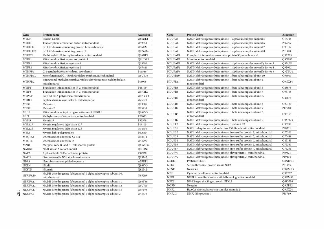
157
Gene Protein name Accession
MTDH Protein LYRIC Q86UE4
MTERF Transcription termination factor, mitochondrial Q99551
MTERFD1 mTERF domain-containing protein 1, mitochondrial Q96E29
MTERFD2 mTERF domain-containing protein 2 Q7Z6M4
MTFMT Methionyl-tRNA formyltransferase, mitochondrial Q96DP5
MTFP1 Mitochondrial fission process protein 1 Q9UDX5
MTFR1 Mitochondrial fission regulator 1 Q15390
MTFR2 Mitochondrial fission regulator 2 Q6P444
MTHFD1 C-1-tetrahydrofolate synthase, cytoplasmic P11586
MTHFD1L Monofunctional C1-tetrahydrofolate synthase, mitochondrial Q6UB35
MTHFD2 Bifunctional methylenetetrahydrofolate dehydrogenase/cyclohydrolase,
mitochondrial P13995
MTIF2 Translation initiation factor IF-2, mitochondrial P46199
MTIF3 Translation initiation factor IF-3, mitochondrial Q9H2K0
MTPAP Poly(A) RNA polymerase, mitochondrial Q9NVV4
MTRF1 Peptide chain release factor 1, mitochondrial O75570
MTX1 Metaxin-1 Q13505
MTX2 Metaxin-2 O75431
MUL1 Mitochondrial ubiquitin ligase activator of NFKB 1 Q969V5
MUT Methylmalonyl-CoA mutase, mitochondrial P22033
MYH9 Myosin-9 P35579
MYL12A Myosin regulatory light chain 12A P19105
MYL12B Myosin regulatory light chain 12B O14950
MYL6 Myosin light polypeptide 6 P60660
MYO18A Unconventional myosin-XVIIIa Q92614
MYO1B Unconventional myosin-Ib O43795
MZB1 Marginal zone B- and B1-cell-specific protein Q8WU39
NADK2 NAD kinase 2, mitochondrial Q4G0N4
NAPA Alpha-soluble NSF attachment protein P54920
NAPG Gamma-soluble NSF attachment protein Q99747
NBAS Neuroblastoma-amplified sequence A2RRP1
NCLN Nicalin Q969V3
NCSTN Nicastrin Q92542
NDUFA10 NADH dehydrogenase [ubiquinone] 1 alpha subcomplex subunit 10,
mitochondrial O95299
NDUFA11 NADH dehydrogenase [ubiquinone] 1 alpha subcomplex subunit 11 Q86Y39
NDUFA12 NADH dehydrogenase [ubiquinone] 1 alpha subcomplex subunit 12 Q9UI09
NDUFA13 NADH dehydrogenase [ubiquinone] 1 alpha subcomplex subunit 13 Q9P0J0
NDUFA2 NADH dehydrogenase [ubiquinone] 1 alpha subcomplex subunit 2 O43678
Gene Protein name Accession
NDUFA5 NADH dehydrogenase [ubiquinone] 1 alpha subcomplex subunit 5 Q16718
NDUFA6 NADH dehydrogenase [ubiquinone] 1 alpha subcomplex subunit 6 P56556
NDUFA7 NADH dehydrogenase [ubiquinone] 1 alpha subcomplex subunit 7 O95182
NDUFA8 NADH dehydrogenase [ubiquinone] 1 alpha subcomplex subunit 8 P51970
NDUFAF1 Complex I intermediate-associated protein 30, mitochondrial Q9Y375
NDUFAF2 Mimitin, mitochondrial Q8N183
NDUFAF3 NADH dehydrogenase [ubiquinone] 1 alpha subcomplex assembly factor 3 Q9BU61
NDUFAF4 NADH dehydrogenase [ubiquinone] 1 alpha subcomplex assembly factor 4 Q9P032
NDUFAF5 NADH dehydrogenase [ubiquinone] 1 alpha subcomplex assembly factor 5 Q5TEU4
NDUFB10 NADH dehydrogenase [ubiquinone] 1 beta subcomplex subunit 10 O96000
NDUFB11 NADH dehydrogenase [ubiquinone] 1 beta subcomplex subunit 11,
mitochondrial Q9NX14
NDUFB3 NADH dehydrogenase [ubiquinone] 1 beta subcomplex subunit 3 O43676
NDUFB4 NADH dehydrogenase [ubiquinone] 1 beta subcomplex subunit 4 O95168
NDUFB5 NADH dehydrogenase [ubiquinone] 1 beta subcomplex subunit 5,
mitochondrial O43674
NDUFB6 NADH dehydrogenase [ubiquinone] 1 beta subcomplex subunit 6 O95139
NDUFB7 NADH dehydrogenase [ubiquinone] 1 beta subcomplex subunit 7 P17568
NDUFB8 NADH dehydrogenase [ubiquinone] 1 beta subcomplex subunit 8,
mitochondrial O95169
NDUFB9 NADH dehydrogenase [ubiquinone] 1 beta subcomplex subunit 9 Q9Y6M9
NDUFC2 NADH dehydrogenase [ubiquinone] 1 subunit C2 O95298
NDUFS1 NADH-ubiquinone oxidoreductase 75 kDa subunit, mitochondrial P28331
NDUFS2 NADH dehydrogenase [ubiquinone] iron-sulfur protein 2, mitochondrial O75306
NDUFS3 NADH dehydrogenase [ubiquinone] iron-sulfur protein 3, mitochondrial O75489
NDUFS4 NADH dehydrogenase [ubiquinone] iron-sulfur protein 4, mitochondrial O43181
NDUFS6 NADH dehydrogenase [ubiquinone] iron-sulfur protein 6, mitochondrial O75380
NDUFS7 NADH dehydrogenase [ubiquinone] iron-sulfur protein 7, mitochondrial O75251
NDUFV1 NADH dehydrogenase [ubiquinone] flavoprotein 1, mitochondrial P49821
NDUFV2 NADH dehydrogenase [ubiquinone] flavoprotein 2, mitochondrial P19404
NEDD1 Protein NEDD1 Q8NHV4
NEK2 Serine/threonine-protein kinase Nek2 P51955
NENF Neudesin Q9UMX5
NFS1 Cysteine desulfurase, mitochondrial Q9Y697
NFU1 NFU1 iron-sulfur cluster scaffold homolog, mitochondrial Q9UMS0
NFXL1 NF-X1-type zinc finger protein NFXL1 Q6ZNB6
NGRN Neugrin Q9NPE2
NHP2 H/ACA ribonucleoprotein complex subunit 2 Q9NX24
NHP2L1 NHP2-like protein 1 P55769
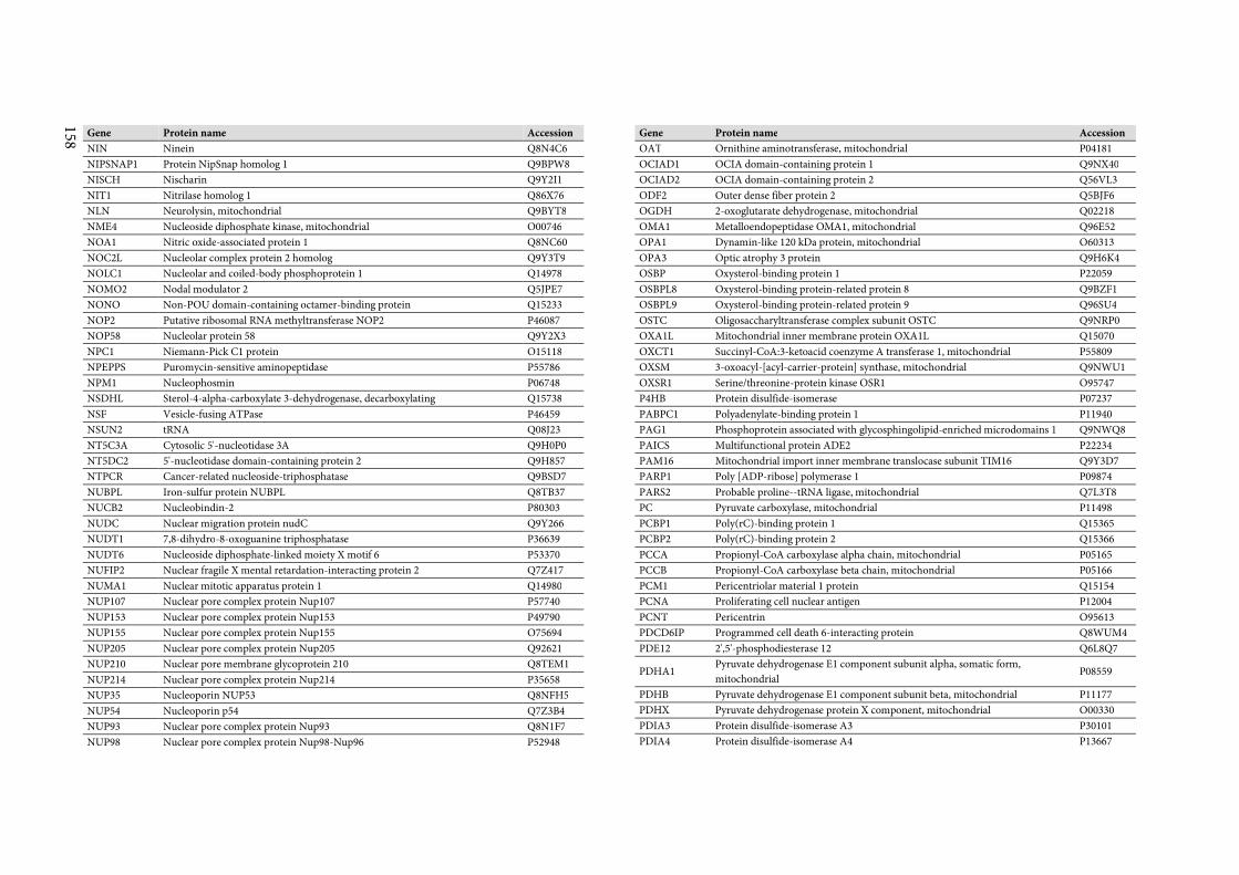
158
Gene Protein name Accession
NIN Ninein Q8N4C6
NIPSNAP1 Protein NipSnap homolog 1 Q9BPW8
NISCH Nischarin Q9Y2I1
NIT1 Nitrilase homolog 1 Q86X76
NLN Neurolysin, mitochondrial Q9BYT8
NME4 Nucleoside diphosphate kinase, mitochondrial O00746
NOA1 Nitric oxide-associated protein 1 Q8NC60
NOC2L Nucleolar complex protein 2 homolog Q9Y3T9
NOLC1 Nucleolar and coiled-body phosphoprotein 1 Q14978
NOMO2 Nodal modulator 2 Q5JPE7
NONO Non-POU domain-containing octamer-binding protein Q15233
NOP2 Putative ribosomal RNA methyltransferase NOP2 P46087
NOP58 Nucleolar protein 58 Q9Y2X3
NPC1 Niemann-Pick C1 protein O15118
NPEPPS Puromycin-sensitive aminopeptidase P55786
NPM1 Nucleophosmin P06748
NSDHL Sterol-4-alpha-carboxylate 3-dehydrogenase, decarboxylating Q15738
NSF Vesicle-fusing ATPase P46459
NSUN2 tRNA Q08J23
NT5C3A Cytosolic 5'-nucleotidase 3A Q9H0P0
NT5DC2 5'-nucleotidase domain-containing protein 2 Q9H857
NTPCR Cancer-related nucleoside-triphosphatase Q9BSD7
NUBPL Iron-sulfur protein NUBPL Q8TB37
NUCB2 Nucleobindin-2 P80303
NUDC Nuclear migration protein nudC Q9Y266
NUDT1 7,8-dihydro-8-oxoguanine triphosphatase P36639
NUDT6 Nucleoside diphosphate-linked moiety X motif 6 P53370
NUFIP2 Nuclear fragile X mental retardation-interacting protein 2 Q7Z417
NUMA1 Nuclear mitotic apparatus protein 1 Q14980
NUP107 Nuclear pore complex protein Nup107 P57740
NUP153 Nuclear pore complex protein Nup153 P49790
NUP155 Nuclear pore complex protein Nup155 O75694
NUP205 Nuclear pore complex protein Nup205 Q92621
NUP210 Nuclear pore membrane glycoprotein 210 Q8TEM1
NUP214 Nuclear pore complex protein Nup214 P35658
NUP35 Nucleoporin NUP53 Q8NFH5
NUP54 Nucleoporin p54 Q7Z3B4
NUP93 Nuclear pore complex protein Nup93 Q8N1F7
NUP98 Nuclear pore complex protein Nup98-Nup96 P52948
Gene Protein name Accession
OAT Ornithine aminotransferase, mitochondrial P04181
OCIAD1 OCIA domain-containing protein 1 Q9NX40
OCIAD2 OCIA domain-containing protein 2 Q56VL3
ODF2 Outer dense fiber protein 2 Q5BJF6
OGDH 2-oxoglutarate dehydrogenase, mitochondrial Q02218
OMA1 Metalloendopeptidase OMA1, mitochondrial Q96E52
OPA1 Dynamin-like 120 kDa protein, mitochondrial O60313
OPA3 Optic atrophy 3 protein Q9H6K4
OSBP Oxysterol-binding protein 1 P22059
OSBPL8 Oxysterol-binding protein-related protein 8 Q9BZF1
OSBPL9 Oxysterol-binding protein-related protein 9 Q96SU4
OSTC Oligosaccharyltransferase complex subunit OSTC Q9NRP0
OXA1L Mitochondrial inner membrane protein OXA1L Q15070
OXCT1 Succinyl-CoA:3-ketoacid coenzyme A transferase 1, mitochondrial P55809
OXSM 3-oxoacyl-[acyl-carrier-protein] synthase, mitochondrial Q9NWU1
OXSR1 Serine/threonine-protein kinase OSR1 O95747
P4HB Protein disulfide-isomerase P07237
PABPC1 Polyadenylate-binding protein 1 P11940
PAG1 Phosphoprotein associated with glycosphingolipid-enriched microdomains 1 Q9NWQ8
PAICS Multifunctional protein ADE2 P22234
PAM16 Mitochondrial import inner membrane translocase subunit TIM16 Q9Y3D7
PARP1 Poly [ADP-ribose] polymerase 1 P09874
PARS2 Probable proline--tRNA ligase, mitochondrial Q7L3T8
PC Pyruvate carboxylase, mitochondrial P11498
PCBP1 Poly(rC)-binding protein 1 Q15365
PCBP2 Poly(rC)-binding protein 2 Q15366
PCCA Propionyl-CoA carboxylase alpha chain, mitochondrial P05165
PCCB Propionyl-CoA carboxylase beta chain, mitochondrial P05166
PCM1 Pericentriolar material 1 protein Q15154
PCNA Proliferating cell nuclear antigen P12004
PCNT Pericentrin O95613
PDCD6IP Programmed cell death 6-interacting protein Q8WUM4
PDE12 2',5'-phosphodiesterase 12 Q6L8Q7
PDHA1 Pyruvate dehydrogenase E1 component subunit alpha, somatic form,
mitochondrial P08559
PDHB Pyruvate dehydrogenase E1 component subunit beta, mitochondrial P11177
PDHX Pyruvate dehydrogenase protein X component, mitochondrial O00330
PDIA3 Protein disulfide-isomerase A3 P30101
PDIA4 Protein disulfide-isomerase A4 P13667
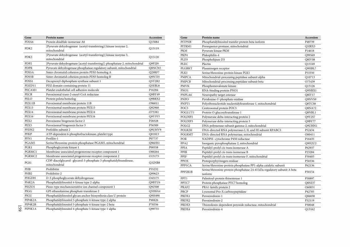
159
Gene Protein name Accession
PDIA6 Protein disulfide-isomerase A6 Q15084
PDK2 [Pyruvate dehydrogenase (acetyl-transferring)] kinase isozyme 2,
mitochondrial Q15119
PDK3 [Pyruvate dehydrogenase (acetyl-transferring)] kinase isozyme 3,
mitochondrial Q15120
PDP2 [Pyruvate dehydrogenase [acetyl-transferring]]-phosphatase 2, mitochondrial Q9P2J9
PDPR Pyruvate dehydrogenase phosphatase regulatory subunit, mitochondrial Q8NCN5
PDS5A Sister chromatid cohesion protein PDS5 homolog A Q29RF7
PDS5B Sister chromatid cohesion protein PDS5 homolog B Q9NTI5
PDSS1 Decaprenyl-diphosphate synthase subunit 1 Q5T2R2
PDZD11 PDZ domain-containing protein 11 Q5EBL8
PECAM1 Platelet endothelial cell adhesion molecule P16284
PECR Peroxisomal trans-2-enoyl-CoA reductase Q9BY49
PELO Protein pelota homolog Q9BRX2
PEX11B Peroxisomal membrane protein 11B O96011
PEX13 Peroxisomal membrane protein PEX13 Q92968
PEX14 Peroxisomal membrane protein PEX14 O75381
PEX16 Peroxisomal membrane protein PEX16 Q9Y5Y5
PEX2 Peroxisome biogenesis factor 2 P28328
PEX3 Peroxisomal biogenesis factor 3 P56589
PFDN2 Prefoldin subunit 2 Q9UHV9
PFKP ATP-dependent 6-phosphofructokinase, platelet type Q01813
PFN1 Profilin-1 P07737
PGAM5 Serine/threonine-protein phosphatase PGAM5, mitochondrial Q96HS1
PGK1 Phosphoglycerate kinase 1 P00558
PGRMC1 Membrane-associated progesterone receptor component 1 O00264
PGRMC2 Membrane-associated progesterone receptor component 2 O15173
PGS1 CDP-diacylglycerol--glycerol-3-phosphate 3-phosphatidyltransferase,
mitochondrial Q32NB8
PHB Prohibitin P35232
PHB2 Prohibitin-2 Q99623
PHGDH D-3-phosphoglycerate dehydrogenase O43175
PI4K2A Phosphatidylinositol 4-kinase type 2-alpha Q9BTU6
PIEZO1 Piezo-type mechanosensitive ion channel component 1 Q92508
PIGG GPI ethanolamine phosphate transferase 2 Q5H8A4
PIGU Phosphatidylinositol glycan anchor biosynthesis class U protein Q9H490
PIP4K2A Phosphatidylinositol 5-phosphate 4-kinase type-2 alpha P48426
PIP4K2B Phosphatidylinositol 5-phosphate 4-kinase type-2 beta P78356
PIP5K1A Phosphatidylinositol 4-phosphate 5-kinase type-1 alpha Q99755
Gene Protein name Accession
PITPNB Phosphatidylinositol transfer protein beta isoform P48739
PITRM1 Presequence protease, mitochondrial Q5JRX3
PKM Pyruvate kinase PKM P14618
PKP4 Plakophilin-4 Q99569
PLD3 Phospholipase D3 Q8IV08
PLEC Plectin Q15149
PLGRKT Plasminogen receptor Q9HBL7
PLK1 Serine/threonine-protein kinase PLK1 P53350
PMPCA Mitochondrial-processing peptidase subunit alpha Q10713
PMPCB Mitochondrial-processing peptidase subunit beta O75439
PMVK Phosphomevalonate kinase Q15126
PNO1 RNA-binding protein PNO1 Q9NRX1
PNPLA6 Neuropathy target esterase Q8IY17
PNPO Pyridoxine-5'-phosphate oxidase Q9NVS9
PNPT1 Polyribonucleotide nucleotidyltransferase 1, mitochondrial Q8TCS8
POC5 Centrosomal protein POC5 Q8NA72
POGLUT1 Protein O-glucosyltransferase 1 Q8NBL1
POLDIP2 Polymerase delta-interacting protein 2 Q9Y2S7
POLDIP3 Polymerase delta-interacting protein 3 Q9BY77
POLG2 DNA polymerase subunit gamma-2, mitochondrial Q9UHN1
POLR2H DNA-directed RNA polymerases I, II, and III subunit RPABC3 P52434
POLRMT DNA-directed RNA polymerase, mitochondrial O00411
POR NADPH--cytochrome P450 reductase P16435
PPA2 Inorganic pyrophosphatase 2, mitochondrial Q9H2U2
PPIA Peptidyl-prolyl cis-trans isomerase A P62937
PPIB Peptidyl-prolyl cis-trans isomerase B P23284
PPIF Peptidyl-prolyl cis-trans isomerase F, mitochondrial P30405
PPOX Protoporphyrinogen oxidase P50336
PPP1CA Serine/threonine-protein phosphatase PP1-alpha catalytic subunit P62136
PPP2R1B Serine/threonine-protein phosphatase 2A 65 kDa regulatory subunit A beta
isoform P30154
PPT1 Palmitoyl-protein thioesterase 1 P50897
PPTC7 Protein phosphatase PTC7 homolog Q8NI37
PRAF2 PRA1 family protein 2 O60831
PRCP Lysosomal Pro-X carboxypeptidase P42785
PRDX1 Peroxiredoxin-1 Q06830
PRDX2 Peroxiredoxin-2 P32119
PRDX3 Thioredoxin-dependent peroxide reductase, mitochondrial P30048
PRDX4 Peroxiredoxin-4 Q13162
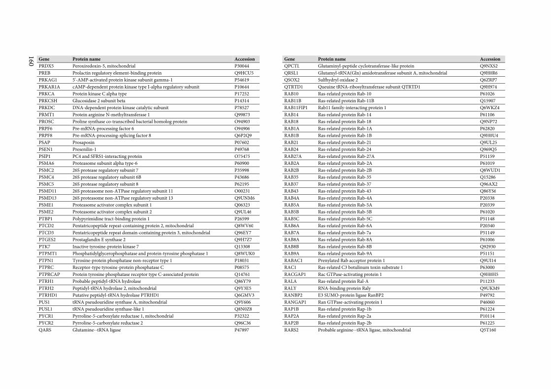
160
Gene Protein name Accession
PRDX5 Peroxiredoxin-5, mitochondrial P30044
PREB Prolactin regulatory element-binding protein Q9HCU5
PRKAG1 5'-AMP-activated protein kinase subunit gamma-1 P54619
PRKAR1A cAMP-dependent protein kinase type I-alpha regulatory subunit P10644
PRKCA Protein kinase C alpha type P17252
PRKCSH Glucosidase 2 subunit beta P14314
PRKDC DNA-dependent protein kinase catalytic subunit P78527
PRMT1 Protein arginine N-methyltransferase 1 Q99873
PROSC Proline synthase co-transcribed bacterial homolog protein O94903
PRPF6 Pre-mRNA-processing factor 6 O94906
PRPF8 Pre-mRNA-processing-splicing factor 8 Q6P2Q9
PSAP Prosaposin P07602
PSEN1 Presenilin-1 P49768
PSIP1 PC4 and SFRS1-interacting protein O75475
PSMA6 Proteasome subunit alpha type-6 P60900
PSMC2 26S protease regulatory subunit 7 P35998
PSMC4 26S protease regulatory subunit 6B P43686
PSMC5 26S protease regulatory subunit 8 P62195
PSMD11 26S proteasome non-ATPase regulatory subunit 11 O00231
PSMD13 26S proteasome non-ATPase regulatory subunit 13 Q9UNM6
PSME1 Proteasome activator complex subunit 1 Q06323
PSME2 Proteasome activator complex subunit 2 Q9UL46
PTBP1 Polypyrimidine tract-binding protein 1 P26599
PTCD2 Pentatricopeptide repeat-containing protein 2, mitochondrial Q8WV60
PTCD3 Pentatricopeptide repeat domain-containing protein 3, mitochondrial Q96EY7
PTGES2 Prostaglandin E synthase 2 Q9H7Z7
PTK7 Inactive tyrosine-protein kinase 7 Q13308
PTPMT1 Phosphatidylglycerophosphatase and protein-tyrosine phosphatase 1 Q8WUK0
PTPN1 Tyrosine-protein phosphatase non-receptor type 1 P18031
PTPRC Receptor-type tyrosine-protein phosphatase C P08575
PTPRCAP Protein tyrosine phosphatase receptor type C-associated protein Q14761
PTRH1 Probable peptidyl-tRNA hydrolase Q86Y79
PTRH2 Peptidyl-tRNA hydrolase 2, mitochondrial Q9Y3E5
PTRHD1 Putative peptidyl-tRNA hydrolase PTRHD1 Q6GMV3
PUS1 tRNA pseudouridine synthase A, mitochondrial Q9Y606
PUSL1 tRNA pseudouridine synthase-like 1 Q8N0Z8
PYCR1 Pyrroline-5-carboxylate reductase 1, mitochondrial P32322
PYCR2 Pyrroline-5-carboxylate reductase 2 Q96C36
QARS Glutamine--tRNA ligase P47897
Gene Protein name Accession
QPCTL Glutaminyl-peptide cyclotransferase-like protein Q9NXS2
QRSL1 Glutamyl-tRNA(Gln) amidotransferase subunit A, mitochondrial Q9H0R6
QSOX2 Sulfhydryl oxidase 2 Q6ZRP7
QTRTD1 Queuine tRNA-ribosyltransferase subunit QTRTD1 Q9H974
RAB10 Ras-related protein Rab-10 P61026
RAB11B Ras-related protein Rab-11B Q15907
RAB11FIP1 Rab11 family-interacting protein 1 Q6WKZ4
RAB14 Ras-related protein Rab-14 P61106
RAB18 Ras-related protein Rab-18 Q9NP72
RAB1A Ras-related protein Rab-1A P62820
RAB1B Ras-related protein Rab-1B Q9H0U4
RAB21 Ras-related protein Rab-21 Q9UL25
RAB24 Ras-related protein Rab-24 Q969Q5
RAB27A Ras-related protein Rab-27A P51159
RAB2A Ras-related protein Rab-2A P61019
RAB2B Ras-related protein Rab-2B Q8WUD1
RAB35 Ras-related protein Rab-35 Q15286
RAB37 Ras-related protein Rab-37 Q96AX2
RAB43 Ras-related protein Rab-43 Q86YS6
RAB4A Ras-related protein Rab-4A P20338
RAB5A Ras-related protein Rab-5A P20339
RAB5B Ras-related protein Rab-5B P61020
RAB5C Ras-related protein Rab-5C P51148
RAB6A Ras-related protein Rab-6A P20340
RAB7A Ras-related protein Rab-7a P51149
RAB8A Ras-related protein Rab-8A P61006
RAB8B Ras-related protein Rab-8B Q92930
RAB9A Ras-related protein Rab-9A P51151
RABAC1 Prenylated Rab acceptor protein 1 Q9UI14
RAC1 Ras-related C3 botulinum toxin substrate 1 P63000
RACGAP1 Rac GTPase-activating protein 1 Q9H0H5
RALA Ras-related protein Ral-A P11233
RALY RNA-binding protein Raly Q9UKM9
RANBP2 E3 SUMO-protein ligase RanBP2 P49792
RANGAP1 Ran GTPase-activating protein 1 P46060
RAP1B Ras-related protein Rap-1b P61224
RAP2A Ras-related protein Rap-2a P10114
RAP2B Ras-related protein Rap-2b P61225
RARS2 Probable arginine--tRNA ligase, mitochondrial Q5T160
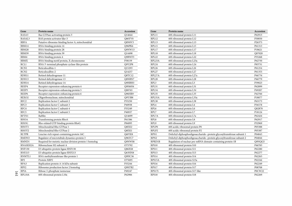
161
Gene Protein name Accession
RASA3 Ras GTPase-activating protein 3 Q14644
RASAL3 RAS protein activator like-3 Q86YV0
RBFA Putative ribosome-binding factor A, mitochondrial Q8N0V3
RBM14 RNA-binding protein 14 Q96PK6
RBM28 RNA-binding protein 28 Q9NW13
RBM39 RNA-binding protein 39 Q14498
RBM4 RNA-binding protein 4 Q9BWF3
RBMX RNA-binding motif protein, X chromosome P38159
RCL1 RNA 3'-terminal phosphate cyclase-like protein Q9Y2P8
RCN1 Reticulocalbin-1 Q15293
RCN2 Reticulocalbin-2 Q14257
RDH11 Retinol dehydrogenase 11 Q8TC12
RDH13 Retinol dehydrogenase 13 Q8NBN7
RDH14 Retinol dehydrogenase 14 Q9HBH5
REEP4 Receptor expression-enhancing protein 4 Q9H6H4
REEP5 Receptor expression-enhancing protein 5 Q00765
REEP6 Receptor expression-enhancing protein 6 Q96HR9
REXO2 Oligoribonuclease, mitochondrial Q9Y3B8
RFC2 Replication factor C subunit 2 P35250
RFC3 Replication factor C subunit 3 P40938
RFC4 Replication factor C subunit 4 P35249
RFC5 Replication factor C subunit 5 P40937
RFTN1 Raftlin Q14699
RHOA Transforming protein RhoA P61586
RHOG Rho-related GTP-binding protein RhoG P84095
RHOT1 Mitochondrial Rho GTPase 1 Q8IXI2
RHOT2 Mitochondrial Rho GTPase 2 Q8IXI1
RLTPR Leucine-rich repeat-containing protein 16C Q6F5E8
RMDN3 Regulator of microtubule dynamics protein 3 Q96TC7
RMND1 Required for meiotic nuclear division protein 1 homolog Q9NWS8
RNASEH2A Ribonuclease H2 subunit A O75792
RNF130 E3 ubiquitin-protein ligase RNF130 Q86XS8
RNF213 E3 ubiquitin-protein ligase RNF213 Q63HN8
RNMTL1 RNA methyltransferase-like protein 1 Q9HC36
RP2 Protein XRP2 O75695
RPA3 Replication protein A 14 kDa subunit P35244
RPF2 Ribosome production factor 2 homolog Q9H7B2
RPIA Ribose-5-phosphate isomerase P49247
RPL10A 60S ribosomal protein L10a P62906
Gene Protein name Accession
RPL11 60S ribosomal protein L11 P62913
RPL12 60S ribosomal protein L12 P30050
RPL13 60S ribosomal protein L13 P26373
RPL15 60S ribosomal protein L15 P61313
RPL17 60S ribosomal protein L17 P18621
RPL18 60S ribosomal protein L18 Q07020
RPL22 60S ribosomal protein L22 P35268
RPL23A 60S ribosomal protein L23a P62750
RPL24 60S ribosomal protein L24 P83731
RPL26 60S ribosomal protein L26 P61254
RPL27 60S ribosomal protein L27 P61353
RPL27A 60S ribosomal protein L27a P46776
RPL28 60S ribosomal protein L28 P46779
RPL3 60S ribosomal protein L3 P39023
RPL31 60S ribosomal protein L31 P62899
RPL34 60S ribosomal protein L34 P49207
RPL35 60S ribosomal protein L35 P42766
RPL35A 60S ribosomal protein L35a P18077
RPL38 60S ribosomal protein L38 P63173
RPL4 60S ribosomal protein L4 P36578
RPL6 60S ribosomal protein L6 Q02878
RPL7 60S ribosomal protein L7 P18124
RPL7A 60S ribosomal protein L7a P62424
RPL8 60S ribosomal protein L8 P62917
RPL9 60S ribosomal protein L9 P32969
RPLP0 60S acidic ribosomal protein P0 P05388
RPLP2 60S acidic ribosomal protein P2 P05387
RPN1 Dolichyl-diphosphooligosaccharide--protein glycosyltransferase subunit 1 P04843
RPN2 Dolichyl-diphosphooligosaccharide--protein glycosyltransferase subunit 2 P04844
RPRD1B Regulation of nuclear pre-mRNA domain-containing protein 1B Q9NQG5
RPS10 40S ribosomal protein S10 P46783
RPS11 40S ribosomal protein S11 P62280
RPS13 40S ribosomal protein S13 P62277
RPS14 40S ribosomal protein S14 P62263
RPS15A 40S ribosomal protein S15a P62244
RPS16 40S ribosomal protein S16 P62249
RPS17 40S ribosomal protein S17 P08708
RPS17L 40S ribosomal protein S17-like P0CW22
RPS18 40S ribosomal protein S18 P62269
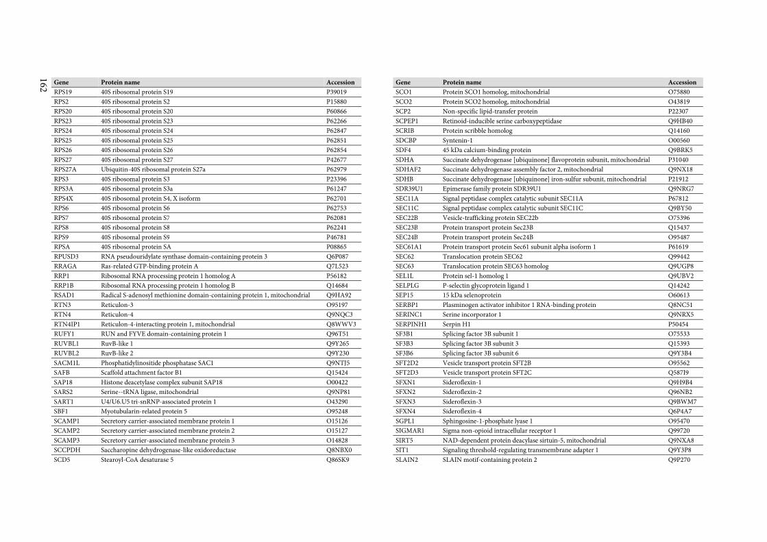
162
Gene Protein name Accession
RPS19 40S ribosomal protein S19 P39019
RPS2 40S ribosomal protein S2 P15880
RPS20 40S ribosomal protein S20 P60866
RPS23 40S ribosomal protein S23 P62266
RPS24 40S ribosomal protein S24 P62847
RPS25 40S ribosomal protein S25 P62851
RPS26 40S ribosomal protein S26 P62854
RPS27 40S ribosomal protein S27 P42677
RPS27A Ubiquitin-40S ribosomal protein S27a P62979
RPS3 40S ribosomal protein S3 P23396
RPS3A 40S ribosomal protein S3a P61247
RPS4X 40S ribosomal protein S4, X isoform P62701
RPS6 40S ribosomal protein S6 P62753
RPS7 40S ribosomal protein S7 P62081
RPS8 40S ribosomal protein S8 P62241
RPS9 40S ribosomal protein S9 P46781
RPSA 40S ribosomal protein SA P08865
RPUSD3 RNA pseudouridylate synthase domain-containing protein 3 Q6P087
RRAGA Ras-related GTP-binding protein A Q7L523
RRP1 Ribosomal RNA processing protein 1 homolog A P56182
RRP1B Ribosomal RNA processing protein 1 homolog B Q14684
RSAD1 Radical S-adenosyl methionine domain-containing protein 1, mitochondrial Q9HA92
RTN3 Reticulon-3 O95197
RTN4 Reticulon-4 Q9NQC3
RTN4IP1 Reticulon-4-interacting protein 1, mitochondrial Q8WWV3
RUFY1 RUN and FYVE domain-containing protein 1 Q96T51
RUVBL1 RuvB-like 1 Q9Y265
RUVBL2 RuvB-like 2 Q9Y230
SACM1L Phosphatidylinositide phosphatase SAC1 Q9NTJ5
SAFB Scaffold attachment factor B1 Q15424
SAP18 Histone deacetylase complex subunit SAP18 O00422
SARS2 Serine--tRNA ligase, mitochondrial Q9NP81
SART1 U4/U6.U5 tri-snRNP-associated protein 1 O43290
SBF1 Myotubularin-related protein 5 O95248
SCAMP1 Secretory carrier-associated membrane protein 1 O15126
SCAMP2 Secretory carrier-associated membrane protein 2 O15127
SCAMP3 Secretory carrier-associated membrane protein 3 O14828
SCCPDH Saccharopine dehydrogenase-like oxidoreductase Q8NBX0
SCD5 Stearoyl-CoA desaturase 5 Q86SK9
Gene Protein name Accession
SCO1 Protein SCO1 homolog, mitochondrial O75880
SCO2 Protein SCO2 homolog, mitochondrial O43819
SCP2 Non-specific lipid-transfer protein P22307
SCPEP1 Retinoid-inducible serine carboxypeptidase Q9HB40
SCRIB Protein scribble homolog Q14160
SDCBP Syntenin-1 O00560
SDF4 45 kDa calcium-binding protein Q9BRK5
SDHA Succinate dehydrogenase [ubiquinone] flavoprotein subunit, mitochondrial P31040
SDHAF2 Succinate dehydrogenase assembly factor 2, mitochondrial Q9NX18
SDHB Succinate dehydrogenase [ubiquinone] iron-sulfur subunit, mitochondrial P21912
SDR39U1 Epimerase family protein SDR39U1 Q9NRG7
SEC11A Signal peptidase complex catalytic subunit SEC11A P67812
SEC11C Signal peptidase complex catalytic subunit SEC11C Q9BY50
SEC22B Vesicle-trafficking protein SEC22b O75396
SEC23B Protein transport protein Sec23B Q15437
SEC24B Protein transport protein Sec24B O95487
SEC61A1 Protein transport protein Sec61 subunit alpha isoform 1 P61619
SEC62 Translocation protein SEC62 Q99442
SEC63 Translocation protein SEC63 homolog Q9UGP8
SEL1L Protein sel-1 homolog 1 Q9UBV2
SELPLG P-selectin glycoprotein ligand 1 Q14242
SEP15 15 kDa selenoprotein O60613
SERBP1 Plasminogen activator inhibitor 1 RNA-binding protein Q8NC51
SERINC1 Serine incorporator 1 Q9NRX5
SERPINH1 Serpin H1 P50454
SF3B1 Splicing factor 3B subunit 1 O75533
SF3B3 Splicing factor 3B subunit 3 Q15393
SF3B6 Splicing factor 3B subunit 6 Q9Y3B4
SFT2D2 Vesicle transport protein SFT2B O95562
SFT2D3 Vesicle transport protein SFT2C Q587I9
SFXN1 Sideroflexin-1 Q9H9B4
SFXN2 Sideroflexin-2 Q96NB2
SFXN3 Sideroflexin-3 Q9BWM7
SFXN4 Sideroflexin-4 Q6P4A7
SGPL1 Sphingosine-1-phosphate lyase 1 O95470
SIGMAR1 Sigma non-opioid intracellular receptor 1 Q99720
SIRT5 NAD-dependent protein deacylase sirtuin-5, mitochondrial Q9NXA8
SIT1 Signaling threshold-regulating transmembrane adapter 1 Q9Y3P8
SLAIN2 SLAIN motif-containing protein 2 Q9P270
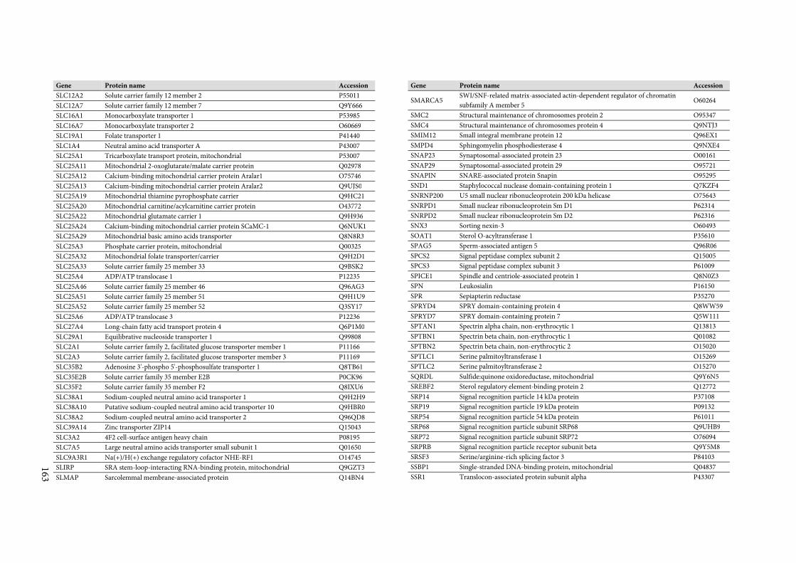
163
Gene Protein name Accession
SLC12A2 Solute carrier family 12 member 2 P55011
SLC12A7 Solute carrier family 12 member 7 Q9Y666
SLC16A1 Monocarboxylate transporter 1 P53985
SLC16A7 Monocarboxylate transporter 2 O60669
SLC19A1 Folate transporter 1 P41440
SLC1A4 Neutral amino acid transporter A P43007
SLC25A1 Tricarboxylate transport protein, mitochondrial P53007
SLC25A11 Mitochondrial 2-oxoglutarate/malate carrier protein Q02978
SLC25A12 Calcium-binding mitochondrial carrier protein Aralar1 O75746
SLC25A13 Calcium-binding mitochondrial carrier protein Aralar2 Q9UJS0
SLC25A19 Mitochondrial thiamine pyrophosphate carrier Q9HC21
SLC25A20 Mitochondrial carnitine/acylcarnitine carrier protein O43772
SLC25A22 Mitochondrial glutamate carrier 1 Q9H936
SLC25A24 Calcium-binding mitochondrial carrier protein SCaMC-1 Q6NUK1
SLC25A29 Mitochondrial basic amino acids transporter Q8N8R3
SLC25A3 Phosphate carrier protein, mitochondrial Q00325
SLC25A32 Mitochondrial folate transporter/carrier Q9H2D1
SLC25A33 Solute carrier family 25 member 33 Q9BSK2
SLC25A4 ADP/ATP translocase 1 P12235
SLC25A46 Solute carrier family 25 member 46 Q96AG3
SLC25A51 Solute carrier family 25 member 51 Q9H1U9
SLC25A52 Solute carrier family 25 member 52 Q3SY17
SLC25A6 ADP/ATP translocase 3 P12236
SLC27A4 Long-chain fatty acid transport protein 4 Q6P1M0
SLC29A1 Equilibrative nucleoside transporter 1 Q99808
SLC2A1 Solute carrier family 2, facilitated glucose transporter member 1 P11166
SLC2A3 Solute carrier family 2, facilitated glucose transporter member 3 P11169
SLC35B2 Adenosine 3'-phospho 5'-phosphosulfate transporter 1 Q8TB61
SLC35E2B Solute carrier family 35 member E2B P0CK96
SLC35F2 Solute carrier family 35 member F2 Q8IXU6
SLC38A1 Sodium-coupled neutral amino acid transporter 1 Q9H2H9
SLC38A10 Putative sodium-coupled neutral amino acid transporter 10 Q9HBR0
SLC38A2 Sodium-coupled neutral amino acid transporter 2 Q96QD8
SLC39A14 Zinc transporter ZIP14 Q15043
SLC3A2 4F2 cell-surface antigen heavy chain P08195
SLC7A5 Large neutral amino acids transporter small subunit 1 Q01650
SLC9A3R1 Na(+)/H(+) exchange regulatory cofactor NHE-RF1 O14745
SLIRP SRA stem-loop-interacting RNA-binding protein, mitochondrial Q9GZT3
SLMAP Sarcolemmal membrane-associated protein Q14BN4
Gene Protein name Accession
SMARCA5 SWI/SNF-related matrix-associated actin-dependent regulator of chromatin
subfamily A member 5 O60264
SMC2 Structural maintenance of chromosomes protein 2 O95347
SMC4 Structural maintenance of chromosomes protein 4 Q9NTJ3
SMIM12 Small integral membrane protein 12 Q96EX1
SMPD4 Sphingomyelin phosphodiesterase 4 Q9NXE4
SNAP23 Synaptosomal-associated protein 23 O00161
SNAP29 Synaptosomal-associated protein 29 O95721
SNAPIN SNARE-associated protein Snapin O95295
SND1 Staphylococcal nuclease domain-containing protein 1 Q7KZF4
SNRNP200 U5 small nuclear ribonucleoprotein 200 kDa helicase O75643
SNRPD1 Small nuclear ribonucleoprotein Sm D1 P62314
SNRPD2 Small nuclear ribonucleoprotein Sm D2 P62316
SNX3 Sorting nexin-3 O60493
SOAT1 Sterol O-acyltransferase 1 P35610
SPAG5 Sperm-associated antigen 5 Q96R06
SPCS2 Signal peptidase complex subunit 2 Q15005
SPCS3 Signal peptidase complex subunit 3 P61009
SPICE1 Spindle and centriole-associated protein 1 Q8N0Z3
SPN Leukosialin P16150
SPR Sepiapterin reductase P35270
SPRYD4 SPRY domain-containing protein 4 Q8WW59
SPRYD7 SPRY domain-containing protein 7 Q5W111
SPTAN1 Spectrin alpha chain, non-erythrocytic 1 Q13813
SPTBN1 Spectrin beta chain, non-erythrocytic 1 Q01082
SPTBN2 Spectrin beta chain, non-erythrocytic 2 O15020
SPTLC1 Serine palmitoyltransferase 1 O15269
SPTLC2 Serine palmitoyltransferase 2 O15270
SQRDL Sulfide:quinone oxidoreductase, mitochondrial Q9Y6N5
SREBF2 Sterol regulatory element-binding protein 2 Q12772
SRP14 Signal recognition particle 14 kDa protein P37108
SRP19 Signal recognition particle 19 kDa protein P09132
SRP54 Signal recognition particle 54 kDa protein P61011
SRP68 Signal recognition particle subunit SRP68 Q9UHB9
SRP72 Signal recognition particle subunit SRP72 O76094
SRPRB Signal recognition particle receptor subunit beta Q9Y5M8
SRSF3 Serine/arginine-rich splicing factor 3 P84103
SSBP1 Single-stranded DNA-binding protein, mitochondrial Q04837
SSR1 Translocon-associated protein subunit alpha P43307
164
Gene Protein name Accession
SSR3 Translocon-associated protein subunit gamma Q9UNL2
SSR4 Translocon-associated protein subunit delta P51571
SSRP1 FACT complex subunit SSRP1 Q08945
STARD3NL MLN64 N-terminal domain homolog O95772
STIM1 Stromal interaction molecule 1 Q13586
STMN1 Stathmin P16949
STOM Erythrocyte band 7 integral membrane protein P27105
STOML2 Stomatin-like protein 2, mitochondrial Q9UJZ1
STRN Striatin O43815
STT3A Dolichyl-diphosphooligosaccharide--protein glycosyltransferase subunit STT3A P46977
STT3B Dolichyl-diphosphooligosaccharide--protein glycosyltransferase subunit STT3B Q8TCJ2
STUB1 E3 ubiquitin-protein ligase CHIP Q9UNE7
STX10 Syntaxin-10 O60499
STX12 Syntaxin-12 Q86Y82
STX16 Syntaxin-16 O14662
STX18 Syntaxin-18 Q9P2W9
STX2 Syntaxin-2 P32856
STX3 Syntaxin-3 Q13277
STX4 Syntaxin-4 Q12846
STX5 Syntaxin-5 Q13190
STX6 Syntaxin-6 O43752
STX7 Syntaxin-7 O15400
STX8 Syntaxin-8 Q9UNK0
SUCLG1 Succinyl-CoA ligase [ADP/GDP-forming] subunit alpha, mitochondrial P53597
SUCLG2 Succinyl-CoA ligase [GDP-forming] subunit beta, mitochondrial Q96I99
SUGT1 Suppressor of G2 allele of SKP1 homolog Q9Y2Z0
SUMF2 Sulfatase-modifying factor 2 Q8NBJ7
SUPT16H FACT complex subunit SPT16 Q9Y5B9
SUPV3L1 ATP-dependent RNA helicase SUPV3L1, mitochondrial Q8IYB8
SURF1 Surfeit locus protein 1 Q15526
SVIP Small VCP/p97-interacting protein Q8NHG7
SYNGR2 Synaptogyrin-2 O43760
SYNJ2BP Synaptojanin-2-binding protein P57105
SYPL1 Synaptophysin-like protein 1 Q16563
TACC1 Transforming acidic coiled-coil-containing protein 1 O75410
TACO1 Translational activator of cytochrome c oxidase 1 Q9BSH4
TAP1 Antigen peptide transporter 1 Q03518
TARDBP TAR DNA-binding protein 43 Q13148
TARS2 Threonine--tRNA ligase, mitochondrial Q9BW92
Gene Protein name Accession
TAX1BP1 Tax1-binding protein 1 Q86VP1
TBC1D15 TBC1 domain family member 15 Q8TC07
TBC1D31 TBC1 domain family member 31 Q96DN5
TBCB Tubulin-folding cofactor B Q99426
TBL2 Transducin beta-like protein 2 Q9Y4P3
TBL3 Transducin beta-like protein 3 Q12788
TBRG4 Protein TBRG4 Q969Z0
TCEB1 Transcription elongation factor B polypeptide 1 Q15369
TCOF1 Treacle protein Q13428
TCP1 T-complex protein 1 subunit alpha P17987
TDRKH Tudor and KH domain-containing protein Q9Y2W6
TECR Very-long-chain enoyl-CoA reductase Q9NZ01
TEFM Transcription elongation factor, mitochondrial Q96QE5
TFAM Transcription factor A, mitochondrial Q00059
TFB1M Dimethyladenosine transferase 1, mitochondrial Q8WVM0
TFB2M Dimethyladenosine transferase 2, mitochondrial Q9H5Q4
TFRC Transferrin receptor protein 1 P02786
TGOLN2 Trans-Golgi network integral membrane protein 2 O43493
THNSL1 Threonine synthase-like 1 Q8IYQ7
TIMM10B Mitochondrial import inner membrane translocase subunit Tim10 B Q9Y5J6
TIMM13 Mitochondrial import inner membrane translocase subunit Tim13 Q9Y5L4
TIMM17A Mitochondrial import inner membrane translocase subunit Tim17-A Q99595
TIMM17B Mitochondrial import inner membrane translocase subunit Tim17-B O60830
TIMM21 Mitochondrial import inner membrane translocase subunit Tim21 Q9BVV7
TIMM23 Mitochondrial import inner membrane translocase subunit Tim23 O14925
TIMM44 Mitochondrial import inner membrane translocase subunit TIM44 O43615
TIMM50 Mitochondrial import inner membrane translocase subunit TIM50 Q3ZCQ8
TIMMDC1 Complex I assembly factor TIMMDC1, mitochondrial Q9NPL8
TM9SF1 Transmembrane 9 superfamily member 1 O15321
TM9SF2 Transmembrane 9 superfamily member 2 Q99805
TM9SF3 Transmembrane 9 superfamily member 3 Q9HD45
TM9SF4 Transmembrane 9 superfamily member 4 Q92544
TMCO1 Transmembrane and coiled-coil domain-containing protein 1 Q9UM00
TMED1 Transmembrane emp24 domain-containing protein 1 Q13445
TMED10 Transmembrane emp24 domain-containing protein 10 P49755
TMED2 Transmembrane emp24 domain-containing protein 2 Q15363
TMED4 Transmembrane emp24 domain-containing protein 4 Q7Z7H5
TMED7 Transmembrane emp24 domain-containing protein 7 Q9Y3B3
TMED9 Transmembrane emp24 domain-containing protein 9 Q9BVK6
165
Gene Protein name Accession
TMEM109 Transmembrane protein 109 Q9BVC6
TMEM11 Transmembrane protein 11, mitochondrial P17152
TMEM126A Transmembrane protein 126A Q9H061
TMEM160 Transmembrane protein 160 Q9NX00
TMEM165 Transmembrane protein 165 Q9HC07
TMEM192 Transmembrane protein 192 Q8IY95
TMEM199 Transmembrane protein 199 Q8N511
TMEM206 Transmembrane protein 206 Q9H813
TMEM245 Transmembrane protein 245 Q9H330
TMEM261 Transmembrane protein 261 Q96GE9
TMEM30A Cell cycle control protein 50A Q9NV96
TMEM33 Transmembrane protein 33 P57088
TMEM43 Transmembrane protein 43 Q9BTV4
TMEM65 Transmembrane protein 65 Q6PI78
TMEM70 Transmembrane protein 70, mitochondrial Q9BUB7
TMEM97 Transmembrane protein 97 Q5BJF2
TMF1 TATA element modulatory factor P82094
TMLHE Trimethyllysine dioxygenase, mitochondrial Q9NVH6
TMPO Lamina-associated polypeptide 2, isoform alpha P42166
TMPO Lamina-associated polypeptide 2, isoforms beta/gamma P42167
TMUB1 Transmembrane and ubiquitin-like domain-containing protein 1 Q9BVT8
TMUB2 Transmembrane and ubiquitin-like domain-containing protein 2 Q71RG4
TMX1 Thioredoxin-related transmembrane protein 1 Q9H3N1
TMX2 Thioredoxin-related transmembrane protein 2 Q9Y320
TMX4 Thioredoxin-related transmembrane protein 4 Q9H1E5
TNPO1 Transportin-1 Q92973
TOMM20 Mitochondrial import receptor subunit TOM20 homolog Q15388
TOMM22 Mitochondrial import receptor subunit TOM22 homolog Q9NS69
TOMM40 Mitochondrial import receptor subunit TOM40 homolog O96008
TOMM40L Mitochondrial import receptor subunit TOM40B Q969M1
TOP2A DNA topoisomerase 2-alpha P11388
TOP2B DNA topoisomerase 2-beta Q02880
TOR1A Torsin-1A O14656
TOR1AIP2 Torsin-1A-interacting protein 2 Q8NFQ8
TOR1B Torsin-1B O14657
TP53I11 Tumor protein p53-inducible protein 11 O14683
TPGS1 Tubulin polyglutamylase complex subunit 1 Q6ZTW0
TPM3 Tropomyosin alpha-3 chain P06753
TPM4 Tropomyosin alpha-4 chain P67936
Gene Protein name Accession
TPR Nucleoprotein TPR P12270
TRABD TraB domain-containing protein Q9H4I3
TRAF3IP3 TRAF3-interacting JNK-activating modulator Q9Y228
TRAP1 Heat shock protein 75 kDa, mitochondrial Q12931
TRAPPC3 Trafficking protein particle complex subunit 3 O43617
TRAPPC5 Trafficking protein particle complex subunit 5 Q8IUR0
TRIM28 Transcription intermediary factor 1-beta Q13263
TRIP11 Thyroid receptor-interacting protein 11 Q15643
TRIP13 Pachytene checkpoint protein 2 homolog Q15645
TRMT10C Mitochondrial ribonuclease P protein 1 Q7L0Y3
TRMT5 tRNA Q32P41
TRMT61B tRNA Q9BVS5
TRMU Mitochondrial tRNA-specific 2-thiouridylase 1 O75648
TRNT1 CCA tRNA nucleotidyltransferase 1, mitochondrial Q96Q11
TRPV2 Transient receptor potential cation channel subfamily V member 2 Q9Y5S1
TRUB1 Probable tRNA pseudouridine synthase 1 Q8WWH5
TRUB2 Probable tRNA pseudouridine synthase 2 O95900
TSFM Elongation factor Ts, mitochondrial P43897
TSPAN14 Tetraspanin-14 Q8NG11
TSPAN7 Tetraspanin-7 P41732
TST Thiosulfate sulfurtransferase Q16762
TUBA1A Tubulin alpha-1A chain Q71U36
TUBA1B Tubulin alpha-1B chain P68363
TUBA1C Tubulin alpha-1C chain Q9BQE3
TUBB Tubulin beta chain P07437
TUBG1 Tubulin gamma-1 chain P23258
TUBGCP2 Gamma-tubulin complex component 2 Q9BSJ2
TUBGCP3 Gamma-tubulin complex component 3 Q96CW5
TUBGCP6 Gamma-tubulin complex component 6 Q96RT7
TUFM Elongation factor Tu, mitochondrial P49411
TUSC3 Tumor suppressor candidate 3 Q13454
TXN Thioredoxin P10599
TXN2 Thioredoxin, mitochondrial Q99757
TXNDC12 Thioredoxin domain-containing protein 12 O95881
TXNDC5 Thioredoxin domain-containing protein 5 Q8NBS9
TYMS Thymidylate synthase P04818
UBA1 Ubiquitin-like modifier-activating enzyme 1 P22314
UBAC2 Ubiquitin-associated domain-containing protein 2 Q8NBM4
UBB Polyubiquitin-B [Cleaved into: Ubiquitin] P0CG47
166
Gene Protein name Accession
UBC Polyubiquitin-C P0CG48
UBE2G2 Ubiquitin-conjugating enzyme E2 G2 P60604
UBE2J1 Ubiquitin-conjugating enzyme E2 J1 Q9Y385
UBE2N Ubiquitin-conjugating enzyme E2 N P61088
UBTF Nucleolar transcription factor 1 P17480
UBXN4 UBX domain-containing protein 4 Q92575
UCK2 Uridine-cytidine kinase 2 Q9BZX2
UFD1L Ubiquitin fusion degradation protein 1 homolog Q92890
UFL1 E3 UFM1-protein ligase 1 O94874
UGGT1 UDP-glucose:glycoprotein glucosyltransferase 1 Q9NYU2
ULBP2 NKG2D ligand 2 Q9BZM5
UMPS Uridine 5'-monophosphate synthase P11172
UNG Uracil-DNA glycosylase P13051
UQCC1 Ubiquinol-cytochrome-c reductase complex assembly factor 1 Q9NVA1
UQCC2 Ubiquinol-cytochrome-c reductase complex assembly factor 2 Q9BRT2
UQCRB Cytochrome b-c1 complex subunit 7 P14927
UQCRC1 Cytochrome b-c1 complex subunit 1, mitochondrial P31930
UQCRC2 Cytochrome b-c1 complex subunit 2, mitochondrial P22695
UQCRFS1 Cytochrome b-c1 complex subunit Rieske, mitochondrial P47985
UQCRQ Cytochrome b-c1 complex subunit 8 O14949
URB1 Nucleolar pre-ribosomal-associated protein 1 O60287
USO1 General vesicular transport factor p115 O60763
USP20 Ubiquitin carboxyl-terminal hydrolase 20 Q9Y2K6
USP30 Ubiquitin carboxyl-terminal hydrolase 30 Q70CQ3
UTP14A U3 small nucleolar RNA-associated protein 14 homolog A Q9BVJ6
UTRN Utrophin P46939
UXS1 UDP-glucuronic acid decarboxylase 1 Q8NBZ7
VAMP3 Vesicle-associated membrane protein 3 Q15836
VAMP4 Vesicle-associated membrane protein 4 O75379
VAMP5 Vesicle-associated membrane protein 5 O95183
VAMP7 Vesicle-associated membrane protein 7 P51809
VAPA Vesicle-associated membrane protein-associated protein A Q9P0L0
VAPB Vesicle-associated membrane protein-associated protein B/C O95292
VARS2 Valine--tRNA ligase, mitochondrial Q5ST30
VAT1 Synaptic vesicle membrane protein VAT-1 homolog Q99536
VAV1 Proto-oncogene vav P15498
VCP Transitional endoplasmic reticulum ATPase P55072
VDAC1 Voltage-dependent anion-selective channel protein 1 P21796
VDAC2 Voltage-dependent anion-selective channel protein 2 P45880
Gene Protein name Accession
VDAC3 Voltage-dependent anion-selective channel protein 3 Q9Y277
VIM Vimentin P08670
VIMP Selenoprotein S Q9BQE4
VMA21 Vacuolar ATPase assembly integral membrane protein VMA21 Q3ZAQ7
VPS13C Vacuolar protein sorting-associated protein 13C Q709C8
VPS29 Vacuolar protein sorting-associated protein 29 Q9UBQ0
VPS35 Vacuolar protein sorting-associated protein 35 Q96QK1
VPS45 Vacuolar protein sorting-associated protein 45 Q9NRW7
VRK1 Serine/threonine-protein kinase VRK1 Q99986
VTI1A Vesicle transport through interaction with t-SNAREs homolog 1A Q96AJ9
VTI1B Vesicle transport through interaction with t-SNAREs homolog 1B Q9UEU0
VWA8 von Willebrand factor A domain-containing protein 8 A3KMH1
WARS2 Tryptophan--tRNA ligase, mitochondrial Q9UGM6
WDFY1 WD repeat and FYVE domain-containing protein 1 Q8IWB7
XPNPEP3 Probable Xaa-Pro aminopeptidase 3 Q9NQH7
XPO1 Exportin-1 O14980
XPR1 Xenotropic and polytropic retrovirus receptor 1 Q9UBH6
XRCC1 DNA repair protein XRCC1 P18887
XRCC5 X-ray repair cross-complementing protein 5 P13010
XRCC6 X-ray repair cross-complementing protein 6 P12956
YARS2 Tyrosine--tRNA ligase, mitochondrial Q9Y2Z4
YBEY Putative ribonuclease P58557
YME1L1 ATP-dependent zinc metalloprotease YME1L1 Q96TA2
YRDC YrdC domain-containing protein, mitochondrial Q86U90
YWHAB 14-3-3 protein beta/alpha P31946
YWHAE 14-3-3 protein epsilon P62258
YWHAH 14-3-3 protein eta Q04917
YWHAQ 14-3-3 protein theta P27348
YWHAZ 14-3-3 protein zeta/delta P63104
ZADH2 Zinc-binding alcohol dehydrogenase domain-containing protein 2 Q8N4Q0
ZAP70 Tyrosine-protein kinase ZAP-70 P43403
ZC3H11A Zinc finger CCCH domain-containing protein 11A O75152
ZC3HAV1 Zinc finger CCCH-type antiviral protein 1 Q7Z2W4
ZDHHC18 Palmitoyltransferase ZDHHC18 Q9NUE0
ZMPSTE24 CAAX prenyl protease 1 homolog O75844
ZW10 Centromere/kinetochore protein zw10 homolog O43264

167

Appendix
168
Literature
169
7 Literature
[1] Atherton, J.C., Blaser, M.J., Coadaptation of Helicobacter pylori and humans: ancient
history, modern implications. J. Clin. Invest. 2009, 119, 2475–2487.
[2] Marshall, B.J., Warren, J.R., Unidentified curved bacilli in the stomach of patients with
gastritis and peptic ulceration. Lancet 1984, 1, 1311–1315.
[3] Kuipers, E.J., Thijs, J.C., Festen, H.P., The prevalence of Helicobacter pylori in peptic
ulcer disease. Aliment. Pharmacol. Ther. 1995, 9 Suppl 2, 59–69.
[4] Wroblewski, L.E., Peek, R.M., Wilson, K.T., Helicobacter pylori and gastric cancer:
factors that modulate disease risk. Clin. Microbiol. Rev. 2010, 23, 713–739.
[5] Wolle, K., Malfertheiner, P., Treatment of Helicobacter pylori. Best Pract. Res. Clin.
Gastroenterol. 2007, 21, 315–324.
[6] Prónai, L., Schandl, L., Orosz, Z., Magyar, P., Tulassay, Z., Lower prevalence of
Helicobacter pylori infection in patients with inflammatory bowel disease but not with
chronic obstructive pulmonary disease - antibiotic use in the history does not play a
significant role. Helicobacter 2004, 9, 278–283.
[7] Arnold, I.C., Dehzad, N., Reuter, S., Martin, H., et al., Helicobacter pylori infection
prevents allergic asthma in mouse models through the induction of regulatory T cells. J.
Clin. Invest. 2011, 121, 3088–3093.
[8] Oertli, M., Sundquist, M., Hitzler, I., Engler, D.B., et al., DC-derived IL-18 drives Treg
differentiation, murine Helicobacter pylori-specific immune tolerance, and asthma
protection. J. Clin. Invest. 2012, 122, 1082–1096.
[9] Dunn, B.E., Cohen, H., Blaser, M.J., Helicobacter pylori. Clin. Microbiol. Rev. 1997, 10,
720–741.
[10] Suerbaum, S., Michetti, P., Helicobacter pylori infection. N. Engl. J. Med. 2002, 347,
1175–1186.
[11] Buck, G.E., Gourley, W.K., Lee, W.K., Subramanyam, K., et al., Relation of
Campylobacter pyloridis to gastritis and peptic ulcer. J. Infect. Dis. 1986, 153, 664–669.
[12] IARC Working Group on the Evaluation of Carcinogenic Risks to Humans,
Schistosomes, Liver Flukes and Helicobacter pylori 1994.
[13] Marshall, B.J., Virulence and pathogenicity of Helicobacter pylori. J. Gastroenterol.
Hepatol. 1991, 6, 121–124.
[14] Blaser, M.J., Atherton, J.C., Helicobacter pylori persistence: biology and disease. J. Clin.
Invest. 2004, 113, 321–333.
[15] Schreiber, S., Bücker, R., Groll, C., Azevedo-Vethacke, M., et al., Rapid loss of motility of
Helicobacter pylori in the gastric lumen in vivo. Infect. Immun. 2005, 73, 1584–1589.
[16] Atuma, C., Strugala, V., Allen, A., Holm, L., The adherent gastrointestinal mucus gel
layer: thickness and physical state in vivo. Am. J. Physiol. Gastrointest. Liver Physiol. 2001,
280, G922–929.
[17] Sycuro, L.K., Pincus, Z., Gutierrez, K.D., Biboy, J., et al., Peptidoglycan crosslinking
relaxation promotes Helicobacter pylori’s helical shape and stomach colonization. Cell
2010, 141, 822–833.
[18] Montecucco, C., Rappuoli, R., Living dangerously: how Helicobacter pylori survives in
the human stomach. Nat. Rev. Mol. Cell Biol. 2001, 2, 457–466.
Literature
170
[19] Celli, J.P., Turner, B.S., Afdhal, N.H., Keates, S., et al., Helicobacter pylori moves through
mucus by reducing mucin viscoelasticity. Proc. Natl. Acad. Sci. U. S. A. 2009, 106, 14321–
14326.
[20] Petersen, A.M., Krogfelt, K.A., Helicobacter pylori: an invading microorganism? A
review. FEMS Immunol. Med. Microbiol. 2003, 36, 117–126.
[21] Ilver, D., Arnqvist, A., Ogren, J., Frick, I.M., et al., Helicobacter pylori adhesin binding
fucosylated histo-blood group antigens revealed by retagging. Science 1998, 279, 373–377.
[22] Mahdavi, J., Sondén, B., Hurtig, M., Olfat, F.O., et al., Helicobacter pylori SabA adhesin
in persistent infection and chronic inflammation. Science 2002, 297, 573–578.
[23] Covacci, A., Censini, S., Bugnoli, M., Petracca, R., et al., Molecular characterization of
the 128-kDa immunodominant antigen of Helicobacter pylori associated with
cytotoxicity and duodenal ulcer. Proc. Natl. Acad. Sci. U. S. A. 1993, 90, 5791–5795.
[24] Odenbreit, S., Püls, J., Sedlmaier, B., Gerland, E., et al., Translocation of Helicobacter
pylori CagA into gastric epithelial cells by type IV secretion. Science 2000, 287, 1497–1500.
[25] Murata-Kamiya, N., Pathophysiological functions of the CagA oncoprotein during
infection by Helicobacter pylori. Microbes Infect. Inst. Pasteur 2011, 13, 799–807.
[26] Ohnishi, N., Yuasa, H., Tanaka, S., Sawa, H., et al., Transgenic expression of Helicobacter
pylori CagA induces gastrointestinal and hematopoietic neoplasms in mouse. Proc. Natl.
Acad. Sci. U. S. A. 2008, 105, 1003–1008.
[27] Cover, T.L., Blanke, S.R., Helicobacter pylori VacA, a paradigm for toxin
multifunctionality. Nat. Rev. Microbiol. 2005, 3, 320–332.
[28] Gewirtz, A.T., Yu, Y., Krishna, U.S., Israel, D.A., et al., Helicobacter pylori Flagellin
Evades Toll-Like Receptor 5-Mediated Innate Immunity. J. Infect. Dis. 2004, 189, 1914–
1920.
[29] Cullen, T.W., Giles, D.K., Wolf, L.N., Ecobichon, C., et al., Helicobacter pylori versus the
host: remodeling of the bacterial outer membrane is required for survival in the gastric
mucosa. PLoS Pathog. 2011, 7, e1002454.
[30] Rad, R., Ballhorn, W., Voland, P., Eisenächer, K., et al., Extracellular and intracellular
pattern recognition receptors cooperate in the recognition of Helicobacter pylori.
Gastroenterology 2009, 136, 2247–2257.
[31] Salama, N.R., Hartung, M.L., Müller, A., Life in the human stomach: persistence
strategies of the bacterial pathogen Helicobacter pylori. Nat. Rev. Microbiol. 2013,
advance online publication.
[32] Müller, A., Oertli, M., Arnold, I.C., H. pylori exploits and manipulates innate and
adaptive immune cell signaling pathways to establish persistent infection. Cell Commun.
Signal. 2011, 9, 25.
[33] Gobert, A.P., Mersey, B.D., Cheng, Y., Blumberg, D.R., et al., Cutting edge: urease
release by Helicobacter pylori stimulates macrophage inducible nitric oxide synthase. J.
Immunol. Baltim. Md 1950 2002, 168, 6002–6006.
[34] Gobert, A.P., Cheng, Y., Wang, J.-Y., Boucher, J.-L., et al., Helicobacter pylori induces
macrophage apoptosis by activation of arginase II. J. Immunol. Baltim. Md 1950 2002,
168, 4692–4700.
[35] Wang, G., Hong, Y., Olczak, A., Maier, S.E., Maier, R.J., Dual Roles of Helicobacter pylori
NapA in inducing and combating oxidative stress. Infect. Immun. 2006, 74, 6839–6846.
[36] Oertli, M., Noben, M., Engler, D.B., Semper, R.P., et al., Helicobacter pylori ?-glutamyl
transpeptidase and vacuolating cytotoxin promote gastric persistence and immune
tolerance. Proc. Natl. Acad. Sci. U. S. A. 2013, 110, 3047–3052.
Literature
171
[37] Tan, S., Noto, J.M., Romero-Gallo, J., Peek, R.M., Amieva, M.R., Helicobacter pylori
perturbs iron trafficking in the epithelium to grow on the cell surface. PLoS Pathog. 2011,
7, e1002050.
[38] Leunk, R.D., Johnson, P.T., David, B.C., Kraft, W.G., Morgan, D.R., Cytotoxic activity in
broth-culture filtrates of Campylobacter pylori. J. Med. Microbiol. 1988, 26, 93–99.
[39] Cover, T.L., Tummuru, M.K., Cao, P., Thompson, S.A., Blaser, M.J., Divergence of
genetic sequences for the vacuolating cytotoxin among Helicobacter pylori strains. J. Biol.
Chem. 1994, 269, 10566–10573.
[40] Pagliaccia, C., Bernard, M. de, Lupetti, P., Ji, X., et al., The m2 form of the Helicobacter
pylori cytotoxin has cell type-specific vacuolating activity. Proc. Natl. Acad. Sci. 1998, 95,
10212–10217.
[41] Smoot, D.T., Resau, J.H., Earlington, M.H., Simpson, M., Cover, T.L., Effects of
Helicobacter pylori vacuolating cytotoxin on primary cultures of human gastric epithelial
cells. Gut 1996, 39, 795–799.
[42] Szabò, I., Brutsche, S., Tombola, F., Moschioni, M., et al., Formation of anion-selective
channels in the cell plasma membrane by the toxin VacA of Helicobacter pylori is
required for its biological activity. EMBO J. 1999, 18, 5517–5527.
[43] Papini, E., de Bernard, M., Milia, E., Bugnoli, M., et al., Cellular vacuoles induced by
Helicobacter pylori originate from late endosomal compartments. Proc. Natl. Acad. Sci. U.
S. A. 1994, 91, 9720–9724.
[44] Li, Y., Wandinger-Ness, A., Goldenring, J.R., Cover, T.L., Clustering and Redistribution
of Late Endocytic Compartments in Response to Helicobacter pylori Vacuolating Toxin.
Mol. Biol. Cell 2004, 15, 1946–1959.
[45] Molinari, M., Galli, C., Norais, N., Telford, J.L., et al., Vacuoles Induced by Helicobacter
pylori Toxin Contain Both Late Endosomal and Lysosomal Markers. J. Biol. Chem. 1997,
272, 25339–25344.
[46] Sommi, P., Ricci, V., Fiocca, R., Necchi, V., et al., Persistence of Helicobacter pylori VacA
toxin and vacuolating potential in cultured gastric epithelial cells. Am. J. Physiol. -
Gastrointest. Liver Physiol. 1998, 275, G681–G688.
[47] Hotchin, N.A., Cover, T.L., Akhtar, N., Cell vacuolation induced by the VacA cytotoxin
of Helicobacter pylori is regulated by the Rac1 GTPase. J. Biol. Chem. 2000, 275, 14009–
14012.
[48] Suzuki, J., Ohnishi, H., Wada, A., Hirayama, T., et al., Involvement of syntaxin 7 in
human gastric epithelial cell vacuolation induced by the Helicobacter pylori-produced
cytotoxin VacA. J. Biol. Chem. 2003, 278, 25585–25590.
[49] Papini, E., Satin, B., Bucci, C., de Bernard, M., et al., The small GTP binding protein rab7
is essential for cellular vacuolation induced by Helicobacter pylori cytotoxin. EMBO J.
1997, 16, 15–24.
[50] Papini, E., Bugnoli, M., De Bernard, M., Figura, N., et al., Bafilomycin A1 inhibits
Helicobacter pylori-induced vacuolization of HeLa cells. Mol. Microbiol. 1993, 7, 323–327.
[51] Cover, T.L., Reddy, L.Y., Blaser, M.J., Effects of ATPase inhibitors on the response of
HeLa cells to Helicobacter pylori vacuolating toxin. Infect. Immun. 1993, 61, 1427–1431.
[52] Papini, E., Gottardi, E., Satin, B., de Bernard, M., et al., The vacuolar ATPase proton
pump is present on intracellular vacuoles induced by Helicobacter pylori. J. Med.
Microbiol. 1996, 45, 84–89.
Literature
172
[53] De Bernard, M., Moschioni, M., Habermann, A., Griffiths, G., Montecucco, C., Cell
vacuolization induced by Helicobacter pylori VacA cytotoxin does not depend on late
endosomal SNAREs. Cell. Microbiol. 2002, 4, 11–18.
[54] Sewald, X., Fischer, W., Haas, R., Sticky socks: Helicobacter pylori VacA takes shape.
Trends Microbiol. 2008, 16, 89–92.
[55] Cover, T.L., Blaser, M.J., Purification and characterization of the vacuolating toxin from
Helicobacter pylori. J. Biol. Chem. 1992, 267, 10570–10575.
[56] Telford, J.L., Ghiara, P., Dell’Orco, M., Comanducci, M., et al., Gene structure of the
Helicobacter pylori cytotoxin and evidence of its key role in gastric disease. J. Exp. Med.
1994, 179, 1653–1658.
[57] Lupetti, P., Heuser, J.E., Manetti, R., Massari, P., et al., Oligomeric and subunit structure
of the Helicobacter pylori vacuolating cytotoxin. J. Cell Biol. 1996, 133, 801–807.
[58] El-Bez, C., Adrian, M., Dubochet, J., Cover, T.L., High resolution structural analysis of
Helicobacter pylori VacA toxin oligomers by cryo-negative staining electron microscopy.
J. Struct. Biol. 2005, 151, 215–228.
[59] Iwamoto, H., Czajkowsky, D.M., Cover, T.L., Szabo, G., Shao, Z., VacA from
Helicobacter pylori: a hexameric chloride channel. FEBS Lett. 1999, 450, 101–104.
[60] Czajkowsky, D.M., Iwamoto, H., Cover, T.L., Shao, Z., The vacuolating toxin from
Helicobacter pylori forms hexameric pores in lipid bilayers at low pH. Proc. Natl. Acad.
Sci. U. S. A. 1999, 96, 2001–2006.
[61] Cover, T.L., Hanson, P.I., Heuser, J.E., Acid-induced Dissociation of VacA, the
Helicobacter pylori Vacuolating Cytotoxin, Reveals Its Pattern of Assembly. J. Cell Biol.
1997, 138, 759–769.
[62] Yahiro, K., Niidome, T., Kimura, M., Hatakeyama, T., et al., Activation of Helicobacter
pylori VacA Toxin by Alkaline or Acid Conditions Increases Its Binding to a 250-kDa
Receptor Protein-tyrosine Phosphatase β. J. Biol. Chem. 1999, 274, 36693–36699.
[63] Gangwer, K.A., Mushrush, D.J., Stauff, D.L., Spiller, B., et al., Crystal structure of the
Helicobacter pylori vacuolating toxin p55 domain. Proc. Natl. Acad. Sci. 2007, 104,
16293–16298.
[64] Tombola, F., Carlesso, C., Szabò, I., de Bernard, M., et al., Helicobacter pylori vacuolating
toxin forms anion-selective channels in planar lipid bilayers: possible implications for the
mechanism of cellular vacuolation. Biophys. J. 1999, 76, 1401–1409.
[65] Torres, V.J., Ivie, S.E., McClain, M.S., Cover, T.L., Functional Properties of the p33 and
p55 Domains of the Helicobacter pylori Vacuolating Cytotoxin. J. Biol. Chem. 2005, 280,
21107–21114.
[66] Reyrat, J.M., Lanzavecchia, S., Lupetti, P., de Bernard, M., et al., 3D imaging of the 58
kDa cell binding subunit of the Helicobacter pylori cytotoxin. J. Mol. Biol. 1999, 290, 459–
470.
[67] Ye, D., Willhite, D.C., Blanke, S.R., Identification of the Minimal Intracellular
Vacuolating Domain of the Helicobacter pylori Vacuolating Toxin. J. Biol. Chem. 1999,
274, 9277–9282.
[68] Torres, V.J., McClain, M.S., Cover, T.L., Interactions between p-33 and p-55 Domains of
the Helicobacter pylori Vacuolating Cytotoxin (VacA). J. Biol. Chem. 2004, 279, 2324–
2331.
[69] McClain, M.S., Cao, P., Cover, T.L., Amino-Terminal Hydrophobic Region
ofHelicobacter pylori Vacuolating Cytotoxin (VacA) Mediates Transmembrane Protein
Dimerization. Infect. Immun. 2001, 69, 1181–1184.
Literature
173
[70] Vinion-Dubiel, A.D., McClain, M.S., Czajkowsky, D.M., Iwamoto, H., et al., A
Dominant Negative Mutant of Helicobacter pyloriVacuolating Toxin (VacA) Inhibits
VacA-induced Cell Vacuolation. J. Biol. Chem. 1999, 274, 37736–37742.
[71] Chambers, M.G., Pyburn, T.M., González-Rivera, C., Collier, S.E., et al., Structural
Analysis of the Oligomeric States of Helicobacter pylori VacA Toxin. J. Mol. Biol. 2013,
425, 524–535.
[72] McClain, M.S., Iwamoto, H., Cao, P., Vinion-Dubiel, A.D., et al., Essential Role of a
GXXXG Motif for Membrane Channel Formation by Helicobacter pylori Vacuolating
Toxin. J. Biol. Chem. 2003, 278, 12101–12108.
[73] Atherton, J.C., Cao, P., Peek, R.M., Tummuru, M.K., et al., Mosaicism in vacuolating
cytotoxin alleles of Helicobacter pylori. Association of specific vacA types with cytotoxin
production and peptic ulceration. J. Biol. Chem. 1995, 270, 17771–17777.
[74] Rhead, J.L., Letley, D.P., Mohammadi, M., Hussein, N., et al., A New Helicobacter pylori
Vacuolating Cytotoxin Determinant, the Intermediate Region, Is Associated With
Gastric Cancer. Gastroenterology 2007, 133, 926–936.
[75] Chung, C., Olivares, A., Torres, E., Yilmaz, O., et al., Diversity of VacA Intermediate
Region among Helicobacter pylori Strains from Several Regions of the World. J. Clin.
Microbiol. 2010, 48, 690–696.
[76] González-Rivera, C., Algood, H.M.S., Radin, J.N., McClain, M.S., Cover, T.L., The
Intermediate Region of Helicobacter pylori VacA Is a Determinant of Toxin Potency in a
Jurkat T Cell Assay. Infect. Immun. 2012, 80, 2578–2588.
[77] Ji, X., Fernandez, T., Burroni, D., Pagliaccia, C., et al., Cell Specificity of Helicobacter
pyloriCytotoxin Is Determined by a Short Region in the Polymorphic Midregion. Infect.
Immun. 2000, 68, 3754–3757.
[78] Letley, D.P., Atherton, J.C., Natural Diversity in the N Terminus of the Mature
Vacuolating Cytotoxin of Helicobacter pylori Determines Cytotoxin Activity. J. Bacteriol.
2000, 182, 3278–3280.
[79] McClain, M.S., Cao, P., Iwamoto, H., Vinion-Dubiel, A.D., et al., A 12-Amino-Acid
Segment, Present in Type s2 but Not Type s1 Helicobacter pylori VacA Proteins,
Abolishes Cytotoxin Activity and Alters Membrane Channel Formation. J. Bacteriol.
2001, 183, 6499–6508.
[80] Moll, G., Papini, E., Colonna, R., Burroni, D., et al., Lipid interaction of the 37-kDa and
58-kDa fragments of the Helicobacter pylori cytotoxin. Eur. J. Biochem. FEBS 1995, 234,
947–952.
[81] Molinari, M., Galli, C., de Bernard, M., Norais, N., et al., The Acid Activation
ofHelicobacter pyloriToxin VacA: Structural and Membrane Binding Studies. Biochem.
Biophys. Res. Commun. 1998, 248, 334–340.
[82] Roche, N., Ilver, D., Angström, J., Barone, S., et al., Human gastric glycosphingolipids
recognized by Helicobacter pylori vacuolating cytotoxin VacA. Microbes Infect. Inst.
Pasteur 2007, 9, 605–614.
[83] Gupta, V.R., Wilson, B.A., Blanke, S.R., Sphingomyelin is important for the cellular
entry and intracellular localization of Helicobacter pylori VacA. Cell. Microbiol. 2010, 12,
1517–1533.
[84] Utt, M., Danielsson, B., Wadström, T., Helicobacter pylori vacuolating cytotoxin binding
to a putative cell surface receptor, heparan sulfate, studied by surface plasmon resonance.
FEMS Immunol. Med. Microbiol. 2001, 30, 109–113.
Literature
174
[85] Seto, K., Hayashi-Kuwabara, Y., Yoneta, T., Suda, H., Tamaki, H., Vacuolation induced
by cytotoxin from Helicobacter pylori is mediated by the EGF receptor in HeLa cells.
FEBS Lett. 1998, 431, 347–350.
[86] Yahiro, K., Wada, A., Nakayama, M., Kimura, T., et al., Protein-tyrosine Phosphatase α,
RPTPα, Is a Helicobacter pylori VacA Receptor. J. Biol. Chem. 2003, 278, 19183–19189.
[87] Barwig, I., Charakterisierung Der Bindungseigenschaften Des Helicobacter pylori
Adhäsins BabA Und Des Vakuolisierenden Cytotoxins VacA. Ludwig-Maximilians-
Universität München, 2009.
[88] Sewald, X., Gebert-Vogl, B., Prassl, S., Barwig, I., et al., Integrin Subunit CD18 Is the T-
Lymphocyte Receptor for the Helicobacter pylori Vacuolating Cytotoxin. Cell Host
Microbe 2008, 3, 20–29.
[89] Satoh, K., Hirayama, T., Takano, K., Suzuki-Inoue, K., et al., VacA, the vacuolating
cytotoxin of Helicobacter pylori, binds to multimerin 1 on human platelets. Thromb. J.
2013, 11, 23.
[90] Hennig, E.E., Godlewski, M.M., Butruk, E., Ostrowski, J., Helicobacter pylori VacA
cytotoxin interacts with fibronectin and alters HeLa cell adhesion and cytoskeletal
organization in vitro. FEMS Immunol. Med. Microbiol. 2005, 44, 143–150.
[91] Schraw, W., Li, Y., McClain, M.S., Goot, F.G. van der, Cover, T.L., Association of
Helicobacter pylori Vacuolating Toxin (VacA) with Lipid Rafts. J. Biol. Chem. 2002, 277,
34642–34650.
[92] Boquet, P., Ricci, V., Intoxication strategy of Helicobacter pylori VacA toxin. Trends
Microbiol. 2012, 20, 165–174.
[93] Nakayama, M., Hisatsune, J., Yamasaki, E., Nishi, Y., et al., Clustering of Helicobacter
pylori VacA in lipid rafts, mediated by its receptor, receptor-like protein tyrosine
phosphatase beta, is required for intoxication in AZ-521 Cells. Infect. Immun. 2006, 74,
6571–6580.
[94] Ricci, V., Galmiche, A., Doye, A., Necchi, V., et al., High Cell Sensitivity to Helicobacter
pylori VacA Toxin Depends on a GPI-anchored Protein and is not Blocked by Inhibition
of the Clathrin-mediated Pathway of Endocytosis. Mol. Biol. Cell 2000, 11, 3897–3909.
[95] Gauthier, N.C., Ricci, V., Gounon, P., Doye, A., et al., Glycosylphosphatidylinositol-
anchored Proteins and Actin Cytoskeleton Modulate Chloride Transport by Channels
Formed by the Helicobacter pylori Vacuolating Cytotoxin VacA in HeLa Cells. J. Biol.
Chem. 2004, 279, 9481–9489.
[96] Gauthier, N.C., Monzo, P., Kaddai, V., Doye, A., et al., Helicobacter pylori VacA
Cytotoxin: A Probe for a Clathrin-independent and Cdc42-dependent Pinocytic Pathway
Routed to Late Endosomes. Mol. Biol. Cell 2005, 16, 4852–4866.
[97] Sewald, X., Jiménez-Soto, L., Haas, R., PKC-dependent endocytosis of the Helicobacter
pylori vacuolating cytotoxin in primary T lymphocytes. Cell. Microbiol. 2011, 13, 482–496.
[98] Garner, J.A., Cover, T.L., Binding and internalization of the Helicobacter pylori
vacuolating cytotoxin by epithelial cells. Infect. Immun. 1996, 64, 4197–4203.
[99] McClain, M.S., Schraw, W., Ricci, V., Boquet, P., Cover, T.L., Acid activation of
Helicobacter pylori vacuolating cytotoxin (VacA) results in toxin internalization by
eukaryotic cells. Mol. Microbiol. 2000, 37, 433–442.
[100] Patel, H.K., Willhite, D.C., Patel, R.M., Ye, D., et al., Plasma Membrane Cholesterol
Modulates Cellular Vacuolation Induced by the Helicobacter pylori Vacuolating
Cytotoxin. Infect. Immun. 2002, 70, 4112–4123.
Literature
175
[101] Sabharanjak, S., Sharma, P., Parton, R.G., Mayor, S., GPI-Anchored Proteins Are
Delivered to Recycling Endosomes via a Distinct cdc42-Regulated, Clathrin-Independent
Pinocytic Pathway. Dev. Cell 2002, 2, 411–423.
[102] Gauthier, N.C., Monzo, P., Gonzalez, T., Doye, A., et al., Early endosomes associated
with dynamic F-actin structures are required for late trafficking of H. pylori VacA toxin. J.
Cell Biol. 2007, 177, 343–354.
[103] Galmiche, A., Rassow, J., Doye, A., Cagnol, S., et al., The N-terminal 34 kDa fragment of
Helicobacter pylori vacuolating cytotoxin targets mitochondria and induces cytochrome c
release. EMBO J. 2000, 19, 6361–6370.
[104] Willhite, D.C., Blanke, S.R., Helicobacter pylori vacuolating cytotoxin enters cells,
localizes to the mitochondria, and induces mitochondrial membrane permeability
changes correlated to toxin channel activity. Cell. Microbiol. 2004, 6, 143–154.
[105] Domańska, G., Motz, C., Meinecke, M., Harsman, A., et al., Helicobacter pylori VacA
Toxin/Subunit p34: Targeting of an Anion Channel to the Inner Mitochondrial
Membrane. PLoS Pathog 2010, 6, e1000878.
[106] Calore, F., Genisset, C., Casellato, A., Rossato, M., et al., Endosome-mitochondria
juxtaposition during apoptosis induced by H. pylori VacA. Cell Death Differ. 2010, 17,
1707–1716.
[107] Kim, I.-J., Blanke, S.R., Remodeling the host environment: modulation of the gastric
epithelium by the Helicobacter pylori vacuolating toxin (VacA). Front. Cell. Infect.
Microbiol. 2012, 2.
[108] Rassow, J., Meinecke, M., Helicobacter pylori VacA: a new perspective on an invasive
chloride channel. Microbes Infect. 2012.
[109] Kimura, M., Goto, S., Wada, A., Yahiro, K., et al., Vacuolating cytotoxin purified
fromHelicobacter pyloricauses mitochondrial damage in human gastric cells. Microb.
Pathog. 1999, 26, 45–52.
[110] Jain, P., Luo, Z.-Q., Blanke, S.R., Helicobacter pylori vacuolating cytotoxin A (VacA)
engages the mitochondrial fission machinery to induce host cell death. Proc. Natl. Acad.
Sci. 2011, 108, 16032–16037.
[111] Willhite, D.C., Cover, T.L., Blanke, S.R., Cellular Vacuolation and Mitochondrial
Cytochrome c Release Are Independent Outcomes of Helicobacter pylori Vacuolating
Cytotoxin Activity That Are Each Dependent on Membrane Channel Formation. J. Biol.
Chem. 2003, 278, 48204–48209.
[112] Kuck, D., Kolmerer, B., Iking-Konert, C., Krammer, P.H., et al., Vacuolating Cytotoxin
of Helicobacter pylori Induces Apoptosis in the Human Gastric Epithelial Cell Line AGS.
Infect. Immun. 2001, 69, 5080–5087.
[113] Palframan, S.L., Kwok, T., Gabriel, K., Vacuolating cytotoxin A (VacA), a key toxin for
Helicobacter pylori pathogenesis. Front. Cell. Infect. Microbiol. 2012, 2.
[114] Singh, M., Prasad, K.N., Saxena, A., Yachha, S.K., Helicobacter pylori Induces Apoptosis
of T- and B-Cell Lines and Translocates Mitochondrial Apoptosis-Inducing Factor to
Nucleus. Curr. Microbiol. 2006, 52, 254–260.
[115] Kim, J.M., Kim, J.S., Kim, N., Ko, S.H., et al., Helicobacter pylori vacuolating cytotoxin
induces apoptosis via activation of endoplasmic reticulum stress in dendritic cells. J.
Gastroenterol. Hepatol. 2014, n/a–n/a.
[116] Rassow, J., Helicobacter pylori vacuolating toxin A and apoptosis. Cell Commun. Signal.
CCS 2011, 9, 26.
Literature
176
[117] Zheng, P.-Y., Jones, N.L., Helicobacter pylori strains expressing the vacuolating cytotoxin
interrupt phagosome maturation in macrophages by recruiting and retaining TACO
(coronin 1) protein. Cell. Microbiol. 2003, 5, 25–40.
[118] Molinari, M., Salio, M., Galli, C., Norais, N., et al., Selective inhibition of Ii-dependent
antigen presentation by Helicobacter pylori toxin VacA. J. Exp. Med. 1998, 187, 135–140.
[119] Gebert, B., Fischer, W., Weiss, E., Hoffmann, R., Haas, R., Helicobacter pylori
Vacuolating Cytotoxin Inhibits T Lymphocyte Activation. Science 2003, 301, 1099–1102.
[120] Boncristiano, M., Paccani, S.R., Barone, S., Ulivieri, C., et al., The Helicobacter pylori
Vacuolating Toxin Inhibits T Cell Activation by Two Independent Mechanisms. J. Exp.
Med. 2003, 198, 1887–1897.
[121] Sundrud, M.S., Torres, V.J., Unutmaz, D., Cover, T.L., Inhibition of primary human T
cell proliferation by Helicobacter pylori vacuolating toxin (VacA) is independent of VacA
effects on IL-2 secretion. Proc. Natl. Acad. Sci. U. S. A. 2004, 101, 7727–7732.
[122] Engler, D.B., Reuter, S., van Wijck, Y., Urban, S., et al., Effective treatment of allergic
airway inflammation with Helicobacter pylori immunomodulators requires BATF3-
dependent dendritic cells and IL-10. Proc. Natl. Acad. Sci. U. S. A. 2014, 111, 11810–
11815.
[123] Censini, S., Lange, C., Xiang, Z., Crabtree, J.E., et al., cag, a pathogenicity island of
Helicobacter pylori, encodes type I-specific and disease-associated virulence factors. Proc.
Natl. Acad. Sci. 1996, 93, 14648–14653.
[124] Kwok, T., Zabler, D., Urman, S., Rohde, M., et al., Helicobacter exploits integrin for type
IV secretion and kinase activation. Nature 2007, 449, 862–866.
[125] Jiménez-Soto, L.F., Kutter, S., Sewald, X., Ertl, C., et al., Helicobacter pylori Type IV
Secretion Apparatus Exploits β1 Integrin in a Novel RGD-Independent Manner. PLoS
Pathog. 2009, 5.
[126] Selbach, M., Moese, S., Hauck, C.R., Meyer, T.F., Backert, S., Src is the kinase of the
Helicobacter pylori CagA protein in vitro and in vivo. J. Biol. Chem. 2002, 277, 6775–6778.
[127] Tammer, I., Brandt, S., Hartig, R., König, W., Backert, S., Activation of Abl by
Helicobacter pylori: a novel kinase for CagA and crucial mediator of host cell scattering.
Gastroenterology 2007, 132, 1309–1319.
[128] Tegtmeyer, N., Wessler, S., Backert, S., Role of the cag-pathogenicity island encoded type
IV secretion system in Helicobacter pylori pathogenesis. FEBS J. 2011, 278, 1190–1202.
[129] Blaser, M.J., Perez-Perez, G.I., Kleanthous, H., Cover, T.L., et al., Infection with
Helicobacter pylori strains possessing cagA is associated with an increased risk of
developing adenocarcinoma of the stomach. Cancer Res. 1995, 55, 2111–2115.
[130] Xiang, Z., Censini, S., Bayeli, P.F., Telford, J.L., et al., Analysis of expression of CagA and
VacA virulence factors in 43 strains of Helicobacter pylori reveals that clinical isolates can
be divided into two major types and that CagA is not necessary for expression of the
vacuolating cytotoxin. Infect. Immun. 1995, 63, 94–98.
[131] Peek, R.M., Crabtree, J.E., Helicobacter infection and gastric neoplasia. J. Pathol. 2006,
208, 233–248.
[132] Argent, R.H., Thomas, R.J., Letley, D.P., Rittig, M.G., et al., Functional association
between the Helicobacter pylori virulence factors VacA and CagA. J. Med. Microbiol. 2008,
57, 145–150.
[133] Yokoyama, K., Higashi, H., Ishikawa, S., Fujii, Y., et al., Functional antagonism between
Helicobacter pylori CagA and vacuolating toxin VacA in control of the NFAT signaling
pathway in gastric epithelial cells. Proc. Natl. Acad. Sci. U. S. A. 2005, 102, 9661–9666.
Literature
177
[134] Mimuro, H., Suzuki, T., Nagai, S., Rieder, G., et al., Helicobacter pylori dampens gut
epithelial self-renewal by inhibiting apoptosis, a bacterial strategy to enhance
colonization of the stomach. Cell Host Microbe 2007, 2, 250–263.
[135] Oldani, A., Cormont, M., Hofman, V., Chiozzi, V., et al., Helicobacter pylori counteracts
the apoptotic action of its VacA toxin by injecting the CagA protein into gastric epithelial
cells. PLoS Pathog. 2009, 5, e1000603.
[136] Akada, J.K., Aoki, H., Torigoe, Y., Kitagawa, T., et al., Helicobacter pylori CagA inhibits
endocytosis of cytotoxin VacA in host cells. Dis. Model. Mech. 2010, 3, 605–617.
[137] Wroblewski, L.E., Peek, R.M., Targeted disruption of the epithelial-barrier by
Helicobacter pylori. Cell Commun. Signal. 2011, 9, 29.
[138] Papini, E., Satin, B., Norais, N., de Bernard, M., et al., Selective increase of the
permeability of polarized epithelial cell monolayers by Helicobacter pylori vacuolating
toxin. J. Clin. Invest. 1998, 102, 813–820.
[139] Chan, E.-C., Chen, K.-T., Lin, Y.-L., Vacuolating toxin from Helicobacter pylori activates
cellular signaling and pepsinogen secretion in human gastric adenocarcinoma cells. FEBS
Lett. 1996, 399, 127–130.
[140] De Bernard, M., Cappon, A., Pancotto, L., Ruggiero, P., et al., The Helicobacter pylori
VacA cytotoxin activates RBL-2H3 cells by inducing cytosolic calcium oscillations. Cell.
Microbiol. 2005, 7, 191–198.
[141] Fracchia, K.M., Pai, C.Y., Walsh, C.M., Modulation of T Cell Metabolism and Function
through Calcium Signaling. Front. Immunol. 2013, 4.
[142] Satin, B., Norais, N., Telford, J., Rappuoli, R., et al., Effect of Helicobacter pylori
vacuolating toxin on maturation and extracellular release of procathepsin D and on
epidermal growth factor degradation. J. Biol. Chem. 1997, 272, 25022–25028.
[143] Gould, G.W., Lippincott-Schwartz, J., New roles for endosomes: from vesicular carriers
to multi-purpose platforms. Nat. Rev. Mol. Cell Biol. 2009, 10, 287–292.
[144] Miaczynska, M., Pelkmans, L., Zerial, M., Not just a sink: endosomes in control of signal
transduction. Curr. Opin. Cell Biol. 2004, 16, 400–406.
[145] Sorkin, A., Von Zastrow, M., Signal transduction and endocytosis: close encounters of
many kinds. Nat. Rev. Mol. Cell Biol. 2002, 3, 600–614.
[146] Cabantous, S., Waldo, G.S., In vivo and in vitro protein solubility assays using split GFP.
Nat. Methods 2006, 3, 845–854.
[147] Schmitt, W., Haas, R., Genetic analysis of the Helicobacter pylori vacuoiating cytotoxin:
structural similarities with the IgA protease type of exported protein. Mol. Microbiol.
1994, 12, 307–319.
[148] Fischer, W., Haas, R., The RecA Protein of Helicobacter pylori Requires a
Posttranslational Modification for Full Activity. J. Bacteriol. 2004, 186, 777–784.
[149] Odenbreit, S., Kavermann, H., Püls, J., Haas, R., CagA tyrosine phosphorylation and
interleukin-8 induction by Helicobacter pylori are independent from AlpAB, HopZ and
Bab group outer membrane proteins. Int. J. Med. Microbiol. 2002, 292, 257–266.
[150] Sambrook, J., Russell, D., Molecular Cloning. A Laboratory Manual, Cold Spring Harbor
Laboratory Press, Cold Spring Harbor, New York 2001.
[151] Urwyler, S., Finsel, I., Ragaz, C., Hilbi, H., Isolation of Legionella-containing vacuoles by
immuno-magnetic separation. Curr. Protoc. Cell Biol. 2010, Chapter 3, Unit 3.34.
[152] Hoffmann, C., Finsel, I., Otto, A., Pfaffinger, G., et al., Functional analysis of novel Rab
GTPases identified in the proteome of purified Legionella-containing vacuoles from
macrophages. Cell. Microbiol. 2014, 16, 1034–1052.
Literature
178
[153] Mashima, H., Suzuki, J., Hirayama, T., Yoshikumi, Y., et al., Involvement of vesicle-
associated membrane protein 7 in human gastric epithelial cell vacuolation induced by
Helicobacter pylori-produced VacA. Infect. Immun. 2008, 76, 2296–2303.
[154] Yamasaki, E., Wada, A., Kumatori, A., Nakagawa, I., et al., Helicobacter pylori
Vacuolating Cytotoxin Induces Activation of the Proapoptotic Proteins Bax and Bak,
Leading to Cytochrome c Release and Cell Death, Independent of Vacuolation. J. Biol.
Chem. 2006, 281, 11250–11259.
[155] Jain, U., Effects of Helicobacter pylori Vacuolating Cytotoxin A on Intracellular Calcium
Signalling in T-Lymphocytes. Ludwig-Maximilians-Universität München, 2013.
[156] Soboloff, J., Rothberg, B.S., Madesh, M., Gill, D.L., STIM proteins: dynamic calcium
signal transducers. Nat. Rev. Mol. Cell Biol. 2012, 13, 549–565.
[157] Park, C.Y., Hoover, P.J., Mullins, F.M., Bachhawat, P., et al., STIM1 Clusters and
Activates CRAC Channels via Direct Binding of a Cytosolic Domain to Orai1. Cell 2009,
136, 876–890.
[158] Yuan, J.P., Zeng, W., Dorwart, M.R., Choi, Y.-J., et al., SOAR and the polybasic STIM1
domains gate and regulate Orai channels. Nat. Cell Biol. 2009, 11, 337–343.
[159] Morbiato, L., Tombola, F., Campello, S., Del Giudice, G., et al., Vacuolation induced by
VacA toxin of Helicobacter pylori requires the intracellular accumulation of membrane
permeant bases, Cl− and water. FEBS Lett. 2001, 508, 479–483.
[160] Sewald, X., Identifizierung Des Rezeptors Und Charakterisierung Der Aufnahme Des
Vakuolisierenden Cytotoxins VacA von Helicobacter pylori in T-Zellen. Ludwig-
Maximilians-Universität München, 2008.
[161] Progida, C., Cogli, L., Piro, F., Luca, A.D., et al., Rab7b controls trafficking from
endosomes to the TGN. J. Cell Sci. 2010, 123, 1480–1491.
[162] De Brito, O.M., Scorrano, L., An intimate liaison: spatial organization of the
endoplasmic reticulum–mitochondria relationship. EMBO J. 2010, 29, 2715–2723.
[163] Rogers, L.D., Foster, L.J., The dynamic phagosomal proteome and the contribution of
the endoplasmic reticulum. Proc. Natl. Acad. Sci. U. S. A. 2007, 104, 18520–18525.
[164] Li, Q., Jagannath, C., Rao, P.K., Singh, C.R., Lostumbo, G., Analysis of phagosomal
proteomes: From latex-bead to bacterial phagosomes. PROTEOMICS 2010, 10, 4098–
4116.
[165] Wu, S.-M., Zhao, X., Zhang, Z.-L., Xie, H.-Y., et al., Quantum-Dot-Labeled DNA Probes
for Fluorescence In Situ Hybridization (FISH) in the Microorganism Escherichia coli.
ChemPhysChem 2006, 7, 1062–1067.
[166] Urwyler, S., Nyfeler, Y., Ragaz, C., Lee, H., et al., Proteome Analysis of Legionella
Vacuoles Purified by Magnetic Immunoseparation Reveals Secretory and Endosomal
GTPases. Traffic 2009, 10, 76–87.
[167] Otto, G.P., Nichols, B.J., The roles of flotillin microdomains – endocytosis and beyond. J.
Cell Sci. 2011, 124, 3933–3940.
[168] Aït-Slimane, T., Galmes, R., Trugnan, G., Maurice, M., Basolateral Internalization of
GPI-anchored Proteins Occurs via a Clathrin-independent Flotillin-dependent Pathway
in Polarized Hepatic Cells. Mol. Biol. Cell 2009, 20, 3792–3800.
[169] Saslowsky, D.E., Cho, J.A., Chinnapen, H., Massol, R.H., et al., Intoxication of zebrafish
and mammalian cells by cholera toxin depends on the flotillin/reggie proteins but not
Derlin-1 or -2. J. Clin. Invest. 2010, 120, 4399–4409.
[170] Leevers, S.J., Vanhaesebroeck, B., Waterfield, M.D., Signalling through phosphoinositide
3-kinases: the lipids take centre stage. Curr. Opin. Cell Biol. 1999, 11, 219–225.
Literature
179
[171] Prigent, M., Dubois, T., Raposo, G., Derrien, V., et al., ARF6 controls post-endocytic
recycling through its downstream exocyst complex effector. J. Cell Biol. 2003, 163, 1111–
1121.
[172] Tonachini, L., Monticone, M., Puri, C., Tacchetti, C., et al., Chondrocyte protein with a
poly-proline region (CHPPR) is a novel mitochondrial protein and promotes
mitochondrial fission. J. Cell. Physiol. 2004, 201, 470–482.
[173] Lombardi, D., Soldati, T., Riederer, M.A., Goda, Y., et al., Rab9 functions in transport
between late endosomes and the trans Golgi network. EMBO J. 1993, 12, 677–682.
[174] Hennig, E.E., Butruk, E., Ostrowski, J., RACK1 Protein Interacts with Helicobacter pylori
VacA Cytotoxin: The Yeast Two-Hybrid Approach. Biochem. Biophys. Res. Commun.
2001, 289, 103–110.
[175] De Bernard, M., Moschioni, M., Napolitani, G., Rappuoli, R., Montecucco, C., The VacA
toxin of Helicobacter pylori identifies a new intermediate filament-interacting protein.
EMBO J. 2000, 19, 48–56.
[176] Price, A., Wickner, W., Ungermann, C., Proteins needed for vesicle budding from the
Golgi complex are also required for the docking step of homotypic vacuole fusion. J. Cell
Biol. 2000, 148, 1223–1229.
[177] Elbaz-Alon, Y., Rosenfeld-Gur, E., Shinder, V., Futerman, A.H., et al., A dynamic
interface between vacuoles and mitochondria in yeast. Dev. Cell 2014, 30, 95–102.
[178] Hönscher, C., Mari, M., Auffarth, K., Bohnert, M., et al., Cellular metabolism regulates
contact sites between vacuoles and mitochondria. Dev. Cell 2014, 30, 86–94.
[179] Stroupe, C., Collins, K.M., Fratti, R.A., Wickner, W., Purification of active HOPS
complex reveals its affinities for phosphoinositides and the SNARE Vam7p. EMBO J.
2006, 25, 1579–1589.
[180] MacDiarmid, C.W., Gardner, R.C., Overexpression of the Saccharomyces cerevisiae
magnesium transport system confers resistance to aluminum ion. J. Biol. Chem. 1998,
273, 1727–1732.
[181] Giots, F., Donaton, M.C.V., Thevelein, J.M., Inorganic phosphate is sensed by specific
phosphate carriers and acts in concert with glucose as a nutrient signal for activation of
the protein kinase A pathway in the yeast Saccharomyces cerevisiae. Mol. Microbiol. 2003,
47, 1163–1181.
[182] Ron, D., Chen, C.H., Caldwell, J., Jamieson, L., et al., Cloning of an intracellular receptor
for protein kinase C: a homolog of the beta subunit of G proteins. Proc. Natl. Acad. Sci. U.
S. A. 1994, 91, 839–843.
[183] Chang, B.Y., Conroy, K.B., Machleder, E.M., Cartwright, C.A., RACK1, a Receptor for
Activated C Kinase and a Homolog of the ? Subunit of G Proteins, Inhibits Activity of Src
Tyrosine Kinases and Growth of NIH 3T3 Cells. Mol. Cell. Biol. 1998, 18, 3245–3256.
[184] Liliental, J., Chang, D.D., Rack1, a receptor for activated protein kinase C, interacts with
integrin beta subunit. J. Biol. Chem. 1998, 273, 2379–2383.
[185] Liu, H., Semino-Mora, C., Dubois, A., Mechanism of H. pylori Intracellular Entry: An in
vitro Study. Front. Cell. Infect. Microbiol. 2012, 2.
[186] Winter, J., Letley, D., Rhead, J., Atherton, J., Robinson, K., Helicobacter pylori
Membrane Vesicles Stimulate Innate Pro- and Anti-Inflammatory Responses and Induce
Apoptosis in Jurkat T Cells. Infect. Immun. 2014, 82, 1372–1381.
[187] Osman, C., Voelker, D.R., Langer, T., Making heads or tails of phospholipids in
mitochondria. J. Cell Biol. 2011, 192, 7–16.

Literature
180
[188] Derré, I., Swiss, R., Agaisse, H., The lipid transfer protein CERT interacts with the
Chlamydia inclusion protein IncD and participates to ER-Chlamydia inclusion
membrane contact sites. PLoS Pathog. 2011, 7, e1002092.
[189] Mehlitz, A., Karunakaran, K., Herweg, J.-A., Krohne, G., et al., The chlamydial organism
Simkania negevensis forms ER vacuole contact sites and inhibits ER-stress. Cell. Microbiol.
2014, n/a–n/a.
[190] Robinson, C.G., Roy, C.R., Attachment and fusion of endoplasmic reticulum with
vacuoles containing Legionella pneumophila. Cell. Microbiol. 2006, 8, 793–805.
[191] Schwartz, S.L., Cao, C., Pylypenko, O., Rak, A., Wandinger-Ness, A., Rab GTPases at a
glance. J. Cell Sci. 2007, 120, 3905–3910.
[192] Lioudyno, M.I., Kozak, J.A., Penna, A., Safrina, O., et al., Orai1 and STIM1 move to the
immunological synapse and are up-regulated during T cell activation. Proc. Natl. Acad.
Sci. U. S. A. 2008, 105, 2011–2016.
[193] Duquette, M., Nadler, M., Okuhara, D., Thompson, J., et al., Members of the
thrombospondin gene family bind stromal interaction molecule 1 and regulate calcium
channel activity. Matrix Biol. 2014, 37, 15–24.
[194] Petschnigg, J., Groisman, B., Kotlyar, M., Taipale, M., et al., The mammalian-membrane
two-hybrid assay (MaMTH) for probing membrane-protein interactions in human cells.
Nat. Methods 2014, 11, 585–592.
[195] Saelinger, C.B., Trafficking of Bacterial Toxins, CRC Press, 1989.
[196] Thieblemont, N., Wright, S.D., Transport of Bacterial Lipopolysaccharide to the Golgi
Apparatus. J. Exp. Med. 1999, 190, 523–534.
[197] Sandvig, K., van Deurs, B., Delivery into cells: lessons learned from plant and bacterial
toxins. Gene Ther. 2005, 12, 865–872.
[198] Sandvig, K., Olsnes, S., Rapid entry of nicked diphtheria toxin into cells at low pH.
Characterization of the entry process and effects of low pH on the toxin molecule. J. Biol.
Chem. 1981, 256, 9068–9076.
[199] Llorente, A., Lauvrak, S.U., van Deurs, B., Sandvig, K., Induction of direct endosome to
endoplasmic reticulum transport in Chinese hamster ovary (CHO) cells (LdlF) with a
temperature-sensitive defect in epsilon-coatomer protein (epsilon-COP). J. Biol. Chem.
2003, 278, 35850–35855.
[200] Bastiaens, P.I., Majoul, I.V., Verveer, P.J., Soling, H.D., Jovin, T.M., Imaging the
intracellular trafficking and state of the AB5 quaternary structure of cholera toxin. EMBO
J. 1996, 15, 4246–4253.
[201] Hansen, S.H., Sandvig, K., van Deurs, B., Molecules internalized by clathrin-
independent endocytosis are delivered to endosomes containing transferrin receptors. J.
Cell Biol. 1993, 123, 89–97.
[202] Schmitz, A., Herrgen, H., Winkeler, A., Herzog, V., Cholera toxin is exported from
microsomes by the Sec61p complex. J. Cell Biol. 2000, 148, 1203–1212.
[203] Friedman, J.R., Lackner, L.L., West, M., DiBenedetto, J.R., et al., ER Tubules Mark Sites
of Mitochondrial Division. Science 2011, 334, 358–362.
[204] Finsel, I., Ragaz, C., Hoffmann, C., Harrison, C.F., et al., The Legionella Effector RidL
Inhibits Retrograde Trafficking to Promote Intracellular Replication. Cell Host Microbe
2013, 14, 38–50.
[205] Holmgren, J., Lönnroth, I., Svennerholm, L., Tissue receptor for cholera exotoxin:
postulated structure from studies with GM1 ganglioside and related glycolipids. Infect.
Immun. 1973, 8, 208–214.
Literature
181
[206] Göttfert, F., Wurm, C.A., Mueller, V., Berning, S., et al., Coaligned Dual-Channel STED
Nanoscopy and Molecular Diffusion Analysis at 20 nm Resolution. Biophys. J. 2013, 105,
L01–L03.
[207] Karniely, S., Pines, O., Single translation—dual destination: mechanisms of dual protein
targeting in eukaryotes. EMBO Rep. 2005, 6, 420–425.
Literature
182
Danksagung
183
8 Danksagung
Danksagung
184
Lebenslauf
185
9 Lebenslauf


![Gold Complexes of a P-Coordinate Cyclotriphosphazene [1,2]zfn.mpdl.mpg.de/data/Reihe_B/35/ZNB-1980-35b-1286.pdfA solution of 0.15 g (CH3)2AUC1 and 0.26 g of the ligand 1 (0.57 mmol](https://img.pdfslide.org/doc/110x75/6006bc07e98d312df12dfa6b/gold-complexes-of-a-p-coordinate-cyclotriphosphazene-12zfnmpdlmpgdedatareiheb35znb-1980-35b-1286pdf.jpg)

![Integrierte Umlauf-und Dienstplanung im ÖPNV · Bündel-Methode (Kiwiel[1990], Helmberg[2000]) ... Coordinate Ascent Subgradient Volume Bundle+AS Dual Simplex Barrier [s] Steffen](https://img.pdfslide.org/doc/110x75/5d5c142388c993b6038b47c9/integrierte-umlauf-und-dienstplanung-im-oe-buendel-methode-kiwiel1990-helmberg2000.jpg)
![Reconstruction of Alpine Digital Elevation Models From ... · dimensional cylindrical coordinate system of the radar. As described by (Curlander [8], Bolter [4], Schreier [48], Kropatsch](https://img.pdfslide.org/doc/110x75/5fd511ac43102156cb037a04/reconstruction-of-alpine-digital-elevation-models-from-dimensional-cylindrical.jpg)