Embed Size (px)
Citation preview
Universitätsverlag GöttingenISSN: 1863-0197
Deutsche Gesellschaft für Geschichte und Theorie der Biologie
Annals of the History and Philosophy of Biology Volume 12 (2007)formerly Jahrbuch für Geschichte und Theorie der Biologie
DGG
TB
An
nals
of t
he H
isto
ry a
nd P
hilo
soph
y of
Bio
logy
, Vol
. 12
(200
7)
Universitätsverlag Göttingen
The name DGGTB (Deutsche Gesellschaft für Geschichte und Theorie der Biologie; German Society for the History and Philosophy of
Biology) reflects recent history as well as German traditi-on. The Society is a relatively late addition to a series of German societies of science and medicine that began with the „Deutsche Gesellschaft für Geschichte der Medizin und der Naturwissenschaften“, founded in 1910 by Leipzig University‘s Karl Sudhoff (1853-1938), who wrote: „We want to establish a ‚German‘ society in order to gather Ger-man-speaking historians together in our special disciplines so that they form the core of an international society…“. Yet Sudhoff, at this time of burgeoning academic internationalism, was „quite willing“ to accommodate the wishes of a number of founding members and „drop the word German in the title of the Society and have it merge with an international society“. The founding and naming of the Society at that time derived from a specific set of histori-cal circumstances, and the same was true some 80 years later when in 1991, in the wake of German reunification, the „Deutsche Gesellschaft für Geschichte und Theorie der Biologie“ was founded. From the start, the Society has been committed to bringing stu-dies in the history and philosophy of biology to a wide audience, using for this purpose its Jahrbuch für Geschichte und Theorie der Biologie. Parallel to the Jahrbuch, the Verhandlungen zur Geschichte und Theorie der Biologie has become the by now traditional medi-um for the publication of papers delivered at the Society‘s annual meetings. In 2005 the Jahrbuch was renamed Annals of the History and Philosophy of Biology, reflecting the Society‘s internationalist aspirations in addressing comparative biology as a subject of historical and philosophical studies.
Annals of the history and philosophy of biology, Volume 12 (2007)
Contents
Kärin Nickelsen:From Leaves to Molecules: Botany and the Development of Photosynthesis Research ..................................................................................................................1
Rudolf Hagemann: The reception of the Schimper-Mereschkowsky endosymbiont hypothesis on the origin of plastids – between 1883 and 1960 – many negative, but a few relevant positive reactions ............................................................................. 41
Vítězslav Orel: Science studies and nature of Mendel´s paradigm ............................................... 61
Manfred D. Laubichler, Jane Maienschein, Grant Yamashita: The Embryo Project and the Emergence of a Digital Infrastructure for Historyand Philosophy of Science ................................................................................... 79
Luminita Göbbel, Rüdiger Schultka, Lennart Olsson: Collecting and dissecting nature: Meckel’s Zootomical Museum at theUniversity of Halle, Germany .............................................................................. 97
Brigitte Wirth: Maria Sibylla Merian, Baltasar Scheid und Richard Bradley.Die Künstlerin und Naturforscherin, ein Kaufmann und ein Botaniker............... 115
Dieter Mollenhauer, Burkhard Büdel, Ekkehard Höxtermann, Volker Wissemann: Gregor Kraus (1841–1915) – versatile botanist from Lower Franconia.............. 155
Reimer Kornmann: Zwei lebensgeschichtliche Dokumente von Peter Kornmann (1907 – 1993).......173
Instructions for authors
Manuscripts should be submitted to the managing editor. Submissions will be peer review-ed. The preferred language is English. Articles in German should be accompanied by a short (max. 1000 words) summary in English.
Managing EditorVolker Wissemann Justus-Liebig-Universität GießenInstitut für Allgemein Botanik und PflanzenphysiologieAG Spezielle Botanik (Carl-Vogt-Haus)Heinrich-Buff-Ring 26-32 (IFZ)D-35392 GiessenGermanyE-mail: [email protected]
EditorsUwe Hoßfeld, Jena, GermanyKristian Köchy, Kassel, GermanyLennart Olsson, Jena, GermanyVolker Wissemann, Gießen, Germany
Editorial BoardPeter Bowler, Belfast, UKOlaf Breidbach, Jena, GermanyEve-Marie Engels, Tübingen, GermanyChristoph Friedrich, Marburg, GermanyNick Hopwood, Cambridge, UKDavid L. Hull, Evanston, USAThomas Junker, Frankfurt/Main, GermanyUlrich Kutschera, Kassel, GermanyJane Maienschein, Tempe, USARenato Mazzolini, Trient, Italy Peter McLaughlin, Heidelberg, GermanyJames R. Moore, Milton Keynes, UKKärin Nickelsen, Bern, SwitzerlandRonald L. Numbers, Wisconsin-Madison, USAWolf-Ernst Reif, Tübingen, Germany Hans-Jörg Rheinberger, Berlin, GermanyMarsha L. Richmond, Detroit, USARobert Richards, Chicago, USANicolas Robin, Jena, GermanyAlexander Rosenberg, Durham, USANicolaas A. Rupke, Göttingen, GermanyMichael Ruse, Tallahassee, USAHans-Konrad Schmutz, Winterthur, SwitzerlandMarcel Weber, Basel, Switzerland
Annals of the History and Philosophy of Biology, Vol. 12 (2007): 97-114
Collecting and dissecting nature: Meckel’s Zootomical Museum at the University of Halle, Germany
Luminita Göbbel, Rüdiger Schultka, Lennart Olsson
Abstract. The anatomy collections at the University of Halle, Germany rank among the largest of their kind in Europe. They started as a private collection in the hands of the famous Meckel family of physicians. Johann Friedrich Meckel the Younger (1781-1833), one of the most famous scientists in the first third of the 19th century, expanded the collection to 12.000 items, including normal (human) anatomy, pathological anatomy, and comparative anatomy. For Meckel, the primary function of his collections was to provide material for professional scientific research. In the comparative anatomy collection - formerly Zootomical Museum -, there were also around 5.000 whole specimens preserved in alcohol, including many rare species. Meckel’s Museum with its thousands of glass jars and barrels was made to resemble museums of comparative anatomy like Cuvier’s in Paris, where circulatory organs of various animals formed one series, respiratory organs another, and so on. The present comparative anatomy collection only partly reflects the original collections in the Zootomical Museum. Several types of changes have occurred: 1. Around 5.000 animals were never catalogued and have probably disappeared from the Zootomical Museum during the 19th century; 2. Around 400 valuable specimens that Meckel had prepared (dismembered skulls mounted on cardboard, organs in different stages of development etc.) were lost in the 20th century; 3. Many mounted skeletons of large mammals, such as rhinoceros and elephant, were removed to gain space, and are now available in dismounted form; 4. Around 400 new specimens have been added, and have sometimes been mixed with original Meckel specimens.
Luminita Göbbel, Rüdiger Schultka, Lennart Olsson
98
The specimens in the comparative anatomy collection are of great scientific as well as historical value. Many of them would be impossible to replace, if they were lost. Some species are extinct or rare nowadays, and many are protected. It is important to stress that the collection was created with scientific purposes in mind, not in the “Naturalienkabinett” tradition. Therefore, the collection houses several type specimens, and also reflects the progress in morphology and developmental biology in Germany in the 19th century.
Introduction
Johann Friedrich Meckel the Younger (1781-1833) played an important role for the development of comparative anatomy, developmental biology and fetal pathology in Germany. He became known as the “German Cuvier” already during his lifetime, because of his outstanding knowledge (Göbbel and Schultka 2002a, 2002b, 2003). He translated the Leçons published by George Cuvier (1769-1832) and wrote that he wanted to fill a gap in Cuvier´s work, the development of organs and organisms. He wrote that “It is therefore ... my goal to at least try to describe the metamorphoses in the entire animal system, of both single organs and the whole organism, from its first inception until its death.” 1
Meckel built upon Cuvier´s work, and developed it substantially by incorporating not only embryology but also malformations into his comparative anatomical work. He was always looking for ways to enlarge his collections, and spent lots of both energy and money for this purpose. The comparative anatomy collection kept growing into a kind of temple of dissected animal bodies, which was named the “Zootomical Museum”.2 This paper focuses on the subject of animal specimens, which formed the contencts of the Meckel’s Zootomical Museum and were also central to medical research and teaching during the nineteenth century. The aim is to readdress the question of medical research and education in Halle and shed light on the anatomical collections, which were among the most important services offered by these institutions.
Biography
Rudolf Beneke (1934) has written an extensive biography of Johann Friedrich Meckel the Younger. Here we will limit ourselves to a short summary. Johann Friedrich Meckel the Younger was born on October 17, 1781 in Halle an der Saale, Germany, into a family of famous physicians (Beneke 1934, Viebig and Schultka
1 Translated from Meckel (1810), p. V. German original: "Es war daher anfänglich meine Absicht, wenigstens einen Versuch zu machen, in der ganzen Thierreihe die Metamorphosen, welche sowohl die einzelnen Organe als der ganze Organismus von seinem ersten Entstehen an bis zu seinem Tode erleidet, darzustellen." 2 see Schultka (1999) for an overview.
Collecting and dissecting nature
99
1998). His grandfather Johann Friedrich Meckel the Older (1724-1774), a student of Albrecht von Haller (1708-1777), was Professor for Anatomy, Botany and Obstetrics in Berlin, and a member of the Royal Academy. Meckel the Younger´s father, Philipp Friedrich Theodor Meckel (1755-1803), studied in Göttingen and Straßburg. From 1779 until his death he worked as a professor of Anatomy, Physiology, Surgery and Obstetrics in Halle. Philipp Meckel was above all active in medical practice. He made thousands of anatomical preparations, thereby enriching the collection inherited from his father, which became important teaching material.
Johann Friedrich Meckel the Younger often took part in his father´s medical practice, and was present at post-mortem examinations already as a child. He thereby learnt medicine, and in particular anatomy, already at an early age (Meckel 1806 a). In addition, the famous Meckel Collection was housed at home. In 1795 Meckel the Younger started to study at the Domgymnasium in Magdeburg. From the autumn of 1798 he studied medicine at Halle University, where he listened to the lectures in anatomy and physiology given by his father and by Johann Christian Reil (1759-1813). The last two semesters (1801-1802) of his studies Meckel spent in Göttingen, where he heard both clinical and anatomy lectures by Heinrich August Wrisberg (1739-1808). Meckel was also taught comparative anatomy by Johann Friedrich Blumenbach (1752-1840). On April 8, 1802 he obtained a doctoral degree with a thesis on heart malformations: “De cordis conditionibus abnormibus”. Thereafter he visited Karl Friedrich Kielmeyer (1765-1844) in Tübingen, and also went to Würzburg and Vienna (Beneke 1934; Jahn 2002). The death of his father on March 17, 1803 put an end to his travels for a while, but in 1804 he resumed his educational travels through Europe. Between 1804 and 1806 Meckel did research in Paris under the supervision of Cuvier in the Jardin des Plantes. Here he improved his knowledge of the natural sciences and came into contact with other contemporary scientists such as Étienne Geoffroy Saint Hilaire (1772-1844), Jean-Baptiste de Lamarck (1744-1829), Alexander von Humboldt (1769-1859) and George Louis Duvernoy (1777-1855). During his stay in Paris, Meckel planned the translation of Cuvier´s five-volume “Leçons d’Anatomie comparée” (1800-1805), which he finished a few years later (1809-1810). Meckel did not just translate Cuvier´s “Leçons” into German, he also added results from his own research as well as new references.
Meckel the Younger became a full professor (Ordinarius) of Anatomy, Surgery and Obstetrics on March 9, 1808 at Halle University, where he would remain for the rest of his life. From 1810 and onwards, his work was focused on anatomy, but in addition he also lectured from 1810 to 1815 on Zoologiam sive Historiam animalium and Historiam naturalem in the faculty of philosophy in Halle (Göbbel and Schultka 2002a, 2002b).
Meckel´s anatomical collections as well as his handbooks on pathological, human and comparative anatomy were of great importance. The “Handbook of
Luminita Göbbel, Rüdiger Schultka, Lennart Olsson
100
pathological anatomy”3 (1812-1818) became the standard work on teratology in the 19th century. In his extensive “Handbook of human anatomy”4 (1815-1820), human embryology occupied the center stage. Through his “System of comparative anatomy”5 (1821-1833), Meckel presented facts from both medicine and the natural sciences of importance for the teaching of comparative anatomy. Both through the translation of Cuvier´s “Leçons” and through his “System”, Meckel´s morphogenetic method influenced morphological research until the end of the 19th century.6
Meckel was famous for his work ethic, and remained a hard-working scientist all his life. In 1833, when he was already quite ill, Meckel asked Hinrich Martin Lichtenstein (1780-1857), professor of zoology and Director of the Museum of Zoology in Berlin, to help him with the evaluation of the large bird collection. Meckel was investigating the brain and sensory organs of different animals for the seventh part of his “System of comparative anatomy” when he died on 31 October 1833, only 52 years old (Göbbel and Schultka 2007).
Short history of the Meckel’s anatomical collections
Today the anatomical collections in the department of anatomy and cell biology in Halle contain more than 7.000 specimens. J. F. Meckel the older, while in Berlin, started the collections, which are among the largest in Europe. He made hundreds of anatomical preparations, using e.g. the mercury injection method. During his time, valuable injection and corrosion preparations were made, as well as nerve preparations. He used examples of different malformations, which he had prepared as demonstration specimens, when teaching pathological anatomy.7 Some specimens are still in the collections, including a complete Situs inversus.
Philipp, the son of J. F. Meckel the older, became a professor in Halle in 1779 and brought the collections there.8 Philipp Meckel added specimens to the collections, so that in the early 19th century it contained 3.476 specimens. This was a mix of dry preparations of e.g. nerves and blood vessels, wet preparations of organs, developmental series, injected blood vessels, corrosions, and bones (including complete human and animal skeletons). He used the collections for medical dissertations and teaching.
3 German original: “Handbuch der pathologischen Anatomie”. 4 German original: “Handbuch der menschlichen Anatomie”. 5 German original: “System der Vergleichenden Anatomie”. 6 Carl Gegenbaur (1826-1903) used Meckel´s translation of Leçons d’Anatomie comparée as well as the System as one of his main sources in all three editions (1859, 1870, 1898) of his textbook on the comparative anatomy of vertebrates. 7 See Schultka and Göbbel (2007). 8 See Schultka and Göbbel (2007).

Collecting and dissecting nature
101
J. F. Meckel the Younger, Philipp´s son, enlarged the human and pathological collections and founded the comparative anatomy collection formerly known as Zootomical Museum. In 1829 Meckel estimated that they contained 16.000 specimens.9 He used the collections to undertake most of the research that he later described in scientific papers and textbooks.10 He had a long-standing conflict with the university and ministry, which wanted the collections to be available to the public, while Meckel wanted them to be used exclusively for research.11 The collections must have anomalies and specimens of immature individuals, including embryos, since affinities could be sought between the early form of one species and the adult form of another.12 After his death (in 1833) his widow Friederika Meckel sold (in 1836) the private collection to Halle University13, but it was not until the 1840s that the specimens became part of the collections in the University´s department of anatomy. Because the localities (e.g. Rezidenzgebäude) were in such bad shape, the collections were reduced to about half their original size by e.g. fungal infections and insect pests. Eduard d’Alton (1803-1854), Meckel the Younger´s successor, tried to improve the situation, but lacked the funds to do much.14 It was only under his successor Alfred Wilhelm Volkmann (1801-1877), that things improved, and the collections were brought into some order, received labels, and were properly catalogued. Only in 1880, when todays anatomy building was opened, did the Meckel collections receive proper exhibition space - 8 large rooms with an area of around 1500 square meters.15 This happened under the new director Hermann Welcker (1822-1897), who also added his skull collection and specimens important for education to the collections.16 Valuable Meckel specimens were also renovated under his leadership.17
Catalogues and systematics of the Meckel’s Zootomical Museum
The labels we find today on the specimens, and the catalogues, were mostly produced after the Meckel era. Gustav Wilhelm Münter (1804-1870), who worked in the department during Meckel the Younger´s lifetime, produced a catalogue of the comparative anatomy collection and the Zootomical Museum between 1829
9 See Schwarz (1999), p. 83. 10 e. g. Meckel’s „Abhandlungen aus der menschlichen und vergleichenden Anatomie und Physiologie” (1806), „Beyträge zur vergleichenden Anatomie“ (1808, 1809a, 1811, 1812a). 11 See Göbbel and Schultka (2007). 12 See Meckel (1806, 1809b, 1810, 1821, 1827) 13 See Taschenberg (1894), Anlage 13, p. 60. 14 See Sturm (1997); Zwiener (2004). 15 See Sturm (1997). 16 See Göbbel and Schultka (2007); Heller (2007); Schultka and Göbbel (2007). 17 See Heller (2007).
Luminita Göbbel, Rüdiger Schultka, Lennart Olsson
102
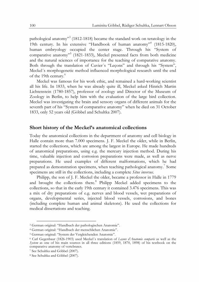
Fig. 1 Index of the catalogue of the comparative anatomy collection from 1831. Original in the Institut für Anatomie und Zellbiologie, Martin-Luther Universität, Halle-Wittenberg.
and 1831.18 Meckel negotiated with the ministry about the price of the collections, should they buy them, and needed a catalogue to support his case, so maybe Münter acted on his orders.
This catalogue is designed more as a book than a normal catalogue, with 12 chapters and an index (Fig. 1).
The first part, the “Forma generalis”, contains 60 “Positions”, mostly containing species names, all of invertebrates, some referring to more than one specimen. Position 41, for example, contains the species name “Doridium maculatum”, which refers to 5 specimens. Some positions refer to organ systems, such as position 6, which refers to the species “Vertillum cynomorium” but also to “Digestio. Generatio. Tentacula”. The second part of the catalogue - “Organa motoria” - contains 94 positions. These are vertebrates and invertebrates in which the musculature has been exposed. In his textbook “System of comparative anatomy”, Meckel describes the locomotory system in three volumes, and the catalogue continues to be very textbook-like.19 The third part contains 108 positions on nervous system specimens, and the fourth part 589 positions on the digestive system. In the fifth part there are 92 positions of circulatory system specimens, in the sixth 114 positions of respiratory system specimens, in the seventh part 18 position of specimens representing the excretory system, and in the eighth part 128 positions on genitalia. Sensory organ specimens run into 274 positions, and there is a separate chapter on “Historia evolutionis” (embryonic development) in the catalogue with 67 positions.
18 See Kapitza (2004 ), p. 66. 19 See Meckel (1824, 1825, 1828).
Collecting and dissecting nature
103
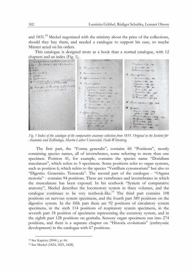
Abstract. Fig. 2 One of the specimens in the Meckel Collections, which were "artificially prepared and glued onto cardboard". Text in the accession catalogue: "5471. Sternum & extremities from Chelone mydas. N72". Original specimen in the Meckel collections in Halle.
The twelfth chapter is on the skeletal system and arranged systematically; 120 species are listed as “Sceleta piscium”, 75 species as “Sceleta amphibiorum”, 267 species as “Sceleta avium”, and 229 species as “Sceleta mammalium”. Some skeletons were mounted, some dismembered and mounted on cardboard (Fig. 2). The total number of specimens was larger than 691, because many species were represented by several specimens. This part of the catalogue has an appendix with a list of additional specimens. These were catalogued later, in 185720, and the 200 specimens received proper labels in Latin.
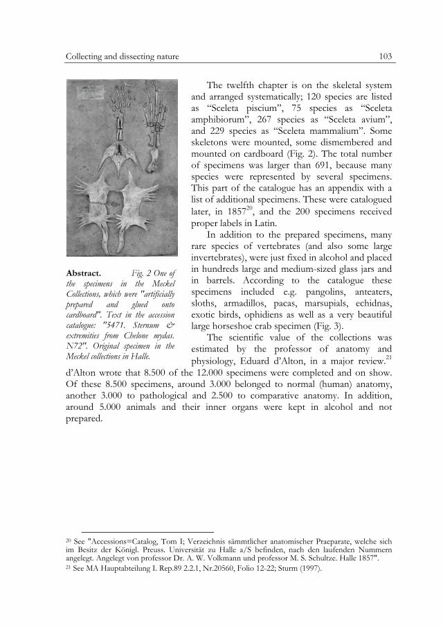
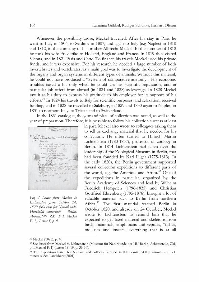
In addition to the prepared specimens, many rare species of vertebrates (and also some large invertebrates), were just fixed in alcohol and placed in hundreds large and medium-sized glass jars and in barrels. According to the catalogue these specimens included e.g. pangolins, anteaters, sloths, armadillos, pacas, marsupials, echidnas, exotic birds, ophidiens as well as a very beautiful large horseshoe crab specimen (Fig. 3).
The scientific value of the collections was estimated by the professor of anatomy and physiology, Eduard d’Alton, in a major review.21
d’Alton wrote that 8.500 of the 12.000 specimens were completed and on show. Of these 8.500 specimens, around 3.000 belonged to normal (human) anatomy, another 3.000 to pathological and 2.500 to comparative anatomy. In addition, around 5.000 animals and their inner organs were kept in alcohol and not prepared.
20 See "Accessions=Catalog, Tom I; Verzeichnis sämmtlicher anatomischer Praeparate, welche sich im Besitz der Königl. Preuss. Universität zu Halle a/S befinden, nach den laufenden Nummern angelegt. Angelegt von professor Dr. A. W. Volkmann und professor M. S. Schultze. Halle 1857". 21 See MA Hauptabteilung I. Rep.89 2.2.1, Nr.20560, Folio 12-22; Sturm (1997).
Luminita Göbbel, Rüdiger Schultka, Lennart Olsson
104
Abstract. Fig. 3 Trachypleus [Limulus] gigas. Horseshoe crab of the Family Xiphosura, sometimes called living fossils. From the Pacific Ocean. Indopazifik vor. Original specimen from the Meckel collections.
When the university had bought the collections in 1836, the ministry ordered d’Alton to make a catalogue covering all specimens in the collections, also those not yet dissected or prepared. This took several years, and a catalogue in Latin, written by Münter for d’Alton, was finished in 1841.22 However, the part on the comparative anatomy collection is an exact copy of the older work from 1829-1931. Volkmann was not satisfied with the catalogue and made a new “complete” version from 1857 to 1864, in collaboration with his Prosector Max J. S. Schultze (1825-1879).23 Old labels, some of which are still to be found on some specimens, refer to this version of the catalogue. According to the catalogue, the collection had been reduced from around 12.000 specimens to 7.228 during Eduard
d’Alton´s professorship. 3.223 of these specimens are listed in the comparative anatomy collection. The undissected animal bodies (around 5000) were never catalogued and have probably disappeared from the collections during the 19th century.
Cuvier’s “Cabinet d’anatomie comparée”, an example of “completeness” and “perfection” for Meckel’s planned museum24
When Meckel the Younger returned from Paris, he developed a grand plan. He wanted to create a collection that would overshadow Cuvier´s “Cabinet d’anatomie comparée”. In Germany, there was nothing like it at the time.
In Paris the museum was founded in 1793, and well known scientists like G. Duvernoy, E. Geoffroy Saint-Hilaire, B. de Lacépède and J. B. Lamarck in zoology, and Cuvier in comparative anatomy, were hired.25 Cuvier´s collection was catalogued in 1822, and contained 11.486 specimens. Of these, 6.231 were dry preparations and 5.255 were wet specimens.26 Many osteological specimens were human skeletons and skulls, and 1.500 vertebrate skeletons as well as 1.041 skulls
22 See „Katalog der ehemaligen Meckelschen Sammlungen. Dritte Abtheilung. Vergleichende Anatomie. wurde von Münter geschrieben und 1840 von d’Alton, auf Richtigkeit geprüft“. 23 See "Accessions = Catalog, Tom I; Verzeichnis sämmtlicher anatomischer Praeparate, welche sich im Besitz der Königl. Preuss. Universität zu Halle a/S befinden, nach den laufenden Nummern angelegt. Angelegt von professor Dr. A. W. Volkmann und professor M. S. Schultze. Halle 1857". 24 See Meckel (1809, 1810). 25 See Appel (1987). 26 In 1833 the collection had grown to 13.313 specimens. See Valenciennes (1833).
Collecting and dissecting nature
105
were also included in the collection. Furthermore, 300 dismembered and mounted skulls, several series of limb bones, and 870 different tooth specimens were in the collection. In addition, the collections contained viscera as dry and injection preparations, branchial arches from different fish species, sterna from different bird species, hyoid bones, feathers, nails, and scales. The wet specimens included 172 muscles, 216 brains, 327 eyes, 220 hearts, 915 mixed organs from different species, 80 fetuses and fetal membranes, 881 dissected molluscs, and 1.097 dissected other invertebrates. Meckel saw this collection as an example of “completeness” and “perfection”27 and organized his own collections according to Cuvier´s classification scheme. Meckel also made the same type of preparations as he had seen in Paris.
In Paris Cuvier had not only a professorship, but also relatively large sums of money and a large book collection. He also had a number of scientists and students, as well as technicians and taxidermists, at his disposal. In addition he was surrounded by colleagues at the museum, with whom he could collaborate. Cuvier mentions, in the introduction to his Règne animal, that he could use Lamarck´s as well as Geoffroy St: Hilaire´s works and expertise, and that von Lacépède´s work was important for his ideas on fish systematics, just to mention some aspects of the importance of the scientific milieu at the museum for Cuvier´s work.28
Although Meckel’s genius was on a par with Cuvier´s, Halle could definitely not compete with Paris. Meckel faced buildings in disrepair, untrustworthy members of staff, and eternal battles with colleagues.29 In spite of all these problems, Meckel were to remain in Halle for the rest of his life, and to create magnificent collections. The minister von Altenstein wrote that the collections were “richer and better organized […] than even those in the Jardin de Plants in Paris”.30 Although this might have been an exaggeration, it seems fair to say that Meckel the Younger was the most prominent German scientist in the idealistic evolutionary tradition.
The origins of specimens in the Zootomical Museum
There are several reasons why Meckel the Younger managed to create collections almost as large as Cuvier´s. One of them was that he inherited animal skeletons from his father, but also his many travels abroad and his activity as a buyer (from hunters, collectors and explorers), as well as support from colleagues, were all important factors.
27 See Meckel (1809, 1821). 28 See Cuvier (1829) Tome I. 29 See e.g. Schwarz (1999), Zwiener (2004). 30 See MA Rep.76 Vf, LitM, Nr.7, Folio 100; Schwarz (1999), p. 88.
Luminita Göbbel, Rüdiger Schultka, Lennart Olsson
106
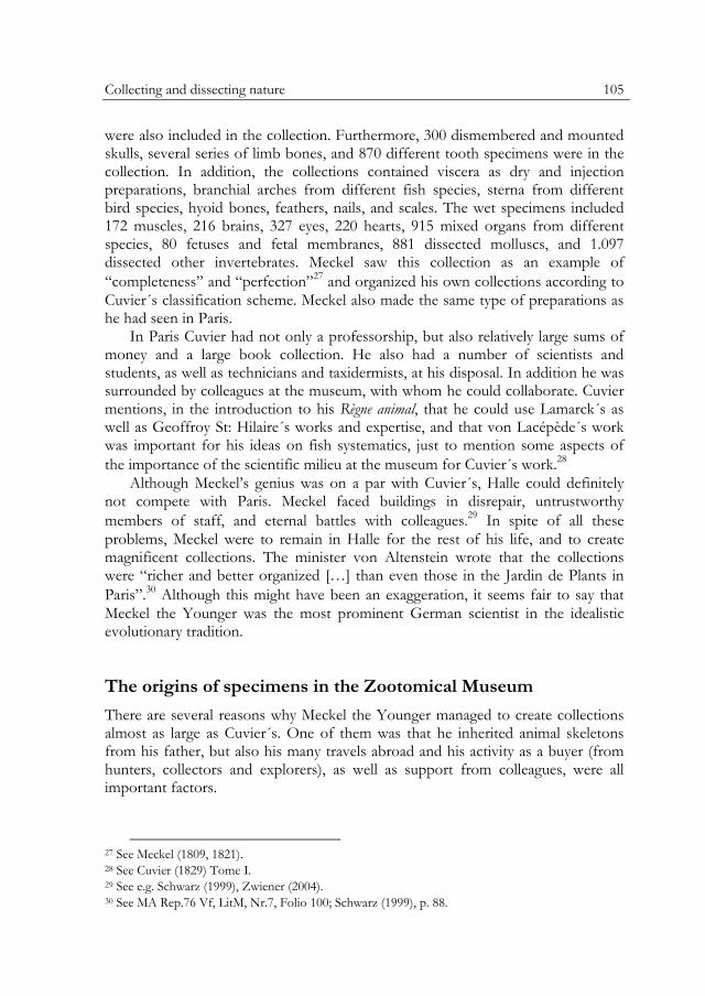
Whenever the possibility arose, Meckel travelled. After his stay in Paris he went to Italy in 1806, to Sardinia in 1807, and again to Italy (e.g Naples) in 1810 and 1812, in the company of his brother Albrecht Meckel. In the summer of 1818 he took his wife Friederike to Holland, England and France. In 1819 they visited Vienna, and in 1821 Paris and Cette. To finance his travels Meckel used his private funds, and it was expensive. For his research he needed a large number of both invertebrates and vertebrates, as a main goal was to investigate the development of the organs and organ systems in different types of animals. Without this material, he could not have produced a “System of comparative anatomy”. His economic troubles eased a bit only when he could use his scientific reputation, and in particular job offers from abroad (in 1824 and 1828) as leverage. In 1828 Meckel saw it as his duty to express his gratitude to his employer for its support of his efforts.31 In 1824 his travels to Italy for scientific purposes, and relaxation, received funding, and in 1828 he travelled to Salzburg, in 1829 and 1830 again to Naples, in 1831 to northern Italy, to Trieste and to Switzerland.
In the 1831 catalogue, the year and place of collection was noted, as well as the year of preparation. Therefore, it is possible to follow his collection success at least
in part. Meckel also wrote to colleagues asking them to sell or exchange material that he needed for his collections. He often turned to Hinrich Martin Lichtenstein (1780-1857), professor of zoology in Berlin. In 1814 Lichtenstein had taken over the leadership of the Zoological Museum in Berlin, that had been founded by Karl Illiger (1775-1813). In the early 1820s, the Berlin government supported several collection expeditions to different parts of the world, e.g. the Americas and Africa.32 One of the expeditions in particular, organized by the Berlin Academy of Sciences and lead by Wilhelm Friedrich Hemprich (1796-1825) and Christian Gottfried Ehrenberg (1795-1876), brought a lot of valuable material back to Berlin from northern Africa.33 The first material reached Berlin in October 1820, and already on 24 October, Meckel wrote to Lichtenstein to remind him that he expected to get fixed material and skeletons from birds, mammals, amphibians and reptiles, “fishes, molluscs and insects, everything that is at all
31 Meckel (1828), p. V. 32 See letter from Meckel to Lichtenstein (Museum für Naturkunde der HU Berlin, Arbeitsstelle, ZM, p I, Meckel F. 1) (Letter 18, 19, p. 36-39). 33 The expedition lasted for 6 years, and collected around 46.000 plants, 34.000 animals and 300 minerals. See Landsberg (2001).
Fig. 4 Letter from Meckel to Lichtenstein from October 24, 1820 (Museum für Naturkunde, Humboldt-Universität Berlin, Arbeitsstelle, ZM, S I, Meckel F. 1). Letter 5, p. 9.
Collecting and dissecting nature
107
possible to get”.34 Other letters were to follow, containing lists of species that Meckel needed (Fig. 4). Meckel often complained that his collections only got the leftovers when the Berlin museum and other German collections had been served. He managed to get lots of material to Halle anyway, and many species mentioned in his letters are still in the collections, including both African and American species (Fig. 5).
Meckel also got material from other colleagues. From Daniel Frederik Eschricht (1798-1863), a danish physician, physiologist and zoologist, he obtained (in May 1828) a specimen illustrating the development of Lister´s river snail Viviparus [Paludina] contectus vivipara] [Millet, 1813] and another illustrating the development of the midwife toad, Alytes [Bombinator] obstetricans [Laurenti, 1768]. Professor Christian Ludwig Nitzsch (1782-1837) gave Meckel a turtle dove skeleton, Streptopelia [Columba] turtur [turtica] [Linné, 1758] and the stomach of a spotted hyena Crocuta [Hyaena] crocuta [striatae] [Erxleben, 1777] as a dry preparation. A brown capuccin monkey, Cebus [Simia] apella [Linné, 1758] was a gift from one Dr. Ullrich.
Several mammalian skeletons, for example of the bactrian camel Camelus bactrianus [dromedarius] [Linné, 1758], the American black bear Ursus americanus [Pallas, 1870], the brown bear Ursus arctos [Linné, 1758], the Asian water buffalo Bubalus [Bos] bubalis (Linné, 1758), and the gray wolf Canis lupus [Linné, 1758], came from Stuttgart.
From Paris Mackel obtained the following skeletal preparations: The skeleton of a spider monkey, Ateles spec., the skeleton of a Brent Goose, Branta [Anas] bernicla [Linné, 1758], the skeleton of a White-tailed Eagle, Haliaeetus [Falco] albicilla [Linné, 1758], and the skull and limb bones of a Southern two-toed Sloth Choloepus didactylus [Linné, 1758].
34 See letter from Meckel to Lichtenstein (Museum für Naturkunde der HU Berlin, Arbeitsstelle, ZM, p I, Meckel F. 1) (Letter 5, p. 9).
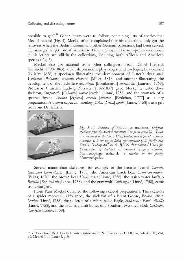
Fig. 5 A. Skeleton of Priodontes maximus. Original specimen from the Meckel collections. The giant armadillo (Tatú) is a mammal in the family Dasypodidae, and is found in South America. It is the largest living representative of its family and listed as "endangered" by the IUCN (International Union for Conservation of Nature). B. Skeleton of giant anteater, Myrmecophaga tridactyla, a member in the family Myrmecophagidae.
Luminita Göbbel, Rüdiger Schultka, Lennart Olsson
108
Two specimens are from Holland: The skeleton of a Skua, Catharacta [Procellaria] [glacialis] spec. and the skeleton of a red deer (elk), Cervus elaphus [Linné, 1758].
Among other skeletons from large mammals, we would like to mention the fin whale Balaenoptera physalus [Linné, 1758] that stranded in 1825 on the Danish coast, and a kangaroo that arrived from Vienna in 1829. Three further skeletons in the collections; an Asian Elephant, Elephas maximus [indicus] [Linné, 1758], a Hippopotamus, Hippopotamus amphibius [Linné, 1758], and a Rhinoceros, Rhinoceros unicornis [indicus] [Linné, 1758], came from London.
The English surgeons Green and Home gave Meckel two specimens of the platypus, Ornithorhynchus anatinus [paradoxus] [Shaw, 1799]. In 1826 Meckel published his results on the anatomy of the platypus and dedicated his monograph to the Royal Society in London. In this work he desribed mammary glands, a spectacular result that proved that this mysterious animal is a mammal.35 Therefore the wet specimens of mammary and poison glands, liver, lungs, skin, musculature and the skeleton still present in the collections today are some of it most valuable pieces.
The Zootomical Museum as a collection for teaching and research
Meckel used his Museum for his publications and medical dissertations (Klunker 2003; Göbbel and Schultka 2007). He dissected thousands animal specimens at the time, when he writing the “System of comparative anatomy”. The original intention was that this work was to contain all organ systems and all groups of animals. Meckel writes in the foreword to the third volume in 1828 that the “coming volumes” will appear “quickly and without delay”, because he already has the specimens needed, and has performed the necessary investigations, and even started to edit.36 Meckel had problems getting all his planned publications finished. The volumes on the nervous system and the sensory organs, as well as on the urogenital system, were never published. In 1833, right after the printing of the seventh volume, Meckel died. In the catalogue, there are specimens from many species, covering also the unpublished organ systems. Probably, a large number of specimens are from the time period when Meckel was translating Cuvier´s “Leçons d’anatomie comparée” into German. Is it clear from the many additions and notes that Meckel used both the collections in Paris and his own material to be able to understand, and thereby translate, the work better.
35 See Meckel (1826). 36 See Meckel (1825).
Collecting and dissecting nature
109
The Meckel’s Zootomical Museum today
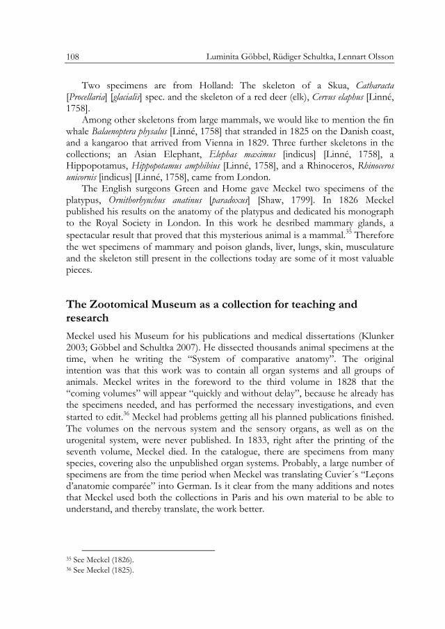
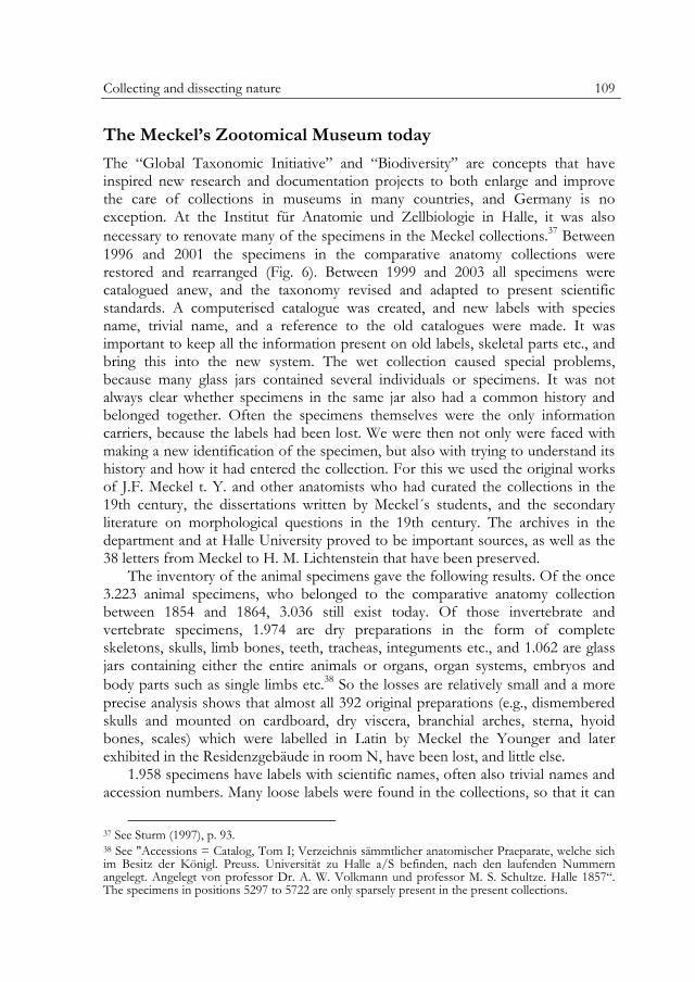
The “Global Taxonomic Initiative” and “Biodiversity” are concepts that have inspired new research and documentation projects to both enlarge and improve the care of collections in museums in many countries, and Germany is no exception. At the Institut für Anatomie und Zellbiologie in Halle, it was also necessary to renovate many of the specimens in the Meckel collections.37 Between 1996 and 2001 the specimens in the comparative anatomy collections were restored and rearranged (Fig. 6). Between 1999 and 2003 all specimens were catalogued anew, and the taxonomy revised and adapted to present scientific standards. A computerised catalogue was created, and new labels with species name, trivial name, and a reference to the old catalogues were made. It was important to keep all the information present on old labels, skeletal parts etc., and bring this into the new system. The wet collection caused special problems, because many glass jars contained several individuals or specimens. It was not always clear whether specimens in the same jar also had a common history and belonged together. Often the specimens themselves were the only information carriers, because the labels had been lost. We were then not only were faced with making a new identification of the specimen, but also with trying to understand its history and how it had entered the collection. For this we used the original works of J.F. Meckel t. Y. and other anatomists who had curated the collections in the 19th century, the dissertations written by Meckel´s students, and the secondary literature on morphological questions in the 19th century. The archives in the department and at Halle University proved to be important sources, as well as the 38 letters from Meckel to H. M. Lichtenstein that have been preserved.
The inventory of the animal specimens gave the following results. Of the once 3.223 animal specimens, who belonged to the comparative anatomy collection between 1854 and 1864, 3.036 still exist today. Of those invertebrate and vertebrate specimens, 1.974 are dry preparations in the form of complete skeletons, skulls, limb bones, teeth, tracheas, integuments etc., and 1.062 are glass jars containing either the entire animals or organs, organ systems, embryos and body parts such as single limbs etc.38 So the losses are relatively small and a more precise analysis shows that almost all 392 original preparations (e.g., dismembered skulls and mounted on cardboard, dry viscera, branchial arches, sterna, hyoid bones, scales) which were labelled in Latin by Meckel the Younger and later exhibited in the Residenzgebäude in room N, have been lost, and little else.
1.958 specimens have labels with scientific names, often also trivial names and accession numbers. Many loose labels were found in the collections, so that it can
37 See Sturm (1997), p. 93. 38 See "Accessions = Catalog, Tom I; Verzeichnis sämmtlicher anatomischer Praeparate, welche sich im Besitz der Königl. Preuss. Universität zu Halle a/S befinden, nach den laufenden Nummern angelegt. Angelegt von professor Dr. A. W. Volkmann und professor M. S. Schultze. Halle 1857“. The specimens in positions 5297 to 5722 are only sparsely present in the present collections.
Luminita Göbbel, Rüdiger Schultka, Lennart Olsson
110
be assumed that also specimens without labels really belong to the Meckel collections. The labels for the vertebrates were written by Münter, who made an enormous number of preparations.39 On these one often finds his name or the names of other taxidermists or scientists, and the year in which the specimens were prepared or restored. There is seldom any information on the source of specimens. The glass jars with invertebrates carry labels with series and number, which refer to a catalogue, whose nomenclature shows that it was made around 1900. It remains, however, unclear who the author (or authors) was. Also the invertebrates belonged to the Meckel collections, and this collection might contain valuable type material.
For most of the specimens from J. F. Meckel the Younger´s active period, we have been able to determine when they were added to the collections. Although 5/6 of the collections are from Meckel the Younger´s Zootomical Museum, it is remarkable that no specimen can safely be ascribed to the active periods of J. F.
Meckel the older or Philipp Friedrich Theodor Meckel. Of the 18 specimens that are labelled as “old” in the accession catalogue, only 3 have been identified. What exactly “old” means in this context remains unclear. We were also able to identify specimens from the post-Meckel era: 65 are from E. d’Alton´s time as director, 89 from A. W. Volkmann´s directorate, 89 can be safely ascribed to H. Welcker´s time as director, and 88 specimens are from the 20th. century. In 56 glass jars, containing invertebrates, fishes, reptiles, birds and mammals, many specimens are present that have so far not been determined down to the species level. Some glass jars contain specimens from the fuana of Suriname. They were bought in 1851 by Dr. Deutschbein. Moreover, hundreds of glass jars containing organs or organ systems have no labels whatsoever. In these cases only a rough determination at a high taxonomic level is possible, because important species characters are missing.
39 See Kapitza (2004).
Fig. 6 The comparative anatomy collection - formerly Meckel the Younger´s Zootomical Museum - in the anatomical collections of the Institut für Anatomie und Zellbiologie, Martin-Luther Universität, Halle-Wittenberg.
Collecting and dissecting nature
111
Among these specimens are probably also some, which earlier were kept in barrels or larger containers, and had not yet been prepared by Meckel.
Acknowledgments. We wish to express our sincerely thanks to Dr. H. Landsberg from the Museum für Naturkunde der Humboldt-Universität Berlin for placing the valuable letter collection (Meckel to Lichtenstein, Arbeitsstelle, ZM, p I, Meckel F, Letter 1-38) at our disposal.
References
Appel, T. (1987) The Cuvier-Geoffroy Debate. Oxford University Press, Oxford. Beneke, R. (1934) Johann Friedrich Meckel der Jüngere. Max Niemeyer, Halle. Cuvier, G. (1829) Le règne animal distribué d’après son organisation, pour servir
de base a l’histoire naturelle des animaux et d’introduction a l’anatomie comparée. T. 1, Nouvelle édition, revue et augm., Paris, chez Déterville, Libraire et chez Crochard, Libraire, Imprimerie d'Hippolyte Tilliard.
Göbbel, L., Schultka, R. (2002a) Der Anatom Johann Friedrich Meckel der Jüngere (1781-1833) und sein Beitrag zur Begründung der vergleichenden Anatomie als Wissenschaft. In: Die Entstehung biologischer Disziplinen II (Hoßfeld, U., Junker, T.; eds.). Wissenschaft und Bildung, Berlin, pp. 303-328.
Göbbel, L., Schultka, R. (2002b) Das wissenschaftliche Programm von Johann Friedrich Meckel d. J. (1781-1833) und seine Bedeutung für die Entwicklung der Wissenschaft vom Leben. Annals of Anatomy 184, pp. 519-522.
Göbbel, L., Schultka, R. (2003) Meckel the Younger and his Epistemology of Organic Form: Morphology in the pre-Gegenbaurian Age. Theory in Biosciences 122, pp. 127-141.
Göbbel, L., Schultka, R. (2007) Vielfalt der Lebensformen , reflectiert in Werk und Sammlungen von J. F. Meckel d.J. (1781-1833). In: Anatomie und Anatomische Sammlungen im 18.Jahrhundert (Schultka, R., Neumann, J.N.; eds.), LIT-Verlag, Münster and Berlin.
Heller, A. (2007) Hermann Welcker (1822-1897) - Seine anatomischen Präparate und Modelle. MD thesis, Martin Luther-Universität Halle-Wittenberg.
Jahn, I. (2002) Das „Meckel-Serres-Gesetz”, sein Ursprung und seine Beziehung zu Evolutionstheorien des 19.Jahrhunderts. Annals of Anatomy 184, pp. 509-517.
Kapitza, B. (2004) Dr. Gustav Wilhelm Münter (1804-1870) und die Meckelschen Sammlungen. Unpublished MD thesis, Martin Luther-Universität Halle-Wittenberg.
Luminita Göbbel, Rüdiger Schultka, Lennart Olsson
112
Klunker, R (2003) Bestand und Identität der human-teratologischen Präparate in den Meckel’schen Sammlungen unter besonderer Berücksichtigung des wissenschaftlichen Werkes von Johann Friedrich Meckel dem jüngerem (1781-1833). MD thesis, Martin Luther-Universität Halle-Wittenberg.
Landsberg, H. (2001) Christian Gottfried Ehrenberg (1795-1876). In: Darwin & Co. Eine Geschichte der Biologie in Portraits. (Jahn, I., Schmitt, M.; eds.) C.H. Beck, München, pp. 260-282.
Meckel, J. F. (1806) Abhandlungen aus der menschlichen und vergleichenden Anatomie und Physiologie. Halle: Hemmerde und Schwetschke 1806.
Meckel, J. F. (1808) Beyträge zur vergleichenden Anatomie. 1. Band, 1. Heft. Carl Heinrich Reclam, Leipzig.
Meckel, J. F. (1809a) Beyträge zur vergleichenden Anatomie. 1. Band, 2. Heft. Carl Heinrich Reclam, Leipzig.
Meckel, J. F. (1809b) Vorrede. In: Vorlesungen über vergleichende Anatomie (Leçons d’Anatomie comparée) von G. Cuvier. Erster Theil welcher die Organe der Bewegung enthält. Uebersetzt und mit Anmerkungen und Zusätzen vermehrt von I.F. Froriep und I.F. Meckel. Paul Gotthelf Kummer, Leipzig.
Meckel, J.F. (1810) Vorrede. In: Vorlesungen über vergleichende Anatomie (Leçons d’Anatomie comparée) von G. Cuvier. Vierter und letzter Theil, welcher die Organe des Kreislaufs, des Athmens, der Stimme und der Generation enthält. Uebersetzt und mit Anmerkungen und Zusätzen vermehrt von I.F. Meckel. Paul Gotthelf Kummer, Leipzig.
Meckel, J. F. (1811) Beyträge zur vergleichenden Anatomie. 2. Band, 1. Heft. Carl Heinrich Reclam, Leipzig.
Meckel, J. F. (1812a) Beyträge zur vergleichenden Anatomie. 2. Band, 2. Heft. Carl Heinrich Reclam, Leipzig.
Meckel, J.F. (1812b) Handbuch der pathologischen Anatomie, 1. Band, Reclam, Leipzig.
Meckel, J.F. (1821) System der vergleichenden Anatomie, 1. Teil, Allgemeine Anatomie, Renger, Halle.
Meckel, J.F. (1822) Anatomisch - physiologische Betrachtungen und Untersuchungen, Waisenhaus, Halle.
Meckel, J.F. (1824) System der vergleichenden Anatomie, Zweiter Theil. Besondere Anatomie. 2.1 Theil. Passive Organe der Bewegung. Renger, Halle.
Meckel, J.F. (1825) System der vergleichenden Anatomie, Zweiter Theil. Besondere Anatomie. 2.2 Theil. Passive Organe der Bewegung. Renger, Halle.
Collecting and dissecting nature
113
Meckel, J.F. (1826) Ornithorhynchi paradoxi descriptio anatomica, Joanne Friderico Meckelio, Fleischerum, Lipsiae.
Meckel, J.F. (1827) Ueber einige Punkte aus der Lehre von den Bildungsabweichungen, vorzüglich mit Bezug auf die beiden vorstehenden Aufsätze. Archiv für Anatomie und Physiologie 1, pp. 335-345.
Meckel, J.F. (1828) System der vergleichenden Anatomie, Dritter Theil. Besondere Anatomie. Aktive Organe der Bewegung. Renger, Halle.
Meckel, J.F. (1831) System der vergleichenden Anatomie, Fünfter Theil. Besondere Anatomie. Gefäßsystem. Renger, Halle.
Meckel, J.F. (1833) System der vergleichenden Anatomie, Sechster Theil. Vergleichende Anatomie der Athmungs- und Stimmwerkzeuge. Verlag der Buchhandlung des Weisenhauses, Halle.
Schultka, R. (1999) Die Meckelschen Sammlungen. Entstehung, Präparatebestand, Orte der Aufbewahrung, Werdegang. Ein kurzer Abriss. Zeitschrift für Heimatforschung 8, pp. 42-56.
Schultka, R., Göbbel, L. (2007) Philipp Friedrich Theodor Meckel (1755-1803) – Lebensdaten und Lebenswerk. In: Anatomie und Anatomische Sammlungen im 18.Jahrhundert (Schultka, R., Neumann, J.N.; eds.), LIT-Verlag, Münster and Berlin.
Schwarz, S. (1999) Die anatomische Privatsammlung der Anatomenfamilie Meckel unter besonderer Berücksichtigung ihres präparationstechnischen Profils. Unpublished MD thesis, Martin Luther-Universität Halle-Wittenberg.
Sturm, L-B. (1998) Die humananatomische Sammlung des Institutes für Anatomie und Zellbiologie zu Halle/Saale – ihre Geschichte und ihr Präparationsprofil unter den Direktoren Eduard d’Alton (1803-1854), Alfred Wilhelm Volkmann (1801-1877) und Hermann Welcker (1822-1897), Unpublished MD thesis, Martin Luther-Universität Halle-Wittenberg.
Taschenberg, O. (1894) Geschichte der Zoologie und der Zoologischen Sammlungen an der Universität Halle 1694-1894. Abhandlungen der Naturforschenden Gesellschaft zu Halle 20, pp. 1-177.
Valenciennes, A. (1833) Catalogue des préparations anatomiques laissées dans le Cabinet d’anatomie comparée du Muséum d’histoire naturelle, par G. Cuvier, faisant suite à la notice insérée dans le tome II des Annales du Muséum. Nouvelles Annales du Muséum d’Histoire Naturelle 2, pp. 417-508.
Viebig, M., Schultka, R. (1998) Die Anatomen Meckel – Zur Genealogie einer halleschen Ärztefamilie. Zeitschrift für Heimatforschung 5, pp. 1-32.
Luminita Göbbel, Rüdiger Schultka, Lennart Olsson
114
Zwiener, Sabine (2004) Johann Samuel Eduard d’Alton (1803-1854) leben und Wirken, Unpublished MD thesis, Martin Luther-Universität Halle-Wittenberg.
Address for correspondence:
Luminita Göbbel Institut für Anatomie und Zellbiologie Martin-Luther-Universität Halle-Wittenberg Große Steinstraße 52 D-06097 Halle (Saale) [email protected]