Embed Size (px)
Citation preview
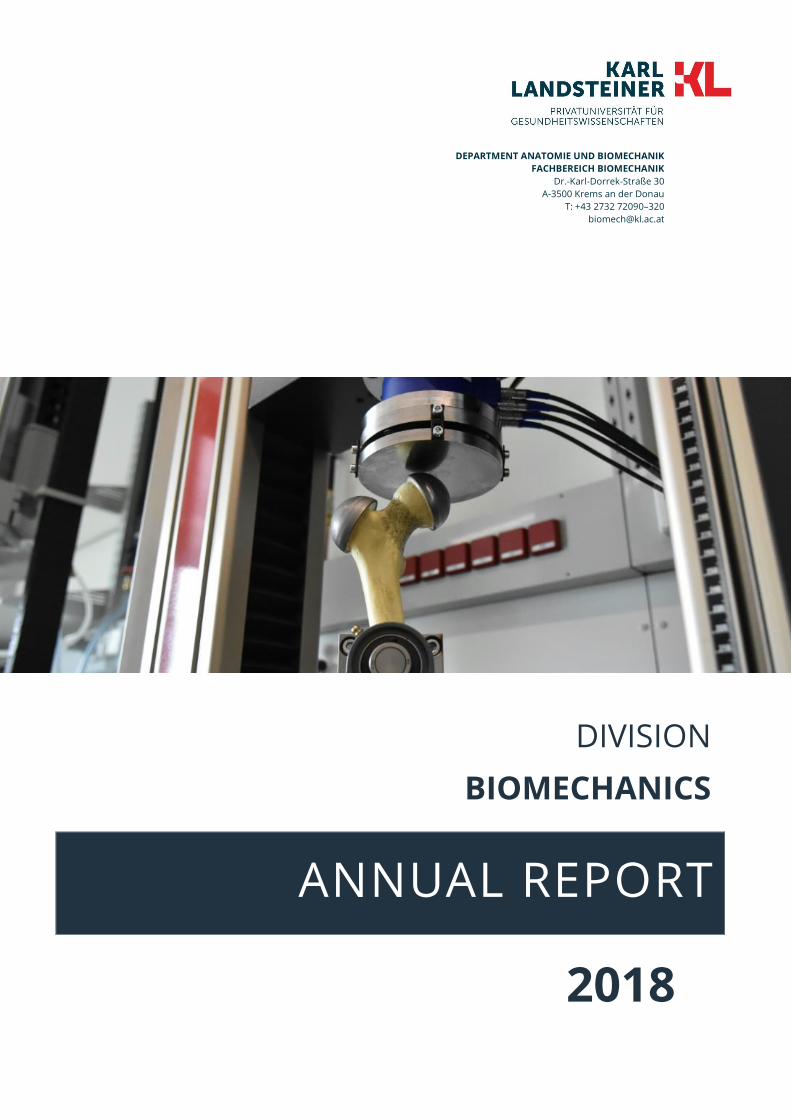
DEPARTMENT ANATOMIE UND BIOMECHANIK
FACHBEREICH BIOMECHANIK
Dr.-Karl-Dorrek-Straße 30
A-3500 Krems an der Donau
T: +43 2732 72090–320
ANNUAL REPORT 2018
DIVISION
BIOMECHANICS
CONTENT
Content
Editorial ................................................................................................................................................................................... 1
Personal and Infrastructure ................................................................................................................................................. 2
Team ................................................................................................................................................................................... 2
BM Computer System ....................................................................................................................................................... 3
BMLab (Biomechanics Laboratory) ................................................................................................................................. 3
Teaching ................................................................................................................................................................................. 4
LINE Biomedical Engineering ........................................................................................................................................... 4
NEW: BME Line Examination ............................................................................................................................................ 4
Research ................................................................................................................................................................................. 5
Journal Publications .......................................................................................................................................................... 5
Presentations ..................................................................................................................................................................... 5
Research Partners ............................................................................................................................................................. 7
Funding Organisations ..................................................................................................................................................... 7
Company Partners ............................................................................................................................................................ 7
Activities & Events .............................................................................................................................................................. 13
Acknowledgements ............................................................................................................................................................ 22
EDITORIAL
Seite 1 Annual Report 2018 DIVISION BIOMECHANICS
Editorial
Willkommen zum Jahresbericht 2018 des Fachbereichs
für BIOMECHANIK. Auch dieses Jahr ist die Gruppe
weitergewachsen. Engagierte Mitarbeiter_innen
verstärkten unser Team, beginnen das Labor mit
Leben zu füllen, stärken unser Back-Office und den IT
Bereich. Die erfolgreiche Drittmitteleinwerbung führte
zum Start zahlreicher neuer Forschungsprojekte und
vergrößerte unser Forschungsnetzwerk. In diesem
Jahr wurde das erste von der KL geführte FFG
Forschungsprojekt mit den Partnern TU-Wien, Donau
Universität Krems sowie Braincon nach drei Jahren
erfolgreich abgeschlossen.
Im Hintergrund haben wir dieses Jahr mit meiner
zweiten Forschungsgruppe an der TU-Wien am
Entwicklungsplan und der Forschungsstrategie in
enger Abstimmung gearbeitet. Die Früchte dieser
Arbeit erwarten wir in den nächsten Jahren.
Zahlreiche PR Meldungen in unterschiedlichen Medien
zeigten ein allgemeines Interesse an unserer
Forschung vor allem im Bereich 3D Druck. Daneben
gibt es einen neuen Webauftritt unter biomech.kl.ac.at
sowie bmlab.kl.ac.at.
Unser Dank gilt den Förderinstitutionen speziell dem
Land NÖ sowie unseren bestehenden und neuen
Forschungspartnern. Last but not least danke ich allen
meinen Mitarbeiter für ihren Einsatz!
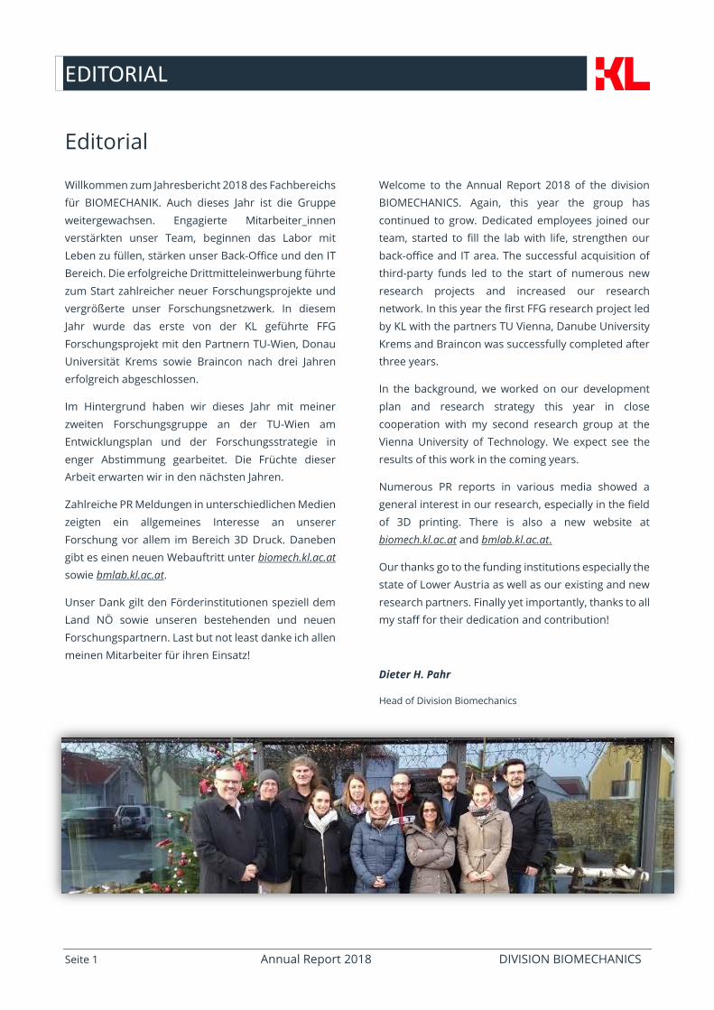
Welcome to the Annual Report 2018 of the division
BIOMECHANICS. Again, this year the group has
continued to grow. Dedicated employees joined our
team, started to fill the lab with life, strengthen our
back-office and IT area. The successful acquisition of
third-party funds led to the start of numerous new
research projects and increased our research
network. In this year the first FFG research project led
by KL with the partners TU Vienna, Danube University
Krems and Braincon was successfully completed after
three years.
In the background, we worked on our development
plan and research strategy this year in close
cooperation with my second research group at the
Vienna University of Technology. We expect see the
results of this work in the coming years.
Numerous PR reports in various media showed a
general interest in our research, especially in the field
of 3D printing. There is also a new website at
biomech.kl.ac.at and bmlab.kl.ac.at.
Our thanks go to the funding institutions especially the
state of Lower Austria as well as our existing and new
research partners. Finally yet importantly, thanks to all
my staff for their dedication and contribution!
Dieter H. Pahr
Head of Division Biomechanics
PERSONAL AND INFRASTRUCTURE
Seite 2 Annual Report 2018 DIVISION BIOMECHANICS
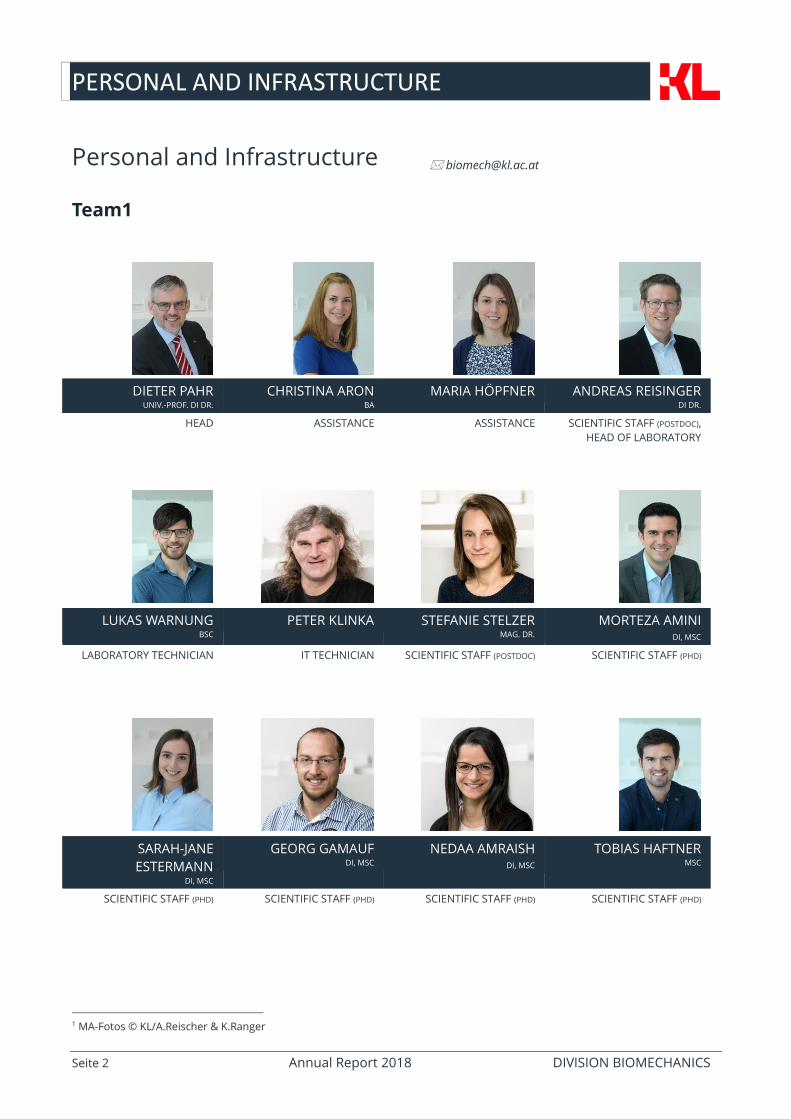
Personal and Infrastructure
Team1
DIETER PAHR UNIV.-PROF. DI DR.
CHRISTINA ARON BA
MARIA HÖPFNER ANDREAS REISINGER DI DR.
HEAD ASSISTANCE ASSISTANCE SCIENTIFIC STAFF (POSTDOC),
HEAD OF LABORATORY
LUKAS WARNUNG BSC
PETER KLINKA STEFANIE STELZER MAG. DR.
MORTEZA AMINI DI, MSC
LABORATORY TECHNICIAN IT TECHNICIAN SCIENTIFIC STAFF (POSTDOC) SCIENTIFIC STAFF (PHD)
SARAH-JANE
ESTERMANN DI, MSC
GEORG GAMAUF DI, MSC
NEDAA AMRAISH DI, MSC
TOBIAS HAFTNER MSC
SCIENTIFIC STAFF (PHD) SCIENTIFIC STAFF (PHD) SCIENTIFIC STAFF (PHD) SCIENTIFIC STAFF (PHD)
1 MA-Fotos © KL/A.Reischer & K.Ranger
PERSONAL AND INFRASTRUCTURE
Seite 3 Annual Report 2018 DIVISION BIOMECHANICS
BM Computer System
Starting with June 2018, Peter Klinka took over the system administration of our Linux network from DI Dr. Andreas
Reisinger. New workstations were bought, the fileserver was extended, and an LTO-7 backup tape was added to the
IT system. A Wiki was installed to share information of interest within the division biomechanics. The electronic
logbook of the laboratory (ELOG) was transferred to be reachable from the web.
BMLab (Biomechanics Laboratory)
The lab is part of the core facility “Campus Krems” program from Lower Austria and was further equipped with a micro
computertomography system and a CNC milling machine.
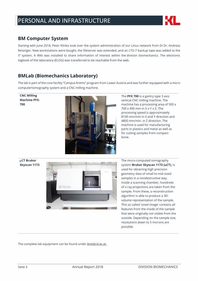
CNC Milling
Machine PFX-
700
The PFX 700 is a gantry type 3 axis
vertical CNC milling machine. The
machine has a processing area of 500 x
700 x 400 mm in X x Y x Z. The
processing speed is approximately
8100 mm/min in X and Y direction and
4800 mm/min. in Z direction. The
machine is used for manufacturing
parts in plastics and metal as well as
for cutting samples from compact
bone.
CT Bruker
Skyscan 1173
The micro-computed tomography
system Bruker Skyscan 1173 (uCT), is
used for obtaining high precision
geometry data of small to mid-sized
samples in a nondestructive way.
Inside a scanning chamber, hundreds
of x-ray projections are taken from the
sample. From these, a reconstruction
algorithm is able to produce a 3D-
volume representation of the sample.
This so called 'voxel image' contains all
features from the inside of the sample
that were originally not visible from the
outside. Depending on the sample size,
resolutions down to 5 microns are
possible.
The complete lab equipment can be found under bmlab.kl.ac.at.
TEACHING
Seite 4 Annual Report 2018 DIVISION BIOMECHANICS
Teaching
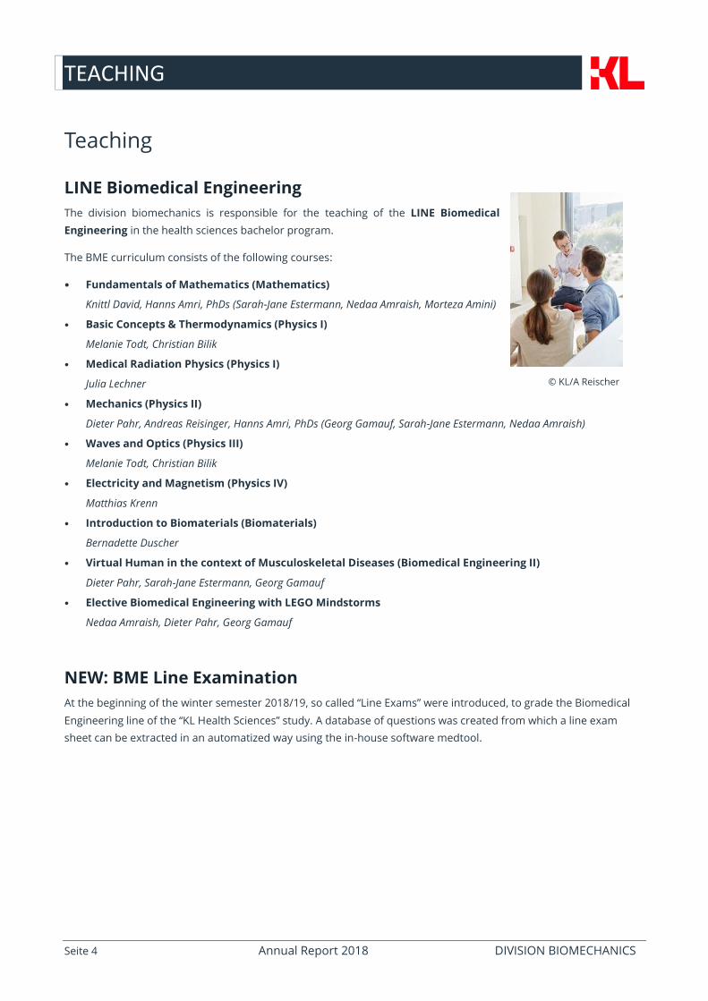
LINE Biomedical Engineering
The division biomechanics is responsible for the teaching of the LINE Biomedical
Engineering in the health sciences bachelor program.
The BME curriculum consists of the following courses:
• Fundamentals of Mathematics (Mathematics)
Knittl David, Hanns Amri, PhDs (Sarah-Jane Estermann, Nedaa Amraish, Morteza Amini)
• Basic Concepts & Thermodynamics (Physics I)
Melanie Todt, Christian Bilik
• Medical Radiation Physics (Physics I)
Julia Lechner
• Mechanics (Physics II)
Dieter Pahr, Andreas Reisinger, Hanns Amri, PhDs (Georg Gamauf, Sarah-Jane Estermann, Nedaa Amraish)
• Waves and Optics (Physics III)
Melanie Todt, Christian Bilik
• Electricity and Magnetism (Physics IV)
Matthias Krenn
• Introduction to Biomaterials (Biomaterials)
Bernadette Duscher
• Virtual Human in the context of Musculoskeletal Diseases (Biomedical Engineering II)
Dieter Pahr, Sarah-Jane Estermann, Georg Gamauf
• Elective Biomedical Engineering with LEGO Mindstorms
Nedaa Amraish, Dieter Pahr, Georg Gamauf
NEW: BME Line Examination
At the beginning of the winter semester 2018/19, so called “Line Exams” were introduced, to grade the Biomedical
Engineering line of the “KL Health Sciences” study. A database of questions was created from which a line exam
sheet can be extracted in an automatized way using the in-house software medtool.
© KL/A Reischer
RESEARCH
Seite 5 Annual Report 2018 DIVISION BIOMECHANICS
Research
Journal Publications
L. Warnung, S. Estermann, A. Reisinger:
Mechanical Properties of Fused Deposition Modeling (FDM) 3D Printing Materials;
https://www.rtejournal.de/ausgabe-15-2018/4781
M. Frank, D. Marx, V. Nedelkovski, J.T. Fischer, D. H. Pahr, P.J. Thurner:
"Dehydration of individual bovine trabeculae causes transition from ductile to quasi-brittle failure mode";
Journal of the Mechanical Behavior of Biomedical Materials, 87 (2018), 296 - 305.
E. Klintström, B. Klintström, D. H. Pahr, T.B. Brismar, ö. Smedby, R. Moreno:
"Direct Estimation of Human Trabecular Bone Stiffness Using Cone Beam Computed Tomography";
Oral Surgery, Oral Medicine, Oral Pathology and Oral Radiology, 126 (2018), 1; 72 - 82.
S.C. Lu, E.E. Vereecke, A. Synek, D. H. Pahr, T.L. Kivell:
"A novel experimental design for the measurement of metacarpal bone loading and deformation and fingertip
force";
PeerJ, 6 (2018), e5480; 19 pages.
E. Soodmand, D. Kluess, P. Varady, R. Cichon, M. Schwarze, D. Gehweiler, F. Niemeyer, D. Pahr, M. Woiczinski:
"Interlaboratory comparison of femur surface reconstruction from CT data compared to reference optical 3D scan";
Biomedical engineering online, 17 (2018), 1; 29 pages.
A. Synek, D. H. Pahr:
"Plausibility and Parameter Sensitivity of Micro-Finite Element-Based Joint Load Prediction at the Proximal Femur";
Biomechanics and Modeling in Mechanobiology, 17 (2018), 3; 843 - 852.
Z.J. Tsegai, M.M. Skinner, D. Pahr, J. Hublin, T.L. Kivell:
"Systemic patterns of trabecular bone across the human and chimpanzee skeleton";
Journal of Anatomy, 232 (2018), 4; 641 - 656.
Presentations
M. Amini, D. Pahr:
"Influence of size-scaling on the mechanical behavior of printed bone microstructure";
Talk: The First International Conference on Materials, Mimicking, Manufacturing from and for Bio Application, Milan,
Italy; 2018-06-27 - 2018-06-29.
M. Amini, A.G. Reisinger, D. Pahr:
"An enhanced biomechanical femoral test setup to study multiple loading configurations in stance and sideways fall
orientations on the same specimen";
Talk: 8th World Congress of Biomechanics, WCB 2018, Dublin; 2018-07-08 - 2018-07-12.
D. Pahr:
"A Simple Multi-Material Domain Iso-surface Mesher for Medical Images";
Poster: 8th World Congress of Biomechanics, WCB 2018, Dublin; 2018-07-08 - 2018-07-12.
RESEARCH
Seite 6 Annual Report 2018 DIVISION BIOMECHANICS
D.H. Pahr:
"Comparison of morphological analysis software systems";
Talk: ECTS 2018, Valencia (invited); 2018-05-26 - 2018-05-29.
D. H. Pahr:
"Bone density frequency models - new insights into age, sex and treatment related changes";
Talk: Austrian Bone Conference 2018, Wien; 2018-11-23 - 2018-11-24.
D. H. Pahr:
"Image processing, modeling, and simulation";
Talk: Additive Manufacturing in Medicine - 1st Symposium, Wien; 2018-11-28.
D. H. Pahr, M. Amini, A.G. Reisinger:
"Biomechanical measurement of the fracture risk of a human femur in multiple load directions based on DIC";
Talk: The First International Conference on Materials, Mimicking, Manufacturing from and for Bio Application, Milan,
Italy (invited); 2018-06-27 - 2018-06-29.
A.G. Reisinger, D. Pahr:
"Visco-Plastic Mechanical Properties of Implanted Bone Screws Identified by Rheological Modeling";
Poster: 8th World Congress of Biomechanics, WCB 2018, Dublin; 2018-07-08 - 2018-07-12.
I.C. Skrna-Jakl, D. H. Pahr:
"Numerical Investigations of the Out-of-plane Stiffness Behavior of Hybrid Core Sandwich Panels";
Talk: 13th World Congress on Computational Mechanics (WCCM2018), New York; 2018-07-22 - 2018-07-27.
M. Stipsitz, P.K. Zysset, D. H. Pahr:
"An efficient solver for large-scale simulations of voxel-based structures using a nonlinear damage material model";
Talk: 6th European Conference on Computational Mechanics (Solids, Structures and Coupled Problems) (ECCM 6),
Glasgow, UK; 2018-06-11 - 2018-06-15.
A. Synek, J. Dunmore, T.L. Kivell, M.M. Skinner, D. Pahr:
"Using an inverse bone remodelling approach to predict joint loads: Sensitive enough to detect different habitual
activities?";
Talk: 8th World Congress of Biomechanics, WCB 2018, Dublin; 2018-07-08 - 2018-07-12.
Z.J. Tsegai, M.M. Skinner, D. H. Pahr, J. Hublin, T.L. Kivell:
"Trabecular bone ontogeny in the forelimb and hindlimb of chimpanzees";
Poster: 8th Annual Meeting of the European Society for the study of Human Evolution (ESHE), Faro, Portugal; 2018-
09-13 - 2018-09-15.
Remark: D. H. Pahr has a double affiliation at TU-Wien and KL Krems.
RESEARCH
Seite 7 Annual Report 2018 DIVISION BIOMECHANICS
Research Partners
Funding Organisations
Company Partners
RESEARCH
Seite 8 Annual Report 2018 DIVISION BIOMECHANICS
ACCURACY AND PRECISION OF FULL FIELD SURFACE STRAIN MEASUREMENTS
Nedaa Amraish (1,2), Andreas Reisinger (2), Dieter H. Pahr (1,2)
1 Division Biomechanics, Karl Landsteiner Private University, Krems, Austria
2 Institute for Lightweight Design and Structural Biomechanics, TU-Wien, Vienna, Austria
Introduction
Mechanical strains of biological tissues have been extensively
investigated using strain gauges or extensometers. Both
techniques are accurate, but do not reveal full-field strain
distribution on a sample’s surface, which is important for
biological tissue such as bone. Digital Image Correlation (DIC)
is an optical method that tracks the displacement of
recognized features (speckles) on the surface of the sample
and derives a full-field surface strain field [1]. Although, DIC
is a promising technique, it is not a turnkey system. The
average noise level at zero-load can reach up to 500 µstrain
which is half the physiological strain in bone tissue [2]. This
noise, if not eliminated, prevents accurately detecting the real
strain distribution. The goal of this study is to investigate the
accuracy and precision of the DIC method based on steel
samples and to develop a protocol, which can be used for
bone later on.
Methods
The commercial DIC system Aramis (GOM GmbH, Germany,
see Figure 1) is used in this study. Nine standardized metallic
samples are prepared according to ASTM guidelines for
tensile tests. The accuracy and precision of the DIC outcomes
are evaluated at zero and several non-zero load steps within
the elastic limit.
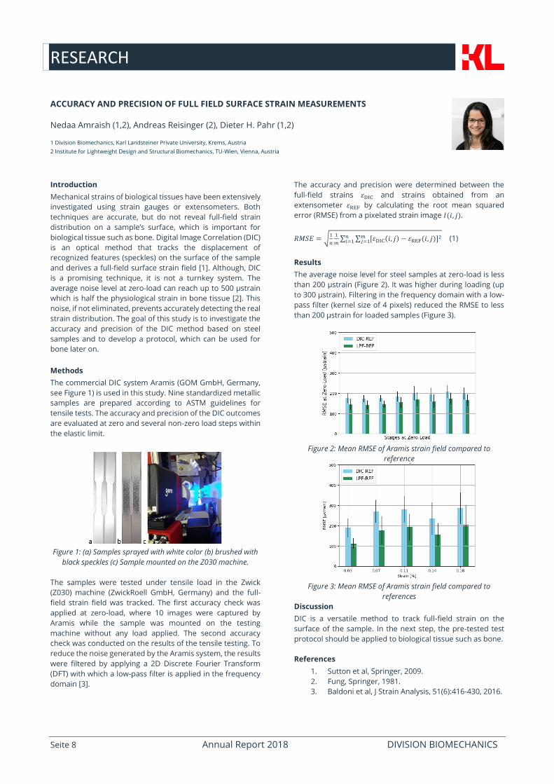
Figure 1: (a) Samples sprayed with white color (b) brushed with
black speckles (c) Sample mounted on the Z030 machine.
The samples were tested under tensile load in the Zwick
(Z030) machine (ZwickRoell GmbH, Germany) and the full-
field strain field was tracked. The first accuracy check was
applied at zero-load, where 10 images were captured by
Aramis while the sample was mounted on the testing
machine without any load applied. The second accuracy
check was conducted on the results of the tensile testing. To
reduce the noise generated by the Aramis system, the results
were filtered by applying a 2D Discrete Fourier Transform
(DFT) with which a low-pass filter is applied in the frequency
domain [3].
The accuracy and precision were determined between the
full-field strains 휀DIC and strains obtained from an
extensometer 휀REF by calculating the root mean squared
error (RMSE) from a pixelated strain image 𝐼(𝑖, 𝑗).
𝑅𝑀𝑆𝐸 = √1
𝑛
1
𝑚∑ ∑ [휀DIC(𝑖, 𝑗) − 휀REF(𝑖, 𝑗)]
2𝑚𝑗=1
𝑛𝑖=1 (1)
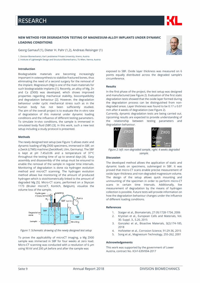
Results
The average noise level for steel samples at zero-load is less
than 200 µstrain (Figure 2). It was higher during loading (up
to 300 µstrain). Filtering in the frequency domain with a low-
pass filter (kernel size of 4 pixels) reduced the RMSE to less
than 200 µstrain for loaded samples (Figure 3).
Figure 2: Mean RMSE of Aramis strain field compared to
reference
Figure 3: Mean RMSE of Aramis strain field compared to
references
Discussion
DIC is a versatile method to track full-field strain on the
surface of the sample. In the next step, the pre-tested test
protocol should be applied to biological tissue such as bone.
References
1. Sutton et al, Springer, 2009.
2. Fung, Springer, 1981.
3. Baldoni et al, J Strain Analysis, 51(6):416-430, 2016.
RESEARCH
Seite 9 Annual Report 2018 DIVISION BIOMECHANICS
NEW METHOD FOR DEGRADATION TESTING OF MAGNESIUM-ALLOY IMPLANTS UNDER DYNAMIC
LOADING CONDITIONS
Georg Gamauf (1), Dieter H. Pahr (1,2), Andreas Reisinger (1)
1. Division Biomechanics, Karl Landsteiner Private University, Krems, Austria
2. Institute of Lightweight Design and Structural Biomechanics, TU-Wien, Vienna, Austria
Introduction
Biodegradable materials are becoming increasingly
important in osteosynthesis to stabilize fractured bones, thus
eliminating the need of a second surgery for the removal of
the implant. Magnesium (Mg) is one of the main materials for
such biodegradable implants [1]. Recently, an alloy of Mg, Zn
and Ca (ZX00) was developed, which shows improved
properties regarding mechanical stability, biocompatibility
and degradation behaviour [2]. However, the degradation
behaviour under cyclic mechanical stress such as in the
human body has not been sufficiently studied.
The aim of the overall project is to evaluate the in-vitro rate
of degradation of this material under dynamic loading
conditions and the influence of different testing parameters.
To simulate in-vivo conditions, the sample is immersed in
simulated body fluid (SBF) [3]. In this work, such a new test
setup including a study protocol is presented.
Methods
The newly designed test setup (see Figure 1) allows static and
dynamic loading of Mg ZX00 specimens, immersed in SBF, on
a Zwick (LTM5) machine (ZwickRoell, Ulm, Germany). The SBF
is kept at pH 7.45±0.06 and a temperature of 37°C
throughout the testing time of up to several days [4]. Easy
assembly and disassembly of the setup must be ensured to
enable the removal of the sample in regular time intervals.
Monitoring of degradation is done via hydrogen evolution
method and microCT scanning. The hydrogen evolution
method allows live monitoring of the amount of produced
hydrogen which is stoichiometrically linked to the amount of
degraded Mg [5]. Micro-CT scans, performed on a Skyscan
1173 (Bruker microCT, Kontich, Belgium), visualize the
volume loss of the sample.
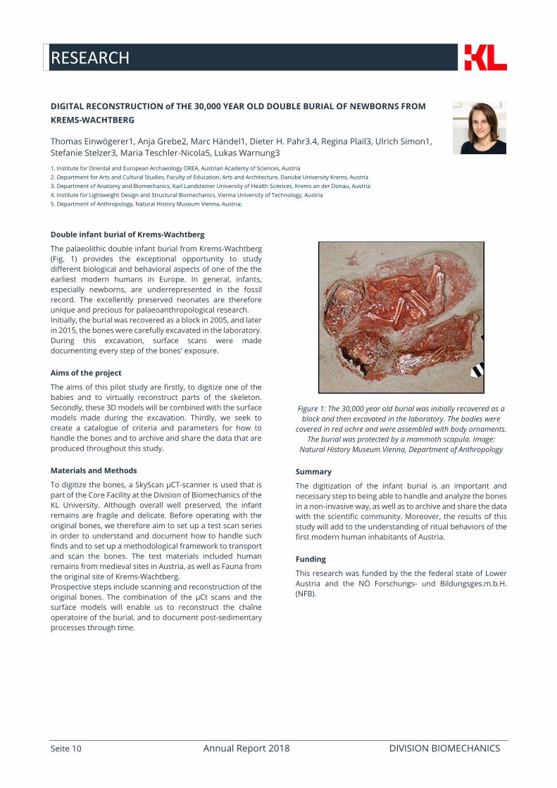
Figure 1: Schematic drawing of the newly designed test setup
To prove the applicability of microCT imaging, a Mg ZX00
sample was immersed in SBF for four weeks at zero load.
Micro-CT scanning was conducted with a resolution of 6 µm
using 90 kV and 200 µA before and after the sample was
exposed to SBF. Oxide layer thickness was measured on 6
points equally distributed across the degraded sample’s
circumference.
Results
In the first phase of the project, the test setup was designed
and manufactured (see Figure 2). Evaluation of the first static
degradation tests showed that the oxide layer formed during
the degradation process can be distinguished from non-
degraded areas. Layer thickness was found to be 0.17 ± 0.07
mm after 4 weeks of degradation (see Figure 2).
Currently, dynamic degradation tests are being carried out.
Upcoming results are expected to provide understanding of
the relationship between testing parameters and
degradation behaviour.
Figure 2: left: non-degraded sample, right: 4 weeks degraded
sample.
Discussion
The developed method allows the application of static and
dynamic loads on specimens, submerged in SBF. It was
proved that micro-CT scans enable precise measurement of
oxide layer thickness and non-degraded magnesium volume.
The design of the setup allows quick mounting and
unmounting of the specimen in order to perform micro-CT
scans in certain time intervals. Additionally, live
measurement of degradation by the means of hydrogen
collection is possible. Future tests will provide information on
how the degradation behaviour changes under the influence
of different loading conditions.
References
1. Staiger et al., Biomaterials, 27 (9):1728-1734, 2006
2. Krystian et al., European Cells and Materials, Vol.
30. Suppl. 3., S.20, 2015
3. Gonzalez et al., Bioactive Materials, 3(2):174-185,
2018
4. Hofstetter et al., Corrosion Science, 91:29-36, 2015
5. Song et al., Magnesium Technology, 255-262, 2001
Acknowledgements
This work was supported by the government of Lower
Austria, contract No. K3-F-639/004-2017
RESEARCH
Seite 10 Annual Report 2018 DIVISION BIOMECHANICS
DIGITAL RECONSTRUCTION of THE 30,000 YEAR OLD DOUBLE BURIAL OF NEWBORNS FROM
KREMS-WACHTBERG
Thomas Einwögerer1, Anja Grebe2, Marc Händel1, Dieter H. Pahr3.4, Regina Plail3, Ulrich Simon1,
Stefanie Stelzer3, Maria Teschler-Nicola5, Lukas Warnung3 1. Institute for Oriental and European Archaeology OREA, Austrian Academy of Sciences, Austria
2. Department for Arts and Cultural Studies, Faculty of Education, Arts and Architecture, Danube University Krems, Austria
3. Department of Anatomy and Biomechanics, Karl Landsteiner University of Health Sciences, Krems an der Donau, Austria
4. Institute for Lightweight Design and Structural Biomechanics, Vienna University of Technology, Austria
5. Department of Anthropology, Natural History Museum Vienna, Austria;
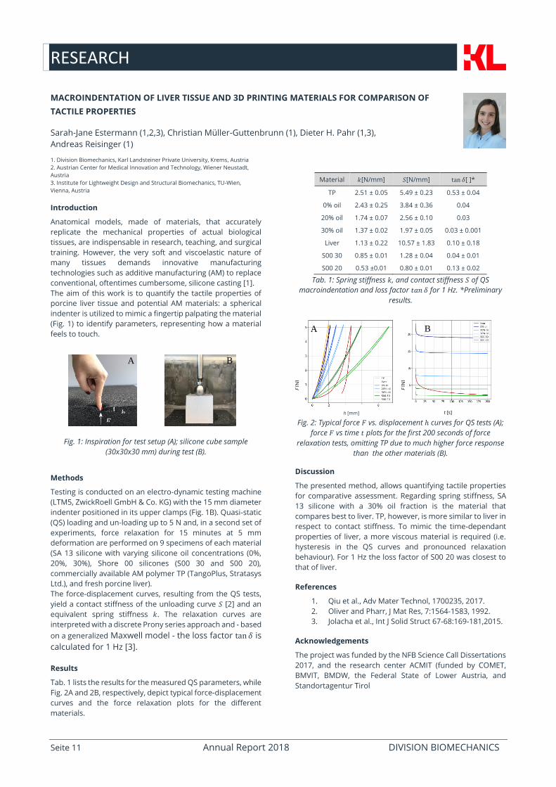
Double infant burial of Krems-Wachtberg
The palaeolithic double infant burial from Krems-Wachtberg
(Fig. 1) provides the exceptional opportunity to study
different biological and behavioral aspects of one of the the
earliest modern humans in Europe. In general, infants,
especially newborns, are underrepresented in the fossil
record. The excellently preserved neonates are therefore
unique and precious for palaeoanthropological research.
Initially, the burial was recovered as a block in 2005, and later
in 2015, the bones were carefully excavated in the laboratory.
During this excavation, surface scans were made
documenting every step of the bones' exposure.
Aims of the project
The aims of this pilot study are firstly, to digitize one of the
babies and to virtually reconstruct parts of the skeleton.
Secondly, these 3D models will be combined with the surface
models made during the excavation. Thirdly, we seek to
create a catalogue of criteria and parameters for how to
handle the bones and to archive and share the data that are
produced throughout this study.
Materials and Methods
To digitize the bones, a SkyScan µCT-scanner is used that is
part of the Core Facility at the Division of Biomechanics of the
KL University. Although overall well preserved, the infant
remains are fragile and delicate. Before operating with the
original bones, we therefore aim to set up a test scan series
in order to understand and document how to handle such
finds and to set up a methodological framework to transport
and scan the bones. The test materials included human
remains from medieval sites in Austria, as well as Fauna from
the original site of Krems-Wachtberg.
Prospective steps include scanning and reconstruction of the
original bones. The combination of the µCt scans and the
surface models will enable us to reconstruct the chaîne
operatoire of the burial, and to document post-sedimentary
processes through time.
Figure 1: The 30,000 year old burial was initially recovered as a
block and then excavated in the laboratory. The bodies were
covered in red ochre and were assembled with body ornaments.
The burial was protected by a mammoth scapula. Image:
Natural History Museum Vienna, Department of Anthropology
Summary
The digitization of the infant burial is an important and
necessary step to being able to handle and analyze the bones
in a non-invasive way, as well as to archive and share the data
with the scientific community. Moreover, the results of this
study will add to the understanding of ritual behaviors of the
first modern human inhabitants of Austria.
Funding
This research was funded by the the federal state of Lower
Austria and the NÖ Forschungs- und Bildungsges.m.b.H.
(NFB).
RESEARCH
Seite 11 Annual Report 2018 DIVISION BIOMECHANICS
t [s] h [mm]
MACROINDENTATION OF LIVER TISSUE AND 3D PRINTING MATERIALS FOR COMPARISON OF
TACTILE PROPERTIES
Sarah-Jane Estermann (1,2,3), Christian Müller-Guttenbrunn (1), Dieter H. Pahr (1,3),
Andreas Reisinger (1)
1. Division Biomechanics, Karl Landsteiner Private University, Krems, Austria
2. Austrian Center for Medical Innovation and Technology, Wiener Neustadt,
Austria
3. Institute for Lightweight Design and Structural Biomechanics, TU-Wien,
Vienna, Austria
Introduction
Anatomical models, made of materials, that accurately
replicate the mechanical properties of actual biological
tissues, are indispensable in research, teaching, and surgical
training. However, the very soft and viscoelastic nature of
many tissues demands innovative manufacturing
technologies such as additive manufacturing (AM) to replace
conventional, oftentimes cumbersome, silicone casting [1].
The aim of this work is to quantify the tactile properties of
porcine liver tissue and potential AM materials: a spherical
indenter is utilized to mimic a fingertip palpating the material
(Fig. 1) to identify parameters, representing how a material
feels to touch.
Fig. 1: Inspiration for test setup (A); silicone cube sample
(30x30x30 mm) during test (B).
Methods
Testing is conducted on an electro-dynamic testing machine
(LTM5, ZwickRoell GmbH & Co. KG) with the 15 mm diameter
indenter positioned in its upper clamps (Fig. 1B). Quasi-static
(QS) loading and un-loading up to 5 N and, in a second set of
experiments, force relaxation for 15 minutes at 5 mm
deformation are performed on 9 specimens of each material
(SA 13 silicone with varying silicone oil concentrations (0%,
20%, 30%), Shore 00 silicones (S00 30 and S00 20),
commercially available AM polymer TP (TangoPlus, Stratasys
Ltd.), and fresh porcine liver).
The force-displacement curves, resulting from the QS tests,
yield a contact stiffness of the unloading curve 𝑆 [2] and an
equivalent spring stiffness 𝑘. The relaxation curves are
interpreted with a discrete Prony series approach and - based
on a generalized Maxwell model - the loss factor tan 𝛿 is
calculated for 1 Hz [3].
Results
Tab. 1 lists the results for the measured QS parameters, while
Fig. 2A and 2B, respectively, depict typical force-displacement
curves and the force relaxation plots for the different
materials.
Material 𝑘[N/mm] 𝑆[N/mm] tan 𝛿[ ]*
TP 2.51 ± 0.05 5.49 ± 0.23 0.53 ± 0.04
0% oil 2.43 ± 0.25 3.84 ± 0.36 0.04
20% oil 1.74 ± 0.07 2.56 ± 0.10 0.03
30% oil 1.37 ± 0.02 1.97 ± 0.05 0.03 ± 0.001
Liver 1.13 ± 0.22 10.57 ± 1.83 0.10 ± 0.18
S00 30 0.85 ± 0.01 1.28 ± 0.04 0.04 ± 0.01
S00 20 0.53 ±0.01 0.80 ± 0.01 0.13 ± 0.02
Tab. 1: Spring stiffness 𝑘, and contact stiffness 𝑆 of QS
macroindentation and loss factor 𝑡𝑎𝑛 𝛿 for 1 Hz. *Preliminary
results.
Fig. 2: Typical force 𝐹 vs. displacement ℎ curves for QS tests (A);
force 𝐹 vs time 𝑡 plots for the first 200 seconds of force
relaxation tests, omitting TP due to much higher force response
than the other materials (B).
Discussion
The presented method, allows quantifying tactile properties
for comparative assessment. Regarding spring stiffness, SA
13 silicone with a 30% oil fraction is the material that
compares best to liver. TP, however, is more similar to liver in
respect to contact stiffness. To mimic the time-dependant
properties of liver, a more viscous material is required (i.e.
hysteresis in the QS curves and pronounced relaxation
behaviour). For 1 Hz the loss factor of S00 20 was closest to
that of liver.
References
1. Qiu et al., Adv Mater Technol, 1700235, 2017.
2. Oliver and Pharr, J Mat Res, 7:1564-1583, 1992.
3. Jolacha et al., Int J Solid Struct 67-68:169-181,2015.
Acknowledgements
The project was funded by the NFB Science Call Dissertations
2017, and the research center ACMIT (funded by COMET,
BMVIT, BMDW, the Federal State of Lower Austria, and
Standortagentur Tirol
F [N
]
F [N
]
B A
𝐹 ℎ
A B
RESEARCH
Seite 12 Annual Report 2018 DIVISION BIOMECHANICS
EFFECT OF SELECTED SCAN PARAMETERS ON QCT BASED BMD ESTIMATIONS OF A FEMUR
Morteza Amini (1), Andreas Reisinger (1), Dieter H. Pahr (1,2)
1. Division Biomechanics, Karl Landsteiner Private University, Krems, Austria
2. Institute of Lightweight Design and Structural Biomechanics, TU-Wien, Vienna, Austria
Background
Finite element models (FEM) based estimates of bone
strength can be used to o predict fracture risk of patients
susceptible to osteoporosis in order to decide on
administering any therapies [1]. A key ingredient of a valid
FEM is material models. Elastic modulus of FEM elements can
be directly mapped from Quantitative Computed
Tomography (QCT) scans of bone. In this method, a
calibration phantom consisted of a few (usually 3 to 5) rods
with known hydroxyapatite (HA) densities is placed together
with the scanned sample/subject. The Hounsfield Units (HU)
of the scanned rods and their known density result in a linear
HU-Density relationship which is used to map the bone
mineral density (BMD) to the elastic moduli [2].
Unlike scanning patients, which has a well-defined protocol,
scanning cadaveric samples, which is the main approach in
FEM validation studies, has no standard procedure. As a
result, the scanning process until extraction of BMD values
based on HU can be affected by many parameters [3]. The
goal of this study was to determine the effect of such
parameters on the QCT-based BMD estimations of a
cadaveric femur.
Methods
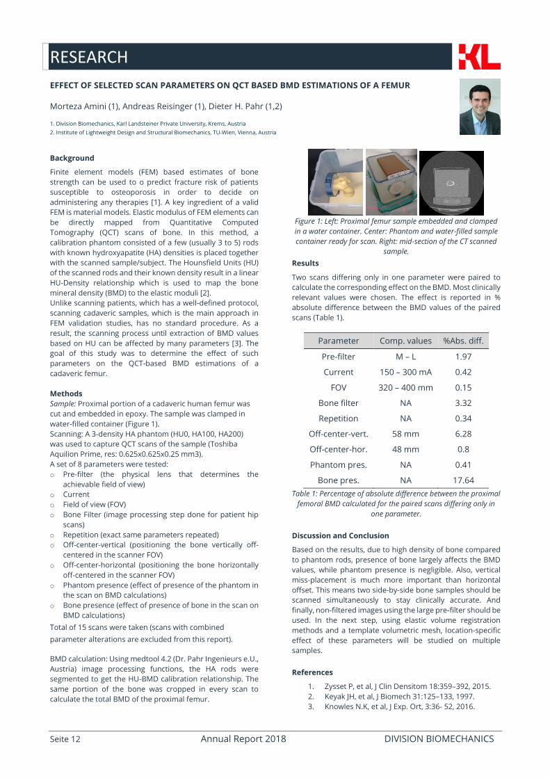
Sample: Proximal portion of a cadaveric human femur was
cut and embedded in epoxy. The sample was clamped in
water-filled container (Figure 1).
Scanning: A 3-density HA phantom (HU0, HA100, HA200)
was used to capture QCT scans of the sample (Toshiba
Aquilion Prime, res: 0.625x0.625x0.25 mm3).
A set of 8 parameters were tested:
o Pre-filter (the physical lens that determines the
achievable field of view)
o Current
o Field of view (FOV)
o Bone Filter (image processing step done for patient hip
scans)
o Repetition (exact same parameters repeated)
o Off-center-vertical (positioning the bone vertically off-
centered in the scanner FOV)
o Off-center-horizontal (positioning the bone horizontally
off-centered in the scanner FOV)
o Phantom presence (effect of presence of the phantom in
the scan on BMD calculations)
o Bone presence (effect of presence of bone in the scan on
BMD calculations)
Total of 15 scans were taken (scans with combined
parameter alterations are excluded from this report).
BMD calculation: Using medtool 4.2 (Dr. Pahr Ingenieurs e.U.,
Austria) image processing functions, the HA rods were
segmented to get the HU-BMD calibration relationship. The
same portion of the bone was cropped in every scan to
calculate the total BMD of the proximal femur.
Figure 1: Left: Proximal femur sample embedded and clamped
in a water container. Center: Phantom and water-filled sample
container ready for scan. Right: mid-section of the CT scanned
sample.
Results
Two scans differing only in one parameter were paired to
calculate the corresponding effect on the BMD. Most clinically
relevant values were chosen. The effect is reported in %
absolute difference between the BMD values of the paired
scans (Table 1).
Parameter Comp. values %Abs. diff.
Pre-filter M – L 1.97
Current
FOV
Bone filter
Repetition
Off-center-vert.
Off-center-hor.
Phantom pres.
Bone pres.
150 – 300 mA
320 – 400 mm
NA
NA
58 mm
48 mm
NA
NA
0.42
0.15
3.32
0.34
6.28
0.8
0.41
17.64
Table 1: Percentage of absolute difference between the proximal
femoral BMD calculated for the paired scans differing only in
one parameter.
Discussion and Conclusion
Based on the results, due to high density of bone compared
to phantom rods, presence of bone largely affects the BMD
values, while phantom presence is negligible. Also, vertical
miss-placement is much more important than horizontal
offset. This means two side-by-side bone samples should be
scanned simultaneously to stay clinically accurate. And
finally, non-filtered images using the large pre-filter should be
used. In the next step, using elastic volume registration
methods and a template volumetric mesh, location-specific
effect of these parameters will be studied on multiple
samples.
References
1. Zysset P, et al, J Clin Densitom 18:359–392, 2015.
2. Keyak JH, et al, J Biomech 31:125–133, 1997.
3. Knowles N.K, et al, J Exp. Ort, 3:36- 52, 2016.
ACTIVITIES & EVENTS
Seite 13 Annual Report 2018 DIVISION BIOMECHANICS
Activities & Events
BIOMEX-Workshop on Biomechanical Imaging, Sweden
Dieter Pahr was the Keynote speaker with “Activities in biomechanics –today and tomorrow” In
Sweden in Huddinge at the BIOMEX-Workshop on Biomechanical Imaging, February 2018.
Articles Projekt “Best Mg Alloy”
(WN-Nachrichten; Chemiereport), January & March 2018.
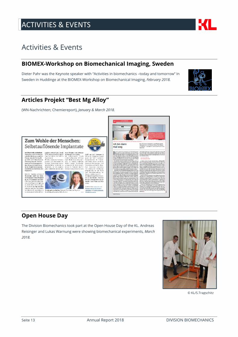
Open House Day
The Division Biomechanics took part at the Open House Day of the KL. Andreas
Reisinger and Lukas Warnung were showing biomechanical experiments, March
2018.
© KL/S.Tragschitz
ACTIVITIES & EVENTS
Seite 14 Annual Report 2018 DIVISION BIOMECHANICS
ÖGOuT-edvanced education seminar “Osteoporose”
Dieter Pahr was given a speech on “Biomechanik des Knochens” at the ÖGOuT (Österreichische Gesellschaft für
Orthopädie und Traumatologie) education seminar on „Osteoporose“, March 2018.
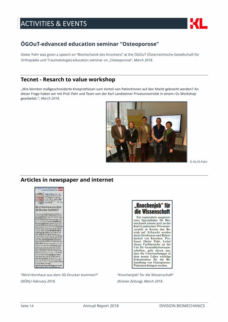
Tecnet - Resarch to value workshop
„Wie könnten maßgeschneiderte Knieprothesen zum Vorteil von PatientInnen auf den Markt gebracht werden? An dieser Frage haben wir mit Prof. Pahr und Team von der Karl Landsteiner Privatuniversität in einem r2v Workshop gearbeitet.“, March 2018.
© KL/D.Pahr
Articles in newspaper and internet
“Wird Hornhaut aus dem 3D-Drucker kommen?“ “Knochenjob” für die Wissenschaft“
(NÖN),l February 2018. (Kronen Zeitung), March 2018.

ACTIVITIES & EVENTS
Seite 15 Annual Report 2018 DIVISION BIOMECHANICS
“Knochenjob: Biomechaniker” (Standard), May 2018. “Was das Herz begehrt”: (Südtiroler Wirtschaftszeitung)
October 2018.
“BREAK EVEN”: New laboratory creates excellent conditions for studying bones at KL Krems”, Pressbox (Press Release),
March 2018
“Auf Biegen & Brechen: Top-Labor schafft optimale Bedingungen für Knochenarbeit an KL Krems” (http://neue-
pressemitteilungen.de), May 2018.
Chemiereport
„So zugfest wie das biologische Vorbild“, Jungforscherin Sarah-Jane Estermann im Porträt (Chemiereport 2018.05), July
2018.
https://www.chemiereport.at/sites/default/files/uploads/printausgaben/chemiereport-2018-05_web.pdf
ACTIVITIES & EVENTS
Seite 16 Annual Report 2018 DIVISION BIOMECHANICS
CityRUN Krems 2018
This year, the division Biomechanics participated with 2 teams called
“BioMechTrio” and the “Bonebreakers” at CityRUN 2018 in Krems, April2018.
©KL/S.Tragschitz
left: Andreas Reisinger, Lukas Warnung and Christina Aron, the
“BioMechTrio” ©KL/S.Tragschitz
right: Jalil Nourisa, Georg Gamauf and NedaaAmraish,
the “Bonebreakers” at the finish ©KL/S.Tragschitz
Hiking Day 2018
For the first time the Division Biomechanics made a hiking day: Dürnstein, Vogelbergsteig, June 2018.
©KL/C.Aron
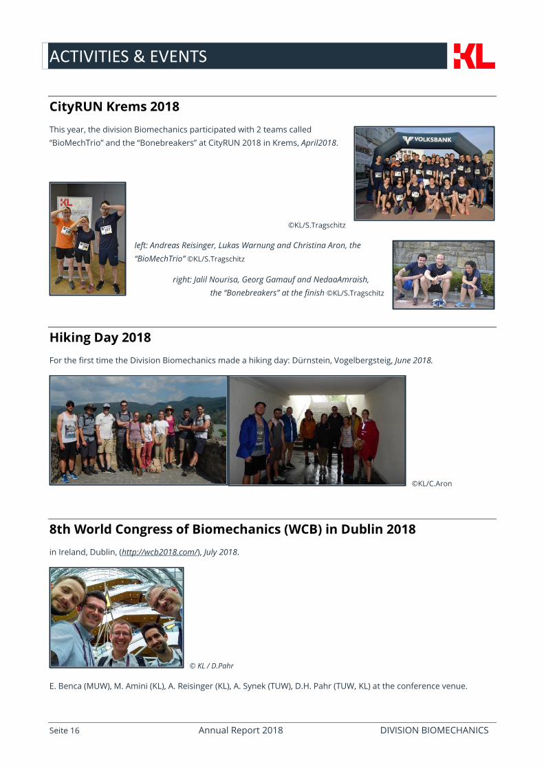
8th World Congress of Biomechanics (WCB) in Dublin 2018
in Ireland, Dublin, (http://wcb2018.com/), July 2018.
© KL / D.Pahr
E. Benca (MUW), M. Amini (KL), A. Reisinger (KL), A. Synek (TUW), D.H. Pahr (TUW, KL) at the conference venue.
ACTIVITIES & EVENTS
Seite 17 Annual Report 2018 DIVISION BIOMECHANICS
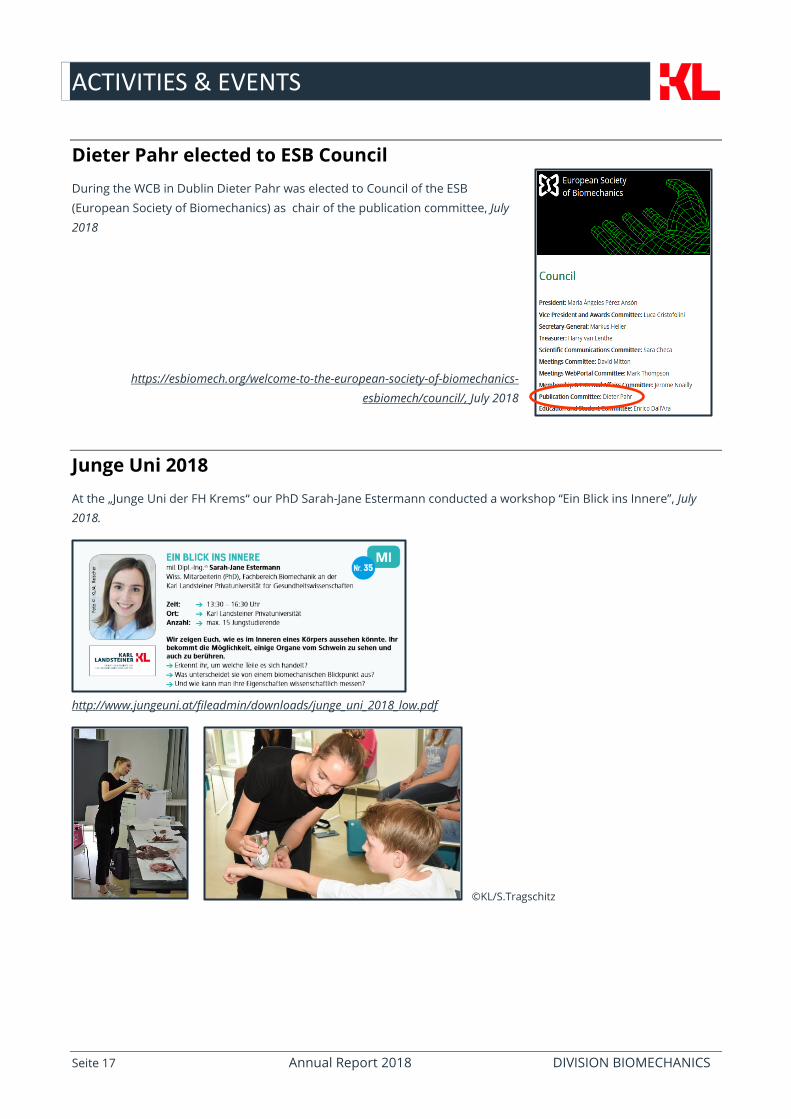
Dieter Pahr elected to ESB Council
During the WCB in Dublin Dieter Pahr was elected to Council of the ESB
(European Society of Biomechanics) as chair of the publication committee, July
2018
https://esbiomech.org/welcome-to-the-european-society-of-biomechanics-
esbiomech/council/, July 2018
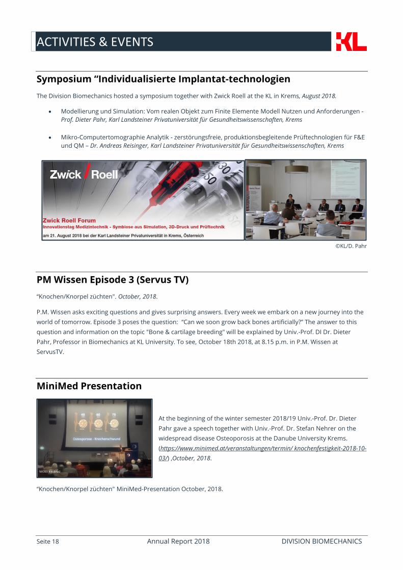
Junge Uni 2018
At the „Junge Uni der FH Krems“ our PhD Sarah-Jane Estermann conducted a workshop “Ein Blick ins Innere”, July
2018.
http://www.jungeuni.at/fileadmin/downloads/junge_uni_2018_low.pdf
©KL/S.Tragschitz
ACTIVITIES & EVENTS
Seite 18 Annual Report 2018 DIVISION BIOMECHANICS
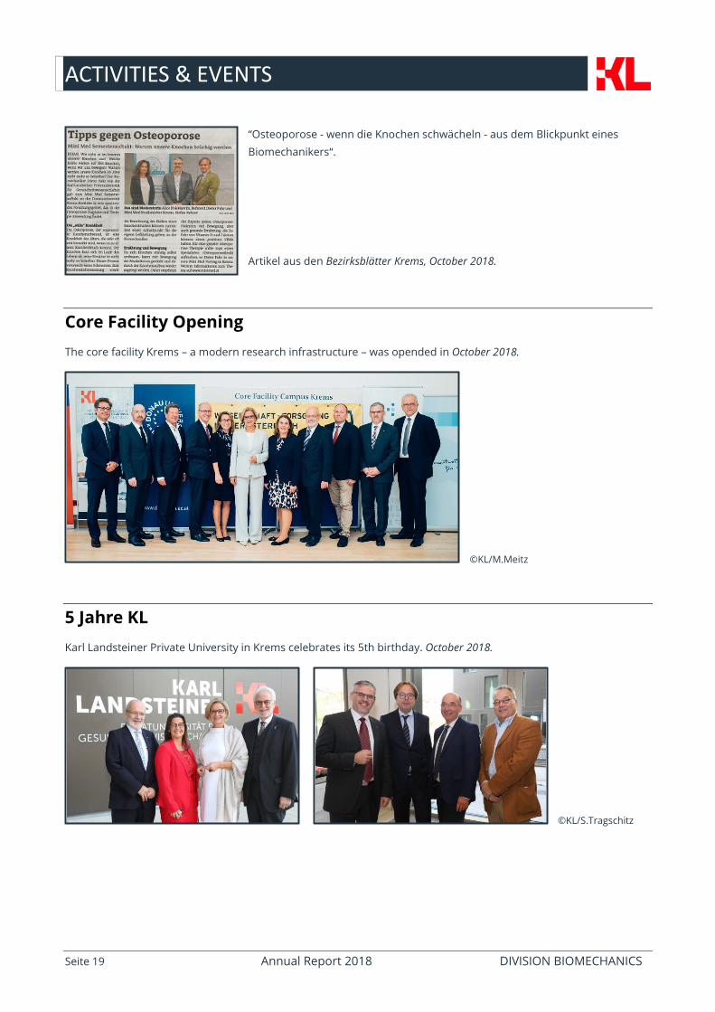
Symposium “Individualisierte Implantat-technologien
The Division Biomechanics hosted a symposium together with Zwick Roell at the KL in Krems, August 2018.
Modellierung und Simulation: Vom realen Objekt zum Finite Elemente Modell Nutzen und Anforderungen -
Prof. Dieter Pahr, Karl Landsteiner Privatuniversität für Gesundheitswissenschaften, Krems
Mikro-Computertomographie Analytik - zerstörungsfreie, produktionsbegleitende Prüftechnologien für F&E
und QM – Dr. Andreas Reisinger, Karl Landsteiner Privatuniversität für Gesundheitswissenschaften, Krems
©KL/D. Pahr
PM Wissen Episode 3 (Servus TV)
“Knochen/Knorpel züchten". October, 2018.
P.M. Wissen asks exciting questions and gives surprising answers. Every week we embark on a new journey into the
world of tomorrow. Episode 3 poses the question: “Can we soon grow back bones artificially?” The answer to this
question and information on the topic "Bone & cartilage breeding" will be explained by Univ.-Prof. DI Dr. Dieter
Pahr, Professor in Biomechanics at KL University. To see, October 18th 2018, at 8.15 p.m. in P.M. Wissen at
ServusTV.
MiniMed Presentation
At the beginning of the winter semester 2018/19 Univ.-Prof. Dr. Dieter
Pahr gave a speech together with Univ.-Prof. Dr. Stefan Nehrer on the
widespread disease Osteoporosis at the Danube University Krems.
(https://www.minimed.at/veranstaltungen/termin/ knochenfestigkeit-2018-10-
03/) ,October, 2018.
“Knochen/Knorpel züchten" MiniMed-Presentation October, 2018.
ACTIVITIES & EVENTS
Seite 19 Annual Report 2018 DIVISION BIOMECHANICS
“Osteoporose - wenn die Knochen schwächeln - aus dem Blickpunkt eines
Biomechanikers“.
Artikel aus den Bezirksblätter Krems, October 2018.
Core Facility Opening
The core facility Krems – a modern research infrastructure – was opended in October 2018.
©KL/M.Meitz
5 Jahre KL
Karl Landsteiner Private University in Krems celebrates its 5th birthday. October 2018.
©KL/S.Tragschitz
ACTIVITIES & EVENTS
Seite 20 Annual Report 2018 DIVISION BIOMECHANICS
Technopol breakfast
November 2018.
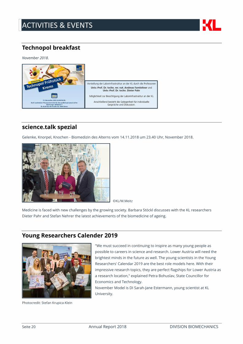
science.talk spezial
Gelenke, Knorpel, Knochen - Biomedizin des Alterns vom 14.11.2018 um 23.40 Uhr, November 2018.
©KL/M.Meitz
Medicine is faced with new challenges by the growing society. Barbara Stöckl discusses with the KL researchers
Dieter Pahr and Stefan Nehrer the latest achievements of the biomedicine of ageing.
Young Researchers Calender 2019
"We must succeed in continuing to inspire as many young people as
possible to careers in science and research. Lower Austria will need the
brightest minds in the future as well. The young scientists in the Young
Researchers' Calendar 2019 are the best role models here. With their
impressive research topics, they are perfect flagships for Lower Austria as
a research location," explained Petra Bohuslav, State Councillor for
Economics and Technology.
November Model is DI Sarah-Jane Estermann, young scientist at KL
University.
Photocredit: Stefan Krupica-Klein
ACTIVITIES & EVENTS
Seite 21 Annual Report 2018 DIVISION BIOMECHANICS
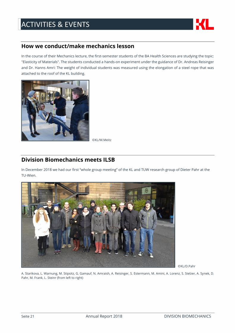
How we conduct/make mechanics lesson
In the course of their Mechanics lecture, the first-semester students of the BA Health Sciences are studying the topic:
"Elasticity of Materials". The students conducted a hands-on experiment under the guidance of Dr. Andreas Reisinger
and Dr. Hanns Amri: The weight of individual students was measured using the elongation of a steel rope that was
attached to the roof of the KL building.
©KL/M.Meitz
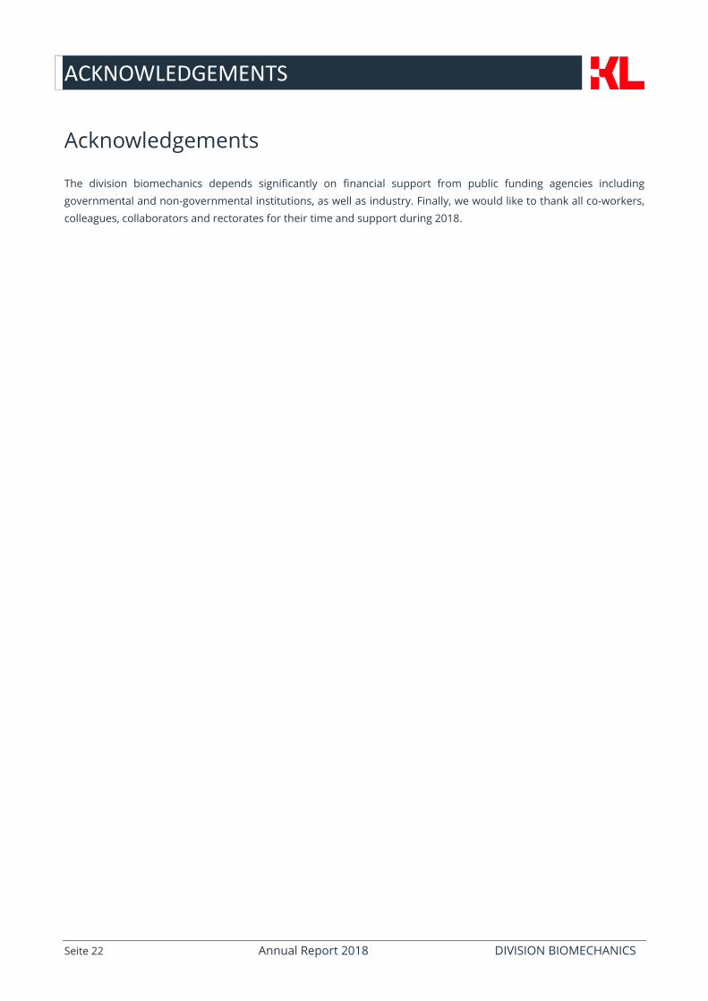
Division Biomechanics meets ILSB
In December 2018 we had our first “whole group meeting” of the KL and TUW research group of Dieter Pahr at the
TU-Wien.
©KL/D.Pahr
A. Starikova, L. Warnung, M. Stipsitz, G. Gamauf, N. Amraish, A. Reisinger, S. Estermann, M. Amini, A. Lorenz, S. Stelzer, A. Synek, D.
Pahr, M. Frank, L. Steinr (from left to right)
ACKNOWLEDGEMENTS
Seite 22 Annual Report 2018 DIVISION BIOMECHANICS
Acknowledgements
The division biomechanics depends significantly on financial support from public funding agencies including
governmental and non-governmental institutions, as well as industry. Finally, we would like to thank all co-workers,
colleagues, collaborators and rectorates for their time and support during 2018.