Embed Size (px)
Citation preview
Antibiotic mechanisms of
invertebrate and mammalian
defensins
Dissertation
zur
Erlangung des Doktorgrades (Dr. rer. nat.)
der
Mathematisch-Naturwissenschaftlichen Fakultät
der
Rheinischen Friedrich-Wilhelms-Universität Bonn
vorgelegt von
Miriam Wilmes
aus
Werdohl
Bonn 2012

III
Die vorliegende Arbeit wurde mit Genehmigung der Mathematisch-
Naturwissenschaftlichen Fakultät der Rheinischen Friedrich-Wilhelms-Universität
Bonn angefertigt.
Erstgutachter: Prof. Dr. Hans-Georg Sahl
Zweitgutachterin: apl. Prof. Dr. Christiane Dahl
Tag der Promotion: 17.12.2012 Erscheinungsjahr: 2013

V
Table of contents
Table of contents .............................................................................................................. V
Abbreviations .................................................................................................................. VII
1 Introduction ................................................................................................................... 1
1.1 Host Defence Peptides ........................................................................................ 1
1.2 Defensins ............................................................................................................ 3
1.3 Antimicrobial mode of action of HDPs ................................................................. 7
1.4 Immunomodulatory functions of HDPs .............................................................. 10
1.5 HDP expression and human disorders .............................................................. 12
1.6 Resistance towards HDPs ................................................................................. 14
1.7 Objectives of this work ....................................................................................... 15
2 Material and Methods ................................................................................................. 17
2.1 Chemicals and solvents ..................................................................................... 17
2.2 Antibiotics and antimicrobial peptides ................................................................ 18
2.3 Microbiological methods .................................................................................... 19
2.3.1 Bacterial strains, culture media and growth conditions ........................... 19
2.3.2 Sterilisation of media, equipment and bacterial cultures ......................... 21
2.3.3 Determination of the optical density of a bacterial culture ....................... 21
2.3.4 Determination of the minimal inhibitory concentration ............................ 21
2.3.5 Antagonisation of putative target molecules ........................................... 22
2.3.6 Bacterial killing kinetics .......................................................................... 22
2.3.7 Growth kinetic measurement .................................................................. 22
2.3.8 Potassium release from whole cells ....................................................... 23
2.3.9 Determination of the membrane potential using tetraphenyl-phosphonium bromide ............................................................................ 23
2.3.10 Incorporation of radio-labelled metabolites ............................................. 24
2.3.11 Membrane permeabilisation assay ......................................................... 24
2.3.12 Intracellular accumulation of the final soluble cell wall precursor UDP-N-acetylmuramyl-pentapeptide ...................................................... 25
2.4 Methods in molecular genetics .......................................................................... 26
2.4.1 Determination of concentration and purity of nucleic acids ..................... 26
2.4.2 Agarose gel electrophoresis ................................................................... 26
2.4.3 Isolation of plasmid DNA ........................................................................ 26
2.4.4 Preparation of electrocompetent E. coli cells .......................................... 26
2.4.5 Transformation of E. coli by electroporation ........................................... 26
2.5 Protein and biochemical methods ...................................................................... 27
2.5.1 Heterologous expression and purification of His-tagged PBP2 ............... 27
2.5.2 Sodium-dodecyl-sulfate polyacrylamide gel electrophoresis ................... 27
2.5.3 Zymogram analysis ................................................................................ 28
2.5.4 Determination of peptide and protein concentration ............................... 29
2.5.5 Peptide quantification by spectrometry ................................................... 29
2.5.6 In vitro lipid II synthesis and purification ................................................. 29
2.5.7 Phosphate determination ....................................................................... 31
2.5.8 Inhibition of PBP2-catalysed reaction in vitro.......................................... 32
2.5.9 Complexation of lipid II ........................................................................... 32
2.5.10 Transmission electron microscopy ......................................................... 32
VI
2.5.11 Biomolecular interaction analysis by surface plasmon resonance .......... 33
2.5.12 Chromatographic purification of lipoteichoic acid .................................... 34
2.5.13 Determination of glucose concentration ................................................. 35
2.5.14 CF-efflux from LTA-containing liposomes ............................................... 35
3 Results ....................................................................................................................... 37
3.1 Part 1: Insight into invertebrate defensin mode of action ................................... 37
3.1.1 Antibacterial activity spectrum of oyster defensins ................................. 38
3.1.2 Impact of oyster defensins on the membrane integrity of S. aureus ....... 38
3.1.3 Accumulation of the soluble cell wall precursor UDP-N-acetylmuramyl-pentapeptide in oyster defensin treated cells .................. 39
3.1.4 Antagonisation of antibacterial activity of oyster defensins ..................... 41
3.1.5 Binding of oyster defensins to lipid II ...................................................... 41
3.2 Part 2: Insight into the antistaphylococcal mode of action of θ-defensins ........... 45
3.2.1 Antibacterial activity of θ-defensins against staphylococci ...................... 46
3.2.2 Impact of RTDs on staphylococcal cell wall biosynthesis ....................... 46
3.2.3 Impact of RTDs on macromolecular synthesis ....................................... 47
3.2.4 Impact of RTDs on the membrane integrity ............................................ 48
3.2.5 Morphological changes of RTD-2 treated S. aureus cells ....................... 51
3.2.6 Release of cell wall lytic enzymes by RTDs ........................................... 53
3.2.7 Antagonisation of antibacterial activity of θ-defensins ............................ 56
3.2.8 Release of carboxyfluorescein from LTA-containing liposomes .............. 56
3.2.9 Susceptibility testing of a PepI producing strain ..................................... 57
3.3 Part 3: Insight into the Gram-negative mode of action of defensins ................... 59
3.3.1 Antibacterial activity of hBD3 against E. coli ........................................... 60
3.3.2 Growth kinetic measurements in presence of hBD3 ............................... 60
3.3.3 Permeabilisation of E. coli outer and inner membrane ........................... 61
3.3.4 Morphological changes of hBD3 treated E. coli cells .............................. 62
3.3.5 Activity of hBD3 against E. coli strains with different LPS structures ...... 63
4 Discussion .................................................................................................................. 65
4.1 Part 1: The central cell wall building block lipid II as target for defensins ........... 65
4.2 Part 2: Induction of autolysis as mode of action of defensins ............................. 70
4.3 Part 3: Interaction of hBD3 with the Gram-negative cell envelope ..................... 74
4.4 Conclusion ........................................................................................................ 76
4.5 Outlook .............................................................................................................. 77
5 Summary .................................................................................................................... 79
6 References ................................................................................................................. 81
7 Publications ................................................................................................................ 97
8 Declaration (Eidesstattliche Erklärung) ....................................................................... 99
VII
Abbreviations
Aλ absorbance at a wavelength of λ
ABC ATP binding cassette
ad fill up to
AD atopic dermatitis
AM amidase
APS ammonium persulfate
ATCC American Type Culture Collection
BCA bicinchoninic acid
BHI brain heart infusion (complex medium)
BSA bovine serum albumin
°C degree Celsius
C55-P undecaprenyl phosphate (lipid carrier)
CCCP carbonyl cyanide m-chlorophenylhydrazone
CD Crohn´s disease
CENTATM chromogenic cephalosporin (β-lactamase substrate)
CF carboxyfluorescein
CFU colony forming unit(s)
Cg-Def defensin from the oyster Crassostrea gigas
CHAPS 3-[(3-cholamidopropyl)dimethylammonio]-1-propanesulfonate hydrate
Ci Curie
cpm counts per minute
CSαβ cysteine-stabilised α-helix β-sheet (motif)
3D three-dimensional
Da Dalton
DAG diacylglycerol
DMSO dimethyl sulfoxide
DMSZ German Research Center for Microorganisms and Cell Culture
DNA deoxyribonucleic acid
DNase deoxyribonuclease
DOPC 1,2-dioleoyl-sn-glycero-3-phosphocholine
EDTA ethylenediaminetetraacetic acid
et al. et alii, and others
GL glucosaminidase
GlcCer glucosylceramide
GlcNAc N-acetylglucosamine
VIII
h hour
hBD human β-defensin
HD human defensin (α-defensin)
HDP host defence peptide
HEPES 4-(2-hydroxyethyl)-1-piperazineethanesulfonic acid
HIV human immunodeficiency virus
HNP human neutrophil peptide (α-defensin)
IC50 half maximal inhibitory concentration
IFN interferon
IL interleukin
IPTG isopropyl-β-D-galactopyranoside
l liter
LB lysogeny broth (complex medium)
lipid II undecaprenyl-pyrophosphoryl-MurNAc-pentapeptide-GlcNAc
LPS lipopolysaccharides
LTA lipoteichoic acid
M mol
MALDI-TOF MS matrix-assisted laser desorption/ionisation time-of-flight mass
spectrometry
MES 2-(N-morpholino)ethanesulfonic acid
MIC minimal inhibitory concentration
MilliQ ultrapure water (type I)
min minute
MRSA methicillin-resistant Staphylococcus aureus
MurNAc N-acetylmuramic acid
MW molecular weight
MWCO molecular weight cut-off
Ni-NTA nickel-nitrilotriacetic acid
NMR nuclear magnetic resonance
OD600 optical density at a wavelength of 600 nm
ONPG o-nitrophenyl-β-D-galactopyranoside
PBP penicillin binding protein
pH pondus hydrogenii, hydrogen ion concentration
PMA phosphomolybdic acid
R resistance
RNA ribonucleic acid
IX
RNase ribonuclease
RP-HPLC reversed-phase high performance liquid chromatography
rpm rounds per minute
RT room temperature
RTD Rhesus macaque θ-defensin
RU response unit(s)
SDS sodium dodecyl sulfate
SDS-PAGE sodium dodecyl sulfate polyacrylamide gel electrophoresis
SPB sodium phosphate buffer
SPR surface plasmon resonance
SUV small unilamellar vesicle
TAE Tris-acetate-EDTA buffer
TEMED N,N,N',N'-tetramethylethylenediamine
TLC thin layer chromatography
TLR Toll-like receptor
TNF tumour necrosis factor
TPP+ tetraphenylphosphonium bromide
Tris tris(hydroxymethyl-)aminomethan
Triton X-100 polyethylene glycol tert-octylphenyl ether
TSB tryptic soy broth (complex medium)
UDP uridine diphosphate
UDP-MurNAc-pp UDP-N-acetylmuramyl-pentapeptide
V volt
VRE vancomycin-resistant enterococci
v/v volume per volume
w/v weight per volume
The abbreviations of the amino acids follow the IUPAC regulations. For time, length and
mass description the standard IS units were used.

Introduction 1
1 Introduction
In 1928, Alexander Fleming discovered the first antibiotic – penicillin – that was
subsequently developed for clinical use (Fleming, 1929). The effective treatment of
previously deadly infectious diseases revolutionised medicine, and contributed to
increasing human life expectancy.
Since that time, numerous natural and synthetic antimicrobial substances have been
identified. However, the development and clinical use of each new antibiotic class was
followed by emergence of resistant bacteria. Today, the therapeutic options for multidrug
resistant pathogens are limited, and in the last decade only two new antibiotic classes
entered the market. Consequently, there is an urgent need to discover and develop novel
anti-infective agents to combat pathogenic bacteria (Brotz-Oesterhelt and Sass, 2010;
Davies and Davies, 2010).
Host defence peptides, which are produced by almost all eukaryotic organisms, are
considered as a promising source of potential antibiotics since they combine direct
antibacterial activities with modulation of immune response. Moreover, they are also
active against those bacteria resistant to conventional antibiotics and show only modest
resistance development under in vitro selection pressure (Hancock and Sahl, 2006;
Yeung et al., 2011).
1.1 Host Defence Peptides
Multicellular organisms defend themselves against invading pathogens by producing a
variety of antimicrobial peptides referred to as host defence peptides (HDPs). These
peptides are an essential component of the ancient, non-specific innate immune system
that represents a first line of host defence. Depending on the organism and tissue type,
HDP expression can be constitutive or induced in response to infection and provide either
a systemic or a local protection of the host.
HDPs display direct antimicrobial activity against a broad range of microorganisms,
including bacteria, fungi and even certain protozoa and enveloped viruses. The most
potent peptides kill at low micromolar concentrations and some also exert activity against
multidrug resistant microbes. In addition to their function as endogenous antibiotics, they
exhibit various immunomodulatory activities (section 1.4).
Generally, HDPs are gene-encoded peptides and derive from larger precursors by
proteolytic processing. They are short (12 to 50 amino acids), cationic (net charge ranging
from +2 to +11) and are able to adopt an amphipathic structure (often in contact with
membranes) in which hydrophobic and cationic amino acid residues are clustered into
2 Introduction
distinct domains (Ganz and Lehrer, 1998; Hancock and Lehrer, 1998; Jenssen et al.,
2006; Zasloff, 2002).
Over the past decades, more than one thousand of these peptides from plants, fungi,
invertebrates and vertebrates have been identified, indicating the abundance of HDPs.
Their sequences are compiled in three antimicrobial peptide databases
(http://aps.unmc.edu/AP/main.html; http://www.bbcm.univ.trieste.it/~tossi/pag1.htm;
http://phytamp.pfba-lab-tun.org/main.php). Despite a high degree of sequence variation,
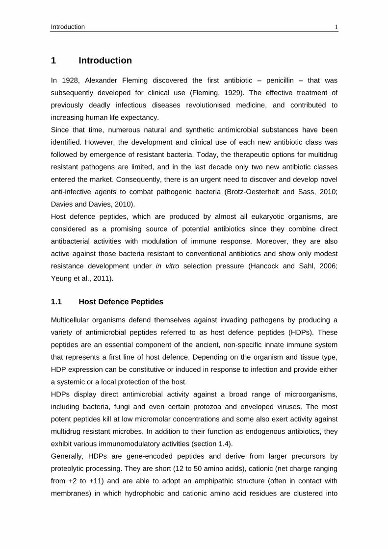
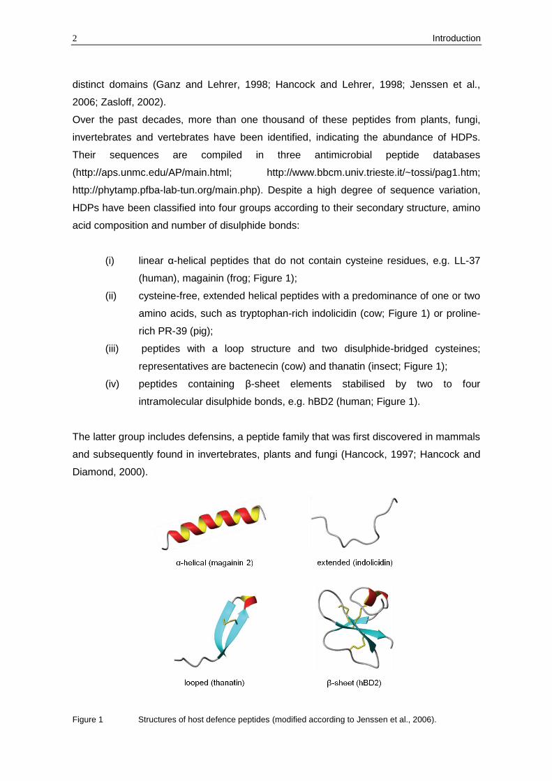
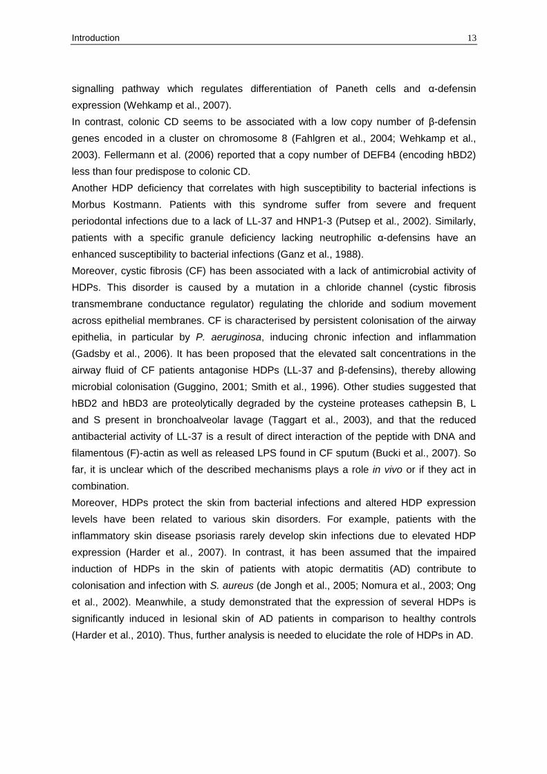
HDPs have been classified into four groups according to their secondary structure, amino
acid composition and number of disulphide bonds:
(i) linear α-helical peptides that do not contain cysteine residues, e.g. LL-37
(human), magainin (frog; Figure 1);
(ii) cysteine-free, extended helical peptides with a predominance of one or two
amino acids, such as tryptophan-rich indolicidin (cow; Figure 1) or proline-
rich PR-39 (pig);
(iii) peptides with a loop structure and two disulphide-bridged cysteines;
representatives are bactenecin (cow) and thanatin (insect; Figure 1);
(iv) peptides containing β-sheet elements stabilised by two to four
intramolecular disulphide bonds, e.g. hBD2 (human; Figure 1).
The latter group includes defensins, a peptide family that was first discovered in mammals
and subsequently found in invertebrates, plants and fungi (Hancock, 1997; Hancock and
Diamond, 2000).
Figure 1 Structures of host defence peptides (modified according to Jenssen et al., 2006).
Introduction 3
1.2 Defensins
Members of the defensin family can be found in three eukaryotic kingdoms – in fungi,
plants and both invertebrate and vertebrate animals.
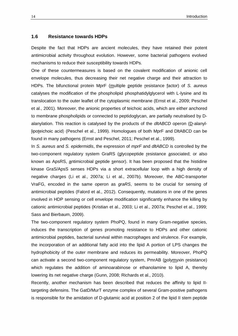
Vertebrate defensins can be subdivided into three families, α-, β- and θ-defensins, based
on precursor and gene structure as well as spacing and pairing of their six conserved
cysteine residues. Both α- and β-defensins are composed of a triple-stranded antiparallel
β-sheet structure stabilised by three disulphide bridges (Figure 2). Alpha-defensins
(disulphide pairing C1-C6, C2-C4, C3-C5) are present in high (up to millimolar)
concentrations in the granules of neutrophils and small intestinal Paneth cells, whereas β-
defensins (disulphide pairing C1-C5, C2-C4, C3-C6) are mainly expressed in epithelial
tissues.
To date, four human neutrophilic α-defensins (HNP1-4), two human enteric α-defensins
(HD5, HD6) and four human epithelial β-defensins (hBD1-4) have been characterised that
differ substantially in their potency and activity spectra (Gram-positive and Gram-negative
bacteria, fungi, viruses) (Ganz, 2003; Schneider et al., 2005). Interestingly, human β-
defensin 1 (hBD1) becomes a potent antimicrobial peptide only after reduction of its
disulphide bonds, then acting against the yeast Candida albicans and anaerobic Gram-
positive commensals of the gut (Schroeder et al., 2011).
However, bioinformatic approaches revealed that the human genome encodes more than
30 β-defensin genes in five chromosomal regions (Schutte et al., 2002; Yamaguchi et al.,
2002). The expression of many of these genes has been confirmed on mRNA level;
several are specifically expressed in the testis and epididymis (Pazgier et al., 2006;
Rodriguez-Jimenez et al., 2003; Yamaguchi et al., 2002), suggesting an involvement of β-
defensins in sperm maturation (Zhou et al., 2004). In addition to defensins, humans
synthesise the α-helical cathelicidin LL-37. Cathelicidins comprise structurally diverse
HDPs whose precursors contain a conserved N-terminal cathelin domain and a C-terminal
antimicrobial domain (Durr et al., 2006).
Theta-defensins have been isolated so far from the neutrophils and monocytes of several
species of Old World monkeys and arose from a pre-existing α-defensin. Their peptide
backbone is cyclised by ligation of two identical or similar nonapeptides and the resulting
peptide ring is stabilised by six disulphide-bridged cysteines (disulphide pairing C1-C6,
C2-C5, C3-C4; Figure 2) (Tang et al., 1999; Tran et al., 2002). Noteworthy, there are six
θ-defensin genes present in the human genome, but a premature stop codon in the signal
sequence aborts their translation, thus causing a higher susceptibility of humans towards
HIV-1 infections (Nguyen et al., 2003).
4 Introduction
Invertebrate defensins have been found in the hemolymph (plasma and hemocytes) and
certain epithelial cells of arthropods (e.g. insects) and mollusks. Functionally, antibacterial
(primarily active against Gram-positive bacteria) and antifungal peptides can be
distinguished which differ from vertebrate defensins by their disulphide bridging patterns
(C1-C4, C2-C5, C3-C6 for peptides containing six cysteines). The core structure of
invertebrate defensins is composed of an α-helical domain linked to a two-stranded
antiparallel β-sheet with three or four disulphide bonds forming the so-called cysteine-
stabilised α-helix β-sheet (CSαβ) motif (Figure 2). Some antifungal peptides like
drosomycin from Drosophila melanogaster contain an additional short N-terminal β-strand
presenting a βαββ-scaffold that is similar to that of antifungal plant defensins (Figure 2)
(Bulet et al., 2004; Thomma et al., 2002). Meanwhile, peptides of another defensin family
referred to as “big defensins” have been isolated from invertebrates (Gerdol et al., 2011;
Rosa et al., 2011; Saito et al., 1995; Teng et al., 2012; Zhao et al., 2007). They consist of
79-94 amino acids that form two distinct structural and functional domains. The
hydrophobic N-terminus exhibits selective activity against Gram-positive bacteria, while
the hydrophilic C-terminus displays Gram-negative activity. Interestingly, the C-terminal
region adopts the typical fold of β-defensins with identical disulphide arrays (C1-C5, C2-
C4, C3-C6; Figure 2), suggesting that β-defensins emerged from an ancestral big
defensin (Zhu and Gao, 2012).
Defensins with a high degree of sequence and structural similarity to invertebrate
defensins have been identified in several fungi (Mygind et al., 2005; Zhu, 2008). For
example, plectasin isolated from the saprophytic ascomycete Pseudoplectania nigrella is
characterised by a CSαβ motif (disulphide pairing C1-C4, C2-C5, C3-C6; Figure 2) and
displays potent antibacterial activity (Mygind et al., 2005).
All plant defensins known so far are characterised by four disulphide bridges and share
the same cysteine pairing pattern (C1-C8, C2-C5, C3-C6, C4-C7; Figure 2). They are
mainly active against fungi, including plant and human pathogens (Tavares et al., 2008;
Thevissen et al., 2007). In past years, increasing evidence for other biological activities
has been gathered such as inhibition of α-amylase activity and protein synthesis, antiviral
and antitumour activity, inhibition of plant root growth, blocking of ion channels and
mediation of zinc tolerance in plants (Aerts et al., 2008; Allen et al., 2008; Lin et al., 2009;
Mirouze et al., 2006; Spelbrink et al., 2004; Wong and Ng, 2005).
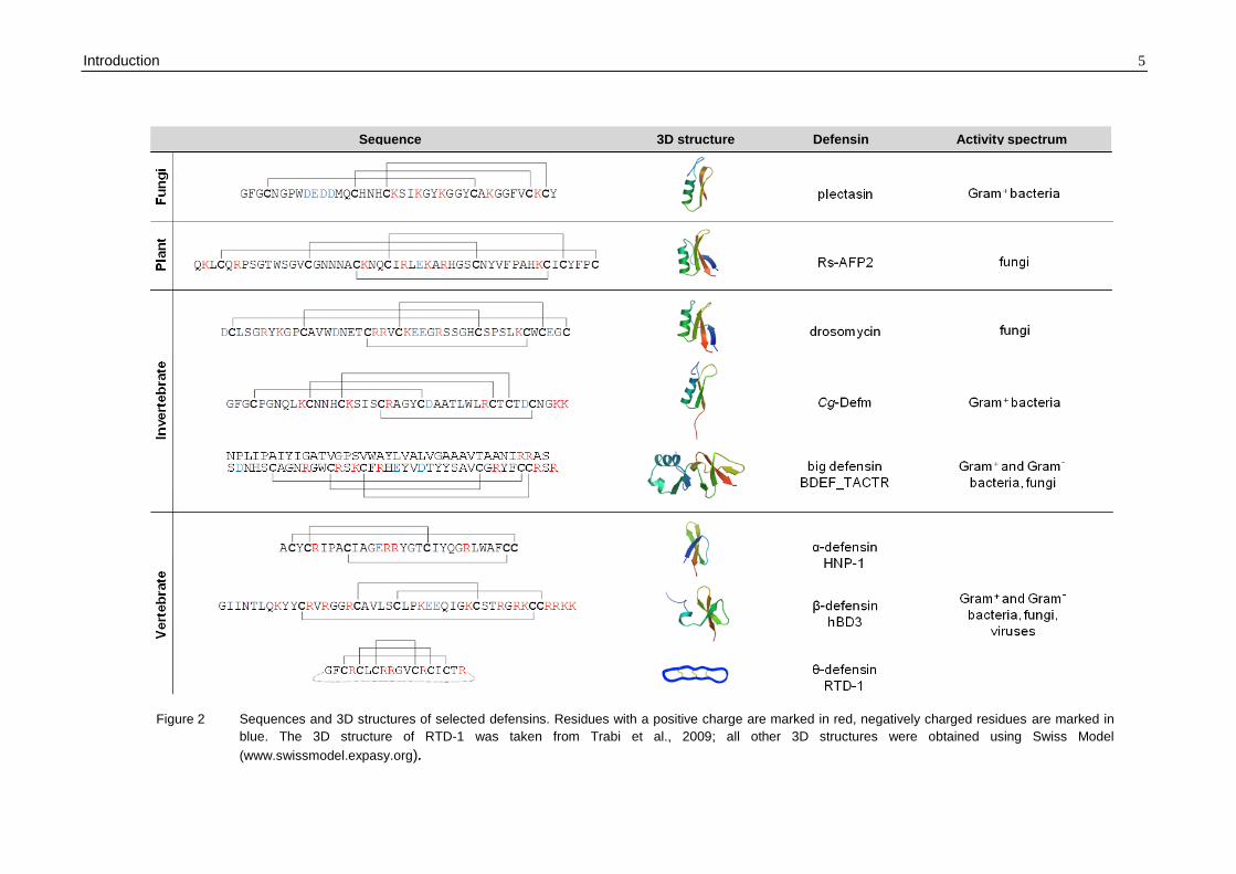
Introduction 5
Figure 2 Sequences and 3D structures of selected defensins. Residues with a positive charge are marked in red, negatively charged residues are marked in
blue. The 3D structure of RTD-1 was taken from Trabi et al., 2009; all other 3D structures were obtained using Swiss Model
(www.swissmodel.expasy.org).
Sequence 3D structure Defensin Activity spectrum
6 Introduction
It has been postulated that all defensins evolved from a single precursor, based on (i)
sequence and structural similarity of plant and insect defensins as well as fungal and
antibacterial invertebrate defensins, (ii) similar mode of action of plant and insect
defensins (section 1.3) and (iii) inter-functionality of defensins from different kingdoms
(Thevissen et al., 2004; Zhu and Gao, 2012). In this respect, overexpression of insect
(Langen et al., 2006) or mammalian defensins (Aerts et al., 2007) in plants resulted in
increased plant resistance to fungal diseases, comparable to that obtained by
overexpression of plant defensins. Vice versa, intravenous injection of plant defensins into
C. albicans infected mice resulted in a significant reduction of the fungal burden in the
kidneys of the infected mice at least as efficiently as the standard drug fluconazole
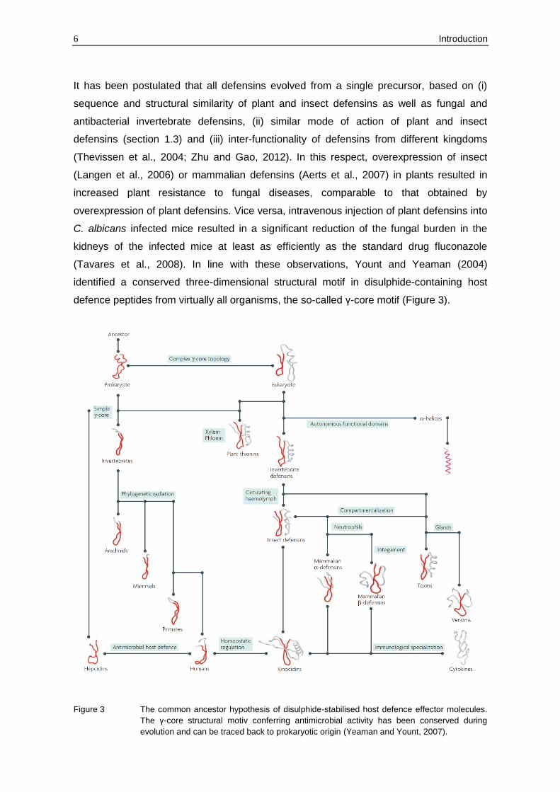
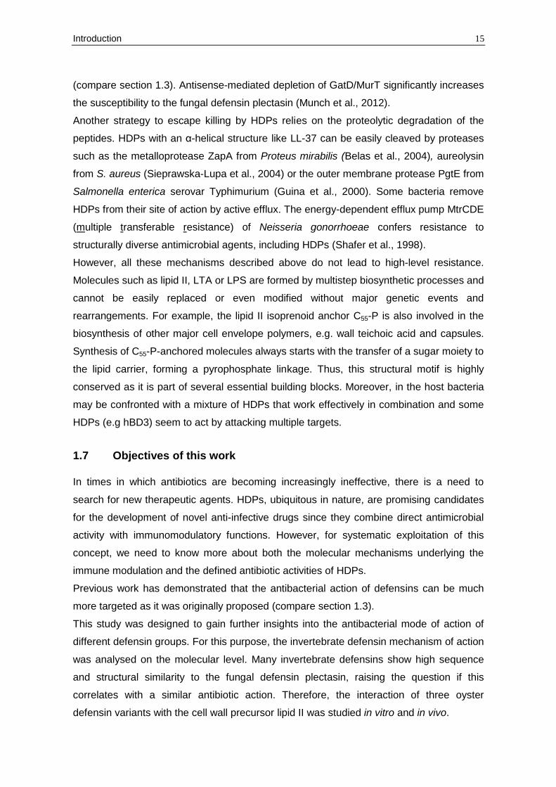
(Tavares et al., 2008). In line with these observations, Yount and Yeaman (2004)
identified a conserved three-dimensional structural motif in disulphide-containing host
defence peptides from virtually all organisms, the so-called γ-core motif (Figure 3).
Figure 3 The common ancestor hypothesis of disulphide-stabilised host defence effector molecules.
The γ-core structural motiv conferring antimicrobial activity has been conserved during
evolution and can be traced back to prokaryotic origin (Yeaman and Yount, 2007).
Introduction 7
This motif arises from the bidirectional orientation of a specific amino acid sequence and
is composed of two antiparallel β-sheets with an interposed short turn region. Indeed, this
motif can also be found in other cysteine-stabilised host defence effector molecules with
antimicrobial activity like venoms, toxins or microbicidal chemokines. Thus, it is proposed
that disulphide-containing HDPs emerged from a common ancestral peptide that can be
traced back to prokaryotic origin (Figure 3) (Yeaman and Yount, 2007).
Consistent with this hypothesis, two defensin-like peptides (DLP) were recently identified
in the myxobacteria Anaeromyxobacter dehalogenans and Stigmatella aurantiaca.
Despite the lack of two cysteine residues, the A. dehalogenans DLP exhibits the typical
CSαβ fold of fungal, invertebrate and plant defensins and displays activity against the
malaria parasite Plasmodium falciparum (Gao et al., 2009; Zhu, 2007).
1.3 Antimicrobial mode of action of HDPs
Almost all HDPs are cationic and amphipathic. Hence, it has been frequently
demonstrated, using model membranes and unilamellar vesicles of various lipid
compositions, that HDPs interact with negatively charged components of the microbial
surface and subsequently disrupt membrane barrier function via pore-formation or
unspecific membrane permeabilisation (Gazit et al., 1995; Henzler-Wildman et al., 2004;
Yang et al., 1998; Yang et al., 2001). Numerous reports on biophysical behaviour of
synthetic and natural peptides in lipid bilayers were published and different models have
been elaborated to describe the membrane-peptide interaction. The best studied peptides
among HDPs are linear peptides that are unstructured in solution and adopt an α-helical
conformation in the presence of membranes.
In this context, it was reasoned that positively charged cationic peptides are
electrostatically attracted to negatively charged phospholipids which are typically present
in the outer leaflet of microbial membranes. With increasing peptide concentration the
peptide molecules insert into the bilayer either forming transmembrane pores or disrupting
the membrane in a detergent-like manner (Brogden, 2005; Zasloff, 2002). For example,
the formation of “toroidal pores” was demonstrated for magainin 2 (Yang et al., 1998) and
LL-37 (Henzler-Wildman et al., 2004), whereas the induction of “barrel-stave” pores
seems unique for the non-ribosomal peptide antibiotic alamethicin (Yang et al., 2001).
Alternatively, peptides such as cecropins accumulate parallel to the membrane surface
and at sufficient high concentration cause disintegration of the lipid bilayer (“carpet
model”) (Gazit et al., 1995; Zasloff, 2002). In all model systems, membrane
permeabilisation depends on the lipid composition (chain length and charge of
phospholipids). The interaction of peptides is strongly reduced by zwitterionic
8 Introduction
phospholipids or cholesterol, both prominent constituents of eukaryotic cell membranes -
a feature that might explain the reduced toxicity towards host cells.
Hence, in the past, discussions on the mode of microbicidal action of HDPs mainly
focused on the lipid bilayer as a target, whereas membrane proteins making up 50% or
more of microbial membranes were hardly considered. While the relevance of the lipid
interactions for the attraction of HDPs to the membrane interface can hardly be
questioned, the relevance for the actual killing process may be overestimated. There is
increasing evidence that some HDPs enter the cytoplasm without disrupting the
membrane bilayer; once inside the cell, they may interfere with nucleic acid and/or protein
synthesis (Boman et al., 1993; Lehrer et al., 1989; Park et al., 1998; Patrzykat et al., 2002;
Subbalakshmi and Sitaram, 1998). Moreover, HDPs have to cross barriers such as cell
wall peptidoglycan or the outer membrane of Gram-negative bacteria to interact with the
cytoplasmic membrane. For Gram-negative bacteria a translocation mechanism termed
“self-promoted uptake” has been described. Thereby, the peptides first bind to the
lipopolysaccharides (LPS) of the outer membrane and subsequently cause displacement
of divalent cations (Mg2+, Ca2+) that bridge and neutralise LPS. Thus, destabilised outer
membrane areas are formed through which peptides can translocate (Hancock, 1997).
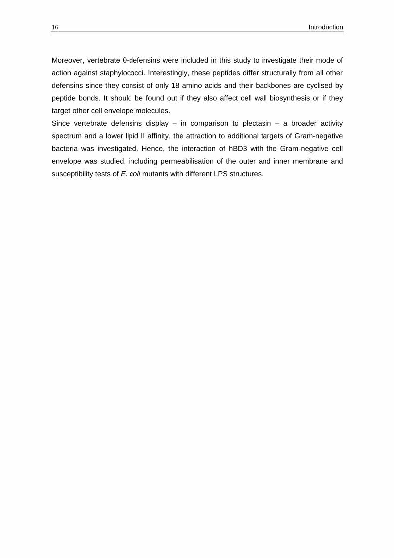
Recently, it has become evident that the fungal defensin plectasin (Schneider et al.,
2010), the α-defensin human neutrophil peptide 1 (HNP-1) (de Leeuw et al., 2010) and the
human β-defensin 3 (hBD3) (Sass et al., 2010) specifically inhibit bacterial cell wall
biosynthesis by targeting lipid II (Figure 27). Plectasin exhibits potent activity against
several Gram-positive bacteria. It does not compromise membrane integrity at all, since
no impact on membrane potential and on intracellular K+ contents could be observed with
cells exposed to plectasin concentrations in the MIC range. Instead, in vitro and in vivo
experiments demonstrated that plectasin forms a stoichiometric complex with the cell wall
precursor lipid II in a 1:1 molar ratio, thus making it unavailable for the interaction with cell
wall biosynthetic enzymes. NMR-based modelling of the plectasin-lipid II complex
indicated that the defensin interacts with the pyrophosphate moiety of lipid II via hydrogen
bonding, whereas the hydrophobic part of the peptide is located on the membrane
surface. Additionally, a salt bridge between the N-terminus (H18) and the D-glutamic acid
in position 2 of the stem peptide is crucial for binding (Figure 4) (Schneider et al., 2010).
The killing of S. aureus by hBD3 is the result of pleiotropic effects. Beside lipid II
sequestration, the peptide seems to have more generalised effects on membrane bound
processes such as the electron transport chain (Sass et al., 2008; Sass et al., 2010). De
Leeuw et al. (2010) proposed that the antimicrobial action of HNP-1 is also based on
lipid II binding as the ability of the peptide to kill S. aureus is strongly reduced in cells with
altered lipid II levels. Moreover, they showed that the affinity towards lipid II is significantly
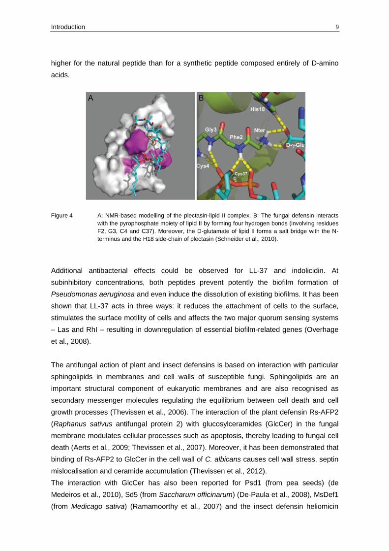
Introduction 9
higher for the natural peptide than for a synthetic peptide composed entirely of D-amino
acids.
Figure 4 A: NMR-based modelling of the plectasin-lipid II complex. B: The fungal defensin interacts
with the pyrophosphate moiety of lipid II by forming four hydrogen bonds (involving residues
F2, G3, C4 and C37). Moreover, the D-glutamate of lipid II forms a salt bridge with the N-
terminus and the H18 side-chain of plectasin (Schneider et al., 2010).
Additional antibacterial effects could be observed for LL-37 and indolicidin. At
subinhibitory concentrations, both peptides prevent potently the biofilm formation of
Pseudomonas aeruginosa and even induce the dissolution of existing biofilms. It has been
shown that LL-37 acts in three ways: it reduces the attachment of cells to the surface,
stimulates the surface motility of cells and affects the two major quorum sensing systems
– Las and RhI – resulting in downregulation of essential biofilm-related genes (Overhage
et al., 2008).
The antifungal action of plant and insect defensins is based on interaction with particular
sphingolipids in membranes and cell walls of susceptible fungi. Sphingolipids are an
important structural component of eukaryotic membranes and are also recognised as
secondary messenger molecules regulating the equilibrium between cell death and cell
growth processes (Thevissen et al., 2006). The interaction of the plant defensin Rs-AFP2
(Raphanus sativus antifungal protein 2) with glucosylceramides (GlcCer) in the fungal
membrane modulates cellular processes such as apoptosis, thereby leading to fungal cell
death (Aerts et al., 2009; Thevissen et al., 2007). Moreover, it has been demonstrated that
binding of Rs-AFP2 to GlcCer in the cell wall of C. albicans causes cell wall stress, septin
mislocalisation and ceramide accumulation (Thevissen et al., 2012).
The interaction with GlcCer has also been reported for Psd1 (from pea seeds) (de
Medeiros et al., 2010), Sd5 (from Saccharum officinarum) (De-Paula et al., 2008), MsDef1
(from Medicago sativa) (Ramamoorthy et al., 2007) and the insect defensin heliomicin
10 Introduction
(from Heliothis virescens) (Thevissen et al., 2004). Other plant defensins such as
DmAMP1 (from Dahlia merckii) bind specifically to inositol phosphoryl-containing
sphingolipids leading to membrane permeabilisation and ion efflux (Thevissen et al., 2003;
Thevissen et al., 1996).
Some HDPs also exhibit antiviral activity by targeting the viral envelope directly, the viral
adsorption and entry into the cell or the intracellular viral life cycle (Ding et al., 2009;
Jenssen et al., 2006). Rhesus macaque θ-defensins (RTDs) and retrocyclins (synthetic θ-
defensins encoded by human pseudogenes) inhibit HIV-1 attachment and entry by binding
specifically to both the viral envelope glycoproteins gp120 or gp41 and the host cell
receptor CD4 (Cole et al., 2002; Gallo et al., 2006; Munk et al., 2003; Seidel et al., 2010;
Wang et al., 2004). The α-defensins HNP1-3 seem to have a direct effect on several
enveloped viruses such as herpes simplex virus type 1 and 2 or influenza A virus, as the
virions lose their ability to infect target cells after incubation with the peptides (Daher et al.,
1986). Moreover, HNP1-3 inhibit multiple steps of the HIV life cycle, e.g. nuclear import
and HIV replication (Chang et al., 2005; Furci et al., 2007; Mackewicz et al., 2003; Seidel
et al., 2010; Wang et al., 2004). Antiviral properties have also been reported for β-
defensins. For example, hBD2 does not only inactivate HIV particles directly, but also
inhibits HIV replication (Seidel et al., 2010; Sun et al., 2005).
1.4 Immunomodulatory functions of HDPs
Research during the last years has demonstrated that HDPs do not only have direct
antimicrobial activities, but also display a diverse range of immunomodulatory functions
(Figure 5). As the antibacterial properties of several HDPs are strongly antagonised under
physiological salt concentrations or serum components, it has been proposed that the
immunomodulatory activities of some of these peptides may be more important to mediate
bacterial clearance in vivo.
Mammalian HPDs are expressed either constitutively or are inducible in various cell types
such as neutrophils, monocytes/macrophages and epitheliaI cells. The expression of
human β-defensins can be triggered by conserved bacterial structures (e.g. LPS) via Toll-
like receptors (TLR) or by proinflammatory stimuli (e.g. TNF-α, IL-1β or IFN-γ). In contrast,
α-defensins are expressed constitutively in the bone marrow and intestinal Paneth cells,
but their secretion can be stimulated, e.g. by bacterial antigens such as OmpA or flagellin
(Hancock and Scott, 2000; Selsted and Ouellette, 2005).
It has been revealed that HDPs can be chemotactic for various immune cells or they can
induce the production and release of particular cytokines/chemokines, thereby recruiting
effector cells to the site of infection (Chertov et al., 1996; Territo et al., 1989; Yang et al.,
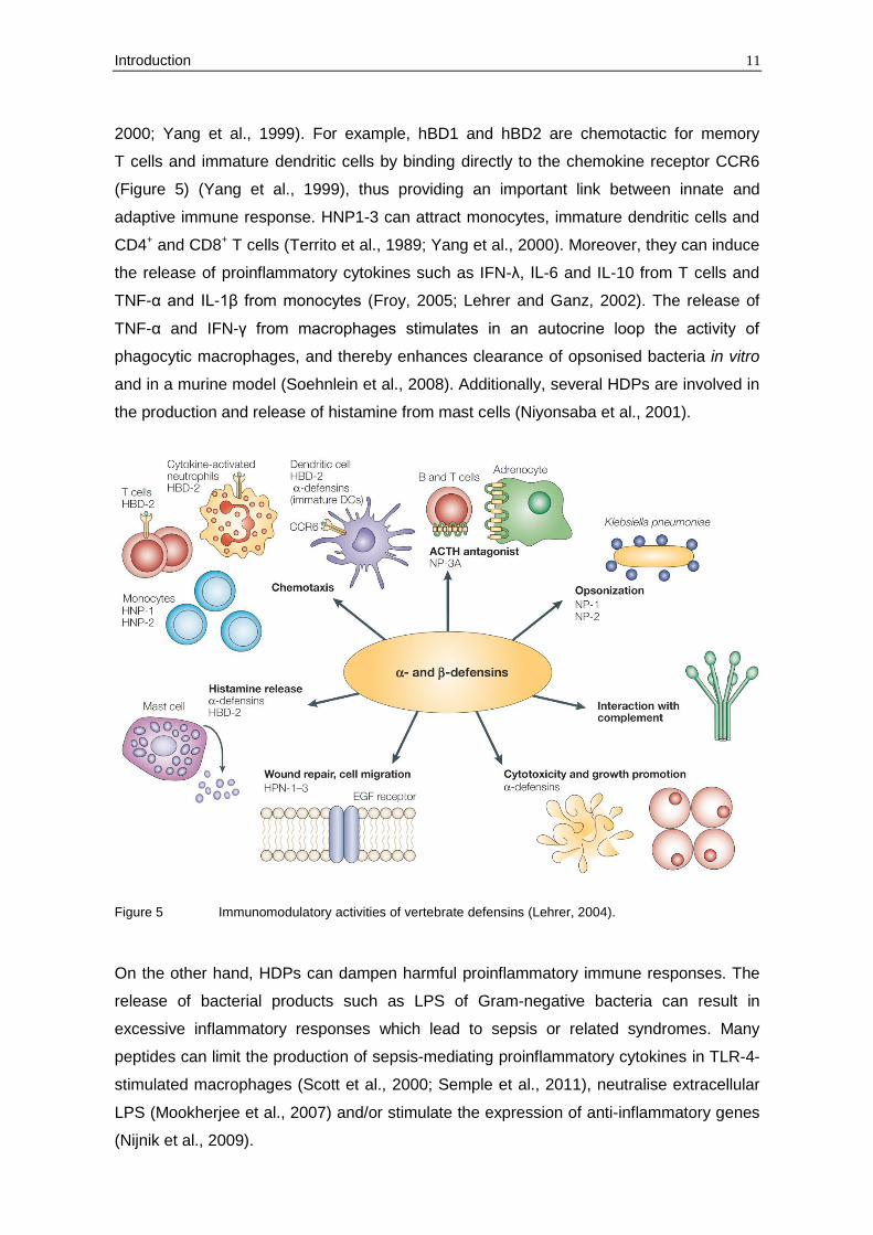
Introduction 11
2000; Yang et al., 1999). For example, hBD1 and hBD2 are chemotactic for memory
T cells and immature dendritic cells by binding directly to the chemokine receptor CCR6
(Figure 5) (Yang et al., 1999), thus providing an important link between innate and
adaptive immune response. HNP1-3 can attract monocytes, immature dendritic cells and
CD4+ and CD8+ T cells (Territo et al., 1989; Yang et al., 2000). Moreover, they can induce
the release of proinflammatory cytokines such as IFN-λ, IL-6 and IL-10 from T cells and
TNF-α and IL-1β from monocytes (Froy, 2005; Lehrer and Ganz, 2002). The release of
TNF-α and IFN-γ from macrophages stimulates in an autocrine loop the activity of
phagocytic macrophages, and thereby enhances clearance of opsonised bacteria in vitro
and in a murine model (Soehnlein et al., 2008). Additionally, several HDPs are involved in
the production and release of histamine from mast cells (Niyonsaba et al., 2001).
Figure 5 Immunomodulatory activities of vertebrate defensins (Lehrer, 2004).
On the other hand, HDPs can dampen harmful proinflammatory immune responses. The
release of bacterial products such as LPS of Gram-negative bacteria can result in
excessive inflammatory responses which lead to sepsis or related syndromes. Many
peptides can limit the production of sepsis-mediating proinflammatory cytokines in TLR-4-
stimulated macrophages (Scott et al., 2000; Semple et al., 2011), neutralise extracellular
LPS (Mookherjee et al., 2007) and/or stimulate the expression of anti-inflammatory genes
(Nijnik et al., 2009).
12 Introduction
Further, HDPs are involved in wound healing. HBD2 and hBD3 are highly expressed in
epidermal keratinocytes in response to injury or infection of the skin. They increase
keratinocyte migration and proliferation through epidermal growth factor receptor
signalling (Niyonsaba et al., 2007). HNP1-3 induce cell proliferation and wound closure as
well as increased cell migration in the airway epithelia (Aarbiou et al., 2002; Aarbiou et al.,
2004).
In addition, defensins can neutralise toxins secreted by bacterial pathogens, thereby
inhibiting their cytocidal activity. It has been shown that HNP1-3 inhibit the function of
lethal toxin from Bacillus anthracis in vitro and in vivo as well as of diphtheria toxin from
Corynebacterium diphtheriae and P. aeruginosa endotoxin A (Kim et al., 2005; Kim et al.,
2006).
The increasing knowledge about the immunomodulatory functions of HDPs led to the
development of synthetic innate defence regulators (IDRs). IDRs are small, synthetic
peptides derived from natural HDP templates which lack direct antimicrobial activity.
IDR-1, whose sequence is based on bovine bactenecin, confers protection in a broad
range of mice infection models, e.g. against MRSA, VRE and Salmonella. It selectively
enhances immunity of the host by stimulating chemokine production by monocytes while
suppressing potentially harmful excessive inflammatory responses (Scott et al., 2007).
1.5 HDP expression and human disorders
The gene copy number or the dysregulated expression of certain HDPs affects the
pathogenesis of several infectious and inflammatory diseases, underlining the importance
of these peptides in combating bacterial pathogens.
Crohn´s disease (CD) is a chronic inflammatory bowel disease that most commonly
affects the ileum of the small intestine and the colon. The disease is in part attributed to
the colonising bacteria of the intestine that trigger mucosal inflammation. It has been
demonstrated that patients with ileal CD have a reduced expression of the intestinal
Paneth cell α-defensins HD5 and HD6, suggesting that α-defensins play a role in
controlling the progress of CD pathogenesis. Consistently, changes in the intestinal
microbiota of transgenic mice producing HD5 in intestinal Paneth cells have been
observed (Wehkamp et al., 2005). The detailed analysis of the intestinal flora of HD5-
expressing mice and mice lacking Paneth cell α-defensins (due to a knockout of the
processing enzyme MMP-7) clearly revealed α-defensin dependent alterations in the
microbial composition, but not in the bacterial number. Thus, the deficiency of α-defensins
in ileal CD may cause an imbalance of the microbial composition which in turn triggers
infection and inflammation (Salzman et al., 2010). The lack of intestinal α-defensins has
been linked to a diminished expression of the transcription factor Tcf-4 of the Wnt
Introduction 13
signalling pathway which regulates differentiation of Paneth cells and α-defensin
expression (Wehkamp et al., 2007).
In contrast, colonic CD seems to be associated with a low copy number of β-defensin
genes encoded in a cluster on chromosome 8 (Fahlgren et al., 2004; Wehkamp et al.,
2003). Fellermann et al. (2006) reported that a copy number of DEFB4 (encoding hBD2)
less than four predispose to colonic CD.
Another HDP deficiency that correlates with high susceptibility to bacterial infections is
Morbus Kostmann. Patients with this syndrome suffer from severe and frequent
periodontal infections due to a lack of LL-37 and HNP1-3 (Putsep et al., 2002). Similarly,
patients with a specific granule deficiency lacking neutrophilic α-defensins have an
enhanced susceptibility to bacterial infections (Ganz et al., 1988).
Moreover, cystic fibrosis (CF) has been associated with a lack of antimicrobial activity of
HDPs. This disorder is caused by a mutation in a chloride channel (cystic fibrosis
transmembrane conductance regulator) regulating the chloride and sodium movement
across epithelial membranes. CF is characterised by persistent colonisation of the airway
epithelia, in particular by P. aeruginosa, inducing chronic infection and inflammation
(Gadsby et al., 2006). It has been proposed that the elevated salt concentrations in the
airway fluid of CF patients antagonise HDPs (LL-37 and β-defensins), thereby allowing
microbial colonisation (Guggino, 2001; Smith et al., 1996). Other studies suggested that
hBD2 and hBD3 are proteolytically degraded by the cysteine proteases cathepsin B, L
and S present in bronchoalveolar lavage (Taggart et al., 2003), and that the reduced
antibacterial activity of LL-37 is a result of direct interaction of the peptide with DNA and
filamentous (F)-actin as well as released LPS found in CF sputum (Bucki et al., 2007). So
far, it is unclear which of the described mechanisms plays a role in vivo or if they act in
combination.
Moreover, HDPs protect the skin from bacterial infections and altered HDP expression
levels have been related to various skin disorders. For example, patients with the
inflammatory skin disease psoriasis rarely develop skin infections due to elevated HDP
expression (Harder et al., 2007). In contrast, it has been assumed that the impaired
induction of HDPs in the skin of patients with atopic dermatitis (AD) contribute to
colonisation and infection with S. aureus (de Jongh et al., 2005; Nomura et al., 2003; Ong
et al., 2002). Meanwhile, a study demonstrated that the expression of several HDPs is
significantly induced in lesional skin of AD patients in comparison to healthy controls
(Harder et al., 2010). Thus, further analysis is needed to elucidate the role of HDPs in AD.
14 Introduction
1.6 Resistance towards HDPs
Despite the fact that HDPs are ancient molecules, they have retained their potent
antimicrobial activity throughout evolution. However, some bacterial pathogens evolved
mechanisms to reduce their susceptibility towards HDPs.
One of these countermeasures is based on the covalent modification of anionic cell
envelope molecules, thus decreasing their net negative charge and their attraction to
HDPs. The bifunctional protein MprF (multiple peptide resistance factor) of S. aureus
catalyses the modification of the phospholipid phosphatidylglycerol with L-lysine and its
translocation to the outer leaflet of the cytoplasmic membrane (Ernst et al., 2009; Peschel
et al., 2001). Moreover, the anionic properties of teichoic acids, which are either anchored
to membrane phospholipids or connected to peptidoglycan, are partially neutralised by D-
alanylation. This reaction is catalysed by the products of the dltABCD operon (D-alanyl-
lipoteichoic acid) (Peschel et al., 1999). Homologues of both MprF and DltABCD can be
found in many pathogens (Ernst and Peschel, 2011; Peschel et al., 1999).
In S. aureus and S. epidermidis, the expression of mprF and dltABCD is controlled by the
two-component regulatory system GraRS (glycopeptide resistance associated; or also
known as ApsRS, antimicrobial peptide sensor). It has been proposed that the histidine
kinase GraS/ApsS senses HDPs via a short extracellular loop with a high density of
negative charges (Li et al., 2007a; Li et al., 2007b). Moreover, the ABC-transporter
VraFG, encoded in the same operon as graRS, seems to be crucial for sensing of
antimicrobial peptides (Falord et al., 2012). Consequently, mutations in one of the genes
involved in HDP sensing or cell envelope modification significantly enhance the killing by
cationic antimicrobial peptides (Kristian et al., 2003; Li et al., 2007a; Peschel et al., 1999;
Sass and Bierbaum, 2009).
The two-component regulatory system PhoPQ, found in many Gram-negative species,
induces the transcription of genes promoting resistance to HDPs and other cationic
antimicrobial peptides, bacterial survival within macrophages and virulence. For example,
the incorporation of an additional fatty acid into the lipid A portion of LPS changes the
hydrophobicity of the outer membrane and reduces its permeability. Moreover, PhoPQ
can activate a second two-component regulatory system, PmrAB (polymyxin resistance)
which regulates the addition of aminoarabinose or ethanolamine to lipid A, thereby
lowering its net negative charge (Gunn, 2008; Richards et al., 2010).
Recently, another mechanism has been described that reduces the affinity to lipid II-
targeting defensins. The GatD/MurT enzyme complex of several Gram-positive pathogens
is responsible for the amidation of D-glutamic acid at position 2 of the lipid II stem peptide
Introduction 15
(compare section 1.3). Antisense-mediated depletion of GatD/MurT significantly increases
the susceptibility to the fungal defensin plectasin (Munch et al., 2012).
Another strategy to escape killing by HDPs relies on the proteolytic degradation of the
peptides. HDPs with an α-helical structure like LL-37 can be easily cleaved by proteases
such as the metalloprotease ZapA from Proteus mirabilis (Belas et al., 2004), aureolysin
from S. aureus (Sieprawska-Lupa et al., 2004) or the outer membrane protease PgtE from
Salmonella enterica serovar Typhimurium (Guina et al., 2000). Some bacteria remove
HDPs from their site of action by active efflux. The energy-dependent efflux pump MtrCDE
(multiple transferable resistance) of Neisseria gonorrhoeae confers resistance to
structurally diverse antimicrobial agents, including HDPs (Shafer et al., 1998).
However, all these mechanisms described above do not lead to high-level resistance.
Molecules such as lipid II, LTA or LPS are formed by multistep biosynthetic processes and
cannot be easily replaced or even modified without major genetic events and
rearrangements. For example, the lipid II isoprenoid anchor C55-P is also involved in the
biosynthesis of other major cell envelope polymers, e.g. wall teichoic acid and capsules.
Synthesis of C55-P-anchored molecules always starts with the transfer of a sugar moiety to
the lipid carrier, forming a pyrophosphate linkage. Thus, this structural motif is highly
conserved as it is part of several essential building blocks. Moreover, in the host bacteria
may be confronted with a mixture of HDPs that work effectively in combination and some
HDPs (e.g hBD3) seem to act by attacking multiple targets.
1.7 Objectives of this work
In times in which antibiotics are becoming increasingly ineffective, there is a need to
search for new therapeutic agents. HDPs, ubiquitous in nature, are promising candidates
for the development of novel anti-infective drugs since they combine direct antimicrobial
activity with immunomodulatory functions. However, for systematic exploitation of this
concept, we need to know more about both the molecular mechanisms underlying the
immune modulation and the defined antibiotic activities of HDPs.
Previous work has demonstrated that the antibacterial action of defensins can be much
more targeted as it was originally proposed (compare section 1.3).
This study was designed to gain further insights into the antibacterial mode of action of
different defensin groups. For this purpose, the invertebrate defensin mechanism of action
was analysed on the molecular level. Many invertebrate defensins show high sequence
and structural similarity to the fungal defensin plectasin, raising the question if this
correlates with a similar antibiotic action. Therefore, the interaction of three oyster
defensin variants with the cell wall precursor lipid II was studied in vitro and in vivo.
16 Introduction
Moreover, vertebrate θ-defensins were included in this study to investigate their mode of
action against staphylococci. Interestingly, these peptides differ structurally from all other
defensins since they consist of only 18 amino acids and their backbones are cyclised by
peptide bonds. It should be found out if they also affect cell wall biosynthesis or if they
target other cell envelope molecules.
Since vertebrate defensins display – in comparison to plectasin – a broader activity
spectrum and a lower lipid II affinity, the attraction to additional targets of Gram-negative
bacteria was investigated. Hence, the interaction of hBD3 with the Gram-negative cell
envelope was studied, including permeabilisation of the outer and inner membrane and
susceptibility tests of E. coli mutants with different LPS structures.
Material and Methods 17
2 Material and Methods
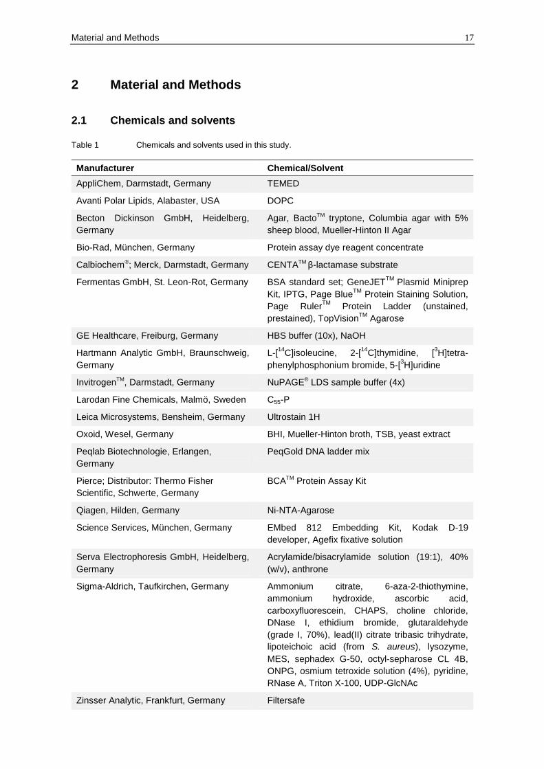
2.1 Chemicals and solvents
Table 1 Chemicals and solvents used in this study.
Manufacturer Chemical/Solvent
AppliChem, Darmstadt, Germany TEMED
Avanti Polar Lipids, Alabaster, USA DOPC
Becton Dickinson GmbH, Heidelberg,
Germany
Agar, BactoTM tryptone, Columbia agar with 5%
sheep blood, Mueller-Hinton II Agar
Bio-Rad, München, Germany Protein assay dye reagent concentrate
Calbiochem®; Merck, Darmstadt, Germany CENTATM β-lactamase substrate
Fermentas GmbH, St. Leon-Rot, Germany BSA standard set; GeneJETTM
Plasmid Miniprep
Kit, IPTG, Page BlueTM
Protein Staining Solution,
Page RulerTM
Protein Ladder (unstained,
prestained), TopVisionTM
Agarose
GE Healthcare, Freiburg, Germany HBS buffer (10x), NaOH
Hartmann Analytic GmbH, Braunschweig,
Germany
L-[14
C]isoleucine, 2-[14
C]thymidine, [3H]tetra-
phenylphosphonium bromide, 5-[3H]uridine
InvitrogenTM, Darmstadt, Germany NuPAGE® LDS sample buffer (4x)
Larodan Fine Chemicals, Malmö, Sweden C55-P
Leica Microsystems, Bensheim, Germany Ultrostain 1H
Oxoid, Wesel, Germany BHI, Mueller-Hinton broth, TSB, yeast extract
Peqlab Biotechnologie, Erlangen,
Germany
PeqGold DNA ladder mix
Pierce; Distributor: Thermo Fisher
Scientific, Schwerte, Germany
BCATM Protein Assay Kit
Qiagen, Hilden, Germany Ni-NTA-Agarose
Science Services, München, Germany EMbed 812 Embedding Kit, Kodak D-19
developer, Agefix fixative solution
Serva Electrophoresis GmbH, Heidelberg,
Germany
Acrylamide/bisacrylamide solution (19:1), 40%
(w/v), anthrone
Sigma-Aldrich, Taufkirchen, Germany Ammonium citrate, 6-aza-2-thiothymine,
ammonium hydroxide, ascorbic acid,
carboxyfluorescein, CHAPS, choline chloride,
DNase I, ethidium bromide, glutaraldehyde
(grade I, 70%), lead(II) citrate tribasic trihydrate,
lipoteichoic acid (from S. aureus), lysozyme,
MES, sephadex G-50, octyl-sepharose CL 4B,
ONPG, osmium tetroxide solution (4%), pyridine,
RNase A, Triton X-100, UDP-GlcNAc
Zinsser Analytic, Frankfurt, Germany Filtersafe
18 Material and Methods
All other chemicals and reagents not listed in this table were purchased from Merck
(Darmstadt, Germany).
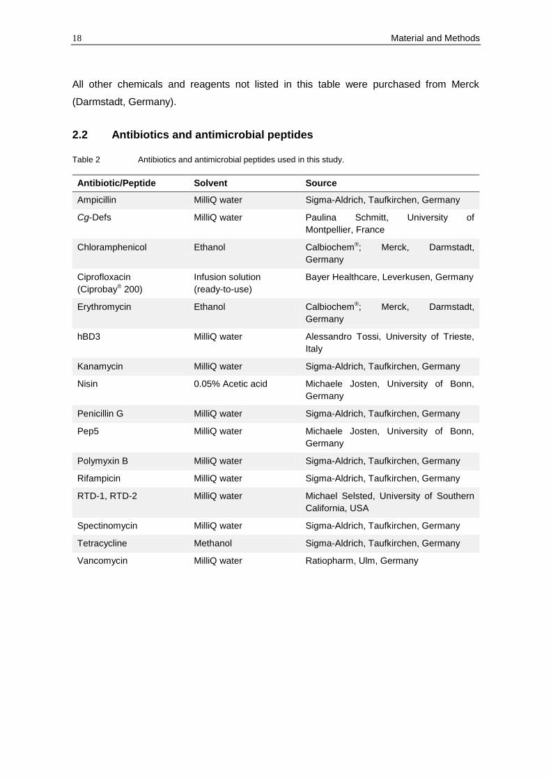
2.2 Antibiotics and antimicrobial peptides
Table 2 Antibiotics and antimicrobial peptides used in this study.
Antibiotic/Peptide Solvent Source
Ampicillin MilliQ water Sigma-Aldrich, Taufkirchen, Germany
Cg-Defs MilliQ water Paulina Schmitt, University of
Montpellier, France
Chloramphenicol Ethanol Calbiochem®; Merck, Darmstadt,
Germany
Ciprofloxacin
(Ciprobay® 200)
Infusion solution
(ready-to-use)
Bayer Healthcare, Leverkusen, Germany
Erythromycin Ethanol Calbiochem®; Merck, Darmstadt,
Germany
hBD3 MilliQ water Alessandro Tossi, University of Trieste,
Italy
Kanamycin MilliQ water Sigma-Aldrich, Taufkirchen, Germany
Nisin 0.05% Acetic acid Michaele Josten, University of Bonn,
Germany
Penicillin G MilliQ water Sigma-Aldrich, Taufkirchen, Germany
Pep5 MilliQ water Michaele Josten, University of Bonn,
Germany
Polymyxin B MilliQ water Sigma-Aldrich, Taufkirchen, Germany
Rifampicin MilliQ water Sigma-Aldrich, Taufkirchen, Germany
RTD-1, RTD-2 MilliQ water Michael Selsted, University of Southern
California, USA
Spectinomycin MilliQ water Sigma-Aldrich, Taufkirchen, Germany
Tetracycline Methanol Sigma-Aldrich, Taufkirchen, Germany
Vancomycin MilliQ water Ratiopharm, Ulm, Germany
Material and Methods 19
2.3 Microbiological methods
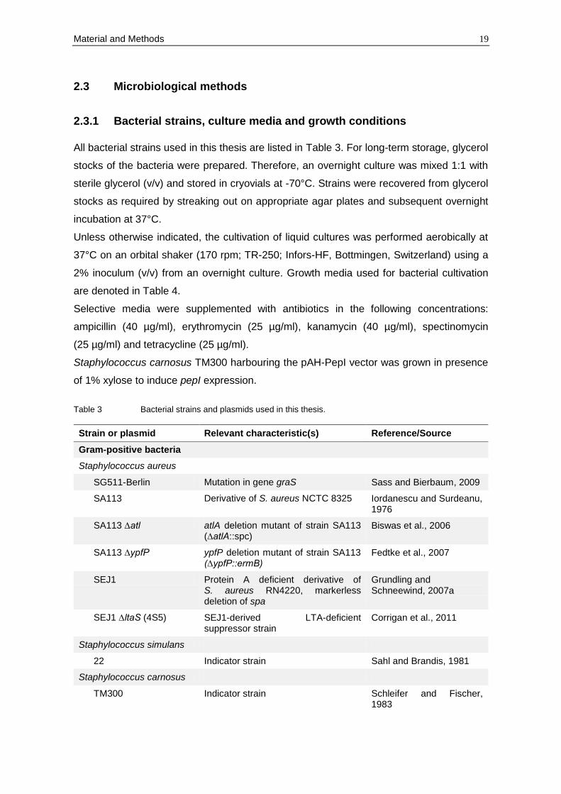
2.3.1 Bacterial strains, culture media and growth conditions
All bacterial strains used in this thesis are listed in Table 3. For long-term storage, glycerol
stocks of the bacteria were prepared. Therefore, an overnight culture was mixed 1:1 with
sterile glycerol (v/v) and stored in cryovials at -70°C. Strains were recovered from glycerol
stocks as required by streaking out on appropriate agar plates and subsequent overnight
incubation at 37°C.
Unless otherwise indicated, the cultivation of liquid cultures was performed aerobically at
37°C on an orbital shaker (170 rpm; TR-250; Infors-HF, Bottmingen, Switzerland) using a
2% inoculum (v/v) from an overnight culture. Growth media used for bacterial cultivation
are denoted in Table 4.
Selective media were supplemented with antibiotics in the following concentrations:
ampicillin (40 µg/ml), erythromycin (25 µg/ml), kanamycin (40 µg/ml), spectinomycin
(25 µg/ml) and tetracycline (25 µg/ml).
Staphylococcus carnosus TM300 harbouring the pAH-PepI vector was grown in presence
of 1% xylose to induce pepI expression.
Table 3 Bacterial strains and plasmids used in this thesis.
Strain or plasmid Relevant characteristic(s) Reference/Source
Gram-positive bacteria
Staphylococcus aureus
SG511-Berlin Mutation in gene graS Sass and Bierbaum, 2009
SA113 Derivative of S. aureus NCTC 8325 Iordanescu and Surdeanu, 1976
SA113 ∆atl atlA deletion mutant of strain SA113 (∆atlA::spc)
Biswas et al., 2006
SA113 ∆ypfP ypfP deletion mutant of strain SA113 (∆ypfP::ermB)
Fedtke et al., 2007
SEJ1 Protein A deficient derivative of S. aureus RN4220, markerless deletion of spa
Grundling and Schneewind, 2007a
SEJ1 ∆ltaS (4S5) SEJ1-derived LTA-deficient suppressor strain
Corrigan et al., 2011
Staphylococcus simulans
22 Indicator strain Sahl and Brandis, 1981
Staphylococcus carnosus
TM300 Indicator strain Schleifer and Fischer, 1983
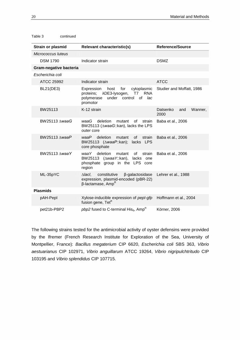
20 Material and Methods
Table 3 continued
Strain or plasmid Relevant characteristic(s) Reference/Source
Micrococcus luteus
DSM 1790 Indicator strain DSMZ
Gram-negative bacteria
Escherichia coli
ATCC 25992 Indicator strain ATCC
BL21(DE3) Expression host for cytoplasmic proteins; λDE3-lysogen, T7 RNA polymerase under control of lac promotor
Studier and Moffatt, 1986
BW25113 K-12 strain Datsenko and Wanner, 2000
BW25113 ∆waaG waaG deletion mutant of strain BW25113 (∆waaG::kan), lacks the LPS outer core
Baba et al., 2006
BW25113 ∆waaP waaP deletion mutant of strain BW25113 (∆waaP::kan); lacks LPS core phosphate
Baba et al., 2006
BW25113 ∆waaY waaY deletion mutant of strain BW25113 (∆waaY::kan), lacks one phosphate group in the LPS core region
Baba et al., 2006
ML-35pYC ∆lacI, constitutive β-galactosidase expression, plasmid-encoded (pBR-22) β-lactamase, Amp
R
Lehrer et al., 1988
Plasmids
pAH-PepI Xylose-inducible expression of pepI-gfp fusion gene, TetR
Hoffmann et al., 2004
pet21b-PBP2 pbp2 fused to C-terminal His6, AmpR Körner, 2006
The following strains tested for the antimicrobial activity of oyster defensins were provided
by the Ifremer (French Research Institute for Exploration of the Sea, University of
Montpellier, France): Bacillus megaterium CIP 6620, Escherichia coli SBS 363, Vibrio
aestuarianus CIP 102971, Vibrio anguillarum ATCC 19264, Vibrio nigripulchtritudo CIP
103195 and Vibrio splendidus CIP 107715.
Material and Methods 21
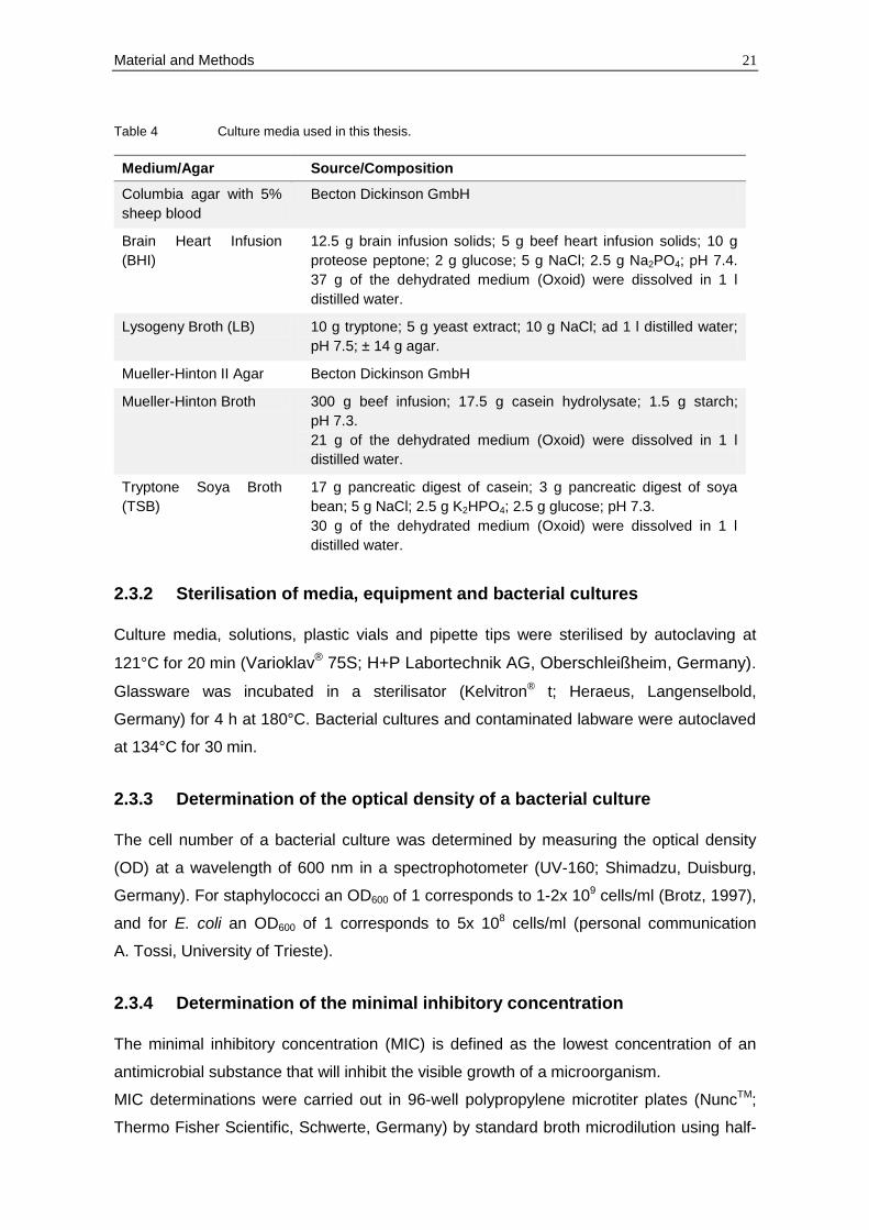
Table 4 Culture media used in this thesis.
Medium/Agar Source/Composition
Columbia agar with 5%
sheep blood
Becton Dickinson GmbH
Brain Heart Infusion
(BHI)
12.5 g brain infusion solids; 5 g beef heart infusion solids; 10 g
proteose peptone; 2 g glucose; 5 g NaCl; 2.5 g Na2PO4; pH 7.4.
37 g of the dehydrated medium (Oxoid) were dissolved in 1 l
distilled water.
Lysogeny Broth (LB) 10 g tryptone; 5 g yeast extract; 10 g NaCl; ad 1 l distilled water;
pH 7.5; ± 14 g agar.
Mueller-Hinton II Agar Becton Dickinson GmbH
Mueller-Hinton Broth 300 g beef infusion; 17.5 g casein hydrolysate; 1.5 g starch;
pH 7.3.
21 g of the dehydrated medium (Oxoid) were dissolved in 1 l
distilled water.
Tryptone Soya Broth
(TSB)
17 g pancreatic digest of casein; 3 g pancreatic digest of soya
bean; 5 g NaCl; 2.5 g K2HPO4; 2.5 g glucose; pH 7.3.
30 g of the dehydrated medium (Oxoid) were dissolved in 1 l
distilled water.
2.3.2 Sterilisation of media, equipment and bacterial cultures
Culture media, solutions, plastic vials and pipette tips were sterilised by autoclaving at
121°C for 20 min (Varioklav® 75S; H+P Labortechnik AG, Oberschleißheim, Germany).
Glassware was incubated in a sterilisator (Kelvitron® t; Heraeus, Langenselbold,
Germany) for 4 h at 180°C. Bacterial cultures and contaminated labware were autoclaved
at 134°C for 30 min.
2.3.3 Determination of the optical density of a bacterial culture
The cell number of a bacterial culture was determined by measuring the optical density
(OD) at a wavelength of 600 nm in a spectrophotometer (UV-160; Shimadzu, Duisburg,
Germany). For staphylococci an OD600 of 1 corresponds to 1-2x 10⁹ cells/ml (Brotz, 1997),
and for E. coli an OD600 of 1 corresponds to 5x 10⁸ cells/ml (personal communication
A. Tossi, University of Trieste).
2.3.4 Determination of the minimal inhibitory concentration
The minimal inhibitory concentration (MIC) is defined as the lowest concentration of an
antimicrobial substance that will inhibit the visible growth of a microorganism.
MIC determinations were carried out in 96-well polypropylene microtiter plates (NuncTM;
Thermo Fisher Scientific, Schwerte, Germany) by standard broth microdilution using half-

22 Material and Methods
concentrated Mueller-Hinton broth or 10% Mueller-Hinton broth (diluted in 10 mM sodium
phosphate buffer (SPB), pH 7.4). Test strains were grown to an OD600 of 1 and
subsequently diluted to 1-2x 10⁵ cells/ml. Then, 50 µl of the bacterial suspension were
mixed with 50 µl of the peptide solution, and the inoculated microtiter plate was incubated
for 10 min at RT on a microtiter shaker (Titertek; Flow Laboratories, Meckenheim,
Germany). The MIC was read after 24 h of incubation at 37°C without agitation. The
results given are mean values of at least two independent experiments performed in
duplicate.
2.3.5 Antagonisation of putative target molecules
Different peptidoglycan precursors (C55-P, lipid II, UDP-MurNAc-pp and UDP-GlcNAc) as
well as LTA from S. aureus were tested for antagonisation of defensin antimicrobial
activity. Therefore, serial dilutions of defensins were performed from 0.25 to 8x MIC in a
polypropylene microtiter plate (NuncTM; Thermo Fisher Scientific, Schwerte, Germany)
with the potential antagonist in a 1:1, 1:2 or 1:4 molar ratio. S. aureus SG511-Berlin was
then added to the microtiter plate as for a conventional MIC determination. After a 24 h-
incubation at 37°C, the lowest peptide/antagonist molar ratio that inhibited the
antimicrobial activity of the highest defensin concentration (8x MIC) was determined.
2.3.6 Bacterial killing kinetics
S. aureus SG511-Berlin was grown in half-concentrated Mueller-Hinton broth to an OD600
of 0.1. Defensins were added in concentrations corresponding to 5x or 10x MIC (as
determined after 24 h). At defined time intervals, 40 µl aliquots of the culture were taken,
diluted in 360 µl 10 mM potassium phosphate buffer (pH 7) and 100 µl of appropriate
dilutions were plated in triplicate on Mueller-Hinton II agar plates (Becton Dickinson
GmbH). The plates were incubated overnight at 37°C and the number of colony forming
units (CFU) was calculated based on the respective dilution factor. An untreated culture
was used as a control.
2.3.7 Growth kinetic measurement
Cells were grown in half-concentrated Mueller-Hinton broth to the exponential phase and
then diluted to an OD600 of 0.2 in 10% Mueller-Hinton broth (in 10 mM SPB, pH 7.4).
Afterwards, 100 µl of the cell suspension were added to 100 µl of various antimicrobial
compounds at 5x MIC. OD600 measurements were performed on a microplate reader
(SunriseTM; Tecan, Crailsheim, Germany) over a period of 6 h at 37°C. Obtained data
were analysed by MagellanTM data analysis software (Tecan, Crailsheim, Germany).
Material and Methods 23
For determination of IC50 values, a serial dilution of hBD3 (0-2 µM) was performed in 10%
Mueller-Hinton broth (in 10 mM SPB, pH 7.4). Then, cells were added to a final
concentration of 1x 10⁶ cells/ml and the bacterial suspension was incubated on a
microplate reader as described above. The IC50 value was defined as the concentration
which inhibited 50% of bacterial growth.
2.3.8 Potassium release from whole cells
Potassium efflux of whole cells was monitored with a MI‐442 potassium electrode and a
MI‐409F reference electrode (Microelectrodes Inc., Bedford, USA) connected to a
microprocessor pH meter (pH 213; HANNA® Instruments, Kehl am Rhein, Germany).
Before starting the measurement, the electrodes were pre-conditioned in choline buffer
(300 mM choline chloride, 30 mM MES, 20 mM Tris; pH 6.5) for at least 1 h. Calibration of
the electrodes was carried out before each determination using freshly prepared standard
solutions containing 0.01, 0.1 and 1 mM KCl in choline buffer (see above).
S. simulans 22 was grown in 50 ml half-concentrated Mueller-Hinton broth (± 10 mM
glucose) at 37°C to an OD600 of 1 to 1.5. Then, cells were harvested by centrifugation
(4,000 rpm, 3 min, 4°C), washed with 25 ml prechilled choline buffer and resuspended in
choline buffer (± 10 mM glucose) to a final OD600 of 30. For each measurement, cells were
diluted in choline buffer (± 10 mM glucose) to an OD600 of 3, and the potassium release
was monitored for 5 min at RT. Defensins were added at 5x and 10x MIC. Potassium
concentrations were calculated from the measured voltage according to Orlov et al. (2002)
and expressed relative to the total amount of potassium released after addition of 1 µM of
the pore-forming lantibiotic nisin (100% efflux).
2.3.9 Determination of the membrane potential using tetraphenyl-
phosphonium bromide
S. aureus SG511-Berlin was grown in half-concentrated Mueller-Hinton broth to an OD600
of 0.5 to 0.6. To monitor the membrane potential, 1 µCi/ml of [3H]tetraphenylphosphonium
bromide (TPP+; 26 Ci/mMol; Hartmann Analytic) was added (the lipophilic TPP+ diffuses
across the bacterial membrane in response to a trans-negative membrane potential). The
culture was treated with defensins at 10x MIC, sample aliquots of 100 µl were filtered
through cellulose acetate filters (pore size 0.2 µm; WhatmanTM, Dassel, Germany) and
washed twice with 5 ml of 50 mM potassium phosphate buffer (pH 7). The filters were
dried, placed into 5 ml scintillation fluid (Filtersafe, Zinsser Analytic) and the radioactivity
was measured with a liquid scintillation counter (Tri-Carb 1900CA; Packard, Downers
Grove, USA) for 5 min per filter. Non specific TPP+ binding was determined by measuring
24 Material and Methods
the TPP+ incorporation into cells treated with 10% butanol (v/v); the total radioactivity was
measured using unfiltered 100 µl sample aliquots. The pore-forming lantibiotic nisin (40
µg/ml corresponds to 10x MIC) was used as a control. For calculation of the membrane
potential (), the TPP+ concentrations were applied into the Nernst equation = (2.3 x
R x T/F) x log (TPP+ inside/TPP+ outside), where T is the absolute temperature, R is the
universal gas constant and F is the Faraday constant (according to Sahl, 1985).
2.3.10 Incorporation of radio-labelled metabolites
The effect of defensins on macromolecular synthesis was studied by monitoring the
incorporation of [3H]- or [14C]-labelled precursors (2-[14C]thymidine, 5-[3H]uridine, L-
[14C]isoleucine; Hartmann Analytic) into S. aureus cells. Therefore, cultures of S. aureus
SG511-Berlin were grown in half-concentrated Mueller-Hinton broth to an OD600 of 0.4,
diluted 10-fold into fresh medium and allowed to regrow to an OD600 of 0.1. Subsequently,
the respective labelled precursor was added to give a final concentration of 1 µCi/ml for
3H-labelled metabolites and 0.1 µCi/ml for 14C-labelled metabolites. Cultures were then
split; one culture was treated with defensins, another one was run as a control. Selectivity
of incorporation was confirmed using antibiotics that inhibit specifially protein
(tetracycline), RNA (rifampicin) or DNA synthesis (ciprofloxacin).
Incorporation of the metabolites was monitored for up to 2 h. At certain time points,
aliquots of 200 µl were taken and immediately added to 2 ml ice-cold 10% trichloroacetic
acid. After 30 min incubation on ice, the samples were filtered through glass microfibre
filters (WhatmanTM, Dassel, Germany) and washed with 5 ml 2.5% trichloroacetic acid.
The dried filters were placed into 5 ml scintillation fluid (Filtersafe; Zinsser Analytic) and
counts were obtained in a liquid scintillation counter (Tri-Carb 1900CA; Packard, Downers
Grove, USA) for 5 min for each filter.
2.3.11 Membrane permeabilisation assay
The effect of defensins on the cell integrity of Gram-negative bacteria was determined
photometrically by measuring the hydrolysis of the extracellular substrates CENTATM
(Calbiochem®) and o-nitrophenyl-β-D-galactopyranoside (ONPG; Sigma-Aldrich) in E. coli
ML35-pYC. The lactose permease-deficient strain contains a cytoplasmic β-galactosidase
which accepts the lactose-mimic ONPG as substrate. ONPG is unable to cross the
membrane and can only be hydrolysed in cells with damaged outer and inner membrane.
Moreover, the strain harbours a plasmid-encoded periplasmic β-lactamase that hydrolyses
the chromogenic cephalosporin CENTATM if the outer membrane is impaired.
Material and Methods 25
E. coli ML35-pYC was grown to early exponential phase and diluted to 1x 10⁷ cells/ml in
10% Mueller-Hinton broth (in 10 mM SPB, pH 7.4). Afterwards, 20 µl of the cell
suspension were incubated with either 0.15 mM CENTA or 1.5 mM ONPG and different
defensin concentrations in a final volume of 200 µl. Hydrolysis was measured at 405 nm
at 37°C for 120 min on a microplate reader (SunriseTM; Tecan, Crailsheim, Germany).
Obtained data were analysed by MagellanTM data analysis software (Tecan, Crailsheim,
Germany).
2.3.12 Intracellular accumulation of the final soluble cell wall precursor
UDP-N-acetylmuramyl-pentapeptide
UDP-N-acetylmuramyl-pentapeptide (UDP-MurNAc-pp) is the final soluble precursor of
bacterial cell wall biosynthesis. Antibiotics such as vancomycin, that interfere with the late,
membrane-bound steps of peptidoglycan synthesis, trigger the accumulation of this
precursor in the cytoplasm which can be isolated and detected by HPLC (Kohlrausch and
Holtje, 1991).
For analysis of the cytoplasmic peptidoglycan precursor pool, S. aureus SG511-Berlin or
S. simulans 22 was grown in half-concentrated Mueller-Hinton broth to an OD600 of 0.5
and supplemented with 130 µg/ml of chloramphenicol. Chloramphenicol prevents the de
novo synthesis of enzymes that may interfere, e.g. through induction of cellular autolysis,
with the accumulation of the UDP-linked peptidoglycan precursor in the cytoplasm (Dai
and Ishiguro, 1988). After 15 min of incubation, defensins or vancomycin, respectively,
were added at 10x MIC (to a final volume of of 5 ml) and the samples were further
incubated for 30 min. Then, cells were spun down (5,300 rpm, 15 min, 4°C), resuspended
in 0.25 ml MilliQ water and treated with two volumes of boiling water for 15 min.
The suspensions were cooled down and the cell extracts were adjusted to pH 2 by
addition of H3PO4. Insoluble components were removed by centrifugation at 13,000 rpm
for 5 min. The supernatants were filtered (Arodisc® syringe filter, pore size 0.2 µm; Pall,
Dreieich, Germany) and analysed by reversed-phase high pressure liquid chromatography
(RP-HPLC) in 50 mM sodium phosphate buffer pH 5.2, developed in an isocratic mode
over 35 min at a flow rate 1 ml/min on a Nucleosil 100-C18 column (Schambeck SFD
GmbH, Bad Honnef, Germany). UDP-linked cell wall precursors were detected at 260 nm
and corresponding fractions were confirmed using matrix-assisted laser desorption
ionisation time-of-flight mass spectrometry (MALDI-TOF MS; BiflexTM, Bruker Daltonics,
Bremen, Germany) in a negative mode with 6-aza-2-thiothymine dissolved in 50%
ethanol/20 mM ammonium citrate (v/v) as matrix.
26 Material and Methods
2.4 Methods in molecular genetics
2.4.1 Determination of concentration and purity of nucleic acids
The concentration of nucleic acids was determined photometrically at 260 nm using a
NanoDrop® ND-1000 spectrophotometer (Peqlab Biotechnologie, Erlangen, Germany).
Purity was assessed by the ratio of absorbance at 260 nm and 280 nm (A260/A280).
2.4.2 Agarose gel electrophoresis
DNA fragments were analysed by standard agarose gel electrophoresis employing 0.8-2%
agarose gels (w/v, Top VisionTM Agarose; Fermentas). Gels were run in 1x TAE buffer and
the DNA was stained by incubating the gel 20-30 min in an ethidium bromide solution
(1 µg/ml in 1 l MilliQ water). Subsequently, DNA bands were visualised using an
ImageMaster® VDS (GE Healthcare/Pharmacia, Freiburg, Germany).
2.4.3 Isolation of plasmid DNA
2 to 5 ml of a bacterial overnight culture were harvested by centrifugation (13,000 rpm,
5 min, RT) and plasmids were extracted using GeneJETTM Plasmid Miniprep Kit
(Fermentas) according to the manufacturer´s instructions. Finally, plasmid DNA was
eluted with 30 µl MilliQ water (70°C) and stored at -20°C until further use.
2.4.4 Preparation of electrocompetent E. coli cells
For preparation of electrocompetent cells, E. coli BL21(DE3) was grown in 500 ml LB at
37°C to an OD600 of 0.5. Afterwards, the culture was incubated on ice for 15 min and
subsequently harvested by centrifugation (6,000 rpm, 15 min, 4°C). Bacterial pellets were
washed with 250 ml prechilled distilled water and 10 ml prechilled 10% glycerol (v/v).
Finally, cells were resuspended in 800 µl prechilled 10% glycerol (v/v). Aliquots of 50 µl
were frozen in liquid nitrogen and stored at -70°C until further use.
2.4.5 Transformation of E. coli by electroporation
Electrocompetent cells were thawed on ice and mixed with 1-2 µl plasmid DNA. After
1 min incubation on ice, the cells were electroporated in a MicroPulserTM (program Ec2;
Biorad, München, Germany) using a prechilled electroporation cuvette (2 mm gap).
Immediately, cells were removed from the cuvette and resuspended in 1 ml LB to recover
for 1 h at 37°C on a rotary shaker.
Material and Methods 27
Finally, 100 µl of the diluted (1:10, 1:100; v/v) and undiluted bacterial solution were
streaked out on LB agar plates containing the appropriate antibiotic.
2.5 Protein and biochemical methods
2.5.1 Heterologous expression and purification of His-tagged PBP2
E. coli BL21(DE3) containing pet21b-PBP2 was grown in 1 l LB supplemented with
40 µg/ml ampicillin at 37°C to an OD600 of 0.6. Protein expression was induced by addition
of IPTG (Fermentas) in a final concentration of 1 mM. After 4 h incubation, cells were
pelleted (7,000 rpm, 12 min, 4°C; Sorvall Evolution RC; Heraeus, Langenselbold,
Germany) and resuspended in 20 ml lysis buffer (Table 5). Then, 200 µg/ml lysozyme,
10 µg/ml RNase and 100 µg/ml DNase were added and cells were further incubated for
30 min at 37°C. After sonication (8x 10 s intervals at 60% including 15 s of cooling on ice;
Sonifier® W250; G. Heinemann, Schwäbisch Gmünd, Germany), the cell debris was
removed by centrifugation (10,300 rpm, 20 min, 4°C) and the supernatant was applied to
1.5 ml Ni-NTA-agarose (Qiagen) and incubated overnight at 4°C under stirring. The batch
was transferred to a polypropylene column (Qiagen, Hilden, Germany) and washed with
10 ml lysis buffer and 5 ml washing buffer (Table 5) to remove weakly bound material.
Recombinant proteins were eluted with 3 ml elution buffer (Table 5) in six fractions of
500 µl, mixed 1:1 with sterile glycerol (v/v) and stored at -20°C. Protein-containing
fractions were pooled and dialysed using Slide-A-Lyzer dialysis cassettes (Pierce; Thermo
Fisher Scientific, Schwerte, Germany).
Table 5 Buffers used for protein purification.
Buffer Chemical ingredients
Lysis buffer 50 mM Tris-HCl (pH 7.5); 0.5 M NaCl; 1% Triton X-100 (v/v);
10 mM imidazole
Washing buffer 50 mM Tris-HCl (pH 7.5); 0.5 M NaCl; 1% Triton X-100 (v/v);
20 mM imidazole
Elution buffer 50 mM Tris-HCl (pH 7.5); 0.5 M NaCl; 1% Triton X-100 (v/v);
200 mM imidazole
Dialysis buffer 50 mM Tris-HCl (pH 7.5); 0.5 M NaCl; 1% Triton X-100 (v/v).
2.5.2 Sodium-dodecyl-sulfate polyacrylamide gel electrophoresis
Purified proteins were analysed by discontinuous sodium-dodecyl-sulfate polyacrylamide
gel electrophoresis (SDS-PAGE; Table 6). Therefore, proteins were mixed with 4x
28 Material and Methods
NuPAGE® LDS sample buffer (Invitrogen) and incubated for 5 min at 95°C prior to loading.
The gel was run in Tris-glycine buffer (25 mM Tris; 192 mM glycine; 0.1% SDS (w/v);
pH 8.5) in a Mini-Protean II Electrophoresis Cell (Bio-Rad, München, Germany) at 100 V
for the stacking gel and 120 V for the resolving gel. Protein bands were visualised using
PageBlueTM protein staining solution (Fermentas) according to the manufacturer´s
instructions.
Table 6 Chemical ingredients for one resolving and one stacking gel.
Chemicals Resolving gel (12%) Stacking gel (4%)
Distilled water 3.845 ml 1.865 ml
3 M Tris, pH 8.5 1.25 ml -
0.1 M Tris, 0.8% SDS (w/v) - 0.3 ml
20% SDS (w/v) 0.05 ml -
APS (21 mg/ml) 0.2 ml 0.08 ml
TEMED 0.005 ml 0.005 ml
40% acrylamide/bisacrylamide (w/v) 2.25 ml 0.25 ml
2.5.3 Zymogram analysis
Cell wall lytic enzymes in the supernatant of RTD- and Pep5-treated cells were analysed
by zymograms. For this, S. aureus SG511-Berlin was grown in half-concentrated Mueller-
Hinton broth to an OD600 of 0.6. Then, cells were harvested by centrifugation (4,000 rpm,
5 min, 4°C) and washed three times with 10 mM SPB (pH 7.4). Finally, cells were
resuspended in 10% Mueller-Hinton broth (in 10 mM SPB, pH 7.4) and aliquots of 2.5 ml
were incubated with peptides corresponding to 10x MIC. After incubation for 30 or 60 min,
cells were pelleted (10,000 rpm, 5 min, 4°C). The supernatant containing released
proteins was concentrated to a volume of 50 µl using VivaSpin-columns (MWCO 3,000;
Sartorius, Göttingen, Germany) according to the manufacturer´s instructions.
Subsequently, 25 µl of the enzyme extract were loaded onto a 12% polyacrylamide gel
containing heat-killed M. luteus DSM 1790 cells as substrate. After run, the gel was
washed three times with distilled water for 15 min before an overnight incubation in buffer
(50 mM Tris-HCl, pH 7.5; 10 mM CaCl2, 10 mM MgCl2, 0.1% Triton X-100, v/v) at 37°C.
Lytic activity was observed as clear zones against an opaque background. To gain a
higher contrast, gels were stained with 0.1% methylene blue (w/v) for 15 min and washed
with distilled water until clear bands became visible.
Material and Methods 29
2.5.4 Determination of peptide and protein concentration
2.5.4.1 Bradford assay
The total protein concentration was measured using protein assay dye reagent (Bio-Rad,
München, Germany) as recommended by the manufacturer. The assay is based on the
absorbance shift of Coomassie Brilliant Blue G-250 bound to basic and aromatic amino
acid residues. The absorption at 595 nm was measured with a spectrophotometer (UV-
160; Shimadzu, Duisburg, Germany). A standard curve was obtained using different
dilutions (0.125-2 mg/ml) of bovine serum albumin (BSA; Fermentas).
2.5.4.2 BCA protein assay
As detergents and some buffers interfere with the Bradford assay, protein concentrations
were alternatively determined using the BCATM Protein Assay Kit (Pierce) according to the
manufacturer´s instructions. This quantification is based on the reduction of copper (II)
ions that form a violet-coloured complex in alkaline solution. The absorption was
measured with a NanoDrop® ND-1000 spectrophotometer (program: protein BCA; Peqlab
Biotechnologie, Erlangen, Germany) at 562 nm. A standard curve was obtained using
different concentrations (5-250 µg/ml) of BSA.
2.5.5 Peptide quantification by spectrometry
The spectrophotometer NanoDrop® ND-1000 (program: protein A280; Peqlab
Biotechnologie, Erlangen, Germany) was used to measure peptide concentrations based
on the molecular weight and extinction coefficient of the sample.
2.5.6 In vitro lipid II synthesis and purification
The cell wall precursor lipid II was synthesised in vitro using membrane preparations of
M. luteus DSM 1790 containing the enzymes MraY and MurG and by external addition of
the lipid carrier C55-P.
2.5.6.1 Membrane preparation of Micrococcus luteus DSM 1790
For membrane preparation, 2 l TSB were inoculated with M. luteus DSM 1790 and
incubated overnight at 37°C in a water bath (120 rpm). The following day, cells were
harvested by centrifugation (8,000 rpm, 10 min, 4°C; Sorval Evolution RC; Heraeus,
Langenselbold, Germany) and washed with 300 ml buffer (50 mM Tris-HCl; 10 mM MgCl2;
pH 7.5). Then, cells were resuspended in 100 ml buffer containing 400 µg/ml lysozyme
30 Material and Methods
and 10 µg/ml DNase, and incubated on ice for 1 to 2 h. The cell suspension was
subsequently warmed up to 35°C, cooled on ice again and membranes were centrifuged
(18,000 rpm, 20 min, 4°C; Sorvall Evolution RC; Heraeus, Langenselbold, Germany).
Finally, the pellets were resuspended in 8 ml buffer (see above) and stored at -70°C until
further use.
2.5.6.2 Extraction of UDP-MurNAc-pp
S. simulans 22 was grown in 3 l Mueller-Hinton broth to an OD600 of 0.75 and
supplemented with 130 µg/ml chloramphenicol. After 15 min incubation, vancomycin was
added in a final concentration of 5 µg/ml and the cells were further incubated for 60 min.
Afterwards, the cells were harvested by centrifugation (8,000 rpm, 10 min, 4°C; Sorvall
Evolution RC; Heraeus, Langenselbold, Germany) and the pellet was resuspended in
20 ml MilliQ water. The cell wall precursor UDP-MurNAc-pp was extracted with two
volumes of boiling water. Finally, the suspension was cooled down, cell debris was
removed by centrifugation (18,000 rpm, 30 min, 4°C; Sorvall Evolution RC; Heraeus,
Langenselbold, Germany) and the supernatant was lyophilised (Alpha 2-4 LSC; Christ,
Osterode am Harz, Germany). The lyophilisate was dissolved in 4 ml MilliQ water and
added to the in vitro lipid II synthesis (section 2.5.6.3).
2.5.6.3 In vitro lipid II synthesis
Initially, the lipid II synthesis was performed in an analytical scale (in a total volume of
75 µl) to find the optimal conditions as membrane preparations can vary in their synthesis
rate (Table 7). Therefore, C55-P (dissolved in methanol:chloroform, 1:1, v/v) was vacuum-
dried and redissolved by addition of Triton X-100 (Table 7). After addition of the isolated
membranes, the other ingredients were added in the following order: Tris-HCl, UDP-
GlcNAc, MgCl2 and UDP-MurNAc-pp (Table 7). The reaction mixture was subsequently
incubated for 2 h at 30°C in a water bath. Bactoprenol-containing products were extracted
with the same volume n-butanol/pyridine acetate (2:1, v/v; pH 4.2) and analysed by thin-
layer chromatography (TLC). For the extraction solution 6 M glacial acetic acid was
adjusted to pH 4.2 by addition of pyridine and mixed with two volumes of n-butanol.
After centrifugation (13,000 rpm, 3 min), 20 µl of the upper (butanol) phase were spotted
on a silica gel (TLC Silica Gel 60 F254; Merck, Darmstadt, Germany) and separated using
chloroform:methanol:water:concentrated ammonium hydroxide (88:48:10:1, v/v/v/v) as the
solvent (Rick et al., 1998). Lipid spots were visualised by PMA staining reagent (2.5%
phosphomolybdate (w/v), 1% ceric sulfate (w/v), 6% sulphuric acid (v/v)). For this, the TLC
Material and Methods 31
plate was dipped into PMA solution, dried and developed by heating at 140°C until dark
blue spots appeared.
For synthesis and purification of milligram quantities of lipid II, the analytical procedure
was scaled up by a factor of 250. After incubation of the reaction mixture, the butanol
phase containing lipid II was collected, subsequently washed (8,000 rpm, 5 min, 4°C) with
an equal volume of cold MilliQ water (pH 4.2) and purified as described below (section
2.5.6.4).
Table 7 Reaction mixture of lipid II synthesis (analytical scale).
Ingredients Volume (µl)
1 mM C55-P 5
4% Triton X-100 10
UDP-MurNAc-pp 5-10
10 mM UDP-GlcNAc 7.5
1 M Tris-HCl, pH 8 5
1 M MgCl2 0.4
Membrane preparation of M. luteus DSM 1790 5-20
MilliQ water ad 75
2.5.6.4 Purification of lipid II by HPLC
Lipid II was applied to a Hi-Trap DEAE column (5 ml; Amersham Biosciences, Freiburg,
Germany) equilibrated with buffer A (chloroform:methanol:water, 2:3:1, v/v/v) with a flow
rate of 3 ml/min. After sample application, the flow rate was set to 5 ml/min and the
column was washed with buffer A for 30 min. Lipid II was eluted in a linear ammonium
biocarbonat gradient (0-25% buffer B; chloroform:methanol:NH4HCO3, 2:3:1, v/v/v). 5 ml-
fractions were collected and analysed by TLC as described above (section 2.5.6.3).
Fractions containing lipid II were combined and concentrated using a rotary vacuum
evaporator (Rotavapor RE11; Büchi Labortechnik, Flawil, Switzerland). Finally, lipid II was
lyophilised, dissolved in chloroform:methanol (1:1, v/v) and stored in glas vials at -20°C.
The lipid II concentration was determined by the phosphate content (section 2.5.7).
2.5.7 Phosphate determination
Phosphate determination was performed according to the method of Rouser (1970). For
this, lipid samples were transferred to phosphate-free glass tubes and dried at 140°C for
20 min. Afterwards, 0.3 ml HClO4 (70%, v/v) were added, and the tubes were incubated in
a block heater at 180°C for 1 to 3 h. After cooling to RT, released inorganic phosphate
32 Material and Methods
was reduced by addition of 1 ml MilliQ water, 0.4 ml ammonium molybdate (1.25%, w/v)
and 0.4 ml of freshly prepared ascorbic acid (5%, w/v), followed by incubation in boiling
water for 5 min. The absorption of the formed, blue-coloured complex was read at
795 nm. Phosphate concentrations of the samples were calculated by using a standard
curve of 0-90 nmol inorganic phosphate (stock solution: 1 mM KH2PO4).
2.5.8 Inhibition of PBP2-catalysed reaction in vitro
The inhibition of the enzymatic activity of PBP2 in vitro was determined by incubating
2 nmol lipid II with 7.5 µg PBP2-His6 in 10 mM MES (pH 5.5) in a final volume of 50 µl.
Peptides were added in molar ratios ranging from 0.5 to 2 with respect to lipid II. The
reaction mixtures were incubated at 30°C for 2 h and lipids were extracted by addition of
one volume n-butanol/pyridine acetate (2:1, v/v) as described above (section 2.5.6.3).
20 µl of the upper phase were spotted onto a TLC plate (TLC Silica Gel 60 F254; Merck,
Darmstadt, Germany) and run in chloroform:methanol:water:ammonium hydroxide
(88:48:10:1, v/v/v/v). Lipid bands were visualised by PMA staining (compare section
2.5.6.3).
2.5.9 Complexation of lipid II
Lipid II (2 nmol) was vacuum-dried for 5 min at RT and Cg-Defh2 in aqueous solution was
added in molar ratios ranging from 0.1 to 1 with respect to the amount of lipid II. Reaction
mixtures were vortexed and incubated at 30°C for 30 min and then applied onto TLC
plates (TLC Silica Gel 60 F254; Merck, Germany, Darmstadt). Plates were developed in
butanol:acetic acid:water:pyridine (15:3:12:10, v/v/v/v) and stained with PMA (compare
section 2.5.6.3).
2.5.10 Transmission electron microscopy
Cells were grown in half-concentrated Mueller-Hinton broth to an OD600 of 0.6. Aliquots of
5 ml were exposed to defensin (at 10x MIC) for 30 min or 60 min at 37°C. Afterwards, the
bacteria were harvested by centrifugation (5,000 rpm, 5 min, 4°C), resuspended in
0.1 M SPB (pH 7.4) containing 3% glutaraldehyde (v/v; Sigma-Aldrich) and fixed overnight
at 4°C. After washing the cells three times for 10 min with 0.1 M SPB (pH 7.4), they were
postfixed in 2% phosphate-buffered osmium tetroxide (v/v; Sigma-Aldrich) at 4°C for 2 h.
Subsequently, the samples were dehydrated with increasing concentrations of ethanol
beginning with 30%. The dehydrated cells were incubated three times in propylene oxide
for 5 min, followed by a treatment with a 1:1 mixture of polypropylene oxide and epon (v/v)
overnight at RT. Finally, cells were embedded in epon using the EMbed-813 Embedding
Material and Methods 33
Kit (Science Services, München, Germany) and incubated for polymerisation at 60°C for
48 h. Thin sections (60 nm) were obtained with a ultramicrotome (Ultracut R; Leica
Microsystems, Bensheim, Germany) using a diamond knife (2 mm, 45°; Science Services,
München, Germany).
The sections were transferred to copper grids (G200H-Cu; 3.05 mm; Science Services,
München, Germany) and contrasted with 3% uranyl acetate (Ultrostain 1H; Leica
Microsystems) and 0.3% lead citrate (10 ml distilled water; 30 mg lead(II) citrate tribasic
trihydrate; 0.1 ml 10 M NaOH).
The sections were examined with an EM900 electron microscope (Zeiss; Oberkochen,
Germany) at 50 kV.
For film developing, the negatives (Electron image film SO-163; Kodak; Science Services,
München, Germany) were first placed into developer (Kodak D-19; Science Services) for
10 min, and subsequently rinsed with distilled water. Afterwards, they were incubated in a
fixative solution (Agefix; Science Services) for 10-15 min, followed by incubation in a
water tank for 20-25 min.
2.5.11 Biomolecular interaction analysis by surface plasmon resonance
The interaction between defensins and lipid II was analysed with a Bicacore® system
which allows analysis of biomolecular interactions in real time based on a surface
plasmon resonance (SPR). For this, one interaction partner is attached to the surface of a
sensor chip (ligand), whereas the other interaction partner (analyte) is passed in solution
over the surface with the immobilised ligand. The interaction between ligand and analyte
generates a response which is proportional to the change in mass. In a sensogram the
response (given in response units, RU; 1 RU = 1 pg/mm2) is plotted against time.
Binding studies between Cg-Defs and lipid II were carried out on a Biacore® 2000
instrument (GE Healthcare, Freiburg, Germany) at 25°C. Therefore, a L1 sensor chip (GE
Healthcare, Freiburg, Germany) was coated with lipids prepared as follows. Two kinds of
small unilamellar vesicles (SUV) were prepared by sonication from 100% pure DOPC
(1,2-dioleoyl-sn-glycero-3-phosphocholine; Avanti polar lipids) and from a mixture
composed of DOPC and lipid II in a 99.2:0.8 molar ratio. SUV were coated on flow cell 1
and 2, respectively. Prior to immobilisation, the chip surface was cleaned by injection of
20 mM CHAPS for 5 min at a flow rate of 10 µl/min, followed by a 1 min injection of 30%
ethanol at 10 µl/min. SUV were injected at 2 µl/min. To remove loosely bound vesicles,
the surface was washed with 10 mM NaOH for 1 min at 100 µl/min. A short injection of
BSA (100 µg/ml) at 30 µl/min was used to cover all non-specific binding sites. The coating
of the lipid layers gave a response in the range of 6,000 to 6,500 RU. For kinetics,
34 Material and Methods
peptides were simultaneously injected at 250 nM on the two flow cells at a flow rate of
30 µl/min. Dissociation was monitored over 400 s in HBS running buffer (10 mM HEPES
pH 7.4; 300 mM NaCl; GE Healthcare). New lipid layers were prepared for every injection
to avoid the need for a regeneration step. The response of peptide binding on DOPC
layers was taken as a negative control and subtracted from the response on lipid II-
containing DOPC layers. To determine the binding ratio, RU levels were measured at the
plateau, just after the end of injection for the three defensins, nisin and tachyplesin. Two
separate experiments were performed for each peptide injected.
2.5.12 Chromatographic purification of lipoteichoic acid
The extraction and purification of lipoteichoic acid (LTA) of S. aureus SG511-Berlin was
performed according to the protocol of Grundling and Schneewind (2007b) with some
modifications.
Cells were grown in 3 l BHI overnight at 37°C in a water bath (120 rpm). The following
day, the bacteria were harvested by centrifugation (7,000 rpm, 15 min, RT) and washed
with 0.1 M sodium citrate (pH 4.7). For LTA extraction, cells were lysed in a Precellys® 24
homogeniser (6,500 rpm, 3x 30 s, 30 s break; Peqlab Biotechnologie, Erlangen Germany)
using the Precellys® glas kit (0.1 mm; Peqlab Biotechnologie, Erlangen, Germany). Glas
beads were pelleted by centrifugation (7,000 rpm, 5 min) and the supernatant containing
bacterial debris was centrifuged at 13,000 g for 60 min (Sorvall Evolution RC; Heraeus,
Langenselbold, Germany). Then, the pellets were washed with 40 ml 0.1 M sodium citrate
buffer (pH 4.7), centrifuged as described above and finally resuspended in 20 ml sodium
citrate buffer (pH 4.7). The suspension was mixed with an equal volume of 1-butanol and
incubated at RT for 30 min under stirring. After centrifugation (13,000 g, 20 min, RT;
Sorvall Evolution RC), the aqueous (lower) phase containing LTA was dialysed against
20 mM sodium citrate (pH 4.7) using Slide-A-Lyzer dialysis cassettes (2,000 MWCO;
Thermo Fisher Scientific, Schwerte, Germany).
Hydrophobic chromatography was executed on an octyl-sepharose CL 4B column
(diameter: 1.6 cm, radius: 0.8 cm, length: 24 cm) equilibrated with 0.1 M sodium citrate
(pH 4.7) in 15% propanol (v/v). The LTA-containing lysate was adjusted to 15% 1-
propanol (v/v) and subjected to the column with a flow rate of 0.3 ml/min. After sample
application, the column was washed overnight with 0.1 M sodium citrate (pH 4.7) in 15%
propanol at the same flow rate. LTA was eluted using a 300 ml gradient of 15-80%
1-propanol in 0.1 M sodium citrate (pH 4.7) with a flow rate of 0.3 ml/min. 5 ml fractions
were collected and analysed by their phosphate content (section 2.5.7). All phosphate-
containing fractions were combined and dialysed against 0.1 M sodium citrate (pH 4.7).
Material and Methods 35
Afterwards, the LTA were concentrated to a volume of 5 ml using VivaSpin columns (15R,
MWCO 2,000; Sartorius, Göttingen, Germany) and stored at -20°C until further use.
2.5.13 Determination of glucose concentration
The glucose concentration of the isolated LTA was determined by the anthrone method
according to Herbert et al. (1971). For this, 0.2 ml of sample and standard solutions (20-
100 µg glucose) were chilled on ice and subsequently mixed with 1 ml freshly prepared
anthrone reagent (200 mg anthrone; 5 ml ethanol; 95 ml 75% H2SO4, v/v). Afterwards, the
tubes were incubated in a block heater at 100°C for 10 min. After cooling, the absorbance
was read at 625 nm.
2.5.14 CF-efflux from LTA-containing liposomes
Vesicles were made of pure DOPC or DOPC supplemented with 0.5 mol% LTA (referring
to the total amount of phospholipids). For this, 4 µM DOPC (dissolved in
chloroform:methanol, 1:1, v/v) were mixed with LTA in a round bottom flask and the
solvent was evaporated under a nitrogen stream. Final traces of solvents were removed in
a dessicator for further 2 h and the dried lipid films were stored at -20°C until further use.
The lipids were resuspended in 600 µl buffer (50 mM MES-KOH; 100 mM K2SO4; pH 6 or
10 mM Tris-HCl; 0.85% NaCl; pH 7.2) containing 50 mM carboxyfluorescein (CF) and the
flask was subsequently placed on a rotary evaporator (without a vacuum; Rotavapor
RE11; Büchi Labortechnik, Flawil, Switzerland) spinning for 30-60 min in a water bath at
30°C. The obtained multilamellar vesicles were subjected to 6 to 8 cycles of freeze-thaw,
and then additionally passed 19 times through a polycarbonate filter (0.4 µm; WhatmanTM,
Dassel, Germany) in an extrusion apparatus (Mini-Extruder; Avanti Polar Lipids,
Alabaster, USA). Liposomes were separated from unencapsulated CF by gel filtration
using sephadex G-50 (Sigma-Aldrich). Therefore, 2 ml of sephadex G-50 (equilibrated in
buffer) were placed into polypropylene columns (Qiagen, Hilden, Germany) and
centrifuged at 750 g for 3 min. The liposomes were subsequently applied to the column
and centrifuged as described above. This procedure was repeated until the flow-through
containing the vesicles was almost colourless.
Then, the CF-loaded vesicles were diluted in 1.5 ml buffer (see above) at a final
concentration of 25 μM phospholipid on a phosphorous base. The fluorescence intensity
was measured at 520 nm (excitation at 492 nm) on a RF-5301 spectrophotometer
(Shimadzu, Duisburg, Germany) for 3 min at RT. Peptides were added after 25 s at
concentrations of 1 µM. To determine 100% marker release, 20 µl of 20% Triton X-100
(v/v) were added at the end of each measurement.

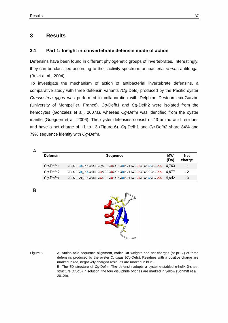
Results 37
3 Results
3.1 Part 1: Insight into invertebrate defensin mode of action
Defensins have been found in different phylogenetic groups of invertebrates. Interestingly,
they can be classified according to their activity spectrum: antibacterial versus antifungal
(Bulet et al., 2004).
To investigate the mechanism of action of antibacterial invertebrate defensins, a
comparative study with three defensin variants (Cg-Defs) produced by the Pacific oyster
Crassostrea gigas was performed in collaboration with Delphine Destoumieux-Garzón
(University of Montpellier, France). Cg-Defh1 and Cg-Defh2 were isolated from the
hemocytes (Gonzalez et al., 2007a), whereas Cg-Defm was identified from the oyster
mantle (Gueguen et al., 2006). The oyster defensins consist of 43 amino acid residues
and have a net charge of +1 to +3 (Figure 6). Cg-Defh1 and Cg-Defh2 share 84% and
79% sequence identity with Cg-Defm.
Figure 6 A: Amino acid sequence alignment, molecular weights and net charges (at pH 7) of three
defensins produced by the oyster C. gigas (Cg-Defs). Residues with a positive charge are
marked in red, negatively charged residues are marked in blue.
B: The 3D structure of Cg-Defm. The defensin adopts a cysteine-stabled α-helix β-sheet
structure (CSαβ) in solution; the four disulphide bridges are marked in yellow (Schmitt et al.,
2012b).
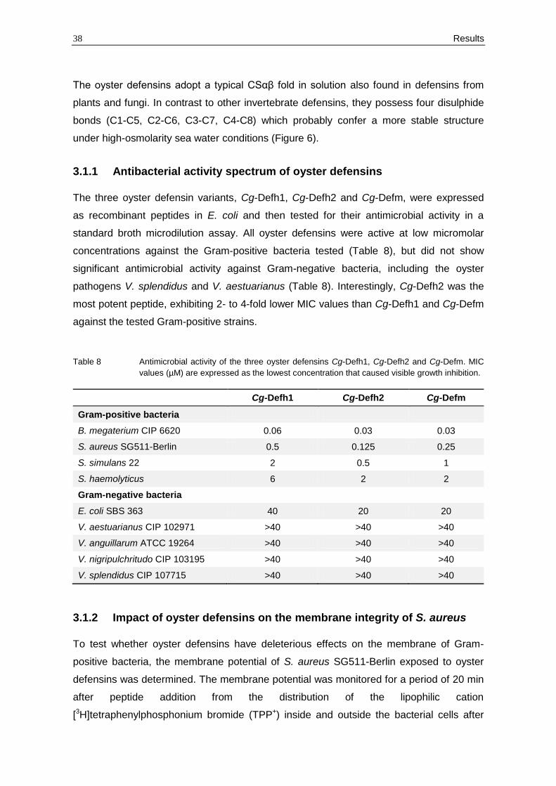
38 Results
The oyster defensins adopt a typical CSαβ fold in solution also found in defensins from
plants and fungi. In contrast to other invertebrate defensins, they possess four disulphide
bonds (C1-C5, C2-C6, C3-C7, C4-C8) which probably confer a more stable structure
under high-osmolarity sea water conditions (Figure 6).
3.1.1 Antibacterial activity spectrum of oyster defensins
The three oyster defensin variants, Cg-Defh1, Cg-Defh2 and Cg-Defm, were expressed
as recombinant peptides in E. coli and then tested for their antimicrobial activity in a
standard broth microdilution assay. All oyster defensins were active at low micromolar
concentrations against the Gram-positive bacteria tested (Table 8), but did not show
significant antimicrobial activity against Gram-negative bacteria, including the oyster
pathogens V. splendidus and V. aestuarianus (Table 8). Interestingly, Cg-Defh2 was the
most potent peptide, exhibiting 2- to 4-fold lower MIC values than Cg-Defh1 and Cg-Defm
against the tested Gram-positive strains.
Table 8 Antimicrobial activity of the three oyster defensins Cg-Defh1, Cg-Defh2 and Cg-Defm. MIC
values (µM) are expressed as the lowest concentration that caused visible growth inhibition.
Cg-Defh1 Cg-Defh2 Cg-Defm
Gram-positive bacteria
B. megaterium CIP 6620 0.06 0.03 0.03
S. aureus SG511-Berlin 0.5 0.125 0.25
S. simulans 22 2 0.5 1
S. haemolyticus 6 2 2
Gram-negative bacteria
E. coli SBS 363 40 20 20
V. aestuarianus CIP 102971 >40 >40 >40
V. anguillarum ATCC 19264 >40 >40 >40
V. nigripulchritudo CIP 103195 >40 >40 >40
V. splendidus CIP 107715 >40 >40 >40
3.1.2 Impact of oyster defensins on the membrane integrity of S. aureus
To test whether oyster defensins have deleterious effects on the membrane of Gram-
positive bacteria, the membrane potential of S. aureus SG511-Berlin exposed to oyster
defensins was determined. The membrane potential was monitored for a period of 20 min
after peptide addition from the distribution of the lipophilic cation
[3H]tetraphenylphosphonium bromide (TPP+) inside and outside the bacterial cells after
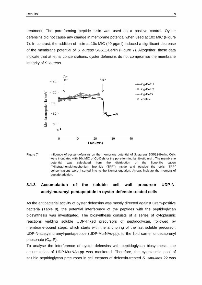
Results 39
treatment. The pore-forming peptide nisin was used as a positive control. Oyster
defensins did not cause any change in membrane potential when used at 10x MIC (Figure
7). In contrast, the addition of nisin at 10x MIC (40 µg/ml) induced a significant decrease
of the membrane potential of S. aureus SG511-Berlin (Figure 7). Altogether, these data
indicate that at lethal concentrations, oyster defensins do not compromise the membrane
integrity of S. aureus.
Figure 7 Influence of oyster defensins on the membrane potential of S. aureus SG511-Berlin. Cells
were incubated with 10x MIC of Cg-Defs or the pore-forming lantibiotic nisin. The membrane
potential was calculated from the distribution of the lipophilic cation
[3H]tetraphenylphosphonium bromide (TPP
+) inside and outside the cells. TPP
+
concentrations were inserted into to the Nernst equation. Arrows indicate the moment of
peptide addition.
3.1.3 Accumulation of the soluble cell wall precursor UDP-N-
acetylmuramyl-pentapeptide in oyster defensin treated cells
As the antibacterial activity of oyster defensins was mostly directed against Gram-positive
bacteria (Table 8), the potential interference of the peptides with the peptidoglycan
biosynthesis was investigated. The biosynthesis consists of a series of cytoplasmic
reactions yielding soluble UDP-linked precursors of peptidoglycan, followed by
membrane-bound steps, which starts with the anchoring of the last soluble precursor,
UDP-N-acetylmuramyl-pentapeptide (UDP-MurNAc-pp), to the lipid carrier undecaprenyl
phosphate (C55-P).
To analyse the interference of oyster defensins with peptidoglycan biosynthesis, the
accumulation of UDP-MurNAc-pp was monitored. Therefore, the cytoplasmic pool of
soluble peptidoglycan precursors in cell extracts of defensin-treated S. simulans 22 was
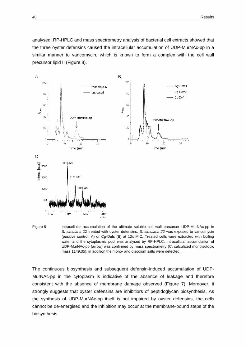
40 Results
analysed. RP-HPLC and mass spectrometry analysis of bacterial cell extracts showed that
the three oyster defensins caused the intracellular accumulation of UDP-MurNAc-pp in a
similar manner to vancomycin, which is known to form a complex with the cell wall
precursor lipid II (Figure 8).
Figure 8 Intracellular accumulation of the ultimate soluble cell wall precursor UDP-MurNAc-pp in
S. simulans 22 treated with oyster defensins. S. simulans 22 was exposed to vancomycin
(positive control; A) or Cg-Defs (B) at 10x MIC. Treated cells were extracted with boiling
water and the cytoplasmic pool was analysed by RP-HPLC. Intracellular accumulation of
UDP-MurNAc-pp (arrow) was confirmed by mass spectrometry (C; calculated monoisotopic
mass 1149.35); in addition the mono- and disodium salts were detected.
The continuous biosynthesis and subsequent defensin-induced accumulation of UDP-
MurNAc-pp in the cytoplasm is indicative of the absence of leakage and therefore
consistent with the absence of membrane damage observed (Figure 7). Moreover, it
strongly suggests that oyster defensins are inhibitiors of peptidoglycan biosynthesis. As
the synthesis of UDP-MurNAc-pp itself is not impaired by oyster defensins, the cells
cannot be de-energised and the inhibition may occur at the membrane-bound steps of the
biosynthesis.
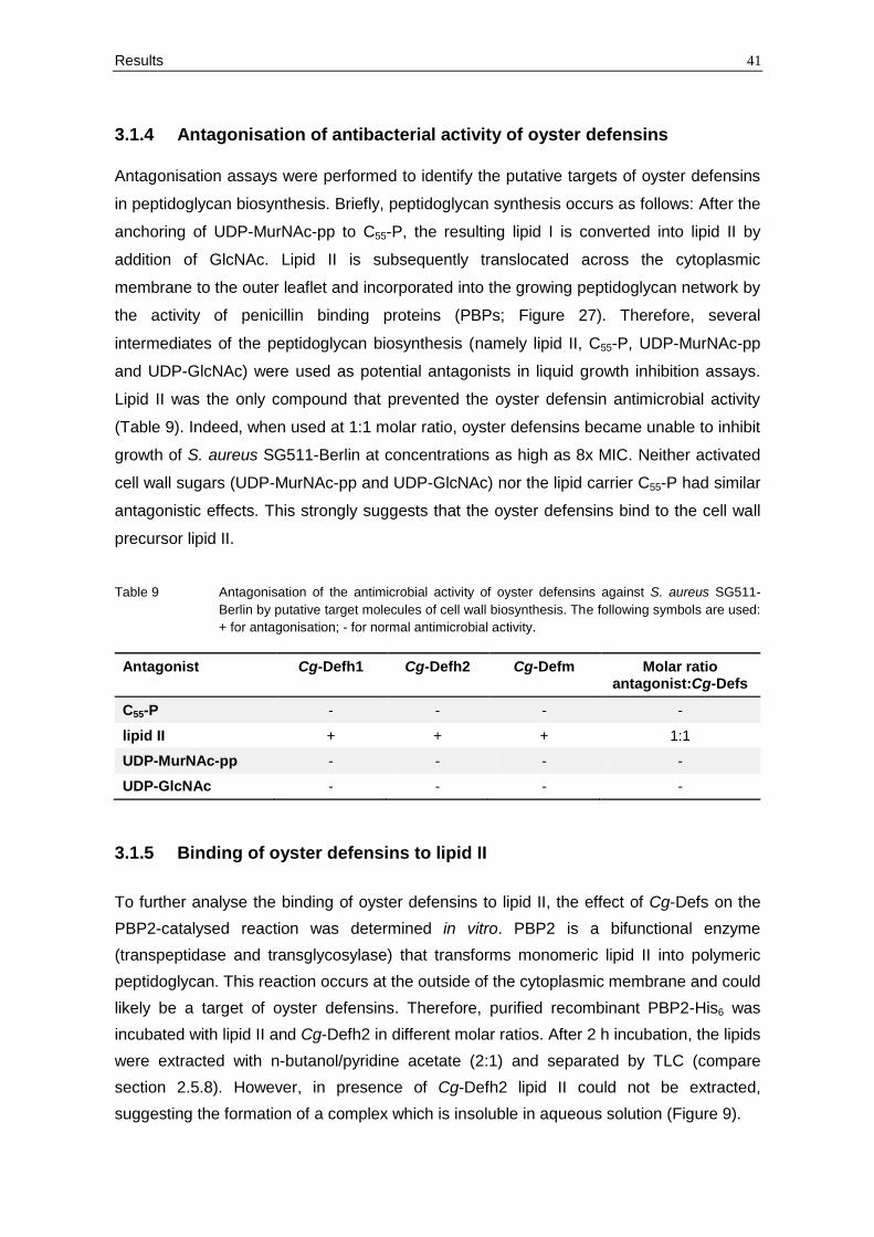
Results 41
3.1.4 Antagonisation of antibacterial activity of oyster defensins
Antagonisation assays were performed to identify the putative targets of oyster defensins
in peptidoglycan biosynthesis. Briefly, peptidoglycan synthesis occurs as follows: After the
anchoring of UDP-MurNAc-pp to C55-P, the resulting lipid I is converted into lipid II by
addition of GlcNAc. Lipid II is subsequently translocated across the cytoplasmic
membrane to the outer leaflet and incorporated into the growing peptidoglycan network by
the activity of penicillin binding proteins (PBPs; Figure 27). Therefore, several
intermediates of the peptidoglycan biosynthesis (namely lipid II, C55-P, UDP-MurNAc-pp
and UDP-GlcNAc) were used as potential antagonists in liquid growth inhibition assays.
Lipid II was the only compound that prevented the oyster defensin antimicrobial activity
(Table 9). Indeed, when used at 1:1 molar ratio, oyster defensins became unable to inhibit
growth of S. aureus SG511-Berlin at concentrations as high as 8x MIC. Neither activated
cell wall sugars (UDP-MurNAc-pp and UDP-GlcNAc) nor the lipid carrier C55-P had similar
antagonistic effects. This strongly suggests that the oyster defensins bind to the cell wall
precursor lipid II.
Table 9 Antagonisation of the antimicrobial activity of oyster defensins against S. aureus SG511-
Berlin by putative target molecules of cell wall biosynthesis. The following symbols are used:
+ for antagonisation; - for normal antimicrobial activity.
Antagonist Cg-Defh1 Cg-Defh2 Cg-Defm Molar ratio antagonist:Cg-Defs
C55-P - - - -
lipid II + + + 1:1
UDP-MurNAc-pp - - - -
UDP-GlcNAc - - - -
3.1.5 Binding of oyster defensins to lipid II
To further analyse the binding of oyster defensins to lipid II, the effect of Cg-Defs on the
PBP2-catalysed reaction was determined in vitro. PBP2 is a bifunctional enzyme
(transpeptidase and transglycosylase) that transforms monomeric lipid II into polymeric
peptidoglycan. This reaction occurs at the outside of the cytoplasmic membrane and could
likely be a target of oyster defensins. Therefore, purified recombinant PBP2-His6 was
incubated with lipid II and Cg-Defh2 in different molar ratios. After 2 h incubation, the lipids
were extracted with n-butanol/pyridine acetate (2:1) and separated by TLC (compare
section 2.5.8). However, in presence of Cg-Defh2 lipid II could not be extracted,
suggesting the formation of a complex which is insoluble in aqueous solution (Figure 9).
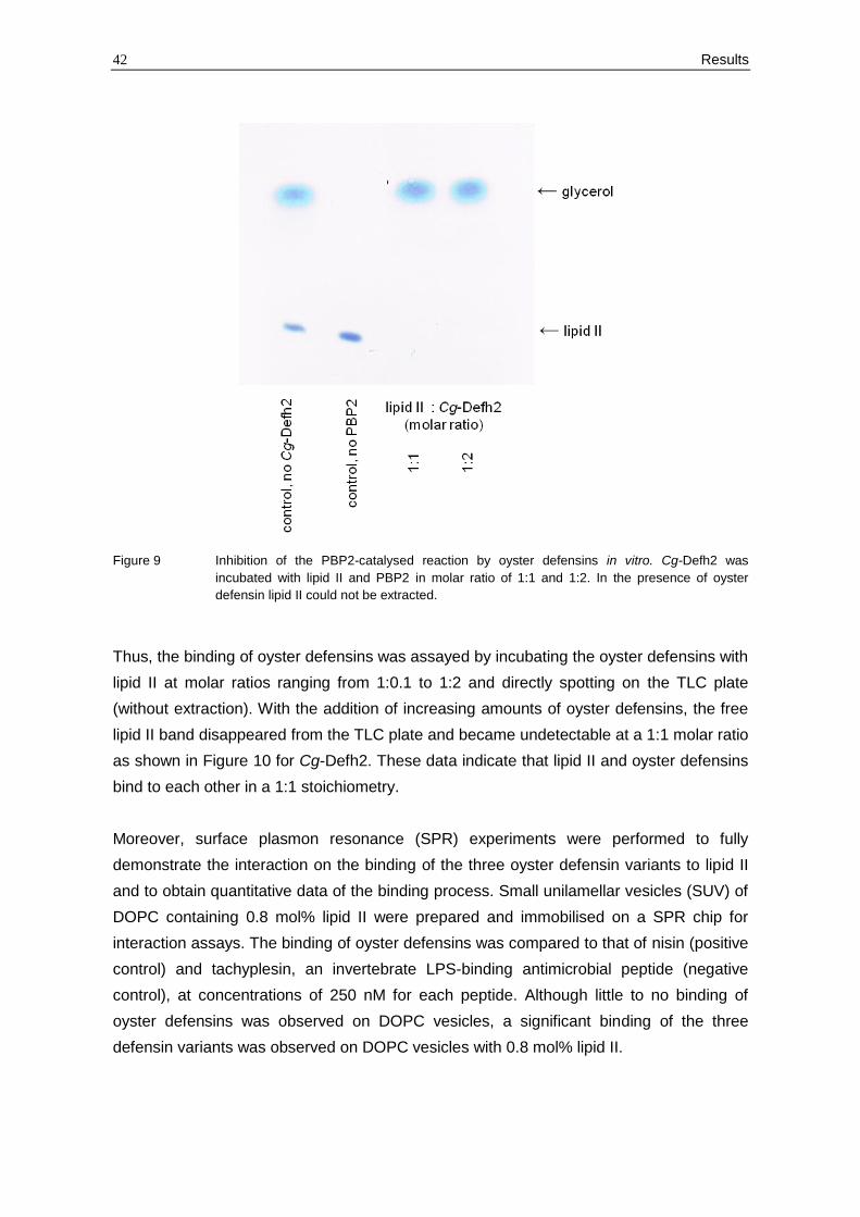
42 Results
Figure 9 Inhibition of the PBP2-catalysed reaction by oyster defensins in vitro. Cg-Defh2 was
incubated with lipid II and PBP2 in molar ratio of 1:1 and 1:2. In the presence of oyster
defensin lipid II could not be extracted.
Thus, the binding of oyster defensins was assayed by incubating the oyster defensins with
lipid II at molar ratios ranging from 1:0.1 to 1:2 and directly spotting on the TLC plate
(without extraction). With the addition of increasing amounts of oyster defensins, the free
lipid II band disappeared from the TLC plate and became undetectable at a 1:1 molar ratio
as shown in Figure 10 for Cg-Defh2. These data indicate that lipid II and oyster defensins
bind to each other in a 1:1 stoichiometry.
Moreover, surface plasmon resonance (SPR) experiments were performed to fully
demonstrate the interaction on the binding of the three oyster defensin variants to lipid II
and to obtain quantitative data of the binding process. Small unilamellar vesicles (SUV) of
DOPC containing 0.8 mol% lipid II were prepared and immobilised on a SPR chip for
interaction assays. The binding of oyster defensins was compared to that of nisin (positive
control) and tachyplesin, an invertebrate LPS-binding antimicrobial peptide (negative
control), at concentrations of 250 nM for each peptide. Although little to no binding of
oyster defensins was observed on DOPC vesicles, a significant binding of the three
defensin variants was observed on DOPC vesicles with 0.8 mol% lipid II.
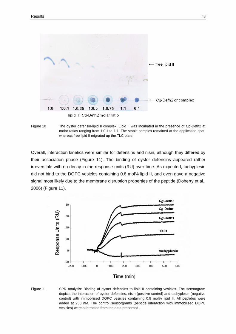
Results 43
Figure 10 The oyster defensin-lipid II complex. Lipid II was incubated in the presence of Cg-Defh2 at
molar ratios ranging from 1:0.1 to 1:1. The stable complex remained at the application spot,
whereas free lipid II migrated up the TLC plate.
Overall, interaction kinetics were similar for defensins and nisin, although they differed by
their association phase (Figure 11). The binding of oyster defensins appeared rather
irreversible with no decay in the response units (RU) over time. As expected, tachyplesin
did not bind to the DOPC vesicles containing 0.8 mol% lipid II, and even gave a negative
signal most likely due to the membrane disruption properties of the peptide (Doherty et al.,
2006) (Figure 11).
Figure 11 SPR analysis: Binding of oyster defensins to lipid II containing vesicles. The sensorgram
depicts the interaction of oyster defensins, nisin (positive control) and tachyplesin (negative
control) with immobilised DOPC vesicles containing 0.8 mol% lipid II. All peptides were
added at 250 nM. The control sensorgrams (peptide interaction with immobilised DOPC
vesicles) were subtracted from the data presented.
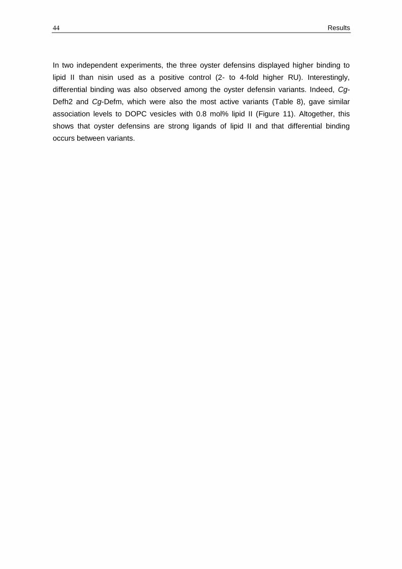
44 Results
In two independent experiments, the three oyster defensins displayed higher binding to
lipid II than nisin used as a positive control (2- to 4-fold higher RU). Interestingly,
differential binding was also observed among the oyster defensin variants. Indeed, Cg-
Defh2 and Cg-Defm, which were also the most active variants (Table 8), gave similar
association levels to DOPC vesicles with 0.8 mol% lipid II (Figure 11). Altogether, this
shows that oyster defensins are strong ligands of lipid II and that differential binding
occurs between variants.
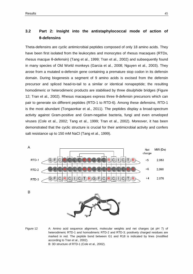
Results 45
3.2 Part 2: Insight into the antistaphylococcal mode of action of
θ-defensins
Theta-defensins are cyclic antimicrobial peptides composed of only 18 amino acids. They
have been first isolated from the leukocytes and monocytes of rhesus macaques (RTDs,
rhesus macque θ-defensin) (Tang et al., 1999; Tran et al., 2002) and subsequently found
in many species of Old World monkeys (Garcia et al., 2008; Nguyen et al., 2003). They
arose from a mutated α-defensin gene containing a premature stop codon in its defensin
domain. During biogenesis a segment of 9 amino acids is excised from the defensin
precursor and spliced head-to-tail to a similar or identical nonapeptide; the resulting
homodimeric or heterodimeric products are stabilised by three disulphide bridges (Figure
12; Tran et al., 2002). Rhesus macaques express three θ-defensin precursors which can
pair to generate six different peptides (RTD-1 to RTD-6). Among these defensins, RTD-1
is the most abundant (Tongaonkar et al., 2011). The peptides display a broad-spectrum
activity against Gram-positive and Gram-negative bacteria, fungi and even enveloped
viruses (Cole et al., 2002; Tang et al., 1999; Tran et al., 2002). Moreover, it has been
demonstrated that the cyclic structure is crucial for their antimicrobial activity and confers
salt resistance up to 150 mM NaCl (Tang et al., 1999).
Figure 12 A: Amino acid sequence alignment, molecular weights and net charges (at pH 7) of
heterodimeric RTD-1 and homodimeric RTD-2 and RTD-3; positively charged residues are
marked in red. The peptide bond between G1 and R18 is indicated by lines (modified
according to Tran et al., 2002).
B: 3D structure of RTD-1 (Cole et al., 2002).
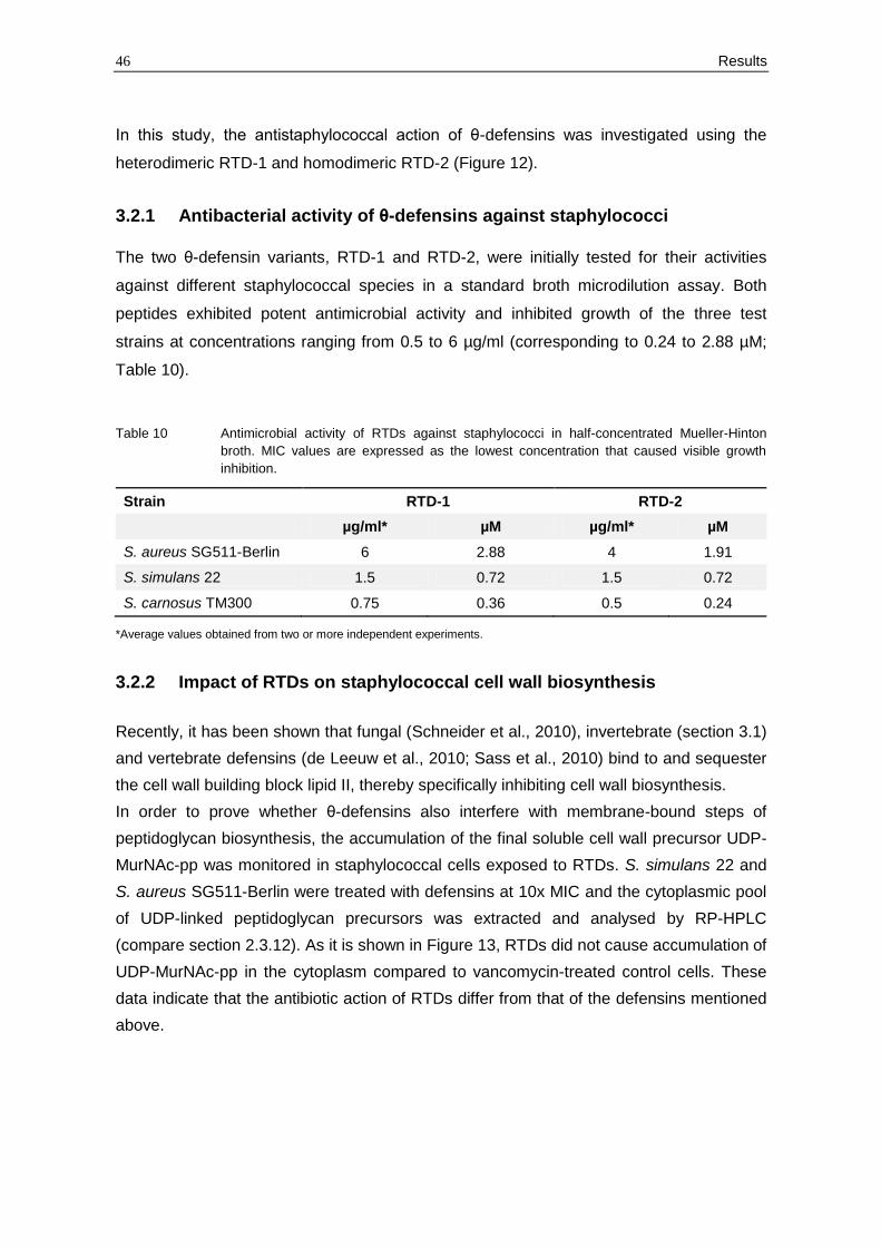
46 Results
In this study, the antistaphylococcal action of θ-defensins was investigated using the
heterodimeric RTD-1 and homodimeric RTD-2 (Figure 12).
3.2.1 Antibacterial activity of θ-defensins against staphylococci
The two θ-defensin variants, RTD-1 and RTD-2, were initially tested for their activities
against different staphylococcal species in a standard broth microdilution assay. Both
peptides exhibited potent antimicrobial activity and inhibited growth of the three test
strains at concentrations ranging from 0.5 to 6 µg/ml (corresponding to 0.24 to 2.88 µM;
Table 10).
Table 10 Antimicrobial activity of RTDs against staphylococci in half-concentrated Mueller-Hinton
broth. MIC values are expressed as the lowest concentration that caused visible growth
inhibition.
Strain RTD-1 RTD-2
µg/ml* µM µg/ml* µM
S. aureus SG511-Berlin 6 2.88 4 1.91
S. simulans 22 1.5 0.72 1.5 0.72
S. carnosus TM300 0.75 0.36 0.5 0.24
*Average values obtained from two or more independent experiments.
3.2.2 Impact of RTDs on staphylococcal cell wall biosynthesis
Recently, it has been shown that fungal (Schneider et al., 2010), invertebrate (section 3.1)
and vertebrate defensins (de Leeuw et al., 2010; Sass et al., 2010) bind to and sequester
the cell wall building block lipid II, thereby specifically inhibiting cell wall biosynthesis.
In order to prove whether θ-defensins also interfere with membrane-bound steps of
peptidoglycan biosynthesis, the accumulation of the final soluble cell wall precursor UDP-
MurNAc-pp was monitored in staphylococcal cells exposed to RTDs. S. simulans 22 and
S. aureus SG511-Berlin were treated with defensins at 10x MIC and the cytoplasmic pool
of UDP-linked peptidoglycan precursors was extracted and analysed by RP-HPLC
(compare section 2.3.12). As it is shown in Figure 13, RTDs did not cause accumulation of
UDP-MurNAc-pp in the cytoplasm compared to vancomycin-treated control cells. These
data indicate that the antibiotic action of RTDs differ from that of the defensins mentioned
above.
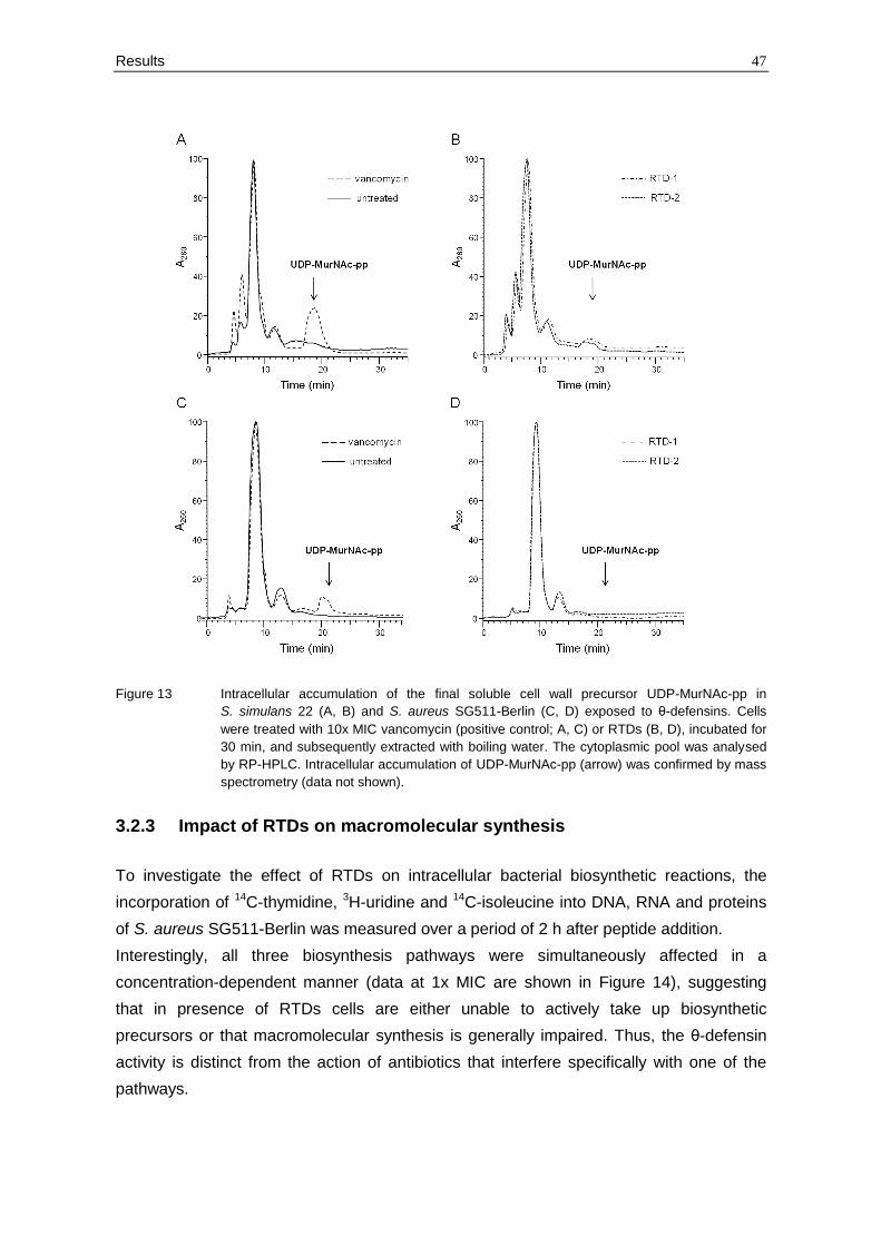
Results 47
Figure 13 Intracellular accumulation of the final soluble cell wall precursor UDP-MurNAc-pp in
S. simulans 22 (A, B) and S. aureus SG511-Berlin (C, D) exposed to θ-defensins. Cells
were treated with 10x MIC vancomycin (positive control; A, C) or RTDs (B, D), incubated for
30 min, and subsequently extracted with boiling water. The cytoplasmic pool was analysed
by RP-HPLC. Intracellular accumulation of UDP-MurNAc-pp (arrow) was confirmed by mass
spectrometry (data not shown).
3.2.3 Impact of RTDs on macromolecular synthesis
To investigate the effect of RTDs on intracellular bacterial biosynthetic reactions, the
incorporation of 14C-thymidine, 3H-uridine and 14C-isoleucine into DNA, RNA and proteins
of S. aureus SG511-Berlin was measured over a period of 2 h after peptide addition.
Interestingly, all three biosynthesis pathways were simultaneously affected in a
concentration-dependent manner (data at 1x MIC are shown in Figure 14), suggesting
that in presence of RTDs cells are either unable to actively take up biosynthetic
precursors or that macromolecular synthesis is generally impaired. Thus, the θ-defensin
activity is distinct from the action of antibiotics that interfere specifically with one of the
pathways.
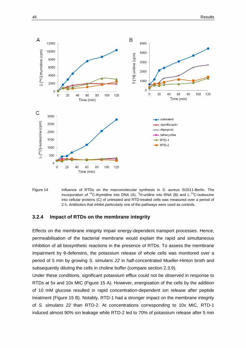
48 Results
Figure 14 Influence of RTDs on the macromolecular synthesis in S. aureus SG511-Berlin. The
incorporation of 14
C-thymidine into DNA (A), 3H-uridine into RNA (B) and L-
14C-isoleucine
into cellular proteins (C) of untreated and RTD-treated cells was measured over a period of
2 h. Antibiotics that inhibit particularly one of the pathways were used as controls.
3.2.4 Impact of RTDs on the membrane integrity
Effects on the membrane integrity impair energy-dependent transport processes. Hence,
permeabilisation of the bacterial membrane would explain the rapid and simultaneous
inhibition of all biosynthetic reactions in the presence of RTDs. To assess the membrane
impairment by θ-defensins, the potassium release of whole cells was monitored over a
period of 5 min by growing S. simulans 22 in half-concentrated Mueller-Hinton broth and
subsequently diluting the cells in choline buffer (compare section 2.3.9).
Under these conditions, significant potassium efflux could not be observed in response to
RTDs at 5x and 10x MIC (Figure 15 A). However, energisation of the cells by the addition
of 10 mM glucose resulted in rapid concentration-dependent ion release after peptide
treatment (Figure 15 B). Notably, RTD-1 had a stronger impact on the membrane integrity
of S. simulans 22 than RTD-2. At concentrations corresponding to 10x MIC, RTD-1
induced almost 90% ion leakage while RTD-2 led to 70% of potassium release after 5 min
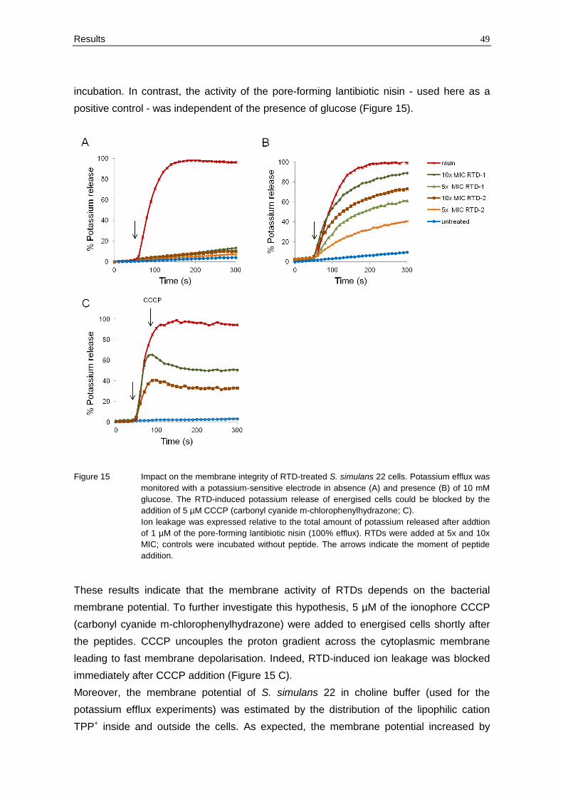
Results 49
incubation. In contrast, the activity of the pore-forming lantibiotic nisin - used here as a
positive control - was independent of the presence of glucose (Figure 15).
Figure 15 Impact on the membrane integrity of RTD-treated S. simulans 22 cells. Potassium efflux was
monitored with a potassium-sensitive electrode in absence (A) and presence (B) of 10 mM
glucose. The RTD-induced potassium release of energised cells could be blocked by the
addition of 5 µM CCCP (carbonyl cyanide m-chlorophenylhydrazone; C).
Ion leakage was expressed relative to the total amount of potassium released after addtion
of 1 µM of the pore-forming lantibiotic nisin (100% efflux). RTDs were added at 5x and 10x
MIC; controls were incubated without peptide. The arrows indicate the moment of peptide
addition.
These results indicate that the membrane activity of RTDs depends on the bacterial
membrane potential. To further investigate this hypothesis, 5 µM of the ionophore CCCP
(carbonyl cyanide m-chlorophenylhydrazone) were added to energised cells shortly after
the peptides. CCCP uncouples the proton gradient across the cytoplasmic membrane
leading to fast membrane depolarisation. Indeed, RTD-induced ion leakage was blocked
immediately after CCCP addition (Figure 15 C).
Moreover, the membrane potential of S. simulans 22 in choline buffer (used for the
potassium efflux experiments) was estimated by the distribution of the lipophilic cation
TPP+ inside and outside the cells. As expected, the membrane potential increased by

50 Results
about 15-20 mV after incubation with 10 mM glucose (Figure 16). Thus, a high membrane
potential - as it can be observed in presence of glucose - seems to be essential for the
membrane-disrupting activity of RTDs.
Figure 16 Membrane potential of S. simulans 22 suspension in choline buffer (300 mM choline
chloride; 30 mM MES, 20 mM Tris; pH 6.5) used for potassium efflux experiments without an
energy source. After 5 min incubation 10 mM glucose were added to the cells (arrow).
Further, the membrane potential of S. aureus SG511-Berlin was monitored in half-
concentrated Mueller-Hinton broth which was routinely used for MIC determinations and
mode of action studies. In absence of glucose, the peptides did not cause any change of
the membrane potential when added at 10x MIC (Figure 17 A). In contrast, the addition of
nisin induced a significant drop of the membrane potential of S. aureus SG511-Berlin
(Figure 17 A). In presence of glucose, the membrane potential increased only slightly
(approximately 10 mV; Figure 17 B). However, RTDs at 10x MIC caused a reduction of
the membrane potential of about 15 mV which was completely restored after 20 min of
treatment (Figure 17 B).
Altogether, the degree of membrane impairment depends on the level of energisation and
therefore on the membrane potential across the bacterial membrane. In cells with a
certain membrane potential, θ-defensins disrupt the membrane barrier function causing a
rapid efflux of small molecules such as potassium ions and a concomitant drop of the
membrane potential.
However, these data did not explain the complete cessation of all biosynthetic reactions
as only a minor impact on the membrane integrity could be observed in medium lacking
free glucose. Thus, the question was raised if the membrane impairment alone is
sufficient for killing by RTDs or if additional activities are involved in the killing mechanism.
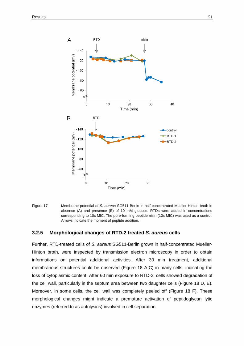
Results 51
Figure 17 Membrane potential of S. aureus SG511-Berlin in half-concentrated Mueller-Hinton broth in
absence (A) and presence (B) of 10 mM glucose. RTDs were added in concentrations
corresponding to 10x MIC. The pore-forming peptide nisin (10x MIC) was used as a control.
Arrows indicate the moment of peptide addition.
3.2.5 Morphological changes of RTD-2 treated S. aureus cells
Further, RTD-treated cells of S. aureus SG511-Berlin grown in half-concentrated Mueller-
Hinton broth, were inspected by transmission electron microscopy in order to obtain
informations on potential additional activities. After 30 min treatment, additional
membranous structures could be observed (Figure 18 A-C) in many cells, indicating the
loss of cytoplasmic content. After 60 min exposure to RTD-2, cells showed degradation of
the cell wall, particularly in the septum area between two daughter cells (Figure 18 D, E).
Moreover, in some cells, the cell wall was completely peeled off (Figure 18 F). These
morphological changes might indicate a premature activation of peptidoglycan lytic
enzymes (referred to as autolysins) involved in cell separation.
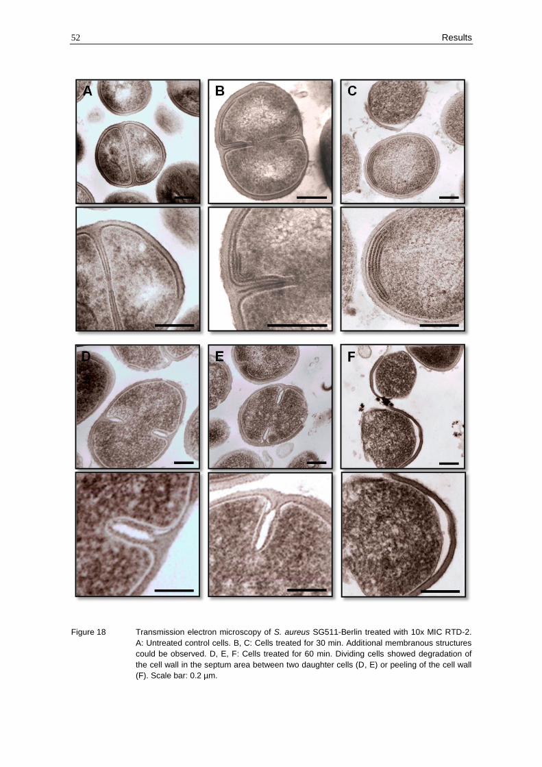
52 Results
Figure 18 Transmission electron microscopy of S. aureus SG511-Berlin treated with 10x MIC RTD-2.
A: Untreated control cells. B, C: Cells treated for 30 min. Additional membranous structures
could be observed. D, E, F: Cells treated for 60 min. Dividing cells showed degradation of
the cell wall in the septum area between two daughter cells (D, E) or peeling of the cell wall
(F). Scale bar: 0.2 µm.
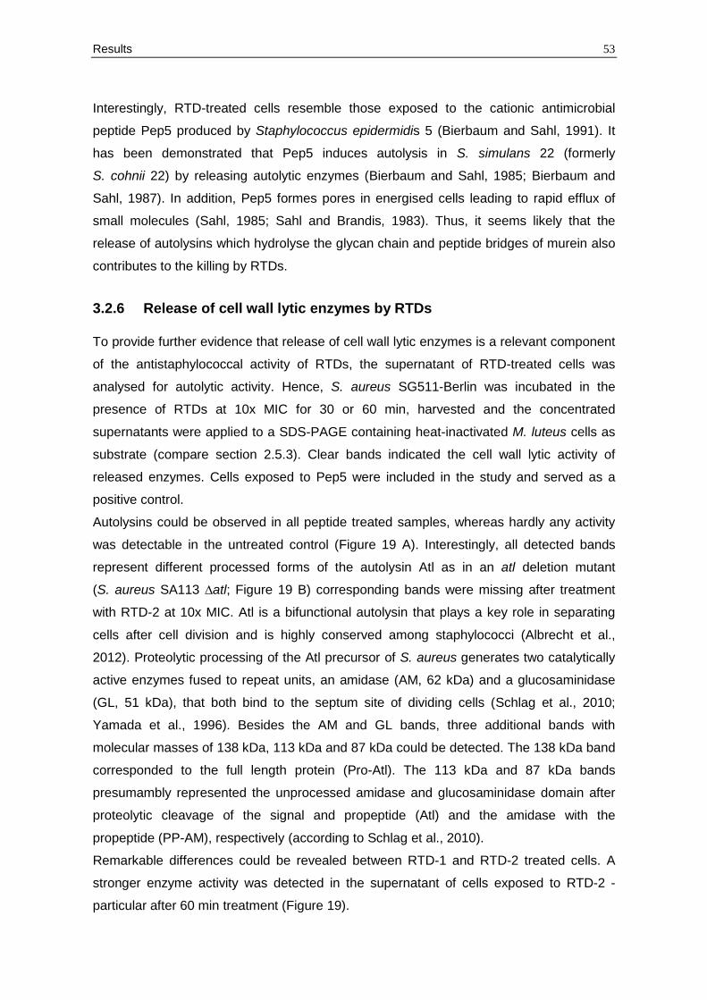
Results 53
Interestingly, RTD-treated cells resemble those exposed to the cationic antimicrobial
peptide Pep5 produced by Staphylococcus epidermidis 5 (Bierbaum and Sahl, 1991). It
has been demonstrated that Pep5 induces autolysis in S. simulans 22 (formerly
S. cohnii 22) by releasing autolytic enzymes (Bierbaum and Sahl, 1985; Bierbaum and
Sahl, 1987). In addition, Pep5 formes pores in energised cells leading to rapid efflux of
small molecules (Sahl, 1985; Sahl and Brandis, 1983). Thus, it seems likely that the
release of autolysins which hydrolyse the glycan chain and peptide bridges of murein also
contributes to the killing by RTDs.
3.2.6 Release of cell wall lytic enzymes by RTDs
To provide further evidence that release of cell wall lytic enzymes is a relevant component
of the antistaphylococcal activity of RTDs, the supernatant of RTD-treated cells was
analysed for autolytic activity. Hence, S. aureus SG511-Berlin was incubated in the
presence of RTDs at 10x MIC for 30 or 60 min, harvested and the concentrated
supernatants were applied to a SDS-PAGE containing heat-inactivated M. luteus cells as
substrate (compare section 2.5.3). Clear bands indicated the cell wall lytic activity of
released enzymes. Cells exposed to Pep5 were included in the study and served as a
positive control.
Autolysins could be observed in all peptide treated samples, whereas hardly any activity
was detectable in the untreated control (Figure 19 A). Interestingly, all detected bands
represent different processed forms of the autolysin Atl as in an atl deletion mutant
(S. aureus SA113 ∆atl; Figure 19 B) corresponding bands were missing after treatment
with RTD-2 at 10x MIC. Atl is a bifunctional autolysin that plays a key role in separating
cells after cell division and is highly conserved among staphylococci (Albrecht et al.,
2012). Proteolytic processing of the Atl precursor of S. aureus generates two catalytically
active enzymes fused to repeat units, an amidase (AM, 62 kDa) and a glucosaminidase
(GL, 51 kDa), that both bind to the septum site of dividing cells (Schlag et al., 2010;
Yamada et al., 1996). Besides the AM and GL bands, three additional bands with
molecular masses of 138 kDa, 113 kDa and 87 kDa could be detected. The 138 kDa band
corresponded to the full length protein (Pro-Atl). The 113 kDa and 87 kDa bands
presumambly represented the unprocessed amidase and glucosaminidase domain after
proteolytic cleavage of the signal and propeptide (Atl) and the amidase with the
propeptide (PP-AM), respectively (according to Schlag et al., 2010).
Remarkable differences could be revealed between RTD-1 and RTD-2 treated cells. A
stronger enzyme activity was detected in the supernatant of cells exposed to RTD-2 -
particular after 60 min treatment (Figure 19).
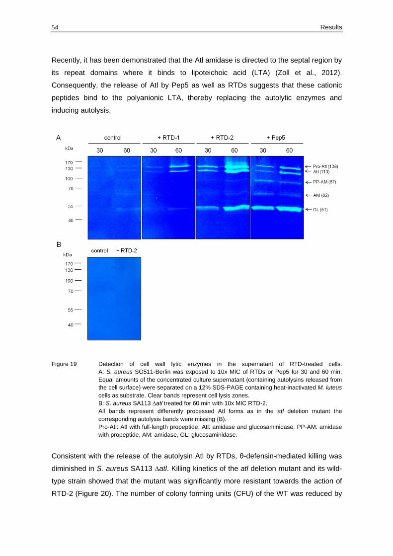
54 Results
Recently, it has been demonstrated that the Atl amidase is directed to the septal region by
its repeat domains where it binds to lipoteichoic acid (LTA) (Zoll et al., 2012).
Consequently, the release of Atl by Pep5 as well as RTDs suggests that these cationic
peptides bind to the polyanionic LTA, thereby replacing the autolytic enzymes and
inducing autolysis.
Figure 19 Detection of cell wall lytic enzymes in the supernatant of RTD-treated cells.
A: S. aureus SG511-Berlin was exposed to 10x MIC of RTDs or Pep5 for 30 and 60 min.
Equal amounts of the concentrated culture supernatant (containing autolysins released from
the cell surface) were separated on a 12% SDS-PAGE containing heat-inactivated M. luteus
cells as substrate. Clear bands represent cell lysis zones.
B: S. aureus SA113 ∆atl treated for 60 min with 10x MIC RTD-2.
All bands represent differently processed Atl forms as in the atl deletion mutant the
corresponding autolysis bands were missing (B).
Pro-Atl: Atl with full-length propeptide, Atl: amidase and glucosaminidase, PP-AM: amidase
with propeptide, AM: amidase, GL: glucosaminidase.
Consistent with the release of the autolysin Atl by RTDs, θ-defensin-mediated killing was
diminished in S. aureus SA113 ∆atl. Killing kinetics of the atl deletion mutant and its wild-
type strain showed that the mutant was significantly more resistant towards the action of
RTD-2 (Figure 20). The number of colony forming units (CFU) of the WT was reduced by
Results 55
several logs after addition of the peptide, whereas the atl deletion mutant was only slightly
affected by RTD-2 even at 80 µg/ml (Figure 20).
Figure 20 Killing kinetic of S. aureus SA113 (WT) and its atl deletion mutant (∆atl) in half-concentrated
Mueller-Hinton broth and presence of 40 and 80 µg/ml RTD-2 over a period of 4 h.
However, no differences of the MIC values between the wildtype and the mutant could be
detected after 24 h incubation (Table 11) as the bacteria which have not been killed by
RTDs were able to regrow during this long incubation time.
Since LTA serves as an anchor molecule for Atl (Zoll et al., 2012), two strains with an
altered LTA content were included in the study. S. aureus SA113 ∆ypfP has a 87%
reduced LTA content compared to the wild-type strain and the remaining LTA are directly
linked to diacylglycerol (DAG) instead of diglucosyl-DAG (Glc2-DAG) (Fedtke et al., 2007).
Moreover, the recently described LTA-deficient suppressor strain S. aureus SEJ1 ∆ltaS
(4S5) was tested for its susceptibility towards RTDs and Pep5. Unexpectedly, both strains
were more susceptible towards Pep5, while only S. aureus SEJ1 ∆ltaS (4S5) showed a
lower MIC of RTDs compared to the parental strain (Table 11).
Table 11 Antibacterial activity of RTDs and Pep5 (µg/ml)* against S. aureus mutants affected in
autolysis or LTA synthesis in 10% Mueller-Hinton broth. MIC values are expressed as the
lowest concentration that caused visible growth inhibition.
Strain RTD-1 RTD-2 Pep5
S. aureus SA113 16 8 4
S. aureus SA113 ∆atl 16 8 4
S. aureus SA113 ∆ypfP 16 16 1
S. aureus SEJ1 >32 32 4
S. aureus SEJ1 ∆ltaS (4S5) 4 2 0.125
*Average values obtained from two or more independent experiments.
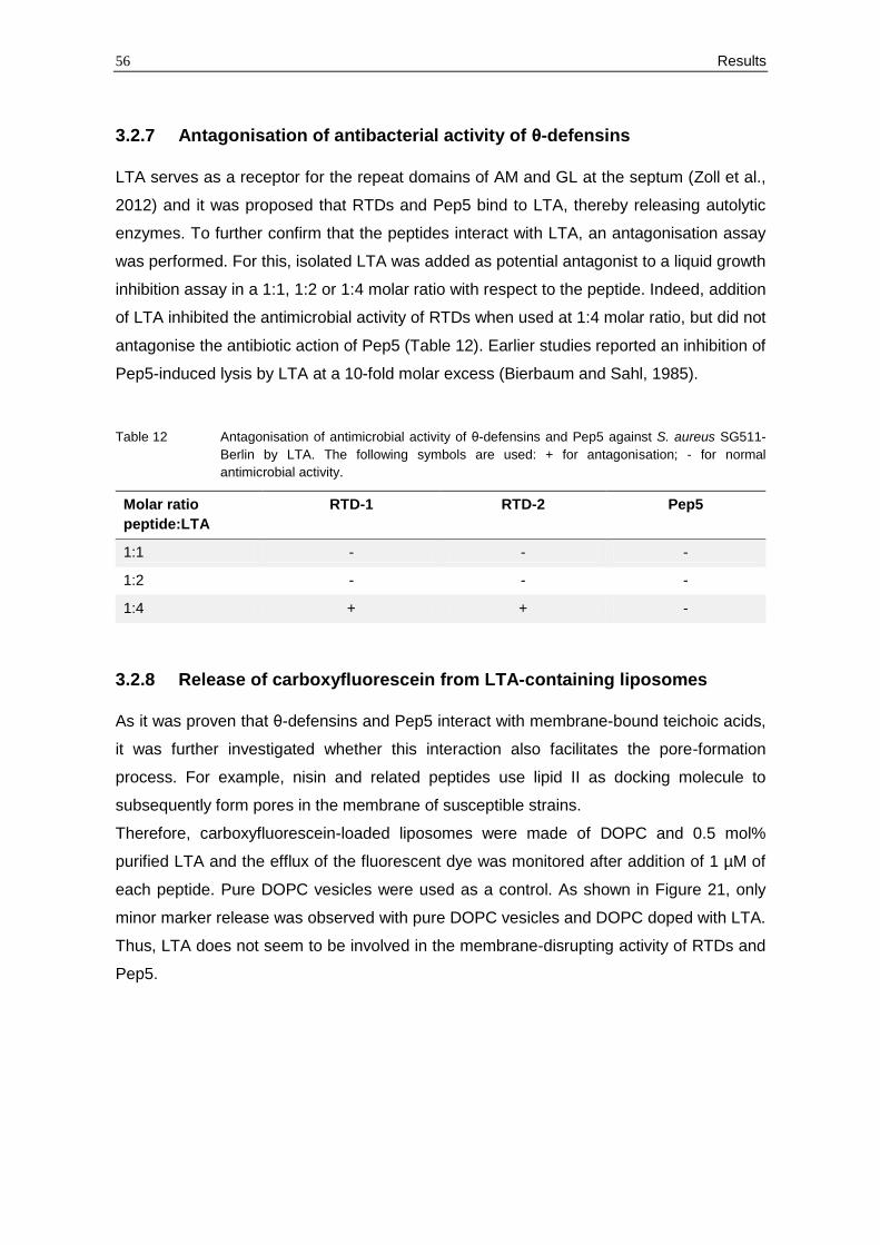
56 Results
3.2.7 Antagonisation of antibacterial activity of θ-defensins
LTA serves as a receptor for the repeat domains of AM and GL at the septum (Zoll et al.,
2012) and it was proposed that RTDs and Pep5 bind to LTA, thereby releasing autolytic
enzymes. To further confirm that the peptides interact with LTA, an antagonisation assay
was performed. For this, isolated LTA was added as potential antagonist to a liquid growth
inhibition assay in a 1:1, 1:2 or 1:4 molar ratio with respect to the peptide. Indeed, addition
of LTA inhibited the antimicrobial activity of RTDs when used at 1:4 molar ratio, but did not
antagonise the antibiotic action of Pep5 (Table 12). Earlier studies reported an inhibition of
Pep5-induced lysis by LTA at a 10-fold molar excess (Bierbaum and Sahl, 1985).
Table 12 Antagonisation of antimicrobial activity of θ-defensins and Pep5 against S. aureus SG511-
Berlin by LTA. The following symbols are used: + for antagonisation; - for normal
antimicrobial activity.
Molar ratio
peptide:LTA
RTD-1 RTD-2 Pep5
1:1 - - -
1:2 - - -
1:4 + + -
3.2.8 Release of carboxyfluorescein from LTA-containing liposomes
As it was proven that θ-defensins and Pep5 interact with membrane-bound teichoic acids,
it was further investigated whether this interaction also facilitates the pore-formation
process. For example, nisin and related peptides use lipid II as docking molecule to
subsequently form pores in the membrane of susceptible strains.
Therefore, carboxyfluorescein-loaded liposomes were made of DOPC and 0.5 mol%
purified LTA and the efflux of the fluorescent dye was monitored after addition of 1 µM of
each peptide. Pure DOPC vesicles were used as a control. As shown in Figure 21, only
minor marker release was observed with pure DOPC vesicles and DOPC doped with LTA.
Thus, LTA does not seem to be involved in the membrane-disrupting activity of RTDs and
Pep5.
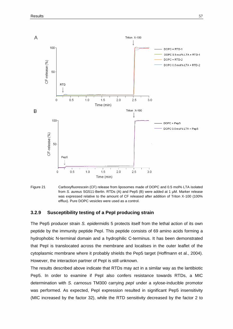
Results 57
Figure 21 Carboxyfluorescein (CF) release from liposomes made of DOPC and 0.5 mol% LTA isolated
from S. aureus SG511-Berlin. RTDs (A) and Pep5 (B) were added at 1 µM. Marker release
was expressed relative to the amount of CF released after addition of Triton X-100 (100%
efflux). Pure DOPC vesicles were used as a control.
3.2.9 Susceptibility testing of a PepI producing strain
The Pep5 producer strain S. epidermidis 5 protects itself from the lethal action of its own
peptide by the immunity peptide PepI. This peptide consists of 69 amino acids forming a
hydrophobic N-terminal domain and a hydrophilic C-terminus. It has been demonstrated
that PepI is translocated across the membrane and localises in the outer leaflet of the
cytoplasmic membrane where it probably shields the Pep5 target (Hoffmann et al., 2004).
However, the interaction partner of PepI is still unknown.
The results described above indicate that RTDs may act in a similar way as the lantibiotic
Pep5. In order to examine if PepI also confers resistance towards RTDs, a MIC
determination with S. carnosus TM300 carrying pepI under a xylose-inducible promotor
was performed. As expected, PepI expression resulted in significant Pep5 insensitivity
(MIC increased by the factor 32), while the RTD sensitivity decreased by the factor 2 to
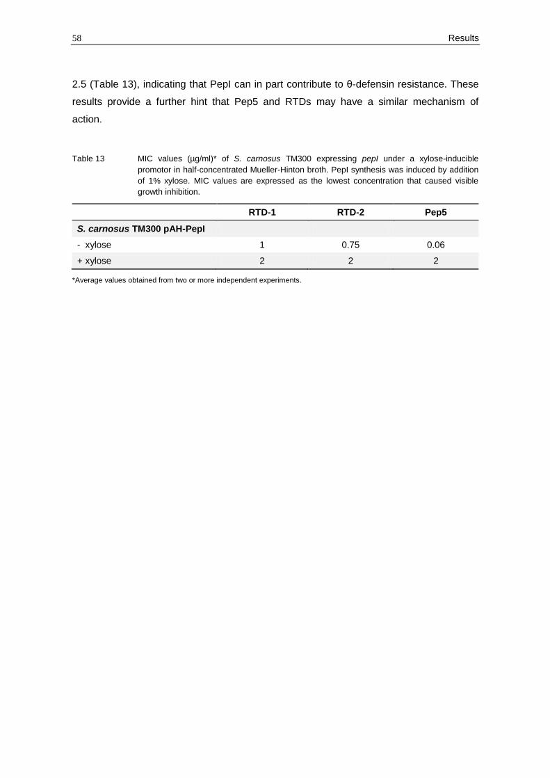
58 Results
2.5 (Table 13), indicating that PepI can in part contribute to θ-defensin resistance. These
results provide a further hint that Pep5 and RTDs may have a similar mechanism of
action.
Table 13 MIC values (µg/ml)* of S. carnosus TM300 expressing pepI under a xylose-inducible
promotor in half-concentrated Mueller-Hinton broth. PepI synthesis was induced by addition
of 1% xylose. MIC values are expressed as the lowest concentration that caused visible
growth inhibition.
RTD-1 RTD-2 Pep5
S. carnosus TM300 pAH-PepI
- xylose 1 0.75 0.06
+ xylose 2 2 2
*Average values obtained from two or more independent experiments.
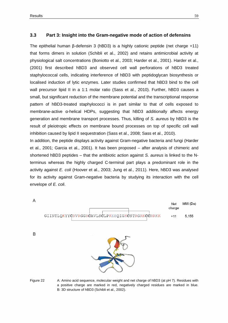
Results 59
3.3 Part 3: Insight into the Gram-negative mode of action of defensins
The epithelial human β-defensin 3 (hBD3) is a highly cationic peptide (net charge +11)
that forms dimers in solution (Schibli et al., 2002) and retains antimicrobial activity at
physiological salt concentrations (Boniotto et al., 2003; Harder et al., 2001). Harder et al.,
(2001) first described hBD3 and observed cell wall perforations of hBD3 treated
staphylococcal cells, indicating interference of hBD3 with peptidoglycan biosynthesis or
localised induction of lytic enzymes. Later studies confirmed that hBD3 bind to the cell
wall precursor lipid II in a 1:1 molar ratio (Sass et al., 2010). Further, hBD3 causes a
small, but significant reduction of the membrane potential and the transcriptional response
pattern of hBD3-treated staphylococci is in part similar to that of cells exposed to
membrane-active α-helical HDPs, suggesting that hBD3 additionally affects energy
generation and membrane transport processes. Thus, killing of S. aureus by hBD3 is the
result of pleiotropic effects on membrane bound processes on top of specific cell wall
inhibition caused by lipid II sequestration (Sass et al., 2008; Sass et al., 2010).
In addition, the peptide displays activity against Gram-negative bacteria and fungi (Harder
et al., 2001; Garcia et al., 2001). It has been proposed – after analysis of chimeric and
shortened hBD3 peptides – that the antibiotic action against S. aureus is linked to the N-
terminus whereas the highly charged C-terminal part plays a predominant role in the
activity against E. coli (Hoover et al., 2003; Jung et al., 2011). Here, hBD3 was analysed
for its activity against Gram-negative bacteria by studying its interaction with the cell
envelope of E. coli.
Figure 22 A: Amino acid sequence, molecular weight and net charge of hBD3 (at pH 7). Residues with
a positive charge are marked in red, negatively charged residues are marked in blue.
B: 3D structure of hBD3 (Schibli et al., 2002).
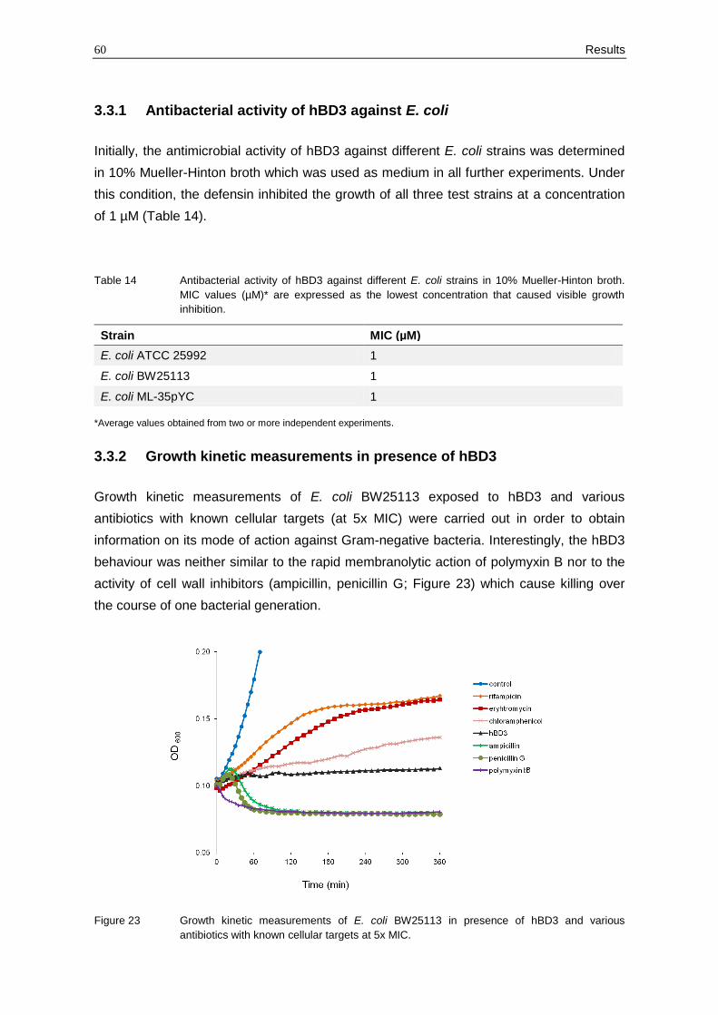
60 Results
3.3.1 Antibacterial activity of hBD3 against E. coli
Initially, the antimicrobial activity of hBD3 against different E. coli strains was determined
in 10% Mueller-Hinton broth which was used as medium in all further experiments. Under
this condition, the defensin inhibited the growth of all three test strains at a concentration
of 1 µM (Table 14).
Table 14 Antibacterial activity of hBD3 against different E. coli strains in 10% Mueller-Hinton broth.
MIC values (µM)* are expressed as the lowest concentration that caused visible growth
inhibition.
*Average values obtained from two or more independent experiments.
3.3.2 Growth kinetic measurements in presence of hBD3
Growth kinetic measurements of E. coli BW25113 exposed to hBD3 and various
antibiotics with known cellular targets (at 5x MIC) were carried out in order to obtain
information on its mode of action against Gram-negative bacteria. Interestingly, the hBD3
behaviour was neither similar to the rapid membranolytic action of polymyxin B nor to the
activity of cell wall inhibitors (ampicillin, penicillin G; Figure 23) which cause killing over
the course of one bacterial generation.
Figure 23 Growth kinetic measurements of E. coli BW25113 in presence of hBD3 and various
antibiotics with known cellular targets at 5x MIC.
Strain MIC (µM)
E. coli ATCC 25992 1
E. coli BW25113 1
E. coli ML-35pYC 1
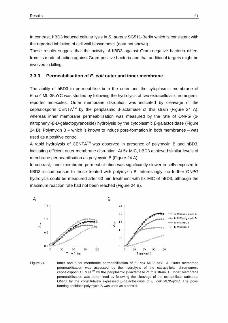
Results 61
In contrast, hBD3 induced cellular lysis in S. aureus SG511-Berlin which is consistent with
the reported inhibition of cell wall biosynthesis (data not shown).
These results suggest that the activity of hBD3 against Gram-negative bacteria differs
from its mode of action against Gram-positive bacteria and that additional targets might be
involved in killing.
3.3.3 Permeabilisation of E. coli outer and inner membrane
The ability of hBD3 to permeabilise both the outer and the cytoplasmic membrane of
E. coli ML-35pYC was studied by following the hydrolysis of two extracellular chromogenic
reporter molecules. Outer membrane disruption was indicated by cleavage of the
cephalosporin CENTATM by the periplasmic β-lactamase of this strain (Figure 24 A),
whereas inner membrane permeabilisation was measured by the rate of ONPG (o-
nitrophenyl-β-D-galactopyranoside) hydrolysis by the cytoplasmic β-galactosidase (Figure
24 B). Polymyxin B – which is known to induce pore-formation in both membranes – was
used as a positive control.
A rapid hydrolysis of CENTATM was observed in presence of polymyxin B and hBD3,
indicating efficient outer membrane disruption. At 5x MIC, hBD3 achieved similar levels of
membrane permeabilisation as polymyxin B (Figure 24 A).
In contrast, inner membrane permeabilisation was significantly slower in cells exposed to
hBD3 in comparison to those treated with polymyxin B. Interestingly, no further ONPG
hydrolysis could be measured after 60 min treatment with 5x MIC of hBD3, although the
maximum reaction rate had not been reached (Figure 24 B).
Figure 24 Inner and outer membrane permeabilisation of E. coli ML35-pYC. A: Outer membrane
permeabilisation was assessed by the hydrolysis of the extracellular chromogenic
cephalosporin CENTATM
by the periplasmic β-lactamase of this strain. B: Inner membrane
permeabilisation was determined by following the cleavage of the extracellular substrate
ONPG by the constitutively expressed β-galactosidase of E. coli ML35-pYC. The pore-
forming antibiotic polymyxin B was used as a control.
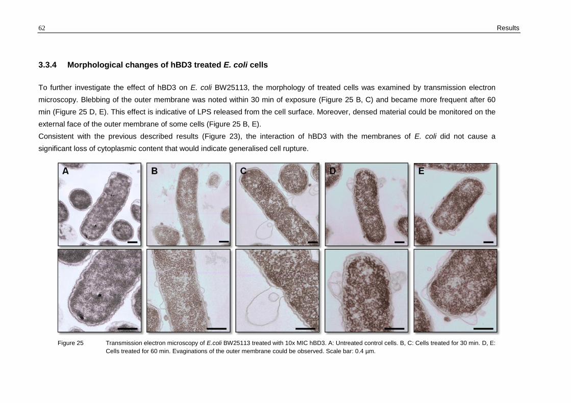
62 Results
3.3.4 Morphological changes of hBD3 treated E. coli cells
To further investigate the effect of hBD3 on E. coli BW25113, the morphology of treated cells was examined by transmission electron
microscopy. Blebbing of the outer membrane was noted within 30 min of exposure (Figure 25 B, C) and became more frequent after 60
min (Figure 25 D, E). This effect is indicative of LPS released from the cell surface. Moreover, densed material could be monitored on the
external face of the outer membrane of some cells (Figure 25 B, E).
Consistent with the previous described results (Figure 23), the interaction of hBD3 with the membranes of E. coli did not cause a
significant loss of cytoplasmic content that would indicate generalised cell rupture.
Figure 25 Transmission electron microscopy of E.coli BW25113 treated with 10x MIC hBD3. A: Untreated control cells. B, C: Cells treated for 30 min. D, E:
Cells treated for 60 min. Evaginations of the outer membrane could be observed. Scale bar: 0.4 µm.
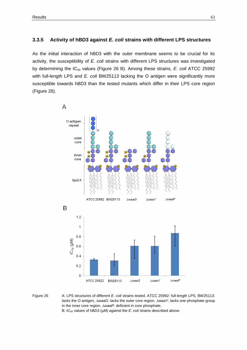
Results 63
3.3.5 Activity of hBD3 against E. coli strains with different LPS structures
As the initial interaction of hBD3 with the outer membrane seems to be crucial for its
activity, the susceptibility of E. coli strains with different LPS structures was investigated
by determining the IC50 values (Figure 26 B). Among these strains, E. coli ATCC 25992
with full-length LPS and E. coli BW25113 lacking the O antigen were significantly more
susceptible towards hBD3 than the tested mutants which differ in their LPS core region
(Figure 26).
Figure 26 A: LPS structures of different E. coli strains tested. ATCC 25992: full-length LPS, BW25113:
lacks the O antigen, ∆waaG: lacks the outer core region, ∆waaY: lacks one phosphate group
in the inner core region, ∆waaP: deficient in core phosphate.
B: IC50 values of hBD3 (µM) against the E. coli strains described above.
64 Results
The kinases WaaP and WaaY are involved in phosphorylation of heptose residues in the
inner core region of LPS. Thus, a mutation in one of the corresponding genes decreases
the net negative charge of the LPS molecules, and thereby lowering the affinity towards
cationic peptides. Consistently, the ∆waaP and ∆waaY deletion mutants were more
resistant towards hBD3. Interestingly, E. coli ∆waaG lacking the outer core region showed
also a higher hBD3 resistance compared to the wild-type strain (E. coli BW25113).
Hence, the susceptibility towards hBD3 depends on the composition of LPS, and not only
the phosphate residues, but also the sugars of the outer core region seem to be involved
in the interaction.
Discussion 65
4 Discussion
The antibacterial mode of action of defensins was long thought to result from electrostatic
interaction between the cationic peptides and the negatively charged microbial
membranes, followed by pore-formation or unspecific membrane permeabilisation.
Previous studies and the results of this thesis have clearly demonstrated that the action of
defensins can be much more targeted and that specific lipid-bound molecules of the
microbial cell envelope are involved in killing.
4.1 Part 1: The central cell wall building block lipid II as target for
defensins
Here, the mode of action of antibacterial invertebrate defensins was studied with three
defensin variants (Cg-Defm, Cg-Defh1, Cg-Defh2) isolated from the oyster C. gigas.
Results showed that oyster defensins kill S. aureus through binding to lipid II, thus
inhibiting peptidoglycan biosynthesis. To date, only invertebrate defensins with antifungal
activity had been characterised in terms of cellular targets (Thevissen et al., 2004).
The biosynthesis of bacterial cell wall peptidoglycan is a multistep process involving
reactions in the cytoplasm and both sides of the cytoplasmic membrane (for details see
Figure 27). It is a prominent target for many antibacterial compounds from different
chemical classes, including glycopeptides, lipopeptides and lantibiotics. Particularly, the
building block lipid II is trapped by many compounds when presented on the outer face of
the bacterial cytoplasmic membrane. For example, the clinically-used glycopeptides such
as vancomycin bind to the D-Ala-D-Ala terminus of the lipid II side chain, thereby causing
sequestration of the precursor from PBPs and subsequently blocking its further
incorporation into the cell wall (Schneider and Sahl, 2010). Moreover, the lantibiotic nisin
(which was used as a positive control during this study) and related peptides combine
lipid II targeting with membrane permeabilisation (Wiedemann et al., 2001). Very recently,
lipid II binding has also been described for one fungal defensin named plectasin
(Schneider et al., 2010) and two mammalian defensins – representatives of the α- and β-
defensin family – namely HNP-1 and hBD3 (de Leeuw et al., 2010; Sass et al., 2010).
The oyster defensins displayed potent activity against several Gram-positive bacteria,
including staphylococci which were chosen for further mode of action studies (Table 8).
Incubation of S. simulans 22 with oyster defensins resulted in the accumulation of the last
soluble cell wall precursor UDP-MurNAc-pp in the cytoplasm, comparable to classical
inhibitors of the membrane-bound steps of cell wall biosynthesis (Figure 8). Moreover,
lipid II antagonised the oyster defensin activity against S. aureus, whereas C55-P (which
serves as a lipid carrier in lipid II) as well as UDP-MurNAc-pp and UDP-GlcNAc (the
66 Discussion
activated cell wall sugars forming the disaccharide moiety of lipid II) did not (Table 9). The
antagonisation of the oyster defensins antibacterial activity by lipid II occurred at a 1:1
molar ratio (Table 9), as also observed for plectasin (Schneider et al., 2010) and for hBD3.
However, the latter one was also antagonised by C55-P and negatively charged
phospholipids when added in up to a 5-fold molar excess (Sass et al., 2010). The 1:1
stoichiometry was also confirmed here by TLC, as indicated by the absence of free lipid II
on the plate at this molar ratio (Figure 10).
Altogether, this result suggests that targeting of lipid II by oyster defensins requires lipid II-
specific molecular determinants such as the disaccharide-pyrophosphate moiety, which is
targeted by the lantibiotics. NMR-based modelling of the plectasin-lipid II complex
revealed that the defensin interacts with the pyrophosphate moiety of lipid II via hydrogen
bonding (involving residues F2, G3, C4 and C37), whereas the hydrophobic part of the
peptide is located on the membrane surface (Figure 4) (Schneider et al., 2010). Indeed,
the three-dimensional structure of the oyster defensins is similar to that of plectasin
although they possess a fourth disulphide bridge. Further, the amino acid residues
involved in lipid II binding of plectasin are also present in the Cg-Defs (F2, G3, C4, C34)
and other antibacterial invertebrate defensins, suggesting a conserved interaction motif.
Consequently, it can be proposed that those defensins carrying a CSαβ motif and
displaying exclusively antibacterial activity could have a similar mechanism of action,
which involves lipid II-binding and subsequent inhibition of cell wall biosynthesis. This is
consistent with their preferential activity against Gram-positive bacteria (Bulet and
Stocklin, 2005; Mygind et al., 2005; Froy and Gurevitz, 2003; Otvos, 2000). In addition,
antifungal invertebrate defensins with a CSαβ fold such as heliomicin do not contain the
amino acids of the lipid II binding motif. Consistently, it has been shown that heliomicin
does not bind to lipid II (Schneider et al., 2010) and lacks antibacterial activity (Lamberty
et al., 1999), thus reinforcing the hypothesis that lipid II-binding is an essential
determinant of invertebrate defensin antibacterial activity.
In contrast, antifungal invertebrate and plant defensins interact with specific sphingolipids,
thereby leading to fungal cell death (compare section 1.3) (Aerts et al., 2009; Thevissen et
al., 2004). Thus, evidence is increasing that invertebrate defensins, and more generally
CSαβ-type defensins, are rather specific inhibitors of microbial biosynthesis or signalling
pathways than mere membrane active agents.
The binding of oyster defensins to lipid II was shown to be very strong as determined here
by SPR. The oyster defensins bound to DOPC vesicles containing 0.8 mol% lipid II in an
almost irreversible manner, as indicated by the absence of decay in the RU values during
the dissociation step (Figure 11). All defensin variants (Cg-Defm, Cg-Defh1, and Cg-
Defh2) gave higher binding responses than nisin. Among the oyster defensins, Cg-Defh2
Discussion 67
showed the highest affinity for lipid II (Figure 11). This correlates with its antimicrobial
activity as Cg-Defh2 exhibited 2- to 4-fold lower MIC values than Cg-Defh1 and Cg-Defm
(Table 8).
Interestingly, it has been demonstrated that all three defensins belong to distinct groups
which evolved through gene duplication and subsequent functional divergence (Schmitt et
al., 2010). This study supports the hypothesis that strong selective pressures have
directed oyster defensins towards the design of new variants with higher potency. Cg-
Defm and Cg-Defh2, the more active forms, exhibit a positively charged residue at
position 16 (K and R, respectively; Figure 6) instead of an uncharged glutamine in Cg-
Defh1. As these residues are exposed on the surface of the peptide (Gueguen et al.,
2006) they might be involved in binding to lipid II, thus explaining the different affinity of
the three variants for the peptidoglycan precursor. Similarly, a lysine on the surface of
plectasin is crucial for the antibacterial activity, probably by promoting a better binding to
the cell wall and membrane of bacteria (Schneider et al., 2010). Therefore, variations in
oyster defensin potency could depend on charge distribution driven by sites under
diversifying selection.
The binding of oyster defensins to lipid II likely occurs at the outer leaflet of the
cytoplasmic membrane. At lethal concentrations (10x MIC), oyster defensins did not
compromise the cytoplasmic membrane of S. aureus (Figure 7). For this reason, they
should have no access to the inner leaflet of the cytoplasmic membrane where lipid II is
synthesised and decorated with a pentaglycine interpeptide bridge before it is translocated
across the cytoplasmic membrane. Consequently, the mechanism of action of oyster
defensins might involve binding and sequestration of extracellular lipid II, thereby blocking
its polymerisation into mature peptidoglycan (Figure 27).
Consistent with the use of lipid II as a molecular target, oyster defensins have been shown
to be highly active against Gram-positive bacteria (in the nanomolar range) and barely
active (≥ 20 μM) against Gram-negative bacteria (Table 8). Indeed, in Gram-negative
bacteria the peptidoglycan is protected by the outer membrane preventing access of
oyster defensins to the periplasmic space in the absence of membrane damage. As a
consequence, the different susceptibility of Gram-positive and Gram-negative bacteria to
oyster defensins probably results from a differential access to lipid II. The cell wall building
block is readily accessible in Gram-positive bacteria, but requires outer membrane
damage to become accessible in Gram-negative bacteria. Such outer membrane damage
can be induced by high concentrations of oyster defensins, as observed above 10 μM
against the Gram-negative oyster pathogen V. splendidus LGP32 (Duperthuy et al.,
2010). Consistently, Cg-Defs have a synergistic effect with Cg-Prps (proline rich HDPs of
C. gigas) and Cg-BPI (bactericidal/permeability-increasing protein of C. gigas) against
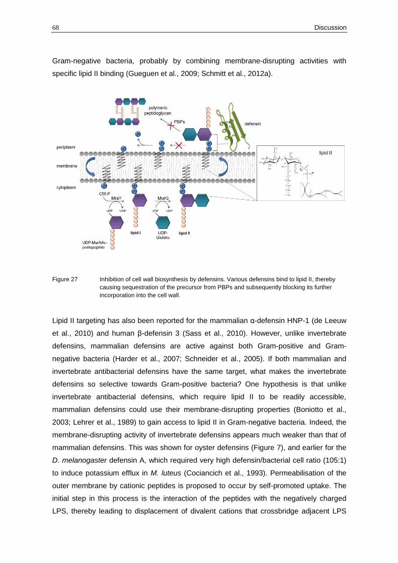
68 Discussion
Gram-negative bacteria, probably by combining membrane-disrupting activities with
specific lipid II binding (Gueguen et al., 2009; Schmitt et al., 2012a).
Figure 27 Inhibition of cell wall biosynthesis by defensins. Various defensins bind to lipid II, thereby
causing sequestration of the precursor from PBPs and subsequently blocking its further
incorporation into the cell wall.
Lipid II targeting has also been reported for the mammalian α-defensin HNP-1 (de Leeuw
et al., 2010) and human β-defensin 3 (Sass et al., 2010). However, unlike invertebrate
defensins, mammalian defensins are active against both Gram-positive and Gram-
negative bacteria (Harder et al., 2007; Schneider et al., 2005). If both mammalian and
invertebrate antibacterial defensins have the same target, what makes the invertebrate
defensins so selective towards Gram-positive bacteria? One hypothesis is that unlike
invertebrate antibacterial defensins, which require lipid II to be readily accessible,
mammalian defensins could use their membrane-disrupting properties (Boniotto et al.,
2003; Lehrer et al., 1989) to gain access to lipid II in Gram-negative bacteria. Indeed, the
membrane-disrupting activity of invertebrate defensins appears much weaker than that of
mammalian defensins. This was shown for oyster defensins (Figure 7), and earlier for the
D. melanogaster defensin A, which required very high defensin/bacterial cell ratio (105:1)
to induce potassium efflux in M. luteus (Cociancich et al., 1993). Permeabilisation of the
outer membrane by cationic peptides is proposed to occur by self-promoted uptake. The
initial step in this process is the interaction of the peptides with the negatively charged
LPS, thereby leading to displacement of divalent cations that crossbridge adjacent LPS
Discussion 69
molecules (Hancock, 1997). Subsequently, outer membrane disruption might depend on
the cationic net charge of the peptides. For example, hBD3 displays a net charge of +11
while plectasin or the oyster defensins carrying a net charge of +1 to +3. Moreover, many
vertebrate defensins form stable dimers or oligomers in solution, which results in an even
higher charge density that could likely be important for their membrane activity (Boniotto
et al., 2003; Schibli et al., 2002).
However, it has been demonstrated that the binding constant of plectasin to
lipid II (1.8 x 10⁻⁷ M) (Schneider et al., 2010) is about one order of magnitude higher
compared to the α-defensin HNP-1 (2.19 x 10⁻⁶ M) (de Leeuw et al., 2010). These findings
indicate that the specificity of lipid II binding correlates to some extent with the
antimicrobial spectrum. Possibly, peptides such as HNP-1 and hBD3 displaying lower
affinity towards the cell wall precursor and a broader activity spectrum, may thereby retain
affinity for additional targets to kill other microbes, e.g. Gram-negative bacteria (compare
section 4.3).
As Cg-defs are only active against Gram-positive bacteria, the oyster C. gigas has to
produce additional HDPs to control pathogens such as Gram-negative bacteria. Indeed,
three representatives of the big defensin family have been recently isolated from this
invertebrate (Cg-BigDef 1-3) (Rosa et al., 2011) which were reported to be active against
both Gram-positive and Gram-negative bacteria (compare section 1.2). Moreover, two Cg-
BPIs (bactericidal/permeability-increasing proteins) have been identified displaying
exclusively Gram-negative activity (Gonzalez et al., 2007b; Zhang et al., 2011) as well as
two proline-rich peptides (Cg-Prps) exhibiting only weak antimicrobial action (Gueguen et
al., 2009; Schmitt et al., 2012a). As described above, these peptides can act
synergistically, thereby increasing their potency as well as their activity spectrum (Schmitt
et al., 2012a).
However, this raises the question which selective pressure has driven the development of
defensins from peptides with a highly specific target to peptides with a reduced affinity and
a broad-spectrum activity (as they can be found in vertebrates)? In this context, it is worth
to recall that vertebrate defensins also display a diverse range of immunomodulatory
functions and even bridge the innate and adaptive immune response, thereby contributing
to bacterial clearance (section 1.4). In contrast, little is known about such functions in
invertebrates. It has been shown that some invertebrate defensins neutralise LPS or
induce signal transduction or gene transcription in mouse models (Hancock et al., 2006),
but nothing has been reported about the immunomodulatory activities in the invertebrate
host itself.
Nevertheless, it could be hypothesised that during the process of gaining new functions in
terms of immunomodulation, defensins may have reduced their direct antimicrobial activity
70 Discussion
and/or target specificity. This is consistent with the fact that some defensins completely
lack direct antibiotic activities such as the human enteric α-defensin HD6. It has been
recently found that HD6 entraps bacteria in peptide nanosets without affecting their
viability (Chu et al., 2012; Ouellette and Selsted, 2012).
4.2 Part 2: Induction of autolysis as mode of action of defensins
Apparently, θ-defensins do not only differ structurally from all other defensins, they also
seem to act by a different mechanism. In this study, it was demonstrated that RTDs
(rhesus macaque θ-defensins) do not interfere with peptidoglycan biosynthesis (Figure
13), but rather induce bacterial lysis in staphylococci by interaction with the bacterial
membrane and/or release of autolytic enzymes (Figure 28).
The membrane permeabilisation by RTDs depended strongly on the energisation of the
membrane as revealed by potassium efflux experiments and membrane potential
measurements (section 3.2.4). Similar observations have been described for the lantibiotic
Pep5, a cationic peptide of bacterial origin. Pep5 also forms transient pores in energised
membranes leading to rapid efflux of low molecular weight substances from the cytoplasm
(Poppinga, 2007; Sahl, 1985; Sahl and Brandis, 1983).
Different models have been elaborated to describe the interaction of cationic peptides with
the bacterial membrane. The “wedge model” was suggested to explain the pore formation
process in presence of a high transmembrane potential. In this model, the amphiphilic
molecules adhere to the anionic lipids on the membrane surface; in response to the
electrical potential the lipid-associated peptides change the orientation (to a wegde-
shape), thereby moving through the membrane to form a water-filled pore (Driessen et al.,
1995; Sahl and Bierbaum, 1998). Unlike other HDPs, the highly flexible RTD structure
lacks the typical amphiphilic character, even though it exhibits a certain clustering of
positive charges (Trabi et al., 2001). Consequently, the peptides do not have a
hydrophobic patch which allows insertion into the bilayer. In contrast, reports on the
biophysical behaviour of RTD-1 in model membranes showed that the peptide induces
membrane thinning when it binds to the membrane surface (Weiss et al., 2002) and that
RTD molecules probably aggregate in the membrane (Buffy et al., 2004).
For Pep5, it has been hypothesised that it uses a docking molecule for binding to the
cytoplasmic membrane and subsequent pore-formation (Brotz et al., 1998; Hoffmann et
al., 2004). Earlier studies have demonstrated that Pep5 does not form a complex with
lipid II (Brotz et al., 1998) in contrast to nisin which uses the cell wall precursor as an
anchor for defined pore-formation (Wiedemann et al., 2001). Since Pep5 and RTDs bind
to the membrane-anchored LTA (Table 12) (Bierbaum and Sahl, 1985; Bierbaum and
Discussion 71
Sahl, 1987) thereby releasing autolytic enzymes, it was investigated whether this
interaction also facilitates pore-formation. Under the tested conditions, neither Pep5 nor
RTDs caused leakage of small vesicles containing purified LTA (Figure 21). This suggests
that the membrane-disrupting activity of these peptides might depend on the membrane
potential only. Voltage-dependent channel formation in model membranes has also been
described for the α-defensin HNP-1 (Kagan et al., 1990).
The membrane perturbation may contribute, but not solely account for killing of
staphylococcal cells by RTDs. Further, it could be demonstrated that these peptides
activate peptidoglycan hydrolases (also referred to as autolysins; Figure 19). These
enzymes are responsible for splitting the septum during cell division and enable
incorporation of newly synthesised cell wall precursors during cell growth (Vollmer et al.,
2008). The premature and uncontrolled activity of autolysins can cause cell lysis and
subsequently cell death as observed in presence of θ-defensins. Zymogram analysis
revealed that RTDs release differently processed forms of Atl, the most prominent
autolysin of staphylococci (Figure 19). Consistently, an atl deletion mutant was
significantly more resistant towards the activity of RTDs (Figure 20). Atl can also be
activated by high salt concentrations (Heilmann et al., 1997) and the lantibiotics nisin and
Pep5 (Bierbaum and Sahl, 1985; Bierbaum and Sahl, 1987). This hydrolase is
synthesised as a pre-pro-peptide that is cleaved into two enzymatically active domains –
an amidase (AM; that cleaves the amide bond between MurNAc and L-alanine) and a
glucosaminidase (GL; that cleaves the β-1,4-gycosidic bond between GlcNAc and
adjacent monosaccharides) – by an as yet uncharacterised mechanism (Heilmann et al.,
1997; Schlag et al., 2010). Both enzymes localise at the cross wall of dividing cells
(Yamada et al., 1996). Indeed, electron micrographs revealed that degradation of
peptidoglycan of RTD-2 treated cells occurred particular at the septum between two
daughter cells (Figure 18 D, E).
The AM and GL are each fused to repeat units that are responsible for targeting the
enzymes to the cross wall (Baba and Schneewind, 1998; Komatsuzawa et al., 1997). It
has been reported that in S. aureus wall teichoic acid (WTA) – present in the old cell wall
– acts as a repellent for the repeats, thereby directing the enzymes to the septum, where
they bind to and are controlled by LTA (Schlag et al., 2010; Zoll et al., 2012). This
suggests that cationic molecules such as RTDs and Pep5 bind to the polyanionic LTA,
thereby liberate the enzymes such that they can degrade the cell wall in an uncontrolled
manner. An antagonisation assay confirmed that LTA completely suppresses the
antimicrobial activity of RTDs when added in a 4-fold molar excess in respect to the
peptide (Table 12). In contrast, higher LTA concentrations (10-fold molar excess) are
72 Discussion
necessary to antagonise the antibiotic action of Pep5 (Bierbaum and Sahl, 1985) which is
consistent with the data obtained here (Table 12).
The number of positive charges of a given peptide might be of particular relevance for the
interaction with LTA and thereby for the release of autolysins (Bierbaum and Sahl, 1987).
A higher autolysin activity could be detected in the supernatant of cells exposed to RTD-2
(net charge +6) and Pep5 (net charge +7; which served as a positive control) compared to
cells treated with RTD-1 (net charge +5).
Notably, the unprocessed Atl forms could also be detected in the supernatant of treated
S. aureus cells (Figure 19), indicating that the processing of the enzyme occurs at the
septum when bound to LTA.
Moreover, two mutants with a reduced LTA content (S. aureus SA113 ∆ypfP and
S. aureus SEJ ∆ltaS (4S5)) were tested for their susceptibility towards RTDs and Pep5.
Consistently, these strains have either a lowered autolysis rate (Fedtke et al., 2007) or a
reduced amount of hydrolytic enzymes bound to the cell envelope (Corrigan et al., 2011).
However, MIC determinations revealed that both strains were more susceptible to Pep5
(Table 11). Additionally, the LTA-deficient suppressor strain ∆ltaS (4S5) showed a
decreased MIC value against RTDs compared to its parental strain (Table 11). As LTA are
crucial for bacterial growth, the strain ∆ltaS (4S5) compensates the loss of LTA by
increasing the intracellular level of the secondary messenger c-di-AMP. It has been
suggested that this messenger plays a role in cell division, but it has to be further
elucidated whether c-di-AMP is involved in the regulation of other cellular processes.
S. aureus SEJ ∆ltaS (4S5) is also more susceptible to cell wall inhibitors such as nisin,
vancomycin and penicillin as well as to daptomycin (Corrigan et al., 2011). Thus, the
decreased MIC values of Pep5 and RTDs might be a secondary effect due to yet
unknown altered cellular processes in this strain. Alternatively, it can be hypothesised that
in case of a reduced LTA content, peptides more easily directly interfere with the bacterial
membrane, thereby leading to membrane permeabilisation even in the absence of a high
membrane potential.
In the strain S. aureus SA113 ∆ypfP the remaining LTA is anchored to the membrane via
diacylglycerol (DAG) instead of diglucosyl-DAG. In this case, the binding of Pep5 to LTA
could still have an impact on the membrane integrity as binding sites are even closer to
the outside of the cytoplasmic membrane.
Interestingly, S. simulans and S. carnosus are more susceptible towards Pep5 (Sahl and
Brandis, 1981) and θ-defensins than S. aureus (Table 10, Table 11). The LTA in these
strains does not differ and consists of a polyglycerolphosphate chain linked to a glycolipid
anchor (Ruhland and Fiedler, 1990). However, modifications of the LTA such as
substitution of the glycerol units with D-alanine may have an impact on the susceptibility
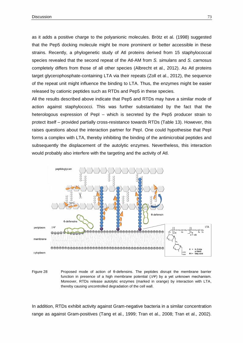
Discussion 73
as it adds a positive charge to the polyanionic molecules. Brötz et al. (1998) suggested
that the Pep5 docking molecule might be more prominent or better accessible in these
strains. Recently, a phylogenetic study of Atl proteins derived from 15 staphylococcal
species revealed that the second repeat of the Atl-AM from S. simulans and S. carnosus
completely differs from those of all other species (Albrecht et al., 2012). As Atl proteins
target glycerophosphate-containing LTA via their repeats (Zoll et al., 2012), the sequence
of the repeat unit might influence the binding to LTA. Thus, the enzymes might be easier
released by cationic peptides such as RTDs and Pep5 in these species.
All the results described above indicate that Pep5 and RTDs may have a similar mode of
action against staphylococci. This was further substantiated by the fact that the
heterologous expression of PepI – which is secreted by the Pep5 producer strain to
protect itself – provided partially cross-resistance towards RTDs (Table 13). However, this
raises questions about the interaction partner for PepI. One could hypothesise that PepI
forms a complex with LTA, thereby inhibiting the binding of the antimicrobial peptides and
subsequently the displacement of the autolytic enzymes. Nevertheless, this interaction
would probably also interfere with the targeting and the activity of Atl.
Figure 28 Proposed mode of action of θ-defensins. The peptides disrupt the membrane barrier
function in presence of a high membrane potential (∆Ψ) by a yet unknown mechanism.
Moreover, RTDs release autolytic enzymes (marked in orange) by interaction with LTA,
thereby causing uncontrolled degradation of the cell wall.
In addition, RTDs exhibit activity against Gram-negative bacteria in a similar concentration
range as against Gram-positives (Tang et al., 1999; Tran et al., 2008; Tran et al., 2002).
74 Discussion
Tran et al. (2008) reported that RTD-1 and RTD-2 effectively permeabilise the outer and
inner membrane of E. coli ML35-pYC, whereas RTD-3 (Figure 12) does not affect the
membrane. This membrane impairment seems to be independent from the membrane
potential as these assays were carried out in buffer lacking free glucose.
Further studies are necessary to analyse if RTDs additionally interfere with membrane
bound molecules in Gram-negative bacteria.
4.3 Part 3: Interaction of hBD3 with the Gram-negative cell envelope
The antibacterial activity of the highly cationic peptide hBD3 was extensively studied in
S. aureus and it has been demonstrated that binding to lipid II and subsequent inhibition
of cell wall biosynthesis is a major determinant of its mechanism of action (Sass et al.,
2008; Sass et al., 2010). Here, the interaction of hBD3 with the cell envelope of E. coli
was investigated to gain a first insight into its antibiotic action against Gram-negative
bacteria.
Initially, the defensin has to cross the outer membrane barrier of Gram-negative bacteria
to gain access to the periplasmic space. This may occur by a mechanism known as self-
promoted uptake which is based on the interaction of the cationic peptides with the
negatively charged LPS molecules of the outer membrane (compare 1.3) (Hancock,
1997). In fact, hBD3 rapidly disrupted the outer membrane of E. coli ML35-pYC
comparable to the membranolytic activity of polymyxin B (Figure 24). Consistently,
evaginations of the outer membrane could be observed by transmission electron
microscopy (Figure 25) which are indicative of LPS released from the cell surface
(Spindler et al., 2011).
To further analyse the interaction of hBD3 with LPS, the susceptibility of strains with
altered LPS structure was tested (Figure 26). LPS molecules typically consist of three
distinct regions: a hydrophobic membrane anchor (lipid A), a short sugar chain substituted
with phosphate groups (core) and a polymer composed of oligosaccharide repeats (O
antigen). Interestingly, all three mutants of the E. coli K-12 strain BW25113 (∆waaP,
∆waaY, ∆waaG) included in the study were more resistant towards hBD3. These mutants
are affected in the assembly of the LPS core region. The WaaP and WaaY kinases are
involved in the phosphorylation of the first and second heptose residue in the inner core
(Yethon et al., 1998). Thus, a deletion of one of the genes reduces the net negative
charge of the cell envelope which correlates with a decreased susceptibility towards
cationic peptides, as observed here. On the other hand, the phosphoryl substituents are
critical for the formation of a stable outer core, because their negative charge allows
neighbouring LPS molecules to be crossbridged by divalent cations (Helander et al., 1989;
Discussion 75
Yethon et al., 2000). It has been shown that waaP deletion mutants are highly susceptible
towards polymyxin B or detergents such as SDS (Yethon et al., 2000; Yethon et al.,
1998). Nevertheless, among the tested strains E. coli ∆waaP displayed the highest
resistance.
The gene waaG encodes a glycosyltransferase which adds the first glucose to the outer
core; consequently the deletion strain lacks the entire outer core region. These results
suggest that not only the phosphate groups, but also the sugars of the outer core region
may be crucial for the interaction with hBD3 whereas the O antigen does not seem to be
involved in binding (Figure 26).
Further, the question is raised whether outer membrane permeabilisation is sufficient by
itself to cause bacterial cell death or if additional target structures are involved in hBD3-
mediated killing of Gram-negative bacteria.
It could be hypothesised that hBD3 interacts with the cell wall precursor lipid II once it
enters the periplasmic space. Studies with chimeric and shortened hBD3 molecules linked
the Gram-positive activity to the N-terminal part and the action against Gram-negative
bacteria to the C-terminus (Hoover et al., 2003; Jung et al., 2011). This suggests that the
highly charged C-terminal part of hBD3 (compare Figure 22) is essential for its interaction
with the outer membrane, whereas the N-terminal part plays a predominant role in the
interaction with lipid II. Moreover, hBD3 forms stable dimers in solution (due to an
intermolecular salt bridge between E28 and K32 on another monomer) supporting the
interaction with the membrane. Additionally, an intramolecular salt bridge involving the
residues E27 and R17 (Schibli et al., 2002) contributes to stability and enhances the
membrane-disrupting properties of hBD3 (Boniotto et al., 2003).
Consequently, hBD3 combines two activities in one molecule in contrast to oyster
defensins which have only a weak membrane-disrupting activity and are barely active
against Gram-negative bacteria. This correlates with the affinity to the cell wall precursor
lipid II as discussed before (compare section 4.1).
However, growth kinetic measurements indicated that the mode of action of hBD3 against
Gram-negative bacteria is different from the action against S. aureus (Figure 23) and that
other target structures might also be involved in killing. Sass et al. (2010) demonstrated
that the activity of hBD3 can be antagonised by the lipid II isoprenoid anchor C55-P in a
molar ratio of 2:1. This lipid carrier is also involved in the biosynthesis and modification of
LPS, suggesting that hBD3 might also interfere with these processes. For example, the
repeating units of the O antigen are assembled on C55-P on the inner face of the
cytoplasmic membrane. After transport across the membrane, the O antigen is
polymerised and ligated to lipid A-core (Raetz and Whitfield, 2002; Wang and Quinn,
2010).
76 Discussion
Further, the inner membrane was permeabilised to some degree by hBD3, but less
efficiently as with polymyxin B (Figure 24), indicating a higher affinity of hBD3 to the outer
membrane. Nevertheless, this interaction did not cause a significant loss of cytoplasmic
content as monitored by transmission electron microscopy (Figure 25) and growth kinetic
measurements (Figure 23). A similar lack of lytic effects has been reported for the
synthetic antimicrobial peptide Bac8. This peptide was shown to cause partial membrane
permeabilisation, disruption of the electron transport chain located in the inner membrane
and cell depolarisation which in turn results in cell death (Spindler et al., 2011).
Consistently, microarray data of hBD3 treated S. aureus cells indicated that energy
generation is affected in presence of the peptide (Sass et al., 2008). This suggests that
hBD3 might also inhibit cellular respiration by either direct or indirect disruption of
respiratory functions.
In conclusion, binding to LPS and subsequent destabilisation of the cell envelope seems
to play a significant role in the mode of action of hBD3 against Gram-negative bacteria
and may be more important than the interaction with lipid II. Moreover, it is likely that
hBD3 interferes with various membrane-bound processes in E. coli which may each
contribute to the overall efficacy. Since the defensin causes stresses at many sites, it may
act like “sand in the gearbox” – a mechanism which has been described for α-helical
HDPs (Pag et al., 2008).
However, further studies are necessary to investigate the inhibition of these individual
processes in more detail and to correlate them with the loss of cell viability (section 4.5).
4.4 Conclusion
Altogether, defensins of different groups interact with molecules of the microbial cell
envelope that are directly accessible such as lipid II, LTA or LPS (Table 15). Thereby, the
interaction between the defensin and the lipid-bound docking molecule can be highly
specific as shown for lipid II and antibacterial CSαβ-type defensins.
Interestingly, similarities in the mode of action between some eukaryotic defensins and
bacterially-produced lantibiotics could be revealed. The lantibiotic nisin and antibacterial
invertebrate and fungal defensins bind to the pyrophosphate moiety of the highly
conserved cell wall precursor lipid II. Moreover, Pep5 and θ-defensins do not only disrupt
the membranes barrier function, but they also release the major autolysin Atl in
staphylococci probably by replacing the enzymes from LTA.
Despite their low sequence similarity, these cationic peptides share many features as
amphipathicity and cationicity. Moreover, their structures are stabilised by disulphide
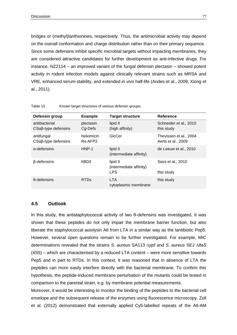
Discussion 77
bridges or (methyl)lanthionines, respectively. Thus, the antimicrobial activity may depend
on the overall conformation and charge distribution rather than on their primary sequence.
Since some defensins inhibit specific microbial targets without impacting membranes, they
are considered attractive candidates for further development as anti-infective drugs. For
instance, NZ2114 – an improved variant of the fungal defensin plectasin – showed potent
activity in rodent infection models against clinically relevant strains such as MRSA and
VRE, enhanced serum-stability, and extended in vivo half-life (Andes et al., 2009; Xiong et
al., 2011).
Table 15 Known target structures of various defensin groups.
Defensin group Example Target structure Reference
antibacterial
CSαβ-type defensins
plectasin
Cg-Defs
lipid II
(high affinity)
Schneider et al., 2010
this study
antifungal
CSαβ-type defensins
heliomicin
Rs-AFP2
GlcCer Thevissen et al., 2004
Aerts et al., 2009
α-defensins HNP-1 lipid II
(intermediate affinity)
de Leeuw et al., 2010
β-defensins hBD3 lipid II
(intermediate affinity)
LPS
Sass et al., 2010
this study
θ-defensins RTDs LTA
cytoplasmic membrane
this study
4.5 Outlook
In this study, the antistaphylococcal activity of two θ-defensins was investigated. It was
shown that these peptides do not only impair the membrane barrier function, but also
liberate the staphylococcal autolysin Atl from LTA in a similar way as the lantibiotic Pep5.
However, several open questions remain to be further investigated. For example, MIC
determinations revealed that the strains S. aureus SA113 ∆ypf and S. aureus SEJ ∆ltaS
(4S5) – which are characterised by a reduced LTA content – were more sensitive towards
Pep5 and in part to RTDs. In this context, it was reasoned that in absence of LTA the
peptides can more easily interfere directly with the bacterial membrane. To confirm this
hypothesis, the peptide-induced membrane perturbation of the mutants could be tested in
comparison to the parental strain, e.g. by membrane potential measurements.
Moreover, it would be interesting to monitor the binding of the peptides to the bacterial cell
envelope and the subsequent release of the enzymes using fluorescence microscopy. Zoll
et al. (2012) demonstrated that externally applied Cy5-labelled repeats of the Atl-AM
78 Discussion
accumulate at the cross wall of dividing S. aureus cells. Thus, it would be expected that
treatment with RTDs causes a delocalisation of the repeats from the septum.
Further, it was shown that the heterologous expression of the immunity peptide PepI of
the Pep5 producer strain provided partially cross-resistance towards RTDs. Nevertheless,
further studies are necessary to gain a deeper insight into the mechanism by which the
molecule confers resistance. Hoffmann et al. (2004) reported that PepI localises in the
outer leaflet of the cytoplasmic membrane and that the highly charged C-terminal part is
crucial for antagonising Pep5 activity. However, it is still unknown whether PepI forms a
complex with lipid-bound molecules of the staphylococcal cell envelope or whether it
interacts directly with the cationic peptides Pep5 or RTDs, thereby preventing interference
with both the bacterial membrane and LTA. The fact that the C-terminus of PepI
possesses several positively charged amino acids rather suggests that it forms a complex
with anionic molecules such as LTA. Thus, it could be first investigated whether PepI can
antagonise both activities of the peptides. The potassium efflux of energised cells
expressing PepI under an IPTG-inducible promotor could be monitored to test whether
PepI can inhibit the peptide-induced membrane permeabilisation (compare section 2.3.8).
In additon, the supernatant of peptide treated cells expressing PepI could be analysed for
cell wall lytic activity in comparison to control cells (compare section 2.5.3).
In the third part of this thesis, the interaction of hBD3 with the cell envelope of E. coli was
investigated. It was demonstrated that the peptide permeabilise the outer membrane by
binding to LPS and that the composition of the LPS core region plays an important role in
this interaction. To gain a deeper insight into the interaction of LPS and hBD3, binding
analysis using surface plasmon resonance could be carried out. Further, killing kinetics of
hBD3 treated E. coli cells might correlate the permeabilisation of both membranes with the
loss of cell viability.
Moreover, some interference of hBD3 with the inner membrane could be detected. In this
context, it would be interesting to measure the depolarisation of the membrane in
presence of hBD3 (compare section 2.3.9). Further, it has been suggested that hBD3
affects directly or indirectly the electron transport chain located in the inner membrane. To
analyse the effect of hBD3 on respiration, the intracellular NAD+/NADH ratios or the
oxygen consumption of cells exposed to hBD3 should be measured. Specific inhibition of
the individual electron transport chain complexes could be studied in vitro using inverted
membrane vesicles.
Summary 79
5 Summary
Multicellular organisms defend themselves against infectious microorganisms by
producing a wide array of antimicrobial peptides referred to as host defence peptides
(HDPs). These evolutionary ancient peptides are important effector molecules of innate
immunity. In addition to their immunomodulatory functions, they display potent direct
antimicrobial activity against a broad range of microorganisms, including multiresistant
pathogens. Thus, they are considered as promising candidates for the development of
novel anti-infective agents.
Almost all HDPs are cationic and amphipathic. Hence, it has been generally assumed that
these peptides interact unspecifically with negatively charged microbial membranes and
subsequently disrupt membrane barrier functions. However, evidence is increasing that
the antibiotic activity of HDPs can be much more targeted as it was originally proposed.
One important HDP class comprises defensins which are characterised by disulphide-
stabilised β-sheets as the major structural component. In this work, the mode of action of
defensins from diverse origins was investigated to gain further insights into the molecular
mechanisms of defensin-mediated killing.
In the first part of this work, the antibacterial action of invertebrate defensins was studied
using three defensin variants (Cg-Defm, Cg-Defh1 and Cg-Defh2) produced by the Pacific
oyster Crassostrea gigas. These peptides exhibited potent activity against several Gram-
positive bacteria, including staphylococci which were chosen for further mode of action
studies. Interestingly, the oyster defensins did not compromise the membrane integrity
since no impact on the membrane potential could be observed in cells exposed to these
peptides. Instead, the defensins triggered the accumulation of the final soluble cell wall
precursor UDP-MurNAc-pp in the cytoplasm, comparable to classical inhibitors of the
membrane-bound steps of cell wall biosynthesis. Moreover, the oyster defensins were
found to bind irreversibly to the cell wall precursor lipid II in a 1:1 stoichiometry, thereby
causing sequestration of the precursor from penicillin binding proteins and subsequently
blocking its further incorporation into the cell wall. Among the oyster defensins, Cg-Defh2
was the most potent, exhibiting 2- to 4-fold lower MIC values than Cg-Defh1 and Cg-
Defh2, which correlated with different binding affinities for lipid II as revealed by surface
plasmon resonance.
The second part of this thesis was aimed at elucidating the antistaphylococcal action of
two rhesus macaque θ-defensins (RTD-1, RTD-2). These peptides differ structurally from
all other defensins in that their backbones are cyclised by peptide bonds. Potassium efflux
experiments and membrane potential measurements demonstrated that the membrane
impairment by RTDs strongly depends on the energisation of the membrane. In addition,
80 Summary
RTD treatment caused the release of the cell wall lytic enzyme Atl probably by interaction
with the membrane-bound lipoteichoic acid. Thus, the premature and uncontrolled activity
of this enzyme contributes strongly to the overall killing by θ-defensins. Interestingly, a
similar mode of action has been described for Pep5, an antimicrobial peptide of bacterial
origin. Consistently, PepI – which is secreted by the Pep5 producer strain to protect itself
– provided partially cross-resistance towards RTDs.
In the third part of this thesis, the antimicrobial activity of the human β-defensin 3 (hBD3)
against Gram-negative bacteria was studied. Previous works have reported that binding to
lipid II and subsequent inhibition of cell wall biosynthesis is a major determinant of its
mechanism of action against staphylococci.
Here, it was demonstrated that the peptide permeabilises both the inner and outer
membrane of E. coli. The interaction of the peptide with the lipopolysaccharides (LPS) of
the outer membrane seems to play a significant role in the activity of hBD3 against Gram-
negative bacteria and was shown to depend on the number of phosphate groups and the
sugars in the LPS core region. Interestingly, the interaction with the inner membrane did
not cause a significant loss of the cytoplasmic content as revealed by growth kinetic
measurements and transmission electron microscopy of hBD3 treated E. coli cells. It
remains to determine if additional target structures in the inner membrane such as the
electron transport chain are also involved in hBD3-mediated killing.
In conclusion, previous reports and the results described in this thesis revealed that
conserved molecules of the microbial cell envelope, which are readily accessible, are
targets of various defensins and an important component of the killing mechanism. As
demonstrated here for lipid II and oyster defensins, the interaction between the defensin
and its lipid-bound target molecule can be highly specific.
References 81
6 References
Aarbiou, J., Ertmann, M., van Wetering, S., van Noort, P., Rook, D., Rabe, K. F., Litvinov, S. V., van Krieken, J. H., de Boer, W. I., and Hiemstra, P. S. (2002): Human neutrophil defensins induce lung epithelial cell proliferation in vitro. J Leukoc Biol 72, 167-74.
Aarbiou, J., Verhoosel, R. M., Van Wetering, S., De Boer, W. I., Van Krieken, J. H., Litvinov, S. V., Rabe, K. F., and Hiemstra, P. S. (2004): Neutrophil defensins enhance lung epithelial wound closure and mucin gene expression in vitro. Am J Respir Cell Mol Biol 30, 193-201.
Aerts, A. M., Carmona-Gutierrez, D., Lefevre, S., Govaert, G., Francois, I. E., Madeo, F., Santos, R., Cammue, B. P., and Thevissen, K. (2009): The antifungal plant defensin RsAFP2 from radish induces apoptosis in a metacaspase independent way in Candida albicans. FEBS Lett 583, 2513-6.
Aerts, A. M., Francois, I. E., Cammue, B. P., and Thevissen, K. (2008): The mode of antifungal action of plant, insect and human defensins. Cell Mol Life Sci 65, 2069-79.
Aerts, A. M., Thevissen, K., Bresseleers, S. M., Sels, J., Wouters, P., Cammue, B. P., and Francois, I. E. (2007): Arabidopsis thaliana plants expressing human beta-defensin-2 are more resistant to fungal attack: functional homology between plant and human defensins. Plant Cell Rep 26, 1391-8.
Albrecht, T., Raue, S., Rosenstein, R., Nieselt, K., and Gotz, F. (2012): Phylogeny of the staphylococcal major autolysin and its use in genus and species typing. J Bacteriol 194, 2630-6.
Allen, A., Snyder, A. K., Preuss, M., Nielsen, E. E., Shah, D. M., and Smith, T. J. (2008): Plant defensins and virally encoded fungal toxin KP4 inhibit plant root growth. Planta 227, 331-9.
Andes, D., Craig, W., Nielsen, L. A., and Kristensen, H. H. (2009): In vivo pharmacodynamic characterization of a novel plectasin antibiotic, NZ2114, in a murine infection model. Antimicrob Agents Chemother 53, 3003-9.
Baba, T., Ara, T., Hasegawa, M., Takai, Y., Okumura, Y., Baba, M., Datsenko, K. A., Tomita, M., Wanner, B. L., and Mori, H. (2006): Construction of Escherichia coli K-12 in-frame, single-gene knockout mutants: the Keio collection. Mol Syst Biol 2, 2006 0008.
Baba, T., and Schneewind, O. (1998): Targeting of muralytic enzymes to the cell division site of Gram-positive bacteria: repeat domains direct autolysin to the equatorial surface ring of Staphylococcus aureus. EMBO J 17, 4639-46.
Belas, R., Manos, J., and Suvanasuthi, R. (2004): Proteus mirabilis ZapA metalloprotease degrades a broad spectrum of substrates, including antimicrobial peptides. Infect Immun 72, 5159-67.
Bierbaum, G., and Sahl, H. G. (1985): Induction of autolysis of staphylococci by the basic peptide antibiotics Pep 5 and nisin and their influence on the activity of autolytic enzymes. Arch Microbiol 141, 249-54.
Bierbaum, G., and Sahl, H. G. (1987): Autolytic system of Staphylococcus simulans 22: influence of cationic peptides on activity of N-acetylmuramoyl-L-alanine amidase. J Bacteriol 169, 5452-8.
Bierbaum, G., and Sahl, H. G. (1991): Induction of autolysis of Staphylococcus simulans 22 by Pep5 and nisin and influence of the cationic peptides on the activity of the
82 References
autolytic enzymes, pp. 386-396. In G. Jung, and H. G. Sahl (Eds): Nisin and Novel Lantibiotics, Springer.
Biswas, R., Voggu, L., Simon, U. K., Hentschel, P., Thumm, G., and Gotz, F. (2006): Activity of the major staphylococcal autolysin Atl. FEMS Microbiol Lett 259, 260-8.
Boman, H. G., Agerberth, B., and Boman, A. (1993): Mechanisms of action on Escherichia coli of cecropin P1 and PR-39, two antibacterial peptides from pig intestine. Infect Immun 61, 2978-84.
Boniotto, M., Antcheva, N., Zelezetsky, I., Tossi, A., Palumbo, V., Verga Falzacappa, M. V., Sgubin, S., Braida, L., Amoroso, A., and Crovella, S. (2003): A study of host defence peptide beta-defensin 3 in primates. Biochem J 374, 707-14.
Brogden, K. A. (2005): Antimicrobial peptides: pore formers or metabolic inhibitors in bacteria? Nat Rev Microbiol 3, 238-50.
Brotz-Oesterhelt, H., and Sass, P. (2010): Postgenomic strategies in antibacterial drug discovery. Future Microbiol 5, 1553-79.
Brotz, H. (1997): Wirkungsmechanismus des Lantibiotikums Mersacidin: Neuartige Interaktion mit einer bakteriellen Zellwandvorstufe. PhD thesis, University of Bonn.
Brotz, H., Josten, M., Wiedemann, I., Schneider, U., Gotz, F., Bierbaum, G., and Sahl, H. G. (1998): Role of lipid-bound peptidoglycan precursors in the formation of pores by nisin, epidermin and other lantibiotics. Mol Microbiol 30, 317-27.
Bucki, R., Byfield, F. J., and Janmey, P. A. (2007): Release of the antimicrobial peptide LL-37 from DNA/F-actin bundles in cystic fibrosis sputum. Eur Respir J 29, 624-32.
Buffy, J. J., McCormick, M. J., Wi, S., Waring, A., Lehrer, R. I., and Hong, M. (2004): Solid-state NMR investigation of the selective perturbation of lipid bilayers by the cyclic antimicrobial peptide RTD-1. Biochemistry 43, 9800-12.
Bulet, P., and Stocklin, R. (2005): Insect antimicrobial peptides: structures, properties and gene regulation. Protein Pept Lett 12, 3-11.
Bulet, P., Stocklin, R., and Menin, L. (2004): Anti-microbial peptides: from invertebrates to vertebrates. Immunol Rev 198, 169-84.
Chang, T. L., Vargas, J., Jr., DelPortillo, A., and Klotman, M. E. (2005): Dual role of alpha-defensin-1 in anti-HIV-1 innate immunity. J Clin Invest 115, 765-73.
Chertov, O., Michiel, D. F., Xu, L., Wang, J. M., Tani, K., Murphy, W. J., Longo, D. L., Taub, D. D., and Oppenheim, J. J. (1996): Identification of defensin-1, defensin-2, and CAP37/azurocidin as T-cell chemoattractant proteins released from interleukin-8-stimulated neutrophils. J Biol Chem 271, 2935-40.
Chu, H., Pazgier, M., Jung, G., Nuccio, S. P., Castillo, P. A., de Jong, M. F., Winter, M. G., Winter, S. E., Wehkamp, J., Shen, B., Salzman, N. H., Underwood, M. A., Tsolis, R. M., Young, G. M., Lu, W., Lehrer, R. I., Baumler, A. J., and Bevins, C. L. (2012): Human alpha-defensin 6 promotes mucosal innate immunity through self-assembled peptide nanonets. Science 337, 477-81.
Cociancich, S., Ghazi, A., Hetru, C., Hoffmann, J. A., and Letellier, L. (1993): Insect defensin, an inducible antibacterial peptide, forms voltage-dependent channels in Micrococcus luteus. J Biol Chem 268, 19239-45.
Cole, A. M., Hong, T., Boo, L. M., Nguyen, T., Zhao, C., Bristol, G., Zack, J. A., Waring, A. J., Yang, O. O., and Lehrer, R. I. (2002): Retrocyclin: a primate peptide that protects cells from infection by T- and M-tropic strains of HIV-1. Proc Natl Acad Sci U S A 99, 1813-8.
References 83
Corrigan, R. M., Abbott, J. C., Burhenne, H., Kaever, V., and Grundling, A. (2011): c-di-AMP is a new second messenger in Staphylococcus aureus with a role in controlling cell size and envelope stress. PLoS Pathog 7, e1002217.
Daher, K. A., Selsted, M. E., and Lehrer, R. I. (1986): Direct inactivation of viruses by human granulocyte defensins. J Virol 60, 1068-74.
Dai, D., and Ishiguro, E. E. (1988): murH, a new genetic locus in Escherichia coli involved in cell wall peptidoglycan biosynthesis. J Bacteriol 170, 2197-201.
Datsenko, K. A., and Wanner, B. L. (2000): One-step inactivation of chromosomal genes in Escherichia coli K-12 using PCR products. Proc Natl Acad Sci U S A 97, 6640-5.
Davies, J., and Davies, D. (2010): Origins and evolution of antibiotic resistance. Microbiol Mol Biol Rev 74, 417-33.
De-Paula, V. S., Razzera, G., Medeiros, L., Miyamoto, C. A., Almeida, M. S., Kurtenbach, E., Almeida, F. C., and Valente, A. P. (2008): Evolutionary relationship between defensins in the Poaceae family strengthened by the characterization of new sugarcane defensins. Plant Mol Biol 68, 321-35.
de Jongh, G. J., Zeeuwen, P. L., Kucharekova, M., Pfundt, R., van der Valk, P. G., Blokx, W., Dogan, A., Hiemstra, P. S., van de Kerkhof, P. C., and Schalkwijk, J. (2005): High expression levels of keratinocyte antimicrobial proteins in psoriasis compared with atopic dermatitis. J Invest Dermatol 125, 1163-73.
de Leeuw, E., Li, C., Zeng, P., Diepeveen-de Buin, M., Lu, W. Y., Breukink, E., and Lu, W. (2010): Functional interaction of human neutrophil peptide-1 with the cell wall precursor lipid II. FEBS Lett 584, 1543-8.
de Medeiros, L. N., Angeli, R., Sarzedas, C. G., Barreto-Bergter, E., Valente, A. P., Kurtenbach, E., and Almeida, F. C. (2010): Backbone dynamics of the antifungal Psd1 pea defensin and its correlation with membrane interaction by NMR spectroscopy. Biochim Biophys Acta 1798, 105-13.
Ding, J., Chou, Y. Y., and Chang, T. L. (2009): Defensins in viral infections. J Innate Immun 1, 413-20.
Doherty, T., Waring, A. J., and Hong, M. (2006): Peptide-lipid interactions of the beta-hairpin antimicrobial peptide tachyplesin and its linear derivatives from solid-state NMR. Biochim Biophys Acta 1758, 1285-91.
Driessen, A. J., van den Hooven, H. W., Kuiper, W., van de Kamp, M., Sahl, H. G., Konings, R. N., and Konings, W. N. (1995): Mechanistic studies of lantibiotic-induced permeabilization of phospholipid vesicles. Biochemistry 34, 1606-14.
Duperthuy, M., Binesse, J., Le Roux, F., Romestand, B., Caro, A., Got, P., Givaudan, A., Mazel, D., Bachere, E., and Destoumieux-Garzon, D. (2010): The major outer membrane protein OmpU of Vibrio splendidus contributes to host antimicrobial peptide resistance and is required for virulence in the oyster Crassostrea gigas. Environ Microbiol 12, 951-63.
Durr, U. H., Sudheendra, U. S., and Ramamoorthy, A. (2006): LL-37, the only human member of the cathelicidin family of antimicrobial peptides. Biochim Biophys Acta 1758, 1408-25.
Ernst, C. M., and Peschel, A. (2011): Broad-spectrum antimicrobial peptide resistance by MprF-mediated aminoacylation and flipping of phospholipids. Mol Microbiol 80, 290-9.
Ernst, C. M., Staubitz, P., Mishra, N. N., Yang, S. J., Hornig, G., Kalbacher, H., Bayer, A. S., Kraus, D., and Peschel, A. (2009): The bacterial defensin resistance protein
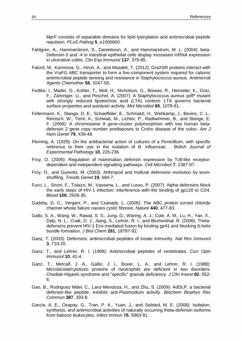
84 References
MprF consists of separable domains for lipid lysinylation and antimicrobial peptide repulsion. PLoS Pathog 5, e1000660.
Fahlgren, A., Hammarstrom, S., Danielsson, A., and Hammarstrom, M. L. (2004): beta-Defensin-3 and -4 in intestinal epithelial cells display increased mRNA expression in ulcerative colitis. Clin Exp Immunol 137, 379-85.
Falord, M., Karimova, G., Hiron, A., and Msadek, T. (2012): GraXSR proteins interact with the VraFG ABC transporter to form a five-component system required for cationic antimicrobial peptide sensing and resistance in Staphylococcus aureus. Antimicrob Agents Chemother 56, 1047-58.
Fedtke, I., Mader, D., Kohler, T., Moll, H., Nicholson, G., Biswas, R., Henseler, K., Gotz, F., Zahringer, U., and Peschel, A. (2007): A Staphylococcus aureus ypfP mutant with strongly reduced lipoteichoic acid (LTA) content: LTA governs bacterial surface properties and autolysin activity. Mol Microbiol 65, 1078-91.
Fellermann, K., Stange, D. E., Schaeffeler, E., Schmalzl, H., Wehkamp, J., Bevins, C. L., Reinisch, W., Teml, A., Schwab, M., Lichter, P., Radlwimmer, B., and Stange, E. F. (2006): A chromosome 8 gene-cluster polymorphism with low human beta-defensin 2 gene copy number predisposes to Crohn disease of the colon. Am J Hum Genet 79, 439-48.
Fleming, A. (1929): On the antibacterial action of cultures of a Penicillium, with specific reference to their use in the isolation of B. influenzae. . British Journal of Experimental Pathology 10, 226-236.
Froy, O. (2005): Regulation of mammalian defensin expression by Toll-like receptor-dependent and independent signalling pathways. Cell Microbiol 7, 1387-97.
Froy, O., and Gurevitz, M. (2003): Arthropod and mollusk defensins--evolution by exon-shuffling. Trends Genet 19, 684-7.
Furci, L., Sironi, F., Tolazzi, M., Vassena, L., and Lusso, P. (2007): Alpha-defensins block the early steps of HIV-1 infection: interference with the binding of gp120 to CD4. Blood 109, 2928-35.
Gadsby, D. C., Vergani, P., and Csanady, L. (2006): The ABC protein turned chloride channel whose failure causes cystic fibrosis. Nature 440, 477-83.
Gallo, S. A., Wang, W., Rawat, S. S., Jung, G., Waring, A. J., Cole, A. M., Lu, H., Yan, X., Daly, N. L., Craik, D. J., Jiang, S., Lehrer, R. I., and Blumenthal, R. (2006): Theta-defensins prevent HIV-1 Env-mediated fusion by binding gp41 and blocking 6-helix bundle formation. J Biol Chem 281, 18787-92.
Ganz, T. (2003): Defensins: antimicrobial peptides of innate immunity. Nat Rev Immunol 3, 710-20.
Ganz, T., and Lehrer, R. I. (1998): Antimicrobial peptides of vertebrates. Curr Opin Immunol 10, 41-4.
Ganz, T., Metcalf, J. A., Gallin, J. I., Boxer, L. A., and Lehrer, R. I. (1988): Microbicidal/cytotoxic proteins of neutrophils are deficient in two disorders: Chediak-Higashi syndrome and "specific" granule deficiency. J Clin Invest 82, 552-6.
Gao, B., Rodriguez Mdel, C., Lanz-Mendoza, H., and Zhu, S. (2009): AdDLP, a bacterial defensin-like peptide, exhibits anti-Plasmodium activity. Biochem Biophys Res Commun 387, 393-8.
Garcia, A. E., Osapay, G., Tran, P. A., Yuan, J., and Selsted, M. E. (2008): Isolation, synthesis, and antimicrobial activities of naturally occurring theta-defensin isoforms from baboon leukocytes. Infect Immun 76, 5883-91.
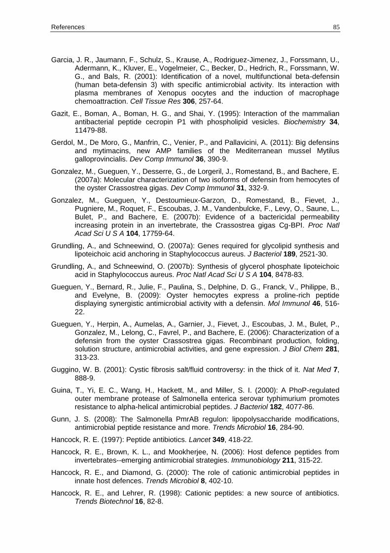
References 85
Garcia, J. R., Jaumann, F., Schulz, S., Krause, A., Rodriguez-Jimenez, J., Forssmann, U., Adermann, K., Kluver, E., Vogelmeier, C., Becker, D., Hedrich, R., Forssmann, W. G., and Bals, R. (2001): Identification of a novel, multifunctional beta-defensin (human beta-defensin 3) with specific antimicrobial activity. Its interaction with plasma membranes of Xenopus oocytes and the induction of macrophage chemoattraction. Cell Tissue Res 306, 257-64.
Gazit, E., Boman, A., Boman, H. G., and Shai, Y. (1995): Interaction of the mammalian antibacterial peptide cecropin P1 with phospholipid vesicles. Biochemistry 34, 11479-88.
Gerdol, M., De Moro, G., Manfrin, C., Venier, P., and Pallavicini, A. (2011): Big defensins and mytimacins, new AMP families of the Mediterranean mussel Mytilus galloprovincialis. Dev Comp Immunol 36, 390-9.
Gonzalez, M., Gueguen, Y., Desserre, G., de Lorgeril, J., Romestand, B., and Bachere, E. (2007a): Molecular characterization of two isoforms of defensin from hemocytes of the oyster Crassostrea gigas. Dev Comp Immunol 31, 332-9.
Gonzalez, M., Gueguen, Y., Destoumieux-Garzon, D., Romestand, B., Fievet, J., Pugniere, M., Roquet, F., Escoubas, J. M., Vandenbulcke, F., Levy, O., Saune, L., Bulet, P., and Bachere, E. (2007b): Evidence of a bactericidal permeability increasing protein in an invertebrate, the Crassostrea gigas Cg-BPI. Proc Natl Acad Sci U S A 104, 17759-64.
Grundling, A., and Schneewind, O. (2007a): Genes required for glycolipid synthesis and lipoteichoic acid anchoring in Staphylococcus aureus. J Bacteriol 189, 2521-30.
Grundling, A., and Schneewind, O. (2007b): Synthesis of glycerol phosphate lipoteichoic acid in Staphylococcus aureus. Proc Natl Acad Sci U S A 104, 8478-83.
Gueguen, Y., Bernard, R., Julie, F., Paulina, S., Delphine, D. G., Franck, V., Philippe, B., and Evelyne, B. (2009): Oyster hemocytes express a proline-rich peptide displaying synergistic antimicrobial activity with a defensin. Mol Immunol 46, 516-22.
Gueguen, Y., Herpin, A., Aumelas, A., Garnier, J., Fievet, J., Escoubas, J. M., Bulet, P., Gonzalez, M., Lelong, C., Favrel, P., and Bachere, E. (2006): Characterization of a defensin from the oyster Crassostrea gigas. Recombinant production, folding, solution structure, antimicrobial activities, and gene expression. J Biol Chem 281, 313-23.
Guggino, W. B. (2001): Cystic fibrosis salt/fluid controversy: in the thick of it. Nat Med 7, 888-9.
Guina, T., Yi, E. C., Wang, H., Hackett, M., and Miller, S. I. (2000): A PhoP-regulated outer membrane protease of Salmonella enterica serovar typhimurium promotes resistance to alpha-helical antimicrobial peptides. J Bacteriol 182, 4077-86.
Gunn, J. S. (2008): The Salmonella PmrAB regulon: lipopolysaccharide modifications, antimicrobial peptide resistance and more. Trends Microbiol 16, 284-90.
Hancock, R. E. (1997): Peptide antibiotics. Lancet 349, 418-22.
Hancock, R. E., Brown, K. L., and Mookherjee, N. (2006): Host defence peptides from invertebrates--emerging antimicrobial strategies. Immunobiology 211, 315-22.
Hancock, R. E., and Diamond, G. (2000): The role of cationic antimicrobial peptides in innate host defences. Trends Microbiol 8, 402-10.
Hancock, R. E., and Lehrer, R. (1998): Cationic peptides: a new source of antibiotics. Trends Biotechnol 16, 82-8.
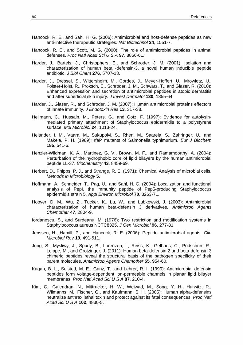
86 References
Hancock, R. E., and Sahl, H. G. (2006): Antimicrobial and host-defense peptides as new anti-infective therapeutic strategies. Nat Biotechnol 24, 1551-7.
Hancock, R. E., and Scott, M. G. (2000): The role of antimicrobial peptides in animal defenses. Proc Natl Acad Sci U S A 97, 8856-61.
Harder, J., Bartels, J., Christophers, E., and Schroder, J. M. (2001): Isolation and characterization of human beta -defensin-3, a novel human inducible peptide antibiotic. J Biol Chem 276, 5707-13.
Harder, J., Dressel, S., Wittersheim, M., Cordes, J., Meyer-Hoffert, U., Mrowietz, U., Folster-Holst, R., Proksch, E., Schroder, J. M., Schwarz, T., and Glaser, R. (2010): Enhanced expression and secretion of antimicrobial peptides in atopic dermatitis and after superficial skin injury. J Invest Dermatol 130, 1355-64.
Harder, J., Glaser, R., and Schroder, J. M. (2007): Human antimicrobial proteins effectors of innate immunity. J Endotoxin Res 13, 317-38.
Heilmann, C., Hussain, M., Peters, G., and Gotz, F. (1997): Evidence for autolysin-mediated primary attachment of Staphylococcus epidermidis to a polystyrene surface. Mol Microbiol 24, 1013-24.
Helander, I. M., Vaara, M., Sukupolvi, S., Rhen, M., Saarela, S., Zahringer, U., and Makela, P. H. (1989): rfaP mutants of Salmonella typhimurium. Eur J Biochem 185, 541-6.
Henzler-Wildman, K. A., Martinez, G. V., Brown, M. F., and Ramamoorthy, A. (2004): Perturbation of the hydrophobic core of lipid bilayers by the human antimicrobial peptide LL-37. Biochemistry 43, 8459-69.
Herbert, D., Phipps, P. J., and Strange, R. E. (1971): Chemical Analysis of microbial cells. Methods in Microbiology 5.
Hoffmann, A., Schneider, T., Pag, U., and Sahl, H. G. (2004): Localization and functional analysis of PepI, the immunity peptide of Pep5-producing Staphylococcus epidermidis strain 5. Appl Environ Microbiol 70, 3263-71.
Hoover, D. M., Wu, Z., Tucker, K., Lu, W., and Lubkowski, J. (2003): Antimicrobial characterization of human beta-defensin 3 derivatives. Antimicrob Agents Chemother 47, 2804-9.
Iordanescu, S., and Surdeanu, M. (1976): Two restriction and modification systems in Staphylococcus aureus NCTC8325. J Gen Microbiol 96, 277-81.
Jenssen, H., Hamill, P., and Hancock, R. E. (2006): Peptide antimicrobial agents. Clin Microbiol Rev 19, 491-511.
Jung, S., Mysliwy, J., Spudy, B., Lorenzen, I., Reiss, K., Gelhaus, C., Podschun, R., Leippe, M., and Grotzinger, J. (2011): Human beta-defensin 2 and beta-defensin 3 chimeric peptides reveal the structural basis of the pathogen specificity of their parent molecules. Antimicrob Agents Chemother 55, 954-60.
Kagan, B. L., Selsted, M. E., Ganz, T., and Lehrer, R. I. (1990): Antimicrobial defensin peptides form voltage-dependent ion-permeable channels in planar lipid bilayer membranes. Proc Natl Acad Sci U S A 87, 210-4.
Kim, C., Gajendran, N., Mittrucker, H. W., Weiwad, M., Song, Y. H., Hurwitz, R., Wilmanns, M., Fischer, G., and Kaufmann, S. H. (2005): Human alpha-defensins neutralize anthrax lethal toxin and protect against its fatal consequences. Proc Natl Acad Sci U S A 102, 4830-5.
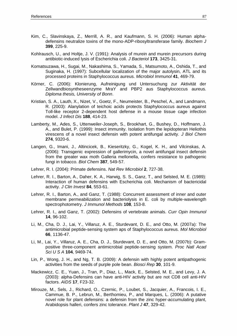
References 87
Kim, C., Slavinskaya, Z., Merrill, A. R., and Kaufmann, S. H. (2006): Human alpha-defensins neutralize toxins of the mono-ADP-ribosyltransferase family. Biochem J 399, 225-9.
Kohlrausch, U., and Holtje, J. V. (1991): Analysis of murein and murein precursors during antibiotic-induced lysis of Escherichia coli. J Bacteriol 173, 3425-31.
Komatsuzawa, H., Sugai, M., Nakashima, S., Yamada, S., Matsumoto, A., Oshida, T., and Suginaka, H. (1997): Subcellular localization of the major autolysin, ATL and its processed proteins in Staphylococcus aureus. Microbiol Immunol 41, 469-79.
Körner, C. (2006): Klonierung, Aufreinigung und Untersuchung zur Aktivität der Zellwandbiosyntheseenzyme MraY and PBP2 aus Staphylococcus aureus. Diploma thesis, University of Bonn.
Kristian, S. A., Lauth, X., Nizet, V., Goetz, F., Neumeister, B., Peschel, A., and Landmann, R. (2003): Alanylation of teichoic acids protects Staphylococcus aureus against Toll-like receptor 2-dependent host defense in a mouse tissue cage infection model. J Infect Dis 188, 414-23.
Lamberty, M., Ades, S., Uttenweiler-Joseph, S., Brookhart, G., Bushey, D., Hoffmann, J. A., and Bulet, P. (1999): Insect immunity. Isolation from the lepidopteran Heliothis virescens of a novel insect defensin with potent antifungal activity. J Biol Chem 274, 9320-6.
Langen, G., Imani, J., Altincicek, B., Kieseritzky, G., Kogel, K. H., and Vilcinskas, A. (2006): Transgenic expression of gallerimycin, a novel antifungal insect defensin from the greater wax moth Galleria mellonella, confers resistance to pathogenic fungi in tobacco. Biol Chem 387, 549-57.
Lehrer, R. I. (2004): Primate defensins. Nat Rev Microbiol 2, 727-38.
Lehrer, R. I., Barton, A., Daher, K. A., Harwig, S. S., Ganz, T., and Selsted, M. E. (1989): Interaction of human defensins with Escherichia coli. Mechanism of bactericidal activity. J Clin Invest 84, 553-61.
Lehrer, R. I., Barton, A., and Ganz, T. (1988): Concurrent assessment of inner and outer membrane permeabilization and bacteriolysis in E. coli by multiple-wavelength spectrophotometry. J Immunol Methods 108, 153-8.
Lehrer, R. I., and Ganz, T. (2002): Defensins of vertebrate animals. Curr Opin Immunol 14, 96-102.
Li, M., Cha, D. J., Lai, Y., Villaruz, A. E., Sturdevant, D. E., and Otto, M. (2007a): The antimicrobial peptide-sensing system aps of Staphylococcus aureus. Mol Microbiol 66, 1136-47.
Li, M., Lai, Y., Villaruz, A. E., Cha, D. J., Sturdevant, D. E., and Otto, M. (2007b): Gram-positive three-component antimicrobial peptide-sensing system. Proc Natl Acad Sci U S A 104, 9469-74.
Lin, P., Wong, J. H., and Ng, T. B. (2009): A defensin with highly potent antipathogenic activities from the seeds of purple pole bean. Biosci Rep 30, 101-9.
Mackewicz, C. E., Yuan, J., Tran, P., Diaz, L., Mack, E., Selsted, M. E., and Levy, J. A. (2003): alpha-Defensins can have anti-HIV activity but are not CD8 cell anti-HIV factors. AIDS 17, F23-32.
Mirouze, M., Sels, J., Richard, O., Czernic, P., Loubet, S., Jacquier, A., Francois, I. E., Cammue, B. P., Lebrun, M., Berthomieu, P., and Marques, L. (2006): A putative novel role for plant defensins: a defensin from the zinc hyper-accumulating plant, Arabidopsis halleri, confers zinc tolerance. Plant J 47, 329-42.
88 References
Mookherjee, N., Rehaume, L. M., and Hancock, R. E. (2007): Cathelicidins and functional analogues as antisepsis molecules. Expert Opin Ther Targets 11, 993-1004.
Munch, D., Roemer, T., Lee, S. H., Engeser, M., Sahl, H. G., and Schneider, T. (2012): Identification and in vitro analysis of the GatD/MurT enzyme-complex catalyzing lipid II amidation in Staphylococcus aureus. PLoS Pathog 8, e1002509.
Munk, C., Wei, G., Yang, O. O., Waring, A. J., Wang, W., Hong, T., Lehrer, R. I., Landau, N. R., and Cole, A. M. (2003): The theta-defensin, retrocyclin, inhibits HIV-1 entry. AIDS Res Hum Retroviruses 19, 875-81.
Mygind, P. H., Fischer, R. L., Schnorr, K. M., Hansen, M. T., Sonksen, C. P., Ludvigsen, S., Raventos, D., Buskov, S., Christensen, B., De Maria, L., Taboureau, O., Yaver, D., Elvig-Jorgensen, S. G., Sorensen, M. V., Christensen, B. E., Kjaerulff, S., Frimodt-Moller, N., Lehrer, R. I., Zasloff, M., and Kristensen, H. H. (2005): Plectasin is a peptide antibiotic with therapeutic potential from a saprophytic fungus. Nature 437, 975-80.
Nguyen, T. X., Cole, A. M., and Lehrer, R. I. (2003): Evolution of primate theta-defensins: a serpentine path to a sweet tooth. Peptides 24, 1647-54.
Nijnik, A., Pistolic, J., Wyatt, A., Tam, S., and Hancock, R. E. (2009): Human cathelicidin peptide LL-37 modulates the effects of IFN-gamma on APCs. J Immunol 183, 5788-98.
Niyonsaba, F., Someya, A., Hirata, M., Ogawa, H., and Nagaoka, I. (2001): Evaluation of the effects of peptide antibiotics human beta-defensins-1/-2 and LL-37 on histamine release and prostaglandin D(2) production from mast cells. Eur J Immunol 31, 1066-75.
Niyonsaba, F., Ushio, H., Nakano, N., Ng, W., Sayama, K., Hashimoto, K., Nagaoka, I., Okumura, K., and Ogawa, H. (2007): Antimicrobial peptides human beta-defensins stimulate epidermal keratinocyte migration, proliferation and production of proinflammatory cytokines and chemokines. J Invest Dermatol 127, 594-604.
Nomura, I., Goleva, E., Howell, M. D., Hamid, Q. A., Ong, P. Y., Hall, C. F., Darst, M. A., Gao, B., Boguniewicz, M., Travers, J. B., and Leung, D. Y. (2003): Cytokine milieu of atopic dermatitis, as compared to psoriasis, skin prevents induction of innate immune response genes. J Immunol 171, 3262-9.
Ong, P. Y., Ohtake, T., Brandt, C., Strickland, I., Boguniewicz, M., Ganz, T., Gallo, R. L., and Leung, D. Y. (2002): Endogenous antimicrobial peptides and skin infections in atopic dermatitis. N Engl J Med 347, 1151-60.
Orlov, D. S., Nguyen, T., and Lehrer, R. I. (2002): Potassium release, a useful tool for studying antimicrobial peptides. J Microbiol Methods 49, 325-8.
Otvos, L., Jr. (2000): Antibacterial peptides isolated from insects. J Pept Sci 6, 497-511.
Ouellette, A. J., and Selsted, M. E. (2012): Immunology. HD6 defensin nanonets. Science 337, 420-1.
Overhage, J., Campisano, A., Bains, M., Torfs, E. C., Rehm, B. H., and Hancock, R. E. (2008): Human host defense peptide LL-37 prevents bacterial biofilm formation. Infect Immun 76, 4176-82.
Pag, U., Oedenkoven, M., Sass, V., Shai, Y., Shamova, O., Antcheva, N., Tossi, A., and Sahl, H. G. (2008): Analysis of in vitro activities and modes of action of synthetic antimicrobial peptides derived from an alpha-helical 'sequence template'. J Antimicrob Chemother 61, 341-52.
References 89
Park, C. B., Kim, H. S., and Kim, S. C. (1998): Mechanism of action of the antimicrobial peptide buforin II: buforin II kills microorganisms by penetrating the cell membrane and inhibiting cellular functions. Biochem Biophys Res Commun 244, 253-7.
Patrzykat, A., Friedrich, C. L., Zhang, L., Mendoza, V., and Hancock, R. E. (2002): Sublethal concentrations of pleurocidin-derived antimicrobial peptides inhibit macromolecular synthesis in Escherichia coli. Antimicrob Agents Chemother 46, 605-14.
Pazgier, M., Hoover, D. M., Yang, D., Lu, W., and Lubkowski, J. (2006): Human beta-defensins. Cell Mol Life Sci 63, 1294-313.
Peschel, A., Jack, R. W., Otto, M., Collins, L. V., Staubitz, P., Nicholson, G., Kalbacher, H., Nieuwenhuizen, W. F., Jung, G., Tarkowski, A., van Kessel, K. P., and van Strijp, J. A. (2001): Staphylococcus aureus resistance to human defensins and evasion of neutrophil killing via the novel virulence factor MprF is based on modification of membrane lipids with l-lysine. J Exp Med 193, 1067-76.
Peschel, A., Otto, M., Jack, R. W., Kalbacher, H., Jung, G., and Gotz, F. (1999): Inactivation of the dlt operon in Staphylococcus aureus confers sensitivity to defensins, protegrins, and other antimicrobial peptides. J Biol Chem 274, 8405-10.
Poppinga, L. (2007): Zelluläre und molekulargenetische Untersuchungen zur Wirkungsweise des Lantibiotikums Pep5. Diploma thesis, University of Bonn.
Putsep, K., Carlsson, G., Boman, H. G., and Andersson, M. (2002): Deficiency of antibacterial peptides in patients with morbus Kostmann: an observation study. Lancet 360, 1144-9.
Raetz, C. R., and Whitfield, C. (2002): Lipopolysaccharide endotoxins. Annu Rev Biochem 71, 635-700.
Ramamoorthy, V., Cahoon, E. B., Li, J., Thokala, M., Minto, R. E., and Shah, D. M. (2007): Glucosylceramide synthase is essential for alfalfa defensin-mediated growth inhibition but not for pathogenicity of Fusarium graminearum. Mol Microbiol 66, 771-86.
Richards, S. M., Strandberg, K. L., and Gunn, J. S. (2010): Salmonella-regulated lipopolysaccharide modifications. Subcell Biochem 53, 101-22.
Rick, P. D., Hubbard, G. L., Kitaoka, M., Nagaki, H., Kinoshita, T., Dowd, S., Simplaceanu, V., and Ho, C. (1998): Characterization of the lipid-carrier involved in the synthesis of enterobacterial common antigen (ECA) and identification of a novel phosphoglyceride in a mutant of Salmonella typhimurium defective in ECA synthesis. Glycobiology 8, 557-67.
Rodriguez-Jimenez, F. J., Krause, A., Schulz, S., Forssmann, W. G., Conejo-Garcia, J. R., Schreeb, R., and Motzkus, D. (2003): Distribution of new human beta-defensin genes clustered on chromosome 20 in functionally different segments of epididymis. Genomics 81, 175-83.
Rosa, R. D., Santini, A., Fievet, J., Bulet, P., Destoumieux-Garzon, D., and Bachere, E. (2011): Big defensins, a diverse family of antimicrobial peptides that follows different patterns of expression in hemocytes of the oyster Crassostrea gigas. PLoS One 6, e25594.
Rouser, G., S. Fkeischer, and Yamamoto, A. (1970): Two dimensional thin layer chromatographic separation of polar lipids and determination of phospholipids by phosphorus analysis of spots. Lipids 5, 494–496.
Ruhland, G. J., and Fiedler, F. (1990): Occurrence and structure of lipoteichoic acids in the genus Staphylococcus. Arch Microbiol 154, 375-9.
90 References
Sahl, H. G. (1985): Influence of the staphylococcinlike peptide Pep 5 on membrane potential of bacterial cells and cytoplasmic membrane vesicles. J Bacteriol 162, 833-6.
Sahl, H. G., and Bierbaum, G. (1998): Lantibiotics: biosynthesis and biological activities of uniquely modified peptides from gram-positive bacteria. Annu Rev Microbiol 52, 41-79.
Sahl, H. G., and Brandis, H. (1981): Production, purification and chemical properties of an antistaphylococcal agent produced by Staphylococcus epidermidis. J Gen Microbiol 127, 377-84.
Sahl, H. G., and Brandis, H. (1983): Efflux of low-Mr substances from the cytoplasm of sensitive cells caused by the staphylococcin-like agent Pep 5. FEMS Microbiol Lett 16, 75-79.
Saito, T., Kawabata, S., Shigenaga, T., Takayenoki, Y., Cho, J., Nakajima, H., Hirata, M., and Iwanaga, S. (1995): A novel big defensin identified in horseshoe crab hemocytes: isolation, amino acid sequence, and antibacterial activity. J Biochem 117, 1131-7.
Salzman, N. H., Hung, K., Haribhai, D., Chu, H., Karlsson-Sjoberg, J., Amir, E., Teggatz, P., Barman, M., Hayward, M., Eastwood, D., Stoel, M., Zhou, Y., Sodergren, E., Weinstock, G. M., Bevins, C. L., Williams, C. B., and Bos, N. A. (2010): Enteric defensins are essential regulators of intestinal microbial ecology. Nat Immunol 11, 76-83.
Sass, P., and Bierbaum, G. (2009): Native graS mutation supports the susceptibility of Staphylococcus aureus strain SG511 to antimicrobial peptides. Int J Med Microbiol 299, 313-22.
Sass, V., Pag, U., Tossi, A., Bierbaum, G., and Sahl, H. G. (2008): Mode of action of human beta-defensin 3 against Staphylococcus aureus and transcriptional analysis of responses to defensin challenge. Int J Med Microbiol 298, 619-33.
Sass, V., Schneider, T., Wilmes, M., Korner, C., Tossi, A., Novikova, N., Shamova, O., and Sahl, H. G. (2010): Human beta-defensin 3 inhibits cell wall biosynthesis in Staphylococci. Infect Immun 78, 2793-800.
Schibli, D. J., Hunter, H. N., Aseyev, V., Starner, T. D., Wiencek, J. M., McCray, P. B., Jr., Tack, B. F., and Vogel, H. J. (2002): The solution structures of the human beta-defensins lead to a better understanding of the potent bactericidal activity of HBD3 against Staphylococcus aureus. J Biol Chem 277, 8279-89.
Schlag, M., Biswas, R., Krismer, B., Kohler, T., Zoll, S., Yu, W., Schwarz, H., Peschel, A., and Gotz, F. (2010): Role of staphylococcal wall teichoic acid in targeting the major autolysin Atl. Mol Microbiol 75, 864-73.
Schleifer, K. H., and Fischer, U. (1983): Description of a new species of the genus Staphylococcus: Staphylococcus carnosus. Int J System Bacteriol 32, 153-156.
Schmitt, P., de Lorgeril, J., Gueguen, Y., Destoumieux-Garzon, D., and Bachere, E. (2012a): Expression, tissue localization and synergy of antimicrobial peptides and proteins in the immune response of the oyster Crassostrea gigas. Dev Comp Immunol 37, 363-70.
Schmitt, P., Gueguen, Y., Desmarais, E., Bachere, E., and de Lorgeril, J. (2010): Molecular diversity of antimicrobial effectors in the oyster Crassostrea gigas. BMC Evol Biol 10, 23.
Schmitt, P., Rosa, R. D., Duperthuy, M., de Lorgeril, J., Bachere, E., and Destoumieux-Garzon, D. (2012b): The Antimicrobial Defense of the Pacific Oyster, Crassostrea
References 91
gigas. How Diversity may Compensate for Scarcity in the Regulation of Resident/Pathogenic Microflora. Front Microbiol 3, 160.
Schneider, J. J., Unholzer, A., Schaller, M., Schafer-Korting, M., and Korting, H. C. (2005): Human defensins. J Mol Med (Berl) 83, 587-95.
Schneider, T., Kruse, T., Wimmer, R., Wiedemann, I., Sass, V., Pag, U., Jansen, A., Nielsen, A. K., Mygind, P. H., Raventos, D. S., Neve, S., Ravn, B., Bonvin, A. M., De Maria, L., Andersen, A. S., Gammelgaard, L. K., Sahl, H. G., and Kristensen, H. H. (2010): Plectasin, a fungal defensin, targets the bacterial cell wall precursor Lipid II. Science 328, 1168-72.
Schneider, T., and Sahl, H. G. (2010): An oldie but a goodie - cell wall biosynthesis as antibiotic target pathway. Int J Med Microbiol 300, 161-9.
Schroeder, B. O., Wu, Z., Nuding, S., Groscurth, S., Marcinowski, M., Beisner, J., Buchner, J., Schaller, M., Stange, E. F., and Wehkamp, J. (2011): Reduction of disulphide bonds unmasks potent antimicrobial activity of human beta-defensin 1. Nature 469, 419-23.
Schutte, B. C., Mitros, J. P., Bartlett, J. A., Walters, J. D., Jia, H. P., Welsh, M. J., Casavant, T. L., and McCray, P. B., Jr. (2002): Discovery of five conserved beta -defensin gene clusters using a computational search strategy. Proc Natl Acad Sci U S A 99, 2129-33.
Scott, M. G., Dullaghan, E., Mookherjee, N., Glavas, N., Waldbrook, M., Thompson, A., Wang, A., Lee, K., Doria, S., Hamill, P., Yu, J. J., Li, Y., Donini, O., Guarna, M. M., Finlay, B. B., North, J. R., and Hancock, R. E. (2007): An anti-infective peptide that selectively modulates the innate immune response. Nat Biotechnol 25, 465-72.
Scott, M. G., Vreugdenhil, A. C., Buurman, W. A., Hancock, R. E., and Gold, M. R. (2000): Cutting edge: cationic antimicrobial peptides block the binding of lipopolysaccharide (LPS) to LPS binding protein. J Immunol 164, 549-53.
Seidel, A., Ye, Y., de Armas, L. R., Soto, M., Yarosh, W., Marcsisin, R. A., Tran, D., Selsted, M. E., and Camerini, D. (2010): Cyclic and acyclic defensins inhibit human immunodeficiency virus type-1 replication by different mechanisms. PLoS One 5, e9737.
Selsted, M. E., and Ouellette, A. J. (2005): Mammalian defensins in the antimicrobial immune response. Nat Immunol 6, 551-7.
Semple, F., MacPherson, H., Webb, S., Cox, S. L., Mallin, L. J., Tyrrell, C., Grimes, G. R., Semple, C. A., Nix, M. A., Millhauser, G. L., and Dorin, J. R. (2011): Human beta-defensin 3 affects the activity of pro-inflammatory pathways associated with MyD88 and TRIF. Eur J Immunol 41, 3291-300.
Shafer, W. M., Qu, X., Waring, A. J., and Lehrer, R. I. (1998): Modulation of Neisseria gonorrhoeae susceptibility to vertebrate antibacterial peptides due to a member of the resistance/nodulation/division efflux pump family. Proc Natl Acad Sci U S A 95, 1829-33.
Sieprawska-Lupa, M., Mydel, P., Krawczyk, K., Wojcik, K., Puklo, M., Lupa, B., Suder, P., Silberring, J., Reed, M., Pohl, J., Shafer, W., McAleese, F., Foster, T., Travis, J., and Potempa, J. (2004): Degradation of human antimicrobial peptide LL-37 by Staphylococcus aureus-derived proteinases. Antimicrob Agents Chemother 48, 4673-9.
Smith, J. J., Travis, S. M., Greenberg, E. P., and Welsh, M. J. (1996): Cystic fibrosis airway epithelia fail to kill bacteria because of abnormal airway surface fluid. Cell 85, 229-36.
92 References
Soehnlein, O., Kai-Larsen, Y., Frithiof, R., Sorensen, O. E., Kenne, E., Scharffetter-Kochanek, K., Eriksson, E. E., Herwald, H., Agerberth, B., and Lindbom, L. (2008): Neutrophil primary granule proteins HBP and HNP1-3 boost bacterial phagocytosis by human and murine macrophages. J Clin Invest 118, 3491-502.
Spelbrink, R. G., Dilmac, N., Allen, A., Smith, T. J., Shah, D. M., and Hockerman, G. H. (2004): Differential antifungal and calcium channel-blocking activity among structurally related plant defensins. Plant Physiol 135, 2055-67.
Spindler, E. C., Hale, J. D., Giddings, T. H., Jr., Hancock, R. E., and Gill, R. T. (2011): Deciphering the mode of action of the synthetic antimicrobial peptide Bac8c. Antimicrob Agents Chemother 55, 1706-16.
Studier, F. W., and Moffatt, B. A. (1986): Use of bacteriophage T7 RNA polymerase to direct selective high-level expression of cloned genes. J Mol Biol 189, 113-30.
Subbalakshmi, C., and Sitaram, N. (1998): Mechanism of antimicrobial action of indolicidin. FEMS Microbiol Lett 160, 91-6.
Sun, L., Finnegan, C. M., Kish-Catalone, T., Blumenthal, R., Garzino-Demo, P., La Terra Maggiore, G. M., Berrone, S., Kleinman, C., Wu, Z., Abdelwahab, S., Lu, W., and Garzino-Demo, A. (2005): Human beta-defensins suppress human immunodeficiency virus infection: potential role in mucosal protection. J Virol 79, 14318-29.
Taggart, C. C., Greene, C. M., Smith, S. G., Levine, R. L., McCray, P. B., Jr., O'Neill, S., and McElvaney, N. G. (2003): Inactivation of human beta-defensins 2 and 3 by elastolytic cathepsins. J Immunol 171, 931-7.
Tang, Y. Q., Yuan, J., Osapay, G., Osapay, K., Tran, D., Miller, C. J., Ouellette, A. J., and Selsted, M. E. (1999): A cyclic antimicrobial peptide produced in primate leukocytes by the ligation of two truncated alpha-defensins. Science 286, 498-502.
Tavares, P. M., Thevissen, K., Cammue, B. P., Francois, I. E., Barreto-Bergter, E., Taborda, C. P., Marques, A. F., Rodrigues, M. L., and Nimrichter, L. (2008): In vitro activity of the antifungal plant defensin RsAFP2 against Candida isolates and its in vivo efficacy in prophylactic murine models of candidiasis. Antimicrob Agents Chemother 52, 4522-5.
Teng, L., Gao, B., and Zhang, S. (2012): The first chordate big defensin: identification, expression and bioactivity. Fish Shellfish Immunol 32, 572-7.
Territo, M. C., Ganz, T., Selsted, M. E., and Lehrer, R. (1989): Monocyte-chemotactic activity of defensins from human neutrophils. J Clin Invest 84, 2017-20.
Thevissen, K., de Mello Tavares, P., Xu, D., Blankenship, J., Vandenbosch, D., Idkowiak-Baldys, J., Govaert, G., Bink, A., Rozental, S., de Groot, P. W., Davis, T. R., Kumamoto, C. A., Vargas, G., Nimrichter, L., Coenye, T., Mitchell, A., Roemer, T., Hannun, Y. A., and Cammue, B. P. (2012): The plant defensin RsAFP2 induces cell wall stress, septin mislocalization and accumulation of ceramides in Candida albicans. Mol Microbiol 84, 166-80.
Thevissen, K., Francois, I. E., Takemoto, J. Y., Ferket, K. K., Meert, E. M., and Cammue, B. P. (2003): DmAMP1, an antifungal plant defensin from dahlia (Dahlia merckii), interacts with sphingolipids from Saccharomyces cerevisiae. FEMS Microbiol Lett 226, 169-73.
Thevissen, K., Francois, I. E., Winderickx, J., Pannecouque, C., and Cammue, B. P. (2006): Ceramide involvement in apoptosis and apoptotic diseases. Mini Rev Med Chem 6, 699-709.
References 93
Thevissen, K., Ghazi, A., De Samblanx, G. W., Brownlee, C., Osborn, R. W., and Broekaert, W. F. (1996): Fungal membrane responses induced by plant defensins and thionins. J Biol Chem 271, 15018-25.
Thevissen, K., Kristensen, H. H., Thomma, B. P., Cammue, B. P., and Francois, I. E. (2007): Therapeutic potential of antifungal plant and insect defensins. Drug Discov Today 12, 966-71.
Thevissen, K., Warnecke, D. C., Francois, I. E., Leipelt, M., Heinz, E., Ott, C., Zahringer, U., Thomma, B. P., Ferket, K. K., and Cammue, B. P. (2004): Defensins from insects and plants interact with fungal glucosylceramides. J Biol Chem 279, 3900-5.
Thomma, B. P., Cammue, B. P., and Thevissen, K. (2002): Plant defensins. Planta 216, 193-202.
Tongaonkar, P., Tran, P., Roberts, K., Schaal, J., Osapay, G., Tran, D., Ouellette, A. J., and Selsted, M. E. (2011): Rhesus macaque theta-defensin isoforms: expression, antimicrobial activities, and demonstration of a prominent role in neutrophil granule microbicidal activities. J Leukoc Biol 89, 283-90.
Trabi, M., Mylne, J. S., Sando, L., and Craik, D. J. (2009): Circular proteins from Melicytus (Violaceae) refine the conserved protein and gene architecture of cyclotides. Org Biomol Chem 7, 2378-88.
Trabi, M., Schirra, H. J., and Craik, D. J. (2001): Three-dimensional structure of RTD-1, a cyclic antimicrobial defensin from Rhesus macaque leukocytes. Biochemistry 40, 4211-21.
Tran, D., Tran, P., Roberts, K., Osapay, G., Schaal, J., Ouellette, A., and Selsted, M. E. (2008): Microbicidal properties and cytocidal selectivity of rhesus macaque theta defensins. Antimicrob Agents Chemother 52, 944-53.
Tran, D., Tran, P. A., Tang, Y. Q., Yuan, J., Cole, T., and Selsted, M. E. (2002): Homodimeric theta-defensins from rhesus macaque leukocytes: isolation, synthesis, antimicrobial activities, and bacterial binding properties of the cyclic peptides. J Biol Chem 277, 3079-84.
Vollmer, W., Joris, B., Charlier, P., and Foster, S. (2008): Bacterial peptidoglycan (murein) hydrolases. FEMS Microbiol Rev 32, 259-86.
Wang, W., Owen, S. M., Rudolph, D. L., Cole, A. M., Hong, T., Waring, A. J., Lal, R. B., and Lehrer, R. I. (2004): Activity of alpha- and theta-defensins against primary isolates of HIV-1. J Immunol 173, 515-20.
Wang, X., and Quinn, P. J. (2010): Lipopolysaccharide: Biosynthetic pathway and structure modification. Prog Lipid Res 49, 97-107.
Wehkamp, J., Harder, J., Weichenthal, M., Mueller, O., Herrlinger, K. R., Fellermann, K., Schroeder, J. M., and Stange, E. F. (2003): Inducible and constitutive beta-defensins are differentially expressed in Crohn's disease and ulcerative colitis. Inflamm Bowel Dis 9, 215-23.
Wehkamp, J., Salzman, N. H., Porter, E., Nuding, S., Weichenthal, M., Petras, R. E., Shen, B., Schaeffeler, E., Schwab, M., Linzmeier, R., Feathers, R. W., Chu, H., Lima, H., Jr., Fellermann, K., Ganz, T., Stange, E. F., and Bevins, C. L. (2005): Reduced Paneth cell alpha-defensins in ileal Crohn's disease. Proc Natl Acad Sci U S A 102, 18129-34.
Wehkamp, J., Wang, G., Kubler, I., Nuding, S., Gregorieff, A., Schnabel, A., Kays, R. J., Fellermann, K., Burk, O., Schwab, M., Clevers, H., Bevins, C. L., and Stange, E. F.
94 References
(2007): The Paneth cell alpha-defensin deficiency of ileal Crohn's disease is linked to Wnt/Tcf-4. J Immunol 179, 3109-18.
Weiss, T. M., Yang, L., Ding, L., Waring, A. J., Lehrer, R. I., and Huang, H. W. (2002): Two states of cyclic antimicrobial peptide RTD-1 in lipid bilayers. Biochemistry 41, 10070-6.
Wiedemann, I., Breukink, E., van Kraaij, C., Kuipers, O. P., Bierbaum, G., de Kruijff, B., and Sahl, H. G. (2001): Specific binding of nisin to the peptidoglycan precursor lipid II combines pore formation and inhibition of cell wall biosynthesis for potent antibiotic activity. J Biol Chem 276, 1772-9.
Wong, J. H., and Ng, T. B. (2005): Sesquin, a potent defensin-like antimicrobial peptide from ground beans with inhibitory activities toward tumor cells and HIV-1 reverse transcriptase. Peptides 26, 1120-6.
Xiong, Y. Q., Hady, W. A., Deslandes, A., Rey, A., Fraisse, L., Kristensen, H. H., Yeaman, M. R., and Bayer, A. S. (2011): Efficacy of NZ2114, a novel plectasin-derived cationic antimicrobial peptide antibiotic, in experimental endocarditis due to methicillin-resistant Staphylococcus aureus. Antimicrob Agents Chemother 55, 5325-30.
Yamada, S., Sugai, M., Komatsuzawa, H., Nakashima, S., Oshida, T., Matsumoto, A., and Suginaka, H. (1996): An autolysin ring associated with cell separation of Staphylococcus aureus. J Bacteriol 178, 1565-71.
Yamaguchi, Y., Nagase, T., Makita, R., Fukuhara, S., Tomita, T., Tominaga, T., Kurihara, H., and Ouchi, Y. (2002): Identification of multiple novel epididymis-specific beta-defensin isoforms in humans and mice. J Immunol 169, 2516-23.
Yang, D., Chen, Q., Chertov, O., and Oppenheim, J. J. (2000): Human neutrophil defensins selectively chemoattract naive T and immature dendritic cells. J Leukoc Biol 68, 9-14.
Yang, D., Chertov, O., Bykovskaia, S. N., Chen, Q., Buffo, M. J., Shogan, J., Anderson, M., Schroder, J. M., Wang, J. M., Howard, O. M., and Oppenheim, J. J. (1999): Beta-defensins: linking innate and adaptive immunity through dendritic and T cell CCR6. Science 286, 525-8.
Yang, L., Harroun, T. A., Heller, W. T., Weiss, T. M., and Huang, H. W. (1998): Neutron off-plane scattering of aligned membranes. I. Method Of measurement. Biophys J 75, 641-5.
Yang, L., Harroun, T. A., Weiss, T. M., Ding, L., and Huang, H. W. (2001): Barrel-stave model or toroidal model? A case study on melittin pores. Biophys J 81, 1475-85.
Yeaman, M. R., and Yount, N. Y. (2007): Unifying themes in host defence effector polypeptides. Nat Rev Microbiol 5, 727-40.
Yethon, J. A., Gunn, J. S., Ernst, R. K., Miller, S. I., Laroche, L., Malo, D., and Whitfield, C. (2000): Salmonella enterica serovar typhimurium waaP mutants show increased susceptibility to polymyxin and loss of virulence In vivo. Infect Immun 68, 4485-91.
Yethon, J. A., Heinrichs, D. E., Monteiro, M. A., Perry, M. B., and Whitfield, C. (1998): Involvement of waaY, waaQ, and waaP in the modification of Escherichia coli lipopolysaccharide and their role in the formation of a stable outer membrane. J Biol Chem 273, 26310-6.
Yeung, A. T., Gellatly, S. L., and Hancock, R. E. (2011): Multifunctional cationic host defence peptides and their clinical applications. Cell Mol Life Sci 68, 2161-76.
References 95
Yount, N. Y., and Yeaman, M. R. (2004): Multidimensional signatures in antimicrobial peptides. Proc Natl Acad Sci U S A 101, 7363-8.
Zasloff, M. (2002): Antimicrobial peptides of multicellular organisms. Nature 415, 389-95.
Zhang, Y., He, X., Li, X., Fu, D., Chen, J., and Yu, Z. (2011): The second bactericidal permeability increasing protein (BPI) and its revelation of the gene duplication in the Pacific oyster, Crassostrea gigas. Fish Shellfish Immunol 30, 954-63.
Zhao, J., Song, L., Li, C., Ni, D., Wu, L., Zhu, L., Wang, H., and Xu, W. (2007): Molecular cloning, expression of a big defensin gene from bay scallop Argopecten irradians and the antimicrobial activity of its recombinant protein. Mol Immunol 44, 360-8.
Zhou, C. X., Zhang, Y. L., Xiao, L., Zheng, M., Leung, K. M., Chan, M. Y., Lo, P. S., Tsang, L. L., Wong, H. Y., Ho, L. S., Chung, Y. W., and Chan, H. C. (2004): An epididymis-specific beta-defensin is important for the initiation of sperm maturation. Nat Cell Biol 6, 458-64.
Zhu, S. (2007): Evidence for myxobacterial origin of eukaryotic defensins. Immunogenetics 59, 949-54.
Zhu, S. (2008): Discovery of six families of fungal defensin-like peptides provides insights into origin and evolution of the CSalphabeta defensins. Mol Immunol 45, 828-38.
Zhu, S., and Gao, B. (2012): Evolutionary origin of beta-defensins. Dev Comp Immunol.
Zoll, S., Schlag, M., Shkumatov, A. V., Rautenberg, M., Svergun, D. I., Gotz, F., and Stehle, T. (2012): Ligand-binding properties and conformational dynamics of autolysin repeat domains in staphylococcal cell wall recognition. J Bacteriol 194, 3789-802.

Publications 97
7 Publications
Journal articles
Sass, V., Schneider, T., Wilmes, M., Körner, C., Tossi, A., Novikova, N., Shamova, O.,
and Sahl, H. G. (2010): Human beta-defensin 3 inhbitis cell wall biosynthesis in
staphylococci. Infect Immun 78, 2793-800
Schmitt, P., Wilmes, M., Pugnière, M., Aumelas, A., Bachère, E., Sahl, H. G., Schneider,
T., and Destoumieux-Garzón, D. (2010): Insight into invertebrate defensin
mechanism of action: oyster defensins inhibit peptidoglycan biosynthesis by
binding to lipid II. J Biol Chem 17, 29208-16
Wilmes, M., Cammue, B. P., Sahl, H. G., and Thevissen, K. (2011): Antibiotic activities of
host defense peptides: more to it than lipid bilayer perturbation. Nat Prod Rep 28,
1350-8
Ulm, H., Wilmes, M., Shai, Y., and Sahl, H. G. (2012): Antimicrobial host defensins –
specific antibiotic activities and innate defense modulation. Front Immunol 3: 249
Poster presentations
Wilmes, M., Tuchscherr, L., Becker, K., and Sahl, H. G.: Efficacy of human β-defensin 3
(hBD3) against clinical isolates of Staphylococcus aureus.
Annual conference of the German Society for Hygiene and Microbiology (DGHM),
Göttingen, 2009
Wilmes, M., Schmitt, P., Schneider, T., Destoumieux-Garzón, D., and Sahl, H. G.: Mode
of action of defensins against Staphylococcus aureus.
Transregio 34 Meeting on “Host pathogen interactions in bacterial infections”,
Greifswald, 2010
Wilmes, M., Sass, V., Schneider, T., Tossi, A., and Sahl, H. G.: Mode of action of human
β-defensin 3 (hBD3).
Annual conference of the Association for General and Applied Microbiology
(VAAM), Karlsruhe, 2011
98 Publications
Wilmes, M., Sass, V., Schneider, T., Tossi, A., Ouellette, A., Selsted, M., and Sahl, H. G.:
Comparison of the antistaphylococcal activity of human β-defensin 3 (hBD3) and
rhesus macaque theta-defensins (RTD-1, RTD-2).
Gordon Research Conference on “Antimicrobial Peptides”, Lucca (Barga), Italy,
2011
Wilmes, M., Ouellette, A., Selsted, M., and Sahl, H. G.: Mode of action of rhesus
macaque theta-defensins (RTD-1, RTD-2) against Staphylococcus aureus.
Annual conference of the Association for General and Applied Microbiology
(VAAM), Tübingen, 2012
Wilmes, M., Ouellette, A., Selsted, M., and Sahl, H. G.: Antistaphylococcal action of
rhesus macaque theta-defensins.
Gordon Research Conference on “New Antibacterial Discovery and Development”,
Lucca (Barga), Italy, 2012
Declaration 99
8 Declaration (Eidesstattliche Erklärung)
Hiermit erkläre ich an Eides statt, dass ich für meine Promotion keine anderen als die
angegebenen Hilfsmittel benutzt habe, und dass die inhaltlich und wörtlich aus anderen
Werken entnommenen Stellen und Zitate als solche gekennzeichnet sind. Diese Arbeit ist
weder identisch noch teilidentisch mit einer Arbeit, die an der Rheinischen Friedrich-
Wilhelms-Universität Bonn oder einer anderen Hochschule zur Erlangung eines
akademischen Grades oder als Prüfungsleistung vorgelegt worden ist. Teile der
Dissertation sind vorab an den unter Punkt 7 „Publications“ aufgeführten Stellen
auszugsweise veröffentlicht worden.
Bonn, Oktober 2012
___________________________
Miriam Wilmes