Embed Size (px)
Citation preview
Antimicrobial peptides as new potential antibiotics
Inaugural-Dissertation
zur Erlangung des Doktorgrades
der Mathematisch-Naturwissenschaftlichen Fakultät der Universität zu Köln
vorgelegt von
André Reinhardt aus Reutlingen
Köln
2017
Berichterstatter: Prof. Dr. Ines Neundorf (Gutachter)
Prof. Dr. Karin Schnetz
Tag der mündlichen Prüfung: 02.06.2017
Die im Rahmen der vorliegenden Arbeit durchgeführten Experimente und
Untersuchungen wurden im Zeitraum von Oktober 2013 bis April 2017 am Institut
für Biochemie der Universität zu Köln unter der Anleitung von Frau Prof. Dr. Ines
Neundorf durchgeführt.
i
Abstract In recent years, the rapid increase of antibiotic resistances and the expansion of
multi-resistant bacterial strains have provoked the need to develop novel antibiotics.
So-called antimicrobial peptides (AMPs) are short, amphiphilic, cationic peptides
and part of the innate immune system. There unique membrane disrupting
mechanism and the low propensity for developing resistances attracted their
attention in pharmaceutical medicine. AMPs are active against a wide spectrum of
microorganisms, such as gram-positive and gram-negative bacteria, fungi, viruses
and parasites.
The present thesis focuses on improving the antimicrobial activity of AMPs by using
different strategies like synthesis of AMP-conjugates, membrane immobilization of
AMPs, and amino acid exchanges within the AMP sequence. For this, multiple
imidazolium-salts, already described as antibacterial agents, were conjugated to
AMPs via solid phase peptide synthesis, developing a branched conjugate.
Combination of both compounds resulted in a higher antimicrobial activity against
multi-resistant bacterial strains. Selectivity of the novel compounds was
demonstrated against human-red blood cells, which was further investigated by lipid
interaction studies with cholesterol. The most selective compound IL-KKA (3a)
could be used as a future lead structure for the development of new antimicrobial
agents.
Since 80% of human infections are caused by biofilms, the newly designed
compound IL-KKA (3a) was coupled covalently via a peptide bond or with electron
beam radiation on polyether sulfone membranes (PES). Both immobilization
techniques were successfully performed, still showing a high antimicrobial activity of
the immobilized compound.
The cell-penetrating peptide sC18 was converted to an AMP by amino acid
exchanges with isoleucine and phenylalanine. Isoleucine and phenylalanine mono
mutants already exhibited an increased activity against a wide spectrum of bacteria.
A higher amount of phenylalanine in the sequence leads to a further increased
antimicrobial activity. The insertion of hydrophobic amino acids at position 10 led to
the formation of a characteristic α-helix, while the positions 15 and 16 seemed to be
necessary for hydrophobic membrane interactions.
ii
All in all, this thesis highlights the successful modification of AMPs to more active
antimicrobial agents, which make them extremely interesting for the design of future
antibiotics and the application of potential anti-biofilm agents.
iii
Zusammenfassung Die Vermehrung von Antibiotikaresistenzen und somit multi-resistenten Bakterien
macht die Entwicklung von alternativen Antibiotika, in Form von antimikrobiellen
Peptiden (AMPs), nötig. AMPs sind kurze, amphipathische, kationische Peptide und
Teil des angeborenen Immunsystems. Ihr einzigartiger Wirkmechanismus und das
seltene Aufkommen von Resistenzen, machen AMPs interessant für die Pharmazie.
AMPs sind aktiv gegen unterschiedlichste Mikroorganismen u.a. gram-positive und
gram-negative Bakterien sowie Pilze, Viren und Parasiten.
In dieser Arbeit soll die antimikrobielle Aktivität von AMPs durch Herstellung von
AMP-Konjugaten, Membranimmobilisierung von AMPs und den Austausch von
Aminosäuren innerhalb der AMP Sequenz weiter erhöht werden. Unterschiedlich
modifizierte Imidazoliumsalze, die bereits für ihre antimikrobielle Aktivität bekannt
sind, wurden mit Hilfe der Festphasenpeptidsynthese an AMPs gekuppelt. Die
Kombination der beiden Komponenten zeigte eine höhere Aktivität gegen
multiresistente Bakterien. Eine der neuen Verbindungen, IL-KKA (3a), zeigte die
höchste Selektivität und kann als Leitstruktur für die Entwicklung von neuen
Antibiotika verwendet werden.
80% der heutigen Infektionen werden durch Biofilme verursacht. Deshalb wurde
das neu designte IL-KKA (3a) mit unterschiedlichen Linkern an Polyethersulfon-
Membranen immobilisiert. Dies wurde entweder durch eine kovalente
Peptidbindung oder mit Hilfe von Elektronenbestrahlung bewerkstelligt. Beide
Techniken waren erfolgreich und die so aktivierten Membranen zeigten weiterhin
eine antimikrobielle Aktivität.
Das zellpenetrierende Peptid sC18 wurde durch den Austausch der Aminosäuren
an Position 10, 15 und 16 durch Isoleucin oder Phenylalanin in ein AMP
umgewandelt. Die Einzelmutanten zeigten bereits eine erhöhte Aktivität gegenüber
unterschiedlichsten Bakterienstämmen. Durch das Einfügen von bis zu 3
Phenylalaninen konnte die antimikrobielle Aktivität weiter erhöht werden. Hierbei
sorgte der Austausch an Position 10 für eine besser ausgebildete α-Helix, während
der Austausch an den Positionen 15 und 16 die hydrophoben
Membraninteraktionen verstärkten.
Zusammenfassend zeigt diese Arbeit, dass die Modifikation von antimikrobiellen
Peptiden zu einer erhöhten Aktivität führen kann. Die in dieser Arbeit neu
iv
entwickelten AMPs haben das Potential, als neue Antibiotika oder als Anti-Biofilm
Wirkstoffe eingesetzt zu werden.
v
Table of contents
1. Introduction ...................................... .............................................. 1
1.1. Antibiotics and the connected problem to antimicrobial resistances ............ 1
1.2. Antimicrobial peptides – a new class of antibiotics? .................................... 2
1.3. Antibacterial AMPs – mechanism of action ................................................. 7
1.4. Structural properties of antimicrobial peptides............................................. 9
1.5. Bacterial resistance mechanisms against antimicrobial peptides .............. 10
1.6. Biofilms – a problem of bacterial infections ............................................... 11
1.7. General structure of bacterial membranes ................................................ 12
1.8. Ionic liquids................................................................................................ 14
1.9. Preliminary work ........................................................................................ 15
1.10. Aim of the thesis ..................................................................................... 17
2. Materials and methods ............................. ................................... 19
2.1. Materials .................................................................................................... 19
2.1.1. Equipment .............................................................................................. 20
2.1.2. Buffers.................................................................................................... 21
2.1.3. Bacterial strains ..................................................................................... 22
2.1.4. Peptide sequences ................................................................................ 22
2.2. Solid phase peptide synthesis (SPPS) ...................................................... 24
2.2.1. Loading of Wang-resin with the first amino acid ..................................... 24
2.2.2. Determination of the first residue attachment ......................................... 25
2.2.3. Automated peptide synthesis ................................................................. 25
2.2.4. Manual coupling of ionic liquids and 5(6)-carboxyfluorescein ................ 26
2.2.5. Endcapping ............................................................................................ 26
2.2.6. Fmoc cleavage ....................................................................................... 26
2.2.7. Cleavage of the Dde-protection group ................................................... 27
2.2.8. Kaiser test .............................................................................................. 27
2.2.9. Sample cleavage ................................................................................... 27
2.2.10. Full cleavage ........................................................................................ 28
2.2.11. LC-mass spectrometry with 0.1% FA for qualitative analysis ............... 28
2.2.12. RP-HPLC with 0.1% TFA for purity analysis ........................................ 29
2.2.13. Preparative RP-HPLC with 0.1% TFA .................................................. 29
2.2.14. Synthesis of ionic liquids ...................................................................... 29
vi
2.3. Characterization methods.......................................................................... 29
2.3.1. Circular dichroism spectroscopy ............................................................ 29
2.3.2. Preparation of large unilamellar vesicle (LUVs) ..................................... 30
2.3.3. Circular dichroism with LUVs ................................................................. 30
2.3.4. Preparation of giant unilamellar vesicle (GUVs) ..................................... 30
2.3.5. CLSM observation of GUVs treated with peptide conjugates ................. 31
2.4. Biological methods .................................................................................... 31
2.4.1. Antimicrobial activity .............................................................................. 31
2.4.2. Killing assay using resistant bacterial strains ......................................... 32
2.4.3. Hemolytic activity ................................................................................... 32
2.4.4. Immobilization and characterization of polyether sulfone ....................... 33
membranes (PES) ........................................................................................... 33
2.4.5. Antimicrobial activity with immobilized PES membranes ....................... 33
3. Results and discussion ............................ ................................... 35
3.1. Improvement of imidazolium salt-peptide conjugates and their mechanism of action............................................................................................................... 35
3.1.1. Synthesis of imidazolium salt-peptide conjugates .................................. 35
3.1.2. pH influence on the secondary structure of compound 3c ..................... 39
3.1.3. Antimicrobial activity against resistant bacterial strains ......................... 40
3.1.4. Hemolytic activity studies ....................................................................... 42
3.2. Electron beam immobilization of novel antimicrobial, short peptide motifs leads to membrane surfaces with promising anti-biofilm properties .................... 48
3.2.1. Synthesis of 3a and its derivates 3d, 3e and 3f ..................................... 49
3.2.2. Membrane immobilization and characterization ..................................... 51
3.2.3. Antimicrobial activity of the functionalized PES membranes .................. 53
3.3. Optimizing the antimicrobial activity of the CPP sC18 ............................... 57
3.3.1. Synthesis of sC18 variants ..................................................................... 58
3.3.2. Antimicrobial activity of sC18 isoleucine variants ................................... 63
3.3.3. Antimicrobial activity of phenylalanine variants ...................................... 70
3.3.4. Characterization of peptides via CD-spectrometry ................................. 76
3.3.5. Peptide interaction with artificial membrane vesicles ............................. 79
4. Conclusion and Outlook ............................ ................................. 83
5. Literature ........................................ .............................................. 85
6. Attachment ........................................ ........................................... 96
vii
6.1. List of abbreviations .................................................................................. 96
6.2. List of Figures ............................................................................................ 99
6.3. List of tables ............................................................................................ 101
6.4. Acknowledgment ..................................................................................... 102
1
1. Introduction
1.1. Antibiotics and the connected problem to antimicrobial resistances
In 1928, Alexander Fleming discovered penicillin, the first antibiotic substance. This
discovery opened one of the most important research fields in medical history.
Nearly 90 years later, antibiotics are still the major tools against infectious diseases.
However, due to their excessive and overdosed application, in the last decades,
bacteria became more and more resistant against common antibiotics (1). The
resulting antimicrobial resistances have become a major health problem (1-4).
Antibiotic resistances are listed as one of the greatest threats to human health (5),
as a lot of resistant bacteria can be found in hospitals, where they cause serious
infections. So-called superbugs refer to bacteria, which have adapted to resist
multiple classes of antibiotics (multidrug-resistant). They demonstrate an enhanced
morbidity and the therapeutic options to kill them are only limited (6). The most
famous superbug is the gram-positive and methicillin-resistant bacterium
Staphylococcus aureus (MRSA) (7). One additional problem is the use of antibiotics
in non-human niches like agriculture, aquaculture and waste disposal that has
steadily increased during the last years (8). For example, the resistance of
Escherichia coli against ciprofloxacin has been associated with the use of
fluoroquinolones, a broad-spectrum antibiotic, in aviculture (9). When bacteria
acquired resistances, they are able to preserve them through genetic and
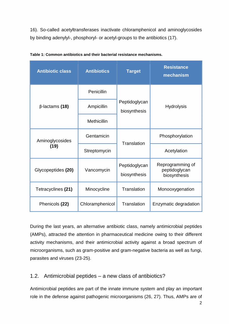
biochemical mechanisms (Table 1). These mechanisms include e.g. genetic
mutations or transfer of genetic gene material between bacteria via conjugation,
transformation or transduction (10, 11). Biochemical resistance mechanisms on the
other side can be divided into different resistance types, like an decreased uptake of
the antibiotic, enzymatic modification and degradation, or an altered targeting within
the cell, or via efflux pumps that remove the antibiotic out of the cell directly after the
uptake (12, 13). However, the most popular mechanism is the enzymatic
inactivation of antibiotics. The three main enzyme classes are β-lactamases,
aminoglycoside-modifying enzymes and chloramphenicol acetyltransferases. β-
lactamases hydrolyze ester and amide bonds from β-lactam antibiotics like penicillin
(14). Aminoglycoside-modifying enzymes reduce the affinity of aminoglycosides and
fluoroquinolones, resulting in a weaker binding to the 30S ribosomal subunit (15,
2
16). So-called acetyltransferases inactivate chloramphenicol and aminoglycosides
by binding adenylyl-, phosphoryl- or acetyl-groups to the antibiotics (17).
Table 1: Common antibiotics and their bacterial res istance mechanisms.
Antibiotic class Antibiotics Target Resistance
mechanism
β-lactams (18)
Penicillin
Peptidoglycan
biosynthesis Hydrolysis Ampicillin
Methicillin
Aminoglycosides (19)
Gentamicin
Translation
Phosphorylation
Streptomycin Acetylation
Glycopeptides (20) Vancomycin Peptidoglycan
biosynthesis
Reprogramming of peptidoglycan biosynthesis
Tetracyclines (21) Minocycline Translation Monooxygenation
Phenicols (22) Chloramphenicol Translation Enzymatic degradation
During the last years, an alternative antibiotic class, namely antimicrobial peptides
(AMPs), attracted the attention in pharmaceutical medicine owing to their different
activity mechanisms, and their antimicrobial activity against a broad spectrum of
microorganisms, such as gram-positive and gram-negative bacteria as well as fungi,
parasites and viruses (23-25).
1.2. Antimicrobial peptides – a new class of antibiotics?
Antimicrobial peptides are part of the innate immune system and play an important
role in the defense against pathogenic microorganisms (26, 27). Thus, AMPs are of
3
great interest in averting infections before they cause symptoms, and additionally,
they participate in inflammation and wound healing processes (28). Based on their
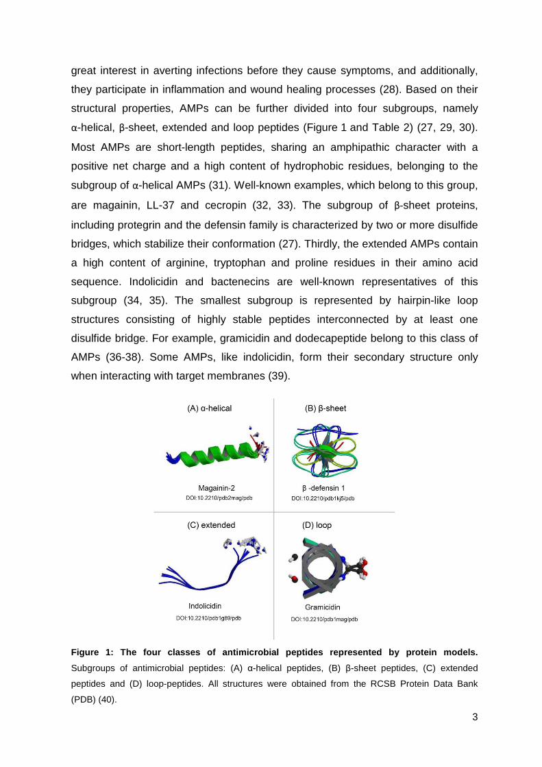
structural properties, AMPs can be further divided into four subgroups, namely
α-helical, β-sheet, extended and loop peptides (Figure 1 and Table 2) (27, 29, 30).
Most AMPs are short-length peptides, sharing an amphipathic character with a
positive net charge and a high content of hydrophobic residues, belonging to the
subgroup of α-helical AMPs (31). Well-known examples, which belong to this group,
are magainin, LL-37 and cecropin (32, 33). The subgroup of β-sheet proteins,
including protegrin and the defensin family is characterized by two or more disulfide
bridges, which stabilize their conformation (27). Thirdly, the extended AMPs contain
a high content of arginine, tryptophan and proline residues in their amino acid
sequence. Indolicidin and bactenecins are well-known representatives of this
subgroup (34, 35). The smallest subgroup is represented by hairpin-like loop
structures consisting of highly stable peptides interconnected by at least one
disulfide bridge. For example, gramicidin and dodecapeptide belong to this class of
AMPs (36-38). Some AMPs, like indolicidin, form their secondary structure only
when interacting with target membranes (39).
Figure 1: The four classes of antimicrobial peptide s represented by protein models.
Subgroups of antimicrobial peptides: (A) α-helical peptides, (B) β-sheet peptides, (C) extended
peptides and (D) loop-peptides. All structures were obtained from the RCSB Protein Data Bank
(PDB) (40).
4
In the last years, researchers optimized natural AMPs and developed related
synthetic ones (41, 42). Especially the reduction of size leads to an optimization of
metabolic stability and bioavailability. Furthermore, shorter peptide sequences
would advantageously reduce the production costs dramatically (43). Modifications
of peptide bonds by introduction of hydrogen bonds in the AMP sequence as well as
insertion of unnatural amino acids and replacement, might also increase the
antimicrobial activity (44). The conjugation of AMPs to drugs, photosensitizer,
nanoparticles or organometallic complexes could convert AMPs into useful delivery
vectors (45). Moreover, there are different strategies to apply AMPs therapeutically.
On the one hand, they can be used as single anti-infective reagents, or in
combination with common antibiotics to obtain a synergetic effect. On the other
hand, AMPs can be used as immunostimulatory agents resulting in an enhanced
innate immune system. Lastly, the application of AMPs as endotoxin-neutralizing
agents is possible to prevent septic shocks induced by bacterial virulence factors
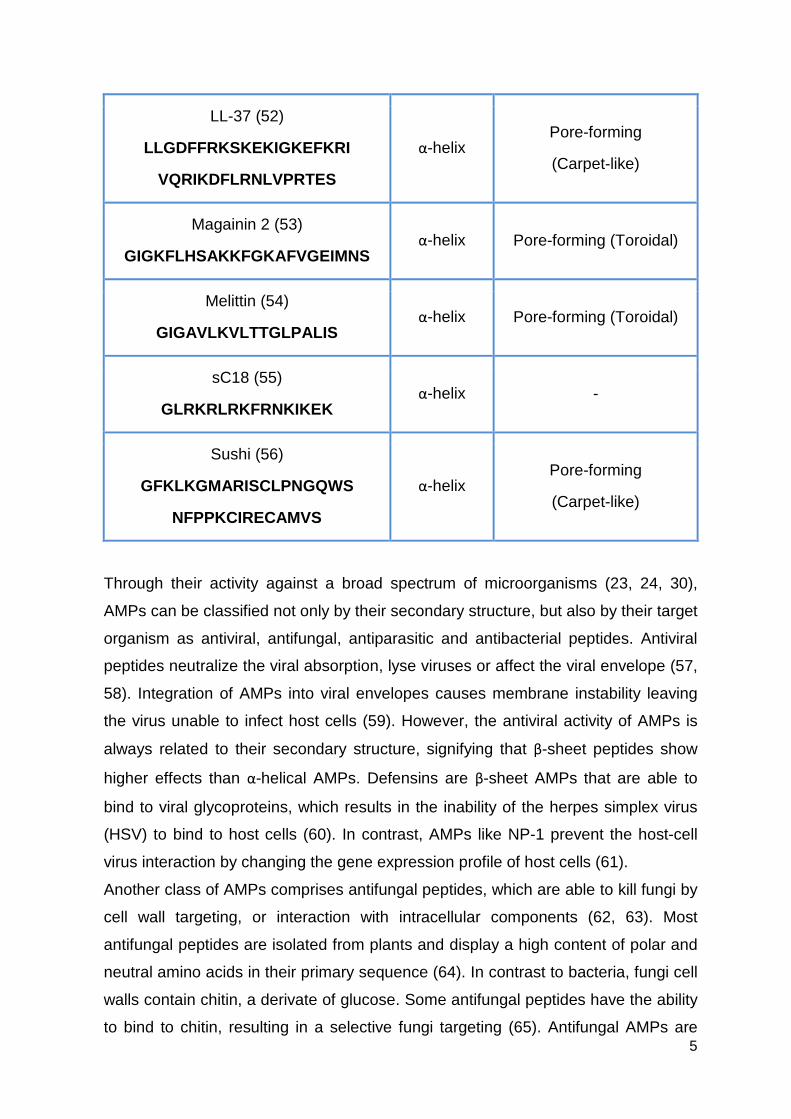
(46). Table 2: Some examples of antimicrobial peptides, t heir sequences, structures and
mechanisms of action.
Peptide and Sequence Structure Mechanism
Apidaecin 1b (47)
GNNRPVYIPQPRPPHPRL
Polyproline helix type II
Inhibition of ATPase
Cecropin A (48)
KWKLFKKIEKVGQNIRDGII
KAGPAVAVVGQATQIAK
α-helix Disruption of cell membrane
Histatin-5 (49)
DSHAKRHHGYKRKFHEKHHSHRGY α-helix Intracellular targeting
Indolicidin (50)
ILPWKWPWWPWRR Extended
Inhibition of DNA/RNA synthesis
KLA (51)
(KLAKLAK) 2 α-helix Pore-forming
5
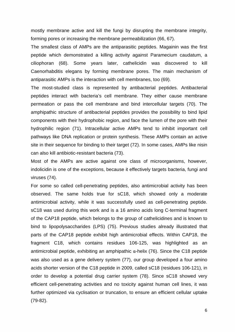
LL-37 (52)
LLGDFFRKSKEKIGKEFKRI
VQRIKDFLRNLVPRTES
α-helix Pore-forming
(Carpet-like)
Magainin 2 (53)
GIGKFLHSAKKFGKAFVGEIMNS α-helix Pore-forming (Toroidal)
Melittin (54)
GIGAVLKVLTTGLPALIS α-helix Pore-forming (Toroidal)
sC18 (55)
GLRKRLRKFRNKIKEK α-helix -
Sushi (56)
GFKLKGMARISCLPNGQWS
NFPPKCIRECAMVS
α-helix Pore-forming
(Carpet-like)
Through their activity against a broad spectrum of microorganisms (23, 24, 30),
AMPs can be classified not only by their secondary structure, but also by their target
organism as antiviral, antifungal, antiparasitic and antibacterial peptides. Antiviral
peptides neutralize the viral absorption, lyse viruses or affect the viral envelope (57,
58). Integration of AMPs into viral envelopes causes membrane instability leaving
the virus unable to infect host cells (59). However, the antiviral activity of AMPs is
always related to their secondary structure, signifying that β-sheet peptides show
higher effects than α-helical AMPs. Defensins are β-sheet AMPs that are able to
bind to viral glycoproteins, which results in the inability of the herpes simplex virus
(HSV) to bind to host cells (60). In contrast, AMPs like NP-1 prevent the host-cell
virus interaction by changing the gene expression profile of host cells (61).
Another class of AMPs comprises antifungal peptides, which are able to kill fungi by
cell wall targeting, or interaction with intracellular components (62, 63). Most
antifungal peptides are isolated from plants and display a high content of polar and
neutral amino acids in their primary sequence (64). In contrast to bacteria, fungi cell
walls contain chitin, a derivate of glucose. Some antifungal peptides have the ability
to bind to chitin, resulting in a selective fungi targeting (65). Antifungal AMPs are
6
mostly membrane active and kill the fungi by disrupting the membrane integrity,
forming pores or increasing the membrane permeabilization (66, 67).
The smallest class of AMPs are the antiparasitic peptides. Magainin was the first
peptide which demonstrated a killing activity against Paramecium caudatum, a
ciliophoran (68). Some years later, cathelicidin was discovered to kill
Caenorhabditis elegans by forming membrane pores. The main mechanism of
antiparasitic AMPs is the interaction with cell membranes, too (69).
The most-studied class is represented by antibacterial peptides. Antibacterial
peptides interact with bacteria’s cell membrane. They either cause membrane
permeation or pass the cell membrane and bind intercellular targets (70). The
amphipathic structure of antibacterial peptides provides the possibility to bind lipid
components with their hydrophobic region, and face the lumen of the pore with their
hydrophilic region (71). Intracellular active AMPs tend to inhibit important cell
pathways like DNA replication or protein synthesis. These AMPs contain an active
site in their sequence for binding to their target (72). In some cases, AMPs like nisin
can also kill antibiotic-resistant bacteria (73).
Most of the AMPs are active against one class of microorganisms, however,
indolicidin is one of the exceptions, because it effectively targets bacteria, fungi and
viruses (74).
For some so called cell-penetrating peptides, also antimicrobial activity has been
observed. The same holds true for sC18, which showed only a moderate
antimicrobial activity, while it was successfully used as cell-penetrating peptide.
sC18 was used during this work and is a 16 amino acids long C-terminal fragment
of the CAP18 peptide, which belongs to the group of cathelicidines and is known to
bind to lipopolysaccharides (LPS) (75). Previous studies already illustrated that
parts of the CAP18 peptide exhibit high antimicrobial effects. Within CAP18, the
fragment C18, which contains residues 106-125, was highlighted as an
antimicrobial peptide, exhibiting an amphipathic α-helix (76). Since the C18 peptide
was also used as a gene delivery system (77), our group developed a four amino
acids shorter version of the C18 peptide in 2009, called sC18 (residues 106-121), in
order to develop a potential drug carrier system (78). Since sC18 showed very
efficient cell-penetrating activities and no toxicity against human cell lines, it was
further optimized via cyclisation or truncation, to ensure an efficient cellular uptake
(79-82).
7
1.3. Antibacterial AMPs – mechanism of action
As already mentioned, the most common mechanism of antibacterial AMPs is the
permeation of bacterial membranes followed by their disruption. The amphipathic
character of AMPs, especially their positively charged sites, leads to a highly
selective interaction with the outer microbial membrane. Due to lipoteichoic acids
(gram-negative bacteria) and lipopolysaccharides (gram-positive bacteria), these
membranes show characteristically a negatively charged environment (83, 84).
Bacterial death occurs only when AMPs are completely saturated on the bacterial
cell membrane. Nevertheless, the interaction of AMPs with lipopolysaccharides or
anionic lipoteichoic acids may reduce the AMP concentration needed for
destabilization of the bacterial membrane and pore formation (85). The hydrophobic
part of the peptides enables them to insert into bacterial membranes (86). The
disruption of the bacterial membrane induces the breakdown of the membrane
potential as well as the leakage of intracellular components and is finally leading to
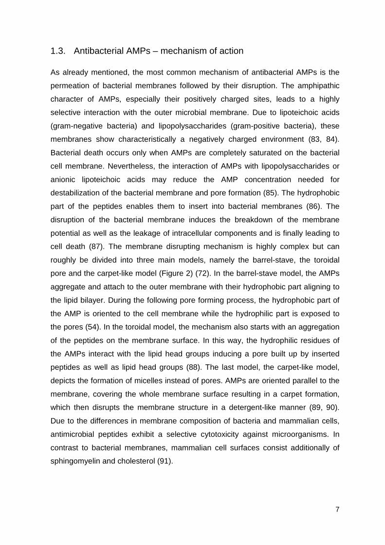
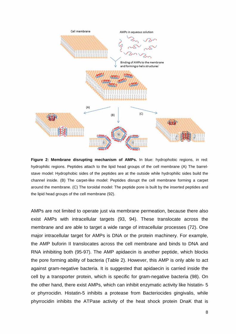
cell death (87). The membrane disrupting mechanism is highly complex but can
roughly be divided into three main models, namely the barrel-stave, the toroidal
pore and the carpet-like model (Figure 2) (72). In the barrel-stave model, the AMPs
aggregate and attach to the outer membrane with their hydrophobic part aligning to
the lipid bilayer. During the following pore forming process, the hydrophobic part of
the AMP is oriented to the cell membrane while the hydrophilic part is exposed to
the pores (54). In the toroidal model, the mechanism also starts with an aggregation
of the peptides on the membrane surface. In this way, the hydrophilic residues of
the AMPs interact with the lipid head groups inducing a pore built up by inserted
peptides as well as lipid head groups (88). The last model, the carpet-like model,
depicts the formation of micelles instead of pores. AMPs are oriented parallel to the
membrane, covering the whole membrane surface resulting in a carpet formation,
which then disrupts the membrane structure in a detergent-like manner (89, 90).
Due to the differences in membrane composition of bacteria and mammalian cells,
antimicrobial peptides exhibit a selective cytotoxicity against microorganisms. In
contrast to bacterial membranes, mammalian cell surfaces consist additionally of
sphingomyelin and cholesterol (91).
8
Figure 2: Membrane disrupting mechanism of AMPs. In blue: hydrophobic regions, in red:
hydrophilic regions. Peptides attach to the lipid head groups of the cell membrane (A) The barrel-
stave model: Hydrophobic sides of the peptides are at the outside while hydrophilic sides build the
channel inside. (B) The carpet-like model: Peptides disrupt the cell membrane forming a carpet
around the membrane. (C) The toroidal model: The peptide pore is built by the inserted peptides and
the lipid head groups of the cell membrane (92).
AMPs are not limited to operate just via membrane permeation, because there also
exist AMPs with intracellular targets (93, 94). These translocate across the
membrane and are able to target a wide range of intracellular processes (72). One
major intracellular target for AMPs is DNA or the protein machinery. For example,
the AMP buforin II translocates across the cell membrane and binds to DNA and
RNA inhibiting both (95-97). The AMP apidaecin is another peptide, which blocks
the pore forming ability of bacteria (Table 2). However, this AMP is only able to act
against gram-negative bacteria. It is suggested that apidaecin is carried inside the
cell by a transporter protein, which is specific for gram-negative bacteria (98). On
the other hand, there exist AMPs, which can inhibit enzymatic activity like histatin- 5
or phyrrocidin. Histatin-5 inhibits a protease from Bacteriocides gingivalis, while
phyrrocidin inhibits the ATPase activity of the heat shock protein DnaK that is
9
involved in protein folding (99, 100). Some AMPs are only active against bacteria in
a certain growth stage, proposing an interaction with a specific metabolic pathway,
which is activated during bacterial growth (101). The cytoplasmic localization of
AMPs leads to the presumption of existing cellular uptake mechanisms. Two
mechanisms for cellular uptake of AMPs are reported. The uptake is either
accomplished via endocytosis, including micropinocytosis or receptor-mediated
endocytosis or by direct penetration (86).
1.4. Structural properties of antimicrobial peptides
The structural properties of antimicrobial peptides are essential for their
antimicrobial activity and cell selectivity. Although a structure related prediction
about the mode of action cannot be proposed, the conformation, charge,
hydrophobicity and solubility are important aspects for antimicrobial peptides. The
structure of α-helical AMPs is often formed during the interaction with the
amphipathic bacterial membrane. It was reported that for an α-helical AMP a length
of at least 22 amino acids is required to transverse the bacterial bilayer via the
barrel-stave model (102). By the introduction of D-amino acids in the hydrophobic
face, the secondary structure is affected, which results in a hemolytic effect and
improved selectivity (103). As the secondary structure of peptides is predicted by
the amino acid sequence, the introduction of amino acids like proline and glycine
hinders the helix-formation and the flexibility (104). Nevertheless, there exist
proline-arginine rich peptides inducing polyproline helical type II structures, which
are comparable to an alpha helix (105, 106).
The positive net charge of AMPs is an important property because the electrostatic
interactions between AMPs and bacterial cell membranes are the major force for
the first contact (72, 107, 108). Since bacterial membranes are rich in acidic
phospholipids and human cell membranes contain acidic phospholipids only on the
inner side, the net charge plays a crucial role for selectivity, too (109). An increasing
positive net charge often leads to an increased antimicrobial but also hemolytic
activity (110).
Another essential structural property of AMPs is the content of hydrophobic amino
acids, which for most antimicrobial peptides is in the range of approximately 50%
(111). Increasing hydrophobicity on the positively charged side of the AMPs up to a
10
certain point leads to an increased antimicrobial activity. Nevertheless, increasing
hydrophobicity is often connected to mammalian cell toxicity and loss of selectivity
(109). Furthermore, an augmented hydrophobicity can alter the range of targets for
the AMP. The AMP magainin, which is only active against gram-negative bacteria,
can also be effective against gram-positive bacteria by the insertion of hydrophobic
amino acids (112).
1.5. Bacterial resistance mechanisms against antimicrobial peptides
In order to find new and useful antibiotic compounds, some AMPs have already
been investigated in clinical research. The understanding of bacterial resistance
against theses AMPs is the next, crucial step (113). Therefore, research groups
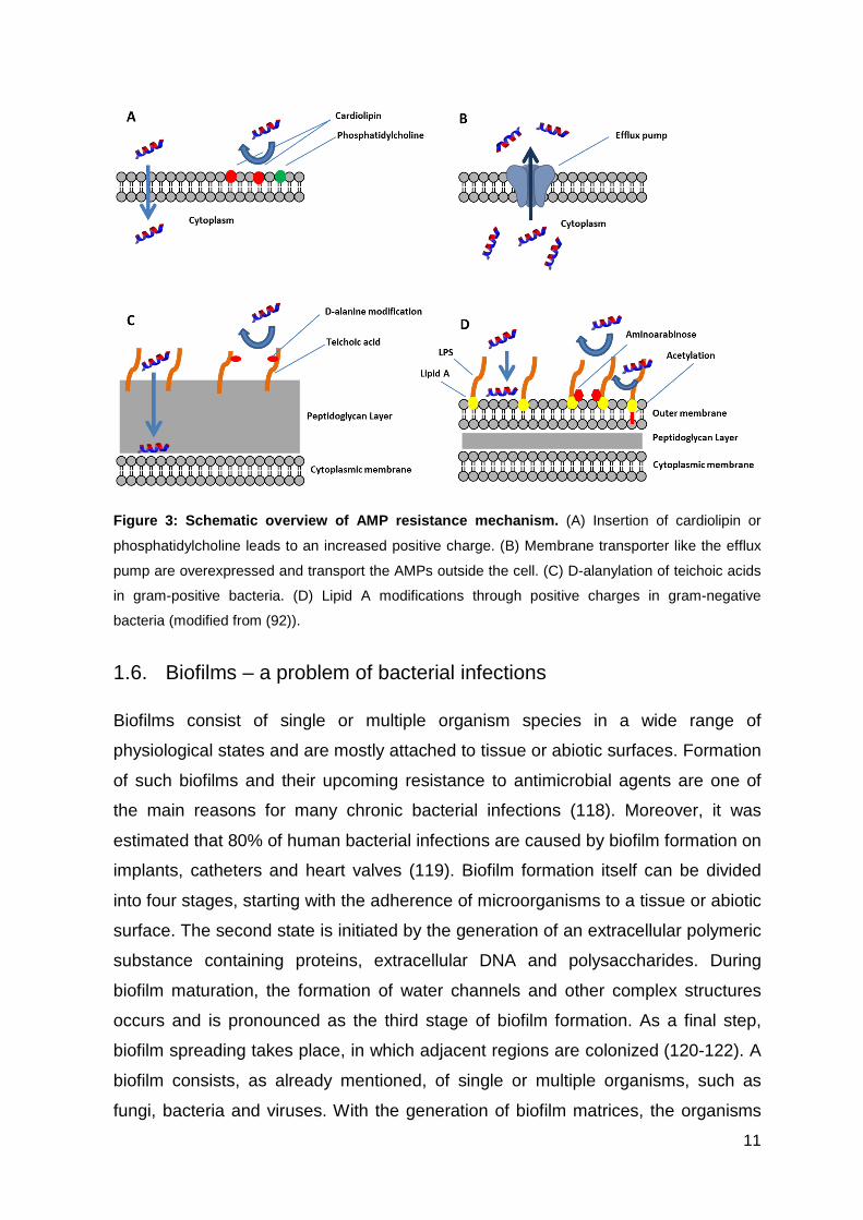
focus on the identification of different bacterial resistance mechanisms. Membrane
alterations represent one major tool for bacteria to develop resistances. It was
shown, that changes in the membrane lipid composition by the inclusion of
cardiolipin or other positively charged phospholipids avoid the insertion of positively
charged AMPs (114) (Figure 3A). Moreover, bacteria are able to overexpress genes
encoding for transmembrane transporters like the efflux pumps, which allow the
displacement of AMPs back into the periplasm (115) (Figure 3B). Another example
for the development of resistances against gram-positive bacteria, is the
modification of their teichoic acids through D-alanylation resulting in the reduction of
their anionic charge (116) (Figure 3C). On the other hand, gram-negative bacteria
can make use of lipid A modifications to incorporate positive charges into
lipopolysaccharides, which decreases the binding affinity of antimicrobial peptides
(117) (Figure 3D). Furthermore, bacteria are able to alter their cellular metabolism
and regulate the expression of proteases, biofilm formation and modifications of
surface structures to avoid the uptake of AMPs (109).
11
Figure 3: Schematic overview of AMP resistance mech anism. (A) Insertion of cardiolipin or
phosphatidylcholine leads to an increased positive charge. (B) Membrane transporter like the efflux
pump are overexpressed and transport the AMPs outside the cell. (C) D-alanylation of teichoic acids
in gram-positive bacteria. (D) Lipid A modifications through positive charges in gram-negative
bacteria (modified from (92)).
1.6. Biofilms – a problem of bacterial infections
Biofilms consist of single or multiple organism species in a wide range of
physiological states and are mostly attached to tissue or abiotic surfaces. Formation
of such biofilms and their upcoming resistance to antimicrobial agents are one of
the main reasons for many chronic bacterial infections (118). Moreover, it was
estimated that 80% of human bacterial infections are caused by biofilm formation on
implants, catheters and heart valves (119). Biofilm formation itself can be divided
into four stages, starting with the adherence of microorganisms to a tissue or abiotic
surface. The second state is initiated by the generation of an extracellular polymeric
substance containing proteins, extracellular DNA and polysaccharides. During
biofilm maturation, the formation of water channels and other complex structures
occurs and is pronounced as the third stage of biofilm formation. As a final step,
biofilm spreading takes place, in which adjacent regions are colonized (120-122). A
biofilm consists, as already mentioned, of single or multiple organisms, such as
fungi, bacteria and viruses. With the generation of biofilm matrices, the organisms
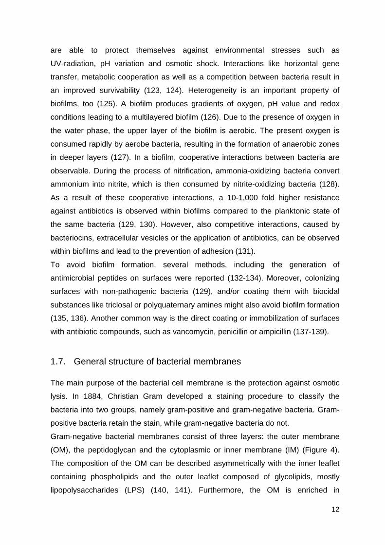
12
are able to protect themselves against environmental stresses such as
UV-radiation, pH variation and osmotic shock. Interactions like horizontal gene
transfer, metabolic cooperation as well as a competition between bacteria result in
an improved survivability (123, 124). Heterogeneity is an important property of
biofilms, too (125). A biofilm produces gradients of oxygen, pH value and redox
conditions leading to a multilayered biofilm (126). Due to the presence of oxygen in
the water phase, the upper layer of the biofilm is aerobic. The present oxygen is
consumed rapidly by aerobe bacteria, resulting in the formation of anaerobic zones
in deeper layers (127). In a biofilm, cooperative interactions between bacteria are
observable. During the process of nitrification, ammonia-oxidizing bacteria convert
ammonium into nitrite, which is then consumed by nitrite-oxidizing bacteria (128).
As a result of these cooperative interactions, a 10-1,000 fold higher resistance
against antibiotics is observed within biofilms compared to the planktonic state of
the same bacteria (129, 130). However, also competitive interactions, caused by
bacteriocins, extracellular vesicles or the application of antibiotics, can be observed
within biofilms and lead to the prevention of adhesion (131).
To avoid biofilm formation, several methods, including the generation of
antimicrobial peptides on surfaces were reported (132-134). Moreover, colonizing
surfaces with non-pathogenic bacteria (129), and/or coating them with biocidal
substances like triclosal or polyquaternary amines might also avoid biofilm formation
(135, 136). Another common way is the direct coating or immobilization of surfaces
with antibiotic compounds, such as vancomycin, penicillin or ampicillin (137-139).
1.7. General structure of bacterial membranes
The main purpose of the bacterial cell membrane is the protection against osmotic
lysis. In 1884, Christian Gram developed a staining procedure to classify the
bacteria into two groups, namely gram-positive and gram-negative bacteria. Gram-
positive bacteria retain the stain, while gram-negative bacteria do not.
Gram-negative bacterial membranes consist of three layers: the outer membrane
(OM), the peptidoglycan and the cytoplasmic or inner membrane (IM) (Figure 4).
The composition of the OM can be described asymmetrically with the inner leaflet
containing phospholipids and the outer leaflet composed of glycolipids, mostly
lipopolysaccharides (LPS) (140, 141). Furthermore, the OM is enriched in
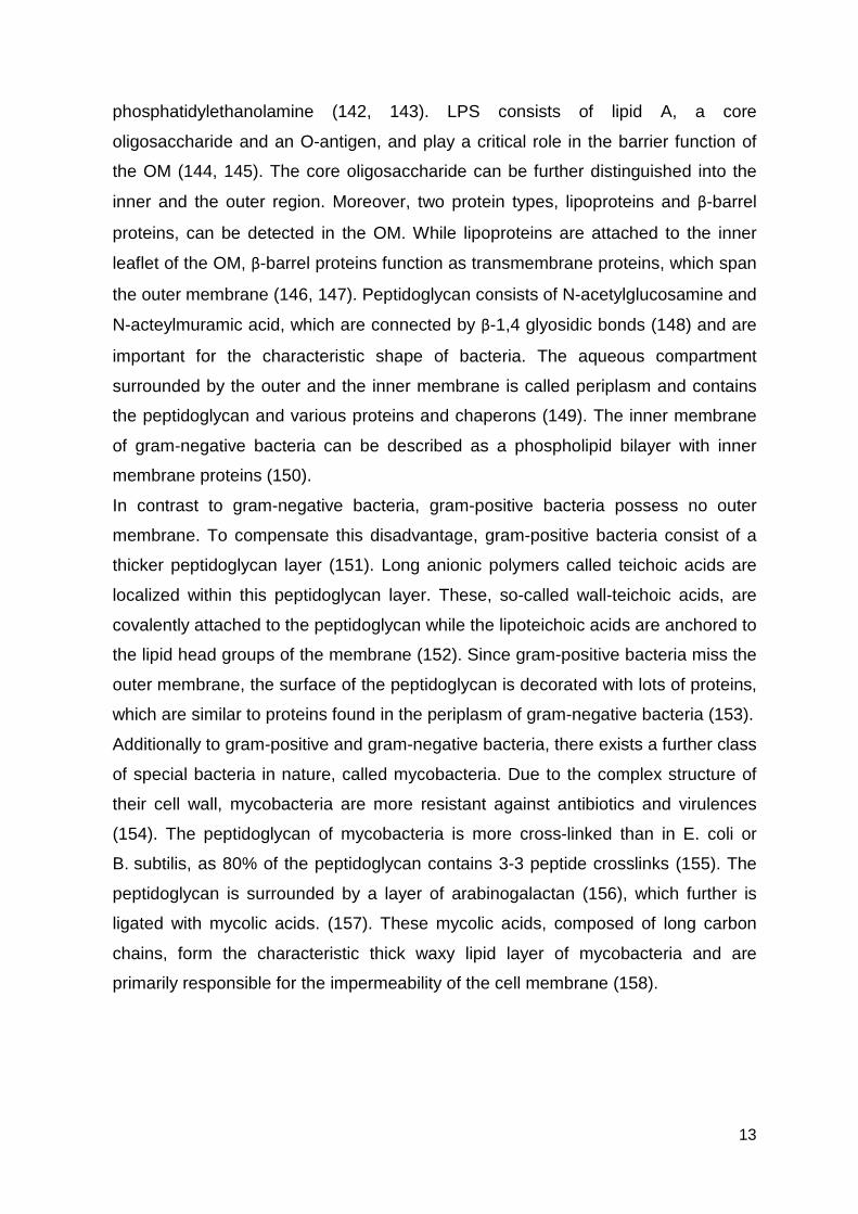
13
phosphatidylethanolamine (142, 143). LPS consists of lipid A, a core
oligosaccharide and an O-antigen, and play a critical role in the barrier function of
the OM (144, 145). The core oligosaccharide can be further distinguished into the
inner and the outer region. Moreover, two protein types, lipoproteins and β-barrel
proteins, can be detected in the OM. While lipoproteins are attached to the inner
leaflet of the OM, β-barrel proteins function as transmembrane proteins, which span
the outer membrane (146, 147). Peptidoglycan consists of N-acetylglucosamine and
N-acteylmuramic acid, which are connected by β-1,4 glyosidic bonds (148) and are
important for the characteristic shape of bacteria. The aqueous compartment
surrounded by the outer and the inner membrane is called periplasm and contains
the peptidoglycan and various proteins and chaperons (149). The inner membrane
of gram-negative bacteria can be described as a phospholipid bilayer with inner
membrane proteins (150).
In contrast to gram-negative bacteria, gram-positive bacteria possess no outer
membrane. To compensate this disadvantage, gram-positive bacteria consist of a
thicker peptidoglycan layer (151). Long anionic polymers called teichoic acids are
localized within this peptidoglycan layer. These, so-called wall-teichoic acids, are
covalently attached to the peptidoglycan while the lipoteichoic acids are anchored to
the lipid head groups of the membrane (152). Since gram-positive bacteria miss the
outer membrane, the surface of the peptidoglycan is decorated with lots of proteins,
which are similar to proteins found in the periplasm of gram-negative bacteria (153).
Additionally to gram-positive and gram-negative bacteria, there exists a further class
of special bacteria in nature, called mycobacteria. Due to the complex structure of
their cell wall, mycobacteria are more resistant against antibiotics and virulences
(154). The peptidoglycan of mycobacteria is more cross-linked than in E. coli or
B. subtilis, as 80% of the peptidoglycan contains 3-3 peptide crosslinks (155). The
peptidoglycan is surrounded by a layer of arabinogalactan (156), which further is
ligated with mycolic acids. (157). These mycolic acids, composed of long carbon
chains, form the characteristic thick waxy lipid layer of mycobacteria and are
primarily responsible for the impermeability of the cell membrane (158).
14
Figure 4: Cell-wall structure of gram-negative, gra m-positive and mycobacteria . (a)
Gram-negative bacteria consist of a cell membrane with a periplasmic space and an outer
membrane. In the periplasmic space localizes a thin peptidoglycan layer. The outer membrane
contains LPS and porins. (b) Gram-positive bacteria have a cell membrane and a thicker
peptidoglycan layer than gram-negative bacteria. The peptidoglycan consists of teichoic acids and
lipoteichoic acids. (c) Mycobacteria consist of a cell membrane, a thin layer of peptidoglycan and
arabinogalactan and a thicker layer of mycolic acids. Glycolipids and porins are detected in the cell
walls (159).
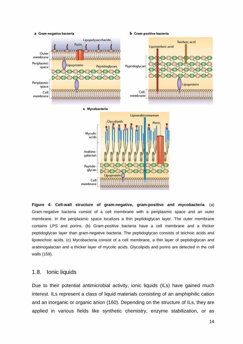
1.8. Ionic liquids
Due to their potential antimicrobial activity, ionic liquids (ILs) have gained much
interest. ILs represent a class of liquid materials consisting of an amphiphilic cation
and an inorganic or organic anion (160). Depending on the structure of ILs, they are
applied in various fields like synthetic chemistry, enzyme stabilization, or as
15
pharmaceutical compounds in pharmaceutical industry (161-163). ILs are extremely
interesting due to their flexibility of altering composition and structure of cation and
anion, their thermal stability and their solvating potential (164-166). Usually, ILs can
be divided into four classes, namely dialkylimidazolium-, alkylammonium-,
phosphonium- and N-alkylpyridinium-based ILs. A well-studied class are
imidazolium-based ionic liquids, which are used as solvents in bioorganic
transformations (167) (Figure 5). The imidazolium cations show high stability within
oxidative and reductive conditions (168) and are relatively easy to synthesize (169).
This class of ionic liquids also improves the solubility of proteins and thus, prevents
them from aggregation (170). In 1999, Welton et al. designed special ionic liquids
with toxic effects against microorganisms (165). Further studies demonstrated that
ionic liquids with a charged hydrophilic head group and hydrophilic tail have an
amphiphilic character, which especially is true for imidazolium and pyrimidinium-
based ionic liquids (171). The amphiphilic character and the length of the alkyl-chain
are the major requirements for their toxic effects against microorganisms (172).
However, also non-specific toxic side effects resulting in cell toxicity against host
cells were observed (173). Consequently, in the next step researchers focus on the
development of new ionic liquid compounds and conjugates with reduced host cell
toxicity.
Figure 5: The imidazolium cation with different anions. R = methyl, ethyl or butyl groups.
1.9. Preliminary work
The preceding paragraphs have described the difficulty about increasing
antimicrobial resistances and the connected health problem. Due to this, the
development of new antibiotic substances is crucial. Antimicrobial peptides
represent an alternative antibiotic class, which holds for a comprehensive analysis.
16
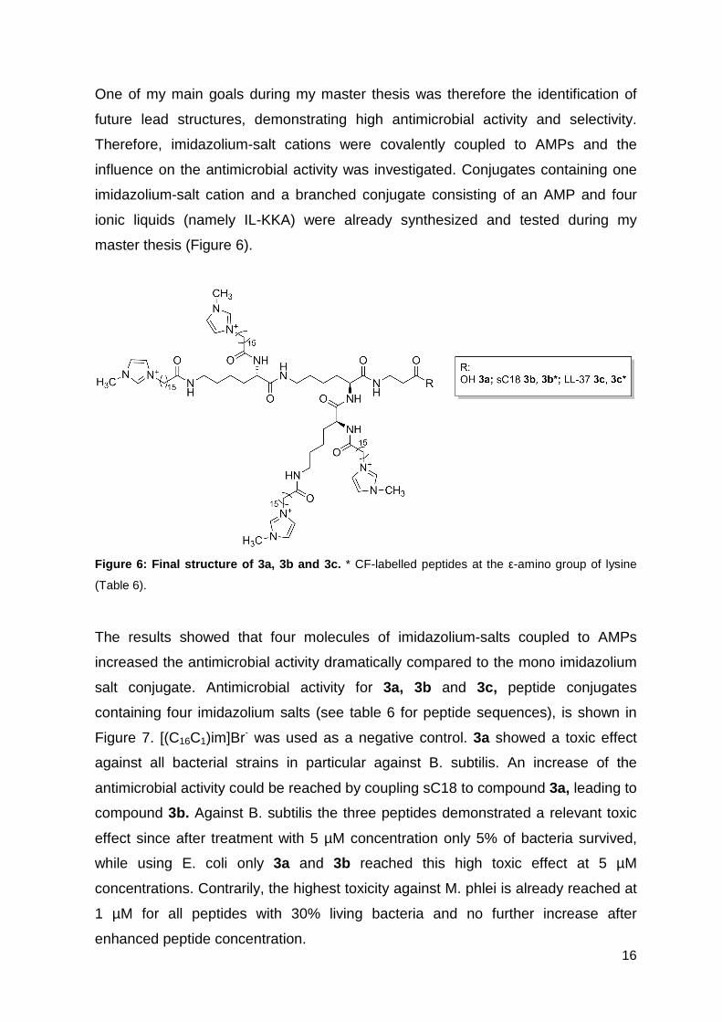
One of my main goals during my master thesis was therefore the identification of
future lead structures, demonstrating high antimicrobial activity and selectivity.
Therefore, imidazolium-salt cations were covalently coupled to AMPs and the
influence on the antimicrobial activity was investigated. Conjugates containing one
imidazolium-salt cation and a branched conjugate consisting of an AMP and four
ionic liquids (namely IL-KKA) were already synthesized and tested during my
master thesis (Figure 6).
Figure 6: Final structure of 3a, 3b and 3c. * CF-labelled peptides at the ε-amino group of lysine
(Table 6).
The results showed that four molecules of imidazolium-salts coupled to AMPs
increased the antimicrobial activity dramatically compared to the mono imidazolium
salt conjugate. Antimicrobial activity for 3a, 3b and 3c, peptide conjugates
containing four imidazolium salts (see table 6 for peptide sequences), is shown in
Figure 7. [(C16C1)im]Br- was used as a negative control. 3a showed a toxic effect
against all bacterial strains in particular against B. subtilis. An increase of the
antimicrobial activity could be reached by coupling sC18 to compound 3a, leading to
compound 3b. Against B. subtilis the three peptides demonstrated a relevant toxic
effect since after treatment with 5 µM concentration only 5% of bacteria survived,
while using E. coli only 3a and 3b reached this high toxic effect at 5 µM
concentrations. Contrarily, the highest toxicity against M. phlei is already reached at
1 µM for all peptides with 30% living bacteria and no further increase after
enhanced peptide concentration.

17
Figure 7: Antimicrobial activity of 3a, 3b and 3c. INT-assay was performed against B. subtilis,
E. coli and M. phlei. Negative control was set up to 100%.
Besides the peptide sC18, already described in chapter 1.2., also the LL-37 peptide
was investigated during this work. The LL-37 peptide is a 37 amino acids long
segment of the FALL-39 peptide, which is known as the only human cathelicidin-
derived AMP. LL-37 was identified as an antimicrobial peptide, expressed in the
bone marrow, with activity against gram-positive as well as gram-negative bacteria
(174). The proposed mechanism for LL-37 is the carpet-like mechanism (175).
1.10. Aim of the thesis
Aim of this work was to design new and more potent antimicrobial peptides as well
as conjugates thereof, and to elucidate their antimicrobial activity spectrum as well
as their mechanism of action.
The first part of the thesis deals with further modification of imidazolium salt-peptide
conjugates to improve their antimicrobial activity and selectivity. Therefore,
conjugates already developed during my master thesis, including the peptides sC18
and LL-37, should be further evaluated concerning their activity against resistant
bacterial strains, as well as their activity mechanism.
18
Further work should then investigate the potential of the short IL-KKA peptide to act
as an anti-biofilm compound when immobilized on polyether sulfone (PES)
membranes. Different coating strategies should be used and the activity of the
functionalized membranes should be characterized by different physical as well as
biological techniques.
The second goal of this work was to improve the antimicrobial activity of sC18.
Since the parent C18 peptide already showed antimicrobial activity it should be
tested if the introduction of hydrophobic amino acids could return this antimicrobial
activity to the shorter sC18 peptide. Therefore, several amino acids within the
sequence of sC18 should be exchanged with either isoleucine or phenylalanine,
respectively, and the antimicrobial activity should be tested. To verify the influence
of the amino acid exchange, structural characteristics and antimicrobial activity
against different bacterial strains should be determined.
19
2. Materials and methods
2.1. Materials
All chemicals, reagents and consumables, which were used during this work, were
obtained from Fluka (Taufkirchen, Germany), Merck (Darmstadt, Germany),
Sarstedt (Nümbrecht, Germany), Sigma-Aldrich (Taufkirchen, Germany) and VWR
(Darmstadt, Germany).
For peptide synthesis, all Nα-Fmoc protected amino acids were achieved from Iris
Biotech (Marktredwitz, Germany). The side-chains of the trifunctional amino acids
were equipped with acid-labeled protection groups to make use of the orthogonal
Fmoc/tBu strategy for peptide synthesis. Protection groups for the regular amino
acids were Pbf (Arg), Trt (Asn, Gln, His, Cys), Boc (Trp, Lys) and tert-butyl (Asp,
Glu, Ser, Thr, Tyr). For branched peptide compounds, Fmoc-L-Lys(Fmoc)-OH was
used while for CF-labeled peptides Fmoc-L-Lys(Dde)-OH was used.
For the calculation of the peptide concentrations, the binding of TFA anions to
positively charged amino acid side chains and free amino groups was taken into
consideration.
20
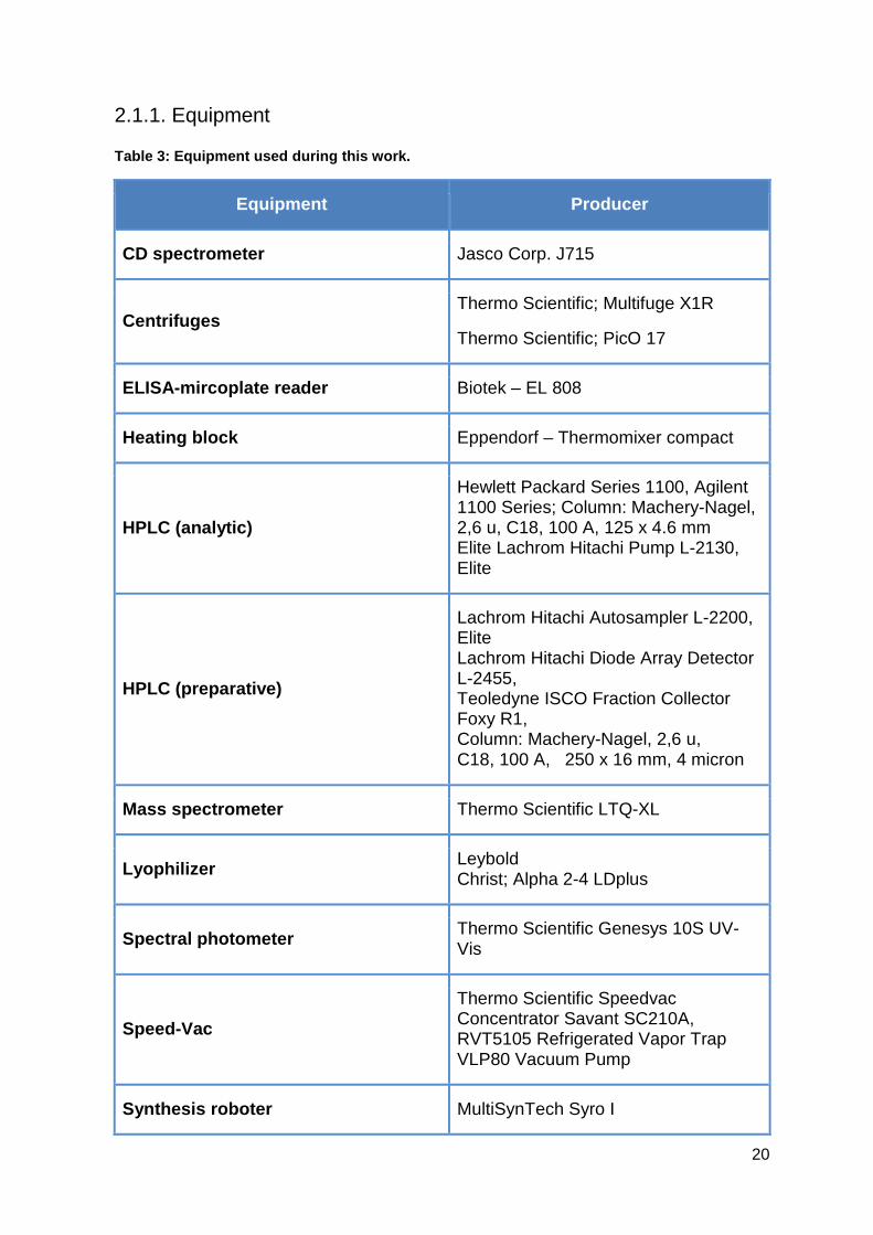
2.1.1. Equipment
Table 3: Equipment used during this work.
Equipment Producer
CD spectrometer Jasco Corp. J715
Centrifuges Thermo Scientific; Multifuge X1R
Thermo Scientific; PicO 17
ELISA-mircoplate reader Biotek – EL 808
Heating block Eppendorf – Thermomixer compact
HPLC (analytic)
Hewlett Packard Series 1100, Agilent 1100 Series; Column: Machery-Nagel, 2,6 u, C18, 100 A, 125 x 4.6 mm Elite Lachrom Hitachi Pump L-2130, Elite
HPLC (preparative)
Lachrom Hitachi Autosampler L-2200, Elite Lachrom Hitachi Diode Array Detector L-2455, Teoledyne ISCO Fraction Collector Foxy R1, Column: Machery-Nagel, 2,6 u, C18, 100 A, 250 x 16 mm, 4 micron
Mass spectrometer Thermo Scientific LTQ-XL
Lyophilizer Leybold Christ; Alpha 2-4 LDplus
Spectral photometer Thermo Scientific Genesys 10S UV-Vis
Speed-Vac
Thermo Scientific Speedvac Concentrator Savant SC210A, RVT5105 Refrigerated Vapor Trap VLP80 Vacuum Pump
Synthesis roboter MultiSynTech Syro I
21
Vortex Scientific Industries – Vortex Genie 2
Xcel Vap Horizon Technology
2.1.2. Buffers
Table 4: Buffers used during this work.
Giant unilamellar vesicle buffer
10 mM HEPES buffer, pH 7.4
50 mM KCl
50 mM NaCl
1mg/mL dextran (from Leuconostoc spp., 6 kDA)
5 µM Oyster 405
Iodnitrotetrazolium violet solution 10 mg Iodnitrotetrazolium-chloride
10 mL DMSO (cell culture quality)
Kaiser test solutions
Solution I (1 g ninhydrin in 20 mL EtOH)
Solution II (80 g phenol in 20 mL EtOH)
Solution III (0.4 mL aqu. KCN-solution (1mM) in 20 mL pyridine)
Mueller-Hinton medium
(Pronadisa-Conda)
2.0 g/L Beef Infusion
1.5 g/L Corn Starch
17.5 g/L Casein Peptone (acidic hydrolysate) pH 7.4
21 g/L Mueller-Hinton Broth in deionized water
Mueller-Hinton agar plates 21 g/L Mueller-Hinton Broth
15 g bacteriologic Agar
22
2.1.3. Bacterial strains
Bacillus subtilis (ATTC 6633)
Corynebacterium glutamicum (ATCC 13032)
Escherichia coli K12 (MG 1625)
Micrococcus luteus (DSM 20030)
Mycobacterium phlei (DSM 48214)
Pseudomonas fluorescens (DSM 50090)
Salmonella typhimurium (TA 100)
Methicillin-resistant Staphyloccocus aureus
Vancomycin-resistant Enterococci
2.1.4. Peptide sequences
Table 5: Peptides synthesized during this work.
Peptide Sequence
1a (sC18) GLRKRLRKFRNKIKEK-NH2
2a (LL-37) LLGDFFRKSKEKIGKEFKRIVQRIKDFLRNLVPRTES-NH2
3a (IL-KKA) IL4-KK(εK)βA-OH
3b (IL-KKA-sC18) IL4-KK(εK)βA-GLRKRLRKFRNKIKEK-NH2
3b* (IL-KKA-sC18) IL4-KK(εK)βA- GLRKRLRKFRNKIKEK(CF)-NH2
3c (IL-KKA-LL-37) IL4-KK(εK)βA-LLGDFFRKSKEKIGKEFKRIVQRIKDFLRNLVPRTES-NH2
3c* (IL-KKA-LL-37) IL4-KK(εK)βA- LLGDFFRKSKEKIGKEFKRIVQRIK(CF)DFLRNLVPRTES-NH2
3d (IL-KKA-Ahx-Ahx-G) IL4-KK(εK)βA -Ahx-Ahx-G-OH
3e (IL-KKA-FF) IL4-KK(εK)βA –FF-OH
23
3f (IL-KKA-WLLKW) IL4-KK(εK)βA –WLLKW-OH
sC18I1 ILRKRLRKFRNKIKEK-NH2
sC18I2 GIRKRLRKFRNKIKEK-NH2
sC18I3 GLIKRLRKFRNKIKEK-NH2
sC18I4 GLRIRLRKFRNKIKEK-NH2
sC18I5 GLRKILRKFRNKIKEK-NH2
sC18I6 GLRKRIRKFRNKIKEK-NH2
sC18I7 GLRKRLIKFRNKIKEK-NH2
sC18I8 GLRKRLRIFRNKIKEK-NH2
sC18I9 GLRKRLRKIRNKIKEK-NH2
sC18I10 GLRKRLRKFINKIKEK-NH2
sC18I11 GLRKRLRKFRIKIKEK-NH2
sC18I12 GLRKRLRKFRNIIKEK-NH2
sC18I14 GLRKRLRKFRNKIIEK-NH2
sC18I15 GLRKRLRKFRNKIKIK-NH2
sC18I16 GLRKRLRKFRNKIKEI-NH2
sC18I10I15 GLRKRLRKFINKIKIK-NH2
24
sC18I10I16 GLRKRLRKFINKIKEI-NH2
sC18I15I16 GLRKRLRKFRNKIKII-NH2
sC18I10I15I16 GLRKRLRKFINKIKII-NH2
sC18F10 GLRKRLRKFFNKIKEK-NH2
sC18F15 GLRKRLRKFRNKIKFK-NH2
sC18F16 GLRKRLRKFRNKIKEF-NH2
sC18F10F15 GLRKRLRKFFNKIKFK-NH2
sC18F10F16 GLRKRLRKFFNKIKEF-NH2
sC18F15F16 GLRKRLRKFRNKIKFF-NH2
sC18F10F15F16 GLRKRLRKFFNKIKFF-NH2
* Peptides were labeled with 5(6)-carboxyfluorescin (CF).
2.2. Solid phase peptide synthesis (SPPS)
2.2.1. Loading of Wang-resin with the first amino acid
For the peptides 3a, 3d, 3e and 3f, the first amino acid had to be preloaded to a
Wang resin. Therefore, 30 mg Wang resin (loading 1.1 mmol/g) was swollen for
15 min in dimethylformamide (DMF). DMF was removed and 5 eq. Fmoc-amino
acid, 5 eq. Oxyma Pure and 5 eq. N’,N’-diisopropylcarbodiimide (DIC) were
dissolved in 500 mL DMF and added to the resin beads. The solution was shaken
over night at room temperature (RT). On the next day, the resins were washed five-
times with DMF, dichloromethane (DCM), methanol and diethylether. Afterwards,
the resins were dried using a SpeedVac.
25
2.2.2. Determination of the first residue attachment
To quantify the coupling of the first amino acid toward the Wang resin, the Fmoc-
group was cleaved and the absorption was measured.
Therefore, 500 µL 30% piperidine in DMF was incubated with 3 mg Wang resin
beads for 30 min at RT. 250 µL of the solution were mixed with 1.5 mL DMF and the
absorbance at 301 nm was measured. As blank 250 µL 30% piperidine in DMF was
added to 1.5 mL DMF. The loading was calculated using the following equation
(Formula 1):
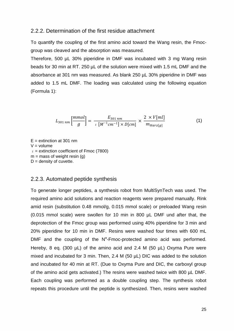
������ ��� = ������
ε��−1�−1� × ���� × 2 × �����������
E = extinction at 301 nm V = volume ε= extinction coefficient of Fmoc (7800) m = mass of weight resin (g) D = density of cuvette.
2.2.3. Automated peptide synthesis
To generate longer peptides, a synthesis robot from MultiSynTech was used. The
required amino acid solutions and reaction reagents were prepared manually. Rink
amid resin (substitution 0.48 mmol/g, 0.015 mmol scale) or preloaded Wang resin
(0.015 mmol scale) were swollen for 10 min in 800 µL DMF und after that, the
deprotection of the Fmoc group was performed using 40% piperidine for 3 min and
20% piperidine for 10 min in DMF. Resins were washed four times with 600 mL
DMF and the coupling of the Nα-Fmoc-protected amino acid was performed.
Hereby, 8 eq. (300 µL) of the amino acid and 2.4 M (50 µL) Oxyma Pure were
mixed and incubated for 3 min. Then, 2.4 M (50 µL) DIC was added to the solution
and incubated for 40 min at RT. (Due to Oxyma Pure and DIC, the carboxyl group
of the amino acid gets activated.) The resins were washed twice with 800 µL DMF.
Each coupling was performed as a double coupling step. The synthesis robot
repeats this procedure until the peptide is synthesized. Then, resins were washed
(1)
26
manually five times with DCM, methanol and diethylether and dried in the
SpeedVac.
2.2.4. Manual coupling of ionic liquids and 5(6)-carboxyfluorescein
For manual coupling, the resin was swollen for 15 min in 1 mL DMF. Then, 5 eq.
ionic liquid, or 3 eq. 5(6)-carboxyfluorescein (CF), respectively, was added with 5
eq. or 3 eq. HATU in 300 µL DMF. After that, diisopropylethylamine (DIPEA) (5 eq.
for ionic liquids and 3 eq. for CF) were added to the mixture and incubated for 2
min. The solution was added for 2 h to the resin at RT. The resin was washed five
times with DMF and again incubated with IL or CF, HATU and DIPEA for 2 h at RT.
After the second coupling, the resin was washed five times with DMF, DCM,
methanol and diethylether and dried using a SpeedVac.
2.2.5. Endcapping
To block free reactive groups and amino acid side-chains during a peptide
synthesis, an endcapping was performed.
Therefore, the resin was swollen for 15 min in 1 mL DCM. After the DCM was
removed, 50 µL acetic anhydride and 50 µL DIPEA were dissolved in 500 µL DCM
and added to the resin. The resin was shaken for 15 min at RT. In the end, the resin
was washed five times with DCM, methanol and diethylether and dried using a
SpeedVac.
2.2.6. Fmoc cleavage
To couple the next amino acid towards a peptide sequence, first, the Fmoc-
protection group had to be removed.
Therefore, the resin was swollen in 300 µL DMF for 15 min. Then, 300 µL 20%
piperidine (in DMF) were added to the resin and incubated for 5 min. Afterwards the
resin was washed five times with DMF, 300 µL 20% piperidine were added again to
the resin for 15 min. In the end, the resin was washed five times with DMF, DCM,
methanol and diethylether and dried using a SpeedVac.
27
2.2.7. Cleavage of the Dde-protection group
For CF-labeled peptides, Fmoc-Lys(Dde)-OH was used to couple the CF at the ε-
side chain of the lysine.
For the cleavage of the Dde-protection group, the resin was swollen in 1 mL DMF
for 15 min. After removing the DMF, 1 mL of 3% hydrazine in DMF was added to
the resin and incubated for 10 min at RT. The flow through was collected in a falcon
and the resin was washed two times with 1 mL DMF. The washing solution was
added to the flow through into the falcon. Then again, 1 mL of 3% hydrazine in DMF
was added. This procedure was repeated ten times and the amount of cleaved Dde
in step 1 and step 10 was determined measuring the absorption at 301 nm. As a
blank 2 mL DMF with 1 mL of 3% hydrazine in DMF were used. If the OD301nm was
less than 0.1, the deprotection was successful. After the cleavage, the resin was
washed five times with DMF, DCM, methanol and diethylether and dried using a
SpeedVac.
2.2.8. Kaiser test
To check the completion of an amino acid coupling to the peptide sequence, a
visual test was used indicating the presence or absence of free amino groups.
One drop of each solution was added to a few resin beads: solution I (1 g ninhydrin
in 20 mL EtOH); solution II (80 g phenol in 20 mL EtOH) and solution III (0.4 mL
aqu. KCN solution (1 mM) in 20 mL pyridine). As negative control, a sample without
resin beads was used and as positive control, N-ethyldiisopropylamine was used.
The samples were incubated for 5 min at 95 °C. If the solution turned yellow, no free
amino groups were present, while a blue color is a hint for free amino groups.
2.2.9. Sample cleavage
The sample cleavage was performed to verify the coupling success and to analyze
the composition of the products.
2.5 µL triisopropylsilane (TIS) and 2.5 µL H2Odd, working as scavenger, were added
to a small amount of dry resin beads. For peptide 3d, 7 µL thioanisole and 3 µL 1,2-
ethanedithiol were used as scavenger due to the presence of tryptophan in the
peptide sequence. The mixture was filled up to 100 µL with trifluoroacetic acid
28
(TFA). After 3 h shaking at RT, 1 mL ice-cold diethylether was added for at least 20
min at -20 °C to precipitate the peptide. The peptide was washed five times with
diethylether by centrifugation at 10,000 x g for 5 min. After each centrifugation step,
the supernatant was discarded and the pellet again resuspended in diethylether.
The pellet was dried using a SpeedVac and dissolved in 100 µL H2Odd or 100 µL
H2Odd /tBuOH (3:1). The peptide solution was diluted in either 10% acetonitrile/ 90%
H2Odd + 0.1% formic acid (FA) or 10% acetonitrile/ 90% H2Odd + 0.1% TFA for
qualitative and quantitative analysis, respectively.
2.2.10. Full cleavage
With the full cleavage, all protection groups were removed from the peptide and the
peptide was cleaved from the resin.
25 µL TIS and 25 µL H2Odd were added to the dry resin. For peptide 3d, 70 µL
thioanisole and 30 µL 1,2-ethanedithiol were used due to the tryptophan in the
peptide sequence. The mixture was filled up to 1 mL with TFA and shaken for 3 h at
RT. After that, the peptide was precipitated in 10 mL ice-cold diethylether. The resin
was washed with 200 µL TFA and the solution was added to diethylether. The
mixture was stored for at least 20 min at -20 °C to precipitate the peptide.
Afterwards, the peptide was washed five times with 10 mL diethylether by
centrifugation at 5,000 x g for 5 min. After every centrifugation step, the supernatant
was discarded and the pellet was resuspended in 10 mL diethylether. The pellet
was dried using a SpeedVac and dissolved in 2-3 mL H2Odd or 2-3 mL H2O/tBuOH
(3:1). One part of the peptide solution was diluted with either 10% acetonitrile/ 90%
H2Odd + 0.1% formic acid (FA) or 10% acetonitrile / 90% H2Odd + 0.1% TFA for
qualitative and quantitative analysis, respectively. The rest of the peptide solution
was lyophilized.
2.2.11. LC-mass spectrometry with 0.1% FA for qualitative analysis
The peptide solution was analyzed qualitatively by reverse-phase liquid
chromatography mass spectrometry (RP-HPLC-MS) using a Nucleodur column
(100-5; C18ec; 4.6 x 125 mm) from Macherey-Nagel. 10 µL of the diluted peptide
solution were injected into the RP-HPLC and separated by an acetonitrile gradient
increasing from 10% up to 60% in 15 min. The flow rate was set to 0.6 mL/min.
29
After HPLC, the eluent was injected into a LTQ-XL ESI-MS. Pseudo molecular ions
were generated and a full mass spectrum was acquired.
2.2.12. RP-HPLC with 0.1% TFA for purity analysis
The purity of the peptides was determined by reverse-phase liquid chromatography
(RP-HPLC) using a Nucleodur column (100-5; C18ec; 4.6 x 125 mm) from
Macherey-Nagel. 10 µL of the diluted peptide solution were injected into the RP-
HPLC and separated by an acetonitrile gradient increasing from 10% up to 60% in
15 min. The flow rate was set to 0.6 mL/min. The UV-chromatogram was used to
determine the percentage of the peptide purity.
2.2.13. Preparative RP-HPLC with 0.1% TFA
The preparative RP-HPLC was used to purify the peptide. A Nucleodur column
(1005; C18ec; 16 x 250 mm) from Macherey-Nagel was used. The column was
equilibrated with 10% acetonitrile in H2Odd + 0.1% TFA for 15 min. For purification,
an acetonitrile gradient from 10% to 60% in 45 min was used. The flow rate was
6 mL/min and the peptide was detected at 220 nm. Acetonitrile was removed with
the Xcel Vap and the sample was lyophilized.
2.2.14. Synthesis of ionic liquids
Ionic liquids were synthesized by the group of AG Giernoth by Julie Piper gen.
Schmauck (176).
2.3. Characterization methods
2.3.1. Circular dichroism spectroscopy
Circular dichroism (CD) spectroscopy was used to determine the secondary
structure of the synthesized peptides. All CD spectra were measured with a Jasco
Corp. J715 spectrometer at 20 °C. A 20 µM peptide solution in 10 mM phosphate
buffer, pH 7.0 or 10 mM phosphate buffer/TFE (1:1 v/v), pH 7.0 was prepared.
Peptide solutions were filled into a 0.1 cm quartz cell and a spectrum was recorded
from wavelength 180 to 260 nm in 0.2 nm intervals. The measurement was
30
performed in triplicate. The scanning speed was 50 nm/min and the sensitivity 100
mdegrees.
2.3.2. Preparation of large unilamellar vesicle (LUVs)
LUVs were prepared combining either 1,2-dioleoyl-sn-glycero-3-
phophoethanolamine (DOPE) and 1,2-dioleoyl-sn-glycero-3-[phospho-rac-(1-
glycerol)] (DOPG) (DOPE/DOPG 80:20) to imitate a gram-negative bacterial
membrane, or by combination of DOPE, DOPG and cardiolipin (CL)
(DOPE/DOPG/CL 15:80:5) to imitate a gram-positive bacterial membrane. Lipid
mixtures were dissolved in 1 mL chloroform and the chloroform was evaporated
under reduced pressure. Then, the lipid film was dissolved in 1 mL 10 mM
phosphate buffer, pH 7.0 and ten times frozen and melted in liquid nitrogen. In a
next step, the suspension was passed 21 times through a mini-extruder equipped
with a 0.4 µm polycarbonate track-etch membrane (Avanti Polar Lipids, Alabaster,
USA). The size of the vesicle was determined via dynamic light scattering.
2.3.3. Circular dichroism with LUVs
20 µM peptide solution was added to 1 mM LUVs in 10 mM phosphate buffer, pH
7.0. The CD measurement was performed as written above.
2.3.4. Preparation of giant unilamellar vesicle (GUVs)
The preparation of giant unilamellar vesicles (GUVs) was performed by Mareike
Horn. In brief, GUVs were prepared combining 1,2-dioleoyl-sn-glycero-3-
phosphocholine (DOPC), Atto550 labeled DOPE and different amounts of
cholesterol. In a first step, super low melting agarose (1% w/v) was coated on a
glass slide and dried for 30 min at 50 °C. After that, 10 µL of DOPC and DOPE
solution were spread on the agarose layer and dried in vacuo for at least 1 h. In a
next step, a seal ring was placed on the lipid coated agarose film. For the
preparation of GUVs with encapsulated Oyster 405, a 10 mM HEPES buffer, pH
7.4, 50 mM KCl, 50mM NaCl, 1mg/mL dextran (from Leuconostoc spp., 6 kDA) and
5 µM Oyster 405 was used. After adding the buffer into the seal ring, the glass slide
31
was left for 2 h in the dark to allow the lipids to swell. For harvesting the GUVs, the
glass slide was tilted gently in all directions. The GUVs were used within three days.
2.3.5. CLSM observation of GUVs treated with peptide conjugates
The CLSM observation with GUVs and peptides were performed by Mareike Horn.
In brief, GUVs were centrifuged at 14.000 x g for 10 min at RT to remove untrapped
Oyster 405. 40 µL GUV solution was diluted with 50 µL of the respective HEPES
buffer described above and transferred into a tissue culture vessel. CF-labeled
peptide conjugates were added to the GUVs with a final concentration of 5 µM.
The GUV-peptide interaction was analyzed using a confocal laser scanning system
(Nikon D-Eclipse C1) containing an inverted microscope (Nikon Eclipse Ti) with an
20x objective (N.A. 0.45, Plan Flour; Nikon). The fluorescence intensity was
determined using ImageJ.
2.4. Biological methods
2.4.1. Antimicrobial activity
The iodnitrotetrazolium-purple assay was performed to determine the viability of
different bacteria. Only viable bacteria convert tetrazolium to formazan, therefore,
the absorption is proportional to the bacterial number.
For imidazolium-salt conjugates: Each bacterial strain was incubated overnight in
Müller-Hinton medium at 30 °C. The next day, the optical density (OD) at 600 nm
was determined and the bacteria culture was diluted to an OD600nm = 0.7. In each
well of a 96-well plate, 180 µL Müller-Hinton medium, 10 µL bacteria suspension
and 10 µL of peptide conjugates (concentration 5 µM, 4 µM, 3 µM, 2 µM, 1 µM, 0.5
µM and 0.25 µM) were added. As positive control, gentamicin (for B. subtilis),
streptomycin (for M. phlei) and tetracycline (E. coli) and as negative control, H2Odd
was used. Bacteria were incubated with peptide-conjugates for 6 h at 30 °C. Then,
10 µL INT solution (iodnitrotetrazolium-chloride 1 mg/mL in DMSO) were added to
each well and incubated for 30 min at 37 °C. The viability was analyzed measuring
the absorption at 540 nm. Negative control was set to 100%.
For sC18 variants: Each bacterial strain was incubated in a subculture overnight at
37 °C. The next day, the sub culture was given to 100 mL fresh Müller-Hinton
32
medium and bacteria were grown to an OD600nm = 0.7. Then, the INT-assay was
performed as written above with the difference that everything was performed at
37 °C.
2.4.2. Killing assay using resistant bacterial strains
The killing assay was performed by Dr. Andreas Schubert at the Fraunhofer
Institute for Cell Therapy and Immunology, Leipzig. It was performed against
vancomycin-resistant Enterococci (VRE) and methicillin-resistant Staphylococcus
aureus (MRSA). In brief, an overnight bacteria culture was diluted to an OD600nm =
0.05 in Trypticase-Soya-Bouillon and incubated for 2 h at 37 °C. After an OD600nm =
0.3 was reached, VRE bacteria were diluted to 700 cfu/180 µL in 10 mM PPB and
0.5% LB-medium (MRSA bacteria to 500 cfu/180 µL in 10 mM PPB and 2% LB-
medium). 180 µL of the solution was transferred to 20 µL peptide solution at
different concentrations and shaken at 37 °C for 1 h. Then, bacteria were plated on
Bacto Brain Heart Infusion (BD) agar plates and incubated overnight at 37 °C. The
next day, the bacteria colonies were counted.
2.4.3. Hemolytic activity
Human red blood cells (h-RBCs) were used to determine the hemolytic activity
based on the release of hemoglobin. H-RBCs were centrifuged at 5,000 x g at 4 °C
for 10 min and three times washed with PBS by centrifugation at 4,000 x g at 4 °C
for 10 min. After each washing step, h-RBCs were resuspended in PBS. 50 µL
peptide solution were added to 50 µL of h-RBCs in PBS to a final concentration of
4% v/v. The solution was incubated for 60 min at 37 °C and centrifuged at 1,000 x g
for 5 min. The supernatant was added into 96-well plates and the absorbance of the
released hemoglobin was measured at 550 nm. As negative control, only PBS was
used, while 0.1% Triton X-100 acts as positive control. Percentage of hemolysis
was calculated (Formula 2):
%hemolysis = (A550nmoferythrocytespluspeptide − A550nmoferythrocytesinPBS)(A550nmoferythrocytesin0.1%Triton − X100 − A550nmoferythrocytesinPBS) × 100
(2)
33
2.4.4. Immobilization and characterization of polyether sulfone
membranes (PES)
Immobilization and characterization of polyether sulfone membranes were
performed by Dr. Agnes Schulze at the Institute for Surface Modifications in Leipzig.
For peptide immobilization, the PES membrane disc (Ø 10 mm) was immersed into
a peptide solution (2.5 mg/mL in ultrapure water) for 5 min followed by electron
beam irradiation (E-Beam) with a dose of 50, 100 or 150 kGy. Irradiation was
performed in N2 atmosphere with O2 quantities < 10 ppm. The voltage and the
current were set to 160 kV and 10 mA, respectively. Then, the irradiated membrane
was rinsed three times for 30 min with ultrapure water and dried at ambient
temperature.
Alternatively, peptides were immobilized by a chemical linker system. For this
purpose, the membranes had first to be functionalized with amino groups at the
surface. The PES membrane disc (Ø 47 mm) was immersed into a solution of
2aminoethyl methacrylate hydrochloride (AEMA) (0.5 wt. % in water) for 30 min
followed by E-Beam irradiation (150 kGy). Then, the samples were washed with
water (three times for 30 min) and dried at ambient temperature. The membranes
were cut into 10 mm discs and were treated with an aqueous solution of the peptide
(2.5 mg/mL), N-hydroxysuccinimide (NHS) (5 mg/mL), and 1-ethyl-3-(3-
dimethylaminopropyl)carbodiimide (EDC) (5 mg/mL). The coupling was allowed to
react overnight at RT. Then, membranes were washed three times for 30 min with
ultrapure water and dried at ambient temperature.
The membrane morphology was investigated by scanning electron microscopy
(SEM, Ultra 55, Carl Zeiss SMT, Jena, Germany).
The chemical surface composition was analyzed with X-ray photoelectron
spectroscopy (AXIS Ultra, Kratos Analytical, Manchester, England). The kinetic
energy of the electrons was analyzed with a pass energy of 160 eV for the survey
spectra and 40 eV for the energy resolved spectra, respectively.
2.4.5. Antimicrobial activity with immobilized PES membranes
For antimicrobial activity tests, the gram-positive bacterium Bacillus subtilis (ATTC
6633) was used as test strain. The bacteria were cultured in Mueller-Hinton Broth
(MHB) medium overnight at 37 °C and diluted to an OD600nm = 0.001 (correspond to
34
106 bacteria/mL). PES membranes immobilized with peptides (control: membrane
without peptide) were placed at the bottom of a 48 well-plate. In each well, 50 µL of
bacteria suspension were added and the solution was incubated for 3 h at 37 °C.
Bacteria suspensions were diluted 1:100 with PBS and 10 µL were exposed onto
Mueller-Hinton agar plates. The next day, the grown bacteria colonies were
counted. Additionally, to the membranes with the bacteria solution, 600 µL Mueller-
Hinton medium were added and incubated for 24 h at 37 °C. Membranes were
removed and 20 µL INT-solution were added for 30 min at 37 °C to the bacteria
suspension. The 48-well plate was subsequently placed in an ELISA plate reader to
measure absorption at 540 nm. Negative control was set up to 100%. All
experiments were done in triplicate or duplicate with n=3.
35
3. Results and discussion
3.1. Improvement of imidazolium salt-peptide conjugates and their
mechanism of action
During my master thesis, the covalent combination of imidazolium salts and the
peptides sC18 and LL-37 yielded new compounds with high antimicrobial activity
against different bacterial strains. To further investigate the antimicrobial activity, the
designed compounds should be tested against multi-resistant strains, which are
heavy to threat with common antibiotics. Moreover, experiments were performed in
order to investigate selectivity and mechanism of action of the newly designed
compounds.
3.1.1. Synthesis of imidazolium salt-peptide conjugates
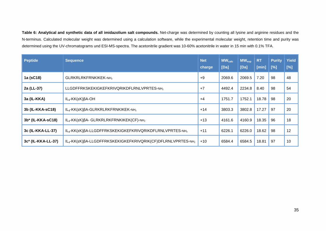
For the new sets of experiment, the conjugates first had to be synthesized again.
Table 6 gives an overview of all the synthesized imidazolium-salt peptide
compounds. Remarkable is that the yield of all peptides containing IL4-KK(εK)βA in
their sequence (3b, 3b*, 3c and 3c*) was low (only 5-10%). A reason for this low
peptide amount could be the fact, that as last coupling step, four imidazolium salts
had to be simultaneously coupled to the already branched peptide sequence.
Therefore, also side products with less than four imidazolium salts were obtained.
Nevertheless, after purification via preparative HPLC all peptides showed a
satisfying purity of around 98%.
35
Table 6: Analytical and synthetic data of all imida zolium salt compounds. Net-charge was determined by counting all lysine and arginine residues and the
N-terminus. Calculated molecular weight was determined using a calculation software, while the experimental molecular weight, retention time and purity was
determined using the UV-chromatograms und ESI-MS-spectra. The acetonitrile gradient was 10-60% acetonitrile in water in 15 min with 0.1% TFA.
Peptide Sequence Net
charge
MWcalc
[Da]
MWexp
[Da]
RT
[min]
Purity
[%]
Yield
[%]
1a (sC18) GLRKRLRKFRNKIKEK-NH2 +9 2069.6 2069.5 7.20 98 48
2a (LL-37) LLGDFFRKSKEKIGKEFKRIVQRIKDFLRNLVPRTES-NH2 +7 4492.4 2234.8 8.40 98 54
3a (IL-KKA) IL4-KK(εK)βA-OH +4 1751.7 1752.1 18.78 98 20
3b (IL-KKA-sC18) IL4-KK(εK)βA-GLRKRLRKFRNKIKEK-NH2 +14 3803.3 3802.8 17.27 97 20
3b* (IL-KKA -sC18) IL4-KK(εK)βA- GLRKRLRKFRNKIKEK(CF)-NH2 +13 4161.6 4160.9 18.35 96 18
3c (IL-KKA -LL-37) IL4-KK(εK)βA-LLGDFFRKSKEKIGKEFKRIVQRIKDFLRNLVPRTES-NH2 +11 6226.1 6226.0 18.62 98 12
3c* (IL-KKA -LL-37) IL4-KK(εK)βA-LLGDFFRKSKEKIGKEFKRIVQRIK(CF)DFLRNLVPRTES-NH2 +10 6584.4 6584.5 18.81 97 10
36
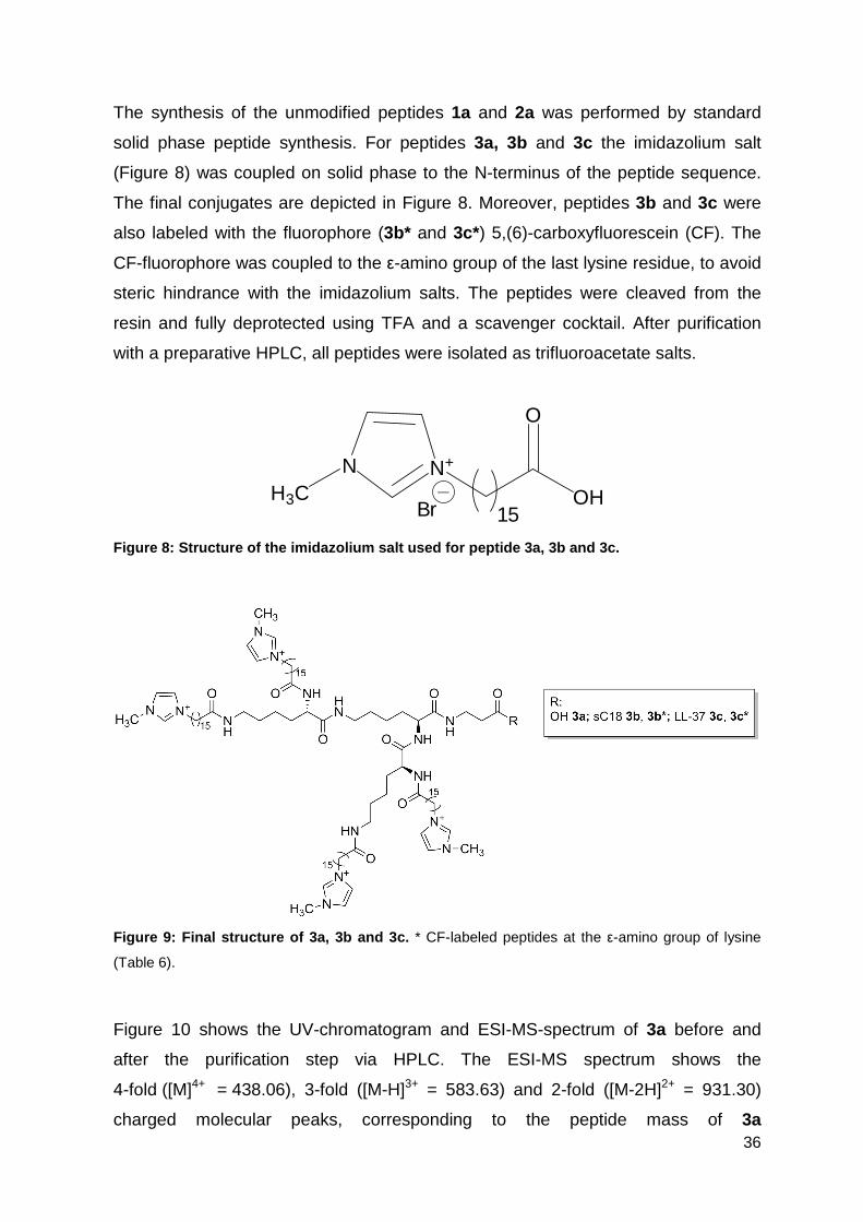
The synthesis of the unmodified peptides 1a and 2a was performed by standard
solid phase peptide synthesis. For peptides 3a, 3b and 3c the imidazolium salt
(Figure 8) was coupled on solid phase to the N-terminus of the peptide sequence.
The final conjugates are depicted in Figure 8. Moreover, peptides 3b and 3c were
also labeled with the fluorophore (3b* and 3c*) 5,(6)-carboxyfluorescein (CF). The
CF-fluorophore was coupled to the ε-amino group of the last lysine residue, to avoid
steric hindrance with the imidazolium salts. The peptides were cleaved from the
resin and fully deprotected using TFA and a scavenger cocktail. After purification
with a preparative HPLC, all peptides were isolated as trifluoroacetate salts.
OH
O
N N+
H3C15Br
Figure 8: Structure of the imidazolium salt used fo r peptide 3a, 3b and 3c.
Figure 9: Final structure of 3a, 3b and 3c. * CF-labeled peptides at the ε-amino group of lysine
(Table 6).
Figure 10 shows the UV-chromatogram and ESI-MS-spectrum of 3a before and
after the purification step via HPLC. The ESI-MS spectrum shows the
4-fold ([M]4+ = 438.06), 3-fold ([M-H]3+ = 583.63) and 2-fold ([M-2H]2+ = 931.30)
charged molecular peaks, corresponding to the peptide mass of 3a
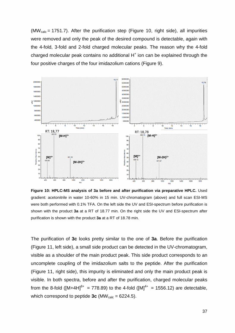
37
(MWcalc = 1751.7). After the purification step (Figure 10, right side), all impurities
were removed and only the peak of the desired compound is detectable, again with
the 4-fold, 3-fold and 2-fold charged molecular peaks. The reason why the 4-fold
charged molecular peak contains no additional H+ ion can be explained through the
four positive charges of the four imidazolium cations (Figure 9).
Figure 10: HPLC-MS analysis of 3a before and after purification via preparative HPLC. Used
gradient: acetonitrile in water 10-60% in 15 min. UV-chromatogram (above) and full scan ESI-MS
were both performed with 0.1% TFA. On the left side the UV and ESI-spectrum before purification is
shown with the product 3a at a RT of 18.77 min. On the right side the UV and ESI-spectrum after
purification is shown with the product 3a at a RT of 18.78 min.
The purification of 3c looks pretty similar to the one of 3a. Before the purification
(Figure 11, left side), a small side product can be detected in the UV-chromatogram,
visible as a shoulder of the main product peak. This side product corresponds to an
uncomplete coupling of the imidazolium salts to the peptide. After the purification
(Figure 11, right side), this impurity is eliminated and only the main product peak is
visible. In both spectra, before and after the purification, charged molecular peaks
from the 8-fold ([M+4H]8+ = 778.89) to the 4-fold ([M]4+ = 1556.12) are detectable,
which correspond to peptide 3c (MWcalc = 6224.5).
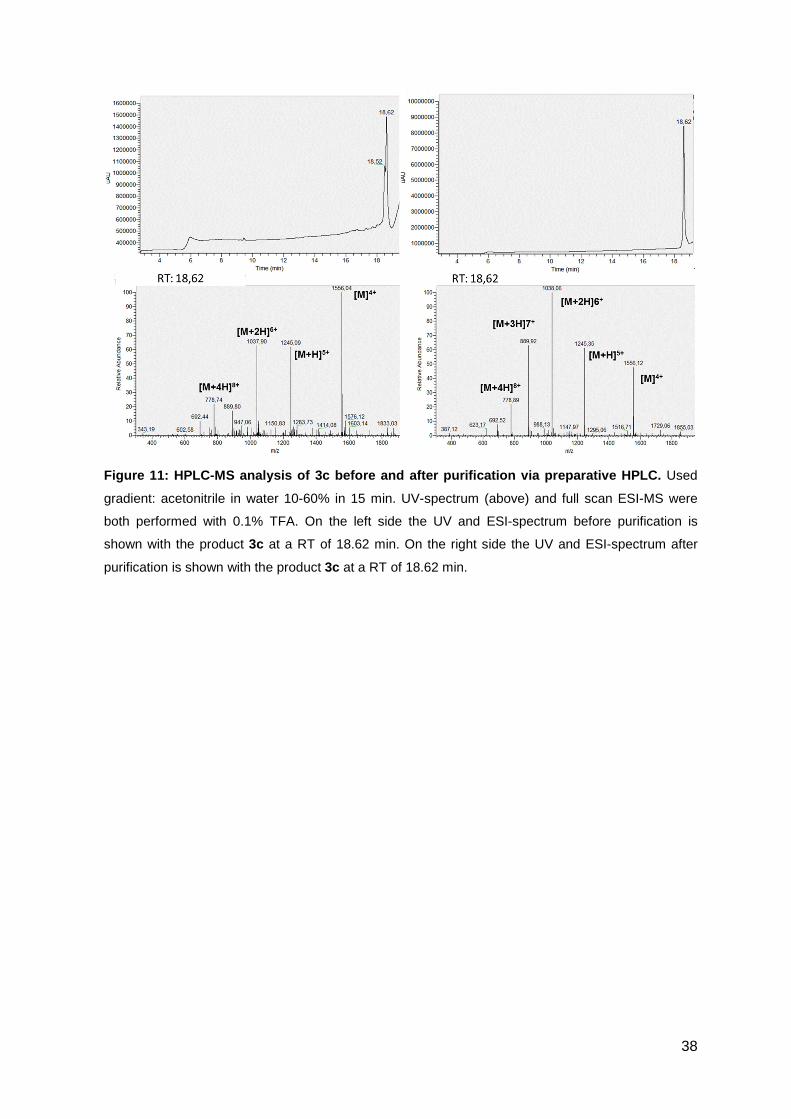
38
Figure 11: HPLC-MS analysis of 3c before and after purification via preparative HPLC. Used
gradient: acetonitrile in water 10-60% in 15 min. UV-spectrum (above) and full scan ESI-MS were
both performed with 0.1% TFA. On the left side the UV and ESI-spectrum before purification is
shown with the product 3c at a RT of 18.62 min. On the right side the UV and ESI-spectrum after
purification is shown with the product 3c at a RT of 18.62 min.
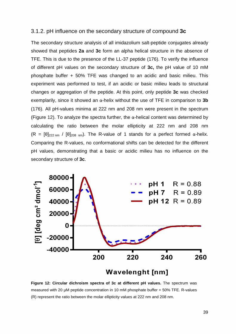
39
3.1.2. pH influence on the secondary structure of compound 3c
The secondary structure analysis of all imidazolium salt-peptide conjugates already
showed that peptides 2a and 3c form an alpha helical structure in the absence of
TFE. This is due to the presence of the LL-37 peptide (176). To verify the influence
of different pH values on the secondary structure of 3c, the pH value of 10 mM
phosphate buffer + 50% TFE was changed to an acidic and basic milieu. This
experiment was performed to test, if an acidic or basic milieu leads to structural
changes or aggregation of the peptide. At this point, only peptide 3c was checked
exemplarily, since it showed an α-helix without the use of TFE in comparison to 3b
(176). All pH-values minima at 222 nm and 208 nm were present in the spectrum
(Figure 12). To analyze the spectra further, the α-helical content was determined by
calculating the ratio between the molar ellipticity at 222 nm and 208 nm
(R = [θ]222 nm / [θ]208 nm). The R-value of 1 stands for a perfect formed α-helix.
Comparing the R-values, no conformational shifts can be detected for the different
pH values, demonstrating that a basic or acidic milieu has no influence on the
secondary structure of 3c.
Figure 12: Circular dichroism spectra of 3c at diff erent pH values. The spectrum was
measured with 20 µM peptide concentration in 10 mM phosphate buffer + 50% TFE. R-values
(R) represent the ratio between the molar ellipticity values at 222 nm and 208 nm.
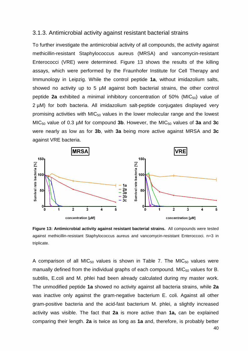
40
3.1.3. Antimicrobial activity against resistant bacterial strains
To further investigate the antimicrobial activity of all compounds, the activity against
methicillin-resistant Staphylococcus aureus (MRSA) and vancomycin-resistant
Enterococci (VRE) were determined. Figure 13 shows the results of the killing
assays, which were performed by the Fraunhofer Institute for Cell Therapy and
Immunology in Leipzig. While the control peptide 1a, without imidazolium salts,
showed no activity up to 5 µM against both bacterial strains, the other control
peptide 2a exhibited a minimal inhibitory concentration of 50% (MIC50) value of
2 µM) for both bacteria. All imidazolium salt-peptide conjugates displayed very
promising activities with MIC50 values in the lower molecular range and the lowest
MIC50 value of 0.3 µM for compound 3b. However, the MIC50 values of 3a and 3c
were nearly as low as for 3b, with 3a being more active against MRSA and 3c
against VRE bacteria.
Figure 13: Antimicrobial activity against resistant bacterial strains. All compounds were tested
against methicillin-resistant Staphylococcus aureus and vancomycin-resistant Enteroccoci. n=3 in
triplicate.
A comparison of all MIC50 values is shown in Table 7. The MIC50 values were
manually defined from the individual graphs of each compound. MIC50 values for B.
subtilis, E.coli and M. phlei had been already calculated during my master work.
The unmodified peptide 1a showed no activity against all bacteria strains, while 2a
was inactive only against the gram-negative bacterium E. coli. Against all other
gram-positive bacteria and the acid-fast bacterium M. phlei, a slightly increased
activity was visible. The fact that 2a is more active than 1a, can be explained
comparing their length. 2a is twice as long as 1a and, therefore, is probably better
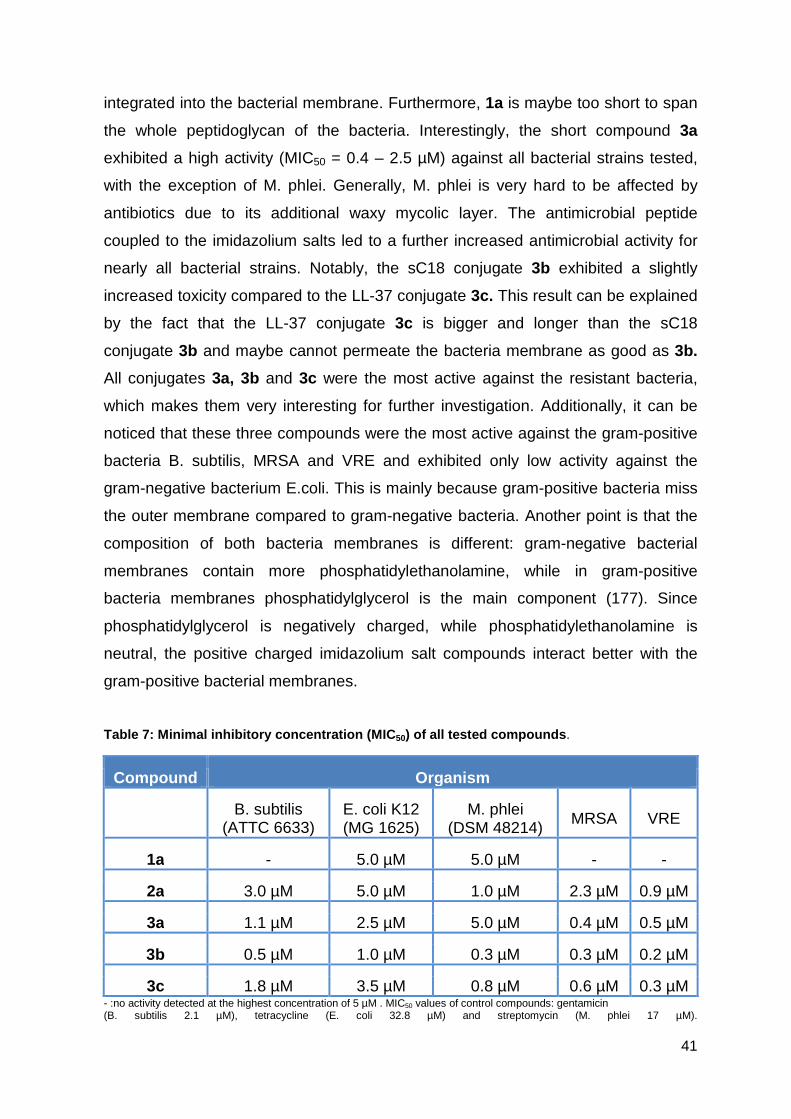
41
integrated into the bacterial membrane. Furthermore, 1a is maybe too short to span
the whole peptidoglycan of the bacteria. Interestingly, the short compound 3a
exhibited a high activity (MIC50 = 0.4 – 2.5 µM) against all bacterial strains tested,
with the exception of M. phlei. Generally, M. phlei is very hard to be affected by
antibiotics due to its additional waxy mycolic layer. The antimicrobial peptide
coupled to the imidazolium salts led to a further increased antimicrobial activity for
nearly all bacterial strains. Notably, the sC18 conjugate 3b exhibited a slightly
increased toxicity compared to the LL-37 conjugate 3c. This result can be explained
by the fact that the LL-37 conjugate 3c is bigger and longer than the sC18
conjugate 3b and maybe cannot permeate the bacteria membrane as good as 3b.
All conjugates 3a, 3b and 3c were the most active against the resistant bacteria,
which makes them very interesting for further investigation. Additionally, it can be
noticed that these three compounds were the most active against the gram-positive
bacteria B. subtilis, MRSA and VRE and exhibited only low activity against the
gram-negative bacterium E.coli. This is mainly because gram-positive bacteria miss
the outer membrane compared to gram-negative bacteria. Another point is that the
composition of both bacteria membranes is different: gram-negative bacterial
membranes contain more phosphatidylethanolamine, while in gram-positive
bacteria membranes phosphatidylglycerol is the main component (177). Since
phosphatidylglycerol is negatively charged, while phosphatidylethanolamine is
neutral, the positive charged imidazolium salt compounds interact better with the
gram-positive bacterial membranes.
Table 7: Minimal inhibitory concentration (MIC 50) of all tested compounds .
Compound Organism
B. subtilis (ATTC 6633)
E. coli K12 (MG 1625)
M. phlei (DSM 48214)
MRSA VRE
1a - 5.0 µM 5.0 µM - -
2a 3.0 µM 5.0 µM 1.0 µM 2.3 µM 0.9 µM
3a 1.1 µM 2.5 µM 5.0 µM 0.4 µM 0.5 µM
3b 0.5 µM 1.0 µM 0.3 µM 0.3 µM 0.2 µM
3c 1.8 µM 3.5 µM 0.8 µM 0.6 µM 0.3 µM - :no activity detected at the highest concentration of 5 µM . MIC50 values of control compounds: gentamicin (B. subtilis 2.1 µM), tetracycline (E. coli 32.8 µM) and streptomycin (M. phlei 17 µM).
42
Compared with commonly used antibiotics, the tested MIC50 values of the
compounds were between 0.3 – 0.6 µM, which is similar to values of vancomycin
(MIC50 = 0.32 µM against MRSA (178, 179)) and linezolid
(MIC50 = 0.59 µM against VRE (179)), highlighting again their high antimicrobial
potential.
3.1.4. Hemolytic activity studies
To get a first impression of every compound’s selectivity, the hemolytic activity
against human erythrocytes was determined and a selectivity index (SI) was
calculated. Therefore, human red blood cells (h-RBCs) from a healthy volunteer
were used and the HC20 values were defined. The HC20 represents the value where
20% hemolytic activity was detected. For peptides 1a and 2a, as well as the
conjugate 3a no hemolytic activity below 5 µM was detected (Figure 14); therefore
the HC20 value was set to 5. Notably, the coupling of four imidazolium salts to the
sC18 and LL-37 peptides led to a highly increased hemolytic activity with HC20
values of 1 µM for 3b and 0.5 µM for 3c. To further investigate the selectivity, the SI
index (HC20/MIC50) was calculated, considering that a high SI index is a hint for
good selectivity (Table 8). 3a exhibited the highest SI, except for M. phlei, because
no high hemolysis against red blood cells was detected. To increase the
antimicrobial activity of 3a further with consistent selectivity, some additional
positive amino acids like arginine and lysine could be added at the C-terminus of
this small compound until the point the selectivity decreases. On the other hand,
conjugates 3b and 3c showed low SI due to their high hemolytic activity. These
results can be explained by the higher hydrophobicity and increased amount of
positive charges of compounds 3b and 3c, which can be correlated to a strong
hemolytic activity. It was additionally shown that the peptides alone were not that
active against erythrocytes compared to the conjugates.

43
Figure 14: Hemolytic activity of all novel conjugat es against human red blood cells. The
activity was measured against peptide concentrations from 0.25 to 5 µM. Triton-X 100 and water
were used as positive and negative control, respectively. HC20 value was determined from this
Figure; n=2.
Table 8: Selectivity index (SI) of all conjugates c alculated with HC 20/MIC50
Organism 1a 2a 3a 3b 3c
B. subtilis - 1.7 4.5 2 0.3
E. coli 1 1 2 1 0,3
M. phlei 1 5 1 3.3 0.6
MRSA - 2.2 12.5 3.3 2.5
VRE - 5.6 10 1.7 1.7 - : denotes no activity at the highest concentration of 5 µM detected
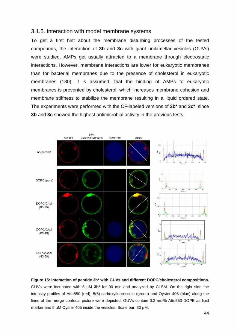
44
3.1.5. Interaction with model membrane systems
To get a first hint about the membrane disturbing processes of the tested
compounds, the interaction of 3b and 3c with giant unilamellar vesicles (GUVs)
were studied. AMPs get usually attracted to a membrane through electrostatic
interactions. However, membrane interactions are lower for eukaryotic membranes
than for bacterial membranes due to the presence of cholesterol in eukaryotic
membranes (180). It is assumed, that the binding of AMPs to eukaryotic
membranes is prevented by cholesterol, which increases membrane cohesion and
membrane stiffness to stabilize the membrane resulting in a liquid ordered state.
The experiments were performed with the CF-labeled versions of 3b* and 3c*, since
3b and 3c showed the highest antimicrobial activity in the previous tests.
Figure 15: Interaction of peptide 3b* with GUVs and different DOPC/cholesterol compositions.
GUVs were incubated with 5 µM 3b* for 90 min and analyzed by CLSM. On the right side the
intensity profiles of Atto550 (red), 5(6)-carboxyfluorescein (green) and Oyster 405 (blue) along the
lines of the merge confocal picture were depicted. GUVs contain 0.2 mol% Atto550-DOPE as lipid
marker and 5 µM Oyster 405 inside the vesicles. Scale bar, 30 µM.
45
GUVs with different amounts of cholesterol ranging from 0 – 60 mol% were
prepared (181). For GUV preparation DOPC and 0.2 mol% Atto550 labeled DOPE
were used to visualize the membrane (Figure 15/16 red channel). To visualize a
possible membrane leakage by dye outflow, Oyster 405 was encapsulated inside
the vesicles. Compound 3b* showed strong interactions with the GUV membrane,
as it could be seen in the microscope pictures and the intensity profile (Figure 15). A
complete dye outflow was visible for GUVs with 20% cholesterol and without
cholesterol suggesting that the mode of action of 3b is a pore-forming mechanism.
The intensity profile of these GUVs further showed that the green dye is detectable
in and outside the vesicle suggesting that the peptide had crossed the vesicle
membrane. However, increasing the cholesterol content led to an increased dye
outflow.
In Figure 16 the results for compound 3c were shown. In this case, no membrane
disrupting mechanism could be identified, since no dye outflow was visible. This
result was unexpected because in the antimicrobial experiments it seems that 3b
and 3c act both via strong membrane interactions. The intensity profile depicted
some green fluorescence inside the vesicle, suggesting that some peptides of 3c
already crossed the membrane. For the LL-37, which is a part of 3c, the carpet-like
mechanism had already been proposed (182). Maybe our compound also acts in a
detergent-like manner and the concentration was too low to detect micelles, which
were aroused from the membrane. The extreme clusters of the red green
fluorescence dye, especially visible in Figure 16, might be caused by the high
presence of cholesterol. Especially at 60%, it seemed that the cholesterol was not
homogenously distributed over the GUV membrane and built these clusters.
This experiment exposed that compounds 3b and 3c have different mode of
actions.
For 3b a pore-forming mechanism is proposed, where the conjugate in a first step
interacts with the lipid membrane, which could be identified by the green
accumulation at the lipid membrane, and in a second step builds pores leading to
the outflow of the blue dye. To differentiate better between the first and the second
step, this experiment could be performed with different concentrations or at different
time points. In contrast, 3c seems to act in a detergent-like manner, since no
outflow of the blue dye could be detected. Nevertheless, an interaction of peptide
and lipid membrane is visible.
46
Previous studies with AMPs Citropin, Aurein and Maculatin also showed differences
between pore-forming and detergent-like mechanism while interacting with lipid
bilayers. Citropin and Aurein, both acting in a detergent-like manner (183), indicate
that these peptides need a higher lipid-to-peptide ratio to achieve a dye leakage
compared to the pore-forming Maculatin. Therefore, it seems that more peptide
molecules are required to disrupt the membrane in a detergent-like manner
compared to a pore-forming mechanism. This observation could explain the
different results seen for the pore forming 3b and the detergent-like acting 3c.
Furthermore, an inhibitory effect of cholesterol was visible for compound 3b.
Hereby, not the interaction between cholesterol and compound led to this inhibitory
effect, but an indirect effect of cholesterol on the membrane properties (184). The
presence of cholesterol results in an enhanced membrane cohesion and
mechanical stiffness, which is explained by an increased acyl chain order in the
liquid ordered phase of membranes containing cholesterol (180).
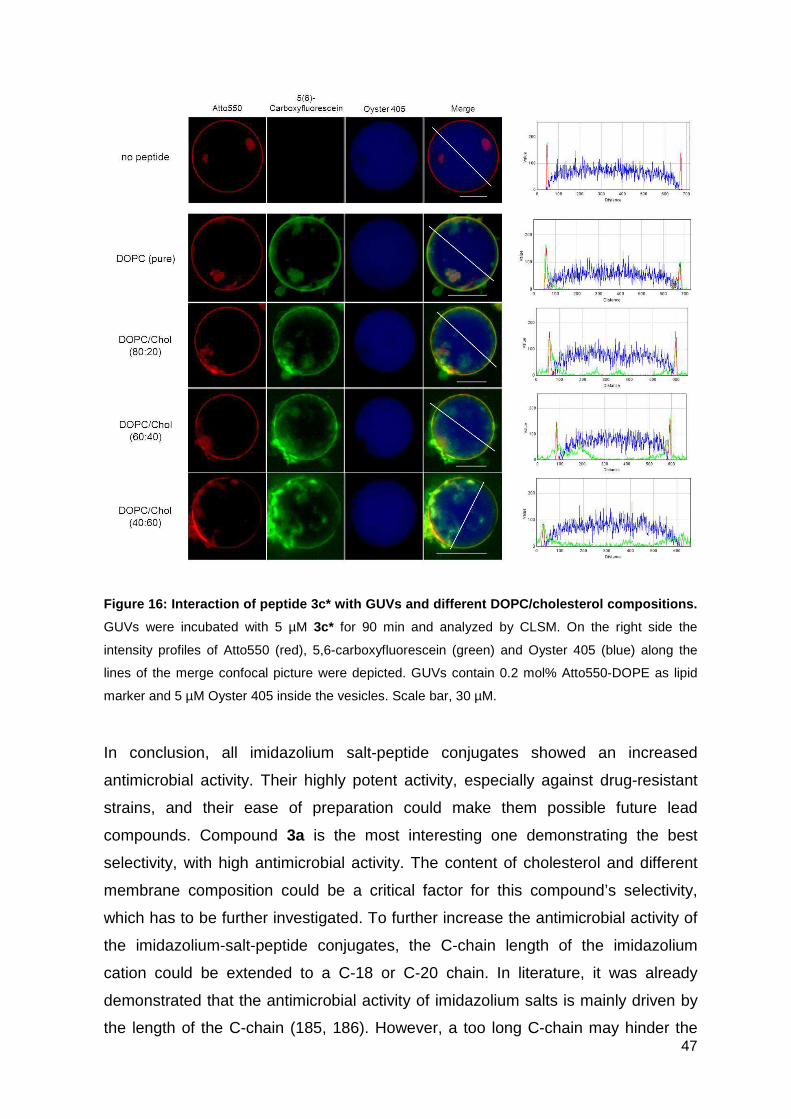
47
Figure 16: Interaction of peptide 3c* with GUVs and different DOPC/cholesterol compositions.
GUVs were incubated with 5 µM 3c* for 90 min and analyzed by CLSM. On the right side the
intensity profiles of Atto550 (red), 5,6-carboxyfluorescein (green) and Oyster 405 (blue) along the
lines of the merge confocal picture were depicted. GUVs contain 0.2 mol% Atto550-DOPE as lipid
marker and 5 µM Oyster 405 inside the vesicles. Scale bar, 30 µM.
In conclusion, all imidazolium salt-peptide conjugates showed an increased
antimicrobial activity. Their highly potent activity, especially against drug-resistant
strains, and their ease of preparation could make them possible future lead
compounds. Compound 3a is the most interesting one demonstrating the best
selectivity, with high antimicrobial activity. The content of cholesterol and different
membrane composition could be a critical factor for this compound’s selectivity,
which has to be further investigated. To further increase the antimicrobial activity of
the imidazolium-salt-peptide conjugates, the C-chain length of the imidazolium
cation could be extended to a C-18 or C-20 chain. In literature, it was already
demonstrated that the antimicrobial activity of imidazolium salts is mainly driven by
the length of the C-chain (185, 186). However, a too long C-chain may hinder the
48
antimicrobial effect, too. This observation is named “cut-off” effect and can be
explained based on inadequate solubility (187). Another reason could be that too
long C-chains mimic the components of lipid bilayer which leads to an decreased
perturbation and disruption within the membrane (188). Moreover, in this work, the
long C-16 chain was located between the antimicrobial peptide and the imidazolium
cation, while a shorter methyl group was at the end of the conjugate. To further
increase the antimicrobial activity, the methyl group can be replaced with an 8 to 16
long C-chain. This additionally C-chain can strengthen the interaction with the lipid
tails of the membrane, which promotes the integration into the membrane layer.
Nevertheless, the C-chain should not be too long on the basis of the problems
mentioned above.
3.2. Electron beam immobilization of novel antimicrobial, short peptide
motifs leads to membrane surfaces with promising anti-biofilm
properties
Biofilm formation is a major problem: 80% of human bacterial infections are caused
by biofilm infections and upcoming resistance is steadily increasing. Therefore
compound 3a, which showed potent antimicrobial activity and selectivity, was
coupled to polyethersulfone (PES) membranes with different linkers and different
coating strategies. PES is a high temperature resistant polymer, which is often used
in biological and pharmaceutical applications due to its advantages in oxidative,
thermal and hydrolytic stability as well as mechanical property (189). One
disadvantage of the PES membranes is the very hydrophobic character, resulting in
membrane fouling. (190). Since alternative membrane materials are often unstable
and expensive, PES membranes are a good alternative. Furthermore, they can be
easily modified to decrease their hydrophobicity (191). In the following experiments
PES membranes were functionalized with the hydrophilic compound 3a,
characterized and their antimicrobial activities were investigated. On the one hand
electron beam radiation was used as novel an easy method for surface coating
((192, 193)). Electron beam radiation is a high-energy dependent process which is
used for cross-linking molecules with the membrane surface. The advantage of
cross-linking is the looking effect of the compounds leading to thermal resistance,
mechanical strength and chemical stress (194). This method was then compared to
a chemical coupling method of peptide immobilization.
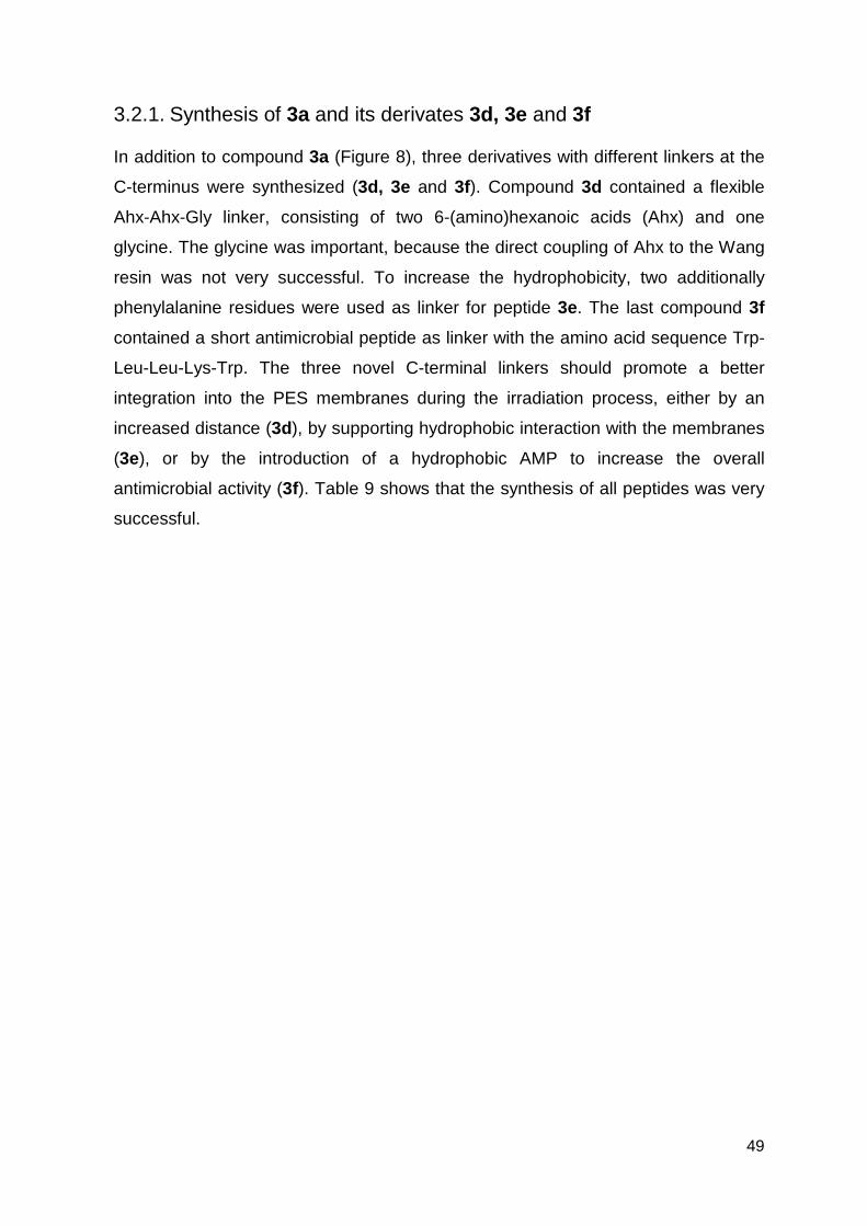
49
3.2.1. Synthesis of 3a and its derivates 3d, 3e and 3f
In addition to compound 3a (Figure 8), three derivatives with different linkers at the
C-terminus were synthesized (3d, 3e and 3f). Compound 3d contained a flexible
Ahx-Ahx-Gly linker, consisting of two 6-(amino)hexanoic acids (Ahx) and one
glycine. The glycine was important, because the direct coupling of Ahx to the Wang
resin was not very successful. To increase the hydrophobicity, two additionally
phenylalanine residues were used as linker for peptide 3e. The last compound 3f
contained a short antimicrobial peptide as linker with the amino acid sequence Trp-
Leu-Leu-Lys-Trp. The three novel C-terminal linkers should promote a better
integration into the PES membranes during the irradiation process, either by an
increased distance (3d), by supporting hydrophobic interaction with the membranes
(3e), or by the introduction of a hydrophobic AMP to increase the overall
antimicrobial activity (3f). Table 9 shows that the synthesis of all peptides was very
successful.
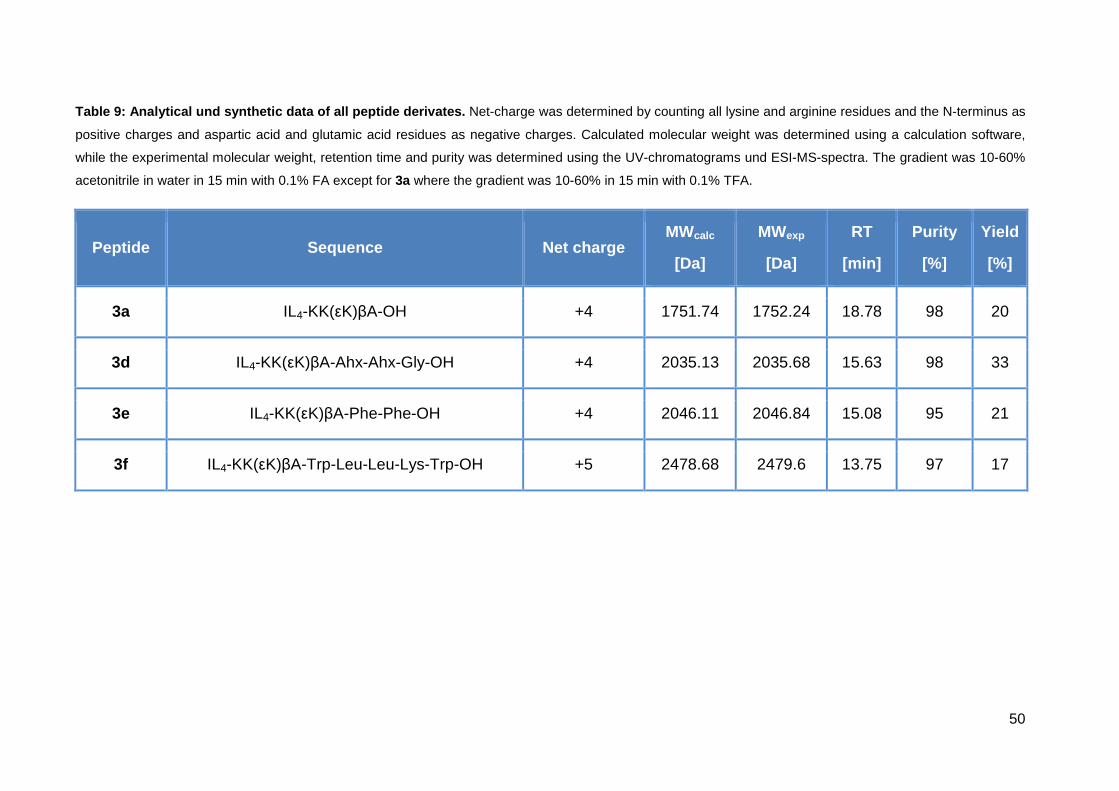
50
Table 9: Analytical und synthetic data of all pepti de derivates. Net-charge was determined by counting all lysine and arginine residues and the N-terminus as
positive charges and aspartic acid and glutamic acid residues as negative charges. Calculated molecular weight was determined using a calculation software,
while the experimental molecular weight, retention time and purity was determined using the UV-chromatograms und ESI-MS-spectra. The gradient was 10-60%
acetonitrile in water in 15 min with 0.1% FA except for 3a where the gradient was 10-60% in 15 min with 0.1% TFA.
Peptide Sequence Net charge MWcalc
[Da]
MWexp
[Da]
RT
[min]
Purity
[%]
Yield
[%]
3a IL4-KK(εK)βA-OH +4 1751.74 1752.24 18.78 98 20
3d IL4-KK(εK)βA-Ahx-Ahx-Gly-OH +4 2035.13 2035.68 15.63 98 33
3e IL4-KK(εK)βA-Phe-Phe-OH +4 2046.11 2046.84 15.08 95 21
3f IL4-KK(εK)βA-Trp-Leu-Leu-Lys-Trp-OH +5 2478.68 2479.6 13.75 97 17
51
3.2.2. Membrane immobilization and characterization
Membrane immobilization and characterization was performed by Dr. Agnes
Schulze at the Institute for Surface Modifications in Leipzig. The four peptides 3a,
3d, 3e and 3f were immobilized onto PES membranes using either electron beam
radiation with 50 kGy, 100 kGy or 150 kGy, or chemical immobilization via a peptide
bond. For this, the C-terminus of the peptides was introduced on the membrane by
NHS/EDC activation. Successful immobilization on the PES membrane was proven
by X-ray photoelectron spectroscopy (XPS) (Table 10). The unmodified PES
membranes were used as reference. XPS data show that they contain 70% carbon,
26% oxygen and 4% sulfur. After the immobilization of all peptides on the
membrane, a significant increase of nitrogen on the membrane surface could be
detected. Since the PES membrane reference contains no nitrogen, the nitrogen
increase let conclude a successful peptide immobilization.
A comparison of the four different peptides was difficult because the values of
nitrogen seem to be pretty variable. However, the highest nitrogen amount (0.72 –
1.73%) could be detected for 3f compared with the other peptides. All in all, the
electron beam radiation seemed to be a successful method to immobilize peptides
on a PES membrane.
Scanning electron microscopy (SEM) pictures were taken to further characterize the
membrane morphology after immobilization. The reference PES membrane
depicted in Figure 17 is highly porous with a pore-like structure. After electron
radiation, immobilization and EDC/NHS immobilization of the peptides, the pore
structure remained the same as for the reference leading to the conclusion that no
pore blocking or structural defects were caused by both immobilization methods.
52
Table 10: Atomic composition of the membranes (as d etermined by XPS analysis).
Elemental ratio (relative atom-%)
Label C N O S
PES Membrane (reference) 69.95 - 26.12 3.93
Membrane + 3a 50kGy 67.81 0.36 28.17 3.33
Membrane + 3a 100kGy 69.25 0.73 26.87 3.62
Membrane + 3a 150kGy 69.39 0.61 27.05 3.56
Membrane + 3a (NHS/EDC) 69.61 0.98 26.61 3.78
Membrane + 3d 50kGy 69.28 0.71 26.85 3.17
Membrane + 3d 100kGy 69.44 0.69 26.61 3.27
Membrane + 3d 150kGy 68.79 0.17 27.47 3.56
Membrane + 3d (NHS/EDC) 69.42 1.03 26.50 3.06
Membrane + 3e 50kGy 68.38 0.81 27.53 3.28
Membrane + 3e 100kGy 67.84 0.59 28.20 3.37
Membrane + 3e 150kGy 69.55 1.34 25.86 3.25
Membrane + 3e (NHS/EDC) 68.95 0.33 27.06 3.65
Membrane + 3f 50kGy 68.81 0.72 27.12 3.35
Membrane + 3f 100kGy 68.60 0.73 27.34 3.33
Membrane + 3f 150kGy 70.51 1.73 24.93 2.85
Membrane + 3f (NHS/EDC) 69.15 0.73 26.79 3.33
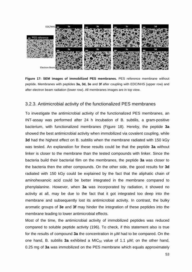
53
Figure 17: SEM images of immobilized PES membranes. PES reference membrane without
peptide. Membranes with peptides 3a, 3d, 3e and 3f after coupling with EDC/NHS (upper row) and
after electron beam radiation (lower row). All membranes images are in top view.
3.2.3. Antimicrobial activity of the functionalized PES membranes
To investigate the antimicrobial activity of the functionalized PES membranes, an
INT-assay was performed after 24 h incubation of B. subtilis, a gram-positive
bacterium, with functionalized membranes (Figure 18). Hereby, the peptide 3a
showed the best antimicrobial activity when immobilized via covalent coupling, while
3d had the highest effect on B. subtilis when the membrane radiated with 150 kGy
was tested. An explanation for these results could be that the peptide 3a without
linker is closer to the membrane than the tested compounds with linker. Since the
bacteria build their bacterial film on the membranes, the peptide 3a was closer to
the bacteria then the other compounds. On the other side, the good results for 3d
radiated with 150 kGy could be explained by the fact that the aliphatic chain of
aminohexanoic acid could be better integrated in the membrane compared to
phenylalanine. However, when 3a was incorporated by radiation, it showed no
activity at all, may be due to the fact that it got integrated too deep into the
membrane and subsequently lost its antimicrobial activity. In contrast, the bulky
aromatic groups of 3e and 3f may hinder the integration of these peptides into the
membrane leading to lower antimicrobial effects.
Most of the time, the antimicrobial activity of immobilized peptides was reduced
compared to soluble peptide activity (196). To check, if this statement also is true
for the results of compound 3a the concentration in µM had to be compared. On the
one hand, B. subtilis 3a exhibited a MIC50 value of 1.1 µM; on the other hand,
0.25 mg of 3a was immobilized on the PES membrane which equals approximately
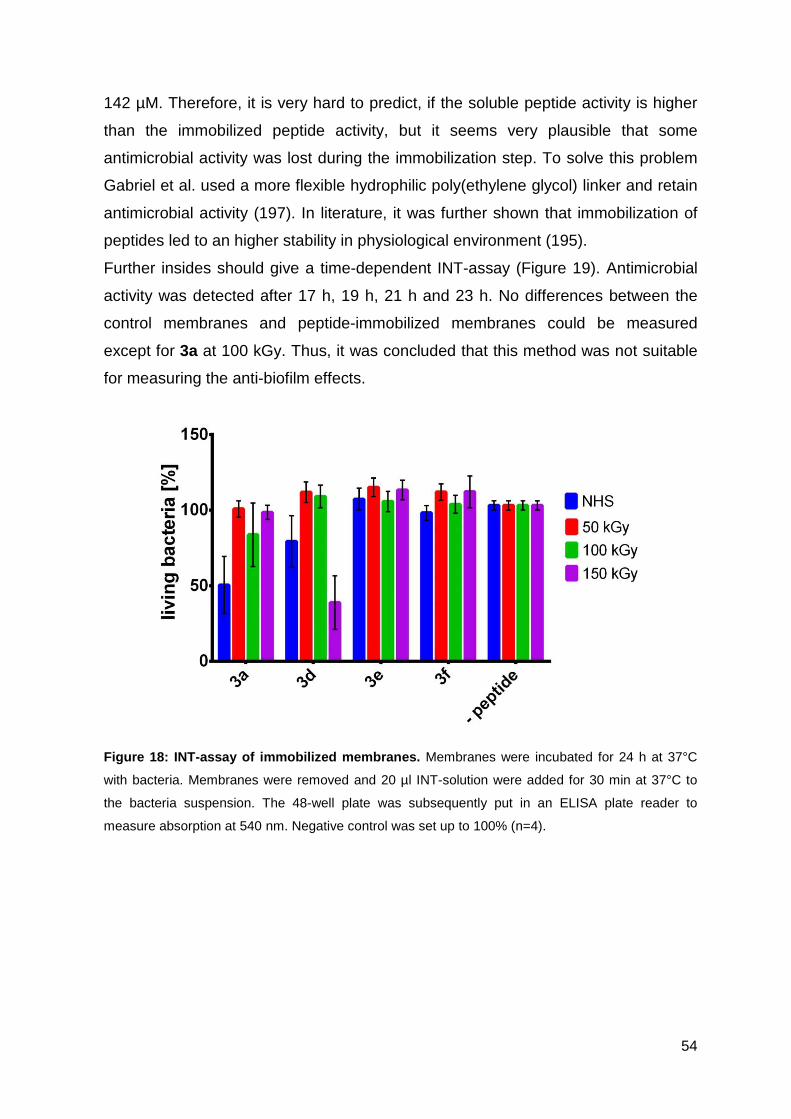
54
142 µM. Therefore, it is very hard to predict, if the soluble peptide activity is higher
than the immobilized peptide activity, but it seems very plausible that some
antimicrobial activity was lost during the immobilization step. To solve this problem
Gabriel et al. used a more flexible hydrophilic poly(ethylene glycol) linker and retain
antimicrobial activity (197). In literature, it was further shown that immobilization of
peptides led to an higher stability in physiological environment (195).
Further insides should give a time-dependent INT-assay (Figure 19). Antimicrobial
activity was detected after 17 h, 19 h, 21 h and 23 h. No differences between the
control membranes and peptide-immobilized membranes could be measured
except for 3a at 100 kGy. Thus, it was concluded that this method was not suitable
for measuring the anti-biofilm effects.
Figure 18: INT-assay of immobilized membranes. Membranes were incubated for 24 h at 37°C
with bacteria. Membranes were removed and 20 µl INT-solution were added for 30 min at 37°C to
the bacteria suspension. The 48-well plate was subsequently put in an ELISA plate reader to
measure absorption at 540 nm. Negative control was set up to 100% (n=4).
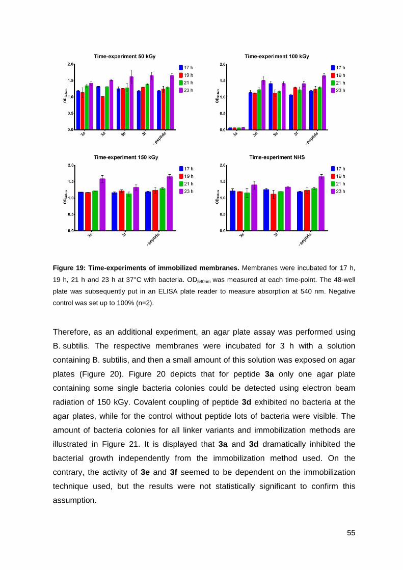
55
Figure 19: Time-experiments of immobilized membrane s. Membranes were incubated for 17 h,
19 h, 21 h and 23 h at 37°C with bacteria. OD540nm was measured at each time-point. The 48-well
plate was subsequently put in an ELISA plate reader to measure absorption at 540 nm. Negative
control was set up to 100% (n=2).
Therefore, as an additional experiment, an agar plate assay was performed using
B. subtilis. The respective membranes were incubated for 3 h with a solution
containing B. subtilis, and then a small amount of this solution was exposed on agar
plates (Figure 20). Figure 20 depicts that for peptide 3a only one agar plate
containing some single bacteria colonies could be detected using electron beam
radiation of 150 kGy. Covalent coupling of peptide 3d exhibited no bacteria at the
agar plates, while for the control without peptide lots of bacteria were visible. The
amount of bacteria colonies for all linker variants and immobilization methods are
illustrated in Figure 21. It is displayed that 3a and 3d dramatically inhibited the
bacterial growth independently from the immobilization method used. On the
contrary, the activity of 3e and 3f seemed to be dependent on the immobilization
technique used, but the results were not statistically significant to confirm this
assumption.
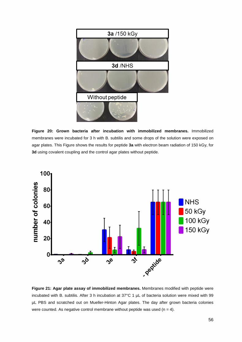
56
Figure 20: Grown bacteria after incubation with imm obilized membranes. Immobilized
membranes were incubated for 3 h with B. subtilis and some drops of the solution were exposed on
agar plates. This Figure shows the results for peptide 3a with electron beam radiation of 150 kGy, for
3d using covalent coupling and the control agar plates without peptide.
Figure 21: Agar plate assay of immobilized membrane s. Membranes modified with peptide were
incubated with B. subtilis. After 3 h incubation at 37°C 1 µL of bacteria solution were mixed with 99
µL PBS and scratched out on Mueller-Hinton Agar plates. The day after grown bacteria colonies
were counted. As negative control membrane without peptide was used (n = 4).
57
In conclusion, bacteria growing experiments showed antimicrobial activity of 3a and
3d, while within the INT-assays, no antimicrobial effect was detected. The
difference between these two experiments might be the volume of the bacterial
solution, which is 10-fold higher in the INT-assay than in the agar plate assay.
Additionally, the incubation time for the agar plate assay is with 3 h shorter than the
6 h for the INT-assays. It can be concluded that the small compounds 3a and 3d
are very active in the first 3 h and have only a limited range with their branched side
chains. However, all coated membranes exhibited an increased activity against
B. subtilis compared to membranes without peptide. Taken together, the electron
beam radiation method demonstrated as high antimicrobial activity as the chemical
coupled method. This lead to the assumption that peptides can be immobilized
successfully on PES membranes using the electron beam radiation method.
3.3. Optimizing the antimicrobial activity of the CPP sC18
sC18 is originally derived from CAP18, a cathelicidin peptide with antimicrobial
activity. sC18 was studied and developed in our research group and is mostly used
as cell-penetrating peptide in the delivery of cytostatic drugs or imagine probes. (80,
196). sC18 is mostly taken up in cells via endocytosis, but an direct entry seems
also possible (55, 81, 82). In chapter one of this work, it was already shown, that
sC18 as a peptide conjugate exhibited high antimicrobial activity. Besides sC18
other well-known CPPs were found to exhibit antimicrobial activity as well, for
example TAT(48-60), penetratin or Pep-1 (55, 197-199). The lastly named peptide
Pep-1 was already modified to an antimicrobial Peptide Pep-1-k through increase of
the cationic character (200). Since the sC18 peptide already has a cationic
character, the idea was to increase the antimicrobial activity and selectivity through
the integration of hydrophobic amino acids. Kim et al. revealed that an introduction
of isoleucine or leucine in the AMP sequence led to an increase in antimicrobial
activity (201). Isoleucine was chosen because its hydrophobicity is a little bit higher
than the hydrophobicity of leucine (202). Furthermore, the hydrophobicity index of
Monera et al. exhibited that phenylalanine is the most hydrophobic amino acid.
Therefore, isoleucine and phenylalanine exchanges were performed and their
influence was tested towards activity and selectivity.
58
3.3.1. Synthesis of sC18 variants
In a first set of experiments, an isoleucine full scan was performed. Further on, the
isoleucine exchanges at position 10, 15 and 16 showed the best antimicrobial
activity, and so they were combined to three double and one triple mutants (Table
11) (Antimicrobial activity is shown in 3.3.2). To investigate, if phenylalanine exhibits
the same influence on the antimicrobial activity as isoleucine did, the same amino
acid exchanges at position 10, 15 and 16 as well as the double and triple mutants
were accomplished for phenylalanine, too (Table 11). The synthesis of all these
peptides was performed successfully. Peptides sC18I1 – sC18I9 and sC18I11 –
sC18I14 were only used in the preliminary full scan to investigate which amino acid
substitution position showed the highest increase in antimicrobial activity. For the
preliminary tests, these peptides were synthesized in small amounts and were not
further purified via preparative HPLC. However, the obtained yields were above
70%, what was sufficient for a first row of biological testing. The peptides sC18,
sC18I10, sC18I15, sC18I16, sC18F10, sC18F15, sC18F1 6 and all double and
triple mutants were purified and showed high purity between 91-98%. The yield of
these peptides was around 30%, except for the double and triple mutants of
isoleucine, which were obtained in a yield of only 10%. This can be explained by the
formation of a side-product, which was detected in the UV-chromatogram before
HPLC purification (Figure 22). The side-product at a retention time of 5.56 min
exhibited a 6-fold charged molecular peak of 342.24 m/z, corresponding to a
peptide mass of 1939.26 Da. The molecular weight of this side-product was 113 m/z
fewer compared to the product peak at a retention time of 6.79 min. This mass
difference can be explained by the loss of an isoleucine at position 15. Similar side
products were detected for all peptides containing an isoleucine at position 15. A
closer look at the peptide sequence suggested that the KIKIK (sC18I15) or similar
motifs at the N-terminus of the peptide were very difficult to synthesize resulting in a
low yield of the final product. As already mentioned, the main product was detected
in the UV-chromatogram at 6.79 min. The ESI-MS spectrum displays the 3-fold
([M+3H]3+ = 685.58) to 6-fold ([M+6H]6+ = 343.29) charged molecular ions. After
purification via preparative HPLC, a sharp peak could be detected in the UV-
chromatogram with 0.1% TFA (Figure 23). Again, the 3-fold ([M+3H]3+ = 685.66) to
6-fold ([M+6H]6+ = 343.22) charged molecular ions were visible with the
corresponding molecular mass of 2054.70 Da which was assigned to sC18I15.
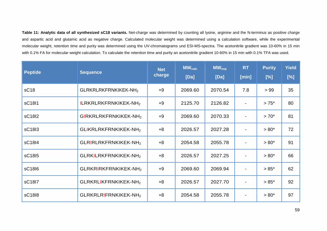
59
Table 11: Analytic data of all synthesized sC18 var iants. Net-charge was determined by counting all lysine, arginine and the N-terminus as positive charge
and aspartic acid and glutamic acid as negative charge. Calculated molecular weight was determined using a calculation software, while the experimental
molecular weight, retention time and purity was determined using the UV-chromatograms und ESI-MS-spectra. The acetonitrile gradient was 10-60% in 15 min
with 0.1% FA for molecular weight calculation. To calculate the retention time and purity an acetonitrile gradient 10-60% in 15 min with 0.1% TFA was used.
Peptide Sequence Net charge
MWcalc
[Da]
MWexp
[Da]
RT
[min]
Purity
[%]
Yield
[%]
sC18 GLRKRLRKFRNKIKEK-NH2 +9 2069.60 2070.54 7.8 > 99 35
sC18I1 ILRKRLRKFRNKIKEK-NH2 +9 2125.70 2126.82 - > 75* 80
sC18I2 GIRKRLRKFRNKIKEK-NH2 +9 2069.60 2070.33 - > 70* 81
sC18I3 GLIKRLRKFRNKIKEK-NH2 +8 2026.57 2027.28 - > 80* 72
sC18I4 GLRIRLRKFRNKIKEK-NH2 +8 2054.58 2055.78 - > 80* 91
sC18I5 GLRKILRKFRNKIKEK-NH2 +8 2026.57 2027.25 - > 80* 66
sC18I6 GLRKRIRKFRNKIKEK-NH2 +9 2069.60 2069.94 - > 85* 62
sC18I7 GLRKRLIKFRNKIKEK-NH2 +8 2026.57 2027.70 - > 85* 92
sC18I8 GLRKRLRIFRNKIKEK-NH2 +8 2054.58 2055.78 - > 80* 97

60
sC18I9 GLRKRLRKIRNKIKEK-NH2 +9 2035.58 2036.01 - > 80* 88
sC18I10 GLRKRLRKFINKIKEK-NH2 +8 2026.57 2027.70 9.2 95 37
sC18I11 GLRKRLRKFRIKIKEK-NH2 +9 2068.65 2069.88 - > 90* 95
sC18I12 GLRKRLRKFRNIIKEK-NH2 +8 2054.58 2055.78 - > 75* 65
sC18I14 GLRKRLRKFRNKIIEK-NH2 +8 2054.58 2055.84 - > 80* 71
sC18I15 GLRKRLRKFRNKIKIK-NH2 +10 2053.64 2054.70 7.6 98 24
sC18I16 GLRKRLRKFRNKIKEI-NH2 +8 2054.58 2055.90 8.6 95 28
sC18I10I15 GLRKRLRKFINKIKIK-NH2 +9 2010.61 2011.00 9.2 90 11
sC18I10I16 GLRKRLRKFINKIKEI-NH2 +7 2011.55 2012.5 10.1 95 8
sC18I15I16 GLRKRLRKFRNKIKII-NH2 +9 2038.63 2038.85 8.3 95 9
sC18I10I15I16 GLRKRLRKFINKIKII-NH2 +8 1995.60 1996.05 9.3 95 6
sC18F10 GLRKRLRKFFNKIKEK-NH2 +8 2060.59 2060.76 9.3 94 37
sC18F15 GLRKRLRKFRNKIKFK-NH2 +10 2087.66 2087.96 8.3 93 31
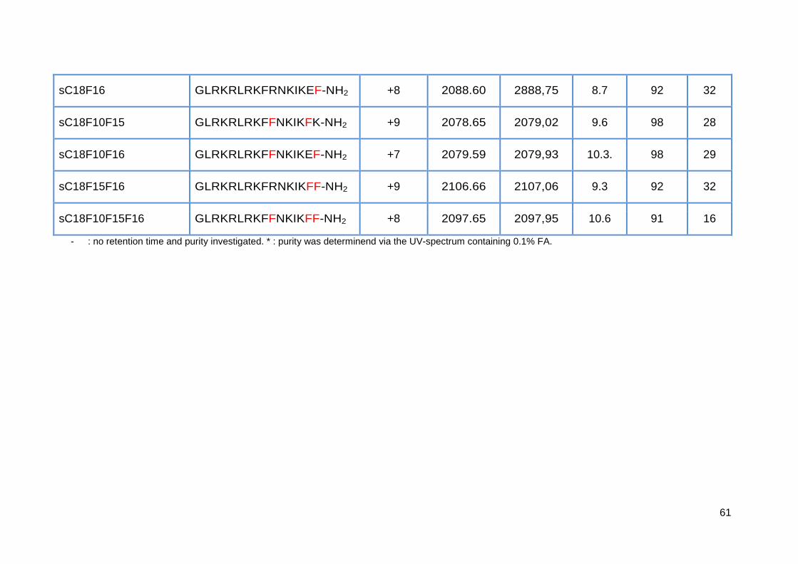
61
sC18F16 GLRKRLRKFRNKIKEF-NH2 +8 2088.60 2888,75 8.7 92 32
sC18F10F15 GLRKRLRKFFNKIKFK-NH2 +9 2078.65 2079,02 9.6 98 28
sC18F10F16 GLRKRLRKFFNKIKEF-NH2 +7 2079.59 2079,93 10.3. 98 29
sC18F15F16 GLRKRLRKFRNKIKFF-NH2 +9 2106.66 2107,06 9.3 92 32
sC18F10F15F16 GLRKRLRKFFNKIKFF-NH2 +8 2097.65 2097,95 10.6 91 16
- : no retention time and purity investigated. * : purity was determinend via the UV-spectrum containing 0.1% FA.
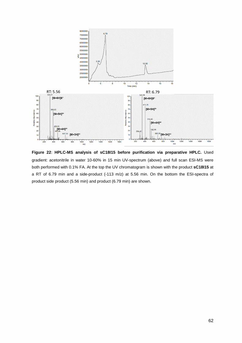
62
Figure 22 : HPLC-MS analysis of sC18I15 before purification via preparative HPLC. Used
gradient: acetonitrile in water 10-60% in 15 min UV-spectrum (above) and full scan ESI-MS were
both performed with 0.1% FA. At the top the UV chromatogram is shown with the product sC18I15 at
a RT of 6.79 min and a side-product (-113 m/z) at 5.56 min. On the bottom the ESI-spectra of
product side product (5.56 min) and product (6.79 min) are shown.
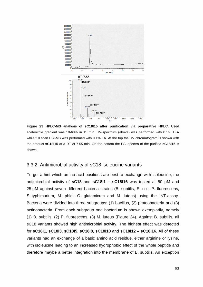
63
Figure 23 HPLC-MS analysis of sC18I15 after purification via preparative HPLC. Used
acetonitrile gradient was 10-60% in 15 min. UV-spectrum (above) was performed with 0.1% TFA
while full scan ESI-MS was performed with 0.1% FA. At the top the UV chromatogram is shown with
the product sC18I15 at a RT of 7.55 min. On the bottom the ESI-spectra of the purified sC18I15 is
shown.
3.3.2. Antimicrobial activity of sC18 isoleucine variants
To get a hint which amino acid positions are best to exchange with isoleucine, the
antimicrobial activity of sC18 and sC18I1 – sC18I16 was tested at 50 µM and
25 µM against seven different bacteria strains (B. subtilis, E. coli, P. fluorescens,
S. typhimurium, M. phlei, C. glutamicum and M. luteus) using the INT-assay.
Bacteria were divided into three subgroups: (1) bacillus, (2) proteobacteria and (3)
actinobacteria. From each subgroup one bacterium is shown exemplarily, namely
(1) B. subtilis, (2) P. fluorescens, (3) M. luteus (Figure 24). Against B. subtilis, all
sC18 variants showed high antimicrobial activity. The highest effect was detected
for sC18I1, sC18I3, sC18I5, sC18I8, sC18I10 and sC18I12 – sC18I16. All of these
variants had an exchange of a basic amino acid residue, either arginine or lysine,
with isoleucine leading to an increased hydrophobic effect of the whole peptide and
therefore maybe a better integration into the membrane of B. subtilis. An exception
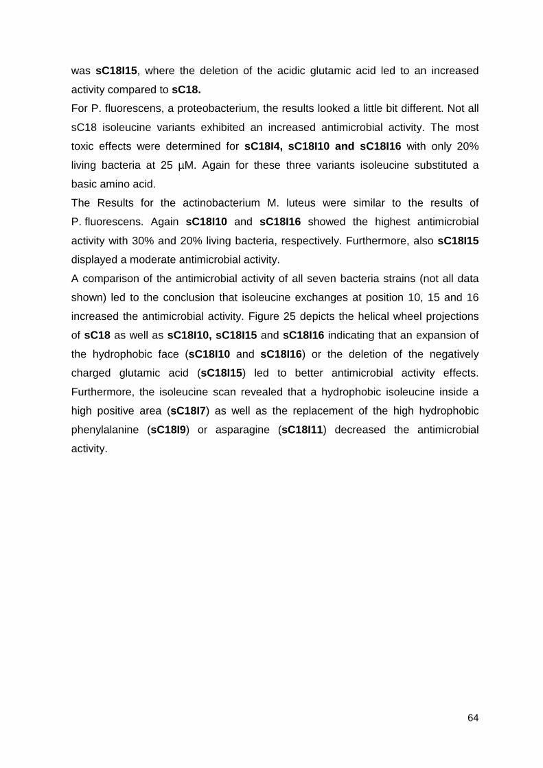
64
was sC18I15, where the deletion of the acidic glutamic acid led to an increased
activity compared to sC18.
For P. fluorescens, a proteobacterium, the results looked a little bit different. Not all
sC18 isoleucine variants exhibited an increased antimicrobial activity. The most
toxic effects were determined for sC18I4, sC18I10 and sC18I16 with only 20%
living bacteria at 25 µM. Again for these three variants isoleucine substituted a
basic amino acid.
The Results for the actinobacterium M. luteus were similar to the results of
P. fluorescens. Again sC18I10 and sC18I16 showed the highest antimicrobial
activity with 30% and 20% living bacteria, respectively. Furthermore, also sC18I15
displayed a moderate antimicrobial activity.
A comparison of the antimicrobial activity of all seven bacteria strains (not all data
shown) led to the conclusion that isoleucine exchanges at position 10, 15 and 16
increased the antimicrobial activity. Figure 25 depicts the helical wheel projections
of sC18 as well as sC18I10, sC18I15 and sC18I16 indicating that an expansion of
the hydrophobic face (sC18I10 and sC18I16) or the deletion of the negatively
charged glutamic acid (sC18I15) led to better antimicrobial activity effects.
Furthermore, the isoleucine scan revealed that a hydrophobic isoleucine inside a
high positive area (sC18I7) as well as the replacement of the high hydrophobic
phenylalanine (sC18I9) or asparagine (sC18I11) decreased the antimicrobial
activity.
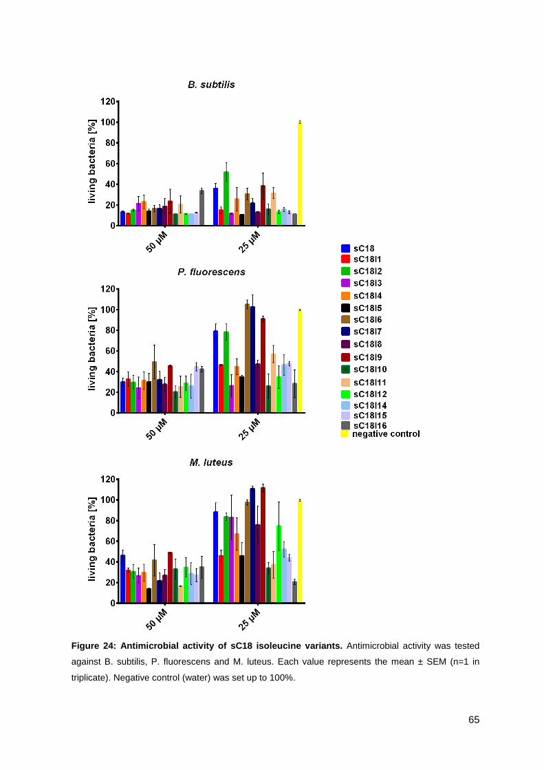
65
Figure 24: Antimicrobial activity of sC18 isoleucin e variants. Antimicrobial activity was tested
against B. subtilis, P. fluorescens and M. luteus. Each value represents the mean ± SEM (n=1 in
triplicate). Negative control (water) was set up to 100%.
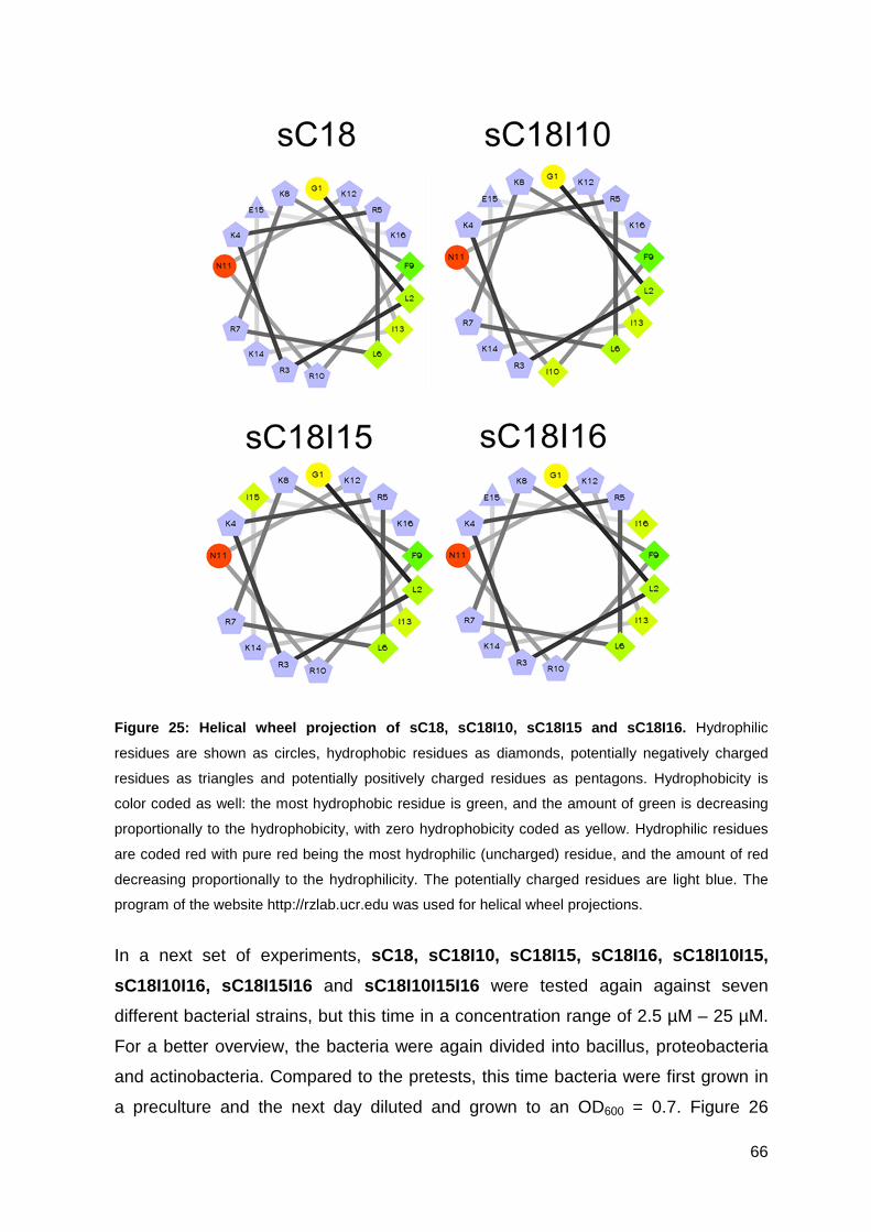
66
Figure 25: Helical wheel projection of sC18, sC18I1 0, sC18I15 and sC18I16. Hydrophilic
residues are shown as circles, hydrophobic residues as diamonds, potentially negatively charged
residues as triangles and potentially positively charged residues as pentagons. Hydrophobicity is
color coded as well: the most hydrophobic residue is green, and the amount of green is decreasing
proportionally to the hydrophobicity, with zero hydrophobicity coded as yellow. Hydrophilic residues
are coded red with pure red being the most hydrophilic (uncharged) residue, and the amount of red
decreasing proportionally to the hydrophilicity. The potentially charged residues are light blue. The
program of the website http://rzlab.ucr.edu was used for helical wheel projections.
In a next set of experiments, sC18, sC18I10, sC18I15, sC18I16, sC18I10I15,
sC18I10I16, sC18I15I16 and sC18I10I15I16 were tested again against seven
different bacterial strains, but this time in a concentration range of 2.5 µM – 25 µM.
For a better overview, the bacteria were again divided into bacillus, proteobacteria
and actinobacteria. Compared to the pretests, this time bacteria were first grown in
a preculture and the next day diluted and grown to an OD600 = 0.7. Figure 26
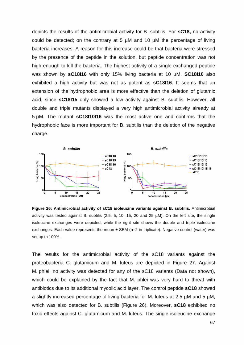
67
depicts the results of the antimicrobial activity for B. subtilis. For sC18, no activity
could be detected; on the contrary at 5 µM and 10 µM the percentage of living
bacteria increases. A reason for this increase could be that bacteria were stressed
by the presence of the peptide in the solution, but peptide concentration was not
high enough to kill the bacteria. The highest activity of a single exchanged peptide
was shown by sC18I16 with only 15% living bacteria at 10 µM. SC18I10 also
exhibited a high activity but was not as potent as sC18I16. It seems that an
extension of the hydrophobic area is more effective than the deletion of glutamic
acid, since sC18I15 only showed a low activity against B. subtilis. However, all
double and triple mutants displayed a very high antimicrobial activity already at
5 µM. The mutant sC18I10I16 was the most active one and confirms that the
hydrophobic face is more important for B. subtilis than the deletion of the negative
charge.
Figure 26: Antimicrobial activity of sC18 isoleucin e variants against B. subtilis. Antimicrobial
activity was tested against B. subtilis (2.5, 5, 10, 15, 20 and 25 µM). On the left site, the single
isoleucine exchanges were depicted, while the right site shows the double and triple isoleucine
exchanges. Each value represents the mean ± SEM (n=2 in triplicate). Negative control (water) was
set up to 100%.
The results for the antimicrobial activity of the sC18 variants against the
proteobacteria C. glutamicum and M. luteus are depicted in Figure 27. Against
M. phlei, no activity was detected for any of the sC18 variants (Data not shown),
which could be explained by the fact that M. phlei was very hard to threat with
antibiotics due to its additional mycolic acid layer. The control peptide sC18 showed
a slightly increased percentage of living bacteria for M. luteus at 2.5 µM and 5 µM,
which was also detected for B. subtilis (Figure 26). Moreover, sC18 exhibited no
toxic effects against C. glutamicum and M. luteus. The single isoleucine exchange
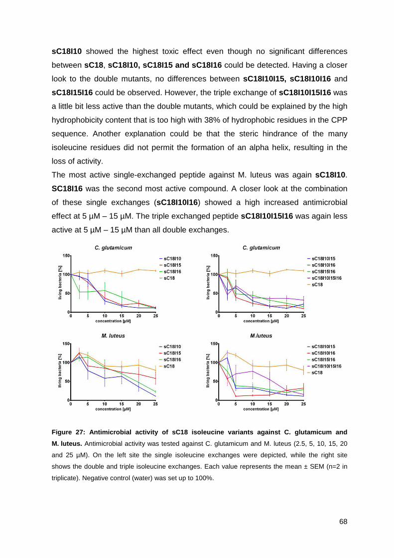
68
sC18I10 showed the highest toxic effect even though no significant differences
between sC18, sC18I10, sC18I15 and sC18I16 could be detected. Having a closer
look to the double mutants, no differences between sC18I10I15, sC18I10I16 and
sC18I15I16 could be observed. However, the triple exchange of sC18I10I15I16 was
a little bit less active than the double mutants, which could be explained by the high
hydrophobicity content that is too high with 38% of hydrophobic residues in the CPP
sequence. Another explanation could be that the steric hindrance of the many
isoleucine residues did not permit the formation of an alpha helix, resulting in the
loss of activity.
The most active single-exchanged peptide against M. luteus was again sC18I10.
SC18I16 was the second most active compound. A closer look at the combination
of these single exchanges (sC18I10I16) showed a high increased antimicrobial
effect at 5 µM – 15 µM. The triple exchanged peptide sC18I10I15I16 was again less
active at 5 µM – 15 µM than all double exchanges.
Figure 27: Antimicrobial activity of sC18 isoleucin e variants against C. glutamicum and
M. luteus. Antimicrobial activity was tested against C. glutamicum and M. luteus (2.5, 5, 10, 15, 20
and 25 µM). On the left site the single isoleucine exchanges were depicted, while the right site
shows the double and triple isoleucine exchanges. Each value represents the mean ± SEM (n=2 in
triplicate). Negative control (water) was set up to 100%.
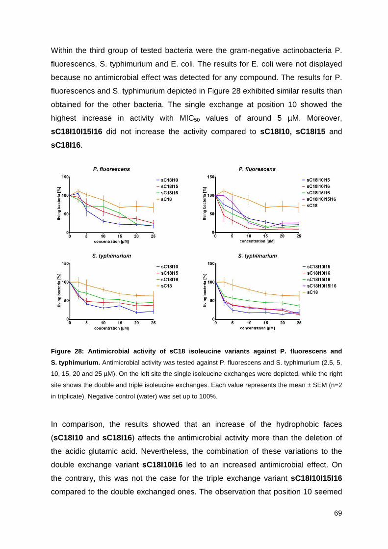
69
Within the third group of tested bacteria were the gram-negative actinobacteria P.
fluorescencs, S. typhimurium and E. coli. The results for E. coli were not displayed
because no antimicrobial effect was detected for any compound. The results for P.
fluorescencs and S. typhimurium depicted in Figure 28 exhibited similar results than
obtained for the other bacteria. The single exchange at position 10 showed the
highest increase in activity with MIC50 values of around 5 µM. Moreover,
sC18I10I15I16 did not increase the activity compared to sC18I10, sC18I15 and
sC18I16.
Figure 28: Antimicrobial activity of sC18 isoleucin e variants against P. fluorescens and
S. typhimurium. Antimicrobial activity was tested against P. fluorescens and S. typhimurium (2.5, 5,
10, 15, 20 and 25 µM). On the left site the single isoleucine exchanges were depicted, while the right
site shows the double and triple isoleucine exchanges. Each value represents the mean ± SEM (n=2
in triplicate). Negative control (water) was set up to 100%.
In comparison, the results showed that an increase of the hydrophobic faces
(sC18I10 and sC18I16) affects the antimicrobial activity more than the deletion of
the acidic glutamic acid. Nevertheless, the combination of these variations to the
double exchange variant sC18I10I16 led to an increased antimicrobial effect. On
the contrary, this was not the case for the triple exchange variant sC18I10I15I16
compared to the double exchanged ones. The observation that position 10 seemed
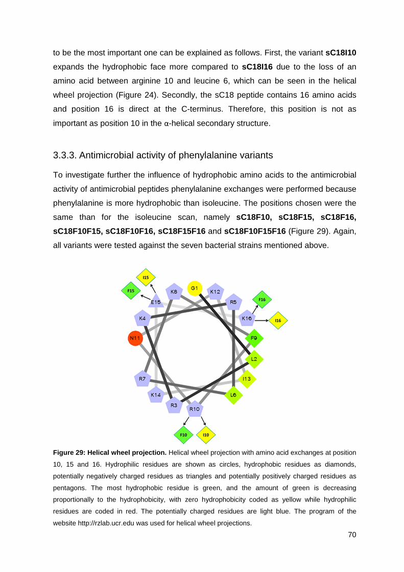
70
to be the most important one can be explained as follows. First, the variant sC18I10
expands the hydrophobic face more compared to sC18I16 due to the loss of an
amino acid between arginine 10 and leucine 6, which can be seen in the helical
wheel projection (Figure 24). Secondly, the sC18 peptide contains 16 amino acids
and position 16 is direct at the C-terminus. Therefore, this position is not as
important as position 10 in the α-helical secondary structure.
3.3.3. Antimicrobial activity of phenylalanine variants
To investigate further the influence of hydrophobic amino acids to the antimicrobial
activity of antimicrobial peptides phenylalanine exchanges were performed because
phenylalanine is more hydrophobic than isoleucine. The positions chosen were the
same than for the isoleucine scan, namely sC18F10, sC18F15, sC18F16,
sC18F10F15, sC18F10F16, sC18F15F16 and sC18F10F15F16 (Figure 29). Again,
all variants were tested against the seven bacterial strains mentioned above.
Figure 29: Helical wheel projection. Helical wheel projection with amino acid exchanges at position
10, 15 and 16. Hydrophilic residues are shown as circles, hydrophobic residues as diamonds,
potentially negatively charged residues as triangles and potentially positively charged residues as
pentagons. The most hydrophobic residue is green, and the amount of green is decreasing
proportionally to the hydrophobicity, with zero hydrophobicity coded as yellow while hydrophilic
residues are coded in red. The potentially charged residues are light blue. The program of the
website http://rzlab.ucr.edu was used for helical wheel projections.
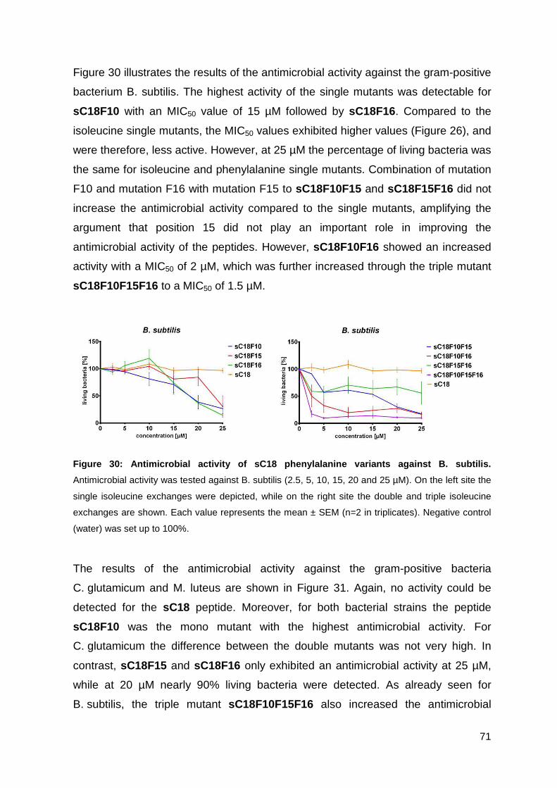
71
Figure 30 illustrates the results of the antimicrobial activity against the gram-positive
bacterium B. subtilis. The highest activity of the single mutants was detectable for
sC18F10 with an MIC50 value of 15 µM followed by sC18F16. Compared to the
isoleucine single mutants, the MIC50 values exhibited higher values (Figure 26), and
were therefore, less active. However, at 25 µM the percentage of living bacteria was
the same for isoleucine and phenylalanine single mutants. Combination of mutation
F10 and mutation F16 with mutation F15 to sC18F10F15 and sC18F15F16 did not
increase the antimicrobial activity compared to the single mutants, amplifying the
argument that position 15 did not play an important role in improving the
antimicrobial activity of the peptides. However, sC18F10F16 showed an increased
activity with a MIC50 of 2 µM, which was further increased through the triple mutant
sC18F10F15F16 to a MIC50 of 1.5 µM.
Figure 30: Antimicrobial activity of sC18 phenylala nine variants against B. subtilis.
Antimicrobial activity was tested against B. subtilis (2.5, 5, 10, 15, 20 and 25 µM). On the left site the
single isoleucine exchanges were depicted, while on the right site the double and triple isoleucine
exchanges are shown. Each value represents the mean ± SEM (n=2 in triplicates). Negative control
(water) was set up to 100%.
The results of the antimicrobial activity against the gram-positive bacteria
C. glutamicum and M. luteus are shown in Figure 31. Again, no activity could be
detected for the sC18 peptide. Moreover, for both bacterial strains the peptide
sC18F10 was the mono mutant with the highest antimicrobial activity. For
C. glutamicum the difference between the double mutants was not very high. In
contrast, sC18F15 and sC18F16 only exhibited an antimicrobial activity at 25 µM,
while at 20 µM nearly 90% living bacteria were detected. As already seen for
B. subtilis, the triple mutant sC18F10F15F16 also increased the antimicrobial
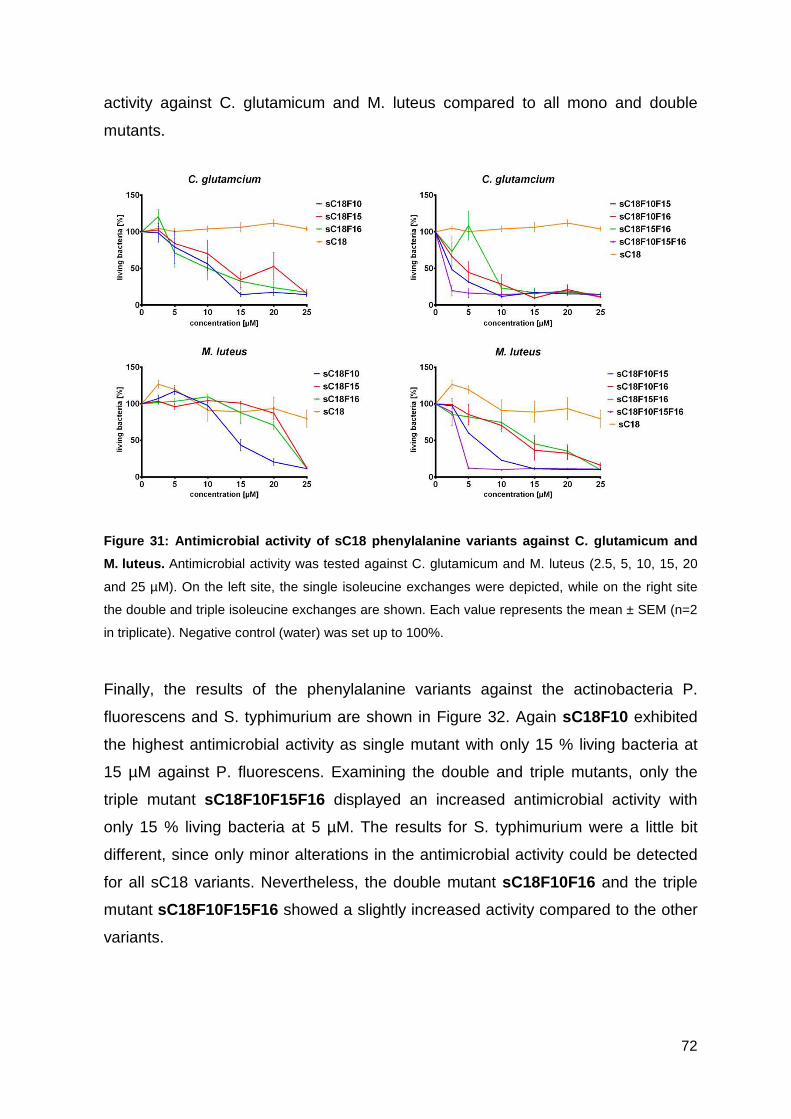
72
activity against C. glutamicum and M. luteus compared to all mono and double
mutants.
Figure 31: Antimicrobial activity of sC18 phenylala nine variants against C. glutamicum and
M. luteus. Antimicrobial activity was tested against C. glutamicum and M. luteus (2.5, 5, 10, 15, 20
and 25 µM). On the left site, the single isoleucine exchanges were depicted, while on the right site
the double and triple isoleucine exchanges are shown. Each value represents the mean ± SEM (n=2
in triplicate). Negative control (water) was set up to 100%.
Finally, the results of the phenylalanine variants against the actinobacteria P.
fluorescens and S. typhimurium are shown in Figure 32. Again sC18F10 exhibited
the highest antimicrobial activity as single mutant with only 15 % living bacteria at
15 µM against P. fluorescens. Examining the double and triple mutants, only the
triple mutant sC18F10F15F16 displayed an increased antimicrobial activity with
only 15 % living bacteria at 5 µM. The results for S. typhimurium were a little bit
different, since only minor alterations in the antimicrobial activity could be detected
for all sC18 variants. Nevertheless, the double mutant sC18F10F16 and the triple
mutant sC18F10F15F16 showed a slightly increased activity compared to the other
variants.
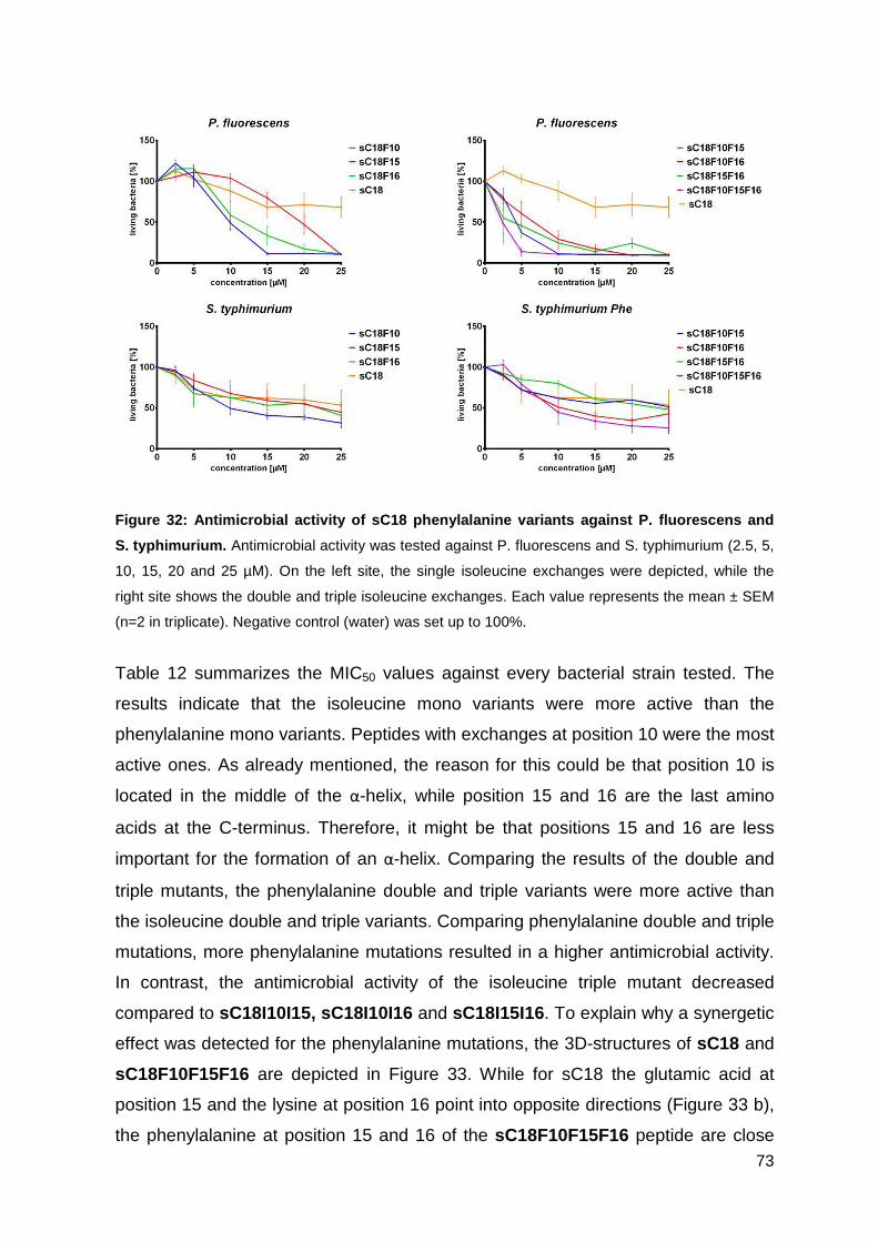
73
Figure 32: Antimicrobial activity of sC18 phenylala nine variants against P. fluorescens and
S. typhimurium. Antimicrobial activity was tested against P. fluorescens and S. typhimurium (2.5, 5,
10, 15, 20 and 25 µM). On the left site, the single isoleucine exchanges were depicted, while the
right site shows the double and triple isoleucine exchanges. Each value represents the mean ± SEM
(n=2 in triplicate). Negative control (water) was set up to 100%.
Table 12 summarizes the MIC50 values against every bacterial strain tested. The
results indicate that the isoleucine mono variants were more active than the
phenylalanine mono variants. Peptides with exchanges at position 10 were the most
active ones. As already mentioned, the reason for this could be that position 10 is
located in the middle of the α-helix, while position 15 and 16 are the last amino
acids at the C-terminus. Therefore, it might be that positions 15 and 16 are less
important for the formation of an α-helix. Comparing the results of the double and
triple mutants, the phenylalanine double and triple variants were more active than
the isoleucine double and triple variants. Comparing phenylalanine double and triple
mutations, more phenylalanine mutations resulted in a higher antimicrobial activity.
In contrast, the antimicrobial activity of the isoleucine triple mutant decreased
compared to sC18I10I15, sC18I10I16 and sC18I15I16. To explain why a synergetic
effect was detected for the phenylalanine mutations, the 3D-structures of sC18 and
sC18F10F15F16 are depicted in Figure 33. While for sC18 the glutamic acid at
position 15 and the lysine at position 16 point into opposite directions (Figure 33 b),
the phenylalanine at position 15 and 16 of the sC18F10F15F16 peptide are close
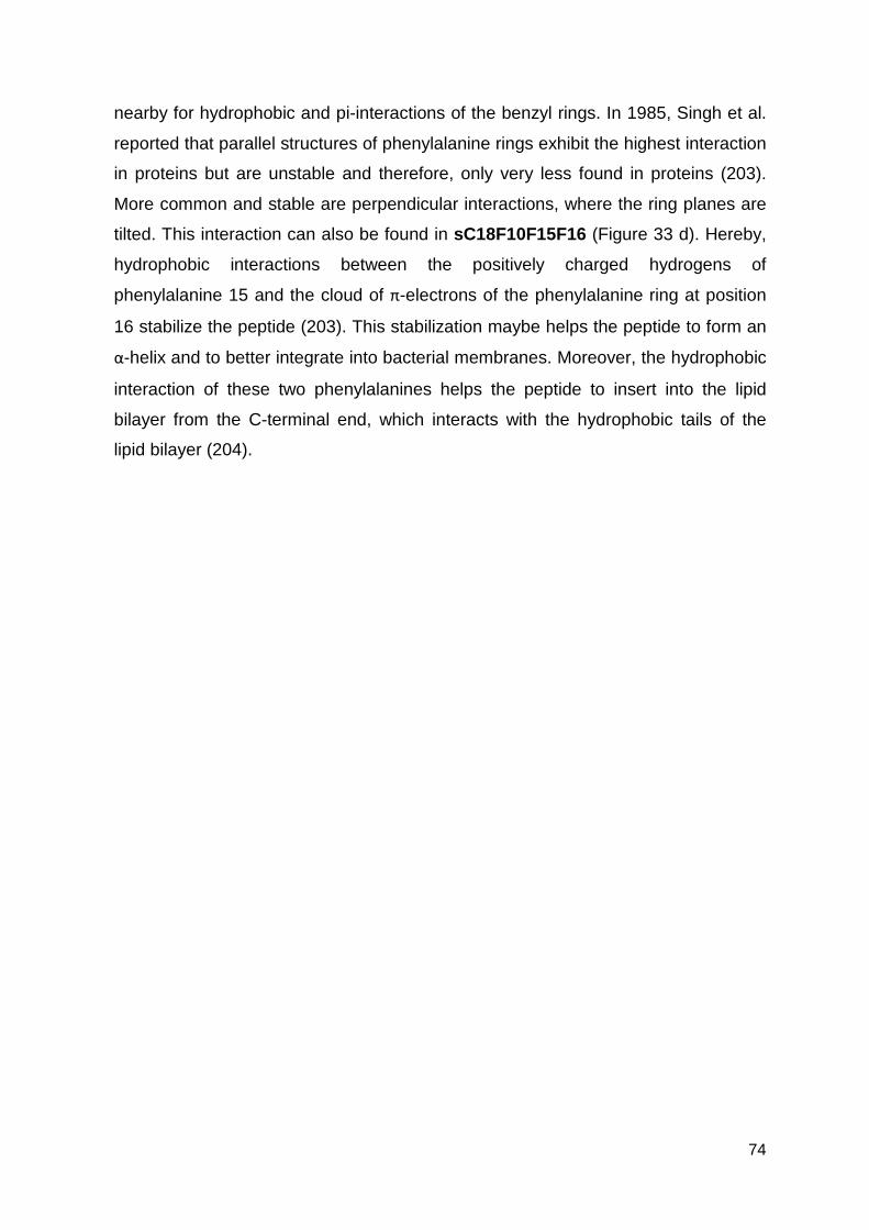
74
nearby for hydrophobic and pi-interactions of the benzyl rings. In 1985, Singh et al.
reported that parallel structures of phenylalanine rings exhibit the highest interaction
in proteins but are unstable and therefore, only very less found in proteins (203).
More common and stable are perpendicular interactions, where the ring planes are
tilted. This interaction can also be found in sC18F10F15F16 (Figure 33 d). Hereby,
hydrophobic interactions between the positively charged hydrogens of
phenylalanine 15 and the cloud of π-electrons of the phenylalanine ring at position
16 stabilize the peptide (203). This stabilization maybe helps the peptide to form an
α-helix and to better integrate into bacterial membranes. Moreover, the hydrophobic
interaction of these two phenylalanines helps the peptide to insert into the lipid
bilayer from the C-terminal end, which interacts with the hydrophobic tails of the
lipid bilayer (204).
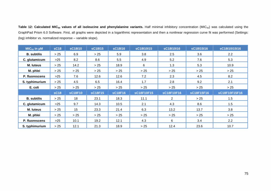
75
Table 12: Calculated MIC 50 values of all isoleucine and phenylalanine variant s. Half minimal inhibitory concentration (MIC50) was calculated using the
GraphPad Prism 6.0 Software. First, all graphs were depicted in a logarithmic representation and then a nonlinear regression curve fit was performed (Settings:
(log) inhibitor vs. normalized response – variable slope).
MIC50 in µM sC18 sC18I10 sC18I15 sC18I16 sC18I10I15 sC18I10I16 sC18I15I16 sC18I10I15I16
B. subtilis > 25 6.9 > 25 5.9 3.8 2.5 3.6 2.2
C. glutamicum >25 8.2 8.6 5.5 4.9 5.2 7.6 5.3
M. luteus > 25 14.2 > 25 18.9 6 1.3 5.3 10.9
M. phlei > 25 > 25 > 25 > 25 > 25 > 25 > 25 > 25
P. fluorescens >25 7.6 12.6 12.6 7.2 2.3 4.5 8.2
S. typhimurium > 25 4.5 6.5 16.4 1.7 2.8 9.2 2.1
E. coli > 25 > 25 > 25 > 25 > 25 > 25 > 25 > 25
sC18 sC18F10 sC18F15 sC18F16 sC18F10F15 sC18F10F16 sC18F15F16 sC18F10F15F16
B. subtilis > 25 18 23.1 18.3 11.1 2 > 25 1.5
C. glutamicum >25 9.7 14.3 10.5 2.1 4.3 8.6 1.5
M. luteus > 25 15 23.3 21.4 6.3 13.2 13.7 3.8
M. phlei > 25 > 25 > 25 > 25 > 25 > 25 > 25 > 25
P. fluorescens >25 10.1 19.2 12.1 4.3 6 3.4 2.2
S. typhimurium > 25 12.1 21.3 18.9 > 25 12.4 23.6 10.7
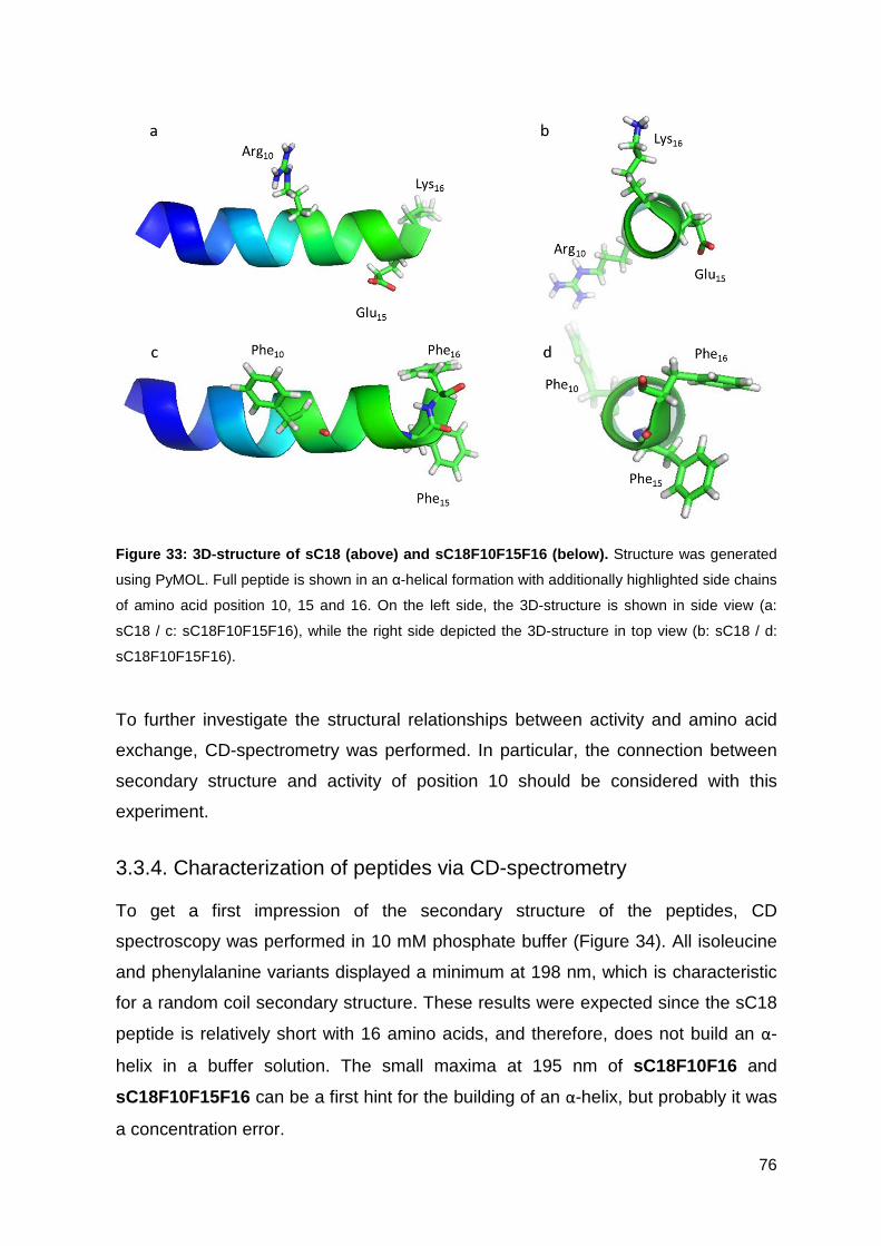
76
Figure 33: 3D-structure of sC18 (above) and sC18F10 F15F16 (below). Structure was generated
using PyMOL. Full peptide is shown in an α-helical formation with additionally highlighted side chains
of amino acid position 10, 15 and 16. On the left side, the 3D-structure is shown in side view (a:
sC18 / c: sC18F10F15F16), while the right side depicted the 3D-structure in top view (b: sC18 / d:
sC18F10F15F16).
To further investigate the structural relationships between activity and amino acid
exchange, CD-spectrometry was performed. In particular, the connection between
secondary structure and activity of position 10 should be considered with this
experiment.
3.3.4. Characterization of peptides via CD-spectrometry
To get a first impression of the secondary structure of the peptides, CD
spectroscopy was performed in 10 mM phosphate buffer (Figure 34). All isoleucine
and phenylalanine variants displayed a minimum at 198 nm, which is characteristic
for a random coil secondary structure. These results were expected since the sC18
peptide is relatively short with 16 amino acids, and therefore, does not build an α-
helix in a buffer solution. The small maxima at 195 nm of sC18F10F16 and
sC18F10F15F16 can be a first hint for the building of an α-helix, but probably it was
a concentration error.
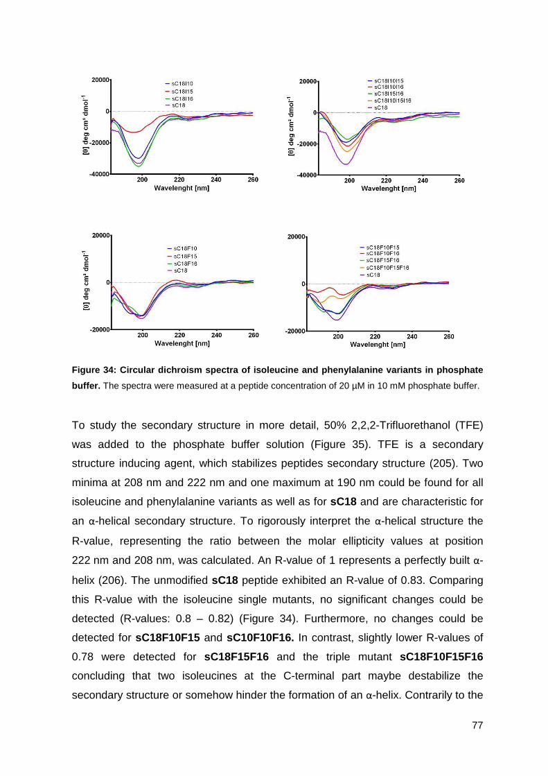
77
Figure 34: Circular dichroism spectra of isoleucine and phenylalanine variants in phosphate
buffer. The spectra were measured at a peptide concentration of 20 µM in 10 mM phosphate buffer.
To study the secondary structure in more detail, 50% 2,2,2-Trifluorethanol (TFE)
was added to the phosphate buffer solution (Figure 35). TFE is a secondary
structure inducing agent, which stabilizes peptides secondary structure (205). Two
minima at 208 nm and 222 nm and one maximum at 190 nm could be found for all
isoleucine and phenylalanine variants as well as for sC18 and are characteristic for
an α-helical secondary structure. To rigorously interpret the α-helical structure the
R-value, representing the ratio between the molar ellipticity values at position
222 nm and 208 nm, was calculated. An R-value of 1 represents a perfectly built α-
helix (206). The unmodified sC18 peptide exhibited an R-value of 0.83. Comparing
this R-value with the isoleucine single mutants, no significant changes could be
detected (R-values: 0.8 – 0.82) (Figure 34). Furthermore, no changes could be
detected for sC18F10F15 and sC10F10F16. In contrast, slightly lower R-values of
0.78 were detected for sC18F15F16 and the triple mutant sC18F10F15F16
concluding that two isoleucines at the C-terminal part maybe destabilize the
secondary structure or somehow hinder the formation of an α-helix. Contrarily to the
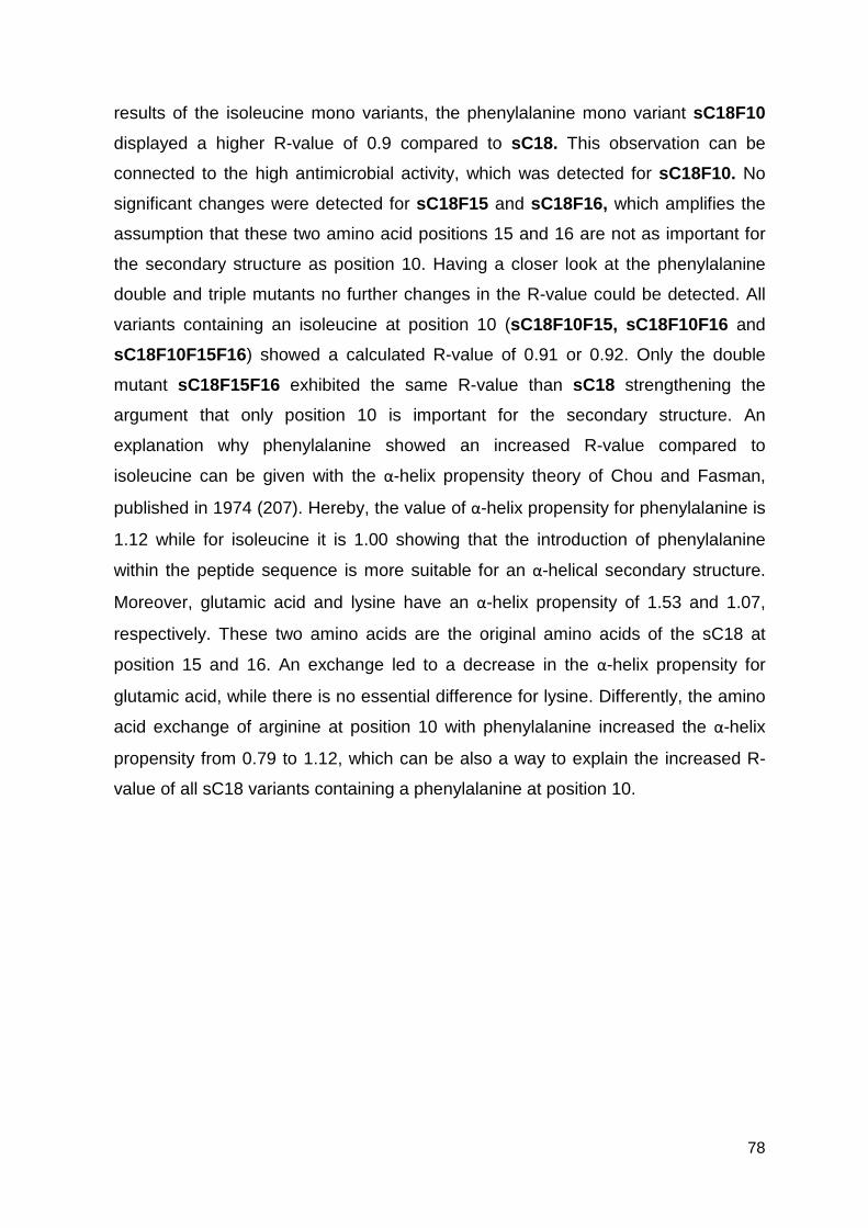
78
results of the isoleucine mono variants, the phenylalanine mono variant sC18F10
displayed a higher R-value of 0.9 compared to sC18. This observation can be
connected to the high antimicrobial activity, which was detected for sC18F10. No
significant changes were detected for sC18F15 and sC18F16, which amplifies the
assumption that these two amino acid positions 15 and 16 are not as important for
the secondary structure as position 10. Having a closer look at the phenylalanine
double and triple mutants no further changes in the R-value could be detected. All
variants containing an isoleucine at position 10 (sC18F10F15, sC18F10F16 and
sC18F10F15F16) showed a calculated R-value of 0.91 or 0.92. Only the double
mutant sC18F15F16 exhibited the same R-value than sC18 strengthening the
argument that only position 10 is important for the secondary structure. An
explanation why phenylalanine showed an increased R-value compared to
isoleucine can be given with the α-helix propensity theory of Chou and Fasman,
published in 1974 (207). Hereby, the value of α-helix propensity for phenylalanine is
1.12 while for isoleucine it is 1.00 showing that the introduction of phenylalanine
within the peptide sequence is more suitable for an α-helical secondary structure.
Moreover, glutamic acid and lysine have an α-helix propensity of 1.53 and 1.07,
respectively. These two amino acids are the original amino acids of the sC18 at
position 15 and 16. An exchange led to a decrease in the α-helix propensity for
glutamic acid, while there is no essential difference for lysine. Differently, the amino
acid exchange of arginine at position 10 with phenylalanine increased the α-helix
propensity from 0.79 to 1.12, which can be also a way to explain the increased R-
value of all sC18 variants containing a phenylalanine at position 10.
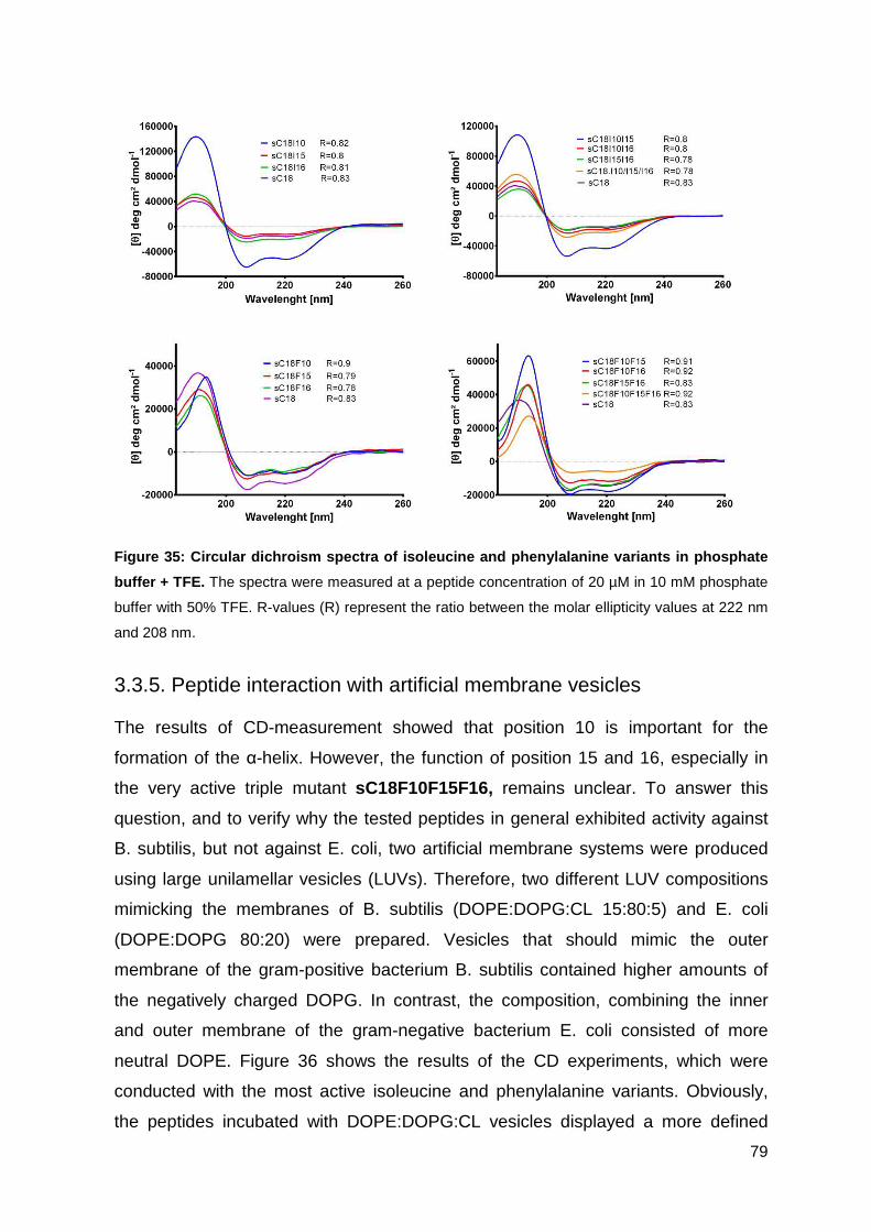
79
Figure 35: Circular dichroism spectra of isoleucine and phenylalanine variants in phosphate
buffer + TFE. The spectra were measured at a peptide concentration of 20 µM in 10 mM phosphate
buffer with 50% TFE. R-values (R) represent the ratio between the molar ellipticity values at 222 nm
and 208 nm.
3.3.5. Peptide interaction with artificial membrane vesicles
The results of CD-measurement showed that position 10 is important for the
formation of the α-helix. However, the function of position 15 and 16, especially in
the very active triple mutant sC18F10F15F16, remains unclear. To answer this
question, and to verify why the tested peptides in general exhibited activity against
B. subtilis, but not against E. coli, two artificial membrane systems were produced
using large unilamellar vesicles (LUVs). Therefore, two different LUV compositions
mimicking the membranes of B. subtilis (DOPE:DOPG:CL 15:80:5) and E. coli
(DOPE:DOPG 80:20) were prepared. Vesicles that should mimic the outer
membrane of the gram-positive bacterium B. subtilis contained higher amounts of
the negatively charged DOPG. In contrast, the composition, combining the inner
and outer membrane of the gram-negative bacterium E. coli consisted of more
neutral DOPE. Figure 36 shows the results of the CD experiments, which were
conducted with the most active isoleucine and phenylalanine variants. Obviously,
the peptides incubated with DOPE:DOPG:CL vesicles displayed a more defined
80
secondary structure when compared to vesicles mimicking the membrane of E. coli.
The two isoleucine variants sC18I10I15 and sC18I10I16 showed two minima at 208
nm and 220 nm and a maximum at 195 nm indicating an α-helical secondary
structure. Furthermore, also sC18F10F16 and sC18F10F15F16 exhibited structural
elements revealing an α-helix, although the structure was not as distinct as for the
isoleucine variants. This can be explained by the use of two different LUV stock
solutions that were used. In comparison, the structure of the four peptides incubated
with DOPE:DOPG vesicles seemed to be unstructured. While for the isoleucine
variants only a minimum at 222 nm was detected, phenylalanine variants show a
maximum at 200 nm. These structural differences in the presence of different
vesicles can be a hint why the peptides are more active against B. subtilis than
E. coli. Nevertheless, the structural elements do not seem to be the only important
factor for antimicrobial activity. Especially for sC18F10F15F16, there could exist a
combination of secondary structure, peptide stabilization and interaction with the
hydrophobic tails of the lipid bilayer leading to the highest antimicrobial activity of all
sC18 variants.
Figure 36: Circular dichroism spectra with artifici al membrane vesicles. LUVs containing
DOPE:DOPG:CL (15:80:5) and DOPE:DOPG (80:20) was prepared to imitate the membrane of
B. subtilis and E. coli, respectively. The spectra were measured at a peptide concentration of 20 µM
in 10 mM phosphate buffer with 1 mM LUVs. R-values (R) represent the ratio between the molar
ellipticity values at 222 nm and 208 nm.
Stella et al. already showed that the main difference between CPP and AMP activity
could be their affinities towards bacterial membranes (200). Especially a positive
charge is needed to bind more strongly to the anionic bilayer. Since the increased
81
activity in this work could not be explained with a higher positive charge, there seem
to be other characteristics. Concluding all results, two further characteristics can be
emphasized. On the one hand, position 10 stabilizes the α-helical structure and
maybe increases the hydrophobic interaction between the hydrophobic parts of two
AMPs during pore formation. On the other hand, position 15 and 16 interact with
each other, especially when exchanged with phenylalanine, and further interact as
first amino acids with the hydrophobic tails of the lipid molecules, resulting in a
higher affinity to the bacterial membrane. This model was already published in
literature for cytolytic peptides, describing that hydrophobic interaction also
increases the cytolytic activity (204). LUV experiments, imitating B. subtilis or E. coli
membranes, gave a further hint that the bacterial membrane composition is related
to peptide activity. This would explain why the sC18 variants exhibited different
activities against different bacteria. Nevertheless, this result cannot be generalized
for all gram-positive and gram-negative bacteria because every group has their
exceptions. For the gram-negative bacterium C.crescentus, the absence of
phosphatidylethanolamine (208) is as unusual as the high amount of
phosphatidylethanolamine in the gram-positive bacterium B. polymyxa (209). This
observation shows that also in the bacterial subgroups every bacterium differs in
their lipid composition making it very hard to treat all bacteria of one subgroup
equally.
To give an overview about the interaction of the sC18F10F15F16 peptide with the
membrane bilayer a mechanism of action model was proposed in Figure 37. In a
first step, the positive charged amino acids of sC18F10F15F16 get attached to the
negatively charged phospholipids (for example DOPG). Hereby, the positive peptide
net charge of +8 (Table 11) is very helpful for this interaction. In a second step, the
phenylalanine at positon 15 and 16 start an interaction with the hydrophobic lipid
tails leading to an insertion of the peptide into the membrane. Furthermore, an
interaction between the phenylalanines at position 10 of different peptides can be
hypothesized resulting in an α-helix stabilization (stripped black line in Figure 37).
This hypothesis can be reinforced, because all sC18 variants with a phenylalanine
at position 10 exhibited a better distinct α-helix (Figure 35). The last step is the
complete integration of more peptides building a pore, which then leads to a
destabilization of the membrane and an antimicrobial effect. The inside of the pore
is built by the hydrophilic sides of the peptides.
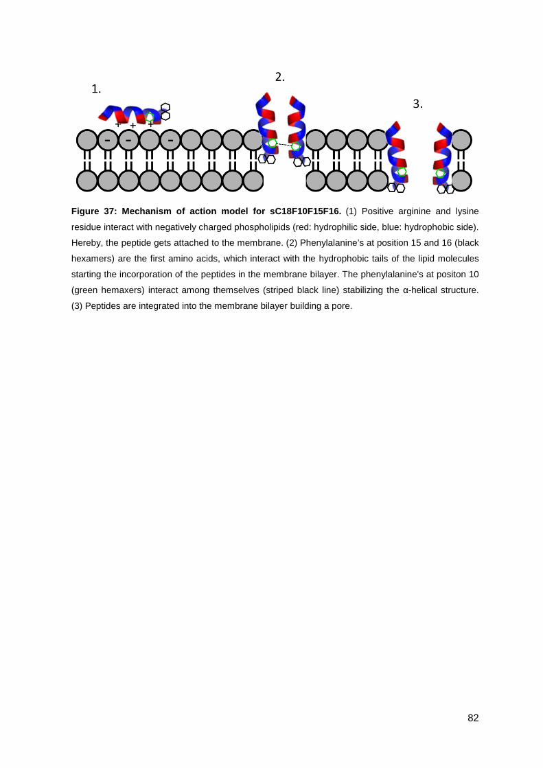
82
Figure 37: Mechanism of action model for sC18F10F15 F16. (1) Positive arginine and lysine
residue interact with negatively charged phospholipids (red: hydrophilic side, blue: hydrophobic side).
Hereby, the peptide gets attached to the membrane. (2) Phenylalanine’s at position 15 and 16 (black
hexamers) are the first amino acids, which interact with the hydrophobic tails of the lipid molecules
starting the incorporation of the peptides in the membrane bilayer. The phenylalanine's at positon 10
(green hemaxers) interact among themselves (striped black line) stabilizing the α-helical structure.
(3) Peptides are integrated into the membrane bilayer building a pore.
83
4. Conclusion and Outlook
Within this work, different strategies are described to increase the antimicrobial
activity of AMPs, e.g. conjugation to imidazolium salts, immobilization on
membranes, or amino acid exchanges.
In a first step, a branched imidazolium-salt compound was designed and
successfully conjugated to sC18 and LL-37. The combination of the antimicrobial
characteristics of AMPs and imidazolium-salts led to a highly increased
antimicrobial activity, especially against hard to medicate multi-drug resistant strains
like MRSA and VRE. The secondary structure of compound 3c was characterized
under very basic and acidic conditions. No changes in the secondary structure were
detected, showing that the peptide is stable under these extreme conditions. To use
the compound as future antibiotic, its stability against proteases and human blood
plasma should be investigated. Another important attribute of antibiotics is their
selectivity. 3b and 3c exhibited very strong antimicrobial activity, but were only less
selective against human red blood cells, while on the other hand 3a showed better
selectivity. Furthermore, the influence of cholesterol as a membrane compound was
investigated, since only eukaryotic cell contain cholesterol. Compounds 3b and 3c
were less active against vesicles containing cholesterol, concluding that the
compounds also show selectivity towards eukaryotic cells. In conclusion, this work
demonstrated for the first time that imidazolium salts conjugated to antimicrobial
peptides led to highly active compounds, especially against resistant bacterial
strains. Since compound 3a execute highest selectivity, this compound could be a
future lead structure for new antibiotic agents.
In a second step, the novel designed compound 3a was immobilized on a polyether
sulfone membrane using different linkers (compounds 3a, 3d, 3e and 3f) to achieve
a surface with high antimicrobial activity. The immobilization with electron beam
radiation and chemical coupling was very successful for all compounds. To increase
the activity range, further polyethylene glycol (PEG) chains could be coupled to the
C-terminus, which also should lead to a better flexibility. In general, an introduction
of a hydrophobic linker seemed to be more effective for a successful electron beam
radiation. Anyway, the electron radiation is a technique without the use of catalyst or
toxic reagents, but has the problem that the integration position of the compound is
not completely understood yet (210). Consequently, the chemical coupling method
84
seems to be more attractive for the immobilization of peptide compounds since the
peptide is arranged at the surface of the membrane via a peptide bond and not
integrated into the membrane. Conclusively, both immobilization methods are easy
and powerful to coat antimicrobial compounds on polyether sulfone membranes
retaining their antimicrobial effect.
Finally, the thin line between cell-penetrating peptides and antimicrobial peptides
was investigated modulating the CPP sC18 to an antimicrobial peptide via amino
acid exchanges. The sC18 peptide already consists of a lot of positively charged
amino acids, whereas the focus had been on increasing the hydrophobic face of
sC18 including phenylalanine and isoleucine. The antimicrobial activity already was
increased with one amino acid substitution and showed the highest effect for
sC18F10F15F16. The hydrophobic face seemed to be as important for antimicrobial
activity as the cationic charge. Position 15 showed the weakest effects since the
hydrophobic amino acid was incorporated in the hydrophilic face of the α-helix.
Therefore, a substitution of glutamic acid at position 15 with arginine or lysine
should enhance the antimicrobial activity further. Therefore, in a next step of
experiments, the selectivity of the novel sC18 variants should be determined to
verify if the lytic activity against human cell lines or human red blood cells also
increases through the different amino acid exchanges. Moreover, bacteria with high
phosphatidylethanolamine content seem to be more resistant against the treatment
with antimicrobial peptides since the role of phosphatidylethanolamine is to spread
out the negatively charged phospholipids and reduce the negatively overall
membrane charge (177).
Recently, the appearance of more and more multi-resistant strains has forced the
development of novel antimicrobial compounds. However, the use of AMPs as
clinical application is still far away because of their low specificity and their toxic
side effects. In this work, it could be shown that small modifications of the peptide
sequence can lead to characteristic changes, but predicting these changes remains
challenging. To use AMPs as antibiotics, the mechanism of action should be better
understood. However, especially their unique membrane targeting mechanism
makes AMPs to a benefit in public health in the near future.
85
5. Literature
(1) Morar, M., and Wright, G. D. (2010) The Genomic Enzymology of Antibiotic Resistance.
Annu Rev Genet 44, 25-51.
(2) Marr, A. K., Gooderham, W. J., and Hancock, R. E. W. (2006) Antibacterial peptides for
therapeutic use: obstacles and realistic outlook. Curr Opin Pharmacol 6, 468-472.
(3) Mygind, P. H., Fischer, R. L., Schnorr, K. M., Hansen, M. T., Sonksen, C. P., Ludvigsen, S.,
Raventos, D., Buskov, S., Christensen, B., De Maria, L., Taboureau, O., Yaver, D., Elvig-
Jorgensen, S. G., Sorensen, M. V., Christensen, B. E., Kjaerulff, S., Frimodt-Moller, N.,
Lehrer, R. I., Zasloff, M., and Kristensen, H. H. (2005) Plectasin is a peptide antibiotic with
therapeutic potential from a saprophytic fungus. Nature 437, 975-980.
(4) van 't Hof, W., Veerman, E. C. I., Helmerhorst, E. J., and Amerongen, A. V. N. (2001)
Antimicrobial peptides: Properties and applicability. Biol Chem 382, 597-619.
(5) Davies, S. C., Fowler, T., Watson, J., Livermore, D. M., and Walker, D. (2013) Annual
Report of the Chief Medical Officer: infection and the rise of antimicrobial resistance.
Lancet 381, 1606-1609.
(6) Davies, J., and Davies, D. (2010) Origins and Evolution of Antibiotic Resistance. Microbiol
Mol Biol R 74, 417-+.
(7) Enright, M. C., Robinson, D. A., Randle, G., Feil, E. J., Grundmann, H., and Spratt, B. G.
(2002) The evolutionary history of methicillin-resistant Staphylococcus aureus (MRSA). P
Natl Acad Sci USA 99, 7687-7692.
(8) Martinez, J. L., and Baquero, F. (2002) Interactions among strategies associated with
bacterial infection: Pathogenicity, epidemicity, and antibiotic resistance. Clin Microbiol Rev
15, 647-+.
(9) Dzidic, S., Suskovic, J., and Kos, B. (2008) Antibiotic resistance mechanisms in bacteria:
Biochemical and genetic aspects. Food Technol Biotech 46, 11-21.
(10) Russell, A. D. (1986) Mechanisms of Bacterial-Resistance to Antibacterial Agents. Pharm Int
7, 300-305.
(11) Raghunath, D. (2008) Emerging antibiotic resistance in bacteria with special reference to
India. Journal of biosciences 33, 593-603.
(12) Hickey, S. M., and Nelson, J. D. (1997) Mechanisms of antibacterial resistance. Advances in
pediatrics 44, 1-41.
(13) Wright, G. D. (2005) Bacterial resistance to antibiotics: enzymatic degradation and
modification. Advanced drug delivery reviews 57, 1451-70.
(14) Thomson, J. M., and Bonomo, R. A. (2005) The threat of antibiotic resistance in Gram-
negative pathogenic bacteria: beta-lactams in peril! Curr Opin Microbiol 8, 518-524.
(15) Strateva, T., and Yordanov, D. (2009) Pseudomonas aeruginosa - a phenomenon of
bacterial resistance. J Med Microbiol 58, 1133-1148.
(16) Maurice, F., Broutin, I., Podglajen, I., Benas, P., Collatz, E., and Dardel, F. (2008) Enzyme
structural plasticity and the emergence of broad-spectrum antibiotic resistance. Embo Rep
9, 344-349.
(17) Depardieu, F., Podglajen, I., Leclercq, R., Collatz, E., and Courvalin, P. (2007) Modes and
modulations of antibiotic resistance gene expression. Clin Microbiol Rev 20, 79-114.
(18) Livermore, D. M. (1991) Mechanisms of Resistance to Beta-Lactam Antibiotics. Scand J
Infect Dis, 7-16.
(19) Mingeot-Leclercq, M. P., Glupczynski, Y., and Tulkens, P. M. (1999) Aminoglycosides:
Activity and resistance. Antimicrob Agents Ch 43, 727-737.
(20) Gutmann, L., Alobeid, S., Billotklein, D., Guerrier, M. L., and Collatz, E. (1994) Synergy and
Resistance to Synergy between Beta-Lactam Antibiotics and Glycopeptides against
Glycopeptide-Resistant Strains of Enterococcus-Faecium. Antimicrob Agents Ch 38, 824-
829.
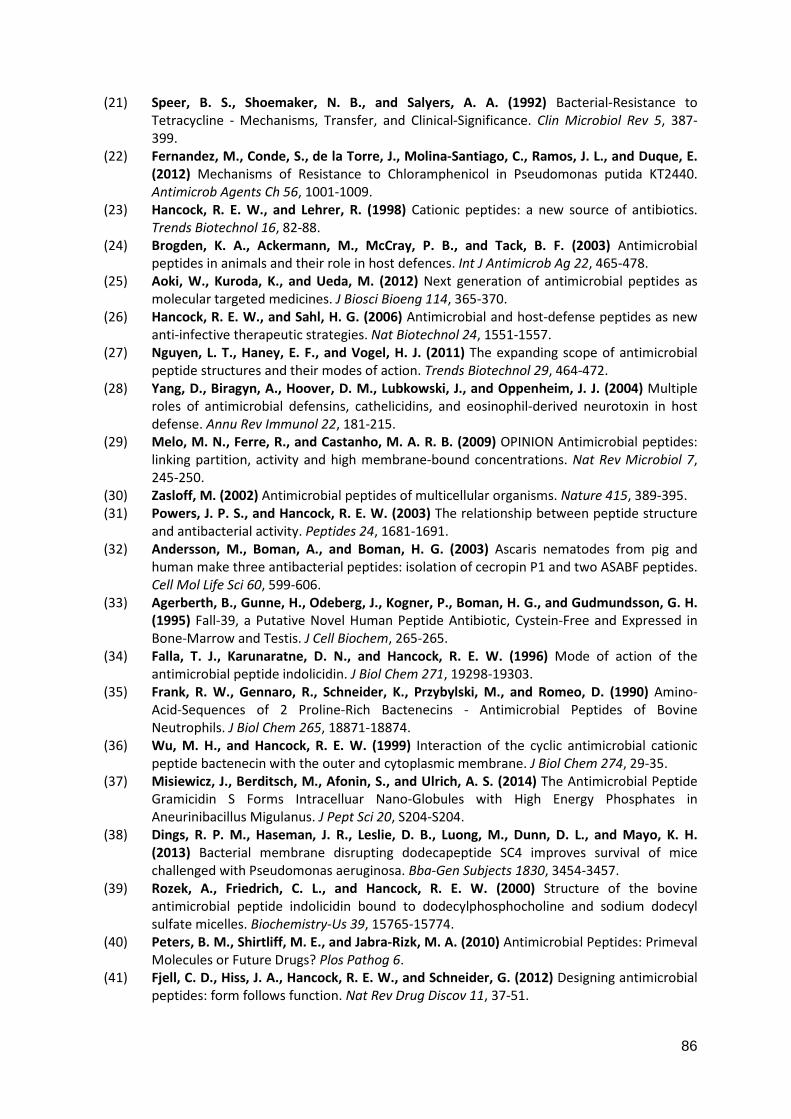
86
(21) Speer, B. S., Shoemaker, N. B., and Salyers, A. A. (1992) Bacterial-Resistance to
Tetracycline - Mechanisms, Transfer, and Clinical-Significance. Clin Microbiol Rev 5, 387-
399.
(22) Fernandez, M., Conde, S., de la Torre, J., Molina-Santiago, C., Ramos, J. L., and Duque, E.
(2012) Mechanisms of Resistance to Chloramphenicol in Pseudomonas putida KT2440.
Antimicrob Agents Ch 56, 1001-1009.
(23) Hancock, R. E. W., and Lehrer, R. (1998) Cationic peptides: a new source of antibiotics.
Trends Biotechnol 16, 82-88.
(24) Brogden, K. A., Ackermann, M., McCray, P. B., and Tack, B. F. (2003) Antimicrobial
peptides in animals and their role in host defences. Int J Antimicrob Ag 22, 465-478.
(25) Aoki, W., Kuroda, K., and Ueda, M. (2012) Next generation of antimicrobial peptides as
molecular targeted medicines. J Biosci Bioeng 114, 365-370.
(26) Hancock, R. E. W., and Sahl, H. G. (2006) Antimicrobial and host-defense peptides as new
anti-infective therapeutic strategies. Nat Biotechnol 24, 1551-1557.
(27) Nguyen, L. T., Haney, E. F., and Vogel, H. J. (2011) The expanding scope of antimicrobial
peptide structures and their modes of action. Trends Biotechnol 29, 464-472.
(28) Yang, D., Biragyn, A., Hoover, D. M., Lubkowski, J., and Oppenheim, J. J. (2004) Multiple
roles of antimicrobial defensins, cathelicidins, and eosinophil-derived neurotoxin in host
defense. Annu Rev Immunol 22, 181-215.
(29) Melo, M. N., Ferre, R., and Castanho, M. A. R. B. (2009) OPINION Antimicrobial peptides:
linking partition, activity and high membrane-bound concentrations. Nat Rev Microbiol 7,
245-250.
(30) Zasloff, M. (2002) Antimicrobial peptides of multicellular organisms. Nature 415, 389-395.
(31) Powers, J. P. S., and Hancock, R. E. W. (2003) The relationship between peptide structure
and antibacterial activity. Peptides 24, 1681-1691.
(32) Andersson, M., Boman, A., and Boman, H. G. (2003) Ascaris nematodes from pig and
human make three antibacterial peptides: isolation of cecropin P1 and two ASABF peptides.
Cell Mol Life Sci 60, 599-606.
(33) Agerberth, B., Gunne, H., Odeberg, J., Kogner, P., Boman, H. G., and Gudmundsson, G. H.
(1995) Fall-39, a Putative Novel Human Peptide Antibiotic, Cystein-Free and Expressed in
Bone-Marrow and Testis. J Cell Biochem, 265-265.
(34) Falla, T. J., Karunaratne, D. N., and Hancock, R. E. W. (1996) Mode of action of the
antimicrobial peptide indolicidin. J Biol Chem 271, 19298-19303.
(35) Frank, R. W., Gennaro, R., Schneider, K., Przybylski, M., and Romeo, D. (1990) Amino-
Acid-Sequences of 2 Proline-Rich Bactenecins - Antimicrobial Peptides of Bovine
Neutrophils. J Biol Chem 265, 18871-18874.
(36) Wu, M. H., and Hancock, R. E. W. (1999) Interaction of the cyclic antimicrobial cationic
peptide bactenecin with the outer and cytoplasmic membrane. J Biol Chem 274, 29-35.
(37) Misiewicz, J., Berditsch, M., Afonin, S., and Ulrich, A. S. (2014) The Antimicrobial Peptide
Gramicidin S Forms Intracelluar Nano-Globules with High Energy Phosphates in
Aneurinibacillus Migulanus. J Pept Sci 20, S204-S204.
(38) Dings, R. P. M., Haseman, J. R., Leslie, D. B., Luong, M., Dunn, D. L., and Mayo, K. H.
(2013) Bacterial membrane disrupting dodecapeptide SC4 improves survival of mice
challenged with Pseudomonas aeruginosa. Bba-Gen Subjects 1830, 3454-3457.
(39) Rozek, A., Friedrich, C. L., and Hancock, R. E. W. (2000) Structure of the bovine
antimicrobial peptide indolicidin bound to dodecylphosphocholine and sodium dodecyl
sulfate micelles. Biochemistry-Us 39, 15765-15774.
(40) Peters, B. M., Shirtliff, M. E., and Jabra-Rizk, M. A. (2010) Antimicrobial Peptides: Primeval
Molecules or Future Drugs? Plos Pathog 6.
(41) Fjell, C. D., Hiss, J. A., Hancock, R. E. W., and Schneider, G. (2012) Designing antimicrobial
peptides: form follows function. Nat Rev Drug Discov 11, 37-51.
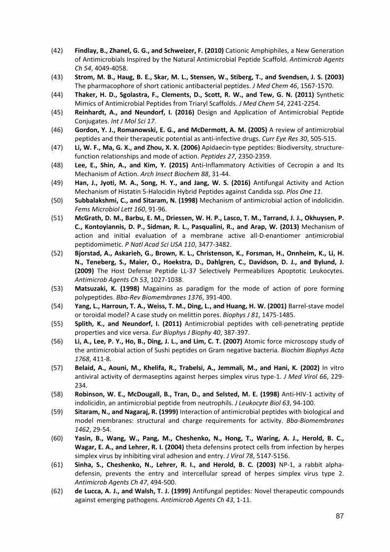
87
(42) Findlay, B., Zhanel, G. G., and Schweizer, F. (2010) Cationic Amphiphiles, a New Generation
of Antimicrobials Inspired by the Natural Antimicrobial Peptide Scaffold. Antimicrob Agents
Ch 54, 4049-4058.
(43) Strom, M. B., Haug, B. E., Skar, M. L., Stensen, W., Stiberg, T., and Svendsen, J. S. (2003)
The pharmacophore of short cationic antibacterial peptides. J Med Chem 46, 1567-1570.
(44) Thaker, H. D., Sgolastra, F., Clements, D., Scott, R. W., and Tew, G. N. (2011) Synthetic
Mimics of Antimicrobial Peptides from Triaryl Scaffolds. J Med Chem 54, 2241-2254.
(45) Reinhardt, A., and Neundorf, I. (2016) Design and Application of Antimicrobial Peptide
Conjugates. Int J Mol Sci 17.
(46) Gordon, Y. J., Romanowski, E. G., and McDermott, A. M. (2005) A review of antimicrobial
peptides and their therapeutic potential as anti-infective drugs. Curr Eye Res 30, 505-515.
(47) Li, W. F., Ma, G. X., and Zhou, X. X. (2006) Apidaecin-type peptides: Biodiversity, structure-
function relationships and mode of action. Peptides 27, 2350-2359.
(48) Lee, E., Shin, A., and Kim, Y. (2015) Anti-Inflammatory Activities of Cecropin a and Its
Mechanism of Action. Arch Insect Biochem 88, 31-44.
(49) Han, J., Jyoti, M. A., Song, H. Y., and Jang, W. S. (2016) Antifungal Activity and Action
Mechanism of Histatin 5-Halocidin Hybrid Peptides against Candida ssp. Plos One 11.
(50) Subbalakshmi, C., and Sitaram, N. (1998) Mechanism of antimicrobial action of indolicidin.
Fems Microbiol Lett 160, 91-96.
(51) McGrath, D. M., Barbu, E. M., Driessen, W. H. P., Lasco, T. M., Tarrand, J. J., Okhuysen, P.
C., Kontoyiannis, D. P., Sidman, R. L., Pasqualini, R., and Arap, W. (2013) Mechanism of
action and initial evaluation of a membrane active all-D-enantiomer antimicrobial
peptidomimetic. P Natl Acad Sci USA 110, 3477-3482.
(52) Bjorstad, A., Askarieh, G., Brown, K. L., Christenson, K., Forsman, H., Onnheim, K., Li, H.
N., Teneberg, S., Maier, O., Hoekstra, D., Dahlgren, C., Davidson, D. J., and Bylund, J.
(2009) The Host Defense Peptide LL-37 Selectively Permeabilizes Apoptotic Leukocytes.
Antimicrob Agents Ch 53, 1027-1038.
(53) Matsuzaki, K. (1998) Magainins as paradigm for the mode of action of pore forming
polypeptides. Bba-Rev Biomembranes 1376, 391-400.
(54) Yang, L., Harroun, T. A., Weiss, T. M., Ding, L., and Huang, H. W. (2001) Barrel-stave model
or toroidal model? A case study on melittin pores. Biophys J 81, 1475-1485.
(55) Splith, K., and Neundorf, I. (2011) Antimicrobial peptides with cell-penetrating peptide
properties and vice versa. Eur Biophys J Biophy 40, 387-397.
(56) Li, A., Lee, P. Y., Ho, B., Ding, J. L., and Lim, C. T. (2007) Atomic force microscopy study of
the antimicrobial action of Sushi peptides on Gram negative bacteria. Biochim Biophys Acta
1768, 411-8.
(57) Belaid, A., Aouni, M., Khelifa, R., Trabelsi, A., Jemmali, M., and Hani, K. (2002) In vitro
antiviral activity of dermaseptins against herpes simplex virus type-1. J Med Virol 66, 229-
234.
(58) Robinson, W. E., McDougall, B., Tran, D., and Selsted, M. E. (1998) Anti-HIV-1 activity of
indolicidin, an antimicrobial peptide from neutrophils. J Leukocyte Biol 63, 94-100.
(59) Sitaram, N., and Nagaraj, R. (1999) Interaction of antimicrobial peptides with biological and
model membranes: structural and charge requirements for activity. Bba-Biomembranes
1462, 29-54.
(60) Yasin, B., Wang, W., Pang, M., Cheshenko, N., Hong, T., Waring, A. J., Herold, B. C.,
Wagar, E. A., and Lehrer, R. I. (2004) theta defensins protect cells from infection by herpes
simplex virus by inhibiting viral adhesion and entry. J Virol 78, 5147-5156.
(61) Sinha, S., Cheshenko, N., Lehrer, R. I., and Herold, B. C. (2003) NP-1, a rabbit alpha-
defensin, prevents the entry and intercellular spread of herpes simplex virus type 2.
Antimicrob Agents Ch 47, 494-500.
(62) de Lucca, A. J., and Walsh, T. J. (1999) Antifungal peptides: Novel therapeutic compounds
against emerging pathogens. Antimicrob Agents Ch 43, 1-11.
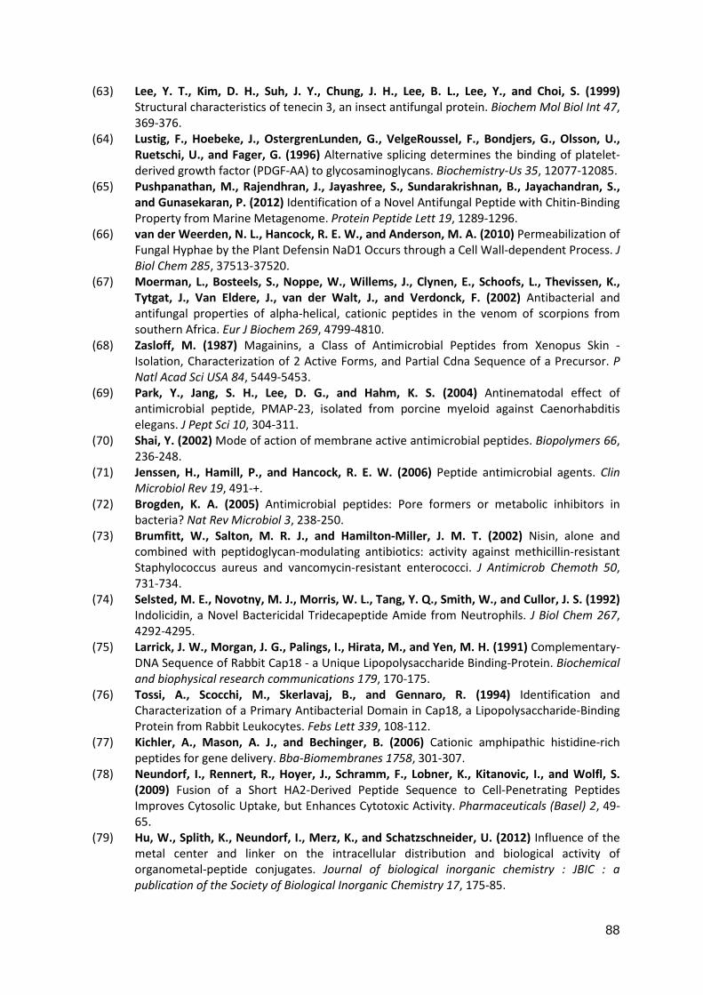
88
(63) Lee, Y. T., Kim, D. H., Suh, J. Y., Chung, J. H., Lee, B. L., Lee, Y., and Choi, S. (1999)
Structural characteristics of tenecin 3, an insect antifungal protein. Biochem Mol Biol Int 47,
369-376.
(64) Lustig, F., Hoebeke, J., OstergrenLunden, G., VelgeRoussel, F., Bondjers, G., Olsson, U.,
Ruetschi, U., and Fager, G. (1996) Alternative splicing determines the binding of platelet-
derived growth factor (PDGF-AA) to glycosaminoglycans. Biochemistry-Us 35, 12077-12085.
(65) Pushpanathan, M., Rajendhran, J., Jayashree, S., Sundarakrishnan, B., Jayachandran, S.,
and Gunasekaran, P. (2012) Identification of a Novel Antifungal Peptide with Chitin-Binding
Property from Marine Metagenome. Protein Peptide Lett 19, 1289-1296.
(66) van der Weerden, N. L., Hancock, R. E. W., and Anderson, M. A. (2010) Permeabilization of
Fungal Hyphae by the Plant Defensin NaD1 Occurs through a Cell Wall-dependent Process. J
Biol Chem 285, 37513-37520.
(67) Moerman, L., Bosteels, S., Noppe, W., Willems, J., Clynen, E., Schoofs, L., Thevissen, K.,
Tytgat, J., Van Eldere, J., van der Walt, J., and Verdonck, F. (2002) Antibacterial and
antifungal properties of alpha-helical, cationic peptides in the venom of scorpions from
southern Africa. Eur J Biochem 269, 4799-4810.
(68) Zasloff, M. (1987) Magainins, a Class of Antimicrobial Peptides from Xenopus Skin -
Isolation, Characterization of 2 Active Forms, and Partial Cdna Sequence of a Precursor. P
Natl Acad Sci USA 84, 5449-5453.
(69) Park, Y., Jang, S. H., Lee, D. G., and Hahm, K. S. (2004) Antinematodal effect of
antimicrobial peptide, PMAP-23, isolated from porcine myeloid against Caenorhabditis
elegans. J Pept Sci 10, 304-311.
(70) Shai, Y. (2002) Mode of action of membrane active antimicrobial peptides. Biopolymers 66,
236-248.
(71) Jenssen, H., Hamill, P., and Hancock, R. E. W. (2006) Peptide antimicrobial agents. Clin
Microbiol Rev 19, 491-+.
(72) Brogden, K. A. (2005) Antimicrobial peptides: Pore formers or metabolic inhibitors in
bacteria? Nat Rev Microbiol 3, 238-250.
(73) Brumfitt, W., Salton, M. R. J., and Hamilton-Miller, J. M. T. (2002) Nisin, alone and
combined with peptidoglycan-modulating antibiotics: activity against methicillin-resistant
Staphylococcus aureus and vancomycin-resistant enterococci. J Antimicrob Chemoth 50,
731-734.
(74) Selsted, M. E., Novotny, M. J., Morris, W. L., Tang, Y. Q., Smith, W., and Cullor, J. S. (1992)
Indolicidin, a Novel Bactericidal Tridecapeptide Amide from Neutrophils. J Biol Chem 267,
4292-4295.
(75) Larrick, J. W., Morgan, J. G., Palings, I., Hirata, M., and Yen, M. H. (1991) Complementary-
DNA Sequence of Rabbit Cap18 - a Unique Lipopolysaccharide Binding-Protein. Biochemical
and biophysical research communications 179, 170-175.
(76) Tossi, A., Scocchi, M., Skerlavaj, B., and Gennaro, R. (1994) Identification and
Characterization of a Primary Antibacterial Domain in Cap18, a Lipopolysaccharide-Binding
Protein from Rabbit Leukocytes. Febs Lett 339, 108-112.
(77) Kichler, A., Mason, A. J., and Bechinger, B. (2006) Cationic amphipathic histidine-rich
peptides for gene delivery. Bba-Biomembranes 1758, 301-307.
(78) Neundorf, I., Rennert, R., Hoyer, J., Schramm, F., Lobner, K., Kitanovic, I., and Wolfl, S.
(2009) Fusion of a Short HA2-Derived Peptide Sequence to Cell-Penetrating Peptides
Improves Cytosolic Uptake, but Enhances Cytotoxic Activity. Pharmaceuticals (Basel) 2, 49-
65.
(79) Hu, W., Splith, K., Neundorf, I., Merz, K., and Schatzschneider, U. (2012) Influence of the
metal center and linker on the intracellular distribution and biological activity of
organometal-peptide conjugates. Journal of biological inorganic chemistry : JBIC : a
publication of the Society of Biological Inorganic Chemistry 17, 175-85.
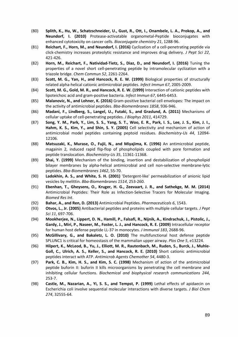
89
(80) Splith, K., Hu, W., Schatzschneider, U., Gust, R., Ott, I., Onambele, L. A., Prokop, A., and
Neundorf, I. (2010) Protease-activatable organometal-Peptide bioconjugates with
enhanced cytotoxicity on cancer cells. Bioconjugate chemistry 21, 1288-96.
(81) Reichart, F., Horn, M., and Neundorf, I. (2016) Cyclization of a cell-penetrating peptide via
click-chemistry increases proteolytic resistance and improves drug delivery. J Pept Sci 22,
421-426.
(82) Horn, M., Reichart, F., Natividad-Tietz, S., Diaz, D., and Neundorf, I. (2016) Tuning the
properties of a novel short cell-penetrating peptide by intramolecular cyclization with a
triazole bridge. Chem Commun 52, 2261-2264.
(83) Scott, M. G., Yan, H., and Hancock, R. E. W. (1999) Biological properties of structurally
related alpha-helical cationic antimicrobial peptides. Infect Immun 67, 2005-2009.
(84) Scott, M. G., Gold, M. R., and Hancock, R. E. W. (1999) Interaction of cationic peptides with
lipoteichoic acid and gram-positive bacteria. Infect Immun 67, 6445-6453.
(85) Malanovic, N., and Lohner, K. (2016) Gram-positive bacterial cell envelopes: The impact on
the activity of antimicrobial peptides. Bba-Biomembranes 1858, 936-946.
(86) Madani, F., Lindberg, S., Langel, U., Futaki, S., and Graslund, A. (2011) Mechanisms of
cellular uptake of cell-penetrating peptides. J Biophys 2011, 414729.
(87) Song, Y. M., Park, Y., Lim, S. S., Yang, S. T., Woo, E. R., Park, I. S., Lee, J. S., Kim, J. I.,
Hahm, K. S., Kim, Y., and Shin, S. Y. (2005) Cell selectivity and mechanism of action of
antimicrobial model peptides containing peptoid residues. Biochemistry-Us 44, 12094-
12106.
(88) Matsuzaki, K., Murase, O., Fujii, N., and Miyajima, K. (1996) An antimicrobial peptide,
magainin 2, induced rapid flip-flop of phospholipids coupled with pore formation and
peptide translocation. Biochemistry-Us 35, 11361-11368.
(89) Shai, Y. (1999) Mechanism of the binding, insertion and destabilization of phospholipid
bilayer membranes by alpha-helical antimicrobial and cell non-selective membrane-lytic
peptides. Bba-Biomembranes 1462, 55-70.
(90) Ladokhin, A. S., and White, S. H. (2001) 'Detergent-like' permeabilization of anionic lipid
vesicles by melittin. Bba-Biomembranes 1514, 253-260.
(91) Ebenhan, T., Gheysens, O., Kruger, H. G., Zeevaart, J. R., and Sathekge, M. M. (2014)
Antimicrobial Peptides: Their Role as Infection-Selective Tracers for Molecular Imaging.
Biomed Res Int.
(92) Bahar, A., and Ren, D. (2013) Antimicrobial Peptides. Pharmaceuticals 6, 1543.
(93) Otvos, L., Jr. (2005) Antibacterial peptides and proteins with multiple cellular targets. J Pept
Sci 11, 697-706.
(94) Mookherjee, N., Lippert, D. N., Hamill, P., Falsafi, R., Nijnik, A., Kindrachuk, J., Pistolic, J.,
Gardy, J., Miri, P., Naseer, M., Foster, L. J., and Hancock, R. E. (2009) Intracellular receptor
for human host defense peptide LL-37 in monocytes. J Immunol 183, 2688-96.
(95) McGillivary, G., and Bakaletz, L. O. (2010) The multifunctional host defense peptide
SPLUNC1 is critical for homeostasis of the mammalian upper airway. Plos One 5, e13224.
(96) Hilpert, K., McLeod, B., Yu, J., Elliott, M. R., Rautenbach, M., Ruden, S., Burck, J., Muhle-
Goll, C., Ulrich, A. S., Keller, S., and Hancock, R. E. (2010) Short cationic antimicrobial
peptides interact with ATP. Antimicrob Agents Chemother 54, 4480-3.
(97) Park, C. B., Kim, H. S., and Kim, S. C. (1998) Mechanism of action of the antimicrobial
peptide buforin II: buforin II kills microorganisms by penetrating the cell membrane and
inhibiting cellular functions. Biochemical and biophysical research communications 244,
253-7.
(98) Castle, M., Nazarian, A., Yi, S. S., and Tempst, P. (1999) Lethal effects of apidaecin on
Escherichia coli involve sequential molecular interactions with diverse targets. J Biol Chem
274, 32555-64.
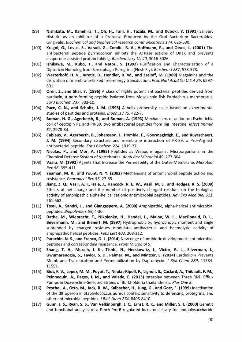
90
(99) Nishikata, M., Kanehira, T., Oh, H., Tani, H., Tazaki, M., and Kuboki, Y. (1991) Salivary
Histatin as an Inhibitor of a Protease Produced by the Oral Bacterium Bacteroides-
Gingivalis. Biochemical and biophysical research communications 174, 625-630.
(100) Kragol, G., Lovas, S., Varadi, G., Condie, B. A., Hoffmann, R., and Otvos, L. (2001) The
antibacterial peptide pyrrhocoricin inhibits the ATPase actions of DnaK and prevents
chaperone-assisted protein folding. Biochemistry-Us 40, 3016-3026.
(101) Ishikawa, M., Kubo, T., and Natori, S. (1992) Purification and Characterization of a
Diptericin Homolog from Sarcophaga-Peregrina (Flesh Fly). Biochem J 287, 573-578.
(102) Westerhoff, H. V., Juretic, D., Hendler, R. W., and Zasloff, M. (1989) Magainins and the
disruption of membrane-linked free-energy transduction. Proc Natl Acad Sci U S A 86, 6597-
601.
(103) Oren, Z., and Shai, Y. (1996) A class of highly potent antibacterial peptides derived from
pardaxin, a pore-forming peptide isolated from Moses sole fish Pardachirus marmoratus.
Eur J Biochem 237, 303-10.
(104) Pace, C. N., and Scholtz, J. M. (1998) A helix propensity scale based on experimental
studies of peptides and proteins. Biophys J 75, 422-7.
(105) Boman, H. G., Agerberth, B., and Boman, A. (1993) Mechanisms of action on Escherichia
coli of cecropin P1 and PR-39, two antibacterial peptides from pig intestine. Infect Immun
61, 2978-84.
(106) Cabiaux, V., Agerberth, B., Johansson, J., Homble, F., Goormaghtigh, E., and Ruysschaert,
J. M. (1994) Secondary structure and membrane interaction of PR-39, a Pro+Arg-rich
antibacterial peptide. Eur J Biochem 224, 1019-27.
(107) Nicolas, P., and Mor, A. (1995) Peptides as Weapons against Microorganisms in the
Chemical Defense System of Vertebrates. Annu Rev Microbiol 49, 277-304.
(108) Vaara, M. (1992) Agents That Increase the Permeability of the Outer-Membrane. Microbiol
Rev 56, 395-411.
(109) Yeaman, M. R., and Yount, N. Y. (2003) Mechanisms of antimicrobial peptide action and
resistance. Pharmacol Rev 55, 27-55.
(110) Jiang, Z. Q., Vasil, A. I., Hale, J., Hancock, R. E. W., Vasil, M. L., and Hodges, R. S. (2009)
Effects of net charge and the number of positively charged residues on the biological
activity of amphipathic alpha-helical cationic antimicrobial peptides. Adv Exp Med Biol 611,
561-562.
(111) Tossi, A., Sandri, L., and Giangaspero, A. (2000) Amphipathic, alpha-helical antimicrobial
peptides. Biopolymers 55, 4-30.
(112) Dathe, M., Wieprecht, T., Nikolenko, H., Handel, L., Maloy, W. L., MacDonald, D. L.,
Beyermann, M., and Bienert, M. (1997) Hydrophobicity, hydrophobic moment and angle
subtended by charged residues modulate antibacterial and haemolytic activity of
amphipathic helical peptides. Febs Lett 403, 208-212.
(113) Parachin, N. S., and Franco, O. L. (2014) New edge of antibiotic development: antimicrobial
peptides and corresponding resistance. Front Microbiol 5.
(114) Zhang, T. H., Muraih, J. K., Tishbi, N., Herskowitz, J., Victor, R. L., Silverman, J.,
Uwumarenogie, S., Taylor, S. D., Palmer, M., and Mintzer, E. (2014) Cardiolipin Prevents
Membrane Translocation and Permeabilization by Daptomycin. J Biol Chem 289, 11584-
11591.
(115) Biot, F. V., Lopez, M. M., Poyot, T., Neulat-Ripoll, F., Lignon, S., Caclard, A., Thibault, F. M.,
Peinnequin, A., Pages, J. M., and Valade, E. (2013) Interplay between Three RND Efflux
Pumps in Doxycycline-Selected Strains of Burkholderia thailandensis. Plos One 8.
(116) Peschel, A., Otto, M., Jack, R. W., Kalbacher, H., Jung, G., and Gotz, F. (1999) Inactivation
of the dlt operon in Staphylococcus aureus confers sensitivity to defensins, protegrins, and
other antimicrobial peptides. J Biol Chem 274, 8405-8410.
(117) Gunn, J. S., Ryan, S. S., Van Velkinburgh, J. C., Ernst, R. K., and Miller, S. I. (2000) Genetic
and functional analysis of a PmrA-PmrB-regulated locus necessary for lipopolysaccharide
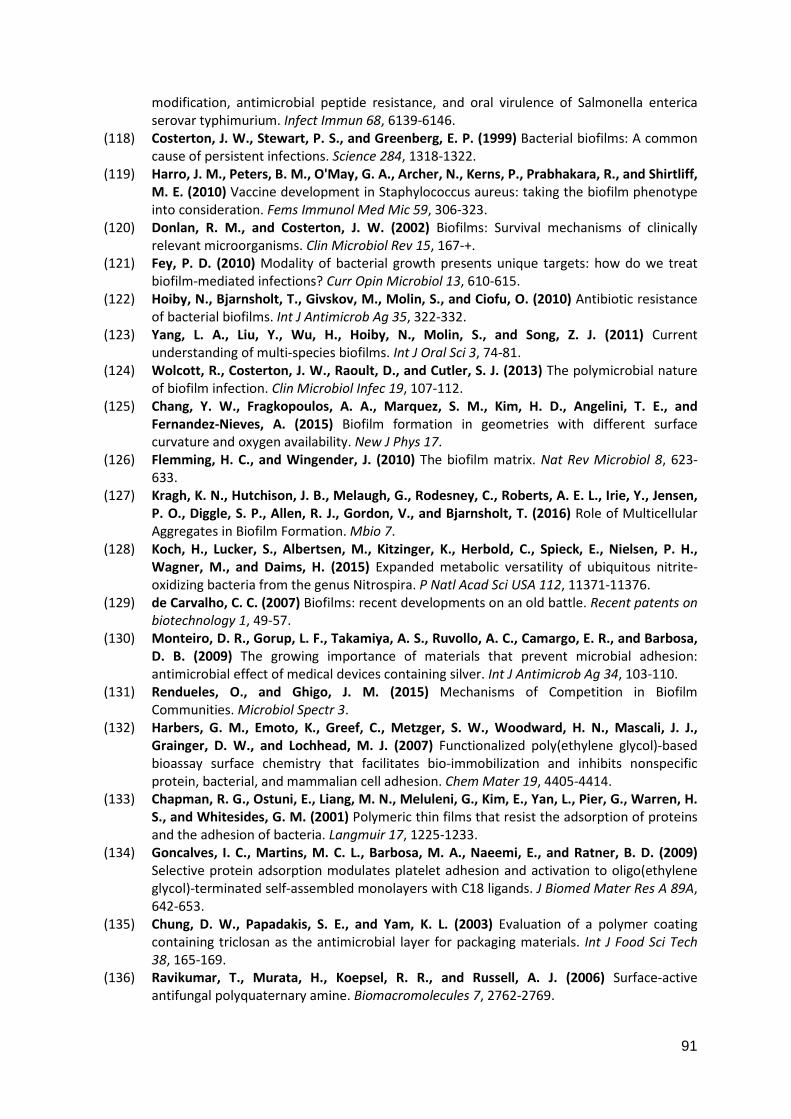
91
modification, antimicrobial peptide resistance, and oral virulence of Salmonella enterica
serovar typhimurium. Infect Immun 68, 6139-6146.
(118) Costerton, J. W., Stewart, P. S., and Greenberg, E. P. (1999) Bacterial biofilms: A common
cause of persistent infections. Science 284, 1318-1322.
(119) Harro, J. M., Peters, B. M., O'May, G. A., Archer, N., Kerns, P., Prabhakara, R., and Shirtliff,
M. E. (2010) Vaccine development in Staphylococcus aureus: taking the biofilm phenotype
into consideration. Fems Immunol Med Mic 59, 306-323.
(120) Donlan, R. M., and Costerton, J. W. (2002) Biofilms: Survival mechanisms of clinically
relevant microorganisms. Clin Microbiol Rev 15, 167-+.
(121) Fey, P. D. (2010) Modality of bacterial growth presents unique targets: how do we treat
biofilm-mediated infections? Curr Opin Microbiol 13, 610-615.
(122) Hoiby, N., Bjarnsholt, T., Givskov, M., Molin, S., and Ciofu, O. (2010) Antibiotic resistance
of bacterial biofilms. Int J Antimicrob Ag 35, 322-332.
(123) Yang, L. A., Liu, Y., Wu, H., Hoiby, N., Molin, S., and Song, Z. J. (2011) Current
understanding of multi-species biofilms. Int J Oral Sci 3, 74-81.
(124) Wolcott, R., Costerton, J. W., Raoult, D., and Cutler, S. J. (2013) The polymicrobial nature
of biofilm infection. Clin Microbiol Infec 19, 107-112.
(125) Chang, Y. W., Fragkopoulos, A. A., Marquez, S. M., Kim, H. D., Angelini, T. E., and
Fernandez-Nieves, A. (2015) Biofilm formation in geometries with different surface
curvature and oxygen availability. New J Phys 17.
(126) Flemming, H. C., and Wingender, J. (2010) The biofilm matrix. Nat Rev Microbiol 8, 623-
633.
(127) Kragh, K. N., Hutchison, J. B., Melaugh, G., Rodesney, C., Roberts, A. E. L., Irie, Y., Jensen,
P. O., Diggle, S. P., Allen, R. J., Gordon, V., and Bjarnsholt, T. (2016) Role of Multicellular
Aggregates in Biofilm Formation. Mbio 7.
(128) Koch, H., Lucker, S., Albertsen, M., Kitzinger, K., Herbold, C., Spieck, E., Nielsen, P. H.,
Wagner, M., and Daims, H. (2015) Expanded metabolic versatility of ubiquitous nitrite-
oxidizing bacteria from the genus Nitrospira. P Natl Acad Sci USA 112, 11371-11376.
(129) de Carvalho, C. C. (2007) Biofilms: recent developments on an old battle. Recent patents on
biotechnology 1, 49-57.
(130) Monteiro, D. R., Gorup, L. F., Takamiya, A. S., Ruvollo, A. C., Camargo, E. R., and Barbosa,
D. B. (2009) The growing importance of materials that prevent microbial adhesion:
antimicrobial effect of medical devices containing silver. Int J Antimicrob Ag 34, 103-110.
(131) Rendueles, O., and Ghigo, J. M. (2015) Mechanisms of Competition in Biofilm
Communities. Microbiol Spectr 3.
(132) Harbers, G. M., Emoto, K., Greef, C., Metzger, S. W., Woodward, H. N., Mascali, J. J.,
Grainger, D. W., and Lochhead, M. J. (2007) Functionalized poly(ethylene glycol)-based
bioassay surface chemistry that facilitates bio-immobilization and inhibits nonspecific
protein, bacterial, and mammalian cell adhesion. Chem Mater 19, 4405-4414.
(133) Chapman, R. G., Ostuni, E., Liang, M. N., Meluleni, G., Kim, E., Yan, L., Pier, G., Warren, H.
S., and Whitesides, G. M. (2001) Polymeric thin films that resist the adsorption of proteins
and the adhesion of bacteria. Langmuir 17, 1225-1233.
(134) Goncalves, I. C., Martins, M. C. L., Barbosa, M. A., Naeemi, E., and Ratner, B. D. (2009)
Selective protein adsorption modulates platelet adhesion and activation to oligo(ethylene
glycol)-terminated self-assembled monolayers with C18 ligands. J Biomed Mater Res A 89A,
642-653.
(135) Chung, D. W., Papadakis, S. E., and Yam, K. L. (2003) Evaluation of a polymer coating
containing triclosan as the antimicrobial layer for packaging materials. Int J Food Sci Tech
38, 165-169.
(136) Ravikumar, T., Murata, H., Koepsel, R. R., and Russell, A. J. (2006) Surface-active
antifungal polyquaternary amine. Biomacromolecules 7, 2762-2769.
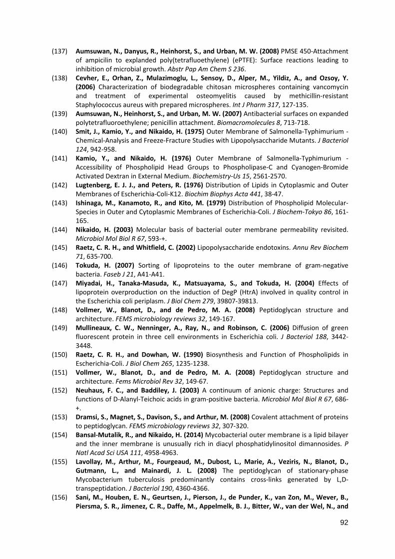
92
(137) Aumsuwan, N., Danyus, R., Heinhorst, S., and Urban, M. W. (2008) PMSE 450-Attachment
of ampicilin to explanded poly(tetrafluoethylene) (ePTFE): Surface reactions leading to
inhibition of microbial growth. Abstr Pap Am Chem S 236.
(138) Cevher, E., Orhan, Z., Mulazimoglu, L., Sensoy, D., Alper, M., Yildiz, A., and Ozsoy, Y.
(2006) Characterization of biodegradable chitosan microspheres containing vancomycin
and treatment of experimental osteomyelitis caused by methicillin-resistant
Staphylococcus aureus with prepared microspheres. Int J Pharm 317, 127-135.
(139) Aumsuwan, N., Heinhorst, S., and Urban, M. W. (2007) Antibacterial surfaces on expanded
polytetrafluoroethylene; penicillin attachment. Biomacromolecules 8, 713-718.
(140) Smit, J., Kamio, Y., and Nikaido, H. (1975) Outer Membrane of Salmonella-Typhimurium -
Chemical-Analysis and Freeze-Fracture Studies with Lipopolysaccharide Mutants. J Bacteriol
124, 942-958.
(141) Kamio, Y., and Nikaido, H. (1976) Outer Membrane of Salmonella-Typhimurium -
Accessibility of Phospholipid Head Groups to Phospholipase-C and Cyanogen-Bromide
Activated Dextran in External Medium. Biochemistry-Us 15, 2561-2570.
(142) Lugtenberg, E. J. J., and Peters, R. (1976) Distribution of Lipids in Cytoplasmic and Outer
Membranes of Escherichia-Coli-K12. Biochim Biophys Acta 441, 38-47.
(143) Ishinaga, M., Kanamoto, R., and Kito, M. (1979) Distribution of Phospholipid Molecular-
Species in Outer and Cytoplasmic Membranes of Escherichia-Coli. J Biochem-Tokyo 86, 161-
165.
(144) Nikaido, H. (2003) Molecular basis of bacterial outer membrane permeability revisited.
Microbiol Mol Biol R 67, 593-+.
(145) Raetz, C. R. H., and Whitfield, C. (2002) Lipopolysaccharide endotoxins. Annu Rev Biochem
71, 635-700.
(146) Tokuda, H. (2007) Sorting of lipoproteins to the outer membrane of gram-negative
bacteria. Faseb J 21, A41-A41.
(147) Miyadai, H., Tanaka-Masuda, K., Matsuayama, S., and Tokuda, H. (2004) Effects of
lipoprotein overproduction on the induction of DegP (HtrA) involved in quality control in
the Escherichia coli periplasm. J Biol Chem 279, 39807-39813.
(148) Vollmer, W., Blanot, D., and de Pedro, M. A. (2008) Peptidoglycan structure and
architecture. FEMS microbiology reviews 32, 149-167.
(149) Mullineaux, C. W., Nenninger, A., Ray, N., and Robinson, C. (2006) Diffusion of green
fluorescent protein in three cell environments in Escherichia coli. J Bacteriol 188, 3442-
3448.
(150) Raetz, C. R. H., and Dowhan, W. (1990) Biosynthesis and Function of Phospholipids in
Escherichia-Coli. J Biol Chem 265, 1235-1238.
(151) Vollmer, W., Blanot, D., and de Pedro, M. A. (2008) Peptidoglycan structure and
architecture. Fems Microbiol Rev 32, 149-67.
(152) Neuhaus, F. C., and Baddiley, J. (2003) A continuum of anionic charge: Structures and
functions of D-Alanyl-Teichoic acids in gram-positive bacteria. Microbiol Mol Biol R 67, 686-
+.
(153) Dramsi, S., Magnet, S., Davison, S., and Arthur, M. (2008) Covalent attachment of proteins
to peptidoglycan. FEMS microbiology reviews 32, 307-320.
(154) Bansal-Mutalik, R., and Nikaido, H. (2014) Mycobacterial outer membrane is a lipid bilayer
and the inner membrane is unusually rich in diacyl phosphatidylinositol dimannosides. P
Natl Acad Sci USA 111, 4958-4963.
(155) Lavollay, M., Arthur, M., Fourgeaud, M., Dubost, L., Marie, A., Veziris, N., Blanot, D.,
Gutmann, L., and Mainardi, J. L. (2008) The peptidoglycan of stationary-phase
Mycobacterium tuberculosis predominantly contains cross-links generated by L,D-
transpeptidation. J Bacteriol 190, 4360-4366.
(156) Sani, M., Houben, E. N., Geurtsen, J., Pierson, J., de Punder, K., van Zon, M., Wever, B.,
Piersma, S. R., Jimenez, C. R., Daffe, M., Appelmelk, B. J., Bitter, W., van der Wel, N., and
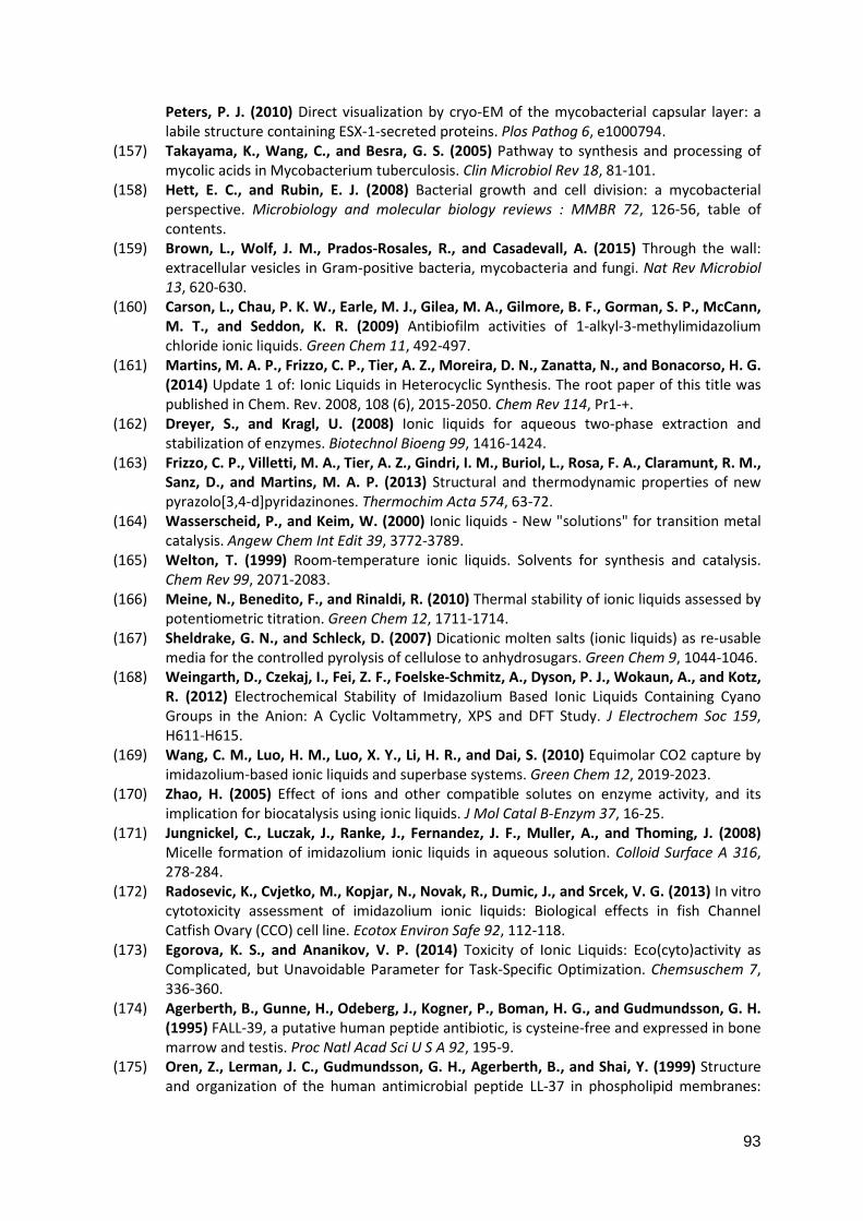
93
Peters, P. J. (2010) Direct visualization by cryo-EM of the mycobacterial capsular layer: a
labile structure containing ESX-1-secreted proteins. Plos Pathog 6, e1000794.
(157) Takayama, K., Wang, C., and Besra, G. S. (2005) Pathway to synthesis and processing of
mycolic acids in Mycobacterium tuberculosis. Clin Microbiol Rev 18, 81-101.
(158) Hett, E. C., and Rubin, E. J. (2008) Bacterial growth and cell division: a mycobacterial
perspective. Microbiology and molecular biology reviews : MMBR 72, 126-56, table of
contents.
(159) Brown, L., Wolf, J. M., Prados-Rosales, R., and Casadevall, A. (2015) Through the wall:
extracellular vesicles in Gram-positive bacteria, mycobacteria and fungi. Nat Rev Microbiol
13, 620-630.
(160) Carson, L., Chau, P. K. W., Earle, M. J., Gilea, M. A., Gilmore, B. F., Gorman, S. P., McCann,
M. T., and Seddon, K. R. (2009) Antibiofilm activities of 1-alkyl-3-methylimidazolium
chloride ionic liquids. Green Chem 11, 492-497.
(161) Martins, M. A. P., Frizzo, C. P., Tier, A. Z., Moreira, D. N., Zanatta, N., and Bonacorso, H. G.
(2014) Update 1 of: Ionic Liquids in Heterocyclic Synthesis. The root paper of this title was
published in Chem. Rev. 2008, 108 (6), 2015-2050. Chem Rev 114, Pr1-+.
(162) Dreyer, S., and Kragl, U. (2008) Ionic liquids for aqueous two-phase extraction and
stabilization of enzymes. Biotechnol Bioeng 99, 1416-1424.
(163) Frizzo, C. P., Villetti, M. A., Tier, A. Z., Gindri, I. M., Buriol, L., Rosa, F. A., Claramunt, R. M.,
Sanz, D., and Martins, M. A. P. (2013) Structural and thermodynamic properties of new
pyrazolo[3,4-d]pyridazinones. Thermochim Acta 574, 63-72.
(164) Wasserscheid, P., and Keim, W. (2000) Ionic liquids - New "solutions" for transition metal
catalysis. Angew Chem Int Edit 39, 3772-3789.
(165) Welton, T. (1999) Room-temperature ionic liquids. Solvents for synthesis and catalysis.
Chem Rev 99, 2071-2083.
(166) Meine, N., Benedito, F., and Rinaldi, R. (2010) Thermal stability of ionic liquids assessed by
potentiometric titration. Green Chem 12, 1711-1714.
(167) Sheldrake, G. N., and Schleck, D. (2007) Dicationic molten salts (ionic liquids) as re-usable
media for the controlled pyrolysis of cellulose to anhydrosugars. Green Chem 9, 1044-1046.
(168) Weingarth, D., Czekaj, I., Fei, Z. F., Foelske-Schmitz, A., Dyson, P. J., Wokaun, A., and Kotz,
R. (2012) Electrochemical Stability of Imidazolium Based Ionic Liquids Containing Cyano
Groups in the Anion: A Cyclic Voltammetry, XPS and DFT Study. J Electrochem Soc 159,
H611-H615.
(169) Wang, C. M., Luo, H. M., Luo, X. Y., Li, H. R., and Dai, S. (2010) Equimolar CO2 capture by
imidazolium-based ionic liquids and superbase systems. Green Chem 12, 2019-2023.
(170) Zhao, H. (2005) Effect of ions and other compatible solutes on enzyme activity, and its
implication for biocatalysis using ionic liquids. J Mol Catal B-Enzym 37, 16-25.
(171) Jungnickel, C., Luczak, J., Ranke, J., Fernandez, J. F., Muller, A., and Thoming, J. (2008)
Micelle formation of imidazolium ionic liquids in aqueous solution. Colloid Surface A 316,
278-284.
(172) Radosevic, K., Cvjetko, M., Kopjar, N., Novak, R., Dumic, J., and Srcek, V. G. (2013) In vitro
cytotoxicity assessment of imidazolium ionic liquids: Biological effects in fish Channel
Catfish Ovary (CCO) cell line. Ecotox Environ Safe 92, 112-118.
(173) Egorova, K. S., and Ananikov, V. P. (2014) Toxicity of Ionic Liquids: Eco(cyto)activity as
Complicated, but Unavoidable Parameter for Task-Specific Optimization. Chemsuschem 7,
336-360.
(174) Agerberth, B., Gunne, H., Odeberg, J., Kogner, P., Boman, H. G., and Gudmundsson, G. H.
(1995) FALL-39, a putative human peptide antibiotic, is cysteine-free and expressed in bone
marrow and testis. Proc Natl Acad Sci U S A 92, 195-9.
(175) Oren, Z., Lerman, J. C., Gudmundsson, G. H., Agerberth, B., and Shai, Y. (1999) Structure
and organization of the human antimicrobial peptide LL-37 in phospholipid membranes:
94
relevance to the molecular basis for its non-cell-selective activity. Biochem J 341 ( Pt 3),
501-13.
(176) Reinhardt, A., Horn, M., Schmauck, J. P. G., Brohl, A., Giernoth, R., Oelkrug, C., Schubert,
A., and Neundorf, I. (2014) Novel Imidazolium Salt-Peptide Conjugates and Their
Antimicrobial Activity. Bioconjugate chemistry 25, 2166-2174.
(177) Epand, R. F., Savage, P. B., and Epand, R. M. (2007) Bacterial lipid composition and the
antimicrobial efficacy of cationic steroid compounds (Ceragenins). Bba-Biomembranes
1768, 2500-2509.
(178) Ahmed, M. H., Ibrahim, M. A., Zhang, J., Melek, F. R., El-Hawary, S. S., Jacob, M. R., and
Muhammad, I. (2014) Methicillin-resistant Staphylococcus aureus, Vancomycin-resistant
Enterococcus faecalis and Enterococcus faecium active Dimeric Isobutyrylphloroglucinol
from Ivesia gordonii. Nat Prod Commun 9, 221-224.
(179) Reis, A. O., Cordeiro, J. C., Machado, A. M., and Sader, H. S. (2001) In vitro antimicrobial
activity of linezolid tested against vancomycin-resistant enterococci isolated in Brazilian
hospitals. The Brazilian journal of infectious diseases : an official publication of the Brazilian
Society of Infectious Diseases 5, 243-51.
(180) Brender, J. R., McHenry, A. J., and Ramamoorthy, A. (2012) Does cholesterol play a role in
the bacterial selectivity of antimicrobial peptides? Front Immunol 3.
(181) McHenry, A. J., Sciacca, M. F. M., Brender, J. R., and Ramamoorthy, A. (2012) Does
cholesterol suppress the antimicrobial peptide induced disruption of lipid raft containing
membranes? Bba-Biomembranes 1818, 3019-3024.
(182) Oren, Z., Lerman, J. C., Gudmundsson, G. H., Agerberth, B., and Shai, Y. (1999) Structure
and organization of the human antimicrobial peptide LL-37 in phospholipid membranes:
relevance to the molecular basis for its non-cell-selective activity. Biochem J 341, 501-513.
(183) Marcotte, I., Wegener, K. L., Lam, Y. H., Chia, B. C. S., de Planque, M. R. R., Bowie, J. H.,
Auger, M., and Separovic, F. (2003) Interaction of antimicrobial peptides from Australian
amphibians with lipid membranes. Chem Phys Lipids 122, 107-120.
(184) Feigin, A. M., Teeter, J. H., and Brand, J. G. (1995) The Influence of Sterols on the
Sensitivity of Lipid Bilayers to Melittin. Biochemical and biophysical research
communications 211, 312-317.
(185) Zheng, Z. Q., Xu, Q. M., Guo, J. N., Qin, J., Mao, H. L., Wang, B., and Yan, F. (2016)
Structure-Antibacterial Activity Relationships of Imidazolium-Type Ionic Liquid Monomers,
Poly(ionic liquids) and Poly(ionic liquid) Membranes: Effect of Alkyl Chain Length and
Cations. Acs Appl Mater Inter 8, 12684-12692.
(186) Luczak, J., Jungnickel, C., Lacka, I., Stolle, S., and Hupka, J. (2010) Antimicrobial and
surface activity of 1-alkyl-3-methylimidazolium derivatives. Green Chem 12, 593-601.
(187) Stolte, S., Matzke, M., Arning, J., Boschen, A., Pitner, W. R., Welz-Biermann, U., Jastorff,
B., and Ranke, J. (2007) Effects of different head groups and functionalised side chains on
the aquatic toxicity of ionic liquids. Green Chem 9, 1170-1179.
(188) Birnie, C. R., Malamud, D., and Schnaare, R. L. (2000) Antimicrobial evaluation of N-alkyl
betaines and N-alkyl-N,N-dimethylamine oxides with variations in chain length. Antimicrob
Agents Ch 44, 2514-2517.
(189) Ho, J. Y., Matsuura, T., and Santerre, J. P. (2000) The effect of fluorinated surface
modifying macromolecules on the surface morphology of polyethersulfone membranes. J
Biomat Sci-Polym E 11, 1085-1104.
(190) Van der Bruggen, B. (2009) Chemical Modification of Polyethersulfone Nanofiltration
Membranes: A Review. J Appl Polym Sci 114, 630-642.
(191) Zhao, C. S., Xue, J. M., Ran, F., and Sun, S. D. (2013) Modification of polyethersulfone
membranes - A review of methods. Prog Mater Sci 58, 76-150.
(192) Schulze, A., Stoelzer, A., Striegler, K., Starke, S., and Prager, A. (2015) Biocatalytic Self-
Cleaning Polymer Membranes. Polymers-Basel 7, 1837-1849.
95
(193) Starke, S., Went, M., Prager, A., and Schulze, A. (2013) A novel electron beam-based
method for the immobilization of trypsin on poly(ethersulfone) and poly(vinylidene
fluoride) membranes. React Funct Polym 73, 698-702.
(194) Kim, S. M., Fan, H., Cho, Y. J., Eo, M. Y., Park, J. H., Kim, B. N., Lee, B. C., and Lee, S. K.
(2015) Electron beam effect on biomaterials I: focusing on bone graft materials.
Biomaterials research 19, 10.
(195) Rapsch, K., Bier, F. F., Tadros, M., and von Nickisch-Rosenegk, M. (2014) Identification of
antimicrobial peptides and immobilization strategy suitable for a covalent surface coating
with biocompatible properties. Bioconjugate chemistry 25, 308-19.
(196) Richter, S., Bouvet, V., Wuest, M., Bergmann, R., Steinbach, J., Pietzsch, J., Neundorf, I.,
and Wuest, F. (2012) (18)F-Labeled phosphopeptide-cell-penetrating peptide dimers with
enhanced cell uptake properties in human cancer cells. Nuclear medicine and biology 39,
1202-12.
(197) Zhu, W. L., and Shin, S. Y. (2009) Effects of dimerization of the cell-penetrating peptide Tat
analog on antimicrobial activity and mechanism of bactericidal action. J Pept Sci 15, 345-
352.
(198) Zhu, W. L., and Shin, S. Y. (2009) Antimicrobial and Cytolytic Activities and Plausible Mode
of Bactericidal Action of the Cell Penetrating Peptide Penetratin and Its Lys-linked Two-
Stranded Peptide. Chem Biol Drug Des 73, 209-215.
(199) Zhu, W. L., Lan, H. L., Park, I. S., Kim, J. I., Jin, H. Z., Hahm, K. S., and Shin, S. Y. (2006)
Design and mechanism of action of a novel bacteria-selective antimicrobial peptide from
the cell-penetrating peptide Pep-1. Biochemical and biophysical research communications
349, 769-774.
(200) Bobone, S., Piazzon, A., Orioni, B., Pedersen, J. Z., Nan, Y. H., Hahm, K. S., Shin, S. Y., and
Stella, L. (2011) The thin line between cell-penetrating and antimicrobial peptides: the case
of Pep-1 and Pep-1-K. J Pept Sci 17, 335-341.
(201) Kim, H., Jang, J. H., Kim, S. C., and Cho, J. H. (2014) De novo generation of short
antimicrobial peptides with enhanced stability and cell specificity. J Antimicrob Chemoth 69,
121-132.
(202) Monera, O. D., Sereda, T. J., Zhou, N. E., Kay, C. M., and Hodges, R. S. (1995) Relationship
of sidechain hydrophobicity and alpha-helical propensity on the stability of the single-
stranded amphipathic alpha-helix. J Pept Sci 1, 319-29.
(203) Singh, J., and Thornton, J. M. (1985) The Interaction between Phenylalanine Rings in
Proteins. Febs Lett 191, 1-6.
(204) Fan, R., Yuan, Y., Zhang, Q., Zhou, X. R., Jia, L., Liu, Z., Yu, C., Luo, S. Z., and Chen, L. (2017)
Isoleucine/leucine residues at "a" and "d" positions of a heptad repeat sequence are crucial
for the cytolytic activity of a short anticancer lytic peptide. Amino acids 49, 193-202.
(205) Dev, S., Khan, R. H., and Surolia, A. (2006) 2,2,2-Trifluoroethanol-Induced structural
change of peanut agglutinin at different pH: A comparative account. IUBMB life 58, 473-9.
(206) Lakshminarayanan, R., Fan, D., Du, C., and Moradian-Oldak, J. (2007) The role of
secondary structure in the entropically driven amelogenin self-assembly. Biophys J 93,
3664-74.
(207) Chou, P. Y., and Fasman, G. D. (1974) Conformational parameters for amino acids in
helical, beta-sheet, and random coil regions calculated from proteins. Biochemistry-Us 13,
211-22.
(208) Desiervo, A. J., and Homola, A. D. (1980) Analysis of Caulobacter-Crescentus Lipids. J
Bacteriol 143, 1215-1222.
(209) Daron, H. H. (1970) Fatty Acid Composition of Lipid Extracts of a Thermophilic Bacillus
Species. J Bacteriol 101, 145-&.
(210) Schulze, A., Marquardt, B., Kaczmarek, S., Schubert, R., Prager, A., and Buchmeiser, M. R.
(2010) Electron Beam-Based Functionalization of Poly(ethersulfone) Membranes.
Macromol Rapid Comm 31, 467-472.
96
6. Attachment
6.1. List of abbreviations
AEMA 2-aminoethyl methacrylate hydrochloride
Ahx 6-hexanoic acid
AMP Antimicrobial peptide
BD Bacto brain heart infusion
CD Circular dichroism
CF 5(6)-carboxyfluorescein
CL Cardiolipin
CLSM Confocal laser scanning microscopy
CPP Cell-penetrating peptide
DCM Dichlormethan
Dde 2-acetyldimedone
DIC N,N′-diisopropylcarbodiimide
DIPEA N,N-diisopropylethylamine
DMF N,N-dimethylformamide
DOPC 1,2-dioleoyl-sn-glycero-3-phosphocholine
DOPE 1,2-dioleoyl-sn-glycero-3-phosphoethanolamine
DOPG 1,2-di-(9Z-octadecenoyl)-sn-glycero-3-phospho-
(1'-rac-glycerol)
EDC 1-ethyl-3-(3-dimethylaminopropyl)carbodiimide
ESI-MS Electrospray ionization mass-spectrometry
FA Formic acid
GUV Giant unilamellar vesicle
HATU N-methylmethanaminium hexafluorophosphate N-oxide
HC20 Minimal hemolytic concentration of 20% living
red blood cells
97
h-RBCs Human red blood cells
HSV Herpes-simplex-virus
IL Ionic liquid
INT Iodnitrotetrazolium
kGy Kilogray
Leu Leucine
LPS Lipopolysaccharides
LUV Large unilamellar vesicle
Lys Lysine
MHB Mueller-Hinton broth
MIC50 Minimal inhibitory concentration of 50% living
bacteria
MRSA Methicillin-resistant Staphylococcus aureus
NHS N-hydroxysuccinimide
OD Optical density
OM Outer membrane
PBS Phosphate buffered saline
PES Polyether sulfone
RP-HPLC Reversed-phase high-performance liquid
chromatography
SEM Standard error of mean
SI Selectivity index
TFA Trifluoracetic acid
TFE 2,2,2-trifluorethanol
TIS Triisopropylsilane
Trp Tryptophan
98
VRE Vancomycin-resistant Enterococci
XPS X-ray photoelectron spectroscopy
99
6.2. List of Figures
Figure 1: The four classes of antimicrobial peptides represented by protein models..
................................................................................................................................. 3
Figure 2: Membrane disrupting mechanism of AMPs. .............................................. 8
Figure 3: Schematic overview of AMP resistance mechanism. .............................. 11
Figure 4: Cell-wall structure of gram-negative, gram-positive and mycobacteria. .. 14
Figure 5: The imidazolium cation with different anions. .......................................... 15
Figure 6: Final structure of 3a, 3b and 3c.. ............................................................. 16
Figure 7: Antimicrobial activity of 3a, 3b and 3c. ................................................... 17
Figure 8: Structure of the imidazolium salt used for peptide 3a, 3b and 3c ............ 36
Figure 9: Final structure of 3a, 3b and 3c. .............................................................. 36
Figure 10: HPLC-MS analysis of 3a before and after purification via preparative
HPLC. ..................................................................................................................... 37
Figure 11: HPLC-MS analysis of 3c before and after purification via preparative
HPLC. ..................................................................................................................... 38
Figure 12: Circular dichroism spectra of 3c at different pH values. ........................ 39
Figure 13: Antimicrobial activity against resistant bacterial strains. ........................ 40
Figure 14: Hemolytic activity of all novel conjugates against human red blood cells.
............................................................................................................................... 43
Figure 15: Interaction of peptide 3b* with GUVs and different DOPC/cholesterol
compositions. ......................................................................................................... 44
Figure 16: Interaction of peptide 3c* with GUVs and different DOPC/cholesterol
compositions. ......................................................................................................... 47
Figure 17: SEM images of immobilized PES membranes. ..................................... 53
Figure 18: INT-assay of immobilized membranes. ................................................. 54
Figure 19: Time-experiments of immobilized membranes. ..................................... 55
Figure 20: Grown bacteria after incubation with immobilized membranes. ............. 56
Figure 21: Agar plate assay of immobilized membranes. ....................................... 56
Figure 22: HPLC-MS analysis of sC18I15 before purification via preparative HPLC.
............................................................................................................................... 62
Figure 23 HPLC-MS analysis of sC18I15 after purification via preparative HPLC. . 63
Figure 24: Antimicrobial activity of sC18 isoleucine variants. ................................. 65
Figure 25: Helical wheel projection of sC18, sC18I10, sC18I15 and sC18I16.. ...... 66
100
Figure 26: Antimicrobial activity of sC18 isoleucine variants against B. subtilis. .... 67
Figure 27: Antimicrobial activity of sC18 isoleucine variants against C. glutamicum
and M. luteus. ......................................................................................................... 68
Figure 28: Antimicrobial activity of sC18 isoleucine variants against P. fluorescens
and S. typhimurium.. ............................................................................................... 69
Figure 29: Helical wheel projection. ........................................................................ 70
Figure 30: Antimicrobial activity of sC18 phenylalanine variants against B. subtilis..
............................................................................................................................... 71
Figure 31: Antimicrobial activity of sC18 phenylalanine variants against C.
glutamicum and M. luteus. ...................................................................................... 72
Figure 32: Antimicrobial activity of sC18 phenylalanine variants against P.
fluorescens and S. typhimurium.. ........................................................................... 73
Figure 33: 3D-structure of sC18 (above) and sC18F10F15F16 (below). ................ 76
Figure 34: Circular dichroism spectra of isoleucine and phenylalanine variants in
phosphate buffer..................................................................................................... 77
Figure 35: Circular dichroism spectra of isoleucine and phenylalanine variants in
phosphate buffer + TFE. ......................................................................................... 79
Figure 36: Circular dichroism spectra with artificial membrane vesicles. ................ 80
Figure 37: Mechanism of action model for sC18F10F15F16. ................................. 82
101
6.3. List of tables
Table 1: Common antibiotics and their bacterial resistance mechanisms. ............... 2
Table 2: Some examples of antimicrobial peptides, their sequences, structures and
mechanisms of action. .............................................................................................. 4
Table 3: Equipment used during this work. ............................................................. 20
Table 4: Buffers used during this work. .................................................................. 21
Table 5: Peptides synthesized during this work. ..................................................... 22
Table 6: Analytical and synthetic data of all imidazolium salt compounds. ............. 35
Table 7: Minimal inhibitory concentration (MIC50) of all tested compounds. ........... 41
Table 8: Selectivity index (SI) of all conjugates calculated with HC20/MIC50 ........... 43
Table 9: Analytical und synthetic data of all peptide derivates.. ............................. 50
Table 10: Atomic composition of the membranes (as determined by XPS analysis).
............................................................................................................................... 52
Table 11: Analytic data of all synthesized sC18 variants.. ...................................... 59
Table 12: Calculated MIC50 values of all isoleucine and phenylalanine variants. ... 75
102
6.4. Acknowledgment
An dieser Stelle möchte ich allen Leuten danken, die mich während meiner Arbeit
sehr unterstütz und geholfen haben.
An erster Stelle Prof. Dr. Ines Neundorf als meine Betreuerin und Doktormutter für
das interessante Thema, welches ich nach meiner Masterarbeit fortsetzen konnte.
Sie hat mich super betreut und viele Inspirationen und Ideen für meine Arbeit
geliefert. Außerdem dafür, dass sie mir viele Freiheiten während meiner Arbeit
gelassen hat, aber bei Problemen jeglicher Art immer ein offenes Ohr hatte.
Zudem möchte ich Prof. Dr. Karin Schnetz für die Übernahme des Zweitgutachtens
und 3-jährige Betreuung in der Graduate School of Biologica Sciences danken.
Auch Sie hat mir einige neue Ideen für meine Arbeit geliefert. Prof. Dr. Tobias
Bollenbach danke ich für die Übernahme des Prüfungsvorsitzenden. Auch Prof. Dr.
Ralf Giernoth möchte ich danken für die gute Kooperation beim Thema der
ionischen Flüssigkeiten, sowie für die gute Betreuung in der GfSBS. Zudem danke
ich Julie Pieper gen. Schmauck für die Herstellung der ionischen Flüssigkeiten und
weiterer Experimente. Bei Christian Oelkrug und Dr. Andreas Schubert vom
Fraunhofer Institute bedanke ich mich für die durchgeführten Test an den
S2-Bakterienstämmen VRE und MRSA. Für die Immobilisierung meiner Peptide
möchte ich Isabell Thomas und Dr. Agnes Schulze vom IOM Leipzig danken.
Ein riesiger Dank geht an meine Arbeitsgruppe und alle ehemaligen
Arbeitsmitglieder, welche das Arbeiten im Labor jeden Tag verbessert haben: Bei
Florian Reichart für die Kooperation in Business Angelegenheiten, bei Stephanie
Natividad-Tietz für die lange Sitznachbarschaft, bei Anja Gronewold und Mareike
Horn (alias die Twins) für viele tolle Abende auch außerhalb des Labors und die
Herstellung der Lipidvesikel, bei Annika Klimpel für die besten Tanzmoves, bei
Lucia Feni für den Ausflug nach Italien sowie das verlorene Elfmeterschießen der
Italiener im EM-Viertelfinale und bei Tamara Lützenburg für die Einführung in die
Welt der Frauenkosmetik. Außerdem geht ein Dank an meine männliche
Unterstützung Alex Trimborn und meinen Kinocheck-Buddy Florian Küster. Zudem
geht ein Dank an meine Studenten Julian Koch, Volker Kinast, Kübra Solak, Markus
Schütz und Petra Sprengart für viele Ergebnisse. Anja Hochheiser danke ich für die
Messung vieler ESI-MS Proben und Constance Chollet für die Hilfe vieler
103
wissenschaftlicher Fragen sowie Claudia Drogt für jegliche Art der Organisation und
Gabi Sitek für die Autoklavierung unzähliger Medien und Kulturen.
Außerdem möchte ich meinen Eltern danken, die mich das ganze Studium
unterstützt haben und mich ertragen mussten, wenn es mit dem Studium mal nicht
so gut geklappt hat. Zu guter Letzt, möchte ich meiner Freundin Lena danken,
welche mich ebenfalls immer unterstützt, motiviert und wieder aufgebaut hat, auch
wenn es mal nicht so gut lief.
Ein großer Dank an euch alle, ohne die ich diese Arbeit nie hätte schreiben können.
104
6.5. Erklärung
Ich versichere, dass ich die von mir vorgelegte Dissertation selbständig angefertigt, die benutzten Quellen und Hilfsmittel vollständig angegeben und die Stellen der Arbeit −einschließlich Tabellen, Karten und Abbildungen −, die anderen Werken im Wortlaut oder dem Sinn nach entnommen sind, in jedem Einzelfall als Entlehnung kenntlich gemacht habe; dass diese Dissertation noch keiner anderen Fakultät oder Universität zur Prüfung vorgelegen hat; dass sie − abgesehen von unten angegebenen Teilpublikationen − noch nicht veröffentlicht worden ist, sowie, dass ich eine solche Veröffentlichung vor Abschluss des Promotionsverfahrens nicht vornehmen werde. Die Bestimmungen der Promotionsordnung sind mir bekannt. Die von mir vorgelegte Dissertation ist von Prof. Dr. Ines Neundorf betreut worden.
Teilpublikationen:
• Reinhardt, A., and Neundorf, I. (2016) Design and Application of Antimicrobial Peptide Conjugates. Int J Mol Sci 17.
• Reinhardt, A., Horn, M., Schmauck, J. P. G., Brohl, A., Giernoth, R., Oelkrug, C., Schubert, A., and Neundorf, I. (2014) Novel Imidazolium Salt-Peptide Conjugates and Their Antimicrobial Activity. Bioconjugate chemistry 25, 2166-2174.
• Reinhardt, A., Schmauck, J., Giernoth, R., and Neun dorf, I. (2014) Antimicrobial activity of mixtures out of antimicrobial peptides and ionic liquids. Proceedings of the 33rd Eur. Pept. Symp. p.282.
Posterpräsentationen:
• Reinhardt, A., and Neundorf, I. (2017) Reversing the activity of the cell-penetrating peptide sC18: How to improve antimicrobial activity by expansion of the hydrophobic face. 34rd European Peptide Symposium, Leipzig
• Reinhardt, A., Horn, M., Schmauck, J., Giernoth, R. , and Neundorf, I. (2015) Novel Imidazolium Salt–Peptide Conjugates and Their Antimicrobial Activity. 12th German Peptide Symposium, Darmstadt
• Reinhardt, A., Schmauck, J., Giernoth, R., Neundorf , I. (2014) Synthesis of imidazolium salt-peptide conjugates and their antimicrobial activity. 33rd European Peptide Symposium, Sofia, Bulgarien
________________ _______________________
Ort, Datum Andre Reinhardt
105
6.6. Lebenslauf
Persönliche Daten:
Name: André Reinhardt
Geburtsdatum: 06.10.1988
Geburtsort: Reutlingen
Staatsangehörigkeit: deutsch
Ausbildung:
Seit 10/2013 Promotion am Institut für Biochemie, Universität zu Köln bei Frau Prof. Dr. Ines Neundorf
10/2011 – 10/2013 Masterstudium Biological Sciences,
Universität zu Köln, Abschluss: Master of Science
10/2008 – 10/2011 Bachelorstudium Biologie, Universität zu Köln Abschluss: Bachelor of Science 08/1999 – 06/2008 Friedrich-Wilhelm-Gymnasium, Köln,
Abschluss: Abitur 08/1995 – 06/1999 St. Nikolaus Grundschule, Köln


![International Journal of Medical Sciences · Material and Methods Peptide Synthesis and Purification For solid phase synthesis of the peptides RQIKIWFQNRRMKWKK [pAnt43-58] and YGRKKRRQRRR](https://img.pdfslide.org/doc/110x75/5f092a017e708231d42587d7/international-journal-of-medical-sciences-material-and-methods-peptide-synthesis.jpg)