Embed Size (px)
Citation preview
1
DIPLOMARBEIT
Titel der Diplomarbeit
Essential Oils as Antimicrobials and Antifungals
angestrebter akademischer Grad
Magistra der Pharmazie (Mag.pharm.)
Verfasserin: Gudrun Lang
Studienrichtung: Pharmazie
Matrikel-Nummer: 0500575
Betreuer: Univ. Prof. Mag. pharm. Dr. Gerhard Buchbauer
Wien, im November 2010

2
Danksagung
An dieser Stelle möchte ich mich besonders bei Herrn Univ. Prof. Dr. Gerhard
Buchbauer für die umfangreiche und hilfsbereite Betreuung während des Verfassens
dieser Diplomarbeit bedanken.
Außerdem bin ich meinen Eltern zu größtem Dank verpflichtet, die mir das
Pharmazie-Studium ermöglicht und mich während meiner Studienjahre unterstützt
haben.
Auch meinem Partner Stefan Deibl, meinen Geschwistern Judith und Clemens, sowie
meinen Freundinnen und Freunden, die das Voranschreiten meines Studiums durch
motivierende Gespräche gefördert und die Freude an absolvierten Prüfungen mit mir
geteilt haben, danke ich.

3
Abstract
The antimicrobial activity of essential oils is discussed in this review taking in
account studies which were published in the period of time from 2008 until
September 2010. Furthermore, the most important methods to examine the
antimicrobial efficiency of essential oils are presented. The studies are divided into
the following three groups depending on the activity of the applied essential oil
against the test microorganisms: antimicrobial, antifungal active agents and
substances which inhibit the growth of yeasts. Various interesting possible
applications are revealed such as the use of essential oils instead of synthetic drugs to
circumvent the increasing resistance of some pathogens. Moreover, they could not
only be used for the therapy of infectious illnesses, but also as preservatives in the
food industry. A further possibility is among others the application of essential oils in
skin products in order to treat or avoid dermal infections. Additionally, the prevalent
constituents of the individual antimicrobial active essential oils are elaborated.

4
Zusammenfassung
Die antimikrobielle Wirkung von ätherischen Ölen wird in diesem Review unter
Berücksichtigung von Studien, die in der Zeitspanne von 2008 bis September 2010
veröffentlicht wurden, diskutiert. Außerdem werden die wichtigsten Methoden zur
Bestimmung der antimikrobiellen Wirksamkeit von ätherischen Ölen präsentiert. Die
Studien werden in die folgenden drei Gruppen unterteilt, abhängig von der Aktivität
des verwendeten ätherischen Öls gegen die Testkeime: antimikrobielle, antifungale
Wirkstoffe und Substanzen, die das Wachstum von Hefen hemmen. Verschiedene
interessante Anwendungsmöglichkeiten werden aufgezeigt, wie zum Beispiel die
Anwendung von ätherischen Ölen an Stelle von synthetischen Wirkstoffen, um die
ansteigende Resistenz von einigen Pathogenen zu umgehen. Außerdem können sie
nicht nur zur Therapie von infektiösen Erkrankungen eingesetzt werden, sondern
auch als Konservierungsmittel in der Lebensmittelindustrie. Eine weitere
Möglichkeit ist unter anderem die Anwendung von ätherischen Ölen in
Hautprodukten, um dermale Infektionen zu behandeln oder zu vermeiden. Des
Weiteren sind die vorherrschenden Bestandteile der einzelnen antimikrobiell
wirksamen ätherischen Öle ausgearbeitet.

5
TABLE OF CONTENTS
INTRODUCTION ...................................................................................................... 6
IN VITRO TESTS TO ASSESS ANTIMICROBIAL ACTIVITY ........................ 7
ANTIMICROBIALS ................................................................................................. 9
EOs against drug-resistant bacteria strains ............................................. 9
EOs against Propionibacterium acnes and Staphylococcus epidermidis 12
EOs against Helicobacter pylori................................................................ 14
EOs as food-preservatives/ EOs against food-related bacteria ............. 15
EOs as bio-preservatives in cosmetic industry ....................................... 23
EOs against dental bacteria ..................................................................... 24
EOs against diverse human pathogens .................................................... 26
EOs against Borrelia burgdorferi ............................................................. 41
EOs against nocardiform actinomycetes ................................................. 42
a) EOs against Mycobacteria ........................................................ 42
b) EOs against Nocardia asteroides .............................................. 43
c) EOs against Rhodococcus equi ................................................. 44
EOs as water disinfectants ....................................................................... 45
EOs as air disinfectants ............................................................................ 45
Anti-biofilm activity of EOs ..................................................................... 46
EOs in combination with synthetic active agents ................................... 47
EOs against phytopathogenic bacteria .................................................... 48
YEASTS .................................................................................................................... 49
Candida ...................................................................................................... 49
Cryptococcus ............................................................................................. 54
ANTIFUNGALS ....................................................................................................... 55
EOs against dermatophytes ...................................................................... 55
EOs against molds ..................................................................................... 58
EOs against phytopathogenic fungi ......................................................... 64
REFERENCES ......................................................................................................... 66
TABLES .................................................................................................................... 80
6
INTRODUCTION
Essential oils (EOs) possess a wide spectrum of different impressive qualities
including antiphlogistic, spasmolythic, antinociceptive and antioxidant activity.
Moreover they exert immunomodulant, psychotrope, acaricide and expectorant
effects.[1]
Due to their multifunctionality, EOs find a huge application area in
medicine and aromatherapy.
Also antiviral, antidiabetic and cancer suppressive activities are observed. In addition
to further other effects, EOs show significant antimicrobial properties against a wide
range of Gram-positive and Gram-negative bacteria. That is why they were already
used for embalming in Ancient Egypt.[2]
In the course of history EOs were always applied for their antimicrobial effects in
traditional medicine. Therefore, plants were used for the treatment of infectious
illnesses since ancient times even though no knowledge about microorganisms
existed by then.[3]
Medicinal plants are of course still in use nowadays, but now the investigation of the
active agents is possible by modern means. The isolation of EOs and their
characterization by using gas chromatography (GC) and mass spectrometry (MS)
systems are common practice. Moreover their antimicrobial activity can be verified
by in-vitro tests. EOs get even more popular regarding the fact that many synthetic
drugs are connected with unpleasant side-effects. Volatile oils also represent an
interesting alternative due to emerging resistance of microorganisms against
synthetic agents.
EOs cannot only exert bacteriostatic and bactericidal effects, but also demonstrate
activity against fungi and yeasts.
This paper focuses on the antimicrobial and antifungal activity of EOs concentrating
on studies that have been published since 2008 until September 2010.

7
IN VITRO TESTS TO ASSESS ANTIMICROBIAL ACTIVITY
Several methods are used to investigate the antimicrobial activity of EOs. The three
most important ones are: The agar diffusion test, the agar or broth dilution test and
the vapour phase test.[4]
Agar Diffusion Test:
A petri dish filled with microorganisms containing agar is needed to perform this
method. The EO is either directly applied to the surface – in this case small holes are
punched into the agar surface - or put on a small paper disk which is afterwards
placed onto the agar. The antimicrobial activity can be estimated from the size of the
originating inhibition zone. Nevertheless it is important to point out that this test
method is not completely free of any problems. This conclusion can be drawn from
the fact that in some cases the results of the agar diffusion test showed small
antimicrobial activity, but the same EO proved high activity in dilution tests.
Especially components with low water solubility showed misleadingly low
antimicrobial activity. Moreover the different volatility of single constituents, then
often unknown diffusion coefficients and other side effects have to be considered.[4]
Dilution Test:
In the broth dilution test concentration series of the antimicrobial substance are
established using a broth medium which is seeded with microorganisms. The
minimal inhibitory concentration (MIC) is evaluated in order to determine the
antimicrobial potency of the tested substance.
In the agar dilution test a concentration gradient of the tested substance is placed
onto an agar plate. By evaluating the microbial growth the MIC can be stated
likewise. This method is declared to be the gold standard but it is not that often used
since it is connected to higher costs and laborious handling.[5]
When performing the dilution test method with EOs it is adjuvant to create a
saturated moistened atmosphere to adjust volatility.[4]
Vapour Phase Test:
Up to now, there is no standardized method available for the vapour phase test. In
general, a seeded agar plate is placed upside-down onto a reservoir which comprises

8
a certain amount of volatile oil. In this case the generated inhibition zone is
considered as criterion for the antimicrobial activity.[4]
Aromatogram:
The procedure of developing an aromatogram resembles the agar diffusion test. That
is why the test microorganisms which are cultivated on an agar plate are exposed to
certain amounts of EOs which are spread on paper disks. The antimicrobial efficacy
of the EO is likewise determined by inhibition zones.[6]
The crucial difference between these two techniques is not the course of action itself,
but the substances which are investigated for their potentially antimicrobial activity.
Therefore, aromatograms always indicate the use of exclusively EOs whereas
antibiograms include also other active substances such as synthetic drugs.[7]
Air washer coupled with Air Sampler:
This test method allows the determination of the antimicrobial activity of EOs
against air-borne microbes. A special machine called air washer is filled with diluted
EO which is vaporized into the room. By the air sampler air-borne microorganisms
are fixed on agar strips. After incubating these strips the number of microbes in the
air can be counted. As a result, the comparison between the amount of bacteria
before and after the application of EO vapours is facilitated.[8]
9
ANTIMICROBIALS
Antimicrobial agents inhibit the growth of microorganisms or lead to their death. In
the following chapter studies are presented that deal with the effect of EOs on
bacteria and yeasts.
Former studies indicate a higher antibacterial effect of EOs against Gram-positive
than against Gram-negative bacteria. The outer cell membrane of Gram-negative
bacteria obtains hydrophilic qualities that impede the contact of the hydrophobic
constituents of the EO with the bacterial cell.[9]
Contrary to this, EOs can directly impair the cell membrane of Gram-positive
bacteria leading to cell membrane rupture, blocking of enzyme systems and
progressivity of ion permeability.[10]
EOs against drug-resistant bacteria strains
The increasing tolerance of several microorganisms against commonly used
antibiotic drugs represents a challenge for scientists to find alternative ways for the
treatment of such infections. One of the main causes that provokes the higher
resistance of microorganisms is the loose application of drugs.[11]
This includes that
they are applied in too low concentrations, not specific enough or without serious
indication. Especially methicillin-resistant Staphylococcus aureus (MRSA) strains are
popular test microorganisms.
S. aureus – a Gram-positive bacterium which is common part of the human microbial
skin flora - can cause minor infections, but nevertheless also severe diseases such as
pneumonia, sepsis, endocarditis or meningitis particularly in hospitalized patients.
The increasing resistance of these pathogens against current drugs tremendously
complicates the therapy of these infections.[12]

10
effective
against
EO main constituents test method Ref.
MRSA,
vancomycin-
resistant
Enterococcus
faecium
(VRE),
multidrug-
resistant
strains of
Klebsiella
pneumoniae
and
Pseudomonas
aeruginosa
Cleistocalyx
operculatus
(Roxb.) Merr and
Perry (Myrtaceae)
γ-terpinene
(5.8%) globulol
(5.6%)
cis-linalool oxide
(5.2%)
MIC = 5 - 20
l/ml
[13]
MRSA Eucalyptus
globulus Labill.
(Myrtaceae)
1,8-cineole
(47.2%)
MIC = 85.6
g/ml
[14]
MRSA, VRE Kadsura
longipedunculata
Finet &
Gagnepain
(Schisandraceae)
-cadinene
(21.8%)
diffusion test,
dilution test
[15]
MRSA Lavandula
angustifolia Mill.
(Lamiaceae)
linalyl acetate
(37.0%), linalool
(29.5%)
disk diffusion [16]
MRSA Lavandula
latifolia Medik
(Lamiaceae)
linalool (38.8%),
1,8-cineole
(28.5%)
disk diffusion [16]
MRSA Lavandula
luisieri Rozeira
Riv.-Mart.
(Lamiaceae)
-necrodyl
acetate (34.5%),
1,8-cineole
(17.6%)
disk diffusion [16]

11
MRSA Lavandula
stoechas L. ssp.
stoechas
(Lamiaceae)
-fenchone (39.2%),
myrtenyl acetate
(9.5%),
-pinene (6.1%),
camphor (5.9%)
MIC = 31.2
g/ml
[17]
MRSA Salvia rosifolia
Sm.
(Lamiaceae)
-pinene,
1,8-cineole
MIC = 125
g/ml
[18]
MRSA Tanacetum
parthenium (L.)
Schultz Bip.
(Asteraceae)
camphor (49.0-60.8%) MIC = 125
g/ml
[19]
MRSA Thymus
vulgaris L.
(Lamiaceae)
thymol (48.1%) MIC = 18.5
g/ml
[14]
MRSA Zataria
multiflora
Boiss.
(Lamiaceae)
thymol (38.7%),
carvacrol (15.3%), rho-
cymene (10.2%)
MIC = 0.25-
1.0 l/ml
MBC = 0.5-
2.0 l/ml
[20]
MRSA Zanthoxylum
tingoassuiba
St.-Hil.
(Rutaceae)
-bisabolol, methyl-N-
methylanthranilate
disk
diffusion
[21]
Table 1: EOs and MRSA
Helichrysum italicum (Roth) G.Don fil. (Asteraceae) EO which contained among
other constituents geraniol showed an inhibitory activity against multidrug resistant
strains of the Gram-negative bacteria Acinetobacter baumannii, Enterobacter
aerogenes, Escherichia coli and P. aeruginosa. The susceptibility of these pathogens
was considerably enhanced by combining commonly used drugs such as -lactams,
chloramphenicol and quinolones with geraniol. H. italicum EO was presumed to
obtain substances which act as efflux pump inhibitors since the EO revealed to be
12
especially active against bacteria which over-expressed efflux pumps and therefore
developed tolerance towards drugs.[22]
The volatile oil of Melaleuca alternifolia Cheel. (Myrtaceae) comprises among other
constituents the antimicrobial active agent terpinen-4-ol. In in-vitro tests the
bacteriostatic and bactericidal activity of both M. alternifolia EO and its isolated
component terpinen-4-ol was ascertained against coagulase-negative staphylococci
and MRSA showing much stronger activity when using terpinen-4-ol on its own. As a
consequence terpinen-4-ol could constitute an interesting alternative in the therapy of
MRSA infections of the skin.[23]
MRSA and MSSA strains got adjusted to M.
alternifolia EO when it was applied at sub-lethal concentrations. These strains
developed higher resistance to the EO but also to antibiotics. After the same
treatment coagulase-negative Staphylococci showed likewise lower vulnerability to
antibiotics, but the effect of M. alternifolia EO was not decreased. Therefore, it is
important to use EOs in high enough concentrations to avoid this adaptation.[24]
The above mentioned results support the idea of using EOs as an alternative to well-
established drugs since they show high efficacy in inhibiting drug-resistant bacteria
strains. The EOs could be used on their own, but also in combination with other EOs
or synthetic active agents since synergy was observed by combining these substances.
Therefore, synergistic effects were noticed regarding Z. multiflora EO in
combination with the synthetic active agent vancomycin[20]
, but also when combining
different EOs such as L. luisieri EO with L. angustifolia or L. stoechas EO.[16]
EOs against Propionibacterium acnes and Staphylococcus epidermidis
The following studies show that EOs are capable of inhibiting the growth of bacteria
which are linked to the occurrence of skin infections, such as P. acnes,
Propionibacterium granulosum[25]
or S. epidermidis. Interestingly, no differences
were noticed between the activity against drug-sensitive and drug-resistant bacteria
strains.[26]
Due to that, EOs could be used in acne therapy or in cosmetic products for
the prevention and treatment of skin infections.[27]

13
effective against EO main constituents test method Ref.
P. acnes,
S. epidermidis
Abies koreana
E.H.Wilson
(Pinaceae)
bornyl acetate
(30.4%),
limonene (19.0%)
[28]
S. epidermidis Acronychia
pedunculata (L.)
Miq. (Rutaceae)
-pinene
(57.4%), (E)--
caryophyllene
(13.6%)
[29]
P. acnes,
S. epidermidis
Citrus natsudaidai
Hayata (Rutaceae)
limonene (81.6%) MIC = 0.31
l/ml
MIC = 10.0
l/ml
[27]
P. acnes,
S. epidermidis
Citrus obovoidea
Hort. ex Takahash
(Rutaceae)
limonene (83.4%) MIC = 0.31
l/ml
MIC = 2.5
l/ml
[27]
P. acnes,
S. epidermidis
Citrus sunki Hort.
ex. Tan. (Rutaceae)
dl-limonene
(68.2%)
[30]
P. acnes several Citrus
species (Rutaceae)
limonene (67.7 to
91.7%), myrcene
(2.6 to 25.3%)
MIC = 1.25 to
20 l/ml
[31]
P. acnes,
S. epidermidis
Cryptomeria
japonica (Thunb.
ex L. f.) D.Don
(Cupressaceae)
kaurene (17.2%),
elemol (10.9%),
-eudesmol
(9.4%), sabinene
(8.9%)
MIC = 0.156
to 10.00 l/ml
[26]
P. acnes,
S. epidermidis
Fortunella japonica
(Thunb.) Swingle
var. margarita
(Swingle) Makino
(Rutaceae)
dl-limonene
(61.6%)
[30]
14
S. epidermidis Helichrysum
pallasii (Spreng.)
Ledeb.
(Asteraceae)
hexadecanoic
acid (14.7%),
(Z,Z)-9,12-
octadecadienoic
acid (14.2%)
MIC = 100
g/ml
[32]
P. acnes Syzygium
aromaticum (L.)
Merr. Et Perry
(Myrtaceae) S.
aromaticum
agar diffusion
tests, MIC =
0.31 mg/ml
[33]
P.
granulosum,
P. acnes
Thymus
quinquecostatus
Celak. (Lamiaceae)
p-cymen-3-ol
(50.4%), p-
cymen-2-ol
(24.1%), cymene
(19.0%)
disk diffusion
method,
MIC = 0.5
mg/ml
[25]
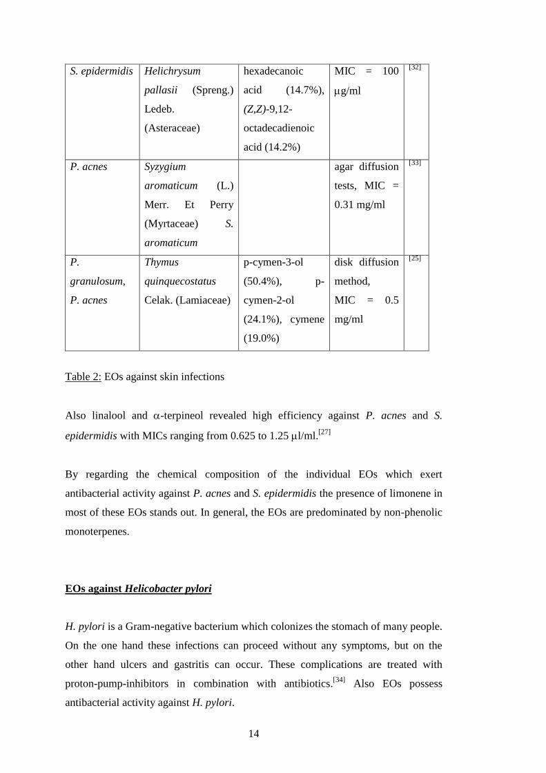
Table 2: EOs against skin infections
Also linalool and -terpineol revealed high efficiency against P. acnes and S.
epidermidis with MICs ranging from 0.625 to 1.25 l/ml.[27]
By regarding the chemical composition of the individual EOs which exert
antibacterial activity against P. acnes and S. epidermidis the presence of limonene in
most of these EOs stands out. In general, the EOs are predominated by non-phenolic
monoterpenes.
EOs against Helicobacter pylori
H. pylori is a Gram-negative bacterium which colonizes the stomach of many people.
On the one hand these infections can proceed without any symptoms, but on the
other hand ulcers and gastritis can occur. These complications are treated with
proton-pump-inhibitors in combination with antibiotics.[34]
Also EOs possess
antibacterial activity against H. pylori.
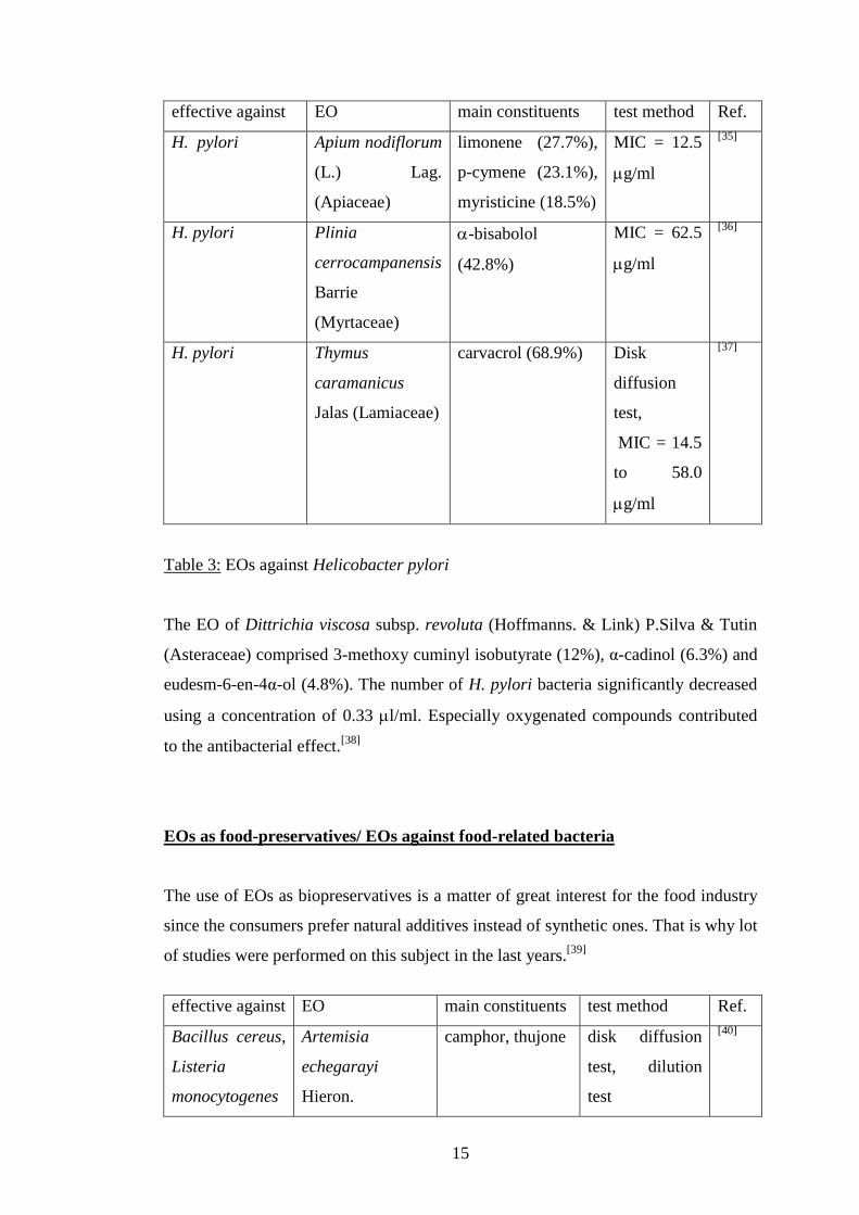
15
effective against EO main constituents test method Ref.
H. pylori Apium nodiflorum
(L.) Lag.
(Apiaceae)
limonene (27.7%),
p-cymene (23.1%),
myristicine (18.5%)
MIC = 12.5
g/ml
[35]
H. pylori Plinia
cerrocampanensis
Barrie
(Myrtaceae)
-bisabolol
(42.8%)
MIC = 62.5
g/ml
[36]
H. pylori Thymus
caramanicus
Jalas (Lamiaceae)
carvacrol (68.9%) Disk
diffusion
test,
MIC = 14.5
to 58.0
g/ml
[37]
Table 3: EOs against Helicobacter pylori
The EO of Dittrichia viscosa subsp. revoluta (Hoffmanns. & Link) P.Silva & Tutin
(Asteraceae) comprised 3-methoxy cuminyl isobutyrate (12%), α-cadinol (6.3%) and
eudesm-6-en-4α-ol (4.8%). The number of H. pylori bacteria significantly decreased
using a concentration of 0.33 l/ml. Especially oxygenated compounds contributed
to the antibacterial effect.[38]
EOs as food-preservatives/ EOs against food-related bacteria
The use of EOs as biopreservatives is a matter of great interest for the food industry
since the consumers prefer natural additives instead of synthetic ones. That is why lot
of studies were performed on this subject in the last years.[39]
effective against EO main constituents test method Ref.
Bacillus cereus,
Listeria
monocytogenes
Artemisia
echegarayi
Hieron.
camphor, thujone disk diffusion
test, dilution
test
[40]

16
(Asteraceae)
L.
monocytogenes,
S. aureus; B.
cereus,
Enterobacter
cloacea
Artemisia incana
(L.) Druce
(Asteraceae)
camphor (19.0%),
borneol (18.9%),
1,8-cineole
(14.5%)
MIC = 31.3
g/ml;
MIC = 125
g/ml
[41]
Salmonella
typhi, E. coli
Chaerophyllum
macropodum
Boiss. (Apiaceae)
trans--ocimene,
myristicin
microdilution
broth test
[42]
S. typhi, E. coli Chrysanthemum
parthenium (L.)
Bernh.
(Asteraceae)
-pinene,
camphor
microdilution
broth test
[43]
E. aerogenes, E.
coli, L.
monocytogenes,
S. aureus,
Salmonella
enteritidis,
Salmonella
typhimurium
C. operculatus γ-terpinene
(5.8%), globulol
(5.6%), cis-
linalool oxide
(5.2%)
disk diffusion
test,
MIC = 1 to 4
l/ml
[13]
Salmonella
species
Citrus species (+)-limonene,
terpenes
MIC = 1% [44]
E. coli Jasminum
sambac (L.)
Aiton (Oleaceae)
methyl salicylate,
benzyl acetate,
methyl
anthranilate
MIC = 31.25
μl/ml
[45]
Enterococcus
faecalis, L.
monocytogenes,
S. aureus;
B. cereus;
Yersinia
Laurus nobilis L.
(Lauraceae)
1,8-cineole (60%) MIC = 0.02%
(v/v);
MIC = 0.2%;
MIC = 1.0%
[46]
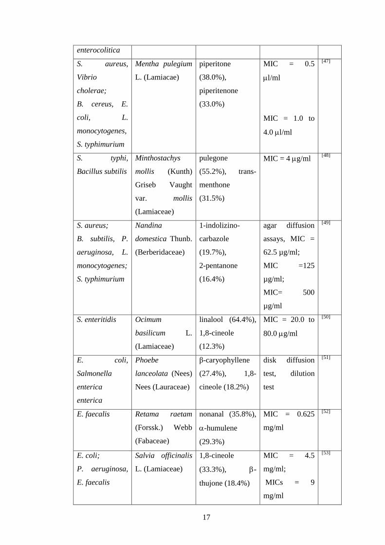
17
enterocolitica
S. aureus,
Vibrio
cholerae;
B. cereus, E.
coli, L.
monocytogenes,
S. typhimurium
Mentha pulegium
L. (Lamiacae)
piperitone
(38.0%),
piperitenone
(33.0%)
MIC = 0.5
l/ml
MIC = 1.0 to
4.0 l/ml
[47]
S. typhi,
Bacillus subtilis
Minthostachys
mollis (Kunth)
Griseb Vaught
var. mollis
(Lamiaceae)
pulegone
(55.2%), trans-
menthone
(31.5%)
MIC = 4 g/ml [48]
S. aureus;
B. subtilis, P.
aeruginosa, L.
monocytogenes;
S. typhimurium
Nandina
domestica Thunb.
(Berberidaceae)
1-indolizino-
carbazole
(19.7%),
2-pentanone
(16.4%)
agar diffusion
assays, MIC =
62.5 µg/ml;
MIC =125
µg/ml;
MIC= 500
µg/ml
[49]
S. enteritidis Ocimum
basilicum L.
(Lamiaceae)
linalool (64.4%),
1,8-cineole
(12.3%)
MIC = 20.0 to
80.0 g/ml
[50]
E. coli,
Salmonella
enterica
enterica
Phoebe
lanceolata (Nees)
Nees (Lauraceae)
β-caryophyllene
(27.4%), 1,8-
cineole (18.2%)
disk diffusion
test, dilution
test
[51]
E. faecalis Retama raetam
(Forssk.) Webb
(Fabaceae)
nonanal (35.8%),
-humulene
(29.3%)
MIC = 0.625
mg/ml
[52]
E. coli;
P. aeruginosa,
E. faecalis
Salvia officinalis
L. (Lamiaceae)
1,8-cineole
(33.3%), -
thujone (18.4%)
MIC = 4.5
mg/ml;
MICs = 9
mg/ml
[53]

18
E. coli, E.
faecalis
Schinus molle L.
(Anacardiaceae)
-phellandrene
(35.9%), -
phellandrene
(29.3%)
MICs = 9
mg/ml
[53]
B. cereus Tanacetum
argenteum (Lam.)
Willd. ssp.
argenteum
(Asteraceae)
-pinene
(36.7%), -pinene
(27.5%)
MIC = 125
g/ml
[54]
B. cereus Tanacetum
argyrophyllum
(C. Koch) Tvzel
var.
argyrophyllum
(Asteraceae)
camphor, borneol
and 1,8-cineole
MIC = 125
g/ml
[55]
L.
monocytogenes
Zizyphus jujuba
Mill.
(Rhamnaceae)
eugenol (48.3%),
isoeugenol
(11.8%)
agar disk
diffusion test,
dilution test
[56]
B. cereus, E.
coli, L.
monocytogenes,
S. enteritidis,
Proteus
mirabilis
T. vulgaris,
Origanum
vulgare L.
(Lamiaceae),
S. aromaticum,
Citrus sinensis
(L.) Osbeck
(Rutaceae)
disk diffusion
test
[57]
Table 4: EOs as biopreservatives
Not only classic in-vitro tests were conducted to investigate the antimicrobial activity.
Therefore, the EOs were also applied on different media (e.g. meat). Subsequently,
the effect on the microbial growth was observed over a period of time. Such studies
are mentioned here:

19
Govaris et al. investigated the usage of Origanum vulgare subsp. hirtum Link.
(Lamiaceae) EO as food preservative. Therefore it was either applied alone at a
percentage of 0.6 or 0.9% or in combination with nisin in minced sheep meat. When
the EO which primarily consisted of carvacrol (80.2%) was used singularly at a
percentage of 0.9%, it exerted quite high activity against S. enteritis, whereas the use
of nisin alone did not harm these pathogens. Even bactericidal activity was observed
when the EO was combined with nisin.[58]
Another study about O. vulgare EO
verifies the antimicrobial effect against S. aureus. The germ’s growth and its
enterotoxin synthesis were inhibited by the volatile oil. Since this EO is especially
powerful against foodborne bacteria, it might be used as biopreservative in food-
industry.[59]
A research was performed in which edible tomato puree films were produced which
were containing allspice, oregano and garlic EO in order to impair microbial growth.
This method might be used in food industry for the extension of shelf life. The
antibacterial effect against E. coli, L. monocytogenes and S. enterica was evaluated
by vapor phase and overlay diffusion tests. Oregano EO – rich in carvacrol (63.4%) -
obtained the strongest antibacterial effect, but also allspice EO which contained
68.6% eugenol and garlic EO (dominated by diallyl disulfide) showed antibacterial
effects. L. monocytogenes revealed to be the most vulnerable pathogen. All three
bacteria were inhibited by direct contact as well as by the vapors.[60]
Aim of a further study was to investigate the consequence of adding Origanum onites
L. (Lamiaceae) EO containing pads to wrapped chicken drumsticks concerning the
food’s shelf-life. The storability was prolonged from three to five days using 5 ml of
the diluted EO (1.5%) due to the fact that the number of enterobacteriaceae, lactic
acid bacteria, pseudomonads, psychrotrophs and yeasts was kept down.
Unfortunately, the chemical composition of this EO was not investigated in this
research.[61]
T. vulgaris EO was incorporated at a concentration of 0.6% in minced beef meat.
Higher concentrations could not be applied since they proved disadvantageous for
the food flavor. The growth of L. monocytogenes bacteria was effectively inhibited
especially at storage at 10 degrees. Moreover synergy was observed in combination

20
with nisin. Therefore the number of these pathogens revealed to be lower than the
official boundary value determined by the EU when nisin (1000 IU/g) and T.
vulgaris EO (0.6%) were applied and when the meat was subsequently refrigerated at
4 degrees.[62]
Cinnamaldehyde was capable of inhibiting the growth of B. cereus in nutrient and
carrot broth at a concentration of 2l/100ml stored at a temperature of 12 degrees,
whereas the application of eugenol and carvacrol was ineffective. That is why
cinnamaldehyde could be used for the preservation of food based on carrots.[63]
The inhibitory potency of carvacrol and cinnamaldehyde was also evaluated against
the food-poisoning causing pathogen Campylobacter jejuni. Both EO components
were effective at concentrations from 0.1% upwards independent on the potential
resistance of the individual strains against drugs. Cinnamaldehyde was noticed to
exert even stronger antibacterial agency in comparison to carvacrol.[64]
Various EO compounds were tested for their antibacterial activity against
Clostridium perfringens. Trans-cinnamaldehyde, 2-tert-butyl-6-methylphenol,
carvacrol and geraniol showed the strongest activity with MICs of 167 μg/ml, 175
μg/ml, 300 μg/ml and 450 μg/ml, respectively. Contrary to this, Lactobacillus strains
which are part of the natural intestinal flora were not harmed.[65]
The following study shows that there exists a certain framework of the concentration
in which the tested EO is efficient against pathogenic bacteria, but does not yet exert
any influence on the salutary bacteria of the gastrointestinal tract: The EO of
Foeniculum vulgare var. azoricum (Mill.) Thell. (Apiaceae) appeared to be rich in
the antimicrobial active agent (E)-anethole (59.3-71.7%). The EO exhibited
antimicrobial effect against a large number of foodborne pathogenic bacteria, but
also against probiotic bacteria such as Lactobacillus strains and Streptococcus
thermophilus. The lowest MIC value of 15.62 g/ml was measured against
Acinetobacter lwoffi, followed by a MIC of 31.25 g/ml against S. aureus and P.
aeruginosa. The inhibiting effect on probiotic bacteria was reported at MIC values
superior than 250 g/ml. Due to its antimicrobial effect against food related
pathogens the EO could be used as food preservative, but one has to keep in mind

21
that the exaggerated ingestion of fennel products could influence the bacterial flora
in the gastrointestinal tract by inhibiting the growth of probiotic bacteria.[66]
A study was conducted about the usage of specific EOs for the therapy of
gastrointestinal dysbiosis, an imbalance of the intestinal microflora. Therefore, the
effect of eight EOs which are traditionally used for the treatment of gastrointestinal
ailments and diseases was examined by MIC evaluation against Bacteroides fragilis,
Clostridium difficile, C. perfringens, E. faecalis, E. coli, Eubacterium limosum,
Bifidobacterium bifidu, Bifidobacterium longum, Lactobacillus acidophilus,
Lactobacillus plantarum, Peptostreptococcus anaerobius and Candida albicans. The
volatile oil of Trachyspermum copticum (L.) Link (Apiaceae) exhibited the strongest
antibacterial effect since it stopped bacterial growth of all tested germs at
concentrations lower than 2.2%. Moreover, it revealed high selectivity against
pathogenic bacteria. The same is true for Carum carvi L. (Apiaceae) and L.
angustifolia EO. Citrus aurantium var. amara L. (Rutaceae) revealed lower
antibacterial potency but showed likewise selectivity. Therefore, these EOs could be
used for the treatment of dysbiosis without impairing the growth of salutary
bacteria.[67]
In the two following mentioned studies it becomes aware that the extent of the
antimicrobial activity is among others dependant on the pH-level and the
composition of the food[68, 69]
:
The qualification of several EOs as food preservatives was evaluated using four
food-borne bacteria strains. Especially useful seemed to be the combination of O.
vulgare (carvacrol 68.5%) with Origanum majorana L. (Lamiaceae) (4-thujanol
36.2%), T. vulgaris (thymol 52.9%, p-cymene 34.0%) or with O. basilicum (linalool
42.3%, estragole 26.9%) exhibiting an additive effect against B. cereus, P.
aeruginosa and E. coli. The growth of L. monocytogenes was additively impaired by
using blends of O. majorana or T. vulgaris with Rosmarinus officinalis L.
(Lamiaceae) (eucalyptol 39.6%, camphor 19.0%), Salvia triloba L. (Lamiaceae)
(eucalyptol 42.0%, camphor 12.0%) or O. basilicum. The strength of activity was
influenced by the pH level and the food ingredients. Therefore, the conclusion could
be drawn that a low pH level of about 5 and high protein content in the food supports
22
the inhibitory properties of the used EOs whereas carbohydrates and fat diminish
it.[68]
The antimicrobial activity against food-related bacteria was observed using the EOs
of Melissa officinalis L. (Lamiaceae), O. majorana, O. vulgare and T. vulgaris.
Three different media were established which were based on meat, milk and salad.
The Listeria strains were found to be more susceptible than Lactobacillus,
Enterobacter and Pseudomonas strains. O. vulgare and T. vulgaris – the two most
efficient EOs - showed additive effects when used in combination. The EOs obtained
the strongest antimicrobial activity in food with high pH level and protein content.[69]
The organoleptic changes which are associated with the application of EOs as food-
preservatives in a high enough concentration to avoid the bacterial growth can
represent a problem which could be solved by using aromas[70]
or additional
measures to extent the shelf life of food products, such as refrigeration[53]
:
The antimicrobial effect of several substances which were found in EOs was
investigated using the Gram-positive bacteria B. cereus, E. faecalis, L.
monocytogenes and S. aureus. Moreover, the inhibitory activity against Gram-
negative bacteria (E. coli, Salmonella choleraesuis, Y. enterocolitica), yeasts (C.
albicans, Zygosaccharomyces rouxii, Debaryomyces hansenii) and fungi was
observed. Carvacrol, cinnamaldehyde and thymol displayed the strongest
antimicrobial activity. EOs can influence the taste of packaged food in an
unfavourable way. That is why the combination of these substances with aromas
(banana, vanilla, strawberry) was examined. Organoleptic tests revealed that all of
them could be used in combination with vanilla, but not with banana. Only the
combination of strawberry aroma with thymol resulted in an organoleptic acceptable
taste.[70]
In an experiment with minced meat the bacteriostatic activity of the EOs of S.
officinalis and S. molle was noticed against Salmonella anatum and S. enteritidis at
concentrations of 1.5% using S. officinalis EO and 2.0% using S. molle.
Unfortunately, the taste was impaired at these concentrations. That is why the

23
combination of these EOs in lower concentrations with NaCl and storage at low
temperatures was detected to be more useful.[53]
Sinapis alba L. (Brassicaceae) EO which was isolated from the seeds contained
phenethyl isothiocyanate as active agent. This lead molecule was obtained by high
performance liquid chromatography (HPLC) and silica gel column chromatography
and subsequently subjected to chemical modifications. Paper disk diffusion assays
were performed in order to investigate the effect on the following intestinal bacteria:
E. coli, C. difficile, C. perfringens, Bifidobacterium breve, B. bifidum, B. longum, L.
acidophilus and L. casei. The EO inhibited the growth of C. difficile, C. perfringens
and E. coli at 5 mg/disk. The same Clostridium strains were effectively inhibited at a
dose of 1 mg/disk when phenethyl isothiocyanate was singularly used. The semi-
synthetic derivates of this molecule which contained aromatic functional groups,
such as benzyl-, benzoyl- and phenethyl-groups revealed higher selectivity and
higher antibacterial agency against pathogenic intestinal bacteria, such as E. coli and
Clostridium strains.[71]
The majority of investigated EOs was rich in non-phenolic monoterpenic compounds.
Nevertheless, also phenolic monoterpenes, such as carvacrol[58]
and phenylpropanoid
constituents (e.g. cinnamaldehyde[64]
) contributed to the antimicrobial activity
against food-borne pathogens.
EOs as bio-preservatives in cosmetic industry
Due to the preserving activity of EOs, these substances could also be applied for the
preservation of cosmetic products. Since some EOs show synergistic effects in
combination with commercially used preservatives the application of EOs makes a
diminution of these synthetic substances possible as the two below-mentioned
studies revealed.[72, 73]
Patrone et al. investigated the combination of several EOs with synthetic
preservatives which are used in cosmetic industry. Eucalyptus globosus Labill.
(Myrtaceae) and Mentha piperita L. (Lamiaceae) EO showed synergistic activity
24
against P. aeruginosa when they were applied in combination with methylparabene.
Moreover, synergy was noticed against S. aureus using S. officinalis, O. vulgare and
M. piperita in combination with imidazolidinyl urea and propylparabene. These
findings constitute a further proof of the advantages of combining EOs with common
preservatives in cosmetic products.[72]
The application of commercial lavender, lemon and tea tree EO in body milks was
investigated observing the inhibition of microbial growth. The main constituents of
the lavender oil were linalool (34.1%) and linalyl acetate (33.3%). The tee trea oil
mainly consisted of terpinen-4-ol (41.3%) and γ-terpinene (19.1%). The most
abundant substance in lemon oil was limonene (79.8%). The growth of the involved
microorganisms S. aureus, P. aeruginosa, Aspergillus niger and Candida species
was sufficiently inhibited using these EOs in combination with 0.2% of a synthetic
preservative. Since synergy was noticed when the EOs were combined with the
synthetic agent, the applied quantity of the synthetical component could be cut down
about 8.5 times.[73]
EOs against dental bacteria
This chapter deals with the antimicrobial activity of EOs against dental bacteria -
especially against the tooth-decay causing bacteria Streptococcus pyogenes and
Streptococcus mutans. EOs are capable of inhibiting the growth of these bacteria as
well as the formation of biofilms. In various cases the potency of chlorohexidine was
found to be even lower than the efficacy of the EOs.[74]
Therefore, the application of
EOs is recommended in products which prevent caries.[75]
effective against EO main constituents test method Ref.
S. mutans Achillea ligustica
All. (Asteraceae)
viridiflorol
(14.5%),
terpinen-4-ol
(13.0%)
MIC = 39
g/ml
[75]
S. mutans, S.
pyogenes
Mentha longifolia
L. (Lamiaceae)
(-)-menthol disk diffusion
test,
[76]

25
microdilution
test
S. mutans Hyptis pectinata L.
Poit. (Lamiaceae)
β-caryophyllene
(28.3%),
caryophyllene
oxide (28.0%)
MIC = 200
g/ml
[77]
Aggregatibacter
actinomycetemc
omitans,
Fusobacterium
nucleatum,
Parvimonas
micra,
Porphyromonas
gingivalis,
Prevotella
intermedia,
Prevotella
nigrescens,
Tannerella
forsythia
Satureja hortensis
L. (Lamiaceae)
carvacrol (86.6%) MICs < 0.125
l/ml
[78]
Table 5: EOs against dental bacteria
The antimicrobial activities of R. officinalis EO, M. piperita EO and chlorohexidine
were compared to each other using the tooth-decay causing bacteria S. pyogenes and
S. mutans. R. officinalis EO whose main constituents were piperitone (23.7%), α-
pinene (14.9%) and linalool (14.9%) obtained MBC of 2000 ppm against S. mutans
and 4000 ppm against S. pyogenes. Chlorohexidine showed MICs of 8000 and 1000
ppm. M. piperita EO which mainly comprised α-terpinene (19.7%) and piperitenone
oxide (19.3%), but also trans-carveol (14.5%) and isomenthone (10.3%) showed
MBCs of 6000 ppm against S. mutans and 1000 ppm against S. pyogenes. The
decimal reduction times (D-values) of the EOs were lower than that of
chlorohexidine with 2.8 min against S. mutans. The lowest D-value against S.
26
pyogenes and the highest anti-biofilm activity was achieved by application of M.
piperita EO. Hence, the EOs displayed even higher activity than chlorohexidine.[74]
Further in-vitro as well as in-vivo experiments verified the high antibacterial activity
of M. piperita EO against the plaque-causing bacteria S. pyogenes and S. mutans.
Also thereby the EO showed stronger effects in preventing the formation of biofilms
and keeping the number of bacteria in the mouth low in comparison to
chlorohexidine.[79]
A similar study was conducted comparing the anti-biofilm activity of Eucalyptus
camaldulensis Dehnh. var. obtuse (Myrtaceae) EO and Mentha spicata L.
(Lamiaceae) EO. The MBC values of both oils turned out to be 2 mg/ml against S.
pyogenes and 4 mg/ml against S. mutans. An in-vivo experiment proved the ability of
preventing biofilm formation. The principal constituents of M. spicata EO were
detected to be limonene (48.0%) and piperitone (20.3%). E. camaldulensis EO
comprised 1,8-cineole (64.0%) and α-pinene (9.6%). E. camaldulensis EO reached a
D-value of 2.8 min against S. mutans using the MBC, so did M. spicata EO. For
comparison only, the D-value of chlorohexidine (2%) was 12.8 min. Only 3.6 min
were measured against S. pyogenes using E. camaldulensis EO, whereas the D-value
was 4.3 min using M. spicata EO.[80]
It becomes quite obvious that Mentha species play an important role in inhibiting the
growth of tooth-decay causing bacteria. Although the composition of the individual
species differ from each other all of them achieved remarkable results in impairing
the microbial growth of periodontal pathogens.
EOs against diverse human pathogens
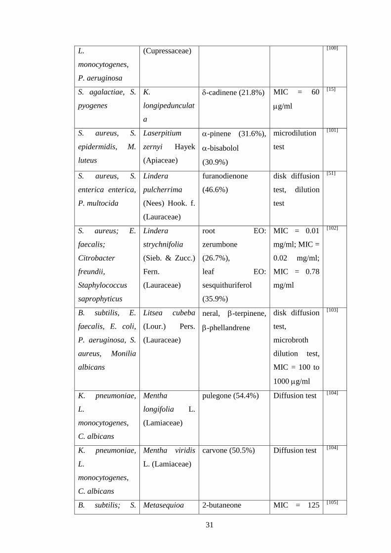
effective against EO main constituents test method Ref.
Cryptococcus
neoformans; K.
pneumoniae
Abies
holophylla
Maxim.
(Pinaceae)
bicyclo[2.2.1]
heptan-2-ol
(28.1%)
MIC = 0.5
mg/ml; MIC =
10.9 mg/ml
[81]
Candida Abies koreana bornyl ester MIC = 0.5 [81]

27
glabrata; K.
pneumoniae; B.
subtilis, E. coli
E.H.Wilson
(Pinaceae)
(41.8%) mg/ml; MIC =
5.5 mg/ml;
MIC = 10.9
mg/ml
B. subtilis, S.
aureus
Ageratum
conyzoides L.
(Asteraceae)
precocene I
(52.2%),
caryophyllene
(26.2%)
disk diffusion
tests
[82]
E. coli, K.
pneumoniae
Anaphalis
nubigena DC.
var.
monocephala
(DC.) C. B.
Clarke
(Asteraceae)
-guaiene (12.3%),
-muurolene
(10.4%)
MIC = 125
g/ml, MIC =
500 g/ml
[83]
S. aureus, S.
epidermidis, C.
albicans, C.
neoformans
Artemisia
absinthium L.
(Asteraceae)
trans-sabinyl
acetate (26.4%),
myrcene (10.8%),
trans-thujone
(10.1%)
agar diffusion
tests
[84]
B. subtilis, K.
pneumoniae,
Bacillus mycoides
Ballota nigra L.
(Lamiaceae)
p-vinylguiacol
(9.2%), borneol
(7.5%)
disk diffusion
test, dilution
test
[85]
Streptococcus
agalactiae, S.
pyogenes
Bupleurum
marginatum
Wall. ex DC.
(Apiaceae)
tridecane (13.2%),
undecane (10.4%)
MIC = 0.125
to 4.00 mg/ml
[86]
S. aureus,
Streptococcus
faecalis
Callistemon
citrinus (Curtis)
Skeels
(Myrtaceae)
1,8-cineole (61.2%) disk diffusion
test, broth
microdilution
test
[87]
S. aureus, S.
faecalis, B.
cereus, Serratia
Callistemon
viminalis
(Gaertn.) G.Don
1,8-cineole (83.2%) disk diffusion
test, broth
microdilution
[87]

28
marcescens (Myrtaceae) test
Clostridium
bifermentas,
Enterococcus
faecium,
Enterococcus
hirae,
Streptococcus
salivarius subsp.
thermophilus
Cannabis sativa
L.
(Cannabaceae)
myrcene, -pinene,
-caryophyllene
broth dilution
test
[88]
Staphylococcus
simulans,
Staphylococcus
lugdunensis, S.
aureus, S.
epidermitis,
Candida
tropicalis
Carum
montanum
(Coss. et Dur.)
Benth. et Hook.
(Apiaceae)
nothoapiole
(62.8%)
diffusion test [89]
B. subtilis, C.
albicans
Chamaecyparis
nootkatensis (D.
Don) Spach.
(Cupressaceae)
limonene (53.2%) diffusion test [90]
E. faecalis, S.
aureus
Cordia
verbenacea
D.C.
(Boraginaceae)
tricyclene (23.9%),
bicyclogermacrene
(11.7%)
MIC = 200
g/ml, MIC =
170 g/ml
[91]
S. aureus,
Pasteurella
multocida
Dodecadenia
grandiflora
Nees
(Lauraceae)
germacrene D
(26.0%),
furanodiene
(13.7%)
disk diffusion
test, dilution
test
[51]
B. cereus, B.
subtilis,
Micrococcus
luteus, S. aureus
Enterolobium
contortisiliquu
m (Vell.)
Morong
carvone [92]
29
(Fabaceae)
E. coli, S. aureus Erigeron
mucronatus DC
(Asteraceae)
caryophyllene
(11.4%), limonene
(10.3%)
Disk diffusion
test
[93]
P. aeruginosa Eugenia
beaurepaireana
(Kiaersk.) D.
Legrand
(Myrtaceae)
-caryophyllene
(8.0%),
bicyclogermacrene
(7.2%)
MIC = 278.3
g/ml
[94]
S. aureus Eugenia
brasiliensis
Lam.
(Myrtaceae)
spathulenol
(12.6%), -cadinol
(8.7%)
MIC = 156.2
g/ml
[94]
S. aureus Eugenia
umbelliflora
Berg.
(Myrtaceae)
viridiflorol
(17.7%), -pinene
(13.2%)
MIC = 119.2
g/ml
[94]
B. subtilis, S.
aureus, S.
mutans, E. coli,
E. faecalis, C.
albicans
Ferula glauca
L. (Apiaceae)
leaf EO: (E)-
caryophyllene
(24.9%), fruit EO:
-pinene (24.2%),
root EO: (E)--
farnesene (10.0%),
elemicin (9.0%),
flower EO:
germacrene D
(14.2%), myrcene
(13.6%)
MIC = 38 to
1250 g/ml
[95]
S. aureus, E. coli,
S. enterica
enterica, Shigella
flexneri, P.
multocida
Hedychium
aurantiacum
Wall. ex Roscoe
(Zingiberaceae)
terpinen-4-ol disk diffusion
test, MIC =
2.0 – 15.6
l/ml
[96]
S. aureus, E. coli, Hedychium trans-meta-mentha- disk diffusion [96]

30
S. enterica
enterica, Shigella
flexneri, P.
multocida
coronarium
J.König
(Zingiberaceae)
2,8-diene, linalool test, MIC =
7.8 – 31.3
l/ml
S. aureus, E. coli,
S. enterica
enterica, Shigella
flexneri, P.
multocida
Hedychium
ellipticum Sm.
(Zingiberaceae)
1,8-cineole,
sabinene
disk diffusion
test, MIC =
7.8 – 31.3
l/ml
[96]
S. aureus Hymenocrater
longiflorus
Benth.
(Lamiaceae)
δ-cadinol (18.5%),
α-pinene (10.2%),
p-menth-1-en-8-ol
(9.8%)
MIC = 120
g/ml
[39]
B. subtilis, S.
aureus
Hypericum
hirsutum L.
(Guttiferae)
(E,E)--farnesene
(7.0–13.8%) and
(E)--farnesene
(7.2–9.4%)
broth
microdilution
test
[97]
B. subtilis, S.
aureus
Hypericum
richeri Vill.
subsp. richeri
(Guttiferae)
germacrene D
(26.9%)
broth
microdilution
test
[97]
B. subtilis, S.
aureus
Hypericum
tetrapterum Fr.
(Guttiferae)
-copaene (12.7%),
-longipinene
(8.1%)
broth
microdilution
test
[97]
E. faecium, B.
cereus, S. aureus,
C. albicans,
Candida
tropicalis, C.
glabrata,
Candida
parapsilosis
Inula helenium
L. (Asteraceae)
alantolactone,
isoalantolactone
MIC = 0.009
to 0.6 mg/ml
[98]
B. subtilis, B.
cereus, S. aureus,
Juniperus
phoenicea L.
-pinene agar diffusion
test
[99]
31
L.
monocytogenes,
P. aeruginosa
(Cupressaceae) [100]
S. agalactiae, S.
pyogenes
K.
longipedunculat
a
-cadinene (21.8%) MIC = 60
g/ml
[15]
S. aureus, S.
epidermidis, M.
luteus
Laserpitium
zernyi Hayek
(Apiaceae)
-pinene (31.6%),
-bisabolol
(30.9%)
microdilution
test
[101]
S. aureus, S.
enterica enterica,
P. multocida
Lindera
pulcherrima
(Nees) Hook. f.
(Lauraceae)
furanodienone
(46.6%)
disk diffusion
test, dilution
test
[51]
S. aureus; E.
faecalis;
Citrobacter
freundii,
Staphylococcus
saprophyticus
Lindera
strychnifolia
(Sieb. & Zucc.)
Fern.
(Lauraceae)
root EO:
zerumbone
(26.7%),
leaf EO:
sesquithuriferol
(35.9%)
MIC = 0.01
mg/ml; MIC =
0.02 mg/ml;
MIC = 0.78
mg/ml
[102]
B. subtilis, E.
faecalis, E. coli,
P. aeruginosa, S.
aureus, Monilia
albicans
Litsea cubeba
(Lour.) Pers.
(Lauraceae)
neral, -terpinene,
-phellandrene
disk diffusion
test,
microbroth
dilution test,
MIC = 100 to
1000 g/ml
[103]
K. pneumoniae,
L.
monocytogenes,
C. albicans
Mentha
longifolia L.
(Lamiaceae)
pulegone (54.4%) Diffusion test [104]
K. pneumoniae,
L.
monocytogenes,
C. albicans
Mentha viridis
L. (Lamiaceae)
carvone (50.5%) Diffusion test [104]
B. subtilis; S. Metasequioa 2-butaneone MIC = 125 [105]
32
aureus, P.
aeruginosa; E.
coli
glyptostroboide
s Miki ex Hu.
(Taxodiaceae)
(30.6%) g/ml; MIC =
250 g/ml;
MIC = 500
g/ml
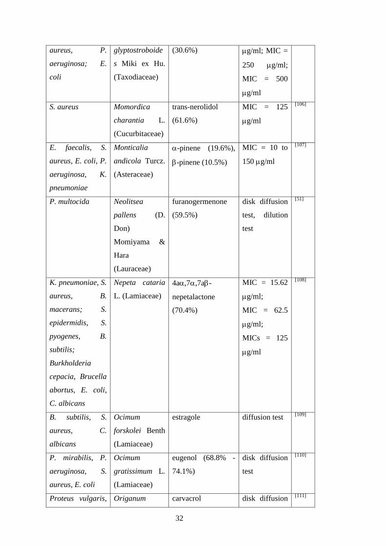
S. aureus Momordica
charantia L.
(Cucurbitaceae)
trans-nerolidol
(61.6%)
MIC = 125
g/ml
[106]
E. faecalis, S.
aureus, E. coli, P.
aeruginosa, K.
pneumoniae
Monticalia
andicola Turcz.
(Asteraceae)
-pinene (19.6%),
-pinene (10.5%)
MIC = 10 to
150 g/ml
[107]
P. multocida Neolitsea
pallens (D.
Don)
Momiyama &
Hara
(Lauraceae)
furanogermenone
(59.5%)
disk diffusion
test, dilution
test
[51]
K. pneumoniae, S.
aureus, B.
macerans; S.
epidermidis, S.
pyogenes, B.
subtilis;
Burkholderia
cepacia, Brucella
abortus, E. coli,
C. albicans
Nepeta cataria
L. (Lamiaceae)
4a,7,7a-
nepetalactone
(70.4%)
MIC = 15.62
g/ml;
MIC = 62.5
g/ml;
MICs = 125
g/ml
[108]
B. subtilis, S.
aureus, C.
albicans
Ocimum
forskolei Benth
(Lamiaceae)
estragole diffusion test [109]
P. mirabilis, P.
aeruginosa, S.
aureus, E. coli
Ocimum
gratissimum L.
(Lamiaceae)
eugenol (68.8% -
74.1%)
disk diffusion
test
[110]
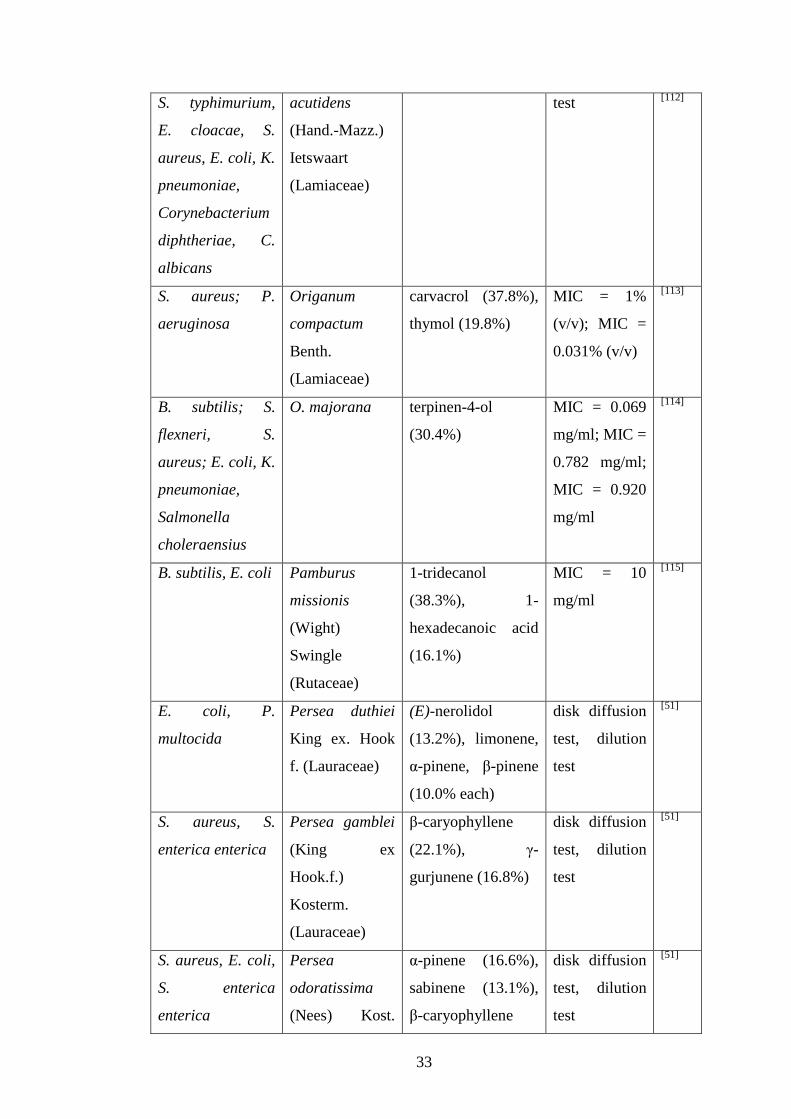
Proteus vulgaris, Origanum carvacrol disk diffusion [111]
33
S. typhimurium,
E. cloacae, S.
aureus, E. coli, K.
pneumoniae,
Corynebacterium
diphtheriae, C.
albicans
acutidens
(Hand.-Mazz.)
Ietswaart
(Lamiaceae)
test [112]
S. aureus; P.
aeruginosa
Origanum
compactum
Benth.
(Lamiaceae)
carvacrol (37.8%),
thymol (19.8%)
MIC = 1%
(v/v); MIC =
0.031% (v/v)
[113]
B. subtilis; S.
flexneri, S.
aureus; E. coli, K.
pneumoniae,
Salmonella
choleraensius
O. majorana terpinen-4-ol
(30.4%)
MIC = 0.069
mg/ml; MIC =
0.782 mg/ml;
MIC = 0.920
mg/ml
[114]
B. subtilis, E. coli Pamburus
missionis
(Wight)
Swingle
(Rutaceae)
1-tridecanol
(38.3%), 1-
hexadecanoic acid
(16.1%)
MIC = 10
mg/ml
[115]
E. coli, P.
multocida
Persea duthiei
King ex. Hook
f. (Lauraceae)
(E)-nerolidol
(13.2%), limonene,
α-pinene, β-pinene
(10.0% each)
disk diffusion
test, dilution
test
[51]
S. aureus, S.
enterica enterica
Persea gamblei
(King ex
Hook.f.)
Kosterm.
(Lauraceae)
β-caryophyllene
(22.1%), γ-
gurjunene (16.8%)
disk diffusion
test, dilution
test
[51]
S. aureus, E. coli,
S. enterica
enterica
Persea
odoratissima
(Nees) Kost.
α-pinene (16.6%),
sabinene (13.1%),
β-caryophyllene
disk diffusion
test, dilution
test
[51]

34
(Lauraceae) (10.4%)
B. cereus, B.
subtilis, S.
aureus, E. coli, P.
aeruginosa, S.
typhi, C. albicans,
C. tropicalis
Phyllanthus
emblica L.
(Phyllanthaceae
)
-caryophyllene, -
bourbonene
MIC = 100 to
1000 g/ml
[116]
S. aureus,
Enterococcus
hirae; P.
aeruginosa, E.
coli; C. albicans
Pituranthos
chloranthus
Benth. and
Hook.
(Apiaceae)
terpinen-4-ol
(30.3%)
MIC = 1.875
mg/l; MICs =
3.75 mg/l;
MIC = 7.5
mg/l
[117]
S. aureus, M.
luteus, S.
typhimurium; S.
epidermidis
Rhaponticum
acaule DC
(Asteraceae)
methyl eugenol,
epi-13-manool
disk diffusion
test, MIC =
500 μg/ml;
MIC = 800
μg/ml
[118]
S. aureus; L.
monocytogenes,
C. albicans; E.
faecalis, S.
pyogenes
Rhaponticum
carthamoides
(Willd.) Iljin
(Asteraceae)
13-norcypera-
1(5),11(12)-diene
(22.6%), aplotaxene
(21.2%)
MIC = 32
g/ml; MIC =
128 g/ml;
MIC = 256
g/ml
[119]
S. aureus,
Streptococcus
pneumoniae,
Shigella spp, E.
faecalis
Ridolfia
segetum (L.)
Moris
(Apiaceae)
dillapiole (47.4%) MIC = 1.25
mg/ml
[120]
B. subtilis,
Chromobacterium
violaceum, E.
coli; S. aureus,
Erwinia
carotovora
Rosa
damascena
Mill.
(Rosaceae)
citronellol (35.2%),
geraniol (22.2%)
MIC = 0.25%
(v/v);
MIC = 0.5%
(v/v)
[121]
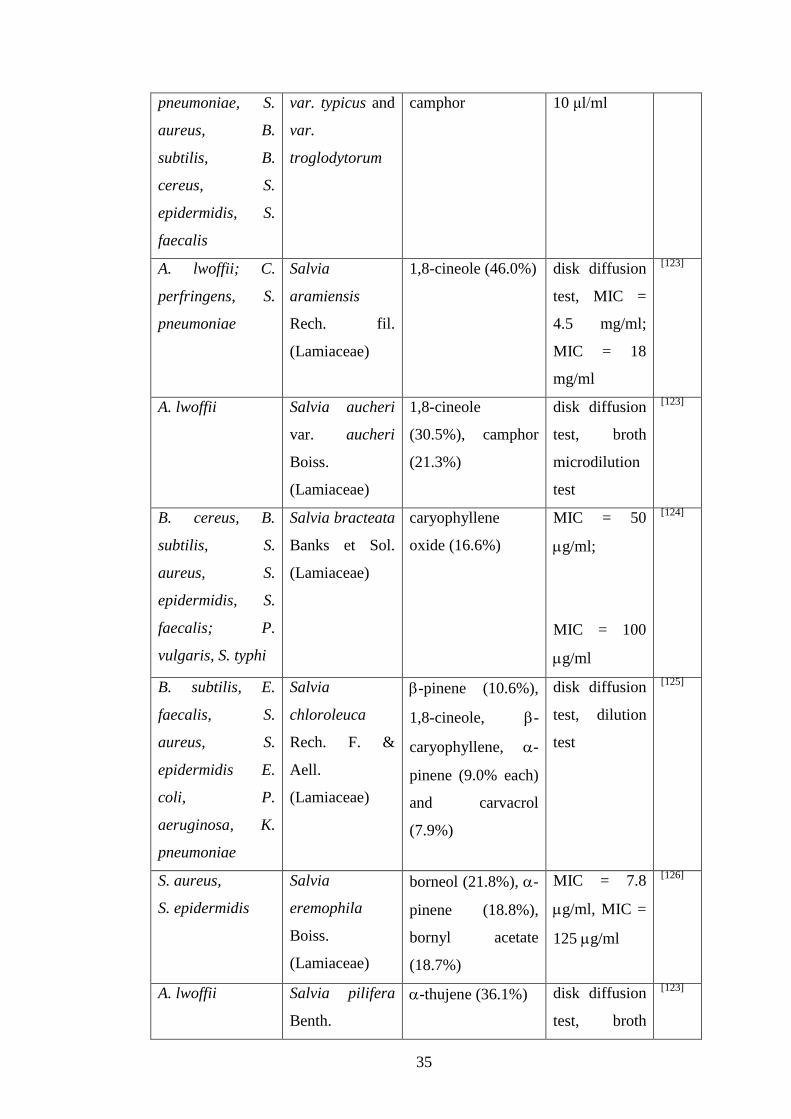
E. coli, K. R. officinalis 1,8-cineole, MIC = 1.25 to [122]
35
pneumoniae, S.
aureus, B.
subtilis, B.
cereus, S.
epidermidis, S.
faecalis
var. typicus and
var.
troglodytorum
camphor
10 μl/ml
A. lwoffii; C.
perfringens, S.
pneumoniae
Salvia
aramiensis
Rech. fil.
(Lamiaceae)
1,8-cineole (46.0%) disk diffusion
test, MIC =
4.5 mg/ml;
MIC = 18
mg/ml
[123]
A. lwoffii Salvia aucheri
var. aucheri
Boiss.
(Lamiaceae)
1,8-cineole
(30.5%), camphor
(21.3%)
disk diffusion
test, broth
microdilution
test
[123]
B. cereus, B.
subtilis, S.
aureus, S.
epidermidis, S.
faecalis; P.
vulgaris, S. typhi
Salvia bracteata
Banks et Sol.
(Lamiaceae)
caryophyllene
oxide (16.6%)
MIC = 50
g/ml;
MIC = 100
g/ml
[124]
B. subtilis, E.
faecalis, S.
aureus, S.
epidermidis E.
coli, P.
aeruginosa, K.
pneumoniae
Salvia
chloroleuca
Rech. F. &
Aell.
(Lamiaceae)
-pinene (10.6%),
1,8-cineole, -
caryophyllene, -
pinene (9.0% each)
and carvacrol
(7.9%)
disk diffusion
test, dilution
test
[125]
S. aureus,
S. epidermidis
Salvia
eremophila
Boiss.
(Lamiaceae)
borneol (21.8%), -
pinene (18.8%),
bornyl acetate
(18.7%)
MIC = 7.8
g/ml, MIC =
125 g/ml
[126]
A. lwoffii Salvia pilifera
Benth.
-thujene (36.1%) disk diffusion
test, broth
[123]

36
(Lamiaceae) microdilution
test
B. cereus, B.
subtilis, S.
aureus, S.
epidermidis, S.
faecalis;
P. vulgaris, P.
aeruginosa
Salvia rubifolia
Boiss.
(Lamiaceae)
-muurolene
(11.8%)
MIC = 50
g/ml;
MIC = 100
g/ml
[124]
B. cereus, E.
faecalis,
bark EO: also P.
mirabilis
Santiria trimera
(Oliv.) Aubrév.
(Burseraceae)
leaf EO: -
humulene (34.6%),
bark EO: -pinene
(51.5%)
agar disc
diffusion test,
broth
microdilution
test
[127]
MRSA, P.
vulgaris, S.
typhimurium, C.
albicans, C.
tropicalis
Satureja
cuneifolia Ten.
(Lamiaceae)
thyme MIC = 62.5 to
500 g/ml
[128]
B. subtilis, S.
aureus, E.
faecalis, K.
pneumoniae, E.
coli, P.
aeruginosa
Satureja
spicigera (C.
Koch) Boiss.
(Lamiaceae)
carvacrol (53.7%),
thymol (36.0%)
disk diffusion
test
[129]
S. aureus, P.
aeruginosa; C.
albicans
Schinus
terebinthifolius
Raddi.
(Anacardiaceae)
cis--terpineol
(17.9%), (E)-
caryophyllene
(17.6%)
MIC = 0.80
mg/ml; MIC =
0.85 mg/ml
[130]
B. subtilis, P.
aeruginosa
Stachys cretica
L. subsp.
smyrnaea Rech.
fil. (Lamiaceae)
trans--
caryophyllene
(51.0%),
germacrene D
disk diffusion
test
[131]

37
(32.8%)
B. subtilis,
Bacillus pumulis,
E. coli, E.
faecalis, K.
pneumoniae, S.
aureus, S.
epidermidis, P.
aeruginosa, C.
albicans
Tanacetum
balsamita L.
subsp.
balsamita
(Asteraceae)
carvone (51.0%), -
thujone (20.8%)
disk diffusion
test, dilution
test
[132]
B. subtilis, S.
epidermidis, S.
aureus, S. faecalis
Teucrium
divaricatum
Sieb. ssp.
villosum
(Celak.) Rech.
fil. (Lamiacae)
(E)-caryophyllene
(30.1%)
MIC = 25 to
100 μg/ml
[133]
A. lwoffii, S.
pyogenes, E. coli,
Listeria species,
C. albicans, C.
parapsilosis,
Candida krusei
Thymbra
spicata L.
(Lamiaceae)
carvacrol (60.4%) disk diffusion
test, dilution
test
[134]
Table 6: EOs against human pathogens
Since the chemical composition of EOs can change according to the growing place
and the point of time at which the plants are collected, the antimicrobial activity can
be influenced by these parameters. That is why the EO of S. cuneifolia which was
isolated of plants in the post-flowering stage presented lower MIC values than the
EOs of pre-flowering and flowering stage.[128]
In another study the chemical
composition of H. spicatum Buch.-Ham. (Zingiberaceae) was noticed to be
dependent on the collection area. Therefore, some samples contained primary
sabinene and terpinen-4-ol whereas others mainly obtained 10-epi--eudesmol and

38
1,8-cineole. Both samples showed activity against S. aureus, P. multocida and E.
coli.[96]
Beside of exerting bacteriostatic and bactericidal effects EOs are also capable of
impairing the development of capsules[135]
and spores[136]
.
Cuminum cyminum L. (Apiaceae) is on the one hand a popular spice on the other
hand it is traditionally applied for its astringent and carminative effects. The EO of
this plant was investigated presenting a high content of α-pinene (29.1%), limonene
(21.5%) and 1,8-cineole (17.9%). During in-vitro tests S. aureus, Streptococcus
faecalis and E. coli appeared to be the most susceptible pathogens whereas K.
pneumoniae was tolerant to the EO. Diverse chemotypes of this plant exist.[137]
This
explains why the seed EO of C. cyminum mainly comprised cumin aldehyde (25.2%)
and γ-terpinene (19%) in another study. This oil exerted antibacterial activity against
K. pneumoniae demonstrated by MIC and MBC results in the range from 0.8 to
3.5 μg/ml. At concentrations lower than the MIC the formation of capsules was
prevented and the function of urease was impaired.[135]
The development of bacterial spores of B. subtilis was impaired by various EOs of
which Elettaria cardamomum (L.) Maton (Zingiberaceae) and M. alternifolia
showed the strongest inhibitory impact. The main compounds of M. alternifolia EO
(terpinen-4-ol, 38.0% of the EO) and those of E. cardamomum (α-terpinyl acetate
46.0% and 1,8-cineole 34.0%) possessed sporicidal activity, but not in such extent as
the whole EO. This indicated the potential existence of synergistic interactions
among the individual constituents and the importance of substances which were
represented in lower levels.[136]
Despite of the fact that M. alternifolia EO exerts
strong inhibitory activity against microbes, some bacteria are nevertheless capable of
developing protection measures against it. A study proved that some P. aeruginosa
strains obtain special pumps (MexAB-OprM pumps) which induce resistance
towards monoterpenes which occur in M. alternifolia EO such as terpinen-4-ol, -
terpineol and 1,8-cineole by ejecting them.[138]

39
Various studies show that the extent of antimicrobial activity and the mode of action
are dependent on the additive and synergistic or even antagonistic effects of the
individual constituents.[136, 139, 140]
The additive interactions of two T. vulgaris chemotypes were observed involving the
cravacrol and the linalool cemotype. The most abundant substances in the EO of
these plants were carvacrol, linalool and thymol. Additive antimicrobial activity was
noticed when these two oils were combined, when their isolated monosubstances
linalool and carvacrol were used in combination or linalool with thymol. When using
the monosubstances in combinations as previously described they exhibited a partial
synergistic effect against K. pneumoniae. The conclusion can be drawn that the
antimicrobial effect of T. vulgaris EO correlates with the additive effects between the
single components.[139]
When combining farnesol with geraniol or geranylgeraniol the mechanism of action
against S. aureus was affected in comparison to using farnesol singularly. Therefore,
the damaging effect of farnesol to the bacterial cell membrane was reduced in
combinations with geraniol, but nevertheless cell proliferation was more strongly
impaired. Geranylgeraniol impeded both modes of action. That is why it is not
sufficient to investigate the mode of action of the major component of an EO, since
the mechanism of the EO is a result of the single constituents interactions.[140]
The following study verifies that the single compounds of EOs could be used as
starting material for the development of semi-synthetic substances which are
characterized by stronger antimicrobial efficacy: In a study published by Pintore et al.
the EO of R. officinalis was divided into oxygenated fractions whose main
components were 1,8-cineole (37.6%) and bornyl acetate (21.4%) and hydrocarbon
fractions consisting of -pinene (44.2%), camphene (24.5%) and limonene (11.7%).
Moreover, the hydrocarbon fraction was transformed into a hydroformulated fraction.
These three fractions and the original EO were tested using different microbes to
determine their antibacterial activity. The highest antimicrobial effect was achieved
against Aeromonas sobria and Candida strains. The hydroformulated fraction even
displayed a fungicidal effect on Candida strains that were robust against the natural
EO and the other two fractions.[141]

40
The antimicrobial activity of EOs can absolutely keep up with the bacteriostatic
activity of synthetic active agents. Therefore, equal or even better results were
achieved in tests involving EOs and amphotericin B[142]
, chloroamphenicol or
streptomycin.[143]
The EO of Perovskia abrotanoides Karel (Lamiaceae) – a plant which is traditionally
applied in the therapy of leishmaniasis – contained a high quantity of camphor (23%)
and 1,8-cineole (22%) and α-pinene (12%). The most susceptible germs revealed to
be S. aureus determined by a MIC and MBC of 8 l/ml and B. cereus with MIC and
MBC values of 2 l/ml. The EO showed no activity against Gram-negative bacteria
(E. coli and P. aeruginosa). The activity against C. albicans was equal to the potency
of amphotericin B with MIC and minimal fungicidal concentration (MFC) values of
8 l/ml. Since the EO showed antimicrobial activity, it could inhibit the
manifestation of secondary microbial infections in leishmaniasis patients. When
using camphor, 1,8-cineole and α-pinene against the above-mentioned
microorganisms singularly camphor achieved the lowest MIC results of 1 or 2 l/ml
in microbroth dilution assays, whereas 1,8-cineole showed the lowest effect.[142]
The volatile oil obtained from the rhizomes of Zingiber officinale Rosc.
(Zingiberaceae) primarily comprised geranial (25.9%) and -zingiberene (9.5%).
Antimicrobial efficacy was observed against S. aureus, P. vulgaris, P. aeruginosa, K.
pneumoniae, whereas E. coli revealed to be insensitive to the EO. The activity was
higher than that of chloramphenicol and similar to streptomycin.[143]
By flow cytometry the damaging effect of thymol and carvacrol to the E. coli cell
membrane was proved. Both substances inhibited the growth of this microorganism
using a concentration of 200 mg/l.[144]
EOs with aldehydic or phenolic compounds exerted the strongest antimicrobial
efficiency with MIC values lower than 2% (v/v) in a study involving thirteen
different EOs and 65 bacteria strains. Cymbopogon citratus (DC.) Stapf (Poaceae)
and Cinnamomum verum J.Presl (Lauraceae) bark revealed EOs with high aldehyde
content, such as geranial, neral and cinnamaldehyde, respectively. Components of
41
the EOs rich in phenolic compounds were thymol and carvacrol in O. compactum,
thymol in Trachyspermum ammi (L.) Sprague (Apiaceae), eugenol in Eugenia
caryophyllus (Sprengel) Bullock & Harr. (Myrtaceae) and C. verum leaf EO. The
growth of P. aeruginosa was most effectively inhibited by O. compactum and C.
verum bark EO with MICs lower than 2%. M. alternifolia (terpinene-4-ol),
Cymbopogon martinii (Roxb.) Wats. (Poaceae) (geraniol) and L. angustifolia (linalyl
actetate, linalool) EOs obtained a high amount of alcohols and therefore fluctuating
antibacterial efficacy. Hydrocarbons (such as limonene) and the bicyclic ether 1,8-
cineole which were present in C. sinensis, E. globulus and Melaleuca cajeputii
Powell (Myrtaceae) showed weaker antibacterial activity with MICs higher than 10%
(v/v).[145]
First and foremost plants of the Lamiaceae family exhibited high antimicrobial
activity against a wide range of Gram-positive and Gram-negative bacteria.
Especially different Origanum, Salvia and Mentha species which are representatives
of this family achieved significant results in antimicrobial tests. In general, the most
frequently occurring substances were identified as the sesquiterpenes caryophyllene
and germacrene D as well as the phenolic monocyclic monoterpenes carvacrol and
thymol. Moreover, the monocyclic monoterpenes 1,8-cineole, terpinen-4-ol and the
bicyclic monoterpene -pinene were often detected in the EOs.
EOs against Borrelia burgdorferi
B. burgdorferi is a bacterium belonging to the class of spirochetes which is spread by
ticks and causes the lyme disease in humans.[146]
The EO of Cistus creticus L. (Cistaceae) was subjected to GC/MS analysis and to in-
vitro tests to investigate its impact on the growth of B. burgdorferi as a consequence
to the fact that borreliosis patients observed reduced pain after intake of C. creticus
leaf products. It turned out that the EO decreased the quantity of these germs to 2%
used at a concentration of 0.02%. GC/MS screenings revealed the presence of
carvacrol and various diterpenes of the labdane-type including manoyl oxide. These
substances are proved to have antimicrobial properties.[147]
42
EOs against nocardiform actinomycetes
a) EOs against Mycobacteria
Mycobacterium tuberculosis is a Gram-positive pathogen which is responsible for
the emergence of tuberculosis. Also in this case drug-resistant strains were identified
which impede an effective cure and indicate alternative active agents.[148]
effective against EO main constituents test method Ref.
M. tuberculosis Achyrocline
alata (Kunth)
DC.
(Asteraceae)
thymol (24.0%) MIC = 62.5
g/ml
[149]
M. tuberculosis Anemia
tomentosa
(Sav.) var.
anthriscifolia
(Schrad).
(Anemiaceae)
(-)-epi-
presilphiperfolan-1-
ol (30.6%),
silphiperfol-6-ene
(14.7%)
MIC = 100
g/ml
[150]
M. tuberculosis Lantana fucata
Lindl.
(Verbenaceae)
-elemene (27.1%),
germacrene D
(11.6%), (E)-
caryophyllene
(7.7%)
MIC = 100
g/ml
[151]
M. tuberculosis Lantana trifolia
L.
(Verbenaceae)
germacrene D
(45.1%), (E)-
caryophyllene
(12.8%),
bicyclogermacrene
(12.7%)
MIC = 80
g/ml
[151]
M. tuberculosis Swinglea
glutinosa Merr.
(Rutaceae)
-pinene (49.6%) MIC = 100
g/ml
[149]
Mycobacterium trans- MIC = 25.9 [152]

43
avium subsp.
paratuberculosis
cinnamaldehyde g/ml
M. avium subsp.
paratuberculosis
carvacrol MIC = 72.2
g/ml
[152]
M. avium subsp.
paratuberculosis
2,5-dihydroxybenz-
aldehyde
MIC = 74
g/ml
[152]
M. avium subsp.
paratuberculosis
2-hydroxy-5-
methoxybenz-
aldehyde
MIC = 90.4
g/ml
[152]
Table 7: EOs against mycobacteria
The growth of M. tuberculosis was most effectively inhibited by the application of an
EO characterized by a high amount of the phenolic monoterpene thymol[149]
, but also
EOs with non-phenolic monoterpenes (such as -pinene[149]
) and sesquiterpenes (e.g.
germacrene D[151]
) obtained low MIC results.
b) EOs against Nocardia asteroides
Especially immunosuppressed patients are susceptible to N. asteroides infections
which are usually generated by inhalation of the germs. In most cases these bacteria
lead to pulmonary diseases.[153]
In the two below-mentioned studies a strong
antimicrobial activity of the EOs was assessed against N. asteroides.
The most prevalent substance in Daucus crinitus Desf. (Apiaceae) EO revealed to be
a rare phenylpropanoid, namely isochavicol isobutyrate (39.0%). Also isochavicol
propionate – a molecule which has never been before found in nature - was detected
in a low quantity. The antimicrobial activity against several bacteria and fungi was
examined presenting the highest activity against N. asteroides with a MIC value of
310 g/ml. Moreover, moderate activity was noticed against Gram-positive bacteria
such as S. aureus and against C. albicans. Gram-negative bacteria strains (K.
pneumoniae and S. enteriditis) were found to be tolerant to the EO. Isochavicol
isobutyrate showed no significant inhibiting effect in the disk diffusion test. This
leads to the conclusion that other components of the EO might be responsible for its
44
agency, such as α-pinene (9.9%), β-caryophyllene (5.4%) or myrcene (3.4%).
Nevertheless, isochavicol derivatives showed noteworthy MIC results in the range
from 16 to 61 g/ml against N. asteroides in the microdilution test.[154]
The EO of Bupleurum plantagineum Desf. (Apiaceae) and Bupleurum montanum
Coss & Dur. (Apiaceae) was isolated from the aerial plant parts and afterwards
submitted to GC/MS analysis. The oil of B. plantagineum was characterized by a
high amount of -pinene (31.9%), myrcene (24.8%) and cis-chrysanthenyl acetate
(28.2%). The main compound of B. montanum EO was megastigma-4,6-(E),8(2)-
triene (25.3%). N. asteroides as well as E. faecalis and S. aureus were assessed to be
the most vulnerable pathogens.[155]
c) EOs against Rhodococcus equi
R. equi was primarily detected as pneumonia-causing bacterium in foals, but it turned
out that these pathogens can likewise infect humans. Also in this case especially
immunocompromised persons are infected.[156]
Costa et al. investigated the chemical composition and the antibacterial activity of the
leaf EOs isolated from three Guatteriopsis species collected in Brazil. Caryophyllene
oxide (69.3%) was identified as the predominant substance in G. blepharophylla
Mart. (Annonaceae) EO whereas -pinene (38.2%), -pinene (30.8%) and (E)-
caryophyllene (20.6%) were prevalent in G. hispida R.E.Fr. (Annonaceae) EO. The
strongest antibacterial agency was shown by G. friesiana W. A. Rodrigues
(Annonaceae) EO whose major constituents were -eudesmol (51.6%) and -
eudesmol (23.7%). All involved bacteria were inhibited by this EO, among others B.
subtilis (MIC of 60 g/ml), S. epidermidis and E. hirae with a MIC result of 100
g/ml. R. equi was ascertained to be the most vulnerable pathogen with MICs of 50
g/ml no matter which oil was applied. In tests using the individual components of
the EOs the eudesmol molecules showed the highest antimicrobial effect, but did not
achieve as high activity as the EO.[157]

45
EOs as water disinfectants
Legionella pneumophila is a pathogen which can lead to Legionnaire's disease, a life-
threatening infection of the respiratory system. The presence of these bacteria was
noticed in air conditioning systems and water pipes.[158]
The following studies
demonstrate that EOs could be used to impair the growth of these germs, e.g. in
pharmaceutical aerosols or for the maintenance of water quality.[159]
One of these bacteriostatic EOs was isolated from M. alternifolia which comprised a
high content of terpinen-4-ol (42.4%) and led to a remarkable growth inhibition by
using MICs between 0.125 and 0.5% (v/v). [159]
In a further study the potential use of Cinnamomum osmophloeum Kaneh (Lauraceae)
EO whose lead molecule was by far cinnamaldehyde (91.3%) as water disinfectant
was investigated. The EO turned out to have significant antimicrobial activity against
L. pneumophila. Its activity was found to be even higher at basic pH levels. Thus, it
could be used in spas for disinfection of water which obtains a basic pH level,
especially because the effectiveness of chlorine is diminished in alkaline
surroundings.[160]
Moreover, it could be applied for the prevention of L. pneumophila
growth in the water of hot water pipes.[161]
Thymus capitatus (L.) Hoffmanns. & Link (Lamiaceae) EO which is rich in carvacrol
and thymol presented high potential as water disinfectant just as its single constituent
carvacrol did. Therefore, the employment of this EO impaired the growth of coliform
bacteria when using 94 mg of EO per litre of spoiled water. At concentrations of 468
mg/l the number of coliforms decreased so massively that the non-existence of these
pathogens could be verified. Moreover this condition continued for two weeks.[162]
EOs as air disinfectants
The EOs of Pelargonium graveolens L'Hér. (Geraniaceae) and Cymbopogon
flexuosus (Nees ex Steud.) Will.Watson (Poaceae) were used in a mixture which
contained geranial (22.3%) – the major component of C. flexuosus - and β-citronellol
46
(18.4%) which equates the major constituent of P. graveolens EO. The antimicrobial
agency of this mixture used as vapor was evaluated in different tests using a special
vapor machine. Therefore the number of air-borne bacteria was reduced to 11% in an
office room within 15 hours. This EO blend could be applied as air disinfectant.
Moreover, it demonstrated inhibitory activity against A. baumanii, C. difficile, MRSA
and VRE strains in in-vitro tests.[163]
S. officinalis contained an EO whose lead molecules were -thujone (17.8%), 1,8-
cineole (16.3%) and camphor (14.2%). By microdilution tests a high antimicrobial
activity was determined represented by MIC results between 0.015 and 0.125 l/ml.
The lowest MIC results were reached in tests involving Bacillus strains (B. cereus, B.
liqueniformis, B. subtilus) and E. hirae. S. aureus showed a MIC value of 0.031
l/ml. E. coli, P. aeruginosa, Pseudomonas cepacia, Pseudomonas fluorescens, S.
enterica and S. typhimurium were also susceptible to the EO referring to MICs of
0.062 l/ml. The yeasts C. albicans, Pichia subpeliucolosa and Trichosporum
fermentans exhibited the strongest resistance with MICs of 0.125 l/ml. Due to the
observed high vapor agency of the volatile oil, it might find application as
disinfectant against airborne microorganisms.[164]
Anti-biofilm activity of EOs
Certain bacteria and yeasts can develop biofilms on medicinal devices, such as
catheters or dialysis access. Since these biofilms are often drug-resistant it is
important to develop active agents against them.[165]
That is why the ability of Boswellia rivae Engl. (Burseraceae) and Boswellia
papyrifera (Delile ex Caill.) Hochst. (Burseraceae) EO to prevent the development of
S. aureus, S. epidermidis and Candida-biofilms was evaluated. GC/MS analysis
revealed limonene (28.0%), α-pinene (13.3%) and 3-carene (15.7%) as the major
constituents in B. rivae EO. B. papyrifera comprised n-octyl acetate (63.5%) and n-
octanol (17.8%). On the one hand the EOs turned out to be very effective against
already existing biofilms, on the other hand the development of biofilms was
inhibited. The generation of C. albicans biofilms was prevented at sub-MIC

47
concentrations of 0.88 g/ml by application of B. rivae EO, whereas the formation of
S. epidermidis biofilms was inhibited at 0.27 μg/ml by B. papyrifera EO.[165]
Nostro et al. investigated the activity of carvacrol on S. aureus and S. epidermidis
biofilms. The direct application of carvacrol in liquid form revealed to be more
efficient than the use of vapor. Therefore, the number of colony forming units
significantly decreased after exposure to the carvacrol liquid. It can be assumed that
carvacrol is capable of destroying biofilms formed by Staphylococcus strains.[166]
M. alternifolia EO exerted remarkable activity against S. aureus biofilms used in
concentrations lower than 1% (v/v). These concentrations were twice as high as the
measured MBC values. The EO diminished the number of biofilm-forming cells
especially during the first 15 minutes after application. The grade of extinction did
not change when concentrations higher than 1% (v/v) were applied.[167]
EOs in combination with synthetic active agents
Combinations of EOs with well-established antibiotics can lead on the one hand to
additive and synergistic but on the other hand also to antagonistic effects. Synergy
was observed when R. officinalis EO was combined with ciprofloxacin to inhibit the
growth of K. pneumoniae. Apart from that, antagonistic interactions were noticed
using combinations of ciprofloxacin with the EOs of M. alternifolia, M. piperita, R.
officinalis and T. vulgaris against S. aureus.[168]
Otherwise, the combined use of M.
alternifolia EO with tobramycin exerted synergistic interactions against E. coli and S.
aureus.[169]
The EO of Foeniculum vulgare Mill. (Apiaceae) showed its strongest inhibitory
effect against S. aureus, E. coli and C. albicans. Synergy was observed in
combinations with tetracycline and amoxicillin concerning a number of pathogens
such as E. coli. Also bactericidal activity was noticed starting at concentrations twice
as high as the MIC results. The EO comprised trans-anethole, fenchone, estragole,
but also -pinene, -terpinene and limonene.[170]
48
O. vulgare EO exhibited significant antimicrobial activity against multi-drug
resistant E. coli strains demonstrated by a MIC value of 0.5 l/ml. Synergistic and
additive activities were noticed against extended-spectrum -lactamase-producing E.
coli when the EO was combined with various antibiotics. That is why the
combination of O. vulgare EO with synthetic drugs such as doxycycline,
fluoroquinolones and lincomycin was recommended allowing a reduction of the drug
dosage and therefore reducing the risk of side effects.[171]
EOs against phytopathogenic bacteria
Kotan et al. studied the inhibitory effect of Thymus fallax Fisch. & CA Mey
(Lamiaceae) and S. spicigera on several phytopathogenic bacteria, including some
Erwinia, Pseudomonas and Xanthomonas strains. The composition of the EOs was
examined by GC/MS presenting a high content of thymol, carvacrol, p-cymene and
-terpinene in T. fallax and S. spicigera. These two plants exhibited a strong
antibacterial effect against a wide range of agricultural pathogens leading to the idea
of using their EOs for plant and seed disinfection.[172]
Eugenol is capable of reducing the cell number of Xanthomonas campestris pv.
phaseoli var. fuscans as an experiment with bean seeds revealed. Eugenol was
applied at concentrations of 2, 4 and 8 mg/ml on infected seeds. Within 72 hours the
growth significantly decreased about 3, 7 and 16%. Therefore, eugenol could be used
as seed disinfectant for the prevention of infections caused by this pathogen.[173]

49
YEASTS
Yeasts are unicellular, eukaryotic organisms that belong to the kingdom of fungi.
They are divided into basidiomycetes and ascomycetes. By developing true hyphae
or pseudohyphae multicellular cell structures can originate. In order to proliferate
they are forming buds. The two most well-known yeasts are Saccharomyces
cerevisiae and Candida albicans which have been studied frequently in order to gain
knowledge about the eukaryotic cell.[174]
In the following chapter recently published studies are summarized which
exclusively deal with the effect of EOs on yeasts. In most cases C. albicans was
utilized.
Candida
Candida species are a natural part of the human flora in the gastrointestinal tract,
genitor-urinary system and on the skin. Nevertheless, they can cause infections in
these body regions, since they are opportunistic pathogens. In worse cases even
systematic infections can emerge. The most common pathogen of Candidiasis is C.
albicans, followed by Candida glabrata and Candida tropicalis. In newborns also
Candida parapsilosis is a prevalent pathogen that can lead to candidiasis, including
candiduria.[175]
Nowadays the majority of nosocomial blood stream infections are linked to Candida
all over the world. Severe infections usually occur in immunosuppressed patients or
in persons who are likely to develop infections due to an already existing serious
disease.[176]
Mahboubi et al. investigated the possible synergistic effect of Amphotericin B
combined with Myrtus communis L. (Myrtaceae) EO whose most abundant substance
was 1,8-cineole (36.1%), followed by -pinene (22.5%). The MICs of C. albicans,
Aspergillus niger, A. parasiticus and A. flavus were evaluated. Candida and
Aspergillus achieved similar MICs - and minimum fungicidal concentrations (MFCs)
- results with 8-16 l/ml and 16-32 μl/ml, respectively. M. communis EO in
combination with Amphotericin B showed a remarkable synergistic effect. In this
case the MIC was significantly lower with 0.06 g/ml against C. albicans compared

50
to the MIC when Amphotericin was used alone (2.00 g/ml). This study
recommends the combination therapy of Amphotericin B with EOs of M. communis
against Aspergillus and Candida infections.[177]
The main part of Zataria multiflora Boiss (Lamiaceae) EO consisted of thymol with
27.1% to 64.9% depending on the collection area. Further components were p-
cymene and carvacrol. Whereas the MIC values of the observed bacteria (B. subtilis,
S. epidermidis, P. aeruginosa, E. coli) were quite high, C. albicans and C. tropicalis
showed significant susceptibility to the EO with MIC values of 0.25 mg/ml and
0.062 mg/ml.[9]
The use of Ocimum sanctum L. (Lamiaceae) is common practise in ayurvedic
medicine for its antimicrobial potency. In a recently published study the anticandidal
activity of this plant was investigated. The main component of the EO which
comprises 53 compounds was methyl chavicol (44.6%), followed by linalool (21.8%).
These two constituents turned out to be the most effective ones. The antimicrobial
study was performed with different C. albicans, C. tropicalis, C. glabrata, C.
parapsilosis and C. krusei strains of which some were fluconazole/ketoconazole-
resistant and others fluconazole/ketoconazole-sensitive. All of these strains were
found to be susceptible to O. sanctum EO with MIC results between 0.1 μl/ml and
0.5 μl/ml. Since fluconazole is very often used to prevent or cure Candida infections,
drug resistant strains have emerged. The result of this study confirms the idea of
using O. sanctum EO in combination with established synthetic antifungal agents to
obtain synergistic effects.[178]
In another study about O. sanctum eugenol, linalool
(which is the most effective constituent), methyl eugenol and 1,8-cineole were
identified as the main components of the EO. The oil showed higher anticandidal
activity against C. albicans and C. tropicalis compared to peppermint EO. Moreover
the mode of action was investigated: The main components of O. sanctum EO exert a
synergistic effect in inhibiting essential proton pumps. That is why the release of
hydrogen protons was blocked by the EO.[179]
The EO of Origanum vulgare L. (Lamiaceae) whose main components were o-
cymene, thyme and γ-terpinene showed antimicrobial activity against C. albicans
and C. dubliniensis with MIC values in the range of 200 to 800 g/ml.[180]
C.
51
dubliniensis strains can lead to oral candidiasis in HIV patients just as C. albicans
can. Moreover, this Candida species is capable of getting adjusted to fluconazole
administration. The germ’s susceptibility to volatile oils could be utilized for the
treatment of C. dubliniensis infections.[181]
The most prevalent substance in S. aromaticum EO was detected to be eugenol
(85.3%). The antimicrobial effect against several Candida strains and fungi was
evaluated revealing high agency against all tested microorganisms. Therefore C.
parapsiliosis was inhibited at a concentration of 0.32 l/ml and the other Candida
strains including C. albicans, C. glabrata, C. tropicalis and C. krusei at 0.64 l/ml.
On the one hand the EO impaired ergosterol synthesis and induced cell membrane
rupture and on the other hand germ tube development was impaired. Eugenol was
supposed to be responsible for the antimicrobial agency since equal MIC results were
achieved when this constituent was applied singularly.[182]
Nystatin is an effective agent in the cure of fungal diseases. Nevertheless, its use can
lead to several side effects including for example kidney damage. Minimizing the
Nystatin dose through combination with EOs might be a solution to reduce adverse
reactions. The EO of O. vulgare showed synergistic effect when combined with
Nystatin against C. albicans, C. crusei and C. tropicalis. The Fractional Inhibitory
Concentration (FIC) levels ranged from 0.11 to 0.17 mg/ml. The GC-MS analysis
revealed the presence of cymenol (58.6%) and cymene (25.0%). Nystatin associated
with the EO of Pelargonium graveolens L'Hérit. (Geraniaceae) exhibited lower
synergistic effect against fewer Candida strains. The main components of P.
graveolens EO were found to be citronellol (47.3%), geraniol (9.1%) and linalool
(8.8%). Although no synergistic effect was detected in combination with Melaleuca
alternifolia Cheel. (Myrtaceae) EO, an additive effect was noticed. The EO of M.
alternifolia mainly consisted of terpinen–4-ol (30.3%) and γ-terpinene (16.3%).[183]
A
similar study was carried out based on the same EOs. Aim of this study was to
investigate the potential synergistic effect of these oils in combination with
Amphotericin B. The antimicrobial potency of the EOs against Candida was tested
revealing that Pelargonium EO had the highest activity. The synergistic effect of P.
graveolens EO in combination with Amphotericin B was confirmed in further
tests.[184]
52
Nevertheless, EOs must be used carefully combined with antibiotics since their
combination might also imply antagonistic effects. Thus, Mentha piperita L.
(Lamiaceae), M. alternifolia, Thymus vulgaris L. (Lamiaceae) and R. officinalis EOs
showed antagonistic effects in combination with Amphotericin B against C. albicans.
The additive, synergistic and antagonistic effect was often linked to the percentage in
which the EO and the antibiotic were applied.[168]
The volatile oil of Piper ovatum Vahl (Piperaceae) was isolated and examined by
GC/MS analysis. The detected lead molecules were -amorphene (16.5%), -
muurolene (13.3%) and cis-muurola-4(14),5-diene (14.3%). Antifungal tests proved
the inhibition of C. tropicalis.[185]
The efficiency of geraniol against C. albicans was investigated in an in-vivo
experiment with mice. Mice were infected with vaginal candidiasis and afterwards
treated with geraniol. As a consequence, the development of mycelia was inhibited
but not candidal cell proliferation. When vaginal washing was additionally
performed to the geraniol administration, also the cell growth was impaired.[186]
Dalleau et al. compared the anti-biofilm activity of molecules which are often
prevalent in EOs. The effect of thymol, carvacrol, α-terpinene, 1,8-cineole, menthol,
citral, linalool, eugenol, farnesol and geraniol was measured by 24-hour treatment of
Candida-biofilms which had been developed for 1 to 5 days. Thymol, carvacrol and
geraniol exhibited the most significant antibiofilm activity against all three tested
strains, including C. albicans, C. parapsilosis and C. glabrata. Above all, carvacrol
was capable of reducing Candida-biofilm development regardless of the maturation
state and attained more than 75% inhibition used in concentrations of 0.03% against
C. albicans and 0.125% against the other two Candida species. Thymol and geraniol
showed similar potency against C. parapsilosis biofilms used at 0.125% independent
on their age.[187]
Giordani et al. compared the anticandidal effect of several Thymus types, Origanum
majorana L. (Lamiaceae) and R. officinalis L. collected in Algeria. It is the first
study including Thymus numidicus Poiret. (Lamiaceae) which presented the highest
53
activity against Candida reaching a MIC of 0.000479 µg/ml. This equates 1357 fold
higher potency compared to Amphotericin B whose measured MIC was found to be
0.65 µg/ml. The main component in the EO of T. numidicus was identified as thymol.
O. majorana volatile oil presented a MIC of 1.564 µg/ml. The O. majorana EO
comprised 25.4% of thymol, 21.4% of carvacrol and 20.8% of γ-terpinene as main
constituents. The EO of T. vulgaris L. (Lamiaceae) whose main constituents were p-
cymene (26.4%) and thymol (25.6%) showed likewise a low MIC value of 3.71
µg/ml. R. officinalis EO whose MIC was determined to be 2.208 g/ml consisted
mainly of α-pinene (19.7%), camphor (12.6%) and borneol (11.2%). Thymus
algeriensis Boiss. et Reut (Lamiaceae) showed the highest MIC value of 11.38
µg/ml. This EO was characterized to contain 25.5% α-pinene, 7.7% 1,8-cineole and
8.5% camphor. The constituents of the singular EOs were almost the same but their
ratio differed significantly. The dimension of the antifungal activity was assumed to
be dependent on the quantity of carvacrol, thymol, γ-terpinene and p-cymene found
in the EO.[188]
Cymbopogon citratus (DC.) Stapf (Poaceae) EO exhibited antifungal potency against
various Candida species, including C. albicans, C. parapsilosis, C. glabrata, C.
tropicalis and C. krusei of which C. albicans appeared to be the most susceptible one.
The GC analysis identified citral as major component with a percentage of 76%.
Since equal anticandidal results were obtained when citral was singularly used, it is
obviously the most powerful constituent.[189]
The essential volatiles of Pinus koraiensis Siebold et Zucc (Pinaceae) cones was
investigated by GC-MS analysis revealing the presence of limonene (27.9%), -
pinene (23.9%) and -pinene (12.0%). The antimicrobial effect was examined by
both broth microdilution and agar disk diffusion tests. In contrast to the relatively
weak antibacterial activity with MICs equally or higher than 21.8 mg/ml much better
efficacy was noticed against the involved yeast strains, such as C. neoformans (MIC
of 0.136 mg/ml) or C. glabrata (MIC >0.545 mg/ml).[190]
54
Cryptococcus
Cryptococcus neoformans enters the human body through inhalation. Whereas it is
not threatening for healthy persons, it can lead to critical infections in immuno-
compromised patients, cancer or HIV-patients. The germs are able to disseminate to
the brain causing meningoencephalitis.[191]
The EO of Thymus x viciosoi (Pau) R. Morales (Lamiaceae) whose main components
were thymol, carvacrol and p-cymene proved antifungal activity using broth
microdilution assays. The quite low MIC values ranged from 0.08 to 0.32 l/ml
against all utilized yeasts and fungis. The lowest MIC of 0.08 emerged at tests
against C. neoformans and Trichophyton mentagrophytes. Since similar low MIC
values were measured during tests using the isolated components carvacrol and
thymol instead of the whole EO, they are assumed to be responsible for the
impressive antifungal efficiency. The effect of the EO on the plasma membrane was
observed by flow cytometry showing damage of the cell membrane, inhibition of the
cell metabolism and as a result cell death.[192]
The EO of Pinus densiflora Siebold et Zucc. (Pinaceae) exhibited antifungal activity.
The evaluated MIC results for C. neoformans were determined to be 0.545 mg/ml
whereas the activity against C. glabrata was not that high demonstrated by a MIC
value of 2.18 mg/ml. By means of GC/MS the main components of the EO were
found to be -phellandrene (16.7%) and -pinene (14.9%).[193]
55
ANTIFUNGALS
This chapter deals with new findings concerning the antifungal activity of EOs.
Various molds, dermatophytes and phytopathogenic fungi were included in these
studies.
EOs against dermatophytes
The designation ‘dermatophytic fungi’ comprises different kinds of Epidermophyton,
Microsporum and Trichophyton species. These pathogens are responsible for the
generation of fungal infections concerning the human skin, nails and hair.[194]
effective against EO main constituents test
method
Ref.
Microsporum
canis,
Microsporum
gypseum,
Trichophyton
rubrum,
Fonsecaea
pedrosoi
Artemisia
absinthium L.
(Asteraceae)
trans-sabinyl
acetate (26.4%)
agar
diffusion
test
[84]
M. canis, M.
gypseum, T.
rubrum, F.
pedrosoi
Artemisia biennis
Willd. (Asteraceae)
(E)--farnesene
(40.0%), (Z)--
ocimene (34.7%)
agar
diffusion
test
[84]
M. canis, M.
gypseum, T.
rubrum, F.
pedrosoi
Artemisia
ludoviciana Nutt.
(Asteraceae)
1,8-cineole
(22.0%), camphor
(15.9%)
agar
diffusion
test
[84]
Trichophyton
mentagrophytes
var. interdigitale
Citrus macroptera
Lour. (Rutaceae)
-pinene (33.3%),
-pinene (25.3%),
p-cymene (17.6%)
MIC =
12.5
g/ml
[195]

56
M. canis Croton
argyrophylloides
Muell. Arg.
(Euphorbiaceae)
spathulenol
(20.3%),
bicyclogermacrene
(11.7%)
MIC = 9
to 19
g/ml
[196]
M. canis Croton zenhtneri
Pax & Hoffman
(Euphorbiaceae)
estragole (72.9%) MIC =
620 to
1250
g/ml
[196]
M. canis, T.
mentagrophytes,
T. rubrum
Magnolia liliflora
Desr.
(Magnoliaceae)
disk
diffusion
test, MIC
= 62.5 to
500 g/ml
[197]
M. canis, T.
rubrum, T.
mentagrophytes
Nandina domestica
Thunb.
(Berberidaceae)
MIC =
62.5 to
500 g/ml
[198]
M. gypseum, M.
canis, T.
mentagrophytes,
T. rubrum
Ocimum forskolei
Benth (Lamiaceae)
estragole diffusion
test
[109]
T.
mentagrophytes;
T. rubrum; M.
gypseum
Plinia
cerrocampanensis
Barrie (Myrtaceae)
-bisabolol
(42.8%)
MIC = 32
μg/ml;
MIC =
62.5
μg/ml;
MIC =
125 μg/ml
[36]
Epidermophyton
floccosum, T.
rubrum, T.
mentagrophytes,
M. canis, M.
gypseum
Syzygium
aromaticum (L.)
Merr. Et Perry
(Myrtaceae)
eugenol (85.3%) MIC =
0.08 to
0.16 l/ml
[182]
T. Zanthoxylum -bisabolol, disk [21]

57
mentagrophyes,
E. floccosum, M.
gypseum
tingoassuiba St.-
Hil. (Rutaceae) EO
methyl-N-
methylanthranilate
diffusion
test
Table 8: EOs against dermatophytes
The impact of the cultivation place on the chemical composition and therefore on the
antimicrobial activity is revealed in the below-mentioned study.
The chemical composition of the EO obtained from D. carota subsp. carota differed
depending on the growing location. That is why the plants from Portugal mainly
comprised geranyl acetate and -pinene whereas -bisabolene and 11--(H)-
himachal-4-en-1--ol were the primary constituents in the Sardinian plants. Both
EOs exhibited antifungal effects especially against the yeast C. neoformans and
dermatophytic fungi, such as E. floccosum, M. canis, M. gypseum, T. mentagrophytes
and T. rubrum. The plants from Sardinia showed the strongest activity with MICs
ranging from 0.16 to 0.64 l/ml.[199]
Of all tested fungi the dermatophytic strains
showed the most distinctive vulnerability to the EO of Daucus carota subsp.
halophilus (Brot.) A. Pujadas (Apiaceae) which mainly comprised sabinene, -
pinene, limonene and elemicin. Therefore, the MIC values ranged from to 0.16 to
0.64 l/ml in tests including Epidermophyton floccosum, M. canis, M. gypseum, T.
mentagrophytes and T. rubrum. Moreover, increased elemicin levels correlated with
stronger fungistatic effects.[200]
Lavandula pedunculata (Miller) Cav. (Lamiaceae) EO was divided into different
chemotypes depending on the major constituent which was either 1,8-cineole or
fenchone. Dermatophytes such as M. canis, M. gypseum, T. mentagrophytes and T.
rubrum showed higher susceptibility to the EOs in comparison to Aspergillus species
and yeasts. Especially strong activity was assessed in tests with sub-chemotypes
which additionally comprised high camphor levels achieving MIC results between
0.32 and 0.64 l/ml.[201]
Besides of inhibiting the growth of dermatophytic fungi some EOs, such as M.
liliflora EO, additionally succeeded in impairing the development of spores.[197]

58
In an in-vivo experiment involving horses which suffered from a Trichophyton
equinum infection the antifungal potential of Melaleuca alternifolia Cheel.
(Myrtaceae) EO against these pathogens was proved. The application of the volatile
oil revealed to be as successful as the treatment with enilconazole. As a result, the
horses recovered from the fungal infections within one month.[202]
The prevalent substances in the EOs which were investigated for their activity
against dermatophytic fungi cannot be assigned to one particular chemical group, but
it seems that especially sesquiterpenes, phenylpropanoids and bicyclic non-phenolic
monoterpenes are connected with strong antifungal effects against skin-infection
causing fungi.
EOs against molds
Molds – in most cases Aspergillus species - can lead to invasive infections especially
in patients with weakened immune system. Not all species are susceptible to the
established active agents. Moreover, the emergence of resistance to applied drugs
was observed.[203]
In addition, the exposure to molds and their spores is assumed to
be connected to asthmatic and allergic reactions.[204]
Many molds are able to produce
toxic molecules, so-called mycotoxins, which represent a health-damaging threat to
human beings since some of them (e.g. aflatoxins) act as carcinogens. These harmful
substances are taken up by the ingestion of contaminated food. Especially cereals and
nuts are susceptible to fungal infestation.[205]
effective against EO main constituents test
method
Ref.
Aspergillus flavus Aegle marmelos L.
Correa (Rutaceae)
dl-limonene
(39.2%)
MIC =
750 l/l
[206]
Aspergillus
parasiticus, A.
flavus
Ageratum
conyzoides L.
(Asteraceae)
precocene I,
precocene II
disk
diffusion
test
[82]
[207]
Geotrichum
candidum;
Artemisia incana
(L.) Druce
camphor (19.0%),
borneol (18.9%),
MIC =
31.3
[41]

59
Aspergillus and
Penicillium species,
Cladosporium
herbarum, Absidia
repens,
Trichothecium
roseum
(Asteraceae) 1,8-cineole (14.5%) g/ml;
MIC =
125 to
500
g/ml
A. flavus,
Aspergillus niger,
Aspergillus
glaucus,
Aspergillus
ochraceus,
Fusarium and
Colletotrichum
species
Chenopodium
ambrosioides L.
(Chenopodiaceae)
(Z)-ascaridole
(61.4%)
[208]
Alternaria
alternata, A. niger,
Penicillium
roquefortii,
Fusarium
oxysporum
Cymbopogon
citratus (DC.) Stapf
(Poaceae)
MIC =
0.062 to
0.31
l/ml
[209]
Mucor
ramamnianus,
Aspergillus
westerdijkiae
Juniperus
phoenicea L.
(Cupressaceae)
-pinene agar
diffusion
test
[100]
A. niger, Rhizopus
oryzae
Laurus nobilis L.
(Lauraceae)
1,8-cineole (60%) MIC =
0.02%
(v/v)
[46]
A. niger Matricaria
chamomilla
L.(Asteraceae)
-bisabolol (56.9%) [210]
A. ochraceus, M.
ramamnianus
Mentha longifolia
L. (Lamiaceae)
pulegone (54.4%) diffusion
test
[104]
60
A. ochraceus, M.
ramamnianus
Mentha viridis L.
(Lamiaceae)
carvone (50.5%) diffusion
test
[104]
Cladosporium
cladosporioides
Myrica gale L.
(Myricaceae)
-pinene,
germacrone
dilution
test
[211]
A. flavus, Fusarium
tabacinum,
Fusarium solani
Nepeta cataria L.
(Lamiaceae)
4a,7,7a-
nepetalactone
(70.4%)
disk
diffusion
test, MIC
= 15.62
g/ml
[108]
Aspergillus species,
A. alternata,
Penicillium species,
Fusarium nivale
Ocimum sanctum L.
(Lamiaceae)
eugenol (61.3%) MIC =
0.3 l/ml
[212]
C. cladosporioides,
Cladosporium
sphareospermum
Piper divaricatum
G.F.W.Meyer.
(Piperaceae)
methyleugenol
(63.8%), eugenol
(23.6%)
dilution
test
[213]
A. niger Pituranthos
chloranthus Benth.
and Hook.
(Apiaceae)
terpinen-4-ol
(30.3%)
MIC =
7.5 mg/l
[117]
A. parasiticus Rosmarinus
officinalis L.
(Lamiaceae)
piperitone (23.7%),
-pinene (14.9%),
limonene (14.9%)
MIC =
1750
ppm
[214]
Ashbiya gossypii,
A. niger, R. oryzae,
Trichoderma reesei
Salvia officinalis L.
(Lamiaceae)
-thujone (17.8%),
1,8-cineole (16.3%),
camphor (14.2%)
MIC =
0.031 to
0.250
l/ml
[164]
A. flavus, A.
parasiticus
Satureja hortensis
L. (Lamiaceae)
thymol, carvacrol diffusion
test, MIC
= 6.25
µl/ml
[215]
[216]
A. flavus, A. niger,
Aspergillus
S. aromaticum eugenol (85.3%) MIC =
0.32 to
[182]
61
fumigatus 0.64
l/ml
A. alternata Thuja orientalis L.
(Cupressaceae)
-pinene (29.2%),
-3-carene (20.1%)
diffusion
test
[217]
A. parasiticus Trachyspermum
copticum (L.) Link
(Apiaceae)
thymol (37.2%), p-
cymene (32.3%)
MIC =
600 ppm
[214]
A. flavus Zataria multiflora
Boiss (Lamiaceae)
carvacrol (71.12%) MIC =
400 ppm
[218]
A. flavus, A. niger,
Fusarium
moniliforme
Zingiber officinale
Rosc.
(Zingiberaceae)
geranial (25.9%) disk
diffusion
test
[143]
A. niger camphor MIC = 2
l/ml
[142]
A. niger -pinene MIC = 4
l/ml
[142]
Table 9: EOs against molds
The prevalent substances of Lippia alba (Mill.) N.E. Brown (Verbenaceae) EO neral
(14.2%) and geranial (22.2%) as well as the entire EO inhibited on the one hand the
aflatoxin B1 production and on the other hand the growth of A. flavus. Moreover, the
growth of other Aspergillus species and Fusarium strains was significantly impaired.
That is why this EO seemed to be suitable for the preservation of food.[219]
A further
study involving two different chemotypes of L. alba was published emphasizing the
different antimicrobial activity of each chemotyp. The citral chemotype of L. alba
EO which consisted to 30.5% of geranial and to 23.6% of neral inhibited the growth
of A. fumigatus at a concentration of 78.7 g/ml. The carvone chemotype which
comprised carvone (25.3%), limonene (22.4%), geranial and neral (10.4% each) was
found to be not as successful since the measured MIC values revealed to be always
higher than 500 g/ml against all microorganisms. This circumstance is probably
linked to the particular citral content since citral exhibited high efficiency in
inhibiting A. fumigatus (MIC of 62.5 g/ml) and C. krusei (39.7 g/ml). Also
62
geraniol (6.3% of the citral chemotype) and citronellal were tested singularly
revealing strong antifungal potency.[220]
The growth as well as the mycotoxin production of molds was significantly inhibited
by the use of EOs. As a consequence, their application as bio-preservatives seems to
be possible. The EOs of the following plants led to a noteworthy reduction or to an
entire inhibition of the aflatoxin production: A. conyzoides[207]
, A. marmelos[206]
, L.
alba[219]
, O. sanctum[212]
, R. officinalis[214]
, S. hortensis[216]
, T. copticum[214]
and Z.
multiflora[218]
.
The growth of A. flavus was entirely impeded by applying the EO of S. hortensis EO
at the MIC on lemons one week before they were exposed to the pathogens.[215]
In
addition, this EO and its individual components thymol and carvacrol effectively
suppressed the growth as well as the aflatoxin B1 and G1 synthesis of A.
parasiticus.[216]
Besides of inhibiting the production of aflatoxines also other mycotoxins - such as
deoxynivalenol and its derivates - were impaired by the use of EOs.
Piperitone were separated from the EO of Eucalyptus dives Schauer (Myrtaceae).
Precocenes I and II were likewise purified from the other constituents of Matricaria
recutita L. (Asteraceae) EO. All these substances were found to be capable of
suppressing the synthesis of deoxynivalenol – a mycotoxin of Fusarium strains. In
the present study the isolated substances effectively inhibited the biosynthesis of
these harmful molecules in Fusarium graminearum.[221]
Also the aflatoxin G1
synthesis of A. parasiticus was effectively suppressed by the application of M.
recutica EO whereas the aflatoxin B1 levels were not reduced. Moreover the
synthesis of the mycotoxin 3-acetyldeoxynivalenol was diminished in F.
graminearum. The active agents were identified as (Z)- and (E)-spiroethers of which
the latter ones displayed higher efficacy.[222]
One of the modes of action seems to be the destruction of existing mycelia as well as
the inhibition of the development of new mycelia as some studies proved.
Therefore, the volatile oil of Citrus sinensis (L.) Osbeck (Rutaceae) which was
noticed to be rich in limonene (84.2%) exerted antifungal activity against A. niger by
63
destroying its mycelial cell walls as microscopy techniques revealed. The fungal
growth completely stopped by using a concentration of 3.0 μg/ml which was at the
same time fungicidal.[223]
The same effect was observed against the hyphae of A.
niger by applying the EO of M. chamomilla.[210]
Furthermore, the EO of A.
conyzoides inhibited the formation of mycelia[82]
, just as Z. multiflora EO did.[218]
Moreover, EOs were found to be capable of inhibiting the formation of spores.
Chamazulene was the lead molecule in Achillea millefolium L. (Asteraceae) EO
constituting 42.2% of the whole oil. At the investigated concentration of 0.25 l/ml
the growth of Aspergillus nidulans was significantly reduced. This EO exerted
genotoxic effects against the fungal cells and suppressed the development of
spores.[224]
As the below-mentioned study shows the fungistatic activity of EOs can be
influenced by the present pH-level. Penicillium verrucosum, Penicillium expansum
and A. ochraceus were more sensitive to Ocimum gratissimum L. (Lamiaceae) EO
compared to the EOs of Thymus vulgaris L. (Lamiaceae) and C. citratus. All EOs
showed stronger effects against the Penicillium strains at a high pH-value of 9
whereas A. ochraceus was more vulnerable at a low pH-value of 3.[225]
In the present studies EOs effectively inhibited the growth of molds, but also the
production of mycotoxins, mycelia and spores. Due to these properties they could be
used among others for the preservation of food, e.g. for active-packaging as a study
with Cinnamomum zeylanicum Breyne (Lauraceae) EO revealed.
Hence, C. zeylanicum EO which was predominated by the antimicrobially active
substance trans-cinnamaldehyde was found to be capable of inhibiting the growth of
Rhizopus stolonifer in bread when the wrapping included this EO at a percentage of
6% (w/w).[226]
By regarding the chemical composition of EOs which exhibit strong antifungal
activity against molds no obvious pattern becomes apparent. Therefore, some EOs
were predominated by non-phenolic terpenes while others exhibited a high
percentage of sesquiterpenes, phenolic monoterpenes or phenylpropanoids.

64
EOs against phytopathogenic fungi
effective against EO main
constituents
test method Ref.
Botrytis fabae, F.
oxysporum, Pythium
debaryanum,
Rhizocotonia solani
Artemisia
judaica L.
(Asteraceae)
Trans-ethyl
cinnamate,
piperitone
[227]
Colletotrichum
gloeosporioides, F.
oxysporum, F. solani,
Ganoderma australe,
Pestalotiopsis funereal,
R. solani
Calocedrus
macrolepis var.
formosana
Florin
(Cupressaceae)
-pinene
(44.2%),
limonene
(21.6%)
[228]
Botrytis cinerea Foeniculum
vulgare (L.)
Mill.
(Apiaceae)
[229]
Botrytis, Fusarium and
Alternaria species
Origanum
acutidens
(Hand.-Mazz.)
Ietswaart
(Lamiaceae)
carvacrol
(87.0%)
[230]
A. alternata, B.
cinerea, F. oxysporum
R. officinalis p-cymene
(44.0%), linalool
(20.5%)
disk-
diffusion test
[231]
Colletotrichum
acutatum,
Colletotrichum
fragariae, C.
gloeosporioides
Salvia rosifolia
Sm.
(Lamiaceae)
-pinene,
1,8-cineole
dilution test [18]
Table 10: EOs against phytopathogenic fungi
65
Eugenol was isolated from Syzygium aromaticum (L.) Merr. Et Perry (Myrtaceae)
EO and subsequently used for the inhibition of phytopathogens. At a concentration of
150 l/l the production of mycelia was totally suppressed in B. cinerea, Monilinia
fructigena, P. expansum and Phlyctema vagabunda. Using a combination of eugenol
and lecithin which protected the fruits from the phytotoxic effects of eugenol the
occurrence of these fungal infections in stored apples was effectively diminished
indicating its potential use as bio-fungicide.[232]
In addition, eugenol was detected to obtain fungistatic effects against fungi which
infect plant seeds such as Fusarium moniliforme, Alternaria solani, R. solani and
Colletotrichum species. Therefore, this substance inhibited the growth of the
pathogens, but also led to the inhibition of mycelia formation and to the destruction
of spores.[233]
66
REFERENCES
1. F. Pisseri, A. Bertoli, L. Pistelli, Parassitologia. 2008, 50, 89-91.
2. A. E. Edris, Phytother Res. 2007, 21, 308-23.
3. J. L. Rios, M. C. Recio, J Ethnopharmacol. 2005, 100, 80-4.
4. A. Pauli, H. Schilcher, In Vitro Antimicrobial Activities of Essential Oils
Monographed in the European Pharmacopoeia 6th Edition. In: Handbook of
Essential Oils: Science, Technology, and Applications, K. H. C. Baser, G.
Buchbauer (eds.), p. 353-547, Taylor & Francis, Boca Raton, 2010.
5. N. P. Varela, R. Friendship, C. Dewey, A. Valdivieso, Can J Vet Res. 2008,
72, 168-74.
6. B. R. Ghalem, B. Mohamed, Afr J Pharm Pharmacol. 2008, 2, 211-5.
7. W. Steflitsch, M. Steflitsch, Journal of Men's Health. 2008, 5, 74-85.
8. K. Sato, S. Krist, G. Buchbauer, Biol Pharm Bull. 2006, 29, 2292-4.
9. S. S. Saei-Dehkordi, H. Tajik, M. Moradi, F. Khalighi-Sigaroodi, Food Chem
Toxicol. 2010, 48, 1562-7.
10. I. G. Sandri, J. Zacaria, F. Fracaro, A. P. L. Delamare, S. Echeverrigaray,
Food Chemistry. 2007, 103, 823-8.
11. H. D. Coutinho, J. G. Costa, V. S. Falcao-Silva, J. P. Siqueira-Junior, E. O.
Lima, Pharm Biol. 2010, 48, 1002-6.
12. Wikipedia. http://en.wikipedia.org/wiki/Staphylococcus_aureus [26 August
2010]
13. N. T. Dung, J. M. Kim, S. C. Kang, Food Chem Toxicol. 2008, 46, 3632-9.
14. A. Tohidpour, M. Sattari, R. Omidbaigi, A. Yadegar, J. Nazemi,
Phytomedicine. 2010, 17, 142-5.
15. S. Mulyaningsih, M. Youns, M. Z. El-Readi, M. L. Ashour, E. Nibret, F.
Sporer, F. Herrmann, J. Reichling, M. Wink, J Pharm Pharmacol. 2010, 62,
1037-44.
16. S. Roller, N. Ernest, J. Buckle, J Altern Complement Med. 2009, 15, 275-9.
17. H. Kirmizibekmez, B. Demirci, E. Yesilada, K. H. Baser, F. Demirci, Nat
Prod Commun. 2009, 4, 1001-6.
18. G. Ozek, F. Demirci, T. Ozek, N. Tabanca, D. E. Wedge, S. I. Khan, K. H.
Baser, A. Duran, E. Hamzaoglu, J Chromatogr A. 2010, 1217, 741-8.
67
19. K. Polatoglu, F. Demirci, B. Demirci, N. Goren, K. H. Baser, J Oleo Sci.
2010, 59, 177-84.
20. M. Mahboubi, F. G. Bidgoli, Phytomedicine. 2010, 17, 548-50.
21. C. B. Detoni, E. C. Cabral-Albuquerque, S. V. Hohlemweger, C. Sampaio, T.
F. Barros, E. S. Velozo, J Microencapsul. 2009, 26, 684-91.
22. V. Lorenzi, A. Muselli, A. F. Bernardini, L. Berti, J. M. Pages, L. Amaral, J.
M. Bolla, Antimicrob Agents Chemother. 2009, 53, 2209-11.
23. R. Loughlin, B. F. Gilmore, P. A. McCarron, M. M. Tunney, Lett Appl
Microbiol. 2008, 46, 428-33.
24. M. A. McMahon, M. M. Tunney, J. E. Moore, I. S. Blair, D. F. Gilpin, D. A.
McDowell, Lett Appl Microbiol. 2008, 47, 263-8.
25. T. H. Oh, S. S. Kim, W. J. Yoon, J. Y. Kim, E. J. Yang, N. H. Lee, C. G.
Hyun, J Gen Appl Microbiol. 2009, 55, 63-8.
26. W. J. Yoon, S. S. Kim, T. H. Oh, N. H. Lee, C. G. Hyun, Pol J Microbiol.
2009, 58, 61-8.
27. S. S. Kim, J. S. Baik, T. H. Oh, W. J. Yoon, N. H. Lee, C. G. Hyun, Biosci
Biotechnol Biochem. 2008, 72, 2507-13.
28. W. J. Yoon, S. S. Kim, T. H. Oh, N. H. Lee, C. G. Hyun, Lipids. 2009, 44,
471-6.
29. D. Lesueur, D. De Rocca Serra, A. Bighelli, T. M. Hoi, T. H. Thai, J.
Casanova, Nat Prod Res. 2008, 22, 393-8.
30. E. J. Yang, S. S. Kim, J. Y. Moon, T. H. Oh, J. S. Baik, N. H. Lee, C. G.
Hyun, Acta Microbiol Immunol Hung. 2010, 57, 15-27.
31. J. S. Baik, S. S. Kim, J. A. Lee, T. H. Oh, J. Y. Kim, N. H. Lee, C. G. Hyun,
J Microbiol Biotechnol. 2008, 18, 74-9.
32. C. Formisano, E. Mignola, D. Rigano, F. Senatore, N. A. Arnold, M. Bruno,
S. Rosselli, J Med Food. 2009, 12, 203-7.
33. Y. Fu, L. Chen, Y. Zu, Z. Liu, X. Liu, Y. Liu, L. Yao, T. Efferth, Arch
Dermatol. 2009, 145, 86-8.
34. The Merck Manuals Online Medical Library.
http://www.merck.com/mmpe/sec02/ch013/ch013b.html [27 August 2010]
35. L. Menghini, L. Leporini, B. Tirillini, F. Epifano, S. Genovese, J Med Food.
2010, 13, 228-30.

68
36. R. Vila, A. I. Santana, R. Perez-Roses, A. Valderrama, M. V. Castelli, S.
Mendonca, S. Zacchino, M. P. Gupta, S. Canigueral, Bioresour Technol.
2010, 101, 2510-4.
37. F. Eftekhar, F. Nariman, M. Yousefzadi, J. Hadiand, S. N. Ebrahimi, Nat
Prod Commun. 2009, 4, 1139-42.
38. G. Miguel, L. Faleiro, C. Cavaleiro, L. Salgueiro, J. Casanova, Phytother Res.
2008, 22, 259-63.
39. F. Ahmadi, S. Sadeghi, M. Modarresi, R. Abiri, A. Mikaeli, Food Chem
Toxicol. 2010, 48, 1137-44.
40. A. Laciar, M. L. Ruiz, R. C. Flores, J. R. Saad, Rev Argent Microbiol. 2009,
41, 226-31.
41. B. Cetin, H. Ozer, A. Cakir, E. Mete, M. Tosun, E. Ozturk, T. Polat, A.
Kandemir, Chem Biodivers. 2009, 6, 2302-10.
42. A. Shafaghat, Nat Prod Commun. 2009, 4, 861-4.
43. A. Shafaghat, H. Sadeghi, K. Oji, Nat Prod Commun. 2009, 4, 859-60.
44. C. A. O'Bryan, P. G. Crandall, V. I. Chalova, S. C. Ricke, J Food Sci. 2008,
73, M264-7.
45. C. C. Rath, S. Devi, S. K. Dash, R. K. Mishra, Indian J Pharm Sci. 2008, 70,
238-41.
46. O. Erkmen, M. M. Ozcan, J Med Food. 2008, 11, 587-92.
47. M. Mahboubi, G. Haghi, J Ethnopharmacol. 2008, 119, 325-7.
48. F. D. Mora, M. Araque, L. B. Rojas, R. Ramirez, B. Silva, A. Usubillaga, Nat
Prod Commun. 2009, 4, 997-1000.
49. V. K. Bajpai, A. Rahman, S. C. Kang, Int J Food Microbiol. 2008, 125, 117-
22.
50. P. Rattanachaikunsopon, P. Phumkhachorn, Biosci Biotechnol Biochem. 2010,
74, 1200-4.
51. S. C. Joshi, A. R. Verma, C. S. Mathela, Food Chem Toxicol. 2010, 48, 37-40.
52. H. Edziri, M. Mastouri, I. Cheraif, M. Aouni, Nat Prod Res. 2010, 24, 789-96.
53. A. Hayouni el, I. Chraief, M. Abedrabba, M. Bouix, J. Y. Leveau, H.
Mohammed, M. Hamdi, Int J Food Microbiol. 2008, 125, 242-51.
54. K. Polatoglu, F. Demirci, B. Demirci, N. Goren, K. H. Baser, J Oleo Sci.
2010, 59, 361-7.

69
55. K. Polatoglu, F. Demirci, B. Demirci, N. Goren, K. H. Can Baser, J Oleo Sci.
2010, 59, 307-13.
56. S. M. Al-Reza, V. K. Bajpai, S. C. Kang, Food Chem Toxicol. 2009, 47,
2374-80.
57. R. Irkin, M. Korukluoglu, Foodborne Pathog Dis. 2009, 6, 387-94.
58. A. Govaris, N. Solomakos, A. Pexara, P. S. Chatzopoulou, Int J Food
Microbiol. 2010, 137, 175-80.
59. E. L. de Souza, J. C. de Barros, C. E. de Oliveira, M. L. da Conceicao, Int J
Food Microbiol. 2010, 137, 308-11.
60. W. X. Du, C. W. Olsen, R. J. Avena-Bustillos, T. H. McHugh, C. E. Levin, R.
Mandrell, M. Friedman, J Food Sci. 2009, 74, M390-7.
61. N. Oral, L. Vatansever, C. Sezer, B. Aydin, A. Guven, M. Gulmez, K. H.
Baser, M. Kurkcuoglu, Poult Sci. 2009, 88, 1459-65.
62. N. Solomakos, A. Govaris, P. Koidis, N. Botsoglou, Food Microbiol. 2008,
25, 120-7.
63. L. A. Hernandez-Herrero, M. J. Giner, M. Valero, Food Microbiol. 2008, 25,
714-21.
64. S. Ravishankar, L. Zhu, B. Law, L. Joens, M. Friedman, J Food Prot. 2008,
71, 1145-9.
65. W. Si, X. Ni, J. Gong, H. Yu, R. Tsao, Y. Han, J. R. Chambers, J Appl
Microbiol. 2009, 106, 213-20.
66. B. Cetin, H. Ozer, A. Cakir, T. Polat, A. Dursun, E. Mete, E. Ozturk, M.
Ekinci, J Med Food. 2010, 13, 196-204.
67. J. A. Hawrelak, T. Cattley, S. P. Myers, Altern Med Rev. 2009, 14, 380-4.
68. J. Gutierrez, C. Barry-Ryan, P. Bourke, Int J Food Microbiol. 2008, 124, 91-
7.
69. J. Gutierrez, C. Barry-Ryan, P. Bourke, Food Microbiol. 2009, 26, 142-50.
70. L. Gutierrez, A. Escudero, R. Batlle, C. Nerin, J Agric Food Chem. 2009, 57,
8564-71.
71. M. G. Kim, H. S. Lee, J Food Sci. 2009, 74, M467-71.
72. V. Patrone, R. Campana, E. Vittoria, W. Baffone, Curr Microbiol. 2010, 60,
237-41.
73. A. Kunicka-Styczynska, M. Sikora, D. Kalemba, J Appl Microbiol. 2009, 107,
1903-11.
70
74. I. Rasooli, S. Shayegh, M. Taghizadeh, S. D. Astaneh, Phytother Res. 2008,
22, 1162-7.
75. F. Maggi, M. Bramucci, C. Cecchini, M. M. Coman, A. Cresci, G. Cristalli,
G. Lupidi, F. Papa, L. Quassinti, G. Sagratini, S. Vittori, Fitoterapia. 2009,
80, 313-9.
76. F. A. Al-Bayati, Ann Clin Microbiol Antimicrob. 2009, 8, 20.
77. P. F. Nascimento, W. S. Alviano, A. L. Nascimento, P. O. Santos, M. F.
Arrigoni-Blank, R. A. de Jesus, V. G. Azevedo, D. S. Alviano, A. M.
Bolognese, R. C. Trindade, Oral Dis. 2008, 14, 485-9.
78. U. K. Gursoy, M. Gursoy, O. V. Gursoy, L. Cakmakci, E. Kononen, V. J.
Uitto, Anaerobe. 2009, 15, 164-7.
79. S. Shayegh, I. Rasooli, M. Taghizadeh, S. D. Astaneh, Nat Prod Res. 2008,
22, 428-39.
80. I. Rasooli, S. Shayegh, S. Astaneh, Int J Dent Hyg. 2009, 7, 196-203.
81. J. H. Lee, S. K. Hong, J Microbiol Biotechnol. 2009, 19, 372-7.
82. R. P. Patil, M. S. Nimbalkar, U. U. Jadhav, V. V. Dawkar, S. P. Govindwar, J
Sci Food Agric. 2010, 90, 608-14.
83. R. K. Joshi, C. Pande, M. H. Mujawar, S. D. Kholkute, Nat Prod Commun.
2009, 4, 993-6.
84. D. Lopes-Lutz, D. S. Alviano, C. S. Alviano, P. P. Kolodziejczyk,
Phytochemistry. 2008, 69, 1732-8.
85. N. Vukovic, S. Sukdolak, S. Solujic, N. Niciforovic, J Med Food. 2009, 12,
435-41.
86. M. L. Ashour, M. El-Readi, M. Youns, S. Mulyaningsih, F. Sporer, T. Efferth,
M. Wink, J Pharm Pharmacol. 2009, 61, 1079-87.
87. O. O. Oyedeji, O. A. Lawal, F. O. Shode, A. O. Oyedeji, Molecules. 2009, 14,
1990-8.
88. L. Nissen, A. Zatta, I. Stefanini, S. Grandi, B. Sgorbati, B. Biavati, A. Monti,
Fitoterapia. 2010, 81, 413-9.
89. H. Laouer, K. Meriem el, S. Prado, N. Baldovini, Phytother Res. 2009, 23,
1726-30.
90. J. Pala-Paul, J. Usano-Alemany, E. Granda, A. C. Soria, Nat Prod Commun.
2009, 4, 1007-10.
71
91. G. Meccia, L. B. Rojas, J. Velasco, T. Diaz, A. Usubillaga, J. C. Arzola, S.
Ramos, Nat Prod Commun. 2009, 4, 1119-22.
92. A. A. Shahat, G. El-Barouty, R. A. Hassan, F. M. Hammouda, F. H. Abdel-
Rahman, M. A. Saleh, J Environ Sci Health B. 2008, 43, 519-25.
93. B. Z. Awen, C. R. Unnithan, S. Ravi, A. J. Lakshmanan, Nat Prod Commun.
2010, 5, 621-4.
94. M. D. Magina, E. M. Dalmarco, A. Wisniewski, Jr., E. L. Simionatto, J. B.
Dalmarco, M. G. Pizzolatti, I. M. Brighente, J Nat Med. 2009, 63, 345-50.
95. F. Maggi, C. Cecchini, A. Cresci, M. M. Coman, B. Tirillini, G. Sagratini, F.
Papa, Fitoterapia. 2009, 80, 68-72.
96. S. Joshi, C. S. Chanotiya, G. Agarwal, O. Prakash, A. K. Pant, C. S. Mathela,
Chem Biodivers. 2008, 5, 299-309.
97. F. Maggi, C. Cecchini, A. Cresci, M. M. Coman, B. Tirillini, G. Sagratini, F.
Papa, S. Vittori, Chem Biodivers. 2010, 7, 447-66.
98. A. Deriu, S. Zanetti, L. A. Sechi, B. Marongiu, A. Piras, S. Porcedda, E.
Tuveri, Int J Antimicrob Agents. 2008, 31, 588-90.
99. M. Ennajar, J. Bouajila, A. Lebrihi, F. Mathieu, A. Savagnac, M. Abderraba,
A. Raies, M. Romdhane, J Sci Food Agric. 2010, 90, 462-70.
100. M. Ennajar, J. Bouajila, A. Lebrihi, F. Mathieu, M. Abderraba, A. Raies, M.
Romdhane, J Food Sci. 2009, 74, M364-71.
101. V. Popovic, S. Petrovic, M. Pavlovic, M. Milenkovic, M. Couladis, O.
Tzakou, S. Duraki, M. Niketic, Nat Prod Commun. 2010, 5, 307-10.
102. R. Yan, Y. Yang, Y. Zeng, G. Zou, J Ethnopharmacol. 2009, 121, 451-5.
103. H. Wang, Y. Liu, Chem Biodivers. 2010, 7, 229-35.
104. M. Mkaddem, J. Bouajila, M. Ennajar, A. Lebrihi, F. Mathieu, M. Romdhane,
J Food Sci. 2009, 74, M358-63.
105. V. K. Bajpai, S. M. Al-Reza, U. K. Choi, J. H. Lee, S. C. Kang, Food Chem
Toxicol. 2009, 47, 1876-83.
106. A. Braca, T. Siciliano, M. D'Arrigo, M. P. Germano, Fitoterapia. 2008, 79,
123-5.
107. S. Baldovino, J. Rojas, L. B. Rojas, M. Lucena, A. Buitrago, A. Morales, Nat
Prod Commun. 2009, 4, 1601-4.
108. A. Adiguzel, H. Ozer, M. Sokmen, M. Gulluce, A. Sokmen, H. Kilic, F.
Sahin, O. Baris, Pol J Microbiol. 2009, 58, 69-76.
72
109. M. O. Fatope, R. G. Marwah, N. M. Al Hadhrami, A. K. Onifade, J. R.
Williams, Chem Biodivers. 2008, 5, 2457-63.
110. L. G. Matasyoh, J. C. Matasyoh, F. N. Wachira, M. G. Kinyua, A. W. Muigai,
T. K. Mukiama, Afr J Tradit Complement Altern Med. 2008, 5, 187-93.
111. B. Cosge, A. Turker, A. Ipek, B. Gurbuz, Molecules. 2009, 14, 1702-12.
112. I. Goze, A. Alim, S. A. Cetinus, A. Cetin, N. Durmus, A. T. Atas, N. Vural, J
Med Food. 2010, 13, 705-9.
113. S. Bouhdid, J. Abrini, A. Zhiri, M. J. Espuny, A. Manresa, J Appl Microbiol.
2009, 106, 1558-68.
114. C. Busatta, R. S. Vidal, A. S. Popiolski, A. J. Mossi, C. Dariva, M. R.
Rodrigues, F. C. Corazza, M. L. Corazza, J. Vladimir Oliveira, R. L. Cansian,
Food Microbiol. 2008, 25, 207-11.
115. P. S. Pavithra, N. Sreevidya, R. S. Verma, J Ethnopharmacol. 2009, 124,
151-3.
116. X. Liu, M. Zhao, W. Luo, B. Yang, Y. Jiang, J Med Food. 2009, 12, 423-8.
117. T. Yangui, M. Bouaziz, A. Dhouib, S. Sayadi, Lett Appl Microbiol. 2009, 48,
112-7.
118. O. Boussaada, S. Ammar, D. Saidana, J. Chriaa, I. Chraif, M. Daami, A. N.
Helal, Z. Mighri, Microbiol Res. 2008, 163, 87-95.
119. J. Havlik, M. Budesinsky, P. Kloucek, L. Kokoska, I. Valterova, S.
Vasickova, V. Zeleny, Phytochemistry. 2009, 70, 414-8.
120. A. Jabrane, H. Ben Jannet, M. Mastouri, Z. Mighri, J. Casanova, Nat Prod
Res. 2010, 24, 491-9.
121. S. Ulusoy, G. Bosgelmez-Tinaz, H. Secilmis-Canbay, Curr Microbiol. 2009,
59, 554-8.
122. Y. Zaouali, T. Bouzaine, M. Boussaid, Food Chem Toxicol. 2010, 48, 3144-
52.
123. M. Kelen, B. Tepe, Bioresour Technol. 2008, 99, 4096-104.
124. V. Cardile, A. Russo, C. Formisano, D. Rigano, F. Senatore, N. A. Arnold, F.
Piozzi, J Ethnopharmacol. 2009, 126, 265-72.
125. M. Yousefzadi, A. Sonboli, S. N. Ebrahimi, S. H. Hashemi, Z Naturforsch C.
2008, 63, 337-40.
126. A. H. Ebrahimabadi, A. Mazoochi, F. J. Kashi, Z. Djafari-Bidgoli, H. Batooli,
Food Chem Toxicol. 2010, 48, 1371-6.

73
127. R. Bikanga, T. Makani, H. Agnaniet, L. C. Obame, F. M. Abdoul-Latif, J.
Lebibi, C. Menut, Nat Prod Commun. 2010, 5, 961-4.
128. M. Kosar, B. Demirci, F. Demirci, K. H. Baser, J Agric Food Chem. 2008, 56,
2260-5.
129. F. Eftekhar, F. Raei, M. Yousefzadi, S. N. Ebrahimi, J. Hadian, Z Naturforsch
C. 2009, 64, 20-4.
130. K. F. El-Massry, A. H. El-Ghorab, H. A. Shaaban, T. Shibamoto, J Agric
Food Chem. 2009, 57, 5265-70.
131. M. Ozturk, M. E. Duru, F. Aydogmus-Ozturk, M. Harmandar, M. Mahlicli, U.
Kolak, A. Ulubelen, Nat Prod Commun. 2009, 4, 109-14.
132. M. Yousefzadi, S. N. Ebrahimi, A. Sonboli, F. Miraghasi, S. Ghiasi, M.
Arman, N. Mosaffa, Nat Prod Commun. 2009, 4, 119-22.
133. C. Formisano, F. Napolitano, D. Rigano, N. A. Arnold, F. Piozzi, F. Senatore,
J Med Food. 2010.
134. M. Unlu, G. Vardar-Unlu, N. Vural, E. Donmez, Z. Y. Ozbas, Nat Prod Res.
2009, 23, 572-9.
135. S. Derakhshan, M. Sattari, M. Bigdeli, Int J Antimicrob Agents. 2008, 32,
432-6.
136. H. A. Lawrence, E. A. Palombo, J Microbiol Biotechnol. 2009, 19, 1590-5.
137. T. Allahghadri, I. Rasooli, P. Owlia, M. J. Nadooshan, T. Ghazanfari, M.
Taghizadeh, S. D. Astaneh, J Food Sci. 2010, 75, H54-61.
138. C. J. Papadopoulos, C. F. Carson, B. J. Chang, T. V. Riley, Appl Environ
Microbiol. 2008, 74, 1932-5.
139. F. Iten, R. Saller, G. Abel, J. Reichling, Planta Med. 2009, 75, 1231-6.
140. N. Togashi, Y. Inoue, H. Hamashima, A. Takano, Molecules. 2008, 13, 3069-
76.
141. G. Pintore, M. Marchetti, M. Chessa, B. Sechi, N. Scanu, G. Mangano, B.
Tirillini, Nat Prod Commun. 2009, 4, 1685-90.
142. M. Mahboubi, N. Kazempour, Indian J Pharm Sci. 2009, 71, 343-7.
143. G. Singh, I. P. Kapoor, P. Singh, C. S. de Heluani, M. P. de Lampasona, C. A.
Catalan, Food Chem Toxicol. 2008, 46, 3295-302.
144. J. Xu, F. Zhou, B. P. Ji, R. S. Pei, N. Xu, Lett Appl Microbiol. 2008, 47, 174-
9.
74
145. L. Mayaud, A. Carricajo, A. Zhiri, G. Aubert, Lett Appl Microbiol. 2008, 47,
167-73.
146. G. Margos, S. A. Vollmer, M. Cornet, M. Garnier, V. Fingerle, B. Wilske, A.
Bormane, L. Vitorino, M. Collares-Pereira, M. Drancourt, K. Kurtenbach,
Appl Environ Microbiol. 2009, 75, 5410-6.
147. A. Hutschenreuther, C. Birkemeyer, K. Grotzinger, R. K. Straubinger, H. W.
Rauwald, Pharmazie. 2010, 65, 290-5.
148. S. T. Cole, R. Brosch, J. Parkhill, T. Garnier, C. Churcher, D. Harris, S. V.
Gordon, K. Eiglmeier, S. Gas, C. E. Barry, 3rd, F. Tekaia, K. Badcock, D.
Basham, D. Brown, T. Chillingworth, R. Connor, R. Davies, K. Devlin, T.
Feltwell, S. Gentles, N. Hamlin, S. Holroyd, T. Hornsby, K. Jagels, A. Krogh,
J. McLean, S. Moule, L. Murphy, K. Oliver, J. Osborne, M. A. Quail, M. A.
Rajandream, J. Rogers, S. Rutter, K. Seeger, J. Skelton, R. Squares, S.
Squares, J. E. Sulston, K. Taylor, S. Whitehead, B. G. Barrell, Nature. 1998,
393, 537-44.
149. J. G. Bueno-Sanchez, J. R. Martinez-Morales, E. E. Stashenko, W. Ribon,
Biomedica. 2009, 29, 51-60.
150. S. C. Pinto, G. G. Leitao, D. R. de Oliveira, H. R. Bizzo, D. F. Ramos, T. S.
Coelho, P. E. Silva, M. C. Lourenco, S. G. Leitao, Nat Prod Commun. 2009,
4, 1675-8.
151. S. Juliao Lde, H. R. Bizzo, A. M. Souza, M. C. Lourenco, P. E. Silva, E. S.
Tavares, L. Rastrelli, S. G. Leitao, Nat Prod Commun. 2009, 4, 1733-6.
152. S. Y. Wong, I. R. Grant, M. Friedman, C. T. Elliott, C. Situ, Appl Environ
Microbiol. 2008, 74, 5986-90.
153. B. A. Brown-Elliott, J. M. Brown, P. S. Conville, R. J. Wallace, Jr., Clin
Microbiol Rev. 2006, 19, 259-82.
154. D. A. Lanfranchi, H. Laouer, M. El Kolli, S. Prado, C. Maulay-Bailly, N.
Baldovini, J Agric Food Chem. 2010, 58, 2174-9.
155. H. Laouer, Y. Hireche-Adjal, S. Prado, N. Boulaacheb, S. Akkal, G. Singh, P.
Singh, V. A. Isidorov, L. Szczepaniak, Nat Prod Commun. 2009, 4, 1605-10.
156. J. F. Prescott, Clin Microbiol Rev. 1991, 4, 20-34.
157. E. V. Costa, S. D. Teixeira, F. A. Marques, M. C. Duarte, C. Delarmelina, M.
L. Pinheiro, J. R. Trigo, B. H. Sales Maia, Phytochemistry. 2008, 69, 1895-9.

75
158. The MedlinePlus Website.
http://www.nlm.nih.gov/medlineplus/ency/article/000616.htm [27 August
2010]
159. F. Mondello, A. Girolamo, M. Scaturro, M. L. Ricci, J Microbiol Methods.
2009, 77, 243-8.
160. C. W. Chang, W. L. Chang, S. T. Chang, Water Res. 2008, 42, 5022-30.
161. C. W. Chang, W. L. Chang, S. T. Chang, S. S. Cheng, Water Res. 2008, 42,
278-86.
162. G. P. Winward, L. M. Avery, T. Stephenson, B. Jefferson, Water Res. 2008,
42, 2260-8.
163. A. L. Doran, W. E. Morden, K. Dunn, V. Edwards-Jones, Lett Appl Microbiol.
2009, 48, 387-92.
164. M. Bouaziz, T. Yangui, S. Sayadi, A. Dhouib, Food Chem Toxicol. 2009, 47,
2755-60.
165. D. Schillaci, V. Arizza, T. Dayton, L. Camarda, V. Di Stefano, Lett Appl
Microbiol. 2008, 47, 433-8.
166. A. Nostro, A. Marino, A. R. Blanco, L. Cellini, M. Di Giulio, F. Pizzimenti,
A. Sudano Roccaro, G. Bisignano, J Med Microbiol. 2009, 58, 791-7.
167. J. Kwiecinski, S. Eick, K. Wojcik, Int J Antimicrob Agents. 2009, 33, 343-7.
168. S. F. van Vuuren, S. Suliman, A. M. Viljoen, Lett Appl Microbiol. 2009, 48,
440-6.
169. M. D'Arrigo, G. Ginestra, G. Mandalari, P. M. Furneri, G. Bisignano,
Phytomedicine. 2010, 17, 317-22.
170. A. C. Aprotosoaie, M. Hancianu, A. Poiata, C. Tuchilus, A. Spac, O. Cioana,
E. Gille, U. Stanescu, Rev Med Chir Soc Med Nat Iasi. 2008, 112, 832-6.
171. H. Si, J. Hu, Z. Liu, Z. L. Zeng, FEMS Immunol Med Microbiol. 2008, 53,
190-4.
172. R. Kotan, A. Cakir, F. Dadasoglu, T. Aydin, R. Cakmakci, H. Ozer, S.
Kordali, E. Mete, N. Dikbas, J Sci Food Agric. 2010, 90, 145-60.
173. P. Lo Cantore, V. Shanmugaiah, N. S. Iacobellis, J Agric Food Chem. 2009,
57, 9454-61.
174. C. P. Kurtzman, J. Piškur, Taxonomy and phylogenetic diversity among the
yeasts, Springer, Berlin, 2006, pp. 29-46.
175. J. M. Achkar, B. C. Fries, Clin Microbiol Rev. 2010, 23, 253-73.
76
176. S. E. Evans, Proc Am Thorac Soc. 2010, 7, 197-203.
177. M. Mahboubi, F. Ghazian Bidgoli, Phytomedicine. 2010, 17, 771-4.
178. K. Amber, A. Aijaz, X. Immaculata, K. A. Luqman, M. Nikhat,
Phytomedicine. 2010, 17, 921-5.
179. A. Khan, A. Ahmad, N. Manzoor, L. A. Khan, Nat Prod Commun. 2010, 5,
345-9.
180. P. Pozzatti, E. S. Loreto, P. G. Lopes, M. L. Athayde, J. M. Santurio, S. H.
Alves, Mycoses. 2010, 53, 12-5.
181. M. A. Pfaller, S. A. Messer, S. Gee, S. Joly, C. Pujol, D. J. Sullivan, D. C.
Coleman, D. R. Soll, J Clin Microbiol. 1999, 37, 870-2.
182. E. Pinto, L. Vale-Silva, C. Cavaleiro, L. Salgueiro, J Med Microbiol. 2009,
58, 1454-62.
183. A. Rosato, C. Vitali, M. Piarulli, M. Mazzotta, M. P. Argentieri, R.
Mallamaci, Phytomedicine. 2009, 16, 972-5.
184. A. Rosato, C. Vitali, D. Gallo, L. Balenzano, R. Mallamaci, Phytomedicine.
2008, 15, 635-8.
185. D. R. Silva, E. H. Endo, B. P. Filho, C. V. Nakamura, T. I. Svidzinski, A. de
Souza, M. C. Young, T. Ueda-Nakamura, D. A. Cortez, Molecules. 2009, 14,
1171-82.
186. N. Maruyama, T. Takizawa, H. Ishibashi, T. Hisajima, S. Inouye, H.
Yamaguchi, S. Abe, Biol Pharm Bull. 2008, 31, 1501-6.
187. S. Dalleau, E. Cateau, T. Berges, J. M. Berjeaud, C. Imbert, Int J Antimicrob
Agents. 2008, 31, 572-6.
188. R. Giordani, Y. Hadef, J. Kaloustian, Fitoterapia. 2008, 79, 199-203.
189. B. Silva Cde, S. S. Guterres, V. Weisheimer, E. E. Schapoval, Braz J Infect
Dis. 2008, 12, 63-6.
190. J. H. Lee, H. Y. Yang, H. S. Lee, S. K. Hong, J Microbiol Biotechnol. 2008,
18, 497-502.
191. The MedlinePlus Website.
http://www.nlm.nih.gov/medlineplus/ency/article/001328.htm [25 May 2010]
192. L. A. Vale-Silva, M. J. Goncalves, C. Cavaleiro, L. Salgueiro, E. Pinto,
Planta Med. 2010, 76, 882-8.
193. J. H. Lee, B. K. Lee, J. H. Kim, S. H. Lee, S. K. Hong, J Microbiol
Biotechnol. 2009, 19, 391-6.

77
194. J. A. Woodfolk, Clin Microbiol Rev. 2005, 18, 30-43.
195. J. Waikedre, A. Dugay, I. Barrachina, C. Herrenknecht, P. Cabalion, A.
Fournet, Chem Biodivers. 2010, 7, 871-7.
196. R. O. Fontenelle, S. M. Morais, E. H. Brito, R. S. Brilhante, R. A. Cordeiro,
N. R. Nascimento, M. R. Kerntopf, J. J. Sidrim, M. F. Rocha, J Appl
Microbiol. 2008, 104, 1383-90.
197. V. K. Bajpai, J. I. Yoon, S. C. Kang, Food Chem Toxicol. 2009, 47, 2606-12.
198. V. K. Bajpai, J. I. Yoon, S. C. Kang, Appl Microbiol Biotechnol. 2009, 83,
1127-33.
199. A. Maxia, B. Marongiu, A. Piras, S. Porcedda, E. Tuveri, M. J. Goncalves, C.
Cavaleiro, L. Salgueiro, Fitoterapia. 2009, 80, 57-61.
200. A. C. Tavares, M. J. Goncalves, C. Cavaleiro, M. T. Cruz, M. C. Lopes, J.
Canhoto, L. R. Salgueiro, J Ethnopharmacol. 2008, 119, 129-34.
201. M. Zuzarte, M. J. Goncalves, C. Cavaleiro, A. M. Dinis, J. M. Canhoto, L. R.
Salgueiro, Chem Biodivers. 2009, 6, 1283-92.
202. F. Pisseri, A. Bertoli, S. Nardoni, L. Pinto, L. Pistelli, G. Guidi, F. Mancianti,
Phytomedicine. 2009, 16, 1056-8.
203. C. Lass-Florl, Future Microbiol. 2010, 5, 789-99.
204. D. L. Hamilos, Proc Am Thorac Soc. 2010, 7, 245-52.
205. M. Z. Abdin, M. M. Ahmad, S. Javed, Arch Microbiol. 2010, 192, 409-25.
206. P. Singh, A. Kumar, N. K. Dubey, R. Gupta, J Food Sci. 2009, 74, M302-7.
207. J. H. Nogueira, E. Goncalez, S. R. Galleti, R. Facanali, M. O. Marques, J. D.
Felicio, Int J Food Microbiol. 2010, 137, 55-60.
208. C. M. Jardim, G. N. Jham, O. D. Dhingra, M. M. Freire, J Chem Ecol. 2008,
34, 1213-8.
209. R. Irkin, M. Korukluoglu, J Med Food. 2009, 12, 193-7.
210. M. Tolouee, S. Alinezhad, R. Saberi, A. Eslamifar, S. J. Zad, K. Jaimand, J.
Taeb, M. B. Rezaee, M. Kawachi, M. Shams-Ghahfarokhi, M. Razzaghi-
Abyaneh, Int J Food Microbiol. 2010, 139, 127-33.
211. J. Popovici, C. Bertrand, E. Bagnarol, M. P. Fernandez, G. Comte, Nat Prod
Res. 2008, 22, 1024-32.
212. A. Kumar, R. Shukla, P. Singh, N. K. Dubey, Food Chem Toxicol. 2010, 48,
539-43.

78
213. J. K. da Silva, E. H. Andrade, E. F. Guimaraes, J. G. Maia, Nat Prod
Commun. 2010, 5, 477-80.
214. I. Rasooli, M. H. Fakoor, D. Yadegarinia, L. Gachkar, A. Allameh, M. B.
Rezaei, Int J Food Microbiol. 2008, 122, 135-9.
215. N. Dikbas, R. Kotan, F. Dadasoglu, F. Sahin, Int J Food Microbiol. 2008, 124,
179-82.
216. M. Razzaghi-Abyaneh, M. Shams-Ghahfarokhi, T. Yoshinari, M. B. Rezaee,
K. Jaimand, H. Nagasawa, S. Sakuda, Int J Food Microbiol. 2008, 123, 228-
33.
217. S. Guleria, A. Kumar, A. K. Tiku, Z Naturforsch C. 2008, 63, 211-4.
218. H. Gandomi, A. Misaghi, A. A. Basti, S. Bokaei, A. Khosravi, A. Abbasifar,
A. J. Javan, Food Chem Toxicol. 2009, 47, 2397-400.
219. R. Shukla, A. Kumar, P. Singh, N. K. Dubey, Int J Food Microbiol. 2009,
135, 165-70.
220. A. C. Mesa-Arango, J. Montiel-Ramos, B. Zapata, C. Duran, L. Betancur-
Galvis, E. Stashenko, Mem Inst Oswaldo Cruz. 2009, 104, 878-84.
221. A. Yaguchi, T. Yoshinari, R. Tsuyuki, H. Takahashi, T. Nakajima, Y. Sugita-
Konishi, H. Nagasawa, S. Sakuda, J Agric Food Chem. 2009, 57, 846-51.
222. T. Yoshinari, A. Yaguchi, N. Takahashi-Ando, M. Kimura, H. Takahashi, T.
Nakajima, Y. Sugita-Konishi, H. Nagasawa, S. Sakuda, FEMS Microbiol Lett.
2008, 284, 184-90.
223. N. Sharma, A. Tripathi, Microbiol Res. 2008, 163, 337-44.
224. J. R. de Sant'anna, C. C. Franco, C. T. Miyamoto, M. M. Cunico, O. G.
Miguel, L. C. Cocco, C. I. Yamamoto, C. C. Junior, M. A. de Castro-Prado,
Phytother Res. 2009, 23, 231-5.
225. J. Nguefack, J. B. Dongmo, C. D. Dakole, V. Leth, H. F. Vismer, J. Torp, E.
F. Guemdjom, M. Mbeffo, O. Tamgue, D. Fotio, P. H. Zollo, A. E.
Nkengfack, Int J Food Microbiol. 2009, 131, 151-6.
226. A. Rodriguez, C. Nerin, R. Batlle, J Agric Food Chem. 2008, 56, 6364-9.
227. S. A. Abdelgaleil, M. A. Abbassy, A. S. Belal, M. A. Abdel Rasoul,
Bioresour Technol. 2008, 99, 5947-50.
228. H. T. Chang, Y. H. Cheng, C. L. Wu, S. T. Chang, T. T. Chang, Y. C. Su,
Bioresour Technol. 2008, 99, 6266-70.

79
229. S. Peighami-Ashnaei, M. Farzaneh, A. Sharifi-Tehrani, K. Behboudi,
Commun Agric Appl Biol Sci. 2009, 74, 843-7.
230. S. Kordali, A. Cakir, H. Ozer, R. Cakmakci, M. Kesdek, E. Mete, Bioresour
Technol. 2008, 99, 8788-95.
231. M. M. Ozcan, J. C. Chalchat, Int J Food Sci Nutr. 2008, 59, 691-8.
232. A. Amiri, R. Dugas, A. L. Pichot, G. Bompeix, Int J Food Microbiol. 2008,
126, 13-9.
233. P. Thobunluepop, Pak J Biol Sci. 2009, 12, 1119-26.

80
TABLES
Table 1: EOs and MRSA
Table 2: EOs against skin infections
Table 3: EOs against Helicobacter pylori
Table 4: EOs as biopreservatives
Table 5: EOs against dental bacteria
Table 6: EOs against human pathogens
Table 7: EOs against mycobacteria
Table 8: EOs against dermatophytes
Table 9: EOs against molds
Table 10: EOs against phytopathogenic fungi

81
CURRICULUM VITAE
Persönliche Daten:
Name: Gudrun Lang
Geburtsdatum: 29. August 1987
Geburtsort: Linz
Staatsbürgerschaft: Österreich
Bildungsgang:
1993 – 1997 Besuch der ÜVS Europaschule, Linz
1997 – 2005 Besuch des Akademischen Gymnasiums Linz
21. Juni 2005 Ablegung der Matura mit gutem Erfolg
seit 2005 Studium der Pharmazie an der Universität Wien


![Analytische Chemie - uni-muenster.de · Sulfur contents in crude oils range from 0.05 to 13.95 %wt [3], typical amount of sulfur for economically interesting oils vary from 0.1 to](https://img.pdfslide.org/doc/110x75/5d4df46188c993cf7a8b9bb4/analytische-chemie-uni-sulfur-contents-in-crude-oils-range-from-005-to-1395.jpg)