Embed Size (px)
Citation preview
Kapadia et al. Alzheimer's Research & Therapy (2021) 13:30 https://doi.org/10.1186/s13195-020-00745-9
RESEARCH Open Access
Differential effects of chronic
immunosuppression on behavioral,epigenetic, and Alzheimer’s disease-associated markers in 3xTg-AD mice Minesh Kapadia1, M. Firoz Mian2, Donglai Ma3, Craig P. Hutton4, Amber Azam5, Klotilda Narkaj5, Chuanhai Cao6,Breanna Brown6, Bernadeta Michalski1, David Morgan7, Paul Forsythe2, Iva B. Zovkic5, Margaret Fahnestock1 andBoris Sakic1*Abstract
Background: Circulating autoantibodies and sex-dependent discrepancy in prevalence are unexplainedphenomena of Alzheimer’s disease (AD). Using the 3xTg-AD mouse model, we reported that adult males show earlymanifestations of systemic autoimmunity, increased emotional reactivity, enhanced expression of the histonevariant macroH2A1 in the cerebral cortex, and loss of plaque/tangle pathology. Conversely, adult females displayless severe autoimmunity and retain their AD-like phenotype. This study examines the link between immunity andother traits of the current 3xTg-AD model.
Methods: Young 3xTg-AD and wild-type mice drank a sucrose-laced 0.4 mg/ml solution of the immunosuppressantcyclophosphamide on weekends for 5 months. After behavioral phenotyping at 2 and 6 months of age, weassessed organ mass, serologic markers of autoimmunity, molecular markers of early AD pathology, and expressionof genes associated with neurodegeneration.
Results: Chronic immunosuppression prevented hematocrit drop and reduced soluble Aβ in 3xTg-AD males whilenormalizing the expression of histone variant macroH2A1 in 3xTg-AD females. This treatment also reducedhepatosplenomegaly, lowered autoantibody levels, and increased the effector T cell population while decreasingthe proportion of regulatory T cells in both sexes. Exposure to cyclophosphamide, however, neither preventedreduced brain mass and BDNF expression nor normalized increased tau and anxiety-related behaviors.
Conclusion: The results suggest that systemic autoimmunity increases soluble Aβ production and affectstranscriptional regulation of macroH2A1 in a sex-related manner. Despite the complexity of multisysteminteractions, 3xTg-AD mice can be a useful in vivo model for exploring the regulatory role of autoimmunity in theetiology of AD-like neurodegenerative disorders.
Keywords: Alzheimer’s disease, 3xTg-AD mice, Autoimmunity, Cyclophosphamide, T lymphocytes, Autoantibodies,Amyloid-beta, Tau, BDNF, Histone variants, Sex
© The Author(s). 2021 Open Access This articwhich permits use, sharing, adaptation, distribappropriate credit to the original author(s) andchanges were made. The images or other thirlicence, unless indicated otherwise in a creditlicence and your intended use is not permittepermission directly from the copyright holderThe Creative Commons Public Domain Dedicadata made available in this article, unless othe
* Correspondence: [email protected] of Psychiatry and Behavioral Neurosciences, McMasterUniversity, 1280 Main St. West, Hamilton, ON L8S 4K1, CanadaFull list of author information is available at the end of the article
le is licensed under a Creative Commons Attribution 4.0 International License,ution and reproduction in any medium or format, as long as you givethe source, provide a link to the Creative Commons licence, and indicate if
d party material in this article are included in the article's Creative Commonsline to the material. If material is not included in the article's Creative Commonsd by statutory regulation or exceeds the permitted use, you will need to obtain. To view a copy of this licence, visit http://creativecommons.org/licenses/by/4.0/.tion waiver (http://creativecommons.org/publicdomain/zero/1.0/) applies to therwise stated in a credit line to the data.
Kapadia et al. Alzheimer's Research & Therapy (2021) 13:30 Page 2 of 22
IntroductionAlzheimer’s disease (AD) is a neurodegenerative disorderthat disproportionately affects women, both in preva-lence and severity [5, 36, 65]. Although the causes forthis discrepancy remain poorly understood, factors be-yond longevity have been implicated [2, 24]. Recent gen-omic [44, 47, 64] and clinical studies [8, 62, 77, 104, 111]point to the involvement of the immune system in theetiology of AD. However, the possibility that sex-specificdifferences in immunity [61] underlie increased diseaseprevalence in females has not been explored. One limit-ing factor is the dearth of appropriate animal models inwhich the causal relationships between the immune sys-tem, sex, and AD-like neuropathology can be studied ina controlled and systematic manner.The triple transgenic (3xTg-AD) mouse model is a
widely used tool for studying AD pathogenesis [12] be-cause it develops age-related cognitive impairments andsoluble intraneuronal amyloid-beta (Aβ) oligomers by 6months of age and neuritic plaques and neurofibrillarytangles in the cortex and hippocampus after 12 monthsof age [13, 82, 83]. The 3xTg-AD mouse model has beeninstrumental in documenting how Aβ contributes totauopathy [84, 119], how soluble Aβ and tau contributeto early stages of the disease [43, 93], and how inflam-mation potentiates neuropathology [54, 60, 114]. Morerecently, we observed that in 3xTg-AD mice, behavioraldeficits similar to mild cognitive impairment appear as aprodrome to subsequent decline in spatial learning/memory task performance [50, 69]. Namely, between 2and 6months of age, these mice display pronouncedanxiety-related behaviors (e.g., “acrophobia” in the step-down test and elevated plus maze, altered exploration ofthe open field, and enhanced thigmotaxis in a swimmingpool), changes in olfactory sensitivity, and impairmentsin cognitive flexibility when tested in reversal trials ofthe Morris water maze [50, 69]. Coinciding with thesebehavioral changes is the spontaneous development of aprogressive systemic autoimmune response, as evidencedby splenomegaly, hepatomegaly, elevated serum levels ofanti-nuclear/anti-dsDNA antibodies, low hematocrit,and increased number of double-negative T spleno-cytes [69]. Importantly, this immune activation in3xTg-AD males manifests as early as 1.5 month ofage, well before the earliest documented signs ofneuropathology [69], and persists even at older ages[29]. When compared to wild-type males, AD malesalso show increased expression of macroH2A1(mH2A1) [56], which is a variant of the canonical his-tone H2A, important in neuroplasticity and often up-regulated during neurodegenerative processes [31, 49].Paradoxically, the brains of 1-year-old males no lon-ger show plaque and tangle deposits. This loss ofAD-like pathology in some male cohorts was
confirmed independently by the donating investigatorin 2014 (https://www.jax.org/strain/004807).In comparison to 3xTg-AD males, 1-year-old 3xTg-
AD females still show Aβ plaque deposition in the cor-tex and hyperphosphorylated tau tangles in the hippo-campus [10], in parallel with development of milderautoimmune manifestations [56]. This sex-dependentshift in phenotypic traits is not isolated to a single col-ony, as several independent groups have documentedsex differences in behavior [14, 15, 19, 26, 40, 90, 91, 96,109, 112, 117], AD-like neuropathology [21, 46, 88, 90,91], response to environmental enrichment or exercise[3, 38, 42, 116], life span [40, 92], and immunity [40, 42].Importantly, many of these sex-specific differences areapparent within the first 6 months of life [19, 21, 26, 38,42, 56, 90, 91, 96, 109]. Jointly, these findings suggestthat adult male 3xTg-AD mice develop a stronger auto-immune response than females that alters their behav-ioral profile early in the course of the disease and isassociated with a delay in AD-like pathology in compari-son to female littermates. However, no study to date hassystematically examined the cause-effect relationship be-tween spontaneous peripheral immune activation, earlybehavioral dysfunction, and prodromal markers of AD-like pathology in both male and female 3xTg-AD mice.To test the nature of sex-related systemic autoimmunityin the context of the altered 3xTg-AD phenotype, thecurrent study compares molecular, cellular, and func-tional consequences of prolonged immunosuppressivetreatment in adult male and female 3xTg-AD and wild-type mice.The alkylating agent cyclophosphamide (CY) is effect-
ive in arresting systemic autoimmunity via its metabolitephosphoramide mustard, which is formed in cells withlow levels of aldehyde dehydrogenase and leads to apop-tosis by forming DNA crosslinks. Given that chronic ex-posure to CY prevents neurodegeneration andnormalizes behavior in autoimmune mice [58, 101–103],an identical, well-established protocol that does not in-volve injection-induced stress was chosen as a treatmentmodality in the current study. To provide consistencyand comparability across studies, previously used behav-ioral, cellular, and molecular variables [56, 69] wereanalyzed.
Materials and methodsAnimalsColonies of homozygous 3xTg-AD mice containingPS1M146V, APPswe, and tauP301L mutations and wild-type(WT), non-transgenic controls of the same mixedB6129SF2 background were established from breederspurchased from the Jackson Laboratories (Bar Harbor,ME, USA). In order to age-match the animals, in-houselitters born on the same date (± 3 days) were selected for
Kapadia et al. Alzheimer's Research & Therapy (2021) 13:30 Page 3 of 22
the current study and tested in parallel. Due to inherentdifferences in the size of litters among 3xTg-ADbreeders versus WT breeders, the final number of ani-mals included was 69 3xTg-AD mice and 75 WT mice.Pups were weaned at 21 days of age and housed insame-sex, age-matched groups of 3 to 5 littermatesunder the following laboratory conditions: 22 °C, 60%humidity, ad lib access to low-fat rodent chow and waterin 150 ml leak-proof bottles, and on a reverse 12-h light/dark cycle. They were tail-tattooed with AIMS™ ATS-3System at the beginning of the study and weighed every2 weeks. All experimental protocols were performed withthe approval of the local Animal Care Committee andthe Canadian Council on Animal Care.
Immunosuppressive treatmentFour-week-old mice were assigned to one of 8 groups(n = 13–21 mice/group) according to substrain (3xTg-AD vs. WT), sex (male vs. female), and treatment (CYvs. vehicle, Veh). They were given to drink an aqueoussolution of CY (diluted to 0.4 mg/ml, “Procytox,” Baxter,Mississauga, ON), an immunosuppressive drug, lacedwith 16% sucrose, or sucrose-only solutions (vehicle) inplace of water bottles, available ad libitum over theweekends for 5 months. Such a non-invasive administra-tion route was chosen to avoid confounding effects ofrepeated pain- and restraint-induced stress on behavioralperformance [7, 74, 98]. Moreover, due to its metallictaste, CY was laced with sucrose to increase palatabilityand thus achieve the therapeutic dose range previouslyshown to attenuate systemic autoimmunity andnormalize functional deficits in lupus-prone mice [57].In order to assess general CY toxicity, we similarlytreated WT mice to control for multisystem effects ofCY and its metabolites that are not related to the im-mune system. Mice were individually housed, and eitherCY or Veh solutions were administered throughout theduration of the study until sacrifice at ~ 6.5 months ofage. Total volume consumed per weekend was moni-tored, and the amount of CY consumed by each mousewas recorded at the beginning of the study (1–2monthsof age) and at the end of the study (5–6 months of age).The final number of animals in each group was as fol-lows: WT Veh males (n = 16), WT CY males (n = 20),3xTg-AD Veh males (n = 13), 3xTg-AD CY males (n =17), WT Veh females (n = 18), WT CY females (n = 21),3xTg-AD Veh females (n = 14), and 3xTg-AD CY fe-males (n = 15).
Behavioral batteryFollowing an initial 5-day habituation period, all miceunderwent behavioral phenotyping from 1.5 to 2.5months and again from 5.5 to 6.5 months of age (Fig. 1a).These periods correspond to timepoints at which 3xTg-
AD mice are documented to exhibit anxiety-like behav-iors, altered olfactory sensitivity [50, 69], and learning/memory impairment, accompanied by accumulation ofintraneuronal Aβ in the hippocampus and amygdala [13,82]. In each block, mice were exposed to tests reflectiveof neurological/sensorimotor function, spontaneouslocomotor activity, and emotional reactivity. These testswere performed during the dark phase in the followingorder: basic reflexes, beam-walking, Rotarod, olfactorysensitivity, T-maze alternation, novel object, open field,step-down, Morris water maze, and spontaneous activity,as described earlier in detail [55, 69, 100].
Tissue collectionApproximately 7-month-old mice were anesthetizedwith a ketamine/xylazine cocktail, and retro-orbitalblood samples were collected. Whole blood was col-lected after severing the inferior vena cava and centri-fuged for 5 min (10,000×g, Eppendorf MiniSpin Plus;Fisher Scientific Canada, Ottawa, ON, Canada). Serumwas separated from the clot and stored at − 20 °C forquantification of autoantibodies. Mice were intracardiallyperfused with ∼ 120 ml of phosphate-buffered saline(PBS) over 5 min, and tissues were harvested and wetweighed as previously described [69]. Spleens were wetweighed, collected in cold PBS, kept on ice, and thenprocessed for flow cytometry analysis of T splenocytedistribution. Brains were wet weighed before separatingcortical hemispheres, which were flash frozen in liquidnitrogen and stored at − 80 °C for molecular assays.
Assessment of autoimmunity markersHematocrit was measured to assess the volume percent-age of red blood cells in blood, as low hematocrit can bea sign of autoimmune hemolytic anemia. Retro-orbitalblood samples from anesthetized animals were collectedin heparinized Fisher microhematocrit capillary tubesjust prior to sacrifice. Sealed tubes were centrifuged for10 min in a standard microhematocrit centrifuge (Clay-Adams, Parsippany, NJ, USA) and read in a Critocapsreader.Anti-nuclear antibody (ANA) positivity in sera, a hall-
mark of systemic autoimmunity, was assessed using animmunofluorescence assay (HEp2010 cells, EUROIM-MUN Canada, Mississauga, ON, Canada) according tothe manufacturer’s instructions [56]. Semi-quantitativeassessment of nuclear staining patterns was performedby an unbiased assessor according to a 1–4 scale usingLED-fluorescence microscopy (EUROStar III, EUROIM-MUN). Staining patterns were classified based on stan-dardized nomenclature of ANA-HEp-2 cell patternsestablished by the International Consensus on Antinu-clear Antibody (ANA) Patterns, ICAP [22]. In additionto semi-quantitative scoring of ANA positivity,
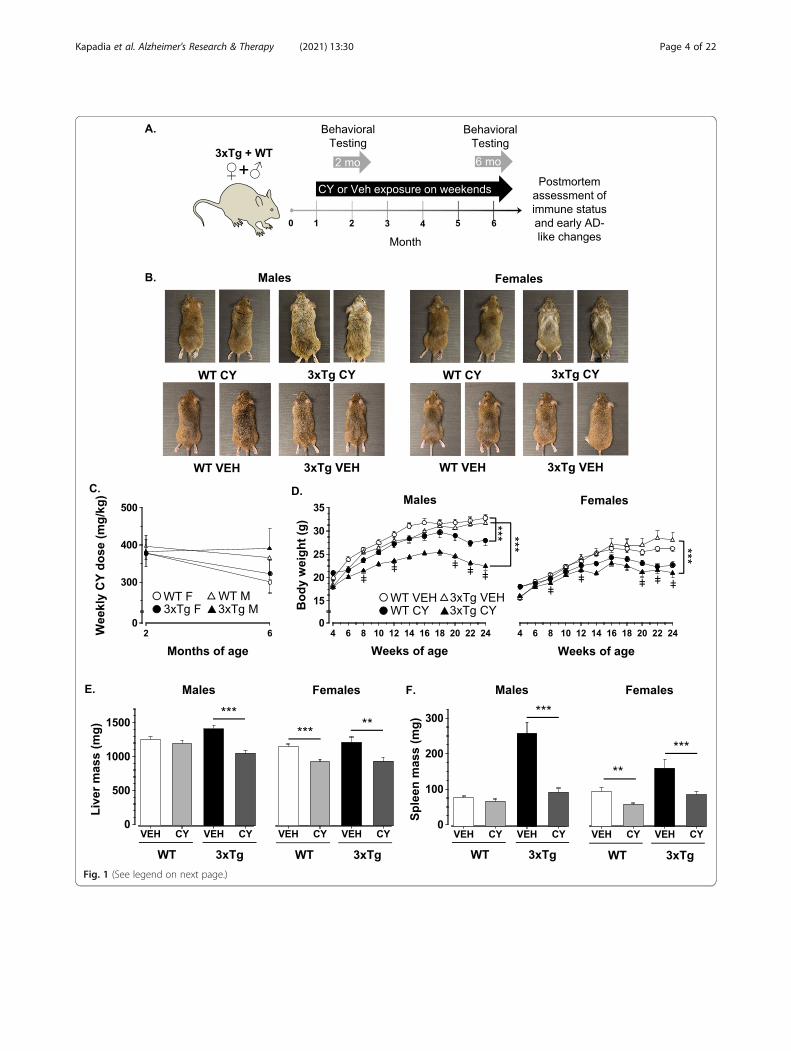
Fig. 1 (See legend on next page.)
Kapadia et al. Alzheimer's Research & Therapy (2021) 13:30 Page 4 of 22
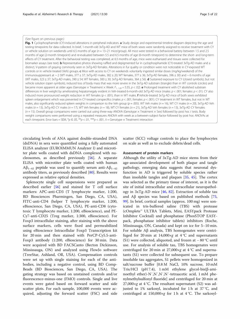
(See figure on previous page.)Fig. 1 Cyclophosphamide (CY)-induced alterations in peripheral indicators. a Study design and experimental timeline diagram depicting the age andtesting timepoints for data collected. In brief, 1-month-old 3xTg-AD and WT mice of both sexes were randomly assigned to receive treatment with CYor vehicle solution on weekends until 6.5 months of age (n = 13–21 mice/group). All mice were tested in a behavioral battery between 1.5 and 2.5months of age (2-month timepoint) and re-evaluated between 5.5 and 6.5 months of age (6-month timepoint) to determine the short- and long-termeffects of CY treatment. After the behavioral testing was completed, at 6.5months of age, mice were euthanized and tissues were collected forbiomarker assays (see text). b Representative photos showing ruffled and depigmented fur in cyclophosphamide (CY)-treated 3xTg-AD males and adistinct, V-pattern of graying commonly seen in 3xTg-AD females. Alterations in fur quality or condition were not noticeable in CY-exposed WTcontrols or in vehicle-treated (Veh) groups. c Mice with access to CY on weekends voluntarily ingested similar doses (mg/kg/weekend) of theimmunosuppressant at ~ 2 (WT males, 377 ± 37; 3xTg-AD males, 382 ± 20; WT females, 377 ± 36; 3xTg-AD females, 396 ± 30) and ~ 6months of age(WT males, 323 ± 37; 3xTg-AD males, 390 ± 54; WT females, 300 ± 30; 3xTg-AD females, 364 ± 56). d Sustained exposure to CY (closed symbols), but notvehicle solution (open symbols), induced loss of body mass that was more severe in the 3xTg-AD substrain (triangle) than in WT controls (circles) andbecame more apparent at older ages (Genotype × Treatment × Week: F1, 120 = 5.55, p < .02). e Prolonged treatment with CY abolished substraindifferences in liver weight by ameliorating hepatomegaly evident in Veh-treated 6-month-old 3xTg-AD mice (males: p < .001; females: p < .01). CY alsoinduced more pronounced weight reduction in WT females (p < .001), than in WT males. f Vehicle-treated 3xTg-AD mice of both sexes exhibitedspleen enlargement which was prevented in CY-treated conspecifics (males: p < .001; females: p < .001). CY treatment in WT females, but not in WTmales, also significantly reduced spleen weights in comparison to the Veh group (p = .005). WT Veh males (n = 16), WT CY males (n = 20), 3xTg-AD Vehmales (n = 13), 3xTg-AD CY males (n = 17), WT Veh females (n = 18), WT CY females (n = 21), 3xTg-AD Veh females (n = 13), 3xTg-AD CY females(n = 15). Overall group comparisons were carried out using three-way ANOVA (Genotype × Treatment × Sex) followed by post hoc t tests. Bodyweight comparisons were performed using a repeated measures ANOVA with week as a between-subject factor followed by post hoc ANOVAs ateach timepoint. Error bars = SEM, *p≤ .05, **p < .01, ***p < .001, ‡ = Genotype × Treatment interaction
Kapadia et al. Alzheimer's Research & Therapy (2021) 13:30 Page 5 of 22
circulating levels of ANA against double-stranded DNA(dsDNA) in sera were quantified using a fully automatedELISA analyzer (EUROIMMUN Analyzer I) and microti-ter plate wells coated with dsDNA complexed with nu-cleosomes, as described previously [56]. A separateELISA with microtiter plate wells coated with humanAβ1–42 peptide was used to quantify serum anti-Aβ1–42antibody titers, as previously described [80]. Results wereexpressed as relative optical densities.Splenocyte single cell suspensions were prepared as
described earlier [56] and stained for T cell surfacemarkers: APC-anti-CD3 (T lymphocyte marker, 1:200,BD Biosciences Pharmingen, San Diego, CA, USA),FITC-anti-CD4 (helper T lymphocyte marker, 1:200,eBioscience, San Diego, CA, USA), PE-anti-CD8 (cyto-toxic T lymphocyte marker, 1:200, eBioscience), and PE-Cy7-anti-CD25 (Treg marker, 1:300, eBioscience). ForFoxp3 intracellular staining, after staining with the abovesurface markers, cells were fixed and permeabilizedusing eBioscience Intracellular Foxp3 Transcription kitfor 20 min and then stained with PerCP-Cy5.5-anti-Foxp3 antibody (1:200, eBioscience) for 30 min. Datawere acquired with BD FACSCanto (Becton Dickinson,Mississauga, ON) and analyzed using FlowJo software(TreeStar, Ashland, OR, USA). Compensation controlswere set up with single staining for each of the anti-bodies, including a negative control, using BD Comp-Beads (BD Biosciences, San Diego, CA, USA). Thegating strategy was based on unstained controls and/orfluorescence-minus-one (FMO) controls. Single and liveevents were gated based on forward scatter and sidescatter plots. For each sample, 100,000 events were ac-quired, adjusting the forward scatter (FSC) and side
scatter (SCC) voltage controls to place the lymphocyteson scale as well as to exclude debris/dead cells.
Assessment of protein markersAlthough the utility of 3xTg-AD mice stems from theirage-associated development of both plaque and tanglepathology, emerging data suggests that neuronal dys-function in AD is triggered by soluble species ratherthan insoluble tangles and plaques [35, 45]. The cortexwas selected as the primary tissue of interest, as it is thesite of initial intracellular and extracellular neuropathol-ogy in 3xTg-AD mice [46, 82]. Extraction of soluble tauand Aβ species was based on published methods [75,99]. In brief, cortical samples (approx. 100 mg) were son-icated in tris-buffered saline (TBS) with protease(cOmplete™ ULTRA Tablets, Mini, EASYpack ProteaseInhibitor Cocktail) and phosphatase (PhosSTOP EASY-pack, phosphatase inhibitor tablets) inhibitors (Roche,Mississauga, ON, Canada) and kept on ice for 5–10min.For soluble Aβ analysis, TBS homogenates were centri-fuged for 20 min at 14,000×g at 4 °C and supernatants(S1) were collected, aliquoted, and frozen at − 80 °C untiluse. For analysis of soluble tau, TBS homogenates werecentrifuged for 20 min at 27,000×g at 4 °C and superna-tants (S1) were collected for subsequent use. To prepareinsoluble tau aggregates, S1 pellets were homogenized insalt/sucrose buffer [0.8 M NaCl, 10% sucrose, 10 mMTris/HCl (pH 7.4), 1 mM ethylene glycol-bis(β-ami-noethyl ether)-N′,N′,N′,N′-tetraacetic acid, 1 mM phe-nylmethylsulfonyl fluoride] and centrifuged for 20 min at27,000×g at 4 °C. The resultant supernatant (S2) was ad-justed to 1% sarkosyl, incubated for 1 h at 37 °C, andcentrifuged at 150,000×g for 1 h at 4 °C. The sarkosyl-
Kapadia et al. Alzheimer's Research & Therapy (2021) 13:30 Page 6 of 22
insoluble pellet was then re-suspended in TE buffer [10mM Tris/HCl (pH 8.0), 1 mM ethylene diamine tetraace-tic acid] and stored at − 80 °C for subsequent analysis.Protein concentrations in each fraction were measuredusing a detergent-compatible protein assay (Bio-Rad La-boratories, Mississauga, ON, Canada).Aβ42 protein levels in TBS-soluble S1 fractions were
measured by Chemiluminescent BetaMark x-42 ELISAper the manufacturer’s instructions (BioLegend, SanDiego, CA, USA). Concentrations were acquired with aMultiskanGO and SkanIt software (Thermo Scientific,Nepean, ON, Canada) at 620 nm. This ELISA recognizesboth mouse and human Aβ42, and therefore, mouseAβ42 values assayed in the corresponding wild-typegroups were subtracted as background. Values are pre-sented as pg human Aβ42 per mg of total protein.TBS-soluble and sarkosyl-insoluble total tau and phos-
phorylated tau were measured using western blotting.Ten and 15 μg of total protein were resolved on 10% gelsand transferred to polyvinylidene fluoride membranes(Bio-Rad, Hercules, CA, USA) for analysis of solubleand insoluble tau species, respectively. The mem-branes were treated as described [75] for detectionwith primary antibodies anti-tau (tau46, 1:1000; Cov-ance, Princeton, NJ, USA) and anti-phospho-tau(D9F4G, 1:1000; Cell Signaling Technology, Danvers,MA, USA), which recognize both mouse and humantau [89] and incubated in secondary antibodies IRDye680-conjugated goat anti-rabbit and IRDye 800CW-conjugated goat anti-mouse (1:10,000, Li-Cor Biosci-ences, Lincoln, NE, USA). Band intensities were quan-tified by densitometry by normalizing to mouse β-actin monoclonal antibody (1:10,000; BioLegend).
Assessment of RNA expressionRNA was extracted from cortical samples in TRIzolusing RNeasy spin columns (Qiagen, Mississauga, ON),complementary DNA was synthesized, and quantitativereal-time polymerase chain reaction was performed asdescribed previously [75]. Primers were designed usingPrimer3 software (http://bioinfo.ut.ee/primer3/) and or-dered from IDT (Coralville, IA, USA). BDNF mRNAcopy number in each sample was normalized to its β-
Table 1 Primer sequences used for qRT-PCR
Gene Accession Forward prim
BDNF NM_001048139.1 GCGGCAGAT
β-actin NM_007393.5 AGCCATGTAC
H2afy NM_001159513.1 CCCGGAAGTC
H2afy2 NM_207000.2 CGTTCCCCAG
Gapdh NM_001289726.1 GTGGAGTCAT
Hprt NM_013556.2 GGAGTCCTGT
actin mRNA copy number [56, 97]. The macroH2A vari-ant of the canonical histone H2A is encoded by twogenes that produce distinct proteins, H2afy (encodesmH2A1) and H2afy2 (encodes mH2A2). Expression ofboth was analyzed as described previously [123]. Genesof interest were normalized against the geometric meanof GAPDH and HPRT, and relative enrichment was nor-malized to vehicle-treated WT controls. All primer se-quences are shown in Table 1.
Statistical analysisWe previously described Genotype [69] and Sex-relateddifferences in 3xTg-AD mice [56]. However, the focus ofthis study was significant Genotype × Treatment × Sexor Genotype × Treatment interactions. Raw data ana-lyses were performed using SPSS 20 software (IBMCorp., Armonk, NY, USA). Normal distribution of thedata was tested by the Shapiro-Wilk test. When data de-parted from normality, the overall assumption was thatparametric tests were robust enough to detect significantgroup differences, since the cohorts were independentand population variances were comparable, as revealedby Levene’s test. Analysis of variance (ANOVA),ANOVA with repeated measures, analysis of covariance(ANCOVA), and chi-square test were used for groupcomparisons. Treatment, Genotype, and Sex were con-sidered between-group factors, and Age or Week aswithin-group factors, where applicable. If significant in-teractions were detected, Student’s t test was used inpost hoc comparisons. Partial eta-squared (η2p) andgeneralized eta-squared (η2g) were used as measures ofeffect size for all effects and interactions reported forANOVAs [95] and ANOVA with repeated measures [6],respectively. For reference, Cohen’s benchmarks forsmall (0.01), medium (0.06), and large (0.14) effects arerecommended for these measures [27, 37, 95]. Pearson’scorrelation coefficients were calculated when examiningbivariate linear relationships for normal variables. Thecriterion for statistical significance was set at p ≤ .05.Graphs display mean values ± SEM. Significant differ-ences of p ≤ .05, p < .01, and p < .001 are shown as *, **,and ***, respectively.
er Reverse primer
AAAAAGACTGC CTTATGAATCGCCAGCCAAT
GTAGCCATCC CTCTCAGCTGTGGTGGTGAA
TAAGAAGCAGGG AGGATTGATTATGGCCTCCACC
TGGCAGAAACT CCTGCACGTAGATGCCGAT
ACTGGAACATGTAG AATGGTGAAGGTCGGTGTG
TGATGTTGCCAGTA GGGACGCAGCAACTGACATTTCTA
Kapadia et al. Alzheimer's Research & Therapy (2021) 13:30 Page 7 of 22
ResultsPeripheral effectsThe earliest observable effects of sustained CY intakewere the development of distinct patterns of fur grayingin 3xTg-AD mice, noticeable after the second month ofexposure. Representative photos exemplify commonlyobserved ruffled and gray hair in 3xTg-AD males at 6months of age (Fig. 1b). In contrast, affected age-matched 3xTg-AD females exhibited a symmetrical, V-like pattern of discoloration. These effects were not seenin CY-treated WT controls or in 3xTg-AD mice exposedto vehicle solution. They were not associated with differ-ences in CY dosage, as drug-treated 3xTg-AD and WTgroups ingested comparable amounts of CY when indi-vidual intake was measured over single weekends at ~ 2months (Genotype: F1, 69 = 1.098, n.s., η2p = .02; Sex: F1,69 = 1.20, n.s., η2p = .02) and ~ 6months of age (Geno-type: F1, 67 = 2.287, n.s., η2p = .03; Sex: F1, 67 = .314, n.s.,η2p = .01, Fig. 1c). Despite this similarity, CY-treated3xTg-AD mice showed more profound weight loss thanCY-treated WT controls, which became more apparentwith time (Genotype × Treatment × Week: F9, 1080 =3.280, p < .001, η2g = .03, Fig. 1d).Given a positive correlation between body and liver
weight at sacrifice (r128 = 0.797, p < .001), body weightwas used as a covariate in ANCOVA, which revealedheavier livers in 3xTg-AD mice than in WT controls(Genotype: F1, 119 = 10.840, p < .001, η2p = .083). Sus-tained exposure to CY reduced liver weight comparablyin all groups except in WT male mice (Genotype ×Treatment × Sex: F1, 119 = 5.207, p = .024, η2p = .042,Fig. 1e). Although exposure to CY reduced spleen weightin a similar pattern (Treatment: F1, 119 = 24.743, p < .001,η2p = .172, Fig. 1f), this effect was most profound in3xTg-AD males (Genotype × Treatment × Sex: F1, 120 =8.259, p = .005, η2p = .065).
Splenic T lymphocytesThe loss of CD4/CD8 markers and the emergence of“double-negative” clones of T cells are well-establishedphenomena in systemic autoimmunity [113, 115]. Con-sidering that the spleen is a major source of immunecells [71], we investigated if CY alters the splenic distri-bution of T cell populations using flow cytometry. Thestrategies employed to gate CD3+, CD3+CD4+,CD3+CD8+, and Foxp3+CD25+CD4+ cells are shown inFig. 2a. Chronic intake of CY mitigated the loss of CD3+
cells in 3xTg-AD mice, irrespective of sex and withoutaffecting WT controls (Genotype × Treatment: F1, 58 =25.809, p < .001, η2p = .31, Fig. 2b). Compared to age-matched WT groups (which did not show sex differ-ences), 3xTg-AD males had fewer CD3+ cells in com-parison to their female conspecifics (Genotype × Sex: F1,58 = 5.994, p = .017, η2p = .09). Importantly, CY similarly
prevented the decline of CD3+CD4+ T cells (Genotype ×Treatment: F1, 58 = 27.923, p < .001, η2p = .33, Fig. 2c)and CD3+CD8+ T cells in 3xTg-AD mice (Genotype ×Treatment: F1, 58 = 7.136, p = .01, η2p = .11, Fig. 2d). Weobserved that the proportion of CD4+ regulatory T cells(Tregs) expressing CD25 and Foxp3 was higher in bothmale and female 3xTg-AD mice compared to WT con-specifics (Genotype: F1, 32 = 101.511, p < .001, η2p = .76,Fig. 2e). Again, chronic intake of CY attenuated the shiftin balance towards T regulatory cells in the CD4+ popu-lation in 3xTg-AD mice, irrespective of sex and withoutaffecting WT controls (Genotype × Treatment: F1, 32 =31.464, p < .001, η2p = .49, Fig. 2e).In our original report [69], we made an attempt to
compare lymphocyte populations in the bone marrow(which is a primary lymphoid organ) by flushing cells fromthe medullary cavity of femoral bones dissected from 1-year-old males. We were unable to do this comparison be-cause a needle could not be inserted into the femoral cav-ity in 3xTg-AD males due to ossification. Furthermore,the femur was solid and pale, suggesting an absence ofbone marrow cells (data not reported). Interestingly, incomparison to other groups, sustained CY treatment re-stored normal, red appearance of the femur of 6-month-old 3xTg-AD males (supplemental data).
Serological measuresBy 6months of age, 3xTg-AD males (and females to alesser degree) exhibit robust signs of autoimmunity in-cluding low hematocrit and hyperproduction of serumautoantibodies to nuclear antigens [56]. In comparisonto Veh controls, prolonged CY exposure loweredhematocrit in all groups except 3xTg-AD males (Geno-type × Treatment × Sex: F1, 73 = 8.399, p = .005, η2p =.10, Fig. 3a). This genotype- and sex-dependent effect ofCY was accompanied by pronounced alterations inserum autoantibodies to nuclear antigens (ANA; χ2 =60.596, df = 7, p < .001, Table 2). Although weak ANAreactivity was noted in 3 out of a total 68 (3/68) CY-treated mice, serum samples from ~ 60% of Veh-treatedanimals showed distinct staining patterns dependent ongenotype and sex (Fig. 3b). In particular, a subset of WTcontrol males (3/16) displayed cytoplasmic (Golgi-like)staining, while nearly all 3xTg-AD males (12/13) exhib-ited moderate to strong homogeneous staining of thenucleus and chromosomes. Interestingly, serum samplesfrom two thirds of Veh WT females also produced stain-ing of the nucleus (9/18) and nucleoli (3/18). In line withthese qualitative findings, exposure to CY reduced serumlevels of antibodies to dsDNA in all treated groups(Treatment: F1, 119 = 45.126, p < .001, η2p = .28, Fig. 3c).However, this mitigation was more prominent in 3xTg-AD males, which exhibited higher levels of anti-dsDNAthan 3xTg-AD females or WT female controls
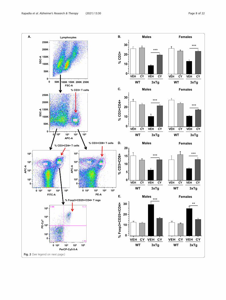
Fig. 2 (See legend on next page.)
Kapadia et al. Alzheimer's Research & Therapy (2021) 13:30 Page 8 of 22
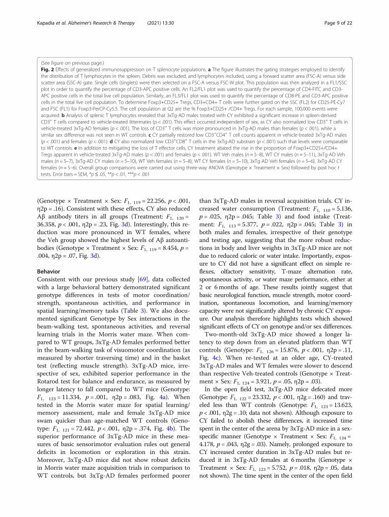
(See figure on previous page.)Fig. 2 Effects of generalized immunosuppression on T splenocyte populations. a The figure illustrates the gating strategies employed to identifythe distribution of T lymphocytes in the spleen. Debris was excluded, and lymphocytes included, using a forward scatter area (FSC-A) versus sidescatter area (SSC-A) gate. Single cells (singlets) were then selected on a FSC-A versus FSC-W plot. This population was then analyzed in a FL1/SSCplot in order to quantify the percentage of CD3-APC positive cells. An FL2/FL1 plot was used to quantify the percentage of CD4-FITC and CD3-APC positive cells in the total live cell population. Similarly, an FL3/FL1 plot was used to quantify the percentage of CD8-PE and CD3-APC positivecells in the total live cell population. To determine Foxp3+CD25+ Tregs, CD3+CD4+ T cells were further gated on the SSC (FL2) for CD25-PE-Cy7and FSC (FL1) for Foxp3-PerCP-Cy5.5. The cell population at Q2 are the % Foxp3+CD25+ /CD4+ Tregs. For each sample, 100,000 events wereacquired. b Analysis of splenic T lymphocytes revealed that 3xTg-AD males treated with CY exhibited a significant increase in spleen-derivedCD3+ T cells compared to vehicle-treated littermates (p < .001). This effect occurred independent of sex, as CY also normalized low CD3+ T cells invehicle-treated 3xTg-AD females (p < .001). The loss of CD3+ T cells was more pronounced in 3xTg-AD males than females (p < .001), while asimilar sex difference was not seen in WT controls. c CY partially restored low CD3+CD4+ T cell counts apparent in vehicle-treated 3xTg-AD males(p < .001) and females (p < .001). d CY also normalized low CD3+CD8+ T cells in the 3xTg-AD substrain (p < .001) such that levels were comparableto WT controls. e In addition to mitigating the loss of T effector cells, CY treatment abated the rise in the proportion of Foxp3+CD25+/CD4+Tregs apparent in vehicle-treated 3xTg-AD males (p < .001) and females (p < .001). WT Veh males (n = 5–8), WT CY males (n = 5–11), 3xTg-AD Vehmales (n = 5–7), 3xTg-AD CY males (n = 5–10), WT Veh females (n = 5–8), WT CY females (n = 5–10), 3xTg-AD Veh females (n = 5–6), 3xTg-AD CYfemales (n = 5–6). Overall group comparisons were carried out using three-way ANOVA (Genotype × Treatment × Sex) followed by post hoc ttests. Error bars = SEM, *p≤ .05, **p < .01, ***p < .001
Kapadia et al. Alzheimer's Research & Therapy (2021) 13:30 Page 9 of 22
(Genotype × Treatment × Sex: F1, 119 = 22.256, p < .001,η2p = .16). Consistent with these effects, CY also reducedAβ antibody titers in all groups (Treatment: F1, 120 =36.358, p < .001, η2p = .23, Fig. 3d). Interestingly, this re-duction was more pronounced in WT females, wherethe Veh group showed the highest levels of Aβ autoanti-bodies (Genotype × Treatment × Sex: F1, 119 = 8.454, p =.004, η2p = .07, Fig. 3d).
BehaviorConsistent with our previous study [69], data collectedwith a large behavioral battery demonstrated significantgenotype differences in tests of motor coordination/strength, spontaneous activities, and performance inspatial learning/memory tasks (Table 3). We also docu-mented significant Genotype by Sex interactions in thebeam-walking test, spontaneous activities, and reversallearning trials in the Morris water maze. When com-pared to WT groups, 3xTg-AD females performed betterin the beam-walking task of visuomotor coordination (asmeasured by shorter traversing time) and in the baskettest (reflecting muscle strength). 3xTg-AD mice, irre-spective of sex, exhibited superior performance in theRotarod test for balance and endurance, as measured bylonger latency to fall compared to WT mice (Genotype:F1, 123 = 11.334, p = .001, η2p = .083, Fig. 4a). Whentested in the Morris water maze for spatial learning/memory assessment, male and female 3xTg-AD miceswam quicker than age-matched WT controls (Geno-type: F1, 121 = 72.442, p < .001, η2p = .374, Fig. 4b). Thesuperior performance of 3xTg-AD mice in these mea-sures of basic sensorimotor evaluation rules out generaldeficits in locomotion or exploration in this strain.Moreover, 3xTg-AD mice did not show robust deficitsin Morris water maze acquisition trials in comparison toWT controls, but 3xTg-AD females performed poorer
than 3xTg-AD males in reversal acquisition trials. CY in-creased water consumption (Treatment: F1, 110 = 5.136,p = .025, η2p = .045; Table 3) and food intake (Treat-ment: F1, 113 = 5.377, p = .022, η2p = .045; Table 3) inboth males and females, irrespective of their genotypeand testing age, suggesting that the more robust reduc-tions in body and liver weights in 3xTg-AD mice are notdue to reduced caloric or water intake. Importantly, expos-ure to CY did not have a significant effect on simple re-flexes, olfactory sensitivity, T-maze alternation rate,spontaneous activity, or water maze performance, either at2 or 6months of age. These results jointly suggest thatbasic neurological function, muscle strength, motor coord-ination, spontaneous locomotion, and learning/memorycapacity were not significantly altered by chronic CY expos-ure. Our analysis therefore highlights tests which showedsignificant effects of CY on genotype and/or sex differences.Two-month-old 3xTg-AD mice showed a longer la-
tency to step down from an elevated platform than WTcontrols (Genotype: F1, 126 = 15.876, p < .001, η2p = .11,Fig. 4c). When re-tested at an older age, CY-treated3xTg-AD males and WT females were slower to descendthan respective Veh-treated controls (Genotype × Treat-ment × Sex: F1, 124 = 3.921, p = .05, η2p = .03).In the open field test, 3xTg-AD mice defecated more
(Genotype: F1, 122 = 23.332, p < .001, η2g = .160) and trav-eled less than WT controls (Genotype: F1, 123 = 13.623,p < .001, η2g = .10; data not shown). Although exposure toCY failed to abolish these differences, it increased timespent in the center of the arena by 3xTg-AD mice in a sex-specific manner (Genotype × Treatment × Sex: F1, 124 =4.178, p = .043, η2g = .03). Namely, prolonged exposure toCY increased center duration in 3xTg-AD males but re-duced it in 3xTg-AD females at 6months (Genotype ×Treatment × Sex: F1, 123 = 5.752, p = .018, η2p = .05, datanot shown). The time spent in the center of the open field
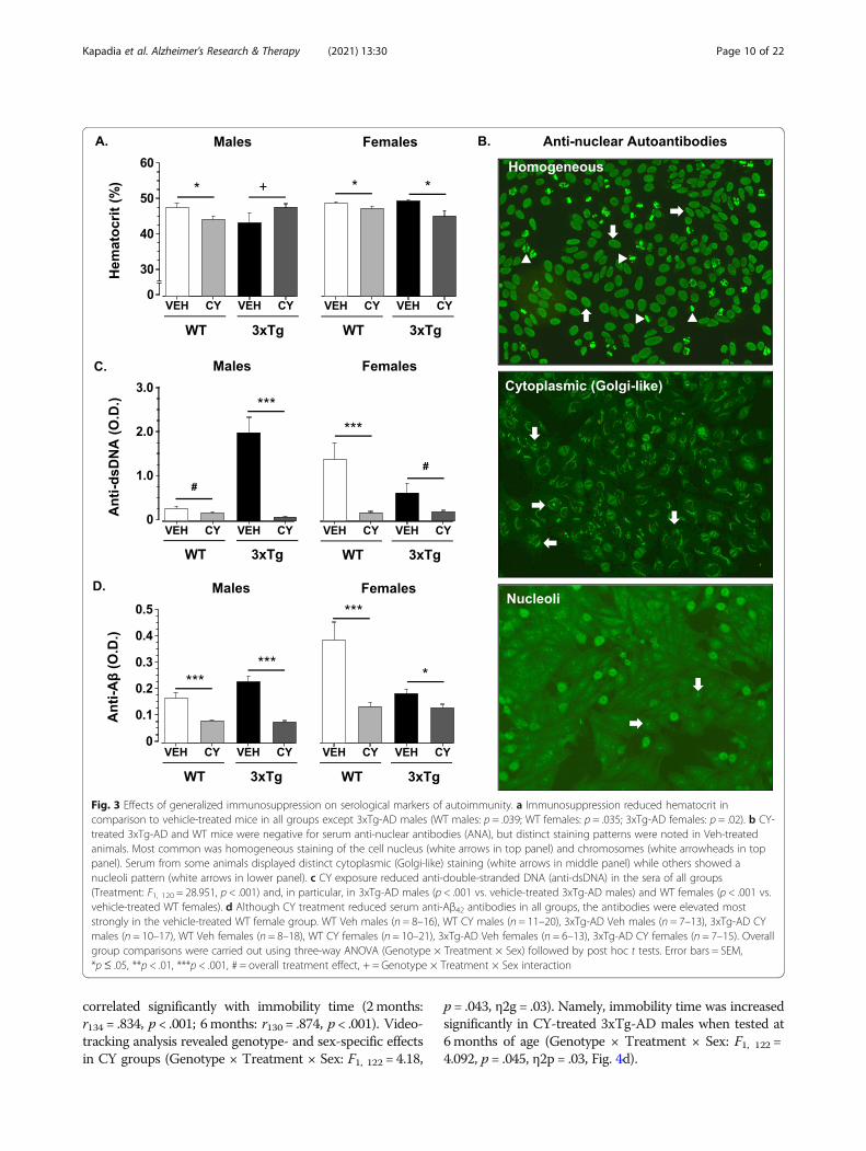
Fig. 3 Effects of generalized immunosuppression on serological markers of autoimmunity. a Immunosuppression reduced hematocrit incomparison to vehicle-treated mice in all groups except 3xTg-AD males (WT males: p = .039; WT females: p = .035; 3xTg-AD females: p = .02). b CY-treated 3xTg-AD and WT mice were negative for serum anti-nuclear antibodies (ANA), but distinct staining patterns were noted in Veh-treatedanimals. Most common was homogeneous staining of the cell nucleus (white arrows in top panel) and chromosomes (white arrowheads in toppanel). Serum from some animals displayed distinct cytoplasmic (Golgi-like) staining (white arrows in middle panel) while others showed anucleoli pattern (white arrows in lower panel). c CY exposure reduced anti-double-stranded DNA (anti-dsDNA) in the sera of all groups(Treatment: F1, 120 = 28.951, p < .001) and, in particular, in 3xTg-AD males (p < .001 vs. vehicle-treated 3xTg-AD males) and WT females (p < .001 vs.vehicle-treated WT females). d Although CY treatment reduced serum anti-Aβ42 antibodies in all groups, the antibodies were elevated moststrongly in the vehicle-treated WT female group. WT Veh males (n = 8–16), WT CY males (n = 11–20), 3xTg-AD Veh males (n = 7–13), 3xTg-AD CYmales (n = 10–17), WT Veh females (n = 8–18), WT CY females (n = 10–21), 3xTg-AD Veh females (n = 6–13), 3xTg-AD CY females (n = 7–15). Overallgroup comparisons were carried out using three-way ANOVA (Genotype × Treatment × Sex) followed by post hoc t tests. Error bars = SEM,*p≤ .05, **p < .01, ***p < .001, # = overall treatment effect, + = Genotype × Treatment × Sex interaction
Kapadia et al. Alzheimer's Research & Therapy (2021) 13:30 Page 10 of 22
correlated significantly with immobility time (2months:r134 = .834, p < .001; 6months: r130 = .874, p < .001). Video-tracking analysis revealed genotype- and sex-specific effectsin CY groups (Genotype × Treatment × Sex: F1, 122 = 4.18,
p = .043, η2g = .03). Namely, immobility time was increasedsignificantly in CY-treated 3xTg-AD males when tested at6months of age (Genotype × Treatment × Sex: F1, 122 =4.092, p = .045, η2p = .03, Fig. 4d).
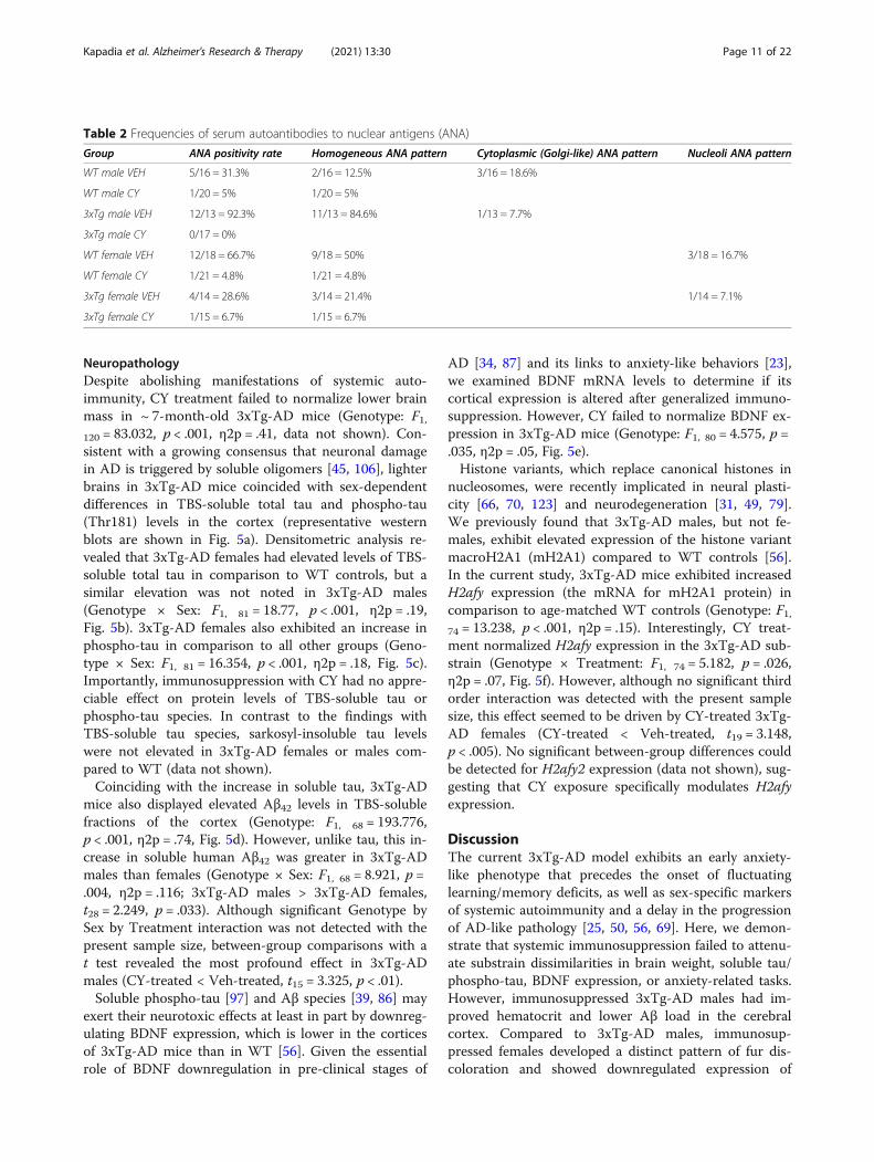
Table 2 Frequencies of serum autoantibodies to nuclear antigens (ANA)
Group ANA positivity rate Homogeneous ANA pattern Cytoplasmic (Golgi-like) ANA pattern Nucleoli ANA pattern
WT male VEH 5/16 = 31.3% 2/16 = 12.5% 3/16 = 18.6%
WT male CY 1/20 = 5% 1/20 = 5%
3xTg male VEH 12/13 = 92.3% 11/13 = 84.6% 1/13 = 7.7%
3xTg male CY 0/17 = 0%
WT female VEH 12/18 = 66.7% 9/18 = 50% 3/18 = 16.7%
WT female CY 1/21 = 4.8% 1/21 = 4.8%
3xTg female VEH 4/14 = 28.6% 3/14 = 21.4% 1/14 = 7.1%
3xTg female CY 1/15 = 6.7% 1/15 = 6.7%
Kapadia et al. Alzheimer's Research & Therapy (2021) 13:30 Page 11 of 22
NeuropathologyDespite abolishing manifestations of systemic auto-immunity, CY treatment failed to normalize lower brainmass in ~ 7-month-old 3xTg-AD mice (Genotype: F1,120 = 83.032, p < .001, η2p = .41, data not shown). Con-sistent with a growing consensus that neuronal damagein AD is triggered by soluble oligomers [45, 106], lighterbrains in 3xTg-AD mice coincided with sex-dependentdifferences in TBS-soluble total tau and phospho-tau(Thr181) levels in the cortex (representative westernblots are shown in Fig. 5a). Densitometric analysis re-vealed that 3xTg-AD females had elevated levels of TBS-soluble total tau in comparison to WT controls, but asimilar elevation was not noted in 3xTg-AD males(Genotype × Sex: F1, 81 = 18.77, p < .001, η2p = .19,Fig. 5b). 3xTg-AD females also exhibited an increase inphospho-tau in comparison to all other groups (Geno-type × Sex: F1, 81 = 16.354, p < .001, η2p = .18, Fig. 5c).Importantly, immunosuppression with CY had no appre-ciable effect on protein levels of TBS-soluble tau orphospho-tau species. In contrast to the findings withTBS-soluble tau species, sarkosyl-insoluble tau levelswere not elevated in 3xTg-AD females or males com-pared to WT (data not shown).Coinciding with the increase in soluble tau, 3xTg-AD
mice also displayed elevated Aβ42 levels in TBS-solublefractions of the cortex (Genotype: F1, 68 = 193.776,p < .001, η2p = .74, Fig. 5d). However, unlike tau, this in-crease in soluble human Aβ42 was greater in 3xTg-ADmales than females (Genotype × Sex: F1, 68 = 8.921, p =.004, η2p = .116; 3xTg-AD males > 3xTg-AD females,t28 = 2.249, p = .033). Although significant Genotype bySex by Treatment interaction was not detected with thepresent sample size, between-group comparisons with at test revealed the most profound effect in 3xTg-ADmales (CY-treated < Veh-treated, t15 = 3.325, p < .01).Soluble phospho-tau [97] and Aβ species [39, 86] may
exert their neurotoxic effects at least in part by downreg-ulating BDNF expression, which is lower in the corticesof 3xTg-AD mice than in WT [56]. Given the essentialrole of BDNF downregulation in pre-clinical stages of
AD [34, 87] and its links to anxiety-like behaviors [23],we examined BDNF mRNA levels to determine if itscortical expression is altered after generalized immuno-suppression. However, CY failed to normalize BDNF ex-pression in 3xTg-AD mice (Genotype: F1, 80 = 4.575, p =.035, η2p = .05, Fig. 5e).Histone variants, which replace canonical histones in
nucleosomes, were recently implicated in neural plasti-city [66, 70, 123] and neurodegeneration [31, 49, 79].We previously found that 3xTg-AD males, but not fe-males, exhibit elevated expression of the histone variantmacroH2A1 (mH2A1) compared to WT controls [56].In the current study, 3xTg-AD mice exhibited increasedH2afy expression (the mRNA for mH2A1 protein) incomparison to age-matched WT controls (Genotype: F1,74 = 13.238, p < .001, η2p = .15). Interestingly, CY treat-ment normalized H2afy expression in the 3xTg-AD sub-strain (Genotype × Treatment: F1, 74 = 5.182, p = .026,η2p = .07, Fig. 5f). However, although no significant thirdorder interaction was detected with the present samplesize, this effect seemed to be driven by CY-treated 3xTg-AD females (CY-treated < Veh-treated, t19 = 3.148,p < .005). No significant between-group differences couldbe detected for H2afy2 expression (data not shown), sug-gesting that CY exposure specifically modulates H2afyexpression.
DiscussionThe current 3xTg-AD model exhibits an early anxiety-like phenotype that precedes the onset of fluctuatinglearning/memory deficits, as well as sex-specific markersof systemic autoimmunity and a delay in the progressionof AD-like pathology [25, 50, 56, 69]. Here, we demon-strate that systemic immunosuppression failed to attenu-ate substrain dissimilarities in brain weight, soluble tau/phospho-tau, BDNF expression, or anxiety-related tasks.However, immunosuppressed 3xTg-AD males had im-proved hematocrit and lower Aβ load in the cerebralcortex. Compared to 3xTg-AD males, immunosup-pressed females developed a distinct pattern of fur dis-coloration and showed downregulated expression of
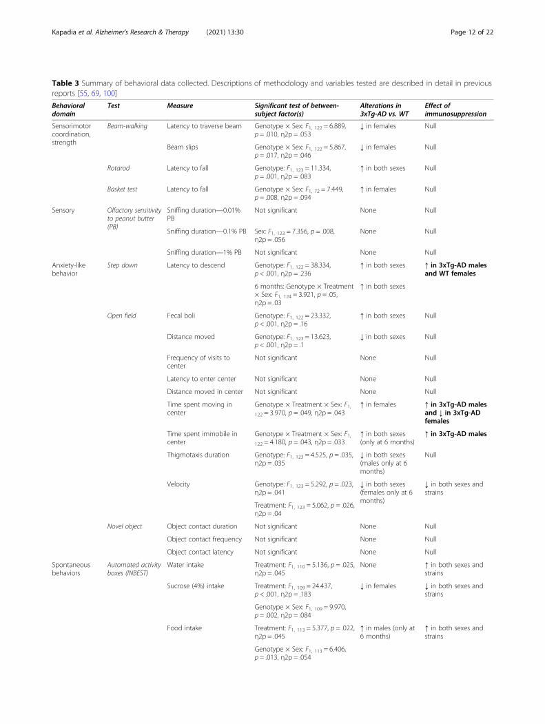
Table 3 Summary of behavioral data collected. Descriptions of methodology and variables tested are described in detail in previousreports [55, 69, 100]
Behavioraldomain
Test Measure Significant test of between-subject factor(s)
Alterations in3xTg-AD vs. WT
Effect ofimmunosuppression
Sensorimotorcoordination,strength
Beam-walking Latency to traverse beam Genotype × Sex: F1, 122 = 6.889,p = .010, η2p = .053
↓ in females Null
Beam slips Genotype × Sex: F1, 122 = 5.867,p = .017, η2p = .046
↓ in females Null
Rotarod Latency to fall Genotype: F1, 123 = 11.334,p = .001, η2p = .083
↑ in both sexes Null
Basket test Latency to fall Genotype × Sex: F1, 72 = 7.449,p = .008, η2p = .094
↑ in females Null
Sensory Olfactory sensitivityto peanut butter(PB)
Sniffing duration—0.01%PB
Not significant None Null
Sniffing duration—0.1% PB Sex: F1, 123 = 7.356, p = .008,η2p = .056
None Null
Sniffing duration—1% PB Not significant None Null
Anxiety-likebehavior
Step down Latency to descend Genotype: F1, 122 = 38.334,p < .001, η2p = .236
↑ in both sexes ↑ in 3xTg-AD malesand WT females
6 months: Genotype × Treatment× Sex: F1, 124 = 3.921, p = .05,η2p = .03
↑ in both sexes
Open field Fecal boli Genotype: F1, 122 = 23.332,p < .001, η2p = .16
↑ in both sexes Null
Distance moved Genotype: F1, 123 = 13.623,p < .001, η2p = .1
↓ in both sexes Null
Frequency of visits tocenter
Not significant None Null
Latency to enter center Not significant None Null
Distance moved in center Not significant None Null
Time spent moving incenter
Genotype × Treatment × Sex: F1,122 = 3.970, p = .049, η2p = .043
↑ in females ↑ in 3xTg-AD malesand ↓ in 3xTg-ADfemales
Time spent immobile incenter
Genotype × Treatment × Sex: F1,122 = 4.180, p = .043, η2p = .033
↑ in both sexes(only at 6 months)
↑ in 3xTg-AD males
Thigmotaxis duration Genotype: F1, 123 = 4.525, p = .035,η2p = .035
↓ in both sexes(males only at 6months)
Null
Velocity Genotype: F1, 123 = 5.292, p = .023,η2p = .041
↓ in both sexes(females only at 6months)
↓ in both sexes andstrains
Treatment: F1, 123 = 5.062, p = .026,η2p = .04
Novel object Object contact duration Not significant None Null
Object contact frequency Not significant None Null
Object contact latency Not significant None Null
Spontaneousbehaviors
Automated activityboxes (INBEST)
Water intake Treatment: F1, 110 = 5.136, p = .025,η2p = .045
None ↑ in both sexes andstrains
Sucrose (4%) intake Treatment: F1, 109 = 24.437,p < .001, η2p = .183
↓ in females ↓ in both sexes andstrains
Genotype × Sex: F1, 109 = 9.970,p = .002, η2p = .084
Food intake Treatment: F1, 113 = 5.377, p = .022,η2p = .045
↑ in males (only at6 months)
↑ in both sexes andstrains
Genotype × Sex: F1, 113 = 6.406,p = .013, η2p = .054
Kapadia et al. Alzheimer's Research & Therapy (2021) 13:30 Page 12 of 22
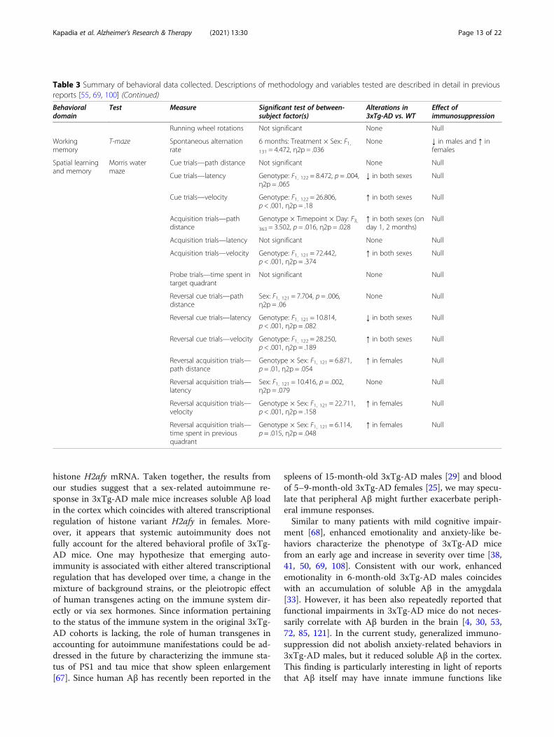
Table 3 Summary of behavioral data collected. Descriptions of methodology and variables tested are described in detail in previousreports [55, 69, 100] (Continued)
Behavioraldomain
Test Measure Significant test of between-subject factor(s)
Alterations in3xTg-AD vs. WT
Effect ofimmunosuppression
Running wheel rotations Not significant None Null
Workingmemory
T-maze Spontaneous alternationrate
6 months: Treatment × Sex: F1,131 = 4.472, η2p = .036
None ↓ in males and ↑ infemales
Spatial learningand memory
Morris watermaze
Cue trials—path distance Not significant None Null
Cue trials—latency Genotype: F1, 122 = 8.472, p = .004,η2p = .065
↓ in both sexes Null
Cue trials—velocity Genotype: F1, 122 = 26.806,p < .001, η2p = .18
↑ in both sexes Null
Acquisition trials—pathdistance
Genotype × Timepoint × Day: F3,363 = 3.502, p = .016, η2p = .028
↑ in both sexes (onday 1, 2 months)
Null
Acquisition trials—latency Not significant None Null
Acquisition trials—velocity Genotype: F1, 121 = 72.442,p < .001, η2p = .374
↑ in both sexes Null
Probe trials—time spent intarget quadrant
Not significant None Null
Reversal cue trials—pathdistance
Sex: F1, 121 = 7.704, p = .006,η2p = .06
None Null
Reversal cue trials—latency Genotype: F1, 121 = 10.814,p < .001, η2p = .082
↓ in both sexes Null
Reversal cue trials—velocity Genotype: F1, 122 = 28.250,p < .001, η2p = .189
↑ in both sexes Null
Reversal acquisition trials—path distance
Genotype × Sex: F1, 121 = 6.871,p = .01, η2p = .054
↑ in females Null
Reversal acquisition trials—latency
Sex: F1, 121 = 10.416, p = .002,η2p = .079
None Null
Reversal acquisition trials—velocity
Genotype × Sex: F1, 121 = 22.711,p < .001, η2p = .158
↑ in females Null
Reversal acquisition trials—time spent in previousquadrant
Genotype × Sex: F1, 121 = 6.114,p = .015, η2p = .048
↑ in females Null
Kapadia et al. Alzheimer's Research & Therapy (2021) 13:30 Page 13 of 22
histone H2afy mRNA. Taken together, the results fromour studies suggest that a sex-related autoimmune re-sponse in 3xTg-AD male mice increases soluble Aβ loadin the cortex which coincides with altered transcriptionalregulation of histone variant H2afy in females. More-over, it appears that systemic autoimmunity does notfully account for the altered behavioral profile of 3xTg-AD mice. One may hypothesize that emerging auto-immunity is associated with either altered transcriptionalregulation that has developed over time, a change in themixture of background strains, or the pleiotropic effectof human transgenes acting on the immune system dir-ectly or via sex hormones. Since information pertainingto the status of the immune system in the original 3xTg-AD cohorts is lacking, the role of human transgenes inaccounting for autoimmune manifestations could be ad-dressed in the future by characterizing the immune sta-tus of PS1 and tau mice that show spleen enlargement[67]. Since human Aβ has recently been reported in the
spleens of 15-month-old 3xTg-AD males [29] and bloodof 5–9-month-old 3xTg-AD females [25], we may specu-late that peripheral Aβ might further exacerbate periph-eral immune responses.Similar to many patients with mild cognitive impair-
ment [68], enhanced emotionality and anxiety-like be-haviors characterize the phenotype of 3xTg-AD micefrom an early age and increase in severity over time [38,41, 50, 69, 108]. Consistent with our work, enhancedemotionality in 6-month-old 3xTg-AD males coincideswith an accumulation of soluble Aβ in the amygdala[33]. However, it has been also repeatedly reported thatfunctional impairments in 3xTg-AD mice do not neces-sarily correlate with Aβ burden in the brain [4, 30, 53,72, 85, 121]. In the current study, generalized immuno-suppression did not abolish anxiety-related behaviors in3xTg-AD males, but it reduced soluble Aβ in the cortex.This finding is particularly interesting in light of reportsthat Aβ itself may have innate immune functions like
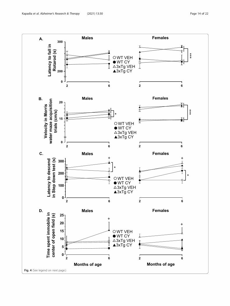
Fig. 4 (See legend on next page.)
Kapadia et al. Alzheimer's Research & Therapy (2021) 13:30 Page 14 of 22
(See figure on previous page.)Fig. 4 Cyclophosphamide (CY) modulation of anxiety-like behaviors in 3xTg-AD mice at 2 and 6months of age. a Performance in the Rotarodremained superior for 3xTg-AD mice (triangles), irrespective of sex, in comparison to age-matched WT controls (circles) (Genotype: F1, 123 = 11.334,p = .001, η2p = .083). Acute or prolonged CY treatment (closed symbols) did not significantly alter the latency to fall off the Rotarod. b Two- and 6-month-old 3xTg-AD males and females swam faster than age-matched WT controls in the Morris water maze acquisition trials (Genotype: F1, 121 =72.442, p < .001, η2p = .374). CY did not alter the swimming speed of 3xTg-AD mice or WT controls. c From an early age, 3xTg-AD males and femalestook longer than sex-matched WT controls to descend from an elevated platform in the step-down test, consistent with “acrophobia” (Genotype: F1,126 = 15.876, p < .001). After several months of CY exposure, 3xTg-AD males, but not WT controls, took longer to complete the step-down test incomparison to vehicle-treated animals, suggesting that prolonged immunosuppression exacerbated anxiety-like behavior (Genotype × Treatment ×Sex: F1, 124 = 3.921, p = .05). Sustained CY intake had no discernable impact on the step-down performance of 3xTg-AD females, but WT controls(similar to 3xTg-AD males) took longer to complete the task at 6months of age. d In the open field test, CY-treated 3xTg-AD males spent the mosttime immobile in the center of a large open field (Genotype × Treatment × Sex: F1, 123 = 4.092, p = .045). WT Veh males (n = 16), WT CY males (n = 20),3xTg-AD Veh males (n = 13), 3xTg-AD CY males (n = 17), WT Veh females (n = 18), WT CY females (n = 21), 3xTg-AD Veh females (n = 13), 3xTg-AD CYfemales (n = 15). Overall group comparisons were carried out using three-way ANOVA (Genotype × Treatment × Sex) followed by post hoc t tests.Error bars = SEM, *p≤ .05, **p < .01, ***p < .001, # = overall treatment effect, + = Genotype × Treatment × Sex interaction
Kapadia et al. Alzheimer's Research & Therapy (2021) 13:30 Page 15 of 22
antimicrobial activity [63, 76] and that disruption of im-mune pathways attenuates Aβ burden [94]. Restorationof increased splenic Foxp3+ Tregs to basal levels haspreviously been shown to coincide with a reduction inthe expression of Aβ in the hippocampus of 3xTg-ADmales [29]. Using CY, we see similar lessening of Aβburden in the cortex of 3xTg-AD males and females.Tregs play a major role in suppression of autoimmunepathology and are often poorly functioning in subjectswith autoimmune disease [118]. It is therefore possiblethat the increase in the proportion of Tregs in 3xTg-ADmice is a protective reaction to enhanced autoimmuneresponses in these animals. Mitigating the influence ofsystemic Foxp3+ Treg-mediated immunosuppression onimmunocytes may allow a re-balance of the immune re-sponse and reduced brain accumulation of Aβ [9]. Onemay speculate that the altered behavioral performance of3xTg-AD mice reflects allostatic load due toautoimmune-mediated clearance of neurotoxic aggre-gates from the brain [105]. Females may not be able toreadily mount such an immune response, renderingthem vulnerable to plaque/tangle accumulation at olderages. Indeed, we found that 3xTg-AD females, but notmales, exhibit an earlier rise in the amount of total andphosphorylated (Thr181) soluble tau in the cortex incomparison to WT controls. These findings complementrecent immunohistochemical data documenting that100% of 6-month-old 3xTg-AD females exhibitphospho-tau (Ser202/Thr205 and Ser422) in the hippo-campus whereas male 3xTg-AD mice show considerableneuropathological variability [10]. This sex discrepancyin soluble total and phosphorylated tau may help to ex-plain why accumulations of hyperphosphorylated tautangles are observed in the brains of 12-month-old3xTg-AD females [25] but not males [69].Although the production of antibodies to Aβ and
other antigens (nuclear and dsDNA) in 3xTg-AD malesis consistent with clinical studies reporting autoanti-bodies in AD patients [51], their presence in WT
females is an unexpected finding that requires furtherinvestigation. We previously noted that ~ 75% of agedWT males also showed varying degrees of ANA positiv-ity [69]. These unexpected results in the WT mice ofboth sexes support the notion that the hybrid strain gen-erated from 129 and C57BL/6 mice (ancestor to boththe WT and 3xTg-AD strains) is predisposed to spon-taneously develop autoimmune manifestations [18, 20].However, why the insertion of AD-related genes acceler-ates the progression of autoimmune manifestations (inmales in particular) remains to be determined. Similarly,more data are required to reveal the nature of the sex-specific patterns in depigmentation in 3xTg-AD miceexposed to CY. Hair graying, a typical sign of aging inmammals, has previously been linked to irreparableDNA damage that impairs the maintenance of melano-cyte stem cells with age [52]. It remains to be deter-mined if the graying in CY-treated 3xTg-AD micereflects an increased accumulation of phosphoramidemustard (the cytotoxic metabolite of CY) leading to ac-celerated DNA damage and an early-aging phenotype.Although the role of histone variants in the CNS is
only beginning to be studied, existing data suggest thatthey are critical regulators of neural plasticity [70, 123].The transcription of histone variants is highly responsiveto environmental stimuli [123], including age-relatedregulation in the brain [70]. Histone macroH2A is avariant of the canonical histone H2A and is encoded by2 genes that produce distinct proteins, H2afy (encodesmH2A1) and H2afy2 (encodes mH2A2). We recentlyshowed that H2afy, the mRNA for the histone variantmH2A1, is upregulated in the 3xTg-AD model, which isconsistent with studies that demonstrate that H2afy is amarker of disease activity in neurodegenerative disorders[49]. The current study demonstrates that upregulatedH2afy transcription can be modified by generalized im-munosuppression. This suggests that upregulation ofmH2A1 transcription may be driven by immune changesin AD. Since our data are limited to mRNA levels coding
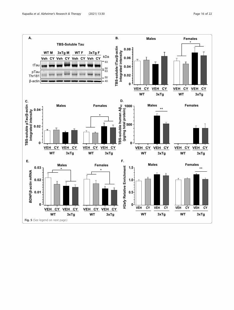
Fig. 5 (See legend on next page.)
Kapadia et al. Alzheimer's Research & Therapy (2021) 13:30 Page 16 of 22
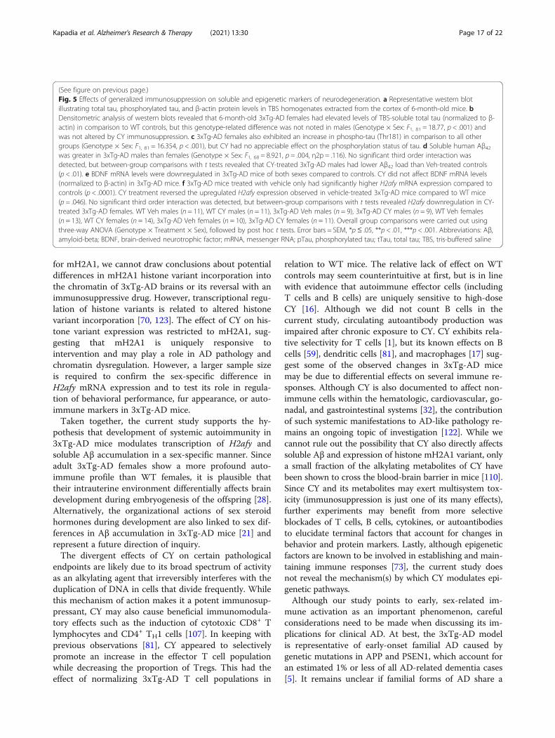
(See figure on previous page.)Fig. 5 Effects of generalized immunosuppression on soluble and epigenetic markers of neurodegeneration. a Representative western blotillustrating total tau, phosphorylated tau, and β-actin protein levels in TBS homogenates extracted from the cortex of 6-month-old mice. bDensitometric analysis of western blots revealed that 6-month-old 3xTg-AD females had elevated levels of TBS-soluble total tau (normalized to β-actin) in comparison to WT controls, but this genotype-related difference was not noted in males (Genotype × Sex: F1, 81 = 18.77, p < .001) andwas not altered by CY immunosuppression. c 3xTg-AD females also exhibited an increase in phospho-tau (Thr181) in comparison to all othergroups (Genotype × Sex: F1, 81 = 16.354, p < .001), but CY had no appreciable effect on the phosphorylation status of tau. d Soluble human Aβ42was greater in 3xTg-AD males than females (Genotype × Sex: F1, 68 = 8.921, p = .004, η2p = .116). No significant third order interaction wasdetected, but between-group comparisons with t tests revealed that CY-treated 3xTg-AD males had lower Aβ42 load than Veh-treated controls(p < .01). e BDNF mRNA levels were downregulated in 3xTg-AD mice of both sexes compared to controls. CY did not affect BDNF mRNA levels(normalized to β-actin) in 3xTg-AD mice. f 3xTg-AD mice treated with vehicle only had significantly higher H2afy mRNA expression compared tocontrols (p < .0001). CY treatment reversed the upregulated H2afy expression observed in vehicle-treated 3xTg-AD mice compared to WT mice(p = .046). No significant third order interaction was detected, but between-group comparisons with t tests revealed H2afy downregulation in CY-treated 3xTg-AD females. WT Veh males (n = 11), WT CY males (n = 11), 3xTg-AD Veh males (n = 9), 3xTg-AD CY males (n = 9), WT Veh females(n = 13), WT CY females (n = 14), 3xTg-AD Veh females (n = 10), 3xTg-AD CY females (n = 11). Overall group comparisons were carried out usingthree-way ANOVA (Genotype × Treatment × Sex), followed by post hoc t tests. Error bars = SEM, *p ≤ .05, **p < .01, ***p < .001. Abbreviations: Aβ,amyloid-beta; BDNF, brain-derived neurotrophic factor; mRNA, messenger RNA; pTau, phosphorylated tau; tTau, total tau; TBS, tris-buffered saline
Kapadia et al. Alzheimer's Research & Therapy (2021) 13:30 Page 17 of 22
for mH2A1, we cannot draw conclusions about potentialdifferences in mH2A1 histone variant incorporation intothe chromatin of 3xTg-AD brains or its reversal with animmunosuppressive drug. However, transcriptional regu-lation of histone variants is related to altered histonevariant incorporation [70, 123]. The effect of CY on his-tone variant expression was restricted to mH2A1, sug-gesting that mH2A1 is uniquely responsive tointervention and may play a role in AD pathology andchromatin dysregulation. However, a larger sample sizeis required to confirm the sex-specific difference inH2afy mRNA expression and to test its role in regula-tion of behavioral performance, fur appearance, or auto-immune markers in 3xTg-AD mice.Taken together, the current study supports the hy-
pothesis that development of systemic autoimmunity in3xTg-AD mice modulates transcription of H2afy andsoluble Aβ accumulation in a sex-specific manner. Sinceadult 3xTg-AD females show a more profound auto-immune profile than WT females, it is plausible thattheir intrauterine environment differentially affects braindevelopment during embryogenesis of the offspring [28].Alternatively, the organizational actions of sex steroidhormones during development are also linked to sex dif-ferences in Aβ accumulation in 3xTg-AD mice [21] andrepresent a future direction of inquiry.The divergent effects of CY on certain pathological
endpoints are likely due to its broad spectrum of activityas an alkylating agent that irreversibly interferes with theduplication of DNA in cells that divide frequently. Whilethis mechanism of action makes it a potent immunosup-pressant, CY may also cause beneficial immunomodula-tory effects such as the induction of cytotoxic CD8+ Tlymphocytes and CD4+ TH1 cells [107]. In keeping withprevious observations [81], CY appeared to selectivelypromote an increase in the effector T cell populationwhile decreasing the proportion of Tregs. This had theeffect of normalizing 3xTg-AD T cell populations in
relation to WT mice. The relative lack of effect on WTcontrols may seem counterintuitive at first, but is in linewith evidence that autoimmune effector cells (includingT cells and B cells) are uniquely sensitive to high-doseCY [16]. Although we did not count B cells in thecurrent study, circulating autoantibody production wasimpaired after chronic exposure to CY. CY exhibits rela-tive selectivity for T cells [1], but its known effects on Bcells [59], dendritic cells [81], and macrophages [17] sug-gest some of the observed changes in 3xTg-AD micemay be due to differential effects on several immune re-sponses. Although CY is also documented to affect non-immune cells within the hematologic, cardiovascular, go-nadal, and gastrointestinal systems [32], the contributionof such systemic manifestations to AD-like pathology re-mains an ongoing topic of investigation [122]. While wecannot rule out the possibility that CY also directly affectssoluble Aβ and expression of histone mH2A1 variant, onlya small fraction of the alkylating metabolites of CY havebeen shown to cross the blood-brain barrier in mice [110].Since CY and its metabolites may exert multisystem tox-icity (immunosuppression is just one of its many effects),further experiments may benefit from more selectiveblockades of T cells, B cells, cytokines, or autoantibodiesto elucidate terminal factors that account for changes inbehavior and protein markers. Lastly, although epigeneticfactors are known to be involved in establishing and main-taining immune responses [73], the current study doesnot reveal the mechanism(s) by which CY modulates epi-genetic pathways.Although our study points to early, sex-related im-
mune activation as an important phenomenon, carefulconsiderations need to be made when discussing its im-plications for clinical AD. At best, the 3xTg-AD modelis representative of early-onset familial AD caused bygenetic mutations in APP and PSEN1, which account foran estimated 1% or less of all AD-related dementia cases[5]. It remains unclear if familial forms of AD share a
Kapadia et al. Alzheimer's Research & Therapy (2021) 13:30 Page 18 of 22
similar sex discrepancy with late-onset AD. Nevertheless,increasing evidence suggests that sex interacts with gen-etic factors to modify the risk for AD. For example,women carrying the ε4 allele of the apolipoprotein Egene (APOE4), the strongest genetic risk factor for late-onset AD, have a far more pronounced risk of develop-ing AD than men carrying the allele [2, 120]. Moreover,several reports suggest that a maternal family history ofAD confers higher risk for developing sporadic AD thanpaternal history or no family history [11, 48, 78]. The in-teractions between sex and genetic factors highlight thepossibility that familial forms of AD may also be affectedby sex differences. It also remains unclear to what extent,if any, brain-reactive autoantibodies and Tregs attenuatebrain pathology by counteracting neuroinflammation inclinical AD [105]. Despite unknown mechanisms, the3xTg-AD model may be a valuable in vivo model forstudying interactions between autoimmunity and AD-likeneurodegenerative brain disorders.
LimitationsThe main limitation of this study is the use of a mousemodel that differs substantially in phenotype from itsoriginal description in 2003. Namely, the delay in AD-like neuropathology in recent cohorts of male mice andtemporal disconnection between plaque/tangle forma-tion and behavioral deficits calls into question theunderlying assumptions of the 3xTg-AD model. Never-theless, the early emergence of spontaneous systemicautoimmunity first detected in 3xTg-AD mice in 2013suggests a potential mechanism that plays a role in regu-lating AD-like neurodegeneration, thus begging furtherinvestigation.Our epigenetic data are limited to the measurement of
mRNA levels. Therefore, we cannot draw conclusionsabout potential differences in mH2A1 histone variant in-corporation into the chromatin of 3xTg-AD brains, orthe mechanism by which its transcription is modulatedby sustained immunosuppression. Along the same lines,cyclophosphamide affects a broad spectrum of cells anddoes not allow us to pinpoint which of T cells, B cells,cytokines, and/or autoantibodies constitute key factorsin mediating its effects on multiple molecular and im-munological dependent variables. Lastly, although thisstudy reveals sex-specific effects of generalized immuno-suppression at different system levels, it does not identifythe origin of autoimmune phenomena in 3xTg-AD mice.A behavioral experiment involving a 2 × 2 × 2 design
(with Genotype, Sex, and Treatment as main factors)can be considered an overly ambitious endeavor. Indeed,it required the testing of three separate mouse cohortsto achieve a suitably large sample size (N > 100) in orderto detect effects of medium size in three sets of variables.Given such a complex design, our project lasted almost
3 years and involved different groups of unbiased experi-menters who performed behavioral experiments, whichinherently generated variability among groups. Lastly, abehavioral battery followed by multiple comparisons in-creased the possibility of detecting significant p valuesby chance and of committing a type I error. We usedMANOVA in preliminary data analysis and temperedour interpretations of significant task-specific differencesin the “Discussion” section to minimize false inferenceand overstatement.
ConclusionsThe 3xTg-AD model is characterized by sex-related sys-temic autoimmunity, early anxiety-like behaviors, andtranscriptional changes in epigenetic factors. We showthat chronic immunosuppression with CY preventshepatosplenomegaly and hypergammaglobulinemia andrestores the phenotype of splenic T cells yet does notimprove 3xTg-AD performance in anxiety-related tasksor increase brain mass or BDNF or lower phospho-taulevels. Sex-specific, reduced production of soluble Aβ,expression of histone mH2A1 variant, and fur grayingsuggest that chronic CY exposure has broad spectrumand sex-specific effects on molecular CNS markers andperipheral tissues. Collectively, our work suggests thatsystemic autoimmunity promotes specific prodromalmarkers of AD-like pathology and epigenetic markers ofneurodegeneration, which jointly may contribute by yetunknown mechanisms to phenotypic alterations in the3xTg-AD model.
Supplementary InformationThe online version contains supplementary material available at https://doi.org/10.1186/s13195-020-00745-9.
Additional file 1: Supplemental Data. Representative photos offemurs from 6-month old 3xTg-AD mice and WT controls treated withcyclophosphamide or vehicle.
Abbreviations3xTg-AD: Triple transgenic mouse model of Alzheimer’s disease;AD: Alzheimer’s disease; Aβ: Amyloid-beta; ANA: Anti-nuclear antibody;ANOVA: Analysis of variance; ANCOVA: Analysis of covariance; BDNF: Brain-derived neurotrophic factor; CY: Cyclophosphamide; dsDNA: Double-stranded DNA; ELISA: Enzyme-linked immunosorbent assay;FMO: Fluorescence-minus-one; FSC: Forward scatter; mH2A1: MacroH2A1;PBS: Phosphate-buffered saline; qRT-PCR: Quantitative reverse transcriptionpolymerase chain reaction; SSC: Side scatter; pTau: Phosphorylated tau;tTau: Total tau; TBS: Tris-buffered saline; WT: Wild-type
AcknowledgementsWe thank Jingpeng Zhai, Isabel Ng, Muriel Tang, Chenchen Tian, Karen Orig,Archita Srivastava, Teresa Ioana, Jaime Knoch, Olivia Leung, Janice Lee, andCharlotte Yat Au-Yeung for assistance with behavioral testing and analysis.
Authors’ contributionsMK performed the animal studies and post-mortem analyses of tissues, ana-lyzed the data, and wrote the manuscript draft. MFM performed the flow cy-tometry analysis. DM performed the ANA autoantibody analyses. CH assistedwith the tissue harvesting, data analyses, and manuscript preparation. AA
Kapadia et al. Alzheimer's Research & Therapy (2021) 13:30 Page 19 of 22
and KN performed the mH2A1 qRT-PCR. CC and BB performed the Aβ anti-body analyses. BM performed the post-mortem analyses of tissues. DM andPF reviewed the manuscript. IZ analyzed the mH2A1 mRNA expression andhelped with the manuscript preparation. MF provided the funding, partici-pated in the study design, and reviewed the manuscript. BS provided thefunding, conceived and designed the study, and finalized the manuscript. Allauthors read and approved the final version of the manuscript.
FundingThis work was supported by the Canadian Institutes of Health Research(CIHR, PJT-149031) and the Canadian Consortium on Neurodegeneration inAging (CCNA).
Availability of data and materialsThe datasets used and/or analyzed during the current study are availablefrom the corresponding author on reasonable request.
Ethics approval and consent to participateAll applicable national (CCAC) and institutional guidelines (McMasterUniversity Animal Ethics Research Board) for the care and use of animalswere followed.
Consent for publicationNot applicable.
Competing interestsThe authors declare that they have no competing interests.
Author details1Department of Psychiatry and Behavioral Neurosciences, McMasterUniversity, 1280 Main St. West, Hamilton, ON L8S 4K1, Canada. 2Departmentof Medicine, McMaster University, Hamilton, ON, Canada. 3Department ofPathology and Molecular Medicine, McMaster University, Hamilton, ON,Canada. 4Department of Psychology, Neuroscience, and Behaviour, McMasterUniversity, Hamilton, ON, Canada. 5Department of Psychology, University ofToronto Mississauga, Mississauga, ON, Canada. 6Department ofPharmaceutical Science, University of South Florida, Tampa, FL, USA.7Department of Translational Science & Molecular Medicine, Michigan StateUniversity, Grand Rapids, MI, USA.
Received: 25 August 2020 Accepted: 7 December 2020
References1. Ahlmann M, Hempel G. The effect of cyclophosphamide on the immune
system: implications for clinical cancer therapy. Cancer ChemotherPharmacol. 2016;78:661–71. https://doi.org/10.1007/s00280-016-3152-1.
2. Altmann A, Tian L, Henderson VW, Greicius MD, Alzheimer's DiseaseNeuroimaging Initiative I. Sex modifies the APOE-related risk of developingAlzheimer disease. Ann Neurol. 2014;75:563–73. https://doi.org/10.1002/ana.24135.
3. Arranz L, De Castro NM, Baeza I, Gimenez-Llort L, De la Fuente M. Effect ofenvironmental enrichment on the immunoendocrine aging of male andfemale triple-transgenic 3xTg-AD mice for Alzheimer’s disease. J AlzheimersDis. 2011;25:727–37. https://doi.org/10.3233/JAD-2011-110236.
4. Arsenault D, Dal-Pan A, Tremblay C, Bennett DA, Guitton MJ, De Koninck Y,Tonegawa S, Calon F. PAK inactivation impairs social recognition in 3xTg-ADmice without increasing brain deposition of tau and Abeta. J Neurosci.2013;33:10729–40. https://doi.org/10.1523/JNEUROSCI.1501-13.2013.
5. Association As. 2019 Alzheimer’s disease facts and figures. AlzheimersDement. 2019;15:321–87. https://doi.org/10.1016/j.jalz.2019.01.010.
6. Bakeman R. Recommended effect size statistics for repeated measuresdesigns. Behav Res Methods. 2005;37:379–84. https://doi.org/10.3758/bf03192707.
7. Balcombe JP, Barnard ND, Sandusky C. Laboratory routines cause animalstress. J Am Assoc Lab Anim Sci. 2004;43:42–51.
8. Bartos A, Fialova L, Svarcova J. Lower serum antibodies against tau proteinand heavy neurofilament in Alzheimer’s disease. J Alzheimers Dis. 2018;64:751–60. https://doi.org/10.3233/jad-180039.
9. Baruch K, Rosenzweig N, Kertser A, Deczkowska A, Sharif AM, Spinrad A,Tsitsou-Kampeli A, Sarel A, Cahalon L, Schwartz M. Breaking immune
tolerance by targeting Foxp3(+) regulatory T cells mitigates Alzheimer’sdisease pathology. Nat Commun. 2015;6:7967. https://doi.org/10.1038/ncomms8967.
10. Belfiore R, Rodin A, Ferreira E, Velazquez R, Branca C, Caccamo A, Oddo S.Temporal and regional progression of Alzheimer’s disease-like pathology in3xTg-AD mice. Aging Cell. 2019;18:e12873. https://doi.org/10.1111/acel.12873.
11. Berti V, Mosconi L, Glodzik L, Li Y, Murray J, De Santi S, Pupi A, Tsui W, DeLeon MJ. Structural brain changes in normal individuals with a maternalhistory of Alzheimer’s. Neurobiol Aging. 2011;32:2325 e2317–26. https://doi.org/10.1016/j.neurobiolaging.2011.01.001.
12. Bilkei-Gorzo A. Genetic mouse models of brain ageing and Alzheimer’sdisease. Pharmacol Ther. 2014;142:244–57. https://doi.org/10.1016/j.pharmthera.2013.12.009.
13. Billings LM, Oddo S, Green KN, McGaugh JL, LaFerla FM. Intraneuronal Abetacauses the onset of early Alzheimer’s disease-related cognitive deficits intransgenic mice. Neuron. 2005;45:675–88. https://doi.org/10.1016/j.neuron.2005.01.040.
14. Blazquez G, Canete T, Tobena A, Gimenez-Llort L, Fernandez-Teruel A.Cognitive and emotional profiles of aged Alzheimer’s disease (3xTgAD)mice: effects of environmental enrichment and sexual dimorphism. BehavBrain Res. 2014;268:185–201. https://doi.org/10.1016/j.bbr.2014.04.008.
15. Bories C, Guitton MJ, Julien C, Tremblay C, Vandal M, Msaid M, De Koninck Y,Calon F. Sex-dependent alterations in social behaviour and cortical synapticactivity coincide at different ages in a model of Alzheimer’s disease. PLoS One.2012;7:e46111. https://doi.org/10.1371/journal.pone.0046111.
16. Brodsky RA. High-dose cyclophosphamide for autoimmunity andalloimmunity. Immunol Res. 2010;47:179–84. https://doi.org/10.1007/s12026-009-8149-y.
17. Bryniarski K, Szczepanik M, Ptak M, Zemelka M, Ptak W. Influence ofcyclophosphamide and its metabolic products on the activity of peritonealmacrophages in mice. Pharmacol Rep. 2009;61:550–7. https://doi.org/10.1016/s1734-1140(09)70098-2.
18. Bygrave AE, Rose KL, Cortes-Hernandez J, Warren J, Rigby RJ, Cook HT,Walport MJ, Vyse TJ, Botto M. Spontaneous autoimmunity in 129 andC57BL/6 mice-implications for autoimmunity described in gene-targetedmice. Plos Biol. 2004;2:E243. https://doi.org/10.1371/journal.pbio.0020243.
19. Canete T, Blazquez G, Tobena A, Gimenez-Llort L, Fernandez-Teruel A.Cognitive and emotional alterations in young Alzheimer’s disease (3xTgAD)mice: effects of neonatal handling stimulation and sexual dimorphism.Behav Brain Res. 2015;281:156–71. https://doi.org/10.1016/j.bbr.2014.11.004.
20. Carlucci F, Cortes-Hernandez J, Fossati-Jimack L, Bygrave AE, Walport MJ,Vyse TJ, Cook HT, Botto M. Genetic dissection of spontaneousautoimmunity driven by 129-derived chromosome 1 loci when expressedon C57BL/6 mice. J Immunol. 2007;178:2352–60. https://doi.org/10.4049/jimmunol.178.4.2352.
21. Carroll JC, Rosario ER, Kreimer S, Villamagna A, Gentzschein E, Stanczyk FZ,Pike CJ. Sex differences in beta-amyloid accumulation in 3xTg-AD mice: roleof neonatal sex steroid hormone exposure. Brain Res. 2010;1366:233–45.https://doi.org/10.1016/j.brainres.2010.10.009.
22. Chan EKL, Damoiseaux J, Carballo OG, Conrad K, de Melo CW,Francescantonio PLC, Fritzler MJ, Garcia-De La Torre I, Herold M, Mimori T,et al. Report of the first international consensus on standardizednomenclature of antinuclear antibody HEp-2 cell patterns 2014-2015. FrontImmunol. 2015;6:412. https://doi.org/10.3389/fimmu.2015.00412.
23. Chen ZY, Jing D, Bath KG, Ieraci A, Khan T, Siao CJ, Herrera DG, Toth M,Yang C, BS ME, et al. Genetic variant BDNF (Val66Met) polymorphism altersanxiety-related behavior. Science. 2006;314:140–3. https://doi.org/10.1126/science.1129663.
24. Chene G, Beiser A, Au R, Preis SR, Wolf PA, Dufouil C, Seshadri S. Genderand incidence of dementia in the Framingham Heart Study from mid-adultlife. Alzheimer’s Dement. 2015;11:310–20. https://doi.org/10.1016/j.jalz.2013.10.005.
25. Cho SM, Lee S, Yang SH, Kim HY, Lee MJ, Kim HV, Kim J, Baek S, Yun J, KimD, et al. Age-dependent inverse correlations in CSF and plasma amyloid-beta(1-42) concentrations prior to amyloid plaque deposition in the brain of3xTg-AD mice. Sci Rep. 2016;6:20185. https://doi.org/10.1038/srep20185.
26. Clinton LK, Billings LM, Green KN, Caccamo A, Ngo J, Oddo S, McGaugh JL,LaFerla FM. Age-dependent sexual dimorphism in cognition and stressresponse in the 3xTg-AD mice. Neurobiol Dis. 2007;28:76–82. https://doi.org/10.1016/j.nbd.2007.06.013.
Kapadia et al. Alzheimer's Research & Therapy (2021) 13:30 Page 20 of 22
27. Cohen J. Statistical power analysis for the behavioral sciences. City:Academic press; 2013.
28. Denenberg VH, Mobraaten LE, Sherman GF, Morrison L, Schrott LM, WatersNS, Rosen GD, Behan PO, Galaburda AM. Effects of the autoimmuneuterine/maternal environment upon cortical ectopias, behavior andautoimmunity. Brain Res. 1991;563:114–22. https://doi.org/10.1016/0006-8993(91)91522-3.
29. Di Benedetto G, Burgaletto C, Carta AR, Saccone S, Lempereur L, Mulas G,Loreto C, Bernardini R, Cantarella G. Beneficial effects of curtailing immunesusceptibility in an Alzheimer’s disease model. J Neuroinflammation. 2019;16:166. https://doi.org/10.1186/s12974-019-1554-9.
30. Di Meco A, Joshi YB, Lauretti E, Pratico D. Maternal dexamethasoneexposure ameliorates cognition and tau pathology in the offspring of tripletransgenic AD mice. Mol Psychiatry. 2016;21:403–10. https://doi.org/10.1038/mp.2015.78.
31. Ehrlich ME, Gandy S. Chromatin plasticity and the pathogenesis ofHuntington disease. Proc Natl Acad Sci U S A. 2011;108:16867–8. https://doi.org/10.1073/pnas.1113321108.
32. Emadi A, Jones RJ, Brodsky RA. Cyclophosphamide and cancer: goldenanniversary. Nat Rev Clin Oncol. 2009;6:638.
33. Espana J, Gimenez-Llort L, Valero J, Minano A, Rabano A, Rodriguez-Alvarez J,LaFerla FM, Saura CA. Intraneuronal beta-amyloid accumulation in theamygdala enhances fear and anxiety in Alzheimer’s disease transgenic mice.Biol Psychiatry. 2010;67:513–21. https://doi.org/10.1016/j.biopsych.2009.06.015.
34. Fahnestock M. Brain-derived neurotrophic factor: the link between amyloid-β and memory loss. Future Neurol. 2011;6:627–39. https://doi.org/10.2217/fnl.11.44.
35. Ferreira ST, Lourenco MV, Oliveira MM, De Felice FG. Soluble amyloid-betaoligomers as synaptotoxins leading to cognitive impairment in Alzheimer’sdisease. Front Cell Neurosci. 2015;9:191. https://doi.org/10.3389/fncel.2015.00191.
36. Ferretti MT, Iulita MF, Cavedo E, Chiesa PA, Schumacher Dimech A,Santuccione Chadha A, Baracchi F, Girouard H, Misoch S, Giacobini E, et al.Sex differences in Alzheimer disease — the gateway to precision medicine.Nat Rev Neurol. 2018;14:457–69. https://doi.org/10.1038/s41582-018-0032-9.
37. Furr RM. Interpreting effect sizes in contrast analysis. Underst Stat. 2004;3:1–25. https://doi.org/10.1207/s15328031us0301_1.
38. Garcia-Mesa Y, Lopez-Ramos JC, Gimenez-Llort L, Revilla S, Guerra R, GruartA, Laferla FM, Cristofol R, Delgado-Garcia JM, Sanfeliu C. Physical exerciseprotects against Alzheimer’s disease in 3xTg-AD mice. J Alzheimers Dis.2011;24:421–54. https://doi.org/10.3233/JAD-2011-101635.
39. Garzon DJ, Fahnestock M. Oligomeric amyloid decreases basal levels ofbrain-derived neurotrophic factor (BDNF) mRNA via specific downregulationof BDNF transcripts IV and V in differentiated human neuroblastoma cells. JNeurosci. 2007;27:2628–35. https://doi.org/10.1523/JNEUROSCI.5053-06.2007.
40. Gimenez-Llort L, Arranz L, Mate I, De la Fuente M. Gender-specificneuroimmunoendocrine aging in a triple-transgenic 3xTg-AD mouse modelfor Alzheimer’s disease and its relation with longevity.Neuroimmunomodulation. 2008;15:331–43. https://doi.org/10.1159/000156475.
41. Gimenez-Llort L, Blazquez G, Canete T, Johansson B, Oddo S, Tobena A,LaFerla FM, Fernandez-Teruel A. Modeling behavioral and neuronalsymptoms of Alzheimer’s disease in mice: a role for intraneuronal amyloid.Neurosci Biobehav Rev. 2007;31:125–47. https://doi.org/10.1016/j.neubiorev.2006.07.007.
42. Gimenez-Llort L, Garcia Y, Buccieri K, Revilla S, Sunol C, Cristofol R, SanfeliuC. Gender-specific neuroimmunoendocrine response to treadmill exercise in3xTg-AD mice. Int J Alzheimers Dis. 2010;2010:128354. https://doi.org/10.4061/2010/128354.
43. Gimenez-Llort L, Rivera-Hernandez G, Marin-Argany M, Sanchez-Quesada JL,Villegas S. Early intervention in the 3xTg-AD mice with an amyloid beta-antibody fragment ameliorates first hallmarks of Alzheimer disease. mAbs.2013;5:665–77. https://doi.org/10.4161/mabs.25424.
44. Guerreiro R, Wojtas A, Bras J, Carrasquillo M, Rogaeva E, Majounie E,Cruchaga C, Sassi C, Kauwe JS, Younkin S, et al. TREM2 variants inAlzheimer’s disease. N Engl J Med. 2013;368:117–27. https://doi.org/10.1056/NEJMoa1211851.
45. Haass C, Selkoe DJ. Soluble protein oligomers in neurodegeneration: lessonsfrom the Alzheimer’s amyloid beta-peptide. Nat Rev Mol Cell Biol. 2007;8:101–12. https://doi.org/10.1038/nrm2101.
46. Hirata-Fukae C, Li HF, Hoe HS, Gray AJ, Minami SS, Hamada K, Niikura T, HuaF, Tsukagoshi-Nagai H, Horikoshi-Sakuraba Y, et al. Females exhibit more
extensive amyloid, but not tau, pathology in an Alzheimer transgenicmodel. Brain Res. 2008;1216:92–103. https://doi.org/10.1016/j.brainres.2008.03.079.
47. Hollingworth P, Harold D, Sims R, Gerrish A, Lambert JC, CarrasquilloMM, Abraham R, Hamshere ML, Pahwa JS, Moskvina V, et al. Commonvariants at ABCA7, MS4A6A/MS4A4E, EPHA1, CD33 and CD2AP areassociated with Alzheimer’s disease. Nat Genet. 2011;43:429–35. https://doi.org/10.1038/ng.803.
48. Honea RA, Swerdlow RH, Vidoni ED, Burns JM. Progressive regional atrophyin normal adults with a maternal history of Alzheimer disease. Neurology.2011;76:822–9. https://doi.org/10.1212/WNL.0b013e31820e7b74.
49. Hu Y, Chopra V, Chopra R, Locascio JJ, Liao Z, Ding H, Zheng B, Matson WR,Ferrante RJ, Rosas HD, et al. Transcriptional modulator H2A histone family,member Y (H2AFY) marks Huntington disease activity in man and mouse.Proc Natl Acad Sci U S A. 2011;108:17141–6. https://doi.org/10.1073/pnas.1104409108.
50. Hutton CP, Lemon JA, Sakic B, Rollo CD, Boreham DR, Fahnestock M,Wojtowicz JM, Becker S. Early intervention with a multi-ingredient dietarysupplement improves mood and spatial memory in a triple transgenicmouse model of Alzheimer’s disease. J Alzheimers Dis. 2018;64:835–57.https://doi.org/10.3233/JAD-170921.
51. Hyman BT, Smith C, Buldyrev I, Whelan C, Brown H, Tang MX, Mayeux R.Autoantibodies to amyloid-beta and Alzheimer’s disease. Ann Neurol. 2001;49:808–10. https://doi.org/10.1002/ana.1061.
52. Inomata K, Aoto T, Binh NT, Okamoto N, Tanimura S, Wakayama T, Iseki S,Hara E, Masunaga T, Shimizu H, et al. Genotoxic stress abrogates renewal ofmelanocyte stem cells by triggering their differentiation. Cell. 2009;137:1088–99. https://doi.org/10.1016/j.cell.2009.03.037.
53. Isopi E, Granzotto A, Corona C, Bomba M, Ciavardelli D, Curcio M,Canzoniero LM, Navarra R, Lattanzio R, Piantelli M, et al. Pyruvate preventsthe development of age-dependent cognitive deficits in a mouse model ofAlzheimer’s disease without reducing amyloid and tau pathology. NeurobiolDis. 2015;81:214–24. https://doi.org/10.1016/j.nbd.2014.11.013.
54. Janelsins MC, Mastrangelo MA, Oddo S, LaFerla FM, Federoff HJ, Bowers WJ.Early correlation of microglial activation with enhanced tumor necrosisfactor-alpha and monocyte chemoattractant protein-1 expressionspecifically within the entorhinal cortex of triple transgenic Alzheimer’sdisease mice. J Neuroinflammation. 2005;2:23. https://doi.org/10.1186/1742-2094-2-23.
55. Kapadia M, Bijelic D, Zhao H, Ma D, Stojanovich L, Milosevic M, Andjus P,Sakic B. Effects of sustained i.c.v. infusion of lupus CSF and autoantibodieson behavioral phenotype and neuronal calcium signaling. Actaneuropathologica communications. 2017;5:70. https://doi.org/10.1186/s40478-017-0473-1.
56. Kapadia M, Mian MF, Michalski B, Azam AB, Ma D, Salwierz P, Christopher A,Rosa E, Zovkic IB, Forsythe P, et al. Sex-dependent differences inspontaneous autoimmunity in adult 3xTg-AD mice. J Alzheimers Dis. 2018;63:1191–205. https://doi.org/10.3233/JAD-170779.
57. Kapadia M, Zhao H, Ma D, Hatkar R, Marchese M, Sakic B.Zoopharmacognosy in diseased laboratory mice: conflicting evidence. PlosOne. 2014;9:e100684. https://doi.org/10.1371/journal.pone.0100684.
58. Kapadia M, Zhao H, Ma D, Sakic B. Sustained immunosuppression altersolfactory function in the MRL model of CNS lupus. J NeuroImmunePharmacol. 2017;12:555–64. https://doi.org/10.1007/s11481-017-9745-6.
59. Kawabata D, Venkatesh J, Ramanujam M, Davidson A, Grimaldi CM,Diamond B. Enhanced selection of high affinity DNA-reactive B cellsfollowing cyclophosphamide treatment in mice. Plos One. 2010;5:e8418.https://doi.org/10.1371/journal.pone.0008418.
60. Kitazawa M, Oddo S, Yamasaki TR, Green KN, LaFerla FM.Lipopolysaccharide-induced inflammation exacerbates tau pathology by acyclin-dependent kinase 5-mediated pathway in a transgenic model ofAlzheimer’s disease. J Neurosci. 2005;25:8843–53. https://doi.org/10.1523/JNEUROSCI.2868-05.2005.
61. Klein SL, Flanagan KL. Sex differences in immune responses. Nat RevImmunol. 2016;16:626–38. https://doi.org/10.1038/nri.2016.90.
62. Krestova M, Ricny J, Bartos A. Changes in concentrations of tau-reactiveantibodies are dependent on sex in Alzheimer’s disease patients. JNeuroimmunol. 2018;322:1–8. https://doi.org/10.1016/j.jneuroim.2018.05.004.
63. Kumar DK, Choi SH, Washicosky KJ, Eimer WA, Tucker S, Ghofrani J,Lefkowitz A, McColl G, Goldstein LE, Tanzi RE, et al. Amyloid-beta peptideprotects against microbial infection in mouse and worm models of
Kapadia et al. Alzheimer's Research & Therapy (2021) 13:30 Page 21 of 22
Alzheimer’s disease. Sci Transl Med. 2016;8:340ra372. https://doi.org/10.1126/scitranslmed.aaf1059.
64. Lambert JC, Heath S, Even G, Campion D, Sleegers K, Hiltunen M,Combarros O, Zelenika D, Bullido MJ, Tavernier B, et al. Genome-wideassociation study identifies variants at CLU and CR1 associated withAlzheimer’s disease. Nat Genet. 2009;41:1094–9. https://doi.org/10.1038/ng.439.
65. Laws KR, Irvine K, Gale TM. Sex differences in Alzheimer’s disease. Curr OpinPsychiatry. 2018;31:133–9. https://doi.org/10.1097/YCO.0000000000000401.
66. Lepack AE, Bagot RC, Pena CJ, Loh YE, Farrelly LA, Lu Y, Powell SK, LorschZS, Issler O, Cates HM, et al. Aberrant H3.3 dynamics in NAc promotevulnerability to depressive-like behavior. Proc Natl Acad Sci U S A. 2016;113:12562–7. https://doi.org/10.1073/pnas.1608270113.
67. Levine S, Saltzman A, Levy E, Ginsberg SD. Systemic pathology in agedmouse models of Down’s syndrome and Alzheimer’s disease. Exp MolPathol. 2009;86:18–22. https://doi.org/10.1016/j.yexmp.2008.10.006.
68. Mah L, Binns MA, Steffens DC, Alzheimer’s Disease Neuroimaging I. Anxietysymptoms in amnestic mild cognitive impairment are associated withmedial temporal atrophy and predict conversion to Alzheimer disease. Am JGeriatr Psychiatry. 2015;23:466–76. https://doi.org/10.1016/j.jagp.2014.10.005.
69. Marchese M, Cowan D, Head E, Ma D, Karimi K, Ashthorpe V, Kapadia M, Zhao H,Davis P, Sakic B. Autoimmune manifestations in the 3xTg-AD model of Alzheimer’sdisease. J Alzheimers Dis. 2014;39:191–210. https://doi.org/10.3233/JAD-131490.
70. Maze I, Wenderski W, Noh KM, Bagot RC, Tzavaras N, Purushothaman I,Elsasser SJ, Guo Y, Ionete C, Hurd YL, et al. Critical role of histone turnoverin neuronal transcription and plasticity. Neuron. 2015;87:77–94. https://doi.org/10.1016/j.neuron.2015.06.014.
71. Mebius RE, Kraal G. Structure and function of the spleen. Nat Rev Immunol.2005;5:606–16. https://doi.org/10.1038/nri1669.
72. Medeiros R, Castello NA, Cheng D, Kitazawa M, Baglietto-Vargas D, Green KN,Esbenshade TA, Bitner RS, Decker MW, LaFerla FM. alpha7 Nicotinic receptoragonist enhances cognition in aged 3xTg-AD mice with robust plaques andtangles. Am J Pathol. 2014;184:520–9. https://doi.org/10.1016/j.ajpath.2013.10.010.
73. Mehta S, Jeffrey KL. Beyond receptors and signaling: epigenetic factors inthe regulation of innate immunity. Immunol Cell Biol. 2015;93:233–44.https://doi.org/10.1038/icb.2014.101.
74. Meijer MK, Spruijt BM, van Zutphen LF, Baumans V. Effect of restraint andinjection methods on heart rate and body temperature in mice. Lab Anim.2006;40:382–91. https://doi.org/10.1258/002367706778476370.
75. Michalski B, Corrada MM, Kawas CH, Fahnestock M. Brain-derivedneurotrophic factor and TrkB expression in the “oldest-old,” the 90+ Study:correlation with cognitive status and levels of soluble amyloid-beta.Neurobiol Aging. 2015;36:3130–9. https://doi.org/10.1016/j.neurobiolaging.2015.08.022.
76. Moir RD, Lathe R, Tanzi RE. The antimicrobial protection hypothesis ofAlzheimer’s disease. Alzheimer’s Dement. 2018;14:1602–14. https://doi.org/10.1016/j.jalz.2018.06.3040.
77. Monsonego A, Nemirovsky A, Harpaz I. CD4 T cells in immunity andimmunotherapy of Alzheimer’s disease. Immunology. 2013;139:438–46.
78. Mosconi L, Berti V, Swerdlow RH, Pupi A, Duara R, de Leon M. Maternaltransmission of Alzheimer’s disease: prodromal metabolic phenotype andthe search for genes. Human Genomics. 2010;4:170–93. https://doi.org/10.1186/1479-7364-4-3-170.
79. Myung NH, Zhu X, Kruman CRJ II, Petersen RB, Siedlak SL, Perry G, SmithMA, Lee HG. Evidence of DNA damage in Alzheimer disease:phosphorylation of histone H2AX in astrocytes. Age (Dordrecht). 2008;30:209–15. https://doi.org/10.1007/s11357-008-9050-7.
80. Nabar NR, Yuan F, Lin X, Wang L, Bai G, Mayl J, Li Y, Zhou SF, Wang J, Cai J,et al. Cell therapy: a safe and efficacious therapeutic treatment forAlzheimer’s disease in APP+PS1 mice. Plos One. 2012;7:e49468. https://doi.org/10.1371/journal.pone.0049468.
81. Nakahara T, Uchi H, Lesokhin AM, Avogadri F, Rizzuto GA, Hirschhorn-Cymerman D, Panageas KS, Merghoub T, Wolchok JD, Houghton AN.Cyclophosphamide enhances immunity by modulating the balance ofdendritic cell subsets in lymphoid organs. Blood. 2010;115:4384–92. https://doi.org/10.1182/blood-2009-11-251231.
82. Oddo S, Caccamo A, Shepherd JD, Murphy MP, Golde TE, Kayed R,Metherate R, Mattson MP, Akbari Y, LaFerla FM. Triple-transgenic model ofAlzheimer’s disease with plaques and tangles: intracellular Abeta andsynaptic dysfunction. Neuron. 2003;39:409–21. https://doi.org/10.1016/s0896-6273(03)00434-3.
83. Oddo S, Caccamo A, Tran L, Lambert MP, Glabe CG, Klein WL, LaFerla FM.Temporal profile of amyloid-beta (Abeta) oligomerization in an in vivomodel of Alzheimer disease. A link between Abeta and tau pathology. J BiolChem. 2006;281:1599–604.
84. Oddo S, Caccamo A, Tseng B, Cheng D, Vasilevko V, Cribbs DH, LaFerla FM.Blocking Abeta42 accumulation delays the onset and progression of taupathology via the C terminus of heat shock protein70-interacting protein: amechanistic link between Abeta and tau pathology. J Neurosci. 2008;28:12163–75. https://doi.org/10.1523/JNEUROSCI.2464-08.2008.
85. Oddo S, Vasilevko V, Caccamo A, Kitazawa M, Cribbs DH, LaFerla FM.Reduction of soluble Abeta and tau, but not soluble Abeta alone,ameliorates cognitive decline in transgenic mice with plaques andtangles. J Biol Chem. 2006;281:39413–23. https://doi.org/10.1074/jbc.M608485200.
86. Peng S, Garzon DJ, Marchese M, Klein W, Ginsberg SD, Francis BM, MountHT, Mufson EJ, Salehi A, Fahnestock M. Decreased brain-derivedneurotrophic factor depends on amyloid aggregation state in transgenicmouse models of Alzheimer’s disease. J Neurosci. 2009;29:9321–9. https://doi.org/10.1523/JNEUROSCI.4736-08.2009.
87. Peng S, Wuu J, Mufson EJ, Fahnestock M. Precursor form of brain-derivedneurotrophic factor and mature brain-derived neurotrophic factor aredecreased in the pre-clinical stages of Alzheimer’s disease. J Neurochem.2005;93:1412–21. https://doi.org/10.1111/j.1471-4159.2005.03135.x.
88. Perez SE, He B, Muhammad N, Oh KJ, Fahnestock M, Ikonomovic MD,Mufson EJ. Cholinotrophic basal forebrain system alterations in 3xTg-ADtransgenic mice. Neurobiol Dis. 2011;41:338–52. https://doi.org/10.1016/j.nbd.2010.10.002.
89. Petry FR, Pelletier J, Bretteville A, Morin F, Calon F, Hebert SS, WhittingtonRA, Planel E. Specificity of anti-tau antibodies when analyzing mice modelsof Alzheimer’s disease: problems and solutions. Plos One. 2014;9:e94251.https://doi.org/10.1371/journal.pone.0094251.
90. Pietropaolo S, Feldon J, Yee BK. Age-dependent phenotypic characteristicsof a triple transgenic mouse model of Alzheimer disease. Behav Neurosci.2008;122:733–47. https://doi.org/10.1037/a0012520.
91. Pietropaolo S, Sun Y, Li R, Brana C, Feldon J, Yee BK. Limited impact ofsocial isolation on Alzheimer-like symptoms in a triple transgenic mousemodel. Behav Neurosci. 2009;123:181–95. https://doi.org/10.1037/a0013607.
92. Rae EA, Brown RE. The problem of genotype and sex differences in lifeexpectancy in transgenic AD mice. Neurosci Biobehav Rev. 2015;57:238–51.https://doi.org/10.1016/j.neubiorev.2015.09.002.
93. Rajamohamedsait H, Rasool S, Rajamohamedsait W, Lin Y, Sigurdsson EM.Prophylactic active tau immunization leads to sustained reduction in bothtau and amyloid-β pathologies in 3xTg mice. Sci Rep. 2017;7:17034. https://doi.org/10.1038/s41598-017-17313-1.
94. Reed-Geaghan EG, Reed QW, Cramer PE, Landreth GE. Deletion of CD14attenuates Alzheimer’s disease pathology by influencing the brain’sinflammatory milieu. J Neurosci. 2010;30:15369–73. https://doi.org/10.1523/jneurosci.2637-10.2010.
95. Richardson JTE. Eta squared and partial eta squared as measures of effectsize in educational research. Educ Res Rev. 2011;6:135–47. https://doi.org/10.1016/j.edurev.2010.12.001.
96. Roddick KM, Roberts AD, Schellinck HM, Brown RE. Sex and genotypedifferences in odor detection in the 3xTg-AD and 5XFAD mouse models ofAlzheimer’s disease at 6 months of age. Chem Senses. 2016;41:433–40.https://doi.org/10.1093/chemse/bjw018.
97. Rosa E, Mahendram S, Ke YD, Ittner LM, Ginsberg SD, Fahnestock M. Taudownregulates BDNF expression in animal and cellular models ofAlzheimer’s disease. Neurobiol Aging. 2016;48:135–42. https://doi.org/10.1016/j.neurobiolaging.2016.08.020.
98. Ryabinin AE, Wang YM, Finn DA. Different levels of Fos immunoreactivityafter repeated handling and injection stress in two inbred strains of mice.Pharmacol Biochem Behav. 1999;63:143–51. https://doi.org/10.1016/s0091-3057(98)00239-1.
99. Sahara N, DeTure M, Ren Y, Ebrahim AS, Kang D, Knight J, Volbracht C,Pedersen JT, Dickson DW, Yen SH, et al. Characteristics of TBS-extractablehyperphosphorylated tau species: aggregation intermediates in rTg4510mouse brain. J Alzheimers Dis. 2013;33:249–63. https://doi.org/10.3233/JAD-2012-121093.
100. Sakic B, Cooper MP, Taylor SE, Stojanovic M, Zagorac B, Kapadia M.Behavioral phenotyping of murine disease models with the IntegratedBehavioral Station (INBEST). J Vis Exp. 2015. https://doi.org/10.3791/51524.
Kapadia et al. Alzheimer's Research & Therapy (2021) 13:30 Page 22 of 22
101. Sakic B, Denburg JA, Denburg SD, Szechtman H. Blunted sensitivity tosucrose in autoimmune MRL-lpr mice: a curve-shift study. Brain Res Bull.1996;41:305–11. https://doi.org/10.1016/s0361-9230(96)00190-6.
102. Sakic B, Kolb B, Whishaw IQ, Gorny G, Szechtman H, Denburg JA.Immunosuppression prevents neuronal atrophy in lupus-prone mice:evidence for brain damage induced by autoimmune disease?J Neuroimmunol. 2000;111:93–101. https://doi.org/10.1016/s0165-5728(00)00364-7.
103. Sakic B, Szechtman H, Denburg SD, Denburg JA. Immunosuppressivetreatment prevents behavioral deficit in autoimmune MRL-lpr mice. PhysiolBehav. 1995;58:797–802. https://doi.org/10.1016/0031-9384(95)00135-6.
104. Sardi F, Fassina L, Venturini L, Inguscio M, Guerriero F, Rolfo E, Ricevuti G.Alzheimer’s disease, autoimmunity and inflammation. The good, the badand the ugly. Autoimmun Rev. 2011;11:149–53.
105. Schwartz M, Baruch K. Breaking peripheral immune tolerance to CNSantigens in neurodegenerative diseases: boosting autoimmunity to fight-offchronic neuroinflammation. J Autoimmun. 2014;54:8–14. https://doi.org/10.1016/j.jaut.2014.08.002.
106. Shankar GM, Li S, Mehta TH, Garcia-Munoz A, Shepardson NE, Smith I, BrettFM, Farrell MA, Rowan MJ, Lemere CA, et al. Amyloid-beta protein dimersisolated directly from Alzheimer’s brains impair synaptic plasticity andmemory. Nat Med. 2008;14:837–42. https://doi.org/10.1038/nm1782.
107. Sistigu A, Viaud S, Chaput N, Bracci L, Proietti E, Zitvogel L.Immunomodulatory effects of cyclophosphamide and implementations forvaccine design. Semin Immunopathol. 2011;33:369–83. https://doi.org/10.1007/s00281-011-0245-0.
108. Sterniczuk R, Antle MC, Laferla FM, Dyck RH. Characterization of the3xTg-AD mouse model of Alzheimer’s disease: part 2. Behavioral andcognitive changes. Brain Res. 2010;1348:149–55. https://doi.org/10.1016/j.brainres.2010.06.011.
109. Stevens LM, Brown RE. Reference and working memory deficits in the 3xTg-AD mouse between 2 and 15-months of age: a cross-sectional study. BehavBrain Res. 2015;278:496–505. https://doi.org/10.1016/j.bbr.2014.10.033.
110. Stewart C, Campagne O, Davis A, Zhong B, Nair S, Haberman V, Y TP, JankeL, M FR (2019) CNS penetration of cyclophosphamide and metabolites inmice bearing group 3 medulloblastoma and non-tumor bearing mice.J Pharm Pharm Sci 22: 612–629 doi https://doi.org/10.18433/jpps30608.
111. Storace D, Cammarata S, Borghi R, Sanguineti R, Giliberto L, Piccini A,Pollero V, Novello C, Caltagirone C, Smith MA, et al. Elevation of {beta}-amyloid 1-42 autoantibodies in the blood of amnestic patients with mildcognitive impairment. Arch Neurol. 2010;67:867–72. https://doi.org/10.1001/archneurol.2010.137.
112. Stover KR, Campbell MA, Van Winssen CM, Brown RE. Early detection ofcognitive deficits in the 3xTg-AD mouse model of Alzheimer’s disease.Behav Brain Res. 2015;289:29–38. https://doi.org/10.1016/j.bbr.2015.04.012.
113. Strober S, Cheng L, Zeng D, Palathumpat R, Dejbakhsh-Jones S, Huie P,Sibley R. Double negative (CD4-CD8- alpha beta+) T cells which promotetolerance induction and regulate autoimmunity. Immunol Rev. 1996;149:217–30. https://doi.org/10.1111/j.1600-065x.1996.tb00906.x.
114. Sy M, Kitazawa M, Medeiros R, Whitman L, Cheng D, Lane TE, Laferla FM.Inflammation induced by infection potentiates tau pathological features intransgenic mice. Am J Pathol. 2011;178:2811–22. https://doi.org/10.1016/j.ajpath.2011.02.012.
115. Thomson CW, Lee BP, Zhang L. Double-negative regulatory T cells: non-conventional regulators. Immunol Res. 2006;35:163–78. https://doi.org/10.1385/IR:35:1:163.
116. Torres-Lista V, Gimenez-Llort L. Early postnatal handling and environmentalenrichment improve the behavioral responses of 17-month-old 3xTg-ADand non-transgenic mice in the Forced Swim Test in a gender-dependentmanner. Behav Process. 2015;120:120–7. https://doi.org/10.1016/j.beproc.2015.09.011.
117. Torres-Lista V, Gimenez-Llort L. Impairment of nesting behaviour in3xTg-AD mice. Behav Brain Res. 2013;247:153–7. https://doi.org/10.1016/j.bbr.2013.03.021.
118. Trinath J, Bayry J. Current trends with FOXP3+ regulatory T cellimmunotherapy to contest autoimmunity and inflammation.Immunotherapy. 2019;11:755–8. https://doi.org/10.2217/imt-2019-0069.
119. Tseng BP, Green KN, Chan JL, Blurton-Jones M, LaFerla FM. Abeta inhibitsthe proteasome and enhances amyloid and tau accumulation. NeurobiolAging. 2008;29:1607–18. https://doi.org/10.1016/j.neurobiolaging.2007.04.014.
120. Ungar L, Altmann A, Greicius MD. Apolipoprotein E, gender, and Alzheimer’sdisease: an overlooked, but potent and promising interaction. Brain ImagingBehav. 2014;8:262–73. https://doi.org/10.1007/s11682-013-9272-x.
121. Van der Jeugd A, Parra-Damas A, Baeta-Corral R, Soto-Faguas CM, Ahmed T,LaFerla FM, Gimenez-Llort L, D'Hooge R, Saura CA. Reversal of memory andneuropsychiatric symptoms and reduced tau pathology by selenium in3xTg-AD mice. Sci Rep. 2018;8:6431. https://doi.org/10.1038/s41598-018-24741-0.
122. Wang J, Gu BJ, Masters CL, Wang Y-J. A systemic view of Alzheimer disease— insights from amyloid-β metabolism beyond the brain. Nat Rev Neurol.2017;13:612–23. https://doi.org/10.1038/nrneurol.2017.111.
123. Zovkic IB, Paulukaitis BS, Day JJ, Etikala DM, Sweatt JD. Histone H2A.Zsubunit exchange controls consolidation of recent and remote memory.Nature. 2014;515:582–6. https://doi.org/10.1038/nature13707.
Publisher’s NoteSpringer Nature remains neutral with regard to jurisdictional claims inpublished maps and institutional affiliations.