Embed Size (px)
Citation preview
Molecules 2012, 17, 14449-14463; doi:10.3390/molecules171214449
molecules ISSN 1420-3049
www.mdpi.com/journal/molecules
Article
Efficacy of Carbazole Alkaloids, Essential Oil and Extract of Murraya koenigii in Enhancing Subcutaneous Wound Healing in Rats
Thilahgavani Nagappan 1, Thirukanthan Chandra Segaran 2, Mohd Effendy Abdul Wahid 2,
Perumal Ramasamy 3 and Charles S. Vairappan 1,*
1 Laboratory of Natural Products Chemistry, Institute for Tropical Biology and Conservation,
Universiti Malaysia Sabah, 88999 Kota Kinabalu, Sabah, Malaysia;
E-Mail: [email protected] 2 Institute of Marine Biotechnology, University Malaysia Terengganu, 21030 Kuala Terengganu,
Terengganu, Malaysia; E-Mails: [email protected] (T.C.S.); [email protected] (M.E.A.W.) 3 School of Medicine, Universiti Malaysia Sabah, 88999 Kota Kinabalu, Sabah, Malaysia;
E-Mail: [email protected]
* Author to whom correspondence should be addressed; E-Mail: [email protected];
Tel.: +6088-320-000 (ext. 2384); Fax: +6088-320-291.
Received: 8 October 2012; in revised form: 6 November 2012 / Accepted: 13 November 2012 /
Published: 5 December 2012
Abstract: The traditional use of Murraya koenigii as Asian folk medicine prompted us to
investigate its wound healing ability. Three carbazole alkaloids (mahanine (1),
mahanimbicine (2), mahanimbine (3)), essential oil and ethanol extract of Murraya koenigii
were investigated for their efficacy in healing subcutaneous wounds. Topical application of
the three alkaloids, essential oil and crude extract on 8 mm wounds created on the dorsal
skin of rats was monitored for 18 days. Wound contraction rate and epithelialization
duration were calculated, while wound granulation and collagen deposition were evaluated
via histological method. Wound contraction rates were obvious by day 4 for the group
treated with extract (19.25%) and the group treated with mahanimbicine (2) (12.60%),
while complete epithelialization was achieved on day 18 for all treatment groups. Wounds
treated with mahanimbicine (2) (88.54%) and extract of M. koenigii (91.78%) showed the
highest rate of collagen deposition with well-organized collagen bands, formation of
fibroblasts, hair follicle buds and with reduced inflammatory cells compared to wounds
treated with mahanine (1), mahanimbine (3) and essential oil. The study revealed the
OPEN ACCESS
Molecules 2012, 17 14450
potential of mahanimbicine (2) and crude extract of M. koenigii in facilitation and
acceleration of wound healing.
Keywords: Murraya koenigii; extract; mahanimbicine; wound healing; collagen deposition
1. Introduction
In most Asian countries, herbal products play an important role in the treatment of wounds, burns,
intestinal problems, coughs and general torpor [1]. Use of traditional remedies and plants in the
treatment of burns and wounds is an important aspect of health management and is also an efficient
way to promote cheaper healthcare options [2,3]. Removal and prevention of infection is essential for
rapid and effective wound healing. Many researchers have reported in vitro and in vivo evidence to
support the use of various plant materials as topical anti-microbial agents to enhance wound healing [4–6].
Several indigenous plants and formulations for the management of cuts, bruises, burn and wounds
have been described in folkloric as well as the Ayurvedic system of medicine [7,8].
Wound healing is a process of removing damaged tissues or invaded pathogens from the body to
restore the continuity and architecture of cutaneous and/or visceral defects [9]. This complex cascade
of events starts from the moment of injury and continues for varying periods of time. The process can
be categorized into three distinct stages: inflammatory phase (establishment of homeostasis and
inflammation), proliferative phase (granulation, contraction and epithelialization time) and remodeling
phase, which will determine the ultimate strength and appearance of the healed tissues [3].
Despite the advances of modern medicine in disease management and healing, more than 80% of
the world’s population still depends on traditional medicines for various skin diseases inclusive of
wound healing [10]. Only 1%–3% of the drugs listed in the western pharmacopoeias are intended for
the use as topical remedies, whereas approximately one-third of all traditional medicines can be used
for wounds and skin disorders [9–11].
On the other hand, research on wound healing agents is a developing area in modern biomedical
science [7]. Inflammation is a complex biological response of vascular tissues towards harmful stimuli
such as pathogens, damaged cells or irritants. It is a well-structured defense mechanism to remove the
injurious stimuli and initiate tissue healing. However, inflamed tissues can respond to various stimuli
via different bioactive mediators, which at time could amplify the phlogistic reaction due to the
interaction between cell types and molecules. In current clinical practice, administration of steroidal
and non-steroidal anti-inflammatory drugs is common. Despite being known for their efficacy, a
number of undesirable side effects have also been documented [12].
As such, the use of Murraya koenigii could be a possible candidate in wound healing since it is
widely used in traditional medicine. Well known for its aromatics, M. koenigii is consumed in many
Asian cuisines. The leaves had been reported to possess pharmacological activities as stimulants,
tonics, and as carminative agents to treat influenza, fever, bronchial asthma and animal bites. In
addition, the carbazole alkaloids isolated from M. koenigii has been reported to induce apoptosis in
human leukemia cells, prostate cancer cells and histiocyctic lymphoma cells [13–20]. To further
Molecules 2012, 17 14451
explore the pharmacological potential of this plant, the present study was carried out to investigate the
wound healing potential of carbazole alkaloids, essential oil and crude extract of M. koenigii.
2. Results and Discussion
2.1. Chemistry
2.1.1. Identification and Structure Elucidation of Carbazole Alkaloids
A total of 270 g of fresh leaves were extracted in ethanol and gave 15.6 g of dark green ethanol.
Secondary metabolites were isolated and purified via silica gel flush chromatography in solvent
gradient and successive high performance liquid chromatography (HPLC) separation as described in
the Experimental section. The isolated compounds were subjected to 1D, 2D and other spectroscopic
measurements. Based on independent structure elucidation the structures of these three compounds
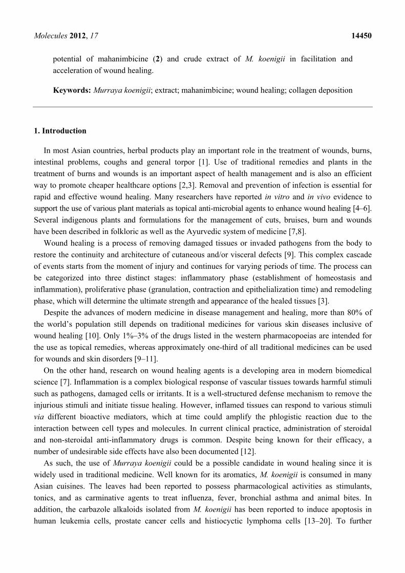
were determined as mahanine (1) (0.40%) (C23H25NO2), mahanimbicine (2) (0.24%) (C23H25NO) and
mahanimbine (3) (0.66%) (C23H25NO) (Figure 1). Spectroscopic data obtained from this study were
similar with the one reported by Ramsewak et al. (1999), Tachibana et al., (2001) and Rahman et al.,
(2005).
Figure 1. Chemical structures of carbazole alkaloids (mahanine (1), mahanimbicine (2),
mahanimbine (3)) isolated from leaves of Murraya koenigii (L.) Spreng.
2.1.2. Profiling of the Essential Oil
A total of 0.12% (w/w) of aromatic yellow oil was obtained from leaves of M. koenigii by hydro
distillation and 1 μL was subjected to Gas Chromatography-Mass Spectrometry (GC-MS) analysis.
The volatile aromatic hydrocarbons were identified based on their Retention Indices (AART) and mass
fragmentation patterns with reference to the NIST 08 and FFNSC version 1.2 databases. A total of 34
Molecules 2012, 17 14452
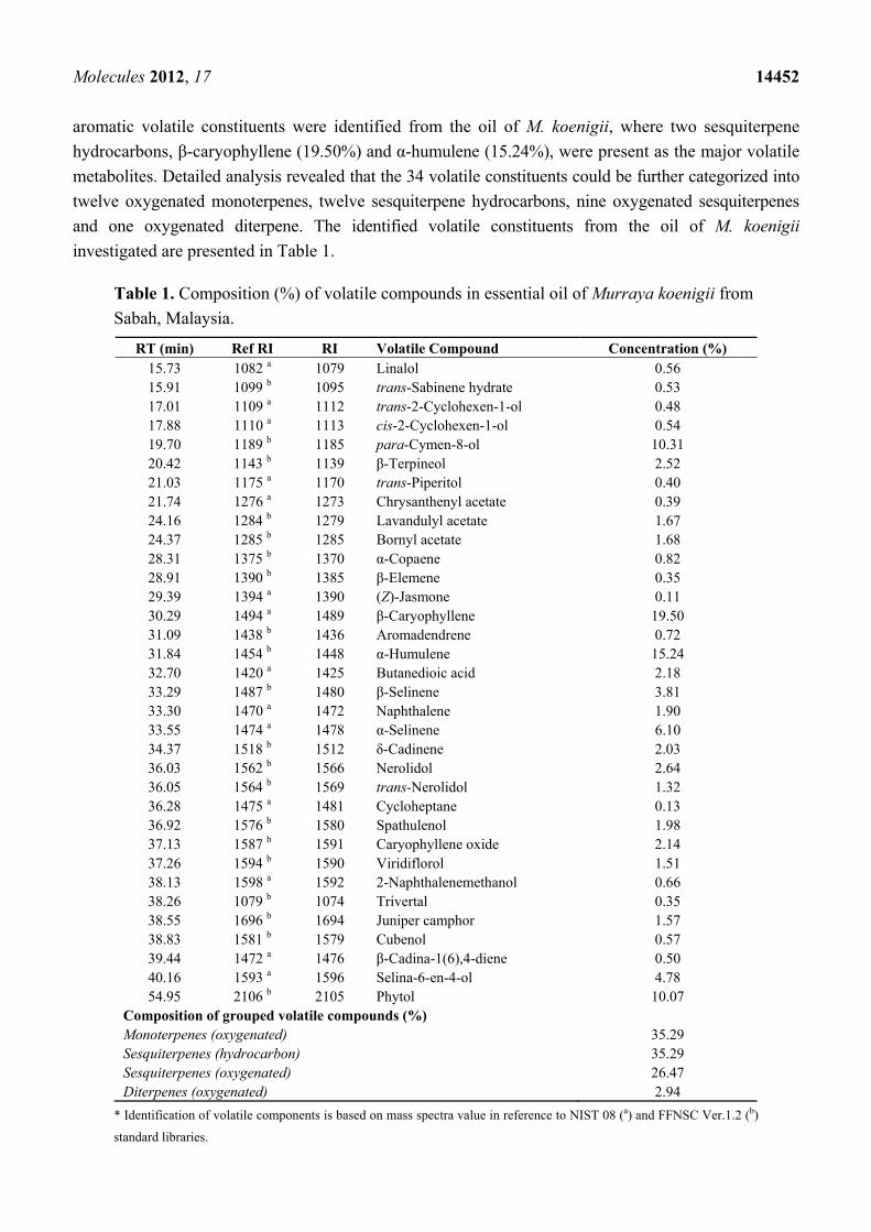
aromatic volatile constituents were identified from the oil of M. koenigii, where two sesquiterpene
hydrocarbons, β-caryophyllene (19.50%) and α-humulene (15.24%), were present as the major volatile
metabolites. Detailed analysis revealed that the 34 volatile constituents could be further categorized into
twelve oxygenated monoterpenes, twelve sesquiterpene hydrocarbons, nine oxygenated sesquiterpenes
and one oxygenated diterpene. The identified volatile constituents from the oil of M. koenigii
investigated are presented in Table 1.
Table 1. Composition (%) of volatile compounds in essential oil of Murraya koenigii from
Sabah, Malaysia.
RT (min) Ref RI RI Volatile Compound Concentration (%) 15.73 1082 a 1079 Linalol 0.56 15.91 1099 b 1095 trans-Sabinene hydrate 0.53 17.01 1109 a 1112 trans-2-Cyclohexen-1-ol 0.48 17.88 1110 a 1113 cis-2-Cyclohexen-1-ol 0.54 19.70 1189 b 1185 para-Cymen-8-ol 10.31 20.42 1143 b 1139 β-Terpineol 2.52 21.03 1175 a 1170 trans-Piperitol 0.40 21.74 1276 a 1273 Chrysanthenyl acetate 0.39 24.16 1284 b 1279 Lavandulyl acetate 1.67 24.37 1285 b 1285 Bornyl acetate 1.68 28.31 1375 b 1370 α-Copaene 0.82 28.91 1390 b 1385 β-Elemene 0.35 29.39 1394 a 1390 (Z)-Jasmone 0.11 30.29 1494 a 1489 β-Caryophyllene 19.50 31.09 1438 b 1436 Aromadendrene 0.72 31.84 1454 b 1448 α-Humulene 15.24 32.70 1420 a 1425 Butanedioic acid 2.18 33.29 1487 b 1480 β-Selinene 3.81 33.30 1470 a 1472 Naphthalene 1.90 33.55 1474 a 1478 α-Selinene 6.10 34.37 1518 b 1512 δ-Cadinene 2.03 36.03 1562 b 1566 Nerolidol 2.64 36.05 1564 b 1569 trans-Nerolidol 1.32 36.28 1475 a 1481 Cycloheptane 0.13 36.92 1576 b 1580 Spathulenol 1.98 37.13 1587 b 1591 Caryophyllene oxide 2.14 37.26 1594 b 1590 Viridiflorol 1.51 38.13 1598 a 1592 2-Naphthalenemethanol 0.66 38.26 1079 b 1074 Trivertal 0.35 38.55 1696 b 1694 Juniper camphor 1.57 38.83 1581 b 1579 Cubenol 0.57 39.44 1472 a 1476 β-Cadina-1(6),4-diene 0.50 40.16 1593 a 1596 Selina-6-en-4-ol 4.78 54.95 2106 b 2105 Phytol 10.07
Composition of grouped volatile compounds (%) Monoterpenes (oxygenated) 35.29 Sesquiterpenes (hydrocarbon) 35.29 Sesquiterpenes (oxygenated) 26.47 Diterpenes (oxygenated) 2.94
* Identification of volatile components is based on mass spectra value in reference to NIST 08 (a) and FFNSC Ver.1.2 (b)
standard libraries.
Molecules 2012, 17 14453
2.2. Biology
2.2.1. Wound Contraction
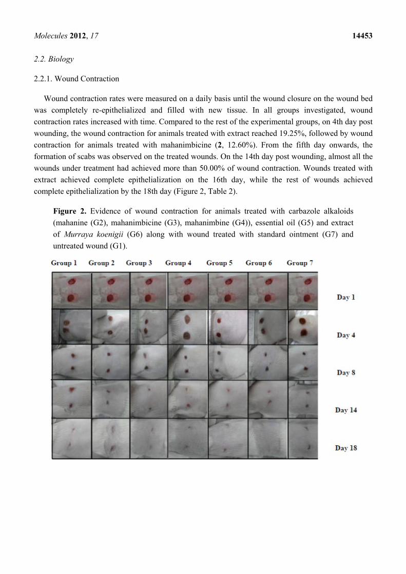
Wound contraction rates were measured on a daily basis until the wound closure on the wound bed
was completely re-epithelialized and filled with new tissue. In all groups investigated, wound
contraction rates increased with time. Compared to the rest of the experimental groups, on 4th day post
wounding, the wound contraction for animals treated with extract reached 19.25%, followed by wound
contraction for animals treated with mahanimbicine (2, 12.60%). From the fifth day onwards, the
formation of scabs was observed on the treated wounds. On the 14th day post wounding, almost all the
wounds under treatment had achieved more than 50.00% of wound contraction. Wounds treated with
extract achieved complete epithelialization on the 16th day, while the rest of wounds achieved
complete epithelialization by the 18th day (Figure 2, Table 2).
Figure 2. Evidence of wound contraction for animals treated with carbazole alkaloids
(mahanine (G2), mahanimbicine (G3), mahanimbine (G4)), essential oil (G5) and extract
of Murraya koenigii (G6) along with wound treated with standard ointment (G7) and
untreated wound (G1).
Molecules 2012, 17 14454
Table 2. Effect of carbazole alkaloids [mahanine (G2), mahanimbicine (G3), mahanimbine
(G4)], essential oil (G5) and extract (G6) of Murraya koenigii, normal healing (G1) and
standard ointment (G7) on percentage of wound healing and epithelialization period of
excision wound model in rats.
Group Post-wounding days Epithelialization
period (days) 2 4 6 8 10 12 14 16 17 18
1 2.12 ±
4.12
6.25 ±
4.12
10.6 ±
2.93
15.9 ±
3.66
25.8 ±
4.09
40 ±
2.26
57.5 ±
1.78
82.7 ±
2.13
90 ±
1.22 100 18
2 4.6 ±
3.77
8.6 ±
4.04
10.7 ±
3.16
18.0 ±
3.21
35 ±
2.34
45.5 ±
2.11
62 ±
1.08
88.5 ±
2.03
96 ±
1.35 100 18
3 6.25 ±
2.42
12.6 ±
3.92
22.0 ±
3.12
27.0 ±
3.24
38 ±
4.05
52.5 ±
1.09
68.5 ±
1.87
90.7 ±
2.88
98 ±
1.30 100 18
4 6.5 ±
3.72
6.5 ±
3.88
12.5 ±
4.86
20 ±
3.56
39.5 ±
3.75
50.5 ±
0.98
65 ±
1.88
93 ±
2.04
97.2 ±
1.05 100 18
5 5.5 ±
3.03
7.5 ±
4.13
10.7 ±
5.67
17.5 ±
2.61
36 ±
4.23
46 ±
2.57
63 ±
1.09
92.5 ±
2.45
95.5 ±
0.84 100 18
6 6.25 ±
2.42
19.25 ±
3.12
37.9 ±
3.09
49.5 ±
1.09
67.5 ±
1.01
75 ±
0.78
95.5 ±
0.66
100 ±
0.55 - - 16
7 3.5 ±
3.11
7.7 ±
2.02
17.9 ±
2.08
20.5 ±
3.34
37.8 ±
3.12
45 ±
2.11
61 ±
0.76
90 ±
0.84
95 ±
0.83 100 18
n = 3 female Sprague-Dawley rats per group, tabular value represent mean ± S.D, p ≤ 0.05; Group 1: wound without
treatment; Group 2: wound treated with mahanine (1); Group 3: wound treated with mahanimbicine (2); Group 4: wound
treated with mahanimbine (3) Group 5: wound treated with essential oil; Group 6: wound treated with extract; Group 7:
wound treated with standard wound healing ointment.
2.2.2. Collagen Density
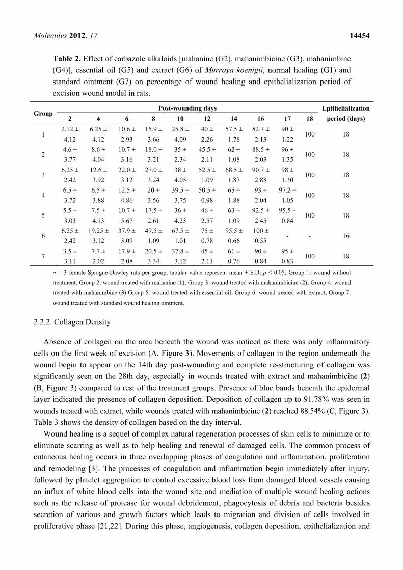
Absence of collagen on the area beneath the wound was noticed as there was only inflammatory
cells on the first week of excision (A, Figure 3). Movements of collagen in the region underneath the
wound begin to appear on the 14th day post-wounding and complete re-structuring of collagen was
significantly seen on the 28th day, especially in wounds treated with extract and mahanimbicine (2)
(B, Figure 3) compared to rest of the treatment groups. Presence of blue bands beneath the epidermal
layer indicated the presence of collagen deposition. Deposition of collagen up to 91.78% was seen in
wounds treated with extract, while wounds treated with mahanimbicine (2) reached 88.54% (C, Figure 3).
Table 3 shows the density of collagen based on the day interval.
Wound healing is a sequel of complex natural regeneration processes of skin cells to minimize or to
eliminate scarring as well as to help healing and renewal of damaged cells. The common process of
cutaneous healing occurs in three overlapping phases of coagulation and inflammation, proliferation
and remodeling [3]. The processes of coagulation and inflammation begin immediately after injury,
followed by platelet aggregation to control excessive blood loss from damaged blood vessels causing
an influx of white blood cells into the wound site and mediation of multiple wound healing actions
such as the release of protease for wound debridement, phagocytosis of debris and bacteria besides
secretion of various and growth factors which leads to migration and division of cells involved in
proliferative phase [21,22]. During this phase, angiogenesis, collagen deposition, epithelialization and
Molecules 2012, 17 14455
newly formed granulation tissue consisting of endothelial cells, macrophages, fibroblasts and the
components of new provisional extracellular matrix begin to cover and fill the wound area [22]. The
remodeling phase involves collagen cross-linking and re-organization, evolution of granulation tissue
into the scar tissue and cells that no longer needed are removed via apoptosis.
Figure 3. Evidence of collagen deposition on wound for animals treated with
mahanimbicine (G2) and extract (G6) along with wound treated with standard ointment
(G7) and untreated wound (G1).
Table 3. Effect of carbazole alkaloids [mahanine (G2), mahanimbicine (G3), mahanimbine
(G4)), essential oil (G5) and extract (G6) of Murraya koenigii, normal healing (G1) and
standard ointment (G7) on percentage of collagen deposition on excision wound model in rats.
Group Day 7 (%) Day 14 (%) Day 21 (%) Day 28 (%)
1 22 ± 0.96 26.77 ± 1.27 61.84 ± 0.94 78.06 ± 1.22
2 20 ± 0.62 36.2 ± 1.36 65.63 ± 0.87 81.56 ± 1.04
3 20.6 ± 0.34 39.62 ± 0.29 67.76 ± 0.85 88.54 ± 1.34
4 14.87 ± 0.44 38.18 ± 0.69 67.15 ± 2.12 81.08 ± 1.09
5 19.18 ± 0.54 30.32 ± 2.33 63.23 ± 0.76 83.78 ± 1.24
6 19.75 ± 0.34 35.51 ± 1.44 69.41 ± 0.24 91.78 ± 1.02
7 21.08 ± 0.53 27.08 ± 1.03 60.05 ± 0.72 86.21 ± 1.12
n = 3 female Sprague-dawley rats per group, tabular value represent mean ± S.D, p ≤ 0.05.
Since M. koenigii has been used as folk medicine for many decades, and it is widely consumed as a
part of spices in many Asian cuisines. Various scientific evidences have revealed the potential of
M. koenigii in reducing diabetic mellitus, obesity and even as a chemopreventive agent [23,24]. In this
Molecules 2012, 17 14456
present study, we investigated the potential of three carbazole alkaloids [mahanine (1), mahanimbine
(2), mahanimbicine (3)], essential oil and extract of M. koenigii as a topical application in enhancing
the wound healing process. Our findings revealed that the extract of M. koenigii significantly
accelerated the rate of wound repair and granulation of new tissue by enhancing formation of more
collagen, fibroblasts and hair follicles and by reducing the numbers of inflammatory cells. The surge in
numbers of neutrophils and lympocytes elicited a response in inflammatory cells by releasing
histamine, bradykinin and other factors that are essential to wound re-modelling and formation of
scabs was observed as early as the 5th day post wounding as inflammation subsided with rapid wound
contraction and wound debridement takes place.
The healing process depends to a large extent on the regulated biosynthesis and deposition of new
collagens and their subsequent maturation. Based on histological findings, extract of M. koenigii was
found to enhance the formation of collagen significantly, followed by mahanimbicine (2). Collagen is
the main structural protein component of connective tissue. Studies have reported that the collagen
sponge enhances the formation of connective tissue and increases the vascularization of wounded
tissues. Immediately upon injury, basal keratinocytes move from the basement membrane and interact
with new connective tissue proteins in the dermis and wound bed inducing the expression of
collagenase. Collagenase, a member of the matrix metalloproteinase family enzymes is responsible for
degrading triple-helical fibrillar collagens into fragments, altering its structure and the affinities to
which cells it binds [25,26]. Therefore, collagenase serves a beneficial role in wound healing during
re-epithelialization by facilitating the movement of keratinocytes over the collagen rich dermis.
Although the epithelialization and contraction process had taken place on the surface within two
weeks, the movement of fibroblasts in the wound area facilitates matrix formation and collagen is laid
down over and throughout the amorphous “plastic tissue”. Movement of collagen was observed in all
the groups on the 14th day post wounding where formation of new hair follicle buds and granulation of
new skin tissue appears. On the 21st day post wounding, appearance of keratinocytes were very visible
and more budding of new hair follicles and sebaceous glands were observed. By the 28th day, a near
complete construction of dermal tissue was detected in the wounds treated with extract of M. koenigii,
followed by wounds treated with mahanimbicine (2) compared to the wounds of the other
experimental groups.
The healing potential of M. koenigii could be a result of the anti-microbial [27,28] and antioxidant
properties exhibited by this perennial plant [29,30]. When the skin is open to infections, the normal
healing process is disrupted as the inflammatory phase becomes chronic and suppresses the
proliferation phase of healing, leading to delays in wound closure. The suppression of the production
of free radicals at or around the wound bed caused by the anti-oxidant potential of this herb helps in
reducing inflammation, increasing angiogenesis and collagen deposition. In previous studies, we had
reported the selective antibacterial activities of these carbazole alkaloids and essential oil of
M. koenigii against several clinical human pathogens [31]. We found that certain functional moieties
play a vital role in influencing the bioactivity of these carbazole alkaloids, although they all share a
similar skeleton. This can be correlated with bacteriostatic activity with its scavenging action on
superoxide and hydroxyl radicals. In this present investigation, it is very clear that the synergistic
effect of these carbazole alkaloids together with other phytochemicals found in M. koenigii extract
Molecules 2012, 17 14457
could be responsible for reducing inflammation through antibacterial protection of wounds and this
leads to increased angiogenesis and collagen deposition.
3. Experimental
3.1. Plant Material
Pest free leaves of Murraya koenigii (L.) Spreng were collected from Kg. Bobot, Kota Belud, Sabah
in August 2010. Voucher specimens were deposited at the BORNEENSIS, Herbarium of the Institute
for Tropical Biology and Conservation, Universiti Malaysia Sabah (BORH 37581).
3.2. Preparation of Extract and Isolation of Carbazole Alkaloids
Air-dried M. koenigii leaves (270 g) were extracted with ethanol (3 L, Merck, Darmstadt, Germany)
for 7 days using a Soxhlet apparatus. Extracts were concentrated under reduced pressure at 40 °C to
yield a dark green residue (14.75 g). A portion (5 g) was then subjected to successive silica gel
(Kiesegel 60, 70–230 mesh, Merck) column chromatography with a stepwise gradient of hexane/ethyl
acetate (Hex/EtOAc: 9:1, 8:2,7:3, 6:4, 1:1) before the column was washed with chloroform-methanol-
water (65:25:4) to obtain six fractions of different polarity. Fractions were then profiled using a high
performance liquid chromatography (HPLC) system equipped with a UV-Vis detector monitored at
254 nm (Prominence, Shimadzu, Kyoto, Japan) coupled with a reverse phase Phenomenex C-18 ODS
(10 mm × 250 mm × 5 μm) column. Mobile phase in gradient elution was: A: 50% MeCN: 50% H2O,
B: 100% MeCN with the profile of: 0–30 min: A: 30% B: 70%, 30.01–60.00 min A: 0% B: 100% and
flow rate was set at 2.00 mL/min under 40 °C. A manual injection of 50 μL of pre-filtered solution (0.2 μm
nylon membrane syringe filters, Whatman, New York, NY, USA) was analyzed. A total of three major
alkaloids were isolated from M. koenigii, compound 1 (0.40%) from fraction 3 while compounds 2
(0.24%) and 3 (0.66%) from fraction 2.
Isolated peaks were subjected to Thin Layer Chromatography (TLC) confirmation with
Drangendoff spray and 1H-NMR revealed the purity of the isolates and confirmed the identity of the
alkaloids with the presence of aromatic and amine protons. Three compounds were isolated and
subjected to 1H-, 13C- and 2D NMR spectroscopic analyses. The structures of compounds were
determined based on the comparison of their 1H- and 13C-NMR data with those reported in the
literature. All spectral data were obtained on the following instruments: IR on a ThermoNicolet FT-IR
spectrometer, optical rotations was measured on an AUTOPOL IV automatic polarimeter (Rudolph
Research Analytical), 1H-NMR (600 MHz) and 13C-NMR (150 MHz) were recorded with a JEOL
ECA 600 (Japan Electronic Optics Laboratory Co. Ltd., Tokyo, Japan) spectrometer, with TMS as
internal standard. HR-ESI-TOFMS data were obtained using LCMS-IT-TOF (Shimadzu, Tokyo, Japan).
The isolated compounds were subjected to spectroscopic measurements and based on independent
structure elucidation the structures of these three compounds were determined as mahanine (1)
(C23H25NO2), mahanimbicine (2) (C23H25NO) and mahanimbine (3) (C23H25NO) (Figure 1). The
spectroscopic data of the respective compounds are assigned as follows:
Compound 1; C23H25NO2; white powder; []D25 = +9.0 (CHCl3, c 2.0):1H NMR: δ 1.41 (3H, s, 3-CH3),
1.56 (3H, br s, 4-CH3), 1.64(3H, br s, 4-CH3), 1.72 (2H, m, H-1), 2.17(2H, m, H-2), 2.27 (3H, br s,
Molecules 2012, 17 14458
5-CH3), 5.01 (1H, br s, OH), 5.12 (1H, m, H-3), 5.64(1H, d, J = 9.8 Hz, H-2), 6.82 (1H, d, J = 9.8 Hz,
H-1), 6.62 (1H, d, H-8), 6.81 (1H, d, J = 2.2 Hz, H-10), 7.47 (1H, s, H-6), 7.64(1H, d, J = 8.3 Hz, H-7),
7.65 (1H, br s, NH), 13C NMR: δ 16.4 (5-CH3), 17.8 (4-CH3), 24.2 (2), 26.7 (3-CH3), 26.5 (4-CH3),
42.2 (1), 79.2 (3), 97.9 (10), 105.8 (11b), 109.1 (8), 118.09 (6a), 119.6 (1), 118.1 (7a), 118.5 (5),
120.6 (7), 121.1 (6), 125.7 (3), 129.2 (2), 132.2 (4), 136.9 (11a), 143.3 (10a), 148.9 (4), 156.5 (9).
EIMS 70 eV m/z (mass value): 347 [M]+ (100), 332 (19), 304 (8), 278 (15), 264 (75).
Compound 2; C23H25NO; mild green powder; []D25 = +60.0 (CHCl3, c 0.30):1H-NMR:δ1.41 (3H, s,
3-CH3), 1.64 (3H, s, 4-CH3), 1.57 (3H, s, 4-CH3), 1.72 (2H, m, H-1), 2.17 (2H, m, H-2), 2.46 (3H,
s, 8-CH3), 5.12 (1H, m, H-3), 5.68 (1H, J = 9.8 Hz, H-2), 6.85 (1H, d, J = 9.8 Hz, H-1), 6.57 (1H, d,
J = 8.5Hz, H-5), 7.09 (1H, d, J = 1.7, 8.3 Hz, H-9), 7.27(1H, br d, J = 8.3 Hz, H-10), 7.69 (1H, d, J =
1.7 Hz, H-7), 7.71 (1H, d, J = 8.5Hz, H-6), 7.78 (1H, s, NH).13C-NMR: δ 17.8 (4-CH3), 21.7 (8-CH3),
24.0 (C-2), 25.7 (4-CH3), 26.6 (3-CH3), 42.3 (C-1), 79.6 (C-3), 106.0 (C-11b), 109.7 (C-5), 111.5 (C-10),
119.3 (C-1), 118.8 (C-6a), 120.1 (C-7), 121.2 (C-6), 125.6 (C-3), 125.3 (C-7a), 126.7 (C-9), 129.3 (C-2),
129.4 (C-8), 132.6 (C-4), 138.7 (C-11a), 140.2 (C-10a), 152.9 (C-4). EIMS 70 eV m/z (mass value):
331[M]+ (23), 316 (6), 248 (100), 210 (3).
Compound 3; C23H25NO; mild brownish-yellow powder; []D25 = +40.0 (CHCl3, c 0.80):
1H-NMR: δ 1.41(3H, s, 3-CH3), 1.63 (3H, s, 4-CH3), 1.55 (3H, s, 4-CH3), 1.72 (2H, t, H-1), 2.17
(2H, m, H-2), 2.29 (3H, s, 5-CH3), 5.11 (1H, m, H-3), 5.63 (1H, d, J = 9.8 Hz, H-2), 6.84 (1H, d, J =
9.8 Hz, H-1), 7.23 (1H, t, J = 8.1 Hz, H-8), 7.07 (1H, br t, J = 8.1 Hz, H-9), 7.85 (1H, br d, J = 8.1 Hz,
H-10), 7.61 (1H, s, H-6), 7.85 (1H, br s, NH), 7.36(1H, d, J = 7.8 Hz, H-7).13C NMR: δ 16.2(5-CH3),
17.6 (4-CH3), 23.9 (C-2), 25.8 (4-CH3), 26.4(3-CH3), 42.1 (C-1), 79.3 (C-3), 105.5 (C-11b), 119.9
(C-10), 24.8 (C-6a), 119.3 (C-1), 118.3 (C-5), 111.4 (C-7), 124.9 (C-8), 121.8 (C-6), 128.9 (C-2),
141.6 (C-7a), 125.5 (C-3), 119.7 (C-9), 132.3 (C-4), 136.9 (C-11a), 117.9 (C-10a), 150.9 (C-4).
EIMS 70 eV m/z (mass value): 331[M]+ (24), 248 (100), 210 (5).
3.3. Extraction of Essential Oil and Analysis
Fresh leaves of M. koenigii (50 g) were chopped and subjected to hydro-distillation using a
Clevenger-type apparatus for duration of 8 h. Distilled oil were collected in pentane, dried over
anhydrous Na2SO4, concentrated in vacuo, stored in air-tight glass vials flushed with nitrogen (N2) gas
and kept at −20 °C prior to analysis. Analysis of the essential oil was performed using a Shimadzu
QP-2010 gas chromatograph coupled with a Shimadzu GCMSQP-2010Plus detector (Shimadzu, Japan)
using a SGE BPX-5 (30.0 m × 0.25 μm i.d., film thickness 0.25 μm) fused silica capillary column.
High purity helium was used as the carrier gas at a constant flow rate of 0.8 mL/min. A total of 1 μL
sample was injected (split ratio 100:1) into GCMS using AOC5000 auto injector for analysis. The
initial temperature was set at 50 °C, heated at a rate of 3 °C/min to 280 °C and held isothermally for
5 min. Ion source temperature for these analysis was set at 200 °C while the interface temperature was
set at 280 °C and the mass spectrometer was set to operate in electron ionization mode with an ionizing
energy of 70 eV as acquisition mass range from 40 a.m.u to 450 a.m.u. at 0.25 scan/s.
Identification of volatile organic constituents was confirmed using published electron impact-mass
spectra (EI-MS) in the National Institute for Standard and Technology (NIST) 1998 and Shimadzu’s
Molecules 2012, 17 14459
Flavours and Fragrance of Natural and Synthetic Compounds (FFNSC) version 1.2 computerized mass
spectral libraries. The retention indices were determined based on a homologous series of n-alkanes
(C8–C40; Custom Retention Time Index Standard, Restek Corp, New York, NY, USA) external
standard analyzed under the same operating conditions and calibrated based on the Automatic
Adjustment of Compound Retention Time (AART) function of the GCMS. Relative concentrations of
the essential oil components were calculated based on GC peak area with the AART correction factors.
3.4. Wound Healing Activity
3.4.1. Experimental Animals
Healthy female Sprague Dawley rats weighing between 200–250 g were obtained from the Animal
Laboratory of Universiti Sains Malaysia, Kubang Kerian, Kelantan. The Institutional Animal Ethics
Committee approved this animal experimentation study at Universiti Malaysia Terengganu (UMT),
Terengganu in accordance with OECD guidelines (No.404, OECD, 2004) together with current
guidelines for the care of laboratory animals. All animals were housed in standard environmental
conditions with temperature of 25 ± 1 °C with 12 h light and 12 h of dark cycle and were acclimatized
to hygienic laboratory condition for a period of 7 days before the experiment was carried out. Any
change in clinical signs such as diarrhea, food and water intake, behavior and blood in urine was
observed. Animals were fed with standard commercial pellet diet (10% of their body weight) and
distilled water ad libitum.
3.4.2. Grouping of Animals
A total of 84 rats were divided into seven experimental groups of twelve rats for four time intervals
(day 7, 14, 21, 28): Group I (negative control group): animals in this group did not receive
administration of any wound healing agent; Group II: animals in this group received administration of
mahanine (1); Group III: animals in this group received administration of mahanimbicine (2); Group
IV: animals in this group received administration of mahanimbine (3); Group V: animals in this group
received administration of essential oil of M. koenigii; Group VI: animals in this group received
administration of M. koenigii extract.; Group VII (positive control group): animals in this group
received the administration of a commercial wound healing cream, Chlorocresol BP 0.1% (Sunward
Pharma, Selangor, Malaysia) as reference drug.
3.4.3. Excision Wound Model and Treatment of Wounds
Animals in assigned groups were anaesthetized by the open mask method with anesthetic ether
before wound creation. Hair on the dorsal thoracic region of the animal was trimmed using electric
clippers, then depilated and treated with a swab of 70% alcohol as topical disinfection before
performing the wound creation. A wound of 8 mm diameter was excised by using sharp sterile skin
bio-puncher (FRAYTM, New York, NY, USA). Wounds was created on clean shaved skin by a
uniformly pressure single twist until the subcutaneous dermal layers were separated. Each animal was
wounded with two circular full thickness wounds on the dorsal part individually to represent
duplicates. Hemostasis was achieved by even compression on the wound with sterile gauze. Animals
Molecules 2012, 17 14460
were housed individually to prevent external tampering with the wounds. The tested carbazole
alkaloids, essential oil and extract (50 mg) were prepared by incorporating into an ointment base
(vehicle) consisting of blank placebo (Sunward Pharma). Topical application of a thin layer of
carbazole alkaloids, essential oil and extract of M. koenigii ointment was done twice daily at the same
time for a period of 14 days.
3.4.4. Wound Contraction and Epithelialization Time
The excision wound margin was traced after wound creation by using transparent paper and the
areas were measured using graph paper. Wound contraction (w.c) was measured daily until the wound
healed completely and expressed as the percentage of reduction in wound area (w.a) of the original
surgical excision as follows:
Percentage (%) of w.c = Ø of w.a Ø of unhealed w.a
Diameter of w.a. × 100%
The epithelialization times were measured from the initial day the excision was performed.
3.4.5. Histopathological Evaluation
Cross-sections of skin, kidney and liver specimens from each group were collected on days 7, 14,
21 and 28 of the experiment for histopathological evaluation and determination of any cytotoxic
effects on vital organs. All specimens were fixed in 10% buffered formalin, dehydrated through a
graded alcohol series, cleared in xylene and blocked with paraffin before sectioning into 5 μm sections.
Serials of 5 μm sections of skin, kidney and liver were stained with Hematoxylin and Eosin (H&E) for
cytotoxicity evaluation and additional sections of skin were stained specifically with Masson trichrome
for the assessment of collagen content and maturation within the dermis. The sections were then
examined under light microscope, observed from the aspects of fibroblast proliferation, collagen
formation, angiogenesis, epithelialization, necrosis of liver and kidney cells and photomicrographs
were taken.
3.4.6. Computerized Collagen Density Evaluation
Skin sections stained with Masson trichrome were examined under polarized light microscope
(Leica, Hamburg, Germany) using image analyzer software (Leica application suite ver. 4.0) and
measurements were made of the intensity of the blue colour that indicates collagen deposition. Density
of collagen deposition under the wound area were measured and compared to normal dermis collagen
at 100 magnification. Mean values of collagen from normal dermis were accepted to be equivalent to
100. Mean values of collagen density under wound areas were expressed as a percentage ratio
compared to collagen density of normal dermis during post wounding days:
Percentage (%) = average collagen intensity under wound
average collagen intensity of normal dermis × 100
Molecules 2012, 17 14461
3.5. Statistical Analysis
The results were analyzed statistically using Student’s t-test to identify differences between treated
and control. The data were considered significant at p < 0.05.
4. Conclusions
Absence of necrotic cells, lesion or shrinkage in cells of liver and kidney of the animals were
observed, suggesting the non-toxic nature of the treatments using extract, mahanine (1),
mahanimbicine (2), mahanimbine (3) and essential oil of M. koenigii. Therefore, the use of M. koenigii
extract as wound healing agent could be useful as it protects the injury site from infections and rapidly
increases the rate of connective tissue formation.
Acknowledgments
The authors would like to acknowledge the Ministry of Science Technology and Innovation of
Malaysia for its financial assistance (Grant No: 02-01-10-SF0131). The corresponding author would
like to express his gratitude to the support staff of the Institute for Tropical Biology and Conservation,
Universiti Malaysia Sabah, for their assistance during the course of this investigation.
References
1. Adetutu, A.; Morgan, W.A.; Corcoran, O. Ethnopharmacological survey and in vitro evaluation of
wound-healing plants used on South-western Nigeria. J. Ethnopharmacol. 2011, 137, 50–56.
2. Gurung, S.; Basnet, N.S. Wound healing properties of Carica papaya latex: In vivo evaluation in
mice burn model. J. Ethanopharmacol. 2009, 121, 338–341.
3. Suntar, I.; Akkol, E.K.; Keles, H.; Oktem, A.; Baser, K.H.C.; Yesilada, E. A novel wound healing
ointment: A formulation of Hypericum perforatum oil and sage and oregano essential oils based
on traditional Turkish knowledge. J. Ethanopharmacol. 2011, 134, 89–96.
4. Chah, K.F.; Eze, C.A.; Esimone, C.O. Antibacterial and wound healing properties of methanolic
extracts of some Nigerian medicinal plants. J. Ethnopharmacol. 2006, 104, 164–167.
5. Muthusamy, S.K.; Ramasamy, S.; Harinarayan, V.R.; Praveen, K.S. Wound healing potential of
Cassia fistula on infected albino rat model. J. Surg. Res. 2006, 131, 283–298.
6. Balekar, N.; Katkam, N.G.; Nakpheng, T.; Jehtae, K.; Srichana, T. Evaluation of the wound
healing potential of Wedelia trilobata (L.) leaves. J. Ethanopharmacol. 2012, 141, 817–824.
7. Kumar, B.; Viayakumar, M.; Govindarajan, G.; Pushpangadan, P. Ethanopharmacological
approaches to wound healing-Exploring medicinal plants of India. J. Ethanopharmacol. 2007,
114, 103–113.
8. Shailajan, S.; Menon, S.; Pednekar, S.; Singh, A. Wound healing efficacy of Jatyadi Taila: In vivo
evaluation in rat using excision wound model. J. Ethanopharmacol. 2011, 138, 99–104.
9. Basal, A.A.M. Healing potential of Rosmarinus officinalis L. on full-thickness excision cutaneous
wounds in alloxan-induced-diabetic BALB/c mice. J. Ethanopharmacol. 2010, 131, 443–450.
Molecules 2012, 17 14462
10. Annan, K.; Houghton, P.J. Antibacterial, antioxidant and fibroblast growth stimulation of aqueous
extracts of Ficus asperifolia Miq. and Gossypium arboreum L., wound-healing plants of Ghana.
J. Pharmacol. 2008, 119, 141–144.
11. Csupor, D.; Blazso, G.; Balogh, A.; Hohmann, J. The traditional Hungarian medicinal plant
Centaureas adleriana Janka accelerates wound healing in rats. J. Ethnopharmacol. 2010, 127,
193–195.
12. Maldini, M.; Sosa, S.; Montoro, P.; Giangaspero, A.; Balick, M.J.; Pizza, C.; Loggia, R.D.
Screening of the topical anti-inflammatory activity of the bark of Acacia cornigera Willdenow,
Brysonimacrassi folia Kunth, Sweetia panamensis Yakovlev and the leaves of Sphagneticola
trilobata Hitchcock. J. Ethanopharmacol. 2009, 122, 430–433.
13. Khanum, F.; Anilakumar, K.R.; Krishna, K.R.S.; Viswanathan, K.R.; Santhanam, K.
Anticarcinogenic effects of curry leaves in dimethylhydrazine-treated rats. Plant Foods Hum. Nutr.
2000, 55, 347–355.
14. Raina, V.K.; Lal, R.K.; Tripathi, S.; Khan, M.; Syamasundar, K.V.; Srivastava, S.K. Essential oil
composition of genetically diverse stocks of Murraya koenigii from India. Flavour Fragr. J.
2002, 17, 144–146.
15. Adebajo, A.C.; Ayoola, O.F.; Iwalewa, E.O.; Akindahunsi, A.A.; Omisore, N.O.A.; Adewunmi, C.O.;
Adenowo, T.K. Anti-trichomonal, biochemical and toxicological activities of methanolic extract
and some carbazole isolated from the leaves of Murraya koenigii growing in Nigeria.
Phytomedicine 2006, 13, 246–254.
16. Arulselvan, P.; Subramanian, S.P. Beneficial effects of Murraya koenigii leaves on antioxidant
defense and ultra structural changes of pancreatic β-cells in experimental diabetes in rats.
Chem. Biol. Interact. 2007, 165, 155–164.
17. Chowdhury, J.U.; Bhuiyan, M.N.I.; Yusuf, M. Chemical composition of the essential oil of
Murraya koenigii (L.) Spreng and Murraya paniculata (L.) Jack. Banglad. J. Pharmacol. 2008, 3,
59–63.
18. Bhattacharjee, S.K. Handbook of Medicinal Plants, 5th Revised & Enlarged ed.; Pointer
Publishers: New Delhi, India, 2008.
19. Bhattacharya, K.; Samanta, S.K.; Tripathi, R.; Mallick, A.; Chandra, S.; Pal, B.C.; Shaha, C.;
Mandal, C. Apototic effects of mahanine on human leukemic cells are mediated through crosstalk
between Apo-1/Fas signaling and the Bid protein and via mitochondrial pathways.
Biochem. Pharmacol. 2010, 79, 361–372.
20. Mandal, S.; Nayak, A.; Kar, M.; Banerjee, S.K.; Das, A.; Upadhyay, S.N.; Singh, R.K.; Banerji, A.;
Banerji, J. Antidiarrhoel activity of carbazole alkaloids from Murraya koenigii Spreng (Rutaceae)
seeds. Fitoterapia 2012, 81, 72–74.
21. Tang, T.; Yin, L.W.; Yang, J.; Shan, G. Emodin, an anthraquinone derivative from Rheum
officinale Baill, enhances cutaneous wound healing in rats. Eur. J. Pharmacol. 2007, 567, 177–185.
22. Silva, K.A.B.S.; Manjavachi, M.N.; Paszcuk, A.F.; Pivatto, M.; Bolzani, V.S.; Calixto, J.B. Plant
derived alkaloids (−)-cassine induces anti-inflammatory and anti-hyperalgesics effects in both
acute and chronic inflammatory and neuropathic pain models. Neuropharmacology 2012, 62,
967–977.
Molecules 2012, 17 14463
23. Dasgupta, T.; Rao, A.R.; Yadava, P.K. Chemomodulatory action of curry leaf (Murraya koenigii)
extract on hepatic and extrahepatic xenobiotic metabolizing enzymes, antioxidant levels, lipid
peroxidation, skin and forestomachpapillomagenesis. Nutr. Res. 2003, 23, 1427–1446.
24. Kaushik, G.; Satya, S.; Khandelwal, R.K.; Naik, S.N. Commonly consumed Indian plant food
materials in the management of diabetes mellitus. Diabetes Metab. Syndr. 2010, 4, 21–40.
25. Nayak, B.S.; Anderson, M.; Pereira Pinto, L.M. Evaluation of wound-healing potential of
Catharanthusroseus leaf extract in rats. Fitoterapia 2007, 78, 540–544.
26. Deshmukh, P.T.; Fernandes, J.; Atul, A.; Toppo, E. Wound healing activity of Calotropis
gigantean root bark in rats. J. Ethanopharmacol. 2009, 125, 178–181.
27. Ramsewak, R.S.; Nair, M.G.; Strasburg, G.M.; DeWitt, D.L.; Nitiss, J.L. Biologically active
carbazole alkaloids from Murraya koenigii. J. Agric. Food Chem. 1999, 47, 444–447.
28. Ningappa, M.B.; Dinesha, R.; Srinivas, L. Antioxidant and free radical scavenging activities of
polyphenol-enriched curry leaf (Murraya koenigii L.) extracts. Food Chem. 2008, 106, 720–728.
29. Tachibana, Y.; Kikuzaki, H.; Lajis, N.; Nakatani, N. Antioxidative activity of carbazoles from
Murraya koenigii leaves. J. Agric. Food Chem. 2001, 49, 5589–5594.
30. Ningappa, M.B.; Dhananjaya, B.L.; Dinesha, R.; Harsha, R.; Srinivas, L. Potent antibacterial
property of APC protein from curry leaves (Murraya koenigii L.). Food Chem. 2010, 118, 747–750.
31. Nagappan, T.; Ramasamy, P.; Abdul Wahid, M.E.; Chandrasegaran, T.; Vairappan, C.S.
Biological activity of carbazole alkaloids and essential oil of Murraya koenigii against antibiotic
resistant microbes and cancer cell lines. Molecules 2011, 16, 9651–9664.
Sample Availability: Not available.
© 2012 by the authors; licensee MDPI, Basel, Switzerland. This article is an open access article
distributed under the terms and conditions of the Creative Commons Attribution license
(http://creativecommons.org/licenses/by/3.0/).
![Review Role of Plant Derived Alkaloids and Their Mechanism ...Role of Plant Derived Alkaloids and Their Mechanism in Neurodegenerative Disorders ... occurrence of symptoms [24]. Cerebral](https://img.pdfslide.org/doc/110x75/5e802dca61852c006f69dbc8/review-role-of-plant-derived-alkaloids-and-their-mechanism-role-of-plant-derived.jpg)


![Review Role of Plant Derived Alkaloids and Their Mechanism ... · potential and alkaloids are one of the most reliable agent against NDDs [33]. Alkaloids are naturally occurring compounds](https://img.pdfslide.org/doc/110x75/5f02f3017e708231d406cf4b/review-role-of-plant-derived-alkaloids-and-their-mechanism-potential-and-alkaloids.jpg)