Embed Size (px)
Citation preview
41
Chapter 3
Evaluation of the efficacy of twoleishmanins in the detection of cellular
immunity in asymptomatic dogs
Laia Solano-Gallego, a Joan Llull b, Margarita Arboixa, Lluis Ferrer c,d, JordiAlberola a,d
aDepartament de Farmacologia i Terapèutica, Universitat Autònoma de Barcelona,Bellaterra, Barcelona, Spain.bHospital Mon Veterinari, Manacor, Balears, Spain.cDepartament de Medicina i Cirurgia Animals, Universitat Autònoma de Barcelona,Bellaterra, Barcelona, Spain.dBoth authors contributed equally
Manuscr i pt submitted i n Veter i nary Paras i to l ogy
Leishmanin skin test Chapter 3
42
Abstract
There are few studies in dogs concerning leishmanin skin test. We evaluated andcompared the efficacy of two leishmanin preparations for detection of dog Leishmaniacellular-mediated immunity. Clinically healthy dogs living in an endemic area werestudied. Leishmanin preparation 1 (3x10⁸ promastigotes/mL) was superior toleishmanin preparation 2 (5x10⁶ promastigotes/mL), measured as the percentage ofpositive reactions and the diameter of the induced induration. Leishmanin skin test is avaluable tool, although the results show that the degree of response, as it has beenshown in human beings, is depending on the preparation used.
Keywords: Leishmania infantum, leishmanin skin test, dog.
Leishmanin skin test Chapter 3
43
Introduction
The widely held opinion that dogs invariably occupied the anergic pole of theleishmanial disease spectrum (Slappendel, 1988) changed when specific cellularimmunity was demonstrated in asymptomatic dogs naturally infected with Leishmania(Cabral et al., 1992; Cabral et al., 1998; Cardoso et al., 1998; Pinelli et al., 1994). Thesefindings argue that canine leishmaniosis may display a wide disease spectrum similar tothat seen in human infection, where clinical disease represents one pole andasymptomatic infection the other (Badaró et al., 1986).
The delayed-type hypersensitivity reaction in leishmaniosis, generally known as theMontenegro or leishmanin skin test (LST) (Montenegro, 1926), is a useful tool for theevaluation of cell-mediated immunity in Leishmania infection in human beings(Pearson & Sousa, 1996) and in dogs (Cardoso et al., 1998; Pinelli et al., 1994). Duringactive infection in human visceral leishmaniosis, the leishmanin skin test is negativewhile it is positive when subclinical infection (self-healing), early stage of visceralleishmaniosis or after succesful treatment (Cahill, 1971; Manson-Bahr, 1961). It isprobable that something similar occurs in the canine population (Cardoso et al., 1998).Contrary to what occurs in humans, where the concentration of promastigotes andcharacteristics of leishmanin preparations are important in the results of LST (Akuffo etal., 1995; Alimohammadian et al., 1993), there are few studies concerning LST in dogs.
The aim of the present study was to evaluate and compare the efficacy of twoleishmanin preparations for detection of dog Leishmania cellular-mediated immunity.
Material and Methods
Both leishmanins used were inactivated suspensions of Leishmania infantum, kindlyprovided by Dr. Jorge Alvar (Instituto de Salud Carlos III, Madrid, Spain). Madridleishmanin (ML) was prepared in the Instituto de Salud Carlos III, Madrid, Spain, andcontained 3x10⁸ promastigotes/mL of L. infantum LEM 75 in 0.4% phenol-saline with aprotein content of 7.3 mg/dL. Roma leishmanin (RL) was prepared in the IstitutoSuperior de Sanita, Roma, Italy, and contained 5x10⁶ promastigotes/mL of L. infantumLEM 75 in 0.5% phenol-saline with a protein content of 2.4 mg/dL. Controls werediluent, saline, and purified protein derivative (PPD).
LST was carried out in a village located in the island of Mallorca (Spain), where canineleishmaniosis is endemic. Thirty-one Ibizian hounds clinically healthy and livingoutdoors were studied. This breed was selected because the Ibizian Hound presents apredominant cellular immune response (Solano-Gallego et al., 2000). Efficacy wasevaluated by subcutaneous injection of 0.1 mL of leishmanin, ML in the right groin andRL in the left groin. The largest diameter of the induced induration and its perpendiculardiameter were measured and averaged at 48 and 72 h. Indurations ≥ 5 mm wereconsidered positive. Five non-infected healthy dogs and five dogs suffering from patentleishmaniosis served as controls.
Blood was collected from all dogs and anti-Leishmania IgG antibodies were assessed bymeans of ELISA as described elsewhere (Solano-Gallego et al., 2000).
Leishmanin skin test Chapter 3
44
Statistical analysis was assessed by Student’s t test (paired and unpaired) using theSPSS program. A P<0.05 was considered significant.
Results
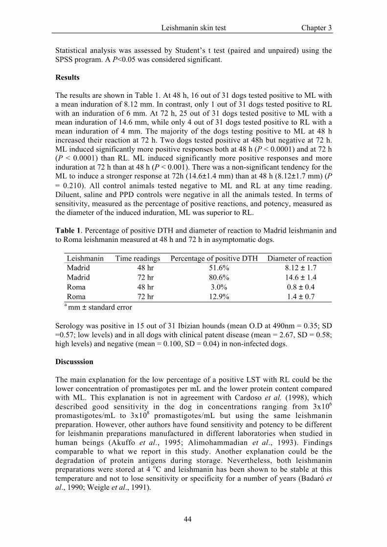
The results are shown in Table 1. At 48 h, 16 out of 31 dogs tested positive to ML witha mean induration of 8.12 mm. In contrast, only 1 out of 31 dogs tested positive to RLwith an induration of 6 mm. At 72 h, 25 out of 31 dogs tested positive to ML with amean induration of 14.6 mm, while only 4 out of 31 dogs tested positive to RL with amean induration of 4 mm. The majority of the dogs testing positive to ML at 48 hincreased their reaction at 72 h. Two dogs tested positive at 48h but negative at 72 h.ML induced significantly more positive responses both at 48 h (P < 0.0001) and at 72 h(P < 0.0001) than RL. ML induced significantly more positive responses and moreinduration at 72 h than at 48 h (P < 0.001). There was a non-significant tendency for theML to induce a stronger response at 72h (14.6±1.4 mm) than at 48 h (8.12±1.7 mm) (P= 0.210). All control animals tested negative to ML and RL at any time reading.Diluent, saline and PPD controls were negative in all the animals tested. In terms ofsensitivity, measured as the percentage of positive reactions, and potency, measured asthe diameter of the induced induration, ML was superior to RL.
Table 1. Percentage of positive DTH and diameter of reaction to Madrid leishmanin andto Roma leishmanin measured at 48 h and 72 h in asymptomatic dogs.
Leishmanin Time readings Percentage of positive DTH Diameter of reactionMadrid 48 hr 51.6% 8.12 ± 1.7Madrid 72 hr 80.6% 14.6 ± 1.4Roma 48 hr 3.0% 0.8 ± 0.4Roma 72 hr 12.9% 1.4 ± 0.7
a mm ± standard error
Serology was positive in 15 out of 31 Ibizian hounds (mean O.D at 490nm = 0.35; SD=0.57; low levels) and in all dogs with clinical patent disease (mean = 2.67, SD = 0.58;high levels) and negative (mean = 0.100, SD = 0.04) in non-infected dogs.
Discusssion
The main explanation for the low percentage of a positive LST with RL could be thelower concentration of promastigotes per mL and the lower protein content comparedwith ML. This explanation is not in agreement with Cardoso et al. (1998), whichdescribed good sensitivity in the dog in concentrations ranging from 3x10⁶
promastigotes/mL to 3x10⁸ promastigotes/mL but using the same leishmaninpreparation. However, other authors have found sensitivity and potency to be differentfor leishmanin preparations manufactured in different laboratories when studied inhuman beings (Akuffo et al., 1995; Alimohammadian et al., 1993). Findingscomparable to what we report in this study. Another explanation could be thedegradation of protein antigens during storage. Nevertheless, both leishmaninpreparations were stored at 4 oC and leishmanin has been shown to be stable at thistemperature and not to lose sensitivity or specificity for a number of years (Badaró etal., 1990; Weigle et al., 1991).
Leishmanin skin test Chapter 3
45
The few studies in the literature concerning LST in dog (Cardoso et al., 1998; Pinelli etal., 1994) and some of the humans studies (Alimohammadian et al., 1993; Weigle et al.,1991) measure the leishmanin concentration in promastigotes per mL. Possibly, theprotein content together with the promastigotes per mL concentration would be a bettermeasurement to compare studies carried out in other laboratories with differentleishmanin preparations.
In field work, sometimes it is not feasible to measure skin reactions on more than oneoccasions. Since more and larger reactions were recorded at 72 hr, it appears that this isthe optimum time to make readings in the dog. Moreover, the best concentration todetect cellular immunity in the dog is 3x10⁸ promastigotes per mL.
In conclusion, leishmanin skin test, if well standardized, can be a valuable tool fordiagnosis and prognosis, epidemiological studies, and as a measure of vaccine efficacyin the dog.
Acknowledgements
The authors are very grateful to the owners of dogs for agreeing to include their dogs inthe study.
References
Akuffo, H., Darce, M., Maasho, K. & Berhan, T.Y. (1995). In vivo evaluation of immune responses inleishmaniasis: the use of cross-species leishmanin preparations for skin testing. AmericanJournal of Tropical Medicine and Hygiene, 53, 16-22.
Alimohammadian, M.H., Kivanjah, M., Pak, F., Gaznavia, A. & Kharazmi, A. (1993). Evaluation ofthe efficacy of Iran leishmanin and comparison with leishmanins from wellcome (UK) and Roma(Italy) in cured cutaneous leishmaniasis patients. Transactions of the Royal Society of TropicalMedicine and Hygiene, 87, 550-551.
Badaró, R., Jones, T.C., Carvalho, E.M., Sampaio, D., Reed, S.G., Barral, A., Teixeira, R. &Johnson Jr, W.D. (1986). New perspectives on a subclinical form of visceral leishmaniasis.Journal of Infectious disease, 154, 1003-1011.
Badaró, R., Pedral-Sampaio, D., Johnson, W.D., Reed, J. & S.G., R. (1990). Evaluation of thestability of a soluble intradermal skin test antigen preparation in American visceralleishmaniasis. Transactions of the Royal Society of Tropical Medicine and Hygiene, 84, 226-227.
Cabral, M., O'Grady, J. & Alexander, J. (1992). Demonstration of Leishmania specific cell mediatedand humoral immunity in asymptomatic dogs. Parasite Immunology, 14, 531-539.
Cabral, M., O'Grady, J.E., Gomes, S., Sousa, J.C., Thompson, H. & Alexander, J. (1998). Theimmunology of canine leishmaniosis: strong evidence for a developing disease spectrum fromasymptomatic dogs. Veterinary Parasitology, 76, 173-180.
Cahill, K. (1971). Studies in Somalia. Transactions of the Royal Society of Tropical Medicine andHygiene, 65, 28-40.
Cardoso, L., Neto, F., Sousa, J.C., Rodrigues, M. & Cabral, M. (1998). Use of leishmanin skin test inthe detection of canine Leishmania-specific cellular immunity. Veterinary Parasitology, 79, 213-220.
Manson-Bahr, P.E.C. (1961). Immunity in kala-azar. Transactions of the Royal Society of TropicalMedicine and Hygiene, 55, 550-555.
Montenegro, J. (1926). A cutis-reacçao na leishmaniose. Anais Facultade Medicina Sao Paulo, 1, 323-333.
Pearson, R.D. & Sousa, A.Q. (1996). Clinical spectrum of leishmaniasis. Clinical Infectious Diseases,22, 1-13.
Pinelli, E., Killick-Kendrick, R., Wagenaar, J., Bernadina, W., del Real, G. & Ruitenberg, J. (1994).Cellular and humoral immune response in dogs experimentally and naturally infected withLeishmania infantum. Infection and Immunity, 62, 229-235.

Leishmanin skin test Chapter 3
46
Slappendel, R.J. (1988). Canine leishmaniasis: A review based on 95 cases in the Netherlands.Veterinary Quarterly, 10, 1-16.
Solano-Gallego, L., Llull, J., Ramos, G., Riera, C., Arboix, M., Alberola, J. & Ferrer, L. (2000). TheIbizian hound presents a predominantly cellular immune response against natural Leishmaniainfection. Veterinary Parasitology, 90, 37-45.
Weigle, K.A., Valderrama, L., Arias, A.L., Santrich, C. & Saravia, N.G. (1991). Leishmanin skin teststandardization and evaluation of safety, dose, storage, longevity of reaction and sensitization.American Journal of Tropical Medicine and Hygiene, 44, 260-271.

47
Chapter 4The Ibizian hound presents a
predominantly cellular immune responseagainst natural Leishmania infection
Laia Solano-Gallegoa, Joan Llullb, Georgina Ramosa, Cristina Rierac,Margarita Arboixa, Jordi Alberolaa,e, Lluis Ferrerd,e
aDepartament de Farmacologia i Terapèutica, Universitat Autònoma de Barcelona,Bellaterra, Barcelona, SpainbHospital Mon Veterinari, Manacor, Balears, SpaincDepartament de Microbiologia i Parasitologia Sanitàries, Universitat de Barcelona,Barcelona, SpaindDepartament de Patologia i Producció Animals, Universitat Autònoma deBarcelona, Bellaterra, Barcelona, SpaineBoth authors contributed equally
Reprinted from Veterinary Parasitology (2000) 90, 37-45
The spectrum of immune responses Chapter 4
48
Abstract
Veterinarians working in the Balearic Islands (Mallorca), an endemic region of canineleishmaniosis, have reported very few cases of leishmaniosis in Ibizian hounds whileconcurrently observing that dogs of other breeds had a high incidence of clinical canineleishmaniosis. To further investigate this observation, two populations of dogs from theBalearic Islands were examined for the presence of Leishmania-specific cellularimmunity using a delayed type hypersensitivity test (DTH) to leishmanin and for thepresence of Leishmania-specific humoral immunity using an ELISA. Fifty-sixasymptomatic dogs, 31 Ibizian hounds and 25 dogs belonging to other breeds wereexamined. Seventy-seven per cent of the dogs demonstrated a specific immune responseagainst Leishmania, either humoral or cellular. This finding suggests that the infectionrate (77%) was higher than previously considered. For Ibizian hounds 81% were DTHpositive while only 48% of the other dogs were DTH positive. A statistical associationbetween Ibizian hounds and positive DTH response was found. A specific humoralresponse was found in 48% of Ibizian hounds and in 56% of the other dogs. Nostatistical association relative to the Leishmania-specific IgG1 and IgG2 levels werefound between the two groups. The Ibizian hound has been reported to be more resistantto Leishmania infection and we found that the Ibizian hound mounts a significantcellular response to infection. Thus, the Ibizian hound may be an interesting caninemodel for the investigation of protective anti-Leishmania immune response.
Key words: Dog; cellular immunity; Leishmania infantum.
The spectrum of immune responses Chapter 4
49
Introduction
Canine leishmaniosis is a systemic severe disease caused by the protozoan parasiteLeishmania infantum (Gramiccia et al., 1989). The disease is endemic in theMediterranean basin, but occasionally, can be diagnosed outside of this region in dogsthat have been living previously in endemic areas (Longstaffe & Guy, 1985). It wasconsidered that dogs infected with L. infantum develop a humoral immune response thatis not immuno-protective (Slappendel & Ferrer, 1998). In fact, most patientsdemonstrate hypergammaglobulinemia (Abranches et al., 1991a; Vitu et al., 1973) andhigh anti-Leishmania titers (Abranches et al., 1991a; Liew & O'Donnell, 1993).
Recent studies have demonstrated that some dogs develop a specific anti-Leishmaniacellular immune response which is effective in protective, leading to a naturalimmunological resistance (Cabral et al., 1992; Cabral et al., 1998; Pinelli et al., 1994).Several studies have demonstrated a high seroprevalence in the Mediterranean caninepopulations without detectable clinical signs (Abranches et al., 1991b; Acedo-Sánchezet al., 1996; Acedo-Sánchez et al., 1998; Fisa et al., 1999). The percentage of dogsnaturally resistant is not well established. Pinelli et al. (1994) found that 6 out of 10beagle dogs developed a protective immune response when experimentally infected byLeishmania. According to Cabral et al. (1998) and to Barbosa Santos et al. (1998), 40%of dogs living in endemic areas display a cellular immune response, either alone orassociated with a humoral response. The DTH to leishmanin has proven to be an usefultool to detect animals with an strong cellular immune response that protects the animalsfrom infection so that most of them would be resistant (Cardoso et al., 1998; Pinelli etal., 1994). According to Deplazes et al. (1995) the titre of specific IgG1 and IgG2 couldhelp to identify susceptible dogs. Dogs with IgG1 and IgG2 titres that remain elevatedfor prolonged periods of time would be considered most susceptible.
Studies in mice have demonstrated that the type of immune response againstLeishmania infection is genetically controlled (Behin et al., 1979; Launois et al., 1998;Mitchel, 1983). However, no studies have been published demonstrating differentinfection rates between genetically different canine breeds. Veterinarians practicing inthe Balearic Islands, an endemic focus of canine leishmaniosis, have observed thatIbizian hound very rarely develop clinical leishmaniosis. The Ibizian hound is originallyfrom North Africa and has lived in the Balearic Islands for over two thousand years.Being exceptionally well suited for hunting, the Ibizian hound has become a verycommon breed in the Balearic Islands (Gounot, 1990). Consequently, it is likely thatindividuals resistant to leishmaniosis have been naturally selected for resistance toLeishmania.
Using these clinical observations, our working hypothesis was that the Ibizian hound isa breed genetically resistant to canine leishmaniosis due to a cellular immune responseagainst the parasite. To investigate this hypothesis, we compared the cellular andhumoral specific immune response against L. infantum infection in Ibizian hounds andin dogs of other breeds from the island of Mallorca.
Materials and methods
Animals
The spectrum of immune responses Chapter 4
50
We used for this study two groups of dogs living outdoors in the village of Felanitx(Mallorca, Spain), an endemic area of leishmaniosis. The first group comprised 31Ibizian hound dogs. Their ages ranged from 6 months to 10 years, with an average of 3years. The second group consisted of 25 dogs of different breeds. Their ages rangedfrom 7 months to 9 years, with an average of 3 years. All dogs were clinically healthy.
Sera
Blood samples were taken by cephalic or jugular venepuncture before leishmanin skintesting was performed. After centrifugation, the sera were kept at –80 oC.
Delayed-type hypersensitivity (DTH) reactions
Dogs were tested for DTH reaction to leishmanin. Leishmanin reagent was aninactivated suspension of 3x10⁸ L. infantum promastigotes per mL in 0.4% phenol-saline, kindly provided by Dr. Alvar (Instituto de Salud Carlos III, Madrid, Spain). Onehundred µL of the solution was intra-dermally injected in the skin of the groin. Skinreactions were recorded after 72 h and an induration or eritematous area >5 mm indiameter was considered positive in agreement with Pinelli et al. (1994) and (Cardoso etal., 1998). Purified protein derivative (2500TU/ml) (Central Veterinary InstituteLelystad, The Netherlands) and leishmanin diluent as well as saline solution (0.1 mL ofeach) served as controls.
ELISA
Leishmania infantum (WHO code MHOM/ FR/ 78/LEM 75) antigen was kindlyprovided by Dr. Montserrat Portús (Universitat de Barcelona, Spain). Promastigoteswere harvested by centrifugation (1300 x g, 10 min, 4oC) and washed with phosphatebuffer saline (PBS pH 7.4) (Sigma). The parasites were resuspended in PBS anddisrupted by sonication. Microtitre plates were coated with 20 µg mL-1 of L. infantumantigen in 0.1 mL of coating buffer (0.1M carbonate-bicarbonate, pH 9.6) and incubatedovernight at 4oC. One hundred µL per well of dog sera, diluted 1:400 for IgG1, IgG2and IgG in PBS-0.05% Tween 20 (PBST)-1% dried skimmed milk, were incubated for1 h at 37oC. After washing three times with PBST and one time with PBS, 100 µL perwell at different dilution of anti-dog IgG1 (1:2000) or anti-dog IgG2 (1:5000) or anti-dog IgG (1:20000) conjugated to horseradish peroxidase (Bethyl laboratories,Montgomery, TX, USA) were added. These conjugates were incubated for 1 h at 37oC,then the plates were rewashed. The substrate solution (ortho-phenylene-diamine, 0.4mg/mL) (Sigma) and H2O2 (0.4 µL/mL) in 0.1M of phosphate/citrate buffer pH 5.0, wasadded at 200 µL/well and developed for 20 min at 24oC. The reaction was stopped with50 µL of H2SO4 3M. Absorbances values were read at 490 nm in an automaticmicroELISA reader (Bio-tek instruments EL 312e microplate).
Sera from 28 non-infected L. infantum dogs living in an endemic region were tested toset up a cut-off for IgG, IgG1 and IgG2 specific ELISA determinations. Cut-offabsorbance values were established as the mean plus 3 standard deviations, resulting in0.236 for IgG (mean 0.099, standard deviation 0.0456), 0.229 for IgG1 (mean 0.098,standard deviation 0.0438), 0.128 for IgG2 (mean 0.0624, standard deviation 0.022). Alldeterminations included the serum from a sick dog with a confirmed infection aspositive control and the serum from a healthy dog as a negative control.
The spectrum of immune responses Chapter 4
51
Statistical analysis
Association between variables and differences among groups were analyzed by meansof Chi-square and Student’s t test respectively. The rate of infection was analysedbetween two groups of dogs with two samples Z-Test. A P<0.05 was consideredsignificant.
Results
Rate of infection
The rate of infection was calculated using all animals that were seropositive in IgGantibodies and/or positive in DTH test. The percentage was 77% for all dogsinvestigated in this study (43 out of 56 asymptomatic dogs). The percentage of infectionin the Ibizian hound dogs was 81% (25 out of 31) and 72% (18 out of 25) in the dogs ofother breeds. We concluded that the percentatges of infection are not different betweenthe two groups (two samples Z test, P=0.2152).
DTH skin reactions to leishmanin
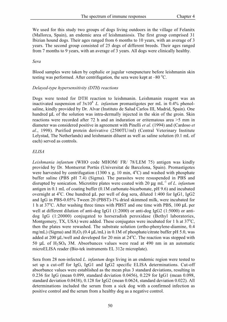
The results of DTH skin testing can be seen in Table 1. Twenty-five out of 31 (81%)Ibizian hounds had a positive reaction to leishmanin. Only 12 out of 25 (48%) of theother dogs had a positive response to the intradermal inoculation of leishmanin. Noreactions were observed when purified protein derivative (PPD), saline solution anddiluent alone were inoculated. According to these results, we concluded that the groupsof dogs and DTH are associated (Chi-square test of association, P=0.010). Thepercentages show in Table 1 suggested that Ibizian hounds group are more likely thanother breeds group to develop a positive DTH. However, either Ibizian hounds or othertesting positive did not differ in the intensity of DTH reaction as measured by reactiondiameter (Student’s t test, P=0.936).
Table 1. Relationship between DTH to leishmanin and the two groups of dogs studied.
DTH to leishmanin+ − Total
Ibizian hound 25 6 31Other breed 12 13 25
Total 37 19 56
Relationship between specific IgG antibodies and groups of dogs
Among the 31 samples from Ibizian hound dogs, 15 presented positive titres of specificanti-Leishmania IgG and 16 negative. In the different breed dogs, 25 samples wereanalyzed and 14 were found positive and 11 negative. We concluded that the specificIgG antibodies and the groups of dogs are not associated (Chi-square test of association,P=0.571).
The spectrum of immune responses Chapter 4
52
Relationship between specific IgG1 and IgG2 antibodies and groups of dogs
Out of the 31 Ibizian hounds, 15 were positive for IgG1 and 21 were positive for IgG2.Of the 25 other dogs, 7 were positive for IgG1 and 18 were positive for IgG2. Weconcluded that the specific IgG1 and IgG2 antibodies and the groups of dogs are notassociated (Chi-square test of association, P=0.120 and P=0.730 respectively).
Relationship between DTH skin reactions to leishmanin and specific IgG, IgG1 andIgG2 antibodies.
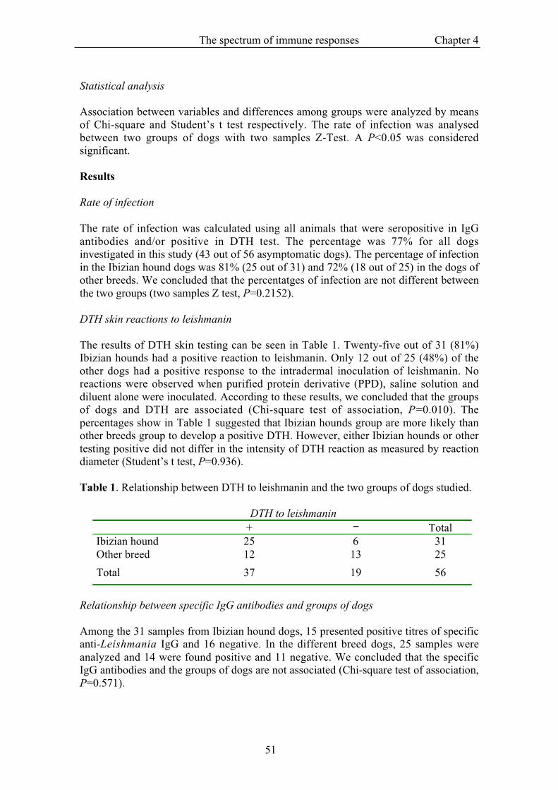
The relationship between DTH and IgG, IgG1 and IgG2 antibodies in the studied dogscan be seen in Table 2. Statistical analysis did not find any association between DTHversus IgG (Chi-square test of association, P=0.059, P=0.577 and P =0.288respectively).
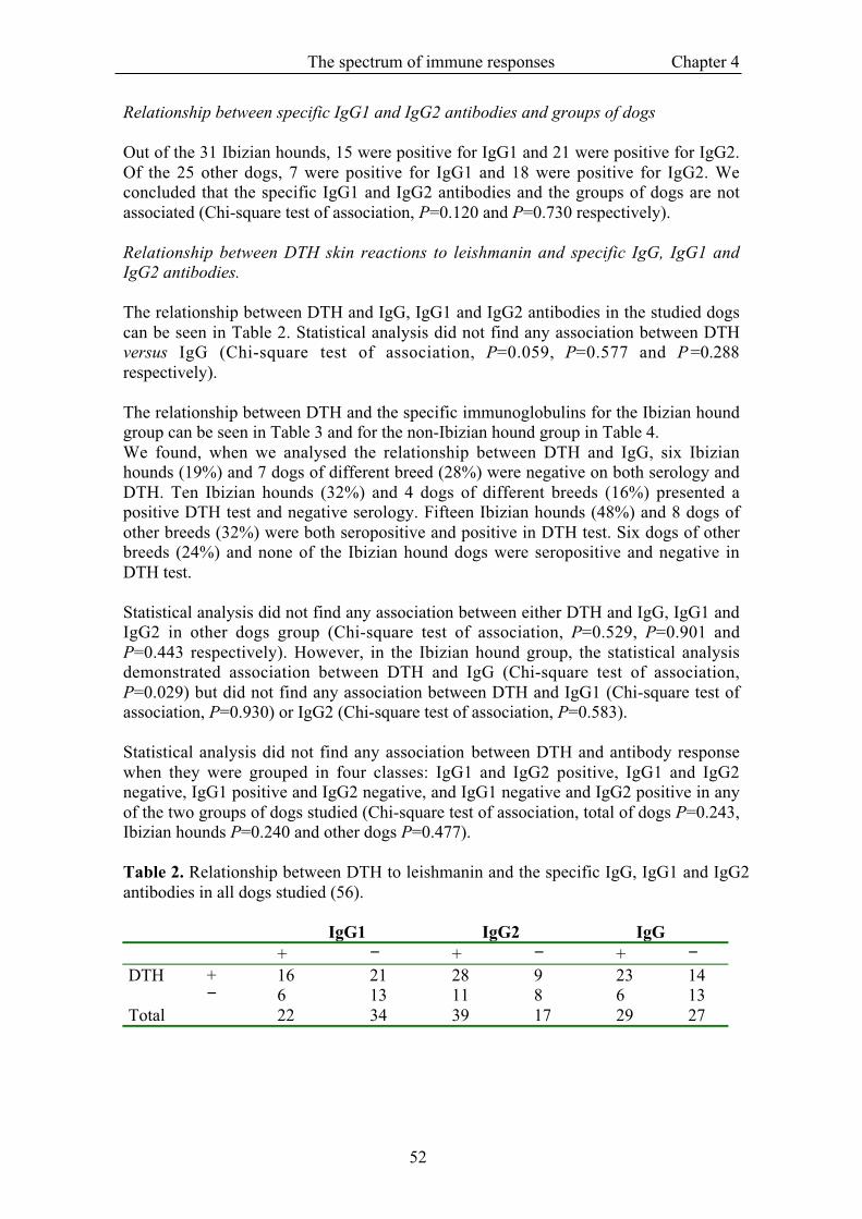
The relationship between DTH and the specific immunoglobulins for the Ibizian houndgroup can be seen in Table 3 and for the non-Ibizian hound group in Table 4.We found, when we analysed the relationship between DTH and IgG, six Ibizianhounds (19%) and 7 dogs of different breed (28%) were negative on both serology andDTH. Ten Ibizian hounds (32%) and 4 dogs of different breeds (16%) presented apositive DTH test and negative serology. Fifteen Ibizian hounds (48%) and 8 dogs ofother breeds (32%) were both seropositive and positive in DTH test. Six dogs of otherbreeds (24%) and none of the Ibizian hound dogs were seropositive and negative inDTH test.
Statistical analysis did not find any association between either DTH and IgG, IgG1 andIgG2 in other dogs group (Chi-square test of association, P=0.529, P=0.901 andP=0.443 respectively). However, in the Ibizian hound group, the statistical analysisdemonstrated association between DTH and IgG (Chi-square test of association,P=0.029) but did not find any association between DTH and IgG1 (Chi-square test ofassociation, P=0.930) or IgG2 (Chi-square test of association, P=0.583).
Statistical analysis did not find any association between DTH and antibody responsewhen they were grouped in four classes: IgG1 and IgG2 positive, IgG1 and IgG2negative, IgG1 positive and IgG2 negative, and IgG1 negative and IgG2 positive in anyof the two groups of dogs studied (Chi-square test of association, total of dogs P=0.243,Ibizian hounds P=0.240 and other dogs P=0.477).
Table 2. Relationship between DTH to leishmanin and the specific IgG, IgG1 and IgG2antibodies in all dogs studied (56).
IgG1 IgG2 IgG+ − + − + −
DTH + 16 21 28 9 23 14− 6 13 11 8 6 13
Total 22 34 39 17 29 27
The spectrum of immune responses Chapter 4
53
Table 3. Relationship between DTH to leishmanin and the specific IgG, IgG1 and IgG2antibodies in the Ibizian hounds.
IgG1 IgG2 IgG+ − + − + −
DTH + 12 13 18 7 15 10− 3 3 3 3 0 6
Total 15 16 21 10 15 16
Table 4. Relationship between DTH to leishmanin and the specific IgG, IgG1 and IgG2antibodies in the other breed of dogs.
IgG1 IgG2 IgG+ − + − + −
DTH + 4 8 10 2 8 4− 3 10 8 5 6 7
Total 7 18 18 7 14 11
Discussion
The results of this study show that the rate of infection between dogs living in anendemic area is considerably higher than previously described (Ben said et al., 1992). Ifwe consider that dogs presenting a specific cellular and/or humoral immune responsehave been infected, the infection rate is very high (77%). The proportion of positivedogs is approximately the same for both groups: 81% in the Ibizian hound (25 out of31) and 72% (18 out of 25) in the dogs of other breeds. These data agree with thatpublished recently by Cabral et al. (1998), who found a rate of Leishmania infection of65% in dogs living in Portugal. Berrahal et al. (1996) also found a rate of infection of80% in dogs living in France using PCR (polymerase chain reaction) to detect theparasite in skin and conjunctival samples. Until now, most epidemiologic studies havebeen based only on serologic surveys. Consequently, these studies did not detectpositive DTH and negative serologic dogs, thus underestimating the rate of infection(Dye et al., 1993; Tesh, 1995).
Dogs with a positive DTH skin test against leishmanin subsequent are considered tohave been exposed to the parasite and potentially resistant to Leishmania infection(Pinelli et al., 1994). According to our results, 48% of dogs of other breeds living in anendemic area are positive by DTH. The remaining 52% (negative in the intradermaltest) include dogs, which have not been infected (28%) and infected dogs with anexclusive humoral immune response (24%) which probably will develop clinicaldisease. In summary, in an endemic area we find two groups of dogs: sick andasymptomatic. The asymptomatic dogs are divided into three groups: resistant dogs,dogs that will develop clinical disease and non-infected dogs. Presumably, the immuneresponse to L. infantum is a mixed humoral and a cellular response. Thus, a wholespectrum of immune responses exists: from Resistant dogs appear to present apredominantly cellular response and susceptible animals present an exaggeratedhumoral response.
The spectrum of immune responses Chapter 4
54
Results obtained from the Ibizian hound group suggests that this breed constitutes aspecial group of dogs regarding the immune response against Leishmania infection.Most (25 out of 31, over 81%) showed a positive DTH to leishmanin. Six dogs thatwere negative to the DTH, were also negative in the serology for IgG, which mightindicate that they have not been exposed to the parasite. A statistically significantassociation was found between Ibizian hounds and DTH positive. However, dogs ofother breed that were positive for DTH were as intense as the DTH response of Ibizianhounds, suggesting that dogs of other breeds are as capable of responding as are Ibizianhounds, but as a group the Ibizian hound responds more uniformly with a positive DTHresponse. Consequently, we consider the Ibizian hound more Leishmania resistant thanother canine breeds.
Several authors have suggested that the presence of anti-Leishmania antibodies alone isnot a conclusive sign of disease progression (Gicheru et al., 1995; Nieto et al., 1999).Titres of specific IgG1 and IgG2 should be a better indicator of disease status than totalIgG (Deplazes et al., 1995). The analysis of IgG subclasses in the infected dogsprovides evidence of a direct correlation between induction of high levels of IgG1 anti-Leishmania antibodies and the appearance of clinical symptoms of the disease (Nieto etal., 1999). Results in asymptomatic dogs are in agreement with these findings. Althoughsome animals (six non-Ibizian hounds) were seropositive for IgG and negative in theDTH, none presented high levels of either IgG1 or clinical symptoms compatible withleishmaniosis.
Deplazes et al. (1995) found that sick dogs initially presented with high levels of both L.infantum specific IgG1 and IgG2. They clinical improved after a long treatment with N-metilglucamine antimoniate (Glucantime , Rhône-Mérieux), and upon improvementthe level of IgG1 decreased and IgG2 levels remained constant. These data suggest ashift in response, from Th2 to Th1, as in the infection of Leishmania major in micewhere the IgG1 is associated with Th2 type response and IgG2 is associated with Th1type response (Reed & Scott, 1993). In the current study, dogs with positive DTH skinreactions had a polymorphic humoral immune response ranging from seronegative topositive titres levels of either IgG1 or IgG2. An association between DTH test andeither of the IgG subclasses was not found.
In human studies, the Leishmania protective IgG subclasses are not well defined.Rodriguez et al. (1996) reported levels of parasite specific IgG1, IgG2, IgG3 and lowlevels of IgG4 in cutaneous leishmaniosis. Skeiky et al. (1997) reported elevated levelsof IgG4 in diffuse cutaneous leishmaniosis. Shiddo et al. (1996) found high levels of allfour IgG subclasses in visceral leishmaniosis. In humans, IgG1 and IgG3 can fixcomplement, while IgG2 is less effective and IgG4 does not fix complement (Brekke etal., 1995). Cell-mediated immunity undoubtedly represents the primary mechanism ofresistance to Leishmania infection, there seems to be sufficient evidence to suggest thata humoral mechanism, including a functional complement system and an appropiateantibody response, may constitute indispensable elements of an effective protectiveresponse at least in human beings (Ulrich et al., 1996). The contribution of antibodiesand complement in a protective role in leishmaniosis, possibly via mechansims of lysisof some Leishmania species and the ability to enhance macrophage mediated parasitekilling, requires further study.
The spectrum of immune responses Chapter 4
55
Data collected in this study confirm our hypothesis that the Ibizian hounds as a grouppresent a more uniform cellular response to infection than do dogs of other breeds.Thus, the Ibizian hound appears to be a very interesting canine model for theinvestigation of immunological mechanisms involved in a self-limited Leishmaniainfection and in the search for a vaccine against canine leishmaniosis.
Acknowledgements
The authors are very grateful to the owners of dogs for agreeing to include their dogs inthe study and Professor M. Portús who kindly offered her support and consultation.
References
Abranches, P., Santos-Gomes, G., Rachamim, N., Campino, L., Schnur, L.F. & Jaffe, C.L. (1991a).An experimental model for canine visceral leishmaniasis. Parasite Immunology, 13, 537-550.
Abranches, P., Silva-Pereira, M.C.D., Conceiçao-Silva, F., Santos-Gomes, G.M. & Janz, J.G.(1991b). Canine leishmaniasis: Pathological and ecological factors influencing transmission ofinfection. Journal of Parasitology, 77, 557-561.
Acedo-Sánchez, C., Martín-Sánchez, J., Vélez-Bernal, I.D., Sanchís-Marín, M.C., Louassini, M.,Maldonado, J.A. & Morillas-Márquez, F. (1996). Leishmaniasis Eco-epidemiology in theAlpujarra Region (Granada province, sothern Spain). International Journal of Parasitology, 25,303-310.
Acedo-Sánchez, C., Morillas-Márquez, F., Sanchíz-Marín, M.C. & Martín-Sánchez, J. (1998).Changes in antibody titres against Leishmania infantum in naturally infected dogs in southernSpain. Veterinary Parasitology, 75, 1-8.
Barbosa Santos, E.G., Marzochi, M.C., Conceiçao, N.F., Brito, C.M. & Pacheco, R.S. (1998).Epidemiological survey on canine population with the use of immunoleish skin test in endemicareas of human american cutaneous leishmaniosis in the state of Rio de Janeiro, Brazil. Revistado Instituto Medicina Tropical de Sao Paulo, 40, 41-47.
Behin, R., Maüel, J. & Sordat, B. (1979). Leishmania tropica: pathogenicity and in vitro macrophagefunction of inbred mice. Experimental parasitology, 48, 81-91.
Ben said, M., Jaiem, A., Smoorenburg, M., Semiao-Santos, S.J., Ben Rachid, M.S. & el Harith, A.(1992). [Canine leishmaniasis in the region of Enfidha (Central Tunisia). Assessment ofseroprevalence with direct agglutination (DAT) and indirect immunofluorescence]. BulletinSociété Pathologie Exotique, 85, 159-163.
Berrahal, F., Mary, C., Roze, M., Berenger, A., Escoffier, K., Lamouroux, D. & Dunan, S. (1996).Canine leishmaniasis: identification of asymptomatic carriers by polymerase chain reaction andimmunoblotting. American Journal of Tropical Medicine and Hygiene, 55, 273-277.
Brekke, O.H., Michaelsen, T.E. & Sandlie, I. (1995). The structural requirements for complementactivation by IgG: Does it hinge on the hinge?. Immunology Today, 16, 85-90.
Cabral, M., O'Grady, J. & Alexander, J. (1992). Demonstration of Leishmania specific cell mediatedand humoral immunity in asymptomatic dogs. Parasite Immunology, 14, 531-539.
Cabral, M., O'Grady, J.E., Gomes, S., Sousa, J.C., Thompson, H. & Alexander, J. (1998). Theimmunology of canine leishmaniosis: strong evidence for a developing disease spectrum fromasymptomatic dogs. Veterinary Parasitology, 76, 173-180.
Cardoso, L., Neto, F., Sousa, J.C., Rodrigues, M. & Cabral, M. (1998). Use of leishmanin skin test inthe detection of canine Leishmania-specific cellular immunity. Veterinary Parasitology, 79, 213-220.
Deplazes, P., Smith, N.C., Arnold, P., Lutz, H. & Eckert, J. (1995). Specific IgG1 and IgG2 antibodyresponses of dogs to Leishmania infantum and other parasites. Parasite Immunology, 17, 451-458.
Dye, C., Vidor, E. & Dereure, J. (1993). Serological diagnosis of leishmaniasis: on detecting infectionas well as disease. Epidemiology Infection, 103, 647-656.
Fisa, R., Gállego, M., Castillejo, S., Aisa, M.J., Serra, T., Riera, C., Carrió, J., Gállego, J. & Portús,M. (1999). Epidemiology of canine leishmaniosis in Catalonia (Spain) The example of thePriorat focus. Veterinary Parasitology, 83, 87-97.
The spectrum of immune responses Chapter 4
56
Gicheru, M.M., Olobo, J.O., Kariuki, T.M. & Adhiambo, C. (1995). Visceral leishmaniasis in ververtmonkeys: immunological responses during asymptomatic infections. Scandinavian Journal ofImmunology, 41, 202-208.
Gounot, G. (1990). Le podenco ibicenco ou Ca Eivissenc. In École Nationale Véterinaire de Toulouse.Gramiccia, M., Gradoni, L. & Angelici, M.C. (1989). Epidemiology of Mediterranean leishmaniosis by
Leishmania infantum: isoenzyme and KDNA analysis for the identification of parasites fromman, vectors and reservoirs. In NATO-ASI Monograph on leishmaniosis. ed. Hart, D.J. pp. 21-37. New York: Plenum Press.
Launois, p., Conceiçao-Silva, F., Himmerlich, H., Parra-Lopez, C., Tacchini-Cottier, F. & Louis,J.A. (1998). Setting in motion the immune mechanisms underlying genetically determinedresistance and susceptibility to infection with Leishmania major. Parasite Immunology, 20, 223-230.
Liew, F.Y. & O'Donnell, C.A. (1993). Immunology of leishmaniasis. Advances in Parasitology, 32,161-259.
Longstaffe, J.A. & Guy, M.W. (1985). Leishmaniasis in dogs. Veterinary Annual, 25, 358-367.Mitchel, G.F. (1983). Murine cutaneous leishmaniosis: resistance in reconstituted nude mice and several
F1 hybrids infected infected with Leishmania tropica major. Journal of Immunogenetics, 10,395-412.
Nieto, C.G., García-Alonso, M., Requena, J.M., Mirón, C., Soto, M., Alonso, C. & Navarrete, I.(1999). Analysis of the humoral immune response against total and recombinant antigens ofLeishmania infantum: correlation with disease progression in canine experimental leishmaniasis.Veterinary Immunology and Immunopathology, 67, 117-130.
Pinelli, E., Killick-Kendrick, R., Wagenaar, J., Bernadina, W., del Real, G. & Ruitenberg, J. (1994).Cellular and humoral immune response in dogs experimentally and naturally infected withLeishmania infantum. Infection and Immunity, 62, 229-235.
Reed, S.G. & Scott, P. (1993). T cell and cytokine responses in leishmaniasis. Current Opinion inImmunology, 5, 524-531.
Rodriguez, V., Centeno, M. & Ulrich, M. (1996). The IgG isotypes of specific antibodies in patientswith American cutaneous leishmaniasis; relationship to the cell-mediated immune response.Parasite Immunology, 18, 341-345.
Shiddo, S.A., Huldt, G., Nilsson, L.A., Ouchterlony, Ö. & Thorstensson, R. (1996). Visceralleishmaniasis in Somalia. Significance of IgG subclasses and of IgE response. ImmunologyLetters, 50, 87-93.
Skeiky, Y.A.W., Benson, D.R., Costa, J.L.M., Badaro, R. & Reed, S.G. (1997). Association ofLeishmania heat shock protein 83 antigen and immunoglobulin G4 antibody titers in brazilianpatients with diffuse cutaneous leishmaniasis. Infection and Immunity, 65, 5368-5370.
Slappendel, R.J. & Ferrer, L. (1998). Leishmaniasis. In Infectious diseases of dog and cat. ed. Greene,C.E. pp. 450-458. Philadelphia: W.B. Saunders Company.
Tesh, R.B. (1995). Control of zoonotic visceral leishmaniasis: is it time to change strategies? AmericanJournal of Tropical Medicine and Hygiene, 52, 287-292.
Ulrich, M., Rodríguez, V. & Centeno, M. (1996). The humoral response in leishmaniasis. In Mollecularand immune mechanisms in the pathogenesis of cutaneous leishmaniasis. ed. Tapia, F.J.,Cáceres-Dittmar, G. & Sánchez, M.A. pp. 189-202: R.G. Landes company.
Vitu, C., Sanchis, R. & Giauffret, A. (1973). Evolution of serum proteins in canine leishmaniasis. C.R.Seances Soc. Biol. Fil., 167, 513-518.
57
Chapter 5
Prevalence of Leishmania infantuminfection in dogs living in an area ofcanine leishmaniasis endemicity usingPCR on several tissues and serology
Laia Solano-gallegoa, Pere Morellb, Margarita Arboixa, Jordi Alberolaa, LluisFerrerc
aDepartament de Farmacologia i Terapèutica, Universitat Autònoma de Barcelona,Bellaterra, Barcelona, SpainbCentre Sanitari Municipal, Palma de Mallorca, SpaincDepartament de Medicina i Cirugia Animals, Universitat Autònoma de Barcelona,Bellaterra, Barcelona, Spain
Reprinted from Journal of Clinical Microbiology (2001) 39, 560-563
Leishmania infection in dogs Chapter 5
58
Abstract
We studied and compared the prevalence of Leishmania infection and theseroprevalence and the prevalence of canine leishmaniasis in an area where canineleishmaniasis is endemic. One hundred dogs living on the island of Mallorca (Spain)were studied. In this study, we clinically examined each dog for the presence ofsymptoms compatible with leishmaniasis, determined the titre of anti-Leishmaniaantibodies, and investigated the presence of Leishmania DNA by PCR in skin,conjunctiva, and bone marrow samples of each dog. The prevalence of the disease andthe seroprevalence were 13 and 26%, respectively. In 63% of the dogs, LeishmaniaDNA could be detected by PCR in at least one of the tissues studied. The results ofpositive PCR in the bone marrow, the conjunctiva, and the skin were 17.8, 32, and 51%,respectively. The prevalence of the infection, 67%, was calculated using all animals thatwere seropositive and/or positive by PCR with any tissue. The results showed that themajority of dogs living in an area where canine leishmaniasis is endemic are infected byLeishmania and that the prevalence of infection is much greater than the prevalence ofovert Leishmania-related disease.
Leishmania infection in dogs Chapter 5
59
Introduction
Canine leishmaniasis is a severe systemic disease of dogs caused by the protozoanparasite Leishmania infantum. Clinical manifestations of the disease include nonpruriticskin lesions, such as exfoliative dermatitis and ulcerations, local or generalizedlymphadenopathy, loss of weight, poor appetite, ocular lesions, epistaxis, lameness,renal failure, and diarrhea (Ciaramella et al., 1997; Ferrer et al., 1988; Koutinas et al.,1999; Slappendel, 1988). Leishmaniasis is a zoonotic disease for which dogs areconsidered the chief reservoir of the parasite. The disease is endemic in theMediterranean basin, where seroprevalence ranges between 10 and 37% (Fisa et al.,1999; Sideris et al., 1999). There are, however, several studies suggesting that the rateof infection is higher than the figures found by serological investigations. A surveyperformed using the PCR and immunoblotting techniques found that most dogs living insouthern France had been exposed to Leishmania (Berrahal et al., 1996). These resultsagree with another study that found a rate of Leishmania infection of 65% for dogsliving in Portugal by using serology and cell-mediated tests (Cabral et al., 1998).
The percentage of infected dogs living in an area where canine leishmaniasis is endemichas major public health implications. It was demonstrated that infected, butasymptomatic, dogs were sources of the parasite for phlebotomine vector sandflies andas a consequence play an active role in the transmission of Leishmania (Molina et al.,1994).
The present study was designed to investigate and compare the prevalence ofLeishmania infection, the seroprevalence and the prevalence of the disease in a caninepopulation living in an area where canine leishmaniasis is endemic. One hundred dogsliving on the island of Mallorca (Spain) were included in this study. Veterinariansclinically examined all dogs, and the titre of anti-Leishmania antibodies wasdetermined. The presence of Leishmania DNA in each dog was investigated by PCRwith three tissues: skin, eye conjunctiva, and bone marrow.
Materials and methods
Animals
The study was carried out on the Island of Mallorca, an endemic area of canineleishmaniasis. The subjects of the study were 100 dogs from different breeds and ages,which had to be euthanatized in the Animal Pound of Palma de Mallorca for reasonsrelated to city sanitation policy.
Sampling
Prior to sampling and euthanasia, all dogs were examined to detect clinical symptomscompatible with canine leishmaniasis. The dogs were then premedicated withacepromacine maleate and anesthetized intravenously with sodium thiopental.Blood was collected by cephalic or jugular venepuncture, and the serum samples fordetecting and quantifying specific antibodies to Leishmania were stored at –80o C.Three types of tissues for PCR were sampled: bone marrow, skin, and eye conjunctiva.Bone marrow aspirates were obtained from the costochondral junctions by using a 22gauge needle. Cutaneous samples were collected from the upper part of the muzzle by
Leishmania infection in dogs Chapter 5
60
punch biopsy with a diameter of 5 mm and with each biopsy weighing approximately30 mg. Conjunctiva samples were obtained using scissors, with each biopsy weighingapproximately 30 mg. Samples were stored at –20o C before DNA extraction. Aftersampling was completed, dogs were euthanatized using an overdose of parenteralbarbiturates.
ELISA
An enzyme-linked immunosorbent assay (ELISA) was performed as previouslydescribed (Riera et al., 1999). Briefly, microtitre plates were coated with a 20-µg mL-1
concentration of L. infantum antigen in 0.1 mL of coating buffer (0.1 M carbonate-bicarbonate, pH 9.6) and incubated overnight at 4oC. One hundred microliters of dogsera per well was diluted 1:400 in phosphate-buffered saline (PBS)-0.05% Tween 20(PBST)-1% dried skim milk and was incubated for 1 h at 37oC. After washing threetimes with PBST and once with PBS, 100 µL of anti-dog immunoglobulin G (IgG)(1:20,000) conjugated to horseradish peroxidase (Bethyl laboratories, Montgomery,Tex.) was added. This conjugate was incubated for 1 h at 37oC, and then the plates wererewashed. The substrate solution (ortho-phenylene-diamine, 0.4 mg/mL) (Sigma) andH2O 2 (0.4 µL/mL) in 0.1 M of phosphate-citrate buffer (pH 5.0) was added at 200µL/well and developed for 20 min at 24oC. The reaction was stopped with 50 µL ofH2SO4 3M. Absorbance was read at 490 nm in an automatic microELISA reader (EL312e microplate; Bio-tek instruments).
Sera from 28 dogs not infected with Leishmania infantum that were living in a regionwhere it is endemic were tested to set up a cut-off for IgG-specific ELISAdeterminations. The cut-off absorbance was established as the mean plus three standarddeviations, resulting in 0.236 for IgG (mean, 0.099, standard deviation, 0.0456). Alldeterminations included the serum from a sick dog with a confirmed infection aspositive control and the serum from a healthy dog as a negative control.
DNA isolation
Bone marrow
Samples of DNA were prepared as previously described (Roura et al., 1999). Briefly,115-µL of bone marrow samples were washed and centrifuged three times in tris-EDTAbuffer (pH 8⋅0), and the leucocyte pellet was incubated in 0.1 mL of lysis buffer (50mMpotassium chloride, 10 mM tris-HCl [pH 8⋅4], 0.5% Tween-20 and 100 µg of proteinaseK mL) at 56oC overnight. Proteinase K was inactivated by incubating the samples at95oC for 10 minutes before using them in the PCR.
Conjunctiva and skin tissue
Conjunctiva and skin biopsies were digested overnight in the presence of sodiumdodecyl sulfate and proteinase K at final concentrations of 2% and 0.2 mg/mL,respectively, in 1 mL of TE buffer (Tris 50mM [pH 8.0], 20mM EDTA). Afterwards,DNA was isolated by double phenol-chloroform extraction (Sambrook et al., 1989).
Leishmania infection in dogs Chapter 5
61
PCR
Leishmania specific oligonucleotide primers SP176 (5'- TCTTGCGGGGAGGGGGTG-3´) and SP177 (5´-TTGACCCCCAACCACATTTTA-3´) were used to amplify a 120-base-pair fragment of Leishmania kinetoplast DNA minicircles (Rodgers et al., 1990).PCR was conducted in a 50-µL final reaction mixture containing PCR buffer, 1.5 mMMgCl2, 0.1 mM of each deoxynucleoside triphosphate, 0.3 µM concentrations of eachprimer, 3 µl of supernatants of digested tissue, and 1.25 U of Taq polymerase (Ecogen).Reactions were carried out in an automatic thermocycler (Perkin Elmer) with a thermalcycling profile of 94oC for 3 min, 35 cycles at 95oC for 30 s, 58oC for 30 s, 72oC for 30s and finally 72oC for 5 min, and at that point the thermocycler maintained a constanttemperature of 4oC. Positive controls containing 10 ng of genomic of Leishmania DNAand a negative control without template DNA were included. Amplified fragments wereanalyzed by electrophoresis in a 2.5% agarose gel containing ethidium bromide (0.5µg/mL) at 100 V for 1 h. A φΧ174 HaeIII (DNA MWM V; Boehringer Mannheim) wasused as a molecular weight marker.
To ensure that negative results corresponded to true negative samples rather than to aproblem with DNA loading, sample degradation, or PCR inhibition, sample DNA wasalso amplified for β -actin by using a forward primer (5´-ACCTGGAGTTCGAGGYTCGGA-3´) and a reverse primer (5´-AAGTAACCCTGGTTGGTGAAGCAG-3´) (Helfand et al., 1999). When samples didnot yield amplification products, they were extracted again until amplification productswere obtained.
Results
Thirteen animals presented one or more clinical signs of canine leishmaniasis while 87dogs were asymptomatic. Twenty-six of the dogs were seropositive. Eleven of themalso showed clinical signs while 15 were clinically healthy dogs. Two dogs with clinicalsigns of leishmaniasis were seronegative.
The presence of parasite DNA was detected in 63 dogs for at least one of the threetissues investigated. The results of the PCRs with the different tissues were as follows:17 out of 95 dogs (17.8%) had positive bone marrow, 32 out of 100 dogs (32%) hadpositive conjunctiva and 51 out of 100 dogs (51%) had positive skin. Leishmania DNAwas detected in all animals presenting clinical signs.
Results for clinical status, serology, and PCR, including the three tissues sampled foreach dog, are shown in Table 1.
The prevalence of the infection, 67%, was calculated by adding all animals that wereseropositive and/or positive for PCR with any tissue.
Leishmania infection in dogs Chapter 5
62
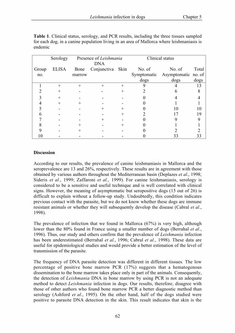
Table 1. Clinical status, serology, and PCR results, including the three tissues sampledfor each dog, in a canine population living in an area of Mallorca where leishmaniasis isendemic
Serology Presence of LeishmaniaDNA
Clinical status
Groupno.
ELISA Bonemarrow
Conjunctiva Skin No. ofSymptomatic
dogs
No. ofAsymptomatic
dogs
Totalno. ofdogs
1 + + + + 9 4 132 + - - + 2 6 83 + - - - 0 4 44 + + - - 0 1 15 - - + + 0 10 106 - - - + 2 17 197 - - + - 0 9 98 - + - + 0 1 19 - + - - 0 2 2
10 - - - - 0 33 33
Discussion
According to our results, the prevalence of canine leishmaniasis in Mallorca and theseroprevalence are 13 and 26%, respectively. These results are in agreement with thoseobtained by various authors throughout the Mediterranean basin (Deplazes et al., 1998;Sideris et al., 1999; Zaffaroni et al., 1999). For canine leishmaniasis, serology isconsidered to be a sensitive and useful technique and is well correlated with clinicalsigns. However, the meaning of asymptomatic but seropositive dogs (15 out of 26) isdifficult to explain without a follow-up study. Undoubtedly, this condition indicatesprevious contact with the parasite, but we do not know whether these dogs are immuneresistant animals or whether they will subsequently develop the disease (Cabral et al.,1998).
The prevalence of infection that we found in Mallorca (67%) is very high, althoughlower than the 80% found in France using a smaller number of dogs (Berrahal et al.,1996). Thus, our study and others confirm that the prevalence of Leishmania infectionhas been underestimated (Berrahal et al., 1996; Cabral et al., 1998). These data areuseful for epidemiological studies and would provide a better estimation of the level oftransmission of the parasite.
The frequency of DNA parasite detection was different in different tissues. The lowpercentage of positive bone marrow PCR (17%) suggests that a hematogenousdissemination to the bone marrow takes place only in part of the animals. Consequently,the detection of Leishmania DNA in bone marrow by using PCR is not an adequatemethod to detect Leishmania infection in dogs. Our results, therefore, disagree withthose of other authors who found bone marrow PCR a better diagnostic method thanserology (Ashford et al., 1995). On the other hand, half of the dogs studied werepositive to parasite DNA detection in the skin. This result indicates that skin is the
Leishmania infection in dogs Chapter 5
63
major tissue reserve of parasites in dogs and that PCR in skin biopsy is a sensitivemethod to detect infection. This finding concerns the biology of the parasite because theskin is the most accessible tissue for the vector. Furthermore, cutaneous samples werecollected from the upper part of the muzzle where most sandflies take their blood meal(Killick-Kendrick & Killick-Kendrick, 1999).
In the compartmental mathematical model of canine leishmaniasis, it was assumed thatasymptomatic dogs were not infectious for sandflies (Dye et al., 1992; Hasibeder et al.,1992). However, other authors showed that infectivity of dogs presenting Leishmaniainfection is not exclusively linked to the symptomatic stage of the disease: they foundthat three out of five asymptomatic but seropositive dogs transmitted the parasite to thesandfly vectors (Molina et al., 1994). Our results support the idea that asymptomaticdogs must be considered infectious for sandflies. We showed that 54% of healthy dogsliving in an area where canine leishmaniasis is endemic must be consideredasymptomatic carriers of Leishmania. Further studies are needed to ascertain thepotential of asymptomatic dogs to transmit Leishmania to vector sandflies.
Studies of the immune response against the Leishmania parasite in the dog haverevealed that T lymphocytes and the cytokines they produce play a crucial role indetermining whether an infection with this intracellular pathogen results in eitherprotective immunity or progressive disease (Pinelli et al., 1995; Pinelli et al., 1994;Pinelli et al., 1999). In the population of our study, it would have been interesting tostudy the cellular immune response using a field tool such as leishmanin skin test. Thiswas not, however, possible for ethical reasons, as we would have needed to previouslymanipulate animals not destined for research. However, it has been reported that 40% ofthe asymptomatic dogs living in Portugal (Cabral et al., 1998) and that 48% of theasymptomatic dogs living in Mallorca (Solano-Gallego et al., 2000) had demonstrableparasite-specific cellular immunity. We found that 37% of asymptomatic dogs werePCR positive in the skin and/or conjunctiva and were seronegative. Probably, dogs withsuitable cellular immunity could control the spread of Leishmania parasite and live inequilibrium with the parasite in the skin and mucosal regions. This fact may explain thehigh rate of infection due to the whole spectrum of immune response to Leishmania inthe canine population (Cabral et al., 1998). In human beings, a strong associationbetween the decrease in the number of CD4+ T lymphocytes and the increase in theinfectivity of people coinfected with L. infantum and human immunodeficiency virus tothe sandflies has been reported recently (Molina et al., 1999).
The high prevalence of Leishmania infection in regions where leishmaniasis is endemichas to be taken into account in any campaign aimed at controlling canine leishmaniasis.In fact, some authors have demonstrated that removing seropositive dogs is aninsufficient method to eradicate canine leishmaniasis (Ashford et al., 1993; Dietze etal., 1997).
Vaccines against Leishmania are a goal for the scientific community working on humanand canine leishmaniasis. Potential vaccines could act by either destroying the parasiteand/or preventing disease pathology. While the latter type of vaccine would preventsevere disease and morbidity among immunized dogs, the parasite transmission cyclewould remain intact. This is especially important because of the high prevalence ofLeishmania infection that we have shown, suggesting that asymptomatic dogs act asreservoirs for parasite transmission to sandflies. Thus, potential canine vaccines must
Leishmania infection in dogs Chapter 5
64
induce sterile immunity, eliminating amastigotes that reside in apparently healthy skin.This type of vaccine would both prevent canine leishmaniasis and result in decreasedhuman disease.
In conclusion, this study demonstrates that the prevalence of Leishmania infection in anarea of endemicity is higher than assumed, and that the main tissue reserve of theparasite in dogs is the skin. This information is essential for designing andimplementing appropriate control measures and must be addressed when evaluating theefficacy of any vaccine.
Acknowledgments
This work was supported by Ministry of Education and Culture of Spain grant FIS97-2004.
We are grateful to the veterinarians, Rafael Juan and Fernando López, for theircollaboration with this study and thank also Unitat de Genètica i Millora (Departamentde Ciències Animals i Aliments, Facultat de Veterinària, Universitat Autònoma deBarcelona, Spain), for their technical help in this work.
References
Ashford, D.A., Badaro, R., Eulalio, C., Miralba, F., Miranda, C., Zalis, M.G. & David, J.R. (1993).Studies on the control of visceral leishmaniasis: validation of the falcon assay screening test-enzyme-linked immunosorbent assay (FAST-ELISA) for field diagnosis of canine visceralleishmaniasis. American Journal of Tropical Medicine and Hygiene, 48, 1-8.
Ashford, D.A., Bozza, M., Freire, M., Miranda, J.C., Sherlock, I., Eulalio, C., Lopes, U., Fernandes,O., Degrave, W., Barker JR., R.H., Badaro, R. & David, J.R. (1995). Comparison of thepolymerase chain reaction and serology for the detection of canine visceral leishmaniasis.American Journal of Tropical Medicine and Hygiene, 53, 251-255.
Berrahal, F., Mary, C., Roze, M., Berenger, A., Escoffier, K., Lamouroux, D. & Dunan, S. (1996).Canine leishmaniasis: identification of asymptomatic carriers by polymerase chain reaction andimmunoblotting. American Journal of Tropical Medicine and Hygiene, 55, 273-277.
Cabral, M., O'Grady, J.E., Gomes, S., Sousa, J.C., Thompson, H. & Alexander, J. (1998). Theimmunology of canine leishmaniosis: strong evidence for a developing disease spectrum fromasymptomatic dogs. Veterinary Parasitology, 76, 173-180.
Ciaramella, P., Oliva, G., de Luna, R., Gradoni, L., Ambrosio, R., Cortese, L., Scalone, A. &Persechino (1997). A retrospective clinical study of canine leishmaniasis in 150 dogs naturallyinfected by Leishmania infantum. Veterinary Record, 141, 539-543.
Deplazes, P., Grimm, F., Papaprodromou, M., Cavaliero, T., Gramiccia, M., Christofi, N.,Economides, P. & Eckert, J. (1998). Canine leishmaniosis in Cyprus due to Leishmaniainfantum MON-1. Acta Tropica, 71, 169-178.
Dietze, R., Barros, G.B., Teixeira, L., Harris, J., Michelson, K., Falqueto, A. & Corey, R. (1997).Effect of eliminating seropositive canines on the transmission of visceral leishmaniasis in Brazil.Clinical Infectious Diseases, 25, 1240-1242.
Dye, C., Killick-Kendrick, R., Vitutia, M.M., Walton, R., Killick-Kendrick, M., Harith, A.E., Guy,M.W., Cañavate, M.C. & Hasibeder, G. (1992). Epidemiology of canine leishmaniasis:prevalence, incidence and basic reproduction number calculated from a cross-sectionalserological survey on the island of Gozo, Malta. Parasitology, 105, 35-41.
Ferrer, L., Rabanal, R., Fondevila, D., Ramos, J.A. & Domingo, M. (1988). Skin lesions in canineleishmaniasis. Journal of Small Animal Practice, 29, 381-388.
Fisa, R., Gállego, M., Castillejo, S., Aisa, M.J., Serra, T., Riera, C., Carrió, J., Gállego, J. & Portús,M. (1999). Epidemiology of canine leishmaniosis in Catalonia (Spain) The example of thePriorat focus. Veterinary Parasitology, 83, 87-97.
Hasibeder, G., Dye, C. & Carpenter, J. (1992). Mathematical modelling and theory for estimating thebasic production number of canine leishmaniosis. Parasitology, 105, 43-53.
Leishmania infection in dogs Chapter 5
65
Helfand, S.C., Dickerson, E.B., Munson, K.L. & Padilla, M.L. (1999). GD3 ganglioside antibodyaugments tumoricidal capacity of canine blood mononuclear cells by induction of interleukin-12.Cancer Research, 59, 3119-3127.
Killick-Kendrick, R. & Killick-Kendrick, M. (1999). Biology of sand fly vectors of Mediterraneancanine leishmaniasis. In Canine leishmaniasis: an update. Proceedings of the Internationalcanine leishmaniasis forum. ed. Killick-Kendrick, R. pp. 26-31. Barcelona, Spain.
Koutinas, A.F., Polizopoulou, Z.S., Saridomichelakis, M.N., Argyriadis, D., Fytianou, A. &Plevraki, K.G. (1999). Clinical considerations on canine visceral leishmaniasis in Greece: aretrospective study of 158 cases (1989-1996). Journal of American Animal HospitalAssociation, 35, 376-383.
Molina, R., Amela, C., Nieto, J., San-Andrés, M., González, F., Castillo, J.A., Lucientes, J. & Alvar,J. (1994). Infectivity of dogs naturally infected with Leishmania infantum to colonizedPhlebotomus Perniciosus. Transactions of the Royal Society of Tropical medicine and Hygiene,88, 491-493.
Molina, R., Lohse, J.M., Pulido, F., Laguna, F., López-Vélez, R. & Alvar, J. (1999). Infection ofsandflies by humans coinfected with Leishmania infantum and human immunodeficiency virus.American Journal of Tropical Medicine and Hygiene, 60, 51-53.
Pinelli, E., Gonzalo, R.M., Boog, C.J., Rutten, V.P.M.G., Gebhard, D., Real, G. & Ruitenberg, E.J.(1995). Leishmania infantum-specific T cell lines derived from asymptomatic dogs that lyseinfected macrophages in a major histocompatibility complex-restricted manner. EuropeanJournal of Immunology, 25, 1594-1600.
Pinelli, E., Killick-Kendrick, R., Wagenaar, J., Bernadina, W., del Real, G. & Ruitenberg, J. (1994).Cellular and humoral immune response in dogs experimentally and naturally infected withLeishmania infantum. Infection and Immunity, 62, 229-235.
Pinelli, E., Rutten, V.P.M.G., Bruysters, M., Moore, P.F. & Ruitenberg, E.J. (1999). Compensationfor decreased expression of B7 molecules on Leishmania infantum-infected canine macrophagesresults in restoration of parasite-specific T-cell proliferation and gamma interferon production.Infection and Immunity, 67, 237-243.
Riera, C., Valladares, J.-E., Gállego, M., Aisa, M.J., Castillejo, S., Fisa, R., Ribas, N., Carrió, J.,Alberola, J. & Arboix, M. (1999). Serological and parasitological follow-up in dogsexperimentally infected with Leishmania infantum and treated with meglumine antimoniate.Veterinary Parasitology, 84, 33-47.
Rodgers, M.R., Stephen, J. & Wirth, D.F. (1990). Amplification and diagnosis of Leishmania.Experimental parasitology, 71, 267-275.
Roura, X., Sánchez, A. & Ferrer, L. (1999). Diagnosis of canine leishmaniasis by a polymerase chainreaction technique. Veterinary Record, 144, 262-264.
Sambrook, J., Fritsch, E.F. & Maniatis, T. (1989). Molecular cloning: a Laboratory Manual. ColdSpring Harbor, New York: Cold Spring Harbor Laboratory Press.
Sideris, V., Papadopoulou, G., Dotsika, E. & Karagouni, E. (1999). Asymptomatic canineleishmaniasis in Greater Athens area, Greece. European Journal of Epidemiology, 15, 271-276.
Slappendel, R.J. (1988). Canine leishmaniasis: A review based on 95 cases in the Netherlands.Veterinary Quarterly, 10, 1-16.
Solano-Gallego, L., Llull, J., Ramos, G., Riera, C., Arboix, M., Alberola, J. & Ferrer, L. (2000). TheIbizian hound presents a predominantly cellular immune response against natural Leishmaniainfection. Veterinary Parasitology, 90, 37-45.
Zaffaroni, E., Rubaudo, L., Lanfranchi, P. & Mignone, W. (1999). Epidemiological patterns of canineleishmaniosis in Western Liguria (Italy). Veterinary Parasitology, 81, 11-19.
66
Chapter 6
Evaluation of the specific immuneresponse in dogs infected by
Leishmania infantumLluis Ferrera, Laia Solano-Gallegob, Margarita Arboixb, Jordi Alberolab
aDepartment of Animal Medicine and Surgery, Universitat Autònoma de Barcelona,Barcelona, SpainbDepartment of Pharmacology and Therapeutics, Universitat Autònoma deBarcelona, Barcelona, Spain
Advances in Veterinary Dermatology (In press)
Immunoprofiles Chapter 6
67
Abstract
The immune system plays a key role in Leishmania infection. Dogs that show clinicallypatent leishmaniosis have a predominantly humoral, non-protective immune response (Thelper-2 like). In contrast, infected dogs, which do not develop the disease display apredominantly cellular, protective immune response (T helper-1 like). Our research wasdirected to assess the usefulness of immunological techniques in the evaluation of dogsinfected by Leishmania and also to define the immune profiles of this population ofdogs. The following parameters were studied: anti-Leishmania IgG1, IgG2, total IgGantibodies, delayed test of hypersensitivity (DTH) to leishmanin, lymphocyteproliferation assay and production of cytokines (gamma-interferon (IFN-γ) and tumornecrosis factor-alpha (TNF-α). We investigated three groups of dogs: (1) Healthy non-infected, (2) Infected without patent disease, and (3) Infected with clinically patentleishmaniosis (before and after treatment). Healthy, non-infected dogs were consistentlynegative for all assays performed. Infected animals without clinically patent diseaseshowed two different immune-profiles. The majority showed low titres of anti-Leishmania antibodies with a positive DTH and a high production of IFN-γ. Theremainder showed positive titres of anti-Leishmania antibodies with a negative or aweakly positive DTH. Before treatment, ill dogs presented high level of anti-Leishmania antibodies (mainly IgG2), negative DTH, no production of IFN-γ but aproduction of TNF-α. Clinical recovery was associated with a decrease in the titre ofantibodies (initially, IgG1 titres were reduced followed by IgG2/total IgG) and also anincrease of the diameter of the DTH. The combination of serology, DTH andmeasurement of cytokines constitutes a useful, clinically relevant method to evaluate theimmune response to Leishmania.
Immunoprofiles Chapter 6
68
Introduction
The last few years led to the revision and reformulation of some of the main paradigmsof infectious diseases. First of all, the line between what has historically existedbetween infectious and non-infectious diseases has become somewhat blurred.Infectious agents are now considered as the cause of many diseases that previously hadbeen considered as non-infectious. The role of Helicobacter pylori in the pathogenesisof gastric ulcer constitutes a good example (Maconi et al., 1999). Secondly, therelationship between infection and disease has become more complex. The simplescheme consisting of contagion, infection and disease has been demonstrated false inmost diseases, at least in part. As an example, it has been well demonstrated that not alldogs infected by Borrelia spp. develop Lyme disease and that many remainasymptomatic (Greene, 1990). Finally, it seems that in most cases clinical healing is notassociated with complete elimination of the infection. When techniques of highsensitivity (for instance PCR) are used to investigate the presence of infectious agents,most patients after treatment and clinical improvement remain infected for a long periodafter the end of the treatment. The huge advances in molecular diagnostic techniquesand in immunology are responsible for these radical changes in the conception ofinfectious diseases of man and animals.
Canine leishmaniosis serves as an extraordinary example to describe the conceptualchanges discussed previously. As in many other infectious diseases, we must alter orreplace the old paradigm and begin to discuss and to spread the new paradigm. Themajor change is the difference between infection and disease. Different authorsdemonstrated that the rate of infection is much higher than the prevalence of the disease.One communication presented in the World Congress (Solano-Gallego et al., 2000b)demonstrates that, similar to the situation in man, only a small portion of dogs infectedby Leishmania develop the disease that we call leishmaniosis.
Extensive researches done in experimental models suggest that the immune systemplays a key role in Leishmania infection (Solbach & Laskay, 2000). Dogs showingclinically patent leishmaniosis have a predominantly humoral, non-protective immuneresponse (T helper-2 like). In contrast, dogs which are infected but do not develop thedisease have a predominantly cellular, protective immune response (T helper-1 like).Therefore, the evaluation of the type of immune response in a single patient appears tobe a key prognostic parameter. Our research was directed to evaluate the usefulness ofimmunological techniques in the evaluation of dogs infected by Leishmania and also todefine the immune profiles of these animals.
Material and methods
Animals
Three groups of dogs were studied:
Group 1: 28 healthy non-infected dogs (negative in PCR for Leishmania and inserology).Group 2: 30 infected dogs (positive PCR for Leishmania).
Immunoprofiles Chapter 6
69
Group 3: 25 infected dogs with clinically patent disease leishmaniosis (directobservation of parasite or positive PCR for Leishmania) and treated with Glucantime(Rhone-Merieux) and allopurinol during at least one year.
All dogs included in the study lived in endemic areas and belonged to different breedsand ages.
Serology
ELISA test was performed as described by Solano-Gallego et al. (2000a). Briefly,microtitre plates were coated with 20 µg mL-1 of L. infantum antigen in 0.1 mL of 0.1Mcarbonate-bicarbonate buffer and incubated overnight at 4oC. One hundred µL per wellof dog sera, diluted 1:400 in PBS-0.05% Tween 20 (PBST)-1% dried skimmed milk,were incubated for 1 h at 37oC. After washing, 100 µL per well of anti-dog IgG(1:20000), anti-dog IgG2 (1:5000) and anti-dog IgG1 conjugated to horseradishperoxidase (Bethyl laboratories, Montgomery, TX, USA) were added and incubated for1 h at 37oC. The substrate solution was ortho-phenylene-diamine. The reaction wasstopped with H2SO4 3M. Absorbance values were read at 490 nm in an automaticmicroELISA reader. The cut-off was established at 20U for IgG, 22U for IgG1 and 11Ufor IgG2 (mean + 4 standard deviations of 32 dogs from non-endemic areas).
Delayed type of hypersensitivity (DTH) to leishmanin
Leishmanin reagent was an inactivated suspension of 3x10⁸ L. infantum promastigotesper mL in 0.4% phenol-saline, kindly provided by Dr. Alvar (Instituto de Salud CarlosIII, Madrid, Spain). One hundred µL of the solution was intra-dermally injected in theskin of the groin. Skin reactions were recorded after 72 h and an induration oreritematous area >5 mm in diameter was considered positive in agreement with Pinelliet al. (1994) and Cardoso et al. (1998). Purified protein derivative (2500TU/ml)(Central Veterinary Institute Lelystad, The Netherlands) and leishmanin diluent as wellas saline solution (0.1 mL of each) served as controls.
Isolation of peripheral blood mononuclear cells (PBMC)
PBMC were isolated from heparinized venous blood samples of the different groups ofdogs by standard ficoll-hypaque (Histopaque 1.077, Sigma, St.Louis, MO) densitygradient centrifugation, as described by Böyum, (1968). The isolated cells were washedthree times in phospate-buffered saline (PBS) and were resuspended at a final
concentration of 1x10⁷ cells ml-1 in supplemented RPMI-1640 (Gibco, Paisley, UK),prior to use in experiments.
Lymphoycte proliferation assay (LPA)
PBMC were cultured in a flat-bottomed 96-well-microtiter plate, at a density of 105
cells per well in supplemented RPMI. The cells were incubated (37oC, 5% CO2) for 3days with 5µg/mL of phytohemagglutinin (PHA), and for 5 days with 10 µg/mL ofleishmanial soluble antigen (LSA) or without antigen and pulsed during the last 8 h with10 µM of BrdU. Optimal concentrations of antigens and mitogens were determined inkinetic experiments performed prior to the present study. The cell proliferation was

Immunoprofiles Chapter 6
70
determined using a non-radioactive ELISA technique, according to the manufacturersinstructions (Cell proliferation ELISA, BrdU (colorimetric), Boehringer Manheim,Germany). The data are reported, as the mean of optical density (O.D.) (450nm) oftriplicate cultures.
Measurement of cytokine production by PBMC after stimulation
PBMC at 1x10⁶cells per mL in complete medium were cultured and incubated with 5µg/mL PHA, 10 µg/mL LSA and with medium alone. Supernatants were collected after48 or 72 h centrifuged and stored at –80oC until tested.
Tumor necrosis factor alpha (TNF-α) activity was assessed in cell supernatants using acytotoxicity bioassay previously described by Brazis et al. (2000) employing the TNF-α-sensitive L929 cell line. Briefly, 50 µL/well of 1 x 10⁶ L929 cells/mL were seededinto flat-bottom plates and cultured overnight in supplemented RPMI medium. Themedium was then replaced with 50 µL/well of fresh medium containing 5 µg/mL ofcycloheximide and 100 µg/mL of STI. Mouse recombinant TNF-α was used toconstruct a standard curve ranging from 2 pg/mL to 2,000 pg/mL. Fifty µL/wellamounts of samples and standards were added, and plates were incubated for 18 h at37°C. Then 10 µL/well of MTT (5 mg/mL) was added, and after a 4 h incubation, 50 µlof a solution of 50% N, N-dimethylformamide and 20% SDS was incubated overnight.The plates were read in an ELISA reader at 570 nm, and results were extrapolated fromthe standard curve constructed using recombinant murine TNF-α.
IFN-γ activity was assessed in cells supernatants using a bioassay based on theinhibition of the cytopathic effect caused by vesicular stomatitis virus (VSV) on Madin-Darby canine kidney cells, ATCC CCL 34 (MDCK2) previously described by Pinelli etal. (1995). In a 96-well flat bottom plate 50 µL of the test supernatant was added intwofold dilutions. The MDCK2 cells at a density of 1x10⁵ cells/well in supplementedRPMI-1640 medium were added incubated overnight at 37oC and 5% CO2. Thefollowing day the supernatants were removed and to the attached cells, 50 µL of VSV at105 pfu/mL was added to each well. After 24h, when untreated infected cells showedcomplete cytopathic effect, all cells were stained with 50 µL 0.75% crystal violet in 0.9%formaldehyde for 15 minutes. The IFN-γ activity was expressed as standard units/mLwhich represent the reciprocal of the maximum dilution protecting 50% of the cellmonolayer as estimated visually.
PCR Leishmania
PCR was used to detect the infection by Leishmania of all dogs studied. Samples ofbone marrow and skin were used. Leishmania specific oligonucleotides primers SP176( 5 ´ - T C T T G C G G G G A G G G G G T G - 3 ´ ) a n d S P 1 7 7 ( 5 ´ -TTGACCCCCAACCACATTTTA-3´) described by (Roura et al., 1999), were used toamplify a 120-base pair fragment of Leishmania kinetoplast DNA minicircles (Rodgerset al., 1990). PCR was conducted as described elsewhere (Roura et al., 1999).
Immunoprofiles Chapter 6
71
Statistical analysis
Differences between groups were evaluated by one way analysis of variance (ANOVA)using a Bonferroni test to establish post-hoc. A P-value of <0.05 was consideredsignificant. All calculations were done using the SPSS statistical analysis package.
Results
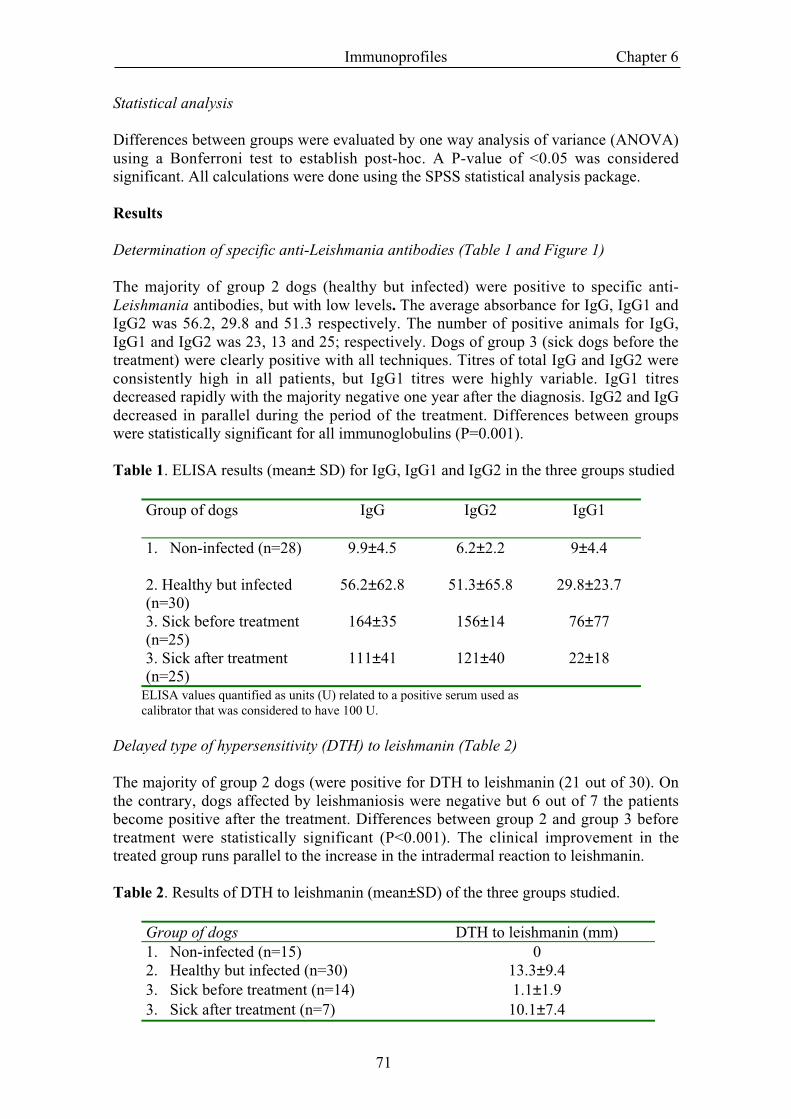
Determination of specific anti-Leishmania antibodies (Table 1 and Figure 1)
The majority of group 2 dogs (healthy but infected) were positive to specific anti-Leishmania antibodies, but with low levels. The average absorbance for IgG, IgG1 andIgG2 was 56.2, 29.8 and 51.3 respectively. The number of positive animals for IgG,IgG1 and IgG2 was 23, 13 and 25; respectively. Dogs of group 3 (sick dogs before thetreatment) were clearly positive with all techniques. Titres of total IgG and IgG2 wereconsistently high in all patients, but IgG1 titres were highly variable. IgG1 titresdecreased rapidly with the majority negative one year after the diagnosis. IgG2 and IgGdecreased in parallel during the period of the treatment. Differences between groupswere statistically significant for all immunoglobulins (P=0.001).
Table 1. ELISA results (mean± SD) for IgG, IgG1 and IgG2 in the three groups studied
Group of dogs IgG IgG2 IgG1
1. Non-infected (n=28) 9.9±4.5 6.2±2.2 9±4.4
2. Healthy but infected(n=30)
56.2±62.8 51.3±65.8 29.8±23.7
3. Sick before treatment(n=25)
164±35 156±14 76±77
3. Sick after treatment(n=25)
111±41 121±40 22±18
ELISA values quantified as units (U) related to a positive serum used ascalibrator that was considered to have 100 U.
Delayed type of hypersensitivity (DTH) to leishmanin (Table 2)
The majority of group 2 dogs (were positive for DTH to leishmanin (21 out of 30). Onthe contrary, dogs affected by leishmaniosis were negative but 6 out of 7 the patientsbecome positive after the treatment. Differences between group 2 and group 3 beforetreatment were statistically significant (P<0.001). The clinical improvement in thetreated group runs parallel to the increase in the intradermal reaction to leishmanin.
Table 2. Results of DTH to leishmanin (mean±SD) of the three groups studied.
Group of dogs DTH to leishmanin (mm)1. Non-infected (n=15) 02. Healthy but infected (n=30) 13.3±9.43. Sick before treatment (n=14) 1.1±1.93. Sick after treatment (n=7) 10.1±7.4
Immunoprofiles Chapter 6
72
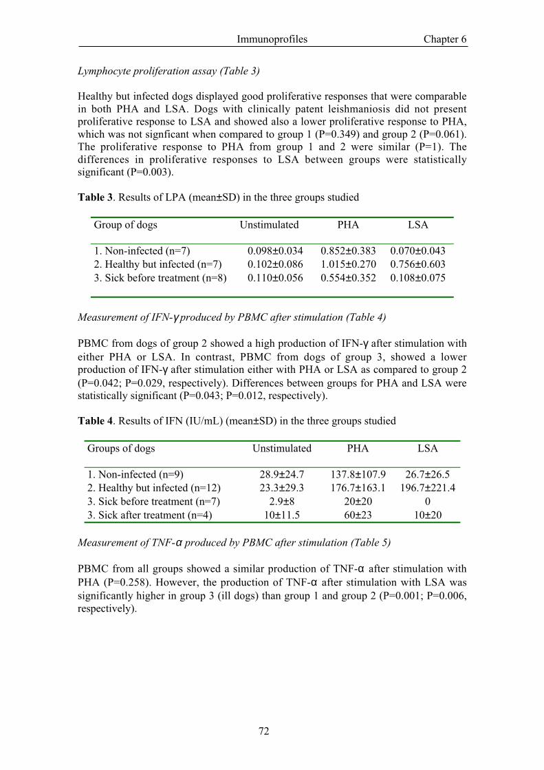
Lymphocyte proliferation assay (Table 3)
Healthy but infected dogs displayed good proliferative responses that were comparablein both PHA and LSA. Dogs with clinically patent leishmaniosis did not presentproliferative response to LSA and showed also a lower proliferative response to PHA,which was not signficant when compared to group 1 (P=0.349) and group 2 (P=0.061).The proliferative response to PHA from group 1 and 2 were similar (P=1). Thedifferences in proliferative responses to LSA between groups were statisticallysignificant (P=0.003).
Table 3. Results of LPA (mean±SD) in the three groups studied
Group of dogs Unstimulated PHA LSA
1. Non-infected (n=7) 0.098±0.034 0.852±0.383 0.070±0.0432. Healthy but infected (n=7) 0.102±0.086 1.015±0.270 0.756±0.6033. Sick before treatment (n=8) 0.110±0.056 0.554±0.352 0.108±0.075
Measurement of IFN-γ produced by PBMC after stimulation (Table 4)
PBMC from dogs of group 2 showed a high production of IFN-γ after stimulation witheither PHA or LSA. In contrast, PBMC from dogs of group 3, showed a lowerproduction of IFN-γ after stimulation either with PHA or LSA as compared to group 2(P=0.042; P=0.029, respectively). Differences between groups for PHA and LSA werestatistically significant (P=0.043; P=0.012, respectively).
Table 4. Results of IFN (IU/mL) (mean±SD) in the three groups studied
Groups of dogs Unstimulated PHA LSA
1. Non-infected (n=9) 28.9±24.7 137.8±107.9 26.7±26.52. Healthy but infected (n=12) 23.3±29.3 176.7±163.1 196.7±221.43. Sick before treatment (n=7) 2.9±8 20±20 03. Sick after treatment (n=4) 10±11.5 60±23 10±20
Measurement of TNF-α produced by PBMC after stimulation (Table 5)
PBMC from all groups showed a similar production of TNF-α after stimulation withPHA (P=0.258). However, the production of TNF-α after stimulation with LSA wassignificantly higher in group 3 (ill dogs) than group 1 and group 2 (P=0.001; P=0.006,respectively).
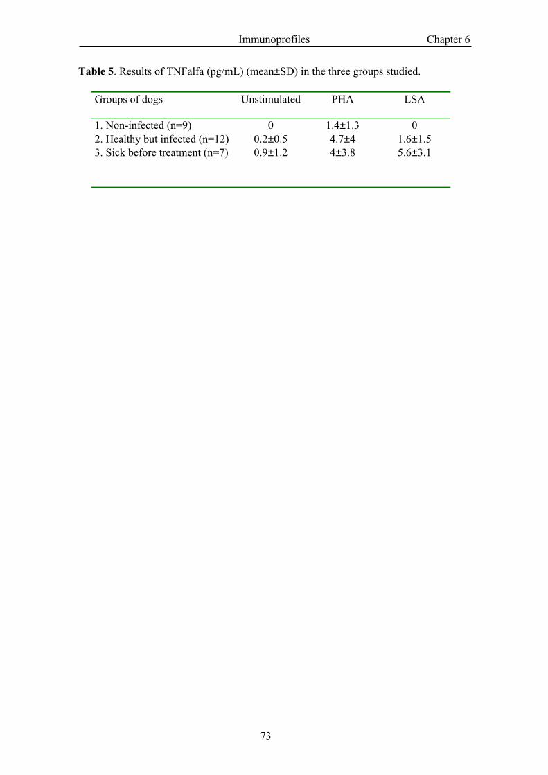
Immunoprofiles Chapter 6
73
Table 5. Results of TNFalfa (pg/mL) (mean±SD) in the three groups studied.
Groups of dogs Unstimulated PHA LSA
1. Non-infected (n=9) 0 1.4±1.3 02. Healthy but infected (n=12) 0.2±0.5 4.7±4 1.6±1.53. Sick before treatment (n=7) 0.9±1.2 4±3.8 5.6±3.1
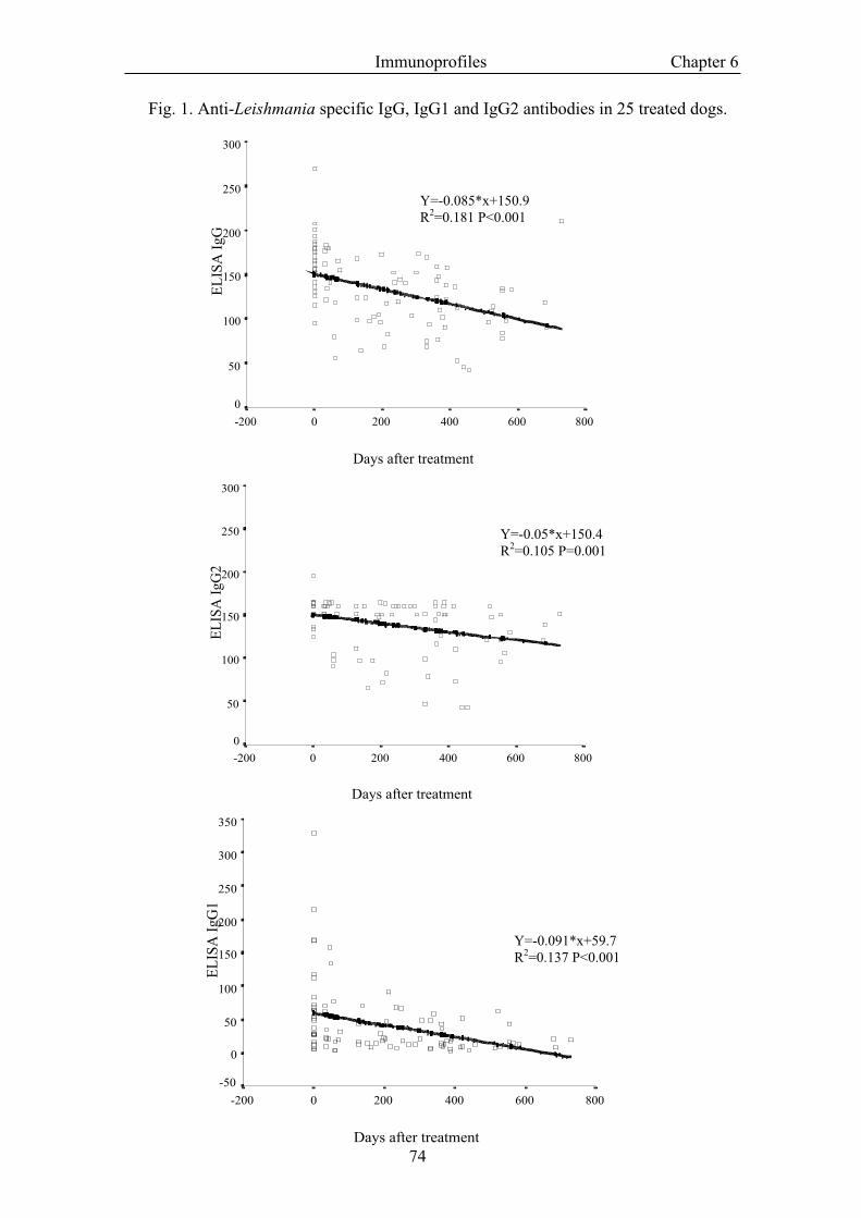
Immunoprofiles Chapter 6
74
Fig. 1. Anti-Leishmania specific IgG, IgG1 and IgG2 antibodies in 25 treated dogs.
Days after treatment
8006004002000-200
EL
ISA
IgG
300
250
200
150
100
50
0
Days after treatment
8006004002000-200
EL
ISA
IgG
2
300
250
200
150
100
50
0
Days after treatment
8006004002000-200
EL
ISA
IgG
1
350
300
250
200
150
100
50
0
-50
Y=-0.091*x+59.7R2=0.137 P<0.001
Y=-0.05*x+150.4R2=0.105 P=0.001
Y=-0.085*x+150.9R2=0.181 P<0.001
Immunoprofiles Chapter 6
75
Discussion
Based on the results we obtained using these techniques, it is possible to define thepattern of immune response of a single patient. We accomplished these results bycombining techniques, which assess the humoral response (serology) with techniquesthat evaluate the specific cellular immunity (Cabral et al., 1998; Rhalem et al., 1999a)i.e., DTH or production of IFN-γ by PBMC. In practice, DTH is possibly moreaccommodating because of the complexity of current techniques available to measurethe production of IFN-γ. In the near future, the development of better techniques tomeasure the production of canine cytokines (i.e., IL-4 and IFNγ) based on ELISAsmight allow for easier evaluation of the type of immunity against Leishmania occuringin a patient undergoing examination.
Dogs living in an endemic area of leishmaniosis can be grouped in different categories.First, dogs can be divided in asymptomatic (healthy) and ill dogs. Dogs with clinicallypatent leishmaniosis are infected animals that have an inefficient immune responseagainst Leishmania (Pinelli et al., 1994). Asymptomatic dogs, however, constitutes arather heterogeneous group of animals (Cabral et al., 1998). This group includes healthynon-infected animals (PCR negative, seronegative, DTH negative, no production ofIFN-γ), infected dogs which control the parasite by means of a cellular immuneresponse (PCR positive, low titre of specific IgG antibodies and DTH/IFN-γ positive)and infected dogs which are incubating the disease and that will be in the futuresymptomatic (PCR positive, seropositive, low DTH and low production of IFN-γ). It isnecessary, as stated before, to use several techniques to properly classify a single patientin one of the four groups.
Dogs affected by canine leishmaniosis show severe immunologic abnormalities. Theypresent a predominantly humoral immune response characterised by the production ofhigh amounts of specific anti-Leishmania IgG2 antibodies and a variable production ofIgG1antibodies. The cellular immune response against Leishmania, however, is veryweak or absent in these patients, as demonstrated by the low DTH, the low productionof IFN-γ and the lack of proliferative responses of PBMCs. These results are similar tothose described by (Pinelli et al., 1994) in dogs experimentally infected and indicatethat these animals present a Th2 helper like immune response (non-protective andpermitting a wide dissemination of the parasite). The production of TNF-α by PBMCsof these patients was elevated and highly variable. It is difficult to give a uniqueinterpretation to this finding. TNF-α is a pro-inflammatory cytokine that provides arapid form of host defence against infection (Nacy et al., 1991) but which also has manyundesirable effects on the host (Blackwell, 1999). High production of TNF-α could playa role in the protection against Leishmania but could also be responsible of some of theclinical signs and lesions of leishmaniosis such as cachexia, fever, or weight loss ashave also been reported in humans (Da-Cruz et al., 1996; Morsy et al., 1995; Pearson etal., 1992; Ribeiro-de-Jesus et al., 1998). But such results must be interpreted withcaution because these dogs appear to be immudepressed as demonstrated by the lowproliferative responses of PBMCs when stimulated by PHA. Some authors havepreviously described the anergic state to mitogens of dogs suffering from leishmaniosis(De Luna et al., 1999; Moreno et al., 1999) and have also found low figures number ofblood CD4+ cells (Moreno et al., 1999).
Immunoprofiles Chapter 6
76
The treatment has a noticeable effect on the immune response of ill dogs. However, theeffect is highly variable and difficult to predict. Some patients show a decrease in thetitre of IgGs which runs parallel to the recovery of the cellular immunity and to theclinical recovery (Rhalem et al., 1999b). Other dogs, however, retain the humoralimmune response and fail to recover the cellular immunity; these are the patients withthe worst prognosis. Probably, as it has been demonstrated in Leishmania majorinfection in mice (Nabors et al., 1995), the treatment due to a reduction of the parasiteburden could be the inductor for a switch from Th2 to Th1-like immune response.Obviously, many factors influence this evolution of the immune response duringtreatment, as type and duration of the treatment, concomitant infections or diseases, re-infections, and the status of the cellular immune response at the beginning of thetreatment.
As expected, non-infected dogs were completely negative to all techniques performed.However, they were immunocompetent as their PBMCs respond with an intenseproliferation when stimulated with mitogens (PHA).
Healthy, infected dogs can be divided also into two groups: (1) Dogs that clearlypresented a specific-Leishmania cellular immunity demonstrated by a positive DTH,proliferation and production of IFN-γ and low specific-Leishmania humoral immunityand (2) Those animals that failed to have a specific-Leishmania cellular immuntiy andinstead they showed only humoral immunity. The appearance of resistant dogs wasreported in the last decade demonstrating cellular immunity in naturally andexperimentally infection and associating this cellular immunity as Th1 like immuneresponse (Cabral et al., 1992; Pinelli et al., 1994). However, the term resistant must beinterpreted with caution as the parasites probably remain in these individuals inequilibrium without producing pathology. However, this is not an unchangeablesituation and several factors can break the equilibrium and lead to clinical disease. Thissituation is similar to that of human (Schubach et al., 1998a; Schubach et al., 1998b)and animals (Aebischer et al., 1993; Belkaid et al., 2000; de Rossell et al., 1992) afterhealing, which persist infected but clinically healthy during long periods of time. Themechanisms of persistence of the parasite are currently being investigated (Bogdan etal., 1996; Bogdan & Rollinghoff, 1998). Some authors have hypothesised thatLeishmania could persist in low nitric oxide producing cells as fibroblasts (Bogdan etal., 2000).
The integration of data obtained in broad epidemiological and immunological studies(Berrahal et al., 1996; Cabral et al., 1998; Solano-Gallego et al., 2000a; Solano-Gallegoet al., 2000b), are changing the understanding of canine leishmaniosis. Infection anddisease are no longer synonymous and the diagnosis has to be established by combiningseveral techniques. Diagnosis and prognosis can be accurately established if, togetherwith the detection of the parasite, the type of immune response of the patient, humoralversus cellular, is evaluated.
Acknowledgements
The authors are grateful to the valuable contribution in the performance of IFN-γbioassay of Dr. Victor Rutten and co-workers from the Department of Immunology andInfectious diseases of Faculty of Veterinary Medicine of Utrecht University.
Immunoprofiles Chapter 6
77
References
Aebischer, T., Moody, S.F. & Handman, E. (1993). Persistence of virulent Leishmania major in murinecutaneous leishmaniasis: a possible hazard for the host. Infection and Immunity, 61, 220-226.
Belkaid, Y., Mendez, S., Lira, R., Kadambi, N., Milon, G. & Sacks, D. (2000). A natural Model ofLeishmania major infection reveals a prolonged silent phase of parasite amplification in the skinbefore the onset of lesion formation and immunity. Journal of Immunology, 165, 969-977.
Berrahal, F., Mary, C., Roze, M., Berenger, A., Escoffier, K., Lamouroux, D. & Dunan, S. (1996).Canine leishmaniasis: identification of asymptomatic carriers by polymerase chain reaction andimmunoblotting. American Journal of Tropical Medicine and Hygiene, 55, 273-277.
Blackwell, J.M. (1999). Tumor necrosis factor alpha and mucocutaneous leishmaniasis. ParasitologyToday, 15, 73-75.
Bogdan, C., Donhauser, N., Doring, R., Rollinghoff, M., Diefenbach, A. & Rittig, M.G. (2000).Fibroblasts as host cells in latent leishmaniosis. Journal of Experimental Medicine, 191, 2121-2130.
Bogdan, C., Gessner, A., Solbach, W. & Röllinghoff, M. (1996). Invasion, control and persistence ofLeishmania parasites. Current Opinion in Immunology, 8, 517-525.
Bogdan, C. & Rollinghoff, M. (1998). The immune response to Leishmania: mechanisms of parasitecontrol and evasion. International Journal of Parasitology, 28, 121-134.
Böyum, A. (1968). Isolation of mononuclear cells and granulocytes from human blood. ScandinavianJournal of Clinical Laboratory Investigation, 21, 77-89.
Brazis, P., Queralt, M., de Mora, F., Ferrer, L. & Puigdemont, A. (2000). Stem cell factor enhancesIgE-mediated histamine and TNF-alpha release from dispersed canine cutaneous mast cells.Veterinary Immunology and Immunopathology, 75, 97-108.
Cabral, M., O'Grady, J. & Alexander, J. (1992). Demonstration of Leishmania specific cell mediatedand humoral immunity in asymptomatic dogs. Parasite Immunology, 14, 531-539.
Cabral, M., O'Grady, J.E., Gomes, S., Sousa, J.C., Thompson, H. & Alexander, J. (1998). Theimmunology of canine leishmaniosis: strong evidence for a developing disease spectrum fromasymptomatic dogs. Veterinary Parasitology, 76, 173-180.
Cardoso, L., Neto, F., Sousa, J.C., Rodrigues, M. & Cabral, M. (1998). Use of leishmanin skin test inthe detection of canine Leishmania-specific cellular immunity. Veterinary Parasitology, 79, 213-220.
Da-Cruz, A.M., de Oliveira, M.P., De Luca, P.M., Mendonca, S.C. & Coutinho, S.G. (1996). Tumornecrosis factor-alpha in human american tegumentary leishmaniasis. Memorias InstitutoOswaldo Cruz, 91, 225-229.
De Luna, R., Vuotto, M.L., Ielpo, M.T.L., Ambrosio, R., Piantedosi, D., Moscatiello, V., Ciaramella,P., Scalone, A., Gradoni, L. & Mancino, D. (1999). Early suppression of lymphoproliferativeresponse in dogs with natural infection by Leishmania infantum. Veterinary Immunology andImmunopathology, 70, 95-103.
de Rossell, R., Jesús de Duran, R., Rossell, O. & Rodríguez, A.M. (1992). Is leishmaniasis ever cured?Transactions of the Royal Society of Tropical Medicine and Hygiene, 86, 251-253.
Greene, R.T. (1990). An update on the serodiagnosis of canine Lyme borreliosis. Journal of VeterinaryInternal Medicine, 4, 167-171.
Maconi, G., Kurihara, H., Tascheri, A.M. & Bianchi Porro, G. (1999). Management of Helicobacterpylori infection: gastroenterological and surgical perspectives. Journal of Chemotherapy, 11,581-590.
Moreno, J., Nieto, J., Chamizo, C., Gonzalez, F., Blanco, F., Barker, D.C. & Alvar, J. (1999). Theimmune response and PBMC subsets in canine visceral leishmaniasis before and afterchemotherapy. Veterinary Immunology and Immunopathology, 71, 181-195.
Morsy, T.A., el Missiry, A.G., Sarwat, M.A., el Rassed, M.M., Habib, F.S. & M.M., A.G. (1995).Tumour necrosis factor-alpha (cachectin) in human visceral leishmaniasis. Journal of EgyptianSociety of Parasitology, 25, 31-51.
Nabors, G.S., Afonso, L.C.C., Farrell, J.P. & Scott, P. (1995). Switch from a type 2 to a type 1 Thelper cell response and cure of established Leishmania major infection in mice is induced bycombined therapy with interleukin 12 and pentostam. Proccedings of National Academy ofScience of USA, 92, 3142-3146.
Nacy, C.A., Meierovics, A.I., Belosevic, M. & Green, S.J. (1991). Tumor necrosis factor-alpha: centralregulatory cytokine in the induction of macrophage antimicrobial activities. Pathobiology, 59,182-184.
Immunoprofiles Chapter 6
78
Pearson, R.D., Cox, G., Jeronimo, S.M.B., Castracane, J., Drew, J.S., Evans, T. & Alencar, J.E.(1992). Visceral leishmaniasis: a model for infection-induced cachexia. American Journal ofTropical Medicine and Hygiene, 47, 8-15.
Pinelli, E., Gonzalo, R.M., Boog, C.J., Rutten, V.P.M.G., Gebhard, D., Real, G. & Ruitenberg, E.J.(1995). Leishmania infantum-specific T cell lines derived from asymptomatic dogs that lyseinfected macrophages in a major histocompatibility complex-restricted manner. EuropeanJournal of Immunology, 25, 1594-1600.
Pinelli, E., Killick-Kendrick, R., Wagenaar, J., Bernardina, W., del Real, G. & Ruitenberg, J.(1994). Cellular and humoral immune response in dogs experimentally and naturally infectedwith Leishmania infantum. Infection and Immunity, 62, 229-235.
Rhalem, A., Sahibi, H., Guessous-Idrissi, N., Lasri, S., Natami, A., Riyad, M. & Berrag, B. (1999a).Immune response against Leishmania antigens in dogs naturally and experimentally infectedwith Leishmania infantum. Veterinary Parasitology, 81, 173-184.
Rhalem, A., Sahibi, H., Lasri, S. & Jaffe, C.L. (1999b). Analysis of immune responses in dog withcanine visceral leishmaniasis before, and after, drug treatment. Veterinary Immunology andImmunopathology, 71, 69-76.
Ribeiro-de-Jesus, A., Almeida, R.P., Lessa, H., Bacellar, O. & Carvalho, E.M. (1998). Cytokineprofile and pathology in human leishmaniasis. Brazilian Journal of Medicine and BiologyResearch, 31, 143-148.
Rodgers, M.R., Stephen, J. & Wirth, D.F. (1990). Amplification and diagnosis of Leishmania.Experimental Parasitology, 71, 267-275.
Roura, X., Sánchez, A. & Ferrer, L. (1999). Diagnosis of canine leishmaniasis by a polymerase chainreaction technique. Veterinary Record, 144, 262-264.
Schubach, A., Haddad, F., Oliveira-Neto, M.P., Degrave, W., Pirmez, C., Grimaldi, G.J. &Fernandes, O. (1998a). Detection of Leishmania DNA by polymerase chain reaction in scars oftreated human patients. Journal of Infectious Disease, 178, 911-914.
Schubach, A., Marzochi, M.C., Cuzzi-Maya, T., Oliveira, A.V., Araujo, M.L., Oliveira, A.L.,Pacheco, R.S., Momen, H., Conceicao-Silva, F., Coutinho, S.G. & Marzochi, K.B. (1998b).Cutaneous scars in American tegumentary leishmaniasis patients: a site of Leishmania (Viannia)braziliensis persistence and viability eleven years after antimonial therapy and clinical cure.American Journal of Tropical Medicine and Hygiene, 58, 824-827.
Solano-Gallego, L., Llull, J., Ramos, G., Riera, C., Arboix, M., Alberola, J. & Ferrer, L. (2000a).The Ibizian hound presents a predominantly cellular immune response against naturalLeishmania infection. Veterinary Parasitology, 90, 37-45.
Solano-Gallego, L., Morell, P., Arboix, M., Alberola, J. & Ferrer, L. (2000b). Prevalence ofLeishmania infection in dogs living in endemic regions. Veterinary Dermatology, 11, 22.
Solbach, W. & Laskay, T. (2000). The host response to Leishmania infection. Advances in Immunology,74, 275-317.