Embed Size (px)
Citation preview
Research ArticleEvaluation of Toxic, Cytotoxic, Mutagenic, andAntimutagenic Activities of Natural and TechnicalCashew Nut Shell Liquids Using the Allium cepa andArtemia salina Bioassays
Aracelli de Sousa Leite,1,2 Alisson Ferreira Dantas,3
George Laylson da Silva Oliveira,4 Antonio L. Gomes Júnior,1
Sidney Gonçalo de Lima,5 Antônia Maria das Graças Lopes Citó,5 Rivelilson M. de Freitas,4
Ana Amélia de C. Melo-Cavalcante,1,2 and José Arimateia Dantas Lopes2,4
1Laboratorio de Pesquisa em Genetica Toxicologica de Pos-Graduacao em Ciencias Farmaceuticas da Universidade Federal do Piauı,6409-550 Teresina, PI, Brazil2Programa de Pos-Graduacao em Biotecnologia (RENORBIO) da Universidade Federal do Piauı, 6409-550 Teresina, PI, Brazil3Programa de Pos-Graduacao em Biologia Animal, Departamento de Genetica e Morfologia, Instituto de Ciencias Biologicas,Universidade de Brasılia, 70910-900 Brasılia, DF, Brazil4Laboratorio de Pesquisa em Neuroquımica Experimental do Programa de Pos-Graduacao em Ciencias Farmaceuticas daUniversidade Federal do Piauı, 6409-550 Teresina, PI, Brazil5Departamento de Quımica, CCN, Universidade Federal do Piauı, 6409-550 Teresina, PI, Brazil
Correspondence should be addressed to Ana Amelia de C. Melo-Cavalcante; ana [email protected]
Received 14 November 2014; Revised 21 January 2015; Accepted 21 January 2015
Academic Editor: Qaisar Mahmood
Copyright © 2015 Aracelli de Sousa Leite et al. This is an open access article distributed under the Creative Commons AttributionLicense, which permits unrestricted use, distribution, and reproduction in any medium, provided the original work is properlycited.
The cashew nut releases a substance that is known as cashew nut shell liquid (CNSL).There are both natural (iCNSL) and technical(tCNSL) cashewnut shell liquids.This study used anArtemia salinabioassay to evaluate the toxic effects of iCNSL and tCNSL cashewnut shell liquids. It also evaluated the toxicity, cytotoxicity, and mutagenicity of CNSL and its effects on the damage induced bycopper sulfate (CuSO
4⋅5H2O) on themeristems’ root ofAllium cepa. Effects of the damage induced by CuSO
4⋅5H2Owere evaluated
before (pre-), during (co-), and after (post-) treatments. The iCNSL contained 94.5% anacardic acid, and the tCNSL contained91.3% cardanol. The liquids were toxic to A. salina. Toxicity, cytotoxicity, and mutagenicity were observed with iCNSL comparedwith the negative control. Similarly, iCNSL failed to inhibit the toxicity and cytotoxicity of CuSO
4⋅5H2O.The tCNSL was not toxic,
cytotoxic, ormutagenic in any of the concentrations.However, the lowest iCNSL concentrations and all of the tCNSL concentrationshad preventive, antimutagenic, and reparative effects on micronuclei and on chromosomal aberrations in the A. cepa. Therefore,protective, modulating, and reparative effects may be observed in the A. cepa, depending on the concentration and type of CNSLused.
1. Introduction
Recent epidemiological studies have shown that medicinalplants may be involved in preventing or delaying the devel-opment of various diseases [1, 2]. These plants may act ondifferent targets in signal transduction pathways that may
modulate gene expression, cell cycle progression, cellular pro-liferation, and/or apoptosis [3, 4]. However, adverse effects,such as genotoxicity, mutagenicity, and carcinogenicity [5, 6],can also occur. These effects may be triggered by compoundsthat interact with deoxyribonucleic acid (DNA), whichwouldcause cellular toxicity and/or genotoxicity [7].
Hindawi Publishing CorporationBioMed Research InternationalVolume 2015, Article ID 626835, 16 pageshttp://dx.doi.org/10.1155/2015/626835
2 BioMed Research International
The species Anacardium occidentale (Anacardiaceae) isfound in tropical regions worldwide. It is common in Brazil,India, Mozambique, Tanzania, Kenya, Vietnam, Indonesia,and Thailand [8]. Recent studies have been associated withseveral biological effects of the cashew plant. It can be usedas an antioxidant [9, 10] and can be used in dermatitis [11]and also possesses larvicidal [12], antigenotoxic [13], andantimicrobial [14] activities.
The cashew nut releases a liquid that is known as cashewnut shell liquid (CNSL). This liquid is a natural source ofphenolic compounds that contribute to its antioxidant [15,16], antifungal [17], antibacterial [18], larvicidal [19], andnongenotoxic effects in prokaryotic [20, 21] and eukary-otic cells [22]. This liquid is classified into two categories,depending on the extraction method used: natural CNSL(iCNSL) extracted with solvents, and its main componentsare anacardic acid (62.9%), cardol (23.98%), and cardanol(6.99%) [23] and technical CNSL (tCNSL) wich is preparedby burning the nuts industrially at high temperatures andit contains cardanol (60–65%), cardol (15–20%), polymericmaterial (10%), and small amounts of metilcardol [24].
The present study aimed to evaluate the toxicity ofiCNSL and tCNSL to A. salina and to determine their toxic,cytotoxic, and mutagenic actions and their protective effectsagainst the damage that is induced by CuSO
4⋅5H2O inA. cepa
root meristems.
2. Material and Methods
2.1. CNSL Preparation and Doses Choice. Ripe cashew nutswere collected from cashew trees in Teresina in the stateof Piauı, Brazil. For iCNSL, extraction the ripe cashew nutswere stored in styrofoam with liquid nitrogen for 5 minutes.Then, they were crushed and subjected to hot extractionSoxhlet extractor with hexane for 16 hours. The iCNSLwas then concentrated in a rotary evaporator at 45∘C. ThetCNSL was provided by a company of the Group Europa–Castanha located in Altos, Teresina, Piauı, in northeasternBrazil. According to the company, the nuts were immersedin a hot bath at 195∘C for 3 hours to extract the tCNSL. ThetCNSL was then filtered and stored.
The lowest doses used in this study were chosen becauserecent research on the tCNSL (100–500 𝜇g/mL) has shownantioxidant properties. Thus, doses lower than those testedin the literature have been evaluated to check if they wouldstill be antioxidant and nonmutagenic.
2.2. CNSL Methylation Reaction. Samples of the extractediCNSL and tCNSL were analysed by gas chromatographycoupled tomass spectrometry (GC-MS) in the formofmethylesters. Five milligrams of CNSL was dissolved in 0.5mL ofdiethyl ether and transferred to a 5.0 mL flask. A solutionof diazomethane in ether (2.0mL) was then added dropwiseat low temperature in an ice bath until outgassing was nolonger observed. The flask was sealed with a ground glassstopper and magnetically stirred continuously at room tem-perature. After 3 hours, the reaction was monitored by thinlayer chromatography (TLC). Following ether evaporation,
the residue was solubilised in ethyl acetate and analysed byGC-MS [25].
2.3. GC-MS Analysis. Both of the derived CNSL sampleswere then analysed in a GC-MS system (Shimadzu, GC-17A/MS-QP5050A). The column chromatography DB-5HT(J & W Scientific) was 30m long and 0.25mm in diameter,had a film thickness of 0.10 𝜇m, and used helium as a car-rier gas (1.0mL/min). The following parameters were used:interface = 270∘C, injector = 250∘C, initial temperature =60∘C (2min), 4.0mL/min to 180∘C (4min), and 10mL/minto 260∘C (10min). Identification was achieved by comparingmass spectra (43 to 4500 Daltons, electron impact ionisation,70 eV) and data from the literature of De Lima et al. [25].
2.4. Artemia salina Test. The method used to assess thetoxicity of iCNSL and tCNSL on A. salina was adapted fromMeyer et al. [26]. The microcrustaceans were hatched inwater as mentioned in Materials and Methods for 48 hoursuntil their larvae were released. Ten A. salina specimens wereintroduced into each of the three tubes containing a 17.37,34.75, or 69.50 𝜇g⋅mL−1 concentration of iCNSL or tCNSL.A nonactive substance (1 : 1 seawater and mineral water) wasused as a control. The dead specimens were counted under astereomicroscope after 24 hours.
2.5. Allium cepa Test. The A. cepa test was adapted from themethod reported by Fiskesjo [27]. Each experimental groupconsisted of iCNSL and tCNSL at concentrations of 17.37,34.75, or 69.50𝜇g⋅mL−1, as well as a solution of 500𝜇g/mLTween 20 (solvent), a negative control (dechlorinated water),and a positive control (1.2 𝜇g⋅mL−1 copper sulphate). Smallbulbs ofA. cepa (2𝑛 = 16) were purchased from supermarketsin Teresina, Piauı.
Following 72 hours of exposure, the roots were measuredin centimetres to assess toxicity.The roots were then placed inCarnoy’s fixative solution (ethanol/glacial acetic acid 3 : 1 v/v),refrigerated at 4∘C for 24 hours, followed by 70% ethanolsolution and refrigeration. The roots were subsequentlyhydrolysed in a hydrochloric acid solution (1N) and placedin a staining solution (Schiff ’s dye) for two hours.
The roots were then placed on slides and sectioned in themeristem region.This region of the root was stained with 2%acetic carmine, covered with a cover slip, and then observedunder an optical microscope (1000x) to analyse cytotoxicity,mutagenicity, and the effects on the damage induced bycopper sulphate. A total of 1,000 cells were analysed on eachslide. The following parameters were observed: (a) mitoticindex (MI), (b) the frequency of chromosomal aberrations(CA) in anaphase and telophase, and (c) the frequency ofmicronuclei (MN).
2.6. Effects onCopper Sulphate-InducedDamage. Themethodused to evaluate antimutagenicity bymeans of theA. cepa testwas adapted from Malini et al. [28]. The present study usedCuSO
4⋅5H2O as the genotoxic agent because of its mutagenic
potential [29, 30].
BioMed Research International 3
Table 1: The investigated components of iCNSL and tCNSL.
Natural iCNSL∗
Peak Retention time (min) Compounds Yield1 36.80 Metilcardol 2.902 37.60 Monounsaturated anacardic acid 82.903 37.62 Diunsaturated anacardic acid 8.004 37.70 Anacardic acid 3.605 38.05 Unidentified 2.60
Technical tCNSLPeak Retention time (min) Compounds Yield6 18.08 Monounsaturated cardanol 79.407 18.10 Diunsaturated cardanol 8.678 18.17 Cardanol 3.239 20.71 Unidentified 8.70∗Analysed in the form of methyl esters.
Three types of treatments were standardised: (1) pretreat-ment, in which the bulbs were exposed initially to iCNSLor tCNSL for 48 hours and the roots were then washedin distilled water and placed in 1.2 𝜇g⋅mL−1 CuSO
4⋅5H2O
solutions for 24 hours to germinate; (2) cotreatment, inwhich the bulbs were placed in tubes that contained iCNSLor tCNSL and a 1.2 𝜇g⋅mL−1 CuSO
4⋅5H2O solution at a 1 : 1
ratio for 72 hours for germination; and (3) posttreatment, inwhich the bulbs were first placed in 1.2𝜇g⋅mL−1 CuSO
4⋅5H2O
solution for 48 hours, and then the roots were rinsed withdistilled water and placed in tubes that contained iCNSL ortCNS for 24 hours for germination. The samples were thenprocessed in the similar way as mentioned in the A. cepa test.
2.7. Statistical Analysis. The number of dead A. salina wasanalysed using the Statistical Package for the Social Sciences(SPSS) software, version 17:0.The IC
50was assessed by probit
analysis.The tests were performed in triplicate.The data wereanalysed using GraphPad Prism software (version 6.03), andthe experimental groups were compared with the negativeand positive control groups. All results were expressed asmean ± standard deviation (SD). The data were assessed byan analysis of variance (ANOVA) followed by Tukey’s testfor multiple comparisons for genotoxicity and mutagenicitytests The significance levels were ∗
𝑃< 0.05, ∗∗
𝑃< 0.01, and
∗∗∗𝑃< 0.001.
3. Results
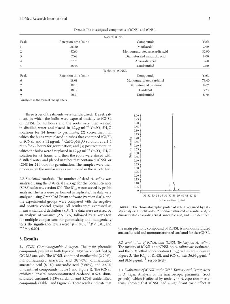
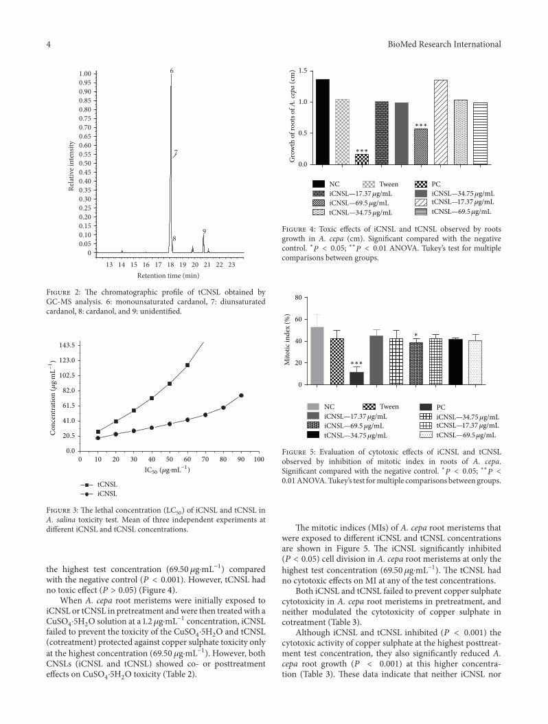
3.1. CNSL Chromatographic Analyses. The main phenoliccompounds present in both types of CNSL were identified byGC-MS analysis. The iCNSL contained metilcardol (2.90%),monounsaturated anacardic acid (82.90%), diunsaturatedanacardic acid (8.0%), anacardic acid (3.60%), and 2.60%unidentified compounds (Table 1 and Figure 1). The tCNSLexhibited 79.40% monounsaturated cardanol, 8.67% diun-saturated cardanol, 3.23% cardanol, and 8.70% unidentifiedcompounds (Table 1 and Figure 2).These results indicate that
43424140393837363534333231Retention time (min)
00.050.100.150.200.250.300.350.400.450.500.550.600.650.700.750.800.850.900.951.00
Rela
tive i
nten
sity
1
2
3
45
Figure 1: The chromatographic profile of iCNSL obtained by GC-MS analysis. 1: metilcardol, 2: monounsaturated anacardic acid, 3:diunsaturated anacardic acid, 4: anacardic acid, and 5: unidentified.
the main phenolic compound of iCNSL is monounsaturatedanacardic acid andmonounsaturated cardanol for the tCNSL.
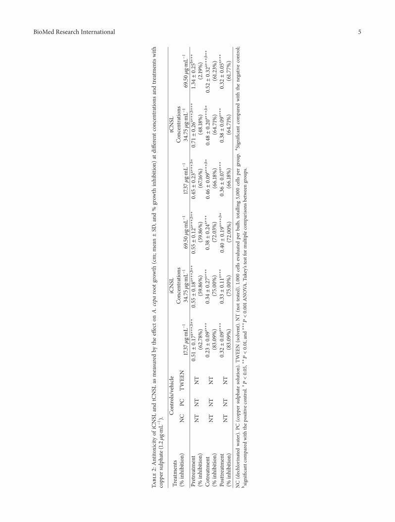
3.2. Evaluation of iCNSL and tCNSL Toxicity on A. salina.The toxicity of iCNSL and tCNSL on A. salina was evaluated,and the 50% lethal concentration (IC
50) values are shown in
Figure 3. The IC50
of iCNSL and tCNSL was 36.96 𝜇g⋅mL−1and 91.67 𝜇g⋅mL−1, respectively.
3.3. Evaluation of iCNSL and tCNSL Toxicity and Cytotoxicityin A. cepa. Analysis of the macroscopic parameter (rootgrowth), which is affected by toxicity in A. cepa root meris-tems, showed that iCNSL had a significant toxic effect at
4 BioMed Research International
2322212019181716151413Retention time (min)
00.050.100.150.200.250.300.350.400.450.500.550.600.650.700.750.800.850.900.951.00
Rela
tive i
nten
sity
6
7
89
Figure 2: The chromatographic profile of tCNSL obtained byGC-MS analysis. 6: monounsaturated cardanol, 7: diunsaturatedcardanol, 8: cardanol, and 9: unidentified.
tCNSLiCNSL
0 10 20 30 40 50 60 70 80 90 100
0.0
20.5
41.0
61.5
82.0
102.5
123.0
143.5
Con
cent
ratio
n (𝜇
g·m
L−1)
IC50 (𝜇g·mL−1)
Figure 3: The lethal concentration (LC50) of iCNSL and tCNSL in
A. salina toxicity test. Mean of three independent experiments atdifferent iCNSL and tCNSL concentrations.
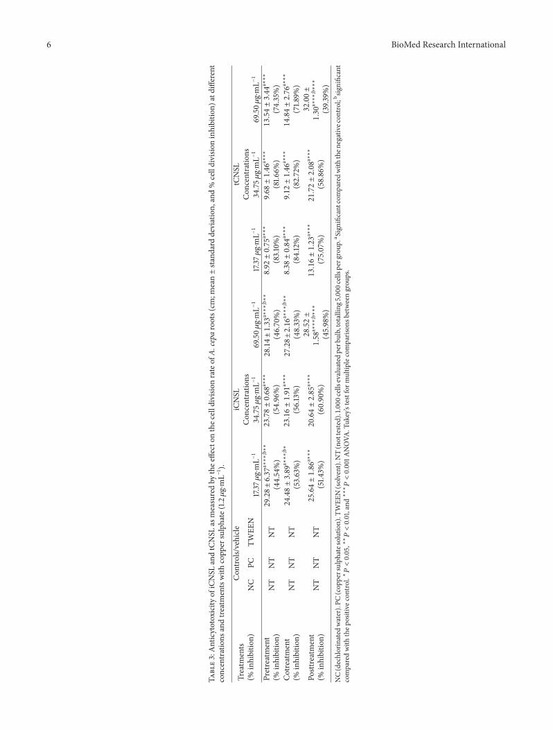
the highest test concentration (69.50𝜇g⋅mL−1) comparedwith the negative control (𝑃 < 0.001). However, tCNSL hadno toxic effect (𝑃 > 0.05) (Figure 4).
When A. cepa root meristems were initially exposed toiCNSL or tCNSL in pretreatment andwere then treatedwith aCuSO
4⋅5H2O solution at a 1.2 𝜇g⋅mL−1 concentration, iCNSL
failed to prevent the toxicity of the CuSO4⋅5H2O and tCNSL
(cotreatment) protected against copper sulphate toxicity onlyat the highest concentration (69.50 𝜇g⋅mL−1). However, bothCNSLs (iCNSL and tCNSL) showed co- or posttreatmenteffects on CuSO
4⋅5H2O toxicity (Table 2).
0.0
0.5
1.0
1.5
Gro
wth
of r
oots
of A
. cep
a (c
m)
NC Tween PC
∗∗∗
∗∗∗
iCNSL—17.37𝜇g/mLtCNSL—17.37𝜇g/mLiCNSL—69.5 𝜇g/mLtCNSL—69.5𝜇g/mLtCNSL—34.75𝜇g/mL
iCNSL—34.75𝜇g/mL
Figure 4: Toxic effects of iCNSL and tCNSL observed by rootsgrowth in A. cepa (cm). Significant compared with the negativecontrol. ∗
𝑃< 0.05; ∗∗
𝑃< 0.01 ANOVA. Tukey’s test for multiple
comparisons between groups.
0
20
40
60
80M
itotic
inde
x (%
)
NC Tween PCiCNSL—17.37𝜇g/mL
tCNSL—17.37𝜇g/mLiCNSL—69.5 𝜇g/mLtCNSL—69.5𝜇g/mLtCNSL—34.75𝜇g/mL
iCNSL—34.75𝜇g/mL
∗∗∗
∗
Figure 5: Evaluation of cytotoxic effects of iCNSL and tCNSLobserved by inhibition of mitotic index in roots of A. cepa.Significant compared with the negative control. ∗
𝑃< 0.05; ∗∗
𝑃<
0.01ANOVA.Tukey’s test formultiple comparisons between groups.
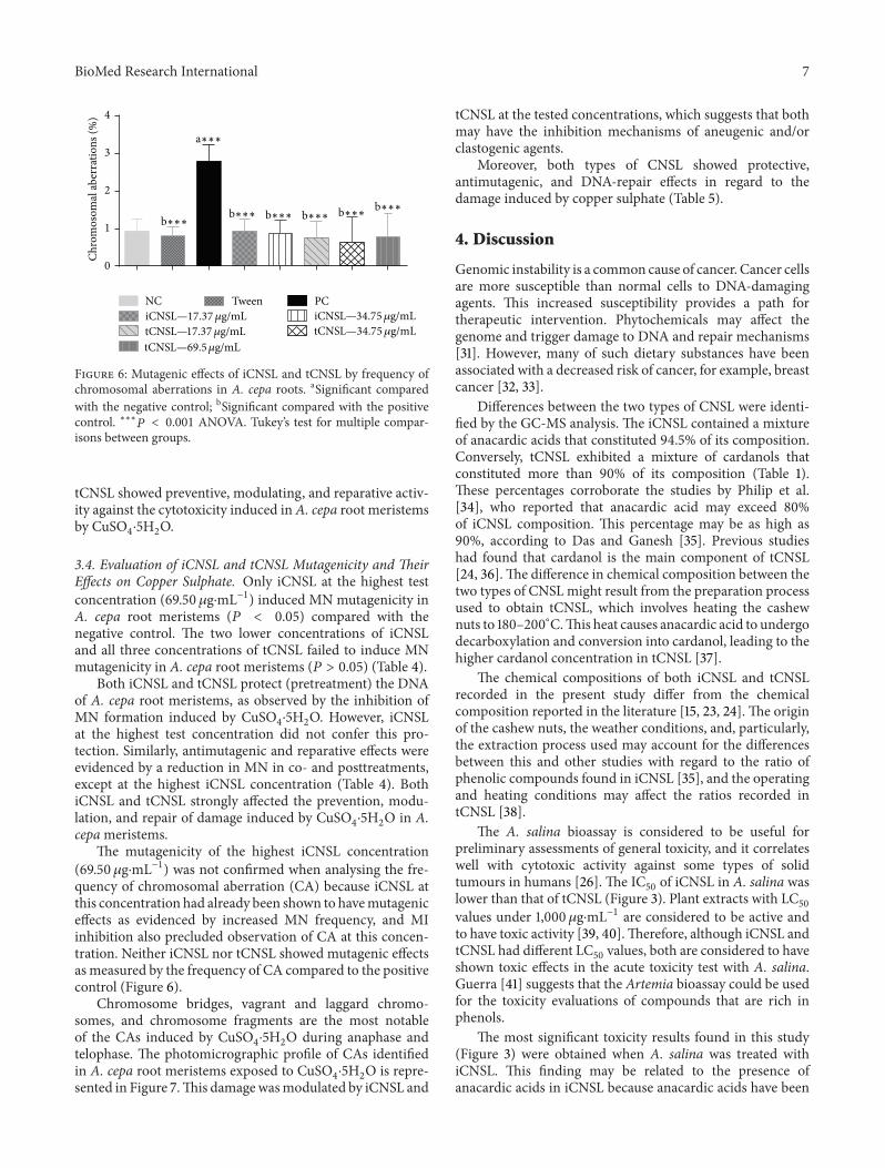
The mitotic indices (MIs) of A. cepa root meristems thatwere exposed to different iCNSL and tCNSL concentrationsare shown in Figure 5. The iCNSL significantly inhibited(𝑃 < 0.05) cell division in A. cepa root meristems at only thehighest test concentration (69.50 𝜇g⋅mL−1). The tCNSL hadno cytotoxic effects on MI at any of the test concentrations.
Both iCNSL and tCNSL failed to prevent copper sulphatecytotoxicity in A. cepa root meristems in pretreatment, andneither modulated the cytotoxicity of copper sulphate incotreatment (Table 3).
Although iCNSL and tCNSL inhibited (𝑃 < 0.001) thecytotoxic activity of copper sulphate at the highest posttreat-ment test concentration, they also significantly reduced A.cepa root growth (𝑃 < 0.001) at this higher concentra-tion (Table 3). These data indicate that neither iCNSL nor
BioMed Research International 5
Table2:Antito
xicity
ofiCNSL
andtCNSL
asmeasuredby
theeffecto
nA.
cepa
root
grow
th(cm;m
ean±SD
,and
%grow
thinhibitio
n)at
different
concentrations
andtre
atmentswith
copp
ersulphate(1.2𝜇g⋅mL−1).
Treatm
ents
(%inhibitio
n)
Con
trols/vehicle
iCNSL
tCNSL
NC
PCTW
EEN
Con
centratio
nsCon
centratio
ns17.37𝜇g⋅mL−1
34.75𝜇
g⋅mL−1
69.50𝜇
g⋅mL−1
17.37𝜇g⋅mL−1
34.75𝜇
g⋅mL−1
69.50𝜇
g⋅mL−1
Pretreatment
(%inhibitio
n)NT
NT
NT0.51±0.17
a∗∗∗,b∗∗
(62.78%)
0.55±0.18
a∗∗∗,b∗∗
(59.8
6%)
0.55±0.12
a∗∗∗,b∗∗
(59.8
6%)
0.45±0.23
a∗∗∗.b∗
(67.16%
)0.71±0.26
a∗∗∗,b∗∗∗
(48.18%)
1.34±0.25
b∗∗∗
(2.19
%)
Cotreatment
(%inhibitio
n)NT
NT
NT
0.23±0.09
a∗∗∗
(83.09%)
0.34±0.27
a∗∗∗
(75.00%)
0.38±0.24
a∗∗∗
(72.03%)
0.46±0.09
a∗∗∗;b∗
(66.18%)
0.48±0.20
a∗∗∗;b∗
(64.71%)
0.52±0.32
a∗∗∗;b∗∗
(61.2
3%)
Posttreatment
(%inhibitio
n)NT
NT
NT
0.32±0.09
a∗∗∗
(83.09%)
0.33±0.11
a∗∗∗
(75.00%)
0.40±0.19
a∗∗∗;b∗
(72.00%)
0.36±0.07
a∗∗∗
(66.18%)
0.38±0.09
a∗∗∗
(64.71%)
0.32±0.05
a∗∗∗
(61.7
7%)
NC(dechlorinated
water).PC
(cop
persulphate
solutio
n).T
WEE
N(solvent).NT(not
teste
d).1.000
cells
evaluatedperbu
lb,totallin
g5,00
0cells
pergrou
p.a Significantc
omparedwith
thenegativ
econtrol;
b significantcom
paredwith
thep
ositive
control.∗𝑃<0.05,∗∗𝑃<0.01,and∗∗∗𝑃<0.001A
NOVA
.Tuk
ey’stestform
ultip
lecomparis
onsb
etweengrou
ps.
6 BioMed Research International
Table3:Anticytotoxicity
ofiCNSL
andtCNSL
asmeasuredby
thee
ffecton
thec
elld
ivision
rateof
A.cepa
roots(cm
;mean±standard
deviation,and%celldivisio
ninhibitio
n)atdifferent
concentrations
andtre
atmentswith
copp
ersulphate(1.2𝜇g⋅mL−
1 ).
Treatm
ents
(%inhibitio
n)
Con
trols/vehicle
iCNSL
tCNSL
NC
PCTW
EEN
Con
centratio
nsCon
centratio
ns17.37𝜇g⋅mL−1
34.75𝜇
g⋅mL−1
69.50𝜇
g⋅mL−1
17.37𝜇g⋅mL−1
34.75𝜇
g⋅mL−1
69.50𝜇
g⋅mL−1
Pretreatment
(%inhibitio
n)NT
NT
NT29.28±6.37
a∗∗∗;b∗∗
(44.54%)
23.78±0.68
a∗∗∗
(54.96%)
28.14±1.33
a∗∗∗;b∗∗
(46.70%)
8.92±0.75
a∗∗∗
(83.10%)
9.68±1.46
a∗∗∗
(81.6
6%)
13.54±3.44
a∗∗∗
(74.35%)
Cotreatment
(%inhibitio
n)NT
NT
NT24.48±3.89
a∗∗∗;b∗
(53.63%)
23.16±1.91
a∗∗∗
(56.13%)
27.28±2.16
a∗∗∗;b∗∗
(48.33%)
8.38±0.84
a∗∗∗
(84.12%)
9.12±1.46
a∗∗∗
(82.72%)
14.84±2.76
a∗∗∗
(71.8
9%)
Posttreatment
(%inhibitio
n)NT
NT
NT
25.64±1.86
a∗∗∗
(51.4
3%)
20.64±2.85
a∗∗∗
(60.90%)
28.52±
1.58
a∗∗∗;b∗∗∗
(45.98%)
13.16±1.23
a∗∗∗
(75.07%)
21.72±2.08
a∗∗∗
(58.86%)
32.00±
1.30
a∗∗∗;b∗∗∗
(39.3
9%)
NC(dechlorinated
water).PC
(cop
persulph
atesolution).T
WEE
N(solvent).NT(notteste
d).1.000
cells
evaluatedperb
ulb,totalling
5,00
0cellsperg
roup
.aSign
ificantcomparedwith
then
egativec
ontro
l;bsig
nificant
comparedwith
thep
ositive
control.∗𝑃<0.05,∗∗𝑃<0.01,and∗∗∗𝑃<0.001A
NOVA
.Tuk
ey’stestform
ultip
lecomparis
onsb
etweengrou
ps.
BioMed Research International 7
0
1
2
3
4
Chro
mos
omal
aber
ratio
ns (%
)
NC Tween PC
a∗∗∗
b∗∗∗b∗∗∗b∗∗∗b∗∗∗b∗∗∗b∗∗∗
iCNSL—17.37𝜇g/mLtCNSL—17.37𝜇g/mLtCNSL—69.5𝜇g/mL
tCNSL—34.75𝜇g/mLiCNSL—34.75𝜇g/mL
Figure 6: Mutagenic effects of iCNSL and tCNSL by frequency ofchromosomal aberrations in A. cepa roots. aSignificant comparedwith the negative control; bSignificant compared with the positivecontrol. ∗∗∗
𝑃< 0.001 ANOVA. Tukey’s test for multiple compar-
isons between groups.
tCNSL showed preventive, modulating, and reparative activ-ity against the cytotoxicity induced in A. cepa root meristemsby CuSO
4⋅5H2O.
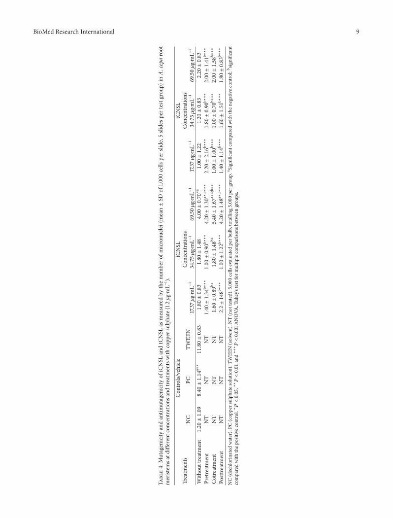
3.4. Evaluation of iCNSL and tCNSL Mutagenicity and TheirEffects on Copper Sulphate. Only iCNSL at the highest testconcentration (69.50𝜇g⋅mL−1) induced MN mutagenicity inA. cepa root meristems (𝑃 < 0.05) compared with thenegative control. The two lower concentrations of iCNSLand all three concentrations of tCNSL failed to induce MNmutagenicity in A. cepa root meristems (𝑃 > 0.05) (Table 4).
Both iCNSL and tCNSL protect (pretreatment) the DNAof A. cepa root meristems, as observed by the inhibition ofMN formation induced by CuSO
4⋅5H2O. However, iCNSL
at the highest test concentration did not confer this pro-tection. Similarly, antimutagenic and reparative effects wereevidenced by a reduction in MN in co- and posttreatments,except at the highest iCNSL concentration (Table 4). BothiCNSL and tCNSL strongly affected the prevention, modu-lation, and repair of damage induced by CuSO
4⋅5H2O in A.
cepameristems.The mutagenicity of the highest iCNSL concentration
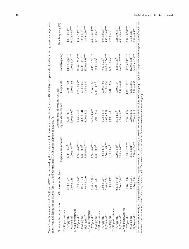
(69.50 𝜇g⋅mL−1) was not confirmed when analysing the fre-quency of chromosomal aberration (CA) because iCNSL atthis concentration had already been shown to havemutageniceffects as evidenced by increased MN frequency, and MIinhibition also precluded observation of CA at this concen-tration. Neither iCNSL nor tCNSL showed mutagenic effectsas measured by the frequency of CA compared to the positivecontrol (Figure 6).
Chromosome bridges, vagrant and laggard chromo-somes, and chromosome fragments are the most notableof the CAs induced by CuSO
4⋅5H2O during anaphase and
telophase. The photomicrographic profile of CAs identifiedin A. cepa root meristems exposed to CuSO
4⋅5H2O is repre-
sented in Figure 7.This damagewasmodulated by iCNSL and
tCNSL at the tested concentrations, which suggests that bothmay have the inhibition mechanisms of aneugenic and/orclastogenic agents.
Moreover, both types of CNSL showed protective,antimutagenic, and DNA-repair effects in regard to thedamage induced by copper sulphate (Table 5).
4. Discussion
Genomic instability is a common cause of cancer. Cancer cellsare more susceptible than normal cells to DNA-damagingagents. This increased susceptibility provides a path fortherapeutic intervention. Phytochemicals may affect thegenome and trigger damage to DNA and repair mechanisms[31]. However, many of such dietary substances have beenassociated with a decreased risk of cancer, for example, breastcancer [32, 33].
Differences between the two types of CNSL were identi-fied by the GC-MS analysis. The iCNSL contained a mixtureof anacardic acids that constituted 94.5% of its composition.Conversely, tCNSL exhibited a mixture of cardanols thatconstituted more than 90% of its composition (Table 1).These percentages corroborate the studies by Philip et al.[34], who reported that anacardic acid may exceed 80%of iCNSL composition. This percentage may be as high as90%, according to Das and Ganesh [35]. Previous studieshad found that cardanol is the main component of tCNSL[24, 36].The difference in chemical composition between thetwo types of CNSLmight result from the preparation processused to obtain tCNSL, which involves heating the cashewnuts to 180–200∘C.This heat causes anacardic acid to undergodecarboxylation and conversion into cardanol, leading to thehigher cardanol concentration in tCNSL [37].
The chemical compositions of both iCNSL and tCNSLrecorded in the present study differ from the chemicalcomposition reported in the literature [15, 23, 24]. The originof the cashew nuts, the weather conditions, and, particularly,the extraction process used may account for the differencesbetween this and other studies with regard to the ratio ofphenolic compounds found in iCNSL [35], and the operatingand heating conditions may affect the ratios recorded intCNSL [38].
The A. salina bioassay is considered to be useful forpreliminary assessments of general toxicity, and it correlateswell with cytotoxic activity against some types of solidtumours in humans [26]. The IC
50of iCNSL in A. salina was
lower than that of tCNSL (Figure 3). Plant extracts with LC50
values under 1,000𝜇g⋅mL−1 are considered to be active andto have toxic activity [39, 40].Therefore, although iCNSL andtCNSL had different LC
50values, both are considered to have
shown toxic effects in the acute toxicity test with A. salina.Guerra [41] suggests that the Artemia bioassay could be usedfor the toxicity evaluations of compounds that are rich inphenols.
The most significant toxicity results found in this study(Figure 3) were obtained when A. salina was treated withiCNSL. This finding may be related to the presence ofanacardic acids in iCNSL because anacardic acids have been
8 BioMed Research International
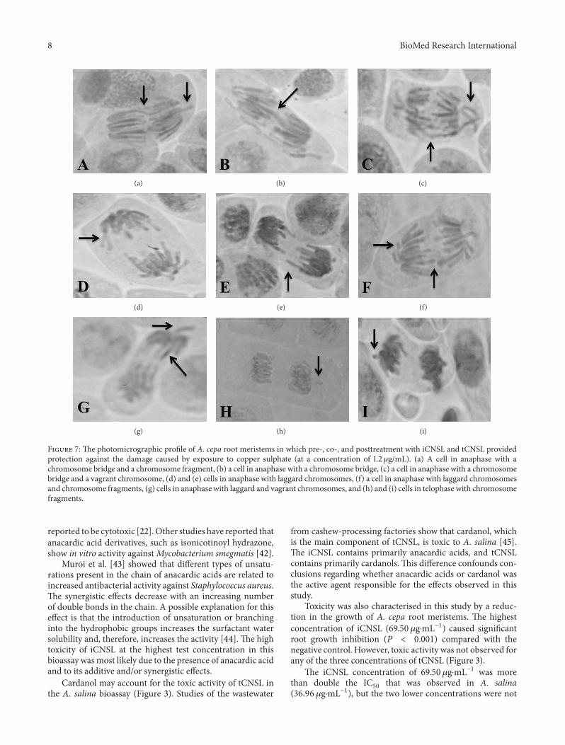
(a) (b) (c)
(d) (e) (f)
(g) (h) (i)
Figure 7: The photomicrographic profile of A. cepa root meristems in which pre-, co-, and posttreatment with iCNSL and tCNSL providedprotection against the damage caused by exposure to copper sulphate (at a concentration of 1.2𝜇g/mL). (a) A cell in anaphase with achromosome bridge and a chromosome fragment, (b) a cell in anaphase with a chromosome bridge, (c) a cell in anaphase with a chromosomebridge and a vagrant chromosome, (d) and (e) cells in anaphase with laggard chromosomes, (f) a cell in anaphase with laggard chromosomesand chromosome fragments, (g) cells in anaphase with laggard and vagrant chromosomes, and (h) and (i) cells in telophase with chromosomefragments.
reported to be cytotoxic [22].Other studies have reported thatanacardic acid derivatives, such as isonicotinoyl hydrazone,show in vitro activity againstMycobacterium smegmatis [42].
Muroi et al. [43] showed that different types of unsatu-rations present in the chain of anacardic acids are related toincreased antibacterial activity against Staphylococcus aureus.The synergistic effects decrease with an increasing numberof double bonds in the chain. A possible explanation for thiseffect is that the introduction of unsaturation or branchinginto the hydrophobic groups increases the surfactant watersolubility and, therefore, increases the activity [44]. The hightoxicity of iCNSL at the highest test concentration in thisbioassay wasmost likely due to the presence of anacardic acidand to its additive and/or synergistic effects.
Cardanol may account for the toxic activity of tCNSL inthe A. salina bioassay (Figure 3). Studies of the wastewater
from cashew-processing factories show that cardanol, whichis the main component of tCNSL, is toxic to A. salina [45].The iCNSL contains primarily anacardic acids, and tCNSLcontains primarily cardanols.This difference confounds con-clusions regarding whether anacardic acids or cardanol wasthe active agent responsible for the effects observed in thisstudy.
Toxicity was also characterised in this study by a reduc-tion in the growth of A. cepa root meristems. The highestconcentration of iCNSL (69.50𝜇g⋅mL−1) caused significantroot growth inhibition (𝑃 < 0.001) compared with thenegative control. However, toxic activity was not observed forany of the three concentrations of tCNSL (Figure 3).
The iCNSL concentration of 69.50 𝜇g⋅mL−1 was morethan double the IC
50that was observed in A. salina
(36.96 𝜇g⋅mL−1), but the two lower concentrations were not
BioMed Research International 9
Table4:
Mutagenicity
andantim
utagenicity
ofiCNSL
andtCNSL
asmeasuredby
thenu
mbero
fmicronu
clei(mean±SD
of1.0
00cells
pers
lide,5slidesp
ertestgrou
p)in
A.cepa
root
meristem
satd
ifferentcon
centratio
nsandtre
atmentswith
copp
ersulphate(1.2𝜇g⋅mL−
1 ).
Treatm
ents
Con
trols/vehicle
iCNSL
tCNSL
NC
PCTW
EEN
Con
centratio
nsCon
centratio
ns17.37𝜇g⋅mL−1
34.75𝜇
g⋅mL−1
69.50𝜇
g⋅mL−1
17.37𝜇g⋅mL−1
34.75𝜇
g⋅mL−1
69.50𝜇
g⋅mL−1
With
outtreatment1.20±1.098.40±1.14
a∗∗11.80±0.831.80±0.83
1.80±1.48
4.00±0.70
∗a
1.00±1.22
1.20±0.83
2.20±0.83
Pretreatment
NT
NT
NT
1.40±1.34
b∗∗∗1.00±0.90
b∗∗∗4.20±1.30
a∗;b∗∗∗2.20±2.16
b∗∗∗1.80±0.90
b∗∗∗2.00±1.41
b∗∗∗
Cotreatment
NT
NT
NT
1.60±0.89
b∗1.80±1.48
b∗5.40±1.67
a∗∗;b∗∗1.00±1.00
b∗∗∗1.00±0.70
b∗∗∗2.00±1.58
b∗∗∗
Posttreatment
NT
NT
NT
2.2±148
b∗∗∗1.00±1.22
b∗∗∗4.20±1.48
a∗;b∗∗∗1.40±1.14
b∗∗∗1.60±1.51
b∗∗∗1.80±0.83
b∗∗∗
NC(dechlorinated
water).PC
(cop
persulph
ates
olution).T
WEE
N(solvent).NT(not
teste
d).5.000
cells
evaluatedperb
ulb,totalling
5.00
0perg
roup
.aSign
ificant
comparedwith
then
egativec
ontro
l;b significant
comparedwith
thep
ositive
control.∗𝑃<0.05,∗∗𝑃<0.01,and∗∗∗𝑃<0.001A
NOVA
.Tuk
ey’stestform
ultip
lecomparis
onsb
etweengrou
ps.
10 BioMed Research International
Table5:Antim
utagenicity
ofiCNSL
andtCNSL
asmeasuredby
thefre
quency
ofchromosom
alaberratio
ns(m
ean±SD
of1.0
00cells
perslide,5
slidesp
ertestgrou
p)in
A.cepa
root
meristem
satd
ifferentcon
centratio
ns(pre-,co-,andpo
sttreatment)with
copp
ersulphate(1.2𝜇g⋅mL−1).
Group
swith
concentration
Chromosom
ebrid
ges
Chromosom
alaberratio
n/5000
cells
Totalfrequ
ency
(%)
Vagrantchrom
osom
esLaggardchromosom
esFragments
Totalfrequ
ency
iCNSL
pretreatment
17,37𝜇g⋅mL−
10.40±0.89
b∗1.6
0±0.50
b∗∗∗
4.00±4.84
2.00±1.2
28.00±3.46
b∗∗∗
0.80±0.34
b∗∗∗
34,75𝜇
g⋅mL−
10.60±0.89
b∗1.2
0±1.0
9b∗∗∗
2.60±2.96
b∗2.60±1.9
47.0
0±4.06
b∗∗∗
0.70±0.40
b∗∗∗
tCNSL
pretreatment
17,37𝜇g⋅mL−
11.2
0±1.0
01.0
0±0.00
b∗∗∗
8.00±4.41
1.20±0.83
b∗11.40±7.33b∗∗∗
1.14±0.73
b∗∗∗
34,75𝜇
g⋅mL−
10.75±0.50
1.20±0.44
b∗∗∗
8.20±3.11
2.40±1.5
112.80±3.76
b∗∗∗
1.28±0.37
b∗∗∗
69,5𝜇g⋅mL−
10.80±0.40
1.60±0.44
b∗∗∗
8.00±4.00
2.60±1.8
113.00±5.61
b∗∗∗
1.30±0.56
b∗∗∗
iCNSL
cotre
atment
17,37𝜇g⋅mL−
10.40±0.89
b∗1.0
0±0.00
b∗∗∗
3.40±1.8
1b∗
1.60±1.14
6.40±2.51
b∗∗∗
0.64±0.25
b∗∗∗
34,75𝜇
g⋅mL−
10.20±0.44
b∗1.2
0±0.44
b∗∗∗
5.40±2.60
1.00±0.70
b∗∗
7.80±2.77
b∗∗∗
0.78±0.27
b∗∗∗
tCNSL
cotre
atment
17,37𝜇g⋅mL−
10.60±0.54
b∗1.6
0±1.14b∗∗∗
5.20±1.7
82.00±1.0
09.4
0±2.51
b∗∗∗
0.94±0.25
b∗∗∗
34,75𝜇
g⋅mL−
10.60±0.54
b∗1.2
0±1.6
4b∗∗∗
4.60±2.19
2.40±1.14
8.80±0.30
b∗∗∗
8.80±3.03
b∗∗∗
69,50𝜇
g⋅mL−
11.6
0±1.9
43.40±1.6
7b∗∗∗
8.80±5.35
3.40±2.79
17.20±5.76
b∗∗∗
1.72±0.57
b∗∗∗
iCNSL
posttreatment
17,37𝜇g⋅mL−
10.20±0.44
b∗∗
1.20±0.83
b∗∗∗
1.60±1.5
7b∗∗
1.60±1.14
4.60±1.8
1b∗∗∗
0.46±0.18
b∗∗∗
34,75𝜇
g⋅mL−
10.20±0,44
b∗2.00±1.0
0b∗∗∗
4.00±4.35
2.40±0.89
8.60±4.27
b∗∗∗
0.86±0.42
b∗∗∗
tCNSL
posttreatment
17,37𝜇g⋅mL−
11.2
0±0.44
1.00±1.2
2b∗∗∗
6.00±4.84
1.00±1.2
2b∗
9.20±7.4
6b∗∗∗
0.92±0.74
b∗∗∗
34,75𝜇
g⋅mL−
10.80±0.44
2.60±1.5
1b∗∗∗
7.40±2.52
2.60±2.60
13.00±6.51
b∗∗∗
1.30±0.65
b∗∗∗
69,50𝜇
g⋅mL−
11.2
0±0.44
3.80±1.7
8b∗∗
7.00±4.12
2.80±0.83
14.80±4.08
b∗∗
1.48±0.40
b∗∗
NC(dechlorinated
water).PC
(cop
persulph
ates
olution).T
WEE
N(solvent).NT(not
teste
d).5.000
cells
evaluatedperb
ulb,totalling
5,00
0perg
roup
.aSign
ificant
comparedwith
then
egativec
ontro
l;b significant
comparedwith
thep
ositive
control.∗𝑃<0.05,∗∗𝑃<0.01,and∗∗∗𝑃<0.001A
NOVA
.Tuk
ey’stestform
ultip
lecomparis
onsb
etweengrou
ps.
BioMed Research International 11
toxic to A. cepa root meristems (Figure 3). Another study, inwhich three species of molluscs of the same genus (Biom-phalaria straminea, B. tenagophila, and B. glabrata) weretreated with a 20 ppm hexane extract, showed mortality ratesthat ranged from 97.1% to 100% after 24 hours of exposure[46].
The A. cepa test system is a key in vivo model forthe evaluation of root growth after direct treatment with asubstance of interest and for the prediction of DNA damage.The test is considered to be an effective preselection tool fortoxicity and genotoxicity studies [47] because the results canbe extrapolated to other animals and plants [47].
TheA. cepa test also provides other macroscopic parame-ters that indicate the toxicity of chemicals and environmentalpollutants.These toxicity parameters include very large roots,which indicate cellular proliferation; colour changes; and thepresence of tumours [48]. This test reveals toxic [26] andcytotoxic effects [49].
The effects of extracts against damage caused by toxicagents have been analysed in recent studies assessing rootsize via the A. cepa test [28, 50]. Certain metals, includingcopper, may inhibit root growth, most likely by inhibiting celldivision, and may also cause toxicity and cytotoxicity [51].
In the present study, only the highest test concentrationof tCNSL showed preventive effects when exposed to theCuSO
4⋅5H2O solution in A. cepa meristems. However, nei-
ther iCNSL nor tCNSL prevented the toxicity induced byCuSO
4⋅H2O in co- or posttreatment applications (Table 3).
Root growth is regulated by the combination of celldivision activity in mitotically active meristems and cellelongation in the regions that are proximal to root apices[52]. Only the highest concentration of iCNSL had significantantiproliferative activity (𝑃 < 0.05) compared with thenegative control (Figure 5). This finding suggests that thisconcentration caused disturbance in meristem proliferationin A. cepa.
Macroscopic parameters are associated with toxicity andmay likewise be associated with a reduction in the MI, whichwould affect DNA replication and protein synthesis [53]. Nopreventive, modulating, and reparative activities of CNSLagainst the cytotoxicity induced by copper sulphate wereobserved in this study (Table 4).
Oliveira et al. [23] also found that anacardic acid is alarger component of iCNSL than of tCNSL (Table 1) andthat anacardic acid may have had prooxidant effects in A.cepameristems. Recent studies indicate that antiproliferativeeffects onmammalian cell cultures are associated with oxida-tive stress [54] because the production of reactive oxygenspecies (ROS) impacts root growth and may inhibit growthand cell division [55].
The tCNSL showed no cytotoxic effect (𝑃 > 0.05) atany concentration. A positive correlation was found betweeninhibited root growth and reduced MI at the highest iCNSLconcentration (Figures 4 and 5).
The MI is calculated by dividing the number of dividingcells by the total number of cells observed and is expressed asa percentage [56]. A reduction in the MI can be interpretedas cell death [57]. The present study showed a mitode-pressive effect of iCNSL at the highest test concentration
(69.50 𝜇g⋅mL−1) on A. cepa cell division. The mitodepressiveeffect may have resulted from abnormal cellular conditionscaused by the treatment. The reduction in the MI mayhave been related to early prophase arrest [58], inhibition ofDNA synthesis, or cell cycle arrest at the G2 phase, whichwould prevent cells from entering mitosis [59]. The reducedMI also inhibits microtubule formation and nucleoproteinsynthesis and reduces the ATP levels that provide energy forspindle elongation, microtubule dynamics, and chromosomemovement [60].
Kubo et al. [61] also showed that anacardic acid andcardolmay have amoderate cytotoxic effect.The inhibition ofprooxidant enzymes may account for this effect. The volumeof the hydrophobic side chain and its ability to act as asurfactant would explain its cytotoxic effect. Cardol has alsobeen shown to be cytotoxic at a dose of 0.01mM in HeLacells [62]. The results of these two studies might confirm thecytotoxic action of the highest concentration of iCNSL whichcontains anacardic acid and cardol [18].
Acevedo et al. [22] showed that the anacardic acid presentin Amphipterygium adstringens has cytotoxic effects in theperipheral lymphocytes ofmice treatedwith doses of less than10mg/kg. The cytotoxic effects were evidenced by decreasesin polychromatic and normochromatic erythrocytes. Anac-ardic acid from A. adstringens is also cytotoxic againstGram-positive bacteria in dental abscesses, has molluscicidalactivity [63], inhibits apoptosis in chick embryonic neuronalcells [64], and inhibits breast cancer (MCF-7 and MDA-MB 231) cervical cancer cell lines and other types of tissues,including lung, liver, bladder, and melanoma [65].
Recent studies have also shown that a combination ofanacardic acid and lunasin, which is another natural plantextract, may exhibit anticarcinogenic properties. These com-pounds act on the regulation of the expression of severalgenes involved in the cell cycle, apoptosis, and signal trans-duction [66]. Both compounds have a strong inhibitory effecton a number of cancer cell lines [67–69]. For example,[70] reported inhibition of the growth of HepG2 and U266tumour cells treated with 60𝜇M of anacardic acid for 24, 48,and 72 hours.
The frequencies of CA and MN are commonly used todetect genotoxicity [59, 71]. This study investigated geno-toxicity based on the frequency of MN and CAs, such aschromosomebridges, vagrant and laggard chromosomes, andchromosome fragments (Figure 7).
Iarmarcovai et al. [72] characterise micronuclei as small,spherical bodies that consist of genetic material that is notincorporated into the main nucleus during the final stagesof mitosis. MN may result from the failure of acentric chro-mosome fragments to incorporate into the cell nucleus andclastogenicity (DNA breaks) or from whole chromosomesof aneugenic origin (disturbance in the mitotic spindle).The iCNSL at a concentration of 69.50𝜇g⋅mL−1 is thoughtto have induced genotoxic effects by means of clastogenicmechanisms (Table 5) because the MN that were generatedat this concentration are considered small. Small MN areindicative of clastogenic action [73] resulting from genotoxicstress [74, 75].
12 BioMed Research International
The mutagenic response that occurred at the highestiCNSL concentration might have resulted from chromoso-mal instability, phenotypes, and cellular changes caused bygenetic defects and/or exogenous exposure [76]. However,previous studies found that iCNSL did not have a mutageniceffect in the Ames test [22] or the MN test in mice bonemarrow [77].
We, therefore, hypothesise that the MN formed at thehighest iCNSL concentration resulted from breaks thatoccurred during cell division, possibly due to unrepairedor incorrectly repaired damage or to poor chromosomeseparation as result of mitotic malfunction.These events mayhave resulted from oxidative stress [78] and therefore from anintegrated response to instability of the genetic material [79]that reflected various chromosomal changes [71].
However, iCNSL and tCNSL had no significant genotoxiceffects on the frequency of CAs compared with the negativecontrol (Figure 7). The iCNSL at the highest test concen-tration showed no preventive, antimutagenic, and reparativeresponses against CuSO
4⋅5H2O. The iCNSL at the two lower
concentrations and tCNSL did show preventive, antimuta-genic, and reparative activities, as indicated by the reducedfrequency of MN (Table 5). These results are consistent withthe decrease in the frequency of CAs that resulted from theinhibition of damage induced by CuSO
4⋅5H2O (Table 5).
Several experimental models have shown that syntheticor natural resorcinolic lipids do not cause DNA damage atlow concentrations, which suggests that they have anticanceractivity [80]. The results of these studies are consistent withthe results of the present study, in which no genotoxicity wasfound at the lowest concentrations of iCNSL and tCNSL.
The present study documented the genotoxic effects ofCuSO
4⋅5H2O, which are explained by the ability of it causing
DNA damage [29]. The chemical components of iCNSL andtCNSL may have protected, modulated, and repaired theoxidative effects of CuSO
4⋅5H2O in A. cepa meristems. Car-
dol and cardanol were found to exhibit in vitro antioxidanteffects in studies of the chemical characteristics of CNSL.These compounds have these effects because they scavengefree radicals, including the hydroxyl radical [23].
Other studies have also reported that the genotoxicityof chemical agents may be repaired by phenolic compoundswith antioxidant and radical-scavenging activities [81]. Chro-mosomal aberrations consist of changes in chromosomestructure that result in breaks or exchange of chromosomalmaterial.These types of damage are usually lethal to cells, butsome are viable and may have somatic or hereditary geneticeffects [82].
Chromosomal fragments in cells indicate chromosomalbreaks and may be related to anaphase bridges [83], distur-bances in microtubule assembly, and cell death [84]. Theresults of the present study (Table 5) show that iCNSL andtCNSL failed to induce the fragment type of CA and thereforedid not cause anaphase bridges when compared to the controlgroup.
Antimutagenic compounds are able to induce somemetabolic enzymes that may act as enzymatic inhibitors ofmutagenic agents or inhibitors of promutagens in pretreat-ment experiments [85, 86]. The preventive, antimutagenic,
and reparative effects of iCNSL and tCNSL observed in A.cepa (Tables 4 and 5) (except at the highest iCNSL con-centration) were corroborated with the study of [15], whoreported that tCNSL effect protected against oxidative stress(at a concentration of 100–500𝜇g⋅mL−1) in S. cerevisiae thatwere defective in antioxidant enzymes.Theprotection againstdamage caused by H
2O2occurs via bioantimutagenic mech-
anisms, but it occurs by means of dysmutagenesis in con-current treatment. Bioantimutagenic agents act on the phys-iological mechanisms of DNA protection and repair andreverse the mutagenic effects and prevent their persistence[85, 87]. Thus, CNSL most likely acted as a bioantimutagenicand dysmutagenic agent and showed a stronger antimuta-genic effect.
Components of phenolic lipids, including anacardic acidand alkylresorcinol, have antigenotoxic activity in vitrobecause of the ability of lipids to interact with biologicalmembranes [88].This is confirmed by the presence of hydro-philic and hydrophobic regions in their structures, which givelipids an amphipathic character that is responsible for theiraffinity for biological membranes. This character allows thephenolic lipids to be incorporated easily into cell membranes[89].
Cardanol also has antioxidant effects [23], and phenoliccompounds with this ability can suppress genotoxicity [81].Deszcz and Kozubek [90] noted that alkylresorcinols maybe characterised as antioxidants when they are at verylow concentrations, and they protect free fatty acids andphospholipids against peroxidation induced by the iron andautooxidation of biological membranes. These activities mayconstitute the main factor accounting for the antimutagenicactivity exhibited by these compounds.
The present study confirms the strong antimutagenic,preventive, and restorative effects of CNSL. De Lima et al.[25] observed that iCNSL (at a concentration of 200𝜇g⋅mL−1)had an antioxidant effect in S. cerevisiae. Andrade et al.[15] observed that 100 𝜇g⋅mL−1 of tCNSL might reduce freeradical levels by 88.9% in the DPPH test and that it scavengeshydroxyl radicals by means of xanthine oxidase, resulting inantioxidant activity with an IC
50= 702𝜇g/mL.
Melo Cavalcante et al. [91] also confirmed thatA. occiden-tale pulp has antioxidant effects against H
2O2at pre-, co-, and
posttreatment in Salmonella typhimurium, as assessed by theAmes test. The authors attributed these effects to the pulp’schemical components, which include anacardic acid. Thesecomponentsmay also protect S. typhimurium (TA102) againstthe damage induced by aflatoxin B
1via several mechanisms
[92]. Cashew juice and cajuina (processed juice) reducedamage to the peripheral blood cells of mice. The juicecaused a 60.82% reduction in damage and the cajuina causedan 82.19% reduction in damage, compared with cyclophos-phamide. Further, the juice and the cajuina reduced thenumber of CAs in the bone marrow of mice by 53% and 65%,respectively. These effects may be related to the antioxidantactivities of their components [13]. The results reportedby de Carvalho Melo-Cavalcante et al. [13] confirm the(concentration-dependent) antimutagenic and antigenotoxiceffects of iCNSL and tCNSL observed in the present study.
BioMed Research International 13
5. Conclusions
In summary, this study showed that anacardic acids arethe primary components of iCNSL and cardanol of tCNSL.Both iCNSL and tCNSL showed protective (pretreatment),modulating (cotreatment), and reparative (posttreatment) invivo effects against the damage induced by copper sulphatein A. cepa meristems at the lowest concentrations evaluated.Therefore, CNSL, which is a natural and renewable productextracted from the cashew nut shell, can be the basis forfurther studies to determine the mechanisms activated by itscomponents and the mechanisms by which their synergismproduces beneficial effects.These studiesmight be precursorsfor the production of biotechnological products.
Conflict of Interests
The authors declare no conflict of interests.
Acknowledgments
The authors thank the National Council of Technologicaland Scientific Development (CNPq/Brazil) and the ResearchLaboratory in Genetic Toxicological, UFPI, Brazil.
References
[1] I. Dahech, W. Farah, M. Trigui et al., “Antioxidant and antimi-crobial activities of Lycium shawii fruits extract,” InternationalJournal of Biological Macromolecules, vol. 60, pp. 328–333, 2013.
[2] M. A. Hasnat, M. Pervin, and B. O. Lim, “Acetylcholinesteraseinhibition and in vitro and in vivo antioxidant activities ofGan-oderma lucidum grown on germinated brown rice,” Molecules,vol. 18, no. 6, pp. 6663–6678, 2013.
[3] F. L. Buchner, H. B. Bueno-de-Mesquita, J. Linseisen et al.,“Fruits and vegetables consumption and the risk of histologicalsubtypes of lung cancer in the European prospective investi-gation into cancer and nutrition (EPIC),” Cancer Causes andControl, vol. 21, no. 3, pp. 357–371, 2010.
[4] I. Soerjomataram, D. Oomen, V. Lemmens et al., “Increasedconsumption of fruit and vegetables and future cancer inci-dence in selected European countries,” European Journal ofCancer, vol. 46, no. 14, pp. 2563–2580, 2010.
[5] F. I. Akaneme and C. C. Amaefule, “Evaluation of the cytotox-icity and genotoxicity of aqueous leaf extracts of Azadirachtaindica A. Juss using the Allium test,” Journal of Medicinal PlantsResearch, vol. 6, no. 23, pp. 3898–3907, 2012.
[6] C. C. Munari, P. F. de Oliveira, I. M. de Souza Lima et al., “Eval-uation of cytotoxic, genotoxic and antigenotoxic potential ofSolanum lycocarpum fruits glicoalkaloid extract in V79 cells,”Food and Chemical Toxicology, vol. 50, no. 10, pp. 3696–3701,2012.
[7] I. M. C. M. Rietjens, M. J. Martena, M. G. Boersma, W. Spiege-lenberg, and G. M. Alink, “Molecular mechanisms of toxicityof important food-borne phytotoxins,” Molecular Nutrition &Food Research, vol. 49, no. 2, pp. 131–158, 2005.
[8] S. E. Mazzetto, D. Lomonaco, and G. Mele, “Cashew nut oil:opportunities and challenges in the context of sustainableindustrial development,” Quimica Nova, vol. 32, no. 3, pp. 732–741, 2009.
[9] V. M. Moo-Huchin, M. I. Moo-Huchin, R. J. Estrada-Leon etal., “Antioxidant compounds, antioxidant activity and phenoliccontent in peel from three tropical fruits fromYucatan,Mexico,”Food Chemistry, vol. 166, pp. 17–22, 2015.
[10] A. A. F. Zielinski, S. Avila, V. Ito, A. Nogueira, G. Wosiacki, andC.W. I. Haminiuk, “The association between chromaticity, phe-nolics, carotenoids, and in vitro antioxidant activity of frozenfruit pulp in Brazil: an application of chemometrics,” Journal ofFood Science, vol. 79, no. 4, pp. C510–C516, 2014.
[11] Y.-S. C. Bae-Harboe and K. S. Masterpol, “Botanical briefs:cashew apple (Anacardium occidentale),”Cutis, vol. 92, no. 4, pp.174–176, 2013.
[12] A. C. P. Guissoni, I. G. Silva, R. Geris, L. C. Cunha, and H. H.G. Silva, “Larvicidal activity of Anacardium occidentale as analternative to control Aedes aegypti and its toxicity in Rattusnorvegicus,” Revista Brasileira de Plantas Medicinais, vol. 15, no.3, pp. 363–367, 2013.
[13] A. A. de CarvalhoMelo-Cavalcante, S. M.M. deMoura Dantas,A. de Sousa Leite et al., “In vivo antigenotoxic and anticlas-togenic effects of fresh and processed cashew (Anacardiumoccidentale) apple juices,” Journal of Medicinal Food, vol. 14, no.7-8, pp. 792–798, 2011.
[14] O. Ayepola and R. Ishola, “Evaluation of antimicrobial activityof Anacardium occidentale (Linn.),” Advances in Medical andDental Sciences, vol. 3, no. 1, pp. 1–3, 2009.
[15] T. D. A. D. Andrade, B. Q. Araujo, A. M. D. L. Cito et al., “Anti-oxidant properties and chemical composition of technicalCashew Nut Shell Liquid (tCNSL),” Food Chemistry, vol. 126,no. 3, pp. 1044–1048, 2011.
[16] L. Michodjehoun-Mestres, J.-M. Souquet, H. Fulcrand, C.Bouchut, M. Reynes, and J.-M. Brillouet, “Monomeric phenolsof cashew apple (Anacardium occidentale L.),” Food Chemistry,vol. 112, no. 4, pp. 851–857, 2009.
[17] V. R. Kannan, C. Sumathi, V. Balasubramanian, andN. Ramesh,“Elementary chemical profiling and antifungal properties ofcashew (Anacardium occidentale L.) Nuts,” Botany ResearchInternational, vol. 2, no. 4, pp. 253–257, 2009.
[18] L. S. Parasa, S. R. Tumati, C. Kumar, S. Chicurupati, and G.Rao, “In vitro antimicrobial activity of cashew (Anacardiumoccidentale, L.) nuts shell liquid against methicillin resistantStaphylococcus aureus (MRSA) clinical isolates,” InternationalJournal of Pharmacy Science, vol. 3, pp. 436–440, 2011.
[19] A. K.Mukhopadhyay, A. K. Hati,W. Tamizharasu, and P. SathyaBabu, “Larvicidal properties of cashew nut shell liquid (Anac-ardium occidentale L) on immature stages of two mosquitospecies,” Journal of Vector Borne Diseases, vol. 47, no. 4, pp. 257–260, 2010.
[20] J. George and R. Kuttan, “Mutagenic, carcinogenic and cocar-cinogenic activity of cashewnut shell liquid,”Cancer Letters, vol.112, no. 1, pp. 11–16, 1997.
[21] K. Polasa and C. Rukmini, “Mutagenicity tests of cashewnutshell liquid, rice-bran oil and other vegetable oils using theSalmonella typhimurium/microsome system,” Food and Chemi-cal Toxicology, vol. 25, no. 10, pp. 763–766, 1987.
[22] H. R. Acevedo, M. D. Rojas, S. D. B. Arceo et al., “Effect of6-nonadecyl salicylic acid and its methyl ester on the induc-tion of micronuclei in polychromatic erythrocytes in mouseperipheral blood,” Mutation Research—Genetic Toxicology andEnvironmental Mutagenesis, vol. 609, no. 1, pp. 43–46, 2006.
[23] M. S. C. Oliveira, S. M. D. Morais, D. V. Magalhaes et al.,“Antioxidant, larvicidal and antiacetylcholinesterase activities
14 BioMed Research International
of cashew nut shell liquid constituents,” Acta Tropica, vol. 117,no. 3, pp. 165–170, 2011.
[24] P. P. Kumar, R. Paramashivappa, P. J. Vithayathil, P. V. S. Rao,and A. S. Rao, “Process for isolation of cardanol from technicalcashew (Anacardium occidentale l.) Nut shell liquid,” Journal ofAgricultural and Food Chemistry, vol. 50, no. 16, pp. 4705–4708,2002.
[25] S. G. De Lima, C. M. Feitosa, A. M. Cito et al., “Effects of imma-ture cashew nut-shell liquid (Anacardium occidentale) againstoxidative damage in Saccharomyces cerevisiae and inhibition ofacetylcholinesterase activity,” Genetics and Molecular Research,vol. 7, no. 3, pp. 806–818, 2008.
[26] B. N. Meyer, N. R. Ferrigni, and J. E. Putnam, “Brine shrimp:a convenient general bioassay for active plant constituents,”Planta Medica, vol. 45, no. 1, pp. 31–34, 1982.
[27] G. Fiskesjo, “The Allium test as a standard in environmentalmonitoring,” Hereditas, vol. 102, no. 1, pp. 99–112, 1985.
[28] M.Malini,M. A.Marin-Morales,M. S.Mantovani et al., “Deter-mination of the antimutagenicity of an aqueous extract ofRhizophora mangle L. (Rhizophoraceae), using in vivo and invitro test systems,”Genetics andMolecular Biology, vol. 33, no. 1,pp. 176–181, 2010.
[29] B. S. Banu, M. Ishaq, K. Danadevi, P. Padmavathi, and Y. R.Ahuja, “DNA damage in leukocytes of mice treated with coppersulfate,” Food and Chemical Toxicology, vol. 42, no. 12, pp. 1931–1936, 2004.
[30] M. Yildiz, I. H. Cgerci, M. Konuk, A. Fatih Fidan, and H. Terzi,“Determination of genotoxic effects of copper sulphate andcobalt chloride in Allium cepa root cells by chromosome aber-ration and comet assays,” Chemosphere, vol. 75, no. 7, pp. 934–938, 2009.
[31] P. Rajendran, E. Ho, D. E. Williams, and R. H. Dashwood,“Dietary phytochemicals, HDAC inhibition, and DNA dam-age/repair defects in cancer cells,” Clinical Epigenetics, vol. 3,article 4, 2011.
[32] C. M. Kaefer and J. A. Milner, “The role of herbs and spices incancer prevention,”The Journal of Nutritional Biochemistry, vol.19, no. 6, pp. 347–361, 2008.
[33] S. Ramos, “Cancer chemoprevention and chemotherapy: diet-ary polyphenols and signalling pathways,” Molecular Nutrition& Food Research, vol. 52, no. 5, pp. 507–526, 2008.
[34] J. Y. N. Philip, J. Buchweishaija, L. L.Mkayula, and L. Ye, “Prepa-ration of molecularly imprinted polymers using anacardic acidmonomers derived from cashew nut shell liquid,” Journal ofAgricultural and Food Chemistry, vol. 55, no. 22, pp. 8870–8876,2007.
[35] P. Das and A. Ganesh, “Bio-oil from pyrolysis of cashew nutshell—a near fuel,” Biomass and Bioenergy, vol. 25, no. 1, pp. 113–117, 2003.
[36] R. Ikeda, H. Tanaka, H. Uyama, and S. Kobayashi, “Synthesisand curing behaviors of a crosslinkable polymer from cashewnut shell liquid,” Polymer, vol. 43, no. 12, pp. 3475–3481, 2002.
[37] O. A. Attanasi, S. Berretta, C. Fiani, P. Filippone, G. Mele, andR. Saladino, “Synthesis and reactions of nitro derivatives ofhydrogenated cardanol,” Tetrahedron, vol. 62, no. 25, pp. 6113–6120, 2006.
[38] M. C. Lubi and E. T. Thachil, “Cashew nut shell liquid(CNSL)—a versatile monomer for polymer synthesis,”DesignedMonomers and Polymers, vol. 3, no. 2, pp. 123–153, 2000.
[39] H. M. dos Santos Jr., D. F. Oliveira, D. A. De Carvalho et al.,“Evaluation of native and exotic Brazilian plants for anticancer
activity,” Journal of NaturalMedicines, vol. 64, no. 2, pp. 231–238,2010.
[40] D. D. R. Arcanjo, A. C. M. Albuquerque, B. Melo-Neto, L. C. L.R. Santana,M. G. F.Medeiros, and A.M. G. L. Cito, “Bioactivityevaluation against Artemia salina Leach of medicinal plantsused in BrazilianNortheastern folkmedicine,”Brazilian Journalof Biology, vol. 72, no. 3, pp. 505–509, 2012.
[41] R. Guerra, “Ecotoxicological and chemical evaluation of phe-nolic compounds in industrial effluents,” Chemosphere, vol. 44,no. 8, pp. 1737–1747, 2001.
[42] B. N. Swamy, T. K. Suma, G. V. Rao, and G. C. Reddy, “Syn-thesis of isonicotinoylhydrazones from anacardic acid and theirin vitro activity against Mycobacterium smegmatis,” EuropeanJournal ofMedicinal Chemistry, vol. 42, no. 3, pp. 420–424, 2007.
[43] H. Muroi, K.-I. Nihei, K. Tsujimoto, and I. Kubo, “Synergis-tic effects of anacardic acids and methicillin against methi-cillin resistant Staphylococcus aureus,” Bioorganic andMedicinalChemistry, vol. 12, no. 3, pp. 583–587, 2004.
[44] M. J. Rosen, Surfactants and Interfacial Phenomena,Wiley, 1989.[45] M. R. Pimentel, D. P. De Lima, L. R. Martins, A. Beatriz, S.
T. Santaella, and L. V. C. Lotufo, “Ecotoxicological analysis ofcashew nut industry effluents, specifically two of its major phe-nolic components, cardol and cardanol,” Pan-American Journalof Aquatic Sciences, vol. 4, no. 3, pp. 363–368, 2009.
[46] C. P. de Souza, N. M. Mendes, L. K. Jannotti-Passos, and J.P. Pereira, “The use of cashew nut shell of caju (Anacardiumoccidentale) as alternative molluscicide,” Revista do Instituto deMedicina Tropical de Sao Paulo, vol. 34, no. 5, pp. 459–466, 1992.
[47] M. D. Bagatini, T. G. Vasconcelos, H. D. Laughinghouse IV,A. F. Martins, and S. B. Tedesco, “Biomonitoring hospitaleffluents by the Allium cepa L. test,” Bulletin of EnvironmentalContamination and Toxicology, vol. 82, no. 5, pp. 590–592, 2009.
[48] O. Herrero, J. M. Perez Martın, P. Fernandez Freire, L. CarvajalLopez, A. Peropadre, andM. J.Hazen, “Toxicological evaluationof three contaminants of emerging concern by use of theAllium cepa test,” Mutation Research/Genetic Toxicology andEnvironmental Mutagenesis, vol. 743, no. 1-2, pp. 20–24, 2012.
[49] L. V. Rossato, S. B. Tedesco, H. D. Laughinghouse IV, J. G.Farias, and F. T. Nicoloso, “Alterations in the mitotic index ofAllium cepa induced by infusions of Pluchea sagittalis submittedto three different cultivation systems,” Anais da AcademiaBrasileira de Ciencias, vol. 82, no. 4, pp. 857–860, 2010.
[50] D. S. B. S. Silva, B. Barboza, A. C. F. S. Garcia et al., “Investigationof protective effects of Erythrina velutina extract against MMSinduced damages in the root meristem cells of Allium cepa,”Brazilian Journal of Pharmacognosy, vol. 23, no. 2, pp. 273–278,2013.
[51] I. Dimitrova and E. Ivanova, “Effect of heavy metal soil pollu-tion on some morphological and cytogenetical characteristicsof flax (Linum usitatissum L.),” Journal of Balkan Ecology, vol. 4,pp. 212–218, 2003.
[52] S. Shishkova, T. L. Rost, and J. G. Dubrovsky, “Determinate rootgrowth and meristem maintenance in angiosperms,” Annals ofBotany, vol. 101, no. 3, pp. 319–340, 2008.
[53] M. Tkalec, K. Malaric, M. Pavlica, B. Pevalek-Kozlina, and Z.Vidakovic-Cifrek, “Effects of radiofrequency electromagneticfields on seed germination and root meristematic cells ofAllium cepa L.,” Mutation Research: Genetic Toxicology andEnvironmental Mutagenesis, vol. 672, no. 2, pp. 76–81, 2009.
[54] J. M. P. Martın, A. Peropadre, O. Herrero, P. F. Freire, V.Labrador, and M. J. Hazen, “Oxidative DNA damage con-tributes to the toxic activity of propylparaben in mammalian
BioMed Research International 15
cells,”Mutation Research—Genetic Toxicology and Environmen-tal Mutagenesis, vol. 702, no. 1, pp. 86–91, 2010.
[55] D. L. Jones, E. B. Blancaflor, L. V. Kochian, and S. Gilroy, “Spatialcoordination of aluminium uptake, production of reactiveoxygen species, callose production and wall rigidification inmaize roots,” Plant, Cell & Environment, vol. 29, no. 7, pp. 1309–1318, 2006.
[56] M. Kumari, A. Mukherjee, and N. Chandrasekaran, “Genotox-icity of silver nanoparticles in Allium cepa,” Science of the TotalEnvironment, vol. 407, no. 19, pp. 5243–5246, 2009.
[57] K. Y. Ping, I. Darah, U. K. Yusuf, C. Yeng, and S. Sasidharan,“Genotoxicity of Euphorbia hirta: an Allium cepa assay,” Mole-cules, vol. 17, no. 7, pp. 7782–7791, 2012.
[58] C. H. Briand and B. M. Kapoor, “The cytogenetic effects ofsodium salicylate on the root meristem cells of Alium sativumL,” Cytologia, vol. 54, no. 2, pp. 203–209, 1989.
[59] S. Turkoglu, “Evaluation of genotoxic effects of sodium pro-pionate, calcium propionate and potassium propionate on theroot meristem cells of Allium cepa,” Food and Chemical Toxi-cology, vol. 46, no. 6, pp. 2035–2041, 2008.
[60] A. Majewska, E. Wolska, E. Sliwinska et al., “Antimitotic effect,G2/M accumulation, chromosomal and ultrastructure changesin meristematic cells of Allium cepa L. root tips treated with theextract from Rhodiola rosea roots,”Caryologia, vol. 56, no. 3, pp.337–351, 2003.
[61] I. Kubo, T. Nitoda, F. E. Tocoli, and I. R. Green, “Multifunctionalcytotoxic agents from Anacardium occidentale,” PhytotherapyResearch, vol. 25, no. 1, pp. 38–45, 2011.
[62] M. Hemshekhar, M. Sebastin Santhosh, K. Kemparaju, and K.S. Girish, “Emerging roles of anacardic acid and its derivatives:a pharmacological overview,” Basic and Clinical Pharmacologyand Toxicology, vol. 110, no. 2, pp. 122–132, 2012.
[63] I. Kubo, S. Komatsu, and M. Ochi, “Molluscicides from thecashew Anacardium occidentale and their large-scale isolation,”Journal of Agricultural and Food Chemistry, vol. 34, no. 6, pp.970–973, 1986.
[64] B. Ahlemeyer, D. Selke, C. Schaper, S. Klumpp, and J.Krieglstein, “Ginkgolic acids induce neuronal death and acti-vate protein phosphatase type-2C,” European Journal of Phar-macology, vol. 430, no. 1, pp. 1–7, 2001.
[65] B. Sung, M. K. Pandey, K. S. Ann et al., “Anacardic acid (6-nonadecyl salicylic acid), an inhibitor of histone acetyltrans-ferase, suppresses expression of nuclear factor-𝜅B-regulatedgene products involved in cell survival, proliferation, invasion,and inflammation through inhibition of the inhibitory subunitof nuclear factor-KBa kinase, leading to potentiation of apopto-sis,” Blood, vol. 111, no. 10, pp. 4880–4891, 2008.
[66] C. C. Hsieh, B. Hernandez-Ledesma, and B. O. de Lumen,“Lunasin-aspirin combination against NIH/3T3 cells trans-formation induced by chemical carcinogens,” Plant Foods forHuman Nutrition, vol. 66, no. 2, pp. 107–113, 2011.
[67] Y.-A. Seong, P.-G. Shin, andG.-D. Kim, “Anacardic acid inducesmitochondrial-mediated apoptosis in the A549 human lungadenocarcinoma cells,” International Journal of Oncology, vol.42, no. 3, pp. 1045–1051, 2013.
[68] J. Tan, B. Chen, L. He et al., “Anacardic acid (6-pentadecyl-salicylic acid) induces apoptosis of prostate cancer cells throughinhibition of androgen receptor and activation of p53 signaling,”Chinese Journal of Cancer Research, vol. 24, no. 4, pp. 275–283,2012.
[69] Y.Wu, L. He, L. Zhang et al., “Anacardic acid (6-pentadecylsali-cylic acid) inhibits tumor angiogenesis by targeting Src/
FAK/Rho GTpases signaling pathway,” Journal of Pharmacologyand ExperimentalTherapeutics, vol. 339, no. 2, pp. 403–411, 2011.
[70] H. Huang, X. Hua, N. Liu et al., “Anacardic acid induces cellapoptosis associated with induction of ATF4-dependent endo-plasmic reticulum stress,” Toxicology Letters, vol. 228, no. 3, pp.170–178, 2014.
[71] B. J. Majer, T. Grummt, M. Uhl, and S. Knasmuller, “Use ofplant bioassays for the detection of genotoxins in the aquaticenvironment,” Acta Hydrochimica et Hydrobiologica, vol. 33, no.1, pp. 45–55, 2005.
[72] G. Iarmarcovai, S. Bonassi, A. Botta, R. A. Baan, and T. Orsiere,“Genetic polymorphisms andmicronucleus formation: a reviewof the literature,” Mutation Research—Reviews in MutationResearch, vol. 658, no. 3, pp. 215–233, 2008.
[73] D. M. Leme and M. A. Marin-Morales, “Allium cepa test inenvironmental monitoring: a review on its application,” Muta-tion Research/Reviews in Mutation Research, vol. 682, no. 1, pp.71–81, 2009.
[74] I. Decordier, A. Papine, G. Plas et al., “Automated image analysisof cytokinesis-blocked micronuclei: an adapted protocol and avalidated scoring procedure for biomonitoring,” Mutagenesis,vol. 24, no. 1, pp. 85–93, 2009.
[75] J. A. Heddle, M. Fenech, M. Hayashi, and J. T. MacGregor,“Reflections on the development ofmicronucleus assays,”Muta-genesis, vol. 26, no. 1, pp. 3–10, 2011.
[76] T. C. C. Fernandes, D. E. C. Mazzeo, and M. A. Marin-Morales,“Mechanism of micronuclei formation in polyploidizated cellsof Allium cepa exposed to trifluralin herbicide,” Pesticide Bio-chemistry and Physiology, vol. 88, no. 3, pp. 252–259, 2007.
[77] A. L. N. Carvalho, R. Annoni, P. R. P. Silva et al., “Acute, sub-acute toxicity and mutagenic effects of anacardic acids fromcashew (Anacardium occidentale Linn.) in mice,” Journal ofEthnopharmacology, vol. 135, no. 3, pp. 730–736, 2011.
[78] M. Fenech, “Cytokinesis-block micronucleus assay evolves intoa “cytome” assay of chromosomal instability, mitotic dysfunc-tion and cell death,” Mutation Research—Fundamental andMolecular Mechanisms of Mutagenesis, vol. 600, no. 1-2, pp. 58–66, 2006.
[79] L.Migliore, F. Coppede,M. Fenech, andP.Thomas, “Associationof micronucleus frequency with neurodegenerative diseases,”Mutagenesis, vol. 26, no. 1, pp. 85–92, 2011.
[80] F. Buonanno, L. Quassinti, M. Bramucci et al., “The protozoantoxin climacostol inhibits growth and induces apoptosis ofhuman tumor cell lines,” Chemico-Biological Interactions, vol.176, no. 2-3, pp. 151–164, 2008.
[81] M.Romero-Jimenez, J. Campos-Sanchez,M.Analla, A.Munoz-Serrano, and A. Alonso-Moraga, “Genotoxicity and anti-genotoxicity of some traditional medicinal herbs,” MutationResearch: Genetic Toxicology and Environmental Mutagenesis,vol. 585, no. 1-2, pp. 147–155, 2005.
[82] S. H. H. Swierenga, J. A. Heddle, E. A. Sigal et al., “Recom-mended protocols based on a survey of current practice ingenotoxicity testing laboratories, IV. Chromosome aberrationand sister-chromatid exchange in Chinese hamster ovary,V79 Chinese hamster lung and human lymphocyte cultures,”Mutation Research—Fundamental and Molecular Mechanismsof Mutagenesis, vol. 246, no. 2, pp. 301–322, 1991.
[83] R. J. Singh, Plant Cytogenetics, CRC Press, Boca Raton, Fla,USA, 2003.
[84] T. A. Celik and O. S. Aslanturk, “Evaluation of cytotoxicityand genotoxicity of Inula viscosa leaf extracts with Allium test,”
16 BioMed Research International
Journal of Biomedicine and Biotechnology, vol. 2010, Article ID189252, 8 pages, 2010.
[85] T. Kada, T. Inoue, andN. Namiki, “Environmental desmutagensand antimutagens,” in Environmental Mutagenesis and PlantBiology, E. J. Klekowski, Ed., pp. 137–151, Praeger, NewYork, NY,USA, 1982.
[86] Y. Kuroda, A. K. Jain, H. Tezuka, and T. Kada, “Antimuta-genicity in cultured mammalian cells,” Mutation Research—Fundamental and Molecular Mechanisms of Mutagenesis, vol.267, no. 2, pp. 201–209, 1992.
[87] S. de Flora, “Mechanisms of inhibitors of mutagenesis and car-cinogenesis,” Mutation Research/Fundamental and MolecularMechanisms of Mutagenesis, vol. 402, no. 1-2, pp. 151–158, 1998.
[88] K. Parikka, I. R. Rowland, R.W.Welch, and K.Wahala, “In vitroantioxidant activity and antigenotoxicity of 5-n-alkylresorcin-ols,” Journal of Agricultural and Food Chemistry, vol. 54, no. 5,pp. 1646–1650, 2006.
[89] A. Kozubek and J. H. P. Tyman, “Resorcinolic lipids, the nat-ural non-isoprenoid phenolic amphiphiles and their biologicalactivity,” Chemical Reviews, vol. 99, no. 1, pp. 1–26, 1999.
[90] L. Deszcz and A. Kozubek, “Higher cardol homologs (5-alkylresorcinols) in rye seedlings,” Biochimica et BiophysicaActa, vol. 1483, no. 2, pp. 241–250, 2000.
[91] A. A. Melo Cavalcante, G. Rubensam, J. N. Picada, E. Gomes daSilva, J. C. FonsecaMoreira, and J. A. P. Henriques, “Mutagenic-ity, antioxidant potential, and antimutagenic activity againsthydrogen peroxide of cashew (Anacardium occidentale) applejuice and cajuina,” Environmental and Molecular Mutagenesis,vol. 41, no. 5, pp. 360–369, 2003.
[92] A. A. M. Cavalcante, G. Rubensam, B. Erdtmann, M. Brendel,and J. A. P. Henriques, “Cashew (Anacardium occidentale) applejuice lowers mutagenicity of aflatoxin B1 in S. typhimuriumTA102,” Genetics and Molecular Biology, vol. 28, no. 2, pp. 328–333, 2005.