Embed Size (px)
Citation preview

Evaluierung Blut-und Gewebebasierter Biomarker beim
Ovarialkarzinom
Inaugural-Dissertation zur
Erlangung des
Doktorgrades
Dr. rer. nat.
der Fakultät für
Biologie an der
Universität Duisburg-Essen
vorgelegt von
Issam Chebouti
aus Siegburg
April 2017

Die der vorliegenden Arbeit zugrunde liegenden Experimente wurden am Institut für
Frauenheilkunde und Geburtshilfe der Universität Duisburg-Essen durchgeführt.
1. Gutachterin: Frau Prof. Dr. rer. nat. Sabine Kasimir-Bauer
2. Gutachterin: Frau Prof. Dr. rer. nat. Shirley Knauer
Vorsitzende des Prüfungsausschusses: Frau Prof. Dr. med. Elke Cario
Tag der mündlichen Prüfung: 18.07.2017
Inhaltsverzeichnis
1 Zusammenfassung ............................................................................................. 11
2 Einleitung ........................................................................................................... 15
2.1 Das Ovarialkarzinom ................................................................................... 15
2.2 Tumorzelldifferenzierung ............................................................................. 20
2.3 Disseminierte Tumorzellen im Knochenmark und zirkulierende
Tumorzellen im Blut beim Ovarialkarzinom ................................................. 22
2.4 Resistenzmechanismen und prognostische Bedeutung von ERCC1 im
Ovarialkarzinom ........................................................................................... 28
2.5 Zirkulierende freie DNA ............................................................................... 30
2.6 Zirkulierende freie DNA und das Ovarialkarzinom ....................................... 32
2.7 Die Biogenese und Funktion der microRNAs .............................................. 33
2.8 MicroRNAs und das Ovarialkarzinom .......................................................... 35
2.9 Zielsetzung .................................................................................................. 37
3 Publikationen ..................................................................................................... 41
4 Diskussion ....................................................................................................... 145
5 Literaturverzeichnis ......................................................................................... 166
6 Anhang ............................................................................................................ 181
6.1 Danksagung .............................................................................................. 181
6.2 Lebenslauf ................................................................................................. 182
6.3 Eidesstattliche Erklärung ........................................................................... 187
Abkürzungsverzeichnis
ADCC Antibody dependent cellular cytotoxicity
ADP Adenosindiphosphat
AKT Proteinkinase B
ALDH Aldehyd-Dehydrogenase
AR Androgenreceptor
bDNA branched Desoxyribonukleinsäure
BRCA Breast Cancer
CALCA Calcitonin related Polypeptid alpha
CA-125 Cancer Antigen-125
CK Cytokeratin
CSF Colony Stimulating Factor
CTC Circulating Tumor Cell
ctDNA circulating tumor Desoxyribonucleic acid
DAPI 4´,6-diamidino-2-phenylindole
DC Dendritic Cells
DC-CK Dendritic cell specific CC-Chemokine
DTC Disseminated Tumor Cell
DNA Desoxyribonucleic acid
DEP Dielectrophoretisis
DEPP-FFF Dielectrophoretisis field flow fractionation
DTZ Disseminierte Tumorzellen
EGFR Endothelial Growth-Factor-Receptor
ELISA Enzyme-linked Immunosorbent Assay
EMA European Medicine Agency
EMT Epithelial-Mesenchymal Transition
EP300 Histone-Acetyltransferase p300
EpCAM Epithelial Cell Adhesion Molecule
ERCC Excision-Repair Cross-Complementation
ERK Extracellular-signal Regulated Kinase FC-Region Fragment-
crystallisable Region
FDA Food and Drug Administration
FIGO Fédération Internationale de Gynécologie et d´Obstétrique
FLT Fms-Like Tyrosinkinase Receptor
GlycoA Glycophorin A
GM-CSF Granulocyte-Monocyte-Colony-Stimulating Factor
GSK Glycogen Synthase Kinase
HEA-125 Human Epithelial Antigen 125
HER Humane Epidermal Growth Factor Receptor
ICC Immuncytochemie
IFN Interferon
IL Interleukin
ISET Isolation by Size of Epithelial Tumor Cells
KM Knochenmark
LNCAP Androgen sensitive humane Prostata Adenokarzinoma
Cell line
LOH Loss of Heterozygoity
MEK Mitogen-Activated Protein Kinase
MET Mesenchymale-Epitheliale Transition
MOFF Multi-Orifice Flow Fractionation
mRNA messenger Ribonucleic Acid
miRNA micro Ribonucleic Acid
MS-HRMA Methylation-sensitive High-Resolution Melting Analysis
MSP-PCR Methylation-Specific Polymerase Chain-Reaction
MUC Mucin
Neg Negative
Ng Nanogramm
Nt Nucleotide
NK Natural Killer Cells
OS Overall survival
OCT Octamer binding Transcriptions Factor
PARP Poly-ADP-Ribose-Polymerase
PCNA Proliferating-Cell-Nuclear-Antigen
PDGF Platted Derived Growth Factor
PFS Progression free survival
PiRNA Piwi-interacting RNA
PI3K(α) Phosphoinositid 3-Kinase(α)
Pos Positive
PSA Prostate Specific Antigen
PSMA Prostate Specific Membrane Antigen
qPCR Quantitative Polymerase Chain Reaction
RAF Rapidly Acclerated Fibrosarcoma
RAN-GTP Ras related Nuclear Protein-Guanosintriphosphat
RASSF-1 RAS-Association Domain-Containing Protein 1
RISC RNA-Induced-Silencing-Complex
RBC Red blood cells
RFC Replication Factor C
RPA Replication Protein A
TFS-2 Transcription Elongation Factor S 2
TFIIH Transcription Factor IIH
TNF Tumor Necrosis Factor
T-RNA Transfer RNA
UTR Untranslated Region
VEGF Vascular Endothelial Growth Factor
Vs Versus
XPA Xeroderma Pigmentosum Complementation Gruppe A
XPC Xeroderma Pigmentosum Complementation Gruppe C
XPG Xeroderma Pigmentosum Complementation Gruppe G
XPF Xeroderma Pigmentosum Complementation Gruppe F
Y-RNA Y-Ribonucleic Acid
zfDNA zirkulierende freie Tumor Desoxyribonukleinsäure
zirmiRNA Zirkulierende Mikroribonukleinsäure
ztDNA zirkulierende Tumor Desoxyribonukleinsäure
ZTZ Zirkulierende Tumorzellen
Abbildungsverzeichnis
Abb. 2.1: Prozentualer Anteil der häufigsten Tumorlokalisationen an allen Krebssterbefällen in Deutschland 2012…………………………………...15
Abb. 2.2: Übersicht der FIGO-Klassen…………………………………………….....16
Abb. 2.3: Vergleich vor (links) und nach (rechts) Operation…………………….....17
Abb. 2.4: Übersicht der Wirkungsweise von Catumaxumab…………………........19
Abb. 2.5: Konzept der Tumorzellmetastasierung……………………………………21
Abb. 2.6: Schematische Darstellung der Epithelialen-Mesenchymalen- Transition……………………………………………………………………..22
Abb. 2.7: Selektionstechniken zur ZTZ Gewinnung…………………………………24
Abb. 2.8: Detektionssysteme für ZTZ………………………………………………...26
Abb. 2.9: Übersicht der DNA-Reparatur durch den ERCC1-XPF Komplex……...30
Abb. 2.10: Detektion von genetisch und epigenetisch veränderter DNA im Blut….31
Abb. 2.11: Prozessierung und Reifung der miRNA…………………………………..34
Abb. 2.12: Schematische Darstellung der Detektion von DTZ……………………...38
Abb. 2.13: Schematische Darstellung der immunmagnetischen Selektion der ZTZ sowie deren Nachweis mittels PCR…………………………………39
Abb. 4.1: Exemplarische Beispiele der detektierten DTZ mit Stammzell-
charakter……………………………………………………………………147
Abb. 4.2: Schematische Darstellung der Distribution der ZTZ-Phänotypen vor und nach Chemotherapie………………………………………………...150
Abb. 4.3: ZTZ-Phänotypen vor und nach Therapie im Vergleich………………..151
Abb. 4.4: Schematische Übersicht der Distribution von ZTZ vor Therapie (A) und nach Therapie (C)…………………………………………………....155
Abb. 4.5 Vergleich der gewonnen RNA-Mengen aus Plasma mittels verschiedener Extraktionsmethoden (Totale RNA, exoRNeasy und exoQuick)……………………………………………………………..159
Abb. 4.6 Vergleich der Library-preparation aus zirkulierender small-RNA…….160
Abb. 4.7 Differentielle Häufigkeit von reifer zirmRNA im Plasma von platinsensitiven v.s. platinresistenten Ovarialkarzinompatientinnen...161

Abb. 4.8. Differentielle Häufigkeit von Vorläufer zirmRNA im Plasma von platinsensitiven v.s. platinresistenten Ovarialkarzinompatientinnen…..162
Abb. 4.9 Schematische Übersicht der RASSF-1A Studie………………………....164

Tabellenverzeichnis
Tab. 2.1: Übersicht der FIGO-Klassen……………………………………………….16
Tab. 2.2: Antikörpertherapien für das Ovarialkarzinom…………………………….18
Tab. 2.3: Übersicht der Studien zum Thema DTZ………………………..…………23
Tab. 2.4: Übersicht der Studien zum Thema ZTZ……………………….…….........27
Tab. 4.1: Verteilung der DTZ und LIN-28 sowie SOX-2-positiven Zellen bevor und nach Therapie………………………………………………………….148
1 Zusammenfassung
11
1 Zusammenfassung
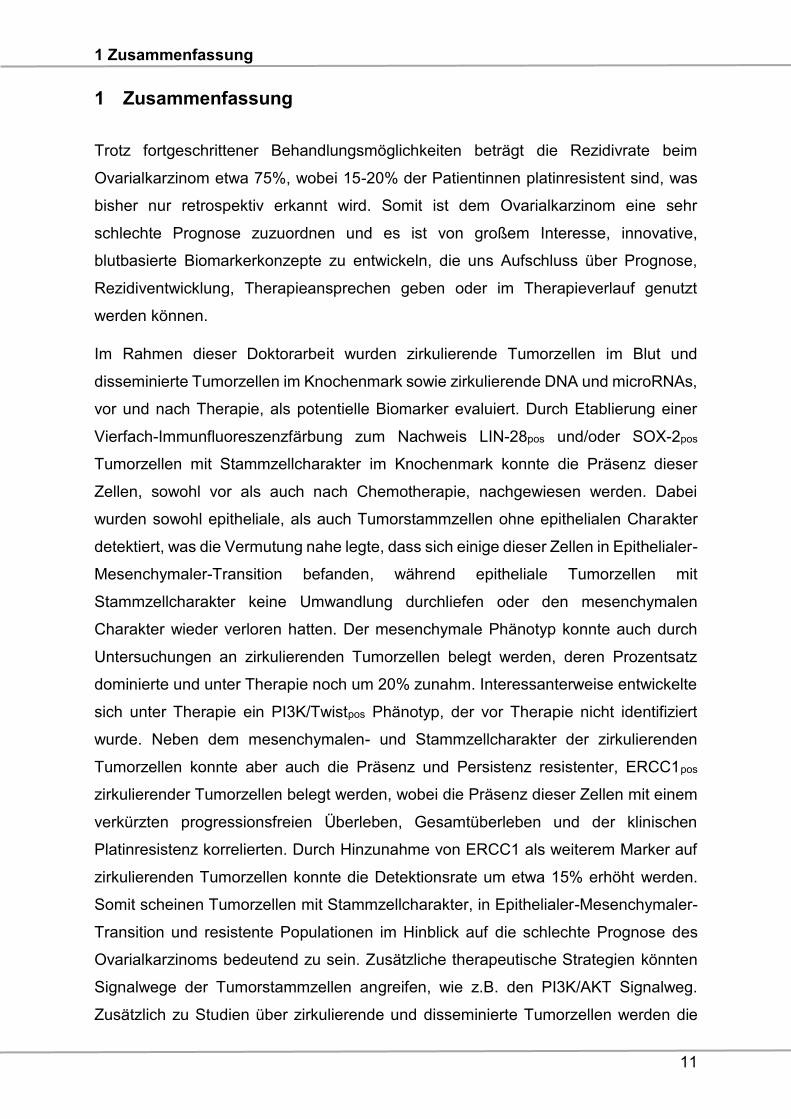
Trotz fortgeschrittener Behandlungsmöglichkeiten beträgt die Rezidivrate beim
Ovarialkarzinom etwa 75%, wobei 15-20% der Patientinnen platinresistent sind, was
bisher nur retrospektiv erkannt wird. Somit ist dem Ovarialkarzinom eine sehr
schlechte Prognose zuzuordnen und es ist von großem Interesse, innovative,
blutbasierte Biomarkerkonzepte zu entwickeln, die uns Aufschluss über Prognose,
Rezidiventwicklung, Therapieansprechen geben oder im Therapieverlauf genutzt
werden können.
Im Rahmen dieser Doktorarbeit wurden zirkulierende Tumorzellen im Blut und
disseminierte Tumorzellen im Knochenmark sowie zirkulierende DNA und microRNAs,
vor und nach Therapie, als potentielle Biomarker evaluiert. Durch Etablierung einer
Vierfach-Immunfluoreszenzfärbung zum Nachweis LIN-28pos und/oder SOX-2pos
Tumorzellen mit Stammzellcharakter im Knochenmark konnte die Präsenz dieser
Zellen, sowohl vor als auch nach Chemotherapie, nachgewiesen werden. Dabei
wurden sowohl epitheliale, als auch Tumorstammzellen ohne epithelialen Charakter
detektiert, was die Vermutung nahe legte, dass sich einige dieser Zellen in Epithelialer-
Mesenchymaler-Transition befanden, während epitheliale Tumorzellen mit
Stammzellcharakter keine Umwandlung durchliefen oder den mesenchymalen
Charakter wieder verloren hatten. Der mesenchymale Phänotyp konnte auch durch
Untersuchungen an zirkulierenden Tumorzellen belegt werden, deren Prozentsatz
dominierte und unter Therapie noch um 20% zunahm. Interessanterweise entwickelte
sich unter Therapie ein PI3K/Twistpos Phänotyp, der vor Therapie nicht identifiziert
wurde. Neben dem mesenchymalen- und Stammzellcharakter der zirkulierenden
Tumorzellen konnte aber auch die Präsenz und Persistenz resistenter, ERCC1pos
zirkulierender Tumorzellen belegt werden, wobei die Präsenz dieser Zellen mit einem
verkürzten progressionsfreien Überleben, Gesamtüberleben und der klinischen
Platinresistenz korrelierten. Durch Hinzunahme von ERCC1 als weiterem Marker auf
zirkulierenden Tumorzellen konnte die Detektionsrate um etwa 15% erhöht werden.
Somit scheinen Tumorzellen mit Stammzellcharakter, in Epithelialer-Mesenchymaler-
Transition und resistente Populationen im Hinblick auf die schlechte Prognose des
Ovarialkarzinoms bedeutend zu sein. Zusätzliche therapeutische Strategien könnten
Signalwege der Tumorstammzellen angreifen, wie z.B. den PI3K/AKT Signalweg.
Zusätzlich zu Studien über zirkulierende und disseminierte Tumorzellen werden die
1 Zusammenfassung
12
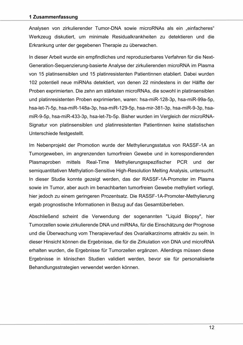
Analysen von zirkulierender Tumor-DNA sowie microRNAs als ein „einfacheres“
Werkzeug diskutiert, um minimale Residualkrankheiten zu detektieren und die
Erkrankung unter der gegebenen Therapie zu überwachen.
In dieser Arbeit wurde ein empfindliches und reproduzierbares Verfahren für die Next-
Generation-Sequenzierung-basierte Analyse der zirkulierenden microRNA im Plasma
von 15 platinsensiblen und 15 platinresistenten Patientinnen etabliert. Dabei wurden
102 potentiell neue miRNAs detektiert, von denen 22 mindestens in der Hälfte der
Proben exprimierten. Die zehn am stärksten microRNAs, die sowohl in platinsensiblen
und platinresistenten Proben exprimierten, waren: hsa-miR-128-3p, hsa-miR-99a-5p,
hsa-let-7i-5p, hsa-miR-148a-3p, hsa-miR-129-5p, hsa-mir-381-3p, hsa-miR-9-3p, hsa-
miR-9-5p, hsa-miR-433-3p, hsa-let-7b-5p. Bisher wurden im Vergleich der microRNA-
Signatur von platinsensiblen und platinresistenten Patientinnen keine statistischen
Unterschiede festgestellt.
Im Nebenprojekt der Promotion wurde der Methylierungsstatus von RASSF-1A an
Tumorgeweben, im angrenzenden tumorfreien Gewebe und in korrespondierenden
Plasmaproben mittels Real-Time Methylierungsspezifischer PCR und der
semiquantitativen Methylation-Sensitive High-Resolution Melting Analysis, untersucht.
In dieser Studie konnte gezeigt werden, das der RASSF-1A-Promoter im Plasma
sowie im Tumor, aber auch im benachbarten tumorfreien Gewebe methyliert vorliegt,
hier jedoch zu einem geringeren Prozentsatz. Die RASSF-1A-Promoter-Methylierung
ergab prognostische Informationen in Bezug auf das Gesamtüberleben.
Abschließend scheint die Verwendung der sogenannten "Liquid Biopsy", hier
Tumorzellen sowie zirkulierende DNA und miRNAs, für die Einschätzung der Prognose
und die Überwachung vom Therapieverlauf des Ovarialkarzinoms attraktiv zu sein. In
dieser Hinsicht können die Ergebnisse, die für die Zirkulation von DNA und microRNA
erhalten wurden, die Ergebnisse für Tumorzellen ergänzen. Allerdings müssen diese
Ergebnisse in klinischen Studien validiert werden, bevor sie für personalisierte
Behandlungsstrategien verwendet werden können.
1 Zusammenfassung
13
Summary
Despite advanced treatment options, the recurrence rate in ovarian carcinoma is
approximately 75%. One of the major problems is the resistance to platinum-based
chemotherapy in about 15-20% of cases which can only be recognized retrospectively.
Thus, a very bad prognosis is attributed to ovarian cancer and it is, therefore, of great
interest to develop innovative, blood-based biomarker concepts, which can provide
information about prognosis and response during the course of therapy.
In this thesis, circulating tumor cells in blood samples and disseminated tumor cells in
bone marrow as well as circulating DNA and microRNAs were evaluated as potential
biomarkers in primary ovarian cancer patients before surgery and after platinum-based
chemotherapy. Establishing a fourfold immunofluorescence staining method for the
detection of LIN-28pos and/or SOX-2pos tumor cells with stem cell character in the bone
marrow, the presence of these cells could be demonstrated before and after
chemotherapy. Both, epithelial and non-epithelial tumor stem cells were detected
suggesting that some of these cells were in Epithelial-Mesenchymal-Transition, while
epithelial tumor cells with stem cell characteristics did not undergo any transformation
or lost their mesenchymal character. The mesenchymal phenotype was also confirmed
by studies on circulating tumor cells, representing the most frequently found phenotype
which increased up to 20% under therapy. Interestingly, a PI3K/Twistpos phenotype
developed under therapy that was not identified before therapy. In addition to
mesenchymal and stem cell characteristics of the circulating tumor cells, the presence
and persistence of more resistant, ERCC1pos circulating tumor cells could also be
demonstrated. The presence of these cells correlated with a shortened progression-
free survival, overall survival and clinical platinum resistance. By including ERCC1 as
an additional marker on circulating tumor cells, the detection rate could be increased
up to 15% before and after therapy. Thus, tumor cells with stem cell characteristics in
Epithelial-Mesenchymal-Transition and resistant populations seem to play a role with
regard to poor outcome of our ovarian cancer patients. Additional therapeutic
strategies could target signalling pathways of the tumor stem cells, e.g. the PI3K/AKT
signalling pathway. In addition to studies on circulating and disseminated tumor cells,
the analyses of circulating tumor DNA as well as microRNAs is discussed to be a more
convenient tool to detect minimal residual disease and monitor disease under the
given therapy.
In this thesis, a sensitive and reproducible method for the Next-Generation-
1 Zusammenfassung
14
Sequencing-based analysis of circulating microRNA in human plasma comparing 15
platinum-sensitive and 15 platinum-resistant cases was established. In this case, 102
potentially new miRNAs were detected, of which at least 22 expressed in the half of
the samples. The ten most microRNAs expressing both platinum-sensitive and
platinum-resistant samples were: hsa-miR-128-3p, hsa-miR-99a-5p, hsa-let-7i-5p,
hsa-miR-148a-3p, hsa-miR-129-5p, hsa-miR-381-3p, hsa-miR-9-3p, hsa-miR-9-5p,
hsa-miR-433-3p, hsa-let-7b-5p. Up to now, comparing the microRNA signature of
platinum-sensitive and platinum-resistant patients, no statistical differences were
detected.
In a sub project of this thesis, the methylation status of RASSF-1A in tumors,
neighbouring tumor-free tissue and in corresponding plasma samples using real-time
methylation-specific PCR and the semi-quantitative methylation-sensitive high-
resolution melting analysis were investigated. Briefly, RASSF-1A promoter was
observed in ctDNA and found highly methylated in primary tumors, and at lower
percentages in the adjacent morphologically tumor cell-free tissues. We reported for
the first time that RASSF-1A promoter methylation provided significant prognostic
information in high grade serous ovarian cancer patients.
In conclusion, using the so-called "liquid biopsy", including tumor cells as well as
circulating DNA and miRNAs, seem to be attractive for estimating prognosis and
monitoring ovarian cancer. In this regard, results obtained for circulating DNA and
microRNAs might complement results obtained for tumor cells. However, these results
have to be validated in clinical trials before using them for personalized treatment
strategies.
2 Einleitung
15
2 Einleitung
2.1 Das Ovarialkarzinom
Das Ovarialkarzinom ist die fünfthäufigste Todesursache bei Frauen mit einer
Krebserkrankung in Europa und den Vereinigten Staaten (Goodman et al., 2003).
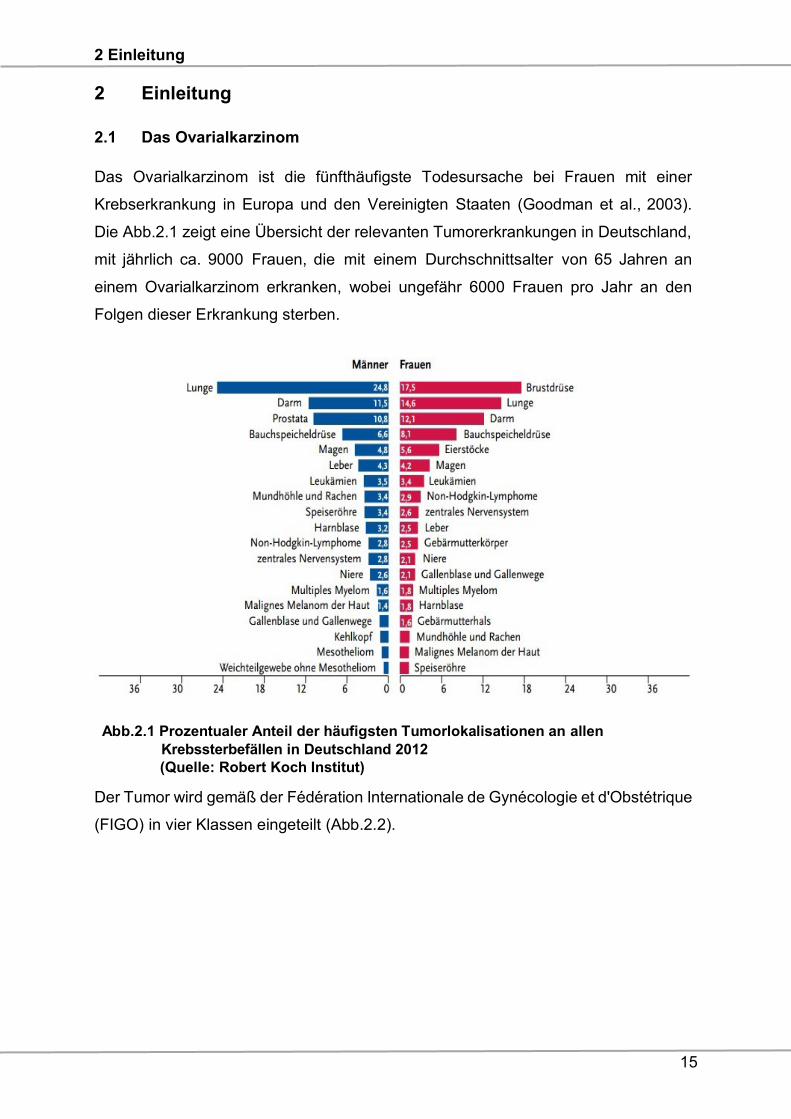
Die Abb.2.1 zeigt eine Übersicht der relevanten Tumorerkrankungen in Deutschland,
mit jährlich ca. 9000 Frauen, die mit einem Durchschnittsalter von 65 Jahren an
einem Ovarialkarzinom erkranken, wobei ungefähr 6000 Frauen pro Jahr an den
Folgen dieser Erkrankung sterben.
Abb.2.1 Prozentualer Anteil der häufigsten Tumorlokalisationen an allen
Krebssterbefällen in Deutschland 2012
(Quelle: Robert Koch Institut)
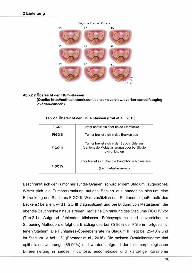
Der Tumor wird gemäß der Fédération Internationale de Gynécologie et d'Obstétrique
(FIGO) in vier Klassen eingeteilt (Abb.2.2).
2 Einleitung
16
Abb.2.2 Übersicht der FIGO-Klassen
(Quelle: http://nethealthbook.com/cancer-overview/ovarian-cancer/staging-
ovarian-cancer/)
Tab.2.1 Übersicht der FIGO-Klassen (Prat et al., 2015)
Beschränkt sich der Tumor nur auf die Ovarien, so wird er dem Stadium I zugeordnet.
Weitet sich die Tumorerkrankung auf das Becken aus, handelt es sich um eine
Erkrankung des Stadiums FIGO II. Wird zusätzlich das Peritoneum (außerhalb des
Beckens) befallen, wird FIGO III diagnostiziert und bei Bildung von Metastasen, die
über die Bauchhöhle hinaus streuen, liegt eine Erkrankung des Stadiums FIGO IV vor
(Tab.2.1). Aufgrund fehlender klinischer Frühsymptome und unzureichender
Screening-Methoden, erfolgt die Erstdiagnose bei 75-80% der Fälle im fortgeschrit-
tenen Stadium. Die Fünfjahres-Überlebensrate im Stadium III liegt bei 25-40% und
im Stadium IV bei 11% (Forstner et al., 2016). Die meisten Ovarialkarzinome sind
epithelialen Ursprungs (85-90%) und werden aufgrund der histomorphologischen
Differenzierung in seröse, muzinöse, endometroide und klarzellige Karzinome
FIGO I Tumor befällt ein oder beide Eierstöcke
FIGO II Tumor breitet sich in das Becken aus
FIGO III
Tumor breitet sich in der Bauchhöhle aus
(peritoneale Metastasierung) oder befällt die
Lymphknoten
FIGO IV
Tumor breitet sich über die Bauchhöhle hinaus aus
(Fernmetastasierung)
2 Einleitung
17
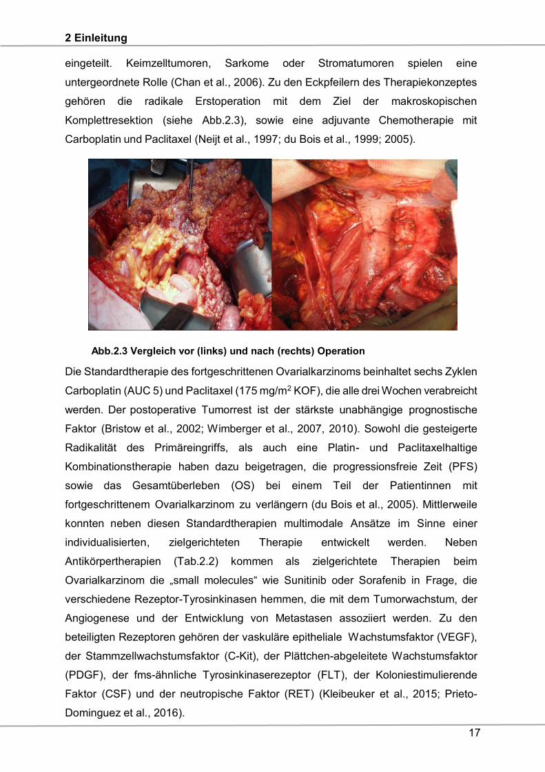
eingeteilt. Keimzelltumoren, Sarkome oder Stromatumoren spielen eine
untergeordnete Rolle (Chan et al., 2006). Zu den Eckpfeilern des Therapiekonzeptes
gehören die radikale Erstoperation mit dem Ziel der makroskopischen
Komplettresektion (siehe Abb.2.3), sowie eine adjuvante Chemotherapie mit
Carboplatin und Paclitaxel (Neijt et al., 1997; du Bois et al., 1999; 2005).
Abb.2.3 Vergleich vor (links) und nach (rechts) Operation
Die Standardtherapie des fortgeschrittenen Ovarialkarzinoms beinhaltet sechs Zyklen
Carboplatin (AUC 5) und Paclitaxel (175 mg/m2 KOF), die alle drei Wochen verabreicht
werden. Der postoperative Tumorrest ist der stärkste unabhängige prognostische
Faktor (Bristow et al., 2002; Wimberger et al., 2007, 2010). Sowohl die gesteigerte
Radikalität des Primäreingriffs, als auch eine Platin- und Paclitaxelhaltige
Kombinationstherapie haben dazu beigetragen, die progressionsfreie Zeit (PFS)
sowie das Gesamtüberleben (OS) bei einem Teil der Patientinnen mit
fortgeschrittenem Ovarialkarzinom zu verlängern (du Bois et al., 2005). Mittlerweile
konnten neben diesen Standardtherapien multimodale Ansätze im Sinne einer
individualisierten, zielgerichteten Therapie entwickelt werden. Neben
Antikörpertherapien (Tab.2.2) kommen als zielgerichtete Therapien beim
Ovarialkarzinom die „small molecules“ wie Sunitinib oder Sorafenib in Frage, die
verschiedene Rezeptor-Tyrosinkinasen hemmen, die mit dem Tumorwachstum, der
Angiogenese und der Entwicklung von Metastasen assoziiert werden. Zu den
beteiligten Rezeptoren gehören der vaskuläre epitheliale Wachstumsfaktor (VEGF),
der Stammzellwachstumsfaktor (C-Kit), der Plättchen-abgeleitete Wachstumsfaktor
(PDGF), der fms-ähnliche Tyrosinkinaserezeptor (FLT), der Koloniestimulierende
Faktor (CSF) und der neutropische Faktor (RET) (Kleibeuker et al., 2015; Prieto-
Dominguez et al., 2016).
2 Einleitung
18
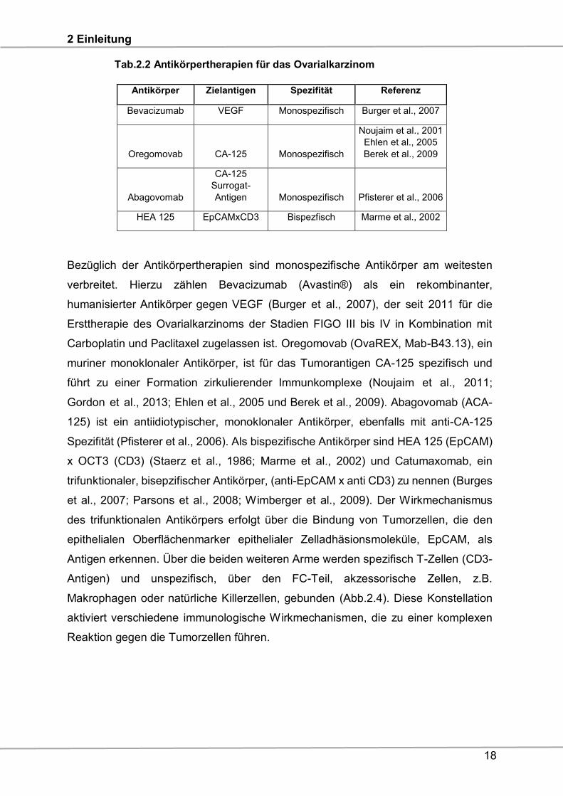
Tab.2.2 Antikörpertherapien für das Ovarialkarzinom
Antikörper Zielantigen Spezifität Referenz
Bevacizumab VEGF Monospezifisch Burger et al., 2007
Oregomovab CA-125 Monospezifisch
Noujaim et al., 2001
Ehlen et al., 2005
Berek et al., 2009
Abagovomab
CA-125
Surrogat-
Antigen Monospezifisch Pfisterer et al., 2006
HEA 125 EpCAMxCD3 Bispezfisch Marme et al., 2002
Bezüglich der Antikörpertherapien sind monospezifische Antikörper am weitesten
verbreitet. Hierzu zählen Bevacizumab (Avastin®) als ein rekombinanter,
humanisierter Antikörper gegen VEGF (Burger et al., 2007), der seit 2011 für die
Ersttherapie des Ovarialkarzinoms der Stadien FIGO III bis IV in Kombination mit
Carboplatin und Paclitaxel zugelassen ist. Oregomovab (OvaREX, Mab-B43.13), ein
muriner monoklonaler Antikörper, ist für das Tumorantigen CA-125 spezifisch und
führt zu einer Formation zirkulierender Immunkomplexe (Noujaim et al., 2011;
Gordon et al., 2013; Ehlen et al., 2005 und Berek et al., 2009). Abagovomab (ACA-
125) ist ein antiidiotypischer, monoklonaler Antikörper, ebenfalls mit anti-CA-125
Spezifität (Pfisterer et al., 2006). Als bispezifische Antikörper sind HEA 125 (EpCAM)
x OCT3 (CD3) (Staerz et al., 1986; Marme et al., 2002) und Catumaxomab, ein
trifunktionaler, bisepzifischer Antikörper, (anti-EpCAM x anti CD3) zu nennen (Burges
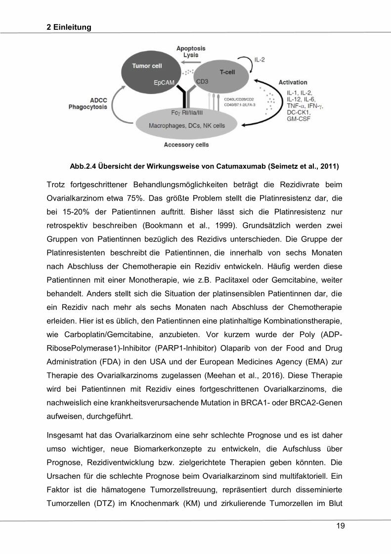
et al., 2007; Parsons et al., 2008; Wimberger et al., 2009). Der Wirkmechanismus
des trifunktionalen Antikörpers erfolgt über die Bindung von Tumorzellen, die den
epithelialen Oberflächenmarker epithelialer Zelladhäsionsmoleküle, EpCAM, als
Antigen erkennen. Über die beiden weiteren Arme werden spezifisch T-Zellen (CD3-
Antigen) und unspezifisch, über den FC-Teil, akzessorische Zellen, z.B.
Makrophagen oder natürliche Killerzellen, gebunden (Abb.2.4). Diese Konstellation
aktiviert verschiedene immunologische Wirkmechanismen, die zu einer komplexen
Reaktion gegen die Tumorzellen führen.
2 Einleitung
19
Abb.2.4 Übersicht der Wirkungsweise von Catumaxumab (Seimetz et al., 2011)
Trotz fortgeschrittener Behandlungsmöglichkeiten beträgt die Rezidivrate beim
Ovarialkarzinom etwa 75%. Das größte Problem stellt die Platinresistenz dar, die
bei 15-20% der Patientinnen auftritt. Bisher lässt sich die Platinresistenz nur
retrospektiv beschreiben (Bookmann et al., 1999). Grundsätzlich werden zwei
Gruppen von Patientinnen bezüglich des Rezidivs unterschieden. Die Gruppe der
Platinresistenten beschreibt die Patientinnen, die innerhalb von sechs Monaten
nach Abschluss der Chemotherapie ein Rezidiv entwickeln. Häufig werden diese
Patientinnen mit einer Monotherapie, wie z.B. Paclitaxel oder Gemcitabine, weiter
behandelt. Anders stellt sich die Situation der platinsensiblen Patientinnen dar, die
ein Rezidiv nach mehr als sechs Monaten nach Abschluss der Chemotherapie
erleiden. Hier ist es üblich, den Patientinnen eine platinhaltige Kombinationstherapie,
wie Carboplatin/Gemcitabine, anzubieten. Vor kurzem wurde der Poly (ADP-
RibosePolymerase1)-Inhibitor (PARP1-Inhibitor) Olaparib von der Food and Drug
Administration (FDA) in den USA und der European Medicines Agency (EMA) zur
Therapie des Ovarialkarzinoms zugelassen (Meehan et al., 2016). Diese Therapie
wird bei Patientinnen mit Rezidiv eines fortgeschrittenen Ovarialkarzinoms, die
nachweislich eine krankheitsverursachende Mutation in BRCA1- oder BRCA2-Genen
aufweisen, durchgeführt.
Insgesamt hat das Ovarialkarzinom eine sehr schlechte Prognose und es ist daher
umso wichtiger, neue Biomarkerkonzepte zu entwickeln, die Aufschluss über
Prognose, Rezidiventwicklung bzw. zielgerichtete Therapien geben könnten. Die
Ursachen für die schlechte Prognose beim Ovarialkarzinom sind multifaktoriell. Ein
Faktor ist die hämatogene Tumorzellstreuung, repräsentiert durch disseminierte
Tumorzellen (DTZ) im Knochenmark (KM) und zirkulierende Tumorzellen im Blut
2 Einleitung
20
(ZTZ). Obwohl die prognostische Bedeutung dieser Zellen bekannt ist, gibt es beim
Ovarialkarzinom kaum Daten zur Charakterisierung dieser Zellen.
2.2 Tumorzelldifferenzierung
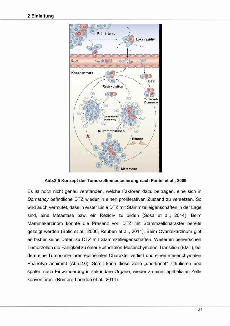
Die Tumorzelldisseminierung beschreibt im Allgemeinen die Streuung der einzelnen
Tumorzellen aus dem Primärtumor, welche über das Blut- und Lymphgefäßsystem
organisiert ist. Einen schematischen Überblick zeigt die Abb.2.5. Dabei verlassen
Tumorzellen in frühen Stadien den Primärtumor und gelangen über die Blutbahn in
sekundäre Organe, wie z.B. das KM. Dort verweilen sie in Form einzelner DTZ oder
in Form von Zellaggregaten, den Mikrometastasen. Mit den DTZ wird sehr häufig der
Begriff „Dormancy“ in Verbindung gebracht. Es wird zwischen Tumor-Cell-Dormancy
und Tumor-Mass-Dormancy unterschieden. Ersteres beschreibt die Fähigkeit der
Tumorzellen, sich in einem ruhenden, nicht proliferativen Zustand zu organisieren. Die
Tumor-Mass Dormancy hingegen beschreibt ein Gleichgewichtszustand zwischen
Proliferation und Apoptose der Mikrometastasen. Die ruhenden DTZ können wieder
aktiviert werden, in die Blutzirkulation eintreten und eine Metastase bilden. Wann
dies geschieht, und unter welchen Bedingungen, ist weitgehend unbekannt. In der
Theorie gibt es bisher drei Modelle, die die Entstehung einer Dormancy erklären
(Bragado et al., 2012):
1. Stressfaktoren im Tumor, z.B. Hypoxie, wirken auf primäre Tumorzellen und
programmieren neuerlich disseminierende Tumorzellen schon im Vorfeld auf
den Zustand der Dormancy.
2. Eine frühe Tumorzelldisseminierung bedingt eine DTZ-Population, die noch
unfähig für ein metastatisches Wachstum ist, somit in einem „Arrestzustand“
verharrt und erst weitere genetische Veränderungen für eine klonale
Expansion erwerben muss.
3. DTZ invasiver Karzinome antworten mit Stresssignalen auf die
Disseminierung bzw. auf einen wachstumsinhibierenden Einfluss des
extrazellulären Milieus im Zielorgan und induzieren Dormancy.
2 Einleitung
21
Abb.2.5 Konzept der Tumorzellmetastasierung nach Pantel et al., 2009
Es ist noch nicht genau verstanden, welche Faktoren dazu beitragen, eine sich in
Dormancy befindliche DTZ wieder in einen proliferativen Zustand zu versetzen. So
wird auch vermutet, dass in erster Linie DTZ mit Stammzelleigenschaften in der Lage
sind, eine Metastase bzw. ein Rezidiv zu bilden (Sosa et al., 2014). Beim
Mammakarzinom konnte die Präsenz von DTZ mit Stammzellcharakter bereits
gezeigt werden (Balic et al., 2006, Reuben et al., 2011). Beim Ovarialkarzinom gibt
es bisher keine Daten zu DTZ mit Stammzelleigenschaften. Weiterhin beherrschen
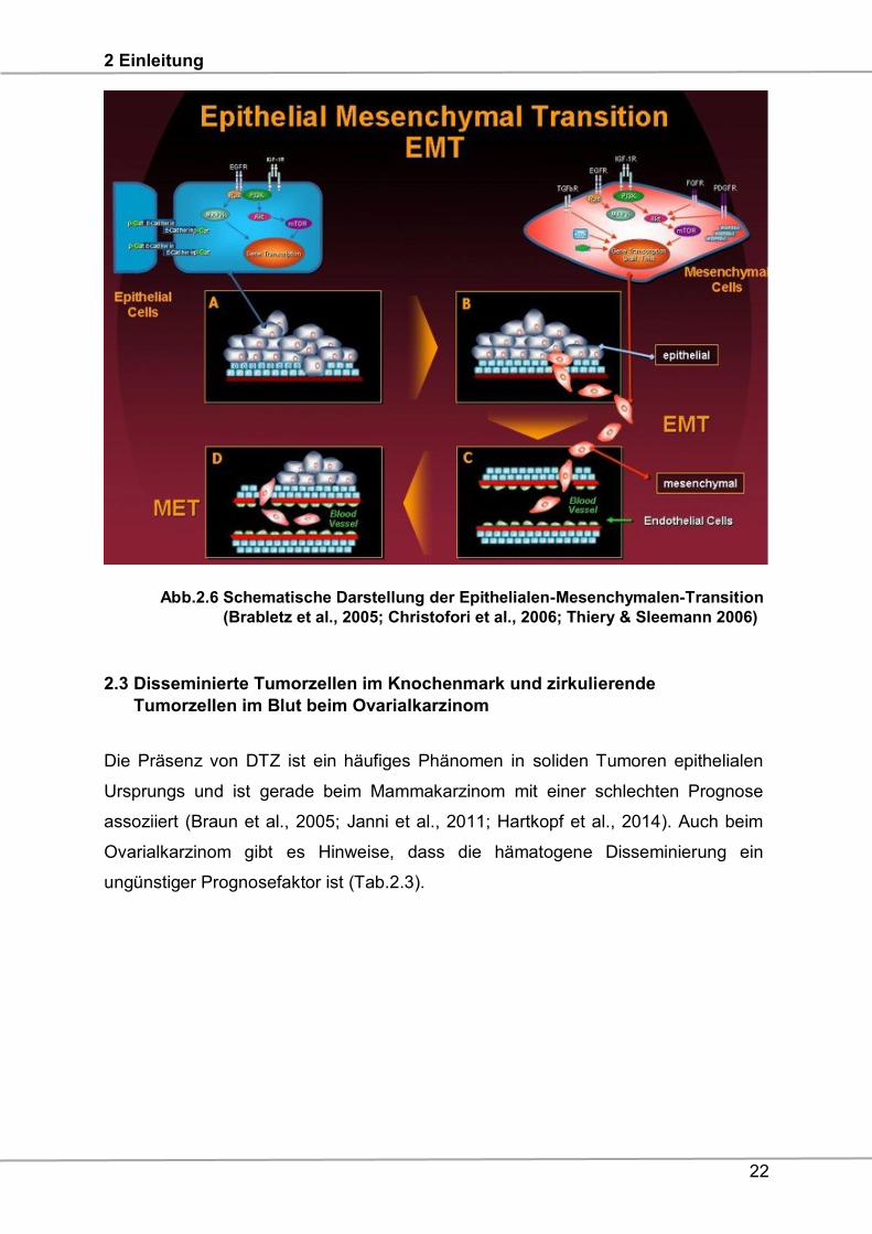
Tumorzellen die Fähigkeit zu einer Epithelialen-Mesenchymalen-Transition (EMT), bei
dem eine Tumorzelle ihren epithelialen Charakter verliert und einen mesenchymalen
Phänotyp annimmt (Abb.2.6). Somit kann diese Zelle „unerkannt“ zirkulieren und
später, nach Einwanderung in sekundäre Organe, wieder zu einer epithelialen Zelle
konvertieren (Romero-Laorden et al., 2014).
2 Einleitung
22
Abb.2.6 Schematische Darstellung der Epithelialen-Mesenchymalen-Transition
(Brabletz et al., 2005; Christofori et al., 2006; Thiery & Sleemann 2006)
2.3 Disseminierte Tumorzellen im Knochenmark und zirkulierende
Tumorzellen im Blut beim Ovarialkarzinom
Die Präsenz von DTZ ist ein häufiges Phänomen in soliden Tumoren epithelialen
Ursprungs und ist gerade beim Mammakarzinom mit einer schlechten Prognose
assoziiert (Braun et al., 2005; Janni et al., 2011; Hartkopf et al., 2014). Auch beim
Ovarialkarzinom gibt es Hinweise, dass die hämatogene Disseminierung ein
ungünstiger Prognosefaktor ist (Tab.2.3).
2 Einleitung
23
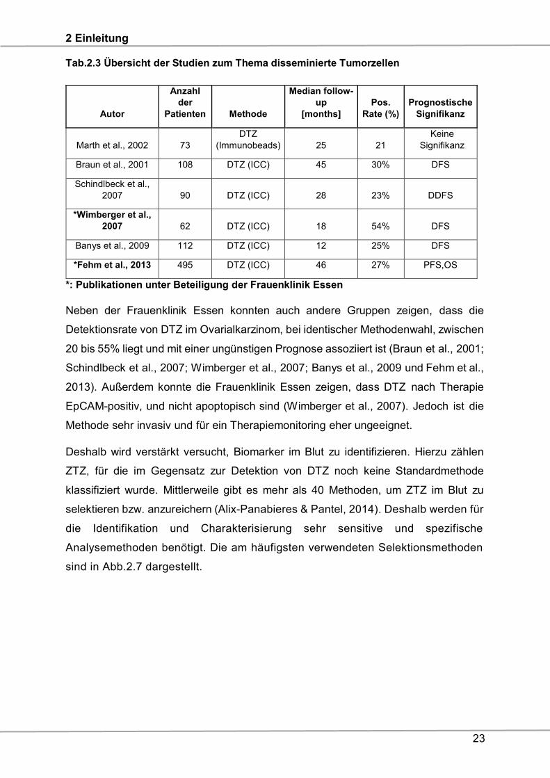
Tab.2.3 Übersicht der Studien zum Thema disseminierte Tumorzellen
*: Publikationen unter Beteiligung der Frauenklinik Essen
Neben der Frauenklinik Essen konnten auch andere Gruppen zeigen, dass die
Detektionsrate von DTZ im Ovarialkarzinom, bei identischer Methodenwahl, zwischen
20 bis 55% liegt und mit einer ungünstigen Prognose assoziiert ist (Braun et al., 2001;
Schindlbeck et al., 2007; Wimberger et al., 2007; Banys et al., 2009 und Fehm et al.,
2013). Außerdem konnte die Frauenklinik Essen zeigen, dass DTZ nach Therapie
EpCAM-positiv, und nicht apoptopisch sind (Wimberger et al., 2007). Jedoch ist die
Methode sehr invasiv und für ein Therapiemonitoring eher ungeeignet.
Deshalb wird verstärkt versucht, Biomarker im Blut zu identifizieren. Hierzu zählen
ZTZ, für die im Gegensatz zur Detektion von DTZ noch keine Standardmethode
klassifiziert wurde. Mittlerweile gibt es mehr als 40 Methoden, um ZTZ im Blut zu
selektieren bzw. anzureichern (Alix-Panabieres & Pantel, 2014). Deshalb werden für
die Identifikation und Charakterisierung sehr sensitive und spezifische
Analysemethoden benötigt. Die am häufigsten verwendeten Selektionsmethoden
sind in Abb.2.7 dargestellt.
Autor
Anzahl
der
Patienten Methode
Median follow-
up
[months]
Pos.
Rate (%)
Prognostische
Signifikanz
Marth et al., 2002 73
DTZ
(Immunobeads) 25 21
Keine
Signifikanz
Braun et al., 2001 108 DTZ (ICC) 45 30% DFS
Schindlbeck et al.,
2007 90 DTZ (ICC) 28 23% DDFS
*Wimberger et al.,
2007 62 DTZ (ICC) 18 54% DFS
Banys et al., 2009 112 DTZ (ICC) 12 25% DFS
*Fehm et al., 2013 495 DTZ (ICC) 46 27% PFS,OS
2 Einleitung
24
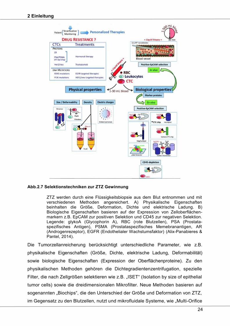
Abb.2.7 Selektionstechniken zur ZTZ Gewinnung
ZTZ werden durch eine Flüssigkeitsbiopsie aus dem Blut entnommen und mit verschiedenen Methoden angereichert. A) Physikalische Eigenschaften beinhalten die Größe, Deformation, Dichte und elektrische Ladung. B) Biologische Eigenschaften basieren auf der Expression von Zelloberflächen-markern z.B. EpCAM zur positiven Selektion und CD45 zur negativen Selektion. Legende: glykoA (Glycophorin A), RBC (rote Blutzellen), PSA (Prostata-spezifisches Antigen), PSMA (Prostataspezifisches Memebranantigen, AR (Androgenrezeptor), EGFR (Endothelialer Wachstumsfaktor) (Alix-Panabieres & Pantel, 2014).
Die Tumorzellanreicherung berücksichtigt unterschiedliche Parameter, wie z.B.
physikalische Eigenschaften (Größe, Dichte, elektrische Ladung, Deformabilität)
sowie biologische Eigenschaften (Expression der Oberflächenproteine). Zu den
physikalischen Methoden gehören die Dichtegradientenzentrifugation, spezielle
Filter, die nach Zellgrößen selektieren wie z.B. „ISET“ (Isolation by size of epithelial
tumor cells) sowie die dreidimensionalen Mikrofilter. Neue Methoden basieren auf
sogenannten „Biochips“, die den Unterschied der Größe und Deformation von ZTZ,
im Gegensatz zu den Blutzellen, nutzt und mikrofluidale Systeme, wie „Multi-Orifice
2 Einleitung
25
Flow Fractionation“ (MOFF), „Dielectrophoretisis“ (DEP) und „Dielectrophoretisis
Field-Flow Fractionation“ (Dep-FFF), die mit Hilfe der Dielektrophorese die
unterschiedlichen Größen und Membraneigenschaften der ZTZ erkennen.
Biologische Anreicherungsmethoden basieren auf der Selektion der Tumorzellen
anhand von Antikörpern, die an Magnetbeads gekoppelt sind und tumorassoziierte
Antigene auf ZTZ binden (positive Selektion) oder die Leukozyten (CD45) aus dem
Blut abreichern (negative Selektion). Weitere, auf „ZTZ-Chips“ basierende,
Methoden gewinnen EpCAMpos ZTZ über eine Kombination aus größen-
basierender, hydrodynamischer Zellsortierung und der immunmagnetischen
Selektion (positiv/negativ). Eine in vivo Methode ist der „GILUPI® Nanodetector“,
der es mit einer Antikörper-beschichteten Braunüle in der Armvene für 30 Minuten
ermöglicht, mit 1500 ml Blut eine Anreicherung der ZTZ durchzuführen. Neben den
Vorteilen beherbergen diese Systeme auch Nachteile, die aufgrund der
Heterogenität der Tumorzellen oftmals eine schlechte Wiederfindungsrate mit sich
bringen. Des Weiteren beeinflussen Luftblasen und der Verlust an
Oberflächenantigenen die Selektion mittels Magnetbeads. Eine Verklumpung der
Zellen erschwert die Selektion bei den „ZTZ-Chips“ und den Filtern.
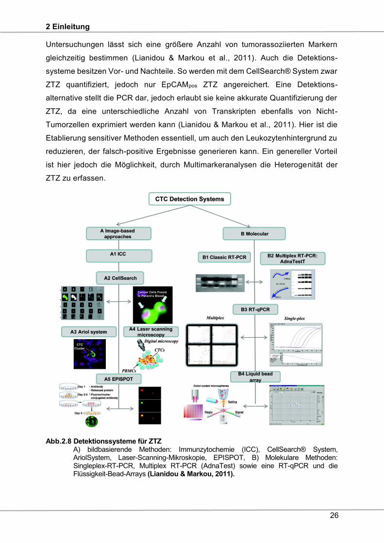
Die nachfolgende Abbildung (Abb.2.8) zeigt einen schematischen Überblick über
die Gruppe der bildbasierenden- und molekularbiologischen Detektionsmöglich-
keiten. Die zurzeit einzige, von der FDA 2004, zugelassene Methode ist das
CellSearch® System, bei der ZTZ aus 7,5 ml Blut anhand von EpCAM
beschichteten, magnetischen Beads isoliert werden. Der Nachweis erfolgt mittels
Immunfluoreszenz der ZTZ mit einem pan-Zytokeratin-Antikörper (epithelialer
Marker), DAPI (Marker für den Zellkern) und einem anti-CD45-Antikörper, um die
unspezifische Detektion falsch-positiver Leukozyten auszuschließen. Das
AriolSystem, ein automasiertes Bildanalysesystem, dient der Quantifizierung von
ZTZ auf Objektträgern. Mittels der Laser-Scan-Mikroskopie werden Präparate von
einem fokussierten Laserstrahl abgetastet, wodurch die Fluoreszenzemission der
Probe detektiert wird. Eine weitere Option zur bildbasierenden Methode stellt die
EPISPOT-Methode dar. Sie detektiert tumorsepezifische Proteine, die von ZTZ
freigesetzt werden. Die Detektion mittels molekularer Analysesysteme basieren auf
der RNA-Analyse lebender Tumorzellen und anschließender Reverser
Transkriptase-Polymerase-Kettenreaktion (RT-PCR) sowie der Amplifikation von
tumor- und epithelspezifischen Markern. Durch die Flexibiltät der Multiplex-
2 Einleitung
26
Untersuchungen lässt sich eine größere Anzahl von tumorassoziierten Markern
gleichzeitig bestimmen (Lianidou & Markou et al., 2011). Auch die Detektions-
systeme besitzen Vor- und Nachteile. So werden mit dem CellSearch® System zwar
ZTZ quantifiziert, jedoch nur EpCAMpos ZTZ angereichert. Eine Detektions-
alternative stellt die PCR dar, jedoch erlaubt sie keine akkurate Quantifizierung der
ZTZ, da eine unterschiedliche Anzahl von Transkripten ebenfalls von Nicht-
Tumorzellen exprimiert werden kann (Lianidou & Markou et al., 2011). Hier ist die
Etablierung sensitiver Methoden essentiell, um auch den Leukozytenhintergrund zu
reduzieren, der falsch-positive Ergebnisse generieren kann. Ein genereller Vorteil
ist hier jedoch die Möglichkeit, durch Multimarkeranalysen die Heterogenität der
ZTZ zu erfassen.
Abb.2.8 Detektionssysteme für ZTZ A) bildbasierende Methoden: Immunzytochemie (ICC), CellSearch® System, AriolSystem, Laser-Scanning-Mikroskopie, EPISPOT, B) Molekulare Methoden: Singleplex-RT-PCR, Multiplex RT-PCR (AdnaTest) sowie eine RT-qPCR und die Flüssigkeit-Bead-Arrays (Lianidou & Markou, 2011).
2 Einleitung
27
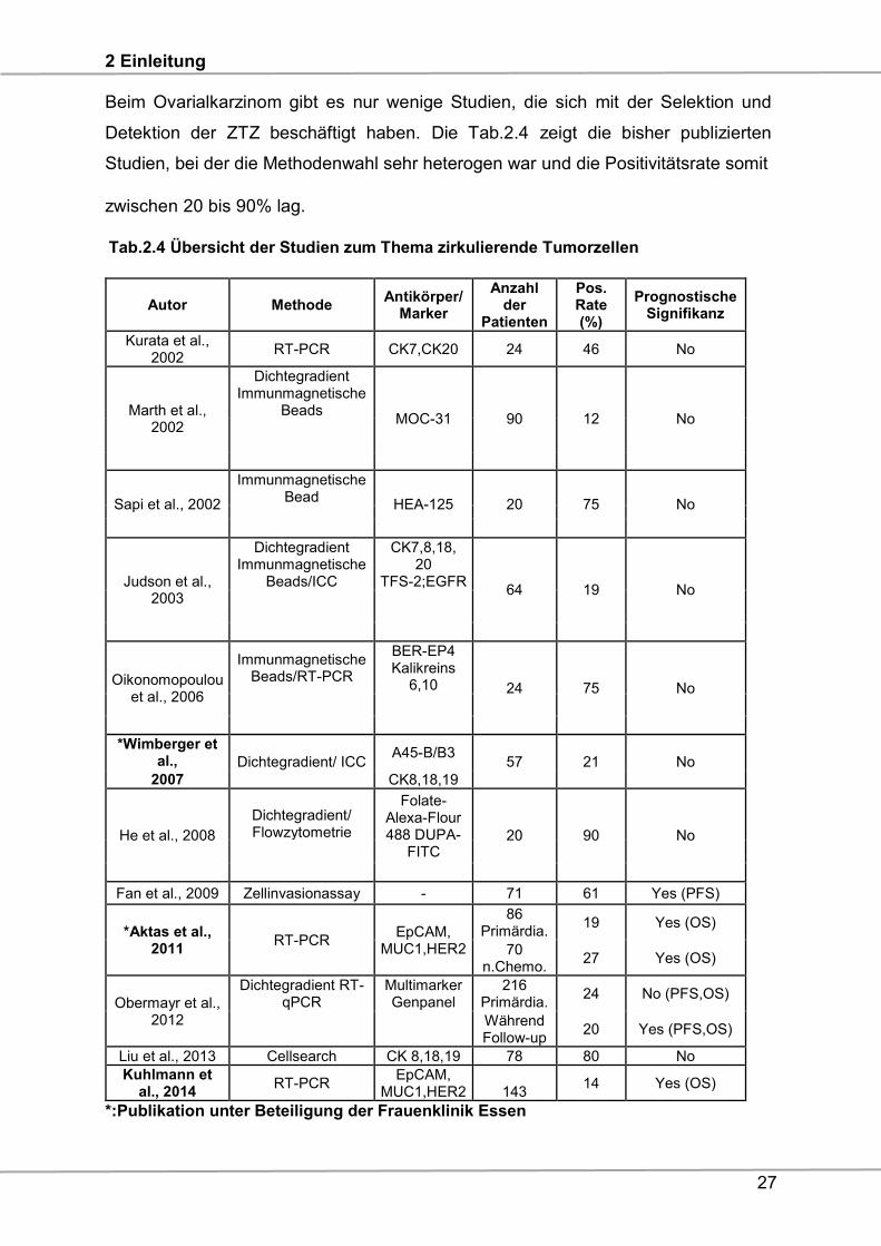
Beim Ovarialkarzinom gibt es nur wenige Studien, die sich mit der Selektion und
Detektion der ZTZ beschäftigt haben. Die Tab.2.4 zeigt die bisher publizierten
Studien, bei der die Methodenwahl sehr heterogen war und die Positivitätsrate somit
zwischen 20 bis 90% lag.
Tab.2.4 Übersicht der Studien zum Thema zirkulierende Tumorzellen
Autor Methode Antikörper/
Marker
Anzahl der
Patienten
Pos. Rate (%)
Prognostische Signifikanz
Kurata et al., 2002
RT-PCR CK7,CK20 24 46 No
Marth et al., 2002
Dichtegradient Immunmagnetische
Beads MOC-31 90 12 No
Sapi et al., 2002
Immunmagnetische Bead
HEA-125 20 75 No
Judson et al., 2003
Dichtegradient Immunmagnetische
Beads/ICC
CK7,8,18, 20
TFS-2;EGFR 64 19 No
Oikonomopoulou et al., 2006
Immunmagnetische Beads/RT-PCR
BER-EP4 Kalikreins
6,10 24 75 No
*Wimberger et al., Dichtegradient/ ICC
A45-B/B3 57 21 No
2007 CK8,18,19
He et al., 2008
Dichtegradient/ Flowzytometrie
Folate-Alexa-Flour 488 DUPA-
FITC 20 90 No
Fan et al., 2009 Zellinvasionassay - 71 61 Yes (PFS)
*Aktas et al., 2011
RT-PCR EpCAM,
MUC1,HER2
86 Primärdia.
19 Yes (OS)
70 n.Chemo.
27 Yes (OS)
Obermayr et al., 2012
Dichtegradient RT-qPCR
Multimarker Genpanel
216 Primärdia.
24 No (PFS,OS)
Während Follow-up
20 Yes (PFS,OS)
Liu et al., 2013 Cellsearch CK 8,18,19 78 80 No
Kuhlmann et al., 2014
RT-PCR EpCAM,
MUC1,HER2 143 14 Yes (OS)
*:Publikation unter Beteiligung der Frauenklinik Essen
2 Einleitung
28
Die prognostische Bedeutung im Hinblick auf das PFS und OS konnte nur in vier
Studien gezeigt werden, wobei auch die Methoden unterschiedlich waren (Fan et al.,
2009; Aktas et al., 2011; Obermayr et al., 2012, Kuhlmann et al., 2014). Im Gegensatz
dazu haben alle weiteren, in der Tabelle zusammengefassten, Arbeitsgruppen keine
prognostische Bedeutung der ZTZ zeigen können. Interessanterweise konnte die
Frauenklinik Essen kürzlich publizieren, dass die Expression vom „Excision-Repair
Cross-Complementation Group 1 (ERCC1) Protein, einem DNA-Reparaturenzym auf
ZTZ, im Blut von Ovarialkarzinompatientinnen zu Beginn der Erkrankung als
prädiktiver Marker für ein verkürztes PFS und OS sowie eine klinische Platinresistenz
identifiziert wurde (Kuhlmann et al., 2014).
2.4 Resistenzmechanismen und prognostische Bedeutung von ERCC1 im
Ovarialkarzinom
Insgesamt gibt es vier bekannte DNA-Reparaturmöglichkeiten, um eine Zelle vor
Chromosomenaberrationen zu schützen. Neben der Einzelstrang- und Doppelstrang-
reparatur verfügt eine Zelle noch über die Möglichkeiten der Photoreaktivierung und
Reparatur von Quervernetzungen. Die Nukleotidexzisionsreparatur ist ein hoch
konservierter Einzelstrang DNA-Reparatur-Signalweg, der vor allem „bulky lesions“
erkennt. Dies sind Lokalisationen, die eine Art „Buckel“ erzeugen und dadurch die
Helixstruktur stören. Ursachen sind Pyrimidindimere und 6,4 Photoprodukte sowie
Cisplatin, die durch UV-Strahlen verursacht werden. Die Reparatur gliedert sich in drei
Abschnitte (Abb.2.9):
1. Schadenserkennung durch einen DNA-Bindungsfaktor und Herausschneiden
einer 25-30 Basen langen DNA-Sequenz.
2. Neusynthese der Sequenz und anschließende Ligation. Das ERRC1 Enzym spielt
eine wichtige Schlüsselrolle in der Reparatur. Es dimerisiert mit dem Molekül
Xeroderma Pigmentosum Komplementation Gruppe F (XPF) und wird benötigt,
um den schädlichen DNA-Strang zu exzidieren (Shi et al., 2012). Die Bindung ist
für die Stabilität und die katalytische Aktivität von XPF wichtig, da ansonsten das
ERCC1 nicht am 5´Ende und XPF am 3´-Ende die DNA restriktieren kann
(Gossage & Madhusudan et al., 2007).
2 Einleitung
29
3. Die DNA-Polymerase synthetisiert den komplementären Strang, sodass die DNA-
Helix ihre ursprüngliche Form zurückerhält.
Bisher gibt es nur wenige Studien, die die Expression und die prognostische
Bedeutung von ERCC1 im Ovarialkarzinom untersucht haben. In einer Studie mit
Tumorgeweben von 101 Ovarialkarzinompatientinnen wurde in 14% der Fälle eine
Überexpression von ERCC1 detektiert, wobei 75% der Patientinnen eine
Platinresistenz entwickelten, was mit einem signifikant reduzierten PFS und OS
korrelierte (Steffensen et al., 2009). In einer ähnlichen Studie war die ERCC1-
Expression im Tumorgewebe bei Patientinnen mit Platinresistenz signifikant höher als
in der Gruppe der platinsensitiven Patientinnen, wobei keine prognostische Relevanz
beobachtet wurde (Xie et al., 2011). Ovarialkarzinompatientinnen mit einer höheren
ERCC1 Expressionsrate im Gewebe und adjuvanter, platinbasierter Chemotherapie
hatten ein signifikant kürzeres PFS und OS als die Gruppe mit niedriger ERCC1-
Expression (Milivic-Kovacevic et al., 2011). Eine Schlüsselpublikation im New England
Journal of Medicine berichtete über eine umfassende Neubewertung von über 494
Patienten mit Lungenkarzinom und folgerte, dass die immunhistochemische ERCC1
Erkennung im Primärtumor mit allen derzeit verfügbaren Antikörpern für Kliniker in
Bezug auf die Vorhersage von Platinresistenz und Therapieentscheidungen
ungeeignet ist (Friboulet et al., 2013). Die Arbeitsgruppe der Frauenklinik Essen hatte
auf Gewebeebene auch eine ERCC1 Expression beim Ovarialkarzinom zeigen
können, jedoch wurde keine Korrelation im Hinblick auf das PFS, OS und die
Platinresistenz gefunden. Im Gegensatz dazu zeigte sowohl die univariable, als auch
die multivariable Analyse der Präsenz von ERCC1pos ZTZ im Blut eine signifikante
Korrelation mit einem verkürzten PFS, OS und der Platinresistenz (Kuhlmann et al.,
2014).
2 Einleitung
30
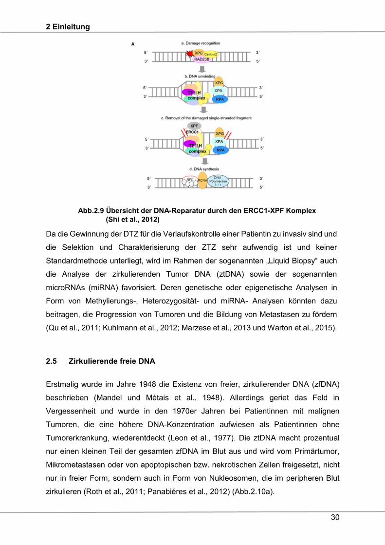
Abb.2.9 Übersicht der DNA-Reparatur durch den ERCC1-XPF Komplex
(Shi et al., 2012)
Da die Gewinnung der DTZ für die Verlaufskontrolle einer Patientin zu invasiv sind und
die Selektion und Charakterisierung der ZTZ sehr aufwendig ist und keiner
Standardmethode unterliegt, wird im Rahmen der sogenannten „Liquid Biopsy“ auch
die Analyse der zirkulierenden Tumor DNA (ztDNA) sowie der sogenannten
microRNAs (miRNA) favorisiert. Deren genetische oder epigenetische Analysen in
Form von Methylierungs-, Heterozygosität- und miRNA- Analysen könnten dazu
beitragen, die Progression von Tumoren und die Bildung von Metastasen zu fördern
(Qu et al., 2011; Kuhlmann et al., 2012; Marzese et al., 2013 und Warton et al., 2015).
2.5 Zirkulierende freie DNA
Erstmalig wurde im Jahre 1948 die Existenz von freier, zirkulierender DNA (zfDNA)
beschrieben (Mandel und Métais et al., 1948). Allerdings geriet das Feld in
Vergessenheit und wurde in den 1970er Jahren bei Patientinnen mit malignen
Tumoren, die eine höhere DNA-Konzentration aufwiesen als Patientinnen ohne
Tumorerkrankung, wiederentdeckt (Leon et al., 1977). Die ztDNA macht prozentual
nur einen kleinen Teil der gesamten zfDNA im Blut aus und wird vom Primärtumor,
Mikrometastasen oder von apoptopischen bzw. nekrotischen Zellen freigesetzt, nicht
nur in freier Form, sondern auch in Form von Nukleosomen, die im peripheren Blut
zirkulieren (Roth et al., 2011; Panabiéres et al., 2012) (Abb.2.10a).
2 Einleitung
31
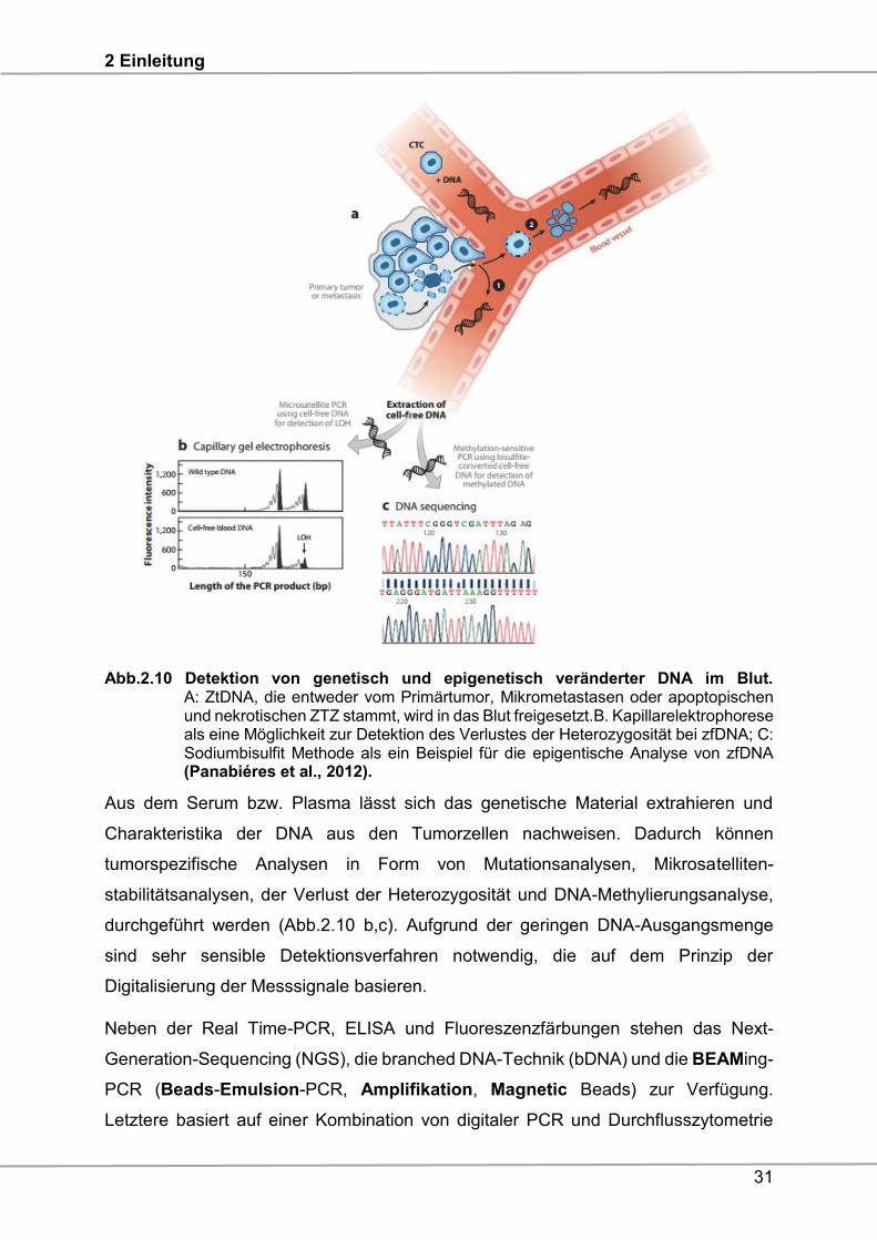
Abb.2.10 Detektion von genetisch und epigenetisch veränderter DNA im Blut. A: ZtDNA, die entweder vom Primärtumor, Mikrometastasen oder apoptopischen und nekrotischen ZTZ stammt, wird in das Blut freigesetzt.B. Kapillarelektrophorese als eine Möglichkeit zur Detektion des Verlustes der Heterozygosität bei zfDNA; C: Sodiumbisulfit Methode als ein Beispiel für die epigentische Analyse von zfDNA (Panabiéres et al., 2012).
Aus dem Serum bzw. Plasma lässt sich das genetische Material extrahieren und
Charakteristika der DNA aus den Tumorzellen nachweisen. Dadurch können
tumorspezifische Analysen in Form von Mutationsanalysen, Mikrosatelliten-
stabilitätsanalysen, der Verlust der Heterozygosität und DNA-Methylierungsanalyse,
durchgeführt werden (Abb.2.10 b,c). Aufgrund der geringen DNA-Ausgangsmenge
sind sehr sensible Detektionsverfahren notwendig, die auf dem Prinzip der
Digitalisierung der Messsignale basieren.
Neben der Real Time-PCR, ELISA und Fluoreszenzfärbungen stehen das Next-
Generation-Sequencing (NGS), die branched DNA-Technik (bDNA) und die BEAMing-
PCR (Beads-Emulsion-PCR, Amplifikation, Magnetic Beads) zur Verfügung.
Letztere basiert auf einer Kombination von digitaler PCR und Durchflusszytometrie
2 Einleitung
32
(Diehl et al., 2005; Shao et al., 2015; Panabieres et al., 2016). Eine weitere, häufig
angewendete Methode, die für die epigenetische Analyse genutzt wird, ist die
„Sodiumbisulfit“ Behandlung von extrahierter DNA, die unmethylierte Zytosine zu
Uracil konvertiert und im Anschluss über die Methylierungsspezifische PCR (MSP)
amplifiziert. Die Primerpaare werden als "methylierungsspezifisch" konzipiert, indem
sie Sequenzen enthalten, die nur nicht umgesetzte 5-Methylzytosine, oder umgekehrt
"nicht methylierte", komplementäre Thymine, die aus unmethylierten Zytosinen
umgesetzt sind, ergänzen. Die Methylierung wird durch die Fähigkeit des spezifischen
Primers zur Verstärkung bestimmt.
2.6 Zirkulierende freie DNA und das Ovarialkarzinom
Studien an zfDNA wurden in den letzten Jahren auch bei Patientinnen mit epithelialem
Ovarialkarzinom durchgeführt, wobei in einigen Fällen die Ergebnisse diskrepant
waren (Zhou et al., 2016). Die Diskrepanzen begründen sich durch unterschiedliche
Methodiken und voranalytischen Bedingungen, die Verwendung von Serum anstelle
von Plasma und die verschiedenen Volumina von Plasma/Serum für die zfDNA-
Extraktion. Viele Studien konzentrierten sich auf die potenzielle Verwendung von
zfDNA als diagnostischen, prognostischen und prädiktiven Biomarker beim
Ovarialkarzinom, und die kürzlich publizierte Metaanalyse evaluierte das Potential der
zfDNA als nicht invasiven Biomarker (Zhou et al., 2016). Eine Studie zur zfDNA im
Rahmen der Vorsorge zielte darauf ab, die Plasma-zfDNA unter Verwendung eines
Real-Time-PCR-Assays für drei Referenzgene zu quantifizieren. Die Daten zeigten,
dass zfDNA in Plasmaproben von Patientinnen mit fortgeschrittenem Ovarialkarzinom,
im Vergleich zu Kontrollen, erhöht vorlagen (Kamat et al., 2006). Ferner konnte mittels
bDNA-Analyse im Plasma gezeigt werden, dass im Vergleich zu Patientinnen im
frühen Stadium, die Serum-zfDNA-Konzentration bei Patientinnen im fortgeschrittenen
Stadium signifikant höher war (Shao et al., 2015). Eine weitere Gruppe untersuchte
den prognostischen Wert von zfDNA im Serum und zeigte keinen signifikanten
Unterschied zwischen zfDNA-Konzentrationen von Patientinnen mit benignen und
malignen Erkrankungen (No et al., 2012).
Neben der Charakterisierung der ztDNA durch den Verlust der Heterozygosität spielen
auch epigenetische Veränderungen in Form von Methylierungen eine Rolle. Die
epigenetische Inaktivierung eines Tumorsuppressorgens resultiert oft aus seiner
Promotormethylierung und wird als ein frühes Ereignis während der Karzinogenese
angesehen (Jones et al., 2002). Ein häufig in diesem Kontext untersuchtes Gen ist das
2 Einleitung
33
RASSF1-Gen (RAS-Assoziation Domain-haltiges Protein 1), das zur Ras-
Assoziationsdomäne-Familie gehört, bestehend aus zehn Mitgliedern. RASSF-
Proteine tragen zur Mikrotubulistabilität bei und sind in Zellzyklusregulation, Apoptose,
Zellmigration und Zelladhäsion involviert. Das RASSF1-Gen befindet sich auf dem
3p21.3-Locus und umfasst acht Exons. Seine zwei Promotorregionen und das
alternative Splicing sind für die acht Isoformen A-H verantwortlich. RASSF1A und
RASSF1C sind bisher weitestgehend untersucht. Insbesondere die RASSF1A-Gen-
Isoform, die als Tumorsuppressor bei humanen Malignomen identifiziert wurde
(Richter et al., 2009; Volodko et al., 2014). RASSF1A beeinflusst zahlreiche
Signalkaskaden, z.B. Ras/PI3K/AKT, Ras/RAF/MEK/ERK und den β-Catenin-
Signalweg. Das RASSF1A-Gen wird häufig durch eine abweichende Promotorhyper-
methylierung in der Mehrzahl von humanen Malignomen, einschließlich Lungen-,
Magen-, Darm-, Blasen-, Kopf- und Halstumoren sowie gynäkologischen Tumoren,
inaktiviert (Grawenda et al., 2015). Beim Ovarialkarzinom wurde die RASSF1A-
Promotormethylierung in einigen Studien untersucht, jedoch keine signifikante
Assoziation mit dem klinischen Ansprechen dokumentiert (Gloss et al., 2014). Eine
genomweite Analyse zur Bestimmung von Methylierungsmustern bei Frauen mit
Ovarialkarzinom zeigte, dass sich die Promotor Methylierung der drei Gene RASSF1A,
CALCA und EP300 im Plasma von Patientinnen mit Ovarialkarzinom von den
gesunden Plasmakontrollen unterschieden (Liggett et al., 2011). Das Resultat wird
durch frühere Studien unterstützt, die häufiger RASSF1A-Promotormethylierungen im
Ovarialkarzinom und selten im Plasma von Frauen mit benignen Tumoren
identifizierten (Ibanez et al., 2004; Rathi et al., 2004; Ma et al., 2005; Teodoridis et al.,
2005). Aktuell gibt es keine Studie, die eine Expression des RASSF1A-Gens im
Gewebe und an ztDNA von korrespondierenden Plasmaproben untersucht, sowie die
prognostische Bedeutung evaluiert hat.
Neben Untersuchungen der DNA werden miRNAs auch in biologischen Flüssigkeiten
wie Serum oder Plasma, die als zirkulierende oder zellfreie miRNAs (zirmiRNAs)
bekannt sind, zunehmend identifiziert und als nicht-invasive diagnostische Marker für
verschiedene Tumorentitäten evaluiert.
2.7 Die Biogenese und Funktion der microRNAs
Neben Untersuchungen der DNA werden miRNAs auch in biologischen Flüssigkeiten
wie Serum oder Plasma, die als zirkulierende oder zellfreie miRNAs (zirmiRNAs)
2 Einleitung
34
bekannt sind, zunehmend identifiziert und als nicht-invasive diagnostische Marker für
verschiedene Tumorentitäten evaluiert.
MiRNAs sind endogen exprimierte, einzelsträngige, nicht-kodierende Ribonuklein-
säuremoleküle (RNA), die eine Länge von 19-25 Nukleotiden besitzen (Nakamura et
al., 2016). Sie werden anfänglich im Nukleus als lange primäre miRNAs, die
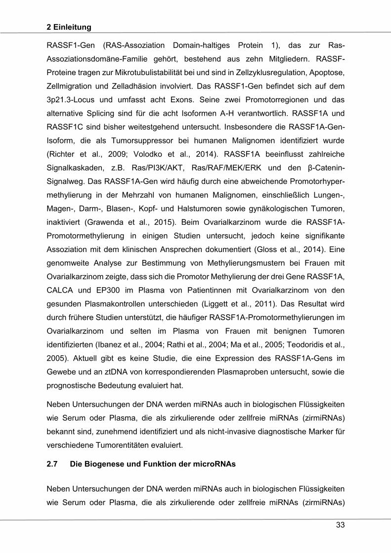
sogenannte „primary-miRNA“, durch die RNA-Polymerase II transkribiert (Abb.2.11).
Die primary-miRNA besitzt, ähnlich zur messengerRNA (mRNA), einen 5`7-Methyl-
Guanosin-Cap und einen 3` poly(A)-Schwanz (Zeng et al., 2006).
Abb.2.11 Prozessierung und Reifung der miRNA (Kuhlmann et al., 2012)
Die Bildung einer „Haarnadelstruktur“ mit einem doppelsträngigen Strang, terminalen
Loop und flankierenden einzelsträngigen Bereichen wird aufgrund der
Sequenzbeschaffenheit gebildet und als pri-miRNA bezeichnet. Für die Biogenese der
miRNA sind die Enzyme DROSHA und DICER essentiell, indem Drosha mit seiner
regulatorischen Untereinheit PASHA als Endonuklease die flankierenden RNA-
Sequenzen der primary-RNA spaltet und dadurch das weitere Vorläufermolekül, die
„precurser-miRNA“ entsteht. Alle anschließenden Reifungsschritte erfolgen im
Zytoplasma der Zelle, indem als erster Schritt die Endonuklease DICER zum Einsatz
kommt und die Entfernung der Loop-Sequenz aus der Haarnadelstruktur durchführt
(Billy et al., 2001). Anschließend geht der „mature miRNA-Strang“ eine stabilisierende
2 Einleitung
35
Interaktion mit einem Ribonukleoproteinkomplex, dem „RNA-induced-silencing-
complex“ (RISC-Komplex), ein. Der komplimentäre Strang, die „passenger RNA“, wird
degradiert. Die mature miRNAs wirken als transkriptionelle Repressoren, indem sie
durch die Bindung an der 3´untranslatierten Region (3`UTR) einer mRNA, die
Genregulation inhibieren. Dieser Prozess ist auch als Konzept der negativen
Genregulation bekannt. Das RISC-Protein verfügt über verschiedene Möglichkeiten,
die Translation der mRNA zu verhindern, indem der Grad an Komplementarität
zwischen der miRNA und dem 3´UTR der Ziel-mRNA eine essentielle Wirkung auf die
Inhibition hat. Dabei gilt das Credo, je höher die Komplementarität, desto höher ist die
Wahrscheinlichkeit, dass der RISC-Komplex die mRNA degradiert. Sollte die
Komplementarität unvollkommen sein, so bindet der RISC-Komplex an die mRNA und
supprimiert die Translation des Zielproteins, ohne einen Degradierungsvorgang zu
starten (Esquela-Kerscher & Slack 2006). Alternativ kann es zu einer Deadenylierung
der mRNA kommen (Wu et al., 2006).
2.8 MicroRNAs und das Ovarialkarzinom
Studien zur Analyse von microRNAs wurden beim Ovarialkarzinom sowohl am
Gewebe, als auch im Plasma und Serum durchgeführt. Es konnte gezeigt werden,
dass miR-141, miR-200a, miR-200b und miR-200c im Vergleich zu gesundem
Ovarialgewebe signifikant überexprimiert waren. Im Gegensatz dazu lagen miR-
125b1, miR-140, miR-145 und miR-199a heruntereguliert vor (Iorio et al., 2007).
Weiterhin waren miR-200a, miR-200b und miR-429 prädiktiv für die
Rezidiventwicklung und ein verkürztes OS (Hu et al., 2009), wobei eine dysregulierte
let-7i Expression bei Patientinnen in der fortgeschrittenen Situation mit einem
verkürztem PFS und der Platinresistenz korrelierte (Yang et al., 2008).
MiRNAs konnten nicht nur im Gewebe, Serum oder Plasma, sondern auch in der
Brustmilch, im Speichel und Urin identifiziert werden (Nakamura et al., 2016).
ZirmiRNA sind sehr stabil. Einerseits durch den pH-Wert und die Temperatur,
andererseits durch RISC-assoziierte Proteine, sogenannte Argonaut-2 Proteine und
extrazelluläre Membranvesikel, wie Exosomen oder Mikrovesikel (Valadi et al., 2007;
Mitchell et al., 2008; Chen et al., 2008; Mause et al., 2010; Arroyo et al., 2011). Die
Extraktion der zirmiRNA kann über die Zentrifugation und über Exosomenisolationskits
verschiedener Firmen erfolgen (Nakamura et al., 2016). Die Detektion der miRNA und
2 Einleitung
36
zirmiRNA erfolgt entweder über NGS oder mittels Mikroarrays, ein technisches
Verfahren, um die Aktivität und Identifikation von bestimmten Genen zu detektieren,
um umfassende Profile von miRNA und zirmiRNA zu erhalten. Das diagnostische
Potential der zirmiRNA konnte in wenigen Studien gezeigt werden. Die Serumanalyse
von 180 Ovarialkarzinompatientinnen ergab, im Vergleich zu 66 Normalspendern, eine
Herunterregulation von miR-25 und miR-93, während miR-7 und miR-429
überexprimiert vorlagen (Meng et al., 2015). Eine andere Studie detektierte im Plasma
von 33 Ovarialpatientinnen eine erhöhte miR-200b-Expression (Kapetanakis et al.,
2015).
Bezüglich der prognostischen Bedeutung korrelierten miR-21; miR-221; miR-23b;
miR-29b; miR-21; miR-429; miR-141 und miR-200b signifikant mit einem verkürzten
PFS oder OS (Xu et al., 2013; Hong et al., 2013; Vaksman et al., 2014; Meng et al.,
2015; Gao et al., 2015; Kapetanakis et al., 2015). Im Gegensatz dazu war die
verminderte Expression von let-7f; miR-1290; miR-200c und miR-145 signifikant mit
einem verkürzten PFS oder OS assoziiert (Zheng et al., 2013; Shapira et al., 2014;
Gao et al., 2015; Liang et al., 2015).
2 Einleitung
37
2.9 Zielsetzung
Trotz Fortschritten in der Behandlung des Ovarialkarzinoms beträgt die Rezidivrate
etwa 75%. Weiterhin sind generell 15-20% der Patientinnen platinresistent, was
bisher nur retrospektiv erkannt wird. Somit ist dem Ovarialkarzinom eine sehr
schlechte Prognose zuzuordnen und die Etablierung neuer Biomarker zur
Prognoseeinschätzung wäre äußerst wünschenswert. Daher ist es von großem
Interesse, innovative primärtumor- bzw. blutbasierte Biomarkerkonzepte zu
entwickeln, die uns Aufschluss über Prognose, Rezidiventwicklung bzw.
Therapieansprechen geben oder im Sinne eines Therapie-Monitoring genutzt werden
können. Da der Primärtumor nur zu Beginn der Erkrankung zur Verfügung steht, ist
die sogenannte „Liquid Biopsy“ immer mehr im Fokus. Beim Ovarialkarzinom sind in
diesem Zusammenhang DTZ, vor allen Dingen aber ZTZ sowie zirkulierende DNA
und microRNAs vielversprechende Kandidaten.
Vorarbeiten:
Die Arbeitsgruppe um Frau Professorin Kasimir-Bauer konnte schon relevante
Beiträge zur Etablierung neuer Biomarkerkonzepte für das Ovarialkarzinom
publizieren und die Hypothese unterstützen, dass Patientinnenblut im Sinne einer
„Real-Time-Liquid-Biopsy“ genutzt werden könnte. Im Hinblick auf DTZ und ZTZ
wurde die schlechte Prognose der Patientinnen hinsichtlich des PFS und OS schon
belegt (Wimberger et al., 2007, Aktas et al., 2011, Fehm et al., 2013). Insbesondere
ERCC1pos ZTZ zu Beginn der Erkrankung konnten als prädiktiver Marker für ein
verkürztes PFS, OS sowie eine klinische Platinresistenz identifiziert werden
(Kuhlmann et al., 2014). Im Rahmen von DNA/RNA Untersuchungen wurde belegt,
dass LOH (Loss of heterozygoity) von D6S1581, gemessen im Plasma von 63
primären Ovarialkarzinompatientinnen, signifikant mit dem PFS und OS korrelierte.
Nach Therapie korrelierte LOH an D10S1765 mit der Tumorzellstreuung in das KM,
was mit den zuvor erhobenen Daten im Primärtumor assoziierte (Kuhlmann et al.,
2011, 2012). Weiterhin war die Arbeitsgruppe eine der ersten die belegten, dass
zirkulierende, nichtkodierende RNU2-1f im Blut von Patientinnen mit Ovarialkarzinom
im Vergleich zu Normalspendern signifikant erhöht war und persistierende Spiegel mit
einer schlechten Prognose korrelierten (Kuhlmann et al., 2014).
2 Einleitung
38
Aufgabenstellung:
Basierend auf den oben genannten Daten wurden folgende Ziele für die Promotion
formuliert:
Ziele:
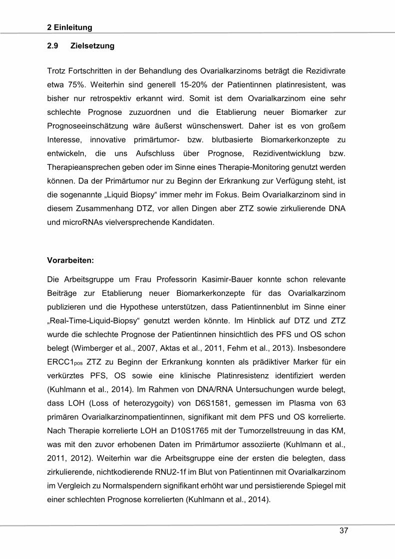
1. Die erste Säule dieser Dissertation beinhaltete die Frage, ob persistierende DTZ
oder DTZ mit Stammzellcharakter im KM den Verlauf der Erkrankung beeinflussen.
Der Nachweis der DTZ unterliegt einer Standardmethode. Nach KM-Aspiration
erfolgt eine Dichtegradientenzentrifugation mit anschließender Immunzytochemie
unter Verwendung des Anti-Zytokeratin-Antikörper A45-B/B3, der die Zytokeratin-
Heterodimere 8/18 und 8/19 detektiert. Hierbei handelt es sich um ein FAB-
Fragment, um unspezifische Bindungen an Lymphozyten zu vermeiden. Der
Nachweis erfolgt mit Neufuchsinrot (Abb.2.12). Während beim Mammakarzinom
eine Reihe von Charakteristika der DTZ publiziert wurden, gibt es zum
Ovarialkarzinom kaum Daten (Schindlbeck et al., 2016).
Abb.2.12 Schematische Darstellung der Detektion von DTZ
Für den Nachweis des Stammzellcharakters der DTZ sollte eine vierfach
Immunfluoreszenzfärbung etabliert werden, die folgende Marker beinhaltete:
Zytokeratin (epithelialer Marker), LIN-28 und SOX-2 als Stammzellmarker, die mit
dem Ovarialkarzinom assoziiert werden (Peng et al., 2010; Bareiss et al., 2013;
Pham et al., 2013), CD34 zum Ausschluss von hämatopoeitischen Stammzellen
und CD45 als Leukozytenmarker. Zur Etablierung der Methoden wurden die
ovariellen Zelllinien OVCAR-3 sowie die Leukämiezelllinie Kasumi-1 verwendet. Die
Methode wurde dann bei KM-Präparaten von Ovarialkarzinompatientinnen vor und
nach Therapie angewendet.
2. Da KM Punktionen sehr invasiv sind und besonders nach Therapie häufig nicht
akzeptiert werden, stand für weitere Fragestellungen das Blut als „Liquid Biopsy“ im
Monoklonaler
Antikörper A45-B/B3 Zytokeratin positive
DTZ
2 Einleitung
39
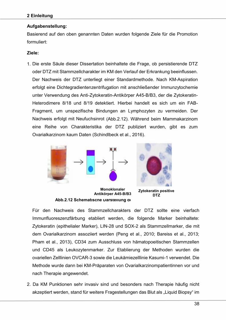
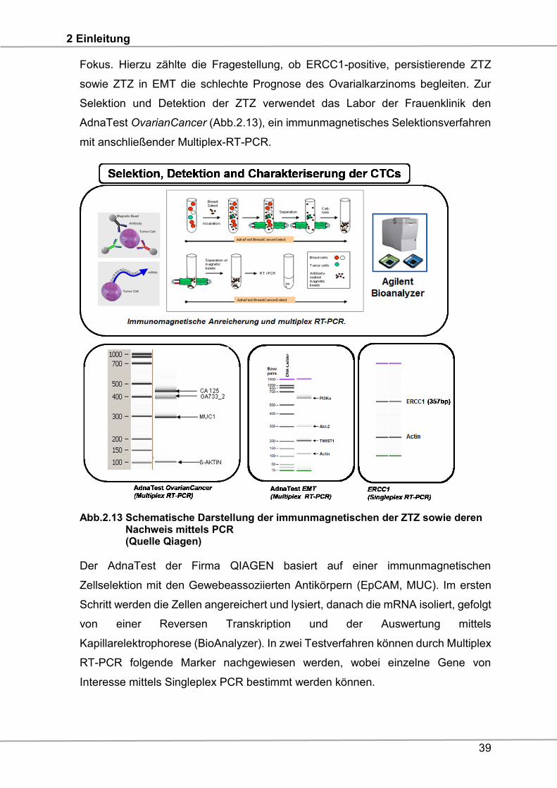
Fokus. Hierzu zählte die Fragestellung, ob ERCC1-positive, persistierende ZTZ
sowie ZTZ in EMT die schlechte Prognose des Ovarialkarzinoms begleiten. Zur
Selektion und Detektion der ZTZ verwendet das Labor der Frauenklinik den
AdnaTest OvarianCancer (Abb.2.13), ein immunmagnetisches Selektionsverfahren
mit anschließender Multiplex-RT-PCR.
Abb.2.13 Schematische Darstellung der immunmagnetischen der ZTZ sowie deren Nachweis mittels PCR (Quelle Qiagen)
Der AdnaTest der Firma QIAGEN basiert auf einer immunmagnetischen
Zellselektion mit den Gewebeassoziierten Antikörpern (EpCAM, MUC). Im ersten
Schritt werden die Zellen angereichert und lysiert, danach die mRNA isoliert, gefolgt
von einer Reversen Transkription und der Auswertung mittels
Kapillarelektrophorese (BioAnalyzer). In zwei Testverfahren können durch Multiplex
RT-PCR folgende Marker nachgewiesen werden, wobei einzelne Gene von
Interesse mittels Singleplex PCR bestimmt werden können.
2 Einleitung
40
1. AdnaTest OvarianCancer (CA-125, EpCAM, MUC)
ERCC1 wird als single-plex PCR durchgeführt
2. AdnaTest EMT (PI3Kα, AKT-2, TWIST)
Im Vordergrund der Dissertation stand die Rekrutierung von Blut vor und nach
abgeschlossener Platintherapie, um explizit die Rolle von ERCC1pos ZTZ nach
Therapie sowie deren Persistenz zu evaluieren. Die Rolle von ZTZ in EMT beim
Ovarialkarzinom wurde bisher noch von keiner Arbeitsgruppe untersucht. Bekannt
war bisher nur, dass Twist als Transkriptionsfaktor im Gewebe der Ovarial-
karzinome von Bedeutung ist. (Kajiyama et al., 2006; Hosono et al., 2007).
3. Zur Evaluierung von small RNA Profilen sollte im Plasma/Serum von Patientinnen
mit Ovarialkarzinom vor Therapie zunächst eine Methode etabliert/optimiert werden,
die mittels NGS das Profil platinsensitiver und platinresistenter Patientinnen
darstellt, sodass im Anschluss ausgewählte small RNA Kandidaten durch RT-qPCR
validiert werden können. Dieses Projekt wurde in Kooperation mit Herrn Dr.
Kuhlmann aus der Frauenklinik in Dresden und Herrn Dr. Michael Reuter aus dem
Fraunhofer Institut in Leipzig durchgeführt.
Nebenziel:
Mit der Arbeitsgruppe von Frau Prof. Dr. Evi Lianidou in Athen besteht eine langjährige
Kooperation. Im Rahmen der Thematik blutbasierter Biomarker stand die Methylierung
zirkulierender Tumor-DNA im Vordergrund, wobei auch Vergleichsanalysen im
Primärtumor durchgeführt wurden. Hier wurde eine entsprechende Validierungs-
kohorte Tumor/Plasma aus der Frauenklinik zur Verfügung gestellt.
3 Publikationen
41
3 Publikationen
Analysis of disseminated tumor cells before and after platinum based
chemotherapy in primary ovarian cancer. Do stem cell like cells predict
prognosis?
Issam Chebouti1*, Christina Blassl2*, Pauline Wimberger3, Hans Neubauer2, Tanja
Fehm2, Rainer Kimmig1, Sabine Kasimir-Bauer1
1 Dep. of Gynecology and Obstetrics, University Hospital Essen (Germany), 2 Dep. of
Gynecology and Obstetrics, University Hospital Düsseldorf (Germany), 3 Dep. of
Gynecology and Obstetrics, Carl-Gustav-Carus University, TU Dresden (Germany)
* shared first authorship
Key words:
Disseminated tumor cells, bone marrow, stem cells, primary ovarian cancer
Corresponding author
Issam Chebouti
Department of Gynecology and Obstetrics
University of Duisburg-Essen
Hufelandstrasse 55
D-45122 Essen
Phone ++ 49 – 201-723-2340
Fax ++49 – 201-723-3938
Email:[email protected]
3 Publikationen
42
Abstract
Background: We recently reported that the presence of disseminated tumor cells
(DTCs) in the bone marrow (BM) of primary ovarian cancer patients (POC pts)
correlated with reduced progression free survival (PFS) and overall survival (OS). Here
we analyzed whether the negative prognostic influence was related to DTC persistence
after platinum based chemotherapy and/or due to DTCs associated with stem cell
character.
Patients and Methods: 79 POC pts were studied for DTCs before therapy (BT) and
after therapy (AT) using immunocytochemistry. Eight pts harboring at least five DTCs
AT were further analyzed on two additional slides by four-fold immunofluorescence
staining for DAPI, Cytokeratin (CK), SOX-2 or LIN-28, CD45 and CD34 (Cy5). A stem-
like tumor cell was classified as Dapipos, CD45neg, CD34neg, SOX-2pos/LIN-28pos and
CKpos or CKneg.
Results: DTCs were detected in 33/79 pts (42%) before and in 32/79 pts (41%) AT.
Persistent DTCs were found in 13 pts, 20 pts were only positive BT, 19 pts AT and 27
pts had no DTCs. Whereas the presence of DTCs BT significantly correlated with
reduced OS (p= 0.02), pts initially DTCneg BT but DTCpos AT had a significantly shorter
PFS (p=0.03). DTC persistence resulted in a shorter PFS and OS reaching borderline
significance (p=0.06; p=0.07). LIN-28-and SOX-2 positive cells were detected in all
eight pts AT.
Conclusion: Stem cell associated proteins are expressed in DTCs that are present AT
and their presence seem to be correlated with a worse outcome. Additional therapeutic
regimens may be necessary to eliminate these cells.
3 Publikationen
43
Introduction
Ovarian cancer is the fifth leading cause of all cancer related deaths in Europe and the
United States and most tumors are diagnosed in an advanced stage with poor
prognosis for the patients (1). Conventional therapy is based on an initial debulking
surgery aiming at macroscopic complete resection combined with subsequent
platinum- and paclitaxel -based chemotherapy (2). Postoperative residual tumor is one
of the most important prognostic factors in advanced ovarian cancer (3,4,5).
It is hypothesized that cancer malignancy and metastasis are driven by a small
subgroup of highly tumorigenic cells within the tumor, called metastasis initiating cells
(MIC). These cells have the ability to self-renew, enhance tumorigenesis and are often
found to be drug resistant (6). The presence of such a small population, often referred
to as cancer stem cells (CSC), has been confirmed in ovarian cancer cell lines as well
as in tumor tissue (7,8). Their amount is increased in chemotherapy resistant ovarian
cancer cell lines (7) and they are believed to contribute to an aggressive behavior of
epithelial ovarian cancer (9). The pluripotency associated stem cell factors SOX2 (sry
related) and LIN-28 have been found to be expressed in ovarian cancer cell lines and
tissue (10,11,12). Bareiss et al., showed that SOX2 expression is a CSC marker in
serous ovarian carcinomas (SOC) and can induce CSC properties (11). In addition,
SOX2 was reported to enhance migration and invasion of ovarian cancer cells (13).
Importantly, SOX2 overexpression was shown to be a poor prognostic marker in
ovarian cancer (14) and also shown to be involved in taxane resistance (15,16).
In ovarian cancer, the primary tumor usually metastasizes to the peritoneum, but a
variety of studies including ours indicate that tumor cells frequently disseminate into
the bone marrow (BM). Disseminated tumor cells (DTCs) in the BM are detected in
20% to 60% of cases before the onset of platinum-based chemotherapy depending on
the method of detection used. Their prognostic relevance with regard to reduced
progression free survival (PFS) and overall survival (OS) has previously been
demonstrated (17,18,19,20,21). In addition, we demonstrated that patients with a
marked increase of DTCs after platinum-based chemotherapy showed a significantly
reduced PFS (22).
Based on the studies mentioned above, there is increasing evidence that DTCs could
reflect cancer progression. Thus, DTCs could be used as novel targets for additional
therapeutic strategies. In this study, we analyzed whether our previously reported
negative prognostic influence of DTCs with regard to reduced PFS and OS 1) was
related to the persistence of DTCs after platinum based chemotherapy and/or 2) might
3 Publikationen
44
have arisen from a cellular phenotype showing stem cell characteristics.
Results
Detection of DTCs
Before therapy (BT), DTCs were detected in 33/79 patients (42%) with a median
number of 4 DTCs (range 1-37). After therapy (AT), 32/79 patients (41%) were positive
for DTCs (median cell number of 8 cells (range 1-100) (Table 1). DTCs were found in
13 patients BT and AT, in 20 patients only BT and in 19 patients only AT, respectively.
DTCs were not detected in samples taken BT or AT from 27 patients (Table 2).
Prognostic significance of DTCs
After a median follow up time of 62 months (range 10-128 months), 44 patients (56%)
were still alive and 33 patients (42%) had died. The median follow-up time for PFS was
15 months (range 4-87 months) resulting in 53 (67%) relapses while 25 patients (32%)
had no relapse (Table 1). The presence of DTCs BT significantly correlated with
reduced OS (p=0.02) and patients initially DTCneg BT but DTCpos AT had a significant
shorter PFS (p=0.03) (Table 2 and Fig.1). The persistence of DTCs resulted in a
shorter PFS and OS reaching borderline significance (p=0.06; p=0.07).
Evaluation of Lin28- and Sox-2-positive cells
Staining of patient samples is shown in Fig. 2-5. Controls are shown in Supplementary
Fig. 1-3. A DTC was classified as a stem-like tumor cell if it had the following staining
characteristics: Dapipos, CD45neg, CD34neg, SOX-2pos/LIN-28pos and CKpos or CKneg
(Fig. 2-5). The Kasumi cell line was used to establish CD34 expression (Suppl. Fig. 1)
and BM samples from healthy donor patients for CD34- and CD45-expression (Suppl.
Fig. 2 and 3).
Detection of LIN-28- and SOX-2-positive cells
DTCs from 10 patients were analyzed BT and AT for SOX-2 and LIN-28 positive cells
AT (Table 3; columns 1 and 2; Suppl. Table 1). 8/10 patients had at least five DTCs as
detected by immunocytochemistry using A45/B-B3. In addition, 2/10 patients (patient
2 and 5) were DTCneg AT but DTCpos BT. As apparent from Table 3, AT CKpos/LIN-28pos
cells were detected in 9/10 patients [median 2 cells (range 1-5)] and CKneg/LIN-28pos
cells in 7/10 patients [median 3 cells (range 1-11)], respectively. CKpos/SOX-2pos cells
were detected in 6/8 patients [median 2 cells (range-0-4)] and CKneg/SOX-2pos cells
3 Publikationen
45
were found in 7/8 patients [median 4 cells (range 1-11)]. Patients two and five, who
were characterized as DTCneg AT by immunocytochemistry but were positive BT (37
and 6 DTCs, respectively) were included in our analysis for stem cell- associated
markers. Interestingly, these two patients harbored 1-2 LIN-28pos and SOX-2pos cells
in their BM AT. Thus, we evaluated LIN-28-/SOX-2-positive cells BT in cases vice
versa, DTCneg BT but DTCpos AT (patients 1, 3 and 4) as well as in patient number 6
with persistent DTCs. As shown in Table 3, in patients who switched initially DTCneg
before but becoming DTCpos AT (patients 1, 3 and 4), in patients who switched from
being DTCpos before but becoming DTCneg AT (patients 2 and 5) as well as in patient
number 6 with persistent DTCs (patient 6) a few LIN-28 as well as SOX-2-positive cells
were present in BM even BT.
Discussion
To the best of our knowledge, this is the first study showing that DTCs, present after
platinum based chemotherapy in primary ovarian cancer patients show stem cell
characteristics. Furthermore, although p values reached borderline significance, these
cells might be associated with worse outcome which finally has to be proven in a bigger
patient cohort.
The rate of DTC detection in primary ovarian cancer before the administration of
platinum-based chemotherapy has been reported to be 20% to 60%, depending on the
method of detection used. Furthermore, their presence has been associated with
worse outcome (17,18,19,20,21,22). The lack of significant correlation between DTCs
and clinical outcome reported by other investigators may be due to their use of different
antibodies for detection of DTCs (23,24).
In this study, DTCs were present in the BM AT in 41% of the patients which is in
accordance with our earlier data which also demonstrated that DTCs, still present AT,
were non-apoptotic and their marked increase was associated with a significantly
reduced PFS (22). These findings suggest that the BM may to be a temporary homing
site for isolated tumor cells, where they can persist and potentially induce recurrence
of the disease. Analyzing 79 patients BT and AT, we confirm the negative prognostic
influence of DTC detection with regard to OS (20). We observed persistent DTCs in
16% of the patients which was associated with a shorter PFS and OS, reaching
borderline significance. Interestingly, 24% of the patients that were initially
characterized as DTCneg BT and converted to DTCpos AT had a significantly shorter
PFS. The negative prognostic influence of these cells could be in alliance with a
3 Publikationen
46
currently discussed hypothesis that some DTCs may have cancer stem cell features
and may be the active source of metastatic spread in primary tumors, in addition to
resistance to various chemotherapeutic agents and radiotherapy (25). Two studies
have confirmed a putative stem cell phenotype among DTCs in breast cancer patients
(26,27). Here, we demonstrate that presence of DTCs that persist AT express the stem
cell markers Lin-28 and/or SOX-2. We show that stem cell-like cells were present
before the administration of chemotherapy. These findings may explain the
significantly shorter PFS of patients who changed from DTCneg BT to DTCpos AT. In
addition, patients characterized as DTCneg AT also harbored some Lin-28 and/or SOX-
2 positive cells in their BM which may be responsible for a worse outcome. Until now,
tumor stem cells have only been analyzed in ovarian tumor tissue, but not in DTCs. In
this regard, previous studies have shown that LIN-28, SOX-2 as well as OCT-4 play a
major role in carcinogenesis (10,11,12). Wang et al., reported that SOX-2 targets SRC
Kinase, a non-receptor tyrosine kinase that increases cell migration, invasion and
adhesion of serous ovarian carcinoma cells (13). Inhibition of either LIN-28 or Oct-4
expression decreases cell viability. The combined repression of both LIN-28 and Oct-
4 results in synergistic inhibition of cancer cell growth and survival of ovarian cancer
cell lines (9). Expression of SOX-2 has been investigated by immunohistochemistry
analysis of normal ovarian epithelial, serous and mucinous cystadenoma and
cystadenomacarcinoma specimens (28) and LIN-28 was overexpressed in different
epithelial tumors including breast, lung, colon and ovarian cancer (29). Furthermore,
SOX-2 may be crucial for the development of chemotherapy resistance. Yang et al.,
analyzed SOX-2 expression in clinical tissue samples and ovarian cancer cell lines
using immunohistochemistry and real-time PCR and demonstrated that SOX-2 was
overexpressed in paclitaxel-resistant cells (30). In ovarian cancer patients receiving
taxanes, expression of SOX-2 was shown to be correlated with chemotherapy
resistance and a shorter PFS whereas patients receiving non-taxane based
chemotherapy showed no significant response influence (31). Since the patients in our
study have also received a combined therapy with paclitaxel and carboplatin, it may
be possible that SOX-2 expression on DTCs was associated with chemotherapy
resistance. However, mechanisms associated with chemotherapy resistance in
ovarian cancer still remain unclear. Functionally, primary platinum-resistance, defined
as platinum-free treatment interval of less than 6 months observed in up to 20% of
ovarian cancer patients, can be the result of either increased tolerance towards DNA-
platinum-adducts or enhanced DNA-repair capacity of tumor cells (32,33,34). In this
3 Publikationen
47
context, we recently demonstrated that ERCC1pos (excision-repair cross-
complementing rodent repair deficiency, complementation group 1 nuclease)
circulating tumor cells (CTCs) constituted an independent predictor, not only for OS
but also for PFS in our ovarian cancer patients. Most interestingly, the presence of
ERCC1pos CTC at primary diagnosis was an independent predictor for platinum-
resistance whereas ERCC1-expression in the corresponding primary tumor tissue
predicted neither platinum-resistance, nor prognosis (35). Consequently, assuming
that CTCs must be spread into the circulation from existing pools in secondary organs,
e.g. the BM, one might speculate the ERCC1pos DTCs also exist and contribute to
platinum resistance. Interestingly, DTCs present BT in our patients significantly
correlated with clinical platinum resistance (data not shown).
In this study, we also detected CKpos/SOX-2pos (LIN-28pos) as well as CKneg/SOX-2pos
(LIN-28pos) cells in all patients. We assume that two different cell types with expression
of stem cell associated proteins may have been detected. It has been described that
tumor cells undergo phenotypic changes, known as epithelial-mesenchymal transition
(EMT), which allow them to migrate to sites of metastasis without being eliminated by
conventional treatment (36). Thus, CKneg/SOX-2pos (Lin-28pos) cells might result from
EMT while the CKpos/SOX-2pos (LIN-28pos) epithelial phenotype may have remained
unchanged.
Taking all these considerations into account, additional therapeutic strategies will be
required to target signaling pathways concerning CSC. These studies will include
mTOR inhibitors, acting downstream of the PI3K/AKT pathway (37), salinomycin (38)
or a new synthetic curcumin analogue against ALDH1 and GSK-3ß (39). Finally,
approaching the tumor microenvironment, such as interrupting the immune cells and
cytokines (e.g. IL-6, IL-8) as well as the immune checkpoints (PD1/PDL1) may provide
additional new tools for immunological killing of cancer stem cells (40,41,42)
Conclusion and limitation of the study: The cohort of our patients is probably too small
to draw the final conclusion that a significant selection of stem cell marker-positive
DTCs occurs during chemotherapy. Consequently, the results presented here should
be viewed as a “proof of principle”, that DTCS with stem cell characteristics exist
among DTCs that are present BT and persist AT. To the best of our knowledge, we
are the only group that has a collection of BM cells harvested from primary ovarian
cancer patients AT. In this regard, 79 paired samples from patients who consented to
allow collection of their BM under local anesthesia AT for research purposes is a
unique collection and would be difficult to achieve high patient numbers. Furthermore,
3 Publikationen
48
based on the number of residual slides and methodological requirements, we only
analyzed two stem cell markers. Ongoing studies will include other stem cell markers,
such as OCT4 as well as resistance marker to finally elucidate the prognostic relevance
of these cells.
Patients and Methods
Patient characteristics
79 patients with primary ovarian cancer who presented at the Department and
Gynecology and Obstetrics, University Hospital Essen between February 2004 and
January 2010 were included in this analysis. Patient characteristics are documented
in Table 1. The mean age was 60 years (range 26-86 years), the median follow-up
time was 62 months (10-128 months) for OS and 15 months (4-87 months) for PFS.
Written Informed consent was obtained from all patients and the study was approved
by the Local Ethics Committee (05-2870). Tumors were classified according to the
WHO classification of tumors of the female genital tract. Grading was conducted using
the grading system proposed by Silverberg (43) and tumor staging was classified
according to the Fédération Internationale de Gynécology et d'Obstétrique (FIGO
2009). The entire study population underwent primary radical surgery. Total abdominal
hysterectomy, bilateral salpingo-oophorectomy, infragastric omentectomy, peritoneal
stripping was performed and in addition to pelvic and para-aortic lymphadenectomy, if
macroscopic complete resection was achieved. The most important aim of surgery was
to achieve macroscopic complete tumor resection. Radical pelvic and para-aortic
lymphadenectomy were only performed if complete tumor resection was achieved
intraperitoneally following actual guidelines (www.ago-ovar.de). All patients received
at least six cycles of carboplatinum AUC 5 and paclitaxel 175 mg/m2. Tumors were
clinically defined as platinum-resistant if they recurred within six months after the
completion of platinum-based chemotherapy.
Cell lines
The human ovarian cancer cell line OVCAR-3 and the Kasumi-1 cell line were
purchased from the American Type Culture Collection (ATCC, Manassas, VA, USA)
and cultured in RPMI 1640 containing 10% (20% for Kasumi-1) fetal calf serum and
1% (100 U/ml) Penicillin-Streptomycin (Gibco™ by Thermo Fisher Scientific, Waltham
MA, US) . Cells were grown at 37 °C in a humidified atmosphere with 5% CO2.
3 Publikationen
49
Detection of DTCs
Between 10 and 20 ml BM were aspirated from the anterior iliac crests and processed
within 24 hours. DTC selection and detection was performed based on the
recommendations for standardized tumor cell detection (44). Details of the staining
procedure and cell detection have been described elsewhere (22). Briefly, BM cells
were isolated from heparinized BM (5000 U/ml BM) by Ficoll-Hypaque density gradient
centrifugation (density 1.077 g/mol; Pharmacia, Freiburg, Germany) at 400 x g for 30
min. Slides were analyzed for DTCs by immunocytochemistry using the pan-
cytokeratin antibody A45-B/B3. Microscopic evaluation of the slides (1 x 10⁶
mononuclear cells per slide) was carried out using the ARIOL system (Applied
Imaging) according to the ISHAGE evaluation criteria (45).
Detection of LIN-28- and SOX-2-positive cells
LIN-28- and SOX-2 positive cells were analyzed separately on additional slides of the
same patients harboring at least five DTCs as detected by immunocytochemistry using
the A45B-B3. Four-fold immunofluorescence staining was established using the
OVCAR-3 cell line spiked into blood of healthy donors. Since CD34-positive normal
hematopoietic stem cells comprise 1.5% of marrow mononuclear cells (46), we
included CD34 in our analysis to exclude false-positive results. CD34 was analysed
using the Kasumi-1 cell line since BM of healthy donors was difficult to obtain and only
available from one donor (Suppl. Fig. 1).
Slides were fixed with 4% Paraformaldehyde for 10 min, permeabilized with 0.1%
Triton-X-100 for 15 min and subsequently washed with TBS and Triton-X-100 three
times for five min. Slides were stained with SOX-2 (Anti-human SOX-2, 1:50, R&D
Systems, USA), LIN-28 (LIN-28, 1:350, Rabbit polyclonal ab46020, Abcam, UK), C11
(anti-PAN-Cytokeratin, 1:400, FITC-labelled, GeneTex, USA), CD34 (Alexa Flour anti-
human 647, 1:100,Cy5 labelled, Biolegend, USA) and CD45 (Alexa Fluor 647;sc1178
Santa Cruz, USA) incubated in a wet chamber for one hour at room temperature.
Subsequently, slides were incubated with DAPI [pre diluted in Phosphate Buffered
Saline (1:250) and further diluted in AB diluent (1:20, Dako, Germany)], and TRITC-
labelled donkey anti-goat 594 (SOX-2), donkey anti-rabbit 594 (LIN-28) (both1:100,
Invitrogen, USA) under the same conditions followed by three washing steps for five
minutes. Moreover, negative controls for primary antibodies were prepared by staining
the spiked OVCAR-3 cell line with the secondary antibodies for 30 minutes under the
same conditions. The slides were mounted with Dako fluorescent mounting medium
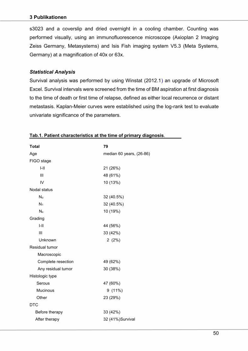
3 Publikationen
50
s3023 and a coverslip and dried overnight in a cooling chamber. Counting was
performed visually, using an immunofluorescence microscope (Axioplan 2 Imaging
Zeiss Germany, Metasystems) and Isis Fish imaging system V5.3 (Meta Systems,
Germany) at a magnification of 40x or 63x.
Statistical Analysis
Survival analysis was performed by using Winstat (2012.1) an upgrade of Microsoft
Excel. Survival intervals were screened from the time of BM aspiration at first diagnosis
to the time of death or first time of relapse, defined as either local recurrence or distant
metastasis. Kaplan-Meier curves were established using the log-rank test to evaluate
univariate significance of the parameters.
Tab.1. Patient characteristics at the time of primary diagnosis.
Total 79
Age median 60 years, (26-86)
FIGO stage
I-II 21 (26%)
III 48 (61%)
IV 10 (13%)
Nodal status
No 32 (40.5%)
N1 32 (40.5%)
Nx 10 (19%)
Grading
I-II 44 (56%)
III 33 (42%)
Unknown 2 (2%)
Residual tumor
Macroscopic
Complete resection 49 (62%)
Any residual tumor 30 (38%)
Histologic type
Serous 47 (60%)
Mucinous 9 (11%)
Other 23 (29%)
DTC
Before therapy 33 (42%)
After therapy 32 (41%)Survival
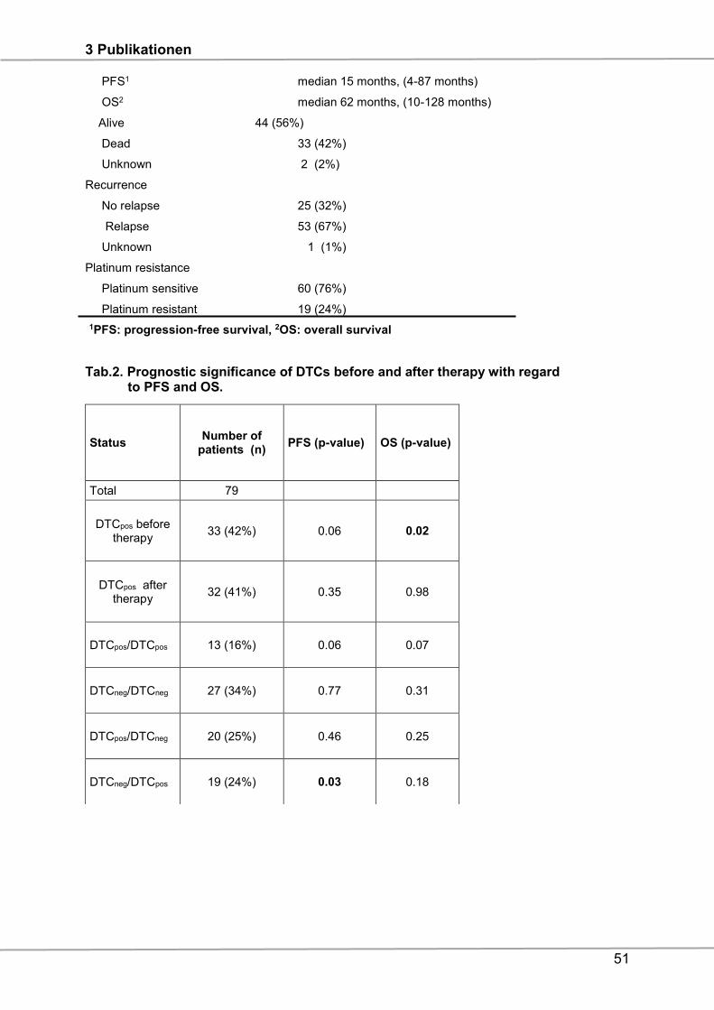
3 Publikationen
51
PFS1 median 15 months, (4-87 months)
OS2 median 62 months, (10-128 months)
Alive 44 (56%)
Dead 33 (42%)
Unknown 2 (2%)
Recurrence
No relapse 25 (32%)
Relapse 53 (67%)
Unknown 1 (1%)
Platinum resistance
Platinum sensitive 60 (76%)
Platinum resistant 19 (24%)
1PFS: progression-free survival, 2OS: overall survival
Tab.2. Prognostic significance of DTCs before and after therapy with regard to PFS and OS.
Status Number of
patients (n) PFS (p-value) OS (p-value)
Total 79
DTCpos before therapy
33 (42%) 0.06 0.02
DTCpos after therapy
32 (41%) 0.35 0.98
DTCpos/DTCpos 13 (16%) 0.06 0.07
DTCneg/DTCneg 27 (34%) 0.77 0.31
DTCpos/DTCneg 20 (25%) 0.46 0.25
DTCneg/DTCpos 19 (24%) 0.03 0.18
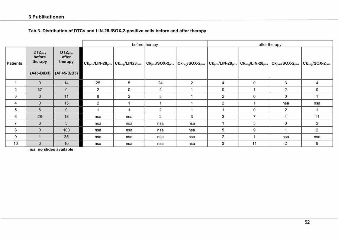
3 Publikationen
52
Tab.3. Distribution of DTCs and LIN-28-/SOX-2-positive cells before and after therapy.
nsa: no slides available
before therapy after therapy
Patients
DTZpos before therapy
DTZpos after
therapy Ckpos/LIN-28pos Ckneg/LIN28pos Ckpos/SOX-2pos Ckneg/SOX-2pos Ckpos/LIN-28pos Ckneg/LIN-28pos Ckpos/SOX-2pos Ckneg/SOX-2pos
(A45-B/B3) (AF45-B/B3)
1 0 14 25 5 24 2 4 0 3 4
2 37 0 2 0 4 1 0 1 2 0
3 0 11 8 2 5 1 2 0 0 1
4 0 15 2 1 1 1 2 1 nsa nsa
5 6 0 1 1 2 1 1 0 2 1
6 28 18 nsa nsa 2 3 3 7 4 11
7 0 5 nsa nsa nsa nsa 1 3 0 2
8 0 100 nsa nsa nsa nsa 5 9 1 2
9 1 35 nsa nsa nsa nsa 2 1 nsa nsa
10 0 10 nsa nsa nsa nsa 3 11 2 9
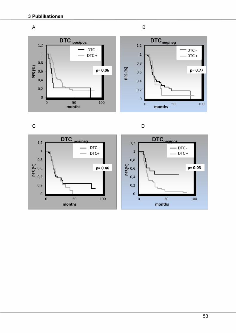
3 Publikationen
53
A B
C D
0
0,2
0,4
0,6
0,8
1
1,2
0 50 100
PFS
(%
)
months
DTC pos/pos
0
0,2
0,4
0,6
0,8
1
1,2
0 50 100
PFS
(%
)
months
DTCneg/neg
DTC -DTC +
p= 0.77
DTC -DTC +
0
0,2
0,4
0,6
0,8
1
1,2
0 50 100
PFS
(%
)
months
DTC pos/neg
0
0,2
0,4
0,6
0,8
1
1,2
0 50 100
PFS
(%)
months
DTC -DTC +
p= 0.03
DTCneg/pos
DTC -
DTC +
p= 0.06
DTC -
DTC+
p= 0.46
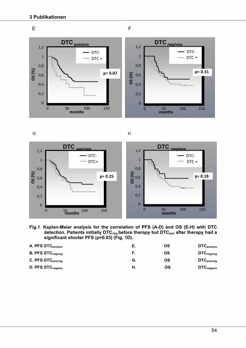
3 Publikationen
54
E F
Fig.1. Kaplan-Meier analysis for the correlation of PFS (A-D) and OS (E-H) with DTC detection. Patients initially DTCneg before therapy but DTCpos after therapy had a significant shorter PFS (p=0.03) (Fig. 1D).
A. PFS DTCpos/pos E. OS DTCpos/pos
B. PFS DTCneg/neg F. OS DTCneg/neg
C. PFS DTCpos/neg G. OS DTCpos/neg
D. PFS DTCneg/pos H. OS DTCneg/pos
0
0,2
0,4
0,6
0,8
1
1,2
0 50 100 150
OS
(%)
months
DTC pos/pos
0
0,2
0,4
0,6
0,8
1
1,2
0 50 100 150
OS
(%)
months
DTC neg/neg
p= 0.31
0
0,2
0,4
0,6
0,8
1
1,2
0 50 100 150
OS
(%)
months
DTC pos/neg
0
0,2
0,4
0,6
0,8
1
1,2
0 50 100 150
OS
(%)
months
DTC neg/pos
p= 0.18
DTC- DTC +
DTC-
DTC +
p= 0.07
DTC-
DTC+
DTC-
DTC +
p= 0.25
G H
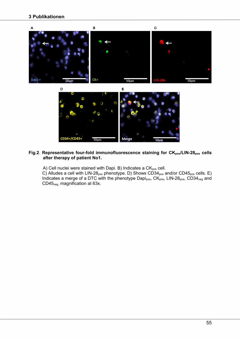
3 Publikationen
55
Fig.2. Representative four-fold immunofluorescence staining for CKpos/LIN-28pos cells after therapy of patient No1. A) Cell nuclei were stained with Dapi. B) Indicates a CKpos cell. C) Alludes a cell with LIN-28pos phenotype. D) Shows CD34pos and/or CD45pos cells. E) Indicates a merge of a DTC with the phenotype Dapipos, CKpos, LIN-28pos, CD34neg and CD45neg, magnification at 63x.
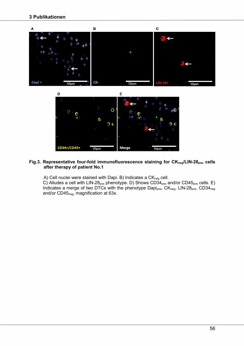
3 Publikationen
56
Fig.3. Representative four-fold immunofluorescence staining for CKneg/LIN-28pos cells after therapy of patient No.1 A) Cell nuclei were stained with Dapi. B) Indicates a CKneg cell. C) Alludes a cell with LIN-28pos phenotype. D) Shows CD34pos and/or CD45pos cells. E) Indicates a merge of two DTCs with the phenotype Dapipos, CKneg, LIN-28pos, CD34neg and/or CD45neg, magnification at 63x.
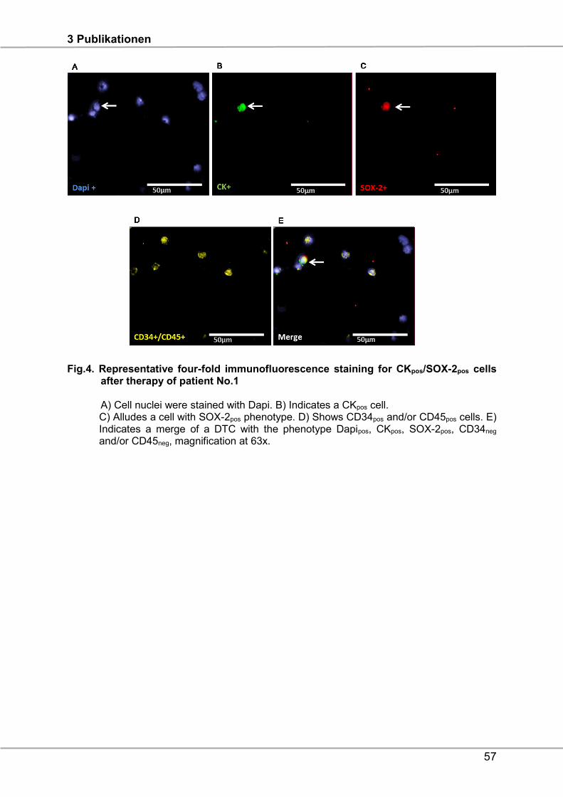
3 Publikationen
57
Fig.4. Representative four-fold immunofluorescence staining for CKpos/SOX-2pos cells after therapy of patient No.1 A) Cell nuclei were stained with Dapi. B) Indicates a CKpos cell. C) Alludes a cell with SOX-2pos phenotype. D) Shows CD34pos and/or CD45pos cells. E) Indicates a merge of a DTC with the phenotype Dapipos, CKpos, SOX-2pos, CD34neg and/or CD45neg, magnification at 63x.
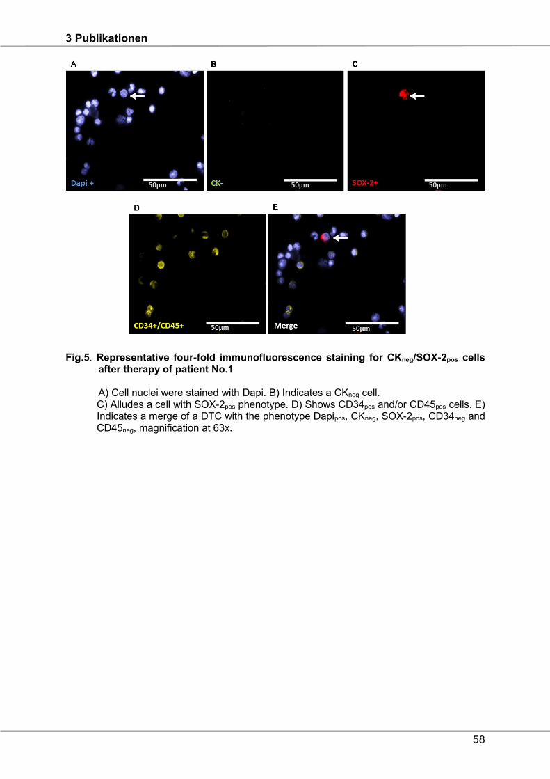
3 Publikationen
58
Fig.5. Representative four-fold immunofluorescence staining for CKneg/SOX-2pos cells after therapy of patient No.1 A) Cell nuclei were stained with Dapi. B) Indicates a CKneg cell. C) Alludes a cell with SOX-2pos phenotype. D) Shows CD34pos and/or CD45pos cells. E) Indicates a merge of a DTC with the phenotype Dapipos, CKneg, SOX-2pos, CD34neg and CD45neg, magnification at 63x.
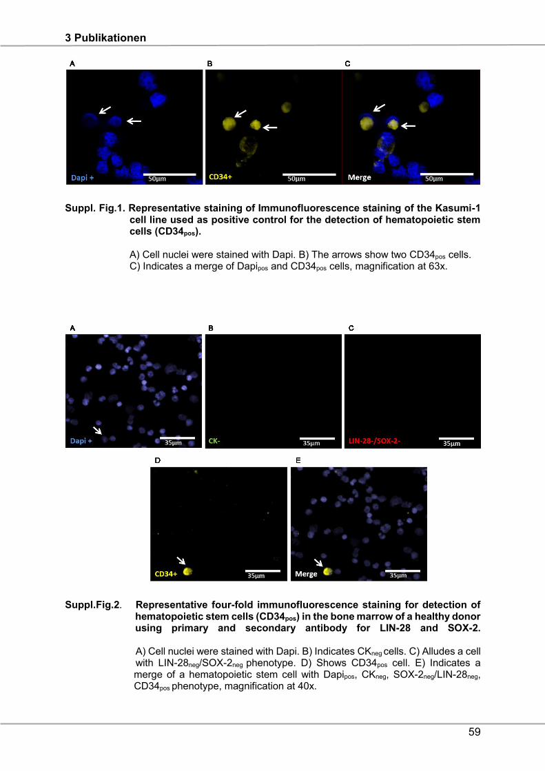
3 Publikationen
59
Suppl. Fig.1. Representative staining of Immunofluorescence staining of the Kasumi-1 cell line used as positive control for the detection of hematopoietic stem cells (CD34pos). A) Cell nuclei were stained with Dapi. B) The arrows show two CD34pos cells. C) Indicates a merge of Dapipos and CD34pos cells, magnification at 63x.
Suppl.Fig.2. Representative four-fold immunofluorescence staining for detection of hematopoietic stem cells (CD34pos) in the bone marrow of a healthy donor using primary and secondary antibody for LIN-28 and SOX-2. A) Cell nuclei were stained with Dapi. B) Indicates CKneg cells. C) Alludes a cell with LIN-28neg/SOX-2neg phenotype. D) Shows CD34pos cell. E) Indicates a merge of a hematopoietic stem cell with Dapipos, CKneg, SOX-2neg/LIN-28neg, CD34pos phenotype, magnification at 40x.
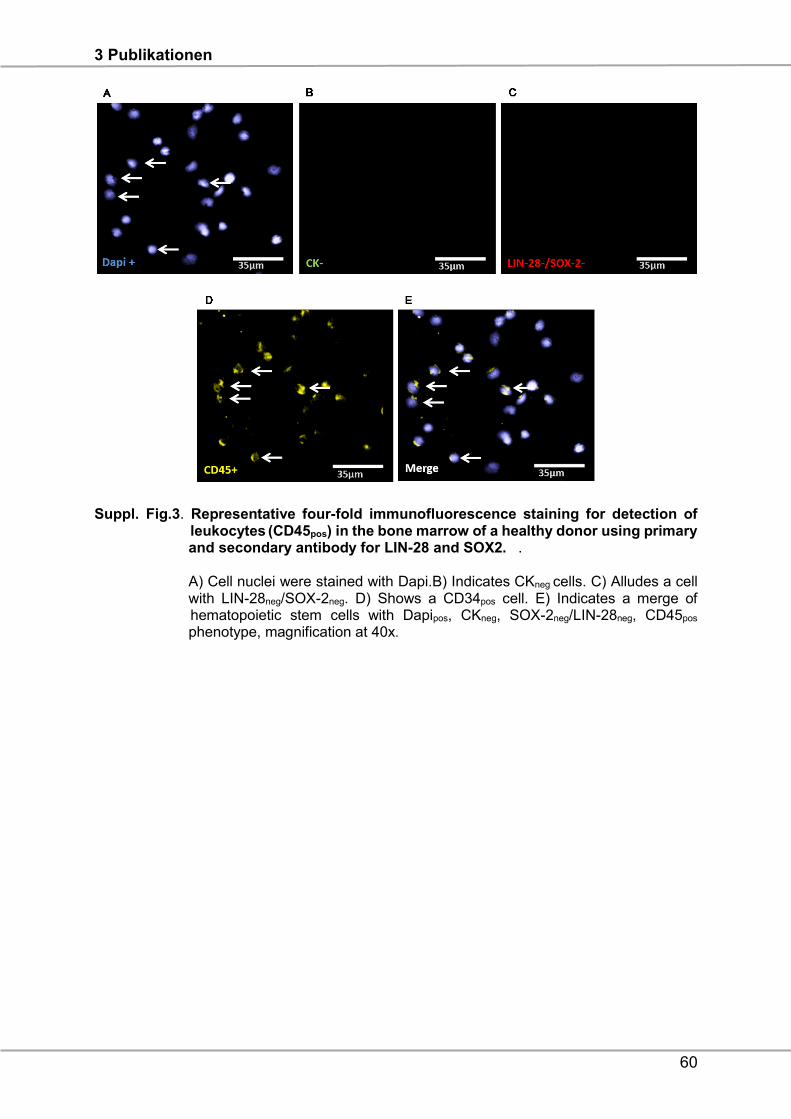
3 Publikationen
60
Suppl. Fig.3. Representative four-fold immunofluorescence staining for detection of leukocytes (CD45pos) in the bone marrow of a healthy donor using primary and secondary antibody for LIN-28 and SOX2. . A) Cell nuclei were stained with Dapi.B) Indicates CKneg cells. C) Alludes a cell with LIN-28neg/SOX-2neg. D) Shows a CD34pos cell. E) Indicates a merge of hematopoietic stem cells with Dapipos, CKneg, SOX-2neg/LIN-28neg, CD45pos
phenotype, magnification at 40x.
3 Publikationen
61
References
1.Goodman M.T, Howe HL, Tung KH, Hotes J, Miller B.A, Coughlin SS, Chen VW.
Incidence of ovarian cancer by race and ethnicity in the United States, 1992-1997.
Cancer. 2003; 97:2676–2685.
2.Du Bois A, Pfisterer J. Future options for first-line therapy of advanced ovarian
cancer. Int J Gynecol Cancer. 2005; 1:42-50.
3.Du Bois A, Reuss A, Pujade-Lauraine E, Harter P, Ray-Coquard I, Pfisterer J. Role
of surgical outcome as prognostic factor in advanced epithelial ovarian cancer: a
combined exploratory analysis of 3 prospectively randomized phase 3 multicenter
trials: by the Arbeitsgemeinschaft Gynaekologische Onkologie Studiengruppe
Ovarialkarzinom (AGO-OVAR) and the Groupe d'Investigateurs Nationaux Pour les
Etudes des Cancers de l'Ovaire (GINECO). Cancer. 2009;115(6):1234-44.
4.Wimberger P, Lehmann N, Kimmig R, Burges A, Meier W, Du Bois A;
Arbeitsgemeinschaft Gynaekologische Onkologie Ovarian Cancer Study Group.
Prognostic factors for complete debulking in advanced ovarian cancer and its impact
on survival. An exploratory analysis of a prospectively randomized phase III study of
the Arbeitsgemeinschaft Gynaekologische Onkologie Ovarian Cancer Study Group
(AGO-OVAR). Gynecol Oncol. 2007;106(1):69-74.
5.Wimberger P, Wehling M, Lehmann N, Kimmig R, Schmalfeldt B, Burges A, Harter
P, Pfisterer J, du Bois A. Influence of residual tumor on outcome in ovarian cancer
patients with FIGO stage IV disease: an exploratory analysis of the AGO-OVAR
(Arbeitsgemeinschaft Gynaekologische Onkologie Ovarian Cancer Study Group). Ann
Surg Oncol. 2010;17(6):1642-8.
6.Dyall S, Gayther SA, Dafou D. Cancer stem cells and epithelial ovarian cancer. J.
Oncol. 2010; 2010:105269.
7.Hosonuma S, Kobayashi Y, Kojo S, Wada H, Seino KI, Kiguchi K, Ishizuka B. Clinical
significance of side population in ovarian cancer cells.Hum Cell. 2011; 24:9–12.
8.Boesch M, Zeimet, AG, Reimer D, Schmidt S, Parson W, Spoeck F, Hatina J, Wolf
D, Sopper S. The side population of ovarian cancer cells defines a heterogeneous
compartment exhibiting stem cell characteristics.2014;5(16):7027-39.
9.Bapat SA, Koppikar CB,Kurrey NK. Stem and Progenitor-Like Cells Contribute to the
3 Publikationen
62
Aggressive Behavior of Human Epithelial Ovarian Cancer.Cancer Res. 2005;
65(8):3025–3029.
10.Peng S, Maihle NJ, Huang Y. Pluripotency factors Lin28 and Oct4 identify a sub-
population of stem cell-like cells in ovarian cancer. Oncogene 2010; 29(14):2153-5159.
11.Bareiss PM, Paczulla A, Wang H, Schairer R, Wiehr S, Kohlhofer U, Rothfuss
OC, Fischer A, Perner S, Staebler A, Wallwiener D, Fend F, Fehm T, et al. SOX2
expression associates with stem cell state in human ovarian carcinoma. Cancer Res.
2013; 73;5544–5555.
12.Pham D. Scheble V, Bareiss P, Fischer A, Beschorner C, Bachmann C, Neubauer
H, Boesmueller H, Kanz L. SOX2 in ovarian carcinoma – association with high grade
and improved outcome after platinum-based chemotherapy. Int J Gynecol
Pathol. 2013; 32(4):358-67.
13.Wang X, Ji X, Chen J, Yan D, Zhang Z, Wang Q, Xi X, Feng Y. SOX2 Enhances
the Migration and Invasion of Ovarian Cancer Cells via Src Kinase. PLoS One. 2014;
9(6):e9959414.
14.Lou X, Han X, Jin C, Tian W, Yu W, Ding D, Cheng L, Huang B, Jiang H, Lin B.
SOX2 targets fibronectin 1 to promote cell migration and invasion in ovarian cancer:
new molecular leads for therapeutic intervention. OMICS.2013; 17(10):510-8.
15.Li Y, Chen K. Li L, Li R, Zhang J, Ren W. Overexpression of SOX2 is involved in
paclitaxel resistance of ovarian cancer via the PI3K/Akt pathway. Tumour Biol. 2015.
16.Du J, Li B, Fang Y, Liu Y, Wang Y, Li J, Zhou W, Wang X. Overexpression of Class
III β-tubulin, Sox2, and nuclear Survivin is predictive of taxane resistance in patients
with stage III ovarian epithelial cancer. BMC Cancer. 2015; 15:536.
17.Banys M, Solomayer EF, Becker S, Krawczyk N, Gardanis K, Staebler A, Neubauer
H, Wallwiener D, Fehm T. Disseminated tumor cells in bone marrow may affect
prognosis of patients with gynecologic malignancies. Int J Gynecol Cancer. 2009;
19(5):948-52.
18.Braun S, Schindlbeck C, Hepp F, Janni W, Kentenich C, Riethmüller G, Pantel K.
Occult tumor cells in bone marrow of patients with locoregionally restricted ovarian
cancer predict early distant metastatic relapse. J Clin Oncol. 2001; 19:368-375.
19.Romero-Laorden N, Olmos D, Fehm T, Garcia-Donas J, Diaz-Padilla I. Circulating
3 Publikationen
63
and disseminated tumor cells in ovarian cancer: A systematic review Gynecol
Oncol. 2014; 133(3):632-9.
20.Fehm T, Banys M, Rack B, Janni W, Marth C, Blassl C, Hartkopf A, Trope C, Kimmig
R, Krawczyk N, Wallwiener D, Wimberger P, Kasimir-Bauer S. Pooled analysis of the
prognostic relevance of disseminated tumor cells in the bone marrow of patients with
ovarian cancer. Int J Gynecol Cancer. 2013; 23(5):839-845.
21.Cui L, Kwong J and Wang CC. Prognostic value of circulating tumor cells and
disseminated tumor cells in patients with ovarian cancer: a systematic review and
meta-analysis. Journal of Ovarian Research. 2015; 8:38.
22.Wimberger P, Heubner M, Otterbach F, Fehm T, Kimmig R, Kasimir-Bauer S.
Influence of platinum-based chemotherapy on disseminated tumor cells in blood and
bone marrow of patients with ovarian cancer. Gynecol. Oncol. 2007; 107, 331–338.
23.Marth C, Kisic J, Kaern J, Tropé C, Fodstad Ø. Circulating tumor cells in the
peripheral blood and bone marrow of patients with ovarian carcinoma do not predict
prognosis. Cancer. 2002; 94:707-712.
24.Cain JM, Ellis GK, Collins C, Greer BE, Tamimi HK, Figge DC, Gown
AM, Livingston RB. Bone marrow involvement in epithelial ovarian cancer by
immunocytochemical assessment. Gynecol Oncol. 1990; 38:442-445.
25.Monteiro J, Fodde R. Cancer stemness and metastasis: therapeutic consequences
and perspectives. Eur J Cancer. 2010; 46(7):1198-203.
26.Balic M, Lin H, Young L, Hawes D, Giuliano A, McNamara G, Datar RH, Cote RJ.
Most early disseminated cancer cells detected in bone marrow of breast cancer
patients have a putative breast cancer stem cell phenotype. Clin Cancer Res 2006;
12:5615-5621
27.Reuben JM, Lee BN, Gao H, Cohen EN, Mego M, Giordano A, Wang X, Lodhi
A, Krishnamurthy S, Hortobagyi GN, Cristofanilli M, Lucci A, Woodward WA. Primary
breast cancer patients with high risk clinicopathologic features have high percentages
of bone marrow epithelial cells with ALDH activity and CD44+CD24lo cancer stem cell
phenotype. Eur J Cancer. 2011; 47(10):1527-36
28.Ye F, Li Y, Hu Y,Zhou C, HU Y, Chen H. Expression of Sox2 in human ovarian
epithelial carcinoma. 2011; 137(1):131-7.
3 Publikationen
64
29.Viswanathan SR, Powers JT, Einhorn W, Hoshida Y, Ng TL, Toffanin S, O'Sullivan
M, Lu J, Phillips LA, Lockhart VL, Shah SP, Tanwar PS, Mermel CH, et al. Lin28
promotes transformation and is associated with advanced human malignancies. Nat
Genet. 2009; 41(7):843-8.
30.Li Y, Chen K, Li L, Li R, Zhang J, Ren W. Overexpression of Sox2 is involved in
paclitaxel resistance of ovarian cancer via the PI3K/AKT pathway. Tumor Biol. 2015.
31.Du J, Li B, Fang Y, Liu Y, Wang Y, Li J, Zhou W, Wang X. Overexpression of Class
III ß-tubulin, Sox2, and nuclear surviving is predictive of taxane resistance in patients
with stage III ovarian epithelial cancer. BMC Cancer. 2015; 15:536.
32.Johnson NP, Hoeschele JD, Rahn RO. Kinetic analysis of the in vitro binding of
radioactive cis- and trans-dichlorodiammineplatinum(II) to DNA. Chem Biol Interact.
1980; 30:151–69.
33.Bookman Michael A. Extending the Platinum-free Interval in Recurrent Ovarian
Cancer: The role of Topeka in Second-Line Chemotherapy. The Oncologist. 1999;
4:87-94.
34.Galluzzi L, Senovilla L, Vitale I, Michels J, Martins I, Kepp O, Castedo M, Kroemer
G. Molecular mechanism of cisplatin resistance. Oncogene. 2012. 31;1869-183.
35.Kuhlmann JD, Wimberger P, Bankfalvi A, Keller T, Schöler S, Aktas B, Buderath
P, Hauch S, Otterbach F, Kimmig R, Kasimir-Bauer S. ERCC1-positive circulating
tumor cells in the blood of Ovarian cancer patients as a predictive biomarker for
platinum resistance. Clinical Chemistry. 2014; 60:10 1282-1289.
36.Chiara F, Massimo B, Daniele G, Giovanna D. Epithelial- mesenchymal transition
and breast cancer: Role, molecular mechanisms and clinical impact. Cancer treatment
Reviews. 2012; 38(6):689-97.
37. Zhang J, Zhang XB, Liu Y, Liu JJ, Zhang MS. Effects of an mTOR inhibitor RAD001
on human breast cancer stem cells in vitro and in vivo. J Clin Oncol 2011. 29:abstr
e11514.
38.Gupta PB, Onder TT, Jiang G, Tao K, Kuperwasser C, Weinberg RA, Lander ES.
Identification of selective inhibitors of cancer stem cells by high-throughput screening.
Cell 2009; 138(4):645-659.
39.Kesharwani RK, Srivastava V, Singh P, Rizvi SI, Adeppa K, Misra K. A novel
3 Publikationen
65
approach for overcoming drug Resistance in breast cancer chemotherapy by targeting
new synthetic curcumin analogues against aldehyde dehydrogenase 1 (ALDH1A1)
and glycogen synthase kinase-3 ß (GSK-3ß). Appl Biochem Biotechnol. 2015;
176:1996-2017.
40.Pan Q, Li Q, Liu S, Ning N, Zhang X, Xu Y, Chang AE, Wicha MS: targeting cancer
stem cells using immunologic approaches. Stem Cells. 2015; 33:2085-2092.
41.Sharma P, Wagner K, Wolchok JD, Allison JP. Novel cancer immunotherapy
agents with survival benefit: Recent successes and next steps. Nat Rev Cancer 2011;
11:805–812.
42.Topalian SL, Hodi FS, Brahmer JR, Gettinger SN, Smith DC, McDermott
DF, Powderly JD, Carvajal RD, Sosman JA, Atkins MB, Leming PD, Spigel
DR,Antonia SJ, et al. Safety, activity, and immune correlates of anti-PD-1 antibody in
cancer. N Engl J Med. 2012; 366:2443–2454.
43.Silverberg SG. Histopathologic grading of ovarian carcinoma: a review and
proposal. Int J Gynecol Pathol. 2000; 19:7-15.
44.Fehm T, Braun S, Muller V, Janni W, Gebauer G, Marth C, Schindlbeck
C, Wallwiener D, Borgen E, Naume B, Pantel K, Solomayer E. A concept for the
standardized detection of disseminated tumor cells in bone marrow of patients with
primary breast cancer and its clinical implementation. Cancer. 2006; 107:885-92.
45.Borgen E, Naume B, Nesland JM, Kvalheim G, Beiske K, Fodstad O, Diel I,
Solomayer EF, Theocharous P, Coombes R, Smith BM, E Wunder, Marolleau JP,
Garcia J, Pantel K. Standardization of the immunocytochemical detection of cancer
cells in BM and blood: I. Establishment of objective criteria for the evaluation of
immunostained cells: The European ISHAGE Working Group for Standardization of
Tumor Cell Detection. Cytotherapy.1999; 5:377-388.
46.Krause DS, Fackler MJ, Civin CI, May WS. CD34: structure, biology, and clinical
utility. Blood. 1996, 1:87 (1): 1-13.
3 Publikationen
66
ERCC1-expressing circulating tumor cells as a potential diagnostic tool for
monitoring response to platinum-based chemotherapy and for predicting post-
therapeutic outcome of ovarian cancer
Issam Chebouti1,6, Jan Dominik Kuhlmann2,5,6, Paul Buderath1,6, Stephan Weber3,
Pauline Wimberger2,5,6, Yvonne Bokeloh4, Siegfried Hauch4, Rainer Kimmig1,6, Sabine
Kasimir-Bauer1,6
1Dep. of Gynecology and Obstetrics, University Hospital Essen (Germany),
2Department of Gynecology and Obstetrics, Medical Faculty and University Hospital
Carl Gustav Carus, Technische Universität Dresden, Germany
3Acomed Statistik, Magdeburg, Germany
4QIAGEN, Hannover GmbH, Germany
5National Center for Tumor Diseases (NCT), Partner Site Dresden, Germany
6German Cancer Consortium (DKTK), Dresden and German Cancer Research
Center (DKFZ), Heidelberg, Germany
Key words: Circulating tumor cells, ERCC1, platinum-resistance, ovarian cancer,
prognosis
Running title: ERCC1+CTCs predict post therapeutic outcome of ovarian cancer
Corresponding author
Issam Chebouti
Department of Gynecology and Obstetrics
University of Duisburg-Essen
Hufelandstrasse 55
D-45122 Essen
Phone ++ 49 – 201-723-2340
Fax ++49 – 201-723-3938
Email: [email protected]
3 Publikationen
67
ABSTRACT
Background: We recently showed that the presence of ERCC1+CTCs is an
independent predictive biomarker for platinum-resistance and poor prognosis of
ovarian cancer. The goal of our current research was to determine how the auxiliary
assessment of ERCC1-transcripts influences overall CTC-detection rate. We extended
this investigation from an initially predictive setting to paired pre- and post-therapeutic
blood analysis in order to see, whether ERCC1+CTCs dynamics mirror response to
chemotherapy.
Methods: 65 Paired blood samples (10ml) of primary ovarian cancer patients at
primary diagnosis and after chemotherapy were studied for CTCs with the AdnaTest
OvarianCancer (QIAGEN Hannover GmbH). We analyzed the tumor-associated
transcripts EpCAM, MUC-1 and CA-125. ERCC1-transcripts were investigated in a
separate approach by singleplex RT-PCR.
Results: Auxiliary assessment of ERCC1-transcripts enhanced the overall CTC-
detection rate up to 17%. ERCC1+CTCs (defined as positive for one of the AdnaTest
markers plus ERCC1-positivity) were detected in 15% of patients at primary diagnosis
and in 12% after chemotherapy. The presence of ERCC1+CTCs after chemotherapy
correlated with platinum-resistance (P=0.01), reduced PFS (P=0.0293) and OS
(P=0.0008) and their persistence indicated poor post-therapeutic outcome (PFS:
P=0.005; OS: P=0.0058). Interestingly, the assessment of ERCC1-transcripts alone
was sufficient for the detection of prognostic relevant ERCC1-expressing CTCs.
Conclusion: Auxiliary assessment of ERCC1-transcripts expands the phenotypic
spectrum of CTC detection and defines an additional overlapping fraction of ERCC1-
expressing CTCs, which are potentially selected by platinum-based chemotherapy.
Specifically, we suggest that ERCC1+CTCs could additionally be useful as a surrogate
for monitoring platinum-based chemotherapy and to assess the post-therapeutic
outcome of ovarian cancer.
3 Publikationen
68
INTRODUCTION
Epithelial ovarian cancer is the fifth leading cause of cancer death of women in Europe
and the United States and the second most common gynecological malignancy [1].
Most cases are diagnosed in advanced stages and, although response rates to
chemotherapy reach up to 80%, the majority of patients cannot be cured. Standard
treatment of advanced ovarian cancer is primary surgery aiming at complete
macroscopic tumor resection followed by platinum- and paclitaxel-based
chemotherapy, which has been shown to prolong progression free survival (PFS) as
well as overall survival (OS) [2]. Postoperative residual tumor is one of the most
important prognostic factors in advanced ovarian cancer [3-5]. However, despite
advances in treatment, more than half of all patients will experience recurrence,
resulting in poor overall survival [6].
Importantly, resistance to platinum-based chemotherapy, which can be caused by e.g.
enhanced DNA-repair capacity of tumor cells, occurs in about 15-20% of patients and
constitutes one of the most recognized clinical challenges for ovarian cancer [7]. The
nucleotide excision repair (NER) pathway is a key pathway involved in mediating
resistance or sensitivity to platinum-based chemotherapeutic agents. The excision
repair cross-complementation group 1 (ERCC1) protein plays a key role in NER. It
dimerizes with xeroderma pigmentosum complementation group F (known as ERCC4)
and mediates the excision of DNA-platinum adducts, typically induced by platinum-
based chemotherapy [8]. ERCC1-expression has been extensively studied in primary
tumor tissue of several cancer entities, including ovarian cancer, and has been
proposed as a potential predictor for response to platinum-based chemotherapy.
However, this concept has been controversial, particularly in the context of
immunohistochemical ERCC1-detection, and has not been implemented into clinical
routine so far [9-20]. Taking into account that primary tumor tissue is typically available
only at primary diagnosis, it would be valuable to establish a non-invasive blood-based
biomarker for stratifying response to platinum-based chemotherapy at primary
diagnosis and for guiding individualized therapy decisions in the future. We recently
showed that the presence of ERCC1+CTCs (circulating tumor cells) at primary
diagnosis of ovarian cancer, a potentially platinum-resistant CTC-subgroup, is an
independent predictive biomarker for primary platinum-resistance and poor prognosis
of ovarian cancer [21]. We now explored in more detail, in how far the auxiliary
assessment of ERCC1-transcripts influences overall CTC-detection rate and whether
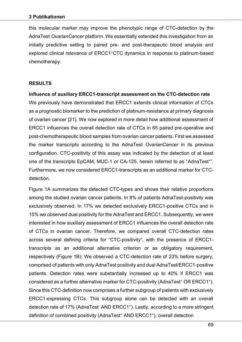
3 Publikationen
69
this molecular marker may improve the phenotypic range of CTC-detection by the
AdnaTest OvarianCancer platform. We essentially extended this investigation from an
initially predictive setting to paired pre- and post-therapeutic blood analysis and
explored clinical relevance of ERCC1+CTC dynamics in response to platinum-based
chemotherapy.
RESULTS
Influence of auxiliary ERCC1-transcript assessment on the CTC-detection rate
We previously have demonstrated that ERCC1 extends clinical information of CTCs
as a prognostic biomarker to the prediction of platinum-resistance at primary diagnosis
of ovarian cancer [21]. We now explored in more detail how additional assessment of
ERCC1 influences the overall detection rate of CTCs in 65 paired pre-operative and
post-chemotherapeutic blood samples from ovarian cancer patients. First we assessed
the marker transcripts according to the AdnaTest OvarianCancer in its previous
configuration. CTC-positivity of this assay was indicated by the detection of at least
one of the transcripts EpCAM, MUC-1 or CA-125, herein referred to as “AdnaTest+”.
Furthermore, we now considered ERCC1-transcripts as an additional marker for CTC-
detection.
Figure 1A summarizes the detected CTC-types and shows their relative proportions
among the studied ovarian cancer patients. In 8% of patients AdnaTest-positivity was
exclusively observed. In 17% we detected exclusively ERCC1-positive CTCs and in
15% we observed dual positivity for the AdnaTest and ERCC1. Subsequently, we were
interested in how auxiliary assessment of ERCC1 influences the overall detection rate
of CTCs in ovarian cancer. Therefore, we compared overall CTC-detection rates
across several defining criteria for “CTC-positivity”, with the presence of ERCC1-
transcripts as an additional alternative criterion or as obligatory requirement,
respectively (Figure 1B): We observed a CTC-detection rate of 23% before surgery,
comprised of patients with only AdnaTest positivity and dual AdnaTest/ERCC1-positive
patients. Detection rates were substantially increased up to 40% if ERCC1 was
considered as a further alternative marker for CTC-positivity (AdnaTest+ OR ERCC1+).
Since this CTC-definition now comprises a further subgroup of patients with exclusively
ERCC1-expressing CTCs. This subgroup alone can be detected with an overall
detection rate of 17% (AdnaTest- AND ERCC1+). Lastly, according to a more stringent
definition of combined positivity (AdnaTest+ AND ERCC1+), overall detection
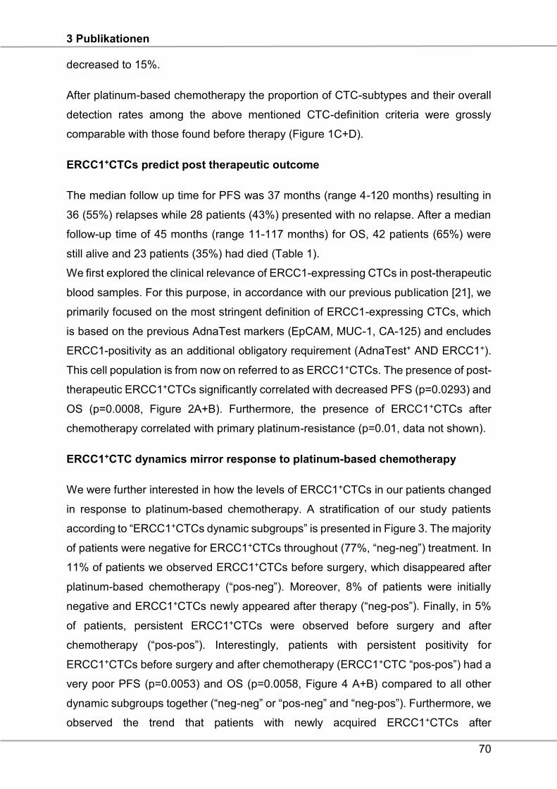
3 Publikationen
70
decreased to 15%.
After platinum-based chemotherapy the proportion of CTC-subtypes and their overall
detection rates among the above mentioned CTC-definition criteria were grossly
comparable with those found before therapy (Figure 1C+D).
ERCC1+CTCs predict post therapeutic outcome
The median follow up time for PFS was 37 months (range 4-120 months) resulting in
36 (55%) relapses while 28 patients (43%) presented with no relapse. After a median
follow-up time of 45 months (range 11-117 months) for OS, 42 patients (65%) were
still alive and 23 patients (35%) had died (Table 1).
We first explored the clinical relevance of ERCC1-expressing CTCs in post-therapeutic
blood samples. For this purpose, in accordance with our previous publication [21], we
primarily focused on the most stringent definition of ERCC1-expressing CTCs, which
is based on the previous AdnaTest markers (EpCAM, MUC-1, CA-125) and encludes
ERCC1-positivity as an additional obligatory requirement (AdnaTest+ AND ERCC1+).
This cell population is from now on referred to as ERCC1+CTCs. The presence of post-
therapeutic ERCC1+CTCs significantly correlated with decreased PFS (p=0.0293) and
OS (p=0.0008, Figure 2A+B). Furthermore, the presence of ERCC1+CTCs after
chemotherapy correlated with primary platinum-resistance (p=0.01, data not shown).
ERCC1+CTC dynamics mirror response to platinum-based chemotherapy
We were further interested in how the levels of ERCC1+CTCs in our patients changed
in response to platinum-based chemotherapy. A stratification of our study patients
according to “ERCC1+CTCs dynamic subgroups” is presented in Figure 3. The majority
of patients were negative for ERCC1+CTCs throughout (77%, “neg-neg”) treatment. In
11% of patients we observed ERCC1+CTCs before surgery, which disappeared after
platinum-based chemotherapy (“pos-neg”). Moreover, 8% of patients were initially
negative and ERCC1+CTCs newly appeared after therapy (“neg-pos”). Finally, in 5%
of patients, persistent ERCC1+CTCs were observed before surgery and after
chemotherapy (“pos-pos”). Interestingly, patients with persistent positivity for
ERCC1+CTCs before surgery and after chemotherapy (ERCC1+CTC “pos-pos”) had a
very poor PFS (p=0.0053) and OS (p=0.0058, Figure 4 A+B) compared to all other
dynamic subgroups together (“neg-neg” or “pos-neg” and “neg-pos”). Furthermore, we
observed the trend that patients with newly acquired ERCC1+CTCs after
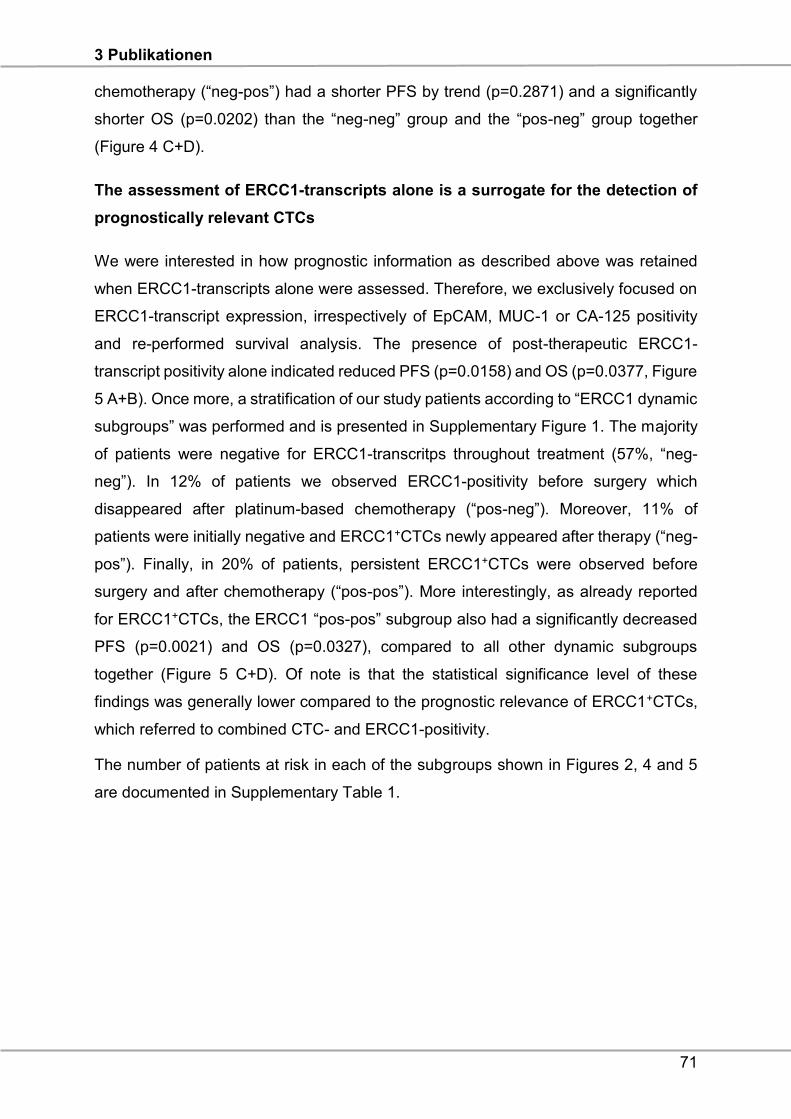
3 Publikationen
71
chemotherapy (“neg-pos”) had a shorter PFS by trend (p=0.2871) and a significantly
shorter OS (p=0.0202) than the “neg-neg” group and the “pos-neg” group together
(Figure 4 C+D).
The assessment of ERCC1-transcripts alone is a surrogate for the detection of
prognostically relevant CTCs
We were interested in how prognostic information as described above was retained
when ERCC1-transcripts alone were assessed. Therefore, we exclusively focused on
ERCC1-transcript expression, irrespectively of EpCAM, MUC-1 or CA-125 positivity
and re-performed survival analysis. The presence of post-therapeutic ERCC1-
transcript positivity alone indicated reduced PFS (p=0.0158) and OS (p=0.0377, Figure
5 A+B). Once more, a stratification of our study patients according to “ERCC1 dynamic
subgroups” was performed and is presented in Supplementary Figure 1. The majority
of patients were negative for ERCC1-transcritps throughout treatment (57%, “neg-
neg”). In 12% of patients we observed ERCC1-positivity before surgery which
disappeared after platinum-based chemotherapy (“pos-neg”). Moreover, 11% of
patients were initially negative and ERCC1+CTCs newly appeared after therapy (“neg-
pos”). Finally, in 20% of patients, persistent ERCC1+CTCs were observed before
surgery and after chemotherapy (“pos-pos”). More interestingly, as already reported
for ERCC1+CTCs, the ERCC1 “pos-pos” subgroup also had a significantly decreased
PFS (p=0.0021) and OS (p=0.0327), compared to all other dynamic subgroups
together (Figure 5 C+D). Of note is that the statistical significance level of these
findings was generally lower compared to the prognostic relevance of ERCC1+CTCs,
which referred to combined CTC- and ERCC1-positivity.
The number of patients at risk in each of the subgroups shown in Figures 2, 4 and 5
are documented in Supplementary Table 1.
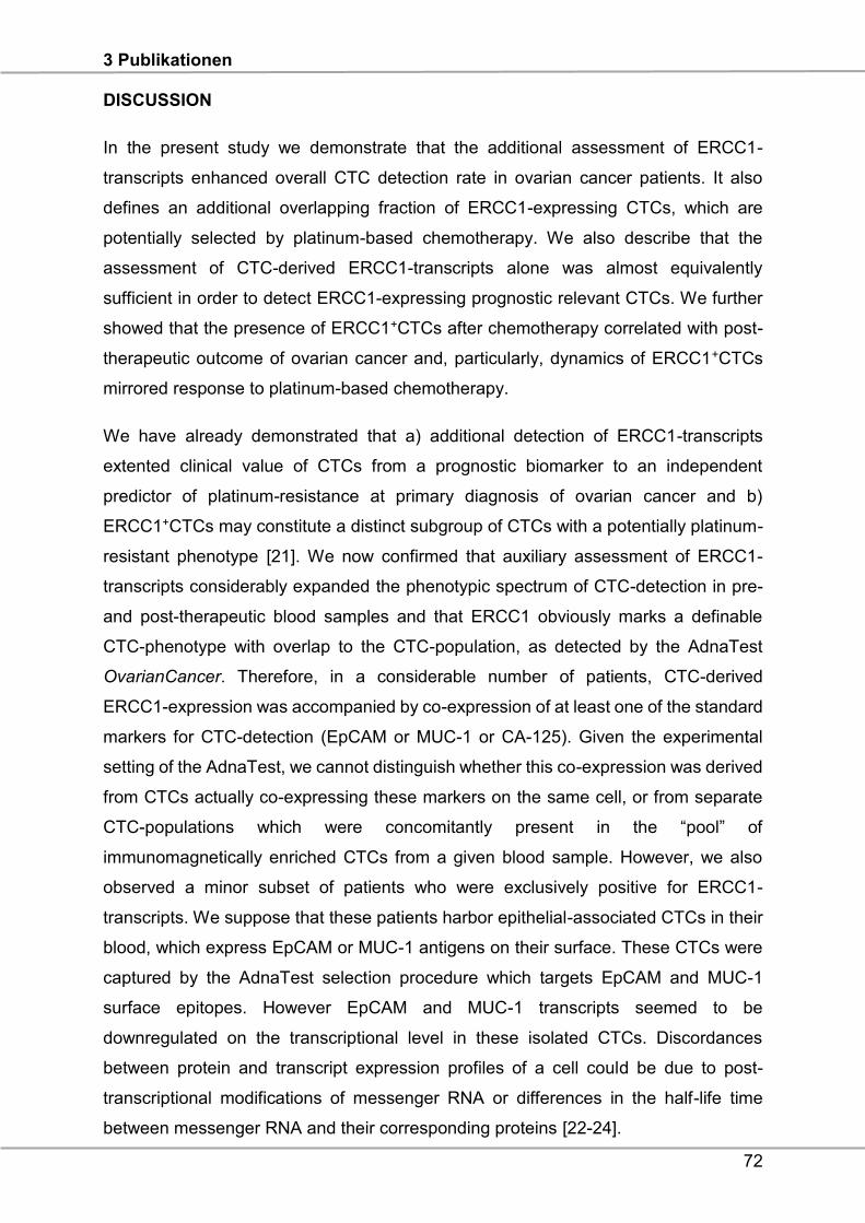
3 Publikationen
72
DISCUSSION
In the present study we demonstrate that the additional assessment of ERCC1-
transcripts enhanced overall CTC detection rate in ovarian cancer patients. It also
defines an additional overlapping fraction of ERCC1-expressing CTCs, which are
potentially selected by platinum-based chemotherapy. We also describe that the
assessment of CTC-derived ERCC1-transcripts alone was almost equivalently
sufficient in order to detect ERCC1-expressing prognostic relevant CTCs. We further
showed that the presence of ERCC1+CTCs after chemotherapy correlated with post-
therapeutic outcome of ovarian cancer and, particularly, dynamics of ERCC1+CTCs
mirrored response to platinum-based chemotherapy.
We have already demonstrated that a) additional detection of ERCC1-transcripts
extented clinical value of CTCs from a prognostic biomarker to an independent
predictor of platinum-resistance at primary diagnosis of ovarian cancer and b)
ERCC1+CTCs may constitute a distinct subgroup of CTCs with a potentially platinum-
resistant phenotype [21]. We now confirmed that auxiliary assessment of ERCC1-
transcripts considerably expanded the phenotypic spectrum of CTC-detection in pre-
and post-therapeutic blood samples and that ERCC1 obviously marks a definable
CTC-phenotype with overlap to the CTC-population, as detected by the AdnaTest
OvarianCancer. Therefore, in a considerable number of patients, CTC-derived
ERCC1-expression was accompanied by co-expression of at least one of the standard
markers for CTC-detection (EpCAM or MUC-1 or CA-125). Given the experimental
setting of the AdnaTest, we cannot distinguish whether this co-expression was derived
from CTCs actually co-expressing these markers on the same cell, or from separate
CTC-populations which were concomitantly present in the “pool” of
immunomagnetically enriched CTCs from a given blood sample. However, we also
observed a minor subset of patients who were exclusively positive for ERCC1-
transcripts. We suppose that these patients harbor epithelial-associated CTCs in their
blood, which express EpCAM or MUC-1 antigens on their surface. These CTCs were
captured by the AdnaTest selection procedure which targets EpCAM and MUC-1
surface epitopes. However EpCAM and MUC-1 transcripts seemed to be
downregulated on the transcriptional level in these isolated CTCs. Discordances
between protein and transcript expression profiles of a cell could be due to post-
transcriptional modifications of messenger RNA or differences in the half-life time
between messenger RNA and their corresponding proteins [22-24].
3 Publikationen
73
The broad heterogeneity of CTCs in the blood of cancer patients, including ovarian
cancer, has already been indicated by several independent reports [25-27]. We may
hypothesize that ERCC1-expressing CTCs play a dominant role during the course of
the disease, which is corroborated by the fact that the rate of exclusively ERCC1-
positive CTCs did not decrease after platinum-based chemotherapy. We have already
shown that breast cancer patients with CTCs detected after neoadjuvant
chemotherapy were associated with tumor stem cell characteristics and ERCC1-
expression [28]. This may suggest a potential selection of this CTC-subset by
chemotherapy. However, due to the fact that exclusively ERCC1-expressing CTCs
rarely occurred in our patient cohort, we were not able to analyze prognostic
significance of this particularly interesting CTC-subset in a statistically substantiated
manner.
Independent of ERCC1 assessment, we previously confirmed the negative prognostic
impact of CTCs before surgery and after chemotherapy, as detected by the AdnaTest
OvarianCancer [21, 29]. Nevertheless, our recent and current results strongly support
our perception that the auxiliary assessment of ERCC1-transcripts provides
complementary clinical information. In addition, the auxiliary assessment of ERCC1-
transcripts after chemotherapy alone, as well as their expression dynamics in pre- and
post-therapeutic blood samples, was almost equivalently sufficient as surrogate for a
CTC-population. This might be useful for predicting post-therapeutic outcome and for
monitoring platinum-based chemotherapy. Given the additional strong prognostic
impact of the standard AdnaTest marker transcripts MUC-1, EpCAM and CA-125 [21,
29] and considering that the statistical significance level slightly declined when only
ERCC1-transcripts were assessed, a combined condition, which assumes ERCC1-
positivity in addition to the detection of at least one of the AdnaTest markers (referred
to as ERCC1+CTCs in our study), appears to be most favorable in terms of a blood-
based prognostic biomarker.
So far, any functional characteristics of ERCC1-expressing CTCs in the blood of
ovarian cancer patients are unknown. Since our study was performed exclusively from
a “biomarker perspective”, we can only assume that ERCC1-(over)expressing CTCs
in the blood may be characterized by an enhanced, preexisting or newly acquired
capacity to resolve DNA-platinum-adducts, consequently bypassing cisplatin-mediated
cytotoxicity and possibly converting to a molecular phenotype of “on-target” platinum-
resistance [8]. This assumption is further supported by a recent investigation which
directly analyzed the presence of DNA-platinum adducts in single CTCs of advanced
3 Publikationen
74
non-small cell lung cancer (NSCLC) patients. In this context, it was suggested that the
kinetics of these adducts in pre- and post- therapeutic blood samples could be a
potential biomarker for response prediction and dose individualization of platinum-
based chemotherapy [30]. Consecutively, ERCC1+CTCs may survive multiple cycles
of chemotherapy and, in line with the fact that metastasis-initiating cells can be present
among CTCs in the blood [31], persistent ERCC1+CTCs with a platinum-resistant
phenotype could have the potential to initiate recurrence, resulting in poor clinical
outcome. Taking into consideration that ERCC1+CTCs are strong prognostic factors in
the post-therapeutic situation, particularly in case of persistent positivity, our data may
also indicate that platinum-resistant ERCC1+CTCs could be directly selected upon
platinum-based chemotherapy. Further functional studies will be necessary in order to
prove this hypothesis. Another interesting question for future studies will be, how CTC-
derived ERCC1-expression is related to EMT- and stem-like characteristics of CTCs.
In this regard, we recently demonstrated that the negative prognostic impact of the
presence and/or persistence of disseminated tumor cells in the bone marrow of ovarian
cancer patients after platinum based chemotherapy was associated with stem cell
character [32].
Conclusively, due to the limited number of patients, our study is explorative and
hypothesis generating. Nevertheless, ERCC1 marks a subpopulation of CTCs which
might be useful for monitoring platinum-based chemotherapy and for assessing post-
therapeutic outcome of ovarian cancer patients. We provide rationale to validate
clinical utility of ERCC1+CTCs among large multicenter clinical trials and to further
elucidate their functional and tumor biological significance. Alternatively, patients with
ERCC1*CTCs may profit from an early initiated and dose-intense maintenance therapy
with e.g. Bevacizumab or PARP-inhibitors. Furthermore, this high risk patient group
might be amenable to platinum-sensitizing therapies in the future, which are
increasingly proposed in preclinical studies [33-35] and already ongoing clinical trials
(NCT01164995). Using CTCs as liquid biopsy tool for individual therapy optimization,
a multi-marker gene panel, compromising all CTC subgroups, will be useful to monitor
patients during the course of the disease [36].
3 Publikationen
75
PATIENTS AND METHODS
Patient characteristics
The present study was conducted at the Department of Gynecology and Obstetrics at
the University Hospital of Essen, Germany. A total of 65 patients diagnosed between
2006 and 2014 with histologically confirmed epithelial ovarian cancer were analyzed.
Clinical characteristics of the patients are documented in Table 1. Informed written
consent was obtained from all patients and the study was approved by the Local Ethics
Committee (05-2870) and performed according to the declaration of Helsinki. Tumors
were classified according to the WHO classification of tumors of the female genital
tract. Grading was conducted using the grading system proposed by Silverberg [37]
and tumor staging was classified according to the Fédération Internationale de
Gynécology et d'Obstétrique [38]. The whole study population underwent primary
radical surgery. Total abdominal hysterectomy, bilateral salpingo-oophorectomy,
infragastric omentectomy, peritoneal stripping were performed. The most important
aim of surgery was to achieve macroscopic complete tumor resection. Radical pelvic
and para-aortic lymphadenectomy were only performed if macroscopic complete tumor
resection was achieved intraperitoneally following actual guidelines. All patients
received at least six cycles of carboplatinum AUC 5 and paclitaxel 175 mg/m2. Tumors
were clinically defined as platinum-resistant if they recurred within six months after the
completion of platinum-based chemotherapy.
Enrichment and Molecular Characterization of CTCs
Peripheral blood (2x5 ml) from each patient was collected in EDTA tubes (Sarstedt &
Co.) and processed within 4h for the enrichment of CTCs and subsequent expression
analysis according to Adnatest OvarianCancer (QIAGEN, Hannover GmbH,
Langenhagen, Germany). The test has been described in detail [21]. Briefly, CTCs
were immunomagnetically selected using the AdnaTest OvarianCancerSelect
targeting epithelial cell adhesion molecule EpCAM (also known as GA733-2), Mucin-
1, cell surface associated (MUC-1) and cell surface associated Mucin-16 (also known
as CA-125). Subsequently, RNA was isolated and gene expression analysis was
performed by reverse-transcription (RT) and multiplex RT-PCR detecting EpCAM,
MUC-1, and CA-125 (AdnaTest OvarianCancerDetect). ERCC1-transcripts were
investigated in a separate approach by singleplex RT-PCR. β-actin served as an
internal control and PCR-products were quantified on the Agilent Bioanalyzer as
follows: Blood samples of 20 healthy donors and healthy donor blood samples spiked
3 Publikationen
76
with two or five cells IGROV1 were analyzed using the AdnaTest OvarianCancer
Select/Detect for overexpression of EpCAM, MUC-1 and CA-125. The resulting PCR
fragments were analyzed with the Agilent Bioanalyzer and the resulting data were
checked for sensitivity and specificity to be ≥ 90% applying a 0.15 ng/μl fragment
concentration as a cut of value for each of the markers. For the required specificity of
>90%, as defined in the test performance criteria, the resulting cut off value was
defined as 0.15 ng/μl fragment concentration. At a cut-off value of 0.15 ng/µl specificity
is 95% and the corresponding recovery rate is 80% for two cells and 100% for five
cells, respectively.
ERCC1-positivity was defined by an amplicon concentration >0.2ng/µl. Sensitivity and
specificity were evaluated in 20 healthy donors and 99 patients with primary ovarian
cancer using ROC analysis. At a cut-off value of 0.17 ng/µl, 95% specificity is reached
and the corresponding clinical sensitivity is 46.5% (Supplementary Figure 2).
Amplicons with the following sizes were generated: EpCAM: 396bp; MUC-1: 293bp;
CA-125: 432bp; ERCC1: 366bp; and β-actin: 114bp.
Statistical Analysis
Survival curve plots and Hazard Ratio calculations were done using SAS (9.4). Survival
intervals were screened from the time of CTC detection at first diagnosis to the time of
clinical event (either death or first time of relapse) or last contact. Kaplan-Meier curves
were assessed using the log-rank test to evaluate univariate significance of the binary
grouping parameters. Fisher exact tests were performed to confirm significance.
Survival curve plots and Hazard Ratio calculations were done using SAS (9.4).
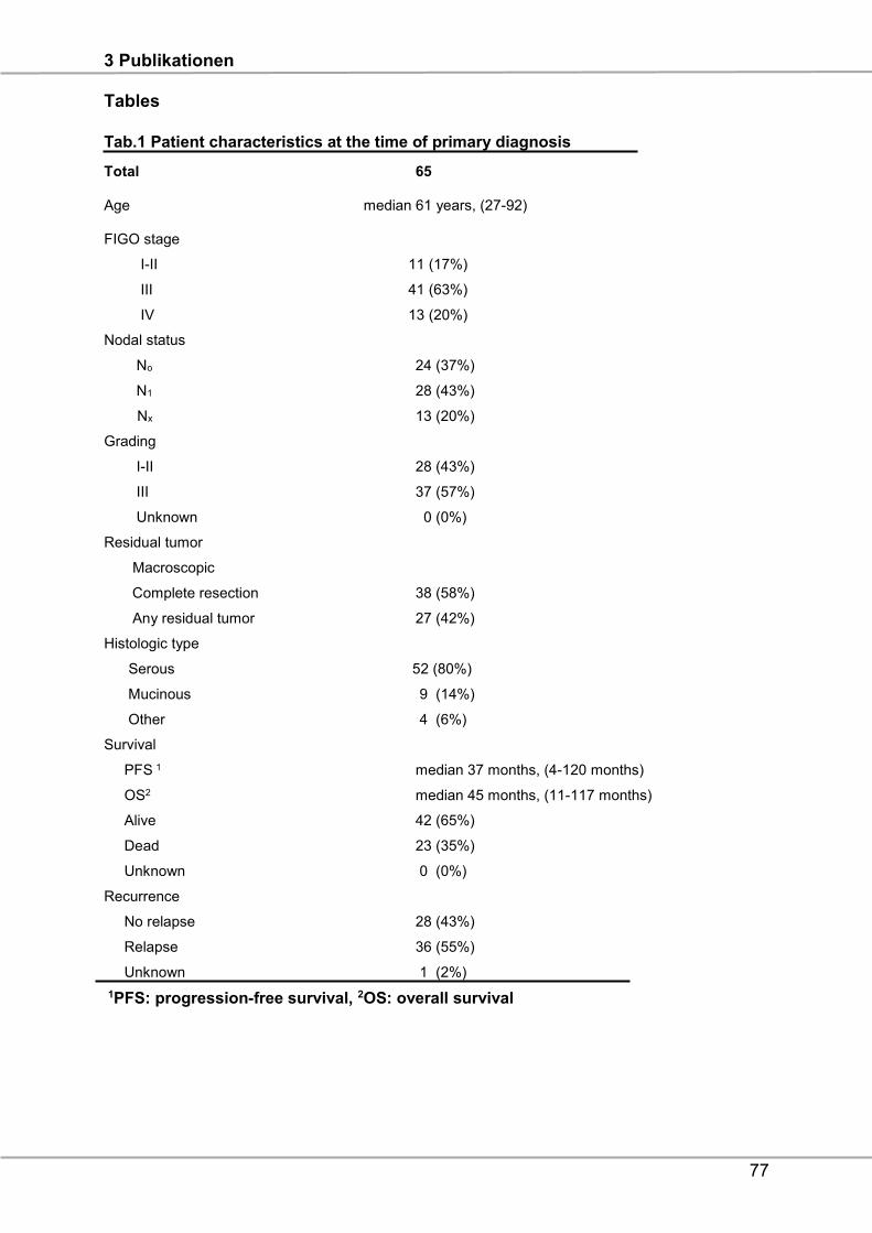
3 Publikationen
77
Tables
Tab.1 Patient characteristics at the time of primary diagnosis
Total 65
Age median 61 years, (27-92)
FIGO stage
I-II 11 (17%)
III 41 (63%)
IV 13 (20%)
Nodal status
No 24 (37%)
N1 28 (43%)
Nx 13 (20%)
Grading
I-II 28 (43%)
III 37 (57%)
Unknown 0 (0%)
Residual tumor
Macroscopic
Complete resection 38 (58%)
Any residual tumor 27 (42%)
Histologic type
Serous 52 (80%)
Mucinous 9 (14%)
Other 4 (6%)
Survival
PFS 1 median 37 months, (4-120 months)
OS2 median 45 months, (11-117 months)
Alive 42 (65%)
Dead 23 (35%)
Unknown 0 (0%)
Recurrence
No relapse 28 (43%)
Relapse 36 (55%)
Unknown 1 (2%)
1PFS: progression-free survival, 2OS: overall survival
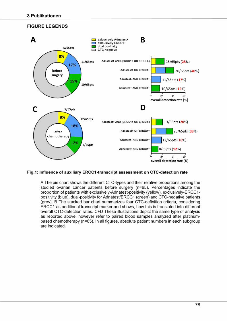
3 Publikationen
78
FIGURE LEGENDS
Fig.1: Influence of auxiliary ERCC1-transcript assessment on CTC-detection rate
A The pie chart shows the different CTC-types and their relative proportions among the studied ovarian cancer patients before surgery (n=65). Percentages indicate the proportion of patients with exclusively-Adnatest-positivity (yellow), exclusively-ERCC1- positivity (blue), dual-positivity for Adnatest/ERCC1 (green) and CTC-negative patients (grey). B The stacked bar chart summarizes four CTC-definition criteria, considering ERCC1 as additional transcript marker and shows, how this is translated into different overall CTC-detection rates. C+D These illustrations depict the same type of analysis as reported above, however refer to paired blood samples analyzed after platinum- based chemotherapy (n=65). In all figures, absolute patient numbers in each subgroup are indicated.
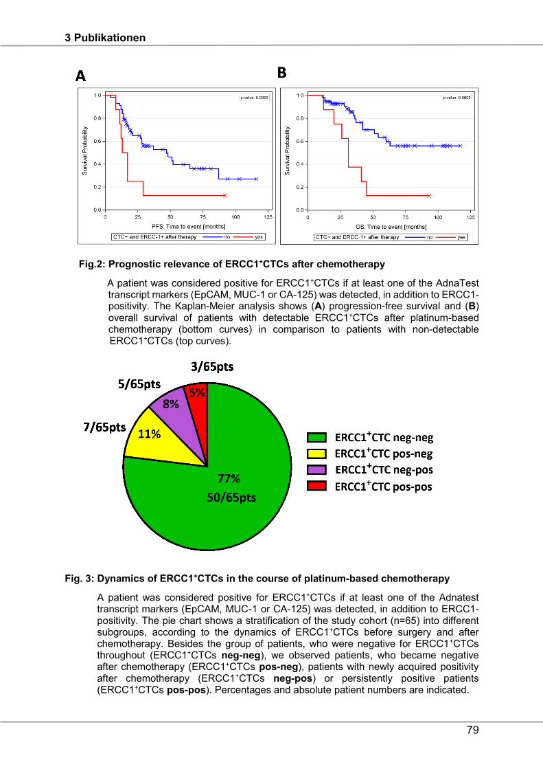
3 Publikationen
79
Fig.2: Prognostic relevance of ERCC1+CTCs after chemotherapy
A patient was considered positive for ERCC1+CTCs if at least one of the AdnaTest transcript markers (EpCAM, MUC-1 or CA-125) was detected, in addition to ERCC1- positivity. The Kaplan-Meier analysis shows (A) progression-free survival and (B) overall survival of patients with detectable ERCC1+CTCs after platinum-based chemotherapy (bottom curves) in comparison to patients with non-detectable ERCC1+CTCs (top curves).
Fig. 3: Dynamics of ERCC1+CTCs in the course of platinum-based chemotherapy
A patient was considered positive for ERCC1+CTCs if at least one of the Adnatest transcript markers (EpCAM, MUC-1 or CA-125) was detected, in addition to ERCC1- positivity. The pie chart shows a stratification of the study cohort (n=65) into different subgroups, according to the dynamics of ERCC1+CTCs before surgery and after chemotherapy. Besides the group of patients, who were negative for ERCC1+CTCs throughout (ERCC1+CTCs neg-neg), we observed patients, who became negative after chemotherapy (ERCC1+CTCs pos-neg), patients with newly acquired positivity after chemotherapy (ERCC1+CTCs neg-pos) or persistently positive patients (ERCC1+CTCs pos-pos). Percentages and absolute patient numbers are indicated.
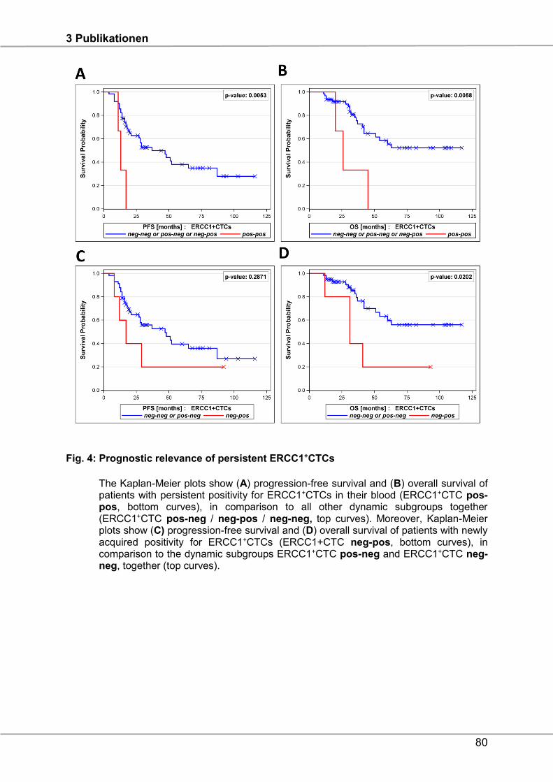
3 Publikationen
80
Fig. 4: Prognostic relevance of persistent ERCC1+CTCs
The Kaplan-Meier plots show (A) progression-free survival and (B) overall survival of patients with persistent positivity for ERCC1+CTCs in their blood (ERCC1+CTC pos- pos, bottom curves), in comparison to all other dynamic subgroups together (ERCC1+CTC pos-neg / neg-pos / neg-neg, top curves). Moreover, Kaplan-Meier plots show (C) progression-free survival and (D) overall survival of patients with newly acquired positivity for ERCC1+CTCs (ERCC1+CTC neg-pos, bottom curves), in comparison to the dynamic subgroups ERCC1+CTC pos-neg and ERCC1+CTC neg- neg, together (top curves).
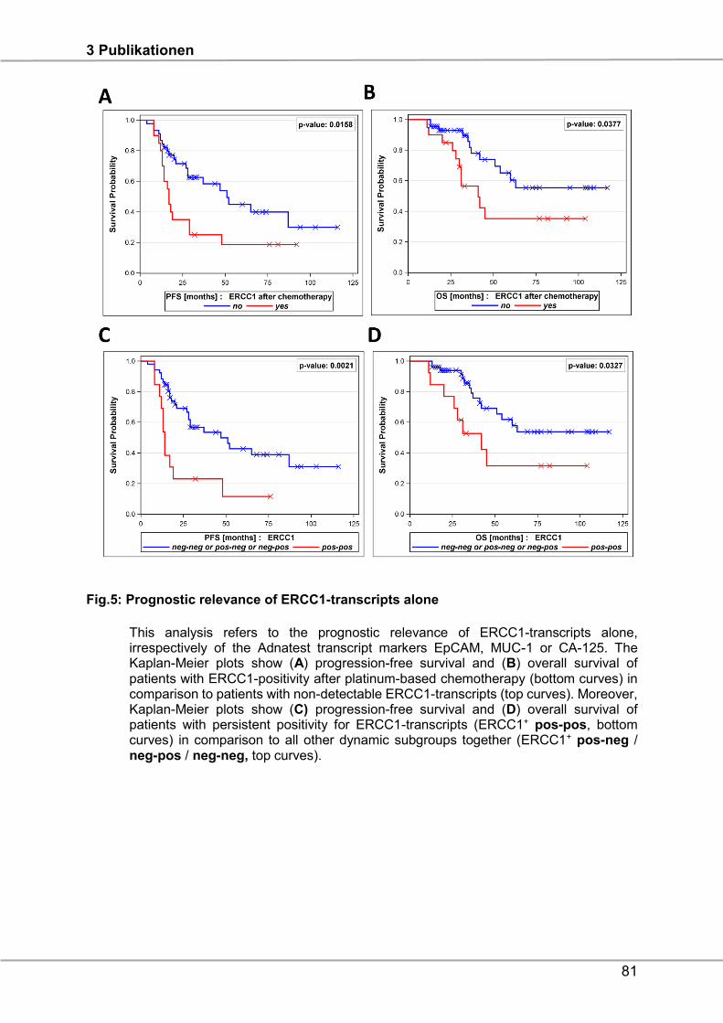
3 Publikationen
81
Fig.5: Prognostic relevance of ERCC1-transcripts alone
This analysis refers to the prognostic relevance of ERCC1-transcripts alone, irrespectively of the Adnatest transcript markers EpCAM, MUC-1 or CA-125. The Kaplan-Meier plots show (A) progression-free survival and (B) overall survival of patients with ERCC1-positivity after platinum-based chemotherapy (bottom curves) in comparison to patients with non-detectable ERCC1-transcripts (top curves). Moreover, Kaplan-Meier plots show (C) progression-free survival and (D) overall survival of patients with persistent positivity for ERCC1-transcripts (ERCC1+ pos-pos, bottom curves) in comparison to all other dynamic subgroups together (ERCC1+ pos-neg / neg-pos / neg-neg, top curves).
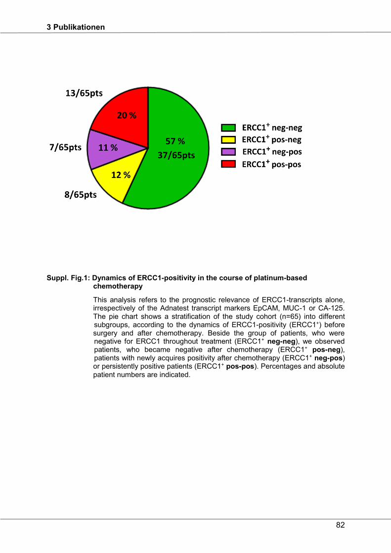
3 Publikationen
82
Suppl. Fig.1: Dynamics of ERCC1-positivity in the course of platinum-based chemotherapy
This analysis refers to the prognostic relevance of ERCC1-transcripts alone, irrespectively of the Adnatest transcript markers EpCAM, MUC-1 or CA-125. The pie chart shows a stratification of the study cohort (n=65) into different subgroups, according to the dynamics of ERCC1-positivity (ERCC1+) before surgery and after chemotherapy. Beside the group of patients, who were negative for ERCC1 throughout treatment (ERCC1+ neg-neg), we observed patients, who became negative after chemotherapy (ERCC1+ pos-neg), patients with newly acquires positivity after chemotherapy (ERCC1+ neg-pos) or persistently positive patients (ERCC1+ pos-pos). Percentages and absolute patient numbers are indicated.
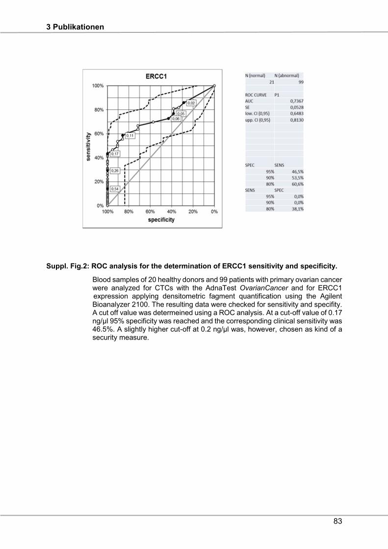
3 Publikationen
83
Suppl. Fig.2: ROC analysis for the determination of ERCC1 sensitivity and specificity.
Blood samples of 20 healthy donors and 99 patients with primary ovarian cancer were analyzed for CTCs with the AdnaTest OvarianCancer and for ERCC1 expression applying densitometric fagment quantification using the Agilent Bioanalyzer 2100. The resulting data were checked for sensitivity and specifity. A cut off value was determeined using a ROC analysis. At a cut-off value of 0.17 ng/µl 95% specificity was reached and the corresponding clinical sensitivity was 46.5%. A slightly higher cut-off at 0.2 ng/μl was, however, chosen as kind of a security measure.
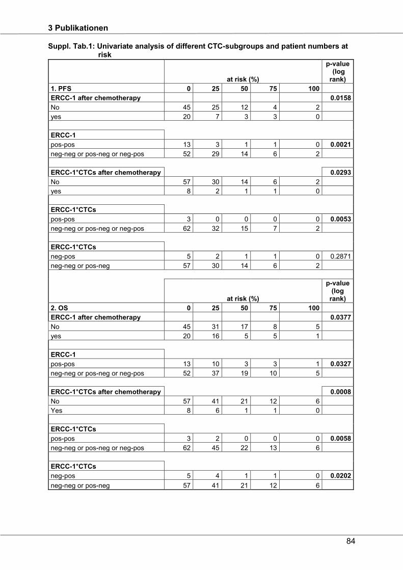
3 Publikationen
84
Suppl. Tab.1: Univariate analysis of different CTC-subgroups and patient numbers at risk
at risk (%)
p-value (log rank)
1. PFS 0 25 50 75 100
ERCC-1 after chemotherapy 0.0158
No 45 25 12 4 2
yes 20 7 3 3 0
ERCC-1
pos-pos 13 3 1 1 0 0.0021
neg-neg or pos-neg or neg-pos 52 29 14 6 2
ERCC-1+CTCs after chemotherapy 0.0293
No 57 30 14 6 2
yes 8 2 1 1 0
ERCC-1+CTCs
pos-pos 3 0 0 0 0 0.0053
neg-neg or pos-neg or neg-pos 62 32 15 7 2
ERCC-1+CTCs
neg-pos 5 2 1 1 0 0.2871
neg-neg or pos-neg 57 30 14 6 2
at risk (%)
p-value (log
rank)
2. OS 0 25 50 75 100
ERCC-1 after chemotherapy 0.0377
No 45 31 17 8 5
yes 20 16 5 5 1
ERCC-1
pos-pos 13 10 3 3 1 0.0327
neg-neg or pos-neg or neg-pos 52 37 19 10 5
ERCC-1+CTCs after chemotherapy 0.0008
No 57 41 21 12 6
Yes 8 6 1 1 0
ERCC-1+CTCs
pos-pos 3 2 0 0 0 0.0058
neg-neg or pos-neg or neg-pos 62 45 22 13 6
ERCC-1+CTCs
neg-pos 5 4 1 1 0 0.0202
neg-neg or pos-neg 57 41 21 12 6

3 Publikationen
85
The table summarizes univariate analysis according to the Kaplan-Meier curves
(Figure 2, 4, 5). The absolute number of patients in each subgroup at a risk of 0%,
25%, 50%, 75% and 100% is indicated for each case.
Funding: The study was supported by the DFG (Deutsche Forschungsgemeinschaft),
KA 1583/3-1.
Conflict of Interest: Sabine Kasimir-Bauer is a consultant for QIAGEN Hannover
GmbH. Siegfried Hauch is an employee of QIAGEN Hannover GmbH. All other authors
have no conflict of interest.
3 Publikationen
86
REFERENCES
1. Goodman MT, Howe HL, Tung KH, Hotes J, Miller BA, Coughlin SS and Chen
VW. Incidence of ovarian cancer by race and ethnicity in the United States,
1992-1997. Cancer. 2003; 97(10 Suppl):2676-2685.
2. du Bois A, Quinn M, Thigpen T, Vermorken J, Avall-Lundqvist E, Bookman M,
Bowtell D, Brady M, Casado A, Cervantes A, Eisenhauer E, Friedlaender M,
Fujiwara K, Grenman S, Guastalla JP, Harper P, et al. 2004 consensus
statements on the management of ovarian cancer: final document of the 3rd
International Gynecologic Cancer Intergroup Ovarian Cancer Consensus
Conference (GCIG OCCC 2004). Ann Oncol. 2005; 16 Suppl 8:viii7-viii12.
3. du Bois A, Reuss A, Pujade-Lauraine E, Harter P, Ray-Coquard I and Pfisterer
J. Role of surgical outcome as prognostic factor in advanced epithelial ovarian
cancer: a combined exploratory analysis of 3 prospectively randomized phase
3 multicenter trials: by the Arbeitsgemeinschaft Gynaekologische Onkologie
Studiengruppe Ovarialkarzinom (AGO-OVAR) and the Groupe d'Investigateurs
Nationaux Pour les Etudes des Cancers de l'Ovaire (GINECO). Cancer. 2009;
115(6):1234-1244.
4. Wimberger P, Lehmann N, Kimmig R, Burges A, Meier W, Du Bois A and
Arbeitsgemeinschaft Gynaekologische Onkologie Ovarian Cancer Study G.
Prognostic factors for complete debulking in advanced ovarian cancer and its
impact on survival. An exploratory analysis of a prospectively randomized phase
III study of the Arbeitsgemeinschaft Gynaekologische Onkologie Ovarian
Cancer Study Group (AGO-OVAR). Gynecol Oncol. 2007; 106(1):69-74.
5. Wimberger P, Wehling M, Lehmann N, Kimmig R, Schmalfeldt B, Burges A,
Harter P, Pfisterer J and du Bois A. Influence of residual tumor on outcome in
ovarian cancer patients with FIGO stage IV disease: an exploratory analysis of
the AGO-OVAR (Arbeitsgemeinschaft Gynaekologische Onkologie Ovarian
Cancer Study Group). Ann Surg Oncol. 2010; 17(6):1642-1648.
6. Martin LP and Schilder RJ. Management of recurrent ovarian carcinoma: current
status and future directions. Semin Oncol. 2009; 36(2):112-125.
7. Bookman MA. Extending the platinum-free interval in recurrent ovarian cancer:
the role of topotecan in second-line chemotherapy. Oncologist. 1999; 4(2):87-
94.
3 Publikationen
87
8. Galluzzi L, Senovilla L, Vitale I, Michels J, Martins I, Kepp O, Castedo M and
Kroemer G. Molecular mechanisms of cisplatin resistance. Oncogene. 2012; 31
(15):1869-1883.
9. Britten RA, Liu D, Tessier A, Hutchison MJ and Murray D. ERCC1 expression
as a molecular marker of cisplatin resistance in human cervical tumor cells. Int
J Cancer. 2000; 89(5):453-457.
10. Grimminger PP, Shi M, Barrett C, Lebwohl D, Danenberg KD, Brabender J,
Vigen CL, Danenberg PV, Winder T and Lenz HJ. TS and ERCC-1 mRNA
expressions and clinical outcome in patients with metastatic colon cancer in
CONFIRM-1 and -2 clinical trials. Pharmacogenomics J. 2012; 12(5):404-411.
11. Liu YP, Ling Y, Qi QF, Zhang YP, Zhang CS, Zhu CT, Wang MH and Pan YD.
The effects of ERCC1 expression levels on the chemosensitivity of gastric
cancer cells to platinum agents and survival in gastric cancer patients treated
with oxaliplatin-based adjuvant chemotherapy. Oncol Lett. 2013; 5(3):935-942.
12. Olaussen KA, Dunant A, Fouret P, Brambilla E, Andre F, Haddad V, Taranchon
E, Filipits M, Pirker R, Popper HH, Stahel R, Sabatier L, Pignon JP, Tursz T, Le
Chevalier T and Soria JC. DNA repair by ERCC1 in non-small-cell lung cancer
and cisplatin-based adjuvant chemotherapy. N Engl J Med. 2006; 355(10):983-
991.
13. Arora S, Kothandapani A, Tillison K, Kalman-Maltese V and Patrick SM.
Downregulation of XPF-ERCC1 enhances cisplatin efficacy in cancer cells. DNA
Repair (Amst). 2010; 9(7):745-753.
14. Milovic-Kovacevic M, Srdic-Rajic T, Radulovic S, Bjelogrlic S and Gavrilovic D.
Expression of ERCC1 protein in biopsy specimen predicts survival in advanced
ovarian cancer patients treated with platinum-based chemotherapy. J BUON.
2011; 16(4):708-714.
15. Scheil-Bertram S, Tylus-Schaaf P, du Bois A, Harter P, Oppitz M, Ewald-Riegler
N and Fisseler-Eckhoff A. Excision repair cross-complementation group 1
protein overexpression as a predictor of poor survival for high-grade serous
ovarian adenocarcinoma. Gynecol Oncol. 2010; 119(2):325-331.
16. Xie C, Yin RT, Li YL, Kang DY, Xu L and Yang KX. [The protein expression of
ERCC1 and survivin in epithelial ovarian carcinoma and their clinical
significance]. Sichuan Da Xue Xue Bao Yi Xue Ban. 2011; 42(1):86-89.
17. Vandenput I, Capoen A, Coenegrachts L, Verbist G, Moerman P, Vergote I and
Amant F. Expression of ERCC1, p53, and class III beta-tubulin do not reveal
3 Publikationen
88
chemoresistance in endometrial cancer: results from an immunohistochemical
study. Int J Gynecol Cancer. 2011; 21(6):1071-1077.
18. Bosmuller H, Haitchi-Petnehazy S, Webersinke G, Marschon R, Roithmeier F,
Stummvoll W, Fehm T, Klier-Richter M, Bonzheim I, Staebler A and Fend F.
Intratumoral lymphocyte density in serous ovarian carcinoma is superior to
ERCC1 expression for predicting response to platinum-based therapy.
Virchows Arch. 2011; 459(2):183-191.
19. Hubner RA, Riley RD, Billingham LJ and Popat S. Excision repair cross-
complementation group 1 (ERCC1) status and lung cancer outcomes: a meta-
analysis of published studies and recommendations. PLoS One. 2011;
6(10):e25164.
20. Friboulet L, Olaussen KA, Pignon JP, Shepherd FA, Tsao MS, Graziano S,
Kratzke R, Douillard JY, Seymour L, Pirker R, Filipits M, Andre F, Solary E,
Ponsonnailles F, Robin A, Stoclin A, et al. ERCC1 isoform expression and DNA
repair in non-small-cell lung cancer. New Engl J Med. 2013; 368(12):1101-1110.
21. Kuhlmann JD, Wimberger P, Bankfalvi A, Keller T, Scholer S, Aktas B, Buderath
P, Hauch S, Otterbach F, Kimmig R and Kasimir-Bauer S. ERCC1-positive
circulating tumor cells in the blood of ovarian cancer patients as a predictive
biomarker for platinum resistance. Clin Chem. 2014; 60(10):1282-1289.
22. Pascal LE, True LD, Campbell DS, Deutsch EW, Risk M, Coleman IM, Eichner
LJ, Nelson PS and Liu AY. Correlation of mRNA and protein levels: cell type-
specific gene expression of cluster designation antigens in the prostate. BMC
Genomics. 2008; 9:246.
23. Greenbaum D, Colangelo C, Williams K and Gerstein M. Comparing protein
abundance and mRNA expression levels on a genomic scale. Genome Biol.
2003; 4(9):117.
24. Nie L, Wu G and Zhang W. Correlation of mRNA expression and protein
abundance affected by multiple sequence features related to translational
efficiency in Desulfovibrio vulgaris: a quantitative analysis. Genetics. 2006;
174(4):2229-2243.
25. Blassl C, Kuhlmann JD, Webers A, Wimberger P, Fehm T and Neubauer H.
Gene expression profiling of single circulating tumor cells in ovarian cancer -
Establishment of a multi-marker gene panel. Mol Oncol. 2016. 10(7):1030-42.
26. De Luca F, Rotunno G, Salvianti F, Galardi F, Pestrin M, Gabellini S, Simi L,
Mancini I, Vannucchi AM, Pazzagli M, Di Leo A and Pinzani P. Mutational
3 Publikationen
89
analysis of single circulating tumor cells by next generation sequencing in
metastatic breast cancer. Oncotarget. 2016. 3;7(18):26107-19
27. Brouwer A, De Laere B, Peeters D, Peeters M, Salgado R, Dirix L and Van
Laere S. Evaluation and consequences of heterogeneity in the circulating tumor
cell compartment. Oncotarget. 2016. Mar 9. doi: 10.18632/oncotarget.8015.
[Epub ahead of print]
28. Kasimir-Bauer S, Bittner AK, Konig L, Reiter K, Keller T, Kimmig R and
Hoffmann O. Does primary neoadjuvant systemic therapy eradicate minimal
residual disease? Analysis of disseminated and circulating tumor cells before
and after therapy. Breast Cancer Res. 2016; 18(1):20.
29. Aktas B, Kasimir-Bauer S, Heubner M, Kimmig R and Wimberger P. Molecular
profiling and prognostic relevance of circulating tumor cells in the blood of
ovarian cancer patients at primary diagnosis and after platinum-based
chemotherapy. Int J Gynecol Cancer. 2011; 21(5):822-830.
30. Nel I, Gauler TC, Eberhardt WE, Nickel AC, Schuler M, Thomale J and
Hoffmann AC. Formation and repair kinetics of Pt-(GpG) DNA adducts in
extracted circulating tumour cells and response to platinum treatment. Br J
Cancer. 2013; 109(5):1223-1229.
31. Baccelli I, Schneeweiss A, Riethdorf S, Stenzinger A, Schillert A, Vogel V, Klein
C, Saini M, Bauerle T, Wallwiener M, Holland-Letz T, Hofner T, Sprick M,
Scharpff M, Marme F, Sinn HP, et al. Identification of a population of blood
circulating tumor cells from breast cancer patients that initiates metastasis in a
xenograft assay. Nat Biotechnol. 2013; 31(6):539-544.
32. Chebouti I, Blassl C, Wimberger P, Neubauer H, Fehm T, Kimmig R and
Kasimir-Bauer S. Analysis of disseminated tumor cells before and after platinum
based chemotherapy in primary ovarian cancer. Do stem cell like cells predict
prognosis? Oncotarget. 2016; 7(18):26454-26464.
33. Jazaeri AA, Shibata E, Park J, Bryant JL, Conaway MR, Modesitt SC, Smith
PG, Milhollen MA, Berger AJ and Dutta A. Overcoming platinum resistance in
preclinical models of ovarian cancer using the neddylation inhibitor MLN4924.
Mol Cancer Ther. 2013; 12(10):1958-1967.
34. Peng DJ, Wang J, Zhou JY and Wu GS. Role of the Akt/mTOR survival pathway
in cisplatin resistance in ovarian cancer cells. Biochem Biophys Res Commun.
2010; 394(3):600-605.
3 Publikationen
90
35. Fukushima H, Abe T, Sakamoto K, Tsujimoto H, Mizuarai S and Oie S. 3'-
ethynylcytidine, an RNA polymerase inhibitor, combined with cisplatin exhibits
a potent synergistic growth-inhibitory effect via Vaults dysfunction. BMC Cancer.
2014; 14:562.
36. Bredemeier M, Edimiris P, Tewes M, Mach P, Aktas B, Hauch S, Wagner
J,Kimmig R, Kasimir-Bauer S. A.Establishment of a multimarker qPCR panel for
the molecular characterization of circulating tumor cells in blood samples of
metastatic breast cancer patients during the course of palliative treatment.
Oncotarget. 2016. May 20. doi: 10.18632/oncotarget.9528. [Epub ahead of
print]
37. Silverberg SG. Histopathologic grading of ovarian carcinoma: a review and
proposal. Int J Gynecol Pathol. 2000; 19(1):7-15.
38. Current FIGO staging for cancer of the vagina, fallopian tube, ovary, and
gestational trophoblastic neoplasia. Int J Gynaecol Obstet. 2009; 105(1):3-4.
3 Publikationen
91
EMT-like circulating tumor cells in ovarian cancer patients are enriched by
platinum-based chemotherapy
Issam Chebouti1,4, Sabine Kasimir-Bauer1,4, Paul Buderath1,4, Pauline Wimberger2,3,4
Siegfried Hauch5, Rainer Kimmig1,4, Jan Dominik Kuhlmann2,3,4
1Department of Gynecology and Obstetrics, University Hospital Essen, Hufelandstr.
74, 45147, Essen, Germany
2Department of Gynecology and Obstetrics, Medical Faculty and University Hospital
Carl Gustav Carus, Technische Universität Dresden, Fetscherstr. 74, 01307
Dresden, Germany
3National Center for Tumor Diseases (NCT), Partner Site Dresden, Im Neuenheimer
Feld 460, 69120 Heidelberg, Germany
4German Cancer Consortium (DKTK), Dresden and German Cancer Research
Center (DKFZ), Im Neuenheimer Feld 280, 69120 Heidelberg, Germany
5QIAGEN, Hannover GmbH, Ostpassage 7, 30853 Langenhagen, Germany
Key words: ovarian cancer, circulating tumor cells, epithelial-to-mesenchymal-
transition, PI3Kα, Akt-2, Twist
Total Number of Figures: 3
Total Number of Tables: 1
Conflict of Interest Statement: The authors declare that no conflict of interest exists.
Correspondence to:
Dr. Jan Dominik Kuhlmann
Department of Gynecology and Obstetrics
Technische Universität Dresden, Germany
Fetscherstraße 74
D-01307 Dresden
Phone : ++ 49 – 351-458-2434
Fax : ++ 49 – 351-458-5844
Email: [email protected]
3 Publikationen
92
ABSTRACT
Background: Epithelial-to-mesenchymal-transition (EMT) contributes to therapy
resistance of ovarian cancer and can particularly be triggered by chemotherapy.
However, in how far EMT extends to circulating tumor cells (CTCs) of ovarian cancer
patients, is unknown. Therefore, assuming that dissemination of ovarian cancer cells
requires at least a partial shift to a mesenchymal phenotype, we analyzed the incidence
of EMT-like CTCs in ovarian cancer patients and inquired, how their molecular
phenotypes respond to platinum-based chemotherapy.
Methods: Blood samples of ovarian cancer patients at primary diagnosis (n=91) and
matched samples after adjuvant chemotherapy (n=31) were evaluated for CTCs with
the AdnaTest OvarianCancer and EMT1/StemCell, analyzing the epithelial-associated
transcripts EpCAM, Muc-1 and CA125 and the EMT-associated transcripts PI3Kα, Akt-
2 and Twist.
Results: At primary diagnosis of ovarian cancer, epithelial CTCs were detected with
an overall incidence of 18%. EMT-like CTCs were more frequently observed (30%)
and were mutually exclusive to epithelial CTCs in the majority of patients (82%). In
response to platinum-based chemotherapy, we observed an increase in EMT-like
CTCs up to 52%, which was accompanied by the “de novo” emergence of dual
PI3Kα+Twist positive, EMT-like CTCs.
Conclusion: This is the first explorative study on ovarian cancer, reporting that
platinum-based chemotherapy selects for ETM-like CTCs and provokes a shift of
molecular phenotypes towards PI3Kα and Twist expressing CTCs, which may reflect
clonal tumor evolution. Future investigation will have to determine, whether this
subgroup of CTCs may be suitable as therapeutic target or as biomarker for high risk
minimal residual disease in ovarian cancer.
Key words: ovarian cancer, circulating tumor cells, epithelial-to-mesenchymal-
transition, PI3Kα, Akt-2, Twist
3 Publikationen
93
INTRODUCTION
Epithelial ovarian cancer is the fifth leading cause of cancer death among women in
Europe and the United States and the second most common gynecological malignancy
[1]. Most cases are diagnosed in advanced stages and although response rates to
chemotherapy reach up to 80%, the majority of patients cannot be cured. Standard
treatment of advanced ovarian cancer is primary surgery aiming at complete resection
followed by platinum and paclitaxel based chemotherapy, which has been shown to
prolong progression free survival (PFS) as well as overall survival (OS) [2].
Postoperative residual tumor is one of the most important prognostic factors in
advanced ovarian cancer [3-5]. Meanwhile, although new multimodal therapeutic
concepts, such as antiangiogenic therapy (e.g. Bevacizumab) or PARP-inhibition (only
for recurrent patients), have been designed, more than half of all patients still
experience recurrence, resulting in a poor overall prognosis [6-8]. Thus, the
identification of innovative therapeutic targets and the identification of predicitive and
prognostic biomarker concepts are highly desirable.
In this regard, circulating tumor cells (CTCs) in the blood and disseminated tumor cells
(DTCs) in the bone marrow (BM) have already been shown to be promising candidates
[9]. Despite the fact that CTCs indicate poor prognosis [10-14], we recently reported
that excision repair cross-complementation group 1 protein (ERCC-1)-positive CTCs
are present in 8% of patients and constitute an independent predictor, not only for OS
but also for PFS. Most interestingly, we discovered the presence of ERCC1+CTC at
primary diagnosis to be an independent predictor for platinum-resistance, whereas
ERCC1-expression in corresponding primary tumor tissue predicted neither platinum-
resistance nor prognosis [15]. Moreover, we reported that the presence of DTCs in the
BM, as well as their persistence after platinum based chemotherapy, correlates with
poor prognosis and is accompanied by stem cell characteristics of DTCs [16].
The broad heterogeneity of CTCs of cancer patients, including ovarian cancer, has
already been demonstrated [17-19] and we may speculate that, besides the presence
of epithelial, stem cell-like or potentially platinum-resistant ERCC1-expressing CTCs,
some other CTC-phenotypes may play a dominant role for therapy resistance and
recurrence in ovarian cancer patients. In this regard, it has been hypothesized that
disseminating epithelial cancer cells may undergo a variety of biochemical changes
and reversibly acquire fibroblastoid or mesenchymal traits, known as epithelial-to-
3 Publikationen
94
mesenchymal-transition (EMT), which has already been described for breast cancer
CTCs [20, 21]. EMT occurs under physiological conditions, however, is also a key
mechanism for malignant progression. “Oncogenic EMT” allows tumor cells to acquire
invasive properties and to develop metastatic growth characteristics. Moreover, it
protects them from hostile conditions during the dissemination process. Interestingly,
disseminated tumor cells may revert to their original epithelial phenotype, referred to
as mesenchymal-epithelial-transition (MET), which promotes their colonization and
contributes to the establishment of ultimately metastatic sites [22-29].
The PI3K/Akt/mTOR signalling pathway is aberrantly activated in the majority of human
malignancies and confers oncogenic functions by promoting proliferation and cell
survival [30, 31]. Therefore, this pathway has attracted widespread attention as
therapeutic target for several malignancies [32]. Interestingly, recent evidence
suggested that the PI3K/Akt/mTOR pathway is also essentially involved in EMT-
regulation, thereby promoting tumor aggressiveness [33]. Moreover, repression of the
cellular adhesion molecule E-cadherin, which is considered a hallmark of EMT, is
mediated by EMT-associated transcription factors, such as Twist, Snail, Slug or Zeb
[34]. Of those, especially Twist raised considerable attention, since it can also mediate
invasiveness, drug resistance and EMT through a positive feedback loop with Akt,
therefore providing a direct link to the PI3K/Akt/mTOR pathway [33].
In ovarian cancer, aberrant activation of the PI3K/Akt/mTOR pathway has also been
reported and EMT is supposed to promote chemo-resistance [30, 35]. Interestingly, it
has been suggested that cisplatin treatment of ovarian cancer cells generates residual
cells with EMT-like traits [36]. However, it is still actively discussed, whether ovarian
cancer cells in the primary tumor actually undergo a complete transition to a
mesenchymal state [37]. Moreover, it is unknown, whether EMT-associated
phenotypes extend to CTCs in the blood of ovarian cancer patients and whether they
contribute to the heterogeneity of ovarian cancer CTCs. Therefore, assuming that
dissemination of ovarian cancer cells requires at least a partial shift to a mesenchymal
phenotype, the main purpose of our study was to analyze the incidence of epithelial
and EMT-like CTCs at primary diagnosis of ovarian cancer. Moreover, we investigated
how their detection rate is influenced by platinum-based chemotherapy. As a
secondary objective, we analyzed EMT-associated transcript markers in more detail
and were interested, how particular molecular phenotypes of EMT-like CTCs respond
to platinum-based chemotherapy.
3 Publikationen
95
RESULTS
Platinum-based chemotherapy selects for EMT-like CTCs in ovarian cancer
We analyzed the epithelial associated marker transcripts EpCAM, Muc-1 and CA-125
as well as the representative EMT-associated marker transcripts PI3Kα, Akt-2 and
Twist before surgery (n = 91) and in paired blood-samples after platinum-based
chemotherapy (n = 31). Positivity for each CTC-subtype was defined by the detection
of at least one of the transcripts of each marker panel, respectively. Epithelial CTCs
were detected with an overall incidence of 18% before surgery, which slightly
decreased to 14% after platinum-based chemotherapy. EMT-like CTCs were observed
with a considerably higher detection rate at baseline (30%) and their incidence further
increased after chemotherapy to an overall detection frequency of 52% (Figure 1,
Supplementary Table 1), suggesting that platinum-based chemotherapy selects for
EMT-like CTCs.
Epithelial and EMT-like CTCs exhibit a low phenotypic overlap
Having described the incidence of epithelial and EMT-like CTCs by separate analyses,
we now investigated the overlap between epithelial and EMT-associated phenotypes
(Figure 2, Supplementary Table 1). Before surgery, patients with detectable CTCs
were positive for either EMT-associated transcripts (58%) or epithelial-associated
transcripts (24%). Thus, epithelial and EMT-like CTCs were mutually exclusive in the
majority of patients (82% in total). Interestingly, only a minor fraction of patients showed
up with dual positivity of epithelial and EMT-like CTCs (18%), indicating that epithelial-
and EMT-like CTCs seem to represent mostly independent CTC populations with low
phenotypic overlap. After chemotherapy, this trend was retained and the proportion of
exclusively EMT-positive CTCs further increased up to 76%, whereas the number of
patients with exclusively epithelial CTCs or dual positivity each declined to 12%.
3 Publikationen
96
PI3Kα and Twist positive EMT-like CTCs are specifically enriched by platinum-
based chemotherapy
We were further interested in the molecular phenotypes of epithelial and EMT-like
CTCs in ovarian cancer and their response to platinum-based chemotherapy. Figure
3A illustrates the marker distribution among epithelial CTCs. Detection frequencies for
EpCAM and Muc-1 were calculated independently from each other and in reference to
only those patients with positivity for epithelial CTCs. Before surgery, positivity for
epithelial CTCs mostly resulted from Muc-1 positivity, which was detected in 81% of
cases, whereas EpCAM transcripts were less frequently detected (38%). After
chemotherapy, detection frequency was slightly reduced for both marker transcripts;
however, Muc-1 positivity remained three times more abundant than EpCAM-positivity
(75% Muc-1-positive vs. 25% EpCAM-positive, Supplementary Table 1).
Figure 3B depicts the molecular heterogeneity of epithelial CTCs in more detail.
Percentages were calculated in reference to only those patients with positivity for
epithelial CTCs, now also considering dual positivity. Before surgery, the majority of
epithelial CTCs were exclusively Muc-1 positive (62%), whereas only 19% were
exclusively EpCAM-positive and further 19% showed dual positivity for Muc-1 and
EpCAM. After chemotherapy, dual EpCAM+Muc-1-positive CTCs were no longer
detectable and only Muc-1 (75%) or EpCAM (25%) positive CTCs were observed. CA-
125 transcripts could not be detected at any time in our patient cohort, indicating
expression levels below the detection limit of our assay or complete absence of CA-
125 transcripts in the enriched CTC populations (Supplementary Table 1).
Subsequently, we performed the same kind of analysis for the EMT-associated marker
panel (Figure 3C). Here again, percentages for PI3Kα, Akt-2 and Twist were calculated
independently from each other in reference to only those patients with positivity for
EMT-like CTCs. Before surgery, PI3Kα was observed in 35% of patients with positivity
for EMT-like CTCs and Akt-2 in 46% of patients. Twist was most frequently detected
(54%, Supplementary Table 1). Interestingly, after platinum-based chemotherapy,
positivity rates for PI3Kα and Akt-2 slightly decreased, whereas Twist positivity was
substantially elevated up to 69% in post-therapeutic blood samples, which is in
accordance to the overall increase of EMT-like CTC (Figure 1).
3 Publikationen
97
Figure 3D depicts the molecular heterogeneity of EMT-like CTCs in more detail.
Percentages were calculated in reference to only those patients with positivity for EMT-
like CTCs, now also considering dual or triple positivity. Before surgery, exclusively
Twist positive CTCs were most abundant (42%) followed by Akt-2 (19%) and PI3Kα-
positive CTCs (12%). Additional CTC-phenotypes with dual or triple positivity for EMT-
markers were observed with low or moderate detection frequency (15% PI3Kα+Akt-2
/ 4% Akt-2+Twist / 8% PI3Kα+Akt-2+Twist). After chemotherapy, selective changes in
the composition of molecular CTC-phenotypes became obvious. Interestingly, an
additional molecular CTC-phenotype with dual PI3Kα+Twist positivity emerged,
mostly, at the expense of PI3Kα+Akt-2-positive CTCs (Supplementary Table 1). The
proportion of the other CTC-phenotypes remained largely stable in response to
platinum-based chemotherapy. Notably, while the proportion of exclusively Twist or
exclusively PI3Kα positive CTCs also remained nearly unchanged in post-
chemotherapeutic blood samples, we conclude that the increase in the overall
incidence of EMT-like CTCs (as shown in Figure 1) is accompanied by the “de novo”
emergence of a dual PI3Kα+Twist positive CTCs after chemotherapy.
Clinical relevance of EMT-like CTCs
We inquired, whether the presence and enrichment of EMT-like CTC subtypes
correlates with the patient’s clinicopathological parameters or with their survival. We
observed the trend that patients with a residual tumor burden after primary debulking
surgery were more likely to have EMT-like CTCs in their blood after adjuvant
chemotherapy than patients with a macroscopically complete tumor resection. This
association became statistically significant, after excluding patients with distant
metastasis (FIGO IV; p=0.02).
Subsequently, we investigated prognostic significance of epithelial and EMT-like CTCs
before surgery and after chemotherapy by Kaplan-Meier analysis. In the unselected
total patient cohort, no prognostic relevance of epithelial CTCs before surgery or after
chemotherapy could be demonstrated (Supplementary Figures 3). However, after
excluding patients with FIGO IV as possible confounders, which per se have a poor
prognosis, the presence of epithelial CTCs at primary diagnosis significantly indicated
decreased PFS (HR: 2.63, 95% CI: 1.24-15.53; p=0.027) and OS (HR: 5.79, 95% CI:
2.98-162.7; p=0.003, Figure 4A+B). There was no prognostic significance of EMT-like
CTCs before surgery or after chemotherapy in the total patient cohort (Supplementary
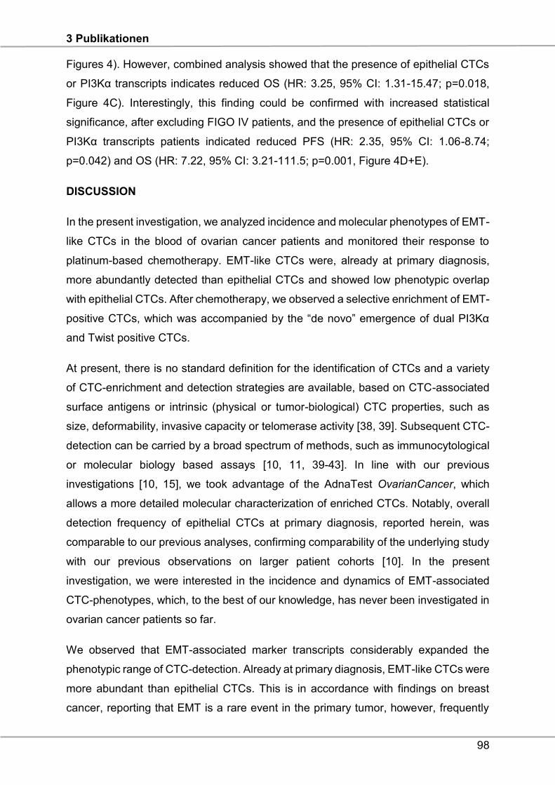
3 Publikationen
98
Figures 4). However, combined analysis showed that the presence of epithelial CTCs
or PI3Kα transcripts indicates reduced OS (HR: 3.25, 95% CI: 1.31-15.47; p=0.018,
Figure 4C). Interestingly, this finding could be confirmed with increased statistical
significance, after excluding FIGO IV patients, and the presence of epithelial CTCs or
PI3Kα transcripts patients indicated reduced PFS (HR: 2.35, 95% CI: 1.06-8.74;
p=0.042) and OS (HR: 7.22, 95% CI: 3.21-111.5; p=0.001, Figure 4D+E).
DISCUSSION
In the present investigation, we analyzed incidence and molecular phenotypes of EMT-
like CTCs in the blood of ovarian cancer patients and monitored their response to
platinum-based chemotherapy. EMT-like CTCs were, already at primary diagnosis,
more abundantly detected than epithelial CTCs and showed low phenotypic overlap
with epithelial CTCs. After chemotherapy, we observed a selective enrichment of EMT-
positive CTCs, which was accompanied by the “de novo” emergence of dual PI3Kα
and Twist positive CTCs.
At present, there is no standard definition for the identification of CTCs and a variety
of CTC-enrichment and detection strategies are available, based on CTC-associated
surface antigens or intrinsic (physical or tumor-biological) CTC properties, such as
size, deformability, invasive capacity or telomerase activity [38, 39]. Subsequent CTC-
detection can be carried by a broad spectrum of methods, such as immunocytological
or molecular biology based assays [10, 11, 39-43]. In line with our previous
investigations [10, 15], we took advantage of the AdnaTest OvarianCancer, which
allows a more detailed molecular characterization of enriched CTCs. Notably, overall
detection frequency of epithelial CTCs at primary diagnosis, reported herein, was
comparable to our previous analyses, confirming comparability of the underlying study
with our previous observations on larger patient cohorts [10]. In the present
investigation, we were interested in the incidence and dynamics of EMT-associated
CTC-phenotypes, which, to the best of our knowledge, has never been investigated in
ovarian cancer patients so far.
We observed that EMT-associated marker transcripts considerably expanded the
phenotypic range of CTC-detection. Already at primary diagnosis, EMT-like CTCs were
more abundant than epithelial CTCs. This is in accordance with findings on breast
cancer, reporting that EMT is a rare event in the primary tumor, however, frequently
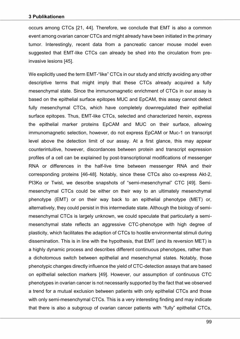
3 Publikationen
99
occurs among CTCs [21, 44]. Therefore, we conclude that EMT is also a common
event among ovarian cancer CTCs and might already have been initiated in the primary
tumor. Interestingly, recent data from a pancreatic cancer mouse model even
suggested that EMT-like CTCs can already be shed into the circulation from pre-
invasive lesions [45].
We explicitly used the term EMT-“like” CTCs in our study and strictly avoiding any other
descriptive terms that might imply that these CTCs already acquired a fully
mesenchymal state. Since the immunomagnetic enrichment of CTCs in our assay is
based on the epithelial surface epitopes MUC and EpCAM, this assay cannot detect
fully mesenchymal CTCs, which have completely downregulated their epithelial
surface epitopes. Thus, EMT-like CTCs, selected and characterized herein, express
the epithelial marker proteins EpCAM and MUC on their surface, allowing
immunomagnetic selection, however, do not express EpCAM or Muc-1 on transcript
level above the detection limit of our assay. At a first glance, this may appear
counterintuitive, however, discordances between protein and transcript expression
profiles of a cell can be explained by post-transcriptional modifications of messenger
RNA or differences in the half-live time between messenger RNA and their
corresponding proteins [46-48]. Notably, since these CTCs also co-express Akt-2,
PI3Kα or Twist, we describe snapshots of “semi-mesenchymal” CTC [49]. Semi-
mesenchymal CTCs could be either on their way to an ultimately mesenchymal
phenotype (EMT) or on their way back to an epithelial phenotype (MET) or,
alternatively, they could persist in this intermediate state. Although the biology of semi-
mesenchymal CTCs is largely unknown, we could speculate that particularly a semi-
mesenchymal state reflects an aggressive CTC-phenotype with high degree of
plasticity, which facilitates the adaption of CTCs to hostile environmental stimuli during
dissemination. This is in line with the hypothesis, that EMT (and its reversion MET) is
a highly dynamic process and describes different continuous phenotypes, rather than
a dichotomous switch between epithelial and mesenchymal states. Notably, those
phenotypic changes directly influence the yield of CTC-detection assays that are based
on epithelial selection markers [49]. However, our assumption of continuous CTC
phenotypes in ovarian cancer is not necessarily supported by the fact that we observed
a trend for a mutual exclusion between patients with only epithelial CTCs and those
with only semi-mesenchymal CTCs. This is a very interesting finding and may indicate
that there is also a subgroup of ovarian cancer patients with “fully” epithelial CTCs,
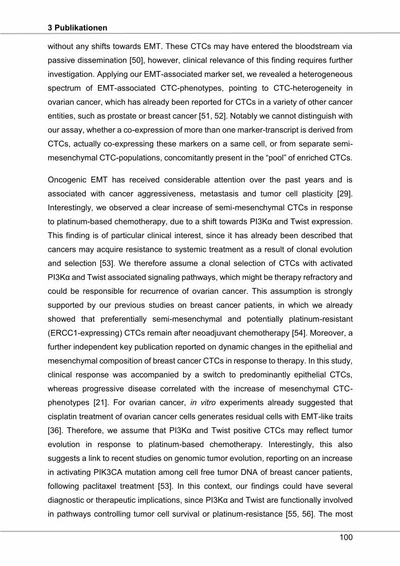
3 Publikationen
100
without any shifts towards EMT. These CTCs may have entered the bloodstream via
passive dissemination [50], however, clinical relevance of this finding requires further
investigation. Applying our EMT-associated marker set, we revealed a heterogeneous
spectrum of EMT-associated CTC-phenotypes, pointing to CTC-heterogeneity in
ovarian cancer, which has already been reported for CTCs in a variety of other cancer
entities, such as prostate or breast cancer [51, 52]. Notably we cannot distinguish with
our assay, whether a co-expression of more than one marker-transcript is derived from
CTCs, actually co-expressing these markers on a same cell, or from separate semi-
mesenchymal CTC-populations, concomitantly present in the “pool” of enriched CTCs.
Oncogenic EMT has received considerable attention over the past years and is
associated with cancer aggressiveness, metastasis and tumor cell plasticity [29].
Interestingly, we observed a clear increase of semi-mesenchymal CTCs in response
to platinum-based chemotherapy, due to a shift towards PI3Kα and Twist expression.
This finding is of particular clinical interest, since it has already been described that
cancers may acquire resistance to systemic treatment as a result of clonal evolution
and selection [53]. We therefore assume a clonal selection of CTCs with activated
PI3Kα and Twist associated signaling pathways, which might be therapy refractory and
could be responsible for recurrence of ovarian cancer. This assumption is strongly
supported by our previous studies on breast cancer patients, in which we already
showed that preferentially semi-mesenchymal and potentially platinum-resistant
(ERCC1-expressing) CTCs remain after neoadjuvant chemotherapy [54]. Moreover, a
further independent key publication reported on dynamic changes in the epithelial and
mesenchymal composition of breast cancer CTCs in response to therapy. In this study,
clinical response was accompanied by a switch to predominantly epithelial CTCs,
whereas progressive disease correlated with the increase of mesenchymal CTC-
phenotypes [21]. For ovarian cancer, in vitro experiments already suggested that
cisplatin treatment of ovarian cancer cells generates residual cells with EMT-like traits
[36]. Therefore, we assume that PI3Kα and Twist positive CTCs may reflect tumor
evolution in response to platinum-based chemotherapy. Interestingly, this also
suggests a link to recent studies on genomic tumor evolution, reporting on an increase
in activating PIK3CA mutation among cell free tumor DNA of breast cancer patients,
following paclitaxel treatment [53]. In this context, our findings could have several
diagnostic or therapeutic implications, since PI3Kα and Twist are functionally involved
in pathways controlling tumor cell survival or platinum-resistance [55, 56]. The most
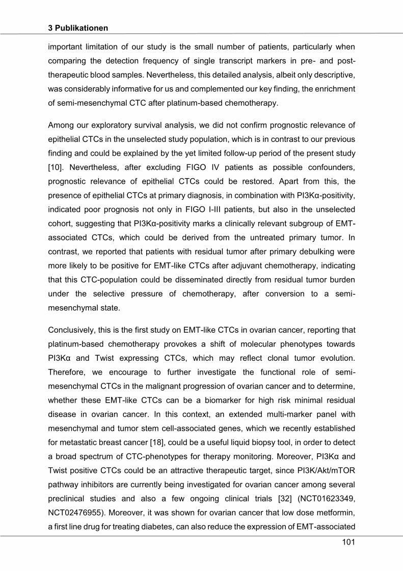
3 Publikationen
101
important limitation of our study is the small number of patients, particularly when
comparing the detection frequency of single transcript markers in pre- and post-
therapeutic blood samples. Nevertheless, this detailed analysis, albeit only descriptive,
was considerably informative for us and complemented our key finding, the enrichment
of semi-mesenchymal CTC after platinum-based chemotherapy.
Among our exploratory survival analysis, we did not confirm prognostic relevance of
epithelial CTCs in the unselected study population, which is in contrast to our previous
finding and could be explained by the yet limited follow-up period of the present study
[10]. Nevertheless, after excluding FIGO IV patients as possible confounders,
prognostic relevance of epithelial CTCs could be restored. Apart from this, the
presence of epithelial CTCs at primary diagnosis, in combination with PI3Kα-positivity,
indicated poor prognosis not only in FIGO I-III patients, but also in the unselected
cohort, suggesting that PI3Kα-positivity marks a clinically relevant subgroup of EMT-
associated CTCs, which could be derived from the untreated primary tumor. In
contrast, we reported that patients with residual tumor after primary debulking were
more likely to be positive for EMT-like CTCs after adjuvant chemotherapy, indicating
that this CTC-population could be disseminated directly from residual tumor burden
under the selective pressure of chemotherapy, after conversion to a semi-
mesenchymal state.
Conclusively, this is the first study on EMT-like CTCs in ovarian cancer, reporting that
platinum-based chemotherapy provokes a shift of molecular phenotypes towards
PI3Kα and Twist expressing CTCs, which may reflect clonal tumor evolution.
Therefore, we encourage to further investigate the functional role of semi-
mesenchymal CTCs in the malignant progression of ovarian cancer and to determine,
whether these EMT-like CTCs can be a biomarker for high risk minimal residual
disease in ovarian cancer. In this context, an extended multi-marker panel with
mesenchymal and tumor stem cell-associated genes, which we recently established
for metastatic breast cancer [18], could be a useful liquid biopsy tool, in order to detect
a broad spectrum of CTC-phenotypes for therapy monitoring. Moreover, PI3Kα and
Twist positive CTCs could be an attractive therapeutic target, since PI3K/Akt/mTOR
pathway inhibitors are currently being investigated for ovarian cancer among several
preclinical studies and also a few ongoing clinical trials [32] (NCT01623349,
NCT02476955). Moreover, it was shown for ovarian cancer that low dose metformin,
a first line drug for treating diabetes, can also reduce the expression of EMT-associated
3 Publikationen
102
makers, such as Twist, suggesting further options for potentially targeting this CTC-
population [57]. Since the biology of ovarian cancer CTCs is largely unknown, we
believe that our finding could be a step forward in understanding their heterogeneity
and their dynamics in response to chemotherapy.
MATERIAL AND METHODS
Patient characteristics
The present study was conducted at the Departments of Gynecology and Obstetrics
at the University Hospitals of Essen and Dresden, Germany. In this study, a total of 95
patients, diagnosed between 2010 and 2014 with histologically confirmed epithelial
ovarian cancer, were analyzed. Clinical characteristics of the patients are documented
in Table 1. Informed written consent was obtained from all patients and the study was
approved by the Local Ethic Committees (Essen 05-2870; Dresden EK 236082012)
and was performed according to the declaration of Helsinki. Tumors were classified
according to the WHO classification of tumors of the female genital tract. Grading was
conducted using the grading system proposed by Silverberg [58] and tumor staging
was classified according to the Fédération Internationale de Gynécology et
d'Obstétrique. The whole study population underwent primary radical surgery. Total
abdominal hysterectomy, bilateral salpingo-oophorectomy, infragastric omentectomy
and peritoneal stripping was performed. The most important aim of surgery was to
achieve macroscopic complete tumor resection. Radical pelvic and para-aortic
lymphadenectomy were only performed if macroscopic complete tumor resection was
achieved. All patients received at least six cycles of carboplatinum AUC 5 and
paclitaxel 175 mg/m2.
Enrichment and molecular characterization of ovarian cancer CTCs
Peripheral blood (5 ml) from each patient was collected in EDTA tubes (Sarstedt &
AG+Co., Nümbrecht, Germany) and processed within 4h for the enrichment of CTCs
and subsequent expression analysis, according to the AdnaTest OvarianCancer
Detect and the AdnaTest EMT-1 Detect (QIAGEN, Hannover GmbH, Langenhagen,
Germany, Supplementary Figure 1+2). These assays have been described in detail
elsewhere [10, 15, 44]. The Adnatest was performed in biological replicates; therefore
two independent consecutive blood samples were obtained from each patient at each
time point. Briefly, we applied immuno-magnetical enrichment of CTCs (Adnatest
3 Publikationen
103
OvarianCancer Select, Adnatest EMT-1Select), targeting epithelial cellular adhesion
molecules.
For the detection of epithelial CTCs, RNA was isolated and gene expression analysis
was performed by reverse-transcription (RT) and multiplex RT-PCR, detecting
EpCAM, Muc-1, and CA-125 (AdnaTest OvarianCancerDetect). In this assay,
amplicons with the following sizes were generated: EpCAM: 396bp; Muc-1: 293bp; CA-
125: 432bp. For the detection EMT-like CTCs, RNA was isolated and gene expression
analysis was performed by reverse-transcription (RT) and multiplex RT-PCR, detecting
PI3Kα, Akt-2 and Twist (AdnaTest EMT-1Detect). Contaminating leukocytes (about
1500 per sample) were reduced by approximately 10fold using a special washing
buffer (AdnaWash buffer) enabling the proper differentiation of EMT expression
profiles with a specificity and sensitivity of >90%, which was confirmed in healthy donor
samples [20, 44]. In this assay, amplicons with the following sizes were generated:
PI3Kα: 595bp, Akt-2: 306 bp; Twist: 203bp. β-actin served as an internal control
(amplicon size: 119 bp) and PCR-products were visualized with the Agilent Bioanalyzer
(Agilent Technologies, Santa Clara, USA). An amplicon concentration of >0.2 ng/µl
was applied as threshold for EpCAM, Muc-1 or CA-125 positivity. Amplicon
concentration of >0.2 ng/µl was applied as threshold for Akt-2, >0.25ng/µl for PI3Kα
and >0,15ng/µl for Twist positivity, respectively.
The analytical sensitivity of the detection of CTC-associated EMT-transcripts was
determined by the analysis of a low number of target cells (5 IGROV ovarian cancer
cells spiked into 5 ml blood of healthy donors). Healthy donor samples without spiked
tumor cells were used to determine the specificity of the test. Applying the above
mentioned amplicon cut-off values, 97% of 30 healthy donor samples were negative
for EMT markers. These experiments demonstrate that a potential risk of false-positive
events is negligible in our present analysis.

3 Publikationen
104
ABBREVIATIONS
AUTHOR CONTRIBUTIONS
IC, SKB, PB, PW, SH, RK and JDK made substantial contributions to the conception
and design of the study, to the experimental work or to the acquisition of data and to
the analysis/interpretation of the results. JDK, SKB, IC, PW and SH were involved in
drafting the manuscript or revising it. All authors read and approved the manuscript in
its final version.
CONFLICT OF INTEREST STATEMENT
Sabine Kasimir-Bauer is a consultant for QIAGEN, Hilden, Germany.
Siegfried Hauch is an employee of QIAGEN, Hilden, Germany.
All other authors declare that they have no conflict of interest.
FUNDING
The present study was supported by internal budgets from the University of Essen and
Dresden, Germany.
BM bone marrow
CTC(s) circulating tumor cell(s)
DTC(s) disseminated tumor cell(s)
EMT epithelial-to-mesenchymal-transition
EpCAM epithelial cell adhesion molecule
ERCC1 excision repair cross-complementation group 1
MET mesenchymal-to-epithelial-transition
Muc-1 Mucin-1
OS overall survial
PFS progression free survival
RT reverse transcription
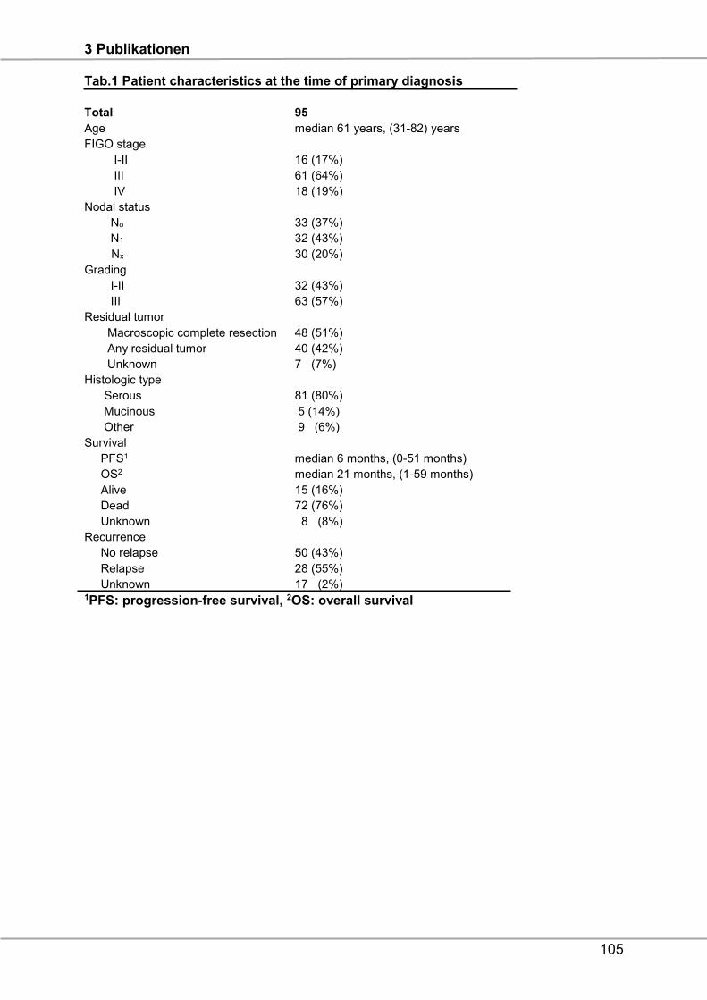
3 Publikationen
105
Tab.1 Patient characteristics at the time of primary diagnosis
Total 95
Age median 61 years, (31-82) years
FIGO stage
I-II 16 (17%)
III 61 (64%)
IV 18 (19%)
Nodal status
No 33 (37%)
N1 32 (43%)
Nx 30 (20%)
Grading
I-II 32 (43%)
III 63 (57%)
Residual tumor
Macroscopic complete resection 48 (51%)
Any residual tumor 40 (42%)
Unknown 7 (7%)
Histologic type
Serous 81 (80%)
Mucinous 5 (14%)
Other 9 (6%)
Survival
PFS1 median 6 months, (0-51 months)
OS2 median 21 months, (1-59 months)
Alive 15 (16%)
Dead 72 (76%)
Unknown 8 (8%)
Recurrence
No relapse 50 (43%)
Relapse 28 (55%)
Unknown 17 (2%) 1PFS: progression-free survival, 2OS: overall survival
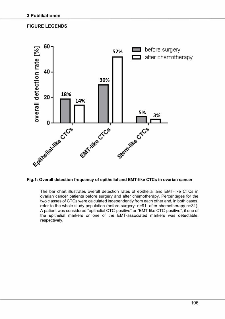
3 Publikationen
106
FIGURE LEGENDS
Fig.1: Overall detection frequency of epithelial and EMT-like CTCs in ovarian cancer
The bar chart illustrates overall detection rates of epithelial and EMT-like CTCs in ovarian cancer patients before surgery and after chemotherapy. Percentages for the two classes of CTCs were calculated independently from each other and, in both cases, refer to the whole study population (before surgery: n=91, after chemotherapy n=31). A patient was considered “epithelial CTC-positive” or “EMT-like CTC-positive”, if one of the epithelial markers or one of the EMT-associated markers was detectable, respectively.
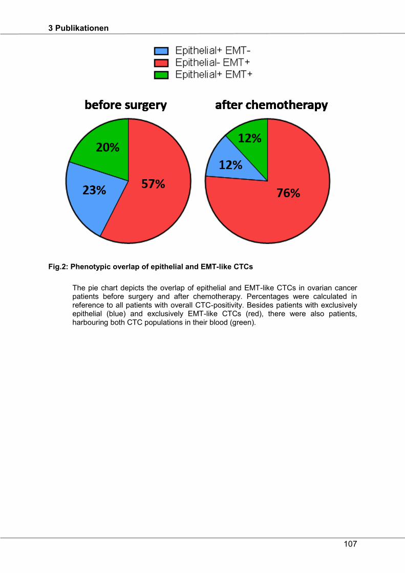
3 Publikationen
107
Fig.2: Phenotypic overlap of epithelial and EMT-like CTCs
The pie chart depicts the overlap of epithelial and EMT-like CTCs in ovarian cancer patients before surgery and after chemotherapy. Percentages were calculated in reference to all patients with overall CTC-positivity. Besides patients with exclusively epithelial (blue) and exclusively EMT-like CTCs (red), there were also patients, harbouring both CTC populations in their blood (green).
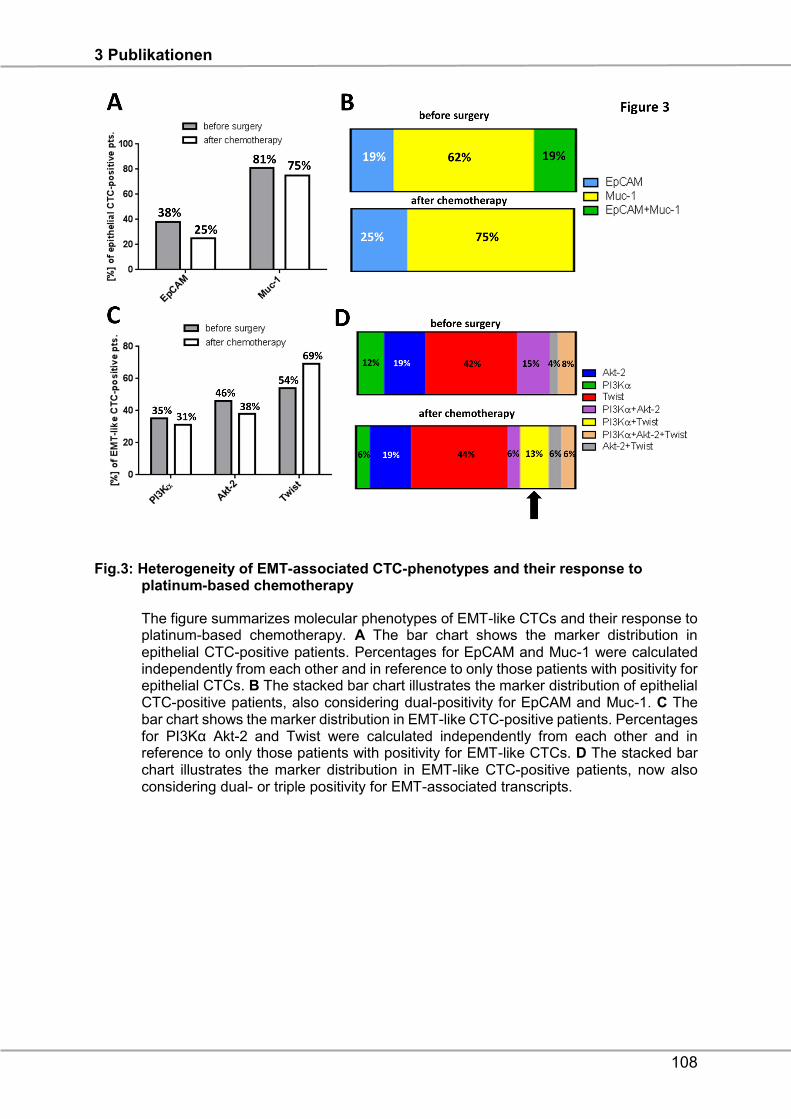
3 Publikationen
108
Fig.3: Heterogeneity of EMT-associated CTC-phenotypes and their response to platinum-based chemotherapy
The figure summarizes molecular phenotypes of EMT-like CTCs and their response to platinum-based chemotherapy. A The bar chart shows the marker distribution in epithelial CTC-positive patients. Percentages for EpCAM and Muc-1 were calculated independently from each other and in reference to only those patients with positivity for epithelial CTCs. B The stacked bar chart illustrates the marker distribution of epithelial CTC-positive patients, also considering dual-positivity for EpCAM and Muc-1. C The bar chart shows the marker distribution in EMT-like CTC-positive patients. Percentages for PI3Kα Akt-2 and Twist were calculated independently from each other and in reference to only those patients with positivity for EMT-like CTCs. D The stacked bar chart illustrates the marker distribution in EMT-like CTC-positive patients, now also considering dual- or triple positivity for EMT-associated transcripts.
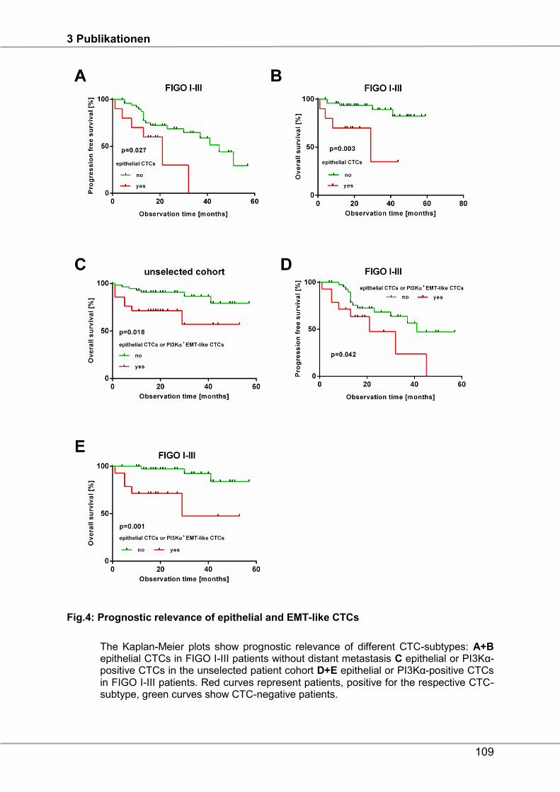
3 Publikationen
109
Fig.4: Prognostic relevance of epithelial and EMT-like CTCs
The Kaplan-Meier plots show prognostic relevance of different CTC-subtypes: A+B epithelial CTCs in FIGO I-III patients without distant metastasis C epithelial or PI3Kα- positive CTCs in the unselected patient cohort D+E epithelial or PI3Kα-positive CTCs in FIGO I-III patients. Red curves represent patients, positive for the respective CTC- subtype, green curves show CTC-negative patients.
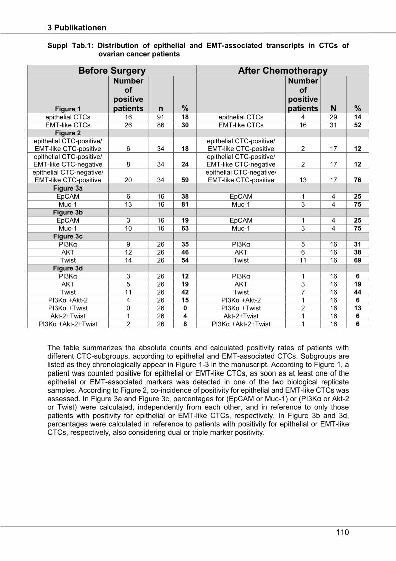
3 Publikationen
110
Suppl Tab.1: Distribution of epithelial and EMT-associated transcripts in CTCs of ovarian cancer patients
The table summarizes the absolute counts and calculated positivity rates of patients with different CTC-subgroups, according to epithelial and EMT-associated CTCs. Subgroups are listed as they chronologically appear in Figure 1-3 in the manuscript. According to Figure 1, a patient was counted positive for epithelial or EMT-like CTCs, as soon as at least one of the epithelial or EMT-associated markers was detected in one of the two biological replicate samples. According to Figure 2, co-incidence of positivity for epithelial and EMT-like CTCs was assessed. In Figure 3a and Figure 3c, percentages for (EpCAM or Muc-1) or (PI3Kα or Akt-2 or Twist) were calculated, independently from each other, and in reference to only those patients with positivity for epithelial or EMT-like CTCs, respectively. In Figure 3b and 3d, percentages were calculated in reference to patients with positivity for epithelial or EMT-like CTCs, respectively, also considering dual or triple marker positivity.
Before Surgery After Chemotherapy
Figure 1
Number of
positive patients n %
Number of
positive patients N %
epithelial CTCs 16 91 18 epithelial CTCs 4 29 14
EMT-like CTCs 26 86 30 EMT-like CTCs 16 31 52
Figure 2 epithelial CTC-positive/ EMT-like CTC-positive 6 34 18
epithelial CTC-positive/ EMT-like CTC-positive 2 17 12
epithelial CTC-positive/ EMT-like CTC-negative 8 34 24
epithelial CTC-positive/ EMT-like CTC-negative 2 17 12
epithelial CTC-negative/ EMT-like CTC-positive 20 34 59
epithelial CTC-negative/ EMT-like CTC-positive 13 17 76
Figure 3a EpCAM 6 16 38 EpCAM 1 4 25
Muc-1 13 16 81 Muc-1 3 4 75
Figure 3b EpCAM 3 16 19 EpCAM 1 4 25
Muc-1 10 16 63 Muc-1 3 4 75
Figure 3c PI3Kα 9 26 35 PI3Kα 5 16 31
AKT 12 26 46 AKT 6 16 38
Twist 14 26 54 Twist 11 16 69
Figure 3d PI3Kα 3 26 12 PI3Kα 1 16 6
AKT 5 26 19 AKT 3 16 19
Twist 11 26 42 Twist 7 16 44
PI3Kα +Akt-2 4 26 15 PI3Kα +Akt-2 1 16 6
PI3Kα +Twist 0 26 0 PI3Kα +Twist 2 16 13
Akt-2+Twist 1 26 4 Akt-2+Twist 1 16 6
PI3Kα +Akt-2+Twist 2 26 8 PI3Kα +Akt-2+Twist 1 16 6
3 Publikationen
111
REFERENCES
1. Goodman MT, Howe HL, Tung KH, Hotes J, Miller BA, Coughlin SS and Chen
VW. Incidence of ovarian cancer by race and ethnicity in the United States, 1992-
1997. Cancer. 2003; 97(10 Suppl):2676-2685.
2. du Bois A, Quinn M, Thigpen T, Vermorken J, Avall-Lundqvist E, Bookman M,
Bowtell D, Brady M, Casado A, Cervantes A, Eisenhauer E, Friedlaender M, Fujiwara
K, et al. 2004 consensus statements on the management of ovarian cancer: final
document of the 3rd International Gynecologic Cancer Intergroup Ovarian Cancer
Consensus Conference (GCIG OCCC 2004). Ann Oncol. 2005; 16 Suppl 8:viii7-viii12.
3. du Bois A, Reuss A, Pujade-Lauraine E, Harter P, Ray-Coquard I and Pfisterer
J. Role of surgical outcome as prognostic factor in advanced epithelial ovarian cancer:
a combined exploratory analysis of 3 prospectively randomized phase 3 multicenter
trials: by the Arbeitsgemeinschaft Gynaekologische Onkologie Studiengruppe
Ovarialkarzinom (AGO-OVAR) and the Groupe d'Investigateurs Nationaux Pour les
Etudes des Cancers de l'Ovaire (GINECO). Cancer. 2009; 115(6):1234-1244.
4. Wimberger P, Lehmann N, Kimmig R, Burges A, Meier W, Du Bois A and
Arbeitsgemeinschaft Gynaekologische Onkologie Ovarian Cancer Study G. Prognostic
factors for complete debulking in advanced ovarian cancer and its impact on survival.
An exploratory analysis of a prospectively randomized phase III study of the
Arbeitsgemeinschaft Gynaekologische Onkologie Ovarian Cancer Study Group (AGO-
OVAR). Gynecol Oncol. 2007; 106(1):69-74.
5. Wimberger P, Wehling M, Lehmann N, Kimmig R, Schmalfeldt B, Burges A,
Harter P, Pfisterer J and du Bois A. Influence of residual tumor on outcome in ovarian
cancer patients with FIGO stage IV disease: an exploratory analysis of the AGO-OVAR
(Arbeitsgemeinschaft Gynaekologische Onkologie Ovarian Cancer Study Group).
Annals of surgical oncology. 2010; 17(6):1642-1648.
6. Martin LP and Schilder RJ. Management of recurrent ovarian carcinoma: current
status and future directions. Seminars in oncology. 2009; 36(2):112-125.
7. Burger RA, Brady MF, Bookman MA, Fleming GF, Monk BJ, Huang H, Mannel
RS, Homesley HD, Fowler J, Greer BE, Boente M, Birrer MJ, Liang SX, et al.
Incorporation of bevacizumab in the primary treatment of ovarian cancer. N Engl J
Med. 2011; 365(26):2473-2483.
8. Ledermann J, Harter P, Gourley C, Friedlander M, Vergote I, Rustin G, Scott
CL, Meier W, Shapira-Frommer R, Safra T, Matei D, Fielding A, Spencer S, et al.
3 Publikationen
112
Olaparib maintenance therapy in patients with platinum-sensitive relapsed serous
ovarian cancer: a preplanned retrospective analysis of outcomes by BRCA status in a
randomised phase 2 trial. Lancet Oncol. 2014; 15(8):852-861.
9. Romero-Laorden N, Olmos D, Fehm T, Garcia-Donas J and Diaz-Padilla I.
Circulating and disseminated tumor cells in ovarian cancer: a systematic review.
Gynecol Oncol. 2014; 133(3):632-639.
10. Aktas B, Kasimir-Bauer S, Heubner M, Kimmig R and Wimberger P. Molecular
profiling and prognostic relevance of circulating tumor cells in the blood of ovarian
cancer patients at primary diagnosis and after platinum-based chemotherapy. Int J
Gynecol Cancer. 2011; 21(5):822-830.
11. Obermayr E, Castillo-Tong DC, Pils D, Speiser P, Braicu I, Van Gorp T, Mahner
S, Sehouli J, Vergote I and Zeillinger R. Molecular characterization of circulating tumor
cells in patients with ovarian cancer improves their prognostic significance -- a study
of the OVCAD consortium. Gynecol Oncol. 2013; 128(1):15-21.
12. Wimberger P, Heubner M, Otterbach F, Fehm T, Kimmig R and Kasimir-Bauer
S. Influence of platinum-based chemotherapy on disseminated tumor cells in blood
and bone marrow of patients with ovarian cancer. Gynecol Oncol. 2007; 107(2):331-
338.
13. Fan T, Zhao Q, Chen JJ, Chen WT and Pearl ML. Clinical significance of
circulating tumor cells detected by an invasion assay in peripheral blood of patients
with ovarian cancer. Gynecol Oncol. 2009; 112(1):185-191.
14. Zhou Y, Bian B, Yuan X, Xie G, Ma Y and Shen L. Prognostic Value of
Circulating Tumor Cells in Ovarian Cancer: A Meta-Analysis. PloS one. 2015;
10(6):e0130873.
15. Kuhlmann JD, Wimberger P, Bankfalvi A, Keller T, Scholer S, Aktas B, Buderath
P, Hauch S, Otterbach F, Kimmig R and Kasimir-Bauer S. ERCC1-positive circulating
tumor cells in the blood of ovarian cancer patients as a predictive biomarker for
platinum resistance. Clini Chem. 2014; 60(10):1282-1289.
16. Chebouti I, Blassl C, Wimberger P, Neubauer H, Fehm T, Kimmig R and
Kasimir-Bauer S. Analysis of disseminated tumor cells before and after platinum based
chemotherapy in primary ovarian cancer. Do stem cell like cells predict prognosis?
Oncotarget. 2016.
17. Blassl C, Kuhlmann JD, Webers A, Wimberger P, Fehm T and Neubauer H.
Gene expression profiling of single circulating tumor cells in ovarian cancer -
Establishment of a multi-marker gene panel. Mol Oncol. 2016.

3 Publikationen
113
18. Bredemeier M, Edimiris P, Tewes M, Mach P, Aktas B, Schellbach D, Wagner
J, Kimmig R and Kasimir-Bauer S. Establishment of a multimarker qPCR panel for the
molecular characterization of circulating tumor cells in blood samples of metastatic
breast cancer patients during the course of palliative treatment. Oncotarget. 2016.
19. Brouwer A, De Laere B, Peeters D, Peeters M, Salgado R, Dirix L and Van
Laere S. Evaluation and consequences of heterogeneity in the circulating tumor cell
compartment. Oncotarget. 2016.
20. Aktas B, Tewes M, Fehm T, Hauch S, Kimmig R and Kasimir-Bauer S. Stem
cell and epithelial-mesenchymal transition markers are frequently overexpressed in
circulating tumor cells of metastatic breast cancer patients. Breast Cancer Res : BCR.
2009; 11(4):R46.
21. Yu M, Bardia A, Wittner BS, Stott SL, Smas ME, Ting DT, Isakoff SJ, Ciciliano
JC, Wells MN, Shah AM, Concannon KF, Donaldson MC, Sequist LV, et al. Circulating
breast tumor cells exhibit dynamic changes in epithelial and mesenchymal
composition. Science. 2013; 339(6119):580-584.
22. Liu H, Zhang X, Li J, Sun B, Qian H and Yin Z. The biological and clinical
importance of epithelial-mesenchymal transition in circulating tumor cells. J Clin Med.
2014.
23. Shih JY, Tsai MF, Chang TH, Chang YL, Yuan A, Yu CJ, Lin SB, Liou GY, Lee
ML, Chen JJ, Hong TM, Yang SC, Su JL, et al. Transcription repressor slug promotes
carcinoma invasion and predicts outcome of patients with lung adenocarcinoma. Clin
Cancer Res. 2005; 11(22):8070-8078.
24. Drake JM, Strohbehn G, Bair TB, Moreland JG and Henry MD. ZEB1 enhances
transendothelial migration and represses the epithelial phenotype of prostate cancer
cells. Mol Biol Cell. 2009; 20(8):2207-2217.
25. Smit MA, Geiger TR, Song JY, Gitelman I and Peeper DS. A Twist-Snail axis
critical for TrkB-induced epithelial-mesenchymal transition-like transformation, anoikis
resistance, and metastasis. Mol Cell Biol. 2009; 29(13):3722-3737.
26. Stoletov K, Kato H, Zardouzian E, Kelber J, Yang J, Shattil S and Klemke R.
Visualizing extravasation dynamics of metastatic tumor cells. J Cell Sci. 2010; 123(Pt
13):2332-2341.
27. Ocana OH, Corcoles R, Fabra A, Moreno-Bueno G, Acloque H, Vega S,
Barrallo-Gimeno A, Cano A and Nieto MA. Metastatic colonization requires the
repression of the epithelial-mesenchymal transition inducer Prrx1. Cancer cell. 2012;
22(6):709-724.
3 Publikationen
114
28. Jiang J, Tang YL and Liang XH. EMT: a new vision of hypoxia promoting cancer
progression. Cancer Biol Ther. 2011; 11(8):714-723.
29. Kalluri R and Weinberg RA. The basics of epithelial-mesenchymal transition. J
Clin Invest.. 2009; 119(6):1420-1428.
30. Levine DA, Bogomolniy F, Yee CJ, Lash A, Barakat RR, Borgen PI and Boyd J.
Frequent mutation of the PIK3CA gene in ovarian and breast cancers. Clin Cancer
Res. 2005; 11(8):2875-2878.
31. Engelman JA, Luo J and Cantley LC. The evolution of phosphatidylinositol 3-
kinases as regulators of growth and metabolism. Nat Rev Genet. 2006; 7(8):606-619.
32. Liu P, Cheng H, Roberts TM and Zhao JJ. Targeting the phosphoinositide 3-
kinase pathway in cancer. Nat Rev Drug Discov. 2009; 8(8):627-644.
33. Xu W, Yang Z and Lu N. A new role for the PI3K/Akt signaling pathway in the
epithelial-mesenchymal transition. Cell adhesion & migration. 2015; 9(4):317-324.
34. Tania M, Khan MA and Fu J. Epithelial to mesenchymal transition inducing
transcription factors and metastatic cancer. Tumour biology 2014; 35(8):7335-7342.
35. Ahmed N, Abubaker K, Findlay J and Quinn M. Epithelial mesenchymal
transition and cancer stem cell-like phenotypes facilitate chemoresistance in recurrent
ovarian cancer. Curr Cancer Drug Targets. 2010; 10(3):268-278.
36. Latifi A, Abubaker K, Castrechini N, Ward AC, Liongue C, Dobill F, Kumar J,
Thompson EW, Quinn MA, Findlay JK and Ahmed N. Cisplatin treatment of primary
and metastatic epithelial ovarian carcinomas generates residual cells with
mesenchymal stem cell-like profile. J Cell Biochem. 2011; 112(10):2850-2864.
37. Davidson B, Trope CG and Reich R. Epithelial-mesenchymal transition in
ovarian carcinoma. Front Oncol. 2012; 2:33.
38. Lianidou ES and Markou A. Circulating tumor cells in breast cancer: detection
systems, molecular characterization, and future challenges. Clin Chem. 2011;
57(9):1242-1255.
39. Joosse SA, Gorges TM and Pantel K. Biology, detection, and clinical
implications of circulating tumor cells. EMBO Mol Med. 2015; 7(1):1-11.
40. Xenidis N, Ignatiadis M, Apostolaki S, Perraki M, Kalbakis K, Agelaki S,
Stathopoulos EN, Chlouverakis G, Lianidou E, Kakolyris S, Georgoulias V and
Mavroudis D. Cytokeratin-19 mRNA-positive circulating tumor cells after adjuvant
chemotherapy in patients with early breast cancer. J Clin Oncol. 2009; 27(13):2177-
2184.
3 Publikationen
115
41. Heitzer E, Auer M, Gasch C, Pichler M, Ulz P, Hoffmann EM, Lax S,
Waldispuehl-Geigl J, Mauermann O, Lackner C, Hofler G, Eisner F, Sill H, et al.
Complex tumor genomes inferred from single circulating tumor cells by array-CGH and
next-generation sequencing. Cancer Res. 2013; 73(10):2965-2975.
42. Xenidis N, Perraki M, Kafousi M, Apostolaki S, Bolonaki I, Stathopoulou A,
Kalbakis K, Androulakis N, Kouroussis C, Pallis T, Christophylakis C, Argyraki K,
Lianidou ES, et al. Predictive and prognostic value of peripheral blood cytokeratin-19
mRNA-positive cells detected by real-time polymerase chain reaction in node-negative
breast cancer patients. J Clin Oncol. 2006; 24(23):3756-3762.
43. Andreopoulou E, Yang LY, Rangel KM, Reuben JM, Hsu L, Krishnamurthy S,
Valero V, Fritsche HA and Cristofanilli M. Comparison of assay methods for detection
of circulating tumor cells in metastatic breast cancer: AdnaGen AdnaTest
BreastCancer Select/Detect versus Veridex CellSearch system. Int J Cancer. 2012;
130(7):1590-1597.
44. Kasimir-Bauer S, Hoffmann O, Wallwiener D, Kimmig R and Fehm T.
Expression of stem cell and epithelial-mesenchymal transition markers in primary
breast cancer patients with circulating tumor cells. Breast cancer Res : BCR. 2012;
14(1):R15.
45. Bachmann HS, Meier W, du Bois A, Kimmig R, Kuhlmann JD, Siffert W, Sehouli
J, Wollschlaeger K, Huober J, Hillemanns P, Burges A, Schmalfeldt B, Aminossadati
B, et al. The FNTB promoter polymorphism rs11623866 as a potential predictive
biomarker for lonafarnib treatment of ovarian cancer patients. Br J Clin Pharmacol.
2015.
46. Pascal LE, True LD, Campbell DS, Deutsch EW, Risk M, Coleman IM, Eichner
LJ, Nelson PS and Liu AY. Correlation of mRNA and protein levels: cell type-specific
gene expression of cluster designation antigens in the prostate. BMC Genomics. 2008;
9:246.
47. Greenbaum D, Colangelo C, Williams K and Gerstein M. Comparing protein
abundance and mRNA expression levels on a genomic scale. Genome Biol. 2003;
4(9):117.
48. Nie L, Wu G and Zhang W. Correlation of mRNA expression and protein
abundance affected by multiple sequence features related to translational efficiency in
Desulfovibrio vulgaris: a quantitative analysis. Genetics. 2006; 174(4):2229-2243.
3 Publikationen
116
49. Bednarz-Knoll N, Alix-Panabieres C and Pantel K. Plasticity of disseminating
cancer cells in patients with epithelial malignancies. Cancer Metastasis Rev. 2012;
31(3-4):673-687.
50. Alpaugh ML, Tomlinson JS, Kasraeian S and Barsky SH. Cooperative role of E-
cadherin and sialyl-Lewis X/A-deficient MUC1 in the passive dissemination of tumor
emboli in inflammatory breast carcinoma. Oncogene. 2002; 21(22):3631-3643.
51. Massard C, Oulhen M, Le Moulec S, Auger N, Foulon S, Abou-Lovergne A,
Billiot F, Valent A, Marty V, Loriot Y, Fizazi K, Vielh P and Farace F. Phenotypic and
genetic heterogeneity of tumor tissue and circulating tumor cells in patients with
metastatic castrationresistant prostate cancer: a report from the PETRUS prospective
study. Oncotarget. 2016.
52. Shaw JA, Guttery DS, Hills A, Fernandez-Garcia D, Page K, Rosales BM,
Goddard KS, Hastings RK, Luo J, Ogle O, Woodley L, Ali S, Stebbing J, et al. Mutation
analysis of cell-free DNA and single circulating tumor cells in metastatic breast cancer
patients with high CTC counts. Clin Cancer Res. 2016.
53. Murtaza M, Dawson SJ, Tsui DW, Gale D, Forshew T, Piskorz AM, Parkinson
C, Chin SF, Kingsbury Z, Wong AS, Marass F, Humphray S, Hadfield J, et al. Non-
invasive analysis of acquired resistance to cancer therapy by sequencing of plasma
DNA. Nature. 2013; 497(7447):108-112.
54. Kasimir-Bauer S, Bittner AK, Konig L, Reiter K, Keller T, Kimmig R and
Hoffmann O. Does primary neoadjuvant systemic therapy eradicate minimal residual
disease? Analysis of disseminated and circulating tumor cells before and after therapy.
Breast Cancer Res. 2016; 18(1):20.
55. Kolasa IK, Rembiszewska A, Felisiak A, Ziolkowska-Seta I, Murawska M, Moes
J, Timorek A, Dansonka-Mieszkowska A and Kupryjanczyk J. PIK3CA amplification
associates with resistance to chemotherapy in ovarian cancer patients. Cancer Bio
Ther. 2009; 8(1):21-26.
56. Nuti SV, Mor G, Li P and Yin G. TWIST and ovarian cancer stem cells:
implications for chemoresistance and metastasis. Oncotarget. 2014; 5(17):7260-7271.
57. Zhang R, Zhang P, Wang H, Hou D, Li W, Xiao G and Li C. Inhibitory effects of
metformin at low concentration on epithelial-mesenchymal transition of
CD44(+)CD117(+) ovarian cancer stem cells. Stem Cell Res. 2015; 6:262.
58. Silverberg SG. Histopathologic grading of ovarian carcinoma: a review and
proposal. Int J Gynecol Pathol. 2000; 19(1):7-15.
3 Publikationen
117
RASSF1A promoter methylation in high-grade serous ovarian cancer: a direct
comparison study in primary tumors, adjacent morphologically tumor cell free
tissues and paired circulating tumor DNA
Lydia Giannopoulou1, Issam Chebouti2, Kitty Pavlakis3, Sabine Kasimir-Bauer2 and Evi
S. Lianidou1
AFFILIATIONS
(1) Analysis of Circulating Tumor Cells lab, Lab of Analytical Chemistry,
Department of Chemistry, University of Athens, University Campus, Athens,
15771, Greece
(2) Department of Gynecology and Obstetrics, University Hospital of Essen,
University of Duisburg-Essen, Hufelandstrasse 55, Essen, D-45122, Germany
(3) Pathology Department, IASO women’s hospital, 15123, Marousi, Athens,
Greece
CONTACT INFORMATION * Correspondence should be addressed to: Dr. E.S. Lianidou, Analysis of Circulating Tumour Cells lab, Laboratory of Analytical Chemistry, Department of Chemistry, University of Athens, 15771, Greece Tel: ++ 30 210 7274311, Fax: ++ 30 210 7274750, E-mail: [email protected]
KEY WORDS
Circulating tumor DNA; RASSF1A; high-grade serous ovarian cancer; methylation
specific PCR; High Resolution Melting Analysis
3 Publikationen
118
ABSTRACT
The RASSF1A promoter is frequently methylated in high-grade serous ovarian cancer
(HGSC). We examined RASSF1A promoter methylation in primary tumors, adjacent
morphologically tumor cell-free tissues and corresponding circulating tumor DNA
(ctDNA) samples of patients with HGSC, using a real-time methylation specific PCR
(real-time MSP) and a methylation-sensitive high-resolution melting analysis (MS-
HRMA) assay for the detection and semi-quantitative estimation of methylation,
respectively. Two groups of primary HGSC tumor FFPE samples were recruited
(Group A n=67 and Group B n=61), along with matched adjacent morphologically
tumor cell-free tissues (n=58) and corresponding plasma samples (n=59) for group B.
Using both assays, RASSF1A promoter was found highly methylated in primary tumors
of both groups, and at lower percentages in the adjacent morphologically tumor cell-
free tissues. Interestingly, RASSF1A promoter methylation was also observed in
ctDNA by real-time MSP. Overall survival (OS) was significantly associated with
RASSF1A promoter methylation in primary tumor samples using MS-HRMA
(P=0.023). Our results clearly indicate that RASSF1A promoter is methylated in
adjacent tissue surrounding the tumor in HGSC patients. We report for the first time
that RASSF1A promoter methylation provides significant prognostic information in
HGSC patients.
3 Publikationen
119
INTRODUCTION
Ovarian cancer represents the third most frequent gynecological cancer and the fifth
leading cause of cancer-related death in women [1]. Epithelial ovarian cancer is the
main type, characterized by histological and molecular heterogeneity. The most
common subtype, high-grade serous ovarian cancer (HGSC), is often diagnosed at an
advanced stage and little progress has been achieved in standard treatment and
overall survival (OS) during the last three decades [2]. Primary disease is treated with
surgical removal of the tumor, followed by a combination of platinum and taxane-based
chemotherapy [3, 4] with about 20% of patients found to be resistant to this treatment
[5, 6]. New multimodal therapeutic concepts now include targeted therapy applying
Bevacizumab or the PARP inhibitor Olaparib in certain clinical situations [7, 8].
It is now clear that epigenetic alterations hold an important role in cancer initiation and
progression and that aberrant DNA methylation, especially promoter hypermethylation
of tumor suppressor genes is a frequent event in most human cancers [9]. Epigenetic
inactivation of a tumor suppressor gene often results from its promoter methylation and
is considered as an early event during carcinogenesis [10]. Many studies have
reported methylation changes in epithelial ovarian cancer [11] and a recent review
summarizes the differences in the observed methylation patterns in the main
histological subtypes of the disease, including HGSC [12].
Cell-free DNA (cfDNA) circulates at high concentrations in cancer patients and can be
used for the detection of several molecular alterations related to cancer development
[13]. Circulating tumor DNA (ctDNA) is a small percentage of cfDNA that is shed in
circulation by tumor cells and carries all these molecular alterations including tumor
specific mutations, microsatellite instability (MI) [13], loss of heterozygosity (LOH) [14],
and DNA methylation [15]. Circulating tumor DNA is a very promising non-invasive
diagnostic, prognostic and predictive tool, since it provides an easily accessible source
of DNA derived from the tumor [16]. Our group has reported SOX17 [17, 18], CST6
[19] and BRMS1 [20] promoter methylation in cfDNA in breast and non-small cell lung
cancer patients.
The RASSF1 gene belongs to the Ras-association domain family that consists of ten
members. RASSF proteins contribute to microtubule stability and they are involved in
cell cycle regulation, apoptosis, cell migration and cell adhesion. The RASSF1 gene is
found on the 3p21.3 locus and comprises eight exons. Its two promoter regions and
3 Publikationen
120
the implied alternative splicing are responsible for the eight isoforms A-H. RASSF1A
and RASSF1C are mostly studied so far, especially RASSF1A gene isoform that
definitely acts as a tumor suppressor in human cancer [21, 22]. RASSF1A is involved
in molecular pathways including Ras/PI3K/AKT, Ras/RAF/MEK/ERK, Hippo pathways
and β-catenin signaling pathway [22, 23]. The RASSF1A gene is frequently inactivated
by aberrant promoter hypermethylation in the majority of human malignancies,
including breast, lung, gastrointestinal, bladder, head and neck cancer and
gynecological cancers, endometrial and cervical cancer [23]. In ovarian cancer,
RASSF1A promoter methylation has been identified in many studies [24], but no
significant association with clinical outcome has been reported so far.
The aim of the present study was to examine the prognostic significance of RASSF1A
promoter methylation in primary tumors, matched adjacent morphologically tumor cell-
free tissues surrounding the tumor and the corresponding plasma samples of patients
with HGSC. To evaluate the clinical significance of RASSF1A promoter methylation in
HGSC, we applied a highly sensitive real-time methylation specific PCR (real-time
MSP) assay [25] for the detection of RASSF1A promoter methylation and compared it
to a methylation-sensitive high-resolution melting analysis (MS-HRMA) assay. We
further directly compared RASSF1A promoter methylation between primary tumors,
matched adjacent tissues and corresponding plasma ctDNA. To the best of our
knowledge, this is the first study on the evaluation of RASSF1A promoter methylation
status in HGSC that is based on matched primary tumors, adjacent tissues and
corresponding plasma samples from the same patients. Our results clearly indicate
that the RASSF1A promoter is methylated in adjacent tissue surrounding the tumor in
HGSC patients. We also report for the first time that RASSF1A promoter methylation
provides significant prognostic information in HGSC patients.
3 Publikationen
121
RESULTS
A schematic diagram of our study is shown in Figure 1.
RASSF1A promoter methylation status in HGSC by real-time MSP
RASSF1A promoter methylation status was first evaluated in the group A by real-time
MSP. According to our results, RASSF1A promoter was methylated in 27/67 (40.3%)
primary tumor samples. RASSF1A promoter methylation status was further evaluated
in the group B. According to our results, RASSF1A promoter was methylated in 25/61
(41.0%) primary tumor samples. In the group of adjacent morphologically tumor cell-
free tissues of group B, 17/58 (29.3%) samples were found methylated. In cfDNA,
isolated from corresponding plasma, 15/59 (25.4%) samples were found positive for
RASSF1A promoter methylation.
Semi-quantitative estimation of RASSF1A promoter methylation by MS-HRMA
We further evaluated the percentages of RASSF1A promoter methylation in primary
tumor samples and adjacent tissues, by using the semi-quantitative MS-HRMA assay.
RASSF1A promoter was found methylated in 27/67 (40.3%) primary tumor samples of
group A and in 28/61 (45.9%) primary tumor samples of group B. 21/58 (36.2%)
adjacent morphologically tumor cell-free tissues of group B were found methylated.
The MS-HRMA assay can detect heterogeneous methylation; we found
heterogeneously methylated samples both in group A (8/67, 11.9%) and in tumor
samples of group B (7/61, 11.5%). We also observed heterogeneous methylation in
5/58 (8.6%) adjacent tissues of group B. According to the semi-quantitative MS-HRMA,
in most positive cases RASSF1A promoter methylation was detected at a lower
percentage in the adjacent morphologically tumor cell-free tissues, when compared to
the paired primary tumors (Figure 2). However, there were three cases where the
percentage of RASSF1A promoter methylation was higher in the adjacent tissue
(Figure 2). No significant difference was observed (P=0.126, Mann-Whitney U test).
Comparison between real-time MSP and MS-HRMA
When we compared our results derived for the same primary tumor samples in both
group A and group B, by real-time MSP and MS-HRMA, the agreement between the
two assays was almost perfect (Table 1). More specifically, in the group A, there was
an agreement for 63/67 (94.0%) primary tumor samples (P<0.001, 2-sided Pearson χ2
test, k=0.876), while in the group B, there was an agreement for 58/61 (95.1%)
3 Publikationen
122
samples (P<0.001, 2-sided Pearson χ2 test, k=0.900). In the group of adjacent tissue
samples (group B), the agreement between real-time MSP and MS-HRMA was
substantial (50/58, 86.2%, P<0.001, 2-sided Pearson χ2 test, k=0.689) (Table 1).
Direct comparison of RASSF1A promoter methylation status in primary tumors,
adjacent tissues and plasma ctDNA
We further directly compared RASSF1A promoter methylation status in 53 cases,
where primary tumors, adjacent tissues and corresponding plasma ctDNA were
available (triplets, n=53). RASSF1A promoter methylation status in primary tumors and
adjacent tissues was evaluated using both real-time MSP and MS-HRMA, while in
corresponding plasma samples only real-time MSP was used because of its higher
sensitivity. In most cases there was a concordance between our findings in primary
tumors, adjacent tissues and plasma (Table 2). In 45/53 (84.9%) cases we found an
agreement for RASSF1A promoter methylation between primary tumor samples and
adjacent tissues (P<0.001, 2-sided Pearson χ2 test, Cohen’s kappa=0.674). According
to the guidelines for the interpretation of k values, there is a substantial agreement
between the two subgroups. In 33/53 (62.3%) cases we observed a slight agreement
for RASSF1A promoter methylation between primary tumor samples and
corresponding plasma, (P=0.227, 2-sided Pearson χ2 test, k=0.156) (Table 2). In group
B, we used again the 53 triplets for the comparison between primary tumor samples
and adjacent tissues using MS-HRMA. The agreement between the two subgroups
was 47/53 (88.7%, P<0.001, 2-sided Pearson χ2 test, Cohen’s kappa=0.768,
substantial agreement).
Our results on RASSF1A promoter methylation in primary tumors, adjacent tissues and
corresponding plasma samples are shown in Figure 3. In six patients, RASSF1A
promoter methylation was detected in the primary tumor and in the adjacent tissue by
both assays and in corresponding cfDNA in plasma by real-time MSP. In five patients,
RASSF1A promoter methylation was detected only in plasma, while the primary tumors
and adjacent tissues were found negative by both assays.
Prognostic significance of RASSF1A promoter methylation in HGSC
We further proceeded to the estimation of the clinical significance of RASSF1A
promoter methylation status for the patients of group B, as Overall Survival (OS) and
Progression-Free Survival (PFS) data were available along with other
clinicopathological characteristics. The total number of patients is now different (n=47),
3 Publikationen
123
because in some cases the clinical information was not available and all cases where
OS≤4 months were excluded from the survival study. The median OS was 36 months
while the median PFS was 12.5 months (starting date being the date of diagnosis; PFS
was estimated based on the date of relapse; OS was estimated based on the date of
death). The correlation between RASSF1A promoter methylation status of primary
tumor samples with clinicopathological features of the patients is shown in Table 3.
RASSF1A promoter methylation was significantly associated with tumor grade using
both assays (real-time MSP: P=0.043, MS-HRMA: P=0.037) and regional lymph nodes
(pN) using MS-HRMA (P=0.040). No significant correlations are found between
RASSF1A methylation status of adjacent tissues and plasma samples, and
clinicopathological characteristics (data not shown).
The Kaplan-Meier analysis was further performed to correlate OS and PFS data with
RASSF1A promoter methylation status. In primary tumor samples, OS was found to
be significantly correlated with RASSF1A promoter methylation status using MS-
HRMA (P=0.023, log-rank test, Figure 4), whereas no significant correlation was
observed using real-time MSP (P=0.157, log-rank test). No significant correlations
were found between OS and RASSF1A promoter methylation status of adjacent
tissues and plasma samples, and between PFS and RASSF1A promoter methylation
status of all three subgroups (data not shown).
Finally, RASSF1A promoter methylation in primary tumor samples and all the available
clinicopathological features were tested in univariate Cox Regression analysis for
association with OS and PFS. RASSF1A promoter methylation status using MS-HRMA
and platinum resistance were significantly associated with decreased OS (P=0.030
and P=0.019, respectively). The lack of correlation between OS and clinical
parameters known to be of predictive value, like age and tumor rest, can be possibly
explained by the relatively small cohort analyzed here. We next performed multivariate
Cox Regression analysis for RASSF1A promoter methylation estimated with the MS-
HRMA assay and platinum resistance in association with OS, but no independent
prognostic significance was observed. The results are shown in detail in Table 4.
3 Publikationen
124
DISCUSSION
RASSF1A promoter methylation is a common event in ovarian cancer, and was first
identified in ovarian tumor samples over a decade ago [26-28]. Apart from primary
tumors, benign cystadenomas and low malignant potential tumors exhibit RASSF1A
promoter methylation as well [29, 30]. Choi et al. first tried to correlate RASSF1A
promoter methylation with patients outcome, but did not find any correlation [30]. A
possible explanation could be that in this study, all the ovarian cancer samples of
serous histotype were concerned as a single cohort without taking into account the two
subtypes, high- and low-grade serous ovarian cancer. However, it is now known that
these two subtypes differ in the progenitor area and the tumors molecular profile [31].
High methylation frequency of RASSF1A has also been observed by Montavon et al.
at HGSC tumor samples, however, the relatively small number of available survival
data (n=37) could be a possible explanation for the lack of association between
RASSF1A methylation and overall survival in this study [32]. Ibanez et al. screened
ovarian tumors of different histology, with matched preoperative serum or plasma and
peritoneal fluid samples for RASSF1A promoter methylation. They concluded for the
first time that RASSF1A promoter methylation can be detected in cfDNA and
represents an early event in ovarian carcinogenesis [33]. Other studies confirm the
detection of methylated RASSF1A in plasma samples [34-36]. Bon Durant et al.
compared RASSF1A promoter methylation between tumor and matched plasma, in 20
available sample pairs and observed 100% agreement. They also determined changes
in RASSF1A methylation status during the course of treatment [34]. A phase II clinical
trial has reported that demethylation of RASSF1A had a positive correlation with PFS
indicating a possible role of RASSF1A promoter methylation in platinum resistance
[37].
A large number of studies declare RASSF1A promoter methylation in the majority of
human malignancies, including breast, endometrial and cervical cancer [23, 38]. Our
group has shown the prognostic significance of RASSF1A promoter methylation in
early stage breast cancer [39] and reported the frequent RASSF1A promoter
methylation in cfDNA of operable gastric cancer patients [40]. Spitzwieser et al.
investigated RASSF1A promoter methylation status in 17 breast cancer samples and
their matched normal adjacent tissues using MS-HRMA and found high methylation
frequencies in tumors and adjacent tissues, but no correlation between their
methylation status [41]. A previous study also showed no significant concordance
3 Publikationen
125
between methylation changes in 56 breast tumor and their paired adjacent normal
tissues [42]. In endometrioid adenocarcinoma, Arafa et al. reported that RASSF1A
promoter methylation is methylated in endometrial cancer samples but also in a small
group of adjacent normal endometrium tissues surrounding the tumor [43]. Evaluation
of RASSF1A promoter methylation in matched samples of ovarian cancer patients has
been very limited so far. There is only one study, including 3 tumors and their matched
normal adjacent tissues, where all 3 adjacent tissues were found non-methylated [28].
Promoter methylation in adjacent morphologically tumor cell-free tissues reflects field
cancerization, also called field effect. Field effect describes all the genetic and
epigenetic abnormalities found in adjacent tissues that are defined as morphologically
normal [44].
In the present study, we examined RASSF1A promoter methylation status in primary
tumors, adjacent morphologically tumor cell-free tissues and corresponding plasma
samples of patients with HGSC, using real-time MSP and MS-HRMA. The two assays
showed almost perfect agreement when applied in the tumor samples of both groups,
and substantial agreement in the adjacent tissues of group B. Two tumor samples from
group A, 3 tumor and 6 adjacent samples from group B were found methylated with
MS-HRMA and unmethylated with real-time MSP (Table 1). These discrepant results
probably are due to the slight differences in the promoter region assessed by each
assay (Figure 5). As shown in Figure 5, there is one extra CG in the forward MS-
HRMA primer. When a sample is negative in real-time MSP, all CGs in MSP primers
are unmethylated. But if the additional CG that is present only in the MS-HRMA primer
is methylated in the sample, then the MS-HRMA result is expected to be positive. There
were also 2 tumor samples from group A and 2 adjacent samples from group B where
we observed methylation with real-time MSP, but not with MS-HRMA (Table 1). This
is potentially due to the higher sensitivity of the real-time MSP assay compared to MS-
HRMA.
In adjacent morphologically tumor cell-free tissues of group B, we observed rather high
methylation levels using both MS-HRMA (36.2%) and real-time MSP (29.3%). This
indicates field cancerization (field effect) and potentially cancer progression. We found
no significant difference between tumor and adjacent tissue methylation level and in
three cases, the percentage of RASSF1A promoter methylation was higher in the
adjacent tissue (Figure 2). A potential explanation is the strong field effect that
characterizes the cases studied, especially these three pairs.
3 Publikationen
126
According to our findings, RASSF1A promoter methylation is significantly correlated
with OS when MS-HRMA is used (P=0.023), but no significant correlation is observed
with real-time MSP (P=0.157). This fact gives an advantage to the MS-HRMA, although
real-time MSP is a more sensitive assay. However, real-time MSP is preferable for
methylation studies in plasma ctDNA, due to its higher sensitivity. In six samples,
where the primary tumor was found unmethylated, the corresponding plasma samples
were methylated. A potential explanation for this observation could be based on tumor
heterogeneity; it is now clear that tissue biopsy represents a snapshot of tumor
molecular profile, while cfDNA reflects the total genetic and epigenetic characteristics
of a particular cancer. The effect of tumor heterogeneity in our results could be only
shown if single cells analysis was performed. However we have not designed our study
based on single cell analysis, this could be a nice idea for future studies. Moreover,
cfDNA can originate not only from the primary tumor but from metastatic sites as well,
from apoptotic and necrotic cells.
In conclusion, we performed a direct comparison study on RASSF1A promoter
methylation in primary tumors, adjacent tissues and plasma samples in HGSC
patients. We report for the first time that RASSF1A promoter is methylated in adjacent
tissue surrounding the tumor in HGSC patients and that RASSF1A promoter
methylation provides prognostic information since it is significantly correlated with OS.
Our results indicate that the evaluation of RASSF1A methylation status in ovarian
cancer has the potential to provide important prognostic information; however to verify
this finding, more prospective studies should be performed. Taking into account that
the primary tumor tissue is typically available only at primary diagnosis, it would be
valuable to establish a non-invasive blood-based biomarker for stratifying response to
platinum-based chemotherapy at primary diagnosis and for guiding individualized
therapy decisions in the future.
3 Publikationen
127
MATERIALS AND METHODS
Clinical samples
Our study material consisted of two main groups of samples from patients with primary
HGSC; a) group A that consists of 67 primary ovarian formalin fixed paraffin-embedded
tissues (FFPEs) and b) group B that consists of 61 primary FFPEs, 58 available
adjacent morphologically tumor cell-free tissues (FFPEs) and 59 available
corresponding plasma samples (2mL). For the plasma sampling, two x 5ml
ethylenediaminetetraacetic acid (EDTA) blood samples were collected at time point of
diagnosis, before tumor surgery and before the application of therapeutic substances
with an S-Monovette (Sarstedt AG & Co.). Blood was centrifuged at 1500g for 10min
and the plasma supernatant was stored at -80°C until further usage. The available
clinicopathological features for both groups are shown in Table 5. For the evaluation
of the specificity of our assays, two groups of normal samples were recruited: a) a
small group of 16 normal fallopian tube FFPEs that were obtained from women of the
reproductive age group and b) a larger group of 51 plasma samples obtained from
healthy women (2mL). All group A samples and the normal fallopian tube samples
were obtained from the Pathology Department of IASO women’s hospital, Athens,
Greece. All group B samples were obtained from the Department of Pathology and the
Department of Gynecology and Obstetrics, University Hospital of Essen, University of
Duisburg-Essen, Germany. FFPE tissue blocks, retrieved from the Institute of
Pathology and Neuropathology of the University Hospital of Essen, Germany were
stained with Haematoxilin & Eosin and FFPE sections used for our assays were
prepared and reviewed by a pathologist. We analyzed only samples with a tumor cell
content of equal or more than 60%. All tissue samples were prepared under
supervision of a pathologist. According to our pathologists, these are reasonable
amounts of tumor tissue to study since 100% purity of tumor tissue can only be
achieved in rare cases. All patients gave their informed written consent to participate
in the study, which was approved by the Local Essen Research Ethics Committee
(05/2856), and IASO women’s hospital Ethics committee (Date: 05/2014). The
OVCAR29 and IGROV1 ovarian cancer cell lines were used as positive controls in
real-time MSP and MS-HRMA reactions for the detection of RASSF1A methylation.
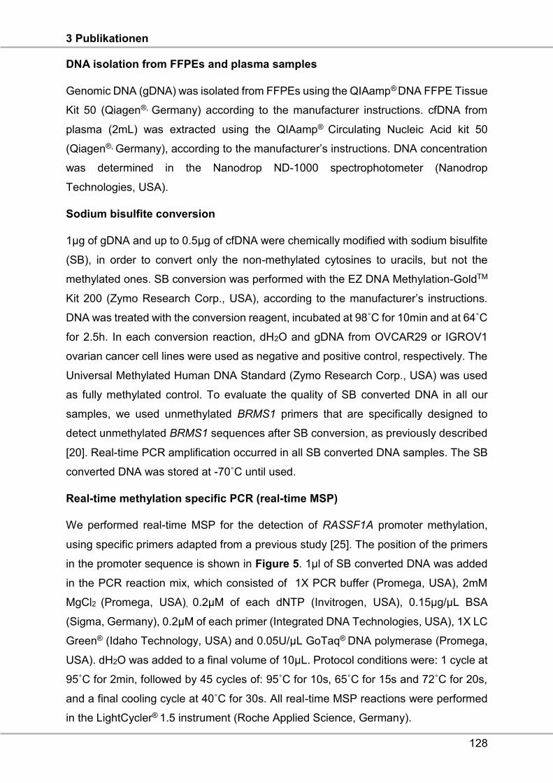
3 Publikationen
128
DNA isolation from FFPEs and plasma samples
Genomic DNA (gDNA) was isolated from FFPEs using the QIAamp® DNA FFPE Tissue
Kit 50 (Qiagen®, Germany) according to the manufacturer instructions. cfDNA from
plasma (2mL) was extracted using the QIAamp® Circulating Nucleic Acid kit 50
(Qiagen®, Germany), according to the manufacturer’s instructions. DNA concentration
was determined in the Nanodrop ND-1000 spectrophotometer (Nanodrop
Technologies, USA).
Sodium bisulfite conversion
1μg of gDNA and up to 0.5μg of cfDNA were chemically modified with sodium bisulfite
(SB), in order to convert only the non-methylated cytosines to uracils, but not the
methylated ones. SB conversion was performed with the EZ DNA Methylation-GoldTM
Kit 200 (Zymo Research Corp., USA), according to the manufacturer’s instructions.
DNA was treated with the conversion reagent, incubated at 98˚C for 10min and at 64˚C
for 2.5h. In each conversion reaction, dH2O and gDNA from OVCAR29 or IGROV1
ovarian cancer cell lines were used as negative and positive control, respectively. The
Universal Methylated Human DNA Standard (Zymo Research Corp., USA) was used
as fully methylated control. To evaluate the quality of SB converted DNA in all our
samples, we used unmethylated BRMS1 primers that are specifically designed to
detect unmethylated BRMS1 sequences after SB conversion, as previously described
[20]. Real-time PCR amplification occurred in all SB converted DNA samples. The SB
converted DNA was stored at -70˚C until used.
Real-time methylation specific PCR (real-time MSP)
We performed real-time MSP for the detection of RASSF1A promoter methylation,
using specific primers adapted from a previous study [25]. The position of the primers
in the promoter sequence is shown in Figure 5. 1μl of SB converted DNA was added
in the PCR reaction mix, which consisted of 1X PCR buffer (Promega, USA), 2mM
MgCl2 (Promega, USA), 0.2μΜ of each dNTP (Invitrogen, USA), 0.15μg/μL BSA
(Sigma, Germany), 0.2μΜ of each primer (Integrated DNA Technologies, USA), 1X LC
Green® (Idaho Technology, USA) and 0.05U/μL GoTaq® DNA polymerase (Promega,
USA). dH2O was added to a final volume of 10μL. Protocol conditions were: 1 cycle at
95˚C for 2min, followed by 45 cycles of: 95˚C for 10s, 65˚C for 15s and 72˚C for 20s,
and a final cooling cycle at 40˚C for 30s. All real-time MSP reactions were performed
in the LightCycler® 1.5 instrument (Roche Applied Science, Germany).
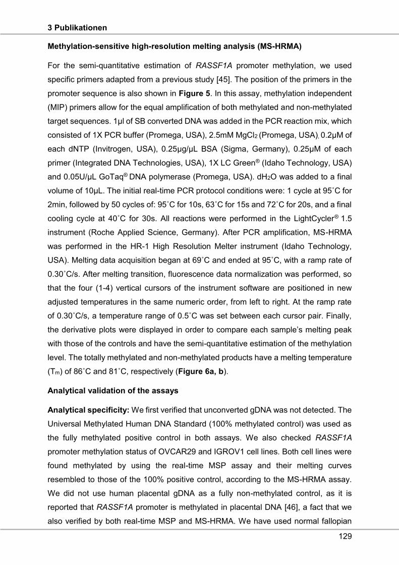
3 Publikationen
129
Methylation-sensitive high-resolution melting analysis (MS-HRMA)
For the semi-quantitative estimation of RASSF1A promoter methylation, we used
specific primers adapted from a previous study [45]. The position of the primers in the
promoter sequence is also shown in Figure 5. In this assay, methylation independent
(MIP) primers allow for the equal amplification of both methylated and non-methylated
target sequences. 1μl of SB converted DNA was added in the PCR reaction mix, which
consisted of 1X PCR buffer (Promega, USA), 2.5mM MgCl2 (Promega, USA), 0.2μΜ of
each dNTP (Invitrogen, USA), 0.25μg/μL BSA (Sigma, Germany), 0.25μΜ of each
primer (Integrated DNA Technologies, USA), 1X LC Green® (Idaho Technology, USA)
and 0.05U/μL GoTaq® DNA polymerase (Promega, USA). dH2O was added to a final
volume of 10μL. The initial real-time PCR protocol conditions were: 1 cycle at 95˚C for
2min, followed by 50 cycles of: 95˚C for 10s, 63˚C for 15s and 72˚C for 20s, and a final
cooling cycle at 40˚C for 30s. All reactions were performed in the LightCycler® 1.5
instrument (Roche Applied Science, Germany). After PCR amplification, MS-HRMA
was performed in the HR-1 High Resolution Melter instrument (Idaho Technology,
USA). Melting data acquisition began at 69˚C and ended at 95˚C, with a ramp rate of
0.30˚C/s. After melting transition, fluorescence data normalization was performed, so
that the four (1-4) vertical cursors of the instrument software are positioned in new
adjusted temperatures in the same numeric order, from left to right. At the ramp rate
of 0.30˚C/s, a temperature range of 0.5˚C was set between each cursor pair. Finally,
the derivative plots were displayed in order to compare each sample’s melting peak
with those of the controls and have the semi-quantitative estimation of the methylation
level. The totally methylated and non-methylated products have a melting temperature
(Tm) of 86˚C and 81˚C, respectively (Figure 6a, b).
Analytical validation of the assays
Analytical specificity: We first verified that unconverted gDNA was not detected. The
Universal Methylated Human DNA Standard (100% methylated control) was used as
the fully methylated positive control in both assays. We also checked RASSF1A
promoter methylation status of OVCAR29 and IGROV1 cell lines. Both cell lines were
found methylated by using the real-time MSP assay and their melting curves
resembled to those of the 100% positive control, according to the MS-HRMA assay.
We did not use human placental gDNA as a fully non-methylated control, as it is
reported that RASSF1A promoter is methylated in placental DNA [46], a fact that we
also verified by both real-time MSP and MS-HRMA. We have used normal fallopian
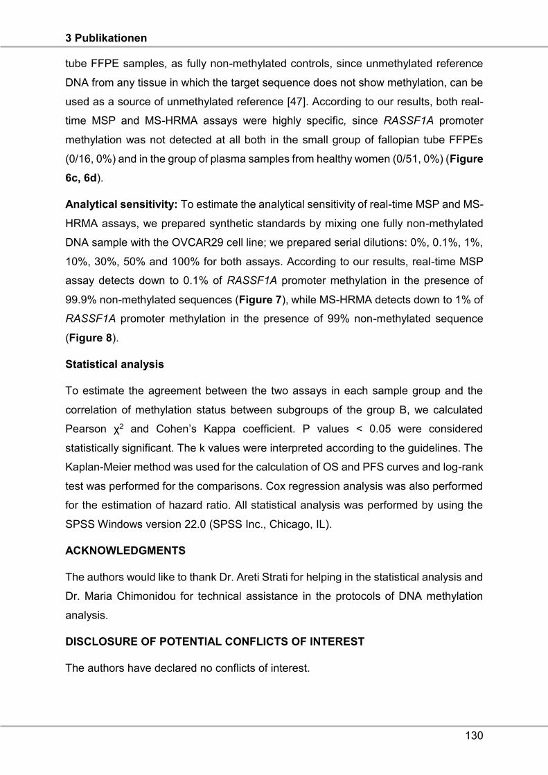
3 Publikationen
130
tube FFPE samples, as fully non-methylated controls, since unmethylated reference
DNA from any tissue in which the target sequence does not show methylation, can be
used as a source of unmethylated reference [47]. According to our results, both real-
time MSP and MS-HRMA assays were highly specific, since RASSF1A promoter
methylation was not detected at all both in the small group of fallopian tube FFPEs
(0/16, 0%) and in the group of plasma samples from healthy women (0/51, 0%) (Figure
6c, 6d).
Analytical sensitivity: To estimate the analytical sensitivity of real-time MSP and MS-
HRMA assays, we prepared synthetic standards by mixing one fully non-methylated
DNA sample with the OVCAR29 cell line; we prepared serial dilutions: 0%, 0.1%, 1%,
10%, 30%, 50% and 100% for both assays. According to our results, real-time MSP
assay detects down to 0.1% of RASSF1A promoter methylation in the presence of
99.9% non-methylated sequences (Figure 7), while MS-HRMA detects down to 1% of
RASSF1A promoter methylation in the presence of 99% non-methylated sequence
(Figure 8).
Statistical analysis
To estimate the agreement between the two assays in each sample group and the
correlation of methylation status between subgroups of the group B, we calculated
Pearson χ2 and Cohen’s Kappa coefficient. P values < 0.05 were considered
statistically significant. The k values were interpreted according to the guidelines. The
Kaplan-Meier method was used for the calculation of OS and PFS curves and log-rank
test was performed for the comparisons. Cox regression analysis was also performed
for the estimation of hazard ratio. All statistical analysis was performed by using the
SPSS Windows version 22.0 (SPSS Inc., Chicago, IL).
ACKNOWLEDGMENTS
The authors would like to thank Dr. Areti Strati for helping in the statistical analysis and
Dr. Maria Chimonidou for technical assistance in the protocols of DNA methylation
analysis.
DISCLOSURE OF POTENTIAL CONFLICTS OF INTEREST
The authors have declared no conflicts of interest.
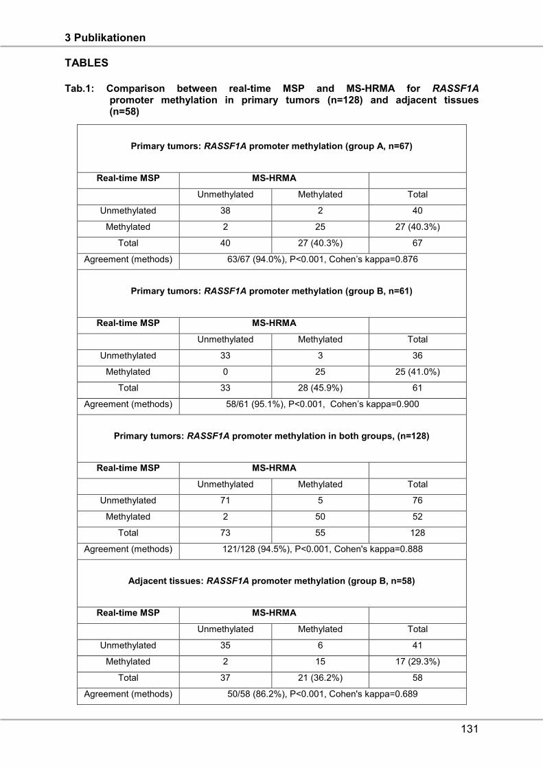
3 Publikationen
131
TABLES
Tab.1: Comparison between real-time MSP and MS-HRMA for RASSF1A promoter methylation in primary tumors (n=128) and adjacent tissues (n=58)
Primary tumors: RASSF1A promoter methylation (group A, n=67)
Real-time MSP MS-HRMA
Unmethylated Methylated Total
Unmethylated 38 2 40
Methylated 2 25 27 (40.3%)
Total 40 27 (40.3%) 67
Agreement (methods) 63/67 (94.0%), P<0.001, Cohen’s kappa=0.876
Primary tumors: RASSF1A promoter methylation (group B, n=61)
Real-time MSP MS-HRMA
Unmethylated Methylated Total
Unmethylated 33 3 36
Methylated 0 25 25 (41.0%)
Total 33 28 (45.9%) 61
Agreement (methods) 58/61 (95.1%), P<0.001, Cohen’s kappa=0.900
Primary tumors: RASSF1A promoter methylation in both groups, (n=128)
Real-time MSP MS-HRMA
Unmethylated Methylated Total
Unmethylated 71 5 76
Methylated 2 50 52
Total 73 55 128
Agreement (methods) 121/128 (94.5%), P<0.001, Cohen's kappa=0.888
Adjacent tissues: RASSF1A promoter methylation (group B, n=58)
Real-time MSP MS-HRMA
Unmethylated Methylated Total
Unmethylated 35 6 41
Methylated 2 15 17 (29.3%)
Total 37 21 (36.2%) 58
Agreement (methods) 50/58 (86.2%), P<0.001, Cohen's kappa=0.689
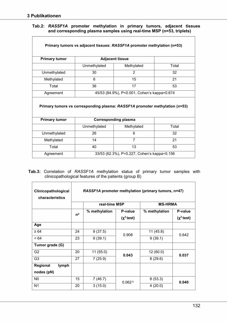
3 Publikationen
132
Tab.2: RASSF1A promoter methylation in primary tumors, adjacent tissues and corresponding plasma samples using real-time MSP (n=53, triplets)
Primary tumors vs adjacent tissues: RASSF1A promoter methylation (n=53)
Primary tumor Adjacent tissue
Unmethylated Methylated Total
Unmethylated 30 2 32
Methylated 6 15 21
Total 36 17 53
Agreement 45/53 (84.9%), P<0.001, Cohen’s kappa=0.674
Primary tumors vs corresponding plasma: RASSF1A promoter methylation (n=53)
Primary tumor Corresponding plasma
Unmethylated Methylated Total
Unmethylated 26 6 32
Methylated 14 7 21
Total 40 13 53
Agreement 33/53 (62.3%), P=0.227, Cohen’s kappa=0.156
Tab.3: Correlation of RASSF1A methylation status of primary tumor samples with clinicopathological features of the patients (group B)
Clinicopathological
characteristics
RASSF1A promoter methylation (primary tumors, n=47)
real-time MSP MS-HRMA
na
% methylation P-value
(χ2 test)
% methylation P-value
(χ2 test)
Age
≥ 64 24 9 (37.5) 0.908
11 (45.8) 0.642
< 64 23 9 (39.1) 9 (39.1)
Tumor grade (G)
G2 20 11 (55.0) 0.043
12 (60.0) 0.037
G3 27 7 (25.9) 8 (29.6)
Regional lymph
nodes (pN)
N0 15 7 (46.7) 0.062 b
8 (53.3) 0.040
N1 20 3 (15.0) 4 (20.0)
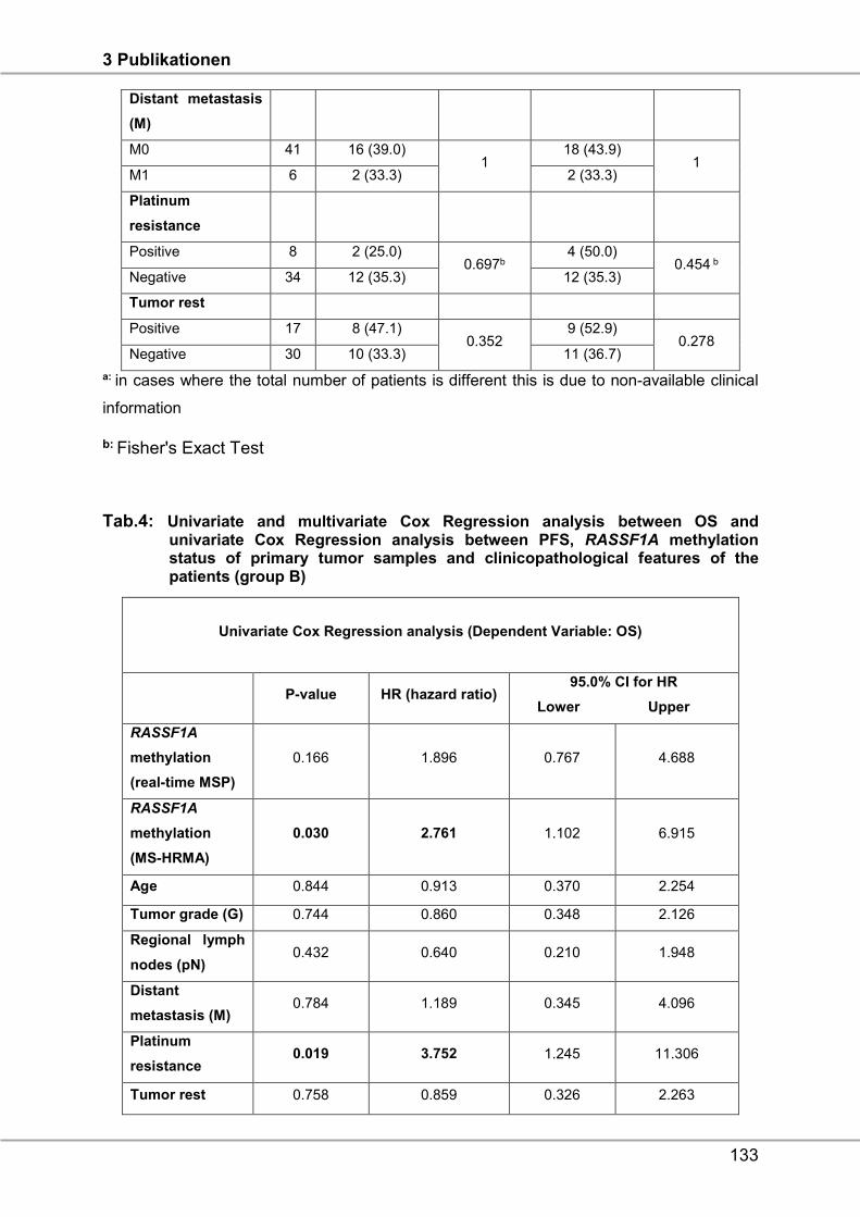
3 Publikationen
133
Distant metastasis
(M)
M0 41 16 (39.0) 1
18 (43.9) 1
M1 6 2 (33.3) 2 (33.3)
Platinum
resistance
Positive 8 2 (25.0) 0.697b
4 (50.0) 0.454 b
Negative 34 12 (35.3) 12 (35.3)
Tumor rest
Positive 17 8 (47.1) 0.352
9 (52.9) 0.278
Negative 30 10 (33.3) 11 (36.7)
a: in cases where the total number of patients is different this is due to non-available clinical
information
b: Fisher's Exact Test
Tab.4: Univariate and multivariate Cox Regression analysis between OS and univariate Cox Regression analysis between PFS, RASSF1A methylation status of primary tumor samples and clinicopathological features of the patients (group B)
Univariate Cox Regression analysis (Dependent Variable: OS)
P-value HR (hazard ratio) 95.0% CI for HR
Lower Upper
RASSF1A
methylation
(real-time MSP)
0.166 1.896 0.767 4.688
RASSF1A
methylation
(MS-HRMA)
0.030 2.761 1.102 6.915
Age 0.844 0.913 0.370 2.254
Tumor grade (G) 0.744 0.860 0.348 2.126
Regional lymph
nodes (pN) 0.432 0.640 0.210 1.948
Distant
metastasis (M) 0.784 1.189 0.345 4.096
Platinum
resistance 0.019 3.752 1.245 11.306
Tumor rest 0.758 0.859 0.326 2.263
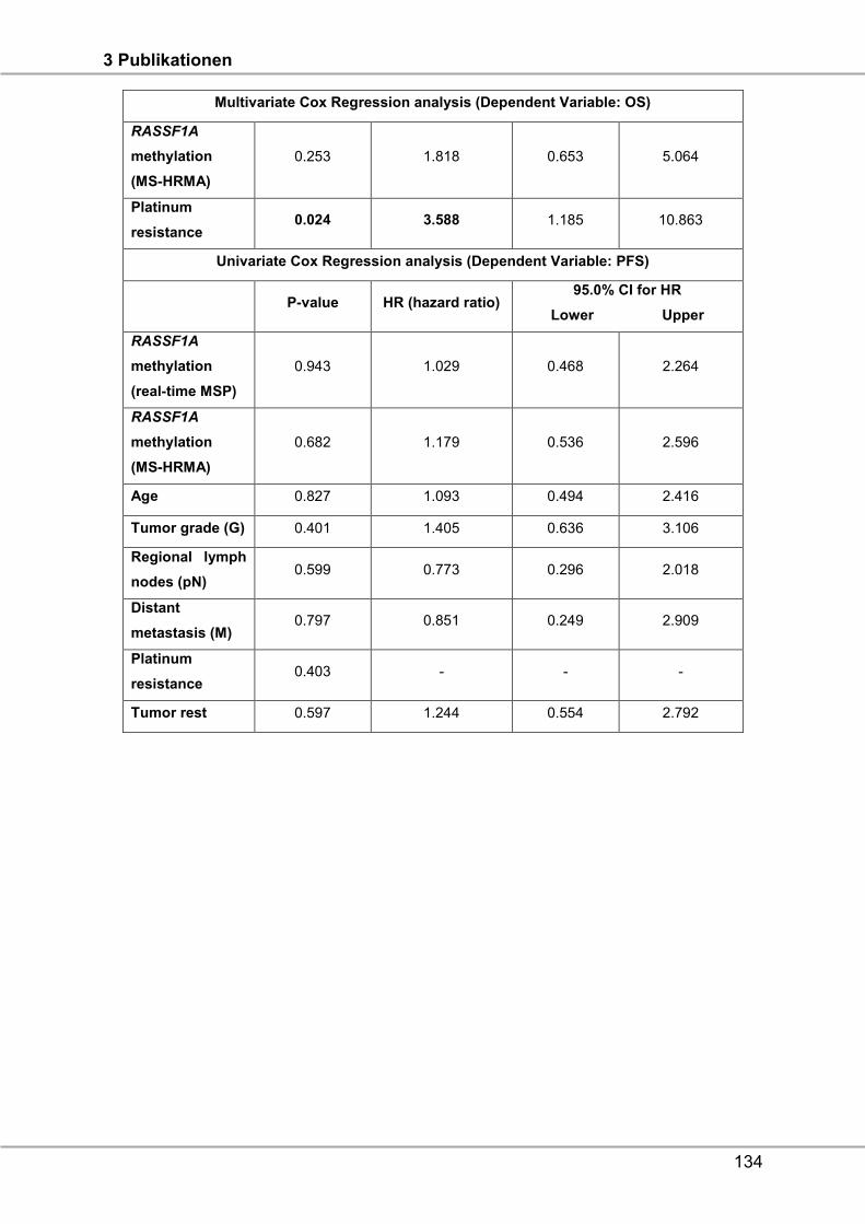
3 Publikationen
134
Multivariate Cox Regression analysis (Dependent Variable: OS)
RASSF1A
methylation
(MS-HRMA)
0.253 1.818 0.653 5.064
Platinum
resistance 0.024 3.588 1.185 10.863
Univariate Cox Regression analysis (Dependent Variable: PFS)
P-value HR (hazard ratio) 95.0% CI for HR
Lower Upper
RASSF1A
methylation
(real-time MSP)
0.943 1.029 0.468 2.264
RASSF1A
methylation
(MS-HRMA)
0.682 1.179 0.536 2.596
Age 0.827 1.093 0.494 2.416
Tumor grade (G) 0.401 1.405 0.636 3.106
Regional lymph
nodes (pN) 0.599 0.773 0.296 2.018
Distant
metastasis (M) 0.797 0.851 0.249 2.909
Platinum
resistance 0.403 - - -
Tumor rest 0.597 1.244 0.554 2.792
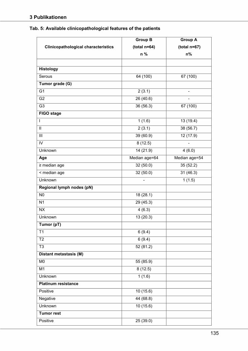
3 Publikationen
135
Tab. 5: Available clinicopathological features of the patients
Clinicopathological characteristics
Group B
(total n=64)
n %
Group A
(total n=67)
n%
Histology
Serous 64 (100) 67 (100)
Tumor grade (G)
G1 2 (3.1) -
G2 26 (40.6) -
G3 36 (56.3) 67 (100)
FIGO stage
I 1 (1.6) 13 (19.4)
II 2 (3.1) 38 (56.7)
III 39 (60.9) 12 (17.9)
IV 8 (12.5) -
Unknown 14 (21.9) 4 (6.0)
Age Median age=64 Median age=54
≥ median age 32 (50.0) 35 (52.2)
< median age 32 (50.0) 31 (46.3)
Unknown - 1 (1.5)
Regional lymph nodes (pN)
N0 18 (28.1)
N1 29 (45.3)
NX 4 (6.3)
Unknown 13 (20.3)
Tumor (pT)
T1 6 (9.4)
T2 6 (9.4)
T3 52 (81.2)
Distant metastasis (M)
M0 55 (85.9)
M1 8 (12.5)
Unknown 1 (1.6)
Platinum resistance
Positive 10 (15.6)
Negative 44 (68.8)
Unknown 10 (15.6)
Tumor rest
Positive 25 (39.0)
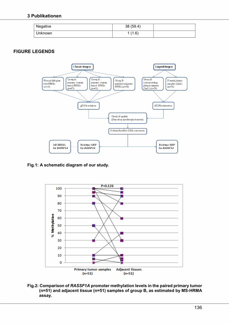
3 Publikationen
136
Negative 38 (59.4)
Unknown 1 (1.6)
FIGURE LEGENDS
Fig.1: A schematic diagram of our study.
Fig.2: Comparison of RASSF1A promoter methylation levels in the paired primary tumor (n=51) and adjacent tissue (n=51) samples of group B, as estimated by MS-HRMA assay.
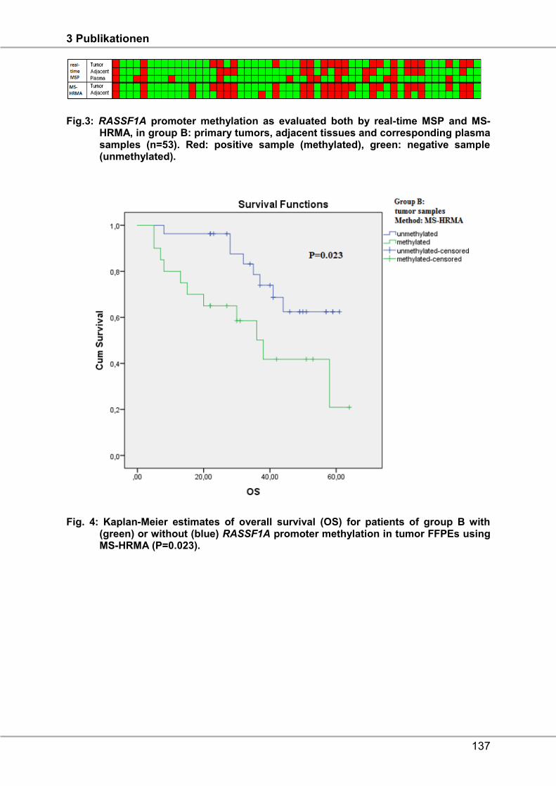
3 Publikationen
137
Fig.3: RASSF1A promoter methylation as evaluated both by real-time MSP and MS- HRMA, in group B: primary tumors, adjacent tissues and corresponding plasma samples (n=53). Red: positive sample (methylated), green: negative sample (unmethylated).
Fig. 4: Kaplan-Meier estimates of overall survival (OS) for patients of group B with (green) or without (blue) RASSF1A promoter methylation in tumor FFPEs using MS-HRMA (P=0.023).
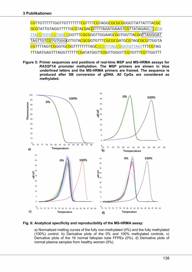
3 Publikationen
138
Figure 5: Primer sequences and positions of real-time MSP and MS-HRMA assays for RASSF1A promoter methylation. The MSP primers are shown in blue underlined letters and the MS-HRMA primers are framed. The sequence is produced after SB conversion of gDNA. All CpGs are considered as methylated.
Fig. 6: Analytical specificity and reproducibility of the MS-HRMA assay:
a) Normalized melting curves of the fully non-methylated (0%) and the fully methylated (100%) control. b) Derivative plots of the 0% and 100% methylated controls. c) Derivative plots of the 16 normal fallopian tube FFPEs (0%). d) Derivative plots of normal plasma samples from healthy women (0%).
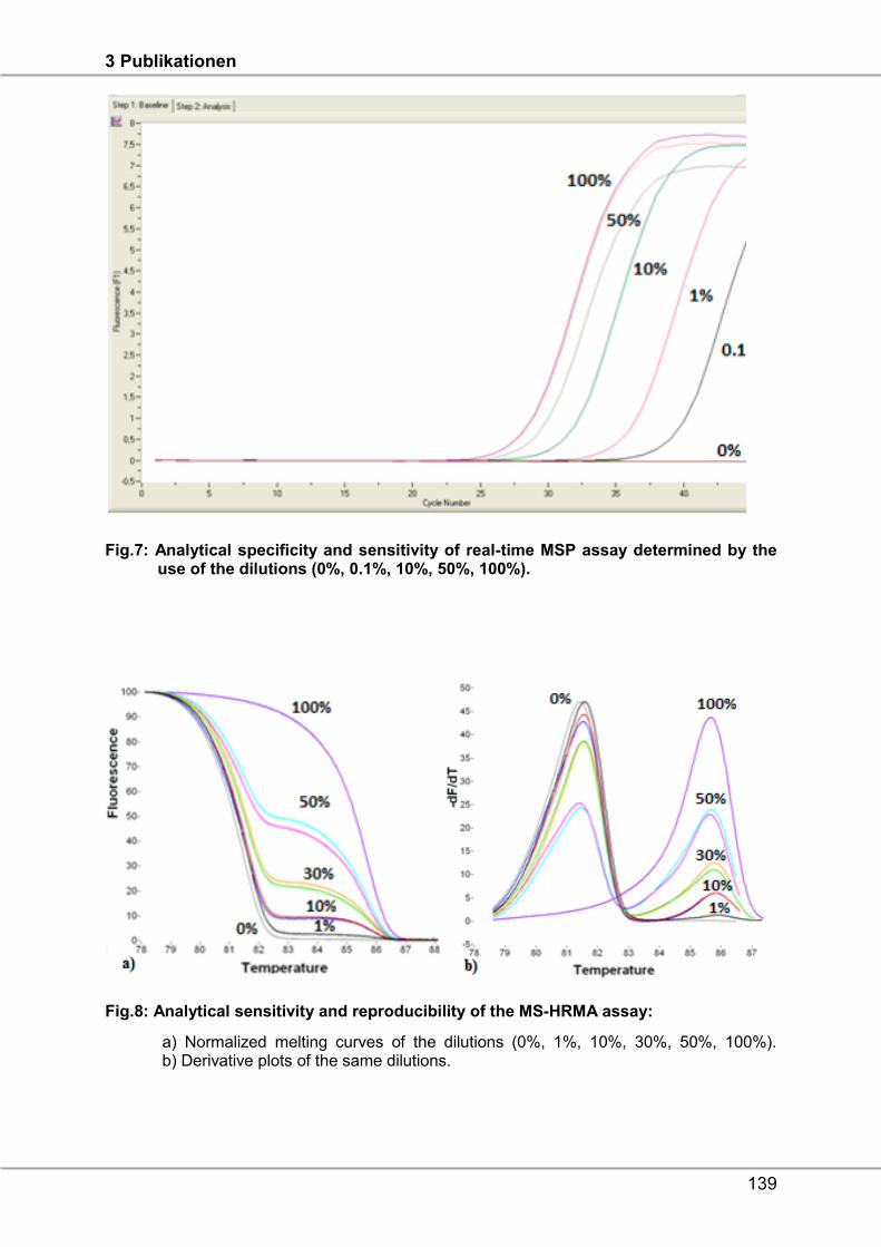
3 Publikationen
139
Fig.7: Analytical specificity and sensitivity of real-time MSP assay determined by the use of the dilutions (0%, 0.1%, 10%, 50%, 100%).
Fig.8: Analytical sensitivity and reproducibility of the MS-HRMA assay:
a) Normalized melting curves of the dilutions (0%, 1%, 10%, 30%, 50%, 100%). b) Derivative plots of the same dilutions.
3 Publikationen
140
REFERENCES
1. Siegel RL, Miller KD and Jemal A. Cancer statistics, 2015. CA Cancer J Clin.
2015; 65: 5-29.
2. du Bois A and Pfisterer J. Future options for first-line therapy of advanced
ovarian cancer. Int J Gynecol Cancer. 2005; 15: 42-50.
3. du Bois A, Reuss A, Pujade-Lauraine E, Harter P, Ray-Coquard I and Pfisterer
J. Role of surgical outcome as prognostic factor in advanced epithelial ovarian
cancer: a combined exploratory analysis of 3 prospectively randomized phase
3 multicenter trials: by the Arbeitsgemeinschaft Gynaekologische Onkologie
Studiengruppe Ovarialkarzinom (AGO-OVAR) and the Groupe d'Investigateurs
Nationaux Pour les Etudes des Cancers de l'Ovaire (GINECO). Cancer. 2009;
115: 1234-44.
4. Wimberger P, Wehling M, Lehmann N, Kimmig R, Schmalfeldt B, Burges A,
Harter P, Pfisterer J and du Bois A. Influence of residual tumor on outcome in
ovarian cancer patients with FIGO stage IV disease: an exploratory analysis of
the AGO-OVAR (Arbeitsgemeinschaft Gynaekologische Onkologie Ovarian
Cancer Study Group). Ann Surg Oncol. 2010; 17: 1642-8.
5. Network TCGAR. Integrated genomic analyses of ovarian carcinoma. Nature.
2011; 474: 609-15.
6. Patch AM, Christie EL, Etemadmoghadam D, Garsed DW, George J, Fereday
S, Nones K, Cowin P, Alsop K, Bailey PJ, Kassahn KS, Newell F, Quinn MC, et
al. Whole-genome characterization of chemoresistant ovarian cancer. Nature.
2015; 521: 489-94.
7. Burger RA, Brady MF, Bookman MA, Fleming GF, Monk BJ, Huang H, Mannel
RS, Homesley HD, Fowler J, Greer BE, Boente M, Birrer MJ and Liang SX.
Incorporation of bevacizumab in the primary treatment of ovarian cancer. N Engl
J Med. 2011; 365: 2473-83.
8. Ledermann J, Harter P, Gourley C, Friedlander M, Vergote I, Rustin G, Scott
CL, Meier W, Shapira-Frommer R, Safra T, Matei D, Fielding A, Spencer S, et
al. Olaparib maintenance therapy in patients with platinum-sensitive relapsed
serous ovarian cancer: a preplanned retrospective analysis of outcomes by
BRCA status in a randomised phase 2 trial. Lancet Oncol. 2014; 15: 852-61.
9. Esteller M. Epigenetics in cancer. N Engl J Med. 2008; 358: 1148-59.
3 Publikationen
141
10. Jones PA and Baylin SB. The fundamental role of epigenetic events in cancer.
Nat Rev Genet. 2002; 3: 415-28.
11. Barton CA, Hacker NF, Clark SJ and O'Brien PM. DNA methylation changes in
ovarian cancer: implications for early diagnosis, prognosis and treatment.
Gynecol Oncol. 2008; 109: 129-39.
12. Earp MA and Cunningham JM. DNA methylation changes in epithelial ovarian
cancer histotypes. Genomics. 2015; 106: 311-21.
13. Marzese DM, Hirose H and Hoon DS. Diagnostic and prognostic value of
circulating tumor-related DNA in cancer patients. Expert Rev Mol Diagn. 2013;
13: 827-44.
14. Kuhlmann JD, Schwarzenbach H, Wimberger P, Poetsch M, Kimmig R and
Kasimir-Bauer S. LOH at 6q and 10q in fractionated circulating DNA of ovarian
cancer patients is predictive for tumor cell spread and overall survival. BMC
Cancer. 2012; 12: 325.
15. Warton K and Samimi G. Methylation of cell-free circulating DNA in the
diagnosis of cancer. Front Mol Biosci. 2015; 2: 13.
16. Schwarzenbach H, Hoon DS and Pantel K. Cell-free nucleic acids as
biomarkers in cancer patients. Nat Rev Cancer. 2011; 11: 426-37.
17. Balgkouranidou I, Chimonidou M, Milaki G, Tsaroucha E, Kakolyris S,
Georgoulias V and Lianidou E. SOX17 promoter methylation in plasma
circulating tumor DNA of patients with non-small cell lung cancer. Clin Chem
Lab Med. 2016; 54: 1385-93.
18. Chimonidou M, Strati A, Malamos N, Georgoulias V and Lianidou ES. SOX17
promoter methylation in circulating tumor cells and matched cell-free DNA
isolated from plasma of patients with breast cancer. Clin Chem. 2013; 59: 270-
9.
19. Chimonidou M, Tzitzira A, Strati A, Sotiropoulou G, Sfikas C, Malamos N,
Georgoulias V and Lianidou E. CST6 promoter methylation in circulating cell-
free DNA of breast cancer patients. Clin Biochem. 2013; 46: 235-40.
20. Balgkouranidou I, Chimonidou M, Milaki G, Tsarouxa EG, Kakolyris S, Welch
DR, Georgoulias V and Lianidou ES. Breast cancer metastasis suppressor-1
promoter methylation in cell-free DNA provides prognostic information in non-
small cell lung cancer. Br J Cancer. 2014; 110: 2054-62.
3 Publikationen
142
21. Richter AM, Pfeifer GP and Dammann RH. The RASSF proteins in cancer; from
epigenetic silencing to functional characterization. Biochim Biophys Acta. 2009;
1796: 114-28.
22. Volodko N, Gordon M, Salla M, Ghazaleh HA and Baksh S. RASSF tumor
suppressor gene family: biological functions and regulation. FEBS Lett. 2014;
588: 2671-84.
23. Grawenda AM and O'Neill E. Clinical utility of RASSF1A methylation in human
malignancies. Br J Cancer. 2015; 113: 372-81.
24. Gloss BS and Samimi G. Epigenetic biomarkers in epithelial ovarian cancer.
Cancer Lett. 2014; 342: 257-63.
25. Fackler MJ, McVeigh M, Evron E, Garrett E, Mehrotra J, Polyak K, Sukumar S
and Argani P. DNA methylation of RASSF1A, HIN-1, RAR-beta, Cyclin D2 and
Twist in in situ and invasive lobular breast carcinoma. Int J Cancer. 2003; 107:
970-5.
26. Agathanggelou A, Honorio S, Macartney DP, Martinez A, Dallol A, Rader J,
Fullwood P, Chauhan A, Walker R, Shaw JA, Hosoe S, Lerman MI, Minna JD,
et al. Methylation associated inactivation of RASSF1A from region 3p21.3 in
lung, breast and ovarian tumours. Oncogene. 2001; 20: 1509-18.
27. Rathi A, Virmani AK, Schorge JO, Elias KJ, Maruyama R, Minna JD, Mok SC,
Girard L, Fishman DA and Gazdar AF. Methylation profiles of sporadic ovarian
tumors and nonmalignant ovaries from high-risk women. Clin Cancer Res.
2002; 8: 3324-31.
28. Yoon JH, Dammann R and Pfeifer GP. Hypermethylation of the CpG island of
the RASSF1A gene in ovarian and renal cell carcinomas. Int J Cancer. 2001;
94: 212-7.
29. Bhagat R, Chadaga S, Premalata CS, Ramesh G, Ramesh C, Pallavi VR and
Krishnamoorthy L. Aberrant promoter methylation of the RASSF1A and APC
genes in epithelial ovarian carcinoma development. Cell Oncol. 2012; 35: 473-
9.
30. Choi YL, Kang SY, Shin YK, Choi JS, Kim SH, Lee SJ, Bae DS and Ahn G.
Aberrant hypermethylation of RASSF1A promoter in ovarian borderline tumors
and carcinomas. Virchows Arch. 2006; 448: 331-6.
31. Kurman RJ and Shih IM. Molecular pathogenesis and extraovarian origin of
epithelial ovarian cancer--shifting the paradigm. Hum Pathol. 2011; 42: 918-31.
3 Publikationen
143
32. Montavon C, Gloss BS, Warton K, Barton CA, Statham AL, Scurry JP, Tabor B,
Nguyen TV, Qu W, Samimi G, Hacker NF, Sutherland RL, Clark SJ, et al.
Prognostic and diagnostic significance of DNA methylation patterns in high
grade serous ovarian cancer. Gynecol Oncol. 2012; 124: 582-8.
33. Ibanez de Caceres I, Battagli C, Esteller M, Herman JG, Dulaimi E, Edelson MI,
Bergman C, Ehya H, Eisenberg BL and Cairns P. Tumor cell-specific BRCA1
and RASSF1A hypermethylation in serum, plasma, and peritoneal fluid from
ovarian cancer patients. Cancer Res. 2004; 64: 6476-81.
34. Bondurant AE, Huang Z, Whitaker RS, Simel LR, Berchuck A and Murphy SK.
Quantitative detection of RASSF1A DNA promoter methylation in tumors and
serum of patients with serous epithelial ovarian cancer. Gynecol Oncol. 2011;
123: 581-7.
35. Liggett TE, Melnikov A, Yi Q, Replogle C, Hu W, Rotmensch J, Kamat A, Sood
AK and Levenson V. Distinctive DNA methylation patterns of cell-free plasma
DNA in women with malignant ovarian tumors. Gynecol Oncol. 2011; 120: 113-
20.
36. Zhang Q, Hu G, Yang Q, Dong R, Xie X, Ma D, Shen K and Kong B. A multiplex
methylation-specific PCR assay for the detection of early-stage ovarian cancer
using cell-free serum DNA. Gynecol Oncol. 2013; 130: 132-9.
37. Matei D, Fang F, Shen C, Schilder J, Arnold A, Zeng Y, Berry WA, Huang T and
Nephew KP. Epigenetic resensitization to platinum in ovarian cancer. Cancer
Res. 2012; 72: 2197-205.
38. van der Weyden L and Adams DJ. The Ras-association domain family (RASSF)
members and their role in human tumourigenesis. Biochim Biophys Acta. 2007;
1776: 58-85.
39. Kioulafa M, Kaklamanis L, Mavroudis D, Georgoulias V and Lianidou ES.
Prognostic significance of RASSF1A promoter methylation in operable breast
cancer. Clin Biochem. 2009; 42: 970-5.
40. Balgkouranidou I, Matthaios D, Karayiannakis A, Bolanaki H, Michailidis P,
Xenidis N, Amarantidis K, Chelis L, Trypsianis G, Chatzaki E, Lianidou ES and
Kakolyris S. Prognostic role of APC and RASSF1A promoter methylation status
in cell free circulating DNA of operable gastric cancer patients. Mutat Res. 2015;
778: 46-51.
41. Spitzwieser M, Holzweber E, Pfeiler G, Hacker S and Cichna-Markl M.
Applicability of HIN-1, MGMT and RASSF1A promoter methylation as
3 Publikationen
144
biomarkers for detecting field cancerization in breast cancer. Breast Cancer
Res. 2015; 17: 125.
42. Van der Auwera I, Bovie C, Svensson C, Trinh XB, Limame R, van Dam P, van
Laere SJ, van Marck EA, Dirix LY and Vermeulen PB. Quantitative methylation
profiling in tumor and matched morphologically normal tissues from breast
cancer patients. BMC Cancer. 2010; 10: 97.
43. Arafa M, Kridelka F, Mathias V, Vanbellinghen JF, Renard I, Foidart JM, Boniver
J and Delvenne P. High frequency of RASSF1A and RARb2 gene promoter
methylation in morphologically normal endometrium adjacent to endometrioid
adenocarcinoma. Histopathology. 2008; 53: 525-32.
44. Chai H and Brown RE. Field effect in cancer-an update. Ann Clin Lab Sci. 2009;
39: 331-7.
45. Wojdacz TK, Borgbo T and Hansen LL. Primer design versus PCR bias in
methylation independent PCR amplifications. Epigenetics. 2009; 4: 231-4.
46. Chiu RW, Chim SS, Wong IH, Wong CS, Lee WS, To KF, Tong JH, Yuen RK,
Shum AS, Chan JK, Chan LY, Yuen JW, Tong YK, et al. Hypermethylation of
RASSF1A in human and rhesus placentas. Am J Pathol. 2007; 170: 941-50.
47. Wojdacz TK, Dobrovic A and Hansen LL. Methylation-sensitive high-resolution
melting. Nat Protoc. 2008; 3: 1903-8.
4 Diskussion
145
4 Diskussion
Ziel dieser Dissertation war die Identifikation neuer Biomarkerkonzepte für das
Ovarialkarzinom. Dies geschah zunächst auf Tumorstammzellebene mit dem
Schwerpunkt der Analyse von persistierenden DTZ mit Stammzellcharakter im KM. Im
zweiten Schritt wurden blutbasierte Strategien durchgeführt, um die Fragestellungen
zu überprüfen, inwieweit die Präsenz und Persistenz von resistenten ERCC1pos ZTZ,
ZTZ sowie ZTZ in EMT mit dem ungünstigen Verlauf der Erkrankung assoziiert sind.
Als weitere, nicht zelluläre, „Liquid Biopsy“ wurden zirkulierende DNA und microRNAs
als neue bzw. ergänzende Marker evaluiert.
Zielsetzung 1:
DTZ mit Stammzelleigenschaften
Die Ergebnisse zu dieser Zielsetzung wurden in Oncotarget publiziert. Analysis of disseminated tumor cells before and after platinum based chemotherapy in primary ovarian cancer. Do stem cells predict prognosis? Issam Chebouti,Christina Blassl, Pauline Wimberger, Hans Neubauer, Tanja Fehm, Rainer Kimmig, Sabine Kasimir-Bauer. Oncotarget. 2016. doi:3;7(18):26454-64.
Die prognostische Bedeutung der DTZ zu Beginn der Erkrankung im Hinblick auf das
PFS und OS wurde bereits von verschiedenen Arbeitsgruppen bestätigt (Wimberger
et al., 2007; Banys et al., 2009; Fehm et al., 2013; und Cui et al., 2015).
Untersuchungen an DTZ nach platinbasierter Chemotherapie wurden bisher von
anderen Gruppen nicht durchgeführt.
Unter Verwendung der Standardmethode zum Nachweis Ckpos DTZ im KM (siehe
Abb.1.12) wurden in der hier untersuchten Kohorte von 79 Patientinnen, bei 42% vor
und bei 41% nach Therapie DTZ im KM detektiert, was die Verteilung der DTZ in
bereits publizierten Studien der Arbeitsgruppe widerspiegelte. DTZ vor Therapie
assoziierten signifikant mit einem verkürzten OS und die Patientinnen, die vor Therapie
DTZneg, aber nach Therapie DTZpos waren, zeigten ein signifikant verkürztes PFS.
Patientinnen mit persistierenden DTZ (16%) zeigten einen Trend zum schlechteren
PFS/OS, der jedoch nicht signifikant war.
Bisher wurden Tumorstammzellen nur im ovariellen Tumorgewebe analysiert, wobei
die Expression von LIN-28, SOX-2 und OCT-4 als wesentliche Marker beschrieben
wurden (Peng et al., 2010; Bareis et al., 2013 und Pham et al., 2013). Für die Analyse
der DTZ im KM wurde im ersten Promotionsjahr, in Zusammenarbeit mit Frau Christina
4 Diskussion
146
Blassl aus der Frauenklinik Düsseldorf, eine Vierfach-Immunfluoreszenzfärbung
etabliert. Hierzu wurde die ovarielle Zelllinie OVCAR-3 sowie die Leukämiezelllinie
Kasumi-1 verwendet. Die Methode wurde dann auf KM-Präparate der Patientinnen vor
und nach platinbasierter Therapie angewendet. Nachgewiesen wurden die Marker
LIN-28 und SOX-2 als Stammzellmarker für das Ovarialkarzinom, der
Zytokeratinmarker C11, der die Zytokeratine 4,5,6,8,10,13 und 18 detektiert, sowie
CD34 als hämatopoeitischer Stammzellmarker und CD45 als Leukozytenmarker zum
Ausschluss falsch-positiver Zellen. Die Untersuchungen am KM von Patientinnen
konnten nicht an den bereits mit der Standard-Methode untersuchten Präparaten
durchgeführt werden, da die Neufuchsinfärbung weitere Färbungen nicht erlaubt.
Somit wurde auf Reservepräparate der Patientinnen zurückgegriffen. Aufgrund der
geringen Anzahl an Reserveobjektträgern wurden nur die Stammzellmarker LIN-28
und SOX-2 untersucht. Die Tab. 4.1 zeigt zur Veranschaulichung der Ergebnisse eine
Übersicht der analysierten 10 Patientinnen. Es wurden drei Gruppen gebildet,
basierend auf den mit der Standardmethode detektieren CKpos DTZ vor und nach
Therapie (Spalten grau unterlegt). Gruppe 1 beschreibt sechs Patientinnen
(1,3,4,7,8,10), die vor Therapie keine DTZ, nach Therapie jedoch Zellen aufwiesen.
Die zweite Gruppe stellt die Patientinnen dar, die vor Therapie DTZpos waren, nach
Therapie aber keine Zellen aufwiesen (2, 5). Gruppe 3 umfasst die Patientinnen mit
persistierenden DTZ (6,9). Betrachtet wurden zunächst die Patientinnen (1,3,4,6,
7,8,9,10), die nach Therapie mindestens fünf Ckpos-Zellen (DTZ) aufwiesen, sodass
die Wahrscheinlichkeit groß war, auch auf den Reservepräparaten Zellen zu
detektieren. Bei 9/10 Patientinnen wurden Ckpos/Lin-28pos Zellen und bei 7/10
Patientinnen Ckneg/Lin-28pos Zellen gefunden. Ähnliche Ergebnisse wurden für SOX-2
nachgewiesen. Bei sechs dieser Patientinnen (1,3,4,7,8,10) zeigten die
korrespondierenden KM-Präparate vor Therapie, die mit der Standardmethode Ckneg
(DTZneg) waren, interessanterweise die Präsenz einzelner LIN-28pos und/oder SOX-
2pos Zellen. Ähnliches galt für Patientinnen, die nach Therapie mit der
Standardmethode als Ckneg (DTZneg) charakterisiert wurden (2,5). Bei Patientinnen mit
persistierenden DTZ (6,9) wurden sowohl vor, als auch nach Therapie Zellen mit
Stammzellcharakter nachgewiesen. Diese Resultate könnten das signifikant verkürzte
PFS von Patientinnen erklären, die anfänglich vor Therapie keine DTZ hatten und nach
Therapie DTZ aufwiesen. Auch bei Patientinnen, die nach Therapie keine Ckpos DTZ,
also epitheliale Zellen aufwiesen, konnten vereinzelt LIN-28pos/SOX-2pos Zellen
detektiert werden. Inwieweit diese Zellen ebenfalls die Prognose beeinflussen, konnte
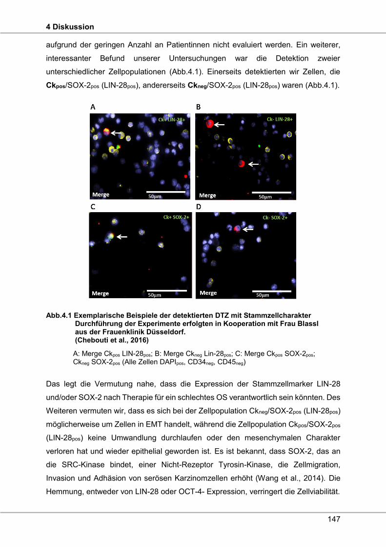
4 Diskussion
147
aufgrund der geringen Anzahl an Patientinnen nicht evaluiert werden. Ein weiterer,
interessanter Befund unserer Untersuchungen war die Detektion zweier
unterschiedlicher Zellpopulationen (Abb.4.1). Einerseits detektierten wir Zellen, die
Ckpos/SOX-2pos (LIN-28pos), andererseits Ckneg/SOX-2pos (LIN-28pos) waren (Abb.4.1).
Abb.4.1 Exemplarische Beispiele der detektierten DTZ mit Stammzellcharakter Durchführung der Experimente erfolgten in Kooperation mit Frau Blassl aus der Frauenklinik Düsseldorf. (Chebouti et al., 2016)
A: Merge Ckpos LIN-28pos; B: Merge Ckneg Lin-28pos; C: Merge Ckpos SOX-2pos; Ckneg SOX-2pos (Alle Zellen DAPIpos, CD34neg, CD45neg)
Das legt die Vermutung nahe, dass die Expression der Stammzellmarker LIN-28
und/oder SOX-2 nach Therapie für ein schlechtes OS verantwortlich sein könnten. Des
Weiteren vermuten wir, dass es sich bei der Zellpopulation Ckneg/SOX-2pos (LIN-28pos)
möglicherweise um Zellen in EMT handelt, während die Zellpopulation Ckpos/SOX-2pos
(LIN-28pos) keine Umwandlung durchlaufen oder den mesenchymalen Charakter
verloren hat und wieder epithelial geworden ist. Es ist bekannt, dass SOX-2, das an
die SRC-Kinase bindet, einer Nicht-Rezeptor Tyrosin-Kinase, die Zellmigration,
Invasion und Adhäsion von serösen Karzinomzellen erhöht (Wang et al., 2014). Die
Hemmung, entweder von LIN-28 oder OCT-4- Expression, verringert die Zellviabilität.

4 Diskussion
148
Tab. 4.1 Verteilung der DTZ und LIN-28 sowie SOX-2-positiven Zellen bevor und nach Therapie
*kov= keine Objektträger vorhanden
bevor Therapie nach Therapie
Patienten
DTZpos bevor Therapie
DTZpos nach Therapie
Ckpos/LIN-28pos Ckneg/LIN 28pos Ckpos/SOX-2pos Ckneg/SOX-2pos Ckpos/LIN-28pos Ckneg/LIN-28pos Ckpos/SOX-2pos Ckneg/SOX-2pos
(A45-B/B3) (AF45-B/B3)
1 0 14 25 5 24 2 4 0 3 4
2 37 0 2 0 4 1 0 1 2 0
3 0 11 8 2 5 1 2 0 0 1
4 0 15 2 1 1 1 2 1 kov kov
5 6 0 1 1 2 1 1 0 2 1
6 28 18 kov kov 2 3 3 7 4 11
7 0 5 kov kov kov kov 1 3 0 2
8 0 100 kov kov kov kov 5 9 1 2
9 1 35 kov kov kov kov 2 1 kov kov
10 0 10 kov kov kov kov 3 11 2 9
4 Diskussion
149
Die kombinierte Repression von LIN-28 und OCT-4 führt zu einer synergetischen
Inhibition beim Wachstum der Tumorzellen und beim Überleben von ovariellen
Tumorzelllinien (Bapat et al., 2005). Die Expression von SOX-2 wurde mittels
Immunhistochemie sowohl an normalem, ovariellen epithelialen, serösen und
muzinösen Zystadenomen, als auch an Zystadenomkarzinomen untersucht (Ye et al.,
2011). Eine Überexpression von LIN-28 wurde in verschiedenen epithelialen Tumoren,
darunter Mamma-, Bronchial-, Kolon- und Ovarialkarzinomen, detektiert (Viswanathan
et al., 2009). Bei Patientinnen mit Ovarialkarzinom, die eine taxanbasierte
Chemotherapie erhielten, korrelierte die Expression von SOX-2 signifikant mit der
Chemoresistenz und einem verkürztem PFS, während eine fehlende taxanbasierte
Chemotherapie, keine Signifikanz zeigte (Du et al., 2015). Da die Patientinnen in
unserer Studie auch eine kombinierte Therapie mit Paclitaxel und Carboplatin erhalten
haben, könnte es möglich sein, dass die SOX-2 Expression an DTZ mit einer
Chemotherapie-Resistenz assoziiert ist. Dennoch bleiben Mechanismen, die für eine
Chemotherapie-Resistenz beim Ovarialkarzinom verantwortlich sind, noch ungeklärt.
Zielsetzung 2:
Untersuchungen an ZTZ
Die zu diesem Thema erhobenen Daten wurden in Oncotarget publizert.
EMT-like circulating tumor cells in ovarian cancer patients are enriched by platinum-based chemotherapy. Issam Chebouti, Sabine Kasimir-Bauer, Paul Buderath, Pauline Wimberger, Siggi Hauch, Rainer Kimmig, Jan Dominik Kuhlmann. Oncotarget 2017, in press.
ERCC1-expressing circulating tumor cells as a potential diagnostic tool for monitoring response to platinum-based chemotherapy and for predicting post-therapeutic outcome of ovarian cancer. Issam Chebouti, Jan Dominik Kuhlmann, Paul Buderath, Stephan Weber, Pauline Wimberger, Yvonne Bokeloh, Siggi Hauch, Rainer Kimmig, Sabine Kasimir-Bauer. Oncotarget 2016, doi:18632/oncotarget.13286.
Da die Analyse der DTZ sehr invasiv ist, ungeeignet als Methode zur Beurteilung des
Krankheitsverlaufs, und zudem momentan keine therapeutische Konsequenz
beinhaltet, lehnen die Patientinnen zum größten Teil eine KM-Punktion nach Therapie
ab. Deshalb sind blutbasierte Studien von immer größerer Bedeutung. Die
Arbeitsgruppe um Frau Professorin Kasimir-Bauer konnte schon relevante Beiträge
zur Etablierung neuer, blutbasierter Biomarkerkonzepte für das Ovarialkarzinom
publizieren und die Hypothese unterstützen, dass Patientinnenblut im Sinne einer
„Real-Time-Liquid-Biopsy“ genutzt werden könnte. Im Hinblick auf ZTZ wurde die
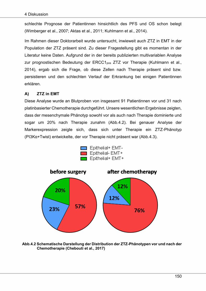
4 Diskussion
150
schlechte Prognose der Patientinnen hinsichtlich des PFS und OS schon belegt
(Wimberger et al., 2007; Aktas et al., 2011; Kuhlmann et al., 2014).
Im Rahmen dieser Doktorarbeit wurde untersucht, inwieweit auch ZTZ in EMT in der
Population der ZTZ präsent sind. Zu dieser Fragestellung gibt es momentan in der
Literatur keine Daten. Aufgrund der in der bereits publizierten multivariablen Analyse
zur prognostischen Bedeutung der ERCC1pos ZTZ vor Therapie (Kuhlmann et al.,
2014), ergab sich die Frage, ob diese Zellen nach Therapie präsent sind bzw.
persistieren und den schlechten Verlauf der Erkrankung bei einigen Patientinnen
erklären.
A) ZTZ in EMT
Diese Analyse wurde an Blutproben von insgesamt 91 Patientinnen vor und 31 nach
platinbasierter Chemotherapie durchgeführt. Unsere wesentlichen Ergebnisse zeigten,
dass der mesenchymale Phänotyp sowohl vor als auch nach Therapie dominierte und
sogar um 20% nach Therapie zunahm (Abb.4.2). Bei genauer Analyse der
Markerexpression zeigte sich, dass sich unter Therapie ein ZTZ-Phänotyp
(PI3Kα+Twist) entwickelte, der vor Therapie nicht präsent war (Abb.4.3).
Abb.4.2 Schematische Darstellung der Distribution der ZTZ-Phänotypen vor und nach der Chemotherapie (Chebouti et al., 2017)
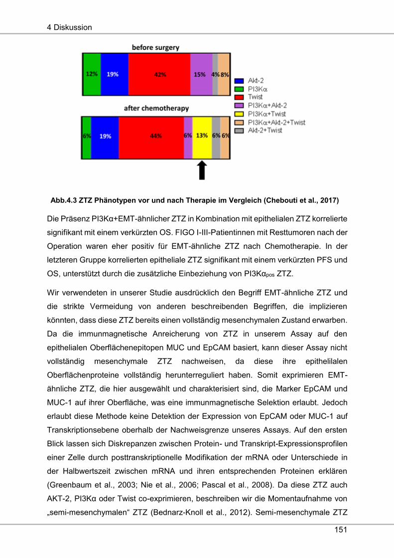
4 Diskussion
151
Abb.4.3 ZTZ Phänotypen vor und nach Therapie im Vergleich (Chebouti et al., 2017)
Die Präsenz PI3Kα+EMT-ähnlicher ZTZ in Kombination mit epithelialen ZTZ korrelierte
signifikant mit einem verkürzten OS. FIGO I-III-Patientinnen mit Resttumoren nach der
Operation waren eher positiv für EMT-ähnliche ZTZ nach Chemotherapie. In der
letzteren Gruppe korrelierten epitheliale ZTZ signifikant mit einem verkürzten PFS und
OS, unterstützt durch die zusätzliche Einbeziehung von PI3Kαpos ZTZ.
Wir verwendeten in unserer Studie ausdrücklich den Begriff EMT-ähnliche ZTZ und
die strikte Vermeidung von anderen beschreibenden Begriffen, die implizieren
könnten, dass diese ZTZ bereits einen vollständig mesenchymalen Zustand erwarben.
Da die immunmagnetische Anreicherung von ZTZ in unserem Assay auf den
epithelialen Oberflächenepitopen MUC und EpCAM basiert, kann dieser Assay nicht
vollständig mesenchymale ZTZ nachweisen, da diese ihre epithelilalen
Oberflächenproteine vollständig herunterreguliert haben. Somit exprimieren EMT-
ähnliche ZTZ, die hier ausgewählt und charakterisiert sind, die Marker EpCAM und
MUC-1 auf ihrer Oberfläche, was eine immunmagnetische Selektion erlaubt. Jedoch
erlaubt diese Methode keine Detektion der Expression von EpCAM oder MUC-1 auf
Transkriptionsebene oberhalb der Nachweisgrenze unseres Assays. Auf den ersten
Blick lassen sich Diskrepanzen zwischen Protein- und Transkript-Expressionsprofilen
einer Zelle durch posttranskriptionelle Modifikation der mRNA oder Unterschiede in
der Halbwertszeit zwischen mRNA und ihren entsprechenden Proteinen erklären
(Greenbaum et al., 2003; Nie et al., 2006; Pascal et al., 2008). Da diese ZTZ auch
AKT-2, PI3Kα oder Twist co-exprimieren, beschreiben wir die Momentaufnahme von
„semi-mesenchymalen“ ZTZ (Bednarz-Knoll et al., 2012). Semi-mesenchymale ZTZ
4 Diskussion
152
könnten entweder auf ihrem Weg zu einem letztlich mesenchymalen Phänotyp (EMT)
oder auf ihrem Weg zurück zu einem epithelilalen Phänotyp (MET) sein. Alternativ
könnten sie in diesem Zwischenzustand verharren. Obwohl die Biologie von semi-
mesenchymalen ZTZ weitgehend unbekannt ist, können wir annehmen, dass
insbesondere ein semi-mesenchymaler Zustand einen aggressiven ZTZ-Phänotyp mit
hohem Plastizitätsgrad wiederspiegelt, der die Anpassung von ZTZ an die
umgebenden Reize während der Streuung erleichtert. Dies steht im Einklang mit der
Hypothese, dass EMT (und der Reversion-MET) ein hochdynamischer Prozess ist und
verschiedene kontinuierliche Phänotypen beschreibt, anstatt einen dichotomen
Wechsel zwischen epithelialen und mesenchymalen Zuständen. Insbesondere
beeinflussen diese phänotypischen Veränderungen direkt die Wiederfindungsrate von
ZTZ-Nachweis-Assays, die auf epithelialen Selektionsmarkern basieren (Bednarz-
Knoll et al., 2012). Jedoch wird unsere Annahme von kontinuierlichen ZTZ-Phänotypen
beim Ovarialkarzinom nicht notwendigerweise durch die Tatsache unterstützt, dass wir
einen Trend für einen gegenseitigen Ausschluss zwischen Patientinnen mit nur
epithelialen ZTZ und denen mit nur semi-mesenchymalen ZTZ beobachteten. Dies ist
ein sehr interessanter Befund und kann zeigen, dass es auch eine Untergruppe von
Ovarialkarzinompatientinnen mit ausschließlich epithelialen ZTZ gibt, ohne
Verschiebungen in Richtung EMT. Die klinische Relevanz dieses Befundes erfordert
jedoch weitere Untersuchungen. Mit Hilfe unseres EMT-assoziierten Markersets
haben wir ein heterogenes Spektrum von EMT-assoziierten ZTZ-Phänotypen
aufgezeigt, was auf eine ZTZ-Heterogenität bei Ovarialkarzinomen hinweist, die
bereits für ZTZ in einer Vielzahl anderer Tumorentitäten, wie dem Prostata- oder
Mammakarzinom, publiziert wurde (Massard et al., 2016; Shaw et al., 2016). Zu
bedenken ist, dass wir mit unserem Assay nicht unterscheiden können, ob eine
Expression mehrerer Marker-Transkripte von einer ZTZ stammt, die diese Marker in
der gleichen Zelle exprimieren, oder aus separaten semi-mesenchymalen ZTZ-
Populationen von angereicherten ZTZ. Wir gehen daher von einer klonalen Selektion
von ZTZ mit aktivierten PI3Kα- und Twist-assoziierten Signalwegen aus, die
therapeutisch refraktär und für das posttherapeutische Rezidiv des Ovarialkarzinoms
verantwortlich sein könnten. Diese Hypothese wird durch unsere bisherigen Studien
an Mammakarzinompatientinnen stark unterstützt, in denen bereits gezeigt wurde,
dass nach der neoadjuvanten Chemotherapie bevorzugt semi-mesenchymale und
möglicherweise platinresistente (ERCC1-exprimierende) ZTZ verbleiben (Kasimir-
Bauer et al., 2016). Darüber hinaus berichtete eine weitere unabhängige
4 Diskussion
153
Schlüsselveröffentlichung über dynamische Veränderungen in der epithelialen und
mesenchymalen Zusammensetzung von Mammakarzinom-ZTZ als Reaktion auf die
Therapie. In dieser Studie wurde das klinische Ansprechen von einer Veränderung zu
überwiegend epithelialen ZTZ begleitet, während progressive Erkrankungen mit dem
Anstieg der mesenchymalen ZTZ-Phänotypen korrelierten (Yu et al., 2013). In vitro-
Experimente zeigten, dass eine Cisplatinbehandlung von Ovarialkarzinomzellen,
Zellen mit EMT-ähnlichen Merkmalen generieren (Latifi et al., 2011). Daher nehmen
wir an, dass PI3Kαpos und Twistpos ZTZ die Tumorentwicklung als Reaktion auf eine
platinbasierte Chemotherapie widerspiegeln könnten. Interessanterweise deutet dies
auch auf einen Zusammenhang mit neueren Studien zur genomischen
Tumorentwicklung hin, die über eine Zunahme der Aktivierung der PI3Kα-Mutation
unter zellfreier Tumor-DNA von Mammakarzinompatientinnen nach Paclitaxel-
behandlung berichteten (Murtaza et al., 2013). In diesem Zusammenhang könnten
unsere Befunde mehrere diagnostische oder therapeutische Auswirkungen haben, da
PI3Kα und Twist funktionell an Signalwegen beteiligt sind, die das Überleben von
Tumoren oder die Platinresistenz kontrollieren (Kolasa et al., 2009, Nuti et al., 2014).
Weiterhin wird SOX-2 in Zusammenhang mit der Chemoresistenz diskutiert. So
zeigten Patientinnen mit taxanbasierter Chemotherapie und SOX-2-Expression im
Gewebe eine Chemotherapieresistenz sowie ein verkürztes PFS (Du et al., 2015). Hier
zeigt sich ein Zusammenhang mit den zuvor beschriebenen Daten an DTZ.
B) ZTZ mit Resistenzcharakter
Ungefähr 15-20% der Patientinnen mit Ovarialkarzinom, die eine platinbasierte
Chemotherapie erhalten, zeigen eine Platinresistenz, was das größte Problem dieser
Erkrankung darstellt. Die Expression von ERCC1 wird als möglicher Biomarker für
Platinresistenz diskutiert, jedoch waren immunhistochemische Bestimmungen beim
Ovarialkarzinom und anderen soliden Tumoren wenig informativ (Friboulet et al.,
2013). Auch die Arbeitsgruppe der Frauenklinik konnte dies bestätigen, jedoch war die
Präsenz von ERCC1pos ZTZ zu Beginn der Erkrankung ein unabhängiger Prädiktor für
das PFS, OS und für die Platinresistenz (Kuhlmann et al., 2014).
Im Rahmen dieser Dissertation wurde an dieser Thematik weitergearbeitet. Im
Vordergrund stand die Frage, ob ERCC1pos ZTZ, die schon zu Beginn der Erkrankung
präsent waren, auch nach Therapie persistieren oder ob sich diese Zellpopulation auch
unter Therapie entwickeln kann. In einer Kohorte von 65 Patientinnen zeigten 15% der
Patientinnen vor und 12% nach Therapie ERCC1pos ZTZ. Die Abb.4.4 fasst die
4 Diskussion
154
detektierten ZTZ-Phänotypen zusammen und zeigt ihre relativen Proportionen
innerhalb den untersuchten Ovarialkarzinompatientinnen. Als AdnaTestpos wurden die
Patientinnen bezeichnet, die mindestens für eines der Transkripte EpCAM, MUC-1
oder CA-125 positiv waren. Außerdem wurde über eine zusätzliche „single-plex“ PCR
die Expression von ERCC1-Transkripten analysiert. Vor Therapie wurde bei 8% der
Patientinnen ausschließlich eine AdnaTest-Positivität beobachtet. In 17% der Fälle
wurden exklusiv ERCC1pos ZTZ detektiert und bei 15% der Patientinnen beobachteten
wir eine Positivität für AdnaTest und ERCC1 (Abb.4.4A). Anschließend waren wir
daran interessiert, wie die Bewertung von ERCC1pos-Patientinnen die
Gesamtnachweisrate von ZTZ bei Ovarialkarzinompatientinnen beeinflusst. Daher
wurde die Gesamt-ZTZ-Detektionsrate mit der Präsenz von ERCC1-Transkripten als
zusätzliches, alternatives Kriterium untersucht (Abb. 4.4B): Vor Therapie wurde eine
ZTZ-Detektionsrate von 23% beobachtet, bestehend aus Patientinnen mit nur
AdnaTest Positivität UND ERCC1neg ODER ERCC1pos Patientinnen. Die
Detektionsrate wurde bis zu 40% deutlich erhöht, wenn ERCC1 als weiterer,
alternativer Marker für die ZTZ-Positivität (AdnaTestpos ODER ERCC1pos) betrachtet
wurde. Die Subgruppe AdnaTestneg UND ERCC1pos zeigte eine Gesamtnachweisrate
von 17%. Schließlich sank nach einer strengeren Definition der kombinierten Positivität
(AdnaTestpos UND ERCC1pos) die Gesamtdetektion auf 15%. Nach der platinbasierten
Chemotherapie war der Anteil der ZTZ-Subtypen und deren Gesamtnachweisraten
unter den oben genannten ZTZ-Definitionskriterien mit denen, die vor der Therapie
gefunden wurden, vergleichbar (Abb.4.4 C + D). Außerdem korrelierte die Präsenz von
ERCC1pos ZTZ mit einem verkürztem PFS und OS sowie mit der Platinresistenz. Diese
Analyse könnte für die Vorhersage von posttherapeutischen Ergebnissen und für die
Überwachung der platinbasierten Chemotherapie nützlich sein. Durch die zusätzliche
starke prognostische Bedeutung der Marker MUC-1, EpCAM und CA-125 (Aktas et al.,
2011 und Kuhlmann et al., 2014) und unter Berücksichtigung, dass das statistische
Signifikanzniveau leicht rückgängig war, wenn nur ERCC1 Transkripte beurteilt
wurden, scheint eine Bedingung, die eine ERCC1-Positivität und mindestens einen
AdnaTest Marker umfasst, im Sinne eines blutbasierten prognostischen Marker am
günstigsten zu sein. Bisher sind funktionelle Merkmale von ERCC1-exprimierenden
ZTZ im Blut von Ovarialkarzinompatientinnen unbekannt. Da unsere Studie
ausschließlich aus der „Biomarkerperspektive“ durchgeführt wurde, können wir nur
annehmen, dass ERCC1-(über)exprimierende ZTZ im Blut durch eine erweiterte, oder
bereits vorhandene oder neu erworbene Fähigkeit, charakterisiert werden können, um
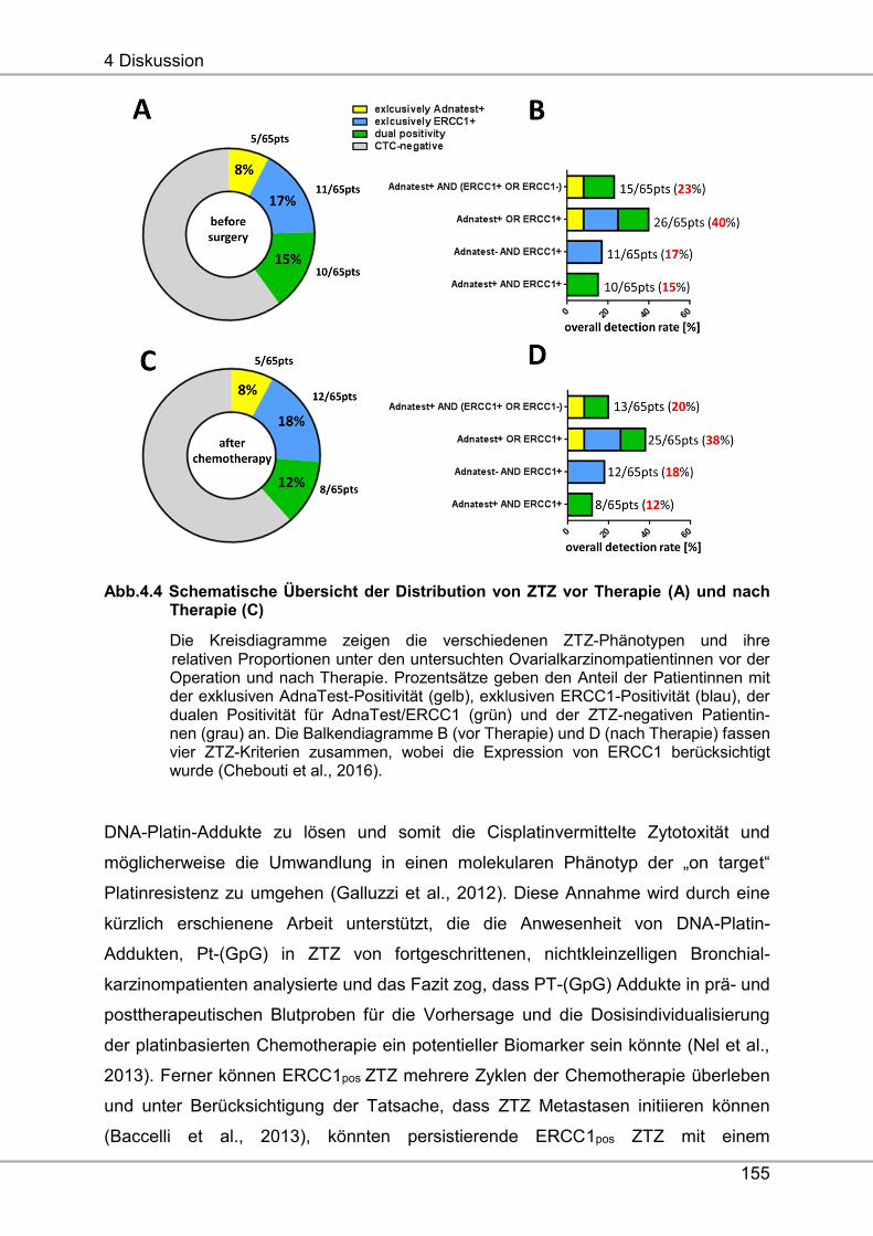
4 Diskussion
155
Abb.4.4 Schematische Übersicht der Distribution von ZTZ vor Therapie (A) und nach Therapie (C)
Die Kreisdiagramme zeigen die verschiedenen ZTZ-Phänotypen und ihre relativen Proportionen unter den untersuchten Ovarialkarzinompatientinnen vor der Operation und nach Therapie. Prozentsätze geben den Anteil der Patientinnen mit der exklusiven AdnaTest-Positivität (gelb), exklusiven ERCC1-Positivität (blau), der dualen Positivität für AdnaTest/ERCC1 (grün) und der ZTZ-negativen Patientin- nen (grau) an. Die Balkendiagramme B (vor Therapie) und D (nach Therapie) fassen vier ZTZ-Kriterien zusammen, wobei die Expression von ERCC1 berücksichtigt wurde (Chebouti et al., 2016).
DNA-Platin-Addukte zu lösen und somit die Cisplatinvermittelte Zytotoxität und
möglicherweise die Umwandlung in einen molekularen Phänotyp der „on target“
Platinresistenz zu umgehen (Galluzzi et al., 2012). Diese Annahme wird durch eine
kürzlich erschienene Arbeit unterstützt, die die Anwesenheit von DNA-Platin-
Addukten, Pt-(GpG) in ZTZ von fortgeschrittenen, nichtkleinzelligen Bronchial-
karzinompatienten analysierte und das Fazit zog, dass PT-(GpG) Addukte in prä- und
posttherapeutischen Blutproben für die Vorhersage und die Dosisindividualisierung
der platinbasierten Chemotherapie ein potentieller Biomarker sein könnte (Nel et al.,
2013). Ferner können ERCC1pos ZTZ mehrere Zyklen der Chemotherapie überleben
und unter Berücksichtigung der Tatsache, dass ZTZ Metastasen initiieren können
(Baccelli et al., 2013), könnten persistierende ERCC1pos ZTZ mit einem
4 Diskussion
156
platinresistenten Phänotyp das Potenzial haben, ein Rezidiv zu verursachen. Unter
der Annahme, dass ERCC1pos ZTZ starke prognostische Faktoren in der
posttherapeutischen Situation sind, insbesondere bei anhaltender Positivität, konnten
unsere Daten auch zeigen, dass platinresistente ERCC1pos ZTZ direkt über die
platinbasierte Chemotherapie selektiert werden können. Jedoch sind weitere
funktionale Untersuchungen nötig, um diese Hypothese zu belegen. Unsere Studie ist
aufgrund der begrenzten Anzahl an Patientinnen explorativ und hypothetisch.
Dennoch markiert ERCC1 eine Subpopulation von ZTZ, die für die Überwachung der
platinbasierten Chemotherapie und für die Beurteilung der therapeutischen
Ergebnisse von Patientinnen mit Ovarialkarzinom nützlich sein könnte. Wir zeigen
Grundlagen für die Validierung des klinischen Nutzens von ERCC1pos ZTZ in großen
multizentrischen klinischen Studien und zur weiteren Aufklärung ihrer funktionalen und
tumorbiologischen Bedeutung. Alternativ könnten Patientinnen mit ERCC1pos ZTZ von
einer früh verabreichten und dosisintensiven Erhaltungstherapie mit Bevacizumab
oder PARP-Inhibitoren profitieren, da einerseits die Versorgung der Tumorzellen
verhindert wird und andererseits bei Zugabe eines PARP-Inhibitors die
Reparaturmechanismen für Einzelstrangbrüche in der DNA der Tumorzelle
ausgeschaltet werden, wodurch es leichter zu Doppelstrangbrüchen bei weiteren
Zellteilungen kommt. Diese können in gesunden Zellen repariert werden, jedoch führt
es in Tumorzellen mit BRCA1- und BRCA-2- Mutationen zur Apoptose (Fong et al.,
2010; Chen et al., 2011).
Zusammenfassende Schlussfolgerung für die unter der Zielsetzung 1 und 2
erhobenen Daten:
Aus den Tumorzellanalysen hat sich herauskristallisiert, dass sowohl vor und nach
Therapie DTZ mit Stammzellcharakter, als auch resistente ZTZ für die schlechte
Prognose verantwortlich sein könnten. Des Weiteren zeigte sich unter Therapie, dass
ZTZ mit mesenchymalem Charakter dominieren und PI3Kαpos/Twistpos ZTZ nach
Therapie als „neuer“ ZTZ Phänotyp präsent sind.
Unter Berücksichtigung all dieser Überlegungen werden nach Abschluss der
Standardtherapien zusätzliche therapeutische Strategien benötigt, um diese ZTZ zu
eliminieren. Möglich erscheint der Einsatz von mTOR-Inhibitoren, die „downstream“
von PI3K/AKT Signalwegen ansetzen (Zhang et al., 2011), Salinomyzin (Gupta et al.,
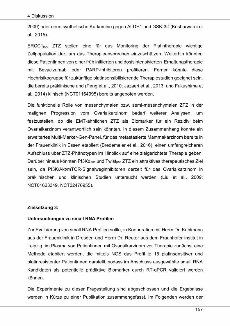
4 Diskussion
157
2009) oder neue synthetische Kurkumine gegen ALDH1 und GSK-3ß (Kesharwarni et
al., 2015).
ERCC1pos ZTZ stellen eine für das Monitoring der Platintherapie wichtige
Zellpopulation dar, um das Therapieansprechen einzuschätzen. Weiterhin könnten
diese Patientinnen von einer früh initiierten und dosisintensivierten Erhaltungstherapie
mit Bevacizumab oder PARP-Inhibitoren profitieren. Ferner könnte diese
Hochrisikogruppe für zukünftige platinsensibilisierende Therapiestudien geeignet sein,
die bereits präklinische und (Peng et al., 2010; Jazaeri et al., 2013; und Fukushima et
al., 2014) klinisch (NCT01164995) bereits angeboten werden.
Die funktionelle Rolle von mesenchymalen bzw. semi-mesenchymalen ZTZ in der
malignen Progression vom Ovarialkarzinom bedarf weiterer Analysen, um
festzustellen, ob die EMT-ähnlichen ZTZ als Biomarker für ein Rezidiv beim
Ovarialkarzinom verantwortlich sein könnten. In diesem Zusammenhang könnte ein
erweitertes Multi-Marker-Gen-Panel, für das metastasierte Mammakarzinom bereits in
der Frauenklinik in Essen etabliert (Bredemeier et al., 2016), einen umfangreicheren
Aufschluss über ZTZ-Phänotypen im Hinblick auf eine zielgerichtete Therapie geben.
Darüber hinaus könnten PI3Kαpos und Twistpos ZTZ ein attraktives therapeutisches Ziel
sein, da PI3K/Akt/mTOR-Signalweginhibitoren derzeit für das Ovarialkarzinom in
präklinischen und klinischen Studien untersucht werden (Liu et al., 2009;
NCT01623349, NCT02476955).
Zielsetzung 3:
Untersuchungen zu small RNA Profilen
Zur Evaluierung von small RNA Profilen sollte, in Kooperation mit Herrn Dr. Kuhlmann
aus der Frauenklinik in Dresden und Herrn Dr. Reuter aus dem Fraunhofer Institut in
Leipzig, im Plasma von Patientinnen mit Ovarialkarzinom vor Therapie zunächst eine
Methode etabliert werden, die mittels NGS das Profil je 15 platinsensitiver und
platinresistenter Patientinnen darstellt, sodass im Anschluss ausgewählte small RNA
Kandidaten als potentielle prädiktive Biomarker durch RT-qPCR validiert werden
können.
Die Experimente zu dieser Fragestellung sind abgeschlossen und die Ergebnisse
werden in Kürze zu einer Publikation zusammengefasst. Im Folgenden werden der
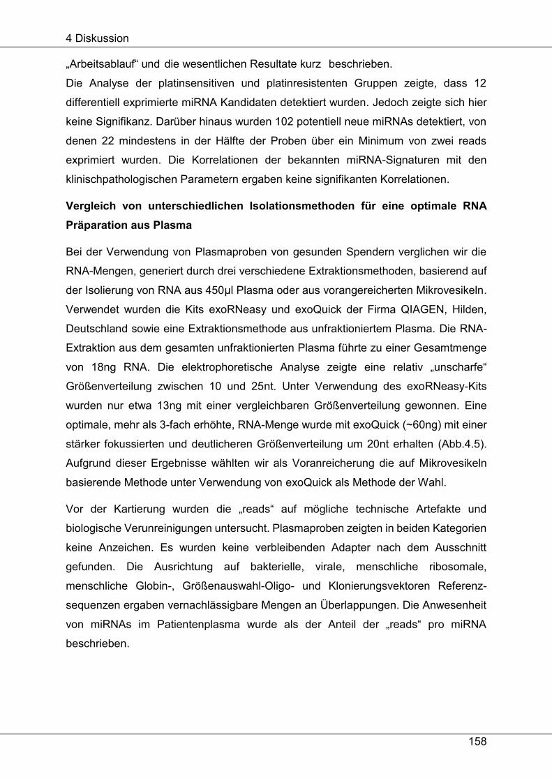
4 Diskussion
158
„Arbeitsablauf“ und die wesentlichen Resultate kurz beschrieben.
Die Analyse der platinsensitiven und platinresistenten Gruppen zeigte, dass 12
differentiell exprimierte miRNA Kandidaten detektiert wurden. Jedoch zeigte sich hier
keine Signifikanz. Darüber hinaus wurden 102 potentiell neue miRNAs detektiert, von
denen 22 mindestens in der Hälfte der Proben über ein Minimum von zwei reads
exprimiert wurden. Die Korrelationen der bekannten miRNA-Signaturen mit den
klinischpathologischen Parametern ergaben keine signifikanten Korrelationen.
Vergleich von unterschiedlichen Isolationsmethoden für eine optimale RNA
Präparation aus Plasma
Bei der Verwendung von Plasmaproben von gesunden Spendern verglichen wir die
RNA-Mengen, generiert durch drei verschiedene Extraktionsmethoden, basierend auf
der Isolierung von RNA aus 450μl Plasma oder aus vorangereicherten Mikrovesikeln.
Verwendet wurden die Kits exoRNeasy und exoQuick der Firma QIAGEN, Hilden,
Deutschland sowie eine Extraktionsmethode aus unfraktioniertem Plasma. Die RNA-
Extraktion aus dem gesamten unfraktionierten Plasma führte zu einer Gesamtmenge
von 18ng RNA. Die elektrophoretische Analyse zeigte eine relativ „unscharfe“
Größenverteilung zwischen 10 und 25nt. Unter Verwendung des exoRNeasy-Kits
wurden nur etwa 13ng mit einer vergleichbaren Größenverteilung gewonnen. Eine
optimale, mehr als 3-fach erhöhte, RNA-Menge wurde mit exoQuick (~60ng) mit einer
stärker fokussierten und deutlicheren Größenverteilung um 20nt erhalten (Abb.4.5).
Aufgrund dieser Ergebnisse wählten wir als Voranreicherung die auf Mikrovesikeln
basierende Methode unter Verwendung von exoQuick als Methode der Wahl.
Vor der Kartierung wurden die „reads“ auf mögliche technische Artefakte und
biologische Verunreinigungen untersucht. Plasmaproben zeigten in beiden Kategorien
keine Anzeichen. Es wurden keine verbleibenden Adapter nach dem Ausschnitt
gefunden. Die Ausrichtung auf bakterielle, virale, menschliche ribosomale,
menschliche Globin-, Größenauswahl-Oligo- und Klonierungsvektoren Referenz-
sequenzen ergaben vernachlässigbare Mengen an Überlappungen. Die Anwesenheit
von miRNAs im Patientenplasma wurde als der Anteil der „reads“ pro miRNA
beschrieben.
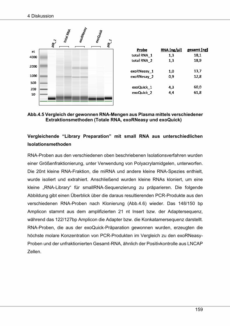
4 Diskussion
159
Abb.4.5 Vergleich der gewonnen RNA-Mengen aus Plasma mittels verschiedener Extraktionsmethoden (Totale RNA, exoRNeasy und exoQuick)
Vergleichende “Library Preparation” mit small RNA aus unterschiedlichen
Isolationsmethoden
RNA-Proben aus den verschiedenen oben beschriebenen Isolationsverfahren wurden
einer Größenfraktionierung, unter Verwendung von Polyacrylamidgelen, unterworfen.
Die 20nt kleine RNA-Fraktion, die miRNA und andere kleine RNA-Spezies enthielt,
wurde isoliert und extrahiert. Anschließend wurden kleine RNAs kloniert, um eine
kleine „RNA-Library“ für smallRNA-Sequenzierung zu präparieren. Die folgende
Abbildung gibt einen Überblick über die daraus resultierenden PCR-Produkte aus den
verschiedenen RNA-Proben nach Klonierung (Abb.4.6) wieder. Das 148/150 bp
Amplicon stammt aus dem amplifizierten 21 nt Insert bzw. der Adaptersequenz,
während das 122/127bp Amplicon die Adapter bzw. die Konkatamersequenz darstellt.
RNA-Proben, die aus der exoQuick-Präparation gewonnen wurden, erzeugten die
höchste molare Konzentration von PCR-Produkten im Vergleich zu den exoRNeasy-
Proben und der unfraktionierten Gesamt-RNA, ähnlich der Positivkontrolle aus LNCAP
Zellen.
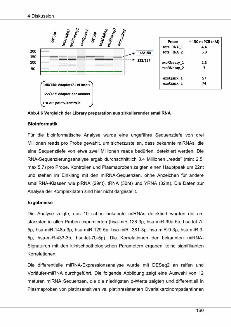
4 Diskussion
160
Abb.4.6 Vergleich der Library preparation aus zirkulierender smallRNA
Bioinformatik
Für die bioinformatische Analyse wurde eine ungefähre Sequenztiefe von drei
Millionen reads pro Probe gewählt, um sicherzustellen, dass bekannte miRNAs, die
eine Sequenztiefe von etwa zwei Millionen reads bedürfen, detektiert werden. Die
RNA-Sequenzierungsanalyse ergab durchschnittlich 3,4 Millionen „reads“ (min: 2,5,
max 5,7) pro Probe. Kontrollen und Plasmaproben zeigten einen Hauptpeak um 22nt
und stehen im Einklang mit den miRNA-Sequenzen, ohne Anzeichen für andere
smallRNA-Klassen wie piRNA (29nt), tRNA (30nt) und YRNA (32nt). Die Daten zur
Analyse der Komplexitäten sind hier nicht dargestellt.
Ergebnisse
Die Analyse zeigte, das 10 schon bekannte miRNAs detektiert wurden die am
stärksten in allen Proben exprimierten (hsa-miR-128-3p, hsa-miR-99a-5p, hsa-let-7i-
5p, hsa-miR-148a-3p, hsa-miR-129-5p, hsa-miR -381-3p, hsa-miR-9-3p, hsa-miR-9-
5p, hsa-miR-433-3p, hsa-let-7b-5p). Die Korrelationen der bekannten miRNA-
Signaturen mit den klinischpathologischen Parametern ergaben keine signifikanten
Korrelationen.
Die differentielle miRNA-Expressionsanalyse wurde mit DESeq2 an reifen und
Vorläufer-miRNA durchgeführt. Die folgende Abbildung zeigt eine Auswahl von 12
maturen miRNA Sequenzen, die die niedrigsten p-Werte zeigten und differentiell in
Plasmaproben von platinsensitiven vs. platinresistenten Ovarialkarzinompatientinnen
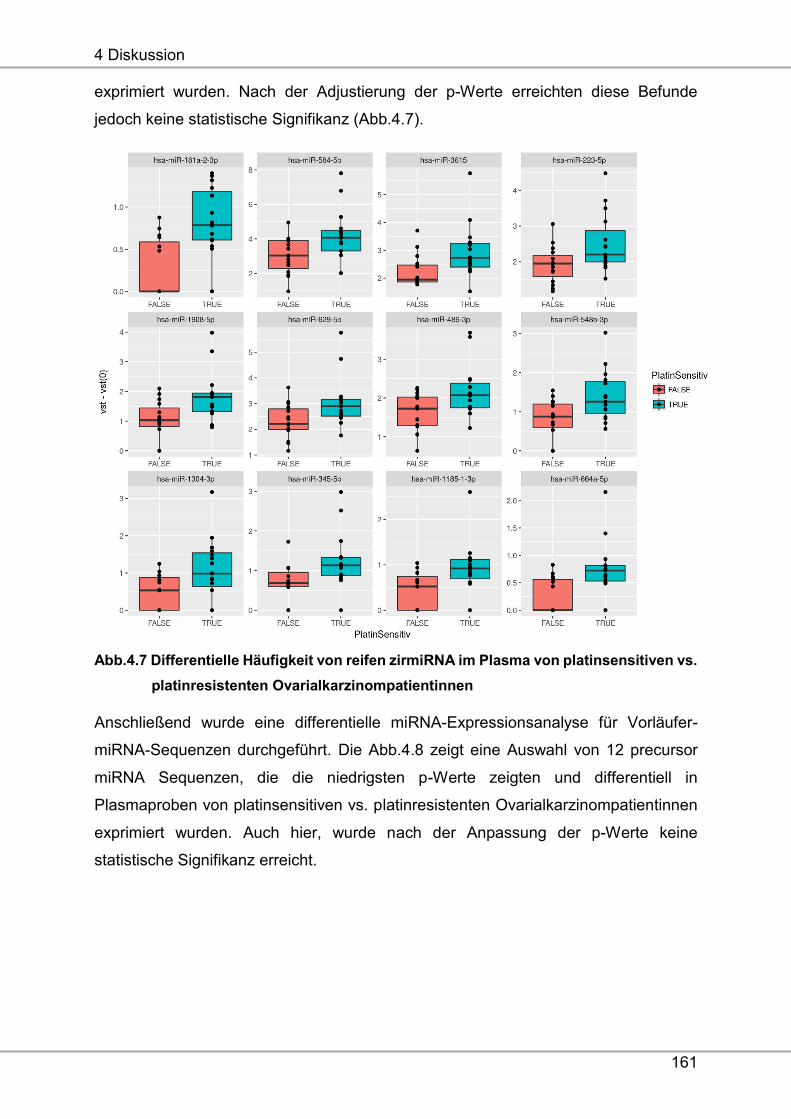
4 Diskussion
161
exprimiert wurden. Nach der Adjustierung der p-Werte erreichten diese Befunde
jedoch keine statistische Signifikanz (Abb.4.7).
Abb.4.7 Differentielle Häufigkeit von reifen zirmiRNA im Plasma von platinsensitiven vs.
platinresistenten Ovarialkarzinompatientinnen
Anschließend wurde eine differentielle miRNA-Expressionsanalyse für Vorläufer-
miRNA-Sequenzen durchgeführt. Die Abb.4.8 zeigt eine Auswahl von 12 precursor
miRNA Sequenzen, die die niedrigsten p-Werte zeigten und differentiell in
Plasmaproben von platinsensitiven vs. platinresistenten Ovarialkarzinompatientinnen
exprimiert wurden. Auch hier, wurde nach der Anpassung der p-Werte keine
statistische Signifikanz erreicht.
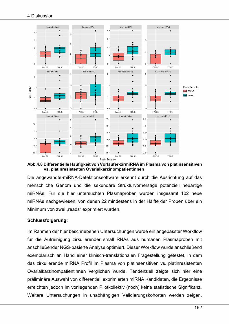
4 Diskussion
162
Abb.4.8 Differentielle Häufigkeit von Vorläufer-zirmiRNA im Plasma von platinsensitiven vs. platinresistenten Ovarialkarzinompatientinnen
Die angewandte-miRNA-Detektionssoftware erkennt durch die Ausrichtung auf das
menschliche Genom und die sekundäre Strukturvorhersage potenziell neuartige
miRNAs. Für die hier untersuchten Plasmaproben wurden insgesamt 102 neue
miRNAs nachgewiesen, von denen 22 mindestens in der Hälfte der Proben über ein
Minimum von zwei „reads“ exprimiert wurden.
Schlussfolgerung:
Im Rahmen der hier beschriebenen Untersuchungen wurde ein angepasster Workflow
für die Aufreinigung zirkulierender small RNAs aus humanen Plasmaproben mit
anschließender NGS-basierte Analyse optimiert. Dieser Workflow wurde anschließend
exemplarisch an Hand einer klinisch-translationalen Fragestellung getestet, in dem
das zirkulierende miRNA Profil im Plasma von platinsensitiven vs. platinresistenten
Ovarialkarzinompatientinnen verglichen wurde. Tendenziell zeigte sich hier eine
präliminäre Auswahl von differentiell exprimierten miRNA Kandidaten, die Ergebnisse
erreichten jedoch im vorliegenden Pilotkollektiv (noch) keine statistische Signifikanz.
Weitere Untersuchungen in unabhängigen Validierungskohorten werden zeigen,
4 Diskussion
163
inwieweit diese miRNA Kandidaten als potentiell prädiktive Biomarker für die
Platinresistenz des Ovarialkarzinoms genutzt werden können.
Nebenziel: RASSF-1A Promotormethylierung
Die Ergebnisse dieser Studie wurden in Oncotarget publiziert.
RASSF1A promoter methylation in high-grade serous ovarian cancer: A direct comparison study in primary tumors, adjacent morphologically tumor cell-free tissues and paired circulating tumor DNA. Lydia Giannopoulou, Issam Chebouti, Kitty Pavlakis, Sabine Kasimir-Bauer, Evi Lianidou. Oncotarget 2017. doi: 10.18632/oncotarget.15249. Obwohl es schon Studien zur RASSF-1A Promotormethylierung im Tumorgewebe von
Ovarialkarzinompatientinnen gibt, analysierte unsere Studie zum ersten Mal den
Methylierungsstatus von RASSF-1A im Tumorgewebe, dem Normalgewebe und den
entsprechenden korrespondieren Plasmaproben.
In Kooperation mit der Arbeitsgruppe von Frau Prof. Lianidou der Universität Athen
wurde die RASSF1A Promotermethylierung im Tumorgewebe (n=61), im angrenzen-
den tumorfreien Gewebe (n=58) und in korrespondierenden Plasmaproben (n=59) von
Patientinnen mit primärem Ovarialkarzinom, mittels der MSP-PCR und der
semiquantitativen „methylation-sensitive high-resolution melting analysis“ (MS-HRMA)
untersucht. Die Frauenklinik stellte hier die unabhängige Validierungskohorte mit 61
Gewebetumorproben, 58 angrenzenden tumorfreien Geweben und 59 korrespon-
dierenden Plasmaproben zur Verfügung. Die schematische Überschrift der Studie
zeigt zur Veranschaulichung die experimentelle Vorgehensweise (Abb.4.9). Die
Isolierung der zirfDNA aus dem Plasma, die Sodiumbisulfitkonvertierung und die MSP-
PCR sowie MS-HRMA wurden in Athen durchgeführt. Die MSP-PCR ergab eine
RASSF1A-Methylierung in 25/61 (41%) Tumorproben, in 17/58 (29%) gesunden
Geweben und in 15/59 (25%) Plasmaproben. Im Gegensatz dazu wurde bei der MS-
HRMA-Analyse eine Methylierung des Gens im Tumorgewebe in 28/61 (46%) und im
gesunden Gewebe in 21/58 (36%) der Patientinnen detektiert. Die Methylierung des
Gens im Plasma wurde mit dieser Methode nicht untersucht. Die RASSF-1A-Promotor-
Methylierung assoziierte signifikant mit der Tumorqualität, unter Verwendung beider
Assays, sowie mit dem Befall regionaler Lymphknoten unter Verwendung von MS-
HRMA. Durch MS HRMA-Analysen im Tumorgewebe wurde gezeigt, dass der RASSF-
1A-Promotor-Methylierungsstatus signifikant mit dem verkürzten OS korrelierte. Im
Gegensatz dazu wurde unter Verwendung der Real-Time-MSP Analyse keine
signifikante Korrelation mit dem PFS und OS beobachtet.
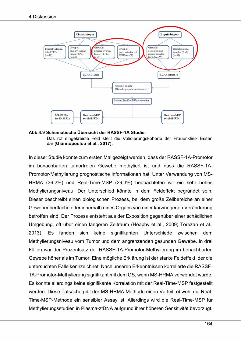
4 Diskussion
164
Abb.4.9 Schematische Übersicht der RASSF-1A Studie.
Das rot eingekreiste Feld stellt die Validierungskohorte der Frauenklinik Essen dar (Giannopoulou et al., 2017).
In dieser Studie konnte zum ersten Mal gezeigt werden, dass der RASSF-1A-Promotor
im benachbarten tumorfreien Gewebe methyliert ist und dass die RASSF-1A-
Promotor-Methylierung prognostische Informationen hat. Unter Verwendung von MS-
HRMA (36,2%) und Real-Time-MSP (29,3%) beobachteten wir ein sehr hohes
Methylierungsniveau. Der Unterschied könnte in dem Feldeffekt begründet sein.
Dieser beschreibt einen biologischen Prozess, bei dem große Zellbereiche an einer
Gewebeoberfläche oder innerhalb eines Organs von einer karzinogenen Veränderung
betroffen sind. Der Prozess entsteht aus der Exposition gegenüber einer schädlichen
Umgebung, oft über einen längeren Zeitraum (Heaphy et al., 2009; Torezan et al.,
2013). Es fanden sich keine signifikanten Unterschiede zwischen dem
Methylierungsniveau vom Tumor und dem angrenzenden gesunden Gewebe. In drei
Fällen war der Prozentsatz der RASSF-1A-Promotor-Methylierung im benachbarten
Gewebe höher als im Tumor. Eine mögliche Erklärung ist der starke Feldeffekt, der die
untersuchten Fälle kennzeichnet. Nach unseren Erkenntnissen korrelierte die RASSF-
1A-Promotor-Methylierung signifikant mit dem OS, wenn MS-HRMA verwendet wurde.
Es konnte allerdings keine signifikante Korrelation mit der Real-Time-MSP festgestellt
werden. Diese Tatsache gibt der MS-HRMA-Methode einen Vorteil, obwohl die Real-
Time-MSP-Methode ein sensibler Assay ist. Allerdings wird die Real-Time-MSP für
Methylierungsstudien in Plasma-ztDNA aufgrund ihrer höheren Sensitivität bevorzugt.
4 Diskussion
165
In sechs Proben, bei denen der Primärtumor unmethyliert gefunden wurde, waren die
entsprechenden Plasmaproben methyliert. Eine mögliche Erklärung ist die
Tumorheterogenität. Es ist klar, dass die Gewebebiopsie eine Momentaufnahme des
Oberflächenprofils eines Tumors darstellt, während die zfDNA die gesamten
genetischen und epigenetischen Eigenschaften eines bestimmten Tumors wider-
spiegelt.
Zusammenfassende Schlussfolgerung für die unter Zielsetzung 3 und das
Nebenziel erhobenen Daten:
Als weitere, nicht zelluläre, „Liquid Biopsy“ sind zirkulierende DNA und miRNA
attraktive Marker für das Ovarialkarzinom. Bezüglich der miRNAs konnten potentielle
Kandidaten zur weiteren Evaluierung identifiziert werden, um platinresistente und
platinsensible Patientinnen zu beschreiben. Die Ergebnisse des RASSF-1A-
Methylierungsstatuses beim Ovarialkarzinom haben das Potenzial, wichtige
prognostische Informationen zu geben. Um diese Art der „Liquid Biopsy“ zukünftig zu
nutzen, müssen prospektive Studien durchgeführt werden. Weitere Untersuchungen
in unabhängigen Validierungskohorten werden zeigen, inwieweit diese Kandidaten als
potentielle prädiktive Biomarker für das Ovarialkarzinom, speziell für die Vorhersage
der Platinresistenz, in der Klinik eingesetzt werden können.
5 Literaturverzeichnis
166
5 Literaturverzeichnis
1. Aktas B, Kasimir-Bauer S, Heubner M, Kimmig R, Wimberger P. Molecular
profiling and prognostic relevance of circulating tumor cells in the blood of
ovarian cancer patients primary diagnosis and after platinum-based
chemotherapy. 2011 Int J Gynecol Cancer, 21:822-830.
2. Alix-Panabières C, Pantel K. Technologies for detection of circulating tumor
cells: facts and vision. 2011. Lab Chip.,14(1):57-62.
3. Arroyo JD, Chevillet JR, Kroh EM, Ruf IK, Pritchard CC, Gibson DF, Mitchell
PS, Bennett CF, Pogosova-Agadjanyan EL, Stirewalt DL, Tait JF, Tewari M.
Argonaute2 complexes carry a population of circulating microRNAs
independent of vesicles in human plasma. 2011. Proc Natl Acad Sci USA
108:5003-5008.
4. Baccelli I, Schneeweiss A, Riethdorf S, Stenzinger A, Schillert A, Vogel V, Klein
C, Saini M, Bauerle T, Wallwiener M, Holland-Letz T, Hofner T, Sprick M,
Scharpff M, Marme F, Sinn HP, et al. Identification of a population of blood
circulating tumor cells from breast cancer patients that initiates metastasis in a
xenograft assay. 2013. Nat Biotechnol.; 31:539-544.
5. Balic M, Lin H, Young L, Hawes D, Giuliano A, McNamara G, Datar RH, Cote
RJ. Most early disseminated cancer cells detected in bone marrow of breast
cancer patients have a putative breast cancer stem cell phenotype.2006. Clin
Cancer Res.; 12:5615–5621.
6. Banys M, Solomayer EF, Becker S, Krawcyk N, Gardanis K, Staebler A,
Neubauer H, Wallwiener D, Fehm T. Disseminated tumor cells in bone marrow
may affect prognosis of patients with gynecologic malignancies. 2009. Int J
Gynecol Cancer, 19(5):948-502.
7. Bapat SA, Koppikar CB, Kurrey NK. Stem and Progenitor-Like Cells Contribute
to the Aggressive Behavior of Human Epithelial Ovarian Cancer. 2005. Cancer
Res.; 65:3025–3029.
8. Bareiss PM, Paczulla A, Wang H, Schairer R, Wiehr S, Kohlhofer U, Rothfuss
OC, Fischer A, Perner S, Staebler A, Wallwiener D, Fend F, Fehm T, et al.
SOX2 expression associates with stem cell state in human ovarian carcinoma.
2013 Cancer Res.; 73; 5544–5555.

5 Literaturverzeichnis
167
9. Bednarz-Knoll N, Alix-Panabieres C and Pantel K. Plasticity of disseminating
cancer cells in patients with epithelial malignancies.2012. Cancer Metastasis
Rev.; 31(3-4):673-687.
10. Berek J, Taylor P, McGuire W, Smith LM, Schultes B, Nicodemus CF.
Oregovomab maintenance monoimmunotherapy does not improve outcomes in
advanced ovarian cancer. 2009. J Clin Oncol, 20;27 (3):418-25.
11. Billy E, Brondani V, Zhang H, Muller U, Filipowicz W. Specific interference with
gene expression induced by long, double-stranded RNA in mouse embryonal
teratocarcinoma cell lines. 2001. Proc Natl Acad Sci USA 98:14428-14433.
12. Bookman MA. Extending the platinum-free interval in recurrent ovarian cancer:
the role of topotecan in second-line chemotherapy.1999. Oncologist. 4:87-94.
13. Brabletz T, Hlubek F, Spaderna S, Schmalhofer O, Hiendlmeyer E, Jung A,
Kirchner T.Invasion and metastasis in colorectal cancer: epithelial-
mesenchymal transition, mesenchymal-epithelial transition, stem cells and
beta-catenin. 2005. Cells Tissues Organs.; 179(1-2):56-65.
14. Bragado P, Sosa MS, Keely P, Condeelis J, Aguirre-Ghiso JA.
Microenvironments dictating tumor cell dormancy.2012. Recent Results Cancer
Res, 195:25-39.
15. Braun S, Schindlbeck C, Hepp F, Janni W, Kentenich C, Riethmuller G, Pantel
K. Occult tumor cells in bone marrow of patients with locoregionally restricted
ovarian cancer predict early distant metastatic relapse. 2001. J Clin Oncol
19:368-375.
16. Braun S, Vogl FD, Naume B, Janni W, Osborne MP, Coombes RC, Schlimok
G, Diel IJ, Gerber G, Gebauer G, Pierga JY, Marth C, Oruzio D, Wiedswang G,
Solomayer EF, Kundt G, Strobl B, Fehm T, Wong GY, Bliss J, Vincent-Salomon
A, Pantel K. A pooled analysis of bone marrow micrometastasis in breast
cancer. 2005. N Engl J Med, 353:793-802.
17. Bredemeier M, Edimiris P, Tewes M, Mach P, Aktas B, Hauch S, Wagner J,
Kimmig R, Kasimir-Bauer S. A. Establishment of a multimarker qPCR panel for
the molecular characterization of circulating tumor cells in blood samples of
metastatic breast cancer patients during the course of palliative treatment.
2016. Oncotarget.; 7(27):41677-41690.
18. Bristow RE, Tomacruz RS, Armstrong DK, Trimble EL, Montz FJ. Survival
effect of maximal cytoreductive surgery for advanced ovarian carcinoma during
the platinum era: a meta-analysis.2002.J Clin Oncol, 20:1248-59.

5 Literaturverzeichnis
168
19. Burger RA, Sill MW, Monk BJ, Greer BE, Sorosty JI. Phase II trial of
bevacizumab in persistent or recurrent epithelial ovarian cancer or primary
peritoneal cancer: a Gynecologic Oncology Group Study.2007. J Clin Oncol,
25(33):5165-71.
20. Burges A, Wimberger P, Kumper C, Gorbounova V, Sommer H, Schmalfeldt B,
Pfisterer J, Lichinitser M, Makhson A, Moiseyenko V, Lahr A, Schulze E, Jager
M, Strohlein MA, Heiss MM, Gottwald T, Lindhofer H, Kimmig R. Effective Relief
of Malignant Ascites in Patients with Advanced Ovarian Cancer by a
Trifunctional Anti-EpCAM x Anti-CD3 Antibody: A phase ½ Study: Clin. Cancer
Res. 2007, 13(13), 3899-3905.
21. Chan JK, Cheung MK, Husain A, Teng NN, West D, Whittemore AS, Berek JS,
Osann K. Patterns and progress in ovarian cancer over 14 years. 2006. Obstet
Gynecol, 108:521-528.
22. Chebouti I, Blassl C, Wimberger P, Neubauer H, Fehm T, Kimmig R, Kasimir-
Bauer S.Analysis of disseminated tumor cells before and after platinum based
chemotherapy in primary ovarian cancer. Do stem cell like cells predict
prognosis? 2016. Oncotarget ;7(18):26454-64.
23. Chebouti I, Kuhlmann JD, Buderath P, Weber S, Wimberger P, Bokeloh Y,
Hauch S, Kimmig R, Kasimir-Bauer S. ERCC1-expressing circulating tumor
cells as a potential diagnostic tool for monitoring response to platinum-based
chemotherapy and for predicting post-therapeutic outcome of ovarian cancer.
2017. doi: 10.18632/oncotarget.13286.
24. Chebouti I, Kasimir-Bauer S, Buderath P, Wimberger P, Hauch S, Kimmig R,
Kuhlmann JD. EMT-like circulating tumor cells in ovarian cancer patients are
enriched by platinum-based chemotherapy. Oncotarget 2017 (in print).
25. Chen A. PARP inhibitors: it´s role in treatment of cancer. 2011. Chin J Cancer;
30: 463-71.
26. Chen X, Ba Y, Ma L, Cai X, Yin Y, Wang K, et al. Characterization of microRNAs
in serum: a novel class of biomarkers for diagnosis of cancer and other
diseases. 2008. Cell Res.; 18:997–1006.
27. Christofori G. New signals from the invasive front. 2006. Nature.;
441(7092):444-50.
28. Cui L, Kwong J, Wang CC. Prognostic value of circulating tumor cells and
disseminated tumor cells in patients with ovarian cancer: a systematic review
and meta-analysis. 2015. Journal of Ovarian Research; 8:38.
5 Literaturverzeichnis
169
29. Diehl F, Li M, Dressmann D, He Y. Detections and quantification of mutations
in the plasma of patients with colorectal tumors. 2005. Proc Ntl Acad Sci USA
102:16368-16373.
30. Du Bois A, Lück HJ, Meier W, Moebus V, Costa SD, Bauknecht T, Richter B,
Warm M, Schroeder W, Olbricht S, Nitz U, Jackisch C. Cisplatin/Paclitaxel vs.
Carboplatin/Paclitaxel in Ovarian Cancer: Update of an Arbeitsgemeinschaft
Gynaekologische Onkologie (AGO) Study Group Trial. 1999. Proc Am Soc Clin
Oncol. , Abstract 1374.
31. Du Bois A, Pfisterer J. Future options for first-line therapy of advanced ovarian
cancer.2005 Int J Gynecol Cancer; 1:42-50.
32. Du J, Li B, Fang Y, Liu Y, Wang Y, Li J, Zhou W, Wang X. Overexpression of
Class III ß-tubulin, Sox2, and nuclear surviving is predictive of taxane resistance
in patients with stage III ovarian epithelial cancer. 2015. BMC Cancer. 15:536.
33. Ehlen TG, Hoskins PJ, Miller D, Whiteside TL, Nicodemus CF, Schultes BC,
Swerton KD. A pilot phase 2 study of oregovomab murine monoclonal antibody
to CA125 as an immunotherapeutic agent for recurrent ovarian cancer. 2005.
Int J Gynecol Cancer, 15:1023-34.
34. Esquela-Kerscher A, Slack FJ. Oncomirs - microRNAs with a role in cancer.
2006. Nat Rev Cancer 6:259-269.
35. Fan T, Zhao Q, Chen JJ, et al. Clinical significance of circulating tumor cells
detected by an invasion assay in peripheral blood of patients with ovarian
cancer. 2009. Gynecol; 112: 185–191.
36. Fehm T, Banys M, Rack B, Janni W, Marth C, Blassl C, Hartkopf A, Trope C,
Kimmig R, Krawcyzk N, Wallwiener D, Wimberger P, Kasimir-Bauer S. Pooled
analysis of the prognostic relevance of disseminated tumor cells in the bone
marrow of patients with ovarian cancer. 2013. Int J Gynecol Cancer. 23(5):839-
45.
37. Fong PC et al. Poly(ADP)-Ribose Polymerase Inhibition: Frequent Durable
Responses in BRCA Carrier Ovarian Cancer Correlating With Platinum-Free
Interval. 2010. J Clin Oncol; 28: 2512-9.
38. Forstner R, Meissnitzer M, Cunha TM. Update on imaging of Ovarian Cancer.
2016. Curr Radiol; 4:31.
39. Friboulet L, Olaussen KA, Pignon JP, Shepherd FA, Tsao MS, Graziano S, et
al. ERCC1 isoform expression and DNA repair in non-small-cell lung cancer.
2013. N Engl J Med; 368:1101–10.
5 Literaturverzeichnis
170
40. Fukushima H, Abe T, Sakamoto K, Tsujimoto H, Mizuarai S, Oie S. 3'-
ethynylcytidine, an RNA polymerase inhibitor, combined with cisplatin exhibits
a potent synergistic growth-inhibitory effect via Vaults dysfunction. 2014. BMC
Cancer; 14:562.
41. Galluzzi L, Senovilla L, Vitale I, Michels J, Martins I, Kepp O, Castedo M,
Kroemer G. Molecular mechanisms of cisplatin resistance. 2012. Oncogene;
31:1869-1883.
42. Gao YC, Wu J. MicroRNA-200c and microRNA-141 as potential diagnostic and
prognostic biomarkers for ovarian cancer. 2015. Tumour Biol.; 36:4843–50.
43. Giannopoulou L, Chebouti I, Pavlakis K, Kasimir-Bauer S, Lianidou ES.
RASSF1A promoter methylation in high-grade serous ovarian cancer: A direct
comparison study in primary tumors, adjacent morphologically tumor cell-free
tissues and paired circulating tumor DNA. Oncotarget. 2017 doi:
10.18632/oncotarget.15249.
44. Gloss BS and Samimi G. Epigenetic biomarkers in epithelial ovarian cancer.
2014. Cancer Lett.; 342: 257-63.
45. Goodman MT, Howe HL, Tung KH, Hotes J, Miller BA, Coughlin SS, Chen VW.
Incidence of ovarian cancer by race and ethnicity in the United States. 2003.
Cancer; 97:2676–2685.
46. Gordon AN, Butler J. Chemotherapeutic management of advanced ovarian
cancer. 2003. Semin Oncol Nus, 19:3-18.
47. Gossage L and Madhusudan S. Current status of excision repair cross
complementing-group 1 (ERCC1) in cancer. 2007. Cancer Treat Rev.;
33(6):565-77.
48. Grawenda AM and O'Neill E. Clinical utility of RASSF1A methylation in human
malignancies. 2015. Br J Cancer; 113: 372-81.
49. Greenbaum D, Colangelo C, Williams K and Gerstein M. Comparing protein
abundance and mRNA expression levels on a genomic scale. 2003. Genome
Biol.; 4(9):117.
50. Hartkopf AD, Stefanescu D, Wallwiener M, Hahn M, Becker S, Solomayer EF,
Fehm TN, Brucker SY, Taran FA.Tumor cell dissemination to the bone marrow
and blood is associated with poor outcome in patients with metastatic breast
cancer. 2014. Breast Cancer Res Treat.; 147(2):345-51.
51. He W, Kularante SA, Kalli KR, Prendergast FG, Amato RJ, Klee GG, Hartmann
LC, Low PS. Quantitation of circulating tumor cells in blood samples from
5 Literaturverzeichnis
171
ovarian and prostate cancer patients using tumor-specific fluorescent ligands.
2008. Int J Cancer. 30(2):134-7.
52. Heaphy CM, Griffith JK, Bisoffi M.. "Mammary field cancerization: molecular
evidence and clinical importance". 2009. Breast Cancer Res Treat. 118 (2):
229–39.
53. Hong F, Li Y, Xu Y, Zhu L. Prognostic significance of serum microRNA-221
expression in human epithelial ovarian cancer. 2013. J Int Med Res.; 41:64–71.
54. Hosono S, Kajiyama H, Terauchi M, Shibata K, Ino K, Nawa A and Kikkawa F.
Expression of Twist increases the risk for recurrence and for poor survival in
epithelial ovarian carcinoma patients. 2007. Br J Cancer.; 96(2):314-320.
55. Hu X, Macdonald DM, Huettner PC, Feng Z, El Naqa IM, Schwarz JK, Mutch
DG, Grigsby PW, Powell SN, Wang X. A miR-200 microRNA cluster as
prognostic marker in advanced ovarian cancer. 2009. Gynecol Oncol 114:457-
464.
56. Ibanez de Caceres I, Battagli C, Esteller M, Herman JG,Dulaimi E, Edelson MI,
Bergman C, Ehya H, Eisenberg BL, Cairns P. Tumor cell-specific BRCA1 and
RASSF1A hypermethylation in serum, plasma, and peritoneal fluid from ovarian
cancer patients. 2004. Cancer Res., 64, pp. 6476–6481.
57. Iorio MV, Visone R, Di Leva G, Donati V, Petrocca F, Casalini P, Taccioli C,
Volinia S, Liu CG, Alder H, Calin GA, Menard S, Croce CM. MicroRNA
signatures in human ovarian cancer. 2007. Cancer Res 67:8699-8707.
58. Janni W, Vogl FD, Wiedswang G, Synnestvedt M, Fehm T, Jückstock J, Borgen
E, Rack B, Braun S, Sommer H, Solomayer E, Pantel K, Nesland J, Friese K,
Naume B. Persistence of disseminated tumor cells in the bone marrow of breast
cancer patients predicts increased risk for relapse--a European pooled analysis.
2011. Clin Cancer Res.; 17(9):2967-76. CCR-10-2515.
59. Jazaeri AA, Shibata E, Park J, Bryant JL, Conaway MR, Modesitt SC, Smith
PG, Milhollen MA, Berger AJ, Dutta A. Overcoming platinum resistance in
preclinical models of ovarian cancer using the neddylation inhibitor MLN4924.
2013. Mol Cancer Ther.; 12:1958-1967.
60. Jones PA and Baylin SB. The fundamental role of epigenetic events in cancer.
2002. Nat Rev Genet.; 3(6):415-28.
61. Judson PL, Geller MA, Bliss RL, Boente MP, Downs LS Jr, Argenta PA, Carson
LF. Preoperative detection of peripherally circulating cancer cells and its
prognostic significance in ovarian cancer. 2003. Gynecol Oncol., 91(2):389-94.
5 Literaturverzeichnis
172
62. Kajiyama H, Hosono S, Terauchi M, Shibata K, Ino K, Yamamoto E, Nomura
S, Nawa A and Kikkawa F. Twist expression predicts poor clinical outcome of
patients with clear cell carcinoma of the ovary. 2006. Oncology.; 71(5-6):394-
401.
63. Kamat AA, Sood AK, Dang D, Gershenson DM, Simpson JL and Bischoff FZ.
Quantification of total plasma cell-free DNA in ovarian cancer using real-time
PCR. 2006. Ann N Y Acad Sci 2006; 1075:230-4.
64. Kapetanakis NI, Uzan C, Jimenez-Pailhes AS, Gouy S, Bentivegna E, Morice
P, et al. Plasma miR-200b in ovarian carcinoma patients: distinct pattern of
pre/post-treatment variation compared to CA125 and potential for prediction of
progression-free survival. 2015. Oncotarget.; 6:36815–24.
65. Kasimir-Bauer S, Bittner AK, König L, Reiter K, Keller T, Kimmig R, Hoffmann
O.Does primary neoadjuvant systemic therapy eradicate minimal residual
disease? Analysis of disseminated and circulating tumor cells before and after
therapy. 2016. Breast Cancer Res.;18(1):20.
66. Kleibeuker EA, Ten Hooven MA, Verheul HM, Slotman BJ, Thijssen VL.
Combining radiotherapy with sunitinib: 2015. Angiogenesis; 18(4):385-95.
67. Kolasa IK, Rembiszewska A, Felisiak A, Ziolkowska-Seta I, Murawska M, Moes
J, Timorek A, Dansonka-Mieszkowska A and Kupryjanczyk J. PIK3CA
amplification associates with resistance to chemotherapy in ovarian cancer
patients. 2009. Cancer Bio Ther.; 8(1):21-26.
68. Kuhlmann JD, Schwarzenbach H, Otterbach F, Heubner M, Wimberger P,
Worm KH, Kimmig R, Kasimir-Bauer S.Loss of heterozygosity proximal to the
M6P/IGF2R locus is predictive for the presence of disseminated tumor cells in
the bone marrow of ovarian cancer patients before and after chemotherapy.
2011. Gene Chromosomes Cancer; 50(8):598-605.
69. Kuhlmann JD, Rasch J, Wimberger P, Kasimir-Bauer S. microRNA and the
pathogenesis of ovarian cancer--a new horizon for molecular diagnostics and
treatment? 2012. Clin Chem Lab Med.13;50(4):601-15.
70. Kuhlmann JD, Wimberger P, Bankfalvi A, Keller T, Schöler S, Aktas B,
Buderath P, Hauch S, Otterbach F, Kimmig R, Kasimir-Bauer S. ERCC1-
positive circulating tumor cells in the blood of ovarian cancer patients as a
predictive biomarker for platinum resistance. 2014. Clin Chem. (10):1282-9.
5 Literaturverzeichnis
173
71. Kurata H, Takakuwa K, Tsuneki I, Aoki Y, Tanaka K. Ovarian tumor cell
detection in peripheral blood progenitor cells harvests by RT-PCR. 2002. Acta
Obstet Gynecol Scand.; 81(6):555-9.
72. Latifi A, Abubaker K, Castrechini N, Ward AC, Liongue C, Dobill F, Kumar J,
Thompson EW, Quinn MA, Findlay JK and Ahmed N. Cisplatin treatment of
primary and metastatic epithelial ovarian carcinomas generates residual cells
with mesenchymal stem cell-like profile. 2011.J Cell Biochem.; 112(10):2850-
2864.
73. Leon SA, Shapiro B, Sklaroff DM, Yaros MJ. Free DNA in the serum of cancer
patients and the effect of therapy. 1977. Cancer Res 37:646-650.
74. Liang H, Jiang Z, Xie G, Lu Y. Serum microRNA-145 as a novel biomarker in
human ovarian cancer. 2015. Tumour Biol.; 36:5305–13.
75. Lianidou ES, Markou A. Circulating tumor cells in breast cancer: detection
systems, molecular characterization, and future challenges. 2011. Clin Chem.;
57(9):1242-55.
76. Liggett TE, Melnikov A,Yi Q, Replogle C, Hu W, Rotmensch J, Kamat A, Sood
AK, Levenson V. Distinctive DNA methylation patterns of cell-free plasma DNA
in women with malignant ovarian tumors. 2011. Gynecol. Oncol., 120, pp. 113–
120.
77. Liu JF, Kindelberger D, Doyle C, Lowe A, Barry WT, Matulonis UA. Predictive
value of circulating tumor cells (CTCs) in newly-diagnosed and recurrent
ovarian cancer patients. 2013. Gynecol Oncol.; 131(2):352-6.
78. Liu P, Cheng H, Roberts TM and Zhao JJ. Targeting the phosphoinositide 3-
kinase pathway in cancer. 2009. Nat Rev Drug Discov.; 8(8):627-644.
79. Ma L, Liu FR, Zhang SL. Detection of circulating hypermethylated tumor-
specific RASSF1A DNA in ovarian cancer patients) Zhonghua bing li xue za zhi.
2005.Chin. J. Pathol., 34, pp. 785–787.
80. Mandel and Métais. Les acides nucléiques du plasma sanguin chez
l'homme.1948. CR Seances Soc Biol Fil.; 142(3-4):241-3.
81. Marme A, Strauss G, Bastart G, Grischke EM, Moldenhauer G. Intraperitonal
bispecific antibody (HEA125xOKT3) therapy inhibits malignant ascites
production in advanced ovarian carcinoma. 2002.Int J, 101:183-189.
82. Marth C, Kisic J, Kaern J, Tropé C, Fodstad O. Circulating tumor cells in the
peripheral blood and bone marrow of patients with ovarian carcinoma do not
predict prognosis. 2002 Cancer, 40(4):1447-50.
5 Literaturverzeichnis
174
83. Marzese DM, Hirose H and Hoon DS. Diagnostic and prognostic value of
circulating tumor-related DNA in cancer patients. 2013. Expert Rev Mol Diagn
Nov; 13(8):827-44.
84. Massard C, Oulhen, Le Moulec S, Auger N, Foulon S, Abou-Lovergne A, Billiot
F, Valent A, Marty V, Loriot Y, Fizazi K, Vielh P and Farace F. Phenotypic and
genetic heterogeneity of tumor tissue and circulating tumor cells in patients with
metastatic castrationresistant prostate cancer: a report from the PETRUS
prospective study. 2016 Oncotarget.23;7(34):55069-55082.
85. Mause SF, Weber C. Microparticles: protagonists of a novel communication
network for intercellular information exchange. 2010. Circ Res.; 107:1047–
1057.
86. Meehan RS, Chen AP. New treatment option for ovarian cancer: PARP
inhibitors. 2016.Gynecologic Research and Practice 3:3.
87. Meng X, Joosse SA, Müller V, Trillsch F, Milde-Langosch K, Mahner S, et al.
Diagnostic and prognostic potential of serum miR-7, miR-16, miR-25, miR- 93,
miR-182, miR-376a and miR-429 in ovarian cancer patients. 2015. Br J Cancer.;
113:1358–66.
88. Milovic-Kovacevic M, Srdic-Rajic T, Radulovic S, Bjelogrlic S, Gavrilovic D.
Expression of ERCC1 proteinin biopsy specimen predicts survival in
advanced ovarian cancer patients treated with platinum-based chemotherapy.
2011. J Buon,. 16(4):708-14.
89. Mitchell PS, Parkin RK, Kroh EM, Fritz BR, Wyman SK, Pogosova-Agadjanyan
EL, et al. Circulating microRNAs as stable blood-based markers for cancer
detection. 2008. Proc Natl Acad Sci USA; 105:10513–10518.
90. Murtaza M, Dawson SJ, Tsui DW, Gale D, Forshew T, Piskorz AM, Parkinson
C, Chin SF, Kingsbury Z, Wong AS, Marass F, Humphray S, Hadfield J, et al.
Non-invasive analysis of acquired resistance to cancer therapy by sequencing
of plasma DNA. 2013. Nature; 497(7447):108-112.
91. Nakamura K, Sawada K, Yoshimura A, Kinose Y, Nakatsuka E and Kimura T.
Clinical relevance of circulating cell-free microRNAs in ovarian cancer. 2016.
Molecular Cancer 15:48.
92. Neijt JP, Engelholm SA, Witteveen PO, Tuxen MK, Sorensen PG, Hansen M,
Hirsch F, Sessa C, de Swart C, van Houwelingen HC, Lund B, Hansen SW
Paclitaxel (175 mg/m2 over 3 hours) with cisplatin or carboplatin in previously
5 Literaturverzeichnis
175
untreated ovarian cancer: an interim analysis.1997. Semin Oncol, 24:S15-36-
S15-39.
93. Nel I, Gauler TC, Eberhardt WE, Nickel AC, Schuler M, Thomale J, Hoffmann
AC. Formation and repair kinetics of Pt-(GpG) DNA adducts in extracted
circulating tumour cells and response to platinum treatment. 2013. Br J Cancer.;
109:1223-1229.
94. Nie L, Wu G and Zhang W. Correlation of mRNA expression and protein
abundance affected by multiple sequence features related to translational
efficiency in Desulfovibrio vulgaris: a quantitative analysis. 2006. Genetics.;
174(4):2229-2243.
95. No JH, Kim K, Park KH and Kim YB. Cell-free DNA level as a prognostic
biomarker for epithelial ovarian cancer. 2012. Anticancer Res; 32(8):3467-71.
96. Noujaim AA, Schultes BC, Baum RP, Madiyalakan R. Induction of CA125-
specific B and T cell responses in patients injected with MAP-B43.13-evidence
for antibody-mediated antigen-processing and presentation of CA125 in vivo.
2011. Cancer Biother Radiopharm, 16(3):187-203.
97. Nuti SV, Mor G, Li P and Yin G. TWIST and ovarian cancer stem
cells: implications for chemoresistance and metastasis. 2014.Oncotarget;
5(17):7260-7271.
98. Oikonomopoulou K, Scorilas A, Michael IP, Grass L, Soosaipillai A, Rosen B,
Murphy J, Diamandis EP. Kallikreins as markers of disseminated tumour cells
in ovarian cancer-- a pilot study. 2006.Tumour Biol.;27(2):104-14.
99. Panabières A, Schwarzenbach H, Pantel K. Circulating tumor cells and
circulating tumor DNA. 2012. Annu Rev Med.; 63:199-215.
100. Panabiéres and Pantel. Clinical Applications of Circulating Tumor Cells and
Circulating Tumor DNA as Liquid Biopsy. 2016. Cancer Discovery. 6(5):479-91.
101. Pantel K, Alix-Panabieres C, Riethdorf S. Cancer micrometastases. 2009. Nat
Rev Clin Oncol 6:339-351.
102. Parsons S, et al. Intraperitoneal treatment of malignant ascites due to epithelial
tumors with catumaxomab: A phase II/III study. 2008. J Clin Oncol, 26: suppl;
abstr 3000.
103. Pascal LE, True LD, Campbell DS, Deutsch EW, Risk M, Coleman IM, Eichner
LJ, Nelson PS and Liu AY. Correlation of mRNA and protein levels: cell type
specific gene expression of cluster designation antigens in the prostate. 2008.
BMC Genomics.; 9:246.
5 Literaturverzeichnis
176
104. Peng DJ, Wang J, Zhou JY, Wu GS. Role of the Akt/mTOR survival pathway in
cisplatin resistance in ovarian cancer cells. 2010. Biochem Biophys Res
Commun.; 394:600-605.
105. Peng S, Maihle NJ, Huang Y. Pluripotency factors Lin28 and Oct4 identify a
sub-population of stem cell-like cells in ovarian cancer. 2010. Oncogene;
29:2153–5159.
106. Pfisterer J, du Bois, Sehouli J, Loibl S, Reinartz S, et al. The anti-idiotypic
antibody abagovomab in patients with recurrent ovarian cancer- A phase I trial
of the AGO-OVAR. 2006. ANN Oncol; 17(10) p1568-77.
107. Pham D. Scheble V, Bareiss P, Fischer A, Beschorner C, Bachmann C,
Neubauer H, Boesmueller H, Kanz L. SOX2 in ovarian carcinoma – association
with high grade and improved outcome after platinum-based chemotherapy.
2013. Int J Gynecol Pathol.; 32:358–67.
108. Prat J. Ovarian, fallopian tube and peritoneal cancer staging: Rationale and
explanation of new FIGO staging 2013. 2015. Best Pract Res Clin Obstet
Gynaecol.; (6):858-69
109. Prieto-Domínguez N, Ordóñez R, Fernández A, García-Palomo A, Muntané J,
González- Gallego J, Mauriz JL. 2016. Front Pharmacol. 8; 7:151.
110. Qu H, Xu W, Huang Y, Yang S. Circulating miRNAs: Promising Biomarkers of
Human Cancer. 2011. Asian Pac J Cancer Prev 12:1117-1125.
111. Rathi A, Virmani AK, Schorge JO, Elias KJ, Maruyama R, Minna JD, Mok
SC, Girard L, Fishman DA, Gazdar AF. Methylation profiles of sporadic
ovarian tumors and nonmalignant ovaries from high-risk women. 2016.
Clin. Cancer Res., 8, pp. 3324–3331.Rep. 2016.4:31.
112. Reuben JM, Lee BN, Gao H, Cohen EN, Mego M, Giordano A, Wang X, Lodhi
A, Krishnamurthy S, Hortobagyi GN, Cristofanilli M, Lucci A, Woodward WA.
Primary breast cancer patients with high risk clinicopathologic features have
high percentages of bone marrow epithelial cells with ALDH activity and
CD44+CD24lo cancer stem cell phenotype. 2011. Eur J Cancer.; 47:1527–36.
113. Richter AM, Pfeifer GP and Dammann RH. The RASSF proteins in cancer; from
epigenetic silencing to functional characterization. 2009. Biochim Biophys Acta.;
1796: 114-28.
114. Romero-Laorden N, Olmos D, Fehm T, Garcia-Donas J, Diaz-Padilla I.
Circulating and disseminated tumor cells in ovarian cancer: A systematic review.
2014. Gynecol Oncol. ; 133:632–9.
5 Literaturverzeichnis
177
115. Roth C, Pantel K, Muller V, Rack B, Kasimir-Bauer S, Janni W, Schwarzenbach
H. Apoptosis-related deregulation of proteolytic activities and high serum levels
of circulating nucleosomes and DNA in blood correlate with breast cancer
progression. 2011. BMC Cancer 11:4.
116. Sapi E, Okpokwasili NI, Rutherford T. Detection of telomerase-positive
circulating epithelial cells in ovarian cancer patients.2002. Cancer Detect Prev.
26(2):158-67.
117. Schindlbeck C, Andergassen U, Jueckstock J, Rack B, Janni W, Jeschke U.
Disseminated and circulating tumor cells in bone marrow and blood
of breast cancer patients: properties, enrichment, and potential targets. 2016.
J Cancer Res Clin Oncol.; 142(9):1883-95.
118. Schindlbeck C, Hantschmann P, Zerzer M, Jahns B, Rjosk D, Janni W, Rack
B, Sommer H, Friese K. Prognostic impact of KI67, p53, human epithelial growth
factor receptor 2, topoisomerase IIalpha, epidermal growth factor receptor, and
nm23 expression of ovarian carcinomas and disseminated tumor cells in the
bone marrow. 2007. Int J Gynecol. Cancer 17:1047-1055.
119. Seimetz D. Novel Monoclonal Antibodies for Cancer Treatment: The
Trifunctional Antibody Catumaxomab (Removab®). 2011. J Cancer. 2:309-316.
120. Shao X, He Y, Ji M, Chen X, Qi J, Shi W, Hao T and Ju S. Quantitative analysis
of cell- free DNA in ovarian cancer. 2014. Oncol Lett; 10(6):3478-3482
121. Shapira I, Oswald M, Lovecchio J, Khalili H, Menzin A, Whyte J, et al.
Circulating biomarkers for detection of ovarian cancer and predicting cancer
outcomes.
2014. Br J Cancer.; 110:976–83.
122. Shaw JA, Guttery DS, Hills A, Fernandez-Garcia D, Page K, Rosales BM,
Goddard KS, Hastings RK, Luo J, Ogle O, Woodley L, Ali S, Stebbing J, et al.
Mutation analysis of cell-free DNA and single circulating tumor cells in
metastatic breast cancer patients with high CTC counts. 2016. Clin Cancer Res.
23(1):88-96.
123. Shi TY, He J, Qiu LX, Zhu ML, Wang MY. Et al. Association between XPF
Polymorphismus and Cancer Risk: A Meta-Analysis. 2012. PLOS One 7(7):
e38606.
124. Sosa MS, Bragado P, Aguirre-Ghiso JA. Mechanisms of disseminated cancer
cell dormancy: an awakening field. 2014. Nat Rev Cancer.(9):611-22.
125. Staerz UD, Bevan MJ. Hybrid hybridoma producing a bispecific monoclonal
5 Literaturverzeichnis
178
antibody that can focus effector T-cell activity.1986. Proc Natl Acad Sci,
83:1453-1457.
126. Steffensen KD, Waldstrom M, Jakobsen Anders. The Relationship of Platinum
Resistance and ERCC1 Protein Expression in Epithelial Ovarian Cancer. 2009.
Gynecol Cancer.19: 820-825.
127. Teodoridis JM, Hall J, Marsh S, Kannall HD, Smyth C, Curto J, Siddiqui N,
Gabra H, McLeod HL, Strathdee G, Brown R.CpG island methylation of DNA
damage response genes in advanced ovarian cancer. 2005. Cancer Res., 65,
pp. 8961–8967.
128. Thiery JP, Sleeman JP Complex networks orchestrate epithelial-mesenchymal
transitions. 2006. Nat Rev Mol Cell Biol.; 7(2):131-42.
129. Torezan LA, Festa-Neto C. Cutaneous field cancerization: clinical,
histopathological and therapeutic aspects. 2013. An Bras Dermatol.; 88(5):775
86.
130. Vaksman O, Tropé C, Davidson B, Reich R. Exosome-derived miRNAs and
ovarian carcinoma progression. 2014. Carcinogenesis.; 35:2113–20.
131. Valadi H, Ekström K, Bossios A, Sjöstrand M, Lee JJ, Lötvall JO. Exosome
mediated transfer of mRNAs and microRNAs is a novel mechanism of
genetic exchange between cells. 2007. Nat Cell Biol. ;9:654–659.
132. Viswanathan SR, Powers JT, Einhorn W, Hoshida Y, Ng TL Toffanin S,
O’Sullivan M, Lu J, Phillips LA, Lockhart VL, Shah SP, Tanwar PS, Mermel CH,
et al. Lin28 promotes transformation and is associated with advanced human
malignancies. 2009.Nat Genet.; 41:843–8.
133. Volodko N, Gordon M, Salla M, Ghazaleh HA and Baksh S. RASSF tumor
suppressor gene family: biological functions and regulation. 2014. FEBS Lett.;
588: 2671-84.
134. Wang X, Ji X, Chen J, Yan D, Zhang Z, Wang Q, Xi X, Feng Y. SOX2 Enhances
the Migration and Invasion of Ovarian Cancer Cells via Src Kinase. 2014. PLoS
One.; 9:e9959414.
135. Warton K and Samimi G. Methylation of cell-free circulating DNA in the
diagnosis of cancer. 2015. Front Mol Biosci; 2:13.
136. Wimberger P, Lehmann N, Kimmig R, Burges A, Meier W, Hoppenau B, du
Bois
Impact of age on outcome in patients with advanced ovarian cancer treated
within a prospectively randomized phase III study of the Arbeitsgemeinschaft
5 Literaturverzeichnis
179
Gynaekologische Onkologie Ovarian Cancer Study Group (AGO- OVAR). 2006.
Gynecol Oncol, 100:300-307
137. Wimberger P, Heubner M, Otterbach F, Fehm T, Kimmig R, Kasimir-Bauer S.
Influence of platinum- based chemotherapy on disseminated tumor cells in
blood and bone marrow of patients with primary ovarian cancer. 2007.Gynecol
Oncol, 107(2) p331-8.
138. Wimberger P, Heubner M, Lindhofer H, Jäger M, Kimmig R, Kasimir-Bauer S.
Influence of catumaxomab on tumor cells in bone marrow and blood in ovarian
cancer. 2009. Anticancer Res.;29(5):1787-91.
139. Wimberger P, Wehling M, Lehmann N, Kimmig R, Schmalfeldt B, Burges A,
Harter P, Pfisterer J, du Bois A. Influence of residual tumor on outcome in
ovarian cancer patients with FIGO stage IV disease: an exploratory analysis of
the AGO-OVAR (Arbeitsgemeinschaft Gynaekologische Onkologie Ovarian
Cancer Study Group). 2010. Ann Surg Oncol.; 17(6):1642-8.
140. Wu L, Fan J, Belasco JG. MicroRNAs direct rapid deadenylation of mRNA.
2006. Proc Natl Acad Sci USA 103:4034-4039.
141. Xie C, Yin RT, LI YL, Kang DY, XU L, Yang KX. The protein expression of
ERCC1 and surviving in epithelial ovarian carcinoma and their clinical
significance. 2011. Sichuan Da Xue Xue Bao Yi Yue Ban. 42(1):86-9.
142. Xu YZ, Xi QH, Ge WL, Zhang XQ. Identification of serum microRNA-21 as a
biomarker for early detection and prognosis in human epithelial ovarian cancer.
2013. Asian Pac J Cancer Prev.; 14:1057–60.
149.Yang H, Kong W, He L, Zhao JJ, O'Donnell JD, Wang J, Wenham RM, Coppola
D, Kruk PA, Nicosia SV, Cheng JQ. MicroRNA expression profiling in human
ovarian cancer: miR-214 induces cell survival and cisplatin resistance by
targeting. 2008. PTEN. Cancer Res 68:425-433.
150.Ye F, Li Y, Hu Y, Zhou C, HU Y, Chen H. Expression of Sox2 in human ovarian
epithelial carcinoma. 2011. J Cancer Res Clin Oncol.; 137:131–7.
151.Yu M, Bardia A, Wittner BS, Stott SL, Smas ME, Ting DT, Isakoff SJ, Ciciliano
JC, Wells MN, Shah AM, Concannon KF, Donaldson MC, Sequist LV, et al.
Circulating breast tumor cells exhibit dynamic changes in epithelial and
mesenchymal composition. 2013. Science.; 339(6119):580-584.
152.Zeng Y. Principles of micro-RNA production and maturation. 2006. Oncogene
25:6156-6162.
153.Zheng H, Zhang L, Zhao Y, Yang D, Song F, Wen Y, et al. Plasma miRNAs as

5 Literaturverzeichnis
180
diagnostic and prognostic biomarkers for ovarian cancer. 2013. PLoS
One.; 8:e77853.
154.Zhou Q, Li W, Leng B, Zheng W, He Z, Zuo M and Chen A. Circulating Cell
Free DNA as the Diagnostic Marker for Ovarian Cancer: A Systematic Review
and Meta-Analysis. 2016.PLoS One Jun 2;11(6):e0155495

181
6 Anhang
6.1 Danksagung
Besonderer Dank gilt Frau Prof. Kasimir-Bauer, für die Möglichkeit eine Promotion an
einem interessanten Thema in Ihrem Labor mit bestmöglicher Betreuung, sowohl in
fachlicher als auch in persönlicher Hinsicht, durchzuführen. Durch die Präsentationen
meiner Projekte an nationalen und internationalen Kongressen, hat Sie mir die
Möglichkeit gegeben, mein Wissen und Persönlichkeit zu erweitern bzw.
weiterzuentwickeln. Sowohl Ihre Arbeitsweise und Ihre Führungsqualitäten als
Vorgesetztin, als auch ihre empathischen Fähigkeiten, werden mich in meinem
weiteren Werdegang mitbegleiten und als Vorbild dienen. Ein einfaches „Dankeschön“
reicht nicht aus um meine Verbundenheit auszudrücken.
Herrn Dr. Hauch und Herrn Dr. Kuhlmann möchte ich herzlich für die sehr gute
Zusammenarbeit bei unseren Projekten und den Hilfestellungen bei den Publikationen
bedanken.
Bei der gesamten Arbeitsgruppe möchte ich mich für eine aufregende und schöne Zeit
bedanken. Einerseits umfasst es die Einarbeitung in die Methoden und die
Hilfestellungen für meine Arbeit im Allgemeinen, andererseits den regen Diskussionen
und Feierlichkeiten im und außerhalb des Labors bzw. im Aufenthaltraumes. Ich habe
den kollegialen Umgang sehr geschätzt und bitte Euch für die Zukunft, weiterhin so ein
gutes Team zu bleiben. Ich hoffe ich werde in Zukunft mit Menschen zusammen-
arbeiten dürfen die Euer „Team-Spirit-Verständnis“ besitzen.
Nicht zuletzt gilt der Dank meiner Familie und Freunde die mir stets zur Seite stehen
und auch an schlechten Tagen ein Ohr für mich haben. Besonders meiner Mutter und
Schwester gebührt großer Dank, die mir Halt sowie Kraft im Leben geben und mich
Motivieren meinen Weg zu gehen.

182
6.2 Lebenslauf
Der Lebenslauf ist in der Online-Version aus Gründen des Datenschutzes nicht
enthalten.

187
6.3 Eidesstattliche Erklärung
Erklärung
Hiermit erkläre ich, gem. § 7 Abs. (2) e) + g) der Promotionsordnung der Fakultät für
Biologie zur Erlangung des Dr. rer. nat., dass ich keine anderen Promotionen bzw.
Promotionsversuche in der Vergangenheit durchgeführt habe und dass diese Arbeit
von keiner anderen Fakultät/Fachbereich abgelehnt worden ist.
Essen den 19.04.2017
Unterschrift des Doktoranden
Erklärung
Hiermit erkläre ich, gem. § 7 Abs. (2) d) + f) der Promotionsordnung der Fakultät für
Biologie zur Erlangung des Dr. rer. nat., dass ich die vorliegende Dissertation
selbständig verfasst und mich keiner anderen als der angegebenen Hilfsmittel bedient,
bei der Abfassung der Dissertation nur die angegeben Hilfsmittel benutzt und alle
wörtlich oder inhaltlich übernommenen Stellen als solche gekennzeichnet habe.
Essen den 19.04.2017
Unterschrift des Doktoranden
Erklärung
Hiermit erkläre ich, gem. § 6 Abs. (2) g) der Promotionsordnung der Fakultät
für Biologie zur Erlangung der Dr. rer. nat., dass ich das Arbeitsgebiet, dem
das Thema „Evaluierung Blut- und Gewebebasierter Biomarker beim
Ovarialkarzinom“ zuzuordnen ist, in Forschung und Lehre vertrete und den
Antrag von Issam Chebouti befürworte und die Betreuung auch im Falle
eines Weggangs, wenn nicht wichtige Gründe dem entgegenstehen,
weiterführen werde.
Essen den 19.04.2017
Unterschrift eines Mitglieds der Universität Duisburg-Essen


![[!] EU-Konformitätserklärung EU- Declaration of Conformity...10.4) 2009/125/EC 2009/125/ES 2009/125/EF 2009/125/EK 2009/125/CE 2009/125/EU 2009/125/EY ,Ql.1peKTl.1Ba3a npo,QYKT14cBbp3aHl.1,](https://img.pdfslide.org/doc/110x75/6100ae8d14699769e700cadd/-eu-konformittserklrung-eu-declaration-of-conformity-104-2009125ec.jpg)