Embed Size (px)
Citation preview
High Efficiency Gene Correction in Hematopoietic Cells by
Donor Template-free CRISPR/Cas9 Genome Editing
Dissertation
Vom Fachbereich Biologie
der Technischen Universität Darmstadt
zur Erlangung des akademischen Grades
eines Doctor rerum naturalium
genehmigte Dissertation von
M.Sc. Duran Sürün
aus Pazarcik
1. Referentin: Prof. Dr. Beatrix Süß
2. Referentin: Prof. Dr. M. Cristina Cardoso
Externer Referent: Prof. Dr. Harald von Melchner
Tag der Einreichung: 01.11.2017
Tag der mündlichen Prüfung:
06.02.2018
Darmstadt 2018
D17

Teile der vorliegenden Arbeit wurden in der folgenden Publikation veröffentlicht:
Sürün D, Schwäble J, Tomasovic A, Roy E, Stein S, Kurrle A, Kühn A, von Melchner H and
Schnütgen F
High Efficiency Gene Correction in Hematopoietic Cells by Donor-Template-free
CRISPR/Cas9 Genome Editing
Molecular Therapy - Nucleic Acids, in Revision
Table of contents
I
Table of contents Abbreviations and Definitions ................................................................................... VII
Summary .................................................................................................................... 1
Zusammenfassung ..................................................................................................... 4
1. Introduction .......................................................................................................... 7
1.1 The hematopoietic system ............................................................................. 7
1.2 Primary immunodeficiency diseases ............................................................. 9
1.3 Molecular background of PIDs .....................................................................11
1.4 Hematopoietic stem cell transplantation and gene therapy ..........................12
1.5 Gene transfer vector systems .......................................................................14
1.5.1 Non-viral vectors ..................................................................................... 14
1.5.2 Viral vectors ............................................................................................ 16
1.5.3 Non-integrating viral vectors ................................................................... 17
1.5.4 Integrating viral vectors ........................................................................... 18
1.5.5 Vectors of choice for gene replacement therapy in hematopoietic stem cells
................................................................................................................ 19
1.6 Clinical gene therapy trials for the treatment of PIDs ...................................21
1.7 Chronic granulomatous disease ...................................................................23
1.8 Gene therapy for XCGD ..............................................................................24
1.9 Site-specific endonucleases .........................................................................26
1.9.1 Meganucleases ....................................................................................... 26
1.9.2 Zinc-finger nucleases .............................................................................. 27
1.9.3 Transcription activator-like effectors nucleases ...................................... 27
1.9.4 Clustered regularly interspaced short palindromic repeats/Cas9 ............ 28
1.10 Molecular outcomes of genome editing ........................................................29
1.11 Aim of this work ............................................................................................30
2. Materials and Methods ........................................................................................31
2.1 Material .........................................................................................................31
Table of contents
II
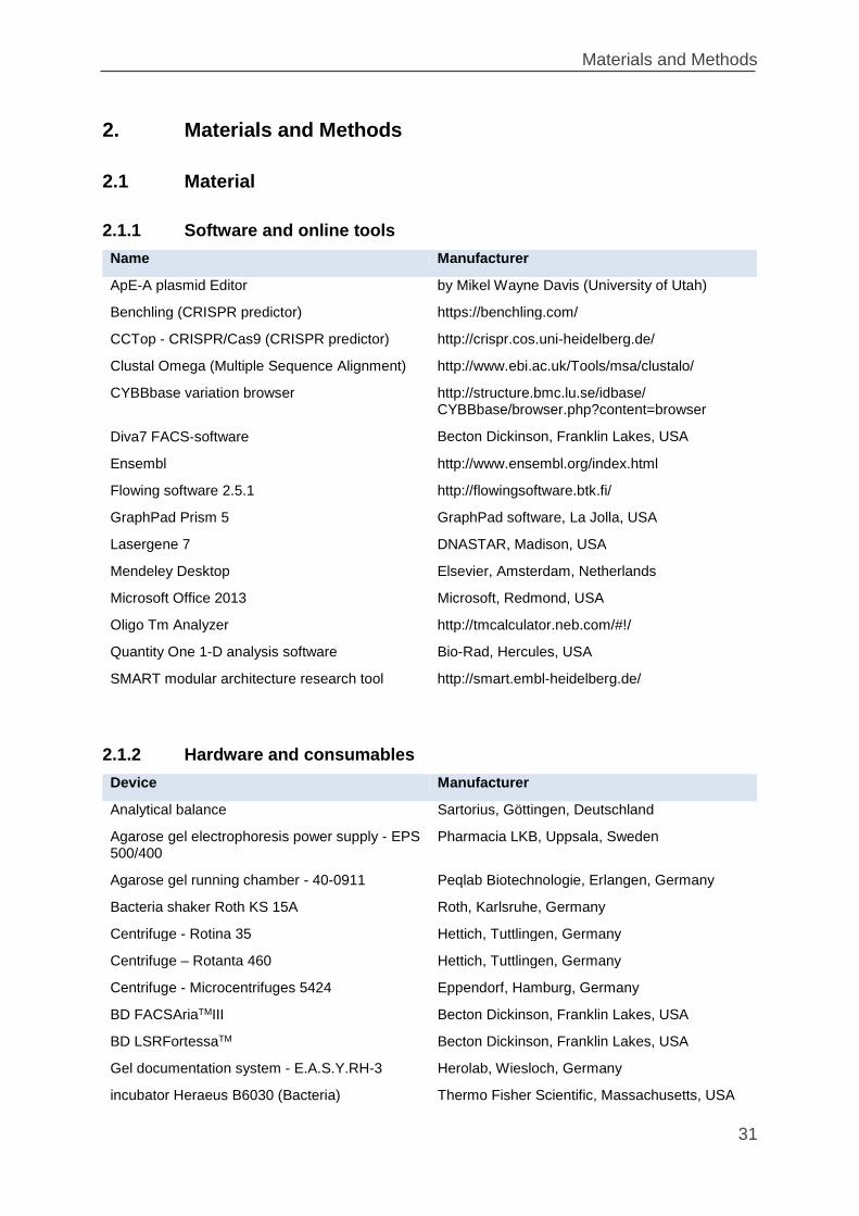
2.1.1 Software and online tools ....................................................................... 31
2.1.2 Hardware and consumables ................................................................... 31
2.1.3 Plastic ware and consumables ............................................................... 32
2.1.4 Chemicals and reagents ......................................................................... 33
2.1.5 Buffers and solutions .............................................................................. 35
2.1.6 Enzymes ................................................................................................. 36
2.1.7 Commercially available kits .................................................................... 36
2.1.8 FACS antibodies ..................................................................................... 36
2.1.9 Western blot antibodies .......................................................................... 37
2.1.10 Cytokines ........................................................................................... 37
2.1.11 Plasmids ............................................................................................ 37
2.1.12 Cell lines and culture media ............................................................... 40
2.1.13 Bacterial stains .................................................................................. 40
2.1.14 Oligonucleotides ................................................................................ 41
2.2 Methods of molecular biology .......................................................................43
2.2.1 Cultivation of bacteria ............................................................................. 43
2.2.2 Cloning processes .................................................................................. 43
2.2.3 Transformation of competent bacteria .................................................... 46
2.2.4 Plasmid preparation ................................................................................ 47
2.2.5 Agarose gel electrophoresis ................................................................... 47
2.2.6 Gel extraction of DNA fragments ............................................................ 48
2.2.7 Polymerase chain reaction (PCR) ........................................................... 48
2.2.8 PCR product extraction ........................................................................... 49
2.2.9 Nucleic acid sequencing ......................................................................... 49
2.2.10 Isolation of genomic DNA from cells .................................................. 49
2.2.11 Surveyor Assay ................................................................................. 50
2.2.12 Southern Blot ..................................................................................... 51
2.2.13 Western Blot ...................................................................................... 52
Table of contents
III
2.3 Cell culture and virological methods .............................................................54
2.3.1 Cultivation of cell lines ............................................................................ 54
2.3.2 Cell counting and determination of cell viability ...................................... 54
2.3.3 Make WEHI-3B conditioned media ......................................................... 55
2.3.4 Freezing and thawing of cultured cell ..................................................... 55
2.3.5 Nucleofection .......................................................................................... 55
2.3.6 Lentiviral vector production ..................................................................... 56
2.3.7 Titration of vector particles ...................................................................... 57
2.3.8 Flow cytometry and cell sorting............................................................... 58
2.3.9 Dihydrorhodamine 123 (DHR) reduction assay ...................................... 58
2.3.10 Statistical analysis ............................................................................. 59
3. Results ................................................................................................................60
3.1 Genome editing by homology-directed repair (HDR) versus non-homologous
end joining (NHEJ) .................................................................................................60
3.2 Reconstitution of a point mutated Hprt gene by genome editing in murine
hematopoietic (32D) cells .......................................................................................61
3.3 NHEJ-mediated reconstitution of a mutated EGFP reporter gene in 32D cells
.....................................................................................................................63
3.4 NHEJ mediated reconstitution of a mutated EGFP reporter transgene in PLB-
985 human promyelocytic leukemia cells ...............................................................67
3.5 Reconstitution of point mutated CYBB gene expressed in X-CGD-PLB
leukemia cells .........................................................................................................73
3.6 Estimating on target mutation efficiency at the endogenous CYBB locus in PLB
cells .....................................................................................................................84
4. Discussion ...........................................................................................................87
5. Conclusions and Outlook ....................................................................................96
1. References ..........................................................................................................98
Acknowledgments ...................................................................................................107
Curriculum vitae .......................................................................................................108

Table of contents
IV
Publications and Abstracts ......................................................................................109
Ehrenwörtliche Erklärung:........................................................................................111
List of figures
IV
List of figures
Figure 1: The hematopoietic system ........................................................................... 8
Figure 2: Mutation type of the selected examples of PIDs ........................................ 12
Figure 3: In vivo and ex vivo gene therapy concepts ................................................ 13
Figure 4: Overview of vectors used in gene therapy clinical trials ............................ 14
Figure 5: Various nonviral gene delivery systems (modified from Manjila et al. 2013)
................................................................................................................................. 15
Figure 6: Transposon vector system for stable gene delivery (modified from Ivics et al.
201137) ...................................................................................................................... 16
Figure 7: Analysis of integration frequencies around transcriptional start sites (TSS)
................................................................................................................................. 19
Figure 8: Mechanisms of the insertional mutagenesis mediated by the retroviral vectors
................................................................................................................................. 20
Figure 9: NADPH oxidase complexes in phagocytic cells ........................................ 24
Figure 10: TALEN-assisted Hprt gene editing .......................................................... 61
Figure 11: Analysis of INDEL distribution ................................................................. 62
Figure 12: TALEN-assisted random Hprt gene repair ............................................... 63
Figure 13: EGFP mutagenesis in 32D-GFP+ cells .................................................... 64
Figure 14: Indels induced by NHEJ in 32D-GFP negative cells enriched by FACS and
subcloned by limiting dilution .................................................................................... 65
Figure 15: EGFP repair frequency in selected 32DG subclones .............................. 66
Figure 16: Generation of PLB-985 cells expressing dual color reporter transgenes . 68
Figure 17: Single copy integration of SBmGW lentiviruses in PLB cells ................... 69
Figure 18: EGFP repair efficiency in PLB-985 cells expressing dual color reporters 70
Figure 19: mEGFP alleles in IDLV infected SBmGW-PLB cells ............................... 72
Figure 20: FACS profiles of XCGD cells expressing wild-type and mutant CYBB cDNAs
................................................................................................................................. 74
Figure 21: CYBB protein expression in XCGD-SBwtCW and SBmCW cells detected
by Western blot ......................................................................................................... 75
Figure 22 Repair of CYBB mutations in XCGD-PLB cells ........................................ 76
Figure 23: FACS profile of sorted CYBB+XCGD cells ............................................... 77
Figure 24: Indel analyses of the shot-gun clones and single cells ............................ 79
Figure 25: FACS analyses of the single cell clones form SB45CW, SB54CW and
SB173CW ................................................................................................................. 80
List of figures
V
Figure 26: Differentiation of XCGD cells ................................................................... 81
Figure 27: NADPH oxidase activity in CYBB corrected XCGD cells ......................... 82
Figure 28: Functional analysis of the SB173CW-C24 clones ................................... 83
Figure 29: On target mutation efficiency at the endogenous exon 3 and exon 6 CYBB
gene ......................................................................................................................... 85
Figure 30: Indel analyses of the shot-gun clones from endogenous CYBB locus..... 86
List of tables
VI
List of tables
Table 1: Selected examples of PIDs and their prevalence13,14 ................................. 10
Table 2: Viral vectors and their main properties ....................................................... 17
Table 3: Clinical trials of HSC-based gene-therapy in PIDs...................................... 22
Table 4: Summary of gene therapy trials for X-CGD including myelosuppressive
strategies .................................................................................................................. 25
Table 5: Examples of applications of genome editing to therapeutic model disease. 29
Table 6: Selected CYBB disease mutations and nucleotide sequences of the CYBB-
targeting sgRNAs ..................................................................................................... 73
Table 7: Types of indels recovered from sorted CYBB+ XCGD cells ........................ 78
Table 8: Computational Transmembrane sequence analysis from XCGD-SB173CW
single cell clones ...................................................................................................... 81
Table 9: Theoretical frequency of gene repair .......................................................... 94
Abbreviations and Definitions
VII
Abbreviations and Definitions
AAV adeno-associated viruses
ADA‐SCID adenosine deaminase‐deficient severe combined immunodeficiency
Amp ampicillin
APC allophycocyanin
bp base pairs
BSA bovine serum albumin
°C degree Celsius
CGD chronic granulomatous disease
CLP common lymphoid progenitors.
CMP common myeloid progenitors.
CRISPR Clustered regularly interspaced short palindromic repeats
CYBB Cytochrome b-245 heavy chain
DNA Deoxyribonucleic acid
DSB DNA double strand break
DHR dihydrorhodamine123
DMEM Dulbecco`s modified Eagle medium
DMSO dimethyl sulfoxide
DNA deoxyribonucleic acid
dsDNA double-stranded deoxyribonucleic acid
E. coli Escherichia coli
EDTA ethylenediaminetetraacetic acid
EGFP enhanced green fluorescent protein
Env envelope protein
et al. and others
FACS fluorescence activated cell sorting
FCS fetal calf serum
FITC fluorescence isothiocyanate
FSC forward scatter
g gram
Gag group‐specific‐antigen
GOI gene of interest
GMP granulocyte-macrophage progenitors
GvHD graft-versus-host disease
h hour
HAT medium hypoxanthine-aminopterin-thymidine medium
H2O2 hydrogen peroxide
HIV‐1 Human immunodeficiency virus‐1
HDR homology directed repair
Abbreviations and Definitions
VIII
HLA human leukocyte antigens
HOCl hypochlorite ion
HPRT hypoxanthine-guanine phosphoribosyltransferase
HSC hematopoietic stem cells
HSCT hematopoietic stem cell transplantation
HSPC hematopoietic stem and progenitor cells
IDLV Integrase-defective lentiviral
IFN interferon
iL2RG interleukin-2 receptor gamma chain
IN integrase
IRES internal ribosomal entry site
kb kilobase pair
KI knock-in
KO knock-out
l liter
LB Luria Broth
LTR long terminal repeat
M molar
MA matrix
MEP megakaryocyte-erythrocyte progenitor
mEGFP mutationally inactivated EGFP
MLV murine leukaemia virus
MOI multiplicity of infection
MPP multipotent progenitors
mRNA messenger RNA
NADPH nicotinamide adenine dinucleotide phosphate
NEB New England Biolabs
NHEJ nonhomologous end joining
NK cell natural killer cell
NMD nonsense mediated decay
nt nucleotide
O2- superoxide anion
OH- hydroxyl radical
ORF open reading frame
PB PiggyBac
PBS primer binding site or phosphate buffered saline
PCR polymerase chain reaction
PE R‐Phycoerythrin
PEI Polyethylenimin
phox phagocytic oxidase
PID primary immunodeficiency diseases
Abbreviations and Definitions
IX
PMA phorbol 12‐myristate 13‐acetate
Pol polymerase
psi packaging signal of retroviral genomic RNA
RGN RNA-guided nucleases
RNA ribonucleic acid
RNase ribonuclease
ROS reactive oxygen species
rpm revolutions per minute
RPMI Roswell Park Memorial Institute - culture medium
RT room temperature or reverse transcriptase
SA splice acceptor
SB Sleeping Beauty
SD slice donor
SDS sodium dodecyl sulfate
sec second
SFFV spleen focus‐forming virus
SIN self‐inactivating
SSC side scatter
SSDNA single-stranded Deoxyribonucleic acid
SV40 Simian virus 40
TagBFP blue fluorescent protein
TALEN transcription activator–like effector nucleases
TIR terminal inverted repeats
TU transducing units or Technical University
V volt
VSV‐G glycoprotein of vesicular stomatitis virus
v/v Volume/volume
WAS Wiskott-Aldrich syndrome
WASp Wiskott-Aldrich syndrome protein
w/v weight/volume (Volume concentration)
WPRE Woodchuck hepatitis virus posttranscriptional regulatory element
WT wild type
X-CGD X‐linked chronic granulomatous disease
X-SCID X‐linked severe combined immunodeficiency
ZFN Zinc-finger nuclease
ZFP zinc-finger protein
µ micro‐
6-TG 6-thioguanine
Summary
1
Summary
A significant fraction of inherited monogenic disorders are caused by patient-specific
mutations dispersed over the entire locus of the affected gene. Although correcting
these mutations by introducing healthy gene copies into the genome of the diseased
cells proved effective in several clinical gene therapy trials and with more advanced
vectors safety and efficacy could be improved, insertional mutagenesis and
unregulated expression of genes deprived of their endogenous control elements
remains a concern when using randomly integrating vectors. As has been shown
repeatedly in clinical trials random vector insertions are susceptible to epigenetic
silencing and can cause cancer by the activation of adjacent proto-oncogenes.
The development of genome editing tools capable of modifying any prespecified
genomic sequence with unprecedented accuracy opened up a wide range of new
possibilities in gene manipulation including targeted gene repair. In particular,
CRISPR/Cas9 system, a prokaryotic adaptive immune system and its swift
repurposing for genome editing was widely adopted as the hitherto simplest genome
editing tool. In combination with a single guide RNA (sgRNA) the Cas9 endonuclease
generates DNA double strand breaks (DSBs) at prespecified genomic loci that are
repaired either by homology directed repair (HDR) or nonhomologous end joining
(NHEJ).
Correction of human disease mutations by this technology has been thus far largely
based on homologous recombination requiring an exogenous donor template along
with RNA guided (gRNA) Cas9 endonucleases (RGNs). In most applications, RGNs
and templates were delivered to the diseased cells by electroporation of several
plasmids each expressing one of the functional components needed for targeted gene
modification. However, transducing the functional components required for homology
directed repair (HDR) on different plasmids and considering that electroporation is
quite harmful to the target cells, only a small fraction of the cells survive transfection
and even fewer retain all functional components. As a result, the number of gene
corrected cells is usually quite low and reduced even further by the inherent bias of the
cell's double strand break (DSB) machinery towards NHEJ.
Summary
2
This thesis explores the efficiency of gene repair by NHEJ in hematopoietic cells
harboring patient specific point mutations in the Cytochrome b-245 heavy chain gene
(CYBB) whose inactivation causes chronic granulomatous disease (X-CGD), - a life-
threatening immunodeficiency disorder. Although in contrast to HDR, NHEJ is error
prone, the present work was based on the theoretical assumption that about, one-third
of the insertions/deletions (indels) associated with NHEJ should restore the open
reading frame (ORF) disrupted by a particular disease mutation. This would lead to a
significant number of ORF reconstitutions of which some, depending on the position
and type of the original mutation, should either completely or partially recover protein
function. Moreover, donor template free delivery of RGNs on one rather than multiple
expression vectors by lentiviral infection was expected to improve gene repair
efficiencies and to reduce toxicity of gene transduction.
In initial experiments designed to determine the efficiency of gene repair by NHEJ 32D
hematopoietic cells expressing four different EGFP reporter transgenes harboring N-
terminal frameshift mutations were nucleofected each with Cas9 and corresponding
sgRNAs. Consistent with previous genome editing protocols involving transfection,
gene repair efficiency was low, ranging from 2.3% to 5.5%.
Similar testing was performed in human PLB-985 leukemia cells expressing one copy
of a mutationally inactivated EGFP reporter (mEGFP). However, to increase
transduction rates and ensure transient RGN expression, the RGNs were delivered by
integration defective lentiviruses (IDLVs). Unlike transfection IDLV delivery of RGNs
yielded high on-target mutation rates leading to mEGFP repair rates of up to 27%.
Collectively, the results demonstrate that mEGFP repair efficiency improved by one
order of magnitude after changing the RGN delivery protocol from plasmid
nucleofection to IDLV infection.
This strategy was tested further in PLB cells harboring bona fide disease mutations.
For this, four X-CGD-patient specific CYBB mutations including two frameshift, one
nonsense and one missense mutation were individually transduced into CYBB null
PLB cells (XCGD-PLB). While subsequent delivery of the corresponding RGNs
effectively repaired the frameshift mutations in up to 10% of the treated cells, the repair

Summary
3
efficiency of the nonsense and missense mutations was with less than 2% rather
ineffective.
As about 20 - 25% of most inherited blood disorders are caused by frameshift
mutations, the results of this thesis suggest that up to a quarter of all patients suffering
from monogenic blood disorders could benefit from a gene therapy employing
personalized, donor-template free RGNs.
Zusammenfassung
4
Zusammenfassung
Ein signifikanter Anteil hereditärer, monogenetischer Erkrankungen wird durch
patientenspezifische Mutationen verursacht, die über den gesamten Locus des
betroffenen Gens verteilt sind. Obwohl sich in mehreren klinischen Studien die
Korrektur dieser Mutationen durch die Einführung gesunder Genkopien in das Genom
der erkrankten Zellen bewährt hat und mit fortschrittlicheren Vektoren die Sicherheit
und Wirksamkeit verbessert werden kann, bleibt die Insertionsmutagenese und die
unregulierte Expression von Genen außerhalb des Einflusses ihrer endogenen
Kontrollelemente ein Problem bei der Verwendung von zufällig integrierenden
Vektoren. Wie in klinischen Studien wiederholt gezeigt wurde, sind diese Vektoren
anfällig für epigenetisches Silencing (Gen-Stilllegung) und können durch Aktivierung
benachbarter Protoonkogene Krebs verursachen.
Die Entwicklung von Genom-Editierungs-Technologien, die in der Lage sind, jede
vorher festgelegte genomische Sequenz mit bisher unerreichter Präzision zu
modifizieren, eröffnete eine breite Palette neuer Möglichkeiten in der Genmanipulation
einschließlich gezielter Genreparatur. Insbesondere das prokaryotische adaptive
Immunsystem CRISPR/Cas9, fand nach seiner eleganten Umfunktionierung zur
Editierung doppelsträngiger DNA wurde als das bisher einfachste Genom-
Editierwerkzeug breite Akzeptanz in der wissenschaftlichen Gemeinschaft.
In Kombination mit einer einzigen guide RNA (sgRNA) erzeugt die Cas9-
Endonuklease DNA-Doppelstrangbrüche (DSBs) an vordefinierten genomischen Loci.
Diese DSB können entweder durch Homologie-gerichtete Reparatur (HDR) oder nicht-
homologe Verbindung der Strangenden (NHEJ) repariert werden.
Die Korrektur menschlicher Krankheitsmutationen durch diese Technologie basierte
bislang weitgehend auf Homologie-gerichtete Reparatur, die neben RNA-geführten
(gRNA) Cas9-Endonukleasen (RGNs) eine exogene homologe Donor-Matrize
erfordert. In den meisten Anwendungen wurden RGNs und Matrizen durch
Elektroporation in die erkrankten Zellen eingebracht, die auf mehreren Plasmiden
kodiert sind, welche jeweils eine der funktionellen Komponenten exprimiert, die für eine
gezielte Genmodifikation benötigt werden. Aufgrund dessen, dass die für die
Homologie-gerichtete Reparatur (HDR) erforderlichen Komponenten auf
Zusammenfassung
5
verschiedenen Plasmiden liegen und die Elektroporation für die Zielzellen ziemlich
schädlich ist, überlebt nur ein kleiner Teil der Zellen die Transfektion und noch weniger
beinhalten alle funktionellen Komponenten für die HDR. Infolgedessen ist die Anzahl
genkorrigierter Zellen in der Regel recht niedrig und wird durch die inhärente
Bevorzugung von NHEJ seitens der Doppelstrangbruch-Maschinerie in Stammzellen
noch weiter reduziert.
In dieser Arbeit wird die Effizienz der Genreparatur durch NHEJ in hämatopoetischen
Zellen mit patientenspezifischen Punktmutationen im Cytochrom b-245-Gen (CYBB)
untersucht, dessen Inaktivierung die lebensbedrohliche Immunschwächekrankheit
chronische Granulomatose (X-CGD) verursacht. Obwohl die NHEJ im Gegensatz zu
HDR fehleranfällig ist, basierte die vorliegende Arbeit auf der theoretischen Annahme,
dass ungefähr ein Drittel der mit NHEJ assoziierten Insertionen/Deletionen (Indels)
den korrekten Leserahmen (ORF) wiederherstellen werden, der initial durch eine
bestimmte Krankheitsmutation verschoben war. Dies würde zu einer signifikanten
Anzahl von ORF-Rekonstitutionen führen, von denen einige, abhängig von der
Position und Art der ursprünglichen Mutation, entweder die Proteinfunktion vollständig
oder teilweise wiederherstellen sollten. Darüber hinaus wurde erwartet, dass die
Donor-Matrize-freie Abgabe von RGNs auf einem statt von mehreren
Expressionsvektoren durch lentivirale Infektion die Effizienz der Genreparatur
verbessern und die Toxizität der Gentransduktion verringert.
Beim ersten Experimenten zur Bestimmung der Effizienz der Genreparatur durch
NHEJ wurden die 32D hämatopoetische Zellen, die vier verschiedene EGFP-
Reportertransgene mit N-terminalen Frameshift-Mutationen exprimierten, jeweils mit
Cas9 und entsprechenden sgRNAs nukleofektiert. In Übereinstimmung mit früheren
Genom-Editierprotokollen, die eine Transfektion beinhalten, war die Effizienz der Gen-
Reparatur gering und lag zwischen 2,3% und 5,5%.
Ähnliche Tests wurden in humanen PLB-985 Leukämiezellen durchgeführt, die eine
Kopie eines mutationsinaktivierten EGFP-Reporters (mEGFP) exprimierten. Um die
Transduktionsraten zu erhöhen und transiente RGN-Expression zu gewährleisten,
wurden die RGNs durch Integrations-defekte Lentiviren (IDLVs) transduziert. Im
Gegensatz zur Nucleofektion ergab die IDLV-Infektion von RGNs eine hohe On-
Zusammenfassung
6
Target-Mutationsraten, was zu mEGFP-Reparaturraten von bis zu 27% führte.
Insgesamt zeigen die Ergebnisse, dass sich die mEGFP-Reparatureffizienz um eine
Größenordnung verbessert hat, nachdem das RGN Transduktionsprotokoll von
Plasmid-Nukleofektion zu IDLV-Infektion geändert wurde.
Anschließend wurde die Strategie bei der hereditären septischen Granulomatose (X-
CGD) die durch Mutationen im Cytochrome b-245 beta polypeptide (CYBB) Gen
entstehen, in PLB985 Zellen getestet. Dazu wurden vier X-CGD-Patientenspezifische
CYBB-Mutationen mit zwei Frameshift-, einer Nonsense- und einer Missense-Mutation
einzeln in CYBB-Null-PLB-Zellen (XCGD-PLB) eingebracht. Während der
nachfolgenden Transduktion der Zellen mit den entsprechenden IDLV-RGNs, wurden
effektiv bis zu 10% die Frameshift-Mutationen korrigiert. Die Reparatureffizienz bei den
Nonsense- und Missense-Mutationen war mit weniger als 2% jedoch eher ineffektiv.
Da etwa 20 – 25% der meisten vererbten Bluterkrankungen durch Frameshift-
Mutationen verursacht werden, legen die Ergebnisse dieser Arbeit nahe, dass ein
Viertel aller an monogenen Blutkrankheiten leidenden Patienten von einer Gentherapie
profitieren könnten, die personalisierte, Donor-Template-freie RGNs verwendet.
Introduction
7
1. Introduction
1.1 The hematopoietic system
The first theory of hematopoiesis was postulated by A. Maximow in 1909, stating
that hematopoiesis accounts for the livelong of formation of blood cells and plasma
from hematopoietic stem cells (HSC)1. Healthy adults produce billions of blood cells
each day to replace an equal number of senescent or apoptotic cells removed from
circulation2,3. The HSC are ultimately responsible for blood cell renewal during which
they differentiate via a series of precursor stages into terminally differentiated cells
that acquire specific functions4.
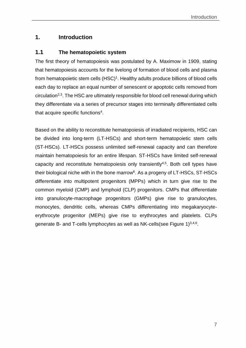
Based on the ability to reconstitute hematopoiesis of irradiated recipients, HSC can
be divided into long-term (LT-HSCs) and short-term hematopoietic stem cells
(ST-HSCs). LT-HSCs possess unlimited self-renewal capacity and can therefore
maintain hematopoiesis for an entire lifespan. ST-HSCs have limited self-renewal
capacity and reconstitute hematopoiesis only transiently4,5. Both cell types have
their biological niche with in the bone marrow6. As a progeny of LT-HSCs, ST-HSCs
differentiate into multipotent progenitors (MPPs) which in turn give rise to the
common myeloid (CMP) and lymphoid (CLP) progenitors. CMPs that differentiate
into granulocyte-macrophage progenitors (GMPs) give rise to granulocytes,
monocytes, dendritic cells, whereas CMPs differentiating into megakaryocyte-
erythrocyte progenitor (MEPs) give rise to erythrocytes and platelets. CLPs
generate B- and T-cells lymphocytes as well as NK-cells(see Figure 1)3,4,6.
Introduction
8
For example, erythrocytes deliver oxygen to tissues and organs, platelets assist
blood clotting during wound healing and tissue repair, and leukocytes are
components of the innate and adaptive immune systems protecting against various
biological and chemical intruders. Overall, differentiated blood cells have a limited
lifespan ranging from several hours (e.g. some granulocytes) up to several decades
(e.g. memory T cells)6.
A malfunction of the blood system may arise by a cell type not forming e.g. by
differentiation block or by a cell type not fulfil its function. This can lead to life-
threatening disease or even death. In most of the cases this malfunction is caused
by a genetic defect.
Figure 1: The hematopoietic system
During differentiation, HSCs increasingly lose their potential for self-renewal and their proliferation rate increases strongly, thus resulting in the enormous expansion capacity of blood cells.
Introduction
9
1.2 Primary immunodeficiency diseases
There are over 10.000 known monogenic diseases that are caused by single gene
mutations. Although most of the monogenic disorders are relatively rare, they
altogether affect about 0.5 - 1% of newborns and up to 10% of hospitalized
patients7,8. The best characterized monogenic diseases involve the hematopoietic
system and include the primary immunodeficiency diseases (PIDs).
PIDs are caused by inherited mutations in genes required for the development
and/or function of the immune system. Patients with PID lack an intact immune
system resulting in increased susceptibility to infections, allergens, autoimmune
reactions and cancer9. PIDs consist of over 200 different diseases which are all rare,
chronic and usually fatal diseases10. Any component of the immune system can be
affect a PID.
In some disorders, only a single part of the system is affected such as chronic
granulomatous disease (CGD; also see chapter 1.7). In other diseases, there are
multiple components can be affected such as in severe combined immunodeficiency
(SCID)11. Defects can be present in adaptive immune system (e.g. SCID and B-cell
immunodeficiencies) or in the innate immune system (e.g. Toll-like Receptor (TLR),
Natural Killer Cell (NK-cell) and myeloid differentiation primary response gene 88
(MYD88) deficiency)12. Some of the best characterized PIDs are described in some
more detail below.
Introduction
10
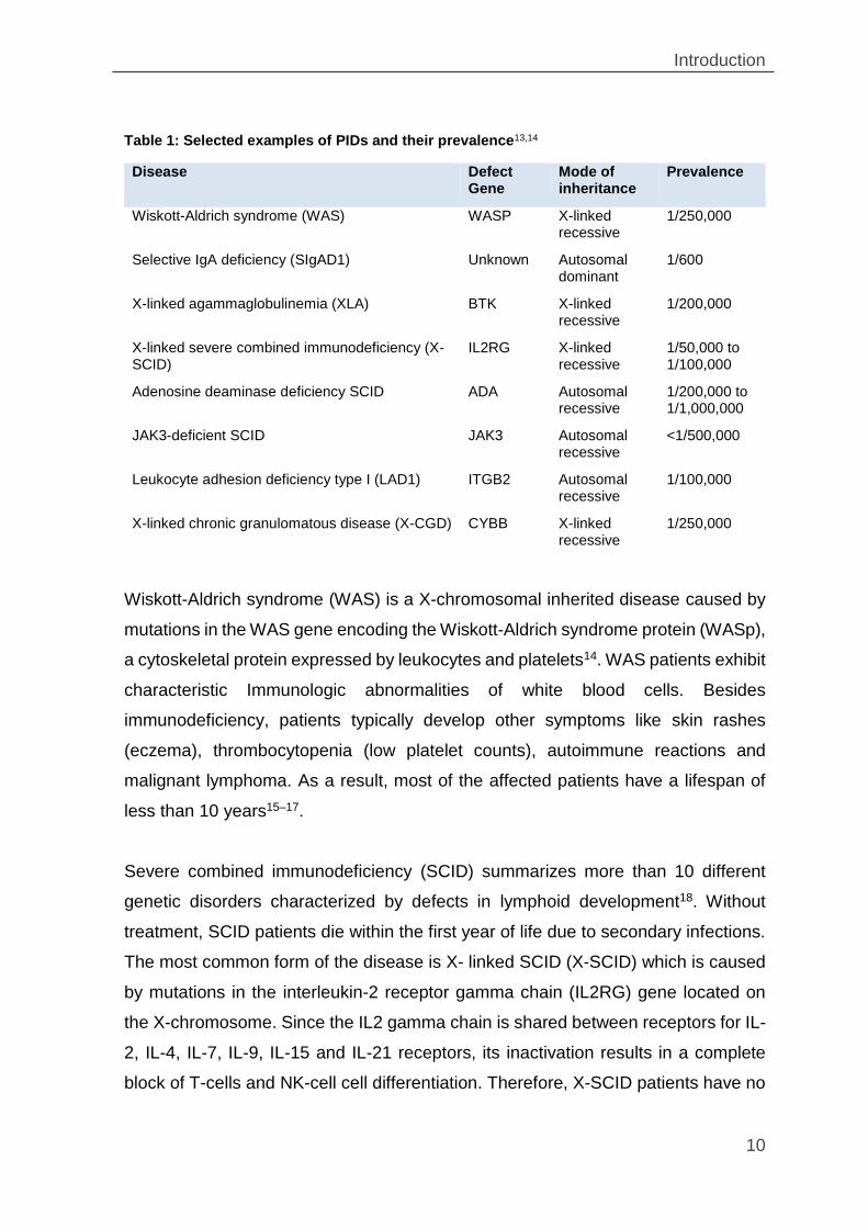
Table 1: Selected examples of PIDs and their prevalence13,14
Disease Defect Gene
Mode of inheritance
Prevalence
Wiskott-Aldrich syndrome (WAS) WASP X-linked recessive
1/250,000
Selective IgA deficiency (SIgAD1) Unknown Autosomal dominant
1/600
X-linked agammaglobulinemia (XLA) BTK X-linked recessive
1/200,000
X-linked severe combined immunodeficiency (X-SCID)
IL2RG X-linked recessive
1/50,000 to 1/100,000
Adenosine deaminase deficiency SCID ADA Autosomal recessive
1/200,000 to 1/1,000,000
JAK3-deficient SCID JAK3 Autosomal recessive
<1/500,000
Leukocyte adhesion deficiency type I (LAD1) ITGB2 Autosomal recessive
1/100,000
X-linked chronic granulomatous disease (X-CGD) CYBB X-linked recessive
1/250,000
Wiskott-Aldrich syndrome (WAS) is a X-chromosomal inherited disease caused by
mutations in the WAS gene encoding the Wiskott-Aldrich syndrome protein (WASp),
a cytoskeletal protein expressed by leukocytes and platelets14. WAS patients exhibit
characteristic Immunologic abnormalities of white blood cells. Besides
immunodeficiency, patients typically develop other symptoms like skin rashes
(eczema), thrombocytopenia (low platelet counts), autoimmune reactions and
malignant lymphoma. As a result, most of the affected patients have a lifespan of
less than 10 years15–17.
Severe combined immunodeficiency (SCID) summarizes more than 10 different
genetic disorders characterized by defects in lymphoid development18. Without
treatment, SCID patients die within the first year of life due to secondary infections.
The most common form of the disease is X- linked SCID (X-SCID) which is caused
by mutations in the interleukin-2 receptor gamma chain (IL2RG) gene located on
the X-chromosome. Since the IL2 gamma chain is shared between receptors for IL-
2, IL-4, IL-7, IL-9, IL-15 and IL-21 receptors, its inactivation results in a complete
block of T-cells and NK-cell cell differentiation. Therefore, X-SCID patients have no
Introduction
11
T-cells and NK cells and also lack functional B-cells due to the absence of CD4+ T-
helper cells18,13.
The second most common severe immunodeficiency disease is ADA-SCID, which
caused by mutations in the adenosine deaminase (ADA) gene. ADA is a key
enzyme in purine catabolism. Its absence leads to the accumulation of purine
metabolites in blood plasma, which are toxic to lymphocytes (B, T and NK cells) and
block their proliferation. Consequently, patients suffer from serious recurrent and
life-threatening infections13. Symptomatic treatment of SCID aims to minimize
bacterial, viral and fungal infections by providing sterile environments combined with
antibiotic, antiviral and antifungal treatment.
Finally, an example for an immunodeficiency of myeloid cells is the chronic
granulomatous disease (CGD). Granulocyte and macrophages of CGD patients fail
to kill phagocytosed microorganisms due to an inherited defect of superoxide
production caused by mutations in genes encoding for the NADPH oxidase
complex19,20. Since CGD was selected as a disease model gene therapy in this
theses, it will be described in more detail in the section 1.7.
1.3 Molecular background of PIDs
A substantial fraction of hereditary monogenic blood disorders are caused by patient
specific mutations dispersed over the entire locus of the affected gene21. The
inheritance pattern of most PIDs is either X-linked recessive, autosomal recessive
or rarely, autosomal dominant13. The mutation type of PID mutations includes point
mutations, small deletions or insertions, large deletions, duplications, inversions and
other more complex mutations22,23.
PID mutations can affect any part of the locus: coding regions (exons), the promoter,
regulatory regions, termination signals, splice donors / acceptors and also introns of
the genes22. The most frequent PID mutations are in coding exons all resulting in
protein dysfunction. Commonly there are point mutations which by nucleotide
replacement create either a premature stop codon (nonsense mutation) or a new
codon for an unrelated amino acid (missense mutation). Nonsense mutations are
Introduction
12
usually sensed by the splicing machinery and trigger the nonsense mediated decay
pathway (NMD) resulting in mRNA degradation. As a result, affected cells do not
express the mutated protein24. However, in some instances the mutated mRNAs
escape NMD and are translated into truncated proteins. In either case the protein
function is typically lost23,25.
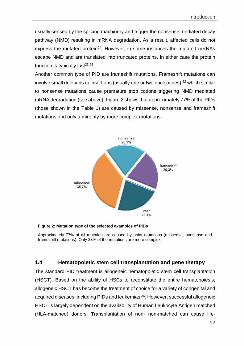
Another common type of PID are frameshift mutations. Frameshift mutations can
involve small deletions or insertions (usually one or two nucleotides) 22 which similar
to nonsense mutations cause premature stop codons triggering NMD mediated
mRNA degradation (see above). Figure 2 shows that approximately 77% of the PIDs
(those shown in the Table 1) are caused by missense, nonsense and frameshift
mutations and only a minority by more complex mutations.
1.4 Hematopoietic stem cell transplantation and gene therapy
The standard PID treatment is allogeneic hematopoietic stem cell transplantation
(HSCT). Based on the ability of HSCs to reconstitute the entire hematopoiesis,
allogeneic HSCT has become the treatment of choice for a variety of congenital and
acquired diseases, including PIDs and leukemias,26. However, successful allogeneic
HSCT is largely dependent on the availability of Human Leukocyte Antigen matched
(HLA-matched) donors. Transplantation of non- non-matched can cause life-
Figure 2: Mutation type of the selected examples of PIDs
Approximately 77% of all mutation are caused by point mutations (missense, nonsense and frameshift mutations). Only 23% of the mutations are more complex.
Introduction
13
threatening graft-versus-host disease (GvHD) during which the engrafted
lymphocytes elicit an immune response against host tissues perceived as
foreign27,28. However, 90% of PID patients with appropriate donors undergo
complete immune reconstitution after HSCT and have a normal life expectancy29.
Unfortunately, for a significant number of patients HLA-matched donors cannot be
found. This is particularly problematic in countries where comprehensive blood
donor catalogs are unavailable30.
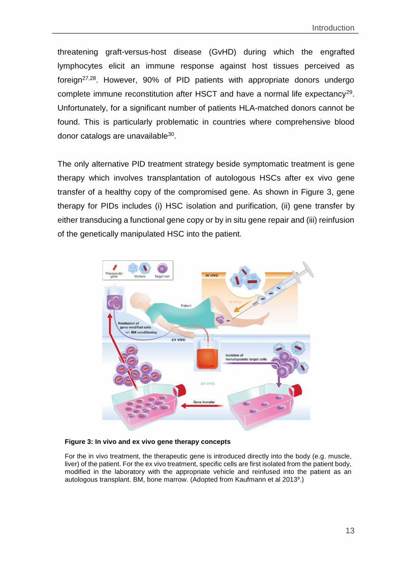
The only alternative PID treatment strategy beside symptomatic treatment is gene
therapy which involves transplantation of autologous HSCs after ex vivo gene
transfer of a healthy copy of the compromised gene. As shown in Figure 3, gene
therapy for PIDs includes (i) HSC isolation and purification, (ii) gene transfer by
either transducing a functional gene copy or by in situ gene repair and (iii) reinfusion
of the genetically manipulated HSC into the patient.
Figure 3: In vivo and ex vivo gene therapy concepts
For the in vivo treatment, the therapeutic gene is introduced directly into the body (e.g. muscle, liver) of the patient. For the ex vivo treatment, specific cells are first isolated from the patient body, modified in the laboratory with the appropriate vehicle and reinfused into the patient as an autologous transplant. BM, bone marrow. (Adopted from Kaufmann et al 20139.)
Introduction
14
The procedure circumvents GvHD and if successful, could be as lifesaving as
allogeneic HSCT. The problems associated with gene therapy are discussed in
more details in sections 1.5, 1.6 and 1.8.
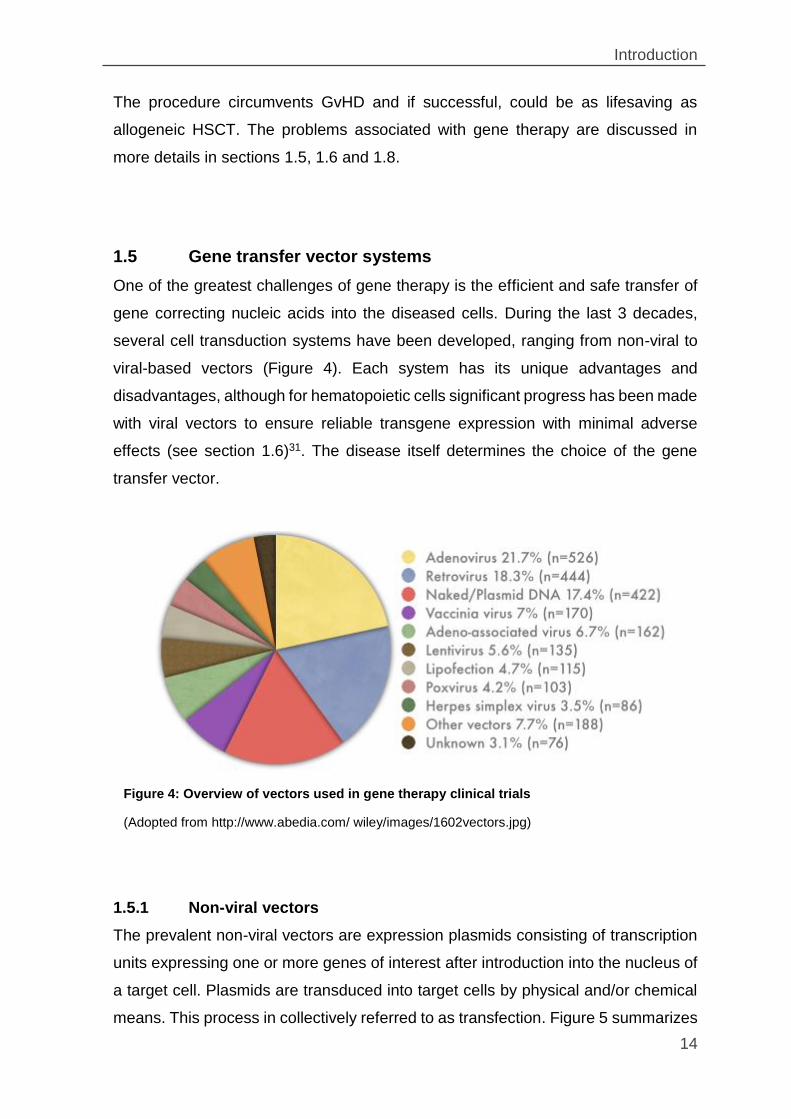
1.5 Gene transfer vector systems
One of the greatest challenges of gene therapy is the efficient and safe transfer of
gene correcting nucleic acids into the diseased cells. During the last 3 decades,
several cell transduction systems have been developed, ranging from non-viral to
viral-based vectors (Figure 4). Each system has its unique advantages and
disadvantages, although for hematopoietic cells significant progress has been made
with viral vectors to ensure reliable transgene expression with minimal adverse
effects (see section 1.6)31. The disease itself determines the choice of the gene
transfer vector.
1.5.1 Non-viral vectors
The prevalent non-viral vectors are expression plasmids consisting of transcription
units expressing one or more genes of interest after introduction into the nucleus of
a target cell. Plasmids are transduced into target cells by physical and/or chemical
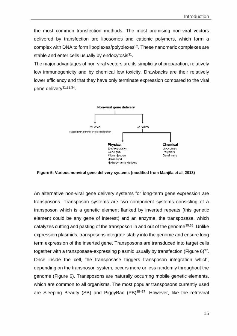
means. This process in collectively referred to as transfection. Figure 5 summarizes
Figure 4: Overview of vectors used in gene therapy clinical trials
(Adopted from http://www.abedia.com/ wiley/images/1602vectors.jpg)
Introduction
15
the most common transfection methods. The most promising non-viral vectors
delivered by transfection are liposomes and cationic polymers, which form a
complex with DNA to form lipoplexes/polyplexes32. These nanomeric complexes are
stable and enter cells usually by endocytosis31.
The major advantages of non-viral vectors are its simplicity of preparation, relatively
low immunogenicity and by chemical low toxicity. Drawbacks are their relatively
lower efficiency and that they have only terminate expression compared to the viral
gene delivery31,33,34.
An alternative non-viral gene delivery systems for long-term gene expression are
transposons. Transposon systems are two component systems consisting of a
transposon which is a genetic element flanked by inverted repeats (this genetic
element could be any gene of interest) and an enzyme, the transposase, which
catalyzes cutting and pasting of the transposon in and out of the genome35,36. Unlike
expression plasmids, transposons integrate stably into the genome and ensure long
term expression of the inserted gene. Transposons are transduced into target cells
together with a transposase-expressing plasmid usually by transfection (Figure 6)37.
Once inside the cell, the transposase triggers transposon integration which,
depending on the transposon system, occurs more or less randomly throughout the
genome (Figure 6). Transposons are naturally occurring mobile genetic elements,
which are common to all organisms. The most popular transposons currently used
are Sleeping Beauty (SB) and PiggyBac (PB)35–37. However, like the retroviral
Figure 5: Various nonviral gene delivery systems (modified from Manjila et al. 2013)
Introduction
16
vectors (see below) transposons are insertionally mutagenic and therefore
associated with adverse effects38–42.
1.5.2 Viral vectors
Viruses are natural nucleic acid transducers that enter susceptible cells by
interacting with specific cell surface receptors. Once inside the cell, they release
their genetic material which directs replication via engaging the cellular nucleic acid
and protein synthesis machineries. In viral vectors, most of the viral genome is
replaced by one or more genes of interest31,43. Typically, the modified genomes are
assembled into infectious particles in cell lines expressing the required viral proteins
from separate expression plasmids44. These cell lines are commonly referred to as
viral producer or packaging cell lines.
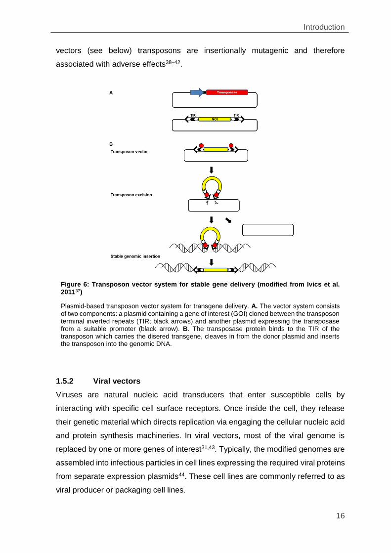
Figure 6: Transposon vector system for stable gene delivery (modified from Ivics et al. 201137)
Plasmid-based transposon vector system for transgene delivery. A. The vector system consists of two components: a plasmid containing a gene of interest (GOI) cloned between the transposon terminal inverted repeats (TIR; black arrows) and another plasmid expressing the transposase from a suitable promoter (black arrow). B. The transposase protein binds to the TIR of the transposon which carries the disered transgene, cleaves in from the donor plasmid and inserts the transposon into the genomic DNA.
Introduction
17
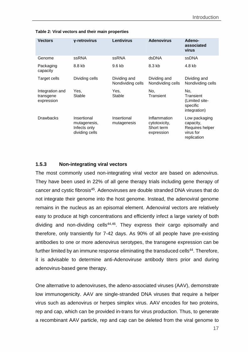
Table 2: Viral vectors and their main properties
Vectors γ-retrovirus Lentivirus Adenovirus Adeno-associated virus
Genome ssRNA ssRNA dsDNA ssDNA
Packaging capacity
8.8 kb 9.6 kb 8.3 kb 4.8 kb
Target cells Dividing cells Dividing and Nondividing cells
Dividing and Nondividing cells
Dividing and Nondividing cells
Integration and transgene expression
Yes, Stable
Yes, Stable
No, Transient
No, Transient (Limited site-specific integration)
Drawbacks Insertional mutagenesis, Infects only dividing cells
Insertional mutagenesis
Inflammation cytotoxicity, Short term expression
Low packaging capacity, Requires helper virus for replication
1.5.3 Non-integrating viral vectors
The most commonly used non-integrating viral vector are based on adenovirus.
They have been used in 22% of all gene therapy trials including gene therapy of
cancer and cystic fibrosis45. Adenoviruses are double stranded DNA viruses that do
not integrate their genome into the host genome. Instead, the adenoviral genome
remains in the nucleus as an episomal element. Adenoviral vectors are relatively
easy to produce at high concentrations and efficiently infect a large variety of both
dividing and non-dividing cells44,46. They express their cargo episomally and
therefore, only transiently for 7-42 days. As 90% of all people have pre-existing
antibodies to one or more adenovirus serotypes, the transgene expression can be
further limited by an immune response eliminating the transduced cells44. Therefore,
it is advisable to determine anti-Adenoviruse antibody titers prior and during
adenovirus-based gene therapy.
One alternative to adenoviruses, the adeno-associated viruses (AAV), demonstrate
low immunogenicity. AAV are single-stranded DNA viruses that require a helper
virus such as adenovirus or herpes simplex virus. AAV encodes for two proteins,
rep and cap, which can be provided in-trans for virus production. Thus, to generate
a recombinant AAV particle, rep and cap can be deleted from the viral genome to
Introduction
18
leave only the virus inverted terminal repeats (ITRs)45,47. The subtype AAV2 is the
most commonly used vector. Although AAV belong to the group of non-integrating
viruses, they still have limited genome insertion sites. An interesting feature of AAV
is that they tend to stably integrate into the AAVS1 locus on chromosome1948. AAV
vectors are currently considered the delivery tool of choice for in vivo therapy of
inherited diseases in post‐mitotic tissues. The major disadvantageous of AAV
vectors for gene delivery is their limited packaging capacity of up to 4.8 kb31. Another
limitation is the frequently seen genomic integration of AAV genome into the host
genome at other sites than AAVS1on chromosome19. Therefore, AAVs and AAV
vectors are associated with tumorigenesis through insertional mutagenesis mainly
into the proto-oncogenes48–50
1.5.4 Integrating viral vectors
The most commonly used integrating viruses are γ-retroviruses and lentiviruses. Y-
retroviruses (e.g. MLV) and lentiviruses (e.g. HIV) belong to the family of
retroviridae, which are a positive-sense RNA viruses that replicate via a double
stranded DNA intermediate45. In infected cells the RNA genome is reversely
transcribed into double-stranded DNA (dsDNA) which integrates randomly
throughout the genome as a provirus. Reverse transcription and integration are
catalyzed by the virally mRNA encoded enzymes reverse transcriptase and
integrase. Proviruses are flanked by repetitive elements known as long terminal
repeats (LTRs) that ensure viral replication by controlling mRNA transcription and
processing45,47,51
In retroviral vectors, all genes required for virus replication such as gag (encoding
viral matrix, capsid, and nucleocapsid proteins), pol (encoding a protease, reverse
transcriptase, and integrase), and env (encoding a bipartite membrane-anchored
surface protein) are replaced by a gene of interest45,46,52. To produce replication-
defective infectious particles, the viral vector carrying the genes of interest is co-
expressed with gag, pol and env encoding cassettes in dedicated packaging cell
lines45,46,52 similar to those described above for adenovirus and AAV production.
Introduction
19
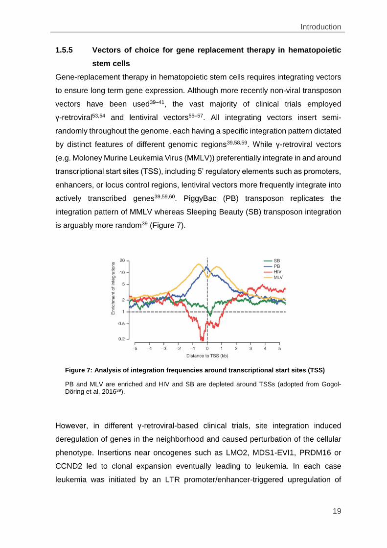
1.5.5 Vectors of choice for gene replacement therapy in hematopoietic
stem cells
Gene-replacement therapy in hematopoietic stem cells requires integrating vectors
to ensure long term gene expression. Although more recently non-viral transposon
vectors have been used39–41, the vast majority of clinical trials employed
γ-retroviral53,54 and lentiviral vectors55–57. All integrating vectors insert semi-
randomly throughout the genome, each having a specific integration pattern dictated
by distinct features of different genomic regions39,58,59. While γ-retroviral vectors
(e.g. Moloney Murine Leukemia Virus (MMLV)) preferentially integrate in and around
transcriptional start sites (TSS), including 5’ regulatory elements such as promoters,
enhancers, or locus control regions, lentiviral vectors more frequently integrate into
actively transcribed genes39,59,60. PiggyBac (PB) transposon replicates the
integration pattern of MMLV whereas Sleeping Beauty (SB) transposon integration
is arguably more random39 (Figure 7).
However, in different γ-retroviral-based clinical trials, site integration induced
deregulation of genes in the neighborhood and caused perturbation of the cellular
phenotype. Insertions near oncogenes such as LMO2, MDS1-EVI1, PRDM16 or
CCND2 led to clonal expansion eventually leading to leukemia. In each case
leukemia was initiated by an LTR promoter/enhancer-triggered upregulation of
Figure 7: Analysis of integration frequencies around transcriptional start sites (TSS)
PB and MLV are enriched and HIV and SB are depleted around TSSs (adopted from Gogol-Döring et al. 201639).
Introduction
20
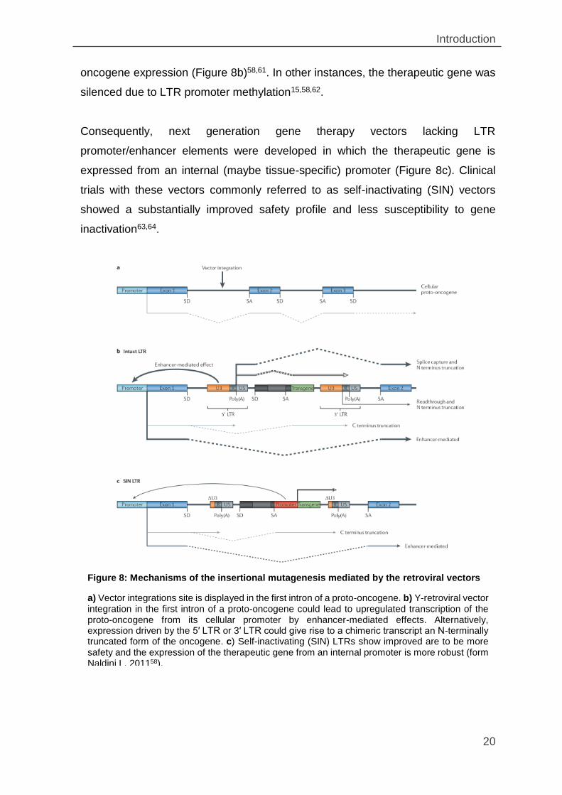
oncogene expression (Figure 8b)58,61. In other instances, the therapeutic gene was
silenced due to LTR promoter methylation15,58,62.
Consequently, next generation gene therapy vectors lacking LTR
promoter/enhancer elements were developed in which the therapeutic gene is
expressed from an internal (maybe tissue-specific) promoter (Figure 8c). Clinical
trials with these vectors commonly referred to as self-inactivating (SIN) vectors
showed a substantially improved safety profile and less susceptibility to gene
inactivation63,64.
Figure 8: Mechanisms of the insertional mutagenesis mediated by the retroviral vectors
a) Vector integrations site is displayed in the first intron of a proto-oncogene. b) Y-retroviral vector integration in the first intron of a proto-oncogene could lead to upregulated transcription of the proto-oncogene from its cellular promoter by enhancer-mediated effects. Alternatively, expression driven by the 5′ LTR or 3′ LTR could give rise to a chimeric transcript an N-terminally truncated form of the oncogene. c) Self-inactivating (SIN) LTRs show improved are to be more safety and the expression of the therapeutic gene from an internal promoter is more robust (form Naldini L, 201158).
Introduction
21
Currently, most clinical gene therapy trials employ third generation lentiviral vectors
which are self-inactivating and usually rely on lineage-specific promoters9,65.
Although significantly less genotoxic than the earlier vector generations, the self-
inactivating (SIN) lentiviral vectors are not entirely safe66. They can still activate
oncogenes from the internal promoter/enhancer elements as has been shown for
Braf and Mak3k8 and inactivate tumor suppressor genes as has been shown for
Pten and Rasa166. Thus, ideally gene replacement strategies would be substituted
by designer endonuclease strategies capable of in situ gene correction (see below).
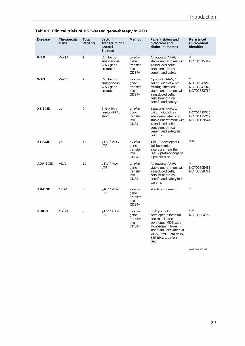
1.6 Clinical gene therapy trials for the treatment of PIDs
Over the last 3 decades, the curative potential of gene therapy has been
demonstrated in many clinical phase I / II PID gene therapy trials67,68. An overview
of the most important past and ongoing PID gene therapy trials is given in Table 3.
There are many requirements for a successful gene therapy: a clear understanding
of the molecular biology and genetics of the disease, the availability of tissue or cell
targeted for the gene delivery, an effective therapeutic vehicle and an animal model
that closely simulates the disease for preclinical studies.
Introduction
22
Table 3: Clinical trials of HSC-based gene-therapy in PIDs
Disease Therapeutic Gene
Total Patients
Vector/ Transcriptional Control Element
Method Patient status and biological and clinical outcomes
Reference/ Clinical-trial identifier
WAS WASP 3 LV / human endogenous WAS gene promoter
ex vivo gene transfer into CD34+
All patients AAW; stable engraftment with transduced cells; persistent clinical benefit and safety
16 NCT01515462
WAS WASP 7 LV / human endogenous WAS gene promoter
ex vivo gene transfer into CD34+
6 patients AAW, 1 patient died of a pre-existing infection; stable engraftment with transduced cells; persistent clinical benefit and safety
69 NCT01347242 NCT01347346 NCT02333760
X1-SCID γc 9 SIN-γ-RV / human EF1α short
ex vivo gene transfer into CD34+
8 patients AAW, 1 patient died of an adenoviral infection; stable engraftment with transduced cells; persistent clinical benefit and safety in 7 patients
70 NCT01410019 NCT01175239 NCT01129544
X1-SCID γc 10 γ-RV / MFG-LTR
ex vivo gene transfer into CD34+
4 of 10 developed T cell leukemia; Insertions near the LMO2 proto-oncogene; 1 patient died
71,72
ADA-SCID ADA 10 γ-RV / MLV-LTR
ex vivo gene transfer into CD34+
All patients AAW; stable engraftment with transduced cells; persistent clinical benefit and safety in 9 patients
73 NCT00598481 NCT00599781
AR-CGD NCF1 5 γ-RV / MLV-LTR
ex vivo gene transfer into CD34+
No clinical benefit 74
X-CGD CYBB 2 γ-RV /SFFV-LTR
ex vivo gene transfer into CD34+
Both patients developed functional neutrophils and developed MDS with monosomy 7 from insertional activation of MDS1-EVI1, PRDM16, SETBP1; 1 patient died
62,75 NCT00564759
AAW: alive and well
Introduction
23
Taken together, these studies showed that clinical outcome is highly dependent on
the transduction efficiency of hematopoietic cells capable of engrafting and
functionally reconstituting the patient’s hematopoiesis. However, clinical success
was also dependent on the particular disease. While positive selection imposed on
genetically reconstituted cells in patients with immunodeficiency diseases (e.g. X-
SCID, ADA-SCID) increased the success rate, lack of such selection in patients with
CGD was clearly detrimental because the non-modified, residual cells outcompeted
the transplanted cells during hematopoietic regeneration9,76,77.
1.7 Chronic granulomatous disease
Chronic granulomatous disease (CGD) is a rare inherited immunodeficiency
affecting 1 in 250,000 individuals78. CGD is characterized by the inability of
phagocytes to eliminate ingested pathogens and is caused by mutations in any of
the five genes of nicotinamide dinucleotide phosphate (NADPH) oxidase complex.
In neutrophils, eosinophils, monocytes, and macrophages NADPH oxidase reduces
molecular oxygen to reactive oxygen species (ROS) such as superoxide anion (O2-),
hydrogen peroxide (H2O2), hypochlorite ion (HOCl) and hydroxyl radical (OH-),
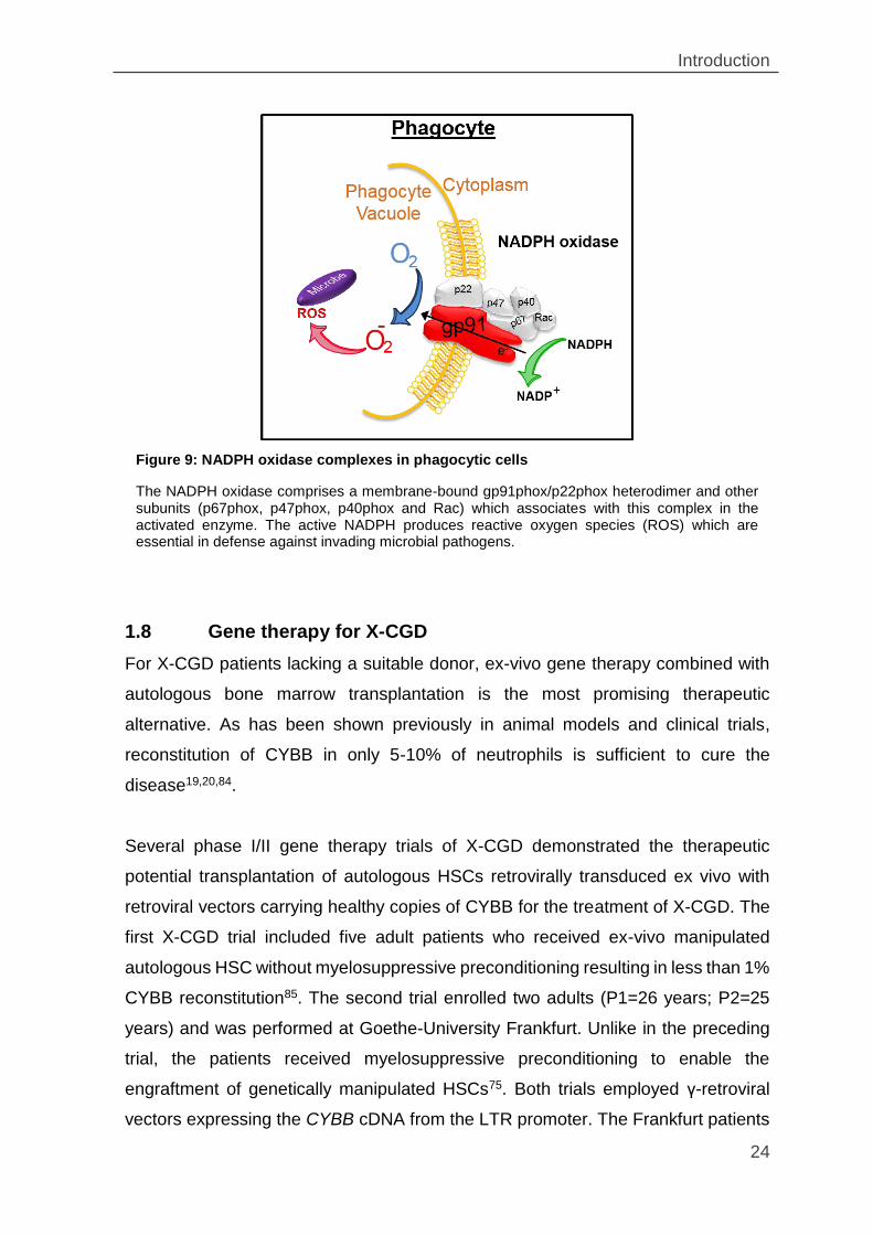
which kill the ingested microorganisms (Figure 9). Deficiencies of NADPH result in
severe, recurrent and life-threatening bacterial and fungal infections such that
affected patients require lifelong prophylactic treatment with antibiotics, antifungals
and interferon gamma (IFN-γ)78–81. To date, the only curative treatment for CGD is
allogeneic bone marrow transplantation79,80,82 which is limited to patients having a
suitable HLA-matched donor78,81,82.
CGD is a genetically heterogeneous disease. Approximately, 60-70% of all CGD
mutations affect the X-chromosomal CYBB gene encoding the protein gp91phox (X-
CGD). The other 30% have mutations in either the CYBA, NCF1, NCF2 or NCF4
genes encoding for p22phox, p47phox, p67phox, and p40phox, respectively. Unlike the X-
linked CYBB mutations, these mutations are inherited in an autosomal recessive
manner (AR-CGD)78,80,82,83.
Introduction
24
1.8 Gene therapy for XCGD
For X-CGD patients lacking a suitable donor, ex-vivo gene therapy combined with
autologous bone marrow transplantation is the most promising therapeutic
alternative. As has been shown previously in animal models and clinical trials,
reconstitution of CYBB in only 5-10% of neutrophils is sufficient to cure the
disease19,20,84.
Several phase I/II gene therapy trials of X-CGD demonstrated the therapeutic
potential transplantation of autologous HSCs retrovirally transduced ex vivo with
retroviral vectors carrying healthy copies of CYBB for the treatment of X-CGD. The
first X-CGD trial included five adult patients who received ex-vivo manipulated
autologous HSC without myelosuppressive preconditioning resulting in less than 1%
CYBB reconstitution85. The second trial enrolled two adults (P1=26 years; P2=25
years) and was performed at Goethe-University Frankfurt. Unlike in the preceding
trial, the patients received myelosuppressive preconditioning to enable the
engraftment of genetically manipulated HSCs75. Both trials employed γ-retroviral
vectors expressing the CYBB cDNA from the LTR promoter. The Frankfurt patients
Figure 9: NADPH oxidase complexes in phagocytic cells
The NADPH oxidase comprises a membrane-bound gp91phox/p22phox heterodimer and other subunits (p67phox, p47phox, p40phox and Rac) which associates with this complex in the activated enzyme. The active NADPH produces reactive oxygen species (ROS) which are essential in defense against invading microbial pathogens.
Introduction
25
fared well for up to 5 months post-transplantation. In both patient CYBB function
could be reconstituted in over 15% of granulocytes, leading to the eradication of
preexisting life-threatening infections75. However, after 5 months some CYBB-
expressing cells underwent clonal expansion due to insertional activation of several
oncogenes (i.e. PRDM16, MDS1/EVI1 and SETBP1). Although this temporarily
increased the number of modified granulocytes from 15% to about 60%, CYBB
expression was eventually silenced by LTR promoter methylation62,75,86. Intriguingly,
the LTR enhancer was not affected and continued to transactivate the nearby proto-
oncogenes. In the end, EVI1 overexpression in both patients led to the genomic
instability and preleukemic myelodysplasia with monosomy 762,86.
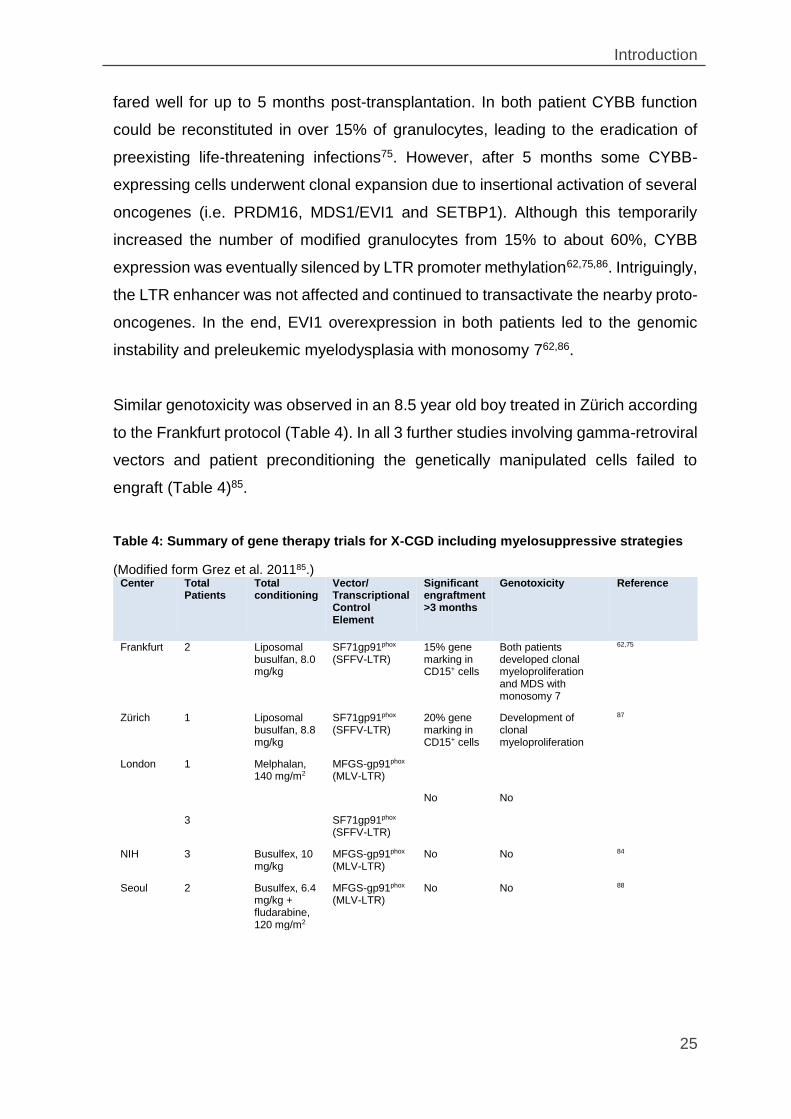
Similar genotoxicity was observed in an 8.5 year old boy treated in Zürich according
to the Frankfurt protocol (Table 4). In all 3 further studies involving gamma-retroviral
vectors and patient preconditioning the genetically manipulated cells failed to
engraft (Table 4)85.
Table 4: Summary of gene therapy trials for X-CGD including myelosuppressive strategies
(Modified form Grez et al. 201185.) Center Total
Patients Total conditioning
Vector/ Transcriptional Control Element
Significant engraftment >3 months
Genotoxicity Reference
Frankfurt 2 Liposomal busulfan, 8.0 mg/kg
SF71gp91phox (SFFV-LTR)
15% gene marking in CD15+ cells
Both patients developed clonal myeloproliferation and MDS with monosomy 7
62,75
Zürich 1 Liposomal busulfan, 8.8 mg/kg
SF71gp91phox (SFFV-LTR)
20% gene marking in CD15+ cells
Development of clonal myeloproliferation
87
London 1 Melphalan, 140 mg/m2
MFGS-gp91phox (MLV-LTR)
No No
3 SF71gp91phox (SFFV-LTR)
NIH 3 Busulfex, 10 mg/kg
MFGS-gp91phox (MLV-LTR)
No No 84
Seoul 2 Busulfex, 6.4 mg/kg + fludarabine, 120 mg/m2
MFGS-gp91phox (MLV-LTR)
No No 88
Introduction
26
Overall, the clinical X-CGD gene therapy revealed that CYBB function can be
temporarily restored by ex vivo gene replacement although the toxicity of the
employed strategy is prohibitively high, suggesting that significantly safer protocols
are required, which guarantee a long-term and safe gene therapy.
1.9 Site-specific endonucleases
Over the last few decades, the tools available for genome manipulation have
advanced significantly. Genome editing/engineering is widely used in the basic
research to specifically knock-in or knock-out genes in given target cells (e.g.
viruses, single cell organisms, plants, even vertebrates). Furthermore it is used for
development of novel therapies. A wide array of powerful gene editing systems are
now available, which are called engineered or designer endonucleases89,90.
Designer endonucleases are engineered enzymes that can introduce DNA double
strand breaks (DSBs) with high specificity into the desired target sequences. To be
useful for genome engineering, the endonucleases must have two particular
functions. First, there must be a specific recognition domain (protein or RNA) which
recognizes long target sequences (ideally, long enough for unique occurrence in the
eukaryotic genome). Second, they must cleave in the targeted sequence. To date,
four types of endonucleases exist; (1) meganucleases, (2) zinc finger nucleases
(ZFNs), (3) transcription activator–like effector nucleases (TALENs) and (4)
CRISPR-associated nuclease Cas9.
1.9.1 Meganucleases
Meganucleases (also called homing endonucleases) are natural
endodeoxyribonucleases characterized by a large target recognition sites (14-40
bp)91,92 which generally occur only once in any given genome. They are found in
bacteria, archaea, phages, protists, fungi, and plants93,94. Although the isolation of
natural meganucleases with new target specificities is tedious and slow, target
specificity can be modified in existing meganucleases by protein engineering92,93.
However, such modifications are challenging92,93.
Introduction
27
1.9.2 Zinc-finger nucleases
Zinc-finger nucleases (ZFNs) are chimeric endonucleases, consisting of individual
zinc-finger protein (ZFP) DNA binding motifs fused to the cleavage domain of the
FokI restriction endonuclease95. ZFNs typically contain between 3 – 6 ZFP motifs.
Each motif recognizes 3 – 4 bp, which results in a recognitionsite of typically
9 – 18 bp90,96. As the FokI cleavage domains acts only in form of a dimer in order to
cleave DNA and therefore a pair of ZFNs targeting the sense and antisense strand
are required90,97. The main drawback of the ZFNs is the limited number of available
ZFP motifs and that the individual ZFP domains do not independently bind to their
specific DNA element but rather influence the binding of the neighboring domain,
which results in many ZFNs leading to off-target effects. This cooperative binding
makes the ZFN design difficult and time consuming as it requires sophisticated
protein engineering and specialized methodology, thus preventing their widespread
use90,98.
1.9.3 Transcription activator-like effectors nucleases
Transcription activator-like effectors nucleases (TALENs) are, similar to ZFNs,
chimeric endonucleases in which a transcription activator-like effector (TALE) DNA
binding domain is fused to the DNA cleavage domain of FokI99,100. The DNA binding
domains consist of highly conserved 33-35 amino acid sequence in which only the
amino acid located at the position 12 and 13 vary. These two positions, referred to
as the Repeat Variable Diresidue (RVD), are highly variable and responsible for
specific base pair recognition 90,99. Naturally, TAL effector proteins are secreted by
Xanthomonas bacteria during plant infection resulting in the modulation of gene
expression100. In contrast to ZFN the individual domains do not influence binding of
the neighboring domain, making TALENs design and assembly is relatively simple
as it does not require extensive screening for target specificity99–102. Central
problems with TALENs are, that they are sensitive to cytosine methylation,
especially at CpG region and many TALEN pairs provide little or no mutagenesis
activity.
Introduction
28
1.9.4 Clustered regularly interspaced short palindromic repeats/Cas9
Clustered regularly interspaced short palindromic repeats (CRISPR) are defining
components of the genomes of most bacteria and archaea and are part of their
adaptive immune system defending then against phage and plasmid DNA
infection103. The first report on the CRISPR array were from 1987 by Ishino and
colleagues, who found in E.coli 29 nt repeats that were interspersed by 32 nt long
non-repetitive sequences (spacers)104. Three different types of CRISPR/Cas9
systems have been characterized thus far. The most widely used CRISPR/Cas9
system of Streptococcus pyogenes (spCas9/sgRNA) consists of three components:
the CRISPR-associated DNA cleaving endonuclease Cas9 protein (~160 kDa, ~4.2
kb), a target DNA sequence recognizing RNA which is transcribed from short DNA
sequences known as protospacers that are separated by short palindromic
sequences clustered in the bacterial genome in the CRISPR array, (crRNA), and a
trans-activating crRNA (tracrRNA) required for crRNA transcription90,105. For
genome editing, the crRNA and tracrRNA were fused into a fully functional single
guide RNA (sgRNA) of 110 nt105. Additionally, functional spCas9 requires a so called
protospacer adjacent motif (PAM) sequence (i.e. 5‘NGG) located downstream of the
sgRNA target sequence which determining the exact position of DNA cleavage
occurring always 3 nt upstream of the PAM106. Cas9 consists of the HNH- and the
RuvC-like nuclease domains, which cleave the coding and the non-coding DNA
strands, respectively. Unlike the other designer endonucleases, CRISPR/Cas9 is
addressed to the target site solely by a RNA molecule, without any protein
engineering107–109. Since the CRISPR/Cas9 system relies on RNA/DNA base-
pairing it circumvents problems encountered with the other, protein based editing
systems such as inactivating methylations and does not require protein engineering.
CRISPR/Cas9 RNA-guided nucleases (RGNs) are easy to make and can be applied
to a large variety of genome editing tasks including targeted gene activation, multiple
gene targeting (multiplexing) and epigenetic manipulation110–112.
Introduction
29
1.10 Molecular outcomes of genome editing
DSB generated by site-specific designer endonuclease activate the cellular repair
machinery which restores the lesion either by homology directed repair (HDR) or by
non-homologous end joining (NHEJ). HDR is an error-free process because it
requires a donor DNA sequence as a repair template90,111. Thus, HDR can be
exploited to create specific sequence changes, including the targeted addition of
whole genes (knock-ins)90,113,114. In contrast, NHEJ restores DSBs in absence of a
template by religating the DNA ends – a process associated with random nucleotide
insertions or deletions (indels)109,114. Because indels frequently cause mutations,
designer endonuclease systems and in particular the CRISPR/Cas9 system are
being employed extensively for functional genetic screens115–117 and for the
accelerated production of knock-out animals118–121. However, designer
endonucleases also have the potential to correct genetic mutations directly in
affected tissues and cells to treat diseases that are not curable by traditional
therapies (Table 5)97,111,122–124.
Table 5: Examples of applications of genome editing to therapeutic model disease.
(Adopted from Cox et al. 2015111) Disease type Nuclease
platform Therapeutic strategy Reference
Hemophilia B ZFN HDR-mediated insertion of correct gene sequence
125
HIV ZFN and CRISPR NHEJ-mediated inactivation of CCR5 126–129
Duchenne muscular dystrophy (DMD)
TALEN and CRISPR
NHEJ-mediated removal of stop codon HDR-mediated gene correction
130,131
Hepatitis B virus (HBV)
TALEN and CRISPR
NHEJ-mediated depletion of viral DNA HDR-mediated
132,133
SCID ZFN HDR-mediated insertion of correct gene sequence
134
Cataracts CRISPR HDR-mediated correction of mutation in mouse zygote
124
Cystic fibrosis Hereditary
CRISPR HDR-mediated correction of CFTR in intestinal stem cells
135
Hereditary tyrosinemia
CRISPR HDR-mediated correction of mutation in liver
136
Aim of this work
30
1.11 Aim of this work
A large part of hereditary monogenic disorders affecting the hematopoietic system, are
caused by patient-specific mutations spread over the entire locus of the affected gene.
These patients often lack an intact immune system resulting in increased susceptibility
to infections, allergens, autoimmune reactions and cancer. Designer endonucleases,
especially CRISPR/Cas9, hold the potential to significantly improve the results of future
clinical personalized gene therapy approaches for these patients. Hitherto, the
correction of monogenic disorders using designer endonucleases are primarily based
on homology directed repair (HDR), which is dependent on an exogenous DNA
template. The efficiency of the gene correction by means of HDR is generally very low,
reduced even further by the cell cycle dependence of HDR, Additionally, in
hematopoietic stem- and progenitor cells (HSPC) the dominant DSB-repair pathway is
the NHEJ pathway.
The aim of this thesis was to develop a template-free NHEJ-mediated DNA repair
strategy for personalized gene therapy of primary immunodeficiency diseases (PIDs)
using site-specific designer endonucleases. In theory, approximately one third of the
indels associated with NHEJ should restore the open reading frame (ORF) disrupted
by a particular disorder point mutation. Depending on the location and type of the point
mutation, some of the reconstituted ORFs should either completely or partially recover
protein function. The following objectives were tackled:
Comparing the efficiency of the DSB repair mechanism of HDR vs. NHEJ using
site-specific designer endonucleases
Restore a point mutated Hprt gene by NHEJ to reconstruction the non-mutated
gene
Generate a restore a point mutated EGFP reporter
Generate cell lines expressing a single copy CYBB reporter genes harboring
patient specific point mutations
Test the efficiency of CRISPR/Cas9-mediated gene correction delivered by
integrase-defective lentiviral (IDLV) into the mutant CYBB target cells
Estimate the target mutation efficiency at the endogenous CYBB locus in human
hematopoietic cells
Materials and Methods
31
2. Materials and Methods
2.1 Material
2.1.1 Software and online tools
Name Manufacturer
ApE-A plasmid Editor by Mikel Wayne Davis (University of Utah)
Benchling (CRISPR predictor) https://benchling.com/
CCTop - CRISPR/Cas9 (CRISPR predictor) http://crispr.cos.uni-heidelberg.de/
Clustal Omega (Multiple Sequence Alignment) http://www.ebi.ac.uk/Tools/msa/clustalo/
CYBBbase variation browser http://structure.bmc.lu.se/idbase/ CYBBbase/browser.php?content=browser
Diva7 FACS‐software Becton Dickinson, Franklin Lakes, USA
Ensembl http://www.ensembl.org/index.html
Flowing software 2.5.1 http://flowingsoftware.btk.fi/
GraphPad Prism 5 GraphPad software, La Jolla, USA
Lasergene 7 DNASTAR, Madison, USA
Mendeley Desktop Elsevier, Amsterdam, Netherlands
Microsoft Office 2013 Microsoft, Redmond, USA
Oligo Tm Analyzer http://tmcalculator.neb.com/#!/
Quantity One 1-D analysis software Bio-Rad, Hercules, USA
SMART modular architecture research tool http://smart.embl-heidelberg.de/
2.1.2 Hardware and consumables
Device Manufacturer
Analytical balance Sartorius, Göttingen, Deutschland
Agarose gel electrophoresis power supply - EPS 500/400
Pharmacia LKB, Uppsala, Sweden
Agarose gel running chamber - 40-0911 Peqlab Biotechnologie, Erlangen, Germany
Bacteria shaker Roth KS 15A Roth, Karlsruhe, Germany
Centrifuge - Rotina 35 Hettich, Tuttlingen, Germany
Centrifuge – Rotanta 460 Hettich, Tuttlingen, Germany
Centrifuge - Microcentrifuges 5424 Eppendorf, Hamburg, Germany
BD FACSAriaTMIII Becton Dickinson, Franklin Lakes, USA
BD LSRFortessaTM Becton Dickinson, Franklin Lakes, USA
Gel documentation system - E.A.S.Y.RH-3 Herolab, Wiesloch, Germany
incubator Heraeus B6030 (Bacteria) Thermo Fisher Scientific, Massachusetts, USA
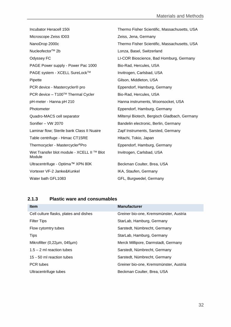
Materials and Methods
32
Incubator Heracell 150i Thermo Fisher Scientific, Massachusetts, USA
Microscope Zeiss ID03 Zeiss, Jena, Germany
NanoDrop 2000c Thermo Fisher Scientific, Massachusetts, USA
NucleofectorTM 2b Lonza, Basel, Switzerland
Odyssey FC LI-COR Bioscience, Bad Homburg, Germany
PAGE Power supply - Power Pac 1000 Bio-Rad, Hercules, USA
PAGE system - XCELL SureLockTM Invitrogen, Carlsbad, USA
Pipette Gilson, Middleton, USA
PCR device - Mastercycler® pro Eppendorf, Hamburg, Germany
PCR device – T100TM Thermal Cycler Bio-Rad, Hercules, USA
pH-meter - Hanna pH 210 Hanna instruments, Woonsocket, USA
Photometer Eppendorf, Hamburg, Germany
Quadro‐MACS cell separator Miltenyi Biotech, Bergisch Gladbach, Germany
Sonifier – VW 2070 Bandelin electronic, Berlin, Germany
Laminar flow; Sterile bank Class II Nuaire Zapf Instruments, Sarsted, Germany
Table centrifuge - Himac CT15RE Hitachi, Tokio, Japan
Thermocycler - MastercyclerRPro Eppendorf, Hamburg, Germany
Wet Transfer blot module - XCELL II TM Blot Module
Invitrogen, Carlsbad, USA
Ultracentrifuge - Optima™ XPN 80K Beckman Coulter, Brea, USA
Vortexer VF-2 Janke&Kunkel IKA, Staufen, Germany
Water bath GFL1083 GFL, Burgwedel, Germany
2.1.3 Plastic ware and consumables
Item Manufacturer
Cell culture flasks, plates and dishes Greiner bio-one, Kremsmünster, Austria
Filter Tips StarLab, Hamburg, Germany
Flow cytomtry tubes Sarstedt, Nümbrecht, Germany
Tips StarLab, Hamburg, Germany
Mikrofilter (0,22µm, 045µm) Merck Millipore, Darmstadt, Germany
1.5 – 2 ml reaction tubes Sarstedt, Nümbrecht, Germany
15 – 50 ml reaction tubes Sarstedt, Nümbrecht, Germany
PCR tubes Greiner bio-one, Kremsmünster, Austria
Ultracentrifuge tubes Beckman Coulter, Brea, USA
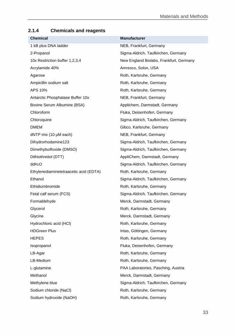
Materials and Methods
33
2.1.4 Chemicals and reagents
Chemical Manufacturer
1 kB plus DNA ladder NEB, Frankfurt, Germany
2-Propanol Sigma-Aldrich, Taufkirchen, Germany
10x Restriction buffer 1,2,3,4 New England Biolabs, Frankfurt, Germany
Acrylamide 40% Amresco, Solon, USA
Agarose Roth, Karlsruhe, Germany
Ampicillin sodium salt Roth, Karlsruhe, Germany
APS 10% Roth, Karlsruhe, Germany
Antarctic Phosphatase Buffer 10x NEB, Frankfurt, Germany
Bovine Serum Albumine (BSA) Applichem, Darmstadt, Germany
Chloroform Fluka, Deisenhofen, Germany
Chloroquine Sigma-Aldrich, Taufkirchen, Germany
DMEM Gibco, Karlsruhe, Germany
dNTP mix (10 µM each) NEB, Frankfurt, Germany
Dihydrorhodamine123 Sigma-Aldrich, Taufkirchen, Germany
Dimethylsolfoxide (DMSO) Sigma-Aldrich, Taufkirchen, Germany
Dithiothreitol (DTT) AppliChem, Darmstadt, Germany
ddH2O Sigma-Aldrich, Taufkirchen, Germany
Ethylenediaminetetraacetic acid (EDTA) Roth, Karlsruhe, Germany
Ethanol Sigma-Aldrich, Taufkirchen, Germany
Ethidiumbromide Roth, Karlsruhe, Germany
Fetal calf serum (FCS) Sigma-Aldrich, Taufkirchen, Germany
Formaldehyde Merck, Darmstadt, Germany
Glycerol Roth, Karlsruhe, Germany
Glycine Merck, Darmstadt, Germany
Hydrochloric acid (HCl) Roth, Karlsruhe, Germany
HDGreen Plus Intas, Göttingen, Germany
HEPES Roth, Karlsruhe, Germany
Isopropanol Fluka, Deisenhofen, Germany
LB-Agar Roth, Karlsruhe, Germany
LB-Medium Roth, Karlsruhe, Germany
L‐glutamine PAA Laboratories, Pasching, Austria
Methanol Merck, Darmstadt, Germany
Methylene blue Sigma-Aldrich, Taufkirchen, Germany
Sodium chloride (NaCl) Roth, Karlsruhe, Germany
Sodium hydroxide (NaOH) Roth, Karlsruhe, Germany
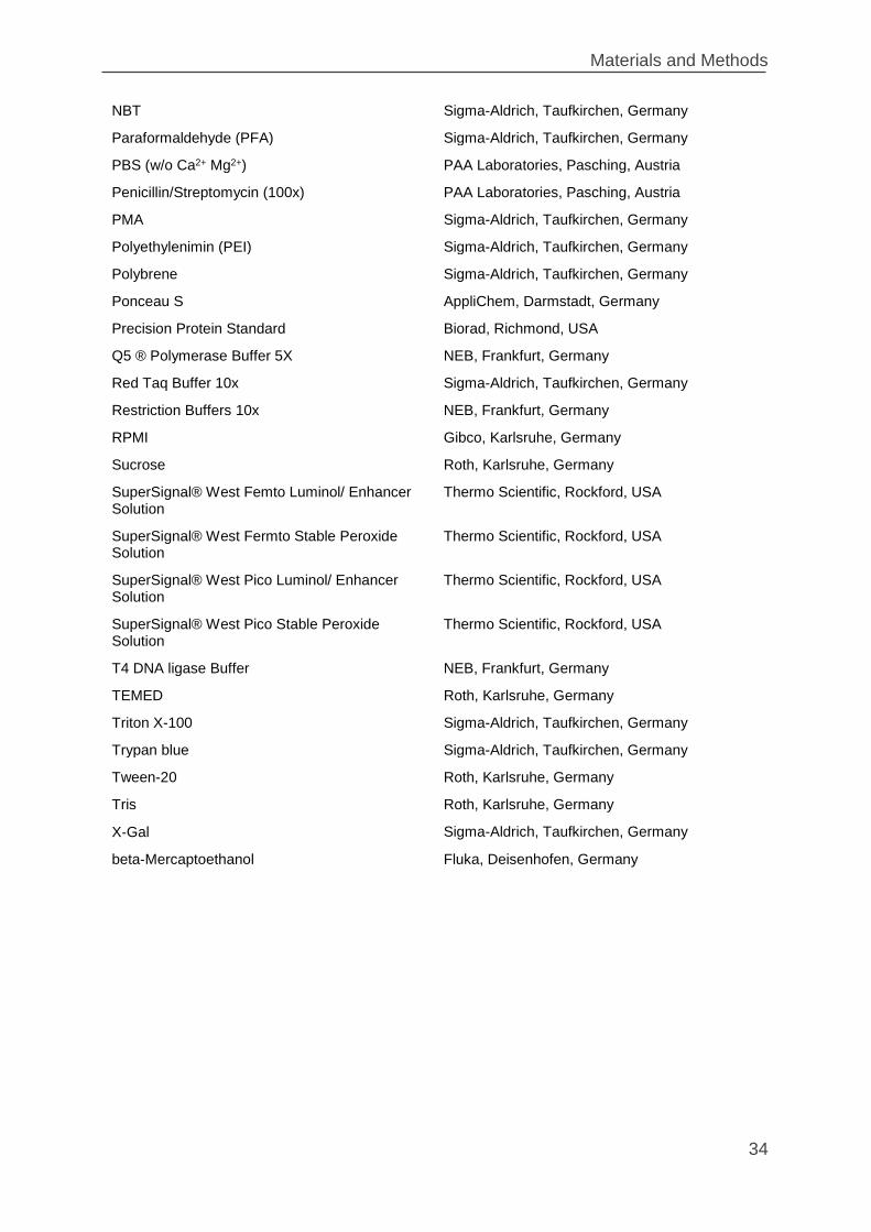
Materials and Methods
34
NBT Sigma-Aldrich, Taufkirchen, Germany
Paraformaldehyde (PFA) Sigma-Aldrich, Taufkirchen, Germany
PBS (w/o Ca2+ Mg2+) PAA Laboratories, Pasching, Austria
Penicillin/Streptomycin (100x) PAA Laboratories, Pasching, Austria
PMA Sigma-Aldrich, Taufkirchen, Germany
Polyethylenimin (PEI) Sigma-Aldrich, Taufkirchen, Germany
Polybrene Sigma-Aldrich, Taufkirchen, Germany
Ponceau S AppliChem, Darmstadt, Germany
Precision Protein Standard Biorad, Richmond, USA
Q5 ® Polymerase Buffer 5X NEB, Frankfurt, Germany
Red Taq Buffer 10x Sigma-Aldrich, Taufkirchen, Germany
Restriction Buffers 10x NEB, Frankfurt, Germany
RPMI Gibco, Karlsruhe, Germany
Sucrose Roth, Karlsruhe, Germany
SuperSignal® West Femto Luminol/ Enhancer Solution
Thermo Scientific, Rockford, USA
SuperSignal® West Fermto Stable Peroxide Solution
Thermo Scientific, Rockford, USA
SuperSignal® West Pico Luminol/ Enhancer Solution
Thermo Scientific, Rockford, USA
SuperSignal® West Pico Stable Peroxide Solution
Thermo Scientific, Rockford, USA
T4 DNA ligase Buffer NEB, Frankfurt, Germany
TEMED Roth, Karlsruhe, Germany
Triton X-100 Sigma-Aldrich, Taufkirchen, Germany
Trypan blue Sigma-Aldrich, Taufkirchen, Germany
Tween-20 Roth, Karlsruhe, Germany
Tris Roth, Karlsruhe, Germany
X‐Gal Sigma-Aldrich, Taufkirchen, Germany
beta-Mercaptoethanol Fluka, Deisenhofen, Germany
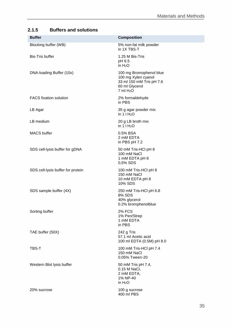
Materials and Methods
35
2.1.5 Buffers and solutions
Buffer Composition
Blocking buffer (WB) 5% non-fat milk powder in 1X TBS-T
Bis-Tris buffer 1.25 M Bis-Tris pH 6.5 in H2O
DNA-loading Buffer (10x) 100 mg Bromophenol blue 100 mg Xylen cyanol 33 ml 150 mM Tris pH 7.6 60 ml Glycerol 7 ml H2O
FACS fixation solution 2% formaldehyde in PBS
LB Agar 35 g agar powder mix in 1 l H2O
LB medium 20 g LB broth mix in 1 l H2O
MACS buffer 0.5% BSA 2 mM EDTA in PBS pH 7.2
SDS cell-lysis buffer for gDNA 50 mM Tris-HCl pH 8 100 mM NaCl 1 mM EDTA pH 8 0,5% SDS
SDS cell-lysis buffer for protein 100 mM Tris-HCl pH 8 150 mM NaCl 10 mM EDTA pH 8 10% SDS
SDS sample buffer (4X) 250 mM Tris-HCl pH 6.8 8% SDS 40% glycerol 0.2% bromphenolblue
Sorting buffer 2% FCS 1% Pen/Strep 1 mM EDTA in PBS
TAE buffer (50X) 242 g Tris 57.1 ml Acetic acid 100 ml EDTA (0.5M) pH 8.0
TBS-T 100 mM Tris-HCl pH 7.4 150 mM NaCl 0.05% Tween-20
Western Blot lysis buffer 50 mM Tris pH 7.4, 0.15 M NaCl, 2 mM EDTA, 1% NP-40 in H2O
20% sucrose 100 g sucrose 400 ml PBS
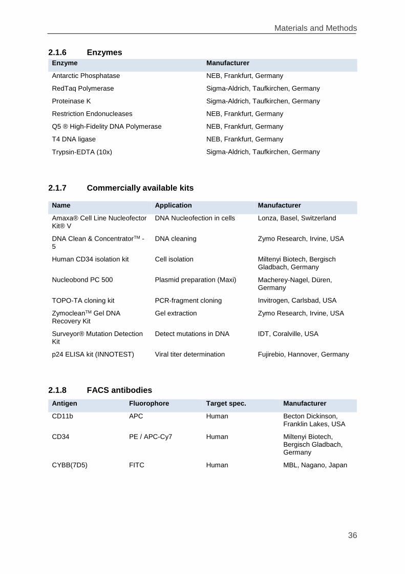
Materials and Methods
36
2.1.6 Enzymes
2.1.7 Commercially available kits
2.1.8 FACS antibodies
Antigen Fluorophore Target spec. Manufacturer
CD11b APC Human Becton Dickinson, Franklin Lakes, USA
CD34 PE / APC-Cy7 Human Miltenyi Biotech, Bergisch Gladbach, Germany
CYBB(7D5) FITC Human MBL, Nagano, Japan
Enzyme Manufacturer
Antarctic Phosphatase NEB, Frankfurt, Germany
RedTaq Polymerase Sigma-Aldrich, Taufkirchen, Germany
Proteinase K Sigma-Aldrich, Taufkirchen, Germany
Restriction Endonucleases NEB, Frankfurt, Germany
Q5 ® High-Fidelity DNA Polymerase NEB, Frankfurt, Germany
T4 DNA ligase NEB, Frankfurt, Germany
Trypsin‐EDTA (10x) Sigma-Aldrich, Taufkirchen, Germany
Name Application Manufacturer
Amaxa® Cell Line Nucleofector Kit® V
DNA Nucleofection in cells Lonza, Basel, Switzerland
DNA Clean & ConcentratorTM -5
DNA cleaning Zymo Research, Irvine, USA
Human CD34 isolation kit Cell isolation Miltenyi Biotech, Bergisch Gladbach, Germany
Nucleobond PC 500 Plasmid preparation (Maxi) Macherey‐Nagel, Düren, Germany
TOPO‐TA cloning kit PCR‐fragment cloning Invitrogen, Carlsbad, USA
ZymocleanTM Gel DNA Recovery Kit
Gel extraction Zymo Research, Irvine, USA
Surveyor® Mutation Detection Kit
Detect mutations in DNA IDT, Coralville, USA
p24 ELISA kit (INNOTEST) Viral titer determination Fujirebio, Hannover, Germany
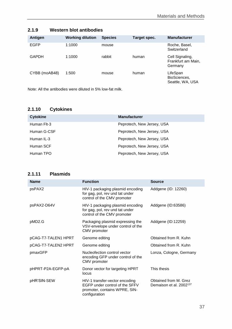
Materials and Methods
37
2.1.9 Western blot antibodies
Antigen Working dilution Species Target spec. Manufacturer
EGFP 1:1000 mouse Roche, Basel, Switzerland
GAPDH 1:1000 rabbit human Cell Signaling, Frankfurt am Main, Germany
CYBB (moAB48) 1:500 mouse human LifeSpan BioSciences, Seattle, WA, USA
Note: All the antibodies were diluted in 5% low-fat milk.
2.1.10 Cytokines
Cytokine Manufacturer
Human Flt‐3 Peprotech, New Jersey, USA
Human G‐CSF Peprotech, New Jersey, USA
Human IL‐3 Peprotech, New Jersey, USA
Human SCF Peprotech, New Jersey, USA
Human TPO Peprotech, New Jersey, USA
2.1.11 Plasmids
Name Function Source
psPAX2 HIV‐1 packaging plasmid encoding for gag, pol, rev und tat under control of the CMV promoter
Addgene (ID: 12260)
psPAX2-D64V HIV‐1 packaging plasmid encoding for gag, pol, rev und tat under control of the CMV promoter
Addgene (ID:63586)
pMD2.G Packaging plasmid expressing the
VSV‐envelope under control of the CMV promoter
Addgene (ID:12259)
pCAG-T7-TALEN1 HPRT Genome editing Obtained from R. Kuhn
pCAG-T7-TALEN2 HPRT Genome editing Obtained from R. Kuhn
pmaxGFP Nucleofection control vector encoding GFP under control of the CMV promoter
Lonza, Cologne, Germany
pHPRT-P2A-EGFP-pA Donor vector for targeting HPRT locus
This thesis
pHR‘SIN‐SEW HIV‐1 transfer‐vector encoding EGFP under control of the SFFV
promoter, contains WPRE, SIN‐configuration
Obtained from M. Grez Demaison et al. 2002137
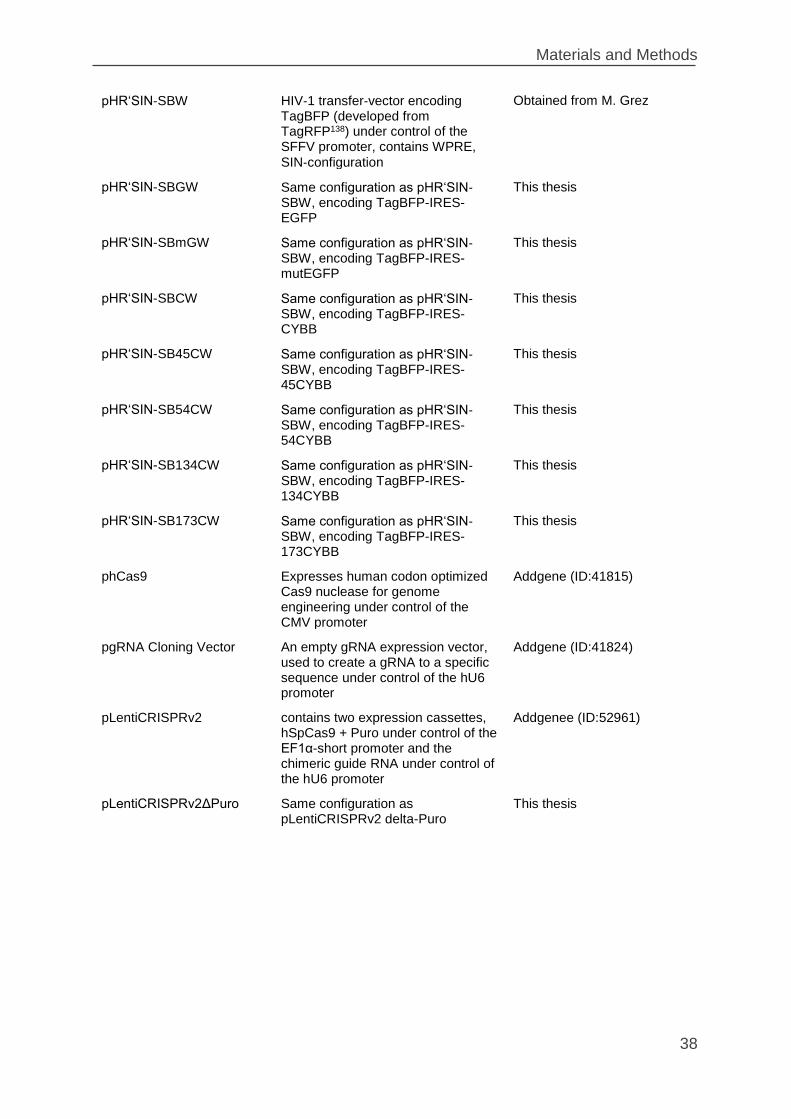
Materials and Methods
38
pHR‘SIN‐SBW HIV‐1 transfer‐vector encoding TagBFP (developed from TagRFP138) under control of the SFFV promoter, contains WPRE,
SIN‐configuration
Obtained from M. Grez
pHR‘SIN-SBGW Same configuration as pHR‘SIN‐SBW, encoding TagBFP-IRES-EGFP
This thesis
pHR‘SIN-SBmGW Same configuration as pHR‘SIN‐SBW, encoding TagBFP-IRES-mutEGFP
This thesis
pHR‘SIN-SBCW Same configuration as pHR‘SIN‐SBW, encoding TagBFP-IRES-CYBB
This thesis
pHR‘SIN-SB45CW Same configuration as pHR‘SIN‐SBW, encoding TagBFP-IRES-45CYBB
This thesis
pHR‘SIN-SB54CW Same configuration as pHR‘SIN‐SBW, encoding TagBFP-IRES-54CYBB
This thesis
pHR‘SIN-SB134CW Same configuration as pHR‘SIN‐SBW, encoding TagBFP-IRES-134CYBB
This thesis
pHR‘SIN-SB173CW Same configuration as pHR‘SIN‐SBW, encoding TagBFP-IRES-173CYBB
This thesis
phCas9 Expresses human codon optimized Cas9 nuclease for genome engineering under control of the CMV promoter
Addgene (ID:41815)
pgRNA Cloning Vector An empty gRNA expression vector, used to create a gRNA to a specific sequence under control of the hU6 promoter
Addgene (ID:41824)
pLentiCRISPRv2 contains two expression cassettes, hSpCas9 + Puro under control of the EF1α-short promoter and the chimeric guide RNA under control of the hU6 promoter
Addgenee (ID:52961)
pLentiCRISPRv2ΔPuro Same configuration as pLentiCRISPRv2 delta-Puro
This thesis
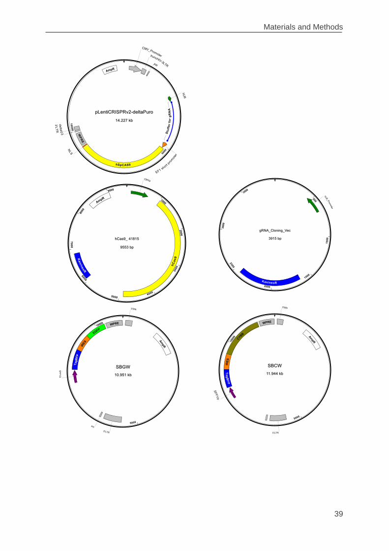
Materials and Methods
39
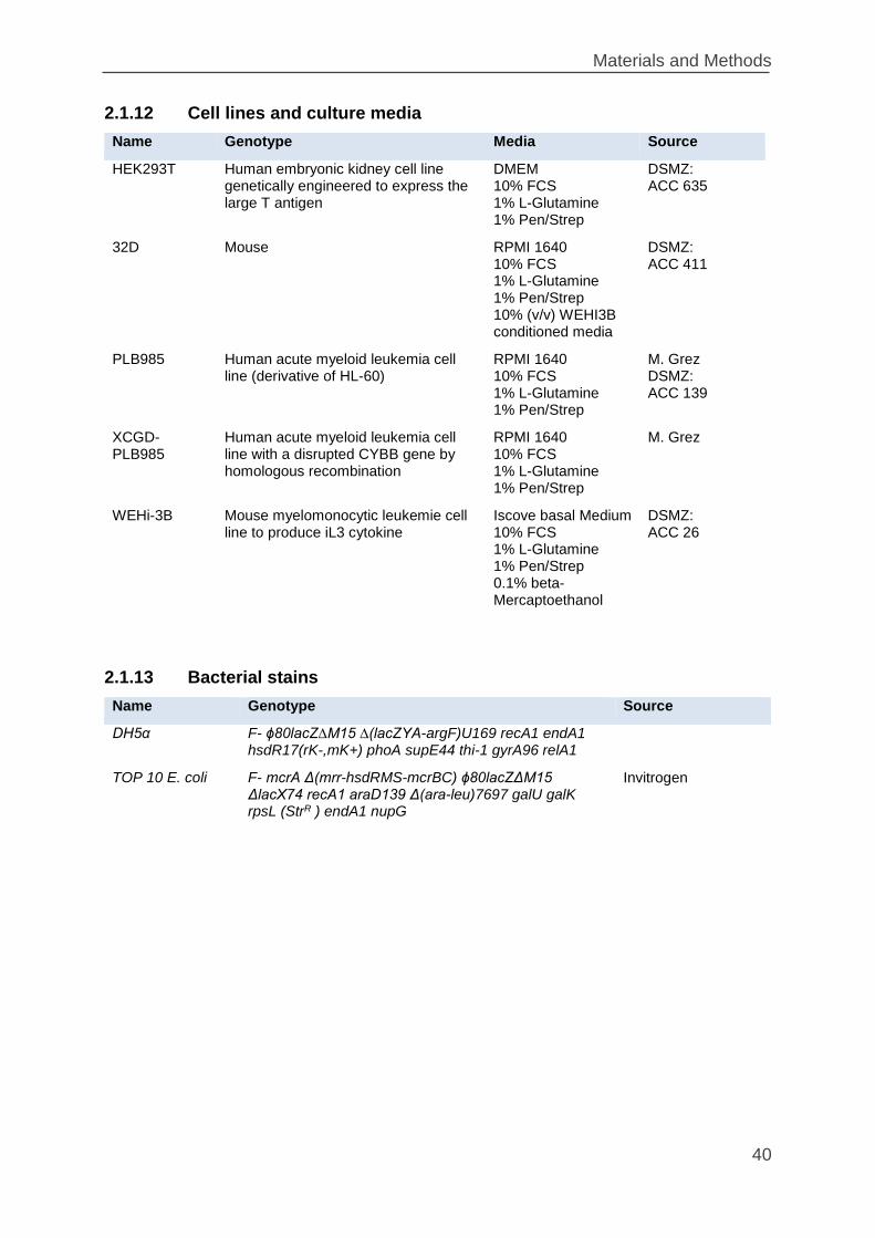
Materials and Methods
40
2.1.12 Cell lines and culture media
Name Genotype Media Source
HEK293T Human embryonic kidney cell line genetically engineered to express the large T antigen
DMEM 10% FCS 1% L-Glutamine 1% Pen/Strep
DSMZ: ACC 635
32D Mouse RPMI 1640 10% FCS 1% L-Glutamine 1% Pen/Strep 10% (v/v) WEHI3B conditioned media
DSMZ: ACC 411
PLB985 Human acute myeloid leukemia cell line (derivative of HL-60)
RPMI 1640 10% FCS 1% L-Glutamine 1% Pen/Strep
M. Grez DSMZ: ACC 139
XCGD-PLB985
Human acute myeloid leukemia cell line with a disrupted CYBB gene by homologous recombination
RPMI 1640 10% FCS 1% L-Glutamine 1% Pen/Strep
M. Grez
WEHi-3B Mouse myelomonocytic leukemie cell line to produce iL3 cytokine
Iscove basal Medium 10% FCS 1% L-Glutamine 1% Pen/Strep 0.1% beta-Mercaptoethanol
DSMZ: ACC 26
2.1.13 Bacterial stains
Name Genotype Source
DH5α F- ϕ80lacZ∆M15 ∆(lacZYA-argF)U169 recA1 endA1 hsdR17(rK-,mK+) phoA supE44 thi-1 gyrA96 relA1
TOP 10 E. coli F- mcrA Δ(mrr-hsdRMS-mcrBC) ϕ80lacZΔM15 ΔlacX74 recA1 araD139 Δ(ara-leu)7697 galU galK rpsL (StrR ) endA1 nupG
Invitrogen
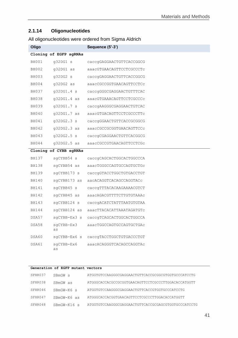
Materials and Methods
41
2.1.14 Oligonucleotides
All oligonucleotides were ordered from Sigma Aldrich
Oligo Sequence (5’-3’)
Cloning of EGFP sgRNAs
BH001 g32DG1 s caccgGAGGAACTGTTCACCGGCG
BH002 g32DG1 as aaacGTGAACAGTTCCTCGCCCTc
BH003 g32DG2 s caccgGAGGAACTGTTCACCGGCG
BH004 g32DG2 as aaacCGCCGGTGAACAGTTCCTCc
BH037 g32DG1.4 s caccgGGGCGAGGAACTGTTTCAC
BH038 g32DG1.4 as aaacGTGAAACAGTTCCTCGCCCc
BH039 g32DG1.7 s caccgAAGGGCGAGGAACTGTCAC
BH040 g32DG1.7 as aaacGTGACAGTTCCTCGCCCTTc
BH041 g32DG2.3 s caccgGGAACTGTTCACCGCGGCG
BH042 g32DG2.3 as aaacCGCCGCGGTGAACAGTTCCc
BH043 g32DG2.5 s caccgCGAGGAACTGTTCACGGCG
BH044 g32DG2.5 as aaacCGCCGTGAACAGTTCCTCGc
Cloning of CYBB sgRNAs
BH137 sgCYBB54 s caccgCAGCACTGGCACTGGCCCA
BH138 sgCYBB54 as aaacTGGGCCAGTGCCAGTGCTGc
BH139 sgCYBB173 s caccgGTACCTGGCTGTGACCTGT
BH140 sgCYBB173 as aacACAGGTCACAGCCAGGTACc
BH141 sgCYBB45 s caccgTTTACACAAGAAAACGTCT
BH142 sgCYBB45 as aaacAGACGTTTTCTTGTGTAAAc
BH143 sgCYBB124 s caccgACATCTATTTAATGTGTAA
BH144 sgCYBB124 as aaacTTACACATTAAATAGATGTc
DSA57 sgCYBB-Ex3 s caccgTCAGCACTGGCACTGGCCA
DSA58 sgCYBB-Ex3
as
aaacTGGCCAGTGCCAGTGCTGAc
DSA60 sgCYBB-Ex6 s caccgTACCTGGCTGTGACCCTGT
DSA61 sgCYBB-Ex6
as
aaacACAGGGTCACAGCCAGGTAc
Generation of EGFP mutant vectors
SFHR037 SBmGW s ATGGTGTCCAAGGGCGAGGAACTGTTCACCGCGGCGTGGTGCCCATCCTG
SFHR038 SBmGW as ATGGGCACCACGCCGCGGTGAACAGTTCCTCGCCCTTGGACACCATGGTT
SFHR046 SBmGW-K6 s ATGGTGTCCAAGGGCGAGGAACTGTTCACCGTGGTGCCCATCCTG
SFHR047 SBmGW-K6 as ATGGGCACCACGGTGAACAGTTCCTCGCCCTTGGACACCATGGTT
SFHR048 SBmGW-K16 s ATGGTGTCCAAGGGCGAGGAACTGTTCACCGCGAGCGTGGTGCCCATCCTG
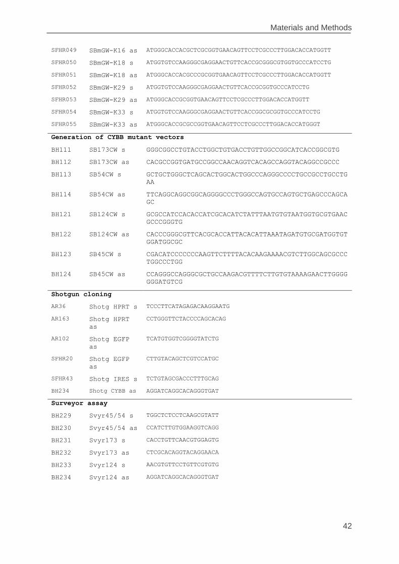
Materials and Methods
42
SFHR049 SBmGW-K16 as ATGGGCACCACGCTCGCGGTGAACAGTTCCTCGCCCTTGGACACCATGGTT
SFHR050 SBmGW-K18 s ATGGTGTCCAAGGGCGAGGAACTGTTCACCGCGGGCGTGGTGCCCATCCTG
SFHR051 SBmGW-K18 as ATGGGCACCACGCCCGCGGTGAACAGTTCCTCGCCCTTGGACACCATGGTT
SFHR052 SBmGW-K29 s ATGGTGTCCAAGGGCGAGGAACTGTTCACCGCGGTGCCCATCCTG
SFHR053 SBmGW-K29 as ATGGGCACCGCGGTGAACAGTTCCTCGCCCTTGGACACCATGGTT
SFHR054 SBmGW-K33 s ATGGTGTCCAAGGGCGAGGAACTGTTCACCGGCGCGGTGCCCATCCTG
SFHR055 SBmGW-K33 as ATGGGCACCGCGCCGGTGAACAGTTCCTCGCCCTTGGACACCATGGGT
Generation of CYBB mutant vectors
BH111 SB173CW s GGGCGGCCTGTACCTGGCTGTGACCTGTTGGCCGGCATCACCGGCGTG
BH112 SB173CW as CACGCCGGTGATGCCGGCCAACAGGTCACAGCCAGGTACAGGCCGCCC
BH113 SB54CW s GCTGCTGGGCTCAGCACTGGCACTGGCCCAGGGCCCCTGCCGCCTGCCTG
AA
BH114 SB54CW as TTCAGGCAGGCGGCAGGGGCCCTGGGCCAGTGCCAGTGCTGAGCCCAGCA
GC
BH121 SB124CW s GCGCCATCCACACCATCGCACATCTATTTAATGTGTAATGGTGCGTGAAC
GCCCGGGTG
BH122 SB124CW as CACCCGGGCGTTCACGCACCATTACACATTAAATAGATGTGCGATGGTGT
GGATGGCGC
BH123 SB45CW s CGACATCCCCCCCAAGTTCTTTTACACAAGAAAACGTCTTGGCAGCGCCC
TGGCCCTGG
BH124 SB45CW as CCAGGGCCAGGGCGCTGCCAAGACGTTTTCTTGTGTAAAAGAACTTGGGG
GGGATGTCG
Shotgun cloning
AR36 Shotg HPRT s TCCCTTCATAGAGACAAGGAATG
AR163 Shotg HPRT
as
CCTGGGTTCTACCCCAGCACAG
AR102 Shotg EGFP
as
TCATGTGGTCGGGGTATCTG
SFHR20 Shotg EGFP
as
CTTGTACAGCTCGTCCATGC
SFHR43 Shotg IRES s TCTGTAGCGACCCTTTGCAG
BH234 Shotg CYBB as AGGATCAGGCACAGGGTGAT
Surveyor assay
BH229 Svyr45/54 s TGGCTCTCCTCAAGCGTATT
BH230 Svyr45/54 as CCATCTTGTGGAAGGTCAGG
BH231 Svyr173 s CACCTGTTCAACGTGGAGTG
BH232 Svyr173 as CTCGCACAGGTACAGGAACA
BH233 Svyr124 s AACGTGTTCCTGTTCGTGTG
BH234 Svyr124 as AGGATCAGGCACAGGGTGAT
Materials and Methods
43
2.2 Methods of molecular biology
2.2.1 Cultivation of bacteria
Liquid culture
E.coli (usually DH5α) bacteria were grown in Luria Broth (LB) medium supplemented
with 100 µg/ml ampicillin (LB-Amp) at 37°C overnight (~16 h) and 220 rpm in a bacteria
shaker (Roth).
Culture plate
Bacteria were plated on ampicillin containing LB-agar plats by using an inoculation
spreader. Plates were incubated overnight at 37 °C in an incubator until bacteria
colonies were visible (~16 h). Bacteria plates were stored up to 4 weeks at 4°C.
2.2.2 Cloning processes
Restriction
For restriction of DNA type II restriction endonucleases were used, which recognize
specific sites of double stranded DNA sequences (usually 4 – 8 bp) and cleave in the
sequence. Thereby either blunt or sticky ends are produced. DNA digestions were
performed using commercially available endonucleases from New England Biolabs
(NEB) according to the manufacturer’s instructions. For a preparative restriction 1 – 5
µg and for an analytical restriction 0.2 – 1 µg DNA was used.
Standard digestion:
0.2 to 5 μg DNA for analytical or preparative purpose
0.1 µl volumes 10x reaction buffer (corresponding NEB buffer 1‐4)
10 U per µg DNA Restriction enzyme
add till 0.05 µg/µl DNA ddH2O
The reaction mixture was incubated for 1 – 2 h at 37°C or higher (enzyme dependent).
In case of a double digest the optimal buffer for the double digest was chosen
according to the manufacturer’s instructions.
Materials and Methods
44
Dephosphorylation of 5’–ends of DNA
The antarctic phosphatase catalyzes the removal of 5´ phosphate groups from DNA
and RNA. Dephosphorylation was used to minimize self-ligation of the vector
background in cloning reactions. Since phosphatase‐treated fragments lack the 5´
phosphoryl terminus required by ligases, they cannot self‐ligate. The enzyme can be
added directly into the digestion reaction after DNA digestion.
Standard dephosphorylation:
50 µl Restriction mix
6 µl Antarctic Phosphatase‐Buffer (10x)
2 µl Antarctic Phosphatase
add 60 µl ddH2O
The reaction was incubated for 30 min at 37°C.
Purification and ligation of vectors and inserts
Before ligation, the vectors and the inserts were purified with the “DNA Clean &
Concentrator”-kit (Zymo Research) according to the manufacturer’s instructions. For
ligation, the T4 DNA ligase was used to catalyze the formation of phosphodiester
bonds between the fragments under consumption of ATP. The ligation was applied for
fragments exhibiting complementary overhangs (sticky‐end ligation) or blunt ends
(blunt‐end ligation). As control sample a reaction which only contains plasmid DNA
was used.
Standard ligation:
0.1 – 1.0 µg total DNA (3:1 to 5:1 Insert:Vector ratio)
2 µl 10x reaction buffer
1 µl T4 DNA ligase
add 20 µl ddH2O
The reaction mixture was incubated at RT for a minimum of 2 h before transformation
into competent bacteria.
Materials and Methods
45
TA-Cloning
This easy and fast cloning technique relies on the ligation of the overhang of adenine
and thymine at the end of different DNA fragment. Shot gun cloning of PCR
amplification products was performed using the pGEM-T vector system (Promega,
Mannheim, Germany) according to the manufacturer’s instructions. pGEM-T vector is
a linearized vector with a single 3´-terminal thymidine at both ends. These compatible
overhangs for PCR products generated by certain thermostable polymerases (e.g.
Taq-Polymerase) greatly improve the efficiency of ligation. Positive clones were
sequenced with a T7 primer.
Standard reaction
1 – 4 µl PCR‐reaction
1 µl 10x reaction buffer
1 µl pGEM-T vector
add 10 μl ddH2O
The reaction was incubated at RT for 1 h. The reaction was subsequently used for
transformation in competent bacteria.
Golden Gate Assembly
Golden Gate assembly exploits the ability of type IIS restriction endonucleases to
cleave DNA outside of the recognition sequence. Combinations of overhang
sequences can be assembled using T4 DNA ligase. This one step cloning method
combines restriction and ligation reaction in one tube (Engler et al. 2008139).
Annealing of the oligonucleotides
50 pmol of each complementary oligonucleotides were added to 40 µl ddH2O and
incubated at 98°C for 5 min. Annealing was achieved by cooling down the
oligonucleotide mixture at RT for 20 min.
Standard oligo annealing
5 µl Oligo 1 (100 µM)
5 µl Oligo 2 (100 µM)
40 µl ddH2O
Materials and Methods
46
Golden Gate reaction
150 ng Vector (LCv2 and derivatives)
5 µl Annealed Oligos
2 µl 10x ligase Buffer
1 µl T4 DNA ligase
1 µl BsmBI restriction enzyme
add 20 µl ddH2O
Program
37°C 5 min
16°C 10 min
37°C 15 min 1 cycle
80°C 5 min 1 cycle
4°C Hold
2.2.3 Transformation of competent bacteria
Transformation of E. coli (usually DH5α) is the method to amplify plasmid DNA through
cellular replication. Most of the plasmids are “high copy” plasmids which are amplified
to multiple copies within the bacteria independent of cellular replication resulting in high
yields of plasmid DNA.
Chemically competent DH5α cells were thawed on ice and approximately 10 ng DNA
or 20 µl ligation reaction was added and incubated on ice for 20 min. A heat shock was
performed by incubation in a water bath at 42°C for 1 min. After additional on ice for
5 min, cells were plated on agar plates containing ampicillin (100 µg/ml) and incubated
at 37°C overnight.
10 cycles
Materials and Methods
47
2.2.4 Plasmid preparation
Miniprep
For analytical scale purification of low amounts of DNA (Miniprep), 2 ml LB-Amp
medium were inoculated with single bacteria clone in a 10 ml tube (Falcon) and grown
at 37°C overnight. The bacteria were pelleted at RT and 13000 rpm for 5 min in a
benchtop centrifuge and the supernatant was discarded. The bacteria pellet was
processed with buffers from the Maxiprep-Kit (Nucleobond PC500 Maxi Kit). In brief,
the pellet was resuspended in 100 µl S1 buffer, lysed 200 µl S2 buffer for 5 min at RT
and neutralized using 150 µl S3 buffer. Debris was pelleted at 13000 rpm for 5 min.
The supernatant was transferred to a fresh tube and the DNA precipitated by addition
of 1 ml of 100% ethanol. The DNA was pelleted at 13000 rpm for 5min, washed once
with 150 µl of 70% ethanol (13000 rpm for 5min). The dried pellet was resuspended in
50 µl ddH2O.
Maxiprep
For extraction of larger amounts of DNA (Maxiprep) 250 ml LB Amp medium were
inoculated and cultivated at 37°C overnight in a 1500 ml shake flask. Preparative scale
plasmid preparation from transformed bacteria was performed using the Nucleobond
PC500 Maxi Kit (Macherey‐Nagel) according to the manufacturer’s instructions. This
kit uses an anion-exchange tip where plasmid DNA selectively binds under low‐salt
and pH conditions. RNA, proteins, metabolites, and other low‐molecular‐weight
impurities are removed by a medium‐salt wash, and pure plasmid DNA is eluted in
high‐salt buffer. The DNA is concentrated and desalted by isopropanol precipitation
and collected by centrifugation. The DNA pellet was dried and dissolved in ddH2O to a
concentration of 1 µg/µl. The concentration of the isolated plasmid DNA was
determined photometrically at absorption A260 by NanoDrop (Thermo Fisher Scientific).
2.2.5 Agarose gel electrophoresis
Agarose gel electrophoresis allows the separation of DNA molecules by their size.
1 - 2% agarose gels were used, the required amount of agarose powder was mixed
with 1x TAE buffer and dissolved by heating the emulsion in a microwave oven. 0.1
µg/µl ethidium bromide or 1 µl HDGreen Plus per 20 ml agarose gel was added to the
dissolved agarose and the gel was casted into a tray. Ethidium bromide / HDGreen
Materials and Methods
48
Plus intercalates into DNA and visualizes DNA under UV light. Combs with the
appropriate number and size of teeth were used to make the loadings slots. After
polymerization, the gel was covered in 1x TAE buffer, samples were mixed with 10x
loading buffer and loaded onto the gel. 1 kb Plus DNA Ladder was used as marker.
The DNA fragments were separated by applying 120 V for 30 – 60 min in an
electrophoresis chamber. The DNA fragments were visualized on a transilluminator
and documented or extracted from the gel (see below).
,
2.2.6 Gel extraction of DNA fragments
To isolate an appropriate DNA fragment from an agarose gel, the DNA band of interest
was cut out of the gel under UV light and transferred into a 2 ml micro tube. The DNA
purification from an agarose gels was performed using the ZymocleanTM Gel DNA
Recovery Kit (Zymo Research) according to the manufacturer’s instructions. This kit is
based on binding of DNA to silica based matrixes in the presence of a high
concentration of chaotropic salt. After washing the agarose to remove impurities the
pure DNA was eluted using 50 µl of ddH2O.
2.2.7 Polymerase chain reaction (PCR)
The polymerase chain reaction (PCR) is a method for oligonucleotide primer directed
enzymatic amplification of a specific DNA sequence of interest. The DNA polymerase
amplifies a template DNA bordered on the forward and reverse primer in an
exponential manner. The polymerase need appropriate buffer, oligonucleotides
(primers), deoxynucleotides (dNTPs) and cycling conditions. The amplified fragment
can be used for further molecular biological methods.
Standard PCR-reaction
10 µl 10x Puffer
2 µl 10 mM dNTP-Mix
2.5 µl Forward Primer (10 µM)
2.5 µl Reverse Primer (10 µM)
50 – 500 ng Template DNA
0:5 µl Q5 High-Fidelity DNA Polymerase
add 50 µl ddH2O
Materials and Methods
49
Standard PCR-protocol
98°C 2 min Initial Denaturation
98°C 20 sec Denaturation
50 – 72°C 20 sec Primer Hybridization
72°C 30 sec/kb Elongation
72°C 2 min Final Extension
4°C ∞ Hold
The optimal primer hybridization temperature was determined by OligoAnalyzer from
NEB (http://tmcalculator.neb.com/#!/) and in some cases by a temperature gradient.
Amplificated products were subsequently analyzed by agarose gel electrophoresis
(2.2.6).
2.2.8 PCR product extraction
The PCR-product was purified using the DNA Clean & ConcentratorTM -5 (Zymo
Research) according to the manufacturer’s instructions. This kit is based on binding of
DNA to silica based matrix in the presence of a high concentration of chaotropic salt.
After washing off the impurities the pure DNA was eluted using 50 µl of ddH2O.
2.2.9 Nucleic acid sequencing
Nucleic acid sequencing was performed by Seqlab Company (Göttingen, Germany).
DNA samples containing approximately 1 μg plasmid or 0.1 µg PCR-product and
30 pmol sequencing primer in 15 µl total volume were prepared and sent to the
sequencing service.
2.2.10 Isolation of genomic DNA from cells
Phenol/Chloroform/Isoamylalkohol (25:24:1) was used for gDNA extraction. Up to
5x10⁶ cells were centrifuged 5 min at 1200 rpm at RT and the supernatant was
discarded. The cell pellet was resuspended in 400 µl SDS cell-lysis buffer, containing
150 µg/ml proteinase K. The cells were incubated at 60°C overnight. Afterwards, 300 µl
phenol/chloroform/isoamylalkohol were added and the mixture was vortexed and then
centrifuged (RT, 13000 rpm, 5 min). The aqueous phase containing DNA (upper) was
25 – 35 Cycles
Materials and Methods
50
transferred to a new tube and mixed with 300 µl chloroform. Vortexing and
centrifugation was repeated. The upper phase was transferred to a new tube and
mixed with 1 ml 100% ethanol (vortexed and centrifuged) for precipitation of the DNA.
The pellet was washed with 500 µl of 70% ethanol (13000 rpm for 5min at RT). The
genomic DNA pellet was dried and dissolved in ddH2O to a concentration of 200 ng/µl.
2.2.11 Surveyor Assay
For the analysis of CYBB indels generated by engineered nucleases, the enzyme
mismatch cleavage method was used. This method uses enzymes, such as T7E1 or
Surveyor, that cleave heteroduplex DNA at mismatches and extrahelical loops formed
by single or multiple nucleotides. In this study, Surveyor® Mutation Detection Kit-S100
(IDT, Leuven, Belgium) was used. RNG targeted CYBB sites were PCR amplified to
obtain 500 nt sized products and PCR product were purified using the DNA Clean &
ConcentratorTM -5 Kit (Zymo Research). For the analysis of on-target mutation rates,
400 ng of purified PCR product was subjected to the Surveyor assay according to the
manufacturer’s instructions. PCR products were mixed with 2 μl 10× Taq DNA
Polymerase PCR buffer and water was added to a final volume of 20 μl. To enable
heteroduplex formation the DNA was reannealed as follows: 95°C for 10 min, 95°C to
85°C ramping at −2°C/s, 85°C to 25°C at −0.25°C/s, 25°C for 1 min and hold at 4°C.
After re-annealing, products were treated with Surveyor nuclease and Surveyor
enhancer S following the manufacturer's instructions and analyzed on 2% Agarose gel
(see 2.2.5). Indel percentages derived from Image J plots140 were calculated by
following formula105:
100 ∗ (1 − (1 −𝑏 + 𝑐
𝑎 + 𝑏 + 𝑐)
0.5
)
Where 𝑎 is the integrated intensity of the undigested PCR product, and 𝑏 and 𝑐 the
integrated intensities of each cleavage product.
Materials and Methods
51
2.2.12 Southern Blot
Southern blot is a technique to detect specific DNA sequences in genomic DNA
samples. It combines the transfer of electrophoretically separated DNA fragments to a
nylon membrane and subsequent detection of specific DNA sequences by
hybridization with radioactively labeled DNA probes.
DNA isolation was performed as described in chapter 2.2.10. 15µg of genomic DNA
were digested overnight using NcoI restriction enzyme and fragments were resolved
on a 0.8% agarose gel alongside a DNA size standard. The gel was stained for 30min
in 1x TAE + EtBr and a picture was taken for later size identification of visualized DNA
fragments. The gel was destained in depurination solution (0.25 M HCl) for 15 min and
rinsed in water. Thereafter, the gel was neutralized in alkaline transfer buffer (0.5 M
NaOH, 1.5 M NaCl) for 30 min and an alkaline transfer was set up for the transfer of
the DNA fragments onto a Nytran SPC membrane (Whatman, Maidstone, UK).
Therefore, a pack of disposable paper towels was set up as a suction reservoir. On top
of this pack two Whatman layers were placed with the same size as the gel on which
the membrane was mounted. The gel was directly placed onto the membrane avoiding
air bubbles. The whole transfer set-up was covered with plastic wrap, the gel was
weighted with a plastic plate and a bottle with approx. 500ml of water. Transfer was
performed over 48h. After transfer, the blot set-up was broken down carefully,
discarding all paper towels, Whatman paper and the dried gel. The membrane was
neutralized for 15min in 0.5M Tris/HCl pH 7.5, 1.5 M NaCl and equilibrated for
hybridization in 2xSSC (saline sodium citrate buffer). Radioactive labelling of the BFP
probe was carried out using the RediPrime II DNA Labeling System (Amersham)
according to the manufacturer’s protocol. Hybridization was performed using
ExpressHyb Hybridyzation Solution (Clontech) according to the manufacturer’s
protocol. The membrane was washed twice at 37°C with 2xSSC, 0.1% SDS and twice
with 0.5xSSC, 0.1% SDS. Signals were detected using overnight exposure of Kodak
BioMax MS films at -80°C using amplifier screens.
Materials and Methods
52
2.2.13 Western Blot
Western blot is a technique to detect specific proteins from a complex mixture of
proteins extracted from cells. The technique is based on four elements: separation
protein by size, protein transfer to a membrane and target protein with specific
antibodies and detection of the specific signals.
Cell lysis to extract protein:
Cells were harvested by centrifugation at RT and 1200 rpm for 5 min and the
supernatant was discarded. The cell pellet was washed once with 1xPBS and the pellet
was lysed in SDS-lysis buffer (75 – 250 µl) supplemented with Protease Inhibitor
Cocktail (Roche, Mannheim, Germany) on ice for 30 min. The cells were then
sonicated twice for 30 sec, (5x, 45% power) and transfer to ice. The cool lysates
mixture was centrifuged at 4ºC and 15000 rpm for 3 min. The supernatant was
transferred to a new 1.5 ml tube.
Protein concentration was determined using the Lowry protein assay. To measure
protein concentration, 1 – 5 µl sample or protein standard was mixed with 200 µl Lowry
solution A, filled up to 1 ml with Lowry solution B, mixed by vortexing and samples were
measured at the absorbance 595 nm. The samples with equal protein concentrations
were resuspended in 4x loading buffer containing 20% beta-mercaptoethanol, boiled
at 95C for 5 min and stored at 20°C.
SDS polyacrylamide gel electrophoresis (SDS-PAGE) and transfer:
The proteins were separated by SDS-PAGE. Equal volumes of protein lysates were
loaded onto a polyacrylamide gel (stacking gel: 4% acrylamide, separating gel: 10%
acrylamide) together with 1 µl protein standard. The gel was run in MOPS buffer,
starting with 80 V for 10 min and then increased voltage to 160 V for 1h
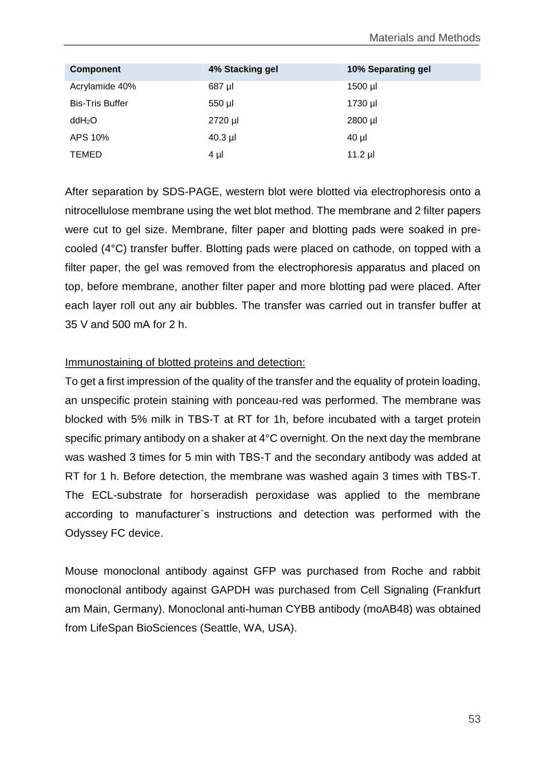
Materials and Methods
53
Component 4% Stacking gel 10% Separating gel
Acrylamide 40% 687 µl 1500 µl
Bis-Tris Buffer 550 µl 1730 µl
ddH2O 2720 µl 2800 µl
APS 10% 40.3 µl 40 µl
TEMED 4 µl 11.2 µl
After separation by SDS-PAGE, western blot were blotted via electrophoresis onto a
nitrocellulose membrane using the wet blot method. The membrane and 2 filter papers
were cut to gel size. Membrane, filter paper and blotting pads were soaked in pre-
cooled (4°C) transfer buffer. Blotting pads were placed on cathode, on topped with a
filter paper, the gel was removed from the electrophoresis apparatus and placed on
top, before membrane, another filter paper and more blotting pad were placed. After
each layer roll out any air bubbles. The transfer was carried out in transfer buffer at
35 V and 500 mA for 2 h.
Immunostaining of blotted proteins and detection:
To get a first impression of the quality of the transfer and the equality of protein loading,
an unspecific protein staining with ponceau-red was performed. The membrane was
blocked with 5% milk in TBS-T at RT for 1h, before incubated with a target protein
specific primary antibody on a shaker at 4°C overnight. On the next day the membrane
was washed 3 times for 5 min with TBS-T and the secondary antibody was added at
RT for 1 h. Before detection, the membrane was washed again 3 times with TBS-T.
The ECL-substrate for horseradish peroxidase was applied to the membrane
according to manufacturer`s instructions and detection was performed with the
Odyssey FC device.
Mouse monoclonal antibody against GFP was purchased from Roche and rabbit
monoclonal antibody against GAPDH was purchased from Cell Signaling (Frankfurt
am Main, Germany). Monoclonal anti-human CYBB antibody (moAB48) was obtained
from LifeSpan BioSciences (Seattle, WA, USA).
Materials and Methods
54
2.3 Cell culture and virological methods
2.3.1 Cultivation of cell lines
Cell lines were cultivated in the appropriate medium in an incubator at 37°C, 5.0% CO2
in a saturated water atmosphere and were passaged twice a week. Hematopoietic
(Suspension) cells were grown in RPMI-1640 medium supplemented with 10% (v/v)
heat-inactivated fetal calf serum (FCS), 2 mM glutamine, 100 U/ml penicillin and 100
µg/ml streptomycin. Hematopoietic 32D cell cultures were supplemented further with
10% (v/v) WEHI-3B conditioned medium. WEHI-3B cells were grown in IMDM
supplemented with 10% (v/v) heat-inactivated fetal calf serum (FCS), 2 mM glutamine,
100 U/ml penicillin and 100 µg/ml streptomycin, 0,1% beta-mercaptoethanol. For
splitting the cells were washed with 1xPBS by centrifugation (at 1200 rpm, 5 min) and
an appropriate cell number was resuspended in fresh media.
The adherent HEK293T cells were grown in DMEM medium supplemented with 10%
FCS (v/v), 2 mM glutamine, 100 U/ml penicillin and 100 µg/ml streptomycin. The cells
were grown until a confluence of 80 – 90% was reached and then split by washing
once with PBS and detaching with Trypsin‐EDTA before an appropriate fraction of the
resulting suspension was seeded into a new culture flask with fresh medium.
2.3.2 Cell counting and determination of cell viability
Cell number and viability were controlled by trypan-blue exclusion. To count the cells
10 – 50 µl cell suspension was mixed with an equal volume of trypan-blue solution.
Trypan-blue selectively stains dead cells as it is not able to pass through intact cellular
membranes. 10 µl of the mixture was applied to a counting chamber and only
unstained cells were counted. Cell number were calculated according to this formula:
𝐶𝑒𝑙𝑙 𝑛𝑢𝑚𝑏𝑒𝑟/𝑚𝑙 =𝑐𝑜𝑢𝑛𝑡𝑒𝑑 𝑐𝑒𝑙𝑙𝑠
𝑛𝑢𝑚𝑏𝑒𝑟 𝑜𝑓 𝑐𝑜𝑢𝑛𝑡𝑒𝑑 𝑠𝑞𝑢𝑎𝑟𝑒𝑠∗ 𝑑𝑖𝑙𝑢𝑡𝑖𝑜𝑛 𝑓𝑎𝑐𝑡𝑜𝑟 ∗ 104
Materials and Methods
55
2.3.3 Make WEHI-3B conditioned media
WEHI-3B cells were expanded in 150-cm2 flasks to a density of 80% to 90%
confluence. The cells were collected, centrifuge (at 1200 rpm and 5 min) and washed
once with 1xPBS (at 1200 rpm, 5 min). The cell pellet divided to four 150-cm2 tissue
culture flasks, each with a total volume of 150 ml of growth medium. During the
expansion stage, the pH indicator in the conditioned medium might have become
yellowish in color. 90% conditioned medium was collected every 2 – 3 days and filtered
through a 0.22 μm filter. Fresh medium was added to the remaining cells. Filtered
conditioned medium was collected in sterile 50-ml conical tubes in 50-ml aliquots to
and stored at –20°C.
2.3.4 Freezing and thawing of cultured cell
Freezing
To freeze cells, adherent cells were first washed once with PBS, detached through
trypsinising, resuspended in the appropriate medium and then like suspension cells,
centrifuged (1200 rpm, 5 min) to pellet the cells. The cell pellet was washed with
1xPBS, resuspended in freezing medium (90% FCS, 10% DMSO) to reach 5x10⁶ cells
per 1 ml, transferred to cryotubes in 1 ml aliquots and frozen in a freezing container at
‐80°C. After 3 days the cells were transferred into liquid nitrogen.
Thawing
Cryotubes were quickly placed at 37ºC until the suspension was almost thawed.
Immediately, the cells were transferred into a 50 ml falcon tube with 24 ml prewarmed
medium. The cells were centrifuged at 1.200 rpm for 5min to remove the cytotoxic
DMSO. Lastly the cells were resuspended in fresh medium and seeded into
appropriate culture flasks. 24 h later the medium was exchanged to fresh medium.
2.3.5 Nucleofection
Nucleofection is an electroporation-based transfection technique. Electroporation is a
biophysical method in which the cell membrane becomes permeable by applying
pulsed electric fields across a cell. This allows chemicals, protein or DNA to be
introduced into the cells. The nucleofection technology is carried out with cell type
Materials and Methods
56
specific electrical parameters, generated by a device called Nucleofector and with cell-
type specific solutions.
For nucleofection of 32D cells, 1 x 10⁶ cells were washed twice in 1xPBS, resuspended
in nucleofection buffer and nucleofected with 1 µg donor plasmid, 0.5 µg of each Hprt-
TALEN or 0.4 µg hspCas9 and 1.6 µg of sgRNA expression vector (Amaxa
nucleofector kit V with E-32 program, Amaxa Biosystems, Cologne, Germany)
according to the manufacturer’s protocol. The cells were selected in growth medium
containing either 2 µg/ml 6-thioguanin (6-TG) (Sigma-Aldrich, Taufkirchen, Germany)
or 1 x hypoxanthine-aminopterin-thymidine (HAT) (Life technologies, Darmstadt,
Germany).
2.3.6 Lentiviral vector production
Lentiviral supernatants were produced by polyethylenimine (PEI) based transient co-
transfection of HEK-293T cells. Briefly, transfer vector, lentiviral gag/pol helper
plasmids for integration (pCMV8.91) or integrase-defective (pCMV8.74) variants and
envelope plasmid encoding the glycoprotein of vesicular stomatitis virus (VSV-G)
(pMD2.G, Addgene #12259) were transfected at a molar ratio of 3:1:1 by standard PEI
transfection. 24 h prior to transfection, HEK 293T cells were seeded at 1.2x10⁷ per
14.5 cm plate in 16.5 ml DMEM with 10% FCS (v/v), 2 mM glutamine, 100 U/ml
penicillin and 100 µg/ml streptomycin to have a confluence 75-85% at the time of
transfection. At the day of transfection:
Prepare DNA solution in a 14 ml tube for every plate:
14 µg psPAX2 (gag, pol)
8 µg pMD2.G (vsvg)
16 µg Transfer DNA
1.75 ml DMEM w/o supplements
Prepare PEI solution in a 14 ml tube for every plate
171 µl PEI (1 µg/µl PEI 4.5 µl per 1 µg DNA)
1.75 ml DMEM w/o supplements
Materials and Methods
57
After 12 – 16 h the medium was replaced with 11 ml fresh medium. A maximum of
33 ml per viral supernatant could be used which was routinely collected from three
14.5cm dishes. After another 48 h viral supernatants were harvested, the supernatants
of the same viral vector were pooled, sterile filtered (0.45 µm pore-size PVDF-
membrane filter; Millipore, Schwalbach, Germany) and concentrated (100-fold) by
ultracentrifugation. The virus was underplayed with 5 ml of a sucrose (20% v/v sucrose
in PBS) and centrifuged (50,000 g, 2:20 h, 4°C). Pelleted viral particles were
resuspended in 330 µl StemSpan SFEM serum-free medium (StemCell Technologies,
Grenoble, France) without any supplements at 4°C overnight and stored at −80°C in
50 µl aliquots.
2.3.7 Titration of vector particles
Several different methods for titration of viral supernatants exist. In the present study
two different methods were used.
For integrated lentivirus, which expresses a virus encoded surface transgene in target
cells, the functional titer was determined by serial dilutions of viral supernatant
transducted of XCGD‐PLB985 cells followed by flow cytometry 3 days post-
transduction. The number of cells expressing the vector‐encoded transgene (CYBB or
TagBFP/EGFP) directly correlates with the number of functional vector particles in the
supernatant and can therefore be calculated. For the titration of virus, 2x10⁵ XCGD‐
PLB985 cells were seeded into a single well of a 24well plate. The lentiviral suspension
was serially diluted in 1:10 steps with medium supplemented with 8 µg/ml polybrene
as a chemical transduction enhancer and applied to the wells to a final volume of 500μl
medium. After incubation overnight, media was removed and 1 ml fresh medium was
added. Cells were grown for 3 days before analysis of the percentage of positive cells
by flow cytometry. For CYBB, a FITC labelled Anti-Flavocytochrome b 558 monoclonal
antibody which recognizes an external epitope of CYBB (7D5-FITC141) was used. For
vector titer calculation, dilutions, that contained about 5 ‐ 20% of transduced cells, was
chosen.
Materials and Methods
58
Calculation of virus titer:
Titer [TU
ml] = (
number of seeded cells ∗ dilution factor ∗ positive cells [%]
100 [%]) / 0.5 ml
To determine IDLV titers, p24 viral coat protein concentrations were estimated in the
viral supernatants using the p24 ELISA kit from INNOTEST (Fujirebio, Hannover,
Germany) according to the manufacturer’s instructions. To determine the number of
particles corresponding to every picogram of p24 antigen, we used a conversion factor
of 6.12 x 10³ particles/pg derived from the flow cytometric analysis of PLB cells 48 h
after SBmGW-IDLV transduction.
2.3.8 Flow cytometry and cell sorting
Flow cytometry, also referred as Fluorescence Activated Cell Sorting (FACS), is a
biophysical technique for biomarker detection. An appropriate amount of cells (0.1‐
1x10⁶) from a cell culture were transferred to 5 ml polystyrol‐FACS‐tubes. The tubes
were filled to the top with PBS and centrifuged at 1200 rpm for 5 min. The supernatant
was removed, the cells were resuspended in 200 µl PBS and ready for measurements
or staining. For cell surface antigen staining, cells were incubated with conjugated
antibodies (1 µl of each antibody) in dark, at RT for 20 – 30 min. Then, one wash step
was performed with PBS to remove unbound antibodies. For the FACS measurement
the cells were kept in 200 µl PBS. Data acquisition was performed with a BD
LSRFortessa flow cytometer (BD Biosciences, Heidelberg, Germany). Data was
analyzed with BD FACSDiva software (BD Biosciences, Heidelberg, Germany) or
flowing software 2.5.1. Cell sorting was performed in a BD FACSAria III flow cytometer
(BD Biosciences, Heidelberg, Germany).
2.3.9 Dihydrorhodamine 123 (DHR) reduction assay
The Dihydrorhodamine 123 (DHR) ‐assay is used to detect superoxide molecules
generated by the NADPH‐oxidase complex. It is a functional assay of the NADPH
oxidase and can be used to investigate reconstitution of CYBB repair. The assay relies
on the conversion of non‐fluorescent DHR123 to highly fluorescent Rhodamine123
preferentially but not exclusively by superoxide. In addition to superoxide, its
Materials and Methods
59
derivatives, such as hydroxyl anions or peroxide can also convert DHR, albeit to a
lesser extent. Activation of NADPH‐oxidase complex in target cells is triggered by the
addition of the unspecific PKC‐activator PMA (Phorbol 12‐myristate 13‐acetate).
DHR assays were performed as described by Brendel et al 2014142. For granulocytic
differentiation, PLB-985 and its derivatives were plated in RPMI 1640 supplemented
with 2.5% heat inactivated FCS, 2 mM glutamine, 100 U/ml penicillin and 100 µg/ml
streptomycin and 1.25% dimethyl sulfoxide (DMSO) at a concentration of
2x10⁵ cells/ml for at least 7 days. Flow cytometry was performed to ensure the
differentiation of labelled cells with anti-CD11b+ antibody. For estimating ROS
production, the differentiated cells were suspended in 1 ml pre-warmed HBSS (Life
technologies, Darmstadt, Germany) supplemented with 7.5 mmol/l D-Glucose, 0.5%
bovine serum albumin (BSA; Sigma-Aldrich) 2000 U/ml catalase (Sigma-Aldrich) and
5 µg/ml DHR123 (Sigma-Aldrich). Following incubation at 37°C for 10 min, cells were
exposed to 0.1 µmol/l PMA (Sigma-Aldrich) for 15 min and placed on ice.
Rhodamin123 fluorescence was measured in a flow cytometer within the next 30 min.
2.3.10 Statistical analysis
For statistical comparisons between groups, Student´s t-test or one-way analysis of
variance (ANOVA) with Bonferoni post-hoc test were used as appropriate in
conjunction with GraphPad Prism 5 software.
Results
60
3. Results
3.1 Genome editing by homology-directed repair (HDR) versus non-
homologous end joining (NHEJ)
In an initial project aimed at testing the efficacy of genome editing in hematopoietic
cells, Transcription activator like effectors nucleases (TALENs) were used. Two
TALENs were designed directed against 16 nucleotide (nt) stretches on both strands
of the 3rd murine Hprt exon encoding for the active centrum of hypoxanthine-guanine
phosphoribosyltransferase (Hprt) enzyme. The Hprt-TALENs were separated from
each other by a 15 nt spacer (Figure 10A). 32D cells (murine hematopoietic, immature,
myeloid cell line) were nucleofected with Hprt-TALENs and EGFP encoding donor
constructs. The nuclefected 32D cells were selected against Hprt expression in 6-
thioguanine (6-TG) containing medium. Less than 5% of the 6-TG resistant cells
expressed EGFP from the knock-in donor construct (Figure 10B), suggesting that in
the majority of cells Hprt inactivation was caused by indels associated with NHEJ-
directed DSB-repair.
To confirm this, individual 6-TG resistant clones were isolated by limiting dilution. The
mutant Hprt alleles in clones were characterized by genomic PCR and sequencing.
Consistent with NHEJ, the Hprt open reading frame (ORF) was disrupted by indels of
different sizes (Figure 10C).
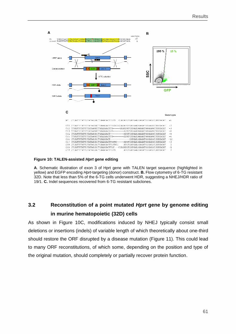
Results
61
3.2 Reconstitution of a point mutated Hprt gene by genome editing
in murine hematopoietic (32D) cells
As shown in Figure 10C, modifications induced by NHEJ typically consist small
deletions or insertions (indels) of variable length of which theoretically about one-third
should restore the ORF disrupted by a disease mutation (Figure 11). This could lead
to many ORF reconstitutions, of which some, depending on the position and type of
the original mutation, should completely or partially recover protein function.
Figure 10: TALEN-assisted Hprt gene editing
A. Schematic illustration of exon 3 of Hprt gene with TALEN target sequence (highlighted in yellow) and EGFP encoding Hprt-targeting (donor) construct. B. Flow cytometry of 6-TG resistant 32D. Note that less than 5% of the 6-TG cells underwent HDR, suggesting a NHEJ/HDR ratio of
19/1. C. Indel sequences recovered from 6-TG resistant subclones.
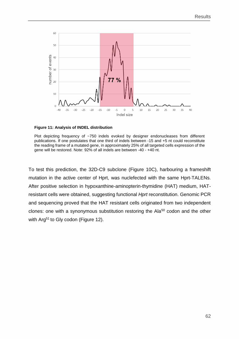
Results
62
To test this prediction, the 32D-C9 subclone (Figure 10C), harbouring a frameshift
mutation in the active center of Hprt, was nuclefected with the same Hprt-TALENs.
After positive selection in hypoxanthine-aminopterin-thymidine (HAT) medium, HAT-
resistant cells were obtained, suggesting functional Hprt reconstitution. Genomic PCR
and sequencing proved that the HAT resistant cells originated from two independent
clones: one with a synonymous substitution restoring the Ala50 codon and the other
with Arg51 to Gly codon (Figure 12).
Figure 11: Analysis of INDEL distribution
Plot depicting frequency of ~750 indels evoked by designer endonucleases from different publications. If one postulates that one third of indels between -15 and +5 nt could reconstitute the reading frame of a mutated gene, in approximately 25% of all targeted cells expression of the gene will be restored. Note: 92% of all indels are between -40 - +40 nt.
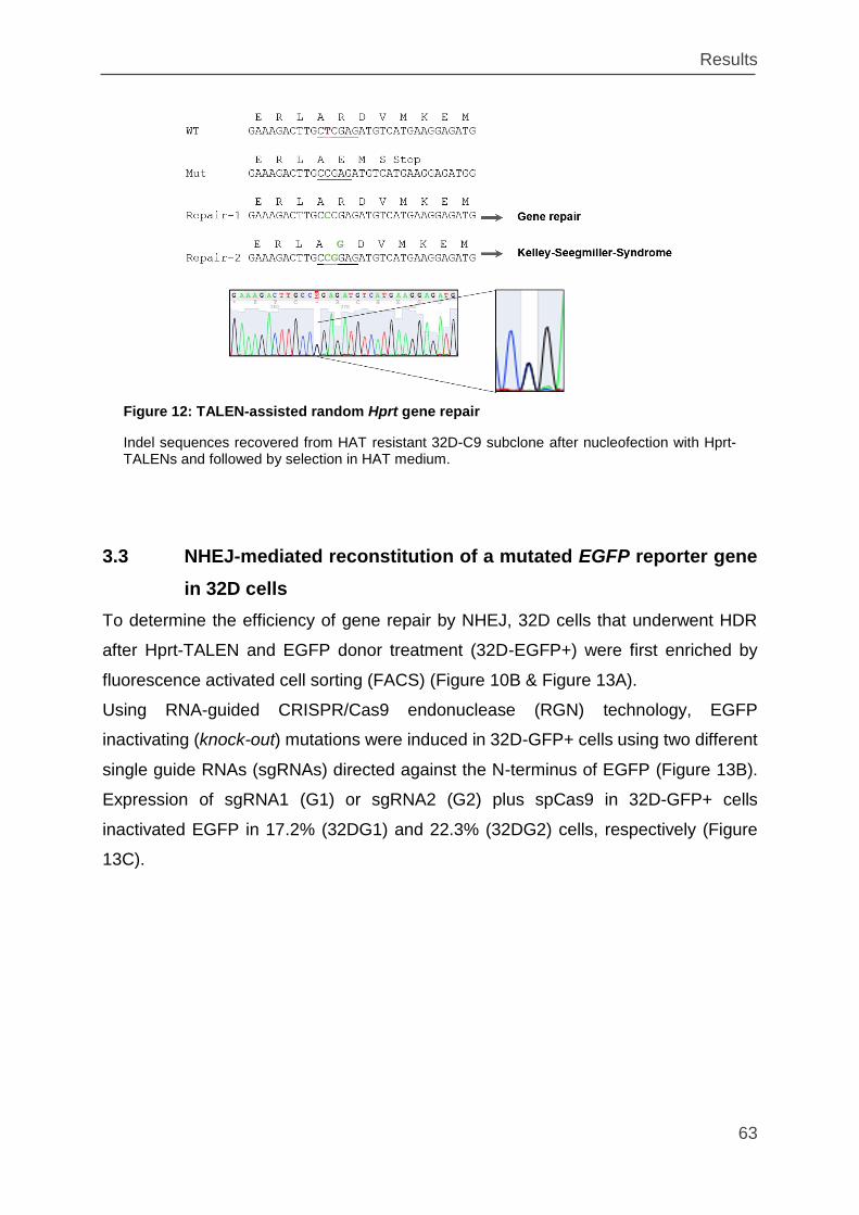
Results
63
3.3 NHEJ-mediated reconstitution of a mutated EGFP reporter gene
in 32D cells
To determine the efficiency of gene repair by NHEJ, 32D cells that underwent HDR
after Hprt-TALEN and EGFP donor treatment (32D-EGFP+) were first enriched by
fluorescence activated cell sorting (FACS) (Figure 10B & Figure 13A).
Using RNA-guided CRISPR/Cas9 endonuclease (RGN) technology, EGFP
inactivating (knock-out) mutations were induced in 32D-GFP+ cells using two different
single guide RNAs (sgRNAs) directed against the N-terminus of EGFP (Figure 13B).
Expression of sgRNA1 (G1) or sgRNA2 (G2) plus spCas9 in 32D-GFP+ cells
inactivated EGFP in 17.2% (32DG1) and 22.3% (32DG2) cells, respectively (Figure
13C).
Figure 12: TALEN-assisted random Hprt gene repair
Indel sequences recovered from HAT resistant 32D-C9 subclone after nucleofection with Hprt-TALENs and followed by selection in HAT medium.
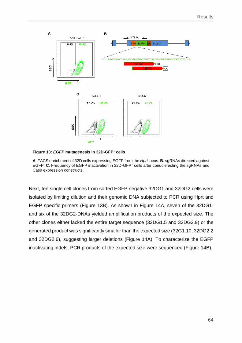
Results
64
Next, ten single cell clones from sorted EGFP negative 32DG1 and 32DG2 cells were
isolated by limiting dilution and their genomic DNA subjected to PCR using Hprt and
EGFP specific primers (Figure 13B). As shown in Figure 14A, seven of the 32DG1-
and six of the 32DG2-DNAs yielded amplification products of the expected size. The
other clones either lacked the entire target sequence (32DG1.5 and 32DG2.9) or the
generated product was significantly smaller than the expected size (32G1.10, 32DG2.2
and 32DG2.6), suggesting larger deletions (Figure 14A). To characterize the EGFP
inactivating indels, PCR products of the expected size were sequenced (Figure 14B).
Figure 13: EGFP mutagenesis in 32D-GFP+ cells
A. FACS enrichment of 32D cells expressing EGFP from the Hprt locus. B. sgRNAs directed against EGFP. C. Frequency of EGFP inactivation in 32D-GFP+ cells after conuclefecting the sgRNAs and Cas9 expression constructs.
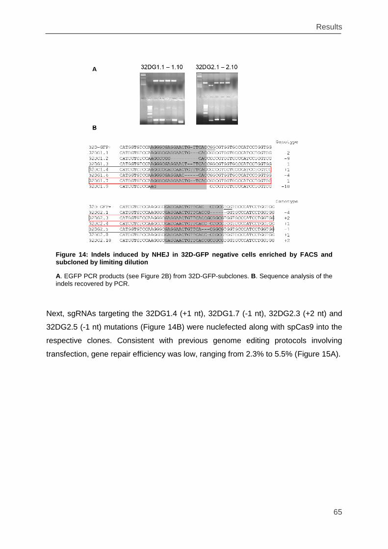
Results
65
Next, sgRNAs targeting the 32DG1.4 (+1 nt), 32DG1.7 (-1 nt), 32DG2.3 (+2 nt) and
32DG2.5 (-1 nt) mutations (Figure 14B) were nuclefected along with spCas9 into the
respective clones. Consistent with previous genome editing protocols involving
transfection, gene repair efficiency was low, ranging from 2.3% to 5.5% (Figure 15A).
Figure 14: Indels induced by NHEJ in 32D-GFP negative cells enriched by FACS and subcloned by limiting dilution
A. EGFP PCR products (see Figure 2B) from 32D-GFP-subclones. B. Sequence analysis of the indels recovered by PCR.
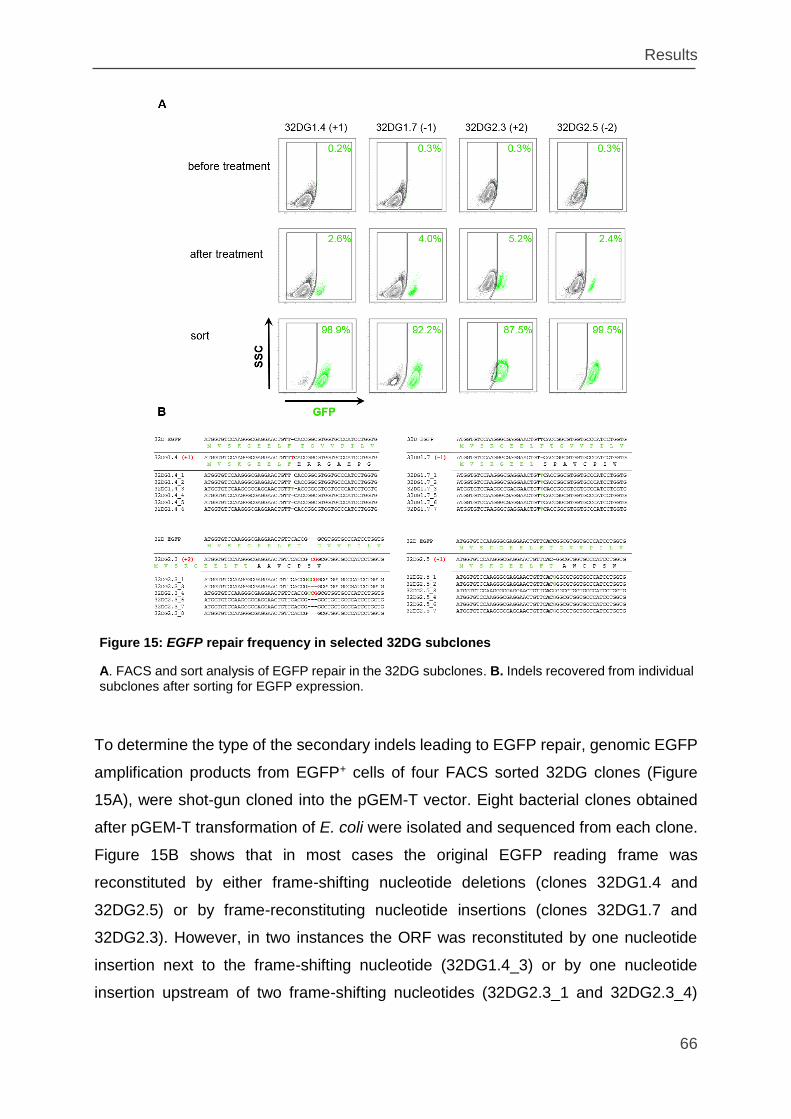
Results
66
To determine the type of the secondary indels leading to EGFP repair, genomic EGFP
amplification products from EGFP+ cells of four FACS sorted 32DG clones (Figure
15A), were shot-gun cloned into the pGEM-T vector. Eight bacterial clones obtained
after pGEM-T transformation of E. coli were isolated and sequenced from each clone.
Figure 15B shows that in most cases the original EGFP reading frame was
reconstituted by either frame-shifting nucleotide deletions (clones 32DG1.4 and
32DG2.5) or by frame-reconstituting nucleotide insertions (clones 32DG1.7 and
32DG2.3). However, in two instances the ORF was reconstituted by one nucleotide
insertion next to the frame-shifting nucleotide (32DG1.4_3) or by one nucleotide
insertion upstream of two frame-shifting nucleotides (32DG2.3_1 and 32DG2.3_4)
Figure 15: EGFP repair frequency in selected 32DG subclones
A. FACS and sort analysis of EGFP repair in the 32DG subclones. B. Indels recovered from individual subclones after sorting for EGFP expression.
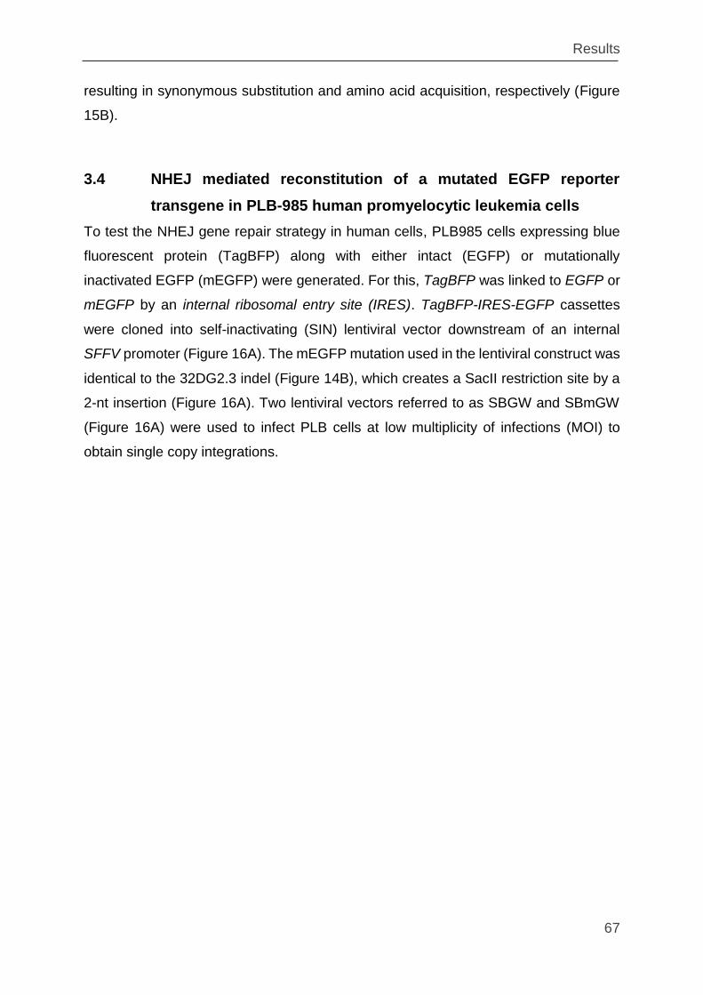
Results
67
resulting in synonymous substitution and amino acid acquisition, respectively (Figure
15B).
3.4 NHEJ mediated reconstitution of a mutated EGFP reporter
transgene in PLB-985 human promyelocytic leukemia cells
To test the NHEJ gene repair strategy in human cells, PLB985 cells expressing blue
fluorescent protein (TagBFP) along with either intact (EGFP) or mutationally
inactivated EGFP (mEGFP) were generated. For this, TagBFP was linked to EGFP or
mEGFP by an internal ribosomal entry site (IRES). TagBFP-IRES-EGFP cassettes
were cloned into self-inactivating (SIN) lentiviral vector downstream of an internal
SFFV promoter (Figure 16A). The mEGFP mutation used in the lentiviral construct was
identical to the 32DG2.3 indel (Figure 14B), which creates a SacII restriction site by a
2-nt insertion (Figure 16A). Two lentiviral vectors referred to as SBGW and SBmGW
(Figure 16A) were used to infect PLB cells at low multiplicity of infections (MOI) to
obtain single copy integrations.
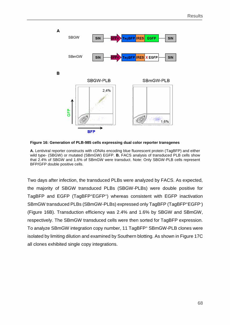
Results
68
Two days after infection, the transduced PLBs were analyzed by FACS. As expected,
the majority of SBGW transduced PLBs (SBGW-PLBs) were double positive for
TagBFP and EGFP (TagBFP+EGFP+) whereas consistent with EGFP inactivation
SBmGW transduced PLBs (SBmGW-PLBs) expressed only TagBFP (TagBFP+EGFP-)
(Figure 16B). Transduction efficiency was 2.4% and 1.6% by SBGW and SBmGW,
respectively. The SBmGW transduced cells were then sorted for TagBFP expression.
To analyze SBmGW integration copy number, 11 TagBFP+ SBmGW-PLB clones were
isolated by limiting dilution and examined by Southern blotting. As shown in Figure 17C
all clones exhibited single copy integrations.
Figure 16: Generation of PLB-985 cells expressing dual color reporter transgenes
A. Lentiviral reporter constructs with cDNAs encoding blue fluorescent protein (TagBFP) and either wild type- (SBGW) or mutated (SBmGW) EGFP. B. FACS analysis of transduced PLB cells show that 2.4% of SBGW and 1.6% of SBmGW were transduct. Note: Only SBGW-PLB cells represent BFP/GFP double positive cells.
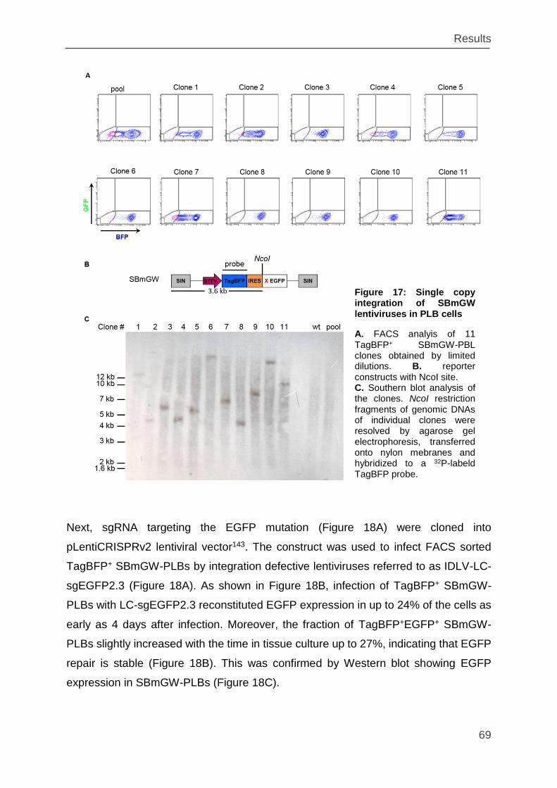
Results
69
Next, sgRNA targeting the EGFP mutation (Figure 18A) were cloned into
pLentiCRISPRv2 lentiviral vector143. The construct was used to infect FACS sorted
TagBFP+ SBmGW-PLBs by integration defective lentiviruses referred to as IDLV-LC-
sgEGFP2.3 (Figure 18A). As shown in Figure 18B, infection of TagBFP+ SBmGW-
PLBs with LC-sgEGFP2.3 reconstituted EGFP expression in up to 24% of the cells as
early as 4 days after infection. Moreover, the fraction of TagBFP+EGFP+ SBmGW-
PLBs slightly increased with the time in tissue culture up to 27%, indicating that EGFP
repair is stable (Figure 18B). This was confirmed by Western blot showing EGFP
expression in SBmGW-PLBs (Figure 18C).
Figure 17: Single copy integration of SBmGW lentiviruses in PLB cells
A. FACS analyis of 11 TagBFP+ SBmGW-PBL clones obtained by limited dilutions. B. reporter constructs with NcoI site. C. Southern blot analysis of the clones. NcoI restriction fragments of genomic DNAs of individual clones were resolved by agarose gel electrophoresis, transferred onto nylon mebranes and hybridized to a 32P-labeld TagBFP probe.
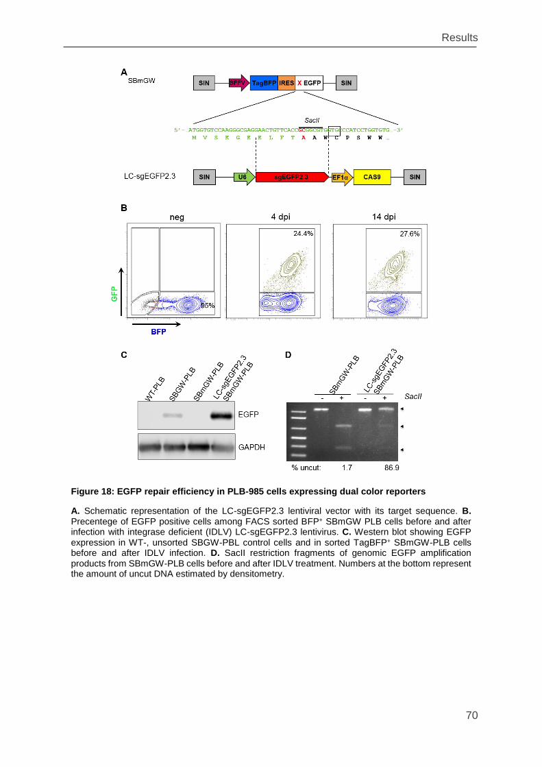
Results
70
Figure 18: EGFP repair efficiency in PLB-985 cells expressing dual color reporters
A. Schematic representation of the LC-sgEGFP2.3 lentiviral vector with its target sequence. B. Precentege of EGFP positive cells among FACS sorted BFP+ SBmGW PLB cells before and after infection with integrase deficient (IDLV) LC-sgEGFP2.3 lentivirus. C. Western blot showing EGFP expression in WT-, unsorted SBGW-PBL control cells and in sorted TagBFP+ SBmGW-PLB cells before and after IDLV infection. D. SacII restriction fragments of genomic EGFP amplification products from SBmGW-PLB cells before and after IDLV treatment. Numbers at the bottom represent the amount of uncut DNA estimated by densitometry.
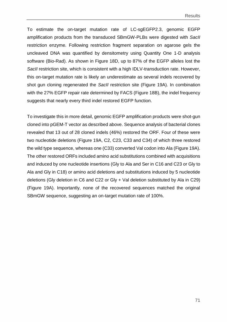
Results
71
To estimate the on-target mutation rate of LC-sgEGFP2.3, genomic EGFP
amplification products from the transduced SBmGW-PLBs were digested with SacII
restriction enzyme. Following restriction fragment separation on agarose gels the
uncleaved DNA was quantified by densitometry using Quantity One 1-D analysis
software (Bio-Rad). As shown in Figure 18D, up to 87% of the EGFP alleles lost the
SacII restriction site, which is consistent with a high IDLV-transduction rate. However,
this on-target mutation rate is likely an underestimate as several indels recovered by
shot gun cloning regenerated the SacII restriction site (Figure 19A). In combination
with the 27% EGFP repair rate determined by FACS (Figure 18B), the indel frequency
suggests that nearly every third indel restored EGFP function.
To investigate this in more detail, genomic EGFP amplification products were shot-gun
cloned into pGEM-T vector as described above. Sequence analysis of bacterial clones
revealed that 13 out of 28 cloned indels (46%) restored the ORF. Four of these were
two nucleotide deletions (Figure 19A, C2, C23, C33 and C34) of which three restored
the wild type sequence, whereas one (C33) converted Val codon into Ala (Figure 19A).
The other restored ORFs included amino acid substitutions combined with acquisitions
and induced by one nucleotide insertions (Gly to Ala and Ser in C16 and C23 or Gly to
Ala and Gly in C18) or amino acid deletions and substitutions induced by 5 nucleotide
deletions (Gly deletion in C6 and C22 or Gly + Val deletion substituted by Ala in C29)
(Figure 19A). Importantly, none of the recovered sequences matched the original
SBmGW sequence, suggesting an on-target mutation rate of 100%.
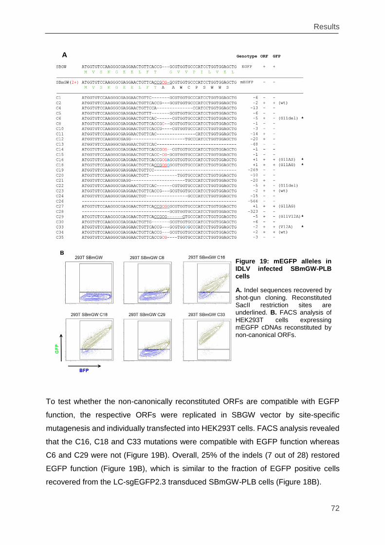
Results
72
To test whether the non-canonically reconstituted ORFs are compatible with EGFP
function, the respective ORFs were replicated in SBGW vector by site-specific
mutagenesis and individually transfected into HEK293T cells. FACS analysis revealed
that the C16, C18 and C33 mutations were compatible with EGFP function whereas
C6 and C29 were not (Figure 19B). Overall, 25% of the indels (7 out of 28) restored
EGFP function (Figure 19B), which is similar to the fraction of EGFP positive cells
recovered from the LC-sgEGFP2.3 transduced SBmGW-PLB cells (Figure 18B).
Figure 19: mEGFP alleles in IDLV infected SBmGW-PLB cells
A. Indel sequences recovered by shot-gun cloning. Reconstituted SacII restriction sites are underlined. B. FACS analysis of HEK293T cells expressing mEGFP cDNAs reconstituted by non-canonical ORFs.
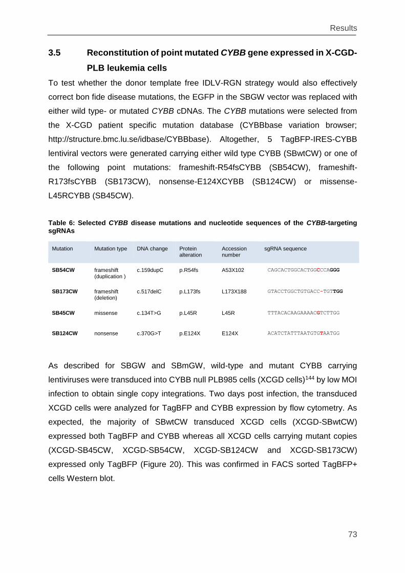
Results
73
3.5 Reconstitution of point mutated CYBB gene expressed in X-CGD-
PLB leukemia cells
To test whether the donor template free IDLV-RGN strategy would also effectively
correct bon fide disease mutations, the EGFP in the SBGW vector was replaced with
either wild type- or mutated CYBB cDNAs. The CYBB mutations were selected from
the X-CGD patient specific mutation database (CYBBbase variation browser;
http://structure.bmc.lu.se/idbase/CYBBbase). Altogether, 5 TagBFP-IRES-CYBB
lentiviral vectors were generated carrying either wild type CYBB (SBwtCW) or one of
the following point mutations: frameshift-R54fsCYBB (SB54CW), frameshift-
R173fsCYBB (SB173CW), nonsense-E124XCYBB (SB124CW) or missense-
L45RCYBB (SB45CW).
Table 6: Selected CYBB disease mutations and nucleotide sequences of the CYBB-targeting sgRNAs
Mutation Mutation type DNA change Protein alteration
Accession number
sgRNA sequence
SB54CW frameshift (duplication )
c.159dupC p.R54fs A53X102 CAGCACTGGCACTGGCCCAGGG
SB173CW frameshift (deletion)
c.517delC p.L173fs L173X188 GTACCTGGCTGTGACC-TGTTGG
SB45CW missense c.134T>G p.L45R L45R TTTACACAAGAAAACGTCTTGG
SB124CW nonsense c.370G>T p.E124X E124X ACATCTATTTAATGTGTAATGG
As described for SBGW and SBmGW, wild-type and mutant CYBB carrying
lentiviruses were transduced into CYBB null PLB985 cells (XCGD cells)144 by low MOI
infection to obtain single copy integrations. Two days post infection, the transduced
XCGD cells were analyzed for TagBFP and CYBB expression by flow cytometry. As
expected, the majority of SBwtCW transduced XCGD cells (XCGD-SBwtCW)
expressed both TagBFP and CYBB whereas all XCGD cells carrying mutant copies
(XCGD-SB45CW, XCGD-SB54CW, XCGD-SB124CW and XCGD-SB173CW)
expressed only TagBFP (Figure 20). This was confirmed in FACS sorted TagBFP+
cells Western blot.
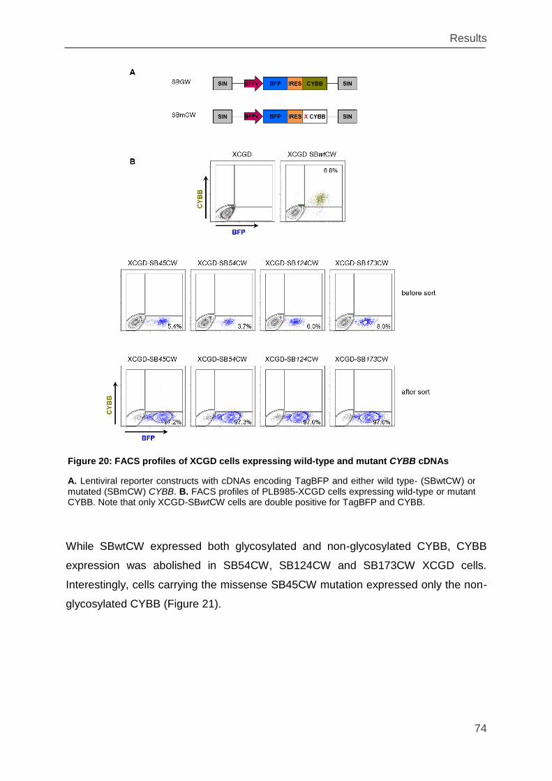
Results
74
While SBwtCW expressed both glycosylated and non-glycosylated CYBB, CYBB
expression was abolished in SB54CW, SB124CW and SB173CW XCGD cells.
Interestingly, cells carrying the missense SB45CW mutation expressed only the non-
glycosylated CYBB (Figure 21).
Figure 20: FACS profiles of XCGD cells expressing wild-type and mutant CYBB cDNAs
A. Lentiviral reporter constructs with cDNAs encoding TagBFP and either wild type- (SBwtCW) or mutated (SBmCW) CYBB. B. FACS profiles of PLB985-XCGD cells expressing wild-type or mutant CYBB. Note that only XCGD-SBwtCW cells are double positive for TagBFP and CYBB.
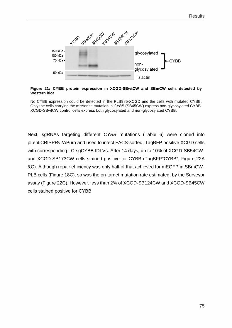
Results
75
Next, sgRNAs targeting different CYBB mutations (Table 6) were cloned into
pLentiCRISPRv2ΔPuro and used to infect FACS-sorted, TagBFP positive XCGD cells
with corresponding LC-sgCYBB IDLVs. After 14 days, up to 10% of XCGD-SB54CW-
and XCGD-SB173CW cells stained positive for CYBB (TagBFP+CYBB+; Figure 22A
&C). Although repair efficiency was only half of that achieved for mEGFP in SBmGW-
PLB cells (Figure 18C), so was the on-target mutation rate estimated, by the Surveyor
assay (Figure 22C). However, less than 2% of XCGD-SB124CW and XCGD-SB45CW
cells stained positive for CYBB
Figure 21: CYBB protein expression in XCGD-SBwtCW and SBmCW cells detected by Western blot
No CYBB expression could be detected in the PLB985-XCGD and the cells with mutated CYBB. Only the cells carrying the missense mutation in CYBB (SB45CW) express non-glycosylated CYBB. XCGD-SBwtCW control cells express both glycosylated and non-glycosylated CYBB.
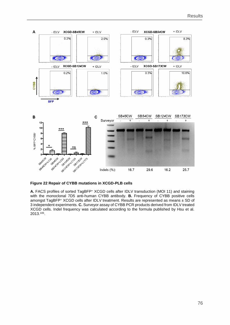
Results
76
Figure 22 Repair of CYBB mutations in XCGD-PLB cells
A. FACS profiles of sorted TagBFP+ XCGD cells after IDLV transduction (MOI 11) and staining with the monoclonal 7D5 anti-human CYBB antibody. B. Frequency of CYBB positive cells amongst TagBFP+ XCGD cells after IDLV treatment. Results are represented as means ± SD of 3 independent experiments. C. Surveyor assay of CYBB PCR products derived from IDLV treated XCGD cells. Indel frequency was calculated according to the formula published by Hsu et al. 2013.105.
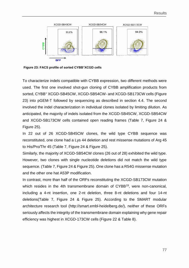
Results
77
To characterize indels compatible with CYBB expression, two different methods were
used. The first one involved shot-gun cloning of CYBB amplification products from
sorted, CYBB+ XCGD-SB45CW, XCGD-SB54CW- and XCGD-SB173CW cells (Figure
23) into pGEM-T followed by sequencing as described in section 4.4. The second
involved the indel characterization in individual clones isolated by limiting dilution. As
anticipated, the majority of indels isolated from the XCGD-SB45CW, XCGD-SB54CW
and XCGD-SB173CW cells contained open reading frames (Table 7, Figure 24 &
Figure 25).
In 22 out of 26 XCGD-SB45CW clones, the wild type CYBB sequence was
reconstituted, one clone had a Lys 44 deletion and rest missense mutations of Arg 45
to His/Pro/Thr 45 (Table 7, Figure 24 & Figure 25).
Similarly, the majority of XCGD-SB54CW clones (26 out of 28) exhibited the wild type.
However, two clones with single nucleotide deletions did not match the wild type
sequence. (Table 7, Figure 24 & Figure 25). One clone has a R54G missense mutation
and the other one hat A53P modification.
In contrast, more than half of the ORFs reconstituting the XCGD-SB173CW mutation
which resides in the 4th transmembrane domain of CYBB19, were non-canonical,
including a 4-nt insertion, one 2-nt deletion, three 8-nt deletions and four 14-nt
deletions(Table 7, Figure 24 & Figure 25). According to the SMART modular
architecture research tool (http://smart.embl-heidelberg.de/), neither of these ORFs
seriously affects the integrity of the transmembrane domain explaining why gene repair
efficiency was highest in XCGD-173CW cells (Figure 22 & Table 8).
Figure 23: FACS profile of sorted CYBB+XCGD cells
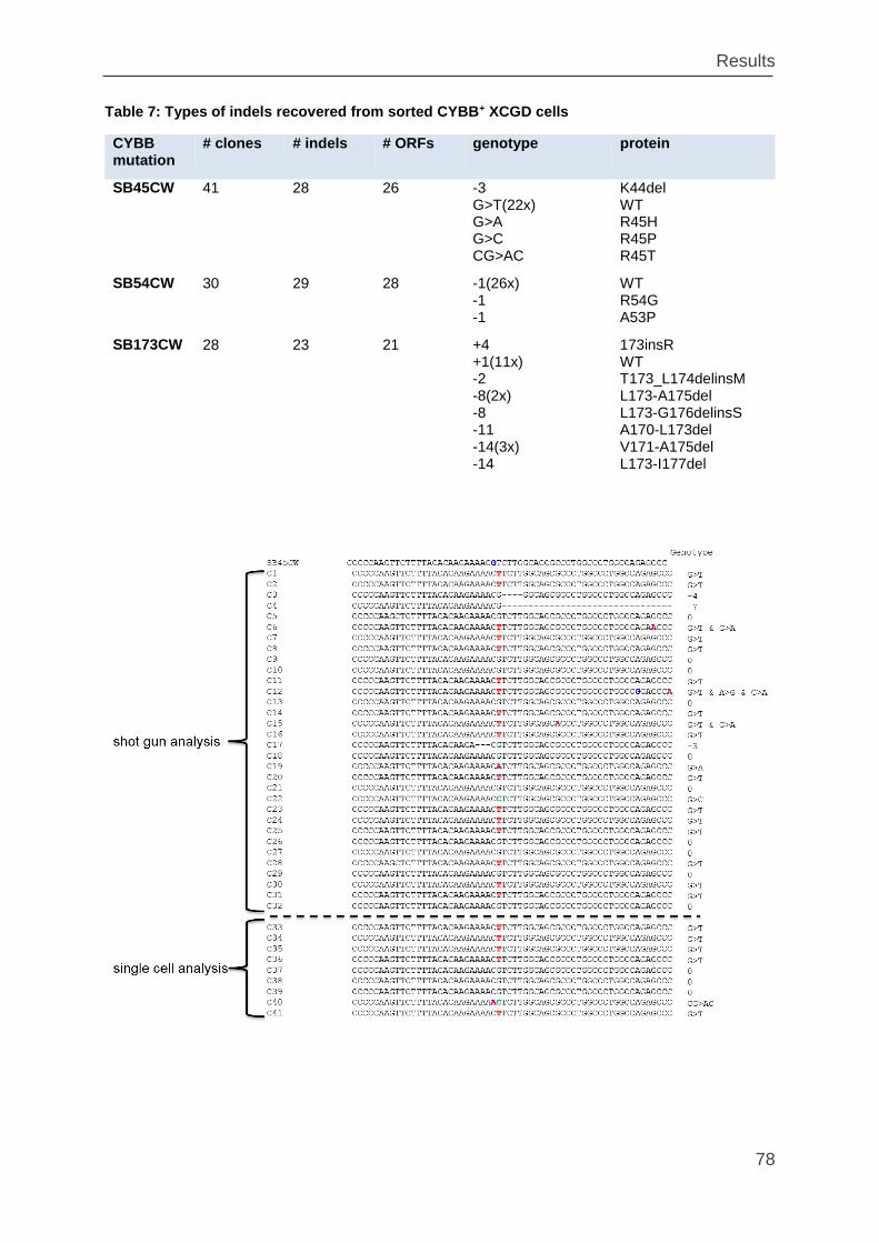
Results
78
Table 7: Types of indels recovered from sorted CYBB+ XCGD cells
CYBB mutation
# clones # indels # ORFs genotype protein
SB45CW 41 28 26 -3 G>T(22x) G>A G>C CG>AC
K44del WT R45H R45P R45T
SB54CW 30 29 28 -1(26x) -1 -1
WT R54G A53P
SB173CW 28 23 21 +4 +1(11x) -2 -8(2x) -8 -11 -14(3x) -14
173insR WT T173_L174delinsM L173-A175del L173-G176delinsS A170-L173del V171-A175del L173-I177del
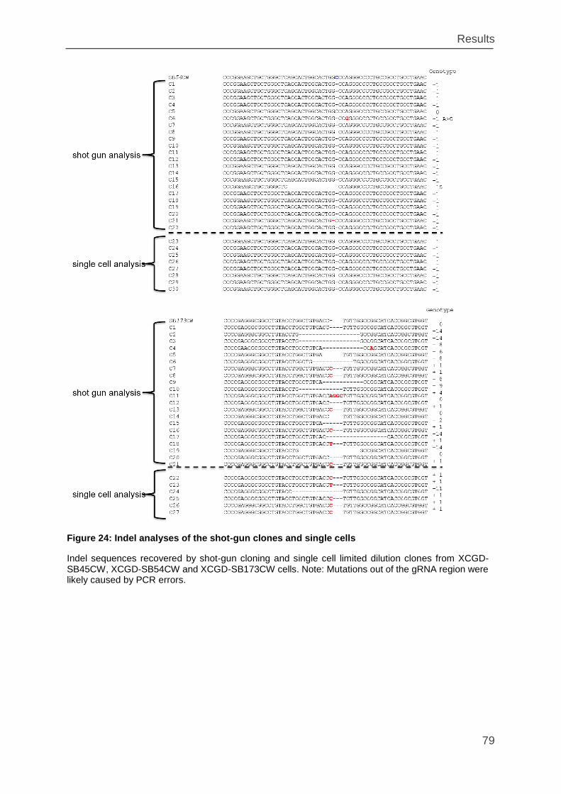
Results
79
Figure 24: Indel analyses of the shot-gun clones and single cells
Indel sequences recovered by shot-gun cloning and single cell limited dilution clones from XCGD-SB45CW, XCGD-SB54CW and XCGD-SB173CW cells. Note: Mutations out of the gRNA region were likely caused by PCR errors.
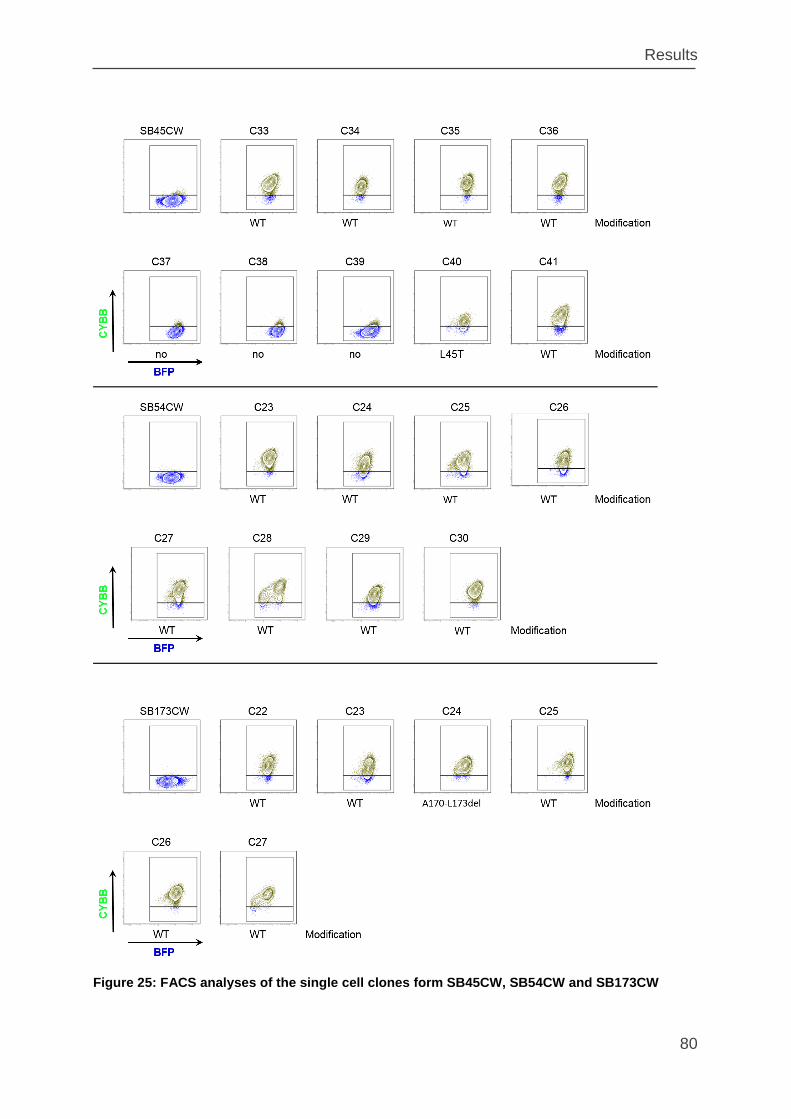
Results
80
Figure 25: FACS analyses of the single cell clones form SB45CW, SB54CW and SB173CW
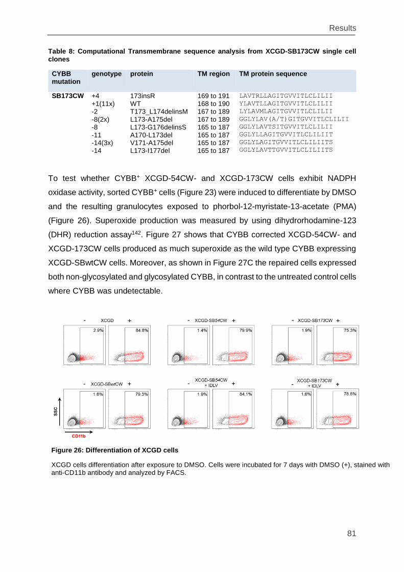
Results
81
Table 8: Computational Transmembrane sequence analysis from XCGD-SB173CW single cell clones
CYBB mutation
genotype protein TM region TM protein sequence
SB173CW +4 +1(11x) -2 -8(2x) -8 -11 -14(3x) -14
173insR WT T173_L174delinsM L173-A175del L173-G176delinsS A170-L173del V171-A175del L173-I177del
169 to 191 168 to 190 167 to 189 167 to 189 165 to 187 165 to 187 165 to 187 165 to 187
LAVTRLLAGITGVVITLCLILII
YLAVTLLAGITGVVITLCLILII
LYLAVMLAGITGVVITLCLILII
GGLYLAV(A/T)GITGVVITLCLILII
GGLYLAVTSITGVVITLCLILII
GGLYLLAGITGVVITLCLILIIT
GGLYLAGITGVVITLCLILIITS
GGLYLAVTTGVVITLCLILIITS
To test whether CYBB+ XCGD-54CW- and XCGD-173CW cells exhibit NADPH
oxidase activity, sorted CYBB+ cells (Figure 23) were induced to differentiate by DMSO
and the resulting granulocytes exposed to phorbol-12-myristate-13-acetate (PMA)
(Figure 26). Superoxide production was measured by using dihydrorhodamine-123
(DHR) reduction assay142. Figure 27 shows that CYBB corrected XCGD-54CW- and
XCGD-173CW cells produced as much superoxide as the wild type CYBB expressing
XCGD-SBwtCW cells. Moreover, as shown in Figure 27C the repaired cells expressed
both non-glycosylated and glycosylated CYBB, in contrast to the untreated control cells
where CYBB was undetectable.
Figure 26: Differentiation of XCGD cells
XCGD cells differentiation after exposure to DMSO. Cells were incubated for 7 days with DMSO (+), stained with anti-CD11b antibody and analyzed by FACS.
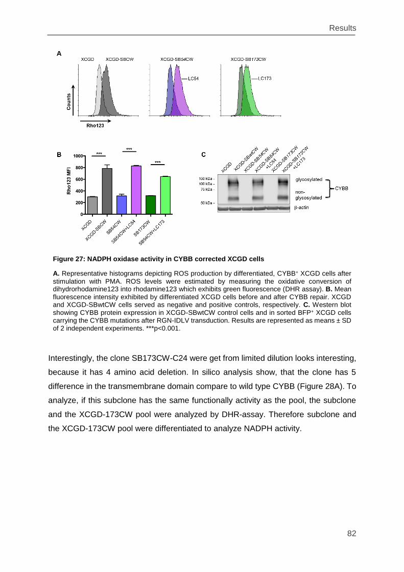
Results
82
Interestingly, the clone SB173CW-C24 were get from limited dilution looks interesting,
because it has 4 amino acid deletion. In silico analysis show, that the clone has 5
difference in the transmembrane domain compare to wild type CYBB (Figure 28A). To
analyze, if this subclone has the same functionally activity as the pool, the subclone
and the XCGD-173CW pool were analyzed by DHR-assay. Therefore subclone and
the XCGD-173CW pool were differentiated to analyze NADPH activity.
Figure 27: NADPH oxidase activity in CYBB corrected XCGD cells
A. Representative histograms depicting ROS production by differentiated, CYBB+ XCGD cells after stimulation with PMA. ROS levels were estimated by measuring the oxidative conversion of dihydrorhodamine123 into rhodamine123 which exhibits green fluorescence (DHR assay). B. Mean fluorescence intensity exhibited by differentiated XCGD cells before and after CYBB repair. XCGD and XCGD-SBwtCW cells served as negative and positive controls, respectively. C. Western blot showing CYBB protein expression in XCGD-SBwtCW control cells and in sorted BFP+ XCGD cells carrying the CYBB mutations after RGN-IDLV transduction. Results are represented as means ± SD of 2 independent experiments. ***p<0.001.
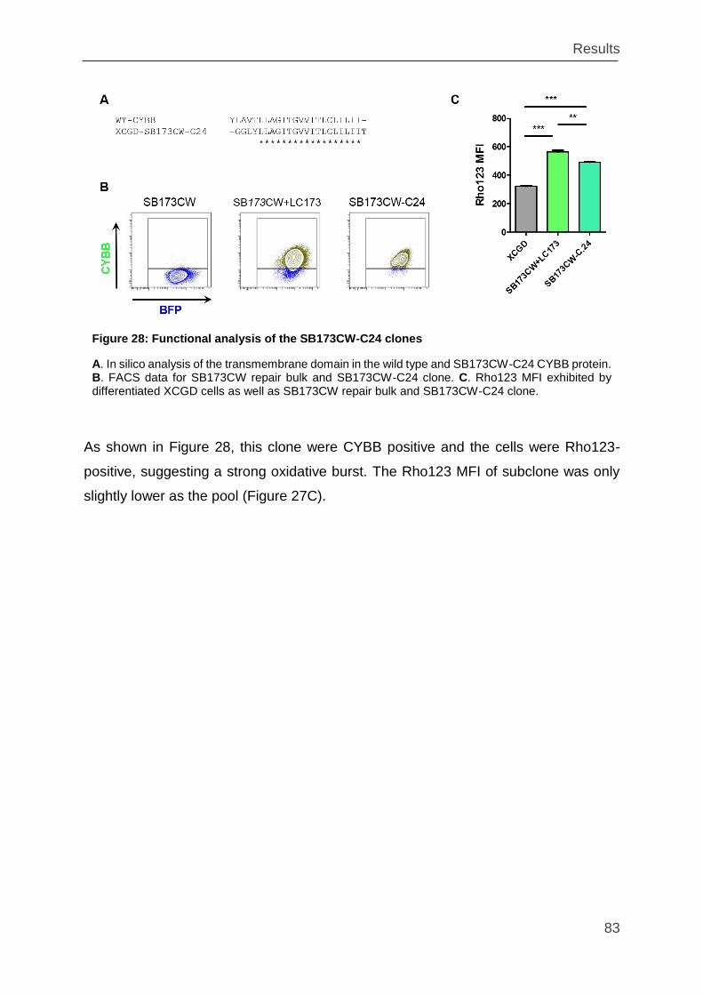
Results
83
As shown in Figure 28, this clone were CYBB positive and the cells were Rho123-
positive, suggesting a strong oxidative burst. The Rho123 MFI of subclone was only
slightly lower as the pool (Figure 27C).
Figure 28: Functional analysis of the SB173CW-C24 clones
A. In silico analysis of the transmembrane domain in the wild type and SB173CW-C24 CYBB protein. B. FACS data for SB173CW repair bulk and SB173CW-C24 clone. C. Rho123 MFI exhibited by differentiated XCGD cells as well as SB173CW repair bulk and SB173CW-C24 clone.
Results
84
3.6 Estimating on target mutation efficiency at the endogenous
CYBB locus in PLB cells
Finally, to determine whether the NHEJ gene repair strategy would work with similar
efficiency at the endogenous CYBB locus, wild type PLB cells were transduced with
LCBFP-sgCYBB LVs in which the sgRNAs targeting the R54fs and L173fs mutations
were replaced with sgRNAs targeting the corresponding wild type sequence (sgEx3
and sgEx6; Figure 29. PLB cells transduced with LVs expressing scrambled (off target)
sgRNA (LC-NTC) served as positive controls. When analyzed by flow cytometry, over
75% of the LC-sgEx3- and LC-sgEx6-LV transduced cells ceased to express CYBB
(Figure 29B) which was confirmed by Western blotting (Figure 29C). To directly
estimate the on target mutation rate, several indels recovered by shot-gun cloning from
LC-sgEx3- and LC-sgEx6-LV transduced cells were sequenced. As shown in Figure
30, 21 out of 25 exons 3 and 19 out of 25 exons 6 exhibited a mutation, suggesting in
full in agreement with the flow cytometry results an on target mutation rate of up to
75% (84% for exon 3 and 76% for exon 6). This on target mutation rate should be
sufficient to correct enough cells to protect X-CGD patients from microbial infection.
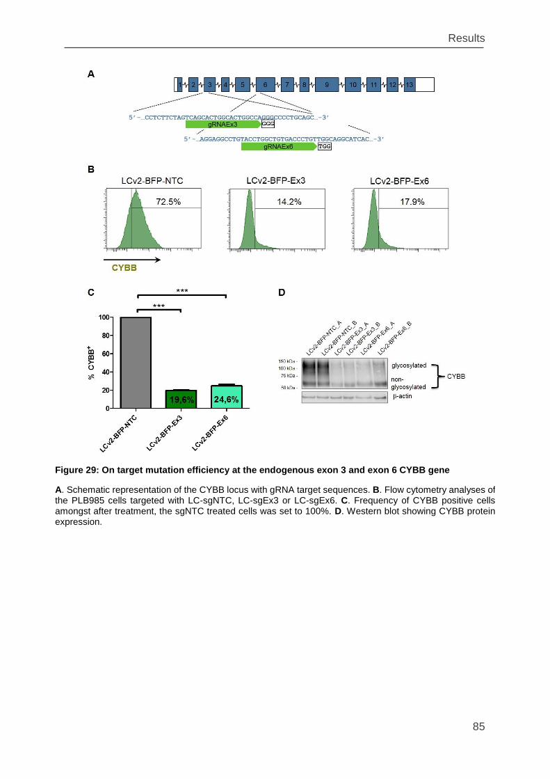
Results
85
Figure 29: On target mutation efficiency at the endogenous exon 3 and exon 6 CYBB gene
A. Schematic representation of the CYBB locus with gRNA target sequences. B. Flow cytometry analyses of the PLB985 cells targeted with LC-sgNTC, LC-sgEx3 or LC-sgEx6. C. Frequency of CYBB positive cells amongst after treatment, the sgNTC treated cells was set to 100%. D. Western blot showing CYBB protein expression.
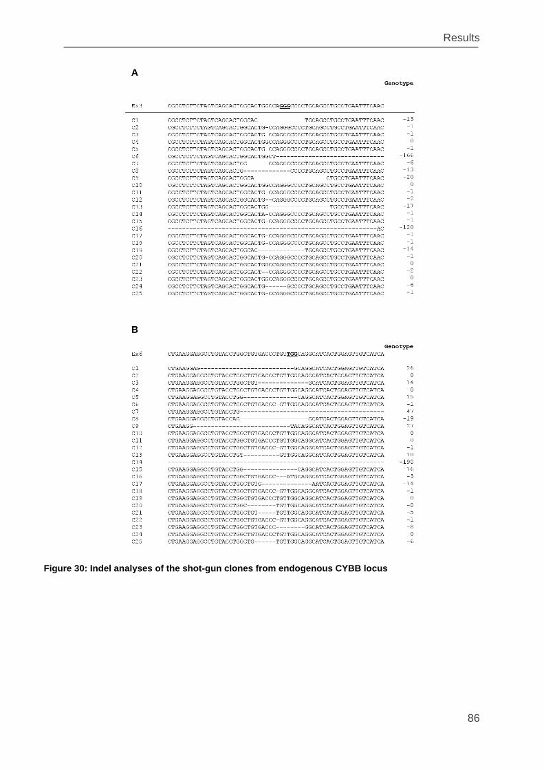
Results
86
Figure 30: Indel analyses of the shot-gun clones from endogenous CYBB locus
Discussion
87
4. Discussion
The focus of this work was to develop an RGN based strategy for in situ gene repair
of point mutations causing monogenic blood disorders.
Over 10.000 monogenic disorders are presently known of which more than 200 are
primary immune deficiencies (PIDs)10. These inherited disorders are the result of a
mutation afflicting only one gene (single gene disorder) within all cells of the body. To
date the only curative treatment for patients with PID is HLA-matched allogeneic
hematopoietic stem cell transplantation. The probability of finding a HLA-matched
donor varies among racial and ethnic groups. Patient of European descent have the
best chance of finding a matched donor (75%) whereas black patients of South or
Central American descent have the worst probability of finding a matched donor
(16%)30. For patients without a suitable HLA-matched donor, ex vivo gene therapy is
the only alternative approach.
In the last three decades over 2400 clinical gene therapy trials have been initiated
(April 2017: http://www.abedia.com/wiley/phases.php). Most trials relied on the
introduction of healthy gene copies into the diseased cells using a variety of vectors
including adenovirus-, adeno-associated virus (AAV)-, γ-retrovirus-, lentivirus-, and
Sleeping Beauty (SB) transposon vectors. Although most strategies proved effective
in terms of gene replacement, but side effects such as insertional mutagenesis leading
to clonal outgrowth and leukemia’s, combined with epigenetic silencing of the
transgene and low engraftment rate limited their widespread use and stimulated the
development of more advanced vectors with improved safety and efficacy profiles58,62.
Due to these serious side effects caused by randomly integrating gene therapy vectors,
novel strategies based on site-specific gene targeting and designer endonuclease
mediated gene editing technologies are being pursued intensely. Accordingly,
successful correction of human gene mutations by Zinc finger (ZFN) -, transcription
activator like (TAL) - or by RNA guided (Cas9) endonuclease directed homology
directed repair (HDR) in hematopoietic- and induced pluripotent stem cells has been
reported for several inherited blood disorders including X-linked severe combined
Discussion
88
immune deficiency (SCID)134,145, sickle cell anemia146,147, -thalassemia148,149 and
most recently, chronic granulomatous disease (CGD)122,150.
In this study, designer endonucleases were used to correct point-mutated transgenes
in both murine and human hematopoietic cells. To test the suitability of this approach
initial studies took advantage of the Hprt gene expressed in an endogenous locus in
32D murine hematopoietic cell line. Despite being a “safe harbor” gene whose
disruption has no functional consequences in hematopoietic cells, HPRT has the
unique advantage of enabling negative and positive selection151. Thus, to test the
efficacy of genome editing in hematopoietic cells, TAL-effector nucleases directed
against the active center of Hprt were nucleofected into 32D cells together with a
promoter-less, EGFP encoding targeting (donor) construct. After selection against Hprt
expression, less than 5% of cells still expressed EGFP. This suggests that in the
majority of the surviving cells (95%) the DSBs induced by the TAL-effector nucleases
were repaired by NHEJ and only a minority (5%) by HDR (Figure 10). This was
confirmed in single cell clones isolated by limiting dilution of which none underwent
homologous recombination.
These results are consistent with previous studies showing that DSBs repair by NHEJ
vastly exceeds DSB repair by HDR in all stages of the cell cycle152,153. In contrast to
NHEJ, HDR is nearly absent in G1 and exhibits its highest activity only during S
phase152,153. Overall, this suggests that DSBs are preferentially repaired by NHEJ
rather than by HDR (95% compared to only 5%). Even if the HDR strategy would be
the ideal procedure to directly replace the diseased genes at their endogenous loci,
due to the very low efficiency is HDR not suitable for gene therapy for monogenic
disorders. However, NHEJ rather than HDR could be a more efficient approach to gene
repair.
Nevertheless, successful corrections of human disease mutations in hematopoietic-
and induced pluripotent stem (IPS) cells by designer endonucleases have been
reported that were exclusively based on HDR (see Table 5). In most of these
applications, endonucleases and templates were delivered by nucleofection or
electroporation of several plasmids, each expressing one of the functional components
required for gene editing111,122,134,145,146,149,150. Because these procedures are highly
Discussion
89
toxic154, only a small fraction of cells survive electroporation and even fewer retain all
components required for successful genome editing. In addition to these technical
considerations155, DSB repair by HDR is down regulated in quiescent HSC’s in favor
of NHEJ156.These biological and technical factors reduce the number of cells with gene
repaired by HDR and therefore most protocols rely on positive selection to enrich for
gene-corrected cells122,146,147,149. Hence, for clinical applications, a more effective
genome editing approach would seem quite useful.
Initial clinical trials employing designer endonucleases involved AIDS patients126. In
these trials ZFN were used to target and inactivate the CCR5 receptor in T-cells which
is required for HIV infection. By generating DSBs within the CCR5 gene of T-cells via
ZNF, the CCR5 gene was knocked out after introduction of indels by NHEJ and
patients became resistant to HIV infection (clinicaltrials.gov; NCT00842634,
NCT02500849)126. While these trials demonstrated that gene therapy based on NHEJ
is a suitable approach, it remained unclear how many indels reconstituted the CCR5
open reading frame (ORF). While this seems unimportant in the context of AIDS since
T-cells with reconstituted CCR5 would automatically be selected against, the efficiency
of gene repair by NHEJ is crucial for the success of a gene therapy of inherited blood
disorders and this is exactly what the present study was designed for.
In theory, approximately one third of the indels associated with NHEJ should restore
the ORF disrupted by a disease mutation. This would lead to a significant number of
ORF reconstitutions of which some, depending on the position and type of the original
mutation, should either completely or partially recover protein function. This is
supported by the in silico analysis (Figure 11) of indels showing the majority (77%) of
ranges between15 nt deletions and 5 nt insertions (-15 nt to +5 nt). Assuming that one
third of all indels reconstitute the reading frame of a mutated gene, approximately 25%
of the targeted cells should re-express a completely or partially functional protein.
Expression of a (partially) functional protein in 25% of the affected cells is sufficient for
treatment of the most of the PIDs and a lot of other monogenic diseases.
Proof of concept for gene correction by NHEJ has been recently obtained in patients
with Duchenne’s Muscular Dystrophy (DMD)131,157. DMD is an X-linked inherited
disorder caused by large intragenic deletions of one or more exons disrupting the
Discussion
90
dystrophin ORF157. Dystrophin covers over 2.4 megabases contains 79 exons coding
for 3,685 amino acid and is the largest human genes.
Ousterout and colleagues131 could restore the ORF of the dystrophin gene in skeletal
myoblasts of patients with exons 48-50 deletion (Δ48-51) resulting in a premature stop
codon in exon 51. Using TALENs, they introduced indels into exon 51 and reconstituted
the dystrophin gene in one clone. Using patients cells with the same mutation (Δ48-
50), Moggio et al.157 pursued an alternative strategy in addition to this indel strategy.
They multiplexed two gRNAs (RGN-RGN) to remove the targeted exon 51 in patient-
derived muscle cells, thereby resetting the reading frame157. This exon-removing
strategy157 works only for exons whose removal does not significantly interfere with
protein function. Moreover, it is crucial that splicing into the adjacent downstream exon
does not disrupt the ORF which has been observed to occur in rare cases.
Unlike the dystrophin studies, the present work addressed the direct correction of point
mutations by NHEJ. Initial experiments involving the Hprt locus in 32D hematopoietic
cells confirmed that NHEJ directed DSB repair is significantly more effective than HDR.
Accordingly, 95% of TAL nuclease expressing cells obtained after negative selection
(6-TG) harbored indels in the active center of HPRT (Figure 10). Moreover, exposure
of a 6-TG resistant subclone with an indel-induced Hprt-inactivating frameshift
mutation to a second round of TALEN-directed DSB repair yielded HAT-resistant cells
derived from two independent clones - one with a synonymous substitution restoring
the Ala50 codon and one with an Arg51 to Gly51 codon conversion that only partially
inactivates HPRT (Figure 12). In human pathology this mutation is associated with the
Kelley-Seegmiller Syndrome158. Affected patients develop urinary tract stones and mild
neurological symptoms. Because this targeted mutation was in the active center of
HPRT, not many modifications were accepted. For mutations located in other areas
not as important to protein function, NHEJ repair will presumably accept more changes
such as deletions or insertions of amino acids and thus be more efficient.
Overall, these results confirm that in situ gene repair by designer endonucleases does
neither require donor templates nor the restoration of the original wild type sequence.
However, these experiments have so far yield no information about the repair
efficiency. To determine the efficiency of gene repair by NHEJ, a simple experiment
Discussion
91
that does not require a selection and has no positive or negative effect on cell growth
is sufficient. A fluorescent protein with the point mutation would be ideal for gene repair
by NHEJ. 32D cells expressing EGFP from the Hprt locus after HPRT-TALEN and
EGFP donor treatment (32D-EGFP+) were enriched and subjected to CRISPR/Cas9
mutagenesis using two different sgRNAs directed against the N-terminus of EGFP
(Figure 13). Here, TALENs were abandoned after publication of CRISPR/Cas9-based
gene editing. The reason to switch of strategy from TALEN to CRISPR/Cas9 system
was the generally poorer efficiency of TALENs and the fact that TALENs cannot cut
methylated DNA105,159. CRISPR/Cas9 system is easier to design and to clone, is more
cost and time-effective, usually has a high efficiency and the endonuclease can cut
inside both methylated and non-methylated DNA105.
Four subclones (Figure 14) with defined frameshift mutations were isolated from the
mutant pools (Figure 15). T Subclones were chosen by first taking two subclones from
each of the used sgRNA and second by selecting two subclones with the insertions
and two with the deletions of one or two nucleotides. The clones were then
nucleofected with dedicated RGNs together with Cas9 vector (two plasmid system).
Gene repair efficiencies were low ranging from 2 - 5% (Figure 15) which reflected the
overall toxicity of the electroporation gene delivery approach154. The sequences of
corrected functional EGFP shows that in the most of the cases, the EGFP ORF was
restored to its original sequence by either frame-shifting the nucleotide deletions or by
frame-reconstituting nucleotide insertions. However, this experiment showed that the
gene repair efficiency by NHEJ without any selection can be as high as the one in the
case of HDR (Figure 10) with selection. Additionally, NHEJ doesn’t need any selection,
meaning that it doesn’t require long incubation times of HSC in clinical applications.
This would probably lead to an efficient transplantation of the HSC, which undergo less
differentiation in vitro.
The next line of research focused on adopting these processes to human cells. PLB985
human acute myeloid leukemia cell line is well-suited for this purpose, because
different gene therapy approaches have demonstrated that this cell line is a powerful
tool for functional analysis of the NADPH oxidase complex. Additionally, a knock-out
CYBB model of this cell line (XCGD-PLB985 cell line) mimics a natural mutation
occurring in the X-linked Chronic Granulomatous Disease(X-CGD)142,144,160. In the first
Discussion
92
experiment, PLB leukemia cells with one copy of a mutant EGFP reporter inserted into
their genome were used to test the NHEJ approach in human cells. Using template
free RGNs delivered by integrase-defective lentiviruses (IDLVs), on-target mutation
rates approaching 100% were obtained (Figure 18 & Figure 19). Combined with the
achieved gene repair efficiency of 27% (Figure 18, section 3.4), the result suggests
that about one in four NHEJ events reconstituted EGFP function. This repair efficiency
exceeded the EGFP repair efficiency in 32D cells by almost 5- to 10-fold. The majority
of these effects can be attributed to the IDLV–RGN delivery approach. This system,
unlike the two plasmid system employed by nucleofection, delivers all components
required for gene editing within one vector by cell infection, - an approach generally
less toxic than electroporation. Unlike IDLVs which engage cell surface receptors for
cell entry, electroporation requires cell permeabilization leading to cell content leaks
adversely affecting adjacent cells. Moreover, electroporation generates random DSBs
that can cause mutations161,162. Additionally, transfected donor DNA after inserting can
integrate randomly into the genome after insertion. IDLVs express the RGNs
episomally from one or two LTR circles which are diluted out by cell replication ensuring
transient expression and preventing insertional mutagenesis163–167 – a major safety
issue in clinical trials employing integrating lentiviruses. Although between 0.35 and
2.3% of IDLVs can still integrate into the genome, depending on the IN mutation and
vector dose165,168 the probability of long term adverse effects is rather low because
constitutive Cas9 expression activates the immune system leading to elimination of
Cas9 expressing cells169. Remaining safety concerns with IDLVs could be addressed
by inserting suicide genes such as the herpes simplex virus thymidine kinase (HSV-tk)
gene enabling the elimination of HSV-tk expressing cells in vivo by administering the
pro-drug as ganciclovir170,171.
Analysis of the reconstituted open reading frames (ORFs) showed that all modification
with single amino acid deletions were compatible with EGFP fluorescence (Figure 19).
This is consistent with recent reporting showing that loss of amino acids from the N-
terminal α-helix of GFP negatively interfere with EGFP fluorescence172. Interestingly,
analysis of the reconstituted open reading frames (ORFs) showed that all but single
amino acid deletions were compatible with EGFP fluorescence. More specifically, the
short hydrophobic N-terminal α-helix of EGFP stabilizes its barrel structure which is
Discussion
93
required for fluorescence and therefore only small hydrophobic amino acids, such as
alanine are tolerated at this site172
Next, the template free IDLV-RGN strategy was tested on a real monogenic diseases.
For this purpose, X-linked chronic granulomatous disease (X-CGD) were selected. As
already described, there exists a cell line CYBB knock-out (KO) model for X-CGD
(XCGD-PLB985) and after knock-in (KI) of the CYBB gene, XCGD-PLB985 cells are a
powerful tool for functional analysis of the NADPH oxidase complex. Importantly,
neither the KO nor the KI has any effect on cell growth15. If the cells with KO or KI had
a proliferation advantage, experiments assessing on-target gene editing efficiency
would yield biased data.
To test the RGN strategy on bona fide disease genes, several patient specific CYBB
mutations causing X-CGD were selected from the Immunodeficiency mutation
databases (CYBBbase variation browser; http://structure.bmc.lu.se/idbase/
CYBBbase), including two frameshift-, one missense- and one nonsense mutation. In
vitro models for X-CGD were obtained by introducing the mutations as CYBB
transgenes into CYBB-KO PLB cells (XCGD-PLB985)144. As promyelocytic leukemia
cells expressing the NADPH oxidase complex, PLB cells were perfectly suited for
testing the RGN approach15. Treatment of the respective mutations with dedicated
RGNs showed that up to 10% of the transduced XCGD cells harbouring CYBB
frameshift mutations recovered CYBB expression and produced superoxide anions
after differentiation into granulocytes. Considering the estimated on-target mutation
rates of 30% and 26% in XCGD-SB54CW- and XCGD-SB173C cells, respectively
(Figure 22) the actual frequency of gene repairing NHEJ events is estimated to be 27%
for the SB54CW - and 38% for the SB173CW mutation (Table 9).
In comparison, both nonsense and missense CYBB mutations showed reduced on-
target mutation rates (Figure 22) which may be due in part to the low CG content of
the respective sgRNAs (Table 9)173. Moreover, as Cas9 has been shown to tolerate
single nucleotide mismatches174, selection against single nucleotide substitutions by
RGN retargeting of already corrected templates may reduce repair efficiency even
further.
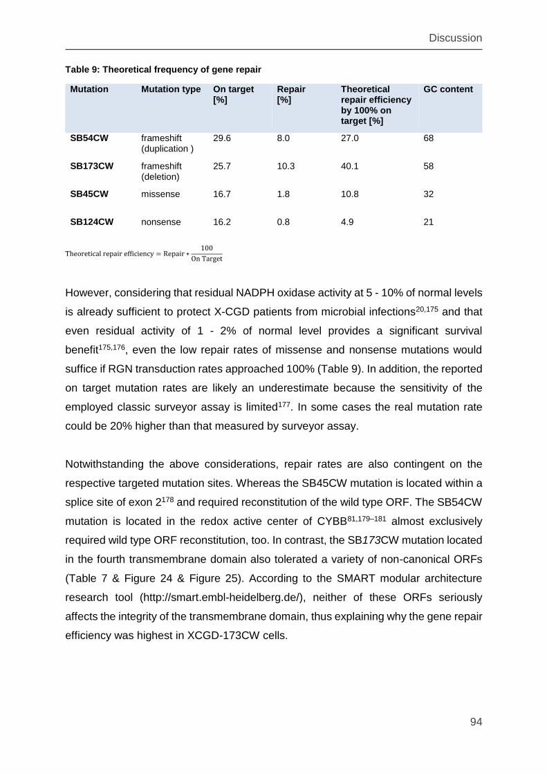
Discussion
94
Table 9: Theoretical frequency of gene repair
Mutation Mutation type On target [%]
Repair [%]
Theoretical repair efficiency by 100% on target [%]
GC content
SB54CW frameshift (duplication )
29.6 8.0 27.0 68
SB173CW frameshift (deletion)
25.7 10.3 40.1 58
SB45CW missense 16.7 1.8 10.8 32
SB124CW nonsense 16.2 0.8 4.9 21
Theoretical repair efficiency = Repair ∗100
On Target
However, considering that residual NADPH oxidase activity at 5 - 10% of normal levels
is already sufficient to protect X-CGD patients from microbial infections20,175 and that
even residual activity of 1 - 2% of normal level provides a significant survival
benefit175,176, even the low repair rates of missense and nonsense mutations would
suffice if RGN transduction rates approached 100% (Table 9). In addition, the reported
on target mutation rates are likely an underestimate because the sensitivity of the
employed classic surveyor assay is limited177. In some cases the real mutation rate
could be 20% higher than that measured by surveyor assay.
Notwithstanding the above considerations, repair rates are also contingent on the
respective targeted mutation sites. Whereas the SB45CW mutation is located within a
splice site of exon 2178 and required reconstitution of the wild type ORF. The SB54CW
mutation is located in the redox active center of CYBB81,179–181 almost exclusively
required wild type ORF reconstitution, too. In contrast, the SB173CW mutation located
in the fourth transmembrane domain also tolerated a variety of non-canonical ORFs
(Table 7 & Figure 24 & Figure 25). According to the SMART modular architecture
research tool (http://smart.embl-heidelberg.de/), neither of these ORFs seriously
affects the integrity of the transmembrane domain, thus explaining why the gene repair
efficiency was highest in XCGD-173CW cells.
Discussion
95
To determine whether the NHEJ gene repair strategy would be similarly efficient at the
endogenous locus, wild type PLB cells were treated with RGNs targeting the exons
harbouring the R54fs and L173fs mutations. In both cases, the KO analysis of CYBB
by flow cytometry and western blot (Figure 29) shows KO efficiency above 75% and
the on target mutation rate determined by sequencing of indels recovered by shot gun
cloning was also above 75%. Assuming gene repair efficiencies of about a quarter
(Figure 18), this data imply an in situ gene repair efficiency of at least 20%, which is
sufficient to protect X-CGD patients from microbial infection and thereby prevent the
major cause of death in X-CGD.
The next step to continue this work would have been final validation of the outlined
strategy in primary X-CGD patient cells, but it was not possible to obtain suitable
primary material. X-CGD is a rare disorder and most patients are severely ill, rendering
access to primary material by bone marrow aspiration or apheresis following HSPC
mobilization by G-CSF very challenging. Bone marrow aspiration is painful, complex
and carries the risk of both patient infections and contamination of material. Apheresis
is easier, but HSPC must be mobilized with recombinant granulocyte colony-
stimulating factor (G-CSF), which can have side effects such as severe bone pains,
headache, arthralgia, malaise, fatigue, insomnia nausea182. A suitable and consenting
donor could not be found in the available time, and therefore an IDLV-RGN repair in
primary human HSPCs could not be demonstrated here.
Conclusions and Outlook
96
5. Conclusions and Outlook
The present work has provided proof of concept for site directed gene repair by NHEJ
in an X-CGD model.
The data shown provide evidence for high efficiency gene repair by donor template
free CRISPR/Cas9 technology using transient, IDLV based RGN transduction into
hematopoietic cells. Unlike the widely adopted gene repair by homologous
recombination (HDR), gene repair by NHEJ requires no donor templates, and is
significantly more efficient because unlike HDR, operates in all phases of the cell cycle.
Furthermore, NHEJ is the dominant DSB-repair pathway in quiescent (G0)
HSCs153,155,156, which are the desired target cells for gene therapy of most monogenic
blood disorders. Procedurally, the single vector IDLV-RGN delivery made possible by
a template-free CRISPR/Cas9 approach is one of its most attractive technical features,
because it is transient, more efficient and less toxic than plasmid-based technologies.
Thus, gene therapy based on the template free IDLV-RGNs provides a sensible
alternative to existing gene therapy protocols. The present study has shown that
frameshift mutations can be effectively repaired in hematopoietic cells by donor
template free CRISPR/Cas9 gene editing
According to the CYBBbase database183, 24% of all X-CGD patients harbor CYBB
frameshift mutations. This frequency is similar to the frequency of frameshift mutations
found in the IL2R184,185, WASP186,187, ADA188, and HBB189,190 genes of patients with
X-linked immunodeficiency disease (X-SCID), Wiskott-Aldrich Syndrome,
adenosinedeaminase immunodeficiency disease (ADA-SCID) and -thalassemia,
respectively. Thus, at least one in four to five of these patients could benefit from gene
therapy with donor template free, RNA-guided Cas9 endonucleases. For most of these
genes, recover of less than 10% of wild type protein activity is already sufficient for
obtaining a therapeutic effect. For immunodeficiency diseases in which positive
selection occurs (e.g. X-SCID, ADA-SCID, WAS), even lower gene repair rates are
sufficient because repaired cells will overgrow diseased cells over time after
transplantation.
Conclusions and Outlook
97
However, before reaching clinical maturity the strategy needs to be validated in vivo.
For this, transgenic mice carrying humanized and patient-specific X-CGD or X-SCID
mutations produced by single embryo CRISPR/Cas9 gene editing191 will be used for
syngeneic bone marrow transplantation experiments in which ex vivo repaired
hematopoietic stem cells are injected into lethally irradiated recipients followed by long
term disease and gene expression monitoring. Special attention will be paid to potential
adverse effects caused by off-target mutations detected by dedicated reporter assays
and whole genome sequencing which will also reveal possible IDLV genome
integrations.
In a second preclinical approach which is largely dependent on patient availability (see
Discussion), hematopoietic stem and progenitor cells (HSPCs) recovered from the
peripheral blood after G-CSF mobilization will be transplanted after ex vivo gene repair
into immunodeficient NSG mice150. Successful gene repair rates and off-target effects
will then be monitored by standard methodology.
Ideally, RGN delivery for gene therapy should be transient and virus free to avoid
insertional mutagenesis and immunological side effects. Therefore, special
consideration should be given to alternative RGN delivery strategies, in the future
experiments. Most promising in this regard are advanced electroporation strategies
delivering the RGNs as preassembled gRNA/Cas9 protein complexes192,193.
Finally, the donor template free CRISPR/Cas9 approach should be easily adaptable to
non-SCID diseases such as Haemophilia or Duchenne Muscular Dystrophy.
References
98
1. References
1. Maximow, A. Der Lymphozyt als gemeinsame Stammzelle der verschiedenen Blutelemente in der
embryonalen Entwicklung und im postfetalen Leben der Säugetiere. Folia Haematol. 125–134 (1909).
2. Rieger, M. A. and & SCHROEDER, T. Hämatopoetische Stammzellen. BIOspektrum (2007).
3. Rieger, M. A. & Schroeder, T. Exploring hematopoiesis at single cell resolution. Cells Tissues Organs 188,
139–149 (2008).
4. Smith, C. Hematopoietic stem cells and hematopoiesis. Cancer Control 10, 9–16 (2003).
5. Rieger, M. A. & Schroeder, T. Hematopoiesis. Cold Spring Harb Perspect Biol 4, (2012).
6. Gunsilius, E., Gastl, G. & Petzer, A. L. Hematopoietic stem cells. Biomed. Pharmacother. 55, 186–194
(2001).
7. WHO. http://www.who.int/genomics/public/geneticdiseases/en/index2.html at
<http://www.who.int/genomics/public/geneticdiseases/en/index2.html>
8. Farhan, S. M. K. & Hegele, R. a. Genetics 101 for Cardiologists: Rare Genetic Variants and Monogenic
Cardiovascular Disease. Can. J. Cardiol. 29, 18–22 (2013).
9. Kaufmann, K. B., Büning, H., Galy, A., Schambach, A. & Grez, M. Gene therapy on the move. EMBO Mol.
Med. 5, 1642–1661 (2013).
10. Nonas, S. Pulmonary Manifestations of Primary Immunodeficiency Disorders. Immunol. Allergy Clin. North
Am. 35, 753–766 (2015).
11. Cavazzana-calvo, M. & Fischer, A. Gene therapy for severe combined immunodeficiency : are we there
yet ? Sci. Med. 117, 1456–1465 (2007).
12. Al-Herz, W. et al. Primary immunodeficiency diseases: An update on the classification from the International
Union of immunological societies expert committee for primary immunodeficiency. Front. Immunol. 5, 1–33
(2014).
13. Lim, M. S. & Elenitoba-johnson, K. S. J. The Molecular Pathology of Primary Immunodeficiencies. 6, 59–
83 (2004).
14. M.L., L. et al. Applying public health strategies to primary immunodeficiency diseases: a potential approach
to genetic disorders. MMWR. Recomm. Rep. 53, 1–29 (2004).
15. Aiuti, A., Bacchetta, R., Seger, R., Villa, A. & Cavazzana-Calvo, M. Gene therapy for primary
immunodeficiencies: Part 2. Curr. Opin. Immunol. 24, 585–591 (2012).
16. Aiuti, A. et al. Lentiviral Hematopoietic Stem Cell Gene Therapy in Patients with Wiskott-Aldrich Syndrome.
Science (80-. ). 341, 1233151 (2013).
17. Jin, Y. et al. Mutations of the Wiskott-Aldrich Syndrome Protein (WASP): hotspots, effect on transcription,
and translation and phenotype/genotype correlation. Blood 104, 4010–9 (2004).
18. Rudd, C. E. Disabled receptor signaling and new primary immunodeficiency disorders. N. Engl. J. Med.
354, 1874–1877 (2006).
19. Rae, J. et al. X-Linked chronic granulomatous disease: mutations in the CYBB gene encoding the gp91-
phox component of respiratory-burst oxidase. Am J Hum Genet 62, 1320–1331 (1998).
20. Becker, S. et al. Correction of respiratory burst activity in X-linked chronic granulomatous cells to
therapeutically relevant levels after gene transfer into bone marrow CD34+ cells. Hum Gene Ther 9, 1561–
1570 (1998).
21. Pessach, I. M. & Notarangelo, L. D. Gene therapy for primary immunodeficiencies: Looking ahead, toward
gene correction. J. Allergy Clin. Immunol. 127, 1344–1350 (2011).
22. Antonarakis, S. E., Krawczak, M. & Cooper, D. N. Disease-causing mutations in the human genome. Eur J
Pediatr 159 Suppl, S173-8 (2000).
References
99
23. Mercadier, J. How do mutations cause disease ? 34–37 (2008).
24. Frischmeyer, P. a & Dietz, H. C. Nonsense-mediated mRNA decay in health and disease. Hum. Mol. Genet.
8, 1893–1900 (1999).
25. Kuzmiak, H. A. & Maquat, L. E. Applying nonsense-mediated mRNA decay research to the clinic: progress
and challenges. Trends Mol. Med. 12, 306–316 (2006).
26. Henig, I. & Zuckerman, T. Hematopoietic stem cell transplantation-50 years of evolution and future
perspectives. Rambam Maimonides Med. J. 5, e0028 (2014).
27. Cavazzana-Calvo, M., Fischer, A., Hacein-Bey-Abina, S. & Aiuti, A. Gene therapy for primary
immunodeficiencies: Part 1. Curr. Opin. Immunol. 24, 580–584 (2012).
28. Champlin, R. E. et al. Blood stem cells compared with bone marrow as a source of hematopoietic cells for
allogeneic transplantation. IBMTR Histocompatibility and Stem Cell Sources Working Committee and the
European Group for Blood and Marrow Transplantation (EBMT). Blood 95, 3702–3709 (2000).
29. Gennery, A. R. et al. Transplantation of hematopoietic stem cells and long-term survival for primary
immunodeficiencies in Europe: Entering a new century, do we do better? J. Allergy Clin. Immunol. 126,
(2010).
30. Gragert, L. et al. HLA Match Likelihoods for Hematopoietic Stem-Cell Grafts in the U.S. Registry. N. Engl.
J. Med. 371, 339–348 (2014).
31. Nayerossadat, N., Ali, P. & Maedeh, T. Viral and nonviral delivery systems for gene delivery. Adv. Biomed.
Res. 1, 27 (2012).
32. Tros de Ilarduya, C., Sun, Y. & Düzgünes, N. Gene delivery by lipoplexes and polyplexes. Eur. J. Pharm.
Sci. 40, 159–170 (2010).
33. Manjila, S. B. et al. Novel gene delivery systems. Int. J. Pharm. Investig. 3, 1–7 (2013).
34. Xiang, S. et al. Uptake mechanisms of non-viral gene delivery. J. Control. Release 158, 371–378 (2012).
35. Skipper, K., Andersen, P., Sharma, N. & Mikkelsen, J. DNA transposon-based gene vehicles - scenes from
an evolutionary drive. J. Biomed. Sci. 20, 92 (2013).
36. Bouuaert, C. C. & Chalmers, R. M. Gene therapy vectors: The prospects and potentials of the cut-and-
paste transposons. Genetica 138, 473–484 (2010).
37. Ivics, Z., Izsvák, Z., Medrano, G., Chapman, K. M. & Hamra, F. K. Sleeping Beauty transposon mutagenesis
in rat spermatogonial stem cells. Nat. Protoc. 6, 1521–1535 (2011).
38. Field, A.-C. et al. Comparison of lentiviral and sleeping beauty mediated αβ T cell receptor gene transfer.
PLoS One 8, e68201 (2013).
39. Gogol-Doring, A. et al. Genome-wide Profiling Reveals Remarkable Parallels Between Insertion Site
Selection Properties of the MLV Retrovirus and the piggyBac Transposon in Primary Human CD4(+) T
Cells. Mol. Ther. 24, 592–606 (2016).
40. Ivics, Z., Hackett, P. B., Plasterk, R. H. & Izsvák, Z. Molecular Reconstruction of Sleeping Beauty, a Tc1-
like Transposon from Fish, and Its Transposition in Human Cells. Cell 91, 501–510 (1997).
41. Izsvak, Z., Chuah, M. K. L., VandenDriessche, T. & Ivics, Z. Efficient stable gene transfer into human cells
by the Sleeping Beauty transposon vectors. Methods 49, 287–297 (2009).
42. Johnen, S. et al. Antiangiogenic and Neurogenic Activities of Sleeping Beauty-Mediated PEDF-Transfected
RPE Cells In Vitro and In Vivo. Biomed Res. Int. 2015, 863845 (2015).
43. Bouard, D., Alazard-Dany, D. & Cosset, F.-L. Viral vectors: from virology to transgene expression. Br. J.
Pharmacol. 157, 153–65 (2009).
44. Mali, S. Delivery systems for gene therapy. Indian J. Hum. Genet. 19, 3–8 (2013).
45. Robbins, P. D. & Ghivizzani, S. C. Viral Vectors for Gene Therapy. Pharmacol. Ther. 80, 35–47 (1998).
46. Gardlík, R. et al. Vectors and delivery systems in gene therapy. Med. Sci. Monit. 11, RA110-A121 (2005).
References
100
47. Kootstra, N. A. & Verma, I. M. Gene therapy with viral vectors. Annu. Rev. Pharmacol. Toxicol. 43, 413–
439 (2003).
48. Deyle, D. R. & Russell, D. W. Adeno-associated virus vector integration. Curr. Opin. Mol. Ther. 11, 442–7
(2009).
49. Nault, J.-C. et al. Recurrent AAV2-related insertional mutagenesis in human hepatocellular carcinomas.
Nat. Genet. 47, 1187–1193 (2015).
50. Russell, D. W. & Grompe, M. Adeno-associated virus finds its disease. Nat. Genet. 47, 1104–1105 (2015).
51. Lech, P. & Somia, N. Retrovirus vectors. Contrib. Nephrol. 159, 30–46 (2008).
52. Baum, C., Schambach, A., Bohne, J. & Galla, M. Retrovirus Vectors: Toward the Plentivirus? Mol. Ther.
13, 1050–1063 (2006).
53. El Ashkar, S. et al. Engineering Next-Generation BET-Independent MLV Vectors for Safer Gene Therapy.
Mol. Ther. - Nucleic Acids 7, 231–245 (2017).
54. Stein, S. et al. From Bench to Bedside: Preclinical Evaluation of a Self-Inactivating Gammaretroviral Vector
for the Gene Therapy of X-linked Chronic Granulomatous Disease. Hum. Gene Ther. Clin. Dev. 24, 86–98
(2013).
55. De Ravin, S. S. et al. Lentiviral hematopoietic stem cell gene therapy for X-linked severe combined
immunodeficiency. Sci. Transl. Med. 8, 335ra57 (2016).
56. Farinelli, G. et al. Lentiviral vector gene therapy protects XCGD mice from acute Staphylococcus aureus
pneumonia and inflammatory response. Mol. Ther. (2016). doi:10.1038/mt.2016.150
57. Spanevello, F. et al. Development of Lentiviral Vectors Simultaneously Expressing Multiple siRNAs Against
CCR5, vif and tat/rev Genes for an HIV-1 Gene Therapy Approach. Mol. Ther. Acids 5, e312 (2016).
58. Naldini, L. Ex vivo gene transfer and correction for cell-based therapies. Nat Rev Genet 12, 301–315 (2011).
59. Mitchell, R. S. et al. Retroviral DNA integration: ASLV, HIV, and MLV show distinct target site preferences.
PLoS Biol. 2, (2004).
60. Cattoglio, C. et al. High-definition mapping of retroviral integration sites identifies active regulatory elements
in human multipotent hematopoietic progenitors High-definition mapping of retroviral integration sites
identifies active regulatory elements in human multipotent. Gene Ther. 116, 5507–5517 (2011).
61. Qasim, W., Gaspar, H. B. & Thrasher, A. J. Progress and prospects: gene therapy for inherited
immunodeficiencies. Gene Ther. 16, 1285–1291 (2009).
62. Stein, S. et al. Genomic instability and myelodysplasia with monosomy 7 consequent to EVI1 activation
after gene therapy for chronic granulomatous disease. Nat Med 16, 198–204 (2010).
63. Modlich, U. et al. Insertional transformation of hematopoietic cells by self-inactivating lentiviral and
gammaretroviral vectors. Mol Ther 17, 1919–1928 (2009).
64. Zychlinski, D. et al. Physiological promoters reduce the genotoxic risk of integrating gene vectors. Mol Ther
16, 718–725 (2008).
65. Frecha, C., Szécsi, J., Cosset, F.-L. & Verhoeyen, E. Strategies for targeting lentiviral vectors. Curr. Gene
Ther. 8, 449–460 (2008).
66. Cesana, D. et al. Uncovering and dissecting the genotoxicity of self-inactivating lentiviral vectors in vivo.
Mol. Ther. 22, 774–85 (2014).
67. Ginn, S. L., Alexander, I. E., Edelstein, M. L., Abedi, M. R. & Wixon, J. Gene therapy clinical trials worldwide
to 2012 - an update. Journal of Gene Medicine 15, 65–77 (2013).
68. Naldini, L. Gene therapy returns to centre stage. Nature 526, 351–360 (2015).
69. Hacein-Bey Abina, S. et al. Outcomes following gene therapy in patients with severe Wiskott-Aldrich
syndrome. JAMA 313, 1550–63 (2015).
70. Hacein-Bey-Abina, S. et al. A modified γ-retrovirus vector for X-linked severe combined immunodeficiency.
References
101
N. Engl. J. Med. 371, 1407–17 (2014).
71. Hacein-Bey-Abina, S. et al. Efficacy of gene therapy for X-linked severe combined immunodeficiency. N.
Engl. J. Med. 363, 355–64 (2010).
72. Hacein-bey-abina, S. et al. Insertional oncogenesis in 4 patients after retrovirus-mediated gene therapy of
SCID-X1. - PubMed - NCBI. 118, (2008).
73. Aiuti, A. et al. Gene therapy for immunodeficiency due to adenosine deaminase deficiency. N. Engl. J. Med.
360, 447–58 (2009).
74. Malech, H. L. et al. Prolonged production of NADPH oxidase-corrected granulocytes after gene therapy of
chronic granulomatous disease. Proc. Natl. Acad. Sci. U. S. A. 94, 12133–8 (1997).
75. Ott, M. G. et al. Correction of X-linked chronic granulomatous disease by gene therapy, augmented by
insertional activation of MDS1-EVI1, PRDM16 or SETBP1. Nat Med 12, 401–409 (2006).
76. Aiuti, A. & Roncarolo, M. G. Ten years of gene therapy for primary immune deficiencies. Hematol. Am Soc
Hematol Educ Progr. 682–689 (2009). doi:10.1182/asheducation-2009.1.682
77. Candotti, F. Gene transfer into hematopoietic stem cells as treatment for primary immunodeficiency
diseases. Int. J. Hematol. 99, 383–392 (2014).
78. Rawat, A., Bhattad, S. & Singh, S. Chronic Granulomatous Disease. Indian J. Pediatr. 83, 345–53 (2016).
79. Holland, S. M. Chronic Granulomatous Disease. Clin. Rev. Allergy Immunol. 38, 3–10 (2009).
80. Chiriaco, M., Salfa, I., Matteo, G. Di, Rossi, P. & Finocchi, A. Chronic Granulomatous Disease: clinical,
molecular and therapeutic aspects. Pediatr. Allergy Immunol. 1–12 (2015). doi:10.1111/pai.12527
81. Yu, L., Quinn, M., Cross, A. & Dinauer, M. Gp91 phox is the heme binding subunit of the superoxide-
generating NADPH oxidase. Proc. Natl. Acad. Sci. U. S. A. 95, 7993–7998 (1998).
82. Seger, R. A. Modern management of chronic granulomatous disease. Br J Haematol 140, 255–266 (2008).
83. Kaufmann, K. B. et al. Alpharetroviral Vector-mediated Gene Therapy for X-CGD: Functional Correction
and Lack of Aberrant Splicing. Mol. Ther. 21, 648–661 (2012).
84. Kang, E. M. et al. Retrovirus gene therapy for X-linked chronic granulomatous disease can achieve stable
long-term correction of oxidase activity in peripheral blood neutrophils. Blood 115, 783–791 (2010).
85. Grez, M. et al. Gene therapy of chronic granulomatous disease: the engraftment dilemma. Mol. Ther. 19,
28–35 (2011).
86. Schwäble, J., Schultze-Strasser, S., Stein, S. & Grez, M. [Gene therapy for septic granulomatosis: risks
and opportunities. The road to successful treatment of congenital immunodeficiency]. Pharm. Unserer Zeit
40, 264–271 (2011).
87. Bianchi, M. et al. Restoration of NET formation by gene therapy in CGD controls aspergillosis. Blood 114,
2619–2622 (2009).
88. Kim JG, Ahn HS, Kang HJ, Kim S, Hong YT, J. C. Retroviral Gene Therapy for X-Linked Chronic
Granulomatous Disease: Results from Phase I/II Trial. Blood 112:2349, (2008).
89. Stone, D., Niyonzima, N. & Jerome, K. R. Genome editing and the next generation of antiviral therapy.
Hum. Genet. 135, 1071–1082 (2016).
90. Hoban, M. D. & Bauer, D. E. A genome editing primer for the hematologist. Blood 127, blood-2016-01-
678151 (2016).
91. Johansen, S., Embley, T. M. & Willassen, N. P. A family of nuclear homing endonucleases. Nucleic Acids
Res. 21, 4405 (1993).
92. Belfort, M. & Bonocora, R. P. Homing endonucleases: from genetic anomalies to programmable genomic
clippers. Methods Mol. Biol. 1123, 1–26 (2014).
93. Hafez, M., Hausner, G. & Bonen, L. Homing endonucleases: DNA scissors on a mission. Genome 55, 553–
569 (2012).
References
102
94. Stoddard, B. L. Homing endonucleases: From microbial genetic invaders to reagents for targeted DNA
modification. Structure 19, 7–15 (2011).
95. Kim, Y. G., Cha, J. & Chandrasegaran, S. Hybrid restriction enzymes: zinc finger fusions to Fok I cleavage
domain. Proc Natl Acad Sci U S A 93, 1156–1160 (1996).
96. Gabriel, R. et al. An unbiased genome-wide analysis of zinc-finger nuclease specificity. Nat Biotechnol 29,
816–823 (2011).
97. Urnov, F. D., Rebar, E. J., Holmes, M. C., Zhang, H. S. & Gregory, P. D. Genome editing with engineered
zinc finger nucleases. Nat Rev Genet 11, 636–646 (2010).
98. Carroll, D. Genome engineering with zinc-finger nucleases. Genetics 188, 773–782 (2011).
99. Boch, J. TALEs of genome targeting. Nat. Publ. Gr. 29, 135–136 (2011).
100. Cermak, T. et al. Erratum: Efficient design and assembly of custom TALEN and other TAL effector-based
constructs for DNA targeting (Nucleic Acids Research (2011) 39 (e82) DOI: 10.1093/nar/gkr218). Nucleic
Acids Res. 39, 7879 (2011).
101. Bogdanove, A. J., Schornack, S. & Lahaye, T. TAL effectors: finding plant genes for disease and defense.
Curr Opin Plant Biol 13, 394–401 (2010).
102. Mashimo, T. et al. Efficient gene targeting by TAL effector nucleases coinjected with exonucleases in
zygotes. Sci Rep 3, 1253 (2013).
103. Barrangou, R. et al. CRISPRProvides Acquired Resistance Against Viruses in Prokaryotes. Science (80-. ).
315, 1709–1712 (2007).
104. Ishino, Y., Shinagawa, H., Makino, K., Amemura, M. & Nakata, A. Nucleotide sequence of the iap gene,
responsible for alkaline phosphatase isozyme conversion in Escherichia coli, and identification of the gene
product. J. Bacteriol. 169, 5429–33 (1987).
105. Hsu, P. D. et al. DNA targeting specificity of RNA-guided Cas9 nucleases. Nat Biotechnol 31, 827–832
(2013).
106. Li, J. et al. Efficient inversions and duplications of mammalian regulatory DNA elements and gene clusters
by CRISPR / Cas 9. 7, 284–298 (2015).
107. Cong, L. et al. Multiplex Genome Engineering Using CRISPR/Cas Systems. 339, 819–823 (2013).
108. Jinek, M. et al. RNA-programmed genome editing in human cells. Elife 2013, 1–9 (2013).
109. Stella, S. & Montoya, G. The genome editing revolution: A CRISPR-Cas TALE off-target story. Insid. Cell
n/a-n/a (2015). doi:10.1002/icl3.1038
110. Hsu, P. D., Lander, E. S. & Zhang, F. Development and applications of CRISPR-Cas9 for genome
engineering. Cell 157, 1262–1278 (2014).
111. Cox, D. B., Platt, R. J. & Zhang, F. Therapeutic genome editing: prospects and challenges. Nat Med 21,
121–131 (2015).
112. Ran, F. A. et al. Genome engineering using the CRISPR-Cas9 system. Nat Protoc 8, 2281–2308 (2013).
113. Gaj, T., Gersbach, C. A. & Barbas 3rd, C. F. ZFN, TALEN, and CRISPR/Cas-based methods for genome
engineering. Trends Biotechnol 31, 397–405
114. Yin, H., Kauffman, K. J. & Anderson, D. G. Delivery technologies for genome editing. Nat. Rev. Drug Discov.
16, 387–399 (2017).
115. Hart, T. et al. High-Resolution CRISPR Screens Reveal Fitness Genes and Genotype-Specific Cancer
Liabilities. Cell 163, 1515–1526 (2015).
116. Tzelepis, K. et al. A CRISPR Dropout Screen Identifies Genetic Vulnerabilities and Therapeutic Targets in
Acute Myeloid Leukemia. Cell Rep. 17, 1193–1205 (2016).
117. Wang, T. et al. Gene Essentiality Profiling Reveals Gene Networks and Synthetic Lethal Interactions with
Oncogenic Ras. Cell 168, 890–903.e15 (2017).
References
103
118. Zhang, C. & Saunders, A. J. An emerging role for Ubiquilin 1 in regulating protein quality control system
and in disease pathogenesis. Discov. Med. 8, 18–22 (2009).
119. Young, C. S. et al. A Single CRISPR-Cas9 Deletion Strategy that Targets the Majority of DMD Patients
Restores Dystrophin Function in hiPSC-Derived Muscle Cells. Cell Stem Cell 18, 533–540 (2016).
120. Paquet, D. et al. Efficient introduction of specific homozygous and heterozygous mutations using
CRISPR/Cas9. Nature 1–18 (2016). doi:10.1038/nature17664
121. Xue, W. et al. CRISPR-mediated direct mutation of cancer genes in the mouse liver. Nature 514, 380–384
(2014).
122. Flynn, R. et al. CRISPR-mediated genotypic and phenotypic correction of a chronic granulomatous disease
mutation in human iPS cells. Exp. Hematol. 43, 838–848.e3 (2015).
123. Wu, Y. et al. Correction of a genetic disease by CRISPR-Cas9-mediated gene editing in mouse
spermatogonial stem cells. Cell Res 25, 67–79 (2014).
124. Wu, Y. et al. Correction of a genetic disease in mouse via use of CRISPR-Cas9. Cell Stem Cell 13, 659–
662 (2013).
125. Li, H. et al. In vivo genome editing restores haemostasis in a mouse model of haemophilia. Nature 475,
217–221 (2011).
126. Tebas, P. et al. Gene editing of CCR5 in autologous CD4 T cells of persons infected with HIV. N. Engl. J.
Med. 370, 901–910 (2014).
127. Perez, E. E. et al. Editing Using Zinc-Finger Nucleases. 26, 808–816 (2012).
128. Holt, N. et al. Human hematopoietic stem/progenitor cells modified by zinc-finger nucleases targeted to
CCR5 control HIV-1 in vivo. Nat Biotechnol 28, 839–847 (2010).
129. Ye, L. et al. Seamless modification of wild-type induced pluripotent stem cells to the natural CCR5Δ32
mutation confers resistance to HIV infection. Proc. Natl. Acad. Sci. 111, 9591–9596 (2014).
130. Long, C. et al. Prevention of muscular dystrophy in mice by CRISPR/Cas9-mediated editing of germline
DNA. Science (80-. ). 345, 1184–1188 (2014).
131. Ousterout, D. G. et al. Reading frame correction by targeted genome editing restores dystrophin expression
in cells from Duchenne muscular dystrophy patients. Mol Ther 21, 1718–1726
132. Bloom, K., Ely, A., Mussolino, C., Cathomen, T. & Arbuthnot, P. Inactivation of Hepatitis B Virus Replication
in Cultured Cells and In Vivo with Engineered Transcription Activator-Like Effector Nucleases. Mol. Ther.
21, 1889–1897 (2013).
133. Lin, S.-R. et al. The CRISPR/Cas9 System Facilitates Clearance of the Intrahepatic HBV Templates In Vivo.
Mol. Ther. - Nucleic Acids 3, e186 (2014).
134. Genovese, P. et al. Targeted genome editing in human repopulating haematopoietic stem cells. Nature
510, 235–240
135. Schwank, G. et al. Functional repair of CFTR by CRISPR/Cas9 in intestinal stem cell organoids of cystic
fibrosis patients. Cell Stem Cell 13, 653–658 (2013).
136. Yin, H. et al. Genome editing with Cas9 in adult mice corrects a disease mutation and phenotype. Nat.
Biotechnol. 32, 551–553 (2014).
137. Demaison, C. et al. High-level transduction and gene expression in hematopoietic repopulating cells using
a human immunodeficiency [correction of imunodeficiency] virus type 1-based lentiviral vector containing
an internal spleen focus forming virus promoter. Hum. Gene Ther. 13, 803–813 (2002).
138. Subach, O. M. et al. Conversion of red fluorescent protein into a bright blue probe. Chem. Biol. 15, 1116–
24 (2008).
139. Engler, C., Kandzia, R. & Marillonnet, S. A one pot, one step, precision cloning method with high throughput
capability. PLoS One 3, (2008).
References
104
140. Abràmofff, M. D., Magalhães, P. J. & Ram, S. J. Image processing with ImageJ Part II. Biophotonics Int.
11, 36–43 (2005).
141. Yamauchi, A. et al. Location of the epitope for 7D5, a monoclonal antibody raised against human
flavocytochrome b558, to the extracellular peptide portion of primate gp91phox. Microbiol. Immunol. 45,
249–57 (2001).
142. Brendel, C., Kaufmann, K. B., Krattenmacher, A., Pahujani, S. & Grez, M. Generation of X-CGD cells for
vector evaluation from healthy donor CD34(+) HSCs by shRNA-mediated knock down of gp91(phox). Mol
Ther Methods Clin Dev 1, 14037 (2014).
143. Sanjana, N. E., Shalem, O. & Zhang, F. Improved vectors and genome-wide libraries for CRISPR screening.
Nat. Methods 11, 783–784 (2014).
144. Zhen, L. et al. Gene targeting of X chromosome-linked chronic granulomatous disease locus in a human
myeloid leukemia cell line and rescue by expression of recombinant gp91phox. Proc Natl Acad Sci U S A
90, 9832–9836 (1993).
145. Urnov, F. D. et al. Highly efficient endogenous human gene correction using designed zinc-finger
nucleases. Nature 435, 646–651 (2005).
146. Sebastiano, V. & Maeder, M. In situ genetic correction of the sickle cell anemia mutation in human induced
pluripotent stem cells using engineered zinc finger nucleases. Stem … 29, 1717–1726 (2011).
147. Sun, N. & Zhao, H. Seamless correction of the sickle cell disease mutation of the HBB gene in human
induced pluripotent stem cells using TALENs. Biotechnol. Bioeng. 111, 1048–1053 (2014).
148. Xie, F. et al. Seamless gene correction of ??-thalassemia mutations in patient-specific iPSCs using
CRISPR/Cas9 and piggyBac. Genome Res. 24, 1526–1533 (2014).
149. Ma, N. et al. Transcription activator-like effector nuclease (TALEN)-mediated Gene correction in integration-
free β-Thalassemia induced pluripotent stem cells. J. Biol. Chem. 288, 34671–34679 (2013).
150. De Ravin, S. S. et al. CRISPR-Cas9 gene repair of hematopoietic stem cells from patients with X-linked
chronic granulomatous disease. Sci. Transl. Med. 9, (2017).
151. Choudhary, R., Baturin, D., Fosmire, S., Freed, B. & Porter, C. C. Knockdown of HPRT for selection of
genetically modified human hematopoietic progenitor cells. PLoS One 8, e59594 (2013).
152. Mao, Z., Bozzella, M., Seluanov, A. & Gorbunova, V. DNA repair by nonhomologous end joining and
homologous recombination during cell cycle in human cells. Cell Cycle 7, 2902–6 (2008).
153. Newman, E. A. et al. Alternative NHEJ Pathway Components Are Therapeutic Targets in High-Risk
Neuroblastoma. Mol. Cancer Res. 13, 470–482 (2015).
154. Piñero, J., López-Baena, M., Ortiz, T. & Cortés, F. Apoptotic and necrotic cell death are both induced by
electroporation in HL60 human promyeloid leukaemia cells. Apoptosis 2, 330–336 (1997).
155. Mao, Z., Bozzella, M., Seluanov, A. & Gorbunova, V. DNA repair by nonhomologous end joining and
homologous recombination during cell cycle in human cells. Cell Cycle 7, 2902–2906 (2008).
156. Beerman, I., Seita, J., Inlay, M. A., Weissman, I. L. & Rossi, D. J. Quiescent hematopoietic stem cells
accumulate DNA damage during aging that is repaired upon entry into cell cycle. Cell Stem Cell 15, 37–50
(2014).
157. Maggio, I. et al. Selection-free gene repair after adenoviral vector transduction of designer nucleases:
Rescue of dystrophin synthesis in DMD muscle cell populations. Nucleic Acids Res. 44, 1449–1470 (2015).
158. Jinnah, H. A., De Gregorio, L., Harris, J. C., Nyhan, W. L. & O’Neill, J. P. The spectrum of inherited
mutations causing HPRT deficiency: 75 new cases and a review of 196 previously reported cases. Mutat.
Res. - Rev. Mutat. Res. 463, 309–326 (2000).
159. Valton, J. et al. Overcoming transcription activator-like effector (TALE) DNA binding domain sensitivity to
cytosine methylation. J. Biol. Chem. 287, 38427–38432 (2012).
References
105
160. Dreyer, A. et al. Biomaterials TALEN-mediated functional correction of X-linked chronic granulomatous
disease in patient-derived induced pluripotent stem cells. Biomaterials 69, 191–200 (2015).
161. Meaking, W. S., Edgerton, J., Wharton, C. W. & Meldrum, R. A. Electroporation-induced damage in
mammalian cell DNA. BBA - Gene Struct. Expr. 1264, 357–362 (1995).
162. Goldberg, A. & Rubinsky, B. The effect of electroporation type pulsed electric fields on DNA in aqueous
solution. Technol. Cancer Res. Treat. 9, 423–430 (2010).
163. Hoban, M. D. et al. Correction of the sickle cell disease mutation in human hematopoietic stem / progenitor
cells. Blood 125, 2597–2604 (2015).
164. Joglekar, A. V et al. Integrase-defective lentiviral vectors as a delivery platform for targeted modification of
adenosine deaminase locus. Mol Ther 21, 1705–17 (2013).
165. Nightingale, S. J. et al. Transient Gene Expression by Nonintegrating Lentiviral Vectors. Mol. Ther. 13,
1121–1132 (2006).
166. Banasik, M. B. & McCray, P. B. Integrase-defective lentiviral vectors: progress and applications. Gene Ther.
17, 150–157 (2010).
167. Wanisch, K. & Yáñez-Muñoz, R. J. Integration-deficient lentiviral vectors: a slow coming of age. Mol. Ther.
17, 1316–1332 (2009).
168. Cornu, T. I. & Cathomen, T. Targeted Genome Modifications Using Integrase‐deficient Lentiviral Vectors.
Mol. Ther. 15, 2107–2113 (2007).
169. Chew, W. L. et al. A multifunctional AAV–CRISPR–Cas9 and its host response. Nat. Methods 13, 868–874
(2016).
170. Zarogoulidis, P. et al. Suicide Gene Therapy for Cancer – Current Strategies. J Genet Syndr Gene Ther
(2013). doi:10.4172/2157-7412.1000139.Suicide
171. McCormick, F. Cancer Gene Therapy: Fringe or Cutting Edge? Nat. Rev. Cancer 1, 130 (2001).
172. Saeger, J., Hytönen, V. P., Klotzsch, E. & Vogel, V. GFP’s Mechanical Intermediate States. PLoS One 7,
(2012).
173. Wang, T., Wei, J. J., Sabatini, D. M. & Lander, E. S. Genetic screens in human cells using the CRISPR-
Cas9 system. Science (80-. ). 343, 80–84
174. Fu, Y. et al. High frequency off-target mutagenesis induced by CRISPR-Cas nucleases in human cells. Nat.
Biotechnol 31, 822–826 (2014).
175. Dinauer, M. C., Gifford, M. A., Pech, N., Li, L. L. & Emshwiller, P. Variable correction of host defense
following gene transfer and bone marrow transplantation in murine X-linked chronic granulomatous disease.
Blood 97, 3738–3745 (2001).
176. Kuhns, D. B. et al. Residual NADPH oxidase and survival in chronic granulomatous disease. N. Engl. J.
Med. 363, 2600–10 (2010).
177. Vouillot, L., Thelie, A. & Pollet, N. Comparison of T7E1 and surveyor mismatch cleavage assays to detect
mutations triggered by engineered nucleases. G3 5, 407–415 (2015).
178. Roos, D. et al. Hematologically important mutations: X-linked chronic granulomatous disease (third update).
Blood Cells. Mol. Dis. 45, 246–65 (2010).
179. O’Neill, S., Brault, J., Stasia, M. J. & Knaus, U. G. Genetic disorders coupled to ROS deficiency. Redox
Biol. 6, 135–156 (2015).
180. Cross, A. R., Rae, J. & Curnutte, J. T. Cytochrome b-245 of the neutrophil superoxide-generating system
contains two nonidentical hemes. Potentiometric studies of a mutant form of gp91phox. J Biol Chem 270,
17075–17077 (1995).
181. Cross, A. R., Heyworth, P. G., Rae, J. & Curnutte, J. T. A variant X-linked chronic granulomatous disease
patient (X91+) with partially functional cytochrome b. J. Biol. Chem. 270, 8194–200 (1995).
References
106
182. Stroncek, D. & McCullough, J. Safeguarding the long-term health of hematopoietic stem cell donors: a
continuous and evolving process to maintain donor safety and trust. Expert Rev. Hematol. 5, 1–3 (2012).
183. CYBBbase database. http://structure.bmc.lu.se/idbase/CYBBbase/browser.php?content=browser (2017).
184. Puck, J. M. et al. Mutation analysis of IL2RG in human X-linked severe combined immunodeficiency. Blood
89, 1968–77 (1997).
185. IL2RG database. https://research.cchmc.org/LOVD2/home.php?select_db=IL2RG (2017).
186. WASP database. http://pidj.rcai.riken.jp/waspbase/ (2017).
187. Jin, Y. et al. Mutations of the Wiskott-Aldrich Syndrome Protein (WASP): Hotspots, effect on transcription,
and translation and phenotype/genotype correlation. Blood 104, 4010–4019 (2004).
188. ADAbase Database. http://structure.bmc.lu.se/idbase/ADAbase/index.php?content=index/IDbases (2017).
189. Giardine, B. et al. Updates of the HbVar database of human hemoglobin variants and thalassemia
mutations. Nucleic Acids Res. 42, 1–10 (2014).
190. HBB Database. https://lovd.bx.psu.edu/home.php (2017).
191. Wefers, B. et al. Direct production of mouse disease models by embryo microinjection of TALENs and
oligodeoxynucleotides. Proc. Natl. Acad. Sci. 110, 3782–3787 (2013).
192. DeWitt, M. A., Corn, J. E. & Carroll, D. Genome editing via delivery of Cas9 ribonucleoprotein. Methods 3–
9 (2017). doi:10.1016/j.ymeth.2017.04.003
193. Hendel, A. et al. Chemically modified guide RNAs enhance CRISPR-Cas genome editing in human primary
cells. Nat. Biotechnol. 33, (2015).

Acknowledgments
107
Acknowledgments
Concluding my dissertation, I would like to thank all the people who contributed the
success of this work during the last years.
First, I would like to express my special gratitude and thanks to my supervisor Prof. Dr.
Harald von Melchner, he has been a highly appreciated mentor for me. I would like to
thank him for encouraging my research and for helping me to grow as a research
scientist.
In particular I would like to thank Prof. Dr. Beatrix Süß and Prof. Dr. M. Cristina Cardoso
for being my supervisors on the TU Darmstadt and for thier kind advising, supporting
and discussing my doctoral thesis.
I am deeply grateful to PD. Dr. Frank Schnütgen for his mentoring during my work in
the laboratory, for his directional instructions, advices and valuable discussion about
my doctoral dissertation.
It’s my great pleasure to express my appreciation to Prof. Dr. Hubert Serve, head of
the Department of Hematology and Oncology Goethe-University Frankfurt, for giving
me the opportunity to conduct my doctoral study in the field of Hematology.
Furthermore, I am greatly indebted to express my deepest thanks to a number of
individuals who deserve special acknowledgement: Ms. Anna-Maria Scheder for the
great support in all matters, without her many things would have been heavier; Dr. Ana
Tomasovic for her advices, generous friendship, psychological support and excellent
proof reading of the manuscript; Dr. Stefan Stein for his continuous encouragement;
Many others at our department who have also been generous with their time, support
and encouragement at various stages during my PhD including Dr. Frank Wempe, Dr.
Nina Kurrle, Dr. Olesya Vakhrusheva, Sarah Weber, Florian Gatzke, Christine
Schönfeld, Jenny Bleeck, Heike Nürnberger, Johanna Kreitz, Melanie Forster, Roy
Ehling, The Duy Nguyen, Nathalie Guillen, Sebastian Koschade and other colleagues
for the support and the nice atmosphere.
I would like to thank my friends Sabine Arens, Ali Bildik and Meded Bildik. They have
been there forever helping me with their wisdoms.
Finally, heartfelt thanks to my parents Mustafa and Elif Sürün, my sisters Hamide,
Fatma, Emine and my brother Ali for their continuous love and encouragement
throughout the years.
Curriculum vitae
108
Curriculum vitae
Duran Sürün
Address: Lerchenstraße 6
63150 Heusenstamm
Phone number: +49178-2314333
E-mail: [email protected]
Date of birth: January 1st 1983
Nationality: German
WORK EXPERIENCE & EDUCATION
Mai 2012 – present PhD Thesis at Goethe-University in the department of Hematology and Oncology, Frankfurt am Main In the Laboratory of Prof. Dr. Harald von Melchner. Dissertation title: “High Efficiency Gene Correction in Hematopoietic Cells by Donor Template-free CRISPR/Cas9 Genome Editing”
Oct 2011 – Mai 2012 Scientific assistant at Georg-Speyer-Haus In the Laboratory of Dr. Manuel Grez, Dr. Manuel Grez
Oct 2010 – Oct 2011 Master thesis in Georg-Speyer-Haus In the Laboratory of Dr. Manuel Grez Grade 1.1 Master title: „Sleeping Beauty – Vektoren für die genetische Modifikation hämatopoetischer Stammzellen“
Oct 2009 – Oct 2011 Master of science at University Gelsenkirchen in Molecular Biology Grade 1.4
Apr 2009 – Aug 2009 Bachelor thesis at Heinrich-Pette Institut In the Laboratory of Prof. Dr. Wolfgang Deppert Grade 1.0
Oct 2006 – Sept 2009 Bachelor of schience in Molecular Biology, University Gelsenkirchen Grade 2.3
Sept 2003 – Jun 2006 Training as Chemical-technical assistant at Hans-Böckler Berufskolleg, Marl Grade 2.5
Sept 2003 – Jun 2006 High school graduation at Hans-Böckler Berufskolleg, Marl Grade 2.5
TEACHING EXPERIENCE
Apr 2008 – Dec 2009 Student Assistant in University Gelsenkirchen in the department of biochemistry, microbiology and laboratory practice
Feb 2015 Students seminar "The renewable man?" at the EKHN Stiftung, Frankfurt am Main, Germany
Publications
109
Publications and Abstracts
CONFERENCES AND AWARDS
Aug 2017 Oral presentation at the 46th Annual scientific International Society for Experimental Hemantology (ISEH) meeting, Frankfurt, Germany
Dec 2016 Poster presentation at the 58th Annual American Society for Hematology (ASH) meeting, San Diego, USA
Nov 2016 Poster presentation at the Nature Herrenhausen Symposium Genome Editing for Gene and Cell Therapy meeting, Hanover, Germany
Oct 2015 Poster price at the Summer School of the LOEWE Centre for Cell and Gene Therapy, Langen, Germany
Form 2012 – 2016 Poster presentation at the Annual meeting of the German Society for Gene Therapy (DG-GT)
PUBLICATION
Sürün D, Schwäble J, Tomasovic A, Roy E, Stein S, Kurrle A, Kühn A, von Melchner H and
Schnütgen F High Efficiency Gene Correction in Hematopoietic Cells by Donor-Template-free CRISPR/Cas9 Genome Editing Molecular Therapy - Nucleic Acids, in Revision
Cencioni C, Spallotta F, Savoia M, Kuenne C, Guenther S, Re A, Wingert S, Rehage M, Sürün D, Siragusa M, Smith JG, Schnütgen F, von Melchner F, Rieger M, Martelli F, Riccio A, Fleming I, Braun T, Zeiher AM, Farsetti A and Gaetano C A Zeb1 Hdac2 eNOS feedback circuitry identifies early cardiovascular precursors in naïve mouse embryonic stem cells Nature Communications, in Revision
Spallotta F, Cencioni C, Atlante S, Garella D, Cocco M, Mori M, Mastrocola R, Kuenne C, Guenther S, Nanni S, Azzimato V, Zukunft S, Kornberger A, Sürün D, Schnütgen F, von Melchner H, Di Stilo A, Aragno M, De Blasio M, Ritchie RH, Zaccagnini G , Martelli F, Farsetti A, Fleming I, Braun T, Beiras-Fernandez A , Botta B, Collino M, Bertinaria M, Zeiher AM, Gaetano C Stable oxidative cytosine modifications accumulate in cardiac mesenchymal cells from Type2 diabetes patients: rescue by alpha-ketoglutarate and TET-TDG functional reactivation Circulation Research, submitted
Nguyen TD, Shaid S, Vakhrusheva O, Koschade S, Zhang J, Oellerich T, Sürün D, Derlet A, Osiewacz H, Behrends C, Dikic I, and Brandts C The selective autophagy receptor p62 is required for mitophagy and promotes leukemia development The Journal of Experimental Medicine, submitted
Ball AK, Beilstein K, Wittmann S, Sürün D, Saul MJ, Schnütgen F, Flamand N, Capelo R, Kahnt AS, Frey H, Schaefer L, Marschalek R, Häfner AK, Steinhilber D Characterization and cellular localization of human 5-lipoxygenase and its protein isoforms 5-LOΔ13, 5-LOΔ4 and 5-LOp12 Biochim Biophys Acta.2017; 1862(5):561-571
Publications
110
Tomasovic A, Kurrle N, Wempe F, De-Zolt S, Scheibe S, Koli K, Serchinger M, Schnütgen F, Sürün D, Sterner-Kock A, Weissmann N, von Melchner H Ltbp4 regulates Pdgfrβ expression via TGFβ-dependent modulation of Nrf2 transcription factor function Matrix Biol. 2017; 59:109-120.
Mahshid Y, Markoutsa S, Dincbas-Renqvist V, Sürün D, Christensson B, Sander B, Björkholm M, Sorg BL, Rådmark O, Claesson HE Phosphorylation of serine 523 on 5-lipoxygenase in human B lymphocytes Prostaglandins Leukot Essent Fatty Acids 2015; 100:33-40
Tomasovic A, Kurrle N, Sürün D, Heidler J, Husnjak K, Poser I, Schnütgen F, Scheibe S, Seimetz M, Jaksch P, Hyman A, Weissmann N, von Melchner H Sestrin 2 protein regulates platelet-derived growth factor receptor β (Pdgfrβ) expression by modulating proteasomal and Nrf2 transcription factor functions J Biol Chem. 2015; 10;290(15):9738-52
Markoutsa S, Sürün D, Karas M, Hofmann B, Steinhilber D, Sorg BL Analysis of 5-lipoxygenase phosphorylation on molecular level by MALDI-MS FEBS J. 2014; 281(8):1931-47
Ehrenwörtliche Erklärung
111
Ehrenwörtliche Erklärung:
Ich erkläre hiermit ehrenwörtlich, dass ich die vorliegende Arbeit entsprechend den
Regeln guter wissenschaftlicher Praxis selbstständig und ohne unzulässige Hilfe
Dritter angefertigt habe.
Sämtliche aus fremden Quellen direkt oder indirekt übernommenen Gedanken sowie
sämtliche von Anderen direkt oder indirekt übernommenen Daten, Techniken und
Materialien sind als solche kenntlich gemacht. Die Arbeit wurde bisher bei keiner
anderen Hochschule zu Prüfungszwecken eingereicht.
Darmstadt, den 01.11.2017
..............................................................................
Duran Sürün