Embed Size (px)
Citation preview
REVIEW AND PERSPECTIVE
KRAS mutation testing for predicting response to anti-EGFRtherapy for colorectal carcinoma: proposal for an Europeanquality assurance program
J. H. J. M. van Krieken & A. Jung & T. Kirchner &
F. Carneiro & R. Seruca & F. T. Bosman & P. Quirke &
J. F. Fléjou & T. Plato Hansen & G. de Hertogh & P. Jares &
C. Langner & G. Hoefler & M. Ligtenberg & D. Tiniakos &
S. Tejpar & G. Bevilacqua & A. Ensari
Received: 24 June 2008 /Accepted: 21 August 2008 / Published online: 18 September 2008# The Author(s) 2008. This article is published with open access at Springerlink.com
Abstract Novel therapeutic agents targeting the epidermalgrowth factor receptor (EGFR) have improved outcomes forpatients with colorectal carcinoma. However, these therapiesare effective only in a subset of patients. Activating mutationsin the KRAS gene are found in 30–40% of colorectal tumorsand are associated with poor response to anti-EGFR therapies.Thus, KRASmutation status can predict which patient may ormay not benefit from anti-EGFR therapy. Although manydiagnostic tools have been developed for KRAS mutationanalysis, validated methods and standardized testing proce-dures are lacking. This poses a challenge for the optimal use of
anti-EGFR therapies in the management of colorectal carci-noma. Here we review the molecular basis of EGFR-targetedtherapies and the resistance to treatment conferred by KRASmutations. We also present guideline recommendations and aproposal for a European quality assurance program to helpensure accuracy and proficiency in KRAS mutation testingacross the European Union.
Keywords Colorectal carcinoma . Anti-EGFR therapy .
KRAS mutation testing . Practice guidelines .
Quality assurance
J. H. J. M. van Krieken (*) :M. LigtenbergDepartment of Pathology,Radboud University Nijmegen Medical Centre,PO Box 9101, Nijmegen 6500 HB, The Netherlandse-mail: [email protected]
A. Jung : T. KirchnerDepartment of Pathology, Ludwig-Maximilians Universität,Munich, Germany
F. Carneiro :R. SerucaInstitute of Molecular Pathology and Immunology of theUniversity of Porto (IPATIMUP),Porto, Portugal
F. Carneiro :R. SerucaMedical Faculty of the of Porto,Porto, Portugal
F. CarneiroDepartment of Pathology, Hospital S. João,Porto, Portugal
F. T. BosmanUniversity Institute of Pathology,Lausanne, Switzerland
P. QuirkePathology and Tumour Biology,University of Leeds,Leeds, England
J. F. FléjouDepartment of Pathology, Saint-Antoine Hospital,University Paris 6,Paris, France
T. Plato HansenDepartment of Pathology, Odense University Hospital,Odense, Denmark
G. de HertoghDepartment of Pathology, University Hospitals KU Leuven,Leuven, Belgium
P. JaresDepartment of Pathology,Hospital Clinic,Barcelona, Spain
C. Langner :G. HoeflerInstitute of Pathology, Medical University Graz,Graz, Austria
Virchows Arch (2008) 453:417–431DOI 10.1007/s00428-008-0665-y
Introduction
Novel classes of therapeutic agents for treating cancer arerapidly changing clinical practice. Several of these newdrugs target specific molecules expressed by cancer cells.One group targets members of the human epidermalgrowth factor receptor (HER) family, namely, theepidermal growth factor receptor (EGFR) and the humanepidermal growth factor receptor 2 (HER2). Both EGFRand HER2 contribute to the development and progressionof several cancers and therefore have been explored astargets for cancer therapy. To apply targeted therapiesoptimally, it is important to recognize that their activitydiffers across patient populations and to understand themolecular mechanisms underlying these differences.
A well-defined example of how the efficacy of a targetedtherapy can vary among patients with different molecularprofiles is the use of trastuzumab (Herceptin®), an anti-HER2 monoclonal antibody, in the treatment of breastcancer. HER2 is overexpressed in 20–30% of malignantbreast tumors as a result of amplification of the coding gene[1, 2]. HER2-positive status is associated with poorprognosis and is a strong predictor of response to trastuzu-mab therapy [1, 3]. Assessment of HER2 status has becomestandard practice to identify breast cancer patients mostlikely to benefit from trastuzumab therapy [3]. In parallel,substantial progress has been made to validate HER2testing methods and implement quality assurance to ensureconsistency and accuracy in HER2 testing [4].
Targeted therapeutic agents have also been developed forthe treatment of colorectal cancer, a leading cause ofcancer-related deaths worldwide [5]. The majority ofpatients with colorectal cancer are diagnosed with locally
advanced or metastatic disease which responds poorly toconventional forms of treatment. The drugs recentlyintroduced for treating colorectal cancer target the EGFR,which is overexpressed in 50–80% of colorectal tumors [6–10]. Although the advent of EGFR-targeted therapies hasimproved outcomes for colorectal cancer patients, they areeffective in only a subset of patients [11]. Therefore, amajor challenge in optimizing EGFR-targeted treatmentoptions in colorectal cancer is to identify reliable bio-markers that can predict which patients will or will notrespond to these targeted therapies.
It has become clear that mutations in the Kirsten RAS(KRAS) gene negatively predict success of anti-EGFR thera-pies. Gain-of-function KRAS mutations lead to EGFR-independent activation of intracellular signaling pathways,resulting in tumor cell proliferation, protection against apopto-sis, increased invasion and metastasis, and activation of tumor-induced angiogenesis [12]. Unlike HER2 testing in breastcancer, however, there is a wide variety of testing methods anda lack of quality assurance schemes for the assessment ofKRAS mutation status in colorectal cancer patients.
The objectives of this paper are threefold: (1) to reviewthe molecular basis of EGFR-targeted therapies and theresistance to treatment conferred by KRAS mutations; (2) tosummarize the different methods available for the detectionof KRAS mutations; and (3) to propose guideline recom-mendations and a European quality assurance (QA)program for KRAS mutation testing in colorectal carcinoma.
Molecular basis of EGFR-targeted therapies
EGFR and cancer
EGFR is a 170-kDa transmembrane tyrosine kinase receptorthat is present in most epithelial tissues and plays animportant role in cell growth and function. Modulation ofgrowth factor receptors, such as the EGFR, is a key strategyused by tumor cells to become self-sufficient and rely less ongrowth signals for their transformation, proliferation andsurvival. EGFR is overexpressed in many solid cancers andhas been shown by many studies to be involved in thedevelopment and progression of human malignancies [12,13]. Extensive research over the last few years has improvedour understanding of the oncogenic role of the EGFR and themechanisms of receptor activation and function. Theseadvances have led to the development of new treatmentmodalities aimed at targeting the EGFR signaling system.
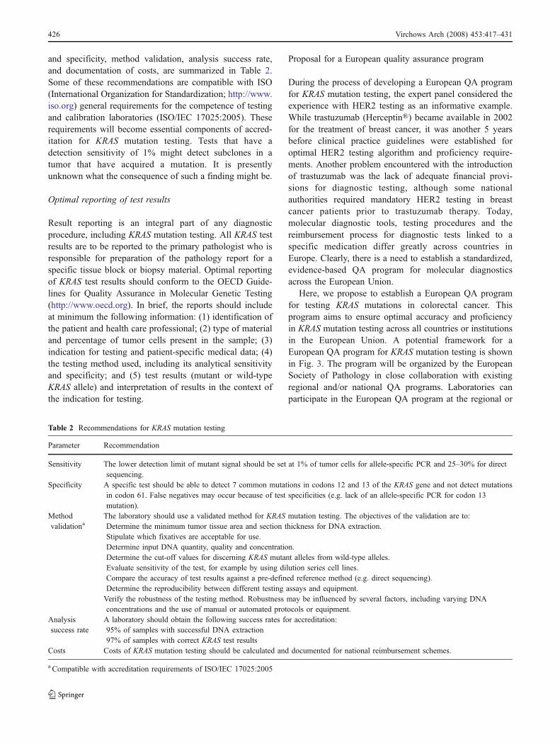
EGFR belongs to HER family of cell surface receptors(see Fig. 1a). The HER receptor family consists of fourstructurally related proteins: EGFR (also called HER1/ErbB1), HER2 (ErbB2), HER3 (ErbB3), and HER4(ErbB4). Each receptor is composed of three domains: (1)
M. LigtenbergDepartment of Human Genetics,Radboud University Nijmegen Medical Centre,Nijmegen, The Netherlands
D. TiniakosLaboratory of Histology and Embryology,Medical School University of Athens,Athens, Greece
S. TejparDigestive Oncology Unit, University Hospital Gasthuisberg,Leuven, Belgium
G. BevilacquaDepartment of Oncology,University of Pisa and Pisa University Hospital,Pisa, Italy
A. EnsariDepartment of Pathology,Ankara University Medical School,Ankara, Turkey
418 Virchows Arch (2008) 453:417–431
an extracellular domain that recognizes and binds ligandsspecifically, such as epidermal growth factor (EGF), trans-forming growth factor (TGF)-α and amphiregulin whichbind specifically to EGFR; (2) a hydrophobic transmem-
brane domain that is involved in interactions between cellsurface receptors; and (3) an intracellular domain thatserves as a site of tryosine kinase activity. There are at leasttwo exceptions to these general principles: HER2 has noknown ligand and is constitutively active, and HER3 doesnot possess intrinsic tyrosine kinase activity. However, allreceptors and their specific ligands interact to form anintegrated system in which an initial signal can be amplifiedand diversified into multiple cellular responses.
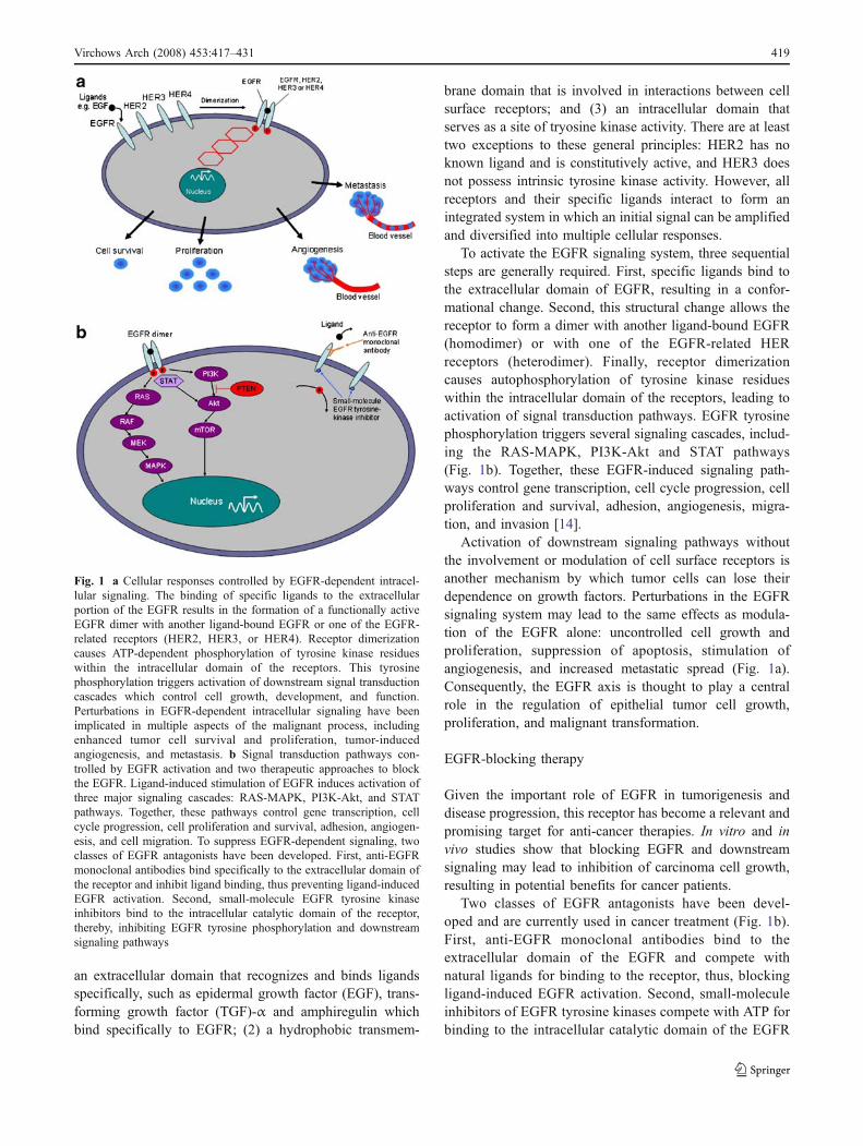
To activate the EGFR signaling system, three sequentialsteps are generally required. First, specific ligands bind tothe extracellular domain of EGFR, resulting in a confor-mational change. Second, this structural change allows thereceptor to form a dimer with another ligand-bound EGFR(homodimer) or with one of the EGFR-related HERreceptors (heterodimer). Finally, receptor dimerizationcauses autophosphorylation of tyrosine kinase residueswithin the intracellular domain of the receptors, leading toactivation of signal transduction pathways. EGFR tyrosinephosphorylation triggers several signaling cascades, includ-ing the RAS-MAPK, PI3K-Akt and STAT pathways(Fig. 1b). Together, these EGFR-induced signaling path-ways control gene transcription, cell cycle progression, cellproliferation and survival, adhesion, angiogenesis, migra-tion, and invasion [14].
Activation of downstream signaling pathways withoutthe involvement or modulation of cell surface receptors isanother mechanism by which tumor cells can lose theirdependence on growth factors. Perturbations in the EGFRsignaling system may lead to the same effects as modula-tion of the EGFR alone: uncontrolled cell growth andproliferation, suppression of apoptosis, stimulation ofangiogenesis, and increased metastatic spread (Fig. 1a).Consequently, the EGFR axis is thought to play a centralrole in the regulation of epithelial tumor cell growth,proliferation, and malignant transformation.
EGFR-blocking therapy
Given the important role of EGFR in tumorigenesis anddisease progression, this receptor has become a relevant andpromising target for anti-cancer therapies. In vitro and invivo studies show that blocking EGFR and downstreamsignaling may lead to inhibition of carcinoma cell growth,resulting in potential benefits for cancer patients.
Two classes of EGFR antagonists have been devel-oped and are currently used in cancer treatment (Fig. 1b).First, anti-EGFR monoclonal antibodies bind to theextracellular domain of the EGFR and compete withnatural ligands for binding to the receptor, thus, blockingligand-induced EGFR activation. Second, small-moleculeinhibitors of EGFR tyrosine kinases compete with ATP forbinding to the intracellular catalytic domain of the EGFR
Fig. 1 a Cellular responses controlled by EGFR-dependent intracel-lular signaling. The binding of specific ligands to the extracellularportion of the EGFR results in the formation of a functionally activeEGFR dimer with another ligand-bound EGFR or one of the EGFR-related receptors (HER2, HER3, or HER4). Receptor dimerizationcauses ATP-dependent phosphorylation of tyrosine kinase residueswithin the intracellular domain of the receptors. This tyrosinephosphorylation triggers activation of downstream signal transductioncascades which control cell growth, development, and function.Perturbations in EGFR-dependent intracellular signaling have beenimplicated in multiple aspects of the malignant process, includingenhanced tumor cell survival and proliferation, tumor-inducedangiogenesis, and metastasis. b Signal transduction pathways con-trolled by EGFR activation and two therapeutic approaches to blockthe EGFR. Ligand-induced stimulation of EGFR induces activation ofthree major signaling cascades: RAS-MAPK, PI3K-Akt, and STATpathways. Together, these pathways control gene transcription, cellcycle progression, cell proliferation and survival, adhesion, angiogen-esis, and cell migration. To suppress EGFR-dependent signaling, twoclasses of EGFR antagonists have been developed. First, anti-EGFRmonoclonal antibodies bind specifically to the extracellular domain ofthe receptor and inhibit ligand binding, thus preventing ligand-inducedEGFR activation. Second, small-molecule EGFR tyrosine kinaseinhibitors bind to the intracellular catalytic domain of the receptor,thereby, inhibiting EGFR tyrosine phosphorylation and downstreamsignaling pathways
Virchows Arch (2008) 453:417–431 419
tyrosine kinase. This competition inhibits EGFR tyrosinephosphorylation and hence suppresses downstream signalingpathways.
Two anti-EGFR antibodies (cetuximab and panitumu-mab) and two small-molecule EGFR tyrosine kinaseinhibitors (gefitinib and erlotinib) have been evaluatedextensively for the treatment of colorectal cancer, metastaticnon-small-cell lung cancer, squamous-cell carcinoma of thehead and neck, and pancreatic cancer where malignanttransformation depends on EGFR signaling [12]. Addition-al EGFR-targeting agents, including monoclonal antibod-ies, small molecules and vaccines, are currently underinvestigation [15].
EGFR and colorectal cancer
Several lines of evidence have demonstrated a role forEGFR in colorectal tumorigenesis. Preclinical data suggestthat EGFR mRNA expression and EGF levels are higher inmalignant areas of colorectal tumors than in the surround-ing benign mucosa (as reviewed by Lockhart and Berlin[16]). In experimental models of colon cancer, TGF-αexpression and EGFR activation allow for increased tumorcell growth and survival [16]. Moreover, mice treated withEGFR tyrosine kinase inhibitors and mice deficient inEGFR develop fewer colorectal polyps compared withuntreated and wild-type mice, respectively, after challengewith colon cancer-inducing agents [16].
In human colorectal cancer, EGFR is also associatedwith tumor development and progression. The mechanismsunderlying the role of EGFR in colorectal cancer are notentirely clear. EGFR is overexpressed in up to 82% ofcolorectal cancers [6–10]. EGFR amplification, preferen-tially of a mutant allele, is correlated with but does notreliably predict EGFR overexpression [17]. Mutations inthe EGFR gene are rare in colorectal cancer but occurregularly in other cancer types, such as lung cancer [18–21].
Based on the importance of the EGFR axis in colorectalcancer, drugs that interfere with various functional domainsof the receptor have been developed, as mentioned above.Currently, two anti-EGFR monoclonal antibodies have beenapproved in several countries for the treatment of colorectalcancer [12, 22]. Cetuximab, a human–mouse chimeric IgG1monoclonal antibody, was the first EGFR-targeted agentapproved for the treatment of colorectal cancer [12, 23].Panitumumab, a fully human IgG2κ monoclonal antibody,was recently approved in the US and Europe as third-linetreatment of metastatic colorectal cancer [12, 24]. Bothantibodies have been shown to reduce the risk of tumorprogression and to improve overall survival (OS), progres-sion-free survival (PFS) and quality of life in patients withrefractory colorectal cancer [11, 23, 25–28]. However, onlya small proportion (8–23%) of patients were observed to
achieve an objective response with cetuximab [11, 23, 25]or panitumumab [26, 28]. Cetuximab or panitumumabtherapy is costly and might cause side effects. To optimizebenefits and reduce the risks as well as contain costsassociated with anti-EGFR treatment, the EGFR has beenevaluated as a potential marker of clinical outcomes.
EGFR overexpression is more common among tumors ofmore advanced stage, tumors with worse histological grades,and tumors with lymphovascular invasion [7, 29, 30].Patients with colorectal carcinomas showing EGFR stainingby immunohistochemistry (IHC) in >50% of tumor cellshave a poor prognosis [8]. High EGFR expression correlateswith lower response rates in patients with advanced rectalcancer undergoing preoperative radiotherapy [31]. Thesefindings suggest that EGFR overexpression is associatedwith advanced disease, increased metastatic ability and poorprognosis, although its impact on patient survival is lessconclusive [10]. However, these data came largely fromstudies in which colorectal cancer patients with refractoryand/or metastatic disease were selected for anti-EGFRtherapy on the basis of an EGFR-positive status. It is likely,with this selection bias in the population tested, that thefrequency of EGFR overexpression and its relationship tocolorectal cancer prognosis might have been overestimatedin the literature thus far. Inter-laboratory variation in thedetection of EGFR levels also contributed to uncertaintyregarding the robustness of previous conclusions. Differentmethods for assessing EGFR expression have produceddifferent results which may or may not correlate with tumorstage, metastatic potential, and patient outcome. There arealso divergent EGFR expression patterns between primaryand metastatic tumors, regardless of the testing method used.Taken together, the role of EGFR overexpression incolorectal cancer remains inconclusive and warrants furtherinvestigation.
While EGFR overexpression is common among colo-rectal tumors, several studies have shown that EGFR levelsare a poor predictor of response to anti-EGFR therapies. Inclinical trials evaluating the efficacy of cetuximab, treat-ment response was not related to levels of EGFRexpression [11, 25, 28]. Cetuximab has shown efficacy insome patients with tumors negative for EGFR as assessedby IHC [32], while many patients with EGFR-expressingcolorectal tumors fail to respond to cetuximab [11, 25].Similarly, a number of patients with EGFR-expressingtumors do not benefit from panitumumab therapy [26–28].More recently, increased EGFR gene copy number asdetected by fluorescence in situ hybridization (FISH) wasassociated with response to cetuximab or panitumumab[33]. This has been contradicted by findings that FISHanalysis of EGFR amplification does not select allcolorectal cancer patients who may benefit from cetuximabtherapy [34]. These discrepancies could be explained by
420 Virchows Arch (2008) 453:417–431
tumor heterogeneity, presence of heterogeneous EGFRpopulations with different levels of low- and high-affinitysites, lack of standardized EGFR testing methods, and poorcorrelation between EGFR protein and DNA levels [17,35].
The EGFR gene is rarely mutated in colorectal cancer.Less than 1% of colorectal carcinomas show mutations inthe EGFR gene, according to the Cosmic database onsomatic mutations in cancer (www.sanger.ac.uk/genetics/CGP/cosmic/). For these reasons, EGFR mutations havelimited to no prognostic power and also do not predictEGFR-targeted treatment outcomes in patients with colo-rectal cancer [19]. In non-small-cell lung cancer, however,EGFR mutations and gene amplification are closely linkedwith favorable response to small-molecule tyrosine kinaseinhibitors [18, 20, 21, 36]. Of note, a recent study reporteda strong correlation between EGFR mutation status andphosphorylation of the EGFR at tyrosine 992 (pEGFR-tyr992) as detected by IHC [37]. Importantly, the expres-sion of pEGFR-tyr992 also correlates significantly withclinical responsiveness to gefitinib in pulmonary adenocar-cinoma [37]. It remains to be determined if this approachusing specific antibodies recognizing EGFR phosphorylat-ed forms can predict responses to anti-EGFR therapies incolorectal carcinoma.
Biomarkers in colorectal cancer
A major challenge in selecting appropriate patients fortreatment is to identify reliable biomarkers that can predictthe outcome of anti-EGFR therapies. As discussed above,EGFR protein expression, gene amplification, and muta-tions have limited predictive value in colorectal cancer,although they remain useful markers of treatment responsein lung cancer [10, 13, 18]. The search for predictivebiomarkers in colorectal cancer is now directed mainlytoward key signaling components downstream of theEGFR.
Potential markers of alterations in EGFR-induced sig-naling in colorectal cancer include mutations in KRAS,BRAF, and PIK3CA genes as well as PTEN proteinexpression. The role of KRAS mutations, which result inconstitutive activation of downstream EGFR signalingpathways, as a determinant of colorectal cancer prognosisand treatment response is discussed below.
Mutations in the BRAF gene, which encodes a serine/threonine kinase that activates the RAS-MAPK pathway,have been found in 4–15% of colorectal cancers [38–40].This frequency increases to 70% in colorectal cancers witha microsatellite instability (MSI) phenotype due to hyper-methylation of the MLH1 promoter [41–43]. In MSIcolorectal carcinoma, BRAF mutations occur independentlyof KRAS mutations and provide proliferation and survival
signals through activation of several signaling pathways[44, 45]. Cell lines with RAS/BRAF mutations are highlyresistant to cetuximab in vitro compared with wild-typecells [46]. One study showed no relationship betweenBRAF mutations and median survival of patients withmetastatic colorectal cancer receiving bevacizumab, anantibody against vascular endothelial growth factor-A(VEGF) [47]. However, there are no data available on therole of BRAF mutations in predicting clinical response toanti-EGFR agents. Lievre et al. screened 30 colorectalcancer patients receiving cetuximab for several mutationsincluding BRAF, but none of these patients had a tumorwith a BRAF mutation or a MSI phenotype [48].
The PIK3CA gene encodes the p110α catalytic subunitof phosphoinositide 3-kinase (PI3K) protein, a criticalcomponent of the PI3K-Akt signaling pathway downstreamof ligand-induced EGFR activation (see Fig. 1b). Thiscatalytic subunit can be activated by an interaction withRAS proteins. PIK3CA mutations have been found in 10–18% of colorectal cancers [38, 46, 49], but it is unclearwhether these mutations can predict response to EGFR-targeted therapies. According to one in vitro study, cell lineswith activating PIK3CA mutations are resistant to cetux-imab compared with wild-type cell lines [46]. However,two studies failed to observe a link between PIK3CAmutation status and cetuximab response in patients withcolon cancer [33, 48]. These data were based on only fivepatients with PIK3CA mutations, possibly precluding theability to find a significant link between PIK3CA mutationsand treatment response. The predictive value of PIK3CAmutations in colorectal cancer needs to be clarified in largerstudies.
PTEN (phosphatase and tensin homolog) acts as a tumorsuppressor protein by inhibiting the PI3K-Akt signalingpathway (see Fig. 1b). Cell lines deficient in PTENexpression are more resistant to cetuximab in vitro thanthose with normal PTEN expression [46]. The loss ofPTEN protein expression negatively predicts efficacy ofcetuximab therapy in patients with metastatic colorectalcancer [50]. In this study, 63% (10/16) of patients withtumors that showed normal PTEN expression were able toachieve a partial response whereas no response wasdocumented in 11 patients with tumors that lacked PTENexpression [50]. Additional studies are warranted toevaluate PTEN as a marker in the selection of colorectalcancer patients for anti-EGFR therapies.
KRAS: a downstream target of EGFR signaling
The human KRAS oncogene is mutated in over 30% ofcolorectal cancers [51]. Over 3,000 KRAS point mutationsin colorectal cancer have been reported thus far (www.sanger.ac.uk/genetics/CGP/cosmic/). Somatic missense
Virchows Arch (2008) 453:417–431 421
mutations in the KRAS gene lead to single amino acidsubstitutions and are generally independent of EGFRmutations [52]. The most frequent alterations are detectedin codons 12 (∼82% of all reported KRAS mutations) and13 (∼17%) in exon 2 of the KRAS gene. Mutations in otherpositions, such as codons 61 and 146, have also beenreported [51]. However, these alterations account for aminor proportion (1–4%) of KRAS mutations and theirclinical relevance in colorectal cancer is unclear [51, 53].KRAS mutations in codons 12 and 13 appear to play amajor role in the progression of colorectal cancer [54–56],while mutations in codons 12, 13, and 61 are potentialbiomarkers in lung cancer [57].
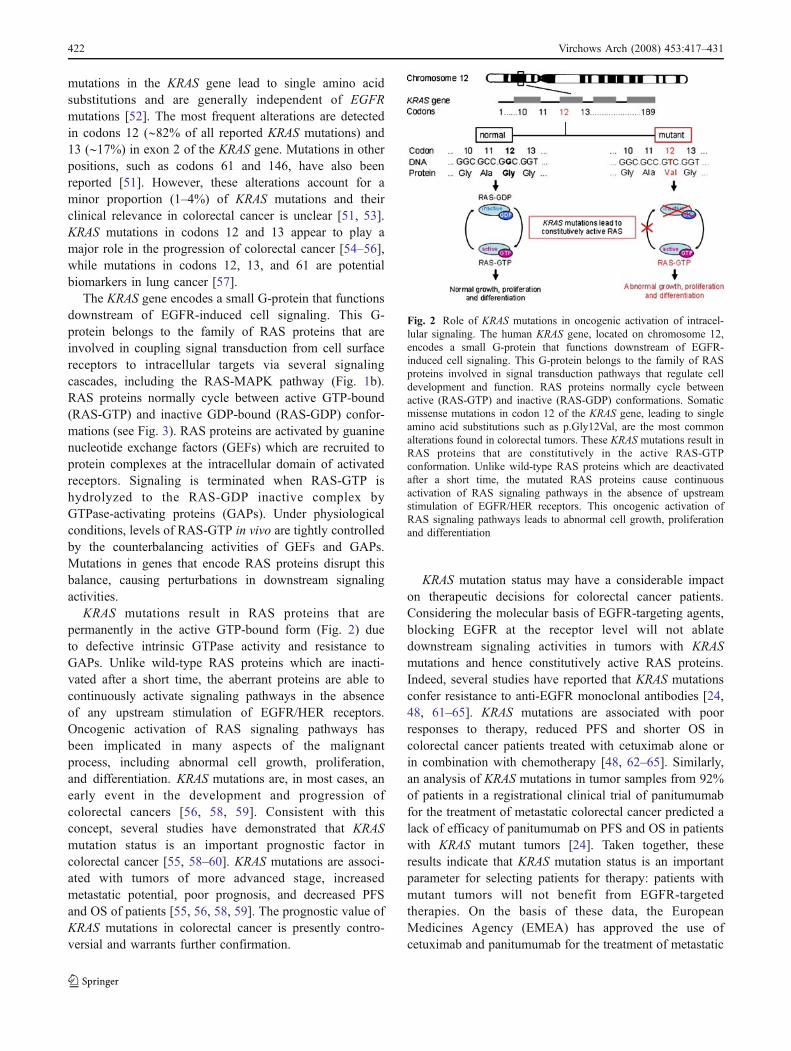
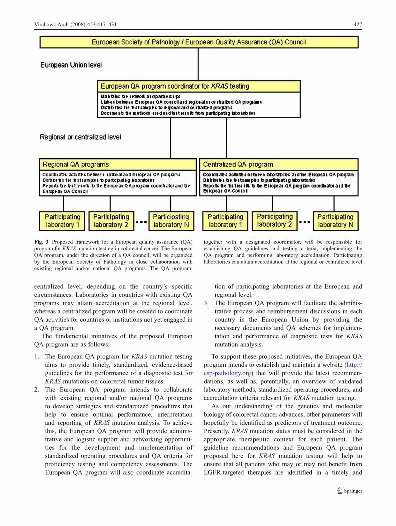
The KRAS gene encodes a small G-protein that functionsdownstream of EGFR-induced cell signaling. This G-protein belongs to the family of RAS proteins that areinvolved in coupling signal transduction from cell surfacereceptors to intracellular targets via several signalingcascades, including the RAS-MAPK pathway (Fig. 1b).RAS proteins normally cycle between active GTP-bound(RAS-GTP) and inactive GDP-bound (RAS-GDP) confor-mations (see Fig. 3). RAS proteins are activated by guaninenucleotide exchange factors (GEFs) which are recruited toprotein complexes at the intracellular domain of activatedreceptors. Signaling is terminated when RAS-GTP ishydrolyzed to the RAS-GDP inactive complex byGTPase-activating proteins (GAPs). Under physiologicalconditions, levels of RAS-GTP in vivo are tightly controlledby the counterbalancing activities of GEFs and GAPs.Mutations in genes that encode RAS proteins disrupt thisbalance, causing perturbations in downstream signalingactivities.
KRAS mutations result in RAS proteins that arepermanently in the active GTP-bound form (Fig. 2) dueto defective intrinsic GTPase activity and resistance toGAPs. Unlike wild-type RAS proteins which are inacti-vated after a short time, the aberrant proteins are able tocontinuously activate signaling pathways in the absenceof any upstream stimulation of EGFR/HER receptors.Oncogenic activation of RAS signaling pathways hasbeen implicated in many aspects of the malignantprocess, including abnormal cell growth, proliferation,and differentiation. KRAS mutations are, in most cases, anearly event in the development and progression ofcolorectal cancers [56, 58, 59]. Consistent with thisconcept, several studies have demonstrated that KRASmutation status is an important prognostic factor incolorectal cancer [55, 58–60]. KRAS mutations are associ-ated with tumors of more advanced stage, increasedmetastatic potential, poor prognosis, and decreased PFSand OS of patients [55, 56, 58, 59]. The prognostic value ofKRAS mutations in colorectal cancer is presently contro-versial and warrants further confirmation.
KRAS mutation status may have a considerable impacton therapeutic decisions for colorectal cancer patients.Considering the molecular basis of EGFR-targeting agents,blocking EGFR at the receptor level will not ablatedownstream signaling activities in tumors with KRASmutations and hence constitutively active RAS proteins.Indeed, several studies have reported that KRAS mutationsconfer resistance to anti-EGFR monoclonal antibodies [24,48, 61–65]. KRAS mutations are associated with poorresponses to therapy, reduced PFS and shorter OS incolorectal cancer patients treated with cetuximab alone orin combination with chemotherapy [48, 62–65]. Similarly,an analysis of KRAS mutations in tumor samples from 92%of patients in a registrational clinical trial of panitumumabfor the treatment of metastatic colorectal cancer predicted alack of efficacy of panitumumab on PFS and OS in patientswith KRAS mutant tumors [24]. Taken together, theseresults indicate that KRAS mutation status is an importantparameter for selecting patients for therapy: patients withmutant tumors will not benefit from EGFR-targetedtherapies. On the basis of these data, the EuropeanMedicines Agency (EMEA) has approved the use ofcetuximab and panitumumab for the treatment of metastatic
Fig. 2 Role of KRAS mutations in oncogenic activation of intracel-lular signaling. The human KRAS gene, located on chromosome 12,encodes a small G-protein that functions downstream of EGFR-induced cell signaling. This G-protein belongs to the family of RASproteins involved in signal transduction pathways that regulate celldevelopment and function. RAS proteins normally cycle betweenactive (RAS-GTP) and inactive (RAS-GDP) conformations. Somaticmissense mutations in codon 12 of the KRAS gene, leading to singleamino acid substitutions such as p.Gly12Val, are the most commonalterations found in colorectal tumors. These KRAS mutations result inRAS proteins that are constitutively in the active RAS-GTPconformation. Unlike wild-type RAS proteins which are deactivatedafter a short time, the mutated RAS proteins cause continuousactivation of RAS signaling pathways in the absence of upstreamstimulation of EGFR/HER receptors. This oncogenic activation ofRAS signaling pathways leads to abnormal cell growth, proliferationand differentiation
422 Virchows Arch (2008) 453:417–431
colorectal cancer in patients who carry a normal, wild-typeKRAS gene [12]. However, as only a fraction of patientswith colorectal tumors that carry a wild-type KRAS allelecan achieve a clinical response with EGFR-targetedtherapies, the search for additional predictive parametersremains an important challenge.
Methods for KRAS mutation testing
PCR has become the cornerstone of molecular diagnostictools, including those developed for KRAS mutation testing.PCR assays are highly sensitive and can be easilyautomated. PCR assays are thus well-suited for large-scale,high-throughput diagnostic testing. For KRAS mutationtesting, however, standard PCR assays are not sufficient.The main requirement for conclusive KRAS genotyping byPCR assay is the ability to discriminate between differentmutant alleles and wild type. There are two main challengesto achieving a conclusive result: one is the heterogeneity ofthe testing material, and the other is differences in thedetection limits for distinct mutations. Depending on thetissue analyzed, the amount of tumor versus non-tumor areais variable and heterogeneous, resulting in a templatemixture in which wild-type and mutant DNA are notpresent in equimolar amounts. Moreover, a cancer cell maycarry a heterozygous or homozygous KRAS mutation,increasing the genetic heterogeneity of the tissue materialused. Differences in PCR efficiencies for the detection ofthe different mutations can lead to a bias whereby certainmutations are detected preferentially over others.
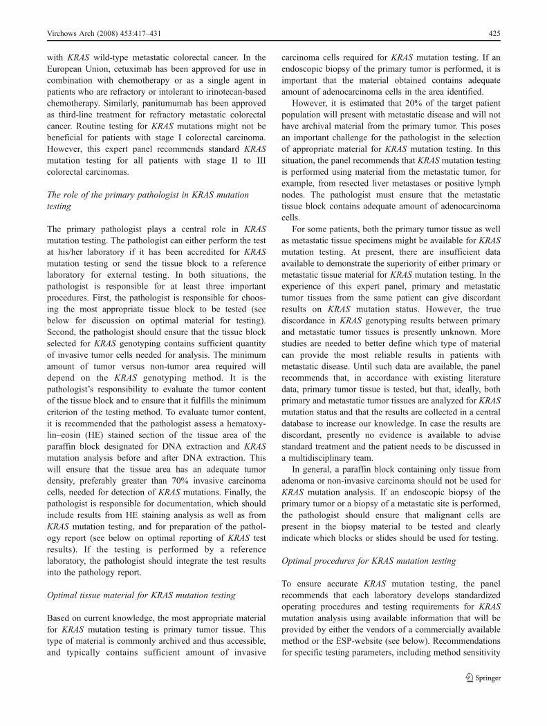
A plethora of methods is available for the detection ofmutations in the KRAS gene (see Table 1 for a non-exhaustive overview). Many of these methods are labora-tory-based assays and are not commercially available foruse in routine diagnostics. Other methods have beendeveloped further and are available as commercial test kitsnot directly intended for diagnostic purposes. To date, twoKRAS mutation test kits (TheraScreen® by DxS Ltd. andKRAS LightMix® by TIB MolBiol) have met the essentialrequirements of the relevant European Directives (CE-Mark) for diagnostic use in the European Union. Only onestudy, to our knowledge, has evaluated the concordancebetween different methods for KRAS mutation testing [66].In this study, 40 colorectal tumor samples were tested forseven common mutations in codons 12 and 13 of theKRAS gene by four commercially available assays and bydirect sequencing as a reference. Two allele-specific PCR-based methods and one PCR/direct sequencing methoddemonstrated high to good agreement with direct sequenc-ing, whereas an oligonucleotide hybridization methodshowed poor agreement. Given the technical requirementsfor a conclusive KRAS test result and the potential for
variability between different KRAS genotyping methods, athorough analytical validation of testing methods togetherwith a high standard of quality assurance are critical foraccurate, reliable KRAS mutation testing in clinicalpractice. Such an initiative to validate and standardizeKRAS mutation testing will also include the developmentof a website (http://esp-pathology.org) providing the latestinformation on current diagnostic methods and intendeduses of KRAS mutation testing. Therefore, at present, noadvice is given to which method is preferred. Theadvantage of commercially available tests is the validationprocess that these have gone through, but obviously thecosts of these tests are higher that in-house developedmethods. Most experience exists in different laboratorieswith sequencing after PCR, and this is a relativelyinexpensive method, but requires validation on a largeseries of cases. For most other methods, it is too early toassess the advantages and disadvantages.
Recommended guidelines and European QA program
Guidelines for KRAS mutation testing in colorectal cancer
The optimal use of EGFR-targeted therapies requiresaccurate KRAS mutation testing. Testing for KRAS muta-tions generally comprises three stages: (1) referral for KRASmutation testing; (2) selection of the tissue block containingthe tumor area of interest; and (3) DNA extraction andKRAS mutation analysis. In the current clinical setting,colorectal cancer patients are not routinely screened forKRAS mutation status. Pathologists test for KRAS mutationsonly upon the specific request of a clinician. Clinicians, inturn, request KRAS genotyping only if the test results areintended to guide decisions on patient management. Thesepractices might not be sufficient for optimal patient care.The process of requesting KRAS status testing, finding theoriginal tissue block and reporting the test results iscumbersome, time-consuming, and prone to errors. There-fore, routine mutation testing at the time of initial diagnosisof stage II and III tumors should be considered. There isalso a lack of validated testing methods and standardizedoperating procedures for the detection of KRAS mutations.There are very few studies that have systematicallycompared the sensitivity, specificity and reproducibility ofthe different techniques for KRAS genotyping. The concor-dance between different diagnostic methods is also largelyunknown. Therefore, there is an urgent need to establishand implement clinical practice guidelines and standardizedprocedures for KRAS mutation testing in patients withcolorectal cancer.
In recognition of the importance of accurate HER2testing in breast cancer management, practice guidelines
Virchows Arch (2008) 453:417–431 423
and a testing algorithm for HER2 testing have beenformulated by the American Society of Clinical Oncologyand the College of American Pathologists [3]. This expertpanel has recommended validation of all laboratory assaysor modifications, use of standardized operating procedures,and compliance with new testing criteria. Importantly, thepanel has also recommended that HER2 testing be done inan accredited laboratory or in a laboratory that meets thequality assurance and proficiency requirements set forth inthe practice guidelines.
To address the need for standardized KRAS mutationtesting methods and procedures in colorectal carcinoma,two working groups of the European Society of Pathology(ESP), the Diseases of the Digestive Tract ESP WorkingGroup and the Molecular Pathology ESP Working Group,convened an expert panel to develop guideline recommen-dations and a proposal for a European QA program forKRAS mutation testing. This expert panel consisted ofEuropean pathologists, molecular biologists, and oncolo-gists with expertise in colorectal carcinoma and KRASmutation analysis. A panel meeting was held during the
Third Intercontinental Congress of Pathology in Barcelonain May 2008. The purpose of this meeting was for the panelmembers to refine and agree on draft guidelines and anorganizational structure of a European QA program forKRAS mutation testing. Consensus recommendations andproposals are summarized here.
Target patient population for KRAS mutation testing
Activating mutations in codons 12 and 13 of the KRASgene identify patients who have a poor clinical response toEGFR-targeted therapies. Ideally, a predictive test shoulddistinguish between treatment responders and non-respond-ers accurately and reliably. Such an ideal predictor ispresently not available. The best option available today is atest that identifies patients who carry two wild-type KRASalleles and excludes patients with mutant codon 12 or 13alleles.
The anti-EGFR antibodies, cetuximab and panitumu-mab, currently available for clinical use have beenapproved in several countries for the treatment of patients
Table 1 Overview of methods used for KRAS genotyping
Method Intended use Ref.
Gel electrophoresis assaysTemporal temperature gradient electrophoresis LBM [67]Denaturing gradient gel electrophoresis LBM [68]Constant denaturant capillary electrophoresis LBM [69]SSCP assay LBM [70]SequencingDideoxy sequencing LBM, RUO kit [71]Pyrosequencing LBM [72, 73]PyroMark™ KRAS RUO kitAllele-specific PCR assaysa
Allele discrimination based on primer designARMS-PCR LBM [74, 75]KRAS mutation test kit RUO kitTheraScreen® kit CE-Mark kit for clinical use [76]KRAS LightMix® kit CE-Mark kit for clinical useREMS-PCR LBM [77]FLAG assay LBM [78]Enriched PCR-RFLP LBM [79]Allele discrimination based allele-specific ligation detection reactionPCR-LDR LBM [80]PCR-LDR spFRET assay LBM [81]Allele discrimination based on discriminating amplification efficiencies at low melting temperaturesCOLD-PCR LBM [82]
Other methodsSurface ligation reaction and biometallization LBM [83]Multi-target DNA assay panel LBM [84]Allele-specific oligonucleotide hybridization—Invigene®KRAS genotyping kit LBM, RUO kit
LBM Laboratory-based method, not commercially available, RUO: research use only, not validated for clinical applicationsa Allele-specific assays are also used by vendors offering KRAS genotyping services
424 Virchows Arch (2008) 453:417–431
with KRAS wild-type metastatic colorectal cancer. In theEuropean Union, cetuximab has been approved for use incombination with chemotherapy or as a single agent inpatients who are refractory or intolerant to irinotecan-basedchemotherapy. Similarly, panitumumab has been approvedas third-line treatment for refractory metastatic colorectalcancer. Routine testing for KRAS mutations might not bebeneficial for patients with stage I colorectal carcinoma.However, this expert panel recommends standard KRASmutation testing for all patients with stage II to IIIcolorectal carcinomas.
The role of the primary pathologist in KRAS mutationtesting
The primary pathologist plays a central role in KRASmutation testing. The pathologist can either perform the testat his/her laboratory if it has been accredited for KRASmutation testing or send the tissue block to a referencelaboratory for external testing. In both situations, thepathologist is responsible for at least three importantprocedures. First, the pathologist is responsible for choos-ing the most appropriate tissue block to be tested (seebelow for discussion on optimal material for testing).Second, the pathologist should ensure that the tissue blockselected for KRAS genotyping contains sufficient quantityof invasive tumor cells needed for analysis. The minimumamount of tumor versus non-tumor area required willdepend on the KRAS genotyping method. It is thepathologist’s responsibility to evaluate the tumor contentof the tissue block and to ensure that it fulfills the minimumcriterion of the testing method. To evaluate tumor content,it is recommended that the pathologist assess a hematoxy-lin–eosin (HE) stained section of the tissue area of theparaffin block designated for DNA extraction and KRASmutation analysis before and after DNA extraction. Thiswill ensure that the tissue area has an adequate tumordensity, preferably greater than 70% invasive carcinomacells, needed for detection of KRAS mutations. Finally, thepathologist is responsible for documentation, which shouldinclude results from HE staining analysis as well as fromKRAS mutation testing, and for preparation of the pathol-ogy report (see below on optimal reporting of KRAS testresults). If the testing is performed by a referencelaboratory, the pathologist should integrate the test resultsinto the pathology report.
Optimal tissue material for KRAS mutation testing
Based on current knowledge, the most appropriate materialfor KRAS mutation testing is primary tumor tissue. Thistype of material is commonly archived and thus accessible,and typically contains sufficient amount of invasive
carcinoma cells required for KRAS mutation testing. If anendoscopic biopsy of the primary tumor is performed, it isimportant that the material obtained contains adequateamount of adenocarcinoma cells in the area identified.
However, it is estimated that 20% of the target patientpopulation will present with metastatic disease and will nothave archival material from the primary tumor. This posesan important challenge for the pathologist in the selectionof appropriate material for KRAS mutation testing. In thissituation, the panel recommends that KRAS mutation testingis performed using material from the metastatic tumor, forexample, from resected liver metastases or positive lymphnodes. The pathologist must ensure that the metastatictissue block contains adequate amount of adenocarcinomacells.
For some patients, both the primary tumor tissue as wellas metastatic tissue specimens might be available for KRASmutation testing. At present, there are insufficient dataavailable to demonstrate the superiority of either primary ormetastatic tissue material for KRAS mutation testing. In theexperience of this expert panel, primary and metastatictumor tissues from the same patient can give discordantresults on KRAS mutation status. However, the truediscordance in KRAS genotyping results between primaryand metastatic tumor tissues is presently unknown. Morestudies are needed to better define which type of materialcan provide the most reliable results in patients withmetastatic disease. Until such data are available, the panelrecommends that, in accordance with existing literaturedata, primary tumor tissue is tested, but that, ideally, bothprimary and metastatic tumor tissues are analyzed for KRASmutation status and that the results are collected in a centraldatabase to increase our knowledge. In case the results arediscordant, presently no evidence is available to advisestandard treatment and the patient needs to be discussed ina multidisciplinary team.
In general, a paraffin block containing only tissue fromadenoma or non-invasive carcinoma should not be used forKRAS mutation analysis. If an endoscopic biopsy of theprimary tumor or a biopsy of a metastatic site is performed,the pathologist should ensure that malignant cells arepresent in the biopsy material to be tested and clearlyindicate which blocks or slides should be used for testing.
Optimal procedures for KRAS mutation testing
To ensure accurate KRAS mutation testing, the panelrecommends that each laboratory develops standardizedoperating procedures and testing requirements for KRASmutation analysis using available information that will beprovided by either the vendors of a commercially availablemethod or the ESP-website (see below). Recommendationsfor specific testing parameters, including method sensitivity
Virchows Arch (2008) 453:417–431 425
and specificity, method validation, analysis success rate,and documentation of costs, are summarized in Table 2.Some of these recommendations are compatible with ISO(International Organization for Standardization; http://www.iso.org) general requirements for the competence of testingand calibration laboratories (ISO/IEC 17025:2005). Theserequirements will become essential components of accred-itation for KRAS mutation testing. Tests that have adetection sensitivity of 1% might detect subclones in atumor that have acquired a mutation. It is presentlyunknown what the consequence of such a finding might be.
Optimal reporting of test results
Result reporting is an integral part of any diagnosticprocedure, including KRAS mutation testing. All KRAS testresults are to be reported to the primary pathologist who isresponsible for preparation of the pathology report for aspecific tissue block or biopsy material. Optimal reportingof KRAS test results should conform to the OECD Guide-lines for Quality Assurance in Molecular Genetic Testing(http://www.oecd.org). In brief, the reports should includeat minimum the following information: (1) identification ofthe patient and health care professional; (2) type of materialand percentage of tumor cells present in the sample; (3)indication for testing and patient-specific medical data; (4)the testing method used, including its analytical sensitivityand specificity; and (5) test results (mutant or wild-typeKRAS allele) and interpretation of results in the context ofthe indication for testing.
Proposal for a European quality assurance program
During the process of developing a European QA programfor KRAS mutation testing, the expert panel considered theexperience with HER2 testing as an informative example.While trastuzumab (Herceptin®) became available in 2002for the treatment of breast cancer, it was another 5 yearsbefore clinical practice guidelines were established foroptimal HER2 testing algorithm and proficiency require-ments. Another problem encountered with the introductionof trastuzumab was the lack of adequate financial provi-sions for diagnostic testing, although some nationalauthorities required mandatory HER2 testing in breastcancer patients prior to trastuzumab therapy. Today,molecular diagnostic tools, testing procedures and thereimbursement process for diagnostic tests linked to aspecific medication differ greatly across countries inEurope. Clearly, there is a need to establish a standardized,evidence-based QA program for molecular diagnosticsacross the European Union.
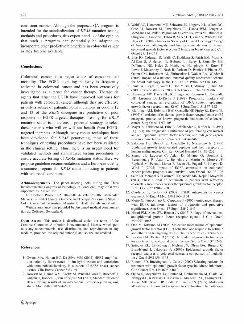
Here, we propose to establish a European QA programfor testing KRAS mutations in colorectal cancer. Thisprogram aims to ensure optimal accuracy and proficiencyin KRAS mutation testing across all countries or institutionsin the European Union. A potential framework for aEuropean QA program for KRAS mutation testing is shownin Fig. 3. The program will be organized by the EuropeanSociety of Pathology in close collaboration with existingregional and/or national QA programs. Laboratories canparticipate in the European QA program at the regional or
Table 2 Recommendations for KRAS mutation testing
Parameter Recommendation
Sensitivity The lower detection limit of mutant signal should be set at 1% of tumor cells for allele-specific PCR and 25–30% for directsequencing.
Specificity A specific test should be able to detect 7 common mutations in codons 12 and 13 of the KRAS gene and not detect mutationsin codon 61. False negatives may occur because of test specificities (e.g. lack of an allele-specific PCR for codon 13mutation).
Methodvalidationa
The laboratory should use a validated method for KRAS mutation testing. The objectives of the validation are to:Determine the minimum tumor tissue area and section thickness for DNA extraction.Stipulate which fixatives are acceptable for use.Determine input DNA quantity, quality and concentration.Determine the cut-off values for discerning KRAS mutant alleles from wild-type alleles.Evaluate sensitivity of the test, for example by using dilution series cell lines.Compare the accuracy of test results against a pre-defined reference method (e.g. direct sequencing).Determine the reproducibility between different testing assays and equipment.Verify the robustness of the testing method. Robustness may be influenced by several factors, including varying DNAconcentrations and the use of manual or automated protocols or equipment.
Analysissuccess rate
A laboratory should obtain the following success rates for accreditation:95% of samples with successful DNA extraction97% of samples with correct KRAS test results
Costs Costs of KRAS mutation testing should be calculated and documented for national reimbursement schemes.
a Compatible with accreditation requirements of ISO/IEC 17025:2005
426 Virchows Arch (2008) 453:417–431
centralized level, depending on the country’s specificcircumstances. Laboratories in countries with existing QAprograms may attain accreditation at the regional level,whereas a centralized program will be created to coordinateQA activities for countries or institutions not yet engaged ina QA program.
The fundamental initiatives of the proposed EuropeanQA program are as follows:
1. The European QA program for KRAS mutation testingaims to provide timely, standardized, evidence-basedguidelines for the performance of a diagnostic test forKRAS mutations on colorectal tumor tissues.
2. The European QA program intends to collaboratewith existing regional and/or national QA programsto develop strategies and standardized procedures thathelp to ensure optimal performance, interpretationand reporting of KRAS mutation analysis. To achievethis, the European QA program will provide adminis-trative and logistic support and networking opportuni-ties for the development and implementation ofstandardized operating procedures and QA criteria forproficiency testing and competency assessments. TheEuropean QA program will also coordinate accredita-
tion of participating laboratories at the European andregional level.
3. The European QA program will facilitate the adminis-trative process and reimbursement discussions in eachcountry in the European Union by providing thenecessary documents and QA schemes for implemen-tation and performance of diagnostic tests for KRASmutation analysis.
To support these proposed initiatives, the European QAprogram intends to establish and maintain a website (http://esp-pathology.org) that will provide the latest recommen-dations, as well as, potentially, an overview of validatedlaboratory methods, standardized operating procedures, andaccreditation criteria relevant for KRAS mutation testing.
As our understanding of the genetics and molecularbiology of colorectal cancer advances, other parameters willhopefully be identified as predictors of treatment outcome.Presently, KRAS mutation status must be considered in theappropriate therapeutic context for each patient. Theguideline recommendations and European QA programproposed here for KRAS mutation testing will help toensure that all patients who may or may not benefit fromEGFR-targeted therapies are identified in a timely and
Fig. 3 Proposed framework for a European quality assurance (QA)program for KRAS mutation testing in colorectal cancer. The EuropeanQA program, under the direction of a QA council, will be organizedby the European Society of Pathology in close collaboration withexisting regional and/or national QA programs. The QA program,
together with a designated coordinator, will be responsible forestablishing QA guidelines and testing criteria, implementing theQA program and performing laboratory accreditation. Participatinglaboratories can attain accreditation at the regional or centralized level
Virchows Arch (2008) 453:417–431 427
consistent manner. Although the proposed QA program isintended for the standardization of KRAS mutation testingmethods and procedures, this expert panel is of the opinionthat such a program can potentially be adapted toincorporate other predictive biomarkers in colorectal canceras they become available.
Conclusions
Colorectal cancer is a major cause of cancer-relatedmortality. The EGFR signaling pathway is frequentlyactivated in colorectal cancer and has been extensivelyinvestigated as a target for cancer therapy. Therapeuticagents that target the EGFR have improved outcomes forpatients with colorectal cancer, although they are effectivein only a subset of patients. Point mutations in codons 12and 13 of the KRAS oncogene are predictive of poorresponse to EGFR-targeted therapies. Testing for KRASmutation status is, therefore, a potential strategy to selectthose patients who will or will not benefit from EGFR-targeted therapies. Although many robust techniques havebeen developed for KRAS genotyping, most of thesetechniques or testing procedures have not been validatedin the clinical setting. Thus, there is an urgent need forvalidated methods and standardized testing procedures toensure accurate testing of KRAS mutation status. Here wepropose guideline recommendations and a European qualityassurance program for KRAS mutation testing in patientswith colorectal carcinoma.
Acknowledgements The panel meeting held during the ThirdIntercontinental Congress of Pathology in Barcelona, May 2008 wassupported by Amgen Inc.
G. Hoefler: Project GZ 70420/0134-IV/B/12/2006 “MolecularMarkers To Predict Clinical Outcome and Therapy Response in Stage IIColon Cancer” of the Austrian Ministry for Health, Family and Youth.
Writing assistance was provided by Archimed medical communica-tion ag, Zofingen, Switzerland.
Open Access This article is distributed under the terms of theCreative Commons Attribution Noncommercial License which per-mits any noncommercial use, distribution, and reproduction in anymedium, provided the original author(s) and source are credited.
References
1. Owens MA, Horten BC, Da Silva MM (2004) HER2 amplifica-tion ratios by fluorescence in situ hybridization and correlationwith immunohistochemistry in a cohort of 6,556 breast cancertissues. Clin Breast Cancer 5:63–69
2. Dowsett M, Hanna WM, Kockx M, Penault-Llorca F, Ruschoff J,Gutjahr T, Habben K, van de Vijver MJ (2007) Standardization ofHER2 testing: results of an international proficiency-testing ringstudy. Mod Pathol 20:584–591
3. Wolff AC, Hammond ME, Schwartz JN, Hagerty KL, Allred DC,Cote RJ, Dowsett M, Fitzgibbons PL, Hanna WM, Langer A,McShane LM, Paik S, PegramMD, Perez EA, Press MF, Rhodes A,Sturgeon C, Taube SE, Tubbs R, Vance GH, van d V, Wheeler TM,Hayes DF (2007) American Society of Clinical Oncology/Collegeof American Pathologists guideline recommendations for humanepidermal growth factor receptor 2 testing in breast cancer. J ClinOncol 25:118–145
4. Ellis IO, Coleman D, Wells C, Kodikara S, Paish EM, Moss S,Al-Sam S, Anderson N, Bobrow L, Buley I, Connolly CE,Dallimore NS, Hales S, Hanby A, Humphreys S, Knox F,Lowe J, Macartney J, Nash R, Parham D, Patnick J, Pinder SE,Quinn CM, Robertson AJ, Shrimankar J, Walker RA, Winder R(2006) Impact of a national external quality assessment schemefor breast pathology in the UK. J Clin Pathol 59:138–145
5. Jemal A, Siegel R, Ward E, Hao Y, Xu J, Murray T, Thun MJ(2008) Cancer statistics, 2008. CA Cancer J Clin 58:71–96
6. Hemming AW, Davis NL, Kluftinger A, Robinson B, QuenvilleNF, Liseman B, LeRiche J (1992) Prognostic markers ofcolorectal cancer: an evaluation of DNA content, epidermalgrowth factor receptor, and Ki-67. J Surg Oncol 51:147–152
7. Kluftinger AM, Robinson BW, Quenville NF, Finley RJ, Davis NL(1992) Correlation of epidermal growth factor receptor and c-erbB2oncogene product to known prognostic indicators of colorectalcancer. Surg Oncol 1:97–105
8. Mayer A, Takimoto M, Fritz E, Schellander G, Kofler K, LudwigH (1993) The prognostic significance of proliferating cell nuclearantigen, epidermal growth factor receptor, and mdr gene expres-sion in colorectal cancer. Cancer 71:2454–2460
9. Salomon DS, Brandt R, Ciardiello F, Normanno N (1995)Epidermal growth factor-related peptides and their receptors inhuman malignancies. Crit Rev Oncol Hematol 19:183–232
10. Spano JP, Lagorce C, Atlan D, Milano G, Domont J,Benamouzig R, Attar A, Benichou J, Martin A, Morere JF,Raphael M, Penault-Llorca F, Breau JL, Fagard R, Khayat D,Wind P (2005) Impact of EGFR expression on colorectalcancer patient prognosis and survival. Ann Oncol 16:102–108
11. Saltz LB,Meropol NJ, Loehrer PJ Sr, NeedleMN, Kopit J, Mayer RJ(2004) Phase II trial of cetuximab in patients with refractorycolorectal cancer that expresses the epidermal growth factor receptor.J Clin Oncol 22:1201–1208
12. Ciardiello F, Tortora G (2008) EGFR antagonists in cancertreatment. N Engl J Med 358:1160–1174
13. Metro G, Finocchiaro G, Cappuzzo F (2006) Anti-cancer therapywith EGFR inhibitors: factors of prognostic and predictivesignificance. Ann Oncol 17 Suppl 2:ii42–ii45
14. Harari PM, Allen GW, Bonner JA (2007) Biology of interactions:antiepidermal growth factor receptor agents. J Clin Oncol25:4057–4065
15. Ono M, Kuwano M (2006) Molecular mechanisms of epidermalgrowth factor receptor (EGFR) activation and response to gefitiniband other EGFR-targeting drugs. Clin Cancer Res 12:7242–7251
16. Lockhart AC, Berlin JD (2005) The epidermal growth factor recep-tor as a target for colorectal cancer therapy. Semin Oncol 32:52–60
17. Spindler KL, Lindebjerg J, Nielsen JN, Olsen DA, Bisgard C,Brandslund I, Jakobsen A (2006) Epidermal growth factorreceptor analyses in colorectal cancer: a comparison of methods.Int J Oncol 29:1159–1165
18. Bonomi PD, Buckingham L, Coon J (2007) Selecting patients fortreatment with epidermal growth factor tyrosine kinase inhibitors.Clin Cancer Res 13:s4606–s4612
19. Ogino S, Meyerhardt JA, Cantor M, Brahmandam M, Clark JW,Namgyal C, Kawasaki T, Kinsella K, Michelini AL, Enzinger PC,Kulke MH, Ryan DP, Loda M, Fuchs CS (2005) Molecularalterations in tumors and response to combination chemotherapy
428 Virchows Arch (2008) 453:417–431
with gefitinib for advanced colorectal cancer. Clin Cancer Res11:6650–6656
20. Paez JG, Janne PA, Lee JC, Tracy S, Greulich H, Gabriel S,Herman P, Kaye FJ, Lindeman N, Boggon TJ, Naoki K, Sasaki H,Fujii Y, Eck MJ, Sellers WR, Johnson BE, Meyerson M (2004)EGFR mutations in lung cancer: correlation with clinical responseto gefitinib therapy. Science 304:1497–1500
21. Pao W, Miller V, Zakowski M, Doherty J, Politi K, Sarkaria I,Singh B, Heelan R, Rusch V, Fulton L, Mardis E, Kupfer D,Wilson R, Kris M, Varmus H (2004) EGF receptor gene mutationsare common in lung cancers from “never smokers” and areassociated with sensitivity of tumors to gefitinib and erlotinib.Proc Natl Acad Sci U S A 101:13306–13311
22. Rocha-Lima CM, Soares HP, Raez LE, Singal R (2007) EGFRtargeting of solid tumors. Cancer Control 14:295–304
23. Jonker DJ, O’Callaghan CJ, Karapetis CS, Zalcberg JR, Tu D,Au HJ, Berry SR, KrahnM, Price T, Simes RJ, Tebbutt NC, van HG,Wierzbicki R, Langer C, Moore MJ (2007) Cetuximab for thetreatment of colorectal cancer. N Engl J Med 357:2040–2048
24. Amado RG, Wolf M, Peeters M, Van CE, Siena S, Freeman DJ,Juan T, Sikorski R, Suggs S, Radinsky R, Patterson SD, Chang DD(2008) Wild-type KRAS is required for panitumumab efficacy inpatients with metastatic colorectal cancer. J Clin Oncol 26:1626–1634
25. Cunningham D, Humblet Y, Siena S, Khayat D, Bleiberg H,Santoro A, Bets D, Mueser M, Harstrick A, Verslype C, Chau I,Van CE (2004) Cetuximab monotherapy and cetuximab plusirinotecan in irinotecan-refractory metastatic colorectal cancer. NEngl J Med 351:337–345
26. Gibson TB, Ranganathan A, Grothey A (2006) Randomized phaseIII trial results of panitumumab, a fully human anti-epidermalgrowth factor receptor monoclonal antibody, in metastatic colo-rectal cancer. Clin Colorectal Cancer 6:29–31
27. Siena S, Peeters M, Van CE, Humblet Y, Conte P, Bajetta E,Comandini D, Bodoky G, van HG, Salek T, Wolf M, DevercelliG, Woolley M, Amado RG (2007) Association of progression-freesurvival with patient-reported outcomes and survival: results froma randomised phase 3 trial of panitumumab. Br J Cancer 97:1469–1474
28. Van CE, Peeters M, Siena S, Humblet Y, Hendlisz A, Neyns B,Canon JL, Van Laethem JL, Maurel J, Richardson G, Wolf M,Amado RG (2007) Open-label phase III trial of panitumumab plusbest supportive care compared with best supportive care alone inpatients with chemotherapy-refractory metastatic colorectal can-cer. J Clin Oncol 25:1658–1664
29. Radinsky R, Risin S, Fan D, Dong Z, Bielenberg D, Bucana CD,Fidler IJ (1995) Level and function of epidermal growth factorreceptor predict the metastatic potential of human colon carcino-ma cells. Clin Cancer Res 1:19–31
30. Steele RJ, Kelly P, Ellul B, EreminO (1990) Epidermal growth factorreceptor expression in colorectal cancer. Br J Surg 77:1352–1354
31. Giralt J, Eraso A, Armengol M, Rossello J, Majo J, Ares C, EspinE, Benavente S, de T I (2002) Epidermal growth factor receptor isa predictor of tumor response in locally advanced rectal cancerpatients treated with preoperative radiotherapy. Int J Radiat OncolBiol Phys 54:1460–1465
32. Chung KY, Shia J, Kemeny NE, Shah M, Schwartz GK, Tse A,Hamilton A, Pan D, Schrag D, Schwartz L, Klimstra DS, FridmanD, Kelsen DP, Saltz LB (2005) Cetuximab shows activity incolorectal cancer patients with tumors that do not express theepidermal growth factor receptor by immunohistochemistry. J ClinOncol 23:1803–1810
33. Moroni M, Veronese S, Benvenuti S, Marrapese G, Sartore-Bianchi A,Di NF, Gambacorta M, Siena S, Bardelli A (2005) Gene copy numberfor epidermal growth factor receptor (EGFR) and clinical response to
antiEGFR treatment in colorectal cancer: a cohort study. Lancet Oncol6:279–286
34. Italiano A, Follana P, Caroli FX, Badetti JL, Benchimol D,Garnier G, Gugenheim J, Haudebourg J, Keslair F, Lesbats G,Lledo G, Roussel JF, Pedeutour F, Francois E (2008) Cetuximabshows activity in colorectal cancer patients with tumors for whichFISH analysis does not detect an increase in EGFR gene copynumber. Ann Surg Oncol 15:649–654
35. Francoual M, Etienne-Grimaldi MC, Formento JL, Benchimol D,Bourgeon A, Chazal M, Letoublon C, Andre T, Gilly N, Delpero JR,Lasser P, Spano JP, Milano G (2006) EGFR in colorectal cancer:more than a simple receptor. Ann Oncol 17:962–967
36. Cappuzzo F, Hirsch FR, Rossi E, Bartolini S, Ceresoli GL, Bemis L,Haney J, Witta S, Danenberg K, Domenichini I, Ludovini V, Magrini E,Gregorc V, Doglioni C, Sidoni A, Tonato M, Franklin WA, Crino L,Bunn PA Jr, Varella-Garcia M (2005) Epidermal growth factor receptorgene and protein and gefitinib sensitivity in non-small-cell lung cancer. JNatl Cancer Inst 97:643–655
37. Hijiya N, Miyawaki M, Kawahara K, Akamine S, Tsuji K, Kadota J,Akizuki S, Uchida T, Matsuura K, Tsukamoto Y, Moriyama M(2008) Phosphorylation status of epidermal growth factor receptor isclosely associated with responsiveness to gefitinib in pulmonaryadenocarcinoma. Hum Pathol 39:316–323
38. Barault L, Veyrie N, Jooste V, Lecorre D, Chapusot C, Ferraz JM,Lievre A, Cortet M, Bouvier AM, Rat P, Roignot P, Faivre J,Laurent-Puig P, Piard F (2008) Mutations in the RAS-MAPK, PI(3)K (phosphatidylinositol-3-OH kinase) signaling network corre-late with poor survival in a population-based series of coloncancers. Int J Cancer 122:2255–2259
39. Tan YH, Liu Y, Eu KW, Ang PW, Li WQ, Salto-Tellez M,Iacopetta B, Soong R (2008) Detection of BRAF V600E mutationby pyrosequencing. Pathology 40:295–298
40. Calistri D, Rengucci C, Seymour I, Lattuneddu A, Polifemo AM,Monti F, Saragoni L, Amadori D (2005) Mutation analysis of p53,K-ras, and BRAF genes in colorectal cancer progression. J CellPhysiol 204:484–488
41. Oliveira C, Pinto M, Duval A, Brennetot C, Domingo E, Espin E,Armengol M, Yamamoto H, Hamelin R, Seruca R, Schwartz S Jr(2003) BRAF mutations characterize colon but not gastric cancerwith mismatch repair deficiency. Oncogene 22:9192–9196
42. Domingo E, Espin E, Armengol M, Oliveira C, Pinto M, Duval A,Brennetot C, Seruca R, Hamelin R, Yamamoto H, Schwartz S Jr(2004) Activated BRAF targets proximal colon tumors withmismatch repair deficiency and MLH1 inactivation. GenesChromosomes Cancer 39:138–142
43. Ogino S, Cantor M, Kawasaki T, Brahmandam M, Kirkner GJ,Weisenberger DJ, Campan M, Laird PW, Loda M, Fuchs CS(2006) CpG island methylator phenotype (CIMP) of colorectalcancer is best characterised by quantitative DNA methylationanalysis and prospective cohort studies. Gut 55:1000–1006
44. Rajagopalan H, Bardelli A, Lengauer C, Kinzler KW, VogelsteinB, Velculescu VE (2002) Tumorigenesis: RAF/RAS oncogenesand mismatch-repair status. Nature 418:934
45. Preto A, Figueiredo J, Velho S, Ribeiro AS, Soares P, Oliveira C,Seruca R (2008) BRAF provides proliferation and survival signalsin MSI colorectal carcinoma cells displaying BRAF(V600E) butnot KRAS mutations. J Pathol 214:320–327
46. Jhawer M, Goel S, Wilson AJ, Montagna C, Ling YH, Byun DS,Nasser S, Arango D, Shin J, Klampfer L, Augenlicht LH,Soler RP, Mariadason JM (2008) PIK3CA mutation/PTENexpression status predicts response of colon cancer cells to theepidermal growth factor receptor inhibitor cetuximab. CancerRes 68:1953–1961
47. Ince WL, Jubb AM, Holden SN, Holmgren EB, Tobin P, SridharM, Hurwitz HI, Kabbinavar F, Novotny WF, Hillan KJ, Koeppen
Virchows Arch (2008) 453:417–431 429
H (2005) Association of k-ras, b-raf, and p53 status with thetreatment effect of bevacizumab. J Natl Cancer Inst 97:981–989
48. Lievre A, Bachet JB, Le CD, Boige V, Landi B, Emile JF, Cote JF,Tomasic G, Penna C, Ducreux M, Rougier P, Penault-Llorca F,Laurent-Puig P (2006) KRAS mutation status is predictive ofresponse to cetuximab therapy in colorectal cancer. Cancer Res66:3992–3995
49. Velho S, Oliveira C, Ferreira A, Ferreira AC, Suriano G, Schwartz S Jr,Duval A, Carneiro F, Machado JC, Hamelin R, Seruca R (2005) Theprevalence of PIK3CA mutations in gastric and colon cancer. Eur JCancer 41:1649–1654
50. Frattini M, Saletti P, Romagnani E, Martin V, Molinari F, Ghisletta M,Camponovo A, Etienne LL, Cavalli F, Mazzucchelli L (2007) PTENloss of expression predicts cetuximab efficacy in metastatic colorectalcancer patients. Br J Cancer 97:1139–1145
51. Edkins S, O’Meara S, Parker A, Stevens C, Reis M, Jones S,Greenman C, Davies H, Dalgliesh G, Forbes S, Hunter C, Smith R,Stephens P, Goldstraw P, Nicholson A, Chan TL, Velculescu VE,Yuen ST, Leung SY, Stratton MR, Futreal PA (2006) RecurrentKRAS codon 146 mutations in human colorectal cancer. CancerBiol Ther 5:928–932
52. Kosaka T, Yatabe Y, Endoh H, Kuwano H, Takahashi T,Mitsudomi T (2004) Mutations of the epidermal growth factorreceptor gene in lung cancer: biological and clinical implications.Cancer Res 64:8919–8923
53. Oliveira C, Westra JL, Arango D, Ollikainen M, Domingo E,Ferreira A, Velho S, Niessen R, Lagerstedt K, Alhopuro P, LaihoP, Veiga I, Teixeira MR, Ligtenberg M, Kleibeuker JH, SijmonsRH, Plukker JT, Imai K, Lage P, Hamelin R, Albuquerque C,Schwartz S Jr, Lindblom A, Peltomaki P, Yamamoto H, AaltonenLA, Seruca R, Hofstra RM (2004) Distinct patterns of KRASmutations in colorectal carcinomas according to germline mis-match repair defects and hMLH1 methylation status. Hum MolGenet 13:2303–2311
54. Russo A, Bazan V, Agnese V, Rodolico V, Gebbia N (2005)Prognostic and predictive factors in colorectal cancer: Kirsten Rasin CRC (RASCAL) and TP53CRC collaborative studies. AnnOncol 16 Suppl 4:iv44–iv49
55. Samowitz WS, Curtin K, Schaffer D, Robertson M, Leppert M,Slattery ML (2000) Relationship of Ki-ras mutations in coloncancers to tumor location, stage, and survival: a population-basedstudy. Cancer Epidemiol Biomarkers Prev 9:1193–1197
56. Andreyev HJ, Norman AR, Cunningham D, Oates J, Dix BR,Iacopetta BJ, Young J, Walsh T, Ward R, Hawkins N, Beranek M,Jandik P, Benamouzig R, Jullian E, Laurent-Puig P, Olschwang S,Muller O, Hoffmann I, Rabes HM, Zietz C, Troungos C,Valavanis C, Yuen ST, Ho JW, Croke CT, O’Donoghue DP,Giaretti W, Rapallo A, Russo A, Bazan V, Tanaka M, Omura K,Azuma T, Ohkusa T, Fujimori T, Ono Y, Pauly M, Faber C,Glaesener R, de Goeij AF, Arends JW, Andersen SN, Lovig T,Breivik J, Gaudernack G, Clausen OP, De Angelis PD, Meling GI,Rognum TO, Smith R, Goh HS, Font A, Rosell R, Sun XF, ZhangH, Benhattar J, Losi L, Lee JQ, Wang ST, Clarke PA, Bell S,Quirke P, Bubb VJ, Piris J, Cruickshank NR, Morton D, Fox JC,Al-Mulla F, Lees N, Hall CN, Snary D, Wilkinson K, Dillon D,Costa J, Pricolo VE, Finkelstein SD, Thebo JS, Senagore AJ,Halter SA, Wadler S, Malik S, Krtolica K, Urosevic N (2001)Kirsten ras mutations in patients with colorectal cancer: the‘RASCAL II’ study. Br J Cancer 85:692–696
57. Zerbe LK, Dwyer-Nield LD, Fritz JM, Redente EF, Shroyer RJ,Conklin E, Kane S, Tucker C, Eckhardt SG, Gustafson DL, IwataKK, Malkinson AM (2008) Inhibition by erlotinib of primary lungadenocarcinoma at an early stage in male mice. Cancer ChemotherPharmacol 62:605–620
58. Keller JW, Franklin JL, Graves-Deal R, Friedman DB, WhitwellCW, Coffey RJ (2007) Oncogenic KRAS provides a uniquely
powerful and variable oncogenic contribution among RAS familymembers in the colonic epithelium. J Cell Physiol 210:740–749
59. Wang JY, Wang YH, Jao SW, Lu CY, Kuo CH, Hu HM, Hsieh JS,Chong IW, Cheng TL, Lin SR (2006) Molecular mechanismsunderlying the tumorigenesis of colorectal adenomas: correlationto activated K-ras oncogene. Oncol Rep 16:1245–1252
60. Castagnola P, Giaretti W (2005) Mutant KRAS, chromosomalinstability and prognosis in colorectal cancer. Biochim BiophysActa 1756:115–125
61. Benvenuti S, Sartore-Bianchi A, Di NF, Zanon C, Moroni M,Veronese S, Siena S, Bardelli A (2007) Oncogenic activation ofthe RAS/RAF signaling pathway impairs the response ofmetastatic colorectal cancers to anti-epidermal growth factorreceptor antibody therapies. Cancer Res 67:2643–2648
62. De RW, Piessevaux H, De SJ, Janssens M, De HG, Personeni N,Biesmans B, Van Laethem JL, Peeters M, Humblet Y, Van CE,Tejpar S (2008) KRAS wild-type state predicts survival and isassociated to early radiological response in metastatic colorectalcancer treated with cetuximab. Ann Oncol 19:508–515
63. Di FF, Blanchard F, Charbonnier F, Le PF, LamyA,GalaisMP, BastitL, Killian A, Sesboue R, Tuech JJ, Queuniet AM, Paillot B, SabourinJC, Michot F, Michel P, Frebourg T (2007) Clinical relevance ofKRAS mutation detection in metastatic colorectal cancer treated byCetuximab plus chemotherapy. Br J Cancer 96:1166–1169
64. Khambata-Ford S, Garrett CR, Meropol NJ, Basik M, Harbison CT,Wu S, Wong TW, Huang X, Takimoto CH, Godwin AK, Tan BR,Krishnamurthi SS, Burris HA III, Poplin EA, Hidalgo M, Baselga J,Clark EA, Mauro DJ (2007) Expression of epiregulin and amphir-egulin and K-ras mutation status predict disease control in metastaticcolorectal cancer patients treated with cetuximab. J Clin Oncol25:3230–3237
65. Lievre A, Bachet JB, Boige V, Cayre A, Le CD, Buc E, Ychou M,Bouche O, Landi B, Louvet C, Andre T, Bibeau F, Diebold MD,Rougier P, Ducreux M, Tomasic G, Emile JF, Penault-Llorca F,Laurent-Puig P (2008) KRAS mutations as an independentprognostic factor in patients with advanced colorectal cancertreated with cetuximab. J Clin Oncol 26:374–379
66. Juan T, Suggs S, Wolf M, Sarosi I, Freeman D, Oliner K, Bakkar A,Patterson SD (2008) A comparability study of 4 commercial KRAStests. American Association for Cancer Research (AACR) AnnualMeeting, April 12–16, 2008, Abstract #1811
67. Kressner U, Bjorheim J, Westring S, Wahlberg SS, Pahlman L,Glimelius B, Lindmark G, Lindblom A, Borresen-Dale AL (1998)Ki-ras mutations and prognosis in colorectal cancer. Eur J Cancer34:518–521
68. Hayes VM,Westra JL, Verlind E, Bleeker W, Plukker JT, Hofstra RM,Buys CH (2000) New comprehensive denaturing-gradient-gel-electro-phoresis assay for KRAS mutation detection applied to paraffin-embedded tumours. Genes Chromosomes Cancer 29:309–314
69. Zhao C, Xu G, Shi X, Ma J, Lu S, Yang Q (2004) Detection of K-ras exon 1 mutations by constant denaturant capillary electropho-resis. Biomed Chromatogr 18:538–541
70. Chaubert P, Bautista D, Benhattar J (1993) An improved method forrapid screening of DNA mutations by nonradioactive single-strandconformation polymorphism procedure. Biotechniques 15:586
71. Khanna M, Park P, Zirvi M, Cao W, Picon A, Day J, Paty P,Barany F (1999) Multiplex PCR/LDR for detection of K-rasmutations in primary colon tumors. Oncogene 18:27–38
72. Ogino S, Kawasaki T, BrahmandamM, Yan L, Cantor M, Namgyal C,Mino-KenudsonM, Lauwers GY, LodaM, Fuchs CS (2005) Sensitivesequencing method for KRAS mutation detection by pyrosequencing.J Mol Diagn 7:413–421
73. Poehlmann A, Kuester D, Meyer F, Lippert H, Roessner A,Schneider-Stock R (2007) K-ras mutation detection in colorectalcancer using the pyrosequencing technique. Pathol Res Pract203:489–497
430 Virchows Arch (2008) 453:417–431
74. Fox JC, England J, White P, Ellison G, Callaghan K, CharlesworthNR, Hehir J, McCarthy TL, Smith-Ravin J, Talbot IC, Snary D,Northover JM, Newton CR, Little S (1998) The detection of K-rasmutations in colorectal cancer using the amplification-refractorymutation system. Br J Cancer 77:1267–1274
75. van Heek NT, Clayton SJ, Sturm PD, Walker J, Gouma DJ,Noorduyn LA, Offerhaus GJ, Fox JC (2005) Comparison of thenovel quantitative ARMS assay and an enriched PCR-ASO assayfor K-ras mutations with conventional cytology on endobiliarybrush cytology from 312 consecutive extrahepatic biliary steno-ses. J Clin Pathol 58:1315–1320
76. Cross J (2008) DxS Ltd. Pharmacogenomics 9:463–46777. Mixich F, Ioana M, Voinea F, Saftoiu A, Ciurea T (2007)
Noninvasive detection through REMS-PCR technique of K-rasmutations in stool DNA of patients with colorectal cancer. JGastrointestin Liver Dis 16:5–10
78. Amicarelli G, Shehi E, Makrigiorgos GM, Adlerstein D (2007)FLAG assay as a novel method for real-time signal generationduring PCR: application to detection and genotyping of KRAScodon 12 mutations. Nucleic Acids Res 35:e131
79. Kimura K, Nagasaka T, Hoshizima N, Sasamoto H, Notohara K,Takeda M, Kominami K, Iishii T, Tanaka N, Matsubara N (2007)
No duplicate KRAS mutation is identified on the same allele ingastric or colorectal cancer cells with multiple KRAS mutations. JInt Med Res 35:450–457
80. Hashimoto M, Barany F, Xu F, Soper SA (2007) Serial processingof biological reactions using flow-through microfluidic devices:coupled PCR/LDR for the detection of low-abundant DNA pointmutations. Analyst 132:913–921
81. Wabuyele MB, Farquar H, Stryjewski W, Hammer RP, Soper SA,Cheng YW, Barany F (2003) Approaching real-time moleculardiagnostics: single-pair fluorescence resonance energy transfer(spFRET) detection for the analysis of low abundant pointmutations in K-ras oncogenes. J Am Chem Soc 125:6937–6945
82. Li J, Zhong W (2007) Typing of multiple single-nucleotidepolymorphisms by a microsphere-based rolling circle amplifica-tion assay. Anal Chem 79:9030–9038
83. Zhang P, Chu X, Xu X, Shen G, Yu R (2008) Electrochemicaldetection of point mutation based on surface ligation reaction andbiometallization. Biosens Bioelectron 23:1435–1441
84. Syngal S, Stoffel E, Chung D, Willett C, Schoetz D, Schroy P,Jagadeesh D, Morel K, Ross M (2006) Detection of stool DNAmutations before and after treatment of colorectal neoplasia.Cancer 106:277–283
Virchows Arch (2008) 453:417–431 431