Embed Size (px)
Citation preview
Light-controlled bacteria-surface and bacteria-bacteria
adhesions
Dissertation
Zur Erlangung des Grades
Doktor der Naturwissenschaften
Chemie, Pharmazie, Geographie und Geowissenschaften (FB 09)
der Johannes Gutenberg-Universität Mainz
vorgelegt von
Fei Chen
geboren in Hubei, P.R. China
Mainz, 2019

Die vorliegende Arbeit wurde in der Zeit von March 2016 bis July 2019 unter der
Betreuung von Prof. Dr. Katharina Landfester und Dr. Seraphine Wegner am Max-
Planck-Institut für Polymerforschung in Mainz angefertigt.
Dekan: Prof. Dr. Dirk Schneider
Prodekan: Prof. Dr. Katja Heinze
Gutachter 1: Prof. Dr. Katharina Landfester
Gutachter 2: Prof Dr. Ute A. Hellmich
Date of oral examination: 23.08.2019

Affidavit
I hereby declare that I wrote the dissertation submitted without any unauthorized
external assistance and used only sources acknowledged in the work. All textual
passages which are appropriated verbatim or paraphrased from published and
unpublished texts as well as all information obtained from oral sources are duly
indicated and listed in accordance with bibliographical rules. In carrying out this
research, I complied with the rules of standard scientific practice as formulated in the
statutes of Johannes Gutenberg-University Mainz to insure standard scientific practice.
(Place, Date) (Signature)

“Insight must precede application.”
- Max Planck

Acknowledgment
During my PhD period, I met a lot of nice people who helped me to finish my job. I would
like to express the deepest appreciation to all of you.
I would like to sincerely acknowledge to Prof. Dr. Katharina Landfester. Thank you very
much for giving me the chance to study in your group as a Ph.D. student. You are nice,
helpful and patient to all students, providing us scientific guidance, enthusiasm, discussion
and encouragement.
I would like to thank my project leader Dr. Seraphine Wegner. You are not only a supervisor
guiding me as a scientific researcher, but also a friend taking care of us and helping me a lot
in my daily life. Your success in the academia shows me a good example and encourages me
to pursue a scientific career in the future. I am grateful for your help during the past years.
I would like to thank Prof. Dirk Schneider and Prof. Dr. Ute A. Hellmich for being on my
PhD committee. I am grateful for your support and kindness.
Many thanks to my colleagues who support me during the time in Mainz. Thanks to Marc,
Samaneh, Ilke, Dongdong, Yuhao, Sukant, Brice, Lisa, Julia, Solveig, Simge and Taniya.
Thank you all for the great discussion and kind help in the lab. I will never forget the happy
time we experienced together. You brought me a lot of joy and gave me encouragement
during the past years. Our friendship lasts forever no matter where we are!
Many thanks to my Chinese friends in Mainz, Wen Sun, Minghan, Long Yang, Shuai Jiang,
Wenxin, Yingzhou Guo, Mengyi and others who have already graduated. Thank you very
much for the unforgettable time together.
In the end, I would like to say thank you to my family. Thank you very much for your
understanding, endless love and support. Without you, I will not be able to finish my study.

Publications
1. Chen, F.; Wegner, S. V., Photoswitchable bacteria-bacteria adhesions enable the control of
multicellular bacterial communities by using blue light. (under revision)
2. Chen, F.; Ricken, J.; Xu, D.; Wegner, S. V., Bacterial Photolithography: Patterning
Escherichia coli Biofilms with High Spatial Control Using Photocleavable Adhesion
Molecules. Advanced Biosystems 2019, 3 (3), 1800269.
3. Chen, F.; Wegner, S. V., Implementation of Blue Light Switchable Bacterial Adhesion for
Design of Biofilms. Bio-protocol 2018, 8 (12), e2893.
4. Chen, F.; Wegner, S. V., Blue Light Switchable Bacterial Adhesion as a Key Step toward
the Design of Biofilms. ACS Synthetic Biology 2017, 6 (12), 2170-2174.
5. Xu, D.; Bartelt, S. M.; Rasoulinejad, S.; Chen, F.; Wegner, S. V., Green light lithography:
a general strategy to create active protein and cell micropatterns. Materials Horizons 2019,
DOI: 10.1039/C9MH00170K.

Contents
Abstract ...................................................................................................................................... 1
Zusammenfassung...................................................................................................................... 3
Chapter 1. Introduction .............................................................................................................. 5
1.1 Bacterial biofilms ............................................................................................................. 5
1.1.1 The impact of biofilms: Detrimental and beneficial aspects .................................................. 6
1.1.2 Biofilm formation and development ...................................................................................... 8
1.1.3 Structure and property .......................................................................................................... 11
1.1.4 Quorum sensing ................................................................................................................... 12
1.1.5 Metabolic interactions .......................................................................................................... 13
1.2 Bacterial adhesion .......................................................................................................... 15
1.2.1 Bacteria-surface adhesion .................................................................................................... 15
1.2.1.1 Controlling bacterial adhesion using surface chemistry .................................. 17
1.2.1.2 Controlling bacterial adhesion with light ......................................................... 18
1.2.2 Bacteria-bacteria adhesion ................................................................................................... 22
1.2.2.1 Controlling bacterial aggregation with native adhesion molecules ................. 22
1.2.2.2 Triggering bacterial aggregation with external molecules ............................... 22
1.2.2.3 Controlling bacterial aggregation through genetic engineering ....................... 23
1.3 Photoswitchable proteins for optogenetic control .......................................................... 24
1.3.1 The photoswitchable protein pair nMag and pMag ............................................................. 25
Chapter 2. Bacterial Photolithography: Patterning Escherichia coli biofilms with high
spatiotemporal control using photocleavable adhesion molecules .......................................... 28
2.1 Abstract .......................................................................................................................... 29
2.2 Introduction .................................................................................................................... 29
2.3 Results and Discussion .................................................................................................. 33
2.4 Conclusions .................................................................................................................... 42
Chapter 3. Blue light switchable bacterial adhesion as a key step towards the design of
biofilms .................................................................................................................................... 43
3.1 Abstract .......................................................................................................................... 44
3.2 Introduction .................................................................................................................... 44
3.3 Results and Discussion .................................................................................................. 46
3.4 Conclusions .................................................................................................................... 54
Chapter 4. Photoswitchable bacteria-bacteria adhesions for blue light controlled bacterial
communities ............................................................................................................................. 55
4.1 Abstract .......................................................................................................................... 56
4.2 Introduction .................................................................................................................... 56
4.3 Results and Discussion .................................................................................................. 58
4.4 Conclusions .................................................................................................................... 84
5. Summary and outlook .......................................................................................................... 87
6. Materials and Methods ......................................................................................................... 89
6.1 Materials ........................................................................................................................ 89
6.1.1 Plasmids ............................................................................................................................... 89
6.1.2 Bacterial Strains ................................................................................................................... 89
6.1.3 Other Materials. ................................................................................................................... 89
6.2 Methods.......................................................................................................................... 90
6.2.1 Bacterial culture ................................................................................................................... 90
6.2.2 Expression and purification of nMagHigh ........................................................................... 90
6.2.3 Cloning of nMagHigh and pMag variants ........................................................................... 91
6.2.4 Synthesis of mannoside-NO2 ............................................................................................... 91
6.2.5 UV-Vis spectroscopy of mannoside-NO2 ............................................................................ 91
6.2.6 Functionalization of glass surfaces with PEG-mannoside-NO2 ........................................... 92
6.2.7 Bacterial adhesion to the mannoside-NO2 functionalized surfaces...................................... 92
6.2.8 Bacterial biofilm photopatterning on the mannoside-NO2 functionalized surfaces. ............ 93
6.2.9 Live imaging of patterned biofilms and quorum sensing ..................................................... 93
6.2.10 Functionalization of glass surfaces with nMagHigh .......................................................... 94
6.2.11 Bacterial adhesion to the nMagHigh functionalized substrates and detachment assays .... 94
6.2.12 Bacterial patterning on the nMagHigh functionalized substrates. ..................................... 95
6.2.13 Lysates of MG1655 expressing different pMag variants ................................................... 96
6.2.14 QCM measurement ............................................................................................................ 97
6.2.15 Bacterial aggregation assay ................................................................................................ 97
6.2.16 Real-time imaging of bacterial co-aggregation .................................................................. 98
6.2.17 Reversibility of bacterial aggregation ................................................................................ 98
6.2.18 Quorum sensing activation ................................................................................................. 99
6.2.19 Biofilm formation assay ..................................................................................................... 99
6.2.20 Metabolic cross-feeding assay ......................................................................................... 100
7. References .......................................................................................................................... 101
Appendix ................................................................................................................................ 121
Nucleotide and amino acid sequences of optogenetic proteins ......................................... 121
nMagHigh ................................................................................................................................... 121
pMagHigh ................................................................................................................................... 121
pMag ........................................................................................................................................... 122
pMagFast1................................................................................................................................... 123
Abstract
1
Abstract
In bacterial biofilms, collective functions arise from the social interactions and spatial
organization of bacteria. Controlling bacterial adhesion as key steps in biofilm formation with
high spatial and temporal precision is essential for controlling the formation, organization and
microstructure of biofilms. Light as a trigger provides unique advantages to dynamically
manipulate bacterial interactions, including high spatiotemporal resolution and non-invasive,
biocompatible and tunability remote control.
In this thesis, different methods to control bacterial adhesions using light have been
developed to control biofilm formation with high spatiotemporal precision and to study how
the spatial organization of bacteria influences their collective functions. In chapter 2, I
developed a method named bacterial photolithography that allows photopatterning biofilms
with complicated geometries. In bacterial lithography, α-D-mannoside, which is recognized
by the FimH surface receptor of Escherichia coli (E.coli), was linked to a non-adhesive
poly(ethylene glycol) (PEG) surface through a photocleavable 2-nitrobenzyl linker. When a
pattern of UV light in a desired shape was projected onto these surfaces, the light-exposed
areas become non-adhesive and bacteria only adhered to the unexposed areas in the
photopattern. This approach enabled bacterial patterning with high spatial resolution down to
10 µm without mechanical interference and the investigation of how microscale spatial
organization affects collective bacterial interactions such as quorum sensing.
In the following chapters photoswitchable proteins were used to control bacterial adhesions
in an optogenetic approach. In particular, the protein pair nMag and pMag, which
heterodimerizes under blue light and dissociates from each other in the dark were used as
optogenetic building blocks. In chapter 3, I engineered bacteria to adhere specifically to
substrates with high spatiotemporal control under blue light, but not in the dark, by using the
nMag and pMag proteins as adhesins. For this, I expressed pMag proteins on the surface of
E.coli so that these bacteria adhered to substrates with immobilized nMag protein under blue
light. These adhesions were reversible in the dark and could be repeatedly turned on and off.
Abstract
2
Further, the number of bacteria that adhered to the substrate as well as their attachment and
detachment kinetics were adjustable by using different point mutants of pMag and altering
light intensities. Overall, this approach overcomes the problem of using UV light for
photoregulation and chemically modifying the bacteria surface.
Multi-bacterial communities are of fundamental importance and have great biotechnological
potentials. However, controlling the assembly and organization of multicellular structures
and therefore their function remains challenging. In chapter 4, I developed the first
photoswitchable bacteria-bacteria adhesions and used these to regulate multicellularity and
associated bacterial behavior. For this purpose, the proteins nMag and pMag were expressed
on bacterial surfaces as adhesins. This allowed to trigger the assembly of multicellular
clusters under blue light and reversibly disassemble them in the dark. These photoswitchable
adhesions made it possible to regulate collective bacterial functions using light including
aggregation, quorum sensing, biofilm formation and metabolic cross-feeding between
auxotrophic bacteria.
In a summary, light-responsive bacteria-surface and bacteria-bacteria adhesions allow
controlling them with high spatiotemporal precision. While in a chemical approach bacterial
lithography was used to pattern stable biofilms through irreversible light response, the
photoswitchable proteins implemented in an optogenetic approach provide reversible and
dynamic control over bacterial adhesions. All these tools open new possibilities for
engineering multicellular communities, understand fundamental bacterial behavior in
biofilms and design biofilms with new functions for biotechnological applications.
Zusammenfassung
3
Zusammenfassung
In bakteriellen Biofilmen entstehen kollektive Funktionen durch soziale Interaktionen und
räumliche Organisation von Bakterien. Die Kontrolle der Bakterienadhäsion als
Schlüsselschritte bei der Bildung von Biofilmen mit hoher räumlicher und zeitlicher
Präzision ist für die Kontrolle der Bildung, Organisation und Mikrostruktur von Biofilmen
unerlässlich. Licht als Auslöser bietet einzigartige Vorteile für die dynamische Manipulation
bakterieller Interaktionen, einschließlich einer hohen räumlich-zeitlichen Auflösung und
einer nicht-invasiven, biokompatiblen und abstimmbaren Fernbedienung.
In dieser Arbeit wurden verschiedene Methoden zur Kontrolle von Bakterienadhäsionen
unter Verwendung von Licht entwickelt, um die Bildung von Biofilmen mit hoher räumlich-
zeitlicher Präzision zu kontrollieren und zu untersuchen, wie die räumliche Organisation von
Bakterien ihre kollektiven Funktionen beeinflusst. In Kapitel 2 habe ich eine Methode der
bakteriellen Fotolithografie entwickelt, mit der Biofilme mit komplizierten Geometrien
fotostrukturiert werden können. In der Bakterienlithographie wurde α-D-Mannoside, das vom
FimH-Oberflächenrezeptor von Escherichia coli (E. coli) erkannt wird, über einen
photospaltbaren 2-Nitrobenzyl-Linker an eine nichtklebende Polyethylenglykol (PEG) -
Oberfläche gebunden. Wenn ein Muster von UV-Licht in einer gewünschten Form auf diese
Oberflächen projiziert wurde, werden die belichteten Bereiche nicht-haftend und Bakterien
haften nur an den unbelichteten Bereichen in dem Fotomuster. Dieser Ansatz ermöglichte die
Strukturierung von Bakterien mit einer hohen räumlichen Auflösung von bis zu 10 µm ohne
mechanische Interferenz und die Untersuchung, wie sich die mikroskalige räumliche
Organisation auf kollektive bakterielle Interaktionen wie Quorum Sensing auswirkt.
In den folgenden Kapiteln wurden photoschaltbare Proteine verwendet, um bakterielle
Adhäsionen in einem optogenetischen Ansatz zu kontrollieren. Als optogenetische Bausteine
dienten insbesondere das unter Blaulicht heterodimerisierende und im Dunkeln voneinander
dissoziierende Proteinpaar nMag und pMag. In Kapitel 3 habe ich Bakterien so konstruiert,
dass sie spezifisch an Substraten mit hoher räumlicher und zeitlicher Kontrolle unter blauem
Licht, jedoch nicht im Dunkeln, haften, indem ich die nMag- und pMag-Proteine als
Adhäsine verwendete. Dazu habe ich pMag-Proteine auf der Oberfläche von E. coli
Zusammenfassung
4
exprimiert, so dass diese Bakterien mit immobilisiertem nMag-Protein unter blauem Licht an
Substraten hafteten. Diese Adhäsionen waren im Dunkeln reversibel und konnten wiederholt
ein- und ausgeschaltet werden. Die Anzahl der Bakterien, die an dem Substrat anhafteten,
sowie ihre Anheftungs- und Ablösekinetik waren durch Verwendung verschiedener
Punktmutanten von pMag und durch Veränderung der Lichtintensität einstellbar. Insgesamt
überwindet dieser Ansatz das Problem der Verwendung von UV-Licht zur Photoregulierung
und chemischen Modifizierung der Bakterienoberfläche.
Multibakterielle Gemeinschaften sind von grundlegender Bedeutung und verfügen über ein
großes biotechnologisches Potenzial. Die Kontrolle des Aufbaus und der Organisation
mehrzelliger Strukturen und damit ihrer Funktion bleibt jedoch eine Herausforderung. In
Kapitel 4 habe ich die ersten photoschaltbaren Bakterien-Bakterien-Adhäsionen entwickelt
und diese verwendet, um die Mehrzelligkeit und das damit verbundene Bakterienverhalten zu
regulieren. Zu diesem Zweck wurden die Proteine nMag und pMag als Adhäsine auf
Bakterienoberflächen exprimiert. Dies ermöglichte es, die Anordnung von mehrzelligen
Clustern unter blauem Licht auszulösen und diese im Dunkeln reversibel zu zerlegen. Diese
photoschaltbaren Adhäsionen ermöglichten die Regulierung kollektiver Bakterienfunktionen
unter Verwendung von Licht, einschließlich Aggregation, Quorum Sensing, Biofilmbildung
und metabolischer Kreuzfütterung zwischen auxotrophen Bakterien.
Zusammenfassend lässt sich sagen, dass lichtempfindliche Bakterienoberflächen- und
Bakterien-Bakterien-Adhäsionen es ermöglichen, sie mit hoher räumlich-zeitlicher Präzision
zu kontrollieren. Während in einem chemischen Ansatz Bakterienlithographie verwendet
wurde, um stabile Biofilme durch irreversible Lichtantwort zu strukturieren, stellen die in
einem optogenetischen Ansatz implementierten photoschaltbaren Proteine eine reversible und
dynamische Kontrolle über Bakterienadhäsionen bereit. Alle diese Werkzeuge eröffnen neue
Möglichkeiten für die Entwicklung von mehrzelligen Gemeinschaften, das Verständnis des
grundlegenden Bakterienverhaltens in Biofilmen und das Design von Biofilmen mit neuen
Funktionen für biotechnologische Anwendungen.
Chapter 1
5
Chapter 1. Introduction
1.1 Bacterial biofilms
Bacteria in nature usually do not only exist as free-floating cells but predominantly live in a
biofilm.1 From bacteria’s point of view, they are exposed to environments with rapid and
frequent changes. The biofilm is a survival strategy of bacteria to stabilize the local
environment of cells and overcome the stresses in adverse conditions.
The discovery of biofilms dates back to the year 1684 when Antonie Van Leeuwenhoek, the
inventor of the microscope, first observed the accumulation of microorganism (now known as
biofilms) in dental plaque from his own teeth.2 Actually, biofilms are omnipresent, not just on
teeth. However, it was not until the late decades of the 20th century that the study of biofilms
began to be a serious and important scientific topic. With the help of developed technology
such as molecular biology and electron microscopy, scientists could effectively study
microbial communities and began to understand the significance of biofilms. Bill Costerton is
recognized as the founding father of the field of biofilms for the pioneered development of
the biofilm theory.3 In 1978, he stated that most bacteria grew in a glycocalyx-enclosed
biofilm that adhered to surfaces or to other cells and that these sessile bacterial populations
become predominant in natural, industrial and medical ecosystems.4 Moreover, the cells in
biofilms are physiologically different from the free-floating planktonic cells.
Biofilms are defined as “aggregates of microorganisms in which cells are frequently
embedded in a self-produced matrix of extracellular polymeric substances (EPS) that are
adherent to each other and/or a surface”.6 EPS are a complex mixture of polysaccharides,
proteins, lipid and extracellular DNA secreted by bacteria and occupy both periphery of the
biofilm and the interior space between the bacteria aggregates.7 In an analogy if the biofilm is
called a “city of microbes”,8 EPS would be the “house of biofilm cells”,9 providing structural
stability, mechanical strength, cohesion and adhesion, and other biological functions as a
highly hydrated gel matrix.7, 9, 10 In biofilms, bacteria interact as a coordinated functional
community in order to share nutrients and resist environmental stress such as antibiotics.11
Chapter 1
6
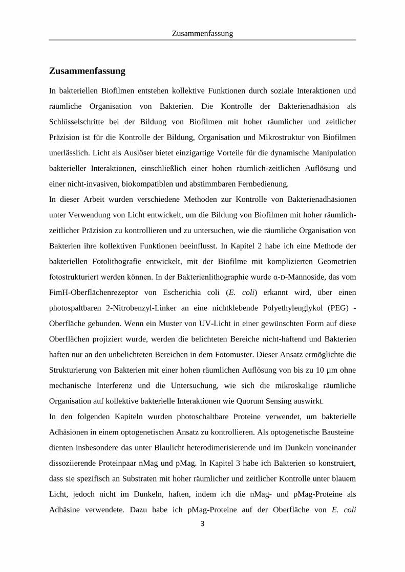
Biofilms can form on different surfaces including living tissues, implanted medical devices,
and industrial water systems. The variable nature of biofilms can be shown by fluorescence
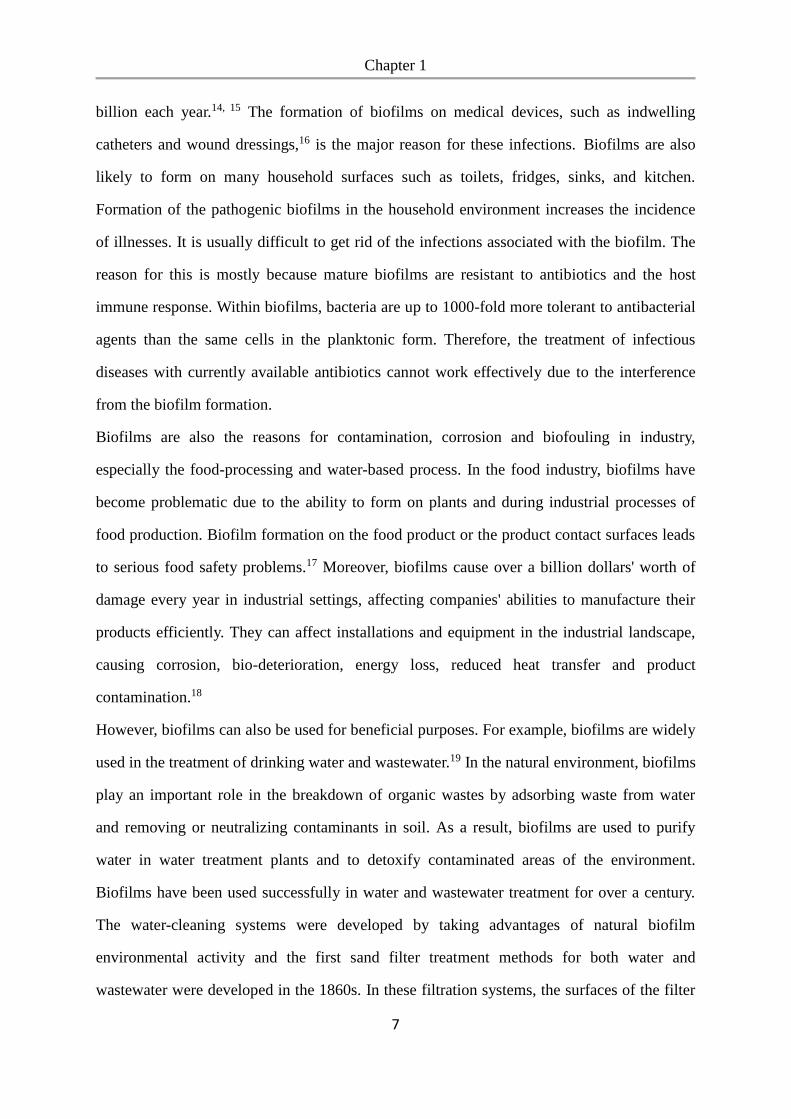
microscopy images of biofilms on a surface in a water system (Figure 1.1).5
Figure 1.1 Fluorescence microscopy images of biofilms on a stainless steel surface in a
laboratory potable water system. Bacteria were stained with 4, 6-diamidino-2-phenylindole
(DAPI).5 Scale bar is 20 µm. Adapted with permission from Ref. 5. Copyright 2002
Emerging Infectious Diseases journal.
1.1.1 The impact of biofilms: Detrimental and beneficial aspects
In 1978 Bill Costerton proposed the biofilm theory and reported that chronic infections in
patients with implanted medical devices were caused by bacterial biofilm formation and
bacteria within biofilms were resistant to antibiotic therapies and immune host defenses.
Since then there was an increasing awareness of the importance of biofilms study. A better
understanding of their specific properties will enable the development of effective strategies
for treatment.12 The National Institutes of Health (NIH) estimates that 80% of infectious
diseases worldwide are caused by biofilms.13 Biofilms cause severe infections in hospitalized
patients. In the United States, nosocomial (hospital-acquired) infections are reported to be the
leading cause of death with 1.7 million cases annually, leading to extra costs of up to $4.5
Chapter 1
7
billion each year.14, 15 The formation of biofilms on medical devices, such as indwelling
catheters and wound dressings,16 is the major reason for these infections. Biofilms are also
likely to form on many household surfaces such as toilets, fridges, sinks, and kitchen.
Formation of the pathogenic biofilms in the household environment increases the incidence
of illnesses. It is usually difficult to get rid of the infections associated with the biofilm. The
reason for this is mostly because mature biofilms are resistant to antibiotics and the host
immune response. Within biofilms, bacteria are up to 1000-fold more tolerant to antibacterial
agents than the same cells in the planktonic form. Therefore, the treatment of infectious
diseases with currently available antibiotics cannot work effectively due to the interference
from the biofilm formation.
Biofilms are also the reasons for contamination, corrosion and biofouling in industry,
especially the food-processing and water-based process. In the food industry, biofilms have
become problematic due to the ability to form on plants and during industrial processes of
food production. Biofilm formation on the food product or the product contact surfaces leads
to serious food safety problems.17 Moreover, biofilms cause over a billion dollars' worth of
damage every year in industrial settings, affecting companies' abilities to manufacture their
products efficiently. They can affect installations and equipment in the industrial landscape,
causing corrosion, bio-deterioration, energy loss, reduced heat transfer and product
contamination.18
However, biofilms can also be used for beneficial purposes. For example, biofilms are widely
used in the treatment of drinking water and wastewater.19 In the natural environment, biofilms
play an important role in the breakdown of organic wastes by adsorbing waste from water
and removing or neutralizing contaminants in soil. As a result, biofilms are used to purify
water in water treatment plants and to detoxify contaminated areas of the environment.
Biofilms have been used successfully in water and wastewater treatment for over a century.
The water-cleaning systems were developed by taking advantages of natural biofilm
environmental activity and the first sand filter treatment methods for both water and
wastewater were developed in the 1860s. In these filtration systems, the surfaces of the filter
Chapter 1
8
media act as a support for biofilms formation and the organic matter in the water is used by
biofilms as a carbon source. Biofilm-treated water requires less disinfectant and therefore
fewer disinfection byproducts form.7
The applications of microbial communities are greatly expanded due to the improved
understanding of natural microbial ecosystems and the development of new tools to construct
synthetic bacterial consortia. The synthetic biologists have developed engineered microbial
consortia for diverse applications, such as the bioproduction of drugs and other valuable
compounds.20 For example, E. coli and S. cerevisiae in a consortium can be used to produce
precursors of the anti-cancer drug paclitaxel. In this consortium, E. coli are engineered to
overproduce taxadiene, the scaffold molecule of paclitaxel, while S. cerevisiae are used as a
host for expressing cytochrome P450s (CYPs) to functionalize taxadiene through multiple
oxidation reactions. Therefore, the coculture of these two engineered organisms is essential
for the overall production of the drug.21-23
Due to the significant impacts of biofilm, there are increasing interests in the understanding
of biofilm formation and developing approaches for biofilm control. The understanding of
biofilm formation, structure and properties, communication and metabolic interactions will
lead to the promotion of beneficial uses and controlling the harmful impacts.
1.1.2 Biofilm formation and development
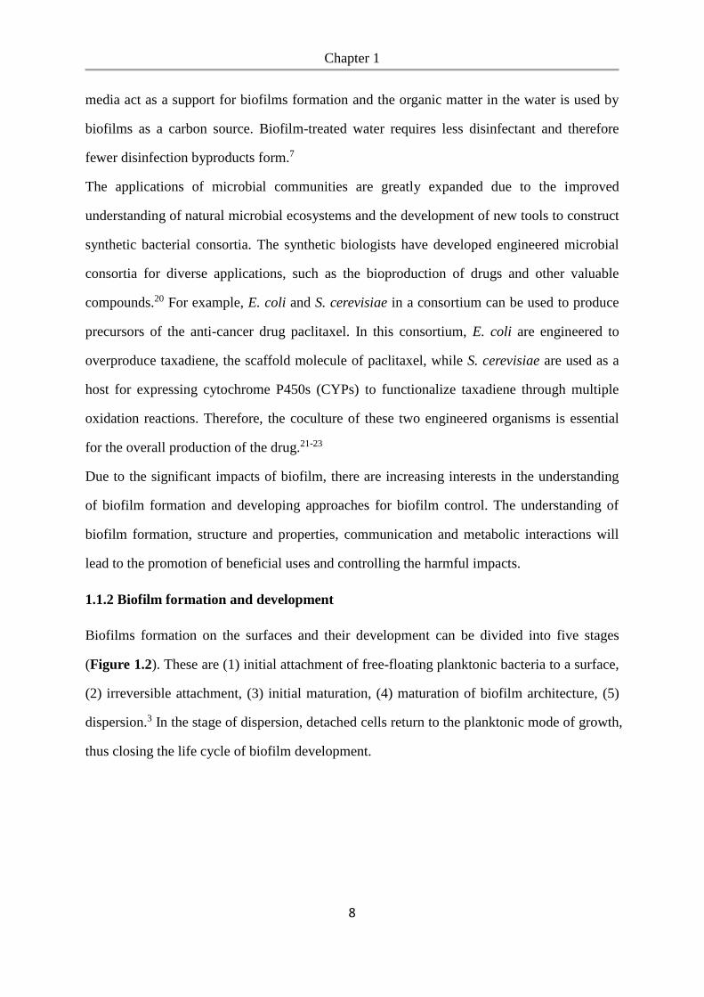
Biofilms formation on the surfaces and their development can be divided into five stages
(Figure 1.2). These are (1) initial attachment of free-floating planktonic bacteria to a surface,
(2) irreversible attachment, (3) initial maturation, (4) maturation of biofilm architecture, (5)
dispersion.3 In the stage of dispersion, detached cells return to the planktonic mode of growth,
thus closing the life cycle of biofilm development.
Chapter 1
9
Figure 1.2 The schematic shows a conceptual model of biofilm formation as a five-stage
developmental process. Stage 1: initial attachment. Stage 2: irreversible attachment. Stage 3:
initial maturation. Stage 4: maturation. Stage 5: dispersion.3 Adapted with permission from
Ref. 3. Copyright 2014 Springer Nature.
Initial attachment. Biofilm formation begins with the initial attachment of free-floating
planktonic bacteria to a surface, which is usually based on physical attachments including
hydrogen bonds, hydrophobic interactions, van der Waals forces, and electrostatic
interactions.24 The individual adherent cells on a surface are capable of independent
movement such as twitching or gliding which is mediated by pilus. Many of these adherent
cells may actually detach from the surface and return to the planktonic lifestyle if perturbed
by hydrodynamic forces, repulsive forces or in response to nutrient availability.25-27 Therefore,
the initial attachment is reversible.
Irreversible attachment. After initial attachment to a surface, bacteria must maintain the
adhesion and grow in order to develop a mature biofilm. They begin to secret
exopolysaccharide, form cell groups and adhere irreversibly. This transition from reversible
to irreversible attachment was firstly reported by Zobel in 1943. The presence of extracellular
matrix improves the interaction of the cells with the surface and induces a permanent cell-
surface bonding. The formation of cell aggregates and microcolonies has also been suggested
Chapter 1
10
as the mechanism for inducing irreversible and permanent attachment at a surface. In this
stage, rapid colonization at a new site on the surface is possible due to the bacterial twitching
motility, which is mediated by type IV pili located on the cell surface and used for propelling
bacteria across a surface.28, 29 O’Toole et al. suggested that the formation of microcolonies by
bacteria interactions at a surface helps to strengthen the attachment.30 Gerke et al. showed
that the formation of microcolonies was induced by a polysaccharide intercellular adhesin
which was produced by the adherent cells to bond the cells together.31
Maturation. In the maturation stages, many porous layers and water channels through the
biofilm are developed for cells to access essential nutrients.32 Many cells alter their
physiological processes and gene expression in response to conditions in their particular
surrounding such as surface contact triggers.33 The secretion of EPS is stimulated by the
chemical communication between cells and the best-characterized cell-to-cell communication
system is quorum sensing (QS). QS also regulates bacteria behavior and cellular functions
such as motility, which in turn could have an impact on biofilm structure. As cells replicate
and the EPS accumulates, biofilms develop into three dimensional (3D) structures which are
supported by EPS, allowing the transport of nutrients and removal of wastes. Additional
studies showed that cellulose, polyglucosamine (PGA) and colanic acid contribute to biofilm
architecture.34, 35
Dispersion. In the final stage of biofilm development, bacterial cells may detach from the
biofilm colony and return to the environment, which enables the spreading and colonization
at new sites. Enzymes such as dispersin B can degrade the biofilm extracellular matrix and
release the cells from the mature biofilm.36 The possible reasons for the biofilm dispersion
are the limited nutrients present at the original sites or the physical effects such as fluid shear
force.37 The detached bacterial cells may adhere to a new surface with a plenty of nutrients
and grow into a new biofilm. Overall, biofilm dispersion plays an important role in the self-
renewal of bacterial communities and is an essential process for the biofilm life cycle.
Chapter 1
11
1.1.3 Structure and property
Mature biofilms have complex structures with defined architectures that provide the essential
living conditions for cells. Many porous layers with water channels exist in biofilms,
allowing the cells in the entre of the colony to access nutrients and remove the wastes. The
structures of water channels act as a circulatory system for the delivery of nutrients and
wastes by diffusion and contribute to the development of biofilms.38, 39
EPS are considered as the primary matrix material of biofilms and the key determinant for
the material properties of biofilms. The production of EPS is affected by the nutrients supply
and bacterial growth. The EPS synthesis is improved when there are excess available carbon
and limited nitrogen, potassium, or phosphate. EPS production can also be enhanced when
bacterial growth is very slow. The physicochemical properties of EPS may vary in different
biofilms but the primary components of EPS are polysaccharides. Some gram-positive
bacteria such as the staphylococci have the cationic EPS polysaccharides. While for gram-
negative bacteria, these EPS polysaccharides are primarily neutral or polyanionic.
Additionally, EPS have the ability to incorporate water into the biofilm structure via
hydrogen bonding due to that EPS are highly hydrated. It appears that the EPS determine b
the structure and thereby cohesive strength of biofilms. The backbone structures of biofilms
that contain 1, 3 - or 1, 4 - β - linked hexose residues are more rigid, less deformable and
insoluble. Furthermore, EPS act as a protection to the bacterial cells living in biofilms. It has
be reported protective EPS (alginate) can protect mucoid P. aeruginosa FRD1 biofilm cells
from exposure to ultraviolet radiation.40 EPS may also contribute to the antimicrobial
resistance properties of biofilms by preventing the transfer of antibiotics through the
biofilm.41
The structure of biofilms are also affected by the environmental factors. In an aqueous
environment, shear forces from the passage of fluid over the biofilms shape the
microcolonies into different morphologies within biofilms.42, 43
Chapter 1
12
1.1.4 Quorum sensing
Cell-to-cell communication is a main ingredient of biofilm formation. Bacteria within
biofilms interact with each other and function as a group for coordinated activities. Bacteria
that have aggregated into biofilms can chemically communicate through quorum sensing,
which was first reported and described in Vibrio fischeri (the marine bioluminescent bacteria)
Bacteria have the ability called quorum sensing to detect the changes of cell population
density and alter the gene regulation accordingly.41, 44
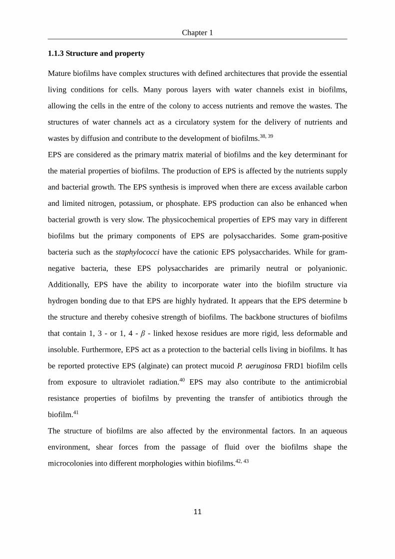
Bacteria can produce and release chemical signal molecules called autoinducers that increase
in concentration as a function of cell density. The detection of a minimal threshold
stimulatory concentration of an autoinducer leads to an alteration in gene expression (Figure
1.3). For example, the expression of genes for antibiotic resistance at high cell densities is
activated through quorum sensing, providing protection for bacteria within biofilms.45, 46 The
high cell concentrations in biofilms enclosed in a matrix allows for quorum sensing even in
small microcolonies as the signaling compounds are be concentrated within the
microcolonies and are not degraded. Quorum sensing also plays an important role in the
regulation of metabolic interaction in biofilms and influences community structure by
enhancing encouraging the growth of beneficial species and inhibiting the growth of
competitors.47 The functions that can be regulated by quorum sensing include
bioluminescence, nitrogen fixation and sporulation.48, 49
Chapter 1
13
Figure 1.3 Schematic illustration of quorum sensing. a) Bacterial cell has the ability to
produce a signaling molecule called autoinducer and sense its extracellular concentration. b).
Quorum sensing is dependent on cell density.46 Adapted with permission from Ref. 46.
Copyright 2011 EMBO Press.
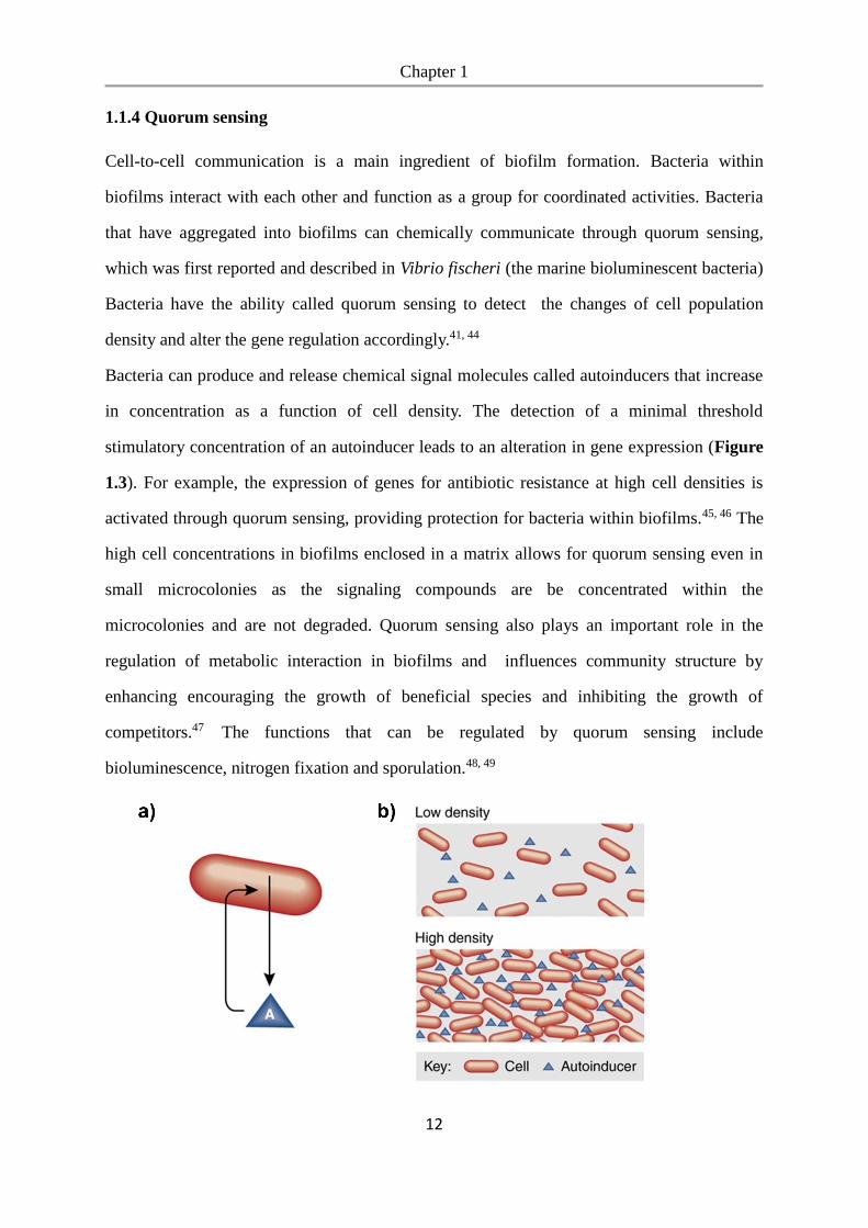
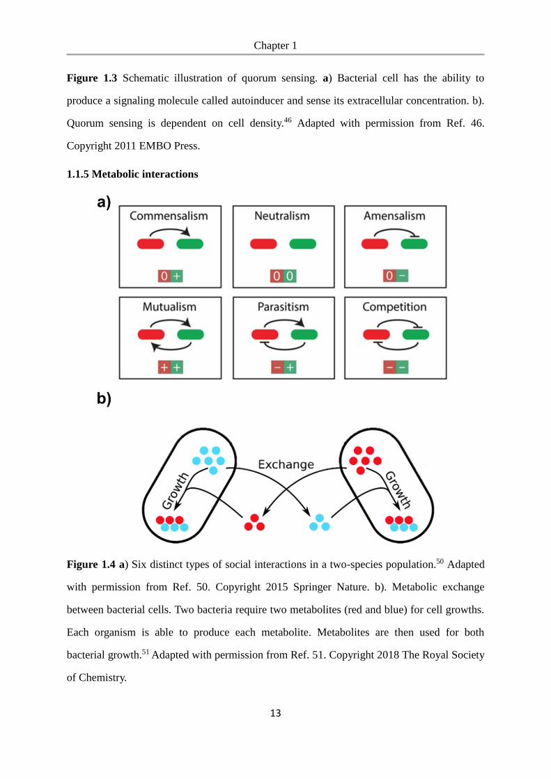
1.1.5 Metabolic interactions
Figure 1.4 a) Six distinct types of social interactions in a two-species population.50 Adapted
with permission from Ref. 50. Copyright 2015 Springer Nature. b). Metabolic exchange
between bacterial cells. Two bacteria require two metabolites (red and blue) for cell growths.
Each organism is able to produce each metabolite. Metabolites are then used for both
bacterial growth.51 Adapted with permission from Ref. 51. Copyright 2018 The Royal Society
of Chemistry.
Chapter 1
14
Bacteria in nature usually do not exist in isolation but in complex ecosystems. In order to
survive and proliferate in such complex consortia, bacteria have developed diverse social
interactions with their neighboring species. The social interactions of bacteria significantly
affect the dynamics and functionality of an entire community by altering the physiology, gene
expression, survival of individual cells and enabling the collective behaviors of populations.
These interactions can have three types of outcomes: a positive impact (win, +), a negative
impact (loss, -) and no impact (neutral, 0) on the bacterial species involved.52 For a simple
ecosystem consisting of only two species, there are six possible distinct types of interaction
through the possible combinations of the three outcomes, including neutralism (0, 0),
commensalism (0, +), amensalism (0, -), competition (-, -), mutualism (+, +), and parasitism
(-, +).50 (Figure 1.4a)
Metabolic cross-feeding is one type of social interaction. Generally, there are two types of
metabolic interactions: competition and cooperation. Due to the limited nutritional resources
in most ecosystems, bacteria have to compete constantly for resources. The ability to compete
for limiting nutrients can determine whether bacteria will be able to survive in a particular
site.53, 54 Besides the competitive metabolic interactions, cooperative metabolic interactions
which allow bacteria to get benefits from each other through metabolic, are more common
among bacterial species.45, 55 Metabolic cross-feeding, also named syntrophy, is one type of
cooperation in which bacteria exchange essential metabolites.56 Cross-feeding is an important
process that governs the growth and composition of microbial ecosystems.57 Bacteria use
metabolic exchanges as a strategy for group success58 and under nutrient-poor conditions
complement each other’s biosynthetic capabilities.59 Previous studies have characterized the
behavior of microbes in co-culture with naturally complementary metabolism60-62 where
shared metabolites are hydrogen,62 acetate,61 amino acids,63 fixed nitrogen and glucose.64 For
example, the presence of a bacterial species that actively produces beneficial molecules such
as vitamins or amino acids allows both the producing bacteria and other species in the
environment to use them and relaxes the metabolic burden on all species in the community.51
(Figure 1.4b) Metabolic interactions are effected by the spatial structure of bacterial
Chapter 1
15
community or biofilms, cost-effectiveness of biosynthesis, nutrients and diffusion.65, 66 For
example, bacterial aggregation or biofilm formation improves the efficiency of the
metabolites exchange and stimulates the metabolic processes. 67
These principles observed in natural biofilms have also been implemented into genetically
engineered bacteria for biotechnological applications.63, 68 For example, the introduction of
genetic modifications into no-interacting bacterial cells could induce the persistent
cooperation. Wenying Shou et al. constructed a synthetic synthetic cooperative system
comprising a yeast pairs and each of them produce and offering the other strain an essential
metabolite.63 An example of parasitic interactions developed by Frederick et al.,
constructed a synthetic predator-prey ecosystem consisting of two E. coli populations.
The predator cells kill the prey by inducing expression of a killer protein in the prey,
while the prey rescue the predators by eliciting expression of an antidote protein in the
predator.68 Therefore, understanding principles that govern biofilms are not just important
from the standpoint of scientific understanding but also have direct implications in
biotechnology.
1.2 Bacterial adhesion
1.2.1 Bacteria-surface adhesion
The biofilm formation begins from the adhesion of bacterial planktonic cells to the
surface. Once a biofilm has formed, the bacteria are extremely resistant to treatment
with antimicrobial agents and difficult to be removed. Therefore, controlling the initial
bacterial adhesion to a surface is the most effective method to prevent the biofilm
formation. Moreover, controllable bacterial adhesion allows bacterial cell patterning
and engineered biofilm formation with the designed spatial organization, which are
important for understanding social cell interactions and chemicals exchange within
biofilms.
The bacterial adhesion can be affected by multiple factors such as bacterial surface
structures, physiochemical properties of the substrate, and environmental conditions.72
Chapter 1
16
The most important factors influencing bacteria adhesion include surface charge,
hydrophobicity, topography or roughness, and the exposed functional groups.73, 74
In general, adhesion of bacteria is prevented on negatively charged surfaces, while it is
promoted on positively charged surfaces.75 This is because most bacteria are
negatively charged on cell surfaces. Positively charged polymer surfaces have been
reported to be bactericidal because the positive charge can disrupt the bacterial
membrane potential or damage the membrane structure.76, 77 Therefore, polycationic
surfaces are often suggested to be efficient antibacterial coatings that bind and kill
bacteria.78
Surface hydrophobicity is another major component that influences the bacteria–
surface interaction. Hydrophobic interaction is one of the strongest noncovalent
interactions in biological systems and plays a major role in bacterial adhesion to
surfaces.79 The bacterial adhesion is also affected by the hydrophobicity of the
bacterial cells. Bacteria with a more hydrophobic cell surface preferentially attach to
hydrophobic surfaces and vice versa.80, 81
Surface topography has also been found to substantially influence the interaction
between bacteria and surfaces. Perera-Costa et al. investigated the effect of surface
topography on bacterial adhesion by using the polydimethylsiloxane (PDMS) surfaces
that contained microtopographic patterns in spatial organization.82 This study showed
a significant reduction in bacterial adhesion (30-45%) on microstructured surfaces
compared to the control. Another important parameter for bacterial adhesion on the
topographic surfaces is the spatial distribution of structures and patterns, which is
relative to bacterial size and shape. It has been reported that surfaces with topographic
features much smaller than bacterial cells inhibit bacterial attachment due to the
decreased contact area between bacteria and surfaces.83-85 On the contrary, for surfaces
with topographic features that comparable with the bacterial size, more bacteria attach
compared to smooth surfaces.86 This is because that the microscopic structure on the
surface tends to increase the overall surface roughness and therefore enhances the
Chapter 1
17
adhesion of bacteria to substrates as more surface area is provided for bacterial
attachment.87, 88
1.2.1.1 Controlling bacterial adhesion using surface chemistry
Surface modification with functional groups presents an important strategy for
controlling bacterial adhesion and the formation of biofilms. For example,
poly(ethylene glycol) (PEG) has been widely used for surface modification as anti-
adhesive coatings cause it shows a great capacity to resist protein adsorption and cell
adhesion.89 However, adhesive surfaces with bio-specific binding properties and
minimized background interferences are required for the fundamental study of
bacterial cell-cell communication and biofilm formation.
1.2.1.1.1 Controlling bacterial adhesion by modifying the surface with native adhesion
molecules
To construct an adhesive surface for bacteria with the ability to form a robust, specific,
irreversible adhesions, native adhesive molecules have been used as exposed
functional groups on surfaces. During the initial attachment in the first stage of biofilm
formation, bacteria employ specific cell surface receptors, called adhesins, that bind to
the substrate through specific receptor ligands for the irreversible attachment.90, 91
Bacterial adhesins serve as anchors and act as specific surface recognition molecules,
allowing the binding to specific receptor molecules on host cells or target surfaces.92
The best characterized bacterial adhesin is the FimH.93 FimH is an α-D-mannoside
specific lectin located at the tips of adhesive organelles, called type 1 fimbriae.94 FimH
lectin mediates binding to glycoproteins that have N-linked oligosaccharides
presenting terminal mannose residues.95 This specific FimH based adhesin–
carbohydrate adhesion of bacteria to mannosylated surfaces has been used for many
applications in the field of medicine. For example, carbohydrate microarrays have
been used to detect pathogens and screen anti-adhesion therapeutics based on the
carbohydrate binding specificities of bacteria.96-99
Chapter 1
18
1.2.1.1.2 Controlling bacterial adhesion by engineering bacteria surface with adhesion
molecules
An alternative powerful approach to control bacterial adhesion is to engineer the bacterial
surface with new adhesion molecules. The ability to modify the surfaces of bacteria cells with
non-native molecules is vital to engineer bacterial communication, biofilm formation and cell
behavior in synthetic biology.
One effective strategy to add molecules to the bacterial cell membrane is called bacterial
display, which uses genetic methods to fuse a protein or a peptide with a transmembrane
protein. Sankaran et al. reported a novel method to control the specific, dynamic and
reversible bacterial adhesion based on a supramolecular interaction between a peptide
displayed on the bacterial surface and cucurbit[8]uril (CB[8]) They genetically modified E.
coli such that a transmembrane protein displayed a CB[8]-binding motif at the bacterial
surface. The binding of this motif to CB[8] and formation of intercellular complexes induce
the bacterial aggregation within the solution in the presence of CB[8] and specific adhesion
to CB[8] modified surfaces. Furthermore, these adhesions can be chemically reversed using
an excess of CB[8] as a competitor.100
Another strategy is to introduce molecules to the bacterial cell surface through chemical
ligation to membrane proteins or carbohydrates. For example, Elahipanah et al. introduced
bio-orthogonal groups to engineer the surface of gram-negative bacteria cells by using a
liposome fusion based method. These groups can subsequently be conjugated to a range of
molecules for further studies on bacterial adhesion and controlling bacterial behavior.101
1.2.1.2 Controlling bacterial adhesion with light
Many stimuli-responsive methods have been proposed for the control of bacterial adhesion
and for bacteria patterning. Among them, the light-responsive methods provide the highest
spatial and temporal resolution, which is required to construct stable/viable biofilms with a
high level of precision. This is particularly significant as the arrangement of bacteria with
respect to each other at the micro scale defines the extent of their interaction in a biofilm as
Chapter 1
19
described above. Additionally, light is a noninvasive trigger as it can be applied remotely
without disturbing of other processes. In contrast, invasive triggers, such as chemicals, can
have unpredictable side effects in the system by interacting with not only the targets but other
biomolecules and do not provide the desired high spatiotemporal control. Moreover, it is easy
to adjust the light intensity, illumination time and wavelengths to tune interactions and
address different functionalities which respond to different colors of light independently.
Therefore, the unique advantages of light make it particularly attractive as an external
stimulus to regulate diverse biological processes.
1.2.1.2.1 Controlling bacterial adhesion by modifying surfaces with light responsive
molecules
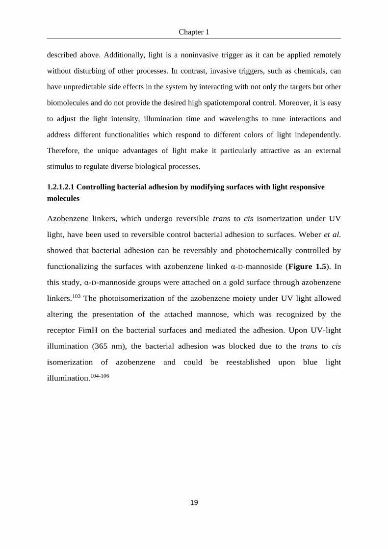
Azobenzene linkers, which undergo reversible trans to cis isomerization under UV
light, have been used to reversible control bacterial adhesion to surfaces. Weber et al.
showed that bacterial adhesion can be reversibly and photochemically controlled by
functionalizing the surfaces with azobenzene linked α-D-mannoside (Figure 1.5). In
this study, α-D-mannoside groups were attached on a gold surface through azobenzene
linkers.103 The photoisomerization of the azobenzene moiety under UV light allowed
altering the presentation of the attached mannose, which was recognized by the
receptor FimH on the bacterial surfaces and mediated the adhesion. Upon UV-light
illumination (365 nm), the bacterial adhesion was blocked due to the trans to cis
isomerization of azobenzene and could be reestablished upon blue light
illumination.104-106
Chapter 1
20
Figure 1.5 Photoswitchable adhesion of type 1 fimbriated E. coli cells to the surface
immobilized with α-D-mannoside ligands via the azobenzene linkers.101 Reproduced
with permission from Ref. 101. Copyright 2014 WILEY-VCH Verlag GmbH & Co.
KGaA.
Nitrobenzyl groups have also been widely used as UV-light cleavable linkers in
surface coating. To control cell-surface interactions, the nitrobenzyl group has been
used to attach anti-adhesive PEG coatings as a linker to surfaces. Upon UV-light
irradiation, the PEG coatings release and the surfaces become adhesive for cell
adhesion.107 For example, mammalian cells have been patterned in confined and
complex geometries using photocleavable 2-nitrobenzyl groups via projection
exposure to UV light through a photomask.108 Similarly, these photocleavable groups
have been used as a caging group on the backbone of RGD to block mammalian cell
adhesion and to turn on cell adhesion upon illumination.109 Yet, analogous approaches
are missing for bacterial cells.
1.2.1.2.2 Optogenetic control of bacterial adhesion
Recently, optogenetic methods have been developed to control bacterial adhesion and
patterning using light with unique advantages including high spatiotemporal control,
Chapter 1
21
tunability and non-invasive remote regulation. These optogenetic approaches use light
controlled protein activity and genetic engineering of the cells to render them light
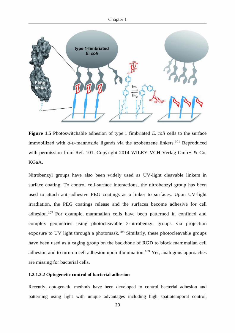
responsive. For example, Jin and Riedel-Kruse have developed a genetically encoded biofilm
patterning tool (“Biofilm Lithography”) by engineering bacteria such that the expression of
membrane adhesion proteins antigen 43 (Ag43) responsible for surface attachment is
optically regulated.110 (Figure 1.6) Accordingly, these E. coli only formed biofilm on blue
illuminated regions of the surface and could be patterned with 25 μm spatial
resolution. Huang et al. reported a similar strategy for microprinting living biofilms through
optogenetic regulation of the c-di-GMP levels, which regulates biofilm formation in P.
aeruginosa. In the presence of near-infrared light, the synthesis of c-di-GMP molecules was
activated through the cyclization of the guanosine triphosphate (GTP) molecules, regulated
by the genes bphO and bphS. While illuminated with blue light, c-di-GMP molecules were
hydrolyzed due to the activation of gene blrP1. Therefore, combining these optogenetic
modules enabled the precise manipulation of the c-di-GMP levels in P. aeruginosa through
dual-color illumination.111
Figure 1.6 a) Schematic illustration of biofilm photolithography. b) Biofilm patterns created
by biofilm lithography.110 Reproduced with permission from Ref. 110. Copyright 2018
National Academy of Sciences.
Chapter 1
22
1.2.2 Bacteria-bacteria adhesion
Adhesions between bacterial cells to form multicellular clusters are crucial for the
development of biofilm structures. Bacterial aggregation plays an important role in a variety
of ecological processes such as competition, adaptation, epidemics, and succession.112
Bacteria-bacteria adhesions are also a key factor for regulating spatial organization and
heterogeneity within biofilms. Manipulation of bacteria-bacteria adhesion has potential
applications in many areas including antimicrobial therapy, modulation of bacterial signaling
such as quorum sensing and engineering multicellular communities and microbial consortia.
Therefore, multiple strategies have been proposed to control bacteria-bacteria adhesion and
associated cell behavior and communication.
1.2.2.1 Controlling bacterial aggregation with native adhesion molecules
Antigen 43 (Ag43) is a surface-located autotransporter protein and one major
determinant of autoaggregation in E. coli. The interactions between Ag43 α-subunits
of adjacent cells in a head-to-tail fashion lead to dimer formation and cell
aggregation.113 Therefore, bacterial aggregation can be controlled by regulating the
Ag43 expression through the gene OxyR or Dam.114
Laganenka et al. showed that autoinducer 2 (AI-2) produced by the bacteria itself is an
attractant and leads to chemotaxis towards leading to autoaggregation of E. coli. Furthermore,
AI-2-dependent autoaggregation enhanced bacterial stress resistance and promoted biofilm
formation.115 It was also reported that cells of chemotactic bacterial strains aggregated in
response to gradients of attractant and this interaction led to collective phenomena such as the
formation of dense multicellular clusters, moving bands and geometric patterns.116-118
1.2.2.2 Triggering bacterial aggregation with external molecules
Synthetic materials with multivalent interactions have been used to induce bacterial
aggregation in order to prevent infections at an early stage and blocking the interaction
between microbial adhesins and host epithelial cell receptors. Examples of these are
polysaccharide, polymers, dendrimers or chemically modified nanoparticles.119-124
Chapter 1
23
Cationic polymers are an obvious starting point in the development of antimicrobial agents
that cluster bacteria. Because of their positive charge, these polymers can efficiently bind the
negatively charged bacterial surfaces and result in the aggregation of these bacteria. In
addition, adhesion of cationic materials to bacterial membranes can result in membrane
damage and have bactericidal activity.121, 125 Lui et al. reported a cationic polymer poly(N-[3-
(dimethylamino)propyl]methacrylamide) induced bacterial aggregation through electrostatic
interactions.126 Furthermore, Vibrio harveyi showed enhanced bioluminescence in response to
polymer-mediated clustering, indicating the quorum sensing was activated upon clustering.
Bacteria-polymer aggregates undergo rapid autoinduction and achieve quorum sensing at
bacterial densities far below those required for autoinduction in the absence of polymers. 126-
128
Glycopolymers carrying carbohydrate functional groups have also been widely reported for
the controllable bacterial aggregation.129 They can interact with the lectins on the surface of
bacteria and induce the bacterial aggregation. Pasparakis et al. described a reversible control
of bacterial aggregation by thermoresponsive glycopolymers.130 They synthesized the
polymer with multiple glucose moieties, which were hidden above 40 °C and revealed below
this temperature. This thermal switchable process enabled the controllable bacterial
aggregation based on the interaction of glucose and lectin.
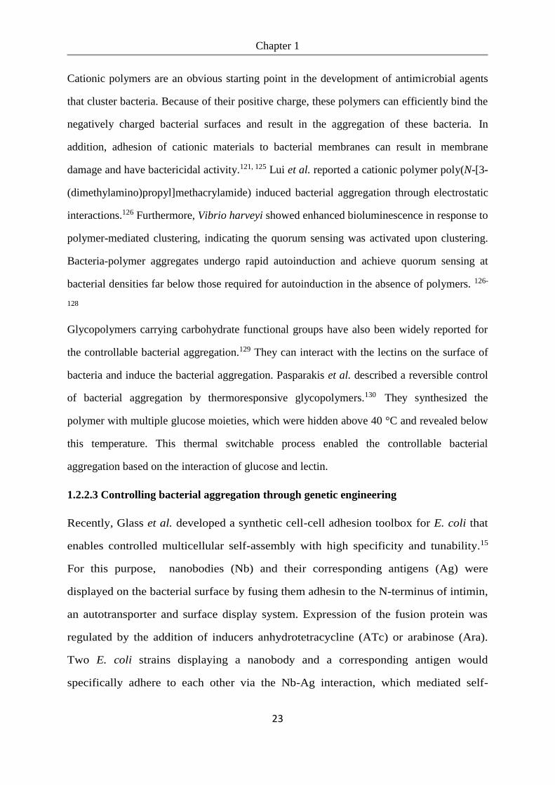
1.2.2.3 Controlling bacterial aggregation through genetic engineering
Recently, Glass et al. developed a synthetic cell-cell adhesion toolbox for E. coli that
enables controlled multicellular self-assembly with high specificity and tunability.15
For this purpose, nanobodies (Nb) and their corresponding antigens (Ag) were
displayed on the bacterial surface by fusing them adhesin to the N-terminus of intimin,
an autotransporter and surface display system. Expression of the fusion protein was
regulated by the addition of inducers anhydrotetracycline (ATc) or arabinose (Ara).
Two E. coli strains displaying a nanobody and a corresponding antigen would
specifically adhere to each other via the Nb-Ag interaction, which mediated self-
Chapter 1
24
assembly of multicellular aggregates with defined patterns and morphologies (Figure
1.7). Furthermore, this toolbox enabled the rationally design of diverse and complex
multicellular patterns.
Figure 1.7 a) Bacteria-bacteria aggregation based on the interactions of nanobody and
antigen. b) The morphology and patterning of bacterial clusters altered by the ratio of green
and red cells.131 Reproduced with permission from Ref. 131. Copyright 2018 Elsevier.
For natural multicellular organisms, cell-cell adhesion is the key tool for directing the
spatial organization. To produce biofilms with a deliberate arrangement, methods for
dynamically controlling bacteria-bacteria adhesions with high spatial and temporal
precision are required.11-13 However, none of the existing approaches provide the required
crucial dynamic and spatiotemporal control over the bacteria-bacteria adhesions.
1.3 Photoswitchable proteins for optogenetic control
Optogenetic tools are genetically encored photoswitchable proteins used for regulating
cellular processes with light as an external stimulus.132, 133 Optogenetics has been used to
regulate diverse cellular functions with visible light, including receptor activation, gene
expression, enzyme activity, protein clustering and protein localization both in mammalian
and bacterial cells.134-137 As part of this thesis the photoswitchable proteins nMag and pMag
have been used and will be detailed below.
Chapter 1
25
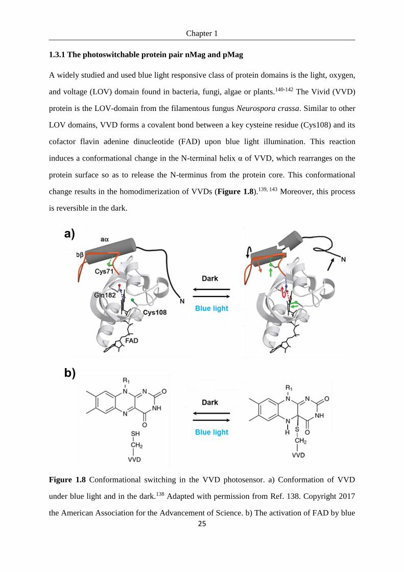
1.3.1 The photoswitchable protein pair nMag and pMag
A widely studied and used blue light responsive class of protein domains is the light, oxygen,
and voltage (LOV) domain found in bacteria, fungi, algae or plants.140-142 The Vivid (VVD)
protein is the LOV-domain from the filamentous fungus Neurospora crassa. Similar to other
LOV domains, VVD forms a covalent bond between a key cysteine residue (Cys108) and its
cofactor flavin adenine dinucleotide (FAD) upon blue light illumination. This reaction
induces a conformational change in the N-terminal helix α of VVD, which rearranges on the
protein surface so as to release the N-terminus from the protein core. This conformational
change results in the homodimerization of VVDs (Figure 1.8).139, 143 Moreover, this process
is reversible in the dark.
Figure 1.8 Conformational switching in the VVD photosensor. a) Conformation of VVD
under blue light and in the dark.138 Adapted with permission from Ref. 138. Copyright 2017
the American Association for the Advancement of Science. b) The activation of FAD by blue
Chapter 1
26
light illumination leads to the formation of a photoadduct between the flavin ring and
VVD.139 Adapted with permission from Ref. 139. Copyright 2019 Springer Nature.
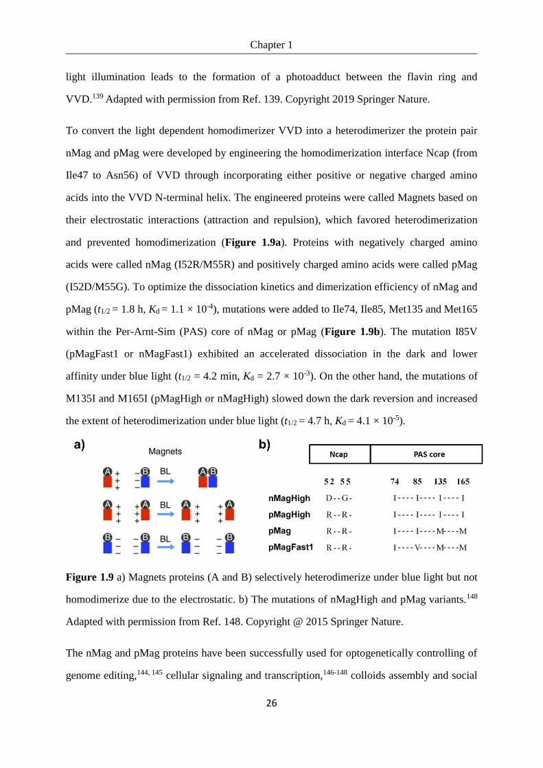
To convert the light dependent homodimerizer VVD into a heterodimerizer the protein pair
nMag and pMag were developed by engineering the homodimerization interface Ncap (from
Ile47 to Asn56) of VVD through incorporating either positive or negative charged amino
acids into the VVD N-terminal helix. The engineered proteins were called Magnets based on
their electrostatic interactions (attraction and repulsion), which favored heterodimerization
and prevented homodimerization (Figure 1.9a). Proteins with negatively charged amino
acids were called nMag (I52R/M55R) and positively charged amino acids were called pMag
(I52D/M55G). To optimize the dissociation kinetics and dimerization efficiency of nMag and
pMag (t1/2 = 1.8 h, Kd = 1.1 × 10⁻⁴), mutations were added to Ile74, Ile85, Met135 and Met165
within the Per-Arnt-Sim (PAS) core of nMag or pMag (Figure 1.9b). The mutation I85V
(pMagFast1 or nMagFast1) exhibited an accelerated dissociation in the dark and lower
affinity under blue light (t1/2 = 4.2 min, Kd = 2.7 × 10⁻³). On the other hand, the mutations of
M135I and M165I (pMagHigh or nMagHigh) slowed down the dark reversion and increased
the extent of heterodimerization under blue light (t1/2 = 4.7 h, Kd = 4.1 × 10⁻⁵).
Figure 1.9 a) Magnets proteins (A and B) selectively heterodimerize under blue light but not
homodimerize due to the electrostatic. b) The mutations of nMagHigh and pMag variants.148
Adapted with permission from Ref. 148. Copyright @ 2015 Springer Nature.
The nMag and pMag proteins have been successfully used for optogenetically controlling of
genome editing,144, 145 cellular signaling and transcription,146-148 colloids assembly and social
Chapter 1
27
sorting149. The tunable binding affinities and kinetics make Magnets a very versatile class of
photoswitchable proteins and allow for precise subcellular control of protein association with
high spatial and temporal resolution.
Chapter 2
28
Chapter 2. Bacterial Photolithography: Patterning Escherichia coli biofilms
with high spatiotemporal control using photocleavable adhesion molecules
Copyright
The following chapter is based on the publication Chen, F., Ricken, J., Xu, D., Wegner, S. V.,
Adv. Biosys., 2019, 3, 1800269-1800274. The results are reprinted with permission from the
WILEY-VCH Verlag GmbH & Co. KGaA.
Aim
Bacteria in nature usually do not only exist as free-floating cells but predominantly live in a
biofilm, where collective functions arise from the social interactions and spatial organization
of bacteria. Controlling bacterial adhesion as key steps in biofilm formation with high spatial
and temporal precision is essential for controlling the formation, organization and
microstructure of biofilms. To study how the spatial arrangement of bacteria influences their
social interactions, such as quorum sensing, this work focuses on developing a method of
patterning bacteria into complex geometries with high resolution. Photocleavable adhesion
molecules can be synthesized to functionalize the surface and thereby bacterial attachment
can be controlled by UV light. Using this method, patterning biofilms with high resolution
can be obtained and provides a tool to study the spatial organization of bacterial cells on the
quorum sensing.
Contributions
I performed most of the experiments and analysis including the synthesis of mannoside-NO2,
UV-vis spectroscopy, glass surfaces functionalization, bacterial attachment and patterning,
live imaging of patterned biofilms and quorum sensing. Julia Riecken purified the compound
mannoside-NO2. Dongdong Xu did the glass slides passivation. Seraphine V. Wegner
proposed the idea and led the project.
Chapter 2
29
2.1 Abstract
Biofilms are not only a leading cause of chronic infections and biofouling, but they also have
a tremendous positive potential in biotechnology for biocatalysis and waste treatment.
Biofilms are spatially structured communities of microbes, which exchange chemicals and
communicate with each other. By spatially controlling bacterial adhesion to surfaces and
therefore the microstructure of biofilms, we have developed a promising method of
understanding social interactions between bacteria and designed biofilms. The bacterial
photolithography approach described here allows us to photopattern specific bacteria
adhesion molecules, to control surface adhesion, and to guide the formation of biofilms. To
do this, α-D-mannoside, which is recognized by Escherichia coli FimH receptor, is linked to
a non-adhesive poly(ethylene glycol) (PEG) surface through a photocleavable 2-nitrobenzyl
linker. When a pattern of UV light in a specific shape is projected onto these surfaces, the
light-exposed areas become non-adhesive and bacteria only adhere to the dark, unexposed
areas in the photopattern. Bacterial photolithography enables bacterial patterning with high
spatial resolution down to 10 µm without mechanical interference. Additionally, patterning
biofilms with complicated geometries allows us to study the importance of microscale spatial
organization on the collective behavior of bacteria such as quorum sensing.
2.2 Introduction
Biofilms are an emergent form of bacterial life as they allow bacteria to survive in
hostile environments and to resist antimicrobial agents.150, 151 In order to form a
biofilm, bacteria first need to adhere to surfaces before they begin to excrete
substances that can anchor them to all kinds of material.10 The formation of biofilms is
a major cause of chronic infections152 and persistent biofouling.153 Recent research has
highlighted the potential of engineered biofilms in biotechnology for antibiofouling,154
biocatalysis,155 biosensing,156 bioremediation157 and water treatment.158 The spatial
structure of the biofilms on the micrometer scale allows bacteria to work together and
perform biochemical transformations, which planktonic bacteria cannot catalyze by
Chapter 2
30
themselves.159, 160 To control the spatial structure of biofilms more efficiently and to
investigate its specific effect on the biofilm’s function as well as collective phenomena
such as quorum sensing, we need to establish a reliable way to obtain patterned
biofilms with high resolution.
The formation of a biofilm begins with the adhesion of free-floating bacteria to a
surface, which determines the later spatial organization in biofilms.153, 161 This is
particularly significant as the arrangement of bacteria with respect to each other
defines the extent of their interaction and exchange of chemicals present in a biofilm.
Different strategies have been proposed to control bacterial adhesion and to pattern
bacteria on surfaces, such as microfluidic devices,59 microcontact printing,162 inkjet
printing,163 hydrogels patterning,164 substrates modified with stimuli-responsive
chemicals,103 and optogenetic methods.165-167 Among these approaches, the light-
responsive methods provide the highest spatial and temporal resolution, are the least
invasive and provide remote controlled. All these factors are required to construct
stable/viable biofilms with a high level of precision. For instance, azobenzene linkers,
which undergo reversible trans to cis isomerization when exposed to UV light, have
been used to control bacterial adhesion to surfaces by exposure to light. This is
achieved by altering the orientation or the presence of mannoside groups, which are
recognized by the bacterial surface adhesion receptor FimH.103, 104, 168 Likewise, in
optogenetic approaches, bacterial adhesion has been dynamically controlled in
genetically modified bacteria in response to low intensities of blue or red light by
either controlling protein expression with light or displaying light responsive protein
pairs on the bacteria surface.165-167 Yet, these optogenetic methods require the genetic
manipulation of bacteria and expression light responsive proteins by the bacterial.
While the reversible control provided by the azobenzene-based approaches and the
optogenetic approaches is an advantage to switch adhesions dynamically, it also
requires constant illumination with light. This limits the practical use for long term
studies and increases the risk of light toxicity, which is especially problematic in the
Chapter 2
31
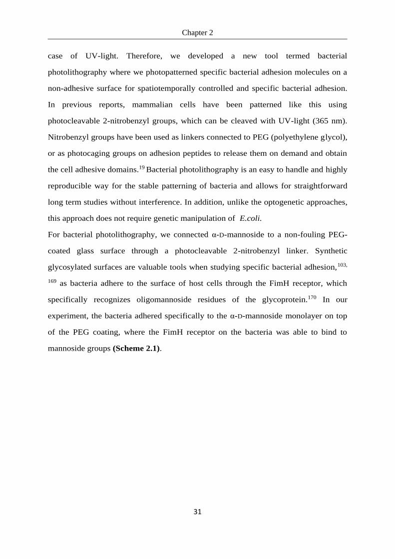
case of UV-light. Therefore, we developed a new tool termed bacterial
photolithography where we photopatterned specific bacterial adhesion molecules on a
non-adhesive surface for spatiotemporally controlled and specific bacterial adhesion.
In previous reports, mammalian cells have been patterned like this using
photocleavable 2-nitrobenzyl groups, which can be cleaved with UV-light (365 nm).
Nitrobenzyl groups have been used as linkers connected to PEG (polyethylene glycol),
or as photocaging groups on adhesion peptides to release them on demand and obtain
the cell adhesive domains.19 Bacterial photolithography is an easy to handle and highly
reproducible way for the stable patterning of bacteria and allows for straightforward
long term studies without interference. In addition, unlike the optogenetic approaches,
this approach does not require genetic manipulation of E.coli.
For bacterial photolithography, we connected α-D-mannoside to a non-fouling PEG-
coated glass surface through a photocleavable 2-nitrobenzyl linker. Synthetic
glycosylated surfaces are valuable tools when studying specific bacterial adhesion,103,
169 as bacteria adhere to the surface of host cells through the FimH receptor, which
specifically recognizes oligomannoside residues of the glycoprotein.170 In our
experiment, the bacteria adhered specifically to the α-D-mannoside monolayer on top
of the PEG coating, where the FimH receptor on the bacteria was able to bind to
mannoside groups (Scheme 2.1).
Chapter 2
32
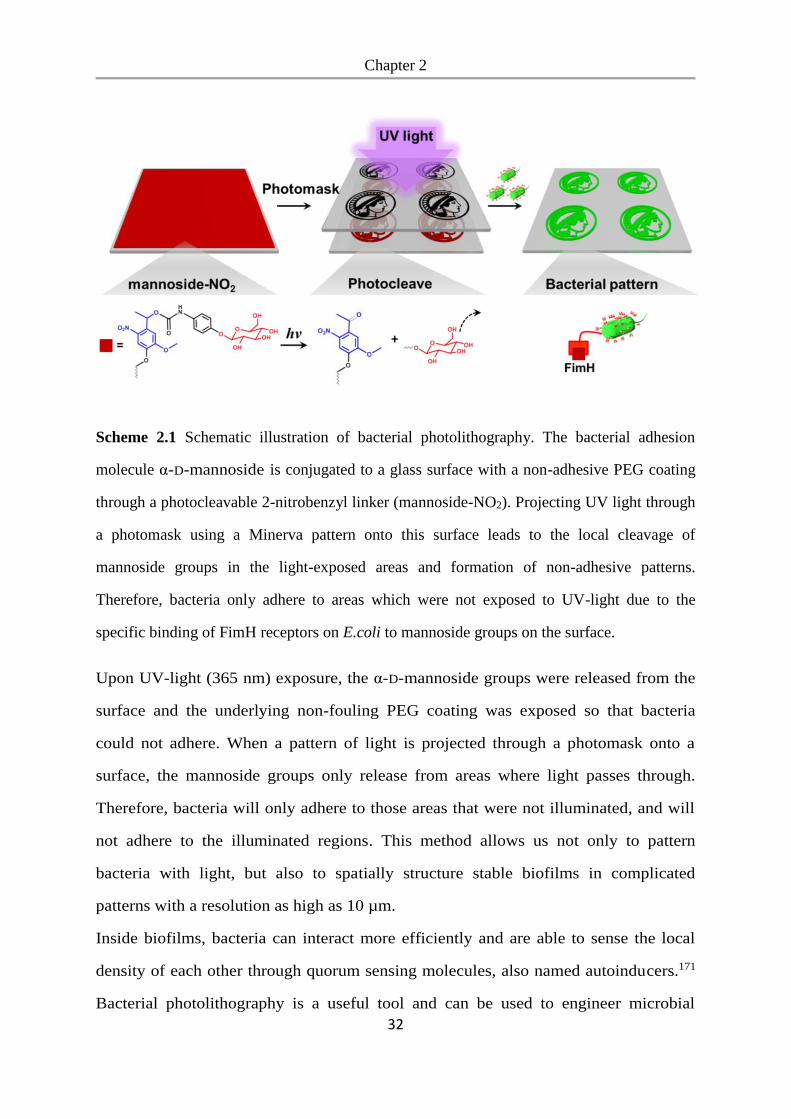
Scheme 2.1 Schematic illustration of bacterial photolithography. The bacterial adhesion
molecule α-D-mannoside is conjugated to a glass surface with a non-adhesive PEG coating
through a photocleavable 2-nitrobenzyl linker (mannoside-NO2). Projecting UV light through
a photomask using a Minerva pattern onto this surface leads to the local cleavage of
mannoside groups in the light-exposed areas and formation of non-adhesive patterns.
Therefore, bacteria only adhere to areas which were not exposed to UV-light due to the
specific binding of FimH receptors on E.coli to mannoside groups on the surface.
Upon UV-light (365 nm) exposure, the α-D-mannoside groups were released from the
surface and the underlying non-fouling PEG coating was exposed so that bacteria
could not adhere. When a pattern of light is projected through a photomask onto a
surface, the mannoside groups only release from areas where light passes through.
Therefore, bacteria will only adhere to those areas that were not illuminated, and will
not adhere to the illuminated regions. This method allows us not only to pattern
bacteria with light, but also to spatially structure stable biofilms in complicated
patterns with a resolution as high as 10 µm.
Inside biofilms, bacteria can interact more efficiently and are able to sense the local
density of each other through quorum sensing molecules, also named autoinducers.171
Bacterial photolithography is a useful tool and can be used to engineer microbial
Chapter 2
33
communities by patterning biofilms of desired bacterial clusters, sizes, and spatial
distribution. This provides us with an ideal platform for understanding the
relationships between bacteria and allows us to study quorum sensing in biofilms with
well-controlled spatial organization in complicated geometries.
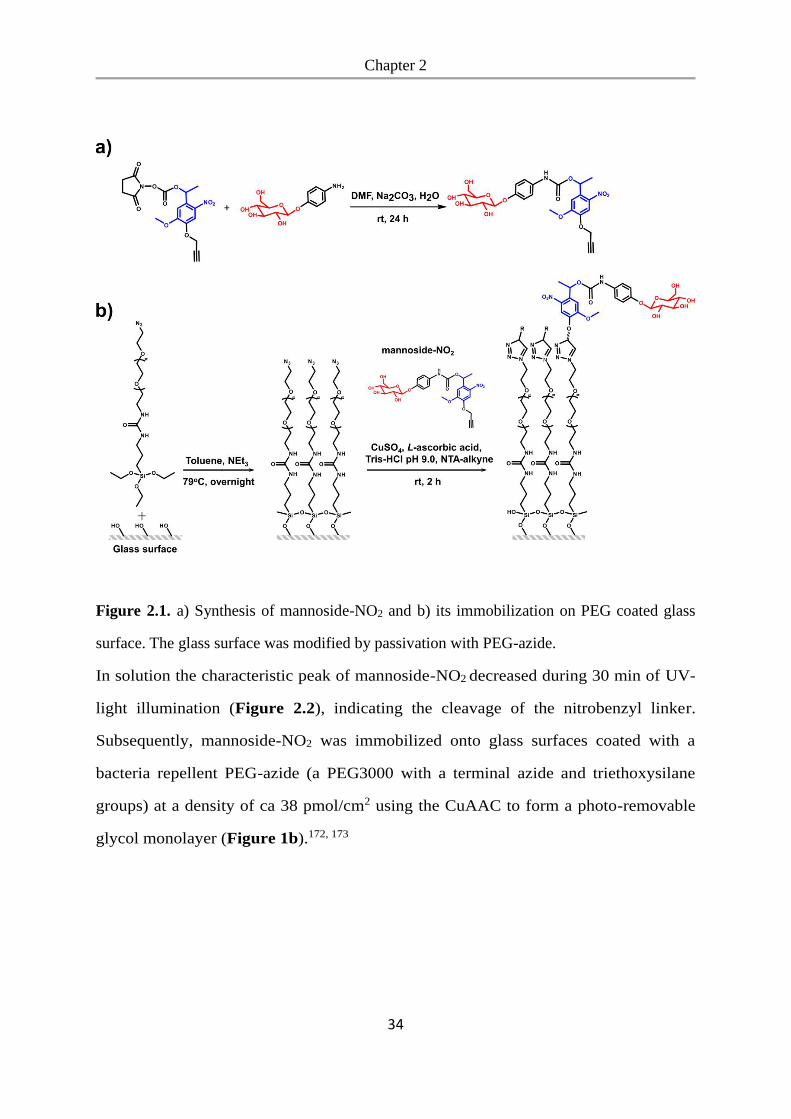
2.3 Results and Discussion
In the first stage of bacterial photolithography, we covered PEG-coated glass surfaces
with photocleavable mannoside groups, which specifically bind to the bacterial surface
adhesion receptor FimH. We used a nitrobenzyl linker, which can be photocleaved
with UV-light (365 nm), possesses an NHS-ester (N-Hydroxysuccinimide ester)
functional group, which reacts with mannoside amine, and this linker processes an
alkyne group for convenient attachment to any material with an azide functional group
by the CuAAC (copper catalyzed azide alkyne cycloaddition, also known as click
reaction).172, 173 First, we coupled the nitrobenzyl linker to α-D-mannosyl-amine to
obtain mannoside-NO2 (Figure 2.1a).
Chapter 2
34
Figure 2.1. a) Synthesis of mannoside-NO2 and b) its immobilization on PEG coated glass
surface. The glass surface was modified by passivation with PEG-azide.
In solution the characteristic peak of mannoside-NO2 decreased during 30 min of UV-
light illumination (Figure 2.2), indicating the cleavage of the nitrobenzyl linker.
Subsequently, mannoside-NO2 was immobilized onto glass surfaces coated with a
bacteria repellent PEG-azide (a PEG3000 with a terminal azide and triethoxysilane
groups) at a density of ca 38 pmol/cm2 using the CuAAC to form a photo-removable
glycol monolayer (Figure 1b).172, 173
Chapter 2
35
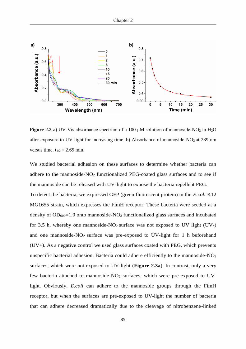
Figure 2.2 a) UV-Vis absorbance spectrum of a 100 μM solution of mannoside-NO2 in H2O
after exposure to UV light for increasing time. b) Absorbance of mannoside-NO2 at 239 nm
versus time. t1/2 = 2.65 min.
We studied bacterial adhesion on these surfaces to determine whether bacteria can
adhere to the mannoside-NO2 functionalized PEG-coated glass surfaces and to see if
the mannoside can be released with UV-light to expose the bacteria repellent PEG.
To detect the bacteria, we expressed GFP (green fluorescent protein) in the E.coli K12
MG1655 strain, which expresses the FimH receptor. These bacteria were seeded at a
density of OD600=1.0 onto mannoside-NO2 functionalized glass surfaces and incubated
for 3.5 h, whereby one mannoside-NO2 surface was not exposed to UV light (UV-)
and one mannoside-NO2 surface was pre-exposed to UV-light for 1 h beforehand
(UV+). As a negative control we used glass surfaces coated with PEG, which prevents
unspecific bacterial adhesion. Bacteria could adhere efficiently to the mannoside-NO2
surfaces, which were not exposed to UV-light (Figure 2.3a). In contrast, only a very
few bacteria attached to mannoside-NO2 surfaces, which were pre-exposed to UV-
light. Obviously, E.coli can adhere to the mannoside groups through the FimH
receptor, but when the surfaces are pre-exposed to UV-light the number of bacteria
that can adhere decreased dramatically due to the cleavage of nitrobenzene-linked
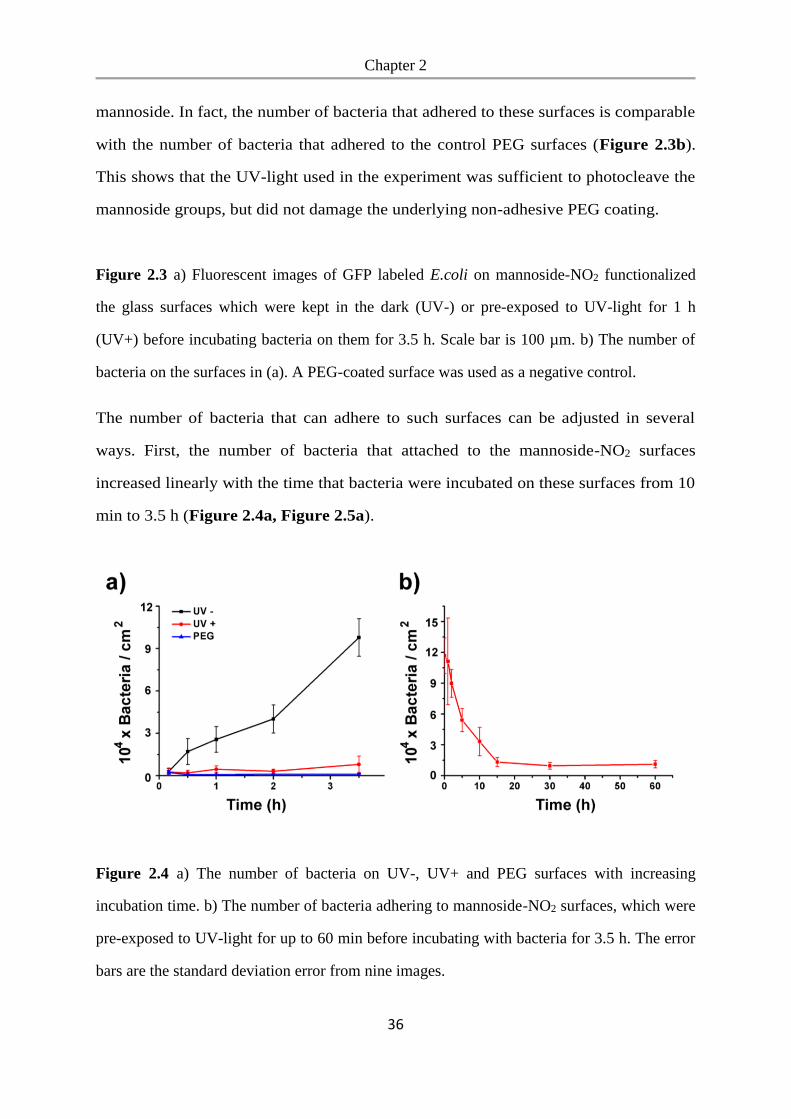
Chapter 2
36
mannoside. In fact, the number of bacteria that adhered to these surfaces is comparable
with the number of bacteria that adhered to the control PEG surfaces (Figure 2.3b).
This shows that the UV-light used in the experiment was sufficient to photocleave the
mannoside groups, but did not damage the underlying non-adhesive PEG coating.
Figure 2.3 a) Fluorescent images of GFP labeled E.coli on mannoside-NO2 functionalized
the glass surfaces which were kept in the dark (UV-) or pre-exposed to UV-light for 1 h
(UV+) before incubating bacteria on them for 3.5 h. Scale bar is 100 µm. b) The number of
bacteria on the surfaces in (a). A PEG-coated surface was used as a negative control.
The number of bacteria that can adhere to such surfaces can be adjusted in several
ways. First, the number of bacteria that attached to the mannoside-NO2 surfaces
increased linearly with the time that bacteria were incubated on these surfaces from 10
min to 3.5 h (Figure 2.4a, Figure 2.5a).
Figure 2.4 a) The number of bacteria on UV-, UV+ and PEG surfaces with increasing
incubation time. b) The number of bacteria adhering to mannoside-NO2 surfaces, which were
pre-exposed to UV-light for up to 60 min before incubating with bacteria for 3.5 h. The error
bars are the standard deviation error from nine images.
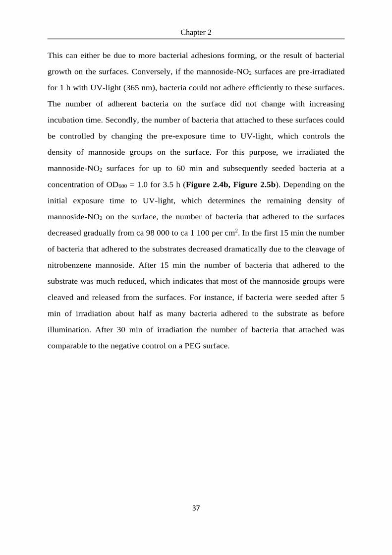
Chapter 2
37
This can either be due to more bacterial adhesions forming, or the result of bacterial
growth on the surfaces. Conversely, if the mannoside-NO2 surfaces are pre-irradiated
for 1 h with UV-light (365 nm), bacteria could not adhere efficiently to these surfaces.
The number of adherent bacteria on the surface did not change with increasing
incubation time. Secondly, the number of bacteria that attached to these surfaces could
be controlled by changing the pre-exposure time to UV-light, which controls the
density of mannoside groups on the surface. For this purpose, we irradiated the
mannoside-NO2 surfaces for up to 60 min and subsequently seeded bacteria at a
concentration of OD600 = 1.0 for 3.5 h (Figure 2.4b, Figure 2.5b). Depending on the
initial exposure time to UV-light, which determines the remaining density of
mannoside-NO2 on the surface, the number of bacteria that adhered to the surfaces
decreased gradually from ca 98 000 to ca 1 100 per cm2. In the first 15 min the number
of bacteria that adhered to the substrates decreased dramatically due to the cleavage of
nitrobenzene mannoside. After 15 min the number of bacteria that adhered to the
substrate was much reduced, which indicates that most of the mannoside groups were
cleaved and released from the surfaces. For instance, if bacteria were seeded after 5
min of irradiation about half as many bacteria adhered to the substrate as before
illumination. After 30 min of irradiation the number of bacteria that attached was
comparable to the negative control on a PEG surface.
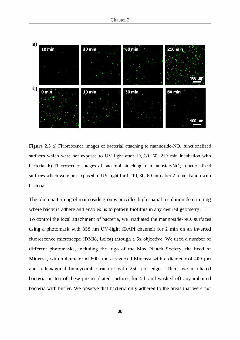
Chapter 2
38
Figure 2.5 a) Fluorescence images of bacterial attaching to mannoside-NO2 functionalized
surfaces which were not exposed to UV light after 10, 30, 60, 210 min incubation with
bacteria. b) Fluorescence images of bacterial attaching to mannoside-NO2 functionalized
surfaces which were pre-exposed to UV-light for 0, 10, 30, 60 min after 2 h incubation with
bacteria.
The photopatterning of mannoside groups provides high spatial resolution determining
where bacteria adhere and enables us to pattern biofilms in any desired geometry.59, 162
To control the local attachment of bacteria, we irradiated the mannoside-NO2 surfaces
using a photomask with 358 nm UV-light (DAPI channel) for 2 min on an inverted
fluorescence microscope (DMi8, Leica) through a 5x objective. We used a number of
different photomasks, including the logo of the Max Planck Society, the head of
Minerva, with a diameter of 800 µm, a reversed Minerva with a diameter of 400 µm
and a hexagonal honeycomb structure with 250 µm edges. Then, we incubated
bacteria on top of these pre-irradiated surfaces for 4 h and washed off any unbound
bacteria with buffer. We observe that bacteria only adhered to the areas that were not
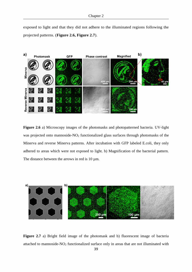
Chapter 2
39
exposed to light and that they did not adhere to the illuminated regions following the
projected patterns. (Figure 2.6, Figure 2.7).
Figure 2.6 a) Microscopy images of the photomasks and photopatterned bacteria. UV-light
was projected onto mannoside-NO2 functionalized glass surfaces through photomasks of the
Minerva and reverse Minerva patterns. After incubation with GFP labeled E.coli, they only
adhered to areas which were not exposed to light. b) Magnification of the bacterial pattern.
The distance between the arrows in red is 10 µm.
Figure 2.7 a) Bright field image of the photomask and b) fluorescent image of bacteria
attached to mannoside-NO2 functionalized surface only in areas that are not illuminated with
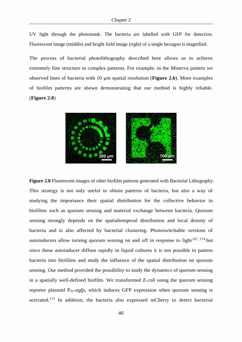
Chapter 2
40
UV light through the photomask. The bacteria are labelled with GFP for detection.
Fluorescent image (middle) and bright field image (right) of a single hexagon is magnified.
The process of bacterial photolithography described here allows us to achieve
extremely fine structure in complex patterns. For example, in the Minerva pattern we
observed lines of bacteria with 10 µm spatial resolution (Figure 2.6). More examples
of biofilm patterns are shown demonstrating that our method is highly reliable.
(Figure 2.8)
Figure 2.8 Fluorescent images of other biofilm patterns generated with Bacterial Lithography.
This strategy is not only useful to obtain patterns of bacteria, but also a way of
studying the importance their spatial distribution for the collective behavior in
biofilms such as quorum sensing and material exchange between bacteria. Quorum
sensing strongly depends on the spatialtemproal distribution and local density of
bacteria and is also affected by bacterial clustering. Photoswitchable versions of
autoinducers allow turning quorum sensing on and off in response to light167, 174 but
since these autoinducer diffuse rapidly in liquid cultures it is not possible to pattern
bacteria into biofilms and study the influence of the spatial distribution on quorum
sensing. Our method provided the possibility to study the dynamics of quorum sensing
in a spatially well-defined biofilm. We transformed E.coli using the quorum sensing
reporter plasmid Plsr-egfp, which induces GFP expression when quorum sensing is
activated.175 In addition, the bacteria also expressed mCherry to detect bacterial
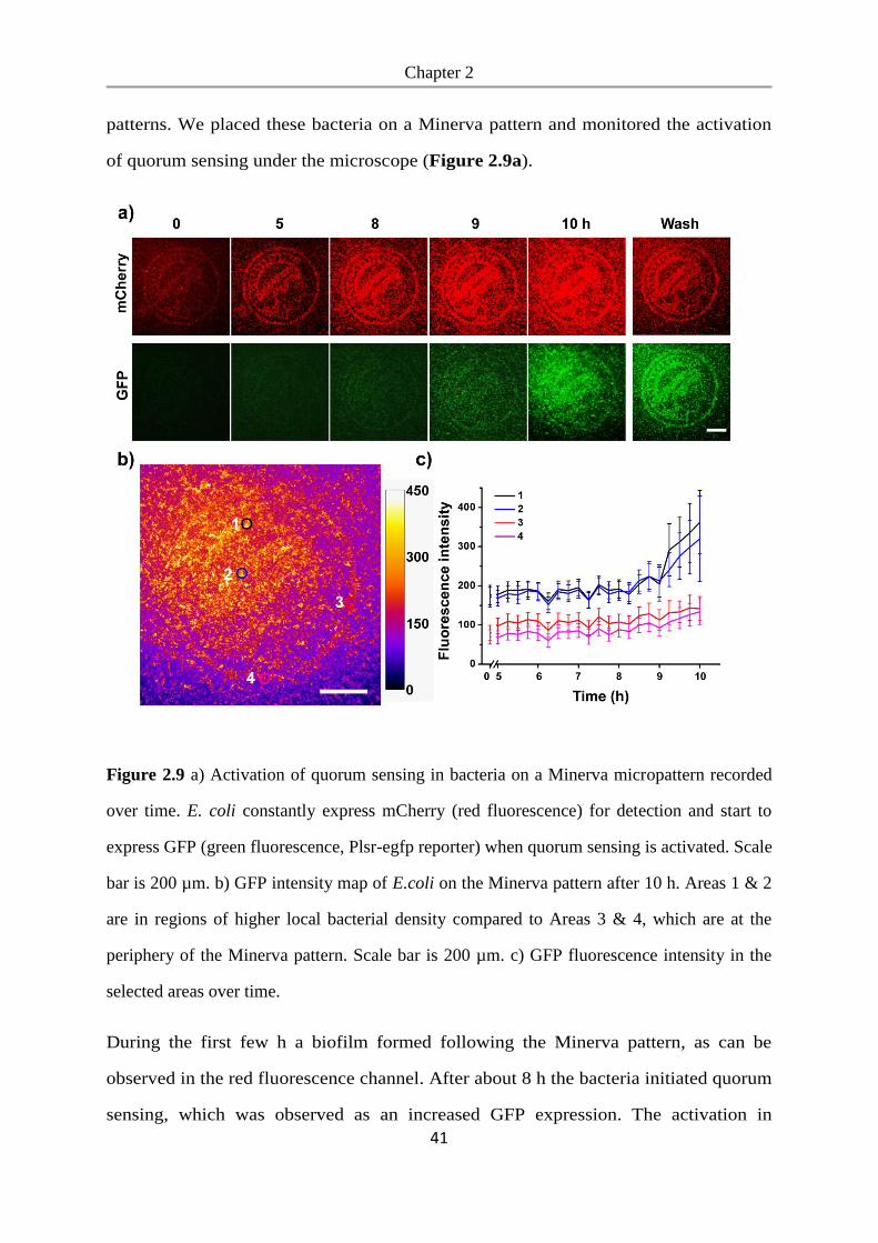
Chapter 2
41
patterns. We placed these bacteria on a Minerva pattern and monitored the activation
of quorum sensing under the microscope (Figure 2.9a).
Figure 2.9 a) Activation of quorum sensing in bacteria on a Minerva micropattern recorded
over time. E. coli constantly express mCherry (red fluorescence) for detection and start to
express GFP (green fluorescence, Plsr-egfp reporter) when quorum sensing is activated. Scale
bar is 200 µm. b) GFP intensity map of E.coli on the Minerva pattern after 10 h. Areas 1 & 2
are in regions of higher local bacterial density compared to Areas 3 & 4, which are at the
periphery of the Minerva pattern. Scale bar is 200 µm. c) GFP fluorescence intensity in the
selected areas over time.
During the first few h a biofilm formed following the Minerva pattern, as can be
observed in the red fluorescence channel. After about 8 h the bacteria initiated quorum
sensing, which was observed as an increased GFP expression. The activation in
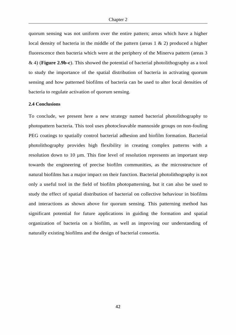
Chapter 2
42
quorum sensing was not uniform over the entire pattern; areas which have a higher
local density of bacteria in the middle of the pattern (areas 1 & 2) produced a higher
fluorescence then bacteria which were at the periphery of the Minerva pattern (areas 3
& 4) (Figure 2.9b-c). This showed the potential of bacterial photolithography as a tool
to study the importance of the spatial distribution of bacteria in activating quorum
sensing and how patterned biofilms of bacteria can be used to alter local densities of
bacteria to regulate activation of quorum sensing.
2.4 Conclusions
To conclude, we present here a new strategy named bacterial photolithography to
photopattern bacteria. This tool uses photocleavable mannoside groups on non-fouling
PEG coatings to spatially control bacterial adhesion and biofilm formation. Bacterial
photolithography provides high flexibility in creating complex patterns with a
resolution down to 10 µm. This fine level of resolution represents an important step
towards the engineering of precise biofilm communities, as the microstructure of
natural biofilms has a major impact on their function. Bacterial photolithography is not
only a useful tool in the field of biofilm photopatterning, but it can also be used to
study the effect of spatial distribution of bacterial on collective behaviour in biofilms
and interactions as shown above for quorum sensing. This patterning method has
significant potential for future applications in guiding the formation and spatial
organization of bacteria on a biofilm, as well as improving our understanding of
naturally existing biofilms and the design of bacterial consortia.
Chapter 3
43
Chapter 3. Blue light switchable bacterial adhesion as a key step towards
the design of biofilms
Copyright
The following chapter is based on the publication Chen, F., Wegner, S. V., ACS. Synth. Biol.,
2017, 6 (12), 2170-2174. The results are reprinted with permission from the American
Chemical Society.
Aim
The method of bacterial photolithography developed in chapter 2 enables the patterning of
biofilms with high resolution. However, it required UV light illumination which is toxic for
cells and is not reversible for the controlling of bacterial adhesion. Optogenetic switches are
emerging molecular tools for studying cellular processes as they offer higher spatiotemporal
precision than classical, chemical-based switches. New tools from optogenetics can be
introduced to control the bacterial adhesion and biofilm formation. Here, by expressing the
optogenetic protein pair pMag on the bacterial surface and immobilizing nMag on the
substrates, bacterial attachment can be controlled in a reversible and spatiotemporal
controlled manner with blue light.
Contributions
I performed all the experiments and analysis including the plasmids construction, protein
expression and purification, SDS-PAGE, QCM-D measurement, functionalization of glass
surfaces, bacterial adhesion and detachment assays, bacterial patterning. Seraphine V.
Wegner proposed the idea and supervised the work.
Chapter 3
44
3.1 Abstract
The control of where and when bacterial adhere to a substrate is a key step towards
controlling the formation and organization in biofilms. This study shows how we engineer
bacteria to adhere specifically to substrates with high spatial and temporal control under blue
light, but not in the dark, by using photoswitchable interaction between nMag and pMag
proteins. For this, we express pMag proteins on the surface of E.coli so that the bacteria can
adhere to substrates with immobilized nMag protein under blue light. These adhesions are
reversible in the dark and can be repeatedly turned on and off. Further, the number of bacteria
that can adhere to the substrate as well as the attachment and detachment dynamics are
adjustable by using different point mutants of pMag and altering light intensity. Overall, the
blue light switchable bacteria adhesions offer reversible, tunable and bioorthogonal control
with exceptional spatial and temporal resolution. This enables us to pattern bacteria on
substrates with great flexibility.
3.2 Introduction
In biofilms different bacteria work as a community, share metabolites and are more resistant
towards environmental stress, providing them with a clear survival advantage.153, 176
Controlling the formation of biofilms is not only a crucial step towards understanding how
different bacteria interact with each other in naturally occurring biofilms,150 but is also
essential when designing biofilms for biotechnological applications in biocatalysis,
biosensing and waste treatment.177, 178 Until recently the focus has been on preventing
bacterial adhesion and biofilm formation,179 but lately the better understanding of biofilms
has led integrating genetic circuits into bacteria in order to engineer multispecies bacterial
consortia with diverse functions.180, 181
The first step during biofilm formation is the adhesion of bacteria to a substrate, a key
determinant of the spatial organization in biofilms.153, 176 Controlling bacterial adhesion has
involved modifying the surface of the bacteria using bio-orthogonal reactive groups through
Chapter 3
45
liposome fusion,182 displaying surface tags on bacteria99, 183, 184 and substrate modification
with adhesion molecules.126, 185, 186 Among these approaches the light responsive methods
provide the highest spatial and temporal control. This is particularly significant as the
arrangement of bacteria with respect to each other defines the extent of their interaction in a
biofilm.153, 176 Photoswitchable azobenzene linkers have been used to alter the presentation of
mannose, which is recognized by the bacterial adhesion receptor FimH, to reversibly turn off
bacterial adhesion upon UV-light illumination (365 nm).103-105 Likewise, some azobenzene-
based molecules allow to modify bacterial attachment to mammalian cells,187 biofilm
formation,188 antibiotic activity189 and quorum sensing in bacteria with UV-light.190 However,
these photoswitches require UV-light illumination, which is toxic to bacteria. Consequently,
the challenge of controlling bacterial attachment to substrates in space and time in a non-
invasive, reversible and tuneable manner to guide biofilm formation remains an ongoing
goal.
In this paper, we present a new approach of how to control bacterial adhesion to substrates
non-invasively and reversibly with light based on photoswitchable proteins. Photoswitchable
proteins have recently been used in the field of optogenetics to regulate cellular functions
with visible light, including receptor activation, gene expression and protein localization both
in mammalian and bacterial cells.134, 135 These systems are bioorthogonal and noninvasive
because they rely on specific protein-protein interactions and respond to low intensity visible
light. Additionally, these proteins are genetically encoded, providing sustainable expression
in the cell. To photoswitch bacterial adhesion, we used the blue light responsive proteins,
nMag and pMag, which heterodimerize under blue light (480 nm) and dissociate from each
other in the dark.143 The nMag and pMag interaction can be tuned in terms of strength and
back conversion kinetics in the dark, thus providing an extensive dynamic range. The point
mutant pMagHigh (and nMagHigh) has a stronger interaction with its binding partners and
slower back conversion (kdark=5.6 x 10⁻⁶ s-1), while the opposite is true for the mutant
pMagFast1 (and nMagFast1) (kdark= 1.3 x 10⁻³ s-1).139
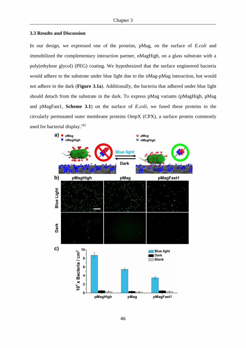
Chapter 3
46
3.3 Results and Discussion
In our design, we expressed one of the proteins, pMag, on the surface of E.coli and
immobilized the complementary interaction partner, nMagHigh, on a glass substrate with a
poly(ethylene glycol) (PEG) coating. We hypothesized that the surface engineered bacteria
would adhere to the substrate under blue light due to the nMag-pMag interaction, but would
not adhere in the dark (Figure 3.1a). Additionally, the bacteria that adhered under blue light
should detach from the substrate in the dark. To express pMag variants (pMagHigh, pMag
and pMagFast1, Scheme 3.1) on the surface of E.coli, we fused these proteins to the
circularly permutated outer membrane proteins OmpX (CPX), a surface protein commonly
used for bacterial display.191
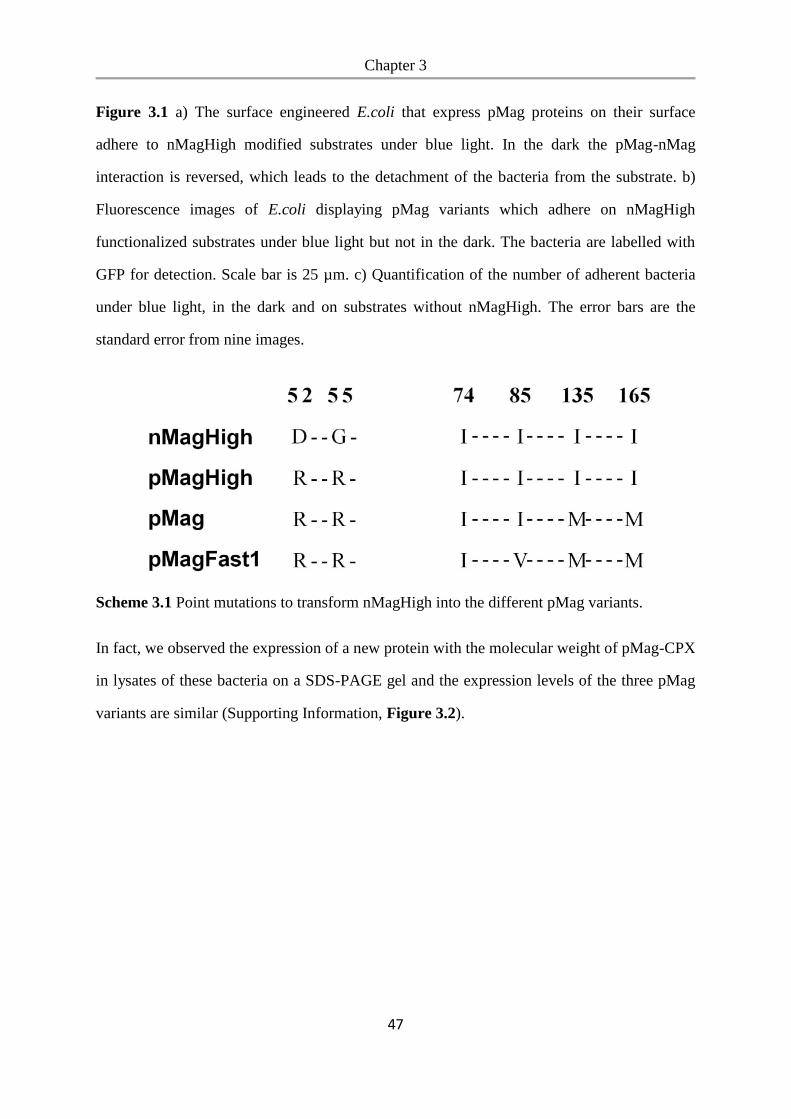
Chapter 3
47
Figure 3.1 a) The surface engineered E.coli that express pMag proteins on their surface
adhere to nMagHigh modified substrates under blue light. In the dark the pMag-nMag
interaction is reversed, which leads to the detachment of the bacteria from the substrate. b)
Fluorescence images of E.coli displaying pMag variants which adhere on nMagHigh
functionalized substrates under blue light but not in the dark. The bacteria are labelled with
GFP for detection. Scale bar is 25 µm. c) Quantification of the number of adherent bacteria
under blue light, in the dark and on substrates without nMagHigh. The error bars are the
standard error from nine images.
Scheme 3.1 Point mutations to transform nMagHigh into the different pMag variants.
In fact, we observed the expression of a new protein with the molecular weight of pMag-CPX
in lysates of these bacteria on a SDS-PAGE gel and the expression levels of the three pMag
variants are similar (Supporting Information, Figure 3.2).
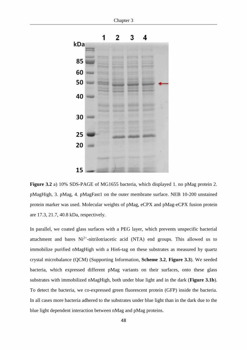
Chapter 3
48
Figure 3.2 a) 10% SDS-PAGE of MG1655 bacteria, which displayed 1. no pMag protein 2.
pMagHigh, 3. pMag, 4. pMagFast1 on the outer membrane surface. NEB 10-200 unstained
protein marker was used. Molecular weights of pMag, eCPX and pMag-eCPX fusion protein
are 17.3, 21.7, 40.8 kDa, respectively.
In parallel, we coated glass surfaces with a PEG layer, which prevents unspecific bacterial
attachment and bares Ni2+-nitrilotriacetic acid (NTA) end groups. This allowed us to
immobilize purified nMagHigh with a His6-tag on these substrates as measured by quartz
crystal microbalance (QCM) (Supporting Information, Scheme 3.2, Figure 3.3). We seeded
bacteria, which expressed different pMag variants on their surfaces, onto these glass
substrates with immobilized nMagHigh, both under blue light and in the dark (Figure 3.1b).
To detect the bacteria, we co-expressed green fluorescent protein (GFP) inside the bacteria.
In all cases more bacteria adhered to the substrates under blue light than in the dark due to the
blue light dependent interaction between nMag and pMag proteins.
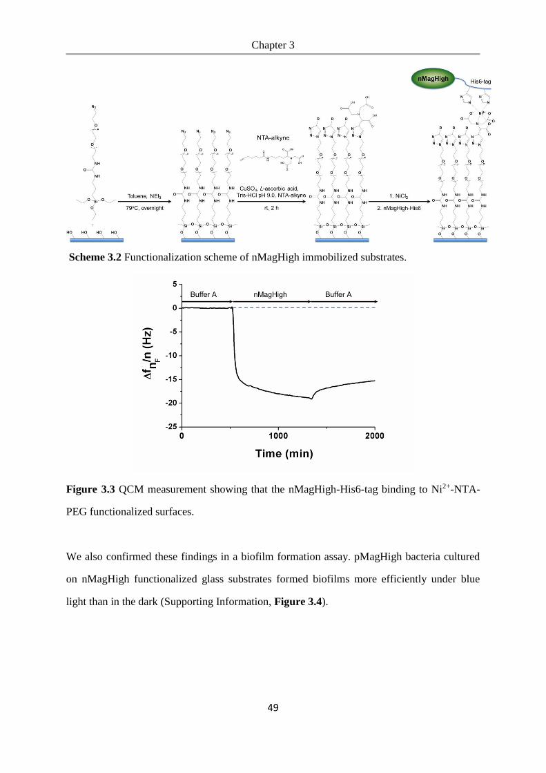
Chapter 3
49
Scheme 3.2 Functionalization scheme of nMagHigh immobilized substrates.
Figure 3.3 QCM measurement showing that the nMagHigh-His6-tag binding to Ni2+-NTA-
PEG functionalized surfaces.
We also confirmed these findings in a biofilm formation assay. pMagHigh bacteria cultured
on nMagHigh functionalized glass substrates formed biofilms more efficiently under blue
light than in the dark (Supporting Information, Figure 3.4).
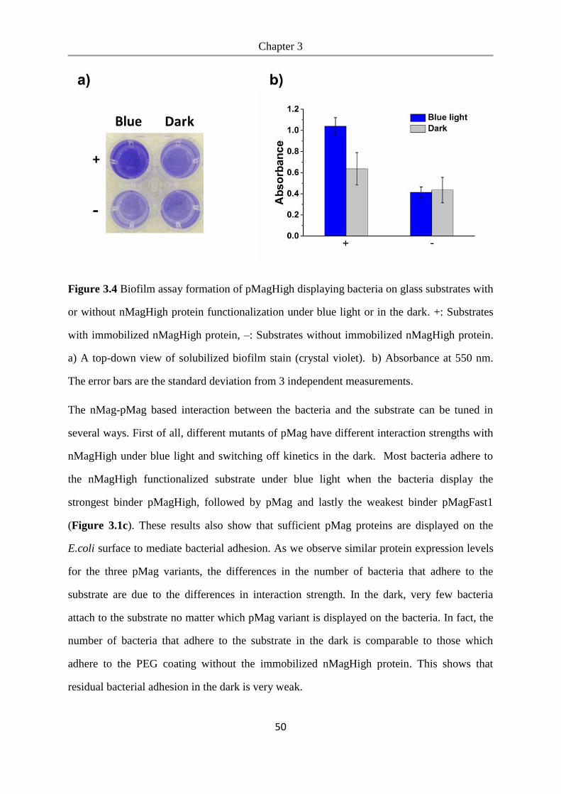
Chapter 3
50
Figure 3.4 Biofilm assay formation of pMagHigh displaying bacteria on glass substrates with
or without nMagHigh protein functionalization under blue light or in the dark. +: Substrates
with immobilized nMagHigh protein, –: Substrates without immobilized nMagHigh protein.
a) A top-down view of solubilized biofilm stain (crystal violet). b) Absorbance at 550 nm.
The error bars are the standard deviation from 3 independent measurements.
The nMag-pMag based interaction between the bacteria and the substrate can be tuned in
several ways. First of all, different mutants of pMag have different interaction strengths with
nMagHigh under blue light and switching off kinetics in the dark. Most bacteria adhere to
the nMagHigh functionalized substrate under blue light when the bacteria display the
strongest binder pMagHigh, followed by pMag and lastly the weakest binder pMagFast1
(Figure 3.1c). These results also show that sufficient pMag proteins are displayed on the
E.coli surface to mediate bacterial adhesion. As we observe similar protein expression levels
for the three pMag variants, the differences in the number of bacteria that adhere to the
substrate are due to the differences in interaction strength. In the dark, very few bacteria
attach to the substrate no matter which pMag variant is displayed on the bacteria. In fact, the
number of bacteria that adhere to the substrate in the dark is comparable to those which
adhere to the PEG coating without the immobilized nMagHigh protein. This shows that
residual bacterial adhesion in the dark is very weak.
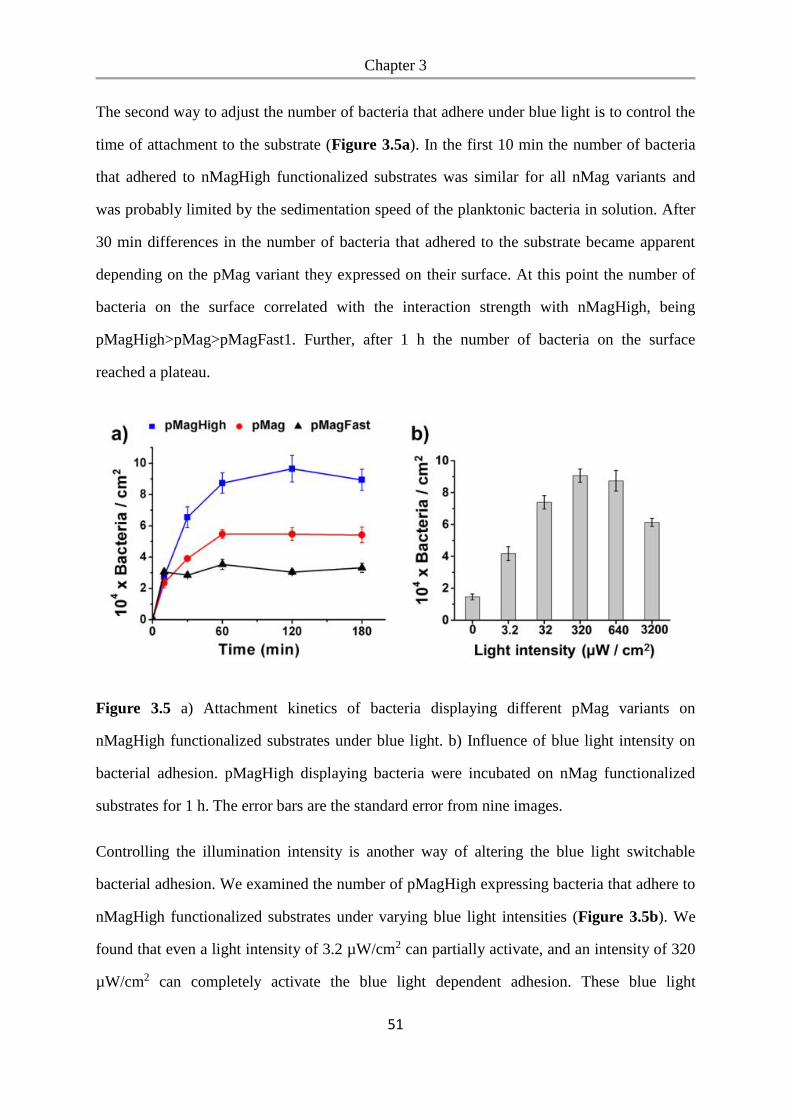
Chapter 3
51
The second way to adjust the number of bacteria that adhere under blue light is to control the
time of attachment to the substrate (Figure 3.5a). In the first 10 min the number of bacteria
that adhered to nMagHigh functionalized substrates was similar for all nMag variants and
was probably limited by the sedimentation speed of the planktonic bacteria in solution. After
30 min differences in the number of bacteria that adhered to the substrate became apparent
depending on the pMag variant they expressed on their surface. At this point the number of
bacteria on the surface correlated with the interaction strength with nMagHigh, being
pMagHigh>pMag>pMagFast1. Further, after 1 h the number of bacteria on the surface
reached a plateau.
Figure 3.5 a) Attachment kinetics of bacteria displaying different pMag variants on
nMagHigh functionalized substrates under blue light. b) Influence of blue light intensity on
bacterial adhesion. pMagHigh displaying bacteria were incubated on nMag functionalized
substrates for 1 h. The error bars are the standard error from nine images.
Controlling the illumination intensity is another way of altering the blue light switchable
bacterial adhesion. We examined the number of pMagHigh expressing bacteria that adhere to
nMagHigh functionalized substrates under varying blue light intensities (Figure 3.5b). We
found that even a light intensity of 3.2 µW/cm2 can partially activate, and an intensity of 320
µW/cm2 can completely activate the blue light dependent adhesion. These blue light
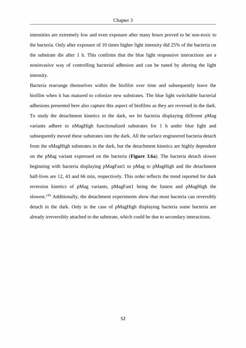
Chapter 3
52
intensities are extremely low and even exposure after many hours proved to be non-toxic to
the bacteria. Only after exposure of 10 times higher light intensity did 25% of the bacteria on
the substrate die after 1 h. This confirms that the blue light responsive interactions are a
noninvasive way of controlling bacterial adhesion and can be tuned by altering the light
intensity.
Bacteria rearrange themselves within the biofilm over time and subsequently leave the
biofilm when it has matured to colonize new substrates. The blue light switchable bacterial
adhesions presented here also capture this aspect of biofilms as they are reversed in the dark.
To study the detachment kinetics in the dark, we let bacteria displaying different pMag
variants adhere to nMagHigh functionalized substrates for 1 h under blue light and
subsequently moved these substrates into the dark. All the surface engineered bacteria detach
from the nMagHigh substrates in the dark, but the detachment kinetics are highly dependent
on the pMag variant expressed on the bacteria (Figure 3.6a). The bacteria detach slower
beginning with bacteria displaying pMagFast1 to pMag to pMagHigh and the detachment
half-lives are 12, 43 and 66 min, respectively. This order reflects the trend reported for dark
reversion kinetics of pMag variants, pMagFast1 being the fastest and pMagHigh the
slowest.143 Additionally, the detachment experiments show that most bacteria can reversibly
detach in the dark. Only in the case of pMagHigh displaying bacteria some bacteria are
already irreversibly attached to the substrate, which could be due to secondary interactions.
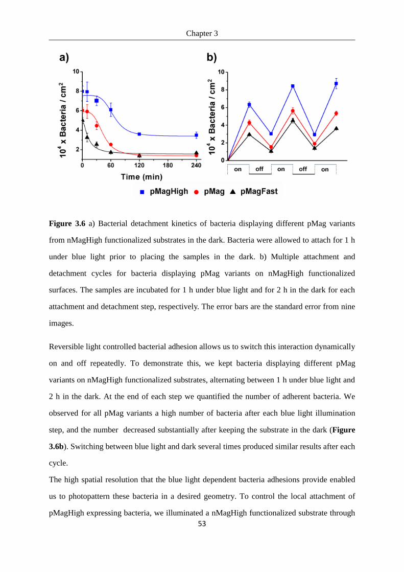
Chapter 3
53
Figure 3.6 a) Bacterial detachment kinetics of bacteria displaying different pMag variants
from nMagHigh functionalized substrates in the dark. Bacteria were allowed to attach for 1 h
under blue light prior to placing the samples in the dark. b) Multiple attachment and
detachment cycles for bacteria displaying pMag variants on nMagHigh functionalized
surfaces. The samples are incubated for 1 h under blue light and for 2 h in the dark for each
attachment and detachment step, respectively. The error bars are the standard error from nine
images.
Reversible light controlled bacterial adhesion allows us to switch this interaction dynamically
on and off repeatedly. To demonstrate this, we kept bacteria displaying different pMag
variants on nMagHigh functionalized substrates, alternating between 1 h under blue light and
2 h in the dark. At the end of each step we quantified the number of adherent bacteria. We
observed for all pMag variants a high number of bacteria after each blue light illumination
step, and the number decreased substantially after keeping the substrate in the dark (Figure
3.6b). Switching between blue light and dark several times produced similar results after each
cycle.
The high spatial resolution that the blue light dependent bacteria adhesions provide enabled
us to photopattern these bacteria in a desired geometry. To control the local attachment of
pMagHigh expressing bacteria, we illuminated a nMagHigh functionalized substrate through
Chapter 3
54
a photomask with blue light for 1 h. We detected obvious bacterial patterns on the substrate
that correspond to the projected photomask (Figure 3.7).
Figure 3.7 a) Bright field and b) fluorescent image of pMagHigh displaying bacteria attached
to nMagHigh functionalized substrates only in areas that are illuminated with blue light
through a photomask. The bacteria are labelled with mCherry for detection. Scale bar is 100
µm.
3.4 Conclusions
In summary, we present here a new platform for controlling bacteria surface adhesions based
on the blue light dependent interaction between nMag and pMag proteins. This optogenetic
approach is the first method that provides non-invasive, bioorthogonal and reversible control
over bacterial attachment with unprecedented spatial and temporal control. The
photoswitchable bacteria adhesions allow us to control and tune bacterial attachment and
detachment to a high degree. These unique properties can be used to guide the formation and
spatial organization of bacteria in a biofilm, which is vital in engineering bacterial consortia.
Chapter 4
55
Chapter 4. Photoswitchable bacteria-bacteria adhesions for blue light
controlled bacterial communities
Aim
In chapter 3, we introduced the controlling of bacterial adhesion to surfaces in a reversible
and high spatiotemporal resolution expressing the optogenetic protein pair pMag on the
bacterial surface and immobilizing nMag on the substrates. Optogenetic protein pair nMag-
pMag can be furtherly used to engineer the bacteria with photoswichable properties by
display techniques. Therefore, blue light can induced the bacterial multi-cellular assembly
and this process is reversible in the dark. Through this light controllable bacterial adhesion,
multiple bacterial social interactions can be regulated, such as aggregation, quorum sensing,
biofilm formation and metabolic cross-feeding between auxotrophic strains.
Contributions
I performed all the experiments and analysis including the constructions of plasmids and
bacterial strains, SDS-PAGE, bacterial aggregation and reversibility, quorum sensing assay,
real-time imaging of bacterial aggregation, biofilm formation, metabolic cross-feeding assay.
Seraphine V. Wegner proposed the idea and supervised the work.
Chapter 4
56
4.1 Abstract
Although the fundamental importance and biotechnological potential of multi-bacterial
communities, also called biofilms, are well known, our ability to control them is limited. We
present a new way of dynamically controlling bacteria-bacteria adhesions by using blue light
and how these photoswitchable adhesions can be used to regulate multicellularity and
associated bacterial behavior. To achieve this, the photoswitchable proteins nMagHigh and
pMagHigh were expressed on bacterial surfaces as adhesins to allow multicellular clusters to
assemble under blue light and reversibly disassemble in the dark. Regulation with visible
light provides unique advantages including high spatiotemporal control, tunability and non-
invasive remote regulation. These photoswitchable adhesions make it possible to regulate
collective bacterial functions including aggregation, quorum sensing, biofilm formation and
metabolic cross-feeding between auxotrophic bacteria with light. Overall, the photoregulation
of bacteria-bacteria adhesions provides a new way of studying bacterial cell biology and will
enable the design of biofilms for biotechnological applications.
4.2 Introduction
Bacteria have the outstanding ability to live in biofilm as social communities. Biofilms allow
them to perform complex functions as a group that cannot be accomplished by a single
bacterium.24 Multi-bacterial communities are different from their planktonic counterparts as
they share metabolites and are shielded from environmental stresses and antibiotics, allowing
them to colonize diverse and even hostile environments.150, 192 Bacterial consortia have great
potential for applications in synthetic biology due to their ability to split complex
biosynthetic pathways and share tasks among different specialized members, thus increasing
their productivity and decreasing the metabolic burden on each member of the consortium.45,
153 Specifically designed bacterial consortia could therefore be used in future industrial,
medical and environmental applications including the synthesis of complex molecules in
bacterial factories, biocatalysis,193 waste treatment and drug delivery.194, 195
Chapter 4
57
The fundamental importance and biotechnological potential of bacterial communities are
already recognized, whereas our current understanding and ability to engineer multi-bacterial
communities to perform desired tasks are both still in their infancy. Part of the complexity
arises from assembling unicellular planktonic bacteria into multicellular assemblies. During
this process the organization of bacteria at the micrometer length scale into multicellular
structures strongly affects the fate of the co-culture because it determines diffusion rates of
shared metabolites and toxins. Moreover, the interaction between bacteria is dynamic and
changes over time as fluctuations in the environment and within the cells occur.45, 196, 197 To
produce biofilms with a deliberate spatiotemporal arrangement, methods for dynamically
controlling bacteria-bacteria adhesions with high spatial and temporal precision are
required.49, 198, 199 Yet, methods for controlling bacteria-bacteria adhesions are very limited.49,
152 A prime example of engineering bacteria-bacteria adhesions by Glass et al. uses outer
membrane-displayed nanobodies and antigens as adhesins to control multicellularity,
aggregation pattern and morphology.131 In other examples, native and surface engineered
bacteria were clustered together using external small molecules, polyvalent nanoparticles and
polymers.99, 104, 123, 126, 200 Yet, none of these approaches provides the required crucial
dynamic and spatiotemporal control over the bacteria-bacteria adhesions.
High spatiotemporal control has been achieved using light as a trigger in the case of bacterial
adhesions onto substrates, which is the first step in biofilm formation. Different approaches
include the optogenetic regulated expression of adhesion molecules on bacterial surfaces110,
111, 165, 201-203 and the modification of substrates and/or bacteria with light responsive
molecules that alter bacterial adhesion.102, 104, 168, 204 Using light as a trigger provides unique
advantages. Most importantly, being able to illuminate an area of interest at a chosen time
provides incomparable spatial and temporal control over the interactions. Secondly, visible
light is biocompatible and offers non-invasive remote control. This is particularly
advantageous for biotechnological applications when compared to small molecule inducers,
as it does not require later removal and is cost efficient. Additionally, by using different
intensities and light frequencies it is possible to modulate interactions in light-responsive
Chapter 4
58
systems. Some light-responsive interactions are also reversible in the dark offering dynamic
on/off switching. This is also why photo-regulation has been introduced to regulate other
important bacterial processes including gene expression, growth, biofilm formation,188
quorum sensing,190, 205 bacterial attachment to mammalian cells and antibiotic activity.126
In this paper, we present an optogenetic approach to control reversible bacteria-bacteria
adhesions with high spatiotemporal control using visible light. Photoswitchable bacteria-
bacteria adhesions allow for the assembly of E.coli into multicellular aggregates with high
spatiotemporal control and specificity in a noninvasive, reversible, sustainable and tunable
manner. The fundamental properties of bacteria-bacteria adhesions have also allowed us to
control associated bacterial processes including quorum sensing, biofilm formation and
metabolic cross-feeding between different auxotrophic bacteria. These examples reveal that
the photoswitchable bacteria-bacteria adhesions offer new design possibilities for
dynamically modulating bacterial consortia, which in turn opens up new possibilities for
studying both bacterial cell biology and biotechnological applications.
4.3 Results and Discussion
Engineered bacteria displaying photoswitchable proteins. As bacterial adhesins, we chose
to use those photoswitchable protein interactions recently used in the field of optogenetics to
regulate diverse cellular functions with visible light, including receptor activation, gene
expression, protein localization and activity as well as cell adhesions.143, 165, 206 Important
benefits of optogenetic protein-protein interactions are their response to low intensity visible
light, the high specificity of the protein-protein interactions and their sustained production in
the cells. In this study, we used the protein pair nMag and pMag as adhesins, which
heterodimerize under blue light (480 nm) and dissociate from each other in the dark.143
Different point mutants of nMag and pMag enable us to alter the strength and back
conversion in the dark; the variants nMagHigh and pMagHigh have stronger interactions with
binding partners and a slower rate of reverse conversion in the dark. In contrast, nMagFast1
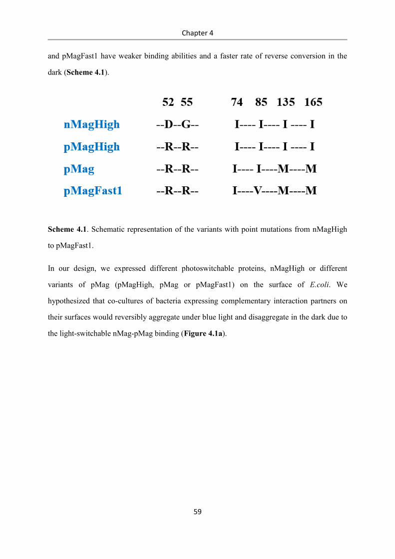
Chapter 4
59
and pMagFast1 have weaker binding abilities and a faster rate of reverse conversion in the
dark (Scheme 4.1).
Scheme 4.1. Schematic representation of the variants with point mutations from nMagHigh
to pMagFast1.
In our design, we expressed different photoswitchable proteins, nMagHigh or different
variants of pMag (pMagHigh, pMag or pMagFast1) on the surface of E.coli. We
hypothesized that co-cultures of bacteria expressing complementary interaction partners on
their surfaces would reversibly aggregate under blue light and disaggregate in the dark due to
the light-switchable nMag-pMag binding (Figure 4.1a).
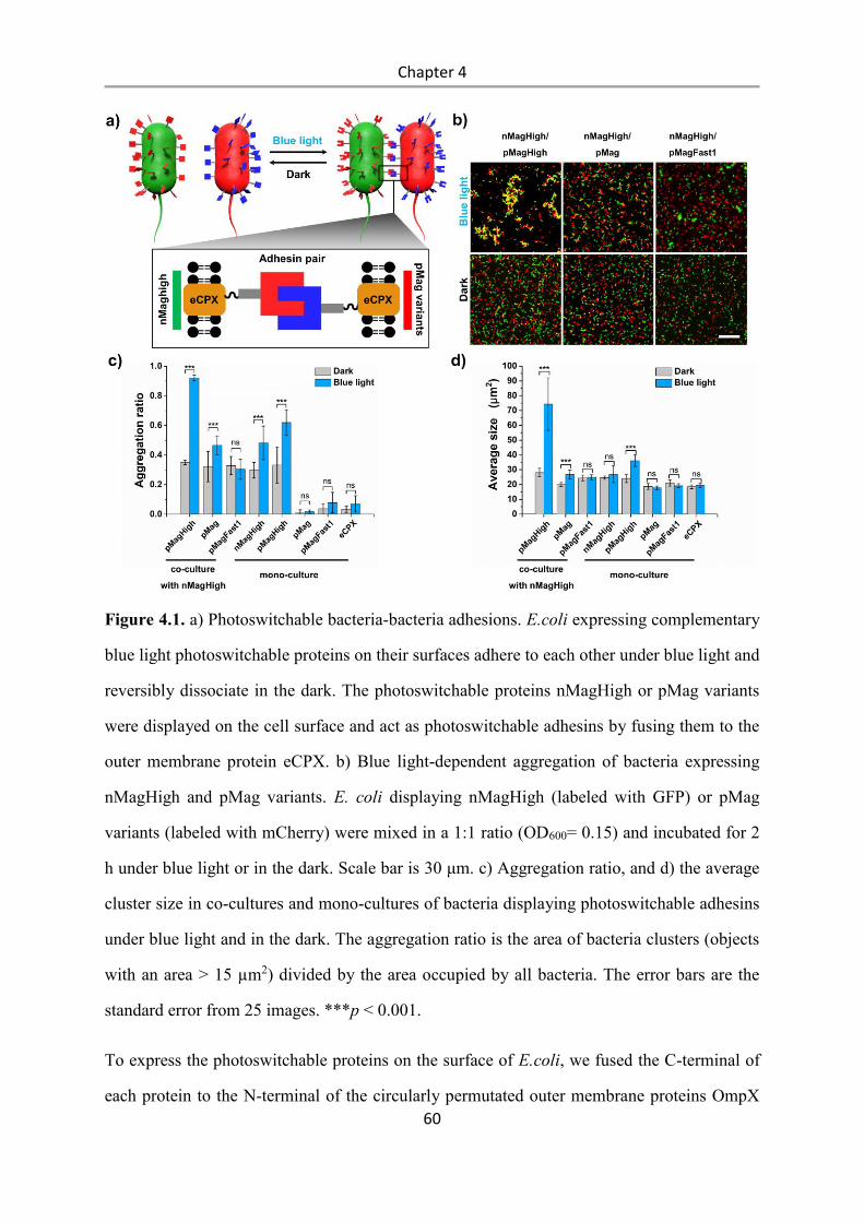
Chapter 4
60
Figure 4.1. a) Photoswitchable bacteria-bacteria adhesions. E.coli expressing complementary
blue light photoswitchable proteins on their surfaces adhere to each other under blue light and
reversibly dissociate in the dark. The photoswitchable proteins nMagHigh or pMag variants
were displayed on the cell surface and act as photoswitchable adhesins by fusing them to the
outer membrane protein eCPX. b) Blue light-dependent aggregation of bacteria expressing
nMagHigh and pMag variants. E. coli displaying nMagHigh (labeled with GFP) or pMag
variants (labeled with mCherry) were mixed in a 1:1 ratio (OD600= 0.15) and incubated for 2
h under blue light or in the dark. Scale bar is 30 μm. c) Aggregation ratio, and d) the average
cluster size in co-cultures and mono-cultures of bacteria displaying photoswitchable adhesins
under blue light and in the dark. The aggregation ratio is the area of bacteria clusters (objects
with an area > 15 µm2) divided by the area occupied by all bacteria. The error bars are the
standard error from 25 images. ***p < 0.001.
To express the photoswitchable proteins on the surface of E.coli, we fused the C-terminal of
each protein to the N-terminal of the circularly permutated outer membrane proteins OmpX
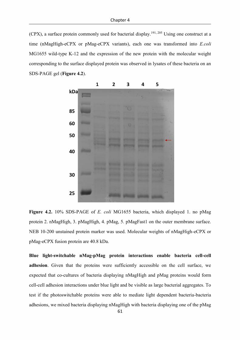
Chapter 4
61
(CPX), a surface protein commonly used for bacterial display.191, 205 Using one construct at a
time (nMagHigh-eCPX or pMag-eCPX variants), each one was transformed into E.coli
MG1655 wild-type K-12 and the expression of the new protein with the molecular weight
corresponding to the surface displayed protein was observed in lysates of these bacteria on an
SDS-PAGE gel (Figure 4.2).
Figure 4.2. 10% SDS-PAGE of E. coli MG1655 bacteria, which displayed 1. no pMag
protein 2. nMagHigh, 3. pMagHigh, 4. pMag, 5. pMagFast1 on the outer membrane surface.
NEB 10-200 unstained protein marker was used. Molecular weights of nMagHigh-eCPX or
pMag-eCPX fusion protein are 40.8 kDa.
Blue light-switchable nMag-pMag protein interactions enable bacteria cell-cell
adhesion. Given that the proteins were sufficiently accessible on the cell surface, we
expected that co-cultures of bacteria displaying nMagHigh and pMag proteins would form
cell-cell adhesion interactions under blue light and be visible as large bacterial aggregates. To
test if the photoswitchable proteins were able to mediate light dependent bacteria-bacteria
adhesions, we mixed bacteria displaying nMagHigh with bacteria displaying one of the pMag
Chapter 4
62
variants on their surface in a 1:1 ratio (OD600 is 0.15), either under blue light or in the dark.
For detection we co-expressed green fluorescent protein (GFP) and a red fluorescent protein
(mCherry) inside the bacteria displaying nMagHigh and pMag variants, respectively. After 2
h incubation under blue light bacteria aggregates were, in fact, visible under the fluorescent
microscope, but not in samples kept in the dark (Figure 1b). The most prominent bacterial
aggregates were formed under blue light in co-cultures of bacteria displaying nMagHigh and
pMagHigh, followed by co-cultures of nMagHigh and pMag. However, almost no
aggregation was observed in co-cultures of pMagHigh with the weakest binder being
pMagFast1. The blue light-triggered aggregation was clearly due to the photoswitchable
proteins displayed on the bacteria since bacteria just displaying eCPX (negative control) did
not aggregate and were homogeneously distributed over the sample.
Next, we quantified the extent of aggregation and the aggregate sizes under blue light and in
the dark. This was done for both mixed cultures of nMagHigh with different pMag-
displaying bacteria and mono-cultures in order to gain not only an insight into the specificity
of the photoswitchable interactions, but also to assess the contribution of heterophilic and
homophilic interactions between bacteria. As a parameter, the aggregation ratio was defined
as the area occupied by clusters of bacteria (objects with an area > 15 µm2, the average area
of a single E.coli is 2.5 µm2, only objects bigger than 6 bacterial cells were considered to be
clusters) divided by the total area occupied by all bacteria (Figure 4.1c). This quantification
showed that in co-cultures of nMagHigh and pMagHigh-displaying E.coli, 90% of the
bacteria were integrated into clusters under blue light, whereas only 30% of bacteria were
clustered in the dark. Moreover, the average cluster size (only objects with an area > 15 µm2
were counted as clusters) was 75 µm2 under blue light, which was significantly larger than
the clusters observed in the dark (average size of 30 µm2) (Figure 4.1d). The aggregation
ratio analysis also confirmed the initial qualitative observation that co-cultures of nMagHigh
and pMag partially formed clusters under blue light, whereas co-cultures of nMagHigh and
pMagFast1 did not. Mono-cultures of bacteria displaying nMagHigh or pMagHigh
aggregated to some extent when exposed to blue light with up to 50% of the bacteria being
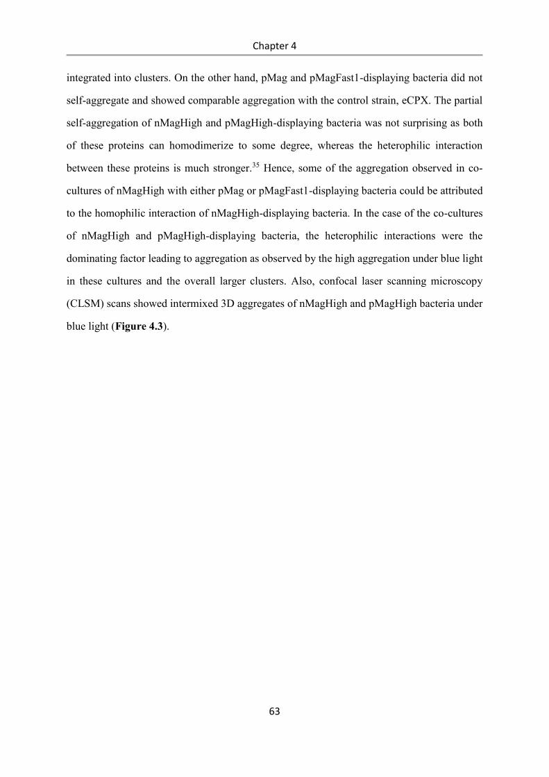
Chapter 4
63
integrated into clusters. On the other hand, pMag and pMagFast1-displaying bacteria did not
self-aggregate and showed comparable aggregation with the control strain, eCPX. The partial
self-aggregation of nMagHigh and pMagHigh-displaying bacteria was not surprising as both
of these proteins can homodimerize to some degree, whereas the heterophilic interaction
between these proteins is much stronger.35 Hence, some of the aggregation observed in co-
cultures of nMagHigh with either pMag or pMagFast1-displaying bacteria could be attributed
to the homophilic interaction of nMagHigh-displaying bacteria. In the case of the co-cultures
of nMagHigh and pMagHigh-displaying bacteria, the heterophilic interactions were the
dominating factor leading to aggregation as observed by the high aggregation under blue light
in these cultures and the overall larger clusters. Also, confocal laser scanning microscopy
(CLSM) scans showed intermixed 3D aggregates of nMagHigh and pMagHigh bacteria under
blue light (Figure 4.3).
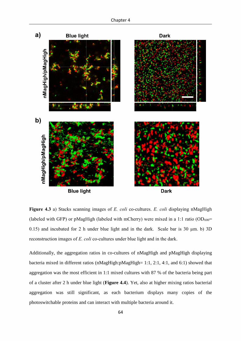
Chapter 4
64
Figure 4.3 a) Stacks scanning images of E. coli co-cultures. E. coli displaying nMagHigh
(labeled with GFP) or pMagHigh (labeled with mCherry) were mixed in a 1:1 ratio (OD600=
0.15) and incubated for 2 h under blue light and in the dark. Scale bar is 30 µm. b) 3D
reconstruction images of E. coli co-cultures under blue light and in the dark.
Additionally, the aggregation ratios in co-cultures of nMagHigh and pMagHigh displaying
bacteria mixed in different ratios (nMagHigh:pMagHigh= 1:1, 2:1, 4:1, and 6:1) showed that
aggregation was the most efficient in 1:1 mixed cultures with 87 % of the bacteria being part
of a cluster after 2 h under blue light (Figure 4.4). Yet, also at higher mixing ratios bacterial
aggregation was still significant, as each bacterium displays many copies of the
photoswitchable proteins and can interact with multiple bacteria around it.
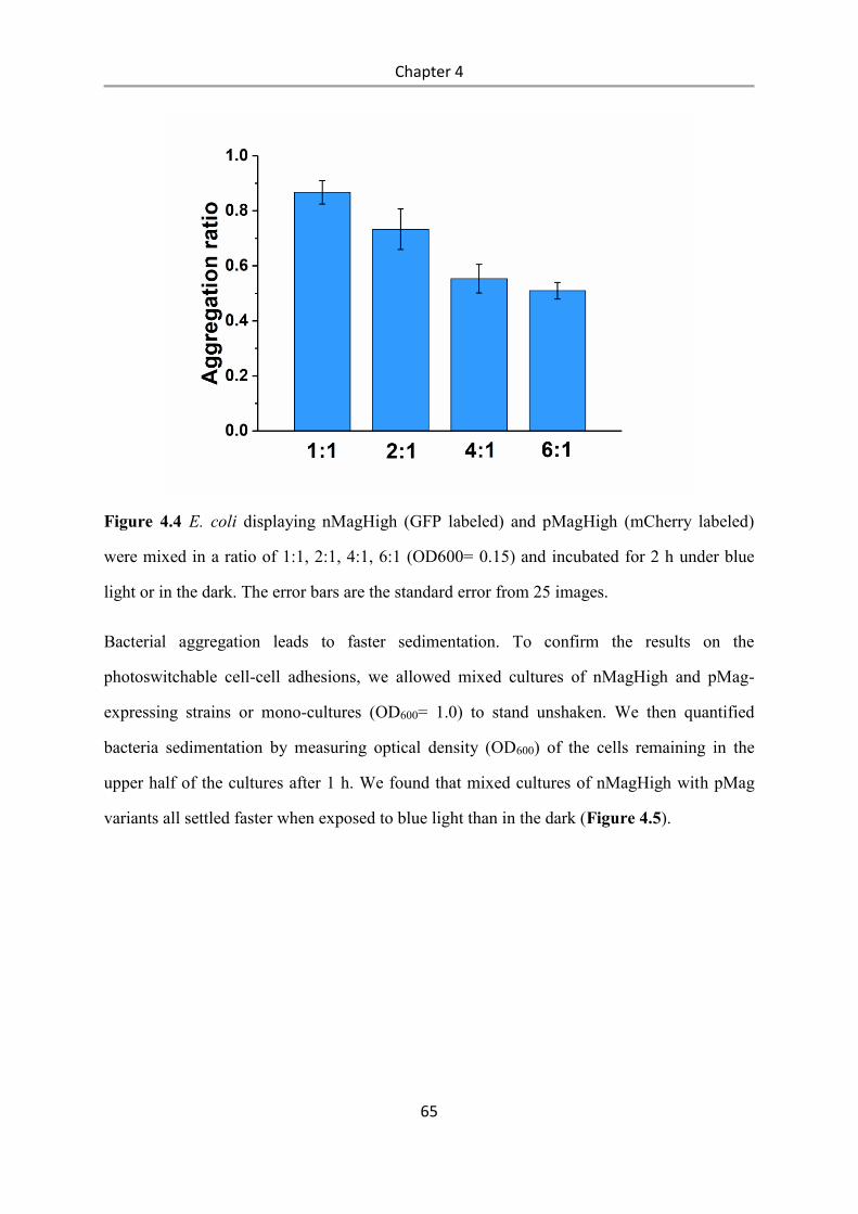
Chapter 4
65
Figure 4.4 E. coli displaying nMagHigh (GFP labeled) and pMagHigh (mCherry labeled)
were mixed in a ratio of 1:1, 2:1, 4:1, 6:1 (OD600= 0.15) and incubated for 2 h under blue
light or in the dark. The error bars are the standard error from 25 images.
Bacterial aggregation leads to faster sedimentation. To confirm the results on the
photoswitchable cell-cell adhesions, we allowed mixed cultures of nMagHigh and pMag-
expressing strains or mono-cultures (OD600= 1.0) to stand unshaken. We then quantified
bacteria sedimentation by measuring optical density (OD600) of the cells remaining in the
upper half of the cultures after 1 h. We found that mixed cultures of nMagHigh with pMag
variants all settled faster when exposed to blue light than in the dark (Figure 4.5).
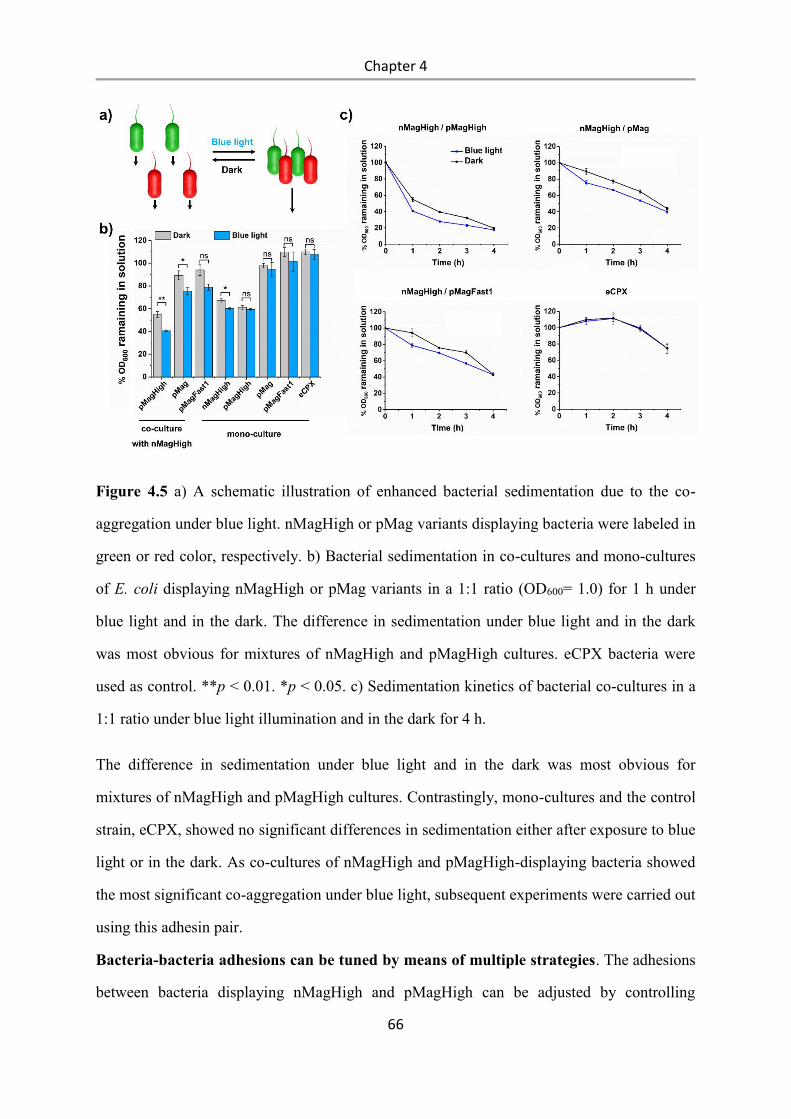
Chapter 4
66
Figure 4.5 a) A schematic illustration of enhanced bacterial sedimentation due to the co-
aggregation under blue light. nMagHigh or pMag variants displaying bacteria were labeled in
green or red color, respectively. b) Bacterial sedimentation in co-cultures and mono-cultures
of E. coli displaying nMagHigh or pMag variants in a 1:1 ratio (OD600= 1.0) for 1 h under
blue light and in the dark. The difference in sedimentation under blue light and in the dark
was most obvious for mixtures of nMagHigh and pMagHigh cultures. eCPX bacteria were
used as control. **p < 0.01. *p < 0.05. c) Sedimentation kinetics of bacterial co-cultures in a
1:1 ratio under blue light illumination and in the dark for 4 h.
The difference in sedimentation under blue light and in the dark was most obvious for
mixtures of nMagHigh and pMagHigh cultures. Contrastingly, mono-cultures and the control
strain, eCPX, showed no significant differences in sedimentation either after exposure to blue
light or in the dark. As co-cultures of nMagHigh and pMagHigh-displaying bacteria showed
the most significant co-aggregation under blue light, subsequent experiments were carried out
using this adhesin pair.
Bacteria-bacteria adhesions can be tuned by means of multiple strategies. The adhesions
between bacteria displaying nMagHigh and pMagHigh can be adjusted by controlling
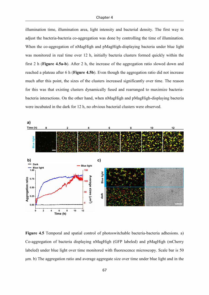
Chapter 4
67
illumination time, illumination area, light intensity and bacterial density. The first way to
adjust the bacteria-bacteria co-aggregation was done by controlling the time of illumination.
When the co-aggregation of nMagHigh and pMagHigh-displaying bacteria under blue light
was monitored in real time over 12 h, initially bacteria clusters formed quickly within the
first 2 h (Figure 4.5a-b). After 2 h, the increase of the aggregation ratio slowed down and
reached a plateau after 6 h (Figure 4.5b). Even though the aggregation ratio did not increase
much after this point, the sizes of the clusters increased significantly over time. The reason
for this was that existing clusters dynamically fused and rearranged to maximize bacteria-
bacteria interactions. On the other hand, when nMagHigh and pMagHigh-displaying bacteria
were incubated in the dark for 12 h, no obvious bacterial clusters were observed.
Figure 4.5 Temporal and spatial control of photoswitchable bacteria-bacteria adhesions. a)
Co-aggregation of bacteria displaying nMagHigh (GFP labeled) and pMagHigh (mCherry
labeled) under blue light over time monitored with fluorescence microscopy. Scale bar is 50
μm. b) The aggregation ratio and average aggregate size over time under blue light and in the
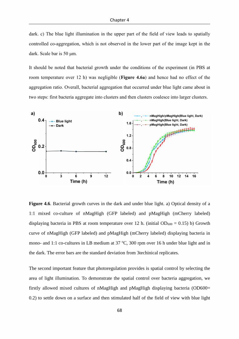
Chapter 4
68
dark. c) The blue light illumination in the upper part of the field of view leads to spatially
controlled co-aggregation, which is not observed in the lower part of the image kept in the
dark. Scale bar is 50 μm.
It should be noted that bacterial growth under the conditions of the experiment (in PBS at
room temperature over 12 h) was negligible (Figure 4.6a) and hence had no effect of the
aggregation ratio. Overall, bacterial aggregation that occurred under blue light came about in
two steps: first bacteria aggregate into clusters and then clusters coalesce into larger clusters.
Figure 4.6. Bacterial growth curves in the dark and under blue light. a) Optical density of a
1:1 mixed co-culture of nMagHigh (GFP labeled) and pMagHigh (mCherry labeled)
displaying bacteria in PBS at room temperature over 12 h. (initial OD600 = 0.15) b) Growth
curve of nMagHigh (GFP labeled) and pMagHigh (mCherry labeled) displaying bacteria in
mono- and 1:1 co-cultures in LB medium at 37 oC, 300 rpm over 16 h under blue light and in
the dark. The error bars are the standard deviation from 3techinical replicates.
The second important feature that photoregulation provides is spatial control by selecting the
area of light illumination. To demonstrate the spatial control over bacteria aggregation, we
firstly allowed mixed cultures of nMagHigh and pMagHigh displaying bacteria (OD600=
0.2) to settle down on a surface and then stimulated half of the field of view with blue light
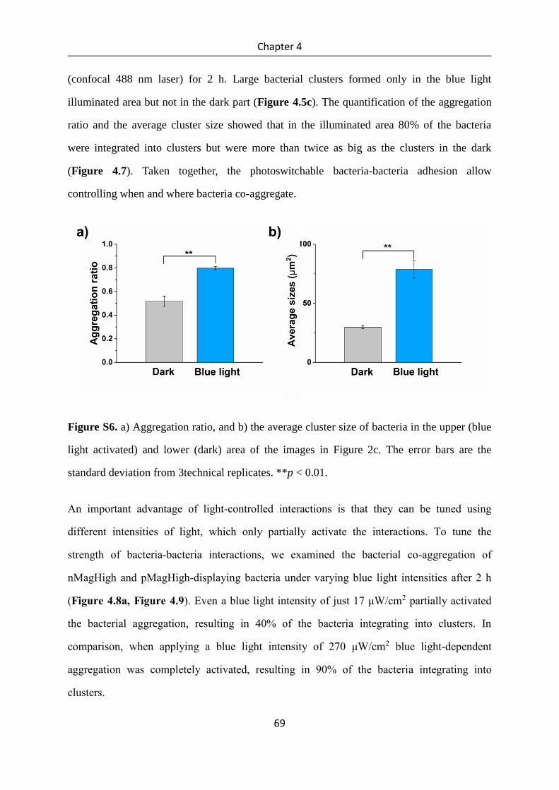
Chapter 4
69
(confocal 488 nm laser) for 2 h. Large bacterial clusters formed only in the blue light
illuminated area but not in the dark part (Figure 4.5c). The quantification of the aggregation
ratio and the average cluster size showed that in the illuminated area 80% of the bacteria
were integrated into clusters but were more than twice as big as the clusters in the dark
(Figure 4.7). Taken together, the photoswitchable bacteria-bacteria adhesion allow
controlling when and where bacteria co-aggregate.
Figure S6. a) Aggregation ratio, and b) the average cluster size of bacteria in the upper (blue
light activated) and lower (dark) area of the images in Figure 2c. The error bars are the
standard deviation from 3technical replicates. **p < 0.01.
An important advantage of light-controlled interactions is that they can be tuned using
different intensities of light, which only partially activate the interactions. To tune the
strength of bacteria-bacteria interactions, we examined the bacterial co-aggregation of
nMagHigh and pMagHigh-displaying bacteria under varying blue light intensities after 2 h
(Figure 4.8a, Figure 4.9). Even a blue light intensity of just 17 μW/cm2 partially activated
the bacterial aggregation, resulting in 40% of the bacteria integrating into clusters. In
comparison, when applying a blue light intensity of 270 μW/cm2 blue light-dependent
aggregation was completely activated, resulting in 90% of the bacteria integrating into
clusters.
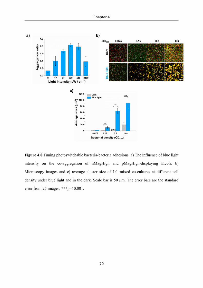
Chapter 4
70
Figure 4.8 Tuning photoswitchable bacteria-bacteria adhesions. a) The influence of blue light
intensity on the co-aggregation of nMagHigh and pMagHigh-displaying E.coli. b)
Microscopy images and c) average cluster size of 1:1 mixed co-cultures at different cell
density under blue light and in the dark. Scale bar is 50 μm. The error bars are the standard
error from 25 images. ***p < 0.001.
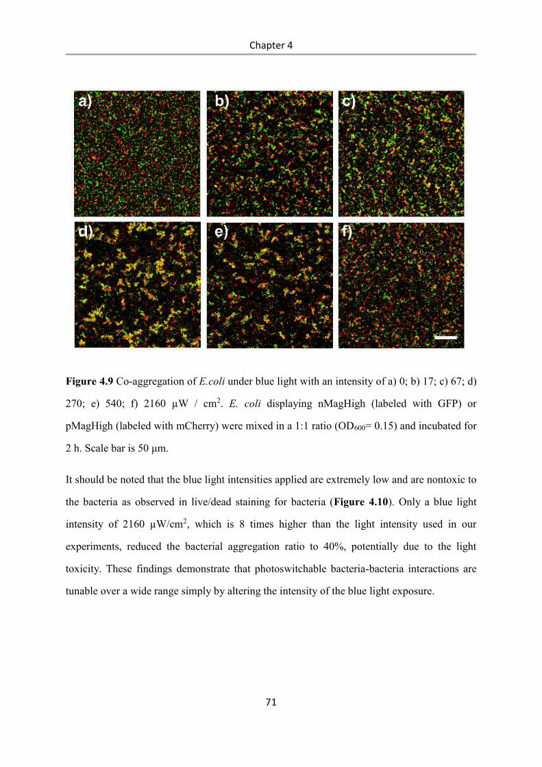
Chapter 4
71
Figure 4.9 Co-aggregation of E.coli under blue light with an intensity of a) 0; b) 17; c) 67; d)
270; e) 540; f) 2160 µW / cm2. E. coli displaying nMagHigh (labeled with GFP) or
pMagHigh (labeled with mCherry) were mixed in a 1:1 ratio (OD600= 0.15) and incubated for
2 h. Scale bar is 50 μm.
It should be noted that the blue light intensities applied are extremely low and are nontoxic to
the bacteria as observed in live/dead staining for bacteria (Figure 4.10). Only a blue light
intensity of 2160 µW/cm2, which is 8 times higher than the light intensity used in our
experiments, reduced the bacterial aggregation ratio to 40%, potentially due to the light
toxicity. These findings demonstrate that photoswitchable bacteria-bacteria interactions are
tunable over a wide range simply by altering the intensity of the blue light exposure.
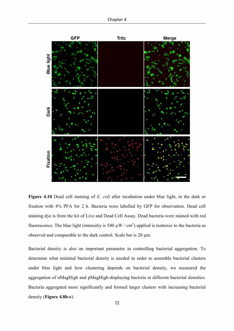
Chapter 4
72
Figure 4.10 Dead cell staining of E. coli after incubation under blue light, in the dark or
fixation with 4% PFA for 2 h. Bacteria were labelled by GFP for observation. Dead cell
staining dye is from the kit of Live and Dead Cell Assay. Dead bacteria were stained with red
fluorescence. The blue light (intensitiy is 540 µW / cm2) applied is nontoxic to the bacteria as
observed and comparable to the dark control. Scale bar is 20 μm.
Bacterial density is also an important parameter in controlling bacterial aggregation. To
determine what minimal bacterial density is needed in order to assemble bacterial clusters
under blue light and how clustering depends on bacterial density, we measured the
aggregation of nMagHigh and pMagHigh-displaying bacteria at different bacterial densities.
Bacteria aggregated more significantly and formed larger clusters with increasing bacterial
density (Figure 4.8b-c).
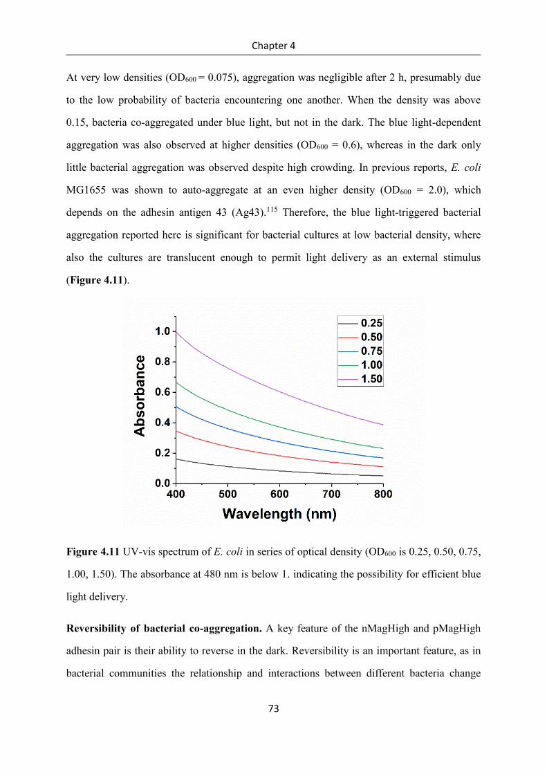
Chapter 4
73
At very low densities (OD600 = 0.075), aggregation was negligible after 2 h, presumably due
to the low probability of bacteria encountering one another. When the density was above
0.15, bacteria co-aggregated under blue light, but not in the dark. The blue light-dependent
aggregation was also observed at higher densities (OD600 = 0.6), whereas in the dark only
little bacterial aggregation was observed despite high crowding. In previous reports, E. coli
MG1655 was shown to auto-aggregate at an even higher density (OD600 = 2.0), which
depends on the adhesin antigen 43 (Ag43).115 Therefore, the blue light-triggered bacterial
aggregation reported here is significant for bacterial cultures at low bacterial density, where
also the cultures are translucent enough to permit light delivery as an external stimulus
(Figure 4.11).
Figure 4.11 UV-vis spectrum of E. coli in series of optical density (OD600 is 0.25, 0.50, 0.75,
1.00, 1.50). The absorbance at 480 nm is below 1. indicating the possibility for efficient blue
light delivery.
Reversibility of bacterial co-aggregation. A key feature of the nMagHigh and pMagHigh
adhesin pair is their ability to reverse in the dark. Reversibility is an important feature, as in
bacterial communities the relationship and interactions between different bacteria change
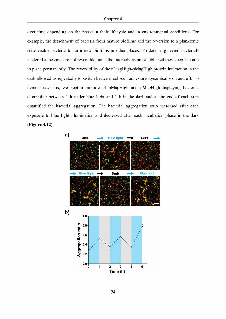
Chapter 4
74
over time depending on the phase in their lifecycle and in environmental conditions. For
example, the detachment of bacteria from mature biofilms and the reversion to a planktonic
state enable bacteria to form new biofilms in other places. To date, engineered bacterial-
bacterial adhesions are not reversible; once the interactions are established they keep bacteria
in place permanently. The reversibility of the nMagHigh-pMagHigh protein interaction in the
dark allowed us repeatedly to switch bacterial cell-cell adhesions dynamically on and off. To
demonstrate this, we kept a mixture of nMagHigh and pMagHigh-displaying bacteria,
alternating between 1 h under blue light and 1 h in the dark and at the end of each step
quantified the bacterial aggregation. The bacterial aggregation ratio increased after each
exposure to blue light illumination and decreased after each incubation phase in the dark
(Figure 4.12).
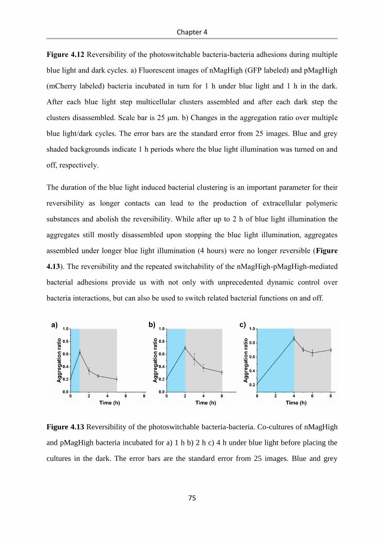
Chapter 4
75
Figure 4.12 Reversibility of the photoswitchable bacteria-bacteria adhesions during multiple
blue light and dark cycles. a) Fluorescent images of nMagHigh (GFP labeled) and pMagHigh
(mCherry labeled) bacteria incubated in turn for 1 h under blue light and 1 h in the dark.
After each blue light step multicellular clusters assembled and after each dark step the
clusters disassembled. Scale bar is 25 μm. b) Changes in the aggregation ratio over multiple
blue light/dark cycles. The error bars are the standard error from 25 images. Blue and grey
shaded backgrounds indicate 1 h periods where the blue light illumination was turned on and
off, respectively.
The duration of the blue light induced bacterial clustering is an important parameter for their
reversibility as longer contacts can lead to the production of extracellular polymeric
substances and abolish the reversibility. While after up to 2 h of blue light illumination the
aggregates still mostly disassembled upon stopping the blue light illumination, aggregates
assembled under longer blue light illumination (4 hours) were no longer reversible (Figure
4.13). The reversibility and the repeated switchability of the nMagHigh-pMagHigh-mediated
bacterial adhesions provide us with not only with unprecedented dynamic control over
bacteria interactions, but can also be used to switch related bacterial functions on and off.
Figure 4.13 Reversibility of the photoswitchable bacteria-bacteria. Co-cultures of nMagHigh
and pMagHigh bacteria incubated for a) 1 h b) 2 h c) 4 h under blue light before placing the
cultures in the dark. The error bars are the standard error from 25 images. Blue and grey
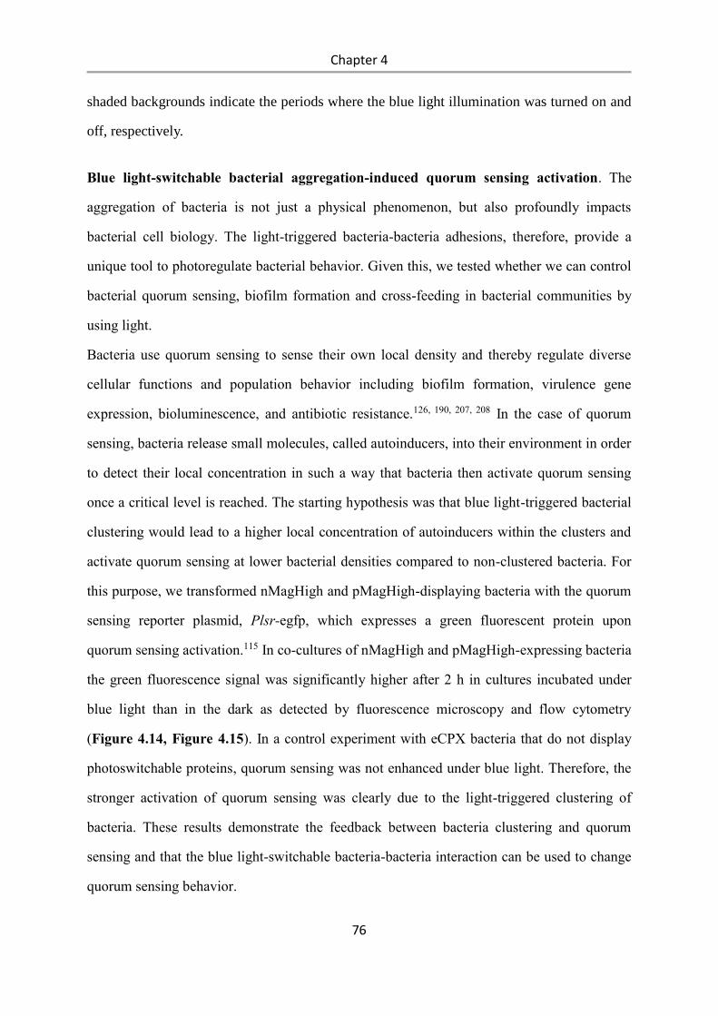
Chapter 4
76
shaded backgrounds indicate the periods where the blue light illumination was turned on and
off, respectively.
Blue light-switchable bacterial aggregation-induced quorum sensing activation. The
aggregation of bacteria is not just a physical phenomenon, but also profoundly impacts
bacterial cell biology. The light-triggered bacteria-bacteria adhesions, therefore, provide a
unique tool to photoregulate bacterial behavior. Given this, we tested whether we can control
bacterial quorum sensing, biofilm formation and cross-feeding in bacterial communities by
using light.
Bacteria use quorum sensing to sense their own local density and thereby regulate diverse
cellular functions and population behavior including biofilm formation, virulence gene
expression, bioluminescence, and antibiotic resistance.126, 190, 207, 208 In the case of quorum
sensing, bacteria release small molecules, called autoinducers, into their environment in order
to detect their local concentration in such a way that bacteria then activate quorum sensing
once a critical level is reached. The starting hypothesis was that blue light-triggered bacterial
clustering would lead to a higher local concentration of autoinducers within the clusters and
activate quorum sensing at lower bacterial densities compared to non-clustered bacteria. For
this purpose, we transformed nMagHigh and pMagHigh-displaying bacteria with the quorum
sensing reporter plasmid, Plsr-egfp, which expresses a green fluorescent protein upon
quorum sensing activation.115 In co-cultures of nMagHigh and pMagHigh-expressing bacteria
the green fluorescence signal was significantly higher after 2 h in cultures incubated under
blue light than in the dark as detected by fluorescence microscopy and flow cytometry
(Figure 4.14, Figure 4.15). In a control experiment with eCPX bacteria that do not display
photoswitchable proteins, quorum sensing was not enhanced under blue light. Therefore, the
stronger activation of quorum sensing was clearly due to the light-triggered clustering of
bacteria. These results demonstrate the feedback between bacteria clustering and quorum
sensing and that the blue light-switchable bacteria-bacteria interaction can be used to change
quorum sensing behavior.
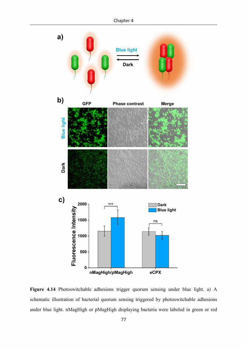
Chapter 4
77
Figure 4.14 Photoswitchable adhesions trigger quorum sensing under blue light. a) A
schematic illustration of bacterial quorum sensing triggered by photoswitchable adhesions
under blue light. nMagHigh or pMagHigh displaying bacteria were labeled in green or red
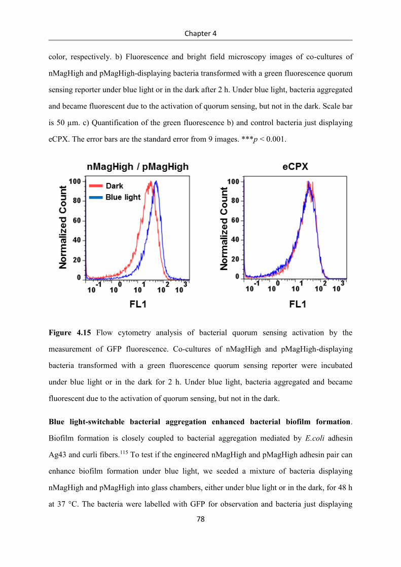
Chapter 4
78
color, respectively. b) Fluorescence and bright field microscopy images of co-cultures of
nMagHigh and pMagHigh-displaying bacteria transformed with a green fluorescence quorum
sensing reporter under blue light or in the dark after 2 h. Under blue light, bacteria aggregated
and became fluorescent due to the activation of quorum sensing, but not in the dark. Scale bar
is 50 µm. c) Quantification of the green fluorescence b) and control bacteria just displaying
eCPX. The error bars are the standard error from 9 images. ***p < 0.001.
Figure 4.15 Flow cytometry analysis of bacterial quorum sensing activation by the
measurement of GFP fluorescence. Co-cultures of nMagHigh and pMagHigh-displaying
bacteria transformed with a green fluorescence quorum sensing reporter were incubated
under blue light or in the dark for 2 h. Under blue light, bacteria aggregated and became
fluorescent due to the activation of quorum sensing, but not in the dark.
Blue light-switchable bacterial aggregation enhanced bacterial biofilm formation.
Biofilm formation is closely coupled to bacterial aggregation mediated by E.coli adhesin
Ag43 and curli fibers.115 To test if the engineered nMagHigh and pMagHigh adhesin pair can
enhance biofilm formation under blue light, we seeded a mixture of bacteria displaying
nMagHigh and pMagHigh into glass chambers, either under blue light or in the dark, for 48 h
at 37 °C. The bacteria were labelled with GFP for observation and bacteria just displaying
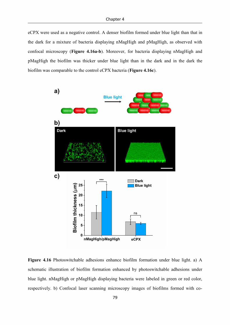
Chapter 4
79
eCPX were used as a negative control. A denser biofilm formed under blue light than that in
the dark for a mixture of bacteria displaying nMagHigh and pMagHigh, as observed with
confocal microscopy (Figure 4.16a-b). Moreover, for bacteria displaying nMagHigh and
pMagHigh the biofilm was thicker under blue light than in the dark and in the dark the
biofilm was comparable to the control eCPX bacteria (Figure 4.16c).
Figure 4.16 Photoswitchable adhesions enhance biofilm formation under blue light. a) A
schematic illustration of biofilm formation enhanced by photoswitchable adhesions under
blue light. nMagHigh or pMagHigh displaying bacteria were labeled in green or red color,
respectively. b) Confocal laser scanning microscopy images of biofilms formed with co-
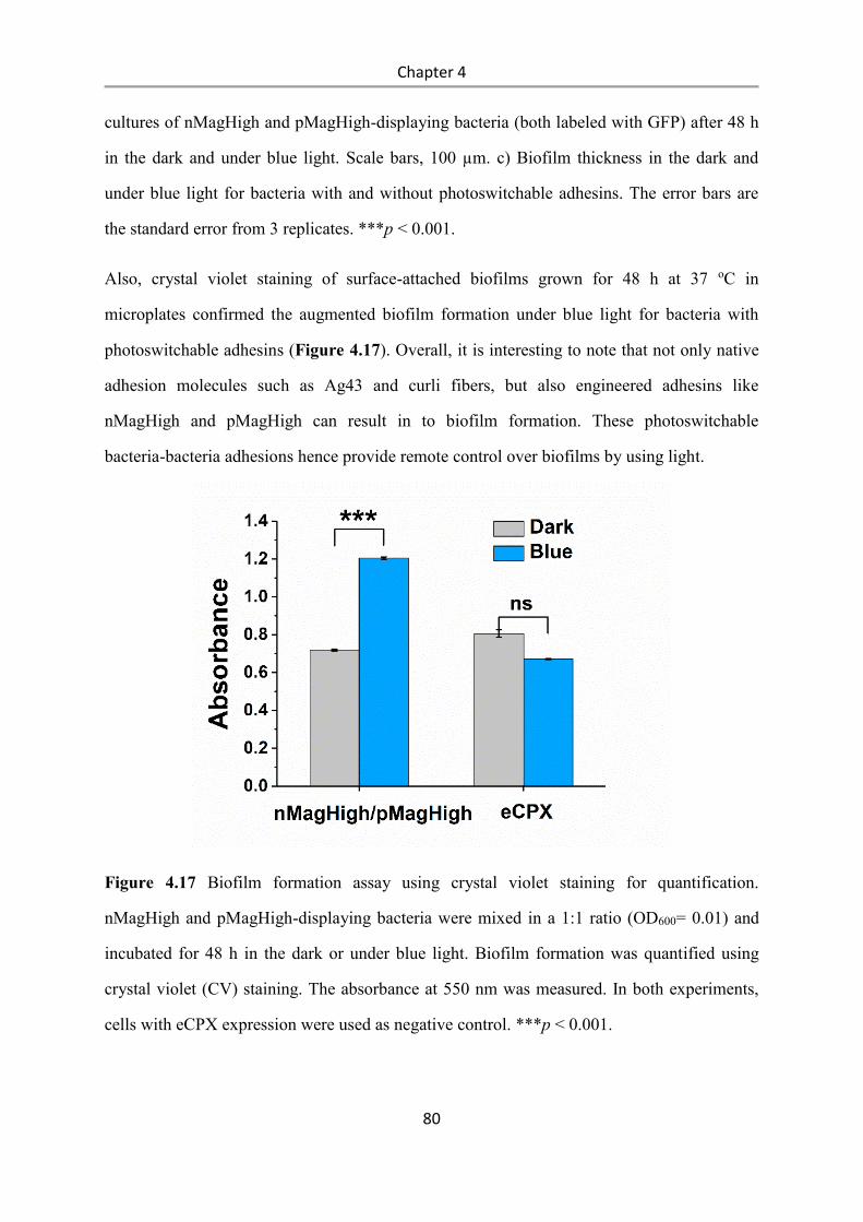
Chapter 4
80
cultures of nMagHigh and pMagHigh-displaying bacteria (both labeled with GFP) after 48 h
in the dark and under blue light. Scale bars, 100 µm. c) Biofilm thickness in the dark and
under blue light for bacteria with and without photoswitchable adhesins. The error bars are
the standard error from 3 replicates. ***p < 0.001.
Also, crystal violet staining of surface-attached biofilms grown for 48 h at 37 oC in
microplates confirmed the augmented biofilm formation under blue light for bacteria with
photoswitchable adhesins (Figure 4.17). Overall, it is interesting to note that not only native
adhesion molecules such as Ag43 and curli fibers, but also engineered adhesins like
nMagHigh and pMagHigh can result in to biofilm formation. These photoswitchable
bacteria-bacteria adhesions hence provide remote control over biofilms by using light.
Figure 4.17 Biofilm formation assay using crystal violet staining for quantification.
nMagHigh and pMagHigh-displaying bacteria were mixed in a 1:1 ratio (OD600= 0.01) and
incubated for 48 h in the dark or under blue light. Biofilm formation was quantified using
crystal violet (CV) staining. The absorbance at 550 nm was measured. In both experiments,
cells with eCPX expression were used as negative control. ***p < 0.001.
Chapter 4
81
Blue light-controllable bacterial aggregation alters metabolic cross-feeding. In nature,
bacteria do not exist in isolation, but live as complex multispecies’ communities. Within
these communities, different members can perform distinct tasks, exchange essential
metabolites and form microbial ecosystems, which also have important biotechnological
implications.56, 209 However, designing bacterial communities is far from straightforward as
the spatial structure of a microbial community, nutrient availability, diffusion constraints and
metabolic burden on different members alters the biochemical phenotypes and abundance of
the participating strains. For instance, Mee et al. devised a series of synthetic syntrophic
communities in co-cultures of different E. coli amino acid auxotrophs. While the exchange of
essential amino acids in certain mixtures of auxotrophs leads to cooperative growth, the
metabolic cross-feeding revealed both positive and negative interactions in communities of a
higher complexity.56 Furthermore, emergence and maintenance of metabolic exchanges
depend on particular circumstances and changes over time. Here we asked the question how
the aggregation changes cross-feeding between different auxotrophs, as aggregation increases
the efficiency of nutrient transfer and potentially can stimulate otherwise unfavorable
metabolic processes.210
To test how the aggregation alters bacterial cross-feeding and co-culture composition, we
expressed either nMagHigh or pMagHigh on the surfaces of E. coli BL21 strains that are
auxotrophic for one essential amino acid (P, K, W, M and T). We named each bacterial strain
after the amino acid it is auxotrophic for and the protein displayed on its surface (n:
nMagHigh, p: pMagHigh), e.g. Kn: lysine auxotroph displaying nMagHigh.(Figure 4.18a) In
order to observe if the spatial proximity alters the cross-feeding process, we co-cultured
different nMagHigh and pMagHigh-displaying auxotrophs under blue light or in the dark and
quantified their growth by measuring the OD600 (Figure 4.18b). As reported before, each
auxotroph was unable to grow on its own in a M9 minimal medium, but a subset of these 1:1
co-cultures showed synergistic growth after 48 h. More importantly, the cooperative growth
of some co-cultures was different under blue light and in the dark; Pn/Kp co-cultures grew 6-

Chapter 4
82
fold better under blue light than in the dark and Wn/Mp, Tn/Wp and Wn/Tp co-cultures grew
better in the dark than under blue light (4, 2 and 3 fold, respectively).
Figure 4.18 Photoswitchable adhesions alter metabolic interactions and cross-feeding. a) A
schematic illustration of bacterial metabolic cross-feeding altered by photoswitchable
adhesions. nMagHigh or pMagHigh displaying bacteria were labeled in green or red color,
respectively. b) Syntrophic growth of different auxotrophic strains displaying nMagHigh or
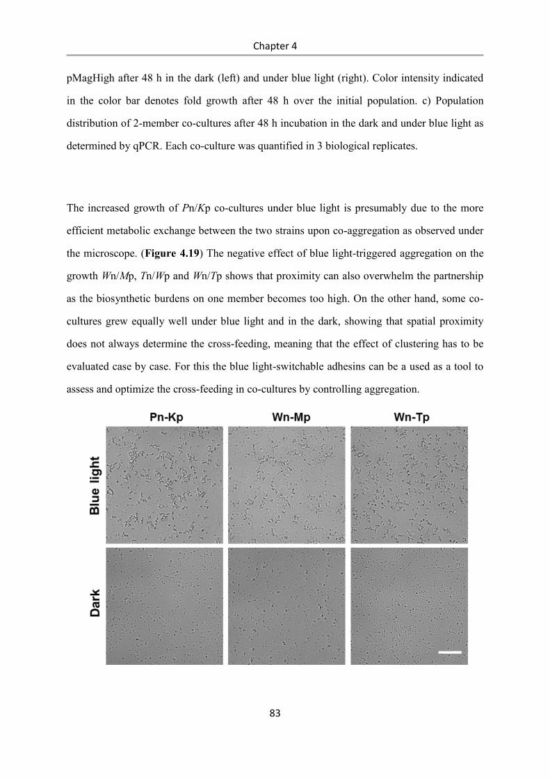
Chapter 4
83
pMagHigh after 48 h in the dark (left) and under blue light (right). Color intensity indicated
in the color bar denotes fold growth after 48 h over the initial population. c) Population
distribution of 2-member co-cultures after 48 h incubation in the dark and under blue light as
determined by qPCR. Each co-culture was quantified in 3 biological replicates.
The increased growth of Pn/Kp co-cultures under blue light is presumably due to the more
efficient metabolic exchange between the two strains upon co-aggregation as observed under
the microscope. (Figure 4.19) The negative effect of blue light-triggered aggregation on the
growth Wn/Mp, Tn/Wp and Wn/Tp shows that proximity can also overwhelm the partnership
as the biosynthetic burdens on one member becomes too high. On the other hand, some co-
cultures grew equally well under blue light and in the dark, showing that spatial proximity
does not always determine the cross-feeding, meaning that the effect of clustering has to be
evaluated case by case. For this the blue light-switchable adhesins can be a used as a tool to
assess and optimize the cross-feeding in co-cultures by controlling aggregation.
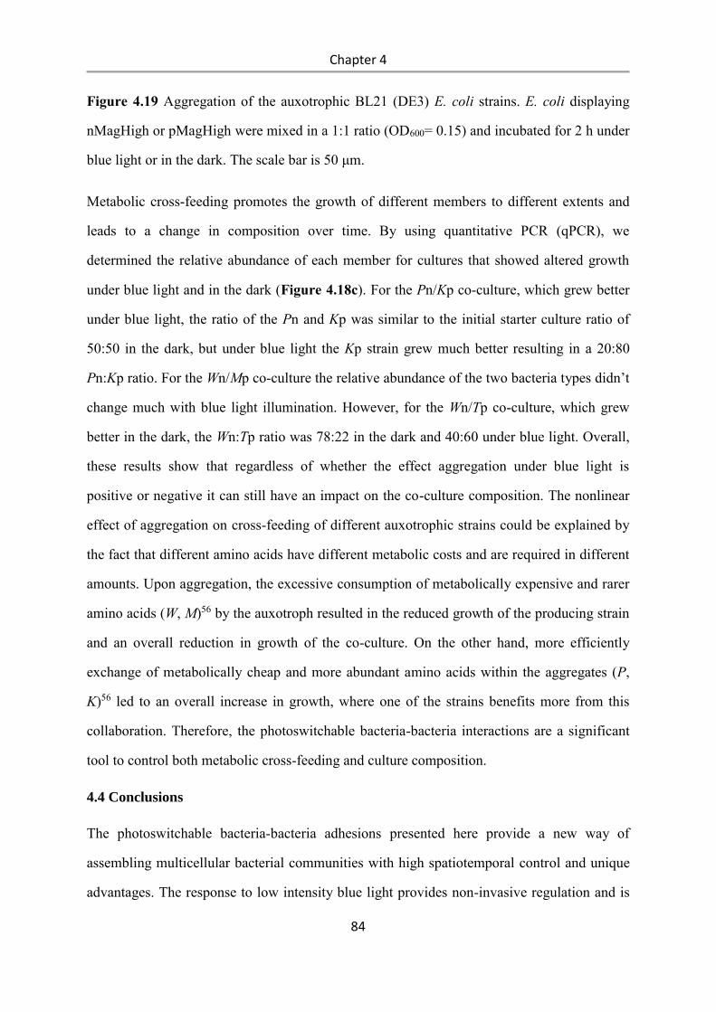
Chapter 4
84
Figure 4.19 Aggregation of the auxotrophic BL21 (DE3) E. coli strains. E. coli displaying
nMagHigh or pMagHigh were mixed in a 1:1 ratio (OD600= 0.15) and incubated for 2 h under
blue light or in the dark. The scale bar is 50 μm.
Metabolic cross-feeding promotes the growth of different members to different extents and
leads to a change in composition over time. By using quantitative PCR (qPCR), we
determined the relative abundance of each member for cultures that showed altered growth
under blue light and in the dark (Figure 4.18c). For the Pn/Kp co-culture, which grew better
under blue light, the ratio of the Pn and Kp was similar to the initial starter culture ratio of
50:50 in the dark, but under blue light the Kp strain grew much better resulting in a 20:80
Pn:Kp ratio. For the Wn/Mp co-culture the relative abundance of the two bacteria types didn’t
change much with blue light illumination. However, for the Wn/Tp co-culture, which grew
better in the dark, the Wn:Tp ratio was 78:22 in the dark and 40:60 under blue light. Overall,
these results show that regardless of whether the effect aggregation under blue light is
positive or negative it can still have an impact on the co-culture composition. The nonlinear
effect of aggregation on cross-feeding of different auxotrophic strains could be explained by
the fact that different amino acids have different metabolic costs and are required in different
amounts. Upon aggregation, the excessive consumption of metabolically expensive and rarer
amino acids (W, M)56 by the auxotroph resulted in the reduced growth of the producing strain
and an overall reduction in growth of the co-culture. On the other hand, more efficiently
exchange of metabolically cheap and more abundant amino acids within the aggregates (P,
K)56 led to an overall increase in growth, where one of the strains benefits more from this
collaboration. Therefore, the photoswitchable bacteria-bacteria interactions are a significant
tool to control both metabolic cross-feeding and culture composition.
4.4 Conclusions
The photoswitchable bacteria-bacteria adhesions presented here provide a new way of
assembling multicellular bacterial communities with high spatiotemporal control and unique
advantages. The response to low intensity blue light provides non-invasive regulation and is
Chapter 4
85
bioorthogonal to other cellular processes. Photoregulation is especially attractive for
biotechnological applications as the application of light is a cheap and easy way of removing
stimuli compared to applying chemicals that are used to control bacterial cultures.
Interestingly, various microorganisms photoregulates their adhesions in response to light in
order to adapt to changing environmental conditions.211, 212 Implementing a light regulated
adhesion module into E. coli can similarly be used to photoregulate bacterial behaviour in
changing enviroments. The optogenetic proteins used, nMagHigh and pMagHigh, assure
specific binding between bacteria expressing interaction partners under blue light and allow
for them to arrange themselves into specific patterns. While the photoswitchable protein
interactions are strong enough to mediate aggregation, they still allow for dynamic
rearrangement and coalescence of aggregates to maximize bacteria-bacteria interactions.
Importantly, the multi-bacterial assemblies can be disassembled in the dark allowing for
dynamic switching between unicellular and multicellular life forms. This is an important
feature when it comes to designing bacterial communities as the relationship between
different bacteria can change over time as environmental parameters and the internal state of
the bacteria vary. Moreover, bacterial aggregation is highly tunable by altering light intensity
and exposure time as well as bacterial density. The sustained production of the genetically
encoded proteins makes control in long term cultures possible, as demonstrated in cross-
feeding and biofilm formation experiments. This is an important advantage over strategies
that involve the chemical modification of the surface of the bacteria, as these modifications
are difficult to maintain over time. E. coli typically divide every 20 min and quickly dilute
the functional groups on their surfaces.
The photoswitchable bacteria-bacteria interactions provide an exclusive combination of
features and are a significant new tool in the study of bacterial cell biology and the regulation
of bacterial consortia. We have demonstrated how we can photocontrol central bacterial
behavior including quorum sensing, biofilm formation and metabolic cross-feeding with this
new tool. The modular nature of the photoswitchable adhesins allow to integrate them onto
the surface of many different cell types and couple them to cell-cell signaling, quorum
Chapter 4
86
sensing-based gene regulators and gene regulatory logic gates. Moreover, the ability to
regulate and optimize cross-feeding in co-cultures opens the way to design more efficient
bacterial factories that can perform further complex transformations. The evolutionary
transition from single to multi-cellular life forms has resulted in an explosion of diversity. By
understanding and controlling multi-cellularity in the field of synthetic biology we hope to
achieve more than just the sophistication of bacterial communities.
Summary and outlook
87
5. Summary and outlook
Controlling bacterial adhesion with high spatial and temporal precision is essential for
controlling the formation, organization and microstructure of biofilms. In this thesis, different
methods to control bacterial adhesions using light have been developed to control biofilm
formation or the assembly of multicellular structures with high spatiotemporal precision and
to study how the spatial organization of bacteria influences their collective functions.
In the first part of the thesis, a new method named bacterial photolithography were
created to pattern stable biofilms through irreversible light response. This method provides
high flexibility in creating complex patterns with a resolution down to 10 µm, which
represents an important step towards the engineering of precise biofilm communities
as the microstructure of natural biofilms has a major impact on their function. This
method enables the investigation of how microscale spatial organization affects collective
bacterial interactions such as quorum sensing. It has significant potential for future
applications in guiding the formation and spatial organization of bacteria in a biofilm,
as well as improving our understanding of naturally existing biofilms and the design of
bacterial consortia.
In the second part of the thesis, photoswitchable proteins nMag and pMag were used to
control bacterial adhesions in an optogenetic approach. This approach overcomes the
problem of using UV light for photoregulation and chemically modifying the bacteria surface.
It provides non-invasive, bioorthogonal and reversible control over bacterial attachment with
unprecedented spatial and temporal control. The photoswitchable bacteria adhesions allow us
to control the formation and spatial organization of bacteria in a biofilm, which is vital in
engineering bacterial consortia.
In the last part of the thesis, the photoswitchable bacteria-bacteria adhesions were developed
based on the optogenetic proteins nMag and pMag. It provides a new way to spatiotemporally
control the reversible assembly of multicellular clusters with blue light. The photoswitchable
bacteria-bacteria interactions provide an exclusive combination of features and are a
Summary and outlook
88
significant new tool in the study of bacterial cell biology and the regulation of bacterial
consortia. These photoswitchable adhesions made it possible to regulate collective bacterial
functions using light including aggregation, quorum sensing, biofilm formation and
metabolic cross-feeding between auxotrophic bacteria. Multi-bacterial communities are of
fundamental importance and have great biotechnological potentials.
All these work open new possibilities for engineering multicellular communities, understand
fundamental bacterial behavior in biofilms and design biofilms with new functions for
biotechnological applications. The evolutionary transition from single to multi-cellular life
forms has resulted in an explosion of diversity. By understanding and controlling multi-
cellularity in the field of synthetic biology, we hope to achieve more than just the
sophistication of bacterial communities.
Materials and Methods
89
6. Materials and Methods
6.1 Materials
6.1.1 Plasmids
The GFP pTrc99A, mCherry pTrc99A (Ampicillin resistant, IPTG inducible), and Plsr-egfp
plasmids (Kanamycin resistant) for E.coli expression were gifts from Prof. Victor Sourjik.
The plasmid pB33eCPX, which contains the gene for the enhanced circularly permutated
outer membrane protein OmpX (eCPX), was a gift from Prof. Patrick Daugherty (Addgene
plasmid # 23336)191. The nMagHigh gene in the pET-21b(+) plasmid between the NdeI and
XhoI cutting sites was purchased from Genescript.
6.1.2 Bacterial Strains
E.coli K12 MG1655 was purchased from DSMZ. BL21(DE3) E.coli were purchased from
NEB. The auxotrophic BL21 (DE3) E. coli strains RF2 (Addgene plasmid # 62070, T), RF6
(Addgene plasmid # 62074, P), RF10 (Addgene plasmid # 62076, K), RF11 (Addgene
plasmid # 61961, M) and RF12 (Addgene plasmid # 62077, T) were a gift from Robert
Gennis & Toshio Iwasaki.213
6.1.3 Other Materials.
The 2,5-dioxopyrrolidin-1-yl (1-(5-methoxy-2-nitro-4-(prop-2-yn-1-yloxy)phenyl)ethyl)
carbonate was synthesized following the literature.26 4-Aminophenyl α-D-mannopyranoside
was purchased from Sigma-Aldrich. PBS (pH 7.4) was prepared by PBS tablets from Gibco.
LB-Medium (Luria/Miller) was purchased from Carl Roth. 10 g tryptone and 5 g NaCl were
dissolved in 1 L H2O to obtain TB (Tryptone Broth) medium. LB and TB media were
autoclaved prior to use. 20 x 20 mm glass coverslips were purchased from Carl Roth. PEG
(3000)-azide was synthesized following previous reports from our lab.172 QuikChange II Site-
Directed Mutagenesis Kit was purchased from Agilent. All chemicals were purchased from
Sigma-Aldrich. The live and dead cell Assay kit (ab189818) was purchased from Abcam.
Materials and Methods
90
KAPA SYBR® FAST Universal Kit was purchased Sigma Aldrich. 8-well slides (μ-Slide, 8-
well glass bottom) were purchased form ibidi.
6.2 Methods
6.2.1 Bacterial culture
E.coli K12 MG1655 were co-transformed with one of the eCPX fused photoswitchable
protein plasmids (nMagHigh-eCPX, or pMagHigh-eCPX, or pMag-eCPX, or pMagFast1-
eCPX, chloramphenicol resistant, L-arabinose inducible), and a fluorescent protein
expression plasmid (GFP-pTrc99A or mCherry-pTrc99A, ampicillin resistant, IPTG
inducible). 5 mL LB medium containing 35 μg/mL chloramphenicol and 50 μg/mL
ampicillin were inoculated with a single colony and incubated overnight at 37 °C at 250 rpm.
Then, 50 μL of these cultures was added into fresh 5 mL LB medium containing 35 μg/mL
chloramphenicol and 50 μg/mL ampicillin and cultured for 2 h at 37 °C, at 50 rpm. Then, the
OD600 = 0.4, 0.04% m/v L-arabinose and 0.5 mM IPTG were added to the cultures to induce
the expression of the eCPX fused proteins and the fluorescent proteins, respectively. The
bacteria were cultured at 25 °C, at 250 rpm for 4 h and subsequently diluted in PBS
(phosphate buffer saline) to the desired density.
6.2.2 Expression and purification of nMagHigh
The nMagHigh with a C-terminal His6-tag in pET-21b(+) expression plasmid was
transformed into BL21(DE3) E.coli cells. The protein expression was induced at OD600 = 0.6
with 1 mM IPTG and the protein was expressed at overnight at 18 °C, 250 rpm. The cell
pellet was suspended in 20 mL buffer A (50 mM Tris-HCl pH 7.4, 300 mM NaCl ) with 1
mM protease inhibitor PMSF (phenylmethane sulfonyl fluoride). The cells were lysed by
sonication and the lysate was cleared by centrifugation at 12000 rpm for 30 min followed by
filtration through a 0.45 μm filter twice. The lysate was passed over a 5 mL of Ni2+-NTA
agarose column. The column was washed with 50 mL buffer C (Buffer A with 25 mM
imidazole) and the protein was eluted with 10 mL buffer B (Buffer A with 250 mM
Materials and Methods
91
imidazole). The purified nMagHigh-His6 was dialyzed against 2 L buffer A twice for at least
6 h and the purity was verified by SDS-PAGE.
6.2.3 Cloning of nMagHigh and pMag variants
To express pMag variants (pMagHigh, pMag and pMagFast1) on the surface of E.coli, we
fused these proteins to the N-terminal of eCPX. To insert the nMagHigh gene into the
plasmid pB33eCPX, the nMagHigh gene was firstly amplified by PCR from nMagHigh pET-
21b(+) plasmid and inserted between the KpnI and SacI cutting sites of pB33eCPX. The
different pMag variants fused to eCPX were generated by point mutagenesis from the
nMagHigh-eCPX plasmid using QuikChange II Site-Directed Mutagenesis Kit (Scheme S1).
6.2.4 Synthesis of mannoside-NO2
To a solution of 2,5-dioxopyrrolidin-1-yl (1-(5-methoxy-2-nitro-4-(prop-2-yn-1-
yloxy)phenyl)ethyl) carbonate (1 eq., 271.27 mg, 1 mmol) in 1 mL DMF, a solution of 4-
aminophenyl α-D-mannopyranoside (1 eq., 392.09 mg, 1 mmol) in 400 μL Na2CO3 buffer (1
M, pH = 8.5) and 600 μL H2O were added. The reaction mixture was kept at room
temperature with stirring for 24 h. The reaction mixture was evaporated under vacuum and
the remaining solid was purified by HPLC (column: reverse phase C18). A: Acetonitrile,
0.1% TFA, B: H2O, 0.1% TFA, flow 1mL/min, gradient 20% to 80% B, retention time 14
min). The concentration of the final product is determined to be 1.0 mM by UV-Vis (ε 350 =
5000 M−1 cm−1). HR-MS(ESI+): [M+ Na]+ observed: m/z = 571.1541, calc.: m/z =
571.1540. 1H-NMR (600 MHz; D3OD): (in ppm) = 7.79 (s, 1H), 7.30 (d, J = 9 Hz, 2H),
7.28 (s, 1H); 7.04 (d, J = 8.4 Hz, 2H), 6.39 (q, J = 6 Hz, 1H), 5.38 (s, 1H), 4.85 (d, J = 2.4Hz,
2H), 3.96 (s, 3H), 3.90 – 3.59 (m, 6H), 3.04 (t, J = 2.4Hz, 1H), 1.65 (d, J = 6Hz, 3H).
6.2.5 UV-Vis spectroscopy of mannoside-NO2
UV-Vis spectra were acquired using plate reader (Tecan, SparkTM). The absorbance of 0.1
mM Mannoside-NO2 in H2O was measured from 280 to 700 nm after different irradiation
times (0, 1, 2, 5, 10, 15, 20, 30 min) with 365 nm light (UV transilluminator from Syngene’s
GeneFlash).
Materials and Methods
92
6.2.6 Functionalization of glass surfaces with PEG-mannoside-NO2
The glass surfaces were functionalized similarly to previous reports.172 Shortly, glass slides
(20 × 20 mm) were cleaned in freshly prepared Piranha solution (3:1 (v/v) conc. H2SO4:H2O2
(30%)) for 1 h, rinsed 3 times with Milli-Q water and dried in an N2 stream. For the
PEGylation reaction, surfaces were immersed in a solution of PEG-azide (10 mg PEG-azide,
MW = 3000 g/mol) and a drop of dry triethylamine in dry toluene and kept at 79 °C
overnight under a N2 atmosphere. The surfaces were first washed with ethyl acetate for 5 min
by sonication, then with methanol for 5 min by sonication and dried in a N2 stream. The
PEG-coated surfaces were incubated in contact with 50 μL of reaction solution containing
100 mM L-ascorbic acid, 100 mM Tris HCl pH 9.0, 50 μM of the mannoside-NO2 and 1 mM
CuSO4 in a moisture chamber for 2 h. Afterwards the surfaces were washed with 50 mM
EDTA (pH 7.4) for 5 min and 3 times PBS for 5 min.
6.2.7 Bacterial adhesion to the mannoside-NO2 functionalized surfaces.
E.coli K12 MG1655 were transformed with the GFP pTrc99A plasmid (ampicillin resistant)
and selected on an LB-agar plate with 50 μg/mL apicillin. 2 m LLB medium containing 50
μg/mL ampicillin was inoculated with a single colony and incubated overnight at 37 °C at
250 rpm. Then, 100 μL of these cultures were transferred into 10 mL fresh LB medium
containing 50 μg/mL ampicillin and cultured at 37 °C, 250 rpm. When the OD600 = 0.4, the
production of the fluorescent protein was induced with 0.5 mM IPTG and the culture was
incubated at 37 °C, 250 rpm for 4 h. The bacteria were then spun down at 4000 rpm for 10
min and washed by PBS twice. Finally, the bacteria were resuspended in PBS to OD600 = 1.0.
For bacterial adhesion assays, 3 mL of bacteria were placed on top of mannoside-NO2
functionalized surfaces. Prior to bacterial seeding, the surfaces were either kept in the dark or
pre-illuminated by UV light (UV transilluminator, Syngene’s GeneFlash) for 1 h. Surfaces
with just the PEG coating but not modified with mannoside-NO2 were used as negative
controls. The bacteria were incubated on these surfaces at room temperature for 10, 30, 60,
120 or 210 min.
Materials and Methods
93
To investigate the effect of UV-light illumination, mannoside-NO2 functionalized surfaces
were firstly pre-illuminated with UV light for 0, 2, 5, 10, 15, 30, 60 min and then bacteria
were placed onto these surfaces for 210 min incubation in the dark.
For analysis, after the incubation of the bacteria on the surfaces, the surfaces were gently
washed 3 times with PBS to remove unbound bacteria and 3 x 3 fluorescent images (field of
view of one image =1.04 mm2) in the GFP channel were acquired on an inverted fluorescence
microscope (DMi8, Leica) through a 20x objective. The number of bacteria on the surface
was analyzed using the particle analyzer tool in ImageJ.
6.2.8 Bacterial biofilm photopatterning on the mannoside-NO2 functionalized surfaces.
A mannoside-NO2 functionalized surface was locally illuminated with 358 nm UV light
through the photomask for 2 min on an inverted fluorescence microscope (DMi8, Leica)
through a 5x objective. Then, the photomask was removed and a bacteria solution with OD600
= 1.0 prepared as described above is placed on top of the surface and incubate for 4 h. The
glass surface was gently washed 3 times with PBS to remove non-attached bacteria before
imaging the bacterial biofilm pattern.
6.2.9 Live imaging of patterned biofilms and quorum sensing
E.coli K12 MG1655 were co-transformed with mCherry pTrc99A (ampicillin resistant) and
Plsr-egfp (kanamycin resistant) plasmids and selected on an LB-agar plate with 50 μg/mL
ampicillin and 50 μg/mL kanamycin, then cultured as described above. A mannoside-NO2
functionalized surface was illuminated through a photomask with UV light as described
above. Then, the photomask was removed and a bacteria solution with OD600 = 1.0 is placed
on top of the surface. 4 h later, non-attached bacteria were removed by washing 3 times with
PBS and the bacterial pattern was observed under microscope in the mCherry fluorescence
channel. Then, PBS containing 10% TB medium was added on top of the surface. Afterwards
the bacterial pattern was imaged in the mCherry and GFP channels every 3 min for 10 h on a
fluorescence microscope (DMi8, Leica) with 10x objective. The fluorescence intensity of
bacterial biofilm pattern was analyzed using the intensity analyzer tool in ImageJ.
Materials and Methods
94
6.2.10 Functionalization of glass surfaces with nMagHigh
The glass substrates were functionalized similarly to previous reports.28 Shortly, glass slides
(20 × 20 mm) were cleaned in freshly prepared Piranha solution (3:1 (v/v) conc. H2SO4:H2O2
(30%)) for 1 h, rinsed 3 times with Milli-Q water and dried in an N2 stream. For the
PEGylation reaction, surfaces were immersed in a solution of PEG-azide (10 mg PEG-azide,
MW= 3500 g/mol) and a drop of dry triethylamine in dry toluene and kept at 79 °C overnight
under a N2 atmosphere. The surfaces were first washed with ethyl acetate for 5 min by
sonication, then with methanol for 5 min by sonication and dried in a N2 stream. The PEG-
coated surfaces were incubated in contact with 100 μL of reaction solution containing 100
mM L-ascorbic acid, 100 mM Tris HCl pH 9.0, 150 μM of the NTA-alkyne and 1 mM
CuSO4 in a moisture chamber for 2 h (Scheme 2). Afterwards the surfaces were incubated
with the following solutions to obtain the nMagHigh functionalized substrates through NTA-
Ni2+ -His tag interaction: 1) 50 mM EDTA (pH 7.4) for 5 min, 2) Buffer A twice for 5 min,
3) 0.1 M NiCl2 in water for 5 min, 4) Buffer A twice for 5 min, 5) 10 μM purified
nMagHigh-His6 protein for 30 min, 7) Buffer A twice for 5 min.
6.2.11 Bacterial adhesion to the nMagHigh functionalized substrates and detachment
assays
E.coli K12 MG1655 were co-transformed with the appropriate pMag-eCPX plasmid
(chloramphenicol resistant) and the GFP plasmid (ampicillin resistant) and selected on an
LB-agar plate with 35 μg/mL chloramphenicol and 50 μg/mL ampicillin. 5 ml LB medium
containing 35 μg/mL chloramphenicol and 50 μg/mL ampicillin was inoculated with a single
colony and incubated overnight at 37oC at 250 rpm. Then, 500 μL of these cultures were
added into fresh 20 ml LB medium containing 35 μg/ml chloramphenicol and 50 μg/mL
ampicillin and cultured for 2 h at 37 °C, 250 rpm. When the OD600 = 0.5, 0.04% m/v L-
arabinose to induce the expression of the pMag-eCPX proteins and 0.5 mM IPTG to induce
the production of the fluorescent protein were added and the cultures were incubated at 25
°C, 250 rpm for 4 h. The bacteria were then spun down at 4000 rpm for 10 min and washed
by PBS twice. Finally, the bacteria were resuspended in PBS to OD600 = 1.0.
Materials and Methods
95
For bacterial adhesion assays, 3 mL of bacteria co-expressing one of the pMag protein
variants on their surface and GFP protein were placed on nMagHigh functionalized
substrates. The bacteria were either incubated under blue light illumination (blue LED panel,
640 µW/cm2) or in the dark for 1 h. Bacteria incubated on substrates that lack the
immobilized nMagHigh protein were used as negative controls. Other bacterial adhesion
assays were performed similarly changing one parameter at the time: The illumination
intensity (0, 3.2, 32, 320, 640 or 3200 µW/cm2) or the incubation time under blue light (10,
30, 60, 120 or 180 min).
For bacterial detachment assays, bacteria were placed on nMagHigh substrates and were
incubated for 1 h under blue light illumination, and then the samples were moved to the dark
for 0, 10, 30, 60, 120 or 240 min, respectively.
For the cycling experiments, bacterial were first incubated under blue light illumination for 1
h for attachment and then kept in the dark for 2 h for detachment. This process was repeated
in total for 2.5 cycles.
For analysis, after the incubation with the bacteria, the substrates were gently washed 3 times
with PBS to remove unbound bacteria and 3 x 3 fluorescent images (area of one image =1.04
mm2) in the GFP channel were acquired on an inverted fluorescence microscope (DMi8,
Leica) through a 20x objective. The number of bacteria on the surface were analyzed using
the particle analyzer tool in ImageJ.
6.2.12 Bacterial patterning on the nMagHigh functionalized substrates.
E.coli K12 MG1655 were co-transformed with pMagHigh-eCPX and mCherry plasmid
(ampicillin resistant) plasmids and cultured as described above. A bacteria solution with
OD600 = 1.0 is placed on top of a nMagHigh functionalized glass substrate and kept in the
dark. The glass surface was locally illuminated with blue light through photomask for 1 h on
an inverted fluorescence microscope (DMi8, Leica) through a 10x objective. Then, the
photomask was removed and the glass surfaces were gently washed 3 times with PBS before
imaging the bacterial pattern.
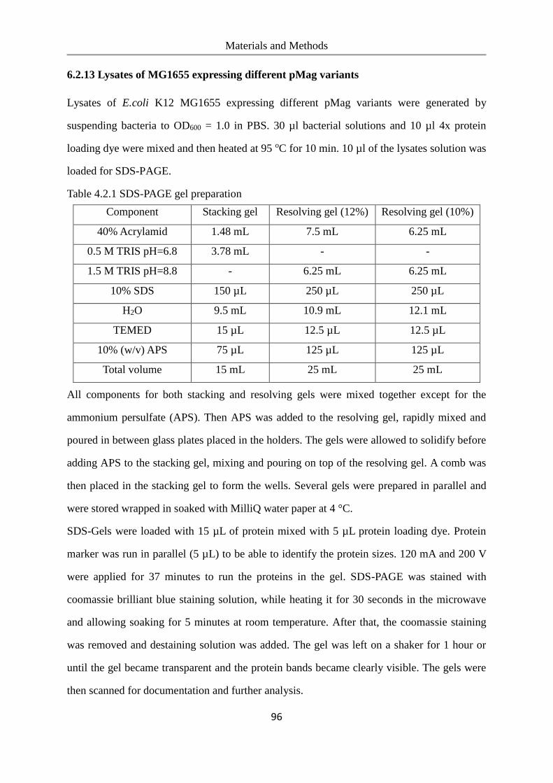
Materials and Methods
96
6.2.13 Lysates of MG1655 expressing different pMag variants
Lysates of E.coli K12 MG1655 expressing different pMag variants were generated by
suspending bacteria to OD600 = 1.0 in PBS. 30 µl bacterial solutions and 10 µl 4x protein
loading dye were mixed and then heated at 95 oC for 10 min. 10 µl of the lysates solution was
loaded for SDS-PAGE.
Table 4.2.1 SDS-PAGE gel preparation
Component Stacking gel Resolving gel (12%) Resolving gel (10%)
40% Acrylamid 1.48 mL 7.5 mL 6.25 mL
0.5 M TRIS pH=6.8 3.78 mL - -
1.5 M TRIS pH=8.8 - 6.25 mL 6.25 mL
10% SDS 150 µL 250 µL 250 µL
H2O 9.5 mL 10.9 mL 12.1 mL
TEMED 15 µL 12.5 µL 12.5 µL
10% (w/v) APS 75 µL 125 µL 125 µL
Total volume 15 mL 25 mL 25 mL
All components for both stacking and resolving gels were mixed together except for the
ammonium persulfate (APS). Then APS was added to the resolving gel, rapidly mixed and
poured in between glass plates placed in the holders. The gels were allowed to solidify before
adding APS to the stacking gel, mixing and pouring on top of the resolving gel. A comb was
then placed in the stacking gel to form the wells. Several gels were prepared in parallel and
were stored wrapped in soaked with MilliQ water paper at 4 °C.
SDS-Gels were loaded with 15 µL of protein mixed with 5 µL protein loading dye. Protein
marker was run in parallel (5 µL) to be able to identify the protein sizes. 120 mA and 200 V
were applied for 37 minutes to run the proteins in the gel. SDS-PAGE was stained with
coomassie brilliant blue staining solution, while heating it for 30 seconds in the microwave
and allowing soaking for 5 minutes at room temperature. After that, the coomassie staining
was removed and destaining solution was added. The gel was left on a shaker for 1 hour or
until the gel became transparent and the protein bands became clearly visible. The gels were
then scanned for documentation and further analysis.
Materials and Methods
97
6.2.14 QCM measurement
All QCM measurements are performed on a Q-Sense E1 system (Q-Sense) with SiO2 crystals
(Q-sense). All measurements are performed with a flow rate of 100 μL/min and at room
temperature. The SiO2 coated QCM crystals are cleaned with a 2% SDS solution overnight,
rinsed 3 times with Milli-Q water and dried in an N2 stream. The QCM crystals are then
treated with oxygen plasma (TePla 100-E, 0.2 mbar, 150 W, 10 min). Subsequently, the SiO2
crystals were functionalized as the glass surfaces with PEG-NTA and loaded with NiCl2
(Scheme S2). In the QCM the following solutions were passed over the crystal: 1) Buffer A,
10 min, 2) 10 μM nMagHigh protein in Buffer A, 15 min, 3) Buffer A, 15 min.
6.2.15 Bacterial aggregation assay
E.coli displaying nMagHigh (labeled with GFP) and one of the pMag variants (labeled with
mCherry) were either mixed in a 1:1 ratio or analyzed as mono-cultures (OD600= 0.15). 300
µL of each bacterial culture was added into 8-well slides and incubated in the dark or under
blue light illumination (270 μW/cm2) for 2 h. Then, 200 µL 10% PFA (paraformaldehyde) in
PBS was added to each sample for fixation for 30 min before acquiring images. To quantify
the bacterial aggregation at various densities, E.coli displaying nMagHigh (GFP labeled) and
pMagHigh (mCherry labeled) were mixed in a 1:1 ratio in PBS to a final OD600 = 0.075,
0.15, 0.3, 0.6 and the bacterial aggregation was performed as described above. Likewise, the
influence of the illumination intensity on bacterial aggregation was investigated using the
protocol above (E.coli displaying nMagHigh (GFP labeled) and pMagHigh (mCherry
labeled) were mixed in a 1:1 ratio in PBS, OD600 = 0.15), but the blue light intensity was
adjusted to 0, 17, 67, 270, 540, or 2160 μW/cm2 using neutral density filters.
Images were acquired on an inverted Confocal Laser Scanning Microscope (CLSM, Leica
TCS SP8) equipped with a 488 nm and 552 nm laser for imaging the GFP and mCherry,
respectively and a 40x water-immersion objective lens. For each sample 25 images (290 µm
x 290 µm)were acquired in the GFP and mCherry channels. All images were processed in
FIJI (FIJI, https://fiji.sc/). To analyze the bacterial aggregation, the images in the GFP and
Materials and Methods
98
mCherry channels were merged and converted into a binary image. Using the Analyze
Particles function in FIJI, clustered bacteria and all bacteria (single and clustered) in each
image were detected by taking into account objects with an area > 15 μm2 and > 2 μm2,
respectively. Aggregation ratio = sum of the area occupied by clustered bacteria / sum of area
occupied by all bacteria. Likewise, the average sizes of clusters were obtained using the
results for the particle analysis tool.
6.2.16 Real-time imaging of bacterial co-aggregation
E.coli displaying nMagHigh (or pMagHigh) were diluted in PBS to a final density of OD600=
0.15 and mixed in a 1:1 ratio. 300 µL of bacterial solution was added into 8-well slides and
time-lapse images (1 image/min) were acquired for 12 h. The time-lapse images of bacteria
under blue light illumination were taken by a CLSM Leica TCS SP8 microscope as described
above and the 488 nm excitation also used for imaging in the GFP channel was used for
photoactivation. The time-lapse images of bacteria in the dark were acquired on an inverted
fluorescent microscope (DMi8, Leica) in the bright field channel and placing a 525 - 900 nm
bandpass filter in front of the white light source.
6.2.17 Reversibility of bacterial aggregation
E.coli displaying nMagHigh and pMagHigh were mixed in a 1:1 ratio to a final OD600 = 0.15.
1 mL aliquots were added into an Eppendorf tube and the samples were placed on a rocker at
10 rpm/min to prevent sedimentation. To switch the bacteria-bacteria interactions on and off,
the samples were alternated between exposure to 1 h of blue light illumination and to 1 h in
the dark for 2.5 cycles. At the end of each step, 300 µL bacterial solution was taken from one
aliquot, transferred into an 8-well chamber and 200 µL 10% PFA solution was carefully
added into each well for fixation. The samples were allowed to settle down for 2 h and
subsequently images were acquired on the CLSM TCS SP8 and analyzed as described above.
Bacterial sedimentation assay. 1:1 mixed or mono-cultures were prepared at a density of
OD600 =1.00 as mentioned above. 200 μL aliquots of each bacterial culture were added into
96-well plates at room temperature and either incubated under blue light (270 μW/cm2) or in
Materials and Methods
99
the dark. At different time points 50 μL solution from the top 25% of the well was transferred
to a new 96-well plate, diluted with 50 μL PBS and the absorbance at 600 nm was measured
on a plate reader (Tecan Spark).
6.2.18 Quorum sensing activation
E. coli MG1655 bacteria were co-transformed with the nMagHigh-eCPX (or pMagHigh-
eCPX) and Plsr-egfp plasmid (egfp gene is inserted into the vector pUA66 with lsr operon,
kanamycin resistant). 10 mL tryptone broth (TB) medium containing 35 μg/mL
chloramphenicol and 50 μg/mL kanamycin was inoculated with a single colony of either
nMagHigh or pMagHigh-displaying bacteria and incubated overnight at 37 °C, at 250 rpm.
The overnight cultures were diluted 1: 1000 into 10 mL TB medium supplemented with the
antibiotics and 0.04% L-arabinose. The bacteria were cultured at 37 °C, at 200 rpm for 4 - 5 h
until OD600 = 0.9. Both cultures were diluted with PBS to OD600 = 0.15, mixed in a 1:1 ratio
and 300 µl bacterial solution was transferred into an 8-well slide. The samples were either
kept under blue light (270 μW/cm2) or in the dark for 2 h at room temperature. The
expression of the quorum-sensing reporter egfp was visualized with an inverted fluorescent
microscope (DMi8, Leica) and the egfp signal was quantified with FIJI. For analysis with
flow cytometry, samples were diluted 1:20 in PBS, cell aggregates were dispersed by
vigorous mixing and the GFP fluorescence was measured with CyFlow ML flow cytometry
(Partec, Germany).
6.2.19 Biofilm formation assay
Overnight cultures of E.coli expressing GFP and nMagHigh or pMagHigh were diluted to
OD600 = 0.01 into LB containing 35 µg/mL chloramphenicol, 50 µg/mL ampicillin, 0.04%
m/v L-arabinose and 0.5 mM IPTG and mixed in a 1:1 ratio. The bacterial co-culture was
either transferred into a 96-well polystyrene plate (Greiner Bio-one, round bottom) (200
µL/well) or 8-well slides (350 µL/well) and incubated for 48 h at 37 °C without shaking
either under blue light (135 μW/cm2) or in the dark. Samples in the 96-well plate were rinsed
with H2O, incubated with 200 µL 1% crystal violet (CV) solution for 15 min at room
Materials and Methods
100
temperature, the wells were rinsed three times with H2O. The remaining CV violet was
solubilized by adding 200 µL of 30% acetic acid in water, 200 µL of the solution was
transferred to a new flat-bottomed 96-well microplate and the absorbance at 550 nm was
quantified using a plate reader. The samples in the 8-well slides were rinsed three times with
H2O, z-stacks were acquired using the CLSM and the biofilm thicknesses were analyzed with
3D reconstruction software from Leica.
6.2.20 Metabolic cross-feeding assay
Overnight cultures of amino acids auxotrophic strains transformed with nMagHigh-eCPX or
pMagHigh-eCPX were grown from a single colony in LB medium supplemented with 20
μg/mL chloramphenicol. The cultures were diluted 1:100 into 5 mL LB medium containing
35 μg/mL chloramphenicol, cultured at 37 °C, at 50 rpm for 2 h and 0.04% m/v L-arabinose
was added to the cultures when the OD600 = 0.4. The bacteria were cultured at 25 °C, at 250
rpm for 4 h, before harvesting and washing twice with M9 salts (6 g/L Na2HPO4, 3 g/L
KH2PO4, 1 g/L NH4Cl, 0.5 g/L NaCl) by centrifugation at 25 °C,at 4000 rpm for 10 min and
subsequently were resuspended in M9 medium to an OD600 = 0.1. For each co-culture 70 μL
of bacterial solution of each strain was diluted into 560 μL M9 medium (M9 salts
supplemented with 1 mM MgSO4·7H2O, 0.083 nM thiamine, 0.25 μg/L D-biotin, and 0.2%
(wt/vol) glucose) in a 48-well microplate and incubated at 30 °C, at 300 rpm for 48 h, either
in the dark or under blue light (135 μW/cm2). After 48 h, the OD600 was measured using a
plate reader. For qPCR analysis of different bacterial strains, the cultures were frozen at -20
°C, and diluted 5-fold before the qPCR reaction was performed following the manufacturer’s
protocol with gene specific primers.
References
101
7. References
1. Wood, T. K.; Hong, S. H.; Ma, Q., Engineering biofilm formation and dispersal.
Trends in Biotechnology 2011, 29 (2), 87-94.
2. Hoiby, N., A short history of microbial biofilms and biofilm infections. APMIS 2017,
125 (4), 272-275.
3. Lappin-Scott, H.; Burton, S.; Stoodley, P., Revealing a world of biofilms — the
pioneering research of Bill Costerton. Nature Reviews Microbiology 2014, 12, 781.
4. Costerton, J. W.; Geesey, G. G.; Cheng, K. J., How Bacteria Stick. Sci Am 1978, 238
(1), 86-&.
5. Donlan, R. M., Biofilms: microbial life on surfaces. Emerg Infect Dis 2002, 8 (9),
881-90.
6. Vert, M.; Doi, Y.; Hellwich, K. H.; Hess, M.; Hodge, P.; Kubisa, P.; Rinaudo, M.;
Schue, F., Terminology for biorelated polymers and applications (IUPAC Recommendations
2012). Pure Appl Chem 2012, 84 (2), 377-408.
7. Sheng, G. P.; Yu, H. Q.; Li, X. Y., Extracellular polymeric substances (EPS) of
microbial aggregates in biological wastewater treatment systems: A review. Biotechnol Adv
2010, 28 (6), 882-894.
8. Watnick, P.; Kolter, R., Biofilm, city of microbes. Journal of Bacteriology 2000, 182
(10), 2675-2679.
9. Flemming, H.-C.; Neu, T. R.; Wozniak, D. J., The EPS matrix: the "house of biofilm
cells". J Bacteriol 2007, 189 (22), 7945-7947.
10. Flemming, H.-C.; Wingender, J., The biofilm matrix. Nature Reviews Microbiology
2010, 8, 623.
11. Høiby, N.; Bjarnsholt, T.; Givskov, M.; Molin, S.; Ciofu, O., Antibiotic resistance of
bacterial biofilms. International Journal of Antimicrobial Agents 2010, 35 (4), 322-332.
12. Ehrlich, G. D.; Arciola, C. R., From Koch's Postulates to Biofilm Theory. The Lesson
of Bill Costerton. The International Journal of Artificial Organs 2012, 35 (10), 695-699.
References
102
13. Jamal, M.; Ahmad, W.; Andleeb, S.; Jalil, F.; Imran, M.; Nawaz, M. A.; Hussain,
T.; Ali, M.; Rafiq, M.; Kamil, M. A., Bacterial biofilm and associated infections. Journal of
the Chinese Medical Association 2018, 81 (1), 7-11.
14. Reed, D.; Kemmerly, S. A., Infection control and prevention: a review of hospital-
acquired infections and the economic implications. The Ochsner journal 2009, 9 (1), 27-31.
15. Klevens, R. M.; Edwards, J. R.; Richards, C. L.; Horan, T. C.; Gaynes, R. P.;
Pollock, D. A.; Cardo, D. M., Estimating Health Care-Associated Infections and Deaths in
U.S. Hospitals, 2002. Public Health Reports 2007, 122 (2), 160-166.
16. Bryers, J. D., Medical biofilms. Biotechnology and bioengineering 2008, 100 (1), 1-
18.
17. Galié, S.; García-Gutiérrez, C.; Miguélez, E. M.; Villar, C. J.; Lombó, F., Biofilms
in the Food Industry: Health Aspects and Control Methods. Frontiers in microbiology 2018, 9,
898-898.
18. Murthy, P. S.; Venkatesan, R., Industrial Biofilms and their Control. In Marine and
Industrial Biofouling, Flemming, H.-C.; Murthy, P. S.; Venkatesan, R.; Cooksey, K., Eds.
Springer Berlin Heidelberg: Berlin, Heidelberg, 2009; pp 65-101.
19. Yousra, T.; Mehri, I.; Lajnef, R.; Rejab, A. B.; Khessairi, A.; Cherif, H.; Ouzari, H.;
Hassen, A., Biofilms in bioremediation and wastewater treatment: characterization of
bacterial community structure and diversity during seasons in municipal wastewater
treatment process. Environmental Science and Pollution Research 2017, 24 (4), 3519-3530.
20. McCarty, N. S.; Ledesma-Amaro, R., Synthetic Biology Tools to Engineer Microbial
Communities for Biotechnology. Trends Biotechnol 2019, 37 (2), 181-197.
21. Chang, M. C.; Keasling, J. D., Production of isoprenoid pharmaceuticals by
engineered microbes. Nat Chem Biol 2006, 2 (12), 674-81.
22. Zhou, K.; Qiao, K.; Edgar, S.; Stephanopoulos, G., Distributing a metabolic pathway
among a microbial consortium enhances production of natural products. Nature
biotechnology 2015, 33 (4), 377-83.
23. Zhou, K.; Qiao, K.; Edgar, S.; Stephanopoulos, G., Distributing a metabolic pathway
References
103
among a microbial consortium enhances production of natural products. Nature
biotechnology 2015, 33 (4), 377-383.
24. O'Toole, G.; Kaplan, H. B.; Kolter, R., Biofilm formation as microbial development.
Annu Rev Microbiol 2000, 54, 49-79.
25. Dunne, W. M., Jr., Bacterial adhesion: seen any good biofilms lately? Clinical
microbiology reviews 2002, 15 (2), 155-166.
26. Banin, E.; Vasil, M. L.; Greenberg, E. P., Iron and Pseudomonas aeruginosa biofilm
formation. Proc Natl Acad Sci U S A 2005, 102 (31), 11076-81.
27. Wu, Y.; Outten, F. W., IscR controls iron-dependent biofilm formation in Escherichia
coli by regulating type I fimbria expression. J Bacteriol 2009, 191 (4), 1248-1257.
28. Mattick, J. S., Type IV Pili and Twitching Motility. 2002, 56 (1), 289-314.
29. O'Toole, G.; Kaplan, H. B.; Kolter, R., Biofilm Formation as Microbial Development.
2000, 54 (1), 49-79.
30. O'Toole, G. A.; Kolter, R., Flagellar and twitching motility are necessary for
Pseudomonas aeruginosa biofilm development. Mol Microbiol 1998, 30 (2), 295-304.
31. Gerke, C.; Kraft, A.; Sussmuth, R.; Schweitzer, O.; Gotz, F., Characterization of the
N-acetylglucosaminyltransferase activity involved in the biosynthesis of the Staphylococcus
epidermidis polysaccharide intercellular adhesin. J Biol Chem 1998, 273 (29), 18586-93.
32. Huang, R.; Li, M.; Gregory, R. L., Bacterial interactions in dental biofilm. Virulence
2011, 2 (5), 435-444.
33. P. Stoodley; K. Sauer; D. G. Davies; Costerton, J. W., Biofilms as Complex
Differentiated Communities. 2002, 56 (1), 187-209.
34. Kostakioti, M.; Hadjifrangiskou, M.; Hultgren, S. J., Bacterial biofilms: development,
dispersal, and therapeutic strategies in the dawn of the postantibiotic era. Cold Spring Harb
Perspect Med 2013, 3 (4), a010306.
35. Beloin, C.; Roux, A.; Ghigo, J. M., Escherichia coli biofilms. Current topics in
microbiology and immunology 2008, 322, 249-289.
36. Hunt, S. M.; Werner, E. M.; Huang, B.; Hamilton, M. A.; Stewart, P. S., Hypothesis
References
104
for the role of nutrient starvation in biofilm detachment. Applied and environmental
microbiology 2004, 70 (12), 7418-7425.
37. Santin, G. C.; Oliveira, D. S. B.; Galo, R.; Borsatto, M. C.; Corona, S. A. M.,
Antimicrobial photodynamic therapy and dental plaque: a systematic review of the literature.
TheScientificWorldJournal 2014, 2014, 824538-824538.
38. SOCRANSKY, S. S.; HAFFAJEE, A. D., Dental biofilms: difficult therapeutic targets.
2002, 28 (1), 12-55.
39. Elasri, M. O.; Miller, R. V., Study of the response of a biofilm bacterial community to
UV radiation. Applied and environmental microbiology 1999, 65 (5), 2025-2031.
40. Algburi, A.; Comito, N.; Kashtanov, D.; Dicks, L. M. T.; Chikindas, M. L., Control
of Biofilm Formation: Antibiotics and Beyond. Applied and environmental microbiology
2017, 83 (3), e02508-16.
41. Liu, Y.; Tay, J. H., The essential role of hydrodynamic shear force in the formation of
biofilm and granular sludge. Water research 2002, 36 (7), 1653-65.
42. Stoodley, P.; Jacobsen, A.; Dunsmore, B. C.; Purevdorj, B.; Wilson, S.; Lappin-
Scott, H. M.; Costerton, J. W., The influence of fluid shear and AICI3 on the material
properties of Pseudomonas aeruginosa PAO1 and Desulfovibrio sp. EX265 biofilms. Water
science and technology : a journal of the International Association on Water Pollution
Research 2001, 43 (6), 113-20.
43. Nealson, K. H.; Platt, T.; Hastings, J. W., Cellular control of the synthesis and activity
of the bacterial luminescent system. Journal of bacteriology 1970, 104 (1), 313-322.
44. Li, Y. H.; Tian, X., Quorum sensing and bacterial social interactions in biofilms.
Sensors (Basel) 2012, 12 (3), 2519-38.
45. Xavier, J. B., Social interaction in synthetic and natural microbial communities. Mol
Syst Biol 2011, 7, 483.
46. Berlanga, M.; Guerrero, R., Living together in biofilms: the microbial cell factory and
its biotechnological implications. Microb Cell Fact 2016, 15 (1), 165-165.
47. Li, Z.; Nair, S. K., Quorum sensing: how bacteria can coordinate activity and
References
105
synchronize their response to external signals? Protein science : a publication of the Protein
Society 2012, 21 (10), 1403-1417.
48. Ng, W.-L.; Bassler, B. L., Bacterial quorum-sensing network architectures. Annual
review of genetics 2009, 43, 197-222.
49. Blanchard, A. E.; Lu, T., Bacterial social interactions drive the emergence of
differential spatial colony structures. BMC Syst Biol 2015, 9, 59.
50. D'Souza, G.; Shitut, S.; Preussger, D.; Yousif, G.; Waschina, S.; Kost, C., Ecology
and evolution of metabolic cross-feeding interactions in bacteria. Natural Product Reports
2018, 35 (5), 455-488.
51. Faust, K.; Raes, J., Microbial interactions: from networks to models. Nat Rev
Microbiol 2012, 10 (8), 538-50.
52. Hibbing, M. E.; Fuqua, C.; Parsek, M. R.; Peterson, S. B., Bacterial competition:
surviving and thriving in the microbial jungle. Nat Rev Microbiol 2010, 8 (1), 15-25.
53. Kreth, J.; Merritt, J.; Shi, W. Y.; Qi, F. X., Competition and coexistence between
Streptococcus mutans and Streptococcus sanguinis in the dental biofilm. Journal of
Bacteriology 2005, 187 (21), 7193-7203.
54. Moons, P.; Michiels, C. W.; Aertsen, A., Bacterial interactions in biofilms. Critical
Reviews in Microbiology 2009, 35 (3), 157-168.
55. Celiker, H.; Gore, J., Cellular cooperation: insights from microbes. Trends Cell Biol
2013, 23 (1), 9-15.
56. Mee, M. T.; Collins, J. J.; Church, G. M.; Wang, H. H., Syntrophic exchange in
synthetic microbial communities. Proc Natl Acad Sci U S A 2014, 111 (20), E2149-56.
57. Ponomarova, O.; Patil, K. R., Metabolic interactions in microbial communities:
untangling the Gordian knot. Current Opinion in Microbiology 2015, 27, 37-44.
58. Wintermute, E. H.; Silver, P. A., Emergent cooperation in microbial metabolism. Mol
Syst Biol 2010, 6, 407.
59. Kim, H. J.; Boedicker, J. Q.; Choi, J. W.; Ismagilov, R. F., Defined spatial structure
stabilizes a synthetic multispecies bacterial community. Proc Natl Acad Sci U S A 2008, 105
References
106
(47), 18188-93.
60. Rozen, D. E.; Philippe, N.; Arjan de Visser, J.; Lenski, R. E.; Schneider, D., Death
and cannibalism in a seasonal environment facilitate bacterial coexistence. Ecology letters
2009, 12 (1), 34-44.
61. Hillesland, K. L.; Stahl, D. A., Rapid evolution of stability and productivity at the
origin of a microbial mutualism. Proc Natl Acad Sci U S A 2010, 107 (5), 2124-9.
62. Shou, W.; Ram, S.; Vilar, J. M. G., Synthetic cooperation in engineered yeast
populations. Proceedings of the National Academy of Sciences 2007, 104 (6), 1877-1882.
63. Kim, H. J.; Boedicker, J. Q.; Choi, J. W.; Ismagilov, R. F., Defined spatial structure
stabilizes a synthetic multispecies bacterial community. Proceedings of the National Academy
of Sciences 2008, 105 (47), 18188-18193.
64. Hol, F. J.; Galajda, P.; Nagy, K.; Woolthuis, R. G.; Dekker, C.; Keymer, J. E.,
Spatial structure facilitates cooperation in a social dilemma: empirical evidence from a
bacterial community. PLoS One 2013, 8 (10), e77042.
65. Allen, B.; Gore, J.; Nowak, M. A., Spatial dilemmas of diffusible public goods. eLife
2013, 2, e01169.
66. Agapakis, C. M.; Boyle, P. M.; Silver, P. A., Natural strategies for the spatial
optimization of metabolism in synthetic biology. Nature Chemical Biology 2012, 8, 527.
67. Balagadde, F. K.; Song, H.; Ozaki, J.; Collins, C. H.; Barnet, M.; Arnold, F. H.;
Quake, S. R.; You, L., A synthetic Escherichia coli predator-prey ecosystem. Mol Syst Biol
2008, 4, 187.
68. Hansen, S. K.; Rainey, P. B.; Haagensen, J. A. J.; Molin, S., Evolution of species
interactions in a biofilm community. Nature 2007, 445, 533.
69. McCarty, N. S.; Ledesma-Amaro, R., Synthetic Biology Tools to Engineer Microbial
Communities for Biotechnology. Trends Biotechnol 2019, 37 (2), 181-197.
70. Blanchard, A. E.; Lu, T., Bacterial social interactions drive the emergence of
differential spatial colony structures. BMC systems biology 2015, 9, 59-59.
71. Katsikogianni, M.; Missirlis, Y. F., Concise review of mechanisms of bacterial
References
107
adhesion to biomaterials and of techniques used in estimating bacteria-material interactions.
European cells & materials 2004, 8, 37-57.
72. Renner, L. D.; Weibel, D. B., Physicochemical regulation of biofilm formation. Mrs
Bull 2011, 36 (5), 347-355.
73. Tuson, H. H.; Weibel, D. B., Bacteria-surface interactions. Soft Matter 2013, 9 (18),
4368-4380.
74. Yuan, Y.; Hays, M. P.; Hardwidge, P. R.; Kim, J., Surface characteristics influencing
bacterial adhesion to polymeric substrates. Rsc Adv 2017, 7 (23), 14254-14261.
75. Grace, J. L.; Huang, J. X.; Cheah, S.-E.; Truong, N. P.; Cooper, M. A.; Li, J.;
Davis, T. P.; Quinn, J. F.; Velkov, T.; Whittaker, M. R., Antibacterial Low Molecular Weight
Cationic Polymers: Dissecting the Contribution of Hydrophobicity, Chain Length and Charge
to Activity. Rsc Adv 2016, 6 (19), 15469-15477.
76. Murata, H.; Koepsel, R. R.; Matyjaszewski, K.; Russell, A. J., Permanent, non-
leaching antibacterial surfaces—2: How high density cationic surfaces kill bacterial cells.
Biomaterials 2007, 28 (32), 4870-4879.
77. Harkes, G.; Feijen, J.; Dankert, J., Adhesion of Escherichia coli on to a series of
poly(methacrylates) differing in charge and hydrophobicity. Biomaterials 1991, 12 (9), 853-
860.
78. Halan, B.; Buehler, K.; Schmid, A., Biofilms as living catalysts in continuous
chemical syntheses. Trends in Biotechnology 2012, 30 (9), 453-465.
79. Berne, C.; Ellison, C. K.; Ducret, A.; Brun, Y. V., Bacterial adhesion at the single-
cell level. Nature Reviews Microbiology 2018, 16 (10), 616-627.
80. Absolom, D. R.; Lamberti, F. V.; Policova, Z.; Zingg, W.; van Oss, C. J.; Neumann,
A. W., Surface thermodynamics of bacterial adhesion. Appl Environ Microbiol 1983, 46 (1),
90-7.
81. Chung, K. K.; Schumacher, J. F.; Sampson, E. M.; Burne, R. A.; Antonelli, P. J.;
Brennana, A. B., Impact of engineered surface microtopography on biofilm formation of
Staphylococcus aureus. Biointerphases 2007, 2 (2), 89-94.
References
108
82. Hsu, L. C.; Fang, J.; Borca-Tasciuc, D. A.; Worobo, R. W.; Moraru, C. I., Effect of
micro- and nanoscale topography on the adhesion of bacterial cells to solid surfaces. Appl
Environ Microbiol 2013, 79 (8), 2703-12.
83. Friedlander, R. S.; Vlamakis, H.; Kim, P.; Khan, M.; Kolter, R.; Aizenberg, J.,
Bacterial flagella explore microscale hummocks and hollows to increase adhesion. Proc Natl
Acad Sci U S A 2013, 110 (14), 5624-9.
84. Feng, G.; Cheng, Y.; Wang, S. Y.; Hsu, L. C.; Feliz, Y.; Borca-Tasciuc, D. A.;
Worobo, R. W.; Moraru, C. I., Alumina surfaces with nanoscale topography reduce
attachment and biofilm formation by Escherichia coli and Listeria spp. Biofouling 2014, 30
(10), 1253-68.
85. Cheng, Y.; Feng, G.; Moraru, C. I., Micro- and Nanotopography Sensitive Bacterial
Attachment Mechanisms: A Review. Frontiers in microbiology 2019, 10, 191-191.
86. Perera-Costa, D.; Bruque, J. M.; González-Martín, M. L.; Gómez-García, A. C.;
Vadillo-Rodríguez, V., Studying the Influence of Surface Topography on Bacterial Adhesion
using Spatially Organized Microtopographic Surface Patterns. Langmuir : the ACS journal of
surfaces and colloids 2014, 30 (16), 4633-4641.
87. Hochbaum, A. I.; Aizenberg, J., Bacteria Pattern Spontaneously on Periodic
Nanostructure Arrays. Nano Letters 2010, 10 (9), 3717-3721.
88. Senaratne, W.; Andruzzi, L.; Ober, C. K., Self-assembled monolayers and polymer
brushes in biotechnology: current applications and future perspectives. Biomacromolecules
2005, 6 (5), 2427-48.
89. Berne, C.; Ducret, A.; Hardy, G. G.; Brun, Y. V., Adhesins Involved in Attachment to
Abiotic Surfaces by Gram-Negative Bacteria. Microbiology spectrum 2015, 3 (4),
10.1128/microbiolspec.MB-0018-2015.
90. Busscher, H. J.; Weerkamp, A. H., Specific and non-specific interactions in bacterial
adhesion to solid substrata. Fems Microbiol Rev 1987, 3 (2), 165-173.
91. Van Houdt, R.; Michiels, C. W., Role of bacterial cell surface structures in Escherichia
coli biofilm formation. Research in microbiology 2005, 156 (5-6), 626-33.
References
109
92. Krogfelt, K. A.; Bergmans, H.; Klemm, P., Direct evidence that the FimH protein is
the mannose-specific adhesin of Escherichia coli type 1 fimbriae. Infection and immunity
1990, 58 (6), 1995-8.
93. Lillington, J.; Geibel, S.; Waksman, G., Biogenesis and adhesion of type 1 and P pili.
Biochimica et biophysica acta 2014, 1840 (9), 2783-93.
94. Bouckaert, J.; Mackenzie, J.; De Paz, J. L.; Chipwaza, B.; Choudhury, D.; Zavialov,
A.; Mannerstedt, K.; Anderson, J.; Piérard, D.; Wyns, L.; Seeberger, P. H.; Oscarson, S.;
De Greve, H.; Knight, S. D., The affinity of the FimH fimbrial adhesin is receptor-driven and
quasi-independent of Escherichia coli pathotypes. Mol Microbiol 2006, 61 (6), 1556-1568.
95. Pieters, R. J., Intervention with bacterial adhesion by multivalent carbohydrates.
Medicinal research reviews 2007, 27 (6), 796-816.
96. Disney, M. D.; Seeberger, P. H., The Use of Carbohydrate Microarrays to Study
Carbohydrate-Cell Interactions and to Detect Pathogens. Chemistry & Biology 2004, 11 (12),
1701-1707.
97. Hsu, K.-L.; Mahal, L. K., A lectin microarray approach for the rapid analysis of
bacterial glycans. Nature Protocols 2006, 1, 543.
98. Hsu, K.-L.; Pilobello, K. T.; Mahal, L. K., Analyzing the dynamic bacterial glycome
with a lectin microarray approach. Nature Chemical Biology 2006, 2 (3), 153-157.
99. Sankaran, S.; Kiren, M. C.; Jonkheijm, P., Incorporating Bacteria as a Living
Component in Supramolecular Self-Assembled Monolayers through Dynamic Nanoscale
Interactions. ACS Nano 2015, 9 (4), 3579-86.
100. Elahipanah, S.; Radmanesh, P.; Luo, W.; O'Brien, P. J.; Rogozhnikov, D.; Yousaf,
M. N., Rewiring Gram-Negative Bacteria Cell Surfaces with Bio-Orthogonal Chemistry via
Liposome Fusion. Bioconjugate Chem 2016, 27 (4), 1082-1089.
101. Weber, T.; Chandrasekaran, V.; Stamer, I.; Thygesen, M. B.; Terfort, A.; Lindhorst,
T. K., Switching of Bacterial Adhesion to a Glycosylated Surface by Reversible Reorientation
of the Carbohydrate Ligand. Angewandte Chemie International Edition 2014, 53 (52), 14583-
14586.
References
110
102. Weber, T.; Chandrasekaran, V.; Stamer, I.; Thygesen, M. B.; Terfort, A.; Lindhorst,
T. K., Switching of Bacterial Adhesion to a Glycosylated Surface by Reversible Reorientation
of the Carbohydrate Ligand. Angew Chem Int Edit 2014, 53 (52), 14583-14586.
103. Weber, T.; Chandrasekaran, V.; Stamer, I.; Thygesen, M. B.; Terfort, A.; Lindhorst,
T. K., Switching of bacterial adhesion to a glycosylated surface by reversible reorientation of
the carbohydrate ligand. Angew Chem Int Ed Engl 2014, 53 (52), 14583-6.
104. Sankaran, S.; van Weerd, J.; Voskuhl, J.; Karperien, M.; Jonkheijm, P.,
Photoresponsive Cucurbit[8]uril-Mediated Adhesion of Bacteria on Supported Lipid Bilayers.
Small 2015, 11 (46), 6187-96.
105. Voskuhl, J.; Sankaran, S.; Jonkheijm, P., Optical control over bioactive ligands at
supramolecular surfaces. Chem Commun (Camb) 2014, 50 (96), 15144-7.
106. Sankaran, S.; van Weerd, J.; Voskuhl, J.; Karperien, M.; Jonkheijm, P.,
Photoresponsive Cucurbit[8]uril-Mediated Adhesion of Bacteria on Supported Lipid Bilayers.
Small 2015, 11 (46), 6187-6196.
107. Nakanishi, J.; Kikuchi, Y.; Takarada, T.; Nakayama, H.; Yamaguchi, K.; Maeda, M.,
Photoactivation of a Substrate for Cell Adhesion under Standard Fluorescence Microscopes.
Journal of the American Chemical Society 2004, 126 (50), 16314-16315.
108. Rolli, C. G.; Nakayama, H.; Yamaguchi, K.; Spatz, J. P.; Kemkemer, R.; Nakanishi,
J., Switchable adhesive substrates: Revealing geometry dependence in collective cell
behavior. Biomaterials 2012, 33 (8), 2409-2418.
109. Petersen, S.; Alonso, J. M.; Specht, A.; Duodu, P.; Goeldner, M.; del Campo, A.,
Phototriggering of Cell Adhesion by Caged Cyclic RGD Peptides. Angewandte Chemie
International Edition 2008, 47 (17), 3192-3195.
110. Jin, X. F.; Riedel-Kruse, I. H., Biofilm Lithography enables high-resolution cell
patterning via optogenetic adhesin expression. P Natl Acad Sci USA 2018, 115 (14), 3698-
3703.
111. Huang, Y. J.; Xia, A. G.; Yang, G.; Jin, F., Bioprinting Living Biofilms through
Optogenetic Manipulation. Acs Synth Biol 2018, 7 (5), 1195-1200.
References
111
112. Yazdi, S.; Ardekani, A. M., Bacterial aggregation and biofilm formation in a vortical
flow. Biomicrofluidics 2012, 6 (4), 44114-44114.
113. van der Woude, M. W.; Henderson, I. R., Regulation and function of Ag43 (flu). Annu
Rev Microbiol 2008, 62, 153-69.
114. Ulett, G. C.; Webb, R. I.; Schembri, M. A., Antigen-43-mediated autoaggregation
impairs motility in Escherichia coli. Microbiology (Reading, England) 2006, 152 (Pt 7),
2101-10.
115. Laganenka, L.; Colin, R.; Sourjik, V., Chemotaxis towards autoinducer 2 mediates
autoaggregation in Escherichia coli. Nat Commun 2016, 7, 12984.
116. Woodward, D. E.; Tyson, R.; Myerscough, M. R.; Murray, J. D.; Budrene, E. O.;
Berg, H. C., Spatio-temporal patterns generated by Salmonella typhimurium. Biophys J 1995,
68 (5), 2181-2189.
117. Budrene, E. O.; Berg, H. C., Complex patterns formed by motile cells of Escherichia
coli. Nature 1991, 349 (6310), 630-633.
118. Budrene, E. O.; Berg, H. C., Dynamics of formation of symmetrical patterns by
chemotactic bacteria. Nature 1995, 376 (6535), 49-53.
119. Bernardi, A.; Jimenez-Barbero, J.; Casnati, A.; De Castro, C.; Darbre, T.; Fieschi,
F.; Finne, J.; Funken, H.; Jaeger, K. E.; Lahmann, M.; Lindhorst, T. K.; Marradi, M.;
Messner, P.; Molinaro, A.; Murphy, P. V.; Nativi, C.; Oscarson, S.; Penades, S.; Peri, F.;
Pieters, R. J.; Renaudet, O.; Reymond, J. L.; Richichi, B.; Rojo, J.; Sansone, F.; Schaffer,
C.; Turnbull, W. B.; Velasco-Torrijos, T.; Vidal, S.; Vincent, S.; Wennekes, T.; Zuilhof, H.;
Imberty, A., Multivalent glycoconjugates as anti-pathogenic agents. Chem Soc Rev 2013, 42
(11), 4709-4727.
120. Wei, T.; Tang, Z.; Yu, Q.; Chen, H., Smart Antibacterial Surfaces with Switchable
Bacteria-Killing and Bacteria-Releasing Capabilities. ACS Applied Materials & Interfaces
2017, 9 (43), 37511-37523.
121. Leire, E.; Amaral, S. P.; Louzao, I.; Winzer, K.; Alexander, C.; Fernandez-Megia,
E.; Fernandez-Trillo, F., Dendrimer mediated clustering of bacteria: improved aggregation
References
112
and evaluation of bacterial response and viability. Biomater Sci-Uk 2016, 4 (6), 998-1006.
122. Gupta, A.; Landis, R. F.; Rotello, V. M., Nanoparticle-Based Antimicrobials: Surface
Functionality is Critical. F1000Research 2016, 5, F1000 Faculty Rev-364.
123. Schmidt, B.; Sankaran, S.; Stegemann, L.; Strassert, C. A.; Jonkheijm, P.; Voskuhl,
J., Agglutination of bacteria using polyvalent nanoparticles of aggregation-induced emissive
thiophthalonitrile dyes. J Mater Chem B 2016, 4 (27), 4732-4738.
124. Disney, M. D.; Zheng, J.; Swager, T. M.; Seeberger, P. H., Detection of Bacteria with
Carbohydrate-Functionalized Fluorescent Polymers. Journal of the American Chemical
Society 2004, 126 (41), 13343-13346.
125. Mintzer, M. A.; Dane, E. L.; O'Toole, G. A.; Grinstaff, M. W., Exploiting dendrimer
multivalency to combat emerging and re-emerging infectious diseases. Molecular
pharmaceutics 2012, 9 (3), 342-354.
126. Lui, L. T.; Xue, X.; Sui, C.; Brown, A.; Pritchard, D. I.; Halliday, N.; Winzer, K.;
Howdle, S. M.; Fernandez-Trillo, F.; Krasnogor, N.; Alexander, C., Bacteria clustering by
polymers induces the expression of quorum-sensing-controlled phenotypes. Nat Chem 2013,
5 (12), 1058-65.
127. Xue, X.; Pasparakis, G.; Halliday, N.; Winzer, K.; Howdle, S. M.; Cramphorn, C. J.;
Cameron, N. R.; Gardner, P. M.; Davis, B. G.; Fernández-Trillo, F.; Alexander, C., Synthetic
Polymers for Simultaneous Bacterial Sequestration and Quorum Sense Interference.
Angewandte Chemie International Edition 2011, 50 (42), 9852-9856.
128. Perez-Soto, N.; Creese, O.; Fernandez-Trillo, F.; Krachler, A. M., Aggregation of
Vibrio cholerae by Cationic Polymers Enhances Quorum Sensing but Overrides Biofilm
Dissipation in Response to Autoinduction. ACS Chem Biol 2018, 13 (10), 3021-3029.
129. Almant, M.; Moreau, V.; Kovensky, J.; Bouckaert, J.; Gouin, S. G., Clustering of
Escherichia coli Type-1 Fimbrial Adhesins by Using Multimeric Heptyl α-D-Mannoside
Probes with a Carbohydrate Core. Chemistry – A European Journal 2011, 17 (36), 10029-
10038.
130. Pasparakis, G.; Cockayne, A.; Alexander, C., Control of bacterial aggregation by
References
113
thermoresponsive glycopolymers. Journal of the American Chemical Society 2007, 129 (36),
11014-+.
131. Glass, D. S.; Riedel-Kruse, I. H., A Synthetic Bacterial Cell-Cell Adhesion Toolbox
for Programming Multicellular Morphologies and Patterns. Cell 2018, 174 (3), 649-658 e16.
132. Muhlhauser, W. W.; Fischer, A.; Weber, W.; Radziwill, G., Optogenetics - Bringing
light into the darkness of mammalian signal transduction. Biochimica et biophysica acta.
Molecular cell research 2017, 1864 (2), 280-292.
133. Baaske, J.; Gonschorek, P.; Engesser, R.; Dominguez-Monedero, A.; Raute, K.;
Fischbach, P.; Müller, K.; Cachat, E.; Schamel, W. W. A.; Minguet, S.; Davies, J. A.;
Timmer, J.; Weber, W.; Zurbriggen, M. D., Dual-controlled optogenetic system for the rapid
down-regulation of protein levels in mammalian cells. Sci Rep-Uk 2018, 8 (1), 15024-15024.
134. Müller, K.; Weber, W., Optogenetic tools for mammalian systems. Molecular
bioSystems 2013, 9 (4), 596--608.
135. Tischer, D.; Weiner, O. D., Illuminating cell signalling with optogenetic tools. Nature
Reviews Molecular Cell Biology 2014, 15 (8), 551-558.
136. Deisseroth, K., Optogenetics. Nat Methods 2010, 8, 26.
137. Muller, K.; Weber, W., Optogenetic tools for mammalian systems. Mol Biosyst 2013,
9 (4), 596-608.
138. Zoltowski, B. D.; Schwerdtfeger, C.; Widom, J.; Loros, J. J.; Bilwes, A. M.;
Dunlap, J. C.; Crane, B. R., Conformational Switching in the Fungal Light Sensor Vivid.
Science 2007, 316 (5827), 1054-1057.
139. Zoltowski, B. D.; Vaccaro, B.; Crane, B. R., Mechanism-based tuning of a LOV
domain photoreceptor. Nat Chem Biol 2009, 5 (11), 827-34.
140. Pudasaini, A.; El-Arab, K. K.; Zoltowski, B. D., LOV-based optogenetic devices:
light-driven modules to impart photoregulated control of cellular signaling. Frontiers in
molecular biosciences 2015, 2, 18.
141. Lungu, O. I.; Hallett, R. A.; Choi, E. J.; Aiken, M. J.; Hahn, K. M.; Kuhlman, B.,
Designing photoswitchable peptides using the AsLOV2 domain. Chem Biol 2012, 19 (4),
References
114
507-17.
142. Guntas, G.; Hallett, R. A.; Zimmerman, S. P.; Williams, T.; Yumerefendi, H.; Bear,
J. E.; Kuhlman, B., Engineering an improved light-induced dimer (iLID) for controlling the
localization and activity of signaling proteins. Proc Natl Acad Sci U S A 2015, 112 (1), 112-7.
143. Kawano, F.; Suzuki, H.; Furuya, A.; Sato, M., Engineered pairs of distinct
photoswitches for optogenetic control of cellular proteins. Nat Commun 2015, 6, 6256.
144. Nihongaki, Y.; Kawano, F.; Nakajima, T.; Sato, M., Photoactivatable CRISPR-Cas9
for optogenetic genome editing. Nature biotechnology 2015, 33 (7), 755-60.
145. Kawano, F.; Okazaki, R.; Yazawa, M.; Sato, M., A photoactivatable Cre-loxP
recombination system for optogenetic genome engineering. Nat Chem Biol 2016, 12 (12),
1059-1064.
146. Nihongaki, Y.; Furuhata, Y.; Otabe, T.; Hasegawa, S.; Yoshimoto, K.; Sato, M.,
CRISPR–Cas9-based photoactivatable transcription systems to induce neuronal
differentiation. Nat Methods 2017, 14, 963.
147. Furuya, A.; Kawano, F.; Nakajima, T.; Ueda, Y.; Sato, M., Assembly Domain-Based
Optogenetic System for the Efficient Control of Cellular Signaling. Acs Synth Biol 2017, 6
(6), 1086-1095.
148. Yu, G.; Onodera, H.; Aono, Y.; Kawano, F.; Ueda, Y.; Furuya, A.; Suzuki, H.; Sato,
M., Optical manipulation of the alpha subunits of heterotrimeric G proteins using
photoswitchable dimerization systems. Sci Rep 2016, 6, 35777.
149. Chervyachkova, E.; Wegner, S. V., Reversible Social Self-Sorting of Colloidal Cell-
Mimics with Blue Light Switchable Proteins. Acs Synth Biol 2018, 7 (7), 1817-1824.
150. Flemming, H. C.; Wingender, J.; Szewzyk, U.; Steinberg, P.; Rice, S. A.; Kjelleberg,
S., Biofilms: an emergent form of bacterial life. Nat Rev Microbiol 2016, 14 (9), 563-75.
151. Davies, D., Understanding biofilm resistance to antibacterial agents. Nat Rev Drug
Discov 2003, 2 (2), 114-22.
152. Stacy, A.; McNally, L.; Darch, S. E.; Brown, S. P.; Whiteley, M., The biogeography
of polymicrobial infection. Nat Rev Microbiol 2016, 14 (2), 93-105.
References
115
153. Nadell, C. D.; Drescher, K.; Foster, K. R., Spatial structure, cooperation and
competition in biofilms. Nat Rev Microbiol 2016, 14 (9), 589-600.
154. Epstein, A. K.; Wong, T.-S.; Belisle, R. A.; Boggs, E. M.; Aizenberg, J., Liquid-
infused structured surfaces with exceptional anti-biofouling performance. Proceedings of the
National Academy of Sciences 2012, 109 (33), 13182-13187.
155. Tsoligkas, A. N.; Winn, M.; Bowen, J.; Overton, T. W.; Simmons, M. J. H.; Goss, R.
J. M., Engineering Biofilms for Biocatalysis. ChemBioChem 2011, 12 (9), 1391-1395.
156. Burlage, R. S.; Kuo, C. T., Living biosensors for the management and manipulation of
microbial consortia. Annu Rev Microbiol 1994, 48, 291-309.
157. Iqbal, M.; Nazir, A.; Abbas, M.; Kanwal, Q.; Iqbal, D. N., Bioremediation: A Green,
Sustainable and Eco-Friendly Technique for the Remediation of Pollutants. In Advanced
Materials for Wastewater Treatment, John Wiley & Sons, Inc.: 2017; pp 263-311.
158. Jensen, H.; Biggs, C. A.; Karunakaran, E., The importance of sewer biofilms. 2016, 3
(4), 487-494.
159. Terada, A.; Hibiya, K.; Nagai, J.; Tsuneda, S.; Hirata, A., Nitrogen removal
characteristics and biofilm analysis of a membrane-aerated biofilm reactor applicable to high-
strength nitrogenous wastewater treatment. J Biosci Bioeng 2003, 95 (2), 170-8.
160. Volke, D. C.; Nikel, P. I., Getting Bacteria in Shape: Synthetic Morphology
Approaches for the Design of Efficient Microbial Cell Factories. Adv Biosyst 2018, 2 (11),
1800111.
161. Wang, X.; Zhu, H.; Yang, F.; Yang, X., Biofilm-Engineered Nanostructures.
Advanced Materials 2009, 21 (27), 2815-2818.
162. Xu, L.; Robert, L.; Ouyang, Q.; Taddei, F.; Chen, Y.; Lindner, A. B.; Baigl, D.,
Microcontact printing of living bacteria arrays with cellular resolution. Nano Lett 2007, 7 (7),
2068-72.
163. Merrin, J.; Leibler, S.; Chuang, J. S., Printing multistrain bacterial patterns with a
piezoelectric inkjet printer. PLoS One 2007, 2 (7), e663.
164. Connell, J. L.; Ritschdorff, E. T.; Whiteley, M.; Shear, J. B., 3D printing of
References
116
microscopic bacterial communities. Proc Natl Acad Sci U S A 2013, 110 (46), 18380-5.
165. Chen, F.; Wegner, S. V., Blue Light Switchable Bacterial Adhesion as a Key Step
toward the Design of Biofilms. Acs Synth Biol 2017, 6 (12), 2170-2174.
166. Huang, Y.; Xia, A.; Yang, G.; Jin, F., Bioprinting Living Biofilms through
Optogenetic Manipulation. ACS Synthetic Biology 2018, 7 (5), 1195-1200.
167. Jin, X.; Riedel-Kruse, I. H., Biofilm Lithography enables high-resolution cell
patterning via optogenetic adhesin expression. 2018, 115 (14), 3698-3703.
168. Voskuhl, J.; Sankaran, S.; Jonkheijm, P., Optical control over bioactive ligands at
supramolecular surfaces. Chem Commun 2014, 50 (96), 15144-15147.
169. Grabosch, C.; Kolbe, K.; Lindhorst, T. K., Glycoarrays by a New Tandem
Noncovalent–Covalent Modification of Polystyrene Microtiter Plates and their Interrogation
with Live Cells. ChemBioChem 2012, 13 (13), 1874-1879.
170. Kau, A. L.; Hunstad, D. A.; Hultgren, S. J., Interaction of uropathogenic Escherichia
coli with host uroepithelium. Curr Opin Microbiol 2005, 8 (1), 54-9.
171. Liu, X.; Yuk, H.; Lin, S.; Parada, G. A.; Tang, T. C.; Tham, E.; de la Fuente-Nunez,
C.; Lu, T. K.; Zhao, X., 3D Printing of Living Responsive Materials and Devices. Adv Mater
2018, 30 (4).
172. Wegner, S. V.; Senturk, O. I.; Spatz, J. P., Photocleavable linker for the patterning of
bioactive molecules. Sci Rep 2015, 5, 18309.
173. Schenk, F. C.; Boehm, H.; Spatz, J. P.; Wegner, S. V., Dual-functionalized
nanostructured biointerfaces by click chemistry. Langmuir 2014, 30 (23), 6897-905.
174. Schweighauser, L.; Wegner, H. A., Chemical Talking with Living Systems: Molecular
Switches Steer Quorum Sensing in Bacteria. 2015, 16 (12), 1709-1711.
175. Laganenka, L.; Colin, R.; Sourjik, V., Chemotaxis towards autoinducer 2 mediates
autoaggregation in Escherichia coli. Nature Communications 2016, 7, 12984.
176. Liu, W.; Roder, H. L.; Madsen, J. S.; Bjarnsholt, T.; Sorensen, S. J.; Burmolle, M.,
Interspecific Bacterial Interactions are Reflected in Multispecies Biofilm Spatial
Organization. Front Microbiol 2016, 7, 1366.
References
117
177. Zhou, M. H.; Wang, H. Y.; Hassett, D. J.; Gu, T. Y., Recent advances in microbial
fuel cells (MFCs) and microbial electrolysis cells (MECs) for wastewater treatment,
bioenergy and bioproducts. J Chem Technol Biot 2013, 88 (4), 508-518.
178. Jensen, H.; Biggs, C. A.; Karunakaran, E., The importance of sewer biofilms. Wires-
Water 2016, 3 (4), 487-494.
179. Swartjes, J. J. T. M.; Das, T.; Sharifi, S.; Subbiahdoss, G.; Sharma, P. K.; Krom, B.
P.; Busscher, H. J.; van der Mei, H. C., A Functional DNase I Coating to Prevent Adhesion of
Bacteria and the Formation of Biofilm. Advanced Functional Materials 2013, 23 (22), 2843-
2849.
180. Mee, M. T.; Collins, J. J.; Church, G. M.; Wang, H. H., Syntrophic exchange in
synthetic microbial communities. Proceedings of the National Academy of Sciences of the
United States of America 2014, 111 (20), E2149-E2156.
181. Song, H.; Ding, M. Z.; Jia, X. Q.; Ma, Q.; Yuan, Y. J., Synthetic microbial consortia:
from systematic analysis to construction and applications. Chemical Society Reviews 2014,
43 (20), 6954-6981.
182. Elahipanah, S.; Radmanesh, P.; Luo, W.; O'Brien, P. J.; Rogozhnikov, D.; Yousaf,
M. N., Rewiring Gram-Negative Bacteria Cell Surfaces with Bio-Orthogonal Chemistry via
Liposome Fusion. Bioconjug Chem 2016, 27 (4), 1082-9.
183. Peschke, T.; Rabe, K. S.; Niemeyer, C. M., Orthogonal Surface Tags for Whole-Cell
Biocatalysis. Angewandte Chemie International Edition 2017.
184. Zhang, R.; Heyde, K. C.; Scott, F. Y.; Paek, S. H.; Ruder, W. C., Programming
Surface Chemistry with Engineered Cells. ACS Synth Biol 2016, 5 (9), 936-41.
185. Rozhok, S.; Shen, C. K.; Littler, P. L.; Fan, Z.; Liu, C.; Mirkin, C. A.; Holz, R. C.,
Methods for fabricating microarrays of motile bacteria. Small 2005, 1 (4), 445-51.
186. Poortinga, A. T.; Bos, R.; Busscher, H. J., Controlled electrophoretic deposition of
bacteria to surfaces for the design of biofilms. Biotechnol Bioeng 2000, 67 (1), 117-20.
187. Mockl, L.; Muller, A.; Brauchle, C.; Lindhorst, T. K., Switching first contact:
photocontrol of E. coli adhesion to human cells. Chem Commun (Camb) 2016, 52 (6), 1254-7.
References
118
188. Hu, Y.; Zou, W.; Julita, V.; Ramanathan, R.; Tabor, R. F.; Nixon-Luke, R.; Bryant,
G.; Bansal, V.; Wilkinson, B. L., Photomodulation of bacterial growth and biofilm formation
using carbohydrate-based surfactants. Chem. Sci. 2016, 7 (11), 6628-6634.
189. Velema, W. A.; van der Berg, J. P.; Hansen, M. J.; Szymanski, W.; Driessen, A. J.;
Feringa, B. L., Optical control of antibacterial activity. Nat Chem 2013, 5 (11), 924-8.
190. Van der Berg, J. P.; Velema, W. A.; Szymanski, W.; Driessen, A. J. M.; Feringa, B.
L., Controlling the activity of quorum sensing autoinducers with light. Chem. Sci. 2015, 6 (6),
3593-3598.
191. Rice, J. J.; Daugherty, P. S., Directed evolution of a biterminal bacterial display
scaffold enhances the display of diverse peptides. Protein Eng Des Sel 2008, 21 (7), 435-42.
192. Hall-Stoodley, L.; Costerton, J. W.; Stoodley, P., Bacterial biofilms: from the natural
environment to infectious diseases. Nat Rev Microbiol 2004, 2 (2), 95-108.
193. Peschke, T.; Rabe, K. S.; Niemeyer, C. M., Orthogonal Surface Tags for Whole-Cell
Biocatalysis. Angewandte Chemie (International ed. in English) 2017, 56 (8), 2183-2186.
194. Brenner, K.; You, L.; Arnold, F. H., Engineering microbial consortia: a new frontier
in synthetic biology. Trends Biotechnol 2008, 26 (9), 483-9.
195. Wood, T. K.; Hong, S. H.; Ma, Q., Engineering biofilm formation and dispersal.
Trends Biotechnol 2011, 29 (2), 87-94.
196. Gao, M.; Zheng, H.; Ren, Y.; Lou, R.; Wu, F.; Yu, W.; Liu, X.; Ma, X., A crucial
role for spatial distribution in bacterial quorum sensing. Sci Rep 2016, 6, 34695.
197. Mitri, S.; Foster, K. R., The genotypic view of social interactions in microbial
communities. Annu Rev Genet 2013, 47, 247-73.
198. Song, H.; Payne, S.; Gray, M.; You, L., Spatiotemporal modulation of biodiversity in
a synthetic chemical-mediated ecosystem. Nat Chem Biol 2009, 5 (12), 929-35.
199. Liu, C.; Fu, X.; Liu, L.; Ren, X.; Chau, C. K.; Li, S.; Xiang, L.; Zeng, H.; Chen,
G.; Tang, L. H.; Lenz, P.; Cui, X.; Huang, W.; Hwa, T.; Huang, J. D., Sequential
establishment of stripe patterns in an expanding cell population. Science 2011, 334 (6053),
238-41.
References
119
200. Pasparakis, G.; Cockayne, A.; Alexander, C., Control of bacterial aggregation by
thermoresponsive glycopolymers. J Am Chem Soc 2007, 129 (36), 11014-5.
201. Sankaran, S.; Zhao, S.; Muth, C.; Paez, J.; Del Campo, A., Toward Light-Regulated
Living Biomaterials. Adv Sci (Weinh) 2018, 5 (8), 1800383.
202. Au - Jin, X.; Au - Riedel-Kruse, I. H., High-resolution Patterned Biofilm Deposition
Using pDawn-Ag43. JoVE 2018, (140), e58625.
203. Sankaran, S.; del Campo, A., Optoregulated Protein Release from an Engineered
Living Material. Advanced Biosystems 2019, 3 (2), 1800312.
204. Chen, F.; Ricken, J.; Xu, D.; Wegner, S. V., Bacterial Photolithography: Patterning
Escherichia coli Biofilms with High Spatial Control Using Photocleavable Adhesion
Molecules. Advanced Biosystems 2019, 3 (3), 1800269.
205. Mukherjee, M.; Hu, Y.; Tan, C. H.; Rice, S. A.; Cao, B., Engineering a light-
responsive, quorum quenching biofilm to mitigate biofouling on water purification
membranes. Science Advances 2018, 4 (12), eaau1459.
206. Benedetti, L.; Barentine, A. E. S.; Messa, M.; Wheeler, H.; Bewersdorf, J.; De
Camilli, P., Light-activated protein interaction with high spatial subcellular confinement.
Proc Natl Acad Sci U S A 2018, 115 (10), E2238-E2245.
207. Whiteley, M.; Diggle, S. P.; Greenberg, E. P., Progress in and promise of bacterial
quorum sensing research. Nature 2017, 551 (7680), 313-320.
208. Connell, J. L.; Kim, J.; Shear, J. B.; Bard, A. J.; Whiteley, M., Real-time monitoring
of quorum sensing in 3D-printed bacterial aggregates using scanning electrochemical
microscopy. P Natl Acad Sci USA 2014, 111 (51), 18255-18260.
209. Seth, E. C.; Taga, M. E., Nutrient cross-feeding in the microbial world. Front
Microbiol 2014, 5, 350.
210. Ponomarova, O.; Patil, K. R., Metabolic interactions in microbial communities:
untangling the Gordian knot. Curr Opin Microbiol 2015, 27, 37-44.
211. Kreis, C. T.; Le Blay, M.; Linne, C.; Makowski, M. M.; Baumchen, O., Adhesion of
Chlamydomonas microalgae to surfaces is switchable by light. Nat Phys 2018, 14 (1), 45-49.
References
120
212. Purcell, E. B.; Siegal-Gaskins, D.; Rawling, D. C.; Fiebig, A.; Crosson, S., A
photosensory two-component system regulates bacterial cell attachment. Proceedings of the
National Academy of Sciences 2007, 104 (46), 18241.
213. Iwasaki, T.; Fukazawa, R.; Miyajima-Nakano, Y.; Baldansuren, A.; Matsushita, S.;
Lin, M. T.; Gennis, R. B.; Hasegawa, K.; Kumasaka, T.; Dikanov, S. A., Dissection of
hydrogen bond interaction network around an iron-sulfur cluster by site-specific isotope
labeling of hyperthermophilic archaeal Rieske-type ferredoxin. J Am Chem Soc 2012, 134
(48), 19731-8.
Appendix
121
Appendix
Nucleotide and amino acid sequences of optogenetic proteins
nMagHigh
Nucleotide sequence:
ATGCACACACTATATGCTCCCGGAGGGTATGATATAATGGGATACCTAGATCAAATA
GGCAACCGTCCGAACCCGCAAGTGGAGCTGGGCCCGGTGGACACCAGCTGCGCG
CTGATCCTGTGCGACCTGAAGCAGAAAGATACCCCGATTGTGTACGCGAGCGAGG
CGTTCCTGTACATGACCGGTTATAGCAACGCGGAAGTTCTGGGCCGTAACTGCCGT
TTTCTGCAAAGCCCGGATGGTATGGTGAAGCCGAAAAGCACCCGTAAGTATGTTG
ACAGCAACACCATCAACACCATTCGTAAAGCGATCGATCGTAACGCGGAAGTGCA
GGTTGAAGTGGTTAACTTCAAGAAAAACGGCCAACGTTTCGTGAACTTTCTGACC
ATCATTCCGGTTCGTGATGAGACCGGCGAATATCGTTATAGCATGGGTTTTCAATGC
GAGACCGAAGGCGGTAGC
Amino acid sequence:
MHTLYAPGGYDIMGYLDQIGNRPNPQVELGPVDTSCALILCDLKQKDTPIVYASEAFL
YMTGYSNAEVLGRNCRFLQSPDGMVKPKSTRKYVDSNTINTIRKAIDRNAEVQVEV
VNFKKNGQRFVNFLTIIPVRDETGEYRYSMGFQCETEGGS-
pMagHigh
Nucleotide sequence:
ATGCACACACTATATGCTCCCGGAGGGTATGATATAATGGGATACCTACGTCAAATA
CGCAACCGTCCGAACCCGCAAGTGGAGCTGGGCCCGGTGGACACCAGCTGCGCG
CTGATCCTGTGCGACCTGAAGCAGAAAGATACCCCGATTGTGTACGCGAGCGAGG
CGTTCCTGTACATGACCGGTTATAGCAACGCGGAAGTTCTGGGCCGTAACTGCCGT
Appendix
122
TTTCTGCAAAGCCCGGATGGTATGGTGAAGCCGAAAAGCACCCGTAAGTATGTTG
ACAGCAACACCATCAACACCATTCGTAAAGCGATCGATCGTAACGCGGAAGTGCA
GGTTGAAGTGGTTAACTTCAAGAAAAACGGCCAACGTTTCGTGAACTTTCTGACC
ATCATTCCGGTTCGTGATGAGACCGGCGAATATCGTTATAGCATGGGTTTTCAATGC
GAGACCGAAGGCGGTAGC
Amino acid sequence:
MHTLYAPGGYDIMGYLRQIRNRPNPQVELGPVDTSCALILCDLKQKDTPIVYASEAFL
YMTGYSNAEVLGRNCRFLQSPDGMVKPKSTRKYVDSNTINTIRKAIDRNAEVQVEV
VNFKKNGQRFVNFLTIIPVRDETGEYRYSMGFQCETEGGS-
pMag
Nucleotide sequence:
ATGCACACACTATATGCTCCCGGAGGGTATGATATAATGGGATACCTACGTCAAATA
CGCAACCGTCCGAACCCGCAAGTGGAGCTGGGCCCGGTGGACACCAGCTGCGCG
CTGATCCTGTGCGACCTGAAGCAGAAAGATACCCCGATTGTGTACGCGAGCGAGG
CGTTCCTGTACATGACCGGTTATAGCAACGCGGAAGTTCTGGGCCGTAACTGCCGT
TTTCTGCAAAGCCCGGATGGTATGGTGAAGCCGAAAAGCACCCGTAAGTATGTTG
ACAGCAACACCATCAACACCAGTCGTAAAGCGATCGATCGTAACGCGGAAGTGCA
GGTTGAAGTGGTTAACTTCAAGAAAAACGGCCAACGTTTCGTGAACTTTCTGACC
ATGATTCCGGTTCGTGATGAGACCGGCGAATATCGTTATAGCATGGGTTTTCAATGC
GAGACCGAAGGCGGTAGC
Amino acid sequence:
MHTLYAPGGYDIMGYLRQIRNRPNPQVELGPVDTSCALILCDLKQKDTPIVYASEAFL
YMTGYSNAEVLGRNCRFLQSPDGMVKPKSTRKYVDSNTINTSRKAIDRNAEVQVEV
VNFKKNGQRFVNFLTMIPVRDETGEYRYSMGFQCETEGGS-
Appendix
123
pMagFast1
Nucleotide sequence:
ATGCACACACTATATGCTCCCGGAGGGTATGATATAATGGGATACCTACGTCAAATA
CGCAACCGTCCGAACCCGCAAGTGGAGCTGGGCCCGGTGGACACCAGCTGCGCG
CTGATCCTGTGCGACCTGAAGCAGAAAGATACCCCGGTTGTGTACGCGAGCGAGG
CGTTCCTGTACATGACCGGTTATAGCAACGCGGAAGTTCTGGGCCGTAACTGCCGT
TTTCTGCAAAGCCCGGATGGTATGGTGAAGCCGAAAAGCACCCGTAAGTATGTTG
ACAGCAACACCATCAACACCATGCGTAAAGCGATCGATCGTAACGCGGAAGTGCA
GGTTGAAGTGGTTAACTTCAAGAAAAACGGCCAACGTTTCGTGAACTTTCTGACC
ATGATTCCGGTTCGTGATGAGACCGGCGAATATCGTTATAGCATGGGTTTTCAATGC
GAGACCGAAGGCGGTAGC
Amino acid sequence:
MH T L Y A P G G Y D I Met G Y L R Q I R N R P N P Q V E L G P V D T S C A L I L C D
L K Q K D T P V V Y A S E A F L Y Met T G Y S N A E V L G R N C R F L Q S P D G Met
V K P K S T R K Y V D S N T I N T Met R K A I D R N A E V Q V E V V N F K K N G Q
R F V N F L T Met I P V R D E T G E Y R Y S Met G F Q C E T E G G S-