Embed Size (px)
Citation preview
Manuela Banciu Liposomal Targeting of Glucocorticoids to Inhibit Tumor Angiogenesis
CChhaapptteerr 55
AAnnttiittuummoorr aaccttiivviittyy ooff lloonngg--cciirrccuullaattiinngg lliippoossoommeess ccoonnttaaiinniinngg gglluuccooccoorrttiiccooiiddss
iinn BB1166 mmeellaannoommaa--bbeeaarriinngg mmiiccee.. EEffffeecctt ooff eennccaappssuullaatteedd
gglluuccooccoorrttiiccooiidd ttyyppee
Manuela Banciu1,2, Josbert M. Metselaar1, Raymond M. Schiffelers1,
and Gert Storm1
1. Department of Pharmaceutics, Utrecht Institute for Pharmaceutical Sciences (UIPS),
Utrecht University, Utrecht, The Netherlands 2. Department of Experimental Biology, Faculty of Biology and Geology,
“Babes- Bolyai” University, Cluj-Napoca, Romania
Submitted for publication

Manuela Banciu Liposomal Targeting of Glucocorticoids to Inhibit Tumor Angiogenesis
Manuela Banciu Liposomal Targeting of Glucocorticoids to Inhibit Tumor Angiogenesis
AAnnttiittuummoorr aaccttiivviittyy ooff LLCCLL--GGCC.. EEffffeecctt ooff eennccaappssuullaatteedd GGCC ttyyppee
93
ABSTRACT This study evaluates whether the inhibitory effects of prednisolone phosphate (PLP) encapsulated in long-circulating liposomes (LCL-PLP) on tumor growth and tumor angiogenesis described previously can be generalized to other types of glucocorticoids (GC) encapsulated in LCL (LCL-GC). Four types of synthetic GC (as disodium salts of the phosphate derivatives), i.e. budesonide disodium phosphate (BUP), dexamethasone disodium phosphate (DXP), methylprednisolone disodium phosphate (MPLP), and PLP, were selected based on the difference in their potency to activate the human glucocorticoid receptor. The effects of all LCL-GC on the production of angiogenic/inflammatory factors in vivo in the B16.F10 murine melanoma model as well as on the viability and proliferation of tumor cells and endothelial cells in vitro were investigated. Our results show that all four selected LCL-GC formulations inhibit tumor growth, albeit to different degrees. The differences in antitumor activity of LCL-GC correlate with their efficacy to suppress tumor angiogenesis and inflammation. The strongest antitumor effect is achieved by LCL-encapsulated BUP (LCL-BUP), due to the highest potency of BUP versus the other three GC types. In addition, the in vitro results presented herein suggest that LCL-BUP has strong cytotoxic effects on B16.F10 melanoma cells. The in vitro data also suggest that anti-proliferative effects of LCL-GC towards angiogenic endothelial cells may play a role in their antitumor activity. Keywords: liposomes, glucocorticoids, angiogenesis, inflammation, cancer
Manuela Banciu Liposomal Targeting of Glucocorticoids to Inhibit Tumor Angiogenesis
CChhaapptteerr 55
94
INTRODUCTION Our previous studies showed that prednisolone phosphate encapsulated in long-circulating liposomes (LCL-PLP) exerts strong inhibitory effects on tumor growth via inhibition of tumor angiogenesis [1, 2]. For efficient delivery of PLP into tumors by intravenous (i.v.) treatment, small-sized liposomes coated with poly(ethylene glycol) (PEG) were used. PEG has been shown to be very effective in reducing recognition and rapid removal of liposomes from the circulation by the mononuclear phagocyte system (MPS), enabling liposomes to stay in the circulation for a prolonged period of time. The long-circulation property provides the liposomes the opportunity to substantially extravasate and accumulate in tumors [3]. Tumor accumulation of PEG-liposomes is favored by the structural and functional abnormalities of tumor neovessels. Tumor vessels are often immature and tortuous, may contain blind ends and show increased permeability [4]. PEG-liposomes can extravasate through the permeable pathological vasculature and thereby accumulate into the malignant tissue (referred to as the “enhanced permeability and retention (EPR) effect”) [5]. The EPR effect enables the antitumor effects of the LCL-PLP formulation. The underlying actions of LCL-PLP on tumor angiogenesis involve a reduction of pro-angiogenic protein levels in tumor tissue. In principle, these anti-angiogenic actions induced by LCL-PLP localized in tumor tissue can be mediated at the genomic as well as non-genomic level. Genomic effects are already induced at very low concentrations of GC. They are effectuated by the interaction of GC with the cytosolic GC receptors (cGCR) followed by cGCR activation and translocation into the nucleus. Once in the nucleus, GC/cGCR complexes modulate transcriptional responses of the majority of inflammatory, angiogenic, immunomodulatory and apoptotic genes, by binding directly to DNA, or by binding to proteins, e.g. to transcription factors involved in regulation of these genes [6-12]. GC can also interact at the posttranscriptional and translational level leading to suppression of a broad variety of factors responsible for angiogenesis, inflammation, apoptosis, and immune function [10]. Higher dosages of GC increase cGCR occupation, which intensifies the GC effects at the genomic level. If cGCR are saturated, GC can additionally induce non-genomic effects. Non-genomic actions of GC strengthen the genomic effects. The non-genomic actions of GC are not simply concentration-dependent. They are mediated via distinct cellular pathways: cGCR-mediated inhibition of arachidonic acid release, interference of the cGCR with phosphatidylinositol 3-kinase/Akt signalling pathway, intercalation of GC molecules into
Manuela Banciu Liposomal Targeting of Glucocorticoids to Inhibit Tumor Angiogenesis
AAnnttiittuummoorr aaccttiivviittyy ooff LLCCLL--GGCC.. EEffffeecctt ooff eennccaappssuullaatteedd GGCC ttyyppee
95
cellular membranes altering cationic transport through the plasma membrane and increasing proton leakage from the mitochondria, and binding of GC to specific membrane-bound receptors [13, 14]. To gain more insight into the inhibitory mechanisms of LCL-PLP on tumor angiogenesis, the type of GC encapsulated in the LCL was varied. We have investigated the effects of four GC types, each encapsulated in LCL (LCL-GC), on the production of angiogenic/inflammatory factors in vivo in the B16.F10 murine melanoma model as well as on the viability and proliferation of tumor cells and endothelial cells in vitro. Attempts were made to correlate the results with the antitumor activity of the four LCL-GC formulations in vivo. The four synthetic GC were used as disodium salt of the phosphate derivatives, as this ensures stable encapsulation in the aqueous interior of the liposomes. The following GC types were encapsulated into LCL: budesonide disodium phosphate (BUP), dexamethasone disodium phosphate (DXP), methylprednisolone disodium phosphate (MPLP), and prednisolone disodium phosphate (PLP). They were selected due to their difference in the ranking order of their potency in terms of activation of the human glucocorticoid receptor: budesonide > dexamethasone > methylprednisolone ~ prednisolone [15-19].
Manuela Banciu Liposomal Targeting of Glucocorticoids to Inhibit Tumor Angiogenesis
CChhaapptteerr 55
96
MATERIALS AND METHODS LCL-GC preparation LCL were prepared as described previously [1]. In brief, appropriate amounts of dipalmitoylphosphatidylcholine (Lipoid GmbH, Ludwigshafen, Germany), cholesterol (Sigma, St. Louis, USA), and poly(ethylene glycol) (PEG)2000-distearoylphosphatidylethanolamine (Lipoid GmbH) in a molar ratio of 1.85:1.0:0.15, respectively, were dissolved in ethanol in a round-bottom flask. A lipid film was created by rotary evaporation. The film was hydrated with a solution of 100 mg/ml prednisolone disodium phosphate (PLP), dexamethasone disodium phosphate (DXP) (both obtained from Bufa, Uitgeest, The Netherlands), budesonide disodium phosphate (BUP) or methylprednisolone disodium phosphate (MPLP) (synthesized by Syncom, Groningen, The Netherlands). Liposome size was reduced by multiple extrusion steps through polycarbonate membranes (Nuclepore, Pleasanton, USA) with a final pore size of 50 nm. Mean particle size of the liposomes was determined by dynamic light scattering. Phospholipid content was determined with a phosphate assay, performed on the organic phase after extraction of liposomal preparations with chloroform, according to Rouser [20]. Unencapsulated GC were removed by dialysis in a Slide-A-Lyzer cassette with a molecular weight cut-off of 10 kDa at 4°C with repeated changes of buffer. The aqueous phase after extraction was used for determining the glucocorticoid phosphate content by high performance liquid chromatography as described previously [21]. The type of column was RP18 (5 µm) (Merck) and the mobile phase consisted of acetonitril and water (1:3 v/v), pH 2. The eluent was monitored with an ultraviolet detector set at 254 nm. The detection limit for the high performance liquid chromatography setup was 20 ng/ml. Cells B16.F10 murine melanoma and C26 murine colon carcinoma cells were cultured as monolayers at 37 °C in a 5% CO2-containing humidified atmosphere in DMEM medium (Gibco, Breda, The Netherlands) supplemented with 10% (v/v) heat-inactivated fetal calf serum (Gibco), 100 IU/ml penicillin, 100 µg/ml streptomycin and 0.25 µg/ml amphotericin B (Gibco). Human umbilical vein endothelial cells (HUVEC) (Glycotech, Rockville, USA) were cultured as a monolayer at 37 °C in a 5% CO2-containing humidified atmosphere in complete EGM endothelial cell growth medium (Cambrex, East Rutherford, NJ, USA).
Manuela Banciu Liposomal Targeting of Glucocorticoids to Inhibit Tumor Angiogenesis
AAnnttiittuummoorr aaccttiivviittyy ooff LLCCLL--GGCC.. EEffffeecctt ooff eennccaappssuullaatteedd GGCC ttyyppee
97
For in vitro studies, the following protocol was established. All three cells types were trypsinized off the substratum and counted in a Bürker counting chamber under microscope in the presence of trypan blue. Only cells excluding the dye were counted as viable cells. Cells were plated in 96-well plates at the appropriate concentrations according to the assay performed. Murine tumor model Male C57Bl/6 mice (6 – 8 weeks of age) were obtained from Charles River (The Netherlands) and kept in standard housing with standard rodent chow and water available ad libitum, and a 12 h light/dark cycle. Experiments were performed according to the national regulations and were approved by the local animal experiments ethical committee. For tumor induction, 1 x 10⁶ B16.F10 melanoma cells were inoculated subcutaneously (s.c.) in the right flank of syngeneic C57Bl/6 mice. B16.F10 tumors became palpable around 7 days after tumor cell inoculation. Effects of LCL-GC versus free GC on cell viability in vitro To determine whether LCL-GC and free GC (i.e. not encapsulated in liposomes) had a direct cytotoxic effect on cells, 5 x 10³ HUVEC, C26 and B16.F10 cells/well were plated in a 96-well plate for 24h. Then, LCL-GC and free GC (i.e. dissolved in aqueous solution) were added in PBS and incubated for 24h, 48h, and 72h. After exposure time, cell viability was determined by XTT-assay (Sigma, St. Louis, USA) according to the manufacturer’s instructions [22]. All three cell types were incubated with tetrazolium salt XTT and electron-coupling reagent (N-methyl dibenzopyrazine methylsulfate) for 1 hour at 37°C in the CO2-incubator. Using an ELISA microplate reader, the absorbance was measured at 490 nm with a reference wavelength of 655 nm. Effects of LCL-GC versus free GC on cell proliferation in vitro To determine the effect of GC (liposomal and free) on cell proliferation, 1 x 10³ HUVEC, C26 and B16.F10 cells/well were plated in a 96-well plate for 24h. After that, LCL-GC and free GC were added in PBS. The anti-proliferative effect of LCL-GC and free GC was determined after 24h, 48h, and 72h of incubation by ELISA BrdU-colorimetric immunoassay (Roche Applied Science, Penzberg, Germany) according to the manufacturer’s instructions [23, 24]. This technique is based on the incorporation of the pyridine analogue bromodeoxyuridine (BrdU) instead of thymidine into the DNA of
Manuela Banciu Liposomal Targeting of Glucocorticoids to Inhibit Tumor Angiogenesis
CChhaapptteerr 55
98
proliferating cells. Cells were incubated with BrdU solution for 24h and then media were completely removed from the wells. Then, cells were fixed and DNA was denatured. To detect BrdU incorporated in newly synthesized cellular DNA, a monoclonal antibody conjugated with peroxidase, anti-BrdU-POD, was added. After 90 minutes of incubation, antibody was removed and cell lysates were washed three times with PBS. The immune complexes were detected by the subsequent substrate of peroxidase (tetramethyl-benzidine) reaction. The reaction product was quantified by measuring the absorbance at 450 nm with a reference wavelength of 655 nm. Effects of LCL-GC versus free GC on the production of angiogenic factors in vivo At 7 days after tumor cell inoculation, tumor size was measured and tumor volume calculated according to the formula V = 0.52 x a2 x b, in which a is the smallest and b, the largest superficial diameter (in mm). 4 animals were used per experimental group. The groups were treated as following: with PBS (group 1), with empty liposomes (group 2), free PLP (group 3), LCL-PLP (group 4), free MPLP (group 5), LCL-MPLP (group 6), free DXP (group 7), LCL-DXP (group 8), free BUP (group 9), LCL-BUP (group 10). Free GC and LCL-GC were administered intravenously (i.v.) at a dose of 10 mg/kg at day 7, 11 after tumor cell inoculation. On day 14, the mice were sacrificed and tumors were isolated. Empty liposomes were administered i.v. at the same lipid concentration as that used for LCL-GC. To evaluate the effect of LCL-GC and free GC at a molecular scale, a screening of angiogenic proteins present in tumor tissues was performed using an angiogenic protein array of RayBio® Mouse Angiogenic protein Antibody Array membranes 1.1 (RayBiotech Inc. Norcross, GA) [25]. Each membrane contains 24 types of primary antibodies against certain angiogenic proteins. To detect the levels of angiogenic factors, the tumor tissue for each group was lysed with Cell Lysis Buffer, provided by manufacturer, after 30 minutes of incubation. Protease Inhibitor Cocktail (Sigma) was added to the lysis buffer. After obtaining the pooled tumor tissue lysates for each group, the protein content of the lysates was measured by protein determination according to Peterson [26]. One array membrane was used per tumor tissue lysate. The array membrane was incubated with 250 µg of protein from tissue lysate, followed by a mixture of secondary Biotin-Conjugated Antibodies against the same angiogenic factors as those for primary antibodies, and finally HRP-conjugated streptavidin. All incubations steps were for 2h, at room temperature and each incubation was followed by five washing
Manuela Banciu Liposomal Targeting of Glucocorticoids to Inhibit Tumor Angiogenesis
AAnnttiittuummoorr aaccttiivviittyy ooff LLCCLL--GGCC.. EEffffeecctt ooff eennccaappssuullaatteedd GGCC ttyyppee
99
steps. After that, the membranes were incubated with a mixture of two detection buffers, provided by manufacturer, for 1 minute, at room temperature. The membranes were exposed to x-ray film for 4 minutes and signal detected using film developer. Each protein for each experimental group was determined in duplicate. The tumor protein levels were obtained by quantification of the color intensity of each spot. Using GelPro Analyzer software, version 3.1, the color intensity was determined for each spot in comparison to positive control spots already bound to the membrane. Then the angiogenic protein levels in tumors from mice treated with empty liposomes, free GC, and LCL-GC were expressed as percentage of inhibition by comparison to tumor angiogenic protein levels in control tumors (tumors from mice treated with PBS). The final results represent mean±SD of four independent measurements. Effects of LCL-GC on the production of COX-2 in vivo The effects of LCL-GC on COX-2 production in tumor tissue were assessed by western blot. Tumor tissue lysate was obtained as described above for angiogenic protein screening. The total protein content was measured using protein determination according to Peterson [26]. 50 µg of total protein was loaded per lane onto a 7.5% polyacrylamide gel. Electrophoresis was performed at 20–25 mA and subsequently proteins were electrotransferred onto a nitrocellulose membrane (Amersham Bioscience, Buckinghamshire, UK), for 1 h at 100 mA, with a Scie-Plas semidry blotter (Scie-Plas, Warwickshire, UK). The non-specific binding to the membrane was blocked using 5% BSA in PBS with 0.05% Tween-20 (PBS-T) buffer for 1 h at room temperature, with constant shaking. Thereafter, the membrane was incubated with the primary antibody, rabbit polyclonal anti-mouse COX-2 (Abcam, Cambridge, UK) at a dilution of 1:200 in PBS-T buffer with 0.1% BSA, followed by incubation with the secondary antibody, goat anti-rabbit antibody labeled with Cy5 dye (Amersham Pharmacia Biotech, Little Chalfont, UK) at a dilution of 1:1250 in PBS-T buffer with 0.1% BSA. For fluorescence visualization, a Typhoon 9400 scanner (Amersham Biosciences, Buckinghamshire, UK) was used. The intensity of the bands was quantified by GelPro Analyzer software, version 3.1.
Manuela Banciu Liposomal Targeting of Glucocorticoids to Inhibit Tumor Angiogenesis
CChhaapptteerr 55
100
Statistical Analysis Data from different experiments were reported as mean ± SD. For statistical analysis, Student’s t- test for independent means was used. A value of P<0.05 was considered significant. To compare the effects of different treatments on tumors in vivo with control tumors, one-way ANOVA with Dunnett’s test for multiple comparisons was used. The differences between the effects of different treatments on angiogenic factors were analyzed by two-way ANOVA with Bonferroni correction for multiple comparisons using GraphPad Prism version 4.02 for Windows software, GraphPad Software (San Diego, CA).
Manuela Banciu Liposomal Targeting of Glucocorticoids to Inhibit Tumor Angiogenesis
AAnnttiittuummoorr aaccttiivviittyy ooff LLCCLL--GGCC.. EEffffeecctt ooff eennccaappssuullaatteedd GGCC ttyyppee
101
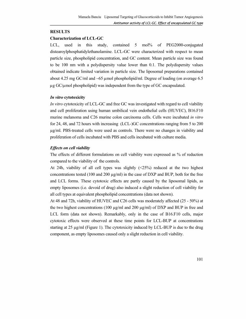
RESULTS Characterization of LCL-GC LCL, used in this study, contained 5 mol% of PEG2000-conjugated distearoylphosphatidylethanolamine. LCL-GC were characterized with respect to mean particle size, phospholipid concentration, and GC content. Mean particle size was found to be 100 nm with a polydispersity value lower than 0.1. The polydispersity values obtained indicate limited variation in particle size. The liposomal preparations contained about 4.25 mg GC/ml and ~65 µmol phospholipid/ml. Degree of loading (on average 6.5 µg GC/µmol phospholipid) was independent from the type of GC encapsulated. In vitro cytotoxicity In vitro cytotoxicity of LCL-GC and free GC was investigated with regard to cell viability and cell proliferation using human umbilical vein endothelial cells (HUVEC), B16.F10 murine melanoma and C26 murine colon carcinoma cells. Cells were incubated in vitro for 24, 48, and 72 hours with increasing (LCL-)GC concentrations ranging from 5 to 200 µg/ml. PBS-treated cells were used as controls. There were no changes in viability and proliferation of cells incubated with PBS and cells incubated with culture media. Effects on cell viability The effects of different formulations on cell viability were expressed as % of reduction compared to the viability of the controls. At 24h, viability of all cell types was slightly (<25%) reduced at the two highest concentrations tested (100 and 200 µg/ml) in the case of DXP and BUP, both for the free and LCL forms. These cytotoxic effects are partly caused by the liposomal lipids, as empty liposomes (i.e. devoid of drug) also induced a slight reduction of cell viability for all cell types at equivalent phospholipid concentrations (data not shown). At 48 and 72h, viability of HUVEC and C26 cells was moderately affected (25 - 50%) at the two highest concentrations (100 µg/ml and 200 µg/ml) of DXP and BUP in free and LCL form (data not shown). Remarkably, only in the case of B16.F10 cells, major cytotoxic effects were observed at these time points for LCL-BUP at concentrations starting at 25 µg/ml (Figure 1). The cytotoxicity induced by LCL-BUP is due to the drug component, as empty liposomes caused only a slight reduction in cell viability.
Manuela Banciu Liposomal Targeting of Glucocorticoids to Inhibit Tumor Angiogenesis
CChhaapptteerr 55
102
Figure 1. Effect of LCL-BUP and free BUP on the viability of B16.F10 cells. Only data obtained at 48h and 72h of cell incubation with LCL-BUP and free BUP treatments are shown. Mean±SD; n= 3 measurements; LCL-BUP= treatment with LCL-BUP; Free BUP= treatment with free BUP; LCL= treatment with empty LCL (i.e. devoid of drug)
Effects on cell proliferation The effects of different formulations on cell proliferation were expressed as % of inhibition compared to the proliferation of the controls. C26: At 24h, proliferation of C26 cells was inhibited by 25 - 40% only after treatment with BUP and DXP, added as liposomal or free drug, at the highest concentrations tested (100 and 200 µg/ml). At 48h and 72h, inhibition of C26 cell proliferation was observed in case of all GC formulations but remained below 50% even at the highest concentrations (data not shown).
0102030405060708090
100
5 10 25 50 100 200
0
10
20
30
40
50
60
70
80
90
100
5 10 25 50 100 200
LCL-BUPFree BUPLCL
R
educ
tion
of c
ell v
iabi
lity
(% o
f PB
S-tre
ated
cel
ls)
BUP concentration (µg/ml)
48 h 72 h
Manuela Banciu Liposomal Targeting of Glucocorticoids to Inhibit Tumor Angiogenesis
AAnnttiittuummoorr aaccttiivviittyy ooff LLCCLL--GGCC.. EEffffeecctt ooff eennccaappssuullaatteedd GGCC ttyyppee
103
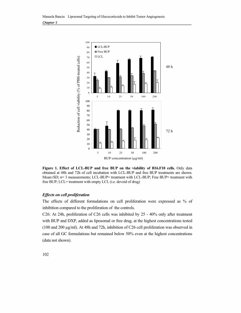
B16.F10: At 24h of incubation, all formulations of GC, except for BUP, showed moderate inhibition of B16.F10 cell proliferation but only at the two highest concentrations tested (100 and 200 µg/ml). Free and LCL-BUP induced a moderate inhibition of cell proliferation at lower concentrations (between 10 - 50 µg/ml BUP). The inhibitory effects became more pronounced at the 100 µg/ml and 200 µg/ml BUP concentrations (data not shown). At 48h and 72h, only LCL-BUP exerted a strong inhibitory effect on the proliferation of B16.F10 cells (Figure 2). At 72h, LCL-BUP-mediated inhibition of cell proliferation was at the level of 60-90% at concentrations ranging from 25 µg/ml to 200 µg/ml (Figure 2).
Figure 2. Effect of LCL-GC and free GC on the proliferation of B16.F10 cells. Mean±SD; n= 3 measurements; LCL-PLP= treatment with LCL-PLP; free PLP= treatment with free PLP; LCL-MPLP= treatment with LCL-MPLP; free MPLP= treatment with free MPLP; LCL- DXP= treatment with LCL-DXP; free DXP= treatment with free DXP; lip BUP= treatment with LCL-BUP; free BUP= treatment with free BUP; LCL= treatment with empty LCL (i.e. devoid of drug)
5 10 25 50 100 2000
102030405060708090
100
5 10 25 50 100 200 5 10 25 50 100 200 5 10 25 50 100 200
0102030405060708090
100
5 10 25 50 100 200
LCL-PLPFree PLPLCL
5 10 25 50 100 200
LCL-MPLPFree MPLP
LCL
5 10 25 50 100 200
LCL-DXPFree DXP
LCL
5 10 25 50 100 200
LCL-BUPFree BUPLCL
Glucocorticoid concentration (µg/ml)
Inhi
bitio
n of
cell
prol
ifera
tion
(% o
f PBS
-trea
ted
cells
)
48h 72h
Manuela Banciu Liposomal Targeting of Glucocorticoids to Inhibit Tumor Angiogenesis
CChhaapptteerr 55
104
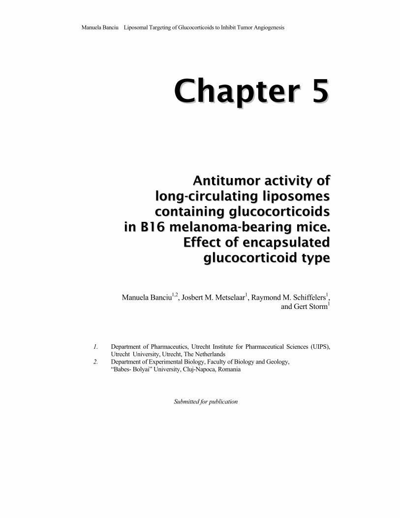
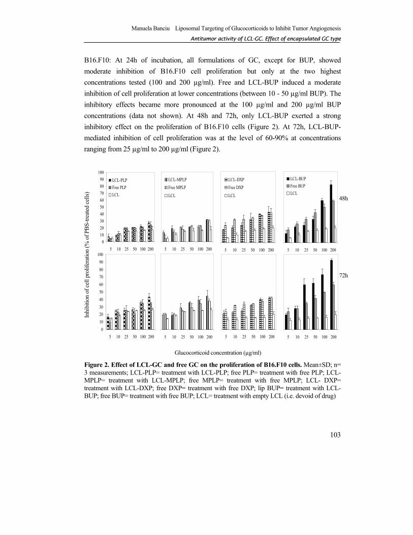
HUVEC: At 24h, inhibition of HUVEC proliferation was moderate (25-50%) only at the highest GC concentrations tested in case of all formulations except for LCL-BUP which was again an exception. LCL-BUP caused a much stronger inhibition up to 60% at the highest concentrations of 100 µg/ml and 200 µg/ml (data not shown). After 48h and 72h, proliferation of HUVEC cells was moderately inhibited (up to 45%) in case of treatment with all free GC types (Figure 3). Remarkably, all four LCL-GC formulations had strong inhibitory effects on HUVEC proliferation (ranging from 75-90%, at the highest concentrations of 100 µg/ml and 200 µg/ml after 72h of incubation) which was clearly mediated by the encapsulated GC (Figure 3). Notably, the anti-proliferative effects of LCL-BUP started at lower concentrations (25-100 µg/ml).
Figure 3. Effect of liposomal GC and free GC on the proliferation of HUVEC. Mean±SD; n= 3 measurements; LCL-PLP= treatment with LCL-PLP; free PLP= treatment with free PLP; LCL-MPLP= treatment with LCL-MPLP; free MPLP= treatment with free MPLP; LCL-DXP= treatment with LCL-DXP; free DXP= treatment with free DXP; LCL-BUP= treatment with LCL-BUP; free BUP= treatment with free BUP; LCL= treatment with empty LCL (i.e. devoid of drug)
48h 72h
0102030405060708090
100
5 10 25 50 100 200
LCL-PLPFree PLPLCL
5 10 25 50 100 200
LCL-MPLPFree MPLPLCL
5 10 25 50 100 200
LCL-DXPFree DXPLCL
5 10 25 50 100 200
LCL-BUPFree BUPLCL
0102030405060708090
100
5 10 25 50 100 200 5 10 25 50 100 200 5 10 25 50 100 200 5 10 25 50 100 200
Inhi
bitio
n of
cel
l pro
lifer
atio
n (%
of P
BS-
treat
ed c
ells
)
Glucocorticoid concentration (µg/ml)
Manuela Banciu Liposomal Targeting of Glucocorticoids to Inhibit Tumor Angiogenesis
AAnnttiittuummoorr aaccttiivviittyy ooff LLCCLL--GGCC.. EEffffeecctt ooff eennccaappssuullaatteedd GGCC ttyyppee
105
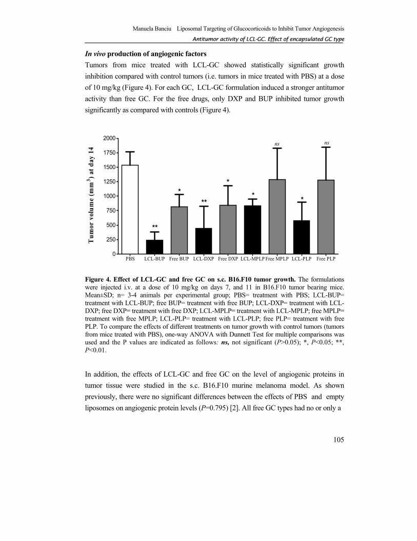
In vivo production of angiogenic factors Tumors from mice treated with LCL-GC showed statistically significant growth inhibition compared with control tumors (i.e. tumors in mice treated with PBS) at a dose of 10 mg/kg (Figure 4). For each GC, LCL-GC formulation induced a stronger antitumor activity than free GC. For the free drugs, only DXP and BUP inhibited tumor growth significantly as compared with controls (Figure 4).
Figure 4. Effect of LCL-GC and free GC on s.c. B16.F10 tumor growth. The formulations were injected i.v. at a dose of 10 mg/kg on days 7, and 11 in B16.F10 tumor bearing mice. Mean±SD; n= 3-4 animals per experimental group; PBS= treatment with PBS; LCL-BUP= treatment with LCL-BUP; free BUP= treatment with free BUP; LCL-DXP= treatment with LCL-DXP; free DXP= treatment with free DXP; LCL-MPLP= treatment with LCL-MPLP; free MPLP= treatment with free MPLP; LCL-PLP= treatment with LCL-PLP; free PLP= treatment with free PLP. To compare the effects of different treatments on tumor growth with control tumors (tumors from mice treated with PBS), one-way ANOVA with Dunnett Test for multiple comparisons was used and the P values are indicated as follows: ns, not significant (P>0.05); *, P<0.05; **, P<0.01.
In addition, the effects of LCL-GC and free GC on the level of angiogenic proteins in tumor tissue were studied in the s.c. B16.F10 murine melanoma model. As shown previously, there were no significant differences between the effects of PBS and empty liposomes on angiogenic protein levels (P=0.795) [2]. All free GC types had no or only a
PBS LCL-BUP Free BUP LCL-DXP Free DXP LCL-MPLP Free MPLP LCL-PLP Free PLP0
250
500
750
1000
1250
1500
1750
2000
**
**
**
*
ns ns
***
Tum
or v
olum
e (m
m3 ) a
t day
14
Manuela Banciu Liposomal Targeting of Glucocorticoids to Inhibit Tumor Angiogenesis
CChhaapptteerr 55
106
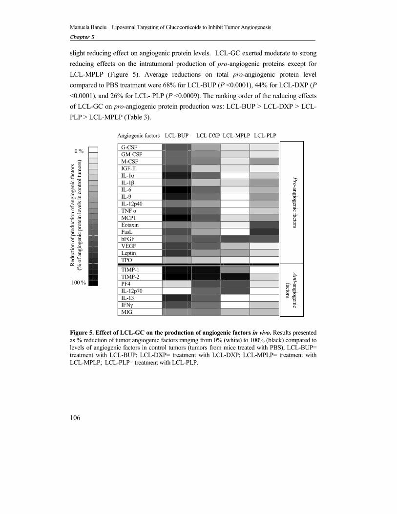
slight reducing effect on angiogenic protein levels. LCL-GC exerted moderate to strong reducing effects on the intratumoral production of pro-angiogenic proteins except for LCL-MPLP (Figure 5). Average reductions on total pro-angiogenic protein level compared to PBS treatment were 68% for LCL-BUP (P <0.0001), 44% for LCL-DXP (P <0.0001), and 26% for LCL- PLP (P <0.0009). The ranking order of the reducing effects of LCL-GC on pro-angiogenic protein production was: LCL-BUP > LCL-DXP > LCL-PLP > LCL-MPLP (Table 3).
Figure 5. Effect of LCL-GC on the production of angiogenic factors in vivo. Results presented as % reduction of tumor angiogenic factors ranging from 0% (white) to 100% (black) compared to levels of angiogenic factors in control tumors (tumors from mice treated with PBS); LCL-BUP= treatment with LCL-BUP; LCL-DXP= treatment with LCL-DXP; LCL-MPLP= treatment with LCL-MPLP; LCL-PLP= treatment with LCL-PLP.
G-CSF GM-CSF M-CSF IGF-II IL-1α IL-1β IL-6 IL-9 IL-12p40 TNF α MCP1 Eotaxin FasL bFGF VEGF Leptin TPO
TIMP-1 TIMP-2 PF4 IL-12p70 IL-13 IFNγ MIG
Angiogenic factors LCL-BUP LCL-DXP LCL-MPLP LCL-PLP
Pro-angiogenic factors Anti-angiogenic
factors
0 %
Redu
ctio
n of
pro
duct
ion
of an
giog
enic
fact
ors
(% o
f ang
ioge
nic p
rote
in le
vels
in co
ntro
l tum
ors)
100 %
Manuela Banciu Liposomal Targeting of Glucocorticoids to Inhibit Tumor Angiogenesis
AAnnttiittuummoorr aaccttiivviittyy ooff LLCCLL--GGCC.. EEffffeecctt ooff eennccaappssuullaatteedd GGCC ttyyppee
107
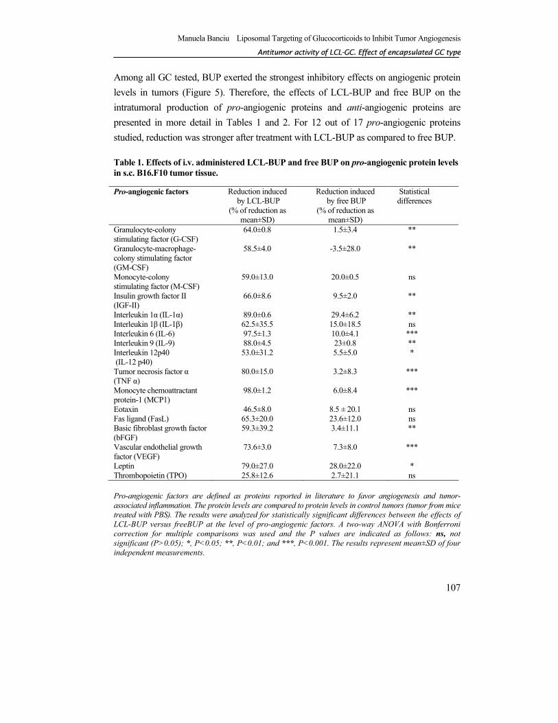
Among all GC tested, BUP exerted the strongest inhibitory effects on angiogenic protein levels in tumors (Figure 5). Therefore, the effects of LCL-BUP and free BUP on the intratumoral production of pro-angiogenic proteins and anti-angiogenic proteins are presented in more detail in Tables 1 and 2. For 12 out of 17 pro-angiogenic proteins studied, reduction was stronger after treatment with LCL-BUP as compared to free BUP. Table 1. Effects of i.v. administered LCL-BUP and free BUP on pro-angiogenic protein levels in s.c. B16.F10 tumor tissue.
Pro-angiogenic factors are defined as proteins reported in literature to favor angiogenesis and tumor-associated inflammation. The protein levels are compared to protein levels in control tumors (tumor from mice treated with PBS). The results were analyzed for statistically significant differences between the effects of LCL-BUP versus freeBUP at the level of pro-angiogenic factors. A two-way ANOVA with Bonferroni correction for multiple comparisons was used and the P values are indicated as follows: ns, not significant (P>0.05); *, P<0.05; **, P<0.01; and ***, P<0.001. The results represent mean±SD of four independent measurements.
Pro-angiogenic factors Reduction induced by LCL-BUP
(% of reduction as mean±SD)
Reduction induced by free BUP
(% of reduction as mean±SD)
Statistical differences
Granulocyte-colony stimulating factor (G-CSF)
64.0±0.8 1.5±3.4 **
Granulocyte-macrophage- colony stimulating factor (GM-CSF)
58.5±4.0 -3.5±28.0 **
Monocyte-colony stimulating factor (M-CSF)
59.0±13.0 20.0±0.5 ns
Insulin growth factor II (IGF-II)
66.0±8.6 9.5±2.0 **
Interleukin 1α (IL-1α) 89.0±0.6 29.4±6.2 ** Interleukin 1β (IL-1β) 62.5±35.5 15.0±18.5 ns Interleukin 6 (IL-6) 97.5±1.3 10.0±4.1 *** Interleukin 9 (IL-9) 88.0±4.5 23±0.8 ** Interleukin 12p40 (IL-12 p40)
53.0±31.2 5.5±5.0 *
Tumor necrosis factor α (TNF α)
80.0±15.0 3.2±8.3 ***
Monocyte chemoattractant protein-1 (MCP1)
98.0±1.2 6.0±8.4 ***
Eotaxin 46.5±8.0 8.5 ± 20.1 ns Fas ligand (FasL) 65.3±20.0 23.6±12.0 ns Basic fibroblast growth factor (bFGF)
59.3±39.2 3.4±11.1 **
Vascular endothelial growth factor (VEGF)
73.6±3.0 7.3±8.0 ***
Leptin 79.0±27.0 28.0±22.0 * Thrombopoietin (TPO) 25.8±12.6 2.7±21.1 ns
Manuela Banciu Liposomal Targeting of Glucocorticoids to Inhibit Tumor Angiogenesis
CChhaapptteerr 55
108
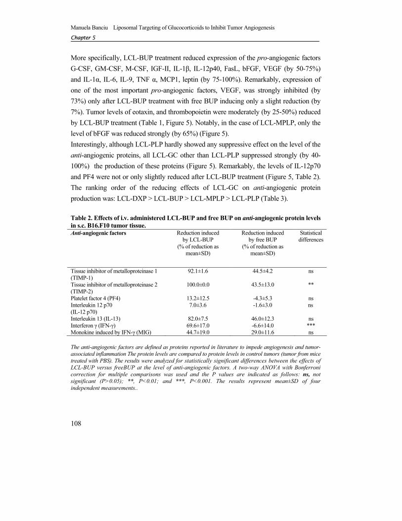
More specifically, LCL-BUP treatment reduced expression of the pro-angiogenic factors G-CSF, GM-CSF, M-CSF, IGF-II, IL-1β, IL-12p40, FasL, bFGF, VEGF (by 50-75%) and IL-1α, IL-6, IL-9, TNF α, MCP1, leptin (by 75-100%). Remarkably, expression of one of the most important pro-angiogenic factors, VEGF, was strongly inhibited (by 73%) only after LCL-BUP treatment with free BUP inducing only a slight reduction (by 7%). Tumor levels of eotaxin, and thrombopoietin were moderately (by 25-50%) reduced by LCL-BUP treatment (Table 1, Figure 5). Notably, in the case of LCL-MPLP, only the level of bFGF was reduced strongly (by 65%) (Figure 5). Interestingly, although LCL-PLP hardly showed any suppressive effect on the level of the anti-angiogenic proteins, all LCL-GC other than LCL-PLP suppressed strongly (by 40-100%) the production of these proteins (Figure 5). Remarkably, the levels of IL-12p70 and PF4 were not or only slightly reduced after LCL-BUP treatment (Figure 5, Table 2). The ranking order of the reducing effects of LCL-GC on anti-angiogenic protein production was: LCL-DXP > LCL-BUP > LCL-MPLP > LCL-PLP (Table 3). Table 2. Effects of i.v. administered LCL-BUP and free BUP on anti-angiogenic protein levels in s.c. B16.F10 tumor tissue. Anti-angiogenic factors Reduction induced
by LCL-BUP (% of reduction as
mean±SD)
Reduction induced by free BUP
(% of reduction as mean±SD)
Statistical differences
Tissue inhibitor of metalloproteinase 1 (TIMP-1)
92.1±1.6 44.5±4.2 ns
Tissue inhibitor of metalloproteinase 2 (TIMP-2)
100.0±0.0 43.5±13.0 **
Platelet factor 4 (PF4) 13.2±12.5 -4.3±5.3 ns Interleukin 12 p70 (IL-12 p70)
7.0±3.6 -1.6±3.0 ns
Interleukin 13 (IL-13) 82.0±7.5 46.0±12.3 ns Interferon γ (IFN-γ) 69.6±17.0 -6.6±14.0 *** Monokine induced by IFN-γ (MIG) 44.7±19.0 29.0±11.6 ns The anti-angiogenic factors are defined as proteins reported in literature to impede angiogenesis and tumor-associated inflammation The protein levels are compared to protein levels in control tumors (tumor from mice treated with PBS). The results were analyzed for statistically significant differences between the effects of LCL-BUP versus freeBUP at the level of anti-angiogenic factors. A two-way ANOVA with Bonferroni correction for multiple comparisons was used and the P values are indicated as follows: ns, not significant (P>0.05); **, P<0.01; and ***, P<0.001. The results represent mean±SD of four independent measurements..
Manuela Banciu Liposomal Targeting of Glucocorticoids to Inhibit Tumor Angiogenesis
AAnnttiittuummoorr aaccttiivviittyy ooff LLCCLL--GGCC.. EEffffeecctt ooff eennccaappssuullaatteedd GGCC ttyyppee
109
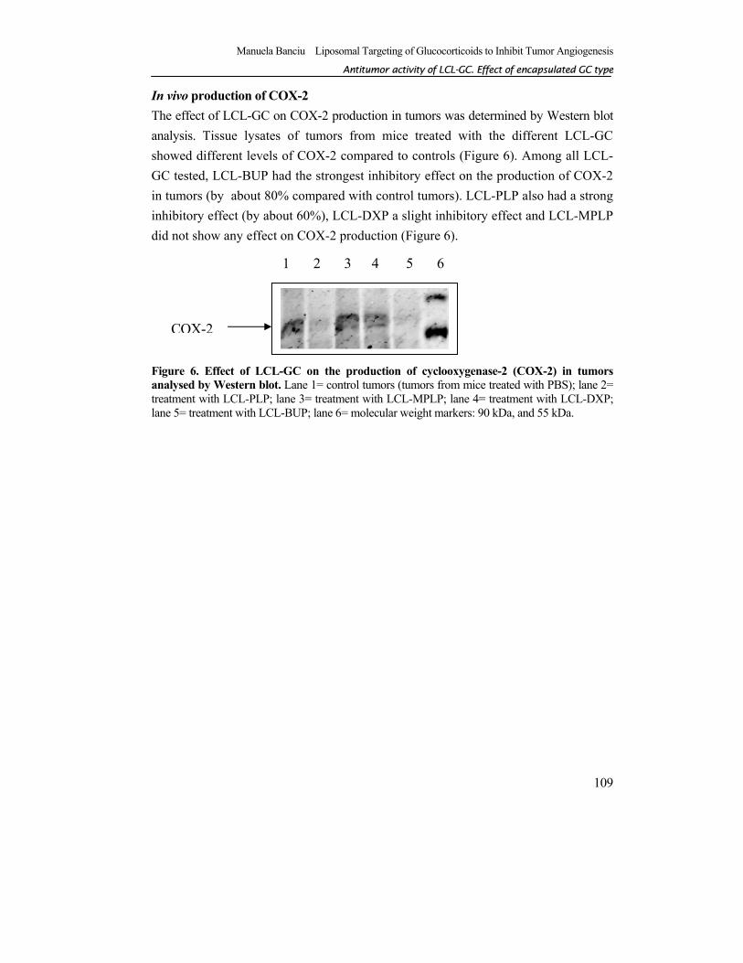
In vivo production of COX-2 The effect of LCL-GC on COX-2 production in tumors was determined by Western blot analysis. Tissue lysates of tumors from mice treated with the different LCL-GC showed different levels of COX-2 compared to controls (Figure 6). Among all LCL-GC tested, LCL-BUP had the strongest inhibitory effect on the production of COX-2 in tumors (by about 80% compared with control tumors). LCL-PLP also had a strong inhibitory effect (by about 60%), LCL-DXP a slight inhibitory effect and LCL-MPLP did not show any effect on COX-2 production (Figure 6).
Figure 6. Effect of LCL-GC on the production of cyclooxygenase-2 (COX-2) in tumors analysed by Western blot. Lane 1= control tumors (tumors from mice treated with PBS); lane 2= treatment with LCL-PLP; lane 3= treatment with LCL-MPLP; lane 4= treatment with LCL-DXP; lane 5= treatment with LCL-BUP; lane 6= molecular weight markers: 90 kDa, and 55 kDa.
1 2 3 4 5 6
COX-2
Manuela Banciu Liposomal Targeting of Glucocorticoids to Inhibit Tumor Angiogenesis
CChhaapptteerr 55
110
DISCUSSION In the present study, we provide a follow-up of our earlier observation that LCL-PLP can inhibit solid tumor growth. To evaluate whether this finding can be generalized to other types of GC, we encapsulated GC other than PLP in LCL for targeting to tumor tissue. We selected three other types of synthetic GC (as disodium salts of the phosphate derivatives), i.e. budesonide disodium phosphate (BUP), dexamethasone disodium phosphate (DXP), and methylprednisolone disodium phosphate (MPLP). The four GC differ in their potency to activate the human glucocorticoid receptor in the following order: budesonide > dexamethasone > methylprednisolone ~ prednisolone [19]. The antitumor effects of these LCL-GC were compared to those obtained with our earlier LCL formulation containing PLP. Our previous in vitro and in vivo studies indicate that the underlying mechanism of LCL-PLP responsible for inhibition of tumor growth is based on inhibition of angiogenesis, due to a strong reduction of intratumoral levels of pro-angiogenic factors with hardly any effect on anti-angiogenic factor levels [2]. All LCL-GC formulations inhibited tumor growth strongly, except for LCL-MPLP (Figure 4). The effects of LCL-GC are facilitated by the tumor-targeting property of the LCL formulation that increases the intratumoral drug concentration and enables the inhibitory effects of GC on tumor growth [2]. The most potent antitumor effect was achieved by LCL- BUP, reaching up to 85% inhibition of tumor growth compared to controls. This strong effect of LCL-BUP versus other three LCL-GC is likely related to the high potency of BUP. However, it is clear that potency is not the only factor that counts. LCL-MPLP performed worse than LCL-PLP whereas similar or better effects would have been expected based on the relative potency of both GC. The relative potency compared to cortisol, which is set at 1, is 5 for methylprednisolone and 4 for prednisolone [15-18]. Although the relative potency of dexamethasone is much higher than that of prednisolone (25 for dexamethasone, 4 for prednisolone) [16], their antitumor effects were similar (65%). The latter remarkable observation might be related to much stronger reduction of anti-angiogenic protein production by LCL-DXP when compared to LCL-PLP (Figure 4), and much higher degree of inhibition of COX-2 by LCL-PLP when compared to LCL-DXP (Figure 6). When GC are administered in the free form, they are rapidly cleared from the circulation and therefore do not localize in the tumor to a substantial degree, with consequently lower antitumor activity as a result [1]. Only free BUP and free DXP induced a significant inhibition of tumor growth which again is likely a consequence of the high relative
Manuela Banciu Liposomal Targeting of Glucocorticoids to Inhibit Tumor Angiogenesis
AAnnttiittuummoorr aaccttiivviittyy ooff LLCCLL--GGCC.. EEffffeecctt ooff eennccaappssuullaatteedd GGCC ttyyppee
111
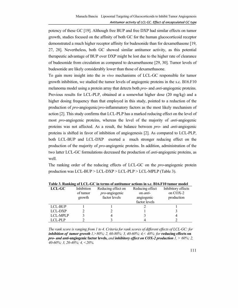
potency of these GC [19]. Although free BUP and free DXP had similar effects on tumor growth, studies focused on the affinity of both GC for the human glucocorticoid receptor demonstrated a much higher receptor affinity for budesonide than for dexamethasone [19, 27, 28]. Nevertheless, both GC showed similar antitumor activity, as this potential therapeutic advantage of BUP over DXP might be lost due to the higher rate of clearance of budesonide from circulation as compared to dexamethasone [29, 30]. Tumor levels of budesonide are likely considerably lower than those of dexamethasone. To gain more insight into the in vivo mechanisms of LCL-GC responsible for tumor growth inhibition, we studied the tumor levels of angiogenic proteins in the s.c. B16.F10 melanoma model using a protein array that detects both pro- and anti-angiogenic proteins. Previous results for LCL-PLP, obtained at a somewhat higher dose (20 mg/kg) and a higher dosing frequency than that employed in this study, pointed to a reduction of the production of pro-angiogenic/pro-inflammatory factors as the most likely mechanism of action [2]. This study confirms that LCL-PLP has a marked reducing effect on the level of most pro-angiogenic proteins, whereas the level of the majority of anti-angiogenic proteins was not affected. As a result, the balance between pro- and anti-angiogenic proteins is shifted in favor of inhibition of angiogenesis [2]. As compared to LCL-PLP, both LCL-BUP and LCL-DXP exerted a much stronger reducing effect on the production of the majority of pro-angiogenic proteins. In addition, administration of the two latter LCL-GC formulations decreased the production of anti-angiogenic proteins, as well. The ranking order of the reducing effects of LCL-GC on the pro-angiogenic protein production was LCL-BUP > LCL-DXP > LCL-PLP > LCL-MPLP (Table 3). Table 3. Ranking of LCL-GC in terms of antitumor actions in s.c. B16.F10 tumor model
LCL-GC Inhibition of tumor growth
Reducing effect on pro-angiogenic
factor levels
Reducing effect on anti-
angiogenic factor levels
Inhibitory effects on COX-2 production
LCL-BUP 1 1 2 1 LCL-DXP 2 2 1 3 LCL-MPLP 3 4 3 4 LCL-PLP 2 3 4 2
The rank score is ranging from 1 to 4. Criteria for rank scores of different effects of LCL-GC: for inhibition of tumor growth 1,>80%; 2, 60-80%; 3, 40-60%; 4,< 40%; for reducing effects on pro- and anti-angiogenic factor levels, and inhibitory effect on COX-2 production 1, > 60%; 2, 40-60%; 3, 20-40%; 4, <20%.
Manuela Banciu Liposomal Targeting of Glucocorticoids to Inhibit Tumor Angiogenesis
CChhaapptteerr 55
112
At first sight, the activity of tested LCL-GC are in line with the potency of encapsulated GC type. However, although true for BUP and DXP versus both other GC types, it is clear that both GC types with similar low potency (MPLP and PLP) do differ in their effect on production of pro-angiogenic factors. LCL-PLP was clearly stronger than LCL-MPLP, with the latter hardly different from PBS treatment (Figure 5). Among all pro-angiogenic factors studied, bFGF and VEGF are key players in angiogenesis being involved in all tumor angiogenesis steps [31-33]. The tumor expression of bFGF was moderately to strongly reduced after treatment with all types of LCL-GC. Remarkably, production of VEGF was only strongly affected by treatment with the LCL-GC with the highest potency being LCL-BUP and LCL-DXP (Figure 5). Neither LCL-PLP nor LCL-MPLP had any effect on VEGF expression in tumors. Remarkably, also the level of two macrophage-derived cytokines, TNF α and IL-1α that stimulate VEGF overexpression in human melanoma cells [34], was strongly reduced only after treatment with LCL-BUP and LCL-DXP. This finding might explain the strong reducing effect of LCL-BUP and LCL-DXP on VEGF production, via their suppressive effect on the level of two pro-angiogenic factors that increase tumor cell-mediated production of VEGF. Except for LCL-PLP, LCL-GC had a strong reducing effect on most of the anti-angiogenic protein levels in tumors, in the following ranking order: LCL-DXP > LCL-BUP > LCL- MPLP > LCL-PLP (Figure 5, Table 3). Interestingly, the production of two anti-angiogenic factors, IL-12p70 and PF4, was not affected after LCL-BUP treatment and LCL-PLP (Figure 5). In addition to anti-angiogenic effects, IL-12p70 is also known to induce cytotoxic effects on cancer cells [35-37]. The continuing presence of these anti-angiogenic factors likely strengthens the inhibitory effects resulting from the reduction of level of pro-angiogenic proteins induced by these two LCL-GC. To link the anti-angiogenic effects of LCL-GC to inflammatory processes, we assessed their actions on cyclooxygenase (COX)-2 production in tumor tissue. It is known that COX-2 is the rate-limiting enzyme in the production of prostaglandin E2 (PGE2) a key mediator of inflammation [38-40]. Inhibition of the production of PGE2 leads to a supplementary reduction of growth factors, cell adhesion molecules, and metalloproteinases involved in different steps of tumor angiogenesis [41, 42]. The ranking order of the inhibitory effects of LCL-GC on the COX-2 production was LCL-BUP > LCL-PLP > LCL-DXP > LCL-MPLP (Figure 6, Table 3). This outcome is in line with previous studies on A549 human adenocarcinoma cells which demonstrated the same order of inhibition of COX-2 expression induced by these GC [38].
Manuela Banciu Liposomal Targeting of Glucocorticoids to Inhibit Tumor Angiogenesis
AAnnttiittuummoorr aaccttiivviittyy ooff LLCCLL--GGCC.. EEffffeecctt ooff eennccaappssuullaatteedd GGCC ttyyppee
113
The in vitro cytotoxicity studies on cell viability and proliferation support the high potency of LCL-BUP. LCL-BUP had a strong killing effect (up to 90%) on B16.F10 melanoma cells over 48h and also at substantially lower concentrations than the other GC tested (Figures 1 and 2). Therefore, a lethal effect on the cancer cells could contribute to the magnitude of the in vivo antitumor effects induced by LCL-BUP (Figure 4). This effect appears to be B16.F10 melanoma cell-specific, as C26 cells were less affected. In addition, all LCL-GC inhibited HUVEC proliferation, with again LCL-BUP being the most potent. This finding would suggest that direct inhibition of endothelial cell proliferation is also involved in inhibition of angiogenesis. It is remarkable that the liposomal drug appears to induce stronger effects than the free agent. These effects might be due to a higher intracellular drug concentration induced by liposomal encapsulation possibly as a result of endocytosis of the lipid particles by the endothelial cells. In conclusion, all four selected GC encapsulated in LCL inhibit tumor growth, albeit to different degrees. The differences in antitumor activity correlate with their inhibitory activity towards the production of pro-angiogenic/pro-inflammatory factors involved in tumor angiogenesis and inflammation. Among the four LCL-GC types studied, LCL-BUP show the highest antitumor activity, which is likely related to the strong potency of this GC to reduce the production of pro-angiogenic and pro-inflammatory factors in tumors. In addition, the in vitro results presented herein suggest that LCL-BUP is strongly cytotoxic for B16.F10 melanoma cells and that anti-proliferative effects of all LCL-GC on angiogenic endothelial cells may play a role in their antitumor activity, as well. One of the future issues is to further identify the critical pathways involved in antitumor actions of different LCL-GC.
ACKNOWLEDGEMENTS The authors would like to thank Marcel Fens for his help with animal studies.
Manuela Banciu Liposomal Targeting of Glucocorticoids to Inhibit Tumor Angiogenesis
CChhaapptteerr 55
114
REFERENCES [1] R.M. Schiffelers, J.M. Metselaar, M.H. Fens, A.P. Janssen, G. Molema, G. Storm, Liposome-
encapsulated prednisolone phosphate inhibits growth of established tumors in mice. Neoplasia 7(2) (2005) 118-127.
[2] M. Banciu, R.M. Schiffelers, M.H. Fens, J.M. Metselaar, G. Storm, Anti-angiogenic effects of liposomal prednisolone phosphate on B16 melanoma in mice. J Control Release 113(1) (2006) 1-8.
[3] A.A. Gabizon, Stealth liposomes and tumor targeting: one step further in the quest for the magic bullet. Clin Cancer Res 7(2) (2001) 223-225.
[4] M. Crowther, N.J. Brown, E.T. Bishop, C.E. Lewis, Microenvironmental influence on macrophage regulation of angiogenesis in wounds and malignant tumors. J Leukoc Biol 70(4) (2001) 478-490.
[5] R.M. Schiffelers, M. Banciu, J.M. Metselaar, G. Storm, Therapeutic application of long-circulating liposomal glucocorticoids in auto-immune diseases and cancer. J Liposome Res 16(3) (2006) 185-194.
[6] D. Burnett, J.J. Reynolds, R.V. Ward, S.C. Afford, R.A. Stockley, Tissue inhibitor of metalloproteinases and collagenase inhibitory activity in lung secretions from patients with chronic obstructive bronchitis: effect of corticosteroid treatment. Thorax 41(10) (1986) 740-745.
[7] A. Amsterdam, K. Tajima, R. Sasson, Cell-specific regulation of apoptosis by glucocorticoids: implication to their anti-inflammatory action. Biochem Pharmacol 64(5-6) (2002) 843-850.
[8] K.A. Smoak, J.A. Cidlowski, Mechanisms of glucocorticoid receptor signaling during inflammation. Mech Ageing Dev 125(10-11) (2004) 697-706.
[9] S. Schmidt, J. Rainer, C. Ploner, E. Presul, S. Riml, R. Kofler, Glucocorticoid-induced apoptosis and glucocorticoid resistance: molecular mechanisms and clinical relevance. Cell Death Differ 11 Suppl 1 (2004) S45-55.
[10] C. Stellato, Post-transcriptional and nongenomic effects of glucocorticoids. Proc Am Thorac Soc 1(3) (2004) 255-263.
[11] E.V. Yang, C.M. Bane, R.C. MacCallum, J.K. Kiecolt-Glaser, W.B. Malarkey, R. Glaser, Stress-related modulation of matrix metalloproteinase expression. J Neuroimmunol 133(1-2) (2002) 144-150.
[12] M. Reichenstein, R. Reich, J.G. LeHoux, I. Hanukoglu, ACTH induces TIMP-1 expression and inhibits collagenase in adrenal cortex cells. Mol Cell Endocrinol 215(1-2) (2004) 109-114.
[13] F. Buttgereit, R.H. Straub, M. Wehling, G.R. Burmester, Glucocorticoids in the treatment of rheumatic diseases: an update on the mechanisms of action. Arthritis Rheum 50(11) (2004) 3408-3417.
[14] H. Leis, A. Page, A. Ramirez, A. Bravo, C. Segrelles, J. Paramio, D. Barettino, J.L. Jorcano, P. Perez, Glucocorticoid Receptor Counteracts Tumorigenic Activity of Akt in Skin through Interference with the Phosphatidylinositol 3-Kinase Signaling Pathway. Mol Endocrinol 18(2) (2004) 303-311.
[15] D. Czock, F. Keller, F.M. Rasche, U. Haussler, Pharmacokinetics and pharmacodynamics of systemically administered glucocorticoids. Clin Pharmacokinet 44(1) (2005) 61-98.
[16] E.F.L. Dubois, Clinical Potencies of Glucocorticoids: What do we Really Measure? Current Respiratory Medicine Reviews 1 (2005) 103-108.
[17] S.A. Johansson, K.E. Andersson, R. Brattsand, E. Gruvstad, P. Hedner, Topical and systemic glucocorticoid potencies of budesonide and beclomethasone dipropionate in man. Eur J Clin Pharmacol 22(6) (1982) 523-529.
[18] F. Buttgereit, M.D. Brand, G.R. Burmester, Equivalent doses and relative drug potencies for non-genomic glucocorticoid effects: a novel glucocorticoid hierarchy. Biochem Pharmacol 58(2) (1999) 363-368.
[19] C. Grossmann, T. Scholz, M. Rochel, C. Bumke-Vogt, W. Oelkers, A.F. Pfeiffer, S. Diederich, V. Bahr, Transactivation via the human glucocorticoid and mineralocorticoid receptor by therapeutically used steroids in CV-1 cells: a comparison of their glucocorticoid and mineralocorticoid properties. Eur J Endocrinol 151(3) (2004) 397-406.
[20] F.S. Rouser G, and Yamamoto A, Two dimensional thin layer chromatographic separation of polar lipids and determination of phospholipids by phosphorus analysis of spots. Lipids 5 (1970) 494-496.
[21] J.M. Metselaar, M.H. Wauben, J.P. Wagenaar-Hilbers, O.C. Boerman, G. Storm, Complete remission of experimental arthritis by joint targeting of glucocorticoids with long-circulating liposomes. Arthritis Rheum 48(7) (2003) 2059-2066.
Manuela Banciu Liposomal Targeting of Glucocorticoids to Inhibit Tumor Angiogenesis
AAnnttiittuummoorr aaccttiivviittyy ooff LLCCLL--GGCC.. EEffffeecctt ooff eennccaappssuullaatteedd GGCC ttyyppee
115
[22] D.A. Scudiero, R.H. Shoemaker, K.D. Paull, A. Monks, S. Tierney, T.H. Nofziger, M.J. Currens, D.
Seniff, M.R. Boyd, Evaluation of a soluble tetrazolium/formazan assay for cell growth and drug sensitivity in culture using human and other tumor cell lines. Cancer Res 48(17) (1988) 4827-4833.
[23] J. Heil, G. Reifferscheid, Detection of mammalian carcinogens with an immunological DNA synthesis-inhibition test. Carcinogenesis 13(12) (1992) 2389-2394.
[24] P.L. Huong, A.H. Kolk, T.A. Eggelte, C.P. Verstijnen, H. Gilis, J.T. Hendriks, Measurement of antigen specific lymphocyte proliferation using 5-bromo-deoxyuridine incorporation. An easy and low cost alternative to radioactive thymidine incorporation. J Immunol Methods 140(2) (1991) 243-248.
[25] R.P. Huang, Detection of multiple proteins in an antibody-based protein microarray system. J Immunol Methods 255(1-2) (2001) 1-13.
[26] G.L. Peterson, Determination of total protein. Methods Enzymol 91 (1983) 95-119. [27] N. Esmailpour, P. Hogger, P. Rohdewald, Binding kinetics of budesonide to the human glucocorticoid
receptor. Eur J Pharm Sci 6(3) (1998) 219-223. [28] E. Dahlberg, A. Thalen, R. Brattsand, J.A. Gustafsson, U. Johansson, K. Roempke, T. Saartok,
Correlation between chemical structure, receptor binding, and biological activity of some novel, highly active, 16 alpha, 17 alpha-acetal-substituted glucocorticoids. Mol Pharmacol 25(1) (1984) 70-78.
[29] F. Chanoine, C. Grenot, P. Heidmann, J.L. Junien, Pharmacokinetics of butixocort 21-propionate, budesonide, and beclomethasone dipropionate in the rat after intratracheal, intravenous, and oral treatments. Drug Metab Dispos 19(2) (1991) 546-553.
[30] D.R. Varma, Anti-inflammatory and ulcerogenic effects and pharmacokinetics of oxyphenbutazone in protein deficient rats. Indian J Med Res 72 (1980) 426-433.
[31] J.R. Jackson, M.P. Seed, C.H. Kircher, D.A. Willoughby, J.D. Winkler, The codependence of angiogenesis and chronic inflammation. Faseb J 11(6) (1997) 457-465.
[32] S. Lutsenko, Kiselev SM, and Severin SE, Molecular mechanisms of tumor angiogenesis. Biochemistry 68(3) (2003) 349-365.
[33] T. Tonini, F. Rossi, P.P. Claudio, Molecular basis of angiogenesis and cancer. Oncogene 22(42) (2003) 6549-6556.
[34] H. Torisu, M. Ono, H. Kiryu, M. Furue, Y. Ohmoto, J. Nakayama, Y. Nishioka, S. Sone, M. Kuwano, Macrophage infiltration correlates with tumor stage and angiogenesis in human malignant melanoma: possible involvement of TNFalpha and IL-1alpha. Int J Cancer 85(2) (2000) 182-188.
[35] F. Ethuin, C. Delarche, M.A. Gougerot-Pocidalo, B. Eurin, L. Jacob, S. Chollet-Martin, Regulation of interleukin 12 p40 and p70 production by blood and alveolar phagocytes during severe sepsis. Lab Invest 83(9) (2003) 1353-1360.
[36] P. Nyberg, L. Xie, R. Kalluri, Endogenous inhibitors of angiogenesis. Cancer Res 65(10) (2005) 3967-3979.
[37] C.Y. Liu, M. Battaglia, S.H. Lee, Q.H. Sun, R.H. Aster, G.P. Visentin, Platelet factor 4 differentially modulates CD4+CD25+ (regulatory) versus CD4+CD25- (nonregulatory) T cells. J Immunol 174(5) (2005) 2680-2686.
[38] J.D. Croxtall, P.T. van Hal, Q. Choudhury, D.W. Gilroy, R.J. Flower, Different glucocorticoids vary in their genomic and non-genomic mechanism of action in A549 cells. Br J Pharmacol 135(2) (2002) 511-519.
[39] P. Kalinski, P.L. Vieira, J.H. Schuitemaker, E.C. de Jong, M.L. Kapsenberg, Prostaglandin E(2) is a selective inducer of interleukin-12 p40 (IL-12p40) production and an inhibitor of bioactive IL-12p70 heterodimer. Blood 97(11) (2001) 3466-3469.
[40] C.M. Chesney, D.D. Pifer, L.W. Byers, E.E. Muirhead, Effect of platelet-activating factor (PAF) on human platelets. Blood 59(3) (1982) 582-585.
[41] T.C. van der Pouw Kraan, L.C. Boeije, R.J. Smeenk, J. Wijdenes, L.A. Aarden, Prostaglandin-E2 is a potent inhibitor of human interleukin 12 production. J Exp Med 181(2) (1995) 775-779.
[42] E. Vassiliou, V. Sharma, H. Jing, F. Sheibanie, D. Ganea, Prostaglandin E2 promotes the survival of bone marrow-derived dendritic cells. J Immunol 173(11) (2004) 6955-6964.

Manuela Banciu Liposomal Targeting of Glucocorticoids to Inhibit Tumor Angiogenesis