Embed Size (px)
DESCRIPTION
Phosphatidylserin -Exposition an der Membran roter Blutzellen als Apoptose -Marker und Aggregationsparameter. Master-Seminar Benjamin Hanf 13.06.2012. Grundlagen: PS. PS: Phosphatidylserin. Vorlesung I. Bernhardt Biophysik Lipid-Übersicht. - PowerPoint PPT Presentation
Citation preview
1
Phosphatidylserin-Exposition an der Membran roter Blutzellen als
Apoptose-Marker und Aggregationsparameter
Master-SeminarBenjamin Hanf
13.06.2012
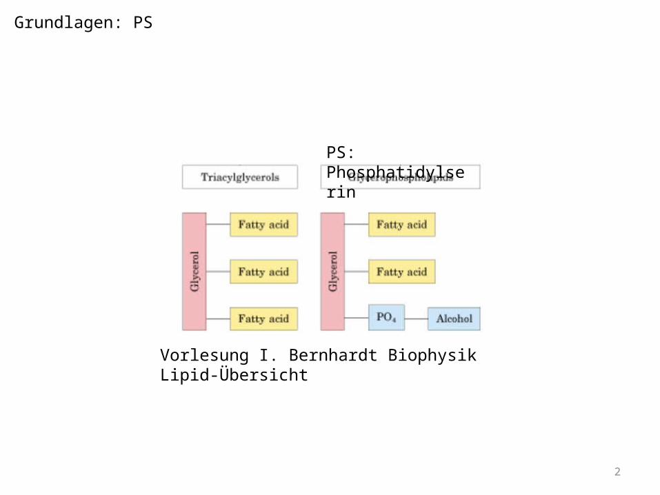
2
PS: Phosphatidylserin
Vorlesung I. Bernhardt Biophysik Lipid-Übersicht
Grundlagen: PS
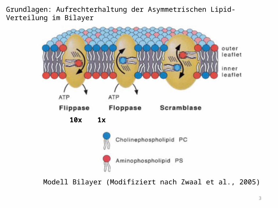
3
10x 1x
Grundlagen: Aufrechterhaltung der Asymmetrischen Lipid-Verteilung im Bilayer
Modell Bilayer (Modifiziert nach Zwaal et al., 2005)
4
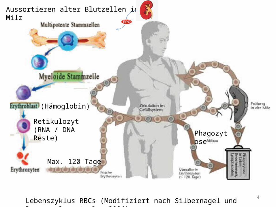
Aussortieren alter Blutzellen in der Milz
Retikulozyt(RNA / DNA Reste)
(Hämoglobin)
Phagozytose
Max. 120 Tage
4Lebenszyklus RBCs (Modifiziert nach Silbernagel und Despoupolos et al., 2004)

5
PS: Bedeutung bei Thrombusbildung
Thrombusbildung altes Modell (modifiziert nach http://www.highimpact.com/animations/medical-animations/MED01296/ )
6
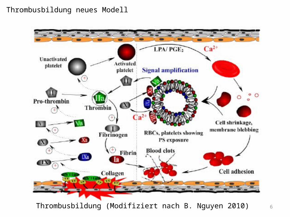
Thrombusbildung neues Modell
Thrombusbildung (Modifiziert nach B. Nguyen 2010)

7
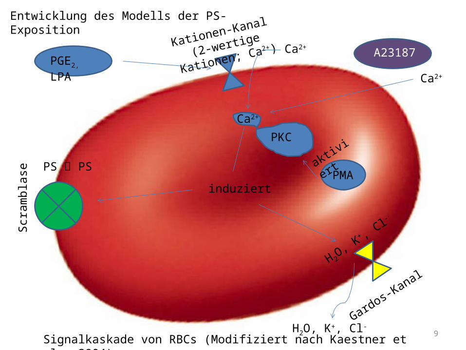
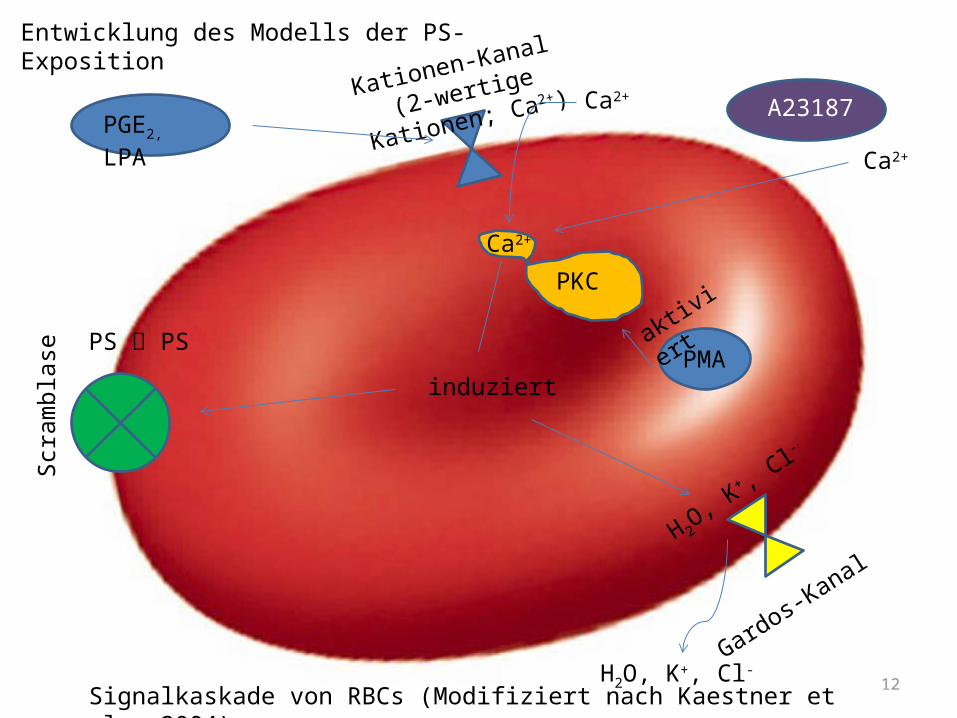
Kationen-Kanal
(2-wertige Kationen; Ca2+)
PGE2, LPA Ca2+
Gardos-Kanal
H 2O, K
+ , Cl-
H2O, K+, Cl-
Scra
mbl
ase
induziert
PKC
Ca2+
A23187
Ca2+
PS PSPMA
aktiviert
Signalkaskade von RBCs (Modifiziert nach Kaestner et al., 2004)
Entwicklung des Modells der PS-Exposition
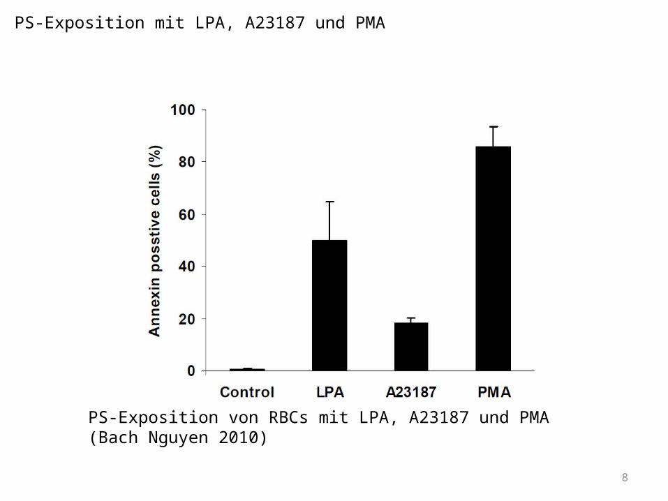
8
PS-Exposition von RBCs mit LPA, A23187 und PMA (Bach Nguyen 2010)
PS-Exposition mit LPA, A23187 und PMA
9
Kationen-Kanal
(2-wertige Kationen; Ca2+)
PGE2, LPA Ca2+
Gardos-Kanal
H 2O, K
+ , Cl-
H2O, K+, Cl-
Scra
mbl
ase
induziert
PKC
Ca2+
A23187
Ca2+
PS PSPMA
aktiviert
Entwicklung des Modells der PS-Exposition
Signalkaskade von RBCs (Modifiziert nach Kaestner et al., 2004)
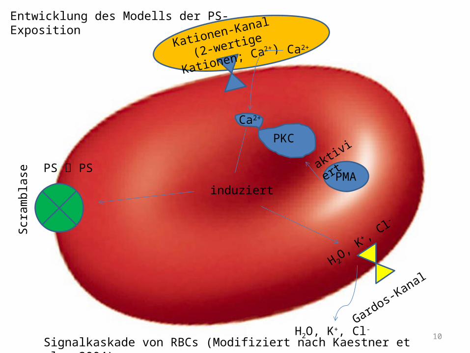
10
Kationen-Kanal
(2-wertige Kationen; Ca2+)Ca2+
Gardos-Kanal
H 2O, K
+ , Cl-
H2O, K+, Cl-
Scra
mbl
ase
induziert
PKC
Ca2+
A23187
PS PSPMA
aktiviert
Entwicklung des Modells der PS-Exposition
Signalkaskade von RBCs (Modifiziert nach Kaestner et al., 2004)
11
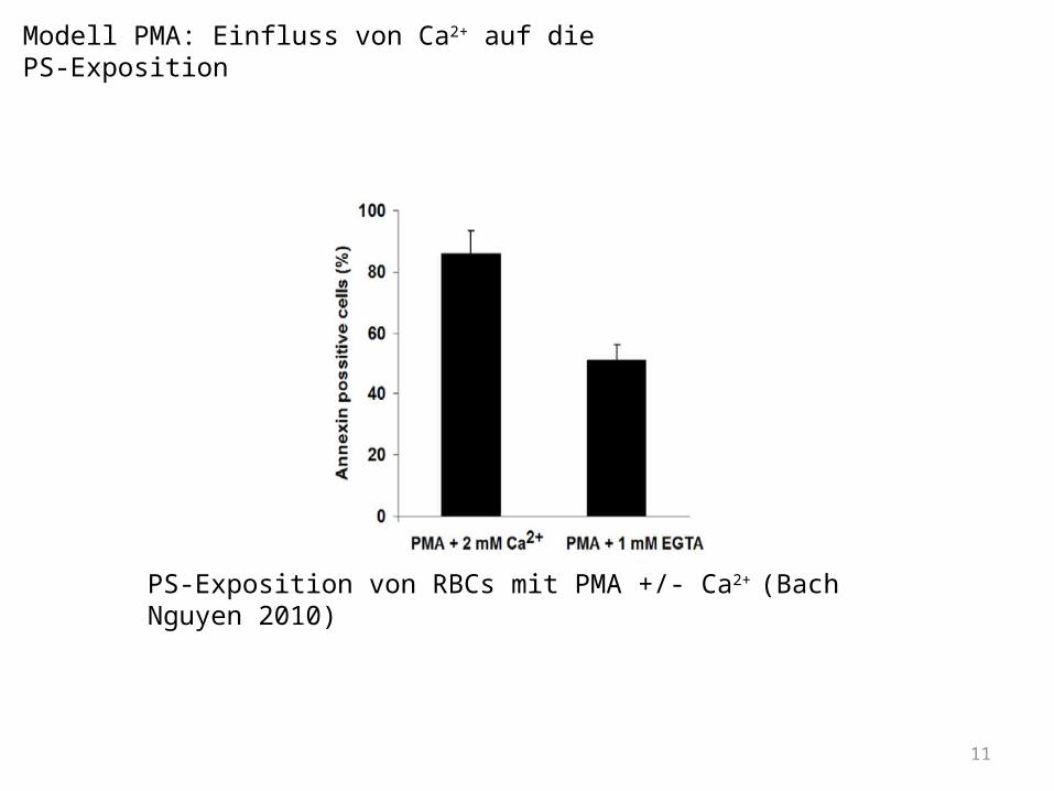
PS-Exposition von RBCs mit PMA +/- Ca2+ (Bach Nguyen 2010)
Modell PMA: Einfluss von Ca2+ auf die PS-Exposition
12
Kationen-Kanal
(2-wertige Kationen; Ca2+)
PGE2, LPA Ca2+
Gardos-Kanal
H 2O, K
+ , Cl-
H2O, K+, Cl-
Scra
mbl
ase
induziert
PKC
Ca2+
A23187
Ca2+
PS PSPMA
aktiviert
Entwicklung des Modells der PS-Exposition
Signalkaskade von RBCs (Modifiziert nach Kaestner et al., 2004)
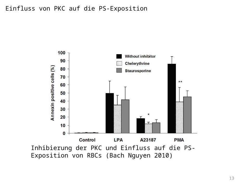
13
Inhibierung der PKC und Einfluss auf die PS-Exposition von RBCs (Bach Nguyen 2010)
Einfluss von PKC auf die PS-Exposition
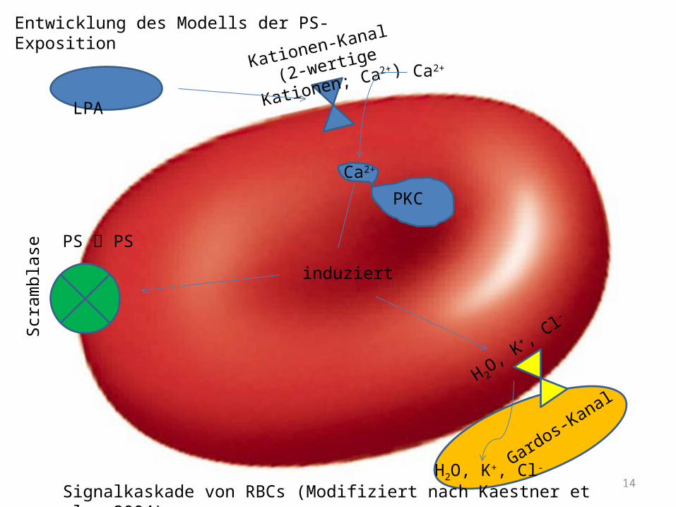
14
Kationen-Kanal
(2-wertige Kationen; Ca2+)
LPA Ca2+
Gardos-Kanal
H 2O, K
+ , Cl-
H2O, K+, Cl-
Scra
mbl
ase
induziert
Ca2+
A23187
PS PS
Entwicklung des Modells der PS-Exposition
PKC
Signalkaskade von RBCs (Modifiziert nach Kaestner et al., 2004)
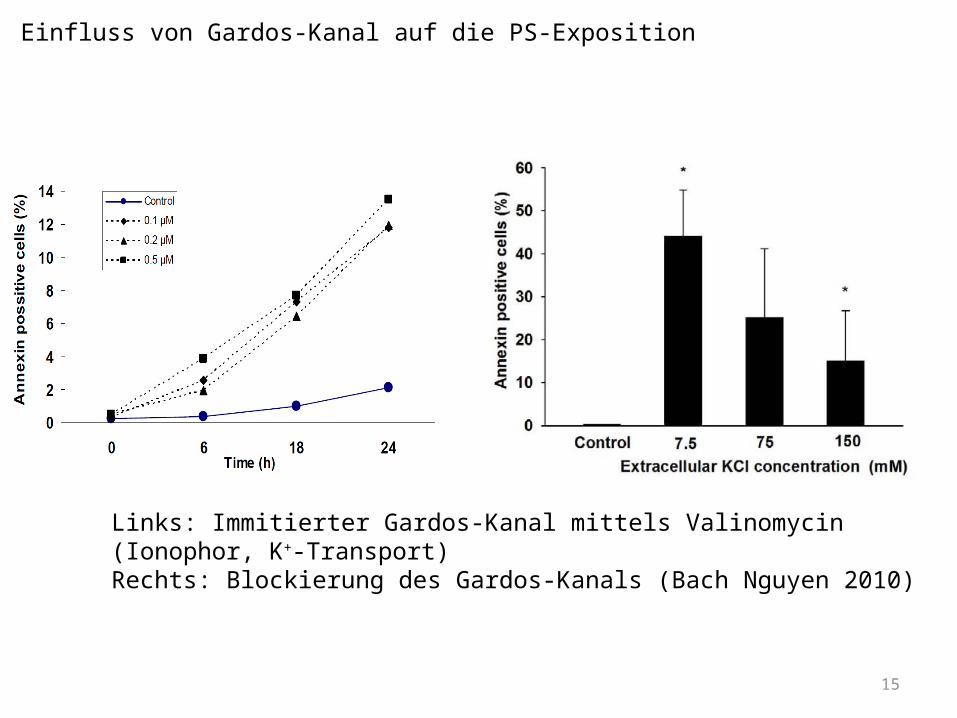
15
Links: Immitierter Gardos-Kanal mittels Valinomycin (Ionophor, K+-Transport)Rechts: Blockierung des Gardos-Kanals (Bach Nguyen 2010)
Einfluss von Gardos-Kanal auf die PS-Exposition

16
Gardos-Kanal
H 2O, K
+ , Cl-
H2O, K+, Cl-
Scra
mbl
ase
induziert
Ca2+
A23187
PS PS
Entwicklung des Modells der PS-Exposition
A23187
Ca2+
PKC
Signalkaskade von RBCs (Modifiziert nach Kaestner et al., 2004)
17
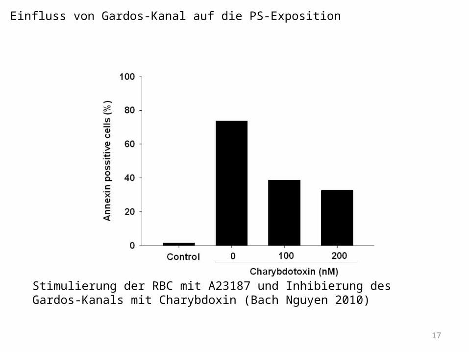
Einfluss von Gardos-Kanal auf die PS-Exposition
Stimulierung der RBC mit A23187 und Inhibierung des Gardos-Kanals mit Charybdoxin (Bach Nguyen 2010)

18
Kationen-Kanal
(2-wertige Kationen; Ca2+)
PGE2, LPA Ca2+
Gardos-Kanal
H 2O, K
+ , Cl-
H2O, K+, Cl-
Scra
mbl
ase
induziert
PKC
Ca2+
A23187
Ca2+
PS PSPMA
aktiviert
Entwicklung des Modells der PS-Exposition
Signalkaskade von RBCs (Modifiziert nach Kaestner et al., 2004)
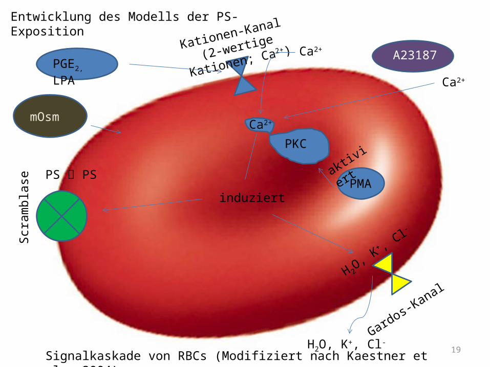
19
Kationen-Kanal
(2-wertige Kationen; Ca2+)
PGE2, LPA Ca2+
Gardos-Kanal
H 2O, K
+ , Cl-
H2O, K+, Cl-
Scra
mbl
ase
induziert
PKC
Ca2+
A23187
Ca2+
PS PSPMA
aktiviert
Entwicklung des Modells der PS-Exposition
mOsm
Signalkaskade von RBCs (Modifiziert nach Kaestner et al., 2004)
20
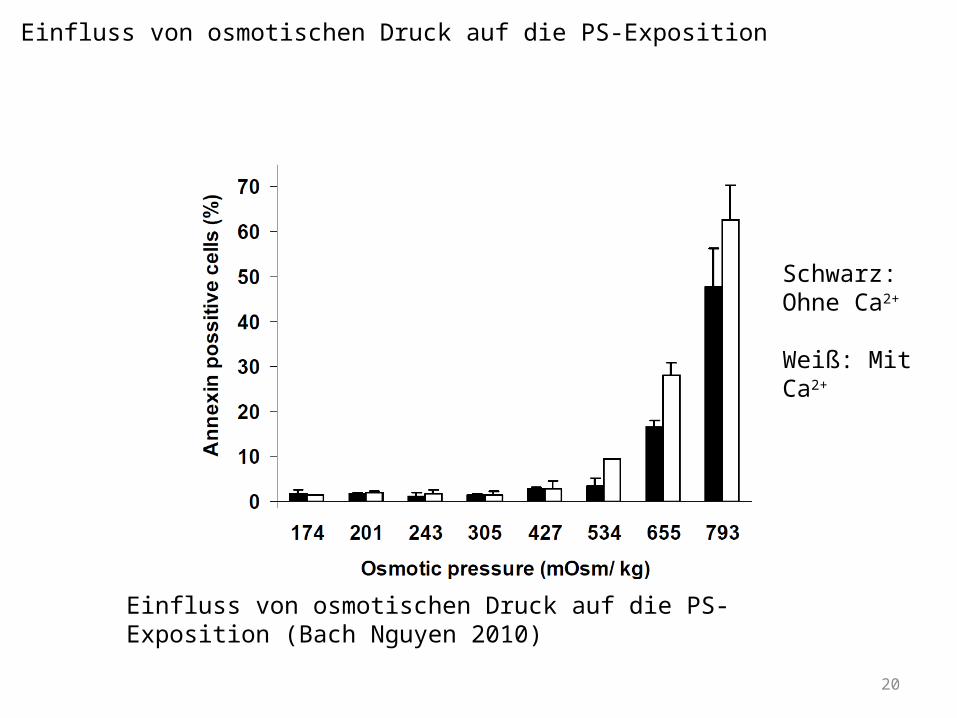
Einfluss von osmotischen Druck auf die PS-Exposition (Bach Nguyen 2010)
Einfluss von osmotischen Druck auf die PS-Exposition
Schwarz: Ohne Ca2+
Weiß: Mit Ca2+
21
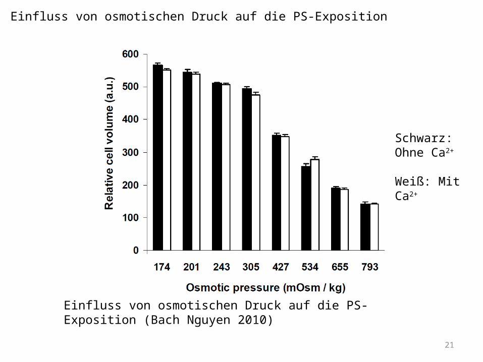
Einfluss von osmotischen Druck auf die PS-Exposition
Einfluss von osmotischen Druck auf die PS-Exposition (Bach Nguyen 2010)
Schwarz: Ohne Ca2+
Weiß: Mit Ca2+
22
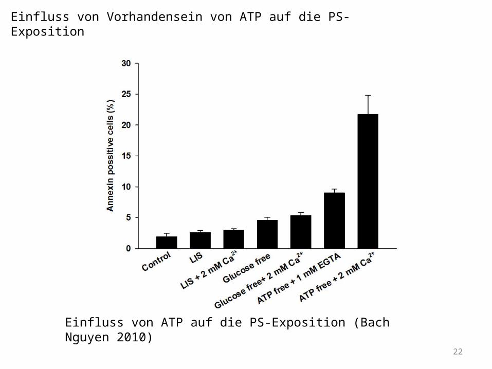
Einfluss von Vorhandensein von ATP auf die PS-Exposition
Einfluss von ATP auf die PS-Exposition (Bach Nguyen 2010)

23
Kationen-Kanal
(2-wertige Kationen; Ca2+)
PGE2, LPA Ca2+
Gardos-Kanal
H 2O, K
+ , Cl-
H2O, K+, Cl-
Scra
mbl
ase
induziert
PKC
Ca2+
A23187
Ca2+
PS PSPMA
aktiviert
Entwicklung des Modells der PS-Exposition
PS PS
Flip
pase
X*ATP
Ca2+
mOsm
Signalkaskade von RBCs (Modifiziert nach Kaestner et al., 2004)

24
Kationen-Kanal
(2-wertige Kationen; Ca2+)
PGE2, LPA Ca2+
Gardos-Kanal
H 2O, K
+ , Cl-
H2O, K+, Cl-
Scra
mbl
ase
induziert
PKC
Ca2+
A23187
Ca2+
PS PSPMA
aktiviert
Entwicklung des Modells der PS-Exposition
PS PS
Flip
pase
X*ATP
Ca2+
mOsm
Caspasen
Oxidativer Stress
Signalkaskade von RBCs (Modifiziert nach Kaestner et al., 2004)
25
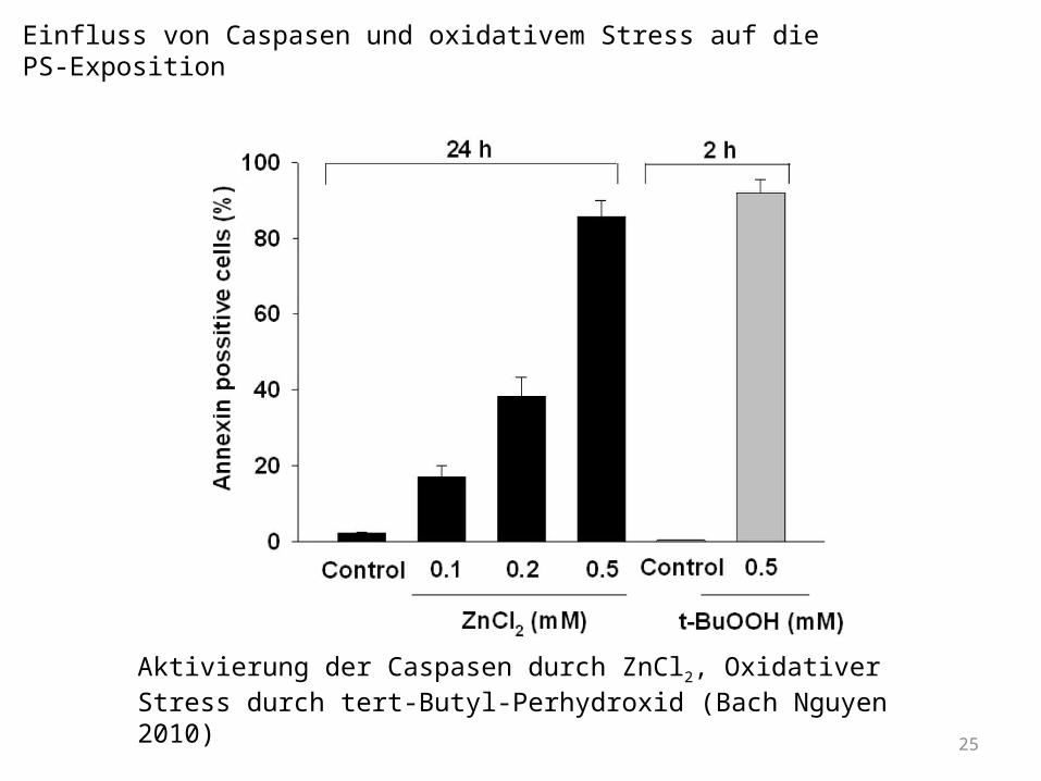
Aktivierung der Caspasen durch ZnCl2, Oxidativer Stress durch tert-Butyl-Perhydroxid (Bach Nguyen 2010)
Einfluss von Caspasen und oxidativem Stress auf die PS-Exposition
26
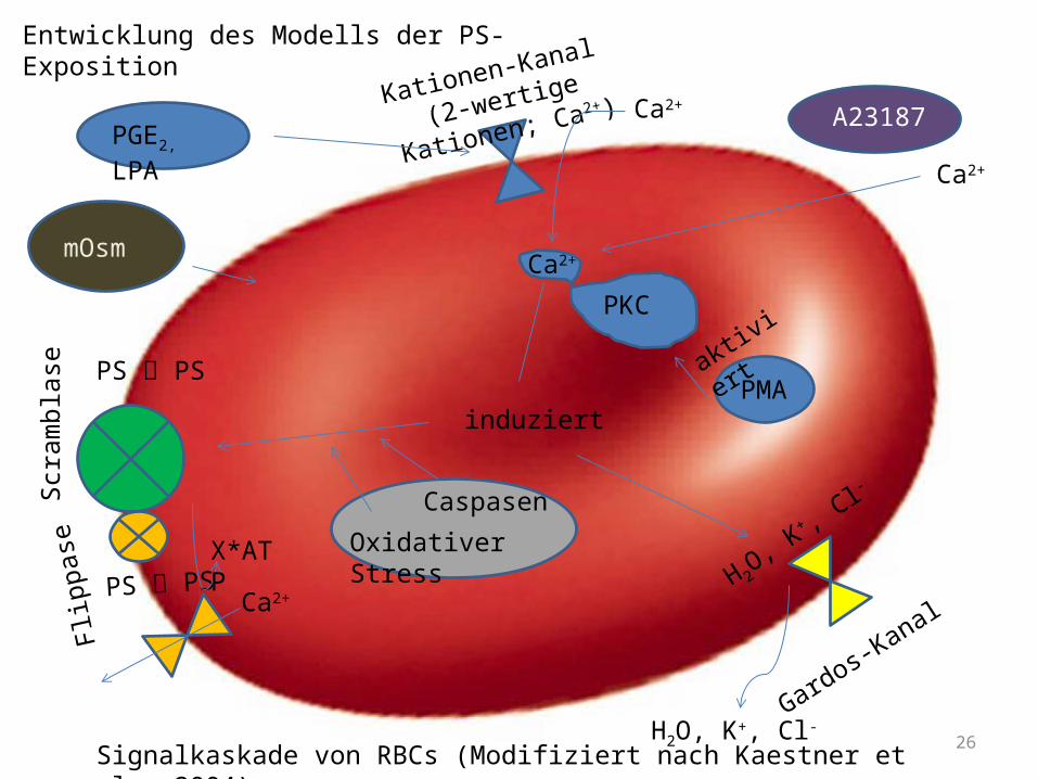
Kationen-Kanal
(2-wertige Kationen; Ca2+)
PGE2, LPA Ca2+
Gardos-Kanal
H 2O, K
+ , Cl-
H2O, K+, Cl-
Scra
mbl
ase
induziert
PKC
Ca2+
A23187
Ca2+
PS PSPMA
aktiviert
Entwicklung des Modells der PS-Exposition
PS PS
Flip
pase
X*ATP
Ca2+
mOsm
Caspasen
Oxidativer Stress
Signalkaskade von RBCs (Modifiziert nach Kaestner et al., 2004)
27
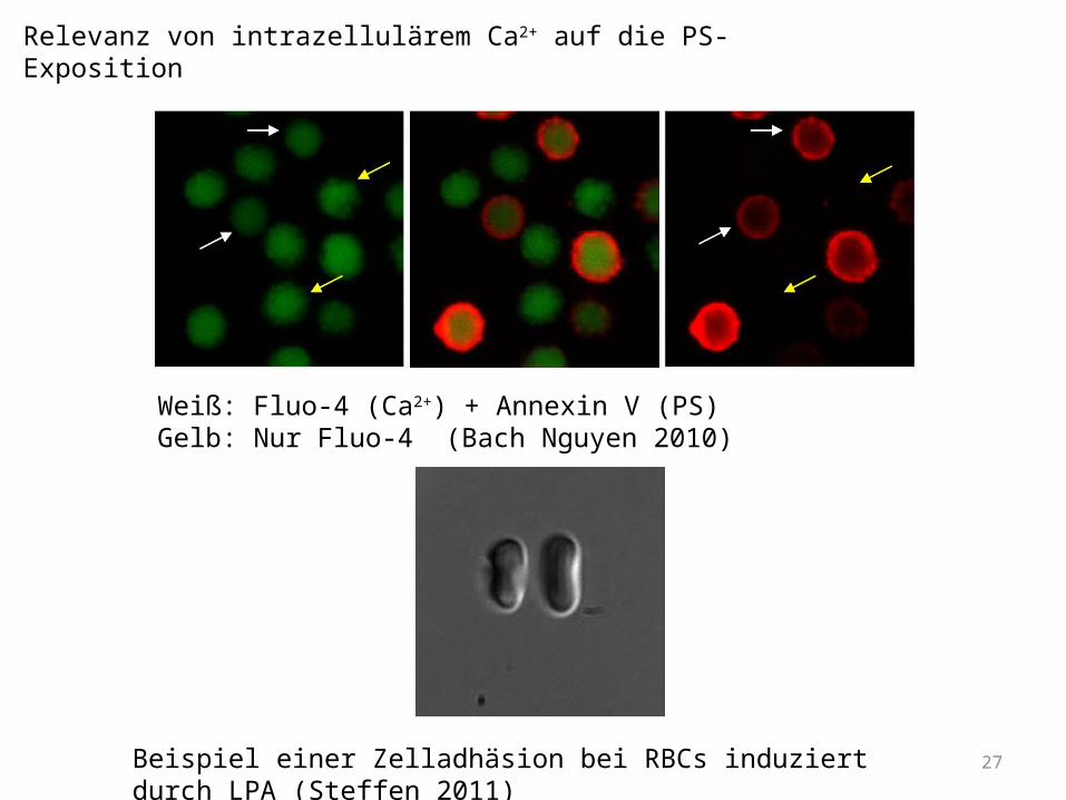
Weiß: Fluo-4 (Ca2+) + Annexin V (PS)Gelb: Nur Fluo-4 (Bach Nguyen 2010)
Beispiel einer Zelladhäsion bei RBCs induziert durch LPA (Steffen 2011)
Relevanz von intrazellulärem Ca2+ auf die PS-Exposition
28
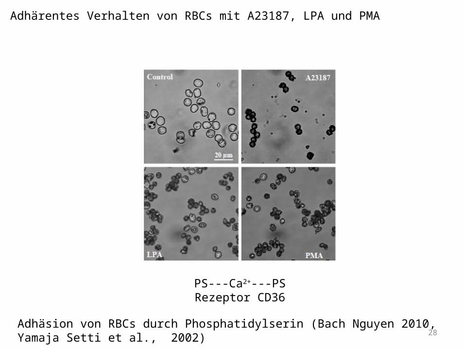
PS---Ca2+---PSRezeptor CD36
Adhäsion von RBCs durch Phosphatidylserin (Bach Nguyen 2010, Yamaja Setti et al., 2002)
Adhärentes Verhalten von RBCs mit A23187, LPA und PMA
29
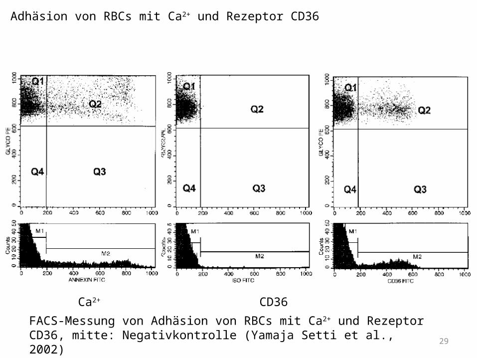
Ca2+ CD36
FACS-Messung von Adhäsion von RBCs mit Ca2+ und Rezeptor CD36, mitte: Negativkontrolle (Yamaja Setti et al., 2002)
Adhäsion von RBCs mit Ca2+ und Rezeptor CD36
30
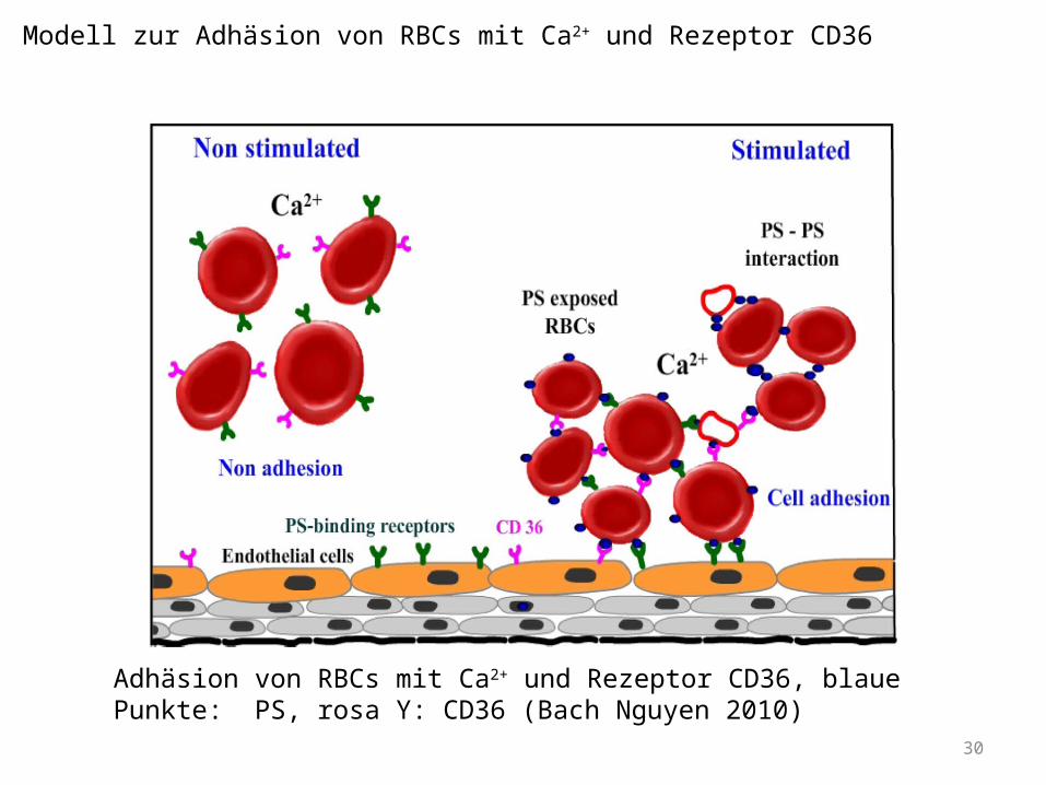
Adhäsion von RBCs mit Ca2+ und Rezeptor CD36, blaue Punkte: PS, rosa Y: CD36 (Bach Nguyen 2010)
Modell zur Adhäsion von RBCs mit Ca2+ und Rezeptor CD36
31
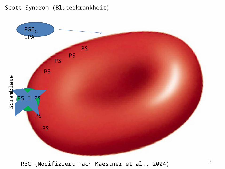
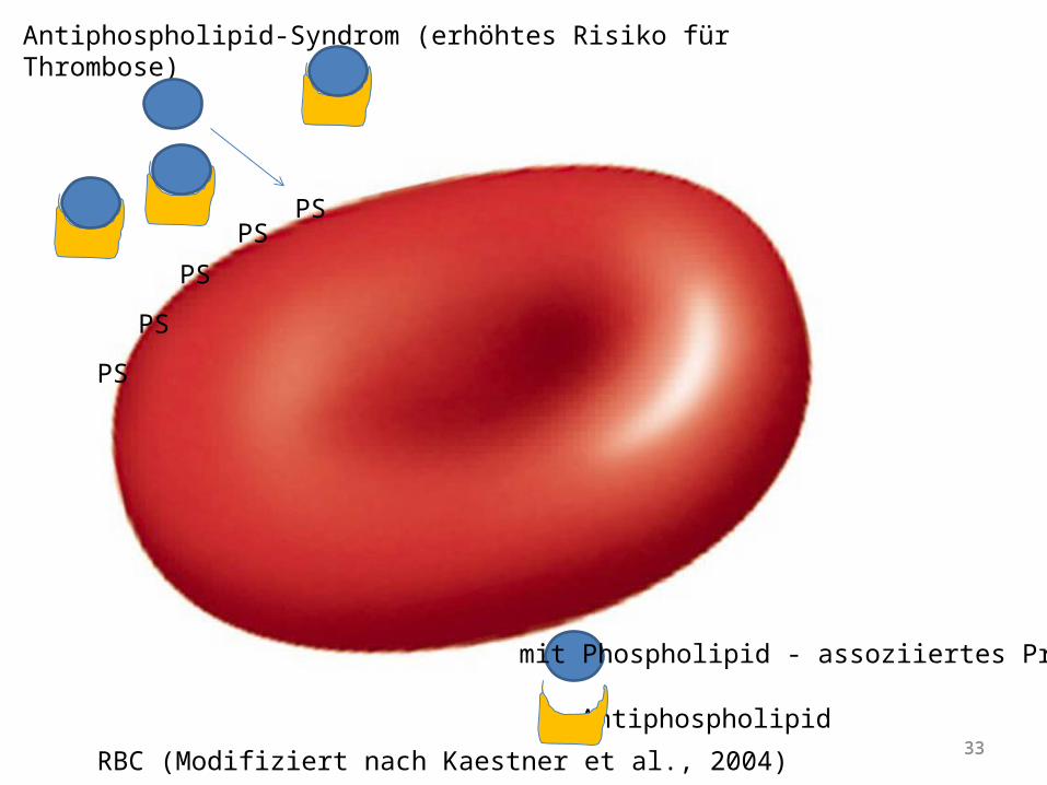
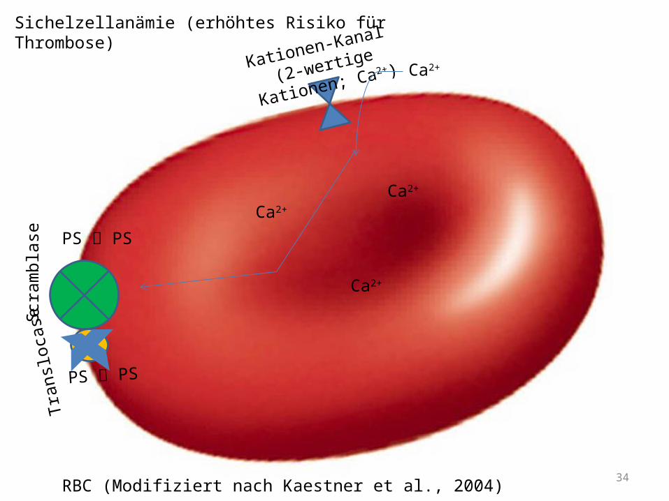
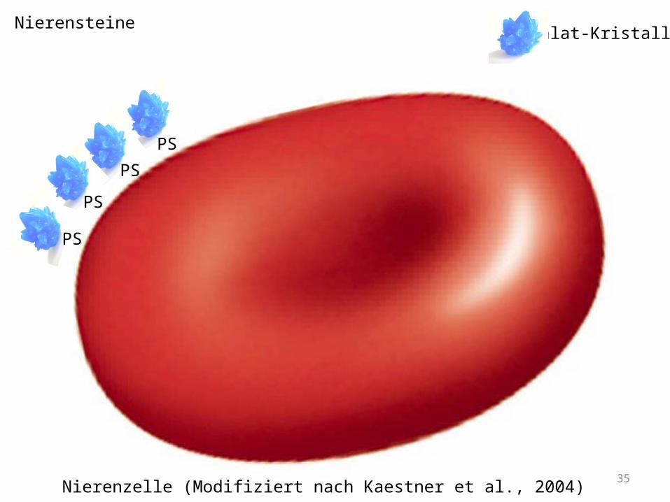
Bsp. An Krankheiten: Fehler in PS-Exposition
- Scott-Syndrom (Bluterkrankheit)
- Antiphospholipid-Syndrom (erhöhtes Risiko zu Thrombosen)
- Sichelzellanämie (erhöhte PS-Exposition Risiko zu Thrombosen)
- Nierensteine (Bindung von Ca2+ auf Nierenzellen)
- Malaria (erhöhter oxidativer Stress sowie Ca2+ -Aufnahme Risiko zu Thrombosen)
32
Scott-Syndrom (Bluterkrankheit)
RBC (Modifiziert nach Kaestner et al., 2004)
PS
PSPS
PS
PS
PS
PS PS
PGE2, LPA
Scra
mbl
ase
33
Antiphospholipid-Syndrom (erhöhtes Risiko für Thrombose)
PS
33RBC (Modifiziert nach Kaestner et al., 2004)
PS
PS
PSPS
mit Phospholipid - assoziiertes Protein
Antiphospholipid
34
Sichelzellanämie (erhöhtes Risiko für Thrombose)
PS PS
RBC (Modifiziert nach Kaestner et al., 2004)
PS PS
Tran
sloca
seKationen-Kanal
(2-wertige Kationen; Ca2+)Ca2+
Ca2+
Ca2+
Ca2+
Scra
mbl
ase
35
Nierensteine
Nierenzelle (Modifiziert nach Kaestner et al., 2004)
Oxalat-Kristall
PS
PS
PS
PS
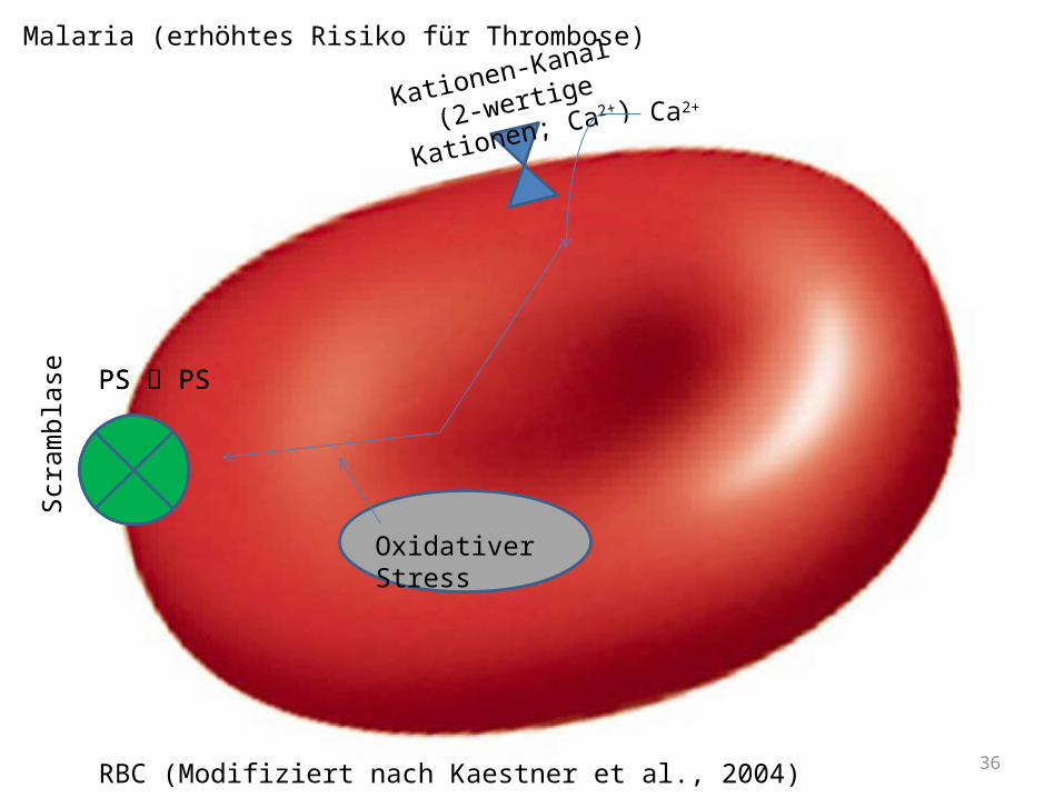
Malaria (erhöhtes Risiko für Thrombose)
PS PS
RBC (Modifiziert nach Kaestner et al., 2004)
Kationen-Kanal
(2-wertige Kationen; Ca2+)Ca2+
Scra
mbl
ase PS PS
Oxidativer Stress
36
37
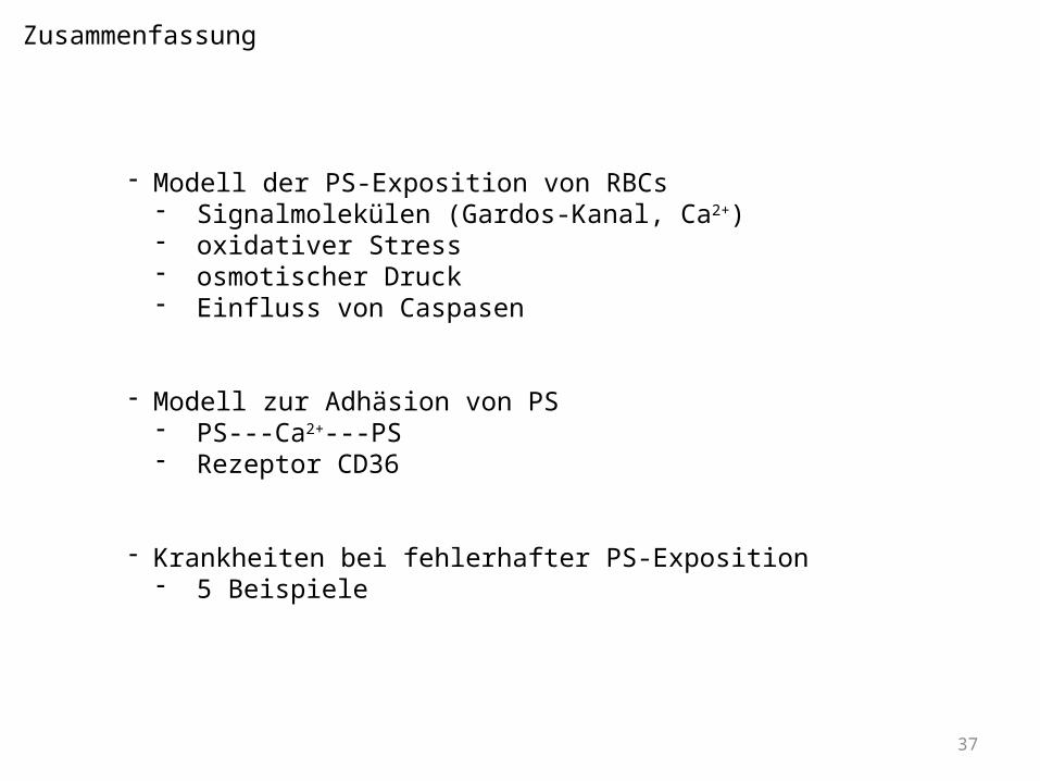
- Modell der PS-Exposition von RBCs- Signalmolekülen (Gardos-Kanal, Ca2+)- oxidativer Stress- osmotischer Druck- Einfluss von Caspasen
- Modell zur Adhäsion von PS- PS---Ca2+---PS- Rezeptor CD36
- Krankheiten bei fehlerhafter PS-Exposition- 5 Beispiele
Zusammenfassung
38
Referenzen
•Zwaal, R.F., Schroit, A.J., “Pathophysiologic implications of membrane phospholipid asymmetry in blood cells” (1997) Blood, 1997, 89: 1121-1132.•Mandal, D., Moitra, P.K., Saha, S., Basu, J., “Caspase 3 regulates phosphatidylserine externalization and phagocytosis of oxidatively stressed erythrocytes” (2002) FEBS Lett, 2002, 513: 184-188.•Kucherenko, Y.V., Weiss, E., Bernhardt, I., “Effect of the ionic strength and prostaglandin E2 on the free Ca2+ concentration and the Ca2+ influx in human red blood cells” (2004) Bioelectrochemistry, 2004, 62: 127-133.•Andrews, D.A., Low, P.S., “Role of red blood cells in thrombosis” (1999) Curr Opin Hematol, 1999, 6: 76-82.•Weiss, H.J., Lages, B., “Platelet prothrombinase activity and intracellular calcium responses in patients with storage pool deficiency, glycoprotein IIb-IIIa deficiency, or impaired platelet coagulant activity a comparison with Scott syndrome” (1997) Blood, 1997, 89: 1599-1611. •Wu, Y., Tibrewal, N., Birge, R.B., “Phosphatidylserine recognition by phagocytes: a view to a kill” Trends Cell Biol, 2006, 16: 189-197. •Verhoven, B., Schlegel, R.A., Williamson, P., “Mechanisms of phosphatidylserine exposure, a phagocyte recognition signal on apoptotic T lymphocytes” (1995) J Exp Med, 1995, 182: 1597-1601. •Uchida, K., “Induction of apoptosis by phosphatidylserine” (1998) J Biochem, 1998, 123: 1073-1078. •Schlegel, R.A., Williamson, P., “Phosphatidylserine, a death knell. Cell Death Differ” (2001) 2001, 8: 551-563. •Ravichandran, K.S., Lorenz, U., “Engulfment of apoptotic cells: signals for a good meal. Nat Rev Immunol” (2007) 7: 964-974. •Kuypers, F.A., De Jong, K., “The role of phosphatidylserine in recognition and removal of erythrocytes” (2004) Cell Mol Biol, 2004, 50: 147-158.•Quan, G.B., Han, Y., Yang, C., Hu, W.B., Liu, M.X., Liu, A., Wang, Y., Wang, J.X., “Mechanism of erythrocyte phosphatidylserine exposure induced by high concentrated glucose” Zhongguo Shi Yan Xue Ye Xue Za Zhi, (2008) 16: 1181-1184. •Kiedaisch, V., Akel, A., Niemoeller, O.M., Wieder, T., Lang, F., „Zinc-induced suicidal erythrocyte death” (2008) Am J Clin Nutr, 2008, 87: 1530-1534.•Sutton, D.J., Tchounwou, P.B., “Mercury-induced externalization of phosphatidylserine and caspase 3 activation in human liver carcinoma (HepG2) cells” (2006) Int J Environ Res Public Health, 2006, 3: 38-42. •Mandal, D., Moitra, P.K., Saha, S., Basu, J., “Caspase 3 regulates phosphatidylserine externalization and phagocytosis of oxidatively stressed erythrocytes” (2002) FEBS Lett, 2002, 513: 184-188.•Telen, M.J., “Red blood cell surface adhesion molecules: their possible roles in normal human physiology and disease” (2000) Semin Hematol, 2000, 37: 130-142. •117. Closse, C., Dachary-Prigent, J., Boisseau, M.R., “Phosphatidylserine-related adhesion of human erythrocytes to vascular endothelium” (1999) Br J Haematol, 1999, 107: 300-302.
39
•L. Wagner, D.B. Nguyen, A. Jung, P. Steffen, C. Wagner, L. Kaestner, T. Mueller, I. Bernhardt, „Phosphatidylserine Exposure and Aggregation of Red Blood Cells“ (2011) Red Cell Club, Philadelphia •B. N. Yamaja Setty, S. Kulkarni, M. J. Stuart, “Role of erythrocyte phosphatidylserine in sickle red cell-endothelial adhesion” (2002) blood 2002 99: 1564-1571•D. B. Nguyen, „Phosphatidylserine exposure in red blood cells: A suggestion for the active role of red blood cells in blood clot formation” (2010) Dissetation Saarbrücken•C. Le Closse. J. Dachary-Prigent, M. R. Boisseau, “Phosphatidylserine-related adhesion of human erythrocytes to vascular endothelium“ (1999) British Journal of Haematology, 1999, 107, 300±302•J. Connor, C. C. Pak, A. J. Schroit, “Exposure of Phosphatidylserine in the Outer Leafle of Human Red Blood Cells” (1993) THE JOLIRNAOFL BIOUXICACLHEMISTRY•R. F.A. Zwaal, E. M. Bevers, P. Comfurius, J. Rosing, R. H. J. Tilly, P. F.J. Verhallen, “Loss of membrane phospholipid asymmetry during activation of blood platelets and sickled red cells; mechanisms and physiological significance” (1989) Molecular and Cellular Biochemistry 91: 23-31, 1989•A. B. Manodori, G. A. Barabino, B. H. Lubin, F. A. Kuypers, „Adherence of phosphatidylserine-exposing erythrocytes to endothelial matrix thrombospondin” (2000) blood 2000 95: 1293-1300•R. F. A. Zwaal*, P. Comfurius and E. M. Bevers, “Surface exposure of phosphatidylserine in pathological cells” (2005) CMLS, Cell. Mol. Life Sci. 62 (2005) 971–988•HI High Impact http://www.highimpact.com/animations/medical-animations/MED01296

40
Vielen Dank für Ihre Aufmerksamkeit!
Gibt es Fragen?