Embed Size (px)
Citation preview
Z b l . Bakt. H y g . , I. Abt . O r i g . C 2, 166-178 (1981)
Lehrstuhl für Mikrobiologie der Universität, Regensburg, Federal Republic of Germany
Methanothermus fervidus, sp. nov., a Novel Extremely Thermophilic Methanogen Isolated from an Icelandic Hot Spring
K A R L O . S T E T T E R , M I C H A E L T H O M M , JOSEF W I N T E R 1 , G E R T R U D W I L D G R U B E R , H A R A L D H U B E R , W O L F R A M Z I L L I G 2 , D A V O R I N JANÉ-C O V I C 2 , H E L M U T KÖNIG, P E T E R P A L M 2 , and S I M O N W U N D E R L 2
Received February 27, 1981
Summary
A rod-shaped extremely thermophilic methanogen is described, growing between 65 and 97 °C with an optimal temperature around 83 °C and a doubling time of 170 min. The GC-content of its D N A is 33 mol %. The isolated cell wall sacculus contains pseudo-murein. The complex cell envelope exhibits two layers, each about 12 nm thick; the inner represents the pseudomurein sacculus and the outer a protein envelope.
A n enriched fraction of R N A polymerase does not react with antiserum against R N A polymerase from Methanobacterium thermoautotrophicum, indicating that the isolate belongs to a new family, the Methanothermaceae, within the order Methanobarteriales. The new organism is named Methanothermus fervidus.
Key words: Thermophil ic - Arch aeb acted a - Methanogens - R N A polymerase -Methane - Bacterial Gas - Volcanism
Introduction
Thermophil ic methanogens known to date were isolated from sewage sludge (Zeikus and Wolfe, 1972) and from thermal volcanic environments (Zeikus et al . , 1980). They all belong to the species Methanobacterium thermoautotrophicum, which is characterized by morphology, GC-content (Zeikus and Wolfe, 1972), cell wal l composition (Kandier and König, 1978), 16 S r - R N A oligonucleotide pattern (Balch et al . , 1979), and R N A polymerase (Steuer et al . , 1980).
The maximal growth temperature of Mb. thermoautotrophicum is around 75 °C. Therefore, it was assumed (Zeikus et al . , 1980), that the upper temperature l imit
1 Botanisches Institut der Universität, München, Federal Republic of Germany. 2 Max-Planck-Institut für Biochemie, Martinsried, Federal Republic of Germany.
of methanogenesis is below 80 °C, possibly because of thermal desintegration of an inner membrane system necessary for methane production (Zeikus et al . , 1980).
Here we report the isolation and first characterization of a new highly thermophilic methanogen which can grow at temperatures of up to 97 °C, belonging to the order Methanobacteriales but differing strongly from Mb. thermoautotrophicum.
Materials and Methods
Strains Methanobacterium thermoautotrophicum, strain /1H, Methanobacterium bryantii,
strains M . O . H . and M . O . H . G . , Methanobacterium formicicum, strain M F , Methano-brevibacter arboriphilus, strain A Z , Methanobrevibacter smithii, strain PS, Methanococcus vannielii, strain SB, and Methanosarcina barkeri, strain M S , were kindly supplied by R.S.Wolfe, Urbana. Methanobacterium thermoautotrophicum, strain JW 510, was obtained from J.Wiegel, Göttingen. Methanobacterium thermoautotrophicum, strain Marb. , was obtained from R.Thauer. Methanobacterium thermoautotrophicum, strain W , was isolated by /• Winter as an aberrant derivative of Methanobacterium thermoautotrophicum, strain AH. Methanosarcina fusaro, D S M 805, and Sulfolobus acidocaldarius, D S M 639, were obtained from the Deutsche Sammlung für Mikroorganismen, Göttingen. Thermo-plasma acidophilum XTL-V&l was obtained from E.A.Freundt, Aarhus. Halobacterium halobium, strain R, was obtained from D. Oesterhelt, Martinsried.
Culture conditions A l l methanogens were cultivated by using the culture technique described by Balch and
Wolfe (1976). MM-medium was used to grow the isolate V24S. It consists of medium 1 of Balch et al.
(1979), modified by the omission of formate and the addition of 2-mercapto-ethanesulfonic acid (0.1 mg/1). The p H is adjusted to 6.5 with acetic acid. The other isolates were grown in medium 1 of Balch et al. (1979).
20 ml cultures were grown in stoppered pressurized 100 ml serum bottles (Bormioli, Italy) made of "type III"-glass by incubation in shakers (New Brunswick), employing a glycerol bath.
The strains of Mb. thermoautotrophicum, Mb. bryantii, Mb. formicicum, Mbr. arboriphilus, Mbr. smithii, Ms. barkeri and Ms. fusaro were grown in medium 1, Mc. vannielii in medium 3 as described by Balch et al. (1979). Sulfolobus acidocaldarius was cultivated in a medium described by Brock et al. (1972). Thermoplasma acidophilum was grown in a medium recommended by E.A.Freundt (Sturm et a l . , 1980). Halobacterium halobium was cultivated as described by Oesterhelt and Stoeckenius (1974).
Preparation of polysilicate plates In an anaerobic chamber (Coy, Ann Arbor, Mich.) at 4 °C, trace amounts of solid so
dium dithionite are added to a mixture containing 20 ml sodium silicate solution (DAB 6; 1.37 kg/1; Merck, Darmstadt, FRG), 20yd of a resazurin solution in water (lg/1), and 180 ml MM-medium, until resazurin becomes colorless. Then, under vigorous stirring, the p H is quickly adjusted to 7.0 with H 2 S 0 4 (about 3.4 ml diluted 1:1 in water) and 40 ml portions are poured immediately into glass petri dishes. After 2 to 5 min at room temperature the plates become solid. Then, the plates are equilibrated with MM-medium by superposing them with 20 ml of MM-medium, containing vancomycin (20 mg/1) in order to avoid infections. The equilibration medium was changed 5 times within 24 h. During equilibration, the plates are gently rotary-shaken at room temperature in the anaerobic
chamber. The equilibrated plates are sterilized for 2 h at 120 °C in a pressure cylinder described by Balch et al. (1979), containing 100 kPa of an 80 % H 2 and 20% C O a gas atmosphere. The covers of the pctri dishes contain paper discs (Selecta, 11 cm 0 ; Schleicher 6c Schiill, FRG) absorbing the residual medium during sterilisation.
The inoculated plates are incubated at 85 °C and 200 kPa H 2 : C O , (80:20) in the pressure cylinder described by Balch et al. (1979).
Electron microscopy For thin sectioning, cell sediments were fixed in MM-medium not containing yeast
extract, with 25 g/1 glutaraldehyde for 2 hours and postfixed with 10g/l O s 0 4 (2h). Epon 812 (Serva, Heidelberg) epoxy resin was used for embedding and thin sections were contrasted with lead citrate (5 min), uranyl acetate (5 min) and again with lead citrate (3 min). The sections were examined in a Siemens Elmiskop 102 (80 KV) .
Preparation of antibodies A rabbit was immunized with a total of 170 ¿eg of purified R N A polymerase from Me
thanobacterium thermoautotrophicum, strain W (Stetter et al . , 1980) using a micro method described previously {Stetter, 1977).
The y-globiilines were enriched from the serum by the method of Linn et al. (1973).
Enrichment of RNA polymerase R N A polymerase was enriched as described previously [Stetter et al . , 1980), but protein
was eluted from the polymine precipitate in one step by extraction with 1.5 mol/1 NH.,C1 in T A G buffer, followed by an ammonium sulfate precipitation (95% final concentration). After centrifugation, the precipitate was resuspended and dialyzed and was used for antibody precipitation.
Isolation of DNA 2 g cells were suspended in 5 ml buffer (10 mmol/1 Tr i s -HCl , 10 mmol/1 M g C l , , p H 7.7)
and disrupted in the French press (Aminco) at 130000 kPa. After the debris was removed (21000 rpm; 5 min; Beckman J21-2), 100//g RNAase (Ribonuclease A from bovine pancreas, heated to 100 °C for 10 min; Boehringer, Mannheim) and, 15 min later, 50 //g proteinase K were added to the supernatant. After 30 min incubation at 25 °C, SDS was added to a final concentration of 5 g/I and the mixture was heated to 65 °C for 15 min. Then, K C l was added to a final concentration of 0.5 mol/1. After 30 min at 4 °C, the K-SDS was removed (21000 rpm; 10 min; Beckman J2-21) and the supernatant was extracted 3 times with an equal volume of buffer-saturated freshly distilled phenol, containing 0.8 g/1 hydroxychinolin. The DNA-containing upper (lighter) phase was then again extracted 3 times with an equal volume of chloroform and n-octanol (9:1) and 3 times with chloroform, followed by dialysis of the upper phase against a buffer containing 10 mmol/1 Tris-H C l , p H 7.7, 10 mmol/1 N a C l and 10 mmol/1 E D T A . In the next step, ethidium bromide (250 ng/1) and solid CsCl (0.95 g/ml; Q = 1.64 g/cm 3, r¡ = 1.3947) were added to the dialyzed solution and then centrifuged 24 h at 47000 rpm (rotor 50 T i , 20 °C, Beckman L5-50). The D N A band was removed with a syringe and again centrifuged for 48 h under the same conditions. Finally, ethidium bromide was removed by shaking with n-butanol and the solution was dialyzed against 1 x SSC (0.15 mol/1 N a C l ; 0.015 mol/1 Na3-citrate). Yield: 0.4 mg D N A .
Cell wall analysis Cell walls were prepared by suspending whole cells (yield of a 4 X 20 ml serum bottle
culture) in 1 ml sodium dodecylsulfate (20 g/1) and treating the suspension in a Bransonic 220 bath until no intact cells were detectable microscopically (about 30 min). Then, the suspension was incubated at 100 °C for 30 min, washed (4 x) with 1 ml distilled water,
spun off in an Eppendorf centrifuge and freeze-dried (16 h, WKF-Gefrieranlage L05-60). Yield: 5 mg dried cell walls.
Amino acids, amino sugars and neutral sugars were determined as described by Kandier and König (1978). Talosaminuronic acid was detected by thin layer chromatography after spraying with ninhydrin-reagent, using a-picolinc: 25% ammonia:water = 70:2:28 as a solvent [König and Kandier, 1979).
Removal of the outer envelope Pronase treatment: Cells (18 mg) were suspended in 1 ml buffer (phosphate 0.07 mol/1;
pH7.4). After addition of 1 mg pronase (Merck), the suspension was incubated for 8 h at 37 °C.
Sodium dodecylsulfate treatment: Cells (18 mg) were suspended in 1 ml sodium dodecyl-sulfate solution (20 g/1) and heated for 10 min in a boiling water bath.
After pronase or sodium dodecylsulfate treatment, the cells were washed (4 x ) with 1 ml buffer (phosphate 0.07 mol/1; p H 7.4) and spun off in an Eppendorf centrifuge. As a reference, untreated cells were washed 2 times in the same buffer.
Results
Collecting of samples
Samples of water from 131 Icelandic volcanic spring and mud holes wi th temperatures between 37 °C and 100 °C and pH-values ranging from 1 to 9.5 were taken. They were drawn from the bottom of the source, either with a 50 m l beaker mounted on a long stick, or, if possible, directly with a 20 ml-syringe. A t pH-values below 4, the p H was raised to around 6 by the addition of C a C 0 3 . Then, 20 m l samples were injected with a syringe into stoppered gassed (95 °/o N 2 ; 5 °/o H 2 ; 100 kPa ( = 1 bar)) 25 ml serum tubes (Schott, Germany) sealed with an aluminium cap (Balch et al . , 1979). Samples of mud and coarse-grained sediments were filled directly into open tubes which were then stoppered. The samples were transported to the laboratory without temperature control and then stored at 4 ° C . Resazurin (0.0001 %>) was added as an oxygen indicator. During storage, traces of sodium dithionite were added until the resazurin became colorless again.
Enrichment cultures
Serum tubes (25 m l , Schott, Germany), containing either 6 ml of medium 2 (Balch et a l , 1979) or medium 2, supplemented wi th 0 . 2 % yeast extract were pressurized (200 kPa H 2 : C 0 2 = 80 : 20; Balch and Wolfe, 1976) and then inoculated wi th 1 ml original source water. The p H was roughly adjusted ( ± 0.5) with sulfuric acid or N a O H , as required, to that of the source of the sample. The samples were then incubated by shaking at 37, 45, 60 or 72 °C, depending on the temperature of the source.
After 3, 7 and 14 days methane production was determined by gas chromatography (Hewlett Packard 7620 A) .
N o methanogens could be enriched in pure / IH-medium. When supplemented with yeast extract, however, after 3 days, three samples, R l a, R 2 a and V 2 4 a had yielded methane positive enrichment cultures containing rods strongly fluorescent at 420 nm. N o additional isolates of methanogens could be obtained after prolonged incubation.
Methanogens isolated from a field of slightly alkaline springs ( p H 8.7) at Reykir , not far from Varmahli th (samples R l a and R2a) , turned out to be closely related to Mb. thermoautotrophicum based on GC-content, R N A polymerase structure, cell wal l composition, and upper temperature l imit of growth. Therefore, they are not further described in this paper.
The sample V24 a, taken from a tiny spring of 85 °C and p H 6.5 in the Hvera-dalir solfataric field in Kerlingarfjöll yielded methanogens of different characters. They are described in more detail.
Isolation procedure
The enrichment culture of V 2 4 a was plated on M M nutrient agar and poly-silicate plates equilibrated wi th M M - m e d i u m ( p H 6.5). The plates were incubated anaerobically in an H 2 + C O s atmosphere at 200 kPa at 85 °C. After 3 days, the pressure decreased and round, smooth, opaque, slightly greyish colonies, 1 to 3 m m in diameter appeared on the polysilicate plates, while no growth occured on agar. A l l colonies had the same macroscopic and microscopic appearance. The isolate was designated as V24S . It consists of rods occuring singly and in pairs, but never in long chains. A second strain, V 2 4 K , could be isolated from the same enrichment culture on plates of p H 8.0 incubated at 72 °C. V 2 4 K grows in long chains. W e found it to be related to Mb. thermoautotrophicum and w i l l not further describe it in this paper.
Cultivation
V 2 4 S grows well in M M - m e d i u m at p H 6.5 under an atmosphere of 200 kPa of H 2 : C 0 2 (80:20). N o growth occurs above p H 7. The addition of 2-mercapto-ethanesulfonic acid (coenzyme M ) enhances growth, especially when small inoculates have been used. H 2 + C 0 2 can not be substituted by formate or acetate. Oxygen is highly toxic. Even very short exposure leads to complete inactivation of V24S which is not the case with Mb. thermoautotrophicum {]. Wiegel, personal communication).
Influence of the glass brand
Growth occurs in serum bottles (Bormioli , Italy), made of alkali-rich soda lime glass ("type HP'-glass), but not in serum tubes (Schott, Germany) made of boro-silicate glass ("type P'-glass) with a high hydrolytic resistance. A d d i t i o n of glass powder from ground bottles of "type III"-glass to the culture medium in "type I"-glass tubes did not, however, yield significant growth. The isolates resembling Mb. thermoautotrophicum do not show this dependence.
Storage
The cells of V24S die within a few hours after the H 2 and C 0 2 supply is exhausted. Therefore, the gas atmosphere must be renewed or the culture be transferred before the gas mixture is used up.
Stock cultures, stable at least 4 months without transfer, were obtained by 6 hours growth (5 °/o inoculation), followed by renewing the gas phase of 200 kPa of H 2 + C 0 2 and storage at 4 °C. They serve as an excellent inoculum. This storage technique was also found to be useful for other methanogens.
13
12
11
10
9
n 8
7 CO
c 6
O
O
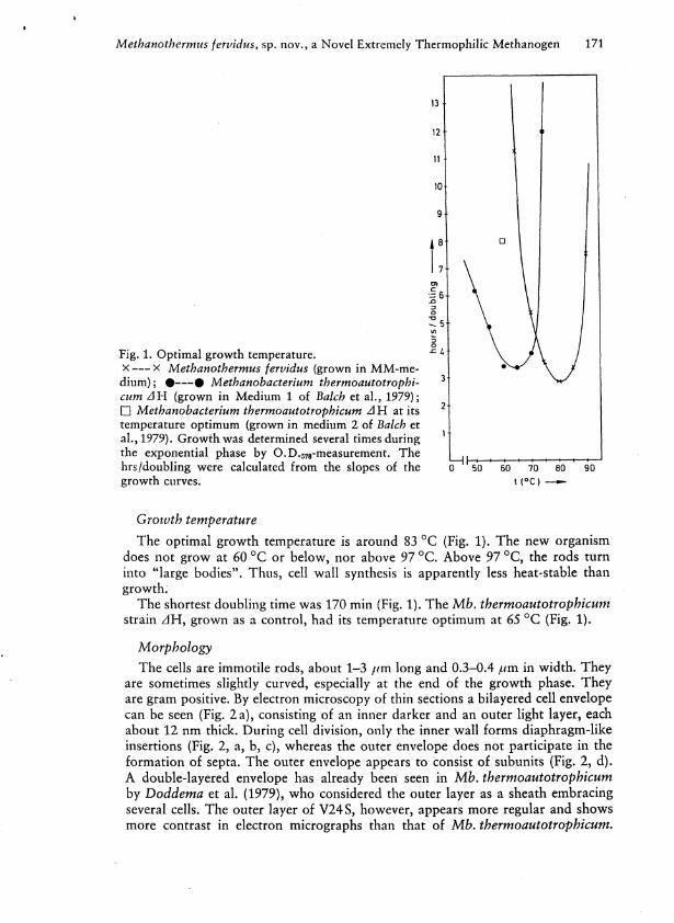
Fig. 1. Optimal growth temperature. ^ 4
x x Methanothermus fervidus (grown in M M - m e dium); • • Methanobacterium thermoautotrophi- 3
cum AH (grown in Medium 1 of Balch et al . , 1979); • Methanobacterium thermoautotrophicum AH at its 2
temperature optimum (grown in medium 2 of Balch et al . , 1979). Growth was determined several times during 1
the exponential phase by O.D.578-measurement. The ^ hrs/doubling were calculated from the slopes of the ¡T11 50 ' 60 ' 70 ' 80 90 growth curves. t ( ° C ) —
Growth temperature The optimal growth temperature is around 83 °C (Fig. 1). The new organism
does not grow at 60 °C or below, nor above 97 °C. Above 97 °C, the rods turn into "large bodies". Thus, cell wal l synthesis is apparently less heat-stable than growth.
The shortest doubling time was 170 min (Fig. 1). The Mb. thermoautotrophicum strain z l H , grown as a control, had its temperature optimum at 65 °C (Fig. 1).
Morphology
The cells are immotile rods, about 1-3 /./m long and 0.3-0.4 f.im in width . They are sometimes slightly curved, especially at the end of the growth phase. They are gram positive. By electron microscopy of thin sections a bilayered cell envelope can be seen (Fig. 2 a), consisting of an inner darker and an outer light layer, each about 12 nm thick. Dur ing cell division, only the inner w a l l forms diaphragm-like insertions (Fig. 2, a, b , c), whereas the outer envelope does not participate in the formation of septa. The outer envelope appears to consist of subunits (Fig. 2, d). A double-layered envelope has already been seen in Mb. thermoautotrophicum by Doddema et al . (1979), who considered the outer layer as a sheath embracing several cells. The outer layer of V24 S, however, appears more regular and shows more contrast in electron micrographs than that of Mb. thermoautotrophicum.
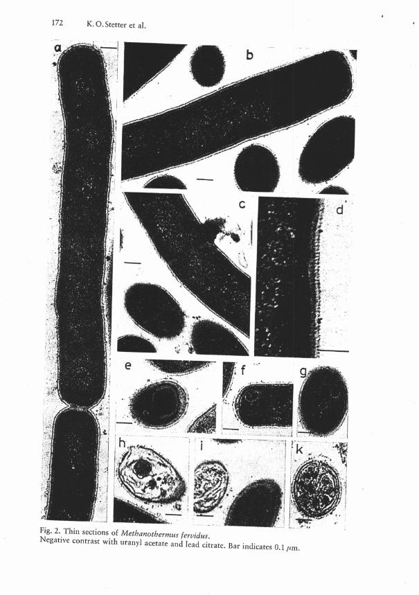
Therefore, the compositions may be different. Membranous structures are often visible within the cells (Fig. 2, e, f, g), particularly when these have lost their cytoplasm (Fig. 2, h , i , k).
Cell wall composition
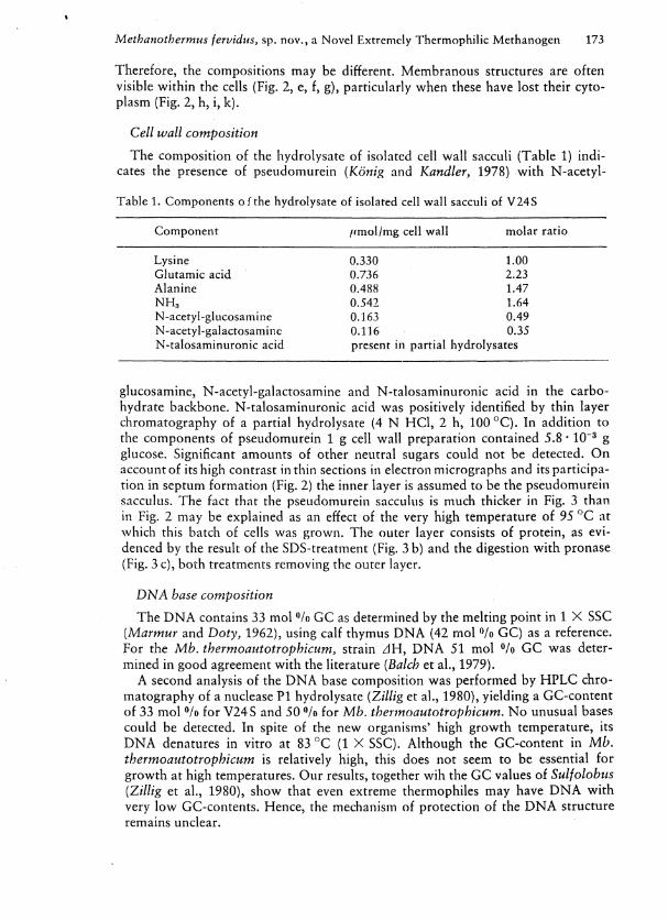
The composition of the hydrolysate of isolated cell wal l sacculi (Table 1) indi cates the presence of pseudomurein (König and Kandier, 1978) wi th N-acetyl-
Table 1. Components of the hydrolysate of isolated cell wall sacculi of V24S
Component //mol/mg cell wall molar ratio
Lysine 0.330 1.00 Glutamic acid 0.736 2.23 Alanine 0.488 1.47 NH» 0.542 1.64 N-acetyl-glucosamine 0.163 0.49 N-acetyl-galactosamine 0.116 0.35 N-talosaminuronic acid present in partial hydrolysates
glucosamine, N-acetyl-galactosamine and N-talosaminuronic acid in the carbohydrate backbone. N-talosaminuronic acid was positively identified by thin layer chromatography of a partial hydrolysate (4 N H C l , 2 h , 100 °C) . In addition to the components of pseudomurein 1 g cell wall preparation contained 5.8 • 10" 3 g glucose. Significant amounts of other neutral sugars could not be detected. O n account of its high contrast in thin sections in electron micrographs and its participation in septum formation (Fig. 2) the inner layer is assumed to be the pseudomurein sacculus. The fact that the pseudomurein sacculus is much thicker in Fig. 3 than in Fig. 2 may be explained as an effect of the very high temperature of 95 °C at which this batch of cells was grown. The outer layer consists of protein, as evidenced by the result of the SDS-treatment (Fig. 3 b) and the digestion wi th pronase (Fig. 3 c), both treatments removing the outer layer.
DNA base composition
The D N A contains 33 mol °/o G C as determined by the melting point in 1 X SSC (Marmur and Doty, 1962), using calf thymus D N A (42 mol °/o G C ) as a reference. For the Mb. thermoautotrophicum, strain z l H , D N A 51 mol °/o G C was determined in good agreement wi th the literature (Balch et al . , 1979).
A second analysis of the D N A base composition was performed by H P L C chromatography of a nuclease P I hydrolysate (Zillig et al . , 1980), yielding a GC-content of 33 mol °/o for V24S and 50 °/o for Mb. thetmoautotrophñcum. N o unusual bases could be detected. In spite of the new organisms' high growth temperature, its D N A denatures in vitro at 83 °C (1 X SSC). Although the GC-content in Mb. thermoautotrophicum is relatively high, this does not seem to be essential for growth at high temperatures. Our results, together w i h the G C values of Sulfolobus (Zillig et al . , 1980), show that even extreme thermophiles may have D N A w i t h very l o w GC-contents. Hence, the mechanism of protection of the D N A structure remains unclear.
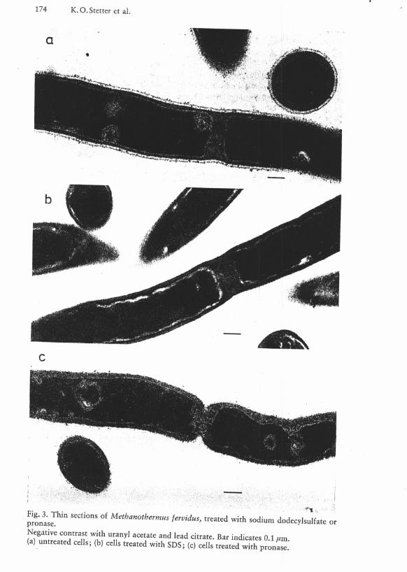
pronase™" S e C t Í O " S ° f M e t h a n o t b e r m u s ^¡dus, treated with sodium dodecylsulfate
(a) untreated cells; (b) cells treated with SDS; (c) cells treated with pronase.
RNA polymerase
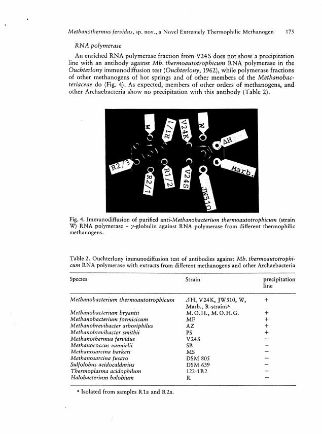
A n enriched R N A polymerase fraction from V24 S does not show a precipitation line wi th an antibody against Mb. thermoautotrophicum R N A polymerase in the Ouchterlony immunodiffusion test (Ouchterlony, 1962), while polymerase fractions of other methanogens of hot springs and of other members of the Methanobac-teriaceae do (Fig. A). As expected, members of other orders of methanogens, and other Archaebacteria show no precipitation with this antibody (Table 2).
Fig. 4. Immunodiffusion of purified znú-Methanobacterium thermoautotrophicum (strain W) R N A polymerase - y-globulin against R N A polymerase from different thermophilic methanogens.
Table 2. Ouchterlony immunodiffusion test of antibodies against Mb. thermoautotrophicum R N A polymerase with extracts from different methanogens and other Archaebacteria
Species Strain precipitation line
Methanobacterium thermoautotrophicum z l H , V 2 4 K , JW510, W, Marb. , R-strainsa
+
Methanobacterium bryantii M . O . H . , M . O . H . G . Methanobacterium formicicum M F + Methanobrevibacter arboriphilus A Z + Methanobrevibacter smithii PS Meth another mus fervidus V24S -Methanococcus vannielii SB — Methanosarcina barkeri MS -Methanosarcina fusaro D S M 805 -Sulfolobus acidocaldarius D S M 639 -Thermoplasma acidophilum 122-1B 2 -Halobacterium halobium R —
a Isolated from samples R l a and R2a.
Discussion
The new methanogen V24S is clearly different from Mb. thermoautotrophicum according to its growth temperature, nutritional requirements, D N A composition and R N A polymerase. The existence of pseudomurein in the cell wal l classifies it as a member of the order Methanobacteriales (Kandier and König, 1978; Balch et al . , 1979).
The R N A polymerase of V24S does not precipitate with antibodies against R N A polymerase from Mb. thermoautotrophicum in the Ouchterlony immunodiffusion test, whereas the members of the two genera of the Meth anob acter iaceae, Methanobacterium and Methanobrevibacter show distinct precipitation lines. The mult i plicity of the lines may be caused by fragmentation of the enzymes. W i t h R N A polymerase from other orders of methanogens and other Archaebacteria, however, no precipitation could be observed wi th antiserum against R N A polymerase of Mb. thermoautotrophicum. Therefore, we consider the new isolate to be a member of a new family of the Methanobacteriales, which we name Methanothermaceae. Because of its ability to grow close to the boiling point, the isolate V 2 4 S is named Meth another mus fervidus.
The ecological niche of Meth another mus fervidus may be different from that of Mb. thermoautotrophicum: the latter seems to be mainly involved in the thermophilic decomposition of organic matter in sewage digestors and waste piles, and of decaying blue green algal mats in hot springs (Zeikus et a l . , 1980). Since no algal mats can grow at temperatures around 90 °C, Meth another mus appears to depend on geothermal hydrogen and C 0 2 . As inferred from our survey of Icelandic solfataras, where Meth another mus could be found only once, and from a survey of Zeikus et al . (1980) in the Yellowstone Nat ional Park, U.S .A. , where it was not detected it appears to be very rare in volcanic spring holes. As indicated by its extreme oxygen sensitivity it may, however, occur more frequently in the depth of volcanic areas.
One could speculate that Meth another mus fervidus and other so far unknown members of the Methanothermaceae, may also be responsible for bacterial methane production in geothermally heated deep sediments, e. g. in the area of Porto C o r -sini in northern Italy, where methane is found in pliocene sediments situated in 3,000 m depth at temperatures around 90 °C (Schoell, 1980). The d 1 3 C value of — 72.7, indicates that this methane is of bacterial origin (Schoell, 1980).
Description and Classification of the Methanothermaceae
Order Meth anob acter iales, Balch and Wolfe 1979 Family I, Meth anob acter iaceae, Barker 1956 Family II, Methanothermaceae, Stetter (fam. nov.) Methanothermaceae, Me.tha.no.ther.ma.ce'ae. M.L.neut.n.Mei/?anothermus type
genus of the family; - aceae ending to denote a family; M . L . f e m . p l . n . Methanothermaceae the Methanothermus family. The Methanothermaceae belong to the order Meth anob acteriales, Balch and Wolfe 1979. The family Methanothermaceae contains one genus.
Gram-positive rods, occuring singly and in pairs. In ultra thin sections, the cell envelope appears as a distinct double-layer. The inner layer is a sacculus contain-
ing pseudomurein. The outer layer consists of protein. R N A polymerase does not show serological relationship with members of Meth anob acter iacaeae in the i m munodiffusion test. Cells are extremely thermophilic, not growing at 60 °C or below. Methan is formed from H 2 + ' C 0 2 . Habitat : anaerobic environments within solfataric fields.
Genus 1 Meth another mus, Stetter (gen. nov.) Me.tha.no.ther'mus. M . L . n . methanum methane; Gr.itm.n.therme heat; M . L . -
masc.n. Methanothermus the methane (-producing) thermophile. The description of the genus is the same as that of the family.
Methanothermus fervidus, Stetter (sp. nov.) fer.vid.us M . L . fervidus, L.masc.adj.fervent; on account of its growth in almost
boil ing water. Straight to slightly curved rods, 1 to 3 jum long and 0.3-0.4 jam in width ,
occurring singly and in pairs, but never in long filaments. They are non-motile and gram-positive. N o growth can be detected at 60 °C or below. Opt imal growth is at 83 °C at p H 6.5. The upper temperature l imit is 97 °C. Methane is formed from H 2 and C 0 2 . Yeast extract is required for growth in artificial medium. Formate and acetate do not serve as substrates. Colonies are formed on polysilicate plates 1 to 3 mm in diameter, and are round, smooth, opaque, and slightly greyish. N o growth can be observed on agar. The cell wal l contains pseudomurein consisting of N-acetyl-glucosamine, N-acetyl-galactosamine, N-talosaminuronic acid, glutamic acid, alanine, and lysine. The cell envelope consists of a double-layer of pseudomurein and protein. The D N A base composition is 33 mol °/o G + C . The R N A polymerase does not precipitate with antibodies against R N A polymerase from Methanobacterium thermoautotrophicum. The type strain is D S M 2088 (isolated from an Icelandic hot spring).
Acknowledgements. We wish to thank Dr. / . Madon, Christine Matzenbacher and Martina Reimers for GC-analyses. This work was supported by a grant of the Deutsche Forschungsgemeinschaft to K.O. Stetter.
References
Balch, W.E., Wolfe, R.S.: New approach to the cultivation of methanogenic bacteria: 2-mercaptoethanesulfonic acid (HS-CoM)-dependent growth of Methanobacterium ru-minantium in a pressurized atmosphere. Appl . Environ. Microbiol. 32, 781-791 (1976)
Balch, W.E., Fox, G.E., Magrum, L.]., Woese, C.R., Wolfe, R.S.: Methanogens: Reevalu-ation of a unique biological group. Microbiol. Rev. 43, 260-296 (1979)
Brock, T.D., Brock, K.M., Belly, R.T., Weiß, R.L.: Sulfolobus: a new genus of sulfur-oxidizing bacteria living at low p H and high temperature. Arch. Microbiol. 84, 54-68 (1972)
Doddema, H.J., Van der Drift, C, Vogels, G.D., Veenhuis, M.: Chemiosmotic coupling in Methanobacterium thermoautotrophicum: Hydrogen-dependent adenosine 5'-triphos-phate synthesis by subcellular particles. J . Bact. 140, 1081-1089 (1979)
Kandier, O., König, H.: Chemical composition of the peptidoglycan-free cell walls of methanogenic bacteria. Arch. Microbiol. 118, 141-152 (1978)
König, H., Kandier, O.: N-Acetyltalosaminuronic acid a constituent of the pseudomurein of the genus Methanobacterium. Arch. Microbiol. 123, 295-299 (1979)
Linn, T.G., Greenleaf, A.L., Shorenstein, R.G., Losick, R.: Loss of the sigma activity of R N A polymerase of Bacillus subtilis during sporulation. Proc. nat. Acad. Sei. (Wash.) 70, 1865-1869 (1973)
Marmur, ]., Doty, P.; Determination of the base composition of deoxyribonucleid acid from its thermal denaturation temperature. J . molec. Biol. 5, 109-118 (1962)
Oesterhelt, D., Stoeckenius, W.; Isolation of the cell membrane of Halo bacterium halobium and its fractination into red and purple membrane. In: Methods in Enzymology. V o l . 31, pp. 667-678, (S.P.Colowick and N.O.Kaplan, eds.). New York, Academic Press (1974)
Ouchterlony, Ö.: Diffusion-in-gel methods for immunological analysis II. In: Progress in Allergy VI, pp. 30-154, {P.Kallod and B.H.Waksman, eds.). Basel, Karger 1962
Schoell, M.: The hydrogen and carbon isotopic composition of methane from natural gases of various origins. Geochim. Cosmochim. Acta 44, 649-661 (1980)
Steuer, K.O.: Transcription in Lactobacillaceae: DNA-dependent R N A polymerase from Lactobacillus casei. Isolation of transcription factor y. Hoppe-Seyler's Z . physiol. Chem. 358, 1093-1104 (1977)
Stetter, K. O., Winter, ]., Hartlieb, R.: DNA-dependent R N A polymerase of the Archae-bacterium Metbanobacterium thermoautotrophicum. Z b l . Bakt. Hyg. , I. Abt. Orig. C 1, 201-214 (1980)
Sturm, S., Schönefeld, U., Zillig, W., Janécovic, D., Stetter, K. O . : Structure and function of the D N A dependent R N A polymerase of the archaebacterium Tbermoplasma acido-philum. Z b l . Bakt. Hyg. , I. Abt. Orig. C 1, 12-25 (1980)
Zeikus, J.G., Wolfe, R.S.: Methanob acter ium thermoautotrophicum sp.n., an anaerobic, autotrophic, extreme thermophile. J . Bact. 109, 707-713 (1972)
Zeikus, J. G., Arie Ben-Bassat, Hegger, P. W.: Microbiology of methanogenesis in thermal, volcanic environments. J . Bact. 143, 432-440 (1980)
Zillig, W„ Stetter, K.O., Wunderl,S., Schulz, W., Priess, H., Scholz, J.: The Sulfolobus-< <Caldariella' ,group: Taxonomy on the basis of the structure of DNA-dependent R N A polymerase. Arch. Microbiol. 125, 259-269 (1980)
Prof. Dr. K.O.Stetter, Lehrstuhl für Mikrobiologie der Universität Regensburg, Univer-sitätsstr. 31, D-8400 Regensburg


![Quantum Simulations of Out-of-Equilibrium Phenomena · Quantum Simulations of Out-of-Equilibrium Phenomena ... Systeme, z.B. die anisotrope XY Kette, ... explosion [Fey82] of the](https://img.pdfslide.org/doc/110x75/5b9d375d09d3f253158bcf73/quantum-simulations-of-out-of-equilibrium-phenomena-quantum-simulations-of-out-of-equilibrium.jpg)