Embed Size (px)
Citation preview
Palmitoylation Regulates Epidermal Homeostasis andHair Follicle DifferentiationPleasantine Mill1., Angela W. S. Lee1.¤, Yuko Fukata2,3, Ryouhei Tsutsumi2, Masaki Fukata2,3, Margaret
Keighren1, Rebecca M. Porter4, Lisa McKie1, Ian Smyth1,5", Ian J. Jackson1"*
1 Medical Research Council, Human Genetics Unit, Edinburgh, United Kingdom, 2 National Institute for Physiological Sciences, National Institutes of Natural Sciences,
Okazaki, Japan, 3 Precursory Research for Embryonic Science and Technology, Japan Science and Technology Agency, Chiyoda, Tokyo, Japan, 4 Department of
Dermatology, School of Medicine, Cardiff University, Cardiff, United Kingdom, 5 Cutaneous Developmental Biology Lab, Department of Biochemistry and Molecular
Biology, Department of Anatomy and Developmental Biology, Monash University, Melbourne, Australia
Abstract
Palmitoylation is a key post-translational modification mediated by a family of DHHC-containing palmitoyl acyl-transferases(PATs). Unlike other lipid modifications, palmitoylation is reversible and thus often regulates dynamic protein interactions.We find that the mouse hair loss mutant, depilated, (dep) is due to a single amino acid deletion in the PAT, Zdhhc21,resulting in protein mislocalization and loss of palmitoylation activity. We examined expression of Zdhhc21 protein in skinand find it restricted to specific hair lineages. Loss of Zdhhc21 function results in delayed hair shaft differentiation, at the siteof expression of the gene, but also leads to hyperplasia of the interfollicular epidermis (IFE) and sebaceous glands, distantfrom the expression site. The specific delay in follicle differentiation is associated with attenuated anagen propagation andis reflected by decreased levels of Lef1, nuclear b-catenin, and Foxn1 in hair shaft progenitors. In the thickened basalcompartment of mutant IFE, phospho-ERK and cell proliferation are increased, suggesting increased signaling through EGFRor integrin-related receptors, with a parallel reduction in expression of the key differentiation factor Gata3. We show that theSrc-family kinase, Fyn, involved in keratinocyte differentiation, is a direct palmitoylation target of Zdhhc21 and ismislocalized in mutant follicles. This study is the first to demonstrate a key role for palmitoylation in regulatingdevelopmental signals in mammalian tissue homeostasis.
Citation: Mill P, Lee AWS, Fukata Y, Tsutsumi R, Fukata M, et al. (2009) Palmitoylation Regulates Epidermal Homeostasis and Hair Follicle Differentiation. PLoSGenet 5(11): e1000748. doi:10.1371/journal.pgen.1000748
Editor: David R. Beier, Harvard Medical School, United States of America
Received April 9, 2009; Accepted October 30, 2009; Published November 26, 2009
Copyright: � 2009 Mill et al. This is an open-access article distributed under the terms of the Creative Commons Attribution License, which permits unrestricteduse, distribution, and reproduction in any medium, provided the original author and source are credited.
Funding: This work was funded by the Medical Research Council (UK), a National Sciences and Engineering Research Council of Canada Fellowship to PM, aCaledonian Research Foundation Fellowship to PM, a Wellcome Trust Travelling Fellowship to IS, an R. Douglas Wright Fellowship to IS, an Australian ResearchCouncil grant to IS, and a Human Frontiers Science Program grant to YF and MF. The funders had no role in study design, data collection and analysis, decision topublish, or preparation of the manuscript
Competing Interests: The authors have declared that no competing interests exist.
* E-mail: [email protected]
¤ Current address: (AWSL) Medical Research Council, Mammalian Genetics Unit, Harwell Science and Innovation Campus, London, United Kingdom
. These authors contributed equally to this work.
" These authors are joint senior authors on this work.
Introduction
Palmitoylation (or protein S-acylation) is a reversible post-
translational lipid modification which involves addition of the fatty
acid palmitate onto specific cysteine residues [1]. Some post-
translational lipid modifications such as myristoylation and
prenylation serve to localize otherwise soluble proteins to the
cytoplasmic surfaces of cellular membranes. In contrast, palmi-
toylation substrates are proteins that are already membrane
associated, and the modification acts to increase or stabilise
membrane affinity or to traffic the protein to specific membrane
domains. In particular, palmitoylation results in localization of the
protein to lipid rafts; domains of the plasma membrane rich in
cholesterol and sphingolipids. Furthermore, as palmitoylation is
reversible, it allows for membrane localization or trafficking to be
dynamically regulated. This has best been demonstrated in
synapses, where palmitoylation regulates membrane localization
and activity of the AMPA receptor [2] and GABAA receptor [3].
Palmitoylation of the post-synaptic density protein PSD95 permits
clustering of the protein at synapses and regulates synaptic
strength [4]. A recent global study of the neural palmitoyl-
proteome highlights the breadth of targets that are rapidly
modulated by palmitoylation [5], further emphasizing the
importance of this modification in dynamic biological processes.
Members of the zinc finger, DHHC containing (ZDHHC)
protein family have recently been shown to promote palmitoyla-
tion of intracellular proteins in yeast and in mammalian cells
[6–8]. These palmitoyl-acyl transferases (PATs) are predicted
membrane proteins possessing a cysteine-rich domain and a
putative zinc finger with a characteristic Asp-His-His-Cys
(DHHC) motif, required for activity. This family is encoded by
24 genes in both mouse and humans, of which 23 are orthologous
pairs. Assaying individual target proteins against the entire
repertoire of PATs indicates that there is substrate specificity;
each substrate is primarily modified by a subgroup of structurally
similar ZDHHC proteins [9].
PLoS Genetics | www.plosgenetics.org 1 November 2009 | Volume 5 | Issue 11 | e1000748
Although some human ZDHHC genes have been implicated in
cancer [10,11], genetic evidence for function of these genes is
limited to neurological disorders. ZDHHC8 shows association with
schizophrenia in humans and neurophysiological deficits in mice
[12–14]. X-linked mental retardation is associated in a few
patients with loss of expression of ZDHHC15 [15] and in others
with frameshifts, splice or missense mutations of ZDHHC9 [16].
Recently, the Drosophila ortholog of Zdhhc8 (App) was shown to play
a key role in patterning and growth control of imaginal discs [17].
However, very little is known about specific palmitoylation
functions during normal mammalian development.
Several lineage-restricted stem cell populations exist in the adult
skin and contribute to renewal of only their own specific niche under
normal steady-state conditions [18]. Their progeny proliferate,
migrate and terminally differentiate along the lineages of the
interfollicular epidermis (IFE), hair follicle and sebaceous gland
[reviewed in 19]. The cornified layer of postnatal skin is constantly
shed and replenished by progeny of the epidermal stem cells in the
basal IFE, which proliferate, differentiate and migrate suprabasally.
Similarly, hair shafts are shed and replaced in a cycle of regression
(catagen), rest (telogen) and regeneration (anagen). During each
anagen, stem cells, residing in the permanent bulge region, are
mobilized to provide hair follicle progenitors, which differentiate
into eight different lineages that make up the hair shaft (consisting of
the medulla, cortex and hair shaft cuticle), the inner root sheath
(IRS) (consisting of the inner root sheath cuticle, Huxley’s and
Henle’s layers), the companion layer cells and the outer root sheath
(ORS). Also within the permanent portion of the follicle is the
sebaceous gland which produces lipid-rich sebum to lubricate the
skin and hair, in addition to providing antibacterial activity. Sebum
is released by disintegrating sebocytes that are continuously replaced
from progenitors in the periphery of the gland. These three stem cell
lineages require a precise balance of self-renewal and differentiation
of their committed progeny. However under certain experimental
conditions or genetic manipulations, stem cells from one niche can
contribute to hair, IFE and sebaceous gland lineages [20,21],
highlighting the interdependence of these epidermal compartments
in maintaining homeostasis.
The depilated mutation (dep, MGI:94884) results in a recessive
phenotype characterized by variable hair loss, with thinner and
shorter hairs remaining in a greasy coat. Recombination
experiments show that the phenotype is due to a defect in the
epidermis, rather then the dermis [22]. Here, we genetically map
and further characterize the dep mutant and show that it carries a
single amino acid deletion in Zdhhc21, resulting in loss of PAT
activity. A detailed study of the phenotype demonstrates that lack
of palmitoylation by Zdhhc21 results in hyperplasia of the IFE and
sebaceous glands and delayed differentiation of the hair shaft.
Furthermore, we identify Fyn, a member of the Src family of
tyrosine protein kinases required for keratinocyte differentiation,
as a direct palmitoylation target of Zdhhc21 and demonstrate its
mislocalization within dep mutant follicles.
Results/Discussion
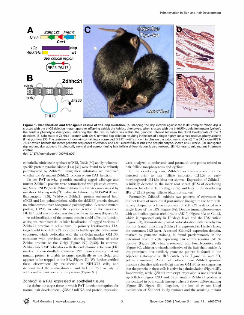
Mutation in Zdhhc21 causes the dep phenotypeThe location of the dep mutation has previously been defined by
complementation against a set of chromosomes bearing deletions
centred on the Tyrp1 gene [23]. The endpoints of those deletions
defining the proximal and distal boundaries of the candidate
interval were further refined using polymorphic markers on mice
carrying the deletion chromosome opposite a Mus spretus
chromosome [24,25, data not shown]. The candidate location of
dep, defined by the deletions 46UThc proximally and 1OZ distally,
is only 160kb in length and contains all or part of just 3 genes:
Zdhhc21, Cer1 and Frem1 (Figure 1A). Two of these have existing
established mutations.
Frem1 is associated with 2 ENU-induced alleles and the classical
mutation head blebs (heb) [26] which result in an embryonic
blebbing phenotype, and is a mouse model for Frasers Syndrome.
Furthermore, a genetic complementation analysis between a Frem1
mutant (bfd) and dep produces normal mice (personal communi-
cation, Monica Justice), indicating Frem1 is not allelic to dep. There
are several knockout mutant alleles of Cer1 but none of these
exhibit the dep phenotype [27–29]. We have sequenced all known
exons of both Frem1 and Cer1 in dep DNA have found no
mutations. Additionally no non-coding RNAs are annotated or
predicted within this interval (miRBase: microrna.sanger.ac.uk,
Ensembl: www.ensembl.org, VEGA: vega.sanger.ac.uk).
However, sequencing of the 7 exons of Zdhhc21 (MGI:1915518)
in dep mutants revealed a 3-bp deletion which results in the
deletion of a single, highly conserved, phenylalanine residue (del-
233F) close to the C terminus of the protein (Figure 1B).
Although this deletion was the only coding alteration found in
the candidate interval, it remained possible that an undetected
non-coding mutation could affect expression of genes outside the
interval. To establish the causative link between Zdhhc21 and the
dep phenotype, we generated transgenic mice containing the
bacterial artificial chromosome, RP23-76J17, containing only
Zdhhc21 and Cer1 (Figure 1A). When crossed onto a dep
background, this transgene rescues the hair phenotype to a
smooth and shiny dorsal coat, indistinguishable from wild-type,
whilst the hair of nontransgenic littermates retains the greasy and
disorderly dep phenotype (Figure 1C). Later in life, non-transgenic
mutant littermates lose their hair, whilst the transgenic mice do
not. Skin sections of transgenic rescued mice show a normal
histological appearance, confirming that the dep phenotype is fully
rescued (Figure 1D and 1E).
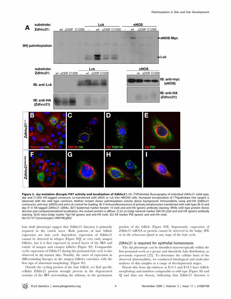
Zdhhc21-del233F is mislocalised and lacks PAT functionZdhhc21 has previously been demonstrated to have palmitoyl
transferase (PAT) activity. Among 23 Zdhhc members tested,
Author Summary
During embryonic development, growth and patterningare regulated at many levels. Signals that mediatetranscriptional activity, where and when genes areexpressed, are a primary level of regulation. However,developmental signals can be further fine-tuned bymodulating protein stability, localization, and activity viapost-translational modifications. One such modification isthe reversible addition of the fatty acid palmitate toproteins. This modification mediates dynamic trafficking oftarget proteins to specific subdomains of the cell. A largefamily of enzymes carries out this palmitoylation process,where each family member has specificity towardsparticular targets. However, the functional significance ofpalmitoylation during mammalian development is unclear.We present evidence of a critical role for palmitoylationduring mouse development using a mutation of a specificpalmitoylating enzyme, whose loss of function leads tohair loss and skin defects in depilated (dep) mice. Despiteits restricted expression in hair follicles, loss of function ofthis enzyme results in developmental defects in nearbystructures. We show that palmitoylation plays an impor-tant regulatory role in hair growth and epidermalhomeostasis.
Palmitoylation in Skin and Hair Development
PLoS Genetics | www.plosgenetics.org 2 November 2009 | Volume 5 | Issue 11 | e1000748
endothelial nitric oxide synthase (eNOS, Nos3) [30] and lymphocyte-
specific protein tyrosine kinase (Lck) [31] were found to be robustly
palmitoylated by Zdhhc21. Using these substrates, we examined
whether the dep mutant Zdhhc21 protein retains PAT function.
To test PAT activity, plasmids encoding tagged wild-type and
mutant Zdhhc21 proteins were cotransfected with plasmids express-
ing Lck or eNOS (Nos3). Palmitoylation of substrates was assessed by
metabolic labeling with [3H]palmitate followed by SDS-PAGE and
fluorography [8,9]. Wild-type Zdhhc21 protein enhanced both
eNOS and Lck palmitoylation, whilst the del233F protein showed
no enhancement over background palmitoylation. A second mutant
protein, C120S, in which the cysteine residue in the conserved
DHHC motif was mutated, was also inactive in this assay (Figure 2A).
As mislocalisation of the mutant protein could affect its function
in vivo, we examined the cellular localization of tagged variants of
Zdhhc21 proteins in cell culture. In primary keratinocytes, HA-
tagged wild type Zdhhc21 localizes to highly specific cytoplasmic
structures, which co-localise with the cis-Golgi marker GM130,
consistent with previous studies showing localization of other
Zdhhc proteins to the Golgi (Figure 2C) [8,30]. In contrast,
Zdhhc21-del233F colocalizes with the endoplasmic reticulum (ER)
marker, protein disulfide isomerase (PDI), demonstrating that dep
mutant protein is unable to target specifically to the Golgi and
appears to be trapped in the ER. (Figure 2I). We further verified
these observations by transfection in NIH-3T3 cells, and
demonstrated the mislocalisation and lack of PAT activity of
additional mutant forms of the protein (Figure S1)
Zdhhc21 is a PAT expressed in epithelial tissuesTo define the target tissue in which PAT function is required for
normal hair development, Zdhhc21 mRNA and protein expression
were analyzed at embryonic and postnatal time-points related to
hair follicle morphogenesis and cycling.
In the developing skin, Zdhhc21 expression could not be
detected prior to hair follicle induction (E13.5) or early
morphogenesis (E14.5) (data not shown). Expression of Zdhhc21
is initially detected in the inner root sheath (IRS) of developing
vibrissae follicles at E16.5 (Figure S2) and later in the developing
IRS of E18.5 pelage follicles (data not shown).
Postnatally, Zdhhc21 exhibits two patterns of expression in
distinct layers of more distal post-mitotic lineages in the hair bulb.
Strong ubiquitous cellular expression of Zdhhc21 is detected in a
single layer of the IRS (Figure 3A). Double immunofluorescence
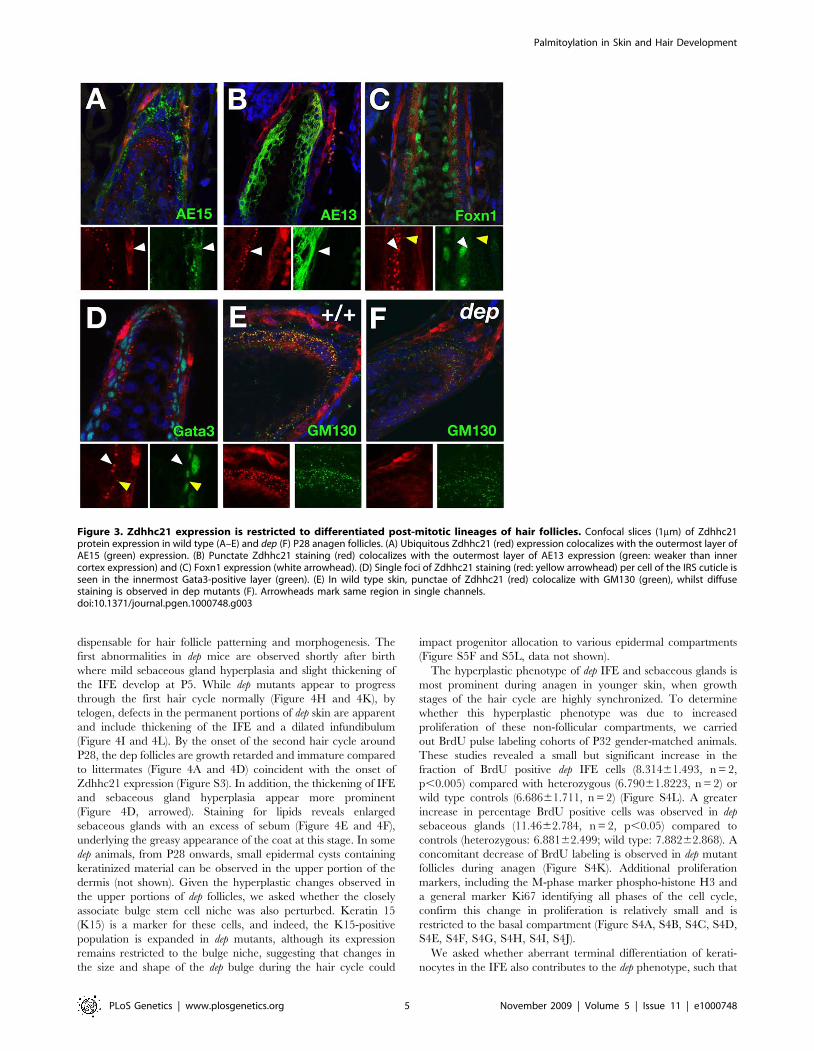
with antibodies against trichohyalin (AE15) (Figure 3A) or Gata3,
which is expressed only in Huxley’s layer and the IRS cuticle
(Figure 3D), demonstrated partial co-localization with trichohyalin
but not Gata3, indicating Zdhhc21 is expressed in Henle’s layer,
the outermost IRS layer. A second Zdhhc21 expression domain,
marked by punctate staining, is found predominantly in the
outermost layer of cells expressing hair cortex keratins (AE13-
positive) (Figure 3B, white arrowhead) and Foxn1-positive cells
(Figure 3C, white arrowhead), indicative of the hair shaft cuticle. A
less prominent but similarly punctate pattern is found in the
adjacent Gata3-positive IRS cuticle cells (Figure 3C and 3D,
yellow arrowhead). As in cell culture, these Zdhhc21-positive
punctae colocalize with cis-Golgi marker GM130 in vivo suggesting
that the protein in these cells is active in palmitoylation (Figure 3E).
Importantly, while Zdhhc21 transcript expression is not altered in
dep follicles (Figure S2D and S2E), mutant Zdhhc21 protein is
mislocalized in both cuticle lineages where it shows diffuse staining
(Figure 3F, Figure S3). Together, the loss of in vivo Golgi
localization of Zdhhc21 in dep mutants and the resulting mutant
Figure 1. Identification and transgenic rescue of the dep mutation. (A) Mapping the dep interval against the b-del complex. When dep iscrossed with the b-IOZ deletion mutant (purple), offspring exhibit the hairloss phenotype. When crossed with the b-46UTHc deletion mutant (yellow),the hairloss phenotype disappears, indicating that the dep mutation lies within the genomic interval between the distal breakpoints of the 2deletions. (B) Schematic of Zdhhc21 protein with dep C-terminal 3bp deletion resulting in the loss of a single highly conserved residue, phenylalanine(F) at position 233. The cysteine-rich domain containing a conserved DHHC motif is shown in blue on the cytoplasmic side. (C) The BAC clone RP23-76J17, which harbors the intact genomic sequences of Zdhhc21 and Cer1 successfully rescues the dep phenotype, shown at 6.5 weeks. (D) Transgenicdep mutant skin appears histologically normal and correct timing hair follicle differentiation is also restored. (E) Non-transgenic mutant littermatecontrol.doi:10.1371/journal.pgen.1000748.g001
Palmitoylation in Skin and Hair Development
PLoS Genetics | www.plosgenetics.org 3 November 2009 | Volume 5 | Issue 11 | e1000748
hair shaft phenotype suggest that Zdhhc21 function is primarily
required in the cuticle layer. Both patterns of hair follicle
expression are hair cycle dependent; expression of Zdhhc21
cannot be detected in telogen (Figure S2J) or very early anagen
follicles, but it is first expressed in nested layers of the IRS and
cuticle of anagen and catagen follicles (Figure S2). Comparable
cyclic expression of Zdhhc21 during this postnatal hair cycle is also
observed in dep mutant skin. Notably, the onset of expression in
differentiating lineages in the anagen follicles correlates with the
first sign of abnormal morphology (Figure S3).
Outside the cycling portion of the hair follicle, we find specific
cellular Zdhhc21 protein strongly present in the degenerated
remains of the IRS surrounding the isthmus, in the permanent
portion of the follicle (Figure S2I). Importantly, expression of
Zdhhc21 mRNA or protein cannot be detected in the bulge, IFE
or in the sebaceous gland at any stage of the hair cycle.
Zdhhc21 is required for epithelial homeostasisThe dep phenotype can be identified macroscopically within the
first postnatal week as a greasy and disorderly hair distribution, as
previously reported [22]. To determine the cellular basis of the
observed abnormalities, we conducted histological and molecular
analyses of skin samples at a range of developmental stages.
Dorsal skin from dep embryos at E14.5 and E18.5 have follicle
morphology and numbers comparable to wild type (Figure 4G and
4J and data not shown), indicating that Zdhhc21 function is
Figure 2. dep mutation disrupts PAT activity and localization of Zdhhc21. (A) [3H]Palmitate fluorography of individual Zdhhc21 (wild type,dep and C120S) HA-tagged constructs co-transfected with eNOS or Lck into HEK293 cells. Increased incorporation of [3H]palmitate into targets isobserved with the wild type construct. Neither mutant shows palmitoylation activity above background. Immunoblots using anti-HA (Zdhhc21constructs), anti-myc (eNOS) and anti-Lck control for loading. (B–I) Immunofluorescence of primary keratinocytes transfected with wild type (B–E) anddep (F–I) HA-tagged Zdhhc21 cDNAs. (B,F) Epidermal marker Keratin 14 (red) and anti-HA (green) antibody staining. While wild type protein showsdiscrete and compartmentalized localization, the mutant protein is diffuse. (C,G) cis-Golgi network marker GM130 (red) and anti-HA (green) antibodystaining. (D,H) trans-Golgi marker Tgn138 (green) and anti-HA (red). (E,I) ER marker PDI (green) and anti-HA (red).doi:10.1371/journal.pgen.1000748.g002
Palmitoylation in Skin and Hair Development
PLoS Genetics | www.plosgenetics.org 4 November 2009 | Volume 5 | Issue 11 | e1000748
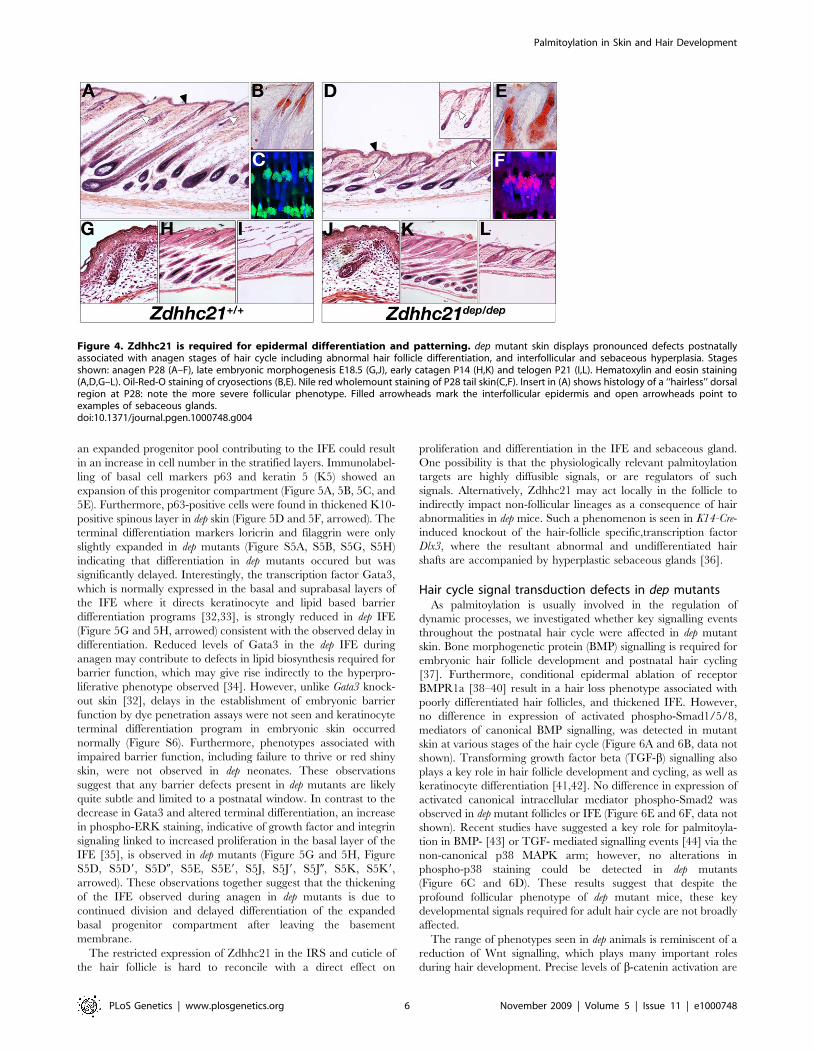
dispensable for hair follicle patterning and morphogenesis. The
first abnormalities in dep mice are observed shortly after birth
where mild sebaceous gland hyperplasia and slight thickening of
the IFE develop at P5. While dep mutants appear to progress
through the first hair cycle normally (Figure 4H and 4K), by
telogen, defects in the permanent portions of dep skin are apparent
and include thickening of the IFE and a dilated infundibulum
(Figure 4I and 4L). By the onset of the second hair cycle around
P28, the dep follicles are growth retarded and immature compared
to littermates (Figure 4A and 4D) coincident with the onset of
Zdhhc21 expression (Figure S3). In addition, the thickening of IFE
and sebaceous gland hyperplasia appear more prominent
(Figure 4D, arrowed). Staining for lipids reveals enlarged
sebaceous glands with an excess of sebum (Figure 4E and 4F),
underlying the greasy appearance of the coat at this stage. In some
dep animals, from P28 onwards, small epidermal cysts containing
keratinized material can be observed in the upper portion of the
dermis (not shown). Given the hyperplastic changes observed in
the upper portions of dep follicles, we asked whether the closely
associate bulge stem cell niche was also perturbed. Keratin 15
(K15) is a marker for these cells, and indeed, the K15-positive
population is expanded in dep mutants, although its expression
remains restricted to the bulge niche, suggesting that changes in
the size and shape of the dep bulge during the hair cycle could
impact progenitor allocation to various epidermal compartments
(Figure S5F and S5L, data not shown).
The hyperplastic phenotype of dep IFE and sebaceous glands is
most prominent during anagen in younger skin, when growth
stages of the hair cycle are highly synchronized. To determine
whether this hyperplastic phenotype was due to increased
proliferation of these non-follicular compartments, we carried
out BrdU pulse labeling cohorts of P32 gender-matched animals.
These studies revealed a small but significant increase in the
fraction of BrdU positive dep IFE cells (8.31461.493, n = 2,
p,0.005) compared with heterozygous (6.79061.8223, n = 2) or
wild type controls (6.68661.711, n = 2) (Figure S4L). A greater
increase in percentage BrdU positive cells was observed in dep
sebaceous glands (11.4662.784, n = 2, p,0.05) compared to
controls (heterozygous: 6.88162.499; wild type: 7.88262.868). A
concomitant decrease of BrdU labeling is observed in dep mutant
follicles during anagen (Figure S4K). Additional proliferation
markers, including the M-phase marker phospho-histone H3 and
a general marker Ki67 identifying all phases of the cell cycle,
confirm this change in proliferation is relatively small and is
restricted to the basal compartment (Figure S4A, S4B, S4C, S4D,
S4E, S4F, S4G, S4H, S4I, S4J).
We asked whether aberrant terminal differentiation of kerati-
nocytes in the IFE also contributes to the dep phenotype, such that
Figure 3. Zdhhc21 expression is restricted to differentiated post-mitotic lineages of hair follicles. Confocal slices (1mm) of Zdhhc21protein expression in wild type (A–E) and dep (F) P28 anagen follicles. (A) Ubiquitous Zdhhc21 (red) expression colocalizes with the outermost layer ofAE15 (green) expression. (B) Punctate Zdhhc21 staining (red) colocalizes with the outermost layer of AE13 expression (green: weaker than innercortex expression) and (C) Foxn1 expression (white arrowhead). (D) Single foci of Zdhhc21 staining (red: yellow arrowhead) per cell of the IRS cuticle isseen in the innermost Gata3-positive layer (green). (E) In wild type skin, punctae of Zdhhc21 (red) colocalize with GM130 (green), whilst diffusestaining is observed in dep mutants (F). Arrowheads mark same region in single channels.doi:10.1371/journal.pgen.1000748.g003
Palmitoylation in Skin and Hair Development
PLoS Genetics | www.plosgenetics.org 5 November 2009 | Volume 5 | Issue 11 | e1000748
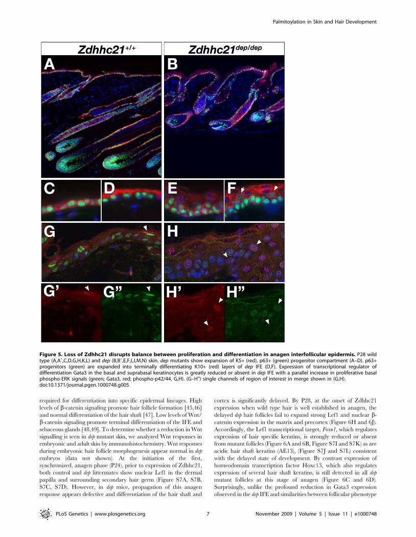
an expanded progenitor pool contributing to the IFE could result
in an increase in cell number in the stratified layers. Immunolabel-
ling of basal cell markers p63 and keratin 5 (K5) showed an
expansion of this progenitor compartment (Figure 5A, 5B, 5C, and
5E). Furthermore, p63-positive cells were found in thickened K10-
positive spinous layer in dep skin (Figure 5D and 5F, arrowed). The
terminal differentiation markers loricrin and filaggrin were only
slightly expanded in dep mutants (Figure S5A, S5B, S5G, S5H)
indicating that differentiation in dep mutants occured but was
significantly delayed. Interestingly, the transcription factor Gata3,
which is normally expressed in the basal and suprabasal layers of
the IFE where it directs keratinocyte and lipid based barrier
differentiation programs [32,33], is strongly reduced in dep IFE
(Figure 5G and 5H, arrowed) consistent with the observed delay in
differentiation. Reduced levels of Gata3 in the dep IFE during
anagen may contribute to defects in lipid biosynthesis required for
barrier function, which may give rise indirectly to the hyperpro-
liferative phenotype observed [34]. However, unlike Gata3 knock-
out skin [32], delays in the establishment of embryonic barrier
function by dye penetration assays were not seen and keratinocyte
terminal differentiation program in embryonic skin occurred
normally (Figure S6). Furthermore, phenotypes associated with
impaired barrier function, including failure to thrive or red shiny
skin, were not observed in dep neonates. These observations
suggest that any barrier defects present in dep mutants are likely
quite subtle and limited to a postnatal window. In contrast to the
decrease in Gata3 and altered terminal differentiation, an increase
in phospho-ERK staining, indicative of growth factor and integrin
signaling linked to increased proliferation in the basal layer of the
IFE [35], is observed in dep mutants (Figure 5G and 5H, Figure
S5D, S5D9, S5D0, S5E, S5E9, S5J, S5J9, S5J0, S5K, S5K9,
arrowed). These observations together suggest that the thickening
of the IFE observed during anagen in dep mutants is due to
continued division and delayed differentiation of the expanded
basal progenitor compartment after leaving the basement
membrane.
The restricted expression of Zdhhc21 in the IRS and cuticle of
the hair follicle is hard to reconcile with a direct effect on
proliferation and differentiation in the IFE and sebaceous gland.
One possibility is that the physiologically relevant palmitoylation
targets are highly diffusible signals, or are regulators of such
signals. Alternatively, Zdhhc21 may act locally in the follicle to
indirectly impact non-follicular lineages as a consequence of hair
abnormalities in dep mice. Such a phenomenon is seen in K14-Cre-
induced knockout of the hair-follicle specific,transcription factor
Dlx3, where the resultant abnormal and undifferentiated hair
shafts are accompanied by hyperplastic sebaceous glands [36].
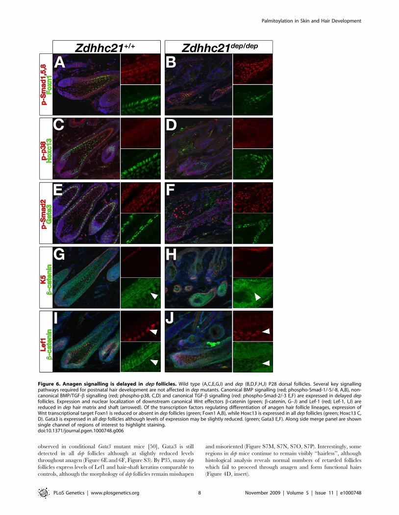
Hair cycle signal transduction defects in dep mutantsAs palmitoylation is usually involved in the regulation of
dynamic processes, we investigated whether key signalling events
throughout the postnatal hair cycle were affected in dep mutant
skin. Bone morphogenetic protein (BMP) signalling is required for
embryonic hair follicle development and postnatal hair cycling
[37]. Furthermore, conditional epidermal ablation of receptor
BMPR1a [38–40] result in a hair loss phenotype associated with
poorly differentiated hair follicles, and thickened IFE. However,
no difference in expression of activated phospho-Smad1/5/8,
mediators of canonical BMP signalling, was detected in mutant
skin at various stages of the hair cycle (Figure 6A and 6B, data not
shown). Transforming growth factor beta (TGF-b) signalling also
plays a key role in hair follicle development and cycling, as well as
keratinocyte differentiation [41,42]. No difference in expression of
activated canonical intracellular mediator phospho-Smad2 was
observed in dep mutant follicles or IFE (Figure 6E and 6F, data not
shown). Recent studies have suggested a key role for palmitoyla-
tion in BMP- [43] or TGF- mediated signalling events [44] via the
non-canonical p38 MAPK arm; however, no alterations in
phospho-p38 staining could be detected in dep mutants
(Figure 6C and 6D). These results suggest that despite the
profound follicular phenotype of dep mutant mice, these key
developmental signals required for adult hair cycle are not broadly
affected.
The range of phenotypes seen in dep animals is reminiscent of a
reduction of Wnt signalling, which plays many important roles
during hair development. Precise levels of b-catenin activation are
Figure 4. Zdhhc21 is required for epidermal differentiation and patterning. dep mutant skin displays pronounced defects postnatallyassociated with anagen stages of hair cycle including abnormal hair follicle differentiation, and interfollicular and sebaceous hyperplasia. Stagesshown: anagen P28 (A–F), late embryonic morphogenesis E18.5 (G,J), early catagen P14 (H,K) and telogen P21 (I,L). Hematoxylin and eosin staining(A,D,G–L). Oil-Red-O staining of cryosections (B,E). Nile red wholemount staining of P28 tail skin(C,F). Insert in (A) shows histology of a ‘‘hairless’’ dorsalregion at P28: note the more severe follicular phenotype. Filled arrowheads mark the interfollicular epidermis and open arrowheads point toexamples of sebaceous glands.doi:10.1371/journal.pgen.1000748.g004
Palmitoylation in Skin and Hair Development
PLoS Genetics | www.plosgenetics.org 6 November 2009 | Volume 5 | Issue 11 | e1000748
required for differentiation into specific epidermal lineages. High
levels of b-catenin signaling promote hair follicle formation [45,46]
and normal differentiation of the hair shaft [47]. Low levels of Wnt/
b-catenin signaling promote terminal differentiation of the IFE and
sebaceous glands [48,49]. To determine whether a reduction in Wnt
signalling is seen in dep mutant skin, we analyzed Wnt responses in
embryonic and adult skin by immunohistochemistry. Wnt responses
during embryonic hair follicle morphogenesis appear normal in dep
embryos (data not shown). At the initiation of the first,
synchronized, anagen phase (P24), prior to expression of Zdhhc21,
both control and dep littermates show nuclear Lef1 in the dermal
papilla and surrounding secondary hair germ (Figure S7A, S7B,
S7C, S7D). However, in dep mice, propagation of this anagen
response appears defective and differentiation of the hair shaft and
cortex is significantly delayed. By P28, at the onset of Zdhhc21
expression when wild type hair is well established in anagen, the
delayed dep hair follicles fail to expand strong Lef1 and nuclear b-
catenin expression in the matrix and precortex (Figure 6H and 6J).
Accordingly, the Lef1 transcriptional target, Foxn1, which regulates
expression of hair specific keratins, is strongly reduced or absent
from mutant follicles (Figure 6A and 6B, Figure S7I and S7K) as are
acidic hair shaft keratins (AE13), (Figure S7J and S7L) consistent
with the delayed state of development. By contrast expression of
homeodomain transcription factor Hoxc13, which also regulates
expression of several hair shaft keratins, is still detected in all dep
mutant follicles at this stage of anagen (Figure 6C and 6D).
Surprisingly, unlike the profound reduction in Gata3 expression
observed in the dep IFE and similarities between follicular phenotype
Figure 5. Loss of Zdhhc21 disrupts balance between proliferation and differentiation in anagen interfollicular epidermis. P28 wildtype (A,A9,C,D,G,H,K,L) and dep (B,B9,E,F,I,J,M,N) skin. dep mutants show expansion of K5+ (red), p63+ (green) progenitor compartment (A–D). p63+progenitors (green) are expanded into terminally differentiating K10+ (red) layers of dep IFE (D,F). Expression of transcriptional regulator ofdifferentiation Gata3 in the basal and suprabasal keratinocytes is greatly reduced or absent in dep IFE with a parallel increase in proliferative basalphospho-ERK signals (green; Gata3, red; phospho-p42/44, G,H). (G–H0) single channels of region of interest in merge shown in (G,H).doi:10.1371/journal.pgen.1000748.g005
Palmitoylation in Skin and Hair Development
PLoS Genetics | www.plosgenetics.org 7 November 2009 | Volume 5 | Issue 11 | e1000748
observed in conditional Gata3 mutant mice [50], Gata3 is still
detected in all dep follicles although at slightly reduced levels
throughout anagen (Figure 6E and 6F, Figure S3). By P35, many dep
follicles express levels of Lef1 and hair-shaft keratins comparable to
controls, although the morphology of dep follicles remain misshapen
and misoriented (Figure S7M, S7N, S7O, S7P). Interestingly, some
regions in dep mice continue to remain visibly ‘‘hairless’’, although
histological analysis reveals normal numbers of retarded follicles
which fail to proceed through anagen and form functional hairs
(Figure 4D, insert).
Figure 6. Anagen signalling is delayed in dep follicles. Wild type (A,C,E,G,I) and dep (B,D,F,H,J) P28 dorsal follicles. Several key signallingpathways required for postnatal hair development are not affected in dep mutants. Canonical BMP signalling (red; phospho-Smad-1/-5/-8, A,B), non-canonical BMP/TGF-b signalling (red; phospho-p38, C,D) and canonical TGF-b signalling (red: phospho-Smad-2/-3 E,F) are expressed in delayed depfollicles. Expression and nuclear localization of downstream canonical Wnt effectors b-catenin (green; b-catenin, G–J) and Lef-1 (red; Lef-1, I,J) arereduced in dep hair matrix and shaft (arrowed). Of the transcription factors regulating differentiation of anagen hair follicle lineages, expression ofWnt transcriptional target Foxn1 is reduced or absent in dep follicles (green; Foxn1 A,B), while Hoxc13 is expressed in all dep follicles (green; Hoxc13 C,D). Gata3 is expressed in all dep follicles although levels of expression may be slightly reduced. (green; Gata3 E,F). Along side merge panel are shownsingle channel of regions of interest to highlight staining.doi:10.1371/journal.pgen.1000748.g006
Palmitoylation in Skin and Hair Development
PLoS Genetics | www.plosgenetics.org 8 November 2009 | Volume 5 | Issue 11 | e1000748
Our data suggest that a number of signalling pathways required
for epidermal homeostasis are disrupted in the absence of
Zdhhc21 PAT activity. During the anagen phase of the hair cycle
there is a reduction of Wnt responses in the hypoproliferative dep
follicles and an increase in phospho-ERK signalling in the
hyperplastic mutant IFE. Which of these phenotypes are direct
or indirect consequences of the loss of Zdhhc21 palmitoylation
remains to be addressed. Given we cannot detect Zdhhc21
expression outside the follicle, we suggest the follicular phenotype
observed in dep mutants is the primary cause where defects in hair
shaft differentiation during anagen perturb processes at a distance
in the IFE and sebaceous glands. Importantly, palmitoylation may
influence the quality and the quantity of a signalling event rather
than acting as an absolute ON/OFF switch. This is in keeping
with the observation that the amplification of Wnt responses
during early anagen is very delayed, and not completely blocked,
suggesting some threshold could be operating and is eventually
met in mutant follicles.
In vivo palmitoylation targets for Zdhhc21At the synapse, palmitoylation mediates dynamic changes in
membrane associations of pools of target proteins involved in
signaling, cell adhesion and trafficking [51]. Given the rapid
remodeling observed in the hair cycle, it is tempting to speculate
that similar processes are involved in the skin and to ask what are
the biologically relevant targets of palmitoylation. It should be
noted, that while several targets have been identified for each of
the 23 Zdhhc PATs, each of these target so far is palmitoylated by
multiple PATs, at least in vitro. This suggests a level of functional
redundancy in the palmitoylation machinery exists. It also suggests
that the dep phenotype could result from the loss of palmitoylation
of one or more targets.
We reasoned that any direct target of PAT activity must be
expressed in the same cells in which we detect Zdhhc21
expression. One possibility is the known Zdhhc21 target, eNOS
[30], which is expressed in the skin [52]. However, observation of
eNOS mutant mice indicates that this is not required for normal
skin and hair development [53, data not shown], suggesting it is
unlikely to be the key palmitoylation target of Zdhhc21 in skin.
Given that Zdhhc21 expression is restricted to hair follicles but
multiple epidermal lineages are affected in dep mutants, we asked
whether diffusible Wnt proteins could be functional palmitoylation
targets for Zdhhc21. Wnt proteins are known to be palmitoylated,
and this modification is essential for their function [54]. However,
this is believed to be mediated by the ER protein porcupine
(PORC), a PAT unrelated to the Zdhhc family [55]. Nevertheless,
we tested three candidate Wnts (Wnt3a, 5a and 10a), which are
expressed in domains that overlap with Zdhhc21 expression [56].
Although these Wnts are predicted to have multiple palmitoylation
sites (CSS-Palm, data not shown) [57], none are directly
palmitoylated by Zdhhc21 (data not shown). Trafficking of Wnt
ligands from the Golgi to endosomes requires the cargo receptor
Wntless/Evi (Wls), a seven-transmembrane protein expressed in
the Golgi [58]. Sustained Wnt signaling also requires that this
cargo receptor be recycled via the retromer complex. Similar
cargo proteins have been shown to require DHHC-dependent
palmitoylation for retrograde sorting [59]. However, no palmi-
toylation of Wls by Zdhhc21 was detected in our co-transfection
assay (data not shown). While it remains possible that Zdhhc21
acts locally in a subset of Wnt-responding cells in the hair follicle
required for proper hair shaft differentiation (i.e. through
modulation of receptor complexes or intracellular signal trans-
duction), we have been unable to establish a direct link between
palmitoylation and Wnt responses in this present study.
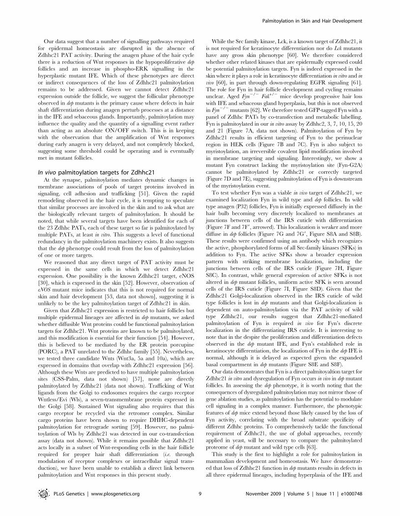
While the Src family kinase, Lck, is a known target of Zdhhc21, it
is not required for keratinocyte differentiation nor do Lck mutants
have any gross skin phenotype [60]. We therefore considered
whether other related kinases that are epidermally expressed could
be potential palmitoylation targets. Fyn is indeed expressed in the
skin where it plays a role in keratinocyte differentiation in vitro and in
vivo [60], in part through down-regulating EGFR signaling [61].
The role for Fyn in hair follicle development and cycling remains
unclear. Aged Fyn2/2 Fak+/2 mice develop progressive hair loss
with IFE and sebaceous gland hyperplasia, but this is not observed
in Fyn2/2 mutants [62]. We therefore tested GFP-tagged Fyn with a
panel of Zdhhc PATs by co-transfection and metabolic labelling.
Fyn is palmitoylated in our in vitro assay by Zdhhc2, 3, 7, 10, 15, 20
and 21 (Figure 7A, data not shown). Palmitoylation of Fyn by
Zdhhc21 results in efficient targeting of Fyn to the perinuclear
region in HEK cells (Figure 7B and 7C). Fyn is also subject to
myristoylation, an irreversible covalent lipid modification involved
in membrane targeting and signaling. Interestingly, we show a
mutant Fyn construct lacking the myristoylation site (Fyn-G2A)
cannot be palmitoylated by Zdhhc21 or correctly targeted
(Figure 7D and 7E), suggesting palmitoylation of Fyn is downstream
of the myristoylation event.
To test whether Fyn was a viable in vivo target of Zdhhc21, we
examined localization Fyn in wild type and dep follicles. In wild
type anagen (P32) follicles, Fyn is initially expressed diffusely in the
hair bulb becoming very discretely localized to membranes at
junctions between cells of the IRS cuticle with differentiation
(Figure 7F and 7F9, arrowed). This localization is weaker and more
diffuse in dep follicles (Figure 7G and 7G9, Figure S8A and S8B).
These results were confirmed using an antibody which recognizes
the active, phosphorylated forms of all Src-family kinases (SFKs) in
addition to Fyn. The active SFKs show a broader expression
pattern with striking membrane localization, including the
junctions between cells of the IRS cuticle (Figure 7H, Figure
S8C). In contrast, while general expression of active SFKs is not
altered in dep mutant follicles, uniform active SFK is seen around
cells of the IRS cuticle (Figure 7I, Figure S8D). Given that the
Zdhhc21 Golgi-localization observed in the IRS cuticle of wild
type follicles is lost in dep mutants and that Golgi-localization is
dependent on auto-palmitoylation via the PAT activity of wild
type Zdhhc21, our results suggest that Zdhhc21-mediated
palmitoylation of Fyn is required in vivo for Fyn’s discrete
localization in the differentiating IRS cuticle. It is interesting to
note that in the despite the proliferation and differentiation defects
observed in the dep mutant IFE, and Fyn’s established role in
keratinocyte differentiation, the localization of Fyn in the dep IFE is
normal, although it is delayed as expected given the expanded
basal compartment in dep mutants (Figure S8E and S8F).
Our data demonstrates that Fyn is a direct palmitoyaltion target for
Zdhhc21 in vitro and dysregulation of Fyn occurs in vivo in dep mutant
follicles. In assessing the dep phenotype, it is worth noting that the
consequences of dysregulated palmitoylation may not mirror those of
gene ablation studies, as palmitoylation has the potential to modulate
cell signaling in a complex manner. Furthermore, the phenotypic
features of dep mice extend beyond those likely caused by the loss of
Fyn activity, correlating with the broad substrate specificity of
different Zdhhc proteins. To comprehensively tackle the functional
requirement of Zdhhc21, the use of global approaches, recently
applied in yeast, will be necessary to compare the palmitoylated
proteome of dep mutant and wild type cells [63].
This study is the first to highlight a role for palmitoylation in
mammalian development and homeostasis. We have demonstrat-
ed that loss of Zdhhc21 function in dep mutants results in defects in
all three epidermal lineages, including hyperplasia of the IFE and
Palmitoylation in Skin and Hair Development
PLoS Genetics | www.plosgenetics.org 9 November 2009 | Volume 5 | Issue 11 | e1000748
sebaceous glands with a delay in hair follicle differentiation. Given
the highly restricted pattern of Zdhhc21 expression to the
differentiating hair follicle, our results demonstrate that defective
palmitoylation can have far-reaching effects disrupting epidermal
homeostasis by altering the balance between proliferation and
differentiation. Although the full identity of direct and biologically
relevant palmitoylation targets in the skin remains unknown, we
show Zdhhc21 can directly palmitoylate Fyn in vitro and this
modification affects Fyn localization both in vitro and in vivo. Future
studies into the distinct and overlapping roles of additional Zdhhc
members will help to fully understand the role of palmitoylation in
modulating key signals during development.
Materials and Methods
Ethics statementMice were maintained in accordance with MRC Guidelines
‘‘Responsibility in the Use of Animals for Medical Research’’ (July
1993) and research licenced by the UK Home Office under the
Animals (Scientific Procedures) Act 1986.
Mouse husbandry and BAC transgenicsAnimals were maintained in SPF environment and on a
C57BL/6J background. Genomic DNA extracted from ear clips
or tail biopsies was used for PCR genotyping. For dep, exon 7 of
Zdhhc21 was amplified by standard PCR to yield a 249bp fragment
that was run on the ABI310 genetic analyzer to detect the deletion.
The dep genotyping primer sequences were: 59-FAM-AGCT-
GACTGAAGGGCACC-39 (Exon 7F) and 59-AAAACCTG-
TAACGCATTTCCA-39 (Exon 7R).
For transgenic rescue, purified RP23-76J17 BAC DNA (BAC
PAC Resource Center (BPRC), the Children’s Hospital Oakland
Research Center Institute, CA) was injected into homozygous dep
embryos. The presence of the BAC was genotyped using three
markers, including CmR specific to the plasmid, as well as two
markers at both ends of the BACs, amplifying the border between
Figure 7. Zdhhc21 palmitoylation of Fyn is required for localization in vitro and in vivo. (A) [3H]Palmitate fluorography of subset of panelof all mammalian Zdhhc HA-tagged constructs co-transfected with Fyn::GFP into HEK293 cells. Increased incorporation of [3H]palmitate into targets isobserved with Zdhhc15, 220 and 221. A myristoylation-deficient Fyn mutant is not palmitoylated following cotransfection with Zdhhc21 (G2A, finallane). (B–E) Immunofluorescence of HEK293 cotransfected with Fyn::GFP alone (B), Fyn::GFP and wild type HA-tagged Zdhhc21 cDNA (C), G2A mutantFyn::GFP alone (D) and G2A mutant Fyn::GFP and HA-tagged Zdhhc21 (E). Zdhhc21 causes increased perinuclear accumulation of Fyn::GFP, a domainassociated with localization of other palmitoylated targets including PSD-95 and GAP-43. This localization is not observed with G2A mutant Fyn::GFP(D). (F–I) Confocal images (0.5mm) of immunostaining of Fyn (red: F,G) and active SFKs (red: H–I) in P32 anagen dorsal hair follicles in wild type (F,H)and dep mutants (G,I). Arrowheads mark region of interest in IRS cuticle that are enlarged in single channel images (F9–I9) where Fyn localization tomembranes between IRS cuticle cells is lost in dep mutants.doi:10.1371/journal.pgen.1000748.g007
Palmitoylation in Skin and Hair Development
PLoS Genetics | www.plosgenetics.org 10 November 2009 | Volume 5 | Issue 11 | e1000748
the BAC carrier plasmid and BAC genomic region. For timed
matings for embryonic samples, the morning of vaginal plug was
counted as 0.5 (E0.5). For postnatal timepoints, a set of gender-
matched wild type, heterozygous and mutant littermates were
aged accordingly from the day of birth 0 (P0).
Deletion mappingMapping of the deletion endpoints defining dep was described by
Smyth et al [24].
Palmitoylation assaycDNA encoding wild type and mutant Zdhhc21 were
transfected into HEK293 cells, along with candidate palmitoyla-
tion substrates. Cells were labelled with [3H]palmitate for 4 hours
as previously described [8,9]. After metabolic labeling, palmitoy-
lated proteins were analysed by SDS-PAGE. Transfection
efficiency and translation of substrates was assessed by Western
blotting.
Preparation of mammalian expression plasmids and cellculture
The mammalian expression vector of wild-type Zdhhc21,
pEFBos-HA-Zdhhc-21 (with EF1-alpha promoter), was provided
by Masaki Fukata. It was then modified by using the Quik-
ChangeH Site-Directed Mutagenesis Kit (Stratagene), to introduce
single nucleotide changes for the following Zdhhc21 alleles: L91F,
C95S and C106S. For the dep mutation, del-233F, was modified
from pEFBos-HA-Zdhhc-21 by subcloning dep cDNA (Access RT-
PCR System, Promega) into the BamHI sites flanking the insert.
Full-length mouse Wnt cDNAs (kindly provided by Jeremy
Nathans (Wnt3a), Wnt5a (Yingzi Yang) and Wnt10a (Takano
Yamamoto)) were introduced into a pCMX2GFPFLAGSTOP
vector (kindly provided by Nick Gilbert) to express double FLAG-
tagged full-length proteins. Constructs were verified by direct
DNA sequencing, using primers:
59-CAAATGGGCGGTAGGCGTGT-39 (Pcmxgfp2fg-seqF)
59-TTGTCCAATTATGTCACACCA-39 (Pcmxgfp2fg-seqR)
The Myc-tagged full length Wls cDNA used in these studies has
accension number BC018381 (Catalog No: MR207034: Origene,
MD).
Human foreskin keratinocytes [64] and NIH 3T3 cells (ATCC)
were maintained as described. DNA plasmids were transfected
into cells using Lipofectamine 2000 (Invitrogen) as per manufac-
turer’s specifications.
Zdhhc21 RNA in situ probe and antibody generationZdhhc21 cDNA product was generated using Access RT-PCR
kit (Promega) and cloned into p-GEM-T. The RT-PCR primers
used were: 59-CATGGGCTTGATTGTCTTTGT-39 and 59-
ACGTGATTGGCAAAGTGGTAG-39. DIG-labeled RNA sec-
tion in situ protocol was performed, details available on request.
Custom rabbit polyclonal antibodies to Zdhhc21 were generated
using a peptide comprising residues 73–87 (GRLPENPKI-
PHAERE+C)(Eurogentec). Pre-incubation of the Zdhhc21 anti-
body with its immunizing peptide blocked all signal in immuno-
histochemistry (Figure S2L).
Histological and marker gene analysisTail epidermis wholemount preparations. Whole tail skin
was peeled off connective tissue and bone, then incubated in 5mM
EDTA/PBS for 4 hours at 37uC. Forceps were used to separate
epidermis from dermis which was then fixed in 4%
paraformaldehyde/PBS.
Alkaline phosphatase staining of dermal papilla and
staging hair cycle. Paraffin embedded sections of 6mm
thickness were dewaxed and rehydrated, and rinsed in PBS.
Sections were incubated 15 minutes in APB (100mM NaCl,
100mM Tris pH9.5, 50mM MgCl2, 0.3% Tween-20), then either
BM Purple (Roche) or Vector Red Alkaline Phosphatase kit
(Vector Labs) color substrate was added. Slides were incubated at
room temperature in the dark until staining developed. Sections
were rinsed in water and counterstained with eosin (in the case of
BM Purple), then dehydrated and mounted in DePex mounting
media (BDH). Staging of hair follicles in hair cycle was done
according to characteristics described in [65].
Oil-Red-O staining for lipids. Cryosections of P24 skin of
10mm thickness were rinsed in water, followed by 50% EtOH and
stained with Oil-Red-O (60% stock in water). Slides were rinsed in
50% EtOH, followed by water and counterstained with haematoxylin.
Samples were blued and washed in water and mounted in
Aquamount (BDH). Oil-Red-O stock solution was prepared by
dissolving 0.5g Oil-Red-O in 100ml 99% Isopropyl Alcohol.
Barrier function assay. Dye penetration assays were
performed on late E16.5 litters as previously described [66] with
the following modifications. Embryos were dissected out in cold
PBS and permeabilized by taking them up and down a methanol/
PBS series (25%, 50%, 75%, 100%) with 5 minute washes.
Embryos were stained in 0.1% toludine blue solution for
3 minutes, then rinsed through 4 rapid PBS washes. Embryos
were immediately photographed on a Nikon AZ100 macroscope
using a Nikon APO 0.56 lens in PBS on 2% agarose plates.
Immunohistochemistry and immunocytochemistryParaffin sections were dewaxed and rehydrated, followed by
washes in TBST +0.5% Tx-100). Microwave antigen retrieval was
carried out using 1mM EDTA (pH8) or citrate buffer (1.8mM citric
acid, 8.2mM sodium citrate, pH6) for 20–30 minutes depending on
the antigen at 900W. Cryosections samples were allowed to come to
room temperature and post-fixed in acetone (220’C for 10 minutes)
followed by rinsing in water. No antigen retrieval step was required
for cryosections. Slides were cooled to room temperature and washed
in TBST. Slides were blocked in 10% donkey serum/TBST, followed
by TBST washes. Primary antibodies were diluted in 1% donkey
serum/TBST incubated on slides overnight at 4uC (Table 1). After
TBST washes, secondary antibodies were diluted in 1% donkey
serum/TBST and added to the slides for 60 minute incubation
(Table 2). Following stringent TBST washes, nuclei were stained with
DAPI (Sigma) or TOTO-3 (Molecular Probes). In the case of
TOTO-3, slides were pre-incubated with RNAse A during primary
antibody incubation. Slides were mounted with Vectashield (Vector)
or Prolong Gold (Molecular Probes) antifade media and coverslips.
Brightfield and fluorescent images were acquired using a Coolsnap
HQ CCD camera (Photometrics Ltd, Tucson, AZ) Zeiss Axioplan II
fluorescence microscope with Plan-neofluar objectives. Image
capture and analysis were performed using in-house scripts written
for IPLab Spectrum (Scanalytics Corp, Fairfax, VA). For colocaliza-
tion studies, 0.5–1 mm optical slice images in Z-stacks were acquired
with a Zeiss LSM510 confocal microscope and Zeiss Plan
Apochromat lenses (Carl Zeiss, Welwyn Garden City, UK). LSM
software was used for analysis (Carl Zeiss, Welwyn Garden City, UK).
BrdU labelingFor BrdU labeling experiments, 2 age- and gender-matched mice
of each genotype were injected with 50 mg BrdU/g body weight and
sacrificed after 2 hrs. Skin sections were dewaxed, subjected to
proteinase K antigen retrieval, followed by HCl denaturation and
neutralization, before incubation with anti-BrdU antibody (BD). For
Palmitoylation in Skin and Hair Development
PLoS Genetics | www.plosgenetics.org 11 November 2009 | Volume 5 | Issue 11 | e1000748
indirect colorimetric visualization, a biotinylated donkey anti-mouse
secondary antibody (Jackson Labs) and Vectastain Universal Elite
ABC Kit (Vector Laboratories) were used, followed by NovaRed
substrate (Vector Laboratories) according to manufacturer’s protocol.
A proliferative index was calculated by counting the number of
positive cells divided by the total number of nuclei within the
epidermal compartment, in each of ten fields at 106magnification,
and the average index per field was calculated. Statistical significance
was calculated using a two-tailed Student’s t-test.
Supporting Information
Figure S1 Localization and function of Zdhhc21 is altered by
mutations of cysteines within DHHC consensus core. (A)
Schematic of mutations in Zdhhc21. In addition to the dep
deletion in C-terminal intracellular tail, several point mutations
were generated by disrupting key cysteine residues within the
DHHC domain. Another mutation, L91F, close to the DHHC
domain was identified from an archive of ENU-mutagenised
sperm from Harwell. However unlike mutations in the critical
cysteines, this mutant protein was correctly localized and exhibited
normal PAT activity. Mice homozygous for this mutation had
normal hair. (B–G) Localization of HA-tagged Zdhhc21 cDNAs
transfected into NIH-3T3 cells (anti-HA red) compared to cis-
Golgi marker GM130 (green). Wild type and L91F strongly co-
localize with GM130, whereas mutations within DHHC domain
disrupt localization similar to dep. (H) Zdhhc21 protein variants
which disrupt localization abrogate autopalmitoylation responses
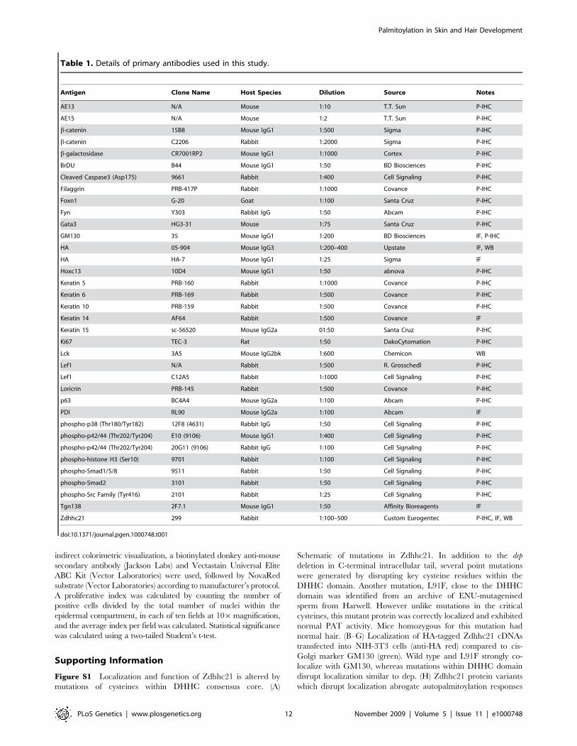
Table 1. Details of primary antibodies used in this study.
Antigen Clone Name Host Species Dilution Source Notes
AE13 N/A Mouse 1:10 T.T. Sun P-IHC
AE15 N/A Mouse 1:2 T.T. Sun P-IHC
b-catenin 15B8 Mouse IgG1 1:500 Sigma P-IHC
b-catenin C2206 Rabbit 1:2000 Sigma P-IHC
b-galactosidase CR7001RP2 Mouse IgG1 1:1000 Cortex P-IHC
BrDU B44 Mouse IgG1 1:50 BD Biosciences P-IHC
Cleaved Caspase3 (Asp175) 9661 Rabbit 1:400 Cell Signaling P-IHC
Filaggrin PRB-417P Rabbit 1:1000 Covance P-IHC
Foxn1 G-20 Goat 1:100 Santa Cruz P-IHC
Fyn Y303 Rabbit IgG 1:50 Abcam P-IHC
Gata3 HG3-31 Mouse 1:75 Santa Cruz P-IHC
GM130 35 Mouse IgG1 1:200 BD Biosciences IF, P-IHC
HA 05-904 Mouse IgG3 1:200–400 Upstate IF, WB
HA HA-7 Mouse IgG1 1:25 Sigma IF
Hoxc13 10D4 Mouse IgG1 1:50 abnova P-IHC
Keratin 5 PRB-160 Rabbit 1:1000 Covance P-IHC
Keratin 6 PRB-169 Rabbit 1:500 Covance P-IHC
Keratin 10 PRB-159 Rabbit 1:500 Covance P-IHC
Keratin 14 AF64 Rabbit 1:500 Covance IF
Keratin 15 sc-56520 Mouse IgG2a 01:50 Santa Cruz P-IHC
Ki67 TEC-3 Rat 1:50 DakoCytomation P-IHC
Lck 3A5 Mouse IgG2bk 1:600 Chemicon WB
Lef1 N/A Rabbit 1:500 R. Grosschedl P-IHC
Lef1 C12A5 Rabbit 1:1000 Cell Signaling P-IHC
Loricrin PRB-145 Rabbit 1:500 Covance P-IHC
p63 BC4A4 Mouse IgG2a 1:100 Abcam P-IHC
PDI RL90 Mouse IgG2a 1:100 Abcam IF
phospho-p38 (Thr180/Tyr182) 12F8 (4631) Rabbit IgG 1:50 Cell Signaling P-IHC
phospho-p42/44 (Thr202/Tyr204) E10 (9106) Mouse IgG1 1:400 Cell Signaling P-IHC
phospho-p42/44 (Thr202/Tyr204) 20G11 (9106) Rabbit IgG 1:100 Cell Signaling P-IHC
phospho-histone H3 (Ser10) 9701 Rabbit 1:100 Cell Signaling P-IHC
phospho-Smad1/5/8 9511 Rabbit 1:50 Cell Signaling P-IHC
phospho-Smad2 3101 Rabbit 1:50 Cell Signaling P-IHC
phospho-Src Family (Tyr416) 2101 Rabbit 1:25 Cell Signaling P-IHC
Tgn138 2F7.1 Mouse IgG1 1:50 Affinity Bioreagents IF
Zdhhc21 299 Rabbit 1:100–500 Custom Eurogentec P-IHC, IF, WB
doi:10.1371/journal.pgen.1000748.t001
Palmitoylation in Skin and Hair Development
PLoS Genetics | www.plosgenetics.org 12 November 2009 | Volume 5 | Issue 11 | e1000748
using ABE chemistry and pulled down by streptavidin agarose
beads and resolved by SDS-PAGE [47]. Portions not pulled down
were also resolved by SDS-PAGE as loading control (I).
Found at: doi:10.1371/journal.pgen.1000748.s001 (1.66 MB TIF)
Figure S2 Characterization of Zdhhc21 expression in skin.
Expression of Zdhhc21 mRNA (B,D,E,G,J) and protein
(A,C,F,H,I,K,). (A) E16.5 vibrissae follicle (Zdhhc21: green, p63:
red). (B,C) P24 dorsal control skin. (D–F) P35 dorsal follicles of dep
(D) and wild type (E), show similar levels and patterns of transcript,
as observed with Zdhhc21 antibody (F). (G–I) While Zdhhc21
mRNA and protein expression is similar in the lower portions of
P63 dorsal follicles (G,H), only protein can be detected in the
upper (I) portions of the isthmus (I) but not in the bulge, sebaceous
glands or IFE. (J–L) In telogen, (P21) wild-type dorsal skin shows
no expression of Zdhhc21 mRNA (J) while some antibody staining
is detected in the isthmus (K), which is specifically blocked by pre-
incubating the antibody with the blocking peptide (L).
Found at: doi:10.1371/journal.pgen.1000748.s002 (4.99 MB TIF)
Figure S3 Cyclic expression of Zdhhc21 during postnatal hair
cycle in wild-type and dep mutant follicles. Expression of Zdhhc21
(red) and Gata3 (green) during catagen (P14 A,B), telogen (P21
C,D), initiation of anagen (P24 E,F), early anagen (P28 G,H) and
late anagen (P35 I,J) in wild-type (A,C,E,G,I) and dep follicles
(B,D,F,H,J). Expression of Zdhhc21 is limited to the post-mitotic
lineages of IRS and cuticle of both control and dep anagen and
catagen follicles.
Found at: doi:10.1371/journal.pgen.1000748.s003 (6.63 MB TIF)
Figure S4 Aberrant epidermal proliferation during anagen
contributes to dep hyperplastic interfollicular epidermis and
sebaceous glands. Hematoxylin and eosin (A–D). Phosphohistone
H3 (red, E–J) with Ki67 (green; I,J,). Significant differences in
proliferation were not readily detectable at telogen (P21; A,B,E,F),
or early (P28; C,D,G–J) anagen. However, quantitative BrDU
labelling studies during anagen (P32) revealed a small but
significant increase in proliferation in dep sebaceous glands and
IFE (L), with a parallel decrease in proliferation in dep hair follicles
(K). (**p,0.005, *p,0.05)
Found at: doi:10.1371/journal.pgen.1000748.s004 (4.18 MB TIF)
Figure S5 Aberrant epidermal differentiation in dep mutant skin.
Wild-type (A–F) and dep (G–,L) P28 dorsal follicles. Expression of
terminal differentiation markers (loricrin (red), p63 (green) (A,G);
filaggrin (red) (B,H) is delayed in dep mutant skin. Ectopic Keratin
6 expression (K6 (red), Ki67 (green) (C,I) is not observed in dep
interfollicular epidermis, but expression remains restricted to the
infundibulum and inner root sheath of the hair follicle. Imbalance
of proliferative and differentiation signals in dep basal IFE where
increased nuclear phospho-ERK (phospho-P42/44 (red), Gata3
(green), (D,–D9,J–J9) is observed with reduced expression of Gata3,
in contrast to wild type skin where high suprabasal phospho-ERK
is associated with strong Gata3 expressing cells (D–D9, arrow-
heads). Aberrant elevated basal p42/44 signalling was confirmed
with a second antibody (I–I9,K–K9). Despite expanded bulge
region below the dilated infundibulum and overgrown sebaceous
glands, the expression of K15 (green) remains restricted to the
bulge (F,L). Nuclei were labelled with DAPI (blue:C,I) or TOTO-3
(blue:D–F,J–L).
Found at: doi:10.1371/journal.pgen.1000748.s005 (4.65 MB TIF)
Figure S6 Loss of Zdhhc21 function does not result in delays in
selective barrier acquisition or keratinocyte terminal differentiation
defects in embryonic dep epidermis. Wild-type (A–E) and dep
mutant (F–J) late E16.5 embryos and E18.5 embryonic skins (C–E,
H–J). (A,B,F,G) Dye exclusion assay showing similar range of
barrier acquisition in a litter with wild-type and dep littermates
from less advanced (A,F) to more established stages of barrier
development (B,G). No difference in expression of terminal
differentiation markers loricrin (C,H) and filaggrin (D,I) is detected
between wild type and dep neonatal skin. Comparable Gata3
expression is observed in developing hair follicles and IFE of wild-
type and dep neonatal skin (E–J).
Found at: doi:10.1371/journal.pgen.1000748.s006 (2.42 MB TIF)
Figure S7 Initiation of Wnt-dependent anagen responses is
normal in dep mice but subsequent propagation is affected.
Alkaline phosphatase staining (A,C,E,G) marks dermal papillae.
Induction of first anagen at P24 (A–D) with strong dermal papilla
Lef1 staining (red) (B,D) and few adjacent positive cells in
epidermal hair germ is observed in both wild-type (A,B) and
mutant (C,D) skin. Subsequent propagation of anagen responses is
defective at P28 (E–L) where retarded dep follicles show little Lef1
signal in matrix (F,H) as well as reduced or absent Foxn1
(green,Zdhhc21 red; J,K) and AE13 (green, Zdhhc21 red; J,L) in
hair shaft precursors. By P35 (M–P), although misshapen and
misoriented, many dep follicles (O,P) are similar to control
littermates (M,N) as shown by AE13 (green, Zdhhc21 red; M,O)
and beta-catenin (green, K5 red; N,P).
Found at: doi:10.1371/journal.pgen.1000748.s007 (5.73 MB TIF)
Figure S8 Effective membrane targeting of Fyn during kerati-
nocyte differentiation is compromised in dep mutant skin. Fyn
localization in P32 anagen follicles (A–B9) and IFE (E–F9) with
localization of active Src family kinases (including Src, Fyn, Yes
Table 2. Details of secondary antibodies used in this study.
Antigen Host Species Dilution Source Notes
ECL a-Mouse IgG, HRP-conjugated Sheep 1:10000 GE Healthcare UK Ltd WB
ECL a-Rabbit IgG, HRP-conjugated Sheep 1:10000 GE Healthcare UK Ltd WB
TRITC-conjugated a-Rabbit IgG Donkey 1:250 Jackson Immunoresearch P-IHC, IF
FITC-conjugated a-Rabbit IgG Donkey 1:250 Jackson Immunoresearch P-IHC, IF
Alexa 488-conjugated-a-Mouse Donkey 1:500 Molecular Probes P-IHC, IF
Alexa 594-conjugated-a-Mouse Donkey 1:500 Molecular Probes P-IHC, IF
Alexa 594-conjugated-a-Rabbit Donkey 1:500 Molecular Probes P-IHC, IF
Alexa 488-conjugated-a-Rat Donkey 1:500 Molecular Probes P-IHC, IF
Biotin-conjugated-a-Rabbit Donkey 1:400 Jackson Immunoresearch P-IHC, IF
doi:10.1371/journal.pgen.1000748.t002
Palmitoylation in Skin and Hair Development
PLoS Genetics | www.plosgenetics.org 13 November 2009 | Volume 5 | Issue 11 | e1000748
and Lck) (C–D9,G–H9). Fyn expression is detected diffusely in the
wild type bulb and becomes restricted to the membrane of
differentiating IRS cuticle and some Henle’s layers at the junction
of the hair bulb and shaft. High levels of membrane associated
active SFKs are seen throughout anagen hair follicle including
dermal papilla, proliferative matrix, ORS and IRS lineages. In
dep mutants, this membrane association of Fyn is greatly reduced/
absent whilst active Src family kinase expression is largely
unchanged. Fyn expression in the control IFE and IF becomes
membrane restricted in suprabasal, differentiating keratinocytes,
whilst membrane associated active Src family kinases can be seen
throughout the basal and suprabasal IFE. Membrane associated
Fyn and active SFKs is delayed in dep mutants. Arrowheads
indicated areas of interest in merge and single channels.
Found at: doi:10.1371/journal.pgen.1000748.s008 (4.72 MB TIF)
Acknowledgments
We thank A. Weiss (Lck cDNA), W. Sessa (eNOS cDNA), Jeremy Nathans
(Wnt3a cDNA), Yingzi Yang (Wnt5a cDNA), Takano Yamamoto (Wnt10a
cDNA), Nick Gilbert (pCMX2GFPFLAGSTOP vector), T.T.Sun (AE13
and AE15 antibodies), and R. Grosschedl (Lef1 antibody) for sharing
reagents. We are grateful to Paul Perry and Brendan Doe for technical
assistance and to Tilo Kunath and Richard Mort for critical comments and
discussions.
Author Contributions
Conceived and designed the experiments: PM AWSL MF IS IJJ.
Performed the experiments: PM AWSL YF RT MK LM IS. Analyzed
the data: PM AWSL MF RMP IS. Wrote the paper: PM IJJ.
References
1. Smotrys JE, Linder ME (2004) Palmitoylation of intracellular signaling proteins:
regulation and function. Annu Rev Biochem 73: 559–587.
2. Hayashi T, Rumbaugh G, Huganir RL (2005) Differential regulation of AMPAreceptor subunit trafficking by palmitoylation of two distinct sites. Neuron 47:
709–723.
3. Keller CA, Yuan X, Panzanelli P, Martin ML, Alldred M, et al. (2004) The
gamma2 subunit of GABA(A) receptors is a substrate for palmitoylation byGODZ. J Neurosci 24: 5881–5891.
4. El Husseini A, Schnell E, Dakoji S, Sweeney N, Zhou Q, et al. (2002) Synaptic
strength regulated by palmitate cycling on PSD-95. Cell 108: 849–863.
5. Kang R, Wan J, Arstikaitis P, Takahashi H, Huang K, et al. (2008) Neural
palmitoyl-proteomics reveals dynamic synaptic palmitoylation. Nature 456:904–909.
6. Lobo S, Greentree WK, Linder ME, Deschenes RJ (2002) Identification of a Ras
palmitoyltransferase in Saccharomyces cerevisiae. J Biol Chem 277:
41268–41273.
7. Roth AF, Feng Y, Chen L, Davis NG (2002) The yeast DHHC cysteine-richdomain protein Akr1p is a palmitoyl transferase. J Cell Biol 159: 23–28.
8. Fukata M, Fukata Y, Adesnik H, Nicoll RA, Bredt DS (2004) Identification ofPSD-95 palmitoylating enzymes. Neuron 44: 987–996.
9. Fukata Y, Iwanaga T, Fukata M (2006) Systematic screening for palmitoyl
transferase activity of the DHHC protein family in mammalian cells. Methods40: 177–182.
10. Oyama T, Miyoshi Y, Koyama K, Nakagawa H, Yamori T, et al. (2000)Isolation of a novel gene on 8p21.3–22 whose expression is reduced significantly
in human colorectal cancers with liver metastasis. Genes Chromosomes Cancer29: 9–15.
11. Ducker CE, Stettler EM, French KJ, Upson JJ, Smith CD (2004) Huntingtin
interacting protein 14 is an oncogenic human protein: palmitoyl acyltransferase.
Oncogene 23: 9230–9237.
12. Mukai J, Liu H, Burt RA, Swor DE, Lai WS, et al. (2004) Evidence that the geneencoding ZDHHC8 contributes to the risk of schizophrenia. Nat Genet 36:
725–731.
13. Faul T, Gawlik M, Bauer M, Jung S, Pfuhlmann B, et al. (2005) ZDHHC8 as a
candidate gene for schizophrenia: analysis of a putative functional intronicmarker in case-control and family-based association studies. BMC Psychiatry 5:
35.
14. Mukai J, Dhilla A, Drew LJ, Stark KL, Cao L, et al. (2008) Palmitoylation-
dependent neurodevelopmental deficits in a mouse model of 22q11 microdele-tion. Nat Neurosci 11: 1302–1310.
15. Mansouri MR, Marklund L, Gustavsson P, Davey E, Carlsson B, et al. (2005)
Loss of ZDHHC15 expression in a woman with a balanced translocationt(X;15)(q13.3;cen) and severe mental retardation. Eur J Hum Genet 13:
970–977.
16. Raymond FL, Tarpey PS, Edkins S, Tofts C, O’Meara S, et al. (2007) Mutations
in ZDHHC9, which encodes a palmitoyltransferase of NRAS and HRAS, causeX-linked mental retardation associated with a Marfanoid habitus. Am J Hum
Genet 80: 982–987.
17. Matakatsu H, Blair SS (2008) The DHHC palmitoyltransferase approximated
regulates Fat signaling and Dachs localization and activity. Curr Biol 18:1390–1395.
18. Clayton E, Doupe DP, Klein AM, Winton DJ, Simons BD, et al. (2007) A single
type of progenitor cell maintains normal epidermis. Nature 446: 185–189.
19. Fuchs E (2007) Scratching the surface of skin development. Nature 445:
834–842.
20. Blanpain C, Lowry WE, Geoghegan A, Polak L, Fuchs E (2004) Self-renewal,multipotency, and the existence of two cell populations within an epithelial stem
cell niche. Cell 118: 635–648.
21. Claudinot S, Nicolas M, Oshima H, Rochat A, Barrandon Y (2005) Long-term
renewal of hair follicles from clonogenic multipotent stem cells. Proc Natl AcadSci U S A 102: 14677–14682.
22. Mayer TC, Kleiman NJ, Green MC (1976) Depilated (dep), a mutant gene that
affects the coat of the mouse and acts in the epidermis. Genetics 84: 59–65.
23. Rinchik EM (1994) Molecular genetics of the brown (b)-locus region of mouse
chromosome 4. II. Complementation analyses of lethal brown deletions.
Genetics 137: 855–865.
24. Smyth IM, Wilming L, Lee AW, Taylor MS, Gautier P, et al. (2006) Genomic
anatomy of the Tyrp1 (brown) deletion complex. Proc Natl Acad Sci U S A 103:
3704–3709.
25. Simpson EH, Suffolk R, Bell JA, Jordan SA, Johnson DK, et al. (2000) A
comparative transcript map and candidates for mutant phenotypes in the Tyrp1
(brown) deletion complex homologous to human 9p21–23. Mamm Genome 11:
58–63.
26. Smyth I, Du X, Taylor MS, Justice MJ, Beutler B, et al. (2004) The extracellular
matrix gene Frem1 is essential for the normal adhesion of the embryonic
epidermis. Proc Natl Acad Sci U S A 101: 13560–13565.
27. Shawlot W, Min DJ, Wakamiya M, Behringer RR (2000) The cerberus-related
gene, Cerr1, is not essential for mouse head formation. Genesis 26: 253–258.
28. Stanley EG, Biben C, Allison J, Hartley L, Wicks IP, et al. (2000) Targeted
insertion of a lacZ reporter gene into the mouse Cer1 locus reveals complex and
dynamic expression during embryogenesis. Genesis 26: 259–264.
29. Belo JA, Bachiller D, Agius E, Kemp C, Borges AC, et al. (2000) Cerberus-like is
a secreted BMP and nodal antagonist not essential for mouse development.
Genesis 26: 265–270.
30. Fernandez-Hernando C, Fukata M, Bernatchez PN, Fukata Y, Lin MI, et al.
(2006) Identification of Golgi-localized acyl transferases that palmitoylate and
regulate endothelial nitric oxide synthase. J Cell Biol 174: 369–377.
31. Tsutsumi R, Fukata Y, Noritake J, Iwanaga T, Perez F, et al. (2009)
Identification of G-protein alpha subunit palmitoylating enzyme. Mol Cell Biol
29: 435–447.
32. de Guzman Strong C, Wertz PW, Wang C, Yang F, Meltzer PS, et al. (2006)
Lipid defect underlies selective skin barrier impairment of an epidermal-specific
deletion of Gata-3. J Cell Biol 175: 661–70.
33. Candi E, Terrinoni A, Rufini A, Chikh A, Lena AM, et al. (2006) p63 is
upstream of IKK alpha in epidermal development. J Cell Sci 119: 4617–
22.
34. Proksch E, Feingold KR, Man MQ, Elias PM (1991) Barrier function regulates
epidermal DNA synthesis. J Clin Invest 87: 1668–1673.
35. Haase I, Hobbs RM, Romero MR, Broad S, Watt FM (2001) A role for
mitogen-activated protein kinase activation by integrins in the pathogenesis of
psoriasis. J Clin Invest 108: 527–36.
36. Hwang J, Mehrani T, Millar SE, Morasso MI (2008) Dlx3 is a crucial regulator
of hair follicle differentiation and cycling. Development 135: 3149–59.
37. Botchkarev VA (2003) Bone morphogenetic proteins and their antagonists in
skin and hair follicle biology. J Invest Dermatol 120: 36–47.
38. Kobielak K, Pasolli HA, Alonso L, Polak L, Fuchs E (2003) Defining BMP
functions in the hair follicle by conditional ablation of BMP receptor IA. J Cell
Biol 163: 609–23.
39. Andl T, Ahn K, Kairo A, Chu EY, Wine-Lee L, et al. (2004) Epithelial Bmpr1a
regulates differentiation and proliferation in postnatal hair follicles and is
essential for tooth development. Development 131: 2257–68.
40. Yuhki M, Yamada M, Kawano M, Iwasato T, Itohara S, et al. (2004) BMPR1A
signaling is necessary for hair follicle cycling and hair shaft differentiation in
mice. Development 131: 1825–33.
41. Foitzik K, Lindner G, Mueller-Roever S, Maurer M, Botchkareva N, et al.
(2001) Control of murine hair follicle regression (catagen) by TGF-beta1 in vivo.
FASEB J 14: 752–60.
42. Descargues P, Sil AK, Sano Y, Korchynskyi O, Han G, et al. (2008) IKKalpha is
a critical coregulator of a Smad4-independent TGFbeta-Smad2/3 signaling
pathway that controls keratinocyte differentiation. Proc Natl Acad Sci U S A
105: 2487–92.
Palmitoylation in Skin and Hair Development
PLoS Genetics | www.plosgenetics.org 14 November 2009 | Volume 5 | Issue 11 | e1000748
43. Leong WF, Zhou T, Lim GL, Li B (2009) Protein palmitoylation regulates
osteoblast differentiation through BMP-induced osterix expression. PLoS One 4:
e4135. doi:10.1371/journal.pone.0004135.
44. Zuo W, Chen YG (2009) Specific activation of mitogen-activated protein kinase
by transforming growth factor-beta receptors in lipid rafts is required for
epithelial cell plasticity. Mol Biol Cell 20: 1020–9.
45. Gat U, DasGupta R, Degenstein L, Fuchs E (1998) De novo hair follicle
morphogenesis and hair tumors in mice expressing a truncated beta-catenin in
skin. Cell 95: 605–614.
46. Silva-Vargas V, Lo CC, Giangreco A, Ofstad T, Prowse DM, et al. (2005) Beta-
catenin and Hedgehog signal strength can specify number and location of hair
follicles in adult epidermis without recruitment of bulge stem cells. Dev Cell 9:
121–131.
47. Merrill BJ, Gat U, DasGupta R, Fuchs E (2001) Tcf3 and Lef1 regulate lineage
differentiation of multipotent stem cells in skin. Genes Dev 15: 1688–1705.
48. DasGupta R, Rhee H, Fuchs E (2002) A developmental conundrum: a stabilized
form of beta-catenin lacking the transcriptional activation domain triggers
features of hair cell fate in epidermal cells and epidermal cell fate in hair follicle
cells. J Cell Biol 158: 331–344.
49. Niemann C, Owens DM, Hulsken J, Birchmeier W, Watt FM (2002) Expression
of DeltaNLef1 in mouse epidermis results in differentiation of hair follicles into
squamous epidermal cysts and formation of skin tumours. Development 129:
95–109.
50. Kurek D, Garinis GA, van Doorninck JH, van der Wees J, Grosveld FG (2007)
Transcriptome and phenotypic analysis reveals Gata3-dependent signalling
pathways in murine hair follicles. Development 134: 261–72.
51. Kang R, Wan J, Artikaitis P, Takahashi H, Huang K, et al. (2008) Neural
palmitoyl proteomics reveals dynamic synaptic palmitoylation. Nature 456:
904–909.
52. Sowden HM, Naseem KM, Tobin DJ (2005) Differential expression of nitric
oxide synthases in human scalp epidermal and hair follicle pigmentary units:
implications for regulation of melanogenesis. Br J Dermatol 153: 301–309.
53. Shesely EG, Maeda N, Kim HS, Desai KM, Krege JH, et al. (1996) Elevated
blood pressures in mice lacking endothelial nitric oxide synthase. Proc Natl Acad
Sci U S A 93: 13176–13181.
54. Willert K, Brown JD, Danenberg E, Duncan AW, Weissman IL, et al. (2003)
Wnt proteins are lipid-modified and can act as stem cell growth factors. Nature423: 448–452.
55. Kadowaki T, Wilder E, Klingensmith J, Zachary K, Perrimon N (1996) The
segment polarity gene porcupine encodes a putative multitransmembraneprotein involved in Wingless processing. Genes Dev 10: 3116–3128.
56. Reddy S, Andl T, Bagasra A, Lu MM, Epstein DJ, et al. (2001) Characterizationof Wnt gene expression in developing and postnatal hair follicles and
identification of Wnt5a as a target of Sonic hedgehog in hair follicle
morphogenesis. Mech Dev 107: 69–82.57. Ren J, Wen L, Gao X, Jin C, Xue Y, et al. (2008) CSS-Palm 2.0: an updated
software for palmitoylation sites prediction. Protein Engineering, Design andSelection 21: 639–644.
58. Banziger C, Soldini D, Schutt C, Zipperlen P, Hausmann G, et al. (2006)Wntless, a conserved membrane protein dedicated to the secretion of Wnt
proteins from signaling cells. Cell 125: 509–522.
59. McCormick PJ, Dumaresq-Doiron K, Pluviose AS, Pichette V, Tosato G, et al.(2008) Palmitoylation controls recycling in lysosomal sorting and trafficking.
Traffic 9: 1984–1997.60. Calautti E, Missero C, Stein PL, Ezzell RM, Dotto GP (1995) fyn tyrosine kinase
is involved in keratinocyte differentiation control. Genes Dev 9: 2279–91.
61. Cabodi S, Calautti E, Talora C, Kuroki T, Stein PL, et al. (2000) A PKC-eta/Fyn-dependent pathway leading to keratinocyte growth arrest and differentia-
tion. Mol Cell 6: 1121–9.62. Iliæ D, Kanazawa S, Nishizumi H, Aizawa S, Kuroki T, et al. (1997) Skin
abnormality in aged fyn2/2 fak+/2 mice. Carcinogenesis 18: 1473–6.63. Roth AF, Wan J, Bailey AO, Sun B, Kuchar JA, et al. (2006) Global analysis of
protein palmitoylation in yeast. Cell 125: 1003–1013.
64. DiColandrea T, Karashima T, Maatta A, Watt FM (2000) Subcellulardistribution of envoplakin and periplakin: insights into their role as precursors
of the epidermal cornified envelope. J Cell Biol 151: 573–586.65. Muller-Rover S, Handjiski B, van der Veen C, Eichmuller S, Foitzik K, et al.
(2001) A comprehensive guide for the accurate classification of murine hair
follicles in distinct hair cycle stages. J Invest Dermatol 117: 3–15.66. Hardman MJ, Sisi P, Banbury DN, Byrne C (1998) Patterned acquisition of skin
barrier function during development. Development 125: 1541–1552.
Palmitoylation in Skin and Hair Development
PLoS Genetics | www.plosgenetics.org 15 November 2009 | Volume 5 | Issue 11 | e1000748